RU2688386C1 - Method for lower limb ischemic involvement severity monitoring and device for its implementation - Google Patents
Method for lower limb ischemic involvement severity monitoring and device for its implementation Download PDFInfo
- Publication number
- RU2688386C1 RU2688386C1 RU2017145620A RU2017145620A RU2688386C1 RU 2688386 C1 RU2688386 C1 RU 2688386C1 RU 2017145620 A RU2017145620 A RU 2017145620A RU 2017145620 A RU2017145620 A RU 2017145620A RU 2688386 C1 RU2688386 C1 RU 2688386C1
- Authority
- RU
- Russia
- Prior art keywords
- severity
- determined
- ischemic
- sti
- toes
- Prior art date
Links
Images


Classifications
-
- A—HUMAN NECESSITIES
- A61—MEDICAL OR VETERINARY SCIENCE; HYGIENE
- A61B—DIAGNOSIS; SURGERY; IDENTIFICATION
- A61B5/00—Measuring for diagnostic purposes; Identification of persons
- A61B5/02—Detecting, measuring or recording pulse, heart rate, blood pressure or blood flow; Combined pulse/heart-rate/blood pressure determination; Evaluating a cardiovascular condition not otherwise provided for, e.g. using combinations of techniques provided for in this group with electrocardiography or electroauscultation; Heart catheters for measuring blood pressure
- A61B5/026—Measuring blood flow
- A61B5/0295—Measuring blood flow using plethysmography, i.e. measuring the variations in the volume of a body part as modified by the circulation of blood therethrough, e.g. impedance plethysmography
Landscapes
- Health & Medical Sciences (AREA)
- Life Sciences & Earth Sciences (AREA)
- Engineering & Computer Science (AREA)
- Medical Informatics (AREA)
- Physics & Mathematics (AREA)
- Cardiology (AREA)
- Biophysics (AREA)
- Pathology (AREA)
- Hematology (AREA)
- Biomedical Technology (AREA)
- Heart & Thoracic Surgery (AREA)
- Physiology (AREA)
- Molecular Biology (AREA)
- Surgery (AREA)
- Animal Behavior & Ethology (AREA)
- General Health & Medical Sciences (AREA)
- Public Health (AREA)
- Veterinary Medicine (AREA)
- Measurement Of The Respiration, Hearing Ability, Form, And Blood Characteristics Of Living Organisms (AREA)
Abstract
Description
Способ и устройство относятся к области медицины и могут быть использованы для диагностики в неврологии и кардиологии, сосудистой хирургии, оценки профессионализма медицинского персонала.The method and the device relate to the field of medicine and can be used for diagnosis in neurology and cardiology, vascular surgery, assessment of the professionalism of medical personnel.
Известны различные способы применения характеристик гемодинамики артериальной и венозной недостаточностей нижних конечностей как основных диагностических факторов степени тяжести ишемического процесса. Например, в работах Капустина Б.Б., Анисимова А.В «Возможности фотопульсографии в диагностике хронической артериальной недостаточности нижних конечностей» (Пермский медицинский журнал, 2010, №2, Т. 27, стр. 86-89) и «Новый способ диагностики состояния регионального кровотока нижних конечностей» (Анисимов А.В., Капустин Б.Б., Машковцева Г.В., Кузнецов П.А., Фундаментальные исследования, 2010, №2, стр. 24-27) предлагается способ диагностики на основе трансиллюминационного гемодинамического мониторинга по З.М. Сигалу с цифровой обработкой полученных параметров кровотока. Для осуществления диагностического процесса используется показатель - амплитуда пульсовых осцилляций (АПО), полученная накожным наложением датчика над магистральным сосудом исследуемой части нижних конечностей. Приведены статистически достоверные доказательства связи результатов измерений АПО в проекции бедренной артерии, подколенной артерии, задней большеберцовой артерии, артерии тыла стопы, зоны сустава Шопара, фаланги первого и пятого пальцев стопы с различными стадиями хронической артериальной недостаточности, что позволяет формировать решающие диагностические правила для использования в базах знаний соответствующих экспертных модулей систем автоматизированных систем поддержки принятия диагностических решений.There are various ways to apply hemodynamic characteristics of arterial and venous insufficiency of the lower extremities as the main diagnostic factors for the severity of the ischemic process. For example, in the works of Kapustin B. B., Anisimova A. V. “Possibilities of photopulsography in the diagnosis of chronic arterial insufficiency of the lower extremities” (Perm Medical Journal, 2010, No. 2, V. 27, pp. 86-89) and “New way of diagnosis the state of regional blood flow in the lower extremities ”(Anisimov AV, Kapustin BB, Mashkovtseva GV, Kuznetsov PA, Basic Research, 2010, №2, pp. 24-27) propose a diagnostic method based on transillumination hemodynamic monitoring according to Z.M. Sigalu with digital processing of the blood flow parameters obtained. For the implementation of the diagnostic process, the indicator is used - the amplitude of pulse oscillations (APO), obtained by skin overlay of the sensor above the main vessel of the studied part of the lower extremities. Statistically reliable evidence of the association of the results of APO measurements in the projection of the femoral artery, popliteal artery, posterior tibial artery, rear foot artery, Choopard joint zone, phalanx of the first and fifth toes with various stages of chronic arterial insufficiency, which allows you to create decisive diagnostic rules for use in knowledge bases of relevant expert modules of automated diagnostic decision support systems.
Так же известен прибор (оксинеометр) для измерения степени насыщения крови кислородом (сатурации) в нижних конечностях, по которому совместно с анализом ФПГ предлагается идентифицировать различные стадии ишемии нижних конечностей (дополнительное изобретение к авт. св. №104292, авторы Зельдин Е.А., Крейцер А.Г.). В приборе применяется дополнительный датчик для определения насыщения кислородом проб крови, - изменения насыщения выражаются в процентах насыщения оксигемоглабина от известного начального. В приборе для повышения точности и сокращения времени измерений применяются камерные фотоэлектрические датчики, работающие по принципу отражения, рассеяния и поглощения света кровью. К недостатку данного прибора и способа регистрации насыщения кислородом относится невозможность одновременного анализа фотоплетизмограммы и насыщения кислородом крови нижних конечностей путем одновременной регистрации информационных сигналов с различных пальцев и комплексным анализом соответствующей информации с помощью специализированного устройства.The device (oxineometer) is also known for measuring the degree of blood oxygen saturation (saturation) in the lower extremities, according to which, together with the FPG analysis, it is proposed to identify various stages of lower limb ischemia (additional invention to the author St. № 104292, authors Zeldin EA , Kreutser A.G.). An additional sensor is used in the device to determine the oxygen saturation of blood samples — changes in saturation are expressed as percentages of oxyhemoglobin saturation from a known initial value. In the device, chamber photoelectric sensors operating on the principle of reflection, scattering and absorption of light by blood are used to increase accuracy and reduce measurement time. The disadvantage of this device and method of recording oxygen saturation is the impossibility of simultaneously analyzing the photoplethysmogram and oxygenation of the blood of the lower extremities by simultaneously recording information signals from different fingers and comprehensive analysis of relevant information using a specialized device.
В настоящее время существуют программно-аппаратные комплексы - например, компьютерный фотоплетизмограф «Элдар», выпускаемый в Самаре (Власова С.П., Ильченко М.Ю., Казакова Е.Б. и др. Дисфункция эндотелия и артериальная гипертензия. - Практическое пособие - 2010), фотоплетизмограф разработанный в Ижевске (Алексеев В.А., Ардашев С.А., Юран С.И. Автоматизированный фотоплетизмограф // Приборы и методы измерений. 2013. №1 (6).) Указанные приборы позволяют: регистрировать и отображать на экране дисплея и в печатном виде сигналы фотоплетизмограммы и реограммы с различных участков тела (в том числе, в процессе мониторинга); выделять информативные составляющие; осуществлять представление мощностей сигналов в различных частотных диапазонах.Currently, there are software and hardware complexes - for example, the computer photoplethysmograph “Eldar” produced in Samara (Vlasova S.P., Ilchenko M.Yu., Kazakova EB, and others. Endothelial dysfunction and arterial hypertension. - Practical guide - 2010), photoplethysmograph developed in Izhevsk (Alekseev V.A., Ardashev S.A., Yuran S.I. Automated photoplethysmograph // Devices and methods of measurements. 2013. №1 (6).) These devices allow: to register and display on the display screen and in printed form signals of photoplethysmogram and rheogram s from different parts of the body (including during the monitoring process); highlight the informative components; to carry out the presentation of signal powers in different frequency ranges.
Основным недостатком при этом является использование персональных ЭВМ для регистрации и представления сигналов без применения интеллектуального сопровождения поддержки диагностико-терапевтического процесса, заключающегося в автоматическом формировании возможных вариантов диагностического решения. Это не способствуют повышению качества и своевременности диагностики нарушений в функционировании сосудистой системы конечностей.The main disadvantage is the use of personal computers for recording and presenting signals without the use of intelligent support for the support of the diagnostic and therapeutic process, which consists in the automatic generation of possible options for a diagnostic solution. This does not contribute to improving the quality and timeliness of diagnosis of disorders in the functioning of the vascular system of the extremities.
К недостаткам указанных способов, устройств и программно-аппартных комплексов следует так же отнести и то, что они не позволяют определять и дифференцировать степень тяжести ишемического поражения нижних конечностей с учетом насыщения крови капиллярных сосудов ног кислородом, что не позволяет своевременно и качественно оценить степень опасности развивающейся патологии и назначить адекватные схемы профилактики и (или) лечения.The disadvantages of these methods, devices and software-hardware complexes should also be attributed to the fact that they do not allow to determine and differentiate the severity of ischemic lesions of the lower extremities taking into account the oxygenation of the blood of the capillary vessels of the legs with oxygen, which does not allow to assess the degree of danger of developing pathology and prescribe adequate prevention and / or treatment regimens.
Эти недостатки присущи способам и устройствам их реализации, взятыми за ближайший аналог.These shortcomings are inherent in methods and devices for their implementation, taken as the closest analogue.
Технической задачей предлагаемого способа является повышение качества дифференциальной диагностики степени тяжести ишемического процесса нижних конечностей.The technical objective of the proposed method is to improve the quality of the differential diagnosis of the severity of the ischemic process of the lower extremities.
Сущность предлагаемого способа заключается в том, что общеизвестными методами в дискретные моменты времени регистрируются и анализируются амплитуды фотоплетизмограмм в красном и инфракрасном свете и реограмм с пальцев пораженной ноги.The essence of the proposed method lies in the fact that the amplitudes of photoplethysmograms in red and infrared light and rheograms from the toes of the affected leg are recorded and analyzed by well-known methods at discrete points in time.
Статистический анализ показал, что наибольшей информативностью по отношению к точности оценки кровоснабжения нижних конечностей обладает большой палец ноги, а все остальные вносят примерно одинаковый, но меньший вклад в решении поставленной задачи.Statistical analysis showed that the big toe has the greatest information content with respect to the accuracy of estimating the blood supply to the lower extremities, while all others make about the same, but less contribution to the solution of the problem.
На Фиг. 1 изображена типовая форма фотоплетизмограммы. С целью повышения точности измерения пульсирующей составляющей кровотока, как и в известных способах и приборах, в качестве первичной информации для i-ой волны вычисляется отношение амплитуды переменной составляющей A1i к амплитуде постоянной составляющей A2i (Ai=A1i/A2i) (фиг. 1)FIG. 1 shows a typical form of photoplethysmogram. In order to improve the measurement accuracy of the pulsating blood flow component, as in the known methods and devices, the ratio of the amplitude of the variable component A1 i to the amplitude of the constant component A2 i (A i = A1 i / A2 i ) is calculated as the primary information for the i-th wave ( Fig. 1)
Для фотоплетизмограммы i-ой волны Fi=F1i/F2i, для реограммы - Ri=R1i/R2i.For the photoplethysmogram of the i-th wave, F i = F1 i / F2 i ; for the rheogram, R i = R1 i / R2 i .
Учитывая нестабильность сигналов при регистрации исследуемых электрофизиологических показателей величины Fi и Ri, усредняются по n-волнам.Considering the instability of the signals during registration of the studied electrophysiological indicators, the values of F i and R i are averaged over n-waves.
Таким образом, показатели, характеризующие кровоснабжение конечностей по фото- и реоплетизмограмме определяются выражениями:Thus, the indicators characterizing the blood supply to the extremities according to the photo- and reopleismogram are determined by the expressions:
![]()
![]()
Учитывая малые величины показателей F* и R* для удобства дальнейших вычислений введем нормирующие коэффициенты а и b, приводящие величины F и R к шкале, соответствующей интервалу [0, 1]:Given the small values of the indicators F * and R * for the convenience of further calculations, we introduce the normalizing coefficients a and b, leading the values of F and R to the scale corresponding to the interval [0, 1]:
![]()
![]()
Важным показателем кровоснабжения нижних конечностей является содержание кислорода в крови (сатурация крови) S. Этот показатель определяется по фотоплетизмограмме по формуле:An important indicator of the blood supply to the lower extremities is the oxygen content in the blood (blood saturation) S. This indicator is determined by the photoplethysmogram by the formula:

где Fi,r - значение Fi, вычисленная в красном свете; Fi,ir - в инфракрасном свете. С учетом усреднения и применения нормирующего коэффициента С, получаем:where F i, r is the value of F i calculated in red light; F i, ir - in infrared light. Taking into account the averaging and application of the normalizing coefficient C, we get:

Специально проведенными исследованиями было показано, что наибольшей информативностью обладают фото- и реоплетизмограммы, снимаемые с большого пальца ноги.Specially conducted studies have shown that the most informative have photo and reopletizmogrammy removed from the big toe.
С учетом этого, для фотоплетизмограммы (ФПГ), регистрируемой на большом пальце ноги, пользуясь рекомендациями работы (Кореневский Н.А. Оценка и управление состоянием здоровьем обучающихся на основе гибридных интеллектуальных технологий: монография / Н.А. Кореневский, А.Н. Шуткин, С.А. Горбатенко, В.И. Серебровский. - Старый Оскол: ТНТ, 2016. - 472 с.) были получены функции степени тяжести ишемического поражения с базовыми переменными F и S.With this in mind, for photoplethysmogram (FIG), recorded on the big toe, using the recommendations of the work (Korenevsky NA Assessment and health management of students on the basis of hybrid intellectual technologies: monograph / NA Korenevsky, AN Shutkin , SA Gorbatenko, VI Serebrovsky. - Stary Oskol: TNT, 2016. - 472 p.) Functions of the severity of ischemic injury were obtained with the basic variables F and S.
Фиг. 2 иллюстрирует график функции степени тяжести ишемического процесса нижних конечностей с базовой переменной F, Фиг. 3 - график функции степени тяжести ишемического процесса нижних конечностей с базовой переменной S. Фиг. 4 - график степени тяжести ишемического поражения нижних конечностей с базовой переменной R.FIG. 2 illustrates a graph of the function of the severity of a lower limb ischemic process with a base variable F, FIG. 3 is a graph of the function of the severity of the ischemic process of the lower extremities with the base variable S. FIG. 4 is a graph of the severity of ischemic lesions of the lower extremities with the base variable R.
Для реоплетизмограммы (РЭО), регистрируемой с большого пальца ноги, график функции степени тяжести ишемического поражения соответствующей нижней конечности приведен на фиг. 4.For a reoplethysmogram (REO) recorded from the big toe, a graph of the function of the severity of the ischemic lesion of the corresponding lower limb is shown in FIG. four.
Аналитически приведенные графики описываются выражениями:Analytically given graphs are described by expressions:

Если измерения проводятся по двум параметрам (ФПГ и РЭО), то для принятия дальнейших решений используется та из функций ƒTF(F) или ƒTR(R), вычисляемое значение которой для конкретного измерения больше, т.е.:If the measurements are carried out by two parameters (PPG and REO), then to make further decisions, use the one of the functions ƒ TF (F) or ƒ TR (R), the calculated value of which for a specific measurement is greater, that is:
![]()
![]()
Учитывая особенности поведения введенных функций степени тяжести ишемического процесса и в соответствии с рекомендациями работы (Кореневский Н.А. Оценка и управление состоянием здоровьем обучающихся на основе гибридных интеллектуальных технологий: монография / Н.А. Кореневский, А.Н. Шуткин, С.А. Горбатенко, В.И. Серебровский. - Старый Оскол: ТНТ, 2016. - 472 с.) общий показатель степени тяжести ишемического поражения нижних конечностей определяется выражением:Taking into account the peculiarities of the behavior of the introduced functions of the severity of the ischemic process and in accordance with the recommendations of the work (Korenevsky NA Assessment and management of the health of students based on hybrid intelligent technologies: monograph / N.A. Korenevsky, A.N. Shutkin, S.A. Gorbatenko, VI Serebrovsky. - Stary Oskol: TNT, 2016. - 472 p.) The overall severity of ischemic lesion of the lower extremities is determined by the expression:
![]()
![]()
На экспертном уровне было принято решение, что шкала STБТ может быть использована для выделения таких классов как: ωсс - стабильное состояние; ωкм - компенсация; ωсб - субкомпенсация; ωдк - декомпенсация.At the expert level, it was decided that the ST BT scale can be used to distinguish such classes as: ω cc - stable state; ω km - compensation; ω sat - subcompensation; ω DC - decompensation.
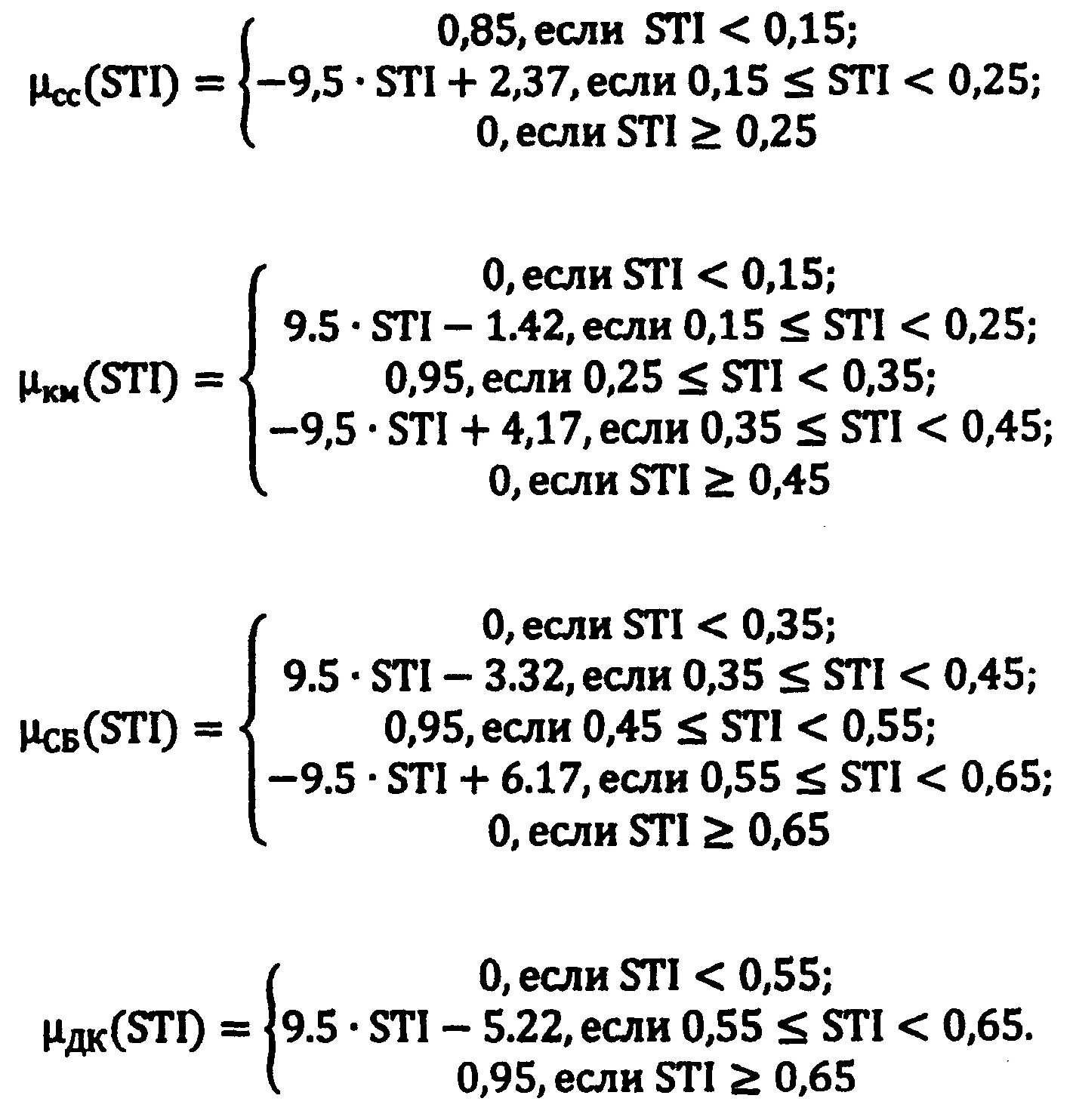
На Фиг. 5 изображены графики функций принадлежности к классам степени тяжести ишемии нижних конечностей с базовой переменной STБТ.FIG. 5 shows the graphs of the functions of belonging to the classes of severity of lower limb ischemia with the basic variable ST BT .
По указанным классам состояний, используя гистограммы распределения классов по шкале STБТ, эксперты определили соответствующие функции принадлежности μсс(STБТ), μкм(STБТ), μсб(STБТ) и μдк(STБТ) соответственно (фиг. 5).For the indicated classes of states, using histograms of the distribution of classes on the ST BT scale, the experts determined the corresponding membership functions μ cc (ST BT ), μ km (ST BT ), μ sat (ST BT ) and μ dk (ST BT ), respectively (FIG. five).
С целью упрощения изображения фиг. 5 у обозначенных функций принадлежности не показаны базовые переменные.In order to simplify the image of FIG. 5 baseline variables are not shown for the indicated membership functions.
Графики, приведенные на фиг. 5, описываются следующими аналитическими выражениями:The plots shown in FIG. 5 are described by the following analytical expressions:


Решение о диагностической классификации принимается путем сравнительного анализа значений функции уверенности в принадлежности к определенному классу ![]()
![]()
![]()
![]()
При равенстве значений функций принадлежностей решение принимается в пользу наиболее тяжелого состояния пациента (наиболее выраженной степени тяжести).In case of equality of the values of the functions of the accessories, the decision is made in favor of the most serious condition of the patient (the most pronounced severity).
На Фиг. 6. показана структурная схема прибора мониторинга степени тяжести ишемии нижних конечностей по большому пальцу ноги (вариант технической реализации микропроцессорного прибора для мониторинга степени тяжести ишемического процесса нижних конечностей по ФПГ и РЭО).FIG. 6. A block diagram of a device for monitoring the degree of severity of lower limb ischemia is shown along the big toe (a variant of the technical implementation of a microprocessor device for monitoring the severity of the ischemic process of the lower extremities in FIGs and REOs).
В приведенной схеме при регистрации фотоплетизмограммы в красном и инфракрасном диапазонах и расчета содержания кислорода в крови используется специализированный аналоговый интерфейс типа AFE (AFE 4490), к которому подключается красный CDк и инфракрасный CDи светодиоды, излучение с которых, пройдя через большого палец ноги регистрируется широкополосным фотоприемником ФП. Отсчеты фотоплетизмограммы в красном и инфракрасном свете по стандартному цифровому интерфейсу передаются в микроконтроллер МК, где осуществляется расчет показателей F и S.In the above scheme, when registering the photoplethysmogram in the red and infrared bands and calculating the oxygen content in the blood, a specialized analogue interface type AFE (AFE 4490) is used, to which the red CD is connected to and the infrared CD and the LEDs are recorded after passing through the big toe broadband photodetector OP. Photoplethysmogram readings in red and infrared light are transmitted via a standard digital interface to a microcontroller MK, where the indicators F and S are calculated.
Специализированный аналоговый интерфейс для измерения биоимпеданса тканей AFE РЭО (AFE 4300) на токовых электродах ТЭ1 и ТЭ2 формирует высокочастотный (50 кГц) сигнал, который модулируется кровотоком сосудов большого пальца ноги. Модулируемый сигнал снимается измерительными электродами НЭ1 и ИЭ2. Отсчеты сигнала в цифровом коде по стандартному интерфейсу передаются в микроконтроллер, который рассчитывает показатель R. После приема заданного числа фото- и рэо-сигналов, микроконтроллер по формуле (4) определяет степень тяжести ишемического процесса, и, применяя формулу (5) осуществляет соответствующую классификацию.A specialized analog interface for measuring the bioimpedance of AFE REO tissues (AFE 4300) on current electrodes ТЭ 1 and ТЭ 2 forms a high-frequency (50 kHz) signal, which is modulated by the blood flow of the big toe vessels. The modulated signal is removed by the measuring electrodes NE1 and IE2. The signal samples in the digital code are transmitted to the microcontroller by the standard interface, which calculates the indicator R. After receiving a specified number of photo and radio signals, the microcontroller determines the severity of the ischemic process using formula (4), and applying the formula (5) performs the appropriate classification .
Результаты классификаций отображаются на жидко- кристаллическом индикаторе (ЖКИ). Управление прибором и выбор требуемой информации осуществляется блоком клавиатуры (БК). Связь с мобильным средством связи пациента осуществляется модулем ближней радиосвязи Bluetooth с антенной А1.The results of the classifications are displayed on a liquid crystal display (LCD). The control of the device and the selection of the required information is carried out by the keyboard unit (BC). Communication with the patient's mobile communication device is carried out by the Bluetooth short-range radio module with antenna A1.
Дальняя (теле) связь с лечащим врачом, консультационными центрами и т.п., обеспечивается модулем GPRS с антенной А2. Возможно подключение к другим вычислительным устройствам через контроллер USB. Фотоприемник, светодиоды, токовые и измерительные электроды располагаются на контактной «ленте-липучке» (шириной 20-30 мм), оборачиваемой вокруг большого пальца пациента.Long-distance (tele) communication with the attending physician, counseling centers, etc., is provided by the GPRS module with antenna A2. It is possible to connect to other computing devices via a USB controller. The photodetector, LEDs, current and measuring electrodes are located on the contact "tape-Velcro" (width 20-30 mm), wrapped around the patient's thumb.
Для увеличения уверенности в классификации стадий деструктивных состояний нижних конечностей сигналы ФПГ и РЭО дополнительно предлагается регистрировать с различных остальных пальцев ноги.To increase confidence in the classification of the stages of destructive states of the lower extremities, FPG and REO signals are additionally proposed to be recorded from various remaining toes.
Как и для большого пальца осуществляется определение F, S и R. Обозначим номер пальца в соответствии с общепринятым как q (q=1, 2, 3, 4) (q=1 соответствует большому пальцу, q=5 - мизинцу). В ходе статистических исследований и экспертного оценивания было установлено, что регистрируемые сигналы с q=2-4 пальцев позволяют получать уверенности в оценке степени тяжести ишемического процесса в два раза меньше, чем от большого пальца, и информация от каждого из них вносит примерно одинаковый вклад в принятие искомого решения.As for the thumb, the definition of F, S and R is used. Denote the number of the finger in accordance with the generally accepted q (q = 1, 2, 3, 4) (q = 1 corresponds to the thumb, q = 5 - to the little finger). In the course of statistical studies and expert evaluation, it was found that the recorded signals with q = 2-4 fingers provide confidence in assessing the severity of the ischemic process two times less than from the thumb, and the information from each of them makes approximately the same contribution to making the desired decision.
Исходя из этого, для оставшихся четырех пальцев были синтезированы одинаковые функции степени тяжести fnq(F), fnq(S) и fnq(R), q=1, 2, 3, 4.Based on this, the same functions of severity fn q (F), fn q (S) and fn q (R), q = 1, 2, 3, 4, were synthesized for the remaining four fingers.
На Фиг. 7 приведены графики степени тяжести ишемического процесса для основных четырех пальцев (кроме большого) с базовыми переменными: A)-F; Б)-S; B)-R.FIG. 7 shows the graphs of the severity of the ischemic process for the main four fingers (except the thumb) with the basic variables: A) -F; B) -S; B) -R.
Аналитические выражения для графиков, приведенных на фиг. 7, представляются выражениями:Analytical expressions for the graphs shown in FIG. 7, are represented by the expressions:

Объединение функций fnq(F) и fnq(R) осуществляется по формуле:The functions f nq (F) and f nq (R) are combined by the formula:
![]()
![]()
Для каждого из q пальцев степень тяжести ишемического процесса определяется выражением:For each of the q fingers, the severity of the ischemic process is determined by the expression:
STnq=ƒnq(S)+ƒTq-ƒnq(S)⋅ƒTq ST nq = ƒ nq (S) + ƒ Tq -ƒ nq (S) Tq
По всем пальцам q, соответственно:On all fingers q, respectively:
![]()
![]()
где STP(1)=STn1.where STP (1) = ST n1 .
Агрегация формул (4) и (7) позволяет получить следующее выражение для определения степени тяжести ишемии нижних конечностей по сигналам от всех пальцев ноги:Aggregation of formulas (4) and (7) allows to obtain the following expression to determine the severity of lower limb ischemia from the signals from all toes:
![]()
![]()
Шкала STI является базовой переменной для функций принадлежности тех же классов состояний, как и для большого пальца ноги.The STI scale is the base variable for membership functions of the same state classes as for the big toe.
На Фиг. 8 показаны графики функций принадлежности к классам степени тяжести ишемической болезни нижних конечностей на пяти пальцах ног.FIG. 8 shows the graphs of the functions of belonging to the severity classes of ischemic disease of the lower limbs on five toes.
Аналогичные графики приведены на фиг. 5. и описываются следующими выражениями:Similar graphs are shown in FIG. 5. and described by the following expressions:


Анализ полученных функций, позволяет сделать вывод о том, что если использовать результаты измерений со всех пяти пальцев ног, то уверенность в принятии классификационного решения по сравнению с использованием данных только с большого пальца увеличивается на 10%.The analysis of the functions obtained allows us to conclude that if we use the results of measurements from all five toes, the confidence in making a classification decision increases by 10% compared to using only the thumb data.
Для реализации предлагаемого способа предлагается устройство - прибор мониторинга степени тяжести ишемических поражений нижних конечностей по пяти пальцам ноги.For the implementation of the proposed method, a device is proposed - a device for monitoring the severity of ischemic lesions of the lower extremities through five toes.
На фиг. 9 приведена структурная схема прибора мониторинга степени тяжести ишемических поражений нижних конечностей по пяти пальцам ноги.FIG. 9 shows a block diagram of a device for monitoring the severity of ischemic lesions of the lower extremities by five toes.
В схеме, приведенной на фиг. 9, в каждом из пяти фотоблоков (ФБ1, …, ФБ5) используются светодиоды, работающие в красном и инфракрасном диапазонах, и широкополосные фотоприемники, подключаемые к АFЕФПГ блоком аналоговых коммутаторов БАК аналогично схеме фиг. 6.In the circuit shown in FIG. 9, each of the five photoblocks (FB1, ..., FB5) uses LEDs operating in the red and infrared bands, and broadband photodetectors connected to the AFEFG unit by the analogue switch unit BAK, similar to the diagram of FIG. 6
В реографических блоках РБ1, …, РБ5 располагаются пары токовых и измерительных электродов, подключаемых через БАК к AFE РЭО аналогично схеме, представленной на фиг. 6.In eographical blocks RB1, ..., RB5 there are pairs of current and measuring electrodes connected through the LHC to the AFE REF, similarly to the diagram shown in FIG. 6
В процессе измерений микроконтроллер с помощью порта РМК поочередно формирует адреса подключения ФБq и РБq (q=1, …, 5) к соответствующим выводам AFE ФПГ и AFE РЭО через БАК. Подключение остальных элементов схемы фиг. 9 аналогично приведенным на схеме фиг. 6.In the process of measurements, the microcontroller, using the RMK port, alternately generates the connection addresses of the FB q and RB q (q = 1, ..., 5) to the corresponding pins of the AFE FPG and AFE REO through the LHC. Connecting the remaining circuit elements of FIG. 9 is similar to that shown in FIG. 6
Одной из важных задач ведения пациентов с ишемическими поражениями нижних конечностей является своевременное выявление отрицательных тенденций в развитии заболевания с проведением адекватных профилактических и лечебных мероприятий. Развитие современной микроэлектроники и мобильных приложений позволяют произвести оценку кровоснабжения нижних конечностей портативными и достаточно дешевыми средствами, что открывает возможности анализа степени поражения нижних конечностей и своевременного реагирования на отрицательные тенденции в развитии заболевания.One of the important tasks of the management of patients with ischemic lesions of the lower extremities is the timely detection of negative trends in the development of the disease with the implementation of adequate preventive and therapeutic measures. The development of modern microelectronics and mobile applications makes it possible to evaluate the blood supply to the lower limbs with portable and fairly cheap means, which opens up the possibility of analyzing the degree of damage to the lower limbs and timely responding to negative trends in the development of the disease.
С учетом этих возможностей предлагается производить оценку степени тяжести ишемического процесса и следить за его динамикой с использованием фотоплетизмографии (ФПГ) и реографии (РЭО) принятия диагностического решения помощью гибридных нечетких правил на основе математических моделей.Given these possibilities, it is proposed to assess the severity of the ischemic process and monitor its dynamics using photoplethysmography (FIG) and rheography (REO) of making diagnostic decisions using hybrid fuzzy rules based on mathematical models.
Claims (19)


Priority Applications (1)
| Application Number | Priority Date | Filing Date | Title |
|---|---|---|---|
| RU2017145620A RU2688386C1 (en) | 2017-12-25 | 2017-12-25 | Method for lower limb ischemic involvement severity monitoring and device for its implementation |
Applications Claiming Priority (1)
| Application Number | Priority Date | Filing Date | Title |
|---|---|---|---|
| RU2017145620A RU2688386C1 (en) | 2017-12-25 | 2017-12-25 | Method for lower limb ischemic involvement severity monitoring and device for its implementation |
Publications (1)
| Publication Number | Publication Date |
|---|---|
| RU2688386C1 true RU2688386C1 (en) | 2019-05-22 |
Family
ID=66637005
Family Applications (1)
| Application Number | Title | Priority Date | Filing Date |
|---|---|---|---|
| RU2017145620A RU2688386C1 (en) | 2017-12-25 | 2017-12-25 | Method for lower limb ischemic involvement severity monitoring and device for its implementation |
Country Status (1)
| Country | Link |
|---|---|
| RU (1) | RU2688386C1 (en) |
Citations (7)
| Publication number | Priority date | Publication date | Assignee | Title |
|---|---|---|---|---|
| RU2192236C2 (en) * | 2000-06-02 | 2002-11-10 | Самарский государственный медицинский университет | Testing device for treating ischemic states of extremities |
| RU2007108157A (en) * | 2007-03-05 | 2008-09-10 | Государственное областное учреждение здравоохранени Свердловский областной клинический психоневрологический госпиталь дл ветеранов войн (ГОУЗ СОКП Госпиталь дл ветеранов войн) (RU) | METHOD FOR DIAGNOSTIC OF ATHEROSCLEROSIS |
| US20080300493A1 (en) * | 2005-12-07 | 2008-12-04 | Rodolfo Gatto | Optical microprobe for blood clot detection |
| US20090287101A1 (en) * | 2008-05-13 | 2009-11-19 | Searete Llc, A Limited Liability Corporation Of The State Of Delaware | Circulatory monitoring systems and methods |
| WO2011127184A1 (en) * | 2010-04-06 | 2011-10-13 | Cardiox Corporation | System for improved hemodynamic detection of circulatory anomalies |
| US20120130419A1 (en) * | 2009-06-23 | 2012-05-24 | Infarct Reduction Technologies Inc. | Automatic devices for remote ischemic preconditioning |
| US20140316292A1 (en) * | 2013-04-19 | 2014-10-23 | Semler Scientific, Inc. | Circulation Monitoring System |
-
2017
- 2017-12-25 RU RU2017145620A patent/RU2688386C1/en not_active IP Right Cessation
Patent Citations (7)
| Publication number | Priority date | Publication date | Assignee | Title |
|---|---|---|---|---|
| RU2192236C2 (en) * | 2000-06-02 | 2002-11-10 | Самарский государственный медицинский университет | Testing device for treating ischemic states of extremities |
| US20080300493A1 (en) * | 2005-12-07 | 2008-12-04 | Rodolfo Gatto | Optical microprobe for blood clot detection |
| RU2007108157A (en) * | 2007-03-05 | 2008-09-10 | Государственное областное учреждение здравоохранени Свердловский областной клинический психоневрологический госпиталь дл ветеранов войн (ГОУЗ СОКП Госпиталь дл ветеранов войн) (RU) | METHOD FOR DIAGNOSTIC OF ATHEROSCLEROSIS |
| US20090287101A1 (en) * | 2008-05-13 | 2009-11-19 | Searete Llc, A Limited Liability Corporation Of The State Of Delaware | Circulatory monitoring systems and methods |
| US20120130419A1 (en) * | 2009-06-23 | 2012-05-24 | Infarct Reduction Technologies Inc. | Automatic devices for remote ischemic preconditioning |
| WO2011127184A1 (en) * | 2010-04-06 | 2011-10-13 | Cardiox Corporation | System for improved hemodynamic detection of circulatory anomalies |
| US20140316292A1 (en) * | 2013-04-19 | 2014-10-23 | Semler Scientific, Inc. | Circulation Monitoring System |
Similar Documents
| Publication | Publication Date | Title |
|---|---|---|
| Hasanzadeh et al. | Blood pressure estimation using photoplethysmogram signal and its morphological features | |
| Sun et al. | Systolic blood pressure estimation using PPG and ECG during physical exercise | |
| US20150216425A1 (en) | Estimations of equivalent inner diameter of arterioles | |
| US20160367154A1 (en) | Obtaining cardiovascular parameters using arterioles related transient time | |
| Weinkauf et al. | Near-instant noninvasive optical imaging of tissue perfusion for vascular assessment | |
| Zahedi et al. | Analysis of the effect of ageing on rising edge characteristics of the photoplethysmogram using a modified Windkessel model | |
| Yang et al. | Estimation and validation of arterial blood pressure using photoplethysmogram morphology features in conjunction with pulse arrival time in large open databases | |
| KR20220013559A (en) | System for monitoring physiological parameters | |
| Stoner et al. | A primer on repeated sitting exposure and the cardiovascular system: considerations for study design, analysis, interpretation, and translation | |
| Chiang et al. | A novel wireless photoplethysmography blood-flow volume sensor for assessing arteriovenous fistula of hemodialysis patients | |
| Lin | Assessment of bilateral photoplethysmography for lower limb peripheral vascular occlusive disease using color relation analysis classifier | |
| Thurston et al. | Ankle Brachial Pressure Index: An update for the vascular specialist and general practitioner | |
| Gentilin et al. | Estimation of carotid-femoral pulse wave velocity from finger photoplethysmography signal | |
| Parry et al. | Ambulatory impedance cardiography: a systematic review | |
| Desebbe et al. | Evaluation of a new smartphone optical blood pressure application (OptiBP™) in the post-anesthesia care unit: a method comparison study against the non-invasive automatic oscillometric brachial cuff as the reference method | |
| US20130006121A1 (en) | Device for Identifying Hemodynamic Changes | |
| RU2688386C1 (en) | Method for lower limb ischemic involvement severity monitoring and device for its implementation | |
| Xie et al. | Optical coherence tomography angiography measures blood pulsatile waveforms at variable tissue depths | |
| Suganthi et al. | Morphological analysis of peripheral arterial signals in Takayasu’s arteritis | |
| Sameen et al. | Cuff-less and continuous blood pressure measurement based on pulse transit time from carotid and toe photoplethysmograms | |
| Sannino et al. | Non-invasive estimation of blood pressure through genetic programming-preliminary results | |
| Lee et al. | Accuracy of noninvasive continuous arterial pressure monitoring using ClearSight during one-lung ventilation | |
| Elkady et al. | Intermittent pneumatic compression for critical limb ischaemia | |
| Tamura | Regulation and approval of continuous non-invasive blood-pressure monitoring devices | |
| Miyagatani et al. | Vascular tone in patients with hemorrhagic shock |
Legal Events
| Date | Code | Title | Description |
|---|---|---|---|
| MM4A | The patent is invalid due to non-payment of fees |
Effective date: 20191226 |