KR20140112207A - Augmented reality imaging display system and surgical robot system comprising the same - Google Patents
Augmented reality imaging display system and surgical robot system comprising the same Download PDFInfo
- Publication number
- KR20140112207A KR20140112207A KR1020130026615A KR20130026615A KR20140112207A KR 20140112207 A KR20140112207 A KR 20140112207A KR 1020130026615 A KR1020130026615 A KR 1020130026615A KR 20130026615 A KR20130026615 A KR 20130026615A KR 20140112207 A KR20140112207 A KR 20140112207A
- Authority
- KR
- South Korea
- Prior art keywords
- image
- augmented reality
- camera
- patient
- surgical
- Prior art date
Links
Images


Classifications
-
- A—HUMAN NECESSITIES
- A61—MEDICAL OR VETERINARY SCIENCE; HYGIENE
- A61B—DIAGNOSIS; SURGERY; IDENTIFICATION
- A61B34/00—Computer-aided surgery; Manipulators or robots specially adapted for use in surgery
- A61B34/30—Surgical robots
- A61B34/37—Master-slave robots
-
- A—HUMAN NECESSITIES
- A61—MEDICAL OR VETERINARY SCIENCE; HYGIENE
- A61B—DIAGNOSIS; SURGERY; IDENTIFICATION
- A61B1/00—Instruments for performing medical examinations of the interior of cavities or tubes of the body by visual or photographical inspection, e.g. endoscopes; Illuminating arrangements therefor
- A61B1/00002—Operational features of endoscopes
- A61B1/00043—Operational features of endoscopes provided with output arrangements
- A61B1/00045—Display arrangement
-
- A—HUMAN NECESSITIES
- A61—MEDICAL OR VETERINARY SCIENCE; HYGIENE
- A61B—DIAGNOSIS; SURGERY; IDENTIFICATION
- A61B1/00—Instruments for performing medical examinations of the interior of cavities or tubes of the body by visual or photographical inspection, e.g. endoscopes; Illuminating arrangements therefor
- A61B1/00147—Holding or positioning arrangements
- A61B1/00149—Holding or positioning arrangements using articulated arms
-
- A—HUMAN NECESSITIES
- A61—MEDICAL OR VETERINARY SCIENCE; HYGIENE
- A61B—DIAGNOSIS; SURGERY; IDENTIFICATION
- A61B34/00—Computer-aided surgery; Manipulators or robots specially adapted for use in surgery
- A61B34/30—Surgical robots
-
- A—HUMAN NECESSITIES
- A61—MEDICAL OR VETERINARY SCIENCE; HYGIENE
- A61B—DIAGNOSIS; SURGERY; IDENTIFICATION
- A61B5/00—Measuring for diagnostic purposes; Identification of persons
- A61B5/05—Detecting, measuring or recording for diagnosis by means of electric currents or magnetic fields; Measuring using microwaves or radio waves
- A61B5/055—Detecting, measuring or recording for diagnosis by means of electric currents or magnetic fields; Measuring using microwaves or radio waves involving electronic [EMR] or nuclear [NMR] magnetic resonance, e.g. magnetic resonance imaging
-
- A—HUMAN NECESSITIES
- A61—MEDICAL OR VETERINARY SCIENCE; HYGIENE
- A61B—DIAGNOSIS; SURGERY; IDENTIFICATION
- A61B5/00—Measuring for diagnostic purposes; Identification of persons
- A61B5/74—Details of notification to user or communication with user or patient ; user input means
- A61B5/742—Details of notification to user or communication with user or patient ; user input means using visual displays
- A61B5/7445—Display arrangements, e.g. multiple display units
-
- A—HUMAN NECESSITIES
- A61—MEDICAL OR VETERINARY SCIENCE; HYGIENE
- A61B—DIAGNOSIS; SURGERY; IDENTIFICATION
- A61B90/00—Instruments, implements or accessories specially adapted for surgery or diagnosis and not covered by any of the groups A61B1/00 - A61B50/00, e.g. for luxation treatment or for protecting wound edges
- A61B90/36—Image-producing devices or illumination devices not otherwise provided for
- A61B90/361—Image-producing devices, e.g. surgical cameras
-
- A—HUMAN NECESSITIES
- A61—MEDICAL OR VETERINARY SCIENCE; HYGIENE
- A61B—DIAGNOSIS; SURGERY; IDENTIFICATION
- A61B90/00—Instruments, implements or accessories specially adapted for surgery or diagnosis and not covered by any of the groups A61B1/00 - A61B50/00, e.g. for luxation treatment or for protecting wound edges
- A61B90/90—Identification means for patients or instruments, e.g. tags
- A61B90/92—Identification means for patients or instruments, e.g. tags coded with colour
-
- A—HUMAN NECESSITIES
- A61—MEDICAL OR VETERINARY SCIENCE; HYGIENE
- A61B—DIAGNOSIS; SURGERY; IDENTIFICATION
- A61B90/00—Instruments, implements or accessories specially adapted for surgery or diagnosis and not covered by any of the groups A61B1/00 - A61B50/00, e.g. for luxation treatment or for protecting wound edges
- A61B90/90—Identification means for patients or instruments, e.g. tags
- A61B90/94—Identification means for patients or instruments, e.g. tags coded with symbols, e.g. text
-
- B—PERFORMING OPERATIONS; TRANSPORTING
- B25—HAND TOOLS; PORTABLE POWER-DRIVEN TOOLS; MANIPULATORS
- B25J—MANIPULATORS; CHAMBERS PROVIDED WITH MANIPULATION DEVICES
- B25J13/00—Controls for manipulators
- B25J13/08—Controls for manipulators by means of sensing devices, e.g. viewing or touching devices
-
- G—PHYSICS
- G06—COMPUTING; CALCULATING OR COUNTING
- G06T—IMAGE DATA PROCESSING OR GENERATION, IN GENERAL
- G06T19/00—Manipulating 3D models or images for computer graphics
- G06T19/006—Mixed reality
-
- G—PHYSICS
- G09—EDUCATION; CRYPTOGRAPHY; DISPLAY; ADVERTISING; SEALS
- G09B—EDUCATIONAL OR DEMONSTRATION APPLIANCES; APPLIANCES FOR TEACHING, OR COMMUNICATING WITH, THE BLIND, DEAF OR MUTE; MODELS; PLANETARIA; GLOBES; MAPS; DIAGRAMS
- G09B9/00—Simulators for teaching or training purposes
-
- G—PHYSICS
- G16—INFORMATION AND COMMUNICATION TECHNOLOGY [ICT] SPECIALLY ADAPTED FOR SPECIFIC APPLICATION FIELDS
- G16H—HEALTHCARE INFORMATICS, i.e. INFORMATION AND COMMUNICATION TECHNOLOGY [ICT] SPECIALLY ADAPTED FOR THE HANDLING OR PROCESSING OF MEDICAL OR HEALTHCARE DATA
- G16H20/00—ICT specially adapted for therapies or health-improving plans, e.g. for handling prescriptions, for steering therapy or for monitoring patient compliance
- G16H20/40—ICT specially adapted for therapies or health-improving plans, e.g. for handling prescriptions, for steering therapy or for monitoring patient compliance relating to mechanical, radiation or invasive therapies, e.g. surgery, laser therapy, dialysis or acupuncture
-
- G—PHYSICS
- G16—INFORMATION AND COMMUNICATION TECHNOLOGY [ICT] SPECIALLY ADAPTED FOR SPECIFIC APPLICATION FIELDS
- G16H—HEALTHCARE INFORMATICS, i.e. INFORMATION AND COMMUNICATION TECHNOLOGY [ICT] SPECIALLY ADAPTED FOR THE HANDLING OR PROCESSING OF MEDICAL OR HEALTHCARE DATA
- G16H40/00—ICT specially adapted for the management or administration of healthcare resources or facilities; ICT specially adapted for the management or operation of medical equipment or devices
- G16H40/60—ICT specially adapted for the management or administration of healthcare resources or facilities; ICT specially adapted for the management or operation of medical equipment or devices for the operation of medical equipment or devices
- G16H40/63—ICT specially adapted for the management or administration of healthcare resources or facilities; ICT specially adapted for the management or operation of medical equipment or devices for the operation of medical equipment or devices for local operation
-
- A—HUMAN NECESSITIES
- A61—MEDICAL OR VETERINARY SCIENCE; HYGIENE
- A61B—DIAGNOSIS; SURGERY; IDENTIFICATION
- A61B1/00—Instruments for performing medical examinations of the interior of cavities or tubes of the body by visual or photographical inspection, e.g. endoscopes; Illuminating arrangements therefor
- A61B1/04—Instruments for performing medical examinations of the interior of cavities or tubes of the body by visual or photographical inspection, e.g. endoscopes; Illuminating arrangements therefor combined with photographic or television appliances
-
- A—HUMAN NECESSITIES
- A61—MEDICAL OR VETERINARY SCIENCE; HYGIENE
- A61B—DIAGNOSIS; SURGERY; IDENTIFICATION
- A61B34/00—Computer-aided surgery; Manipulators or robots specially adapted for use in surgery
- A61B34/20—Surgical navigation systems; Devices for tracking or guiding surgical instruments, e.g. for frameless stereotaxis
- A61B2034/2046—Tracking techniques
- A61B2034/2065—Tracking using image or pattern recognition
-
- A—HUMAN NECESSITIES
- A61—MEDICAL OR VETERINARY SCIENCE; HYGIENE
- A61B—DIAGNOSIS; SURGERY; IDENTIFICATION
- A61B90/00—Instruments, implements or accessories specially adapted for surgery or diagnosis and not covered by any of the groups A61B1/00 - A61B50/00, e.g. for luxation treatment or for protecting wound edges
- A61B90/36—Image-producing devices or illumination devices not otherwise provided for
- A61B2090/364—Correlation of different images or relation of image positions in respect to the body
- A61B2090/365—Correlation of different images or relation of image positions in respect to the body augmented reality, i.e. correlating a live optical image with another image
-
- A—HUMAN NECESSITIES
- A61—MEDICAL OR VETERINARY SCIENCE; HYGIENE
- A61B—DIAGNOSIS; SURGERY; IDENTIFICATION
- A61B90/00—Instruments, implements or accessories specially adapted for surgery or diagnosis and not covered by any of the groups A61B1/00 - A61B50/00, e.g. for luxation treatment or for protecting wound edges
- A61B90/39—Markers, e.g. radio-opaque or breast lesions markers
- A61B2090/3937—Visible markers
-
- A—HUMAN NECESSITIES
- A61—MEDICAL OR VETERINARY SCIENCE; HYGIENE
- A61B—DIAGNOSIS; SURGERY; IDENTIFICATION
- A61B6/00—Apparatus for radiation diagnosis, e.g. combined with radiation therapy equipment
- A61B6/02—Devices for diagnosis sequentially in different planes; Stereoscopic radiation diagnosis
- A61B6/03—Computerised tomographs
- A61B6/032—Transmission computed tomography [CT]
-
- G—PHYSICS
- G06—COMPUTING; CALCULATING OR COUNTING
- G06T—IMAGE DATA PROCESSING OR GENERATION, IN GENERAL
- G06T2210/00—Indexing scheme for image generation or computer graphics
- G06T2210/41—Medical
Abstract
Description
증강현실 영상 표시 시스템 및 이를 포함하는 수술 로봇 시스템이 개시된다. 더욱 상세하게는, 사용자의 시선 이동에 따라 대응되는 부위의 가상 영상이 실시간으로 디스플레이되는 증강현실 영상 표시 시스템 및 이를 포함하는 수술 로봇 시스템이 개시된다.Augmented reality image display system and surgical robot system including the same are disclosed. More particularly, the present invention relates to an augmented reality image display system and a surgical robot system including the augmented reality image display system, in which a virtual image of a corresponding part is displayed in real time according to a user's gaze movement.
최소 침습 수술(Minimal Invasive Surgery)이란 환부의 크기를 최소화하는 수술을 통칭한다. 최소 침습 수술은 인체의 일부(예: 복부)에 큰 절개창을 열고 시행하는 개복 수술과는 달리, 인체에 0.5㎝∼1.5㎝ 크기의 적어도 하나의 절개공(또는 침습구)을 형성하고, 이 절개공을 통해 내시경과 각종 수술도구들을 넣은 후 영상을 보면서 시행하는 수술 방법이다.Minimal Invasive Surgery refers to surgery that minimizes the size of the affected area. The minimally invasive surgery is different from the laparotomy in which a large incision is opened and performed on a part of the human body (for example, the abdomen), at least one incision ball (or an invasion ball) of 0.5 cm to 1.5 cm in size is formed in the human body, It is a surgical method that is performed by inserting an endoscope and various surgical tools through a ball and viewing the images.
이러한 최소 침습 수술은 개복 수술과는 달리 수술 후 통증이 적고, 장운동의 조기 회복 및 음식물의 조기 섭취가 가능하며 입원 기간이 짧고 정상 상태로의 복귀가 빠르며 절개 범위가 좁아 미용 효과가 우수하다는 장점을 갖는다. 이와 같은 장점으로 인하여 최소 침습 수술은 담낭 절제술, 전립선암 수술, 탈장 교정술 등에 사용되고 있고 그 분야를 점점 더 넓혀가고 있는 추세이다.This minimally invasive surgery has advantages such as less pain after surgery, early recovery of bowel movements and early ingestion of food, short hospital stay, quick return to normal state, . Because of these advantages, minimally invasive surgery is being used in cholecystectomy, prostate cancer surgery, and hernia repair.
일반적으로 최소 침습 수술에 이용되는 수술 로봇은 마스터 장치와 슬레이브 장치를 포함한다. 마스터 장치는 의사의 조작에 따른 제어신호를 생성하여 슬레이브 장치로 전송하고, 슬레이브 장치는 마스터 장치로부터 제어신호를 수신하여 수술에 필요한 조작을 환자에게 가하게 되며, 마스터 장치와 슬레이브 장치를 통합하여 구성하거나, 각각 별도의 장치로 구성하여 수술실에 배치한 상태에서 수술을 진행하고 있다.In general, a surgical robot used for minimally invasive surgery includes a master device and a slave device. The master device generates a control signal according to the operation of the doctor and transmits the generated control signal to the slave device. The slave device receives the control signal from the master device and applies the operation required for the operation to the patient. The master device and the slave device are integrated , Each of which is configured as a separate device, is placed in the operating room, and the operation is underway.
슬레이브 장치는 적어도 하나 이상의 로봇 암을 구비하며, 각 로봇 암의 단부에는 수술 기구(surgical instruments)가 장착되는데, 이때, 수술 기구 말단부에는 수술 도구(surgical tool)가 장착되어 있다.The slave device is provided with at least one robot arm, and surgical instruments are mounted on the end portions of the robot arms. At this time, a surgical tool is mounted at the distal end of the surgical instrument.
이와 같은 수술 로봇을 이용한 최소 침습 수술은 슬레이브 장치의 수술 도구(surgical tool) 및 수술 도구가 장착된 수술 기구(surgical instruments)가 환자의 인체 내부로 진입하여 필요한 시술이 행해지게 되는데 이때, 수술 도구 및 수술 기구가 인체 내부로 진입한 이후에는 수술 도구 중 하나인 내시경을 통하여 수집된 이미지로부터 내부 상황을 확인하고, 환자의 수술 전 의료 영상(예, CT, MRI 등)은 참고적인 보조 영상으로 이용하고 있다.In such a minimally invasive surgery using a surgical robot, a surgical tool of a slave device and a surgical instrument equipped with a surgical tool enter into a human body, and necessary procedures are performed. In this case, After the surgical instrument has entered the human body, internal conditions are checked from the images collected through an endoscope, which is one of the surgical instruments, and medical images (eg, CT, MRI, etc.) have.
환자 신체 내부를 직관적으로 관찰할 수 있는 증강현실 영상 표시 시스템 및 이를 포함하는 수술 로봇 시스템을 제공하는 것이다.The present invention provides an augmented reality image display system capable of intuitively observing the inside of a patient's body and a surgical robot system including the same.
수술 로봇 시스템의 일 실시 예는 환자에게 수술 동작을 수행하는 슬레이브 시스템, 상기 슬레이브 시스템의 수술 동작을 제어하는 마스터 시스템, 환자 신체 내부에 대한 가상 영상을 생성하는 영상 시스템 및 환자 신체 또는 인체 모형에 부착된 복수의 마커를 포함하는 실제 영상을 획득하는 카메라, 상기 실제 영상에서 복수의 마커를 검출하고, 검출된 복수의 마커를 이용하여 카메라의 위치 및 주시 방향을 추정한 후, 상기 가상 영상 중 추정된 카메라의 위치 및 주시 방향에 대응되는 부위를 상기 실제 영상에 오버레이 방식으로 합성한 증강현실 영상을 생성하는 증강현실 영상 생성부 및 상기 증강현실 영상을 표시하는 디스플레이를 포함하는 증강현실 영상 표시 시스템을 포함한다.
One embodiment of the surgical robot system includes a slave system for performing a surgical operation on a patient, a master system for controlling the operation of the slave system, an image system for generating a virtual image of the inside of the patient body, A camera for acquiring an actual image including a plurality of markers, a plurality of markers in the real image, estimating a camera position and a viewing direction using the plurality of detected markers, An augmented reality image generation unit that generates an augmented reality image in which an area corresponding to a camera position and a viewing direction is synthesized in an overlay manner on the actual image, and a display unit that displays the augmented reality image do.
또한, 증강현실 영상 표시 시스템의 일 실시 예는 환자 신체 또는 인체 모형에 부착된 복수의 마커를 포함하는 실제 영상을 획득하는 카메라, 상기 실제 영상에서 복수의 마커를 검출하고, 검출된 복수의 마커를 이용하여 카메라의 위치 및 주시 방향을 추정한 후, 추정된 위치 및 주시 방향에 대응되는 부위의 가상 영상을 상기 실제 영상에 오버레이 방식으로 합성한 증강현실 영상을 생성하는 증강현실 영상 생성부 및 상기 증강현실 영상을 표시하는 디스플레이를 포함한다.According to another aspect of the present invention, there is provided an augmented reality image display system including a camera for acquiring an actual image including a plurality of markers attached to a patient's body or a human body model, a plurality of markers in the real image, An augmented reality image generating unit for generating augmented reality image in which a virtual image of a portion corresponding to the estimated position and the viewing direction is synthesized in an overlay manner on the real image, And a display for displaying a real image.
도 1은 수술 로봇 시스템의 외관 구조를 도시한 도면이다.
도 2는 수술 로봇 시스템의 구성요소를 개략적으로 도시한 블럭도이다.
도 3은 수술 보조자가 증강현실 영상 표시 시스템을 장착한 형상을 도시한 도면이다.
도 4는 환자 신체에 마커를 부착한 예를 도시한 도면이다.
도 5는 인체 모형에 마커를 부착한 예를 도시한 도면이다.
도 6 및 도 7은 각각 카메라의 주시 방향에 따른 증강현실 영상을 도시한 도면이다.
도 8 및 도 9는 각각 카메라와 마커와의 거리에 비례하여 축소 또는 확대된 가상 영상이 합성된 증강현실 영상을 도시한 도면이다.
도 10은 도 9에서 카메라가 마커와의 거리는 유지한 채 이동한 경우의 증강현실 영상을 도시한 도면이다.
도 11은 내시경을 통해 획득한 수술 부위 영상 및 실제 수술 도구와 가상 수술 도구가 생성된 증강현실 영상을 도시한 도면이다.1 is a view showing an external structure of a surgical robot system.
2 is a block diagram schematically showing the components of a surgical robot system.
3 is a view showing a configuration in which a surgeon mounts an augmented reality image display system.
4 is a view showing an example of attaching a marker to a patient's body.
5 is a view showing an example of attaching a marker to a human body model.
FIGS. 6 and 7 are views showing an augmented reality image according to the camera's direction of sight, respectively.
8 and 9 are views showing an augmented reality image in which a virtual image reduced or enlarged in proportion to a distance between a camera and a marker is synthesized.
FIG. 10 is a view showing an augmented reality image when the camera moves while maintaining the distance from the marker in FIG.
11 is a diagram showing a surgical site image acquired through an endoscope and an augmented reality image in which a virtual surgical tool and an actual surgical tool are generated.
본 발명의 목적, 특정한 장점들 및 신규한 특징들은 첨부된 도면들과 연관되어지는 이하의 상세한 설명과 바람직한 실시 예들로부터 더욱 명백해질 것이다. 본 명세서에서 각 도면의 구성요소들에 참조번호를 부가함에 있어서, 동일한 구성 요소들에 한해서는 비록 다른 도면상에 표시되더라도 가능한 한 동일한 번호를 가지도록 하고 있음에 유의하여야한다. 또한, 본 발명을 설명함에 있어서, 관련된 공지 기술에 대한 구체적인 설명이 본 발명의 요지를 불필요하게 흐릴 수 있다고 판단되는 경우 그 상세한 설명은 생략한다. 본 명세서에서, 제1, 제2 등의 용어는 하나의 구성요소를 다른 구성요소로부터 구별하기 위해 사용되는 것으로, 구성요소가 상기 용어들에 의해 제한되는 것은 아니다.
BRIEF DESCRIPTION OF THE DRAWINGS The objectives, specific advantages, and novel features of the present invention will become more apparent from the following detailed description taken in conjunction with the accompanying drawings, in which: FIG. It should be noted that, in the present specification, the reference numerals are added to the constituent elements of the drawings, and the same constituent elements are assigned the same number as much as possible even if they are displayed on different drawings. In the following description, well-known functions or constructions are not described in detail since they would obscure the invention in unnecessary detail. In this specification, the terms first, second, etc. are used to distinguish one element from another, and the element is not limited by the terms.
이하, 첨부된 도면을 참조하여 본 실시 예를 상세히 설명하기로 한다.
Hereinafter, embodiments of the present invention will be described in detail with reference to the accompanying drawings.
도 1은 수술 로봇 시스템의 외관 구조를 도시한 도면이고, 도 2는 수술 로봇 시스템의 구성요소를 개략적으로 도시한 블럭도이다.FIG. 1 is a view showing an external structure of a surgical robot system, and FIG. 2 is a block diagram schematically showing the components of a surgical robot system.
수술 로봇 시스템은 크게 수술대에 누워있는 환자(P)에게 수술을 행하는 슬레이브 시스템(200)과 조작자(예로써, 의사)(S)의 조작을 통해 슬레이브 시스템(200)을 원격 제어하는 마스터 시스템(100)을 포함할 수 있다. 이때, 조작자(S)를 보조할 보조자(A)가 환자(P) 측에 한 명 이상 위치할 수 있다.The surgical robot system mainly includes a
여기에서, 조작자(S)를 보조한다는 것은 수술이 진행되는 동안 수술 작업을 보조하는 것으로, 예를 들어, 사용되는 수술 도구의 교체 등을 포함할 수 있으나, 특별히 이에 한정되는 것은 아니다. 예를 들어, 수술 작업 종류에 따라 다양한 수술 도구가 사용될 수 있는데, 슬레이브 시스템(200)의 로봇 암(210)의 갯 수는 제한적이므로, 한 번에 장착될 수 있는 수술 도구의 갯 수 역시 제한적이다. 이에 따라, 수술 도중 수술 도구를 교체할 필요가 있는 경우, 조작자(S)는 환자(P) 측에 위치한 보조자(A)에게 수술 도구를 교체하도록 지시하고, 보조자(A)는 지시에 따라 슬레이브 시스템(200)의 로봇 암(210)으로부터 사용하지 않는 수술 도구를 제거하고, 트레이(T)에 놓인 다른 수술 도구(230′)를 해당 로봇 암(210)에 장착할 수 있다.Here, assisting the operator S is to assist in the surgical operation during the operation, and may include, for example, replacement of the surgical tool to be used, but is not limited thereto. For example, various types of surgical instruments may be used depending on the kind of surgical operation. Since the number of the
마스터 시스템(100)과 슬레이브 시스템(200)은 물리적으로 독립된 별도의 장치로 분리 구성될 수 있으나, 특별히 이에 한정되는 것은 아니며, 예로써 하나로 통합된 일체형 장치로 구성되는 것 역시 가능할 것이다.
The
도 1 및 도 2에 도시한 바와 같이, 마스터 시스템(100)은 입력부(110) 및 표시부(120)를 포함할 수 있다.As shown in FIGS. 1 and 2, the
입력부(110)는 수술 로봇 시스템의 동작 모드를 선택하는 명령, 슬레이브 시스템(200)의 동작을 원격으로 제어하기 위한 명령 등을 조작자(S)로부터 입력받을 수 있는 구성을 의미하며, 본 실시 예에서 입력부(110)로 햅틱 디바이스, 클러치 페달, 스위치, 버튼 등이 사용될 수 있으나, 특별히 이에 한정되는 것은 아니며, 예로써 음성 인식 디바이스 등과 같은 구성도 사용될 수 있다. 이하 설명에서는 입력부(110)로, 햅틱 디바이스가 사용된 것을 예로 들어 설명하기로 한다.The
도 1에서는 입력부(110)가 두 개의 핸들(111, 113)을 포함하는 것으로 도시하고 있으나, 이는 하나의 실시 예에 불과할 뿐, 본 발명이 이에 한정되는 것은 아니다. 예를 들어, 하나의 핸들을 포함할 수도 있고 또는 세 개 이상의 핸들을 포함하는 것 역시 가능할 것이다.Although the
조작자(S)는 도 1과 같이, 양손으로 두 개의 핸들(111, 113)을 각각 조작하여 슬레이브 시스템(200)의 로봇 암(210)의 동작을 제어할 수 있다. 이때, 도 1에 자세하게 도시되지는 않았으나, 각각의 핸들(111, 113)은 엔드 이펙터, 복수의 링크 및 복수의 관절을 포함할 수 있다.The operator S can control the operation of the
여기에서, 엔드 이펙터는 조작자(S)의 손이 직접적으로 접촉되는 부분으로, 펜슬 또는 스틱 형상을 가질 수 있으나, 그 형상이 특별히 이에 한정되는 것은 아니다.Here, the end effector is a portion directly contacting the hand of the operator S, and may have a pencil or stick shape, but the shape is not particularly limited to this.
관절은 링크와 링크의 연결 부위를 말하며, 1 자유도 이상을 가질 수 있다. 여기에서, "자유도(Degree Of Freedom:DOF)"란 기구학(Kinematics) 또는 역기구학(Inverse Kinematics)에서의 자유도를 말한다. 기구의 자유도란 기구의 독립적인 운동의 수, 또는 각 링크 간의 상대 위치의 독립된 운동을 결정하는 변수의 수를 말한다. 예를 들어, x축, y축, z축으로 이루어진 3차원 공간상의 물체는, 물체의 공간적인 위치를 결정하기 위한 3 자유도(각 축에서의 위치)와, 물체의 공간적인 자세를 결정하기 위한 3 자유도(각 축에서의 위치)와, 물체의 공간적인 자세를 결정하기 위한 3 자유도(각 축에 대한 회전 각도) 중에서 하나 이상의 자유도를 갖는다. 구체적으로, 물체가 각각의 축을 따라 이동 가능하고, 각각의 축을 기준으로 회전 가능하다고 한다면, 이 물체는 6 자유도를 갖는 것으로 이해될 수 있다.A joint is the linkage between a link and a link, and can have more than one degree of freedom. Here, "Degree of Freedom (DOF)" refers to degrees of freedom in kinematics or inverse kinematics. The degree of freedom of a mechanism refers to the number of independent motions of the mechanism or the number of variables that determine the independent movement of relative positions between links. For example, an object in a three-dimensional space consisting of an x-axis, a y-axis, and a z-axis has three degrees of freedom (position in each axis) for determining the spatial position of an object, (Three degrees of freedom for each axis) and three degrees of freedom (rotation angle for each axis) for determining the spatial posture of the object. Specifically, if an object is movable along each axis and is rotatable about each axis, it can be understood that the object has six degrees of freedom.
또한, 관절에는 관절의 상태를 나타내는 상태 정보 예로써, 관절에 가해진 힘/토크 정보, 관절의 위치 정보 및 이동 속도 정보 등을 검출할 수 있는 검출부(미도시)가 마련될 수 있다. 이에 따라, 조작자(S)가 입력부(110)를 조작하면, 검출부(미도시)는 조작된 입력부(110)의 상태 정보를 검출할 수 있고, 제어부(130)는 제어 신호 생성부(131)를 이용하여 검출부(미도시)에 의해 검출된 입력부(110)의 상태 정보에 대응되는 제어 신호를 생성하고, 통신부(140)를 통해 슬레이브 시스템(200)으로 생성된 제어 신호를 전송할 수 있다. 즉, 마스터 시스템(100)의 제어부(130)는 제어 신호 생성부(131)를 이용하여 조작자(S)의 입력부(110) 조작에 따른 제어 신호를 생성하고, 생성된 제어 신호를 통신부(140)를 통하여 슬레이브 시스템(200)으로 전송할 수 있다.
In addition, the joint may be provided with a detection unit (not shown) capable of detecting force / torque information, joint position information, and movement speed information, etc., which are examples of state information indicating the state of the joint. Accordingly, when the operator S operates the
마스터 시스템(100)의 표시부(120)에는 내시경(220)을 통해 수집된 환자(P) 신체 내부에 대한 실제 영상 및 환자의 수술 전 의료 영상에 대한 3차원 영상 등이 화상 이미지로 표시될 수 있다. 이를 위해, 마스터 시스템(100)은 슬레이브 시스템(200) 및 영상 시스템(300)으로부터 전송되는 영상 데이터를 입력받아 표시부(120)로 출력하기 위한 영상 처리부(133)를 포함할 수 있다. 여기에서, "영상 데이터"는 전술한 바와 같이, 내시경(220)을 통해 수집된 실제 영상, 환자의 수술 전 의료 영상을 이용하여 생성한 3차원 영상 등을 포함할 수 있으나, 특별히 이에 한정되는 것은 아니다.An actual image of the inside of the body of the patient P collected through the
표시부(120)는 하나 이상의 모니터로 구성될 수 있으며, 각 모니터에 수술 시 필요한 정보들이 개별적으로 표시되도록 구현할 수 있다. 예를 들어, 표시부(120)가 세 개의 모니터로 구성된 경우, 이중 하나의 모니터에는 내시경(220)을 통해 수집된 실제 영상, 환자의 수술 전 의료 영상을 이용하여 생성한 3차원 영상 등이 표시되고, 다른 두 개의 모니터에는 각각 슬레이브 시스템(200)의 동작 상태에 관한 정보 및 환자 정보 등이 표시되도록 구현할 수 있다. 이때, 모니터의 수량은 표시를 요하는 정보의 유형이나 종류 등에 따라 다양하게 결정될 수 있다.The
여기에서, "환자 정보"는 환자의 상태를 나타내는 정보일 수 있으며, 예를 들어, 체온, 맥박, 호흡 및 혈압 등과 같은 생체 정보일 수 있다. 이러한 생체 정보를 마스터 시스템(100)으로 제공하기 위해 후술할 슬레이브 시스템(200)은 체온 측정 모듈, 맥박 측정 모듈, 호흡 측정 모듈, 혈압 측정 모듈 등을 포함하는 생체 정보 측정 유닛을 더 포함할 수 있다. 이를 위해, 마스터 시스템(100)은 슬레이브 시스템(200)으로부터 전송되는 생체 정보를 입력받아 처리하여 표시부(120)로 출력하기 위한 신호 처리부(미도시)를 더 포함할 수 있다.
Here, the "patient information" may be information indicating the state of the patient, and may be biometric information such as body temperature, pulse, respiration and blood pressure. In order to provide the biometric information to the
슬레이브 시스템(200)은 복수의 로봇 암(210), 로봇 암(210) 단부에 장착된 각종 수술 도구들(230)을 포함할 수 있다. 복수의 로봇 암(210)은 도 1에 도시한 바와 같이, 몸체(201)에 결합되어 고정 및 지지될 수 있다. 이때, 한 번에 사용되는 수술 도구(230)의 갯 수와 로봇 암(210)의 갯 수는 여러 가지 요인 중 진단법, 수술법 및 수술실 내의 공간적인 제약에 따라 좌우될 수 있다.The
또한, 복수의 로봇 암(210)은 각각 복수의 링크(211) 및 복수의 관절(213)을 포함할 수 있으며, 각 관절(213)은 링크(211)와 링크(211)를 연결하고, 1 자유도(Degree Of Freedom:DOF) 이상을 가질 수 있다.Each of the plurality of
또한, 로봇 암(210)의 각 관절에는 마스터 시스템(100)으로부터 전송되는 제어 신호에 따라 로봇 암(210)의 움직임을 제어하기 위한 제1 구동부(215)가 마련될 수 있다. 예를 들어, 조작자(S)가 마스터 시스템(100)의 입력부(110)를 조작하면, 마스터 시스템(100)은 조작되는 입력부(110)의 상태 정보에 대응되는 제어 신호를 생성하여 슬레이브 시스템(200)으로 전송하고, 슬레이브 시스템(200)의 제어부(240)는 마스터 시스템(100)으로부터 전송된 제어 신호에 따라 제1 구동부(215)를 구동시킴으로써, 로봇 암(210)의 각 관절 움직임을 제어할 수 있다. 이때, 조작자(S)가 입력부(110)를 조작함에 따라 로봇 암(210)이 상응하는 방향으로 회전 및 이동하는 등에 대한 실질적인 제어 과정은 본 발명의 요지와 다소 거리감이 있으므로 이에 대한 구체적인 설명은 생략한다.Each of the joints of the
한편, 슬레이브 시스템(200)의 로봇 암(210)의 각 관절은 상술한 바와 같이, 마스터 시스템(100)으로부터 전송되는 제어 신호에 의해 움직이도록 구현할 수 있으나, 외력에 의해 움직이도록 구현하는 것도 가능하다. 즉, 수술대 근처에 위치한 보조자(A)가 수동으로 로봇 암(210)의 각 관절을 움직여 로봇 암(210)의 위치 등을 제어하도록 구현할 수 있다.
Each joint of the
수술 도구들(230)은 도 1에 도시하지는 않았으나, 일 예로 로봇 암(210)의 단부에 장착되는 하우징 및 하우징으로부터 일정 길이로 연장되는 샤프트를 포함할 수 있다.Although not shown in FIG. 1, the
하우징에는 구동휠이 결합될 수 있고, 구동휠을 와이어 등을 통해 수술 도구들(230)과 연결되어 구동휠을 회전시킴으로써, 수술 도구들(230)을 동작시킬 수 있다. 이를 위해 로봇 암(210)의 단부에는 구동휠을 회전시키기 위한 제3 구동부(235)가 마련될 수 있다. 예를 들어, 조작자(S)가 마스터 시스템(100)의 입력부(110)를 조작하면, 마스터 시스템(100)은 조작되는 입력부(110)의 상태 정보에 대응되는 제어 신호를 생성하여 슬레이브 시스템(200)으로 전송하고, 슬레이브 시스템(200)의 제어부(240)는 마스터 시스템(100)으로부터 전송된 제어 신호에 따라 제3 구동부(235)를 구동시킴으로써, 수술 도구들(230)을 원하는 대로 동작시킬 수 있다. 다만, 수술 도구들(230)을 동작시키는 메커니즘이 반드시 전술한 것처럼 구성되어야하는 것은 아니며, 로봇 수술을 위해 수술 도구들(230)에 필요한 동작을 구현할 수 있는 다양한 전기적/기계적 메커니즘이 적용될 수 있음은 물론이다.A driving wheel may be coupled to the housing, and the driving wheel may be connected to the
각종 수술 도구들(230)은 스킨 홀더, 석션(suction) 라인, 메스, 가위, 그래스퍼, 수술용 바늘, 바늘 홀더, 스테이플 어플라이어(staple applier), 절단 블레이드 등을 포함할 수 있으나, 특별히 이에 한정되는 것은 아니며, 수술에 필요한 공지된 도구라면 어떤 것이든 사용 가능할 것이다.The various
일반적으로 수술 도구(230)는 크게 주 수술 도구 및 보조 수술 도구로 분류할 수 있다. 여기에서, "주 수술 도구"란 수술 부위에 대해 절개, 봉합, 응고, 세척 등과 같은 직접적인 수술 동작을 수행하는 도구(예: 메스, 수술용 바늘 등)를 의미할 수 있고, "보조 수술 도구"란 수술 부위에 대해 직접적인 수술 동작을 수행하는 것이 아닌 주 수술 도구의 동작을 보조하기 위한 수술 도구(예: 스킨 홀더 등)를 의미할 수 있다.In general, the
내시경(220) 역시 수술 부위에 직접적인 수술 동작을 행하는 것이 아닌 주 수술 도구의 동작을 보조하기 위해 사용되는 도구이므로 넓은 의미에서 내시경(220)도 보조 수술 도구에 해당하는 것으로 볼 수 있을 것이다. 내시경(220)으로는 로봇 수술에서 주로 사용되는 복강경뿐 아니라 흉강경, 관절경, 비경, 방광경, 직장경, 십이지장경, 심장경 등 다양한 수술용 내시경이 사용될 수 있다.The
또한, 내시경(220)으로 CMOS(Complementary Metal-Oxide Semiconductor) 카메라 및 CCD(Charge Coupled Device) 카메라가 사용될 수 있으나, 특별히 이에 한정되는 것은 아니다. 또한, 내시경(220)은 수술 부위에 빛을 조사하기 위한 조명수단을 포함할 수 있다. 또한, 내시경(220)은 도 1에 도시한 바와 같이, 로봇 암(210)의 단부에 장착될 수 있으며, 슬레이브 시스템(200)은 내시경(220)을 동작시키기 위한 제2 구동부(225)를 더 포함할 수 있다. 또한, 슬레이브 시스템(200)의 제어부(240)는 내시경(220)을 이용하여 수집한 영상을 통신부(250)를 통해 마스터 시스템(100) 및 영상 시스템(300) 등으로 전송할 수 있다.
In addition, a CMOS (Complementary Metal-Oxide Semiconductor) camera and a CCD (Charge Coupled Device) camera may be used as the
또한, 본 실시 예에 따른 슬레이브 시스템(200)은 도 2에 도시한 바와 같이, 수술 도구들(230)의 현재 위치를 검출하기 위한 위치 센서(211)를 포함할 수 있다. 이때, 위치 센서(211)로는 포텐쇼미터(Potentiometer), 엔코더(Encoder) 등이 사용될 수 있으나, 특별히 이에 한정되는 것은 아니다.In addition, the
위치 센서(211)는 수술 도구들(230)이 장착된 로봇 암(210)의 각 관절에 마련될 수 있으며, 위치 센서(211)는 로봇 암(210) 각 관절의 움직임 상태에 대한 정보를 검출하고, 제어부(240)는 위치 센서(211)로부터 검출된 정보를 전달받아 위치 연산부(241)를 이용하여 수술 도구들(230)의 현재 위치를 연산할 수 있다. 이때, 위치 연산부(241)는 입력된 정보를 로봇 암의 기구학(Kinematics)에 적용함으로써, 수술 도구들(230)의 현재 위치를 산출할 수 있고, 여기에서, 산출된 현재 위치는 좌표값일 수 있다. 또한, 제어부(240)는 산출된 수술 도구의 위치 좌표값을 후술할 증강현실 영상 표시 시스템(400)으로 전송할 수 있다.The
이와 같이, 수술 도구들(230)이 장착된 로봇 암(210)의 각 관절 상태를 검출하여 수술 도구들(230)의 현재 위치를 추정함으로써, 수술 도구들(230)이 내시경(220)의 시야 바깥쪽에 있거나, 내부 장기 등에 의해 내시경(220)의 시야 내에서 차단되는 경우에도 수술 도구들(230)의 위치를 추정하는 것이 용이하다.In this way, by detecting the joint state of the
또한, 도 1에 도시하지는 않았으나, 슬레이브 시스템(200)은 내시경(220)을 통해 수집된 환자 인체 내부의 수술 부위 영상을 표시할 수 있는 표시수단(미도시)을 더 포함할 수 있다.
Although not shown in FIG. 1, the
영상 시스템(300)은 환자의 수술 전 의료 영상에 대한 3차원 영상 및 3차원 영상을 내시경(220)을 통해 수집된 영상에 투영시킨 가상 영상 등이 저장된 이미지 저장부(310)를 포함할 수 있다. 여기에서, "수술 전 의료 영상"이란 CT(Computed Tomography, 컴퓨터단층촬영검사) 영상, MRI(Magnetic Resonance Imaging, 자기공명) 영상, PET(Positron Emission Tomography, 양전자방출단층촬영술) 영상, SPECT(Single Photon Emission Computed Tomography, 단광자방출단층검사) 영상, US(Ultrasonography, 초음파촬영술) 영상 등을 포함할 수 있으나, 특별히 이에 한정되는 것은 아니다.The
이를 위해 영상 시스템(300)은 환자의 수술 전 의료 영상을 3차원 영상으로 변환하기 위한 3차원 영상 변환부(321) 및 슬레이브 시스템(200)으로부터 전송받은 내시경(220)을 통해 수집된 실제 영상에 3차원 영상을 투영시켜 가상 영상을 생성하는 가상 영상 생성부(323)를 포함할 수 있다.For this, the
구체적으로, 영상 시스템(300)의 제어부(320)는 환자별로 CT 또는 MRI 등의 수술 전 의료 영상이 구축된 의료 영상 DB로부터 의료 영상을 수신하고, 3차원 영상 변환부(321)를 통하여 수신된 의료 영상을 3차원 영상으로 변환하고, 변환된 3차원 영상을 이미지 저장부(310)에 저장할 수 있다. 또한, 제어부(320)는 슬레이브 시스템(200)으로부터 내시경(220)을 통해 수집된 환자 신체 내부의 수술 부위에 대한 실제 영상을 수신하고, 가상 영상 생성부(323)를 이용하여 3차원 영상을 수신된 실제 영상에 투영시킨 가상 영상을 생성하고, 생성된 가상 영상을 이미지 저장부(310)에 저장할 수 있다. 이와 같이, 이미지 저장부(310)에 저장된 3차원 영상 및 가상 영상은 이후, 통신부(330)를 통해 마스터 시스템(100), 슬레이브 시스템(200) 및 후술할 증강현실 영상 표시 시스템(400)으로 전송될 수 있다.Specifically, the
영상 시스템(300)은 마스터 시스템(100) 또는 슬레이브 시스템(200)과 일체형으로 구성될 수 있으나, 특별히 이에 한정되는 것은 아니며, 별도의 장치로 구성되는 것 역시 가능할 것이다.
The
본 실시 예에 따른 증강현실 영상 표시 시스템(400)은 도 2에 도시한 바와 같이, 환자 신체 또는 인체 모형에 부착된 복수의 마커를 포함하는 실제 영상을 획득하는 카메라(410), 카메라(410)를 통해 획득한 실제 영상에서 복수의 마커를 검출한 후, 검출된 복수의 마커를 이용하여 카메라(410)의 위치 및 주시 방향을 추정하여 대응되는 부위의 가상 영상을 실제 영상에 오버레이 방식으로 합성한 증강현실 영상을 생성하는 증강현실 영상 생성부(430) 및 증강현실 영상을 표시하는 디스플레이(420)를 포함할 수 있다.2, the augmented reality
본 실시 예에서 증강현실 영상 표시 시스템(400)은 HMD(Head Mounted Display) 형태로 구현될 수 있으나, 특별히 이에 한정되는 것은 아니다. 일 예로, 본 실시 예에 따른 증강현실 영상 표시 시스템(400)은 도 3에 도시된 바와 같이, 안경 방식의 HMD(Head Mounted Display)로 구현될 수 있다.
In the present embodiment, the augmented reality
카메라(410)는 실제 영상을 촬영하기 위한 구성으로서, 도 3에 도시된 바와 같이, 증강현실 영상 표시 시스템(400)의 전면 즉, 사용자의 눈과 마주하지 않고, 외부를 향하는 면에 부착될 수 있다. 즉, 사용자의 전방을 촬영하도록 부착되는 것이다. 이때, 카메라(410)는 도 3과 같이 사용자의 눈과 평행한 위치에 부착될 수 있으나, 특별히 이에 한정되는 것은 아니다. 다만, 카메라(410)를 사용자의 눈과 평행한 위치에 부착함으로써, 사용자의 실제 시선 방향과 대응되는 위치에 대한 실제 영상을 획득하는 것이 용이할 수 있다. 이는 하나의 실시 예에 불과하며, 카메라(410)는 사용자의 전방을 촬영할 수 있는 위치라면 어디든 부착 가능할 것이다.As shown in FIG. 3, the
또한, 카메라(410)는 CMOS(Complementary Metal-Oxide Semiconductor) 카메라 및 CCD(Charge Coupled Device) 카메라일 수 있으나, 특별히 이에 한정되는 것은 아니다.The
구체적으로, 본 실시 예에서 카메라(410)는 수술대에 누워있는 환자(P) 또는 인체 모형(P′)을 촬영할 수 있다. 이때, 인체 모형(P′)을 사용하는 이유는 수술대에 누워있는 수술중인 환자(P)는 움직이는 것이 불가능하여 앞면 및 뒷면 중 한쪽 면만 관찰이 가능하므로, 해당 환자(P)의 앞면 및 뒷면을 모두 관찰하는 것이 용이하도록 하기 위함이다.Specifically, in this embodiment, the
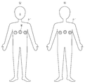
이때, 수술대에 누워있는 환자(P)의 신체 외부에는 도 4에 도시한 바와 같이, 복수의 마커(M)가 부착될 수 있다. 여기에서, "마커(M)"는 카메라(410)의 위치 및 주시 방향을 추정하기 위한 표식을 의미할 수 있다. 이때, 마커(M)는 수술 부위와 인접한 위치에 부착될 수 있으나, 특별히 이에 한정되는 것은 아니다. 또한, 도 4에서는 세 개의 마커(M)를 도시하고 있으나, 이는 하나의 실시 예에 불과하며, 더 많은 수의 마커(M)가 부착되는 것 역시 가능하다. 또한, 도 4에서는 세 개의 마커(M)가 일렬로 부착된 것으로 도시하고 있으나, 이는 설명의 편의를 위한 일 예일 뿐, 본 실시 예에 따른 마커(M)의 부착 형태는 이에 한정되지 않으며, 삼각형 및 사각형과 같은 형태로 부착되는 것이 전형적이다. 또한, 도 5에 도시한 바와 같이 인체 모형(P′)의 외부에도 마커(M)가 부착될 수 있다. 이때, 인체 모형(P′)에는 앞면과 뒷면 모두에 마커(M)가 부착될 수 있다.At this time, as shown in Fig. 4, a plurality of markers M may be attached to the outside of the body of the patient P lying on the operating table. Here, the "marker M" may mean a marker for estimating the position and the viewing direction of the
도 4 및 도 5에 도시한 복수의 마커(M)는 각각 서로 다른 식별정보를 가질 수 있다. 여기에서, "식별정보"는 각 마커(M)가 부착된 위치에 대한 정보, 각 마커(M)의 원 크기 등을 포함할 수 있으나, 특별히 이에 한정되는 것은 아니다. 도 4를 예를 들어 설명하면, 마커①은 환자의 복부 오른쪽(환자 기준)이라는 위치 정보를 갖고, 마커②는 환자의 복부 중심부라는 위치 정보를 가지며, 마커③은 환자의 복부 왼쪽이라는 위치 정보를 가질 수 있다. 마찬가지로, 도 5의 마커④, 마커⑤ 및 마커⑥은 각각 환자의 등 왼쪽(환자 기준), 등 중심부, 등 오른쪽이라는 위치 정보를 가질 수 있다. 또한, 각 마커(M)의 원래 크기는 사전에 정의될 수 있다.The plurality of markers M shown in Figs. 4 and 5 may have different identification information, respectively. Here, the "identification information" may include information on the position where each marker M is attached, the original size of each marker M, and the like, but is not limited thereto. 4, the
또한, 도 4 및 도 5에서는 일 예로 숫자를 표기하여 각각의 마커를 구분하고 있으나, 이는 하나의 실시 예에 불과하며, 각 마커는 서로 다른 패턴, 서로 다른 색을 갖도록 하여 구분하는 것 역시 가능할 것이다.In FIGS. 4 and 5, numerals are marked to indicate each marker, but this is only an example, and it is also possible to divide each marker into different colors and different colors .
이에 따라, 카메라(410)를 통해 촬영된 실제 영상에는 서로 다른 식별정보를 갖고, 서로 구분되는 복수의 마커가 포함될 수 있고, 후술할 증강현실 영상 생성부(430)에서는 실제 영상에 포함된 복수의 마커를 검출하고, 검출된 각 마커에 대한 실제 영상 내에서의 위치 정보를 산출한 후, 산출된 각 마커의 위치 정보 및 사전에 정의된 각 마커의 식별정보를 이용하여 카메라(410)의 현재 위치 및 주시 방향을 추정할 수 있다. 여기에서, "각 마커의 위치 정보"는 각 마커 간의 거리, 각 마커 간의 연결 각도 및 각 마커의 크기 등을 포함할 수 있으나, 특별히 이에 한정되는 것은 아니다.
Accordingly, the actual image photographed through the
증강현실 영상 생성부(430)는 상술한 바와 같이 카메라(410)를 통해 촬영된 실제 영상에 포함된 복수의 마커를 검출하고, 검출된 각 마커에 대한 실제 영상 내에서의 위치 정보를 산출한 후, 산출된 각 마커의 위치 정보 및 사전에 정의된 각 마커의 식별정보를 이용하여 카메라(410)의 현재 위치 및 주시 방향을 추정하고, 추정된 카메라(410)의 현재 위치 및 주시 방향에 대응되는 부위의 가상 영상을 카메라(410)를 통해 촬영된 실제 영상에 오버레이 방식으로 합성한 증강현실 영상을 생성할 수 있다.The augmented reality
여기에서, "가상 영상"은 전술한 바와 같이, 환자(P)의 수술 전 의료 영상에 대한 3차원 영상 및 3차원 영상을 내시경(220)을 통해 수집된 영상에 투영시킨 영상을 포함할 수 있다. 즉, "가상 영상"은 환자(P)의 신체 내부에 대한 영상일 수 있다.Here, as described above, the "virtual image" may include an image obtained by projecting a three-dimensional image and a three-dimensional image of the medical image of the patient P before surgery on the image collected through the
또한, "실제 영상"은 카메라(410)를 통해 촬영된 실세계에 대한 영상을 의미하며, 본 실시 예에서는 수술대에 누워있는 환자(P) 또는 인체 모형(P′)을 촬영한 영상일 수 있으며, 실제 영상에는 환자(P) 또는 인체 모형(P′)에 부착된 마커가 포함될 수 있다.The term "actual image" means an image of the real world photographed through the
증강현실 영상은 상술한 바와 같이, 카메라(410)를 통해 촬영된 실제 영상에 포함된 환자(P) 또는 인체 모형(P′)에 가상 영상 즉, 신체 내부 영상을 오버레이 방식으로 합성한 영상을 의미한다. 예를 들어, 도 6에 도시한 바와 같이, 카메라(410)를 통해 촬영된 실제 영상의 환자(P)의 신체에 카메라(410)의 주시 방향과 대응되는 신체 내부 영상인 가상 영상을 합성한 영상인 것이다. 이때, 증강현실 영상에서는 실제 영상에 포함된 마커가 제거될 수 있다. 이는 사용자에게 혼란을 주지 않도록 하기 위함이다.As described above, the augmented reality image is an image obtained by overlaying a virtual image, that is, an internal body image, on a patient P or a human body P 'included in a real image photographed through the
이때, 카메라(410)는 사용자의 시선이 향하는 부위를 실시간으로 촬영할 수 있으며, 증강현실 영상 생성부(430)는 카메라(410)를 통해 촬영되는 실제 영상을 실시간으로 수신하여 카메라(410)의 이동에 따른 증강현실 영상을 생성할 수 있다.The augmented reality
구체적으로, 증강현실 영상 생성부(430)는 카메라(410)로부터 촬영된 실제 영상을 실시간으로 수신하고, 수신된 실제 영상에서 복수의 마커를 검출하고, 검출된 각 마커 간의 거리, 각 마커 간의 연결 각도 및 각 마커의 크기를 산출한 후, 산출된 마커 간의 거리, 마커 간의 연결 각도, 각 마커의 크기 및 사전에 정의된 각 마커의 식별정보를 이용하여 카메라(410)의 현재 위치 및 주시 방향을 실시간으로 추정할 수 있다. 이때, 검출된 각 마커 간의 거리, 각 마커 간의 연결 각도 및 각 마커의 크기를 산출하고, 카메라(410)의 현재 위치 및 주시 방향을 추정하는 구체적인 방법은 본 발명의 요지와 다소 거리감이 있으며, 공지된 기술이므로 이에 대한 상세한 설명은 생략한다.Specifically, the augmented reality
즉, 증강현실 영상 생성부(430)는 이동하는 카메라(410)의 위치 및 주시 방향을 실시간으로 추정하고, 추정된 카메라(410)의 위치 및 주시 방향에 대응되는 증강현실 영상을 실시간으로 생성할 수 있다.That is, the augmented reality
예를 들어, 도 6 및 도 7을 참조하여 설명하면, 카메라(410)가 환자(P)의 복부 중앙에 위치하여 복부를 주시하고 있는 경우, 증강현실 영상 생성부(430)는 카메라(410)로부터 촬영된 실제 영상을 수신하여 복수의 마커를 검출하고, 검출된 각 마커에 대한 실제 영상 내에서의 위치 정보를 이용하여 카메라(410)가 현재 환자(P) 복부 중앙에 위치하여 복부를 주시하고 있음을 추정하고, 영상 시스템(300)으로부터 대응되는 부위의 가상 영상을 수신하여 실제 영상에 오버레이 방식으로 합성함으로써, 환자(P)의 복부 중심부가 정면을 향하는 증강현실 영상을 생성한다.6 and 7, when the
또한, 도 7과 같이, 카메라(410)가 환자(P)의 복부 중앙을 벗어난 위치에서 대각선 방향으로 복부 좌측(환자 기준)을 주시하고 있는 경우, 증강현실 영상 생성부(430)는 카메라(410)로부터 촬영된 실제 영상을 수신하여 마커를 검출하고, 검출된 각 마커에 대한 실제 영상 내에서의 위치 정보를 이용하여 카메라(410)가 현재 환자(P) 복부 좌측에서 복부를 주시하고 있음을 추정하고, 영상 시스템(300)으로부터 대응되는 부위의 가상 영상을 수신하여 실제 영상에 오버레이 방식으로 합성함으로써, 환자(P)의 복부 좌측이 정면을 향하는 증강현실 영상을 생성한다.7, when the
이때, 도 6 및 도 7은 각각 카메라(410)가 복부 중앙 및 복부 좌측에 위치하고 있는 경우에 대해서만 도시하고 있으나, 카메라(410)가 연속적으로 이동하는 경우, 대응되는 증강현실 영상 역시 연속적으로 디스플레이(420)로 출력되어 표시될 수 있음은 자명하다. 즉, 증강현실 영상 생성부(430)는 사용자의 시선 이동 궤적을 그대로 추종하는 증강현실 영상을 생성하여 디스플레이(420)로 출력할 수 있다.6 and 7 show only the case where the
또한, 증강현실 영상 생성부(430)는 카메라(410)를 통해 촬영된 실제 영상에서 검출된 마커의 크기를 산출하고, 산출된 마커의 크기를 사전에 정의된 크기와 비교하여 카메라(410)와 마커와의 거리를 연산할 수 있다. 즉, 카메라(410)가 촬영 대상으로부터 떨어진 거리를 연산하는 것이다. 일반적으로 카메라(410)가 촬영 대상으로부터 멀어지면 촬영 대상은 작아지고, 촬영 대상과 가까워지면 촬영 대상은 커지게 된다. 즉, 카메라(410)가 촬영 대상으로부터 떨어진 거리에 따라 실제 영상 내에서의 촬영 대상 크기는 작아지거나 커지게 되는 것이다.Also, the augmented reality
이에 따라, 증강현실 영상 생성부(430)는 카메라(410)와 마커와의 거리를 연산하고, 연산된 거리에 따라 영상 시스템(300)으로부터 수신한 대응 부위의 가상 영상을 확대 또는 축소한 후, 실제 영상과 합성하여 증강현실 영상을 생성할 수 있다. 즉, 카메라(410)와 환자(P)와의 거리가 가까워지면 해당 부위와 대응되는 가상 영상을 확대하여 실제 영상과 합성하고, 카메라(410)와 환자(P)와의 거리가 멀어지면 해당 부위와 대응되는 가상 영상을 축소하여 실제 영상과 합성할 수 있다. 카메라(410)와 환자(P)와의 거리에 따라 축소 또는 확대되어 생성된 증강현실 영상을 각각, 도 8 및 도 9에 도시하였다. 또한, 카메라(410)가 환자(P)에 근접하여 복부 좌측(환자 기준)에 위치하는 경우, 환자(P)의 복부 좌측이 정면을 향하는 증강현실 영상을 도 10에 도시하였다.Accordingly, the augmented reality
이와 같이, 본 실시 예에 따른 증강현실 영상 표시 시스템(400)은 사용자의 시선 이동에 따라 대응되는 환자(P)의 신체 내부를 실시간으로 관찰할 수 있다. 즉, 사용자의 시선 방향 변화에 직접적으로 대응하여 실시간으로 사용자의 시선이 향하는 부분이 정면을 향하는 증강현실 영상을 생성하여 디스플레이할 수 있기 때문에, 마우스, 키보드, 조이스틱과 같은 입력장치를 사용하여 시선 방향 및 위치를 지정함으로써 환자(P)의 신체 내부를 관찰하는 종래와 비교하여 보다 직관적으로 환자(P) 신체 내부 상태를 관찰할 수 있다.
As described above, the augmented reality
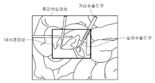
또한, 증강현실 영상 생성부(430)는 슬레이브 시스템(200)로부터 수술 도구의 현재 위치 정보를 수신하여 증강현실 영상의 매칭되는 영역에 가상 수술 도구를 생성할 수 있다. 여기에서, "위치 정보"는 전술한 바와 같이, 좌표값일 수 있으며, 증강현실 영상 생성부(430)는 증강현실 영상에서 수신된 수술 도구의 좌표값과 매칭되는 좌표에 가상 수술 도구를 생성할 수 있다. 이때, 도 11에 도시한 바와 같이, 내시경(220)을 통해 실제 수술 도구가 촬영되었다면, 가상 수술 도구에서 실제 수술 도구와 겹치는 부분은 실제 수술 도구가 표시될 수 있다.In addition, the augmented reality
즉, 본 실시 예에서 증강현실 영상은 도 11과 같이, 환자(P)의 수술 전 의료 영상에 대한 3차원 영상과 내시경(220)을 통해 획득한 내시경 영상 및 슬레이브 시스템(200)으로부터 수신된 수술 도구의 위치 정보를 이용하여 생성한 가상 수술 도구가 합성된 영상일 수 있다. 이때, 내시경(220)을 통해 획득한 영상에 실제 수술 도구가 포함되지 않는 경우, 증강현실 영상에는 가상 수술 도구만이 합성될 수 있고, 내시경(220)을 통해 획득한 영상에 실제 수술 도구가 포함된 경우, 도 11과 같이 가상 수술 도구와 실제 수술 도구가 연결되도록 합성될 수 있다.
11, the three-dimensional image of the medical image before surgery of the patient P, the endoscopic image acquired through the
디스플레이(420)는 증강현실 영상 생성부(430)를 통해 생성된 증강현실 영상을 표시하기 위한 구성으로, 도 3에 도시한 바와 같이, 증강현실 영상 표시 시스템(400)의 후면 즉, 사용자의 눈과 마주하는 면에 마련될 수 있다. 여기에서, 디스플레이(420)로는 액정표시장치(Liquid Crystal Display:LCD)가 사용될 수 있으나, 특별히 이에 한정되는 것은 아니다.
3, the
본 실시 예에서 증강현실 영상 표시 시스템(400)은 환자(P)를 직접 볼 수 있는 보조자(A)가 사용할 수 있다. 즉, 본 실시 예에 따른 증강현실 영상 표시 시스템(400)은 환자(P) 신체 내부를 직관적으로 관찰할 수 있는 시스템으로서, 환자(P)를 직접 볼 수 있는 사용자가 사용할 수 있다.In the present embodiment, the augmentor A who can directly view the patient P can use the augmented reality
예를 들어, 수술장에서 보조자(A)는 별도의 모니터를 통해 환자(P)의 신체 내부를 관찰할 수 있다. 이때, 모니터는 환자(P) 근처가 아닌 다른 위치에 마련되는 것이 일반적이므로, 보조자(A)는 환자(P)와 모니터를 동시에 보는 것이 불가능하다. 수술이 진행되는 동안 조작자(S)로부터 수술 도구 교체 지시가 있을 때, 보조자(A)는 환자(P) 신체 내부에 삽입된 수술 도구를 제거하고 다른 수술 도구를 장착 및 환자(P) 신체 내부로 삽입해야하는데, 이때, 수술 도구는 환자(P) 측에 위치해 있고, 환자(P) 신체 내부는 별도의 모니터를 통해 확인해야하므로, 보조자(A)는 모니터로 환자(P) 신체 내부를 관찰하면서 환자(P) 신체 내부에 삽입된 수술 도구를 제거하고 교체해야한다. 이에 따라, 수술 도구 교체 작업이 더디게 진행될 수 있고, 비직관적인 관찰을 통해 수술 도구를 제거하는 과정에서 주변 장기 및 조직을 손상시키는 사고가 발생할 수도 있다.For example, in the surgical field, the assistant A can observe the inside of the body of the patient P through a separate monitor. At this time, since the monitor is generally provided at a position other than the vicinity of the patient P, it is impossible for the assistant A to simultaneously view the patient P and the monitor. (S), the assistant (A) removes the surgical tool inserted into the patient (P), inserts another surgical tool, and inserts the patient (P) inside the body Since the surgical tool is located on the side of the patient P and the inside of the patient P must be confirmed through a separate monitor, the assistant A observes the inside of the patient P with the monitor Patient (P) Surgical instruments inserted inside the body should be removed and replaced. As a result, the operation of replacing the surgical tool may progress slowly, and an accident that damages surrounding organs and tissues may occur in the process of removing the surgical tool through non-intuitive observation.
그러나, 보조자(A)가 본 실시 예에 따른 증강현실 영상 표시 시스템(400)을 착용하고 수술 작업을 보조하게 되면, 별도의 모니터를 볼 필요없이 환자(P)를 주시하면 환자(P)의 신체 내부 상태가 디스플레이(420)에 표시되므로, 직관적으로 환자(P) 신체 내부를 상세히 관찰할 수 있어 수술 도구 교체 등과 같은 보조 작업을 신속하게 수행할 수 있고, 또한, 수술장과 멀리 떨어져 위치한 조작자(S)가 미처 관찰하지 못한 부분을 관찰하여 상세한 정보를 제공할 수 있으므로, 수술 작업의 질을 향상시킬 수 있다.However, if the assistant A wears the augmented reality
한편, 이와 같이 조작자(S)가 원격으로 수술을 진행하는 수술 로봇 시스템이 아닌 조작자(S)가 직접 환자(P)에게 수술 도구를 삽입하여 수술 동작을 가하는 수술 시스템 예로써, 복강경 수술과 같은 수술 시스템에서는 조작자(S)가 직접 본 실시 예에 따른 증강현실 영상 표시 시스템(400)을 착용하고 수술을 집도하게 되면, 조작자(S)가 살펴보는 부위의 체내 영상이 그대로 표시되므로, 피부로 감싸진 수술 영역을 보다 효과적으로 관찰할 수 있으므로 수술 중 장기 및 조직이 손상되는 사고를 방지할 수 있다.
On the other hand, as an example of a surgical system in which an operator S inserts a surgical tool directly into a patient P to perform a surgical operation, rather than a surgical robot system in which the operator S performs surgery remotely, System, when the operator S directly wears the augmented reality
이상 본 발명을 구체적인 실시 예를 통하여 상세히 설명하였으나, 이는 본 발명을 구체적으로 설명하기 위한 것으로 본 발명은 이에 한정되지 않으며, 본 발명의 기술적 사상 내에서 당 분야의 통상의 지식을 가진 자에 의해 그 변형이나 개량이 가능함이 명백하다.While the present invention has been particularly shown and described with reference to exemplary embodiments thereof, the same is by way of illustration and example only and is not to be construed as limiting the present invention. It is evident that modification or improvement is possible.
본 발명의 단순한 변형 내지 변경은 모두 본 발명의 영역에 속하는 것으로 본 발명의 구체적인 보호 범위는 첨부된 특허청구범위에 의하여 명확해질 것이다.It will be understood by those skilled in the art that various changes in form and details may be made therein without departing from the spirit and scope of the invention as defined by the appended claims.
100 : 마스터 시스템
110 : 입력부
111, 113 : 핸들
120 : 표시부
130, 240, 320, 440 : 제어부
131 : 제어 신호 생성부
133 : 영상 처리부
140, 250, 330, 450 : 통신부
200 : 슬레이브 시스템
201 : 몸체
210 : 로봇 암
211 : 링크
213 : 관절
215 : 제1 구동부
217 : 위치 센서
220 : 내시경
225 : 제2 구동부
230, 230′ : 수술 도구
235 : 제3 구동부
241 : 위치 연산부
300 : 영상 시스템
310 : 이미지 저장부
321 : 3차원 영상 변환부
323 : 가상 영상 생성부
400 : 증강현실 영상 표시 시스템
410 : 카메라
420 : 디스플레이
430 : 증강현실 영상 생성부100: Master system
110: input unit
111, 113:
120:
130, 240, 320, 440:
131: control signal generation unit
133:
140, 250, 330, 450:
200: Slave system
201: Body
210: Robot arm
211: Link
213: joints
215:
217: Position sensor
220: Endoscope
225:
230, 230 ': Surgical tool
235:
241:
300: imaging system
310: Image storage unit
321: 3D image conversion unit
323: Virtual image generation unit
400: Augmented reality image display system
410: camera
420: Display
430: augmented reality image generation unit
Claims (25)
상기 슬레이브 시스템의 수술 동작을 제어하는 마스터 시스템;
환자 신체 내부에 대한 가상 영상을 생성하는 영상 시스템; 및
환자 신체 또는 인체 모형에 부착된 복수의 마커를 포함하는 실제 영상을 획득하는 카메라, 상기 실제 영상에서 복수의 마커를 검출하고, 검출된 복수의 마커를 이용하여 카메라의 위치 및 주시 방향을 추정한 후, 상기 가상 영상 중 추정된 카메라의 위치 및 주시 방향에 대응되는 부위를 상기 실제 영상에 오버레이 방식으로 합성한 증강현실 영상을 생성하는 증강현실 영상 생성부 및 상기 증강현실 영상을 표시하는 디스플레이를 포함하는 증강현실 영상 표시 시스템
을 포함하는 수술 로봇 시스템.A slave system for performing a surgical operation on a patient;
A master system for controlling a surgical operation of the slave system;
An imaging system for generating a virtual image of a patient's body; And
A camera for acquiring an actual image including a plurality of markers attached to a patient's body or a human body model; a plurality of markers in the actual image; detecting a position and a viewing direction of the camera using the plurality of detected markers; An augmented reality image generation unit for generating an augmented reality image obtained by synthesizing an estimated camera position and an area corresponding to a viewing direction of the virtual image in an overlay manner on the actual image, and a display for displaying the augmented reality image, Augmented Reality Video Display System
And a surgical robot system.
상기 슬레이브 시스템은,
환자에게 수술 동작을 가하는 수술 도구;
환자 신체 내부의 수술 부위를 촬영하는 내시경;
상기 수술 도구의 위치를 검출하기 위한 위치 센서; 및
상기 위치 센서를 검출된 신호를 이용하여 상기 수술 도구의 위치 정보를 연산하는 위치 연산부
를 포함하는 수술 로봇 시스템.The method according to claim 1,
In the slave system,
A surgical tool for applying a surgical operation to a patient;
An endoscope for photographing a surgical site inside the patient's body;
A position sensor for detecting a position of the surgical tool; And
A position calculator for calculating position information of the surgical tool using the detected signal of the position sensor,
Wherein the surgical robot system comprises:
상기 증강현실 영상 생성부는 상기 슬레이브 시스템으로부터 상기 수술 도구의 위치 정보를 수신하여 상기 증강현실 영상에서 상기 위치 정보와 매칭되는 영역에 가상 수술 도구를 생성하는 수술 로봇 시스템.3. The method of claim 2,
Wherein the augmented reality image generation unit receives position information of the surgical tool from the slave system and generates a virtual surgery tool in an area matching the position information in the augmented reality image.
상기 증강현실 영상 표시 시스템은 HMD(Head Mounted Display)인 수술 로봇 시스템.The method according to claim 1,
Wherein the augmented reality image display system is an HMD (Head Mounted Display).
상기 카메라는 상기 디스플레이에 부착되고,
상기 증강현실 영상 생성부는 상기 디스플레이의 이동에 따라 변하는 카메라의 위치 및 주시 방향을 실시간으로 추정하고, 상기 가상 영상 중 추정된 카메라의 위치 및 주시 방향에 대응되는 부위를 상기 실제 영상에 합성하여 증강현실 영상을 생성하는 수술 로봇 시스템.The method according to claim 1,
The camera being attached to the display,
Wherein the augmented reality image generation unit estimates in real time the position and the viewing direction of the camera that changes according to the movement of the display and synthesizes a portion corresponding to the estimated camera position and the viewing direction of the virtual image to the actual image, A surgical robot system for generating images.
상기 증강현실 영상 생성부는 상기 검출된 각 마커에 대한 실제 영상 내에서의 위치 정보를 산출하고, 산출된 각 마커의 위치 정보를 이용하여 상기 카메라의 위치 및 주시 방향을 추정하는 수술 로봇 시스템.The method according to claim 1,
Wherein the augmented reality image generation unit calculates position information in an actual image for each of the detected markers and estimates the position and the viewing direction of the camera using the calculated position information of each marker.
상기 각 마커의 위치 정보는 각 마커 간의 거리 및 각 마커 간의 연결 각도를 포함하는 수술 로봇 시스템.The method according to claim 6,
Wherein the position information of each of the markers includes a distance between each marker and a connection angle between the markers.
상기 각 마커의 위치 정보는 실제 영상 내의 마커의 크기 정보를 포함하는 수술 로봇 시스템.The method according to claim 6,
Wherein the position information of each marker includes size information of a marker in an actual image.
상기 증강현실 영상 생성부는 상기 마커의 크기 정보를 이용하여 상기 카메라와 마커와의 거리를 연산하고, 상기 가상 영상을 상기 연산된 거리에 따라 확대 또는 축소한 후 상기 실제 영상에 합성한 증강현실 영상을 생성하는 수술 로봇 시스템.9. The method of claim 8,
Wherein the augmented reality image generation unit calculates a distance between the camera and the marker using the size information of the marker, enlarges or reduces the virtual image according to the calculated distance, and outputs the augmented reality image synthesized to the actual image Generating surgical robot system.
상기 가상 영상은 상기 환자의 수술 전 의료 영상을 3차원 영상으로 변환한 영상인 수술 로봇 시스템.The method according to claim 1,
Wherein the virtual image is a medical image converted into a three-dimensional image before surgery of the patient.
상기 가상 영상은 내시경을 통해 촬영된 수술 부위 및 실제 수술 도구를 더 포함하는 수술 로봇 시스템.11. The method of claim 10,
Wherein the virtual image further includes a surgical site photographed through an endoscope and an actual surgical tool.
상기 영상 시스템은,
환자의 수술 전 의료 영상을 3차원 영상으로 변환하는 3차원 영상 변환부;
변환된 3차원 영상을 상기 내시경을 통해 획득된 영상에 투영시켜 가상 영상을 생성하는 가상 영상 생성부; 및
상기 3차원 영상 및 가상 영상이 저장되는 저장부
를 포함하는 수술 로봇 시스템.12. The method of claim 11,
Wherein the image system comprises:
A three-dimensional image converter for converting a medical image of the patient into a three-dimensional image before surgery;
A virtual image generation unit for generating a virtual image by projecting the converted three-dimensional image onto an image obtained through the endoscope; And
The three-dimensional image and the virtual image are stored in a storage unit
Wherein the surgical robot system comprises:
상기 환자의 수술 전 의료 영상은 CT(Computed Tomography) 영상 및 MRI(Magnetic Resonance Imaging) 영상을 포함하는 수술 로봇 시스템.11. The method of claim 10,
The preoperative medical image of the patient includes CT (Computed Tomography) image and MRI (Magnetic Resonance Imaging) image.
상기 증강현실 영상은 상기 카메라의 위치 및 주시 방향에 대응되는 부위가 정면을 향하도록 생성되는 수술 로봇 시스템.The method according to claim 1,
Wherein the augmented reality image is generated so that a portion corresponding to the position and the viewing direction of the camera faces the front side.
상기 실제 영상에서 복수의 마커를 검출하고, 검출된 복수의 마커를 이용하여 카메라의 위치 및 주시 방향을 추정한 후, 추정된 위치 및 주시 방향에 대응되는 부위의 가상 영상을 상기 실제 영상에 오버레이 방식으로 합성한 증강현실 영상을 생성하는 증강현실 영상 생성부; 및
상기 증강현실 영상을 표시하는 디스플레이
를 포함하는 증강현실 영상 표시 시스템.A camera for acquiring an actual image including a plurality of markers attached to a patient's body or a human body model;
A plurality of markers are detected in the actual image, a camera position and a viewing direction are estimated using the plurality of detected markers, and a virtual image of a portion corresponding to the estimated position and the viewing direction is superimposed on the actual image An augmented reality image generating unit for generating an augmented reality image synthesized with the augmented reality image; And
A display for displaying the augmented reality image
Wherein the augmented reality image display system includes:
상기 카메라는 상기 디스플레이에 부착되고,
상기 증강현실 영상 생성부는 상기 디스플레이의 이동에 따라 변하는 상기 카메라의 위치 및 주시 방향을 실시간으로 추정하고, 추정된 위치 및 주시 방향에 대응되는 부위의 가상 영상을 상기 실제 영상에 합성하여 증강현실 영상을 생성하는 증강현실 영상 표시 시스템.16. The method of claim 15,
The camera being attached to the display,
Wherein the augmented reality image generation unit estimates in real time the position and the viewing direction of the camera that changes according to the movement of the display and synthesizes a virtual image of a portion corresponding to the estimated position and the viewing direction to the actual image, Generating augmented reality image display system.
상기 증강현실 영상 생성부는 상기 검출된 각 마커에 대한 실제 영상 내에서의 위치 정보를 산출하고, 산출된 각 마커의 위치 정보를 이용하여 상기 카메라의 위치 및 주시 방향을 추정하는 증강현실 영상 표시 시스템.16. The method of claim 15,
Wherein the augmented reality image generation unit calculates position information in an actual image for each of the detected markers and estimates a position and a viewing direction of the camera using the calculated position information of each marker.
상기 각 마커의 위치 정보는 각 마커 간의 거리 및 각 마커 간의 연결 각도를 포함하는 증강현실 영상 표시 시스템.18. The method of claim 17,
Wherein the position information of each marker includes a distance between each marker and a connection angle between the markers.
상기 각 마커의 위치 정보는 실제 영상 내의 마커의 크기 정보를 포함하는 증강현실 영상 표시 시스템.18. The method of claim 17,
Wherein the position information of each marker includes size information of a marker in an actual image.
상기 증강현실 영상 생성부는 상기 마커의 크기 정보를 이용하여 상기 카메라와 마커와의 거리를 연산하고, 상기 가상 영상을 상기 연산된 거리에 따라 확대 또는 축소하여 상기 실제 영상에 합성한 증강현실 영상을 생성하는 증강현실 영상 표시 시스템.20. The method of claim 19,
The augmented reality image generation unit calculates the distance between the camera and the marker using the size information of the marker and enlarges or reduces the virtual image according to the calculated distance to generate an augmented reality image synthesized with the actual image Augmented reality image display system.
상기 가상 영상은 환자의 수술 전 의료 영상을 3차원 영상으로 변환한 영상인 증강현실 영상 표시 시스템.16. The method of claim 15,
Wherein the virtual image is a medical image of the patient before surgery converted into a three-dimensional image.
상기 가상 영상은 내시경을 통해 촬영된 수술 부위 및 실제 수술 도구를 더 포함하는 증강현실 영상 표시 시스템.22. The method of claim 21,
Wherein the virtual image further includes a surgical site photographed through an endoscope and an actual surgical tool.
상기 환자의 수술 전 의료 영상은 CT(Computed Tomography) 영상 및 MRI(Magnetic Resonance Imaging) 영상을 포함하는 증강현실 영상 표시 시스템.22. The method of claim 21,
Wherein the preoperative medical image of the patient includes a CT (Computed Tomography) image and an MRI (Magnetic Resonance Imaging) image.
상기 증강현실 영상은 환자의 신체 내부로 삽입된 수술 도구에 대한 가상 이미지를 포함하는 증강현실 영상 표시 시스템.16. The method of claim 15,
Wherein the augmented reality image includes a virtual image of a surgical tool inserted into a body of a patient.
상기 증강현실 영상은 상기 카메라의 위치 및 주시 방향에 대응되는 부위가 정면을 향하도록 생성되는 증강현실 영상 표시 시스템.16. The method of claim 15,
Wherein the augmented reality image is generated so that a portion corresponding to a position and a viewing direction of the camera faces front.
Priority Applications (2)
| Application Number | Priority Date | Filing Date | Title |
|---|---|---|---|
| KR1020130026615A KR20140112207A (en) | 2013-03-13 | 2013-03-13 | Augmented reality imaging display system and surgical robot system comprising the same |
| US14/132,782 US9767608B2 (en) | 2013-03-13 | 2013-12-18 | Augmented reality image display system and surgical robot system comprising the same |
Applications Claiming Priority (1)
| Application Number | Priority Date | Filing Date | Title |
|---|---|---|---|
| KR1020130026615A KR20140112207A (en) | 2013-03-13 | 2013-03-13 | Augmented reality imaging display system and surgical robot system comprising the same |
Publications (1)
| Publication Number | Publication Date |
|---|---|
| KR20140112207A true KR20140112207A (en) | 2014-09-23 |
Family
ID=51530275
Family Applications (1)
| Application Number | Title | Priority Date | Filing Date |
|---|---|---|---|
| KR1020130026615A KR20140112207A (en) | 2013-03-13 | 2013-03-13 | Augmented reality imaging display system and surgical robot system comprising the same |
Country Status (2)
| Country | Link |
|---|---|
| US (1) | US9767608B2 (en) |
| KR (1) | KR20140112207A (en) |
Cited By (29)
| Publication number | Priority date | Publication date | Assignee | Title |
|---|---|---|---|---|
| KR101647467B1 (en) * | 2015-06-05 | 2016-08-11 | 주식회사 메드릭스 | 3d surgical glasses system using augmented reality |
| KR101667152B1 (en) * | 2015-05-22 | 2016-10-24 | 고려대학교 산학협력단 | Smart glasses system for supplying surgery assist image and method for supplying surgery assist image using smart glasses |
| WO2016190607A1 (en) * | 2015-05-22 | 2016-12-01 | 고려대학교 산학협력단 | Smart glasses system for providing surgery assisting image and method for providing surgery assisting image by using smart glasses |
| WO2017171295A1 (en) * | 2016-03-29 | 2017-10-05 | 사회복지법인 삼성생명공익재단 | Augmented reality system in which estimation of jaw movement of patient is reflected and augmented reality providing method therefor |
| KR101868120B1 (en) * | 2016-12-01 | 2018-06-18 | 재단법인 아산사회복지재단 | Augmented Reality Angle Measuring Apparatus for Non-radiographic Correction Osteotomy Surgery |
| JP2018161526A (en) * | 2018-07-03 | 2018-10-18 | ソニー株式会社 | Medical observation system, medical observation method, and medical control device |
| KR20190004591A (en) * | 2017-07-04 | 2019-01-14 | 경희대학교 산학협력단 | Navigation system for liver disease using augmented reality technology and method for organ image display |
| KR20190018906A (en) * | 2017-08-16 | 2019-02-26 | 연세대학교 원주산학협력단 | Mobile terminal for rendering virtual organs in augmented/virtual reality and system thereof |
| KR20190066937A (en) * | 2017-12-06 | 2019-06-14 | 조선대학교산학협력단 | Surgery navigation program stored in computer-readable storage medium |
| WO2019164272A1 (en) * | 2018-02-20 | 2019-08-29 | (주)휴톰 | Method and device for providing surgical image |
| WO2019177711A1 (en) * | 2018-03-13 | 2019-09-19 | Intuitive Surgical Operations, Inc. | Methods of guiding manual movement of medical systems |
| KR20190116601A (en) * | 2018-04-03 | 2019-10-15 | (주)다울디엔에스 | Training system of a surgical operation using authoring tool based on augmented reality |
| KR102013837B1 (en) * | 2018-02-20 | 2019-10-21 | (주)휴톰 | Method and apparatus for providing surgical video |
| KR20190130777A (en) * | 2018-05-15 | 2019-11-25 | 주식회사 삼육오엠씨네트웍스 | Apparatus for laparoscope surgical simulator |
| US10571671B2 (en) | 2014-03-31 | 2020-02-25 | Sony Corporation | Surgical control device, control method, and imaging control system |
| KR20200049646A (en) * | 2018-10-29 | 2020-05-08 | 주식회사 매니아마인드 | Microsurgical and injection virtual reality device |
| KR20200060660A (en) * | 2018-11-22 | 2020-06-01 | (주)다울디엔에스 | Knowledge base authoring system based on AR surgery application system |
| WO2021030129A1 (en) * | 2019-08-14 | 2021-02-18 | Warsaw Orthopedic, Inc. | Systems, devices, and methods for surgical navigation with anatomical tracking |
| WO2021062375A1 (en) * | 2019-09-27 | 2021-04-01 | Raytrx, Llc | Augmented and extended reality glasses for use in surgery visualization and telesurgery |
| WO2021112312A1 (en) * | 2019-12-06 | 2021-06-10 | 서울대학교산학협력단 | Augmented reality tool for implant surgery and implant surgery information visualization method |
| WO2021201434A1 (en) * | 2020-04-01 | 2021-10-07 | 고려대학교 산학협력단 | Method and device for providing surgical guide using augmented reality |
| KR20210156703A (en) * | 2020-06-18 | 2021-12-27 | 주식회사 디엠에프 | Method for providing real-time visualization service using 3-dimensional facial scan data and augment reality |
| KR20220087352A (en) | 2020-12-17 | 2022-06-24 | (주)스마트큐브 | Apparatus for enhancing human organs and corresponding auscultation sound, and method therefor |
| WO2022181919A1 (en) * | 2021-02-26 | 2022-09-01 | (주)휴톰 | Device and method for providing virtual reality-based operation environment |
| WO2023003389A1 (en) * | 2021-07-21 | 2023-01-26 | (주)휴톰 | Apparatus and method for determining insertion position of trocar on three-dimensional virtual pneumoperitoneum model of patient |
| US11628038B2 (en) | 2020-02-21 | 2023-04-18 | Raytrx, Llc | Multi-option all-digital 3D surgery visualization system and control |
| KR20230073443A (en) * | 2021-11-18 | 2023-05-26 | (주)에이트원 | Method for supporting aircraft maintenance based on augmented reality |
| KR102576673B1 (en) * | 2022-12-12 | 2023-09-08 | (주)투비시스템 | Method for augmented reality based task guidance |
| US11819273B2 (en) | 2015-03-17 | 2023-11-21 | Raytrx, Llc | Augmented and extended reality glasses for use in surgery visualization and telesurgery |
Families Citing this family (317)
| Publication number | Priority date | Publication date | Assignee | Title |
|---|---|---|---|---|
| US8361067B2 (en) | 2002-09-30 | 2013-01-29 | Relievant Medsystems, Inc. | Methods of therapeutically heating a vertebral body to treat back pain |
| US8219178B2 (en) | 2007-02-16 | 2012-07-10 | Catholic Healthcare West | Method and system for performing invasive medical procedures using a surgical robot |
| US10653497B2 (en) | 2006-02-16 | 2020-05-19 | Globus Medical, Inc. | Surgical tool systems and methods |
| US10893912B2 (en) | 2006-02-16 | 2021-01-19 | Globus Medical Inc. | Surgical tool systems and methods |
| US10357184B2 (en) | 2012-06-21 | 2019-07-23 | Globus Medical, Inc. | Surgical tool systems and method |
| US7728868B2 (en) | 2006-08-02 | 2010-06-01 | Inneroptic Technology, Inc. | System and method of providing real-time dynamic imagery of a medical procedure site using multiple modalities |
| US10028753B2 (en) | 2008-09-26 | 2018-07-24 | Relievant Medsystems, Inc. | Spine treatment kits |
| US9965681B2 (en) | 2008-12-16 | 2018-05-08 | Osterhout Group, Inc. | Eye imaging in head worn computing |
| US9298007B2 (en) | 2014-01-21 | 2016-03-29 | Osterhout Group, Inc. | Eye imaging in head worn computing |
| US8690776B2 (en) | 2009-02-17 | 2014-04-08 | Inneroptic Technology, Inc. | Systems, methods, apparatuses, and computer-readable media for image guided surgery |
| US8641621B2 (en) | 2009-02-17 | 2014-02-04 | Inneroptic Technology, Inc. | Systems, methods, apparatuses, and computer-readable media for image management in image-guided medical procedures |
| US11464578B2 (en) | 2009-02-17 | 2022-10-11 | Inneroptic Technology, Inc. | Systems, methods, apparatuses, and computer-readable media for image management in image-guided medical procedures |
| IT1401669B1 (en) | 2010-04-07 | 2013-08-02 | Sofar Spa | ROBOTIC SURGERY SYSTEM WITH PERFECT CONTROL. |
| US11412998B2 (en) | 2011-02-10 | 2022-08-16 | Karl Storz Imaging, Inc. | Multi-source medical display |
| US10631712B2 (en) * | 2011-02-10 | 2020-04-28 | Karl Storz Imaging, Inc. | Surgeon's aid for medical display |
| US10674968B2 (en) * | 2011-02-10 | 2020-06-09 | Karl Storz Imaging, Inc. | Adjustable overlay patterns for medical display |
| WO2012131660A1 (en) | 2011-04-01 | 2012-10-04 | Ecole Polytechnique Federale De Lausanne (Epfl) | Robotic system for spinal and other surgeries |
| US10390877B2 (en) | 2011-12-30 | 2019-08-27 | Relievant Medsystems, Inc. | Systems and methods for treating back pain |
| US11871901B2 (en) | 2012-05-20 | 2024-01-16 | Cilag Gmbh International | Method for situational awareness for surgical network or surgical network connected device capable of adjusting function based on a sensed situation or usage |
| US11974822B2 (en) | 2012-06-21 | 2024-05-07 | Globus Medical Inc. | Method for a surveillance marker in robotic-assisted surgery |
| US11793570B2 (en) | 2012-06-21 | 2023-10-24 | Globus Medical Inc. | Surgical robotic automation with tracking markers |
| US10231791B2 (en) | 2012-06-21 | 2019-03-19 | Globus Medical, Inc. | Infrared signal based position recognition system for use with a robot-assisted surgery |
| US11399900B2 (en) | 2012-06-21 | 2022-08-02 | Globus Medical, Inc. | Robotic systems providing co-registration using natural fiducials and related methods |
| US11045267B2 (en) | 2012-06-21 | 2021-06-29 | Globus Medical, Inc. | Surgical robotic automation with tracking markers |
| US11298196B2 (en) | 2012-06-21 | 2022-04-12 | Globus Medical Inc. | Surgical robotic automation with tracking markers and controlled tool advancement |
| JP2015528713A (en) | 2012-06-21 | 2015-10-01 | グローバス メディカル インコーポレイティッド | Surgical robot platform |
| US10624710B2 (en) | 2012-06-21 | 2020-04-21 | Globus Medical, Inc. | System and method for measuring depth of instrumentation |
| US11857266B2 (en) | 2012-06-21 | 2024-01-02 | Globus Medical, Inc. | System for a surveillance marker in robotic-assisted surgery |
| US11317971B2 (en) | 2012-06-21 | 2022-05-03 | Globus Medical, Inc. | Systems and methods related to robotic guidance in surgery |
| US10136954B2 (en) | 2012-06-21 | 2018-11-27 | Globus Medical, Inc. | Surgical tool systems and method |
| US10758315B2 (en) | 2012-06-21 | 2020-09-01 | Globus Medical Inc. | Method and system for improving 2D-3D registration convergence |
| US11607149B2 (en) | 2012-06-21 | 2023-03-21 | Globus Medical Inc. | Surgical tool systems and method |
| US11864745B2 (en) | 2012-06-21 | 2024-01-09 | Globus Medical, Inc. | Surgical robotic system with retractor |
| US11395706B2 (en) | 2012-06-21 | 2022-07-26 | Globus Medical Inc. | Surgical robot platform |
| US10350013B2 (en) | 2012-06-21 | 2019-07-16 | Globus Medical, Inc. | Surgical tool systems and methods |
| US11116576B2 (en) | 2012-06-21 | 2021-09-14 | Globus Medical Inc. | Dynamic reference arrays and methods of use |
| US11864839B2 (en) | 2012-06-21 | 2024-01-09 | Globus Medical Inc. | Methods of adjusting a virtual implant and related surgical navigation systems |
| US11253327B2 (en) | 2012-06-21 | 2022-02-22 | Globus Medical, Inc. | Systems and methods for automatically changing an end-effector on a surgical robot |
| US11857149B2 (en) | 2012-06-21 | 2024-01-02 | Globus Medical, Inc. | Surgical robotic systems with target trajectory deviation monitoring and related methods |
| KR101997566B1 (en) * | 2012-08-07 | 2019-07-08 | 삼성전자주식회사 | Surgical robot system and control method thereof |
| US10588691B2 (en) | 2012-09-12 | 2020-03-17 | Relievant Medsystems, Inc. | Radiofrequency ablation of tissue within a vertebral body |
| EP2914186B1 (en) | 2012-11-05 | 2019-03-13 | Relievant Medsystems, Inc. | Systems for creating curved paths through bone and modulating nerves within the bone |
| US9336629B2 (en) | 2013-01-30 | 2016-05-10 | F3 & Associates, Inc. | Coordinate geometry augmented reality process |
| US10314559B2 (en) | 2013-03-14 | 2019-06-11 | Inneroptic Technology, Inc. | Medical device guidance |
| JP6138566B2 (en) * | 2013-04-24 | 2017-05-31 | 川崎重工業株式会社 | Component mounting work support system and component mounting method |
| US9370372B2 (en) | 2013-09-04 | 2016-06-21 | Mcginley Engineered Solutions, Llc | Drill bit penetration measurement systems and methods |
| US9283048B2 (en) | 2013-10-04 | 2016-03-15 | KB Medical SA | Apparatus and systems for precise guidance of surgical tools |
| EP3065650B1 (en) | 2013-11-08 | 2019-01-30 | Mcginley Engineered Solutions LLC | Surgical saw with sensing technology for determining cut through of bone and depth of the saw blade during surgery |
| WO2015107099A1 (en) | 2014-01-15 | 2015-07-23 | KB Medical SA | Notched apparatus for guidance of an insertable instrument along an axis during spinal surgery |
| US9939934B2 (en) | 2014-01-17 | 2018-04-10 | Osterhout Group, Inc. | External user interface for head worn computing |
| US9841599B2 (en) | 2014-06-05 | 2017-12-12 | Osterhout Group, Inc. | Optical configurations for head-worn see-through displays |
| US9829707B2 (en) | 2014-08-12 | 2017-11-28 | Osterhout Group, Inc. | Measuring content brightness in head worn computing |
| US9299194B2 (en) | 2014-02-14 | 2016-03-29 | Osterhout Group, Inc. | Secure sharing in head worn computing |
| US10684687B2 (en) | 2014-12-03 | 2020-06-16 | Mentor Acquisition One, Llc | See-through computer display systems |
| US11103122B2 (en) * | 2014-07-15 | 2021-08-31 | Mentor Acquisition One, Llc | Content presentation in head worn computing |
| US10254856B2 (en) | 2014-01-17 | 2019-04-09 | Osterhout Group, Inc. | External user interface for head worn computing |
| US20160019715A1 (en) | 2014-07-15 | 2016-01-21 | Osterhout Group, Inc. | Content presentation in head worn computing |
| US10649220B2 (en) | 2014-06-09 | 2020-05-12 | Mentor Acquisition One, Llc | Content presentation in head worn computing |
| US9740280B2 (en) | 2014-01-21 | 2017-08-22 | Osterhout Group, Inc. | Eye imaging in head worn computing |
| US9753288B2 (en) | 2014-01-21 | 2017-09-05 | Osterhout Group, Inc. | See-through computer display systems |
| US9836122B2 (en) | 2014-01-21 | 2017-12-05 | Osterhout Group, Inc. | Eye glint imaging in see-through computer display systems |
| US11669163B2 (en) | 2014-01-21 | 2023-06-06 | Mentor Acquisition One, Llc | Eye glint imaging in see-through computer display systems |
| US11487110B2 (en) | 2014-01-21 | 2022-11-01 | Mentor Acquisition One, Llc | Eye imaging in head worn computing |
| US9766463B2 (en) | 2014-01-21 | 2017-09-19 | Osterhout Group, Inc. | See-through computer display systems |
| US9494800B2 (en) | 2014-01-21 | 2016-11-15 | Osterhout Group, Inc. | See-through computer display systems |
| WO2015116816A1 (en) * | 2014-01-29 | 2015-08-06 | Becton, Dickinson And Company | Wearable electronic device for enhancing visualization during insertion of an invasive device |
| EP3104803B1 (en) | 2014-02-11 | 2021-09-15 | KB Medical SA | Sterile handle for controlling a robotic surgical system from a sterile field |
| US9401540B2 (en) | 2014-02-11 | 2016-07-26 | Osterhout Group, Inc. | Spatial location presentation in head worn computing |
| JP6235943B2 (en) * | 2014-03-18 | 2017-11-22 | 日本光電工業株式会社 | Blood pressure measurement system |
| JP6644699B2 (en) * | 2014-03-19 | 2020-02-12 | インテュイティブ サージカル オペレーションズ, インコーポレイテッド | Medical devices, systems and methods using gaze tracking |
| CN106659541B (en) | 2014-03-19 | 2019-08-16 | 直观外科手术操作公司 | Integrated eyeball stares medical device, the system and method that tracking is used for stereoscopic viewer |
| US20160187651A1 (en) | 2014-03-28 | 2016-06-30 | Osterhout Group, Inc. | Safety for a vehicle operator with an hmd |
| WO2015154069A1 (en) | 2014-04-04 | 2015-10-08 | Surgical Theater LLC | Dynamic and interactive navigation in a surgical environment |
| CN106659537B (en) | 2014-04-24 | 2019-06-11 | Kb医疗公司 | The surgical instrument holder used in conjunction with robotic surgical system |
| US10663740B2 (en) | 2014-06-09 | 2020-05-26 | Mentor Acquisition One, Llc | Content presentation in head worn computing |
| EP3169252A1 (en) | 2014-07-14 | 2017-05-24 | KB Medical SA | Anti-skid surgical instrument for use in preparing holes in bone tissue |
| US9959591B2 (en) * | 2014-07-31 | 2018-05-01 | Seiko Epson Corporation | Display apparatus, method for controlling display apparatus, and program |
| EP3188671A4 (en) | 2014-09-05 | 2018-03-14 | Mcginley Engineered Solutions LLC | Instrument leading edge measurement system and method |
| US9901406B2 (en) | 2014-10-02 | 2018-02-27 | Inneroptic Technology, Inc. | Affected region display associated with a medical device |
| US11504192B2 (en) | 2014-10-30 | 2022-11-22 | Cilag Gmbh International | Method of hub communication with surgical instrument systems |
| US10188467B2 (en) | 2014-12-12 | 2019-01-29 | Inneroptic Technology, Inc. | Surgical guidance intersection display |
| US10154239B2 (en) | 2014-12-30 | 2018-12-11 | Onpoint Medical, Inc. | Image-guided surgery with surface reconstruction and augmented reality visualization |
| USD751552S1 (en) | 2014-12-31 | 2016-03-15 | Osterhout Group, Inc. | Computer glasses |
| GB2534359A (en) * | 2015-01-15 | 2016-07-27 | Corin Ltd | System and method for patient implant alignment |
| CN105982735B (en) * | 2015-01-30 | 2018-11-16 | 上海交通大学 | The method for improving the master-slave mode remote tele-operation surgery systems transparency and stability |
| US10013808B2 (en) | 2015-02-03 | 2018-07-03 | Globus Medical, Inc. | Surgeon head-mounted display apparatuses |
| US10835119B2 (en) | 2015-02-05 | 2020-11-17 | Duke University | Compact telescope configurations for light scanning systems and methods of using the same |
| US10238279B2 (en) | 2015-02-06 | 2019-03-26 | Duke University | Stereoscopic display systems and methods for displaying surgical data and information in a surgical microscope |
| US20160239985A1 (en) | 2015-02-17 | 2016-08-18 | Osterhout Group, Inc. | See-through computer display systems |
| WO2016131903A1 (en) | 2015-02-18 | 2016-08-25 | KB Medical SA | Systems and methods for performing minimally invasive spinal surgery with a robotic surgical system using a percutaneous technique |
| CN107249497B (en) * | 2015-02-20 | 2021-03-16 | 柯惠Lp公司 | Operating room and surgical site awareness |
| US10026228B2 (en) * | 2015-02-25 | 2018-07-17 | Intel Corporation | Scene modification for augmented reality using markers with parameters |
| US11956414B2 (en) | 2015-03-17 | 2024-04-09 | Raytrx, Llc | Wearable image manipulation and control system with correction for vision defects and augmentation of vision and sensing |
| US20210335483A1 (en) * | 2015-03-17 | 2021-10-28 | Raytrx, Llc | Surgery visualization theatre |
| GB2536650A (en) * | 2015-03-24 | 2016-09-28 | Augmedics Ltd | Method and system for combining video-based and optic-based augmented reality in a near eye display |
| EP3280344A2 (en) * | 2015-04-07 | 2018-02-14 | King Abdullah University Of Science And Technology | Method, apparatus, and system for utilizing augmented reality to improve surgery |
| WO2016200874A1 (en) | 2015-06-08 | 2016-12-15 | The General Hospital Corporation | Airway management and visualization device |
| GB201510959D0 (en) * | 2015-06-22 | 2015-08-05 | Ec Medica Ltd | Augmented reality imaging System, Apparatus and Method |
| US9949700B2 (en) | 2015-07-22 | 2018-04-24 | Inneroptic Technology, Inc. | Medical device approaches |
| US10646298B2 (en) | 2015-07-31 | 2020-05-12 | Globus Medical, Inc. | Robot arm and methods of use |
| US10058394B2 (en) | 2015-07-31 | 2018-08-28 | Globus Medical, Inc. | Robot arm and methods of use |
| US10080615B2 (en) | 2015-08-12 | 2018-09-25 | Globus Medical, Inc. | Devices and methods for temporary mounting of parts to bone |
| JP6894431B2 (en) | 2015-08-31 | 2021-06-30 | ケービー メディカル エスアー | Robotic surgical system and method |
| US10888389B2 (en) | 2015-09-10 | 2021-01-12 | Duke University | Systems and methods for arbitrary viewpoint robotic manipulation and robotic surgical assistance |
| US10034716B2 (en) | 2015-09-14 | 2018-07-31 | Globus Medical, Inc. | Surgical robotic systems and methods thereof |
| US9771092B2 (en) | 2015-10-13 | 2017-09-26 | Globus Medical, Inc. | Stabilizer wheel assembly and methods of use |
| CA2997965C (en) | 2015-10-14 | 2021-04-27 | Surgical Theater LLC | Augmented reality surgical navigation |
| CN108135670B (en) | 2015-10-23 | 2021-02-26 | 柯惠Lp公司 | Surgical system for detecting gradual changes in perfusion |
| WO2017075044A1 (en) | 2015-10-27 | 2017-05-04 | Mcginley Engineered Solutions, Llc | Unicortical path detection for a surgical depth measurement system |
| US10321920B2 (en) | 2015-11-06 | 2019-06-18 | Mcginley Engineered Solutions, Llc | Measurement system for use with surgical burr instrument |
| US9824437B2 (en) * | 2015-12-11 | 2017-11-21 | Daqri, Llc | System and method for tool mapping |
| US10052170B2 (en) * | 2015-12-18 | 2018-08-21 | MediLux Capitol Holdings, S.A.R.L. | Mixed reality imaging system, apparatus and surgical suite |
| DE102015226669B4 (en) | 2015-12-23 | 2022-07-28 | Siemens Healthcare Gmbh | Method and system for outputting augmented reality information |
| US10646289B2 (en) * | 2015-12-29 | 2020-05-12 | Koninklijke Philips N.V. | System, controller and method using virtual reality device for robotic surgery |
| JP6736298B2 (en) * | 2016-01-25 | 2020-08-05 | キヤノン株式会社 | Information processing apparatus, information processing method, and program |
| US10448910B2 (en) | 2016-02-03 | 2019-10-22 | Globus Medical, Inc. | Portable medical imaging system |
| US11058378B2 (en) | 2016-02-03 | 2021-07-13 | Globus Medical, Inc. | Portable medical imaging system |
| US10117632B2 (en) | 2016-02-03 | 2018-11-06 | Globus Medical, Inc. | Portable medical imaging system with beam scanning collimator |
| US11883217B2 (en) | 2016-02-03 | 2024-01-30 | Globus Medical, Inc. | Portable medical imaging system and method |
| US10842453B2 (en) | 2016-02-03 | 2020-11-24 | Globus Medical, Inc. | Portable medical imaging system |
| US9675319B1 (en) | 2016-02-17 | 2017-06-13 | Inneroptic Technology, Inc. | Loupe display |
| US10850116B2 (en) | 2016-12-30 | 2020-12-01 | Mentor Acquisition One, Llc | Head-worn therapy device |
| WO2017151752A1 (en) | 2016-03-01 | 2017-09-08 | Mirus Llc | Augmented visualization during surgery |
| US20190088162A1 (en) * | 2016-03-04 | 2019-03-21 | Covidien Lp | Virtual and/or augmented reality to provide physical interaction training with a surgical robot |
| CN111329551A (en) | 2016-03-12 | 2020-06-26 | P·K·朗 | Augmented reality guidance for spinal and joint surgery |
| US10866119B2 (en) | 2016-03-14 | 2020-12-15 | Globus Medical, Inc. | Metal detector for detecting insertion of a surgical device into a hollow tube |
| DE112017001315T5 (en) * | 2016-03-16 | 2018-11-22 | Koninklijke Philips N.V. | Computer device for blending a laparoscopic image and an ultrasound image |
| AU2017236893A1 (en) | 2016-03-21 | 2018-09-06 | Washington University | Virtual reality or augmented reality visualization of 3D medical images |
| EP3241518A3 (en) | 2016-04-11 | 2018-01-24 | Globus Medical, Inc | Surgical tool systems and methods |
| US10694939B2 (en) | 2016-04-29 | 2020-06-30 | Duke University | Whole eye optical coherence tomography(OCT) imaging systems and related methods |
| US11154363B1 (en) * | 2016-05-24 | 2021-10-26 | Paul A. Lovoi | Terminal guidance for improving the accuracy of the position and orientation of an object |
| WO2018041197A1 (en) * | 2016-08-31 | 2018-03-08 | 北京术锐技术有限公司 | Method for unfolding, operating and withdrawing imaging tool and surgical instrument of surgical robot |
| EP3509527A4 (en) | 2016-09-09 | 2020-12-30 | Mobius Imaging LLC | Methods and systems for display of patient data in computer-assisted surgery |
| CN106205268B (en) * | 2016-09-09 | 2022-07-22 | 上海健康医学院 | X-ray analog camera system and method |
| US20180082480A1 (en) * | 2016-09-16 | 2018-03-22 | John R. White | Augmented reality surgical technique guidance |
| US11259894B2 (en) * | 2016-09-19 | 2022-03-01 | Neocis, Inc. | Tracking and guidance arrangement for a surgical robot system and related method |
| US20180279862A1 (en) * | 2016-10-10 | 2018-10-04 | Jack Wade | Systems and methods for closed-loop medical imaging |
| US10278778B2 (en) * | 2016-10-27 | 2019-05-07 | Inneroptic Technology, Inc. | Medical device navigation using a virtual 3D space |
| US10748450B1 (en) * | 2016-11-29 | 2020-08-18 | Sproutel, Inc. | System, apparatus, and method for creating an interactive augmented reality experience to simulate medical procedures for pediatric disease education |
| US11056022B1 (en) * | 2016-11-29 | 2021-07-06 | Sproutel, Inc. | System, apparatus, and method for creating an interactive augmented reality experience to simulate medical procedures for pediatric disease education |
| US10918445B2 (en) * | 2016-12-19 | 2021-02-16 | Ethicon Llc | Surgical system with augmented reality display |
| US10499997B2 (en) | 2017-01-03 | 2019-12-10 | Mako Surgical Corp. | Systems and methods for surgical navigation |
| US10412377B2 (en) * | 2017-01-11 | 2019-09-10 | Koninklijke Philips N.V. | Augmented display device for use in a medical imaging laboratory |
| EP3568070B1 (en) | 2017-01-16 | 2024-01-03 | Philipp K. Lang | Optical guidance for surgical, medical, and dental procedures |
| CN110199357B (en) | 2017-01-17 | 2023-07-14 | 皇家飞利浦有限公司 | Augmented reality interventional system providing background overlay |
| JP7233841B2 (en) | 2017-01-18 | 2023-03-07 | ケービー メディカル エスアー | Robotic Navigation for Robotic Surgical Systems |
| EP3574504A1 (en) * | 2017-01-24 | 2019-12-04 | Tietronix Software, Inc. | System and method for three-dimensional augmented reality guidance for use of medical equipment |
| CN107049210B (en) * | 2017-01-25 | 2018-09-28 | 山东省肿瘤防治研究院 | A kind of hysteroscope display control program based on augmented reality |
| US10010379B1 (en) * | 2017-02-21 | 2018-07-03 | Novarad Corporation | Augmented reality viewing and tagging for medical procedures |
| US11250947B2 (en) | 2017-02-24 | 2022-02-15 | General Electric Company | Providing auxiliary information regarding healthcare procedure and system performance using augmented reality |
| WO2018162079A1 (en) | 2017-03-10 | 2018-09-13 | Brainlab Ag | Augmented reality pre-registration |
| AU2018230901B2 (en) * | 2017-03-10 | 2020-12-17 | Biomet Manufacturing, Llc | Augmented reality supported knee surgery |
| AU2018236172B2 (en) | 2017-03-13 | 2021-03-04 | Zimmer, Inc. | Augmented reality diagnosis guidance |
| US11071594B2 (en) | 2017-03-16 | 2021-07-27 | KB Medical SA | Robotic navigation of robotic surgical systems |
| US9892564B1 (en) * | 2017-03-30 | 2018-02-13 | Novarad Corporation | Augmenting real-time views of a patient with three-dimensional data |
| US11589937B2 (en) | 2017-04-20 | 2023-02-28 | Intuitive Surgical Operations, Inc. | Systems and methods for constraining a virtual reality surgical system |
| US10860853B2 (en) | 2017-04-28 | 2020-12-08 | Intel Corporation | Learning though projection method and apparatus |
| US10973391B1 (en) | 2017-05-22 | 2021-04-13 | James X. Liu | Mixed reality viewing of a surgical procedure |
| WO2019006028A1 (en) * | 2017-06-28 | 2019-01-03 | Intuitive Surgical Operations, Inc. | Systems and methods for projecting an endoscopic image to a three-dimensional volume |
| US10675094B2 (en) | 2017-07-21 | 2020-06-09 | Globus Medical Inc. | Robot surgical platform |
| US11633235B2 (en) | 2017-07-31 | 2023-04-25 | Children's National Medical Center | Hybrid hardware and computer vision-based tracking system and method |
| US11259879B2 (en) | 2017-08-01 | 2022-03-01 | Inneroptic Technology, Inc. | Selective transparency to assist medical device navigation |
| US11432877B2 (en) | 2017-08-02 | 2022-09-06 | Medtech S.A. | Surgical field camera system that only uses images from cameras with an unobstructed sight line for tracking |
| AU2018321969B2 (en) | 2017-08-25 | 2022-08-11 | Mcginley Engineered Solutions, Llc | Sensing of surgical instrument placement relative to anatomic structures |
| US10861236B2 (en) | 2017-09-08 | 2020-12-08 | Surgical Theater, Inc. | Dual mode augmented reality surgical system and method |
| US11801114B2 (en) | 2017-09-11 | 2023-10-31 | Philipp K. Lang | Augmented reality display for vascular and other interventions, compensation for cardiac and respiratory motion |
| AU2018345760A1 (en) | 2017-10-02 | 2020-01-30 | Mcginley Engineered Solutions, Llc | Surgical instrument with real time navigation assistance |
| US10169850B1 (en) | 2017-10-05 | 2019-01-01 | International Business Machines Corporation | Filtering of real-time visual data transmitted to a remote recipient |
| US11925373B2 (en) | 2017-10-30 | 2024-03-12 | Cilag Gmbh International | Surgical suturing instrument comprising a non-circular needle |
| US11564756B2 (en) | 2017-10-30 | 2023-01-31 | Cilag Gmbh International | Method of hub communication with surgical instrument systems |
| US11801098B2 (en) | 2017-10-30 | 2023-10-31 | Cilag Gmbh International | Method of hub communication with surgical instrument systems |
| US11911045B2 (en) | 2017-10-30 | 2024-02-27 | Cllag GmbH International | Method for operating a powered articulating multi-clip applier |
| US11510741B2 (en) | 2017-10-30 | 2022-11-29 | Cilag Gmbh International | Method for producing a surgical instrument comprising a smart electrical system |
| US11357548B2 (en) | 2017-11-09 | 2022-06-14 | Globus Medical, Inc. | Robotic rod benders and related mechanical and motor housings |
| EP3492032B1 (en) | 2017-11-09 | 2023-01-04 | Globus Medical, Inc. | Surgical robotic systems for bending surgical rods |
| US11794338B2 (en) | 2017-11-09 | 2023-10-24 | Globus Medical Inc. | Robotic rod benders and related mechanical and motor housings |
| US11134862B2 (en) | 2017-11-10 | 2021-10-05 | Globus Medical, Inc. | Methods of selecting surgical implants and related devices |
| US11058497B2 (en) * | 2017-12-26 | 2021-07-13 | Biosense Webster (Israel) Ltd. | Use of augmented reality to assist navigation during medical procedures |
| US11903601B2 (en) | 2017-12-28 | 2024-02-20 | Cilag Gmbh International | Surgical instrument comprising a plurality of drive systems |
| US11832840B2 (en) | 2017-12-28 | 2023-12-05 | Cilag Gmbh International | Surgical instrument having a flexible circuit |
| US11786245B2 (en) | 2017-12-28 | 2023-10-17 | Cilag Gmbh International | Surgical systems with prioritized data transmission capabilities |
| US11589888B2 (en) | 2017-12-28 | 2023-02-28 | Cilag Gmbh International | Method for controlling smart energy devices |
| US11202570B2 (en) | 2017-12-28 | 2021-12-21 | Cilag Gmbh International | Communication hub and storage device for storing parameters and status of a surgical device to be shared with cloud based analytics systems |
| US11864728B2 (en) | 2017-12-28 | 2024-01-09 | Cilag Gmbh International | Characterization of tissue irregularities through the use of mono-chromatic light refractivity |
| US11666331B2 (en) | 2017-12-28 | 2023-06-06 | Cilag Gmbh International | Systems for detecting proximity of surgical end effector to cancerous tissue |
| US20190201139A1 (en) | 2017-12-28 | 2019-07-04 | Ethicon Llc | Communication arrangements for robot-assisted surgical platforms |
| US10758310B2 (en) | 2017-12-28 | 2020-09-01 | Ethicon Llc | Wireless pairing of a surgical device with another device within a sterile surgical field based on the usage and situational awareness of devices |
| US11304763B2 (en) * | 2017-12-28 | 2022-04-19 | Cilag Gmbh International | Image capturing of the areas outside the abdomen to improve placement and control of a surgical device in use |
| US11166772B2 (en) | 2017-12-28 | 2021-11-09 | Cilag Gmbh International | Surgical hub coordination of control and communication of operating room devices |
| US20190201039A1 (en) | 2017-12-28 | 2019-07-04 | Ethicon Llc | Situational awareness of electrosurgical systems |
| US11540855B2 (en) | 2017-12-28 | 2023-01-03 | Cilag Gmbh International | Controlling activation of an ultrasonic surgical instrument according to the presence of tissue |
| US11844579B2 (en) | 2017-12-28 | 2023-12-19 | Cilag Gmbh International | Adjustments based on airborne particle properties |
| US11559307B2 (en) | 2017-12-28 | 2023-01-24 | Cilag Gmbh International | Method of robotic hub communication, detection, and control |
| US10892995B2 (en) | 2017-12-28 | 2021-01-12 | Ethicon Llc | Surgical network determination of prioritization of communication, interaction, or processing based on system or device needs |
| US11896322B2 (en) | 2017-12-28 | 2024-02-13 | Cilag Gmbh International | Sensing the patient position and contact utilizing the mono-polar return pad electrode to provide situational awareness to the hub |
| US11423007B2 (en) | 2017-12-28 | 2022-08-23 | Cilag Gmbh International | Adjustment of device control programs based on stratified contextual data in addition to the data |
| US11969142B2 (en) | 2017-12-28 | 2024-04-30 | Cilag Gmbh International | Method of compressing tissue within a stapling device and simultaneously displaying the location of the tissue within the jaws |
| US11786251B2 (en) | 2017-12-28 | 2023-10-17 | Cilag Gmbh International | Method for adaptive control schemes for surgical network control and interaction |
| US11818052B2 (en) | 2017-12-28 | 2023-11-14 | Cilag Gmbh International | Surgical network determination of prioritization of communication, interaction, or processing based on system or device needs |
| US11612408B2 (en) | 2017-12-28 | 2023-03-28 | Cilag Gmbh International | Determining tissue composition via an ultrasonic system |
| US11896443B2 (en) * | 2017-12-28 | 2024-02-13 | Cilag Gmbh International | Control of a surgical system through a surgical barrier |
| US11559308B2 (en) | 2017-12-28 | 2023-01-24 | Cilag Gmbh International | Method for smart energy device infrastructure |
| US11602393B2 (en) | 2017-12-28 | 2023-03-14 | Cilag Gmbh International | Surgical evacuation sensing and generator control |
| US11659023B2 (en) | 2017-12-28 | 2023-05-23 | Cilag Gmbh International | Method of hub communication |
| US11132462B2 (en) | 2017-12-28 | 2021-09-28 | Cilag Gmbh International | Data stripping method to interrogate patient records and create anonymized record |
| US11696760B2 (en) | 2017-12-28 | 2023-07-11 | Cilag Gmbh International | Safety systems for smart powered surgical stapling |
| US11389164B2 (en) | 2017-12-28 | 2022-07-19 | Cilag Gmbh International | Method of using reinforced flexible circuits with multiple sensors to optimize performance of radio frequency devices |
| US11571234B2 (en) | 2017-12-28 | 2023-02-07 | Cilag Gmbh International | Temperature control of ultrasonic end effector and control system therefor |
| US11109866B2 (en) | 2017-12-28 | 2021-09-07 | Cilag Gmbh International | Method for circular stapler control algorithm adjustment based on situational awareness |
| US11937769B2 (en) | 2017-12-28 | 2024-03-26 | Cilag Gmbh International | Method of hub communication, processing, storage and display |
| US11744604B2 (en) | 2017-12-28 | 2023-09-05 | Cilag Gmbh International | Surgical instrument with a hardware-only control circuit |
| US11857152B2 (en) | 2017-12-28 | 2024-01-02 | Cilag Gmbh International | Surgical hub spatial awareness to determine devices in operating theater |
| US11633237B2 (en) | 2017-12-28 | 2023-04-25 | Cilag Gmbh International | Usage and technique analysis of surgeon / staff performance against a baseline to optimize device utilization and performance for both current and future procedures |
| US11576677B2 (en) | 2017-12-28 | 2023-02-14 | Cilag Gmbh International | Method of hub communication, processing, display, and cloud analytics |
| US11678881B2 (en) | 2017-12-28 | 2023-06-20 | Cilag Gmbh International | Spatial awareness of surgical hubs in operating rooms |
| US11529187B2 (en) | 2017-12-28 | 2022-12-20 | Cilag Gmbh International | Surgical evacuation sensor arrangements |
| US11832899B2 (en) | 2017-12-28 | 2023-12-05 | Cilag Gmbh International | Surgical systems with autonomously adjustable control programs |
| US11969216B2 (en) | 2017-12-28 | 2024-04-30 | Cilag Gmbh International | Surgical network recommendations from real time analysis of procedure variables against a baseline highlighting differences from the optimal solution |
| US11672605B2 (en) | 2017-12-28 | 2023-06-13 | Cilag Gmbh International | Sterile field interactive control displays |
| US11464559B2 (en) | 2017-12-28 | 2022-10-11 | Cilag Gmbh International | Estimating state of ultrasonic end effector and control system therefor |
| AU2019206392A1 (en) * | 2018-01-10 | 2020-07-23 | Covidien Lp | Guidance for positioning a patient and surgical robot |
| US11484365B2 (en) | 2018-01-23 | 2022-11-01 | Inneroptic Technology, Inc. | Medical image guidance |
| US11348257B2 (en) | 2018-01-29 | 2022-05-31 | Philipp K. Lang | Augmented reality guidance for orthopedic and other surgical procedures |
| US20190254753A1 (en) | 2018-02-19 | 2019-08-22 | Globus Medical, Inc. | Augmented reality navigation systems for use with robotic surgical systems and methods of their use |
| US11389188B2 (en) | 2018-03-08 | 2022-07-19 | Cilag Gmbh International | Start temperature of blade |
| US11259830B2 (en) | 2018-03-08 | 2022-03-01 | Cilag Gmbh International | Methods for controlling temperature in ultrasonic device |
| US11337746B2 (en) | 2018-03-08 | 2022-05-24 | Cilag Gmbh International | Smart blade and power pulsing |
| US11090047B2 (en) | 2018-03-28 | 2021-08-17 | Cilag Gmbh International | Surgical instrument comprising an adaptive control system |
| US11589865B2 (en) | 2018-03-28 | 2023-02-28 | Cilag Gmbh International | Methods for controlling a powered surgical stapler that has separate rotary closure and firing systems |
| US10573023B2 (en) | 2018-04-09 | 2020-02-25 | Globus Medical, Inc. | Predictive visualization of medical imaging scanner component movement |
| TWI678181B (en) * | 2018-04-30 | 2019-12-01 | 長庚大學 | Surgical guidance system |
| EP3787543A4 (en) | 2018-05-02 | 2022-01-19 | Augmedics Ltd. | Registration of a fiducial marker for an augmented reality system |
| CN109330695B (en) * | 2018-07-31 | 2021-02-19 | 深圳市精锋医疗科技有限公司 | Slave operation equipment assembly with good adjustability and surgical robot |
| JP6823018B2 (en) * | 2018-08-03 | 2021-01-27 | ファナック株式会社 | Coordination support device |
| GB2577718B (en) * | 2018-10-03 | 2022-08-24 | Cmr Surgical Ltd | Feature identification |
| GB2577714B (en) * | 2018-10-03 | 2023-03-22 | Cmr Surgical Ltd | Automatic endoscope video augmentation |
| EP3864629A4 (en) | 2018-10-08 | 2022-07-27 | McGinley Education Innovations, LLC | Augmented reality based real-time ultrasonography image rendering for surgical assistance |
| US11337742B2 (en) | 2018-11-05 | 2022-05-24 | Globus Medical Inc | Compliant orthopedic driver |
| US11278360B2 (en) | 2018-11-16 | 2022-03-22 | Globus Medical, Inc. | End-effectors for surgical robotic systems having sealed optical components |
| US11287874B2 (en) | 2018-11-17 | 2022-03-29 | Novarad Corporation | Using optical codes with augmented reality displays |
| US10939977B2 (en) | 2018-11-26 | 2021-03-09 | Augmedics Ltd. | Positioning marker |
| US11766296B2 (en) | 2018-11-26 | 2023-09-26 | Augmedics Ltd. | Tracking system for image-guided surgery |
| US11744655B2 (en) | 2018-12-04 | 2023-09-05 | Globus Medical, Inc. | Drill guide fixtures, cranial insertion fixtures, and related methods and robotic systems |
| US11602402B2 (en) | 2018-12-04 | 2023-03-14 | Globus Medical, Inc. | Drill guide fixtures, cranial insertion fixtures, and related methods and robotic systems |
| WO2020142049A2 (en) * | 2018-12-31 | 2020-07-09 | Havelsan Hava Elektronik Sanayi Ve Ticaret Anonim Sirketi | Mixed-reality haptic simulation system with real device instrument interaction and serial or parallel manipulator |
| US11553969B1 (en) | 2019-02-14 | 2023-01-17 | Onpoint Medical, Inc. | System for computation of object coordinates accounting for movement of a surgical site for spinal and other procedures |
| US11857378B1 (en) | 2019-02-14 | 2024-01-02 | Onpoint Medical, Inc. | Systems for adjusting and tracking head mounted displays during surgery including with surgical helmets |
| US11331101B2 (en) | 2019-02-19 | 2022-05-17 | Cilag Gmbh International | Deactivator element for defeating surgical stapling device lockouts |
| US11751872B2 (en) | 2019-02-19 | 2023-09-12 | Cilag Gmbh International | Insertable deactivator element for surgical stapler lockouts |
| US11918313B2 (en) | 2019-03-15 | 2024-03-05 | Globus Medical Inc. | Active end effectors for surgical robots |
| US11317978B2 (en) | 2019-03-22 | 2022-05-03 | Globus Medical, Inc. | System for neuronavigation registration and robotic trajectory guidance, robotic surgery, and related methods and devices |
| US20200297357A1 (en) | 2019-03-22 | 2020-09-24 | Globus Medical, Inc. | System for neuronavigation registration and robotic trajectory guidance, robotic surgery, and related methods and devices |
| US11806084B2 (en) | 2019-03-22 | 2023-11-07 | Globus Medical, Inc. | System for neuronavigation registration and robotic trajectory guidance, and related methods and devices |
| US11419616B2 (en) | 2019-03-22 | 2022-08-23 | Globus Medical, Inc. | System for neuronavigation registration and robotic trajectory guidance, robotic surgery, and related methods and devices |
| US11382549B2 (en) | 2019-03-22 | 2022-07-12 | Globus Medical, Inc. | System for neuronavigation registration and robotic trajectory guidance, and related methods and devices |
| US11571265B2 (en) | 2019-03-22 | 2023-02-07 | Globus Medical Inc. | System for neuronavigation registration and robotic trajectory guidance, robotic surgery, and related methods and devices |
| US11045179B2 (en) | 2019-05-20 | 2021-06-29 | Global Medical Inc | Robot-mounted retractor system |
| AU2020282347B8 (en) * | 2019-05-30 | 2022-11-10 | Monogram Orthopaedics Inc. | Robot mounted camera registration and tracking system for orthopedic and neurological surgery |
| US11628023B2 (en) | 2019-07-10 | 2023-04-18 | Globus Medical, Inc. | Robotic navigational system for interbody implants |
| US11288802B2 (en) | 2019-07-17 | 2022-03-29 | Gustav Lo | Systems and methods for displaying augmented anatomical features |
| US10832486B1 (en) * | 2019-07-17 | 2020-11-10 | Gustav Lo | Systems and methods for displaying augmented anatomical features |
| US11529180B2 (en) | 2019-08-16 | 2022-12-20 | Mcginley Engineered Solutions, Llc | Reversible pin driver |
| US11696671B2 (en) | 2019-08-19 | 2023-07-11 | Covidien Ag | Steerable endoscope with motion alignment |
| US11958183B2 (en) | 2019-09-19 | 2024-04-16 | The Research Foundation For The State University Of New York | Negotiation-based human-robot collaboration via augmented reality |
| US11571171B2 (en) | 2019-09-24 | 2023-02-07 | Globus Medical, Inc. | Compound curve cable chain |
| US11890066B2 (en) | 2019-09-30 | 2024-02-06 | Globus Medical, Inc | Surgical robot with passive end effector |
| US11426178B2 (en) | 2019-09-27 | 2022-08-30 | Globus Medical Inc. | Systems and methods for navigating a pin guide driver |
| US11864857B2 (en) | 2019-09-27 | 2024-01-09 | Globus Medical, Inc. | Surgical robot with passive end effector |
| US11510684B2 (en) | 2019-10-14 | 2022-11-29 | Globus Medical, Inc. | Rotary motion passive end effector for surgical robots in orthopedic surgeries |
| US20210128265A1 (en) * | 2019-11-06 | 2021-05-06 | ViT, Inc. | Real-Time Ultrasound Imaging Overlay Using Augmented Reality |
| US11871904B2 (en) | 2019-11-08 | 2024-01-16 | Covidien Ag | Steerable endoscope system with augmented view |
| US11382712B2 (en) * | 2019-12-22 | 2022-07-12 | Augmedics Ltd. | Mirroring in image guided surgery |
| US11864841B2 (en) | 2019-12-31 | 2024-01-09 | Carl Zeiss Meditec Ag | Method of operating a surgical microscope and surgical microscope |
| US11607287B2 (en) | 2019-12-31 | 2023-03-21 | Carl Zeiss Meditec Ag | Method of operating a surgical microscope and surgical microscope |
| US11409091B2 (en) * | 2019-12-31 | 2022-08-09 | Carl Zeiss Meditec Ag | Method of operating a surgical microscope and surgical microscope |
| US11237627B2 (en) | 2020-01-16 | 2022-02-01 | Novarad Corporation | Alignment of medical images in augmented reality displays |
| US11464581B2 (en) | 2020-01-28 | 2022-10-11 | Globus Medical, Inc. | Pose measurement chaining for extended reality surgical navigation in visible and near infrared spectrums |
| US11382699B2 (en) | 2020-02-10 | 2022-07-12 | Globus Medical Inc. | Extended reality visualization of optical tool tracking volume for computer assisted navigation in surgery |
| US11207150B2 (en) | 2020-02-19 | 2021-12-28 | Globus Medical, Inc. | Displaying a virtual model of a planned instrument attachment to ensure correct selection of physical instrument attachment |
| CN113317877B (en) * | 2020-02-28 | 2022-06-17 | 上海微创卜算子医疗科技有限公司 | Augmented reality surgical robot system and augmented reality equipment |
| US11832883B2 (en) | 2020-04-23 | 2023-12-05 | Johnson & Johnson Surgical Vision, Inc. | Using real-time images for augmented-reality visualization of an ophthalmology surgical tool |
| US11253216B2 (en) | 2020-04-28 | 2022-02-22 | Globus Medical Inc. | Fixtures for fluoroscopic imaging systems and related navigation systems and methods |
| US11607277B2 (en) | 2020-04-29 | 2023-03-21 | Globus Medical, Inc. | Registration of surgical tool with reference array tracked by cameras of an extended reality headset for assisted navigation during surgery |
| US11701176B2 (en) | 2020-05-06 | 2023-07-18 | Warsaw Orthopedic, Inc. | Spinal surgery system and methods of use |
| US11153555B1 (en) | 2020-05-08 | 2021-10-19 | Globus Medical Inc. | Extended reality headset camera system for computer assisted navigation in surgery |
| US11382700B2 (en) | 2020-05-08 | 2022-07-12 | Globus Medical Inc. | Extended reality headset tool tracking and control |
| US11510750B2 (en) | 2020-05-08 | 2022-11-29 | Globus Medical, Inc. | Leveraging two-dimensional digital imaging and communication in medicine imagery in three-dimensional extended reality applications |
| US11317973B2 (en) | 2020-06-09 | 2022-05-03 | Globus Medical, Inc. | Camera tracking bar for computer assisted navigation during surgery |
| US11389252B2 (en) | 2020-06-15 | 2022-07-19 | Augmedics Ltd. | Rotating marker for image guided surgery |
| US11382713B2 (en) | 2020-06-16 | 2022-07-12 | Globus Medical, Inc. | Navigated surgical system with eye to XR headset display calibration |
| CN111839727A (en) * | 2020-07-10 | 2020-10-30 | 哈尔滨理工大学 | Prostate particle implantation path visualization method and system based on augmented reality |
| US11877807B2 (en) | 2020-07-10 | 2024-01-23 | Globus Medical, Inc | Instruments for navigated orthopedic surgeries |
| US20220020219A1 (en) * | 2020-07-15 | 2022-01-20 | Orthosoft Ulc | Augmented reality bone landmark display |
| US11793588B2 (en) | 2020-07-23 | 2023-10-24 | Globus Medical, Inc. | Sterile draping of robotic arms |
| EP4193589A1 (en) * | 2020-08-07 | 2023-06-14 | Brainlab AG | Real time augmentation |
| US11357594B2 (en) | 2020-08-07 | 2022-06-14 | Johnson & Johnson Surgical Vision, Inc. | Jig assembled on stereoscopic surgical microscope for applying augmented reality techniques to surgical procedures |
| US11737831B2 (en) | 2020-09-02 | 2023-08-29 | Globus Medical Inc. | Surgical object tracking template generation for computer assisted navigation during surgical procedure |
| WO2022055588A1 (en) | 2020-09-08 | 2022-03-17 | Medtronic, Inc. | Imaging discovery utility for augmenting clinical image management |
| US11523785B2 (en) | 2020-09-24 | 2022-12-13 | Globus Medical, Inc. | Increased cone beam computed tomography volume length without requiring stitching or longitudinal C-arm movement |
| DE102020126029A1 (en) * | 2020-10-05 | 2022-04-07 | B. Braun New Ventures GmbH | Surgical assistance system and display method |
| CN111991084B (en) * | 2020-10-08 | 2022-04-26 | 深圳市精锋医疗科技股份有限公司 | Surgical robot, virtual imaging control method thereof and virtual imaging control device thereof |
| US11911112B2 (en) | 2020-10-27 | 2024-02-27 | Globus Medical, Inc. | Robotic navigational system |
| US11941814B2 (en) | 2020-11-04 | 2024-03-26 | Globus Medical Inc. | Auto segmentation using 2-D images taken during 3-D imaging spin |
| USD1022197S1 (en) | 2020-11-19 | 2024-04-09 | Auris Health, Inc. | Endoscope |
| US11717350B2 (en) | 2020-11-24 | 2023-08-08 | Globus Medical Inc. | Methods for robotic assistance and navigation in spinal surgery and related systems |
| EP4304490A1 (en) | 2021-03-10 | 2024-01-17 | Onpoint Medical, Inc. | Augmented reality guidance for imaging systems and robotic surgery |
| EP4308027A1 (en) * | 2021-03-15 | 2024-01-24 | Relievant Medsystems, Inc. | Robotic spine systems and robotic-assisted methods for tissue modulation |
| DE102021205700B4 (en) * | 2021-06-07 | 2023-07-06 | Siemens Healthcare Gmbh | Presentation device for displaying an augmented reality and method for providing an augmented reality |
| CN113367793A (en) * | 2021-06-07 | 2021-09-10 | 苏州苏穗绿梦生物技术有限公司 | Dental robot based on augmented reality and man-machine interaction method |
| US11857273B2 (en) | 2021-07-06 | 2024-01-02 | Globus Medical, Inc. | Ultrasonic robotic surgical navigation |
| US11896445B2 (en) | 2021-07-07 | 2024-02-13 | Augmedics Ltd. | Iliac pin and adapter |
| US11439444B1 (en) | 2021-07-22 | 2022-09-13 | Globus Medical, Inc. | Screw tower and rod reduction tool |
| CA3236128A1 (en) * | 2021-10-23 | 2023-04-27 | Nelson STONE | Procedure guidance and training apparatus, methods and systems |
| US11948265B2 (en) | 2021-11-27 | 2024-04-02 | Novarad Corporation | Image data set alignment for an AR headset using anatomic structures and data fitting |
| US11911115B2 (en) | 2021-12-20 | 2024-02-27 | Globus Medical Inc. | Flat panel registration fixture and method of using same |
| WO2023172109A1 (en) * | 2022-03-11 | 2023-09-14 | 주식회사 림사이언스 | Method and system for managing path of needle by using digital twin, and non-transitory computer-readable recording medium |
| US20240045491A1 (en) * | 2022-08-05 | 2024-02-08 | Alexander Sourov | Medical image overlays for augmented reality experiences |
Family Cites Families (21)
| Publication number | Priority date | Publication date | Assignee | Title |
|---|---|---|---|---|
| US6847336B1 (en) * | 1996-10-02 | 2005-01-25 | Jerome H. Lemelson | Selectively controllable heads-up display system |
| US6659939B2 (en) * | 1998-11-20 | 2003-12-09 | Intuitive Surgical, Inc. | Cooperative minimally invasive telesurgical system |
| US8944070B2 (en) * | 1999-04-07 | 2015-02-03 | Intuitive Surgical Operations, Inc. | Non-force reflecting method for providing tool force information to a user of a telesurgical system |
| US6856324B2 (en) * | 2001-03-27 | 2005-02-15 | Siemens Corporate Research, Inc. | Augmented reality guided instrument positioning with guiding graphics |
| US20050181340A1 (en) * | 2004-02-17 | 2005-08-18 | Haluck Randy S. | Adaptive simulation environment particularly suited to laparoscopic surgical procedures |
| US7774044B2 (en) * | 2004-02-17 | 2010-08-10 | Siemens Medical Solutions Usa, Inc. | System and method for augmented reality navigation in a medical intervention procedure |
| US8398541B2 (en) * | 2006-06-06 | 2013-03-19 | Intuitive Surgical Operations, Inc. | Interactive user interfaces for robotic minimally invasive surgical systems |
| US9289267B2 (en) * | 2005-06-14 | 2016-03-22 | Siemens Medical Solutions Usa, Inc. | Method and apparatus for minimally invasive surgery using endoscopes |
| US20070236514A1 (en) * | 2006-03-29 | 2007-10-11 | Bracco Imaging Spa | Methods and Apparatuses for Stereoscopic Image Guided Surgical Navigation |
| US8620473B2 (en) * | 2007-06-13 | 2013-12-31 | Intuitive Surgical Operations, Inc. | Medical robotic system with coupled control modes |
| US9138129B2 (en) * | 2007-06-13 | 2015-09-22 | Intuitive Surgical Operations, Inc. | Method and system for moving a plurality of articulated instruments in tandem back towards an entry guide |
| WO2009094646A2 (en) * | 2008-01-24 | 2009-07-30 | The University Of North Carolina At Chapel Hill | Methods, systems, and computer readable media for image guided ablation |
| CA2743369C (en) * | 2008-11-13 | 2016-09-06 | Queen's University At Kingston | System and method for integrating gaze tracking with virtual reality or augmented reality |
| KR100997759B1 (en) | 2008-12-30 | 2010-12-01 | 전남대학교산학협력단 | Tele operated surgical robot system with image guidance |
| JP2010200894A (en) | 2009-03-02 | 2010-09-16 | Tadashi Ukimura | Surgery support system and surgical robot system |
| US20110254829A1 (en) | 2010-04-16 | 2011-10-20 | Sony Ericsson Mobile Communications Ab | Wearable electronic device, viewing system and display device as well as method for operating a wearable electronic device and method for operating a viewing system |
| KR20120029228A (en) * | 2010-09-16 | 2012-03-26 | 엘지전자 주식회사 | Transparent display device and method for providing object information |
| US20120071757A1 (en) * | 2010-09-17 | 2012-03-22 | University Of British Columbia | Ultrasound Registration |
| WO2012065175A2 (en) * | 2010-11-11 | 2012-05-18 | The Johns Hopkins University | Human-machine collaborative robotic systems |
| KR101447931B1 (en) | 2011-04-12 | 2014-10-13 | (주)미래컴퍼니 | Surgical robot system using augmented reality and control method thereof |
| US20150366628A1 (en) * | 2014-06-18 | 2015-12-24 | Covidien Lp | Augmented surgical reality environment system |
-
2013
- 2013-03-13 KR KR1020130026615A patent/KR20140112207A/en not_active IP Right Cessation
- 2013-12-18 US US14/132,782 patent/US9767608B2/en not_active Expired - Fee Related
Cited By (33)
| Publication number | Priority date | Publication date | Assignee | Title |
|---|---|---|---|---|
| US10571671B2 (en) | 2014-03-31 | 2020-02-25 | Sony Corporation | Surgical control device, control method, and imaging control system |
| US11819273B2 (en) | 2015-03-17 | 2023-11-21 | Raytrx, Llc | Augmented and extended reality glasses for use in surgery visualization and telesurgery |
| KR101667152B1 (en) * | 2015-05-22 | 2016-10-24 | 고려대학교 산학협력단 | Smart glasses system for supplying surgery assist image and method for supplying surgery assist image using smart glasses |
| WO2016190607A1 (en) * | 2015-05-22 | 2016-12-01 | 고려대학교 산학협력단 | Smart glasses system for providing surgery assisting image and method for providing surgery assisting image by using smart glasses |
| WO2016195401A1 (en) * | 2015-06-05 | 2016-12-08 | 주식회사 메드릭스 | 3d glasses system for surgical operation using augmented reality |
| KR101647467B1 (en) * | 2015-06-05 | 2016-08-11 | 주식회사 메드릭스 | 3d surgical glasses system using augmented reality |
| WO2017171295A1 (en) * | 2016-03-29 | 2017-10-05 | 사회복지법인 삼성생명공익재단 | Augmented reality system in which estimation of jaw movement of patient is reflected and augmented reality providing method therefor |
| KR101868120B1 (en) * | 2016-12-01 | 2018-06-18 | 재단법인 아산사회복지재단 | Augmented Reality Angle Measuring Apparatus for Non-radiographic Correction Osteotomy Surgery |
| KR20190004591A (en) * | 2017-07-04 | 2019-01-14 | 경희대학교 산학협력단 | Navigation system for liver disease using augmented reality technology and method for organ image display |
| KR20190018906A (en) * | 2017-08-16 | 2019-02-26 | 연세대학교 원주산학협력단 | Mobile terminal for rendering virtual organs in augmented/virtual reality and system thereof |
| KR20190066937A (en) * | 2017-12-06 | 2019-06-14 | 조선대학교산학협력단 | Surgery navigation program stored in computer-readable storage medium |
| WO2019164272A1 (en) * | 2018-02-20 | 2019-08-29 | (주)휴톰 | Method and device for providing surgical image |
| KR102013837B1 (en) * | 2018-02-20 | 2019-10-21 | (주)휴톰 | Method and apparatus for providing surgical video |
| WO2019177711A1 (en) * | 2018-03-13 | 2019-09-19 | Intuitive Surgical Operations, Inc. | Methods of guiding manual movement of medical systems |
| KR20190116601A (en) * | 2018-04-03 | 2019-10-15 | (주)다울디엔에스 | Training system of a surgical operation using authoring tool based on augmented reality |
| KR20190130777A (en) * | 2018-05-15 | 2019-11-25 | 주식회사 삼육오엠씨네트웍스 | Apparatus for laparoscope surgical simulator |
| JP2018161526A (en) * | 2018-07-03 | 2018-10-18 | ソニー株式会社 | Medical observation system, medical observation method, and medical control device |
| KR20200049646A (en) * | 2018-10-29 | 2020-05-08 | 주식회사 매니아마인드 | Microsurgical and injection virtual reality device |
| KR20200060660A (en) * | 2018-11-22 | 2020-06-01 | (주)다울디엔에스 | Knowledge base authoring system based on AR surgery application system |
| WO2021030129A1 (en) * | 2019-08-14 | 2021-02-18 | Warsaw Orthopedic, Inc. | Systems, devices, and methods for surgical navigation with anatomical tracking |
| US11672607B2 (en) | 2019-08-14 | 2023-06-13 | Warsaw Orthopedic, Inc. | Systems, devices, and methods for surgical navigation with anatomical tracking |
| WO2021062375A1 (en) * | 2019-09-27 | 2021-04-01 | Raytrx, Llc | Augmented and extended reality glasses for use in surgery visualization and telesurgery |
| WO2021112312A1 (en) * | 2019-12-06 | 2021-06-10 | 서울대학교산학협력단 | Augmented reality tool for implant surgery and implant surgery information visualization method |
| US11628038B2 (en) | 2020-02-21 | 2023-04-18 | Raytrx, Llc | Multi-option all-digital 3D surgery visualization system and control |
| WO2021201434A1 (en) * | 2020-04-01 | 2021-10-07 | 고려대학교 산학협력단 | Method and device for providing surgical guide using augmented reality |
| KR20210122575A (en) * | 2020-04-01 | 2021-10-12 | 고려대학교 산학협력단 | Method and apparatus for surgical guide using augmented reality |
| KR20210138553A (en) * | 2020-04-01 | 2021-11-19 | 고려대학교 산학협력단 | Method and apparatus for surgical guide using augmented reality |
| KR20210156703A (en) * | 2020-06-18 | 2021-12-27 | 주식회사 디엠에프 | Method for providing real-time visualization service using 3-dimensional facial scan data and augment reality |
| KR20220087352A (en) | 2020-12-17 | 2022-06-24 | (주)스마트큐브 | Apparatus for enhancing human organs and corresponding auscultation sound, and method therefor |
| WO2022181919A1 (en) * | 2021-02-26 | 2022-09-01 | (주)휴톰 | Device and method for providing virtual reality-based operation environment |
| WO2023003389A1 (en) * | 2021-07-21 | 2023-01-26 | (주)휴톰 | Apparatus and method for determining insertion position of trocar on three-dimensional virtual pneumoperitoneum model of patient |
| KR20230073443A (en) * | 2021-11-18 | 2023-05-26 | (주)에이트원 | Method for supporting aircraft maintenance based on augmented reality |
| KR102576673B1 (en) * | 2022-12-12 | 2023-09-08 | (주)투비시스템 | Method for augmented reality based task guidance |
Also Published As
| Publication number | Publication date |
|---|---|
| US9767608B2 (en) | 2017-09-19 |
| US20140275760A1 (en) | 2014-09-18 |
Similar Documents
| Publication | Publication Date | Title |
|---|---|---|
| KR20140112207A (en) | Augmented reality imaging display system and surgical robot system comprising the same | |
| JP6714085B2 (en) | System, controller, and method for using virtual reality devices for robotic surgery | |
| KR20140139840A (en) | Display apparatus and control method thereof | |
| KR102171873B1 (en) | Haptic glove and Surgical robot system | |
| JP5707449B2 (en) | Tool position and identification indicator displayed in the border area of the computer display screen | |
| CN105992568B (en) | The robot control of operation instrument visibility | |
| KR102119534B1 (en) | Surgical robot and method for controlling the same | |
| KR101258912B1 (en) | Laparoscopic ultrasound robotic surgical system | |
| KR20140115575A (en) | Surgical robot system and method for controlling the same | |
| WO2019116592A1 (en) | Device for adjusting display image of endoscope, and surgery system | |
| CN108778085B (en) | Image processing apparatus, endoscopic surgery system, and image processing method | |
| WO2019006028A1 (en) | Systems and methods for projecting an endoscopic image to a three-dimensional volume | |
| US20130041368A1 (en) | Apparatus and Method for Using a Remote Control System in Surgical Procedures | |
| JP2019162339A (en) | Surgery supporting system and display method | |
| CN113645919A (en) | Medical arm system, control device, and control method | |
| WO2018088105A1 (en) | Medical support arm and medical system | |
| WO2019155931A1 (en) | Surgical system, image processing device, and image processing method | |
| CN114760903A (en) | Method, apparatus, and system for controlling an image capture device during a surgical procedure | |
| US20190380798A1 (en) | Systems and methods for controlling tool with articulatable distal portion | |
| US20240046589A1 (en) | Remote surgical mentoring | |
| WO2008004222A2 (en) | Computer image-aided method and system for guiding instruments through hollow cavities | |
| KR20140130815A (en) | Trocar for surgery and method for obtaining image using the same | |
| US20220096164A1 (en) | Systems and methods for facilitating optimization of an imaging device viewpoint during an operating session of a computer-assisted operation system | |
| JP2023507063A (en) | Methods, devices, and systems for controlling image capture devices during surgery | |
| JP2023103499A (en) | Medical image processing system, surgical image control device, and surgical image control method |
Legal Events
| Date | Code | Title | Description |
|---|---|---|---|
| A201 | Request for examination | ||
| E902 | Notification of reason for refusal | ||
| AMND | Amendment | ||
| E601 | Decision to refuse application | ||
| AMND | Amendment | ||
| X601 | Decision of rejection after re-examination |