JP6265630B2 - Endoscope apparatus and method for operating endoscope apparatus - Google Patents
Endoscope apparatus and method for operating endoscope apparatus Download PDFInfo
- Publication number
- JP6265630B2 JP6265630B2 JP2013124346A JP2013124346A JP6265630B2 JP 6265630 B2 JP6265630 B2 JP 6265630B2 JP 2013124346 A JP2013124346 A JP 2013124346A JP 2013124346 A JP2013124346 A JP 2013124346A JP 6265630 B2 JP6265630 B2 JP 6265630B2
- Authority
- JP
- Japan
- Prior art keywords
- treatment tool
- specific part
- proximity
- treatment
- information
- Prior art date
- Legal status (The legal status is an assumption and is not a legal conclusion. Google has not performed a legal analysis and makes no representation as to the accuracy of the status listed.)
- Active
Links
- 238000000034 method Methods 0.000 title claims description 28
- 210000004204 blood vessel Anatomy 0.000 claims description 87
- 230000023597 hemostasis Effects 0.000 claims description 43
- 238000012545 processing Methods 0.000 claims description 37
- 230000002439 hemostatic effect Effects 0.000 claims description 19
- 230000008569 process Effects 0.000 claims description 13
- 230000001629 suppression Effects 0.000 claims description 9
- 210000005036 nerve Anatomy 0.000 claims description 8
- 230000008859 change Effects 0.000 claims description 5
- 238000003384 imaging method Methods 0.000 description 28
- 230000004048 modification Effects 0.000 description 15
- 238000012986 modification Methods 0.000 description 15
- 210000001519 tissue Anatomy 0.000 description 14
- 230000006378 damage Effects 0.000 description 12
- 208000032843 Hemorrhage Diseases 0.000 description 11
- 208000034158 bleeding Diseases 0.000 description 11
- 230000000740 bleeding effect Effects 0.000 description 11
- 238000001356 surgical procedure Methods 0.000 description 7
- 210000001035 gastrointestinal tract Anatomy 0.000 description 6
- 239000003550 marker Substances 0.000 description 6
- 230000003287 optical effect Effects 0.000 description 6
- 238000013459 approach Methods 0.000 description 5
- 102000001554 Hemoglobins Human genes 0.000 description 4
- 108010054147 Hemoglobins Proteins 0.000 description 4
- 230000006870 function Effects 0.000 description 4
- 210000000626 ureter Anatomy 0.000 description 4
- 239000000470 constituent Substances 0.000 description 3
- 238000005286 illumination Methods 0.000 description 3
- 238000000926 separation method Methods 0.000 description 3
- 210000004369 blood Anatomy 0.000 description 2
- 239000008280 blood Substances 0.000 description 2
- 230000001112 coagulating effect Effects 0.000 description 2
- 238000012937 correction Methods 0.000 description 2
- 230000007423 decrease Effects 0.000 description 2
- 238000001514 detection method Methods 0.000 description 2
- MOFVSTNWEDAEEK-UHFFFAOYSA-M indocyanine green Chemical compound [Na+].[O-]S(=O)(=O)CCCCN1C2=CC=C3C=CC=CC3=C2C(C)(C)C1=CC=CC=CC=CC1=[N+](CCCCS([O-])(=O)=O)C2=CC=C(C=CC=C3)C3=C2C1(C)C MOFVSTNWEDAEEK-UHFFFAOYSA-M 0.000 description 2
- 229960004657 indocyanine green Drugs 0.000 description 2
- 230000003902 lesion Effects 0.000 description 2
- 230000029052 metamorphosis Effects 0.000 description 2
- 230000009467 reduction Effects 0.000 description 2
- 238000002271 resection Methods 0.000 description 2
- 239000002344 surface layer Substances 0.000 description 2
- 210000001015 abdomen Anatomy 0.000 description 1
- 238000010521 absorption reaction Methods 0.000 description 1
- 230000009471 action Effects 0.000 description 1
- 210000001367 artery Anatomy 0.000 description 1
- 230000005540 biological transmission Effects 0.000 description 1
- 210000004556 brain Anatomy 0.000 description 1
- 239000003795 chemical substances by application Substances 0.000 description 1
- 230000003247 decreasing effect Effects 0.000 description 1
- 238000003745 diagnosis Methods 0.000 description 1
- 230000001079 digestive effect Effects 0.000 description 1
- 210000003238 esophagus Anatomy 0.000 description 1
- 239000007850 fluorescent dye Substances 0.000 description 1
- 238000010438 heat treatment Methods 0.000 description 1
- 230000005764 inhibitory process Effects 0.000 description 1
- 210000002429 large intestine Anatomy 0.000 description 1
- 230000005389 magnetism Effects 0.000 description 1
- 239000000463 material Substances 0.000 description 1
- 239000002184 metal Substances 0.000 description 1
- 239000000203 mixture Substances 0.000 description 1
- 210000004400 mucous membrane Anatomy 0.000 description 1
- 210000003205 muscle Anatomy 0.000 description 1
- 210000004798 organs belonging to the digestive system Anatomy 0.000 description 1
- 210000005259 peripheral blood Anatomy 0.000 description 1
- 239000011886 peripheral blood Substances 0.000 description 1
- 206010041823 squamous cell carcinoma Diseases 0.000 description 1
- 210000002784 stomach Anatomy 0.000 description 1
- 238000013334 tissue model Methods 0.000 description 1
- 210000003462 vein Anatomy 0.000 description 1
Images


Classifications
-
- A—HUMAN NECESSITIES
- A61—MEDICAL OR VETERINARY SCIENCE; HYGIENE
- A61B—DIAGNOSIS; SURGERY; IDENTIFICATION
- A61B1/00—Instruments for performing medical examinations of the interior of cavities or tubes of the body by visual or photographical inspection, e.g. endoscopes; Illuminating arrangements therefor
- A61B1/00002—Operational features of endoscopes
- A61B1/00004—Operational features of endoscopes characterised by electronic signal processing
- A61B1/00006—Operational features of endoscopes characterised by electronic signal processing of control signals
-
- A—HUMAN NECESSITIES
- A61—MEDICAL OR VETERINARY SCIENCE; HYGIENE
- A61B—DIAGNOSIS; SURGERY; IDENTIFICATION
- A61B1/00—Instruments for performing medical examinations of the interior of cavities or tubes of the body by visual or photographical inspection, e.g. endoscopes; Illuminating arrangements therefor
- A61B1/00002—Operational features of endoscopes
- A61B1/00004—Operational features of endoscopes characterised by electronic signal processing
- A61B1/00009—Operational features of endoscopes characterised by electronic signal processing of image signals during a use of endoscope
- A61B1/000094—Operational features of endoscopes characterised by electronic signal processing of image signals during a use of endoscope extracting biological structures
-
- A—HUMAN NECESSITIES
- A61—MEDICAL OR VETERINARY SCIENCE; HYGIENE
- A61B—DIAGNOSIS; SURGERY; IDENTIFICATION
- A61B1/00—Instruments for performing medical examinations of the interior of cavities or tubes of the body by visual or photographical inspection, e.g. endoscopes; Illuminating arrangements therefor
- A61B1/00002—Operational features of endoscopes
- A61B1/00043—Operational features of endoscopes provided with output arrangements
- A61B1/00055—Operational features of endoscopes provided with output arrangements for alerting the user
-
- A—HUMAN NECESSITIES
- A61—MEDICAL OR VETERINARY SCIENCE; HYGIENE
- A61B—DIAGNOSIS; SURGERY; IDENTIFICATION
- A61B18/00—Surgical instruments, devices or methods for transferring non-mechanical forms of energy to or from the body
- A61B18/04—Surgical instruments, devices or methods for transferring non-mechanical forms of energy to or from the body by heating
- A61B18/12—Surgical instruments, devices or methods for transferring non-mechanical forms of energy to or from the body by heating by passing a current through the tissue to be heated, e.g. high-frequency current
- A61B18/14—Probes or electrodes therefor
- A61B18/1492—Probes or electrodes therefor having a flexible, catheter-like structure, e.g. for heart ablation
-
- A—HUMAN NECESSITIES
- A61—MEDICAL OR VETERINARY SCIENCE; HYGIENE
- A61B—DIAGNOSIS; SURGERY; IDENTIFICATION
- A61B17/00—Surgical instruments, devices or methods, e.g. tourniquets
- A61B17/32—Surgical cutting instruments
- A61B17/320068—Surgical cutting instruments using mechanical vibrations, e.g. ultrasonic
- A61B2017/320082—Surgical cutting instruments using mechanical vibrations, e.g. ultrasonic for incising tissue
-
- A—HUMAN NECESSITIES
- A61—MEDICAL OR VETERINARY SCIENCE; HYGIENE
- A61B—DIAGNOSIS; SURGERY; IDENTIFICATION
- A61B18/00—Surgical instruments, devices or methods for transferring non-mechanical forms of energy to or from the body
- A61B2018/00315—Surgical instruments, devices or methods for transferring non-mechanical forms of energy to or from the body for treatment of particular body parts
- A61B2018/00345—Vascular system
- A61B2018/00404—Blood vessels other than those in or around the heart
-
- A—HUMAN NECESSITIES
- A61—MEDICAL OR VETERINARY SCIENCE; HYGIENE
- A61B—DIAGNOSIS; SURGERY; IDENTIFICATION
- A61B18/00—Surgical instruments, devices or methods for transferring non-mechanical forms of energy to or from the body
- A61B2018/00315—Surgical instruments, devices or methods for transferring non-mechanical forms of energy to or from the body for treatment of particular body parts
- A61B2018/00434—Neural system
-
- A—HUMAN NECESSITIES
- A61—MEDICAL OR VETERINARY SCIENCE; HYGIENE
- A61B—DIAGNOSIS; SURGERY; IDENTIFICATION
- A61B18/00—Surgical instruments, devices or methods for transferring non-mechanical forms of energy to or from the body
- A61B2018/00571—Surgical instruments, devices or methods for transferring non-mechanical forms of energy to or from the body for achieving a particular surgical effect
- A61B2018/00607—Coagulation and cutting with the same instrument
-
- A—HUMAN NECESSITIES
- A61—MEDICAL OR VETERINARY SCIENCE; HYGIENE
- A61B—DIAGNOSIS; SURGERY; IDENTIFICATION
- A61B18/00—Surgical instruments, devices or methods for transferring non-mechanical forms of energy to or from the body
- A61B2018/00636—Sensing and controlling the application of energy
- A61B2018/00642—Sensing and controlling the application of energy with feedback, i.e. closed loop control
-
- A—HUMAN NECESSITIES
- A61—MEDICAL OR VETERINARY SCIENCE; HYGIENE
- A61B—DIAGNOSIS; SURGERY; IDENTIFICATION
- A61B34/00—Computer-aided surgery; Manipulators or robots specially adapted for use in surgery
- A61B34/20—Surgical navigation systems; Devices for tracking or guiding surgical instruments, e.g. for frameless stereotaxis
- A61B2034/2046—Tracking techniques
- A61B2034/2065—Tracking using image or pattern recognition
Landscapes
- Health & Medical Sciences (AREA)
- Life Sciences & Earth Sciences (AREA)
- Surgery (AREA)
- Engineering & Computer Science (AREA)
- Heart & Thoracic Surgery (AREA)
- Public Health (AREA)
- Nuclear Medicine, Radiotherapy & Molecular Imaging (AREA)
- Veterinary Medicine (AREA)
- General Health & Medical Sciences (AREA)
- Animal Behavior & Ethology (AREA)
- Physics & Mathematics (AREA)
- Biomedical Technology (AREA)
- Molecular Biology (AREA)
- Medical Informatics (AREA)
- Radiology & Medical Imaging (AREA)
- Pathology (AREA)
- Biophysics (AREA)
- Optics & Photonics (AREA)
- Signal Processing (AREA)
- Surgical Instruments (AREA)
- Endoscopes (AREA)
- Cardiology (AREA)
- Plasma & Fusion (AREA)
- Otolaryngology (AREA)
Description
本発明は、内視鏡装置及び内視鏡装置の作動方法等に関する。 The present invention relates to an endoscope apparatus, an operation method of the endoscope apparatus, and the like.
手術において処置具が危険部位に近づいたことをユーザーに通知する手術支援システムが知られている。例えば特許文献1には、手術中に処置具が接触してはならない危険部位等の指定部位と処置具との相対的な位置関係を手術者に知らせることができる手術支援システムが公開されている。この手術支援システムでは、予め取得した生体組織の画像データから生体組織のモデルを作成するモデル作成部と、処置具の先端と指定部位の離間距離を求める離間距離演算部と、離間距離が所定の閾値以下である場合に接近状態であると判断する判定部とを備え、手術中における生体組織及び処置具の位置データに基づいて、当該処置具が生体組織内の指定部位に接近していることを知らせる。
There is known a surgery support system that notifies a user that a treatment tool has approached a dangerous site during surgery. For example,
さて、手術においては、術者の意図しない生体の特定部位を損傷させる危機を回避することにより、安全性を向上させるという課題がある。上記の特許文献1では、処置の安全性を向上させるために、処置具が危険部位に近づいたことをユーザーに通知している。しかしながら、特許文献1には、例えば切開能力等の処置具の設定を制御する手法については記載されていない。
Now, in surgery, there is a problem of improving safety by avoiding a crisis that damages a specific part of a living body that is not intended by the operator. In
本発明の幾つかの態様によれば、処置具の設定を制御することにより処置の安全性を向上可能な内視鏡装置及び内視鏡装置の作動方法等を提供できる。 According to some aspects of the present invention, it is possible to provide an endoscope apparatus, an operation method of the endoscope apparatus, and the like that can improve the safety of treatment by controlling the setting of the treatment tool.
本発明の一態様は、特定部位と処置具との間の距離に関する距離情報の取得処理を行う距離情報取得部と、前記距離情報に基づいて、前記特定部位と前記処置具との間の近接度の取得処理を行う近接度取得部と、前記近接度に基づいて、前記処置具の切開及び止血の少なくとも一方に関する設定を制御する処置具制御部と、を含む内視鏡装置に関係する。 One aspect of the present invention is a distance information acquisition unit that performs a process of acquiring distance information regarding a distance between a specific part and a treatment tool, and proximity between the specific part and the treatment tool based on the distance information. The present invention relates to an endoscope apparatus including: a proximity acquisition unit that performs a degree acquisition process; and a treatment instrument control unit that controls settings related to at least one of incision and hemostasis of the treatment instrument based on the proximity.
本発明の一態様によれば、特定部位と処置具との間の距離に関する距離情報に基づいて、特定部位と処置具との間の近接度が取得され、その近接度に基づいて、処置具の切開及び止血の少なくとも一方に関する設定が制御される。このようにして処置具の設定を制御することにより、処置の安全性を向上することが可能になる。 According to one aspect of the present invention, the proximity between the specific part and the treatment tool is acquired based on the distance information regarding the distance between the specific part and the treatment tool, and the treatment tool is obtained based on the proximity. Settings relating to at least one of incision and hemostasis are controlled. By controlling the setting of the treatment instrument in this way, the safety of the treatment can be improved.
また本発明の他の態様は、特定部位と処置具との間の距離に関する距離情報の取得処理を行い、前記距離情報に基づいて、前記特定部位と前記処置具との間の近接度の取得処理を行い、前記近接度に基づいて、前記処置具の切開及び止血の少なくとも一方に関する設定を制御する内視鏡装置の作動方法に関係する。 According to another aspect of the present invention, the distance information related to the distance between the specific part and the treatment tool is acquired, and the proximity between the specific part and the treatment tool is acquired based on the distance information. The present invention relates to an operation method of an endoscope apparatus that performs processing and controls settings related to at least one of incision and hemostasis of the treatment tool based on the proximity.
以下、本実施形態について説明する。なお、以下に説明する本実施形態は、特許請求の範囲に記載された本発明の内容を不当に限定するものではない。また本実施形態で説明される構成の全てが、本発明の必須構成要件であるとは限らない。 Hereinafter, this embodiment will be described. In addition, this embodiment demonstrated below does not unduly limit the content of this invention described in the claim. In addition, all the configurations described in the present embodiment are not necessarily essential configuration requirements of the present invention.
1.本実施形態の概要
まず本実施形態の概要について説明する。内視鏡装置により手術を行う際、その手術領域には損傷を避けるべき部位(例えば血管、神経、尿管等)が混在・近接する場合が多い。ユーザー(医師、術者)は、このような特定部位を避けながら処置を行い、或は特定部位を処置(例えば血管の止血・切断)する場合には望まない損傷(例えば出血)が起こらないように処置を行う。しかしながら、意図せず特定部位を損傷させてしまう可能性があり、そのような意図しない損傷により手術の安全性が低下するという課題がある。
1. Outline of this Embodiment First, an outline of this embodiment will be described. When an operation is performed using an endoscope apparatus, there are many cases where parts (for example, blood vessels, nerves, ureters, etc.) to avoid damage are mixed and close in the operation area. Users (physicians and surgeons) should avoid such specific areas, or prevent specific damage (for example, bleeding) when treating specific areas (for example, hemostasis or cutting of blood vessels). Take action. However, there is a possibility that a specific part may be unintentionally damaged, and there is a problem that the safety of the operation is lowered due to such unintended damage.
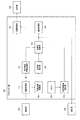
そこで図1に示すように、本実施形態の内視鏡装置は、距離情報取得部120と近接度取得部130と処置具制御部160とを含む。距離情報取得部120は、特定部位と処置具(手術器具)との間の距離に関する距離情報の取得処理を行う。近接度取得部130は、距離情報に基づいて、特定部位と処置具との間の近接度の取得処理を行う。そして処置具制御部160は、近接度に基づいて、処置具の切開及び止血の少なくとも一方に関する設定を制御する。
Therefore, as shown in FIG. 1, the endoscope apparatus of the present embodiment includes a distance
このようにすれば、生体の特定部位と処置具との近接度に基づいて処置具の設定(例えば、切開能力や止血能力に関する設定)を制御できるので、術者の意図しない生体の特定部位を損傷させる危険性を低下できる。上述した特許文献1では、処置具が特定部位に近接した場合にユーザーに警告する。しかしながら、近接した際に処置具の設定が変更されない場合、特定部位を傷付ける可能性が残る。この点、本実施形態では、処置具の設定を制御できるため、更に安全性を向上することができる。
This makes it possible to control the setting of the treatment tool (for example, settings relating to the incision ability and hemostasis ability) based on the proximity between the specific part of the living body and the treatment tool. The risk of damage can be reduced. In
より具体的には、図4等で後述するように、処置具制御部160は、処置具が特定部位に近づいた場合(例えば近接度が閾値を超えた場合)に、処置具の切開能力を抑制する制御又は処置具の止血性能を向上させる制御を行う。例えば、電気的高周波エネルギーを用いた電気メスを処置具として用いる場合、電気メスへ供給する電気出力(電力)を抑制することで切開能力(切断性能)を抑制する。
More specifically, as will be described later with reference to FIG. 4 and the like, the treatment
このようにすれば、処置具が特定部位に近接したときに切開能力が抑制され、或は止血能力が向上するので、誤って処置具を特定部位に近づけた場合であっても、特定部位を損傷させる(或は、特定部位が血管の場合には出血させる)可能性を低下できる。 In this way, the incision ability is suppressed or the hemostasis ability is improved when the treatment tool comes close to the specific part, so even if the treatment tool is accidentally brought close to the specific part, The possibility of damage (or bleeding when the specific site is a blood vessel) can be reduced.
ここで、特定部位とは、内視鏡装置の処置具により処置を行う領域において、危険な部位或は注意すべき部位のことである。即ち、処置具(例えば電気メスや超音波メス等)によって処置(例えば切開や止血等)を行っている際に、ユーザーが意図せず傷付けてしまうと生体組織に危害が及ぶ可能性のある部位である。例えば、特定部位としては、血管や神経、尿管等の線状又は管状の部位が想定される。 Here, the specific part is a dangerous part or a part to be noted in an area where treatment is performed by the treatment tool of the endoscope apparatus. That is, when a treatment (for example, an incision or hemostasis) is performed with a treatment tool (for example, an electric knife or an ultrasonic knife), a part that may harm the living tissue if the user inadvertently injures it. It is. For example, as the specific part, a linear or tubular part such as a blood vessel, a nerve, or a ureter is assumed.
また、近接度とは、処置具と特定部位の近さの度合いを表す指標であり、処置具と特定部位の距離が小さいほど近接度は大きくなる。処置具が特定部位に近いほど特定部位を損傷する可能性が増すことから、近接度は、そのような危険性を表す指標とも考えられる。そのため、危険性に応じて距離情報と近接度の対応を変化させてもよい。例えば、特定部位が血管である場合、危険性としては、血管を傷つけて出血させることが考えられる。出血の可能性や出血量は、処置具の切開能力(処置具情報)や血管の太さ等の特性(特定部位情報)によって変わるので、処置具の切開能力が高いほど或は血管が太いほど、同一距離に対して近接度を大きくしてもよい。 The proximity is an index representing the degree of proximity between the treatment tool and the specific part, and the proximity increases as the distance between the treatment tool and the specific part decreases. Since the possibility of damaging a specific part increases as the treatment tool is closer to the specific part, the proximity is also considered as an index representing such a risk. Therefore, the correspondence between distance information and proximity may be changed according to the risk. For example, when the specific site is a blood vessel, the risk is that the blood vessel is damaged and bleeding is caused. The possibility of bleeding and the amount of bleeding vary depending on the incision ability (treatment tool information) of the treatment tool and characteristics (specific site information) such as the thickness of the blood vessel, so the higher the incision ability of the treatment tool or the thicker the blood vessel The degree of proximity may be increased for the same distance.
また、切開に関する設定とは、処置具を用いて生体組織を切開する際に処置具に対して設定するものであり、例えば切開能力を決める設定値やパラメーター(例えば電気メスの高周波出力や超音波メスの超音波出力)、或は切開を指示するモード設定(切開モード)等である。同様に、止血に関する設定とは、処置具を用いて血管を止血する際に処置具に対して設定するものであり、例えば止血能力を決める設定値やパラメーター、或は止血を指示するモード設定(止血モード)等である。 The setting related to incision is set for the treatment tool when incising the living tissue using the treatment tool. For example, setting values and parameters for determining the incision ability (for example, high-frequency output of an electric knife and ultrasonic waves) Ultrasonic setting of the scalpel) or mode setting (incision mode) for instructing incision. Similarly, the setting related to hemostasis is set for the treatment tool when the blood vessel is stopped using the treatment tool. For example, setting values and parameters for determining the hemostasis capability, or mode setting for instructing hemostasis ( Hemostatic mode).
また、距離情報とは、特定部位と処置具との間の距離に関するものであればよく、処置具から特定部位までの距離そのものでなくともよい。即ち、距離の基準点は処置具の先端である必要はなく、任意に設定した所定位置であればよい。例えば、距離の基準点は、処置具の根元や撮像部(スコープ)の先端等であってもよい。処置具は撮像部の先端から挿入され、形状やサイズが決まっているため、処置具からの距離に代えて上記のような基準点からの距離を用いることが可能である。また、距離の終点についても、処置具に最も近い特定部位の位置に限定されない。例えば、処置具としてナイフを用いる場合、ナイフの切開方向(刃先の延長線上)における特定部位の位置を距離の終点としてもよい。本実施形態では、処置具が特定部位に及ぼす影響(例えば特定部位が血管である場合に、出血させる可能性)を回避したいので、その処置具の影響を受ける特定部位の位置を距離の終点とすればよい。 The distance information may be related to the distance between the specific part and the treatment tool, and may not be the distance itself from the treatment tool to the specific part. That is, the distance reference point does not need to be the tip of the treatment instrument, and may be a predetermined position set arbitrarily. For example, the reference point of the distance may be the root of the treatment tool, the tip of the imaging unit (scope), or the like. Since the treatment tool is inserted from the tip of the imaging unit and has a predetermined shape and size, the distance from the reference point as described above can be used instead of the distance from the treatment tool. Further, the end point of the distance is not limited to the position of the specific part closest to the treatment tool. For example, when a knife is used as the treatment tool, the position of a specific part in the knife incision direction (on the extension line of the blade edge) may be set as the end point of the distance. In this embodiment, since it is desired to avoid the influence of the treatment tool on the specific part (for example, when the specific part is a blood vessel, there is a possibility of bleeding), the position of the specific part affected by the treatment tool is set as the end point of the distance. do it.
2.第1実施形態
次に、本実施形態の詳細な構成について説明する。本実施形態の内視鏡装置としては、消化器(例えば食道や胃等の上部消化管や、或は大腸等の下部消化管)に挿入して診察・処置を行う消化器内視鏡装置や、外科手術において手術部位(例えば脳や腹部、関節等)に挿入することにより手術部位を撮影する外科内視鏡装置を想定できる。
2. First Embodiment Next, a detailed configuration of the present embodiment will be described. As the endoscope apparatus of the present embodiment, a digestive endoscope apparatus that is inserted into a digestive organ (for example, an upper digestive tract such as the esophagus or stomach, or a lower digestive tract such as the large intestine) for diagnosis and treatment, In addition, a surgical endoscopic apparatus that images a surgical site by inserting it into a surgical site (for example, a brain, an abdomen, a joint, or the like) in a surgical operation can be assumed.
なお、以下では特定部位が血管である場合を例に説明するが、本実施形態はこれに限定されない。即ち上述のように、特定部位は、内視鏡装置の処置具により処置を行う領域において、危険な部位或は注意すべき部位であればよい。 Hereinafter, a case where the specific part is a blood vessel will be described as an example, but the present embodiment is not limited to this. That is, as described above, the specific part may be a dangerous part or a part to be watched in the region where the treatment is performed by the treatment tool of the endoscope apparatus.
図2に、第1実施形態における内視鏡装置の構成例を示す。この内視鏡装置は、撮像部200(スコープ部)、処置具210(手術器具)、プロセッサー部300(画像プロセッサー部)、表示部400を含む。
FIG. 2 shows a configuration example of the endoscope apparatus according to the first embodiment. The endoscope apparatus includes an imaging unit 200 (scope unit), a treatment tool 210 (surgical instrument), a processor unit 300 (image processor unit), and a
第1実施形態では、撮像画像から処置具210及び血管の位置を検出し、それらの間の距離情報を求め、その距離情報に基づいて近接度を取得し、その近接度に基づいて処置具210の設定を制御する。
In the first embodiment, the positions of the
具体的には、撮像部200は、例えばCCDやCMOSセンサー等に代表される撮像素子を有する。撮像部200は、その撮像素子により観察対象物(被写体)を撮像し、その撮像した画像データをプロセッサー部300へ出力する。
Specifically, the
プロセッサー部300は、撮像画像に対する画像処理や、内視鏡装置の各部の制御を行う。プロセッサー部300は、画像取得部110、距離情報取得部120、近接度取得部130、通知処理部140、画像処理部150、処置具制御部160を含む。
The
画像取得部110は、撮像部200から送信される撮像画像(画像データ)を受信し、その撮像画像を画像処理部150と距離情報取得部120へ出力する。
The
画像処理部150は、撮像画像に対して種々の画像処理を行い、処理後の画像を表示部400へ出力する。例えば、画像処理として、ホワイトバランス処理やガンマ補正処理、強調処理、拡大・縮小処理、歪み補正処理、ノイズリダクション処理等を行う。
The
距離情報取得部120は、撮像画像から血管構造を検出し、その血管の位置情報を取得する。また撮像画像から処置具を検出し、その処置具の位置情報を取得する。そして、血管及び処置具の位置情報から処置具から血管までの2次元距離(又は後述する3次元距離)を求める。処置具は多くの場合、金属で構成されているため、それを利用して処置具を検出する手法が考えられる。例えば、撮像画像を輝度画像に変換し、所定閾値以上の輝度値を有する画素を検出し、その画素のまとまりを輪郭検出することによってグルーピングし、所定の画素数以上のグループを処置具として検出する。血管についても同様に、血管の無い部分との輝度の差等から検出する手法が考えられる。そして、検出した画素グループの先端を処置具の先端とみなし、その先端から血管までの距離を算出する。なお、処置具や血管の検出手法は上記に限定されず、例えば色差画像(CrやCb)等を用いて色の特徴から処置具や血管を検出してもよい。
The distance
近接度取得部130は、距離情報に基づいて、血管と処置具の近接度合を示す近接度を算出し、その近接度を処置具制御部160に出力する。ここで近接度は、血管位置と処置具の距離が接近するほど値が大きくなる。例えば、図3に示すように、近接度取得部130はルックアップテーブルを記憶しており、そのルックアップテーブルを参照することにより2次元距離Dを近接度に変換する。ここでD1>D2>D3>D4である。或は、近接度取得部130は、2次元距離Dを関連度に対応付ける関数により関連度を算出してもよい。例えば所定係数をαとして、近接度はf(D)=α/D等と表される。
The
処置具制御部160は、近接度に応じて処置具210の設定を変化させる制御を行う。具体的には、処置具210は、生体組織を切開する機能又は血管を止血する機能を有し、電気出力(又は振動出力)に従って、その切開能力又は止血能力が調整される。例えば図4に示すように、処置具制御部160は、近接度に対して電気出力(又は振動出力)の設定値を変化させ、近接度が大きい場合に切開能力を抑制する。即ち、近接度が閾値Xよりも小さいときには、ユーザー設定により切開能力を自由に設定可能であり、近接度が閾値Xよりも大きい場合には、近接度が大きくなるほど徐々に切開能力を減少させる制御を行う。
The treatment
上記のような処置具210として、例えば、電気出力として高周波エネルギーを発生させる処置具(例えば電気メスや、バイポーラ型デバイス等)を想定できる。或は、振動出力として超音波の振動エネルギーを発生させる処置具(例えば超音波メス等)を想定できる。これらの処置具では、高周波エネルギーや振動エネルギーにより生体組織を過熱し、その熱によって生体組織を変成させて切開又は止血を行う。例えば、切開を行う処置具は、生体組織を変成により崩壊させるだけの高いエネルギーを出力する。止血を行う処置具は、生体組織を変成により凝固させる比較的低いエネルギーを出力する。また、切開と止血を切り替える処置具では、出力エネルギーを切り替えることによって、切開モードと止血モードを切り替える。
As the
なお、処置具210の制御手法は図4に限定されず、処置具210が特定部位に接近したときに特定部位の損傷可能性を低下させる制御であればよい。例えば、近接度が閾値Xを超えた場合に、止血性能を上げる制御を行い、意図しない出血を防ぐようにしてもよい。或は、近接度が閾値Xを超えた場合に、切開モードから止血モードに切り替える制御を行ってもよい。或は、特定部位が神経のような損傷が許されない部位である場合には、近接度が閾値Xを超えた場合に出力をゼロ(広義には所定レベル以下)にする制御を行ってもよい。
Note that the control method of the
通知処理部140は、処置具制御部160による設定(設定値又はモード設定)に基づいて、ユーザーに対して処置具210の制御状態を示す通知を行う。制御状態としては、例えば切開能力の抑制状態や、切開モード等のモード設定等が想定される。通知処理部140は、制御状態を示す表示設定を画像処理部150へ出力し、画像処理部150は、撮像画像に表示設定を重畳し、表示データとして表示部400へ出力する。その結果、表示部400には、診断画像と共に処置具の制御状態が表示される。
The
なお、制御状態の通知手法は画像表示に限定されず、例えば音や振動、LEDの点灯等によって通知を行ってもよい。 Note that the notification method of the control state is not limited to image display, and notification may be performed, for example, by sound, vibration, LED lighting, or the like.
3.第1実施形態の変形例
上述した第1実施形態では、種々の変形実施が可能である。以下に、その変形例について説明する。
3. Modifications of the First Embodiment In the first embodiment described above, various modifications can be made. Below, the modification is demonstrated.
第1実施形態では、距離情報として2次元距離を取得したが、距離情報として3次元距離を取得してもよい。 In the first embodiment, a two-dimensional distance is acquired as distance information, but a three-dimensional distance may be acquired as distance information.
この第1変形例では、撮像部200は、ステレオ画像を撮影可能なステレオ光学系を有する。ステレオ光学系は、例えば視差を有する2つの撮像光学系を配置して構成される。距離情報取得部120は、その視差を有するステレオ画像をマッチング処理して画像上の各位置での奥行き方向の距離を算出する。そして、処置具と血管の2次元位置及び奥行き方向の距離に基づいて、処置具と血管の3次元位置を検出し、その間の3次元距離を求める。近接度取得部130は、第1実施形態と同様にして3次元距離を関連度に変換する。
In the first modified example, the
なお、奥行き方向の距離を検出する手法は、上記のようなステレオ撮影に限定されない。例えば、撮像部200がオートフォーカスを行う光学系を有し、そのオートフォーカスにより処置具や血管にフォーカスが合ったときのレンズ位置から処置具や血管までの奥行き方向の距離を推定してもよい。或は、撮像部200の先端から照射する照明光が、被写体が遠いほど暗くなることを利用して、画像の明るさから処置具や血管までの奥行き方向の距離を推定してもよい。
Note that the method of detecting the distance in the depth direction is not limited to the above stereo shooting. For example, the
また第1実施形態では、撮像部200で撮像した画像から処置具の位置を検出したが、位置センサーの出力に基づいて処置具の位置を検出してもよい。
In the first embodiment, the position of the treatment instrument is detected from the image captured by the
この第2変形例では、位置センサーは処置具の位置を検出し、距離情報取得部120へ出力する。距離情報取得部120は、その処置具の位置と画像から検出した血管の位置とに基づいて処置具から血管までの距離情報を取得する。例えば、処置具210に磁気コイルを組み込み、位置センサーが磁気コイルから発生する磁気をセンシングすることにより処置具の位置を検出する。
In the second modification, the position sensor detects the position of the treatment tool and outputs it to the distance
また第1実施形態では、生体の特定部位の一例として血管の位置情報を取得したが、構造を検出できれば神経や尿管等でも位置情報を取得可能である。 In the first embodiment, the position information of the blood vessel is acquired as an example of a specific part of the living body. However, if the structure can be detected, the position information can also be acquired by a nerve, a ureter or the like.
この第3変形例では、例えば、近赤外蛍光プローブであるICG(インド・シアニン・グリーン)を生体に投与すると体内を循環し、その蛍光を観察することで画像として生体の構造を認識することができる。これにより、例えば尿管の画像認識が可能となる。また、神経においても所定の蛍光薬剤等の投与により、画像として構造を認識できる。 In this third modification, for example, when ICG (Indo-Cyanine-Green), which is a near-infrared fluorescent probe, is administered to a living body, it circulates in the body, and the structure of the living body is recognized as an image by observing the fluorescence. Can do. Thereby, for example, image recognition of the ureter can be performed. In addition, in the nerve, the structure can be recognized as an image by administering a predetermined fluorescent agent or the like.
また第1実施形態では、画像から処置具210の先端位置を検出したが、処置具210にマーカーを付与して、画像認識によって、手術器具の位置を取得しても良い。
In the first embodiment, the tip position of the
この第4変形例では、処置具210の先端にマーカーを付し、距離情報取得部120がそのマーカーを検出することにより処置具210の位置を検出する。或は、処置具210の先端以外の部分(例えばナイフの柄の部分等)にマーカーを付してもよい。この場合、距離情報取得部120は、処置具210の長さや幅等の物理情報に基づいてマーカーからナイフの先端までの距離を取得し、その距離からナイフの先端の位置を推定する。
In the fourth modification, a marker is attached to the distal end of the
以上の実施形態によれば、処置具制御部160は、近接度が閾値よりも大きい場合に処置具210の切開能力を抑制する制御を行う。
According to the above embodiment, the treatment
このようにすれば、処置具210を特定部位に所定以上近づけたときに切開能力が抑制されるので、ユーザーが誤って手術領域内の特定部位を損傷させてしまうような危機を、回避し、手術の安全性を高めることができる。
In this way, since the incision ability is suppressed when the
ここで、切開能力とは、処置具210が生体を切開する能力を表す情報であり、例えばユーザーの指示又は処置具制御部160の制御により処置具210から電気出力(又は振動出力)を出力したときに、生体が切開される範囲(例えば長さ、深さ等)である。
Here, the incision ability is information indicating the ability of the
また本実施形態では、通知処理部140は、処置具制御部160による制御状態をユーザーに通知する処理を行う。より具体的には、近接度が閾値よりも大きい場合に処置具210の切開能力を抑制する制御を処置具制御部160が行う場合に、通知処理部140は、切開能力が抑制されている旨をユーザーに通知する処理を行う。
In the present embodiment, the
このようにすれば、ユーザーが処置具210の制御状態を知ることができるので、その制御状態に応じた適切な操作を行うことが可能となる。即ち、制御状態を通知することによりユーザーに対して注意を促し、ユーザーが知らないうちに制御状態が変更されることがなくなるので、処置の安全性を確保できる。
In this way, since the user can know the control state of the
さて、上述した特許文献1の手法では、例えばMRI等を用いて予め生体組織の画像データを取得しておく必要があり、また、手術を行う際には処置具の位置データを検出するために位置検出装置が必要な構成となっている。そのため、装置が大がかりであるという課題がある。
In the method of
この点、本実施形態では、画像取得部110は、撮像部200により撮像された、特定部位及び処置具210の像を含む撮像画像を取得する。そして、距離情報取得部120は、撮像画像から処置具210及び特定部位の位置を検出し、その検出した処置具210及び特定部位の位置に基づいて処置具210から特定部位までの距離情報を取得する。
In this regard, in the present embodiment, the
このようにすれば、処置具210が特定部位に接近していることを撮像画像からリアルタイムに検出し、ユーザーに関連度として通知することができるので、事前に特定部位の画像データを取得しておく必要がない。また、事前に特定部位の画像データを取得するための装置や、撮像画像以外から処置具210の位置を検出する装置やセンサー(例えば超音波画像装置やMRIやGPS送受信装置)が不要となるため、装置をコンパクトに構成できる。
In this way, it is possible to detect in real time from the captured image that the
ここで、画像から処置具210を検出する場合、例えばナイフ等を用いて処置を行っている際にはナイフの先端が生体内に隠れて撮像されない可能性がある。例えばナイフで生体を切開したとき等にはナイフの先端が生体内に隠れ、画像でナイフの先端を認識できない場合がある。
Here, when the
この点、本実施形態では、処置具210の柄等にマーカーを付すことにより、先端が隠れている場合であっても処置具210の先端位置を推定できるので、正確な近接度を求めることが可能となる。
In this regard, in this embodiment, by attaching a marker to the handle or the like of the
4.第2実施形態
図5に、第2実施形態における内視鏡装置の構成例を示す。この内視鏡装置は、撮像部200、処置具210、プロセッサー部300、表示部400を含む。プロセッサー部300は、画像取得部110、距離情報取得部120、近接度取得部130、通知処理部140、画像処理部150、処置具制御部160、特定部位情報取得部180を含む。なお、上述の実施形態で説明した構成要素と同一の構成要素については同一の符号を付し、適宜説明を省略する。
4). Second Embodiment FIG. 5 shows a configuration example of an endoscope apparatus according to the second embodiment. The endoscope apparatus includes an
第2実施形態では、特定部位の特性情報である特定部位情報及び処置具210と特定部位の近接度に基づいて、処置具210の設定を制御する。
In the second embodiment, the setting of the
具体的には、特定部位情報取得部180は、画像取得部110が取得した撮像画像から血管の境界を検出し、その検出結果に基づいて血管径(血管の幅)を特定部位情報として取得する。血管の断面は一般的に円形と考えられることから、画像上での血管の幅を血管径とする。なお、撮像画像から血管を画像認識する処理は、距離情報取得部120と特定部位情報取得部180で共通化してもよいし、別個に行ってもよい。
Specifically, the specific part
処置具制御部160は、血管径と近接度に基づいて処置具210の設定を制御する。図6に、その制御の例を示す。VD1は、血管径が所定の径より小さい場合における近接度と電気出力(又は振動出力)の関係を示し、VD2は、血管径が所定の径より大きい場合における近接度と電気出力(又は振動出力)の関係を示す。処置具制御部160は、近接度が閾値Xよりも大きい場合に、処置具210の切開能力を抑制する制御を行うが、血管径が所定の径より大きい場合(VD2)には、誤って血管を切断した際の出血リスクが大きいと判断し、即座に抑制を行う。一方、血管径が所定の径より小さい場合(VD1)には、血管を切断した際の出血リスクが小さいと判断し、即座に抑制は実施しない。即ち、血管径が大きい方が、近接度の増加に対してより速く切開能力を低下させる。
The treatment
以上の実施形態によれば、特定部位情報取得部180は、特定部位の特性情報である特定部位情報を取得する。そして、処置具制御部160は、近接度と特定部位情報に基づいて、処置具210の切開及び止血の少なくとも一方に関する設定を制御する。
According to the above embodiment, the specific part
このようにすれば、特定部位の特性に応じて処置具210の制御を適切に行うことが可能となる。即ち、近接度に対してどのように処置具210の設定を制御するかを、特定部位の特性ごとに変更することが可能となる。
If it does in this way, it will become possible to control
なお、以上の実施形態では、特定部位情報として血管径(血管の太さ)を取得する場合を例に説明したが、本実施形態はこれに限定されない。即ち、特定部位の特性情報とは、処置の対象部に存在する特定部位が有する特性に対応した情報であればよい。例えば、特定部位が血管である場合には、血管の種類(例えば動脈や静脈、末梢血管)や、血管の太さ、血管が存在する部位(例えば下部消化管、上部消化管等)、血管が存在する組織(例えば粘膜や脂肪、筋肉)等を表す情報が想定される。 In the above embodiment, the case where the blood vessel diameter (blood vessel thickness) is acquired as the specific part information has been described as an example, but the present embodiment is not limited to this. That is, the characteristic information of the specific part may be information corresponding to the characteristic of the specific part existing in the treatment target part. For example, when the specific site is a blood vessel, the type of blood vessel (eg, artery, vein, peripheral blood vessel), the thickness of the blood vessel, the site where the blood vessel exists (eg, lower digestive tract, upper digestive tract, etc.) Information representing the existing tissue (for example, mucous membrane, fat, muscle) is assumed.
また本実施形態では、特定部位情報は、特定部位の太さ情報である。処置具制御部160は、近接度が閾値Xよりも大きい場合に処置具210の切開能力を抑制する制御を行い、特定部位が太いほど切開能力の抑制度合いを大きくする。より具体的には、特定部位は、血管であり、太さ情報は、血管の血管径である。
In the present embodiment, the specific part information is thickness information of the specific part. The treatment
このようにすれば、近接する血管の太さの違いによって、最適な切除及び止血の少なくとも一方に関する制御を実施することができる。即ち、血管を損傷したときに血管が太いほど出血のリスクが増すので、そのようなリスクの高い血管ほど処置具210の切開能力を速く抑制することで、安全性を向上できる。また、出血のリスクの少ない細い血管では、切開能力の抑制を少なくすることで、不用意に処置に要する時間を増加させないようにできる。
If it does in this way, the control regarding at least one of optimal resection and hemostasis can be implemented by the difference in the thickness of the adjacent blood vessel. That is, when a blood vessel is damaged, the risk of bleeding increases as the blood vessel becomes thicker. Therefore, the safety of the blood vessel with such a high risk can be improved by suppressing the incision ability of the
ここで、切開能力とは、処置具210が生体組織を切開する能力を表す情報であり、例えばユーザーの指示によりナイフに電流を流した(又は電圧を印加した)ときに、生体が切開される範囲(例えば長さ、深さ等)である。
Here, the incision ability is information indicating the ability of the
また本実施形態では、特定部位情報は、特定部位の種類を表す情報である。そして、処置具制御部160は、特定部位が血管である場合には、近接度が閾値Xよりも大きい場合に、近接度が大きくなるほど切開能力を低下させる制御を行い、特定部位が神経である場合には、近接度が閾値Xよりも大きい場合に、切開能力を所定レベル以下に設定する。
In the present embodiment, the specific part information is information indicating the type of the specific part. Then, when the specific part is a blood vessel, the treatment
このようにすれば、近接する特定部位の種類の違いによって、最適な切除及び止血の少なくとも一方に関する制御を実施することができる。即ち、損傷した場合に非常にリスクが高い神経のような部位の場合には、切開能力を所定レベル以下(例えばゼロ)に設定することで、損傷のリスクをできるだけ低下させることができる。 If it does in this way, control regarding at least one of optimal excision and hemostasis can be implemented by the difference in the kind of the adjoining specific part. That is, in the case of a part such as a nerve that has a very high risk when it is damaged, the risk of damage can be reduced as much as possible by setting the incision ability below a predetermined level (for example, zero).
5.第3実施形態
図7に、第3実施形態における内視鏡装置の構成例を示す。この内視鏡装置は、撮像部200、処置具210、プロセッサー部300、表示部400を含む。プロセッサー部300は、画像取得部110、距離情報取得部120、近接度取得部130、通知処理部140、画像処理部150、処置具制御部160、処置具情報取得部170、特定部位情報取得部180、メモリー230を含む。なお、上述の実施形態で説明した構成要素と同一の構成要素については同一の符号を付し、適宜説明を省略する。
5. Third Embodiment FIG. 7 shows a configuration example of an endoscope apparatus according to a third embodiment. The endoscope apparatus includes an
第3実施形態では、処置具210の特性情報である処置具情報及び処置具210と特定部位の近接度に基づいて、処置具210の設定を制御する。
In the third embodiment, the setting of the
メモリー230には、例えば処置具210の型番のパラメーター(ID情報)が記憶されている。或は、メモリー230には、処置具210の用途や使用部位、サイズ、形状等の情報が記憶されていてもよい。
In the
処置具情報取得部170は、メモリー230から読み出した上記の情報に基づいて、処置具210の止血能力を処置具情報として取得する。例えば、メモリー230から読み出す情報と切開能力を対応付けたルックアップテーブルを記憶しておき、そのルックアップテーブルを参照して止血能力を取得する。或は、メモリー230から読み出す情報を引数として関数により止血性能を取得してもよい。
The treatment instrument
処置具制御部160は、止血性能と近接度に基づいて処置具210の設定を制御する。図8に、その制御の例を示す。HC1は、止血性能が高い場合における近接度と電気出力(又は振動出力)の関係を示し、HC2は、止血性能が小さい場合における近接度と電気出力(又は振動出力)の関係を示す。処置具制御部160は、処置具210の止血能力が高い場合(HC1)には、近接度が第1閾値X1より大きい場合に、処置具210の切開能力を抑制する制御を行う。一方、処置具210の止血能力が低い場合(HC2)には、近接度が第2閾値X2(X2<X1)の場合に、処置具210の切開能力を抑制する制御を行う。例えば、超音波メスは電気メス(高周波メス)に比べて、切開性能は高いが、止血性能が低い特性を持ち、一方、電気メスは超音波メスに比べて、切開性能は低いが、止血性能が高い特性を持つ。即ち、電気メスを用いる場合の閾値は、超音波メスを用いる場合の閾値よりも大きくする。
The treatment
6.第3実施形態の変形例
上述した第3実施形態では、種々の変形実施が可能である。以下に、その変形例について説明する。
6). Modifications of the Third Embodiment In the third embodiment described above, various modifications can be made. Below, the modification is demonstrated.
第3実施形態では、血管径に基づく処置具210の制御と、止血性能に基づく処置具210の制御とを別個に行う場合について説明したが、それらを組み合わせて処置具210を制御してもよい。例えば、血管径が閾値Xよりも大きい場合であっても、止血性能が高い処置具210を用いる際には、切開能力の抑制を実施しない制御方法、などが考えられる。
In the third embodiment, the case where the control of the
また第3実施形態では、メモリー230から読み出した情報に基づいて止血性能を取得する場合を例に説明したが、例えば、ユーザーからの設定情報に基づいて止血性能を取得してもよい。例えば、止血能力を切り替え可能な処置具210の場合、ユーザーが切り替えた止血能力を処置具情報として取得することが考えられる。
In the third embodiment, the case where the hemostatic performance is acquired based on information read from the
また第3実施形態では、メモリー230がプロセッサー部300に設けられる場合を例に説明したが、例えば、メモリー230は処置具210又は撮像部200に設けられてもよい。この場合、その処置具210又は撮像部200に設けられたメモリー230から処置具情報取得部170が処置具210のパラメーターを読み出す。
In the third embodiment, the case where the
以上の実施形態によれば、処置具情報取得部170は、処置具210の特性情報である処置具情報を取得する。そして、処置具制御部160は、近接度と処置具情報に基づいて、処置具210の切開及び止血の少なくとも一方に関する設定を制御する。
According to the above embodiment, the treatment tool
このようにすれば、処置具210の特性に応じて処置具210の制御を適切に行うことが可能となる。即ち、近接度に対してどのように処置具210の設定を制御するかを、処置具210の特性ごとに変更することが可能となる。
In this way, it becomes possible to appropriately control the
なお、以上の実施形態では、処置具情報が止血能力である場合を例に説明したが、本実施形態はこれに限定されない。即ち、処置具210の特性情報とは、処置具210が有する特性に対応した情報であればよい。例えば、処置具210がナイフである場合、その切開能力に対応する情報として、ナイフに流す電流値やナイフに印加する電圧値、ナイフの材質、刃の形状、刃のサイズ、設定した処置モード(例えば止血モードや切開モード)等が想定される。或は、処置具210を用いる部位(例えば下部消化管、上部消化管等)の情報や、処置具210の種類や用途を表す情報、処置具210に対応付けられたID情報等であってもよい。
In the above embodiment, the case where the treatment tool information is the hemostasis capability has been described as an example, but the present embodiment is not limited to this. That is, the characteristic information of the
また本実施形態では、処置具制御部160は、近接度が閾値(X1、X2)よりも大きい場合に処置具210の切開能力を抑制する制御を行い、処置具情報に応じて閾値を変化させる。より具体的には、特定部位は、血管であり、処置具情報は、処置具210の止血能力である。そして、処置具制御部160は、止血能力が低いほど閾値を小さくする(X1>X2)。
In the present embodiment, the treatment
このようにすれば、止血能力の違いによって、最適な切除及び止血の少なくとも一方に関する制御を実施することができる。即ち、止血能力が低いほど止血に要する時間が長い可能性があるため、そのようなリスクの高い処置具210ほど、特定部位から離れたところから切開能力を抑制することで、安全性を向上できる。また、止血に要する時間が短い止血能力の高い処置具210では、より特定部位に近づいたところで切開能力を抑制することで、不用意に処置に要する時間を増加させないようにできる。
In this way, control regarding at least one of optimal resection and hemostasis can be performed depending on the difference in hemostasis capability. That is, since the time required for hemostasis may be longer as the hemostasis ability is lower, the higher the risk of the
ここで、止血能力とは、処置具210が血管を止血する能力を表す情報である。例えば止血用の処置具210として、血管を挟み込んで高周波や超音波により組織を加熱・凝固することで止血を行うデバイスが考えられる。このようなデバイスでは、止血能力は、例えば血管を凝固させる範囲(例えば幅)や時間、止血できる血管の太さ、止血の確実性等である。
Here, the hemostasis ability is information representing the ability of the
また本実施形態では、処置具情報は、処置具210の種類を表す情報である。そして、処置具制御部160は、処置具210が電気メスである場合には、閾値を第1閾値X1に設定し、処置具210が超音波メスである場合には、閾値を第1閾値X1よりも小さい第2閾値X2に設定する。
In the present embodiment, the treatment tool information is information indicating the type of the
このようにすれば、処置具210の種類の違いによって、最適な切除及び止血の少なくとも一方に関する制御を実施することができる。即ち、止血能力が比較的低い超音波メスの場合には、より特定部位から遠いところから切開能力を抑制することで、損傷のリスクを低下させることができる。 If it does in this way, control regarding at least one of optimal excision and hemostasis can be implemented by the difference in the kind of treatment implement 210. FIG. That is, in the case of an ultrasonic scalpel having a relatively low hemostatic ability, the risk of damage can be reduced by suppressing the incision ability from a position farther from a specific site.
7.第4実施形態
図9に、第4実施形態における内視鏡装置の構成例を示す。この内視鏡装置は、撮像部200、処置具210、プロセッサー部300、表示部400を含む。プロセッサー部300は、画像取得部110、距離情報取得部120、近接度取得部130、通知処理部140、画像処理部150、処置具制御部160、制御解除部165、処置具情報取得部170、特定部位情報取得部180、メモリー230を含む。なお、上述の実施形態で説明した構成要素と同一の構成要素については同一の符号を付し、適宜説明を省略する。
7). Fourth Embodiment FIG. 9 shows a configuration example of an endoscope apparatus according to a fourth embodiment. The endoscope apparatus includes an
第4実施形態では、所定の条件を満たした場合に、近接度に応じた処置具210の制御を解除する。
In the fourth embodiment, when a predetermined condition is satisfied, the control of the
具体的には、制御解除部165は、ユーザーからの入力及び時間経過の少なくとも一方に基づいて、処置具制御部160による制御を解除する。上述のように処置具制御部160は、近接度が閾値よりも大きい場合に処置具210の切開能力を抑制する制御を行う。制御解除部165は、ユーザーからの入力があった場合及び、近接度が所定時間の間に所定量以上変化しない場合の少なくとも一方の場合に、切開能力を抑制する制御を解除する。
Specifically, the
より具体的には、制御解除部165は、近接度が所定量変化しない場合に内部のタイマーを起動し、所定時間が経過すると、処置具制御部160に解除信号を出力する。また、制御解除部165は、図示しない術者から解除指示が設定された場合に解除信号を出力する。処置具制御部160は、制御解除部165からの解除信号が入力されると、処置具210の切開能力の抑制を解除する。
More specifically, the
さて、処置具210が特定部位に近づくと切開能力が抑制されて切開できなくなるが、そのときユーザーが処置具210を遠ざけた場合には、ユーザーは特定部位を処置する意思がないと考えられる。一方、切開能力が抑制されても処置具210を移動させない場合には、ユーザーは特定部位を処置する意思があると考えられる。この場合、切開能力の抑制を解除しないと特定部位を処置できず、処置に無駄な時間がかかってしまう。
Now, when the
この点、第4実施形態によれば、ユーザーからの入力及び時間経過の少なくとも一方によって切開能力の抑制(広義には近接度に応じた処置具210の制御)を解除できる。これにより、切開能力の抑制により処置の安全性を向上させると共に、抑制解除により不用意に処置に要する時間を増加することがない。
In this regard, according to the fourth embodiment, suppression of the incision ability (control of the
8.第5実施形態
図10に、第5実施形態における内視鏡装置の構成例を示す。この内視鏡装置は、撮像部200、処置具210、プロセッサー部300、表示部400、光源部500を含む。プロセッサー部300は、画像取得部110、距離情報取得部120、近接度取得部130、通知処理部140、画像処理部150、処置具制御部160、光源制御部190を含む。なお上述の実施形態で説明した構成要素と同一の構成要素については同一の符号を付し、適宜説明を省略する。
8). Fifth Embodiment FIG. 10 shows a configuration example of an endoscope apparatus according to a fifth embodiment. The endoscope apparatus includes an
第5実施形態では、深部血管を撮像することが可能な特殊光による撮像画像を取得し、その撮像画像から深部血管を検出し、その深部血管の被写体からの深さ(深度)を用いて処置具から深部血管までの距離情報を取得する。 In the fifth embodiment, an image captured by special light that can image a deep blood vessel is acquired, the deep blood vessel is detected from the captured image, and the treatment is performed using the depth (depth) of the deep blood vessel from the subject. The distance information from the device to the deep blood vessel is acquired.
具体的には、光源部500は、白色の波長帯域を有する通常光(白色光)と、特定の波長帯域を有する特殊光とを照明光として発生する。例えば、光源部500は、通常光を発生する光源と特殊光を透過するフィルターとを有し、そのフィルターを光路に挿入していない場合には通常光を出射し、フィルターを光路に挿入している場合には特殊光を出射する。或は、光源部500は、通常光を発生する光源と特殊光を発生する光源とを有し、それらの光源を切り替えて通常光と特殊光を出射してもよい。
Specifically, the
光源制御部190は、光源部500を制御するものであり、通常光と特殊光を切り替えて照明光として出射させる制御を行う。通常光と特殊光の切り替えは、例えばユーザーからの指示に基づいて行われてもよいし、或は自動的に交互に切り替えて照射されてもよい。
The light
特定の波長帯域は、白色の波長帯域(例えば380nm〜650nm)よりも狭い帯域であり(NBI: Narrow Band Imaging)、血液中のヘモグロビンに吸収される波長の波長帯域である。より具体的には、ヘモグロビンに吸収される波長は、390nm〜445nm(B2成分、第1の狭帯域光)、または530nm〜550nm(G2成分、第2の狭帯域光)である。なお、390nm〜445nmや530nm〜550nmの波長帯域は、ヘモグロビンに吸収されるという特性及び、それぞれ生体の表層部または深部まで到達するという特性から得られた数字である。即ち、これらの波長帯域に限定されず、例えばヘモグロビンによる吸収と生体の表層部又は深部への到達に関する実験結果等の変動要因により、波長帯域の下限値が0〜10%程度減少し、上限値が0〜10%程度上昇することも考えられる。 The specific wavelength band is a band narrower than a white wavelength band (for example, 380 nm to 650 nm) (NBI: Narrow Band Imaging), and is a wavelength band of a wavelength absorbed by hemoglobin in blood. More specifically, the wavelength absorbed by hemoglobin is 390 nm to 445 nm (B2 component, first narrowband light) or 530 nm to 550 nm (G2 component, second narrowband light). Note that the wavelength bands of 390 nm to 445 nm and 530 nm to 550 nm are numbers obtained from the characteristic of being absorbed by hemoglobin and the characteristic of reaching the surface layer part or the deep part of the living body, respectively. That is, it is not limited to these wavelength bands, for example, due to fluctuation factors such as absorption by hemoglobin and experimental results on the arrival of the living body on the surface layer or the deep part, the lower limit of the wavelength band is reduced by about 0 to 10%, the upper limit It is also conceivable that the value increases by about 0 to 10%.
撮像部200は、例えば通常のRGB撮像素子により特殊光を撮像する。そして、画像取得部110は、撮像部200から入力される画像のB成分(即ちB2成分)と、撮像部200から入力される画像のG成分(即ちG2成分)とを、それぞれ特殊光画像として取得する。特殊光のB2成分とG2成分は、生体表面より深い部分に存在する深部血管を撮影することができるため、特殊光画像にはB2成分による深部血管とG2成分による深部血管が写っている。B2成分とG2成分で到達できる生体表面からの深さが異なっているため、B2、G2成分による深部血管の深さは異なっている。
The
距離情報取得部120は、B2成分の画像とG2成分の画像から血管を検出する。各成分で写る血管の深さは予め分かっているため、その深さを血管の奥行き方向の距離情報として取得する。そして、画像平面上での処置具及び血管の位置と、B2、G2成分での血管の深さとに基づいて、処置具から血管までの3次元距離を算出する。近接度取得部130は、その3次元距離を第1実施形態と同様の手法で近接度に変換する。
The distance
なお本実施形態では、次のような特殊光画像を取得してもよい。即ち、画像取得部110は、撮像部200から入力される画像のG成分(即ちG2成分)をRチャンネルに入力し、撮像部200から入力される画像のB成分(即ちB2成分)をG、Bチャンネルに入力することにより、特殊光画像を取得してもよい。このような特殊光画像により、扁平上皮癌等の通常光では視認が難しい病変などを褐色等で表示することが可能となり、病変部の見落としを抑止することができる。
In the present embodiment, the following special light image may be acquired. That is, the
以上、本発明を適用した実施形態およびその変形例について説明したが、本発明は、各実施形態やその変形例そのままに限定されるものではなく、実施段階では、発明の要旨を逸脱しない範囲内で構成要素を変形して具体化することができる。また、上記した各実施形態や変形例に開示されている複数の構成要素を適宜組み合わせることによって、種々の発明を形成することができる。例えば、各実施形態や変形例に記載した全構成要素からいくつかの構成要素を削除してもよい。さらに、異なる実施の形態や変形例で説明した構成要素を適宜組み合わせてもよい。このように、発明の主旨を逸脱しない範囲内において種々の変形や応用が可能である。また、明細書又は図面において、少なくとも一度、より広義または同義な異なる用語と共に記載された用語は、明細書又は図面のいかなる箇所においても、その異なる用語に置き換えることができる。 As mentioned above, although embodiment and its modification which applied this invention were described, this invention is not limited to each embodiment and its modification as it is, and in the range which does not deviate from the summary of invention in an implementation stage. The component can be modified and embodied. Further, various inventions can be formed by appropriately combining a plurality of constituent elements disclosed in the above-described embodiments and modifications. For example, some constituent elements may be deleted from all the constituent elements described in each embodiment or modification. Furthermore, you may combine suitably the component demonstrated in different embodiment and modification. Thus, various modifications and applications are possible without departing from the spirit of the invention. In addition, a term described together with a different term having a broader meaning or the same meaning at least once in the specification or the drawings can be replaced with the different term anywhere in the specification or the drawings.
110 画像取得部、120 距離情報取得部、130 近接度取得部、
140 通知処理部、150 画像処理部、160 処置具制御部、
165 制御解除部、170 処置具情報取得部、180 特定部位情報取得部、
190 光源制御部、200 撮像部、210 処置具、230 メモリー、
300 プロセッサー部、400 表示部、500 光源部、
X 閾値、X1 第1閾値、X2 第2閾値
110 image acquisition unit, 120 distance information acquisition unit, 130 proximity acquisition unit,
140 notification processing unit, 150 image processing unit, 160 treatment instrument control unit,
165 control release unit, 170 treatment tool information acquisition unit, 180 specific site information acquisition unit,
190 light source control unit, 200 imaging unit, 210 treatment tool, 230 memory,
300 processor unit, 400 display unit, 500 light source unit,
X threshold, X1 first threshold, X2 second threshold
Claims (8)
前記距離情報に基づいて、前記特定部位と前記処置具との間の近接度の取得処理を行う近接度取得部と、
前記特定部位の太さ情報である特定部位情報の取得処理を行う特定部位情報取得部と、
前記近接度が閾値よりも大きい場合に前記処置具の切開能力を抑制する制御を行い、前記特定部位が太いほど前記切開能力の抑制度合いを大きくする処置具制御部と、
を含むことを特徴とする内視鏡装置。 A distance information acquisition unit that detects a position of a treatment tool and a specific part, and performs a process of acquiring distance information related to a distance between the specific part and the treatment tool based on the detected position of the treatment tool and the specific part; ,
Based on the distance information, a proximity acquisition unit that performs acquisition processing of the proximity between the specific part and the treatment tool;
A specific part information acquisition unit that performs acquisition processing of specific part information that is thickness information of the specific part;
When the proximity is greater than a threshold, control to suppress the incision ability of the treatment instrument, a treatment instrument control unit that increases the degree of suppression of the incision ability as the specific portion is thicker ,
An endoscopic device comprising:
前記特定部位は、血管であり、
前記太さ情報は、前記血管の血管径であることを特徴とする内視鏡装置。 In claim 1 ,
The specific site is a blood vessel,
The endoscope apparatus characterized in that the thickness information is a blood vessel diameter of the blood vessel.
前記距離情報に基づいて、前記特定部位と前記処置具との間の近接度の取得処理を行う近接度取得部と、
前記特定部位の種類を表す情報である特定部位情報の取得処理を行う特定部位情報取得部と、
前記特定部位が血管である場合には、前記近接度が閾値よりも大きい場合に、前記近接度が大きくなるほど前記処置具の切開能力を低下させる制御を行い、前記特定部位が神経である場合には、前記近接度が前記閾値よりも大きい場合に、前記切開能力を所定レベル以下に設定する処置具制御部と、
を含むことを特徴とする内視鏡装置。 A distance information acquisition unit that detects a position of a treatment tool and a specific part, and performs a process of acquiring distance information related to a distance between the specific part and the treatment tool based on the detected position of the treatment tool and the specific part; ,
Based on the distance information, a proximity acquisition unit that performs acquisition processing of the proximity between the specific part and the treatment tool;
A specific part information acquisition unit that performs acquisition processing of specific part information that is information indicating the type of the specific part;
When the specific site is a blood vessel, when the proximity is greater than a threshold value, control is performed to reduce the incision ability of the treatment tool as the proximity increases, and when the specific site is a nerve. A treatment instrument controller that sets the incision ability to a predetermined level or less when the proximity is greater than the threshold ;
An endoscopic device comprising:
前記距離情報に基づいて、前記特定部位と前記処置具との間の近接度の取得処理を行う近接度取得部と、
前記処置具の特性情報である処置具情報の取得処理を行う処置具情報取得部と、
前記近接度が閾値よりも大きい場合に前記処置具の切開能力を抑制する制御を行い、前記処置具情報に応じて前記閾値を変化させる処置具制御部と、
を含むことを特徴とする内視鏡装置。 A distance information acquisition unit that detects a position of a treatment tool and a specific part, and performs a process of acquiring distance information related to a distance between the specific part and the treatment tool based on the detected position of the treatment tool and the specific part; ,
Based on the distance information, a proximity acquisition unit that performs acquisition processing of the proximity between the specific part and the treatment tool;
A treatment instrument information acquisition unit that performs an acquisition process of treatment instrument information that is characteristic information of the treatment instrument;
A treatment instrument controller that performs control to suppress the incision ability of the treatment instrument when the proximity is greater than a threshold, and changes the threshold according to the treatment instrument information ;
An endoscopic device comprising:
前記特定部位は、血管であり、
前記処置具情報は、前記処置具の止血能力であり、
前記処置具制御部は、
前記止血能力が低いほど前記閾値を小さくすることを特徴とする内視鏡装置。 In claim 4 ,
The specific site is a blood vessel,
The treatment tool information is a hemostasis capability of the treatment tool,
The treatment instrument controller is
An endoscope apparatus characterized in that the threshold value is reduced as the hemostatic ability is lower.
前記処置具情報は、前記処置具の種類を表す情報であり、
前記処置具制御部は、
前記処置具が電気メスである場合には、前記閾値を第1閾値に設定し、前記処置具が超音波メスである場合には、前記閾値を前記第1閾値よりも小さい第2閾値に設定することを特徴とする内視鏡装置。 In claim 4 ,
The treatment tool information is information indicating the type of the treatment tool,
The treatment instrument controller is
When the treatment instrument is an electric knife, the threshold value is set to a first threshold value. When the treatment tool is an ultrasonic knife, the threshold value is set to a second threshold value that is smaller than the first threshold value. An endoscope apparatus characterized by:
前記距離情報に基づいて、前記特定部位と前記処置具との間の近接度の取得処理を行う近接度取得部と、
前記近接度が閾値よりも大きい場合に前記処置具の切開能力を抑制する制御を行う処置具制御部と、
ユーザーからの入力があった場合及び、前記近接度が所定時間の間に所定量以上変化しない場合の少なくとも一方の場合に、前記処置具制御部による前記切開能力を抑制する制御を解除する制御解除部と、
を含むことを特徴とする内視鏡装置。 A distance information acquisition unit that detects a position of a treatment tool and a specific part, and performs a process of acquiring distance information related to a distance between the specific part and the treatment tool based on the detected position of the treatment tool and the specific part; ,
Based on the distance information, a proximity acquisition unit that performs acquisition processing of the proximity between the specific part and the treatment tool;
A treatment instrument controller that performs control to suppress the incision ability of the treatment instrument when the proximity is greater than a threshold ;
Control cancellation for canceling the control of the incision ability by the treatment instrument control unit when there is an input from a user and / or when the proximity does not change more than a predetermined amount during a predetermined time And
An endoscopic device comprising:
前記距離情報に基づいて、前記特定部位と前記処置具との間の近接度の取得処理を行い、
前記特定部位の太さ情報である特定部位情報の取得処理を行い、
前記近接度が閾値よりも大きい場合に前記処置具の切開能力を抑制する制御を行い、
前記特定部位が太いほど前記切開能力の抑制度合いを大きくすることを特徴とする内視鏡装置の作動方法。
Detecting the position of the treatment tool and the specific part, and performing distance information acquisition processing on the distance between the specific part and the treatment tool based on the detected position of the treatment tool and the specific part,
Based on the distance information, perform a process of obtaining the proximity between the specific part and the treatment tool,
The specific part information that is the thickness information of the specific part is acquired,
When the proximity is greater than a threshold value, control to suppress the incision ability of the treatment tool,
The method of operating an endoscope apparatus, wherein the degree of suppression of the incision capability is increased as the specific portion is thicker .
Priority Applications (2)
| Application Number | Priority Date | Filing Date | Title |
|---|---|---|---|
| JP2013124346A JP6265630B2 (en) | 2013-06-13 | 2013-06-13 | Endoscope apparatus and method for operating endoscope apparatus |
| US14/301,651 US20140371527A1 (en) | 2013-06-13 | 2014-06-11 | Endoscope apparatus and method for operating endoscope apparatus |
Applications Claiming Priority (1)
| Application Number | Priority Date | Filing Date | Title |
|---|---|---|---|
| JP2013124346A JP6265630B2 (en) | 2013-06-13 | 2013-06-13 | Endoscope apparatus and method for operating endoscope apparatus |
Publications (3)
| Publication Number | Publication Date |
|---|---|
| JP2015000093A JP2015000093A (en) | 2015-01-05 |
| JP2015000093A5 JP2015000093A5 (en) | 2016-07-28 |
| JP6265630B2 true JP6265630B2 (en) | 2018-01-24 |
Family
ID=52019789
Family Applications (1)
| Application Number | Title | Priority Date | Filing Date |
|---|---|---|---|
| JP2013124346A Active JP6265630B2 (en) | 2013-06-13 | 2013-06-13 | Endoscope apparatus and method for operating endoscope apparatus |
Country Status (2)
| Country | Link |
|---|---|
| US (1) | US20140371527A1 (en) |
| JP (1) | JP6265630B2 (en) |
Families Citing this family (22)
| Publication number | Priority date | Publication date | Assignee | Title |
|---|---|---|---|---|
| JP6265630B2 (en) * | 2013-06-13 | 2018-01-24 | オリンパス株式会社 | Endoscope apparatus and method for operating endoscope apparatus |
| DE102015100927A1 (en) * | 2015-01-22 | 2016-07-28 | MAQUET GmbH | Assistance device and method for imaging assistance of an operator during a surgical procedure using at least one medical instrument |
| WO2016117106A1 (en) * | 2015-01-23 | 2016-07-28 | オリンパス株式会社 | Surgical treatment device |
| JP6243364B2 (en) * | 2015-01-26 | 2017-12-06 | 富士フイルム株式会社 | Endoscope processor, operation method, and control program |
| US9905000B2 (en) | 2015-02-19 | 2018-02-27 | Sony Corporation | Method and system for surgical tool localization during anatomical surgery |
| WO2016158214A1 (en) * | 2015-04-01 | 2016-10-06 | オリンパス株式会社 | Power supply device for high frequency treatment instrument, high frequency treatment system, and control method for high frequency treatment instrument |
| JP6095863B1 (en) * | 2015-04-21 | 2017-03-15 | オリンパス株式会社 | Medical equipment |
| EP3295858A4 (en) | 2015-05-14 | 2019-01-23 | Olympus Corporation | Endoscope device |
| CN107106231A (en) * | 2015-06-19 | 2017-08-29 | 奥林巴斯株式会社 | The method of work of supply unit, high-frequency treatment system and high-frequency treatment system for high-frequency treatment utensil |
| JP2017086648A (en) * | 2015-11-13 | 2017-05-25 | ソニー株式会社 | Surgery system, control method for surgery, and program |
| WO2017109912A1 (en) | 2015-12-24 | 2017-06-29 | オリンパス株式会社 | Medical manipulator system and image display method for same |
| EP3415071A4 (en) | 2016-02-10 | 2019-11-20 | Olympus Corporation | Endoscope system |
| WO2017187526A1 (en) * | 2016-04-26 | 2017-11-02 | オリンパス株式会社 | Energy treatment tool, treatment system, and control device |
| WO2017187528A1 (en) * | 2016-04-26 | 2017-11-02 | オリンパス株式会社 | Energy treatment tool, treatment system, and control device |
| EP3332726A4 (en) | 2016-04-26 | 2019-04-24 | Olympus Corporation | Energy treatment tool, treatment system, and control device |
| DE102016118441A1 (en) | 2016-09-29 | 2018-03-29 | Olympus Winter & Ibe Gmbh | A method for processing electronic image data of an endoscope, system comprising an endoscope, an image recording device, an image processing unit and an image reproduction device, and image processing unit |
| WO2018225316A1 (en) * | 2017-06-05 | 2018-12-13 | オリンパス株式会社 | Medical control device |
| EP3678535B1 (en) | 2017-09-05 | 2021-11-03 | Briteseed, LLC | System and method used to determine tissue and/or artifact characteristics |
| CN111343899B (en) | 2017-11-13 | 2022-12-27 | 富士胶片株式会社 | Endoscope system and working method thereof |
| JP7009636B2 (en) * | 2018-08-17 | 2022-01-25 | 富士フイルム株式会社 | Endoscope system |
| WO2020054852A1 (en) * | 2018-09-14 | 2020-03-19 | オリンパス株式会社 | Medical control device, medical system, marking device, and marking device control method |
| US20230238768A1 (en) * | 2020-06-24 | 2023-07-27 | Panasonic Intellectual Property Management Co., Ltd. | Light emitting device and electronic apparatus using same |
Family Cites Families (29)
| Publication number | Priority date | Publication date | Assignee | Title |
|---|---|---|---|---|
| ES2115776T3 (en) * | 1992-08-14 | 1998-07-01 | British Telecomm | POSITION LOCATION SYSTEM. |
| JP4063933B2 (en) * | 1997-12-01 | 2008-03-19 | オリンパス株式会社 | Surgery simulation device |
| US6466815B1 (en) * | 1999-03-30 | 2002-10-15 | Olympus Optical Co., Ltd. | Navigation apparatus and surgical operation image acquisition/display apparatus using the same |
| US7634305B2 (en) * | 2002-12-17 | 2009-12-15 | Given Imaging, Ltd. | Method and apparatus for size analysis in an in vivo imaging system |
| ATE547976T1 (en) * | 2002-12-26 | 2012-03-15 | Given Imaging Ltd | IMMOBILIZABLE IN-VIVO MEASUREMENT DEVICE |
| JP4766902B2 (en) * | 2005-03-31 | 2011-09-07 | オリンパスメディカルシステムズ株式会社 | Surgery support device |
| WO2006124648A2 (en) * | 2005-05-13 | 2006-11-23 | The University Of North Carolina At Chapel Hill | Capsule imaging devices, systems and methods for in vivo imaging applications |
| US9789608B2 (en) * | 2006-06-29 | 2017-10-17 | Intuitive Surgical Operations, Inc. | Synthetic representation of a surgical robot |
| US20070134637A1 (en) * | 2005-12-08 | 2007-06-14 | Simbionix Ltd. | Medical simulation device with motion detector |
| JP4358821B2 (en) * | 2005-12-15 | 2009-11-04 | オリンパス株式会社 | Intra-subject introduction device |
| JP2009521978A (en) * | 2005-12-29 | 2009-06-11 | ギブン イメージング リミテッド | System, apparatus, and method for estimating the size of a body lumen object |
| US9198563B2 (en) * | 2006-04-12 | 2015-12-01 | The Invention Science Fund I, Llc | Temporal control of a lumen traveling device in a body tube tree |
| EP2163183A4 (en) * | 2007-06-05 | 2015-04-29 | Olympus Corp | Image processing device, image processing program and image processing method |
| JP5399253B2 (en) * | 2007-09-26 | 2014-01-29 | オリンパスメディカルシステムズ株式会社 | Intra-subject introduction system |
| JP5011060B2 (en) * | 2007-10-22 | 2012-08-29 | オリンパスメディカルシステムズ株式会社 | Medical equipment |
| JP5100336B2 (en) * | 2007-11-29 | 2012-12-19 | オリンパスメディカルシステムズ株式会社 | Capsule medical device |
| JP5288447B2 (en) * | 2008-03-28 | 2013-09-11 | 学校法人早稲田大学 | Surgery support system, approach state detection device and program thereof |
| JP5314913B2 (en) * | 2008-04-03 | 2013-10-16 | オリンパスメディカルシステムズ株式会社 | Capsule medical system |
| US8532734B2 (en) * | 2008-04-18 | 2013-09-10 | Regents Of The University Of Minnesota | Method and apparatus for mapping a structure |
| JP5284731B2 (en) * | 2008-09-02 | 2013-09-11 | オリンパスメディカルシステムズ株式会社 | Stereoscopic image display system |
| EP2351509A4 (en) * | 2008-10-28 | 2018-01-17 | Olympus Corporation | Medical device |
| WO2010093042A1 (en) * | 2009-02-16 | 2010-08-19 | オリンパスメディカルシステムズ株式会社 | Capsule medical device |
| US8945140B2 (en) * | 2010-06-18 | 2015-02-03 | Vantage Surgical Systems, Inc. | Surgical procedures using instrument to boundary spacing information extracted from real-time diagnostic scan data |
| WO2012103169A2 (en) * | 2011-01-25 | 2012-08-02 | Smith & Nephew, Inc. | Targeting operation sites |
| US20130002842A1 (en) * | 2011-04-26 | 2013-01-03 | Ikona Medical Corporation | Systems and Methods for Motion and Distance Measurement in Gastrointestinal Endoscopy |
| JP5855358B2 (en) * | 2011-05-27 | 2016-02-09 | オリンパス株式会社 | Endoscope apparatus and method for operating endoscope apparatus |
| US9204939B2 (en) * | 2011-08-21 | 2015-12-08 | M.S.T. Medical Surgery Technologies Ltd. | Device and method for assisting laparoscopic surgery—rule based approach |
| US9592095B2 (en) * | 2013-05-16 | 2017-03-14 | Intuitive Surgical Operations, Inc. | Systems and methods for robotic medical system integration with external imaging |
| JP6265630B2 (en) * | 2013-06-13 | 2018-01-24 | オリンパス株式会社 | Endoscope apparatus and method for operating endoscope apparatus |
-
2013
- 2013-06-13 JP JP2013124346A patent/JP6265630B2/en active Active
-
2014
- 2014-06-11 US US14/301,651 patent/US20140371527A1/en not_active Abandoned
Also Published As
| Publication number | Publication date |
|---|---|
| US20140371527A1 (en) | 2014-12-18 |
| JP2015000093A (en) | 2015-01-05 |
Similar Documents
| Publication | Publication Date | Title |
|---|---|---|
| JP6265630B2 (en) | Endoscope apparatus and method for operating endoscope apparatus | |
| JP6265627B2 (en) | Endoscope apparatus and method for operating endoscope apparatus | |
| JP6355875B2 (en) | Endoscope system | |
| JPWO2018163644A1 (en) | Information processing apparatus, support system, and information processing method | |
| JP7350834B2 (en) | Image processing device, endoscope system, control method for image processing device | |
| WO2019155931A1 (en) | Surgical system, image processing device, and image processing method | |
| US20150105769A1 (en) | Method for endoscopic treatment | |
| EP4243709A1 (en) | Systems for controlling cooperative surgical instruments with variable surgical site access trajectories | |
| CN118019500A (en) | Method and system for controlling a collaborative surgical instrument | |
| US20230100989A1 (en) | Surgical devices, systems, and methods using fiducial identification and tracking | |
| WO2023052936A1 (en) | Systems for controlling cooperative surgical instruments with variable surgical site access trajectories | |
| WO2023052948A1 (en) | Systems for controlling cooperative surgical instruments | |
| JP2022180177A (en) | Endoscope system, medical image processing device, and operation method thereof | |
| US10537225B2 (en) | Marking method and resecting method | |
| US20210386270A1 (en) | Medical system, energy control method, and processor | |
| US20210177531A1 (en) | Robotic surgical system and methods of use thereof | |
| EP4221629B1 (en) | Surgical devices and systems using multi-source imaging | |
| US20230116781A1 (en) | Surgical devices, systems, and methods using multi-source imaging | |
| WO2023149527A1 (en) | Medical system, notification method, and method for operating medical system | |
| US11065021B2 (en) | Osteotomy device | |
| WO2023052929A1 (en) | Surgical devices, systems, and methods using multi-source imaging | |
| WO2023052939A1 (en) | Surgical devices, systems, and methods using multi-source imaging | |
| CN118159207A (en) | System for controlling a collaborative surgical instrument with variable surgical site access trajectory | |
| CN118119353A (en) | System for controlling a collaborative surgical instrument with variable surgical site access trajectory | |
| CN118042993A (en) | Method and system for controlling a collaborative surgical instrument |
Legal Events
| Date | Code | Title | Description |
|---|---|---|---|
| A521 | Request for written amendment filed |
Free format text: JAPANESE INTERMEDIATE CODE: A523 Effective date: 20160609 |
|
| A621 | Written request for application examination |
Free format text: JAPANESE INTERMEDIATE CODE: A621 Effective date: 20160609 |
|
| A977 | Report on retrieval |
Free format text: JAPANESE INTERMEDIATE CODE: A971007 Effective date: 20170317 |
|
| A131 | Notification of reasons for refusal |
Free format text: JAPANESE INTERMEDIATE CODE: A131 Effective date: 20170425 |
|
| A521 | Request for written amendment filed |
Free format text: JAPANESE INTERMEDIATE CODE: A523 Effective date: 20170606 |
|
| TRDD | Decision of grant or rejection written | ||
| A01 | Written decision to grant a patent or to grant a registration (utility model) |
Free format text: JAPANESE INTERMEDIATE CODE: A01 Effective date: 20171205 |
|
| R151 | Written notification of patent or utility model registration |
Ref document number: 6265630 Country of ref document: JP Free format text: JAPANESE INTERMEDIATE CODE: R151 |
|
| R250 | Receipt of annual fees |
Free format text: JAPANESE INTERMEDIATE CODE: R250 |
|
| R250 | Receipt of annual fees |
Free format text: JAPANESE INTERMEDIATE CODE: R250 |
|
| R250 | Receipt of annual fees |
Free format text: JAPANESE INTERMEDIATE CODE: R250 |
|
| R250 | Receipt of annual fees |
Free format text: JAPANESE INTERMEDIATE CODE: R250 |