WO2023153471A1 - Antibody or fragment thereof that binds to fcrl1 - Google Patents
Antibody or fragment thereof that binds to fcrl1 Download PDFInfo
- Publication number
- WO2023153471A1 WO2023153471A1 PCT/JP2023/004346 JP2023004346W WO2023153471A1 WO 2023153471 A1 WO2023153471 A1 WO 2023153471A1 JP 2023004346 W JP2023004346 W JP 2023004346W WO 2023153471 A1 WO2023153471 A1 WO 2023153471A1
- Authority
- WO
- WIPO (PCT)
- Prior art keywords
- antibody
- amino acid
- seq
- fcrl1
- acid sequence
- Prior art date
Links
Images


Classifications
-
- A—HUMAN NECESSITIES
- A61—MEDICAL OR VETERINARY SCIENCE; HYGIENE
- A61K—PREPARATIONS FOR MEDICAL, DENTAL OR TOILETRY PURPOSES
- A61K39/00—Medicinal preparations containing antigens or antibodies
- A61K39/395—Antibodies; Immunoglobulins; Immune serum, e.g. antilymphocytic serum
-
- A—HUMAN NECESSITIES
- A61—MEDICAL OR VETERINARY SCIENCE; HYGIENE
- A61K—PREPARATIONS FOR MEDICAL, DENTAL OR TOILETRY PURPOSES
- A61K47/00—Medicinal preparations characterised by the non-active ingredients used, e.g. carriers or inert additives; Targeting or modifying agents chemically bound to the active ingredient
- A61K47/50—Medicinal preparations characterised by the non-active ingredients used, e.g. carriers or inert additives; Targeting or modifying agents chemically bound to the active ingredient the non-active ingredient being chemically bound to the active ingredient, e.g. polymer-drug conjugates
- A61K47/51—Medicinal preparations characterised by the non-active ingredients used, e.g. carriers or inert additives; Targeting or modifying agents chemically bound to the active ingredient the non-active ingredient being chemically bound to the active ingredient, e.g. polymer-drug conjugates the non-active ingredient being a modifying agent
- A61K47/68—Medicinal preparations characterised by the non-active ingredients used, e.g. carriers or inert additives; Targeting or modifying agents chemically bound to the active ingredient the non-active ingredient being chemically bound to the active ingredient, e.g. polymer-drug conjugates the non-active ingredient being a modifying agent the modifying agent being an antibody, an immunoglobulin or a fragment thereof, e.g. an Fc-fragment
-
- A—HUMAN NECESSITIES
- A61—MEDICAL OR VETERINARY SCIENCE; HYGIENE
- A61P—SPECIFIC THERAPEUTIC ACTIVITY OF CHEMICAL COMPOUNDS OR MEDICINAL PREPARATIONS
- A61P29/00—Non-central analgesic, antipyretic or antiinflammatory agents, e.g. antirheumatic agents; Non-steroidal antiinflammatory drugs [NSAID]
-
- A—HUMAN NECESSITIES
- A61—MEDICAL OR VETERINARY SCIENCE; HYGIENE
- A61P—SPECIFIC THERAPEUTIC ACTIVITY OF CHEMICAL COMPOUNDS OR MEDICINAL PREPARATIONS
- A61P35/00—Antineoplastic agents
-
- A—HUMAN NECESSITIES
- A61—MEDICAL OR VETERINARY SCIENCE; HYGIENE
- A61P—SPECIFIC THERAPEUTIC ACTIVITY OF CHEMICAL COMPOUNDS OR MEDICINAL PREPARATIONS
- A61P35/00—Antineoplastic agents
- A61P35/02—Antineoplastic agents specific for leukemia
-
- A—HUMAN NECESSITIES
- A61—MEDICAL OR VETERINARY SCIENCE; HYGIENE
- A61P—SPECIFIC THERAPEUTIC ACTIVITY OF CHEMICAL COMPOUNDS OR MEDICINAL PREPARATIONS
- A61P37/00—Drugs for immunological or allergic disorders
- A61P37/02—Immunomodulators
-
- C—CHEMISTRY; METALLURGY
- C07—ORGANIC CHEMISTRY
- C07K—PEPTIDES
- C07K16/00—Immunoglobulins [IGs], e.g. monoclonal or polyclonal antibodies
-
- C—CHEMISTRY; METALLURGY
- C07—ORGANIC CHEMISTRY
- C07K—PEPTIDES
- C07K16/00—Immunoglobulins [IGs], e.g. monoclonal or polyclonal antibodies
- C07K16/18—Immunoglobulins [IGs], e.g. monoclonal or polyclonal antibodies against material from animals or humans
- C07K16/28—Immunoglobulins [IGs], e.g. monoclonal or polyclonal antibodies against material from animals or humans against receptors, cell surface antigens or cell surface determinants
-
- C—CHEMISTRY; METALLURGY
- C07—ORGANIC CHEMISTRY
- C07K—PEPTIDES
- C07K16/00—Immunoglobulins [IGs], e.g. monoclonal or polyclonal antibodies
- C07K16/46—Hybrid immunoglobulins
-
- C—CHEMISTRY; METALLURGY
- C12—BIOCHEMISTRY; BEER; SPIRITS; WINE; VINEGAR; MICROBIOLOGY; ENZYMOLOGY; MUTATION OR GENETIC ENGINEERING
- C12N—MICROORGANISMS OR ENZYMES; COMPOSITIONS THEREOF; PROPAGATING, PRESERVING, OR MAINTAINING MICROORGANISMS; MUTATION OR GENETIC ENGINEERING; CULTURE MEDIA
- C12N15/00—Mutation or genetic engineering; DNA or RNA concerning genetic engineering, vectors, e.g. plasmids, or their isolation, preparation or purification; Use of hosts therefor
- C12N15/09—Recombinant DNA-technology
- C12N15/63—Introduction of foreign genetic material using vectors; Vectors; Use of hosts therefor; Regulation of expression
- C12N15/74—Vectors or expression systems specially adapted for prokaryotic hosts other than E. coli, e.g. Lactobacillus, Micromonospora
-
- C—CHEMISTRY; METALLURGY
- C12—BIOCHEMISTRY; BEER; SPIRITS; WINE; VINEGAR; MICROBIOLOGY; ENZYMOLOGY; MUTATION OR GENETIC ENGINEERING
- C12N—MICROORGANISMS OR ENZYMES; COMPOSITIONS THEREOF; PROPAGATING, PRESERVING, OR MAINTAINING MICROORGANISMS; MUTATION OR GENETIC ENGINEERING; CULTURE MEDIA
- C12N15/00—Mutation or genetic engineering; DNA or RNA concerning genetic engineering, vectors, e.g. plasmids, or their isolation, preparation or purification; Use of hosts therefor
- C12N15/09—Recombinant DNA-technology
- C12N15/63—Introduction of foreign genetic material using vectors; Vectors; Use of hosts therefor; Regulation of expression
- C12N15/79—Vectors or expression systems specially adapted for eukaryotic hosts
- C12N15/80—Vectors or expression systems specially adapted for eukaryotic hosts for fungi
- C12N15/81—Vectors or expression systems specially adapted for eukaryotic hosts for fungi for yeasts
-
- C—CHEMISTRY; METALLURGY
- C12—BIOCHEMISTRY; BEER; SPIRITS; WINE; VINEGAR; MICROBIOLOGY; ENZYMOLOGY; MUTATION OR GENETIC ENGINEERING
- C12N—MICROORGANISMS OR ENZYMES; COMPOSITIONS THEREOF; PROPAGATING, PRESERVING, OR MAINTAINING MICROORGANISMS; MUTATION OR GENETIC ENGINEERING; CULTURE MEDIA
- C12N5/00—Undifferentiated human, animal or plant cells, e.g. cell lines; Tissues; Cultivation or maintenance thereof; Culture media therefor
- C12N5/10—Cells modified by introduction of foreign genetic material
- C12N5/12—Fused cells, e.g. hybridomas
- C12N5/16—Animal cells
- C12N5/163—Animal cells one of the fusion partners being a B or a T lymphocyte
-
- G—PHYSICS
- G01—MEASURING; TESTING
- G01N—INVESTIGATING OR ANALYSING MATERIALS BY DETERMINING THEIR CHEMICAL OR PHYSICAL PROPERTIES
- G01N33/00—Investigating or analysing materials by specific methods not covered by groups G01N1/00 - G01N31/00
- G01N33/48—Biological material, e.g. blood, urine; Haemocytometers
- G01N33/50—Chemical analysis of biological material, e.g. blood, urine; Testing involving biospecific ligand binding methods; Immunological testing
- G01N33/53—Immunoassay; Biospecific binding assay; Materials therefor
Definitions
- the present invention includes a monoclonal antibody or antibody fragment that binds to the extracellular region of Fc receptor-like protein 1, a hybridoma that produces the antibody, a nucleic acid having a nucleotide sequence that encodes the antibody or the antibody fragment, and the nucleic acid.
- a transformed cell obtained by introducing a vector into a host cell, a method for producing the antibody or the antibody fragment using the hybridoma or the transformed cell, an antibody-drug conjugate containing the antibody or the antibody fragment, the antibody or the antibody
- the present invention relates to therapeutic agents and diagnostic agents comprising antibody fragments, and methods of treating and diagnosing Fc receptor-like protein 1-related diseases using said antibodies or said antibody fragments or antibody-drug conjugates comprising said antibodies or said antibody fragments.
- FCRL1 Fc receptor-like protein 1
- FCRL1 is a membrane protein belonging to the immunoglobulin superfamily, also known as CD307a, FCRH1, IFGP1, IRTA5, and the like.
- the amino acid sequence of human FCRL1 was identified in 2001 (Non-Patent Document 1).
- FCRL1 is a type I transmembrane protein expressed in B cells. It is a protein with three extracellular immunoglobulin-like domains, two intracellular immunoreceptor tyrosine activation motifs and a transmembrane region (Non-Patent Document 1). No endogenous ligand for FCRL1 has been identified to date.
- FCRL1 is reported to be expressed in cancer cells such as chronic lymphocytic leukemia, follicular lymphoma, hairy cell leukemia, and mantle cell lymphoma (Non-Patent Documents 2 and 3). . Furthermore, in recent years, it has been reported that FCRL1 contributes to cancer proliferation (Non-Patent Document 4).
- E3, E9 (Non-Patent Document 2), 2G5, 7G8, 5A2 (Patent Document 1), 1F9, 2A10 (Patent Document 2), and 5A3 (Patent Document 3) are known as monoclonal antibodies against FCRL1. It is also known that binding an immunotoxin to an anti-FCRL1 antibody exhibits cytotoxic activity against cancer cell lines (Non-Patent Document 4).
- the present invention provides a novel monoclonal antibody or antibody fragment that binds to the extracellular region of FCRL1, a hybridoma producing the antibody, a nucleic acid having a nucleotide sequence encoding the antibody or the antibody fragment, and a vector containing the nucleic acid as a host.
- a transformed cell obtained by introducing into a cell, a method for producing the antibody or the antibody fragment using the hybridoma or the transformed cell, an antibody-drug conjugate containing the antibody or the antibody fragment, and the antibody or the antibody fragment. It is an object of the present invention to provide therapeutic and diagnostic agents comprising the antibody or antibody fragment, and methods of treating and diagnosing FCRL1-related diseases using the antibody or antibody fragment, or an antibody-drug conjugate comprising the antibody or antibody fragment.
- the present invention relates to 1 to 26 below.
- FCRL1 Fc receptor-like protein 1
- FCRL1 Fc receptor-like protein 1
- VH heavy chain variable region
- CDR complementarity determining region
- said antibody fragment is selected from Fab, Fab', F(ab') 2 , single chain antibodies (scFv), dimerization V regions (Diabody), disulfide stabilized V regions (dsFv) and peptides containing CDRs 7.
- a hybridoma that produces the antibody according to any one of 1 to 6 above.
- 10. 9 comprising the nucleic acid according to 9 above.
- a transformed cell obtained by introducing the vector according to 10 above into a host cell. 12.
- the antibody or antibody of any one of 1 to 7 above which comprises culturing the hybridoma of 8 above or the transformed cell of 11 above in a medium and collecting the antibody or antibody fragment from the culture.
- a method for producing an antibody fragment 13.
- An antibody-drug conjugate comprising the antibody or antibody fragment of any one of 1 to 7 above.
- the antibody-drug conjugate according to 13 above, wherein the antibody-drug conjugate comprises the antibody or antibody fragment linked to a drug via a linker.
- 15. A composition comprising the antibody or antibody fragment of any one of 1 to 7 above or the antibody-drug conjugate of 13 or 14 above. 16.
- a diagnostic agent for FCRL1-related diseases comprising the antibody or antibody fragment of any one of 1 to 7 above or the antibody-drug conjugate of 13 or 14 above. 18.
- the FCRL1-associated disease is cancer, an autoimmune disease or an inflammatory disease.
- a therapeutic agent for FCRL1-related diseases comprising the antibody or antibody fragment of any one of 1 to 7 above or the antibody-drug conjugate of 13 or 14 above. 20.
- the therapeutic agent according to 19 above, wherein the FCRL1-associated disease is cancer, an autoimmune disease or an inflammatory disease.
- 22. A method for treating FCRL1-related diseases, comprising administering the antibody or antibody fragment of any one of 1 to 7 above or the antibody-drug conjugate of 13 or 14 above.
- 23. 15. Use of the antibody or antibody fragment according to any one of 1 to 7 above or the antibody-drug conjugate according to 13 or 14 above for the manufacture of a diagnostic agent for FCRL1-related diseases.
- the monoclonal antibody or antibody fragment of the present invention selectively binds to the extracellular region of human FCRL1.
- the monoclonal antibody or antibody fragment of the present invention exhibits superior effects when used in an antibody-drug conjugate (hereinafter also referred to as ADC) compared to existing FCRL1 antibodies. Therefore, the monoclonal antibodies or antibody fragments of the present invention can be used as therapeutic agents and diagnostic agents for human FCRL1-related diseases.
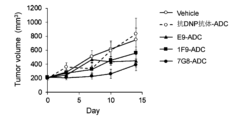
- FIG. 1 shows the results of measuring the antitumor effect of an antibody-drug conjugate in which a payload linker SG3249 was bound to a known anti-human FCRL1 antibody in a mouse model subcutaneously implanted with SU-DHL-6 cells.
- the vertical axis in FIG. 1 indicates tumor size (mm 3 ).
- the horizontal axis in FIG. 1 indicates the number of days after administration of ADC to the SU-DHL-6 cell subcutaneous transplantation mouse model.
- E9, 1F9 and 7G8 were used as known anti-human FCRL1 antibodies.
- Anti-2,4-dinitrophenol (DNP) IgG1 antibody was used as a negative antibody.
- FIG. 1 shows the results of measuring the antitumor effect of an antibody-drug conjugate in which a payload linker SG3249 was bound to a known anti-human FCRL1 antibody in a mouse model subcutaneously implanted with SU-DHL-6 cells.
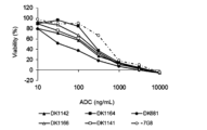
- FIG. 2A shows the results of measuring the effect of the novel anti-human FCRL1 antibody conjugated with the payload linker SG3249 on the survival of SU-DHL-6 cells.
- the vertical axis in FIG. 2A indicates the cell viability (%), and the number of cells in the condition without ADC treatment was defined as 100%.
- the horizontal axis of FIG. 2A indicates the concentration of ADC added to SU-DHL-6 cells.
- DK1142, DK1164, DK681, DK1166 and DK1141 were used as novel anti-human FCRL1 antibodies.
- 7G8 was used as a known anti-human FCRL1 antibody.
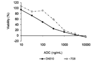
- FIG. 2B shows the results of using DK610 as a novel anti-human FCRL1 antibody in the same assay as in FIG. 2A.
- FIG. 3A shows the results of measuring the effect of the novel anti-human FCRL1 antibody conjugated with SG3249, which is a payload linker, on the survival of Ramos cells.
- the vertical axis in FIG. 3A indicates the cell viability (%), and the number of cells in the condition not treated with ADC was defined as 100%.
- the horizontal axis of FIG. 3A indicates the concentration of ADC added to Ramos cells.
- DK1142, DK1164, DK681, DK1166 and DK1141 were used as novel anti-human FCRL1 antibodies. 7G8 was used as a known anti-human FCRL1 antibody.
- FIG. 3B shows the results of using DK610 as a novel anti-human FCRL1 antibody in the same assay as in FIG. 3A.
- FIG. 4 shows the results of measuring the anti-tumor effect of an ADC comprising a novel anti-human FCRL1 antibody conjugated with a payload linker SG3249 in SU-DHL-6 cell subcutaneous mouse model and Ramos cell subcutaneous mouse model. The results 10 days after drug administration are shown.
- the vertical axis of FIG. 4 shows the relative tumor size when the tumor size of mice administered with 7G8 is set to 1. DK1142, DK1164, DK681, DK1166, DK1141 and DK610 were used as novel anti-human FCRL1 antibodies. 7G8 was used as a known anti-human FCRL1 antibody.
- FIG. 5 shows the results of measuring the antitumor effect of an ADC in which a novel anti-human FCRL1 antibody was conjugated with SG3249, which is a payload linker, in a mouse model of subcutaneous implantation of Ramos cells. The results on day 42 after drug administration are shown.
- the vertical axis in FIG. 5 indicates tumor size (mm 3 ).
- DK1142, DK1164, DK681, DK1166, DK1141 and DK610 were used as novel anti-human FCRL1 antibodies.
- 7G8 was used as a known anti-human FCRL1 antibody.
- FIG. 6 shows the results of measuring internalization of the novel anti-human FCRL1 antibody in Ramos cells. The vertical axis in FIG. 6 indicates fluorescence intensity.
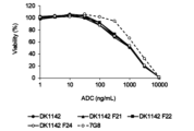
- FIG. 7A shows the results of measuring the effect of the novel anti-human FCRL1 antibody conjugated with the payload linker SG3249 on the survival of SU-DHL-6 cells.
- the vertical axis in FIG. 7A indicates the cell viability (%), and the number of cells in the condition without ADC treatment was defined as 100%.
- the horizontal axis of FIG. 7A indicates the concentration of ADC added to SU-DHL-6 cells.
- DK681 was used as the novel anti-FCRL1 chimeric antibody, and DK681 F11, DK681 F12, DK681 F13 and DK681 F14 were used as the novel anti-FCRL1 humanized antibodies.
- 7G8 was used as a known anti-human FCRL1 antibody.
- FIG. 7B shows the results of using DK1142 as the novel anti-FCRL1 chimeric antibody and DK1142 F21, DK1142 F22 and DK1142 F24 as the novel anti-FCRL1 humanized antibody in the same measurements as in FIG. 7A.
- FIG. 7B shows the results of using DK1142 as the novel anti-FCRL1 chimeric antibody and DK1142 F21, DK1142 F22 and DK1142 F24 as the novel anti-FCRL1 humanized antibody in the same measurements as in FIG. 7A.
- FIG. 8A shows the results of measuring the effects on the survival of Ramos cells for ADCs in which the novel anti-human FCRL1 antibody was conjugated with SG3249, which is a payload linker.
- the vertical axis in FIG. 8A indicates the cell viability (%), and the number of cells in the condition without ADC treatment was defined as 100%.
- the horizontal axis of FIG. 8A indicates the concentration of ADC added to Ramos cells.
- DK681 was used as the novel anti-FCRL1 chimeric antibody
- DK681 F11, DK681 F12, DK681 F13 and DK681 F14 were used as the novel anti-FCRL1 humanized antibodies.
- 7G8 was used as a known anti-human FCRL1 antibody.
- FIG. 8B shows the results of using DK1142 as the novel anti-FCRL1 chimeric antibody and DK1142 F21, DK1142 F22 and DK1142 F24 as the novel anti-FCRL1 humanized antibody in the same measurements as in FIG. 8A.
- FIG. 9 shows the results of measuring the antitumor effect of an ADC in which a payload linker SG3249 is bound to a novel anti-human FCRL1 antibody in SU-DHL-6 cell subcutaneous mouse model and Ramos cell subcutaneous mouse model. The results on day 7 after drug administration are shown.
- the vertical axis in FIG. 9 shows the relative tumor size when the tumor size of mice administered with 7G8 is set to 1.
- the present invention relates to monoclonal antibodies or antibody fragments that bind to human FCRL1.
- FCRL1 is also called CD307a, FCRH1, IFGP1 and IRTA5.
- FCRL1 belongs to the immunoglobulin superfamily and is a type 1 membrane protein consisting of 413 amino acids.
- FCRL1 has two intracellular immunoreceptor tyrosine-activated motifs (ITAM). Therefore, it is expected that an activating signal is transmitted into the cell by ligand binding, but at present, an endogenous ligand for FCRL1 has not been identified, and the function of FCRL1 has not been elucidated. Recent experiments using cancer cell lines have reported that FCRL1 is involved in cancer cell proliferation by regulating the expression of apoptosis-related molecules.
- ITAM immunoreceptor tyrosine-activated motifs
- human FCRL1 is a polypeptide comprising the amino acid sequence set forth in SEQ ID NO: 3 or the amino acid sequence of NCBI Accession No. NP_443170, one of the amino acid sequences set forth in SEQ ID NO: 3 or the amino acid sequence of NCBI Accession No. NP_443170
- Examples include polypeptides comprising amino acid sequences having preferably 80% or more, more preferably 90% or more, and most preferably 95% or more similarity, and having the function of human FCRL1.
- a polypeptide having an amino acid sequence in which one or more amino acids are deleted, substituted or added in the amino acid sequence shown in SEQ ID NO: 3 or the amino acid sequence shown in NCBI Accession No. NP_443170 is obtained by site-directed mutagenesis [Molecular Cloning, A Laboratory Manual, Second Edition, Cold Spring Harbor Laboratory Press (1989), Current Protocols in Molecular Biology, John Wiley & Sons (1987-1997), Nucleic acids Research, 10, 6487 (1982), Proc. Natl. A cad USA, 79, 6409 (1982), Gene, 34, 315 (1985), Nucleic Acids Research, 13, 4431 (1985), Proc. Natl. Acad. Sci. USA, 82, 488 (1985)], etc. can be obtained, for example, by introducing site-directed mutation into a DNA encoding a polypeptide containing the amino acid sequence of SEQ ID NO:3.
- the number of amino acids to be deleted, substituted or added is not particularly limited, but preferably 1 to several tens, for example 1 to 20, more preferably 1 to several, for example 1 to 5 amino acids. is.
- the gene encoding human FCRL1 includes the nucleotide sequence set forth in SEQ ID NO: 1 and the nucleotide sequence of NCBI Accession No. NM_052938.
- DNAs that hybridize under stringent conditions include colony hybridization, plaque hybridization, and Southern blotting using DNA containing the nucleotide sequence of SEQ ID NO: 1 or NM — 052938 as a probe. It refers to hybridizable DNA obtained by a hybridization method, a DNA microarray method, or the like.
- the hybridizable DNA is a DNA having at least 60% or more similarity, preferably 80% or more similarity, more preferably 95% similarity to the nucleotide sequence of SEQ ID NO: 1 or NM — 052938. DNA having the above similarity can be mentioned.
- the gene encoding human FCRL1 of the present invention also includes a gene having a small-scale mutation in the base sequence due to such polymorphism in the gene used in the present invention.
- Antibodies of the present invention include antibodies that bind to both human FCRL1 and monkey FCRL1.
- the monkey FCRL1 is a polypeptide comprising the amino acid sequence set forth in SEQ ID NO: 4 or the amino acid sequence of NCBI Accession No. XP_015310712, one of the amino acid sequence set forth in SEQ ID NO: 4 or the amino acid sequence of NCBI Accession No. XP_015310712
- Polypeptides comprising amino acid sequences having preferably 80% or more, more preferably 90% or more, and most preferably 95% or more similarity and having monkey FCRL1 functions are included.
- a polypeptide having an amino acid sequence in which one or more amino acids are deleted, substituted or added in the amino acid sequence represented by SEQ ID NO: 4 or the amino acid sequence represented by NCBI Accession No. XP_015310712 is subjected to site-directed mutagenesis or the like. It can be obtained, for example, by introducing site-directed mutation into a DNA encoding a polypeptide containing the amino acid sequence of SEQ ID NO:4.
- the number of amino acids to be deleted, substituted or added is not particularly limited, but preferably 1 to several tens, for example 1 to 20, more preferably 1 to several, for example 1 to 5 amino acids. is.
- the gene encoding monkey FCRL1 includes the nucleotide sequence set forth in SEQ ID NO: 2 and the nucleotide sequence of NCBI Accession No. XM_005541349.
- a gene consisting of a nucleotide sequence in which one or more nucleotides are deleted, substituted or added in the nucleotide sequence of SEQ ID NO: 2 or the nucleotide sequence of XM_005541349, and comprising a DNA encoding a polypeptide having the function of monkey FCRL1;
- a nucleotide sequence having at least 60% or more similarity, preferably 80% or more similarity, more preferably 95% or more similarity to the nucleotide sequence of SEQ ID NO: 2 or XM_005541349 A DNA that hybridizes under stringent conditions with a gene comprising a nucleotide sequence and encoding a polypeptide having the function of monkey FCRL1 or a DNA comprising the nucleotide sequence
- the similarity of amino acid sequences or base sequences in the present invention refers to a numerical value calculated under specific conditions by comparing two amino acid sequences or base sequences. Specifically, similarity is obtained by obtaining an alignment of two sequences and calculating the percentage of identical or similar residue pairs within the alignment. Algorithms such as the Needleman-Wunsch method, the Smith-Waterman method, the FASTA method and the BLAST method are used to obtain the alignment. Parameters used in each algorithm include similarity evaluation indices in residue pair units (for amino acid sequences, substitution matrices such as BLOSUM62, BLOSUM50 and PAM30 are used, and for base sequences, match reward, mismatch penalty etc.
- a quantitative evaluation index of the gap portion for example, an affine-type gap cost function
- a quantitative evaluation index of the gap portion for example, an affine-type gap cost function
- the binding of the antibody of the present invention to the extracellular region of human FCRL1 is confirmed by measuring the binding ability of the antibody of the present invention to human FCRL1-expressing cells using ELISA, flow cytometry, surface plasmon resonance, and the like. can do.
- known immunological detection methods [Monoclonal Antibodies-Principles and practice, Third edition, Academic Press (1996), Antibodies-A Laboratory Manual, Cold Spring Harbor Laboratory (1988), Monoclonal Antibody Experiment Manual, Kodansha Scientific (1987)] can also be used in combination.
- Antibody molecules are also called immunoglobulins (hereinafter referred to as Ig), and human antibodies are classified into IgA1, IgA2, IgD, IgE, IgG1, IgG2, IgG3, IgG4 and IgM isotypes according to differences in molecular structure. be done.
- IgG1, IgG2, IgG3 and IgG4, which have relatively high amino acid sequence similarity, are also collectively referred to as IgG.
- Antibody molecules are composed of polypeptides called heavy chains (hereafter referred to as H chains) and light chains (hereafter referred to as L chains).
- H chain is the H chain variable region (also referred to as VH) and the H chain constant region (also referred to as CH) from the N-terminus
- L chain is the L chain variable region (also referred to as VL) from the N-terminus. ) and L chain constant region (also denoted as CL), respectively.
- CH is known for ⁇ , ⁇ , ⁇ , ⁇ and ⁇ chains for each Ig isotype.
- CH is further composed of a CH1 domain, a hinge region, a CH2 domain and a CH3 domain from the N-terminal side.
- a domain is a functional structural unit that constitutes each polypeptide of an antibody molecule.
- the CH2 domain and CH3 domain are collectively referred to as the Fc region or simply Fc.
- CL is known as C ⁇ and C ⁇ chains.
- EU index also referred to as EU numbering
- CH1 is the amino acid sequence of EU index 118 to 215
- the hinge is the amino acid sequence of EU index 216 to 230
- CH2 is the amino acid sequence of EU index 231 to 340
- CH3 is the EU index 341 to 447. Each is specified with an amino acid sequence.
- Monoclonal antibodies in the present invention include antibodies produced by hybridomas or genetically recombinant antibodies produced by transformed cells transformed with an expression vector containing an antibody gene.
- Hybridomas are cells that produce monoclonal antibodies with desired antigen specificity, obtained by fusing B cells obtained by immunizing non-human animals with antigens and myeloma cells derived from mice. Say. Therefore, the variable region that constitutes the antibody produced by the hybridoma consists of the amino acid sequence of the non-human animal antibody.
- the antibodies of the present invention include, in particular, genetically engineered recombinant mouse antibodies, recombinant rat antibodies, recombinant rabbit antibodies, human chimeric antibodies (hereinafter also abbreviated simply as chimeric antibodies), humanized antibodies (human Also included are genetically engineered antibodies such as CDR-grafted antibodies (also referred to as CDR-grafted antibodies) and human antibodies.
- a chimeric antibody means an antibody consisting of non-human animal (non-human animal) antibody VH and VL and human antibody CH and CL.
- non-human animals any animals such as mice, rats, hamsters, and rabbits can be used as long as hybridomas can be produced.
- Human chimeric antibodies are produced by obtaining cDNAs encoding VH and VL of monoclonal antibodies from hybridomas derived from non-human animal cells producing monoclonal antibodies, and animal cells having DNAs encoding CH and CL of human antibodies. They can be inserted into an expression vector to construct a human chimeric antibody expression vector, and introduced into animal cells for expression and production.
- a humanized antibody refers to an antibody in which the amino acid sequences of the VH and VL CDRs of a non-human animal antibody have been grafted into the corresponding CDRs of the VH and VL of a human antibody. Regions other than the CDRs of VH and VL are called framework regions (hereinafter referred to as FRs).
- a humanized antibody comprises a cDNA encoding a VH amino acid sequence consisting of a non-human animal antibody VH CDR amino acid sequence and an arbitrary human antibody VH FR amino acid sequence, and a non-human animal antibody VL CDR amino acid sequence.
- a cDNA encoding the VL amino acid sequence consisting of the sequence and the FR amino acid sequence of any human antibody VL is constructed and inserted into an animal cell expression vector having DNA encoding the human antibody CH and CL.
- a modified antibody expression vector can be constructed and introduced into animal cells for expression and production.
- Human antibodies originally refer to antibodies that naturally exist in the human body. Antibodies and the like obtained from genetic animals are also included.
- Human antibodies can be obtained by immunizing mice carrying human immunoglobulin genes (Tomizuka K. et al., Proc Natl Acad Sci U S A. 97, 722-7, 2000) with the desired antigen. can be done.
- human antibodies having desired binding activity can be selected to obtain human antibodies without immunization ( Winter G. et. al., Annu Rev Immunol.12:433-55.1994).
- immortalizing human B cells using EB virus it is possible to prepare cells that produce human antibodies with desired binding activity and obtain human antibodies (Rosen A. et. al., Nature 267, 52-54.1977).
- Antibodies present in the human body can be obtained, for example, by immortalizing lymphocytes isolated from human peripheral blood by infecting them with EB virus or the like and then cloning them to obtain lymphocytes that produce the antibodies.
- the antibody can be purified from the culture in which the lymphocytes are cultured.
- a human antibody phage library is a phage library in which antibody fragments such as Fab and scFv are expressed on the surface by inserting antibody genes prepared from human B cells into the phage genes. From the library, phages expressing antibody fragments having desired antigen-binding activity can be recovered using the binding activity to the antigen-immobilized substrate as an indicator.
- the antibody fragment can also be converted into a human antibody molecule consisting of two complete H chains and two complete L chains by genetic engineering techniques.
- Human antibody-producing transgenic animals refer to animals in which human antibody genes have been integrated into the chromosome of the host animal. Specifically, a human antibody-producing transgenic animal can be produced by introducing a human antibody gene into a mouse ES cell, transplanting the ES cell into an early embryo of another mouse, and allowing the embryo to develop.
- the method for producing human antibodies from human antibody-producing transgenic animals is to obtain human antibody-producing hybridomas by a hybridoma production method commonly used in mammals other than humans, and culture them to produce human antibodies in the culture. It can be produced and accumulated.
- the VH and VL amino acid sequences of the antibody of the present invention may be used in any human antibody frame. Any of the VH and VL amino acid sequences of the humanized antibody grafted onto the work may be used.
- the amino acid sequence of CL in the antibody of the present invention may be either the amino acid sequence of a human antibody or the amino acid sequence of a non-human animal antibody, but is preferably C ⁇ or C ⁇ of the amino acid sequence of a human antibody.
- the CH of the antibody of the present invention may be CH of any molecular species belonging to immunoglobulin, but is preferably a subclass belonging to the IgG class, ⁇ 1 (IgG1; for example, Accession No. AAA02914.1), ⁇ 2 (IgG2; No. AAG00910.2), ⁇ 3 (IgG3; eg Accession No. P01860.2) and ⁇ 4 (IgG4; eg Accession No. P01861.1) can all be used.
- CH may also be CH in which one or more amino acids constituting CH are deleted, substituted, or added.
- the number of amino acids to be deleted, substituted or added is not particularly limited, but preferably 1 to several tens, for example 1 to 20, more preferably 1 to several, for example 1 to 5 amino acids. is.
- Examples of CH in which one or more amino acids constituting CH are deleted, substituted, or added include IgG1 CH variants in which serine at position 239 according to EU numbering is substituted with cysteine in human IgG1 CH. More specifically, for example, an IgG1 CH variant containing an amino acid sequence (SEQ ID NO: 80) in which serine at position 239 according to EU numbering of human IgG1 CH containing the amino acid sequence set forth in SEQ ID NO: 79 is replaced with cysteine. be done.
- Antibodies of the present invention include Fc fusion proteins in which Fc is bound to an antibody fragment, Fc fusion proteins in which Fc is bound to a naturally occurring ligand or receptor (also referred to as an immunoadhesin), and multiple Fc regions fused together.
- the present invention also includes Fc fusion proteins and the like.
- an Fc region with altered amino acid residues can also be used in the antibody of the present invention in order to stabilize the antibody and control half-life in blood.
- the antibodies or antibody fragments of the present invention include antibodies containing any post-translationally modified amino acid residues.
- Post-translational modifications include, for example, deletion of lysine residues at the C-terminus of H chains (lysine clipping), conversion of glutamine residues to pyroglutamine (pyroGlu) at the N-terminus of polypeptides, and the like. [Beck et al, Analytical Chemistry, 85, 715-736 (2013)].
- an antibody fragment is an antibody fragment that binds to the extracellular region of human FCRL1 and has antigen-binding activity.
- antibody fragments include Fab, Fab', F(ab') 2 , scFv, diabodies, dsFv, peptides containing CDRs, and the like.
- Fab is a fragment obtained by treating an IgG antibody with a proteolytic enzyme papain (cleaved at the 224th amino acid residue of the H chain), about half of the N-terminal side of the H chain and the entire L chain are disulfide bonds It is an antibody fragment with a molecular weight of about 50,000 bound by (SS bond) and having antigen-binding activity.
- the antibody fragment of the present invention is preferably an antibody fragment that binds to the extracellular domain of FCRL1 and induces internalization of FCRL1.
- F(ab′) 2 is a fragment obtained by treating IgG with the protease pepsin (cleaved at the 234th amino acid residue of the H chain). It is an antibody fragment having antigen-binding activity with a molecular weight of about 100,000, which is slightly larger than that conjugated with a protein.
- Fab' is an antibody fragment having antigen-binding activity and having a molecular weight of about 50,000, which is obtained by cleaving the S—S bond of the hinge region of F(ab') 2 .
- the scFv uses an appropriate peptide linker (P) such as a linker peptide in which one VH and one VL are connected to any number of linkers (G4S) consisting of 4 Gly and 1 Ser residues.
- P peptide linker
- G4S linkers
- a diabody is an antibody fragment formed by dimerization of scFv with the same or different antigen-binding specificities, and is an antibody fragment having bivalent antigen-binding activity against the same antigen or specific antigen-binding activity against different antigens.
- a dsFv is a polypeptide obtained by substituting one amino acid residue in each of VH and VL with a cysteine residue and binding them via an S—S bond between the cysteine residues.
- a peptide containing CDRs comprises at least one or more regions of CDRs of VH or VL.
- Peptides containing multiple CDRs can have the CDRs linked directly or via suitable peptide linkers.
- DNA encoding the VH and VL CDRs of the modified antibody of the present invention is constructed, the DNA is inserted into a prokaryotic expression vector or a eukaryotic expression vector, and the expression vector is introduced into a prokaryotic or eukaryotic organism. It can be expressed and manufactured by doing.
- Peptides containing CDRs can also be produced by chemical synthesis methods such as the Fmoc method or the tBoc method.
- VH heavy chain variable region
- CDR complementarity determining region
- SEQ ID NOs: 20 to 22 respectively an antibody comprising an amino acid sequence
- CDRs 1 to 3 of a light chain variable region hereinafter abbreviated as VL
- VH CDRs 1-3 comprise the amino acid sequences set forth in SEQ ID NOs: 28-30, respectively
- VL CDRs 1-3 comprise the amino acid sequences set forth in SEQ ID NOs: 32-34, respectively
- VH CDRs 1-3 comprise the amino acid sequences set forth in SEQ ID NOs: 36-38, respectively, and the VL CDRs 1-3 comprise the amino acid sequences set forth in SEQ ID NOs:
- One aspect of the antibody of the present invention includes any one selected from the following (1a) to (1f).
- One aspect of the antibody of the present invention includes the anti-human FCRL1 mouse monoclonal antibodies DK610, DK681, DK1142, DK1141, DK1166 and DK1164 described later in Examples. Moreover, one embodiment of the antibody of the present invention includes an antibody comprising the variable region of any one of DK610, DK681, DK1142, DK1141, DK1166 and DK1164. Further, an embodiment of the antibody of the present invention includes an antibody having the amino acid sequence of VH CDR1-3 and VL CDR1-3 of any one of DK610, DK681, DK1142, DK1141, DK1166 and DK1164. be done.
- One aspect of the antibody of the present invention includes any one selected from the following (2b-1) to (2b-4), (2c-1), (2c-2) and (2g-1).
- (2b-1) An antibody wherein VH comprises the amino acid sequence set forth in SEQ ID NO:72 and VL comprises the amino acid sequence set forth in SEQ ID NO:68.
- (2b-2) An antibody wherein VH comprises the amino acid sequence set forth in SEQ ID NO:73 and VL comprises the amino acid sequence set forth in SEQ ID NO:74.
- An antibody wherein VH comprises the amino acid sequence set forth in SEQ ID NO:72 and VL comprises the amino acid sequence set forth in SEQ ID NO:74.
- One aspect of the antibody of the present invention is a humanized antibody in which the amino acid sequences of VH CDRs 1-3 and VL CDRs 1-3 of the DK681 antibody or DK1142 antibody are grafted to the FRs of a human antibody.
- Such antibodies include, for example, DK681 F11, DK681 F12, DK681 F13, DK681 F14, DK1142 F21 and DK1142 F22, which are described later in Examples.
- DK1142 F24 As a humanized antibody obtained by grafting the amino acid sequences of CDR1 to 3 of VH and CDR1 and 3 of VL of DK1142 antibody and an amino acid sequence obtained by modifying CDR2 of VL of DK1142 antibody into FR of human antibody, DK1142 F24, which will be described later, can be mentioned.
- Antibodies of the present invention include antibodies that selectively bind to FCRL1 expressed on the cell surface and cause internalization of FCRL1. Antibodies of the present invention also include antibodies that exhibit strong efficacy when a drug is bound to the antibody to form an ADC.
- the fact that the antibody of the present invention causes the internalization of FCRL1 can be confirmed by, for example, binding a reagent that emits fluorescence in a low pH environment such as intracellular lysosomes to the antibody, adding it to the cell, and measuring the fluorescence intensity. can be confirmed by
- the antibodies of the present invention also include antibodies into which chemical structures have been introduced that can react with drugs or linkers to form bonds.
- a natural or unnatural amino acid residue having a functional group such as a group, a haloalkyl group, or a carbonyl group is added, inserted, or substituted at the N-terminus, C-terminus, or amino acid sequence of the heavy or light chain of the antibody, and the antibody is ⁇ , ⁇ -unsaturated carbonyl group, ⁇ , ⁇ -unsaturated sulfinyl group, ⁇ , ⁇ -unsaturated sulfonyl group, thiol group, hydroxyl group,
- an antibody in which the amino acid residue at a specific position of the antibody is substituted with cysteine is substituted with cysteine.
- Heavy chain amino acid residues suitable for substitution with cysteine in IgG antibodies include, for example, serine at position 239 according to EU numbering (Dimasi, N. et. al., Molecular Pharmaceutics. 14, 1501-1516, 2017), 442 al., The Journal of Biological Chemistry. 275, 30445-50, 2000), the 290th lysine (Graziani, EI. et. al., Molecular Cancer Therapeutics.
- threonine at position 114 threonine at position 114, alanine at position 140, leucine at position 174, leucine at position 179, threonine at position 187, threonine at position 209, valine at position 262, glycine at position 371, tyrosine at position 373, At least one of glutamic acid at position 382, serine at position 424, asparagine at position 434 and glutamine at position 438 (International Publication No. 2016/040856).
- ⁇ light chain amino acid residues suitable for substitution with cysteine include, for example, lysine at position 183 according to EU numbering (Graziani, EI. et. al., Molecular Cancer Therapeutics.
- an antibody introduced with para-acetylphenylalanine (Skidmore, L. et. al., Molecular Cancer Therapeutics 19(9), 1833-1843, 2020), the thiol group of the cysteine residue was enzymatically converted to a formyl group Antibodies (U.S. Patent Application Publication No. 2012/0183566), antibodies in which a cysteine is inserted between serine 239 and valine 240 in the heavy chain constant region according to EU numbering (U.S. Patent No. 10744204), etc. is mentioned.
- ADCs containing the antibody of the present invention include molecules in which an antibody and a drug are chemically or genetically engineered directly or via a linker. Antibody portions in such ADC molecules are also included in the antibodies of the present invention.
- the drug contained in the ADC of the present invention may be any molecule as long as it is a molecule having physiological activity. Examples include proteins, antibody drugs, nucleic acid drugs, and the like.
- the ADC comprises the N-terminus and C-terminus of the H chain or L chain of the antibody or antibody fragment that binds to human FCRL1 of the present invention, an appropriate functional group or side chain in the antibody molecule, a sugar chain, or the like, and a drug or linker. It can be produced by binding by a chemical method [Introduction to Antibody Engineering, Jijin Shokan (1994)].
- the antibody or antibody fragment of the present invention can be prepared by a known method (e.g., S. J. Walsh et al. Chem. Soc. Rev. 2021, 50, 1305-1353; Tumey, L. Nathan (2020). Antibody-Drug). Conjugates -Methods and Protocols: New York, Springer; and Laurent Ducry (2013). Antibody-Drug Conjugate: New York, Springer, etc.).
- the combination of the functional group contained in the antibody and the functional group contained in the drug or linker can be appropriately selected based on known information.
- a bond can be formed by nucleophilic reaction between a nucleophilic functional group such as a thiol group in the antibody molecule and a Michael acceptor such as an ⁇ , ⁇ unsaturated carboxylic acid contained in the drug or linker.
- a bond can be formed by cyclizing an azide group in an antibody molecule and an alkynyl group in a drug or linker in the presence or absence of a catalyst.
- the DNA encoding the monoclonal antibody or antibody fragment that binds to human FCRL1 of the present invention is ligated with the DNA encoding the protein or antibody drug to be bound, and inserted into an expression vector. It can be produced by a genetic engineering technique in which it is introduced into a host cell and expressed.
- radioactive isotopes examples include 111In, 131I, 125I, 90Y, 64Cu, 99Tc, 77Lu and 211At.
- Radioisotopes can be directly conjugated to antibodies, such as by the chloramine T method. Alternatively, a substance that chelates the radioisotope may be bound to the antibody.
- Chelating agents include, for example, 1-isothiocyanatobenzyl-3-methyldiethylenetriaminepentaacetic acid (MX-DTPA).
- low-molecular drugs examples include alkylating agents, nitrosourea agents, antimetabolites, antibiotics, plant alkaloids, topoisomerase inhibitors, hormone therapy agents, hormone antagonists, aromatase inhibitors, P-glycoprotein inhibitors, and platinum.
- anticancer agents such as M-phase inhibitors or kinase inhibitors [Clinical Oncology, Cancer and Chemotherapy (1996)]
- steroidal agents such as hydrocortisone or prednisone
- non-steroidal agents such as aspirin or indomethacin
- gold thiomalate gold thiomalate
- Immunomodulators such as penicillamine
- immunosuppressants such as cyclophosphamide or azathioprine
- anti-inflammatory agents such as antihistamines such as chlorpheniramine maleate or clemacitin [Inflammation and anti-inflammatory therapy, Ishiyaku Publishing Co., Ltd. (192 )] and the like.
- Anticancer agents include, for example, amifostine (ethol), cisplatin, dacarbazine (DTIC), dactinomycin, mechlorethamine (nitrogen mustard), streptozocin, cyclophosphamide, ifosfamide, carmustine (BCNU), lomustine (CCNU), doxorubicin.
- macromolecular drugs examples include polyethylene glycol (hereinafter referred to as PEG), albumin, dextran, polyoxyethylene, styrene-maleic acid copolymer, polyvinylpyrrolidone, pyran copolymer, or hydroxypropylmethacrylamide.
- methods of binding PEG to antibodies include a method of reacting with a PEG modification reagent [Bioconjugate Pharmaceuticals, Hirokawa Shoten (1993)].
- PEGylation modification reagents include modifiers for the ⁇ -amino group of lysine (Japanese Patent Laid-Open No. 61-178926), modifiers for the carboxyl groups of aspartic acid and glutamic acid (Japanese Patent Laid-Open No. 56-23587 Japanese Patent Application Laid-Open No. 2-117920), or modifiers for the guanidino group of arginine (Japanese Patent Application Laid-Open No. 2-117920).
- the immunopotentiating agent may be a natural product known as an immunoadjuvant, and as a specific example, the agent that enhances immunity is ⁇ (1 ⁇ 3) glucan (e.g., lentinan or schizophyllan) or ⁇ -galactosylceramide (KRN7000 ) and the like.
- glucan e.g., lentinan or schizophyllan
- KRN7000 ⁇ -galactosylceramide
- proteins include cytokines or growth factors that activate immunocompetent cells such as NK cells, macrophages or neutrophils, or toxin proteins.
- cytokines or growth factors examples include interferon (hereinafter referred to as IFN)- ⁇ , IFN- ⁇ , IFN- ⁇ , interleukin (hereinafter referred to as IL)-2, IL-12, IL-15, IL- 18, IL-21, IL-23, granulocyte colony stimulating factor (G-CSF), granulocyte/macrophage colony stimulating factor (GM-CSF) or macrophage colony stimulating factor (M-CSF).
- Toxin proteins include, for example, ricin or diphtheria toxin, and also include protein toxins in which mutations are introduced into the protein to control toxicity.
- Antibody drugs include, for example, antibodies to antigens that induce apoptosis by antibody binding, antigens that are involved in tumor pathogenesis, antigens that regulate immune function, and antigens that are involved in angiogenesis at lesion sites.
- Antigens to which apoptosis is induced by antibody binding include, for example, cluster of differentiation (hereinafter referred to as CD) 19, CD20, CD21, CD22, CD23, CD24, CD37, CD53, CD72, CD73, CD74, CDw75, CDw76, CD77, CDw78, CD79a, CD79b, CD80 (B7.1), CD81, CD82, CD83, CDw84, CD85, CD86 (B7.2), human leukocyte antigen (HLA)-Class II or Epidermal Growth Factor Receptor ( EGFR ) and the like.
- CD cluster of differentiation
- Antigens involved in tumor pathogenesis or antigens of antibodies that regulate immune function include, for example, CD4, CD40, CD40 ligand, B7 family molecules (e.g., CD80, CD86, CD274, B7-DC, B7-H2, B7- H3 or B7-H4), ligands of B7 family molecules (e.g.
- Antigens of antibodies that inhibit angiogenesis at lesion sites include, for example, vascular endothelial growth factor (VEGF), angiopoietin, fibroblast growth factor (FGF), EGF, hepatocyte growth factor (HGF), platelet-derived row factor (PDGF) , insulin-like growth factor (IGF), erythropoietin (EPO), TGF ⁇ , IL-8, ephrin or SDF-1 or their receptors.
- VEGF vascular endothelial growth factor
- FGF fibroblast growth factor
- EGF fibroblast growth factor
- HGF hepatocyte growth factor
- PDGF platelet-derived row factor
- IGF insulin-like growth factor
- EPO erythropoietin
- TGF ⁇ IL-8
- ephrin ephrin or SDF-1 or their receptors.
- a fusion antibody with a protein or an antibody drug is produced by linking a cDNA encoding a monoclonal antibody or an antibody fragment with a cDNA encoding an antibody contained in the protein or antibody drug to construct a DNA encoding the fusion antibody, and converting the DNA into a prokaryote.
- a fusion antibody can be produced by inserting it into an expression vector for organisms or eukaryotes, introducing the expression vector into prokaryotes or eukaryotes, and expressing it.
- Nucleic acid drugs include, for example, drugs containing nucleic acids such as small interference ribonucleic acid (siRNA) or microRNA that act on living organisms by controlling gene functions.
- small interference ribonucleic acid siRNA
- microRNA small interference ribonucleic acid
- conjugates with nucleic acid drugs that suppress the master transcription factor ROR ⁇ t in Th17 cells are contemplated.
- the linker contained in the ADC of the present invention may have any structure as long as it has the function of binding the antibody and the drug.
- it may have a structure having a special function such as being cleaved near or inside a target cell or tissue, or a branched structure capable of binding multiple drugs.
- the ADC of the present invention includes, for example, known linkers (e.g., S. J. Walsh et al. Chem. Soc. Rev. 2021, 50, 1305-1353; Tumey, L. Nathan (2020).
- Linkers e.g., S. J. Walsh et al. Chem. Soc. Rev. 2021, 50, 1305-1353; Tumey, L. Nathan (2020).
- Antibody-Drug Conjugates -Methods and Protocols New York, Springer; and Laurent Ducry (2013). Antibody-Drug Conjugate: New York, Springer, etc.).
- peptide oligosaccharide, -(CH 2 )-, oxygen atom, sulfur atom, -NH-, -(CH 2 CH 2 O)-, -CO-, -PO-, amino acid, para-amino Benzyl (PAB), a cyclic alkyl having 3 to 10 carbon atoms, and a linker consisting of any one selected from the group consisting of structures represented by the following formulas, or connecting two or more units selected from the above group and a linker comprising the structure
- amino acids constituting linkers include valine (Val), citrulline (Cit), phenylalanine (Phe), lysine (Lys), D-valine (D-Val), leucine (Leu), glycine (Gly), and alanine. (Ala), asparagine (Asn) and the like.
- linkers include peptides, oligosaccharides, -(CH 2 ) n -, -(CH 2 CH 2 O) n -, -CO-, Val-Cit-PAB, Val-Ala-PAB, Val- Lys(Ac)-PAB, Phe-Lys-PAB, Phe-Lys(Ac)-PAB, Ala-PAB, PAB, D-Val-Leu-Lys, Gly-Gly-Arg, Ala-Ala-Asn-PAB, Gly-Gly-Phe-Gly-PAB, -Gly-Gly-Phe-Gly-PAB, -Gly-Gly-Phe-Gly-CH 2 -O-CH 2 -CO-, and a linker containing any one selected from the group consisting of structures represented by the following formulas , and a linker comprising a structure connecting two or more units selected from the above group.
- n represents an integer of 1 to 1000, preferably an integer of 1 to 100, more preferably an integer of 1 to 50, still more preferably an integer of 1 to 20, and most preferably an integer of 1 to 15.
- Ac represents an acetyl group.
- Lys(Ac) represents that the side chain amino group of lysine is acetylated.
- linker for example, -(CH 2 ) m -CO-NH-(CH 2 CH 2 O) n -Val-Cit-PAB, -(CH 2 ) m -CO-NH-(CH 2 CH 2 O) n -Val-Ala-PAB, -(CH 2 ) m -CO-NH-(CH 2 CH 2 O) n -Val-Lys(Ac)-PAB, -(CH 2 ) m -CO-NH- (CH 2 CH 2 O) n -Phe-Lys-PAB, -(CH 2 ) m -CO-NH-(CH 2 CH 2 O) n -Phe-Lys(Ac)-PAB, -(CH 2 ) m —CO—NH—(CH 2 CH 2 O) n —Ala-PAB, —(CH 2 ) m —CO—NH—(CH 2 CH 2 O) n —D-Val-PAB,
- n represents an integer of 1-1000, preferably an integer of 1-100, more preferably an integer of 1-50, still more preferably an integer of 1-20, and most preferably an integer of 1-15.
- Ac represents an acetyl group.
- Lys(Ac) represents that the side chain amino group of lysine is acetylated.
- the linker before binding to the antibody preferably has a functional group capable of binding to the antibody and drug.
- functional groups include ⁇ , ⁇ unsaturated carbonyl groups, ⁇ , ⁇ unsaturated sulfinyl groups, ⁇ , ⁇ unsaturated sulfonyl groups, thiol groups, amino groups, hydroxyamino groups, hydrazide groups, hydrazide groups, amide groups. , formyl group, carboxyl group, azide group, alkynyl group, alkenyl group, haloalkyl group and the like.
- the atom adjacent to the carbonyl carbon atom of the ⁇ , ⁇ unsaturated carbonyl group, amido group and carboxyl group and the molecule adjacent to the sulfur atom of the ⁇ , ⁇ unsaturated carbonyl group and ⁇ , ⁇ unsaturated sulfinyl group include carbon, oxygen, Nitrogen, sulfur atoms and the like can be mentioned.
- Examples of the ⁇ , ⁇ unsaturated carbonyl group include maleimide group.
- Examples of the alkenyl groups include vinylpyridyl groups.
- Examples of the alkynyl group include a BCN group (Bicyclo "6.1.0" non-4-yne) and a DBCO group (Dibenzocyclooctyne).
- Linker payloads of the present invention include, for example, PBD dimer payload linkers such as SG3249 represented by the following formula (Med. Chem. Lett. 2016, 7, 983-987).
- the drug that binds to the antibody may be a label used in conventional immunological detection or measurement methods.
- Labels include, for example, enzymes such as alkaline phosphatase, peroxidase, or luciferase, luminescent substances such as acridinium esters or lophine, or fluorescent substances such as fluorescein isothiocyanate (FITC) or tetramethylrhodamine isothiocyanate (RITC). mentioned.
- the present invention also includes a composition containing, as an active ingredient, a monoclonal antibody or antibody fragment that binds to human FCRL1.
- the present invention also relates to therapeutic agents for human FCRL1-related diseases, containing as an active ingredient a monoclonal antibody or antibody fragment that binds to human FCRL1.
- the present invention also relates to a method for treating human FCRL1-related diseases, comprising administering a monoclonal antibody or antibody fragment that binds to human FCRL1.
- a human FCRL1-related disease may be any disease involving human FCRL1 or a ligand of human FCRL1, and includes, for example, cancer, autoimmune disease and inflammatory disease.
- Cancer diseases include, for example, diffuse large B-cell lymphoma, follicular lymphoma, B-cell lymphoma, Hodgkin's lymphoma, chronic lymphocytic leukemia, hairy cell leukemia, mantle cell lymphoma, follicular marginal zone lymphoma, small lymphocytic lymphoma, and the like.
- Autoimmune or inflammatory diseases include, for example, rheumatoid arthritis, multiple sclerosis, chronic obstructive pulmonary disease, systemic lupus erythematosus, lupus nephritis, asthma, atopic dermatological inflammatory colitis, Crohn's disease or Behcet's disease. be done.
- a therapeutic agent containing the antibody or antibody fragment of the present invention may contain only the antibody or antibody fragment as an active ingredient, but usually one or more pharmacologically acceptable carriers and They are preferably mixed together and provided as a pharmaceutical formulation prepared by any method known in the art of pharmacy.
- the route of administration that is most effective for treatment includes oral administration and parenteral administration such as buccal cavity, respiratory tract, rectal, subcutaneous, intramuscular or intravenous administration, preferably intravenous.
- oral administration and parenteral administration such as buccal cavity, respiratory tract, rectal, subcutaneous, intramuscular or intravenous administration, preferably intravenous.
- Internal administration can be mentioned.
- Dosage forms include, for example, sprays, capsules, tablets, powders, granules, syrups, emulsions, suppositories, injections, ointments, and tapes.
- the dosage or frequency of administration varies depending on the desired therapeutic effect, administration method, treatment period, age and body weight, but it is usually 10 ⁇ g/kg to 10 mg/kg per day for adults.
- the present invention relates to reagents for detecting or measuring FCRL1, containing monoclonal antibodies or antibody fragments that bind to human FCRL1.
- the present invention also relates to a method for detecting or measuring FCRL1 using a monoclonal antibody or antibody fragment that binds to human FCRL1.
- Methods for detecting or measuring human FCRL1 in the present invention include any known methods. Examples thereof include immunological detection or measurement methods.
- An immunological detection or measurement method is a method of detecting or measuring the amount of antibody or antigen using a labeled antigen or antibody.
- Immunological detection or measurement methods include, for example, radiolabeled immunoassay (RIA), enzyme immunoassay (EIA or ELISA), fluorescence immunoassay (FIA), luminescence immunoassay, Western A blotting method or a physicochemical method can be used.
- the present invention comprises a diagnostic agent for FCRL1-related diseases comprising a monoclonal antibody or antibody fragment that binds to human FCRL1, or a monoclonal antibody that binds to human FCRL1 or the antibody fragment to detect or measure FCRL1. It relates to a method for diagnosing FCRL1-related diseases. Diseases associated with human FCRL1 can be diagnosed by detecting or measuring cells in which human FCRL1 is expressed according to the methods described above using the monoclonal antibody or antibody fragment of the present invention.
- Biological samples to be detected or measured for human FCRL1 in the present invention include, for example, tissues, cells, blood, plasma, serum, pancreatic juice, urine, feces, tissue fluids, culture fluids, and the like, which express human FCRL1 or human FCRL1. There is no particular limitation as long as it is possible to contain cells that are isolated.
- a diagnostic agent containing the monoclonal antibody or antibody fragment of the present invention may contain a reagent for antigen-antibody reaction and a reagent for detecting the reaction, depending on the diagnostic method of interest.
- Reagents for antigen-antibody reaction include buffers, salts and the like.
- the detection reagent includes a labeled secondary antibody that recognizes the monoclonal antibody or the antibody fragment, or reagents used in conventional immunological detection or measurement methods, such as substrates corresponding to labels.
- the present invention also relates to the use of anti-human FCRL1 monoclonal antibodies or antibody fragments for the production of therapeutic or diagnostic agents for FCRL1-related diseases.
- Antibody production method Antigen preparation Human FCRL1 or human FCRL1-expressing cells to be used as antigens are obtained by transferring an expression vector containing cDNA encoding full-length or partial length of human FCRL1 to Escherichia coli, yeast, insect cells, animal cells, or the like. It can be obtained by installing Human FCRL1 can also be obtained by purifying human FCRL1 from various human cell lines, human cells, human tissues, and the like that express human FCRL1 in large amounts. In addition, these human cell lines, human cells, human tissues, and the like can be used as antigens as they are.
- a synthetic peptide having a partial sequence of human FCRL1 can be prepared by a chemical synthesis method such as the Fmoc method or the tBoc method and used as an antigen.
- a known tag such as FLAG or His may be added to the C-terminus or N-terminus of human FCRL1 or a synthetic peptide having a partial sequence of human FCRL1.
- Human FCRL1 used in the present invention can be obtained by methods described in Molecular Cloning, A Laboratory Manual, Second Edition, Cold Spring Harbor Laboratory Press (1989), Current Protocols In Molecular Biology, John Wiley & Sons (1987-1997), etc. can be produced by expressing the DNA encoding human FCRL1 in host cells, for example, by the following method.
- a recombinant vector is constructed by inserting a full-length cDNA containing a portion encoding human FCRL1 downstream of the promoter of an appropriate expression vector.
- a DNA fragment of appropriate length containing a portion encoding a polypeptide prepared based on full-length cDNA may be used.
- a transformant that produces the polypeptide can be obtained.
- Any expression vector can be used as long as it is capable of autonomous replication in the host cell used or integration into the chromosome and contains an appropriate promoter at a position where the DNA encoding the polypeptide can be transcribed. can be done.
- Any host cell that can express the gene of interest can be used, including microorganisms belonging to the genus Escherichia such as Escherichia coli, yeast, insect cells, and animal cells.
- the recombinant vector When a prokaryote such as E. coli is used as a host cell, the recombinant vector is capable of autonomous replication in the prokaryote and at the same time contains a promoter, a ribosome binding sequence, a DNA containing a portion encoding human FCRL1, and a transcription termination sequence. It is preferably a vector containing Although the recombinant vector does not necessarily have a transcription termination sequence, it is preferable to place a transcription termination sequence immediately below the structural gene. Furthermore, the recombinant vector may contain a promoter-regulating gene.
- the recombinant vector it is preferable to use a plasmid in which the distance between the Shine-Dalgarno sequence (also referred to as the SD sequence), which is a ribosome binding sequence, and the initiation codon is adjusted to an appropriate distance (eg, 6 to 18 bases).
- the SD sequence also referred to as the SD sequence
- the initiation codon is adjusted to an appropriate distance (eg, 6 to 18 bases).
- bases can be substituted so that the codons are optimal for expression in the host, thereby improving the production rate of the desired human FCRL1. can be done.
- Any expression vector can be used as long as it can exhibit its function in the host cell to be used.
- Pharmacia pSE280 (Invitrogen), pGEMEX-1 (Promega), pQE-8 (Qiagen), pKYP10 (Japanese Patent Laid-Open No. 58-110600), pKYP200 [Agricultural Biological Chemistry, 48, 669 (1984)], pLSA1 [Agric. Biol. Chem., 53, 277 (1989)], pGEL1 [Proc. Natl. Acad. Sci. ) (manufactured by Stratagene), pTrs30 [prepared from E.
- coli JM109/pTrS30 (FERM BP-5407)] pTrs32 [prepared from E. coli JM109/pTrS32 (FERM BP-5408)], pGHA2 [E. coli IGHA2 (FERM BP-400) prepared from E. coli IGKA2 (FERM BP-6798), Japanese Patent Application Laid-Open No. 60-221091], pTerm2 (US Pat. No. 4,686,191 No., US Pat. No. 4,939,094, US Pat. No. 160,735), pSupex, pUB110, pTP5, pC194, pEG400 [J. Bacteriol., 172, 2392 (1990)], Examples include pGEX (manufactured by Pharmacia), pET system (manufactured by Novagen), pME18SFL3, and the like.
- Any promoter can be used as long as it can exhibit its function in the host cell used.
- promoters derived from E. coli or phage such as trp promoter (Ptrp), lac promoter, PL promoter, PR promoter or T7 promoter.
- Terp trp promoter
- lac promoter lac promoter
- PL promoter PL promoter
- PR promoter PR promoter
- T7 promoter T7 promoter
- artificially designed and modified promoters such as a tandem promoter in which two Ptrps are arranged in series, a tac promoter, a lacT7 promoter, or a let I promoter, and the like are included.
- E. coli XL1-Blue E. coli XL2-Blue
- E. coli DH1 E. coli MC1000
- E. coli KY3276 E. coli W1485, E. coli JM109, E. coli HB101
- E. coli No. 49 E. coli W3110, E. coli NY49 or E. coli DH5 ⁇ .
- any method that introduces DNA into the host cell to be used can be used.
- a method using calcium ions Proc. Natl. Acad. , 69, 2110 (1972), Gene, 17, 107 (1982), Molecular & General Genetics, 168, 111 (1979)].
- any expression vector can be used as long as it can exhibit its function in animal cells.
- pAS3-3 Japanese Patent Laid-Open No. 2-227075
- pCDM8 Japanese Patent Laid-Open No. 2-227075
- pCDM8 Japanese Patent Laid-Open No. 2-227075
- pCDM8 Japanese Patent Laid-Open No. 2-227075
- pCDM8 Japanese Patent Laid-Open No. 2-227075
- pCDM8 Japanese Patent Laid-Open No. 2-227075
- pCDM8 Japanese Patent Laid-Open No. 2-227075
- pCDM8 Japanese Patent Laid-Open No. 2-227075
- pCDM8 Japanese Patent Laid-Open No. 2-227075
- pCDM8 Japanese Patent Laid-Open No. 2-227075
- pCDM8 Japanese Patent Laid-Open No. 2-227075
- pCDM8 Japanese Patent Laid-Open No.
- Any promoter can be used as long as it can exhibit its function in animal cells.
- CMV cytomegalovirus
- IE immediate early gene promoter
- SV40 early promoter SV40 early promoter
- retrovirus promoter SV40 early promoter
- metallothionein promoter the metallothionein promoter
- the heat shock promoter the SR ⁇ promoter
- Moloney murine leukemia virus promoter or enhancer the enhancer of the IE gene of human CMV may be used together with the promoter.
- host cells examples include human leukemia cell Namalwa cells, monkey cell COS cells, Chinese hamster ovary cell CHO cells [Journal of Experimental Medicine, 108, 945 (1958); Proc. Natl. Acad. Sci. USA, 60, 1275 (1968); Genetics, 55, 513 (1968); Chromosoma, 41, 129 (1973); Methods in Cell Science, 18, 115 (1996); Radiation Research, 148, 260 (1997); USA, 77, 4216 (1980); Proc. Natl. Acad. Sci., 60, 1275 (1968); Cell, 6, 121 (1975); Molecular Cell Genetics, Appendix I, II (pp.
- CHO cells deficient in the dihydrofolate reductase gene (hereinafter referred to as dhfr) (CHO/DG44 cells) [Proc. Natl. Acad. Sci. USA, 77, 4216 (1980)]; CHO-K1 (ATCC CCL-61), DUkXB11 (ATCC CCL-9096), Pro-5 (ATCC CCL-1781), CHO-S (Life Technologies, Cat#11619), Pro-3, rat myeloma cell YB2/3HL. P2. G11.16Ag.
- mouse myeloma cell NSO mouse myeloma cell SP2/0-Ag14
- Syrian hamster cell BHK or HBT5637 Japanese Patent Laid-Open No. 63-000299
- Any method for introducing DNA into animal cells can be used as a method for introducing a recombinant vector into host cells.
- Japanese Patent Application Laid-Open No. 2-227075 Japanese Patent Application Laid-Open No. 2-227075
- the lipofection method Japanese Patent Application Laid-Open No. 2-227075
- the lipofection method Japanese Patent Application Laid-Open No. 2-227075
- the lipofection method Japanese Patent Application Laid-Open No. 2-227075
- the lipofection method [Proc. Natl. Acad. Sci. USA, 84, 7413 (1987)].
- a transformant derived from a microorganism or an animal cell containing a recombinant vector incorporating a DNA encoding human FCRL1 obtained as described above is cultured in a medium to produce and accumulate the human FCRL1 in the culture medium.
- Human FCRL1 can be produced by allowing the cells to grow and collecting from the culture medium.
- a method for culturing the transformant in a medium can be carried out according to a conventional method used for culturing a host.
- an inducer may be added to the medium as necessary.
- an inducer may be added to the medium as necessary.
- a lac promoter isopropyl- ⁇ -D-thiogalactopyranoside or the like is used to culture a microorganism transformed with a recombinant vector using a trp promoter.
- indole acrylic acid or the like may be added to the medium.
- Examples of media for culturing transformants obtained using animal cells as hosts include the commonly used RPMI1640 medium [The Journal of the American Medical Association, 199, 519 (1967)], Eagle's MEM medium [Science , 122, 501 (1952)], Dulbecco's modified MEM medium [Virology, 8, 396 (1959)], 199 medium [Proc. Soc. Exp. Biol. Med., 73, 1 (1950)] or Iscove's Modified Examples thereof include Dulbecco's Medium (IMDM) medium and a medium obtained by adding fetal bovine serum (FBS) or the like to these medium. Cultivation is usually carried out for 1 to 7 days under conditions such as pH 6 to 8, 30 to 40°C in the presence of 5% CO2. Moreover, antibiotics such as kanamycin or penicillin may be added to the medium during the culture, if necessary.
- RPMI1640 medium The Journal of the American Medical Association, 199, 519 (1967)]
- Methods for expressing the gene encoding human FCRL1 include, in addition to direct expression, methods such as secretory production and fusion protein expression [Molecular Cloning, A Laboratory Manual, Second Edition, Cold Spring Harbor Laboratory Press (1989)]. be done.
- Methods for producing human FCRL1 include, for example, a method of producing it in a host cell, a method of secreting it outside the host cell, and a method of producing it on the membrane of the host cell. Appropriate methods can be selected by changing the structure.
- the obtained human FCRL1 can be isolated and purified, for example, as follows.
- human FCRL1 When human FCRL1 is expressed in a dissolved state in cells, the cells are collected by centrifugation after completion of the culture, suspended in an aqueous buffer, and treated with an ultrasonicator, a French press, a Mantongaurin homogenizer, a Dynomill, or the like. to disrupt the cells and obtain a cell-free extract.
- the usual protein isolation and purification methods such as solvent extraction, salting out with ammonium sulfate, desalting, precipitation with an organic solvent, diethylamino Anion exchange chromatography method using resins such as ethyl (DEAE)-Sepharose, DIAION HPA-75 (manufactured by Mitsubishi Chemical), cation exchange chromatography using resins such as S-Sepharose FF (manufactured by Pharmacia) method, hydrophobic chromatography using resins such as butyl sepharose and phenyl sepharose, gel filtration using molecular sieves, affinity chromatography, chromatofocusing, or electrophoresis such as isoelectric focusing. can be used alone or in combination to obtain purified preparations.
- solvent extraction salting out with ammonium sulfate
- desalting precipitation with an organic solvent
- diethylamino Anion exchange chromatography method using resins such as ethyl (DEAE)-S
- human FCRL1 forms an insoluble form in cells and is expressed
- the cells are collected and crushed in the same manner as described above, and centrifuged to collect the insoluble form of human FCRL1 as a precipitate fraction.
- the collected insoluble form of human FCRL1 is solubilized with a protein denaturant.
- a purified preparation of the polypeptide can be obtained by the same isolation and purification method as described above.
- the human FCRL1 or a derivative such as a sugar modification thereof When human FCRL1 or a derivative such as a sugar modification thereof is extracellularly secreted, the human FCRL1 or a derivative such as a sugar modification thereof can be recovered in the culture supernatant.
- a soluble fraction is obtained by treating the culture by a technique such as centrifugation in the same manner as described above, and a purified preparation can be obtained from the soluble fraction by using the same isolation and purification method as described above. can.
- a polypeptide containing a partial amino acid sequence of human FCRL1 used in the present invention can be produced by methods known to those skilled in the art. Specifically, it can be produced by culturing a transformant into which a part of the DNA encoding the amino acid sequence of human FCRL1 has been deleted and an expression vector containing this has been introduced. Also, according to the above method, a polypeptide having an amino acid sequence in which one or more amino acids are deleted, substituted, or added to the amino acid sequence of human FCRL1 can be obtained.
- Human FCRL1 used in the present invention can also be produced by chemical synthesis methods such as the Fmoc method or the tBoc method.
- chemical synthesis may be performed using a peptide synthesizer manufactured by Advanced Chemtech, Perkin-Elmer, Pharmacia, Protein Technology Instrument, Synthecel-Vega, Perceptive, or Shimadzu Corporation. You can also
- mice, rats or hamsters aged 3 to 20 weeks are immunized with the antigen obtained in (1), and the spleen, lymph nodes, Collect antibody-producing cells in peripheral blood.
- a mouse FCRL1 knockout mouse can also be used as an animal to be immunized.
- Immunization is performed by administering the antigen subcutaneously, intravenously, or intraperitoneally to the animal together with an appropriate adjuvant, such as Freund's complete adjuvant, or aluminum hydroxide gel and pertussis vaccine.
- an appropriate adjuvant such as Freund's complete adjuvant, or aluminum hydroxide gel and pertussis vaccine.
- the antigen is a partial peptide
- a conjugate with a carrier protein such as BSA (bovine serum albumin) or KLH (Keyhole Limpet Hemocyanin) is prepared and used as an immunogen.
- BSA bovine serum albumin
- KLH Keyhole Limpet Hemocyanin
- the antigen is administered 5-10 times at intervals of 1-2 weeks after the first administration. Blood is collected 3 to 7 days after each administration, and the serum antibody titer is measured using enzyme immunoassay [Antibodies - A Laboratory Manual, Cold Spring Harbor Laboratory (1988)]. An animal whose serum shows a sufficient antibody titer against the antigen used for immunization is used as a source of antibody-producing cells for fusion.
- tissue containing antibody-producing cells such as spleen is excised from the immunized animal, and antibody-producing cells are collected.
- spleen cells When spleen cells are used, the spleen is minced, loosened, centrifuged, and red blood cells are removed to obtain fusion antibody-producing cells.
- myeloma cells cell lines obtained from mice are used.
- U1 Current Topics in Microbiology and Immunology, 18, 1 (1978)]
- P3-NS1/1-Ag41 NS-1
- SP2/0-Ag14 SP-2
- SP-2 SP2/0-Ag14
- P3-X63-Ag8653(653) J. Immunology, 123, 1548 (1979)] or P3-X63-Ag8(X63) [Nature, 256, 495 (1975)] is used.
- the myeloma cells were passaged in normal medium [RPMI 1640 medium supplemented with glutamine, 2-mercaptoethanol, gentamycin, FBS, and 8-azaguanine] and passaged in normal medium 3-4 days prior to cell fusion. , ensure the number of cells of 2 ⁇ 10 7 or more on the day of fusion.
- the antibody-producing cells for fusion obtained in (2) and the myeloma cells obtained in (3) were mixed in Minimum Essential Medium (MEM) medium or PBS (disodium phosphate 1. 83 g, monopotassium phosphate 0.21 g, salt 7.65 g, distilled water 1 liter, pH 7.2), and the number of cells is 5 to 10:1 for fusion antibody-producing cells: myeloma cells. After mixing and centrifuging, remove the supernatant. After loosening the precipitated cell mass, a mixture of polyethylene glycol-1000 (PEG-1000), MEM medium and dimethylsulfoxide is added at 37° C. with stirring.
- MEM Minimum Essential Medium
- PBS dimethyl phosphate 1.
- HAT medium normal medium supplemented with hypoxanthine, thymidine and aminopterin. This suspension is cultured for 7-14 days at 37° C. in a 5% CO2 incubator.
- a portion of the culture supernatant is removed, and a cell group that reacts to antigens containing human FCRL1 but does not react to antigens that do not contain human FCRL1 is selected by a hybridoma selection method such as the binding assay described later.
- cloning is performed by the limiting dilution method, and those showing stable and strong antibody titers are selected as monoclonal antibody-producing hybridomas.
- the supernatant is removed by centrifugation, suspended in Hybridoma SFM medium, and cultured for 3 to 7 days.
- the obtained cell suspension is centrifuged, and the obtained supernatant is purified with a protein A-column or a protein G-column to collect the IgG fraction to obtain a purified monoclonal antibody.
- 5% Daigo GF21 can also be added to the Hybridoma SFM medium.
- Antibody subclass determination is performed by enzyme immunoassay using a subclass typing kit. Quantification of the amount of protein is calculated by the Lowry method or absorbance at 280 nm.
- Human FCRL1-expressing cells may be any cells that express human FCRL1 on the cell surface, and examples thereof include human cells, human cell lines, and human FCRL1 forced-expressing cell lines obtained in (1).
- a test substance such as serum, hybridoma culture supernatant, or purified monoclonal antibody is dispensed as the first antibody and allowed to react.
- the cells were washed thoroughly with PBS containing 1 to 10% bovine serum albumin (BSA) (hereinafter referred to as BSA-PBS), and then an anti-immunoglobulin antibody labeled with a fluorescent reagent or the like was added as a second antibody. Dispense and react.
- BSA-PBS bovine serum albumin
- a monoclonal antibody that specifically reacts with human FCRL1-expressing cells is selected by measuring the amount of fluorescence of the labeled antibody using a flow cytometer.
- an antibody that competes with the antibody of the present invention and binds to human FCRL1 can be obtained by adding the test antibody to the above-described measurement system using flow cytometry and allowing it to react. That is, by screening antibodies that inhibit the binding of the antibody of the present invention to human FCRL1 when the test antibody is added, the antibody of the present invention competes with the antibody of the present invention for binding to the amino acid sequence or conformation of human FCRL1. It is possible to obtain a monoclonal antibody that
- the antibody that binds to an epitope containing the epitope bound by the monoclonal antibody that binds to human FCRL1 of the present invention is obtained by identifying the epitope of the antibody obtained by the screening method described above by a known method, and synthesizing the epitope containing the identified epitope. It can be obtained by preparing a peptide or a synthetic peptide or the like mimicking the three-dimensional structure of the epitope and immunizing it.
- the epitope bound by the monoclonal antibody that binds to human FCRL1 of the present invention and the antibody that binds to the same epitope are obtained by identifying the epitope of the antibody obtained by the screening method described above, and partially synthetic peptides of the identified epitope. , or by preparing a synthetic peptide or the like mimicking the three-dimensional structure of the epitope and immunizing it.
- Any human antibody CH and CL can be used for the human antibody C region.
- ⁇ 1 subclass CH and ⁇ class CL of human antibodies are used.
- cDNAs are used as DNAs encoding CH and CL of human antibodies, but chromosomal DNAs consisting of exons and introns can also be used.
- an amino acid residue By adding, inserting or substituting a codon encoding an amino acid residue into the DNA encoding CH or CL of a human antibody, an amino acid residue can be added, inserted or substituted at the relevant position.
- Any animal cell expression vector can be used as long as it can integrate and express a gene encoding the C region of a human antibody. For example, pAGE107 [Cytotechnol., 3, 133 (1990)], pAGE103 [J.
- Promoters and enhancers of expression vectors for animal cells include SV40 early promoter [J.Biochem., 101, 1307 (1987)], Moloney murine leukemia virus LTR [Biochem.Biophys.Res.Commun., 149, 960( 1987)] or immunoglobulin heavy chain promoters [Cell, 41, 479 (1985)] and enhancers [Cell, 33, 717 (1983)].
- the recombinant antibody expression vector has a balanced balance between ease of construction of the recombinant antibody expression vector, ease of introduction into animal cells, and expression levels of antibody H and L chains in animal cells. For these reasons, a recombinant antibody expression vector of the type (tandem type) in which the antibody H chain and L chain are present on the same vector [J. Immunol. Methods, 167, 271 (1994)], but a type in which the antibody H chain and L chain are present on separate vectors can also be used.
- pKANTEX93 International Publication No. 97/10354
- pEE18 [Hybridoma, 17, 559 (1998)] and the like are used as tandem-type recombinant antibody expression vectors.
- a cDNA library is constructed by cloning the synthesized cDNA into a vector such as a phage or plasmid. From the library, recombinant phages or recombinant plasmids having cDNAs encoding VH or VL are isolated using DNAs encoding mouse antibody C region or V region as probes. The entire VH or VL nucleotide sequence of the desired mouse antibody on the recombinant phage or recombinant plasmid is determined, and the entire VH or VL amino acid sequence is deduced from the nucleotide sequence.
- mice, rats, hamsters, rabbits, etc. are used as non-human animals for producing hybridoma cells that produce non-human antibodies, but any animal can be used as long as it is possible to produce hybridoma cells.
- RNA easy kit manufactured by Qiagen
- oligo(dT) immobilized cellulose column method [Molecular Cloning, A Laboratory Manual, Second Edition, Cold Spring Harbor Laboratory Press (1989)], or Oligo-dT30 ⁇ Super> mRNA Purification ( A kit such as a registered trademark Kit (manufactured by Takara Bio Inc.) is used.
- mRNA can be prepared from hybridoma cells using a kit such as Fast Track mRNA Isolation (registered trademark) Kit (manufactured by Invitrogen) or QuickPrep mRNA Purification (registered trademark) Kit (manufactured by Pharmacia).
- Synthesis of cDNA and construction of cDNA libraries can be carried out using known methods [Molecular Cloning, A Laboratory Manual, Second Edition, Cold Spring Harbor Laboratory Press (1989), Current Protocols in Molecular Biology, Supplement 1, John Wiley & Sons (1987 -1997)], or a kit such as Superscript Plasmid System for cDNA Synthesis and Plasmid Cloning (manufactured by Invitrogen) or ZAP-cDNA Synthesis (registered trademark) Kit (manufactured by Stratagene).
- Any vector into which cDNA can be incorporated can be used as a vector into which cDNA synthesized using mRNA extracted from hybridoma cells as a template when constructing a cDNA library.
- ZAP Express [Strategies, 5, 58 (1992)], pBluescript II SK (+) [Nucleic Acids Research, 17, 9494 (1989)], ⁇ ZAPII (Stratagene), ⁇ gt10, ⁇ gt11 [DNA Cloning: A Practical Approach, I, 49 (1985)], Lambda BlueMid (manufactured by Clontech), ⁇ ExCell, pT7T3-18U (manufactured by Pharmacia), pCD2 [Mol. Cell. Biol., 3, 280 (1983)] or pUC18 [Gene , 33, 103 (1985)].
- E. coli into which a cDNA library constructed by a phage or plasmid vector can be introduced can be used as long as the cDNA library can be introduced, expressed and maintained.
- XL1-Blue MRF' [Strategies, 5, 81 (1992)], C600 [Genetics, 39, 440 (1954)], Y1088, Y1090 [Science, 222, 778 (1983)], NM522 [J. Mol. Biol., 166, 1 (1983)], K802 [J. Mol. Biol., 16, 118 (1966)] or JM105 [Gene, 38, 275 (1985)].
- primers were prepared and cDNA synthesized from mRNA or a cDNA library was used as a template in the Polymerase Chain Reaction method [hereinafter referred to as the PCR method, Molecular Cloning, A Laboratory Manual, Second Edition, Cold Spring Harbor Laboratory Press (1989 ), Current Protocols in Molecular Biology, Supplement 1, John Wiley & Sons (1987-1997)].
- the selected cDNA is cleaved with an appropriate restriction enzyme or the like, cloned into a plasmid such as pBluescript SK(-) (manufactured by Stratagene), and the base sequence of the cDNA is determined by a commonly used base sequence analysis method.
- Nucleotide sequence analysis methods include, for example, reactions such as the dideoxy method [Proc. Natl. Acad. Sci. L. F.
- a base sequence automatic analyzer such as a DNA sequencer (manufactured by Pharmacia) is used.
- VH and VL amino acid sequences are deduced from the determined nucleotide sequences, respectively, and compared with the known antibody VH and VL amino acid sequences [Sequences of Proteins of Immunological Interest, US Dept. Health and Human Services (1991)]. This confirms whether the obtained cDNA encodes the complete VH and VL amino acid sequences of the antibody, including the secretory signal sequence, respectively. For complete antibody VH and VL amino acid sequences, including the secretory signal sequence, compare with known antibody VH and VL complete amino acid sequences [Sequences of Proteins of Immunological Interest, US Dept. Health and Human Services (1991)].
- the length and N-terminal amino acid sequence of the secretory signal sequence can be estimated, and the subgroup to which they belong can be known.
- the amino acid sequences of each CDR of VH and VL can also be found by comparing with the amino acid sequences of VH and VL of known antibodies [Sequences of Proteins of Immunological Interest, US Dept. Health and Human Services (1991)]. can be done.
- the complete amino acid sequences of the obtained VH and VL can be used to BLAST against any database such as SWISS-PROT or PIR-Protein [J. ] to confirm whether the complete amino acid sequences of VH and VL are novel.
- a human chimeric antibody expression vector VH or VL of a non-human antibody is added upstream of each gene encoding CH or CL of a human antibody in the recombinant antibody expression vector obtained in (1).
- a human chimeric antibody expression vector can be constructed by cloning the respective cDNAs encoding them.
- the nucleotide sequence of the linking portion encodes an appropriate amino acid for linking the 3′ end of the cDNA encoding VH or VL of the non-human antibody and the 5′ end of CH or CL of the human antibody, and VH and VL cDNAs designed with appropriate restriction enzyme recognition sequences are generated.
- the prepared VH and VL cDNAs are placed upstream of the respective genes encoding CH or CL of the human antibody in the recombinant antibody expression vector obtained in (1) so that they are expressed in an appropriate form. Clone and construct a human chimeric antibody expression vector.
- the cDNA encoding the non-human antibody VH or VL is amplified by PCR using synthetic DNA having recognition sequences for appropriate restriction enzymes at both ends, respectively, and the recombinant antibody expression vector obtained in (1). can also be cloned into
- a cDNA encoding VH or VL of a humanized antibody can be constructed as follows.
- the FR amino acid sequences of the human antibody VH or VL are selected, respectively. Any FR amino acid sequence can be used as long as it is derived from a human antibody.
- the amino acid sequences of FRs of human antibodies registered in databases such as the Protein Data Bank, or the common amino acid sequences of each subgroup of FRs of human antibodies [Sequences of Proteins of Immunological Interest, US Dept. Health and Human Services ( 1991)].
- An FR amino acid sequence having as high a similarity (at least 60% or more) as possible to the FR amino acid sequence of VH or VL of the original antibody is selected in order to suppress a decrease in the binding activity of the antibody.
- the amino acid sequences of the CDRs of the original antibody are grafted into the FR amino acid sequences of the selected human antibody VH or VL, respectively, to design the VH or VL amino acid sequences of the humanized antibody, respectively.
- the designed amino acid sequence is converted into a DNA sequence in consideration of the frequency of codon usage [Sequences of Proteins of Immunological Interest, US Dept. Health and Human Services (1991)] found in the nucleotide sequence of the antibody gene, and a humanized antibody A DNA sequence is designed which encodes the VH or VL amino acid sequence of , respectively.
- each amplified product is cloned into a plasmid such as pBluescript SK(-) (manufactured by Stratagene), the base sequence is determined by the same method as described in (2), and the desired humanized antibody is obtained.
- a plasmid having a DNA sequence encoding the amino acid sequence of the VH or VL of is obtained.
- each of the full-length VH and the full-length VL can be synthesized as one long-chain DNA and used instead of the PCR amplification product. Furthermore, by introducing appropriate restriction enzyme recognition sequences at both ends of the synthetic long-chain DNA, the cDNA encoding VH or VL of the humanized antibody can be easily transferred to the recombinant antibody expression vector obtained in (1). can be cloned into
- a humanized antibody retains its antigen-binding activity by simply grafting only the VH and VL CDRs of a non-human antibody to the VH and VL FRs of a human antibody. of non-human antibodies [BIO/TECHNOLOGY, 9, 266 (1991)].
- a humanized antibody among the amino acid sequences of the VH and VL FRs of a human antibody, amino acid residues directly involved in binding to an antigen, amino acid residues interacting with CDR amino acid residues, and amino acid residues of the antibody. Reduced antigen binding by identifying amino acid residues that maintain conformation and are indirectly involved in antigen binding, and substituting those amino acid residues with those of the original non-human antibody activity can be increased.
- X-ray crystallography [J. Mol. Biol., 112, 535 (1977)] or computer modeling [Protein Engineering, 7, 1501 (1994)], etc. to identify amino acid residues of FR involved in antigen-binding activity can be used to construct and analyze the three-dimensional structure of an antibody.
- a humanized antibody having the required antigen-binding activity can be obtained by repeating trial and error by preparing several variants of each antibody and examining their correlation with the antigen-binding activity.
- the FR amino acid residues of the VH and VL of the human antibody can be modified by performing the PCR reaction described in (4) using the synthetic DNA for modification.
- the nucleotide sequence of the amplified product after PCR reaction is determined by the method described in (2) to confirm that the intended modification has been carried out.
- Any host cell that can express a recombinant antibody can be used as the host cell into which the expression vector is introduced.
- COS-7 cells American Type Culture Collection (ATCC) number: CRL1651] can be used. [Methods in Nucleic Acids Res., CRC press, 283 (1991)].
- the expression level and antigen-binding activity of the recombinant antibody in the culture supernatant was determined by an enzyme immunoassay [Monoclonal Antibodies-Principles and practice, Third edition, Academic Press (1996), Antibodies - A Laboratory Manual, Cold Spring Harbor Laboratory (1988), Monoclonal Antibody Experiment Manual, Kodansha Scientific (1987)] and the like.
- a transformant that stably expresses the recombinant antibody can be obtained by the method.
- An electroporation method Japanese Patent Laid-Open No. 2-257891, Cytotechnology, 3, 133 (1990)] or the like is used to introduce an expression vector into a host cell.
- Any cell can be used as the host cell into which the recombinant antibody expression vector is introduced, as long as it is a host cell that can express the recombinant antibody.
- CHO-K1 ATCC CCL-61
- DUKXB11 ATCC CCL-9096
- Pro-5 ATCC CCL-1781
- CHO-S Life Technologies, Cat#11619
- mice 20 (ATCC number: CRL1662, also referred to as YB2/0), mouse myeloma cell NS0, mouse myeloma cell SP2/0-Ag14 (ATCC number: CRL1581), mouse P3X63-Ag8.653 cell (ATCC number: CRL1580), dihydro CHO cells (CHO/DG44 cells) deficient in the folate reductase gene (Dihydroforate Reductase, hereinafter referred to as dhfr) [Proc. Natl. Acad. Sci. USA, 77, 4216 (1980)] are used.
- dhfr dihydro CHO cells deficient in the folate reductase gene
- proteins such as enzymes involved in the synthesis of the intracellular sugar nucleotide GDP-fucose, sugar chain modifications in which the 1-position of fucose is ⁇ -bonded to the 6-position of N-acetylglucosamine at the reducing end of N-glycoside-linked complex-type sugar chains.
- a host cell with reduced or deleted activity such as a protein involved in the transport of the intracellular sugar nucleotide GDP-fucose to the Golgi apparatus, such as a CHO deficient in the ⁇ 1,6-fucosyltransferase gene.
- Cells (WO 2005/035586, WO 02/31140), Lec13 acquired lectin resistance [Somatic Cell and Molecular genetics, 12, 55 (1986)], etc. can also be used.
- a transformant that stably expresses the recombinant antibody is selected by culturing in an animal cell culture medium containing a drug such as G418 sulfate (hereinafter referred to as G418) (Japan Japanese Patent Laid-Open No. 2-257891).
- Animal cell culture medium includes RPMI1640 medium (manufactured by Invitrogen), GIT medium (manufactured by Nihon Pharmaceutical), EX-CELL301 medium (manufactured by JRH), IMDM medium (manufactured by Invitrogen) or Hybridoma SFM medium (manufactured by Invitrogen). company), or a medium obtained by adding various additives such as FBS to these medium.
- the obtained transformant in a medium the recombinant antibody is expressed and accumulated in the culture supernatant.
- the expression level and antigen-binding activity of the recombinant antibody in the culture supernatant can be measured by ELISA or the like.
- the dhfr gene amplification system Japanese Patent Laid-Open No. 2-257891
- Japanese Patent Laid-Open No. 2-257891 Japanese Patent Laid-Open No. 2-257891
- Recombinant antibodies are purified from the culture supernatant of the transformed strain using a protein A-column [Monoclonal Antibodies - Principles and practice, Third edition, Academic Press (1996), Antibodies - A Laboratory Manual, Cold Spring Harbor Laboratory (1988)]. Methods used in protein purification such as gel filtration, ion exchange chromatography and ultrafiltration can also be combined.
- the molecular weight of the purified recombinant antibody H chain, L chain or the entire antibody molecule can be determined by polyacrylamide gel electrophoresis [Nature, 227, 680 (1970)] or Western blotting [Monoclonal Antibodies - Principles and practice, Third edition, Academic Press (1996), Antibodies - A Laboratory Manual, Cold Spring Harbor Laboratory (1988)].
- Activity evaluation of Purified Monoclonal Antibody or Antibody Fragment Activity evaluation of the purified monoclonal antibody or antibody fragment of the present invention can be performed as follows.
- the binding activity of the antibody or antibody fragment of the present invention to human FCRL1 is measured using the flow cytometry described in 1-(6) above. Alternatively, it can be measured using a fluorescent antibody method [Cancer Immunol. Immunother., 36, 373 (1993)].
- CDC activity or ADCC activity against human FCRL1-expressing cells can be measured by known methods [Cancer Immunol. Immunother., 36, 373 (1993); Current protocols in Immunology, Chapter 7. Immunologic studies in humans, Editor, John E, Coligan et al. ., John Wiley & Sons, Inc., (1993)].
- Method for controlling the effector activity of the monoclonal antibody of the present invention the reducing terminal of the N-linked complex-type sugar chain that binds to the 297th asparagine (Asn) in the Fc region of the antibody is Methods for controlling the amount of fucose (also referred to as core fucose) that binds ⁇ 1,6 to N-acetylglucosamine (GlcNAc) (WO 2005/035586, WO 2002/31140, WO 00/61739 JP-A-2003-100033), or a method of controlling by modifying amino acid residues in the Fc region of an antibody, and the like are known. Any method can be used to control effector activity with the monoclonal antibodies of the present invention.
- Effector activity refers to an antibody-dependent activity induced via the Fc region of an antibody, such as ADCC activity, CDC activity, or antibody-dependent phagocytosis by phagocytic cells such as macrophages or dendritic cells. , ADP activity) are known.
- effector activity for example, inflammatory cells as a target, human peripheral blood mononuclear cells (PBMC) as an effector, and an inflammatory cell-specific antibody are mixed and incubated for about 4 hours. Lactate dehydrogenase (LDH) liberated as a can be measured.
- LDH Lactate dehydrogenase
- an antibody that recognizes a blood cell-specific antigen, such as CD20 is added to human whole blood, incubated, and then a reduction in the number of target blood cells can be measured as effector activity.
- human whole blood may be mixed with another target cell, and after incubation with an antibody specific to the target cell, the reduction in the number of target cells can be measured as effector activity.
- effector activity can be measured by the free LDH method, free 51Cr method, flow cytometry method, or the like.
- the effector activity of the antibody can be increased or decreased.
- the antibody is expressed using CHO cells deficient in the ⁇ 1,6-fucosyltransferase gene. Antibodies to which fucose is not bound can be obtained. Antibodies to which fucose is not conjugated have high ADCC activity.
- the antibody is expressed using host cells into which an ⁇ 1,6-fucosyltransferase gene has been introduced. to obtain an antibody to which fucose is bound.
- Antibodies to which fucose is conjugated have lower ADCC activity than antibodies to which fucose is not conjugated.
- ADCC activity or CDC activity can be increased or decreased by modifying amino acid residues in the Fc region of the antibody.
- the Fc region amino acid sequences described in US Patent Application Publication No. 2007/0148165 can be used to increase the CDC activity of an antibody.
- the antibody of the present invention may be modified according to amino acid modification or sugar chain modification in the antibody constant region described above, for example, as described in Japanese Patent Application Publication No. 2013-165716 or Japanese Patent Application Publication No. 2012-021004. Also included are antibodies whose half-life in blood is controlled by controlling reactivity to Fc receptors by modifying the amino acids of .
- a method for producing an antibody-drug conjugate containing the anti-FCRL1 monoclonal antibody or antibody fragment of the present invention is prepared by chemically, enzymatically, or chemically combining a monoclonal antibody and a drug. Alternatively, it can be produced by combining with a genetic engineering technique.
- a method of binding a monoclonal antibody to a drug by a chemical method A reactive substituent is introduced into an antibody by adding, inserting or substituting an amino acid residue having a suitable substituent at any position by the method described in (1).
- a bond at an arbitrary position of an antibody can be cleaved by reduction, hydrolysis, enzymatic degradation, or the like to form a reactive substituent.
- a sugar having a reactive substituent can be introduced into the sugar chain contained in the antibody molecule using an enzyme such as glycosidase or glycosyltransferase.
- reactive substituents include an ⁇ , ⁇ unsaturated carbonyl group, an ⁇ , ⁇ unsaturated sulfinyl group, an ⁇ , ⁇ unsaturated sulfonyl group, a thiol group, an amino group, an amide group, a formyl group, and a carboxyl group. , an azide group, an alkynyl group, an alkenyl group, a haloalkyl group, a carbonyl group, and the like.
- a chemical structure capable of reacting with the reactive functional group introduced into the antibody is introduced into the drug or linker, and reacted under appropriate reaction conditions to bind the antibody and the drug or linker.
- the linker may be bound to the drug before reacting with the antibody, or may be bound to the drug after reacting with the antibody.
- Linkers and drugs can be combined by known methods (e.g., S. J. Walsh et al. Chem. Soc. Rev. 2021, 50, 1305-1353; Tumey, L. Nathan (2020).
- a method of binding a monoclonal antibody to a drug using an enzymatic technique For example, an amino acid sequence recognized by a specific enzyme is added or substituted to the C-terminus of the antibody by the method described in (1).
- amino acid sequences include, for example, CaaX tags (C is cysteine, a is any aliphatic amino acid, and X is the C-terminal amino acid) recognized by farnesyltransferase, geranyltransferase, and the like.
- a functional group that can be transferred by an enzyme that recognizes the amino acid sequence introduced into the antibody is introduced into the drug or linker, and the antibody and the drug or linker are bound by enzymatic reaction with the amino acid sequence under appropriate conditions.
- functional groups corresponding to the CaaX tag include prenyl groups such as geranyl and farnesyl groups.
- the linker may be bound to the drug before reacting with the antibody, or may be bound to the drug after reacting with the antibody.
- a linker and a drug can be bound by a known method.
- a method of binding a monoclonal antibody to a drug by genetic engineering techniques When the drug is a protein or peptide, DNA encoding the protein or peptide is designed and added, inserted, or substituted at any position in the antibody gene.
- the monoclonal antibody can be bound to the drug by expressing it by the same method as in 2 above.
- Methods for treating diseases using the anti-human FCRL1 monoclonal antibody or antibody fragment of the present invention can be used to treat any human FCRL1-related disease as long as it expresses FCRL1. can be done.
- a therapeutic agent containing the monoclonal antibody or antibody fragment of the present invention may contain only the antibody or antibody fragment as an active ingredient, but usually one or more pharmacologically acceptable carriers are mixed together and provided as a pharmaceutical formulation prepared by methods known in the pharmaceutical arts.
- the route of administration includes, for example, oral administration, or parenteral administration such as intraoral administration, intratracheal administration, intrarectal administration, subcutaneous administration, intramuscular administration, and intravenous administration.
- Dosage forms include, for example, sprays, capsules, tablets, powders, granules, syrups, emulsions, suppositories, injections, ointments, and tapes.
- Formulations suitable for oral administration include emulsions, syrups, capsules, tablets, powders or granules.
- Liquid preparations such as emulsions or syrups may contain water, sugars such as sucrose, sorbitol or fructose, glycols such as polyethylene glycol or propylene glycol, oils such as sesame oil, olive oil or soybean oil, p-hydroxybenzoic acid.
- Preservatives such as esters or flavors such as strawberry flavor or peppermint are used as additives for production.
- Capsules, tablets, powders, granules, etc. contain excipients such as lactose, glucose, sucrose or mannitol, disintegrants such as starch or sodium alginate, lubricants such as magnesium stearate or talc, polyvinyl alcohol, hydroxy It is produced using a binder such as propylcellulose or gelatin, a surfactant such as fatty acid ester, or a plasticizer such as glycerin as an additive.
- excipients such as lactose, glucose, sucrose or mannitol, disintegrants such as starch or sodium alginate, lubricants such as magnesium stearate or talc, polyvinyl alcohol, hydroxy It is produced using a binder such as propylcellulose or gelatin, a surfactant such as fatty acid ester, or a plasticizer such as glycerin as an additive.
- Formulations suitable for parenteral administration include injections, suppositories, and sprays. Injections are prepared using a carrier consisting of a salt solution, a glucose solution, or a mixture of both. Suppositories are prepared with carriers such as cocoa butter, hydrogenated fats or carboxylic acids.
- Aerosols are manufactured using carriers that do not irritate the oral cavity and mucous membranes of the respiratory tract of the recipient, disperse the monoclonal antibody or antibody fragment of the present invention as fine particles, and facilitate absorption.
- a carrier for example, lactose or glycerin is used. It can also be manufactured as an aerosol or dry powder.
- the ingredients exemplified as additives in formulations suitable for oral administration can also be added.
- Cancer diseases, autoimmune diseases and inflammatory diseases that are human FCRL1-related diseases can be diagnosed, for example, by detecting or measuring human FCRL1 present in the patient's body by immunological techniques. Diagnosis can also be performed by detecting human FCRL1 expressed in cells in the patient's body using an immunological technique such as flow cytometry.
- An immunological method is a method of detecting or measuring the amount of antibody or antigen using a labeled antigen or antibody.
- radioactive substance-labeled immunoassay enzyme immunoassay, fluorescence immunoassay, luminescence immunoassay, Western blotting, physicochemical technique, or the like is used.
- an antigen or a cell expressing the antigen is reacted with the antibody of the present invention or the antibody fragment thereof, and further reacted with a radiolabeled anti-immunoglobulin antibody or the antibody fragment. Then measure with a scintillation counter or the like.
- Enzyme immunoassay for example, reacts an antigen or cells expressing the antigen with the antibody or antibody fragment of the present invention, and then reacts with an enzyme-labeled anti-immunoglobulin antibody or binding fragment. , the substrate is added and the absorbance of the reaction solution is measured with an absorptiometer. For example, a sandwich ELISA method or the like is used.
- a known enzyme label [enzyme immunoassay method, Igakushoin (1987)] can be used.
- sandwich ELISA is a method in which an antibody is bound to a solid phase, an antigen to be detected or measured is trapped, and a second antibody is allowed to react with the trapped antigen.
- two types of antibodies or antibody fragments that recognize an antigen to be detected or measured and that have different antigen-recognition sites are prepared. well plate), and then the second antibody or antibody fragment is labeled with a fluorescent substance such as FITC, an enzyme such as peroxidase, or biotin.
- Antibodies used in the sandwich ELISA method may be either polyclonal antibodies or monoclonal antibodies, and antibody fragments such as Fab, Fab' or F(ab')2 may be used.
- a combination of two types of antibodies used in the sandwich ELISA method may be a combination of monoclonal antibodies or antibody fragments that recognize different epitopes, or a combination of a polyclonal antibody and a monoclonal antibody or antibody fragment.
- the fluorescence immunoassay method is measured by the method described in the literature [Monoclonal Antibodies-Principles and practice, Third edition, Academic Press (1996), Monoclonal Antibody Experiment Manual, Kodansha Scientific (1987)], etc.
- a known fluorescent label [fluorescent antibody method, Soft Science (1983)] can be used.
- FITC or RITC is used.
- the luminescence immunoassay method is measured by the method described in the literature [Bioluminescence and Chemiluminescence Clinical Test 42, Hirokawa Shoten (1998)]. Labels used in the luminescence immunoassay method include known luminescent labels, such as acridinium ester or lophine.
- antigens or cells expressing antigens are fractionated by SDS (sodium dodecyl sulfate)-PAGE (polyacrylamide gel) [Antibodies-A Laboratory Manual Cold Spring Harbor Laboratory (1988)], and then the gel is analyzed. Blotting is performed on a polyvinylidene fluoride (PVDF) membrane or nitrocellulose membrane, the membrane is reacted with an antibody or antibody fragment that recognizes the antigen, and the anti-antibody is labeled with a fluorescent substance such as FITC, an enzyme label such as peroxidase, or a biotin label. After reaction with a mouse IgG antibody or binding fragment, the label is measured by visualization.
- SDS sodium dodecyl sulfate
- PAGE polyacrylamide gel
- the polypeptide having the amino acid sequence of SEQ ID NO: 3 or 4 is detected by washing with Tween-PBS and detecting the band bound to the monoclonal antibody using ECL Western Blotting Detection Reagents (manufactured by Amersham) or the like.
- ECL Western Blotting Detection Reagents manufactured by Amersham
- An antibody that can bind to a polypeptide that does not retain the native three-dimensional structure is used as the antibody used for Western blotting detection.
- the physicochemical method is performed, for example, by binding human FCRL1, which is an antigen, to the monoclonal antibody or antibody fragment of the present invention to form an aggregate, and then detecting this aggregate.
- a capillary tube method a one-dimensional immunodiffusion method, an immunoturbidimetric method, or a latex immunoturbidimetric method [Clinical Test Method Report, Kanehara Shuppan (1998)] can be used.
- Latex immunoturbidimetry uses a carrier such as polystyrene latex with a particle size of about 0.1 to 1 ⁇ m sensitized with an antibody or antigen, and causes an antigen-antibody reaction with the corresponding antigen or antibody. Scattered light increases and transmitted light decreases. By detecting this change as absorbance or integrating sphere turbidity, the antigen concentration and the like in the test sample are measured.
- Human FCRL1-expressing cells can be detected or measured by known immunological detection methods, among which immunoprecipitation, immunocytostaining, immunohistochemical staining or fluorescent antibody staining. It is preferable to use
- Immunoprecipitation is performed by reacting cells expressing human FCRL1 with the monoclonal antibody or antibody fragment of the present invention, and then adding a carrier having specific binding ability to immunoglobulin such as protein G-Sepharose to obtain an antigen antibody. Allow the complex to settle. Alternatively, it can be carried out by the following method. After immobilizing the above-described monoclonal antibody or antibody fragment of the present invention on a 96-well plate for ELISA, blocking is performed with BSA-PBS. Anti-mouse immunoglobulin, anti-rat immunoglobulin, protein-A or protein-G, etc.
- antigen-expressing cells or tissues are treated with a surfactant, methanol, or the like in some cases to improve passage of the antibody, and then reacted with the monoclonal antibody of the present invention. Furthermore, after reacting with an anti-immunoglobulin antibody or a binding fragment thereof labeled with a fluorescent label such as FITC, an enzyme label such as peroxidase, or a biotin label, the label is visualized and observed under a microscope. .
- the monoclonal antibody or the antibody fragment of the present invention that binds to human FCRL1 can detect cells expressing the native three-dimensional structure while retaining it by fluorescent antibody staining.
- the formed antibody-antigen complex and the free antibody-antigen complex not involved in the formation of the antibody-antigen complex can be measured without separating the antibody or antigen.
- the monoclonal antibody, antibody fragment, or antibody-drug conjugate of the present invention can be applied to any human FCRL1-associated disease, as long as it is a human FCRL1-associated disease. It can be used to treat diseases.
- a therapeutic agent containing the monoclonal antibody or antibody fragment of the present invention may contain only the antibody or antibody fragment as an active ingredient, but usually one or more pharmacologically acceptable carriers are mixed together and provided as a pharmaceutical formulation prepared by methods known in the pharmaceutical arts.
- the route of administration includes, for example, oral administration, or parenteral administration such as intraoral administration, intratracheal administration, intrarectal administration, subcutaneous administration, intramuscular administration, and intravenous administration.
- Dosage forms include, for example, sprays, capsules, tablets, powders, granules, syrups, emulsions, suppositories, injections, ointments, and tapes.
- Formulations suitable for oral administration include emulsions, syrups, capsules, tablets, powders or granules.
- Liquid preparations such as emulsions or syrups may contain water, sugars such as sucrose, sorbitol or fructose, glycols such as polyethylene glycol or propylene glycol, oils such as sesame oil, olive oil or soybean oil, p-hydroxybenzoic acid.
- Preservatives such as esters or flavors such as strawberry flavor or peppermint are used as additives for production.
- Capsules, tablets, powders, granules, etc. contain excipients such as lactose, glucose, sucrose or mannitol, disintegrants such as starch or sodium alginate, lubricants such as magnesium stearate or talc, polyvinyl alcohol, hydroxy It is produced using a binder such as propylcellulose or gelatin, a surfactant such as fatty acid ester, or a plasticizer such as glycerin as an additive.
- excipients such as lactose, glucose, sucrose or mannitol, disintegrants such as starch or sodium alginate, lubricants such as magnesium stearate or talc, polyvinyl alcohol, hydroxy It is produced using a binder such as propylcellulose or gelatin, a surfactant such as fatty acid ester, or a plasticizer such as glycerin as an additive.
- Formulations suitable for parenteral administration include injections, suppositories, and sprays. Injections are prepared using a carrier consisting of a salt solution, a glucose solution, or a mixture of both. Suppositories are prepared with carriers such as cocoa butter, hydrogenated fats or carboxylic acids.
- Aerosols are manufactured using carriers that do not irritate the oral cavity and mucous membranes of the respiratory tract of the recipient, disperse the monoclonal antibody or antibody fragment of the present invention as fine particles, and facilitate absorption.
- a carrier for example, lactose or glycerin is used. It can also be manufactured as an aerosol or dry powder.
- the ingredients exemplified as additives in formulations suitable for oral administration can also be added.
- Example 1 Preparation of known anti-FCRL1 chimeric antibodies Known E3 and E9 (Blood. 2008, 111, 338-43), 1F9 and 2A10 (International A chimeric antibody was prepared based on the amino acid sequence information of the variable region of Publication No. 2005/097185). Table 1 shows the amino acid sequences of the heavy chain variable region (VH) and light chain variable region (VL) of each antibody.
- the chimeric antibody expression vector was constructed by inserting the VH region into the pFUSE-CHIg-hG1 plasmid vector and the VL region into the pFUSE2-CLIg-hk plasmid vector.
- a vector was used in which serine at position 239 (EU numbering) of the heavy chain constant region was converted to cysteine.
- Human chimeric antibodies were produced using these vectors and the Expi293 Expression System (Life Technologies). The procedure was performed as follows according to the attached manual.
- Expi293F cells 7.5 ⁇ 10 8 Expi293F cells (Thermo Fisher Scientific) were added per reaction to 255 mL of Expi293 Expression Medium (Thermo Fisher Scientific). 200 ⁇ g of pFUSE-CHIg-hG1 plasmid vector, 100 ⁇ g of pFUSE2-CLIg-hk plasmid vector and ExpiFectamin 293 Reagent (Thermo Fisher Scientific) were added to Opti-MEM (Thermo Fisher Scientific) and allowed to stand for 20 minutes. After The plasmid solution was added to the above cell-containing solution. After overnight culture, ExpiFectamin 293 Transfection Enhancer was added to the cell-containing solution (total culture volume was 300 mL). After further culturing the cell-containing solution for 2 days, the culture supernatant was collected.
- MabSelect SuRe (GE Healthcare) was used to purify the antibody.
- the collected culture supernatant was centrifuged, and the obtained culture supernatant was filtered with a filter.
- a column was filled with 1 mL of carrier, and the buffer was replaced with DPBS. After adding the culture supernatant to the column and allowing the antibody to adsorb to the carrier, the column was washed twice with 10 mL of DPBS. 2.5 mL of Arg-Antibody Elution Buffer (Nacalai Tesque) was added to the column to elute the antibody.
- the antibody solution was desalted using a NAP column (GE Healthcare) and used for subsequent analysis.
- the obtained antibody is an IgG1 antibody in which the 239th (EU numbering) serine of the heavy chain is substituted with cysteine (hereinafter also referred to as S239C mutation).
- a heavy chain constant region containing the S239C mutation comprises the amino acid sequence set forth in SEQ ID NO:80.
- E3, 2G5 and 5A2 were not subjected to ADC formation because aggregates were detected when they were prepared.
- Example 2 Production of ADC of known anti-FCRL1 chimeric antibody ADC can be produced by the method described in Bioconjug Chem 2013, 24(7), 1256-1263. An ADC was produced by reacting the FCRL1 chimeric antibody having the S239C mutation produced in Example 1 with SG3249 (Med. Chem. Lett. 2016, 7, 983-987), which is a PBD dimer payload linker.
- Drug-to-antibody ratio (DAR) analysis was performed by converting ADC into light chain fragments and heavy chain fragments by pretreatment with a reducing agent, followed by high-performance liquid chromatography and a reversed-phase column ( reverse phase HPLC).
- the DAR is calculated from the peak area ratio of the unreacted light chain, the drug-bound light chain, the unreacted heavy chain, and the drug-bound heavy chain.
- the drug-to-antibody ratios of all generated ADCs were between 1.8 and 1.9.
- 2A10 was excluded from the evaluation because it was difficult to prepare an ADC with controlled DAR.
- Example 3 In vivo efficacy evaluation of ADC of known anti-FCRL1 chimeric antibody SU- SU- suspended in Phosphate Buffered Saline (PBS) containing 50 vol% Matrigel (Corning) subcutaneously on the ventral side of 5-week-old male SCID mice DHL-6 cells were implanted at 1 ⁇ 10 7 cells/0.1 mL/head. Twenty-one days after transplantation, individuals with a tumor volume of 120 mm 3 or more were selected and grouped.
- PBS Phosphate Buffered Saline
- Matrigel Matrigel
- 0.3 mg/kg body weight of diluted known FCRL1 chimeric antibody-ADC or 0.4 mg/kg body weight of anti-DNP antibody-ADC was administered into the tail vein.
- the composition of the vehicle is 10 mmol/L L-sodium glutamate, 262 mmol/L D-sorbitol, 0.05 mg/mL polysorbate 80, pH 5.5. Tumor volumes and body weights of mice were measured twice weekly. The results are shown in FIG.
- the ADC prepared in Example 2 was used as the ADC for the known anti-human FCRL1 chimeric antibody.
- 7G8-ADC showed the strongest antitumor activity among known anti-human FCRL1 antibody ADCs. 7G8-ADC similarly showed the strongest antitumor activity in a Ramos cell subcutaneous transplant mouse model (details are omitted).
- Full-length cynomolgus monkey FCRL1 (XP_015310712.1) or a plasmid vector (15-50 ⁇ g) expressing a mutant thereof, a fusion protein (10-25 ⁇ g) of human FCRL1 extracellular domain and C-terminal His Tag or Rabbit IgG1-Fc, or 293T cells (0.5-2.0 ⁇ 10 7 cells) transiently expressing human FCRL1, cynomolgus monkey FCRL1, or mutants thereof were used.
- any one or more of these immunogens were injected intramuscularly, intradermally, intraperitoneally, or intravenously 3-8 times at intervals of 10-50 days based on various regimens.
- an adjuvant was used, the Sigma adjuvant system (Sigma-Aldrich) was used.
- mice were selected, and 3 days after the final immunization, spleen cells and mouse myeloma cell P3U1 were fused to prepare monoclonal antibody-producing hybridomas.
- FCRL1 binding property of novel anti-human FCRL1 mouse antibody The antigen-specific reactivity of the novel anti-human FCRL1 mouse antibody obtained in Example 4 was evaluated by Flow Cytometry (FCM).
- FCRL1 antigen-expressing cells were prepared by transiently expressing recombinant FCRL1 antigen on 293T cells. A codon-optimized cDNA was synthesized based on the amino acid sequence of human FCRL1 (NCBI accession number: NP_443170.1) or cynomolgus monkey FCRL1 (XP_015310712), and an internal ribosome derived from encephalomyocarditis virus (EMCV) was synthesized downstream thereof.
- EMCV encephalomyocarditis virus
- FCM FCM was performed under the following conditions.
- the recovered cells were suspended in Dulbecco's PBS (FCM buffer) containing 5% FBS, 25% DMEM and 0.1% sodium azide to a concentration of 2 ⁇ 10 6 cells/mL.
- 25 ⁇ L of this cell suspension and 25 ⁇ L of an antibody solution diluted with PBS to 1 ⁇ g/mL of each antibody were mixed in each well of a 96-well V plate (5 ⁇ 10 4 cells/well, 500 ng/mL of each MAb), reacted for 30 minutes at 4°C.
- the secondary antibody when the sample is a mouse antibody, R-Phycoerythrin (PE) F(ab') 2 Fragment Goat Anti-Mouse IgG (H + L) (Jackson ImmunoResearch) is used, and the sample is a human antibody or human chimera.
- R-Phycoerythrin (PE) F(ab') 2 Fragment Goat Anti-Human IgG, Fc ⁇ Fragment Specific Jackson ImmunoResearch
- FCM flow cytometry
- the dissociation constant (K D ) was calculated as the apparent affinity for FCRL1-expressing cells in FCM.
- Example 6 ADC production of novel anti-human FCRL1 chimeric antibody Based on the amino acid sequence information of the variable region of the novel anti-human FCRL1 mouse antibody obtained in Example 4, according to the methods of Examples 1 and 2, Novel anti-FCRL1 antibodies with the S239C mutation and ADCs with SG3249 added specifically to the mutation site of these antibodies were generated. The drug-to-antibody ratios of all ADCs were 1.8-1.9.
- ADC anti-cell test of novel anti-human FCRL1 chimeric antibody SU-DHL-6 cells were seeded on a 384-well plate (Greiner-Bio) at 40 ⁇ L/well so that 5000 cells/well.
- Ramos cells were seeded on a 96-well plate (Thermo fisher scientific) at 80 ⁇ L/well so as to have 4000 cells/well.
- the dilution ratio of ADC was ⁇ 10, and 9 or 10 points were prepared with 10,000 ng/mL as the maximum concentration (final concentration: 10 to 10,000 ng/mL).
- ADC diluted to the desired final concentration was added at 10 ⁇ L/well to the 384-well plate and at 20 ⁇ L/well to the 96-well plate.
- ADC After adding ADC, it was cultured for about 4 days in a carbon dioxide gas incubator set at 37°C. After culturing, CellTiter-Glo Luminescent Cell Viability Assay (Promega) was added at 20 ⁇ L/well to 384-well plate and 100 ⁇ L/well to 96-well plate, reacted for about 15 minutes, and then the luminescence value was measured. Live cells were measured.
- the ADC of the novel anti-human FCRL1 chimeric antibody used was the ADC prepared in Example 6, and the ADC prepared in Example 2 was used as the 7G8-ADC.
- Figures 2A and 2B show the results of the anti-cell test against SU-DHL-6 cells
- Figures 3A and 3B show the results of the anti-cell test against Ramos cells.
- ADCs of any of the novel anti-FCRL1 antibodies on SU-DHL-6 cells and Ramos cells had stronger anti-cellular effects compared to known anti-FCRL1 antibodies. was accepted.
- ADC antitumor test of novel anti-human FCRL1 chimeric antibody A Ramos cell subcutaneous transplantation mouse model was prepared by the following method. Ramos cells were suspended in PBS and implanted subcutaneously on the ventral side of SCID mice at 5 ⁇ 10 6 cells/0.05 mL/head. Individuals with a tumor volume of 85 mm 3 or more on day 7 after transplantation were selected and grouped.
- the Ramos cell subcutaneous mouse model prepared in this manner and the SU-DHL-6 cell subcutaneous mouse model prepared by the method of Example 3 were used in the following antitumor tests.
- 0.3 mg/kg body weight of diluted novel anti-FCRL1 chimeric antibody ADC or 7G8-ADC was administered into the tail vein.
- the composition of the vehicle is 10 mmol/L L-sodium glutamate, 262 mmol/L D-sorbitol, 0.05 mg/mL polysorbate 80, pH 5.5.
- the ADC of the novel anti-human FCRL1 chimeric antibody used was the ADC prepared in Example 6, and the ADC prepared in Example 2 was used as the 7G8-ADC.
- FIG. 5 shows the tumor size on day 42 of the Ramos cell subcutaneous transplantation mouse model. As shown in FIG. 5, tumor growth was observed with 7G8-ADC, whereas all ADCs of the novel anti-human FCRL1 chimeric antibody were found to exhibit sustained and potent efficacy.
- Example 9 Internalization of novel anti-human FCRL1 chimeric antibody
- the novel anti-human FCRL1 chimeric antibody prepared in Example 6 was labeled with IncuCyte Human FabFluor-pH Red Antibody Labeling Reagent (Sartorius) according to the attached instructions.
- Ramos cells were treated with labeled antibody diluted to a final concentration of 200 ng/mL. After culturing for about 4 hours or 24 hours in a carbon dioxide gas incubator set at 37° C., the Mean Fluorescence Intensity (MFI) was measured by FCM. It should be noted that the more the antibody is internalized, the higher the MFI.
- MFI Mean Fluorescence Intensity
- Figure 6 shows the results of internalization. As shown in FIG. 6, all of the novel anti-human FCRL1 chimeric antibodies had higher internalization ability than known anti-human FCRL1 chimeric antibodies.
- Example 10 Preparation of novel anti-human FCRL1 humanized antibody (1) Design of VH and VL amino acid sequences of DK681 humanized antibody and DK1142 humanized antibody DK681 humanized antibody and DK1142 humanized antibody by the method described below Various VH and VL amino acid sequences of the antibody were designed.
- DK681 humanized antibody and DK1142 humanized antibody having various VH and VL amino acid sequences are collectively referred to as hzDK681 antibody and hzDK1142 antibody. Kabat et al. [ Sequences of Proteins of Immunological Interest, US Dept. Health and Human Services (1991)], the human antibody germline sequence, and the BLAST method [J. Mol. Biol.
- hSGHI Subgroup H chain I
- hzDK681 HV0 (SEQ ID NO: 67) was designed by transplanting the amino acid sequences of CDR1-3 of DK681 VH shown in SEQ ID NOS: 28, 29 and 30 into appropriate positions of the FR amino acid sequence of AKU38660.1. did.
- the amino acid sequences of CDR1-3 of DK681 VL represented by SEQ ID NOS: 32, 33 and 34 were added to appropriate positions in the amino acid sequence of FR of AAW69164.1 (FR4 of DK681 chimeric antibody was used as is).
- transplanted and designed hzDK681 LV0 (SEQ ID NO: 68).
- hzDK1142 HV0 (SEQ ID NO: 69) was designed by transplanting the amino acid sequences of CDR1-3 of DK1142 VH represented by SEQ ID NOS: 36, 37 and 38 into appropriate positions of the hSGHI FR amino acid sequence.
- the amino acid sequences of CDR1 to 3 of DK1142 VL represented by SEQ ID NOs: 40, 41 and 42, respectively, were added to appropriate positions in the amino acid sequence of FR of ABG38363.1 (FR4 of DK1142 chimeric antibody was used as is).
- transplanted and designed hzDK1142 LV0 (SEQ ID NO: 70).
- amino acid residues are substituted with amino acid residues present in the same site of the DK1142 antibody, and VH of the humanized antibody with various modifications (SEQ ID NO: 75 and SEQ ID NO:77) and VL (SEQ ID NO:76) were designed.
- VL CDR2 represented by SEQ ID NO: 71 was introduced by replacing the second Val in the VL CDR2 amino acid sequence represented by SEQ ID NO: 41 with Ile.
- a humanized antibody VL (SEQ ID NO: 78) was also designed containing the VL CDR2 set forth in SEQ ID NO: 71.
- variable region genes of humanized antibodies The DK681 humanized antibodies designed in this way are named DK681 F11, DK681 F12, DK681 F13 and DK681 F14, respectively, and the DK1142 humanized antibodies are named DK1142 F21 and DK1142 F22, respectively. and DK1142 F24.
- the variable regions and CDRs of these humanized antibodies are shown in Table 4. Nucleotide sequences encoding the amino acid sequences of the variable regions of these humanized antibodies were designed using codons frequently used in animal cells.
- a necessary plasmid was prepared by introducing a gene fragment corresponding to the base sequence designed in (2) into an expression vector using a seamless cloning method.
- a pCI-OtCMV_hK vector having a signal sequence and a human ⁇ chain constant region sequence was used as the VL expression vector
- a pCI-OtCAG_hG1 (S239C) vector having a signal sequence and a human ⁇ chain constant region sequence was used as the VH expression vector.
- the constant region sequence of the pCI-OtCAG_hG1(S239C) vector is a heavy chain constant region obtained by introducing the S239C mutation into human IgG1.
- vectors are vectors produced by total synthesis using Promega's pCI vector as a common backbone and introducing restriction enzyme sites necessary for expressing human antibody genes.
- the completed plasmid was prepared in bulk using the QIAGEN Plasmid Plus Maxi kit (QIAGEN).
- the humanized antibody of interest was then transiently expressed using the Expi293 Expression System Kit (Thermo Fisher Scientific). The method of plasmid introduction followed the attached document.
- the light chain expression vector and the heavy chain expression vector were mixed at a ratio of 2:1 and introduced.
- the cells after plasmid introduction were cultured for 2 to 4 days under conditions of 37° C., 8% CO 2 and 125 rpm. After that, the cell culture suspension was centrifuged and the culture supernatant was collected through a 0.2 ⁇ m filter.
- a purified antibody was obtained from the culture supernatant by affinity purification using MabSelect SuRe (Cytiva).
- the culture supernatant was added to the column, washed with PBS, and eluted with an elution buffer (20 mM citric acid, 50 mM NaCl, pH 3.4). to elute the antibody.
- elution buffer (20 mM citric acid, 50 mM NaCl, pH 3.4).
- Neutralization buffer (1 M phosphate-NaOH, pH 7.0) was added to the obtained antibody solution to neutralize it, and NAP25 (manufactured by Cytiva) was used to replace the solvent of the antibody solution with PBS. .
- the antibody solution after buffer replacement was concentrated by ultrafiltration using Amicon Ultra-4 Centrifugal Filter Units (Millipore), the absorbance A280 was measured using Nanodrop (Thermo Fisher Scientific), and the concentration of the antibody solution was was measured and prepared.
- the extinction coefficient is according to C.I. N. It was calculated from the amino acid sequence of each humanized antibody according to the method of Pace et al. (1995, Prot. Sci. 4:2411-2423).
- the purified antibody was subjected to quality confirmation by analytical gel filtration chromatography (using column TSKgel SuperSW3000 manufactured by Tosoh Corporation) and SDS-PAGE.
- FCRL1-binding properties of novel anti-human FCRL1 humanized antibodies DK681 chimeric antibodies (chDK681) and DK1142 in which the constant regions of the DK681 and DK1142 mouse antibodies obtained in Example 4 are connected to human IgG1 (S239C) constant regions
- the binding activity to hFCRL1/FcRH1-His was measured using Biacore8K+ (manufactured by Cytiva) and measured by the surface plasmon resonance method (SPR method).
- the binding activity of the anti-FCRL1 antibody was measured as follows.
- the Anti-human IgG antibody was immobilized on a CM5 sensor chip (manufactured by Cytiva) using the Human Antibody Capture Kit (manufactured by Cytiva) according to the attached protocol.
- An anti-FCRL1 antibody adjusted to 5 ⁇ g/mL was added to the flow cell on which the anti-human IgG antibody was immobilized at a flow rate of 10 ⁇ L/min for 30 seconds.
- hFCRL1/FcRH1-His which was diluted from 10000 ng/mL to 5 concentrations by 3-fold dilution, was monitored at a flow rate of 30 ⁇ L/min for 180 seconds of binding reaction and 400 seconds of dissociation reaction.
- the kinetic constant of each antibody was calculated by fitting with a steady state affinity model or a 1:1 binding model using Bia Evaluation Software (manufactured by Cytiva).
- Table 5 shows the calculated binding rate constant (ka), dissociation rate constant (kd) and dissociation constant [KD] of each antibody.
- chDK681 is described as DK681 F01
- chDK1142 is described as DK1142 F02.
- the anti-human FCRL1 humanized antibody prepared in Example 10 has binding activity equivalent to that of the chDK681 antibody and the chDK1142 antibody.
- Example 12 ADC production of novel anti-human FCRL1 humanized antibody According to the method of Examples 1 and 2, novel anti-FCRL1 antibodies having S239C mutation and SG3249 were added specifically to the mutation site of these antibodies. An ADC was fabricated. The variable region sequences of the novel anti-human FCRL1 humanized antibodies shown in Table 4 were used. The drug-to-antibody ratios of all ADCs were 1.7-1.8.
- Example 13 Anti-cell test of ADC of novel anti-human FCRL1 humanized antibody According to the method of Example 7, SU-DHL-6 cells of ADC of novel anti-FCRL1 humanized antibody prepared in Example 12 and Ramos An anti-cellular effect on cells was confirmed.
- Figures 7A and 7B show the results of the anti-cell test against SU-DHL-6 cells
- Figures 8A and 8B show the results of the anti-cell test against Ramos cells.
- ADCs of any of the novel anti-FCRL1 antibodies showed stronger anti-antibodies compared to ADCs of known anti-FCRL1 antibodies against SU-DHL-6 cells and Ramos cells. A cellular effect was observed.
- Example 14 ADC antitumor test of novel anti-human FCRL1 humanized antibody According to the methods of Examples 3 and 8, SU-DHL-6 cell subcutaneous mouse model and Ramos cell subcutaneous mouse model were used. was used in the following antitumor studies.
- 0.3 mg/kg body weight of diluted novel anti-human FCRL1 humanized antibody ADC or 7G8-ADC was administered into the tail vein.
- the composition of the vehicle is 10 mmol/L L-sodium glutamate, 262 mmol/L D-sorbitol, 0.05 mg/mL polysorbate 80, pH 5.5.
- the ADC of the novel anti-human FCRL1 humanized antibody was the ADC prepared in Example 12, and the ADC prepared in Example 2 was used as the 7G8-ADC.
- ADCs of all novel anti-FCRL1 humanized antibodies had stronger antitumor activity than 7G8-ADC. These results indicated that the ADC of the novel anti-FCRL1 humanized antibody had a superior anti-tumor effect to the ADC of the known anti-FCRL1 antibody.
- the present invention provides novel monoclonal antibodies or antibody fragments that bind to the extracellular domain of FCRL1.
- SEQ ID NO: 1 human FCRL1 gene sequence SEQ ID NO: 2: monkey FCRL1 gene sequence SEQ ID NO: 3: human FCRL1 amino acid sequence SEQ ID NO: 4: monkey FCRL1 amino acid sequence SEQ ID NO: 5: E3 VH amino acid sequence SEQ ID NO: 6: E3 VL amino acid sequence SEQ ID NO: 7: E9 VH amino acid sequence SEQ ID NO: 8: E9 VL amino acid sequence SEQ ID NO: 9: 1F9 VH amino acid sequence SEQ ID NO: 10: 1F9 VL amino acid sequence SEQ ID NO: 11: 2A10 VH amino acid sequence SEQ ID NO: 12: 2A10 VL amino acid sequence SEQ ID NO: 13: 7G8 VH amino acid sequence SEQ ID NO: 14: 7G8 VL amino acid sequence SEQ ID NO: 15: 2G5 VH amino acid sequence SEQ ID NO: 16: 2G5 VL amino acid sequence SEQ ID NO: 17: 5A2 VH amino acid sequence SEQ ID NO: 18: 5A2
Landscapes
- Health & Medical Sciences (AREA)
- Life Sciences & Earth Sciences (AREA)
- Chemical & Material Sciences (AREA)
- Genetics & Genomics (AREA)
- Engineering & Computer Science (AREA)
- Organic Chemistry (AREA)
- General Health & Medical Sciences (AREA)
- Immunology (AREA)
- Bioinformatics & Cheminformatics (AREA)
- Biomedical Technology (AREA)
- Medicinal Chemistry (AREA)
- Biotechnology (AREA)
- Wood Science & Technology (AREA)
- Zoology (AREA)
- Biochemistry (AREA)
- Molecular Biology (AREA)
- General Engineering & Computer Science (AREA)
- Microbiology (AREA)
- Veterinary Medicine (AREA)
- Pharmacology & Pharmacy (AREA)
- Public Health (AREA)
- Biophysics (AREA)
- Animal Behavior & Ethology (AREA)
- Mycology (AREA)
- Physics & Mathematics (AREA)
- Nuclear Medicine, Radiotherapy & Molecular Imaging (AREA)
- General Chemical & Material Sciences (AREA)
- Chemical Kinetics & Catalysis (AREA)
- Plant Pathology (AREA)
- Hematology (AREA)
- Proteomics, Peptides & Aminoacids (AREA)
- Cell Biology (AREA)
- Epidemiology (AREA)
- Urology & Nephrology (AREA)
- Rheumatology (AREA)
- Pain & Pain Management (AREA)
- Oncology (AREA)
- Food Science & Technology (AREA)
- Analytical Chemistry (AREA)
- General Physics & Mathematics (AREA)
Abstract
The present invention relates to: a monoclonal antibody or a fragment thereof that binds to an extracellular region of FCRL1; a hybridoma that produces the antibody; a nucleic acid having a base sequence that encodes the antibody or a fragment of the antibody; cells transformed by a vector including the nucleic acid; a method for producing the antibody or a fragment of the antibody, the method using the hybridoma or the transformed cells; an antibody-drug conjugate including the antibody or a fragment of the antibody; a therapeutic drug and a diagnostic drug which include the antibody or a fragment of the antibody; and a method for treating and a method for diagnosing an FCRL1-related disease which use the antibody or a fragment of the antibody or an antibody-drug conjugate including the antibody or a fragment of the antibody.
Description
本発明は、Fc receptor-like protein 1の細胞外領域に結合するモノクローナル抗体または該抗体断片、該抗体を産生するハイブリドーマ、該抗体または該抗体断片をコードする塩基配列を有する核酸、該核酸を含むベクターを宿主細胞に導入して得られる形質転換細胞、該ハイブリドーマまたは該形質転換細胞を用いる該抗体または該抗体断片の製造方法、該抗体または該抗体断片を含む抗体薬物複合体、該抗体または該抗体断片を含む治療薬および診断薬、並びに該抗体もしくは該抗体断片または該抗体もしくは該抗体断片を含む抗体薬物複合体を用いたFc receptor-like protein 1関連疾患の治療方法および診断方法に関する。
The present invention includes a monoclonal antibody or antibody fragment that binds to the extracellular region of Fc receptor-like protein 1, a hybridoma that produces the antibody, a nucleic acid having a nucleotide sequence that encodes the antibody or the antibody fragment, and the nucleic acid. A transformed cell obtained by introducing a vector into a host cell, a method for producing the antibody or the antibody fragment using the hybridoma or the transformed cell, an antibody-drug conjugate containing the antibody or the antibody fragment, the antibody or the antibody The present invention relates to therapeutic agents and diagnostic agents comprising antibody fragments, and methods of treating and diagnosing Fc receptor-like protein 1-related diseases using said antibodies or said antibody fragments or antibody-drug conjugates comprising said antibodies or said antibody fragments.
Fc receptor-like protein 1(以下、FCRL1と記載する場合もある)はCD307a、FCRH1、IFGP1、IRTA5等の別名でも知られる免疫グロブリンスーパーファミリーに属する膜タンパク質である。ヒトFCRL1のアミノ酸配列は2001年に同定されている(非特許文献1)。
Fc receptor-like protein 1 (hereinafter sometimes referred to as FCRL1) is a membrane protein belonging to the immunoglobulin superfamily, also known as CD307a, FCRH1, IFGP1, IRTA5, and the like. The amino acid sequence of human FCRL1 was identified in 2001 (Non-Patent Document 1).
FCRL1はB細胞に発現するI型膜貫通タンパク質である。3つの細胞外免疫グロブリン様ドメイン、2つの細胞内免疫受容体チロシン活性化モチーフおよび膜貫通領域を有するタンパク質である(非特許文献1)。FCRL1の内在性リガンドは現在までのところ、同定されていない。
FCRL1 is a type I transmembrane protein expressed in B cells. It is a protein with three extracellular immunoglobulin-like domains, two intracellular immunoreceptor tyrosine activation motifs and a transmembrane region (Non-Patent Document 1). No endogenous ligand for FCRL1 has been identified to date.
FCRL1は正常B細胞に加えて、慢性リンパ性白血病、濾胞性リンパ腫、有毛細胞白血病、マントル細胞リンパ腫などのがん細胞で発現していることが報告されている(非特許文献2、3)。さらに近年、FCRL1ががんの増殖に寄与していることが報告されている(非特許文献4)。
In addition to normal B cells, FCRL1 is reported to be expressed in cancer cells such as chronic lymphocytic leukemia, follicular lymphoma, hairy cell leukemia, and mantle cell lymphoma (Non-Patent Documents 2 and 3). . Furthermore, in recent years, it has been reported that FCRL1 contributes to cancer proliferation (Non-Patent Document 4).
FCRL1に対するモノクローナル抗体としてE3、E9(非特許文献2)、2G5、7G8、5A2(特許文献1)、1F9、2A10(特許文献2)、5A3(特許文献3)が知られている。また、抗FCRL1抗体にイムノトキシンを結合させるとがん細胞株に対して細胞傷害活性を発揮することが知られている(非特許文献4)
E3, E9 (Non-Patent Document 2), 2G5, 7G8, 5A2 (Patent Document 1), 1F9, 2A10 (Patent Document 2), and 5A3 (Patent Document 3) are known as monoclonal antibodies against FCRL1. It is also known that binding an immunotoxin to an anti-FCRL1 antibody exhibits cytotoxic activity against cancer cell lines (Non-Patent Document 4).
本発明は、FCRL1の細胞外領域に結合する新規のモノクローナル抗体または該抗体断片、該抗体を産生するハイブリドーマ、該抗体または該抗体断片をコードする塩基配列を有する核酸、該核酸を含むベクターを宿主細胞に導入して得られる形質転換細胞、該ハイブリドーマまたは該形質転換細胞を用いる該抗体または該抗体断片の製造方法、該抗体または該抗体断片を含む抗体薬物複合体、該抗体または該抗体断片を含む治療薬および診断薬、並びに該抗体もしくは該抗体断片または該抗体もしくは該抗体断片を含む抗体薬物複合体を用いたFCRL1関連疾患の治療方法および診断方法の提供を目的とする。
The present invention provides a novel monoclonal antibody or antibody fragment that binds to the extracellular region of FCRL1, a hybridoma producing the antibody, a nucleic acid having a nucleotide sequence encoding the antibody or the antibody fragment, and a vector containing the nucleic acid as a host. A transformed cell obtained by introducing into a cell, a method for producing the antibody or the antibody fragment using the hybridoma or the transformed cell, an antibody-drug conjugate containing the antibody or the antibody fragment, and the antibody or the antibody fragment It is an object of the present invention to provide therapeutic and diagnostic agents comprising the antibody or antibody fragment, and methods of treating and diagnosing FCRL1-related diseases using the antibody or antibody fragment, or an antibody-drug conjugate comprising the antibody or antibody fragment.
本発明は以下の1~26に関する。
1.Fc receptor-like protein 1(以下、FCRL1と略記する)に結合するモノクローナル抗体または該抗体断片であって、該抗体が下記(a)~(g)から選ばれるいずれか一つの抗体である、抗体または該抗体断片。
(a)重鎖可変領域(heavy chain variable region;以下VHと略記する)の相補性決定領域(complementarity determining region;以下CDRと略記する)1~3が、それぞれ配列番号20~22に記載されるアミノ酸配列を含み、かつ軽鎖可変領域(light chain variable region;以下VLと略記する)のCDR1~3が、それぞれ配列番号24~26に記載されるアミノ酸配列を含む抗体、
(b)VHのCDR1~3が、それぞれ配列番号28~30に記載されるアミノ酸配列を含み、かつVLのCDR1~3が、それぞれ配列番号32~34に記載されるアミノ酸配列を含む抗体、
(c)VHのCDR1~3が、それぞれ配列番号36~38に記載されるアミノ酸配列を含み、かつVLのCDR1~3が、それぞれ配列番号40~42に記載されるアミノ酸配列を含む抗体、
(d)VHのCDR1~3が、それぞれ配列番号44~46に記載されるアミノ酸配列を含み、かつVLのCDR1~3が、それぞれ配列番号48~50に記載されるアミノ酸配列を含む抗体、
(e)VHのCDR1~3が、それぞれ配列番号52~54に記載されるアミノ酸配列を含み、かつVLのCDR1~3が、それぞれ配列番号56~58に記載されるアミノ酸配列を含む抗体、および
(f)VHのCDR1~3が、それぞれ配列番号60~62に記載されるアミノ酸配列を含み、かつVLのCDR1~3が、それぞれ配列番号64~66に記載されるアミノ酸配列を含む抗体。
(g)VHのCDR1~3が、それぞれ配列番号36~38に記載されるアミノ酸配列を含み、かつVLのCDR1~3が、それぞれ配列番号40、71及び42に記載されるアミノ酸配列を含む抗体。
2.FCRL1に結合するモノクローナル抗体または該抗体断片であって、該抗体が下記(2b-1)~(2b-4)、(2c-1)、(2c-2)及び(2g-1)から選ばれるいずれか一つの抗体である、抗体または該抗体断片。
(2b-1)VHが配列番号72に記載されるアミノ酸配列を含み、かつVLが配列番号68に記載されるアミノ酸配列を含む抗体。
(2b-2)VHが配列番号73に記載されるアミノ酸配列を含み、かつVLが配列番号74に記載されるアミノ酸配列を含む抗体。
(2b-3)VHが配列番号72に記載されるアミノ酸配列を含み、かつVLが配列番号74に記載されるアミノ酸配列を含む抗体。
(2b-4)VHが配列番号73に記載されるアミノ酸配列を含み、かつVLが配列番号68に記載されるアミノ酸配列を含む抗体。
(2c-1)VHが配列番号75に記載されるアミノ酸配列を含み、かつVLが配列番号76に記載されるアミノ酸配列を含む抗体。
(2c-2)VHが配列番号77に記載されるアミノ酸配列を含み、かつVLが配列番号76に記載されるアミノ酸配列を含む抗体。
(2g-1)VHが配列番号77に記載されるアミノ酸配列を含み、かつVLが配列番号78に記載されるアミノ酸配列を含む抗体。
3.前記抗体の重鎖定常領域がIgGの重鎖定常領域である前記1または2に記載の抗体または該抗体断片。
4.前記抗体の重鎖定常領域が配列番号79または80に記載されるアミノ酸配列を含む前記3に記載の抗体または該抗体断片。
5.抗体が遺伝子組換え抗体である、前記1~4のいずれか1に記載の抗体または該抗体断片。
6.遺伝子組換え抗体が、キメラ抗体、ヒト化抗体およびヒト抗体からなる群より選ばれる1である、前記5に記載の抗体または該抗体断片。
7.前記抗体断片がFab、Fab’、F(ab’)2、一本鎖抗体(scFv)、二量体化V領域(Diabody)、ジスルフィド安定化V領域(dsFv)およびCDRを含むペプチドから選ばれる1である、前記1~6のいずれか1に記載の抗体断片。
8.前記1~6のいずれか1に記載の抗体を産生するハイブリドーマ。
9.前記1~7のいずれか1に記載の抗体または該抗体断片をコードする塩基配列を有する核酸。
10.前記9に記載の核酸を含むベクター。
11.前記10に記載のベクターを宿主細胞に導入して得られる形質転換細胞。
12.前記8に記載のハイブリドーマまたは前記11に記載の形質転換細胞を培地中で培養し、培養物から抗体または抗体断片を採取することを含む、前記1~7のいずれか1に記載の抗体または該抗体断片の製造方法。
13.前記1~7のいずれか1に記載の抗体または該抗体断片を含む抗体薬物複合体。
14.前記抗体薬物複合体が、リンカーを介して薬剤と連結した前記抗体または該抗体断片を含む前記13に記載の抗体薬物複合体。
15.前記1~7のいずれか1に記載の抗体もしくは該抗体断片または前記13もしくは14に記載の抗体薬物複合体を含む、組成物。
16.前記1~7のいずれか1に記載の抗体もしくは該抗体断片または前記13もしくは14に記載の抗体薬物複合体を含む、FCRL1の検出または測定用試薬。
17.前記1~7のいずれか1に記載の抗体もしくは該抗体断片または前記13もしくは14に記載の抗体薬物複合体を含む、FCRL1関連疾患の診断薬。
18.FCRL1関連疾患が、がん、自己免疫疾患または炎症性疾患である、前記17に記載の診断薬。
19.前記1~7のいずれか1に記載の抗体もしくは該抗体断片または前記13もしくは14に記載の抗体薬物複合体を含む、FCRL1関連疾患の治療薬。
20.FCRL1関連疾患が、がん、自己免疫疾患または炎症性疾患である、前記19に記載の治療薬。
21.前記1~7のいずれか1に記載の抗体もしくは該抗体断片または前記13もしくは14に記載の抗体薬物複合体を用いた、FCRL1関連疾患の診断方法。
22.前記1~7のいずれか1に記載の抗体もしくは該抗体断片または前記13もしくは14に記載の抗体薬物複合体を投与することを含む、FCRL1関連疾患の治療方法。
23.FCRL1関連疾患の診断薬を製造するための、前記1~7のいずれか1に記載の抗体もしくは該抗体断片または前記13もしくは14に記載の抗体薬物複合体の使用。
24.FCRL1関連疾患の治療薬を製造するための、前記1~7のいずれか1に記載の抗体もしくは該抗体断片または前記13もしくは14に記載の抗体薬物複合体の使用。
25.FCRL1関連疾患の診断薬としての使用のための、前記1~7のいずれか1に記載の抗体もしくは該抗体断片または前記13もしくは14に記載の抗体薬物複合体。
26.FCRL1関連疾患の治療薬としての使用のための、前記1~7のいずれか1に記載の抗体もしくは該抗体断片または前記13もしくは14に記載の抗体薬物複合体。 The present invention relates to 1 to 26 below.
1. A monoclonal antibody or antibody fragment that binds to Fc receptor-like protein 1 (hereinafter abbreviated as FCRL1), wherein the antibody is any one antibody selected from (a) to (g) below or said antibody fragment.
(a) heavy chain variable region (hereinafter abbreviated as VH) complementarity determining region (hereinafter abbreviated as CDR) 1 to 3 are described in SEQ ID NOs: 20 to 22, respectively an antibody comprising an amino acid sequence, and whereinCDRs 1 to 3 of a light chain variable region (hereinafter abbreviated as VL) comprise the amino acid sequences set forth in SEQ ID NOs: 24 to 26, respectively;
(b) an antibody wherein the VH CDRs 1-3 comprise the amino acid sequences set forth in SEQ ID NOs: 28-30, respectively, and the VL CDRs 1-3 comprise the amino acid sequences set forth in SEQ ID NOs: 32-34, respectively;
(c) an antibody wherein the VH CDRs 1-3 comprise the amino acid sequences set forth in SEQ ID NOs: 36-38, respectively, and the VL CDRs 1-3 comprise the amino acid sequences set forth in SEQ ID NOs: 40-42, respectively;
(d) an antibody in which the VH CDRs 1-3 comprise the amino acid sequences set forth in SEQ ID NOs: 44-46, respectively, and the VL CDRs 1-3 comprise the amino acid sequences set forth in SEQ ID NOs: 48-50, respectively;
(e) an antibody wherein the VH CDRs 1-3 comprise the amino acid sequences set forth in SEQ ID NOs: 52-54, respectively, and the VL CDRs 1-3 comprise the amino acid sequences set forth in SEQ ID NOs: 56-58, respectively; (f) an antibody in which VH CDRs 1-3 comprise the amino acid sequences set forth in SEQ ID NOs: 60-62, respectively, and VL CDRs 1-3 comprise the amino acid sequences set forth in SEQ ID NOs: 64-66, respectively;
(g) an antibody in which VH CDRs 1-3 comprise the amino acid sequences set forth in SEQ ID NOs: 36-38, respectively, and VL CDRs 1-3 comprise the amino acid sequences set forth in SEQ ID NOs: 40, 71, and 42, respectively; .
2. A monoclonal antibody or antibody fragment that binds to FCRL1, wherein the antibody is selected from the following (2b-1) to (2b-4), (2c-1), (2c-2) and (2g-1) An antibody or antibody fragment that is any one antibody.
(2b-1) An antibody wherein VH comprises the amino acid sequence set forth in SEQ ID NO:72 and VL comprises the amino acid sequence set forth in SEQ ID NO:68.
(2b-2) An antibody wherein VH comprises the amino acid sequence set forth in SEQ ID NO:73 and VL comprises the amino acid sequence set forth in SEQ ID NO:74.
(2b-3) An antibody wherein VH comprises the amino acid sequence set forth in SEQ ID NO:72 and VL comprises the amino acid sequence set forth in SEQ ID NO:74.
(2b-4) An antibody wherein VH comprises the amino acid sequence set forth in SEQ ID NO:73 and VL comprises the amino acid sequence set forth in SEQ ID NO:68.
(2c-1) An antibody wherein VH comprises the amino acid sequence set forth in SEQ ID NO:75 and VL comprises the amino acid sequence set forth in SEQ ID NO:76.
(2c-2) An antibody wherein VH comprises the amino acid sequence set forth in SEQ ID NO:77 and VL comprises the amino acid sequence set forth in SEQ ID NO:76.
(2g-1) An antibody wherein VH comprises the amino acid sequence set forth in SEQ ID NO:77 and VL comprises the amino acid sequence set forth in SEQ ID NO:78.
3. 3. The antibody or antibody fragment according to 1 or 2 above, wherein the heavy chain constant region of the antibody is an IgG heavy chain constant region.
4. 4. The antibody or antibody fragment of 3 above, wherein the heavy chain constant region of the antibody comprises the amino acid sequence set forth in SEQ ID NO: 79 or 80.
5. 5. The antibody or antibody fragment according to any one of 1 to 4 above, wherein the antibody is a recombinant antibody.
6. 6. The antibody or antibody fragment according to 5 above, wherein the recombinant antibody is one selected from the group consisting of chimeric antibodies, humanized antibodies and human antibodies.
7. said antibody fragment is selected from Fab, Fab', F(ab') 2 , single chain antibodies (scFv), dimerization V regions (Diabody), disulfide stabilized V regions (dsFv) and peptides containing CDRs 7. The antibody fragment according to any one of 1 to 6 above, which is 1.
8. A hybridoma that produces the antibody according to any one of 1 to 6 above.
9. A nucleic acid having a base sequence encoding the antibody or antibody fragment according to any one of 1 to 7 above.
10. 9. A vector comprising the nucleic acid according to 9 above.
11. 11. A transformed cell obtained by introducing the vector according to 10 above into a host cell.
12. The antibody or antibody of any one of 1 to 7 above, which comprises culturing the hybridoma of 8 above or the transformed cell of 11 above in a medium and collecting the antibody or antibody fragment from the culture. A method for producing an antibody fragment.
13. 8. An antibody-drug conjugate comprising the antibody or antibody fragment of any one of 1 to 7 above.
14. 14. The antibody-drug conjugate according to 13 above, wherein the antibody-drug conjugate comprises the antibody or antibody fragment linked to a drug via a linker.
15. 15. A composition comprising the antibody or antibody fragment of any one of 1 to 7 above or the antibody-drug conjugate of 13 or 14 above.
16. A reagent for detecting or measuring FCRL1, comprising the antibody or antibody fragment according to any one of 1 to 7 above, or the antibody-drug conjugate according to 13 or 14 above.
17. A diagnostic agent for FCRL1-related diseases, comprising the antibody or antibody fragment of any one of 1 to 7 above or the antibody-drug conjugate of 13 or 14 above.
18. 18. The diagnostic agent according to 17 above, wherein the FCRL1-associated disease is cancer, an autoimmune disease or an inflammatory disease.
19. A therapeutic agent for FCRL1-related diseases, comprising the antibody or antibody fragment of any one of 1 to 7 above or the antibody-drug conjugate of 13 or 14 above.
20. 20. The therapeutic agent according to 19 above, wherein the FCRL1-associated disease is cancer, an autoimmune disease or an inflammatory disease.
21. A method for diagnosing an FCRL1-related disease using the antibody or antibody fragment of any one of 1 to 7 above or the antibody-drug conjugate of 13 or 14 above.
22. A method for treating FCRL1-related diseases, comprising administering the antibody or antibody fragment of any one of 1 to 7 above or the antibody-drug conjugate of 13 or 14 above.
23. 15. Use of the antibody or antibody fragment according to any one of 1 to 7 above or the antibody-drug conjugate according to 13 or 14 above for the manufacture of a diagnostic agent for FCRL1-related diseases.
24. 15. Use of the antibody or antibody fragment according to any one of 1 to 7 above or the antibody-drug conjugate according to 13 or 14 above for the manufacture of a therapeutic agent for a disease associated with FCRL1.
25. 15. The antibody or antibody fragment according to any one of 1 to 7 above or the antibody-drug conjugate according to 13 or 14 above for use as a diagnostic agent for FCRL1-related diseases.
26. 15. The antibody or antibody fragment according to any one of 1 to 7 above or the antibody-drug conjugate according to 13 or 14 above for use as a therapeutic agent for a disease associated with FCRL1.
1.Fc receptor-like protein 1(以下、FCRL1と略記する)に結合するモノクローナル抗体または該抗体断片であって、該抗体が下記(a)~(g)から選ばれるいずれか一つの抗体である、抗体または該抗体断片。
(a)重鎖可変領域(heavy chain variable region;以下VHと略記する)の相補性決定領域(complementarity determining region;以下CDRと略記する)1~3が、それぞれ配列番号20~22に記載されるアミノ酸配列を含み、かつ軽鎖可変領域(light chain variable region;以下VLと略記する)のCDR1~3が、それぞれ配列番号24~26に記載されるアミノ酸配列を含む抗体、
(b)VHのCDR1~3が、それぞれ配列番号28~30に記載されるアミノ酸配列を含み、かつVLのCDR1~3が、それぞれ配列番号32~34に記載されるアミノ酸配列を含む抗体、
(c)VHのCDR1~3が、それぞれ配列番号36~38に記載されるアミノ酸配列を含み、かつVLのCDR1~3が、それぞれ配列番号40~42に記載されるアミノ酸配列を含む抗体、
(d)VHのCDR1~3が、それぞれ配列番号44~46に記載されるアミノ酸配列を含み、かつVLのCDR1~3が、それぞれ配列番号48~50に記載されるアミノ酸配列を含む抗体、
(e)VHのCDR1~3が、それぞれ配列番号52~54に記載されるアミノ酸配列を含み、かつVLのCDR1~3が、それぞれ配列番号56~58に記載されるアミノ酸配列を含む抗体、および
(f)VHのCDR1~3が、それぞれ配列番号60~62に記載されるアミノ酸配列を含み、かつVLのCDR1~3が、それぞれ配列番号64~66に記載されるアミノ酸配列を含む抗体。
(g)VHのCDR1~3が、それぞれ配列番号36~38に記載されるアミノ酸配列を含み、かつVLのCDR1~3が、それぞれ配列番号40、71及び42に記載されるアミノ酸配列を含む抗体。
2.FCRL1に結合するモノクローナル抗体または該抗体断片であって、該抗体が下記(2b-1)~(2b-4)、(2c-1)、(2c-2)及び(2g-1)から選ばれるいずれか一つの抗体である、抗体または該抗体断片。
(2b-1)VHが配列番号72に記載されるアミノ酸配列を含み、かつVLが配列番号68に記載されるアミノ酸配列を含む抗体。
(2b-2)VHが配列番号73に記載されるアミノ酸配列を含み、かつVLが配列番号74に記載されるアミノ酸配列を含む抗体。
(2b-3)VHが配列番号72に記載されるアミノ酸配列を含み、かつVLが配列番号74に記載されるアミノ酸配列を含む抗体。
(2b-4)VHが配列番号73に記載されるアミノ酸配列を含み、かつVLが配列番号68に記載されるアミノ酸配列を含む抗体。
(2c-1)VHが配列番号75に記載されるアミノ酸配列を含み、かつVLが配列番号76に記載されるアミノ酸配列を含む抗体。
(2c-2)VHが配列番号77に記載されるアミノ酸配列を含み、かつVLが配列番号76に記載されるアミノ酸配列を含む抗体。
(2g-1)VHが配列番号77に記載されるアミノ酸配列を含み、かつVLが配列番号78に記載されるアミノ酸配列を含む抗体。
3.前記抗体の重鎖定常領域がIgGの重鎖定常領域である前記1または2に記載の抗体または該抗体断片。
4.前記抗体の重鎖定常領域が配列番号79または80に記載されるアミノ酸配列を含む前記3に記載の抗体または該抗体断片。
5.抗体が遺伝子組換え抗体である、前記1~4のいずれか1に記載の抗体または該抗体断片。
6.遺伝子組換え抗体が、キメラ抗体、ヒト化抗体およびヒト抗体からなる群より選ばれる1である、前記5に記載の抗体または該抗体断片。
7.前記抗体断片がFab、Fab’、F(ab’)2、一本鎖抗体(scFv)、二量体化V領域(Diabody)、ジスルフィド安定化V領域(dsFv)およびCDRを含むペプチドから選ばれる1である、前記1~6のいずれか1に記載の抗体断片。
8.前記1~6のいずれか1に記載の抗体を産生するハイブリドーマ。
9.前記1~7のいずれか1に記載の抗体または該抗体断片をコードする塩基配列を有する核酸。
10.前記9に記載の核酸を含むベクター。
11.前記10に記載のベクターを宿主細胞に導入して得られる形質転換細胞。
12.前記8に記載のハイブリドーマまたは前記11に記載の形質転換細胞を培地中で培養し、培養物から抗体または抗体断片を採取することを含む、前記1~7のいずれか1に記載の抗体または該抗体断片の製造方法。
13.前記1~7のいずれか1に記載の抗体または該抗体断片を含む抗体薬物複合体。
14.前記抗体薬物複合体が、リンカーを介して薬剤と連結した前記抗体または該抗体断片を含む前記13に記載の抗体薬物複合体。
15.前記1~7のいずれか1に記載の抗体もしくは該抗体断片または前記13もしくは14に記載の抗体薬物複合体を含む、組成物。
16.前記1~7のいずれか1に記載の抗体もしくは該抗体断片または前記13もしくは14に記載の抗体薬物複合体を含む、FCRL1の検出または測定用試薬。
17.前記1~7のいずれか1に記載の抗体もしくは該抗体断片または前記13もしくは14に記載の抗体薬物複合体を含む、FCRL1関連疾患の診断薬。
18.FCRL1関連疾患が、がん、自己免疫疾患または炎症性疾患である、前記17に記載の診断薬。
19.前記1~7のいずれか1に記載の抗体もしくは該抗体断片または前記13もしくは14に記載の抗体薬物複合体を含む、FCRL1関連疾患の治療薬。
20.FCRL1関連疾患が、がん、自己免疫疾患または炎症性疾患である、前記19に記載の治療薬。
21.前記1~7のいずれか1に記載の抗体もしくは該抗体断片または前記13もしくは14に記載の抗体薬物複合体を用いた、FCRL1関連疾患の診断方法。
22.前記1~7のいずれか1に記載の抗体もしくは該抗体断片または前記13もしくは14に記載の抗体薬物複合体を投与することを含む、FCRL1関連疾患の治療方法。
23.FCRL1関連疾患の診断薬を製造するための、前記1~7のいずれか1に記載の抗体もしくは該抗体断片または前記13もしくは14に記載の抗体薬物複合体の使用。
24.FCRL1関連疾患の治療薬を製造するための、前記1~7のいずれか1に記載の抗体もしくは該抗体断片または前記13もしくは14に記載の抗体薬物複合体の使用。
25.FCRL1関連疾患の診断薬としての使用のための、前記1~7のいずれか1に記載の抗体もしくは該抗体断片または前記13もしくは14に記載の抗体薬物複合体。
26.FCRL1関連疾患の治療薬としての使用のための、前記1~7のいずれか1に記載の抗体もしくは該抗体断片または前記13もしくは14に記載の抗体薬物複合体。 The present invention relates to 1 to 26 below.
1. A monoclonal antibody or antibody fragment that binds to Fc receptor-like protein 1 (hereinafter abbreviated as FCRL1), wherein the antibody is any one antibody selected from (a) to (g) below or said antibody fragment.
(a) heavy chain variable region (hereinafter abbreviated as VH) complementarity determining region (hereinafter abbreviated as CDR) 1 to 3 are described in SEQ ID NOs: 20 to 22, respectively an antibody comprising an amino acid sequence, and wherein
(b) an antibody wherein the VH CDRs 1-3 comprise the amino acid sequences set forth in SEQ ID NOs: 28-30, respectively, and the VL CDRs 1-3 comprise the amino acid sequences set forth in SEQ ID NOs: 32-34, respectively;
(c) an antibody wherein the VH CDRs 1-3 comprise the amino acid sequences set forth in SEQ ID NOs: 36-38, respectively, and the VL CDRs 1-3 comprise the amino acid sequences set forth in SEQ ID NOs: 40-42, respectively;
(d) an antibody in which the VH CDRs 1-3 comprise the amino acid sequences set forth in SEQ ID NOs: 44-46, respectively, and the VL CDRs 1-3 comprise the amino acid sequences set forth in SEQ ID NOs: 48-50, respectively;
(e) an antibody wherein the VH CDRs 1-3 comprise the amino acid sequences set forth in SEQ ID NOs: 52-54, respectively, and the VL CDRs 1-3 comprise the amino acid sequences set forth in SEQ ID NOs: 56-58, respectively; (f) an antibody in which VH CDRs 1-3 comprise the amino acid sequences set forth in SEQ ID NOs: 60-62, respectively, and VL CDRs 1-3 comprise the amino acid sequences set forth in SEQ ID NOs: 64-66, respectively;
(g) an antibody in which VH CDRs 1-3 comprise the amino acid sequences set forth in SEQ ID NOs: 36-38, respectively, and VL CDRs 1-3 comprise the amino acid sequences set forth in SEQ ID NOs: 40, 71, and 42, respectively; .
2. A monoclonal antibody or antibody fragment that binds to FCRL1, wherein the antibody is selected from the following (2b-1) to (2b-4), (2c-1), (2c-2) and (2g-1) An antibody or antibody fragment that is any one antibody.
(2b-1) An antibody wherein VH comprises the amino acid sequence set forth in SEQ ID NO:72 and VL comprises the amino acid sequence set forth in SEQ ID NO:68.
(2b-2) An antibody wherein VH comprises the amino acid sequence set forth in SEQ ID NO:73 and VL comprises the amino acid sequence set forth in SEQ ID NO:74.
(2b-3) An antibody wherein VH comprises the amino acid sequence set forth in SEQ ID NO:72 and VL comprises the amino acid sequence set forth in SEQ ID NO:74.
(2b-4) An antibody wherein VH comprises the amino acid sequence set forth in SEQ ID NO:73 and VL comprises the amino acid sequence set forth in SEQ ID NO:68.
(2c-1) An antibody wherein VH comprises the amino acid sequence set forth in SEQ ID NO:75 and VL comprises the amino acid sequence set forth in SEQ ID NO:76.
(2c-2) An antibody wherein VH comprises the amino acid sequence set forth in SEQ ID NO:77 and VL comprises the amino acid sequence set forth in SEQ ID NO:76.
(2g-1) An antibody wherein VH comprises the amino acid sequence set forth in SEQ ID NO:77 and VL comprises the amino acid sequence set forth in SEQ ID NO:78.
3. 3. The antibody or antibody fragment according to 1 or 2 above, wherein the heavy chain constant region of the antibody is an IgG heavy chain constant region.
4. 4. The antibody or antibody fragment of 3 above, wherein the heavy chain constant region of the antibody comprises the amino acid sequence set forth in SEQ ID NO: 79 or 80.
5. 5. The antibody or antibody fragment according to any one of 1 to 4 above, wherein the antibody is a recombinant antibody.
6. 6. The antibody or antibody fragment according to 5 above, wherein the recombinant antibody is one selected from the group consisting of chimeric antibodies, humanized antibodies and human antibodies.
7. said antibody fragment is selected from Fab, Fab', F(ab') 2 , single chain antibodies (scFv), dimerization V regions (Diabody), disulfide stabilized V regions (dsFv) and peptides containing CDRs 7. The antibody fragment according to any one of 1 to 6 above, which is 1.
8. A hybridoma that produces the antibody according to any one of 1 to 6 above.
9. A nucleic acid having a base sequence encoding the antibody or antibody fragment according to any one of 1 to 7 above.
10. 9. A vector comprising the nucleic acid according to 9 above.
11. 11. A transformed cell obtained by introducing the vector according to 10 above into a host cell.
12. The antibody or antibody of any one of 1 to 7 above, which comprises culturing the hybridoma of 8 above or the transformed cell of 11 above in a medium and collecting the antibody or antibody fragment from the culture. A method for producing an antibody fragment.
13. 8. An antibody-drug conjugate comprising the antibody or antibody fragment of any one of 1 to 7 above.
14. 14. The antibody-drug conjugate according to 13 above, wherein the antibody-drug conjugate comprises the antibody or antibody fragment linked to a drug via a linker.
15. 15. A composition comprising the antibody or antibody fragment of any one of 1 to 7 above or the antibody-drug conjugate of 13 or 14 above.
16. A reagent for detecting or measuring FCRL1, comprising the antibody or antibody fragment according to any one of 1 to 7 above, or the antibody-drug conjugate according to 13 or 14 above.
17. A diagnostic agent for FCRL1-related diseases, comprising the antibody or antibody fragment of any one of 1 to 7 above or the antibody-drug conjugate of 13 or 14 above.
18. 18. The diagnostic agent according to 17 above, wherein the FCRL1-associated disease is cancer, an autoimmune disease or an inflammatory disease.
19. A therapeutic agent for FCRL1-related diseases, comprising the antibody or antibody fragment of any one of 1 to 7 above or the antibody-drug conjugate of 13 or 14 above.
20. 20. The therapeutic agent according to 19 above, wherein the FCRL1-associated disease is cancer, an autoimmune disease or an inflammatory disease.
21. A method for diagnosing an FCRL1-related disease using the antibody or antibody fragment of any one of 1 to 7 above or the antibody-drug conjugate of 13 or 14 above.
22. A method for treating FCRL1-related diseases, comprising administering the antibody or antibody fragment of any one of 1 to 7 above or the antibody-drug conjugate of 13 or 14 above.
23. 15. Use of the antibody or antibody fragment according to any one of 1 to 7 above or the antibody-drug conjugate according to 13 or 14 above for the manufacture of a diagnostic agent for FCRL1-related diseases.
24. 15. Use of the antibody or antibody fragment according to any one of 1 to 7 above or the antibody-drug conjugate according to 13 or 14 above for the manufacture of a therapeutic agent for a disease associated with FCRL1.
25. 15. The antibody or antibody fragment according to any one of 1 to 7 above or the antibody-drug conjugate according to 13 or 14 above for use as a diagnostic agent for FCRL1-related diseases.
26. 15. The antibody or antibody fragment according to any one of 1 to 7 above or the antibody-drug conjugate according to 13 or 14 above for use as a therapeutic agent for a disease associated with FCRL1.
本発明のモノクローナル抗体または該抗体断片は、ヒトFCRL1の細胞外領域に選択的に結合する。特に本発明のモノクローナル抗体または該抗体断片は、既存のFCRL1抗体に比べ、抗体薬物複合体(Antibody-Drug Conjugate;以下ADCとも記載する)に使用したときに優れた効果を発揮する。それゆえ、本発明のモノクローナル抗体または該抗体断片は、ヒトFCRL1関連疾患の治療薬および診断薬として利用できる。
The monoclonal antibody or antibody fragment of the present invention selectively binds to the extracellular region of human FCRL1. In particular, the monoclonal antibody or antibody fragment of the present invention exhibits superior effects when used in an antibody-drug conjugate (hereinafter also referred to as ADC) compared to existing FCRL1 antibodies. Therefore, the monoclonal antibodies or antibody fragments of the present invention can be used as therapeutic agents and diagnostic agents for human FCRL1-related diseases.
本発明は、ヒトFCRL1に結合するモノクローナル抗体または該抗体断片に関する。
The present invention relates to monoclonal antibodies or antibody fragments that bind to human FCRL1.
FCRL1は、CD307a、FCRH1、IFGP1およびIRTA5ともいう。FCRL1は、免疫グロブリンスーパーファミリーに属し、413アミノ酸からなる1型膜タンパク質である。
FCRL1 is also called CD307a, FCRH1, IFGP1 and IRTA5. FCRL1 belongs to the immunoglobulin superfamily and is a type 1 membrane protein consisting of 413 amino acids.
FCRL1は細胞内に2つの免疫受容体チロシン活性モチーフ(ITAM)を有している。そのため、リガンドの結合により細胞内に活性化シグナルが伝達されることが予想されるが、現時点でFCRL1の内在性リガンドは同定されておらず、FCRL1の機能は明らかとなっていない。近年のがん細胞株を用いた実験では、FCRL1がアポトーシス関連分子の発現を制御することにより、がん細胞の増殖に関与することが報告されている。
FCRL1 has two intracellular immunoreceptor tyrosine-activated motifs (ITAM). Therefore, it is expected that an activating signal is transmitted into the cell by ligand binding, but at present, an endogenous ligand for FCRL1 has not been identified, and the function of FCRL1 has not been elucidated. Recent experiments using cancer cell lines have reported that FCRL1 is involved in cancer cell proliferation by regulating the expression of apoptosis-related molecules.
本発明においてヒトFCRL1としては、配列番号3に記載のアミノ酸配列若しくはNCBIアクセッション番号NP_443170のアミノ酸配列を含むポリペプチド、配列番号3に記載のアミノ酸配列若しくはNCBIアクセッション番号NP_443170のアミノ酸配列において1つ以上のアミノ酸が欠失、置換または付加されたアミノ酸配列からなり、かつヒトFCRL1の機能を有するポリペプチド、あるいは配列番号3に記載のアミノ酸配列若しくはNCBIアクセッション番号NP_443170のアミノ酸配列と60%以上、好ましくは80%以上、さらに好ましくは90%以上、最も好ましくは95%以上の類似性を有するアミノ酸配列から成り、かつヒトFCRL1の機能を有するポリペプチドなどが挙げられる。
In the present invention, human FCRL1 is a polypeptide comprising the amino acid sequence set forth in SEQ ID NO: 3 or the amino acid sequence of NCBI Accession No. NP_443170, one of the amino acid sequences set forth in SEQ ID NO: 3 or the amino acid sequence of NCBI Accession No. NP_443170 A polypeptide consisting of an amino acid sequence in which the above amino acids are deleted, substituted, or added and having the function of human FCRL1, or the amino acid sequence set forth in SEQ ID NO: 3 or the amino acid sequence of NCBI Accession No. NP_443170 and 60% or more, Examples include polypeptides comprising amino acid sequences having preferably 80% or more, more preferably 90% or more, and most preferably 95% or more similarity, and having the function of human FCRL1.
配列番号3に記載のアミノ酸配列またはNCBIアクセッション番号NP_443170で示されるアミノ酸配列において1つ以上のアミノ酸が欠失、置換または付加されたアミノ酸配列を有するポリペプチドは、部位特異的変異導入法[Molecular Cloning, A Laboratory Manual, Second Edition, Cold Spring Harbor Laboratory Press (1989)、Current Protocols in Molecular Biology, John Wiley & Sons (1987-1997)、Nucleic acids Research, 10, 6487 (1982)、Proc. Natl. Acad. Sci. USA, 79, 6409 (1982)、Gene, 34, 315 (1985)、Nucleic Acids Research, 13, 4431 (1985)、Proc. Natl. Acad. Sci. USA, 82, 488 (1985)]などを用いて、例えば配列番号3のアミノ酸配列を含むポリペプチドをコードするDNAに、部位特異的変異を導入することにより得ることができる。
A polypeptide having an amino acid sequence in which one or more amino acids are deleted, substituted or added in the amino acid sequence shown in SEQ ID NO: 3 or the amino acid sequence shown in NCBI Accession No. NP_443170 is obtained by site-directed mutagenesis [Molecular Cloning, A Laboratory Manual, Second Edition, Cold Spring Harbor Laboratory Press (1989), Current Protocols in Molecular Biology, John Wiley & Sons (1987-1997), Nucleic acids Research, 10, 6487 (1982), Proc. Natl. A cad USA, 79, 6409 (1982), Gene, 34, 315 (1985), Nucleic Acids Research, 13, 4431 (1985), Proc. Natl. Acad. Sci. USA, 82, 488 (1985)], etc. can be obtained, for example, by introducing site-directed mutation into a DNA encoding a polypeptide containing the amino acid sequence of SEQ ID NO:3.
欠失、置換または付加されるアミノ酸の数は特に限定されないが、好ましくは1個~数十個、例えば、1~20個、より好ましくは1個~数個、例えば、1~5個のアミノ酸である。
The number of amino acids to be deleted, substituted or added is not particularly limited, but preferably 1 to several tens, for example 1 to 20, more preferably 1 to several, for example 1 to 5 amino acids. is.
ヒトFCRL1をコードする遺伝子としては、配列番号1に記載の塩基配列、およびNCBIアクセッション番号NM_052938の塩基配列が挙げられる。配列番号1に記載の塩基配列若しくはNM_052938の塩基配列において、1以上の塩基が欠失、置換または付加された塩基配列から成り、かつヒトFCRL1の機能を有するポリペプチドをコードするDNAを含む遺伝子、配列番号1に記載の塩基配列若しくはNM_052938の塩基配列と少なくとも60%以上の類似性を有する塩基配列、好ましくは80%以上の類似性を有する塩基配列、さらに好ましくは95%以上の類似性を有する塩基配列から成り、かつヒトFCRL1の機能を有するポリペプチドをコードするDNAを含む遺伝子または配列番号1に記載の塩基配列、若しくはNM_052938の塩基配列を含むDNAとストリンジェントな条件下でハイブリダイズするDNAから成り、かつヒトFCRL1の機能を有するポリペプチドをコードする遺伝子なども本発明のヒトFCRL1をコードする遺伝子に含有される。
The gene encoding human FCRL1 includes the nucleotide sequence set forth in SEQ ID NO: 1 and the nucleotide sequence of NCBI Accession No. NM_052938. A gene consisting of a nucleotide sequence in which one or more nucleotides are deleted, substituted or added in the nucleotide sequence of SEQ ID NO: 1 or NM — 052938, and comprising a DNA encoding a polypeptide having the function of human FCRL1; A nucleotide sequence having at least 60% or more similarity, preferably 80% or more similarity, more preferably 95% or more similarity to the nucleotide sequence set forth in SEQ ID NO: 1 or NM — 052938 A DNA that hybridizes under stringent conditions with a gene comprising a nucleotide sequence and encoding a polypeptide having the function of human FCRL1 or a DNA comprising the nucleotide sequence set forth in SEQ ID NO: 1 or the nucleotide sequence of NM_052938 The gene encoding human FCRL1 of the present invention includes a gene encoding a polypeptide consisting of and having the function of human FCRL1.
ストリンジェントな条件下でハイブリダイズするDNAとしては、配列番号1に記載の塩基配列またはNM_052938の塩基配列を含むDNAをプローブに用いた、コロニー・ハイブリダイゼーション法、プラーク・ハイブリダイゼーション法、サザンブロット・ハイブリダイゼーション法またはDNAマイクロアレイ法などにより得られるハイブリダイズ可能なDNAのことをいう。
Examples of DNAs that hybridize under stringent conditions include colony hybridization, plaque hybridization, and Southern blotting using DNA containing the nucleotide sequence of SEQ ID NO: 1 or NM — 052938 as a probe. It refers to hybridizable DNA obtained by a hybridization method, a DNA microarray method, or the like.
具体的には、ハイブリダイズしたコロニーあるいはプラーク由来のDNA、または該配列を有するPCR産物若しくはオリゴDNAを固定化したフィルター若しくはスライドガラスを用いて、0.7~1.0mol/Lの塩化ナトリウム存在下、65℃でハイブリダイゼーション法[Molecular Cloning, A Laboratory Manual, Second Edition, Cold Spring Harbor Laboratory Press (1989)、Current Protocols in Molecular Biology, John Wiley & Sons (1987-1997)、DNA Cloning 1: Core Techniques, A Practical Approach, Second Edition, Oxford University, (1995)]を行った後、0.1~2倍濃度のSSC溶液(1倍濃度のSSC溶液の組成は、150mmol/L塩化ナトリウム、15mmol/Lクエン酸ナトリウムよりなる)を用い、65℃条件下でフィルターまたはスライドガラスを洗浄することにより同定できるDNAを挙げることができる。
Specifically, using a filter or slide glass immobilized with DNA derived from hybridized colonies or plaques, or a PCR product or oligo DNA having the sequence, 0.7 to 1.0 mol / L of sodium chloride under the hybridization method [Molecular Cloning, A Laboratory Manual, Second Edition, Cold Spring Harbor Laboratory Press (1989), Current Protocols in Molecular Biology, John Wiley & Sons (1987-1997), DNA Cloning 1: Core Techniques , A Practical Approach, Second Edition, Oxford University, (1995)], then a 0.1 to 2-fold SSC solution (1-fold SSC solution consists of 150 mmol/L sodium chloride, 15 mmol/L DNA that can be identified by washing a filter or slide glass under 65° C. conditions using sodium citrate).
ハイブリダイズ可能なDNAとしては配列番号1に記載の塩基配列、またはNM_052938の塩基配列と少なくとも60%以上の類似性を有するDNA、好ましくは80%以上の類似性を有するDNA、さらに好ましくは95%以上の類似性を有するDNAを挙げることができる。
The hybridizable DNA is a DNA having at least 60% or more similarity, preferably 80% or more similarity, more preferably 95% similarity to the nucleotide sequence of SEQ ID NO: 1 or NM — 052938. DNA having the above similarity can be mentioned.
真核生物のタンパク質をコードする遺伝子の塩基配列には、しばしば遺伝子の多型が認められる。本発明において用いられる遺伝子に、このような多型によって塩基配列に小規模な変異を生じた遺伝子も本発明のヒトFCRL1をコードする遺伝子に含有される。
本発明の抗体としては、ヒトFCRL1とサルFCRL1のいずれにも結合する抗体が挙げられる。 Genetic polymorphisms are often observed in the nucleotide sequences of genes encoding eukaryotic proteins. The gene encoding human FCRL1 of the present invention also includes a gene having a small-scale mutation in the base sequence due to such polymorphism in the gene used in the present invention.
Antibodies of the present invention include antibodies that bind to both human FCRL1 and monkey FCRL1.
本発明においてサルFCRL1としては、配列番号4に記載のアミノ酸配列若しくはNCBIアクセッション番号XP_015310712のアミノ酸配列を含むポリペプチド、配列番号4に記載のアミノ酸配列若しくはNCBIアクセッション番号XP_015310712のアミノ酸配列において1つ以上のアミノ酸が欠失、置換または付加されたアミノ酸配列からなり、かつサルFCRL1の機能を有するポリペプチド、あるいは配列番号4に記載のアミノ酸配列若しくはNCBIアクセッション番号XP_015310712のアミノ酸配列と60%以上、好ましくは80%以上、さらに好ましくは90%以上、最も好ましくは95%以上の類似性を有するアミノ酸配列から成り、かつサルFCRL1の機能を有するポリペプチドなどが挙げられる。
In the present invention, the monkey FCRL1 is a polypeptide comprising the amino acid sequence set forth in SEQ ID NO: 4 or the amino acid sequence of NCBI Accession No. XP_015310712, one of the amino acid sequence set forth in SEQ ID NO: 4 or the amino acid sequence of NCBI Accession No. XP_015310712 A polypeptide consisting of an amino acid sequence in which the above amino acids are deleted, substituted, or added and having the function of monkey FCRL1, or 60% or more of the amino acid sequence set forth in SEQ ID NO: 4 or the amino acid sequence of NCBI Accession No. XP_015310712, Polypeptides comprising amino acid sequences having preferably 80% or more, more preferably 90% or more, and most preferably 95% or more similarity and having monkey FCRL1 functions are included.
配列番号4に記載のアミノ酸配列またはNCBIアクセッション番号XP_015310712で示されるアミノ酸配列において1つ以上のアミノ酸が欠失、置換または付加されたアミノ酸配列を有するポリペプチドは、部位特異的変異導入法などを用いて、例えば配列番号4のアミノ酸配列を含むポリペプチドをコードするDNAに、部位特異的変異を導入することにより得ることができる。
A polypeptide having an amino acid sequence in which one or more amino acids are deleted, substituted or added in the amino acid sequence represented by SEQ ID NO: 4 or the amino acid sequence represented by NCBI Accession No. XP_015310712 is subjected to site-directed mutagenesis or the like. It can be obtained, for example, by introducing site-directed mutation into a DNA encoding a polypeptide containing the amino acid sequence of SEQ ID NO:4.
欠失、置換または付加されるアミノ酸の数は特に限定されないが、好ましくは1個~数十個、例えば、1~20個、より好ましくは1個~数個、例えば、1~5個のアミノ酸である。
The number of amino acids to be deleted, substituted or added is not particularly limited, but preferably 1 to several tens, for example 1 to 20, more preferably 1 to several, for example 1 to 5 amino acids. is.
サルFCRL1をコードする遺伝子としては、配列番号2に記載の塩基配列、およびNCBIアクセッション番号XM_005541349の塩基配列が挙げられる。配列番号2に記載の塩基配列若しくはXM_005541349の塩基配列において、1以上の塩基が欠失、置換または付加された塩基配列から成り、かつサルFCRL1の機能を有するポリペプチドをコードするDNAを含む遺伝子、配列番号2に記載の塩基配列若しくはXM_005541349の塩基配列と少なくとも60%以上の類似性を有する塩基配列、好ましくは80%以上の類似性を有する塩基配列、さらに好ましくは95%以上の類似性を有する塩基配列から成り、かつサルFCRL1の機能を有するポリペプチドをコードするDNAを含む遺伝子または配列番号2に記載の塩基配列、若しくはXM_005541349の塩基配列を含むDNAとストリンジェントな条件下でハイブリダイズするDNAから成り、かつサルFCRL1の機能を有するポリペプチドをコードする遺伝子なども本発明のサルFCRL1をコードする遺伝子に含有される。
The gene encoding monkey FCRL1 includes the nucleotide sequence set forth in SEQ ID NO: 2 and the nucleotide sequence of NCBI Accession No. XM_005541349. A gene consisting of a nucleotide sequence in which one or more nucleotides are deleted, substituted or added in the nucleotide sequence of SEQ ID NO: 2 or the nucleotide sequence of XM_005541349, and comprising a DNA encoding a polypeptide having the function of monkey FCRL1; A nucleotide sequence having at least 60% or more similarity, preferably 80% or more similarity, more preferably 95% or more similarity to the nucleotide sequence of SEQ ID NO: 2 or XM_005541349 A DNA that hybridizes under stringent conditions with a gene comprising a nucleotide sequence and encoding a polypeptide having the function of monkey FCRL1 or a DNA comprising the nucleotide sequence of SEQ ID NO: 2 or the nucleotide sequence of XM_005541349 A gene encoding a polypeptide consisting of and having monkey FCRL1 functions is also included in the monkey FCRL1-encoding gene of the present invention.
本発明におけるアミノ酸配列または塩基配列の類似性とは、2つのアミノ酸配列または塩基配列を比較し、特定の条件で算出した数値をいう。具体的には、類似性は2つの配列のアラインメントを取得し、該アラインメント内で一致または類似する残基ペアが占める割合を算出することで得られる。アラインメント取得には例えばNeedleman-Wunsch法、Smith-Waterman法、FASTA法およびBLAST法などのアルゴリズムが用いられる。各アルゴリズムで用いられるパラメータとしては、残基ペア単位での類似性評価指標(アミノ酸配列の場合、例えばBLOSUM62、BLOSUM50およびPAM30などの置換行列が用いられ、塩基配列の場合、例えばmatch reward, mismatch penaltyなどが用いられる。)、ギャップ部分の定量的評価指標(例えばアフィン型ギャップコスト関数)などが挙げられる。本発明におけるアミノ酸配列または塩基配列の類似性の一例としては、BLAST法の代表的実装であるNCBI BLASTによりデフォルトのパラメータを用いて取得されたアラインメントに付帯して出力されるidentitiesまたはpositivesの値が挙げられる。
The similarity of amino acid sequences or base sequences in the present invention refers to a numerical value calculated under specific conditions by comparing two amino acid sequences or base sequences. Specifically, similarity is obtained by obtaining an alignment of two sequences and calculating the percentage of identical or similar residue pairs within the alignment. Algorithms such as the Needleman-Wunsch method, the Smith-Waterman method, the FASTA method and the BLAST method are used to obtain the alignment. Parameters used in each algorithm include similarity evaluation indices in residue pair units (for amino acid sequences, substitution matrices such as BLOSUM62, BLOSUM50 and PAM30 are used, and for base sequences, match reward, mismatch penalty etc. are used.), a quantitative evaluation index of the gap portion (for example, an affine-type gap cost function), and the like. As an example of the similarity of amino acid sequences or base sequences in the present invention, the value of identities or positives output accompanying an alignment obtained using default parameters by NCBI BLAST, which is a representative implementation of the BLAST method, is mentioned.
本発明の抗体がヒトFCRL1の細胞外領域に結合することは、ヒトFCRL1発現細胞に対する本発明の抗体の結合性を、ELISA、フローサイトメトリーおよび表面プラズモン共鳴法などを用いて測定することにより確認することができる。また、公知の免疫学的検出法[Monoclonal Antibodies-Principles and practice, Third edition, Academic Press (1996)、Antibodies-A Laboratory Manual, Cold Spring Harbor Laboratory (1988)、単クローン抗体実験マニュアル、講談社サイエンティフィック(1987)]などを組み合わせて確認することもできる。
The binding of the antibody of the present invention to the extracellular region of human FCRL1 is confirmed by measuring the binding ability of the antibody of the present invention to human FCRL1-expressing cells using ELISA, flow cytometry, surface plasmon resonance, and the like. can do. In addition, known immunological detection methods [Monoclonal Antibodies-Principles and practice, Third edition, Academic Press (1996), Antibodies-A Laboratory Manual, Cold Spring Harbor Laboratory (1988), Monoclonal Antibody Experiment Manual, Kodansha Scientific (1987)] can also be used in combination.
抗体分子はイムノグロブリン(以下、Igと表記する)とも称され、ヒト抗体は、分子構造の違いに応じて、IgA1、IgA2、IgD、IgE、IgG1、IgG2、IgG3、IgG4およびIgMのアイソタイプに分類される。アミノ酸配列の類似性が比較的高いIgG1、IgG2、IgG3およびIgG4を総称してIgGともいう。
Antibody molecules are also called immunoglobulins (hereinafter referred to as Ig), and human antibodies are classified into IgA1, IgA2, IgD, IgE, IgG1, IgG2, IgG3, IgG4 and IgM isotypes according to differences in molecular structure. be done. IgG1, IgG2, IgG3 and IgG4, which have relatively high amino acid sequence similarity, are also collectively referred to as IgG.
抗体分子は重鎖(Heavy chain、以下H鎖と記す)および軽鎖(Light chain、以下L鎖と記す)と呼ばれるポリペプチドより構成される。また、H鎖はN末端側よりH鎖可変領域(VHとも表記される)およびH鎖定常領域(CHとも表記される)、L鎖はN末端側よりL鎖可変領域(VLとも表記される)およびL鎖定常領域(CLとも表記される)の各領域により、それぞれ構成される。CHは各Igアイソタイプごとに、α、δ、ε、γおよびμ鎖がそれぞれ知られている。CHはさらに、N末端側よりCH1ドメイン、ヒンジ領域、CH2ドメインおよびCH3ドメインの各ドメインにより構成される。ドメインとは、抗体分子の各ポリペプチドを構成する機能的な構造単位をいう。また、CH2ドメインとCH3ドメインを併せてFc領域または単にFcという。CLは、Cλ鎖およびCκ鎖が知られている。
Antibody molecules are composed of polypeptides called heavy chains (hereafter referred to as H chains) and light chains (hereafter referred to as L chains). In addition, the H chain is the H chain variable region (also referred to as VH) and the H chain constant region (also referred to as CH) from the N-terminus, and the L chain is the L chain variable region (also referred to as VL) from the N-terminus. ) and L chain constant region (also denoted as CL), respectively. CH is known for α, δ, ε, γ and μ chains for each Ig isotype. CH is further composed of a CH1 domain, a hinge region, a CH2 domain and a CH3 domain from the N-terminal side. A domain is a functional structural unit that constitutes each polypeptide of an antibody molecule. In addition, the CH2 domain and CH3 domain are collectively referred to as the Fc region or simply Fc. CL is known as Cλ and Cκ chains.
本発明におけるCH1ドメイン、ヒンジ領域、CH2ドメイン、CH3ドメインおよびFc領域は、EUインデックス(EUナンバリングともいう)[Kabat et al., Sequences of Proteins of Immunological Interest, US Dept. Health and Human Services (1991)]により、N末端からのアミノ酸残基の番号で特定することができる。具体的には、CH1はEUインデックス118~215番のアミノ酸配列、ヒンジはEUインデックス216~230番のアミノ酸配列、CH2はEUインデックス231~340番のアミノ酸配列、CH3はEUインデックス341~447番のアミノ酸配列とそれぞれ特定される。
CH1 domain, hinge region, CH2 domain, CH3 domain and Fc region in the present invention, EU index (also referred to as EU numbering) [Kabat et al., Sequences of Proteins of Immunological Interest, US Dept. Health and Human Services (1991) ] can be specified by the number of amino acid residues from the N-terminus. Specifically, CH1 is the amino acid sequence of EU index 118 to 215, the hinge is the amino acid sequence of EU index 216 to 230, CH2 is the amino acid sequence of EU index 231 to 340, and CH3 is the EU index 341 to 447. Each is specified with an amino acid sequence.
本発明におけるモノクローナル抗体としては、ハイブリドーマにより産生される抗体、または抗体遺伝子を含む発現ベクターで形質転換した形質転換細胞により産生される遺伝子組換え抗体をあげることができる。
Monoclonal antibodies in the present invention include antibodies produced by hybridomas or genetically recombinant antibodies produced by transformed cells transformed with an expression vector containing an antibody gene.
ハイブリドーマとは、非ヒト動物に抗原を免疫して取得されたB細胞と、マウスなどに由来するミエローマ細胞とを細胞融合させて得られる、所望の抗原特異性を有したモノクローナル抗体を産生する細胞をいう。したがって、ハイブリドーマが産生する抗体を構成する可変領域は、非ヒト動物抗体のアミノ酸配列からなる。
Hybridomas are cells that produce monoclonal antibodies with desired antigen specificity, obtained by fusing B cells obtained by immunizing non-human animals with antigens and myeloma cells derived from mice. Say. Therefore, the variable region that constitutes the antibody produced by the hybridoma consists of the amino acid sequence of the non-human animal antibody.
本発明の抗体としては、特に遺伝子工学的に作製された組換えマウス抗体、組換えラット抗体、組換えラビット抗体、ヒト型キメラ抗体(以下、単にキメラ抗体とも略記する)、ヒト化抗体(ヒト型相補性決定領域CDR移植抗体ともいう)およびヒト抗体などの遺伝子組換え抗体も含まれる。
The antibodies of the present invention include, in particular, genetically engineered recombinant mouse antibodies, recombinant rat antibodies, recombinant rabbit antibodies, human chimeric antibodies (hereinafter also abbreviated simply as chimeric antibodies), humanized antibodies (human Also included are genetically engineered antibodies such as CDR-grafted antibodies (also referred to as CDR-grafted antibodies) and human antibodies.
キメラ抗体とは、ヒト以外の動物(非ヒト動物)の抗体のVHおよびVLと、ヒト抗体のCHおよびCLからなる抗体を意味する。非ヒト動物としては、マウス、ラット、ハムスター、ラビット等、ハイブリドーマを作製することが可能であれば、いかなるものも用いることができる。
A chimeric antibody means an antibody consisting of non-human animal (non-human animal) antibody VH and VL and human antibody CH and CL. As non-human animals, any animals such as mice, rats, hamsters, and rabbits can be used as long as hybridomas can be produced.
ヒト型キメラ抗体は、モノクローナル抗体を生産する非ヒト動物細胞由来のハイブリドーマより、該モノクローナル抗体のVHおよびVLをコードするcDNAを取得し、ヒト抗体のCHおよびCLをコードするDNAを有する動物細胞用発現ベクターにそれぞれ挿入してヒト型キメラ抗体発現ベクターを構築し、動物細胞へ導入することにより発現させ、製造することができる。
Human chimeric antibodies are produced by obtaining cDNAs encoding VH and VL of monoclonal antibodies from hybridomas derived from non-human animal cells producing monoclonal antibodies, and animal cells having DNAs encoding CH and CL of human antibodies. They can be inserted into an expression vector to construct a human chimeric antibody expression vector, and introduced into animal cells for expression and production.
ヒト化抗体とは、非ヒト動物抗体のVHおよびVLのCDRのアミノ酸配列をヒト抗体のVHおよびVLの対応するCDRに移植した抗体をいう。VHおよびVLのCDR以外の領域はフレームワーク領域(以下、FRと表記する)と称される。
A humanized antibody refers to an antibody in which the amino acid sequences of the VH and VL CDRs of a non-human animal antibody have been grafted into the corresponding CDRs of the VH and VL of a human antibody. Regions other than the CDRs of VH and VL are called framework regions (hereinafter referred to as FRs).
ヒト化抗体は、非ヒト動物抗体のVHのCDRのアミノ酸配列と任意のヒト抗体のVHのFRのアミノ酸配列からなるVHのアミノ酸配列をコードするcDNAと、非ヒト動物抗体のVLのCDRのアミノ酸配列と任意のヒト抗体のVLのFRのアミノ酸配列からなるVLのアミノ酸配列をコードするcDNAを構築し、ヒト抗体のCHおよびCLをコードするDNAを有する動物細胞用発現ベクターにそれぞれ挿入してヒト化抗体発現ベクターを構築し、動物細胞へ導入することにより発現させ、製造することができる。
A humanized antibody comprises a cDNA encoding a VH amino acid sequence consisting of a non-human animal antibody VH CDR amino acid sequence and an arbitrary human antibody VH FR amino acid sequence, and a non-human animal antibody VL CDR amino acid sequence. A cDNA encoding the VL amino acid sequence consisting of the sequence and the FR amino acid sequence of any human antibody VL is constructed and inserted into an animal cell expression vector having DNA encoding the human antibody CH and CL. A modified antibody expression vector can be constructed and introduced into animal cells for expression and production.
ヒト抗体は、元来、ヒト体内に天然に存在する抗体をいうが、最近の遺伝子工学的、細胞工学的、発生工学的な技術の進歩により作製されたヒト抗体ファージライブラリーおよびヒト抗体産生トランスジェニック動物から得られる抗体等も含まれる。
Human antibodies originally refer to antibodies that naturally exist in the human body. Antibodies and the like obtained from genetic animals are also included.
ヒト抗体は、ヒトイムノグロブリン遺伝子を保持するマウス(Tomizuka K. et. al., Proc Natl Acad Sci U S A. 97, 722-7, 2000)に所望の抗原を免疫することにより、取得することが出来る。また、ヒト由来のB細胞から抗体遺伝子を増幅したphage displayライブラリーを用いることにより、所望の結合活性を有するヒト抗体を選択することで、免疫を行わずにヒト抗体を取得することができる(Winter G. et. al., Annu Rev Immunol.12:433-55. 1994)。さらに、EBウイルスを用いてヒトB細胞を不死化することにより、所望の結合活性を有するヒト抗体を生産する細胞を作製し、ヒト抗体を取得することができる(Rosen A. et. al., Nature 267, 52-54.1977)。
Human antibodies can be obtained by immunizing mice carrying human immunoglobulin genes (Tomizuka K. et al., Proc Natl Acad Sci U S A. 97, 722-7, 2000) with the desired antigen. can be done. In addition, by using a phage display library in which antibody genes are amplified from human-derived B cells, human antibodies having desired binding activity can be selected to obtain human antibodies without immunization ( Winter G. et. al., Annu Rev Immunol.12:433-55.1994). Furthermore, by immortalizing human B cells using EB virus, it is possible to prepare cells that produce human antibodies with desired binding activity and obtain human antibodies (Rosen A. et. al., Nature 267, 52-54.1977).
ヒト体内に存在する抗体は、例えば、ヒト末梢血から単離したリンパ球を、EBウイルス等を感染させることによって不死化した後、クローニングすることにより、該抗体を産生するリンパ球を得ることができ、該リンパ球を培養した培養物中より該抗体を精製することができる。
Antibodies present in the human body can be obtained, for example, by immortalizing lymphocytes isolated from human peripheral blood by infecting them with EB virus or the like and then cloning them to obtain lymphocytes that produce the antibodies. The antibody can be purified from the culture in which the lymphocytes are cultured.
ヒト抗体ファージライブラリーは、ヒトB細胞から調製した抗体遺伝子をファージ遺伝子に挿入することによりFab、scFv等の抗体断片を表面に発現させたファージのライブラリーである。該ライブラリーより、抗原を固定化した基質に対する結合活性を指標として所望の抗原結合活性を有する抗体断片を発現しているファージを回収することができる。該抗体断片は、更に遺伝子工学的手法により、2本の完全なH鎖および2本の完全なL鎖からなるヒト抗体分子へも変換することができる。
A human antibody phage library is a phage library in which antibody fragments such as Fab and scFv are expressed on the surface by inserting antibody genes prepared from human B cells into the phage genes. From the library, phages expressing antibody fragments having desired antigen-binding activity can be recovered using the binding activity to the antigen-immobilized substrate as an indicator. The antibody fragment can also be converted into a human antibody molecule consisting of two complete H chains and two complete L chains by genetic engineering techniques.
ヒト抗体産生トランスジェニック動物は、ヒト抗体遺伝子が宿主動物の染色体内に組込まれた動物をいう。具体的には、マウスES細胞へヒト抗体遺伝子を導入し、該ES細胞を他のマウスの初期胚へ移植後、発生させることによりヒト抗体産生トランスジェニック動物を作製することができる。ヒト抗体産生トランスジェニック動物からのヒト抗体の作製方法は、通常のヒト以外の哺乳動物で行われているハイブリドーマ作製方法によりヒト抗体産生ハイブリドーマを取得し、培養することで培養物中にヒト抗体を産生蓄積させることができる。
Human antibody-producing transgenic animals refer to animals in which human antibody genes have been integrated into the chromosome of the host animal. Specifically, a human antibody-producing transgenic animal can be produced by introducing a human antibody gene into a mouse ES cell, transplanting the ES cell into an early embryo of another mouse, and allowing the embryo to develop. The method for producing human antibodies from human antibody-producing transgenic animals is to obtain human antibody-producing hybridomas by a hybridoma production method commonly used in mammals other than humans, and culture them to produce human antibodies in the culture. It can be produced and accumulated.
本発明の抗体のVHおよびVLのアミノ酸配列としては、ヒト抗体のVHおよびVLのアミノ酸配列、非ヒト動物抗体のVHおよびVLのアミノ酸配列または非ヒト動物抗体のCDRを、任意のヒト抗体のフレームワークに移植したヒト化抗体のVHおよびVLのアミノ酸配列のいずれでもよい。
As the VH and VL amino acid sequences of the antibody of the present invention, the VH and VL amino acid sequences of a human antibody, the VH and VL amino acid sequences of a non-human animal antibody, or the CDRs of a non-human animal antibody may be used in any human antibody frame. Any of the VH and VL amino acid sequences of the humanized antibody grafted onto the work may be used.
本発明の抗体におけるCLのアミノ酸配列としては、ヒト抗体のアミノ酸配列または非ヒト動物抗体のアミノ酸配列のいずれでもよいが、ヒト抗体のアミノ酸配列のCκまたはCλが好ましい。
The amino acid sequence of CL in the antibody of the present invention may be either the amino acid sequence of a human antibody or the amino acid sequence of a non-human animal antibody, but is preferably C κ or C λ of the amino acid sequence of a human antibody.
本発明の抗体のCHとしては、イムノグロブリンに属するいかなる分子種のCHでもよいが、好ましくはIgGクラスに属するサブクラス、γ1(IgG1;例えばアクセッション番号 AAA02914.1)、γ2(IgG2;例えばアクセッション番号 AAG00910.2)、γ3(IgG3;例えばアクセッション番号 P01860.2)およびγ4(IgG4;例えばアクセッション番号 P01861.1)のいずれも用いることができる。また、CHを構成する1つ以上のアミノ酸が欠失、置換または付加されたCHでもよい。欠失、置換または付加されるアミノ酸の数は特に限定されないが、好ましくは1個~数十個、例えば、1~20個、より好ましくは1個~数個、例えば、1~5個のアミノ酸である。CHを構成する1つ以上のアミノ酸が欠失、置換または付加されたCHとして、ヒトIgG1のCHの、EUナンバリングによる239番目のセリンをシステインに置換したIgG1 CH改変体が挙げられる。より具体的には、例えば配列番号79に記載されるアミノ酸配列を含むヒトIgG1のCHのEUナンバリングによる239番目のセリンをシステインに置換したアミノ酸配列(配列番号80)を含むIgG1 CH改変体が挙げられる。
The CH of the antibody of the present invention may be CH of any molecular species belonging to immunoglobulin, but is preferably a subclass belonging to the IgG class, γ1 (IgG1; for example, Accession No. AAA02914.1), γ2 (IgG2; No. AAG00910.2), γ3 (IgG3; eg Accession No. P01860.2) and γ4 (IgG4; eg Accession No. P01861.1) can all be used. CH may also be CH in which one or more amino acids constituting CH are deleted, substituted, or added. The number of amino acids to be deleted, substituted or added is not particularly limited, but preferably 1 to several tens, for example 1 to 20, more preferably 1 to several, for example 1 to 5 amino acids. is. Examples of CH in which one or more amino acids constituting CH are deleted, substituted, or added include IgG1 CH variants in which serine at position 239 according to EU numbering is substituted with cysteine in human IgG1 CH. More specifically, for example, an IgG1 CH variant containing an amino acid sequence (SEQ ID NO: 80) in which serine at position 239 according to EU numbering of human IgG1 CH containing the amino acid sequence set forth in SEQ ID NO: 79 is replaced with cysteine. be done.
本発明の抗体としては、Fcと抗体断片とが結合したFc融合タンパク質、Fcと天然に存在するリガンドまたは受容体とが結合したFc融合タンパク質(イムノアドヘシンともいう)、複数のFc領域を融合させたFc融合タンパク質等も本発明に包含される。また、抗体を安定化させるためおよび血中半減期を制御するために、アミノ酸残基を改変したFc領域なども本発明の抗体に用いることができる。
Antibodies of the present invention include Fc fusion proteins in which Fc is bound to an antibody fragment, Fc fusion proteins in which Fc is bound to a naturally occurring ligand or receptor (also referred to as an immunoadhesin), and multiple Fc regions fused together. The present invention also includes Fc fusion proteins and the like. In addition, an Fc region with altered amino acid residues can also be used in the antibody of the present invention in order to stabilize the antibody and control half-life in blood.
本発明の抗体又は該抗体断片は、翻訳後修飾されたいかなるアミノ酸残基を含む抗体をも包含する。翻訳後修飾としては、例えば、H鎖のC末端におけるリジン残基の欠失[リジン・クリッピング(lysine clipping)]またはポリペプチドのN末端におけるグルタミン残基のピログルタミン(pyroGlu)への変換などが挙げられる[Beck et al, Analytical Chemistry, 85, 715-736(2013)]。
The antibodies or antibody fragments of the present invention include antibodies containing any post-translationally modified amino acid residues. Post-translational modifications include, for example, deletion of lysine residues at the C-terminus of H chains (lysine clipping), conversion of glutamine residues to pyroglutamine (pyroGlu) at the N-terminus of polypeptides, and the like. [Beck et al, Analytical Chemistry, 85, 715-736 (2013)].
本発明において、抗体断片とは、ヒトFCRL1の細胞外領域に結合する、抗原結合活性を有する抗体断片である。本発明において抗体断片としては、Fab、Fab’、F(ab’)2、scFv、diabody、dsFvまたはCDRを含むペプチドなどが挙げられる。Fabは、IgG抗体をタンパク質分解酵素パパインで処理して得られる断片のうち(H鎖の224番目のアミノ酸残基で切断される)、H鎖のN末端側約半分とL鎖全体がジスルフィド結合(S-S結合)で結合した、分子量約5万の抗原結合活性を有する抗体断片である。本発明の抗体断片としては、FCRL1の細胞外領域に結合し、FCRL1の内在化を誘導する抗体断片が好ましい。
In the present invention, an antibody fragment is an antibody fragment that binds to the extracellular region of human FCRL1 and has antigen-binding activity. In the present invention, antibody fragments include Fab, Fab', F(ab') 2 , scFv, diabodies, dsFv, peptides containing CDRs, and the like. Fab is a fragment obtained by treating an IgG antibody with a proteolytic enzyme papain (cleaved at the 224th amino acid residue of the H chain), about half of the N-terminal side of the H chain and the entire L chain are disulfide bonds It is an antibody fragment with a molecular weight of about 50,000 bound by (SS bond) and having antigen-binding activity. The antibody fragment of the present invention is preferably an antibody fragment that binds to the extracellular domain of FCRL1 and induces internalization of FCRL1.
F(ab’)2は、IgGをタンパク質分解酵素ペプシンで処理して得られる断片のうち(H鎖の234番目のアミノ酸残基で切断される)、Fabがヒンジ領域のS-S結合を介して結合されたものよりやや大きい、分子量約10万の抗原結合活性を有する抗体断片である。Fab’は、上記F(ab’)2のヒンジ領域のS-S結合を切断した分子量約5万の抗原結合活性を有する抗体断片である。
F(ab′) 2 is a fragment obtained by treating IgG with the protease pepsin (cleaved at the 234th amino acid residue of the H chain). It is an antibody fragment having antigen-binding activity with a molecular weight of about 100,000, which is slightly larger than that conjugated with a protein. Fab' is an antibody fragment having antigen-binding activity and having a molecular weight of about 50,000, which is obtained by cleaving the S—S bond of the hinge region of F(ab') 2 .
scFvは、1本のVHと1本のVLとを4個のGlyおよび1個のSer残基からなるリンカー(G4S)を任意の個数つなげたリンカーペプチドなどの適当なペプチドリンカー(P)を用いて連結した、VH-P-VLないしはVL-P-VHポリペプチドで、抗原結合活性を有する抗体断片である。
The scFv uses an appropriate peptide linker (P) such as a linker peptide in which one VH and one VL are connected to any number of linkers (G4S) consisting of 4 Gly and 1 Ser residues. VH-P-VL or VL-P-VH polypeptides linked together and antibody fragments having antigen-binding activity.
Diabodyは、抗原結合特異性の同じまたは異なるscFvが2量体を形成した抗体断片で、同じ抗原に対する2価の抗原結合活性または異なる抗原に対する特異的な抗原結合活性を有する抗体断片である。
A diabody is an antibody fragment formed by dimerization of scFv with the same or different antigen-binding specificities, and is an antibody fragment having bivalent antigen-binding activity against the same antigen or specific antigen-binding activity against different antigens.
dsFvは、VHおよびVL中のそれぞれ1アミノ酸残基をシステイン残基に置換したポリペプチドを該システイン残基間のS-S結合を介して結合させたものをいう。
A dsFv is a polypeptide obtained by substituting one amino acid residue in each of VH and VL with a cysteine residue and binding them via an S—S bond between the cysteine residues.
CDRを含むペプチドは、VHまたはVLのCDRの少なくとも1領域以上を含んで構成される。複数のCDRを含むペプチドは、CDR同士を直接または適当なペプチドリンカーを介して結合させることができる。本発明の改変抗体のVHおよびVLのCDRをコードするDNAを構築し、該DNAを原核生物用発現ベクターまたは真核生物用発現ベクターに挿入し、該発現ベクターを原核生物または真核生物へ導入することにより発現させ、製造することができる。また、CDRを含むペプチドは、Fmoc法またはtBoc法などの化学合成法によって製造することもできる。
A peptide containing CDRs comprises at least one or more regions of CDRs of VH or VL. Peptides containing multiple CDRs can have the CDRs linked directly or via suitable peptide linkers. DNA encoding the VH and VL CDRs of the modified antibody of the present invention is constructed, the DNA is inserted into a prokaryotic expression vector or a eukaryotic expression vector, and the expression vector is introduced into a prokaryotic or eukaryotic organism. It can be expressed and manufactured by doing. Peptides containing CDRs can also be produced by chemical synthesis methods such as the Fmoc method or the tBoc method.
本発明の抗体の一態様としては、下記(a)~(g)から選ばれるいずれか一つが挙げられる。
(a)重鎖可変領域(heavy chain variable region;以下VHと略記する)の相補性決定領域(complementarity determining region;以下CDRと略記する)1~3が、それぞれ配列番号20~22に記載されるアミノ酸配列を含み、かつ軽鎖可変領域(light chain variable region;以下VLと略記する)のCDR1~3が、それぞれ配列番号24~26に記載されるアミノ酸配列を含む抗体、
(b)VHのCDR1~3が、それぞれ配列番号28~30に記載されるアミノ酸配列を含み、かつVLのCDR1~3が、それぞれ配列番号32~34に記載されるアミノ酸配列を含む抗体、
(c)VHのCDR1~3が、それぞれ配列番号36~38に記載されるアミノ酸配列を含み、かつVLのCDR1~3が、それぞれ配列番号40~42に記載されるアミノ酸配列を含む抗体、
(d)VHのCDR1~3が、それぞれ配列番号44~46に記載されるアミノ酸配列を含み、かつVLのCDR1~3が、それぞれ配列番号48~50に記載されるアミノ酸配列を含む抗体、
(e)VHのCDR1~3が、それぞれ配列番号52~54に記載されるアミノ酸配列を含み、かつVLのCDR1~3が、それぞれ配列番号56~58に記載されるアミノ酸配列を含む抗体、および
(f)VHのCDR1~3が、それぞれ配列番号60~62に記載されるアミノ酸配列を含み、かつVLのCDR1~3が、それぞれ配列番号64~66に記載されるアミノ酸配列を含む抗体。
(g)VHのCDR1~3が、それぞれ配列番号36~38に記載されるアミノ酸配列を含み、かつVLのCDR1~3が、それぞれ配列番号40、71及び42に記載されるアミノ酸配列を含む抗体。 One aspect of the antibody of the present invention includes any one selected from the following (a) to (g).
(a) heavy chain variable region (hereinafter abbreviated as VH) complementarity determining region (hereinafter abbreviated as CDR) 1 to 3 are described in SEQ ID NOs: 20 to 22, respectively an antibody comprising an amino acid sequence, and wherein CDRs 1 to 3 of a light chain variable region (hereinafter abbreviated as VL) comprise the amino acid sequences set forth in SEQ ID NOs: 24 to 26, respectively;
(b) an antibody wherein the VH CDRs 1-3 comprise the amino acid sequences set forth in SEQ ID NOs: 28-30, respectively, and the VL CDRs 1-3 comprise the amino acid sequences set forth in SEQ ID NOs: 32-34, respectively;
(c) an antibody wherein the VH CDRs 1-3 comprise the amino acid sequences set forth in SEQ ID NOs: 36-38, respectively, and the VL CDRs 1-3 comprise the amino acid sequences set forth in SEQ ID NOs: 40-42, respectively;
(d) an antibody in which the VH CDRs 1-3 comprise the amino acid sequences set forth in SEQ ID NOs: 44-46, respectively, and the VL CDRs 1-3 comprise the amino acid sequences set forth in SEQ ID NOs: 48-50, respectively;
(e) an antibody wherein the VH CDRs 1-3 comprise the amino acid sequences set forth in SEQ ID NOs: 52-54, respectively, and the VL CDRs 1-3 comprise the amino acid sequences set forth in SEQ ID NOs: 56-58, respectively; (f) an antibody in which VH CDRs 1-3 comprise the amino acid sequences set forth in SEQ ID NOs: 60-62, respectively, and VL CDRs 1-3 comprise the amino acid sequences set forth in SEQ ID NOs: 64-66, respectively;
(g) an antibody in which VH CDRs 1-3 comprise the amino acid sequences set forth in SEQ ID NOs: 36-38, respectively, and VL CDRs 1-3 comprise the amino acid sequences set forth in SEQ ID NOs: 40, 71, and 42, respectively; .
本発明の抗体の一態様としては、下記(1a)~(1f)から選ばれるいずれか一つが挙げられる。
(1a)VHが、配列番号19に記載されるアミノ酸配列を含み、かつVLが配列番号23に記載されるアミノ酸配列を含む抗体、
(1b)VHが配列番号27に記載されるアミノ酸配列を含み、かつVLが配列番号31に記載されるアミノ酸配列を含む抗体、
(1c)VHが配列番号35に記載されるアミノ酸配列を含み、かつVLが配列番号39に記載されるアミノ酸配列を含む抗体、
(1d)VHが配列番号43に記載されるアミノ酸配列を含み、かつVLが配列番号47に記載されるアミノ酸配列を含む抗体、
(1e)VHが配列番号51に記載されるアミノ酸配列を含み、かつVLが配列番号55に記載されるアミノ酸配列を含む抗体、および
(1f)VHが配列番号59に記載されるアミノ酸配列を含み、かつVLが配列番号63に記載されるアミノ酸配列を含む抗体 One aspect of the antibody of the present invention includes any one selected from the following (1a) to (1f).
(1a) an antibody wherein VH comprises the amino acid sequence set forth in SEQ ID NO: 19 and VL comprises the amino acid sequence set forth in SEQ ID NO: 23;
(1b) an antibody wherein VH comprises the amino acid sequence set forth in SEQ ID NO:27 and VL comprises the amino acid sequence set forth in SEQ ID NO:31;
(1c) an antibody wherein VH comprises the amino acid sequence set forth in SEQ ID NO:35 and VL comprises the amino acid sequence set forth in SEQ ID NO:39;
(1d) an antibody wherein VH comprises the amino acid sequence set forth in SEQ ID NO:43 and VL comprises the amino acid sequence set forth in SEQ ID NO:47;
(1e) an antibody wherein VH comprises the amino acid sequence set forth in SEQ ID NO:51 and VL comprises the amino acid sequence set forth in SEQ ID NO:55, and (1f) VH comprises the amino acid sequence set forth in SEQ ID NO:59 and wherein VL comprises the amino acid sequence set forth in SEQ ID NO: 63
本発明の抗体の一態様としては、実施例において後述する、抗ヒトFCRL1マウスモノクローナル抗体DK610、DK681、DK1142、DK1141、DK1166およびDK1164が挙げられる。また、本発明の抗体の一態様として、DK610、DK681、DK1142、DK1141、DK1166およびDK1164のうちいずれか一つの抗体の可変領域を含む抗体が挙げられる。また、本発明の抗体の一態様として、DK610、DK681、DK1142、DK1141、DK1166およびDK1164のうちいずれか一つの抗体のVHのCDR1~3およびVLのCDR1~3のアミノ酸配列を有する抗体などが挙げられる。
One aspect of the antibody of the present invention includes the anti-human FCRL1 mouse monoclonal antibodies DK610, DK681, DK1142, DK1141, DK1166 and DK1164 described later in Examples. Moreover, one embodiment of the antibody of the present invention includes an antibody comprising the variable region of any one of DK610, DK681, DK1142, DK1141, DK1166 and DK1164. Further, an embodiment of the antibody of the present invention includes an antibody having the amino acid sequence of VH CDR1-3 and VL CDR1-3 of any one of DK610, DK681, DK1142, DK1141, DK1166 and DK1164. be done.
本発明の抗体の一態様としては、下記(2b-1)~(2b-4)、(2c-1)、(2c-2)及び(2g-1)から選ばれるいずれか一つが挙げられる。
(2b-1)VHが配列番号72に記載されるアミノ酸配列を含み、かつVLが配列番号68に記載されるアミノ酸配列を含む抗体。
(2b-2)VHが配列番号73に記載されるアミノ酸配列を含み、かつVLが配列番号74に記載されるアミノ酸配列を含む抗体。
(2b-3)VHが配列番号72に記載されるアミノ酸配列を含み、かつVLが配列番号74に記載されるアミノ酸配列を含む抗体。
(2b-4)VHが配列番号73に記載されるアミノ酸配列を含み、かつVLが配列番号68に記載されるアミノ酸配列を含む抗体。
(2c-1)VHが配列番号75に記載されるアミノ酸配列を含み、かつVLが配列番号76に記載されるアミノ酸配列を含む抗体。
(2c-2)VHが配列番号77に記載されるアミノ酸配列を含み、かつVLが配列番号76に記載されるアミノ酸配列を含む抗体。
(2g-1)VHが配列番号77に記載されるアミノ酸配列を含み、かつVLが配列番号78に記載されるアミノ酸配列を含む抗体。 One aspect of the antibody of the present invention includes any one selected from the following (2b-1) to (2b-4), (2c-1), (2c-2) and (2g-1).
(2b-1) An antibody wherein VH comprises the amino acid sequence set forth in SEQ ID NO:72 and VL comprises the amino acid sequence set forth in SEQ ID NO:68.
(2b-2) An antibody wherein VH comprises the amino acid sequence set forth in SEQ ID NO:73 and VL comprises the amino acid sequence set forth in SEQ ID NO:74.
(2b-3) An antibody wherein VH comprises the amino acid sequence set forth in SEQ ID NO:72 and VL comprises the amino acid sequence set forth in SEQ ID NO:74.
(2b-4) An antibody wherein VH comprises the amino acid sequence set forth in SEQ ID NO:73 and VL comprises the amino acid sequence set forth in SEQ ID NO:68.
(2c-1) An antibody wherein VH comprises the amino acid sequence set forth in SEQ ID NO:75 and VL comprises the amino acid sequence set forth in SEQ ID NO:76.
(2c-2) An antibody wherein VH comprises the amino acid sequence set forth in SEQ ID NO:77 and VL comprises the amino acid sequence set forth in SEQ ID NO:76.
(2g-1) An antibody wherein VH comprises the amino acid sequence set forth in SEQ ID NO:77 and VL comprises the amino acid sequence set forth in SEQ ID NO:78.
本発明の抗体の一態様として、DK681抗体又はDK1142抗体のVHのCDR1~3およびVLのCDR1~3のアミノ酸配列をヒト抗体のFRに移植したヒト化抗体が挙げられる。そのような抗体としては、例えば、実施例において後述する、DK681 F11、DK681 F12、DK681 F13、DK681 F14、DK1142 F21およびDK1142 F22が挙げられる。また、DK1142抗体のVHのCDR1~3並びにVLのCDR1および3のアミノ酸配列と、DK1142抗体のVLのCDR2を改変したアミノ酸配列とを、ヒト抗体のFRに移植したヒト化抗体として、実施例において後述する、DK1142 F24が挙げられる。
One aspect of the antibody of the present invention is a humanized antibody in which the amino acid sequences of VH CDRs 1-3 and VL CDRs 1-3 of the DK681 antibody or DK1142 antibody are grafted to the FRs of a human antibody. Such antibodies include, for example, DK681 F11, DK681 F12, DK681 F13, DK681 F14, DK1142 F21 and DK1142 F22, which are described later in Examples. In addition, as a humanized antibody obtained by grafting the amino acid sequences of CDR1 to 3 of VH and CDR1 and 3 of VL of DK1142 antibody and an amino acid sequence obtained by modifying CDR2 of VL of DK1142 antibody into FR of human antibody, DK1142 F24, which will be described later, can be mentioned.
本発明の抗体としては、細胞表面に発現するFCRL1に選択的に結合し、FCRL1の内在化(internalization)を惹起する抗体が挙げられる。また本発明の抗体としては、抗体に薬物を結合してADCにしたときに、強い薬効を示す抗体が挙げられる。
Antibodies of the present invention include antibodies that selectively bind to FCRL1 expressed on the cell surface and cause internalization of FCRL1. Antibodies of the present invention also include antibodies that exhibit strong efficacy when a drug is bound to the antibody to form an ADC.
本発明の抗体がFCRL1の内在化を惹起していることは、例えば細胞内のリソソームのような低pH環境で蛍光を発する試薬を抗体に結合させて細胞に添加し、蛍光強度を測定することにより確認することができる。
The fact that the antibody of the present invention causes the internalization of FCRL1 can be confirmed by, for example, binding a reagent that emits fluorescence in a low pH environment such as intracellular lysosomes to the antibody, adding it to the cell, and measuring the fluorescence intensity. can be confirmed by
本発明の抗体は、薬物またはリンカーと反応し結合を形成することができる化学構造を導入した抗体も含む。例えば、α,β不飽和カルボニル基、α,β不飽和スルフィニル基、α,β不飽和スルホニル基、チオール基、水酸基、アミノ基、アミド基、ホルミル基、カルボキシル基、アジド基、アルキニル基、アルケニル基、ハロアルキル基、カルボニル基などの官能基を有する天然または非天然アミノ酸残基を抗体の重鎖または軽鎖のN末端、C末端またはアミノ酸配列の途中に付加、挿入または置換した抗体、抗体が有する糖鎖にα,β不飽和カルボニル基、α,β不飽和スルフィニル基、α,β不飽和スルホニル基、チオール基、水酸基、アミノ基、アミド基、ホルミル基、カルボキシル基、アジド基、アルキニル基、アルケニル基、ハロアルキル基、カルボニル基などの官能基を有する糖鎖を導入した抗体などが挙げられる。
The antibodies of the present invention also include antibodies into which chemical structures have been introduced that can react with drugs or linkers to form bonds. For example, α,β unsaturated carbonyl group, α,β unsaturated sulfinyl group, α,β unsaturated sulfonyl group, thiol group, hydroxyl group, amino group, amide group, formyl group, carboxyl group, azide group, alkynyl group, alkenyl A natural or unnatural amino acid residue having a functional group such as a group, a haloalkyl group, or a carbonyl group is added, inserted, or substituted at the N-terminus, C-terminus, or amino acid sequence of the heavy or light chain of the antibody, and the antibody is α,β-unsaturated carbonyl group, α,β-unsaturated sulfinyl group, α,β-unsaturated sulfonyl group, thiol group, hydroxyl group, amino group, amide group, formyl group, carboxyl group, azido group, alkynyl group , antibodies into which sugar chains having functional groups such as alkenyl groups, haloalkyl groups, and carbonyl groups have been introduced.
例えば、抗体の特定の位置のアミノ酸残基をシステインに置換した抗体が挙げられる。IgG抗体においてシステインとの置換に適した重鎖のアミノ酸残基としては、例えばEUナンバリングによる239番目のセリン(Dimasi, N. et. al., Molecular Pharmaceutics. 14, 1501-1516, 2017)、442番目のセリン(Stimmel, JB. et. al., The Journal of Biological Chemistry. 275, 30445-50, 2000)、290番目のリシン(Graziani, EI. et. al., Molecular Cancer Therapeutics. 19, 2068-2078, 2020)、114番目のスレオニン、140番目のアラニン、174番目のロイシン、179番目のロイシン、187番目のスレオニン、209番目のスレオニン、262番目のバリン、371番目のグリシン、373番目のチロシン、382番目のグルタミン酸、424番目のセリン、434番目のアスパラギンおよび438番目のグルタミン(国際公開第2016/040856号)の少なくともいずれか1つが挙げられる。また、システインとの置換に適したκ軽鎖のアミノ酸残基としては、例えばEUナンバリングによる183番目のリシン(Graziani, EI. et. al., Molecular Cancer Therapeutics. 19, 2068-2078, 2020)、124番目のグルタミン(Shinmi, D. et. al., Bioconjugate Chemistry. 27, 1324-31, 2016)、106番目のロイシンまたはイソロイシン、108番目のアルギニン、142番目のアルギニン及び149番目のリシン(国際公開第2016/040856号)の少なくともいずれか1つが挙げられる。
For example, an antibody in which the amino acid residue at a specific position of the antibody is substituted with cysteine. Heavy chain amino acid residues suitable for substitution with cysteine in IgG antibodies include, for example, serine at position 239 according to EU numbering (Dimasi, N. et. al., Molecular Pharmaceutics. 14, 1501-1516, 2017), 442 al., The Journal of Biological Chemistry. 275, 30445-50, 2000), the 290th lysine (Graziani, EI. et. al., Molecular Cancer Therapeutics. 19, 2068- 2078, 2020), threonine at position 114, alanine at position 140, leucine at position 174, leucine at position 179, threonine at position 187, threonine at position 209, valine at position 262, glycine at position 371, tyrosine at position 373, At least one of glutamic acid at position 382, serine at position 424, asparagine at position 434 and glutamine at position 438 (International Publication No. 2016/040856). In addition, κ light chain amino acid residues suitable for substitution with cysteine include, for example, lysine at position 183 according to EU numbering (Graziani, EI. et. al., Molecular Cancer Therapeutics. 19, 2068-2078, 2020), 124th glutamine (Shinmi, D. et. al., Bioconjugate Chemistry. 27, 1324-31, 2016), 106th leucine or isoleucine, 108th arginine, 142nd arginine and 149th lysine (International publication No. 2016/040856).
また例えばパラアセチルフェニルアラニンが導入された抗体(Skidmore, L. et. al., Molecular Cancer Therapeutics 19(9), 1833-1843, 2020)、システイン残基のチオール基を酵素的にホルミル基に変換した抗体(米国特許出願公開第2012/0183566号明細書)、EUナンバリングによる重鎖定常領域の239番目のセリンと240番目のバリンの間にシステインを挿入した抗体(米国特許第10744204号明細書)などが挙げられる。
Also, for example, an antibody introduced with para-acetylphenylalanine (Skidmore, L. et. al., Molecular Cancer Therapeutics 19(9), 1833-1843, 2020), the thiol group of the cysteine residue was enzymatically converted to a formyl group Antibodies (U.S. Patent Application Publication No. 2012/0183566), antibodies in which a cysteine is inserted between serine 239 and valine 240 in the heavy chain constant region according to EU numbering (U.S. Patent No. 10744204), etc. is mentioned.
本発明の抗体を含むADCは、抗体と薬物を、直接またはリンカーを介して、化学的に又は遺伝子工学的に結合した分子が含まれる。このようなADC分子中の抗体部分も本発明の抗体に含まれる。
ADCs containing the antibody of the present invention include molecules in which an antibody and a drug are chemically or genetically engineered directly or via a linker. Antibody portions in such ADC molecules are also included in the antibodies of the present invention.
本発明のADCに含まれる薬物(本明細書においてペイロードとも記載する)としては、生理活性を有する分子であればいかなるものでもよく、例えば、放射性同位元素、低分子の薬剤、高分子の薬剤、タンパク質、抗体医薬または核酸医薬などが挙げられる。
The drug contained in the ADC of the present invention (also referred to as a payload herein) may be any molecule as long as it is a molecule having physiological activity. Examples include proteins, antibody drugs, nucleic acid drugs, and the like.
ADCは、本発明のヒトFCRL1に結合する抗体または該抗体断片のH鎖若しくはL鎖のN末端、C末端、抗体分子中の適当な官能基または側鎖あるいは糖鎖などと、薬物またはリンカーを化学的手法[抗体工学入門、地人書館(1994)]により結合させることにより製造することができる。
The ADC comprises the N-terminus and C-terminus of the H chain or L chain of the antibody or antibody fragment that binds to human FCRL1 of the present invention, an appropriate functional group or side chain in the antibody molecule, a sugar chain, or the like, and a drug or linker. It can be produced by binding by a chemical method [Introduction to Antibody Engineering, Jijin Shokan (1994)].
本発明の抗体また該抗体断片は、公知の方法(例えば、S. J. Walsh et al. Chem. Soc. Rev. 2021, 50、1305-1353; Tumey, L. Nathan (2020). Antibody-Drug Conjugates -Methods and Protocols: New York, Springer; および Laurent Ducry (2013). Antibody-Drug Conjugate: New York, Springerなどに記載の方法)によりリンカーまたは薬剤と結合させることができる。例えば抗体分子(抗体に結合する糖鎖も含む)に導入したα,β不飽和カルボニル基、α,β不飽和スルフィニル基、α,β不飽和スルホニル基、チオール基、水酸基、アミノ基、アミド基、ホルミル基、カルボキシル基、アジド基、アルキニル基、アルケニル基、ハロアルキル基、カルボニル基などの官能基を、薬剤またはリンカーに含まれる官能基と適切な条件で反応させる方法が挙げられる。抗体に含まれる官能基と薬剤またはリンカーに含まれる官能基の組み合わせは公知情報により適切に選択することができる。例えば抗体分子中のチオール基などの求核性官能基と、薬物またはリンカーに含まれるα,β不飽和カルボン酸などのマイケルアクセプターとを求核反応させることにより結合を形成することもできる。また例えば抗体分子中のアジド基と薬物またはリンカーに含まれるアルキニル基を、触媒の存在下または非存在下で環化反応させることにより結合を形成することもできる。
The antibody or antibody fragment of the present invention can be prepared by a known method (e.g., S. J. Walsh et al. Chem. Soc. Rev. 2021, 50, 1305-1353; Tumey, L. Nathan (2020). Antibody-Drug). Conjugates -Methods and Protocols: New York, Springer; and Laurent Ducry (2013). Antibody-Drug Conjugate: New York, Springer, etc.). For example, α,β-unsaturated carbonyl groups, α,β-unsaturated sulfinyl groups, α,β-unsaturated sulfonyl groups, thiol groups, hydroxyl groups, amino groups, and amide groups introduced into antibody molecules (including sugar chains that bind to antibodies) , a formyl group, a carboxyl group, an azide group, an alkynyl group, an alkenyl group, a haloalkyl group, a carbonyl group, and the like with a functional group contained in the drug or linker under appropriate conditions. The combination of the functional group contained in the antibody and the functional group contained in the drug or linker can be appropriately selected based on known information. For example, a bond can be formed by nucleophilic reaction between a nucleophilic functional group such as a thiol group in the antibody molecule and a Michael acceptor such as an α,β unsaturated carboxylic acid contained in the drug or linker. Alternatively, for example, a bond can be formed by cyclizing an azide group in an antibody molecule and an alkynyl group in a drug or linker in the presence or absence of a catalyst.
また、本発明のヒトFCRL1に結合するモノクローナル抗体または該抗体断片をコードするDNAと、結合させたいタンパク質または抗体医薬をコードするDNAを、連結させて発現ベクターに挿入し、該発現ベクターを適当な宿主細胞へ導入し、発現させる遺伝子工学的手法より製造することができる。
Alternatively, the DNA encoding the monoclonal antibody or antibody fragment that binds to human FCRL1 of the present invention is ligated with the DNA encoding the protein or antibody drug to be bound, and inserted into an expression vector. It can be produced by a genetic engineering technique in which it is introduced into a host cell and expressed.
放射性同位元素としては、例えば、111In、131I、125I、90Y、64Cu、99Tc、77Luまたは211Atなどが挙げられる。放射性同位元素は、クロラミンT法などによって抗体に直接結合させることができる。また、放射性同位元素をキレートする物質を抗体に結合させてもよい。キレート剤としては、例えば、1-イソチオシアネートベンジル-3-メチルジエチレントリアミンペンタ酢酸(MX-DTPA)などが挙げられる。
Examples of radioactive isotopes include 111In, 131I, 125I, 90Y, 64Cu, 99Tc, 77Lu and 211At. Radioisotopes can be directly conjugated to antibodies, such as by the chloramine T method. Alternatively, a substance that chelates the radioisotope may be bound to the antibody. Chelating agents include, for example, 1-isothiocyanatobenzyl-3-methyldiethylenetriaminepentaacetic acid (MX-DTPA).
低分子の薬剤としては、例えば、アルキル化剤、ニトロソウレア剤、代謝拮抗剤、抗生物質、植物アルカロイド、トポイソメラーゼ阻害剤、ホルモン療法剤、ホルモン拮抗剤、アロマターゼ阻害剤、P糖蛋白阻害剤、白金錯体誘導体、M期阻害剤若しくはキナーゼ阻害剤などの抗癌剤[臨床腫瘍学、癌と化学療法社(1996)]、ハイドロコーチゾン若しくはプレドニゾンなどのステロイド剤、アスピリン若しくはインドメタシンなどの非ステロイド剤、金チオマレート若しくはペニシラミンなどの免疫調節剤、サイクロフォスファミド若しくはアザチオプリンなどの免疫抑制剤またはマレイン酸クロルフェニラミン若しくはクレマシチンのような抗ヒスタミン剤などの抗炎症剤[炎症と抗炎症療法、医歯薬出版株式会社(192)]などが挙げられる。
Examples of low-molecular drugs include alkylating agents, nitrosourea agents, antimetabolites, antibiotics, plant alkaloids, topoisomerase inhibitors, hormone therapy agents, hormone antagonists, aromatase inhibitors, P-glycoprotein inhibitors, and platinum. complex derivatives, anticancer agents such as M-phase inhibitors or kinase inhibitors [Clinical Oncology, Cancer and Chemotherapy (1996)], steroidal agents such as hydrocortisone or prednisone, non-steroidal agents such as aspirin or indomethacin, gold thiomalate or Immunomodulators such as penicillamine, immunosuppressants such as cyclophosphamide or azathioprine, or anti-inflammatory agents such as antihistamines such as chlorpheniramine maleate or clemacitin [Inflammation and anti-inflammatory therapy, Ishiyaku Publishing Co., Ltd. (192 )] and the like.
抗癌剤としては、例えば、アミフォスチン(エチオール)、シスプラチン、ダカルバジン(DTIC)、ダクチノマイシン、メクロレタミン(ナイトロジェンマスタード)、ストレプトゾシン、シクロフォスファミド、イホスファミド、カルムスチン(BCNU)、ロムスチン(CCNU)、ドキソルビシン(アドリアマイシン)、エピルビシン、ゲムシタビン(ゲムザール)、ダウノルビシン、プロカルバジン、マイトマイシン、シタラビン、エトポシド、メトトレキセート、5-フルオロウラシル、フルオロウラシル、ビンブラスチン、ビンクリスチン、ブレオマイシン、ダウノマイシン、ペプロマイシン、エストラムスチン、パクリタキセル(タキソール)、ドセタキセル(タキソテア)、アルデスロイキン、アスパラギナーゼ、ブスルファン、カルボプラチン、オキサリプラチン、ネダプラチン、クラドリビン、カンプトテシン、10-ヒドロキシ-7-エチル-カンプトテシン(SN38)、フロクスウリジン、フルダラビン、ヒドロキシウレア、イダルビシン、メスナ、イリノテカン(CPT-11)、ノギテカン、ミトキサントロン、トポテカン、ロイプロリド、メゲストロール、メルファラン、メルカプトプリン、ヒドロキシカルバミド、プリカマイシン、ミトタン、ペガスパラガーゼ、ペントスタチン、ピポブロマン、タモキシフェン、ゴセレリン、リュープロレニン、フルタミド、テニポシド、テストラクトン、チオグアニン、チオテパ、ウラシルマスタード、ビノレルビン、クロラムブシル、ハイドロコーチゾン、プレドニゾロン、メチルプレドニゾロン、ビンデシン、ニムスチン、セムスチン、カペシタビン、トムデックス、アザシチジン、オキザロプラチン、ピロロベンゾジアゼピン(PBD)誘導体、オーリスタチン類(モノメチルアウリスタチンE、モノメチルアウリスタチンFなど)、アマニチン、カンプトテシン誘導体(デルクステカン、エキサテカン、SN-38等)、ゲフィチニブ(イレッサ)、イマチニブ(STI571)、エルロチニブ、FMS-like tyrosine kinase 3(Flt3)阻害剤、vascular endothelial growth factor receptor(VEGFR)阻害剤、fibroblast growth factor receptor(FGFR)阻害剤、イレッサ若しくはタルセバなどのepidermal growth factor receptor(EGFR)阻害剤、ラディシコール、17-アリルアミノ-17-デメトキシゲルダナマイシン、ラパマイシン、アムサクリン、オール-トランスレチノイン酸、サリドマイド、レナリドマイド、アナストロゾール、ファドロゾール、レトロゾール、エキセメスタン、金チオマレート、D-ペニシラミン、ブシラミン、アザチオプリン、ミゾリビン、シクロスポリン、ヒドロコルチゾン、ベキサロテン(ターグレチン)、タモキシフェン、デキサメタゾン、プロゲスチン類、エストロゲン類、アナストロゾール(アリミデックス)、ロイプリン、アスピリン、インドメタシン、セレコキシブ、ペニシラミン、金チオマレート、マレイン酸クロルフェニラミン、クロロフェニラミン、クレマシチン、トレチノイン、ベキサロテン、砒素、ボルテゾミブ、アロプリノール、イブリツモマブチウキセタン、タルグレチン、オゾガミン、クラリスロマシン、ロイコボリン、ケトコナゾール、アミノグルテチミド、スラミンまたはメイタンシノイド、ドラスタチン10、アクチノマイシン、アントラサイクリン、デュオカルマイシン、デュオカルマイシンの2量体(CPI-dimer等)、エリブリンあるいはその誘導体などが挙げられる。
Anticancer agents include, for example, amifostine (ethol), cisplatin, dacarbazine (DTIC), dactinomycin, mechlorethamine (nitrogen mustard), streptozocin, cyclophosphamide, ifosfamide, carmustine (BCNU), lomustine (CCNU), doxorubicin. (adriamycin), epirubicin, gemcitabine (gemzar), daunorubicin, procarbazine, mitomycin, cytarabine, etoposide, methotrexate, 5-fluorouracil, fluorouracil, vinblastine, vincristine, bleomycin, daunomycin, peplomycin, estramustine, paclitaxel (Taxol), docetaxel ( taxotea), aldesleukin, asparaginase, busulfan, carboplatin, oxaliplatin, nedaplatin, cladribine, camptothecin, 10-hydroxy-7-ethyl-camptothecin (SN38), floxuridine, fludarabine, hydroxyurea, idarubicin, mesna, irinotecan (CPT) -11), nogitecan, mitoxantrone, topotecan, leuprolide, megestrol, melphalan, mercaptopurine, hydroxycarbamide, plicamycin, mitotane, pegasparagase, pentostatin, pipobroman, tamoxifen, goserelin, leuprolenin, flutamide, Teniposide, testolactone, thioguanine, thiotepa, uracil mustard, vinorelbine, chlorambucil, hydrocortisone, prednisolone, methylprednisolone, vindesine, nimustine, semustine, capecitabine, tomdex, azacitidine, oxaloplatin, pyrrolobenzodiazepine (PBD) derivatives, auristatins (monomethylauristatin E, monomethylauristatin F, etc.), amanitin, camptothecin derivatives (deruxtecan, exatecan, SN-38, etc.), gefitinib (Iressa), imatinib (STI571), erlotinib, FMS-like tyrosine kinase 3 (Flt3) inhibition vascular endothelial growth factor receptor (VEGFR) inhibitors, fibroblast growth factor receptor (FGFR) inhibitors, epidermal growth factor receptor (EGFR) inhibitors such as Iressa or Tarceva, radicicol , 17-allylamino-17-demethoxy gel danamycin, rapamycin, amsacrine, all-trans retinoic acid, thalidomide, lenalidomide, anastrozole, fadrozole, letrozole, exemestane, gold thiomalate, D-penicillamine, bucillamine, azathioprine, mizoribine, cyclosporine, hydrocortisone, bexarotene (Targretin), tamoxifen, dexamethasone, progestins, estrogens, anastrozole (arimidex), leuprin, aspirin, indomethacin, celecoxib, penicillamine, gold thiomalate, chlorpheniramine maleate, chloropheniramine, clemacitin, tretinoin, bexarotene, arsenic, bortezomib, 2 doses of allopurinol, ibritumomab tiuxetan, targretin, ozogamine, clarithromacin, leucovorin, ketoconazole, aminoglutethimide, suramin or maytansinoids, dolastatin 10, actinomycin, anthracyclines, duocarmycin, duocarmycin (CPI-dimer, etc.), eribulin or derivatives thereof, and the like.
高分子の薬剤としては、例えば、ポリエチレングリコール(以下、PEGと表記する)、アルブミン、デキストラン、ポリオキシエチレン、スチレンマレイン酸コポリマー、ポリビニルピロリドン、ピランコポリマー、またはヒドロキシプロピルメタクリルアミドなどが挙げられる。これらの高分子化合物を抗体または該抗体断片に結合させることにより、(1)化学的、物理的若しくは生物的な種々の因子に対する安定性の向上、(2)血中半減期の顕著な延長、または(3)免疫原性の消失若しくは抗体産生の抑制、などの効果が期待される[バイオコンジュゲート医薬品、廣川書店(1993)]。
Examples of macromolecular drugs include polyethylene glycol (hereinafter referred to as PEG), albumin, dextran, polyoxyethylene, styrene-maleic acid copolymer, polyvinylpyrrolidone, pyran copolymer, or hydroxypropylmethacrylamide. By binding these macromolecular compounds to antibodies or antibody fragments, (1) improved stability against various chemical, physical or biological factors, (2) significant extension of blood half-life, Alternatively, (3) effects such as elimination of immunogenicity or suppression of antibody production are expected [Bioconjugate pharmaceuticals, Hirokawa Shoten (1993)].
例えば、PEGと抗体を結合させる方法としては、PEG化修飾試薬と反応させる方法などが挙げられる[バイオコンジュゲート医薬品、廣川書店(1993)]。PEG化修飾試薬としては、リジンのε-アミノ基への修飾剤(日本国特開昭61-178926号公報)、アスパラギン酸およびグルタミン酸のカルボキシル基への修飾剤(日本国特開昭56-23587号公報)、またはアルギニンのグアニジノ基への修飾剤(日本国特開平2-117920号公報)などが挙げられる。
For example, methods of binding PEG to antibodies include a method of reacting with a PEG modification reagent [Bioconjugate Pharmaceuticals, Hirokawa Shoten (1993)]. Examples of PEGylation modification reagents include modifiers for the ε-amino group of lysine (Japanese Patent Laid-Open No. 61-178926), modifiers for the carboxyl groups of aspartic acid and glutamic acid (Japanese Patent Laid-Open No. 56-23587 Japanese Patent Application Laid-Open No. 2-117920), or modifiers for the guanidino group of arginine (Japanese Patent Application Laid-Open No. 2-117920).
免疫賦活剤としては、イムノアジュバントとして知られている天然物でもよく、具体例としては、免疫を亢進する薬剤が、β(1→3)グルカン(例えば、レンチナンまたはシゾフィラン)またはαガラクトシルセラミド(KRN7000)などが挙げられる。
The immunopotentiating agent may be a natural product known as an immunoadjuvant, and as a specific example, the agent that enhances immunity is β(1→3) glucan (e.g., lentinan or schizophyllan) or α-galactosylceramide (KRN7000 ) and the like.
タンパク質としては、例えば、NK細胞、マクロファージまたは好中球などの免疫担当細胞を活性化するサイトカイン若しくは増殖因子または毒素タンパク質などが挙げられる。
Examples of proteins include cytokines or growth factors that activate immunocompetent cells such as NK cells, macrophages or neutrophils, or toxin proteins.
サイトカインまたは増殖因子としては、例えば、インターフェロン(以下、IFNと記す)-α、IFN-β、IFN-γ、インターロイキン(以下、ILと記す)-2、IL-12、IL-15、IL-18、IL-21、IL-23、顆粒球コロニー刺激因子(G-CSF)、顆粒球/マクロファージコロニー刺激因子(GM-CSF)またはマクロファージコロニー刺激因子(M-CSF)などが挙げられる。毒素タンパク質としては、例えば、リシンまたはジフテリアトキシンなどが挙げられ、毒性を調節するためにタンパク質に変異を導入したタンパク毒素も含まれる。
Examples of cytokines or growth factors include interferon (hereinafter referred to as IFN)-α, IFN-β, IFN-γ, interleukin (hereinafter referred to as IL)-2, IL-12, IL-15, IL- 18, IL-21, IL-23, granulocyte colony stimulating factor (G-CSF), granulocyte/macrophage colony stimulating factor (GM-CSF) or macrophage colony stimulating factor (M-CSF). Toxin proteins include, for example, ricin or diphtheria toxin, and also include protein toxins in which mutations are introduced into the protein to control toxicity.
抗体医薬としては、例えば、抗体の結合によりアポトーシスが誘導される抗原、腫瘍の病態形成に関わる抗原、免疫機能を調節する抗原または病変部位の血管新生に関与する抗原に対する抗体が挙げられる。
Antibody drugs include, for example, antibodies to antigens that induce apoptosis by antibody binding, antigens that are involved in tumor pathogenesis, antigens that regulate immune function, and antigens that are involved in angiogenesis at lesion sites.
抗体の結合によりアポトーシスが誘導される抗原としては、例えば、cluster of differentiation(以下、CDと記載する)19、CD20、CD21、CD22、CD23、CD24、CD37、CD53、CD72、CD73、CD74、CDw75、CDw76、CD77、CDw78、CD79a、CD79b、CD80(B7.1)、CD81、CD82、CD83、CDw84、CD85、CD86(B7.2)、human leukocyte antigen(HLA)-Class IIまたはEpidermal Growth Factor Receptor(EGFR)などが挙げられる。
Antigens to which apoptosis is induced by antibody binding include, for example, cluster of differentiation (hereinafter referred to as CD) 19, CD20, CD21, CD22, CD23, CD24, CD37, CD53, CD72, CD73, CD74, CDw75, CDw76, CD77, CDw78, CD79a, CD79b, CD80 (B7.1), CD81, CD82, CD83, CDw84, CD85, CD86 (B7.2), human leukocyte antigen (HLA)-Class II or Epidermal Growth Factor Receptor ( EGFR ) and the like.
腫瘍の病態形成に関わる抗原または免疫機能を調節する抗体の抗原としては、例えば、CD4、CD40、CD40リガンド、B7ファミリー分子(例えば、CD80、CD86、CD274、B7-DC、B7-H2、B7-H3またはB7-H4)、B7ファミリー分子のリガンド(例えば、CD28、CTLA-4、ICOS、PD-1またはBTLA)、OX-40、OX-40リガンド、CD137、tumor necrosis factor(TNF)受容体ファミリー分子(例えば、DR4、DR5、TNFR1またはTNFR2)、TNF-related apoptosis-inducing ligand receptor(TRAIL)ファミリー分子、TRAILファミリー分子の受容体ファミリー(例えば、TRAIL-R1、TRAIL-R2、TRAIL-R3またはTRAIL-R4)、receptor activator of nuclear factor kappa B ligand(RANK)、RANKリガンド、CD25、葉酸受容体、サイトカイン[例えば、IL-1α、IL-1β、IL-4、IL-5、IL-6、IL-10、IL-13、transforming growth factor(TGF)βまたはTNFαなど]若しくはこれらのサイトカインの受容体、またはケモカイン(例えば、SLC、ELC、I-309、TARC、MDCまたはCTACKなど)若しくはこれらのケモカインの受容体が挙げられる。
Antigens involved in tumor pathogenesis or antigens of antibodies that regulate immune function include, for example, CD4, CD40, CD40 ligand, B7 family molecules (e.g., CD80, CD86, CD274, B7-DC, B7-H2, B7- H3 or B7-H4), ligands of B7 family molecules (e.g. CD28, CTLA-4, ICOS, PD-1 or BTLA), OX-40, OX-40 ligand, CD137, tumor necrosis factor (TNF) receptor family A molecule (e.g., DR4, DR5, TNFR1 or TNFR2), a TNF-related apoptosis-inducing ligand receptor (TRAIL) family molecule, a receptor family of TRAIL family molecules (e.g., TRAIL-R1, TRAIL-R2, TRAIL-R3 or TRAIL -R4), receptor activator of nuclear factor kappa B ligand (RANK), RANK ligand, CD25, folate receptor, cytokine [e.g., IL-1α, IL-1β, IL-4, IL-5, IL-6, IL -10, IL-13, transforming growth factor (TGF) β or TNFα] or receptors for these cytokines, or chemokines (for example, SLC, ELC, I-309, TARC, MDC or CTACK) or these chemokines receptors.
病変部位の血管新生を阻害する抗体の抗原としては、例えば、vascular endothelial growth factor(VEGF)、angiopoietin、fibroblast growth factor(FGF)、EGF、hepatocyte growth factor(HGF)、platelet-derived growth factor(PDGF)、insulin-like growth factor(IGF)、erythropoietin(EPO)、TGFβ、IL-8、ephrinまたはSDF-1若しくはこれらの受容体などが挙げられる。
Antigens of antibodies that inhibit angiogenesis at lesion sites include, for example, vascular endothelial growth factor (VEGF), angiopoietin, fibroblast growth factor (FGF), EGF, hepatocyte growth factor (HGF), platelet-derived row factor (PDGF) , insulin-like growth factor (IGF), erythropoietin (EPO), TGFβ, IL-8, ephrin or SDF-1 or their receptors.
タンパク質または抗体医薬との融合抗体は、モノクローナル抗体または抗体断片をコードするcDNAにタンパク質または抗体医薬に含まれる抗体をコードするcDNAを連結させ、融合抗体をコードするDNAを構築し、該DNAを原核生物または真核生物用発現ベクターに挿入し、該発現ベクターを原核生物または真核生物へ導入することにより発現させ、融合抗体を製造することができる。
A fusion antibody with a protein or an antibody drug is produced by linking a cDNA encoding a monoclonal antibody or an antibody fragment with a cDNA encoding an antibody contained in the protein or antibody drug to construct a DNA encoding the fusion antibody, and converting the DNA into a prokaryote. A fusion antibody can be produced by inserting it into an expression vector for organisms or eukaryotes, introducing the expression vector into prokaryotes or eukaryotes, and expressing it.
核酸医薬としては、例えば、遺伝子の機能を制御することによって生体に作用するsmall interference ribonucleic acid(siRNA)またはmicroRNAなどの核酸を含む医薬品が挙げられる。例えば、Th17細胞のマスター転写因子RORγtを抑制する核酸医薬とのコンジュゲートが考えられる。
Nucleic acid drugs include, for example, drugs containing nucleic acids such as small interference ribonucleic acid (siRNA) or microRNA that act on living organisms by controlling gene functions. For example, conjugates with nucleic acid drugs that suppress the master transcription factor RORγt in Th17 cells are contemplated.
本発明のADCに含まれるリンカーとしては、抗体と薬剤を結合する機能を有するものであればどのような構造でもよい。例えば標的の細胞や組織の近傍または内部で切断されるなどの特別な機能を有する構造や、複数の薬物を結合できるように分岐した構造などを有していてもよい。本発明のADCには例えば、公知のリンカー(例えば、S. J. Walsh et al. Chem. Soc. Rev. 2021, 50、1305-1353; Tumey, L. Nathan (2020). Antibody-Drug Conjugates -Methods and Protocols: New York, Springer; および Laurent Ducry (2013). Antibody-Drug Conjugate: New York, Springerなどに記載のリンカー)を使うことができる。具体的には、例えば、ペプチド、オリゴ糖、-(CH2)-、酸素原子、硫黄原子、-NH-、-(CH2CH2O)-、-CO-、-PO-、アミノ酸、パラアミノベンジル(PAB)、炭素数が3~10の環状アルキル、および下記式で示される構造からなる群から選択されるいずれか1からなるリンカー、または上記群から選ばれる二以上の単位を連結してなる構造を含むリンカーが挙げられる。
The linker contained in the ADC of the present invention may have any structure as long as it has the function of binding the antibody and the drug. For example, it may have a structure having a special function such as being cleaved near or inside a target cell or tissue, or a branched structure capable of binding multiple drugs. The ADC of the present invention includes, for example, known linkers (e.g., S. J. Walsh et al. Chem. Soc. Rev. 2021, 50, 1305-1353; Tumey, L. Nathan (2020). Antibody-Drug Conjugates -Methods and Protocols : New York, Springer; and Laurent Ducry (2013). Antibody-Drug Conjugate: New York, Springer, etc.). Specifically, for example, peptide, oligosaccharide, -(CH 2 )-, oxygen atom, sulfur atom, -NH-, -(CH 2 CH 2 O)-, -CO-, -PO-, amino acid, para-amino Benzyl (PAB), a cyclic alkyl having 3 to 10 carbon atoms, and a linker consisting of any one selected from the group consisting of structures represented by the following formulas, or connecting two or more units selected from the above group and a linker comprising the structure

リンカーを構成するアミノ酸としては、例えば、バリン(Val)、シトルリン(Cit)、フェニルアラニン(Phe)、リシン(Lys)、D-バリン(D-Val)、ロイシン(Leu)、グリシン(Gly)、アラニン(Ala)、アスパラギン(Asn)などが挙げられる。
Examples of amino acids constituting linkers include valine (Val), citrulline (Cit), phenylalanine (Phe), lysine (Lys), D-valine (D-Val), leucine (Leu), glycine (Gly), and alanine. (Ala), asparagine (Asn) and the like.
リンカーの一態様として、例えば、ペプチド、オリゴ糖、-(CH2)n-、-(CH2CH2O)n-、-CO-、Val-Cit-PAB、Val-Ala-PAB、Val-Lys(Ac)-PAB、Phe-Lys-PAB、Phe-Lys(Ac)-PAB、Ala-PAB、PAB、D-Val-Leu-Lys、Gly-Gly-Arg、Ala-Ala-Asn-PAB、Gly-Gly-Phe-Gly-PAB、-Gly-Gly-Phe-Gly-CH2-O-CH2-CO-、および下記式で示される構造からなる群から選ばれるいずれか1つを含むリンカー、および上記群から選ばれる2以上の単位を連結した構造を含むリンカーがあげられる。
Examples of linkers include peptides, oligosaccharides, -(CH 2 ) n -, -(CH 2 CH 2 O) n -, -CO-, Val-Cit-PAB, Val-Ala-PAB, Val- Lys(Ac)-PAB, Phe-Lys-PAB, Phe-Lys(Ac)-PAB, Ala-PAB, PAB, D-Val-Leu-Lys, Gly-Gly-Arg, Ala-Ala-Asn-PAB, Gly-Gly-Phe-Gly-PAB, -Gly-Gly-Phe-Gly-CH 2 -O-CH 2 -CO-, and a linker containing any one selected from the group consisting of structures represented by the following formulas , and a linker comprising a structure connecting two or more units selected from the above group.

ここで、nは1~1000の整数、好ましくは1~100の整数、より好ましくは1~50の整数、さらに好ましくは1~20の整数、最も好ましくは1~15の整数を表す。またAcはアセチル基を表す。Lys(Ac)はリシンの側鎖のアミノ基がアセチル化されていることを表す。
Here, n represents an integer of 1 to 1000, preferably an integer of 1 to 100, more preferably an integer of 1 to 50, still more preferably an integer of 1 to 20, and most preferably an integer of 1 to 15. Ac represents an acetyl group. Lys(Ac) represents that the side chain amino group of lysine is acetylated.
リンカーの一態様として、例えば、-(CH2)m-CO-NH-(CH2CH2O)n-Val-Cit-PAB、-(CH2)m-CO-NH-(CH2CH2O)n-Val-Ala-PAB、-(CH2)m-CO-NH-(CH2CH2O)n-Val-Lys(Ac)-PAB、-(CH2)m-CO-NH-(CH2CH2O)n-Phe-Lys-PAB、-(CH2)m-CO-NH-(CH2CH2O)n-Phe-Lys(Ac)-PAB、-(CH2)m-CO-NH-(CH2CH2O)n-Ala-PAB、-(CH2)m-CO-NH-(CH2CH2O)n-D-Val-Leu-Lys、-(CH2)m-CO-NH-(CH2CH2O)n-Gly-Gly-Arg、-(CH2)m-CO-NH-(CH2CH2O)n-Ala-Ala-Asn-PAB、-(CH2)m-NH-CO-cBu-CO-Cit-PAB、-(CH2)m-Gly-Gly-Phe-Gly-CH2-O-CH2-CO-のいずれか1つを含むリンカーが挙げられる。ここで、mは1~10の整数を表し、好ましくは1である。nは、それぞれ1~1000の整数、好ましくは1~100の整数、より好ましくは1~50の整数、さらに好ましくは1~20の整数、最も好ましくは1~15の整数を表す。またAcはアセチル基を表す。Lys(Ac)はリシンの側鎖のアミノ基がアセチル化されていることを表す。
One embodiment of the linker, for example, -(CH 2 ) m -CO-NH-(CH 2 CH 2 O) n -Val-Cit-PAB, -(CH 2 ) m -CO-NH-(CH 2 CH 2 O) n -Val-Ala-PAB, -(CH 2 ) m -CO-NH-(CH 2 CH 2 O) n -Val-Lys(Ac)-PAB, -(CH 2 ) m -CO-NH- (CH 2 CH 2 O) n -Phe-Lys-PAB, -(CH 2 ) m -CO-NH-(CH 2 CH 2 O) n -Phe-Lys(Ac)-PAB, -(CH 2 ) m —CO—NH—(CH 2 CH 2 O) n —Ala-PAB, —(CH 2 ) m —CO—NH—(CH 2 CH 2 O) n —D-Val-Leu-Lys, —(CH 2 ) m -CO-NH-(CH 2 CH 2 O) n -Gly-Gly-Arg, -(CH 2 ) m -CO-NH-(CH 2 CH 2 O) n -Ala-Ala-Asn-PAB, any one of -(CH 2 ) m -NH-CO-cBu-CO-Cit-PAB, -(CH 2 ) m -Gly-Gly-Phe-Gly-CH 2 -O-CH 2 -CO- A linker comprising: Here, m represents an integer of 1 to 10, preferably 1. Each n represents an integer of 1-1000, preferably an integer of 1-100, more preferably an integer of 1-50, still more preferably an integer of 1-20, and most preferably an integer of 1-15. Ac represents an acetyl group. Lys(Ac) represents that the side chain amino group of lysine is acetylated.
抗体と結合する前のリンカーは抗体および薬物と結合できる官能基を有していることが好ましい。このような官能基の例としてα,β不飽和カルボニル基、α,β不飽和スルフィニル基、α,β不飽和スルホニル基、チオール基、アミノ基、ヒドロキシアミノ基、ヒドラジド基、ヒドラジル基、アミド基、ホルミル基、カルボキシル基、アジド基、アルキニル基、アルケニル基、ハロアルキル基などが挙げられる。α,β不飽和カルボニル基、アミド基、カルボキシル基のカルボニル炭素原子に隣接する原子ならびにα,β不飽和カルボニル基およびα,β不飽和スルフィニル基の硫黄原子に隣接する分子としては炭素、酸素、窒素、硫黄原子などが挙げられる。上記のα,β不飽和カルボニル基としては、例えば、マレイミド基が挙げられる。上記のアルケニル基としては、例えば、ビニルピリジル基が挙げられる。上記のアルキニル基としては、例えばBCN基(Bicyclo「6.1.0」non-4-yne)およびDBCO基(Dibenzocyclooctyne)があげられる。
本発明のADCにおける、抗体を除いたリンカー薬物部分をリンカーペイロードとも呼ぶ。本発明のリンカーペイロードとしては、例えば下記式で表されるSG3249(Med. Chem. Lett. 2016, 7, 983-987)などの、PBDダイマーペイロードリンカーが挙げられる。 The linker before binding to the antibody preferably has a functional group capable of binding to the antibody and drug. Examples of such functional groups include α,β unsaturated carbonyl groups, α,β unsaturated sulfinyl groups, α,β unsaturated sulfonyl groups, thiol groups, amino groups, hydroxyamino groups, hydrazide groups, hydrazide groups, amide groups. , formyl group, carboxyl group, azide group, alkynyl group, alkenyl group, haloalkyl group and the like. The atom adjacent to the carbonyl carbon atom of the α,β unsaturated carbonyl group, amido group and carboxyl group and the molecule adjacent to the sulfur atom of the α,β unsaturated carbonyl group and α,β unsaturated sulfinyl group include carbon, oxygen, Nitrogen, sulfur atoms and the like can be mentioned. Examples of the α,β unsaturated carbonyl group include maleimide group. Examples of the alkenyl groups include vinylpyridyl groups. Examples of the alkynyl group include a BCN group (Bicyclo "6.1.0" non-4-yne) and a DBCO group (Dibenzocyclooctyne).
The linker drug moiety, excluding the antibody, in the ADC of the invention is also referred to as the linker payload. Linker payloads of the present invention include, for example, PBD dimer payload linkers such as SG3249 represented by the following formula (Med. Chem. Lett. 2016, 7, 983-987).

本発明の抗体の誘導体をヒトFCRL1の検出および測定ならびにヒトFCRL1関連疾患の診断に使用する場合に、当該抗体に結合する薬剤としては、通常の免疫学的検出または測定法で用いられる標識体が挙げられる。標識体としては、例えば、アルカリフォスファターゼ、ペルオキシダーゼ若しくはルシフェラーゼなどの酵素、アクリジニウムエステル若しくはロフィンなどの発光物質、またはフルオレセインイソチオシアネート(FITC)若しくはテトラメチルローダミンイソチオシアネート(RITC)などの蛍光物質などが挙げられる。
When the antibody derivative of the present invention is used for the detection and measurement of human FCRL1 and the diagnosis of human FCRL1-related diseases, the drug that binds to the antibody may be a label used in conventional immunological detection or measurement methods. mentioned. Labels include, for example, enzymes such as alkaline phosphatase, peroxidase, or luciferase, luminescent substances such as acridinium esters or lophine, or fluorescent substances such as fluorescein isothiocyanate (FITC) or tetramethylrhodamine isothiocyanate (RITC). mentioned.
また、本発明は、ヒトFCRL1に結合するモノクローナル抗体または該抗体断片を有効成分として含有する組成物を含む。
The present invention also includes a composition containing, as an active ingredient, a monoclonal antibody or antibody fragment that binds to human FCRL1.
また、本発明は、ヒトFCRL1に結合するモノクローナル抗体または該抗体断片を有効成分として含有する、ヒトFCRL1関連疾患の治療薬に関する。また、本発明は、ヒトFCRL1に結合するモノクローナル抗体または該抗体断片を投与することを含む、ヒトFCRL1関連疾患の治療方法に関する。
The present invention also relates to therapeutic agents for human FCRL1-related diseases, containing as an active ingredient a monoclonal antibody or antibody fragment that binds to human FCRL1. The present invention also relates to a method for treating human FCRL1-related diseases, comprising administering a monoclonal antibody or antibody fragment that binds to human FCRL1.
ヒトFCRL1関連疾患としては、ヒトFCRL1又はヒトFCRL1のリガンドが関与する疾患であればいかなるものでもよく、例えば、がん、自己免疫疾患および炎症性疾患が挙げられる。がん疾患としては、例えば、びまん性大細胞型B細胞性リンパ腫、濾胞性リンパ腫、B細胞リンパ腫、ホジキンリンパ腫、慢性リンパ性白血病、有毛細胞白血病、マントル細胞リンパ腫、濾胞辺縁帯リンパ腫、小リンパ球性リンパ腫、などが挙げられる。自己免疫疾患または炎症性疾患としては、例えば、関節リウマチ、多発性硬化症、慢性閉塞肺疾患、全身性エリテマトーデス、ループス腎炎、喘息、アトピー性皮膚炎症性大腸炎、クローン病またはベーチェット病などが挙げられる。
A human FCRL1-related disease may be any disease involving human FCRL1 or a ligand of human FCRL1, and includes, for example, cancer, autoimmune disease and inflammatory disease. Cancer diseases include, for example, diffuse large B-cell lymphoma, follicular lymphoma, B-cell lymphoma, Hodgkin's lymphoma, chronic lymphocytic leukemia, hairy cell leukemia, mantle cell lymphoma, follicular marginal zone lymphoma, small lymphocytic lymphoma, and the like. Autoimmune or inflammatory diseases include, for example, rheumatoid arthritis, multiple sclerosis, chronic obstructive pulmonary disease, systemic lupus erythematosus, lupus nephritis, asthma, atopic dermatological inflammatory colitis, Crohn's disease or Behcet's disease. be done.
本発明の抗体または該抗体断片を含有する治療剤は、有効成分としての該抗体または該抗体断片のみを含むものであってもよいが、通常は薬理学的に許容される1以上の担体と一緒に混合し、製剤学の技術分野において公知の任意の方法により製造した医薬製剤として提供するのが好ましい。
A therapeutic agent containing the antibody or antibody fragment of the present invention may contain only the antibody or antibody fragment as an active ingredient, but usually one or more pharmacologically acceptable carriers and They are preferably mixed together and provided as a pharmaceutical formulation prepared by any method known in the art of pharmacy.
投与経路は、治療に際して最も効果的なものを使用するのが好ましく、経口投与、または口腔内、気道内、直腸内、皮下、筋肉内若しくは静脈内などの非経口投与が挙げられ、好ましくは静脈内投与を挙げられる。投与形態としては、例えば、噴霧剤、カプセル剤、錠剤、散剤、顆粒剤、シロップ剤、乳剤、座剤、注射剤、軟膏またはテープ剤などが挙げられる。
It is preferable to use the route of administration that is most effective for treatment, and includes oral administration and parenteral administration such as buccal cavity, respiratory tract, rectal, subcutaneous, intramuscular or intravenous administration, preferably intravenous. Internal administration can be mentioned. Dosage forms include, for example, sprays, capsules, tablets, powders, granules, syrups, emulsions, suppositories, injections, ointments, and tapes.
投与量または投与回数は、目的とする治療効果、投与方法、治療期間、年齢および体重などにより異なるが、通常成人1日当たり10μg/kg~10mg/kgである。
The dosage or frequency of administration varies depending on the desired therapeutic effect, administration method, treatment period, age and body weight, but it is usually 10 μg/kg to 10 mg/kg per day for adults.
本発明は、ヒトFCRL1に結合するモノクローナル抗体または該抗体断片を含有する、FCRL1の検出または測定用試薬に関する。また、本発明は、ヒトFCRL1に結合するモノクローナル抗体または該抗体断片を用いたFCRL1の検出または測定方法に関する。本発明においてヒトFCRL1を検出または測定する方法としては、任意の公知の方法が挙げられる。例えば、免疫学的検出または測定方法などが挙げられる。
The present invention relates to reagents for detecting or measuring FCRL1, containing monoclonal antibodies or antibody fragments that bind to human FCRL1. The present invention also relates to a method for detecting or measuring FCRL1 using a monoclonal antibody or antibody fragment that binds to human FCRL1. Methods for detecting or measuring human FCRL1 in the present invention include any known methods. Examples thereof include immunological detection or measurement methods.
免疫学的検出または測定方法とは、標識を施した抗原または抗体を用いて、抗体量または抗原量を検出または測定する方法である。免疫学的検出または測定方法としては、例えば、放射性物質標識免疫抗体法(RIA)、酵素免疫測定法(EIAまたはELISA)、蛍光免疫測定法(FIA)、発光免疫測定法(luminescent immunoassay)、ウエスタンブロット法または物理化学的手法などが挙げられる。
An immunological detection or measurement method is a method of detecting or measuring the amount of antibody or antigen using a labeled antigen or antibody. Immunological detection or measurement methods include, for example, radiolabeled immunoassay (RIA), enzyme immunoassay (EIA or ELISA), fluorescence immunoassay (FIA), luminescence immunoassay, Western A blotting method or a physicochemical method can be used.
本発明はヒトFCRL1に結合するモノクローナル抗体又は該抗体断片を含む、FCRL1関連疾患の診断薬、またはヒトFCRL1に結合するモノクローナル抗体又は該抗体断片を用いてFCRL1の検出または測定をすることを含む、FCRL1関連疾患の診断方法に関する。本発明のモノクローナル抗体または該抗体断片を用いて、上記の方法に従いヒトFCRL1が発現した細胞を検出または測定することにより、ヒトFCRL1が関連する疾患を診断することができる。
The present invention comprises a diagnostic agent for FCRL1-related diseases comprising a monoclonal antibody or antibody fragment that binds to human FCRL1, or a monoclonal antibody that binds to human FCRL1 or the antibody fragment to detect or measure FCRL1. It relates to a method for diagnosing FCRL1-related diseases. Diseases associated with human FCRL1 can be diagnosed by detecting or measuring cells in which human FCRL1 is expressed according to the methods described above using the monoclonal antibody or antibody fragment of the present invention.
本発明においてヒトFCRL1を検出または測定する対象となる生体試料としては、例えば、組織、細胞、血液、血漿、血清、膵液、尿、糞便、組織液または培養液など、ヒトFCRL1又はヒトFCRL1が発現している細胞を含む可能性のあるものであれば特に限定されない。
Biological samples to be detected or measured for human FCRL1 in the present invention include, for example, tissues, cells, blood, plasma, serum, pancreatic juice, urine, feces, tissue fluids, culture fluids, and the like, which express human FCRL1 or human FCRL1. There is no particular limitation as long as it is possible to contain cells that are isolated.
本発明のモノクローナル抗体または該抗体断片を含有する診断薬は、目的の診断法に応じて、抗原抗体反応を行なうための試薬、該反応の検出用試薬を含んでもよい。抗原抗体反応を行なうための試薬としては、緩衝剤、塩などが挙げられる。検出用試薬としては、該モノクローナル抗体若しくは該抗体断片を認識する標識された2次抗体、または標識に対応した基質などの通常の免疫学的検出または測定法に用いられる試薬が挙げられる。
A diagnostic agent containing the monoclonal antibody or antibody fragment of the present invention may contain a reagent for antigen-antibody reaction and a reagent for detecting the reaction, depending on the diagnostic method of interest. Reagents for antigen-antibody reaction include buffers, salts and the like. The detection reagent includes a labeled secondary antibody that recognizes the monoclonal antibody or the antibody fragment, or reagents used in conventional immunological detection or measurement methods, such as substrates corresponding to labels.
また、本発明はFCRL1関連疾患の治療薬若しくは診断薬の製造のための、抗ヒトFCRL1モノクローナル抗体または該抗体断片の使用に関する。
The present invention also relates to the use of anti-human FCRL1 monoclonal antibodies or antibody fragments for the production of therapeutic or diagnostic agents for FCRL1-related diseases.
以下に、本発明の抗体の製造方法、疾患の治療方法、および疾患の診断方法について、具体的に説明する。
The methods for producing antibodies, treating diseases, and diagnosing diseases of the present invention are specifically described below.
1.抗体の製造方法
(1)抗原の調製
抗原となるヒトFCRL1またはヒトFCRL1発現細胞は、ヒトFCRL1全長またはその部分長をコードするcDNAを含む発現ベクターを、大腸菌、酵母、昆虫細胞または動物細胞などに導入することで得ることができる。また、ヒトFCRL1は、ヒトFCRL1を多量に発現している各種ヒト細胞株、ヒト細胞およびヒト組織などからヒトFCRL1を精製することによっても得ることができる。また、これらヒト細胞株、ヒト細胞およびヒト組織などをそのまま抗原として使用することもできる。さらに、Fmoc法またはtBoc法などの化学合成法によりヒトFCRL1の部分配列を有する合成ペプチドを調製し、抗原に用いることもできる。ヒトFCRL1またはヒトFCRL1の部分配列を有する合成ペプチドには、C末端またはN末端にFLAGまたはHisなどの公知のタグが付加されていてもよい。 1. Antibody production method (1) Antigen preparation Human FCRL1 or human FCRL1-expressing cells to be used as antigens are obtained by transferring an expression vector containing cDNA encoding full-length or partial length of human FCRL1 to Escherichia coli, yeast, insect cells, animal cells, or the like. It can be obtained by installing Human FCRL1 can also be obtained by purifying human FCRL1 from various human cell lines, human cells, human tissues, and the like that express human FCRL1 in large amounts. In addition, these human cell lines, human cells, human tissues, and the like can be used as antigens as they are. Furthermore, a synthetic peptide having a partial sequence of human FCRL1 can be prepared by a chemical synthesis method such as the Fmoc method or the tBoc method and used as an antigen. A known tag such as FLAG or His may be added to the C-terminus or N-terminus of human FCRL1 or a synthetic peptide having a partial sequence of human FCRL1.
本発明で用いられるヒトFCRL1は、Molecular Cloning,A Laboratory Manual, Second Edition, Cold Spring Harbor Laboratory Press (1989)やCurrent Protocols In Molecular Biology, John Wiley & Sons (1987-1997)などに記載された方法などを用い、例えば以下の方法により、該ヒトFCRL1をコードするDNAを宿主細胞中で発現させて、製造することができる。
Human FCRL1 used in the present invention can be obtained by methods described in Molecular Cloning, A Laboratory Manual, Second Edition, Cold Spring Harbor Laboratory Press (1989), Current Protocols In Molecular Biology, John Wiley & Sons (1987-1997), etc. can be produced by expressing the DNA encoding human FCRL1 in host cells, for example, by the following method.
まず、ヒトFCRL1をコードする部分を含む完全長cDNAを適当な発現ベクターのプロモーターの下流に挿入することにより、組換えベクターを作製する。上記完全長cDNAの代わりに、完全長cDNAをもとにして調製された、ポリペプチドをコードする部分を含む適当な長さのDNA断片を用いてもよい。次に、得られた該組換えベクターを、該発現ベクターに適合した宿主細胞に導入することにより、ポリペプチドを生産する形質転換体を得ることができる。
First, a recombinant vector is constructed by inserting a full-length cDNA containing a portion encoding human FCRL1 downstream of the promoter of an appropriate expression vector. Instead of the above full-length cDNA, a DNA fragment of appropriate length containing a portion encoding a polypeptide prepared based on full-length cDNA may be used. Next, by introducing the resulting recombinant vector into a host cell compatible with the expression vector, a transformant that produces the polypeptide can be obtained.
発現ベクターとしては、使用する宿主細胞における自律複製または染色体中への組込みが可能で、ポリペプチドをコードするDNAを転写できる位置に、適当なプロモーターを含有しているものであればいずれも用いることができる。宿主細胞としては、大腸菌などのエシェリヒア属などに属する微生物、酵母、昆虫細胞または動物細胞など、目的とする遺伝子を発現できるものであればいずれも用いることができる。
Any expression vector can be used as long as it is capable of autonomous replication in the host cell used or integration into the chromosome and contains an appropriate promoter at a position where the DNA encoding the polypeptide can be transcribed. can be done. Any host cell that can express the gene of interest can be used, including microorganisms belonging to the genus Escherichia such as Escherichia coli, yeast, insect cells, and animal cells.
大腸菌などの原核生物を宿主細胞として用いる場合、組換えベクターは、原核生物中で自律複製が可能であると同時に、プロモーター、リボソーム結合配列、ヒトFCRL1をコードする部分を含むDNA、および転写終結配列を含むベクターであることが好ましい。また、該組換えベクターには、転写終結配列は必ずしも必要ではないが、構造遺伝子の直下に転写終結配列を配置することが好ましい。さらに、該組換えベクターには、プロモーターを制御する遺伝子を含んでいてもよい。
When a prokaryote such as E. coli is used as a host cell, the recombinant vector is capable of autonomous replication in the prokaryote and at the same time contains a promoter, a ribosome binding sequence, a DNA containing a portion encoding human FCRL1, and a transcription termination sequence. It is preferably a vector containing Although the recombinant vector does not necessarily have a transcription termination sequence, it is preferable to place a transcription termination sequence immediately below the structural gene. Furthermore, the recombinant vector may contain a promoter-regulating gene.
該組換えベクターとしては、リボソーム結合配列であるシャイン・ダルガルノ配列(SD配列ともいう)と開始コドンとの間を適当な距離(例えば6~18塩基)に調節したプラスミドを用いることが好ましい。
As the recombinant vector, it is preferable to use a plasmid in which the distance between the Shine-Dalgarno sequence (also referred to as the SD sequence), which is a ribosome binding sequence, and the initiation codon is adjusted to an appropriate distance (eg, 6 to 18 bases).
また、該ヒトFCRL1をコードするDNAの塩基配列としては、宿主内での発現に最適なコドンとなるように塩基を置換することができ、これにより目的とするヒトFCRL1の生産率を向上させることができる。
In addition, in the base sequence of the DNA encoding said human FCRL1, bases can be substituted so that the codons are optimal for expression in the host, thereby improving the production rate of the desired human FCRL1. can be done.
発現ベクターとしては、使用する宿主細胞中で機能を発揮できるものであればいずれも用いることができ、例えば、pBTrp2、pBTac1、pBTac2(以上、ロシュ・ダイアグノスティックス社製)、pKK233―2(ファルマシア社製)、pSE280(インビトロジェン社製)、pGEMEX-1(プロメガ社製)、pQE-8(キアゲン社製)、pKYP10(日本国特開昭58-110600号公報)、pKYP200[Agricultural Biological Chemistry, 48, 669 (1984)]、pLSA1[Agric. Biol. Chem., 53, 277 (1989)]、pGEL1[Proc. Natl. Acad. Sci. USA, 82, 4306 (1985)]、pBluescript II SK(-)(ストラタジーン社製)、pTrs30[大腸菌JM109/pTrS30(FERM BP-5407)より調製]、pTrs32[大腸菌JM109/pTrS32(FERM BP-5408)より調製]、pGHA2[大腸菌IGHA2(FERM BP-400)より調製、日本国特開昭60-221091号公報]、pGKA2[大腸菌IGKA2(FERM BP-6798)より調製、日本国特開昭60-221091号公報]、pTerm2(米国特許第4,686,191号明細書、米国特許第4,939,094号明細書、米国特許第160,735号明細書)、pSupex、pUB110、pTP5、pC194、pEG400[J. Bacteriol., 172, 2392 (1990)]、pGEX(ファルマシア社製)、pETシステム(ノバジェン社製)またはpME18SFL3などが挙げられる。
Any expression vector can be used as long as it can exhibit its function in the host cell to be used. Pharmacia), pSE280 (Invitrogen), pGEMEX-1 (Promega), pQE-8 (Qiagen), pKYP10 (Japanese Patent Laid-Open No. 58-110600), pKYP200 [Agricultural Biological Chemistry, 48, 669 (1984)], pLSA1 [Agric. Biol. Chem., 53, 277 (1989)], pGEL1 [Proc. Natl. Acad. Sci. ) (manufactured by Stratagene), pTrs30 [prepared from E. coli JM109/pTrS30 (FERM BP-5407)], pTrs32 [prepared from E. coli JM109/pTrS32 (FERM BP-5408)], pGHA2 [E. coli IGHA2 (FERM BP-400) prepared from E. coli IGKA2 (FERM BP-6798), Japanese Patent Application Laid-Open No. 60-221091], pTerm2 (US Pat. No. 4,686,191 No., US Pat. No. 4,939,094, US Pat. No. 160,735), pSupex, pUB110, pTP5, pC194, pEG400 [J. Bacteriol., 172, 2392 (1990)], Examples include pGEX (manufactured by Pharmacia), pET system (manufactured by Novagen), pME18SFL3, and the like.
プロモーターとしては、使用する宿主細胞中で機能を発揮できるものであればいかなるものでもよい。例えば、trpプロモーター(Ptrp)、lacプロモーター、PLプロモーター、PRプロモーターまたはT7プロモーターなどの、大腸菌またはファージなどに由来するプロモーターが挙げられる。また、例えば、Ptrpを2つ直列させたタンデムプロモーター、tacプロモーター、lacT7プロモーターまたはlet Iプロモーターなどの人為的に設計改変されたプロモーターなどが挙げられる。
Any promoter can be used as long as it can exhibit its function in the host cell used. Examples thereof include promoters derived from E. coli or phage, such as trp promoter (Ptrp), lac promoter, PL promoter, PR promoter or T7 promoter. In addition, for example, artificially designed and modified promoters such as a tandem promoter in which two Ptrps are arranged in series, a tac promoter, a lacT7 promoter, or a let I promoter, and the like are included.
宿主細胞としては、例えば、大腸菌XL1-Blue、大腸菌XL2-Blue、大腸菌DH1、大腸菌MC1000、大腸菌KY3276、大腸菌W1485、大腸菌JM109、大腸菌HB101、大腸菌No.49、大腸菌W3110、大腸菌NY49または大腸菌DH5αなどが挙げられる。
As host cells, for example, E. coli XL1-Blue, E. coli XL2-Blue, E. coli DH1, E. coli MC1000, E. coli KY3276, E. coli W1485, E. coli JM109, E. coli HB101, E. coli No. 49, E. coli W3110, E. coli NY49 or E. coli DH5α.
宿主細胞への組換えベクターの導入方法としては、使用する宿主細胞へDNAを導入する方法であればいずれも用いることができ、例えば、カルシウムイオンを用いる方法[Proc. Natl. Acad. Sci. USA, 69, 2110 (1972)、Gene, 17, 107 (1982)、Molecular & General Genetics, 168, 111 (1979)]が挙げられる。
As a method for introducing a recombinant vector into a host cell, any method that introduces DNA into the host cell to be used can be used. For example, a method using calcium ions [Proc. Natl. Acad. , 69, 2110 (1972), Gene, 17, 107 (1982), Molecular & General Genetics, 168, 111 (1979)].
動物細胞を宿主として用いる場合、発現ベクターとしては、動物細胞中で機能を発揮できるものであればいずれも用いることができ、例えば、pcDNAI、pCDM8(フナコシ社製)、pAGE107[日本国特開平3-22979号公報;Cytotechnology, 3, 133 (1990)]、pAS3-3(日本国特開平2-227075号公報)、pCDM8[Nature, 329, 840 (1987)]、pcDNAI/Amp(インビトロジェン社製)、pcDNA3.1(インビトロジェン社製)、pREP4(インビトロジェン社製)、pAGE103[J. Biochemistry, 101, 1307 (1987)]、pAGE210、pME18SFL3、pKANTEX93(国際公開第97/10354号)、N5KG1val(米国特許第6,001,358号明細書)、INPEP4(Biogen-IDEC社製)およびトランスポゾンベクター(国際公開第2010/143698号)などが挙げられる。
When animal cells are used as hosts, any expression vector can be used as long as it can exhibit its function in animal cells. -22979; Cytotechnology, 3, 133 (1990)], pAS3-3 (Japanese Patent Laid-Open No. 2-227075), pCDM8 [Nature, 329, 840 (1987)], pcDNAI/Amp (manufactured by Invitrogen) , pcDNA3.1 (manufactured by Invitrogen), pREP4 (manufactured by Invitrogen), pAGE103 [J. Biochemistry, 101, 1307 (1987)], pAGE210, pME18SFL3, pKANTEX93 (International Publication No. 97/10354), N5KG1val (U.S. Patent No. 6,001,358), INPEP4 (manufactured by Biogen-IDEC) and transposon vectors (International Publication No. 2010/143698).
プロモーターとしては、動物細胞中で機能を発揮できるものであればいずれも用いることができ、例えば、サイトメガロウイルス(CMV)のimmediate early(IE)遺伝子のプロモーター、SV40の初期プロモーター、レトロウイルスのプロモーター、メタロチオネインプロモーター、ヒートショックプロモーター、SRαプロモーターまたはモロニーマウス白血病ウイルスのプロモーター若しくはエンハンサーが挙げられる。また、ヒトCMVのIE遺伝子のエンハンサーをプロモーターと共に用いてもよい。
Any promoter can be used as long as it can exhibit its function in animal cells. For example, cytomegalovirus (CMV) immediate early (IE) gene promoter, SV40 early promoter, retrovirus promoter. , the metallothionein promoter, the heat shock promoter, the SRα promoter or the Moloney murine leukemia virus promoter or enhancer. Also, the enhancer of the IE gene of human CMV may be used together with the promoter.
宿主細胞としては、例えば、ヒト白血病細胞Namalwa細胞、サル細胞COS細胞、チャイニーズ・ハムスター卵巣細胞CHO細胞[Journal of Experimental Medicine, 108, 945 (1958); Proc. Natl. Acad. Sci. USA, 60 , 1275 (1968); Genetics, 55, 513 (1968); Chromosoma, 41, 129 (1973); Methods in Cell Science, 18, 115 (1996); Radiation Research, 148, 260 (1997); Proc. Natl. Acad. Sci. USA, 77, 4216 (1980); Proc. Natl. Acad. Sci., 60, 1275 (1968); Cell, 6, 121 (1975); Molecular Cell Genetics, Appendix I, II (pp. 883-900)];ジヒドロ葉酸還元酵素遺伝子(以下、dhfrと表記する)が欠損したCHO細胞(CHO/DG44細胞)[Proc.Natl.Acad.Sci.USA,77,4216(1980)]、CHO-K1(ATCC CCL-61)、DUkXB11(ATCC CCL-9096)、Pro-5(ATCC CCL-1781)、CHO-S(Life Technologies、Cat#11619)、Pro-3、ラットミエローマ細胞YB2/3HL.P2.G11.16Ag.20(またはYB2/0ともいう)、マウスミエローマ細胞NSO、マウスミエローマ細胞SP2/0-Ag14、シリアンハムスター細胞BHKまたはHBT5637(日本国特開昭63-000299号公報)などが挙げられる。
Examples of host cells include human leukemia cell Namalwa cells, monkey cell COS cells, Chinese hamster ovary cell CHO cells [Journal of Experimental Medicine, 108, 945 (1958); Proc. Natl. Acad. Sci. USA, 60, 1275 (1968); Genetics, 55, 513 (1968); Chromosoma, 41, 129 (1973); Methods in Cell Science, 18, 115 (1996); Radiation Research, 148, 260 (1997); USA, 77, 4216 (1980); Proc. Natl. Acad. Sci., 60, 1275 (1968); Cell, 6, 121 (1975); Molecular Cell Genetics, Appendix I, II (pp. 883- 900)]; CHO cells deficient in the dihydrofolate reductase gene (hereinafter referred to as dhfr) (CHO/DG44 cells) [Proc. Natl. Acad. Sci. USA, 77, 4216 (1980)], CHO-K1 (ATCC CCL-61), DUkXB11 (ATCC CCL-9096), Pro-5 (ATCC CCL-1781), CHO-S (Life Technologies, Cat#11619), Pro-3, rat myeloma cell YB2/3HL. P2. G11.16Ag. 20 (also referred to as YB2/0), mouse myeloma cell NSO, mouse myeloma cell SP2/0-Ag14, Syrian hamster cell BHK or HBT5637 (Japanese Patent Laid-Open No. 63-000299).
宿主細胞への組換えベクターの導入方法としては、動物細胞にDNAを導入する方法であればいずれも用いることができ、例えば、エレクトロポレーション法[Cytotechnology, 3, 133 (1990)]、リン酸カルシウム法(日本国特開平2-227075号公報)またはリポフェクション法[Proc. Natl. Acad. Sci. USA, 84, 7413 (1987)]、などが挙げられる。
Any method for introducing DNA into animal cells can be used as a method for introducing a recombinant vector into host cells. (Japanese Patent Application Laid-Open No. 2-227075) or the lipofection method [Proc. Natl. Acad. Sci. USA, 84, 7413 (1987)].
以上のようにして得られるヒトFCRL1をコードするDNAを組み込んだ組換えベクターを保有する微生物または動物細胞などの由来の形質転換体を培地中で培養し、培養液中に該ヒトFCRL1を生成蓄積させ、該培養液から採取することにより、ヒトFCRL1を製造することができる。該形質転換体を培地中で培養する方法は、宿主の培養に用いられる通常の方法に従って行うことができる。
A transformant derived from a microorganism or an animal cell containing a recombinant vector incorporating a DNA encoding human FCRL1 obtained as described above is cultured in a medium to produce and accumulate the human FCRL1 in the culture medium. Human FCRL1 can be produced by allowing the cells to grow and collecting from the culture medium. A method for culturing the transformant in a medium can be carried out according to a conventional method used for culturing a host.
真核生物由来の細胞で発現させた場合には、糖または糖鎖が付加されたヒトFCRL1を得ることができる。
When expressed in eukaryotic cells, it is possible to obtain human FCRL1 to which sugars or sugar chains have been added.
誘導性のプロモーターを用いた組換えベクターで形質転換した微生物を培養するときには、必要に応じてインデューサーを培地に添加してもよい。例えば、lacプロモーターを用いた組換えベクターで形質転換した微生物を培養する場合にはイソプロピル-β-D-チオガラクトピラノシドなどを、trpプロモーターを用いた組換えベクターで形質転換した微生物を培養する場合にはインドールアクリル酸などを培地に添加してもよい。
When culturing a microorganism transformed with a recombinant vector using an inducible promoter, an inducer may be added to the medium as necessary. For example, when culturing a microorganism transformed with a recombinant vector using a lac promoter, isopropyl-β-D-thiogalactopyranoside or the like is used to culture a microorganism transformed with a recombinant vector using a trp promoter. In that case, indole acrylic acid or the like may be added to the medium.
動物細胞を宿主として得られた形質転換体を培養する培地としては、例えば、一般に使用されているRPMI1640培地[The Journal of the American Medical Association, 199, 519 (1967)]、EagleのMEM培地[Science, 122, 501 (1952)]、ダルベッコ改変MEM培地[Virology, 8, 396 (1959)]、199培地[Proc. Soc. Exp. Biol. Med., 73, 1 (1950)]若しくはIscove’s Modified Dulbecco’s Medium(IMDM)培地またはこれら培地に牛胎児血清(FBS)などを添加した培地などが挙げられる。培養は、通常pH6~8、30~40℃、5%CO2存在下などの条件下で1~7日間行う。また、培養中に必要に応じて、カナマイシンまたはペニシリンなどの抗生物質を培地に添加してもよい。
Examples of media for culturing transformants obtained using animal cells as hosts include the commonly used RPMI1640 medium [The Journal of the American Medical Association, 199, 519 (1967)], Eagle's MEM medium [Science , 122, 501 (1952)], Dulbecco's modified MEM medium [Virology, 8, 396 (1959)], 199 medium [Proc. Soc. Exp. Biol. Med., 73, 1 (1950)] or Iscove's Modified Examples thereof include Dulbecco's Medium (IMDM) medium and a medium obtained by adding fetal bovine serum (FBS) or the like to these medium. Cultivation is usually carried out for 1 to 7 days under conditions such as pH 6 to 8, 30 to 40°C in the presence of 5% CO2. Moreover, antibiotics such as kanamycin or penicillin may be added to the medium during the culture, if necessary.
ヒトFCRL1をコードする遺伝子の発現方法としては、例えば、直接発現以外に、分泌生産または融合タンパク質発現などの方法[Molecular Cloning, A Laboratory Manual,Second Edition,Cold Spring Harbor Laboratory Press (1989)]が挙げられる。
Methods for expressing the gene encoding human FCRL1 include, in addition to direct expression, methods such as secretory production and fusion protein expression [Molecular Cloning, A Laboratory Manual, Second Edition, Cold Spring Harbor Laboratory Press (1989)]. be done.
ヒトFCRL1の生産方法としては、例えば、宿主細胞内に生産させる方法、宿主細胞外に分泌させる方法、または宿主細胞外膜上に生産させる方法が挙げられ、使用する宿主細胞または生産させるヒトFCRL1の構造を変えることにより、適切な方法を選択することができる。
Methods for producing human FCRL1 include, for example, a method of producing it in a host cell, a method of secreting it outside the host cell, and a method of producing it on the membrane of the host cell. Appropriate methods can be selected by changing the structure.
ヒトFCRL1が宿主細胞内又は宿主細胞外膜上に生産される場合、ポールソンらの方法[J. Biol. Chem., 264, 17619 (1989)]、ロウらの方法[Proc. Natl. Acad. Sci., USA, 86, 8227 (1989)、Genes Develop., 4, 1288 (1990)]、日本国特開平05-336963号公報または国際公開第94/23021号などに記載の方法を用いることにより、ヒトFCRL1を宿主細胞外に積極的に分泌させることができる。また、ジヒドロ葉酸還元酵素遺伝子などを用いた遺伝子増幅系(日本国特開平2-227075号公報)を利用してヒトFCRL1の生産量を上昇させることもできる。
When human FCRL1 is produced in the host cell or on the host cell membrane, the method of Paulson et al. [J. Biol. Chem., 264, 17619 (1989)], the method of Low et al. USA, 86, 8227 (1989), Genes Develop., 4, 1288 (1990)], Japanese Patent Laid-Open No. 05-336963 or International Publication No. 94/23021, etc. Human FCRL1 can be actively secreted outside host cells. In addition, a gene amplification system using a dihydrofolate reductase gene or the like (Japanese Patent Application Laid-Open No. 2-227075) can be used to increase the production of human FCRL1.
得られたヒトFCRL1は、例えば、以下のようにして単離、精製することができる。ヒトFCRL1が細胞内に溶解状態で発現した場合には、培養終了後に細胞を遠心分離により回収し、水系緩衝液に懸濁後、超音波破砕機、フレンチプレス、マントンガウリンホモゲナイザーまたはダイノミルなどを用いて細胞を破砕し、無細胞抽出液を得る。該無細胞抽出液を遠心分離することにより得られる上清から、通常のタンパク質の単離精製法、即ち、溶媒抽出法、硫安などによる塩析法、脱塩法、有機溶媒による沈殿法、ジエチルアミノエチル(DEAE)-セファロース、DIAION HPA-75(三菱化学社製)などのレジンを用いた陰イオン交換クロマトグラフィー法、S-Sepharose FF(ファルマシア社製)などのレジンを用いた陽イオン交換クロマトグラフィー法、ブチルセファロース、フェニルセファロースなどのレジンを用いた疎水性クロマトグラフィー法、分子篩を用いたゲルろ過法、アフィニティークロマトグラフィー法、クロマトフォーカシング法、または等電点電気泳動などの電気泳動法などの手法を単独または組み合わせて用い、精製標品を得ることができる。
The obtained human FCRL1 can be isolated and purified, for example, as follows. When human FCRL1 is expressed in a dissolved state in cells, the cells are collected by centrifugation after completion of the culture, suspended in an aqueous buffer, and treated with an ultrasonicator, a French press, a Mantongaurin homogenizer, a Dynomill, or the like. to disrupt the cells and obtain a cell-free extract. From the supernatant obtained by centrifuging the cell-free extract, the usual protein isolation and purification methods such as solvent extraction, salting out with ammonium sulfate, desalting, precipitation with an organic solvent, diethylamino Anion exchange chromatography method using resins such as ethyl (DEAE)-Sepharose, DIAION HPA-75 (manufactured by Mitsubishi Chemical), cation exchange chromatography using resins such as S-Sepharose FF (manufactured by Pharmacia) method, hydrophobic chromatography using resins such as butyl sepharose and phenyl sepharose, gel filtration using molecular sieves, affinity chromatography, chromatofocusing, or electrophoresis such as isoelectric focusing. can be used alone or in combination to obtain purified preparations.
ヒトFCRL1が細胞内に不溶体を形成して発現した場合は、上記と同様に細胞を回収後破砕し、遠心分離を行うことにより、沈殿画分として該ヒトFCRL1の不溶体を回収する。回収した該ヒトFCRL1の不溶体をタンパク質変性剤で可溶化する。該可溶化液を希釈または透析することにより、該ヒトFCRL1を正常な立体構造に戻した後、上記と同様の単離精製法によりポリペプチドの精製標品を得ることができる。
When human FCRL1 forms an insoluble form in cells and is expressed, the cells are collected and crushed in the same manner as described above, and centrifuged to collect the insoluble form of human FCRL1 as a precipitate fraction. The collected insoluble form of human FCRL1 is solubilized with a protein denaturant. After restoring the normal three-dimensional structure of the human FCRL1 by diluting or dialyzing the lysate, a purified preparation of the polypeptide can be obtained by the same isolation and purification method as described above.
ヒトFCRL1またはその糖修飾体などの誘導体が細胞外に分泌された場合には、培養上清において該ヒトFCRL1またはその糖修飾体などの誘導体を回収することができる。該培養物を上記と同様に遠心分離などの手法により処理することにより可溶性画分を取得し、該可溶性画分から、上記と同様の単離精製法を用いることにより、精製標品を得ることができる。
When human FCRL1 or a derivative such as a sugar modification thereof is extracellularly secreted, the human FCRL1 or a derivative such as a sugar modification thereof can be recovered in the culture supernatant. A soluble fraction is obtained by treating the culture by a technique such as centrifugation in the same manner as described above, and a purified preparation can be obtained from the soluble fraction by using the same isolation and purification method as described above. can.
本発明において用いられるヒトFCRL1のアミノ酸配列の部分配列を含むポリペプチドは、当業者に公知の方法によって作製することができる。具体的には、ヒトFCRL1のアミノ酸配列をコードするDNAの一部を欠失させ、これを含む発現ベクターを導入した形質転換体を培養することにより作製することができる。また、上記の方法に準じて、ヒトFCRL1のアミノ酸配列において1以上のアミノ酸が欠失、置換または付加されたアミノ酸配列を有するポリペプチドを得ることができる。
A polypeptide containing a partial amino acid sequence of human FCRL1 used in the present invention can be produced by methods known to those skilled in the art. Specifically, it can be produced by culturing a transformant into which a part of the DNA encoding the amino acid sequence of human FCRL1 has been deleted and an expression vector containing this has been introduced. Also, according to the above method, a polypeptide having an amino acid sequence in which one or more amino acids are deleted, substituted, or added to the amino acid sequence of human FCRL1 can be obtained.
また、本発明において用いられるヒトFCRL1は、Fmoc法またはtBoc法などの化学合成法によっても製造することができる。また、アドバンストケムテック社製、パーキン・エルマー社製、ファルマシア社製、プロテインテクノロジインストルメント社製、シンセセル-ベガ社製、パーセプチブ社製または島津製作所社製などのペプチド合成機を利用して化学合成することもできる。
Human FCRL1 used in the present invention can also be produced by chemical synthesis methods such as the Fmoc method or the tBoc method. Alternatively, chemical synthesis may be performed using a peptide synthesizer manufactured by Advanced Chemtech, Perkin-Elmer, Pharmacia, Protein Technology Instrument, Synthecel-Vega, Perceptive, or Shimadzu Corporation. You can also
(2)動物の免疫と融合用抗体産生細胞の調製
3~20週令のマウス、ラットまたはハムスターなどの動物に、(1)で得られる抗原を免疫して、その動物の脾臓、リンパ節、末梢血中の抗体産生細胞を採取する。また、マウスFCRL1ノックアウトマウスを被免疫動物として用いることもできる。 (2) Immunization of animals and preparation of antibody-producing cells for fusion Animals such as mice, rats or hamsters aged 3 to 20 weeks are immunized with the antigen obtained in (1), and the spleen, lymph nodes, Collect antibody-producing cells in peripheral blood. A mouse FCRL1 knockout mouse can also be used as an animal to be immunized.
3~20週令のマウス、ラットまたはハムスターなどの動物に、(1)で得られる抗原を免疫して、その動物の脾臓、リンパ節、末梢血中の抗体産生細胞を採取する。また、マウスFCRL1ノックアウトマウスを被免疫動物として用いることもできる。 (2) Immunization of animals and preparation of antibody-producing cells for fusion Animals such as mice, rats or hamsters aged 3 to 20 weeks are immunized with the antigen obtained in (1), and the spleen, lymph nodes, Collect antibody-producing cells in peripheral blood. A mouse FCRL1 knockout mouse can also be used as an animal to be immunized.
免疫は、動物の皮下、静脈内または腹腔内に、例えば、フロインドの完全アジュバント、または水酸化アルミニウムゲルと百日咳菌ワクチンなどの適当なアジュバントとともに抗原を投与することにより行う。抗原が部分ペプチドである場合には、BSA(ウシ血清アルブミン)またはKLH(Keyhole Limpet hemocyanin)などのキャリアタンパク質とコンジュゲートを作製し、これを免疫原として用いる。
Immunization is performed by administering the antigen subcutaneously, intravenously, or intraperitoneally to the animal together with an appropriate adjuvant, such as Freund's complete adjuvant, or aluminum hydroxide gel and pertussis vaccine. When the antigen is a partial peptide, a conjugate with a carrier protein such as BSA (bovine serum albumin) or KLH (Keyhole Limpet Hemocyanin) is prepared and used as an immunogen.
抗原の投与は、1回目の投与の後、1~2週間おきに5~10回行う。各投与後3~7日目に採血し、その血清の抗体価を酵素免疫測定法[Antibodies - A Laboratory Manual, Cold Spring Harbor Laboratory (1988)]などを用いて測定する。免疫に用いた抗原に対し、その血清が十分な抗体価を示した動物を融合用抗体産生細胞の供給源とする。
The antigen is administered 5-10 times at intervals of 1-2 weeks after the first administration. Blood is collected 3 to 7 days after each administration, and the serum antibody titer is measured using enzyme immunoassay [Antibodies - A Laboratory Manual, Cold Spring Harbor Laboratory (1988)]. An animal whose serum shows a sufficient antibody titer against the antigen used for immunization is used as a source of antibody-producing cells for fusion.
抗原の最終投与後3~7日目に、免疫した動物より脾臓などの抗体産生細胞を含む組織を摘出し、抗体産生細胞を採取する。脾臓細胞を用いる場合には、脾臓を細断、ほぐした後、遠心分離し、さらに赤血球を除去して融合用抗体産生細胞を取得する。
On the 3rd to 7th day after the final administration of the antigen, tissue containing antibody-producing cells such as spleen is excised from the immunized animal, and antibody-producing cells are collected. When spleen cells are used, the spleen is minced, loosened, centrifuged, and red blood cells are removed to obtain fusion antibody-producing cells.
(3)骨髄腫細胞の調製
骨髄腫細胞としては、マウスから得られた株化細胞を用い、例えば、8-アザグアニン耐性マウス(BALB/c由来)骨髄腫細胞株P3-X63Ag8-U1(P3-U1)[Current Topics in Microbiology and Immunology, 18, 1 (1978)]、P3-NS1/1-Ag41(NS-1)[European J. Immunology, 6, 511 (1976)]、SP2/0-Ag14(SP-2)[Nature, 276, 269 (1978)]、P3-X63-Ag8653(653)[J. Immunology, 123, 1548 (1979)]またはP3-X63-Ag8(X63)[Nature, 256, 495 (1975)]などが用いられる。 (3) Preparation of myeloma cells As myeloma cells, cell lines obtained from mice are used. U1) [Current Topics in Microbiology and Immunology, 18, 1 (1978)], P3-NS1/1-Ag41 (NS-1) [European J. Immunology, 6, 511 (1976)], SP2/0-Ag14 ( SP-2) [Nature, 276, 269 (1978)], P3-X63-Ag8653(653) [J. Immunology, 123, 1548 (1979)] or P3-X63-Ag8(X63) [Nature, 256, 495 (1975)] is used.
骨髄腫細胞としては、マウスから得られた株化細胞を用い、例えば、8-アザグアニン耐性マウス(BALB/c由来)骨髄腫細胞株P3-X63Ag8-U1(P3-U1)[Current Topics in Microbiology and Immunology, 18, 1 (1978)]、P3-NS1/1-Ag41(NS-1)[European J. Immunology, 6, 511 (1976)]、SP2/0-Ag14(SP-2)[Nature, 276, 269 (1978)]、P3-X63-Ag8653(653)[J. Immunology, 123, 1548 (1979)]またはP3-X63-Ag8(X63)[Nature, 256, 495 (1975)]などが用いられる。 (3) Preparation of myeloma cells As myeloma cells, cell lines obtained from mice are used. U1) [Current Topics in Microbiology and Immunology, 18, 1 (1978)], P3-NS1/1-Ag41 (NS-1) [European J. Immunology, 6, 511 (1976)], SP2/0-Ag14 ( SP-2) [Nature, 276, 269 (1978)], P3-X63-Ag8653(653) [J. Immunology, 123, 1548 (1979)] or P3-X63-Ag8(X63) [Nature, 256, 495 (1975)] is used.
該骨髄腫細胞は、正常培地[グルタミン、2-メルカプトエタノール、ジェンタマイシン、FBS、および8-アザグアニンを加えたRPMI1640培地]で継代し、細胞融合の3~4日前に正常培地に継代し、融合当日2×10⁷個以上の細胞数を確保する。
The myeloma cells were passaged in normal medium [RPMI 1640 medium supplemented with glutamine, 2-mercaptoethanol, gentamycin, FBS, and 8-azaguanine] and passaged in normal medium 3-4 days prior to cell fusion. , ensure the number of cells of 2×10 7 or more on the day of fusion.
(4)細胞融合とモノクローナル抗体産生ハイブリドーマの調製
(2)で得られる融合用抗体産生細胞と(3)で得られる骨髄腫細胞をMinimum Essential Medium(MEM)培地またはPBS(リン酸二ナトリウム1.83g、リン酸一カリウム0.21g、食塩7.65g、蒸留水1リットル、pH7.2)でよく洗浄し、細胞数が、融合用抗体産生細胞:骨髄腫細胞=5~10:1になるよう混合し、遠心分離した後、上清を除く。沈澱した細胞群をよくほぐした後、ポリエチレングリコール-1000(PEG-1000)、MEM培地およびジメチルスルホキシドの混液を37℃で、攪拌しながら加える。さらに1~2分間毎にMEM培地1~2mLを数回加えた後、MEM培地を加えて全量が50mLになるようにする。遠心分離後、上清を除く。沈澱した細胞群をゆるやかにほぐした後、融合用抗体産生細胞にHAT培地[ヒポキサンチン、チミジン、およびアミノプテリンを加えた正常培地]中にゆるやかに細胞を懸濁する。この懸濁液を5%CO2インキュベーター中、37℃にて7~14日間培養する。 (4) Cell fusion and preparation of monoclonal antibody-producing hybridoma The antibody-producing cells for fusion obtained in (2) and the myeloma cells obtained in (3) were mixed in Minimum Essential Medium (MEM) medium or PBS (disodium phosphate 1. 83 g, monopotassium phosphate 0.21 g, salt 7.65 g, distilled water 1 liter, pH 7.2), and the number of cells is 5 to 10:1 for fusion antibody-producing cells: myeloma cells. After mixing and centrifuging, remove the supernatant. After loosening the precipitated cell mass, a mixture of polyethylene glycol-1000 (PEG-1000), MEM medium and dimethylsulfoxide is added at 37° C. with stirring. After adding 1 to 2 mL of MEM medium several times every 1 to 2 minutes, MEM medium is added to bring the total volume to 50 mL. After centrifugation, remove the supernatant. After gently loosening the precipitated cell cluster, the antibody-producing cells for fusion are gently suspended in HAT medium [normal medium supplemented with hypoxanthine, thymidine and aminopterin]. This suspension is cultured for 7-14 days at 37° C. in a 5% CO2 incubator.
(2)で得られる融合用抗体産生細胞と(3)で得られる骨髄腫細胞をMinimum Essential Medium(MEM)培地またはPBS(リン酸二ナトリウム1.83g、リン酸一カリウム0.21g、食塩7.65g、蒸留水1リットル、pH7.2)でよく洗浄し、細胞数が、融合用抗体産生細胞:骨髄腫細胞=5~10:1になるよう混合し、遠心分離した後、上清を除く。沈澱した細胞群をよくほぐした後、ポリエチレングリコール-1000(PEG-1000)、MEM培地およびジメチルスルホキシドの混液を37℃で、攪拌しながら加える。さらに1~2分間毎にMEM培地1~2mLを数回加えた後、MEM培地を加えて全量が50mLになるようにする。遠心分離後、上清を除く。沈澱した細胞群をゆるやかにほぐした後、融合用抗体産生細胞にHAT培地[ヒポキサンチン、チミジン、およびアミノプテリンを加えた正常培地]中にゆるやかに細胞を懸濁する。この懸濁液を5%CO2インキュベーター中、37℃にて7~14日間培養する。 (4) Cell fusion and preparation of monoclonal antibody-producing hybridoma The antibody-producing cells for fusion obtained in (2) and the myeloma cells obtained in (3) were mixed in Minimum Essential Medium (MEM) medium or PBS (
培養後、培養上清の一部を抜き取り、後述のバインディングアッセイなどのハイブリドーマの選択方法により、ヒトFCRL1を含む抗原に反応し、ヒトFCRL1を含まない抗原に反応しない細胞群を選択する。次に、限界希釈法によりクローニングを行い、安定して強い抗体価の認められたものをモノクローナル抗体産生ハイブリドーマとして選択する。
After culturing, a portion of the culture supernatant is removed, and a cell group that reacts to antigens containing human FCRL1 but does not react to antigens that do not contain human FCRL1 is selected by a hybridoma selection method such as the binding assay described later. Next, cloning is performed by the limiting dilution method, and those showing stable and strong antibody titers are selected as monoclonal antibody-producing hybridomas.
(5)精製モノクローナル抗体の調製
プリスタン処理[2,6,10,14-テトラメチルペンタデカン(Pristane)0.5mLを腹腔内投与し、2週間飼育する]した8~10週令のマウスまたはヌードマウスに、(4)で得られるモノクローナル抗体産生ハイブリドーマを腹腔内に注射する。10~21日でハイブリドーマは腹水癌化する。このマウスから腹水を採取し、遠心分離して固形分を除去後、40~50%硫酸アンモニウムで塩析し、カプリル酸沈殿法、DEAE-セファロースカラム、プロテインA-カラムまたはゲル濾過カラムによる精製を行ない、IgGまたはIgM画分を集め、精製モノクローナル抗体とする。 (5) Preparation of Purified Monoclonal Antibody Pristane-treated 8- to 10-week-old mice or nude mice [0.5 mL of 2,6,10,14-tetramethylpentadecane (Pristane) was intraperitoneally administered and raised for 2 weeks] Then, the monoclonal antibody-producing hybridoma obtained in (4) is injected intraperitoneally. In 10 to 21 days, the hybridoma becomes ascites carcinoma. Ascitic fluid was collected from this mouse, centrifuged to remove solids, salted out with 40-50% ammonium sulfate, and purified by caprylic acid precipitation, DEAE-Sepharose column, protein A-column or gel filtration column. , IgG or IgM fractions are collected and used as purified monoclonal antibodies.
プリスタン処理[2,6,10,14-テトラメチルペンタデカン(Pristane)0.5mLを腹腔内投与し、2週間飼育する]した8~10週令のマウスまたはヌードマウスに、(4)で得られるモノクローナル抗体産生ハイブリドーマを腹腔内に注射する。10~21日でハイブリドーマは腹水癌化する。このマウスから腹水を採取し、遠心分離して固形分を除去後、40~50%硫酸アンモニウムで塩析し、カプリル酸沈殿法、DEAE-セファロースカラム、プロテインA-カラムまたはゲル濾過カラムによる精製を行ない、IgGまたはIgM画分を集め、精製モノクローナル抗体とする。 (5) Preparation of Purified Monoclonal Antibody Pristane-treated 8- to 10-week-old mice or nude mice [0.5 mL of 2,6,10,14-tetramethylpentadecane (Pristane) was intraperitoneally administered and raised for 2 weeks] Then, the monoclonal antibody-producing hybridoma obtained in (4) is injected intraperitoneally. In 10 to 21 days, the hybridoma becomes ascites carcinoma. Ascitic fluid was collected from this mouse, centrifuged to remove solids, salted out with 40-50% ammonium sulfate, and purified by caprylic acid precipitation, DEAE-Sepharose column, protein A-column or gel filtration column. , IgG or IgM fractions are collected and used as purified monoclonal antibodies.
また、(4)で得られるモノクローナル抗体産生ハイブリドーマを、10%FBSを添加したRPMI1640培地などで培養した後、遠心分離により上清を除き、Hybridoma SFM培地に懸濁し、3~7日間培養する。得られた細胞懸濁液を遠心分離し、得られた上清よりプロテインA-カラムまたはプロテインG-カラムによる精製を行ない、IgG画分を集め、精製モノクローナル抗体を得ることもできる。なお、Hybridoma SFM培地には5%ダイゴGF21を添加することもできる。
In addition, after culturing the monoclonal antibody-producing hybridomas obtained in (4) in RPMI1640 medium supplemented with 10% FBS, the supernatant is removed by centrifugation, suspended in Hybridoma SFM medium, and cultured for 3 to 7 days. The obtained cell suspension is centrifuged, and the obtained supernatant is purified with a protein A-column or a protein G-column to collect the IgG fraction to obtain a purified monoclonal antibody. 5% Daigo GF21 can also be added to the Hybridoma SFM medium.
抗体のサブクラスの決定は、サブクラスタイピングキットを用いて酵素免疫測定法により行う。タンパク質量の定量は、ローリー法または280nmでの吸光度より算出する。
Antibody subclass determination is performed by enzyme immunoassay using a subclass typing kit. Quantification of the amount of protein is calculated by the Lowry method or absorbance at 280 nm.
(6)モノクローナル抗体の選択
モノクローナル抗体の選択は以下に示すように、フローサイトメトリーを用いて、ヒトFCRL1発現細胞への抗体の結合性を測定することなどにより行う。ヒトFCRL1発現細胞は、細胞表面上にヒトFCRL1が発現していればいずれの細胞でもよく、例えば、ヒト細胞、ヒト細胞株および(1)で得られるヒトFCRL1強制発現細胞株などが挙げられる。 (6) Selection of Monoclonal Antibodies As shown below, monoclonal antibodies are selected by measuring the binding properties of antibodies to human FCRL1-expressing cells using flow cytometry. Human FCRL1-expressing cells may be any cells that express human FCRL1 on the cell surface, and examples thereof include human cells, human cell lines, and human FCRL1 forced-expressing cell lines obtained in (1).
モノクローナル抗体の選択は以下に示すように、フローサイトメトリーを用いて、ヒトFCRL1発現細胞への抗体の結合性を測定することなどにより行う。ヒトFCRL1発現細胞は、細胞表面上にヒトFCRL1が発現していればいずれの細胞でもよく、例えば、ヒト細胞、ヒト細胞株および(1)で得られるヒトFCRL1強制発現細胞株などが挙げられる。 (6) Selection of Monoclonal Antibodies As shown below, monoclonal antibodies are selected by measuring the binding properties of antibodies to human FCRL1-expressing cells using flow cytometry. Human FCRL1-expressing cells may be any cells that express human FCRL1 on the cell surface, and examples thereof include human cells, human cell lines, and human FCRL1 forced-expressing cell lines obtained in (1).
ヒトFCRL1発現細胞を96ウェルプレートなどのプレートに分注した後、第1抗体として血清、ハイブリドーマの培養上清又は精製モノクローナル抗体などの被験物質を分注し、反応させる。反応後の細胞を1~10%bovine serum albumin(BSA)を含むPBS(以下、BSA-PBSと記す)などで、よく洗浄した後、第2抗体として蛍光試薬などで標識した抗イムノグロブリン抗体を分注して反応させる。BSA-PBSなどでよく洗浄した後、フローサイトメーターを用いて標識化抗体の蛍光量を測定することにより、ヒトFCRL1発現細胞に対して特異的に反応するモノクローナル抗体を選択する。
After human FCRL1-expressing cells are dispensed into a plate such as a 96-well plate, a test substance such as serum, hybridoma culture supernatant, or purified monoclonal antibody is dispensed as the first antibody and allowed to react. After the reaction, the cells were washed thoroughly with PBS containing 1 to 10% bovine serum albumin (BSA) (hereinafter referred to as BSA-PBS), and then an anti-immunoglobulin antibody labeled with a fluorescent reagent or the like was added as a second antibody. Dispense and react. After thorough washing with BSA-PBS or the like, a monoclonal antibody that specifically reacts with human FCRL1-expressing cells is selected by measuring the amount of fluorescence of the labeled antibody using a flow cytometer.
また、本発明の抗体と競合してヒトFCRL1に結合する抗体は、上述のフローサイトメトリーを用いた測定系に、被験抗体を添加して反応させることで取得できる。すなわち、被験抗体を加えた時に本発明の抗体とヒトFCRL1との結合が阻害される抗体をスクリーニングすることにより、ヒトFCRL1のアミノ酸配列、又はその立体構造への結合について、本発明の抗体と競合するモノクローナル抗体を取得することができる。
In addition, an antibody that competes with the antibody of the present invention and binds to human FCRL1 can be obtained by adding the test antibody to the above-described measurement system using flow cytometry and allowing it to react. That is, by screening antibodies that inhibit the binding of the antibody of the present invention to human FCRL1 when the test antibody is added, the antibody of the present invention competes with the antibody of the present invention for binding to the amino acid sequence or conformation of human FCRL1. It is possible to obtain a monoclonal antibody that
また、本発明のヒトFCRL1に結合するモノクローナル抗体が結合するエピトープを含むエピトープに結合する抗体は、上述のスクリーニング方法で取得された抗体のエピトープを公知の方法で同定し、同定したエピトープを含む合成ペプチド、またはエピトープの立体構造に擬態させた合成ペプチド等を作製し、免疫することで取得することができる。
In addition, the antibody that binds to an epitope containing the epitope bound by the monoclonal antibody that binds to human FCRL1 of the present invention is obtained by identifying the epitope of the antibody obtained by the screening method described above by a known method, and synthesizing the epitope containing the identified epitope. It can be obtained by preparing a peptide or a synthetic peptide or the like mimicking the three-dimensional structure of the epitope and immunizing it.
更に、本発明のヒトFCRL1に結合するモノクローナル抗体が結合するエピトープと、同じエピトープに結合する抗体は、上述のスクリーニング方法で取得された抗体のエピトープを同定し、同定したエピトープの部分的な合成ペプチド、またはエピトープの立体構造に擬態させた合成ペプチド等を作製し、免疫することで、取得することができる。
Furthermore, the epitope bound by the monoclonal antibody that binds to human FCRL1 of the present invention and the antibody that binds to the same epitope are obtained by identifying the epitope of the antibody obtained by the screening method described above, and partially synthetic peptides of the identified epitope. , or by preparing a synthetic peptide or the like mimicking the three-dimensional structure of the epitope and immunizing it.
2.遺伝子組換え抗体の作製
遺伝子組換え抗体の作製例として、以下にヒト型キメラ抗体およびヒト化抗体の作製方法を示す。遺伝子組換えのマウス抗体、ラット抗体およびラビット抗体なども同様の方法で作製することができる。 2. Production of Genetically Recombinant Antibodies As examples of production of genetically engineered antibodies, methods for producing human chimeric antibodies and humanized antibodies are shown below. Genetically-recombinant mouse antibodies, rat antibodies, rabbit antibodies, and the like can also be produced in a similar manner.
遺伝子組換え抗体の作製例として、以下にヒト型キメラ抗体およびヒト化抗体の作製方法を示す。遺伝子組換えのマウス抗体、ラット抗体およびラビット抗体なども同様の方法で作製することができる。 2. Production of Genetically Recombinant Antibodies As examples of production of genetically engineered antibodies, methods for producing human chimeric antibodies and humanized antibodies are shown below. Genetically-recombinant mouse antibodies, rat antibodies, rabbit antibodies, and the like can also be produced in a similar manner.
(1)遺伝子組換え抗体発現用ベクターの構築
遺伝子組換え抗体発現用ベクターは、ヒト抗体のCHおよびCLをコードするDNAが組み込まれた動物細胞用発現ベクターであり、動物細胞用発現ベクターにヒト抗体のCHおよびCLをコードするDNAをそれぞれクローニングすることにより構築することができる。 (1) Construction of recombinant antibody expression vector It can be constructed by cloning the DNAs encoding CH and CL of the antibody respectively.
遺伝子組換え抗体発現用ベクターは、ヒト抗体のCHおよびCLをコードするDNAが組み込まれた動物細胞用発現ベクターであり、動物細胞用発現ベクターにヒト抗体のCHおよびCLをコードするDNAをそれぞれクローニングすることにより構築することができる。 (1) Construction of recombinant antibody expression vector It can be constructed by cloning the DNAs encoding CH and CL of the antibody respectively.
ヒト抗体のC領域は任意のヒト抗体のCHおよびCLを用いることができる。例えば、ヒト抗体のγ1サブクラスのCHおよびκクラスのCLなどを用いる。ヒト抗体のCHおよびCLをコードするDNAには、cDNAを用いるが、エキソンとイントロンからなる染色体DNAを用いることもできる。ヒト抗体のCHまたはCLをコードするDNAに、アミノ酸残基をコードするコドンを付加、挿入または置換することにより、該当する位置にアミノ酸残基を付加、挿入または置換することができる。動物細胞用発現ベクターには、ヒト抗体のC領域をコードする遺伝子を組込み発現できるものであればいかなるものでも用いることができる。例えば、pAGE107[Cytotechnol., 3, 133 (1990)]、pAGE103[J. Biochem., 101, 1307 (1987)]、pHSG274[Gene, 27, 223 (1984)]、pKCR[Proc. Natl. Acad. Sci. USA, 78, 1527 (1981)]、pSG1bd2-4[Cytotechnol., 4, 173 (1990)]またはpSE1UK1Sed1-3[Cytotechnol., 13, 79 (1993)]などを用いる。動物細胞用発現ベクターのうちプロモーターとエンハンサーには、SV40の初期プロモーター[J. Biochem., 101, 1307 (1987)]、モロニーマウス白血病ウイルスLTR[Biochem. Biophys. Res. Commun., 149, 960 (1987)]または免疫グロブリンH鎖のプロモーター[Cell, 41, 479 (1985)]とエンハンサー[Cell, 33, 717 (1983)]などが挙げられる。
Any human antibody CH and CL can be used for the human antibody C region. For example, γ1 subclass CH and κ class CL of human antibodies are used. cDNAs are used as DNAs encoding CH and CL of human antibodies, but chromosomal DNAs consisting of exons and introns can also be used. By adding, inserting or substituting a codon encoding an amino acid residue into the DNA encoding CH or CL of a human antibody, an amino acid residue can be added, inserted or substituted at the relevant position. Any animal cell expression vector can be used as long as it can integrate and express a gene encoding the C region of a human antibody. For example, pAGE107 [Cytotechnol., 3, 133 (1990)], pAGE103 [J. Biochem., 101, 1307 (1987)], pHSG274 [Gene, 27, 223 (1984)], pKCR [Proc. Natl. Acad. USA, 78, 1527 (1981)], pSG1bd2-4 [Cytotechnol., 4, 173 (1990)] or pSE1UK1Sed1-3 [Cytotechnol., 13, 79 (1993)]. Promoters and enhancers of expression vectors for animal cells include SV40 early promoter [J.Biochem., 101, 1307 (1987)], Moloney murine leukemia virus LTR [Biochem.Biophys.Res.Commun., 149, 960( 1987)] or immunoglobulin heavy chain promoters [Cell, 41, 479 (1985)] and enhancers [Cell, 33, 717 (1983)].
遺伝子組換え抗体発現用ベクターには、遺伝子組換え抗体発現ベクターの構築の容易さ、動物細胞への導入の容易さ、動物細胞内での抗体H鎖およびL鎖の発現量のバランスが均衡するなどの点から、抗体H鎖およびL鎖が同一のベクター上に存在するタイプ(タンデム型)の遺伝子組換え抗体発現用ベクター[J.Immunol. Methods, 167, 271(1994)]を用いるが、抗体H鎖およびL鎖が別々のベクター上に存在するタイプを用いることもできる。タンデム型の遺伝子組換え抗体発現用ベクターには、pKANTEX93(国際公開第97/10354号公報)、pEE18[Hybridoma, 17, 559 (1998)]などを用いる。
The recombinant antibody expression vector has a balanced balance between ease of construction of the recombinant antibody expression vector, ease of introduction into animal cells, and expression levels of antibody H and L chains in animal cells. For these reasons, a recombinant antibody expression vector of the type (tandem type) in which the antibody H chain and L chain are present on the same vector [J. Immunol. Methods, 167, 271 (1994)], but a type in which the antibody H chain and L chain are present on separate vectors can also be used. pKANTEX93 (International Publication No. 97/10354), pEE18 [Hybridoma, 17, 559 (1998)] and the like are used as tandem-type recombinant antibody expression vectors.
(2)ヒト以外の動物由来の抗体のV領域をコードするcDNAの取得およびアミノ酸配列の解析
非ヒト抗体のVH及びVLをコードするcDNAの取得およびアミノ酸配列の解析は以下のようにして行うことができる。 (2) Acquisition of cDNA encoding non-human animal-derived antibody V region and analysis of amino acid sequence Acquisition of cDNA encoding VH and VL of non-human antibody and analysis of amino acid sequence should be carried out as follows. can be done.
非ヒト抗体のVH及びVLをコードするcDNAの取得およびアミノ酸配列の解析は以下のようにして行うことができる。 (2) Acquisition of cDNA encoding non-human animal-derived antibody V region and analysis of amino acid sequence Acquisition of cDNA encoding VH and VL of non-human antibody and analysis of amino acid sequence should be carried out as follows. can be done.
非ヒト抗体を産生するハイブリドーマ細胞よりmRNAを抽出し、cDNAを合成する。合成したcDNAをファージまたはプラスミドなどのベクターにクローニングしてcDNAライブラリーを作製する。該ライブラリーより、マウス抗体のC領域部分またはV領域部分をコードするDNAをプローブとして用い、VH若しくはVLをコードするcDNAを有する組換えファージまたは組換えプラスミドをそれぞれ単離する。組換えファージまたは組換えプラスミド上の目的とするマウス抗体のVHまたはVLの全塩基配列をそれぞれ決定し、塩基配列よりVHまたはVLの全アミノ酸配列をそれぞれ推定する。
Extract mRNA from hybridoma cells that produce non-human antibodies and synthesize cDNA. A cDNA library is constructed by cloning the synthesized cDNA into a vector such as a phage or plasmid. From the library, recombinant phages or recombinant plasmids having cDNAs encoding VH or VL are isolated using DNAs encoding mouse antibody C region or V region as probes. The entire VH or VL nucleotide sequence of the desired mouse antibody on the recombinant phage or recombinant plasmid is determined, and the entire VH or VL amino acid sequence is deduced from the nucleotide sequence.
非ヒト抗体を産生するハイブリドーマ細胞を作製するヒト以外の動物には、マウス、ラット、ハムスターまたはラビットなどを用いるが、ハイブリドーマ細胞を作製することが可能であれば、いかなる動物も用いることができる。
Mice, rats, hamsters, rabbits, etc. are used as non-human animals for producing hybridoma cells that produce non-human antibodies, but any animal can be used as long as it is possible to produce hybridoma cells.
ハイブリドーマ細胞からの全RNAの調製には、チオシアン酸グアニジン-トリフルオロ酢酸セシウム法[Methods in Enzymol., 154, 3 (1987)]、またはRNA easy kit(キアゲン社製)などのキットなどを用いる。
For the preparation of total RNA from hybridoma cells, the guanidine thiocyanate-cesium trifluoroacetate method [Methods in Enzymol., 154, 3 (1987)] or kits such as RNA easy kit (manufactured by Qiagen) are used.
全RNAからのmRNAの調製には、オリゴ(dT)固定化セルロースカラム法[Molecular Cloning, A Laboratory Manual, Second Edition, Cold Spring Harbor Laboratory Press (1989)]、またはOligo-dT30<Super>mRNA Purification(登録商標)Kit(タカラバイオ社製)などのキットなどを用いる。また、Fast Track mRNA Isolation(登録商標)Kit(インビトロジェン社製)、またはQuickPrep mRNA Purification(登録商標)Kit(ファルマシア社製)などのキットを用いてハイブリドーマ細胞からmRNAを調製することもできる。
For the preparation of mRNA from total RNA, oligo(dT) immobilized cellulose column method [Molecular Cloning, A Laboratory Manual, Second Edition, Cold Spring Harbor Laboratory Press (1989)], or Oligo-dT30 <Super> mRNA Purification ( A kit such as a registered trademark Kit (manufactured by Takara Bio Inc.) is used. Alternatively, mRNA can be prepared from hybridoma cells using a kit such as Fast Track mRNA Isolation (registered trademark) Kit (manufactured by Invitrogen) or QuickPrep mRNA Purification (registered trademark) Kit (manufactured by Pharmacia).
cDNAの合成およびcDNAライブラリーの作製には、公知の方法[Molecular Cloning, A Laboratory Manual, Second Edition, Cold Spring Harbor Laboratory Press (1989)、Current Protocols in Molecular Biology, Supplement 1, John Wiley & Sons (1987-1997)]、またはSuperScript Plasmid System for cDNA Synthesis and Plasmid Cloning(インビトロジェン社製)若しくはZAP-cDNA Synthesis(登録商標)Kit(ストラタジーン社製)などのキットなどを用いる。
Synthesis of cDNA and construction of cDNA libraries can be carried out using known methods [Molecular Cloning, A Laboratory Manual, Second Edition, Cold Spring Harbor Laboratory Press (1989), Current Protocols in Molecular Biology, Supplement 1, John Wiley & Sons (1987 -1997)], or a kit such as Superscript Plasmid System for cDNA Synthesis and Plasmid Cloning (manufactured by Invitrogen) or ZAP-cDNA Synthesis (registered trademark) Kit (manufactured by Stratagene).
cDNAライブラリーの作製の際、ハイブリドーマ細胞から抽出したmRNAを鋳型として合成したcDNAを組み込むベクターには、該cDNAを組み込めるベクターであればいかなるものでも用いることができる。例えば、ZAP Express[Strategies, 5, 58 (1992)]、pBluescript II SK(+)[Nucleic Acids Research, 17, 9494 (1989)]、λZAPII(Stratagene社製)、λgt10、λgt11[DNA Cloning:A Practical Approach, I, 49 (1985)]、Lambda BlueMid(クローンテック社製)、λExCell、pT7T3-18U(ファルマシア社製)、pCD2[Mol. Cell. Biol., 3, 280 (1983)]またはpUC18[Gene, 33, 103 (1985)]などを用いる。
Any vector into which cDNA can be incorporated can be used as a vector into which cDNA synthesized using mRNA extracted from hybridoma cells as a template when constructing a cDNA library. For example, ZAP Express [Strategies, 5, 58 (1992)], pBluescript II SK (+) [Nucleic Acids Research, 17, 9494 (1989)], λZAPII (Stratagene), λgt10, λgt11 [DNA Cloning: A Practical Approach, I, 49 (1985)], Lambda BlueMid (manufactured by Clontech), λExCell, pT7T3-18U (manufactured by Pharmacia), pCD2 [Mol. Cell. Biol., 3, 280 (1983)] or pUC18 [Gene , 33, 103 (1985)].
ファージまたはプラスミドベクターにより構築されるcDNAライブラリーを導入する大腸菌には、該cDNAライブラリーを導入、発現および維持できるものであればいかなるものでも用いることができる。例えば、XL1-Blue MRF’[Strategies, 5, 81 (1992)]、C600[Genetics, 39, 440 (1954)]、Y1088、Y1090[Science, 222, 778 (1983)]、NM522[J. Mol. Biol., 166, 1 (1983)]、K802[J. Mol. Biol., 16, 118 (1966)]またはJM105[Gene, 38, 275 (1985)]などを用いる。
Any E. coli into which a cDNA library constructed by a phage or plasmid vector can be introduced can be used as long as the cDNA library can be introduced, expressed and maintained. For example, XL1-Blue MRF' [Strategies, 5, 81 (1992)], C600 [Genetics, 39, 440 (1954)], Y1088, Y1090 [Science, 222, 778 (1983)], NM522 [J. Mol. Biol., 166, 1 (1983)], K802 [J. Mol. Biol., 16, 118 (1966)] or JM105 [Gene, 38, 275 (1985)].
cDNAライブラリーからの非ヒト抗体のVHまたはVLをコードするcDNAクローンの選択には、アイソトープ若しくは蛍光標識したプローブを用いたコロニー・ハイブリダイゼーション法、またはプラーク・ハイブリダイゼーション法[Molecular Cloning, A Laboratory Manual, Second Edition, Cold Spring Harbor Laboratory Press (1989)]などを用いる。
For selection of cDNA clones encoding VH or VL of non-human antibodies from the cDNA library, the colony hybridization method using an isotope- or fluorescent-labeled probe, or the plaque hybridization method [Molecular Cloning, A Laboratory Manual , Second Edition, Cold Spring Harbor Laboratory Press (1989)].
また、プライマーを調製し、mRNAから合成したcDNAまたはcDNAライブラリーを鋳型として、Polymerase Chain Reaction法[以下、PCR法と表記する、Molecular Cloning, A Laboratory Manual, Second Edition , Cold Spring Harbor Laboratory Press (1989)、Current Protocols in Molecular Biology, Supplement 1, John Wiley & Sons (1987-1997)]を行うことよりVHまたはVLをコードするcDNAを調製することもできる。
In addition, primers were prepared and cDNA synthesized from mRNA or a cDNA library was used as a template in the Polymerase Chain Reaction method [hereinafter referred to as the PCR method, Molecular Cloning, A Laboratory Manual, Second Edition, Cold Spring Harbor Laboratory Press (1989 ), Current Protocols in Molecular Biology, Supplement 1, John Wiley & Sons (1987-1997)].
選択されたcDNAを、適当な制限酵素などで切断後、pBluescript SK(-)(ストラタジーン社製)などのプラスミドにクローニングし、通常用いられる塩基配列解析方法などにより該cDNAの塩基配列を決定する。塩基配列解析方法には、例えば、ジデオキシ法[Proc. Natl. Acad. Sci. USA, 74, 5463 (1977)]などの反応を行った後、ABI PRISM3700(PEバイオシステムズ社製)またはA.L.F.DNAシークエンサー(ファルマシア社製)などの塩基配列自動分析装置などを用いる。
The selected cDNA is cleaved with an appropriate restriction enzyme or the like, cloned into a plasmid such as pBluescript SK(-) (manufactured by Stratagene), and the base sequence of the cDNA is determined by a commonly used base sequence analysis method. . Nucleotide sequence analysis methods include, for example, reactions such as the dideoxy method [Proc. Natl. Acad. Sci. L. F. A base sequence automatic analyzer such as a DNA sequencer (manufactured by Pharmacia) is used.
決定した塩基配列からVHおよびVLの全アミノ酸配列をそれぞれ推定し、既知の抗体のVHおよびVLの全アミノ酸配列[Sequences of Proteins of Immunological Interest, US Dept. Health and Human Services (1991)]と比較することにより、取得したcDNAが分泌シグナル配列を含む抗体のVHおよびVLの完全なアミノ酸配列をコードしているかをそれぞれ確認する。分泌シグナル配列を含む抗体のVHおよびVLの完全なアミノ酸配列に関しては、既知の抗体のVHおよびVLの全アミノ酸配列[Sequences of Proteins of Immunological Interest, US Dept. Health and Human Services (1991)]と比較することにより、分泌シグナル配列の長さおよびN末端アミノ酸配列を推定でき、更にはそれらが属するサブグループを知ることができる。また、VHおよびVLの各CDRのアミノ酸配列についても、既知の抗体のVHおよびVLのアミノ酸配列[Sequences of Proteins of Immunological Interest, US Dept. Health and Human Services (1991)]と比較することによって見出すことができる。
The VH and VL amino acid sequences are deduced from the determined nucleotide sequences, respectively, and compared with the known antibody VH and VL amino acid sequences [Sequences of Proteins of Immunological Interest, US Dept. Health and Human Services (1991)]. This confirms whether the obtained cDNA encodes the complete VH and VL amino acid sequences of the antibody, including the secretory signal sequence, respectively. For complete antibody VH and VL amino acid sequences, including the secretory signal sequence, compare with known antibody VH and VL complete amino acid sequences [Sequences of Proteins of Immunological Interest, US Dept. Health and Human Services (1991)]. By doing so, the length and N-terminal amino acid sequence of the secretory signal sequence can be estimated, and the subgroup to which they belong can be known. In addition, the amino acid sequences of each CDR of VH and VL can also be found by comparing with the amino acid sequences of VH and VL of known antibodies [Sequences of Proteins of Immunological Interest, US Dept. Health and Human Services (1991)]. can be done.
また、得られたVHおよびVLの完全なアミノ酸配列を用いて、例えば、SWISS-PROTまたはPIR-Proteinなどの任意のデータベースに対してBLAST法[J. Mol. Biol., 215, 403 (1990)]などの類似性検索を行い、VHおよびVLの完全なアミノ酸配列が新規なものかを確認できる。
Alternatively, the complete amino acid sequences of the obtained VH and VL can be used to BLAST against any database such as SWISS-PROT or PIR-Protein [J. ] to confirm whether the complete amino acid sequences of VH and VL are novel.
(3)ヒト型キメラ抗体発現ベクターの構築
(1)で得られる遺伝子組換え抗体発現用ベクターのヒト抗体のCHまたはCLをコードするそれぞれの遺伝子の上流に、それぞれ非ヒト抗体のVHまたはVLをコードするcDNAをそれぞれクローニングすることで、ヒト型キメラ抗体発現ベクターを構築することができる。 (3) Construction of a human chimeric antibody expression vector VH or VL of a non-human antibody is added upstream of each gene encoding CH or CL of a human antibody in the recombinant antibody expression vector obtained in (1). A human chimeric antibody expression vector can be constructed by cloning the respective cDNAs encoding them.
(1)で得られる遺伝子組換え抗体発現用ベクターのヒト抗体のCHまたはCLをコードするそれぞれの遺伝子の上流に、それぞれ非ヒト抗体のVHまたはVLをコードするcDNAをそれぞれクローニングすることで、ヒト型キメラ抗体発現ベクターを構築することができる。 (3) Construction of a human chimeric antibody expression vector VH or VL of a non-human antibody is added upstream of each gene encoding CH or CL of a human antibody in the recombinant antibody expression vector obtained in (1). A human chimeric antibody expression vector can be constructed by cloning the respective cDNAs encoding them.
非ヒト抗体のVHまたはVLをコードするcDNAの3’末端側と、ヒト抗体のCHまたはCLの5’末端側とを連結するために、連結部分の塩基配列が適切なアミノ酸をコードし、かつ適当な制限酵素認識配列になるように設計したVHおよびVLのcDNAを作製する。作製されたVHおよびVLのcDNAを、(1)で得られる遺伝子組換え抗体発現用ベクターのヒト抗体のCHまたはCLをコードするそれぞれの遺伝子の上流にそれらが適切な形で発現する様にそれぞれクローニングし、ヒト型キメラ抗体発現ベクターを構築する。
The nucleotide sequence of the linking portion encodes an appropriate amino acid for linking the 3′ end of the cDNA encoding VH or VL of the non-human antibody and the 5′ end of CH or CL of the human antibody, and VH and VL cDNAs designed with appropriate restriction enzyme recognition sequences are generated. The prepared VH and VL cDNAs are placed upstream of the respective genes encoding CH or CL of the human antibody in the recombinant antibody expression vector obtained in (1) so that they are expressed in an appropriate form. Clone and construct a human chimeric antibody expression vector.
また、非ヒト抗体VHまたはVLをコードするcDNAを、適当な制限酵素の認識配列を両端に有する合成DNAを用いてPCR法によりそれぞれ増幅し、(1)で得られる遺伝子組換え抗体発現用ベクターにクローニングすることもできる。
Alternatively, the cDNA encoding the non-human antibody VH or VL is amplified by PCR using synthetic DNA having recognition sequences for appropriate restriction enzymes at both ends, respectively, and the recombinant antibody expression vector obtained in (1). can also be cloned into
(4)ヒト化抗体のV領域をコードするcDNAの構築
ヒト化抗体のVHまたはVLをコードするcDNAは、以下のようにして構築することができる。 (4) Construction of cDNA Encoding V Region of Humanized Antibody A cDNA encoding VH or VL of a humanized antibody can be constructed as follows.
ヒト化抗体のVHまたはVLをコードするcDNAは、以下のようにして構築することができる。 (4) Construction of cDNA Encoding V Region of Humanized Antibody A cDNA encoding VH or VL of a humanized antibody can be constructed as follows.
非ヒト抗体のVHまたはVLのCDRのアミノ酸配列を移植するための、ヒト抗体のVHまたはVLのFRのアミノ酸配列をそれぞれ選択する。選択するFRのアミノ酸配列には、ヒト抗体由来のものであれば、いずれのものでも用いることができる。例えば、Protein Data Bankなどのデータベースに登録されているヒト抗体のFRのアミノ酸配列、またはヒト抗体のFRの各サブグループの共通アミノ酸配列[Sequences of Proteins of Immunological Interest, US Dept. Health and Human Services (1991)]などを用いる。抗体の結合活性の低下を抑えるため、元の抗体のVHまたはVLのFRのアミノ酸配列とできるだけ高い類似性(少なくとも60%以上)のFRのアミノ酸配列を選択する。
For grafting the amino acid sequence of the VH or VL CDRs of the non-human antibody, the FR amino acid sequences of the human antibody VH or VL are selected, respectively. Any FR amino acid sequence can be used as long as it is derived from a human antibody. For example, the amino acid sequences of FRs of human antibodies registered in databases such as the Protein Data Bank, or the common amino acid sequences of each subgroup of FRs of human antibodies [Sequences of Proteins of Immunological Interest, US Dept. Health and Human Services ( 1991)]. An FR amino acid sequence having as high a similarity (at least 60% or more) as possible to the FR amino acid sequence of VH or VL of the original antibody is selected in order to suppress a decrease in the binding activity of the antibody.
次に、選択したヒト抗体のVHまたはVLのFRのアミノ酸配列に、もとの抗体のCDRのアミノ酸配列をそれぞれ移植し、ヒト化抗体のVHまたはVLのアミノ酸配列をそれぞれ設計する。設計したアミノ酸配列を抗体の遺伝子の塩基配列に見られるコドンの使用頻度[Sequences of Proteins of Immunological Interest, US Dept. Health and Human Services (1991)]を考慮してDNA配列に変換し、ヒト化抗体のVHまたはVLのアミノ酸配列をコードするDNA配列をそれぞれ設計する。
Next, the amino acid sequences of the CDRs of the original antibody are grafted into the FR amino acid sequences of the selected human antibody VH or VL, respectively, to design the VH or VL amino acid sequences of the humanized antibody, respectively. The designed amino acid sequence is converted into a DNA sequence in consideration of the frequency of codon usage [Sequences of Proteins of Immunological Interest, US Dept. Health and Human Services (1991)] found in the nucleotide sequence of the antibody gene, and a humanized antibody A DNA sequence is designed which encodes the VH or VL amino acid sequence of , respectively.
設計したDNA配列に基づき、100塩基前後の長さからなる数本の合成DNAを合成し、それらを用いてPCR反応を行う。この場合、PCR反応での反応効率及び合成可能なDNAの長さから、好ましくはVH、VL各々について各6本の合成DNAを設計する。さらに、両端に位置する合成DNAの5’または3’末端に適当な制限酵素の認識配列を導入することで、ヒト化抗体のVHまたはVLをコードするcDNAを、(1)で得られる遺伝子組換え抗体発現用ベクターへ容易にクローニングすることができる。
Based on the designed DNA sequence, several strands of synthetic DNA with a length of around 100 bases are synthesized, and PCR reactions are performed using them. In this case, six synthetic DNAs are preferably designed for each of VH and VL, considering reaction efficiency in PCR reaction and the length of DNA that can be synthesized. Furthermore, by introducing appropriate restriction enzyme recognition sequences into the 5' or 3' ends of the synthetic DNA located at both ends, the cDNA encoding VH or VL of the humanized antibody was obtained in the gene set obtained in (1). It can be easily cloned into a recombinant antibody expression vector.
PCR反応後、増幅産物をpBluescript SK(-)(ストラタジーン社製)などのプラスミドにそれぞれクローニングし、(2)に記載の方法と同様の方法により、塩基配列を決定し、所望のヒト化抗体のVHまたはVLのアミノ酸配列をコードするDNA配列を有するプラスミドを取得する。
After the PCR reaction, each amplified product is cloned into a plasmid such as pBluescript SK(-) (manufactured by Stratagene), the base sequence is determined by the same method as described in (2), and the desired humanized antibody is obtained. A plasmid having a DNA sequence encoding the amino acid sequence of the VH or VL of is obtained.
または、設計したDNA配列に基づき、VH全長およびVL全長を各々1本の長鎖DNAとして合成したものを、上記PCR増幅産物に代えて用いることもできる。さらに、合成長鎖DNAの両端に適当な制限酵素の認識配列を導入することで、ヒト化抗体のVHまたはVLをコードするcDNAを、(1)で得られる遺伝子組換え抗体発現用ベクターへ容易にクローニングすることができる。
Alternatively, based on the designed DNA sequence, each of the full-length VH and the full-length VL can be synthesized as one long-chain DNA and used instead of the PCR amplification product. Furthermore, by introducing appropriate restriction enzyme recognition sequences at both ends of the synthetic long-chain DNA, the cDNA encoding VH or VL of the humanized antibody can be easily transferred to the recombinant antibody expression vector obtained in (1). can be cloned into
(5)ヒト化抗体のV領域のアミノ酸配列の改変
ヒト化抗体は、非ヒト抗体のVHおよびVLのCDRのみをヒト抗体のVHおよびVLのFRに移植しただけでは、その抗原結合活性は元の非ヒト抗体に比べて低下する[BIO/TECHNOLOGY, 9, 266 (1991)]。ヒト化抗体では、ヒト抗体のVHおよびVLのFRのアミノ酸配列の中で、直接抗原との結合に関与しているアミノ酸残基、CDRのアミノ酸残基と相互作用するアミノ酸残基、および抗体の立体構造を維持し、間接的に抗原との結合に関与しているアミノ酸残基を同定し、それらのアミノ酸残基を元の非ヒト抗体のアミノ酸残基に置換することにより、低下した抗原結合活性を上昇させることができる。 (5) Modification of Amino Acid Sequence of V Region of Humanized Antibody A humanized antibody retains its antigen-binding activity by simply grafting only the VH and VL CDRs of a non-human antibody to the VH and VL FRs of a human antibody. of non-human antibodies [BIO/TECHNOLOGY, 9, 266 (1991)]. In a humanized antibody, among the amino acid sequences of the VH and VL FRs of a human antibody, amino acid residues directly involved in binding to an antigen, amino acid residues interacting with CDR amino acid residues, and amino acid residues of the antibody. Reduced antigen binding by identifying amino acid residues that maintain conformation and are indirectly involved in antigen binding, and substituting those amino acid residues with those of the original non-human antibody activity can be increased.
ヒト化抗体は、非ヒト抗体のVHおよびVLのCDRのみをヒト抗体のVHおよびVLのFRに移植しただけでは、その抗原結合活性は元の非ヒト抗体に比べて低下する[BIO/TECHNOLOGY, 9, 266 (1991)]。ヒト化抗体では、ヒト抗体のVHおよびVLのFRのアミノ酸配列の中で、直接抗原との結合に関与しているアミノ酸残基、CDRのアミノ酸残基と相互作用するアミノ酸残基、および抗体の立体構造を維持し、間接的に抗原との結合に関与しているアミノ酸残基を同定し、それらのアミノ酸残基を元の非ヒト抗体のアミノ酸残基に置換することにより、低下した抗原結合活性を上昇させることができる。 (5) Modification of Amino Acid Sequence of V Region of Humanized Antibody A humanized antibody retains its antigen-binding activity by simply grafting only the VH and VL CDRs of a non-human antibody to the VH and VL FRs of a human antibody. of non-human antibodies [BIO/TECHNOLOGY, 9, 266 (1991)]. In a humanized antibody, among the amino acid sequences of the VH and VL FRs of a human antibody, amino acid residues directly involved in binding to an antigen, amino acid residues interacting with CDR amino acid residues, and amino acid residues of the antibody. Reduced antigen binding by identifying amino acid residues that maintain conformation and are indirectly involved in antigen binding, and substituting those amino acid residues with those of the original non-human antibody activity can be increased.
抗原結合活性に関わるFRのアミノ酸残基を同定するために、X線結晶解析[J. Mol. Biol., 112, 535 (1977)]またはコンピューターモデリング[Protein Engineering, 7, 1501 (1994)]などを用いることにより、抗体の立体構造の構築および解析を行うことができる。また、それぞれの抗体について数種の改変体を作製し、それぞれの抗原結合活性との相関を検討することを繰り返し、試行錯誤することで必要な抗原結合活性を有するヒト化抗体を取得できる。
X-ray crystallography [J. Mol. Biol., 112, 535 (1977)] or computer modeling [Protein Engineering, 7, 1501 (1994)], etc. to identify amino acid residues of FR involved in antigen-binding activity can be used to construct and analyze the three-dimensional structure of an antibody. In addition, a humanized antibody having the required antigen-binding activity can be obtained by repeating trial and error by preparing several variants of each antibody and examining their correlation with the antigen-binding activity.
ヒト抗体のVH及びVLのFRのアミノ酸残基は、改変用合成DNAを用いて(4)に記載のPCR反応を行うことにより、改変させることができる。PCR反応後の増幅産物について(2)に記載の方法により、塩基配列を決定し、目的の改変が施されたことを確認する。
The FR amino acid residues of the VH and VL of the human antibody can be modified by performing the PCR reaction described in (4) using the synthetic DNA for modification. The nucleotide sequence of the amplified product after PCR reaction is determined by the method described in (2) to confirm that the intended modification has been carried out.
(6)ヒト化抗体発現ベクターの構築
(1)で得られる遺伝子組換え抗体発現用ベクターのヒト抗体のCHまたはCLをコードするそれぞれの遺伝子の上流に、構築した遺伝子組換え抗体のVHまたはVLをコードするcDNAをそれぞれクローニングし、ヒト化抗体発現ベクターを構築することができる。 (6) Construction of humanized antibody expression vector VH or VL of the constructed recombinant antibody upstream of each gene encoding CH or CL of the human antibody in the recombinant antibody expression vector obtained in (1) can be cloned to construct a humanized antibody expression vector.
(1)で得られる遺伝子組換え抗体発現用ベクターのヒト抗体のCHまたはCLをコードするそれぞれの遺伝子の上流に、構築した遺伝子組換え抗体のVHまたはVLをコードするcDNAをそれぞれクローニングし、ヒト化抗体発現ベクターを構築することができる。 (6) Construction of humanized antibody expression vector VH or VL of the constructed recombinant antibody upstream of each gene encoding CH or CL of the human antibody in the recombinant antibody expression vector obtained in (1) can be cloned to construct a humanized antibody expression vector.
例えば、(4)および(5)で得られるヒト化抗体のVHまたはVLを構築する際に用いる合成DNAのうち、両端に位置する合成DNAの5’または3’末端に適当な制限酵素の認識配列を導入することで、(1)で得られる遺伝子組換え抗体発現用ベクターのヒト抗体のCHまたはCLをコードするそれぞれの遺伝子の上流にそれらが適切な形で発現するようにそれぞれクローニングする。
For example, among the synthetic DNAs used for constructing the VH or VL of the humanized antibody obtained in (4) and (5), recognition of appropriate restriction enzymes at the 5' or 3' ends of the synthetic DNAs located at both ends By introducing the sequences, they are cloned upstream of each gene encoding CH or CL of the human antibody of the recombinant antibody expression vector obtained in (1) so that they are expressed in an appropriate form.
(7)遺伝子組換え抗体の一過性発現
(3)および(6)で得られる遺伝子組換え抗体発現ベクター、またはそれらを改変した発現ベクターを用いて遺伝子組換え抗体の一過性発現を行い、作製した多種類のヒト型キメラ抗体、ヒト化抗体の抗原結合活性を効率的に評価することができる。 (7) Transient expression of genetically modified antibody Transiently expressing the genetically modified antibody using the genetically modified antibody expression vector obtained in (3) and (6) or an expression vector modified thereof. It is possible to efficiently evaluate the antigen-binding activity of various types of human chimeric antibodies and humanized antibodies produced.
(3)および(6)で得られる遺伝子組換え抗体発現ベクター、またはそれらを改変した発現ベクターを用いて遺伝子組換え抗体の一過性発現を行い、作製した多種類のヒト型キメラ抗体、ヒト化抗体の抗原結合活性を効率的に評価することができる。 (7) Transient expression of genetically modified antibody Transiently expressing the genetically modified antibody using the genetically modified antibody expression vector obtained in (3) and (6) or an expression vector modified thereof. It is possible to efficiently evaluate the antigen-binding activity of various types of human chimeric antibodies and humanized antibodies produced.
発現ベクターを導入する宿主細胞には、遺伝子組換え抗体を発現できる宿主細胞であれば、いかなる細胞でも用いることができるが、例えばCOS-7細胞[American Type Culture Collection(ATCC)番号:CRL1651]を用いる[Methods in Nucleic Acids Res., CRC press, 283 (1991)]。
Any host cell that can express a recombinant antibody can be used as the host cell into which the expression vector is introduced. For example, COS-7 cells [American Type Culture Collection (ATCC) number: CRL1651] can be used. [Methods in Nucleic Acids Res., CRC press, 283 (1991)].
COS-7細胞への発現ベクターの導入には、DEAE-デキストラン法[Methods in Nucleic Acids Res., CRC press (1991)]、またはリポフェクション法[Proc. Natl. Acad. Sci. USA, 84, 7413 (1987)]などを用いる。
For introduction of expression vectors into COS-7 cells, the DEAE-dextran method [Methods in Nucleic Acids Res., CRC press (1991)] or the lipofection method [Proc. Natl. Acad. Sci. USA, 84, 7413 ( 1987)].
発現ベクターの導入後、培養上清中の遺伝子組換え抗体の発現量および抗原結合活性は酵素免疫抗体法[Monoclonal Antibodies-Principles and practice, Third edition, Academic Press (1996)、Antibodies - A Laboratory Manual, Cold Spring Harbor Laboratory (1988)、単クローン抗体実験マニュアル、講談社サイエンティフィック(1987)]などを用いて測定する。
After the introduction of the expression vector, the expression level and antigen-binding activity of the recombinant antibody in the culture supernatant was determined by an enzyme immunoassay [Monoclonal Antibodies-Principles and practice, Third edition, Academic Press (1996), Antibodies - A Laboratory Manual, Cold Spring Harbor Laboratory (1988), Monoclonal Antibody Experiment Manual, Kodansha Scientific (1987)] and the like.
(8)遺伝子組換え抗体を安定に発現する形質転換株の取得と遺伝子組換え抗体の調製
(3)および(6)で得られた遺伝子組換え抗体発現ベクターを適当な宿主細胞に導入することにより遺伝子組換え抗体を安定に発現する形質転換株を得ることができる。宿主細胞への発現ベクターの導入には、エレクトロポレーション法[日本国特開平2-257891号公報、Cytotechnology, 3, 133 (1990)]などを用いる。 (8) Acquisition of a transformant that stably expresses a genetically modified antibody and preparation of a genetically modified antibody Introduction of the genetically modified antibody expression vector obtained in (3) and (6) into an appropriate host cell A transformant that stably expresses the recombinant antibody can be obtained by the method. An electroporation method [Japanese Patent Laid-Open No. 2-257891, Cytotechnology, 3, 133 (1990)] or the like is used to introduce an expression vector into a host cell.
(3)および(6)で得られた遺伝子組換え抗体発現ベクターを適当な宿主細胞に導入することにより遺伝子組換え抗体を安定に発現する形質転換株を得ることができる。宿主細胞への発現ベクターの導入には、エレクトロポレーション法[日本国特開平2-257891号公報、Cytotechnology, 3, 133 (1990)]などを用いる。 (8) Acquisition of a transformant that stably expresses a genetically modified antibody and preparation of a genetically modified antibody Introduction of the genetically modified antibody expression vector obtained in (3) and (6) into an appropriate host cell A transformant that stably expresses the recombinant antibody can be obtained by the method. An electroporation method [Japanese Patent Laid-Open No. 2-257891, Cytotechnology, 3, 133 (1990)] or the like is used to introduce an expression vector into a host cell.
遺伝子組換え抗体発現ベクターを導入する宿主細胞には、遺伝子組換え抗体を発現させることができる宿主細胞であれば、いかなる細胞でも用いることができる。例えば、CHO-K1(ATCC CCL-61)、DUKXB11(ATCC CCL-9096)、Pro-5(ATCC CCL-1781)、CHO-S(Life Technologies、Cat#11619)、ラットミエローマ細胞YB2/3HL.P2.G11.16Ag.20(ATCC番号:CRL1662、またはYB2/0ともいう)、マウスミエローマ細胞NS0、マウスミエローマ細胞SP2/0-Ag14(ATCC番号:CRL1581)、マウスP3X63-Ag8.653細胞(ATCC番号:CRL1580)、ジヒドロ葉酸還元酵素遺伝子(Dihydroforate Reductase、以下、dhfrと表記する)が欠損したCHO細胞(CHO/DG44細胞)[Proc. Natl. Acad. Sci. USA, 77, 4216 (1980)]などを用いる。
Any cell can be used as the host cell into which the recombinant antibody expression vector is introduced, as long as it is a host cell that can express the recombinant antibody. For example, CHO-K1 (ATCC CCL-61), DUKXB11 (ATCC CCL-9096), Pro-5 (ATCC CCL-1781), CHO-S (Life Technologies, Cat#11619), rat myeloma cell YB2/3HL. P2. G11.16Ag. 20 (ATCC number: CRL1662, also referred to as YB2/0), mouse myeloma cell NS0, mouse myeloma cell SP2/0-Ag14 (ATCC number: CRL1581), mouse P3X63-Ag8.653 cell (ATCC number: CRL1580), dihydro CHO cells (CHO/DG44 cells) deficient in the folate reductase gene (Dihydroforate Reductase, hereinafter referred to as dhfr) [Proc. Natl. Acad. Sci. USA, 77, 4216 (1980)] are used.
また、細胞内糖ヌクレオチドGDP-フコースの合成に関与する酵素などのタンパク質、N-グリコシド結合複合型糖鎖の還元末端のN-アセチルグルコサミンの6位にフコースの1位がα結合する糖鎖修飾に関与する酵素などのタンパク質若しくは細胞内糖ヌクレオチドGDP-フコースのゴルジ体への輸送に関与するタンパク質などの活性が低下または欠失した宿主細胞、例えばα1,6-フコース転移酵素遺伝子が欠損したCHO細胞(国際公開第2005/035586号公報、国際公開第02/31140号公報)、レクチン耐性を獲得したLec13[Somatic Cell and Molecular genetics, 12, 55 (1986)]などを用いることもできる。
In addition, proteins such as enzymes involved in the synthesis of the intracellular sugar nucleotide GDP-fucose, sugar chain modifications in which the 1-position of fucose is α-bonded to the 6-position of N-acetylglucosamine at the reducing end of N-glycoside-linked complex-type sugar chains. or a host cell with reduced or deleted activity such as a protein involved in the transport of the intracellular sugar nucleotide GDP-fucose to the Golgi apparatus, such as a CHO deficient in the α1,6-fucosyltransferase gene. Cells (WO 2005/035586, WO 02/31140), Lec13 acquired lectin resistance [Somatic Cell and Molecular genetics, 12, 55 (1986)], etc. can also be used.
発現ベクターの導入後、遺伝子組換え抗体を安定に発現する形質転換株は、G418硫酸塩(以下、G418と表記する)などの薬剤を含む動物細胞培養用培地で培養することにより選択する(日本国特開平2-257891号公報)。
After introduction of the expression vector, a transformant that stably expresses the recombinant antibody is selected by culturing in an animal cell culture medium containing a drug such as G418 sulfate (hereinafter referred to as G418) (Japan Japanese Patent Laid-Open No. 2-257891).
動物細胞培養用培地には、RPMI1640培地(インビトロジェン社製)、GIT培地(日本製薬社製)、EX-CELL301培地(ジェイアールエイチ社製)、IMDM培地(インビトロジェン社製)若しくはHybridoma SFM培地(インビトロジェン社製)、またはこれら培地にFBSなどの各種添加物を添加した培地などを用いる。得られた形質転換株を培地中で培養することで培養上清中に遺伝子組換え抗体を発現蓄積させる。培養上清中の遺伝子組換え抗体の発現量および抗原結合活性はELISA法などにより測定できる。また、dhfr遺伝子増幅系(日本国特開平2-257891号公報)などを利用して、形質転換株の産生する遺伝子組換え抗体の発現量を向上させることができる。
Animal cell culture medium includes RPMI1640 medium (manufactured by Invitrogen), GIT medium (manufactured by Nihon Pharmaceutical), EX-CELL301 medium (manufactured by JRH), IMDM medium (manufactured by Invitrogen) or Hybridoma SFM medium (manufactured by Invitrogen). company), or a medium obtained by adding various additives such as FBS to these medium. By culturing the obtained transformant in a medium, the recombinant antibody is expressed and accumulated in the culture supernatant. The expression level and antigen-binding activity of the recombinant antibody in the culture supernatant can be measured by ELISA or the like. In addition, the dhfr gene amplification system (Japanese Patent Laid-Open No. 2-257891) or the like can be used to improve the expression level of the recombinant antibody produced by the transformant.
遺伝子組換え抗体は、形質転換株の培養上清よりプロテインA-カラムを用いて精製する[Monoclonal Antibodies - Principles and practice, Third edition, Academic Press (1996)、Antibodies - A Laboratory Manual, Cold Spring Harbor Laboratory (1988)]。また、ゲル濾過、イオン交換クロマトグラフィーおよび限外濾過などのタンパク質の精製で用いられる方法を組み合わすこともできる。
Recombinant antibodies are purified from the culture supernatant of the transformed strain using a protein A-column [Monoclonal Antibodies - Principles and practice, Third edition, Academic Press (1996), Antibodies - A Laboratory Manual, Cold Spring Harbor Laboratory (1988)]. Methods used in protein purification such as gel filtration, ion exchange chromatography and ultrafiltration can also be combined.
精製した遺伝子組換え抗体のH鎖、L鎖或いは抗体分子全体の分子量は、ポリアクリルアミドゲル電気泳動法[Nature, 227, 680 (1970)]、またはウェスタンブロッティング法[Monoclonal Antibodies - Principles and practice, Third edition, Academic Press (1996)、Antibodies - A Laboratory Manual, Cold Spring Harbor Laboratory(1988)]など用いて測定することができる。
The molecular weight of the purified recombinant antibody H chain, L chain or the entire antibody molecule can be determined by polyacrylamide gel electrophoresis [Nature, 227, 680 (1970)] or Western blotting [Monoclonal Antibodies - Principles and practice, Third edition, Academic Press (1996), Antibodies - A Laboratory Manual, Cold Spring Harbor Laboratory (1988)].
3.精製モノクローナル抗体または該抗体断片の活性評価
精製した本発明のモノクローナル抗体または該抗体断片の活性評価は、以下のように行うことができる。 3. Activity Evaluation of Purified Monoclonal Antibody or Antibody Fragment Activity evaluation of the purified monoclonal antibody or antibody fragment of the present invention can be performed as follows.
精製した本発明のモノクローナル抗体または該抗体断片の活性評価は、以下のように行うことができる。 3. Activity Evaluation of Purified Monoclonal Antibody or Antibody Fragment Activity evaluation of the purified monoclonal antibody or antibody fragment of the present invention can be performed as follows.
本発明の抗体または該抗体断片のヒトFCRL1に対する結合活性は、前述の1-(6)記載のフローサイトメトリーを用いて測定する。また、蛍光抗体法[Cancer Immunol. Immunother., 36, 373 (1993)]などを用いて測定できる。
The binding activity of the antibody or antibody fragment of the present invention to human FCRL1 is measured using the flow cytometry described in 1-(6) above. Alternatively, it can be measured using a fluorescent antibody method [Cancer Immunol. Immunother., 36, 373 (1993)].
ヒトFCRL1発現細胞に対するCDC活性、又はADCC活性は公知の測定方法[Cancer Immunol. Immunother., 36, 373(1993); Current protocols in Immunology, Chapter7. Immunologic studies in humans, Editor, John E, Coligan et al., John Wiley & Sons, Inc.,(1993)]により測定することができる。
CDC activity or ADCC activity against human FCRL1-expressing cells can be measured by known methods [Cancer Immunol. Immunother., 36, 373 (1993); Current protocols in Immunology, Chapter 7. Immunologic studies in humans, Editor, John E, Coligan et al. ., John Wiley & Sons, Inc., (1993)].
4.抗体のエフェクター活性を制御する方法
本発明のモノクローナル抗体のエフェクター活性を制御する方法としては、抗体のFc領域の297番目のアスパラギン(Asn)に結合するN結合複合型糖鎖の還元末端に存在するN-アセチルグルコサミン(GlcNAc)にα1,6結合するフコース(コアフコースともいう)の量を制御する方法(国際公開第2005/035586号公報、国際公開第2002/31140号公報、国際公開第00/61739号公報)、又は抗体のFc領域のアミノ酸残基を改変することで制御する方法などが知られている。本発明のモノクローナル抗体にはいずれの方法を用いても、エフェクター活性を制御することができる。 4. Method for Controlling Antibody Effector Activity As a method for controlling the effector activity of the monoclonal antibody of the present invention, the reducing terminal of the N-linked complex-type sugar chain that binds to the 297th asparagine (Asn) in the Fc region of the antibody is Methods for controlling the amount of fucose (also referred to as core fucose) that binds α1,6 to N-acetylglucosamine (GlcNAc) (WO 2005/035586, WO 2002/31140, WO 00/61739 JP-A-2003-100033), or a method of controlling by modifying amino acid residues in the Fc region of an antibody, and the like are known. Any method can be used to control effector activity with the monoclonal antibodies of the present invention.
本発明のモノクローナル抗体のエフェクター活性を制御する方法としては、抗体のFc領域の297番目のアスパラギン(Asn)に結合するN結合複合型糖鎖の還元末端に存在するN-アセチルグルコサミン(GlcNAc)にα1,6結合するフコース(コアフコースともいう)の量を制御する方法(国際公開第2005/035586号公報、国際公開第2002/31140号公報、国際公開第00/61739号公報)、又は抗体のFc領域のアミノ酸残基を改変することで制御する方法などが知られている。本発明のモノクローナル抗体にはいずれの方法を用いても、エフェクター活性を制御することができる。 4. Method for Controlling Antibody Effector Activity As a method for controlling the effector activity of the monoclonal antibody of the present invention, the reducing terminal of the N-linked complex-type sugar chain that binds to the 297th asparagine (Asn) in the Fc region of the antibody is Methods for controlling the amount of fucose (also referred to as core fucose) that binds α1,6 to N-acetylglucosamine (GlcNAc) (WO 2005/035586, WO 2002/31140, WO 00/61739 JP-A-2003-100033), or a method of controlling by modifying amino acid residues in the Fc region of an antibody, and the like are known. Any method can be used to control effector activity with the monoclonal antibodies of the present invention.
エフェクター活性とは、抗体のFc領域を介して引き起こされる抗体依存性の活性をいい、ADCC活性、CDC活性、又はマクロファージ若しくは樹状細胞などの食細胞による抗体依存性ファゴサイトーシス(Antibody-dependent phagocytosis、ADP活性)などが知られている。
Effector activity refers to an antibody-dependent activity induced via the Fc region of an antibody, such as ADCC activity, CDC activity, or antibody-dependent phagocytosis by phagocytic cells such as macrophages or dendritic cells. , ADP activity) are known.
エフェクター活性の測定法として、例えば、標的として炎症性細胞、エフェクターとしてヒト末梢血単核球(PBMC)、そして炎症性細胞特異的な抗体を混合し、4時間程度インキュベーションした後、細胞傷害の指標として遊離してきた乳酸脱水素酵素(LDH)を測定することができる。若しくは、ヒト全血に、例えばCD20の様な血液細胞特異的な抗原を認識する抗体を添加し、インキュベーションした後、標的となる血液細胞数の減少をエフェクター活性として測定することができる。または、例えば、ヒト全血に別の標的細胞を混合し、さらに標的細胞に特異的な抗体を添加しインキュベーションした後、標的細胞数の減少をエフェクター活性として測定することができる。いずれの場合においてもエフェクター活性は、遊離LDH法、遊離51Cr法またはフローサイトメトリー法などによって測定することができる。
As a method for measuring effector activity, for example, inflammatory cells as a target, human peripheral blood mononuclear cells (PBMC) as an effector, and an inflammatory cell-specific antibody are mixed and incubated for about 4 hours. Lactate dehydrogenase (LDH) liberated as a can be measured. Alternatively, an antibody that recognizes a blood cell-specific antigen, such as CD20, is added to human whole blood, incubated, and then a reduction in the number of target blood cells can be measured as effector activity. Alternatively, for example, human whole blood may be mixed with another target cell, and after incubation with an antibody specific to the target cell, the reduction in the number of target cells can be measured as effector activity. In any case, effector activity can be measured by the free LDH method, free 51Cr method, flow cytometry method, or the like.
抗体のFcのN結合複合型糖鎖のコアフコースの含量を制御することで、抗体のエフェクター活性を増加又は低下させることができる。抗体のFcに結合しているN結合複合型糖鎖に結合するフコースの含量を低下させる方法としては、α1,6-フコース転移酵素遺伝子が欠損したCHO細胞を用いて抗体を発現することで、フコースが結合していない抗体を取得することができる。フコースが結合していない抗体は高いADCC活性を有する。
By controlling the content of the core fucose of the N-linked complex sugar chain of the Fc of the antibody, the effector activity of the antibody can be increased or decreased. As a method for reducing the content of fucose that binds to the N-linked complex-type sugar chain bound to the Fc of the antibody, the antibody is expressed using CHO cells deficient in the α1,6-fucosyltransferase gene. Antibodies to which fucose is not bound can be obtained. Antibodies to which fucose is not conjugated have high ADCC activity.
一方、抗体のFcに結合しているN結合複合型糖鎖に結合するフコースの含量を増加させる方法としては、α1,6-フコース転移酵素遺伝子を導入した宿主細胞を用いて抗体を発現させることで、フコースが結合している抗体を取得できる。フコースが結合している抗体は、フコースが結合していない抗体よりも低いADCC活性を有する。
On the other hand, as a method for increasing the content of fucose that binds to the N-linked complex-type sugar chain bound to the Fc of the antibody, the antibody is expressed using host cells into which an α1,6-fucosyltransferase gene has been introduced. to obtain an antibody to which fucose is bound. Antibodies to which fucose is conjugated have lower ADCC activity than antibodies to which fucose is not conjugated.
また、抗体のFc領域のアミノ酸残基を改変することでADCC活性又はCDC活性を増加又は低下させることができる。例えば、米国特許出願公開第2007/0148165号明細書に記載のFc領域のアミノ酸配列を用いることで、抗体のCDC活性を増加させることができる。
In addition, ADCC activity or CDC activity can be increased or decreased by modifying amino acid residues in the Fc region of the antibody. For example, the Fc region amino acid sequences described in US Patent Application Publication No. 2007/0148165 can be used to increase the CDC activity of an antibody.
また、米国特許第6,737,056号明細書、米国特許第7,297,775号明細書又は米国特許第7,317,091号明細書に記載のアミノ酸改変を行うことで、ADCC活性又はCDC活性を、増加させることも低下させることもできる。また本発明の抗体には、上述の抗体定常領域におけるアミノ酸改変または糖鎖改変に合わせて、例えば、日本国特開第2013-165716号公報または日本国特開第2012-021004号公報などに記載のアミノ酸改変を行うことにより、Fc受容体への反応性を制御することで血中半減期を制御した抗体も含まれる。
Further, by making amino acid modifications described in US Pat. No. 6,737,056, US Pat. No. 7,297,775 or US Pat. CDC activity can be increased or decreased. In addition, the antibody of the present invention may be modified according to amino acid modification or sugar chain modification in the antibody constant region described above, for example, as described in Japanese Patent Application Publication No. 2013-165716 or Japanese Patent Application Publication No. 2012-021004. Also included are antibodies whose half-life in blood is controlled by controlling reactivity to Fc receptors by modifying the amino acids of .
さらに、上述の方法を組み合わせて、一つの抗体に使用することにより、抗体のエフェクター活性や血中半減期が制御された抗体を取得することができる。
Furthermore, by combining the above methods and using them for one antibody, it is possible to obtain an antibody with controlled antibody effector activity and blood half-life.
5.本発明の抗FCRL1モノクローナル抗体または該抗体断片を含む抗体薬物複合体の作製方法
本発明の抗FCRL1モノクローナル抗体または該抗体断片を含む抗体薬物複合体は、モノクローナル抗体と薬物を化学的、酵素的、または遺伝子工学的手法で結合することにより作製することができる。 5. A method for producing an antibody-drug conjugate containing the anti-FCRL1 monoclonal antibody or antibody fragment of the present invention The antibody-drug conjugate containing the anti-FCRL1 monoclonal antibody or antibody fragment of the present invention is prepared by chemically, enzymatically, or chemically combining a monoclonal antibody and a drug. Alternatively, it can be produced by combining with a genetic engineering technique.
本発明の抗FCRL1モノクローナル抗体または該抗体断片を含む抗体薬物複合体は、モノクローナル抗体と薬物を化学的、酵素的、または遺伝子工学的手法で結合することにより作製することができる。 5. A method for producing an antibody-drug conjugate containing the anti-FCRL1 monoclonal antibody or antibody fragment of the present invention The antibody-drug conjugate containing the anti-FCRL1 monoclonal antibody or antibody fragment of the present invention is prepared by chemically, enzymatically, or chemically combining a monoclonal antibody and a drug. Alternatively, it can be produced by combining with a genetic engineering technique.
(1)化学的な手法でモノクローナル抗体と薬物を結合する方法
2.(1)に記載の方法で任意の位置に適切な置換基を有するアミノ酸残基を付加、挿入または置換することにより、抗体に反応性置換基を導入する。また、抗体に対し還元、加水分解または酵素分解などにより任意の位置の結合を開裂し、反応性置換基とすることもできる。さらに抗体分子に含まれる糖鎖に対し、グリコシダーゼや糖転移酵素などの酵素を使って反応性置換基を有する糖を導入することもできる。このような反応性置換基としては、例えば、α,β不飽和カルボニル基、α,β不飽和スルフィニル基、α,β不飽和スルホニル基、チオール基、アミノ基、アミド基、ホルミル基、カルボキシル基、アジド基、アルキニル基、アルケニル基、ハロアルキル基、カルボニル基などが挙げられる。 (1) A method of binding a monoclonal antibody to a drug by a chemical method. A reactive substituent is introduced into an antibody by adding, inserting or substituting an amino acid residue having a suitable substituent at any position by the method described in (1). In addition, a bond at an arbitrary position of an antibody can be cleaved by reduction, hydrolysis, enzymatic degradation, or the like to form a reactive substituent. Furthermore, a sugar having a reactive substituent can be introduced into the sugar chain contained in the antibody molecule using an enzyme such as glycosidase or glycosyltransferase. Examples of such reactive substituents include an α,β unsaturated carbonyl group, an α,β unsaturated sulfinyl group, an α,β unsaturated sulfonyl group, a thiol group, an amino group, an amide group, a formyl group, and a carboxyl group. , an azide group, an alkynyl group, an alkenyl group, a haloalkyl group, a carbonyl group, and the like.
2.(1)に記載の方法で任意の位置に適切な置換基を有するアミノ酸残基を付加、挿入または置換することにより、抗体に反応性置換基を導入する。また、抗体に対し還元、加水分解または酵素分解などにより任意の位置の結合を開裂し、反応性置換基とすることもできる。さらに抗体分子に含まれる糖鎖に対し、グリコシダーゼや糖転移酵素などの酵素を使って反応性置換基を有する糖を導入することもできる。このような反応性置換基としては、例えば、α,β不飽和カルボニル基、α,β不飽和スルフィニル基、α,β不飽和スルホニル基、チオール基、アミノ基、アミド基、ホルミル基、カルボキシル基、アジド基、アルキニル基、アルケニル基、ハロアルキル基、カルボニル基などが挙げられる。 (1) A method of binding a monoclonal antibody to a drug by a chemical method. A reactive substituent is introduced into an antibody by adding, inserting or substituting an amino acid residue having a suitable substituent at any position by the method described in (1). In addition, a bond at an arbitrary position of an antibody can be cleaved by reduction, hydrolysis, enzymatic degradation, or the like to form a reactive substituent. Furthermore, a sugar having a reactive substituent can be introduced into the sugar chain contained in the antibody molecule using an enzyme such as glycosidase or glycosyltransferase. Examples of such reactive substituents include an α,β unsaturated carbonyl group, an α,β unsaturated sulfinyl group, an α,β unsaturated sulfonyl group, a thiol group, an amino group, an amide group, a formyl group, and a carboxyl group. , an azide group, an alkynyl group, an alkenyl group, a haloalkyl group, a carbonyl group, and the like.
抗体に導入された反応性官能基と反応可能な化学構造を薬物またはリンカーに導入し、適切な反応条件で反応させることにより抗体と薬物またはリンカーを結合する。リンカーは、抗体と反応する前に薬物と結合させても、抗体と反応後に薬物と結合させてもよい。リンカーと薬物は公知の方法(例えば、S. J. Walsh et al. Chem. Soc. Rev. 2021, 50、1305-1353; Tumey, L. Nathan (2020). Antibody-Drug Conjugates -Methods and Protocols: New York, Springer; および Laurent Ducry (2013). Antibody-Drug Conjugate: New York, Springerなどに記載の方法)により結合させることができる。
(2)酵素的な手法でモノクローナル抗体と薬物を結合する方法
2.(1)に記載の方法で例えば抗体のC末端に、特定の酵素により認識されるアミノ酸配列を付加、または置換する。このようなアミノ酸配列として、例えばファルネシルトランスフェラーゼやゲラニルトランスフェラーゼなどにより認識されるCaaXタグ(Cはシステイン、aは任意の脂肪族アミノ酸、XはC末端アミノ酸を示す)が挙げられる。
抗体に導入されたアミノ酸配列を認識する酵素により転移される官能基を、薬物またはリンカーに導入し、適切な条件で、上記アミノ酸配列と酵素反応することにより抗体と薬物またはリンカーを結合する。例えば、上記CaaXタグに対応する官能基としては、ゲラニル基やファルネシル基などのプレニル基が挙げられる。リンカーは、抗体と反応する前に薬物と結合させても、抗体と反応後に薬物と結合させてもよい。リンカーと薬物は公知の方法により結合させることができる。 A chemical structure capable of reacting with the reactive functional group introduced into the antibody is introduced into the drug or linker, and reacted under appropriate reaction conditions to bind the antibody and the drug or linker. The linker may be bound to the drug before reacting with the antibody, or may be bound to the drug after reacting with the antibody. Linkers and drugs can be combined by known methods (e.g., S. J. Walsh et al. Chem. Soc. Rev. 2021, 50, 1305-1353; Tumey, L. Nathan (2020). Antibody-Drug Conjugates -Methods and Protocols: New York, Springer; and Laurent Ducry (2013). Antibody-Drug Conjugate: New York, Springer et al.).
(2) A method of binding a monoclonal antibody to a drug using an enzymatic technique. For example, an amino acid sequence recognized by a specific enzyme is added or substituted to the C-terminus of the antibody by the method described in (1). Such amino acid sequences include, for example, CaaX tags (C is cysteine, a is any aliphatic amino acid, and X is the C-terminal amino acid) recognized by farnesyltransferase, geranyltransferase, and the like.
A functional group that can be transferred by an enzyme that recognizes the amino acid sequence introduced into the antibody is introduced into the drug or linker, and the antibody and the drug or linker are bound by enzymatic reaction with the amino acid sequence under appropriate conditions. For example, functional groups corresponding to the CaaX tag include prenyl groups such as geranyl and farnesyl groups. The linker may be bound to the drug before reacting with the antibody, or may be bound to the drug after reacting with the antibody. A linker and a drug can be bound by a known method.
(3)遺伝子工学的な手法でモノクローナル抗体と薬物を結合する方法
薬物がタンパク質またはペプチドの場合、該タンパク質またはペプチドをコードするDNAを設計し、抗体遺伝子の任意の位置に付加、挿入、または置換して2と同様の方法により発現させることによりモノクローナル抗体と薬物を結合することができる。 (3) A method of binding a monoclonal antibody to a drug by genetic engineering techniques When the drug is a protein or peptide, DNA encoding the protein or peptide is designed and added, inserted, or substituted at any position in the antibody gene. The monoclonal antibody can be bound to the drug by expressing it by the same method as in 2 above.
薬物がタンパク質またはペプチドの場合、該タンパク質またはペプチドをコードするDNAを設計し、抗体遺伝子の任意の位置に付加、挿入、または置換して2と同様の方法により発現させることによりモノクローナル抗体と薬物を結合することができる。 (3) A method of binding a monoclonal antibody to a drug by genetic engineering techniques When the drug is a protein or peptide, DNA encoding the protein or peptide is designed and added, inserted, or substituted at any position in the antibody gene. The monoclonal antibody can be bound to the drug by expressing it by the same method as in 2 above.
6.本発明の抗ヒトFCRL1モノクローナル抗体または該抗体断片を用いた疾患の治療方法
本発明のモノクローナル抗体または該抗体断片は、FCRL1が発現する疾患であれば、いずれのヒトFCRL1関連疾患の治療に用いることができる。 6. Methods for treating diseases using the anti-human FCRL1 monoclonal antibody or antibody fragment of the present invention The monoclonal antibody or antibody fragment of the present invention can be used to treat any human FCRL1-related disease as long as it expresses FCRL1. can be done.
本発明のモノクローナル抗体または該抗体断片は、FCRL1が発現する疾患であれば、いずれのヒトFCRL1関連疾患の治療に用いることができる。 6. Methods for treating diseases using the anti-human FCRL1 monoclonal antibody or antibody fragment of the present invention The monoclonal antibody or antibody fragment of the present invention can be used to treat any human FCRL1-related disease as long as it expresses FCRL1. can be done.
本発明のモノクローナル抗体または該抗体断片を含有する治療剤は、有効成分としての該抗体または該抗体断片のみを含むものであってもよいが、通常は薬理学的に許容される1以上の担体と一緒に混合し、製剤学の技術分野において公知の方法により製造した医薬製剤として提供される。
A therapeutic agent containing the monoclonal antibody or antibody fragment of the present invention may contain only the antibody or antibody fragment as an active ingredient, but usually one or more pharmacologically acceptable carriers are mixed together and provided as a pharmaceutical formulation prepared by methods known in the pharmaceutical arts.
投与経路としては、例えば、経口投与、または口腔内、気道内、直腸内、皮下、筋肉内若しくは静脈内などの非経口投与が挙げられる。投与形態としては、例えば、噴霧剤、カプセル剤、錠剤、散剤、顆粒剤、シロップ剤、乳剤、座剤、注射剤、軟膏またはテープ剤などが挙げられる。
The route of administration includes, for example, oral administration, or parenteral administration such as intraoral administration, intratracheal administration, intrarectal administration, subcutaneous administration, intramuscular administration, and intravenous administration. Dosage forms include, for example, sprays, capsules, tablets, powders, granules, syrups, emulsions, suppositories, injections, ointments, and tapes.
経口投与に適当な製剤としては、乳剤、シロップ剤、カプセル剤、錠剤、散剤または顆粒剤などが挙げられる。
Formulations suitable for oral administration include emulsions, syrups, capsules, tablets, powders or granules.
乳剤またはシロップ剤のような液体調製物は、水、ショ糖、ソルビトール若しくは果糖などの糖類、ポリエチレングリコール若しくはプロピレングリコールなどのグリコール類、ごま油、オリーブ油若しくは大豆油などの油類、p-ヒドロキシ安息香酸エステル類などの防腐剤またはストロベリーフレーバー若しくはペパーミントなどのフレーバー類などを添加剤として用いて製造する。
Liquid preparations such as emulsions or syrups may contain water, sugars such as sucrose, sorbitol or fructose, glycols such as polyethylene glycol or propylene glycol, oils such as sesame oil, olive oil or soybean oil, p-hydroxybenzoic acid. Preservatives such as esters or flavors such as strawberry flavor or peppermint are used as additives for production.
カプセル剤、錠剤、散剤または顆粒剤などは、乳糖、ブドウ糖、ショ糖若しくはマンニトールなどの賦形剤、デンプン若しくはアルギン酸ナトリウムなどの崩壊剤、ステアリン酸マグネシウム若しくはタルクなどの滑沢剤、ポリビニルアルコール、ヒドロキシプロピルセルロース若しくはゼラチンなどの結合剤、脂肪酸エステルなどの界面活性剤またはグリセリンなどの可塑剤などを添加剤として用いて製造する。
Capsules, tablets, powders, granules, etc. contain excipients such as lactose, glucose, sucrose or mannitol, disintegrants such as starch or sodium alginate, lubricants such as magnesium stearate or talc, polyvinyl alcohol, hydroxy It is produced using a binder such as propylcellulose or gelatin, a surfactant such as fatty acid ester, or a plasticizer such as glycerin as an additive.
非経口投与に適当な製剤としては、注射剤、座剤または噴霧剤などである。注射剤は、塩溶液若しくはブドウ糖溶液、またはその両者の混合物からなる担体などを用いて製造する。座剤はカカオ脂、水素化脂肪またはカルボン酸などの担体を用いて製造する。
Formulations suitable for parenteral administration include injections, suppositories, and sprays. Injections are prepared using a carrier consisting of a salt solution, a glucose solution, or a mixture of both. Suppositories are prepared with carriers such as cocoa butter, hydrogenated fats or carboxylic acids.
噴霧剤は受容者の口腔および気道粘膜を刺激せず、かつ本発明のモノクローナル抗体または該抗体断片を微細な粒子として分散させ、吸収を容易にさせる担体などを用いて製造する。担体としては、例えば乳糖またはグリセリンなどを用いる。また、エアロゾルまたはドライパウダーとして製造することもできる。さらに、上記非経口剤においても、経口投与に適当な製剤で添加剤として例示した成分を添加することもできる。
Aerosols are manufactured using carriers that do not irritate the oral cavity and mucous membranes of the respiratory tract of the recipient, disperse the monoclonal antibody or antibody fragment of the present invention as fine particles, and facilitate absorption. As a carrier, for example, lactose or glycerin is used. It can also be manufactured as an aerosol or dry powder. Furthermore, in the parenteral preparations described above, the ingredients exemplified as additives in formulations suitable for oral administration can also be added.
7.本発明の抗ヒトFCRL1モノクローナル抗体または該抗体断片を用いた疾患の診断方法
本発明のモノクローナル抗体もしくは該抗体断片または抗体薬物複合体を用いて、ヒトFCRL1またはヒトFCRL1が発現した細胞を検出または測定することにより、ヒトFCRL1関連疾患を診断することができる。 7. Method for diagnosing diseases using the anti-human FCRL1 monoclonal antibody or antibody fragment of the present invention Detection or measurement of human FCRL1 or human FCRL1-expressing cells using the monoclonal antibody, antibody fragment, or antibody-drug conjugate of the present invention By doing so, human FCRL1-related diseases can be diagnosed.
本発明のモノクローナル抗体もしくは該抗体断片または抗体薬物複合体を用いて、ヒトFCRL1またはヒトFCRL1が発現した細胞を検出または測定することにより、ヒトFCRL1関連疾患を診断することができる。 7. Method for diagnosing diseases using the anti-human FCRL1 monoclonal antibody or antibody fragment of the present invention Detection or measurement of human FCRL1 or human FCRL1-expressing cells using the monoclonal antibody, antibody fragment, or antibody-drug conjugate of the present invention By doing so, human FCRL1-related diseases can be diagnosed.
ヒトFCRL1関連疾患であるがん疾患、自己免疫疾患および炎症性疾患の診断は、例えば患者体内に存在するヒトFCRL1を免疫学的手法により検出または測定して行うことができる。また、患者体内の細胞に発現しているヒトFCRL1をフローサイトメトリーなどの免疫学的手法を用いて検出することにより診断を行うことができる。
Cancer diseases, autoimmune diseases and inflammatory diseases that are human FCRL1-related diseases can be diagnosed, for example, by detecting or measuring human FCRL1 present in the patient's body by immunological techniques. Diagnosis can also be performed by detecting human FCRL1 expressed in cells in the patient's body using an immunological technique such as flow cytometry.
免疫学的手法とは、標識を施した抗原または抗体を用いて、抗体量または抗原量を検出または測定する方法である。例えば、放射性物質標識免疫抗体法、酵素免疫測定法、蛍光免疫測定法、発光免疫測定法、ウエスタンブロット法または物理化学的手法などを用いる。
An immunological method is a method of detecting or measuring the amount of antibody or antigen using a labeled antigen or antibody. For example, radioactive substance-labeled immunoassay, enzyme immunoassay, fluorescence immunoassay, luminescence immunoassay, Western blotting, physicochemical technique, or the like is used.
放射性物質標識免疫抗体法は、例えば、抗原または抗原を発現した細胞などに、本発明の抗体または該抗体断片を反応させ、さらに放射線標識を施した抗イムノグロブリン抗体または該抗体断片を反応させた後、シンチレーションカウンターなどで測定する。
In the radiolabeled immuno-antibody method, for example, an antigen or a cell expressing the antigen is reacted with the antibody of the present invention or the antibody fragment thereof, and further reacted with a radiolabeled anti-immunoglobulin antibody or the antibody fragment. Then measure with a scintillation counter or the like.
酵素免疫測定法は、例えば、抗原または抗原を発現した細胞などに、本発明の抗体または該抗体断片を反応させ、さらに酵素などで標識を施した抗イムノグロブリン抗体または結合断片を反応させた後、基質を添加して反応液の吸光度を吸光光度計で測定する。例えばサンドイッチELISA法などを用いる。酵素免疫測定法で用いる標識体としては、公知[酵素免疫測定法、医学書院(1987)]の酵素標識を用いることができる。
Enzyme immunoassay, for example, reacts an antigen or cells expressing the antigen with the antibody or antibody fragment of the present invention, and then reacts with an enzyme-labeled anti-immunoglobulin antibody or binding fragment. , the substrate is added and the absorbance of the reaction solution is measured with an absorptiometer. For example, a sandwich ELISA method or the like is used. As the label used in the enzyme immunoassay method, a known enzyme label [enzyme immunoassay method, Igakushoin (1987)] can be used.
例えば、アルカリフォスファターゼ標識、ペルオキシダーゼ標識、ルシフェラーゼ標識またはビオチン標識などを用いる。サンドイッチELISA法は、固相に抗体を結合させた後、検出または測定対象である抗原をトラップさせ、トラップされた抗原に第2の抗体を反応させる方法である。該ELISA法では、検出または測定したい抗原を認識する抗体または抗体断片であって、抗原認識部位の異なる2種類の抗体を準備し、そのうち、第1の抗体または抗体断片を予めプレート(例えば、96ウェルプレート)に吸着させ、次に第2の抗体または抗体断片をFITCなどの蛍光物質、ペルオキシダーゼなどの酵素またはビオチンなどで標識しておく。上記の抗体が吸着したプレートに、生体内から分離された、細胞またはその破砕液、組織またはその破砕液、細胞培養上清、血清、胸水、腹水または眼液などを反応させた後、標識したモノクローナル抗体または抗体断片を反応させ、標識物質に応じた検出反応を行う。濃度既知の抗原を段階的に希釈して作成した検量線より、被験サンプル中の抗原濃度を算出する。サンドイッチELISA法に用いる抗体としては、ポリクローナル抗体またはモノクローナル抗体のいずれを用いてもよく、Fab、Fab’またはF(ab’)2などの抗体フラグメントを用いてもよい。サンドイッチELISA法で用いる2種類の抗体の組み合わせとしては、異なるエピトープを認識するモノクローナル抗体または抗体断片の組み合わせでもよいし、ポリクローナル抗体とモノクローナル抗体または抗体断片との組み合わせでもよい。
For example, alkaline phosphatase labeling, peroxidase labeling, luciferase labeling, or biotin labeling is used. Sandwich ELISA is a method in which an antibody is bound to a solid phase, an antigen to be detected or measured is trapped, and a second antibody is allowed to react with the trapped antigen. In the ELISA method, two types of antibodies or antibody fragments that recognize an antigen to be detected or measured and that have different antigen-recognition sites are prepared. well plate), and then the second antibody or antibody fragment is labeled with a fluorescent substance such as FITC, an enzyme such as peroxidase, or biotin. Cells or their lysates, tissues or their lysates, cell culture supernatants, serum, pleural effusion, ascitic fluid or ocular fluid isolated from the living body were reacted with the above-mentioned antibody-adsorbed plate, followed by labeling. A monoclonal antibody or antibody fragment is reacted, and a detection reaction is performed according to the labeling substance. The antigen concentration in the test sample is calculated from a standard curve prepared by serially diluting an antigen of known concentration. Antibodies used in the sandwich ELISA method may be either polyclonal antibodies or monoclonal antibodies, and antibody fragments such as Fab, Fab' or F(ab')2 may be used. A combination of two types of antibodies used in the sandwich ELISA method may be a combination of monoclonal antibodies or antibody fragments that recognize different epitopes, or a combination of a polyclonal antibody and a monoclonal antibody or antibody fragment.
蛍光免疫測定法は、文献[Monoclonal Antibodies-Principles and practice, Third edition,Academic Press (1996)、単クローン抗体実験マニュアル、講談社サイエンティフィック (1987)]などに記載された方法で測定する。蛍光免疫測定法で用いる標識体としては、公知[蛍光抗体法、ソフトサイエンス社(1983)]の蛍光標識を用いることができる。例えば、FITCまたはRITCなどを用いる。
The fluorescence immunoassay method is measured by the method described in the literature [Monoclonal Antibodies-Principles and practice, Third edition, Academic Press (1996), Monoclonal Antibody Experiment Manual, Kodansha Scientific (1987)], etc. As the label used in the fluorescence immunoassay method, a known fluorescent label [fluorescent antibody method, Soft Science (1983)] can be used. For example, FITC or RITC is used.
発光免疫測定法は文献[生物発光と化学発光 臨床検査42、廣川書店(1998)]などに記載された方法で測定する。発光免疫測定法で用いる標識体としては、公知の発光体標識が挙げられ、アクリジニウムエステルまたはロフィンなどを用いる。
The luminescence immunoassay method is measured by the method described in the literature [Bioluminescence and Chemiluminescence Clinical Test 42, Hirokawa Shoten (1998)]. Labels used in the luminescence immunoassay method include known luminescent labels, such as acridinium ester or lophine.
ウエスタンブロット法は、抗原または抗原を発現した細胞などをSDS(ドデシル硫酸ナトリウム)-PAGE(ポリアクリルアミドゲル)[Antibodies - A Laboratory Manual Cold Spring Harbor Laboratory (1988)]で分画した後、該ゲルをポリフッ化ビニリデン(PVDF)膜またはニトロセルロース膜にブロッティングし、該膜に抗原を認識する抗体または抗体断片を反応させ、さらにFITCなどの蛍光物質、ペルオキシダーゼなどの酵素標識またはビオチン標識などを施した抗マウスIgG抗体または結合断片を反応させた後、該標識を可視化することによって測定する。
In Western blotting, antigens or cells expressing antigens are fractionated by SDS (sodium dodecyl sulfate)-PAGE (polyacrylamide gel) [Antibodies-A Laboratory Manual Cold Spring Harbor Laboratory (1988)], and then the gel is analyzed. Blotting is performed on a polyvinylidene fluoride (PVDF) membrane or nitrocellulose membrane, the membrane is reacted with an antibody or antibody fragment that recognizes the antigen, and the anti-antibody is labeled with a fluorescent substance such as FITC, an enzyme label such as peroxidase, or a biotin label. After reaction with a mouse IgG antibody or binding fragment, the label is measured by visualization.
一例を以下に示す。配列番号3または4のアミノ酸配列を有するポリペプチドを発現している細胞や組織を溶解し、還元条件下でレーンあたりのタンパク量として0.1~30μgをSDS-PAGE法により泳動する。泳動されたタンパク質をPVDF膜にトランスファーし1~10%BSAを含むPBS(以下、BSA-PBSと表記する)に室温で30分間反応させブロッキング操作を行う。ここで本発明のモノクローナル抗体を反応させ、0.05~0.1%のTween-20を含むPBS(以下、Tween-PBSと表記する)で洗浄し、ペルオキシダーゼ標識したヤギ抗マウスIgGを室温で2時間反応させる。Tween-PBSで洗浄し、ECL Western Blotting Detection Reagents(アマシャム社製)などを用いてモノクローナル抗体が結合したバンドを検出することにより、配列番号3または4のアミノ酸配列を有するポリペプチドを検出する。ウェスタンブロッティングでの検出に用いられる抗体としては、天然型の立体構造を保持していないポリペプチドに結合できる抗体が用いられる。
An example is shown below. Cells or tissues expressing the polypeptide having the amino acid sequence of SEQ ID NO: 3 or 4 are lysed, and 0.1 to 30 µg of protein per lane is electrophoresed by SDS-PAGE under reducing conditions. The electrophoresed protein is transferred to a PVDF membrane and reacted with PBS containing 1 to 10% BSA (hereinafter referred to as BSA-PBS) at room temperature for 30 minutes for blocking operation. Here, the monoclonal antibody of the present invention is reacted, washed with PBS containing 0.05 to 0.1% Tween-20 (hereinafter referred to as Tween-PBS), and peroxidase-labeled goat anti-mouse IgG is added at room temperature. Allow to react for 2 hours. The polypeptide having the amino acid sequence of SEQ ID NO: 3 or 4 is detected by washing with Tween-PBS and detecting the band bound to the monoclonal antibody using ECL Western Blotting Detection Reagents (manufactured by Amersham) or the like. An antibody that can bind to a polypeptide that does not retain the native three-dimensional structure is used as the antibody used for Western blotting detection.
物理化学的手法は、例えば、抗原であるヒトFCRL1と本発明のモノクローナル抗体または該抗体断片とを結合させることにより凝集体を形成させて、この凝集体を検出することにより行う。この他に物理化学的手法として、毛細管法、一次元免疫拡散法、免疫比濁法またはラテックス免疫比濁法[臨床検査法提要、金原出版(1998)]などを用いることもできる。ラテックス免疫比濁法は、抗体または抗原を感作させた粒径0.1~1μm程度のポリスチレンラテックスなどの担体を用い、対応する抗原または抗体により抗原抗体反応を起こさせると、反応液中の散乱光は増加し、透過光は減少する。この変化を吸光度または積分球濁度として検出することにより被験サンプル中の抗原濃度などを測定する。
The physicochemical method is performed, for example, by binding human FCRL1, which is an antigen, to the monoclonal antibody or antibody fragment of the present invention to form an aggregate, and then detecting this aggregate. In addition, as a physicochemical method, a capillary tube method, a one-dimensional immunodiffusion method, an immunoturbidimetric method, or a latex immunoturbidimetric method [Clinical Test Method Report, Kanehara Shuppan (1998)] can be used. Latex immunoturbidimetry uses a carrier such as polystyrene latex with a particle size of about 0.1 to 1 μm sensitized with an antibody or antigen, and causes an antigen-antibody reaction with the corresponding antigen or antibody. Scattered light increases and transmitted light decreases. By detecting this change as absorbance or integrating sphere turbidity, the antigen concentration and the like in the test sample are measured.
ヒトFCRL1が発現している細胞の検出または測定は、公知の免疫学的検出法を用いることができるが、中でも、免疫沈降法、免疫細胞染色法、免疫組織染色法または蛍光抗体染色法などを用いることが好ましい。
Human FCRL1-expressing cells can be detected or measured by known immunological detection methods, among which immunoprecipitation, immunocytostaining, immunohistochemical staining or fluorescent antibody staining. It is preferable to use
免疫沈降法は、ヒトFCRL1を発現した細胞などを本発明のモノクローナル抗体または該抗体断片と反応させた後、プロテインG-セファロースなどのイムノグロブリンに特異的な結合能を有する担体を加えて抗原抗体複合体を沈降させる。または以下のような方法によっても行うことができる。ELISA用96ウェルプレートに上述した本発明のモノクローナル抗体または該抗体断片を固相化した後、BSA-PBSによりブロッキングする。抗体が、例えばハイブリドーマ培養上清などの精製されていない状態である場合には、抗マウスイムノグロブリン、抗ラットイムノグロブリン、プロテイン-Aまたはプロテイン-GなどをあらかじめELISA用96ウェルプレートに固相化し、BSA-PBSでブロッキングした後、ハイブリドーマ培養上清を分注して結合させる。次に、BSA-PBSを捨てPBSでよく洗浄した後、ヒトFCRL1を発現している細胞や組織の溶解液を反応させる。よく洗浄した後のプレートより免疫沈降物をSDS-PAGE用サンプルバッファーで抽出し、上記のウェスタンブロッティングにより検出する。
Immunoprecipitation is performed by reacting cells expressing human FCRL1 with the monoclonal antibody or antibody fragment of the present invention, and then adding a carrier having specific binding ability to immunoglobulin such as protein G-Sepharose to obtain an antigen antibody. Allow the complex to settle. Alternatively, it can be carried out by the following method. After immobilizing the above-described monoclonal antibody or antibody fragment of the present invention on a 96-well plate for ELISA, blocking is performed with BSA-PBS. Anti-mouse immunoglobulin, anti-rat immunoglobulin, protein-A or protein-G, etc. are immobilized in advance on a 96-well plate for ELISA when the antibody is in an unpurified state such as hybridoma culture supernatant. After blocking with , BSA-PBS, the hybridoma culture supernatant is dispensed and allowed to bind. Next, after discarding the BSA-PBS and thoroughly washing with PBS, a lysate of cells or tissues expressing human FCRL1 is reacted. Immunoprecipitates are extracted from the well-washed plate with a sample buffer for SDS-PAGE and detected by Western blotting as described above.
免疫細胞染色法または免疫組織染色法は、抗原を発現した細胞または組織などを、場合によっては抗体の通過性を良くするため界面活性剤やメタノールなどで処理した後、本発明のモノクローナル抗体と反応させ、さらにFITCなどの蛍光標識、ペルオキシダーゼなどの酵素標識またはビオチン標識などを施した抗イムノグロブリン抗体またはその結合断片と反応させた後、該標識を可視化し、顕微鏡にて顕鏡する方法である。また、蛍光標識の抗体と細胞を反応させ、フローサイトメーターにて解析する蛍光抗体染色法[Monoclonal Antibodies-Principles and practice, Third edition,Academic Press (1996)、単クローン抗体実験マニュアル、講談社サイエンティフィック (1987)]により検出を行うことができる。特に、ヒトFCRL1に結合する、本発明のモノクローナル抗体または該抗体断片は、蛍光抗体染色法により天然型の立体構造を保持して発現している細胞の検出ができる。
In the immunocytostaining method or the immunohistochemical staining method, antigen-expressing cells or tissues are treated with a surfactant, methanol, or the like in some cases to improve passage of the antibody, and then reacted with the monoclonal antibody of the present invention. Furthermore, after reacting with an anti-immunoglobulin antibody or a binding fragment thereof labeled with a fluorescent label such as FITC, an enzyme label such as peroxidase, or a biotin label, the label is visualized and observed under a microscope. . In addition, fluorescent antibody staining method [Monoclonal Antibodies-Principles and practice, Third edition, Academic Press (1996), Monoclonal Antibody Experiment Manual, Kodansha Scientific (1987)]. In particular, the monoclonal antibody or the antibody fragment of the present invention that binds to human FCRL1 can detect cells expressing the native three-dimensional structure while retaining it by fluorescent antibody staining.
また、蛍光抗体染色法のうち、FMAT8100HTSシステム(アプライドバイオシステム社製)などを用いた場合には、形成された抗体-抗原複合体と、抗体-抗原複合体の形成に関与していない遊離の抗体または抗原とを分離することなく、抗原量または抗体量を測定できる。
In addition, among the fluorescent antibody staining methods, when the FMAT8100HTS system (manufactured by Applied Biosystems) or the like is used, the formed antibody-antigen complex and the free antibody-antigen complex not involved in the formation of the antibody-antigen complex The amount of antigen or antibody can be measured without separating the antibody or antigen.
8.本発明の抗ヒトFCRL1モノクローナル抗体または該抗体断片を用いた疾患の治療方法
本発明のモノクローナル抗体もしくは該抗体断片または抗体薬物複合体は、ヒトFCRL1が関連する疾患であれば、いずれのヒトFCRL1関連疾患の治療に用いることができる。 8. Method for treating diseases using the anti-human FCRL1 monoclonal antibody or antibody fragment of the present invention The monoclonal antibody, antibody fragment, or antibody-drug conjugate of the present invention can be applied to any human FCRL1-associated disease, as long as it is a human FCRL1-associated disease. It can be used to treat diseases.
本発明のモノクローナル抗体もしくは該抗体断片または抗体薬物複合体は、ヒトFCRL1が関連する疾患であれば、いずれのヒトFCRL1関連疾患の治療に用いることができる。 8. Method for treating diseases using the anti-human FCRL1 monoclonal antibody or antibody fragment of the present invention The monoclonal antibody, antibody fragment, or antibody-drug conjugate of the present invention can be applied to any human FCRL1-associated disease, as long as it is a human FCRL1-associated disease. It can be used to treat diseases.
本発明のモノクローナル抗体または該抗体断片を含有する治療剤は、有効成分としての該抗体または該抗体断片のみを含むものであってもよいが、通常は薬理学的に許容される1以上の担体と一緒に混合し、製剤学の技術分野において公知の方法により製造した医薬製剤として提供される。
A therapeutic agent containing the monoclonal antibody or antibody fragment of the present invention may contain only the antibody or antibody fragment as an active ingredient, but usually one or more pharmacologically acceptable carriers are mixed together and provided as a pharmaceutical formulation prepared by methods known in the pharmaceutical arts.
投与経路としては、例えば、経口投与、または口腔内、気道内、直腸内、皮下、筋肉内若しくは静脈内などの非経口投与が挙げられる。投与形態としては、例えば、噴霧剤、カプセル剤、錠剤、散剤、顆粒剤、シロップ剤、乳剤、座剤、注射剤、軟膏またはテープ剤などが挙げられる。
The route of administration includes, for example, oral administration, or parenteral administration such as intraoral administration, intratracheal administration, intrarectal administration, subcutaneous administration, intramuscular administration, and intravenous administration. Dosage forms include, for example, sprays, capsules, tablets, powders, granules, syrups, emulsions, suppositories, injections, ointments, and tapes.
経口投与に適当な製剤としては、乳剤、シロップ剤、カプセル剤、錠剤、散剤または顆粒剤などが挙げられる。
Formulations suitable for oral administration include emulsions, syrups, capsules, tablets, powders or granules.
乳剤またはシロップ剤のような液体調製物は、水、ショ糖、ソルビトール若しくは果糖などの糖類、ポリエチレングリコール若しくはプロピレングリコールなどのグリコール類、ごま油、オリーブ油若しくは大豆油などの油類、p-ヒドロキシ安息香酸エステル類などの防腐剤またはストロベリーフレーバー若しくはペパーミントなどのフレーバー類などを添加剤として用いて製造する。
Liquid preparations such as emulsions or syrups may contain water, sugars such as sucrose, sorbitol or fructose, glycols such as polyethylene glycol or propylene glycol, oils such as sesame oil, olive oil or soybean oil, p-hydroxybenzoic acid. Preservatives such as esters or flavors such as strawberry flavor or peppermint are used as additives for production.
カプセル剤、錠剤、散剤または顆粒剤などは、乳糖、ブドウ糖、ショ糖若しくはマンニトールなどの賦形剤、デンプン若しくはアルギン酸ナトリウムなどの崩壊剤、ステアリン酸マグネシウム若しくはタルクなどの滑沢剤、ポリビニルアルコール、ヒドロキシプロピルセルロース若しくはゼラチンなどの結合剤、脂肪酸エステルなどの界面活性剤またはグリセリンなどの可塑剤などを添加剤として用いて製造する。
Capsules, tablets, powders, granules, etc. contain excipients such as lactose, glucose, sucrose or mannitol, disintegrants such as starch or sodium alginate, lubricants such as magnesium stearate or talc, polyvinyl alcohol, hydroxy It is produced using a binder such as propylcellulose or gelatin, a surfactant such as fatty acid ester, or a plasticizer such as glycerin as an additive.
非経口投与に適当な製剤としては、注射剤、座剤または噴霧剤などである。注射剤は、塩溶液若しくはブドウ糖溶液、またはその両者の混合物からなる担体などを用いて製造する。座剤はカカオ脂、水素化脂肪またはカルボン酸などの担体を用いて製造する。
Formulations suitable for parenteral administration include injections, suppositories, and sprays. Injections are prepared using a carrier consisting of a salt solution, a glucose solution, or a mixture of both. Suppositories are prepared with carriers such as cocoa butter, hydrogenated fats or carboxylic acids.
噴霧剤は受容者の口腔および気道粘膜を刺激せず、かつ本発明のモノクローナル抗体または該抗体断片を微細な粒子として分散させ、吸収を容易にさせる担体などを用いて製造する。担体としては、例えば乳糖またはグリセリンなどを用いる。また、エアロゾルまたはドライパウダーとして製造することもできる。さらに、上記非経口剤においても、経口投与に適当な製剤で添加剤として例示した成分を添加することもできる。
Aerosols are manufactured using carriers that do not irritate the oral cavity and mucous membranes of the respiratory tract of the recipient, disperse the monoclonal antibody or antibody fragment of the present invention as fine particles, and facilitate absorption. As a carrier, for example, lactose or glycerin is used. It can also be manufactured as an aerosol or dry powder. Furthermore, in the parenteral preparations described above, the ingredients exemplified as additives in formulations suitable for oral administration can also be added.
以下、本発明を実施例により具体的に説明するが、本発明は下記実施例に限定されるものではない。
The present invention will be specifically described below with reference to examples, but the present invention is not limited to the following examples.
[実施例1]公知抗FCRL1キメラ抗体の作製
公知のE3及びE9(Blood. 2008, 111, 338-43)、1F9及び2A10(国際公開第2005/063299号)、ならびに7G8、2G5および5A2(国際公開第2005/097185号)の可変領域のアミノ酸配列情報をもとにキメラ抗体を作製した。それぞれの抗体の重鎖可変領域(VH)および軽鎖可変領域(VL)のアミノ酸配列を表1に示す。 [Example 1] Preparation of known anti-FCRL1 chimeric antibodies Known E3 and E9 (Blood. 2008, 111, 338-43), 1F9 and 2A10 (International A chimeric antibody was prepared based on the amino acid sequence information of the variable region of Publication No. 2005/097185). Table 1 shows the amino acid sequences of the heavy chain variable region (VH) and light chain variable region (VL) of each antibody.
公知のE3及びE9(Blood. 2008, 111, 338-43)、1F9及び2A10(国際公開第2005/063299号)、ならびに7G8、2G5および5A2(国際公開第2005/097185号)の可変領域のアミノ酸配列情報をもとにキメラ抗体を作製した。それぞれの抗体の重鎖可変領域(VH)および軽鎖可変領域(VL)のアミノ酸配列を表1に示す。 [Example 1] Preparation of known anti-FCRL1 chimeric antibodies Known E3 and E9 (Blood. 2008, 111, 338-43), 1F9 and 2A10 (International A chimeric antibody was prepared based on the amino acid sequence information of the variable region of Publication No. 2005/097185). Table 1 shows the amino acid sequences of the heavy chain variable region (VH) and light chain variable region (VL) of each antibody.

キメラ抗体の発現ベクターは、VH領域をpFUSE-CHIg-hG1プラスミドベクター、VL領域をpFUSE2-CLIg-hkプラスミドベクターにそれぞれ挿入し構築した。重鎖の定常領域の239番(EUナンバリング)のセリンをシステインに変換したベクターを使用した。これらのベクターとExpi293 Expression System(Life Technologies社)を使用して、ヒト型キメラ抗体を産生した。手順は付属マニュアルに従い以下のように行った。
The chimeric antibody expression vector was constructed by inserting the VH region into the pFUSE-CHIg-hG1 plasmid vector and the VL region into the pFUSE2-CLIg-hk plasmid vector. A vector was used in which serine at position 239 (EU numbering) of the heavy chain constant region was converted to cysteine. Human chimeric antibodies were produced using these vectors and the Expi293 Expression System (Life Technologies). The procedure was performed as follows according to the attached manual.
1反応あたり7.5×10⁸個のExpi293F細胞(Thermo Fisher Scientific社)を255mLのExpi293 Expression Medium(Thermo Fisher Scientific社)に加えた。Opti-MEM(Thermo Fisher Scientific社)に200μgのpFUSE-CHIg-hG1プラスミドベクター、100μgのpFUSE2-CLIg-hkプラスミドベクターおよびExpiFectamin 293 Reagent(Thermo Fisher Scientific社)を添加し、20分間静置した後に、該プラスミド溶液を上記の細胞含有液に添加した。さらに一晩の培養後に、該細胞含有液にExpiFectamin 293 Transfection Enhancerを添加した(培養ボリュームはトータルで300mL)。該細胞含有液をさらに2日間培養した後に、培養上清を回収した。
7.5×10 8 Expi293F cells (Thermo Fisher Scientific) were added per reaction to 255 mL of Expi293 Expression Medium (Thermo Fisher Scientific). 200 μg of pFUSE-CHIg-hG1 plasmid vector, 100 μg of pFUSE2-CLIg-hk plasmid vector and ExpiFectamin 293 Reagent (Thermo Fisher Scientific) were added to Opti-MEM (Thermo Fisher Scientific) and allowed to stand for 20 minutes. After The plasmid solution was added to the above cell-containing solution. After overnight culture, ExpiFectamin 293 Transfection Enhancer was added to the cell-containing solution (total culture volume was 300 mL). After further culturing the cell-containing solution for 2 days, the culture supernatant was collected.
抗体の精製には、MabSelect SuRe(GE Healthcare社)を使用した。回収した培養上清を遠心分離し、得られた培養上清をフィルターでろ過した。カラムに1mLの担体を充填し、DPBSでバッファーを置換した。該カラムに培養上清を添加し、担体に抗体を吸着させた後に、該カラムを10mLのDPBSで2回洗浄した。該カラムに2.5mLのArg-Antibody Elution Buffer(ナカライテスク社)を添加して抗体を溶出させた。NAPカラム(GE Healthcare社)を用いて該抗体溶液を脱塩し、以降の解析に使用した。
MabSelect SuRe (GE Healthcare) was used to purify the antibody. The collected culture supernatant was centrifuged, and the obtained culture supernatant was filtered with a filter. A column was filled with 1 mL of carrier, and the buffer was replaced with DPBS. After adding the culture supernatant to the column and allowing the antibody to adsorb to the carrier, the column was washed twice with 10 mL of DPBS. 2.5 mL of Arg-Antibody Elution Buffer (Nacalai Tesque) was added to the column to elute the antibody. The antibody solution was desalted using a NAP column (GE Healthcare) and used for subsequent analysis.
得られた抗体は重鎖の239番目(EUナンバリング)のセリンがシステインに置換されたIgG1抗体(以下S239C変異ともいう)である。S239C変異を含む重鎖定常領域は配列番号80に記載されるアミノ酸配列を含む。なお、E3、2G5および5A2は作製したところ凝集体が検出されため、ADC化に供しなかった。
The obtained antibody is an IgG1 antibody in which the 239th (EU numbering) serine of the heavy chain is substituted with cysteine (hereinafter also referred to as S239C mutation). A heavy chain constant region containing the S239C mutation comprises the amino acid sequence set forth in SEQ ID NO:80. E3, 2G5 and 5A2 were not subjected to ADC formation because aggregates were detected when they were prepared.
[実施例2]公知抗FCRL1キメラ抗体のADCの作製
ADCはBioconjug Chem 2013, 24(7), 1256-1263に記載の方法などにより作製することができる。実施例1により作製した、S239C変異を有するFCRL1キメラ抗体にPBDダイマーペイロードリンカーであるSG3249(Med. Chem. Lett. 2016, 7, 983-987)を反応させることでADCを作製した。 [Example 2] Production of ADC of known anti-FCRL1 chimeric antibody ADC can be produced by the method described in Bioconjug Chem 2013, 24(7), 1256-1263. An ADC was produced by reacting the FCRL1 chimeric antibody having the S239C mutation produced in Example 1 with SG3249 (Med. Chem. Lett. 2016, 7, 983-987), which is a PBD dimer payload linker.
ADCはBioconjug Chem 2013, 24(7), 1256-1263に記載の方法などにより作製することができる。実施例1により作製した、S239C変異を有するFCRL1キメラ抗体にPBDダイマーペイロードリンカーであるSG3249(Med. Chem. Lett. 2016, 7, 983-987)を反応させることでADCを作製した。 [Example 2] Production of ADC of known anti-FCRL1 chimeric antibody ADC can be produced by the method described in Bioconjug Chem 2013, 24(7), 1256-1263. An ADC was produced by reacting the FCRL1 chimeric antibody having the S239C mutation produced in Example 1 with SG3249 (Med. Chem. Lett. 2016, 7, 983-987), which is a PBD dimer payload linker.
薬物抗体比(Drug-to-antibody ratio;DAR)の分析は、還元剤による前処理により、ADCを軽鎖フラグメントと重鎖フラグメントへと変換した上で、高速液体クロマトグラフィー装置及び逆相カラム(逆相HPLC)を用いて行うことができる。未反応の軽鎖、薬物が結合した軽鎖、未反応の重鎖、薬物が結合した重鎖のピーク面積比からDARを算出する。作製した全てのADCの薬物抗体比は1.8~1.9であった。また、2A10はDARをコントロールしたADCを作製することが困難であったため、評価から除外した。
Drug-to-antibody ratio (DAR) analysis was performed by converting ADC into light chain fragments and heavy chain fragments by pretreatment with a reducing agent, followed by high-performance liquid chromatography and a reversed-phase column ( reverse phase HPLC). The DAR is calculated from the peak area ratio of the unreacted light chain, the drug-bound light chain, the unreacted heavy chain, and the drug-bound heavy chain. The drug-to-antibody ratios of all generated ADCs were between 1.8 and 1.9. In addition, 2A10 was excluded from the evaluation because it was difficult to prepare an ADC with controlled DAR.
また、Clin Cancer Res 2005, 11(8), 3126-3135に記載の抗2,4-dinitrophenol(DNP)IgG1抗体(S239C変異)をコードするベクターを用い、実施例1に記載の方法に準じて作製したDNP抗体について、同様の手法によりADC(抗DNP抗体-ADC)を作製し、以下の試験ではネガティブコントロールとして使用した。
In addition, according to the method described in Example 1, using a vector encoding the anti-2,4-dinitrophenol (DNP) IgG1 antibody (S239C mutation) described in Clin Cancer Res 2005, 11(8), 3126-3135 Using the prepared DNP antibody, ADC (anti-DNP antibody-ADC) was prepared in the same manner and used as a negative control in the following tests.
[実施例3]公知抗FCRL1キメラ抗体のADCのin vivo薬効評価
5週齢の雄性SCIDマウスの腹側皮下に50 vol% Matrigel(Corning)を含むPhosphate Buffered Saline(PBS)に懸濁したSU-DHL-6細胞を1×10⁷個/0.1mL/headで移植した。移植後21日目に腫瘍体積が120mm3以上の個体を選択し、群分けした。 [Example 3] In vivo efficacy evaluation of ADC of known anti-FCRL1 chimeric antibody SU- SU- suspended in Phosphate Buffered Saline (PBS) containing 50 vol% Matrigel (Corning) subcutaneously on the ventral side of 5-week-old male SCID mice DHL-6 cells were implanted at 1×10 7 cells/0.1 mL/head. Twenty-one days after transplantation, individuals with a tumor volume of 120 mm 3 or more were selected and grouped.
5週齢の雄性SCIDマウスの腹側皮下に50 vol% Matrigel(Corning)を含むPhosphate Buffered Saline(PBS)に懸濁したSU-DHL-6細胞を1×10⁷個/0.1mL/headで移植した。移植後21日目に腫瘍体積が120mm3以上の個体を選択し、群分けした。 [Example 3] In vivo efficacy evaluation of ADC of known anti-FCRL1 chimeric antibody SU- SU- suspended in Phosphate Buffered Saline (PBS) containing 50 vol% Matrigel (Corning) subcutaneously on the ventral side of 5-week-old male SCID mice DHL-6 cells were implanted at 1×10 7 cells/0.1 mL/head. Twenty-one days after transplantation, individuals with a tumor volume of 120 mm 3 or more were selected and grouped.
群分け日をday0として、day0に0.3mg/kg body weightの希釈した公知FCRL1キメラ抗体-ADCまたは0.4mg/kg body weightの抗DNP抗体-ADCを尾静脈内に投与した。Vehicleの組成は10mmol/L L-グルタミン酸ナトリウム、262mmol/L D-ソルビトール、0.05mg/mLポリソルベート80、pH5.5である。マウスの腫瘍体積及び体重は週2回測定した。結果を図1に示す。なお、公知抗ヒトFCRL1キメラ抗体のADCは、実施例2で作製したADCを用いた。
On day 0, 0.3 mg/kg body weight of diluted known FCRL1 chimeric antibody-ADC or 0.4 mg/kg body weight of anti-DNP antibody-ADC was administered into the tail vein. The composition of the vehicle is 10 mmol/L L-sodium glutamate, 262 mmol/L D-sorbitol, 0.05 mg/mL polysorbate 80, pH 5.5. Tumor volumes and body weights of mice were measured twice weekly. The results are shown in FIG. The ADC prepared in Example 2 was used as the ADC for the known anti-human FCRL1 chimeric antibody.
図1に示すように公知抗ヒトFCRL1抗体のADCとして7G8-ADCが最も強い抗腫瘍活性を示した。Ramos細胞皮下移植マウスモデルでも同様に7G8-ADCが最も強い抗腫瘍活性を示した(詳細は省略)。
As shown in Fig. 1, 7G8-ADC showed the strongest antitumor activity among known anti-human FCRL1 antibody ADCs. 7G8-ADC similarly showed the strongest antitumor activity in a Ramos cell subcutaneous transplant mouse model (details are omitted).
[実施例4]新規抗ヒトFCRL1マウス抗体の取得
免疫ホストとしてA/J、BALB/cまたはC57BL6マウス(いずれも日本SLC)を用い、免疫原としては、ヒトFCRL1(NP_443170.1)の全長、カニクイザルFCRL1(XP_015310712.1)の全長、もしくはその変異体を発現するプラスミドベクター(15-50μg)、ヒトFCRL1細胞外ドメインとC末His TagまたはRabbit IgG1-Fcの融合タンパク質(10-25μg)、またはヒトFCRL1、カニクイザルFCRL1もしくはその変異体を一過性に発現させた293T細胞(0.5-2.0×10⁷個)を用いた。 [Example 4] Acquisition of Novel Anti-Human FCRL1 Mouse Antibodies A/J, BALB/c or C57BL6 mice (both from Japan SLC) were used as immune hosts, and full-length human FCRL1 (NP_443170.1) was used as the immunogen. Full-length cynomolgus monkey FCRL1 (XP_015310712.1) or a plasmid vector (15-50 μg) expressing a mutant thereof, a fusion protein (10-25 μg) of human FCRL1 extracellular domain and C-terminal His Tag or Rabbit IgG1-Fc, or 293T cells (0.5-2.0×10 7 cells) transiently expressing human FCRL1, cynomolgus monkey FCRL1, or mutants thereof were used.
免疫ホストとしてA/J、BALB/cまたはC57BL6マウス(いずれも日本SLC)を用い、免疫原としては、ヒトFCRL1(NP_443170.1)の全長、カニクイザルFCRL1(XP_015310712.1)の全長、もしくはその変異体を発現するプラスミドベクター(15-50μg)、ヒトFCRL1細胞外ドメインとC末His TagまたはRabbit IgG1-Fcの融合タンパク質(10-25μg)、またはヒトFCRL1、カニクイザルFCRL1もしくはその変異体を一過性に発現させた293T細胞(0.5-2.0×10⁷個)を用いた。 [Example 4] Acquisition of Novel Anti-Human FCRL1 Mouse Antibodies A/J, BALB/c or C57BL6 mice (both from Japan SLC) were used as immune hosts, and full-length human FCRL1 (NP_443170.1) was used as the immunogen. Full-length cynomolgus monkey FCRL1 (XP_015310712.1) or a plasmid vector (15-50 μg) expressing a mutant thereof, a fusion protein (10-25 μg) of human FCRL1 extracellular domain and C-terminal His Tag or Rabbit IgG1-Fc, or 293T cells (0.5-2.0×10 7 cells) transiently expressing human FCRL1, cynomolgus monkey FCRL1, or mutants thereof were used.
これらのいずれか1つ、または複数の免疫原を種々のレジメンに基づき10-50日間隔で3-8回、筋注、皮内投与、腹腔内投与、または静注した。アジュバントを用いた場合には、Sigma adjuvant system(Sigma-Aldrich)を用いた。抗血清の各種抗原発現細胞に対する反応性評価に基づき、マウスを選択し、最終免疫から3日後に脾臓細胞とマウスミエローマ細胞P3U1との細胞融合を行い、モノクローナル抗体産生ハイブリドーマを作製した。
Any one or more of these immunogens were injected intramuscularly, intradermally, intraperitoneally, or intravenously 3-8 times at intervals of 10-50 days based on various regimens. When an adjuvant was used, the Sigma adjuvant system (Sigma-Aldrich) was used. Based on the reactivity evaluation of the antiserum to various antigen-expressing cells, mice were selected, and 3 days after the final immunization, spleen cells and mouse myeloma cell P3U1 were fused to prepare monoclonal antibody-producing hybridomas.
取得した新規抗FCRL1マウス抗体のうち、セカンドイムノトキシンを用いたin vitro抗細胞活性、およびカニクイザルFCRL1への交差反応性に基づくスクリーニングを行い、さらにADC化のための抗体工学的改変に適したアミノ酸配列かどうかの観点から、ADC化する抗体を選抜した。選抜された6抗体(DK610、DK681、DK1142、DK1141、DK1166およびDK1164)のVHおよびVL、ならびに重鎖CDR1~3(HCDR1~3)および軽鎖CDR1~3(LCDR1~3)のアミノ酸配列を表2に示す。
Among the obtained novel anti-FCRL1 mouse antibodies, screening was performed based on in vitro anti-cell activity using a second immunotoxin and cross-reactivity to cynomolgus monkey FCRL1, and amino acids suitable for antibody engineering modification for ADC conversion were performed. Antibodies that convert to ADC were selected in terms of sequence. The amino acid sequences of VH and VL, heavy chain CDR1-3 (HCDR1-3) and light chain CDR1-3 (LCDR1-3) of 6 selected antibodies (DK610, DK681, DK1142, DK1141, DK1166 and DK1164) are shown. 2.

[実施例5]新規抗ヒトFCRL1マウス抗体のFCRL1結合性
実施例4で得られた新規抗ヒトFCRL1マウス抗体の抗原特異的反応性を、Flow Cytometry(FCM)で評価した。FCRL1抗原発現細胞は、293T細胞上に組み換えFCRL1抗原を一過性に発現させて調製した。ヒトFCRL1(NCBI accession number:NP_443170.1)またはカニクイザルFCRL1(XP_015310712)のアミノ酸配列を元にコドンを最適化したcDNAを合成し、その下流にencephalomyocarditis virus(EMCV)由来のinternal ribosome entry site(IRES)配列と、TagBFPの配列をタンデムに配置したフラグメントをpcDNA3.1hygroにクローニングしてヒトFCRL1またはカニクイザルFCRL1の発現プラスミドベクターを構築した。精製した発現ベクターを293T細胞にトランスフェクションし、トランスフェクション後2日目の細胞を回収し、各抗体との結合をFCMで確認した。 [Example 5] FCRL1 binding property of novel anti-human FCRL1 mouse antibody The antigen-specific reactivity of the novel anti-human FCRL1 mouse antibody obtained in Example 4 was evaluated by Flow Cytometry (FCM). FCRL1 antigen-expressing cells were prepared by transiently expressing recombinant FCRL1 antigen on 293T cells. A codon-optimized cDNA was synthesized based on the amino acid sequence of human FCRL1 (NCBI accession number: NP_443170.1) or cynomolgus monkey FCRL1 (XP_015310712), and an internal ribosome derived from encephalomyocarditis virus (EMCV) was synthesized downstream thereof. entry site (IRES) A fragment in which the sequence and the TagBFP sequence were arranged in tandem was cloned into pcDNA3.1hygro to construct an expression plasmid vector for human FCRL1 or cynomolgus monkey FCRL1. The purified expression vector was transfected into 293T cells, the cells were collected two days after transfection, and binding to each antibody was confirmed by FCM.
実施例4で得られた新規抗ヒトFCRL1マウス抗体の抗原特異的反応性を、Flow Cytometry(FCM)で評価した。FCRL1抗原発現細胞は、293T細胞上に組み換えFCRL1抗原を一過性に発現させて調製した。ヒトFCRL1(NCBI accession number:NP_443170.1)またはカニクイザルFCRL1(XP_015310712)のアミノ酸配列を元にコドンを最適化したcDNAを合成し、その下流にencephalomyocarditis virus(EMCV)由来のinternal ribosome entry site(IRES)配列と、TagBFPの配列をタンデムに配置したフラグメントをpcDNA3.1hygroにクローニングしてヒトFCRL1またはカニクイザルFCRL1の発現プラスミドベクターを構築した。精製した発現ベクターを293T細胞にトランスフェクションし、トランスフェクション後2日目の細胞を回収し、各抗体との結合をFCMで確認した。 [Example 5] FCRL1 binding property of novel anti-human FCRL1 mouse antibody The antigen-specific reactivity of the novel anti-human FCRL1 mouse antibody obtained in Example 4 was evaluated by Flow Cytometry (FCM). FCRL1 antigen-expressing cells were prepared by transiently expressing recombinant FCRL1 antigen on 293T cells. A codon-optimized cDNA was synthesized based on the amino acid sequence of human FCRL1 (NCBI accession number: NP_443170.1) or cynomolgus monkey FCRL1 (XP_015310712), and an internal ribosome derived from encephalomyocarditis virus (EMCV) was synthesized downstream thereof. entry site (IRES) A fragment in which the sequence and the TagBFP sequence were arranged in tandem was cloned into pcDNA3.1hygro to construct an expression plasmid vector for human FCRL1 or cynomolgus monkey FCRL1. The purified expression vector was transfected into 293T cells, the cells were collected two days after transfection, and binding to each antibody was confirmed by FCM.
FCMは以下の条件で行った。回収した細胞を、5%FBS、25%DMEMおよび0.1% sodium azideを含むDulbecco’s PBS(FCM buffer)で2×10⁶個/mLになるように懸濁した。この細胞懸濁液25μLと、各抗体を1μg/mLにPBSで希釈した抗体溶液25μLを、96-well V plateの各well中で混合し(5×10⁴cells/well、500ng/mL of each MAb)、4℃で30分間反応させた。
FCM was performed under the following conditions. The recovered cells were suspended in Dulbecco's PBS (FCM buffer) containing 5% FBS, 25% DMEM and 0.1% sodium azide to a concentration of 2×10 6 cells/mL. 25 μL of this cell suspension and 25 μL of an antibody solution diluted with PBS to 1 μg/mL of each antibody were mixed in each well of a 96-well V plate (5×10 4 cells/well, 500 ng/mL of each MAb), reacted for 30 minutes at 4°C.
プレートを遠心し、上清を除いた後、200μL/wellのFCM bufferを加えて懸濁し、再遠心する方法で一回洗浄した。洗浄した細胞は、2次抗体のFCM buffer希釈液を25μL/well加えて再懸濁し、4℃で30分間反応させた。
After centrifuging the plate and removing the supernatant, 200 μL/well of FCM buffer was added to suspend and washed once by re-centrifuging. The washed cells were resuspended by adding 25 μL/well of FCM buffer diluent of the secondary antibody and allowed to react at 4° C. for 30 minutes.
2次抗体としては、サンプルがマウス抗体の場合は、R-Phycoerythrin(PE) F(ab’)2 Fragment Goat Anti-Mouse IgG(H+L)(Jackson ImmunoResearch)を用い、サンプルがヒト抗体またはヒト型キメラ抗体の場合は、R-Phycoerythrin(PE) F(ab’)2 Fragment Goat Anti-Human IgG, Fcγ Fragment Specific(Jackson ImmunoResearch)を用いた。各2次抗体の溶液は、原液をFCM bufferで200倍に希釈し調製した。1回の洗浄後、各wellの細胞を100μLのFCM bufferで懸濁し、各抗体との結合をフローサイトメトリー(FCM)で確認した。
As the secondary antibody, when the sample is a mouse antibody, R-Phycoerythrin (PE) F(ab') 2 Fragment Goat Anti-Mouse IgG (H + L) (Jackson ImmunoResearch) is used, and the sample is a human antibody or human chimera. For antibodies, R-Phycoerythrin (PE) F(ab') 2 Fragment Goat Anti-Human IgG, Fcγ Fragment Specific (Jackson ImmunoResearch) was used. A solution of each secondary antibody was prepared by diluting the stock solution 200-fold with FCM buffer. After washing once, cells in each well were suspended with 100 μL of FCM buffer, and binding to each antibody was confirmed by flow cytometry (FCM).
抗体の結合親和性はFCMにおけるFCRL1発現細胞に対する見かけの親和性(Apparent affinity)として解離定数(KD)を算出した。
For the binding affinity of the antibody, the dissociation constant (K D ) was calculated as the apparent affinity for FCRL1-expressing cells in FCM.
ヒトFCRL1またはカニクイザルFCRL1を一過性発現した細胞に、各抗体の、100nM(15μg/ml)からの3倍希釈系列を12点反応させた。FCMにより取得されたデータを解析し、細胞に結合した各抗体の結合量を、PE-ラベル2次抗体由来の蛍光量MFIとして算出した。抗体濃度と測定されたMFIを抗体濃度の対数に対してプロットし、それぞれの抗体の飽和結合曲線を得て、4-パラメーターロジスティックモデルで回帰した。フィッティングにはPrism 5 software (GraphPad Software Inc.)を用い、得られた50%効果濃度(EC50)より解離定数(KD)を算出した。ヒトまたはカニクイザルFCRL1に対する結合活性を表3に示す。
Cells transiently expressing human FCRL1 or cynomolgus monkey FCRL1 were reacted with 12 points of 3-fold serial dilutions of each antibody from 100 nM (15 μg/ml). The data obtained by FCM was analyzed, and the binding amount of each antibody bound to the cells was calculated as the fluorescence amount MFI derived from the PE-labeled secondary antibody. Antibody concentration and measured MFI were plotted against the logarithm of antibody concentration to obtain saturation binding curves for each antibody and regressed with a 4-parameter logistic model. Prism 5 software (GraphPad Software Inc.) was used for fitting, and the dissociation constant (K D ) was calculated from the obtained 50% effective concentration (EC 50 ). Table 3 shows the binding activity to human or cynomolgus monkey FCRL1.

[実施例6]新規抗ヒトFCRL1キメラ抗体のADC作製
実施例4で得られた新規抗ヒトFCRL1マウス抗体の可変領域のアミノ酸配列情報をもとに、実施例1および2の方法に準じて、S239C変異を有する新規抗FCRL1抗体および、これらの抗体の変異部位に特異的にSG3249を付加したADCを作製した。全てのADCの薬物抗体比は1.8~1.9であった。 [Example 6] ADC production of novel anti-human FCRL1 chimeric antibody Based on the amino acid sequence information of the variable region of the novel anti-human FCRL1 mouse antibody obtained in Example 4, according to the methods of Examples 1 and 2, Novel anti-FCRL1 antibodies with the S239C mutation and ADCs with SG3249 added specifically to the mutation site of these antibodies were generated. The drug-to-antibody ratios of all ADCs were 1.8-1.9.
実施例4で得られた新規抗ヒトFCRL1マウス抗体の可変領域のアミノ酸配列情報をもとに、実施例1および2の方法に準じて、S239C変異を有する新規抗FCRL1抗体および、これらの抗体の変異部位に特異的にSG3249を付加したADCを作製した。全てのADCの薬物抗体比は1.8~1.9であった。 [Example 6] ADC production of novel anti-human FCRL1 chimeric antibody Based on the amino acid sequence information of the variable region of the novel anti-human FCRL1 mouse antibody obtained in Example 4, according to the methods of Examples 1 and 2, Novel anti-FCRL1 antibodies with the S239C mutation and ADCs with SG3249 added specifically to the mutation site of these antibodies were generated. The drug-to-antibody ratios of all ADCs were 1.8-1.9.
[実施例7]新規抗ヒトFCRL1キメラ抗体のADCの抗細胞試験
SU-DHL-6細胞は384wellプレート(Greiner-Bio)に5000個/wellになるように40μL/wellで播種した。Ramos細胞は96wellプレート(Thermo fisher scientific)に4000個/wellになるように80μL/wellで播種した。ADCの希釈倍率は√10倍とし、10000ng/mLを最高濃度として9又は10点で調製した(最終濃度:10~10000ng/mL)。目的の最終濃度になるように希釈したADCを384wellプレートには10μL/well、96wellプレートには20μL/wellで添加した。 [Example 7] ADC anti-cell test of novel anti-human FCRL1 chimeric antibody SU-DHL-6 cells were seeded on a 384-well plate (Greiner-Bio) at 40 µL/well so that 5000 cells/well. Ramos cells were seeded on a 96-well plate (Thermo fisher scientific) at 80 μL/well so as to have 4000 cells/well. The dilution ratio of ADC was √10, and 9 or 10 points were prepared with 10,000 ng/mL as the maximum concentration (final concentration: 10 to 10,000 ng/mL). ADC diluted to the desired final concentration was added at 10 μL/well to the 384-well plate and at 20 μL/well to the 96-well plate.
SU-DHL-6細胞は384wellプレート(Greiner-Bio)に5000個/wellになるように40μL/wellで播種した。Ramos細胞は96wellプレート(Thermo fisher scientific)に4000個/wellになるように80μL/wellで播種した。ADCの希釈倍率は√10倍とし、10000ng/mLを最高濃度として9又は10点で調製した(最終濃度:10~10000ng/mL)。目的の最終濃度になるように希釈したADCを384wellプレートには10μL/well、96wellプレートには20μL/wellで添加した。 [Example 7] ADC anti-cell test of novel anti-human FCRL1 chimeric antibody SU-DHL-6 cells were seeded on a 384-well plate (Greiner-Bio) at 40 µL/well so that 5000 cells/well. Ramos cells were seeded on a 96-well plate (Thermo fisher scientific) at 80 μL/well so as to have 4000 cells/well. The dilution ratio of ADC was √10, and 9 or 10 points were prepared with 10,000 ng/mL as the maximum concentration (final concentration: 10 to 10,000 ng/mL). ADC diluted to the desired final concentration was added at 10 μL/well to the 384-well plate and at 20 μL/well to the 96-well plate.
ADC添加後、37℃設定の炭酸ガスインキュベーター内で約4日間培養した。培養終了後、CellTiter-Glo Luminescent Cell Viability Assay(Promega)を、384wellプレートには20μL/well、96wellプレートには100μL/wellで添加し、約15分反応させた後、発光値を測定することで生細胞を測定した。なお、新規抗ヒトFCRL1キメラ抗体のADCは、実施例6で作製したADCを用い、7G8-ADCは実施例2で作製したADCを用いた。
After adding ADC, it was cultured for about 4 days in a carbon dioxide gas incubator set at 37°C. After culturing, CellTiter-Glo Luminescent Cell Viability Assay (Promega) was added at 20 μL/well to 384-well plate and 100 μL/well to 96-well plate, reacted for about 15 minutes, and then the luminescence value was measured. Live cells were measured. The ADC of the novel anti-human FCRL1 chimeric antibody used was the ADC prepared in Example 6, and the ADC prepared in Example 2 was used as the 7G8-ADC.
SU-DHL-6細胞に対する抗細胞試験の結果を図2Aおよび図2B、Ramos細胞に対する抗細胞試験の結果を図3Aおよび図3Bに示す。図2A、図2B、図3A及び図3Bに示すように、SU-DHL-6細胞およびRamos細胞に対して、いずれの新規抗FCRL1抗体のADCも公知抗FCRL1抗体と比較して強い抗細胞効果が認められた。
Figures 2A and 2B show the results of the anti-cell test against SU-DHL-6 cells, and Figures 3A and 3B show the results of the anti-cell test against Ramos cells. As shown in Figures 2A, 2B, 3A and 3B, ADCs of any of the novel anti-FCRL1 antibodies on SU-DHL-6 cells and Ramos cells had stronger anti-cellular effects compared to known anti-FCRL1 antibodies. was accepted.
[実施例8]新規抗ヒトFCRL1キメラ抗体のADCの抗腫瘍試験
Ramos細胞皮下移植マウスモデルを以下の方法により作製した。Ramos細胞はPBSに懸濁し、5×10⁶個/0.05mL/headでSCIDマウスの腹側皮下に移植した。移植後7日目に腫瘍体積が85mm3以上の個体を選択し、群分けした。 [Example 8] ADC antitumor test of novel anti-human FCRL1 chimeric antibody A Ramos cell subcutaneous transplantation mouse model was prepared by the following method. Ramos cells were suspended in PBS and implanted subcutaneously on the ventral side of SCID mice at 5×10 6 cells/0.05 mL/head. Individuals with a tumor volume of 85 mm 3 or more on day 7 after transplantation were selected and grouped.
Ramos細胞皮下移植マウスモデルを以下の方法により作製した。Ramos細胞はPBSに懸濁し、5×10⁶個/0.05mL/headでSCIDマウスの腹側皮下に移植した。移植後7日目に腫瘍体積が85mm3以上の個体を選択し、群分けした。 [Example 8] ADC antitumor test of novel anti-human FCRL1 chimeric antibody A Ramos cell subcutaneous transplantation mouse model was prepared by the following method. Ramos cells were suspended in PBS and implanted subcutaneously on the ventral side of SCID mice at 5×10 6 cells/0.05 mL/head. Individuals with a tumor volume of 85 mm 3 or more on day 7 after transplantation were selected and grouped.
このようにして作製したRamos細胞皮下移植マウスモデル、および実施例3の方法で作製したSU-DHL-6細胞皮下移植マウスモデルを以下の抗腫瘍試験で用いた。
The Ramos cell subcutaneous mouse model prepared in this manner and the SU-DHL-6 cell subcutaneous mouse model prepared by the method of Example 3 were used in the following antitumor tests.
群分け日(day0)に0.3mg/kg body weightの希釈した新規抗FCRL1キメラ抗体のADCまたは7G8-ADCを尾静脈内に投与した。Vehicleの組成は10mmol/L L-グルタミン酸ナトリウム、262mmol/L D-ソルビトール、0.05mg/mL ポリソルベート80、pH5.5である。Day10における7G8-ADC投与群の平均腫瘍体積を1とし、それぞれの新規抗ヒトFCRL1キメラ抗体-ADC投与群の平均腫瘍体積の相対値を図4に示す。なお、新規抗ヒトFCRL1キメラ抗体のADCは、実施例6で作製したADCを用い、7G8-ADCは実施例2で作製したADCを用いた。
On the day of grouping (day 0), 0.3 mg/kg body weight of diluted novel anti-FCRL1 chimeric antibody ADC or 7G8-ADC was administered into the tail vein. The composition of the vehicle is 10 mmol/L L-sodium glutamate, 262 mmol/L D-sorbitol, 0.05 mg/mL polysorbate 80, pH 5.5. Taking the average tumor volume of the 7G8-ADC administration group on Day 10 as 1, the relative values of the average tumor volume of each novel anti-human FCRL1 chimeric antibody-ADC administration group are shown in FIG. The ADC of the novel anti-human FCRL1 chimeric antibody used was the ADC prepared in Example 6, and the ADC prepared in Example 2 was used as the 7G8-ADC.
図4に示される通り、いずれの新規抗ヒトFCRL1キメラ抗体のADCも、7G8-ADCよりも強い抗腫瘍活性を有していた。また、図5にはRamos細胞皮下移植マウスモデルのday42における腫瘍サイズを示す。図5に示される通り7G8-ADCは腫瘍の増殖が認められているのに対して、新規抗ヒトFCRL1キメラ抗体のADCはいずれも持続的に強力な薬効を示すことが明らかとなった。これらの結果より、新規抗FCRL1抗体のADCは公知抗FCRL1抗体のADCよりも優れた抗腫瘍効果があることが示された。
As shown in Figure 4, ADCs of all novel anti-human FCRL1 chimeric antibodies had stronger antitumor activity than 7G8-ADC. Moreover, FIG. 5 shows the tumor size on day 42 of the Ramos cell subcutaneous transplantation mouse model. As shown in FIG. 5, tumor growth was observed with 7G8-ADC, whereas all ADCs of the novel anti-human FCRL1 chimeric antibody were found to exhibit sustained and potent efficacy. These results indicated that the ADC of the novel anti-FCRL1 antibody had a superior anti-tumor effect to the ADC of the known anti-FCRL1 antibody.
[実施例9]新規抗ヒトFCRL1キメラ抗体の内在化
実施例6で作製した新規抗ヒトFCRL1キメラ抗体にIncuCyte Human FabFluor-pH Red Antibody Labeling Reagent(Sartorius)を、添付の説明書に従ってラベルした。Ramos細胞に最終濃度が200ng/mLとなるように希釈したラベル化抗体を処置した。37℃設定の炭酸ガスインキュベーター内で約4時間又は24時間培養後、FCMによりMean Fluorescence Intensity(MFI)を測定した。なお、抗体が内在化しているほどMFIが高くなる。 [Example 9] Internalization of novel anti-human FCRL1 chimeric antibody The novel anti-human FCRL1 chimeric antibody prepared in Example 6 was labeled with IncuCyte Human FabFluor-pH Red Antibody Labeling Reagent (Sartorius) according to the attached instructions. Ramos cells were treated with labeled antibody diluted to a final concentration of 200 ng/mL. After culturing for about 4 hours or 24 hours in a carbon dioxide gas incubator set at 37° C., the Mean Fluorescence Intensity (MFI) was measured by FCM. It should be noted that the more the antibody is internalized, the higher the MFI.
実施例6で作製した新規抗ヒトFCRL1キメラ抗体にIncuCyte Human FabFluor-pH Red Antibody Labeling Reagent(Sartorius)を、添付の説明書に従ってラベルした。Ramos細胞に最終濃度が200ng/mLとなるように希釈したラベル化抗体を処置した。37℃設定の炭酸ガスインキュベーター内で約4時間又は24時間培養後、FCMによりMean Fluorescence Intensity(MFI)を測定した。なお、抗体が内在化しているほどMFIが高くなる。 [Example 9] Internalization of novel anti-human FCRL1 chimeric antibody The novel anti-human FCRL1 chimeric antibody prepared in Example 6 was labeled with IncuCyte Human FabFluor-pH Red Antibody Labeling Reagent (Sartorius) according to the attached instructions. Ramos cells were treated with labeled antibody diluted to a final concentration of 200 ng/mL. After culturing for about 4 hours or 24 hours in a carbon dioxide gas incubator set at 37° C., the Mean Fluorescence Intensity (MFI) was measured by FCM. It should be noted that the more the antibody is internalized, the higher the MFI.
内在化の結果を図6に示す。図6に示すように、いずれの新規抗ヒトFCRL1キメラ抗体も公知抗ヒトFCRL1キメラ抗体と比較して高い内在化能を有していた。
Figure 6 shows the results of internalization. As shown in FIG. 6, all of the novel anti-human FCRL1 chimeric antibodies had higher internalization ability than known anti-human FCRL1 chimeric antibodies.
[実施例10]新規抗ヒトFCRL1ヒト化抗体の作製
(1)DK681ヒト化抗体およびDK1142ヒト化抗体のVHおよびVLのアミノ酸配列の設計
以下に記載する方法で、DK681ヒト化抗体およびDK1142ヒト化抗体の各種VHおよびVLのアミノ酸配列を設計した。以降の記述では様々なVHおよびVLのアミノ酸配列を有するDK681ヒト化抗体およびDK1142ヒト化抗体の総称として、hzDK681抗体およびhzDK1142抗体と記載する。実施例4で得られたDK681マウス抗体およびDK1142マウス抗体の可変領域のCDRのアミノ酸配列の移植に適した既知のヒト抗体のフレームワーク(以下、FRと記載する)のアミノ酸配列として、Kabatら[Sequences of Proteins of Immunological Interest, US Dept. Health and Human Services(1991)]で報告されているヒトFRコンセンサス配列、ヒト抗体ジャームライン配列、およびBLAST法[J. Mol. Biol., 215, 403 (1990)]などの類似性検索により得られたヒト抗体可変領域由来のFR配列の検索の中からそれぞれ次のように選択した。DK681についてはGenbank Accession Number: AKU38660.1およびGenbank Accession Number: AAW69164.1を、DK1142についてはKabatら[Sequences of Proteins of Immunological Interest, US Dept. Health and Human Services(1991)]で報告されているhuman subgroup H chain I (以下、hSGHIとも称する。)およびGenbank Accession Number: ABG38363.1をそれぞれ選択し、これらのFRにCDRを移植した。 [Example 10] Preparation of novel anti-human FCRL1 humanized antibody (1) Design of VH and VL amino acid sequences of DK681 humanized antibody and DK1142 humanized antibody DK681 humanized antibody and DK1142 humanized antibody by the method described below Various VH and VL amino acid sequences of the antibody were designed. In the following description, DK681 humanized antibody and DK1142 humanized antibody having various VH and VL amino acid sequences are collectively referred to as hzDK681 antibody and hzDK1142 antibody. Kabat et al. [ Sequences of Proteins of Immunological Interest, US Dept. Health and Human Services (1991)], the human antibody germline sequence, and the BLAST method [J. Mol. Biol. , 215, 403 (1990)] were selected from the FR sequences derived from human antibody variable regions obtained by similarity search as follows. Genbank Accession Number: AKU38660.1 and Genbank Accession Number: AAW69164.1 for DK681 and hum reported in Kabat et al. [Sequences of Proteins of Immunological Interest, US Dept. Health and Human Services (1991)] for DK1142 an Subgroup H chain I (hereinafter also referred to as hSGHI) and Genbank Accession Number: ABG38363.1 were selected, and CDRs were transplanted into these FRs.
DK681についてはAKU38660.1のFRのアミノ酸配列の適切な位置に、それぞれ配列番号28、29及び30で示されるDK681 VHのCDR1~3のアミノ酸配列を移植し、hzDK681 HV0(配列番号67)を設計した。また、AAW69164.1のFR(FR4としてはDK681キメラ抗体のFRをそのまま使用)のアミノ酸配列の適切な位置に、それぞれ配列番号32、33及び34で示されるDK681 VLのCDR1~3のアミノ酸配列を移植し、hzDK681 LV0(配列番号68)を設計した。
For DK681, hzDK681 HV0 (SEQ ID NO: 67) was designed by transplanting the amino acid sequences of CDR1-3 of DK681 VH shown in SEQ ID NOS: 28, 29 and 30 into appropriate positions of the FR amino acid sequence of AKU38660.1. did. In addition, the amino acid sequences of CDR1-3 of DK681 VL represented by SEQ ID NOS: 32, 33 and 34 were added to appropriate positions in the amino acid sequence of FR of AAW69164.1 (FR4 of DK681 chimeric antibody was used as is). transplanted and designed hzDK681 LV0 (SEQ ID NO: 68).
上記のとおり設計したhzDK681 HV0およびhzDK681 LV0のコンピューターモデリングにより、抗体の結合活性に影響を与えると考えられるFRのアミノ酸残基を同定した。その結果、hzDK681 HV0およびhzDK681 LV0の可変領域のFRのアミノ酸残基の中で、抗原結合部位の三次元構造を変化させ、抗体の結合活性に影響を与えると考えられるアミノ酸残基として、VHでは、配列番号67で表わされるアミノ酸配列の11番目のVal、12番目のLys、38番目のArg、48番目のMet、67番目のArg、68番目のVal、70番目のIle、72番目のAla、74番目のThr、及び97番目のAlaを選択し、VLでは、配列番号68で表わされるアミノ酸配列の21番目のIle、49番目のPro、及び91番目のValを選択した。これらの選択したアミノ酸残基のうち、少なくとも1つ以上のアミノ酸残基を、DK681抗体の同じ部位に存在するアミノ酸残基へ置換し、様々な改変を有するヒト化抗体のVH(配列番号72および配列番号73)およびVL(配列番号74)を設計した。
By computer modeling of hzDK681 HV0 and hzDK681 LV0 designed as described above, FR amino acid residues that are thought to affect the binding activity of the antibody were identified. As a result, among the amino acid residues in the FRs of the variable regions of hzDK681 HV0 and hzDK681 LV0, VH is considered to change the three-dimensional structure of the antigen-binding site and affect the binding activity of the antibody. , 11th Val of the amino acid sequence represented by SEQ ID NO: 67, 12th Lys, 38th Arg, 48th Met, 67th Arg, 68th Val, 70th Ile, 72nd Ala, Thr at position 74 and Ala at position 97 were selected, and in VL, Ile at position 21, Pro at position 49, and Val at position 91 of the amino acid sequence represented by SEQ ID NO: 68 were selected. Among these selected amino acid residues, at least one or more amino acid residues are substituted with amino acid residues present in the same site of the DK681 antibody, and VH of the humanized antibody with various modifications (SEQ ID NO: 72 and SEQ ID NO:73) and VL (SEQ ID NO:74) were designed.
DK1142についてはhSGHIのFRのアミノ酸配列の適切な位置に、それぞれ配列番号36、37及び38で表わされるDK1142 VHのCDR1~3のアミノ酸配列を移植し、hzDK1142 HV0(配列番号69)を設計した。また、ABG38363.1のFR(FR4としてはDK1142キメラ抗体のFRをそのまま使用)のアミノ酸配列の適切な位置に、それぞれ配列番号40、41及び42で表わされるDK1142 VLのCDR1~3のアミノ酸配列を移植し、hzDK1142 LV0(配列番号70)を設計した。
For DK1142, hzDK1142 HV0 (SEQ ID NO: 69) was designed by transplanting the amino acid sequences of CDR1-3 of DK1142 VH represented by SEQ ID NOS: 36, 37 and 38 into appropriate positions of the hSGHI FR amino acid sequence. In addition, the amino acid sequences of CDR1 to 3 of DK1142 VL represented by SEQ ID NOs: 40, 41 and 42, respectively, were added to appropriate positions in the amino acid sequence of FR of ABG38363.1 (FR4 of DK1142 chimeric antibody was used as is). transplanted and designed hzDK1142 LV0 (SEQ ID NO: 70).
上記のとおり設計したhzDK1142 HV0およびhzDK1142 LV0のコンピューターモデリングにより、抗体の結合活性に影響を与えると考えられるFRのアミノ酸残基を同定した。その結果、hzDK1142 HV0およびhzDK1142 LV0の可変領域のFRのアミノ酸残基の中で、抗原結合部位の三次元構造を変化させ、抗体の結合活性に影響を与えると考えられるアミノ酸残基として、VHでは、配列番号69に記載されるアミノ酸配列の11番目のVal、12番目のLys、38番目のArg、48番目のMet、67番目のArg、68番目のVal、70番目のIle、74番目のThr、及び95番目のThyを選択し、VLでは、配列番号70に記載されるアミノ酸配列の2番目のIle、15番目のPro、及び50番目のGlnを選択した。これらの選択したアミノ酸残基のうち、少なくとも1つ以上のアミノ酸残基を、DK1142抗体の同じ部位に存在するアミノ酸残基へ置換し、様々な改変を有するヒト化抗体のVH(配列番号75および配列番号77)およびVL(配列番号76)を設計した。
By computer modeling of hzDK1142 HV0 and hzDK1142 LV0 designed as described above, FR amino acid residues that are thought to affect the binding activity of the antibody were identified. As a result, among the amino acid residues in the FRs of the variable regions of hzDK1142 HV0 and hzDK1142 LV0, the amino acid residues thought to change the three-dimensional structure of the antigen-binding site and affect the binding activity of the antibody were found to be , 11th Val of the amino acid sequence set forth in SEQ ID NO: 69, 12th Lys, 38th Arg, 48th Met, 67th Arg, 68th Val, 70th Ile, 74th Thr , and Thy at position 95 were selected, and in VL, Ile at position 2, Pro at position 15, and Gln at position 50 in the amino acid sequence shown in SEQ ID NO: 70 were selected. Among these selected amino acid residues, at least one or more amino acid residues are substituted with amino acid residues present in the same site of the DK1142 antibody, and VH of the humanized antibody with various modifications (SEQ ID NO: 75 and SEQ ID NO:77) and VL (SEQ ID NO:76) were designed.
上記のような標準的なCDR移植の設計に加えて、一部のhzDK1142抗体のVLには、VL CDRの改変を併せて行った。具体的には、配列番号41に記載されるVLのCDR2のアミノ酸配列中の2番目のValをIleに置換した、配列番号71に記載されるVLのCDR2を導入した。また、配列番号71に記載されるVLのCDR2を含む、ヒト化抗体のVL(配列番号78)を設計した。
In addition to the standard CDR grafting design described above, some hzDK1142 antibody VLs were also modified with VL CDRs. Specifically, the VL CDR2 represented by SEQ ID NO: 71 was introduced by replacing the second Val in the VL CDR2 amino acid sequence represented by SEQ ID NO: 41 with Ile. A humanized antibody VL (SEQ ID NO: 78) was also designed containing the VL CDR2 set forth in SEQ ID NO: 71.
(2)ヒト化抗体の可変領域遺伝子の設計
このようにして設計したDK681ヒト化抗体はそれぞれDK681 F11、DK681 F12、DK681 F13及びDK681 F14と命名し、DK1142ヒト化抗体はそれぞれDK1142 F21、DK1142 F22及びDK1142 F24と命名した。これらのヒト化抗体の可変領域およびCDRを表4に示す。これらのヒト化抗体の可変領域のアミノ酸配列をコードする塩基配列は、動物細胞で高頻度に使用されるコドンを用いて設計した。 (2) Design of variable region genes of humanized antibodies The DK681 humanized antibodies designed in this way are named DK681 F11, DK681 F12, DK681 F13 and DK681 F14, respectively, and the DK1142 humanized antibodies are named DK1142 F21 and DK1142 F22, respectively. and DK1142 F24. The variable regions and CDRs of these humanized antibodies are shown in Table 4. Nucleotide sequences encoding the amino acid sequences of the variable regions of these humanized antibodies were designed using codons frequently used in animal cells.
このようにして設計したDK681ヒト化抗体はそれぞれDK681 F11、DK681 F12、DK681 F13及びDK681 F14と命名し、DK1142ヒト化抗体はそれぞれDK1142 F21、DK1142 F22及びDK1142 F24と命名した。これらのヒト化抗体の可変領域およびCDRを表4に示す。これらのヒト化抗体の可変領域のアミノ酸配列をコードする塩基配列は、動物細胞で高頻度に使用されるコドンを用いて設計した。 (2) Design of variable region genes of humanized antibodies The DK681 humanized antibodies designed in this way are named DK681 F11, DK681 F12, DK681 F13 and DK681 F14, respectively, and the DK1142 humanized antibodies are named DK1142 F21 and DK1142 F22, respectively. and DK1142 F24. The variable regions and CDRs of these humanized antibodies are shown in Table 4. Nucleotide sequences encoding the amino acid sequences of the variable regions of these humanized antibodies were designed using codons frequently used in animal cells.

(3)ヒト化抗体の作製
(2)で設計した塩基配列に対応する遺伝子断片を、シームレスクローニング法を用いて発現ベクターに導入して必要なプラスミドを作製した。VL発現ベクターとしてはシグナル配列およびヒトκ鎖定常領域配列を有したpCI-OtCMV_hKベクターを、VH発現ベクターとしてはシグナル配列およびヒトγ鎖定常領域配列を有したpCI-OtCAG_hG1(S239C)ベクターを用いた。pCI-OtCAG_hG1(S239C)ベクターが有する定常領域配列は、ヒトIgG1にS239Cの変異を導入した重鎖定常領域である。なお、これらのベクターは、Promega社 pCI ベクターを共通主骨格としてヒト抗体遺伝子を発現させるために必要な制限酵素サイトを導入し、全合成によって作製されたベクターである。完成したプラスミドは、QIAGEN Plasmid Plus Maxi kit(QIAGEN)を用いて大量調製した。次に、目的のヒト化抗体を、Expi293 Expression System Kit(Thermo Fisher Scientific)を使用して一過性に発現させた。プラスミドの導入の方法は添付書類に従った。 (3) Preparation of Humanized Antibody A necessary plasmid was prepared by introducing a gene fragment corresponding to the base sequence designed in (2) into an expression vector using a seamless cloning method. A pCI-OtCMV_hK vector having a signal sequence and a human κ chain constant region sequence was used as the VL expression vector, and a pCI-OtCAG_hG1 (S239C) vector having a signal sequence and a human γ chain constant region sequence was used as the VH expression vector. . The constant region sequence of the pCI-OtCAG_hG1(S239C) vector is a heavy chain constant region obtained by introducing the S239C mutation into human IgG1. These vectors are vectors produced by total synthesis using Promega's pCI vector as a common backbone and introducing restriction enzyme sites necessary for expressing human antibody genes. The completed plasmid was prepared in bulk using the QIAGEN Plasmid Plus Maxi kit (QIAGEN). The humanized antibody of interest was then transiently expressed using the Expi293 Expression System Kit (Thermo Fisher Scientific). The method of plasmid introduction followed the attached document.
(2)で設計した塩基配列に対応する遺伝子断片を、シームレスクローニング法を用いて発現ベクターに導入して必要なプラスミドを作製した。VL発現ベクターとしてはシグナル配列およびヒトκ鎖定常領域配列を有したpCI-OtCMV_hKベクターを、VH発現ベクターとしてはシグナル配列およびヒトγ鎖定常領域配列を有したpCI-OtCAG_hG1(S239C)ベクターを用いた。pCI-OtCAG_hG1(S239C)ベクターが有する定常領域配列は、ヒトIgG1にS239Cの変異を導入した重鎖定常領域である。なお、これらのベクターは、Promega社 pCI ベクターを共通主骨格としてヒト抗体遺伝子を発現させるために必要な制限酵素サイトを導入し、全合成によって作製されたベクターである。完成したプラスミドは、QIAGEN Plasmid Plus Maxi kit(QIAGEN)を用いて大量調製した。次に、目的のヒト化抗体を、Expi293 Expression System Kit(Thermo Fisher Scientific)を使用して一過性に発現させた。プラスミドの導入の方法は添付書類に従った。 (3) Preparation of Humanized Antibody A necessary plasmid was prepared by introducing a gene fragment corresponding to the base sequence designed in (2) into an expression vector using a seamless cloning method. A pCI-OtCMV_hK vector having a signal sequence and a human κ chain constant region sequence was used as the VL expression vector, and a pCI-OtCAG_hG1 (S239C) vector having a signal sequence and a human γ chain constant region sequence was used as the VH expression vector. . The constant region sequence of the pCI-OtCAG_hG1(S239C) vector is a heavy chain constant region obtained by introducing the S239C mutation into human IgG1. These vectors are vectors produced by total synthesis using Promega's pCI vector as a common backbone and introducing restriction enzyme sites necessary for expressing human antibody genes. The completed plasmid was prepared in bulk using the QIAGEN Plasmid Plus Maxi kit (QIAGEN). The humanized antibody of interest was then transiently expressed using the Expi293 Expression System Kit (Thermo Fisher Scientific). The method of plasmid introduction followed the attached document.
軽鎖の発現ベクターと重鎖の発現ベクターは、2:1の比率で混合して導入した。プラスミド導入後の細胞を、37℃、8% CO2、125rpmの条件下で、2~4日間培養した。その後、細胞培養懸濁液の遠心分離を行い、0.2μmフィルターを通して培養上清を回収した。培養上清からMabSelect SuRe(Cytiva社)を用いたアフィニティー精製により、精製抗体を取得した。
The light chain expression vector and the heavy chain expression vector were mixed at a ratio of 2:1 and introduced. The cells after plasmid introduction were cultured for 2 to 4 days under conditions of 37° C., 8% CO 2 and 125 rpm. After that, the cell culture suspension was centrifuged and the culture supernatant was collected through a 0.2 μm filter. A purified antibody was obtained from the culture supernatant by affinity purification using MabSelect SuRe (Cytiva).
具体的には、カラムに充填したレジンをPBSで平衡化した後、当該カラムに培養上清を添加し、PBSで洗浄し、溶出バッファー(20mM クエン酸、50mM NaCl, pH 3.4)を用いて抗体を溶出した。得られた抗体溶液に中和バッファー(1M リン酸-NaOH,pH 7.0)を1/10量加えて中和し、NAP25(Cytiva社製)を用いて抗体溶液の溶媒をPBSに置換した。バッファー置換後の抗体溶液について、Amicon Ultra-4 Centrifugal Filter Units(ミリポア)を用いて限外濾過による濃縮を行い、Nanodrop(Thermo Fisher Scientific社)を使用して吸光度A280を測定し、抗体溶液の濃度の測定と調製を行った。吸光係数は、C.N.Paceらの方法に従って(1995,Prot. Sci.4:2411-2423)それぞれのヒト化抗体のアミノ酸配列から算出した。精製した抗体は、分析用ゲル濾過クロマトグラフィー(東ソー株式会社製カラムTSKgel SuperSW3000を使用)およびSDS-PAGEによる品質確認を行った。
Specifically, after equilibrating the resin packed in the column with PBS, the culture supernatant was added to the column, washed with PBS, and eluted with an elution buffer (20 mM citric acid, 50 mM NaCl, pH 3.4). to elute the antibody. Neutralization buffer (1 M phosphate-NaOH, pH 7.0) was added to the obtained antibody solution to neutralize it, and NAP25 (manufactured by Cytiva) was used to replace the solvent of the antibody solution with PBS. . The antibody solution after buffer replacement was concentrated by ultrafiltration using Amicon Ultra-4 Centrifugal Filter Units (Millipore), the absorbance A280 was measured using Nanodrop (Thermo Fisher Scientific), and the concentration of the antibody solution was was measured and prepared. The extinction coefficient is according to C.I. N. It was calculated from the amino acid sequence of each humanized antibody according to the method of Pace et al. (1995, Prot. Sci. 4:2411-2423). The purified antibody was subjected to quality confirmation by analytical gel filtration chromatography (using column TSKgel SuperSW3000 manufactured by Tosoh Corporation) and SDS-PAGE.
[実施例11]新規抗ヒトFCRL1ヒト化抗体のFCRL1結合性
実施例4で得られたDK681およびDK1142マウス抗体の定常領域をヒトIgG1(S239C)定常領域に接続したDK681キメラ抗体(chDK681)およびDK1142キメラ抗体(chDK1142)と、実施例10で得られた抗FCRL1ヒト化抗体のヒトFCRL1に対する結合活性を比較することを目的とし、hFCRL1/FcRH1-His(R&D Systems社製)に対する結合活性をBiacore8K+(Cytiva社製)を用いて表面プラズモン共鳴法(SPR法)により測定した。 [Example 11] FCRL1-binding properties of novel anti-human FCRL1 humanized antibodies DK681 chimeric antibodies (chDK681) and DK1142 in which the constant regions of the DK681 and DK1142 mouse antibodies obtained in Example 4 are connected to human IgG1 (S239C) constant regions For the purpose of comparing the chimeric antibody (chDK1142) and the binding activity of the anti-FCRL1 humanized antibody obtained in Example 10 to human FCRL1, the binding activity to hFCRL1/FcRH1-His (manufactured by R & D Systems) was measured using Biacore8K+ ( (manufactured by Cytiva) and measured by the surface plasmon resonance method (SPR method).
実施例4で得られたDK681およびDK1142マウス抗体の定常領域をヒトIgG1(S239C)定常領域に接続したDK681キメラ抗体(chDK681)およびDK1142キメラ抗体(chDK1142)と、実施例10で得られた抗FCRL1ヒト化抗体のヒトFCRL1に対する結合活性を比較することを目的とし、hFCRL1/FcRH1-His(R&D Systems社製)に対する結合活性をBiacore8K+(Cytiva社製)を用いて表面プラズモン共鳴法(SPR法)により測定した。 [Example 11] FCRL1-binding properties of novel anti-human FCRL1 humanized antibodies DK681 chimeric antibodies (chDK681) and DK1142 in which the constant regions of the DK681 and DK1142 mouse antibodies obtained in Example 4 are connected to human IgG1 (S239C) constant regions For the purpose of comparing the chimeric antibody (chDK1142) and the binding activity of the anti-FCRL1 humanized antibody obtained in Example 10 to human FCRL1, the binding activity to hFCRL1/FcRH1-His (manufactured by R & D Systems) was measured using Biacore8K+ ( (manufactured by Cytiva) and measured by the surface plasmon resonance method (SPR method).
抗FCRL1抗体の結合活性は以下のように測定した。Anti-human IgG antibodyを、Human Antibody Capture Kit(Cytiva社製)を用いて、添付のプロトコルに従い、CM5センサーチップ(Cytiva社製)に固定化した。Anti-human IgG antibodyを固定化したフローセルに、5μg/mLに調製した抗FCRL1抗体を10μL/分の流速で30秒間添加した。
The binding activity of the anti-FCRL1 antibody was measured as follows. The Anti-human IgG antibody was immobilized on a CM5 sensor chip (manufactured by Cytiva) using the Human Antibody Capture Kit (manufactured by Cytiva) according to the attached protocol. An anti-FCRL1 antibody adjusted to 5 μg/mL was added to the flow cell on which the anti-human IgG antibody was immobilized at a flow rate of 10 μL/min for 30 seconds.
次に、10000ng/mLより3倍希釈で段階的に5濃度に調製したhFCRL1/FcRH1-Hisを30μL/分の流速で、結合反応を180秒間、解離反応を400秒間モニターした。取得したセンサーグラムは、Bia Evaluation Software(Cytiva社製)を用いて、Steady state affinity modelまたは1:1 Binding modelによりfittingにより各抗体の速度論定数を算出した。算出された各抗体の結合速度定数(ka)、解離速度定数(kd)および解離定数[KD]を表5に示す。なお、chDK681をDK681 F01、chDK1142をDK 1142 F02と記載する。
Next, hFCRL1/FcRH1-His, which was diluted from 10000 ng/mL to 5 concentrations by 3-fold dilution, was monitored at a flow rate of 30 μL/min for 180 seconds of binding reaction and 400 seconds of dissociation reaction. For the obtained sensorgrams, the kinetic constant of each antibody was calculated by fitting with a steady state affinity model or a 1:1 binding model using Bia Evaluation Software (manufactured by Cytiva). Table 5 shows the calculated binding rate constant (ka), dissociation rate constant (kd) and dissociation constant [KD] of each antibody. In addition, chDK681 is described as DK681 F01, and chDK1142 is described as DK1142 F02.

以上の結果より、実施例10で作製した抗ヒトFCRL1ヒト化抗体は、chDK681抗体およびchDK1142抗体と同等の結合活性を持つことが明らかとなった。
From the above results, it was revealed that the anti-human FCRL1 humanized antibody prepared in Example 10 has binding activity equivalent to that of the chDK681 antibody and the chDK1142 antibody.
[実施例12]新規抗ヒトFCRL1ヒト化抗体のADC作製
実施例1および2の方法に準じて、S239C変異を有する新規抗FCRL1抗体および、これらの抗体の変異部位に特異的にSG3249を付加したADCを作製した。なお、新規抗ヒトFCRL1ヒト化抗体の可変領域の配列は表4に示したものを使用した。全てのADCの薬物抗体比は1.7~1.8であった。 [Example 12] ADC production of novel anti-human FCRL1 humanized antibody According to the method of Examples 1 and 2, novel anti-FCRL1 antibodies having S239C mutation and SG3249 were added specifically to the mutation site of these antibodies. An ADC was fabricated. The variable region sequences of the novel anti-human FCRL1 humanized antibodies shown in Table 4 were used. The drug-to-antibody ratios of all ADCs were 1.7-1.8.
実施例1および2の方法に準じて、S239C変異を有する新規抗FCRL1抗体および、これらの抗体の変異部位に特異的にSG3249を付加したADCを作製した。なお、新規抗ヒトFCRL1ヒト化抗体の可変領域の配列は表4に示したものを使用した。全てのADCの薬物抗体比は1.7~1.8であった。 [Example 12] ADC production of novel anti-human FCRL1 humanized antibody According to the method of Examples 1 and 2, novel anti-FCRL1 antibodies having S239C mutation and SG3249 were added specifically to the mutation site of these antibodies. An ADC was fabricated. The variable region sequences of the novel anti-human FCRL1 humanized antibodies shown in Table 4 were used. The drug-to-antibody ratios of all ADCs were 1.7-1.8.
[実施例13]新規抗ヒトFCRL1ヒト化抗体のADCの抗細胞試験
実施例7の方法に準じて、実施例12で作製した新規抗FCRL1ヒト化抗体のADCのSU-DHL-6細胞およびRamos細胞に対する抗細胞効果を確認した。 [Example 13] Anti-cell test of ADC of novel anti-human FCRL1 humanized antibody According to the method of Example 7, SU-DHL-6 cells of ADC of novel anti-FCRL1 humanized antibody prepared in Example 12 and Ramos An anti-cellular effect on cells was confirmed.
実施例7の方法に準じて、実施例12で作製した新規抗FCRL1ヒト化抗体のADCのSU-DHL-6細胞およびRamos細胞に対する抗細胞効果を確認した。 [Example 13] Anti-cell test of ADC of novel anti-human FCRL1 humanized antibody According to the method of Example 7, SU-DHL-6 cells of ADC of novel anti-FCRL1 humanized antibody prepared in Example 12 and Ramos An anti-cellular effect on cells was confirmed.
SU-DHL-6細胞に対する抗細胞試験の結果を図7Aおよび図7B、Ramos細胞に対する抗細胞試験の結果を図8Aおよび図8Bに示す。図7A、図7B、図8A及び図8Bに示すように、SU-DHL-6細胞およびRamos細胞に対して、いずれの新規抗FCRL1抗体のADCも公知抗FCRL1抗体のADCと比較して強い抗細胞効果が認められた。
Figures 7A and 7B show the results of the anti-cell test against SU-DHL-6 cells, and Figures 8A and 8B show the results of the anti-cell test against Ramos cells. As shown in Figures 7A, 7B, 8A and 8B, ADCs of any of the novel anti-FCRL1 antibodies showed stronger anti-antibodies compared to ADCs of known anti-FCRL1 antibodies against SU-DHL-6 cells and Ramos cells. A cellular effect was observed.
[実施例14]新規抗ヒトFCRL1ヒト化抗体のADCの抗腫瘍試験
実施例3および8の方法に準じて、作製したSU-DHL-6細胞皮下移植マウスモデルおよびRamos細胞皮下移植マウスモデルを用いて以下の抗腫瘍試験で用いた。 [Example 14] ADC antitumor test of novel anti-human FCRL1 humanized antibody According to the methods of Examples 3 and 8, SU-DHL-6 cell subcutaneous mouse model and Ramos cell subcutaneous mouse model were used. was used in the following antitumor studies.
実施例3および8の方法に準じて、作製したSU-DHL-6細胞皮下移植マウスモデルおよびRamos細胞皮下移植マウスモデルを用いて以下の抗腫瘍試験で用いた。 [Example 14] ADC antitumor test of novel anti-human FCRL1 humanized antibody According to the methods of Examples 3 and 8, SU-DHL-6 cell subcutaneous mouse model and Ramos cell subcutaneous mouse model were used. was used in the following antitumor studies.
群分け日(day0)に0.3mg/kg body weightの希釈した新規抗ヒトFCRL1ヒト化抗体のADCまたは7G8-ADCを尾静脈内に投与した。Vehicleの組成は10mmol/L L-グルタミン酸ナトリウム、262mmol/L D-ソルビトール、0.05mg/mL ポリソルベート80、pH5.5である。Day7における7G8-ADC投与群の平均腫瘍体積を1とし、それぞれの新規抗ヒトFCRL1ヒト化抗体のADC投与群の平均腫瘍体積の相対値を図9に示す。なお、新規抗ヒトFCRL1ヒト化抗体のADCは実施例12で作製したADCを用い、7G8-ADCは実施例2で作製したADCを用いた。
On the day of grouping (day 0), 0.3 mg/kg body weight of diluted novel anti-human FCRL1 humanized antibody ADC or 7G8-ADC was administered into the tail vein. The composition of the vehicle is 10 mmol/L L-sodium glutamate, 262 mmol/L D-sorbitol, 0.05 mg/mL polysorbate 80, pH 5.5. Assuming that the average tumor volume of the 7G8-ADC-administered group on Day 7 is 1, FIG. The ADC of the novel anti-human FCRL1 humanized antibody was the ADC prepared in Example 12, and the ADC prepared in Example 2 was used as the 7G8-ADC.
図9に示される通り、いずれの新規抗FCRL1ヒト化抗体のADCは、7G8-ADCよりも強い抗腫瘍活性を有していた。この結果から、新規抗FCRL1ヒト化抗体のADCは公知抗FCRL1抗体のADCよりも優れた抗腫瘍効果があることが示された。
As shown in Figure 9, ADCs of all novel anti-FCRL1 humanized antibodies had stronger antitumor activity than 7G8-ADC. These results indicated that the ADC of the novel anti-FCRL1 humanized antibody had a superior anti-tumor effect to the ADC of the known anti-FCRL1 antibody.
本発明により、FCRL1の細胞外領域に結合する新規モノクローナル抗体または該抗体断片などが提供される。
The present invention provides novel monoclonal antibodies or antibody fragments that bind to the extracellular domain of FCRL1.
本発明を特定の態様を参照して詳細に説明したが、本発明の精神と範囲を離れることなく様々な変更および修正が可能であることは、当業者にとって明らかである。なお、本出願は、2022年2月9日付けで出願された日本特許出願(特願2022-019051)に基づいており、その全体が引用により援用される。また、ここに引用されるすべての参照は全体として取り込まれる。
Although the present invention has been described in detail with reference to specific embodiments, it will be apparent to those skilled in the art that various changes and modifications can be made without departing from the spirit and scope of the invention. This application is based on a Japanese patent application (Japanese Patent Application No. 2022-019051) filed on February 9, 2022, the entirety of which is incorporated by reference. Also, all references cited herein are incorporated in their entirety.
配列番号1:ヒトFCRL1の遺伝子配列
配列番号2:サルFCRL1の遺伝子配列
配列番号3:ヒトFCRL1のアミノ酸配列
配列番号4:サルFCRL1のアミノ酸配列
配列番号5:E3のVHのアミノ酸配列
配列番号6:E3のVLのアミノ酸配列
配列番号7:E9のVHのアミノ酸配列
配列番号8:E9のVLのアミノ酸配列
配列番号9:1F9のVHのアミノ酸配列
配列番号10:1F9のVLのアミノ酸配列
配列番号11:2A10のVHのアミノ酸配列
配列番号12:2A10のVLのアミノ酸配列
配列番号13:7G8のVHのアミノ酸配列
配列番号14:7G8のVLのアミノ酸配列
配列番号15:2G5のVHのアミノ酸配列
配列番号16:2G5のVLのアミノ酸配列
配列番号17:5A2のVHのアミノ酸配列
配列番号18:5A2のVLのアミノ酸配列
配列番号19:DK610のVHのアミノ酸配列
配列番号20:DK610のHCDR1のアミノ酸配列
配列番号21:DK610のHCDR2のアミノ酸配列
配列番号22:DK610のHCDR3のアミノ酸配列
配列番号23:DK610のVLのアミノ酸配列
配列番号24:DK610のLCDR1のアミノ酸配列
配列番号25:DK610のLCDR2のアミノ酸配列
配列番号26:DK610のLCDR3のアミノ酸配列
配列番号27:DK681のVHのアミノ酸配列
配列番号28:DK681のHCDR1のアミノ酸配列
配列番号29:DK681のHCDR2のアミノ酸配列
配列番号30:DK681のHCDR3のアミノ酸配列
配列番号31:DK681のVLのアミノ酸配列
配列番号32:DK681のLCDR1のアミノ酸配列
配列番号33:DK681のLCDR2のアミノ酸配列
配列番号34:DK681のLCDR3のアミノ酸配列
配列番号35:DK1142のVHのアミノ酸配列
配列番号36:DK1142のHCDR1のアミノ酸配列
配列番号37:DK1142のHCDR2のアミノ酸配列
配列番号38:DK1142のHCDR3のアミノ酸配列
配列番号39:DK1142のVLのアミノ酸配列
配列番号40:DK1142のLCDR1のアミノ酸配列
配列番号41:DK1142のLCDR2のアミノ酸配列
配列番号42:DK1142のLCDR3のアミノ酸配列
配列番号43:DK1141のVHのアミノ酸配列
配列番号44:DK1141のHCDR1のアミノ酸配列
配列番号45:DK1141のHCDR2のアミノ酸配列
配列番号46:DK1141のHCDR3のアミノ酸配列
配列番号47:DK1141のVLのアミノ酸配列
配列番号48:DK1141のLCDR1のアミノ酸配列
配列番号49:DK1141のLCDR2のアミノ酸配列
配列番号50:DK1141のLCDR3のアミノ酸配列
配列番号51:DK1166のVHのアミノ酸配列
配列番号52:DK1166のHCDR1のアミノ酸配列
配列番号53:DK1166のHCDR2のアミノ酸配列
配列番号54:DK1166のHCDR3のアミノ酸配列
配列番号55:DK1166のVLのアミノ酸配列
配列番号56:DK1166のLCDR1のアミノ酸配列
配列番号57:DK1166のLCDR2のアミノ酸配列
配列番号58:DK1166のLCDR3のアミノ酸配列
配列番号59:DK1164のVHのアミノ酸配列
配列番号60:DK1164のHCDR1のアミノ酸配列
配列番号61:DK1164のHCDR2のアミノ酸配列
配列番号62:DK1164のHCDR3のアミノ酸配列
配列番号63:DK1164のVLのアミノ酸配列
配列番号64:DK1164のLCDR1のアミノ酸配列
配列番号65:DK1164のLCDR2のアミノ酸配列
配列番号66:DK1164のLCDR3のアミノ酸配列
配列番号67:hzDK681 HV0のアミノ酸配列
配列番号68:hzDK681 LV0のアミノ酸配列、DK681 F11およびDK681 F14のVLのアミノ酸配列
配列番号69:hzDK1142 HV0のアミノ酸配列
配列番号70:hzDK1142 LV0のアミノ酸配列
配列番号71:DK1142 F24のLCDR2のアミノ酸配列
配列番号72:DK681 F11およびDK681 F13のVHのアミノ酸配列
配列番号73:DK681 F12およびDK681 F14のVHのアミノ酸配列
配列番号74:DK681 F12およびDK681 F13のVLのアミノ酸配列
配列番号75:DK1142 F21のVHのアミノ酸配列
配列番号76:DK1142 F21およびDK1142 F22のVLのアミノ酸配列
配列番号77:DK1142 F22およびDK1142 F24のVHのアミノ酸配列
配列番号78:DK1142 F24のVLのアミノ酸配列
配列番号79:ヒトIgG1のCHのアミノ酸配列
配列番号80:IgG1(S239C)のCHのアミノ酸配列 SEQ ID NO: 1: human FCRL1 gene sequence SEQ ID NO: 2: monkey FCRL1 gene sequence SEQ ID NO: 3: human FCRL1 amino acid sequence SEQ ID NO: 4: monkey FCRL1 amino acid sequence SEQ ID NO: 5: E3 VH amino acid sequence SEQ ID NO: 6: E3 VL amino acid sequence SEQ ID NO: 7: E9 VH amino acid sequence SEQ ID NO: 8: E9 VL amino acid sequence SEQ ID NO: 9: 1F9 VH amino acid sequence SEQ ID NO: 10: 1F9 VL amino acid sequence SEQ ID NO: 11: 2A10 VH amino acid sequence SEQ ID NO: 12: 2A10 VL amino acid sequence SEQ ID NO: 13: 7G8 VH amino acid sequence SEQ ID NO: 14: 7G8 VL amino acid sequence SEQ ID NO: 15: 2G5 VH amino acid sequence SEQ ID NO: 16: 2G5 VL amino acid sequence SEQ ID NO: 17: 5A2 VH amino acid sequence SEQ ID NO: 18: 5A2 VL amino acid sequence SEQ ID NO: 19: DK610 VH amino acid sequence SEQ ID NO: 20: DK610 HCDR1 amino acid sequence SEQ ID NO: 21: DK610 HCDR2 amino acid sequence SEQ ID NO:22: DK610 HCDR3 amino acid sequence SEQ ID NO:23: DK610 VL amino acid sequence SEQ ID NO:24: DK610 LCDR1 amino acid sequence SEQ ID NO:25: DK610 LCDR2 amino acid sequence SEQ ID NO:26: LCDR3 amino acid sequence of DK610 SEQ ID NO: 27: DK681 VH amino acid sequence SEQ ID NO: 28: DK681 HCDR1 amino acid sequence SEQ ID NO: 29: DK681 HCDR2 amino acid sequence SEQ ID NO: 30: DK681 HCDR3 amino acid sequence SEQ ID NO: 31: DK681 VL amino acid sequence SEQ ID NO:32: DK681 LCDR1 amino acid sequence SEQ ID NO:33: DK681 LCDR2 amino acid sequence SEQ ID NO:34: DK681 LCDR3 amino acid sequence SEQ ID NO:35: DK1142 VH amino acid sequence SEQ ID NO:36: DK1142 HCDR1 amino acid sequence SEQ ID NO:37: DK1142 HCDR2 amino acid sequence SEQ ID NO:38: DK1142 HCDR3 amino acid sequence SEQ ID NO:39: DK1142 VL amino acid sequence SEQ ID NO:40: DK1142 LCDR1 amino acid sequence SEQ ID NO:41: DK1142 LCDR2 amino acid sequence SEQ ID NO:42: DK1142 LCDR3 amino acid sequence SEQ ID NO:43: DK1141 VH amino acid sequence SEQ ID NO:44: DK1141 HCDR1 amino acid sequence SEQ ID NO:45: DK1141 HCDR2 amino acid sequence SEQ ID NO:46: DK1141 HCDR3 amino acid sequence SEQ ID NO:47: DK1141 VL amino acid sequence SEQ ID NO:48: DK1141 LCDR1 amino acid sequence SEQ ID NO:49: DK1141 LCDR2 amino acid sequence SEQ ID NO:50: DK1141 LCDR3 amino acid sequence SEQ ID NO:51: DK1166 VH amino acid sequence SEQ ID NO:52: DK1166 HCDR1 amino acid sequence SEQ ID NO:53: DK1166 HCDR2 amino acid sequence SEQ ID NO:54: DK1166 HCDR3 amino acid sequence SEQ ID NO:55: DK1166 VL amino acid sequence SEQ ID NO:56: DK1166 LCDR1 amino acid sequence SEQ ID NO:57: DK1166 LCDR2 amino acid sequence SEQ ID NO:58: DK1166 LCDR3 amino acid sequence SEQ ID NO:59: DK1164 VH amino acid sequence SEQ ID NO:60: DK1164 HCDR1 amino acid sequence SEQ ID NO:61: DK1164 HCDR2 amino acid sequence SEQ ID NO:62: DK1164 HCDR3 amino acid sequence SEQ ID NO:63: DK1164 VL amino acid sequence SEQ ID NO:64: DK1164 LCDR1 amino acid sequence SEQ ID NO:65: DK1164 LCDR2 amino acid sequence SEQ ID NO:66: LCDR3 amino acid sequence of DK1164 SEQ ID NO:67: amino acid sequence of hzDK681 HV0 SEQ ID NO:68: amino acid sequence of hzDK681 LV0, amino acid sequence of VL of DK681 F11 and DK681 F14 SEQ ID NO:69: amino acid sequence of hzDK1142 HV0 SEQ ID NO:70: hzDK1142 LV0 SEQ ID NO: 71: amino acid sequence of LCDR2 of DK1142 F24 SEQ ID NO: 72: amino acid sequence of VHs of DK681 F11 and DK681 F13 Amino acid sequence of VL of F13 SEQ ID NO:75: Amino acid sequence of VH of DK1142 F21 SEQ ID NO:76: Amino acid sequence of VL of DK1142 F21 and DK1142 F22 SEQ ID NO:77: Amino acid sequence of VH of DK1142 F22 and DK1142 F24 SEQ ID NO:78: DK1142 F24 VL amino acid sequence SEQ ID NO: 79: Human IgG1 CH amino acid sequence SEQ ID NO: 80: IgG1 (S239C) CH amino acid sequence
Claims (26)
- Fc receptor-like protein 1(以下、FCRL1と略記する)に結合するモノクローナル抗体または該抗体断片であって、該抗体が下記(a)~(g)から選ばれるいずれか一つの抗体である、抗体または該抗体断片。
(a)重鎖可変領域(heavy chain variable region;以下VHと略記する)の相補性決定領域(complementarity determining region;以下CDRと略記する)1~3が、それぞれ配列番号20~22に記載されるアミノ酸配列を含み、かつ軽鎖可変領域(light chain variable region;以下VLと略記する)のCDR1~3が、それぞれ配列番号24~26に記載されるアミノ酸配列を含む抗体、
(b)VHのCDR1~3が、それぞれ配列番号28~30に記載されるアミノ酸配列を含み、かつVLのCDR1~3が、それぞれ配列番号32~34に記載されるアミノ酸配列を含む抗体、
(c)VHのCDR1~3が、それぞれ配列番号36~38に記載されるアミノ酸配列を含み、かつVLのCDR1~3が、それぞれ配列番号40~42に記載されるアミノ酸配列を含む抗体、
(d)VHのCDR1~3が、それぞれ配列番号44~46に記載されるアミノ酸配列を含み、かつVLのCDR1~3が、それぞれ配列番号48~50に記載されるアミノ酸配列を含む抗体、
(e)VHのCDR1~3が、それぞれ配列番号52~54に記載されるアミノ酸配列を含み、かつVLのCDR1~3が、それぞれ配列番号56~58に記載されるアミノ酸配列を含む抗体、および
(f)VHのCDR1~3が、それぞれ配列番号60~62に記載されるアミノ酸配列を含み、かつVLのCDR1~3が、それぞれ配列番号64~66に記載されるアミノ酸配列を含む抗体。
(g)VHのCDR1~3が、それぞれ配列番号36~38に記載されるアミノ酸配列を含み、かつVLのCDR1~3が、それぞれ配列番号40、71及び42に記載されるアミノ酸配列を含む抗体。 A monoclonal antibody or antibody fragment that binds to Fc receptor-like protein 1 (hereinafter abbreviated as FCRL1), wherein the antibody is any one antibody selected from (a) to (g) below or said antibody fragment.
(a) heavy chain variable region (hereinafter abbreviated as VH) complementarity determining region (hereinafter abbreviated as CDR) 1 to 3 are described in SEQ ID NOs: 20 to 22, respectively an antibody comprising an amino acid sequence, and wherein CDRs 1 to 3 of a light chain variable region (hereinafter abbreviated as VL) comprise the amino acid sequences set forth in SEQ ID NOs: 24 to 26, respectively;
(b) an antibody wherein the VH CDRs 1-3 comprise the amino acid sequences set forth in SEQ ID NOs: 28-30, respectively, and the VL CDRs 1-3 comprise the amino acid sequences set forth in SEQ ID NOs: 32-34, respectively;
(c) an antibody wherein the VH CDRs 1-3 comprise the amino acid sequences set forth in SEQ ID NOs: 36-38, respectively, and the VL CDRs 1-3 comprise the amino acid sequences set forth in SEQ ID NOs: 40-42, respectively;
(d) an antibody in which the VH CDRs 1-3 comprise the amino acid sequences set forth in SEQ ID NOs: 44-46, respectively, and the VL CDRs 1-3 comprise the amino acid sequences set forth in SEQ ID NOs: 48-50, respectively;
(e) an antibody wherein the VH CDRs 1-3 comprise the amino acid sequences set forth in SEQ ID NOs: 52-54, respectively, and the VL CDRs 1-3 comprise the amino acid sequences set forth in SEQ ID NOs: 56-58, respectively; (f) an antibody in which VH CDRs 1-3 comprise the amino acid sequences set forth in SEQ ID NOs: 60-62, respectively, and VL CDRs 1-3 comprise the amino acid sequences set forth in SEQ ID NOs: 64-66, respectively;
(g) an antibody in which VH CDRs 1-3 comprise the amino acid sequences set forth in SEQ ID NOs: 36-38, respectively, and VL CDRs 1-3 comprise the amino acid sequences set forth in SEQ ID NOs: 40, 71, and 42, respectively; . - FCRL1に結合するモノクローナル抗体または該抗体断片であって、該抗体が下記(2b-1)~(2b-4)、(2c-1)、(2c-2)及び(2g-1)から選ばれるいずれか一つの抗体である、抗体または該抗体断片。
(2b-1)VHが配列番号72に記載されるアミノ酸配列を含み、かつVLが配列番号68に記載されるアミノ酸配列を含む抗体。
(2b-2)VHが配列番号73に記載されるアミノ酸配列を含み、かつVLが配列番号74に記載されるアミノ酸配列を含む抗体。
(2b-3)VHが配列番号72に記載されるアミノ酸配列を含み、かつVLが配列番号74に記載されるアミノ酸配列を含む抗体。
(2b-4)VHが配列番号73に記載されるアミノ酸配列を含み、かつVLが配列番号68に記載されるアミノ酸配列を含む抗体。
(2c-1)VHが配列番号75に記載されるアミノ酸配列を含み、かつVLが配列番号76に記載されるアミノ酸配列を含む抗体。
(2c-2)VHが配列番号77に記載されるアミノ酸配列を含み、かつVLが配列番号76に記載されるアミノ酸配列を含む抗体。
(2g-1)VHが配列番号77に記載されるアミノ酸配列を含み、かつVLが配列番号78に記載されるアミノ酸配列を含む抗体。 A monoclonal antibody or antibody fragment that binds to FCRL1, wherein the antibody is selected from the following (2b-1) to (2b-4), (2c-1), (2c-2) and (2g-1) An antibody or antibody fragment that is any one antibody.
(2b-1) An antibody wherein VH comprises the amino acid sequence set forth in SEQ ID NO:72 and VL comprises the amino acid sequence set forth in SEQ ID NO:68.
(2b-2) An antibody wherein VH comprises the amino acid sequence set forth in SEQ ID NO:73 and VL comprises the amino acid sequence set forth in SEQ ID NO:74.
(2b-3) An antibody wherein VH comprises the amino acid sequence set forth in SEQ ID NO:72 and VL comprises the amino acid sequence set forth in SEQ ID NO:74.
(2b-4) An antibody wherein VH comprises the amino acid sequence set forth in SEQ ID NO:73 and VL comprises the amino acid sequence set forth in SEQ ID NO:68.
(2c-1) An antibody wherein VH comprises the amino acid sequence set forth in SEQ ID NO:75 and VL comprises the amino acid sequence set forth in SEQ ID NO:76.
(2c-2) An antibody wherein VH comprises the amino acid sequence set forth in SEQ ID NO:77 and VL comprises the amino acid sequence set forth in SEQ ID NO:76.
(2g-1) An antibody wherein VH comprises the amino acid sequence set forth in SEQ ID NO:77 and VL comprises the amino acid sequence set forth in SEQ ID NO:78. - 前記抗体の重鎖定常領域がIgGの重鎖定常領域である請求項1または2に記載の抗体または該抗体断片。 The antibody or antibody fragment according to claim 1 or 2, wherein the heavy chain constant region of said antibody is an IgG heavy chain constant region.
- 前記抗体の重鎖定常領域が配列番号79または80に記載されるアミノ酸配列を含む請求項3に記載の抗体または該抗体断片。 The antibody or antibody fragment according to claim 3, wherein the heavy chain constant region of said antibody comprises the amino acid sequence set forth in SEQ ID NO: 79 or 80.
- 抗体が遺伝子組換え抗体である、請求項1~4のいずれか1項に記載の抗体または該抗体断片。 The antibody or antibody fragment according to any one of claims 1 to 4, wherein the antibody is a recombinant antibody.
- 遺伝子組換え抗体が、キメラ抗体、ヒト化抗体およびヒト抗体からなる群より選ばれる1である、請求項5に記載の抗体または該抗体断片。 The antibody or antibody fragment according to claim 5, wherein the recombinant antibody is one selected from the group consisting of chimeric antibodies, humanized antibodies and human antibodies.
- 前記抗体断片がFab、Fab’、F(ab’)2、一本鎖抗体(scFv)、二量体化V領域(Diabody)、ジスルフィド安定化V領域(dsFv)およびCDRを含むペプチドから選ばれる1である、請求項1~6のいずれか1項に記載の抗体断片。 said antibody fragment is selected from Fab, Fab', F(ab') 2 , single chain antibodies (scFv), dimerization V regions (Diabody), disulfide stabilized V regions (dsFv) and peptides containing CDRs 7. The antibody fragment according to any one of claims 1 to 6, which is 1.
- 請求項1~6のいずれか1項に記載の抗体を産生するハイブリドーマ。 A hybridoma that produces the antibody according to any one of claims 1 to 6.
- 請求項1~7のいずれか1項に記載の抗体または該抗体断片をコードする塩基配列を有する核酸。 A nucleic acid having a base sequence encoding the antibody or antibody fragment according to any one of claims 1 to 7.
- 請求項9に記載の核酸を含むベクター。 A vector comprising the nucleic acid according to claim 9.
- 請求項10に記載のベクターを宿主細胞に導入して得られる形質転換細胞。 A transformed cell obtained by introducing the vector according to claim 10 into a host cell.
- 請求項8に記載のハイブリドーマまたは請求項11に記載の形質転換細胞を培地中で培養し、培養物から抗体または抗体断片を採取することを含む、請求項1~7のいずれか1項に記載の抗体または該抗体断片の製造方法。 8. The method of any one of claims 1 to 7, comprising culturing the hybridoma of claim 8 or the transformed cell of claim 11 in a medium and collecting the antibody or antibody fragment from the culture. or a method for producing an antibody or antibody fragment thereof.
- 請求項1~7のいずれか1項に記載の抗体または該抗体断片を含む抗体薬物複合体。 An antibody-drug conjugate comprising the antibody or antibody fragment according to any one of claims 1 to 7.
- 前記抗体薬物複合体が、リンカーを介して薬剤と連結した前記抗体または該抗体断片を含む請求項13に記載の抗体薬物複合体。 The antibody-drug conjugate according to claim 13, wherein the antibody-drug conjugate comprises the antibody or the antibody fragment linked to a drug via a linker.
- 請求項1~7のいずれか1項に記載の抗体もしくは該抗体断片または請求項13もしくは14に記載の抗体薬物複合体を含む、組成物。 A composition comprising the antibody or antibody fragment of any one of claims 1 to 7 or the antibody-drug conjugate of claim 13 or 14.
- 請求項1~7のいずれか1項に記載の抗体もしくは該抗体断片または請求項13もしくは14に記載の抗体薬物複合体を含む、FCRL1の検出または測定用試薬。 A reagent for detecting or measuring FCRL1, comprising the antibody or antibody fragment of any one of claims 1 to 7 or the antibody-drug conjugate of claim 13 or 14.
- 請求項1~7のいずれか1項に記載の抗体もしくは該抗体断片または請求項13もしくは14に記載の抗体薬物複合体を含む、FCRL1関連疾患の診断薬。 A diagnostic agent for FCRL1-related diseases, comprising the antibody or antibody fragment of any one of claims 1 to 7 or the antibody-drug conjugate of claim 13 or 14.
- FCRL1関連疾患が、がん、自己免疫疾患または炎症性疾患である、請求項17に記載の診断薬。 The diagnostic agent according to claim 17, wherein the FCRL1-related disease is cancer, autoimmune disease or inflammatory disease.
- 請求項1~7のいずれか1項に記載の抗体もしくは該抗体断片または請求項13もしくは14に記載の抗体薬物複合体を含む、FCRL1関連疾患の治療薬。 A therapeutic agent for FCRL1-related diseases, comprising the antibody or antibody fragment of any one of claims 1 to 7 or the antibody-drug conjugate of claim 13 or 14.
- FCRL1関連疾患が、がん、自己免疫疾患または炎症性疾患である、請求項19に記載の治療薬。 The therapeutic agent according to claim 19, wherein the FCRL1-related disease is cancer, autoimmune disease or inflammatory disease.
- 請求項1~7のいずれか1項に記載の抗体もしくは該抗体断片または請求項13もしくは14に記載の抗体薬物複合体を用いた、FCRL1関連疾患の診断方法。 A method for diagnosing an FCRL1-related disease using the antibody or antibody fragment according to any one of claims 1 to 7 or the antibody-drug conjugate according to claim 13 or 14.
- 請求項1~7のいずれか1項に記載の抗体もしくは該抗体断片または請求項13もしくは14に記載の抗体薬物複合体を投与することを含む、FCRL1関連疾患の治療方法。 A method of treating an FCRL1-related disease, comprising administering the antibody or antibody fragment of any one of claims 1 to 7 or the antibody-drug conjugate of claim 13 or 14.
- FCRL1関連疾患の診断薬を製造するための、請求項1~7のいずれか1項に記載の抗体もしくは該抗体断片または請求項13もしくは14に記載の抗体薬物複合体の使用。 Use of the antibody or antibody fragment according to any one of claims 1 to 7 or the antibody-drug conjugate according to claim 13 or 14 for the manufacture of a diagnostic agent for FCRL1-related diseases.
- FCRL1関連疾患の治療薬を製造するための、請求項1~7のいずれか1項に記載の抗体もしくは該抗体断片または請求項13もしくは14に記載の抗体薬物複合体の使用。 Use of the antibody or antibody fragment according to any one of claims 1 to 7 or the antibody-drug conjugate according to claim 13 or 14 for the manufacture of a therapeutic agent for FCRL1-related diseases.
- FCRL1関連疾患の診断薬としての使用のための、請求項1~7のいずれか1項に記載の抗体もしくは該抗体断片または請求項13もしくは14に記載の抗体薬物複合体。 The antibody or antibody fragment according to any one of claims 1 to 7 or the antibody-drug conjugate according to claim 13 or 14 for use as a diagnostic agent for FCRL1-related diseases.
- FCRL1関連疾患の治療薬としての使用のための、請求項1~7のいずれか1項に記載の抗体もしくは該抗体断片または請求項13もしくは14に記載の抗体薬物複合体。 The antibody or antibody fragment according to any one of claims 1 to 7 or the antibody-drug conjugate according to claim 13 or 14 for use as a therapeutic agent for FCRL1-related diseases.
Applications Claiming Priority (2)
| Application Number | Priority Date | Filing Date | Title |
|---|---|---|---|
| JP2022019051 | 2022-02-09 | ||
| JP2022-019051 | 2022-02-09 |
Publications (1)
| Publication Number | Publication Date |
|---|---|
| WO2023153471A1 true WO2023153471A1 (en) | 2023-08-17 |
Family
ID=87564452
Family Applications (1)
| Application Number | Title | Priority Date | Filing Date |
|---|---|---|---|
| PCT/JP2023/004346 WO2023153471A1 (en) | 2022-02-09 | 2023-02-09 | Antibody or fragment thereof that binds to fcrl1 |
Country Status (2)
| Country | Link |
|---|---|
| TW (1) | TW202342518A (en) |
| WO (1) | WO2023153471A1 (en) |
Citations (1)
| Publication number | Priority date | Publication date | Assignee | Title |
|---|---|---|---|---|
| JP2007530076A (en) * | 2004-03-29 | 2007-11-01 | メダレックス インコーポレイテッド | IRTA-5 antibody and use thereof |
-
2023
- 2023-02-09 WO PCT/JP2023/004346 patent/WO2023153471A1/en unknown
- 2023-02-09 TW TW112104559A patent/TW202342518A/en unknown
Patent Citations (1)
| Publication number | Priority date | Publication date | Assignee | Title |
|---|---|---|---|---|
| JP2007530076A (en) * | 2004-03-29 | 2007-11-01 | メダレックス インコーポレイテッド | IRTA-5 antibody and use thereof |
Non-Patent Citations (1)
| Title |
|---|
| DU XING, NAGATA SATOSHI, ISE TOMOKO, STETLER-STEVENSON MARYALICE, PASTAN IRA: "FCRL1 on chronic lymphocytic leukemia, hairy cell leukemia, and B- cell non-Hodgkin lymphoma as a target of immunotoxins", BLOOD, 1 January 2008 (2008-01-01), pages 338 - 343, XP055955982, Retrieved from the Internet <URL:https://watermark.silverchair.com/zh800108000338.pdf?token=AQECAHi208BE49Ooan9kkhW_Ercy7Dm3ZL_9Cf3qfKAc485ysgAAA-kwggPlBgkqhkiG9w0BBwagggPWMIID0gIBADCCA8sGCSqGSIb3DQEHATAeBglghkgBZQMEAS4wEQQMKaIZd4FJ07d_9d_KAgEQgIIDnE73b2h8GQr1Eun4xOuelW6Ovd7NnI6h_CQBDoRjx4K6yLo7EMRhzK3q0iTL8icKDI9qWVuUvl5XnO8obaOIyg-5RAgDKaLdZU_IgUvE2wLuMA7iv7hvrOARTOMmSXdYLC06Bweukp-OGmrvBsLwBZSmz_ftsEDJZu8QVc8zlQy-avO5P5r19uu0cKl66YvP2yVc2_WYgF-F4DB6N3vTFXmYMEtySKgTQl1WsVDHeqv4HRRD_YQdE-TrsqGBTw0rCwL9-gHIsh93gSx7-KfAQhkaUKlEmyE4JsMeLyo8H1EFnCDXjm-Goz6veqkgtBaaW8zUg2oU5JPWDGk7dQ7mdRZP13vviaIUrIjJu9u8kjuRkewBjeqlVxKXsKD41OlrLCKJknFXQPv3qCe1zd4dXcgIL6fvic3OyChyjZiJtXvSrRUa00A9UvnKz_O2nyelTNSMjhE9L1xq29qr5pKWCWSkhhW5aaBr5gjx * |
Also Published As
| Publication number | Publication date |
|---|---|
| TW202342518A (en) | 2023-11-01 |
Similar Documents
| Publication | Publication Date | Title |
|---|---|---|
| JP6158511B2 (en) | Anti-TIM-3 antibody | |
| EP2374883B1 (en) | Anti-cd4 antibody | |
| US9695237B2 (en) | Anti-FOLR1 antibody | |
| WO2011155579A1 (en) | ANTI-Trop-2 ANTIBODY | |
| WO2012074097A1 (en) | Anti-cd33 antibody | |
| JP7311425B2 (en) | Bispecific antibodies that bind to CD40 and EpCAM | |
| WO2018123979A9 (en) | Antibody capable of binding to myelin oligodendrocyte glycoprotein | |
| JP5812869B2 (en) | Anti-system ASC amino acid transporter 2 (ASCT2) antibody | |
| WO2013005649A1 (en) | Anti-human ccr6 antibody | |
| US20240026017A1 (en) | Anti-human ccr1 monoclonal antibody | |
| EP3970743A1 (en) | Bispecific antibody binding to cd40 and fap | |
| WO2016063522A1 (en) | ANTI HUMAN Gas6 MONOCLONAL ANTIBODY | |
| EP3971293A1 (en) | Bispecific antibody capable of binding to cd40 and gpc3 | |
| WO2023153471A1 (en) | Antibody or fragment thereof that binds to fcrl1 | |
| WO2017126587A1 (en) | Anti-human ccr1 monoclonal antibody | |
| WO2024034638A1 (en) | Anti-fgf23 antibody, and fragment of said antibody |
Legal Events
| Date | Code | Title | Description |
|---|---|---|---|
| 121 | Ep: the epo has been informed by wipo that ep was designated in this application |
Ref document number: 23752935 Country of ref document: EP Kind code of ref document: A1 |