WO2023044014A1 - Methods, systems and compositions for restoration and preservation of intact organs in a mammal - Google Patents
Methods, systems and compositions for restoration and preservation of intact organs in a mammal Download PDFInfo
- Publication number
- WO2023044014A1 WO2023044014A1 PCT/US2022/043816 US2022043816W WO2023044014A1 WO 2023044014 A1 WO2023044014 A1 WO 2023044014A1 US 2022043816 W US2022043816 W US 2022043816W WO 2023044014 A1 WO2023044014 A1 WO 2023044014A1
- Authority
- WO
- WIPO (PCT)
- Prior art keywords
- perfusate
- mixture
- organex
- mammal
- perfusion
- Prior art date
Links
- 239000000203 mixture Substances 0.000 title claims abstract description 138
- 210000000056 organ Anatomy 0.000 title claims abstract description 130
- 241000124008 Mammalia Species 0.000 title claims abstract description 107
- 238000004321 preservation Methods 0.000 title claims abstract description 12
- 238000000034 method Methods 0.000 title description 35
- 210000004369 blood Anatomy 0.000 claims abstract description 59
- 239000008280 blood Substances 0.000 claims abstract description 59
- 230000002631 hypothermal effect Effects 0.000 claims abstract description 20
- 208000032107 Rigor Mortis Diseases 0.000 claims abstract description 14
- 230000003915 cell function Effects 0.000 claims abstract description 11
- 230000010412 perfusion Effects 0.000 claims description 187
- 210000004556 brain Anatomy 0.000 claims description 49
- 239000000243 solution Substances 0.000 claims description 48
- 235000002639 sodium chloride Nutrition 0.000 claims description 46
- 241001465754 Metazoa Species 0.000 claims description 42
- QVGXLLKOCUKJST-UHFFFAOYSA-N atomic oxygen Chemical compound [O] QVGXLLKOCUKJST-UHFFFAOYSA-N 0.000 claims description 42
- 239000001301 oxygen Substances 0.000 claims description 42
- 229910052760 oxygen Inorganic materials 0.000 claims description 42
- FAPWRFPIFSIZLT-UHFFFAOYSA-M Sodium chloride Chemical compound [Na+].[Cl-] FAPWRFPIFSIZLT-UHFFFAOYSA-M 0.000 claims description 32
- 108010054147 Hemoglobins Proteins 0.000 claims description 29
- 102000001554 Hemoglobins Human genes 0.000 claims description 29
- 150000003839 salts Chemical class 0.000 claims description 29
- 239000012530 fluid Substances 0.000 claims description 25
- 238000001914 filtration Methods 0.000 claims description 24
- WQZGKKKJIJFFOK-GASJEMHNSA-N Glucose Natural products OC[C@H]1OC(O)[C@H](O)[C@@H](O)[C@@H]1O WQZGKKKJIJFFOK-GASJEMHNSA-N 0.000 claims description 23
- WQZGKKKJIJFFOK-VFUOTHLCSA-N beta-D-glucose Chemical compound OC[C@H]1O[C@@H](O)[C@H](O)[C@@H](O)[C@@H]1O WQZGKKKJIJFFOK-VFUOTHLCSA-N 0.000 claims description 23
- 230000001105 regulatory effect Effects 0.000 claims description 22
- 239000007789 gas Substances 0.000 claims description 19
- CURLTUGMZLYLDI-UHFFFAOYSA-N Carbon dioxide Chemical compound O=C=O CURLTUGMZLYLDI-UHFFFAOYSA-N 0.000 claims description 18
- 239000008103 glucose Substances 0.000 claims description 17
- CSNNHWWHGAXBCP-UHFFFAOYSA-L Magnesium sulfate Chemical compound [Mg+2].[O-][S+2]([O-])([O-])[O-] CSNNHWWHGAXBCP-UHFFFAOYSA-L 0.000 claims description 16
- 239000011780 sodium chloride Substances 0.000 claims description 16
- 230000000302 ischemic effect Effects 0.000 claims description 15
- 230000037452 priming Effects 0.000 claims description 15
- MTCFGRXMJLQNBG-REOHCLBHSA-N (2S)-2-Amino-3-hydroxypropansäure Chemical compound OC[C@H](N)C(O)=O MTCFGRXMJLQNBG-REOHCLBHSA-N 0.000 claims description 14
- DHMQDGOQFOQNFH-UHFFFAOYSA-N Glycine Chemical compound NCC(O)=O DHMQDGOQFOQNFH-UHFFFAOYSA-N 0.000 claims description 14
- KZSNJWFQEVHDMF-BYPYZUCNSA-N L-valine Chemical compound CC(C)[C@H](N)C(O)=O KZSNJWFQEVHDMF-BYPYZUCNSA-N 0.000 claims description 14
- WCUXLLCKKVVCTQ-UHFFFAOYSA-M Potassium chloride Chemical compound [Cl-].[K+] WCUXLLCKKVVCTQ-UHFFFAOYSA-M 0.000 claims description 14
- AUNGANRZJHBGPY-SCRDCRAPSA-N Riboflavin Chemical compound OC[C@@H](O)[C@@H](O)[C@@H](O)CN1C=2C=C(C)C(C)=CC=2N=C2C1=NC(=O)NC2=O AUNGANRZJHBGPY-SCRDCRAPSA-N 0.000 claims description 14
- UIIMBOGNXHQVGW-UHFFFAOYSA-M Sodium bicarbonate Chemical compound [Na+].OC([O-])=O UIIMBOGNXHQVGW-UHFFFAOYSA-M 0.000 claims description 14
- OVBPIULPVIDEAO-LBPRGKRZSA-N folic acid Chemical compound C=1N=C2NC(N)=NC(=O)C2=NC=1CNC1=CC=C(C(=O)N[C@@H](CCC(O)=O)C(O)=O)C=C1 OVBPIULPVIDEAO-LBPRGKRZSA-N 0.000 claims description 14
- VCJMYUPGQJHHFU-UHFFFAOYSA-N iron(3+);trinitrate Chemical compound [Fe+3].[O-][N+]([O-])=O.[O-][N+]([O-])=O.[O-][N+]([O-])=O VCJMYUPGQJHHFU-UHFFFAOYSA-N 0.000 claims description 14
- 108010030158 HBOC 201 Proteins 0.000 claims description 13
- HNDVDQJCIGZPNO-YFKPBYRVSA-N L-histidine Chemical compound OC(=O)[C@@H](N)CC1=CN=CN1 HNDVDQJCIGZPNO-YFKPBYRVSA-N 0.000 claims description 13
- ROHFNLRQFUQHCH-YFKPBYRVSA-N L-leucine Chemical compound CC(C)C[C@H](N)C(O)=O ROHFNLRQFUQHCH-YFKPBYRVSA-N 0.000 claims description 13
- OUYCCCASQSFEME-QMMMGPOBSA-N L-tyrosine Chemical compound OC(=O)[C@@H](N)CC1=CC=C(O)C=C1 OUYCCCASQSFEME-QMMMGPOBSA-N 0.000 claims description 13
- -1 QVE-Oph Chemical compound 0.000 claims description 13
- 238000004891 communication Methods 0.000 claims description 13
- 239000012453 solvate Substances 0.000 claims description 13
- AYFVYJQAPQTCCC-GBXIJSLDSA-N L-threonine Chemical compound C[C@@H](O)[C@H](N)C(O)=O AYFVYJQAPQTCCC-GBXIJSLDSA-N 0.000 claims description 12
- 208000010496 Heart Arrest Diseases 0.000 claims description 11
- 239000003795 chemical substances by application Substances 0.000 claims description 11
- UXVMQQNJUSDDNG-UHFFFAOYSA-L Calcium chloride Chemical compound [Cl-].[Cl-].[Ca+2] UXVMQQNJUSDDNG-UHFFFAOYSA-L 0.000 claims description 10
- 229940024606 amino acid Drugs 0.000 claims description 10
- 235000001014 amino acid Nutrition 0.000 claims description 10
- 239000001110 calcium chloride Substances 0.000 claims description 10
- 229910001628 calcium chloride Inorganic materials 0.000 claims description 10
- TXUWMXQFNYDOEZ-UHFFFAOYSA-N 5-(1H-indol-3-ylmethyl)-3-methyl-2-sulfanylidene-4-imidazolidinone Chemical compound O=C1N(C)C(=S)NC1CC1=CNC2=CC=CC=C12 TXUWMXQFNYDOEZ-UHFFFAOYSA-N 0.000 claims description 9
- 210000001367 artery Anatomy 0.000 claims description 9
- 230000033228 biological regulation Effects 0.000 claims description 9
- 229910002092 carbon dioxide Inorganic materials 0.000 claims description 9
- 239000002207 metabolite Substances 0.000 claims description 9
- 230000028016 temperature homeostasis Effects 0.000 claims description 9
- HTTJABKRGRZYRN-UHFFFAOYSA-N Heparin Chemical compound OC1C(NC(=O)C)C(O)OC(COS(O)(=O)=O)C1OC1C(OS(O)(=O)=O)C(O)C(OC2C(C(OS(O)(=O)=O)C(OC3C(C(O)C(O)C(O3)C(O)=O)OS(O)(=O)=O)C(CO)O2)NS(O)(=O)=O)C(C(O)=O)O1 HTTJABKRGRZYRN-UHFFFAOYSA-N 0.000 claims description 8
- JVTAAEKCZFNVCJ-UHFFFAOYSA-M Lactate Chemical compound CC(O)C([O-])=O JVTAAEKCZFNVCJ-UHFFFAOYSA-M 0.000 claims description 8
- TWRXJAOTZQYOKJ-UHFFFAOYSA-L Magnesium chloride Chemical compound [Mg+2].[Cl-].[Cl-] TWRXJAOTZQYOKJ-UHFFFAOYSA-L 0.000 claims description 8
- 150000001413 amino acids Chemical class 0.000 claims description 8
- 239000003792 electrolyte Substances 0.000 claims description 8
- 229960002897 heparin Drugs 0.000 claims description 8
- 229920000669 heparin Polymers 0.000 claims description 8
- 238000001727 in vivo Methods 0.000 claims description 8
- 229910052943 magnesium sulfate Inorganic materials 0.000 claims description 8
- 235000019341 magnesium sulphate Nutrition 0.000 claims description 8
- CXKWCBBOMKCUKX-UHFFFAOYSA-M methylene blue Chemical compound [Cl-].C1=CC(N(C)C)=CC2=[S+]C3=CC(N(C)C)=CC=C3N=C21 CXKWCBBOMKCUKX-UHFFFAOYSA-M 0.000 claims description 8
- 229960000907 methylthioninium chloride Drugs 0.000 claims description 8
- DYKFCLLONBREIL-KVUCHLLUSA-N minocycline Chemical compound C([C@H]1C2)C3=C(N(C)C)C=CC(O)=C3C(=O)C1=C(O)[C@@]1(O)[C@@H]2[C@H](N(C)C)C(O)=C(C(N)=O)C1=O DYKFCLLONBREIL-KVUCHLLUSA-N 0.000 claims description 8
- 229960004023 minocycline Drugs 0.000 claims description 8
- LXNHXLLTXMVWPM-UHFFFAOYSA-N pyridoxine Chemical compound CC1=NC=C(CO)C(CO)=C1O LXNHXLLTXMVWPM-UHFFFAOYSA-N 0.000 claims description 8
- DAEPDZWVDSPTHF-UHFFFAOYSA-M sodium pyruvate Chemical compound [Na+].CC(=O)C([O-])=O DAEPDZWVDSPTHF-UHFFFAOYSA-M 0.000 claims description 8
- 229940088594 vitamin Drugs 0.000 claims description 8
- 229930003231 vitamin Natural products 0.000 claims description 8
- 235000013343 vitamin Nutrition 0.000 claims description 8
- 239000011782 vitamin Substances 0.000 claims description 8
- QDGAVODICPCDMU-UHFFFAOYSA-N 2-amino-3-[3-[bis(2-chloroethyl)amino]phenyl]propanoic acid Chemical compound OC(=O)C(N)CC1=CC=CC(N(CCCl)CCCl)=C1 QDGAVODICPCDMU-UHFFFAOYSA-N 0.000 claims description 7
- HJCMDXDYPOUFDY-WHFBIAKZSA-N Ala-Gln Chemical compound C[C@H](N)C(=O)N[C@H](C(O)=O)CCC(N)=O HJCMDXDYPOUFDY-WHFBIAKZSA-N 0.000 claims description 7
- AUNGANRZJHBGPY-UHFFFAOYSA-N D-Lyxoflavin Natural products OCC(O)C(O)C(O)CN1C=2C=C(C)C(C)=CC=2N=C2C1=NC(=O)NC2=O AUNGANRZJHBGPY-UHFFFAOYSA-N 0.000 claims description 7
- 239000004471 Glycine Substances 0.000 claims description 7
- DGAQECJNVWCQMB-PUAWFVPOSA-M Ilexoside XXIX Chemical compound C[C@@H]1CC[C@@]2(CC[C@@]3(C(=CC[C@H]4[C@]3(CC[C@@H]5[C@@]4(CC[C@@H](C5(C)C)OS(=O)(=O)[O-])C)C)[C@@H]2[C@]1(C)O)C)C(=O)O[C@H]6[C@@H]([C@H]([C@@H]([C@H](O6)CO)O)O)O.[Na+] DGAQECJNVWCQMB-PUAWFVPOSA-M 0.000 claims description 7
- AGPKZVBTJJNPAG-WHFBIAKZSA-N L-isoleucine Chemical compound CC[C@H](C)[C@H](N)C(O)=O AGPKZVBTJJNPAG-WHFBIAKZSA-N 0.000 claims description 7
- FFEARJCKVFRZRR-BYPYZUCNSA-N L-methionine Chemical compound CSCC[C@H](N)C(O)=O FFEARJCKVFRZRR-BYPYZUCNSA-N 0.000 claims description 7
- OVBPIULPVIDEAO-UHFFFAOYSA-N N-Pteroyl-L-glutaminsaeure Natural products C=1N=C2NC(N)=NC(=O)C2=NC=1CNC1=CC=C(C(=O)NC(CCC(O)=O)C(O)=O)C=C1 OVBPIULPVIDEAO-UHFFFAOYSA-N 0.000 claims description 7
- DFPAKSUCGFBDDF-UHFFFAOYSA-N Nicotinamide Chemical compound NC(=O)C1=CC=CN=C1 DFPAKSUCGFBDDF-UHFFFAOYSA-N 0.000 claims description 7
- XSQUKJJJFZCRTK-UHFFFAOYSA-N Urea Chemical compound NC(N)=O XSQUKJJJFZCRTK-UHFFFAOYSA-N 0.000 claims description 7
- 229940121363 anti-inflammatory agent Drugs 0.000 claims description 7
- 239000002260 anti-inflammatory agent Substances 0.000 claims description 7
- FAPWYRCQGJNNSJ-UBKPKTQASA-L calcium D-pantothenic acid Chemical compound [Ca+2].OCC(C)(C)[C@@H](O)C(=O)NCCC([O-])=O.OCC(C)(C)[C@@H](O)C(=O)NCCC([O-])=O FAPWYRCQGJNNSJ-UBKPKTQASA-L 0.000 claims description 7
- 239000001569 carbon dioxide Substances 0.000 claims description 7
- 230000001120 cytoprotective effect Effects 0.000 claims description 7
- 229960000304 folic acid Drugs 0.000 claims description 7
- 235000019152 folic acid Nutrition 0.000 claims description 7
- 239000011724 folic acid Substances 0.000 claims description 7
- 229960003966 nicotinamide Drugs 0.000 claims description 7
- 235000005152 nicotinamide Nutrition 0.000 claims description 7
- 239000011570 nicotinamide Substances 0.000 claims description 7
- 239000001103 potassium chloride Substances 0.000 claims description 7
- 235000011164 potassium chloride Nutrition 0.000 claims description 7
- 229960002477 riboflavin Drugs 0.000 claims description 7
- 239000002151 riboflavin Substances 0.000 claims description 7
- 235000019192 riboflavin Nutrition 0.000 claims description 7
- CDAISMWEOUEBRE-UHFFFAOYSA-N scyllo-inosotol Natural products OC1C(O)C(O)C(O)C(O)C1O CDAISMWEOUEBRE-UHFFFAOYSA-N 0.000 claims description 7
- 229960001153 serine Drugs 0.000 claims description 7
- 229910052708 sodium Inorganic materials 0.000 claims description 7
- 239000011734 sodium Substances 0.000 claims description 7
- IFGCUJZIWBUILZ-UHFFFAOYSA-N sodium 2-[[2-[[hydroxy-(3,4,5-trihydroxy-6-methyloxan-2-yl)oxyphosphoryl]amino]-4-methylpentanoyl]amino]-3-(1H-indol-3-yl)propanoic acid Chemical compound [Na+].C=1NC2=CC=CC=C2C=1CC(C(O)=O)NC(=O)C(CC(C)C)NP(O)(=O)OC1OC(C)C(O)C(O)C1O IFGCUJZIWBUILZ-UHFFFAOYSA-N 0.000 claims description 7
- 235000017557 sodium bicarbonate Nutrition 0.000 claims description 7
- 229910000030 sodium bicarbonate Inorganic materials 0.000 claims description 7
- 229960004295 valine Drugs 0.000 claims description 7
- QGZKDVFQNNGYKY-UHFFFAOYSA-N Ammonia Chemical compound N QGZKDVFQNNGYKY-UHFFFAOYSA-N 0.000 claims description 6
- FFEARJCKVFRZRR-UHFFFAOYSA-N L-Methionine Natural products CSCCC(N)C(O)=O FFEARJCKVFRZRR-UHFFFAOYSA-N 0.000 claims description 6
- 229930182844 L-isoleucine Natural products 0.000 claims description 6
- 239000004395 L-leucine Substances 0.000 claims description 6
- 235000019454 L-leucine Nutrition 0.000 claims description 6
- 229930195722 L-methionine Natural products 0.000 claims description 6
- MWUXSHHQAYIFBG-UHFFFAOYSA-N Nitric oxide Chemical compound O=[N] MWUXSHHQAYIFBG-UHFFFAOYSA-N 0.000 claims description 6
- 239000003242 anti bacterial agent Substances 0.000 claims description 6
- 229940088710 antibiotic agent Drugs 0.000 claims description 6
- 229940127090 anticoagulant agent Drugs 0.000 claims description 6
- 239000003146 anticoagulant agent Substances 0.000 claims description 6
- 230000000747 cardiac effect Effects 0.000 claims description 6
- 239000000969 carrier Substances 0.000 claims description 6
- HVYWMOMLDIMFJA-DPAQBDIFSA-N cholesterol Chemical compound C1C=C2C[C@@H](O)CC[C@]2(C)[C@@H]2[C@@H]1[C@@H]1CC[C@H]([C@H](C)CCCC(C)C)[C@@]1(C)CC2 HVYWMOMLDIMFJA-DPAQBDIFSA-N 0.000 claims description 6
- DDRJAANPRJIHGJ-UHFFFAOYSA-N creatinine Chemical compound CN1CC(=O)NC1=N DDRJAANPRJIHGJ-UHFFFAOYSA-N 0.000 claims description 6
- RWSXRVCMGQZWBV-WDSKDSINSA-N glutathione Chemical compound OC(=O)[C@@H](N)CCC(=O)N[C@@H](CS)C(=O)NCC(O)=O RWSXRVCMGQZWBV-WDSKDSINSA-N 0.000 claims description 6
- 229960002885 histidine Drugs 0.000 claims description 6
- 229960000310 isoleucine Drugs 0.000 claims description 6
- 229960003136 leucine Drugs 0.000 claims description 6
- 229960004452 methionine Drugs 0.000 claims description 6
- 210000001636 ophthalmic artery Anatomy 0.000 claims description 6
- 230000000541 pulsatile effect Effects 0.000 claims description 6
- 235000000346 sugar Nutrition 0.000 claims description 6
- 150000008163 sugars Chemical class 0.000 claims description 6
- 229960004441 tyrosine Drugs 0.000 claims description 6
- ODKSFYDXXFIFQN-BYPYZUCNSA-N L-arginine Chemical compound OC(=O)[C@@H](N)CCCN=C(N)N ODKSFYDXXFIFQN-BYPYZUCNSA-N 0.000 claims description 5
- UIIMBOGNXHQVGW-DEQYMQKBSA-M Sodium bicarbonate-14C Chemical compound [Na+].O[14C]([O-])=O UIIMBOGNXHQVGW-DEQYMQKBSA-M 0.000 claims description 5
- 230000003833 cell viability Effects 0.000 claims description 5
- 229960001231 choline Drugs 0.000 claims description 5
- OEYIOHPDSNJKLS-UHFFFAOYSA-N choline Chemical compound C[N+](C)(C)CCO OEYIOHPDSNJKLS-UHFFFAOYSA-N 0.000 claims description 5
- XLYOFNOQVPJJNP-ZSJDYOACSA-N heavy water Substances [2H]O[2H] XLYOFNOQVPJJNP-ZSJDYOACSA-N 0.000 claims description 5
- CDAISMWEOUEBRE-GPIVLXJGSA-N inositol Chemical compound O[C@H]1[C@H](O)[C@@H](O)[C@H](O)[C@H](O)[C@@H]1O CDAISMWEOUEBRE-GPIVLXJGSA-N 0.000 claims description 5
- 229910000162 sodium phosphate Inorganic materials 0.000 claims description 5
- PWKSKIMOESPYIA-UHFFFAOYSA-N 2-acetamido-3-sulfanylpropanoic acid Chemical compound CC(=O)NC(CS)C(O)=O PWKSKIMOESPYIA-UHFFFAOYSA-N 0.000 claims description 4
- WWVANQJRLPIHNS-BKPPORCPSA-N 2-iminobiotin Chemical compound N1C(=N)N[C@H]2[C@H](CCCCC(=O)O)SC[C@H]21 WWVANQJRLPIHNS-BKPPORCPSA-N 0.000 claims description 4
- OYPRJOBELJOOCE-UHFFFAOYSA-N Calcium Chemical compound [Ca] OYPRJOBELJOOCE-UHFFFAOYSA-N 0.000 claims description 4
- ZKLPARSLTMPFCP-UHFFFAOYSA-N Cetirizine Chemical compound C1CN(CCOCC(=O)O)CCN1C(C=1C=CC(Cl)=CC=1)C1=CC=CC=C1 ZKLPARSLTMPFCP-UHFFFAOYSA-N 0.000 claims description 4
- 229930064664 L-arginine Natural products 0.000 claims description 4
- 235000014852 L-arginine Nutrition 0.000 claims description 4
- ZLMJMSJWJFRBEC-UHFFFAOYSA-N Potassium Chemical compound [K] ZLMJMSJWJFRBEC-UHFFFAOYSA-N 0.000 claims description 4
- JZRWCGZRTZMZEH-UHFFFAOYSA-N Thiamine Natural products CC1=C(CCO)SC=[N+]1CC1=CN=C(C)N=C1N JZRWCGZRTZMZEH-UHFFFAOYSA-N 0.000 claims description 4
- 229910052791 calcium Inorganic materials 0.000 claims description 4
- 239000011575 calcium Substances 0.000 claims description 4
- VAAUVRVFOQPIGI-SPQHTLEESA-N ceftriaxone Chemical compound S([C@@H]1[C@@H](C(N1C=1C(O)=O)=O)NC(=O)\C(=N/OC)C=2N=C(N)SC=2)CC=1CSC1=NC(=O)C(=O)NN1C VAAUVRVFOQPIGI-SPQHTLEESA-N 0.000 claims description 4
- 229960001803 cetirizine Drugs 0.000 claims description 4
- PYZRQGJRPPTADH-UHFFFAOYSA-N lamotrigine Chemical compound NC1=NC(N)=NN=C1C1=CC=CC(Cl)=C1Cl PYZRQGJRPPTADH-UHFFFAOYSA-N 0.000 claims description 4
- 229960001848 lamotrigine Drugs 0.000 claims description 4
- 229910001629 magnesium chloride Inorganic materials 0.000 claims description 4
- 229910052700 potassium Inorganic materials 0.000 claims description 4
- 239000011591 potassium Substances 0.000 claims description 4
- 229940002612 prodrug Drugs 0.000 claims description 4
- 239000000651 prodrug Substances 0.000 claims description 4
- 235000008160 pyridoxine Nutrition 0.000 claims description 4
- 239000011677 pyridoxine Substances 0.000 claims description 4
- AJPJDKMHJJGVTQ-UHFFFAOYSA-M sodium dihydrogen phosphate Chemical compound [Na+].OP(O)([O-])=O AJPJDKMHJJGVTQ-UHFFFAOYSA-M 0.000 claims description 4
- 239000001488 sodium phosphate Substances 0.000 claims description 4
- 235000011008 sodium phosphates Nutrition 0.000 claims description 4
- 229940054269 sodium pyruvate Drugs 0.000 claims description 4
- WPLOVIFNBMNBPD-ATHMIXSHSA-N subtilin Chemical compound CC1SCC(NC2=O)C(=O)NC(CC(N)=O)C(=O)NC(C(=O)NC(CCCCN)C(=O)NC(C(C)CC)C(=O)NC(=C)C(=O)NC(CCCCN)C(O)=O)CSC(C)C2NC(=O)C(CC(C)C)NC(=O)C1NC(=O)C(CCC(N)=O)NC(=O)C(CC(C)C)NC(=O)C(NC(=O)C1NC(=O)C(=C/C)/NC(=O)C(CCC(N)=O)NC(=O)C(CC(C)C)NC(=O)C(C)NC(=O)CNC(=O)C(NC(=O)C(NC(=O)C2NC(=O)CNC(=O)C3CCCN3C(=O)C(NC(=O)C3NC(=O)C(CC(C)C)NC(=O)C(=C)NC(=O)C(CCC(O)=O)NC(=O)C(NC(=O)C(CCCCN)NC(=O)C(N)CC=4C5=CC=CC=C5NC=4)CSC3)C(C)SC2)C(C)C)C(C)SC1)CC1=CC=CC=C1 WPLOVIFNBMNBPD-ATHMIXSHSA-N 0.000 claims description 4
- 235000019157 thiamine Nutrition 0.000 claims description 4
- 229960003495 thiamine Drugs 0.000 claims description 4
- 239000011721 thiamine Substances 0.000 claims description 4
- KYMBYSLLVAOCFI-UHFFFAOYSA-N thiamine Chemical compound CC1=C(CCO)SCN1CC1=CN=C(C)N=C1N KYMBYSLLVAOCFI-UHFFFAOYSA-N 0.000 claims description 4
- RYFMWSXOAZQYPI-UHFFFAOYSA-K trisodium phosphate Chemical compound [Na+].[Na+].[Na+].[O-]P([O-])([O-])=O RYFMWSXOAZQYPI-UHFFFAOYSA-K 0.000 claims description 4
- 229940011671 vitamin b6 Drugs 0.000 claims description 4
- KWTQSFXGGICVPE-UHFFFAOYSA-N 2-amino-5-(diaminomethylideneamino)pentanoic acid;hydron;chloride Chemical compound Cl.OC(=O)C(N)CCCN=C(N)N KWTQSFXGGICVPE-UHFFFAOYSA-N 0.000 claims description 3
- 239000001763 2-hydroxyethyl(trimethyl)azanium Substances 0.000 claims description 3
- 108010003320 Carboxyhemoglobin Proteins 0.000 claims description 3
- VEXZGXHMUGYJMC-UHFFFAOYSA-M Chloride anion Chemical compound [Cl-] VEXZGXHMUGYJMC-UHFFFAOYSA-M 0.000 claims description 3
- 235000019743 Choline chloride Nutrition 0.000 claims description 3
- 229910016870 Fe(NO3)3-9H2O Inorganic materials 0.000 claims description 3
- 108010024636 Glutathione Proteins 0.000 claims description 3
- BVHLGVCQOALMSV-JEDNCBNOSA-N L-lysine hydrochloride Chemical compound Cl.NCCCC[C@H](N)C(O)=O BVHLGVCQOALMSV-JEDNCBNOSA-N 0.000 claims description 3
- 108010061951 Methemoglobin Proteins 0.000 claims description 3
- 229910017621 MgSO4-7H2O Inorganic materials 0.000 claims description 3
- 108010064719 Oxyhemoglobins Proteins 0.000 claims description 3
- BELBBZDIHDAJOR-UHFFFAOYSA-N Phenolsulfonephthalein Chemical compound C1=CC(O)=CC=C1C1(C=2C=CC(O)=CC=2)C2=CC=CC=C2S(=O)(=O)O1 BELBBZDIHDAJOR-UHFFFAOYSA-N 0.000 claims description 3
- 229910021529 ammonia Inorganic materials 0.000 claims description 3
- 230000003115 biocidal effect Effects 0.000 claims description 3
- 239000004202 carbamide Substances 0.000 claims description 3
- UBAZGMLMVVQSCD-UHFFFAOYSA-N carbon dioxide;molecular oxygen Chemical compound O=O.O=C=O UBAZGMLMVVQSCD-UHFFFAOYSA-N 0.000 claims description 3
- 235000012000 cholesterol Nutrition 0.000 claims description 3
- SGMZJAMFUVOLNK-UHFFFAOYSA-M choline chloride Chemical compound [Cl-].C[N+](C)(C)CCO SGMZJAMFUVOLNK-UHFFFAOYSA-M 0.000 claims description 3
- 229960003178 choline chloride Drugs 0.000 claims description 3
- 229940109239 creatinine Drugs 0.000 claims description 3
- 239000008121 dextrose Substances 0.000 claims description 3
- 235000021588 free fatty acids Nutrition 0.000 claims description 3
- 229960003180 glutathione Drugs 0.000 claims description 3
- 229910017053 inorganic salt Inorganic materials 0.000 claims description 3
- 108020004707 nucleic acids Proteins 0.000 claims description 3
- 102000039446 nucleic acids Human genes 0.000 claims description 3
- 150000007523 nucleic acids Chemical class 0.000 claims description 3
- 235000015097 nutrients Nutrition 0.000 claims description 3
- 229960003531 phenolsulfonphthalein Drugs 0.000 claims description 3
- ZUFQODAHGAHPFQ-UHFFFAOYSA-N pyridoxine hydrochloride Chemical compound Cl.CC1=NC=C(CO)C(CO)=C1O ZUFQODAHGAHPFQ-UHFFFAOYSA-N 0.000 claims description 3
- 229960004172 pyridoxine hydrochloride Drugs 0.000 claims description 3
- 235000019171 pyridoxine hydrochloride Nutrition 0.000 claims description 3
- 239000011764 pyridoxine hydrochloride Substances 0.000 claims description 3
- 239000012266 salt solution Substances 0.000 claims description 3
- TXEYQDLBPFQVAA-UHFFFAOYSA-N tetrafluoromethane Chemical compound FC(F)(F)F TXEYQDLBPFQVAA-UHFFFAOYSA-N 0.000 claims description 3
- DPJRMOMPQZCRJU-UHFFFAOYSA-M thiamine hydrochloride Chemical compound Cl.[Cl-].CC1=C(CCO)SC=[N+]1CC1=CN=C(C)N=C1N DPJRMOMPQZCRJU-UHFFFAOYSA-M 0.000 claims description 3
- 229960000344 thiamine hydrochloride Drugs 0.000 claims description 3
- 235000019190 thiamine hydrochloride Nutrition 0.000 claims description 3
- 239000011747 thiamine hydrochloride Substances 0.000 claims description 3
- SQUHHTBVTRBESD-UHFFFAOYSA-N Hexa-Ac-myo-Inositol Natural products CC(=O)OC1C(OC(C)=O)C(OC(C)=O)C(OC(C)=O)C(OC(C)=O)C1OC(C)=O SQUHHTBVTRBESD-UHFFFAOYSA-N 0.000 claims description 2
- 229960000367 inositol Drugs 0.000 claims description 2
- 150000003722 vitamin derivatives Chemical class 0.000 claims 1
- 208000028867 ischemia Diseases 0.000 abstract description 45
- 230000001413 cellular effect Effects 0.000 abstract description 43
- 238000011084 recovery Methods 0.000 abstract description 34
- 206010021143 Hypoxia Diseases 0.000 abstract description 6
- 230000007954 hypoxia Effects 0.000 abstract description 4
- 206010063837 Reperfusion injury Diseases 0.000 abstract description 2
- 238000009472 formulation Methods 0.000 abstract description 2
- RYYVLZVUVIJVGH-UHFFFAOYSA-N caffeine Chemical compound CN1C(=O)N(C)C(=O)C2=C1N=CN2C RYYVLZVUVIJVGH-UHFFFAOYSA-N 0.000 description 296
- 238000002618 extracorporeal membrane oxygenation Methods 0.000 description 157
- 108090000623 proteins and genes Proteins 0.000 description 104
- 210000004027 cell Anatomy 0.000 description 91
- 210000003734 kidney Anatomy 0.000 description 74
- 238000001543 one-way ANOVA Methods 0.000 description 70
- 210000002216 heart Anatomy 0.000 description 64
- 238000011201 multiple comparisons test Methods 0.000 description 60
- 210000004185 liver Anatomy 0.000 description 59
- 210000001519 tissue Anatomy 0.000 description 45
- 238000004458 analytical method Methods 0.000 description 42
- 230000014509 gene expression Effects 0.000 description 42
- 150000001875 compounds Chemical class 0.000 description 30
- 230000006378 damage Effects 0.000 description 28
- 230000006870 function Effects 0.000 description 28
- 230000000971 hippocampal effect Effects 0.000 description 25
- 238000002474 experimental method Methods 0.000 description 24
- 210000001320 hippocampus Anatomy 0.000 description 23
- 238000005516 engineering process Methods 0.000 description 22
- 238000011002 quantification Methods 0.000 description 22
- 210000002569 neuron Anatomy 0.000 description 19
- 210000002442 prefrontal cortex Anatomy 0.000 description 19
- 230000030833 cell death Effects 0.000 description 18
- 230000002503 metabolic effect Effects 0.000 description 18
- 210000004940 nucleus Anatomy 0.000 description 18
- 230000000694 effects Effects 0.000 description 17
- 230000004087 circulation Effects 0.000 description 16
- 101710193519 Glial fibrillary acidic protein Proteins 0.000 description 15
- 210000004413 cardiac myocyte Anatomy 0.000 description 15
- 230000006721 cell death pathway Effects 0.000 description 15
- 210000005046 glial fibrillary acidic protein Anatomy 0.000 description 15
- 102100039289 Glial fibrillary acidic protein Human genes 0.000 description 14
- 210000000496 pancreas Anatomy 0.000 description 14
- FWBHETKCLVMNFS-UHFFFAOYSA-N 4',6-Diamino-2-phenylindol Chemical compound C1=CC(C(=N)N)=CC=C1C1=CC2=CC=C(C(N)=N)C=C2N1 FWBHETKCLVMNFS-UHFFFAOYSA-N 0.000 description 13
- 101001092197 Homo sapiens RNA binding protein fox-1 homolog 3 Proteins 0.000 description 12
- 206010061218 Inflammation Diseases 0.000 description 12
- 102100035530 RNA binding protein fox-1 homolog 3 Human genes 0.000 description 12
- 230000004054 inflammatory process Effects 0.000 description 12
- 239000003550 marker Substances 0.000 description 12
- 239000002953 phosphate buffered saline Substances 0.000 description 12
- 235000018102 proteins Nutrition 0.000 description 12
- 102000004169 proteins and genes Human genes 0.000 description 12
- 238000010186 staining Methods 0.000 description 12
- 210000004072 lung Anatomy 0.000 description 11
- 208000027418 Wounds and injury Diseases 0.000 description 10
- 210000001130 astrocyte Anatomy 0.000 description 10
- 239000000872 buffer Substances 0.000 description 10
- 230000003247 decreasing effect Effects 0.000 description 10
- 210000003494 hepatocyte Anatomy 0.000 description 10
- 208000014674 injury Diseases 0.000 description 10
- 230000000144 pharmacologic effect Effects 0.000 description 10
- 230000019491 signal transduction Effects 0.000 description 10
- 238000003384 imaging method Methods 0.000 description 9
- 230000008569 process Effects 0.000 description 9
- 230000002035 prolonged effect Effects 0.000 description 9
- 239000000523 sample Substances 0.000 description 9
- 238000013515 script Methods 0.000 description 9
- 210000005239 tubule Anatomy 0.000 description 9
- 208000003663 ventricular fibrillation Diseases 0.000 description 9
- 108010033024 Phospholipid Hydroperoxide Glutathione Peroxidase Proteins 0.000 description 8
- 102100023410 Phospholipid hydroperoxide glutathione peroxidase Human genes 0.000 description 8
- 210000001168 carotid artery common Anatomy 0.000 description 8
- 238000003125 immunofluorescent labeling Methods 0.000 description 8
- 238000001802 infusion Methods 0.000 description 8
- 210000000274 microglia Anatomy 0.000 description 8
- 230000002093 peripheral effect Effects 0.000 description 8
- 238000012913 prioritisation Methods 0.000 description 8
- 230000010410 reperfusion Effects 0.000 description 8
- 230000011664 signaling Effects 0.000 description 8
- 238000012166 snRNA-seq Methods 0.000 description 8
- LOGFVTREOLYCPF-KXNHARMFSA-N (2s,3r)-2-[[(2r)-1-[(2s)-2,6-diaminohexanoyl]pyrrolidine-2-carbonyl]amino]-3-hydroxybutanoic acid Chemical compound C[C@@H](O)[C@@H](C(O)=O)NC(=O)[C@H]1CCCN1C(=O)[C@@H](N)CCCCN LOGFVTREOLYCPF-KXNHARMFSA-N 0.000 description 7
- 208000032843 Hemorrhage Diseases 0.000 description 7
- 101001089266 Homo sapiens Receptor-interacting serine/threonine-protein kinase 3 Proteins 0.000 description 7
- 102000003777 Interleukin-1 beta Human genes 0.000 description 7
- 108090000193 Interleukin-1 beta Proteins 0.000 description 7
- 206010030113 Oedema Diseases 0.000 description 7
- 102100033729 Receptor-interacting serine/threonine-protein kinase 3 Human genes 0.000 description 7
- 230000004872 arterial blood pressure Effects 0.000 description 7
- 230000034994 death Effects 0.000 description 7
- 210000004295 hippocampal neuron Anatomy 0.000 description 7
- 230000000977 initiatory effect Effects 0.000 description 7
- 238000012544 monitoring process Methods 0.000 description 7
- 230000037361 pathway Effects 0.000 description 7
- 206010002091 Anaesthesia Diseases 0.000 description 6
- 108010014172 Factor V Proteins 0.000 description 6
- 230000037005 anaesthesia Effects 0.000 description 6
- 238000013459 approach Methods 0.000 description 6
- 230000005779 cell damage Effects 0.000 description 6
- 230000008859 change Effects 0.000 description 6
- 230000007423 decrease Effects 0.000 description 6
- 238000000502 dialysis Methods 0.000 description 6
- 238000002565 electrocardiography Methods 0.000 description 6
- 230000002964 excitative effect Effects 0.000 description 6
- 210000001105 femoral artery Anatomy 0.000 description 6
- 210000003128 head Anatomy 0.000 description 6
- 210000002865 immune cell Anatomy 0.000 description 6
- 230000033001 locomotion Effects 0.000 description 6
- 239000012528 membrane Substances 0.000 description 6
- 230000002025 microglial effect Effects 0.000 description 6
- 210000003205 muscle Anatomy 0.000 description 6
- 230000002829 reductive effect Effects 0.000 description 6
- 230000008439 repair process Effects 0.000 description 6
- 230000009469 supplementation Effects 0.000 description 6
- 238000011222 transcriptome analysis Methods 0.000 description 6
- 238000002604 ultrasonography Methods 0.000 description 6
- XLYOFNOQVPJJNP-UHFFFAOYSA-N water Chemical compound O XLYOFNOQVPJJNP-UHFFFAOYSA-N 0.000 description 6
- JKRODHBGNBKZLE-YUMQZZPRSA-N (2s)-2-amino-5-[[(2r)-1-[(2-ethoxy-2-oxoethyl)amino]-1-oxo-3-sulfanylpropan-2-yl]amino]-5-oxopentanoic acid Chemical compound CCOC(=O)CNC(=O)[C@H](CS)NC(=O)CC[C@H](N)C(O)=O JKRODHBGNBKZLE-YUMQZZPRSA-N 0.000 description 5
- 108010088751 Albumins Proteins 0.000 description 5
- 102000009027 Albumins Human genes 0.000 description 5
- 102100040121 Allograft inflammatory factor 1 Human genes 0.000 description 5
- 102000003952 Caspase 3 Human genes 0.000 description 5
- 108090000397 Caspase 3 Proteins 0.000 description 5
- 101000890626 Homo sapiens Allograft inflammatory factor 1 Proteins 0.000 description 5
- 238000000585 Mann–Whitney U test Methods 0.000 description 5
- 108700024319 S-ethyl glutathione Proteins 0.000 description 5
- 241000282898 Sus scrofa Species 0.000 description 5
- 238000012288 TUNEL assay Methods 0.000 description 5
- 102000013394 Troponin I Human genes 0.000 description 5
- 108010065729 Troponin I Proteins 0.000 description 5
- UREBDLICKHMUKA-CXSFZGCWSA-N dexamethasone Chemical compound C1CC2=CC(=O)C=C[C@]2(C)[C@]2(F)[C@@H]1[C@@H]1C[C@@H](C)[C@@](C(=O)CO)(O)[C@@]1(C)C[C@@H]2O UREBDLICKHMUKA-CXSFZGCWSA-N 0.000 description 5
- 229960003957 dexamethasone Drugs 0.000 description 5
- 239000003814 drug Substances 0.000 description 5
- 210000003191 femoral vein Anatomy 0.000 description 5
- 238000002594 fluoroscopy Methods 0.000 description 5
- 239000012634 fragment Substances 0.000 description 5
- 238000007490 hematoxylin and eosin (H&E) staining Methods 0.000 description 5
- 238000003364 immunohistochemistry Methods 0.000 description 5
- 208000037906 ischaemic injury Diseases 0.000 description 5
- 230000007774 longterm Effects 0.000 description 5
- 239000000463 material Substances 0.000 description 5
- 238000005259 measurement Methods 0.000 description 5
- 230000004060 metabolic process Effects 0.000 description 5
- 230000002572 peristaltic effect Effects 0.000 description 5
- 238000001243 protein synthesis Methods 0.000 description 5
- 230000009467 reduction Effects 0.000 description 5
- CCEKAJIANROZEO-UHFFFAOYSA-N sulfluramid Chemical group CCNS(=O)(=O)C(F)(F)C(F)(F)C(F)(F)C(F)(F)C(F)(F)C(F)(F)C(F)(F)C(F)(F)F CCEKAJIANROZEO-UHFFFAOYSA-N 0.000 description 5
- 230000001839 systemic circulation Effects 0.000 description 5
- 230000009885 systemic effect Effects 0.000 description 5
- 230000014616 translation Effects 0.000 description 5
- 241001631457 Cannula Species 0.000 description 4
- VYZAMTAEIAYCRO-UHFFFAOYSA-N Chromium Chemical compound [Cr] VYZAMTAEIAYCRO-UHFFFAOYSA-N 0.000 description 4
- 108020004414 DNA Proteins 0.000 description 4
- LFQSCWFLJHTTHZ-UHFFFAOYSA-N Ethanol Chemical compound CCO LFQSCWFLJHTTHZ-UHFFFAOYSA-N 0.000 description 4
- 239000007995 HEPES buffer Substances 0.000 description 4
- WZUVPPKBWHMQCE-UHFFFAOYSA-N Haematoxylin Chemical compound C12=CC(O)=C(O)C=C2CC2(O)C1C1=CC=C(O)C(O)=C1OC2 WZUVPPKBWHMQCE-UHFFFAOYSA-N 0.000 description 4
- 101000738771 Homo sapiens Receptor-type tyrosine-protein phosphatase C Proteins 0.000 description 4
- 241001024304 Mino Species 0.000 description 4
- 241000283973 Oryctolagus cuniculus Species 0.000 description 4
- 102100037422 Receptor-type tyrosine-protein phosphatase C Human genes 0.000 description 4
- 239000004473 Threonine Substances 0.000 description 4
- 230000003187 abdominal effect Effects 0.000 description 4
- 239000002253 acid Substances 0.000 description 4
- 230000006907 apoptotic process Effects 0.000 description 4
- 239000002585 base Substances 0.000 description 4
- 229910052804 chromium Inorganic materials 0.000 description 4
- 239000011651 chromium Substances 0.000 description 4
- 230000003436 cytoskeletal effect Effects 0.000 description 4
- 230000010339 dilation Effects 0.000 description 4
- 238000009547 dual-energy X-ray absorptiometry Methods 0.000 description 4
- 238000011156 evaluation Methods 0.000 description 4
- 238000011534 incubation Methods 0.000 description 4
- 239000003112 inhibitor Substances 0.000 description 4
- 230000010354 integration Effects 0.000 description 4
- 210000005240 left ventricle Anatomy 0.000 description 4
- 125000001360 methionine group Chemical group N[C@@H](CCSC)C(=O)* 0.000 description 4
- 208000010125 myocardial infarction Diseases 0.000 description 4
- 238000002360 preparation method Methods 0.000 description 4
- 238000000513 principal component analysis Methods 0.000 description 4
- 238000011160 research Methods 0.000 description 4
- 230000004044 response Effects 0.000 description 4
- 238000012163 sequencing technique Methods 0.000 description 4
- 210000002966 serum Anatomy 0.000 description 4
- 238000007619 statistical method Methods 0.000 description 4
- 239000006228 supernatant Substances 0.000 description 4
- 229960002898 threonine Drugs 0.000 description 4
- 239000011534 wash buffer Substances 0.000 description 4
- JKMHFZQWWAIEOD-UHFFFAOYSA-N 2-[4-(2-hydroxyethyl)piperazin-1-yl]ethanesulfonic acid Chemical compound OCC[NH+]1CCN(CCS([O-])(=O)=O)CC1 JKMHFZQWWAIEOD-UHFFFAOYSA-N 0.000 description 3
- QKNYBSVHEMOAJP-UHFFFAOYSA-N 2-amino-2-(hydroxymethyl)propane-1,3-diol;hydron;chloride Chemical compound Cl.OCC(N)(CO)CO QKNYBSVHEMOAJP-UHFFFAOYSA-N 0.000 description 3
- 208000010444 Acidosis Diseases 0.000 description 3
- 239000006144 Dulbecco’s modified Eagle's medium Substances 0.000 description 3
- 238000000729 Fisher's exact test Methods 0.000 description 3
- 102100034459 Hepatitis A virus cellular receptor 1 Human genes 0.000 description 3
- 238000010826 Nissl staining Methods 0.000 description 3
- 229920001213 Polysorbate 20 Polymers 0.000 description 3
- 230000009471 action Effects 0.000 description 3
- 230000003321 amplification Effects 0.000 description 3
- 239000000427 antigen Substances 0.000 description 3
- 108091007433 antigens Proteins 0.000 description 3
- 102000036639 antigens Human genes 0.000 description 3
- 210000000709 aorta Anatomy 0.000 description 3
- 230000003140 astrocytic effect Effects 0.000 description 3
- 230000031018 biological processes and functions Effects 0.000 description 3
- 210000004204 blood vessel Anatomy 0.000 description 3
- 238000004422 calculation algorithm Methods 0.000 description 3
- 210000000748 cardiovascular system Anatomy 0.000 description 3
- 239000002299 complementary DNA Substances 0.000 description 3
- 210000002808 connective tissue Anatomy 0.000 description 3
- 230000008602 contraction Effects 0.000 description 3
- 238000012937 correction Methods 0.000 description 3
- 230000002939 deleterious effect Effects 0.000 description 3
- 238000001514 detection method Methods 0.000 description 3
- 239000003599 detergent Substances 0.000 description 3
- 229940042399 direct acting antivirals protease inhibitors Drugs 0.000 description 3
- 238000012377 drug delivery Methods 0.000 description 3
- 238000001493 electron microscopy Methods 0.000 description 3
- 210000002889 endothelial cell Anatomy 0.000 description 3
- 238000010201 enrichment analysis Methods 0.000 description 3
- 238000010195 expression analysis Methods 0.000 description 3
- 230000004806 ferroptosis Effects 0.000 description 3
- 238000011049 filling Methods 0.000 description 3
- 238000013467 fragmentation Methods 0.000 description 3
- 238000006062 fragmentation reaction Methods 0.000 description 3
- 210000004565 granule cell Anatomy 0.000 description 3
- 230000013632 homeostatic process Effects 0.000 description 3
- 230000006698 induction Effects 0.000 description 3
- 230000002757 inflammatory effect Effects 0.000 description 3
- 230000002401 inhibitory effect Effects 0.000 description 3
- NBQNWMBBSKPBAY-UHFFFAOYSA-N iodixanol Chemical compound IC=1C(C(=O)NCC(O)CO)=C(I)C(C(=O)NCC(O)CO)=C(I)C=1N(C(=O)C)CC(O)CN(C(C)=O)C1=C(I)C(C(=O)NCC(O)CO)=C(I)C(C(=O)NCC(O)CO)=C1I NBQNWMBBSKPBAY-UHFFFAOYSA-N 0.000 description 3
- 229960004359 iodixanol Drugs 0.000 description 3
- 238000002955 isolation Methods 0.000 description 3
- 210000000066 myeloid cell Anatomy 0.000 description 3
- 230000021597 necroptosis Effects 0.000 description 3
- 238000003199 nucleic acid amplification method Methods 0.000 description 3
- 150000007524 organic acids Chemical class 0.000 description 3
- 230000036961 partial effect Effects 0.000 description 3
- 239000002245 particle Substances 0.000 description 3
- 230000007170 pathology Effects 0.000 description 3
- 239000008188 pellet Substances 0.000 description 3
- 239000000137 peptide hydrolase inhibitor Substances 0.000 description 3
- 239000000256 polyoxyethylene sorbitan monolaurate Substances 0.000 description 3
- 235000010486 polyoxyethylene sorbitan monolaurate Nutrition 0.000 description 3
- 230000000770 proinflammatory effect Effects 0.000 description 3
- 230000006010 pyroptosis Effects 0.000 description 3
- 239000003161 ribonuclease inhibitor Substances 0.000 description 3
- 230000035882 stress Effects 0.000 description 3
- 238000012360 testing method Methods 0.000 description 3
- 238000011870 unpaired t-test Methods 0.000 description 3
- 210000002700 urine Anatomy 0.000 description 3
- 238000005406 washing Methods 0.000 description 3
- QUTFFEUUGHUPQC-ILWYWAAHSA-N (2r,3r,4s,5r)-3,4,5,6-tetrahydroxy-2-[(4-nitro-2,1,3-benzoxadiazol-7-yl)amino]hexanal Chemical compound OC[C@@H](O)[C@@H](O)[C@H](O)[C@H](C=O)NC1=CC=C([N+]([O-])=O)C2=NON=C12 QUTFFEUUGHUPQC-ILWYWAAHSA-N 0.000 description 2
- 102100036732 Actin, aortic smooth muscle Human genes 0.000 description 2
- 108010085238 Actins Proteins 0.000 description 2
- 102000007469 Actins Human genes 0.000 description 2
- 102000011767 Acute-Phase Proteins Human genes 0.000 description 2
- 108010062271 Acute-Phase Proteins Proteins 0.000 description 2
- XKRFYHLGVUSROY-UHFFFAOYSA-N Argon Chemical compound [Ar] XKRFYHLGVUSROY-UHFFFAOYSA-N 0.000 description 2
- FOXXZZGDIAQPQI-XKNYDFJKSA-N Asp-Pro-Ser-Ser Chemical compound OC(=O)C[C@H](N)C(=O)N1CCC[C@H]1C(=O)N[C@@H](CO)C(=O)N[C@@H](CO)C(O)=O FOXXZZGDIAQPQI-XKNYDFJKSA-N 0.000 description 2
- IJGRMHOSHXDMSA-UHFFFAOYSA-N Atomic nitrogen Chemical compound N#N IJGRMHOSHXDMSA-UHFFFAOYSA-N 0.000 description 2
- 102100021568 B-cell scaffold protein with ankyrin repeats Human genes 0.000 description 2
- BVKZGUZCCUSVTD-UHFFFAOYSA-M Bicarbonate Chemical compound OC([O-])=O BVKZGUZCCUSVTD-UHFFFAOYSA-M 0.000 description 2
- 108010078791 Carrier Proteins Proteins 0.000 description 2
- 108091006146 Channels Proteins 0.000 description 2
- 206010009192 Circulatory collapse Diseases 0.000 description 2
- 206010053567 Coagulopathies Diseases 0.000 description 2
- KCXVZYZYPLLWCC-UHFFFAOYSA-N EDTA Chemical compound OC(=O)CN(CC(O)=O)CCN(CC(O)=O)CC(O)=O KCXVZYZYPLLWCC-UHFFFAOYSA-N 0.000 description 2
- WSFSSNUMVMOOMR-UHFFFAOYSA-N Formaldehyde Chemical compound O=C WSFSSNUMVMOOMR-UHFFFAOYSA-N 0.000 description 2
- 108010007712 Hepatitis A Virus Cellular Receptor 1 Proteins 0.000 description 2
- 101000929319 Homo sapiens Actin, aortic smooth muscle Proteins 0.000 description 2
- 101000971155 Homo sapiens B-cell scaffold protein with ankyrin repeats Proteins 0.000 description 2
- 101001116302 Homo sapiens Platelet endothelial cell adhesion molecule Proteins 0.000 description 2
- 101000633700 Homo sapiens Src kinase-associated phosphoprotein 1 Proteins 0.000 description 2
- 208000002682 Hyperkalemia Diseases 0.000 description 2
- PIWKPBJCKXDKJR-UHFFFAOYSA-N Isoflurane Chemical compound FC(F)OC(Cl)C(F)(F)F PIWKPBJCKXDKJR-UHFFFAOYSA-N 0.000 description 2
- LEVWYRKDKASIDU-IMJSIDKUSA-N L-cystine Chemical compound [O-]C(=O)[C@@H]([NH3+])CSSC[C@H]([NH3+])C([O-])=O LEVWYRKDKASIDU-IMJSIDKUSA-N 0.000 description 2
- 239000004158 L-cystine Substances 0.000 description 2
- 235000019393 L-cystine Nutrition 0.000 description 2
- 206010027417 Metabolic acidosis Diseases 0.000 description 2
- MBBZMMPHUWSWHV-BDVNFPICSA-N N-methylglucamine Chemical compound CNC[C@H](O)[C@@H](O)[C@H](O)[C@H](O)CO MBBZMMPHUWSWHV-BDVNFPICSA-N 0.000 description 2
- 102100024616 Platelet endothelial cell adhesion molecule Human genes 0.000 description 2
- 206010039897 Sedation Diseases 0.000 description 2
- 102100029208 Src kinase-associated phosphoprotein 1 Human genes 0.000 description 2
- 229920004890 Triton X-100 Polymers 0.000 description 2
- ZHAFUINZIZIXFC-UHFFFAOYSA-N [9-(dimethylamino)-10-methylbenzo[a]phenoxazin-5-ylidene]azanium;chloride Chemical compound [Cl-].O1C2=CC(=[NH2+])C3=CC=CC=C3C2=NC2=C1C=C(N(C)C)C(C)=C2 ZHAFUINZIZIXFC-UHFFFAOYSA-N 0.000 description 2
- 150000007513 acids Chemical class 0.000 description 2
- 230000036982 action potential Effects 0.000 description 2
- 230000004913 activation Effects 0.000 description 2
- 238000010171 animal model Methods 0.000 description 2
- 239000005557 antagonist Substances 0.000 description 2
- 230000002424 anti-apoptotic effect Effects 0.000 description 2
- 230000005911 anti-cytotoxic effect Effects 0.000 description 2
- 230000003556 anti-epileptic effect Effects 0.000 description 2
- 230000003110 anti-inflammatory effect Effects 0.000 description 2
- 239000001961 anticonvulsive agent Chemical class 0.000 description 2
- 239000003963 antioxidant agent Substances 0.000 description 2
- 235000006708 antioxidants Nutrition 0.000 description 2
- 238000003556 assay Methods 0.000 description 2
- 230000008236 biological pathway Effects 0.000 description 2
- 239000000090 biomarker Substances 0.000 description 2
- 230000023555 blood coagulation Effects 0.000 description 2
- 230000003925 brain function Effects 0.000 description 2
- 210000001715 carotid artery Anatomy 0.000 description 2
- 230000008568 cell cell communication Effects 0.000 description 2
- 210000003855 cell nucleus Anatomy 0.000 description 2
- 230000019522 cellular metabolic process Effects 0.000 description 2
- 230000033077 cellular process Effects 0.000 description 2
- 238000005119 centrifugation Methods 0.000 description 2
- 210000003710 cerebral cortex Anatomy 0.000 description 2
- 230000035602 clotting Effects 0.000 description 2
- 230000015271 coagulation Effects 0.000 description 2
- 238000005345 coagulation Methods 0.000 description 2
- 238000002648 combination therapy Methods 0.000 description 2
- 239000002872 contrast media Substances 0.000 description 2
- 229960003067 cystine Drugs 0.000 description 2
- 238000007405 data analysis Methods 0.000 description 2
- 238000010790 dilution Methods 0.000 description 2
- 239000012895 dilution Substances 0.000 description 2
- 230000003292 diminished effect Effects 0.000 description 2
- 229940079593 drug Drugs 0.000 description 2
- 239000002961 echo contrast media Substances 0.000 description 2
- 210000001353 entorhinal cortex Anatomy 0.000 description 2
- PJMPHNIQZUBGLI-UHFFFAOYSA-N fentanyl Chemical compound C=1C=CC=CC=1N(C(=O)CC)C(CC1)CCN1CCC1=CC=CC=C1 PJMPHNIQZUBGLI-UHFFFAOYSA-N 0.000 description 2
- 239000012458 free base Substances 0.000 description 2
- 230000004927 fusion Effects 0.000 description 2
- 230000000574 ganglionic effect Effects 0.000 description 2
- 238000010199 gene set enrichment analysis Methods 0.000 description 2
- 230000002518 glial effect Effects 0.000 description 2
- 230000004190 glucose uptake Effects 0.000 description 2
- PCHJSUWPFVWCPO-UHFFFAOYSA-N gold Chemical compound [Au] PCHJSUWPFVWCPO-UHFFFAOYSA-N 0.000 description 2
- 239000010931 gold Substances 0.000 description 2
- 229910052737 gold Inorganic materials 0.000 description 2
- 230000004217 heart function Effects 0.000 description 2
- 238000005534 hematocrit Methods 0.000 description 2
- 239000000004 hemodialysis solution Substances 0.000 description 2
- 230000001146 hypoxic effect Effects 0.000 description 2
- 238000010166 immunofluorescence Methods 0.000 description 2
- 238000002991 immunohistochemical analysis Methods 0.000 description 2
- 238000012744 immunostaining Methods 0.000 description 2
- 238000011065 in-situ storage Methods 0.000 description 2
- 230000028709 inflammatory response Effects 0.000 description 2
- 238000002347 injection Methods 0.000 description 2
- 239000007924 injection Substances 0.000 description 2
- 238000011835 investigation Methods 0.000 description 2
- 229960002725 isoflurane Drugs 0.000 description 2
- 230000000670 limiting effect Effects 0.000 description 2
- 239000007788 liquid Substances 0.000 description 2
- 210000004698 lymphocyte Anatomy 0.000 description 2
- 239000006166 lysate Substances 0.000 description 2
- 239000011159 matrix material Substances 0.000 description 2
- 230000007246 mechanism Effects 0.000 description 2
- 210000000110 microvilli Anatomy 0.000 description 2
- 150000007522 mineralic acids Chemical class 0.000 description 2
- 210000003470 mitochondria Anatomy 0.000 description 2
- 210000003365 myofibril Anatomy 0.000 description 2
- 230000001537 neural effect Effects 0.000 description 2
- 239000004090 neuroprotective agent Substances 0.000 description 2
- 210000004248 oligodendroglia Anatomy 0.000 description 2
- 235000005985 organic acids Nutrition 0.000 description 2
- 230000036542 oxidative stress Effects 0.000 description 2
- 239000012188 paraffin wax Substances 0.000 description 2
- 230000000737 periodic effect Effects 0.000 description 2
- 238000012545 processing Methods 0.000 description 2
- 238000004393 prognosis Methods 0.000 description 2
- 238000000718 qrs complex Methods 0.000 description 2
- 238000003908 quality control method Methods 0.000 description 2
- 210000005245 right atrium Anatomy 0.000 description 2
- 210000004761 scalp Anatomy 0.000 description 2
- 230000036280 sedation Effects 0.000 description 2
- 206010040560 shock Diseases 0.000 description 2
- 230000002269 spontaneous effect Effects 0.000 description 2
- 238000006467 substitution reaction Methods 0.000 description 2
- 239000013589 supplement Substances 0.000 description 2
- 230000001629 suppression Effects 0.000 description 2
- 230000004083 survival effect Effects 0.000 description 2
- 239000000725 suspension Substances 0.000 description 2
- 238000012353 t test Methods 0.000 description 2
- UMGDCJDMYOKAJW-UHFFFAOYSA-N thiourea Chemical compound NC(N)=S UMGDCJDMYOKAJW-UHFFFAOYSA-N 0.000 description 2
- 238000002054 transplantation Methods 0.000 description 2
- 210000005233 tubule cell Anatomy 0.000 description 2
- 238000009423 ventilation Methods 0.000 description 2
- 230000035899 viability Effects 0.000 description 2
- 238000012800 visualization Methods 0.000 description 2
- OZFAFGSSMRRTDW-UHFFFAOYSA-N (2,4-dichlorophenyl) benzenesulfonate Chemical compound ClC1=CC(Cl)=CC=C1OS(=O)(=O)C1=CC=CC=C1 OZFAFGSSMRRTDW-UHFFFAOYSA-N 0.000 description 1
- NNWQLZWAZSJGLY-VKHMYHEASA-N (2s)-2-azaniumyl-4-azidobutanoate Chemical compound OC(=O)[C@@H](N)CCN=[N+]=[N-] NNWQLZWAZSJGLY-VKHMYHEASA-N 0.000 description 1
- WHBMMWSBFZVSSR-GSVOUGTGSA-M (R)-3-hydroxybutyrate Chemical compound C[C@@H](O)CC([O-])=O WHBMMWSBFZVSSR-GSVOUGTGSA-M 0.000 description 1
- QXQAPNSHUJORMC-UHFFFAOYSA-N 1-chloro-4-propylbenzene Chemical compound CCCC1=CC=C(Cl)C=C1 QXQAPNSHUJORMC-UHFFFAOYSA-N 0.000 description 1
- UHTQHHLSGVOGQR-UHFFFAOYSA-N 2-[4-(2-hydroxyethyl)piperazin-4-ium-1-yl]ethanesulfonate Chemical compound OCCN1CCN(CCS(O)(=O)=O)CC1.OCC[NH+]1CCN(CCS([O-])(=O)=O)CC1 UHTQHHLSGVOGQR-UHFFFAOYSA-N 0.000 description 1
- CWQQGENZHRKCMJ-UHFFFAOYSA-N 3-hydroxybutanoic acid;sodium Chemical compound [Na].CC(O)CC(O)=O CWQQGENZHRKCMJ-UHFFFAOYSA-N 0.000 description 1
- 125000005274 4-hydroxybenzoic acid group Chemical group 0.000 description 1
- ONOTWLLTFZMERH-UHFFFAOYSA-N 5-(2,6-difluorophenoxy)-3-[[3-methyl-2-(quinoline-2-carbonylamino)butanoyl]amino]-4-oxopentanoic acid hydrate Chemical compound O.CC(C)C(NC(=O)c1ccc2ccccc2n1)C(=O)NC(CC(O)=O)C(=O)COc1c(F)cccc1F ONOTWLLTFZMERH-UHFFFAOYSA-N 0.000 description 1
- WWVANQJRLPIHNS-UHFFFAOYSA-N 5-(2-amino-3a,4,6,6a-tetrahydro-1h-thieno[3,4-d]imidazol-4-yl)pentanoic acid Chemical compound N1C(N)=NC2C1CSC2CCCCC(O)=O WWVANQJRLPIHNS-UHFFFAOYSA-N 0.000 description 1
- 230000002407 ATP formation Effects 0.000 description 1
- 102100024642 ATP-binding cassette sub-family C member 9 Human genes 0.000 description 1
- 239000012103 Alexa Fluor 488 Substances 0.000 description 1
- 239000012114 Alexa Fluor 647 Substances 0.000 description 1
- 102100040202 Apolipoprotein B-100 Human genes 0.000 description 1
- 102000012002 Aquaporin 4 Human genes 0.000 description 1
- 108010036280 Aquaporin 4 Proteins 0.000 description 1
- 102100022005 B-lymphocyte antigen CD20 Human genes 0.000 description 1
- 241000283690 Bos taurus Species 0.000 description 1
- COVZYZSDYWQREU-UHFFFAOYSA-N Busulfan Chemical compound CS(=O)(=O)OCCCCOS(C)(=O)=O COVZYZSDYWQREU-UHFFFAOYSA-N 0.000 description 1
- 102100029761 Cadherin-5 Human genes 0.000 description 1
- 102100021849 Calretinin Human genes 0.000 description 1
- 241000282465 Canis Species 0.000 description 1
- 241000283707 Capra Species 0.000 description 1
- OKTJSMMVPCPJKN-UHFFFAOYSA-N Carbon Chemical compound [C] OKTJSMMVPCPJKN-UHFFFAOYSA-N 0.000 description 1
- 206010057248 Cell death Diseases 0.000 description 1
- 102100033601 Collagen alpha-1(I) chain Human genes 0.000 description 1
- 102100037077 Complement C1q subcomponent subunit A Human genes 0.000 description 1
- 102100031096 Cubilin Human genes 0.000 description 1
- 108010015742 Cytochrome P-450 Enzyme System Proteins 0.000 description 1
- 102000003849 Cytochrome P450 Human genes 0.000 description 1
- 102000018832 Cytochromes Human genes 0.000 description 1
- 108010052832 Cytochromes Proteins 0.000 description 1
- 102000004127 Cytokines Human genes 0.000 description 1
- 108090000695 Cytokines Proteins 0.000 description 1
- 238000000116 DAPI staining Methods 0.000 description 1
- 102100033215 DNA nucleotidylexotransferase Human genes 0.000 description 1
- 230000033616 DNA repair Effects 0.000 description 1
- 102100032883 DNA-binding protein SATB2 Human genes 0.000 description 1
- AHCYMLUZIRLXAA-SHYZEUOFSA-N Deoxyuridine 5'-triphosphate Chemical compound O1[C@H](COP(O)(=O)OP(O)(=O)OP(O)(O)=O)[C@@H](O)C[C@@H]1N1C(=O)NC(=O)C=C1 AHCYMLUZIRLXAA-SHYZEUOFSA-N 0.000 description 1
- QRLVDLBMBULFAL-UHFFFAOYSA-N Digitonin Natural products CC1CCC2(OC1)OC3C(O)C4C5CCC6CC(OC7OC(CO)C(OC8OC(CO)C(O)C(OC9OCC(O)C(O)C9OC%10OC(CO)C(O)C(OC%11OC(CO)C(O)C(O)C%11O)C%10O)C8O)C(O)C7O)C(O)CC6(C)C5CCC4(C)C3C2C QRLVDLBMBULFAL-UHFFFAOYSA-N 0.000 description 1
- 239000012591 Dulbecco’s Phosphate Buffered Saline Substances 0.000 description 1
- 102100038616 E3 ubiquitin-protein ligase MARCHF1 Human genes 0.000 description 1
- 108010067770 Endopeptidase K Proteins 0.000 description 1
- PIICEJLVQHRZGT-UHFFFAOYSA-N Ethylenediamine Chemical compound NCCN PIICEJLVQHRZGT-UHFFFAOYSA-N 0.000 description 1
- 102100035449 FRAS1-related extracellular matrix protein 1 Human genes 0.000 description 1
- 241000282324 Felis Species 0.000 description 1
- 102100028065 Fibulin-5 Human genes 0.000 description 1
- 101150090421 GO gene Proteins 0.000 description 1
- DSLZVSRJTYRBFB-UHFFFAOYSA-N Galactaric acid Natural products OC(=O)C(O)C(O)C(O)C(O)C(O)=O DSLZVSRJTYRBFB-UHFFFAOYSA-N 0.000 description 1
- IAJILQKETJEXLJ-UHFFFAOYSA-N Galacturonsaeure Natural products O=CC(O)C(O)C(O)C(O)C(O)=O IAJILQKETJEXLJ-UHFFFAOYSA-N 0.000 description 1
- 102100035902 Glutamate decarboxylase 1 Human genes 0.000 description 1
- 102100030678 HEPACAM family member 2 Human genes 0.000 description 1
- 108010081925 Hemoglobin Subunits Proteins 0.000 description 1
- 102100030231 Homeobox protein cut-like 2 Human genes 0.000 description 1
- 241000282412 Homo Species 0.000 description 1
- 101000760581 Homo sapiens ATP-binding cassette sub-family C member 9 Proteins 0.000 description 1
- 101000889953 Homo sapiens Apolipoprotein B-100 Proteins 0.000 description 1
- 101000897405 Homo sapiens B-lymphocyte antigen CD20 Proteins 0.000 description 1
- 101000794587 Homo sapiens Cadherin-5 Proteins 0.000 description 1
- 101000898072 Homo sapiens Calretinin Proteins 0.000 description 1
- 101000740726 Homo sapiens Complement C1q subcomponent subunit A Proteins 0.000 description 1
- 101000922080 Homo sapiens Cubilin Proteins 0.000 description 1
- 101000655236 Homo sapiens DNA-binding protein SATB2 Proteins 0.000 description 1
- 101000957748 Homo sapiens E3 ubiquitin-protein ligase MARCHF1 Proteins 0.000 description 1
- 101000877896 Homo sapiens FRAS1-related extracellular matrix protein 1 Proteins 0.000 description 1
- 101001060252 Homo sapiens Fibulin-5 Proteins 0.000 description 1
- 101000873546 Homo sapiens Glutamate decarboxylase 1 Proteins 0.000 description 1
- 101000843667 Homo sapiens HEPACAM family member 2 Proteins 0.000 description 1
- 101001068136 Homo sapiens Hepatitis A virus cellular receptor 1 Proteins 0.000 description 1
- 101000726714 Homo sapiens Homeobox protein cut-like 2 Proteins 0.000 description 1
- 101000840258 Homo sapiens Immunoglobulin J chain Proteins 0.000 description 1
- 101000619910 Homo sapiens LIM/homeobox protein Lhx6 Proteins 0.000 description 1
- 101001000104 Homo sapiens Myosin-11 Proteins 0.000 description 1
- 101001030243 Homo sapiens Myosin-7 Proteins 0.000 description 1
- 101000637179 Homo sapiens NHS-like protein 2 Proteins 0.000 description 1
- 101001108436 Homo sapiens Neurexin-1 Proteins 0.000 description 1
- 101001108433 Homo sapiens Neurexin-1-beta Proteins 0.000 description 1
- 101000591234 Homo sapiens Phosphatidylinositol phosphatase PTPRQ Proteins 0.000 description 1
- 101000734572 Homo sapiens Phosphoenolpyruvate carboxykinase, cytosolic [GTP] Proteins 0.000 description 1
- 101000735539 Homo sapiens Pituitary adenylate cyclase-activating polypeptide Proteins 0.000 description 1
- 101001126417 Homo sapiens Platelet-derived growth factor receptor alpha Proteins 0.000 description 1
- 101001069749 Homo sapiens Prospero homeobox protein 1 Proteins 0.000 description 1
- 101000831286 Homo sapiens Protein timeless homolog Proteins 0.000 description 1
- 101000718237 Homo sapiens Putative adhesion G protein-coupled receptor E4P Proteins 0.000 description 1
- 101000591211 Homo sapiens Receptor-type tyrosine-protein phosphatase O Proteins 0.000 description 1
- 101000591236 Homo sapiens Receptor-type tyrosine-protein phosphatase R Proteins 0.000 description 1
- 101000695838 Homo sapiens Receptor-type tyrosine-protein phosphatase U Proteins 0.000 description 1
- 101000752245 Homo sapiens Rho guanine nucleotide exchange factor 5 Proteins 0.000 description 1
- 101000641021 Homo sapiens Sterile alpha motif domain-containing protein 5 Proteins 0.000 description 1
- 101000713590 Homo sapiens T-box transcription factor TBX1 Proteins 0.000 description 1
- 101000946843 Homo sapiens T-cell surface glycoprotein CD8 alpha chain Proteins 0.000 description 1
- 101000598715 Homo sapiens Thrombospondin type-1 domain-containing protein 7B Proteins 0.000 description 1
- 101000801209 Homo sapiens Transducin-like enhancer protein 4 Proteins 0.000 description 1
- 101000851018 Homo sapiens Vascular endothelial growth factor receptor 1 Proteins 0.000 description 1
- 101000666457 Homo sapiens XK-related protein 4 Proteins 0.000 description 1
- 101001046426 Homo sapiens cGMP-dependent protein kinase 1 Proteins 0.000 description 1
- 108010001336 Horseradish Peroxidase Proteins 0.000 description 1
- 102000001706 Immunoglobulin Fab Fragments Human genes 0.000 description 1
- 108010054477 Immunoglobulin Fab Fragments Proteins 0.000 description 1
- 102100029571 Immunoglobulin J chain Human genes 0.000 description 1
- 125000002842 L-seryl group Chemical group O=C([*])[C@](N([H])[H])([H])C([H])([H])O[H] 0.000 description 1
- 102100022098 LIM/homeobox protein Lhx6 Human genes 0.000 description 1
- WHXSMMKQMYFTQS-UHFFFAOYSA-N Lithium Chemical compound [Li] WHXSMMKQMYFTQS-UHFFFAOYSA-N 0.000 description 1
- 206010024728 Lividity Diseases 0.000 description 1
- 239000002616 MRI contrast agent Substances 0.000 description 1
- FYYHWMGAXLPEAU-UHFFFAOYSA-N Magnesium Chemical compound [Mg] FYYHWMGAXLPEAU-UHFFFAOYSA-N 0.000 description 1
- 241001529936 Murinae Species 0.000 description 1
- 206010052904 Musculoskeletal stiffness Diseases 0.000 description 1
- 108060008487 Myosin Proteins 0.000 description 1
- 102000003505 Myosin Human genes 0.000 description 1
- 102100036639 Myosin-11 Human genes 0.000 description 1
- 102100038934 Myosin-7 Human genes 0.000 description 1
- 102100031816 NHS-like protein 2 Human genes 0.000 description 1
- 102100021582 Neurexin-1-beta Human genes 0.000 description 1
- IOVCWXUNBOPUCH-UHFFFAOYSA-M Nitrite anion Chemical compound [O-]N=O IOVCWXUNBOPUCH-UHFFFAOYSA-M 0.000 description 1
- 229930040373 Paraformaldehyde Natural products 0.000 description 1
- 102100034105 Phosphatidylinositol phosphatase PTPRQ Human genes 0.000 description 1
- 102100034796 Phosphoenolpyruvate carboxykinase, cytosolic [GTP] Human genes 0.000 description 1
- 108091000080 Phosphotransferase Proteins 0.000 description 1
- 102100035733 Pituitary adenylate cyclase-activating polypeptide Human genes 0.000 description 1
- 102100030485 Platelet-derived growth factor receptor alpha Human genes 0.000 description 1
- 108010034539 PolyHeme Proteins 0.000 description 1
- 208000035992 Postmortem Changes Diseases 0.000 description 1
- 241000288906 Primates Species 0.000 description 1
- 102100033880 Prospero homeobox protein 1 Human genes 0.000 description 1
- 102000007327 Protamines Human genes 0.000 description 1
- 108010007568 Protamines Proteins 0.000 description 1
- 108010029485 Protein Isoforms Proteins 0.000 description 1
- 102000001708 Protein Isoforms Human genes 0.000 description 1
- 102100026426 Putative adhesion G protein-coupled receptor E4P Human genes 0.000 description 1
- 241001510071 Pyrrhocoridae Species 0.000 description 1
- 238000003559 RNA-seq method Methods 0.000 description 1
- 102100034086 Receptor-type tyrosine-protein phosphatase O Human genes 0.000 description 1
- 108700038365 Reelin Proteins 0.000 description 1
- 102000043322 Reelin Human genes 0.000 description 1
- 102100037421 Regulator of G-protein signaling 5 Human genes 0.000 description 1
- 101710140403 Regulator of G-protein signaling 5 Proteins 0.000 description 1
- 101150057388 Reln gene Proteins 0.000 description 1
- 206010061481 Renal injury Diseases 0.000 description 1
- 239000008156 Ringer's lactate solution Substances 0.000 description 1
- 108091006283 SLC17A7 Proteins 0.000 description 1
- YGSDEFSMJLZEOE-UHFFFAOYSA-N Salicylic acid Natural products OC(=O)C1=CC=CC=C1O YGSDEFSMJLZEOE-UHFFFAOYSA-N 0.000 description 1
- 102100027344 Small kinetochore-associated protein Human genes 0.000 description 1
- 101710121446 Small kinetochore-associated protein Proteins 0.000 description 1
- 102100034287 Sterile alpha motif domain-containing protein 5 Human genes 0.000 description 1
- 238000000692 Student's t-test Methods 0.000 description 1
- 229930006000 Sucrose Natural products 0.000 description 1
- CZMRCDWAGMRECN-UGDNZRGBSA-N Sucrose Chemical compound O[C@H]1[C@H](O)[C@@H](CO)O[C@@]1(CO)O[C@@H]1[C@H](O)[C@@H](O)[C@H](O)[C@@H](CO)O1 CZMRCDWAGMRECN-UGDNZRGBSA-N 0.000 description 1
- 241000282887 Suidae Species 0.000 description 1
- 101000870046 Sus scrofa Glutamate dehydrogenase 1, mitochondrial Proteins 0.000 description 1
- 241000282894 Sus scrofa domesticus Species 0.000 description 1
- 102100036771 T-box transcription factor TBX1 Human genes 0.000 description 1
- 102100034922 T-cell surface glycoprotein CD8 alpha chain Human genes 0.000 description 1
- 102100037766 Thrombospondin type-1 domain-containing protein 7B Human genes 0.000 description 1
- 206010044334 Trance Diseases 0.000 description 1
- 102100033763 Transducin-like enhancer protein 4 Human genes 0.000 description 1
- 239000013504 Triton X-100 Substances 0.000 description 1
- 102100033178 Vascular endothelial growth factor receptor 1 Human genes 0.000 description 1
- 206010047139 Vasoconstriction Diseases 0.000 description 1
- 102100038039 Vesicular glutamate transporter 1 Human genes 0.000 description 1
- 102100038349 XK-related protein 4 Human genes 0.000 description 1
- 230000007950 acidosis Effects 0.000 description 1
- 208000026545 acidosis disease Diseases 0.000 description 1
- 230000001154 acute effect Effects 0.000 description 1
- 239000000853 adhesive Substances 0.000 description 1
- 230000001070 adhesive effect Effects 0.000 description 1
- 230000004931 aggregating effect Effects 0.000 description 1
- 239000000556 agonist Substances 0.000 description 1
- IAJILQKETJEXLJ-RSJOWCBRSA-N aldehydo-D-galacturonic acid Chemical compound O=C[C@H](O)[C@@H](O)[C@@H](O)[C@H](O)C(O)=O IAJILQKETJEXLJ-RSJOWCBRSA-N 0.000 description 1
- 150000004781 alginic acids Chemical class 0.000 description 1
- 125000001931 aliphatic group Chemical group 0.000 description 1
- 229910052783 alkali metal Inorganic materials 0.000 description 1
- 150000001340 alkali metals Chemical class 0.000 description 1
- 229910052784 alkaline earth metal Inorganic materials 0.000 description 1
- 150000001342 alkaline earth metals Chemical class 0.000 description 1
- 108010029483 alpha 1 Chain Collagen Type I Proteins 0.000 description 1
- 150000001412 amines Chemical class 0.000 description 1
- 238000002583 angiography Methods 0.000 description 1
- 210000002376 aorta thoracic Anatomy 0.000 description 1
- 229910052786 argon Inorganic materials 0.000 description 1
- 125000003118 aryl group Chemical group 0.000 description 1
- 238000003149 assay kit Methods 0.000 description 1
- 230000003416 augmentation Effects 0.000 description 1
- 230000003190 augmentative effect Effects 0.000 description 1
- 230000002567 autonomic effect Effects 0.000 description 1
- 210000003719 b-lymphocyte Anatomy 0.000 description 1
- 239000011324 bead Substances 0.000 description 1
- 238000010009 beating Methods 0.000 description 1
- JUHORIMYRDESRB-UHFFFAOYSA-N benzathine Chemical compound C=1C=CC=CC=1CNCCNCC1=CC=CC=C1 JUHORIMYRDESRB-UHFFFAOYSA-N 0.000 description 1
- WPYMKLBDIGXBTP-UHFFFAOYSA-N benzoic acid group Chemical group C(C1=CC=CC=C1)(=O)O WPYMKLBDIGXBTP-UHFFFAOYSA-N 0.000 description 1
- 230000002146 bilateral effect Effects 0.000 description 1
- 238000001574 biopsy Methods 0.000 description 1
- 230000015572 biosynthetic process Effects 0.000 description 1
- 230000002051 biphasic effect Effects 0.000 description 1
- 230000008499 blood brain barrier function Effects 0.000 description 1
- 230000017531 blood circulation Effects 0.000 description 1
- 230000036765 blood level Effects 0.000 description 1
- 239000003633 blood substitute Substances 0.000 description 1
- 210000001218 blood-brain barrier Anatomy 0.000 description 1
- 230000036760 body temperature Effects 0.000 description 1
- 210000000988 bone and bone Anatomy 0.000 description 1
- 210000001185 bone marrow Anatomy 0.000 description 1
- 210000002302 brachial artery Anatomy 0.000 description 1
- 210000004958 brain cell Anatomy 0.000 description 1
- 208000029028 brain injury Diseases 0.000 description 1
- 238000000339 bright-field microscopy Methods 0.000 description 1
- 239000008366 buffered solution Substances 0.000 description 1
- 238000010804 cDNA synthesis Methods 0.000 description 1
- 102100022422 cGMP-dependent protein kinase 1 Human genes 0.000 description 1
- ZEWYCNBZMPELPF-UHFFFAOYSA-J calcium;potassium;sodium;2-hydroxypropanoic acid;sodium;tetrachloride Chemical compound [Na].[Na+].[Cl-].[Cl-].[Cl-].[Cl-].[K+].[Ca+2].CC(O)C(O)=O ZEWYCNBZMPELPF-UHFFFAOYSA-J 0.000 description 1
- 229910052799 carbon Inorganic materials 0.000 description 1
- 238000002680 cardiopulmonary resuscitation Methods 0.000 description 1
- 229960004755 ceftriaxone Drugs 0.000 description 1
- 208000037887 cell injury Diseases 0.000 description 1
- 230000004635 cellular health Effects 0.000 description 1
- 230000004640 cellular pathway Effects 0.000 description 1
- 230000036755 cellular response Effects 0.000 description 1
- 230000005754 cellular signaling Effects 0.000 description 1
- 230000002490 cerebral effect Effects 0.000 description 1
- 230000003788 cerebral perfusion Effects 0.000 description 1
- 238000012512 characterization method Methods 0.000 description 1
- 238000006243 chemical reaction Methods 0.000 description 1
- 239000003153 chemical reaction reagent Substances 0.000 description 1
- 238000000546 chi-square test Methods 0.000 description 1
- VDANGULDQQJODZ-UHFFFAOYSA-N chloroprocaine Chemical compound CCN(CC)CCOC(=O)C1=CC=C(N)C=C1Cl VDANGULDQQJODZ-UHFFFAOYSA-N 0.000 description 1
- 229960002023 chloroprocaine Drugs 0.000 description 1
- 230000004186 co-expression Effects 0.000 description 1
- 230000002301 combined effect Effects 0.000 description 1
- 238000010205 computational analysis Methods 0.000 description 1
- 239000000470 constituent Substances 0.000 description 1
- 238000010276 construction Methods 0.000 description 1
- 230000037020 contractile activity Effects 0.000 description 1
- 210000004087 cornea Anatomy 0.000 description 1
- 230000001186 cumulative effect Effects 0.000 description 1
- 238000005520 cutting process Methods 0.000 description 1
- WZHCOOQXZCIUNC-UHFFFAOYSA-N cyclandelate Chemical compound C1C(C)(C)CC(C)CC1OC(=O)C(O)C1=CC=CC=C1 WZHCOOQXZCIUNC-UHFFFAOYSA-N 0.000 description 1
- 230000009089 cytolysis Effects 0.000 description 1
- 230000001086 cytosolic effect Effects 0.000 description 1
- 230000001419 dependent effect Effects 0.000 description 1
- 230000006866 deterioration Effects 0.000 description 1
- 238000011161 development Methods 0.000 description 1
- 235000014113 dietary fatty acids Nutrition 0.000 description 1
- ZBCBWPMODOFKDW-UHFFFAOYSA-N diethanolamine Chemical compound OCCNCCO ZBCBWPMODOFKDW-UHFFFAOYSA-N 0.000 description 1
- 229940043237 diethanolamine Drugs 0.000 description 1
- UVYVLBIGDKGWPX-KUAJCENISA-N digitonin Chemical compound O([C@@H]1[C@@H]([C@]2(CC[C@@H]3[C@@]4(C)C[C@@H](O)[C@H](O[C@H]5[C@@H]([C@@H](O)[C@@H](O[C@H]6[C@@H]([C@@H](O[C@H]7[C@@H]([C@@H](O)[C@H](O)CO7)O)[C@H](O)[C@@H](CO)O6)O[C@H]6[C@@H]([C@@H](O[C@H]7[C@@H]([C@@H](O)[C@H](O)[C@@H](CO)O7)O)[C@@H](O)[C@@H](CO)O6)O)[C@@H](CO)O5)O)C[C@@H]4CC[C@H]3[C@@H]2[C@@H]1O)C)[C@@H]1C)[C@]11CC[C@@H](C)CO1 UVYVLBIGDKGWPX-KUAJCENISA-N 0.000 description 1
- UVYVLBIGDKGWPX-UHFFFAOYSA-N digitonine Natural products CC1C(C2(CCC3C4(C)CC(O)C(OC5C(C(O)C(OC6C(C(OC7C(C(O)C(O)CO7)O)C(O)C(CO)O6)OC6C(C(OC7C(C(O)C(O)C(CO)O7)O)C(O)C(CO)O6)O)C(CO)O5)O)CC4CCC3C2C2O)C)C2OC11CCC(C)CO1 UVYVLBIGDKGWPX-UHFFFAOYSA-N 0.000 description 1
- LOKCTEFSRHRXRJ-UHFFFAOYSA-I dipotassium trisodium dihydrogen phosphate hydrogen phosphate dichloride Chemical compound P(=O)(O)(O)[O-].[K+].P(=O)(O)([O-])[O-].[Na+].[Na+].[Cl-].[K+].[Cl-].[Na+] LOKCTEFSRHRXRJ-UHFFFAOYSA-I 0.000 description 1
- 238000002224 dissection Methods 0.000 description 1
- 239000012153 distilled water Substances 0.000 description 1
- 238000009826 distribution Methods 0.000 description 1
- 230000009977 dual effect Effects 0.000 description 1
- 229940099191 duragesic Drugs 0.000 description 1
- 230000004064 dysfunction Effects 0.000 description 1
- 210000000959 ear middle Anatomy 0.000 description 1
- 238000000537 electroencephalography Methods 0.000 description 1
- 230000002124 endocrine Effects 0.000 description 1
- 210000003038 endothelium Anatomy 0.000 description 1
- YQGOJNYOYNNSMM-UHFFFAOYSA-N eosin Chemical compound [Na+].OC(=O)C1=CC=CC=C1C1=C2C=C(Br)C(=O)C(Br)=C2OC2=C(Br)C(O)=C(Br)C=C21 YQGOJNYOYNNSMM-UHFFFAOYSA-N 0.000 description 1
- 210000003743 erythrocyte Anatomy 0.000 description 1
- 229940012017 ethylenediamine Drugs 0.000 description 1
- 238000000605 extraction Methods 0.000 description 1
- 229930195729 fatty acid Natural products 0.000 description 1
- 239000000194 fatty acid Substances 0.000 description 1
- 150000004665 fatty acids Chemical class 0.000 description 1
- 229960002428 fentanyl Drugs 0.000 description 1
- 239000000835 fiber Substances 0.000 description 1
- 229940012952 fibrinogen Drugs 0.000 description 1
- 210000002950 fibroblast Anatomy 0.000 description 1
- MHMNJMPURVTYEJ-UHFFFAOYSA-N fluorescein-5-isothiocyanate Chemical compound O1C(=O)C2=CC(N=C=S)=CC=C2C21C1=CC=C(O)C=C1OC1=CC(O)=CC=C21 MHMNJMPURVTYEJ-UHFFFAOYSA-N 0.000 description 1
- 230000004907 flux Effects 0.000 description 1
- DSLZVSRJTYRBFB-DUHBMQHGSA-N galactaric acid Chemical compound OC(=O)[C@H](O)[C@@H](O)[C@@H](O)[C@H](O)C(O)=O DSLZVSRJTYRBFB-DUHBMQHGSA-N 0.000 description 1
- 230000024924 glomerular filtration Effects 0.000 description 1
- 150000002303 glucose derivatives Chemical class 0.000 description 1
- 230000034659 glycolysis Effects 0.000 description 1
- 230000006545 glycolytic metabolism Effects 0.000 description 1
- 230000010247 heart contraction Effects 0.000 description 1
- 210000003709 heart valve Anatomy 0.000 description 1
- 238000010438 heat treatment Methods 0.000 description 1
- 230000000004 hemodynamic effect Effects 0.000 description 1
- 108010037743 hemoglobin glutamer-200 Proteins 0.000 description 1
- 125000000623 heterocyclic group Chemical group 0.000 description 1
- 238000007489 histopathology method Methods 0.000 description 1
- 229940088597 hormone Drugs 0.000 description 1
- 239000005556 hormone Substances 0.000 description 1
- 238000010185 immunofluorescence analysis Methods 0.000 description 1
- 238000011532 immunohistochemical staining Methods 0.000 description 1
- 238000013388 immunohistochemistry analysis Methods 0.000 description 1
- 230000001771 impaired effect Effects 0.000 description 1
- 230000006872 improvement Effects 0.000 description 1
- 230000005764 inhibitory process Effects 0.000 description 1
- 230000003993 interaction Effects 0.000 description 1
- 210000001439 intercalated cell Anatomy 0.000 description 1
- 230000008611 intercellular interaction Effects 0.000 description 1
- 210000000936 intestine Anatomy 0.000 description 1
- 230000003834 intracellular effect Effects 0.000 description 1
- NTHXOOBQLCIOLC-UHFFFAOYSA-N iohexol Chemical compound OCC(O)CN(C(=O)C)C1=C(I)C(C(=O)NCC(O)CO)=C(I)C(C(=O)NCC(O)CO)=C1I NTHXOOBQLCIOLC-UHFFFAOYSA-N 0.000 description 1
- 210000004731 jugular vein Anatomy 0.000 description 1
- 101150044508 key gene Proteins 0.000 description 1
- 210000000231 kidney cortex Anatomy 0.000 description 1
- 230000003907 kidney function Effects 0.000 description 1
- 208000037806 kidney injury Diseases 0.000 description 1
- 210000001865 kupffer cell Anatomy 0.000 description 1
- 239000003446 ligand Substances 0.000 description 1
- 229910052744 lithium Inorganic materials 0.000 description 1
- 244000144972 livestock Species 0.000 description 1
- 210000000210 loop of henle Anatomy 0.000 description 1
- 239000012139 lysis buffer Substances 0.000 description 1
- 229910052749 magnesium Inorganic materials 0.000 description 1
- 239000011777 magnesium Substances 0.000 description 1
- 238000002595 magnetic resonance imaging Methods 0.000 description 1
- 210000004962 mammalian cell Anatomy 0.000 description 1
- 230000001404 mediated effect Effects 0.000 description 1
- 239000002609 medium Substances 0.000 description 1
- 229960003194 meglumine Drugs 0.000 description 1
- 230000004630 mental health Effects 0.000 description 1
- 230000007102 metabolic function Effects 0.000 description 1
- DWCZIOOZPIDHAB-UHFFFAOYSA-L methyl green Chemical compound [Cl-].[Cl-].C1=CC(N(C)C)=CC=C1C(C=1C=CC(=CC=1)[N+](C)(C)C)=C1C=CC(=[N+](C)C)C=C1 DWCZIOOZPIDHAB-UHFFFAOYSA-L 0.000 description 1
- 238000000386 microscopy Methods 0.000 description 1
- 230000003228 microsomal effect Effects 0.000 description 1
- PQLXHQMOHUQAKB-UHFFFAOYSA-N miltefosine Chemical compound CCCCCCCCCCCCCCCCOP([O-])(=O)OCC[N+](C)(C)C PQLXHQMOHUQAKB-UHFFFAOYSA-N 0.000 description 1
- 238000002156 mixing Methods 0.000 description 1
- 238000012986 modification Methods 0.000 description 1
- 230000004048 modification Effects 0.000 description 1
- 235000019799 monosodium phosphate Nutrition 0.000 description 1
- 239000004570 mortar (masonry) Substances 0.000 description 1
- 230000001095 motoneuron effect Effects 0.000 description 1
- 230000002107 myocardial effect Effects 0.000 description 1
- 210000004165 myocardium Anatomy 0.000 description 1
- 210000000107 myocyte Anatomy 0.000 description 1
- 210000001087 myotubule Anatomy 0.000 description 1
- 210000000581 natural killer T-cell Anatomy 0.000 description 1
- 210000000822 natural killer cell Anatomy 0.000 description 1
- 230000006654 negative regulation of apoptotic process Effects 0.000 description 1
- 210000005036 nerve Anatomy 0.000 description 1
- 210000003757 neuroblast Anatomy 0.000 description 1
- 230000004766 neurogenesis Effects 0.000 description 1
- 238000002610 neuroimaging Methods 0.000 description 1
- 230000007935 neutral effect Effects 0.000 description 1
- 229910052757 nitrogen Inorganic materials 0.000 description 1
- 230000008599 nitrosative stress Effects 0.000 description 1
- 238000010606 normalization Methods 0.000 description 1
- 230000000050 nutritive effect Effects 0.000 description 1
- QIQXTHQIDYTFRH-UHFFFAOYSA-N octadecanoic acid Chemical compound CCCCCCCCCCCCCCCCCC(O)=O QIQXTHQIDYTFRH-UHFFFAOYSA-N 0.000 description 1
- 230000003287 optical effect Effects 0.000 description 1
- 210000003463 organelle Anatomy 0.000 description 1
- 230000010355 oscillation Effects 0.000 description 1
- 238000007254 oxidation reaction Methods 0.000 description 1
- 230000004792 oxidative damage Effects 0.000 description 1
- 230000001590 oxidative effect Effects 0.000 description 1
- 230000036284 oxygen consumption Effects 0.000 description 1
- 206010033675 panniculitis Diseases 0.000 description 1
- FJKROLUGYXJWQN-UHFFFAOYSA-N papa-hydroxy-benzoic acid Natural products OC(=O)C1=CC=C(O)C=C1 FJKROLUGYXJWQN-UHFFFAOYSA-N 0.000 description 1
- 229920002866 paraformaldehyde Polymers 0.000 description 1
- 230000001575 pathological effect Effects 0.000 description 1
- 210000003668 pericyte Anatomy 0.000 description 1
- 210000000578 peripheral nerve Anatomy 0.000 description 1
- 102000013415 peroxidase activity proteins Human genes 0.000 description 1
- 108040007629 peroxidase activity proteins Proteins 0.000 description 1
- WLJVXDMOQOGPHL-UHFFFAOYSA-N phenylacetic acid Chemical compound OC(=O)CC1=CC=CC=C1 WLJVXDMOQOGPHL-UHFFFAOYSA-N 0.000 description 1
- SONNWYBIRXJNDC-VIFPVBQESA-N phenylephrine Chemical compound CNC[C@H](O)C1=CC=CC(O)=C1 SONNWYBIRXJNDC-VIFPVBQESA-N 0.000 description 1
- 229960001802 phenylephrine Drugs 0.000 description 1
- 235000011007 phosphoric acid Nutrition 0.000 description 1
- 150000003016 phosphoric acids Chemical class 0.000 description 1
- 102000020233 phosphotransferase Human genes 0.000 description 1
- 210000004180 plasmocyte Anatomy 0.000 description 1
- 210000004043 pneumocyte Anatomy 0.000 description 1
- 210000000557 podocyte Anatomy 0.000 description 1
- 229920000515 polycarbonate Polymers 0.000 description 1
- 239000004417 polycarbonate Substances 0.000 description 1
- 230000008092 positive effect Effects 0.000 description 1
- 230000032029 positive regulation of DNA repair Effects 0.000 description 1
- 230000012229 positive regulation of cytoskeleton organization Effects 0.000 description 1
- 238000012805 post-processing Methods 0.000 description 1
- 229940124606 potential therapeutic agent Drugs 0.000 description 1
- 239000000843 powder Substances 0.000 description 1
- 238000007781 pre-processing Methods 0.000 description 1
- 210000005238 principal cell Anatomy 0.000 description 1
- MFDFERRIHVXMIY-UHFFFAOYSA-N procaine Chemical compound CCN(CC)CCOC(=O)C1=CC=C(N)C=C1 MFDFERRIHVXMIY-UHFFFAOYSA-N 0.000 description 1
- 229960004919 procaine Drugs 0.000 description 1
- 239000000047 product Substances 0.000 description 1
- 230000002062 proliferating effect Effects 0.000 description 1
- 229940048914 protamine Drugs 0.000 description 1
- 230000001012 protector Effects 0.000 description 1
- 238000007828 protein synthesis assay Methods 0.000 description 1
- 230000017854 proteolysis Effects 0.000 description 1
- 210000000512 proximal kidney tubule Anatomy 0.000 description 1
- 210000005234 proximal tubule cell Anatomy 0.000 description 1
- 108020003175 receptors Proteins 0.000 description 1
- 102000005962 receptors Human genes 0.000 description 1
- 230000034225 regulation of ventricular cardiomyocyte membrane depolarization Effects 0.000 description 1
- 210000002254 renal artery Anatomy 0.000 description 1
- 230000008458 response to injury Effects 0.000 description 1
- 230000004043 responsiveness Effects 0.000 description 1
- 230000033764 rhythmic process Effects 0.000 description 1
- 238000005070 sampling Methods 0.000 description 1
- 238000012216 screening Methods 0.000 description 1
- 230000035945 sensitivity Effects 0.000 description 1
- 230000035939 shock Effects 0.000 description 1
- 210000003491 skin Anatomy 0.000 description 1
- 210000000329 smooth muscle myocyte Anatomy 0.000 description 1
- PPASLZSBLFJQEF-RKJRWTFHSA-M sodium ascorbate Substances [Na+].OC[C@@H](O)[C@H]1OC(=O)C(O)=C1[O-] PPASLZSBLFJQEF-RKJRWTFHSA-M 0.000 description 1
- 235000010378 sodium ascorbate Nutrition 0.000 description 1
- 229960005055 sodium ascorbate Drugs 0.000 description 1
- 239000001509 sodium citrate Substances 0.000 description 1
- NLJMYIDDQXHKNR-UHFFFAOYSA-K sodium citrate Chemical compound O.O.[Na+].[Na+].[Na+].[O-]C(=O)CC(O)(CC([O-])=O)C([O-])=O NLJMYIDDQXHKNR-UHFFFAOYSA-K 0.000 description 1
- PPASLZSBLFJQEF-RXSVEWSESA-M sodium-L-ascorbate Chemical compound [Na+].OC[C@H](O)[C@H]1OC(=O)C(O)=C1[O-] PPASLZSBLFJQEF-RXSVEWSESA-M 0.000 description 1
- 230000003595 spectral effect Effects 0.000 description 1
- 238000001228 spectrum Methods 0.000 description 1
- 230000006641 stabilisation Effects 0.000 description 1
- 238000011105 stabilization Methods 0.000 description 1
- 210000004500 stellate cell Anatomy 0.000 description 1
- 210000000130 stem cell Anatomy 0.000 description 1
- 238000003860 storage Methods 0.000 description 1
- 230000002739 subcortical effect Effects 0.000 description 1
- 210000004304 subcutaneous tissue Anatomy 0.000 description 1
- 239000005720 sucrose Substances 0.000 description 1
- 230000001502 supplementing effect Effects 0.000 description 1
- 230000008093 supporting effect Effects 0.000 description 1
- 230000009897 systematic effect Effects 0.000 description 1
- 230000008685 targeting Effects 0.000 description 1
- 229940052907 telazol Drugs 0.000 description 1
- 238000012349 terminal deoxynucleotidyl transferase dUTP nick-end labeling Methods 0.000 description 1
- 210000000115 thoracic cavity Anatomy 0.000 description 1
- 230000036962 time dependent Effects 0.000 description 1
- 230000000451 tissue damage Effects 0.000 description 1
- 231100000827 tissue damage Toxicity 0.000 description 1
- 230000008354 tissue degradation Effects 0.000 description 1
- 230000002103 transcriptional effect Effects 0.000 description 1
- 238000012546 transfer Methods 0.000 description 1
- 229910052723 transition metal Inorganic materials 0.000 description 1
- 230000004102 tricarboxylic acid cycle Effects 0.000 description 1
- 230000001960 triggered effect Effects 0.000 description 1
- 238000012285 ultrasound imaging Methods 0.000 description 1
- 210000004291 uterus Anatomy 0.000 description 1
- 239000006200 vaporizer Substances 0.000 description 1
- 210000005167 vascular cell Anatomy 0.000 description 1
- 210000005166 vasculature Anatomy 0.000 description 1
- 230000025033 vasoconstriction Effects 0.000 description 1
- 210000003462 vein Anatomy 0.000 description 1
- 210000001631 vena cava inferior Anatomy 0.000 description 1
- 230000002861 ventricular Effects 0.000 description 1
- 238000011179 visual inspection Methods 0.000 description 1
- 239000002023 wood Substances 0.000 description 1
- 239000012224 working solution Substances 0.000 description 1
- BPICBUSOMSTKRF-UHFFFAOYSA-N xylazine Chemical compound CC1=CC=CC(C)=C1NC1=NCCCS1 BPICBUSOMSTKRF-UHFFFAOYSA-N 0.000 description 1
- 229960001600 xylazine Drugs 0.000 description 1
- 150000003751 zinc Chemical class 0.000 description 1
Classifications
-
- A—HUMAN NECESSITIES
- A61—MEDICAL OR VETERINARY SCIENCE; HYGIENE
- A61P—SPECIFIC THERAPEUTIC ACTIVITY OF CHEMICAL COMPOUNDS OR MEDICINAL PREPARATIONS
- A61P7/00—Drugs for disorders of the blood or the extracellular fluid
- A61P7/08—Plasma substitutes; Perfusion solutions; Dialytics or haemodialytics; Drugs for electrolytic or acid-base disorders, e.g. hypovolemic shock
-
- A—HUMAN NECESSITIES
- A01—AGRICULTURE; FORESTRY; ANIMAL HUSBANDRY; HUNTING; TRAPPING; FISHING
- A01N—PRESERVATION OF BODIES OF HUMANS OR ANIMALS OR PLANTS OR PARTS THEREOF; BIOCIDES, e.g. AS DISINFECTANTS, AS PESTICIDES OR AS HERBICIDES; PEST REPELLANTS OR ATTRACTANTS; PLANT GROWTH REGULATORS
- A01N1/00—Preservation of bodies of humans or animals, or parts thereof
- A01N1/02—Preservation of living parts
- A01N1/0205—Chemical aspects
- A01N1/021—Preservation or perfusion media, liquids, solids or gases used in the preservation of cells, tissue, organs or bodily fluids
- A01N1/0226—Physiologically active agents, i.e. substances affecting physiological processes of cells and tissue to be preserved, e.g. anti-oxidants or nutrients
Definitions
- the invention provides an isolated perfusate mixture comprising: an inorganic salt solution; an artificial oxygen carrier; and autologous blood.
- the invention provides a system for the hypothermic preservation of organs in a mammal, the system comprising: a perfusion device for the perfusion of an isolated perfusate mixture into the mammal, the perfusion device comprising: a perfusion loop; and a controller programmed to regulate at least a perfusate temperature within the perfusion loop to maintain hypothermic conditions; and the isolated perfusate mixture as described elsewhere herein.
- the invention provides a mammal perfused with the isolated perfusate composition as described elsewhere herein, wherein mammalian organs are perfused under hypothermic conditions.
- the invention provides perfused organs in a diseased mammal , wherein the perfused organs maintain one or more properties selected from the group consisting of an in vivo level of cell function and viability, and an in vivo level of morphology
- the one or more artificial oxygen carriers is selected from the group consisting of hemoglobin glutamer-250, isolated cell-free hemoglobin, cross-linked hemoglobin, polymerized hemoglobin, encapsulated hemoglobin, and perfluorocarbon oxygen carriers.
- the artificial oxygen carrier is hemoglobin glutamer-250.
- the one or more inorganic salts are selected from the group consisting of sodium chloride, sodium bicarbonate, magnesium chloride, and calcium chloride.
- the perfusate mixture comprises a priming solution containing one or more sugars.
- the one or more sugars are glucose or dextrane.
- the isolated perfusate mixture further comprises one or more amino acids.
- the one or more amino acids are selected from the group consisting of glycine, L-alanyl-glutamine, L-arginine, L-cysteine, L-histidine, L-isoleucine, L- leucine, L-lysine, L-methionine, L-phenylalanine, L-serine, L-threonine, L-tryptophan, L- tyrosine, L-valine and salts and solvates thereof.
- the perfusate mixture further comprises one or more vitamins.
- the one or more vitamins are selected from the group consisting of choline, D-calcium pantothenate, folic acid, niacinamide, pyridoxine, riboflavin, thiamine, i- inositol and salts and solvates thereof.
- the perfusate mixture further comprises, ferric nitrate, magnesium sulfate, potassium chloride, sodium phosphate, and derivatives thereof.
- the perfusate mixture further comprises an anti-clotting agent.
- the anti-clotting agent is heparin.
- the percentage of autologous blood in the mixture is between 10% and 50%. In certain embodiments, the percentage of autologous blood in the mixture is approximately 28%.
- the mixture is dialyzed against a solution comprising inorganic salts. In certain embodiments, the mixture is dialyzed against plasma.
- the mixture comprises electrolytes and oncotic agents at levels comparable to those in autologous blood.
- the perfusate further comprises cytoprotective agents.
- the cytoprotective agents are selected from the group consisting of 2-Iminobiotin, Necrostatin-1, sodium 3-hydroxybutryate, glutathione, minocycline, lamotrigine, QVE-Oph, methylene blue, and/or any salts, solvates, tautomers, and prodrugs thereof.
- the mixture further comprises antibiotics.
- the antibiotic is ceftriazone.
- the mixture comprises one or more anti-inflammatory agents.
- the one or more the anti-inflammatory agents is dexamathazone or cetirizine.
- the temperature of the mixture is approximately 28°C.
- the perfusion loop further comprises at least one pulse generator programmed to generate a pressure pulse within the perfusate within the perfusion loop.
- the perfusion loop comprises a venous loop, a filtration loop and an arterial loop
- the venous loop comprises at least one perfusion pump
- the filtration loop comprises at least one perfusion pump, and at least one hemodiafiltration unit adapted and configured to equilibrate the perfusate
- the arterial loop comprises at least one gas exchange source and at least one gas mixer adapted and configured to supply oxygen and carbon dioxide to the perfusate
- the mammal, the venous loop, the filtration loop and the arterial loop are in fluidic communication such that the perfusate can be carried from the mammal, through the venous loop, through the filtration loop, through the arterial loop and back to the mammal.
- the one or more components selected from the group consisting of the venous loop, the filtration loop and the arterial loop further comprise a reservoir containing excess perfusate.
- the one or more components selected from the group consisting of the mammal, the venous loop, the filtration loop and the arterial loop further comprise one or more elements selected from the group consisting of: one or more valves adapted and configured to regulate the flow of the perfusate; one or more filters adapted and configured to filter the perfusate; and one or more sensors for measuring one or more properties of the perfusate selected from the group consisting of pH, dissolved oxygen concentration, dissolved carbon dioxide concentration, dissolved metabolite concentration, temperature, pressure, and flow rate.
- the one or more sensors measure the concentration of at least one dissolved metabolite selected from the group consisting of nitric oxide, lactate, bicarbonate, oxygen, carbon dioxide, total hemoglobin, methemoglobin, oxyhemoglobin, carboxyhemoglobin, sodium, potassium, chloride, calcium, glucose, urea, ammonia, and creatinine.
- the mammal perfusion apparatus comprises one or more sensors for measuring one or more properties of the perfusate selected from the group consisting of a pressure and a flow rate.
- the one or more components selected from the group consisting of the mammal, the venous loop, the filtration loop and the arterial loop comprise one or more heat exchange units comprising: one or more heat exchangers; one or more temperature regulation units; one or more temperature regulating pumps; a thermoregulation fluid; and one or more pipes configured and adapted to transport the thermoregulation fluid, wherein the one or more pipes are in fluidic communication with the one or more heat exchangers, the one or more temperature regulation units and the one or more temperature regulating pumps.
- the one or more components selected from the group consisting of the brain enclosure unit, the venous loop, the filtration loop and the arterial loop comprise one or more sensors adapted and configured to measure the temperature within the perfusion device.
- the one or more sensors are adapted and configured to measure the temperature within the perfusion device
- the one or more temperature regulation units and the one or more temperature regulating pumps are in electronic communication with a computer programmed to regulate the temperature of the thermoregulation fluid and the specified flow rate of the one or more temperature regulating pumps to maintain a specified temperature within the perfusion device.
- the hemodiafiltration unit is adapted and configured to supply one or more nutrients to the perfusate, selected from the group consisting of Glycine, L-Alanyl- Glutamine, L- Arginine hydrochloride, L-Cystine, L-Histidine hydrochloride-H2O, L-Isoleucine, L-Leucine, L-Lysine hydrochloride, L-Methionine, L-Phenylalanine, L-Serine, L-Threonine, L- Tryptophan, L-Tyrosine, L-Valine, Choline chloride, D-Calcium pantothenate, Folic Acid, Niacinamide, Pyridoxine hydrochloride, Riboflavin, Thiamine hydrochloride, i-Inositol, Calcium Chloride (CaC12-2H2O), Ferric Nitrate (Fe(NO3)3 9H2O), Magnesium Sulfate (M
- the system is configured to perfuse the mammal with the perfusate at a cardiac pulsatile pressure of about 20 mmHg to about 140 mmHg.
- the system is configured to perfuse the organs in the mammal with the perfusate through the pulse generator at a rate of about 40 to about 180 beats per minute.
- system further comprises a controller in electronic communication with one or more elements of the system.
- the mammal is a deceased mammal. In certain embodiments, the mammal is a human. In certain embodiments, the deceased mammal is deceased for longer than 1 hour. In certain embodiments, the deceased mammal has been deceased for longer than 4 hours. In certain embodiments, the mammal died of cardiac arrest.
- the organs in the deceased mammal are ischemic prior to perfusion with the isolated perfusate mixture.
- rigor mortis is prevented. In certain embodiments, rigor mortis is reversed.
- the perfusate mixture flows into the ophthalmic artery.
- the perfusate mixture flows into the renal intralobular arteries.
- FIGS. 1 A-1G show overview of the OrganEx technology and experimental workflow.
- FIG 1 A shows connection of the porcine body to the OrganEx perfusion system (or ECMO, not shown) via cannulation of the femoral artery and vein.
- FIG. IB is a simplified schematic view of the OrganEx perfusion device. The system is equipped with a centrifugal pump, pulse generator, hemodiafiltration gas infusion, drug delivery systems, and sensors to measure metabolic and circulation parameters.
- FIG.1C is a schematic of the experimental workflow and conditions.
- FIG. ID the lid of the flow chamber that is connected to the electronic pressure regulator via an air line and the barb connector.
- FIG. IE the bottom view of the lid showing in green space occupied by air.
- FIG. IF the isometric view of the body, in green is the fluid path (fluid space) occupied by the mixture of autologous blood and the perfusate.
- FIG. 1G the side view of the lid, body and the membrane that separates two parts.
- FIGS. 2A-2E show circulation and blood/perfusate properties during the perfusion protocols.
- FIGS. 3 A-3K show analysis of tissue integrity across experimental conditions and organs.
- FIG. 3E are representative images of H&E staining in heart, liver, and kidney.
- FIGS. 4A-4P show analysis of cell death across experimental conditions and organs.
- FIGS. 4A, 4F, 4K, and 4N Representative confocal images of immunofluorescent staining for activated caspase 3 (actCASP3) and TUNEL assay in heart, liver, kidney, and brain.
- FIGS. 4B- 4D Quantification of actCASP3 immunolabeling signal intensity in heart (FIG. 4B), liver (FIG. 4C), and kidney (FIG. 4D).
- FIGS. 4G-4J Normalized total intensity of TUNEL signal in heart (FIG. 4G), liver (FIG. 4H), and kidney (FIG. 41).
- FIGS. 40 and 4P Normalized total intensity of TUNEL signal in CAI (FIG. 40) and PFC (FIG. 4P). Scale bars, 50 pm. Data presented are mean ⁇ S.E.M. One-way ANOVA with post-hoc Dunnett’s adjustments was performed. For more detailed information on statistics and reproducibility, see methods. *P ⁇ 0.05, **P ⁇ 0.01, ***P ⁇ 0.001, NS: not significant.
- FIG. 5A-5K depict functional characterization and metabolic activity of selected organs.
- FIGS 5A-5C complexes in ECMO and OrganEx treated animals 3 hours into the perfusion (left); Chi-squared test was performed. Representative EKG trances in OrganEx and ECMO group 3 hours into the perfusion (right).
- FIG. 5F Representative confocal images of immunolabeling for albumin in liver. Scale bars, 50 pm.
- FIG. 5H Representative images of organotypic hippocampal slices after 14 days in culture. Scale bar, 500 pm.
- FIG. 5 J Representative confocal images of newly synthesized proteins (AHA, Click-iT chemistry) with DAPI counterstaining in the long-term organotypic hippocampal slice culture. Scale bar, 100 pm.
- FIG. 5D Observed QRS .
- FIGS 6A-6D show organ and cell type-specific transcriptomic changes assessed by snRNA-seq across various warm ischemia intervals and different perfusion interventions.
- Upper panels UMAP layout showing major t-types in the hippocampus (FIG.6A) , heart(FIG.6B), liver(FIG.6C), and kidney(FIG.6D).
- Middle panels comparison of averaged Augur AUC scores across t-types indicating which cell type underwent the most transcriptomic changes;
- Lower panels dot plots depicting P values of gene set enrichment of gene sets important in cellular recovery and specific cellular functions in major respective t-types. **P ⁇ 0.01, ***P ⁇ 0.001.
- FIGS. 7A-7E show analysis of circulation and blood/perfusate properties after Ih of warm ischemia and perfusion interventions.
- FIG.7A Representative fluoroscopy images of autologous blood flow (ECMO intervention, up) or a mixture of autologous blood and the perfusate (OrganEx intervention, below) in the head captured after 3 and 6 hours respectively of perfusion, showing robust restoration of the circulation in the OrganEx group.
- a contrast catheter was placed in the left common carotid artery (CCA), except in the ECMO group at 6 hours timepoint where contrast catheter could not be advanced beyond aortic arch in to the left CCA due to pronounced vasoconstriction, thus resulting in bilateral CCA filling.
- n 6.
- FIG.7C Longitudinal change in arterial and venous cannula pressures throughout the perfusion demonstrating robust perfusion in OrganEx group.
- FIG.7D Time-dependent changes in oxygen delivery and consumption demonstrating increased oxygen delivery and stable oxygen consumption over the perfusion period in OrganEx group.
- FIG.7E Presence of classical signs of death (rigor and livor mortis) in ECMO as compared to OrganEx group at the experimental endpoint. Data presented are mean ⁇ S.E.M. Two-tailed unpaired t-test was performed. For more detailed information on statistics and reproducibility, see methods. *P ⁇ 0.05, **P ⁇ 0.01, ***P ⁇ 0.001, NS: not significant.
- FIGS. 8A-8L show Nissl staining and immunohistochemical analysis of the hippocampal CAI region and the prefrontal cortex (PFC).
- FIG. 8A Representative images of Nissl staining of the CAI (up) and PFC (below).
- FIGS. 8B and 8C Quantification of the number of cells per standardized area (FIG. 8B) and percentage of ellipsoid cells per area (FIG. 8C) in the CAI between the experimental groups.
- FIGS. 8D and 8E Quantification of the number of cells per standardized area (FIG. 8D) and percentage of ellipsoid cells per area (FIG. 8E) in the PFC between the experimental groups.
- FIGS. 8A Representative images of Nissl staining of the CAI (up) and PFC (below).
- FIGS. 8B and 8C Quantification of the number of cells per standardized area (FIG. 8B) and percentage of ellipsoid cells
- FIG. 8F and 8H Representative confocal images of immunofluorescent staining for neurons (RBFOX3/NeuN), astrocytes (GFAP), and microglia (IBA1) counterstained with DAPI nuclear stain in CAI (FIG. 8F) and PFC (FIG. 8H).
- FIG. 8G Quantification of GFAP immunoreactivity in hippocampal CAI region depicting comparable immunoreactivity between OrganEx and Oh WIT group, with a significant increase compared to the other groups.
- Microglia number (FIG. 8L) shows comparable results between OrganEx and Oh WIT with different dynamics seen in the ECMO group. Scale bars, 50 pm. Data presented are mean ⁇ S.E.M. One-way ANOVA with post-hoc Dunnett’s adjustments was performed. For more detailed information on statistics and reproducibility, see methods. *P ⁇ 0.05, **P ⁇ 0.01, ***P ⁇ 0.001, NS: not significant.
- FIGS. 9A-9J show representative images of H&E staining across assessed peripheral organs and kidney periodic acid-Schiff (PAS) staining and immunolabeling for HACVR1 and Ki-67.
- FIG.9A Representative images of the H&E staining in heart, kidney, liver, pancreas, and lungs. Arrows point to nuclear damage, asterisks point to disrupted tissue integrity, empty arrowheads point to hemorrhage, full arrowheads point to cell vacuolization, double arrows point to tissue edema.
- FIGS. 9B and 9C H&E histopathological scores in lungs (FIG.9B) and pancreas (FIG.9C).
- FIG.9D Representative images of PAS staining of the kidney. Arrows point to disrupted brush border, full arrowheads point to the presence of casts, asterisks point to tubular dilation, double arrows point to the Bowman space dilation.
- FIG.9E Kidney PAS histopathological damage score.
- FIG.9F and FIG.9H Representative confocal images of immunofluorescent staining for HAVCR1 and Ki-67 in kidney, respectively.
- FIG.9G Quantification of HAVCR1 immunolabeling signal intensity.
- FIG.9I and FIG.9J Quantification of the kidney Ki-67 positive staining.
- HACVR1 and Ki-67 immunolabeling quantification results follow a similar pattern seen with other organs with comparable results between Oh WIT and OrganEx group and significant decrease in the 7h WIT and ECMO groups. Scale bars, 100 pm. Data presented are mean ⁇ S.E.M. One-way ANOVA with post-hoc Dunnett’s adjustments was performed. For more detailed information on statistics and reproducibility, see methods. *P ⁇ 0.05, **P ⁇ 0.01, NS: not significant.
- FIGS. 10A-10O show evaluation of different cell death pathways by immunohistochemical staining for important molecules in pyroptosis (IL1B), necroptosis (RIPK3) and ferroptosis (GPX4) across the experimental conditions.
- FIGS 10A, 10F, 10K Representative confocal images of immunofluorescent staining for pyroptosis marker IL IB, necroptosis marker RIPK3, and ferroptosis marker GPX4, each co-stained with DAPI nuclear stain in CAI, heart, liver, and kidney.
- FIGS.10B-10E Quantification of IL1B immunolabeling signal intensity in CAI (FIG.10B), heart (FIG.10C), liver (FIG.10D), and kidney (FIG.10E).
- FIGS.10G-10J Quantification of RIPK3 positive intranuclear co-staining in CAI (FIG.10G), and immunolabeling signal intensity heart (FIG.10H), liver (FIG.101), kidney (FIG.10J).
- FIGS.10L- 10O Quantification of GPX4 immunolabeling signal intensity in CAI (FIG.10L), heart (FIG.10M), liver (FIG.10N), and kidney (FIG.10O). Scale bars, 50 pm left and right panels.
- FIGS. 11 A-l 10 EEG setup and recordings, click-iT chemistry and immunohistochemical analysis of factor V and troponin I.
- FIG.l 1A Placement of EEG electrodes on the porcine scalp.
- FIG.1 IB Representative snapshot of the EEG recordings after administration of anesthesia and before the induction of cardiac arrest by ventricular fibrillation.
- FIG.l 1C Representative snapshot of the EEG recordings immediately following the ventricular fibrillation.
- FIG.l ID Representative snapshot of the EEG during ECMO intervention at around 2h of perfusion protocol.
- FIG.1 IE Representative snapshot of the EEG during OrganEx intervention at around 2h of perfusion protocol, showing a light pulsatile artefact.
- FIGS.1 IF and 11G Representative snapshot of the EEG recordings following contrast injection at 3h in ECMO and OrganEx animals, respectively. OrganEx EEG snapshot is consistent with a possible muscle-movement artefact. GND, ground electrode; REF, reference electrode.
- FIGS.11H and 111 Representative confocal images of AHA through Click-iT chemistry in newly synthesized proteins with DAPI nuclear stain in the long-term organotypic hippocampal slice culture in CA3 (FIG.l 1H) and DG (FIG.11 Al) subregions.
- FIGS.11 J and 1 IK Relative intensity of nascent protein around nuclei in hippocampal CA3 (FIG.l 1 J) and DG (FIG.l IK) region showing comparable protein synthesis between OrganEx and Oh WIT up to 14 days in culture.
- FIG.l IL Representative confocal images of immunofluorescent staining for troponin I in the heart.
- FIG.1 IM Quantification of troponin I immunolabeling signal intensity in heart. A decreased trend in immunolabeling intensity was observed with ischemia time and a significant decrease in immunolabeling intensity in ECMO compared to the OrganEx group.
- FIG.l IN Representative confocal images of immunofluorescent staining for factor V in liver.
- FIG.110 Quantification of factor V immunolabeling signal intensity in liver follows a similar pattern seen with other organs with comparable results between Oh WIT, Ih WIT, and OrganEx group and a significant decrease in 7h WIT and ECMO groups. Scale bars, 50 pm. Data presented are mean ⁇ S.E.M. For more detailed information on statistics and reproducibility, see methods. *P ⁇ 0.05, **P ⁇ 0.01, NS: not significant. AU, arbitrary units.
- FIGS. 12A-12F Quality control of snRNA-seq data in healthy and varying ischemic conditions in the hippocampus, heart, liver, and kidney.
- a taxonomy of t-types in healthy organs and brain, heart, liver, and kidney that experienced ischemia (Ih WIT, 7h WIT, ECMO and OrganEx) were generated, representing presumptive major cell types across organs of interest.
- Ih WIT, 7h WIT, ECMO and OrganEx a taxonomy of t-types in healthy organs and brain, heart, liver, and kidney that experienced ischemia
- These major t-types were further subdivided into high-resolution subclusters that were transcriptomically comparable to publicly available human and mouse single-cell datasets and that were marked by distinct expression profiles.
- FIG. 12A Bar plot showing the number of cells/nuclei across organs and biological replicates.
- FIG. 12B Violin plot showing the distribution of the number of unique molecular identifiers - UMIs (upper panel) and genes (lower panel) detected across all biological replicates.
- FIGS. 12C-12F respective analyses of snRNA-seq in the hippocampus (FIG. 12C), heart (FIG. 12D), liver (FIG. 12E), and kidney (FIG. 12F).
- the left upper corner depicts detailed UMAP layout showing all t-types in the respective organs.
- the right side depicts the expression of top t- type markers.
- the left lower corner depicts transcriptomic correlation between the t-type taxonomy defined in this study and that of previous human and mouse datasets.
- FIGS. 13A-13D Single-nucleus transcriptome analysis in healthy and varying ischemic conditions in the hippocampus (FIG.13 A), heart(FIG.13 B), liver (FIG.13 C), and kidney(FIG.13D).
- FIGS.13A-13D From left to right: UMAP layout showing major t-types; UMAP layout , colored by Augur cell type prioritization (AUC) between Oh WIT compared to Ih (up) and 7h WIT (down); statistical comparison of Augur AUC scores between Oh WIT and Ih (up) and 7h (down) of WIT; Volcano plot showing top DEGs in major annotated t-types between Oh and Ih WIT (up), or Oh and 7h WIT (down); GO terms associated with the genes up and downregulated in detected nuclei between Oh and Ih WIT (up), or Oh and 7h WIT (down) with their nominal P-value in respective major annotated t-types.
- AUC Augur cell
- FIGS. 14A -14H show hippocampal single-nucleus transcriptome analysis comparing OrganEx to other experimental conditions.
- FIG.14A AUC scores of the Augur cell type prioritization between OrganEx and other groups.
- FIG.14B Volcano plot showing DEGs in hippocampal neurons between OrganEx and Oh WIT, Ih WIT, 7h WIT, and ECMO.
- FIG.14C Trajectories of hippocampal neurons. Color indicates different experimental groups.
- FIG.14D Sankey plot showing perfusate components and violin plots showing their effects on hippocampal neurons between the OrganEx and ECMO groups.
- FIG.14E Hierarchical clustering of the top DEGs across experimental groups and derived functional gene modules (upper left).
- FIG.14F Expression of the genes involved in cell-death pathways in neurons.
- FIG.14G Gene expression enrichment of the genes involved in cell-death pathways in neurons.
- FIG.14H Overall signaling patterns across all experimental conditions. Genes important in inflammation are highlighted gray, i, Stacked bar plot showing relative information flow for each signaling pa pathway across experimental group pairs. Significant signaling pathways were ranked based on differences in the overall information flow within the inferred networks between OrganEx and Oh WIT, Ih WIT, 7h WIT, and ECMO. Genes important in inflammation are highlighted gray.
- FIGS. 15A-15H show heart single-nucleus transcriptome analysis comparing OrganEx to other experimental conditions.
- FIG.15 A AUC scores of the Augur cell type prioritization between OrganEx and other groups.
- FIG.15B Volcano plot showing the DEGs in cardiomyocytes between OrganEx and Oh WIT, Ih WIT, 7h WIT, and ECMO.
- FIG.15C Trajectories of hippocampal neurons. Color indicates different experimental groups.
- FIG.15D Sankey plot showing perfusate components and violin plots showing their effects on cardiomyocytes between the OrganEx and ECMO groups.
- FIG.15E Hierarchical clustering of the top DEGs across experimental groups and derived functional gene modules (upper left). Eigengene average expression trends exhibit distinct trends between ECMO and OrganEx groups (upper right) of modules whose enriched GO terms are predominantly related to cellular function (below) (FIG. 34).
- FIG.15F Expression of the genes involved in cell-death pathways in cardiomyocytes.
- FIG.15G Gene expression enrichment of the genes involved in cell-death pathways in cardiomyocytes.
- FIG.15H Overall signaling patterns across all experimental conditions.
- FIGS. 16A-16I show liver single-nucleus transcriptome analysis comparing OrganEx to other experimental conditions.
- FIG.16 A AUC scores of the Augur cell type prioritization between OrganEx and other groups.
- FIG.16B Volcano plot showing DEGs in hepatocytes between OrganEx and Oh WIT, Ih WIT, 7h WIT, and ECMO.
- FIG.16C Trajectories of hippocampal neurons. Color indicates different experimental groups.
- FIG.16D Sankey plot showing perfusate components and violin plots showing their effects on hepatocytes between the OrganEx and ECMO.
- FIG.16E Hierarchical clustering of the top DEGs across experimental groups and derived functional gene modules (upper left).
- FIG.16F Expression of the genes involved in cell-death pathways in hepatocytes.
- FIG.16G Gene expression enrichment of the genes involved in cell-death pathways in hepatocytes.
- FIG.16H Overall signaling patterns across all experimental conditions. Genes important in inflammation are highlighted gray.
- FIG.16A I Stacked bar plot showing relative information flow for each signaling pathway across experimental group pairs. Significant signaling pathways were ranked based on differences in the overall information flow within the inferred networks between OrganEx and Oh WIT, Ih WIT, 7h WIT, and ECMO.
- Necro-1 necrostatin-1
- Mino minocycline
- DEXA dexamethasone
- Met. B methylene blue
- GEE Glutathione Ethyl Ester. *P ⁇ 0.05, **P ⁇ 0.01, ***P ⁇ 0.001, NS: not significant.
- FIGS.17A- 171 show kidney single-nucleus transcriptome analysis comparing OrganEx to other experimental conditions.
- FIG.17 A AUC scores of the Augur cell type prioritization between OrganEx and other groups.
- FIG.17B Volcano plot showing DEGs in PCT between OrganEx and Oh WIT, Ih WIT, 7h WIT, and ECMO.
- FIG.17C Trajectories of hippocampal neurons. Color indicates pseudotime progression and different cell states, respectively.
- FIG.17D Sankey plot showing perfusate components and violin plots showing their effects on PCT between the OrganEx and ECMO groups.
- FIG.17E Hierarchical clustering of the top DEGs across experimental groups and derived functional gene modules (upper left).
- FIG.17F Expression of the genes involved in cell-death pathways in PCT.
- FIG.17G Gene expression enrichment of the genes involved in cell-death pathways in PCT.
- PCT proximal convoluted tubule.
- FIG.17H Overall signaling patterns across all experimental conditions. Genes important in inflammation are highlighted gray.
- the invention provides a novel system for restoration and preservation of an intact mammalian organs.
- the system is capable of preserving organs in the mammalian body and restoring and maintaining cellular integrity and cellular function for hours post mortem or after global ischemia.
- the invention also provides novel synthetic organ perfusate formulations, and methods of mixing the perfusate with blood, for example, autologous blood, derived from the mammal.
- the invention includes surgical methods and procedures to connect the mammal to the OrganEx system.
- the system, perfusate, and surgical method attenuate organ cell death, preserve anatomical and cellular integrity and restore cellular function as indicated by active metabolism.
- the invention also provides means to reduce reperfusion injury, stimulate recovery from hypoxia, and metabolically support the energy needs of organ function.
- the invention further provides methods of using the system and blood perfusate mixture, to prevent the collapse of organ vasculature and to allow for better perfusion of the organs.
- the invention provides methods to prevent the onset of rigor mortis.
- the term “about” is understood as within a range of normal tolerance in the art, for example within 2 standard deviations of the mean. “About” can be understood as within 10%, 9%, 8%, 7%, 6%, 5%, 4%, 3%, 2%, 1%, 0.5%, 0.1%, 0.05%, or 0.01% of the stated value. Unless otherwise clear from context, all numerical values provided herein are modified by the term about.
- hypooxic refers to a concentration of dissolved oxygen less than about 13%, corresponding to a partial pressure of about 100 mmHg, the physiologic partial pressure of oxygen in the alveoli of the lung.
- cellular hypoxia refers to a cellular response to exposure to a hypoxic environment, often resulting in apoptosis, or cellular death.
- anaerobic metabolism refers to the cellular consumption of glucose to produce two molecules of lactate, with the lactate remaining in dissolved in solution. The ratio of lactate produced to glucose consumed will be 2: 1.
- the term “aerobic metabolism” refers to the cellular consumption of glucose to produce two molecules of lactate, both of which will be consumed through the Krebs cycle in the presence of sufficient levels of oxygen.
- the ratio of lactate produced to glucose consumed will be 0: 1.
- hypothermic refers to a body temperature substantially below normal bounds. Hypothermic temperatures include, but are not limited to, temperatures between 10° and 32° C, between 20°C and 30°C, and about 28°C.
- salt embraces addition salts of free acids or free bases that are compounds useful within the invention.
- Suitable acid addition salts may be prepared from an inorganic acid or from an organic acid.
- inorganic acids include hydrochloric, hydrobromic, hydriodic, nitric, carbonic, sulfuric, phosphoric acids, perchloric and tetrafluorob or onic acids.
- organic acids may be selected from aliphatic, cycloaliphatic, aromatic, araliphatic, heterocyclic, carboxylic and sulfonic classes of organic acids, examples of which include formic, acetic, propionic, succinic, glycolic, gluconic, lactic, malic, tartaric, citric, ascorbic, glucuronic, maleic, fumaric, pyruvic, aspartic, glutamic, benzoic, anthranilic, 4-hydroxybenzoic, phenylacetic, mandelic, embonic (pamoic), methanesulfonic, ethanesulfonic, benzenesulfonic, pantothenic, trifluoromethanesulfonic, 2- hydroxyethanesulfonic, p-toluenesulfonic, sulfanilic, cyclohexylaminosulfonic, stearic, alginic, b -hydroxybutyric
- Suitable base addition salts of compounds useful within the invention include, for example, metallic salts including alkali metal, alkaline earth metal and transition metal salts such as, for example, lithium, calcium, magnesium, potassium, sodium and zinc salts.
- Acceptable base addition salts also include organic salts made from basic amines such as, for example, N,N'-dibenzylethylenediamine, chloroprocaine, choline, diethanolamine, ethylenediamine, meglumine (N-methyl-glucamine) and procaine. All of these salts may be prepared by conventional means from the corresponding free base compound by reacting, for example, the appropriate acid or base with the corresponding free base.
- Ranges provided herein are understood to be shorthand for all of the values within the range.
- a range of 1 to 50 is understood to include any number, combination of numbers, or sub-range from the group consisting 1, 2, 3, 4, 5, 6, 7, 8, 9, 10, 11, 12, 13, 14, 15, 16, 17, 18, 19, 20, 21, 22, 23, 24, 25, 26, 27, 28, 29, 30, 31, 32, 33, 34, 35, 36, 37, 38, 39, 40, 41, 42, 43, 44, 45, 46, 47, 48, 49, or 50 (as well as fractions thereof unless the context clearly dictates otherwise).
- rigor mortis refers to process that sets in upon the death of a mammal. Rigor mortis is characterized by stiffening of the muscles. Stiffening occurs as a result of ATP depletion. ATP is depleted in ischemic tissues where there is insufficient oxygen available for mitochondria to drive ATP formation. As a result, the bridges between actin and myosin fibers are no longer broken, causing muscle stiffness.
- OrganEx refers to a system that consists of a perfusion system and synthetic perfusate.
- the perfusion system consists of a computer driven custom-made pulse generator connected to a centrifugal pump, which enables reproduction of physiological pressure and flow waveforms, together with automated hemodiafiltration, gas mixer, and drug delivery systems which allow control of blood coagulation and supplementation of the perfusate.
- the perfusion system is also equipped with sensors for electrolytes, blood gases, metabolic parameters, hemoglobin, vessels and cannulas pressures, and total circulatory flow rate.
- autologous blood of the mammal is drained into the OrganEx system and mixed with the perfusate, which is then used to perfuse the animal.
- ECMO refers to a clinical standard of a heart-and-lung substitution perfusion device - extracorporeal membrane oxygenation system (ECMO).
- ECMO heart-and-lung substitution perfusion device - extracorporeal membrane oxygenation system
- DMEM Dulbecco s Modified Eagle Medium
- the invention provides a novel OrganEx technology and its experimental application that has the potential for recovery of key molecular and cellular processes in multiple porcine organs after prolonged warm ischemia.
- the data presented herein demonstrate that mammalian cells are more resilient to ischemic injury than previously understood. Further, the data establish that cellular deterioration is a more protracted process that is not scripted within narrowly-defined sequences or timeframes.
- the application of the OrganEx technology can halt the process of cell demise.
- the application of the OrganEx technology can shift cellular states towards recovery at molecular and cellular levels.
- the application of the present invention can shift cellular states towards recovery, even following prolonged warm ischemia.
- the invention provides a comprehensive single-cell transcriptomic analysis of the brain and vital peripheral organs over varying warm ischemic intervals.
- the comprehensive single-cell transcriptomic analysis of the brain and vital peripheral organs over varying warm ischemic intervals is obtained utilizing perfusion with either autologous blood, the OrganEx perfusate, or a mixture of autologous blood and the OrganEx perfusate.
- the invention provides a transcriptome dataset and a unique resource for future basic and translational studies on cell-types, organs and ischemia.
- the OrganEx platform and acellular perfusate connected to an intact dead mammal, provides a solution to the problem of ischemic stress in tissue culture and isolated organs.
- the invention provides a means to reinstate circulation and systemic metabolic parameters across multiple organs in an intact animal.
- the invention provides for the removal of deleterious processes and lack of oxygen, crucial for the control of multiple non-specific injury mechanisms affecting end-organ recovery and overall prognosis after global ischemia.
- the invention facilitates or enables repair responses at the molecular and cellular level in several or all organs.
- the repair at the molecular and cellular level translate to processes supporting recovery of organs.
- recovery of organs last for an extended period of time.
- rigor mortis can be prevented in an animal following warm ischemia.
- rigor mortis can be reversed.
- dead spots in the heart can be prevented.
- electrical activity can be measured in the heart.
- contractile activity can be measured in the heart.
- electrical activity can be measured in the brain.
- the movements can be initiated in the animal.
- the animal may regain consciousness.
- the animal may regain the ability to move.
- enduring effects on cellular recovery post perfusion occurs in slices from the tissue most susceptible to ischemia, the hippocampus.
- long term in vivo recovery of the hippocampus, and other organs is observed at the organ level.
- long OrganEx perfusions of the whole-body can prevent organs from undergoing ischemic injury.
- long OrganEx perfusion can lead to recovery of vital organs, including the brain, the hearth, the kidney, the pancreas, or the liver.
- the OrganEx technology can be used in combination with mammals that are still alive.
- the live mammal is a human.
- the live mammal being treated has experienced a stroke or a heart attack prior to perfusion with the blood perfusate mixture.
- the human recovers and/or recovers more quickly from the stroke or the heart attack.
- the invention preserves and recovers brain function.
- the recovery from ischemic injury in the brain is measurable by magnetic resonance imaging (MRI).
- MRI magnetic resonance imaging
- long OrganEx perfusion can be applied to the mammal, e.g., a human, following a cardiac event, e.g., a heart attack.
- the invention preserves and recovers heart function.
- the mammal recovers and/or recovers more quickly from the heart attack.
- the recovery from ischemic injury in the heart is measurable with an electrocardiogram (EKG).
- EKG electrocardiogram
- death of a mammal may be reversed.
- the technology provides for new avenues for whole-body global ischemia research.
- the invention provides a means to conduct clinical resuscitation science or transplantation medicine.
- the invention provides for larger donor organ pools by recovering previously marginalized organs.
- the invention includes a novel perfusion composition for the preservation of organs in the mammalian body.
- the perfusion composition can be used to preserve organs in a mammal after warm ischemia.
- the blood perfusate mixture can preserve the brain, the liver, lung, heart, pancreas, kidney, and the like.
- the perfusion composition is a perfusate comprising a solution comprising one or more artificial oxygen carrier compounds and one or more compounds selected from the group consisting of anti-cytotoxic compounds, antioxidants, anti-inflammatory compounds, antiepileptic compounds, anti-apoptotic compounds, antibiotics, cell death inhibitors, neuroprotectants and oxidative/nitrosative stress inhibitors.
- the perfusate comprises priming solution.
- the priming solution comprises sodium chloride, sodium bicarbonate, magnesium chloride, calcium chloride, glucose and dextrane.
- concentrations of the components in the priming solutions is as shown in Table 1.
- the perfusate comprises hemodiafiltration exchange solution.
- the hemodiafiltration exchange solution comprises one or more amino acids selected from the group consisting of glycine, L-alanyl-glutamine, L-arginine, L-cysteine, L-histidine, L-isoleucine, L-leucine, L-lysine, L-methionine, L-phenylalanine, L-serine, L- threonine, L-tryptophan, L-tyrosine, L-valine and salts and solvates thereof; one or more vitamins selected from the group consisting of choline, D-calcium pantothenate, folic acid, niacinamide, pyridoxine, riboflavin, thiamine, i-inositol and salts and solvates thereof; and one or more inorganic salts selected from the group consisting of calcium chloride, ferric nitrate, magnesium sulfate
- the perfusate contains hemoglobin glutamer-250 (Hemopure ® (HBOC-250)) or an alternative oxygen carrier, one or more cytoprotective agents selected from Hexahydro-2-imino-lH-thieno[3,4-d]imidazole-4-pentanoic acid (2-Iminobiotin), 5-(lH-Indol-3- ylmethyl)-3-methyl-2-thioxo-4-Imidazolidinone (Necrostatin-1), Sodium 3 -Hydroxybutyric Acid, Glutathione Monoethyl Ester, Minocycline, Lamotrigine, 5-(2,6-Difluorophenoxy)-3-[[3- methyl-l-oxo-2-[(2-quinolinylcarbonyl)amino]butyl]amino]-4-oxo-pentanoic acid hydrate (QVD-Oph), Methylene Blue, and one or
- the perfusate comprises one or more artificial oxygen carrier compounds.
- the one or more artificial oxygen carrier compounds are hemoglobin derivatives.
- the hemoglobin derivatives can be one or more compounds selected from the group consisting of isolated, cell-free hemoglobin, cross-linked hemoglobin, polymerized hemoglobin, encapsulated hemoglobin and functionalized hemoglobin.
- the artificial oxygen carrier compound can be hemoglobin glutamer-250 ( HEMOPURE®), a cross-linked hemoglobin tetramer comprising two alpha hemoglobin and two beta hemoglobin subunits cross-linked by a carbon linker.
- the one or more artificial oxygen carrier compounds can be artificial red blood cell substitutes, such as ERYTHROMERTM, PolyHeme, Oxyglobin, PolyHb-SOD-CAT-CA, PolyHb-Fibrinogen, Hemspan or MP4.
- the artificial oxygen carrier compound can be any blood substitute compound known in the art.
- the perfusion composition comprises one or more compounds selected from the group consisting of anti-cytotoxic compounds, antioxidants, anti-inflammatory compounds, antiepileptic compounds, anti-apoptotic compounds, antibiotics, cell death inhibitors, neuroprotectants and nitrite stress inhibitors.
- the perfusion composition comprises at least one imaging contrast agent.
- the perfusion composition further comprises one or more ultrasound contrast agents, that also can be used under certain ultrasound settings as clot disintegrator and blood-brain barrier opener.
- the one or more ultrasound contrast agents can be micrometer-sized air-filled polymeric particles.
- the perfusion composition comprises at least one MRI contrast agent or CT contrast agent.
- the perfusion composition comprises one or more compounds selected from the group consisting of the compounds of Tables. 1-3, or salts, solvates, tautomers, and prodrugs thereof.
- the invention provides a novel system for in situ hypothermic preservation of organs in a mammalian body.
- the invention provides a system for the hypothermic, in situ preservation system, the system comprising: a perfusion device for the perfusion of a mammalian body, comprising a means for regulating the temperature, flow, pressure, dissolved gases, and concentration of metabolites in the system; and the perfusate composition of the invention.
- the means for regulating the temperature of the system comprises a controller programmed to regulate at least a perfusate temperature within the system to maintain hypothermic conditions.
- the means for regulating the flow, pressure, dissolved gases, and metabolite concentrations in the system comprises a controller programmed to regulate these parameters within the system to maintain constant or alterable 1 evel s/ concentrati ons .
- the system is configured to introduce oxygen to a perfusate of the invention and circulate the perfusate through mammal.
- the perfusion unit is adapted and configured to introduce oxygen and carbon dioxide to the perfusate.
- the perfusion unit is adapted and configured to dialyze the perfusate.
- the system comprises an intrarenal arterial cannula, an animal input line, a pressure sensor, a flow sensor, a pulse generator, an arterial oxygenator, an exchange solution, one or more roller pumps, a hemodiafiltration membrane, one or more reservoirs with perfusate drugs, one or more perfusate reservoirs, a pressure senor, a centrifugal pump, a hemoglobin sensor a return line from the mammal.
- the electronic components of the perfusion system are built in a modular manner.
- Each sensor, motor or electronic component that requires external control has a separate logical controller built on chicken Uno platform, local circuits and software.
- Each logical controller has a serial output and input and is connected via com port to the computer.
- the computer has modulus, scripts and functions written in Python that either control or collect data from these logical controllers.
- the pulse generator consists of (1) 3D printed flow chamber (fluid space for blood/perfusate passage, and air space that is separated by a membrane from the fluid space and can change volume against fluid space) connected to the main circuit, downstream from the centrifugal pump, (2) high resolution electronic pressure regulator (0-10 PSI) which is connected to the air source on one end and to the flow chamber on the opposite end, (3) logical controller regulating electronic pressure regulator, (4) and a computer with control software.
- the Logical controller is made out of chicken Uno microprocessor board and has a DAC converter (MCP 4725) with an operational amplifier (LT1215 CN8); it produces continuous analog output (0-10V) for electronic pressure regulator.
- Logical controller has a script which runs the electronic pressure regulator, based on sinusoidal function, and it can receive input from a computer via serial port to regulate variables within sinusoidal function (e.g. amplitude, iteration/looping speed, baseline value).
- sinusoidal function e.g. amplitude, iteration/looping speed, baseline value.
- continuous output is produced and sent to the electronic pressure regulator which then controls flow chamber volume, by moving a membrane which separates fluid space from the air space, thus producing pulsatility and oscillations in venous and arterial pressures throughout the perfusion system.
- Computer is connected to the logical controller, and custom-made scripts in Python allow for manual or automatic control.
- the software also monitors flow and pressure in the arterial and venous cannula and stores the data.
- the automated hemodiafiltration system consists of (1) two peristaltic pumps which have separate logical controllers and are connected via serial ports to the computer, (2) liquid level solid-state sensor with a resistive output (0-5V), in a clear, elliptical polycarbonate tube integrated with logical controller, for monitoring fluid level in exchange solution canister, connected to the computer.
- Logical controllers for peristaltic pumps and level sensor have custom made scripts which allow for higher-order language control.
- the computer governs two peristaltic pumps and collects data from the sensor via Python script. The script performs proportional control over peristaltic pumps and allows for continuous dialysis while keeping the animal euvolemic.
- the invention provides methods of preserving organs in the mammalian body.
- the organs are perfused with a solution comprising a priming solution, a hemodialysis solution and a solution comprising pharmacological components.
- the constituents and concentrations of the components in the priming solution, hemodialysis solution and solution comprising the pharmacological components are as shown in Tables 1-3.
- the mammalian body is perfused with a mixture of the perfusate and autologous blood.
- the autologous blood is mixed with any of the components of the perfusate solution before perfusion of the mammalian body.
- one or more artificial oxygen carriers are present in the mixture.
- the mammalian organs maintain morphofunctional integrity under hypothermic conditions after perfusion with the blood perfusate mixture.
- the organs in the mammal are ischemic prior to perfusion with the blood perfusate mixture.
- the organs are perfused while the mammal is still alive. In other embodiments, the organs are perfused immediately upon death of the mammal. In other embodiments, there is a 20 minute delay between death and perfusion of the organs. In other embodiments, the time between death and perfusion is at least one hour. In other embodiments, the time between death and perfusion is 2, 3, 4, 5, 6, 7, 8, 9, 10, 11 or 12 hours.
- the perfused organs are organs that can be transplanted from one mammal to another, including, but not limited to the kidney, the pancreas, the heart, the lung, the intestine, the corneas, the middle ear, bone, bone marrow, heart valves, connective tissue, skin, uterus, muscles, blood vessels, nerves and connective tissue.
- the organs are removed from the mammal following perfusion with the blood perfusate mixture.
- perfusion with the blood perfusate mixture helps maintain an in vivo rate of cellular metabolism and preserves functional responses of cells.
- the blood perfusate perfused organs can maintain longer viability than organs perfused with the ECMO system.
- the perfused organs belong to any mammal.
- Non-human mammals include, for example, livestock and pets, such as ovine, bovine, porcine, canine, feline and murine mammals. Mammals can also include primates, including humans. In certain embodiments, the perfused mammal is a human.
- rigor mortis is prevented by perfusion of the deceased mammal.
- perfusion of the deceased mammal with the technologies described herein prevents stiffening of the muscles.
- the tissues in the perfused mammal continue or regain the consumption of.
- the tissues in the perfused mammalian continue or regain the ability to produce ATP.
- the perfusion system consists of the main closed-loop circuit directly connected to an animal, and it includes a centrifugal pump (Medtronic Bio-Console 560, Medtronic, Minneapolis, MN) that drives the mixture of autologous blood and OrganEx perfusate through the oxygenator (Affinity Fusion, Medtronic), and custom-made pulsatility generator into animal arterial system.
- the oxygenator is connected to a refrigerated bath (Polystat, Cole-Parmer, Niles, IL) for temperature control and the gas blender (Sechrist Industries, Anaheim, CA), for control of dissolved gases and anesthesia infusion.
- the perfusion system has a fluid reservoir, which is used to prime the system and hold the supplement fluid.
- an automated hemodiafiltration system and a reservoir are connected to the main circuit (FIG. IB).
- the automated hemodiafiltration system is used to exchange plasma fraction against custom-made dialysis exchange solution.
- the hemodiafiltration system consists of a roller-pump (Cobe Shiley, Stockert, Lake wood, CO), dialyzer (Diacap Pro 13H, Braun, Melsungen, Germany) and two peristaltic pumps (Masterflex L/S, Cole-Parmer) integrated with level sensor (eTape, Milone Technologies, Sewell, NJ) and custom-made logical controller.
- Two infusion pumps (Sigma Spectrum, Baxter Healthcare Corporation, Deerfield, IL) are connected to the arterial side of the main circuit supplementing heparin and pharmacological compounds of the perfusate.
- the CDI blood parameter module and the hematocrit/oxygen saturation probe (Terumo Cardiovascular Systems Corp., Elkton, MD) are connected on the arterial and venous side, respectively, along with pressure (PendoTECH, Princeton, NJ) and flow sensors (Bio-Probe TX50, Medtronic).
- OrganEx perfusion system components, logical controllers and sensors are connected to a computer for automated control and data gathering. Detailed schematics available upon request.
- the OrganEx perfusate is a final mixture of a custom-made priming solution (Table 1), Hemopure (HbO2 Therapeutics, Waltham, MA), custom-made dialysis exchange solution (Table 2) and the solution of pharmacological compounds (Table 3). Table 1. Components of the priming solution
- the OrganEx perfusion system prior to connecting an animal, the OrganEx perfusion system is flooded and primed with 2200 mL of custom-made priming solution (5000 mL), followed by infusion of 1000 mL of Hemopure into the system. These solutions are left to mix and equilibrate throughout the perfusion system, after which 600 mL is extracted from the perfusion system to achieve desired concentrations of electrolytes and oncotic agents in the perfusion system and prepare it for addition of the autologous blood.
- custom-made priming solution 5000 mL
- Hemopure Hemopure
- animal femoral vessels Prior to initiation of the perfusion protocol, animal femoral vessels are cannulated and connected to the main circuit. At 30 minutes of WIT, 5 000 USP units of heparin (Sigma- Aldrich, St Louis, MO) is administered into the system, followed by approximately 1000 mL of venous blood from the dead animal, which is drained into the perfusion system. At this point, circulatory volume in the OrganEx perfusion system is approximately 3600 mL, out of which 2600 mL is the priming solution and 1000 mL of autologous blood.
- heparin Sigma- Aldrich, St Louis, MO
- the mixture of the perfusate and autologous blood is left to equilibrate, and counter dialyzed against the residual priming solution over 30 minutes to allow for correction of metabolic derangements in the drained venous blood.
- approximately 1600 mL of fluid is filtered out of the perfusion system over 30 minutes while the residual fluid is cooled to 28°C, yielding a final volume of 2000 mL in the OrganEx perfusion system.
- 1000 mL of the perfusate and autologous blood mixture is infused back into the animal, ensuring circulatory system filling following venous drainage, and the perfusion protocol is initiated. The remaining 1000 mL of the mixture is stored in the reservoir and used for fluid supplementation, if required.
- OrganEx perfusion system utilizes automated hemodiafiltration circuit which corrects and maintains certain metabolic and electrolyte parameters by performing 1 :1 (vol: vol) exchange of solutes and particles smaller than 40 kDa against a custom dialysis exchange solution (20,000 mL), while maintaining euvolemia. Hemodiafiltration flux was kept at 30-35 mL/kg/hr throughout 6-hour perfusion.
- the ECMO perfusion system was assembled according to clinical standard.
- ECMO perfusion system has the main closed-loop circuit directly connected to an animal and consists of a centrifugal pump (Bio-Console 560, Medtronic) that drives autologous blood through the oxygenator (Affinity Fusion, Medtronic) into animal arterial system.
- the oxygenator is connected to a refrigerated bath (Polystat, Cole-Parmer) for temperature control and the gas blender (Sechrist Industries), for control of dissolved gases and anesthesia infusion.
- the perfusion system has a fluid reservoir, which is used to prime the system and hold the supplement fluid.
- ECMO perfusion system contained the CDI blood parameter module, and the hematocrit/oxygen saturation probe (Terumo Cardiovascular Systems Corp.) are connected on the arterial and venous side, respectively, along with pressure (PendoTECH) and flow sensors (Bio-Probe TX50, Medtronic). All probes and sensors from the ECMO perfusion system are connected to a computer to allow data gathering. Detailed schematics available upon request.
- the ECMO perfusion system is primed with 1000 mL 0.9% Sodium Chloride (Baxter Healthcare Corporation, Deerfield, IL) and 5000 USP units of heparin (Sigma- Aldrich). Upon initiation of the perfusion protocol, the reservoir is taken out of the main circuit and the residual priming solution is stored for later supplementation. Animal anesthesia and surgical protocol
- Cardiac arrest and subsequent circulatory collapse were induced by ventricular fibrillation through the substernal window by applying a 9V battery to the myocardial wall.
- animals Prior to the ventricular fibrillation, animals received 7 000 USP units of heparin (Sigma- Aldrich).
- heparin Sigma- Aldrich
- an incision was made in the right inguinal region exposing femoral artery and vein (FIGS.1 A-1G). Both, arterial and venous cannulas were inserted into femoral artery and vein, respectively.
- Artery was cannulated with 14 Fr and the vein with 19 Fr cannula (Edwards Lifesciences LLC, Irvine, CA). The tip of the venous cannula was placed in the inferior vena cava opening of the right atrium, and arterial cannula was positioned inferior to renal arteries.
- Targeted arterial pressure was set to 50-80 mmHg, and it was controlled with phenylephrine, not more than 2 mg/hr.
- flow rate was gradually increased over 25 minutes, targeting flow rates and arterial pressures as in the OrganEx group.
- Ringer’s lactate (Baxter Healthcare Corporation) was used as a supplementation fluid at 3-4 ml/kg/hr.
- ECMO and OrganEx group hypothermic perfusion protocol at 28°C was utilized throughout the entire 6- hours of the perfusion protocol.
- Electrocardiogram (EKG) assessment was done with 4 leads placed at each corner of the trunk. Real time arterial and central venous pressure monitoring was done through cutdown of the brachial artery and jugular vein, respectively. Urine output was measured via Foley catheter. Animal core temperature was continuously monitored with a rectal probe. Monitoring of EKG, pressure and temperature was done utilizing Philips IntelliVue MP50 (Philips, Eindhoven, NL). During the preoperative procedure temperature was kept at 37°C using a heating pad, which was turned off following ventricular fibrillation. Electroencephalogram (EEG) was monitored with Natus long-term monitoring (LTM) system and EMU40 breakout box (Natus Medical Inc., San Carlos, CA).
- Imaging of the abdominal and head blood vessels was performed using Philips Allura Xper FD20 system.
- baseline physiological imaging was performed prior to the induction of ventricular fibrillation in both ECMO and OrganEx experimental protocol.
- the contrast-injecting catheter was introduced through the femoral artery cutdown and positioned in suprarenal aorta for renal and in the common carotid artery for brain imaging.
- Omnipaque Contrast 350 mg/mL (General Electric Inc., Boston, MA), 24 mL and 45 mL were introduced utilizing Medrad power injector (Bayer Vital GmbH, Leverkusen, Germany) for brain and kidney imaging acquisition. Following baseline imaging, all animals underwent additional fluoroscopy at hour 3 of perfusion.
- Perfusion dynamics were monitored via Triplex Ultrasonography (Spectral Doppler, Colour Doppler, and B-mode) using the LOGIQe portable ultrasound system (General Electric) and an 8L-RS linear array probe (General Electric). In all assessed animals, left ophthalmic artery, common carotid artery and intrarenal arteries were used to profile perfusion dynamics. Power waveform analysis was done using Frq 4.4 MHz, Gn 17, SV 2 and DR 40.
- regions of interest were extracted from each organ and frozen at -80 °C. To ensure consistency between the specimens, all dissections were performed by the same person. Cell nuclei isolation from each organ (brain, heart, liver, kidney) were treated the same according to our already established protocol with some modifications in order to acknowledge each organ’s specific structural qualities and to have identical buffers to enable inter-organ comparison within the same experimental animal. To avoid experimental bias nuclei isolation was done by the same person blinded for the replicates of experimental conditions. Furthermore, to randomly and fully represent the full tissue section, each tissue was pulverized to fine powder in liquid nitrogen with mortar and pestle (Coorstek, Golden, CO). All reagents were molecular biology grade and sourced from Sigma unless stated otherwise.
- pulverized tissue Small amounts of pulverized tissue (5-10 mg) were then added into 1 ml of ice-cold lysis buffer (“Buffer A” is 250 mM sucrose, 25 mM KC1, 5 mM MgCh, lOmM NaCl, 10 mM Tris-HCl (pH 7.4), protease inhibitors w/o EDTA (Roche), RNAse inhibitor (80 U/ml) (Roche), ImM DTT, 1% BSA (m/v) (Gemini Bio-Products, Woodland, CA), 0.1% NP-40 (v/v), 0.1% Tween-20 (v/v) (Bio-Rad), 0.01% Digitonin (m/v) (Thermo-Fisher, Cleveland, OH).
- Buffer A is 250 mM sucrose, 25 mM KC1, 5 mM MgCh, lOmM NaCl, 10 mM Tris-HCl (pH 7.4),
- TX-100 0.1% TX-100 (v/v) was additionally added. DTT, RNAse Protector, protease inhibitors, and all detergents were added immediately before use. The suspension was transferred to 2ml Dounce tissue homogenizer and lysed with constant pressure and without introduction of air with pestle A (30x) and pestle B (30x). The homogenate was strained through pre-wetted 40 pm tube top cell strainer (Thermo-Fisher). All subsequent centrifugation was performed in a refrigerated, bench-top centrifuge with swing-out rotor (Eppendorf, Hamburg, Germany).
- Heart lysate was centrifuged at 100g for 5 min at 4°C, pellet of myofibrils and non-dissociated connective tissue was discarded, and supernatant saved. All lysates (brain, liver, kidney) and heart supernatant (post 100g) were centrifuged at 1000g, 10 min, 4 °C, pellets were saved, and resuspended in 0.4 ml resuspension buffer (“Buffer B” is “Buffer A” w/o detergents).
- the tubes were then centrifuged at 3000g, for 30 min at 4°C. Following centrifugation, the supernatant was removed and total of 1 ml of wash buffer (“Buffer D” is 25 mM KC1, 5 mM MgC12, lOmM NaCl, 10 mM Tris-HCl (pH 7.4), RNAse inhibitor (80 U/ml), 1 mM DTT, 1% BSA (m/v), 0.1% Tween 20 (v/v), in DPBS (w/o Ca2+ and Mg2+) (Gibco) )was added in tubes and centrifuged at 1000g, for 10 min at 4 °C.
- Buffer D is 25 mM KC1, 5 mM MgC12, lOmM NaCl, 10 mM Tris-HCl (pH 7.4), RNAse inhibitor (80 U/ml), 1 mM DTT, 1% BSA (m/v), 0.1% Twe
- Sequencing reads were aligned to the reference pig genome (susScrl 1) with the combined exon-intron gene annotations from NCBI RefSeq using CellRanger 5.0.1. pipeline, which also performed UMI counting, barcode counting and distinguishing true cells from background.
- the filtered count matrices were then moralized by library size using the “NormalizeData” function in Seurat.
- feature selection was first performed for each batch and the features from the same conditions were summarized using “SelectlntegrationFeatures”. The union of the highly variable genes across conditions were then passed to the data integration pipeline in Seurat to generate a batch-corrected expression matrix.
- the cluster markers were calculated using “FindMarkers” function and manually removed the doublet clusters that showing high expression of markers of two different type of cells. To gain more accurate cell annotations and clearer UMAP visualizations, the same pipelines of data integration, dimension reduction and cell clustering were reperformed on the filtered data.
- the cell type classification for hippocampus data was based on the gene markers derived from recent data in adult human hippocampus and entorhinal cortex. The cells were initially classified into several major groups based on marker gene expression: excitatory neurons (SLC17A7+), inhibitory neurons (GAD1+), oligodendrocyte progenitor cells (PDGFRA+), oligodendrocytes (PLPl+ astrocytes (AQP4+ microglia (PTPRC+), vascular cells (COL1A1+). Because of the high heterogeneity present in excitatory and inhibitory neuron populations, these two populations were further subclustered.
- excitatory neurons For excitatory neurons, they were classified to mature granule cells (PROX1+), mossy cells (ADCYAP1+), CA2-4 excitatory neurons (FREM1+IGALNT3+), CAI and subiculum excitatory neurons (SATB2+IBCL11B+ITLE-), entorhinal cortex upper layer (CUX2+) and deep layer (TLE4+) excitatory neurons.
- PROX1+ mature granule cells
- ADCYAP1+ ADCYAP1+
- CA2-4 excitatory neurons FREM1+IGALNT3+
- CAI and subiculum excitatory neurons SATB2+IBCL11B+ITLE-
- CUX2+ entorhinal cortex upper layer
- TLE4+ deep layer excitatory neurons.
- inhibitory neurons they were classified based on their developmental origins, either derived from medial ganglionic eminence (MGE, LHX6+) and caudal ganglionic e
- the heart data (FIG. 12D) was annotated based on the gene markers derived from recent data in adult human heart.
- the endothelial cells have two subgroups that have differential expression of FWF and TBX1. Immune cells were further classified to myeloid cells (BANK1+/C1QA+) and lymphoid cells (SKAP 1+/CD8A+).
- the kidney data (FIG.12E) was annotated based on the gene markers derived from recent data in adult human kidney.
- the liver data (FIG.12F) was annotated based on the gene markers derived from recent data in adult mouse liver.
- the cells were classified based on marker expression: hepatocytes (APOB+/PCK1+), stellate cells (RELN+/ ACTA2+), cholangiocytes (CFTR+/PKHDl+ ⁇ immune cells (PTPRC+), endothelial cells (FLT1+/PECAM1+).
- the immune cells were further classified to multiple subgroups: B cells (MS4A1+), plasma cells JCHAIN+!MZBl+ natural killer cell and T cells (SKAP1+), myeloid cells (CD163+/EMR4+, which are predominantly Kupffer cells).
- FindMarkers function from Seurat was used to determine marker genes for high resolution clusters. P-value adjustment is performed using Bonferroni correction with the cutoff set at 0.05. Top 10 genes for each cluster were ranked by fold changes and were visualized on a heatmap by using DoHeatmap function (Seurat). The dataset was randomly sampled to have 1000 cells per condition for each t-type prior to differential expression analysis.
- Augur was applied to prioritize the cell types between each pair of conditions. Since there are three samples per condition, the Augur analysis was performed on all of the nine sample pairs in each condition pair using the high-resolution cell clusters identified via Seurat. The median of the calculated area under curve (AUC) scores of each cluster were then visualized on the UMAP layout. Comparison of the AUC scores for each specific cell type and a given condition pair were done by comparing the specific cell type of interest AUC score and AUC scores of all the other cell types in that given condition pair by using Wilcoxon Rank Sum test (one tailed).
- DEGs differentially expressed genes between OrganEx and other conditions (Oh PMI, Ih PMI, 7h PMI, ECMO) in major cell-types for each organ Seurat “FindMarkers” function was used.
- DEGs were defined at cut-off criteria of adjusted P-value (Bonferroni) ⁇ 0.05, expression ratio greater than 0.1 in one condition and average log2 fold change (log2FC) greater than 0.2 in the same condition.
- Top 15 DEGs ranked by absolute values of log2FC, were visualized using Bioconductor EnhancedVolcano package.
- Top 100 DEGs were used for HumanBase Functional Module Detection. Gene symbols starting with ma and LOC were excluded since HumanBase Functional Module Detection does not recognize those gene symbols.
- Identifying enriched biological pathways between OrganEx and all other groups in major cell-types for each organ was performed using “enrichGO” function from clusterProfiler. Multiple testing was adjusted by false discovery rate (FDR) with the cutoff set at 0.2. Top 15 biological processes ranked by P-value were visualized by in-house made ggplot2 script.
- gene set enrichment analysis was performed in hippocampal neurons, astrocytes and microglial cells, cardiomyocytes in the heart, hepatocytes in the liver and proximal convoluted tubule cells in the kidney. This method is commonly used in Gene Ontology enrichment analysis and has been widely applied in multiple published studies. Specifically, all the expressed genes (expressed in at least one cell) were set as the gene universe and considered each set of condition-enriched genes as a sampling from the gene universe.
- a specific condition e.g., Oh WIT, Ih WIT, 7h WIT, ECMO and OrganEx
- the gene set enrichment performed by Hypergeometric test (also named one- tailed Fisher’s Exact test), is an assessment of whether genes from a given gene sets are overrepresented in condition-enriched genes than drawing from the gene universe by chance.
- Hypergeometric test also named one- tailed Fisher’s Exact test
- DE differential expression
- Seurat FindMarkers function Seurat FindMarkers function.
- one condition group was taken, its expression profiles were compared with the rest of the conditions using Wilcoxon Rank Sum test. For any given comparison, genes with false discovery rate (FDR) smaller than 0.01 were considered statistically significant and were kept.
- Cardiomyocytes in heart were tested for Cardiac Muscle Cell Action Potential (GO: 0086001), Fatty Acid Beta-Oxidation (GO: 0006635) and Glycolysis (G0:0006096) as can be seen in FIG.6B.
- FIG.6C Hepatocytes enrichment of acute phase reactants and all expressed CYP isoforms (FIG.6C) were tested in, and at last, proximal tubule cells in the kidney were tested for injury molecule genes and PCT transporter genes (FIG.6D).
- the top differential expressed genes for all the paired conditions were identified: Oh vs Ih, Oh vs 7h, Oh vs ECMO, Oh vs OrganEx, Ih vs 7h, Ih vs ECMO, Ih vs OrganEx, 7h vs OrganEx, and ECMO vs OrganEx using FindMarkers() function in Seurat.
- significant DEG was selected with average log2FC greater than 0.5 or less than -0.5.
- the top 50 upregulated genes were merged with 50 downregulated differentially expressed genes (DEGs, in total 100 genes) from each paired condition for the following analysis.
- TopGO the gene ontology analysis was performed for the genes in each module, and used fisher’s exact test to calculate the P values.
- heart, kidney and liver the same methods with hippocampal data to select the genes were used.
- To keep the numbers of selected gene are comparable with hippocampus samples significantly DEG with average log2FC greater than 0.75 or less than -0.75 in heart data, significantly DEG with average log2FC greater than 1.00 or less than -1.00 in kidney data and selected significantly DEG with average log2FC greater than 1.75 or less than -1.75 in liver data.
- Then the same setting was used to build the gene expression network using hierarchical clustering.
- the scaled eigengenes of each module was plotted and used the same method (TopGO) for gene ontology analysis.
- the monocle2 was used and in-house R scripts to conduct pseudotime analysis for hippocampus, heart, liver, and kidney.
- the recommended analysis protocol was followed, except using FindMarkers function from Seurat package to perform pairwise comparison across different conditions to find the statistically significant up- and down-regulated genes.
- Some parameters were customized based on computational permutation. For examples, it was required that minimum percentage of expressed cells for each gene in either condition is larger than 0.1, and fold change larger than 1.25. Maximum number of cells in either condition was down sampled to 1000 cells to balance the comparison. Consequently, the identified differentially expressed genes by Seurat were used as the informative genes to order cells using setOrderingFilter function from monocle2, and the advanced nonlinear reconstruction algorithm called DDRTree was chosen to execute data dimensional reduction.
- Cell-cell communication analysis Cell-cell interactions based on the expression of known ligand-receptor pairs in different t-types were inferred using CellChat (v.1.1.3). The official CellChat workflow was followed for analyzing multiple datasets (Oh WIT, Ih WIT, 7h WIT, ECMO and OrganEx). each dataset was first randomly down sampled to 1000 cells per t-type to to balance the comparison. Next, normalized counts were loaded into CellChat. After that, CellChatDB human database was selected for cell-cell communication analysis. The preprocessing was then applied as functions identifyOverExpressedGenes and identifyOverExpressedlnteractions with standard parameters set. Next, communication probability was computed between interacting cell groups with truncated mean set at 0.1.
- the monocle2 and in-house R scripts were used to conduct pseudotime analysis for all major organs, including hippocampus, heart, liver, and kidney.
- the recommended analysis protocol was followed, except using FindMarkers function from Seurat package to perform pairwise comparison across different conditions to find the statistically significant up- and down- regulated genes.
- Some parameters were customized based on computational permutation. For examples, it was required that minimum percentage of expressed cells for each gene in either condition is larger than 0.1, and fold change larger than 1.25. Maximum number of cells in either condition was down sampled to 1000 cells to balance the comparison. Consequently, the identified differentially expressed genes by Seurat were used as the informative genes to order cells using setOrd eringFilter function from monocle2, and the advanced nonlinear reconstruction algorithm called DDRTree was chosen to execute data dimensional reduction.
- Tissue sections were imaged using an LSM880 confocal microscope (Zeiss; Jena, Germany) equipped with a motorized stage using 10x (0.3 NA) or 20* (0.8 NA) objective lenses with identical settings across all experimental conditions.
- Lasers used argon 458, 488, and 514; diode 405; and DPSS 561-10.
- the DPSS 561-10 laser intensity was increased during imaging of the control perfusate samples for the intravascular hemoglobin fluorescence study in order to obtain a background signal comparable to other groups. Images were acquired at either 1,024 x 1,024 or 2,048 x 2,048-pixel resolution.
- Images are either representative confocal tile scans, high-magnification maximum intensity Z-stack projections (approximately 7-9-pm stacks; ⁇ 1 pm per Z-step), or high magnification confocal images.
- histological images were acquired using an Aperio CS2 Pathology Slide Scanner (Leica; Wetzlar, Germany) as described above. Image adjustments were uniformly applied to all experimental conditions in Zeiss Zen. Digitized images were assembled in Zeiss Zen, ImageScope, and Adobe Illustrator.
- Criteria for heart were nuclear damage, myocyte vacuolization, widened spaces between myofibers and edema.
- Criteria for lungs were nuclear damage, pneumocyte vacuolization and hemorrhage.
- Criteria for kidney were nuclear damage, tubule vacuolization, hemorrhage, and tubule damage.
- Criteria for liver were nuclear damage, tissue vacuolization, hepatocyte vacuolization, and congestion.
- Criteria for pancreas were nuclear damage, cell vacuolization, hemorrhage, edema.
- Kidney periodic acid-Schiff (PAS) staining - pathology injury score Kidney periodic acid-Schiff (PAS) staining - pathology injury score.
- GFAP+ cells labeled astrocytes
- IBA1+ cells microglia cells
- the particle analysis of astrocytes was performed using a custom pipeline in the open-source software, CellProfiler56, where a uniform threshold was set on GFAP to identify GFAP+ skeleton objects. The number of GFAP+ fragments in each area were then auto-counted and averaged according to the number of the astrocytes.
- the mean intensity of NeuN per neuron were quantified by a custom pipeline where neurons were identified based on the DAPI segmented objects expressing NeuN. Data were analyzed and plotted in GraphPad Prism9 software. All data are shown as mean ⁇ SEM. “n” refers to the number of biological replicates.
- actCASP3+ cell intensity was quantified manually by a blinded observer using ImageJ.
- Mean immunolabelling intensity of IL1B, RIPK3 and GPX4 in kidney, liver and heart was quantified using ImageJ with background subtraction and measure functions.
- the number of actCASP3+ and RIPK3+ cells were counted manually in the hippocampal CAI region and prefrontal cortex.
- Expression of IL IB and GPX4 in hippocampal CAI was measured in cells that were manually selected in the granule cell layer using ImageJ measure function. TUNEL quantification and analysis.
- the images were acquired immediately using widefield microscope (Zeiss) under 2.5x objective (Ex: 465-495 nm, Em: 5210-560 nm) with exposure time of 295 ms (kidney and heart) and 50 ms (cerebral cortex). Images were analyzed using Imaged to quantify the mean intensity of the ROI with normalization based on the fixed controls.
- Hippocampal slices were then collected and transferred onto culture membranes (Falcon culture insert, 0.4 pm) and cultured in a six-well culture dish with 1.5 ml medium (48% DMEM/F-12 (Gibco), 48% Neurobasal (Gibco), lx N-2 (Gibco), lx B-27 (Gibco), lx Glutamax (Gibco), lx NEAA (Gibco), lx Pen Strep (Gibco).
- the plates were maintained in an incubator at 37°C with 5% CO2 for up to 2 weeks.
- the amino acid methionine analog, azidohomoalanine was added to the hippocampal slice at a final concentration of 50 pM in HEPES buffered solution (HBS) followed by the incubation of 6 hours at 37°C. Slices were washed with PBS and immediately fixed in 4% paraformaldehyde at 4°C overnight. Detection of nascent protein synthesis was performed with the modified Click-iT 1-azidohomoalanine (AHA) Alexa Fluor 488 Protein Synthesis HCS Assay kit (ThermoFisher).
- HBS HEPES buffered solution
- the slices were then blocked with 5% NDS for 1 hour at room temperature, and incubated with Rabbit anti-NeuN antibody (abeam, 1 : 1000) overnight at 4°C followed by the incubation of anti-rabbit AlexaFluor647 (1 :500) for 1 hour at room temperature.
- the slices were then co-stained with DAPI to visualize nuclei. Images were acquired using Zeiss LSM800 confocal microscope with 20x objective. The combined Z-stack images of DAPI, Al exaFluor 488 and Al exaFluor 647 channels were acquired with optical slice thickness of around 20 pm and images of maximum projection are shown. Proteins detected around the nucleus (perinuclear) are the most abundant newly synthesized proteins in cells28, and were quantified using Imaged intensity measurement function.
- FIG. 2C Total flow rate: P values for hours 1-6, Ih: ⁇ 0.0001, 2h: ⁇ 0.0001, 3h: ⁇ 0.0001, 4h: ⁇ 0.0001, 5h: ⁇ 0.0001, 6h: ⁇ 0.0001; t values for hours 1- 6, Ih: 12.34, 2h: 8.120, 3h: 8.869, 4h: 9.683, 5h: 17.64, 6h: 22.96.
- Brachial arterial pressure P values for hours 0-6, Oh: 0.9704, Ih: ⁇ 0.0001, 2h: 0.0002, 3h: 0.0003, 4h: 0.0006, 5h: 0.0027, 6h: ⁇ 0.0001; / values for hours 0-6, Oh: 0.03807, Ih: 12.24, 2h: 5.645, 3h: 5.480, 4h: 4.890, 5h: 3.953, 6h: 7.590.
- Serum K + P values for hours 0-6, Oh: 0.8365, Ih: 0.0001, 2h: 0.0008, 3h: ⁇ 0.0001, 4h: ⁇ 0.0001, 5h: ⁇ 0.0001, 6h: ⁇ 0.0001; / values for hours 0-6, Oh: 0.2118, Ih: 6.156, 2h: 4.707, 3h: 7.224, 4h: 12.24, 5h: 7.401, 6h: 9.358.
- Serum pH P values for hours 0-6, Oh: 0.3489, Ih: ⁇ 0.0001, 2h: ⁇ 0.0001, 3h: ⁇ 0.0001, 4h: ⁇ 0.0001, 5h: ⁇ 0.0001, 6h: ⁇ 0.0001; t values for hours 0-6, Oh: 0.9827, Ih: 6.285, 2h: 9.256, 3h: 9.668, 4h: 8.427, 5h: 7.056, 6h: 7.034.
- Arterial cannula pressure P values for hours 1-6, Ih: ⁇ 0.0001, 2h: ⁇ 0.0001, 3h: ⁇ 0.0001, 4h: ⁇ 0.0001, 5h: ⁇ 0.0001, 6h: ⁇ 0.0001; / values for hours 1-6, Ih: 12.34, 2h: 8.120, 3h: 8.869, 4h: 9.683, 5h: 17.64, 6h: 22.96.
- FIG. 7C Arterial cannula pressure: P values for hours 1-6, Ih: ⁇ 0.0001, 2h: ⁇ 0.0001, 3h: ⁇ 0.0001, 4h: ⁇ 0.0001, 5h: ⁇ 0.0001, 6h: ⁇ 0.0001; / values for hours 1-6, Ih: 12.34, 2h: 8.120, 3h: 8.869, 4h: 9.683, 5h: 17.64, 6h: 22.96.
- Ih: 9.058 2h: 2.516, 3h: 2.665, 4h: 5.516, 5h: 3.386, 6h: 3.946.
- O2 delivery P values for hours 1-6, Ih: ⁇ 0.0001, 2h: ⁇ 0.0001, 3h: ⁇ 0.0001, 4h: ⁇ 0.0001, 5h: ⁇ 0.0001, 6h: ⁇ 0.0001; / values for hours 1-6, Ih: 10.44, 2h: 6.957, 3h: 8.578, 4h: 9.462, 5h: 14.32, 6h: 18.56.
- P OrganEx vs 7h WIT
- P 0.0159
- FIG. 4K One-
- n 5 independent brains per condition.
- the OrganEx technology consists of a perfusion system and synthetic perfusate (FIG. 1 A, FIG. IB).
- the perfusion system consists of a computer driven custom-made pulse generator connected to a centrifugal pump, which enables reproduction of physiological pressure and flow waveforms, together with automated hemodiafiltration, gas mixer, and drug delivery systems which allow control of blood coagulation and supplementation of the perfusate.
- the perfusion system is also equipped with sensors for electrolytes, blood gases, metabolic parameters, hemoglobin, vessels and cannulas pressures, and total circulatory flow rate.
- the perfusate is optimized for wholebody compatibility.
- the OrganEx perfusate is a final mixture of a custom-made priming solution (Table 1), Hemopure (HbO2 Therapeutics, Waltham, MA), custom-made dialysis exchange solution (Table 2) and the solution of pharmacological compounds (Table 3).
- FIG. 1 A To evaluate OrganEx technology in large mammals, a porcine global warm ischemia model induced by cardiac arrest on anesthetized and heparinized animals (FIG. 1 A) was implemented. Following cardiac arrest and cessation of the systemic circulation, animals were left dead for one hour, allowing warm ischemic damage to ensue at core temperature of 36-37°C. Subsequently, animals were connected to one of the two perfusion systems via a femoral artery/vein approach with the goal of reinstating systemic circulation and nutritive rather than functional circulation of heart and lungs (FIG. 1A). Overall, this study consisted of five groups.
- OrganEx Upon restoration of systemic circulation and certain key metabolic parameters following OrganEx perfusion, next investigated was its effects on organ structural integrity and cytoarchitecture.
- the brain s three major cell types, neurons, astrocytes, and microglia in two regions most vulnerable to ischemia, the hippocampal CAI subregion and the prefrontal cortex (PFC) were analyzed.
- OrganEx hematoxylin and eosin staining
- hemorrhage hematoxylin and eosin staining
- tissue edema tissue edema
- nuclear pyknosis cell vacuolization
- cellular integrity were evaluated and were combined into a cumulative damage score according to the standard pathologic criteria.
- the OrganEx group showed a decrease in the H&E damage score, as compared to 7h WIT and ECMO groups (FIG. 3E-3H, FIGS. 9A-9J,).
- organs treated with OrganEx perfusion exhibited signs of reduced hemorrhage and tissue edema when compared to the Ih WIT group. These results are indicative of the absence of injury promotion and cytoarchitectural damage as compared to ECMO, and more importantly, a reduction of H&E damage scores towards the Oh WIT state with OrganEx perfusion.
- Example 4 Analysis of cell death processes Because the OrganEx perfusate contains pharmacological suppressors of cell death and decreased cellular demise based on the histopathological analysis in the OrganEx group (FIG. 3E-3H, FIGS. 9A-9J) was observed, key proteins of key cell death pathways were next investigated by immunohistochemistry analysis. For apoptosis, immunolabeling intensity of activated caspase3 (actCASP3) and TUNEL assay was measured, and an increase was observed across peripheral organs such as heart, liver, kidney, and pancreas in the ECMO compared to the OrganEx group.
- actCASP3 activated caspase3
- TUNEL assay was measured, and an increase was observed across peripheral organs such as heart, liver, kidney, and pancreas in the ECMO compared to the OrganEx group.
- interleukin 1 beta (IL1B) immunohistochemistry was utilized. Across all investigated peripheral organs such as heart, liver, and kidney, IL1B immunolabeling intensity was comparable in Oh WIT and increased in ECMO when compared to OrganEx. In the brain, immunolabeling intensity was decreased in ECMO when compared to OrganEx group (FIGS. 10A-10E). Trends in both peripheral organs, and the brain are similar to the observed actCASP3 results (FIG. 4A-4E).
- necroptosis and ferroptosis two distinct cell death pathways were investigated by utilizing immunohistochemistry of their important proteins in the pathways, the receptorinteracting ser/thr kinase 3 (RIPK3) and the glutathione peroxidase 4 (GPX4), respectively.
- the results were consistent between the two cell death pathways and between all the organs evaluated such as brain, heart, liver, and kidney.
- immunolabeling intensity was comparable in Oh WIT and significantly decreased in 7h WIT and ECMO groups (FIGS. 10F- 10O).
- Liver cellular recovery was assessed using immunostaining for albumin and factor V, which are non- structural liver-synthesized proteins with great abundance and short half-life, respectively. Compared to OrganEx group, immunolabeling intensities of both proteins were comparable in Oh WIT and significantly diminished in 7h WIT and ECMO groups (FIGS. 5F, 5G, UN, 110).
- kidney functional metric In the kidney, although many cellular features were preserved similar to Oh WIT in OrganEx groups, including tissue integrity (FIGS. 3H, 9D,9E), cell death (FIGS. 4A-4P, 10A- 10O), molecular and proliferative injury responses (FIGS. 9F-9J), and cellular metabolic indicators (FIG. 5C), the primary kidney functional metric, urine output, was minimal. Yet hypothermic perfusion is known to slow kidney function in patients with healthy organs and extracorporeal perfusion circuits can perturb endocrine, humoral, and neural factors regulating glomerular filtration even when renal perfusion and cellular health is adequate. Longer recovery time also may be required since low urine output often follows shock resuscitation.
- EEG patterns during these movements were not interpretable due to movement-induced artifacts but were flat immediately before and after the movements (FIG. 11G). Whether these movements are initiated from preferential interruption of cerebral descending inhibition of motor patterns or from positive action at subcortical, spinal, peripheral nerve, or neuromotor unit levels is difficult to determine. However, the ability for them to be executed does indicate the preservation of efferent motor output function at least at the level of the spinal cervical cord or its roots.
- BSCs organotypic hippocampal brain slice cultures
- BSCs from the ECMO group were more fragile than OrganEx samples, and most disintegrated by day 14 (FIGS 5H, 51). BSC tissue integrity was preserved through day-14 in the OrganEx group equal to Oh WIT despite having had one additional ischemic exposure (lx during initial Ih warm ischemia, lx during brain extraction, 5-10mins). Similarly, protein synthesis was comparable through day-14 in the OrganEx and Oh WIT groups and decreased in ECMO across different hippocampal regions (FIGS 5J, 5K, 11H-11K).
- snRNA-seq single-nucleus RNA-sequencing
- FIG. 6B The transcriptomic signatures of tissue and cellular functioning in heart, liver, and kidney specimens were then evaluated. Cardiomyocytes in the OrganEx group exhibited enrichment of genes orchestrating action potential formation and the well-described shift28 toward glycolytic metabolism following ischemia signifying cardiomyocyte viability (FIG. 6B). In the liver and kidney, hepatocytes and PCT cells were enriched for cytochrome P450 and PCT transporter genes, respectively, in OrganEx versus other groups — though not quite to levels of Oh WIT controls — suggesting preservation of organ-specific functions (FIGS. 6C, 6D). Liver acutephase reactant and kidney injury marker genes also were notably lower following OrganEx reperfusion than after ECMO or 7h WIT (FIGS. 6C,6D). These data corroborate earlier findings on tissue integrity and cellular activity (FIGS.3E-3H, FIGS. 5B, 5G; FIGS.9A-9J; FIGS 11 A- 110).
- snRNA-seq analysis supports cellular and tissue-level findings of decreased cellular injury and the initiation of certain molecular and cellular repair processes following OrganEx intervention.
- Table 18 Downregulated modules in the heart for OrganEx vs other experimental conditions
- Table 19 Upregulated modules in the liver for OrganEx vs other experimental condition
- Table 23 List of Enriched GO terms.
- OrganEx technology and its potential to support recovery of key molecular and cellular processes in multiple porcine organs after prolonged global warm ischemia are described.
- This also demonstrates the underappreciated capacity of the large mammalian body for restoration of hemodynamic and metabolic parameters following circulatory arrest or other severe ischemic stress.
- This application of the OrganEx technology demonstrates that cellular demise can be halted, and their state be shifted towards recovery at molecular and cellular levels, even following prolonged warm ischemia.
- a comprehensive single-cell transcriptomic analysis of multiple porcine organs was generated to provide a unique resource for future studies on cell-types, ischemia, and reperfusion.
- OrganEx technology was able to bridge prior clinical- translational gaps by restoring circulation and metabolic homeostasis across the whole body. This quells deleterious processes caused by disturbed cellular environments and lack of oxygen, representing a distinct feature of this technology and an essential control for multiple nonspecific injury mechanisms affecting end-organ recovery and overall prognosis after global ischemia. Potential applications of this technology are manifold and could provide novel pathways in ischemia research and advance related clinical disciplines.
- OrganEx has the potential to extend limits of allowable warm ischemia times through regional abdominal/thoracic reperfusion, thereby increasing organ availability for transplantation. This approach would require obligatory antecedent clamping of the aorta/carotid arteries to prevent brain recirculation in the organ donor. Conversely, if any future refinements of the technology could be aimed at the recovery of the brain function after injury, then the brain circulation would remain patent. In this regard, OrganEx technology may improve outcomes in extracorporeal cardiopulmonary resuscitation with the needed circulatory support, or the OrganEx perfusate could aid in recovery wherein cardiac function is preserved but the brain is damaged, as seen in stroke. Thus, a clear distinction should be made before possible utilization of OrganEx technology, relative to inclusion of the brain circulation.
- Embodiment 1 provides an isolated perfusate mixture comprising: an inorganic salt solution; an artificial oxygen carrier; and autologous blood.
- Embodiment 2 provides the isolated perfusate mixture of embodiment 1, wherein the one or more artificial oxygen carriers is selected from the group consisting of hemoglobin glutamer- 250, isolated cell-free hemoglobin, cross-linked hemoglobin, polymerized hemoglobin, encapsulated hemoglobin, and perfluorocarbon oxygen carriers.
- the one or more artificial oxygen carriers is selected from the group consisting of hemoglobin glutamer- 250, isolated cell-free hemoglobin, cross-linked hemoglobin, polymerized hemoglobin, encapsulated hemoglobin, and perfluorocarbon oxygen carriers.
- Embodiment 3 provides the isolated perfusate mixture of embodiments 1-2, wherein the artificial oxygen carrier is hemoglobin glutamer-250.
- Embodiment 4 provides the isolated perfusate mixture of embodiments 1-3, wherein the one or more inorganic salts are selected from the group consisting of sodium chloride, sodium bicarbonate, magnesium chloride, and calcium chloride.
- Embodiment 5 provides the isolated perfusate mixture of embodiments 1-4, wherein the perfusate comprises a priming solution containing one or more sugars.
- Embodiment 6 provides the isolated perfusate mixture of embodiments 1-5 wherein the one or more sugars are glucose or dextrane.
- Embodiment 7 provides the isolated perfusate mixture of embodiments 1-6, further comprising one or more amino acids.
- Embodiment 8 provides the isolated perfusate mixture of embodiments 1-7, wherein the one or more amino acids are selected from the group consisting of glycine, L-alanyl-glutamine, L-arginine, L-cysteine, L-histidine, L-isoleucine, L-leucine, L-lysine, L-methionine, L- phenylalanine, L-serine, L-threonine, L-tryptophan, L-tyrosine, L-valine and salts and solvates thereof.
- the one or more amino acids are selected from the group consisting of glycine, L-alanyl-glutamine, L-arginine, L-cysteine, L-histidine, L-isoleucine, L-leucine, L-lysine, L-methionine, L- phenylalanine, L-serine, L-threonine, L-tryptophan, L-tyros
- Embodiment 9 provides the isolated perfusate mixture of embodiments 1-8, wherein the mixture further comprises one or more vitamins.
- Embodiment 10 provides the isolated perfusate mixture of embodiments 1-9, wherein the one or more vitamins are selected from the group consisting of choline, D-calcium pantothenate, folic acid, niacinamide, pyridoxine, riboflavin, thiamine, i-inositol and salts and solvates thereof.
- Embodiment 11 provides the isolated perfusate mixture of embodiments 1-10, wherein the mixture further comprises, ferric nitrate, magnesium sulfate, potassium chloride, sodium phosphate, and derivatives thereof.
- Embodiment 12 provides the isolated perfusate mixture of embodiments 1-11, wherein the mixture further comprises an anti-clotting agent.
- Embodiment 13 provides the isolated perfusate mixture of embodiments 1-12, wherein the anti-clotting agent is heparin.
- Embodiment 14 provides the isolated perfusate mixture of embodiments 1-13, wherein the percentage of autologous blood in the mixture is between 10% and 50%.
- Embodiment 15 provides the isolated perfusate mixture of embodiments 1-14, wherein the percentage of autologous blood in the mixture is approximately 28%.
- Embodiment 16 provides the isolated perfusate mixture of embodiments 1-15, wherein the mixture is dialyzed against a solution comprising inorganic salts.
- Embodiment 17 provides the isolated perfusate mixture of embodiments 1-16, wherein the mixture is dialyzed against plasma.
- Embodiment 18 provides the isolated perfusate mixture of embodiments 1-17, wherein the mixture comprises electrolytes and oncotic agents at levels comparable to those in autologous blood.
- Embodiment 19 provides the isolated perfusate mixture of embodiments 1-18, wherein the perfusate further comprises cytoprotective agents.
- Embodiment 20 provides the isolated perfusate mixture of embodiments 1-19, wherein the cytoprotective agents are selected from the group consisting of 2-Iminobiotin, Necrostatin-1, sodium 3-hydroxybutryate, glutathione, minocycline, lamotrigine, QVE-Oph, methylene blue, and or any salts, solvates, tautomers, and prodrugs thereof.
- the cytoprotective agents are selected from the group consisting of 2-Iminobiotin, Necrostatin-1, sodium 3-hydroxybutryate, glutathione, minocycline, lamotrigine, QVE-Oph, methylene blue, and or any salts, solvates, tautomers, and prodrugs thereof.
- Embodiment 21 provides the isolated perfusate mixture of embodiments 1-20, wherein the mixture further comprises antibiotics.
- Embodiment 22 provides the isolated perfusate mixture of embodiments 1-21, wherein the antibiotic is ceftriazone.
- Embodiment 23 provides the isolated perfusate mixture of embodiments 1-22, wherein the mixture comprises one or more anti-inflammatory agents.
- Embodiment 24 provides the isolated perfusate mixture of embodiments 1-23, wherein the one or more the anti-inflammatory agents is dexamathazone or cetirizine.
- Embodiment 25 provides the isolated perfusate mixture of embodiments 1-24, wherein the temperature of the mixture is approximately 28°C.
- Embodiment 26 provides a system for the hypothermic preservation of organs in a mammal, the system comprising: a perfusion device for the perfusion of an isolated perfusate mixture into the mammal, the perfusion device comprising: a perfusion loop; and a controller programmed to regulate at least a perfusate temperature within the perfusion loop to maintain hypothermic conditions; and the isolated perfusate mixture of any of embodiments 1-25.
- Embodiment 27 provides the system of embodiment 26 , wherein the perfusion loop further comprises at least one pulse generator programmed to generate a pressure pulse within the perfusate within the perfusion loop.
- Embodiment 28 provides the system of embodiments 26-27, wherein the perfusion loop comprises a venous loop, a filtration loop and an arterial loop, wherein: the venous loop comprises at least one perfusion pump; the filtration loop comprises at least one perfusion pump, and at least one hemodiafiltration unit adapted and configured to equilibrate the perfusate; the arterial loop comprises at least one gas exchange source and at least one gas mixer adapted and configured to supply oxygen and carbon dioxide to the perfusate; wherein the mammal, the venous loop, the filtration loop and the arterial loop are in fluidic communication such that the perfusate can be carried from the mammal, through the venous loop, through the filtration loop, through the arterial loop and back to the mammal.
- Embodiment 29 provides the system of embodiments 26-28, wherein one or more components selected from the group consisting of the venous loop, the filtration loop and the arterial loop further comprise a reservoir containing excess perfusate.
- Embodiment 30 provides the system of embodiments 26-29, wherein one or more components selected from the group consisting of the mammal, the venous loop, the filtration loop and the arterial loop further comprise one or more elements selected from the group consisting of: one or more valves adapted and configured to regulate the flow of the perfusate; one or more filters adapted and configured to filter the perfusate; and one or more sensors for measuring one or more properties of the perfusate selected from the group consisting of pH, dissolved oxygen concentration, dissolved carbon dioxide concentration, dissolved metabolite concentration, temperature, pressure, and flow rate.
- Embodiment 31 provides the system of embodiments 26-30, wherein the one or more sensors measure the concentration of at least one dissolved metabolite selected from the group consisting of nitric oxide, lactate, bicarbonate, oxygen, carbon dioxide, total hemoglobin, methemoglobin, oxyhemoglobin, carboxyhemoglobin, sodium, potassium, chloride, calcium, glucose, urea, ammonia, and creatinine.
- the one or more sensors measure the concentration of at least one dissolved metabolite selected from the group consisting of nitric oxide, lactate, bicarbonate, oxygen, carbon dioxide, total hemoglobin, methemoglobin, oxyhemoglobin, carboxyhemoglobin, sodium, potassium, chloride, calcium, glucose, urea, ammonia, and creatinine.
- Embodiment 32 provides the system of embodiments 26-31, wherein the mammal perfusion apparatus comprises one or more sensors for measuring one or more properties of the perfusate selected from the group consisting of pressure and flow rate.
- Embodiment 33 provides the system of embodiments 26-32, wherein one or more components selected from the group consisting of the mammal, the venous loop, the filtration loop and the arterial loop comprise one or more heat exchange units comprising: one or more heat exchangers; one or more temperature regulation units; one or more temperature regulating pumps; a thermoregulation fluid; and one or more pipes configured and adapted to transport the thermoregulation fluid, wherein the one or more pipes are in fluidic communication with the one or more heat exchangers, the one or more temperature regulation units and the one or more temperature regulating pumps.
- one or more components selected from the group consisting of the mammal, the venous loop, the filtration loop and the arterial loop comprise one or more heat exchange units comprising: one or more heat exchangers; one or more temperature regulation units; one or more temperature regulating pumps; a thermoregulation fluid; and one or more pipes configured and adapted to transport the thermoregulation fluid, wherein the one or more pipes are in fluidic communication with the one or more heat exchangers, the one or more
- Embodiment 34 provides the system of embodiments 26-33, wherein the one or more components selected from the group consisting of the brain enclosure unit, the venous loop, the filtration loop and the arterial loop comprise one or more sensors adapted and configured to measure the temperature within the perfusion device.
- Embodiment 35 provides the system of embodiments 26-34, wherein the one or more sensor(s) is/are adapted and configured to measure the temperature within the perfusion device, the one or more temperature regulation units and the one or more temperature regulating pumps are in electronic communication with a computer programmed to regulate the temperature of the thermoregulation fluid and the specified flow rate of the one or more temperature regulating pumps to maintain a specified temperature within the perfusion device.
- Embodiment 36 provides the system of embodiments 26-35, wherein the hemodiafiltration unit is adapted and configured to supply one or more nutrients to the perfusate, selected from the group consisting of Glycine, L-Al any 1 -Glutamine, L- Arginine hydrochloride, L-Cystine, L-Histidine hydrochloride-H2O, L-Isoleucine, L-Leucine, L-Lysine hydrochloride, L- Methionine, L-Phenylalanine, L-Serine, L-Threonine, L-Tryptophan, L-Tyrosine, L-Valine, Choline chloride, D-Calcium pantothenate, Folic Acid, Niacinamide, Pyridoxine hydrochloride, Riboflavin, Thiamine hydrochloride, i-Inositol, Calcium Chloride (CaC12-2H2O), Ferric Nitrate (Fe(NO3)3 9H
- Embodiment 37 provides the system of embodiments 26-36, wherein the system is configured to perfuse the mammal with the perfusate at a cardiac pulsatile pressure of about 20 mmHg to about 140 mmHg.
- Embodiment 38 provides the system of embodiments 26-37, wherein the system is configured to perfuse the organs in the mammal with the perfusate through the pulse generator at a rate of about 40 to about 180 beats per minute.
- Embodiment 39 provides the system of embodiments 26-38, further comprising a controller in electronic communication with one or more elements of the system.
- Embodiment 40 provides a mammal perfused with the isolated perfusate composition of any of embodiments 1-25, wherein mammalian organs are perfused under hypothermic conditions.
- Embodiment 41 provides the mammal of embodiment 40, wherein the mammal is a deceased mammal.
- Embodiment 42 provides the mammal of embodiments 40-41, wherein the mammal is a human.
- Embodiment 43 provides the mammal of embodiments 40-42, wherein the deceased mammal is deceased for longer than 1 hour.
- Embodiment 44 provides the mammal of embodiments 40-45, wherein the deceased mammal has been deceased for longer than 4 hours.
- Embodiment 45 provides the mammal of embodiments 40-45, wherein the mammal died of cardiac arrest.
- Embodiment 46 provides the mammal of embodiments 40-45, wherein the organs in the deceased mammal are ischemic prior to perfusion with the isolated perfusate mixture of any of claims 1-25.
- Embodiment 47 provides the mammal of embodiments 40-46, wherein rigor mortis is prevented.
- Embodiment 48 provides the mammal of embodiments 40-47, wherein rigor mortis is reversed.
- Embodiment 49 provides the mammal of embodiments 40-48, wherein the perfusate mixture flows into the ophthalmic artery.
- Embodiment 50 provides the mammal of embodiments 40-49, wherein the perfusate mixture flows into the renal intralobular arteries.
- Embodiment 51 provides perfused organs in a diseased mammal , wherein the perfused organs maintain one or more properties selected from the group consisting of an in vivo level of cell function and viability, and an in vivo level of morphology.
Landscapes
- Health & Medical Sciences (AREA)
- Life Sciences & Earth Sciences (AREA)
- Engineering & Computer Science (AREA)
- General Health & Medical Sciences (AREA)
- Chemical Kinetics & Catalysis (AREA)
- Animal Behavior & Ethology (AREA)
- Hematology (AREA)
- General Chemical & Material Sciences (AREA)
- Medicinal Chemistry (AREA)
- Nuclear Medicine, Radiotherapy & Molecular Imaging (AREA)
- Organic Chemistry (AREA)
- Pharmacology & Pharmacy (AREA)
- Diabetes (AREA)
- Chemical & Material Sciences (AREA)
- Bioinformatics & Cheminformatics (AREA)
- Public Health (AREA)
- Veterinary Medicine (AREA)
- Biophysics (AREA)
- Physiology (AREA)
- Dentistry (AREA)
- Wood Science & Technology (AREA)
- Zoology (AREA)
- Environmental Sciences (AREA)
- Agricultural Chemicals And Associated Chemicals (AREA)
Abstract
Description


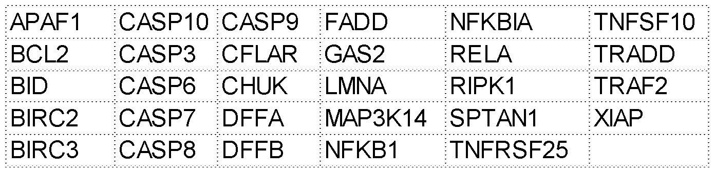


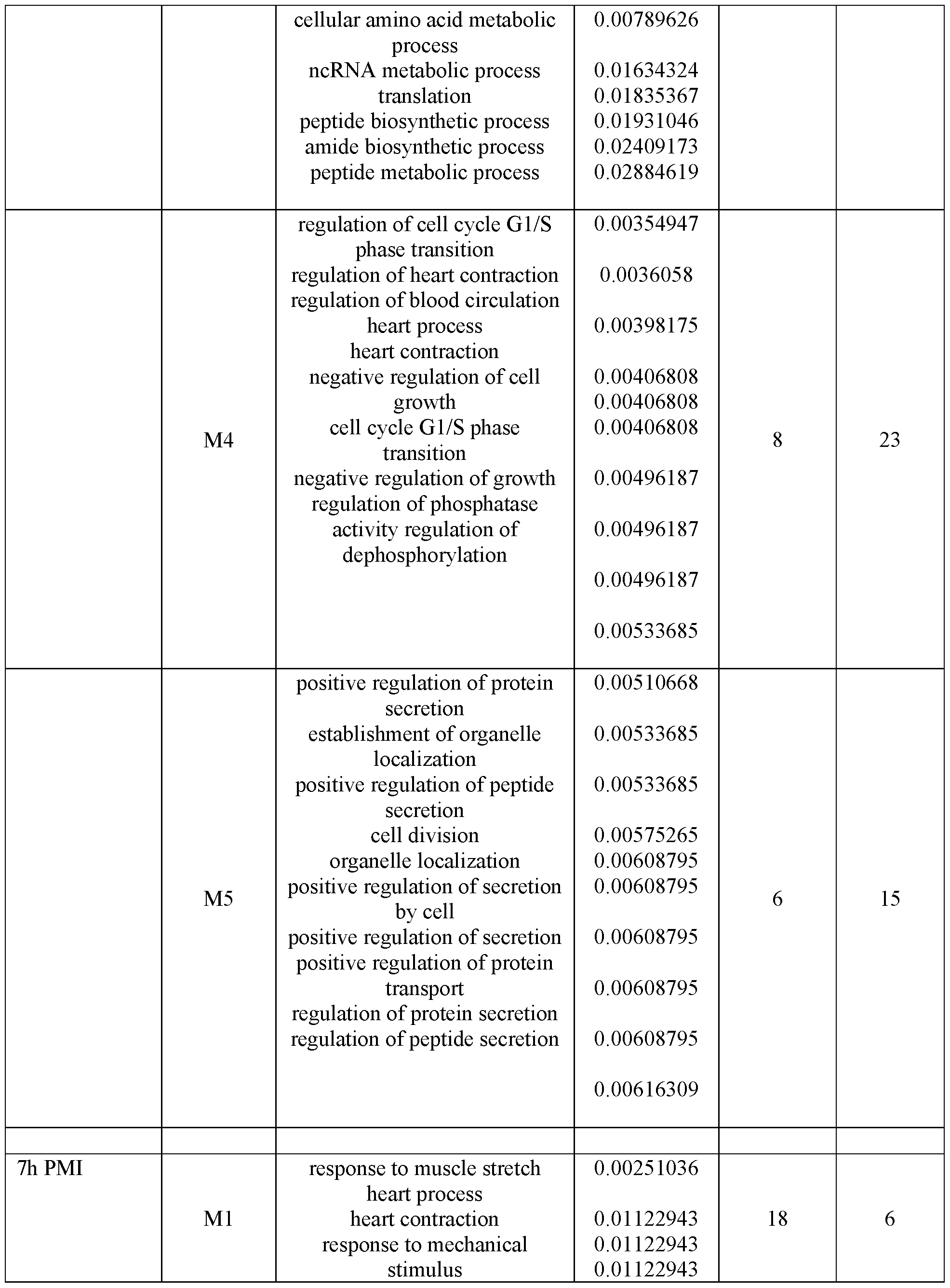


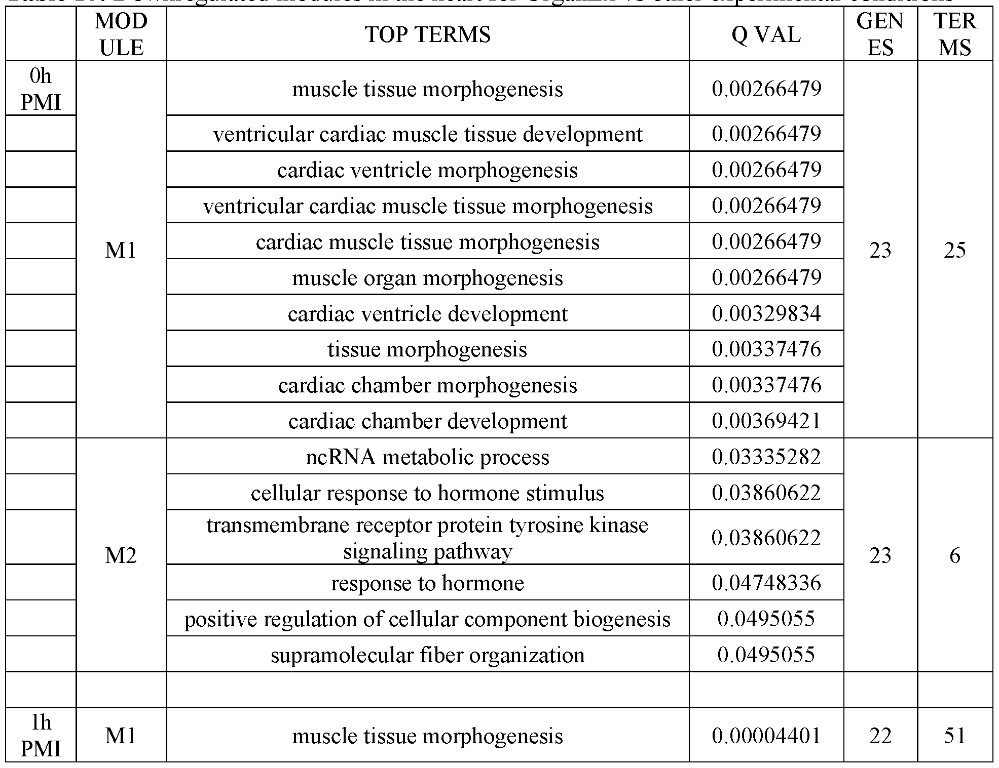


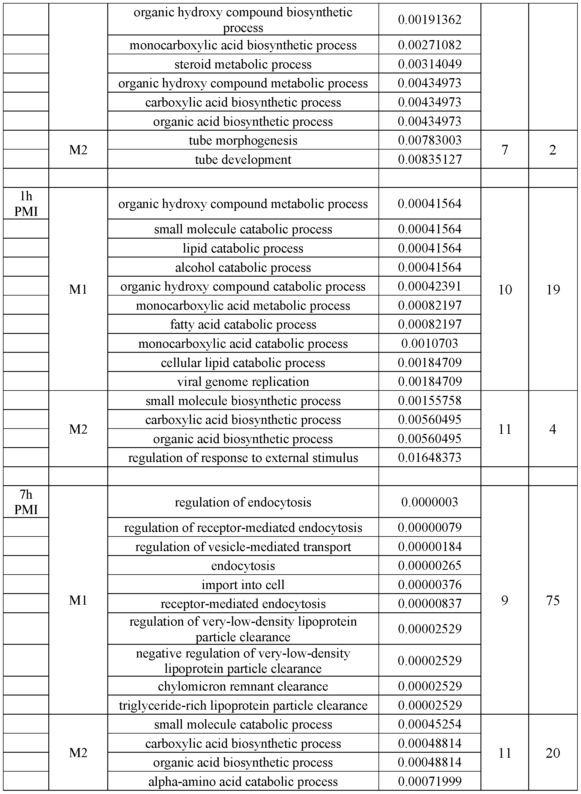

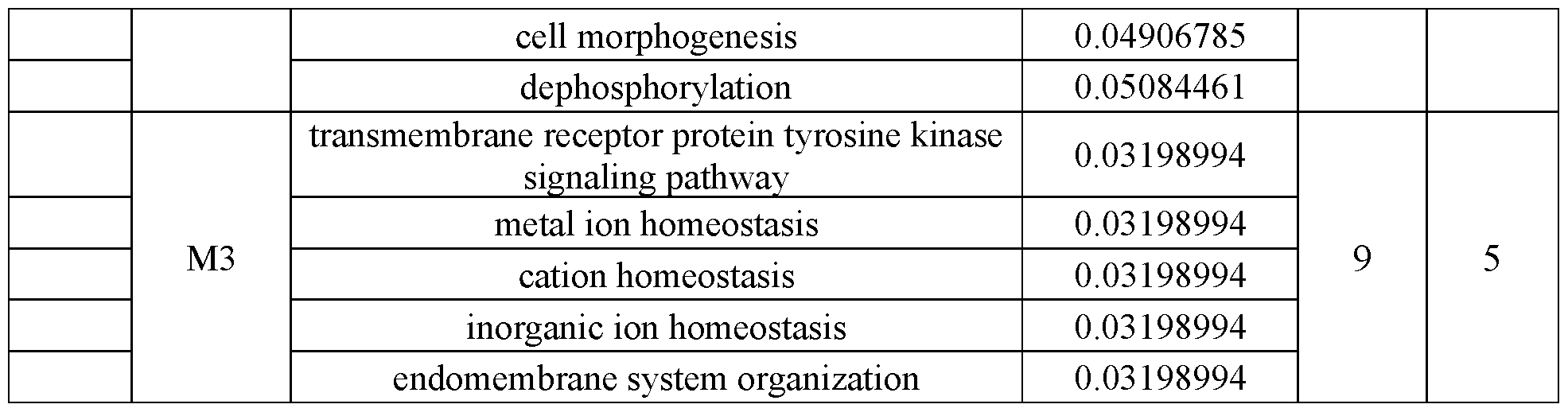


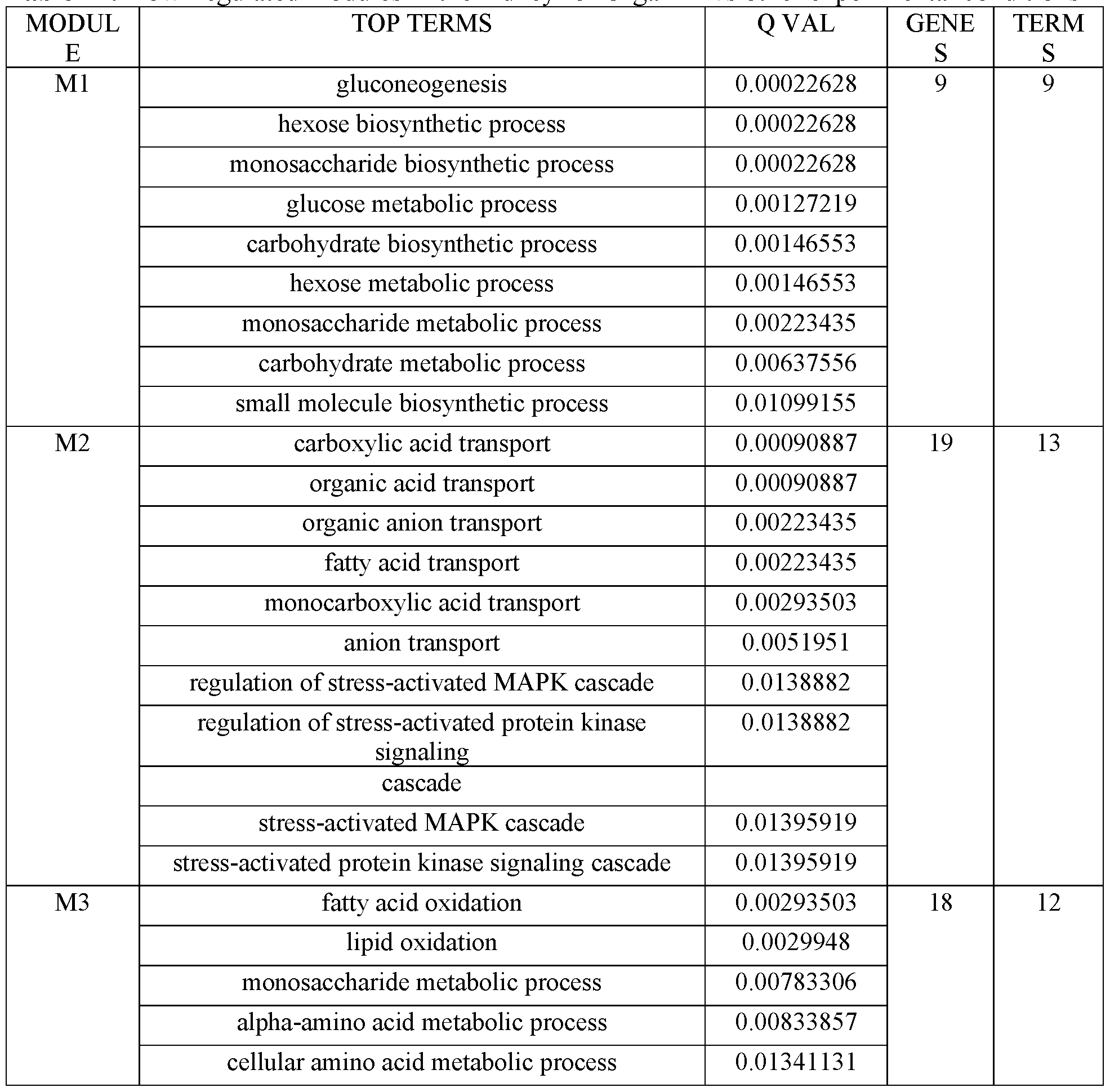


Claims
Priority Applications (2)
| Application Number | Priority Date | Filing Date | Title |
|---|---|---|---|
| CA3231611A CA3231611A1 (en) | 2021-09-17 | 2022-09-16 | Methods, systems and compositions for restoration and preservation of intact organs in a mammal |
| AU2022345865A AU2022345865A1 (en) | 2021-09-17 | 2022-09-16 | Methods, systems and compositions for restoration and preservation of intact organs in a mammal |
Applications Claiming Priority (2)
| Application Number | Priority Date | Filing Date | Title |
|---|---|---|---|
| US202163245632P | 2021-09-17 | 2021-09-17 | |
| US63/245,632 | 2021-09-17 |
Publications (1)
| Publication Number | Publication Date |
|---|---|
| WO2023044014A1 true WO2023044014A1 (en) | 2023-03-23 |
Family
ID=85603531
Family Applications (1)
| Application Number | Title | Priority Date | Filing Date |
|---|---|---|---|
| PCT/US2022/043816 WO2023044014A1 (en) | 2021-09-17 | 2022-09-16 | Methods, systems and compositions for restoration and preservation of intact organs in a mammal |
Country Status (3)
| Country | Link |
|---|---|
| AU (1) | AU2022345865A1 (en) |
| CA (1) | CA3231611A1 (en) |
| WO (1) | WO2023044014A1 (en) |
Citations (7)
| Publication number | Priority date | Publication date | Assignee | Title |
|---|---|---|---|---|
| US6699231B1 (en) * | 1997-12-31 | 2004-03-02 | Heartport, Inc. | Methods and apparatus for perfusion of isolated tissue structure |
| US20040259769A1 (en) * | 1999-11-12 | 2004-12-23 | Baxter International Inc. | Reduced side-effect hemoglobin compositions |
| US20050272148A1 (en) * | 2000-12-06 | 2005-12-08 | Hariri Robert J | Method of collecting placental stem cells |
| WO2011149602A1 (en) * | 2010-05-27 | 2011-12-01 | Bing Lou Wong | High-temperature stable oxygen-carrier-containing pharmaceutical composition |
| US20120238998A1 (en) * | 2005-12-22 | 2012-09-20 | Csl Behring Gmbh | Octanoate-reduced Human Albumin |
| US20150342177A1 (en) * | 2014-06-02 | 2015-12-03 | Transmedics, Inc | Ex vivo organ care system |
| US20170007616A1 (en) * | 2014-01-27 | 2017-01-12 | Susanne GARDNER | Antimicrobial formulations and applications thereof |
-
2022
- 2022-09-16 AU AU2022345865A patent/AU2022345865A1/en active Pending
- 2022-09-16 WO PCT/US2022/043816 patent/WO2023044014A1/en active Application Filing
- 2022-09-16 CA CA3231611A patent/CA3231611A1/en active Pending
Patent Citations (7)
| Publication number | Priority date | Publication date | Assignee | Title |
|---|---|---|---|---|
| US6699231B1 (en) * | 1997-12-31 | 2004-03-02 | Heartport, Inc. | Methods and apparatus for perfusion of isolated tissue structure |
| US20040259769A1 (en) * | 1999-11-12 | 2004-12-23 | Baxter International Inc. | Reduced side-effect hemoglobin compositions |
| US20050272148A1 (en) * | 2000-12-06 | 2005-12-08 | Hariri Robert J | Method of collecting placental stem cells |
| US20120238998A1 (en) * | 2005-12-22 | 2012-09-20 | Csl Behring Gmbh | Octanoate-reduced Human Albumin |
| WO2011149602A1 (en) * | 2010-05-27 | 2011-12-01 | Bing Lou Wong | High-temperature stable oxygen-carrier-containing pharmaceutical composition |
| US20170007616A1 (en) * | 2014-01-27 | 2017-01-12 | Susanne GARDNER | Antimicrobial formulations and applications thereof |
| US20150342177A1 (en) * | 2014-06-02 | 2015-12-03 | Transmedics, Inc | Ex vivo organ care system |
Also Published As
| Publication number | Publication date |
|---|---|
| CA3231611A1 (en) | 2023-03-23 |
| AU2022345865A1 (en) | 2024-03-14 |
Similar Documents
| Publication | Publication Date | Title |
|---|---|---|
| Andrijevic et al. | Cellular recovery after prolonged warm ischaemia of the whole body | |
| Liu et al. | Gene therapy knockdown of Hippo signaling induces cardiomyocyte renewal in pigs after myocardial infarction | |
| Kitahara et al. | Heterotopic transplantation of a decellularized and recellularized whole porcine heart | |
| JP6636956B2 (en) | Human liver scaffold | |
| Bishawi et al. | A normothermic ex vivo organ perfusion delivery method for cardiac transplantation gene therapy | |
| Haam et al. | The effects of hydrogen gas inhalation during ex vivo lung perfusion on donor lungs obtained after cardiac death | |
| Seki et al. | Adipose tissue-derived mesenchymal stem cell transplantation promotes hepatic regeneration after hepatic ischemia-reperfusion and subsequent hepatectomy in rats | |
| JP2018148909A (en) | Isolated renal cells and use thereof | |
| US20100093066A1 (en) | Decellularization and recellularization apparatuses and systems containing the same | |
| EP2893806B1 (en) | Method for maintaining organ or tissue for transplantation use for long period | |
| Diaz et al. | Injectable myocardial matrix hydrogel mitigates negative left ventricular remodeling in a chronic myocardial infarction model | |
| CA2993955A1 (en) | Materials and methods for treating and evaluating ischemic and/or reperfusion-injured tissue and/or tissue susceptible to same | |
| Kim et al. | Recipient-site preconditioning with deferoxamine increases fat graft survival by inducing VEGF and neovascularization in a rat model | |
| Chen et al. | Bone marrow derived mesenchymal stromal cells ameliorate ischemia/reperfusion injury-induced acute kidney injury in rats via secreting tumor necrosis factor-inducible gene 6 protein | |
| Kundu et al. | Transplantation of Apoptosis‐Resistant Endothelial Progenitor Cells Improves Renal Function in Diabetic Kidney Disease | |
| WO2023044014A1 (en) | Methods, systems and compositions for restoration and preservation of intact organs in a mammal | |
| Chen et al. | Nonvirally modified autologous primary hepatocytes correct diabetes and prevent target organ injury in a large preclinical model | |
| Smolenski et al. | Reduction of hyperacute rejection and protection of metabolism and function in hearts of human decay accelerating factor (hDAF)-expressing pigs | |
| Vazquez-Zapien et al. | Biochemical and histological effects of embryonic stem cells in a mouse model of renal failure | |
| Moor et al. | Comparative analysis of kidney transplantation modeled using precision-cut kidney slices and kidney transplantation in pigs | |
| Otani et al. | Early effects of the ex vivo evaluation system on graft function after swine lung transplantation | |
| Laguens et al. | Cardiac regeneration: the gene therapy approach | |
| Kusmic et al. | Silencing Survivin: a Key Therapeutic strategy for cardiac hypertrophy | |
| Wang et al. | Therapeutic evaluation of a microbioartificial liver with recombinant HepG2 cells for rats with hepatic failure | |
| Sperry et al. | Identification of pharmacological inducers of a reversible hypometabolic state for whole organ preservation |
Legal Events
| Date | Code | Title | Description |
|---|---|---|---|
| 121 | Ep: the epo has been informed by wipo that ep was designated in this application |
Ref document number: 22870751 Country of ref document: EP Kind code of ref document: A1 |
|
| WWE | Wipo information: entry into national phase |
Ref document number: 2022345865 Country of ref document: AU Ref document number: AU2022345865 Country of ref document: AU |
|
| WWE | Wipo information: entry into national phase |
Ref document number: 3231611 Country of ref document: CA |
|
| ENP | Entry into the national phase |
Ref document number: 2022345865 Country of ref document: AU Date of ref document: 20220916 Kind code of ref document: A |
|
| WWE | Wipo information: entry into national phase |
Ref document number: 2022870751 Country of ref document: EP |
|
| NENP | Non-entry into the national phase |
Ref country code: DE |
|
| ENP | Entry into the national phase |
Ref document number: 2022870751 Country of ref document: EP Effective date: 20240417 |