WO2023013764A1 - Method for estimating fibrosis progression and/or liver disease activity in non-alcoholic steatohepatitis - Google Patents
Method for estimating fibrosis progression and/or liver disease activity in non-alcoholic steatohepatitis Download PDFInfo
- Publication number
- WO2023013764A1 WO2023013764A1 PCT/JP2022/030093 JP2022030093W WO2023013764A1 WO 2023013764 A1 WO2023013764 A1 WO 2023013764A1 JP 2022030093 W JP2022030093 W JP 2022030093W WO 2023013764 A1 WO2023013764 A1 WO 2023013764A1
- Authority
- WO
- WIPO (PCT)
- Prior art keywords
- igfals
- reference value
- fibrosis
- antibody
- blood
- Prior art date
Links
- 208000008338 non-alcoholic fatty liver disease Diseases 0.000 title claims abstract description 159
- 238000000034 method Methods 0.000 title claims abstract description 120
- 206010016654 Fibrosis Diseases 0.000 title claims abstract description 115
- 230000004761 fibrosis Effects 0.000 title claims abstract description 113
- 206010053219 non-alcoholic steatohepatitis Diseases 0.000 title claims abstract description 96
- 208000019423 liver disease Diseases 0.000 title claims description 7
- 230000009266 disease activity Effects 0.000 title 1
- 101000693844 Homo sapiens Insulin-like growth factor-binding protein complex acid labile subunit Proteins 0.000 claims abstract description 288
- 102100025515 Insulin-like growth factor-binding protein complex acid labile subunit Human genes 0.000 claims abstract description 288
- 210000004369 blood Anatomy 0.000 claims abstract description 166
- 239000008280 blood Substances 0.000 claims abstract description 166
- 208000019425 cirrhosis of liver Diseases 0.000 claims abstract description 86
- 230000000694 effects Effects 0.000 claims abstract description 7
- 108090000623 proteins and genes Proteins 0.000 claims description 72
- 102000004169 proteins and genes Human genes 0.000 claims description 70
- 239000000126 substance Substances 0.000 claims description 65
- 238000012360 testing method Methods 0.000 claims description 54
- 239000011230 binding agent Substances 0.000 claims description 26
- 239000000090 biomarker Substances 0.000 claims description 14
- 230000035945 sensitivity Effects 0.000 claims description 13
- 239000002699 waste material Substances 0.000 claims description 12
- 102000028416 insulin-like growth factor binding Human genes 0.000 claims description 8
- 108091022911 insulin-like growth factor binding Proteins 0.000 claims description 8
- 239000000203 mixture Substances 0.000 abstract description 14
- 239000000523 sample Substances 0.000 description 56
- 101000599951 Homo sapiens Insulin-like growth factor I Proteins 0.000 description 55
- 102100037852 Insulin-like growth factor I Human genes 0.000 description 53
- 208000007082 Alcoholic Fatty Liver Diseases 0.000 description 27
- 208000026594 alcoholic fatty liver disease Diseases 0.000 description 27
- 108010047041 Complementarity Determining Regions Proteins 0.000 description 24
- 210000002966 serum Anatomy 0.000 description 24
- 238000004519 manufacturing process Methods 0.000 description 22
- 102000004371 Insulin-like growth factor binding protein 5 Human genes 0.000 description 20
- 108090000961 Insulin-like growth factor binding protein 5 Proteins 0.000 description 20
- 102000004374 Insulin-like growth factor binding protein 3 Human genes 0.000 description 19
- 108090000965 Insulin-like growth factor binding protein 3 Proteins 0.000 description 19
- 102000007474 Multiprotein Complexes Human genes 0.000 description 17
- 108010085220 Multiprotein Complexes Proteins 0.000 description 17
- 208000006454 hepatitis Diseases 0.000 description 15
- 238000012317 liver biopsy Methods 0.000 description 15
- 239000000758 substrate Substances 0.000 description 15
- LFQSCWFLJHTTHZ-UHFFFAOYSA-N Ethanol Chemical compound CCO LFQSCWFLJHTTHZ-UHFFFAOYSA-N 0.000 description 14
- 238000001514 detection method Methods 0.000 description 14
- 231100000283 hepatitis Toxicity 0.000 description 14
- 238000005259 measurement Methods 0.000 description 14
- 210000004185 liver Anatomy 0.000 description 12
- 201000010099 disease Diseases 0.000 description 11
- 208000037265 diseases, disorders, signs and symptoms Diseases 0.000 description 11
- 210000003494 hepatocyte Anatomy 0.000 description 11
- 238000004949 mass spectrometry Methods 0.000 description 11
- MHAJPDPJQMAIIY-UHFFFAOYSA-N Hydrogen peroxide Chemical compound OO MHAJPDPJQMAIIY-UHFFFAOYSA-N 0.000 description 10
- 102000004190 Enzymes Human genes 0.000 description 8
- 108090000790 Enzymes Proteins 0.000 description 8
- 238000003745 diagnosis Methods 0.000 description 8
- 229940088598 enzyme Drugs 0.000 description 8
- 239000003550 marker Substances 0.000 description 8
- 239000000047 product Substances 0.000 description 8
- 239000007787 solid Substances 0.000 description 8
- 238000005406 washing Methods 0.000 description 8
- 238000002965 ELISA Methods 0.000 description 7
- 210000002381 plasma Anatomy 0.000 description 7
- 108010001336 Horseradish Peroxidase Proteins 0.000 description 6
- 230000007423 decrease Effects 0.000 description 6
- 239000007790 solid phase Substances 0.000 description 6
- 206010019668 Hepatic fibrosis Diseases 0.000 description 5
- 102000003992 Peroxidases Human genes 0.000 description 5
- 238000004458 analytical method Methods 0.000 description 5
- 238000003556 assay Methods 0.000 description 5
- 238000003317 immunochromatography Methods 0.000 description 5
- 238000002372 labelling Methods 0.000 description 5
- 108040007629 peroxidase activity proteins Proteins 0.000 description 5
- 238000011160 research Methods 0.000 description 5
- 208000004930 Fatty Liver Diseases 0.000 description 4
- 206010019799 Hepatitis viral Diseases 0.000 description 4
- 241000282412 Homo Species 0.000 description 4
- 150000001413 amino acids Chemical group 0.000 description 4
- 239000011324 bead Substances 0.000 description 4
- 230000036765 blood level Effects 0.000 description 4
- 210000004027 cell Anatomy 0.000 description 4
- 239000003593 chromogenic compound Substances 0.000 description 4
- 238000007796 conventional method Methods 0.000 description 4
- 238000011161 development Methods 0.000 description 4
- 230000018109 developmental process Effects 0.000 description 4
- 239000003814 drug Substances 0.000 description 4
- 210000005228 liver tissue Anatomy 0.000 description 4
- 201000001862 viral hepatitis Diseases 0.000 description 4
- NFGXHKASABOEEW-UHFFFAOYSA-N 1-methylethyl 11-methoxy-3,7,11-trimethyl-2,4-dodecadienoate Chemical compound COC(C)(C)CCCC(C)CC=CC(C)=CC(=O)OC(C)C NFGXHKASABOEEW-UHFFFAOYSA-N 0.000 description 3
- 125000001917 2,4-dinitrophenyl group Chemical group [H]C1=C([H])C(=C([H])C(=C1*)[N+]([O-])=O)[N+]([O-])=O 0.000 description 3
- 102000002260 Alkaline Phosphatase Human genes 0.000 description 3
- 108020004774 Alkaline Phosphatase Proteins 0.000 description 3
- 108090001008 Avidin Proteins 0.000 description 3
- 102000014914 Carrier Proteins Human genes 0.000 description 3
- 102000004266 Collagen Type IV Human genes 0.000 description 3
- 108010042086 Collagen Type IV Proteins 0.000 description 3
- 238000004252 FT/ICR mass spectrometry Methods 0.000 description 3
- 108010015776 Glucose oxidase Proteins 0.000 description 3
- 239000004366 Glucose oxidase Substances 0.000 description 3
- 206010019708 Hepatic steatosis Diseases 0.000 description 3
- 101100001515 Homo sapiens IGFALS gene Proteins 0.000 description 3
- 102000004856 Lectins Human genes 0.000 description 3
- 108090001090 Lectins Proteins 0.000 description 3
- 241001465754 Metazoa Species 0.000 description 3
- 241000700605 Viruses Species 0.000 description 3
- 238000010521 absorption reaction Methods 0.000 description 3
- 108700031341 acid labile subunit insulin-like growth factor binding Proteins 0.000 description 3
- 210000000628 antibody-producing cell Anatomy 0.000 description 3
- 239000000427 antigen Substances 0.000 description 3
- 102000036639 antigens Human genes 0.000 description 3
- 108091007433 antigens Proteins 0.000 description 3
- 108091008324 binding proteins Proteins 0.000 description 3
- 239000012472 biological sample Substances 0.000 description 3
- 210000001124 body fluid Anatomy 0.000 description 3
- 239000010839 body fluid Substances 0.000 description 3
- 238000012767 chemiluminescent enzyme immunoassay Methods 0.000 description 3
- 210000004978 chinese hamster ovary cell Anatomy 0.000 description 3
- 210000002808 connective tissue Anatomy 0.000 description 3
- 230000006378 damage Effects 0.000 description 3
- 229940079593 drug Drugs 0.000 description 3
- 238000011156 evaluation Methods 0.000 description 3
- 208000010706 fatty liver disease Diseases 0.000 description 3
- 229940116332 glucose oxidase Drugs 0.000 description 3
- 235000019420 glucose oxidase Nutrition 0.000 description 3
- 230000002440 hepatic effect Effects 0.000 description 3
- 230000008595 infiltration Effects 0.000 description 3
- 238000001764 infiltration Methods 0.000 description 3
- 239000002523 lectin Substances 0.000 description 3
- 230000003902 lesion Effects 0.000 description 3
- 239000012528 membrane Substances 0.000 description 3
- 230000002503 metabolic effect Effects 0.000 description 3
- 230000001575 pathological effect Effects 0.000 description 3
- 238000007619 statistical method Methods 0.000 description 3
- 238000011282 treatment Methods 0.000 description 3
- 238000001262 western blot Methods 0.000 description 3
- YBJHBAHKTGYVGT-ZKWXMUAHSA-N (+)-Biotin Chemical compound N1C(=O)N[C@@H]2[C@H](CCCCC(=O)O)SC[C@@H]21 YBJHBAHKTGYVGT-ZKWXMUAHSA-N 0.000 description 2
- KIUKXJAPPMFGSW-DNGZLQJQSA-N (2S,3S,4S,5R,6R)-6-[(2S,3R,4R,5S,6R)-3-Acetamido-2-[(2S,3S,4R,5R,6R)-6-[(2R,3R,4R,5S,6R)-3-acetamido-2,5-dihydroxy-6-(hydroxymethyl)oxan-4-yl]oxy-2-carboxy-4,5-dihydroxyoxan-3-yl]oxy-5-hydroxy-6-(hydroxymethyl)oxan-4-yl]oxy-3,4,5-trihydroxyoxane-2-carboxylic acid Chemical compound CC(=O)N[C@H]1[C@H](O)O[C@H](CO)[C@@H](O)[C@@H]1O[C@H]1[C@H](O)[C@@H](O)[C@H](O[C@H]2[C@@H]([C@@H](O[C@H]3[C@@H]([C@@H](O)[C@H](O)[C@H](O3)C(O)=O)O)[C@H](O)[C@@H](CO)O2)NC(C)=O)[C@@H](C(O)=O)O1 KIUKXJAPPMFGSW-DNGZLQJQSA-N 0.000 description 2
- HSINOMROUCMIEA-FGVHQWLLSA-N (2s,4r)-4-[(3r,5s,6r,7r,8s,9s,10s,13r,14s,17r)-6-ethyl-3,7-dihydroxy-10,13-dimethyl-2,3,4,5,6,7,8,9,11,12,14,15,16,17-tetradecahydro-1h-cyclopenta[a]phenanthren-17-yl]-2-methylpentanoic acid Chemical compound C([C@@]12C)C[C@@H](O)C[C@H]1[C@@H](CC)[C@@H](O)[C@@H]1[C@@H]2CC[C@]2(C)[C@@H]([C@H](C)C[C@H](C)C(O)=O)CC[C@H]21 HSINOMROUCMIEA-FGVHQWLLSA-N 0.000 description 2
- BCHIXGBGRHLSBE-UHFFFAOYSA-N (4-methyl-2-oxochromen-7-yl) dihydrogen phosphate Chemical compound C1=C(OP(O)(O)=O)C=CC2=C1OC(=O)C=C2C BCHIXGBGRHLSBE-UHFFFAOYSA-N 0.000 description 2
- GEYOCULIXLDCMW-UHFFFAOYSA-N 1,2-phenylenediamine Chemical compound NC1=CC=CC=C1N GEYOCULIXLDCMW-UHFFFAOYSA-N 0.000 description 2
- UAIUNKRWKOVEES-UHFFFAOYSA-N 3,3',5,5'-tetramethylbenzidine Chemical compound CC1=C(N)C(C)=CC(C=2C=C(C)C(N)=C(C)C=2)=C1 UAIUNKRWKOVEES-UHFFFAOYSA-N 0.000 description 2
- 206010003827 Autoimmune hepatitis Diseases 0.000 description 2
- 102000008186 Collagen Human genes 0.000 description 2
- 108010035532 Collagen Proteins 0.000 description 2
- RGHNJXZEOKUKBD-SQOUGZDYSA-N D-gluconic acid Chemical compound OC[C@@H](O)[C@@H](O)[C@H](O)[C@@H](O)C(O)=O RGHNJXZEOKUKBD-SQOUGZDYSA-N 0.000 description 2
- 108090000288 Glycoproteins Proteins 0.000 description 2
- 102000003886 Glycoproteins Human genes 0.000 description 2
- 101001044927 Homo sapiens Insulin-like growth factor-binding protein 3 Proteins 0.000 description 2
- 101000840566 Homo sapiens Insulin-like growth factor-binding protein 5 Proteins 0.000 description 2
- 108090000723 Insulin-Like Growth Factor I Proteins 0.000 description 2
- 102100022708 Insulin-like growth factor-binding protein 3 Human genes 0.000 description 2
- 102100029225 Insulin-like growth factor-binding protein 5 Human genes 0.000 description 2
- 206010067125 Liver injury Diseases 0.000 description 2
- 206010028980 Neoplasm Diseases 0.000 description 2
- NBIIXXVUZAFLBC-UHFFFAOYSA-N Phosphoric acid Chemical compound OP(O)(O)=O NBIIXXVUZAFLBC-UHFFFAOYSA-N 0.000 description 2
- 241000700159 Rattus Species 0.000 description 2
- 102000013275 Somatomedins Human genes 0.000 description 2
- QAOWNCQODCNURD-UHFFFAOYSA-N Sulfuric acid Chemical compound OS(O)(=O)=O QAOWNCQODCNURD-UHFFFAOYSA-N 0.000 description 2
- NKANXQFJJICGDU-QPLCGJKRSA-N Tamoxifen Chemical compound C=1C=CC=CC=1C(/CC)=C(C=1C=CC(OCCN(C)C)=CC=1)/C1=CC=CC=C1 NKANXQFJJICGDU-QPLCGJKRSA-N 0.000 description 2
- NIJJYAXOARWZEE-UHFFFAOYSA-N Valproic acid Chemical compound CCCC(C(O)=O)CCC NIJJYAXOARWZEE-UHFFFAOYSA-N 0.000 description 2
- 239000005557 antagonist Substances 0.000 description 2
- 239000003146 anticoagulant agent Substances 0.000 description 2
- 229940127219 anticoagulant drug Drugs 0.000 description 2
- 238000001854 atmospheric pressure photoionisation mass spectrometry Methods 0.000 description 2
- 210000004899 c-terminal region Anatomy 0.000 description 2
- 201000011510 cancer Diseases 0.000 description 2
- 239000001913 cellulose Substances 0.000 description 2
- 229920002678 cellulose Polymers 0.000 description 2
- 239000003795 chemical substances by application Substances 0.000 description 2
- HVYWMOMLDIMFJA-DPAQBDIFSA-N cholesterol Chemical compound C1C=C2C[C@@H](O)CC[C@]2(C)[C@@H]2[C@@H]1[C@@H]1CC[C@H]([C@H](C)CCCC(C)C)[C@@]1(C)CC2 HVYWMOMLDIMFJA-DPAQBDIFSA-N 0.000 description 2
- 230000007882 cirrhosis Effects 0.000 description 2
- 229920001436 collagen Polymers 0.000 description 2
- 230000002860 competitive effect Effects 0.000 description 2
- 238000010586 diagram Methods 0.000 description 2
- 230000035622 drinking Effects 0.000 description 2
- 238000000132 electrospray ionisation Methods 0.000 description 2
- 238000002795 fluorescence method Methods 0.000 description 2
- 229920002674 hyaluronan Polymers 0.000 description 2
- 229960003160 hyaluronic acid Drugs 0.000 description 2
- 210000004969 inflammatory cell Anatomy 0.000 description 2
- NOESYZHRGYRDHS-UHFFFAOYSA-N insulin Chemical compound N1C(=O)C(NC(=O)C(CCC(N)=O)NC(=O)C(CCC(O)=O)NC(=O)C(C(C)C)NC(=O)C(NC(=O)CN)C(C)CC)CSSCC(C(NC(CO)C(=O)NC(CC(C)C)C(=O)NC(CC=2C=CC(O)=CC=2)C(=O)NC(CCC(N)=O)C(=O)NC(CC(C)C)C(=O)NC(CCC(O)=O)C(=O)NC(CC(N)=O)C(=O)NC(CC=2C=CC(O)=CC=2)C(=O)NC(CSSCC(NC(=O)C(C(C)C)NC(=O)C(CC(C)C)NC(=O)C(CC=2C=CC(O)=CC=2)NC(=O)C(CC(C)C)NC(=O)C(C)NC(=O)C(CCC(O)=O)NC(=O)C(C(C)C)NC(=O)C(CC(C)C)NC(=O)C(CC=2NC=NC=2)NC(=O)C(CO)NC(=O)CNC2=O)C(=O)NCC(=O)NC(CCC(O)=O)C(=O)NC(CCCNC(N)=N)C(=O)NCC(=O)NC(CC=3C=CC=CC=3)C(=O)NC(CC=3C=CC=CC=3)C(=O)NC(CC=3C=CC(O)=CC=3)C(=O)NC(C(C)O)C(=O)N3C(CCC3)C(=O)NC(CCCCN)C(=O)NC(C)C(O)=O)C(=O)NC(CC(N)=O)C(O)=O)=O)NC(=O)C(C(C)CC)NC(=O)C(CO)NC(=O)C(C(C)O)NC(=O)C1CSSCC2NC(=O)C(CC(C)C)NC(=O)C(NC(=O)C(CCC(N)=O)NC(=O)C(CC(N)=O)NC(=O)C(NC(=O)C(N)CC=1C=CC=CC=1)C(C)C)CC1=CN=CN1 NOESYZHRGYRDHS-UHFFFAOYSA-N 0.000 description 2
- 201000007270 liver cancer Diseases 0.000 description 2
- 208000014018 liver neoplasm Diseases 0.000 description 2
- 238000012544 monitoring process Methods 0.000 description 2
- 230000017074 necrotic cell death Effects 0.000 description 2
- -1 obeticholic acid) Chemical class 0.000 description 2
- 210000000056 organ Anatomy 0.000 description 2
- 238000004393 prognosis Methods 0.000 description 2
- 230000002250 progressing effect Effects 0.000 description 2
- 230000008439 repair process Effects 0.000 description 2
- 238000005070 sampling Methods 0.000 description 2
- 238000010561 standard procedure Methods 0.000 description 2
- 231100000240 steatosis hepatitis Toxicity 0.000 description 2
- 208000001072 type 2 diabetes mellitus Diseases 0.000 description 2
- OGYGFUAIIOPWQD-UHFFFAOYSA-N 1,3-thiazolidine Chemical compound C1CSCN1 OGYGFUAIIOPWQD-UHFFFAOYSA-N 0.000 description 1
- CVOFKRWYWCSDMA-UHFFFAOYSA-N 2-chloro-n-(2,6-diethylphenyl)-n-(methoxymethyl)acetamide;2,6-dinitro-n,n-dipropyl-4-(trifluoromethyl)aniline Chemical compound CCC1=CC=CC(CC)=C1N(COC)C(=O)CCl.CCCN(CCC)C1=C([N+]([O-])=O)C=C(C(F)(F)F)C=C1[N+]([O-])=O CVOFKRWYWCSDMA-UHFFFAOYSA-N 0.000 description 1
- HQFLTUZKIRYQSP-UHFFFAOYSA-N 3-ethyl-2h-1,3-benzothiazole-6-sulfonic acid Chemical compound OS(=O)(=O)C1=CC=C2N(CC)CSC2=C1 HQFLTUZKIRYQSP-UHFFFAOYSA-N 0.000 description 1
- YRNWIFYIFSBPAU-UHFFFAOYSA-N 4-[4-(dimethylamino)phenyl]-n,n-dimethylaniline Chemical compound C1=CC(N(C)C)=CC=C1C1=CC=C(N(C)C)C=C1 YRNWIFYIFSBPAU-UHFFFAOYSA-N 0.000 description 1
- XZKIHKMTEMTJQX-UHFFFAOYSA-L 4-nitrophenyl phosphate(2-) Chemical compound [O-][N+](=O)C1=CC=C(OP([O-])([O-])=O)C=C1 XZKIHKMTEMTJQX-UHFFFAOYSA-L 0.000 description 1
- YPMOAQISONSSNL-UHFFFAOYSA-N 8-hydroxyoctyl 2-methylprop-2-enoate Chemical compound CC(=C)C(=O)OCCCCCCCCO YPMOAQISONSSNL-UHFFFAOYSA-N 0.000 description 1
- 108010088751 Albumins Proteins 0.000 description 1
- 102000009027 Albumins Human genes 0.000 description 1
- MLDQJTXFUGDVEO-UHFFFAOYSA-N BAY-43-9006 Chemical compound C1=NC(C(=O)NC)=CC(OC=2C=CC(NC(=O)NC=3C=C(C(Cl)=CC=3)C(F)(F)F)=CC=2)=C1 MLDQJTXFUGDVEO-UHFFFAOYSA-N 0.000 description 1
- 241000283690 Bos taurus Species 0.000 description 1
- 102100031151 C-C chemokine receptor type 2 Human genes 0.000 description 1
- 101710149815 C-C chemokine receptor type 2 Proteins 0.000 description 1
- 102100035875 C-C chemokine receptor type 5 Human genes 0.000 description 1
- 101710149870 C-C chemokine receptor type 5 Proteins 0.000 description 1
- 108010074051 C-Reactive Protein Proteins 0.000 description 1
- 241000282836 Camelus dromedarius Species 0.000 description 1
- 241000282472 Canis lupus familiaris Species 0.000 description 1
- 241000283707 Capra Species 0.000 description 1
- 241000699800 Cricetinae Species 0.000 description 1
- RGHNJXZEOKUKBD-UHFFFAOYSA-N D-gluconic acid Natural products OCC(O)C(O)C(O)C(O)C(O)=O RGHNJXZEOKUKBD-UHFFFAOYSA-N 0.000 description 1
- 208000032928 Dyslipidaemia Diseases 0.000 description 1
- 238000008157 ELISA kit Methods 0.000 description 1
- 208000010334 End Stage Liver Disease Diseases 0.000 description 1
- 241000283086 Equidae Species 0.000 description 1
- 102000010834 Extracellular Matrix Proteins Human genes 0.000 description 1
- 108010037362 Extracellular Matrix Proteins Proteins 0.000 description 1
- 241000282326 Felis catus Species 0.000 description 1
- 102100040510 Galectin-3-binding protein Human genes 0.000 description 1
- 101710197901 Galectin-3-binding protein Proteins 0.000 description 1
- WQZGKKKJIJFFOK-GASJEMHNSA-N Glucose Natural products OC[C@H]1OC(O)[C@H](O)[C@@H](O)[C@@H]1O WQZGKKKJIJFFOK-GASJEMHNSA-N 0.000 description 1
- 206010018429 Glucose tolerance impaired Diseases 0.000 description 1
- 108010051696 Growth Hormone Proteins 0.000 description 1
- 101001018196 Homo sapiens Mitogen-activated protein kinase kinase kinase 5 Proteins 0.000 description 1
- 101001098868 Homo sapiens Proprotein convertase subtilisin/kexin type 9 Proteins 0.000 description 1
- 101000836383 Homo sapiens Serpin H1 Proteins 0.000 description 1
- 206010020772 Hypertension Diseases 0.000 description 1
- 206010021067 Hypopituitarism Diseases 0.000 description 1
- 108060003951 Immunoglobulin Proteins 0.000 description 1
- 102000012745 Immunoglobulin Subunits Human genes 0.000 description 1
- 108010079585 Immunoglobulin Subunits Proteins 0.000 description 1
- 102000004877 Insulin Human genes 0.000 description 1
- 108090001061 Insulin Proteins 0.000 description 1
- 206010022489 Insulin Resistance Diseases 0.000 description 1
- 102000004218 Insulin-Like Growth Factor I Human genes 0.000 description 1
- 239000005511 L01XE05 - Sorafenib Substances 0.000 description 1
- 208000017170 Lipid metabolism disease Diseases 0.000 description 1
- 241000124008 Mammalia Species 0.000 description 1
- 102100033127 Mitogen-activated protein kinase kinase kinase 5 Human genes 0.000 description 1
- 108010006519 Molecular Chaperones Proteins 0.000 description 1
- 241000699666 Mus <mouse, genus> Species 0.000 description 1
- 241000699670 Mus sp. Species 0.000 description 1
- 101150010554 NAS4 gene Proteins 0.000 description 1
- 102000007399 Nuclear hormone receptor Human genes 0.000 description 1
- 108020005497 Nuclear hormone receptor Proteins 0.000 description 1
- 208000008589 Obesity Diseases 0.000 description 1
- 108091034117 Oligonucleotide Proteins 0.000 description 1
- 241000283973 Oryctolagus cuniculus Species 0.000 description 1
- 240000007594 Oryza sativa Species 0.000 description 1
- 235000007164 Oryza sativa Nutrition 0.000 description 1
- 241000282579 Pan Species 0.000 description 1
- 241001494479 Pecora Species 0.000 description 1
- 102000003728 Peroxisome Proliferator-Activated Receptors Human genes 0.000 description 1
- 108090000029 Peroxisome Proliferator-Activated Receptors Proteins 0.000 description 1
- 206010036049 Polycystic ovaries Diseases 0.000 description 1
- 208000001280 Prediabetic State Diseases 0.000 description 1
- 241000288906 Primates Species 0.000 description 1
- 102100038955 Proprotein convertase subtilisin/kexin type 9 Human genes 0.000 description 1
- 102000007056 Recombinant Fusion Proteins Human genes 0.000 description 1
- 108010008281 Recombinant Fusion Proteins Proteins 0.000 description 1
- 101100529034 Saccharomyces cerevisiae (strain ATCC 204508 / S288c) RPN6 gene Proteins 0.000 description 1
- 102100027287 Serpin H1 Human genes 0.000 description 1
- 108020004459 Small interfering RNA Proteins 0.000 description 1
- 102100038803 Somatotropin Human genes 0.000 description 1
- 108010090804 Streptavidin Proteins 0.000 description 1
- 238000000692 Student's t-test Methods 0.000 description 1
- 241000282887 Suidae Species 0.000 description 1
- 102000004887 Transforming Growth Factor beta Human genes 0.000 description 1
- 108090001012 Transforming Growth Factor beta Proteins 0.000 description 1
- 101100286583 Xenopus laevis igf3 gene Proteins 0.000 description 1
- BTKMJKKKZATLBU-UHFFFAOYSA-N [2-(1,3-benzothiazol-2-yl)-1,3-benzothiazol-6-yl] dihydrogen phosphate Chemical compound C1=CC=C2SC(C3=NC4=CC=C(C=C4S3)OP(O)(=O)O)=NC2=C1 BTKMJKKKZATLBU-UHFFFAOYSA-N 0.000 description 1
- 230000002159 abnormal effect Effects 0.000 description 1
- 238000002835 absorbance Methods 0.000 description 1
- 238000009825 accumulation Methods 0.000 description 1
- 238000010306 acid treatment Methods 0.000 description 1
- 230000009471 action Effects 0.000 description 1
- 230000004913 activation Effects 0.000 description 1
- 230000032683 aging Effects 0.000 description 1
- 239000000556 agonist Substances 0.000 description 1
- 230000001476 alcoholic effect Effects 0.000 description 1
- 229910000147 aluminium phosphate Inorganic materials 0.000 description 1
- IYIKLHRQXLHMJQ-UHFFFAOYSA-N amiodarone Chemical compound CCCCC=1OC2=CC=CC=C2C=1C(=O)C1=CC(I)=C(OCCN(CC)CC)C(I)=C1 IYIKLHRQXLHMJQ-UHFFFAOYSA-N 0.000 description 1
- 229960005260 amiodarone Drugs 0.000 description 1
- 210000004102 animal cell Anatomy 0.000 description 1
- 238000010171 animal model Methods 0.000 description 1
- 230000002529 anti-mitochondrial effect Effects 0.000 description 1
- 230000003460 anti-nuclear Effects 0.000 description 1
- 230000000692 anti-sense effect Effects 0.000 description 1
- OHDRQQURAXLVGJ-HLVWOLMTSA-N azane;(2e)-3-ethyl-2-[(e)-(3-ethyl-6-sulfo-1,3-benzothiazol-2-ylidene)hydrazinylidene]-1,3-benzothiazole-6-sulfonic acid Chemical compound [NH4+].[NH4+].S/1C2=CC(S([O-])(=O)=O)=CC=C2N(CC)C\1=N/N=C1/SC2=CC(S([O-])(=O)=O)=CC=C2N1CC OHDRQQURAXLVGJ-HLVWOLMTSA-N 0.000 description 1
- 230000009286 beneficial effect Effects 0.000 description 1
- 239000003613 bile acid Substances 0.000 description 1
- 230000033228 biological regulation Effects 0.000 description 1
- 238000001574 biopsy Methods 0.000 description 1
- 229960002685 biotin Drugs 0.000 description 1
- 235000020958 biotin Nutrition 0.000 description 1
- 239000011616 biotin Substances 0.000 description 1
- 210000000601 blood cell Anatomy 0.000 description 1
- 230000036772 blood pressure Effects 0.000 description 1
- 230000015556 catabolic process Effects 0.000 description 1
- 238000005119 centrifugation Methods 0.000 description 1
- ZAIPMKNFIOOWCQ-UEKVPHQBSA-N cephalexin Chemical compound C1([C@@H](N)C(=O)N[C@H]2[C@@H]3N(C2=O)C(=C(CS3)C)C(O)=O)=CC=CC=C1 ZAIPMKNFIOOWCQ-UEKVPHQBSA-N 0.000 description 1
- 238000006243 chemical reaction Methods 0.000 description 1
- 230000001684 chronic effect Effects 0.000 description 1
- 208000011444 chronic liver failure Diseases 0.000 description 1
- 238000010367 cloning Methods 0.000 description 1
- 230000009918 complex formation Effects 0.000 description 1
- 238000012790 confirmation Methods 0.000 description 1
- 239000000356 contaminant Substances 0.000 description 1
- 238000000354 decomposition reaction Methods 0.000 description 1
- 230000003247 decreasing effect Effects 0.000 description 1
- 230000007850 degeneration Effects 0.000 description 1
- 230000001419 dependent effect Effects 0.000 description 1
- 230000008021 deposition Effects 0.000 description 1
- 238000013461 design Methods 0.000 description 1
- 206010012601 diabetes mellitus Diseases 0.000 description 1
- 238000002405 diagnostic procedure Methods 0.000 description 1
- 230000037213 diet Effects 0.000 description 1
- 235000005911 diet Nutrition 0.000 description 1
- 239000000975 dye Substances 0.000 description 1
- 238000001962 electrophoresis Methods 0.000 description 1
- 230000002124 endocrine Effects 0.000 description 1
- 210000000750 endocrine system Anatomy 0.000 description 1
- 230000007717 exclusion Effects 0.000 description 1
- 210000002744 extracellular matrix Anatomy 0.000 description 1
- 239000007850 fluorescent dye Substances 0.000 description 1
- 239000012634 fragment Substances 0.000 description 1
- 238000001641 gel filtration chromatography Methods 0.000 description 1
- 238000002523 gelfiltration Methods 0.000 description 1
- 239000000174 gluconic acid Substances 0.000 description 1
- 235000012208 gluconic acid Nutrition 0.000 description 1
- 239000008103 glucose Substances 0.000 description 1
- 230000013595 glycosylation Effects 0.000 description 1
- 230000005484 gravity Effects 0.000 description 1
- 230000012010 growth Effects 0.000 description 1
- 239000003102 growth factor Substances 0.000 description 1
- 239000000122 growth hormone Substances 0.000 description 1
- 231100000234 hepatic damage Toxicity 0.000 description 1
- 231100000753 hepatic injury Toxicity 0.000 description 1
- 239000005556 hormone Substances 0.000 description 1
- 229940088597 hormone Drugs 0.000 description 1
- 102000044162 human IGF1 Human genes 0.000 description 1
- 230000002989 hypothyroidism Effects 0.000 description 1
- 208000003532 hypothyroidism Diseases 0.000 description 1
- 230000001900 immune effect Effects 0.000 description 1
- 230000003053 immunization Effects 0.000 description 1
- 230000002163 immunogen Effects 0.000 description 1
- 102000018358 immunoglobulin Human genes 0.000 description 1
- 238000000338 in vitro Methods 0.000 description 1
- 230000001939 inductive effect Effects 0.000 description 1
- 230000028709 inflammatory response Effects 0.000 description 1
- 239000003112 inhibitor Substances 0.000 description 1
- 229940125396 insulin Drugs 0.000 description 1
- 229940043355 kinase inhibitor Drugs 0.000 description 1
- 210000000265 leukocyte Anatomy 0.000 description 1
- 239000007788 liquid Substances 0.000 description 1
- 238000011528 liquid biopsy Methods 0.000 description 1
- 238000004811 liquid chromatography Methods 0.000 description 1
- 230000008818 liver damage Effects 0.000 description 1
- 230000003908 liver function Effects 0.000 description 1
- 208000018191 liver inflammation Diseases 0.000 description 1
- 230000004807 localization Effects 0.000 description 1
- 239000000463 material Substances 0.000 description 1
- 238000000816 matrix-assisted laser desorption--ionisation Methods 0.000 description 1
- 238000001840 matrix-assisted laser desorption--ionisation time-of-flight mass spectrometry Methods 0.000 description 1
- 238000002483 medication Methods 0.000 description 1
- 108020004999 messenger RNA Proteins 0.000 description 1
- 238000010197 meta-analysis Methods 0.000 description 1
- 230000004066 metabolic change Effects 0.000 description 1
- 230000008558 metabolic pathway by substance Effects 0.000 description 1
- 230000004060 metabolic process Effects 0.000 description 1
- 230000035772 mutation Effects 0.000 description 1
- 108010087904 neutravidin Proteins 0.000 description 1
- 108020004017 nuclear receptors Proteins 0.000 description 1
- 235000016709 nutrition Nutrition 0.000 description 1
- 235000020824 obesity Nutrition 0.000 description 1
- ZXERDUOLZKYMJM-ZWECCWDJSA-N obeticholic acid Chemical compound C([C@@]12C)C[C@@H](O)C[C@H]1[C@@H](CC)[C@@H](O)[C@@H]1[C@@H]2CC[C@]2(C)[C@@H]([C@H](C)CCC(O)=O)CC[C@H]21 ZXERDUOLZKYMJM-ZWECCWDJSA-N 0.000 description 1
- 229960001601 obeticholic acid Drugs 0.000 description 1
- 238000001543 one-way ANOVA Methods 0.000 description 1
- 210000001672 ovary Anatomy 0.000 description 1
- 235000016236 parenteral nutrition Nutrition 0.000 description 1
- 239000003757 phosphotransferase inhibitor Substances 0.000 description 1
- 201000010065 polycystic ovary syndrome Diseases 0.000 description 1
- 206010036067 polydipsia Diseases 0.000 description 1
- 201000009104 prediabetes syndrome Diseases 0.000 description 1
- 108090000765 processed proteins & peptides Proteins 0.000 description 1
- 238000000575 proteomic method Methods 0.000 description 1
- 238000011002 quantification Methods 0.000 description 1
- 239000000018 receptor agonist Substances 0.000 description 1
- 229940044601 receptor agonist Drugs 0.000 description 1
- 230000009467 reduction Effects 0.000 description 1
- 238000012552 review Methods 0.000 description 1
- 235000009566 rice Nutrition 0.000 description 1
- 238000001004 secondary ion mass spectrometry Methods 0.000 description 1
- 239000013049 sediment Substances 0.000 description 1
- 230000001235 sensitizing effect Effects 0.000 description 1
- 201000002859 sleep apnea Diseases 0.000 description 1
- 229960003787 sorafenib Drugs 0.000 description 1
- 210000004500 stellate cell Anatomy 0.000 description 1
- 230000000638 stimulation Effects 0.000 description 1
- 239000006228 supernatant Substances 0.000 description 1
- 238000000672 surface-enhanced laser desorption--ionisation Methods 0.000 description 1
- 238000001356 surgical procedure Methods 0.000 description 1
- 230000008961 swelling Effects 0.000 description 1
- 229960001603 tamoxifen Drugs 0.000 description 1
- ZRKFYGHZFMAOKI-QMGMOQQFSA-N tgfbeta Chemical compound C([C@H](NC(=O)[C@H](C(C)C)NC(=O)CNC(=O)[C@H](CCC(O)=O)NC(=O)[C@H](CCCNC(N)=N)NC(=O)[C@H](CC(N)=O)NC(=O)[C@H](CC(C)C)NC(=O)[C@H]([C@@H](C)O)NC(=O)[C@H](CCC(O)=O)NC(=O)[C@H]([C@@H](C)O)NC(=O)[C@H](CC(C)C)NC(=O)CNC(=O)[C@H](C)NC(=O)[C@H](CO)NC(=O)[C@H](CCC(N)=O)NC(=O)[C@@H](NC(=O)[C@H](C)NC(=O)[C@H](C)NC(=O)[C@@H](NC(=O)[C@H](CC(C)C)NC(=O)[C@@H](N)CCSC)C(C)C)[C@@H](C)CC)C(=O)N[C@@H]([C@@H](C)O)C(=O)N[C@@H](C(C)C)C(=O)N[C@@H](CC=1C=CC=CC=1)C(=O)N[C@@H](C)C(=O)N1[C@@H](CCC1)C(=O)N[C@@H]([C@@H](C)O)C(=O)N[C@@H](CC(N)=O)C(=O)N[C@@H](CCC(O)=O)C(=O)N[C@@H](C)C(=O)N[C@@H](CC=1C=CC=CC=1)C(=O)N[C@@H](CCCNC(N)=N)C(=O)N[C@@H](C)C(=O)N[C@@H](CC(C)C)C(=O)N1[C@@H](CCC1)C(=O)N1[C@@H](CCC1)C(=O)N[C@@H](CCCNC(N)=N)C(=O)N[C@@H](CCC(O)=O)C(=O)N[C@@H](CCCNC(N)=N)C(=O)N[C@@H](CO)C(=O)N[C@@H](CCCNC(N)=N)C(=O)N[C@@H](CC(C)C)C(=O)N[C@@H](CC(C)C)C(O)=O)C1=CC=C(O)C=C1 ZRKFYGHZFMAOKI-QMGMOQQFSA-N 0.000 description 1
- 229940126585 therapeutic drug Drugs 0.000 description 1
- 230000001225 therapeutic effect Effects 0.000 description 1
- 230000007704 transition Effects 0.000 description 1
- UFTFJSFQGQCHQW-UHFFFAOYSA-N triformin Chemical compound O=COCC(OC=O)COC=O UFTFJSFQGQCHQW-UHFFFAOYSA-N 0.000 description 1
- 239000013638 trimer Substances 0.000 description 1
- 229960000604 valproic acid Drugs 0.000 description 1
- XLYOFNOQVPJJNP-UHFFFAOYSA-N water Substances O XLYOFNOQVPJJNP-UHFFFAOYSA-N 0.000 description 1
- 230000004580 weight loss Effects 0.000 description 1
Images


Classifications
-
- G—PHYSICS
- G01—MEASURING; TESTING
- G01N—INVESTIGATING OR ANALYSING MATERIALS BY DETERMINING THEIR CHEMICAL OR PHYSICAL PROPERTIES
- G01N33/00—Investigating or analysing materials by specific methods not covered by groups G01N1/00 - G01N31/00
- G01N33/48—Biological material, e.g. blood, urine; Haemocytometers
- G01N33/50—Chemical analysis of biological material, e.g. blood, urine; Testing involving biospecific ligand binding methods; Immunological testing
- G01N33/53—Immunoassay; Biospecific binding assay; Materials therefor
- G01N33/543—Immunoassay; Biospecific binding assay; Materials therefor with an insoluble carrier for immobilising immunochemicals
Definitions
- the present disclosure relates to a method for estimating the degree of fibrosis progression and/or activity of the liver disease in non-alcoholic steatohepatitis.
- the disclosure also relates to compositions and kits for use in such methods.
- non-alcoholic steatohepatitis NASH
- development of a method capable of non-invasively monitoring fibrosis is strongly desired, since progress of liver fibrosis determines patient prognosis.
- NASH is a pathological condition in which the liver becomes inflamed with non-alcoholic fatty liver (NAFL) and liver fibrosis progresses. If NASH progresses, there is a risk of developing liver cirrhosis or liver cancer.
- NASH was characterized by (i) macrovesicular/vesicular fatty deposits, (ii) inflammatory cell infiltration, (iii) balloon-like hepatocytes, and and (iv) fibrosis around hepatocytes in the center of the hepatic lobule, and 3) viral hepatitis (viruses include, for example, HBV and HCV) and autoimmune hepatitis. Diagnosis is based on no other liver disease.
- liver biopsy is a standard method for evaluating liver fibrosis in patients, but due to its high invasiveness and cost, it is difficult to perform routinely and repeatedly. In addition, since liver biopsy can only provide information on a small area of the liver tissue, it has been pointed out that sampling errors occur in NASH, which has heterogeneous intrahepatic lesions. . On the other hand, although several blood biomarkers for liver fibrosis in NASH have been developed and applied, many of them have problems in terms of performance.
- Patent Document 1 and Non-Patent Document 1 disclose that circulating insulin-like growth factor-binding protein acid-labile subunit (IGFALS) levels are reduced in patients with HCV-induced hepatitis.
- WO 2005/010102 also discloses that blood IGFALS levels are reduced in patients with HCV-induced hepatitis.
- Patent Document 2 discloses that blood IGFALS decreases in end-stage HCV-induced hepatitis patients and severe liver fibrosis cases (grade 4 or later). Disclosed are diagnostics based on marker levels.
- NASH is diagnosed on the basis that the patient does not have viral hepatitis such as HCV, and NASH and HCV-induced hepatitis are different diseases.
- the present disclosure relates to a method for estimating the degree of fibrosis progression and/or activity of the liver disease in non-alcoholic steatohepatitis.
- the disclosure also provides compositions and kits for use in such methods.
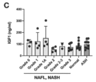
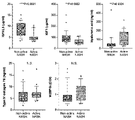
- the present inventors have extensively studied a method for early detection of liver fibrosis in patients with non-alcoholic steatohepatitis, and found that serum IGFALS protein levels in patients with non-alcoholic steatohepatitis are at an early stage such as grade 1-2. It was found to be a good biomarker of fibrosis, including fibrosis (see, eg, Figure 2C). Serum IGFALS levels were unchanged in patients with alcoholic steatohepatitis (ASH) compared to healthy controls (see Figure 3D), and the difference was not clear in HCV-induced hepatitis.
- ASH alcoholic steatohepatitis
- the serum IGFALS level is not necessarily a biomarker showing efficacy in general liver fibrosis, but was thought to be a specific biomarker for estimating liver fibrosis grade in NASH patients.
- the inventors further demonstrated that non-alcoholic steatohepatitis activity can be estimated by serum IGFALS levels (see FIGS. 6A-7).
- a method for estimating the progression of liver fibrosis and/or the activity of said liver disease in a subject with non-alcoholic fatty liver disease comprising: measuring the protein level of insulin-like growth factor binding protein (acid-labile subunit, IGFALS) in a blood sample obtained from the subject; (i) blood IGFALS protein levels in said subject; (ii) (a) reference value in healthy subjects (first reference value), or (ii) (b) reference value in subjects with fibrosis grade 2-3 liver fibrosis (second reference value), or (ii)(c) comparing to a reference value for blood IGFALS levels; A method, including (2) (i) comparing the blood IGFALS protein level of said subject with (ii) (a) a reference value (first reference value) of a healthy subject; A subject from whom a blood sample in which the ratio of said level to a first reference value (said level/first reference value) is less than a first reference value has
- the method according to (1) above further comprising (3) (i) comparing the subject's blood IGFALS protein level to a reference value (second reference value) for a subject with (ii)(b) fibrosis grade 2-3 liver fibrosis; A subject from whom a blood sample in which the ratio of said level to a second reference value (said level/second reference value) is less than a second reference value has fibrosis with a liver fibrosis grade of 2 or higher , or presumed to be likely, and/or A subject from whom a blood sample in which the ratio of said level to a second reference value (said level/second reference value) is equal to or greater than a second reference value has fibrosis with a liver fibrosis grade of 2 or higher.
- the second reference value is a value of 1.5 or less.
- the reference value for the blood IGFALS level is lower than the reference value (first reference value) in healthy subjects, and the reference value (second reference value) in subjects with fibrosis grade 2-3 liver fibrosis.
- NAFLD non-alcoholic steatohepatitis
- the means for measuring blood IGFALS levels comprises an antibody that binds to IGFALS or a protein complex containing IGFALS.
- the means for measuring blood IGFALS levels comprises an antibody that binds to IGFALS.
- the means for measuring blood IGFALS levels comprises an antibody that binds to a protein complex containing IGFALS.
- the antibody that binds to a protein complex comprising IGFALS comprises an antibody that binds to IGFALS and an antibody that binds to IGF1 or IGFBP-3 or IGFBP-5.
- a means for measuring IGFALS levels includes a first antibody that binds to IGFALS or a protein complex containing IGFALS immobilized on a support, and a labeled IGFALS or a protein complex containing IGFALS that binds to the first antibody.
- a protein complex comprising the first antibody, the second antibody, and IGFALS or IGFALS, comprising a second antibody, comprising the first antibody, the second antibody, and IGFALS or a protein complex comprising IGFALS The kit according to (13) above, which is capable of forming a complex.
- the means for measuring IGFALS levels includes test strips for immunochromatography;
- a test strip is a strip comprising a sample pad for introducing a blood sample, a conjugate pad, an area containing a test line and a control line, and a waste pad.
- the conjugate pad comprises a substance (first binding substance) that binds to IGFALS or a protein complex comprising IGFALS, the first binding substance being labeled;
- the test line contains a binding substance (second binding substance) that binds to IGFALS or a protein complex containing IGFALS, the second binding substance is immobilized on the test line,
- a control line did not contain any substance that binds to IGFALS, the first binding agent and the second binding agent are capable of simultaneously binding to IGFALS or a protein complex comprising IGFALS;
- IGFALS or a protein complex containing IGFALS contained in the blood sample binds to the first binding substance labeled on the conjugate pad to form a complex, and the complex is immobilized on the test line.
- the amount of label bound to the phased second binding agent and detected on the test line is indicative of the amount of IGFALS in the blood sample; 14.
- the means for measuring IGFALS levels comprises immunochromatographic test strips;
- a test strip is a strip comprising a sample pad for introducing a blood sample, a conjugate pad, an area containing a test line and a control line, and a waste pad.
- the conjugate pad comprises a substance that binds to IGFALS (the first binding substance), the first binding substance being labeled;
- the test line contains a binding substance (second binding substance) that binds to IGFALS, the second binding substance is immobilized on the test line, A control line did not contain any substance that binds to IGFALS, the first binding agent and the second binding agent can simultaneously bind to IGFALS;
- IGFALS contained in the blood sample binds to the first binding substance labeled on the conjugate pad to form a complex, and the complex is immobilized on the test line to form a second binding substance.
- the means for measuring IGFALS levels comprises immunochromatographic test strips;
- a test strip is a strip comprising a sample pad for introducing a blood sample, a conjugate pad, an area containing a test line and a control line, and a waste pad.
- the conjugate pad comprises a substance (first binding substance) that binds to a protein complex comprising IGFALS, the first binding substance being labeled;
- the test line contains a binding substance (second binding substance) that binds to the protein complex containing IGFALS, the second binding substance is immobilized on the test line,
- a control line did not contain any substance that binds to IGFALS, the first binding agent and the second binding agent are capable of simultaneously binding to a protein complex comprising IGFALS;
- the protein complex containing IGFALS contained in the blood sample binds to the first binding substance labeled on the conjugate pad to form a complex, and the complex is immobilized on the test line.
- each of the first binding substance and the second binding substance is an antibody that binds to IGFALS or an antibody that binds to a protein complex comprising IGFALS.
- each of the first binding substance and the second binding substance is an antibody that binds to IGFALS.
- each of the first binding agent and the second binding agent is an antibody that binds to a protein complex comprising IGFALS.
- one of the first binding substance and the second binding substance is an antibody that binds to IGFALS and the other is an antibody that binds to IGF1 or IGFBP-3 or IGFBP-5 (17) or The kit according to (17B).
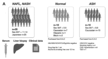
- FIG. 1A represents the subject population of this example.
- Subjects were non-alcoholic fatty liver (NAFL), non-alcoholic steatohepatitis (NASH), healthy subjects, and alcoholic steatohepatitis (ASH) based on Liver Biopsy and other clinical data.
- a population diagnosed as In this example, the serum biomarker levels of these subjects were examined.
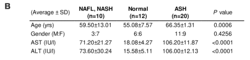
- FIG. 1B shows age, gender, AST values, and ALT values in each subject group.
- FIG. 1C shows IGF1 levels in the NAFL/NASH group with grades 1-3 fibrosis graded according to Table 1, the normal group, and the ASH group.
- FIG. 1D shows serum IGFALS levels in the NAFL/NASH group with grades 1-3 fibrosis, the healthy group, and the ASH group, graded according to Table 1.
- FIG. FIG. 2A shows a comparison of ROC curves based on each diagnostic index. The vertical axis in FIG. 2A represents sensitivity (%) and the horizontal axis represents 100-specificity (%).
- FIG. 2B shows the area under the curve (AUC) of the ROC curve based on each diagnostic index, the cutoff value, the sensitivity and specificity at that cutoff value, and the p-value.
- FIG. 2C shows the ROC curve in the NAFL/NASH group with grade 2 fibrosis in the low fibrosis group plus healthy subjects.
- FIG. 3A shows a comparison of AST values in healthy subjects, NAFL/NASH group and ASH group.
- FIG. 3B shows a comparison of ALT values in the healthy subject group, NAFL/NASH group and ASH group.
- FIG. 3C is a diagram plotting the AST and ALT values of each subject in the healthy subject group, NAFL/NASH group, and ASH group.
- FIG. 3D shows that serum IGFALS levels are not significantly different between healthy and ASH groups.
- FIG. 3 shows that serum IGFALS levels do not respond to fibrosis in general, but specifically to fibrosis in the NAFL/NASH group.
- FIG. 3E is a diagram showing the relationship between each subject group and age.
- FIG. 4 shows ROC curves for each biomarker for diagnosing subjects with greater than grade 1 fibrosis. The vertical axis represents sensitivity (%) and the horizontal axis represents 100-specificity (%).
- FIG. 5 shows ROC curves for each biomarker for diagnosing subjects with greater than grade 2 fibrosis (ie, between grades 2 and 3, and grade 3). The vertical axis represents sensitivity (%) and the horizontal axis represents 100-specificity (%).
- FIG. 4 shows ROC curves for each biomarker for diagnosing subjects with greater than grade 1 fibrosis. The vertical axis represents sensitivity (%) and the horizontal axis represents 100-specificity (%).
- FIG. 5 shows ROC curves for each biomarker for diagnosing subjects with greater than grade 2 fibro
- FIG. 6A shows blood levels of each factor in the healthy subject group (Normal), ASH group, and NASH group.
- FIG. 6B shows the correlation between the blood IGFALS level and the blood IGF1 level in the healthy subject group (Normal), ASH group, and NASH group.
- FIG. 6C shows the correlation (Pearson correlation coefficient) of the parameters obtained from the analysis of the three groups of the healthy subject group (Normal), ASH group, and NASH group.
- FIG. 7 shows blood levels of each factor in the active NASH group and the other control groups.
- FIG. 8 shows ROC curves in detecting active NASH groups based on blood levels of each factor.
- a "subject” can be a mammal, e.g., primates such as humans and chimpanzees, laboratory animals such as rats, mice, rabbits, domesticated animals such as pigs, cows, horses, sheep, and goats, and companion animals such as dogs and cats, preferably humans.
- patient means a subject with a disease, preferably a human with a disease.
- non-alcoholic fatty liver disease is defined as a condition in which fat accumulates in the liver (fatty liver) in people who have never been or rarely drink alcohol (30 g in men). /day, less than 20g/day for women (converted to ethanol). The intake of alcohol can be calculated by alcohol content (%) x amount of alcohol (mL) x specific gravity of alcohol (0.8 g/mL).
- NAFLD is a general term for a series of diseases including non-alcoholic fatty liver, progressing to steatohepatitis and cirrhosis.
- Causes of NAFLD include lifestyle diseases such as obesity, diabetes, dyslipidemia, and hypertension, as well as diseases such as sleep apnea, polycystic ovarian syndrome, hypothyroidism, and hypopituitarism. , pancreaticoduodenectomy, central parenteral nutrition after surgery such as jejuno-ileal bypass, and administration of drugs (tamoxifen, valproic acid, amiodarone, etc.) are known.
- Diagnosis of non-alcoholic fatty liver disease is based on 1) no history of alcohol consumption or very little alcohol consumption (less than 30 g/day for men and less than 20 g/day for women); (i) macrovesicular/vesicular fat deposition, (ii) inflammatory cell infiltration, (iii) hepatocyte ballooning, and (iv) perihepatocyte fibrosis in the central hepatic lobule. and 3) not viral hepatitis (viruses include, for example, HBV and HCV) and other liver diseases such as autoimmune hepatitis. NAFLD is therefore distinct from hepatitis caused by HCV and HBV.
- MAFLD metabolic-related fatty liver disease
- the diagnostic criteria for MAFLD are, in addition to the presence of hepatic steatosis, 1) a BMI value of 25 kg/m 2 or more, 2) the presence of type 2 diabetes, or 3) the following seven metabolic risk criteria (metabolic at- by the presence of two or more of the risk criteria).
- NAFL non-alcoholic fatty liver
- Treatments for subjects with NAFL include diet and/or exercise regimens and weight loss thereby. Treatment of the underlying disease, such as those listed above, may also be effective.
- non-alcoholic steatohepatitis is a pathological condition in which NAFL causes liver inflammation and liver fibrosis progresses.
- NASH non-alcoholic steatohepatitis
- subjects can develop cirrhosis and liver cancer. It can be diagnosed by a liver biopsy, which examines liver tissue.
- the diagnosis of NASH is NAFLD, and 2) histological findings from liver biopsy show either (iii) hepatocyte ballooning and (iv) perihepatocyte fibrosis in the central hepatic lobules.
- NASH can be diagnosed when:
- ASH alcoholic steatohepatitis
- liver tissue lesions are mainly degeneration or necrosis of hepatocytes, 1) significant swelling of hepatocytes mainly in the center of the lobule (ballooning, ballooning), and 2) Varying degrees of hepatocyte necrosis and 3) Mallory's body (alcoholic vitreous), or 4) polynuclear leukocyte infiltration.
- confirmation of drinking history e.g., drinking an average of 60 g (ethanol equivalent) or more per day for 5 years or more
- exclusion of liver damage due to causes other than alcohol e.g., hepatitis virus marker negative, anti-mitochondrial antibody negative, and antinuclear antibody negative.
- hepatic fibrosis refers to a state in which connective tissue accumulates in the liver. Connective tissue accumulates, for example, due to repair of damage in the liver. In particular, when the damage is chronic, repair occurs repeatedly, resulting in accelerated accumulation of connective tissue and progression of fibrosis.
- Pathologically, fibrosis is initiated by the activation of perivascular stellate cells in the liver. These cells can participate in the inflammatory response and overproduce extracellular matrix (such as collagen) and matrixcellular proteins while producing fibrosis-inducing factors such as TGF- ⁇ .
- the fibrosis grade can be determined, for example, based on Table 1 below (see NASH clinical research network historical scoring system (Keliner et al., Hepatology, 2005)).
- Fibrosis can be improved or prevented from progressing by removing the cause.
- Medicaments to treat fibrosis include PPAR agonists (e.g., thiazolidine derivatives), bile acid nuclear receptor agonists (e.g., bile acid analogs, e.g., obeticholic acid), CCR2 or CCR5 antagonists (e.g., Senicriviroc), kinase inhibitors. agents (eg, sorafenib), ASK1 antagonists (eg, seronsertib), collagen-specific chaperone inhibitors (eg, siRNA or antisense oligos against HSP47).
- PPAR agonists e.g., thiazolidine derivatives
- bile acid nuclear receptor agonists e.g., bile acid analogs, e.g., obeticholic acid
- CCR2 or CCR5 antagonists e.g., Senicriviroc
- IGFALS insulin-like growth factor-binding protein acid-labile subunits
- IGF1 insulin-like growth factor 1
- IGFBP1-6 IGF binding proteins
- Human IGFALS is registered with the US National Center for Biotechnology Information (NCBI) as GENE ID: 3483, and its amino acid sequence is not particularly limited, but for example, the amino acid sequence registered in NCBI Reference Sequence: NP_001139478.1. could be.
- a "blood sample” can be whole blood, serum, and plasma.
- Whole blood may contain an anticoagulant.
- Serum is the liquid component obtained by centrifugation after drawing blood into an anticoagulant-free container and allowing the blood to clot.
- Plasma is the supernatant obtained by centrifuging blood mixed with an anticoagulant to sediment blood cell components.
- the term "antibody” refers to an immunoglobulin, a protein having a structure in which two heavy chains (H chains) and two light chains (L chains) stabilized by disulfide bonds are associated.
- the heavy chain consists of a heavy chain variable region VH, heavy chain constant regions CH1, CH2, CH3, and a hinge region located between CH1 and CH2, and the light chain consists of a light chain variable region VL and a light chain constant region CL.
- a variable region fragment (Fv) consisting of VH and VL is a region that directly participates in antigen binding and imparts diversity to antibodies.
- the antigen-binding region consisting of VL, CL, VH and CH1 is called the Fab region, and the region consisting of the hinge region, CH2 and CH3 is called the Fc region.
- the regions that directly contact the antigen undergo particularly large changes and are called complementarity-determining regions (CDRs).
- CDRs complementarity-determining regions
- a portion other than the CDRs with relatively few mutations is called a framework region (FR).
- FR framework region
- the light chain and heavy chain variable regions each have three CDRs, which are referred to as heavy chain CDRs 1-3 and light chain CDRs 1-3 in order from the N-terminus. Each CDR is integrated into framework regions.
- the heavy chain variable region of the antibody consists of, from the N-terminal side to the C-terminal side, heavy chain framework region 1, heavy chain CDR1, heavy chain framework region 2, heavy chain CDR2, heavy chain framework region 3, heavy chain It has CDR3, and heavy chain framework region 4, in that order.
- the light chain variable region of the antibody consists of, from the N-terminal side to the C-terminal side, light chain framework region 1, light chain CDR1, light chain framework region 2, light chain CDR2, light chain framework region 3, light chain It has CDR3, and light chain framework region 4, in that order.
- Antibodies can be recombinant proteins (recombinant antibodies) and can be produced in animal cells, such as Chinese hamster ovary cells (CHO cells).
- the origin of the antibody is not particularly limited, but examples thereof include non-human animal antibodies, non-human mammal antibodies (eg, mouse antibodies, rat antibodies, camel antibodies), and human antibodies.
- Antibodies may also be chimeric, humanized, and fully humanized antibodies.
- Antibodies may be polyclonal antibodies or monoclonal antibodies, preferably monoclonal antibodies.
- Antibodies can be isolated antibodies or purified antibodies.
- Antibodies can be, for example, IgG.
- Antibodies can be, for example, IgG1, IgG2, IgG3, or IgG4.
- variable regions of immunoglobulin chains consist of relatively conserved framework regions (FR) joined by three hypervariable regions (more often called “complementarity determining regions” or CDRs). generally exhibit the same overall structure, including
- the CDRs from the two chains of each heavy/light chain pair described above are represented by framework regions to form structures that specifically bind to specific epitopes on target proteins (e.g., PCSK9).
- target proteins e.g., PCSK9
- FR1, CDR1, FR2, CDR2, FR3, CDR3 and FR4 parallel to From N-terminus to C-terminus, both naturally occurring light and heavy chain variable regions typically conform to the following order of these elements.
- FR1, CDR1, FR2, CDR2, FR3, CDR3 and FR4 A numbering system, such as that of Kabat, has been devised to assign numbers to the amino acids that occupy positions in each of these domains.
- Heavy chain CDRs 1-3 and light chain CDRs 1-3 can be determined based on the amino acid sequences of the heavy and light chain variable regions, respectively, for example, by the numbering system according to Kabat.
- a "protein complex” is a complex comprising multiple proteins, each associated with at least one other protein.
- An “antibody-protein complex” is a protein complex that includes an antibody and a protein, and the antibody is bound to the protein. Protein complexes include protein complexes found in blood samples (protein complexes in blood samples) and complexes that do not contain antibodies, such as artificially produced antibodies (such as monoclonal antibodies) ( antibody-free protein complexes).
- liver fibrosis level (or liver fibrosis progression, liver fibrosis grade, or liver fibrosis) in a subject with or likely to have non-alcoholic fatty liver disease (NAFLD) score) is provided.
- NAFLD non-alcoholic fatty liver disease
- the methods of the present disclosure involve measuring the protein level (ie, concentration) of IGFALS in a biological sample (eg, body fluid sample, preferably blood sample) obtained from a subject.
- a biological sample may preferably be a blood sample.
- the use of blood samples allows for minimally invasive and repeatable tests, which can be beneficial in that liver biopsies, previously required for diagnosis, can be avoided. Measurements can be made with either serum or plasma.
- the average protein level of IGFALS in blood can be, for example, less than half of the average in healthy individuals.
- the subject may be a NASH patient who does not have fibrosis (has grade 0 fibrosis) or who may have fibrosis.
- the patient can be a NASH patient who has or is likely to have grade 1 fibrosis (eg, a grade selected from the group consisting of grades 1A, 1B and 1C).
- the subject can be a NASH patient with or at risk of having grade 2 fibrosis. In certain aspects of the present disclosure, the subject has or may have grade 3 fibrosis. Can be a NASH patient. In certain aspects of the present disclosure, the subject may be a NASH patient with or at risk of having grade 4 fibrosis. In one preferred aspect, the subject is a NASH patient without fibrosis (with grade 0 fibrosis) or with possible fibrosis, grade 1 (e.g., grades 1A, 1B and 1C selected from the group consisting of ) and one or more NASH patients selected from the group consisting of NASH patients with or with the potential for grade 2 fibrosis.
- grade 1 e.g., grades 1A, 1B and 1C selected from the group consisting of
- the subject with said grade of fibrosis has NASH or NAFL, e.g., diagnosed by alcohol history and liver biopsy, and ruled out for hepatitis due to other causes, such as viral hepatitis, as described above.
- NASH or NAFL e.g., diagnosed by alcohol history and liver biopsy, and ruled out for hepatitis due to other causes, such as viral hepatitis, as described above.
- the subject can be one that has been determined to have a fibrosis grade. Therefore, in one embodiment, the liver fibrosis grade can be estimated by body fluid biopsy (liquid biopsy) for a patient whose presence or absence of liver fibrosis and liver fibrosis grade have been determined by liver biopsy or the like.
- the method of the present disclosure comprises comparing (i) the subject's blood IGFALS protein level (measurement) with (ii) (a) a reference value (first reference value) in a healthy subject. can further include If the measured value of (i) (the measured value) is lower than the first reference value, the subject has or is likely to have fibrosis of fibrosis grade 2 or higher. Accordingly, the methods of the present disclosure further comprise presuming that a subject with a blood IGFALS protein level lower than the first reference value has, or is likely to have, fibrosis grade 2 or greater. You can stay.
- the method of the present disclosure comprises: (i) comparing the blood IGFALS protein level (measured value) of the subject with (ii) (a) a reference value (first reference value) of a healthy subject;
- the ratio of the subject's blood IGFALS protein level (measured value) to a first reference value (said measured value/first reference value) is equal to or less than a first reference value (i.e., a predetermined cutoff value), or It can further include presuming that the subject from whom the blood sample is less than has or is likely to have fibrosis with a liver fibrosis grade of 2 or greater.
- the method of the present disclosure comprises: (i) comparing the blood IGFALS protein level (measured value) of the subject with (ii) (a) a reference value (first reference value) of a healthy subject; A subject from which a blood sample in which the ratio of the measured value to the first reference value (the measured value/first reference value) is equal to or greater than the first reference value (i.e., a predetermined cutoff value) is Presuming that there is no fibrosis with a fibrosis grade of 2 or higher, or that there is a possibility thereof (i.e., the possibility of not having fibrosis with a liver fibrosis grade of 2 or higher); can further include
- the first reference value is, for example, the mean value of the IGFALS level (preferably blood IGFALS level) in a biological sample (e.g., body fluid sample, preferably blood sample) from healthy subjects, the first quartile value, or a minimum value, or a value in between.
- a biological sample e.g., body fluid sample, preferably blood sample
- the first reference value (that is, the predetermined cutoff value) is, for example, less than 1.0, 0.9 or less, 0.8 or less, 0.7 or less, 2/3 or less, 0.6 or less, 0.6 or less. It can be a value of 55 or less, 0.5 or less, 0.45 or less, or 0.4 or less.
- the first reference value (ie, the predetermined cutoff value) can be a value of 0.4 or greater, 0.45 or greater, 0.5 or greater, or 0.55 or greater.
- the first reference value ie, the predetermined cutoff value
- the first reference value can be the average blood IGFALS level in healthy subjects, and the first reference value (i.e., cutoff value) can be a value of 2/3 or less, e.g. , may be in the range of 0.4 to 0.7, preferably in the range of 0.5 to 0.6.
- the first reference value can be the first quartile of blood IGFALS levels in healthy subjects, and the first reference value (ie, cutoff value) is 0.9 or less, 0.9 or less, It can be a number of 8 or less, 0.7 or less, 2/3 or less.
- the first reference value can be the minimum blood IGFALS level in healthy subjects, and the first reference value (ie, cutoff value) can be a number less than one.
- the method of the present disclosure comprises: (i) the subject's blood IGFALS protein level (measurement) is compared to (ii) (b) a reference value for a subject with fibrosis grade 2-3 liver fibrosis (second reference value) can include
- the method of the present disclosure comprises: (i) the subject's blood IGFALS protein level (measurement) is compared to (ii) (b) a reference value for a subject with fibrosis grade 2-3 liver fibrosis (second reference value) including A subject from which a blood sample in which the ratio of the measured value to a second reference value (said measured value/second reference value) is less than a second reference value (i.e., a predetermined cutoff value) is treated with liver It may further comprise assuming that the fibrosis grade has or is likely to have fibrosis of 2 or more.
- the method of the present disclosure comprises: (i) the subject's blood IGFALS protein level (measurement) is compared to (ii) (b) a reference value for a subject with fibrosis grade 2-3 liver fibrosis (second reference value) including A subject from which a blood sample in which the ratio of the measured value to the second reference value (said measured value/second reference value) is equal to or greater than the second reference value (i.e., a predetermined cutoff value) is It may further include estimating that there is no fibrosis with a fibrosis grade of 2 or greater, or that there is a likelihood of having fibrosis with a fibrosis grade of 2 or greater (ie, the likelihood of having no fibrosis with a liver fibrosis grade of 2 or greater).
- the second reference value can be the mean, third quartile, or maximum blood IGFALS level in subjects with fibrosis grade 2-3 liver fibrosis, or a value therebetween.
- the second reference value (i.e. cutoff value) is 1.8 or less, 1.7 or less, 1.6 or less, 1.5 or less, 1.4 or less, 1.3 or less, 1.2 or less, 1 .1 or less, or can be a number of 1 or less.
- the second reference value (ie, cutoff value) can be, for example, a numerical value greater than or equal to 1, greater than or equal to 1.1, greater than or equal to 1.2, greater than or equal to 1.3, or greater than or equal to 1.4.
- the second reference value (ie, cutoff value) can be, for example, a numerical value in the range of 1-1.8, and can be a numerical value in the range of 1-1.5.
- the second reference value is the mean blood IGFALS level in subjects with fibrosis grade 2-3 liver fibrosis, and the second reference value (i.e., cutoff value) is 1 It can be a number of 0.8 or less, a number of 1.5 or less, or a number in the range of 1 to 1.8, preferably in the range of 1 to 1.5.
- the method of the present disclosure comprises: (i) comparing the subject's blood IGFALS protein level (measurement) with (ii)(c) a reference value (ie, a predetermined cut-off value) for blood IGFALS levels.
- the method of the present disclosure comprises: (i) comparing the subject's blood IGFALS protein level (measurement) to (ii)(c) a predetermined cut-off value for blood IGFALS level; A subject from whom a blood sample for which the measured value is less than a reference value (i.e., a predetermined cutoff value) for blood IGFALS levels is defined as having or likely to have fibrosis with a liver fibrosis grade of 2 or greater. It may further include estimating that
- the method of the present disclosure comprises: (i) comparing the blood IGFALS protein level (measurement) of said subject with (ii)(c) a reference value (i.e., a predetermined cut-off value) for the blood IGFALS level; A subject from whom a blood sample for which the measured value is equal to or greater than a reference value (i.e., a predetermined cut-off value) for blood IGFALS level is free of, or likely to have, fibrosis with a liver fibrosis grade of 2 or higher. It can further include assuming that there is.
- the blood IGFALS protein level (measurement) of said subject including comparing Methods are provided further comprising assuming that a subject from whom a blood sample from which said measured value is equal to or less than said reference value has or may have active NAFL/NASH.
- Reference values (i.e., predetermined cut-off values) for blood IGFALS levels are, for example, values below 7 ⁇ g/mL, values below 6.9 ⁇ g/mL, values below 6.8 ⁇ g/mL, values below 6.7 ⁇ g/mL 6.6 ⁇ g/mL or less, 6.5 ⁇ g/mL or less, 6.4 ⁇ g/mL or less, 6.3 ⁇ g/mL or less, 6.2 ⁇ g/mL or less, 6 .1 ⁇ g/mL or less, 6.0 ⁇ g/mL or less, 5.9 ⁇ g/mL or less, 5.8 ⁇ g/mL or less, 5.7 ⁇ g/mL or less, 5.6 ⁇ g/mL or less , 5.5 ⁇ g/mL or less, 5.4 ⁇ g/mL or less, 5.3 ⁇ g/mL or less, 5.2 ⁇ g/mL or less, 5.1 ⁇ g/mL or less, or 5 0 ⁇ g
- Reference values (i.e., predetermined cut-off values) for blood IGFALS levels are, for example, values greater than or equal to 5.0 ⁇ g/mL, values greater than or equal to 5.1 ⁇ g/mL, values greater than or equal to 5.2 ⁇ g/mL, 5.3 ⁇ g /mL or more, 5.4 ⁇ g/mL or more, 5.5 ⁇ g/mL or more, 5.6 ⁇ g/mL or more, 5.7 ⁇ g/mL or more, 5.8 ⁇ g/mL or more , a value of 5.9 ⁇ g/mL or greater, or a value of 6.0 ⁇ g/mL or greater.
- the reference value (i.e., predetermined cutoff value) for blood IGFALS levels can be, for example, 4 ⁇ g/mL to 7 ⁇ g/mL, more preferably 5 ⁇ g/mL to 6 ⁇ g/mL. .
- the reference values for blood IGFALS levels for assessment of active NAFL/NASH are similar, but the lower the blood IGFALS level, the more likely is active NAFL/NASH. Therefore, it can be assumed that subjects with blood IGFALS levels below the lower reference value are more likely to have active NAFL/NASH.
- the reference value for blood IGFALS levels for active NAFL/NASH assessment may preferably be between 4 ⁇ g/mL and 7 ⁇ g/mL, more preferably between 5 ⁇ g/mL and 6 ⁇ g/mL. Those skilled in the art can appropriately set the reference value.
- a method as described above comprising: said subject has grade 0 or 1 liver fibrosis at the first time the method is performed; performing the method of any one of claims 1 to 11 using each blood sample obtained from the subject at multiple time points; determining that a subject presumed to have grade 2 or greater liver fibrosis in each blood sample obtained after a specified time point has transitioned from grade 0 or 1 liver fibrosis to grade 2 or greater; estimating when liver fibrosis transitioned from grade 0 or 1 to grade 2 or greater in a subject with grade 2 or greater liver fibrosis; A method is provided.
- the method comprises presuming the subject does not have or is likely to have fibrosis with a liver fibrosis grade of 2 or greater, wherein said After the subject has fibrosis of grade 2 or greater, it would include presuming that the subject has or is likely to have fibrosis of liver fibrosis grade 2 or greater. Then, when the subject's hepatic fibrosis grade progresses to 2 or higher, it can be determined that the subject's fibrosis has transitioned to grade 2 or higher.
- the time when the last blood sample when the liver fibrosis grade was 0 or 1 and the first when the liver fibrosis grade became 2 It can be estimated that the liver fibrosis transitioned from grade 0 or 1 to grade 2 or later between the time points of collecting blood samples. In this way, it is possible to estimate when liver fibrosis transitioned from grade 0 or 1 to grade 2 or later in the subject.
- the method of the present disclosure is a blood sample technique that is less invasive and can be performed repeatedly. Therefore, the methods of the present disclosure are suitable for monitoring liver fibrosis grade over time.
- blood IGFALS protein levels can be estimated by serum or plasma IGFALS protein levels. Serum or plasma IGFALS protein levels can be determined by an enzyme-linked immunosorbent assay (ELISA). Blood samples may be subjected to gel filtration or the like to separate IGFALS from other contaminants prior to measurement.
- ELISA enzyme-linked immunosorbent assay
- ELISA includes direct method, indirect method, sandwich method, and competitive method.
- IGFALS are immobilized on a support (for example, plate surface), and after washing, the immobilized IGFALS are detected with a labeled antibody.
- IGFALS are immobilized on a support (for example, a plate surface), and after washing, the immobilized IGFALS are allowed to bind to antibodies. It is detected by an antibody.
- the sandwich method the first antibody is immobilized on a support (e.g., plate surface), and after washing, IGFALS is allowed to bind to the immobilized first antibody, and after further washing, IGFALS is labeled. It is detected with a second antibody.
- an antibody is immobilized on a support (for example, a plate surface), and after washing, the immobilized antibody is brought into contact with a sample containing a certain concentration of labeled IGFALS. It estimates the concentration of IGFALS in the sample by measuring the amount of label that remains in the sample. In either case, an antibody that binds to IGFALS can be used as the antibody.
- the antibody that binds IGFALS can be specific for IGFALS.
- the antibody that binds to IGFALS is 10 ⁇ 7 M or less, 10 ⁇ 8 M or less, 10 ⁇ 9 M or less, 10 ⁇ 10 M or less, 10 ⁇ 11 M or less, or 10 ⁇ 12 M or less to IGFALS. It can bind with a KD value of M or less.
- the first antibody and the second antibody are capable of binding IGFALS simultaneously.
- the first antibody, second antibody and IGFALS can form a complex (protein-antibody complex) comprising the first antibody, second antibody and IGFALS.
- IGFALS can form protein complexes with IGF1 and/or IGFBP-3, particularly with IGF1 and IGFBP-3, in a sample.
- IGFALS may form protein complexes with IGF1 and/or IGFBP-5, particularly IGF1 and IGFBP-5, in a sample. Therefore, IGFALS may be measured by detecting the protein complexes described above. In this case, the protein complex can be detected with an antibody that binds to IGFALS and an antibody that binds to IGF1 and/or IGFBP-3 (or IGFBP-5).
- the protein complex is adsorbed to the solid phase surface with an antibody that binds to immobilized IGFALS, and then the protein complex is adsorbed with an antibody that binds to IGF1 and/or an antibody that binds to IGFBP-3 (or IGFBP-5).
- the protein complex is adsorbed to the solid phase surface by the immobilized antibody that binds to IGF1 and/or the antibody that binds to IGFBP-3 (or IGFBP-5), and then by the antibody that binds to IGFALS. Protein complexes can also be detected.
- the first antibody comprises an antibody that binds IGFALS and the second antibody comprises an antibody that binds IGF1 and/or an antibody that binds IGFBP-3 (or IGFBP-5).
- the first antibody comprises an antibody that binds to IGF1 and/or an antibody that binds to IGFBP-3 (or IGFBP-5) and the second antibody comprises an antibody that binds to IGFALS.
- the protein complex may be detected using the above antibody by a sandwich assay, or may be detected using the above antibody by an immunochromatographic method described below.
- IGFALS levels can be measured by a chemiluminescent enzyme immunoassay (CLEIA).
- IGFALS or a protein complex containing IGFALS eg, a protein complex with IGF1 and IGF3 (or IGFBP-5)
- the solid phase can be a plate surface or a bead (eg, magnetic bead) surface.
- IGFALS or multimers containing IGFALS adsorbed to the solid surface can be detected with a second antibody.
- the magnetic beads can be washed by bringing a magnet into contact with the container from outside the container, trapping the magnetic beads on the inner surface of the contact portion, and exchanging the solution.
- IGFALS can be captured on the solid phase directly by the first antibody, or by an additional antibody that recognizes the immobilized first antibody and the non-immobilized first antibody. good. In this case, a complex is formed that binds in the order solid phase-further antibody-first antibody.
- the complex can be an antibody complex comprising a solid phase-further antibody-first antibody-IGFALS or a protein complex comprising IGFALS.
- Antibody complexes comprising the first antibody-IGFALS or-or protein complexes comprising IGFALS are obtained by washing after antibody complex formation and then cleaving the bond between the additional antibody and the first antibody. It can be released into the clear water. This antibody complex can also be recovered, captured on another solid surface, and detected with a second antibody.
- a further antibody may recognize the first antibody via a labeling molecule attached to the first antibody.
- the labeling molecule can be disassociated from further antibody binding, for example, by introducing an excess amount of free labeling molecule into the system. In this way the bond between the further antibody and the first antibody can be cleaved.
- the labeling molecule is not particularly limited, but for example, 2,4-dinitrophenyl (DNP) can be used.
- the additional antibody may be an antibody that recognizes DNP.
- the first antibody and the second antibody can be monoclonal antibodies.
- the first antibody and the second antibody each replace the heavy chain CDRs 1-3 and light chain CDRs 1-3, respectively, of the anti-human IGFALS antibody produced from clone M6005C04. 3, and the antibody having the heavy chain CDRs 1-3 and light chain CDRs 1-3 of the anti-human IGFALS antibody produced from clone M6001E07 as heavy chain CDRs 1-3 and light chain CDRs 1-3, respectively.
- the first antibody and the second antibody each replace the heavy chain CDRs 1-3 and light chain CDRs 1-3, respectively, of the anti-human IGFALS antibody produced from clone M6001E07.
- CDRs are numbered by Kabat et al. , AbM, contact, IMGT, Aho, or Martin (Enhanced Chothia).
- the first antibody and the second antibody respectively have the heavy and light chain variable regions of the anti-human IGFALS antibody produced from clone M6005C04 and the anti-human IGFALS produced from clone M6001E07. It can be an antibody having a heavy chain variable region and a light chain variable region of an antibody. In certain aspects, the first antibody and the second antibody respectively have the heavy and light chain variable regions of the anti-human IGFALS antibody produced from clone M6001E07 and the anti-human IGFALS produced from clone M6005C04. It can be an antibody having a heavy chain variable region and a light chain variable region of an antibody.
- the first antibody and the second antibody can be an anti-human IGFALS antibody produced from clone M6005C04 and an anti-human IGFALS antibody produced from clone M6001E07, respectively. In one aspect, the first antibody and the second antibody can be an anti-human IGFALS antibody produced from clone M6001E07 and an anti-human IGFALS antibody produced from clone M6005C04, respectively.
- Enzymes used in the enzyme antibody method can be used as labels.
- Substrates for the above enzymes chromogenic substrates, fluorescent substrates, and luminescent substrates
- biotin can be used as the label
- an avidin-labeled enzyme can be used when detecting the biotinylated antibody.
- avidin, streptavidin, neutravidin and the like can be used.
- IGFALS levels are measured or estimated based on the amount of substrate conversion by an enzyme linked to an antibody bound to IGFALS.
- Substrates that are colored or discolored by enzymes are preferably used.
- a calibration method for estimation and specific estimation can be performed by a conventional method.
- a person skilled in the art can label the antibody using a conventional method. Labeling can, for example, be covalent. A labeled antibody can be obtained in this manner.
- horseradish peroxidase can be used as the peroxidase.
- Horseradish peroxidase produces color, fluorescence or chemiluminescence upon addition of a chromogenic, fluorogenic or luminescent substrate. Therefore, the presence of labeled capture molecules can be detected using chromogenic, fluorescent or chemiluminescent indicators.
- Chromogenic substrates for horseradish peroxidase include, for example, tetramethylbenzidine (TMB), o-phenylenediamine (OPD), 2,2-azinobis[3-ethylbenzo-thiazoline-6-sulfonic acid (ABTS), and Amplex ( Trademark) Red, which can be used to detect labeled molecules in the presence of hydrogen peroxide.
- TMB tetramethylbenzidine
- OPD o-phenylenediamine
- ABTS 2,2-azinobis[3-ethylbenzo-thiazoline-6-sulfonic acid
- Amplex Trademark
- Luminescent substrates for alkaline phosphatase include p-nitrophenyl phosphate (pNPP), 4-methylumbelliferyl phosphate (4-MUP), and AttoPhos (trademark), and can be used to detect capture molecules.
- pNPP p-nitrophenyl phosphate
- 4-MUP 4-methylumbelliferyl phosphate
- AttoPhos trademark
- Glucose oxidase oxidizes glucose to generate gluconic acid and hydrogen peroxide.
- Hydrogen peroxide can be readily detected, for example, using a colorimetric probe for detecting hydrogen peroxide (eg, peroxidase).
- Hydrogen peroxide can be developed, for example, in the presence of peroxidase and its chromogenic substrate.
- the sensitivity and specificity can each be 70% or higher, 75% or higher, 80% or higher, 85% or higher, or 90% or higher.
- blood biomarker levels other than blood IGFALS may not be considered. That is, in the above method of the present disclosure, only the blood IGFALS level can be used for evaluation.
- the blood IGFALS level can be estimated from the serum IGFALS level.
- blood IGFALS levels can be determined by mass spectrometry.
- Mass spectrometry can be performed appropriately by those skilled in the art. Mass spectrometry includes, for example, matrix-assisted laser desorption ionization time-of-flight (MALDI-TOF) mass spectrometry, electrospray ionization (ESI) mass spectrometry, surface-enhanced laser desorption ionization time-of-flight (SELDI-TOF) mass spectrometry.
- MALDI-TOF matrix-assisted laser desorption ionization time-of-flight
- ESI electrospray ionization
- SELDI-TOF surface-enhanced laser desorption ionization time-of-flight
- the method of the present disclosure is an in vitro method.
- the "method for estimating the level of liver fibrosis” refers to the “method for estimating the level of liver fibrosis", the “method for determining the level of liver fibrosis”, or the “diagnosis of the level of liver fibrosis”.
- method for obtaining preliminary information for The methods of the present disclosure do not involve diagnosing humans.
- compositions, kits or articles of manufacture for use in the disclosed methods are provided.
- a composition, kit or article of manufacture of the present disclosure comprises at least a means for measuring IGFALS levels.
- a composition, kit or article of manufacture of the disclosure may further comprise instructions for performing the methods of the disclosure with the means for measuring IGFALS levels.
- the means for measuring the IGFALS level is not particularly limited as long as it can identify the IGFALS level.
- it can be an antibody or lectin that binds to IGFALS or a protein complex containing IGFALS.
- Antibodies or lectins that bind to IGFALS can be prepared by those skilled in the art by routine methods. So are antibodies that bind to protein complexes containing IGFALS.
- Antibodies that bind to protein complexes containing IGFALS can be obtained as antibodies that bind to each component of the complex.
- Antibodies can be obtained, for example, by immunizing a non-human mammal or the like with IGFALS as an immunogen and cloning the resulting antibody-producing cells.
- Antibodies can also be produced from antibody-producing cells into which a gene encoding the antibody has been introduced. As antibody-producing cells, for example, Cheney's hamster ovary cells (CHO cells) can be used.
- compositions, kit or article of manufacture for use in the methods of the present disclosure can comprise an antibody that binds to IGFALS.
- the antibody can be labeled.
- the compositions, kits or articles of manufacture for use in the methods of the present disclosure can be compositions, kits or articles of manufacture for ELISA (direct method).
- compositions, kits or articles of manufacture for use in the methods of the present disclosure can contain unlabeled antibodies that bind IGFALS. Unlabeled antibody can be detected by a labeled secondary antibody.
- a composition, kit or article of manufacture for use in the methods of the present disclosure may further comprise a labeled secondary antibody in addition to the unlabeled antibody that binds IGFALS.
- the compositions, kits or articles of manufacture for use in the methods of the present disclosure can be compositions, kits or articles of manufacture for ELISA (indirect method).
- kits or articles of manufacture for use in the methods of the present disclosure can include an unlabeled antibody that binds IGFALS and a labeled antibody that binds IGFALS.
- An unlabeled antibody that binds to IGFALS can be immobilized on the surface of an assay plate, or immobilized on the surface of an assay plate. Immobilization can be appropriately carried out by a person skilled in the art by a conventional method. Accordingly, kits or articles of manufacture for use in the disclosed methods may further comprise assay plates.
- antibodies can be used to detect IGFALS, eg, by Western blotting.
- Western blotting can be performed by methods well known to those skilled in the art.
- an unlabeled antibody that binds to IGFALS is reacted with a membrane onto which proteins are transferred from a gel after electrophoresis, followed by washing, followed by detection with a labeled antibody that recognizes the labeled antibody.
- labels include substrates (chromogenic substrates, fluorescent substrates, and luminescent substrates) and enzymes used in enzyme-antibody methods (eg, peroxidase, glucose oxidase, and alkaline phosphatase).
- a label can be detected by a known method or the like.
- antibodies can be used to prepare samples for mass spectrometry. That is, the antibody can be used to prepare a sample for mass spectrometry by enriching or purifying IGFALS in the sample using its affinity with the antibody. Antibodies may thereby be useful in the detection of IGFALS in mass spectrometry.
- IGFALS can form complexes with IGF1 and/or IGFBP-3, particularly protein complexes with IGF1 and IGFBP-3, in a sample.
- IGFALS may form complexes with IGF1 and/or IGFBP-5, particularly protein complexes with IGF1 and IGFBP-5, in a sample. Therefore, IGFALS may be measured by detecting the protein complexes described above. In this case, the protein complex can be detected with an antibody that binds to IGFALS and an antibody that binds to IGF1 and/or IGFBP-3 (or IGFBP-5).
- the protein complex is adsorbed to the solid phase surface with an antibody that binds to immobilized IGFALS, and then the protein complex is adsorbed with an antibody that binds to IGF1 and/or an antibody that binds to IGFBP-3 (or IGFBP-5).
- the protein complex is adsorbed to the solid phase surface by the immobilized antibody that binds to IGF1 and/or the antibody that binds to IGFBP-3 (or IGFBP-5), and then by the antibody that binds to IGFALS. Protein complexes can also be detected.
- a kit may include, for example, an antibody that binds to IGFALS and an antibody that binds to IGF1 and/or an antibody that binds to IGFBP-3 (or IGFBP-5).
- the protein complex may be detected using the above antibody by a sandwich assay, or may be detected using the above antibody by an immunochromatographic method described below.
- a kit or article of manufacture for use in the methods of the disclosure comprises an antibody that binds IGFALS and an antibody that binds IGF1 and/or an antibody that binds IGFBP-3 (or IGFBP-5). obtain.
- Kits or products for use in these methods of the present disclosure may further include a substrate that develops color based on the label.
- kits or articles of manufacture for use in a method of the present disclosure, comprising: Kits or products are provided in which means for measuring IGFALS levels include a detection system in an immunochromatographic method (lateral flow method).
- kits or product comprising a means for measuring IGFALS levels or a detection system in immunochromatography (lateral flow method),
- the detection system including test strips for immunochromatography (e.g., the substrate can be a cellulose membrane);
- a test strip is a strip comprising a sample pad for introducing a blood sample, a conjugate pad, an area containing a test line and a control line, and a waste pad.
- the conjugate pad contains a substance that binds to IGFALS (the first binding substance) and the first binding substance is labeled ⁇ wherein the first binding substance is directed from the conjugate pad to the test line is movable on the strip ⁇
- the test line contains a binding substance (second binding substance) that binds to IGFALS, the second binding substance is immobilized on the test line
- the control line contains no substance that binds to IGFALS (eg, may contain an isotype control antibody), the first binding agent and the second binding agent can simultaneously bind to IGFALS;
- IGFALS contained in the blood sample binds to the first binding substance labeled on the conjugate pad to form a complex, migrates onto the test line, and forms a complex on the test line.
- the amount of label bound to the immobilized second binding substance and detected on the test line indicates the amount of IGFALS in the blood sample;
- kits or product comprising a means for measuring IGFALS levels or a detection system in immunochromatography (lateral flow method),
- the detection system including test strips for immunochromatography (e.g., the substrate can be a cellulose membrane);
- a test strip is a strip comprising a sample pad for introducing a blood sample, a conjugate pad, an area containing a test line and a control line, and a waste pad.
- the conjugate pad contains a substance (first binding substance) that binds to a protein complex comprising IGFALS, the first binding substance being labeled ⁇ wherein the first binding substance is from the conjugate pad is movable on the strip towards the test line ⁇ ,
- the test line contains a binding substance (second binding substance) that binds to the protein complex, the second binding substance is immobilized on the test line,
- a control line does not contain a substance that binds to the protein complex (for example, it may contain an isotype control antibody), the first binding substance and the second binding substance can simultaneously bind to the protein complex;
- the protein complex contained in the blood sample binds to the first binding substance labeled on the conjugate pad to form a complex, migrates onto the test line, and moves onto the test line.
- a protein complex comprising IGFALS can comprise IGFALS, IGF1 and IGFBP-3. In one aspect, a protein complex comprising IGFALS can comprise IGFALS, IGF1 and IGFBP-5. In certain aspects, one of the first binding agent and the second binding agent binds to IGFALS. In some aspects, the other of the first binding agent and the second binding agent binds to IGF1. In some aspects, the other of the first binding agent and the second binding agent binds to IGFBP-3. In some aspects, the other of the first binding agent and the second binding agent binds to IGFBP-5.
- the binding substance is preferably an antibody, more preferably a monoclonal antibody.
- kits or articles of manufacture of this disclosure can be for use in a method of this disclosure.
- the protein level of IGFALS can be measured or estimated based on measurements by an absorption method or a fluorescence method using a reader for quantification of the protein level of IGFALS. Correlations between absorbance or fluorescence measurements and IGFALS protein levels can be determined according to conventional methods. Thus, the protein level of IGFALS can be estimated from the measured value by calibrating the measured value and the protein level.
- a label suitable for the absorption method or fluorescence method using a reader (such as an enzyme that can use a dye, a fluorescent dye, etc. as a substrate) can be used as the label.
- a substance that binds to IGFALS or a protein complex containing IGFALS in the kit or product of the present disclosure can be an antibody or a lectin.
- the agents that bind to IGFALS or a protein complex comprising IGFALS are the first antibody and the second antibody described above, respectively. It can be an antibody, or a second antibody and a first antibody.
- one of the first antibody and the second antibody binds to IGFALS.
- the other of the first antibody and the second antibody binds to IGF1.
- the other of the first antibody and the second antibody binds to IGFBP-3.
- the other of the first antibody and the second antibody binds to IGFBP-5.
- NASH non-alcoholic steatohepatitis
- NASH non-alcoholic steatohepatitis
- Serum Samples from Healthy Individuals and ASH Patients Serum samples from healthy individuals were obtained from HBV/HCV/HIV-negative healthy donors of known age, sex, and race, and obtained from KAC Co., Ltd. , Ltd. purchased from.
- KAC Co., Ltd. , Ltd. purchased from for alcoholic steatohepatitis patient sera, those derived from patients diagnosed as ASH who are HBV/HCV-negative with clear age, sex, race, height, weight, AST, ALT, medical history, and medications taken are KAC. Co. , Ltd. purchased from.
- the detection antibody is biotinylated and detected by horseradish peroxidase coupled to avidin.
- the kit contains 3,3',5,5'-tetramethylbenzidine (TMB substrate) and develops a yellow color (maximum absorption wavelength 450 nm) by horseradish peroxidase treatment followed by sulfuric acid or phosphoric acid treatment.
- IGF1 insulin-like growth factors
- IGF1 is a peptide growth factor mainly produced in the liver, and its production is maintained at a high level by good nutritional conditions and growth hormone stimulation.
- IGF1 is known to be a hormone having various physiological actions such as individual development/growth, regulation of substance metabolism, and aging.
- IGF1 forms a complex with a binding protein in blood, and is characterized by having a long half-life in blood and maintaining a high concentration.
- IGF binding proteins 1-6 Six types of IGF binding proteins (IGF binding proteins 1-6, IGFBP1-6) are known to exist in IGF1, but most of IGF1 circulating in the blood is associated with IGFBP3 or IGFBP5. In addition to either, it exists in the form of protein complexes (trimers) with acid-labile subunits (ALS or IGFALS), which are glycoproteins (Baxter, Nat Rev Cancer 2014).
- FIG. 6A blood IGFALS levels were significantly lower in the NAFL/NASH group than in healthy subjects.
- FIG. 6B blood IGF1 level and blood IGFALS level showed a certain positive correlation.
- FIG. 6C blood IGFALS levels showed a negative correlation with blood type IV collagen 7S levels or blood M2BPGi levels.
- NAFL/NASH specimens were grouped according to the degree of fibrosis progression in the liver biopsy (fibrosis grade 0-3).
- IGFALS may be a biomarker to detect some fibrosis in MAFLD (or fibrosis in NASH).
- blood IGF1 levels are known to decrease with age after adulthood. (Fig. 3E), the effect of age on IGFALS values seen in the NASH group with fibrosis grade 2 and above was considered negligible.
- IGFALS is superior in both sensitivity and specificity to existing fibrosis markers.
- Type IV collagen 7s T4 Col7s
- Mac-2 binding protein glycosylation isomer M2BPGi
- platelet count age, AST, ALT Calculated based on the following formula (1) Therefore, ROC (receiver operating characteristic) analysis was performed based on each of the Fib-4 indexes.
- fibrosis grade 1A or lower in liver biopsy was classified as a low fibrosis group, and grade 2 or higher was classified as a high fibrosis group, and the sensitivity and specificity of each marker for fibrosis were analyzed.
- the serum IGFALS level was 0.9026 in AUC (area under curve), and other markers (Fib-4, 0.8442; HA, 0.6993; T4 Col7s, 0.5455; M2BPGi, 0 .5035) (Fig. 2A, B; Cutoff ⁇ 0.5587, Sensitivity 100, Specificity 81.82).
- the AUC reached 0.940, and the low fibrosis group and the high fibrosis group with a cutoff value of 5.587 (about half of the healthy subject group) (Fig. 2C).
- the ROC curve for >Grade 1 fibrosis was shown in FIG. 4 and the ROC curve for >Grade 2 fibrosis was as shown in FIG. Tables 3 to 5 below show the results of AUC and P value for each marker. In Tables 3-5, cutoffs were determined by the Youden Index.
- liver fibrosis is an urgent issue.
- Liver biopsy is a standard method for evaluating liver fibrosis in NASH patients, but it is difficult to perform routinely and repeatedly due to its high invasiveness and cost.
- sampling errors occur in NASH, which has heterogeneous intrahepatic lesions, since information on only a small area of the liver tissue can be obtained.
- liver fibrosis in NASH Although several blood biomarkers for liver fibrosis in NASH have been developed/applied, there are many markers that are uncertain regarding their positioning in diagnosis and the setting of cutoff values, and there is no evidence as to whether they can be used to determine therapeutic effects. is insufficient. In this study, it was revealed that serum IGFALS shows a low value in NASH with fibrosis grade 2 or later, and that both sensitivity and specificity are superior to existing HA, T4 Col7s, and M2BPGi. Based on this knowledge, it is possible to establish a blood diagnostic method that is superior to existing liver fibrosis markers, and a method to manage the liver fibrosis state of NASH patients without invasive diagnosis such as liver biopsy. can be established.
- IGFALS-producing organ in humans is the liver, and the producing cells are hepatocytes. Therefore, in NASH, HCV hepatitis, and hepatic fibrosis, it is suggested that the production of IGFALS from the liver decreases due to specific loads on hepatocytes such as metabolic changes. In addition, it is interesting that reduction in IGFALS can sensitively detect fibrosis of grade 2 and beyond.
- NAFL / NASH a group of patients with NAS4 points or more and fibrosis level of 2 points or more is defined as active NAFL / NASH, and patients in the active NAFL / NASH group and the inactive NAFL / NASH group categorized.
- the blood IGFALS level, blood IGF1 level, blood hyaluronic acid level, blood type IV collagen 7S level, and blood M2BPGi level of each group were measured. The results were as shown in FIG. As shown in FIG. 7, blood IGFALS levels were significantly different between the active NAFL/NASH group and the inactive NAFL/NASH group.
- the blood hyaluronic acid level which is an existing marker, was high in the activated NAFL/NASH group.
- the blood IGFALS level could effectively discriminate between the active NAFL/NASH group and others. As a result, for example, it may be possible to identify patients at high risk of transition to liver cirrhosis using the blood IGFALS level as an index.
Abstract
The present disclosure provides a method for estimating fibrosis progression and/or activity in non-alcoholic steatohepatitis. The present disclosure also provides a composition and a kit for use in said method. Specifically, the present disclosure makes it possible to, for example, estimate the progression of liver fibrosis in a subject on the basis of only the IGFALS level in the subject's blood.
Description
本開示は、非アルコール性脂肪性肝炎における線維化進行度および/または前記肝疾患の活動性を推定する方法に関する。本開示はまた、当該方法に用いるための組成物およびキットに関する。
The present disclosure relates to a method for estimating the degree of fibrosis progression and/or activity of the liver disease in non-alcoholic steatohepatitis. The disclosure also relates to compositions and kits for use in such methods.
近年、非アルコール性脂肪性肝炎(NASH)は国内外において罹患率が急増しており、本邦においても慢性肝不全の主要因になっているが、有効な治療薬やバイオマーカーの開発は大きく立ち遅れている。特に、肝線維化の進展が患者予後を規定していることから、非侵襲的に線維化をモニター可能な方法の開発が強く望まれている。
In recent years, the prevalence of non-alcoholic steatohepatitis (NASH) has increased rapidly in Japan and overseas, and it is a major cause of chronic liver failure in Japan, but the development of effective therapeutic drugs and biomarkers lags behind. ing. In particular, development of a method capable of non-invasively monitoring fibrosis is strongly desired, since progress of liver fibrosis determines patient prognosis.
NASHは、非アルコール性脂肪肝(NAFL)に伴い肝臓が炎症を起こし、肝線維化が進行する病態である。NASHが進行すると、肝硬変や肝がんを発生させるリスクを有する。NASHは、1)飲酒歴がないことに加えて、肝生検による肝組織所見で(i)大滴性/小滴性脂肪沈着、(ii)炎症細胞浸潤、(iii)肝細胞の風船様腫大、および(iv)肝小葉中心部における肝細胞周囲の線維化が認められること、並びに3)ウイルス性肝炎(ウイルスとしては、例えば、HBVやHCVが挙げられる)や自己免疫性肝炎などの他の肝疾患ではないことに基づいて診断される。
NASH is a pathological condition in which the liver becomes inflamed with non-alcoholic fatty liver (NAFL) and liver fibrosis progresses. If NASH progresses, there is a risk of developing liver cirrhosis or liver cancer. In addition to 1) no history of alcohol consumption, NASH was characterized by (i) macrovesicular/vesicular fatty deposits, (ii) inflammatory cell infiltration, (iii) balloon-like hepatocytes, and and (iv) fibrosis around hepatocytes in the center of the hepatic lobule, and 3) viral hepatitis (viruses include, for example, HBV and HCV) and autoimmune hepatitis. Diagnosis is based on no other liver disease.
患者の肝線維化を評価するスタンダードな方法として肝生検が挙げられるが、侵襲性の高さやコストの面から、ルーチンに反復して実施することが困難である。また、肝生検では、肝組織のごく一部の領域の情報しか得られないために、肝内病変の不均一性を有するNASHにおいては、サンプリングエラーが発生することも問題として指摘されている。一方、NASHにおける肝線維化の血液バイオマーカーがいくつか開発され、適用されているものの、性能面において課題を有するものも少なくない。
Liver biopsy is a standard method for evaluating liver fibrosis in patients, but due to its high invasiveness and cost, it is difficult to perform routinely and repeatedly. In addition, since liver biopsy can only provide information on a small area of the liver tissue, it has been pointed out that sampling errors occur in NASH, which has heterogeneous intrahepatic lesions. . On the other hand, although several blood biomarkers for liver fibrosis in NASH have been developed and applied, many of them have problems in terms of performance.
例えば、特許文献1および非特許文献1は、HCV誘発肝炎患者において血中インスリン様成長因子結合タンパク質酸不安定性サブユニット(IGFALS)レベルが低下することを開示する。特許文献2もまた、HCV誘発肝炎患者において血中IGFALSレベルが低下することを開示する。特許文献2では、末期のHCV誘発肝炎患者や重篤な肝線維化症例(グレード4以降)で血中IGFALSが低下することを開示するが、その低下は診断との相関が弱く、複数のバイオマーカーレベルに基づく診断を開示するものである。しかし、上述の通り、NASHは、患者がHCVなどのウイルス性肝炎でないことに基づいて診断されるものであり、NASHとHCV誘発肝炎とは異なる疾患である。
For example, Patent Document 1 and Non-Patent Document 1 disclose that circulating insulin-like growth factor-binding protein acid-labile subunit (IGFALS) levels are reduced in patients with HCV-induced hepatitis. WO 2005/010102 also discloses that blood IGFALS levels are reduced in patients with HCV-induced hepatitis. Patent Document 2 discloses that blood IGFALS decreases in end-stage HCV-induced hepatitis patients and severe liver fibrosis cases (grade 4 or later). Disclosed are diagnostics based on marker levels. However, as described above, NASH is diagnosed on the basis that the patient does not have viral hepatitis such as HCV, and NASH and HCV-induced hepatitis are different diseases.
本開示は、非アルコール性脂肪性肝炎における線維化進行度および/または前記肝疾患の活動性を推定する方法に関する。本開示はまた、当該方法に用いるための組成物およびキットを提供する。
The present disclosure relates to a method for estimating the degree of fibrosis progression and/or activity of the liver disease in non-alcoholic steatohepatitis. The disclosure also provides compositions and kits for use in such methods.
本発明者らは、非アルコール性脂肪性肝炎患者において早期に肝線維化を検出する方法を鋭意検討したところ、非アルコール性脂肪性肝炎患者の血清IGFALSタンパク質レベルがグレード1~2などの早期の線維化を含む線維化の良好なバイオマーカーとなることを見出した(例えば、図2C参照)。アルコール性脂肪性肝炎(ASH)の患者においては、血清IGFALSレベルは、健常者群と比較して変化を認めなかったこと(図3D参照)、および、HCV誘発肝炎では差がクリアではなかったことに鑑みると、血清IGFALSレベルは、必ずしも肝線維化全般において有効性を示すバイオマーカーであるとはいえず、NASH患者における肝線維化グレードの推定に特異的なバイオマーカーであると考えられた。本発明者らはさらに、非アルコール性脂肪性肝炎の活動性を血清IGFALSレベルにより推定できることを明らかにした(図6A~7参照)。
The present inventors have extensively studied a method for early detection of liver fibrosis in patients with non-alcoholic steatohepatitis, and found that serum IGFALS protein levels in patients with non-alcoholic steatohepatitis are at an early stage such as grade 1-2. It was found to be a good biomarker of fibrosis, including fibrosis (see, eg, Figure 2C). Serum IGFALS levels were unchanged in patients with alcoholic steatohepatitis (ASH) compared to healthy controls (see Figure 3D), and the difference was not clear in HCV-induced hepatitis. In view of , the serum IGFALS level is not necessarily a biomarker showing efficacy in general liver fibrosis, but was thought to be a specific biomarker for estimating liver fibrosis grade in NASH patients. The inventors further demonstrated that non-alcoholic steatohepatitis activity can be estimated by serum IGFALS levels (see FIGS. 6A-7).
本発明によれば以下の発明が提供される。
(1)非アルコール性脂肪性肝疾患(NAFLD)を有する対象において肝線維化の進行度および/または前記肝疾患の活動性を推定する方法であって、
前記対象から得られた血液試料におけるインスリン結合タンパク質酸不安定性サブユニット(insulin-like growth factor binding protein, acid-labile subunit, IGFALS)のタンパク質レベルを測定することと、
(i)当該対象の血中IGFALSタンパク質レベルと、
(ii)(a)健常者における基準値(第1の基準値)、もしくは(ii)(b)線維化グレード2~3の肝線維化を有する対象における基準値(第2の基準値)、または(ii)(c)血中IGFALSレベルに関する参照値と
を比較することと、
を含む、方法。
(2)
(i)当該対象の血中IGFALSタンパク質レベルと、(ii)(a)健常者の基準値(第1の基準値)とを比較することを含み、
前記レベルと第1の基準値との比(前記レベル/第1の基準値)が第1の参照値未満である血液試料が由来する対象を、肝線維化グレードが2以上の線維化を有する、またはその可能性があると推定すること、および/または、
前記レベルと第1の基準値との比(前記レベル/第1の基準値)が第1の参照値以上である血液試料が由来する対象を、肝線維化グレードが2以上の線維化を有しない、またはその可能性があると推定すること、
をさらに含む、上記(1)に記載の方法。
(3)
(i)当該対象の血中IGFALSタンパク質レベルと、(ii)(b)線維化グレード2~3の肝線維化を有する対象の基準値(第2の基準値)とを比較することを含み、
前記レベルと第2の基準値との比(前記レベル/第2の基準値)が第2の参照値未満である血液試料が由来する対象を、肝線維化グレードが2以上の線維化を有する、またはその可能性があると推定すること、および/または、
前記レベルと第2の基準値との比(前記レベル/第2の基準値)が第2の参照値以上である血液試料が由来する対象を、肝線維化グレードが2以上の線維化を有しない、またはその可能性があると推定すること
をさらに含む、上記(1)に記載の方法。
(4)
(i)当該対象の血中IGFALSタンパク質レベルと、(ii)(c)血中IGFALSレベルに関する参照値とを比較することを含み、
当該レベルが血中IGFALSレベルに関する参照値未満である血液試料が由来する対象を、肝線維化グレードが2以上の線維化を有する、またはその可能性があると推定すること、および/または、
当該レベルが血中IGFALSレベルに関する参照値以上である血液試料
が由来する対象を、肝線維化グレードが2以上の線維化を有しない、またはその可能性があると推定すること
をさらに含む、上記(1)に記載の方法。
(5)第1の参照値が、2/3以下の値である、上記(2)に記載の方法。
(6)第2の参照値が、1.5以下の値である、上記(3)に記載の方法。
(7)血中IGFALSレベルに関する参照値が、健常者における基準値(第1の基準値)より小さく、線維化グレード2~3の肝線維化を有する対象における基準値(第2の基準値)より大きい値である、上記(4)に記載の方法。
(8)IGFALSレベルに関する参照値が、4μg/mL~7μg/mL、または5μg/mL~6μg/mLの範囲の値である、上記(4)または(7)に記載の方法。
(9)NAFLDが、非アルコール性脂肪性肝炎(NASH)である、上記(1)~(8)のいずれかに記載の方法。
(10)推定を、他の線維化レベルを示す血中バイオマーカーレベルを用いないで行う、上記(1)~(9)のいずれかに記載の方法。
(11)グレード2以上の線維化を有する対象を検出する感度および特異度が、それぞれ80%以上である、上記(1)~(10)のいずれかに記載の方法。
(12)上記(1)~(11)のいずれかに記載の方法であって、
前記対象が、前記方法の初回の実施時にグレード0または1の肝線維化を有し、
当該対象から複数時点で得られた血液試料それぞれを用いて上記(1)~(11)のいずれかに記載の方法を実施することと、
特定時点以降に得られた血液試料それぞれにおいてグレード2以降の肝線維化を有すると推定された対象を肝線維化がグレード0または1からグレード2以降に移行したと決定すること、および/または当該グレード2以降の肝線維化を有するとされた対象において肝線維化がグレード0または1からグレード2以降に移行した時期を推定することを含む、
方法。
(13)血中IGFALSレベルを測定する手段を含む、上記(1)~(12)のいずれかに記載の方法に用いるためのキット。
(14)血中IGFALSレベルを測定する手段が、IGFALSまたはIGFALSを含むタンパク質複合体に結合する抗体を含む、上記(13)に記載のキット。
(14A)血中IGFALSレベルを測定する手段が、IGFALSに結合する抗体を含む、上記(13)に記載のキット。
(14B)血中IGFALSレベルを測定する手段が、IGFALSを含むタンパク質複合体に結合する抗体を含む、上記(13)に記載のキット。
(14C)IGFALSを含むタンパク質複合体に結合する抗体が、IGFALSに結合する抗体と、IGF1またはIGFBP-3もしくはIGFBP-5に結合する抗体を含む、上記(14B)に記載のキット。
(以下、(14)、および(14A)~(14C)を総称して単に(14)という。)
(15)IGFALSレベルを測定する手段が、支持体に固相化されたIGFALSまたはIGFALSを含むタンパク質複合体に結合する第1の抗体と、標識されたIGFALSまたはIGFALSを含むタンパク質複合体に結合する第2の抗体とを含み、第1の抗体と第2の抗体とIGFALSまたはIGFALSを含むタンパク質複合体とは、第1の抗体と第2の抗体とIGFALSまたはIGFALSを含むタンパク質複合体とを含む複合体を形成することができる、上記(13)に記載のキット。
(16)IGFALSレベルを測定する手段が、イムノクロマト用のテストストリップを含み、
テストストリップは、血液試料を導入するためのサンプルパッドと、コンジュゲートパッドと、テストラインおよびコントロールラインを含む領域と、廃液パッドとを含むストリップであり、サンプルパッドに血液試料を導入すると、血液試料が、コンジュゲートパッドを経由し、次いでテストラインおよびコントロールラインを含む領域を経由して廃液パッドに移動するように構成されており、
コンジュゲートパッドは、IGFALSまたはIGFALSを含むタンパク質複合体に結合する物質(第1の結合物質)を含み、第1の結合物質は標識されており、
テストラインは、IGFALSまたはIGFALSを含むタンパク質複合体に結合する結合物質(第2の結合物質)を含み、第2の結合物質は、テストライン上に固相化されており、
コントロールラインは、IGFALSに結合する物質を含まず、
第1の結合物質と第2の結合物質とは同時にIGFALSまたはIGFALSを含むタンパク質複合体に結合でき、
これにより、血液試料に含まれるIGFALSまたはIGFALSを含むタンパク質複合体が、コンジュゲートパッド上で標識された第1の結合物質と結合して複合体を形成し、テストライン上で前記複合体が固相化された第2の結合物質に結合し、テストライン上において検出される標識の量が血液試料中のIGFALSの量を示す、
請求項13に記載のキット。
(16A)IGFALSレベルを測定する手段が、イムノクロマト用のテストストリップを含み、
テストストリップは、血液試料を導入するためのサンプルパッドと、コンジュゲートパッドと、テストラインおよびコントロールラインを含む領域と、廃液パッドとを含むストリップであり、サンプルパッドに血液試料を導入すると、血液試料が、コンジュゲートパッドを経由し、次いでテストラインおよびコントロールラインを含む領域を経由して廃液パッドに移動するように構成されており、
コンジュゲートパッドは、IGFALSに結合する物質(第1の結合物質)を含み、第1の結合物質は標識されており、
テストラインは、IGFALSに結合する結合物質(第2の結合物質)を含み、第2の結合物質は、テストライン上に固相化されており、
コントロールラインは、IGFALSに結合する物質を含まず、
第1の結合物質と第2の結合物質とは同時にIGFALSに結合でき、
これにより、血液試料に含まれるIGFALSが、コンジュゲートパッド上で標識された第1の結合物質と結合して複合体を形成し、テストライン上で前記複合体が固相化された第2の結合物質に結合し、テストライン上において検出される標識の量が血液試料中のIGFALSの量を示す、
上記(13)に記載のキット。
(16B)IGFALSレベルを測定する手段が、イムノクロマト用のテストストリップを含み、
テストストリップは、血液試料を導入するためのサンプルパッドと、コンジュゲートパッドと、テストラインおよびコントロールラインを含む領域と、廃液パッドとを含むストリップであり、サンプルパッドに血液試料を導入すると、血液試料が、コンジュゲートパッドを経由し、次いでテストラインおよびコントロールラインを含む領域を経由して廃液パッドに移動するように構成されており、
コンジュゲートパッドは、IGFALSを含むタンパク質複合体に結合する物質(第1の結合物質)を含み、第1の結合物質は標識されており、
テストラインは、IGFALSを含むタンパク質複合体に結合する結合物質(第2の結合物質)を含み、第2の結合物質は、テストライン上に固相化されており、
コントロールラインは、IGFALSに結合する物質を含まず、
第1の結合物質と第2の結合物質とは同時にIGFALSを含むタンパク質複合体に結合でき、
これにより、血液試料に含まれるIGFALSを含むタンパク質複合体が、コンジュゲートパッド上で標識された第1の結合物質と結合して複合体を形成し、テストライン上で前記複合体が固相化された第2の結合物質に結合し、テストライン上において検出される標識の量が血液試料中のIGFALSの量を示す、
上記(13)に記載のキット。
(17)第1の結合物質および第2の結合物質がそれぞれ、IGFALSに結合する抗体であるか、またはIGFALSを含むタンパク質複合体に結合する抗体である、上記(16)に記載のキット。
(17A)第1の結合物質および第2の結合物質がそれぞれ、IGFALSに結合する抗体である、上記(16A)に記載のキット。
(17B)第1の結合物質および第2の結合物質がそれぞれ、IGFALSを含むタンパク質複合体に結合する抗体である、上記(16B)に記載のキット。
(18)第1の結合物質および第2の結合物質の一方が、IGFALSに結合する抗体であり、他方が、IGF1またはIGFBP-3もしくはIGFBP-5に結合する抗体である、上記(17)または(17B)に記載のキット。 According to the present invention, the following inventions are provided.
(1) A method for estimating the progression of liver fibrosis and/or the activity of said liver disease in a subject with non-alcoholic fatty liver disease (NAFLD), comprising:
measuring the protein level of insulin-like growth factor binding protein (acid-labile subunit, IGFALS) in a blood sample obtained from the subject;
(i) blood IGFALS protein levels in said subject;
(ii) (a) reference value in healthy subjects (first reference value), or (ii) (b) reference value in subjects with fibrosis grade 2-3 liver fibrosis (second reference value), or (ii)(c) comparing to a reference value for blood IGFALS levels;
A method, including
(2)
(i) comparing the blood IGFALS protein level of said subject with (ii) (a) a reference value (first reference value) of a healthy subject;
A subject from whom a blood sample in which the ratio of said level to a first reference value (said level/first reference value) is less than a first reference value has fibrosis with a liver fibrosis grade of 2 or higher , or presumed to be likely, and/or
A subject from whom a blood sample in which the ratio of the level to the first reference value (said level/first reference value) is equal to or greater than the first reference value has fibrosis with a liver fibrosis grade of 2 or higher. do not, or presume that it is possible;
The method according to (1) above, further comprising
(3)
(i) comparing the subject's blood IGFALS protein level to a reference value (second reference value) for a subject with (ii)(b) fibrosis grade 2-3 liver fibrosis;
A subject from whom a blood sample in which the ratio of said level to a second reference value (said level/second reference value) is less than a second reference value has fibrosis with a liver fibrosis grade of 2 or higher , or presumed to be likely, and/or
A subject from whom a blood sample in which the ratio of said level to a second reference value (said level/second reference value) is equal to or greater than a second reference value has fibrosis with a liver fibrosis grade of 2 or higher. The method according to (1) above, further comprising estimating that it will not or may.
(4)
(i) comparing said subject's blood IGFALS protein level to (ii)(c) a reference value for blood IGFALS level;
presuming that a subject from whom a blood sample from which said level is less than a reference value for blood IGFALS levels has or is likely to have fibrosis with a liver fibrosis grade of 2 or greater; and/or
presuming that the subject from whom the blood sample from which the level is equal to or greater than a reference value for blood IGFALS level does not have or may have fibrosis with a liver fibrosis grade of 2 or higher. (1) The method as described in.
(5) The method according to (2) above, wherein the first reference value is a value of 2/3 or less.
(6) The method according to (3) above, wherein the second reference value is a value of 1.5 or less.
(7) The reference value for the blood IGFALS level is lower than the reference value (first reference value) in healthy subjects, and the reference value (second reference value) in subjects with fibrosis grade 2-3 liver fibrosis The method according to (4) above, which is a larger value.
(8) The method according to (4) or (7) above, wherein the reference value for the IGFALS level is in the range of 4 μg/mL to 7 μg/mL, or 5 μg/mL to 6 μg/mL.
(9) The method according to any one of (1) to (8) above, wherein NAFLD is non-alcoholic steatohepatitis (NASH).
(10) The method according to any one of (1) to (9) above, wherein the estimation is performed without using other blood biomarker levels indicating fibrosis levels.
(11) The method according to any one of (1) to (10) above, wherein the sensitivity and specificity for detecting a subject withgrade 2 or higher fibrosis are respectively 80% or higher.
(12) The method according to any one of (1) to (11) above,
said subject has grade 0 or 1 liver fibrosis at the first time the method is performed;
Carrying out the method according to any one of the above (1) to (11) using each blood sample obtained from the subject at multiple time points;
determining that a subject presumed to havegrade 2 or greater liver fibrosis in each blood sample obtained after a specified time point has transitioned from grade 0 or 1 liver fibrosis to grade 2 or greater; estimating when liver fibrosis transitioned from grade 0 or 1 to grade 2 or greater in a subject with grade 2 or greater liver fibrosis;
Method.
(13) A kit for use in the method according to any one of (1) to (12) above, comprising means for measuring blood IGFALS levels.
(14) The kit according to (13) above, wherein the means for measuring blood IGFALS levels comprises an antibody that binds to IGFALS or a protein complex containing IGFALS.
(14A) The kit according to (13) above, wherein the means for measuring blood IGFALS levels comprises an antibody that binds to IGFALS.
(14B) The kit according to (13) above, wherein the means for measuring blood IGFALS levels comprises an antibody that binds to a protein complex containing IGFALS.
(14C) The kit according to (14B) above, wherein the antibody that binds to a protein complex comprising IGFALS comprises an antibody that binds to IGFALS and an antibody that binds to IGF1 or IGFBP-3 or IGFBP-5.
(Hereinafter, (14) and (14A) to (14C) are collectively referred to simply as (14).)
(15) A means for measuring IGFALS levels includes a first antibody that binds to IGFALS or a protein complex containing IGFALS immobilized on a support, and a labeled IGFALS or a protein complex containing IGFALS that binds to the first antibody. A protein complex comprising the first antibody, the second antibody, and IGFALS or IGFALS, comprising a second antibody, comprising the first antibody, the second antibody, and IGFALS or a protein complex comprising IGFALS The kit according to (13) above, which is capable of forming a complex.
(16) the means for measuring IGFALS levels includes test strips for immunochromatography;
A test strip is a strip comprising a sample pad for introducing a blood sample, a conjugate pad, an area containing a test line and a control line, and a waste pad. is configured to travel through the conjugate pad and then through the region containing the test line and the control line to the waste pad;
the conjugate pad comprises a substance (first binding substance) that binds to IGFALS or a protein complex comprising IGFALS, the first binding substance being labeled;
The test line contains a binding substance (second binding substance) that binds to IGFALS or a protein complex containing IGFALS, the second binding substance is immobilized on the test line,
A control line did not contain any substance that binds to IGFALS,
the first binding agent and the second binding agent are capable of simultaneously binding to IGFALS or a protein complex comprising IGFALS;
As a result, IGFALS or a protein complex containing IGFALS contained in the blood sample binds to the first binding substance labeled on the conjugate pad to form a complex, and the complex is immobilized on the test line. the amount of label bound to the phased second binding agent and detected on the test line is indicative of the amount of IGFALS in the blood sample;
14. A kit according to claim 13.
(16A) the means for measuring IGFALS levels comprises immunochromatographic test strips;
A test strip is a strip comprising a sample pad for introducing a blood sample, a conjugate pad, an area containing a test line and a control line, and a waste pad. is configured to travel through the conjugate pad and then through the region containing the test line and the control line to the waste pad;
the conjugate pad comprises a substance that binds to IGFALS (the first binding substance), the first binding substance being labeled;
The test line contains a binding substance (second binding substance) that binds to IGFALS, the second binding substance is immobilized on the test line,
A control line did not contain any substance that binds to IGFALS,
the first binding agent and the second binding agent can simultaneously bind to IGFALS;
As a result, IGFALS contained in the blood sample binds to the first binding substance labeled on the conjugate pad to form a complex, and the complex is immobilized on the test line to form a second binding substance. the amount of label bound to the binding substance and detected on the test line is indicative of the amount of IGFALS in the blood sample;
The kit according to (13) above.
(16B) the means for measuring IGFALS levels comprises immunochromatographic test strips;
A test strip is a strip comprising a sample pad for introducing a blood sample, a conjugate pad, an area containing a test line and a control line, and a waste pad. is configured to travel through the conjugate pad and then through the region containing the test line and the control line to the waste pad;
the conjugate pad comprises a substance (first binding substance) that binds to a protein complex comprising IGFALS, the first binding substance being labeled;
The test line contains a binding substance (second binding substance) that binds to the protein complex containing IGFALS, the second binding substance is immobilized on the test line,
A control line did not contain any substance that binds to IGFALS,
the first binding agent and the second binding agent are capable of simultaneously binding to a protein complex comprising IGFALS;
As a result, the protein complex containing IGFALS contained in the blood sample binds to the first binding substance labeled on the conjugate pad to form a complex, and the complex is immobilized on the test line. the amount of label bound to the second binding substance and detected on the test line is indicative of the amount of IGFALS in the blood sample;
The kit according to (13) above.
(17) The kit according to (16) above, wherein each of the first binding substance and the second binding substance is an antibody that binds to IGFALS or an antibody that binds to a protein complex comprising IGFALS.
(17A) The kit of (16A) above, wherein each of the first binding substance and the second binding substance is an antibody that binds to IGFALS.
(17B) The kit of (16B) above, wherein each of the first binding agent and the second binding agent is an antibody that binds to a protein complex comprising IGFALS.
(18) wherein one of the first binding substance and the second binding substance is an antibody that binds to IGFALS and the other is an antibody that binds to IGF1 or IGFBP-3 or IGFBP-5 (17) or The kit according to (17B).
(1)非アルコール性脂肪性肝疾患(NAFLD)を有する対象において肝線維化の進行度および/または前記肝疾患の活動性を推定する方法であって、
前記対象から得られた血液試料におけるインスリン結合タンパク質酸不安定性サブユニット(insulin-like growth factor binding protein, acid-labile subunit, IGFALS)のタンパク質レベルを測定することと、
(i)当該対象の血中IGFALSタンパク質レベルと、
(ii)(a)健常者における基準値(第1の基準値)、もしくは(ii)(b)線維化グレード2~3の肝線維化を有する対象における基準値(第2の基準値)、または(ii)(c)血中IGFALSレベルに関する参照値と
を比較することと、
を含む、方法。
(2)
(i)当該対象の血中IGFALSタンパク質レベルと、(ii)(a)健常者の基準値(第1の基準値)とを比較することを含み、
前記レベルと第1の基準値との比(前記レベル/第1の基準値)が第1の参照値未満である血液試料が由来する対象を、肝線維化グレードが2以上の線維化を有する、またはその可能性があると推定すること、および/または、
前記レベルと第1の基準値との比(前記レベル/第1の基準値)が第1の参照値以上である血液試料が由来する対象を、肝線維化グレードが2以上の線維化を有しない、またはその可能性があると推定すること、
をさらに含む、上記(1)に記載の方法。
(3)
(i)当該対象の血中IGFALSタンパク質レベルと、(ii)(b)線維化グレード2~3の肝線維化を有する対象の基準値(第2の基準値)とを比較することを含み、
前記レベルと第2の基準値との比(前記レベル/第2の基準値)が第2の参照値未満である血液試料が由来する対象を、肝線維化グレードが2以上の線維化を有する、またはその可能性があると推定すること、および/または、
前記レベルと第2の基準値との比(前記レベル/第2の基準値)が第2の参照値以上である血液試料が由来する対象を、肝線維化グレードが2以上の線維化を有しない、またはその可能性があると推定すること
をさらに含む、上記(1)に記載の方法。
(4)
(i)当該対象の血中IGFALSタンパク質レベルと、(ii)(c)血中IGFALSレベルに関する参照値とを比較することを含み、
当該レベルが血中IGFALSレベルに関する参照値未満である血液試料が由来する対象を、肝線維化グレードが2以上の線維化を有する、またはその可能性があると推定すること、および/または、
当該レベルが血中IGFALSレベルに関する参照値以上である血液試料
が由来する対象を、肝線維化グレードが2以上の線維化を有しない、またはその可能性があると推定すること
をさらに含む、上記(1)に記載の方法。
(5)第1の参照値が、2/3以下の値である、上記(2)に記載の方法。
(6)第2の参照値が、1.5以下の値である、上記(3)に記載の方法。
(7)血中IGFALSレベルに関する参照値が、健常者における基準値(第1の基準値)より小さく、線維化グレード2~3の肝線維化を有する対象における基準値(第2の基準値)より大きい値である、上記(4)に記載の方法。
(8)IGFALSレベルに関する参照値が、4μg/mL~7μg/mL、または5μg/mL~6μg/mLの範囲の値である、上記(4)または(7)に記載の方法。
(9)NAFLDが、非アルコール性脂肪性肝炎(NASH)である、上記(1)~(8)のいずれかに記載の方法。
(10)推定を、他の線維化レベルを示す血中バイオマーカーレベルを用いないで行う、上記(1)~(9)のいずれかに記載の方法。
(11)グレード2以上の線維化を有する対象を検出する感度および特異度が、それぞれ80%以上である、上記(1)~(10)のいずれかに記載の方法。
(12)上記(1)~(11)のいずれかに記載の方法であって、
前記対象が、前記方法の初回の実施時にグレード0または1の肝線維化を有し、
当該対象から複数時点で得られた血液試料それぞれを用いて上記(1)~(11)のいずれかに記載の方法を実施することと、
特定時点以降に得られた血液試料それぞれにおいてグレード2以降の肝線維化を有すると推定された対象を肝線維化がグレード0または1からグレード2以降に移行したと決定すること、および/または当該グレード2以降の肝線維化を有するとされた対象において肝線維化がグレード0または1からグレード2以降に移行した時期を推定することを含む、
方法。
(13)血中IGFALSレベルを測定する手段を含む、上記(1)~(12)のいずれかに記載の方法に用いるためのキット。
(14)血中IGFALSレベルを測定する手段が、IGFALSまたはIGFALSを含むタンパク質複合体に結合する抗体を含む、上記(13)に記載のキット。
(14A)血中IGFALSレベルを測定する手段が、IGFALSに結合する抗体を含む、上記(13)に記載のキット。
(14B)血中IGFALSレベルを測定する手段が、IGFALSを含むタンパク質複合体に結合する抗体を含む、上記(13)に記載のキット。
(14C)IGFALSを含むタンパク質複合体に結合する抗体が、IGFALSに結合する抗体と、IGF1またはIGFBP-3もしくはIGFBP-5に結合する抗体を含む、上記(14B)に記載のキット。
(以下、(14)、および(14A)~(14C)を総称して単に(14)という。)
(15)IGFALSレベルを測定する手段が、支持体に固相化されたIGFALSまたはIGFALSを含むタンパク質複合体に結合する第1の抗体と、標識されたIGFALSまたはIGFALSを含むタンパク質複合体に結合する第2の抗体とを含み、第1の抗体と第2の抗体とIGFALSまたはIGFALSを含むタンパク質複合体とは、第1の抗体と第2の抗体とIGFALSまたはIGFALSを含むタンパク質複合体とを含む複合体を形成することができる、上記(13)に記載のキット。
(16)IGFALSレベルを測定する手段が、イムノクロマト用のテストストリップを含み、
テストストリップは、血液試料を導入するためのサンプルパッドと、コンジュゲートパッドと、テストラインおよびコントロールラインを含む領域と、廃液パッドとを含むストリップであり、サンプルパッドに血液試料を導入すると、血液試料が、コンジュゲートパッドを経由し、次いでテストラインおよびコントロールラインを含む領域を経由して廃液パッドに移動するように構成されており、
コンジュゲートパッドは、IGFALSまたはIGFALSを含むタンパク質複合体に結合する物質(第1の結合物質)を含み、第1の結合物質は標識されており、
テストラインは、IGFALSまたはIGFALSを含むタンパク質複合体に結合する結合物質(第2の結合物質)を含み、第2の結合物質は、テストライン上に固相化されており、
コントロールラインは、IGFALSに結合する物質を含まず、
第1の結合物質と第2の結合物質とは同時にIGFALSまたはIGFALSを含むタンパク質複合体に結合でき、
これにより、血液試料に含まれるIGFALSまたはIGFALSを含むタンパク質複合体が、コンジュゲートパッド上で標識された第1の結合物質と結合して複合体を形成し、テストライン上で前記複合体が固相化された第2の結合物質に結合し、テストライン上において検出される標識の量が血液試料中のIGFALSの量を示す、
請求項13に記載のキット。
(16A)IGFALSレベルを測定する手段が、イムノクロマト用のテストストリップを含み、
テストストリップは、血液試料を導入するためのサンプルパッドと、コンジュゲートパッドと、テストラインおよびコントロールラインを含む領域と、廃液パッドとを含むストリップであり、サンプルパッドに血液試料を導入すると、血液試料が、コンジュゲートパッドを経由し、次いでテストラインおよびコントロールラインを含む領域を経由して廃液パッドに移動するように構成されており、
コンジュゲートパッドは、IGFALSに結合する物質(第1の結合物質)を含み、第1の結合物質は標識されており、
テストラインは、IGFALSに結合する結合物質(第2の結合物質)を含み、第2の結合物質は、テストライン上に固相化されており、
コントロールラインは、IGFALSに結合する物質を含まず、
第1の結合物質と第2の結合物質とは同時にIGFALSに結合でき、
これにより、血液試料に含まれるIGFALSが、コンジュゲートパッド上で標識された第1の結合物質と結合して複合体を形成し、テストライン上で前記複合体が固相化された第2の結合物質に結合し、テストライン上において検出される標識の量が血液試料中のIGFALSの量を示す、
上記(13)に記載のキット。
(16B)IGFALSレベルを測定する手段が、イムノクロマト用のテストストリップを含み、
テストストリップは、血液試料を導入するためのサンプルパッドと、コンジュゲートパッドと、テストラインおよびコントロールラインを含む領域と、廃液パッドとを含むストリップであり、サンプルパッドに血液試料を導入すると、血液試料が、コンジュゲートパッドを経由し、次いでテストラインおよびコントロールラインを含む領域を経由して廃液パッドに移動するように構成されており、
コンジュゲートパッドは、IGFALSを含むタンパク質複合体に結合する物質(第1の結合物質)を含み、第1の結合物質は標識されており、
テストラインは、IGFALSを含むタンパク質複合体に結合する結合物質(第2の結合物質)を含み、第2の結合物質は、テストライン上に固相化されており、
コントロールラインは、IGFALSに結合する物質を含まず、
第1の結合物質と第2の結合物質とは同時にIGFALSを含むタンパク質複合体に結合でき、
これにより、血液試料に含まれるIGFALSを含むタンパク質複合体が、コンジュゲートパッド上で標識された第1の結合物質と結合して複合体を形成し、テストライン上で前記複合体が固相化された第2の結合物質に結合し、テストライン上において検出される標識の量が血液試料中のIGFALSの量を示す、
上記(13)に記載のキット。
(17)第1の結合物質および第2の結合物質がそれぞれ、IGFALSに結合する抗体であるか、またはIGFALSを含むタンパク質複合体に結合する抗体である、上記(16)に記載のキット。
(17A)第1の結合物質および第2の結合物質がそれぞれ、IGFALSに結合する抗体である、上記(16A)に記載のキット。
(17B)第1の結合物質および第2の結合物質がそれぞれ、IGFALSを含むタンパク質複合体に結合する抗体である、上記(16B)に記載のキット。
(18)第1の結合物質および第2の結合物質の一方が、IGFALSに結合する抗体であり、他方が、IGF1またはIGFBP-3もしくはIGFBP-5に結合する抗体である、上記(17)または(17B)に記載のキット。 According to the present invention, the following inventions are provided.
(1) A method for estimating the progression of liver fibrosis and/or the activity of said liver disease in a subject with non-alcoholic fatty liver disease (NAFLD), comprising:
measuring the protein level of insulin-like growth factor binding protein (acid-labile subunit, IGFALS) in a blood sample obtained from the subject;
(i) blood IGFALS protein levels in said subject;
(ii) (a) reference value in healthy subjects (first reference value), or (ii) (b) reference value in subjects with fibrosis grade 2-3 liver fibrosis (second reference value), or (ii)(c) comparing to a reference value for blood IGFALS levels;
A method, including
(2)
(i) comparing the blood IGFALS protein level of said subject with (ii) (a) a reference value (first reference value) of a healthy subject;
A subject from whom a blood sample in which the ratio of said level to a first reference value (said level/first reference value) is less than a first reference value has fibrosis with a liver fibrosis grade of 2 or higher , or presumed to be likely, and/or
A subject from whom a blood sample in which the ratio of the level to the first reference value (said level/first reference value) is equal to or greater than the first reference value has fibrosis with a liver fibrosis grade of 2 or higher. do not, or presume that it is possible;
The method according to (1) above, further comprising
(3)
(i) comparing the subject's blood IGFALS protein level to a reference value (second reference value) for a subject with (ii)(b) fibrosis grade 2-3 liver fibrosis;
A subject from whom a blood sample in which the ratio of said level to a second reference value (said level/second reference value) is less than a second reference value has fibrosis with a liver fibrosis grade of 2 or higher , or presumed to be likely, and/or
A subject from whom a blood sample in which the ratio of said level to a second reference value (said level/second reference value) is equal to or greater than a second reference value has fibrosis with a liver fibrosis grade of 2 or higher. The method according to (1) above, further comprising estimating that it will not or may.
(4)
(i) comparing said subject's blood IGFALS protein level to (ii)(c) a reference value for blood IGFALS level;
presuming that a subject from whom a blood sample from which said level is less than a reference value for blood IGFALS levels has or is likely to have fibrosis with a liver fibrosis grade of 2 or greater; and/or
presuming that the subject from whom the blood sample from which the level is equal to or greater than a reference value for blood IGFALS level does not have or may have fibrosis with a liver fibrosis grade of 2 or higher. (1) The method as described in.
(5) The method according to (2) above, wherein the first reference value is a value of 2/3 or less.
(6) The method according to (3) above, wherein the second reference value is a value of 1.5 or less.
(7) The reference value for the blood IGFALS level is lower than the reference value (first reference value) in healthy subjects, and the reference value (second reference value) in subjects with fibrosis grade 2-3 liver fibrosis The method according to (4) above, which is a larger value.
(8) The method according to (4) or (7) above, wherein the reference value for the IGFALS level is in the range of 4 μg/mL to 7 μg/mL, or 5 μg/mL to 6 μg/mL.
(9) The method according to any one of (1) to (8) above, wherein NAFLD is non-alcoholic steatohepatitis (NASH).
(10) The method according to any one of (1) to (9) above, wherein the estimation is performed without using other blood biomarker levels indicating fibrosis levels.
(11) The method according to any one of (1) to (10) above, wherein the sensitivity and specificity for detecting a subject with
(12) The method according to any one of (1) to (11) above,
said subject has
Carrying out the method according to any one of the above (1) to (11) using each blood sample obtained from the subject at multiple time points;
determining that a subject presumed to have
Method.
(13) A kit for use in the method according to any one of (1) to (12) above, comprising means for measuring blood IGFALS levels.
(14) The kit according to (13) above, wherein the means for measuring blood IGFALS levels comprises an antibody that binds to IGFALS or a protein complex containing IGFALS.
(14A) The kit according to (13) above, wherein the means for measuring blood IGFALS levels comprises an antibody that binds to IGFALS.
(14B) The kit according to (13) above, wherein the means for measuring blood IGFALS levels comprises an antibody that binds to a protein complex containing IGFALS.
(14C) The kit according to (14B) above, wherein the antibody that binds to a protein complex comprising IGFALS comprises an antibody that binds to IGFALS and an antibody that binds to IGF1 or IGFBP-3 or IGFBP-5.
(Hereinafter, (14) and (14A) to (14C) are collectively referred to simply as (14).)
(15) A means for measuring IGFALS levels includes a first antibody that binds to IGFALS or a protein complex containing IGFALS immobilized on a support, and a labeled IGFALS or a protein complex containing IGFALS that binds to the first antibody. A protein complex comprising the first antibody, the second antibody, and IGFALS or IGFALS, comprising a second antibody, comprising the first antibody, the second antibody, and IGFALS or a protein complex comprising IGFALS The kit according to (13) above, which is capable of forming a complex.
(16) the means for measuring IGFALS levels includes test strips for immunochromatography;
A test strip is a strip comprising a sample pad for introducing a blood sample, a conjugate pad, an area containing a test line and a control line, and a waste pad. is configured to travel through the conjugate pad and then through the region containing the test line and the control line to the waste pad;
the conjugate pad comprises a substance (first binding substance) that binds to IGFALS or a protein complex comprising IGFALS, the first binding substance being labeled;
The test line contains a binding substance (second binding substance) that binds to IGFALS or a protein complex containing IGFALS, the second binding substance is immobilized on the test line,
A control line did not contain any substance that binds to IGFALS,
the first binding agent and the second binding agent are capable of simultaneously binding to IGFALS or a protein complex comprising IGFALS;
As a result, IGFALS or a protein complex containing IGFALS contained in the blood sample binds to the first binding substance labeled on the conjugate pad to form a complex, and the complex is immobilized on the test line. the amount of label bound to the phased second binding agent and detected on the test line is indicative of the amount of IGFALS in the blood sample;
14. A kit according to claim 13.
(16A) the means for measuring IGFALS levels comprises immunochromatographic test strips;
A test strip is a strip comprising a sample pad for introducing a blood sample, a conjugate pad, an area containing a test line and a control line, and a waste pad. is configured to travel through the conjugate pad and then through the region containing the test line and the control line to the waste pad;
the conjugate pad comprises a substance that binds to IGFALS (the first binding substance), the first binding substance being labeled;
The test line contains a binding substance (second binding substance) that binds to IGFALS, the second binding substance is immobilized on the test line,
A control line did not contain any substance that binds to IGFALS,
the first binding agent and the second binding agent can simultaneously bind to IGFALS;
As a result, IGFALS contained in the blood sample binds to the first binding substance labeled on the conjugate pad to form a complex, and the complex is immobilized on the test line to form a second binding substance. the amount of label bound to the binding substance and detected on the test line is indicative of the amount of IGFALS in the blood sample;
The kit according to (13) above.
(16B) the means for measuring IGFALS levels comprises immunochromatographic test strips;
A test strip is a strip comprising a sample pad for introducing a blood sample, a conjugate pad, an area containing a test line and a control line, and a waste pad. is configured to travel through the conjugate pad and then through the region containing the test line and the control line to the waste pad;
the conjugate pad comprises a substance (first binding substance) that binds to a protein complex comprising IGFALS, the first binding substance being labeled;
The test line contains a binding substance (second binding substance) that binds to the protein complex containing IGFALS, the second binding substance is immobilized on the test line,
A control line did not contain any substance that binds to IGFALS,
the first binding agent and the second binding agent are capable of simultaneously binding to a protein complex comprising IGFALS;
As a result, the protein complex containing IGFALS contained in the blood sample binds to the first binding substance labeled on the conjugate pad to form a complex, and the complex is immobilized on the test line. the amount of label bound to the second binding substance and detected on the test line is indicative of the amount of IGFALS in the blood sample;
The kit according to (13) above.
(17) The kit according to (16) above, wherein each of the first binding substance and the second binding substance is an antibody that binds to IGFALS or an antibody that binds to a protein complex comprising IGFALS.
(17A) The kit of (16A) above, wherein each of the first binding substance and the second binding substance is an antibody that binds to IGFALS.
(17B) The kit of (16B) above, wherein each of the first binding agent and the second binding agent is an antibody that binds to a protein complex comprising IGFALS.
(18) wherein one of the first binding substance and the second binding substance is an antibody that binds to IGFALS and the other is an antibody that binds to IGF1 or IGFBP-3 or IGFBP-5 (17) or The kit according to (17B).
本明細書では、「対象」は、哺乳動物であり得、例えばヒト及びチンパンジーなどの霊長類、ラット、マウス、ウサギ等の実験動物、ブタ、ウシ、ウマ、ヒツジ、及びヤギ等の家畜動物、並びにイヌ及びネコ等の愛玩動物が挙げられ、好ましくはヒトである。本明細書では、「患者」は、疾患を有する対象、好ましくは疾患を有するヒトを意味する。
As used herein, a "subject" can be a mammal, e.g., primates such as humans and chimpanzees, laboratory animals such as rats, mice, rabbits, domesticated animals such as pigs, cows, horses, sheep, and goats, and companion animals such as dogs and cats, preferably humans. As used herein, "patient" means a subject with a disease, preferably a human with a disease.
本明細書では、「非アルコール性脂肪性肝疾患」(NAFLD)は、肝臓内に脂肪が蓄積された状態(脂肪肝)のうち、飲酒歴のないヒト、またはほとんど飲酒しないヒト(男性で30g/日未満、女性で20g/日未満(エタノール換算))において生じる状態をいう。アルコールの摂取量は、アルコール度数(%)×アルコール量(mL)×アルコール比重(0.8g/mL)により算出され得る。NAFLDは、非アルコール性脂肪肝から、脂肪性肝炎および肝硬変に進行した状態までを含む一連の疾患の総称である。NAFLDの原因としては、肥満、糖尿病、脂質異常症、および高血圧などの生活習慣病に加えて、睡眠時無呼吸症候群、多嚢胞卵巣症候群、甲状腺機能低下症、および下垂体機能低下症などの疾患、膵頭十二指腸切除術、空回腸バイパス術などの手術後の中心静脈栄養や薬剤(タモキシフェン、バルプロ酸、アミオダロンなど)投与などが知られる。非アルコール性脂肪性肝疾患の診断は、1)飲酒歴がない、またはほとんど飲酒がないこと(男性で30g/日未満、女性で20g/日未満)、2)肝生検による肝組織所見で(i)大滴性/小滴性脂肪沈着、(ii)炎症細胞浸潤、(iii)肝細胞の風船様腫大、および(iv)肝小葉中心部における肝細胞周囲の線維化が認められること、並びに3)ウイルス性肝炎(ウイルスとしては、例えば、HBVやHCVが挙げられる)や自己免疫性肝炎などの他の肝疾患ではないことに基づいて行われ得る。したがって、NAFLDは、HCVやHBVによる肝炎とは明確に異なる。
As used herein, "non-alcoholic fatty liver disease" (NAFLD) is defined as a condition in which fat accumulates in the liver (fatty liver) in people who have never been or rarely drink alcohol (30 g in men). /day, less than 20g/day for women (converted to ethanol). The intake of alcohol can be calculated by alcohol content (%) x amount of alcohol (mL) x specific gravity of alcohol (0.8 g/mL). NAFLD is a general term for a series of diseases including non-alcoholic fatty liver, progressing to steatohepatitis and cirrhosis. Causes of NAFLD include lifestyle diseases such as obesity, diabetes, dyslipidemia, and hypertension, as well as diseases such as sleep apnea, polycystic ovarian syndrome, hypothyroidism, and hypopituitarism. , pancreaticoduodenectomy, central parenteral nutrition after surgery such as jejuno-ileal bypass, and administration of drugs (tamoxifen, valproic acid, amiodarone, etc.) are known. Diagnosis of non-alcoholic fatty liver disease is based on 1) no history of alcohol consumption or very little alcohol consumption (less than 30 g/day for men and less than 20 g/day for women); (i) macrovesicular/vesicular fat deposition, (ii) inflammatory cell infiltration, (iii) hepatocyte ballooning, and (iv) perihepatocyte fibrosis in the central hepatic lobule. and 3) not viral hepatitis (viruses include, for example, HBV and HCV) and other liver diseases such as autoimmune hepatitis. NAFLD is therefore distinct from hepatitis caused by HCV and HBV.
本明細書では、「代謝関連脂肪性肝疾患」(MAFLD)は、NAFLDに変わる新しい疾患の分類として提案された疾患分類である。MAFLDの診断基準は、肝脂肪症の存在に加えて、1)BMI値が25kg/m2以上であること、2)2型糖尿病の存在、または3)以下7つの代謝危険基準(metabolic at-risk criteria)のうちの2以上の存在による。
7つの代謝危険基準
・ウェスト周囲が94cm以上(男性)または80cm以上(女性)
・血圧が130/85mmHg以上
・血漿トリグリセリド値が150mg/dL以上
・HDLコレステロール値が40mg/dL未満(男性)または50mg/dL未満(女性)
・糖尿病予備軍
・HOMAインスリン耐性スコアが2.5以上
・血漿hsCRP値が2mg/dL超 As used herein, "metabolism-related fatty liver disease" (MAFLD) is a disease classification that has been proposed as a new disease classification to replace NAFLD. The diagnostic criteria for MAFLD are, in addition to the presence of hepatic steatosis, 1) a BMI value of 25 kg/m 2 or more, 2) the presence oftype 2 diabetes, or 3) the following seven metabolic risk criteria (metabolic at- by the presence of two or more of the risk criteria).
7 Metabolic Risk Criteria・Waist circumference 94cm or more (men) or 80cm or more (women)
・Blood pressure ≥130/85 mmHg ・Plasma triglyceride level ≥150 mg/dL ・HDL cholesterol level <40 mg/dL (male) or <50 mg/dL (female)
・Prediabetes ・HOMA insulin resistance score of 2.5 or higher ・Plasma hsCRP level >2 mg/dL
7つの代謝危険基準
・ウェスト周囲が94cm以上(男性)または80cm以上(女性)
・血圧が130/85mmHg以上
・血漿トリグリセリド値が150mg/dL以上
・HDLコレステロール値が40mg/dL未満(男性)または50mg/dL未満(女性)
・糖尿病予備軍
・HOMAインスリン耐性スコアが2.5以上
・血漿hsCRP値が2mg/dL超 As used herein, "metabolism-related fatty liver disease" (MAFLD) is a disease classification that has been proposed as a new disease classification to replace NAFLD. The diagnostic criteria for MAFLD are, in addition to the presence of hepatic steatosis, 1) a BMI value of 25 kg/m 2 or more, 2) the presence of
7 Metabolic Risk Criteria・Waist circumference 94cm or more (men) or 80cm or more (women)
・Blood pressure ≥130/85 mmHg ・Plasma triglyceride level ≥150 mg/dL ・HDL cholesterol level <40 mg/dL (male) or <50 mg/dL (female)
・Prediabetes ・HOMA insulin resistance score of 2.5 or higher ・Plasma hsCRP level >2 mg/dL
本明細書では、「非アルコール性脂肪肝」(NAFL)は、病気の進行をほとんど示さない脂肪肝をいう。NAFLDの約80~90%がNAFLに該当する。NAFLを有する対象の処置としては、食事療法および/または運動療法、並びにこれらによる減量が挙げられる。原因となる疾患(例えば、上記で列挙した疾患)の処置も有効であり得る。
As used herein, "non-alcoholic fatty liver" (NAFL) refers to fatty liver that shows little progression of the disease. Approximately 80-90% of NAFLDs fall under NAFL. Treatments for subjects with NAFL include diet and/or exercise regimens and weight loss thereby. Treatment of the underlying disease, such as those listed above, may also be effective.
本明細書では、「非アルコール性脂肪性肝炎」(NASH)は、NAFLに伴い肝臓が炎症を起こし、肝線維化が進行する病態である。NASHが進行すると、対象は肝硬変および肝がんを発症し得る。肝臓の組織を調べる肝生検により診断され得る。NASHの診断は、NAFLDである上に、2)肝生検による肝組織所見で、(iii)肝細胞の風船様腫大、および(iv)肝小葉中心部における肝細胞周囲の線維化のいずれかが認められた場合にNASHと診断され得る。
In this specification, "non-alcoholic steatohepatitis" (NASH) is a pathological condition in which NAFL causes liver inflammation and liver fibrosis progresses. As NASH progresses, subjects can develop cirrhosis and liver cancer. It can be diagnosed by a liver biopsy, which examines liver tissue. The diagnosis of NASH is NAFLD, and 2) histological findings from liver biopsy show either (iii) hepatocyte ballooning and (iv) perihepatocyte fibrosis in the central hepatic lobules. NASH can be diagnosed when:
本明細書では、「アルコール性脂肪性肝炎」(ASH)は、過剰な飲酒を常習的に継続することにより発症する脂肪性肝炎である。ASHは、肝機能異常の評価(例えば、肝組織病変の主体が肝細胞の変性または壊死であり、1)小葉中心部を主体とした肝細胞の著明な膨化(風船化、ballooning)、および2)種々の程度の肝細胞壊死、並びに3)Mallory体(アルコール硝子体)、または4)多核白血球の浸潤を認める。)、飲酒歴の確認(例えば、5年以上にわたる1日平均60g(エタノール換算)以上の飲酒)、およびアルコール以外の原因による肝障害の除外(例えば、肝炎ウイルスマーカー陰性、抗ミトコンドリア抗体陰性、かつ抗核抗体陰性であること)により診断され得る。
As used herein, "alcoholic steatohepatitis" (ASH) is steatohepatitis caused by habitually continuing excessive drinking. ASH is an evaluation of abnormal liver function (e.g., liver tissue lesions are mainly degeneration or necrosis of hepatocytes, 1) significant swelling of hepatocytes mainly in the center of the lobule (ballooning, ballooning), and 2) Varying degrees of hepatocyte necrosis and 3) Mallory's body (alcoholic vitreous), or 4) polynuclear leukocyte infiltration. ), confirmation of drinking history (e.g., drinking an average of 60 g (ethanol equivalent) or more per day for 5 years or more), and exclusion of liver damage due to causes other than alcohol (e.g., hepatitis virus marker negative, anti-mitochondrial antibody negative, and antinuclear antibody negative).
本明細書では、「肝線維化」は、肝臓において結合組織が蓄積した状態を示す。結合組織は、例えば、肝臓における損傷の修復により蓄積する。特に、損傷が慢性的である場合には修復が繰り返し発生し、その結果として結合組織の蓄積が促進し、線維化が進む。病理学的には、肝血管周囲の星細胞の活性化により線維化が開始される。これらの細胞は、炎症反応に加担すると共に、TGF-βなどの繊維化誘導因子を産生しつつ、細胞外基質(コラーゲン等)とmatricellularタンパク質を過剰に産生し得る。線維化グレードは、例えば、以下表1に基づいて決定することができる(NASH clinical research network histological scoring system(Keliner et al., Hepatology, 2005)参照)。
As used herein, "hepatic fibrosis" refers to a state in which connective tissue accumulates in the liver. Connective tissue accumulates, for example, due to repair of damage in the liver. In particular, when the damage is chronic, repair occurs repeatedly, resulting in accelerated accumulation of connective tissue and progression of fibrosis. Pathologically, fibrosis is initiated by the activation of perivascular stellate cells in the liver. These cells can participate in the inflammatory response and overproduce extracellular matrix (such as collagen) and matrixcellular proteins while producing fibrosis-inducing factors such as TGF-β. The fibrosis grade can be determined, for example, based on Table 1 below (see NASH clinical research network historical scoring system (Keliner et al., Hepatology, 2005)).

線維化は、原因を除去することにより改善、または進行を妨げ得る。線維化を処置する医薬としては、PPARアゴニスト(例えば、チアゾリジン誘導体)、胆汁酸核内受容体アゴニスト(例えば、胆汁酸アナログ、例えば、オベチコール酸)、CCR2またはCCR5アンタゴニスト(例えば、セニクリビロク)、キナーゼ阻害剤(例えば、ソラフェニブ)、ASK1アンタゴニスト(例えば、セロンセルチブ)、コラーゲン特異的シャペロン阻害剤(例えば、HSP47に対するsiRNAまたはアンチセンスオリゴ)が挙げられる。
Fibrosis can be improved or prevented from progressing by removing the cause. Medicaments to treat fibrosis include PPAR agonists (e.g., thiazolidine derivatives), bile acid nuclear receptor agonists (e.g., bile acid analogs, e.g., obeticholic acid), CCR2 or CCR5 antagonists (e.g., Senicriviroc), kinase inhibitors. agents (eg, sorafenib), ASK1 antagonists (eg, seronsertib), collagen-specific chaperone inhibitors (eg, siRNA or antisense oligos against HSP47).
本明細書では、「インスリン様成長因子結合タンパク質酸不安定性サブユニット」(IGFALS)は、インスリン様成長因子1(IGF1)に結合し、その半減期および血管内局在を増加させる血中糖タンパク質である。IGF1は、6種類のIGF結合タンパク質(IGFBP1~6)と結合し得る。血中に循環しているIGF1のほとんどは、IGFBP3またはIGFBP5のいずれかに加えて、上記IGFALSとタンパク質複合体を形成している(Baxter, Nat Rev Cancer 2014)。ヒトIGFALSは、アメリカ国立生物工学情報センター(NCBI)にGENE ID: 3483として登録されており、そのアミノ酸配列は、特に限定されないが例えば、NCBI Reference Sequence: NP_001139478.1において登録されているアミノ酸配列であり得る。
As used herein, "insulin-like growth factor-binding protein acid-labile subunits" (IGFALS) are blood glycoproteins that bind insulin-like growth factor 1 (IGF1) and increase its half-life and intravascular localization. is. IGF1 can bind to six IGF binding proteins (IGFBP1-6). Most of the IGF1 circulating in the blood forms protein complexes with the IGFALS in addition to either IGFBP3 or IGFBP5 (Baxter, Nat Rev Cancer 2014). Human IGFALS is registered with the US National Center for Biotechnology Information (NCBI) as GENE ID: 3483, and its amino acid sequence is not particularly limited, but for example, the amino acid sequence registered in NCBI Reference Sequence: NP_001139478.1. could be.
本明細書では、「血液試料」は、全血、血清、および血漿であり得る。全血は、抗凝固剤を含み得る。血清は、抗凝固剤を含まない容器に採血し、血液を凝固させた後に遠心分離により得られる液体成分である。血漿は、抗凝固剤と混合した血液を遠心分離して血球成分を沈殿させて得られる上澄みである。
As used herein, a "blood sample" can be whole blood, serum, and plasma. Whole blood may contain an anticoagulant. Serum is the liquid component obtained by centrifugation after drawing blood into an anticoagulant-free container and allowing the blood to clot. Plasma is the supernatant obtained by centrifuging blood mixed with an anticoagulant to sediment blood cell components.
本明細書では、「抗体」とは、免疫グロブリンを意味し、ジスルフィド結合で安定化された2本の重鎖(H鎖)と2本の軽鎖(L鎖)が会合した構造をとるタンパク質をいう。重鎖は、重鎖可変領域VH、重鎖定常領域CH1、CH2、CH3、及びCH1とCH2の間に位置するヒンジ領域からなり、軽鎖は、軽鎖可変領域VLと軽鎖定常領域CLとからなる。この中で、VHとVLからなる可変領域断片(Fv)が、抗原結合に直接関与し、抗体に多様性を与える領域である。また、VL、CL、VH、CH1からなる抗原結合領域をFab領域と呼び、ヒンジ領域、CH2、CH3からなる領域をFc領域と呼ぶ。
可変領域のうち、直接抗原と接触する領域は特に変化が大きく、相補性決定領域(complementarity-determining region:CDR)と呼ばれる。CDR以外の比較的変異の少ない部分をフレームワーク(framework region:FR)と呼ぶ。軽鎖と重鎖の可変領域には、それぞれ3つのCDRが存在し、それぞれN末端側から順に、重鎖CDR1~3及び軽鎖CDR1~3と呼ばれる。それぞれのCDRは、フレームワーク領域中に組込まれている。抗体の重鎖可変領域は、N末端側からC末端側に向けて、重鎖フレームワーク領域1、重鎖CDR1、重鎖フレームワーク領域2、重鎖CDR2、重鎖フレームワーク領域3、重鎖CDR3、および重鎖フレームワーク領域4をこの順番で有する。抗体の軽鎖可変領域は、N末端側からC末端側に向けて、軽鎖フレームワーク領域1、軽鎖CDR1、軽鎖フレームワーク領域2、軽鎖CDR2、軽鎖フレームワーク領域3、軽鎖CDR3、および軽鎖フレームワーク領域4をこの順番で有する。抗体は、組換えタンパク質(組換え抗体)であってもよく、例えばチャイニーズハムスター卵巣細胞(CHO細胞)などの動物細胞に産生させることができる。抗体の由来は、特に限定されないが例えば、非ヒト動物の抗体、非ヒト哺乳動物の抗体(例えば、マウス抗体、ラット抗体、ラクダ抗体)、およびヒト抗体が挙げられる。また、抗体は、キメラ抗体、ヒト化抗体、および完全ヒト化抗体であってもよい。抗体は、ポリクローナル抗体またはモノクローナル抗体であってもよく、好ましくはモノクローナル抗体である。抗体は、単離された抗体、または精製された抗体であり得る。抗体は、例えば、IgGであり得る。抗体は、例えば、IgG1、IgG2、IgG3、またはIgG4であり得る。 As used herein, the term "antibody" refers to an immunoglobulin, a protein having a structure in which two heavy chains (H chains) and two light chains (L chains) stabilized by disulfide bonds are associated. Say. The heavy chain consists of a heavy chain variable region VH, heavy chain constant regions CH1, CH2, CH3, and a hinge region located between CH1 and CH2, and the light chain consists of a light chain variable region VL and a light chain constant region CL. consists of Among them, a variable region fragment (Fv) consisting of VH and VL is a region that directly participates in antigen binding and imparts diversity to antibodies. The antigen-binding region consisting of VL, CL, VH and CH1 is called the Fab region, and the region consisting of the hinge region, CH2 and CH3 is called the Fc region.
Among the variable regions, the regions that directly contact the antigen undergo particularly large changes and are called complementarity-determining regions (CDRs). A portion other than the CDRs with relatively few mutations is called a framework region (FR). The light chain and heavy chain variable regions each have three CDRs, which are referred to as heavy chain CDRs 1-3 and light chain CDRs 1-3 in order from the N-terminus. Each CDR is integrated into framework regions. The heavy chain variable region of the antibody consists of, from the N-terminal side to the C-terminal side, heavychain framework region 1, heavy chain CDR1, heavy chain framework region 2, heavy chain CDR2, heavy chain framework region 3, heavy chain It has CDR3, and heavy chain framework region 4, in that order. The light chain variable region of the antibody consists of, from the N-terminal side to the C-terminal side, light chain framework region 1, light chain CDR1, light chain framework region 2, light chain CDR2, light chain framework region 3, light chain It has CDR3, and light chain framework region 4, in that order. Antibodies can be recombinant proteins (recombinant antibodies) and can be produced in animal cells, such as Chinese hamster ovary cells (CHO cells). The origin of the antibody is not particularly limited, but examples thereof include non-human animal antibodies, non-human mammal antibodies (eg, mouse antibodies, rat antibodies, camel antibodies), and human antibodies. Antibodies may also be chimeric, humanized, and fully humanized antibodies. Antibodies may be polyclonal antibodies or monoclonal antibodies, preferably monoclonal antibodies. Antibodies can be isolated antibodies or purified antibodies. Antibodies can be, for example, IgG. Antibodies can be, for example, IgG1, IgG2, IgG3, or IgG4.
可変領域のうち、直接抗原と接触する領域は特に変化が大きく、相補性決定領域(complementarity-determining region:CDR)と呼ばれる。CDR以外の比較的変異の少ない部分をフレームワーク(framework region:FR)と呼ぶ。軽鎖と重鎖の可変領域には、それぞれ3つのCDRが存在し、それぞれN末端側から順に、重鎖CDR1~3及び軽鎖CDR1~3と呼ばれる。それぞれのCDRは、フレームワーク領域中に組込まれている。抗体の重鎖可変領域は、N末端側からC末端側に向けて、重鎖フレームワーク領域1、重鎖CDR1、重鎖フレームワーク領域2、重鎖CDR2、重鎖フレームワーク領域3、重鎖CDR3、および重鎖フレームワーク領域4をこの順番で有する。抗体の軽鎖可変領域は、N末端側からC末端側に向けて、軽鎖フレームワーク領域1、軽鎖CDR1、軽鎖フレームワーク領域2、軽鎖CDR2、軽鎖フレームワーク領域3、軽鎖CDR3、および軽鎖フレームワーク領域4をこの順番で有する。抗体は、組換えタンパク質(組換え抗体)であってもよく、例えばチャイニーズハムスター卵巣細胞(CHO細胞)などの動物細胞に産生させることができる。抗体の由来は、特に限定されないが例えば、非ヒト動物の抗体、非ヒト哺乳動物の抗体(例えば、マウス抗体、ラット抗体、ラクダ抗体)、およびヒト抗体が挙げられる。また、抗体は、キメラ抗体、ヒト化抗体、および完全ヒト化抗体であってもよい。抗体は、ポリクローナル抗体またはモノクローナル抗体であってもよく、好ましくはモノクローナル抗体である。抗体は、単離された抗体、または精製された抗体であり得る。抗体は、例えば、IgGであり得る。抗体は、例えば、IgG1、IgG2、IgG3、またはIgG4であり得る。 As used herein, the term "antibody" refers to an immunoglobulin, a protein having a structure in which two heavy chains (H chains) and two light chains (L chains) stabilized by disulfide bonds are associated. Say. The heavy chain consists of a heavy chain variable region VH, heavy chain constant regions CH1, CH2, CH3, and a hinge region located between CH1 and CH2, and the light chain consists of a light chain variable region VL and a light chain constant region CL. consists of Among them, a variable region fragment (Fv) consisting of VH and VL is a region that directly participates in antigen binding and imparts diversity to antibodies. The antigen-binding region consisting of VL, CL, VH and CH1 is called the Fab region, and the region consisting of the hinge region, CH2 and CH3 is called the Fc region.
Among the variable regions, the regions that directly contact the antigen undergo particularly large changes and are called complementarity-determining regions (CDRs). A portion other than the CDRs with relatively few mutations is called a framework region (FR). The light chain and heavy chain variable regions each have three CDRs, which are referred to as heavy chain CDRs 1-3 and light chain CDRs 1-3 in order from the N-terminus. Each CDR is integrated into framework regions. The heavy chain variable region of the antibody consists of, from the N-terminal side to the C-terminal side, heavy
免疫グロブリン鎖の可変領域は、3つの超可変領域(より頻繁には、「相補性決定領域」又はCDRと称される。)によって連結された、相対的に保存されたフレームワーク領域(FR)を含む同一の全体構造を一般に呈する。上記各重鎖/軽鎖対の2つの鎖から得られたCDRは、標的タンパク質(例えば、PCSK9)上の特異的エピトープと特異的に結合する構造を形成するために、フレームワーク領域によって典型的に並列される。N末端からC末端へ、天然に存在する軽鎖及び重鎖可変領域は何れも、これらの要素の以下の順序と通例合致する。FR1、CDR1、FR2、CDR2、FR3、CDR3及びFR4。これらの各ドメイン中の位置を占めるアミノ酸に番号を割り当てるために、付番システム(例えば、Kabatによる付番システム)が考案されている。この付番システムは、「Kabat Sequences of Proteins of Immunological Interest (1987 and 1991, NIH, Bethesda, MD)」又は「Chothia & Lesk, 1987, J.Mol.Biol.196:901-917; Chothia et al., 1989, Nature 342:878-883」において定義されている。
The variable regions of immunoglobulin chains consist of relatively conserved framework regions (FR) joined by three hypervariable regions (more often called "complementarity determining regions" or CDRs). generally exhibit the same overall structure, including The CDRs from the two chains of each heavy/light chain pair described above are represented by framework regions to form structures that specifically bind to specific epitopes on target proteins (e.g., PCSK9). parallel to From N-terminus to C-terminus, both naturally occurring light and heavy chain variable regions typically conform to the following order of these elements. FR1, CDR1, FR2, CDR2, FR3, CDR3 and FR4. A numbering system, such as that of Kabat, has been devised to assign numbers to the amino acids that occupy positions in each of these domains. This numbering system is "Kabat Sequences of Proteins of Immunological Interest (1987 and 1991, NIH, Bethesda, Md.)" or "Chothia & Lesk, 1987, J. Mol. Biol. 196:901-917; Chothia et al. , 1989, Nature 342:878-883".
重鎖CDR1~3および軽鎖CDR1~3はそれぞれ、重鎖可変領域および軽鎖可変領域のアミノ酸配列に基づき、例えば、Kabatによる付番システムにより決定することができる。
Heavy chain CDRs 1-3 and light chain CDRs 1-3 can be determined based on the amino acid sequences of the heavy and light chain variable regions, respectively, for example, by the numbering system according to Kabat.
本明細書では、「タンパク質複合体」とは、複数のタンパク質を含み、それぞれが少なくとも1つの他のタンパク質と会合している複合体である。「抗体-タンパク質複合体」とは、タンパク質複合体のうち、抗体とタンパク質とを含み、前記抗体が前記タンパク質に結合している複合体である。タンパク質複合体には、血液試料に見られるタンパク質複合体(血液試料中のタンパク質複合体)が挙げられ、抗体、例えば、人工的に作製した抗体(例えば、モノクローナル抗体等)を含まない複合体(抗体非含有タンパク質複合体)である。
As used herein, a "protein complex" is a complex comprising multiple proteins, each associated with at least one other protein. An “antibody-protein complex” is a protein complex that includes an antibody and a protein, and the antibody is bound to the protein. Protein complexes include protein complexes found in blood samples (protein complexes in blood samples) and complexes that do not contain antibodies, such as artificially produced antibodies (such as monoclonal antibodies) ( antibody-free protein complexes).
<本開示の方法>
本開示によれば、非アルコール性脂肪性肝疾患(NAFLD)を有する、または有する可能性を有する対象において肝線維化レベル(または、肝線維化の進行度、肝線維化グレード、もしくは肝線維化スコア)を推定する方法が提供される。 <Method of the Present Disclosure>
According to the present disclosure, liver fibrosis level (or liver fibrosis progression, liver fibrosis grade, or liver fibrosis) in a subject with or likely to have non-alcoholic fatty liver disease (NAFLD) score) is provided.
本開示の方法は、対象から得られた生物学的試料(例えば、体液試料、好ましくは血液試料)におけるIGFALSのタンパク質レベル(すなわち、濃度)を測定することを含む。生物学的試料は、好ましくは、血液試料であり得る。血液試料を用いることにより、これまで診断に必要であった肝生検を回避し得る点で低侵襲の検査および繰り返しの検査が可能であり、有益であり得る。測定は、血清を用いても、血漿を用いても行うことができる。
The methods of the present disclosure involve measuring the protein level (ie, concentration) of IGFALS in a biological sample (eg, body fluid sample, preferably blood sample) obtained from a subject. A biological sample may preferably be a blood sample. The use of blood samples allows for minimally invasive and repeatable tests, which can be beneficial in that liver biopsies, previously required for diagnosis, can be avoided. Measurements can be made with either serum or plasma.
本開示によれば、肝線維化が進むと、血中IGFALSのタンパク質レベルが低下し得る。特に肝臓の線維化グレード2以上において、血中IGFALSのタンパク質レベルの平均値は、例えば、健常者の平均値の半分以下となり得る。
本開示のある態様では、対象は、線維化を有しない(グレード0の線維化を有する)またはその可能性を有するNASH患者であり得る。
本開示のある態様では、患者は、グレード1(例えば、グレード1A、1Bおよび1Cからなる群から選択されるグレード)の線維化を有する、またはその可能性を有するNASH患者であり得る。
本開示のある態様では、対象は、グレード2の線維化を有する、またはその可能性を有するNASH患者であり得る。
本開示のある態様では、対象は、グレード3の線維化を有する、またはその可能性を有する。NASH患者であり得る。
本開示のある態様では、対象は、グレード4の線維化を有する、またはその可能性を有するNASH患者であり得る。
ある好ましい態様では、対象は、線維化を有しない(グレード0の線維化を有する)またはその可能性を有するNASH患者、グレード1(例えば、グレード1A、1Bおよび1Cからなる群から選択されるグレード)の線維化を有する、またはその可能性を有するNASH患者、およびグレード2の線維化を有する、またはその可能性を有するNASH患者からなる群から選択される1以上のNASH患者であり得る。
ある好ましい態様では、上記グレードの線維化を有する対象は、上記のように例えば、飲酒歴と肝生検による診断、およびウイルス性肝炎などの他の原因による肝炎の除外診断によりNASHまたはNAFLであると診断された対象であり得る。この態様では、当該対象は、線維化グレードをされに決定された対象であり得る。したがって、ある態様では、肝生検等により線維化の有無や肝線維化グレードを決定された患者に対して、体液生検(リキッドバイオプシー)により肝線維化グレードを推定し得る。 According to the present disclosure, as liver fibrosis progresses, circulating IGFALS protein levels may decrease. Especially inliver fibrosis grade 2 or higher, the average protein level of IGFALS in blood can be, for example, less than half of the average in healthy individuals.
In certain aspects of the present disclosure, the subject may be a NASH patient who does not have fibrosis (hasgrade 0 fibrosis) or who may have fibrosis.
In certain aspects of the present disclosure, the patient can be a NASH patient who has or is likely to havegrade 1 fibrosis (eg, a grade selected from the group consisting of grades 1A, 1B and 1C).
In certain aspects of the present disclosure, the subject can be a NASH patient with or at risk of havinggrade 2 fibrosis.
In certain aspects of the present disclosure, the subject has or may havegrade 3 fibrosis. Can be a NASH patient.
In certain aspects of the present disclosure, the subject may be a NASH patient with or at risk of havinggrade 4 fibrosis.
In one preferred aspect, the subject is a NASH patient without fibrosis (withgrade 0 fibrosis) or with possible fibrosis, grade 1 (e.g., grades 1A, 1B and 1C selected from the group consisting of ) and one or more NASH patients selected from the group consisting of NASH patients with or with the potential for grade 2 fibrosis.
In certain preferred embodiments, the subject with said grade of fibrosis has NASH or NAFL, e.g., diagnosed by alcohol history and liver biopsy, and ruled out for hepatitis due to other causes, such as viral hepatitis, as described above. can be a subject diagnosed with In this aspect, the subject can be one that has been determined to have a fibrosis grade. Therefore, in one embodiment, the liver fibrosis grade can be estimated by body fluid biopsy (liquid biopsy) for a patient whose presence or absence of liver fibrosis and liver fibrosis grade have been determined by liver biopsy or the like.
本開示のある態様では、対象は、線維化を有しない(グレード0の線維化を有する)またはその可能性を有するNASH患者であり得る。
本開示のある態様では、患者は、グレード1(例えば、グレード1A、1Bおよび1Cからなる群から選択されるグレード)の線維化を有する、またはその可能性を有するNASH患者であり得る。
本開示のある態様では、対象は、グレード2の線維化を有する、またはその可能性を有するNASH患者であり得る。
本開示のある態様では、対象は、グレード3の線維化を有する、またはその可能性を有する。NASH患者であり得る。
本開示のある態様では、対象は、グレード4の線維化を有する、またはその可能性を有するNASH患者であり得る。
ある好ましい態様では、対象は、線維化を有しない(グレード0の線維化を有する)またはその可能性を有するNASH患者、グレード1(例えば、グレード1A、1Bおよび1Cからなる群から選択されるグレード)の線維化を有する、またはその可能性を有するNASH患者、およびグレード2の線維化を有する、またはその可能性を有するNASH患者からなる群から選択される1以上のNASH患者であり得る。
ある好ましい態様では、上記グレードの線維化を有する対象は、上記のように例えば、飲酒歴と肝生検による診断、およびウイルス性肝炎などの他の原因による肝炎の除外診断によりNASHまたはNAFLであると診断された対象であり得る。この態様では、当該対象は、線維化グレードをされに決定された対象であり得る。したがって、ある態様では、肝生検等により線維化の有無や肝線維化グレードを決定された患者に対して、体液生検(リキッドバイオプシー)により肝線維化グレードを推定し得る。 According to the present disclosure, as liver fibrosis progresses, circulating IGFALS protein levels may decrease. Especially in
In certain aspects of the present disclosure, the subject may be a NASH patient who does not have fibrosis (has
In certain aspects of the present disclosure, the patient can be a NASH patient who has or is likely to have
In certain aspects of the present disclosure, the subject can be a NASH patient with or at risk of having
In certain aspects of the present disclosure, the subject has or may have
In certain aspects of the present disclosure, the subject may be a NASH patient with or at risk of having
In one preferred aspect, the subject is a NASH patient without fibrosis (with
In certain preferred embodiments, the subject with said grade of fibrosis has NASH or NAFL, e.g., diagnosed by alcohol history and liver biopsy, and ruled out for hepatitis due to other causes, such as viral hepatitis, as described above. can be a subject diagnosed with In this aspect, the subject can be one that has been determined to have a fibrosis grade. Therefore, in one embodiment, the liver fibrosis grade can be estimated by body fluid biopsy (liquid biopsy) for a patient whose presence or absence of liver fibrosis and liver fibrosis grade have been determined by liver biopsy or the like.
ある態様では、本開示の方法は、(i)当該対象の血中IGFALSタンパク質レベル(測定値)と、(ii)(a)健常者の基準値(第1の基準値)とを比較することをさらに含み得る。(i)の測定値(当該測定値)が、第1の基準値よりも低い場合には、当該対象は、線維化グレード2以上の線維化を有する、またはその可能性が示される。したがって、本開示の方法は、第1の基準値よりも低い血中IGFALSのタンパク質レベルを有する対象を線維化グレード2以上の線維化を有する、またはその可能性を有すると推定することをさらに含んでいてもよい。
In one aspect, the method of the present disclosure comprises comparing (i) the subject's blood IGFALS protein level (measurement) with (ii) (a) a reference value (first reference value) in a healthy subject. can further include If the measured value of (i) (the measured value) is lower than the first reference value, the subject has or is likely to have fibrosis of fibrosis grade 2 or higher. Accordingly, the methods of the present disclosure further comprise presuming that a subject with a blood IGFALS protein level lower than the first reference value has, or is likely to have, fibrosis grade 2 or greater. You can stay.
ある態様では、本開示の方法は、
(i)当該対象の血中IGFALSタンパク質レベル(測定値)と、(ii)(a)健常者の基準値(第1の基準値)とを比較することを含み、
当該対象の血中IGFALSタンパク質レベル(測定値)と第1の基準値との比(前記測定値/第1の基準値)が第1の参照値(すなわち、所定のカットオフ値)以下、または未満である血液試料が由来する対象を、肝線維化グレードが2以上の線維化を有する、またはその可能性があると推定すること
をさらに含み得る。 In one aspect, the method of the present disclosure comprises:
(i) comparing the blood IGFALS protein level (measured value) of the subject with (ii) (a) a reference value (first reference value) of a healthy subject;
The ratio of the subject's blood IGFALS protein level (measured value) to a first reference value (said measured value/first reference value) is equal to or less than a first reference value (i.e., a predetermined cutoff value), or It can further include presuming that the subject from whom the blood sample is less than has or is likely to have fibrosis with a liver fibrosis grade of 2 or greater.
ある態様では、本開示の方法は、
(i)当該対象の血中IGFALSタンパク質レベル(測定値)と、(ii)(a)健常者の基準値(第1の基準値)とを比較することを含み、
前記測定値と第1の基準値との比(前記測定値/第1の基準値)が第1の参照値(すなわち、所定のカットオフ値)以上である血液試料が由来する対象を、肝線維化グレードが2以上の線維化を有しない、またはその可能性(すなわち、肝線維化グレードが2以上の線維化を有しない可能性)があると推定すること、
をさらに含み得る。 In one aspect, the method of the present disclosure comprises:
(i) comparing the blood IGFALS protein level (measured value) of the subject with (ii) (a) a reference value (first reference value) of a healthy subject;
A subject from which a blood sample in which the ratio of the measured value to the first reference value (the measured value/first reference value) is equal to or greater than the first reference value (i.e., a predetermined cutoff value) is Presuming that there is no fibrosis with a fibrosis grade of 2 or higher, or that there is a possibility thereof (i.e., the possibility of not having fibrosis with a liver fibrosis grade of 2 or higher);
can further include
第1の基準値は、例えば、健常者の生物学的試料(例えば、体液試料、好ましくは血液試料)におけるIGFALSレベル(好ましくは、血中IGFALSレベル)の平均値、第1四分位値、もしくは最小値、またはこれらの間の値であり得る。
The first reference value is, for example, the mean value of the IGFALS level (preferably blood IGFALS level) in a biological sample (e.g., body fluid sample, preferably blood sample) from healthy subjects, the first quartile value, or a minimum value, or a value in between.
第1の参照値(すなわち、所定のカットオフ値)は、例えば、1.0未満、0.9以下、0.8以下、0.7以下、2/3以下、0.6以下、0.55以下、0.5以下、0.45以下、または0.4以下の値であり得る。第1の参照値(すなわち、所定のカットオフ値)は、0.4以上、0.45以上、0.5以上、または0.55以上の値であり得る。ある好ましい態様では、第1の参照値(すなわち、所定のカットオフ値)は、0.4~0.7の範囲の数値、好ましくは、0.5~0.6の範囲の数値であり得る。
The first reference value (that is, the predetermined cutoff value) is, for example, less than 1.0, 0.9 or less, 0.8 or less, 0.7 or less, 2/3 or less, 0.6 or less, 0.6 or less. It can be a value of 55 or less, 0.5 or less, 0.45 or less, or 0.4 or less. The first reference value (ie, the predetermined cutoff value) can be a value of 0.4 or greater, 0.45 or greater, 0.5 or greater, or 0.55 or greater. In a preferred embodiment, the first reference value (ie, the predetermined cutoff value) can be a numerical value in the range of 0.4-0.7, preferably in the range of 0.5-0.6. .
ある態様では、第1の基準値は、健常者の血中IGFALSレベルの平均値であり得、第1の参照値(すなわち、カットオフ値)は、2/3以下の数値であり得、例えば、0.4~0.7の範囲の数値であり得、好ましくは、0.5~0.6の範囲の数値であり得る。ある態様では、第1の基準値は、健常者の血中IGFALSレベルの第1四分位値であり得、第1の参照値(すなわち、カットオフ値)は、0.9以下、0.8以下、0.7以下、2/3以下の数値であり得る。ある態様では、第1の基準値は、健常者の血中IGFALSレベルの最小値であり得、第1の参照値(すなわち、カットオフ値)は、1未満の数値であり得る。
In one aspect, the first reference value can be the average blood IGFALS level in healthy subjects, and the first reference value (i.e., cutoff value) can be a value of 2/3 or less, e.g. , may be in the range of 0.4 to 0.7, preferably in the range of 0.5 to 0.6. In certain aspects, the first reference value can be the first quartile of blood IGFALS levels in healthy subjects, and the first reference value (ie, cutoff value) is 0.9 or less, 0.9 or less, It can be a number of 8 or less, 0.7 or less, 2/3 or less. In one aspect, the first reference value can be the minimum blood IGFALS level in healthy subjects, and the first reference value (ie, cutoff value) can be a number less than one.
ある態様では、本開示の方法は、
(i)当該対象の血中IGFALSタンパク質レベル(測定値)と、(ii)(b)線維化グレード2~3の肝線維化を有する対象の基準値(第2の基準値)とを比較することを含み得る。 In one aspect, the method of the present disclosure comprises:
(i) the subject's blood IGFALS protein level (measurement) is compared to (ii) (b) a reference value for a subject with fibrosis grade 2-3 liver fibrosis (second reference value) can include
ある態様では、本開示の方法は、
(i)当該対象の血中IGFALSタンパク質レベル(測定値)と、(ii)(b)線維化グレード2~3の肝線維化を有する対象の基準値(第2の基準値)とを比較することを含み、
前記測定値と第2の基準値との比(前記測定値/第2の基準値)が第2の参照値(すなわち、所定のカットオフ値)未満である血液試料が由来する対象を、肝線維化グレードが2以上の線維化を有する、またはその可能性があると推定することをさらに含み得る。 In one aspect, the method of the present disclosure comprises:
(i) the subject's blood IGFALS protein level (measurement) is compared to (ii) (b) a reference value for a subject with fibrosis grade 2-3 liver fibrosis (second reference value) including
A subject from which a blood sample in which the ratio of the measured value to a second reference value (said measured value/second reference value) is less than a second reference value (i.e., a predetermined cutoff value) is treated with liver It may further comprise assuming that the fibrosis grade has or is likely to have fibrosis of 2 or more.
ある態様では、本開示の方法は、
(i)当該対象の血中IGFALSタンパク質レベル(測定値)と、(ii)(b)線維化グレード2~3の肝線維化を有する対象の基準値(第2の基準値)とを比較することを含み、
前記測定値と第2の基準値との比(前記測定値/第2の基準値)が第2の参照値(すなわち、所定のカットオフ値)以上である血液試料が由来する対象を、肝線維化グレードが2以上の線維化を有しない、またはその可能性(すなわち、肝線維化グレードが2以上の線維化を有しない可能性)があると推定すること
をさらに含み得る。 In one aspect, the method of the present disclosure comprises:
(i) the subject's blood IGFALS protein level (measurement) is compared to (ii) (b) a reference value for a subject with fibrosis grade 2-3 liver fibrosis (second reference value) including
A subject from which a blood sample in which the ratio of the measured value to the second reference value (said measured value/second reference value) is equal to or greater than the second reference value (i.e., a predetermined cutoff value) is It may further include estimating that there is no fibrosis with a fibrosis grade of 2 or greater, or that there is a likelihood of having fibrosis with a fibrosis grade of 2 or greater (ie, the likelihood of having no fibrosis with a liver fibrosis grade of 2 or greater).
第2の基準値は、線維化グレード2~3の肝線維化を有する対象の血中IGFALSレベルの平均値、第3四分位値、もしくは最大値、またはこれらの間の値であり得る。
The second reference value can be the mean, third quartile, or maximum blood IGFALS level in subjects with fibrosis grade 2-3 liver fibrosis, or a value therebetween.
第2の参照値(すなわち、カットオフ値)は、1.8以下、1.7以下、1.6以下、1.5以下、1.4以下、1.3以下、1.2以下、1.1以下、または1以下の数値であり得る。第2の参照値(すなわち、カットオフ値)は、例えば、1以上、1.1以上、1.2以上、1.3以上、または1.4以上の数値であり得る。第2の参照値(すなわち、カットオフ値)は、例えば、1~1.8の範囲の数値であり得、1~1.5の範囲の数値であり得る。
The second reference value (i.e. cutoff value) is 1.8 or less, 1.7 or less, 1.6 or less, 1.5 or less, 1.4 or less, 1.3 or less, 1.2 or less, 1 .1 or less, or can be a number of 1 or less. The second reference value (ie, cutoff value) can be, for example, a numerical value greater than or equal to 1, greater than or equal to 1.1, greater than or equal to 1.2, greater than or equal to 1.3, or greater than or equal to 1.4. The second reference value (ie, cutoff value) can be, for example, a numerical value in the range of 1-1.8, and can be a numerical value in the range of 1-1.5.
ある態様では、第2の基準値は、線維化グレード2~3の肝線維化を有する対象の血中IGFALSレベルの平均値であり、第2の参照値(すなわち、カットオフ値)は、1.8以下の数値、1.5以下の数値、または1~1.8の範囲の数値であり得、好ましくは1~1.5の範囲の数値であり得る。
In certain aspects, the second reference value is the mean blood IGFALS level in subjects with fibrosis grade 2-3 liver fibrosis, and the second reference value (i.e., cutoff value) is 1 It can be a number of 0.8 or less, a number of 1.5 or less, or a number in the range of 1 to 1.8, preferably in the range of 1 to 1.5.
ある態様では、本開示の方法は、
(i)当該対象の血中IGFALSタンパク質レベル(測定値)と、(ii)(c)血中IGFALSレベルに関する参照値(すなわち、所定のカットオフ値)とを比較することを含み得る。 In one aspect, the method of the present disclosure comprises:
(i) comparing the subject's blood IGFALS protein level (measurement) with (ii)(c) a reference value (ie, a predetermined cut-off value) for blood IGFALS levels.
ある態様では、本開示の方法は、
(i)当該対象の血中IGFALSタンパク質レベル(測定値)と、(ii)(c)血中IGFALSレベルに関する所定のカットオフ値とを比較することを含み、
当該測定値が血中IGFALSレベルに関する参照値(すなわち、所定のカットオフ値)未満である血液試料が由来する対象を、肝線維化グレードが2以上の線維化を有する、またはその可能性があると推定すること
をさらに含み得る。 In one aspect, the method of the present disclosure comprises:
(i) comparing the subject's blood IGFALS protein level (measurement) to (ii)(c) a predetermined cut-off value for blood IGFALS level;
A subject from whom a blood sample for which the measured value is less than a reference value (i.e., a predetermined cutoff value) for blood IGFALS levels is defined as having or likely to have fibrosis with a liver fibrosis grade of 2 or greater. It may further include estimating that
ある態様では、本開示の方法は、
(i)当該対象の血中IGFALSタンパク質レベル(測定値)と、(ii)(c)血中IGFALSレベルに関する参照値(すなわち、所定のカットオフ値)とを比較することを含み、
当該測定値が血中IGFALSレベルに関する参照値(すなわち、所定のカットオフ値)以上である血液試料が由来する対象を、肝線維化グレードが2以上の線維化を有しない、またはその可能性があると推定すること
をさらに含み得る。 In one aspect, the method of the present disclosure comprises:
(i) comparing the blood IGFALS protein level (measurement) of said subject with (ii)(c) a reference value (i.e., a predetermined cut-off value) for the blood IGFALS level;
A subject from whom a blood sample for which the measured value is equal to or greater than a reference value (i.e., a predetermined cut-off value) for blood IGFALS level is free of, or likely to have, fibrosis with a liver fibrosis grade of 2 or higher. It can further include assuming that there is.
また、本開示によれば、
(i)当該対象の血中IGFALSタンパク質レベル(測定値)と、(ii)(c)活動性NAFL/NASH評価のための血中IGFALSレベルに関する参照値(すなわち、所定のカットオフ値)とを比較することを含み、
当該測定値が前記参照値以下である血液試料が由来する対象を、活動性NAFL/NASHである、またはその可能性があると推定すること
をさらに含む方法が提供される。 Also, according to the present disclosure:
(i) the blood IGFALS protein level (measurement) of said subject; including comparing
Methods are provided further comprising assuming that a subject from whom a blood sample from which said measured value is equal to or less than said reference value has or may have active NAFL/NASH.
血中IGFALSレベルに関する参照値(すなわち、所定のカットオフ値)は、例えば、7μg/mL以下の数値、6.9μg/mL以下の数値、6.8μg/mL以下の数値、6.7μg/mL以下の数値、6.6μg/mL以下の数値、6.5μg/mL以下の数値、6.4μg/mL以下の数値、6.3μg/mL以下の数値、6.2μg/mL以下の数値、6.1μg/mL以下の数値、6.0μg/mL以下の数値、5.9μg/mL以下の数値、5.8μg/mL以下の数値、5.7μg/mL以下の数値、5.6μg/mL以下の数値、5.5μg/mL以下の数値、5.4μg/mL以下の数値、5.3μg/mL以下の数値、5.2μg/mL以下の数値、5.1μg/mL以下の数値、または5.0μg/mL以下の数値であり得る。血中IGFALSレベルに関する参照値(すなわち、所定のカットオフ値)は、例えば、5.0μg/mL以上の数値、5.1μg/mL以上の数値、5.2μg/mL以上の数値、5.3μg/mL以上の数値、5.4μg/mL以上の数値、5.5μg/mL以上の数値、5.6μg/mL以上の数値、5.7μg/mL以上の数値、5.8μg/mL以上の数値、5.9μg/mL以上の数値、または6.0μg/mL以上の数値であり得る。ある好ましい態様では、血中IGFALSレベルに関する参照値(すなわち、所定のカットオフ値)は、例えば、4μg/mL~7μg/mLであり得、より好ましくは、5μg/mL~6μg/mLであり得る。活動性NAFL/NASH評価のための血中IGFALSレベルに関する参照値も同様であるが、血中IGFALSレベルがより低値を示すほど、活動性NAFL/NASHである可能性は高くなる。したがって、より低い参照値以下の血中IGFALSレベルを有する対象を、活動性NAFL/NASHを有する可能性が高いと推定することができる。この観点で、活動性NAFL/NASH評価のための血中IGFALSレベルに関する参照値は、好ましくは、4μg/mL~7μg/mLであり得、より好ましくは、5μg/mL~6μg/mLであり得る。当業者であれば、参照値を適宜設定することができる。
Reference values (i.e., predetermined cut-off values) for blood IGFALS levels are, for example, values below 7 μg/mL, values below 6.9 μg/mL, values below 6.8 μg/mL, values below 6.7 μg/mL 6.6 μg/mL or less, 6.5 μg/mL or less, 6.4 μg/mL or less, 6.3 μg/mL or less, 6.2 μg/mL or less, 6 .1 μg/mL or less, 6.0 μg/mL or less, 5.9 μg/mL or less, 5.8 μg/mL or less, 5.7 μg/mL or less, 5.6 μg/mL or less , 5.5 μg/mL or less, 5.4 μg/mL or less, 5.3 μg/mL or less, 5.2 μg/mL or less, 5.1 μg/mL or less, or 5 0 μg/mL or less. Reference values (i.e., predetermined cut-off values) for blood IGFALS levels are, for example, values greater than or equal to 5.0 μg/mL, values greater than or equal to 5.1 μg/mL, values greater than or equal to 5.2 μg/mL, 5.3 μg /mL or more, 5.4 μg/mL or more, 5.5 μg/mL or more, 5.6 μg/mL or more, 5.7 μg/mL or more, 5.8 μg/mL or more , a value of 5.9 μg/mL or greater, or a value of 6.0 μg/mL or greater. In one preferred aspect, the reference value (i.e., predetermined cutoff value) for blood IGFALS levels can be, for example, 4 μg/mL to 7 μg/mL, more preferably 5 μg/mL to 6 μg/mL. . The reference values for blood IGFALS levels for assessment of active NAFL/NASH are similar, but the lower the blood IGFALS level, the more likely is active NAFL/NASH. Therefore, it can be assumed that subjects with blood IGFALS levels below the lower reference value are more likely to have active NAFL/NASH. In this regard, the reference value for blood IGFALS levels for active NAFL/NASH assessment may preferably be between 4 μg/mL and 7 μg/mL, more preferably between 5 μg/mL and 6 μg/mL. . Those skilled in the art can appropriately set the reference value.
本開示によれば、上記に記載の方法であって、
前記対象が、前記方法の初回の実施時にグレード0または1の肝線維化を有し、
当該対象から複数時点で得られた血液試料それぞれを用いて請求項1~11のいずれかに記載の方法を実施することと、
特定時点以降に得られた血液試料それぞれにおいてグレード2以降の肝線維化を有すると推定された対象を肝線維化がグレード0または1からグレード2以降に移行したと決定すること、および/または当該グレード2以降の肝線維化を有するとされた対象において肝線維化がグレード0または1からグレード2以降に移行した時期を推定することを含む、
方法
が提供される。
前記対象の線維化がグレード0または1である間は、上記方法は、対象を、肝線維化グレードが2以上の線維化を有しない、またはその可能性があると推定することを含み、前記対象の線維化がグレード2以上となった後は、対象を、肝線維化グレードが2以上の線維化を有する、またはその可能性があると推定することを含むこととなる。そして、当該対象の肝線維化グレードが2以上に進展した場合には、当該対象の線維化がグレード2以降に移行したと決定することができる。また、当該対象の肝線維化グレードが2以上に進展した場合には、肝線維化グレードが0または1であった最後の血液試料を採取した時点と、肝線維化グレードが2となった最初の血液資料を採取した時点との間で、肝線維化がグレード0または1からグレード2以降に移行したと推定することができる。このようにして当該対象において、肝線維化がグレード0または1からグレード2以降に移行した時期を推定することができる。本開示の方法は、血液試料を用いる手法であり、侵襲性が低いために、繰り返しの実施が可能である。したがって、本開示の方法は、肝線維化グレードの経時的なモニタリングに適している。 According to the present disclosure, a method as described above, comprising:
said subject has grade 0 or 1 liver fibrosis at the first time the method is performed;
performing the method of any one ofclaims 1 to 11 using each blood sample obtained from the subject at multiple time points;
determining that a subject presumed to havegrade 2 or greater liver fibrosis in each blood sample obtained after a specified time point has transitioned from grade 0 or 1 liver fibrosis to grade 2 or greater; estimating when liver fibrosis transitioned from grade 0 or 1 to grade 2 or greater in a subject with grade 2 or greater liver fibrosis;
A method is provided.
While the fibrosis in the subject is grade 0 or 1, the method comprises presuming the subject does not have or is likely to have fibrosis with a liver fibrosis grade of 2 or greater, wherein said After the subject has fibrosis of grade 2 or greater, it would include presuming that the subject has or is likely to have fibrosis of liver fibrosis grade 2 or greater. Then, when the subject's hepatic fibrosis grade progresses to 2 or higher, it can be determined that the subject's fibrosis has transitioned to grade 2 or higher. In addition, when the liver fibrosis grade of the subject has progressed to 2 or more, the time when the last blood sample when the liver fibrosis grade was 0 or 1 and the first when the liver fibrosis grade became 2 It can be estimated that the liver fibrosis transitioned from grade 0 or 1 to grade 2 or later between the time points of collecting blood samples. In this way, it is possible to estimate when liver fibrosis transitioned from grade 0 or 1 to grade 2 or later in the subject. The method of the present disclosure is a blood sample technique that is less invasive and can be performed repeatedly. Therefore, the methods of the present disclosure are suitable for monitoring liver fibrosis grade over time.
前記対象が、前記方法の初回の実施時にグレード0または1の肝線維化を有し、
当該対象から複数時点で得られた血液試料それぞれを用いて請求項1~11のいずれかに記載の方法を実施することと、
特定時点以降に得られた血液試料それぞれにおいてグレード2以降の肝線維化を有すると推定された対象を肝線維化がグレード0または1からグレード2以降に移行したと決定すること、および/または当該グレード2以降の肝線維化を有するとされた対象において肝線維化がグレード0または1からグレード2以降に移行した時期を推定することを含む、
方法
が提供される。
前記対象の線維化がグレード0または1である間は、上記方法は、対象を、肝線維化グレードが2以上の線維化を有しない、またはその可能性があると推定することを含み、前記対象の線維化がグレード2以上となった後は、対象を、肝線維化グレードが2以上の線維化を有する、またはその可能性があると推定することを含むこととなる。そして、当該対象の肝線維化グレードが2以上に進展した場合には、当該対象の線維化がグレード2以降に移行したと決定することができる。また、当該対象の肝線維化グレードが2以上に進展した場合には、肝線維化グレードが0または1であった最後の血液試料を採取した時点と、肝線維化グレードが2となった最初の血液資料を採取した時点との間で、肝線維化がグレード0または1からグレード2以降に移行したと推定することができる。このようにして当該対象において、肝線維化がグレード0または1からグレード2以降に移行した時期を推定することができる。本開示の方法は、血液試料を用いる手法であり、侵襲性が低いために、繰り返しの実施が可能である。したがって、本開示の方法は、肝線維化グレードの経時的なモニタリングに適している。 According to the present disclosure, a method as described above, comprising:
said subject has
performing the method of any one of
determining that a subject presumed to have
A method is provided.
While the fibrosis in the subject is
ある好ましい態様では、血中IGFALSのタンパク質レベルは、血清または血漿IGFALSのタンパク質レベルにより推定することができる。血清または血漿IGFALSのタンパク質レベルは、酵素連結免疫吸着アッセイ(ELISA)により決定することができる。血液試料は、測定に供する前にゲル濾過などでIGFALSを他の夾雑物から分離してもよい。
In a preferred embodiment, blood IGFALS protein levels can be estimated by serum or plasma IGFALS protein levels. Serum or plasma IGFALS protein levels can be determined by an enzyme-linked immunosorbent assay (ELISA). Blood samples may be subjected to gel filtration or the like to separate IGFALS from other contaminants prior to measurement.
ELISAとしては、直接法、間接法、サンドイッチ法、および競合法が挙げられる。直接法は、IGFALSを支持体(例えばプレート表面)上に固相化し、洗浄後、固相化されたIGFALSを標識された抗体により検出するものである。間接法は、IGFALSを支持体(例えばプレート表面)上に固相化し、洗浄後、固相化されたIGFALSを抗体と結合させ、更なる洗浄後、IGFALSに結合した抗体を標識された二次抗体により検出するものである。サンドイッチ法は、第1の抗体を支持体(例えばプレート表面)上に固相化し、洗浄後、固相化された第1の抗体にIGFALSを結合させ、更なる洗浄後、IGFALSを標識された第2の抗体で検出するものである。競合法は、抗体を支持体(例えばプレート表面)上に固相化し、洗浄後、固相化された抗体に、一定濃度の標識されたIGFALSを含む試料を接触させ、洗浄後に、支持体上に残る標識の量を測定して、試料中のIGFALS濃度を推定するものである。いずれの場合も、抗体としては、IGFALSに結合する抗体を用いることができる。
ELISA includes direct method, indirect method, sandwich method, and competitive method. In the direct method, IGFALS are immobilized on a support (for example, plate surface), and after washing, the immobilized IGFALS are detected with a labeled antibody. In the indirect method, IGFALS are immobilized on a support (for example, a plate surface), and after washing, the immobilized IGFALS are allowed to bind to antibodies. It is detected by an antibody. In the sandwich method, the first antibody is immobilized on a support (e.g., plate surface), and after washing, IGFALS is allowed to bind to the immobilized first antibody, and after further washing, IGFALS is labeled. It is detected with a second antibody. In the competitive method, an antibody is immobilized on a support (for example, a plate surface), and after washing, the immobilized antibody is brought into contact with a sample containing a certain concentration of labeled IGFALS. It estimates the concentration of IGFALS in the sample by measuring the amount of label that remains in the sample. In either case, an antibody that binds to IGFALS can be used as the antibody.
ある好ましい態様では、IGFALSに結合する抗体は、IGFALSに特異的であり得る。ある好ましい態様では、IGFALSに結合する抗体は、IGFALSに対して10-7M以下、10-8M以下、10-9M以下、10-10M以下、10-11M以下、または10-12M以下のKD値で結合し得る。
In one preferred aspect, the antibody that binds IGFALS can be specific for IGFALS. In certain preferred embodiments, the antibody that binds to IGFALS is 10 −7 M or less, 10 −8 M or less, 10 −9 M or less, 10 −10 M or less, 10 −11 M or less, or 10 −12 M or less to IGFALS. It can bind with a KD value of M or less.
ある好ましい態様では、第1の抗体と第2の抗体は、IGFALSと同時に結合することができる。特に第1の抗体、第2の抗体およびIGFALSは、第1の抗体、第2の抗体およびIGFALSを含む複合体(タンパク質-抗体複合体)を形成することができる。
In one preferred embodiment, the first antibody and the second antibody are capable of binding IGFALS simultaneously. In particular, the first antibody, second antibody and IGFALS can form a complex (protein-antibody complex) comprising the first antibody, second antibody and IGFALS.
ある態様では、IGFALSは、試料中でIGF1および/またはIGFBP-3とタンパク質複合体、特にIGF1およびIGFBP-3とタンパク質複合体を形成し得る。ある態様では、IGFALSは、試料中でIGF1および/またはIGFBP-5とタンパク質複合体、特にIGF1およびIGFBP-5とタンパク質複合体を形成し得る。したがって、上記タンパク質複合体を検出することによってIGFALSを測定してもよい。この場合、IGFALSに結合する抗体と、IGF1に結合する抗体および/またはIGFBP-3(またはIGFBP-5)に結合する抗体とにより、タンパク質複合体を検出することができる。例えば、固相化したIGFALSに結合する抗体によりタンパク質複合体を固相表面に吸着し、その後、IGF1に結合する抗体および/またはIGFBP-3(またはIGFBP-5)に結合する抗体によりタンパク質複合体を検出することができる。また、逆に、固相化したIGF1に結合する抗体および/またはIGFBP-3(またはIGFBP-5)に結合する抗体によりタンパク質複合体を固相表面に吸着し、その後、IGFALSに結合する抗体によりタンパク質複合体を検出することもできる。したがって、ある態様では、例えば、第1の抗体は、IGFALSに結合する抗体を含み、第2の抗体は、IGF1に結合する抗体および/またはIGFBP-3(またはIGFBP-5)に結合する抗体を含んでいてもよく、あるいは、第1の抗体は、IGF1に結合する抗体および/またはIGFBP-3(またはIGFBP-5)に結合する抗体を含み、第2の抗体は、IGFALSに結合する抗体を含んでいてもよい。タンパク質複合体は、サンドイッチアッセイにより上記抗体を用いて検出してもよく、後述するイムノクロマト法により上記抗体を用いて検出してもよい。
In certain aspects, IGFALS can form protein complexes with IGF1 and/or IGFBP-3, particularly with IGF1 and IGFBP-3, in a sample. In certain aspects, IGFALS may form protein complexes with IGF1 and/or IGFBP-5, particularly IGF1 and IGFBP-5, in a sample. Therefore, IGFALS may be measured by detecting the protein complexes described above. In this case, the protein complex can be detected with an antibody that binds to IGFALS and an antibody that binds to IGF1 and/or IGFBP-3 (or IGFBP-5). For example, the protein complex is adsorbed to the solid phase surface with an antibody that binds to immobilized IGFALS, and then the protein complex is adsorbed with an antibody that binds to IGF1 and/or an antibody that binds to IGFBP-3 (or IGFBP-5). can be detected. Alternatively, conversely, the protein complex is adsorbed to the solid phase surface by the immobilized antibody that binds to IGF1 and/or the antibody that binds to IGFBP-3 (or IGFBP-5), and then by the antibody that binds to IGFALS. Protein complexes can also be detected. Thus, in certain aspects, for example, the first antibody comprises an antibody that binds IGFALS and the second antibody comprises an antibody that binds IGF1 and/or an antibody that binds IGFBP-3 (or IGFBP-5). Alternatively, the first antibody comprises an antibody that binds to IGF1 and/or an antibody that binds to IGFBP-3 (or IGFBP-5) and the second antibody comprises an antibody that binds to IGFALS. may contain. The protein complex may be detected using the above antibody by a sandwich assay, or may be detected using the above antibody by an immunochromatographic method described below.
ある態様では、IGFALSレベルは、化学発光酵素免疫測定法(CLEIA)により測定され得る。IGFALSまたはIGFALSを含むタンパク質複合体(例えば、IGF1およびIGF3(またはIGFBP-5)とのタンパク質複合体)は、固相化された第1の抗体により固相表面に吸着(捕捉)されうる。固相は、プレート表面またはビーズ(例えば、磁性ビーズ)表面であり得る。固相表面を洗浄し、その後、固相表面に吸着したIGFALSまたはIGFALSを含む多量体を第2の抗体により検出することができる。磁性ビーズは、容器外部から磁石を容器に接触させ、接触部の内表面に磁性ビーズを捕捉させて溶液交換を行うことによって洗浄され得る。CLEIAを高感度化する種々の手法を本検出に組み合わせてもよい。固相へのIGFALSの捕捉は、第1の抗体により直接行ってもよいし、固相化された第1の抗体を認識するさらなる抗体と非固相化型の第1の抗体により行ってもよい。この場合、固相-更なる抗体-第1の抗体の順で結合した複合体が形成される。第1の抗体がIGFALSまたはIGFALSを含むタンパク質複合体と結合すると、複合体は、固相-更なる抗体-第1の抗体-IGFALSまたはIGFALSを含むタンパク質複合体を含む抗体複合体となり得る。抗体複合体形成後に洗浄をし、その後、更なる抗体と第1の抗体との結合を開裂させることにより、第1の抗体-IGFALSまたは-またはIGFALSを含むタンパク質複合体を含む抗体複合体が上清中に放出され得る。この抗体複合体を回収し、さらに他の固相表面に捕捉し、第2の抗体で検出することもできる。更なる抗体は、第1の抗体を、第1の抗体に結合した標識分子を介して認識してもよい。標識分子は、例えば、過剰量の遊離型標識分子を系に導入することにより、更なる抗体との結合を乖離させることができる。このようにして、更なる抗体と第1の抗体との結合を開裂することができる。標識分子としては、特に限定されないが例えば、2,4-ジニトロフェニル(DNP)を用いることができる。この場合、更なる抗体は、DNPを認識する抗体であり得る。
In some aspects, IGFALS levels can be measured by a chemiluminescent enzyme immunoassay (CLEIA). IGFALS or a protein complex containing IGFALS (eg, a protein complex with IGF1 and IGF3 (or IGFBP-5)) can be adsorbed (captured) on the solid surface by the immobilized first antibody. The solid phase can be a plate surface or a bead (eg, magnetic bead) surface. After washing the solid surface, IGFALS or multimers containing IGFALS adsorbed to the solid surface can be detected with a second antibody. The magnetic beads can be washed by bringing a magnet into contact with the container from outside the container, trapping the magnetic beads on the inner surface of the contact portion, and exchanging the solution. Various techniques for sensitizing CLEIA may be combined with this detection. IGFALS can be captured on the solid phase directly by the first antibody, or by an additional antibody that recognizes the immobilized first antibody and the non-immobilized first antibody. good. In this case, a complex is formed that binds in the order solid phase-further antibody-first antibody. When the first antibody binds to IGFALS or a protein complex comprising IGFALS, the complex can be an antibody complex comprising a solid phase-further antibody-first antibody-IGFALS or a protein complex comprising IGFALS. Antibody complexes comprising the first antibody-IGFALS or-or protein complexes comprising IGFALS are obtained by washing after antibody complex formation and then cleaving the bond between the additional antibody and the first antibody. It can be released into the clear water. This antibody complex can also be recovered, captured on another solid surface, and detected with a second antibody. A further antibody may recognize the first antibody via a labeling molecule attached to the first antibody. The labeling molecule can be disassociated from further antibody binding, for example, by introducing an excess amount of free labeling molecule into the system. In this way the bond between the further antibody and the first antibody can be cleaved. The labeling molecule is not particularly limited, but for example, 2,4-dinitrophenyl (DNP) can be used. In this case, the additional antibody may be an antibody that recognizes DNP.
ある好ましい態様では、第1の抗体および第2の抗体は、モノクローナル抗体であり得る。
In one preferred embodiment, the first antibody and the second antibody can be monoclonal antibodies.
ある態様では、第1の抗体および第2の抗体はそれぞれ、クローンM6005C04から産生される抗ヒトIGFALS抗体の重鎖CDR1~3および軽鎖CDR1~3をそれぞれ重鎖CDR1~3および軽鎖CDR1~3として有する抗体、およびクローンM6001E07から産生される抗ヒトIGFALS抗体の重鎖CDR1~3および軽鎖CDR1~3をそれぞれ重鎖CDR1~3および軽鎖CDR1~3として有する抗体であり得る。別態様では、第1の抗体および第2の抗体はそれぞれ、クローンM6001E07から産生される抗ヒトIGFALS抗体の重鎖CDR1~3および軽鎖CDR1~3をそれぞれ重鎖CDR1~3および軽鎖CDR1~3として有する抗体、およびクローンM6005C04から産生される抗ヒトIGFALS抗体の重鎖CDR1~3および軽鎖CDR1~3をそれぞれ重鎖CDR1~3および軽鎖CDR1~3として有する抗体であり得る。CDRは、Kabatらの番号付け(Kabat,E.A.et al., Sequences of Proteins of Immunological Interest,5th ed., 1991, Bethesda: US Dept. of Health and Human Services, PHS, NIH.)、Chothia、AbM、contact、IMGT、Aho、またはMartin(Enhanced Chothia)等に基づいて決定され得る。
In certain aspects, the first antibody and the second antibody each replace the heavy chain CDRs 1-3 and light chain CDRs 1-3, respectively, of the anti-human IGFALS antibody produced from clone M6005C04. 3, and the antibody having the heavy chain CDRs 1-3 and light chain CDRs 1-3 of the anti-human IGFALS antibody produced from clone M6001E07 as heavy chain CDRs 1-3 and light chain CDRs 1-3, respectively. Alternatively, the first antibody and the second antibody each replace the heavy chain CDRs 1-3 and light chain CDRs 1-3, respectively, of the anti-human IGFALS antibody produced from clone M6001E07. 3, and the antibody having the heavy chain CDRs 1-3 and light chain CDRs 1-3 of an anti-human IGFALS antibody produced from clone M6005C04 as heavy chain CDRs 1-3 and light chain CDRs 1-3, respectively. CDRs are numbered by Kabat et al. , AbM, contact, IMGT, Aho, or Martin (Enhanced Chothia).
ある態様では、第1の抗体および第2の抗体はそれぞれ、クローンM6005C04から産生される抗ヒトIGFALS抗体の重鎖可変領域と軽鎖可変領域を有する抗体、およびクローンM6001E07から産生される抗ヒトIGFALS抗体の重鎖可変領域と軽鎖可変領域を有する抗体であり得る。ある態様では、第1の抗体および第2の抗体はそれぞれ、クローンM6001E07から産生される抗ヒトIGFALS抗体の重鎖可変領域と軽鎖可変領域を有する抗体、およびクローンM6005C04から産生される抗ヒトIGFALS抗体の重鎖可変領域と軽鎖可変領域を有する抗体であり得る。
In certain aspects, the first antibody and the second antibody respectively have the heavy and light chain variable regions of the anti-human IGFALS antibody produced from clone M6005C04 and the anti-human IGFALS produced from clone M6001E07. It can be an antibody having a heavy chain variable region and a light chain variable region of an antibody. In certain aspects, the first antibody and the second antibody respectively have the heavy and light chain variable regions of the anti-human IGFALS antibody produced from clone M6001E07 and the anti-human IGFALS produced from clone M6005C04. It can be an antibody having a heavy chain variable region and a light chain variable region of an antibody.
ある態様では、第1の抗体および第2の抗体はそれぞれ、クローンM6005C04から産生される抗ヒトIGFALS抗体、およびクローンM6001E07から産生される抗ヒトIGFALS抗体であり得る。ある態様では、第1の抗体および第2の抗体はそれぞれ、クローンM6001E07から産生される抗ヒトIGFALS抗体、およびクローンM6005C04から産生される抗ヒトIGFALS抗体であり得る。
In one aspect, the first antibody and the second antibody can be an anti-human IGFALS antibody produced from clone M6005C04 and an anti-human IGFALS antibody produced from clone M6001E07, respectively. In one aspect, the first antibody and the second antibody can be an anti-human IGFALS antibody produced from clone M6001E07 and an anti-human IGFALS antibody produced from clone M6005C04, respectively.
標識としては、酵素抗体法で用いられる酵素(例えば、ペルオキシダーゼ、グルコースオキシダーゼ、およびアルカリフォスファターゼ)を用いることができる。基質としては、上記酵素の基質(発色基質、蛍光基質、および発光基質)を用いることができる。別法として、標識としては、ビオチンを用い、ビオチン化抗体を検出する際に、アビジン標識酵素を用いることもできる。アビジンとしては、ストレプトアビジン、ニュートラビジンなどを用いることができる。いずれの場合も、IGFALSに結合した抗体に連結した酵素による基質変換量に基づいてIGFALSレベルを測定または推定するものである。基質としては、酵素によって着色または変色するものが好ましく用いられる。推定のためのキャリブレーション法や具体的な推定は、常法により実施することができる。
Enzymes used in the enzyme antibody method (eg, peroxidase, glucose oxidase, and alkaline phosphatase) can be used as labels. Substrates for the above enzymes (chromogenic substrates, fluorescent substrates, and luminescent substrates) can be used as substrates. Alternatively, biotin can be used as the label, and an avidin-labeled enzyme can be used when detecting the biotinylated antibody. As avidin, streptavidin, neutravidin and the like can be used. In either case, IGFALS levels are measured or estimated based on the amount of substrate conversion by an enzyme linked to an antibody bound to IGFALS. Substrates that are colored or discolored by enzymes are preferably used. A calibration method for estimation and specific estimation can be performed by a conventional method.
抗体への標識は、当業者であれば常法を用いて行うことができる。標識は、例えば、共有結合性に行うことができる。このようにして標識化抗体を得ることができる。
A person skilled in the art can label the antibody using a conventional method. Labeling can, for example, be covalent. A labeled antibody can be obtained in this manner.
ペルオキシダーゼとしては、例えば、西洋ワサビペルオキシダーゼ(HRP)が用いられ得る。西洋ワサビペルオキシダーゼは、発色基質、蛍光基質または発光基質を加えることにより発色、蛍光または化学発光を生じる。従って、発色、蛍光または化学発光を指標として標識した捕捉分子の存在を検出することができる。西洋ワサビペルオキシダーゼの発色基質としては、例えば、テトラメチルベンジダイン(TMB)、o-フェニレンジアミン(OPD)、2,2-アジノビス[3-エチルベンゾ-チアゾリン-6-スルホン酸(ABTS)、およびAmplex(商標)Redが挙げられ、過酸化水素存在下において、標識分子の検出に用いることができる。
For example, horseradish peroxidase (HRP) can be used as the peroxidase. Horseradish peroxidase produces color, fluorescence or chemiluminescence upon addition of a chromogenic, fluorogenic or luminescent substrate. Therefore, the presence of labeled capture molecules can be detected using chromogenic, fluorescent or chemiluminescent indicators. Chromogenic substrates for horseradish peroxidase include, for example, tetramethylbenzidine (TMB), o-phenylenediamine (OPD), 2,2-azinobis[3-ethylbenzo-thiazoline-6-sulfonic acid (ABTS), and Amplex ( Trademark) Red, which can be used to detect labeled molecules in the presence of hydrogen peroxide.
アルカリフォスファターゼの発光基質としては、p-ニトロフェニルホスフェート(pNPP)、4-メチルウンベリフェリルホスフェート(4-MUP)、およびAttoPhos(商標)が挙げられ、捕捉分子の検出に用いることができる。
Luminescent substrates for alkaline phosphatase include p-nitrophenyl phosphate (pNPP), 4-methylumbelliferyl phosphate (4-MUP), and AttoPhos (trademark), and can be used to detect capture molecules.
グルコースオキシダーゼは、グルコースを酸化し、グルコン酸と過酸化水素を発生する。過酸化水素は、例えば、過酸化水素検出用の比色プローブ(例えば、ペルオキシダーゼ)を用いて容易に検出できる。過酸化水素は、例えば、ペルオキシダーゼとその発色基質存在下において、発色させることができる。
Glucose oxidase oxidizes glucose to generate gluconic acid and hydrogen peroxide. Hydrogen peroxide can be readily detected, for example, using a colorimetric probe for detecting hydrogen peroxide (eg, peroxidase). Hydrogen peroxide can be developed, for example, in the presence of peroxidase and its chromogenic substrate.
本開示の上記方法において、感度および特異度はそれぞれ、70%以上、75%以上、80%以上、85%以上、または90%以上であり得る。
In the above methods of the present disclosure, the sensitivity and specificity can each be 70% or higher, 75% or higher, 80% or higher, 85% or higher, or 90% or higher.
本開示の上記方法において、血中IGFALS以外の血中バイオマーカーレベルを考慮しないことができる。すなわち、本開示の上記方法において、血中IGFALSレベルのみを評価に用いることができる。
In the above method of the present disclosure, blood biomarker levels other than blood IGFALS may not be considered. That is, in the above method of the present disclosure, only the blood IGFALS level can be used for evaluation.
本開示の上記方法において、血中IGFALSレベルは、血清中のIGFALSレベルにより推定することができる。
In the above method of the present disclosure, the blood IGFALS level can be estimated from the serum IGFALS level.
本開示の方法において、ある態様では、血中IGFALSレベルは、質量分析により決定することができる。質量分析は、当業者であれば適宜実施できる。質量分析としては、例えば、マトリックス支援レーザー脱離イオン化飛行時間(MALDI-TOF)型質量分析法、エレクトロスプレーイオン化(ESI)質量分析法、表面増強レーザー脱離イオン化飛行時間(SELDI-TOF)型質量分析法、四重極飛行時間(Q-TOF)型質量分析法、大気圧光イオン化質量分析(APPI-MS)法、フーリエ変換質量分析(FTMS)法、マトリックス支援レーザー脱離イオン化フーリエ変換イオンサイクロトロン共鳴質量分析法、および二次イオン質量分析(SIMS)法が挙げられる。質量分析の前に血液サンプルをゲル濾過や液体クロマトグラフィーにより分離、濃縮または精製してもよい。
In the methods of the present disclosure, in certain aspects, blood IGFALS levels can be determined by mass spectrometry. Mass spectrometry can be performed appropriately by those skilled in the art. Mass spectrometry includes, for example, matrix-assisted laser desorption ionization time-of-flight (MALDI-TOF) mass spectrometry, electrospray ionization (ESI) mass spectrometry, surface-enhanced laser desorption ionization time-of-flight (SELDI-TOF) mass spectrometry. Analytical methods, quadrupole time-of-flight (Q-TOF) mass spectrometry, atmospheric pressure photoionization mass spectrometry (APPI-MS), Fourier transform mass spectrometry (FTMS), matrix-assisted laser desorption ionization Fourier transform ion cyclotron Resonance mass spectrometry, and secondary ion mass spectrometry (SIMS) methods are included. Blood samples may be separated, concentrated or purified by gel filtration or liquid chromatography prior to mass spectrometric analysis.
本開示の方法は、インビトロの方法である。本開示の方法において、「肝線維化レベルを推定する方法」は、「肝線維化レベルを推定するための方法」、「肝線維化レベルを判定する方法」、または「肝線維化レベルを診断するための予備的情報を得るための方法」と読み替えることができる。本開示の方法は、ある態様では、ヒトを診断する工程を含まない。
The method of the present disclosure is an in vitro method. In the method of the present disclosure, the "method for estimating the level of liver fibrosis" refers to the "method for estimating the level of liver fibrosis", the "method for determining the level of liver fibrosis", or the "diagnosis of the level of liver fibrosis". method for obtaining preliminary information for The methods of the present disclosure, in some aspects, do not involve diagnosing humans.
<本開示の組成物、キットまたは製品>
ある態様では、本開示の方法に用いるための組成物、キットまたは製品が提供される。本開示の組成物、キットまたは製品は、少なくともIGFALSレベルを測定する手段を含む。本開示の組成物、キットまたは製品は、IGFALSレベルを測定する手段を用いて本開示の方法を実施するための取扱説明書をさらに有していてもよい。 <Compositions, Kits or Products of the Disclosure>
In some aspects, compositions, kits or articles of manufacture for use in the disclosed methods are provided. A composition, kit or article of manufacture of the present disclosure comprises at least a means for measuring IGFALS levels. A composition, kit or article of manufacture of the disclosure may further comprise instructions for performing the methods of the disclosure with the means for measuring IGFALS levels.
IGFALSレベルを測定する手段は、IGFALSレベルを特定できる手段であれば特に限定されないが、例えば、IGFALSまたはIGFALSを含むタンパク質複合体に結合する抗体またはレクチンであり得る。当業者であれば、IGFALSに結合する抗体またはレクチンを常法により調製することができる。IGFALSを含むタンパク質複合体に結合する抗体も同様である。IGFALSを含むタンパク質複合体に結合する抗体は、当該複合体の各コンポーネントに結合する抗体として得られ得る。抗体は、例えば、非ヒト哺乳動物等にIGFALSを免疫原として用いて免疫し、得られた抗体を産生する細胞をクローニングすることによって得ることができる。抗体は、当該抗体をコードする遺伝子を導入した抗体産生細胞から産生させることもできる。抗体産生細胞としては、例えば、チェイニーズハムスター卵巣細胞(CHO細胞)などを用いることができる。
The means for measuring the IGFALS level is not particularly limited as long as it can identify the IGFALS level. For example, it can be an antibody or lectin that binds to IGFALS or a protein complex containing IGFALS. Antibodies or lectins that bind to IGFALS can be prepared by those skilled in the art by routine methods. So are antibodies that bind to protein complexes containing IGFALS. Antibodies that bind to protein complexes containing IGFALS can be obtained as antibodies that bind to each component of the complex. Antibodies can be obtained, for example, by immunizing a non-human mammal or the like with IGFALS as an immunogen and cloning the resulting antibody-producing cells. Antibodies can also be produced from antibody-producing cells into which a gene encoding the antibody has been introduced. As antibody-producing cells, for example, Cheney's hamster ovary cells (CHO cells) can be used.
ある態様では、本開示の方法に用いるための組成物、キットまたは製品は、IGFALSに結合する抗体を含み得る。当該抗体は、標識されていることができる。ある態様では、本開示の方法に用いるための組成物、キットまたは製品は、ELISA(直接法)のための組成物、キットまたは製品であり得る。
In some aspects, a composition, kit or article of manufacture for use in the methods of the present disclosure can comprise an antibody that binds to IGFALS. The antibody can be labeled. In some aspects, the compositions, kits or articles of manufacture for use in the methods of the present disclosure can be compositions, kits or articles of manufacture for ELISA (direct method).
ある態様では、本開示の方法に用いるための組成物、キットまたは製品は、IGFALSに結合する未標識抗体を含み得る。未標識抗体は、標識された二次抗体によって検出することができる。ある態様では、本開示の方法に用いるための組成物、キットまたは製品は、IGFALSに結合する未標識抗体の他に、標識された二次抗体をさらに含んでいてもよい。ある態様では、本開示の方法に用いるための組成物、キットまたは製品は、ELISA(間接法)のための組成物、キットまたは製品であり得る。
In some embodiments, compositions, kits or articles of manufacture for use in the methods of the present disclosure can contain unlabeled antibodies that bind IGFALS. Unlabeled antibody can be detected by a labeled secondary antibody. In some aspects, a composition, kit or article of manufacture for use in the methods of the present disclosure may further comprise a labeled secondary antibody in addition to the unlabeled antibody that binds IGFALS. In some aspects, the compositions, kits or articles of manufacture for use in the methods of the present disclosure can be compositions, kits or articles of manufacture for ELISA (indirect method).
ある態様では、未標識抗体は、例えば、IGFALSの固相表面に捕捉するために用いることができる。この態様では、固相表面に捕捉されたIGFALSに結合する標識された抗体で認識することができる。したがって、ある態様では、本開示の方法に用いるためのキットまたは製品は、IGFALSに結合する未標識抗体とIGFALSに結合する標識された抗体を含み得る。IGFALSに結合する未標識抗体は、例えば、アッセイプレートの表面に固相化されているか、アッセイプレートの表面に固相化して用いることができる。固相化は当業者であれば常法により適宜実施することができる。したがって、本開示の方法に用いるためのキットまたは製品は、アッセイプレートをさらに含んでいてもよい。ある態様では、抗体は、例えば、IGFALSをウェスタンブロッティングにより検出することに用いることができる。ウェスタンブロッティングは、当業者に周知の方法により行うことができる。ウェスタンブロッティングでは、例えば、電気泳動後のゲルからタンパク質を転写したメンブレンに対してIGFALSに結合する未標識抗体を反応させ、その後、洗浄を経て、前君標識抗体を認識する標識抗体により検出することができる。標識としては例えば、基質(発色基質、蛍光基質、および発光基質)、酵素抗体法で用いられる酵素(例えば、ペルオキシダーゼ、グルコースオキシダーゼ、およびアルカリフォスファターゼ)を用いることができる。標識は公知の方法等により検出することができる。ある態様では、抗体は、質量分析用の試料を調製することに用いることができる。すなわち、抗体は、抗体とのアフィニティを利用して試料中のIGFALSを濃縮または精製して質量分析用の試料を調製することに用いることができる。これにより抗体は、質量分析におけるIGFALSの検出において役立ち得る。
In some embodiments, unlabeled antibodies can be used, for example, to capture onto a solid surface of IGFALS. In this embodiment, recognition is possible with labeled antibodies that bind IGFALS captured on the solid surface. Thus, in certain aspects, a kit or article of manufacture for use in the methods of the present disclosure can include an unlabeled antibody that binds IGFALS and a labeled antibody that binds IGFALS. An unlabeled antibody that binds to IGFALS can be immobilized on the surface of an assay plate, or immobilized on the surface of an assay plate. Immobilization can be appropriately carried out by a person skilled in the art by a conventional method. Accordingly, kits or articles of manufacture for use in the disclosed methods may further comprise assay plates. In some embodiments, antibodies can be used to detect IGFALS, eg, by Western blotting. Western blotting can be performed by methods well known to those skilled in the art. In Western blotting, for example, an unlabeled antibody that binds to IGFALS is reacted with a membrane onto which proteins are transferred from a gel after electrophoresis, followed by washing, followed by detection with a labeled antibody that recognizes the labeled antibody. can be done. Examples of labels that can be used include substrates (chromogenic substrates, fluorescent substrates, and luminescent substrates) and enzymes used in enzyme-antibody methods (eg, peroxidase, glucose oxidase, and alkaline phosphatase). A label can be detected by a known method or the like. In some embodiments, antibodies can be used to prepare samples for mass spectrometry. That is, the antibody can be used to prepare a sample for mass spectrometry by enriching or purifying IGFALS in the sample using its affinity with the antibody. Antibodies may thereby be useful in the detection of IGFALS in mass spectrometry.
ある態様では、IGFALSは、試料中でIGF1および/またはIGFBP-3と複合体、特にIGF1およびIGFBP-3とタンパク質複合体を形成し得る。ある態様では、IGFALSは、試料中でIGF1および/またはIGFBP-5と複合体、特にIGF1およびIGFBP-5とタンパク質複合体を形成し得る。したがって、上記タンパク質複合体を検出することによってIGFALSを測定してもよい。この場合、IGFALSに結合する抗体と、IGF1に結合する抗体および/またはIGFBP-3(またはIGFBP-5)に結合する抗体とにより、タンパク質複合体を検出することができる。例えば、固相化したIGFALSに結合する抗体によりタンパク質複合体を固相表面に吸着し、その後、IGF1に結合する抗体および/またはIGFBP-3(またはIGFBP-5)に結合する抗体によりタンパク質複合体を検出することができる。また、逆に、固相化したIGF1に結合する抗体および/またはIGFBP-3(またはIGFBP-5)に結合する抗体によりタンパク質複合体を固相表面に吸着し、その後、IGFALSに結合する抗体によりタンパク質複合体を検出することもできる。したがって、ある態様では、キットは、例えば、IGFALSに結合する抗体と、IGF1に結合する抗体および/またはIGFBP-3(またはIGFBP-5)に結合する抗体を含んでいてもよい。タンパク質複合体は、サンドイッチアッセイにより上記抗体を用いて検出してもよく、後述するイムノクロマト法により上記抗体を用いて検出してもよい。したがって、この態様では、本開示の方法に用いるためのキットまたは製品は、IGFALSに結合する抗体と、IGF1に結合する抗体および/またはIGFBP-3(またはIGFBP-5)に結合する抗体とを含み得る。
In certain aspects, IGFALS can form complexes with IGF1 and/or IGFBP-3, particularly protein complexes with IGF1 and IGFBP-3, in a sample. In certain aspects, IGFALS may form complexes with IGF1 and/or IGFBP-5, particularly protein complexes with IGF1 and IGFBP-5, in a sample. Therefore, IGFALS may be measured by detecting the protein complexes described above. In this case, the protein complex can be detected with an antibody that binds to IGFALS and an antibody that binds to IGF1 and/or IGFBP-3 (or IGFBP-5). For example, the protein complex is adsorbed to the solid phase surface with an antibody that binds to immobilized IGFALS, and then the protein complex is adsorbed with an antibody that binds to IGF1 and/or an antibody that binds to IGFBP-3 (or IGFBP-5). can be detected. Alternatively, conversely, the protein complex is adsorbed to the solid phase surface by the immobilized antibody that binds to IGF1 and/or the antibody that binds to IGFBP-3 (or IGFBP-5), and then by the antibody that binds to IGFALS. Protein complexes can also be detected. Thus, in some embodiments, a kit may include, for example, an antibody that binds to IGFALS and an antibody that binds to IGF1 and/or an antibody that binds to IGFBP-3 (or IGFBP-5). The protein complex may be detected using the above antibody by a sandwich assay, or may be detected using the above antibody by an immunochromatographic method described below. Thus, in this aspect, a kit or article of manufacture for use in the methods of the disclosure comprises an antibody that binds IGFALS and an antibody that binds IGF1 and/or an antibody that binds IGFBP-3 (or IGFBP-5). obtain.
これら本開示の方法に用いるためのキットまたは製品は、標識に基づいて発色する基質をさらに含んでいてもよい。
Kits or products for use in these methods of the present disclosure may further include a substrate that develops color based on the label.
ある態様では、本開示の方法に用いるためのキットまたは製品であって、
IGFALSレベルを測定する手段が、イムノクロマト法(ラテラルフロー法)における検出系を含む、キットまたは製品が提供される。 In one aspect, a kit or article of manufacture for use in a method of the present disclosure, comprising:
Kits or products are provided in which means for measuring IGFALS levels include a detection system in an immunochromatographic method (lateral flow method).
ある態様では、IGFALSレベルを測定する手段またはイムノクロマト法(ラテラルフロー法)における検出系を含むキットまたは製品であって、
検出系が、
イムノクロマト用のテストストリップ(例えば、基材はセルロース膜であり得る)を含み、
テストストリップは、血液試料を導入するためのサンプルパッドと、コンジュゲートパッドと、テストラインおよびコントロールラインを含む領域と、廃液パッドとを含むストリップであり、サンプルパッドに血液試料を導入すると、血液試料が、コンジュゲートパッドを経由し、次いでテストラインおよびコントロールラインを含む領域を経由して廃液パッドに移動するように構成されており、
コンジュゲートパッドは、IGFALSに結合する物質(第1の結合物質)を含み、第1の結合物質は標識されており{ここで、第1の結合物質は、コンジュゲートパッドからテストラインに向けてストリップ上を移動可能である}、
テストラインは、IGFALSに結合する結合物質(第2の結合物質)を含み、第2の結合物質は、テストライン上に固相化されており、
コントロールラインは、IGFALSに結合する物質を含まず(例えば、アイソタイプコントロール抗体を含んでいてもよい。)、
第1の結合物質と第2の結合物質とは同時にIGFALSに結合でき、
これにより、血液試料に含まれるIGFALSが、コンジュゲートパッド上で標識された第1の結合物質と結合して複合体を形成し、テストライン上へ移動して、テストライン上で前記複合体が固相化された第2の結合物質に結合し、テストライン上において検出される標識の量が血液試料中のIGFALSの量を示す、
キットまたは製品が提供され得る。 In one aspect, a kit or product comprising a means for measuring IGFALS levels or a detection system in immunochromatography (lateral flow method),
The detection system
including test strips for immunochromatography (e.g., the substrate can be a cellulose membrane);
A test strip is a strip comprising a sample pad for introducing a blood sample, a conjugate pad, an area containing a test line and a control line, and a waste pad. is configured to travel through the conjugate pad and then through the region containing the test line and the control line to the waste pad;
The conjugate pad contains a substance that binds to IGFALS (the first binding substance) and the first binding substance is labeled {wherein the first binding substance is directed from the conjugate pad to the test line is movable on the strip},
The test line contains a binding substance (second binding substance) that binds to IGFALS, the second binding substance is immobilized on the test line,
The control line contains no substance that binds to IGFALS (eg, may contain an isotype control antibody),
the first binding agent and the second binding agent can simultaneously bind to IGFALS;
As a result, IGFALS contained in the blood sample binds to the first binding substance labeled on the conjugate pad to form a complex, migrates onto the test line, and forms a complex on the test line. The amount of label bound to the immobilized second binding substance and detected on the test line indicates the amount of IGFALS in the blood sample;
A kit or article of manufacture may be provided.
ある態様では、IGFALSレベルを測定する手段またはイムノクロマト法(ラテラルフロー法)における検出系を含むキットまたは製品であって、
検出系が、
イムノクロマト用のテストストリップ(例えば、基材はセルロース膜であり得る)を含み、
テストストリップは、血液試料を導入するためのサンプルパッドと、コンジュゲートパッドと、テストラインおよびコントロールラインを含む領域と、廃液パッドとを含むストリップであり、サンプルパッドに血液試料を導入すると、血液試料が、コンジュゲートパッドを経由し、次いでテストラインおよびコントロールラインを含む領域を経由して廃液パッドに移動するように構成されており、
コンジュゲートパッドは、IGFALSを含むタンパク質複合体に結合する物質(第1の結合物質)を含み、第1の結合物質は標識されており{ここで、第1の結合物質は、コンジュゲートパッドからテストラインに向けてストリップ上を移動可能である}、
テストラインは、前記タンパク質複合体に結合する結合物質(第2の結合物質)を含み、第2の結合物質は、テストライン上に固相化されており、
コントロールラインは、前記タンパク質複合体に結合する物質を含まず(例えば、アイソタイプコントロール抗体を含んでいてもよい。)、
第1の結合物質と第2の結合物質とは同時に前記タンパク質複合体に結合でき、
これにより、血液試料に含まれる前記タンパク質複合体が、コンジュゲートパッド上で標識された第1の結合物質と結合して複合体を形成し、テストライン上へ移動して、テストライン上で前記複合体が固相化された第2の結合物質に結合し、テストライン上において検出される標識の量が血液試料中のIGFALSの量を示す、
キットまたは製品が提供され得る。ある態様では、IGFALSを含むタンパク質複合体は、IGFALSとIGF1とIGFBP-3を含み得る。ある態様では、IGFALSを含むタンパク質複合体は、IGFALSとIGF1とIGFBP-5を含み得る。ある態様では、第1の結合物質および第2の結合物質の一方は、IGFALSに結合する。ある態様では、第1の結合物質および第2の結合物質の他方は、IGF1に結合する。ある態様では、第1の結合物質および第2の結合物質の他方は、IGFBP-3に結合する。ある態様では、第1の結合物質および第2の結合物質の他方は、IGFBP-5に結合する。結合物質は、好ましくは、抗体であり、より好ましくは、モノクローナル抗体であり得る。 In one aspect, a kit or product comprising a means for measuring IGFALS levels or a detection system in immunochromatography (lateral flow method),
The detection system
including test strips for immunochromatography (e.g., the substrate can be a cellulose membrane);
A test strip is a strip comprising a sample pad for introducing a blood sample, a conjugate pad, an area containing a test line and a control line, and a waste pad. is configured to travel through the conjugate pad and then through the region containing the test line and the control line to the waste pad;
The conjugate pad contains a substance (first binding substance) that binds to a protein complex comprising IGFALS, the first binding substance being labeled {wherein the first binding substance is from the conjugate pad is movable on the strip towards the test line},
The test line contains a binding substance (second binding substance) that binds to the protein complex, the second binding substance is immobilized on the test line,
A control line does not contain a substance that binds to the protein complex (for example, it may contain an isotype control antibody),
the first binding substance and the second binding substance can simultaneously bind to the protein complex;
As a result, the protein complex contained in the blood sample binds to the first binding substance labeled on the conjugate pad to form a complex, migrates onto the test line, and moves onto the test line. The complex binds to the immobilized second binding substance, and the amount of label detected on the test line indicates the amount of IGFALS in the blood sample.
A kit or article of manufacture may be provided. In some aspects, a protein complex comprising IGFALS can comprise IGFALS, IGF1 and IGFBP-3. In one aspect, a protein complex comprising IGFALS can comprise IGFALS, IGF1 and IGFBP-5. In certain aspects, one of the first binding agent and the second binding agent binds to IGFALS. In some aspects, the other of the first binding agent and the second binding agent binds to IGF1. In some aspects, the other of the first binding agent and the second binding agent binds to IGFBP-3. In some aspects, the other of the first binding agent and the second binding agent binds to IGFBP-5. The binding substance is preferably an antibody, more preferably a monoclonal antibody.
ある態様では、本開示の方法は、本開示のキットまたは製品を用いて実施することができる。ある態様では、本開示のキットまたは製品は、本開示の方法において用いるためのものであり得る。
In some embodiments, the methods of this disclosure can be practiced using kits or articles of manufacture of this disclosure. In some aspects, a kit or article of manufacture of this disclosure can be for use in a method of this disclosure.
本開示のキットまたは製品では、IGFALSのタンパク質レベルの定量のために、リーダーを用いた吸光法または蛍光法による測定値に基づいてIGFALSのタンパク質レベルを測定または推定することができる。吸光法または蛍光法による測定値とIGFALSのタンパク質レベルとの相関は、常法に基づいて決定することができる。このように測定値とタンパク質レベルとのキャリブレーションにより測定値からIGFALSのタンパク質レベルを推定することができる。この目的で、本開示のキットまたは製品では、標識として、リーダーを用いた吸光法または蛍光法に適した標識(基質として色素、蛍光色素などを用いることができる酵素等)を用いることができる。
In the kit or product of the present disclosure, the protein level of IGFALS can be measured or estimated based on measurements by an absorption method or a fluorescence method using a reader for quantification of the protein level of IGFALS. Correlations between absorbance or fluorescence measurements and IGFALS protein levels can be determined according to conventional methods. Thus, the protein level of IGFALS can be estimated from the measured value by calibrating the measured value and the protein level. For this purpose, in the kit or product of the present disclosure, a label suitable for the absorption method or fluorescence method using a reader (such as an enzyme that can use a dye, a fluorescent dye, etc. as a substrate) can be used as the label.
本開示のキットまたは製品においてIGFALSまたはIGFALSを含むタンパク質複合体に結合する物質が、抗体またはレクチンであり得る。本開示のキットまたは製品において、ある態様では、IGFALSまたはIGFALSを含むタンパク質複合体に結合する物質(第1の結合物質および第2の結合物質)はそれぞれ、上述した第1の抗体および第2の抗体、または第2の抗体および第1の抗体であり得る。ある態様では、第1の抗体および第2の抗体の一方は、IGFALSに結合する。ある態様では、第1の抗体および第2の抗体の他方は、IGF1に結合する。ある態様では、第1の抗体および第2の抗体の他方は、IGFBP-3に結合する。ある態様では、第1の抗体および第2の抗体の他方は、IGFBP-5に結合する。
A substance that binds to IGFALS or a protein complex containing IGFALS in the kit or product of the present disclosure can be an antibody or a lectin. In the kits or articles of manufacture of the present disclosure, in certain aspects, the agents that bind to IGFALS or a protein complex comprising IGFALS (the first binding agent and the second binding agent) are the first antibody and the second antibody described above, respectively. It can be an antibody, or a second antibody and a first antibody. In some aspects, one of the first antibody and the second antibody binds to IGFALS. In some aspects, the other of the first antibody and the second antibody binds to IGF1. In some aspects, the other of the first antibody and the second antibody binds to IGFBP-3. In some aspects, the other of the first antibody and the second antibody binds to IGFBP-5.
材料と方法
(1) NAFL/NASH患者血清サンプル
非アルコール性脂肪性肝炎(NASH)患者血清サンプルは、東京医科歯科大学および横浜市立大学において、それぞれ6名と19名の肝生検で診断された非アルコール性脂肪肝(NAFL)/NASH患者から得た。本研究で対象としたすべての患者から書面によるインフォームドコンセントを得ており、研究デザインはヘルシンキ宣言に準拠したものである。また、本研究の計画は、東京医科歯科大学・医学部倫理審査委員会にて承認されている。
対象患者の肝生検評価は、Nonalcoholic Steatohepatitis Clinical Research Networkの提唱するNASH clinical research network histological scoring system(Keliner et al., Hepatology, 2005)に準拠して実施した。 Materials and Methods (1) NAFL/NASH Patient Serum Samples Non-alcoholic steatohepatitis (NASH) patient serum samples were diagnosed in 6 and 19 liver biopsies at Tokyo Medical and Dental University and Yokohama City University, respectively. Obtained from non-alcoholic fatty liver (NAFL)/NASH patients. Written informed consent was obtained from all patients included in this study, and the study design complied with the Declaration of Helsinki. In addition, the plan for this research has been approved by the Tokyo Medical and Dental University/School of Medicine Ethics Review Committee.
Liver biopsy evaluation of the subject patients was performed in accordance with the NASH clinical research network historical scoring system proposed by the Nonalcoholic Steatohepatitis Clinical Research Network (Keliner et al., Hepatology, 2005).
(2) 健常人およびASH患者の血清サンプル
健常人血清については、年齢・性別・人種が明らかなHBV/HCV/HIV陰性の健常人ドナーに由来するものをKAC Co., Ltd.より購入した。アルコール性脂肪性肝炎患者血清については、年齢・性別・人種・身長・体重・AST・ALT・既往歴・服用薬の明らかなHBV/HCV陰性のASHと診断された患者に由来するものをKAC Co., Ltd.より購入した。 (2) Serum Samples from Healthy Individuals and ASH Patients Serum samples from healthy individuals were obtained from HBV/HCV/HIV-negative healthy donors of known age, sex, and race, and obtained from KAC Co., Ltd. , Ltd. purchased from. For alcoholic steatohepatitis patient sera, those derived from patients diagnosed as ASH who are HBV/HCV-negative with clear age, sex, race, height, weight, AST, ALT, medical history, and medications taken are KAC. Co. , Ltd. purchased from.
健常人血清については、年齢・性別・人種が明らかなHBV/HCV/HIV陰性の健常人ドナーに由来するものをKAC Co., Ltd.より購入した。アルコール性脂肪性肝炎患者血清については、年齢・性別・人種・身長・体重・AST・ALT・既往歴・服用薬の明らかなHBV/HCV陰性のASHと診断された患者に由来するものをKAC Co., Ltd.より購入した。 (2) Serum Samples from Healthy Individuals and ASH Patients Serum samples from healthy individuals were obtained from HBV/HCV/HIV-negative healthy donors of known age, sex, and race, and obtained from KAC Co., Ltd. , Ltd. purchased from. For alcoholic steatohepatitis patient sera, those derived from patients diagnosed as ASH who are HBV/HCV-negative with clear age, sex, race, height, weight, AST, ALT, medical history, and medications taken are KAC. Co. , Ltd. purchased from.
(3) 血清中に含まれるIGF1およびIGFALSの定量測定
血清中のIGF1およびIGFALSは、それぞれHuman IGF-I/IGF-1 Quantikine ELISA Kit (R&D Systems, Inc., #DG100B) とELISA MAXTM Deluxe Set Human IGFALS (BioLegend, #455904)を用いて、添付のプロトコルに従って測定した。ELISA MAXTM Deluxe Set Human IGFALS (BioLegend, #455904)は、クローンM6005C04から産生される抗ヒトIGFALS抗体をキャプチャー用抗体として含み、クローンM6001E07から産生される抗ヒトIGFALS抗体を検出用抗体として含む。検出用抗体は、ビオチン化されており、アビジンと連結したセイヨウワサビペルオキシダーゼにより検出される。キットは、3,3’,5,5’-テトラメチルベンジジン(TMB基質)を含み、セイヨウワサビペルオキシダーゼ処理とその後の硫酸またはリン酸処理により黄色く発色する(最大吸収波長450 nm)。 (3) Quantitative Measurement of IGF1 and IGFALS Contained in Serum IGF1 and IGFALS in serum were measured using Human IGF-I/IGF-1 Quantikine ELISA Kit (R&D Systems, Inc., #DG100B) and ELISA MAX ™ Deluxe Set, respectively. Measurement was performed using Human IGFALS (BioLegend, #455904) according to the attached protocol. ELISA MAX ™ Deluxe Set Human IGFALS (BioLegend, #455904) contains an anti-human IGFALS antibody produced from clone M6005C04 as a capture antibody and an anti-human IGFALS antibody produced from clone M6001E07 as a detection antibody. The detection antibody is biotinylated and detected by horseradish peroxidase coupled to avidin. The kit contains 3,3',5,5'-tetramethylbenzidine (TMB substrate) and develops a yellow color (maximum absorption wavelength 450 nm) by horseradish peroxidase treatment followed by sulfuric acid or phosphoric acid treatment.
血清中のIGF1およびIGFALSは、それぞれHuman IGF-I/IGF-1 Quantikine ELISA Kit (R&D Systems, Inc., #DG100B) とELISA MAXTM Deluxe Set Human IGFALS (BioLegend, #455904)を用いて、添付のプロトコルに従って測定した。ELISA MAXTM Deluxe Set Human IGFALS (BioLegend, #455904)は、クローンM6005C04から産生される抗ヒトIGFALS抗体をキャプチャー用抗体として含み、クローンM6001E07から産生される抗ヒトIGFALS抗体を検出用抗体として含む。検出用抗体は、ビオチン化されており、アビジンと連結したセイヨウワサビペルオキシダーゼにより検出される。キットは、3,3’,5,5’-テトラメチルベンジジン(TMB基質)を含み、セイヨウワサビペルオキシダーゼ処理とその後の硫酸またはリン酸処理により黄色く発色する(最大吸収波長450 nm)。 (3) Quantitative Measurement of IGF1 and IGFALS Contained in Serum IGF1 and IGFALS in serum were measured using Human IGF-I/IGF-1 Quantikine ELISA Kit (R&D Systems, Inc., #DG100B) and ELISA MAX ™ Deluxe Set, respectively. Measurement was performed using Human IGFALS (BioLegend, #455904) according to the attached protocol. ELISA MAX ™ Deluxe Set Human IGFALS (BioLegend, #455904) contains an anti-human IGFALS antibody produced from clone M6005C04 as a capture antibody and an anti-human IGFALS antibody produced from clone M6001E07 as a detection antibody. The detection antibody is biotinylated and detected by horseradish peroxidase coupled to avidin. The kit contains 3,3',5,5'-tetramethylbenzidine (TMB substrate) and develops a yellow color (maximum absorption wavelength 450 nm) by horseradish peroxidase treatment followed by sulfuric acid or phosphoric acid treatment.
(4) 統計解析
二群の統計解析は、Student t検定(対応なし)を用いて行った。三群以上の統計解析においては、one way ANOVAの後、Dunnettの多重検定によって有意差検定を行なった。ROC解析などのその他の解析、および、グラフの作成は、Prism 8 (ver. 8.4.3, GraphPad)を用いて実施した。 (4) Statistical Analysis Statistical analysis of the two groups was performed using Student's t-test (unpaired). In statistical analysis of three or more groups, significant difference was tested by Dunnett's multiple test after one way ANOVA. Other analyzes such as ROC analysis and creation of graphs were performed using Prism 8 (ver. 8.4.3, GraphPad).
二群の統計解析は、Student t検定(対応なし)を用いて行った。三群以上の統計解析においては、one way ANOVAの後、Dunnettの多重検定によって有意差検定を行なった。ROC解析などのその他の解析、および、グラフの作成は、Prism 8 (ver. 8.4.3, GraphPad)を用いて実施した。 (4) Statistical Analysis Statistical analysis of the two groups was performed using Student's t-test (unpaired). In statistical analysis of three or more groups, significant difference was tested by Dunnett's multiple test after one way ANOVA. Other analyzes such as ROC analysis and creation of graphs were performed using Prism 8 (ver. 8.4.3, GraphPad).
結果
1.NASH患者の肝線維化が進むにつれて、血中IGFALSレベルが低下する
近年、NASHの発症やその病態の進展に、内分泌系の破綻が大きく寄与していることが指摘されている。我々は、インスリン様成長因子(insulin-like growth factor, IGF)に着目して研究を進めてきた。IGF1は主に肝臓で産生されるペプチド性成長因子であり、良い栄養状態や成長ホルモンの刺激によってその産生が高く維持されている。IGF1は個体の発達・成長、物質代謝の調節、老化など、さまざまな生理作用を有するホルモンであることが知られている。構造の類似しているインスリンとは異なり、IGF1は血中で結合タンパク質と複合体を形成することで、血中の半減期は長く濃度も高く維持されていることが特徴である。IGF1には6種類のIGF結合タンパク質(IGF binding protein 1-6, IGFBP1-6)が存在していることが知られているが、血中に循環しているIGF1のほとんどは、IGFBP3またはIGFBP5のいずれかに加えて、糖タンパク質であるacid-labile subunit(ALSまたはIGFALS)とタンパク質複合体(三量体)を形成して存在している(Baxter, Nat Rev Cancer 2014)。Results 1. Blood IGFALS level decreases as hepatic fibrosis progresses in NASH patients In recent years, it has been pointed out that the breakdown of the endocrine system greatly contributes to the onset and progression of NASH. We have focused our research on insulin-like growth factors (IGFs). IGF1 is a peptide growth factor mainly produced in the liver, and its production is maintained at a high level by good nutritional conditions and growth hormone stimulation. IGF1 is known to be a hormone having various physiological actions such as individual development/growth, regulation of substance metabolism, and aging. Unlike insulin, which has a similar structure, IGF1 forms a complex with a binding protein in blood, and is characterized by having a long half-life in blood and maintaining a high concentration. Six types of IGF binding proteins (IGF binding proteins 1-6, IGFBP1-6) are known to exist in IGF1, but most of IGF1 circulating in the blood is associated with IGFBP3 or IGFBP5. In addition to either, it exists in the form of protein complexes (trimers) with acid-labile subunits (ALS or IGFALS), which are glycoproteins (Baxter, Nat Rev Cancer 2014).
1.NASH患者の肝線維化が進むにつれて、血中IGFALSレベルが低下する
近年、NASHの発症やその病態の進展に、内分泌系の破綻が大きく寄与していることが指摘されている。我々は、インスリン様成長因子(insulin-like growth factor, IGF)に着目して研究を進めてきた。IGF1は主に肝臓で産生されるペプチド性成長因子であり、良い栄養状態や成長ホルモンの刺激によってその産生が高く維持されている。IGF1は個体の発達・成長、物質代謝の調節、老化など、さまざまな生理作用を有するホルモンであることが知られている。構造の類似しているインスリンとは異なり、IGF1は血中で結合タンパク質と複合体を形成することで、血中の半減期は長く濃度も高く維持されていることが特徴である。IGF1には6種類のIGF結合タンパク質(IGF binding protein 1-6, IGFBP1-6)が存在していることが知られているが、血中に循環しているIGF1のほとんどは、IGFBP3またはIGFBP5のいずれかに加えて、糖タンパク質であるacid-labile subunit(ALSまたはIGFALS)とタンパク質複合体(三量体)を形成して存在している(Baxter, Nat Rev Cancer 2014)。
NASHにおけるIGF1やその結合タンパク質の血中動態を明らかにするために、肝生検によって病理情報の明らかなNAFLおよびNASH患者10名と、比較対象として健常人12名およびASH患者20名について、血清中に含まれるIGF1やその結合タンパク質を測定した(図1A)。これらの三群の間で、年齢および男女比には統計的に有意差はなかった(図1B)。また、肝障害マーカーであるASTとALTは、健常人群に比べて、NAFL/NASH群とASH群で高値を示していた(図1B; 図3A, B)。なお、血中アルブミン量においては健常者、NAFL/NASH群、およびASH群の3群間で有意差は認められなかった。ASTとALTの比を調べると、特にASH群ではAST/ALT比が高い傾向が強かった(図3C)。図6Aにより示されるように、血中IGFALSレベルに関して、NAFL/NASH群は健常者よりも有意に低かった。図6Bに示されるように、血中IGF1レベルと血中IGFALSレベルは、一定の正の相関を示した。図6Cに示されるように、血中IGFALSレベルは、血中IV型コラーゲン7Sレベルまたは血中M2BPGiレベルとは負の相関を示した。
In order to elucidate the blood dynamics of IGF1 and its binding proteins in NASH, the serum of 10 NAFL and NASH patients whose pathological information was clarified by liver biopsy, 12 healthy subjects and 20 ASH patients for comparison was analyzed. IGF1 contained therein and its binding protein were measured (Fig. 1A). There were no statistically significant differences in age and sex ratio among these three groups (Fig. 1B). In addition, AST and ALT, which are liver injury markers, showed higher values in the NAFL/NASH group and ASH group than in the healthy subject group (Fig. 1B; Fig. 3A, B). There was no significant difference in blood albumin level among the three groups of healthy subjects, NAFL/NASH group and ASH group. When the ratio of AST and ALT was examined, there was a strong tendency for the AST/ALT ratio to be high, especially in the ASH group (Fig. 3C). As shown by FIG. 6A, blood IGFALS levels were significantly lower in the NAFL/NASH group than in healthy subjects. As shown in FIG. 6B, blood IGF1 level and blood IGFALS level showed a certain positive correlation. As shown in FIG. 6C, blood IGFALS levels showed a negative correlation with blood type IV collagen 7S levels or blood M2BPGi levels.
NAFL/NASH検体を肝生検における線維化の進行度に応じて群分けた(線維化グレード 0-3)。
NAFL/NASH specimens were grouped according to the degree of fibrosis progression in the liver biopsy (fibrosis grade 0-3).

ELISAによって測定した血清中のIGF1とIGFALSを比較したところ、IGF1と健常人群と比べていずれの群も有意な差が見られなかったのに対して(図1C)、IGFALSはNAFL/NASH群における線維化グレード 2と3において健常人群よりも有意な低値を示すことが明らかとなった(図1D; NAFL/NASH群Grade 0, 10.33±3.00; Grade 1, 8.18±2.94; Grade 1A, 6.62±1.81; Grade 2, 4.22±0.86; Grade 2-3, 4.68±0.88; Grade 3, 4.10±0.72; 健常人群, 9.29±2.29; ASH群, 9.08±2.77; average±SD (mg/ml))。また、ASH群においては、健常人群との間にIGF1、IGFALSともに有意な差は認められなかった(図1C, D; 図3D)。したがって、IGFALSは、MAFLDにおける線維化の一部(またはNASHにおける線維化)を検出するバイオマーカー足り得る。一般に、血中IGF1レベルは成人後、高齢になるにしたがって低下していくことが知られているが、NAFL/NASH群の中の各線維化グレード、および、健常人群の間で年齢の差は無いことから(図3E)、線維化グレード2以降のNASH群で見られたIGFALS値に対する年齢の影響は無視できるものと考えられた。
When IGF1 and IGFALS in serum measured by ELISA were compared, no significant difference was observed in any group compared to IGF1 and the healthy control group (Fig. 1C). It became clear that fibrosis grades 2 and 3 showed significantly lower values than the healthy subject group (Fig. 1D; NAFL/NASH group Grade 0, 10.33 ± 3.00; Grade 1, 8.18 ± 2 Grade 1A, 6.62±1.81; Grade 2, 4.22±0.86; Grade 2-3, 4.68±0.88; Grade 3, 4.10±0.72; human group, 9.29±2.29; ASH group, 9.08±2.77; average±SD (mg/ml)). Also, in the ASH group, no significant difference was observed in both IGF1 and IGFALS between the healthy subject group (Fig. 1C, D; Fig. 3D). Therefore, IGFALS may be a biomarker to detect some fibrosis in MAFLD (or fibrosis in NASH). In general, blood IGF1 levels are known to decrease with age after adulthood. (Fig. 3E), the effect of age on IGFALS values seen in the NASH group with fibrosis grade 2 and above was considered negligible.
2.IGFALSは既存線維化マーカーに比して感度・特異性ともに優れている
IGFALSのNASHにおける肝線維化マーカーとしての有用性を明らかにするために、既存マーカーであるヒアルロン酸(hyaluronic acid, HA)、IV型コラーゲン7s (T4 Col7s)、Mac-2結合タンパク質糖鎖修飾異性体(Mac-2 binding protein glycosylation isomer, M2BPGi)、および血小板数・年齢・AST・ALT以下式(1)に基づいて算出されるからFib-4 indexのそれぞれに基づいて、ROC(receiver operating characteristic)解析を行なった。 2. IGFALS is superior in both sensitivity and specificity to existing fibrosis markers.Type IV collagen 7s (T4 Col7s), Mac-2 binding protein glycosylation isomer (M2BPGi), and platelet count, age, AST, ALT Calculated based on the following formula (1) Therefore, ROC (receiver operating characteristic) analysis was performed based on each of the Fib-4 indexes.
IGFALSのNASHにおける肝線維化マーカーとしての有用性を明らかにするために、既存マーカーであるヒアルロン酸(hyaluronic acid, HA)、IV型コラーゲン7s (T4 Col7s)、Mac-2結合タンパク質糖鎖修飾異性体(Mac-2 binding protein glycosylation isomer, M2BPGi)、および血小板数・年齢・AST・ALT以下式(1)に基づいて算出されるからFib-4 indexのそれぞれに基づいて、ROC(receiver operating characteristic)解析を行なった。 2. IGFALS is superior in both sensitivity and specificity to existing fibrosis markers.

NAFL/NASH群を対象として、肝生検における線維化グレード1A以下をlow fibrosis群、グレード2以上をhigh fibrosis群として、線維化に対する各マーカーの感度・特異性を解析した。その結果、血清中IGFALSレベルは、AUC(area under curve)で0.9026であり、他のマーカー(Fib-4, 0.8442; HA, 0.6993; T4 Col7s, 0.5455; M2BPGi, 0.5035)よりも高値を示した(図2A,B; Cutoff <0.5587, Sensitivity 100, Specificity 81.82)。また、low fibrosis群に健常人検体のデータを追加してROC解析を実施した結果、AUCは0.940まで達し、cutoff値5.587(健常人群の約半分)をもってlow fibrosis群とhigh fibrosis群を判定可能であることが明らかとなった(図2C)。グレード1を超える線維化についてのROC曲線は、図4に示され、グレード2を超える線維化についてのROC曲線は、図5に示される通りであった。また、各マーカーによるAUCとP値の結果を以下表3~5に示す。表3~5では、カットオフは、Youden Indexにより決定された。
For the NAFL/NASH group, fibrosis grade 1A or lower in liver biopsy was classified as a low fibrosis group, and grade 2 or higher was classified as a high fibrosis group, and the sensitivity and specificity of each marker for fibrosis were analyzed. As a result, the serum IGFALS level was 0.9026 in AUC (area under curve), and other markers (Fib-4, 0.8442; HA, 0.6993; T4 Col7s, 0.5455; M2BPGi, 0 .5035) (Fig. 2A, B; Cutoff < 0.5587, Sensitivity 100, Specificity 81.82). In addition, as a result of performing ROC analysis by adding data from healthy subject specimens to the low fibrosis group, the AUC reached 0.940, and the low fibrosis group and the high fibrosis group with a cutoff value of 5.587 (about half of the healthy subject group) (Fig. 2C). The ROC curve for >Grade 1 fibrosis was shown in FIG. 4 and the ROC curve for >Grade 2 fibrosis was as shown in FIG. Tables 3 to 5 below show the results of AUC and P value for each marker. In Tables 3-5, cutoffs were determined by the Youden Index.


上記表3~5に示されるように、グレード2を超える線維化の検出(表5)に加えて、グレード1Aを超える線維化の検出(表4)およびグレード1を超える線維化の検出(表3)が可能であることが明らかである。このことから、IGFALSは、早期線維化の有望なマーカーであることが示された。
As shown in Tables 3-5 above, in addition to the detection of >Grade 2 fibrosis (Table 5), the detection of >Grade 1A fibrosis (Table 4) and the detection of >Grade 1 fibrosis (Table 3) is clearly possible. This indicated that IGFALS is a promising marker for early fibrosis.
考察
NASHにおける肝線維化の進展が患者予後を規定していることから(Hagstroem et al., J Hepatol 2017)、非侵襲的に線維化をモニター可能な方法の開発は喫緊の課題となっている。NASH患者の肝線維化を評価する標準的な方法として肝生検が挙げられるが、侵襲性の高さやコストの面から、ルーチンに反復して実施することが困難である。また、肝組織のごく一部の領域の情報しか得られないために、肝内病変の不均一性を有するNASHにおいては、サンプリングエラーが発生することも問題として指摘されている。一方、NASHにおける肝線維化の血液バイオマーカーがいくつか開発/適用されているものの、診断での位置付けやカットオフ値の設定に関して不確定なマーカーが多く、治療効果の判定に使えるかについてもエビデンスの構築が不十分な状況である。本研究において、血清中IGFALSが線維化グレード2以降のNASHにおいて低値を示すこと、さらに既存のHA、T4 Col7s、M2BPGiに比べて感度・特異度ともに優れていることが明らかとなった。この知見を基盤として、既存の肝線維化マーカーよりも優れた血液診断法を確立し得るとともに、肝生検などの侵襲的な診断をせずに、NASH患者の肝線維化状態を管理する手法を確立し得る。 Discussion Since the progression of liver fibrosis in NASH defines patient prognosis (Hagstroem et al., J Hepatol 2017), the development of a method that can non-invasively monitor fibrosis is an urgent issue. . Liver biopsy is a standard method for evaluating liver fibrosis in NASH patients, but it is difficult to perform routinely and repeatedly due to its high invasiveness and cost. In addition, it has been pointed out as a problem that sampling errors occur in NASH, which has heterogeneous intrahepatic lesions, since information on only a small area of the liver tissue can be obtained. On the other hand, although several blood biomarkers for liver fibrosis in NASH have been developed/applied, there are many markers that are uncertain regarding their positioning in diagnosis and the setting of cutoff values, and there is no evidence as to whether they can be used to determine therapeutic effects. is insufficient. In this study, it was revealed that serum IGFALS shows a low value in NASH withfibrosis grade 2 or later, and that both sensitivity and specificity are superior to existing HA, T4 Col7s, and M2BPGi. Based on this knowledge, it is possible to establish a blood diagnostic method that is superior to existing liver fibrosis markers, and a method to manage the liver fibrosis state of NASH patients without invasive diagnosis such as liver biopsy. can be established.
NASHにおける肝線維化の進展が患者予後を規定していることから(Hagstroem et al., J Hepatol 2017)、非侵襲的に線維化をモニター可能な方法の開発は喫緊の課題となっている。NASH患者の肝線維化を評価する標準的な方法として肝生検が挙げられるが、侵襲性の高さやコストの面から、ルーチンに反復して実施することが困難である。また、肝組織のごく一部の領域の情報しか得られないために、肝内病変の不均一性を有するNASHにおいては、サンプリングエラーが発生することも問題として指摘されている。一方、NASHにおける肝線維化の血液バイオマーカーがいくつか開発/適用されているものの、診断での位置付けやカットオフ値の設定に関して不確定なマーカーが多く、治療効果の判定に使えるかについてもエビデンスの構築が不十分な状況である。本研究において、血清中IGFALSが線維化グレード2以降のNASHにおいて低値を示すこと、さらに既存のHA、T4 Col7s、M2BPGiに比べて感度・特異度ともに優れていることが明らかとなった。この知見を基盤として、既存の肝線維化マーカーよりも優れた血液診断法を確立し得るとともに、肝生検などの侵襲的な診断をせずに、NASH患者の肝線維化状態を管理する手法を確立し得る。 Discussion Since the progression of liver fibrosis in NASH defines patient prognosis (Hagstroem et al., J Hepatol 2017), the development of a method that can non-invasively monitor fibrosis is an urgent issue. . Liver biopsy is a standard method for evaluating liver fibrosis in NASH patients, but it is difficult to perform routinely and repeatedly due to its high invasiveness and cost. In addition, it has been pointed out as a problem that sampling errors occur in NASH, which has heterogeneous intrahepatic lesions, since information on only a small area of the liver tissue can be obtained. On the other hand, although several blood biomarkers for liver fibrosis in NASH have been developed/applied, there are many markers that are uncertain regarding their positioning in diagnosis and the setting of cutoff values, and there is no evidence as to whether they can be used to determine therapeutic effects. is insufficient. In this study, it was revealed that serum IGFALS shows a low value in NASH with
本研究では、NASHに加えて、非ウイルス性の肝炎病態としてアルコール性脂肪性肝炎(ASH)患者についても同様に解析したが、血中IGFALSレベルは健常人と同程度であることも明らかとなった。他の肝炎病態においては、HCV肝炎を対象としたプロテオミクス解析によって、Ishak分類でステージ2-6に相当するHCV肝炎患者の血中IGFALSが低下していることが報告されている(Qin et al., Proteomics 2012)。これらのことから、血中IGFALSの低下は、線維化が進行したNASHやHCV肝炎で特徴的に見られる現象である可能性が考えられる。ヒトにおけるIGFALSの主要な産生臓器は肝臓であり、また産生細胞は肝細胞であることが知られている。したがって、NASHやHCV肝炎、肝線維化状態において、代謝変化等の肝細胞に対する特定の負荷によって、肝臓からのIGFALSの産生が低下することが示唆される。加えて、IGFALSの低下が、グレード2以降の線維化を鋭敏に検出し得ることは興味深い。
In this study, in addition to NASH, patients with alcoholic steatohepatitis (ASH), which is a nonviral hepatitis, were also analyzed in the same manner, and it was also revealed that blood IGFALS levels were similar to those of healthy subjects. rice field. In other hepatitis conditions, proteomic analysis of HCV hepatitis has reported that blood IGFALS in patients with HCV hepatitis corresponding to stages 2-6 in the Ishak classification is reduced (Qin et al. , Proteomics 2012). From these facts, it is conceivable that the decrease in blood IGFALS is a phenomenon characteristically seen in NASH and HCV hepatitis with advanced fibrosis. It is known that the main IGFALS-producing organ in humans is the liver, and the producing cells are hepatocytes. Therefore, in NASH, HCV hepatitis, and hepatic fibrosis, it is suggested that the production of IGFALS from the liver decreases due to specific loads on hepatocytes such as metabolic changes. In addition, it is interesting that reduction in IGFALS can sensitively detect fibrosis of grade 2 and beyond.
これまでに、NASHにおいて、IGF1の血中レベルの低下を指摘している報告が複数ある。例えば、Yaoらが実施した最近のメタ解析においては、健常人に比べてNAFLD患者では血中IGF1レベルが低下していることが報告されている(Yao et al., Endocrine 2019)。また、肝臓におけるmRNAレベルでも、NASスコアの高いNAFLD患者においてIGF1量が減少することも報告されている(Ahrens et al., Cell Metab 2013; Stanley et al., J Clin Endocrinol Metab 2021)。しかしながら、NASHにおける肝線維化がどの程度進行すれば血中IGF1が低下するのかは明らかになっておらず、また、バイオマーカーの観点で既存マーカーとの有用性を明確に示した研究は存在していない。これは、IGF1が肝臓に加えて他の臓器でも産生されること、血中IGF1レベルは肝臓での産生に加えて分解などの血中動態の総和が反映された値であること、などの特性から、肝線維化の進行度やそれに伴う肝細胞の障害を直接反映したものではないことが理由として考えられる。それに対して、IGFALSは産生細胞がほぼ肝細胞に限定されること、IGFALSレベルが血中IGF1の安定性の指標として病勢に応じて一定の変化を示していることから、本研究で見出したIGFALSは、NASHにおける線維化のステージ依存的な変化を捉える上で、再現性の高いマーカーとして活用できる可能性がある。
To date, there have been multiple reports pointing to decreased blood levels of IGF1 in NASH. For example, a recent meta-analysis conducted by Yao et al. reported that blood IGF1 levels are lower in NAFLD patients than in healthy subjects (Yao et al., Endocrine 2019). In addition, it has also been reported that the amount of IGF1 is reduced in NAFLD patients with a high NAS score at the mRNA level in the liver (Ahrens et al., Cell Metab 2013; Stanley et al., J Clin Endocrinol Metab 2021). However, it has not been clarified how much hepatic fibrosis in NASH progresses to lower blood IGF1, and there are no studies that clearly show the usefulness of existing markers from the viewpoint of biomarkers. not This is due to the fact that IGF1 is produced in other organs in addition to the liver, and that the blood IGF1 level is a value reflecting the sum of blood dynamics such as decomposition in addition to production in the liver. Therefore, it is considered that the reason is that it does not directly reflect the progress of liver fibrosis and the accompanying hepatocyte damage. On the other hand, IGFALS is almost exclusively produced by hepatocytes, and IGFALS levels show constant changes as an index of the stability of blood IGF1 according to the disease stage. can be used as a highly reproducible marker to capture stage-dependent changes in fibrosis in NASH.
次に、血中IGFALSレベルの活動性NAFL/NASHのマーカーとしての有用性を検討した。NAFL/NASHのうち、NAS4点以上であり、かつ線維化レベル2点以上の患者群を活動性NAFL/NASHと規定し、活動性NAFL/NASH群と、非活動性NAFL/NASH群とで患者をカテゴライズした。それぞれの群の血中IGFALSレベル、血中IGF1レベル、血中ヒアルロン酸レベル、血中IV型コラーゲン7Sレベル、および血中M2BPGiレベルを測定した。結果は、図7に示される通りであった。図7に示されるように、血中IGFALSレベルは、活動性NAFL/NASH群と非活動性NAFL/NASH群とで有意差を伴って相違した。また、既存マーカーである血中ヒアルロン酸レベルは、活性型NAFL/NASH群において高かった。
Next, we examined the usefulness of the blood IGFALS level as a marker for active NAFL/NASH. Among NAFL / NASH, a group of patients with NAS4 points or more and fibrosis level of 2 points or more is defined as active NAFL / NASH, and patients in the active NAFL / NASH group and the inactive NAFL / NASH group categorized. The blood IGFALS level, blood IGF1 level, blood hyaluronic acid level, blood type IV collagen 7S level, and blood M2BPGi level of each group were measured. The results were as shown in FIG. As shown in FIG. 7, blood IGFALS levels were significantly different between the active NAFL/NASH group and the inactive NAFL/NASH group. In addition, the blood hyaluronic acid level, which is an existing marker, was high in the activated NAFL/NASH group.
健常者およびNAFL/NASH群を対象として、活動性NAFL/NASH群とそれ以外とを診断する際のマーカーの感度および特異度からROC曲線を作成した。結果は、表6および図8に示される通りであった。
For healthy subjects and the NAFL/NASH group, an ROC curve was created from the sensitivity and specificity of markers when diagnosing the active NAFL/NASH group and others. The results were as shown in Table 6 and FIG.

表6および図8に示されるように血中IGFALSレベルにより、活動性NAFL/NASH群とそれ以外とを有効に見分けることができることが明らかであった。これにより、例えば、肝硬変への移行リスクの高い患者を血中IGFALSレベルを指標として特定することが可能なり得る。
As shown in Table 6 and FIG. 8, it was clear that the blood IGFALS level could effectively discriminate between the active NAFL/NASH group and others. As a result, for example, it may be possible to identify patients at high risk of transition to liver cirrhosis using the blood IGFALS level as an index.
As shown in Table 6 and FIG. 8, it was clear that the blood IGFALS level could effectively discriminate between the active NAFL/NASH group and others. As a result, for example, it may be possible to identify patients at high risk of transition to liver cirrhosis using the blood IGFALS level as an index.
Claims (17)
- 非アルコール性脂肪性肝疾患(NAFLD)を有する対象において肝線維化の進行度および/または前記肝疾患の活動性を推定する方法であって、
前記対象から得られた血液試料におけるインスリン結合タンパク質酸不安定性サブユニット(insulin-like growth factor binding protein, acid-labile subunit, IGFALS)のタンパク質レベルを測定することと、
(i)当該対象の血中IGFALSタンパク質レベルと、
(ii)(a)健常者における基準値(第1の基準値)、もしくは(ii)(b)線維化グレード2~3の肝線維化を有する対象における基準値(第2の基準値)、または(ii)(c)血中IGFALSレベルに関する参照値と
を比較することと、
を含む、方法。 1. A method of estimating the progression of liver fibrosis and/or the activity of said liver disease in a subject with non-alcoholic fatty liver disease (NAFLD), comprising:
measuring the protein level of insulin-like growth factor binding protein (acid-labile subunit, IGFALS) in a blood sample obtained from the subject;
(i) blood IGFALS protein levels in said subject;
(ii) (a) reference value in healthy subjects (first reference value), or (ii) (b) reference value in subjects with fibrosis grade 2-3 liver fibrosis (second reference value), or (ii)(c) comparing to a reference value for blood IGFALS levels;
A method, including - (i)当該対象の血中IGFALSタンパク質レベルと、(ii)(a)健常者の基準値(第1の基準値)とを比較することを含み、
前記レベルと第1の基準値との比(前記レベル/第1の基準値)が第1の参照値未満である血液試料が由来する対象を、肝線維化グレードが2以上の線維化を有する、またはその可能性があると推定すること、および/または、
前記レベルと第1の基準値との比(前記レベル/第1の基準値)が第1の参照値以上である血液試料が由来する対象を、肝線維化グレードが2以上の線維化を有しない、またはその可能性があると推定すること、
をさらに含む、請求項1に記載の方法。 (i) comparing the blood IGFALS protein level of said subject with (ii) (a) a reference value (first reference value) of a healthy subject;
A subject from whom a blood sample in which the ratio of said level to a first reference value (said level/first reference value) is less than a first reference value has fibrosis with a liver fibrosis grade of 2 or higher , or presumed to be likely, and/or
A subject from whom a blood sample in which the ratio of the level to the first reference value (said level/first reference value) is equal to or greater than the first reference value has fibrosis with a liver fibrosis grade of 2 or higher. do not, or presume that it is possible;
2. The method of claim 1, further comprising: - (i)当該対象の血中IGFALSタンパク質レベルと、(ii)(b)線維化グレード2~3の肝線維化を有する対象の基準値(第2の基準値)とを比較することを含み、
前記レベルと第2の基準値との比(前記レベル/第2の基準値)が第2の参照値未満である血液試料が由来する対象を、肝線維化グレードが2以上の線維化を有する、またはその可能性があると推定すること、および/または、
前記レベルと第2の基準値との比(前記レベル/第2の基準値)が第2の参照値以上である血液試料が由来する対象を、肝線維化グレードが2以上の線維化を有しない、またはその可能性があると推定すること
をさらに含む、請求項1に記載の方法。 (i) comparing the subject's blood IGFALS protein level to a reference value (second reference value) for a subject with (ii)(b) fibrosis grade 2-3 liver fibrosis;
A subject from whom a blood sample in which the ratio of said level to a second reference value (said level/second reference value) is less than a second reference value has fibrosis with a liver fibrosis grade of 2 or higher , or presumed to be likely, and/or
A subject from whom a blood sample in which the ratio of said level to a second reference value (said level/second reference value) is equal to or greater than a second reference value has fibrosis with a liver fibrosis grade of 2 or higher. 2. The method of claim 1, further comprising estimating that it will not or likely does. - (i)当該対象の血中IGFALSタンパク質レベルと、(ii)(c)血中IGFALSレベルに関する参照値とを比較することを含み、
当該レベルが血中IGFALSレベルに関する参照値未満である血液試料が由来する対象を、肝線維化グレードが2以上の線維化を有する、またはその可能性があると推定すること、および/または、
当該レベルが血中IGFALSレベルに関する参照値以上である血液試料が由来する対象を、肝線維化グレードが2以上の線維化を有しない、またはその可能性があると推定すること
をさらに含む、請求項1に記載の方法。 (i) comparing said subject's blood IGFALS protein level to (ii)(c) a reference value for blood IGFALS level;
presuming that a subject from whom a blood sample from which said level is less than a reference value for blood IGFALS levels has or is likely to have fibrosis with a liver fibrosis grade of 2 or greater; and/or
presuming that the subject from whom the blood sample from which the level is equal to or greater than a reference value for blood IGFALS level does not have or may have fibrosis with a liver fibrosis grade of 2 or greater Item 1. The method according to item 1. - 第1の参照値が、2/3以下の値である、請求項2に記載の方法。 The method according to claim 2, wherein the first reference value is a value of 2/3 or less.
- 第2の参照値が、1.5以下の値である、請求項3に記載の方法。 The method according to claim 3, wherein the second reference value is a value of 1.5 or less.
- 血中IGFALSレベルに関する参照値が、健常者における基準値(第1の基準値)より小さく、線維化グレード2~3の肝線維化を有する対象における基準値(第2の基準値)より大きい値である、請求項4に記載の方法。 A reference value for the blood IGFALS level that is less than the reference value (first reference value) in healthy subjects and greater than the reference value (second reference value) in subjects with fibrosis grade 2-3 liver fibrosis 5. The method of claim 4, wherein
- IGFALSレベルに関する参照値が、4μg/mL~7μg/mL、または5μg/mL~6μg/mLの範囲の値である、請求項4または7に記載の方法。 The method of claim 4 or 7, wherein the reference value for IGFALS level is a value in the range of 4 μg/mL to 7 μg/mL, or 5 μg/mL to 6 μg/mL.
- NAFLDが、非アルコール性脂肪性肝炎(NASH)である、請求項1~8のいずれかに記載の方法。 The method according to any one of claims 1 to 8, wherein NAFLD is non-alcoholic steatohepatitis (NASH).
- 推定を、他の線維化レベルを示す血中バイオマーカーレベルを用いないで行う、請求項1~9のいずれかに記載の方法。 The method according to any one of claims 1 to 9, wherein the estimation is performed without using other blood biomarker levels that indicate fibrosis levels.
- グレード2以上の線維化を有する対象を検出する感度および特異度が、それぞれ80%以上である、請求項1~10のいずれかに記載の方法。 The method according to any one of claims 1 to 10, wherein the sensitivity and specificity for detecting subjects with grade 2 or higher fibrosis are respectively 80% or higher.
- 請求項1~11のいずれかに記載の方法であって、
前記対象が、前記方法の初回の実施時にグレード0または1の肝線維化を有し、
当該対象から複数時点で得られた血液試料それぞれを用いて請求項1~11のいずれかに記載の方法を実施することと、
特定時点以降に得られた血液試料それぞれにおいてグレード2以降の肝線維化を有すると推定された対象を肝線維化がグレード0または1からグレード2以降に移行したと決定すること、および/または当該グレード2以降の肝線維化を有するとされた対象において肝線維化がグレード0または1からグレード2以降に移行した時期を推定することを含む、
方法。 A method according to any one of claims 1 to 11,
said subject has grade 0 or 1 liver fibrosis at the first time the method is performed;
performing the method of any one of claims 1 to 11 using each blood sample obtained from the subject at multiple time points;
determining that a subject presumed to have grade 2 or greater liver fibrosis in each blood sample obtained after a specified time point has transitioned from grade 0 or 1 liver fibrosis to grade 2 or greater; estimating when liver fibrosis transitioned from grade 0 or 1 to grade 2 or greater in a subject with grade 2 or greater liver fibrosis;
Method. - 血中IGFALSレベルを測定する手段を含む、請求項1~12のいずれかに記載の方法に用いるためのキット。 A kit for use in the method according to any one of claims 1 to 12, comprising means for measuring blood IGFALS levels.
- 血中IGFALSレベルを測定する手段が、IGFALSまたはIGFALSを含むタンパク質複合体に結合する抗体を含む、請求項13に記載のキット。 The kit according to claim 13, wherein the means for measuring blood IGFALS levels comprises an antibody that binds to IGFALS or a protein complex containing IGFALS.
- IGFALSレベルを測定する手段が、支持体に固相化されたIGFALSまたはIGFALSを含むタンパク質複合体に結合する第1の抗体と、標識されたIGFALSまたはIGFALSを含むタンパク質複合体に結合する第2の抗体とを含み、第1の抗体と第2の抗体とIGFALSまたはIGFALSを含むタンパク質複合体とは、第1の抗体と第2の抗体とIGFALSまたはIGFALSを含むタンパク質複合体とを含む複合体を形成することができる、請求項13に記載のキット。 The means for measuring IGFALS levels comprises a first antibody that binds to IGFALS or a protein complex containing IGFALS immobilized on a support and a second antibody that binds to labeled IGFALS or a protein complex containing IGFALS. a protein complex comprising a first antibody, a second antibody and IGFALS or IGFALS comprises a complex comprising the first antibody, a second antibody and IGFALS or a protein complex comprising IGFALS 14. The kit of claim 13, capable of being formed.
- IGFALSレベルを測定する手段が、イムノクロマト用のテストストリップを含み、
テストストリップは、血液試料を導入するためのサンプルパッドと、コンジュゲートパッドと、テストラインおよびコントロールラインを含む領域と、廃液パッドとを含むストリップであり、サンプルパッドに血液試料を導入すると、血液試料が、コンジュゲートパッドを経由し、次いでテストラインおよびコントロールラインを含む領域を経由して廃液パッドに移動するように構成されており、
コンジュゲートパッドは、IGFALSまたはIGFALSを含むタンパク質複合体に結合する物質(第1の結合物質)を含み、第1の結合物質は標識されており、
テストラインは、IGFALSまたはIGFALSを含むタンパク質複合体に結合する結合物質(第2の結合物質)を含み、第2の結合物質は、テストライン上に固相化されており、
コントロールラインは、IGFALSに結合する物質を含まず、
第1の結合物質と第2の結合物質とは同時にIGFALSまたはIGFALSを含むタンパク質複合体に結合でき、
これにより、血液試料に含まれるIGFALSまたはIGFALSを含むタンパク質複合体が、コンジュゲートパッド上で標識された第1の結合物質と結合して複合体を形成し、テストライン上で前記複合体が固相化された第2の結合物質に結合し、テストライン上において検出される標識の量が血液試料中のIGFALSの量を示す、
請求項13に記載のキット。 the means for measuring IGFALS levels comprises immunochromatographic test strips;
A test strip is a strip comprising a sample pad for introducing a blood sample, a conjugate pad, an area containing a test line and a control line, and a waste pad. is configured to travel through the conjugate pad and then through the region containing the test line and the control line to the waste pad;
the conjugate pad comprises a substance (first binding substance) that binds to IGFALS or a protein complex comprising IGFALS, the first binding substance being labeled;
The test line contains a binding substance (second binding substance) that binds to IGFALS or a protein complex containing IGFALS, the second binding substance is immobilized on the test line,
A control line did not contain any substance that binds to IGFALS,
the first binding agent and the second binding agent are capable of simultaneously binding to IGFALS or a protein complex comprising IGFALS;
As a result, IGFALS or a protein complex containing IGFALS contained in the blood sample binds to the first binding substance labeled on the conjugate pad to form a complex, and the complex is immobilized on the test line. the amount of label bound to the phased second binding agent and detected on the test line is indicative of the amount of IGFALS in the blood sample;
14. A kit according to claim 13. - 第1の結合物質および第2の結合物質がそれぞれ、IGFALSに結合する抗体であるか、またはIGFALSを含むタンパク質複合体に結合する抗体である、請求項16に記載のキット。
17. The kit of claim 16, wherein each of the first binding agent and the second binding agent is an antibody that binds to IGFALS or an antibody that binds to a protein complex comprising IGFALS.
Priority Applications (1)
| Application Number | Priority Date | Filing Date | Title |
|---|---|---|---|
| JP2023540425A JPWO2023013764A1 (en) | 2021-08-06 | 2022-08-05 |
Applications Claiming Priority (2)
| Application Number | Priority Date | Filing Date | Title |
|---|---|---|---|
| JP2021-129760 | 2021-08-06 | ||
| JP2021129760 | 2021-08-06 |
Publications (1)
| Publication Number | Publication Date |
|---|---|
| WO2023013764A1 true WO2023013764A1 (en) | 2023-02-09 |
Family
ID=85154604
Family Applications (1)
| Application Number | Title | Priority Date | Filing Date |
|---|---|---|---|
| PCT/JP2022/030093 WO2023013764A1 (en) | 2021-08-06 | 2022-08-05 | Method for estimating fibrosis progression and/or liver disease activity in non-alcoholic steatohepatitis |
Country Status (2)
| Country | Link |
|---|---|
| JP (1) | JPWO2023013764A1 (en) |
| WO (1) | WO2023013764A1 (en) |
Citations (4)
| Publication number | Priority date | Publication date | Assignee | Title |
|---|---|---|---|---|
| WO2009090882A1 (en) * | 2008-01-18 | 2009-07-23 | The University Of Tokyo | Diagnosis method for non-alcoholic fatty liver disease |
| US20130225428A1 (en) * | 2011-11-30 | 2013-08-29 | Institute For Systems Biology | Liver disease markers |
| CN110577998A (en) * | 2019-01-31 | 2019-12-17 | 上海交通大学医学院附属仁济医院 | Construction of molecular model for predicting postoperative early recurrence risk of liver cancer and application evaluation thereof |
| CN110914689A (en) * | 2017-05-10 | 2020-03-24 | 首尔大学校产学协力团 | Biomarker for monitoring or diagnosing liver cancer onset of high risk group of liver cancer and application thereof |
-
2022
- 2022-08-05 WO PCT/JP2022/030093 patent/WO2023013764A1/en active Application Filing
- 2022-08-05 JP JP2023540425A patent/JPWO2023013764A1/ja active Pending
Patent Citations (4)
| Publication number | Priority date | Publication date | Assignee | Title |
|---|---|---|---|---|
| WO2009090882A1 (en) * | 2008-01-18 | 2009-07-23 | The University Of Tokyo | Diagnosis method for non-alcoholic fatty liver disease |
| US20130225428A1 (en) * | 2011-11-30 | 2013-08-29 | Institute For Systems Biology | Liver disease markers |
| CN110914689A (en) * | 2017-05-10 | 2020-03-24 | 首尔大学校产学协力团 | Biomarker for monitoring or diagnosing liver cancer onset of high risk group of liver cancer and application thereof |
| CN110577998A (en) * | 2019-01-31 | 2019-12-17 | 上海交通大学医学院附属仁济医院 | Construction of molecular model for predicting postoperative early recurrence risk of liver cancer and application evaluation thereof |
Non-Patent Citations (1)
| Title |
|---|
| JEFFERS, THOMAS ET AL.: "Proteomics Signature of Advanced Fibrosis in Non-alcoholic Steatohepatitis (NASH) using Data-independent Acquisition (DIA) Mass Spectrometry", JOURNAL OF HEPATOLOGY, ELSEVIER, AMSTERDAM, NL, vol. 75, no. Suppl. 2, 1 July 2021 (2021-07-01) - 26 June 2021 (2021-06-26), AMSTERDAM, NL , pages S592, XP009543292, ISSN: 0168-8278 * |
Also Published As
| Publication number | Publication date |
|---|---|
| JPWO2023013764A1 (en) | 2023-02-09 |
Similar Documents
| Publication | Publication Date | Title |
|---|---|---|
| US8889366B2 (en) | Marker for graft failure and mortality | |
| US11016107B2 (en) | Method for diagnosing or monitoring kidney function or diagnosing kidney dysfunction | |
| EP3919906A1 (en) | Method for immunologically analyzing free aim in biological specimen, and method for detecting nash in subject | |
| JP2011516876A (en) | Method for diagnosing pulmonary arterial hypertension | |
| JP7271442B2 (en) | Methods of diagnosing or monitoring renal function or aiding in diagnosing renal dysfunction | |
| WO2023013764A1 (en) | Method for estimating fibrosis progression and/or liver disease activity in non-alcoholic steatohepatitis | |
| JP5524241B2 (en) | Biomarkers associated with nephropathy | |
| EP4189396A1 (en) | Improved epitope for detecting and/or quantifying autoantibodies against alpha-fetoprotein | |
| US20180143208A1 (en) | Diagnostic of Heart Failure | |
| JP7307479B2 (en) | IgA nephropathy diagnostic kit | |
| WO2024014426A1 (en) | Method for detecting risk associated with myositis/dermatomyositis | |
| US20230053658A1 (en) | Circulating nedd9 is increased in pulmonary arterial hypertension | |
| JP4423426B2 (en) | Method of using increased concentration of adrenomedullin precursor C-terminal peptide as an indicator of cardiovascular disease or inflammatory disease | |
| WO2014177701A1 (en) | Process for diagnosing a human subject with diseases affecting the kidneys, or at risk of acquiring diseases affecting the kidneys | |
| CN114364984A (en) | A method of diagnosing or monitoring renal function or diagnosing renal dysfunction in a pediatric patient | |
| JP2011257147A (en) | Data collection method, kit, and tumor marker | |
| Hashida et al. | Development of an Ultra-Sensitive Enzyme Immunoassay for Insulin and Its Application to the Evaluation of Diabetic Risk by Analysis of Morning Urine | |
| WO2018132639A1 (en) | Methods and kits for the diagnosis and/or prognosis of ocular cicatricial pemphigoid |
Legal Events
| Date | Code | Title | Description |
|---|---|---|---|
| 121 | Ep: the epo has been informed by wipo that ep was designated in this application |
Ref document number: 22853182 Country of ref document: EP Kind code of ref document: A1 |
|
| WWE | Wipo information: entry into national phase |
Ref document number: 2023540425 Country of ref document: JP |
|
| NENP | Non-entry into the national phase |
Ref country code: DE |