WO2021195536A1 - Immunotherapeutic targets in multiple myeloma and methods for their identification - Google Patents
Immunotherapeutic targets in multiple myeloma and methods for their identification Download PDFInfo
- Publication number
- WO2021195536A1 WO2021195536A1 PCT/US2021/024431 US2021024431W WO2021195536A1 WO 2021195536 A1 WO2021195536 A1 WO 2021195536A1 US 2021024431 W US2021024431 W US 2021024431W WO 2021195536 A1 WO2021195536 A1 WO 2021195536A1
- Authority
- WO
- WIPO (PCT)
- Prior art keywords
- seq
- cell
- antibody
- antigen
- group
- Prior art date
Links
- 206010035226 Plasma cell myeloma Diseases 0.000 title claims abstract description 104
- 208000034578 Multiple myelomas Diseases 0.000 title claims abstract description 48
- 238000000034 method Methods 0.000 title claims description 42
- 230000001024 immunotherapeutic effect Effects 0.000 title description 6
- 108090000623 proteins and genes Proteins 0.000 claims abstract description 93
- 210000004027 cell Anatomy 0.000 claims abstract description 86
- 102000004169 proteins and genes Human genes 0.000 claims abstract description 76
- 210000001744 T-lymphocyte Anatomy 0.000 claims abstract description 49
- 102000018697 Membrane Proteins Human genes 0.000 claims abstract description 26
- 108010052285 Membrane Proteins Proteins 0.000 claims abstract description 26
- 238000011282 treatment Methods 0.000 claims abstract description 13
- 231100000599 cytotoxic agent Toxicity 0.000 claims abstract description 8
- 229940127089 cytotoxic agent Drugs 0.000 claims abstract description 7
- 239000002254 cytotoxic agent Substances 0.000 claims abstract description 7
- 108010019670 Chimeric Antigen Receptors Proteins 0.000 claims description 105
- 108090000765 processed proteins & peptides Proteins 0.000 claims description 97
- 102000004196 processed proteins & peptides Human genes 0.000 claims description 95
- 229920001184 polypeptide Polymers 0.000 claims description 88
- 239000000427 antigen Substances 0.000 claims description 77
- 108091007433 antigens Proteins 0.000 claims description 77
- 102000036639 antigens Human genes 0.000 claims description 77
- 201000000050 myeloid neoplasm Diseases 0.000 claims description 56
- 230000014509 gene expression Effects 0.000 claims description 44
- 239000012634 fragment Substances 0.000 claims description 42
- 238000004458 analytical method Methods 0.000 claims description 29
- 230000027455 binding Effects 0.000 claims description 25
- 102100031172 C-C chemokine receptor type 1 Human genes 0.000 claims description 22
- 101710149814 C-C chemokine receptor type 1 Proteins 0.000 claims description 22
- 102100025240 CD320 antigen Human genes 0.000 claims description 22
- 101000934335 Homo sapiens CD320 antigen Proteins 0.000 claims description 22
- 101001003142 Homo sapiens Interleukin-12 receptor subunit beta-1 Proteins 0.000 claims description 22
- 101000984186 Homo sapiens Leukocyte immunoglobulin-like receptor subfamily B member 4 Proteins 0.000 claims description 22
- 101000633786 Homo sapiens SLAM family member 6 Proteins 0.000 claims description 22
- 102100020790 Interleukin-12 receptor subunit beta-1 Human genes 0.000 claims description 22
- 102100025578 Leukocyte immunoglobulin-like receptor subfamily B member 4 Human genes 0.000 claims description 22
- 102100029197 SLAM family member 6 Human genes 0.000 claims description 22
- 102100031512 Fc receptor-like protein 3 Human genes 0.000 claims description 20
- 101000846910 Homo sapiens Fc receptor-like protein 3 Proteins 0.000 claims description 20
- 101000994375 Homo sapiens Integrin alpha-4 Proteins 0.000 claims description 20
- 101000650820 Homo sapiens Semaphorin-4A Proteins 0.000 claims description 20
- 101000965705 Homo sapiens Volume-regulated anion channel subunit LRRC8D Proteins 0.000 claims description 20
- 102100032818 Integrin alpha-4 Human genes 0.000 claims description 20
- 102100027718 Semaphorin-4A Human genes 0.000 claims description 20
- 102100040987 Volume-regulated anion channel subunit LRRC8D Human genes 0.000 claims description 20
- 101001001420 Homo sapiens Interferon gamma receptor 1 Proteins 0.000 claims description 16
- 102100035678 Interferon gamma receptor 1 Human genes 0.000 claims description 16
- 102000016266 T-Cell Antigen Receptors Human genes 0.000 claims description 16
- 102000005962 receptors Human genes 0.000 claims description 16
- 108020003175 receptors Proteins 0.000 claims description 16
- 206010028980 Neoplasm Diseases 0.000 claims description 15
- 238000004949 mass spectrometry Methods 0.000 claims description 15
- 150000007523 nucleic acids Chemical class 0.000 claims description 11
- 210000002865 immune cell Anatomy 0.000 claims description 10
- 101000914514 Homo sapiens T-cell-specific surface glycoprotein CD28 Proteins 0.000 claims description 9
- 102100027213 T-cell-specific surface glycoprotein CD28 Human genes 0.000 claims description 9
- 108091008874 T cell receptors Proteins 0.000 claims description 8
- 108010092262 T-Cell Antigen Receptors Proteins 0.000 claims description 8
- 210000002540 macrophage Anatomy 0.000 claims description 8
- 210000000066 myeloid cell Anatomy 0.000 claims description 8
- 210000000822 natural killer cell Anatomy 0.000 claims description 8
- 108020004707 nucleic acids Proteins 0.000 claims description 8
- 102000039446 nucleic acids Human genes 0.000 claims description 8
- 230000011664 signaling Effects 0.000 claims description 8
- 239000008194 pharmaceutical composition Substances 0.000 claims description 7
- 102000006306 Antigen Receptors Human genes 0.000 claims description 5
- 108010083359 Antigen Receptors Proteins 0.000 claims description 5
- 210000001151 cytotoxic T lymphocyte Anatomy 0.000 claims description 4
- 102100027207 CD27 antigen Human genes 0.000 claims description 3
- 101000914511 Homo sapiens CD27 antigen Proteins 0.000 claims description 3
- 230000004068 intracellular signaling Effects 0.000 claims description 3
- 210000000265 leukocyte Anatomy 0.000 claims description 3
- 102100026423 Adhesion G protein-coupled receptor E5 Human genes 0.000 claims description 2
- 101000718243 Homo sapiens Adhesion G protein-coupled receptor E5 Proteins 0.000 claims description 2
- 210000004556 brain Anatomy 0.000 claims description 2
- 210000001671 embryonic stem cell Anatomy 0.000 claims description 2
- 210000001035 gastrointestinal tract Anatomy 0.000 claims description 2
- 210000003958 hematopoietic stem cell Anatomy 0.000 claims description 2
- 210000003734 kidney Anatomy 0.000 claims description 2
- 210000004185 liver Anatomy 0.000 claims description 2
- 210000004698 lymphocyte Anatomy 0.000 claims description 2
- 108020004999 messenger RNA Proteins 0.000 claims description 2
- 210000001778 pluripotent stem cell Anatomy 0.000 claims description 2
- 210000003289 regulatory T cell Anatomy 0.000 claims description 2
- 210000000278 spinal cord Anatomy 0.000 claims description 2
- 239000003814 drug Substances 0.000 abstract description 5
- 229940124660 anti-multiple myeloma Drugs 0.000 abstract 1
- 210000001519 tissue Anatomy 0.000 description 25
- 102000008394 Immunoglobulin Fragments Human genes 0.000 description 19
- 108010021625 Immunoglobulin Fragments Proteins 0.000 description 19
- 108010008014 B-Cell Maturation Antigen Proteins 0.000 description 18
- 102000006942 B-Cell Maturation Antigen Human genes 0.000 description 18
- 239000000523 sample Substances 0.000 description 11
- 230000001225 therapeutic effect Effects 0.000 description 11
- 101001047659 Homo sapiens Lymphocyte transmembrane adapter 1 Proteins 0.000 description 9
- 102100024034 Lymphocyte transmembrane adapter 1 Human genes 0.000 description 9
- YBJHBAHKTGYVGT-ZKWXMUAHSA-N (+)-Biotin Chemical compound N1C(=O)N[C@@H]2[C@H](CCCCC(=O)O)SC[C@@H]21 YBJHBAHKTGYVGT-ZKWXMUAHSA-N 0.000 description 8
- 201000011510 cancer Diseases 0.000 description 8
- 239000013610 patient sample Substances 0.000 description 7
- 102100025064 Cellular tumor antigen p53 Human genes 0.000 description 6
- 101000633784 Homo sapiens SLAM family member 7 Proteins 0.000 description 6
- 101000795167 Homo sapiens Tumor necrosis factor receptor superfamily member 13B Proteins 0.000 description 6
- 238000003559 RNA-seq method Methods 0.000 description 6
- 102100029198 SLAM family member 7 Human genes 0.000 description 6
- 102100029675 Tumor necrosis factor receptor superfamily member 13B Human genes 0.000 description 6
- 238000002372 labelling Methods 0.000 description 6
- 108091007741 Chimeric antigen receptor T cells Proteins 0.000 description 5
- 101001015037 Homo sapiens Integrin beta-7 Proteins 0.000 description 5
- 101001018021 Homo sapiens T-lymphocyte surface antigen Ly-9 Proteins 0.000 description 5
- 102100033016 Integrin beta-7 Human genes 0.000 description 5
- -1 SLC03A1 Proteins 0.000 description 5
- 102100033447 T-lymphocyte surface antigen Ly-9 Human genes 0.000 description 5
- 230000006287 biotinylation Effects 0.000 description 5
- 150000001875 compounds Chemical class 0.000 description 5
- 208000037265 diseases, disorders, signs and symptoms Diseases 0.000 description 5
- 230000003394 haemopoietic effect Effects 0.000 description 5
- 230000003834 intracellular effect Effects 0.000 description 5
- 238000010200 validation analysis Methods 0.000 description 5
- 102100026234 Cytokine receptor common subunit gamma Human genes 0.000 description 4
- DHMQDGOQFOQNFH-UHFFFAOYSA-N Glycine Chemical compound NCC(O)=O DHMQDGOQFOQNFH-UHFFFAOYSA-N 0.000 description 4
- 101001055227 Homo sapiens Cytokine receptor common subunit gamma Proteins 0.000 description 4
- 101001037246 Homo sapiens Interleukin-27 receptor subunit alpha Proteins 0.000 description 4
- 101000599048 Homo sapiens Interleukin-6 receptor subunit alpha Proteins 0.000 description 4
- 101001026236 Homo sapiens Intermediate conductance calcium-activated potassium channel protein 4 Proteins 0.000 description 4
- 101000873418 Homo sapiens P-selectin glycoprotein ligand 1 Proteins 0.000 description 4
- 101001067184 Homo sapiens Plexin-A3 Proteins 0.000 description 4
- 101001094872 Homo sapiens Plexin-C1 Proteins 0.000 description 4
- 101000653757 Homo sapiens Sphingosine 1-phosphate receptor 4 Proteins 0.000 description 4
- 101000763579 Homo sapiens Toll-like receptor 1 Proteins 0.000 description 4
- 101000801255 Homo sapiens Tumor necrosis factor receptor superfamily member 17 Proteins 0.000 description 4
- 101000965721 Homo sapiens Volume-regulated anion channel subunit LRRC8A Proteins 0.000 description 4
- 102100040066 Interleukin-27 receptor subunit alpha Human genes 0.000 description 4
- 102100037792 Interleukin-6 receptor subunit alpha Human genes 0.000 description 4
- 102100037441 Intermediate conductance calcium-activated potassium channel protein 4 Human genes 0.000 description 4
- 108010017736 Leukocyte Immunoglobulin-like Receptor B1 Proteins 0.000 description 4
- 102100025584 Leukocyte immunoglobulin-like receptor subfamily B member 1 Human genes 0.000 description 4
- 102100034925 P-selectin glycoprotein ligand 1 Human genes 0.000 description 4
- 102100034386 Plexin-A3 Human genes 0.000 description 4
- 102100035381 Plexin-C1 Human genes 0.000 description 4
- 102100029803 Sphingosine 1-phosphate receptor 4 Human genes 0.000 description 4
- 102100027010 Toll-like receptor 1 Human genes 0.000 description 4
- 102100033726 Tumor necrosis factor receptor superfamily member 17 Human genes 0.000 description 4
- 102100040985 Volume-regulated anion channel subunit LRRC8A Human genes 0.000 description 4
- 239000011616 biotin Substances 0.000 description 4
- 229960002685 biotin Drugs 0.000 description 4
- 235000020958 biotin Nutrition 0.000 description 4
- 238000007413 biotinylation Methods 0.000 description 4
- 239000003153 chemical reaction reagent Substances 0.000 description 4
- 201000010099 disease Diseases 0.000 description 4
- 239000003937 drug carrier Substances 0.000 description 4
- 230000007246 mechanism Effects 0.000 description 4
- 230000001404 mediated effect Effects 0.000 description 4
- DAZSWUUAFHBCGE-KRWDZBQOSA-N n-[(2s)-3-methyl-1-oxo-1-pyrrolidin-1-ylbutan-2-yl]-3-phenylpropanamide Chemical compound N([C@@H](C(C)C)C(=O)N1CCCC1)C(=O)CCC1=CC=CC=C1 DAZSWUUAFHBCGE-KRWDZBQOSA-N 0.000 description 4
- 230000037361 pathway Effects 0.000 description 4
- 239000011347 resin Substances 0.000 description 4
- 229920005989 resin Polymers 0.000 description 4
- 230000008685 targeting Effects 0.000 description 4
- 230000004913 activation Effects 0.000 description 3
- 238000013459 approach Methods 0.000 description 3
- 210000000170 cell membrane Anatomy 0.000 description 3
- 239000003795 chemical substances by application Substances 0.000 description 3
- 230000001086 cytosolic effect Effects 0.000 description 3
- 238000010586 diagram Methods 0.000 description 3
- 238000010201 enrichment analysis Methods 0.000 description 3
- 230000002349 favourable effect Effects 0.000 description 3
- 230000002068 genetic effect Effects 0.000 description 3
- 238000009169 immunotherapy Methods 0.000 description 3
- 210000000056 organ Anatomy 0.000 description 3
- 239000007787 solid Substances 0.000 description 3
- 230000004083 survival effect Effects 0.000 description 3
- 238000002560 therapeutic procedure Methods 0.000 description 3
- 230000001131 transforming effect Effects 0.000 description 3
- XLYOFNOQVPJJNP-UHFFFAOYSA-N water Substances O XLYOFNOQVPJJNP-UHFFFAOYSA-N 0.000 description 3
- MTCFGRXMJLQNBG-REOHCLBHSA-N (2S)-2-Amino-3-hydroxypropansäure Chemical compound OC[C@H](N)C(O)=O MTCFGRXMJLQNBG-REOHCLBHSA-N 0.000 description 2
- 108091007505 ADAM17 Proteins 0.000 description 2
- 102100031585 ADP-ribosyl cyclase/cyclic ADP-ribose hydrolase 1 Human genes 0.000 description 2
- 102100028163 ATP-binding cassette sub-family C member 4 Human genes 0.000 description 2
- 102100028186 ATP-binding cassette sub-family C member 5 Human genes 0.000 description 2
- 102100034134 Activin receptor type-1B Human genes 0.000 description 2
- 102100032153 Adenylate cyclase type 8 Human genes 0.000 description 2
- 102100031325 Anthrax toxin receptor 2 Human genes 0.000 description 2
- 102100022718 Atypical chemokine receptor 2 Human genes 0.000 description 2
- 108091058539 C10orf54 Proteins 0.000 description 2
- 102100024220 CD180 antigen Human genes 0.000 description 2
- 101150013553 CD40 gene Proteins 0.000 description 2
- 102100036008 CD48 antigen Human genes 0.000 description 2
- 102100040855 CKLF-like MARVEL transmembrane domain-containing protein 7 Human genes 0.000 description 2
- 102100040738 CSC1-like protein 1 Human genes 0.000 description 2
- 108010078791 Carrier Proteins Proteins 0.000 description 2
- 102100037182 Cation-independent mannose-6-phosphate receptor Human genes 0.000 description 2
- 102100037677 Cell surface hyaluronidase Human genes 0.000 description 2
- 102100026099 Claudin domain-containing protein 1 Human genes 0.000 description 2
- 108090000695 Cytokines Proteins 0.000 description 2
- 102000004127 Cytokines Human genes 0.000 description 2
- 102100037794 Diacylglycerol lipase-beta Human genes 0.000 description 2
- 102100031113 Disintegrin and metalloproteinase domain-containing protein 15 Human genes 0.000 description 2
- 102100031111 Disintegrin and metalloproteinase domain-containing protein 17 Human genes 0.000 description 2
- 102100024361 Disintegrin and metalloproteinase domain-containing protein 9 Human genes 0.000 description 2
- 102100030146 Epithelial membrane protein 3 Human genes 0.000 description 2
- 239000004471 Glycine Substances 0.000 description 2
- 102100028966 HLA class I histocompatibility antigen, alpha chain F Human genes 0.000 description 2
- 101000777636 Homo sapiens ADP-ribosyl cyclase/cyclic ADP-ribose hydrolase 1 Proteins 0.000 description 2
- 101000986629 Homo sapiens ATP-binding cassette sub-family C member 4 Proteins 0.000 description 2
- 101000986622 Homo sapiens ATP-binding cassette sub-family C member 5 Proteins 0.000 description 2
- 101000799189 Homo sapiens Activin receptor type-1B Proteins 0.000 description 2
- 101000959328 Homo sapiens Adenylate cyclase type 3 Proteins 0.000 description 2
- 101000775481 Homo sapiens Adenylate cyclase type 8 Proteins 0.000 description 2
- 101000796085 Homo sapiens Anthrax toxin receptor 2 Proteins 0.000 description 2
- 101000678892 Homo sapiens Atypical chemokine receptor 2 Proteins 0.000 description 2
- 101000777558 Homo sapiens C-C chemokine receptor type 10 Proteins 0.000 description 2
- 101000980829 Homo sapiens CD180 antigen Proteins 0.000 description 2
- 101000716130 Homo sapiens CD48 antigen Proteins 0.000 description 2
- 101000749308 Homo sapiens CKLF-like MARVEL transmembrane domain-containing protein 7 Proteins 0.000 description 2
- 101000891989 Homo sapiens CSC1-like protein 1 Proteins 0.000 description 2
- 101001028831 Homo sapiens Cation-independent mannose-6-phosphate receptor Proteins 0.000 description 2
- 101000880605 Homo sapiens Cell surface hyaluronidase Proteins 0.000 description 2
- 101000912657 Homo sapiens Claudin domain-containing protein 1 Proteins 0.000 description 2
- 101000950829 Homo sapiens Diacylglycerol lipase-beta Proteins 0.000 description 2
- 101000777455 Homo sapiens Disintegrin and metalloproteinase domain-containing protein 15 Proteins 0.000 description 2
- 101000832769 Homo sapiens Disintegrin and metalloproteinase domain-containing protein 9 Proteins 0.000 description 2
- 101001011788 Homo sapiens Epithelial membrane protein 3 Proteins 0.000 description 2
- 101000986080 Homo sapiens HLA class I histocompatibility antigen, alpha chain F Proteins 0.000 description 2
- 101000852815 Homo sapiens Insulin receptor Proteins 0.000 description 2
- 101001056814 Homo sapiens Integral membrane protein 2C Proteins 0.000 description 2
- 101001046683 Homo sapiens Integrin alpha-L Proteins 0.000 description 2
- 101000599858 Homo sapiens Intercellular adhesion molecule 2 Proteins 0.000 description 2
- 101000852870 Homo sapiens Interferon alpha/beta receptor 1 Proteins 0.000 description 2
- 101001003149 Homo sapiens Interleukin-10 receptor subunit beta Proteins 0.000 description 2
- 101000599056 Homo sapiens Interleukin-6 receptor subunit beta Proteins 0.000 description 2
- 101000585618 Homo sapiens Leptin receptor gene-related protein Proteins 0.000 description 2
- 101000777628 Homo sapiens Leukocyte antigen CD37 Proteins 0.000 description 2
- 101000980823 Homo sapiens Leukocyte surface antigen CD53 Proteins 0.000 description 2
- 101000917824 Homo sapiens Low affinity immunoglobulin gamma Fc region receptor II-b Proteins 0.000 description 2
- 101000984630 Homo sapiens Low-density lipoprotein receptor-related protein 10 Proteins 0.000 description 2
- 101001039207 Homo sapiens Low-density lipoprotein receptor-related protein 8 Proteins 0.000 description 2
- 101000742901 Homo sapiens Lysophosphatidylserine lipase ABHD12 Proteins 0.000 description 2
- 101000961382 Homo sapiens Mitochondrial calcium uniporter regulator 1 Proteins 0.000 description 2
- 101000969812 Homo sapiens Multidrug resistance-associated protein 1 Proteins 0.000 description 2
- 101000969763 Homo sapiens Myelin protein zero-like protein 1 Proteins 0.000 description 2
- 101000581981 Homo sapiens Neural cell adhesion molecule 1 Proteins 0.000 description 2
- 101000973615 Homo sapiens Nuclear envelope integral membrane protein 1 Proteins 0.000 description 2
- 101001024723 Homo sapiens Nucleoporin NDC1 Proteins 0.000 description 2
- 101000929663 Homo sapiens Phospholipid-transporting ATPase ABCA7 Proteins 0.000 description 2
- 101000994669 Homo sapiens Potassium voltage-gated channel subfamily A member 3 Proteins 0.000 description 2
- 101000617546 Homo sapiens Presenilin-2 Proteins 0.000 description 2
- 101000928339 Homo sapiens Progressive ankylosis protein homolog Proteins 0.000 description 2
- 101001135804 Homo sapiens Protein tyrosine phosphatase receptor type C-associated protein Proteins 0.000 description 2
- 101000606506 Homo sapiens Receptor-type tyrosine-protein phosphatase eta Proteins 0.000 description 2
- 101000716102 Homo sapiens T-cell surface glycoprotein CD4 Proteins 0.000 description 2
- 101000655149 Homo sapiens Transmembrane protein 154 Proteins 0.000 description 2
- 101001077673 Homo sapiens Voltage-gated hydrogen channel 1 Proteins 0.000 description 2
- 108060003951 Immunoglobulin Proteins 0.000 description 2
- 102100036721 Insulin receptor Human genes 0.000 description 2
- 102100025464 Integral membrane protein 2C Human genes 0.000 description 2
- 102100022339 Integrin alpha-L Human genes 0.000 description 2
- 102100037872 Intercellular adhesion molecule 2 Human genes 0.000 description 2
- 102100036714 Interferon alpha/beta receptor 1 Human genes 0.000 description 2
- 102100020788 Interleukin-10 receptor subunit beta Human genes 0.000 description 2
- 102100037795 Interleukin-6 receptor subunit beta Human genes 0.000 description 2
- WHUUTDBJXJRKMK-VKHMYHEASA-N L-glutamic acid Chemical compound OC(=O)[C@@H](N)CCC(O)=O WHUUTDBJXJRKMK-VKHMYHEASA-N 0.000 description 2
- 102100029825 Leptin receptor gene-related protein Human genes 0.000 description 2
- 102100031586 Leukocyte antigen CD37 Human genes 0.000 description 2
- 102100024221 Leukocyte surface antigen CD53 Human genes 0.000 description 2
- 102100029205 Low affinity immunoglobulin gamma Fc region receptor II-b Human genes 0.000 description 2
- 102100021918 Low-density lipoprotein receptor-related protein 4 Human genes 0.000 description 2
- 102100040705 Low-density lipoprotein receptor-related protein 8 Human genes 0.000 description 2
- KDXKERNSBIXSRK-UHFFFAOYSA-N Lysine Natural products NCCCCC(N)C(O)=O KDXKERNSBIXSRK-UHFFFAOYSA-N 0.000 description 2
- 239000004472 Lysine Substances 0.000 description 2
- 102100038056 Lysophosphatidylserine lipase ABHD12 Human genes 0.000 description 2
- 102100039374 Mitochondrial calcium uniporter regulator 1 Human genes 0.000 description 2
- 102100021339 Multidrug resistance-associated protein 1 Human genes 0.000 description 2
- 102100021270 Myelin protein zero-like protein 1 Human genes 0.000 description 2
- 102100027347 Neural cell adhesion molecule 1 Human genes 0.000 description 2
- 102100025246 Neurogenic locus notch homolog protein 2 Human genes 0.000 description 2
- 108010029751 Notch2 Receptor Proteins 0.000 description 2
- 102100022220 Nuclear envelope integral membrane protein 1 Human genes 0.000 description 2
- 108091028043 Nucleic acid sequence Proteins 0.000 description 2
- 102100037826 Nucleoporin NDC1 Human genes 0.000 description 2
- 102100036620 Phospholipid-transporting ATPase ABCA7 Human genes 0.000 description 2
- 102100034355 Potassium voltage-gated channel subfamily A member 3 Human genes 0.000 description 2
- 102100022036 Presenilin-2 Human genes 0.000 description 2
- 102100036812 Progressive ankylosis protein homolog Human genes 0.000 description 2
- 101710192597 Protein map Proteins 0.000 description 2
- 102100036937 Protein tyrosine phosphatase receptor type C-associated protein Human genes 0.000 description 2
- 108010026552 Proteome Proteins 0.000 description 2
- 102100039808 Receptor-type tyrosine-protein phosphatase eta Human genes 0.000 description 2
- 108091006627 SLC12A9 Proteins 0.000 description 2
- 108091006597 SLC15A4 Proteins 0.000 description 2
- 108091006161 SLC17A5 Proteins 0.000 description 2
- 108091006517 SLC26A6 Proteins 0.000 description 2
- 108091006549 SLC30A1 Proteins 0.000 description 2
- 108091006555 SLC30A5 Proteins 0.000 description 2
- 108091006941 SLC39A10 Proteins 0.000 description 2
- 108091006268 SLC5A3 Proteins 0.000 description 2
- 108091006272 SLC5A6 Proteins 0.000 description 2
- 102000005039 SLC6A6 Human genes 0.000 description 2
- 108060007765 SLC6A6 Proteins 0.000 description 2
- 102000005036 SLC6A9 Human genes 0.000 description 2
- 108060007768 SLC6A9 Proteins 0.000 description 2
- 108091006237 SLC7A6 Proteins 0.000 description 2
- MTCFGRXMJLQNBG-UHFFFAOYSA-N Serine Natural products OCC(N)C(O)=O MTCFGRXMJLQNBG-UHFFFAOYSA-N 0.000 description 2
- 102100023105 Sialin Human genes 0.000 description 2
- VYPSYNLAJGMNEJ-UHFFFAOYSA-N Silicium dioxide Chemical compound O=[Si]=O VYPSYNLAJGMNEJ-UHFFFAOYSA-N 0.000 description 2
- 102100027046 Sodium-dependent multivitamin transporter Human genes 0.000 description 2
- 102100020884 Sodium/myo-inositol cotransporter Human genes 0.000 description 2
- 102100036742 Solute carrier family 12 member 9 Human genes 0.000 description 2
- 102100021484 Solute carrier family 15 member 4 Human genes 0.000 description 2
- 102100035281 Solute carrier family 26 member 6 Human genes 0.000 description 2
- 102100036011 T-cell surface glycoprotein CD4 Human genes 0.000 description 2
- 108091007178 TNFRSF10A Proteins 0.000 description 2
- 102100033042 Transmembrane protein 154 Human genes 0.000 description 2
- 102000004142 Trypsin Human genes 0.000 description 2
- 108090000631 Trypsin Proteins 0.000 description 2
- 102100040113 Tumor necrosis factor receptor superfamily member 10A Human genes 0.000 description 2
- 102100040245 Tumor necrosis factor receptor superfamily member 5 Human genes 0.000 description 2
- 102100038282 V-type immunoglobulin domain-containing suppressor of T-cell activation Human genes 0.000 description 2
- 102100025443 Voltage-gated hydrogen channel 1 Human genes 0.000 description 2
- 102100032803 Y+L amino acid transporter 2 Human genes 0.000 description 2
- 102100034993 Zinc transporter 1 Human genes 0.000 description 2
- 102100026644 Zinc transporter 5 Human genes 0.000 description 2
- 102100035243 Zinc transporter ZIP10 Human genes 0.000 description 2
- 230000005856 abnormality Effects 0.000 description 2
- 230000000735 allogeneic effect Effects 0.000 description 2
- 108091006004 biotinylated proteins Proteins 0.000 description 2
- 230000005754 cellular signaling Effects 0.000 description 2
- 230000002559 cytogenic effect Effects 0.000 description 2
- 238000013461 design Methods 0.000 description 2
- 229940079593 drug Drugs 0.000 description 2
- 239000000839 emulsion Substances 0.000 description 2
- 230000007717 exclusion Effects 0.000 description 2
- 108020001507 fusion proteins Proteins 0.000 description 2
- 102000037865 fusion proteins Human genes 0.000 description 2
- 229930195712 glutamate Natural products 0.000 description 2
- 102000018358 immunoglobulin Human genes 0.000 description 2
- 230000003993 interaction Effects 0.000 description 2
- 238000002955 isolation Methods 0.000 description 2
- 238000003368 label free method Methods 0.000 description 2
- 239000003550 marker Substances 0.000 description 2
- 239000000203 mixture Substances 0.000 description 2
- 230000035772 mutation Effects 0.000 description 2
- 108010087904 neutravidin Proteins 0.000 description 2
- 239000002245 particle Substances 0.000 description 2
- 238000010837 poor prognosis Methods 0.000 description 2
- 230000008569 process Effects 0.000 description 2
- 230000004044 response Effects 0.000 description 2
- 238000004611 spectroscopical analysis Methods 0.000 description 2
- 208000024891 symptom Diseases 0.000 description 2
- 239000012588 trypsin Substances 0.000 description 2
- 238000001262 western blot Methods 0.000 description 2
- 229920000936 Agarose Polymers 0.000 description 1
- 108090001008 Avidin Proteins 0.000 description 1
- 102100024222 B-lymphocyte antigen CD19 Human genes 0.000 description 1
- 238000011357 CAR T-cell therapy Methods 0.000 description 1
- 102000017420 CD3 protein, epsilon/gamma/delta subunit Human genes 0.000 description 1
- 102100025221 CD70 antigen Human genes 0.000 description 1
- 102100031507 Fc receptor-like protein 5 Human genes 0.000 description 1
- 101150032879 Fcrl5 gene Proteins 0.000 description 1
- 102100021197 G-protein coupled receptor family C group 5 member D Human genes 0.000 description 1
- 102100031573 Hematopoietic progenitor cell antigen CD34 Human genes 0.000 description 1
- 241000282412 Homo Species 0.000 description 1
- 101000980825 Homo sapiens B-lymphocyte antigen CD19 Proteins 0.000 description 1
- 101000934356 Homo sapiens CD70 antigen Proteins 0.000 description 1
- 101001040713 Homo sapiens G-protein coupled receptor family C group 5 member D Proteins 0.000 description 1
- 101000777663 Homo sapiens Hematopoietic progenitor cell antigen CD34 Proteins 0.000 description 1
- 101000605006 Homo sapiens Lysosome-associated membrane glycoprotein 5 Proteins 0.000 description 1
- 101000801433 Homo sapiens Trophoblast glycoprotein Proteins 0.000 description 1
- 101000679851 Homo sapiens Tumor necrosis factor receptor superfamily member 4 Proteins 0.000 description 1
- 102000001706 Immunoglobulin Fab Fragments Human genes 0.000 description 1
- 108010054477 Immunoglobulin Fab Fragments Proteins 0.000 description 1
- 102000017727 Immunoglobulin Variable Region Human genes 0.000 description 1
- 108010067060 Immunoglobulin Variable Region Proteins 0.000 description 1
- 108010065805 Interleukin-12 Proteins 0.000 description 1
- 108010002350 Interleukin-2 Proteins 0.000 description 1
- 108010002616 Interleukin-5 Proteins 0.000 description 1
- OUYCCCASQSFEME-QMMMGPOBSA-N L-tyrosine Chemical compound OC(=O)[C@@H](N)CC1=CC=C(O)C=C1 OUYCCCASQSFEME-QMMMGPOBSA-N 0.000 description 1
- 102100038212 Lysosome-associated membrane glycoprotein 5 Human genes 0.000 description 1
- 241001465754 Metazoa Species 0.000 description 1
- 101100236305 Mus musculus Ly9 gene Proteins 0.000 description 1
- 239000004677 Nylon Substances 0.000 description 1
- 229940079156 Proteasome inhibitor Drugs 0.000 description 1
- 108010076504 Protein Sorting Signals Proteins 0.000 description 1
- 101000932846 Rattus norvegicus Voltage-dependent L-type calcium channel subunit alpha-1S Proteins 0.000 description 1
- 230000006044 T cell activation Effects 0.000 description 1
- 102100033579 Trophoblast glycoprotein Human genes 0.000 description 1
- 102100022153 Tumor necrosis factor receptor superfamily member 4 Human genes 0.000 description 1
- 208000027418 Wounds and injury Diseases 0.000 description 1
- 150000003926 acrylamides Chemical class 0.000 description 1
- 150000001413 amino acids Chemical group 0.000 description 1
- 239000000611 antibody drug conjugate Substances 0.000 description 1
- 229940049595 antibody-drug conjugate Drugs 0.000 description 1
- 239000011324 bead Substances 0.000 description 1
- 230000015572 biosynthetic process Effects 0.000 description 1
- 210000001185 bone marrow Anatomy 0.000 description 1
- 238000002659 cell therapy Methods 0.000 description 1
- 239000001913 cellulose Substances 0.000 description 1
- 229920002678 cellulose Polymers 0.000 description 1
- 238000012512 characterization method Methods 0.000 description 1
- 230000008711 chromosomal rearrangement Effects 0.000 description 1
- 238000003776 cleavage reaction Methods 0.000 description 1
- 238000011260 co-administration Methods 0.000 description 1
- 230000009089 cytolysis Effects 0.000 description 1
- 230000003013 cytotoxicity Effects 0.000 description 1
- 231100000135 cytotoxicity Toxicity 0.000 description 1
- 239000002619 cytotoxin Substances 0.000 description 1
- 230000006378 damage Effects 0.000 description 1
- 230000034994 death Effects 0.000 description 1
- 231100000517 death Toxicity 0.000 description 1
- 230000003247 decreasing effect Effects 0.000 description 1
- 230000001419 dependent effect Effects 0.000 description 1
- 238000001514 detection method Methods 0.000 description 1
- 238000011161 development Methods 0.000 description 1
- 238000003745 diagnosis Methods 0.000 description 1
- LOKCTEFSRHRXRJ-UHFFFAOYSA-I dipotassium trisodium dihydrogen phosphate hydrogen phosphate dichloride Chemical compound P(=O)(O)(O)[O-].[K+].P(=O)(O)([O-])[O-].[Na+].[Na+].[Cl-].[K+].[Cl-].[Na+] LOKCTEFSRHRXRJ-UHFFFAOYSA-I 0.000 description 1
- 230000003828 downregulation Effects 0.000 description 1
- 230000000694 effects Effects 0.000 description 1
- 210000004700 fetal blood Anatomy 0.000 description 1
- 238000000684 flow cytometry Methods 0.000 description 1
- 230000004927 fusion Effects 0.000 description 1
- 238000010353 genetic engineering Methods 0.000 description 1
- 239000011521 glass Substances 0.000 description 1
- 229910052739 hydrogen Inorganic materials 0.000 description 1
- 239000001257 hydrogen Substances 0.000 description 1
- 230000002209 hydrophobic effect Effects 0.000 description 1
- 230000002519 immonomodulatory effect Effects 0.000 description 1
- 230000005746 immune checkpoint blockade Effects 0.000 description 1
- 230000008088 immune pathway Effects 0.000 description 1
- 229940127121 immunoconjugate Drugs 0.000 description 1
- 238000010820 immunofluorescence microscopy Methods 0.000 description 1
- 238000010348 incorporation Methods 0.000 description 1
- 238000011534 incubation Methods 0.000 description 1
- 208000014674 injury Diseases 0.000 description 1
- 208000032839 leukemia Diseases 0.000 description 1
- 238000001294 liquid chromatography-tandem mass spectrometry Methods 0.000 description 1
- 230000036210 malignancy Effects 0.000 description 1
- 238000001819 mass spectrum Methods 0.000 description 1
- 239000000463 material Substances 0.000 description 1
- 230000003278 mimic effect Effects 0.000 description 1
- 239000002773 nucleotide Substances 0.000 description 1
- 125000003729 nucleotide group Chemical group 0.000 description 1
- 229920001778 nylon Polymers 0.000 description 1
- 239000008177 pharmaceutical agent Substances 0.000 description 1
- 239000002953 phosphate buffered saline Substances 0.000 description 1
- 230000026731 phosphorylation Effects 0.000 description 1
- 238000006366 phosphorylation reaction Methods 0.000 description 1
- 210000004180 plasmocyte Anatomy 0.000 description 1
- 239000004033 plastic Substances 0.000 description 1
- 229920003023 plastic Polymers 0.000 description 1
- 230000004481 post-translational protein modification Effects 0.000 description 1
- 230000002265 prevention Effects 0.000 description 1
- 230000000069 prophylactic effect Effects 0.000 description 1
- 239000003207 proteasome inhibitor Substances 0.000 description 1
- 238000011002 quantification Methods 0.000 description 1
- 238000007637 random forest analysis Methods 0.000 description 1
- 230000001105 regulatory effect Effects 0.000 description 1
- 230000008844 regulatory mechanism Effects 0.000 description 1
- 230000003252 repetitive effect Effects 0.000 description 1
- 230000007017 scission Effects 0.000 description 1
- 239000000377 silicon dioxide Substances 0.000 description 1
- IBKZNJXGCYVTBZ-IDBHZBAZSA-M sodium;1-[3-[2-[5-[(3as,4s,6ar)-2-oxo-1,3,3a,4,6,6a-hexahydrothieno[3,4-d]imidazol-4-yl]pentanoylamino]ethyldisulfanyl]propanoyloxy]-2,5-dioxopyrrolidine-3-sulfonate Chemical compound [Na+].O=C1C(S(=O)(=O)[O-])CC(=O)N1OC(=O)CCSSCCNC(=O)CCCC[C@H]1[C@H]2NC(=O)N[C@H]2CS1 IBKZNJXGCYVTBZ-IDBHZBAZSA-M 0.000 description 1
- 239000000243 solution Substances 0.000 description 1
- 239000002904 solvent Substances 0.000 description 1
- 230000009870 specific binding Effects 0.000 description 1
- 238000010561 standard procedure Methods 0.000 description 1
- 210000000130 stem cell Anatomy 0.000 description 1
- 230000004936 stimulating effect Effects 0.000 description 1
- 239000000126 substance Substances 0.000 description 1
- 239000000758 substrate Substances 0.000 description 1
- 230000002459 sustained effect Effects 0.000 description 1
- 210000000225 synapse Anatomy 0.000 description 1
- 229940124597 therapeutic agent Drugs 0.000 description 1
- 230000001988 toxicity Effects 0.000 description 1
- 231100000419 toxicity Toxicity 0.000 description 1
- 239000003053 toxin Substances 0.000 description 1
- 231100000765 toxin Toxicity 0.000 description 1
- 108700012359 toxins Proteins 0.000 description 1
- 230000009466 transformation Effects 0.000 description 1
- 102000035160 transmembrane proteins Human genes 0.000 description 1
- 108091005703 transmembrane proteins Proteins 0.000 description 1
- 210000004881 tumor cell Anatomy 0.000 description 1
- OUYCCCASQSFEME-UHFFFAOYSA-N tyrosine Natural products OC(=O)C(N)CC1=CC=C(O)C=C1 OUYCCCASQSFEME-UHFFFAOYSA-N 0.000 description 1
- 238000009424 underpinning Methods 0.000 description 1
- 241001515965 unidentified phage Species 0.000 description 1
- 238000005406 washing Methods 0.000 description 1
- 239000000080 wetting agent Substances 0.000 description 1
Classifications
-
- G—PHYSICS
- G01—MEASURING; TESTING
- G01N—INVESTIGATING OR ANALYSING MATERIALS BY DETERMINING THEIR CHEMICAL OR PHYSICAL PROPERTIES
- G01N33/00—Investigating or analysing materials by specific methods not covered by groups G01N1/00 - G01N31/00
- G01N33/48—Biological material, e.g. blood, urine; Haemocytometers
- G01N33/50—Chemical analysis of biological material, e.g. blood, urine; Testing involving biospecific ligand binding methods; Immunological testing
- G01N33/53—Immunoassay; Biospecific binding assay; Materials therefor
- G01N33/574—Immunoassay; Biospecific binding assay; Materials therefor for cancer
- G01N33/57407—Specifically defined cancers
- G01N33/57426—Specifically defined cancers leukemia
-
- A—HUMAN NECESSITIES
- A61—MEDICAL OR VETERINARY SCIENCE; HYGIENE
- A61P—SPECIFIC THERAPEUTIC ACTIVITY OF CHEMICAL COMPOUNDS OR MEDICINAL PREPARATIONS
- A61P35/00—Antineoplastic agents
-
- C—CHEMISTRY; METALLURGY
- C07—ORGANIC CHEMISTRY
- C07K—PEPTIDES
- C07K14/00—Peptides having more than 20 amino acids; Gastrins; Somatostatins; Melanotropins; Derivatives thereof
- C07K14/435—Peptides having more than 20 amino acids; Gastrins; Somatostatins; Melanotropins; Derivatives thereof from animals; from humans
- C07K14/705—Receptors; Cell surface antigens; Cell surface determinants
- C07K14/70503—Immunoglobulin superfamily
- C07K14/7051—T-cell receptor (TcR)-CD3 complex
-
- A—HUMAN NECESSITIES
- A61—MEDICAL OR VETERINARY SCIENCE; HYGIENE
- A61K—PREPARATIONS FOR MEDICAL, DENTAL OR TOILETRY PURPOSES
- A61K39/00—Medicinal preparations containing antigens or antibodies
- A61K39/0005—Vertebrate antigens
- A61K39/0011—Cancer antigens
-
- C—CHEMISTRY; METALLURGY
- C07—ORGANIC CHEMISTRY
- C07K—PEPTIDES
- C07K16/00—Immunoglobulins [IGs], e.g. monoclonal or polyclonal antibodies
-
- C—CHEMISTRY; METALLURGY
- C12—BIOCHEMISTRY; BEER; SPIRITS; WINE; VINEGAR; MICROBIOLOGY; ENZYMOLOGY; MUTATION OR GENETIC ENGINEERING
- C12Q—MEASURING OR TESTING PROCESSES INVOLVING ENZYMES, NUCLEIC ACIDS OR MICROORGANISMS; COMPOSITIONS OR TEST PAPERS THEREFOR; PROCESSES OF PREPARING SUCH COMPOSITIONS; CONDITION-RESPONSIVE CONTROL IN MICROBIOLOGICAL OR ENZYMOLOGICAL PROCESSES
- C12Q1/00—Measuring or testing processes involving enzymes, nucleic acids or microorganisms; Compositions therefor; Processes of preparing such compositions
- C12Q1/68—Measuring or testing processes involving enzymes, nucleic acids or microorganisms; Compositions therefor; Processes of preparing such compositions involving nucleic acids
- C12Q1/6876—Nucleic acid products used in the analysis of nucleic acids, e.g. primers or probes
- C12Q1/6883—Nucleic acid products used in the analysis of nucleic acids, e.g. primers or probes for diseases caused by alterations of genetic material
- C12Q1/6886—Nucleic acid products used in the analysis of nucleic acids, e.g. primers or probes for diseases caused by alterations of genetic material for cancer
-
- G—PHYSICS
- G01—MEASURING; TESTING
- G01N—INVESTIGATING OR ANALYSING MATERIALS BY DETERMINING THEIR CHEMICAL OR PHYSICAL PROPERTIES
- G01N33/00—Investigating or analysing materials by specific methods not covered by groups G01N1/00 - G01N31/00
- G01N33/48—Biological material, e.g. blood, urine; Haemocytometers
- G01N33/50—Chemical analysis of biological material, e.g. blood, urine; Testing involving biospecific ligand binding methods; Immunological testing
- G01N33/53—Immunoassay; Biospecific binding assay; Materials therefor
- G01N33/574—Immunoassay; Biospecific binding assay; Materials therefor for cancer
-
- G—PHYSICS
- G01—MEASURING; TESTING
- G01N—INVESTIGATING OR ANALYSING MATERIALS BY DETERMINING THEIR CHEMICAL OR PHYSICAL PROPERTIES
- G01N33/00—Investigating or analysing materials by specific methods not covered by groups G01N1/00 - G01N31/00
- G01N33/48—Biological material, e.g. blood, urine; Haemocytometers
- G01N33/50—Chemical analysis of biological material, e.g. blood, urine; Testing involving biospecific ligand binding methods; Immunological testing
- G01N33/68—Chemical analysis of biological material, e.g. blood, urine; Testing involving biospecific ligand binding methods; Immunological testing involving proteins, peptides or amino acids
- G01N33/6803—General methods of protein analysis not limited to specific proteins or families of proteins
- G01N33/6842—Proteomic analysis of subsets of protein mixtures with reduced complexity, e.g. membrane proteins, phosphoproteins, organelle proteins
-
- A—HUMAN NECESSITIES
- A61—MEDICAL OR VETERINARY SCIENCE; HYGIENE
- A61K—PREPARATIONS FOR MEDICAL, DENTAL OR TOILETRY PURPOSES
- A61K39/00—Medicinal preparations containing antigens or antibodies
- A61K2039/51—Medicinal preparations containing antigens or antibodies comprising whole cells, viruses or DNA/RNA
- A61K2039/515—Animal cells
- A61K2039/5156—Animal cells expressing foreign proteins
-
- A—HUMAN NECESSITIES
- A61—MEDICAL OR VETERINARY SCIENCE; HYGIENE
- A61K—PREPARATIONS FOR MEDICAL, DENTAL OR TOILETRY PURPOSES
- A61K39/00—Medicinal preparations containing antigens or antibodies
- A61K2039/51—Medicinal preparations containing antigens or antibodies comprising whole cells, viruses or DNA/RNA
- A61K2039/515—Animal cells
- A61K2039/5158—Antigen-pulsed cells, e.g. T-cells
-
- C—CHEMISTRY; METALLURGY
- C07—ORGANIC CHEMISTRY
- C07K—PEPTIDES
- C07K2317/00—Immunoglobulins specific features
- C07K2317/60—Immunoglobulins specific features characterized by non-natural combinations of immunoglobulin fragments
- C07K2317/62—Immunoglobulins specific features characterized by non-natural combinations of immunoglobulin fragments comprising only variable region components
- C07K2317/622—Single chain antibody (scFv)
-
- C—CHEMISTRY; METALLURGY
- C12—BIOCHEMISTRY; BEER; SPIRITS; WINE; VINEGAR; MICROBIOLOGY; ENZYMOLOGY; MUTATION OR GENETIC ENGINEERING
- C12Q—MEASURING OR TESTING PROCESSES INVOLVING ENZYMES, NUCLEIC ACIDS OR MICROORGANISMS; COMPOSITIONS OR TEST PAPERS THEREFOR; PROCESSES OF PREPARING SUCH COMPOSITIONS; CONDITION-RESPONSIVE CONTROL IN MICROBIOLOGICAL OR ENZYMOLOGICAL PROCESSES
- C12Q2600/00—Oligonucleotides characterized by their use
- C12Q2600/158—Expression markers
-
- G—PHYSICS
- G01—MEASURING; TESTING
- G01N—INVESTIGATING OR ANALYSING MATERIALS BY DETERMINING THEIR CHEMICAL OR PHYSICAL PROPERTIES
- G01N33/00—Investigating or analysing materials by specific methods not covered by groups G01N1/00 - G01N31/00
- G01N33/48—Biological material, e.g. blood, urine; Haemocytometers
- G01N33/50—Chemical analysis of biological material, e.g. blood, urine; Testing involving biospecific ligand binding methods; Immunological testing
- G01N33/68—Chemical analysis of biological material, e.g. blood, urine; Testing involving biospecific ligand binding methods; Immunological testing involving proteins, peptides or amino acids
- G01N33/6803—General methods of protein analysis not limited to specific proteins or families of proteins
- G01N33/6848—Methods of protein analysis involving mass spectrometry
Definitions
- MM Multiple myeloma
- plasma cell myeloma is a cancer of plasma cells, a type of white blood cell that normally produces antibodies.
- MM multiple myeloma affected 488,000 people and resulted in 101,100 deaths in 2015. In the United States, it develops in 6.5 per 100,000 people per year and 0.7% of people are affected at some point in their lives. Without treatment, typical survival is seven months. With current treatments, survival is usually 4-5 years.
- Targeting tumor antigens with immunotherapy is rapidly emerging as a promising approach for cancer treatment. This is based on successes of antibody- mediated checkpoint blockade and engineered T cells.
- monoclonal antibodies, antibody-drug conjugates, bi-specific antibody constructs, and Chimeric Antigen Receptor (CAR) T-cell therapy targeting BCMA (B-cell maturation antigen) are significantly improving survival in patients with MM.
- CAR Chimeric Antigen Receptor
- CAR CD 19-Chimeric Antigen Receptor
- BCMA bispecific T-cell engagers
- BiTE bispecific T-cell engagers
- Both BCMA targeting immune-therapies were granted breakthrough status for patients with RRMM by FDA. All these trials demonstrate impressive results with the ability of anti-BCMA CAR T cells to induce deep responses in highly pretreated RRMM, however, despite this, remissions are not sustained and the majority of patients eventually relapse.
- One of the mechanisms of resistance lies in the antigen loss or downregulation with the emergence of low BCMA or BCMA-negative subclones.
- Identifying alternative targets is crucial to provide therapeutic options to patients who failed BCMA CAR therapy and/or design combinatorial strategies limiting the risk of antigen escape.
- One of the most important determinants of the success of CAR T-cell therapy is the choice of the target antigen; an ideal target should be highly expressed on all tumor cells, on cancer stem cells, in most patients, absent in normal counterparts and most organs of the whole body.
- studying the myeloma surface proteome is critical to identify additional immunotherapeutic targets and understand the role of an altered surfaceome in the disease biology.
- surface proteins may mediate regulatory mechanisms underpinning myeloma manipulation of the bone marrow microenvironment.
- one aspect of the present disclosure is directed to the use of high-quality Mass-Spectrometry methodologies and generated integrative bioinformatics tools to unbiasedly and accurately map the cell surface of MM cell lines and primary MM patient samples bearing distinct genetic backgrounds. These helped overcome the challenge of studying surface proteins that present with low abundance, high hydrophobicity and heavy post-translational modifications compared to intracellular proteins.
- methods are provided for identifying additional targets that can be used to design alternative CAR T-cell therapeutics beyond BCMA.
- the present disclosure is directed to the identification of candidate cell surface antigens that can be utilized as immuno-therapeutic targets for developing novel drugs including antibodies and CAR T cells for diagnosing and treating patients with Multiple Myeloma.
- the antibodies of the present invention can also be used to identify disease markers for diagnostic purposes like flow cytometry.
- the identified targets disclosed herein have the potential to serve as marker for identifying and treating those Multiple Myeloma (MM) patients that display surface targets significantly associated with features of poor prognosis (i.e. associated with high-risk MM).
- a method for identifying target cell surface antigens that are specific for multiple myeloma cells and can be used as immuno-therapeutic targets.
- the method comprises performing surface-specific proteomic analyses of multiple myeloma cell lines bearing distinct genetic abnormalities using biotin labeling followed by Mass- Spectrometry analysis (MS).
- MS Mass- Spectrometry analysis
- RNA-seq datasets of MM patients are investigated to identify surface proteins whose gene expression is elevated in MM patients relative to normal tissues.
- a method for identifying target cell surface antigens that are specific for multiple myeloma cells wherein the method comprises Mass-Spectrometry analysis of surface labeled proteins and analysis of RNA-seq datasets of MM patients to identify proteins common to both analysis as target cell surface antigens.
- cell surface polypeptides having the sequence of SEQ ID NO: 1 through SEQ ID NO: 155 have been identified as being associated with MM cells. Accordingly, the peptides of SEQ ID NO: 1-155 represent targets for immuno-based therapeutic strategies for treating MM.
- an antibody that specifically binds to a polypeptide selected from the group consisting of SEQ ID NO: 1-155 is provided.
- the antibody is a monoclonal antibody. Also encompassed by the present invention are antibody fragments wherein the fragment retains the ability to bind to the same epitope as the whole antibody.
- these peptides represent targets for immuno-based therapeutic strategies for treating MM.
- an antibody that specifically binds to one of these polypeptides is provided.
- the antibody is a monoclonal antibody.
- Also encompassed by the present invention are antibody fragments wherein the fragment retains the ability to bind to the same epitope as the whole antibody.
- cell surface polypeptides IL12RB1, CCR1, LILRB4, FCRL3, IFNGR1, SLAMF6, LAX1, SEMA4A, ITGA4, LRRC8D and CD320 have been identified as being associated with MM cells. Accordingly, these peptides represent targets for immuno-based therapeutic strategies for treating MM.
- an antibody that specifically binds to one of these polypeptides is provided.
- the antibody is a monoclonal antibody.
- Also encompassed by the present invention are antibody fragments wherein the fragment retains the ability to bind to the same epitope as the whole antibody.
- an antibody or antibody fragment that binds to a polypeptide selected from the group consisting of SEQ ID NO: 1-115. In accordance with one embodiment the antibody or antibody fragment binds to a polypeptide selected from the group consisting of SEQ ID NO: 1-10. In accordance with one embodiment the antibody or antibody fragment binds to a polypeptide selected from the group consisting of SEQ ID NO: 11-20. In accordance with one embodiment the antibody or antibody fragment binds to a polypeptide selected from the group consisting of SEQ ID NO: 21-30. In accordance with one embodiment the antibody or antibody fragment binds to a polypeptide selected from the group consisting of SEQ ID NO: 31-40.
- the antibody or antibody fragment binds to a polypeptide selected from the group consisting of SEQ ID NO: 41-50. In accordance with one embodiment the antibody or antibody fragment binds to a polypeptide selected from the group consisting of SEQ ID NO: 51-60. In accordance with one embodiment the antibody or antibody fragment binds to a polypeptide selected from the group consisting of SEQ ID NO: 61-70. In accordance with one embodiment the antibody or antibody fragment binds to a polypeptide selected from the group consisting of SEQ ID NO: 71-80. In accordance with one embodiment the antibody or antibody fragment binds to a polypeptide selected from the group consisting of SEQ ID NO: 81-90.
- the antibody or antibody fragment binds to a polypeptide selected from the group consisting of SEQ ID NO: 91-100. In accordance with one embodiment the antibody or antibody fragment binds to a polypeptide selected from the group consisting of SEQ ID NO: 101-110. In accordance with one embodiment the antibody or antibody fragment binds to a polypeptide selected from the group consisting of SEQ ID NO: 111-120. In accordance with one embodiment the antibody or antibody fragment binds to a polypeptide selected from the group consisting of SEQ ID NO: 121-130. In accordance with one embodiment the antibody or antibody fragment binds to a polypeptide selected from the group consisting of SEQ ID NO: 131-140. In accordance with one embodiment the antibody or antibody fragment binds to a polypeptide selected from the group consisting of SEQ ID NO: 141-147.
- a monoclonal antibody that specifically binds to a polypeptide selected from the group consisting of SEQ ID NO: 168-208.
- a monoclonal antibody is provided wherein the antibody specifically binds to a polypeptide having as least 90, 95 or 99% sequence identity to a polypeptide selected from the group consisting of SEQ ID NO: 19, SEQ ID NO: 20, SEQ ID NO: 37, SEQ ID NO: 42, SEQ ID NO: 54, SEQ ID NO: 56, SEQ ID NO: 57, SEQ ID NO: 79, SEQ ID NO: 112 and SEQ ID NO: 104.
- a monoclonal antibody wherein the antibody specifically binds to a polypeptide having as least 90, 95 or 99% sequence identity to a polypeptide selected from the group consisting of CCR1 (SEQ ID NO: 60), CD320 (SEQ ID NO: 56), FCRL3(SEQ ID NO: 57), IFNGR1 (SEQ ID NO: 79), IL12RB1 (SEQ ID NO: 19), ITGA4 (SEQ ID NO: 42), LILRB4 (SEQ ID NO: 20), LRRC8D (SEQ ID NO: 112), SEMA4A (SEQ ID NO: 104), SLAMF6 (SEQ ID NO: 37) and TNFRSF17 (SEQ ID NO: 167).
- CCR1 SEQ ID NO: 60
- CD320 SEQ ID NO: 56
- IFNGR1 SEQ ID NO: 79
- IL12RB1 SEQ ID NO: 19
- ITGA4 SEQ ID NO: 42
- a monoclonal antibody wherein the antibody specifically binds to a polypeptide having as least 90, 95 or 99% sequence identity to a polypeptide selected from the group consisting of IL12RB1, CCR1(SEQ ID NO: 60), LILRB4 (SEQ ID NO: 20), FCRL3 (SEQ ID NO: 57), IFNGR1 (SEQ ID NO: 79), SLAMF6 (SEQ ID NO: 37), SEMA4A (SEQ ID NO:
- a monoclonal antibody wherein the antibody specifically binds to a polypeptide having as least 90, 95 or 99% sequence identity to a polypeptide selected from the group consisting of SEQ ID NO: 1-155 or 168-208, optionally wherein the polypeptide selected from the group consisting of SEQ ID NO: 168-208.
- any of the antibodies disclosed herein optionally further comprises a detectable marker and/or a cytotoxic agent linked to the antibody.
- composition comprising 2,
- any of the antibodies disclosed herein is linked to a solid support. In one embodiment any of the antibodies disclosed herein is covalently linked to a detectable label. In one embodiment any of the antibodies disclosed herein is covalently linked a cytotoxic agent.
- a chimeric antigen receptor comprising an antibody, or antigen binding fragment thereof, that binds one or more epitopes of a polypeptide selected from the group consisting of SEQ ID NO: 1-155, a transmembrane domain; and a T -cell antigen receptor chain, wherein the transmembrane domain links the an antibody, or antigen binding fragment thereof to the T -cell antigen receptor chain.
- a chimeric antigen receptor comprising an antibody single-chain variable fragment that specifically binds to one or more epitopes of a polypeptide selected from the group consisting of SEQ ID NO: 1- 155, a transmembrane domain; and a T -cell antigen receptor chain, wherein the transmembrane domain links the antibody single-chain variable fragment to the T - cell antigen receptor chain.
- the T -cell antigen receptor chain of the chimeric antigen receptor comprises a O ⁇ 3z chain (zeta-chain).
- the chimeric antigen receptor specifically binds to a polypeptide selected from the group consisting of SEQ ID NO: 1-25, or a polypeptide selected from the group consisting of SEQ ID NO: 25-50, or polypeptide selected from the group consisting of SEQ ID NO: 50-75, or a polypeptide selected from the group consisting of SEQ ID NO: 75-100, or a polypeptide selected from the group consisting of SEQ ID NO: 100-125.
- the chimeric antigen receptor further comprises a hinge region located between the antibody single-chain variable fragment and the transmembrane domain.
- a modified T-cell wherein the T-cell has been transformed to express a chimeric antigen receptor of the present disclosure that specifically binds to a polypeptide selected from the group consisting of SEQ ID NO 1-155.
- the T -cell antigen receptor chain of the chimeric antigen receptor is a O ⁇ 3z chain (zeta-chain).
- a pharmaceutical composition comprising any of the antibodies, chimeric antigen receptors or CAR T-cells as disclosed herein, optionally including a pharmaceutically acceptable carrier.
- the pharmaceutical composition can be formulated using standard techniques for any suitable route of administration including intravenously or intraperitoneally.
- a method for treating multiple myeloma comprises administering a therapeutic amount of any of the antibodies, chimeric antigen receptors or CAR T-cells of the present disclosure to a patient in need of such treatment.
- the compositions is administered as a pharmaceutical composition comprising any of the antibodies, chimeric antigen receptors or CAR T-cells as disclosed herein and a pharmaceutically acceptable carrier.
- a method of treating MM comprises administering CAR T-cells that express chimeric antigen receptors that specifically bind to a polypeptide selected from the group consisting of SEQ ID NO: 1-155.
- the treatment is conducted in conjunction with other known therapeutic treatments known to the skilled practitioner, including the co administration of CAR T-cells directed to BCMA or other known surface candidate targets in MM including the antigens SLAMF7, CD38, GPRC5D, ITGB7, CD229, CD56, TACI, CD19 and CD70.
- Figs. 1A-1D represent the steps for identifying target MM genes
- Fig. 1A is a schematic representation of the biotinylation of the surface proteins of 7 different MM cell lines followed by Spectrometry analysis. Each cell line bears unique combinations of chromosomal rearrangements and p53 mutations as shown. This analysis led to the identification of 5,454 uniprot IDs corresponding to 4761 proteins
- Fig. IB is a schematic representation of the integrated database used to generated for cell surface molecule annotation. For each repository we indicated the methodology used for cell surface molecule annotation and relative size. This also served as a scoring system with 0 denoting a protein not at the cell surface location and 5 a protein detected in all five repositories.
- Fig. 1C presents a Venn Diagram showing the overlap between cell surface molecules with a score equal or higher than 3 as detected by Mass-Spectrometer analysis in cell lines and RNA-seq in primary patient samples. As shown in Fig. ID expression levels of the cell surface molecules was analyzed and by excluding molecules with an expression below 1 SD from the average patient gene expression 326 surface proteins were selected for further analyses.
- Figs. 2A-2C Enrichment analysis of candidate targets. 326 surface proteins selected in Fig. ID were analyzed by STRING. A significant number of edges was identified (490) that is higher than expected (109) with an average local functional and/or physical local clustering coefficient of 0.326 and a PPI enrichment p value ⁇ 1.0e-16. In yellow the largest cluster. The largest cluster (227/326 proteins) involves proteins with a functional enrichment related to immune pathways (Fig. 2A). The other two clusters involve transporters and adhesion molecules Fig. 2B). Additional enrichment analysis of the largest cluster by the KEGG collection is shown in Fig. 2C. Further enrichment analysis of the largest cluster by the Reactome collection is shown. Fig.
- FIG. 3 provides a Venn Diagram overlapping the targets with a potential biological relevance (227 molecules involved in immune-related pathways) and therapeutic relevance (94 molecules with minimal expression in normal tissues).
- 67 common targets are common. 24 out of those 67 targets presented the most favorable expression profile in primary patients and were used for validation in patient samples including: CCR1, CD28, CD320, FCRL3, IFNGR1, IL12RB1.
- nucleic acid sequence is a DNA sequence present in nature that was produced by natural means but not generated by genetic engineering (e.g., using molecular biology/transformation techniques)
- solid support relates to a solvent insoluble substrate that is capable of forming linkages (preferably covalent bonds) with soluble molecules.
- the support can be either biological in nature, such as, without limitation, a cell or bacteriophage particle, or synthetic, such as, without limitation, an acrylamide derivative, glass, plastic, agarose, cellulose, nylon, silica, or magnetized particles.
- the support can be in particulate form or a monolythic strip or sheet.
- the surface of such supports may be solid or porous and of any convenient shape.
- the term “linked” or like terms refers to the connection between two groups.
- the linkage may comprise a covalent, ionic, or hydrogen bond or other interaction that binds two compounds or substances to one another.
- the term “purified” and like terms relate to an enrichment of a molecule or compound relative to other components normally associated with the molecule or compound in a native environment.
- the term “purified” does not necessarily indicate that complete purity of the particular molecule has been achieved during the process.
- a “highly purified” compound as used herein refers to a compound that is greater than 90% pure.
- “Therapeutic agent,” “pharmaceutical agent” or “drug” refers to any therapeutic or prophylactic agent which may be used in the treatment (including the prevention, diagnosis, alleviation, or cure) of a malady, affliction, disease or injury in a patient.
- the term “pharmaceutically acceptable carrier” includes any of the standard pharmaceutical carriers, such as a phosphate buffered saline solution, water, emulsions such as an oil/water or water/oil emulsion, and various types of wetting agents.
- the term also encompasses any of the agents approved by a regulatory agency of the US Federal government or listed in the US Pharmacopeia for use in animals, including humans.
- treating includes alleviating the symptoms associated with a specific disorder or condition and/or preventing or eliminating said symptoms.
- treating cancer includes preventing or slowing the growth and/or division of cancer cells as well as killing cancer cells.
- antibody refers to a polyclonal or monoclonal antibody or a binding fragment thereof such as Fab, F(ab’)2 and Fv fragments.
- Antibodies as disclosed herein include, but are not limited to, monoclonal, multispecific, human or chimeric antibodies, single chain antibodies, Fab fragments, F(ab') fragments, anti-idiotypic (anti-Id)antibodies (including, e.g., anti-id antibodies to antibodies of the invention), intracellularly-made antibodies (i.e., intrabodies), and epitope-binding fragments of any of the above.
- the immunoglobulin molecules of the invention can be of any type (e.g., IgG, IgE, IgM, IgD, IgA and IgY), class (e.g.,
- biologically active fragments of the antibodies described herein encompasses natural or synthetic portions of the respective full- length antibody that retain the capability of specific binding to the target epitope.
- parenteral includes administration subcutaneously, intravenously or intramuscularly.
- the present disclosure is based on applicant's analysis of the expression of cell surface candidate targets in MM. Biotinylation of the surface proteins of 7 different MM cell lines followed by Spectrometry analysis led to the identification of 5,454 uniprot IDs corresponding to 4761 proteins. As detailed in Fig. IB an integrated database was used to generate cell surface molecule annotation. A combination of cell surface molecule annotation and exclusion based on expression levels (see Fig.
- a heatmap reveals the protein annotation of 94 selected targets in several normal tissues and organs of the whole body. These molecules were selected by merging the Human Protein Atlas, Human Protein Map and Proteomics database as previously reported (Pema F et al., Cancer Cell 2017). Molecules with high expression in any normal tissue except hematopoietic tissues were excluded. Molecules with available annotation in less than 2 out of the 3 proteomic databases were excluded.
- the 94 targets include the cell surface polypeptides IL12RB1, SLC5A3, CCR1, ANKH, IL27RA, S1PR4, TLR1, KCNA3, PSEN2, BCMA, IL2RG, LILRB4, IL6R, ITGB7, LRP10, FCRL3, IFNGR1, SLAMF6, SLC03A1, LILRB1, PLXNA3, SLC17A5, CD28, LAX1, NEMP1, TMEM154, SEMA4A, C10orf54, ITM2C, LY9, SLAMF7, ITGA4, LRRC8A, LRRC8D, CD320, KCNN4, PLXNC1, CD37, SELPLG, DAGLB, ABCC4, ADAM9, CD4, CD180, CD48, CD40, MCUR1, ABCC5, IL6ST, LRP8, SLC5A6, SLC7A6, HLA-F, ICAM2, LEPROT, ITGAL, TMEM63A, CMTM7, IL
- FIG. 3 A Venn Diagram overlapping the targets with a potential biological relevance (227 molecules involved in immune-related pathways) and therapeutic relevance (94 molecules with minimal expression in normal tissues) revealed 67 common targets (see Fig. 3). 24 out of those 67 targets presented the most favorable expression profile in primary patients and were used for validation in patient samples including: CCR1, CD28, CD320, FCRL3, IFNGR1, IL12RB1. IL27RA. IL2RG, IL6R, ITGA4,
- KCNN4 LAX1, LILRB1, LILRB4, LRRC8A, LRRC8D, PLXNA3, PLXNC1,
- Example 2 Further analysis as described in Example 2 has further identified the following 12 genes that encode products that in some aspects are targets for the generation of immunotherapeutics: CCR1 (SEQ ID NO: 156), CD320 (SEQ ID NO: 157), FCRL3(SEQ ID NO: 158), IFNGRl (SEQ ID NO: 159), IL12RB1 (SEQ ID NO:
- ITGA4 SEQ ID NO: 161
- LAX1 SEQ ID NO: 162
- LILRB4 SEQ ID NO: 163
- LRRC8D SEQ ID NO: 164
- SEMA4A SEQ ID NO: 165
- SLAMF6 SEQ ID NO: 166
- TNFRSF17 SEQ ID NO: 167
- Western Blot analysis identified the expression of 11 selected targets in in 25 MM patients relative to normal cord blood CD34+ cells revealing these proteins to be of particular interest as targets.
- BCMA was used as a control.
- VCP was used as loading control.
- the identified proteins include IL12RB1, CCR1, LILRB4, FCRL3, IFNGRl, SLAMF6, LAX1, SEMA4A, ITGA4, LRRC8D and CD320 by western blot analysis.
- an antibody that specifically binds to a polypeptide comprising a sequence selected from the group consisting of SEQ ID NO: 1-155.
- an antibody is provided that specifically binds to a polypeptide comprising a sequence selected from the group consisting of (SEQ ID NO: 156), (SEQ ID NO: 157), (SEQ ID NO: 158), (SEQ ID NO: 159), (SEQ ID NO: 160), (SEQ ID NO: 161), (SEQ ID NO: 162), (SEQ ID NO: 163), (SEQ ID NO: 164), (SEQ ID NO: 165), and (SEQ ID NO: 166).
- Such antibodies can be linked to detectable markers for diagnostic purposes.
- the antibodies can be linked to cytotoxic agents and the conjugated antibodies can be administered to patients for targeted delivery of cytotoxins to MM cells.
- antibodies generated against the peptides disclosed in Table 1 (SEQ ID NO: 1-155), or a polypeptide comprising a sequence selected from the group consisting of (SEQ ID NO: 156), (SEQ ID NO: 157), (SEQ ID NO: 158), (SEQ ID NO: 159), (SEQ ID NO: 160), (SEQ ID NO: 161), (SEQ ID NO:
- the antibody single-chain variable fragment is a chimeric protein made up of the light (VL) and heavy (VH) chains of immunoglobins, connected with a short linker peptide.
- VL and VH regions are selected in advance for their binding ability to a target antigen selected from the polypeptides of SEQ ID NO: 1-155, or a polypeptide comprising a sequence selected from the group consisting of (SEQ ID NO: 156), (SEQ ID NO: 157), (SEQ ID NO: 158), (SEQ ID NO: 159), (SEQ ID NO: 160), (SEQ ID NO: 161), (SEQ ID NO: 162), (SEQ ID NO: 163), (SEQ ID NO: 164), (SEQ ID NO: 165), and (SEQ ID NO: 166).
- the linker between the VL and VH regions consists of hydrophilic residues with stretches of glycine and serine in it for flexibility as well as stretches of glutamate and lysine for added solubility.
- the antibody single-chain variable fragment can then be covalently linked to an intracellular immune cell signaling domain typically through a transmembrane domain to create a chimeric antigen receptor (CAR).
- the immune cell signaling domain can be a T-cell, NK cell, macrophage, and/or a myeloid cell.
- the antibody single-chain variable fragment can then be covalently linked to an intracellular T-cell signaling domain typically through a transmembrane domain to create a chimeric antigen receptor (CAR).
- CARs when expressed in immune cells such as T-cells, NK cells, macrophages, and/or a myeloid cells can provide the immune cells a new ability to target a specific protein.
- CAR T-cells, NK cells, macrophages, and/or myeloid cells comprising CARs that specifically bind to polypeptides selected from the group consisting of SEQ ID NO: 1-155, or a polypeptide comprising a sequence selected from the group consisting of (SEQ ID NO: 156), (SEQ ID NO: 157), (SEQ ID NO: 158), (SEQ ID NO: 159), (SEQ ID NO: 160), (SEQ ID NO: 161), (SEQ ID NO: 162), (SEQ ID NO: 163), (SEQ ID NO: 164), (SEQ ID NO: 165), and (SEQ ID NO: 166) can be used to treat MM patients.
- polypeptides selected from the group consisting of SEQ ID NO: 1-155, or a polypeptide comprising a sequence selected from the group consisting of (SEQ ID NO: 156), (SEQ ID NO: 157), (SEQ ID NO: 158), (SEQ ID NO: 159), (S
- CAR T-cells are prepared by isolating the patient's own cells and transforming T-cells with nucleic acid sequences encoding CAR having specificity to a polypeptide selected from the group consisting of SEQ ID NO: 1-155.
- the CAR T-cells are prepared by providing allogeneic T-cells and transforming T- cells with nucleic acid sequences encoding CAR having specificity to a polypeptide selected from the group consisting of SEQ ID NO: 1-155, 156-167, and 168-208, and optionally selected from the group consisting of (SEQ ID NO: 156), (SEQ ID NO: 157), (SEQ ID NO: 158), (SEQ ID NO: 159), (SEQ ID NO: 160), (SEQ ID NO:
- the method of treating a patient with MM comprises administering 1, 2, 3, 4, 5 or more CAR T-cells, CAR NK cells, CAR macrophages, and/or CAR myeloid cells to a MM patient in need to therapy, wherein each of the CAR T-cells targets a different antigen present on a polypeptide selected from the group consisting of SEQ ID NO: 1-155, or a polypeptide comprising a sequence selected from the group consisting of (SEQ ID NO: 156), (SEQ ID NO: 157), (SEQ ID NO: 158), (SEQ ID NO: 159), (SEQ ID NO: 160), (SEQ ID NO: 161), (SEQ ID NO: 162), (SEQ ID NO: 163), (SEQ ID NO: 164), (SEQ ID NO: 165), and (SEQ ID NO: 166).
- an antibody that specifically binds to a gene product of CCR1 (SEQ ID NO: 156), CD320 (SEQ ID NO: 157), FCRL3(SEQ ID NO: 158), IFNGR1 (SEQ ID NO: 159), IL12RB1 (SEQ ID NO: 160), ITGA4 (SEQ ID NO: 161), LAX1 (SEQ ID NO: 162), LILRB4 (SEQ ID NO: 163), LRRC8D (SEQ ID NO: 164), SEMA4A (SEQ ID NO: 165), SLAMF6 (SEQ ID NO: 166), and TNFRSF17 (SEQ ID NO: 167).
- an antibody that specifically binds to a polypeptide comprising a sequence selected from the group consisting of SEQ ID NO: 168-208.
- Such antibodies can be linked to detectable markers for diagnostic purposes.
- the antibodies can be linked to cytotoxic agents and the conjugated antibodies can be administered to patients for targeted delivery of cy to toxins to MM cells.
- antibodies generated against the peptides disclosed in Table 1 can be used to generate an antibody single-chain variable fragment which can then be used to prepare a chimeric antigen receptor (CAR).
- the antibody single-chain variable fragment is a chimeric protein made up of the light (VL) and heavy (VH) chains of immunoglobins, connected with a short linker peptide.
- VL and VH regions are selected in advance for their binding ability to a target antigen selected from the polypeptides of SEQ ID NO: 168-208.
- the linker between the VL and VH regions consists of hydrophilic residues with stretches of glycine and serine in it for flexibility as well as stretches of glutamate and lysine for added solubility.
- the antibody single-chain variable fragment can then be covalently linked to an intracellular T-cell signaling domain typically through a transmembrane domain to create a chimeric antigen receptor (CAR).
- CARs when expressed in T-cells can provide T cells a new ability to target a specific protein.
- CAR T-cells comprising CARs that specifically bind to polypeptides selected from the group consisting of SEQ ID NO: 168-208 can be used to treat MM patients.
- the CAR T-cells are prepared by isolating the patient's own cells and transforming T-cells with nucleic acid sequences encoding CAR having specificity to a polypeptide selected from the group consisting of SEQ ID NO: 168- 208.
- the method of treating a patient with MM comprises administering 1, 2, 3, 4, 5 or more CAR T-cells to a MM patient in need to therapy, wherein each of the CAR T-cells targets a different antigen present on a polypeptide selected from the group consisting of SEQ ID NO: 168-208.
- the transmembrane domain of the CARs of the present disclosure comprises a hydrophobic alpha helix that spans the cell membrane. It anchors the CAR to the plasma membrane, bridging the extracellular antigen recognition domains (i.e., antibody single-chain variable fragment) with the intracellular signaling region.
- the CAR further comprises a hinge region located between the antigen recognition domains and the transmembrane domain. The ideal hinge enhances the flexibility of the scFv receptor head, reducing the spatial constraints between the CAR and its target antigen. This promotes antigen binding and synapse formation between the CAR-T cells and target cells.
- the hinge sequences is based on membrane-proximal regions from other immune molecules including IgG, CD8, and CD28.
- the intracellular T-cell signaling domain of the CAR when expressed a cell will remain inside the cell. After an antigen is bound to the external antigen recognition domain, CAR receptors cluster together and transmit an activation signal. Then the internal cytoplasmic end of the receptor perpetuates signaling inside the T cell.
- Normal T cell activation relies on the phosphorylation of immunoreceptor tyrosine-based activation motifs (IT AMs) present in the cytoplasmic domain of CD3- zeta.
- IT AMs immunoreceptor tyrosine-based activation motifs
- the CARS of the present disclosure comprise the CD3-zeta's cytoplasmic domain as the main CAR endodomain component.
- T cells also require co-stimulatory molecules in addition to CD3 signaling in order to persist after activation.
- the endodomains of CAR receptors also include one or more chimeric domains from co-stimulatory proteins known to those skilled in the art. Signaling domains from a wide variety of co stimulatory molecules have been successfully tested, including CD28, CD27, CD134 (0X40), and CD137.
- co-stimulatory domains like CD28 or 4-1BB, CD28-41BB or CD28-OX40, and cytokines, such is IL-2, IL-5, IL-12 can be added to the endodomains of CAR receptors to augment T cell activity.
- a method for identifying target multiple myeloma associated surface antigens comprises identifying a plurality of genes that express cell-surface proteins in a first multiple myeloma sample and a second multiple myeloma sample; selecting nucleic acids from said first multiple myeloma sample that have expression levels higher than a control gene unrelated to hematopoietic cells, and identifying the proteins corresponding to the detected elevated expressed nucleic acids to designate a first pool of selected proteins; conducting mass spec analysis on proteins isolated from said second myeloma sample to identify proteins that are present in higher concentration in said second multiple myeloma relative to normal tissues, wherein such proteins represent a second pool of selected proteins; excluding proteins with high expression in brain, spinal cord, gut, liver and kidney from said first and second pools to produce a modified first and second pool of proteins; and identifying proteins common to said first and second modified pool of proteins as target multiple myeloma associated surface antigen
- the method of embodiment 1 is provided wherein said first multiple myeloma sample and a second multiple myeloma sample are taken from the same tissue source.
- the method of embodiment 1 is provided wherein said first multiple myeloma sample is a nucleic acid pool of expressed genes from MM patients and the second multiple myeloma sample represents proteins expressed in MM cell lines.
- the method of any one of embodiments 1-3 wherein the target multiple myeloma associated surface antigen has an expression level in a normal tissue sample that is more than about one standard deviation below the normal peak of the protein expression level distribution of the normal tissue sample.
- the method of any one of embodiments 1-5 is provided wherein proteins are identified as cell surface proteins based on the use of an integrative computational tool assigning a score relative to five published databases disclosed in Example 2.
- said first pool of proteins is identified by analyzing an RNA-seq dataset from Multiple Myeloma patients to identify surface targets based on the use of an integrative computational tool assigning a score relative to five published databases disclosed in Example 2; removing low expressing genes (1 SD below the mean); and excluding proteins with high expression in any normal tissue except hematopoietic tissues; wherein the remaining proteins constitute said first pool of proteins.
- a method for identifying target multiple myeloma associated surface antigens comprises analyzing an RNA-seq dataset from Multiple Myeloma patients to identify surface targets based on the use of an integrative computational tool assigning a score relative to five published databases disclosed in Example 2; removing low expressing genes (1 SD below the mean); and excluding proteins with high expression in any normal tissue except hematopoietic tissues; wherein the remaining proteins constitute target multiple myeloma associated surface antigens.
- a monoclonal antibody that specifically binds to a polypeptide selected from the group consisting of SEQ ID NO: 1-155 or 168-208 is provided or a monoclonal antibody that specifically binds to a polypeptide having at least 90, 95, or 99% sequence identity with a polypeptide selected from the group consisting of SEQ ID NO: 1-155 or 168-208.
- the monoclonal antibody of embodiment 9 is provided wherein the antibody specifically binds to a polypeptide selected from the group consisting of SEQ ID NO: 168-208.
- the monoclonal antibody of embodiment 9 wherein the antibody specifically binds to a polypeptide selected from the group consisting of SEQ ID NO: 19, SEQ ID NO: 20, SEQ ID NO: 37, SEQ ID NO: 42, SEQ ID NO: 54, SEQ ID NO: 56, SEQ ID NO: 57, SEQ ID NO: 79, SEQ ID NO: 112 and SEQ ID NO: 104.
- the monoclonal antibody of any one of embodiments 9-11 is provided wherein the antibody specifically binds to a polypeptide having at least 90% sequence identity to a sequence selected from the group consisting of CCR1 (SEQ ID NO: 60), CD320 (SEQ ID NO: 56), FCRL3(SEQ ID NO: 57), IFNGR1 (SEQ ID NO: 79), IL12RB1 (SEQ ID NO: 19), ITGA4 (SEQ ID NO: 42), LILRB4 (SEQ ID NO: 20), LRRC8D (SEQ ID NO: 112), SEMA4A (SEQ ID NO: 104), and SLAMF6 (SEQ ID NO: 37).
- CCR1 SEQ ID NO: 60
- CD320 SEQ ID NO: 56
- IFNGR1 SEQ ID NO: 79
- IL12RB1 SEQ ID NO: 19
- ITGA4 SEQ ID NO: 42
- LILRB4 SEQ ID NO: 20
- the monoclonal antibody of any one of embodiments 9-12 is provided wherein the antibody specifically binds to a polypeptide having at least 90% sequence identity to a sequence selected from the group consisting of SEQ ID NO: 1-155 or 168-208.
- the monoclonal antibody of any one of embodiments 9-12 is provided wherein the antibody specifically binds to a polypeptide having at least 95% sequence identity to a sequence selected from the group consisting of SEQ ID NO: 1-155 or 168-208.
- the monoclonal antibody of any one of embodiments 9-14 is provided wherein said antibody further comprises a detectable label covalently linked to the antibody.
- the monoclonal antibody of any one of embodiments 9-14 is provided wherein said antibody further comprises a cytotoxic agent linked to the antibody.
- a chimeric antigen receptor comprising an antibody of any one of embodiments 9-15, or antigen binding fragment thereof; a transmembrane domain; and an immune cell antigen receptor chain, wherein the transmembrane domain links the an antibody, or antigen binding fragment thereof to the immune cell antigen receptor chain.
- a chimeric antigen receptor of embodiment 17 wherein the antibody or antigen binding fragment thereof, specifically binds to a polypeptide selected from the group consisting of SEQ ID NO: 168-208.
- a chimeric antigen receptor of embodiment 17 wherein the antibody or antigen binding fragment thereof, specifically binds to a polypeptide having at least 95% sequence identity to a sequence selected from the group consisting of CCR1 (SEQ ID NO: 60), CD320 (SEQ ID NO: 56), FCRL3(SEQ ID NO: 57), IFNGR1 (SEQ ID NO: 79), IL12RB1 (SEQ ID NO: 19), ITGA4 (SEQ ID NO: 42), LILRB4 (SEQ ID NO: 20), LRRC8D (SEQ ID NO: 112), SEMA4A (SEQ ID NO: 104) and SLAMF6 (SEQ ID NO: 37).
- CCR1 SEQ ID NO: 60
- CD320 SEQ ID NO: 56
- IFNGR1 SEQ ID NO: 79
- IL12RB1 SEQ ID NO: 19
- ITGA4 SEQ ID NO: 42
- LILRB4 SEQ ID NO: 20
- a chimeric antigen receptor of embodiment 17 wherein the antibody, or antigen binding fragment thereof, binds one or more epitopes of a polypeptide having at least 90% homology to a sequence selected from the group consisting of SEQ ID NO: 1-155 or 168-208.
- a chimeric antigen receptor of embodiment 17 wherein the antibody, or antigen binding fragment thereof, binds one or more epitopes of a polypeptide having at least 95% homology to a sequence selected from the group consisting of SEQ ID NO: 1-155 or 168-208.
- a chimeric antigen receptor of embodiment 17 wherein the antibody or antigen binding fragment thereof, specifically binds to a polypeptide selected from the group consisting of SEQ ID NO: 19, SEQ ID NO: 20, SEQ ID NO: 37, SEQ ID NO: 42, SEQ ID NO: 54, SEQ ID NO: 56, SEQ ID NO: 57, SEQ ID NO: 79, SEQ ID NO: 112 and SEQ ID NO: 104.
- a chimeric antigen receptor of any one of embodiments 17-22 wherein said antibody or antigen binding fragment comprises an antibody single-chain variable fragment.
- a chimeric antigen receptor of embodiment 23 is provided further comprising a hinge region located between the antibody single-chain variable fragment and the transmembrane domain.
- a T-cell modified to express the chimeric antigen receptor of any one of embodiments 16-23 is provided.
- the T-cell of embodiment 25 is provided wherein the T-cell is a tumor infiltrating leukocyte.
- an NK cell modified to express the chimeric antigen receptor of any one of embodiments 17-24 is provided.
- a myeloid cell modified to express the chimeric antigen receptor of any one of embodiments 17-24 is provided.
- a macrophage modified to express the chimeric antigen receptor of any one of embodiments 17-24 is provided.
- T-cell of embodiment 25 or 26 wherein the T -cell antigen receptor chain is the O ⁇ 3z chain (zeta-chain).
- a pharmaceutical composition comprising the antibody of any one of embodiments 9-16, chimeric antigen receptor of any one of embodiments 17-24, the T-cell of embodiments 25 or 26, the NK cell of embodiment 27, the myeloid cell of embodiment 28 or the macrophage of embodiment 29.
- a method for treating multiple myeloma comprises administering the pharmaceutical composition of embodiment 31 to a patient in need of such treatment.
- an isolated immunoresponsive cell comprising an antigen recognizing receptor that binds to an antigen present on a polypeptide selected from the group consisting of CCR1 (SEQ ID NO: 60), CD320 (SEQ ID NO: 56), FCRL3(SEQ ID NO: 57), IFNGR1 (SEQ ID NO: 79), IL12RB1 (SEQ ID NO: 19), ITGA4 (SEQ ID NO: 42), LILRB4 (SEQ ID NO: 20), LRRC8D (SEQ ID NO: 112), SEMA4A (SEQ ID NO: 104), and SLAMF6 (SEQ ID NO: 37) or a polypeptide having 90, 95 or 99% sequence identity to any of said proteins.
- a polypeptide selected from the group consisting of CCR1 (SEQ ID NO: 60), CD320 (SEQ ID NO: 56), FCRL3(SEQ ID NO: 57), IFNGR1 (SEQ ID NO: 79), IL12RB1 (SEQ ID NO: 19), ITGA4
- an isolated immunoresponsive cell of embodiment 33 wherein the antigen is present on a polypeptide selected from the group consisting of IL12RB1 (SEQ ID NO: 19), LILRB4 (SEQ ID NO: 20), SLAMF6 (SEQ ID NO: 37), CCR1 (SEQ ID NO: 60), and CD320 (SEQ ID NO: 56).
- an isolated immunoresponsive cell of embodiment 33 or 34 wherein said antigen recognizing receptor is a T cell receptor (TCR), or a chimeric antigen receptor (CAR).
- TCR T cell receptor
- CAR chimeric antigen receptor
- an isolated immunoresponsive cell of embodiment 35 wherein said antigen recognizing receptor is a CAR.
- an isolated immunoresponsive cell of embodiment 36 wherein the intracellular signaling domain of said CAR is the CD3C-chain, CD97, CD1 la-CD 18, CD2, ICOS, CD27, CD 154, CD8, 0X40, 4- IBB, CD28 signaling domain, or combinations thereof.
- an isolated immunoresponsive cell of any one of embodiments 33-37 wherein the cell is selected from the group consisting of a T cell, a Natural Killer (NK) cell, a cytotoxic T lymphocyte (CTL), a regulatory T cell, a Natural Killer T (NKT) cell, a human embryonic stem cell, and a pluripotent stem cell from which lymphoid cells may be differentiated.
- a T cell a Natural Killer (NK) cell, a cytotoxic T lymphocyte (CTL), a regulatory T cell, a Natural Killer T (NKT) cell, a human embryonic stem cell, and a pluripotent stem cell from which lymphoid cells may be differentiated.
- a T cell comprising:
- each of the first antigen and the second antigen is selected from the group consisting of
- CCR1 (SEQ ID NO: 60), CD320 (SEQ ID NO: 56), FCRL3(SEQ ID NO: 57), IFNGR1 (SEQ ID NO: 79), IL12RB1 (SEQ ID NO: 19), ITGA4 (SEQ ID NO: 42), LILRB4 (SEQ ID NO: 20), LRRC8D (SEQ ID NO: 112), SEMA4A (SEQ ID NO: 104), and SLAMF6 (SEQ ID NO: 37), and the first antigen and the second antigen are different.
- an isolated immunoresponsive cell of embodiment 37 wherein each of said antigen recognizing receptor is a T cell receptor (TCR) or a chimeric antigen receptor (CAR), optionally wherein the said antigen recognizing receptor is a CAR.
- TCR T cell receptor
- CAR chimeric antigen receptor
- a method of reducing tumor burden in a subject comprising administering to the subject an effective amount of the immunoresponsive cell of any one of embodiments 33-40.
- the 7 cell lines are: OPM-2 bearing p53 mutation and t(4; 14), NCI-H929 with t(4; 14) and 8q+, JJN3 with t(14;16) and t(8; 14), KMSll with p53 null and t(4; 14), t(14;16) and t(8;14), U266 p53 mut and t(l 1 ; 14), AMO-1 with p53 WT and t(12;14) and RPMI-
- MM cell lines used for Mass-Spectrum analysis Surface-specific proteomic analyses of these seven multiple myeloma cell lines bearing distinct genetic abnormalities was conducted using biotin labeling followed by Mass-Spectrometry analysis (MS). Cell surface proteins were labeled with biotin on live cells; biotin-tagged proteins were then captured on avidin beads, digested with trypsin and analyzed by Mass-Spectrometry and data independent annotation (DIA).
- MS Mass-Spectrometry analysis
- labeling with biotin was conducted using procedures to ensure that only surface proteins were exposed to the biotinylation reagents.
- Pierce® Cell Surface Protein Biotinylation and isolation Kit (Thermo Fisher, A44390) was used for labeling and isolation of surface proteins. Briefly, the cells were incubated with the membrane-impermeable Sulfo-NHS-SS-biotin reagent for 10 min at RT, after the incubation excess labeling reagent was removed and the cells were washed several times to remove any unbound labeling reagent before lysis. Biotinylated proteins were captured on Neutravidin resin, and any non-specific bound unbiotinylated proteins were removed by repetitive washing of the resins.
- cutoff 1 includes 1229 uniprot IDs, cutoff 2: 699 uniprot IDs, cutoff 3: 448 uniprot IDs, cutoff 4: 260 uniprot IDs and cutoff 5: 63 uniprot IDs.
- cutoff 2 699 uniprot IDs
- cutoff 3 448 uniprot IDs
- cutoff 4 260 uniprot IDs
- cutoff 5 63 uniprot IDs.
- This group of 326 surface proteins includes known MM -associated surface proteins, some of which are targeted in clinical and pre-clinical studies such as BCMA, SLAMF7, TACI, LY9, CD38.
- GPCR5D and FCRL5 also currently investigated in clinical trials for patients with MM did not result in this list because they were identified by transcriptomic analysis in patients, but we did not detect their protein expression by MS analysis.
- the MM surfaceome mainly consists in immune-related proteins
- targets for further validation in primary MM patient samples. Eleven of these targets include CCR1 (SEQ ID NO: 60), CD320 (SEQ ID NO: 56), FCRL3(SEQ ID NO: 57), IFNGR1 (SEQ ID NO: 79), IL12RB1 (SEQ ID NO: 19), ITGA4 (SEQ ID NO: 42), LILRB4 (SEQ ID NO: 20), LRRC8D (SEQ ID NO: 112), SEMA4A (SEQ ID NO: 104), and SLAMF6 (SEQ ID NO: 37) and BCMA, and in some aspects, these targets can be integrated into modified or synthesized chimeric antigen receptors, antibodies or antibody binding fragments thereof, and immune cells including T-Cells containing or expressing the foregoing.
Landscapes
- Health & Medical Sciences (AREA)
- Life Sciences & Earth Sciences (AREA)
- Chemical & Material Sciences (AREA)
- Immunology (AREA)
- Engineering & Computer Science (AREA)
- Molecular Biology (AREA)
- General Health & Medical Sciences (AREA)
- Organic Chemistry (AREA)
- Medicinal Chemistry (AREA)
- Hematology (AREA)
- Urology & Nephrology (AREA)
- Biomedical Technology (AREA)
- Biochemistry (AREA)
- Physics & Mathematics (AREA)
- Proteomics, Peptides & Aminoacids (AREA)
- Pathology (AREA)
- Analytical Chemistry (AREA)
- Microbiology (AREA)
- Cell Biology (AREA)
- Biophysics (AREA)
- Oncology (AREA)
- Biotechnology (AREA)
- Genetics & Genomics (AREA)
- Hospice & Palliative Care (AREA)
- Food Science & Technology (AREA)
- General Physics & Mathematics (AREA)
- Zoology (AREA)
- Pharmacology & Pharmacy (AREA)
- Veterinary Medicine (AREA)
- Public Health (AREA)
- Animal Behavior & Ethology (AREA)
- Bioinformatics & Cheminformatics (AREA)
- Nuclear Medicine, Radiotherapy & Molecular Imaging (AREA)
- General Chemical & Material Sciences (AREA)
- Chemical Kinetics & Catalysis (AREA)
- Wood Science & Technology (AREA)
- Gastroenterology & Hepatology (AREA)
- Toxicology (AREA)
- Bioinformatics & Computational Biology (AREA)
- General Engineering & Computer Science (AREA)
Abstract
Description


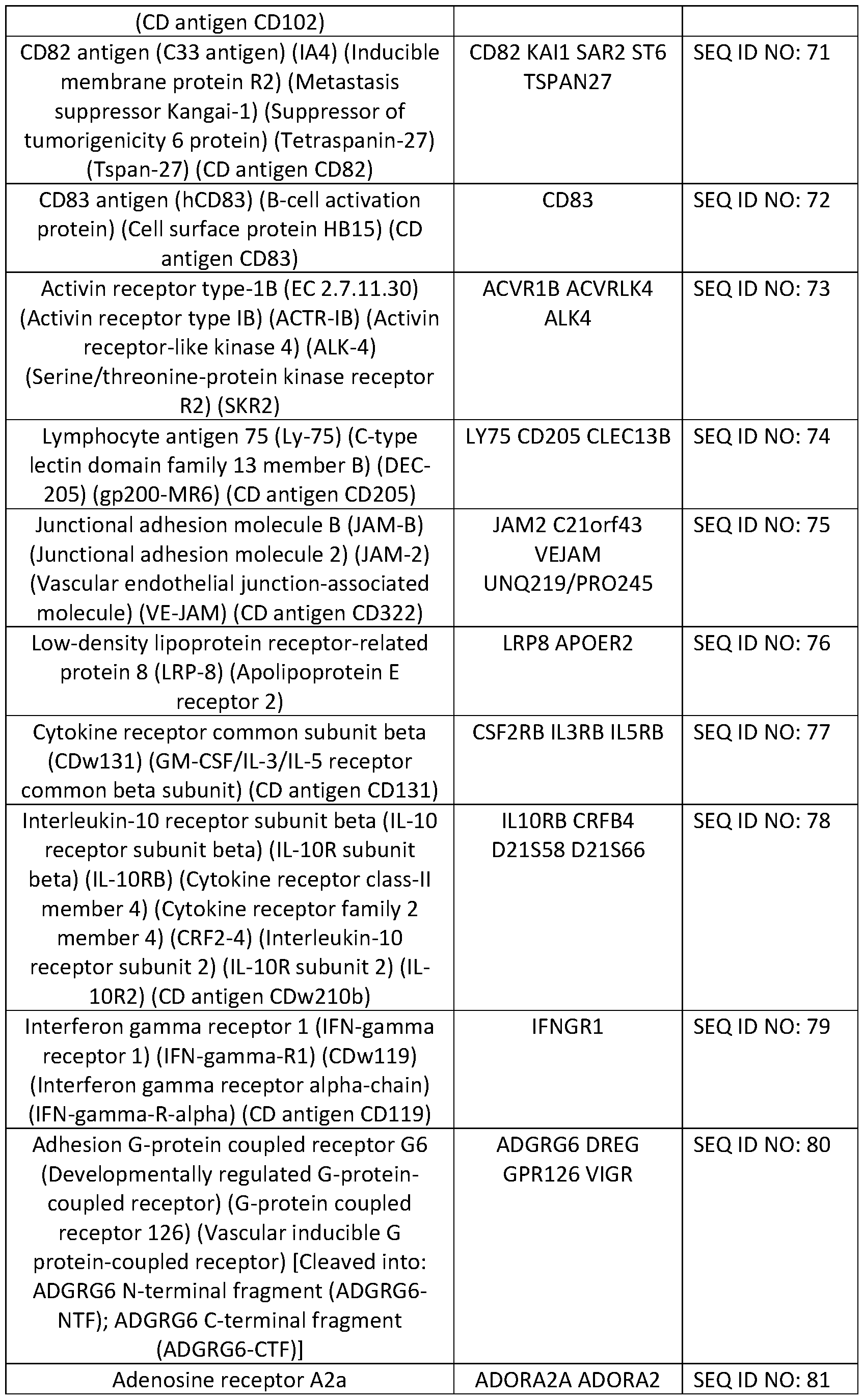


Claims
Priority Applications (7)
| Application Number | Priority Date | Filing Date | Title |
|---|---|---|---|
| EP21775630.3A EP4126243A1 (en) | 2020-03-27 | 2021-03-26 | Immunotherapeutic targets in multiple myeloma and methods for their identification |
| US17/913,189 US20230137672A1 (en) | 2020-03-27 | 2021-03-26 | Immunotherapeutic targets in multiple myeloma and methods for their identification |
| JP2022558042A JP2023519304A (en) | 2020-03-27 | 2021-03-26 | Immunotherapy targets in multiple myeloma and methods for their identification |
| IL296714A IL296714A (en) | 2020-03-27 | 2021-03-26 | Immunotherapeutic targets in multiple myeloma and methods for their identification |
| CA3175860A CA3175860A1 (en) | 2020-03-27 | 2021-03-26 | Immunotherapeutic targets in multiple myeloma and methods for their identification |
| CN202180024834.4A CN116097096A (en) | 2020-03-27 | 2021-03-26 | Immunotherapeutic targets in multiple myeloma and methods of identifying same |
| KR1020227037268A KR20220160053A (en) | 2020-03-27 | 2021-03-26 | Immunotherapy targets in multiple myeloma and methods for their identification |
Applications Claiming Priority (2)
| Application Number | Priority Date | Filing Date | Title |
|---|---|---|---|
| US202063000694P | 2020-03-27 | 2020-03-27 | |
| US63/000,694 | 2020-03-27 |
Publications (1)
| Publication Number | Publication Date |
|---|---|
| WO2021195536A1 true WO2021195536A1 (en) | 2021-09-30 |
Family
ID=77890610
Family Applications (1)
| Application Number | Title | Priority Date | Filing Date |
|---|---|---|---|
| PCT/US2021/024431 WO2021195536A1 (en) | 2020-03-27 | 2021-03-26 | Immunotherapeutic targets in multiple myeloma and methods for their identification |
Country Status (8)
| Country | Link |
|---|---|
| US (1) | US20230137672A1 (en) |
| EP (1) | EP4126243A1 (en) |
| JP (1) | JP2023519304A (en) |
| KR (1) | KR20220160053A (en) |
| CN (1) | CN116097096A (en) |
| CA (1) | CA3175860A1 (en) |
| IL (1) | IL296714A (en) |
| WO (1) | WO2021195536A1 (en) |
Cited By (2)
| Publication number | Priority date | Publication date | Assignee | Title |
|---|---|---|---|---|
| CN114031690A (en) * | 2021-12-03 | 2022-02-11 | 广州百暨基因科技有限公司 | Chimeric antigen receptor targeting CCR1 and NKG2D ligands and application thereof |
| WO2023105248A1 (en) | 2021-12-11 | 2023-06-15 | Cancer Research Technology Limited | Immunotherapy for cancer |
Families Citing this family (1)
| Publication number | Priority date | Publication date | Assignee | Title |
|---|---|---|---|---|
| MX2023001415A (en) * | 2020-08-05 | 2023-04-24 | Synthekine Inc | Compositions and methods related to il27 receptor binding. |
Citations (13)
| Publication number | Priority date | Publication date | Assignee | Title |
|---|---|---|---|---|
| US6046012A (en) * | 1993-07-19 | 2000-04-04 | Hoffmann-La Roche Inc. | Antibody to IL-12 receptor |
| US6635468B2 (en) * | 1997-09-17 | 2003-10-21 | Genentech, Inc. | Secreted and transmembrane polypeptides and nucleic acids encoding the same |
| US20100143912A1 (en) * | 2007-01-25 | 2010-06-10 | The Regents Of The Universuty Of California | Specific n-terminal labeling of peptides and proteins in complex mixtures |
| US20150301058A1 (en) * | 2012-11-26 | 2015-10-22 | Caris Science, Inc. | Biomarker compositions and methods |
| US20170074883A1 (en) * | 2015-09-14 | 2017-03-16 | Leukemia Therapeutics, LLC | Identification of novel diagnostics and therapeutics by modulating rhoh |
| US20170234874A1 (en) * | 2015-10-07 | 2017-08-17 | Clearbridge Biophotonics Pte Ltd. | Integrated visual morphology and cell protein expression using resonance-light scattering |
| US20180201901A1 (en) * | 2014-02-14 | 2018-07-19 | Cellectis | Cells for immunotherapy engineered for targeting antigen present both on immune cells and pathological cells |
| US20180348227A1 (en) * | 2016-08-04 | 2018-12-06 | Memorial Sloan-Kettering Cancer Center | Cancer antigen targets and uses thereof |
| WO2019094360A1 (en) * | 2017-11-07 | 2019-05-16 | The Board Of Regents Of The University Of Texas System | Targeting lilrb4 with car-t or car-nk cells in the treatment of cancer |
| WO2019099440A1 (en) * | 2017-11-14 | 2019-05-23 | Arcellx, Inc. | Multifunctional immune cell therapies |
| US20190194327A1 (en) * | 2017-12-22 | 2019-06-27 | Jounce Therapeutics, Inc. | Antibodies to lilrb2 |
| US20200041513A1 (en) * | 2016-05-20 | 2020-02-06 | Horiba Abx Sas | Residual disease detection in multiple myeloma |
| WO2020033585A1 (en) * | 2018-08-07 | 2020-02-13 | The Broad Institute, Inc. | Methods for combinatorial screening and use of therapeutic targets thereof |
-
2021
- 2021-03-26 US US17/913,189 patent/US20230137672A1/en active Pending
- 2021-03-26 CA CA3175860A patent/CA3175860A1/en active Pending
- 2021-03-26 EP EP21775630.3A patent/EP4126243A1/en active Pending
- 2021-03-26 CN CN202180024834.4A patent/CN116097096A/en active Pending
- 2021-03-26 WO PCT/US2021/024431 patent/WO2021195536A1/en active Application Filing
- 2021-03-26 JP JP2022558042A patent/JP2023519304A/en active Pending
- 2021-03-26 IL IL296714A patent/IL296714A/en unknown
- 2021-03-26 KR KR1020227037268A patent/KR20220160053A/en active Search and Examination
Patent Citations (13)
| Publication number | Priority date | Publication date | Assignee | Title |
|---|---|---|---|---|
| US6046012A (en) * | 1993-07-19 | 2000-04-04 | Hoffmann-La Roche Inc. | Antibody to IL-12 receptor |
| US6635468B2 (en) * | 1997-09-17 | 2003-10-21 | Genentech, Inc. | Secreted and transmembrane polypeptides and nucleic acids encoding the same |
| US20100143912A1 (en) * | 2007-01-25 | 2010-06-10 | The Regents Of The Universuty Of California | Specific n-terminal labeling of peptides and proteins in complex mixtures |
| US20150301058A1 (en) * | 2012-11-26 | 2015-10-22 | Caris Science, Inc. | Biomarker compositions and methods |
| US20180201901A1 (en) * | 2014-02-14 | 2018-07-19 | Cellectis | Cells for immunotherapy engineered for targeting antigen present both on immune cells and pathological cells |
| US20170074883A1 (en) * | 2015-09-14 | 2017-03-16 | Leukemia Therapeutics, LLC | Identification of novel diagnostics and therapeutics by modulating rhoh |
| US20170234874A1 (en) * | 2015-10-07 | 2017-08-17 | Clearbridge Biophotonics Pte Ltd. | Integrated visual morphology and cell protein expression using resonance-light scattering |
| US20200041513A1 (en) * | 2016-05-20 | 2020-02-06 | Horiba Abx Sas | Residual disease detection in multiple myeloma |
| US20180348227A1 (en) * | 2016-08-04 | 2018-12-06 | Memorial Sloan-Kettering Cancer Center | Cancer antigen targets and uses thereof |
| WO2019094360A1 (en) * | 2017-11-07 | 2019-05-16 | The Board Of Regents Of The University Of Texas System | Targeting lilrb4 with car-t or car-nk cells in the treatment of cancer |
| WO2019099440A1 (en) * | 2017-11-14 | 2019-05-23 | Arcellx, Inc. | Multifunctional immune cell therapies |
| US20190194327A1 (en) * | 2017-12-22 | 2019-06-27 | Jounce Therapeutics, Inc. | Antibodies to lilrb2 |
| WO2020033585A1 (en) * | 2018-08-07 | 2020-02-13 | The Broad Institute, Inc. | Methods for combinatorial screening and use of therapeutic targets thereof |
Cited By (2)
| Publication number | Priority date | Publication date | Assignee | Title |
|---|---|---|---|---|
| CN114031690A (en) * | 2021-12-03 | 2022-02-11 | 广州百暨基因科技有限公司 | Chimeric antigen receptor targeting CCR1 and NKG2D ligands and application thereof |
| WO2023105248A1 (en) | 2021-12-11 | 2023-06-15 | Cancer Research Technology Limited | Immunotherapy for cancer |
Also Published As
| Publication number | Publication date |
|---|---|
| EP4126243A1 (en) | 2023-02-08 |
| JP2023519304A (en) | 2023-05-10 |
| CN116097096A (en) | 2023-05-09 |
| US20230137672A1 (en) | 2023-05-04 |
| IL296714A (en) | 2022-11-01 |
| KR20220160053A (en) | 2022-12-05 |
| CA3175860A1 (en) | 2021-09-30 |
Similar Documents
| Publication | Publication Date | Title |
|---|---|---|
| WO2021195536A1 (en) | Immunotherapeutic targets in multiple myeloma and methods for their identification | |
| Gubin et al. | Checkpoint blockade cancer immunotherapy targets tumour-specific mutant antigens | |
| CN107903324B (en) | Bispecific antibody capable of binding to human CD19 and CD3 | |
| JP2017145258A (en) | APRIL variant | |
| CN110612446A (en) | Method for isolating neoantigen-specific T cell receptor sequences | |
| US20150086575A1 (en) | B cell surface reactive antibodies | |
| KR20210007954A (en) | Chimeric Receptor T Cell Therapy Using Characteristics of Tumor Microenvironment | |
| JP2020515236A (en) | Epigenetic analysis of cell therapy and related methods | |
| JP2019529437A (en) | Compositions and methods for characterizing the efficacy of solid tumors against anti-PD-L1 antibody monotherapy | |
| US20230074705A1 (en) | Target antigen discovery, phenotypic screens and use thereof for identification of target cell specific target epitopes | |
| IL298279A (en) | Immune cells with enhanced function | |
| WO2018145125A1 (en) | Coagulation factor viii mimetic protein and uses thereof | |
| CN117069842B (en) | Anti-human BCMA nanobody with specific isoelectric point, CAR-T and application | |
| EP3026060A1 (en) | Humanized antibody or fragment thereof specific for CD45R0 | |
| JP2023502990A (en) | Anti-B7-H3 monoclonal antibody and method of use thereof | |
| TW202126696A (en) | Anti-epha10 antibodies and methods of use thereof | |
| US20210277120A1 (en) | Compositions and methods for treatment of t cell malignancies | |
| CN113747917A (en) | Monoclonal antibody aiming at MHC (major histocompatibility complex) bound human Dickkopf-1 peptide and application thereof | |
| WO2022093694A1 (en) | Polypeptides targeting hpv peptide-mhc complexes and methods of use thereof | |
| CN114729403A (en) | Methods and systems for identifying and validating consensus candidate antigens and consensus antigen-specific T lymphocyte pairs | |
| US20240180969A1 (en) | Polypeptides targeting hla-a*11 and methods of use thereof | |
| Gloger | Analysis of the HLA class I immunopeptidome in health and in cancer | |
| WO2022212858A2 (en) | Polypeptides targeting hla-a*11 and methods of use thereof | |
| WO2023177821A2 (en) | Binding domains and methods of use thereof | |
| Salter | Signaling Analyses of Therapeutic T cells Engineered with Synthetic Receptors |
Legal Events
| Date | Code | Title | Description |
|---|---|---|---|
| 121 | Ep: the epo has been informed by wipo that ep was designated in this application |
Ref document number: 21775630 Country of ref document: EP Kind code of ref document: A1 |
|
| ENP | Entry into the national phase |
Ref document number: 3175860 Country of ref document: CA |
|
| ENP | Entry into the national phase |
Ref document number: 2022558042 Country of ref document: JP Kind code of ref document: A |
|
| ENP | Entry into the national phase |
Ref document number: 20227037268 Country of ref document: KR Kind code of ref document: A |
|
| WWE | Wipo information: entry into national phase |
Ref document number: 2021775630 Country of ref document: EP |
|
| ENP | Entry into the national phase |
Ref document number: 2021775630 Country of ref document: EP Effective date: 20221027 |
|
| NENP | Non-entry into the national phase |
Ref country code: DE |