WO2021066053A1 - Method for inducing hibernation-like state and device therefor - Google Patents
Method for inducing hibernation-like state and device therefor Download PDFInfo
- Publication number
- WO2021066053A1 WO2021066053A1 PCT/JP2020/037268 JP2020037268W WO2021066053A1 WO 2021066053 A1 WO2021066053 A1 WO 2021066053A1 JP 2020037268 W JP2020037268 W JP 2020037268W WO 2021066053 A1 WO2021066053 A1 WO 2021066053A1
- Authority
- WO
- WIPO (PCT)
- Prior art keywords
- qrfp
- administration
- body temperature
- neurons
- core body
- Prior art date
Links
- 238000000034 method Methods 0.000 title claims abstract description 94
- 230000001939 inductive effect Effects 0.000 title abstract description 16
- QPWYMHBRJDWMIS-AULSSRMGSA-N qrfp Chemical compound C([C@H](NC(=O)CNC(=O)[C@H](CC(N)=O)NC(=O)[C@H](CC(C)C)NC(=O)[C@H](CCC(O)=O)NC(=O)[C@H](CCC(O)=O)NC(=O)[C@H](C)NC(=O)[C@H](CC(C)C)NC(=O)[C@H](CO)NC(=O)CNC(=O)[C@@H](NC(=O)[C@H](C)NC(=O)[C@@H](NC(=O)CNC(=O)[C@@H](N)C(C)C)[C@@H](C)O)CC(C)C)C(=O)N[C@@H](CC(N)=O)C(=O)N[C@@H](CCCNC(N)=N)C(=O)N[C@@H](CCCCN)C(=O)N[C@@H](CCCCN)C(=O)NCC(=O)NCC(=O)N[C@@H](CC=1C=CC=CC=1)C(=O)N[C@@H](CO)C(=O)N[C@@H](CC=1C=CC=CC=1)C(=O)N[C@@H](CCCNC(N)=N)C(=O)N[C@@H](CC=1C=CC=CC=1)C(N)=O)C1=CC=C(O)C=C1 QPWYMHBRJDWMIS-AULSSRMGSA-N 0.000 claims abstract description 94
- 101000986779 Homo sapiens Orexigenic neuropeptide QRFP Proteins 0.000 claims abstract description 84
- 102100028142 Orexigenic neuropeptide QRFP Human genes 0.000 claims abstract description 84
- 230000000638 stimulation Effects 0.000 claims abstract description 41
- 230000036760 body temperature Effects 0.000 claims abstract description 32
- 230000002964 excitative effect Effects 0.000 claims abstract description 31
- 239000000126 substance Substances 0.000 claims abstract description 20
- 210000002569 neuron Anatomy 0.000 claims description 199
- 150000001875 compounds Chemical class 0.000 claims description 85
- 230000036757 core body temperature Effects 0.000 claims description 78
- 230000036284 oxygen consumption Effects 0.000 claims description 78
- 230000007423 decrease Effects 0.000 claims description 74
- 230000006266 hibernation Effects 0.000 claims description 55
- 210000004556 brain Anatomy 0.000 claims description 51
- 241000124008 Mammalia Species 0.000 claims description 43
- 210000003814 preoptic area Anatomy 0.000 claims description 39
- 238000012360 testing method Methods 0.000 claims description 39
- 230000002631 hypothermal effect Effects 0.000 claims description 38
- 230000002861 ventricular Effects 0.000 claims description 36
- 108090000765 processed proteins & peptides Proteins 0.000 claims description 30
- QVGXLLKOCUKJST-UHFFFAOYSA-N atomic oxygen Chemical compound [O] QVGXLLKOCUKJST-UHFFFAOYSA-N 0.000 claims description 28
- 229910052760 oxygen Inorganic materials 0.000 claims description 28
- 239000001301 oxygen Substances 0.000 claims description 28
- 150000001408 amides Chemical class 0.000 claims description 25
- 239000000523 sample Substances 0.000 claims description 22
- 230000003247 decreasing effect Effects 0.000 claims description 21
- 239000007789 gas Substances 0.000 claims description 18
- 238000003860 storage Methods 0.000 claims description 17
- 230000002829 reductive effect Effects 0.000 claims description 10
- 230000001537 neural effect Effects 0.000 claims description 9
- 230000002806 hypometabolic effect Effects 0.000 claims description 8
- 238000012384 transportation and delivery Methods 0.000 claims description 6
- 238000012216 screening Methods 0.000 claims description 5
- 230000004936 stimulating effect Effects 0.000 claims description 4
- 230000005540 biological transmission Effects 0.000 claims description 2
- 238000004519 manufacturing process Methods 0.000 abstract description 9
- 238000000053 physical method Methods 0.000 abstract 1
- 241000699670 Mus sp. Species 0.000 description 112
- 241001465754 Metazoa Species 0.000 description 44
- 238000009826 distribution Methods 0.000 description 33
- OGUCZBIQSYYWEF-UHFFFAOYSA-N Clozapine N-oxide Chemical compound C1C[N+](C)([O-])CCN1C1=NC2=CC(Cl)=CC=C2NC2=CC=CC=C12 OGUCZBIQSYYWEF-UHFFFAOYSA-N 0.000 description 29
- 210000003016 hypothalamus Anatomy 0.000 description 27
- 230000020169 heat generation Effects 0.000 description 26
- 210000004027 cell Anatomy 0.000 description 25
- 208000010513 Stupor Diseases 0.000 description 24
- 238000002474 experimental method Methods 0.000 description 21
- 241000702421 Dependoparvovirus Species 0.000 description 20
- 230000004060 metabolic process Effects 0.000 description 20
- 239000007924 injection Substances 0.000 description 19
- 238000002347 injection Methods 0.000 description 19
- 241000699666 Mus <mouse, genus> Species 0.000 description 18
- 230000005058 diapause Effects 0.000 description 18
- 230000002503 metabolic effect Effects 0.000 description 16
- 241000282412 Homo Species 0.000 description 15
- 230000000694 effects Effects 0.000 description 15
- 206010021034 Hypometabolism Diseases 0.000 description 14
- 101150006715 Qrfp gene Proteins 0.000 description 14
- 230000006870 function Effects 0.000 description 12
- 230000004913 activation Effects 0.000 description 11
- 230000002267 hypothalamic effect Effects 0.000 description 10
- 238000004458 analytical method Methods 0.000 description 9
- 230000001965 increasing effect Effects 0.000 description 9
- 230000006698 induction Effects 0.000 description 9
- SZBULDQSDUXAPJ-XNIJJKJLSA-N (2r,3r,4s,5r)-2-[6-(cyclohexylamino)purin-9-yl]-5-(hydroxymethyl)oxolane-3,4-diol Chemical compound O[C@@H]1[C@H](O)[C@@H](CO)O[C@H]1N1C2=NC=NC(NC3CCCCC3)=C2N=C1 SZBULDQSDUXAPJ-XNIJJKJLSA-N 0.000 description 8
- 230000033228 biological regulation Effects 0.000 description 8
- 239000000835 fiber Substances 0.000 description 8
- 238000007901 in situ hybridization Methods 0.000 description 8
- 210000002963 paraventricular hypothalamic nucleus Anatomy 0.000 description 8
- 101150106357 slc32a1 gene Proteins 0.000 description 8
- 239000000243 solution Substances 0.000 description 8
- 238000001356 surgical procedure Methods 0.000 description 8
- 210000003050 axon Anatomy 0.000 description 7
- 210000002364 input neuron Anatomy 0.000 description 7
- 230000007246 mechanism Effects 0.000 description 7
- 239000002953 phosphate buffered saline Substances 0.000 description 7
- 230000000241 respiratory effect Effects 0.000 description 7
- ODHCTXKNWHHXJC-VKHMYHEASA-N 5-oxo-L-proline Chemical compound OC(=O)[C@@H]1CCC(=O)N1 ODHCTXKNWHHXJC-VKHMYHEASA-N 0.000 description 6
- 241000283074 Equus asinus Species 0.000 description 6
- ODHCTXKNWHHXJC-GSVOUGTGSA-N Pyroglutamic acid Natural products OC(=O)[C@H]1CCC(=O)N1 ODHCTXKNWHHXJC-GSVOUGTGSA-N 0.000 description 6
- 101150077427 Slc17a6 gene Proteins 0.000 description 6
- ODHCTXKNWHHXJC-UHFFFAOYSA-N acide pyroglutamique Natural products OC(=O)C1CCC(=O)N1 ODHCTXKNWHHXJC-UHFFFAOYSA-N 0.000 description 6
- 230000005059 dormancy Effects 0.000 description 6
- 210000003796 lateral hypothalamic area Anatomy 0.000 description 6
- 230000001404 mediated effect Effects 0.000 description 6
- 230000036544 posture Effects 0.000 description 6
- 230000009467 reduction Effects 0.000 description 6
- 230000001105 regulatory effect Effects 0.000 description 6
- 210000000211 third ventricle Anatomy 0.000 description 6
- 239000013607 AAV vector Substances 0.000 description 5
- 241000700159 Rattus Species 0.000 description 5
- FAPWRFPIFSIZLT-UHFFFAOYSA-M Sodium chloride Chemical compound [Na+].[Cl-] FAPWRFPIFSIZLT-UHFFFAOYSA-M 0.000 description 5
- 239000012190 activator Substances 0.000 description 5
- 230000000903 blocking effect Effects 0.000 description 5
- 210000004369 blood Anatomy 0.000 description 5
- 239000008280 blood Substances 0.000 description 5
- 239000003814 drug Substances 0.000 description 5
- 230000014509 gene expression Effects 0.000 description 5
- 238000002695 general anesthesia Methods 0.000 description 5
- 239000012528 membrane Substances 0.000 description 5
- 108090000623 proteins and genes Proteins 0.000 description 5
- 102000005962 receptors Human genes 0.000 description 5
- 108020003175 receptors Proteins 0.000 description 5
- 238000011084 recovery Methods 0.000 description 5
- 230000007958 sleep Effects 0.000 description 5
- 230000002889 sympathetic effect Effects 0.000 description 5
- 210000002983 tuber cinereum Anatomy 0.000 description 5
- 206010002091 Anaesthesia Diseases 0.000 description 4
- IAZDPXIOMUYVGZ-UHFFFAOYSA-N Dimethylsulphoxide Chemical compound CS(C)=O IAZDPXIOMUYVGZ-UHFFFAOYSA-N 0.000 description 4
- PIWKPBJCKXDKJR-UHFFFAOYSA-N Isoflurane Chemical compound FC(F)OC(Cl)C(F)(F)F PIWKPBJCKXDKJR-UHFFFAOYSA-N 0.000 description 4
- 108090000189 Neuropeptides Proteins 0.000 description 4
- 102000003797 Neuropeptides Human genes 0.000 description 4
- 102100037469 Protein DEPP1 Human genes 0.000 description 4
- 102000003615 TRPM2 Human genes 0.000 description 4
- 101150095096 TRPM2 gene Proteins 0.000 description 4
- 241000700605 Viruses Species 0.000 description 4
- 230000037005 anaesthesia Effects 0.000 description 4
- 230000003542 behavioural effect Effects 0.000 description 4
- 230000037396 body weight Effects 0.000 description 4
- 238000004364 calculation method Methods 0.000 description 4
- 210000005056 cell body Anatomy 0.000 description 4
- 238000011161 development Methods 0.000 description 4
- 230000018109 developmental process Effects 0.000 description 4
- 229940079593 drug Drugs 0.000 description 4
- 230000005284 excitation Effects 0.000 description 4
- 238000004868 gas analysis Methods 0.000 description 4
- 229960002725 isoflurane Drugs 0.000 description 4
- 239000000463 material Substances 0.000 description 4
- 238000005259 measurement Methods 0.000 description 4
- 230000003287 optical effect Effects 0.000 description 4
- 210000000056 organ Anatomy 0.000 description 4
- 210000000221 suprachiasmatic nucleus Anatomy 0.000 description 4
- 210000004377 supraoptic nucleus Anatomy 0.000 description 4
- 238000001931 thermography Methods 0.000 description 4
- 239000013598 vector Substances 0.000 description 4
- 101150007969 ADORA1 gene Proteins 0.000 description 3
- OYPRJOBELJOOCE-UHFFFAOYSA-N Calcium Chemical compound [Ca] OYPRJOBELJOOCE-UHFFFAOYSA-N 0.000 description 3
- CURLTUGMZLYLDI-UHFFFAOYSA-N Carbon dioxide Chemical compound O=C=O CURLTUGMZLYLDI-UHFFFAOYSA-N 0.000 description 3
- PEDCQBHIVMGVHV-UHFFFAOYSA-N Glycerine Chemical compound OCC(O)CO PEDCQBHIVMGVHV-UHFFFAOYSA-N 0.000 description 3
- 241000283940 Marmota Species 0.000 description 3
- 238000013459 approach Methods 0.000 description 3
- 230000008901 benefit Effects 0.000 description 3
- 229910052791 calcium Inorganic materials 0.000 description 3
- 239000011575 calcium Substances 0.000 description 3
- 230000002060 circadian Effects 0.000 description 3
- 230000001419 dependent effect Effects 0.000 description 3
- 230000002068 genetic effect Effects 0.000 description 3
- 238000012744 immunostaining Methods 0.000 description 3
- 230000002401 inhibitory effect Effects 0.000 description 3
- 239000007928 intraperitoneal injection Substances 0.000 description 3
- 239000003446 ligand Substances 0.000 description 3
- 230000007774 longterm Effects 0.000 description 3
- 230000037323 metabolic rate Effects 0.000 description 3
- 210000005036 nerve Anatomy 0.000 description 3
- 230000002974 pharmacogenomic effect Effects 0.000 description 3
- 239000011780 sodium chloride Substances 0.000 description 3
- 230000000946 synaptic effect Effects 0.000 description 3
- 230000001331 thermoregulatory effect Effects 0.000 description 3
- 230000007704 transition Effects 0.000 description 3
- XLYOFNOQVPJJNP-UHFFFAOYSA-N water Substances O XLYOFNOQVPJJNP-UHFFFAOYSA-N 0.000 description 3
- -1 5-carboxy-2-oxazolyl Chemical group 0.000 description 2
- 108700028369 Alleles Proteins 0.000 description 2
- 102000004219 Brain-derived neurotrophic factor Human genes 0.000 description 2
- 108090000715 Brain-derived neurotrophic factor Proteins 0.000 description 2
- 238000011746 C57BL/6J (JAX™ mouse strain) Methods 0.000 description 2
- 229920000858 Cyclodextrin Polymers 0.000 description 2
- 101001060451 Homo sapiens Pyroglutamylated RF-amide peptide receptor Proteins 0.000 description 2
- 241000283973 Oryctolagus cuniculus Species 0.000 description 2
- 102000002808 Pituitary adenylate cyclase-activating polypeptide Human genes 0.000 description 2
- 108010004684 Pituitary adenylate cyclase-activating polypeptide Proteins 0.000 description 2
- 206010062519 Poor quality sleep Diseases 0.000 description 2
- 206010037660 Pyrexia Diseases 0.000 description 2
- 102100027888 Pyroglutamylated RF-amide peptide receptor Human genes 0.000 description 2
- 108010077297 QRFP peptide Proteins 0.000 description 2
- 206010037742 Rabies Diseases 0.000 description 2
- 241000711798 Rabies lyssavirus Species 0.000 description 2
- 241000555745 Sciuridae Species 0.000 description 2
- 229930006000 Sucrose Natural products 0.000 description 2
- CZMRCDWAGMRECN-UGDNZRGBSA-N Sucrose Chemical compound O[C@H]1[C@H](O)[C@@H](CO)O[C@@]1(CO)O[C@@H]1[C@H](O)[C@@H](O)[C@H](O)[C@@H](CO)O1 CZMRCDWAGMRECN-UGDNZRGBSA-N 0.000 description 2
- 206010044565 Tremor Diseases 0.000 description 2
- 229920004890 Triton X-100 Polymers 0.000 description 2
- 239000013504 Triton X-100 Substances 0.000 description 2
- XSQUKJJJFZCRTK-UHFFFAOYSA-N Urea Chemical compound NC(N)=O XSQUKJJJFZCRTK-UHFFFAOYSA-N 0.000 description 2
- OIRDTQYFTABQOQ-KQYNXXCUSA-N adenosine Chemical compound C1=NC=2C(N)=NC=NC=2N1[C@@H]1O[C@H](CO)[C@@H](O)[C@H]1O OIRDTQYFTABQOQ-KQYNXXCUSA-N 0.000 description 2
- 239000000556 agonist Substances 0.000 description 2
- GZCGUPFRVQAUEE-SLPGGIOYSA-N aldehydo-D-glucose Chemical compound OC[C@@H](O)[C@@H](O)[C@H](O)[C@@H](O)C=O GZCGUPFRVQAUEE-SLPGGIOYSA-N 0.000 description 2
- 230000006399 behavior Effects 0.000 description 2
- 238000005452 bending Methods 0.000 description 2
- 230000036772 blood pressure Effects 0.000 description 2
- 229910052799 carbon Inorganic materials 0.000 description 2
- 210000001175 cerebrospinal fluid Anatomy 0.000 description 2
- 230000008859 change Effects 0.000 description 2
- 238000004891 communication Methods 0.000 description 2
- 238000002591 computed tomography Methods 0.000 description 2
- 230000001276 controlling effect Effects 0.000 description 2
- 238000007796 conventional method Methods 0.000 description 2
- 238000010586 diagram Methods 0.000 description 2
- 210000001671 embryonic stem cell Anatomy 0.000 description 2
- 239000006274 endogenous ligand Substances 0.000 description 2
- 238000005265 energy consumption Methods 0.000 description 2
- 230000007613 environmental effect Effects 0.000 description 2
- 210000003059 ependyma Anatomy 0.000 description 2
- 239000007850 fluorescent dye Substances 0.000 description 2
- 210000003128 head Anatomy 0.000 description 2
- 230000006801 homologous recombination Effects 0.000 description 2
- 238000002744 homologous recombination Methods 0.000 description 2
- XMBWDFGMSWQBCA-UHFFFAOYSA-N hydrogen iodide Chemical compound I XMBWDFGMSWQBCA-UHFFFAOYSA-N 0.000 description 2
- 238000003384 imaging method Methods 0.000 description 2
- 238000003780 insertion Methods 0.000 description 2
- 230000037431 insertion Effects 0.000 description 2
- 238000009434 installation Methods 0.000 description 2
- 230000003993 interaction Effects 0.000 description 2
- 210000001153 interneuron Anatomy 0.000 description 2
- QWTDNUCVQCZILF-UHFFFAOYSA-N isopentane Chemical compound CCC(C)C QWTDNUCVQCZILF-UHFFFAOYSA-N 0.000 description 2
- 238000012417 linear regression Methods 0.000 description 2
- 239000007788 liquid Substances 0.000 description 2
- 239000002609 medium Substances 0.000 description 2
- 108020004999 messenger RNA Proteins 0.000 description 2
- PPAYPDQCRKDOKR-UHFFFAOYSA-M n,n-didecyl-4-[2-(1-methylpyridin-1-ium-4-yl)ethenyl]aniline;iodide Chemical compound [I-].C1=CC(N(CCCCCCCCCC)CCCCCCCCCC)=CC=C1\C=C\C1=CC=[N+](C)C=C1 PPAYPDQCRKDOKR-UHFFFAOYSA-M 0.000 description 2
- 230000007230 neural mechanism Effects 0.000 description 2
- 230000008284 neuronal mechanism Effects 0.000 description 2
- 239000013307 optical fiber Substances 0.000 description 2
- 210000002509 periaqueductal gray Anatomy 0.000 description 2
- 230000002093 peripheral effect Effects 0.000 description 2
- 230000002085 persistent effect Effects 0.000 description 2
- 239000002504 physiological saline solution Substances 0.000 description 2
- 230000004044 response Effects 0.000 description 2
- 238000012552 review Methods 0.000 description 2
- 230000035945 sensitivity Effects 0.000 description 2
- 210000002966 serum Anatomy 0.000 description 2
- 230000011664 signaling Effects 0.000 description 2
- 239000007858 starting material Substances 0.000 description 2
- 238000013179 statistical model Methods 0.000 description 2
- 239000005720 sucrose Substances 0.000 description 2
- 230000028016 temperature homeostasis Effects 0.000 description 2
- 238000010998 test method Methods 0.000 description 2
- 210000001519 tissue Anatomy 0.000 description 2
- 230000000451 tissue damage Effects 0.000 description 2
- 231100000827 tissue damage Toxicity 0.000 description 2
- WFKWXMTUELFFGS-UHFFFAOYSA-N tungsten Chemical compound [W] WFKWXMTUELFFGS-UHFFFAOYSA-N 0.000 description 2
- 229910052721 tungsten Inorganic materials 0.000 description 2
- 239000010937 tungsten Substances 0.000 description 2
- 238000009827 uniform distribution Methods 0.000 description 2
- 238000011144 upstream manufacturing Methods 0.000 description 2
- 230000002792 vascular Effects 0.000 description 2
- 238000005303 weighing Methods 0.000 description 2
- YZOUYRAONFXZSI-SBHWVFSVSA-N (1S,3R,5R,6R,8R,10R,11R,13R,15R,16R,18R,20R,21R,23R,25R,26R,28R,30R,31S,33R,35R,36R,37S,38R,39S,40R,41S,42R,43S,44R,45S,46R,47S,48R,49S)-5,10,15,20,25,30,35-heptakis(hydroxymethyl)-37,39,40,41,42,43,44,45,46,47,48,49-dodecamethoxy-2,4,7,9,12,14,17,19,22,24,27,29,32,34-tetradecaoxaoctacyclo[31.2.2.23,6.28,11.213,16.218,21.223,26.228,31]nonatetracontane-36,38-diol Chemical compound O([C@@H]([C@H]([C@@H]1OC)OC)O[C@H]2[C@@H](O)[C@@H]([C@@H](O[C@@H]3[C@@H](CO)O[C@@H]([C@H]([C@@H]3O)OC)O[C@@H]3[C@@H](CO)O[C@@H]([C@H]([C@@H]3OC)OC)O[C@@H]3[C@@H](CO)O[C@@H]([C@H]([C@@H]3OC)OC)O[C@@H]3[C@@H](CO)O[C@@H]([C@H]([C@@H]3OC)OC)O3)O[C@@H]2CO)OC)[C@H](CO)[C@H]1O[C@@H]1[C@@H](OC)[C@H](OC)[C@H]3[C@@H](CO)O1 YZOUYRAONFXZSI-SBHWVFSVSA-N 0.000 description 1
- BAPRUDZDYCKSOQ-RITPCOANSA-N (2s,4r)-1-acetyl-4-hydroxypyrrolidine-2-carboxylic acid Chemical compound CC(=O)N1C[C@H](O)C[C@H]1C(O)=O BAPRUDZDYCKSOQ-RITPCOANSA-N 0.000 description 1
- GDEURKKLNUGTDA-UHFFFAOYSA-M (2z)-3-propyl-2-[(2z,4z)-5-(3-propyl-1,3-benzothiazol-3-ium-2-yl)penta-2,4-dienylidene]-1,3-benzothiazole;iodide Chemical compound [I-].S1C2=CC=CC=C2[N+](CCC)=C1/C=C/C=C/C=C1N(CCC)C2=CC=CC=C2S1 GDEURKKLNUGTDA-UHFFFAOYSA-M 0.000 description 1
- JKMHFZQWWAIEOD-UHFFFAOYSA-N 2-[4-(2-hydroxyethyl)piperazin-1-yl]ethanesulfonic acid Chemical compound OCC[NH+]1CCN(CCS([O-])(=O)=O)CC1 JKMHFZQWWAIEOD-UHFFFAOYSA-N 0.000 description 1
- CFPWPNDPUSLDPF-UHFFFAOYSA-N 2-[carbamimidoyl(methyl)amino]-2-phosphonoacetic acid Chemical compound NC(=N)N(C)C(C(O)=O)P(O)(O)=O CFPWPNDPUSLDPF-UHFFFAOYSA-N 0.000 description 1
- 239000001763 2-hydroxyethyl(trimethyl)azanium Substances 0.000 description 1
- YWSTWDVPYHSYLN-UHFFFAOYSA-N 3-chloro-5-hydroxy-6-(4-methylpiperazin-1-yl)benzo[b][1,4]benzodiazepine Chemical compound C1CN(C)CCN1C1=C(C=CC=C2)C2=NC2=CC=C(Cl)C=C2N1O YWSTWDVPYHSYLN-UHFFFAOYSA-N 0.000 description 1
- FWBHETKCLVMNFS-UHFFFAOYSA-N 4',6-Diamino-2-phenylindol Chemical compound C1=CC(C(=N)N)=CC=C1C1=CC2=CC=C(C(N)=N)C=C2N1 FWBHETKCLVMNFS-UHFFFAOYSA-N 0.000 description 1
- RZVAJINKPMORJF-UHFFFAOYSA-N Acetaminophen Chemical compound CC(=O)NC1=CC=C(O)C=C1 RZVAJINKPMORJF-UHFFFAOYSA-N 0.000 description 1
- 108050000203 Adenosine receptors Proteins 0.000 description 1
- 102000009346 Adenosine receptors Human genes 0.000 description 1
- 239000012103 Alexa Fluor 488 Substances 0.000 description 1
- 208000019901 Anxiety disease Diseases 0.000 description 1
- 239000002126 C01EB10 - Adenosine Substances 0.000 description 1
- 241000212000 Caniformia Species 0.000 description 1
- 241000283707 Capra Species 0.000 description 1
- 241000282693 Cercopithecidae Species 0.000 description 1
- VEXZGXHMUGYJMC-UHFFFAOYSA-M Chloride anion Chemical compound [Cl-] VEXZGXHMUGYJMC-UHFFFAOYSA-M 0.000 description 1
- 235000019743 Choline chloride Nutrition 0.000 description 1
- 108091026890 Coding region Proteins 0.000 description 1
- 108010051219 Cre recombinase Proteins 0.000 description 1
- FBPFZTCFMRRESA-JGWLITMVSA-N D-glucitol Chemical compound OC[C@H](O)[C@@H](O)[C@H](O)[C@H](O)CO FBPFZTCFMRRESA-JGWLITMVSA-N 0.000 description 1
- 206010011953 Decreased activity Diseases 0.000 description 1
- 108090000045 G-Protein-Coupled Receptors Proteins 0.000 description 1
- 102000003688 G-Protein-Coupled Receptors Human genes 0.000 description 1
- WQZGKKKJIJFFOK-GASJEMHNSA-N Glucose Natural products OC[C@H]1OC(O)[C@H](O)[C@@H](O)[C@@H]1O WQZGKKKJIJFFOK-GASJEMHNSA-N 0.000 description 1
- 241000282575 Gorilla Species 0.000 description 1
- 102100030386 Granzyme A Human genes 0.000 description 1
- 239000007995 HEPES buffer Substances 0.000 description 1
- 241000978750 Havardia Species 0.000 description 1
- 102100029100 Hematopoietic prostaglandin D synthase Human genes 0.000 description 1
- 101001009599 Homo sapiens Granzyme A Proteins 0.000 description 1
- 101000988802 Homo sapiens Hematopoietic prostaglandin D synthase Proteins 0.000 description 1
- 206010020674 Hypermetabolism Diseases 0.000 description 1
- 206010021033 Hypomenorrhoea Diseases 0.000 description 1
- 241000167869 Ictidomys tridecemlineatus Species 0.000 description 1
- 241000581650 Ivesia Species 0.000 description 1
- WHUUTDBJXJRKMK-VKHMYHEASA-N L-glutamic acid Chemical compound OC(=O)[C@@H](N)CCC(O)=O WHUUTDBJXJRKMK-VKHMYHEASA-N 0.000 description 1
- 238000010826 Nissl staining Methods 0.000 description 1
- 241000282579 Pan Species 0.000 description 1
- 241000282576 Pan paniscus Species 0.000 description 1
- 229930040373 Paraformaldehyde Natural products 0.000 description 1
- 241000282405 Pongo abelii Species 0.000 description 1
- 241000288906 Primates Species 0.000 description 1
- 102000007568 Proto-Oncogene Proteins c-fos Human genes 0.000 description 1
- 108010071563 Proto-Oncogene Proteins c-fos Proteins 0.000 description 1
- 108010056448 RFamide peptide Proteins 0.000 description 1
- 241000283984 Rodentia Species 0.000 description 1
- 108091006162 SLC17A6 Proteins 0.000 description 1
- 101100084449 Saccharomyces cerevisiae (strain ATCC 204508 / S288c) PRP4 gene Proteins 0.000 description 1
- 241000283925 Spermophilus Species 0.000 description 1
- 241000701093 Suid alphaherpesvirus 1 Species 0.000 description 1
- 206010043189 Telangiectasia Diseases 0.000 description 1
- 108010055044 Tetanus Toxin Proteins 0.000 description 1
- 102000046053 Vesicular Glutamate Transport Protein 2 Human genes 0.000 description 1
- 102100038170 Vesicular inhibitory amino acid transporter Human genes 0.000 description 1
- 210000000683 abdominal cavity Anatomy 0.000 description 1
- 230000005856 abnormality Effects 0.000 description 1
- 239000002253 acid Substances 0.000 description 1
- 229960005305 adenosine Drugs 0.000 description 1
- 239000002582 adenosine A1 receptor agonist Substances 0.000 description 1
- 210000003486 adipose tissue brown Anatomy 0.000 description 1
- 230000001919 adrenal effect Effects 0.000 description 1
- VREFGVBLTWBCJP-UHFFFAOYSA-N alprazolam Chemical compound C12=CC(Cl)=CC=C2N2C(C)=NN=C2CN=C1C1=CC=CC=C1 VREFGVBLTWBCJP-UHFFFAOYSA-N 0.000 description 1
- 238000002266 amputation Methods 0.000 description 1
- 238000010171 animal model Methods 0.000 description 1
- 210000002159 anterior chamber Anatomy 0.000 description 1
- 210000001971 anterior hypothalamus Anatomy 0.000 description 1
- 230000036506 anxiety Effects 0.000 description 1
- 230000007529 anxiety like behavior Effects 0.000 description 1
- 235000021407 appetite control Nutrition 0.000 description 1
- 230000037007 arousal Effects 0.000 description 1
- 238000003556 assay Methods 0.000 description 1
- 238000013477 bayesian statistics method Methods 0.000 description 1
- 230000009286 beneficial effect Effects 0.000 description 1
- 238000012984 biological imaging Methods 0.000 description 1
- 238000009529 body temperature measurement Methods 0.000 description 1
- 239000004202 carbamide Substances 0.000 description 1
- 229910002092 carbon dioxide Inorganic materials 0.000 description 1
- 239000001569 carbon dioxide Substances 0.000 description 1
- 235000011089 carbon dioxide Nutrition 0.000 description 1
- 230000001413 cellular effect Effects 0.000 description 1
- 210000003169 central nervous system Anatomy 0.000 description 1
- 239000003153 chemical reaction reagent Substances 0.000 description 1
- 229960003178 choline chloride Drugs 0.000 description 1
- SGMZJAMFUVOLNK-UHFFFAOYSA-M choline chloride Chemical compound [Cl-].C[N+](C)(C)CCO SGMZJAMFUVOLNK-UHFFFAOYSA-M 0.000 description 1
- 230000005895 circadian behavior Effects 0.000 description 1
- 230000009636 circadian regulation Effects 0.000 description 1
- 238000004140 cleaning Methods 0.000 description 1
- QZUDBNBUXVUHMW-UHFFFAOYSA-N clozapine Chemical compound C1CN(C)CCN1C1=NC2=CC(Cl)=CC=C2NC2=CC=CC=C12 QZUDBNBUXVUHMW-UHFFFAOYSA-N 0.000 description 1
- 229960004170 clozapine Drugs 0.000 description 1
- 230000001054 cortical effect Effects 0.000 description 1
- 230000008878 coupling Effects 0.000 description 1
- 238000010168 coupling process Methods 0.000 description 1
- 238000005859 coupling reaction Methods 0.000 description 1
- 239000006059 cover glass Substances 0.000 description 1
- 239000013078 crystal Substances 0.000 description 1
- 210000004748 cultured cell Anatomy 0.000 description 1
- 238000005520 cutting process Methods 0.000 description 1
- 238000012217 deletion Methods 0.000 description 1
- 230000037430 deletion Effects 0.000 description 1
- 210000001787 dendrite Anatomy 0.000 description 1
- 238000013461 design Methods 0.000 description 1
- 238000001514 detection method Methods 0.000 description 1
- 210000002451 diencephalon Anatomy 0.000 description 1
- 235000005911 diet Nutrition 0.000 description 1
- 230000037213 diet Effects 0.000 description 1
- LOKCTEFSRHRXRJ-UHFFFAOYSA-I dipotassium trisodium dihydrogen phosphate hydrogen phosphate dichloride Chemical compound P(=O)(O)(O)[O-].[K+].P(=O)(O)([O-])[O-].[Na+].[Na+].[Cl-].[K+].[Cl-].[Na+] LOKCTEFSRHRXRJ-UHFFFAOYSA-I 0.000 description 1
- 238000001647 drug administration Methods 0.000 description 1
- 230000004064 dysfunction Effects 0.000 description 1
- 239000012636 effector Substances 0.000 description 1
- 238000003372 electrophysiological method Methods 0.000 description 1
- 210000002257 embryonic structure Anatomy 0.000 description 1
- 230000002124 endocrine Effects 0.000 description 1
- 230000002708 enhancing effect Effects 0.000 description 1
- 150000002148 esters Chemical class 0.000 description 1
- DEFVIWRASFVYLL-UHFFFAOYSA-N ethylene glycol bis(2-aminoethyl)tetraacetic acid Chemical compound OC(=O)CN(CC(O)=O)CCOCCOCCN(CC(O)=O)CC(O)=O DEFVIWRASFVYLL-UHFFFAOYSA-N 0.000 description 1
- 238000011156 evaluation Methods 0.000 description 1
- 238000013401 experimental design Methods 0.000 description 1
- 238000010195 expression analysis Methods 0.000 description 1
- 238000010304 firing Methods 0.000 description 1
- 230000037406 food intake Effects 0.000 description 1
- 235000012631 food intake Nutrition 0.000 description 1
- 238000010230 functional analysis Methods 0.000 description 1
- 210000001222 gaba-ergic neuron Anatomy 0.000 description 1
- GDSRMADSINPKSL-HSEONFRVSA-N gamma-cyclodextrin Chemical compound OC[C@H]([C@H]([C@@H]([C@H]1O)O)O[C@H]2O[C@@H]([C@@H](O[C@H]3O[C@H](CO)[C@H]([C@@H]([C@H]3O)O)O[C@H]3O[C@H](CO)[C@H]([C@@H]([C@H]3O)O)O[C@H]3O[C@H](CO)[C@H]([C@@H]([C@H]3O)O)O[C@H]3O[C@H](CO)[C@H]([C@@H]([C@H]3O)O)O[C@H]3O[C@H](CO)[C@H]([C@@H]([C@H]3O)O)O3)[C@H](O)[C@H]2O)CO)O[C@@H]1O[C@H]1[C@H](O)[C@@H](O)[C@@H]3O[C@@H]1CO GDSRMADSINPKSL-HSEONFRVSA-N 0.000 description 1
- 229940080345 gamma-cyclodextrin Drugs 0.000 description 1
- 239000003193 general anesthetic agent Substances 0.000 description 1
- 229940005494 general anesthetics Drugs 0.000 description 1
- 210000001905 globus pallidus Anatomy 0.000 description 1
- 229940050410 gluconate Drugs 0.000 description 1
- 230000004110 gluconeogenesis Effects 0.000 description 1
- 229960001031 glucose Drugs 0.000 description 1
- 239000008103 glucose Substances 0.000 description 1
- 244000144993 groups of animals Species 0.000 description 1
- 238000010438 heat treatment Methods 0.000 description 1
- 125000005842 heteroatom Chemical group 0.000 description 1
- 230000001744 histochemical effect Effects 0.000 description 1
- 230000001127 hyperphagic effect Effects 0.000 description 1
- 238000011532 immunohistochemical staining Methods 0.000 description 1
- 239000007943 implant Substances 0.000 description 1
- 238000000338 in vitro Methods 0.000 description 1
- 238000001727 in vivo Methods 0.000 description 1
- 238000011534 incubation Methods 0.000 description 1
- 208000015181 infectious disease Diseases 0.000 description 1
- 230000002458 infectious effect Effects 0.000 description 1
- 230000010365 information processing Effects 0.000 description 1
- 238000001802 infusion Methods 0.000 description 1
- 230000005764 inhibitory process Effects 0.000 description 1
- 238000007689 inspection Methods 0.000 description 1
- 230000003585 interneuronal effect Effects 0.000 description 1
- 230000000968 intestinal effect Effects 0.000 description 1
- 230000003834 intracellular effect Effects 0.000 description 1
- 238000000185 intracerebroventricular administration Methods 0.000 description 1
- 238000007913 intrathecal administration Methods 0.000 description 1
- 238000001990 intravenous administration Methods 0.000 description 1
- 150000002576 ketones Chemical class 0.000 description 1
- 238000002372 labelling Methods 0.000 description 1
- 230000033001 locomotion Effects 0.000 description 1
- 210000000627 locus coeruleus Anatomy 0.000 description 1
- 238000012423 maintenance Methods 0.000 description 1
- 238000013507 mapping Methods 0.000 description 1
- 239000003550 marker Substances 0.000 description 1
- 230000013011 mating Effects 0.000 description 1
- 210000001767 medulla oblongata Anatomy 0.000 description 1
- 230000028161 membrane depolarization Effects 0.000 description 1
- 230000007102 metabolic function Effects 0.000 description 1
- 238000006241 metabolic reaction Methods 0.000 description 1
- DZNKOAWEHDKBEP-UHFFFAOYSA-N methyl 2-[6-[bis(2-methoxy-2-oxoethyl)amino]-5-[2-[2-[bis(2-methoxy-2-oxoethyl)amino]-5-methylphenoxy]ethoxy]-1-benzofuran-2-yl]-1,3-oxazole-5-carboxylate Chemical compound COC(=O)CN(CC(=O)OC)C1=CC=C(C)C=C1OCCOC(C(=C1)N(CC(=O)OC)CC(=O)OC)=CC2=C1OC(C=1OC(=CN=1)C(=O)OC)=C2 DZNKOAWEHDKBEP-UHFFFAOYSA-N 0.000 description 1
- 230000037023 motor activity Effects 0.000 description 1
- 239000012120 mounting media Substances 0.000 description 1
- 238000007837 multiplex assay Methods 0.000 description 1
- 208000010125 myocardial infarction Diseases 0.000 description 1
- 239000013642 negative control Substances 0.000 description 1
- 230000001423 neocortical effect Effects 0.000 description 1
- 210000000653 nervous system Anatomy 0.000 description 1
- 230000000955 neuroendocrine Effects 0.000 description 1
- 230000008452 non REM sleep Effects 0.000 description 1
- 235000003715 nutritional status Nutrition 0.000 description 1
- 229920002113 octoxynol Polymers 0.000 description 1
- 230000001956 orexigenic effect Effects 0.000 description 1
- 229960005113 oxaceprol Drugs 0.000 description 1
- 229920002866 paraformaldehyde Polymers 0.000 description 1
- 230000037361 pathway Effects 0.000 description 1
- 230000010412 perfusion Effects 0.000 description 1
- 210000005259 peripheral blood Anatomy 0.000 description 1
- 239000011886 peripheral blood Substances 0.000 description 1
- 230000002688 persistence Effects 0.000 description 1
- 239000008363 phosphate buffer Substances 0.000 description 1
- 230000003863 physical function Effects 0.000 description 1
- 230000035479 physiological effects, processes and functions Effects 0.000 description 1
- 229920003229 poly(methyl methacrylate) Polymers 0.000 description 1
- 239000004926 polymethyl methacrylate Substances 0.000 description 1
- 238000002203 pretreatment Methods 0.000 description 1
- 238000007639 printing Methods 0.000 description 1
- 230000001737 promoting effect Effects 0.000 description 1
- BHMBVRSPMRCCGG-OUTUXVNYSA-N prostaglandin D2 Chemical compound CCCCC[C@H](O)\C=C\[C@@H]1[C@@H](C\C=C/CCCC(O)=O)[C@@H](O)CC1=O BHMBVRSPMRCCGG-OUTUXVNYSA-N 0.000 description 1
- BHMBVRSPMRCCGG-UHFFFAOYSA-N prostaglandine D2 Natural products CCCCCC(O)C=CC1C(CC=CCCCC(O)=O)C(O)CC1=O BHMBVRSPMRCCGG-UHFFFAOYSA-N 0.000 description 1
- 102000004169 proteins and genes Human genes 0.000 description 1
- 230000002685 pulmonary effect Effects 0.000 description 1
- 238000003753 real-time PCR Methods 0.000 description 1
- 230000015355 regulation of circadian sleep/wake cycle, sleep Effects 0.000 description 1
- 238000003303 reheating Methods 0.000 description 1
- 230000029058 respiratory gaseous exchange Effects 0.000 description 1
- 230000036387 respiratory rate Effects 0.000 description 1
- 230000002441 reversible effect Effects 0.000 description 1
- TUFFYSFVSYUHPA-UHFFFAOYSA-M rhodamine 123 Chemical compound [Cl-].COC(=O)C1=CC=CC=C1C1=C(C=CC(N)=C2)C2=[O+]C2=C1C=CC(N)=C2 TUFFYSFVSYUHPA-UHFFFAOYSA-M 0.000 description 1
- 230000033764 rhythmic process Effects 0.000 description 1
- 210000005262 rostral ventrolateral medulla Anatomy 0.000 description 1
- 238000005070 sampling Methods 0.000 description 1
- 210000003625 skull Anatomy 0.000 description 1
- 239000002904 solvent Substances 0.000 description 1
- 238000012421 spiking Methods 0.000 description 1
- 238000010186 staining Methods 0.000 description 1
- 229910001220 stainless steel Inorganic materials 0.000 description 1
- 239000010935 stainless steel Substances 0.000 description 1
- 238000007619 statistical method Methods 0.000 description 1
- 239000000758 substrate Substances 0.000 description 1
- 239000006228 supernatant Substances 0.000 description 1
- 230000001629 suppression Effects 0.000 description 1
- 210000000225 synapse Anatomy 0.000 description 1
- 230000005062 synaptic transmission Effects 0.000 description 1
- 238000007910 systemic administration Methods 0.000 description 1
- 238000012385 systemic delivery Methods 0.000 description 1
- 230000009885 systemic effect Effects 0.000 description 1
- 230000008685 targeting Effects 0.000 description 1
- 208000009056 telangiectasis Diseases 0.000 description 1
- 210000001103 thalamus Anatomy 0.000 description 1
- 230000000476 thermogenic effect Effects 0.000 description 1
- 210000000115 thoracic cavity Anatomy 0.000 description 1
- 238000013518 transcription Methods 0.000 description 1
- 230000035897 transcription Effects 0.000 description 1
- 238000001890 transfection Methods 0.000 description 1
- 238000012546 transfer Methods 0.000 description 1
- 238000002054 transplantation Methods 0.000 description 1
- 230000001960 triggered effect Effects 0.000 description 1
- 241000701161 unidentified adenovirus Species 0.000 description 1
- 210000003462 vein Anatomy 0.000 description 1
- 108010030503 vesicular GABA transporter Proteins 0.000 description 1
- 238000011179 visual inspection Methods 0.000 description 1
Images


Classifications
-
- A—HUMAN NECESSITIES
- A61—MEDICAL OR VETERINARY SCIENCE; HYGIENE
- A61N—ELECTROTHERAPY; MAGNETOTHERAPY; RADIATION THERAPY; ULTRASOUND THERAPY
- A61N1/00—Electrotherapy; Circuits therefor
- A61N1/18—Applying electric currents by contact electrodes
- A61N1/32—Applying electric currents by contact electrodes alternating or intermittent currents
- A61N1/36—Applying electric currents by contact electrodes alternating or intermittent currents for stimulation
- A61N1/3605—Implantable neurostimulators for stimulating central or peripheral nerve system
- A61N1/3606—Implantable neurostimulators for stimulating central or peripheral nerve system adapted for a particular treatment
- A61N1/36078—Inducing or controlling sleep or relaxation
-
- A—HUMAN NECESSITIES
- A61—MEDICAL OR VETERINARY SCIENCE; HYGIENE
- A61N—ELECTROTHERAPY; MAGNETOTHERAPY; RADIATION THERAPY; ULTRASOUND THERAPY
- A61N1/00—Electrotherapy; Circuits therefor
- A61N1/02—Details
- A61N1/04—Electrodes
- A61N1/05—Electrodes for implantation or insertion into the body, e.g. heart electrode
- A61N1/0526—Head electrodes
- A61N1/0529—Electrodes for brain stimulation
- A61N1/0534—Electrodes for deep brain stimulation
-
- A—HUMAN NECESSITIES
- A01—AGRICULTURE; FORESTRY; ANIMAL HUSBANDRY; HUNTING; TRAPPING; FISHING
- A01K—ANIMAL HUSBANDRY; AVICULTURE; APICULTURE; PISCICULTURE; FISHING; REARING OR BREEDING ANIMALS, NOT OTHERWISE PROVIDED FOR; NEW BREEDS OF ANIMALS
- A01K29/00—Other apparatus for animal husbandry
- A01K29/005—Monitoring or measuring activity, e.g. detecting heat or mating
-
- A—HUMAN NECESSITIES
- A01—AGRICULTURE; FORESTRY; ANIMAL HUSBANDRY; HUNTING; TRAPPING; FISHING
- A01K—ANIMAL HUSBANDRY; AVICULTURE; APICULTURE; PISCICULTURE; FISHING; REARING OR BREEDING ANIMALS, NOT OTHERWISE PROVIDED FOR; NEW BREEDS OF ANIMALS
- A01K67/00—Rearing or breeding animals, not otherwise provided for; New or modified breeds of animals
-
- A—HUMAN NECESSITIES
- A61—MEDICAL OR VETERINARY SCIENCE; HYGIENE
- A61K—PREPARATIONS FOR MEDICAL, DENTAL OR TOILETRY PURPOSES
- A61K49/00—Preparations for testing in vivo
- A61K49/0004—Screening or testing of compounds for diagnosis of disorders, assessment of conditions, e.g. renal clearance, gastric emptying, testing for diabetes, allergy, rheuma, pancreas functions
-
- A—HUMAN NECESSITIES
- A61—MEDICAL OR VETERINARY SCIENCE; HYGIENE
- A61M—DEVICES FOR INTRODUCING MEDIA INTO, OR ONTO, THE BODY; DEVICES FOR TRANSDUCING BODY MEDIA OR FOR TAKING MEDIA FROM THE BODY; DEVICES FOR PRODUCING OR ENDING SLEEP OR STUPOR
- A61M21/00—Other devices or methods to cause a change in the state of consciousness; Devices for producing or ending sleep by mechanical, optical, or acoustical means, e.g. for hypnosis
- A61M21/02—Other devices or methods to cause a change in the state of consciousness; Devices for producing or ending sleep by mechanical, optical, or acoustical means, e.g. for hypnosis for inducing sleep or relaxation, e.g. by direct nerve stimulation, hypnosis, analgesia
-
- A—HUMAN NECESSITIES
- A61—MEDICAL OR VETERINARY SCIENCE; HYGIENE
- A61N—ELECTROTHERAPY; MAGNETOTHERAPY; RADIATION THERAPY; ULTRASOUND THERAPY
- A61N1/00—Electrotherapy; Circuits therefor
- A61N1/02—Details
- A61N1/04—Electrodes
- A61N1/05—Electrodes for implantation or insertion into the body, e.g. heart electrode
- A61N1/0526—Head electrodes
- A61N1/0529—Electrodes for brain stimulation
-
- A—HUMAN NECESSITIES
- A61—MEDICAL OR VETERINARY SCIENCE; HYGIENE
- A61N—ELECTROTHERAPY; MAGNETOTHERAPY; RADIATION THERAPY; ULTRASOUND THERAPY
- A61N1/00—Electrotherapy; Circuits therefor
- A61N1/18—Applying electric currents by contact electrodes
- A61N1/32—Applying electric currents by contact electrodes alternating or intermittent currents
- A61N1/36—Applying electric currents by contact electrodes alternating or intermittent currents for stimulation
- A61N1/3605—Implantable neurostimulators for stimulating central or peripheral nerve system
- A61N1/3606—Implantable neurostimulators for stimulating central or peripheral nerve system adapted for a particular treatment
- A61N1/36121—Production of neurotransmitters; Modulation of genes expression
-
- A—HUMAN NECESSITIES
- A61—MEDICAL OR VETERINARY SCIENCE; HYGIENE
- A61N—ELECTROTHERAPY; MAGNETOTHERAPY; RADIATION THERAPY; ULTRASOUND THERAPY
- A61N1/00—Electrotherapy; Circuits therefor
- A61N1/18—Applying electric currents by contact electrodes
- A61N1/32—Applying electric currents by contact electrodes alternating or intermittent currents
- A61N1/36—Applying electric currents by contact electrodes alternating or intermittent currents for stimulation
- A61N1/3605—Implantable neurostimulators for stimulating central or peripheral nerve system
- A61N1/36128—Control systems
- A61N1/36135—Control systems using physiological parameters
-
- G—PHYSICS
- G01—MEASURING; TESTING
- G01N—INVESTIGATING OR ANALYSING MATERIALS BY DETERMINING THEIR CHEMICAL OR PHYSICAL PROPERTIES
- G01N33/00—Investigating or analysing materials by specific methods not covered by groups G01N1/00 - G01N31/00
- G01N33/48—Biological material, e.g. blood, urine; Haemocytometers
- G01N33/50—Chemical analysis of biological material, e.g. blood, urine; Testing involving biospecific ligand binding methods; Immunological testing
- G01N33/5005—Chemical analysis of biological material, e.g. blood, urine; Testing involving biospecific ligand binding methods; Immunological testing involving human or animal cells
- G01N33/5008—Chemical analysis of biological material, e.g. blood, urine; Testing involving biospecific ligand binding methods; Immunological testing involving human or animal cells for testing or evaluating the effect of chemical or biological compounds, e.g. drugs, cosmetics
- G01N33/5044—Chemical analysis of biological material, e.g. blood, urine; Testing involving biospecific ligand binding methods; Immunological testing involving human or animal cells for testing or evaluating the effect of chemical or biological compounds, e.g. drugs, cosmetics involving specific cell types
- G01N33/5058—Neurological cells
-
- A—HUMAN NECESSITIES
- A61—MEDICAL OR VETERINARY SCIENCE; HYGIENE
- A61B—DIAGNOSIS; SURGERY; IDENTIFICATION
- A61B5/00—Measuring for diagnostic purposes; Identification of persons
- A61B5/01—Measuring temperature of body parts ; Diagnostic temperature sensing, e.g. for malignant or inflamed tissue
-
- A—HUMAN NECESSITIES
- A61—MEDICAL OR VETERINARY SCIENCE; HYGIENE
- A61B—DIAGNOSIS; SURGERY; IDENTIFICATION
- A61B5/00—Measuring for diagnostic purposes; Identification of persons
- A61B5/08—Detecting, measuring or recording devices for evaluating the respiratory organs
- A61B5/083—Measuring rate of metabolism by using breath test, e.g. measuring rate of oxygen consumption
- A61B5/0833—Measuring rate of oxygen consumption
-
- A—HUMAN NECESSITIES
- A61—MEDICAL OR VETERINARY SCIENCE; HYGIENE
- A61M—DEVICES FOR INTRODUCING MEDIA INTO, OR ONTO, THE BODY; DEVICES FOR TRANSDUCING BODY MEDIA OR FOR TAKING MEDIA FROM THE BODY; DEVICES FOR PRODUCING OR ENDING SLEEP OR STUPOR
- A61M21/00—Other devices or methods to cause a change in the state of consciousness; Devices for producing or ending sleep by mechanical, optical, or acoustical means, e.g. for hypnosis
- A61M2021/0005—Other devices or methods to cause a change in the state of consciousness; Devices for producing or ending sleep by mechanical, optical, or acoustical means, e.g. for hypnosis by the use of a particular sense, or stimulus
- A61M2021/0072—Other devices or methods to cause a change in the state of consciousness; Devices for producing or ending sleep by mechanical, optical, or acoustical means, e.g. for hypnosis by the use of a particular sense, or stimulus with application of electrical currents
-
- A—HUMAN NECESSITIES
- A61—MEDICAL OR VETERINARY SCIENCE; HYGIENE
- A61M—DEVICES FOR INTRODUCING MEDIA INTO, OR ONTO, THE BODY; DEVICES FOR TRANSDUCING BODY MEDIA OR FOR TAKING MEDIA FROM THE BODY; DEVICES FOR PRODUCING OR ENDING SLEEP OR STUPOR
- A61M21/00—Other devices or methods to cause a change in the state of consciousness; Devices for producing or ending sleep by mechanical, optical, or acoustical means, e.g. for hypnosis
- A61M2021/0005—Other devices or methods to cause a change in the state of consciousness; Devices for producing or ending sleep by mechanical, optical, or acoustical means, e.g. for hypnosis by the use of a particular sense, or stimulus
- A61M2021/0077—Other devices or methods to cause a change in the state of consciousness; Devices for producing or ending sleep by mechanical, optical, or acoustical means, e.g. for hypnosis by the use of a particular sense, or stimulus with application of chemical or pharmacological stimulus
-
- A—HUMAN NECESSITIES
- A61—MEDICAL OR VETERINARY SCIENCE; HYGIENE
- A61M—DEVICES FOR INTRODUCING MEDIA INTO, OR ONTO, THE BODY; DEVICES FOR TRANSDUCING BODY MEDIA OR FOR TAKING MEDIA FROM THE BODY; DEVICES FOR PRODUCING OR ENDING SLEEP OR STUPOR
- A61M2205/00—General characteristics of the apparatus
- A61M2205/33—Controlling, regulating or measuring
- A61M2205/3368—Temperature
-
- A—HUMAN NECESSITIES
- A61—MEDICAL OR VETERINARY SCIENCE; HYGIENE
- A61M—DEVICES FOR INTRODUCING MEDIA INTO, OR ONTO, THE BODY; DEVICES FOR TRANSDUCING BODY MEDIA OR FOR TAKING MEDIA FROM THE BODY; DEVICES FOR PRODUCING OR ENDING SLEEP OR STUPOR
- A61M2205/00—General characteristics of the apparatus
- A61M2205/50—General characteristics of the apparatus with microprocessors or computers
- A61M2205/52—General characteristics of the apparatus with microprocessors or computers with memories providing a history of measured variating parameters of apparatus or patient
-
- A—HUMAN NECESSITIES
- A61—MEDICAL OR VETERINARY SCIENCE; HYGIENE
- A61M—DEVICES FOR INTRODUCING MEDIA INTO, OR ONTO, THE BODY; DEVICES FOR TRANSDUCING BODY MEDIA OR FOR TAKING MEDIA FROM THE BODY; DEVICES FOR PRODUCING OR ENDING SLEEP OR STUPOR
- A61M2230/00—Measuring parameters of the user
- A61M2230/40—Respiratory characteristics
- A61M2230/43—Composition of exhalation
- A61M2230/435—Composition of exhalation partial O2 pressure (P-O2)
-
- A—HUMAN NECESSITIES
- A61—MEDICAL OR VETERINARY SCIENCE; HYGIENE
- A61M—DEVICES FOR INTRODUCING MEDIA INTO, OR ONTO, THE BODY; DEVICES FOR TRANSDUCING BODY MEDIA OR FOR TAKING MEDIA FROM THE BODY; DEVICES FOR PRODUCING OR ENDING SLEEP OR STUPOR
- A61M2230/00—Measuring parameters of the user
- A61M2230/50—Temperature
-
- A—HUMAN NECESSITIES
- A61—MEDICAL OR VETERINARY SCIENCE; HYGIENE
- A61M—DEVICES FOR INTRODUCING MEDIA INTO, OR ONTO, THE BODY; DEVICES FOR TRANSDUCING BODY MEDIA OR FOR TAKING MEDIA FROM THE BODY; DEVICES FOR PRODUCING OR ENDING SLEEP OR STUPOR
- A61M2250/00—Specially adapted for animals
Definitions
- the present invention provides a method for inducing a hibernation-like state and a device for that purpose.
- Elucidation of the mechanism of daily diapause and / or hibernation is a necessary step to develop a method for artificially inducing artificial hibernation-like hypometabolism in non-hibernating animals including humans 1,7 , and further. Will also be useful in long-range space exploration in the future.
- excitatory manipulation of a novel chemically defined neuronal population in the hypothalamus results in a very long-term hypometabolism / hypothermia in mice. In this state, the metabolic rate drops to less than one-third, but unlike the anesthetized state, the mice still respond to changes in ambient temperature. In addition, the mice recovered spontaneously from this condition without any apparent abnormalities. This finding is an important finding in the development of hibernation mechanisms and methods for inducing artificial hibernation-like states.
- the present invention provides a method for inducing a hibernation-like state and a device for that purpose.
- AVPe anterior ventricular periventricular nucleus
- MPA medial preoptic area
- Pe periventricular nucleus
- QRFP pyroglutaminated RF amide peptide
- the following invention is provided.
- AVPe anterior ventricular periventricular nucleus
- MPA medial preoptic area
- Pe periventricular nucleus
- QRFP Pyroglutamic acid RF amide peptide
- a control unit that transmits a control signal that controls the generation of voltage
- a voltage generating unit that receives a control signal from the control unit and generates a voltage
- a stimulus probe that is electrically connected proximally to the voltage generator and has an electrical stimulus electrode distally, has a sufficient length to access QRFP-producing neurons from the brain surface, and the voltage generator.
- a stimulation probe that generates electrical stimulation at the distal electrical stimulation electrode by the voltage from the part, With an outside temperature gauge With a core thermometer, An exhaled gas analyzer that measures the oxygen concentration in the exhaled gas, A recording unit that records the measured outside air temperature and at least one numerical value selected from the group consisting of core body temperature and oxygen concentration. Including equipment.
- AVPe anterior ventricular periventricular nucleus
- MPA medial preoptic area
- Pe periventricular nucleus
- QRFP Pyroglutamic acid RF amide peptide
- a control unit that transmits control signals that control the release of QRFP-producing neuronal stimulant compounds, The storage part of the compound and A compound delivery unit that receives a control signal from the control unit and sends the compound from the storage unit from the compound storage unit, and a compound transmission unit.
- a guide that provides a compound outlet and a flow path for the compound to the outlet, and delivers the compound to QRFP-producing neurons.
- An outside temperature gauge With a core thermometer, An exhaled gas analyzer that measures the oxygen concentration in the exhaled gas, A recording unit that records the measured outside air temperature and at least one numerical value selected from the group consisting of core body temperature and oxygen concentration. Including equipment.
- the apparatus further including a determination unit for determining whether or not the subject is in a hypothermic state from the outside air temperature and the core body temperature recorded in the recording unit.
- the above (1) to (3) further including a determination unit for determining whether or not the subject is in a hypometabolic state from the outside air temperature recorded in the recording unit, the core body temperature, and the oxygen concentration.
- the device according to any.
- the above (1) to (4) further include a determination unit for determining whether or not the subject is hibernating based on the outside air temperature, the core body temperature, and the oxygen concentration recorded in the recording unit.
- the device according to any of the above.
- GRFP continuously or intermittently until the control unit determines that the subject is in any one state selected from the group consisting of hypothermic, hypometabolic, and hibernating states.
- the device according to any one of (3) to (5) above, which transmits a control signal for stimulating a producing neuron.
- a method for lowering the theoretically set temperature of body temperature in a mammalian subject which comprises giving an excitatory stimulus to a pyroglutaminated RF amide peptide (QRFP) producing neuron.
- QRFP-producing neurons are neurons in one or more regions selected from the group consisting of anterior ventricular periventricular nucleus (AVPe), medial preoptic area (MPA) and periventricular nucleus (Pe).
- excitatory stimulus is a stimulus selected from the group consisting of a chemical stimulus, a magnetic stimulus and an electrical stimulus.
- QRFP pyroglutaminated RF amide peptide
- AVPe anterior ventricular periventricular nucleus
- MPA medial preoptic area
- Pe periventricular nucleus
- QRFP pyroglutaminated RF amide peptide
- AVPe anterior ventricular periventricular nucleus
- MPA medial preoptic area
- Pe periventricular nucleus
- a method of testing for substances that give excitatory stimuli Providing Pyroglutamic Acid RF Amide Peptide (QRFP) Producing Neurons Contacting the test compound with the cells and Measuring the excitement of the QRFP-producing neurons and A method comprising determining whether a test compound imparts an excitatory stimulus to the QRFP-producing neuron by comparing the excitement of the QRFP-producing neuron before and after contact with the test compound.
- QRFP pyroglutaminated RF amide peptide
- a method of testing for substances that give excitatory stimuli Administration of the test compound to the regions of the anterior ventricular periventricular nucleus (AVPe), medial preoptic area (MPA) and periventricular nucleus (Pe) of mammals Measuring the excitement (eg, potential) of QRFP-producing neurons, A method comprising determining whether a test compound imparts an excitatory stimulus to the QRFP-producing neuron by comparing the excitement of the QRFP-producing neuron before and after contact with the test compound. (10d) A method for testing a test compound that induces hibernation.
- mammals such as humans in which the test compound was administered to the regions of the anterior ventricular periventricular nucleus (AVPe), medial preoptic area (MPA) and periventricular nucleus (Pe), before and after administration, respectively.
- AVPe anterior ventricular periventricular nucleus
- MPA medial preoptic area
- Pe periventricular nucleus
- AVPe anterior ventricular periventricular nucleus
- MPA medial preoptic area
- Pe periventricular nucleus
- the estimated value of core body temperature when it is assumed that the degree of decrease in oxygen consumption when the core body temperature decreases after administration is lower than that before administration and the oxygen consumption is 0 is the administration.
- the decrease after administration compared to before indicates that the mammal hibernated, the method.
- a device for determining hibernation In mammals such as humans in which the test compound was administered to the regions of the anterior ventricular periventricular nucleus (AVPe), medial preoptic area (MPA) and periventricular nucleus (Pe), before and after administration, respectively.
- a recording unit that records oxygen consumption and core body temperature recorded under at least two different ambient temperature conditions, respectively.
- the correlation between oxygen consumption and core body temperature was estimated before and after administration, and the degree of decrease in oxygen consumption when core body temperature decreased was compared with that before administration from the estimated correlation.
- To determine whether or not it will decrease after administration, and whether or not the estimated core body temperature assuming that oxygen consumption is 0, will decrease after administration compared to before administration. Equipped with an arithmetic unit that determines The estimated value of core body temperature when it is assumed that the degree of decrease in oxygen consumption when the core body temperature decreases after administration is lower than that before administration and the oxygen consumption is 0 is the administration.
- a device provided with a determination unit for determining that the mammal has hibernated when it decreases after administration as compared with the previous one.
- FIG. 1a-h relate to activation of Qrfp-iCre neurons that reduce hypothalamic body temperature and energy expenditure.
- FIG. 1a shows a strategy for chemo-genetic excitement of iCre-positive neurons in Qrfp-iCre mice. Chemical excitement of iCre-positive cells in Qrfp-iCre mice was measured by infrared thermography and was found to induce hypothermia. Heterozygous Rosa26 dreddm3 (M3) and / or heterozygous (Q-het) or homozygous (Q-homo) Qrfp-iCre mice carrying the Rosa26 dreddm4 (M4) allele were subjected to the experiment.
- Pe periventricular nucleus
- AVPe anterior chamber Pe
- MPA medial supraoptic area
- LPO lateral hypothalamus
- AHA anterior hypothalamus
- VMH ventromedial hypothalamus
- LHA lateral hypothalamus
- SON supraopticus Nucleus
- DMH dorsomedial hypothalamus
- TMN nodular papilla nucleus
- MM medial papillary nucleus
- SCN suprachiasmatic nucleus
- VOLT supraoptic nucleus
- TC supraoptic nucleus
- ARC supraoptic vein
- Typical body temperature measurement results showing the surface body temperature of Q-hM3D mice.
- CNO was injected intraperitoneally at 0 hours. Note that the temperature of the tail rises to 0.5 hours (arrow).
- FIGS. 2a to 2l show the results of histological and functional analysis of Q neuron projection.
- FIG. 1 shows Purple lineage, Q-hM3D mice; yellow lineage, Qrfp-iCre mice injected with AAV10-DIO-hM3Dq-mCherry into the lateral hypothalamus; black lineages, Qrfp-iCre mice injected with AAV-DIO- Injection of mCherry (negative control).
- Q-neuron-induced hypometabolism (QIH) lasts for several days and can be reinduced by CNO infusion.
- the lines and shades of b and g indicate the mean and standard deviation of each group, respectively.
- FIGS. 2a to 2l show the results of histological and functional analysis of Q neuron projection.
- FIG. 1 shows the results of histological and functional analysis of Q neuron projection.
- FIG. 2a shows a strategy to depict the axon projection pattern of Q neurons visualized by expressing GFP in Q neurons by injecting AAV-DIO-GFP into Qrfp-iCre mice.
- AVPe, MPA and Pe Distribution of GFP-positive Q neurons on the scale bar, 100 ⁇ m. Distribution of axons originating from Q neurons. Scale bar, 100 ⁇ m. The crop image of the image taken by the ScaleS method using the brain was clarified by the ScaleS method, and the Q neurons of AVPe and the fibers of DMH were shown.
- In situ hybridization analysis showing that a population of Q neurons expresses Vgat and / or Vglut2 in Q-hM3D mice. Scale bar, 100 ⁇ m.
- High magnification image of the rectangular area shown in FIG. 2e A single color image of the rectangular area in FIG. 2e.
- Vgat + mCherry + ;
- Vglt2 + mCherry + ;
- Vgat + Vglt2 + mCherry + .
- Percentage of Vgat-positive neurons in mCherry-expressing cells counting in 4 sections prepared from 2 mice) (1291 in 1997 cells), Vglut2 (359 in 1997 cells) and (115 in 1997 cells). Other mCherry-expressing cells do not express Vgat or Vglut2.
- DMH fiber stimulation on Ts is about the same as the effect of cell body excitation in AVPe / MPA.
- Pelvic periventricular nucleus
- AVPe anterior ventricular Pe
- VOLT vascular organs of the demarcation plate
- MPA medial hypothalamic field
- VLPO ventricular hypothalamus
- DMH dorsomedial hypothalamus
- TMN nodular mammillary nucleus
- MM medial papillary nucleus
- LC globus pallidus
- the decrease in metabolism induced by Q neurons is accompanied by a decrease in the set value of body temperature.
- the posterior distribution of the estimated G (e) and the difference in G (f) from QIH to the normal state The posterior distribution of the estimated G (e) and the difference in G (f) from QIH to the normal state. Relation T B and VO 2 in various T A. The negative slope of the curve indicates H and the x-intercept indicates TR. See FIG. 3d for a description of the points and lines. Distribution of estimated H (h) and difference in H from QIH to normal (i). Distribution of estimated H (h) and difference in H from QIH to normal (i). Distribution of estimated TR (j) and difference in TR from QIH to normal state (k). Distribution of estimated TR (j) and difference in TR from QIH to normal state (k).
- FIG. 5 shows an outline of the apparatus of the first embodiment.
- FIG. 6 shows an outline of the apparatus of the first embodiment.
- FIG. 7 shows an outline of the apparatus of the second embodiment.
- FIG. 8 outlines the additional configuration of the devices of the first and second embodiments.
- subject means humans and non-human mammals such as non-human primates such as rats, monkeys, gorillas, chimpanzees, orangutans and bonobos.
- the "hypothalamus” is a center that exists in the diencephalon and regulates endocrine and autonomous functions.
- the term "Q neuron” refers to a nerve located in the medial region of the hypothalamus, that is, the anterior ventricular periventricular nucleus (AVPe), the medial preoptic area (MPA), and the periventricular nucleus (Pe).
- a cell the nerve cell is one that produces the pyroglutaminated RF amide peptide (QRFP).
- Pyroglutamic acid RF amide peptide (QRFP) is a neuropeptide identified as an endogenous ligand for the GPR103 receptor. QRFP is strongly expressed in the hypothalamus and is thought to be involved in the regulation of sleep and wakefulness, as it has been shown to have an effect of enhancing the wakefulness system.
- T A is subject to ambient temperature (°C)
- T B is the deep body temperature (°C)
- T R is meant a theoretical set temperature (°C) To do.
- VO 2 means the oxygen consumption of the target.
- T R obtains the correlation between T B and VO 2 when changing the T A, a temperature that is determined as T B when VO 2 is zero.
- T B rather than the temperature of the body surface affected by the outside air temperature is the temperature of the body.
- T B in humans, rectal, esophageal can be defined in intravesical, or pulmonary arterial blood temperature.
- hibernation is a hypothermic and hypometabolic state found in mammals.
- "Daily torpor” is a short-term hypometabolic condition. The hibernation and diurnal sleep, the diurnal sleep, whereas lowering of T R decreases little H of place, except that both T R and H is significantly reduced in hibernation.
- the "hibernation-like state” means a state in which both of T R and H with the decrease was significantly reduced in T A.
- non-hibernating animal refers to an animal that does not have the ecology of hibernating in winter or during fasting.
- oxygen concentration is an index indicating the amount of oxygen per volume.
- the unit of oxygen concentration can be, for example,% or mmHg.
- oxygen consumption (VO 2 ) is the amount of oxygen consumed per hour calculated from the oxygen concentrations contained in exhaled breath and inhaled air. Oxygen consumption varies with body weight and may be corrected and calculated per unit body weight (eg, per kg and per g).
- a living non-hibernating animal we consist of the anterior ventricular periventricular nucleus (AVPe) of the hypothalamus, the medial preoptic area (MPA), and the periventricular nucleus (Pe). It has been found that hibernation-like states can be induced in the subject by applying an excitatory stimulus to the pyroglutaminated RF amide peptide (QRFP) -producing neurons in one or more regions selected from the group.
- AVPe anterior ventricular periventricular nucleus
- MPA medial preoptic area
- QRFP pyroglutaminated RF amide peptide
- a method for inducing a hibernation-like state in a living non-hibernating animal such as the anterior ventricular periventricular nucleus (AVPe) of the hypothalamus and the medial preoptic area (MPA).
- a method comprising applying an excitatory stimulus to a pyroglutaminated RF amide peptide (QRFP) producing neuron in one or more regions selected from the group consisting of the periventricular nucleus (Pe) is provided.
- QRFP pyroglutaminated RF amide peptide
- Excitatory stimulation can be triggered by stimulation with deep brain electrodes or with an activator of QRFP-producing neurons.
- the apparatus of the present invention that stimulates a pyroglutaminated RF amide peptide (QRFP) -producing neuron in the region is provided.
- AVPe anterior ventricular periventricular nucleus
- MPA medial preoptic area
- Pe periventricular nucleus
- An apparatus that stimulates a pyroglutaminated RF amide peptide (QRFP) -producing neuron in the region is provided.
- QRFP pyroglutaminated RF amide peptide
- the apparatus of the present invention is A control unit that transmits a control signal that controls the generation of voltage, A voltage generating unit that receives a control signal from the control unit and generates a voltage, A stimulus probe that is electrically connected proximally to the voltage generator and has an electrical stimulus electrode distally, has a sufficient length to access QRFP-producing neurons from the brain surface, and the voltage generator. It may include a stimulation probe that generates electrical stimulation at the distal electrical stimulation electrode by voltage from the unit. Thereby, the device of the present invention can electrically give an excitatory stimulus to the QRFP-producing neurons.
- the device of the present invention A control unit that transmits control signals that control the release of QRFP-producing neuronal stimulant compounds, The storage part of the compound and It may include a compound release unit that receives a control signal from the control unit and releases the compound from the compound storage unit.
- the apparatus of the present invention With an outside temperature gauge With a core thermometer, An exhaled gas analyzer that measures the oxygen concentration in the exhaled gas, A recording unit that records the measured outside air temperature and at least one numerical value selected from the group consisting of core body temperature and oxygen concentration. May further be included.
- the subject with a decrease in outside air temperature (T A), core body temperature (T B) is able to determine the country decreases, and expired gas analysis from calculated oxygen consumption of the subject, it is possible to determine the theoretical set temperature (T R) and the negative feedback gain of heat generation (H).
- T A outside air temperature
- T B core body temperature
- H negative feedback gain of heat generation
- the apparatus of the present invention has the configuration of (A1) above.
- the device of the present invention thereby induces a hibernating state in a subject by electrically stimulating QRFP-producing neurons in the brain of a living subject.
- the first embodiment will be described with reference to FIGS. 5 and 6.
- the device 1 of the present invention A control unit 10 that transmits a control signal that controls the generation of voltage, A voltage generating unit 20 that receives a control signal from the control unit and generates a voltage, A stimulus probe that is electrically connected proximally to the voltage generator and has an electrical stimulus electrode distally, has a sufficient length to access QRFP-producing neurons from the brain surface, and the voltage generator. It has a stimulation probe 30 that generates electrical stimulation at the distal electrical stimulation electrode 40 by a voltage from the portion.
- the control unit 10 transmits a control signal for controlling voltage generation.
- the control unit 10 may include control elements (microprocessor and power supply or battery).
- the control signal can control one or a plurality of voltage generations by one control signal. Alternatively, this control signal can be transmitted multiple times to control the voltage generation multiple times.
- the control signal can apply a voltage stimulus on the first floor, but may, for example, control the voltage generation so as to apply a plurality of stimuli until a hibernation-like state is induced in the subject ⁇ however, hibernation. After the induction of the morphology, stimulation may or may not be applied ⁇ .
- the voltage generating unit 20 is electrically connected to the control unit 10 by the wiring 15, and can receive a control signal from the control unit 10 to generate a voltage.
- the voltage can be, for example, a voltage of 0-5 volts (V), eg, 0. It can be varied in l-volt increments.
- the voltage can be, for example, a pulse, the pulse width can be, for example, tens of microseconds, and the stimulation frequency can be, for example, tens to hundreds of pps.
- the voltage may be adjusted, for example, starting at 1 volt and increasing until effective.
- the control unit 10 and the voltage generation unit 20 are connected by a wiring 15, but in the device 1 of the present invention, the control unit 10 and the voltage generation unit 20 are replaced with the wiring 15 instead of the wiring 15.
- wireless communication may be possible between the control signal transmitting unit 11 included in the control unit 10 and the control signal receiving unit 21 included in the voltage generating unit.
- the voltage generating unit 20 can have a battery 20a.
- the battery 20a may be rechargeable in a non-contact manner. When the battery can be charged by a non-contact method, the battery 20a can be charged from outside the body even if it exists inside the body.
- the voltage generating unit 20 transmits the voltage generated by the voltage generating unit 20 to the stimulation probe 30 and the stimulation electrode 40 existing at the tip via the extension cable 25.
- the distal (ie, tip) of the stimulation probe 30 has a stimulation electrode 40, which can apply a voltage to the tissue of the brain.
- the stimulation probe 30 can be inserted into the brain by stereotactic brain surgery to allow the stimulation electrode 40 to reach the QRFP-producing neurons accurately.
- the head is fixed with a measurement frame, and the electrodes are inserted at the positions where the electrodes determined by CT scan or MRI are inserted with an accuracy of 1 mm or less.
- the stimulation probe 30 is formed of a material that is hard enough not to cause bending or stretching when puncturing deep into the brain (for example, a hard material such as tungsten).
- the stimulation probe 30 is not particularly limited, and may have a diameter of, for example, 1 ⁇ m to 1 mm, or 1 mm to 2.5 mm.
- the stimulation probe 30 has one or more stimulation electrodes 40 (for example, two, three or four) distally.
- the stimulation electrode 40 can have a length of about 1 to 5 mm in the long axis direction of the stimulation probe 30.
- the stimulation electrodes 40 are not particularly limited, but may be arranged at intervals of, for example, about 1 mm to 1.5 mm.
- Each of the stimulation electrodes 40 may be collectively controlled by one control signal, or preferably each may be controlled separately by an individual control signal. By controlling each of them separately by individual control signals, it is possible to selectively generate a voltage at the optimum electrode in relation to the insertion position of the electrode to stimulate the brain.
- the device 1 of the present invention induces a hibernation-like state in the subject, and does not need to be portable.
- portable means that the object moves together with the object with respect to the scaffolding at the place where the object is located (for example, the ground, or the floor of the vehicle when riding on the vehicle). Therefore, the device of the present invention may be fixed at the installation site. Since the device of the present invention can be connected to a power source, it may not have, for example, a battery or a rechargeable battery.
- the device 100 of the present invention is A control unit 110 that transmits a control signal that controls the release of a QRFP-producing neuron-stimulating compound, and Storage part 125 of the compound and A compound sending unit 120 that receives a control signal from the control unit and sends the compound from the compound storage unit 125, A guide 130 comprising a compound outlet 140 and a compound flow path to the outlet 140 and delivering the compound to QRFP-producing neurons.
- the control unit 110 is electrically connected to the compound delivery unit 120 through the wiring 115.
- the compound delivery unit 120 receives a control signal from the control unit 110, and in response to the control signal, the compound accumulated in the storage unit 125 is discharged from the storage unit 125 through the flow path 126, the flow path 121, and the guide 130. It is released from 140 into the brain.
- the compound may be in the form of a solution dissolved in a solvent, and may be fed to the compound discharge port 140 by a liquid feeding mechanism by the compound sending unit 120.
- the compound storage unit 125 may have a compound introduction port 125a for introducing a compound from the outside.
- the compound inlet 125a can supply the compound to the compound storage.
- the compound storage portion 125 may be exposed to the outside of the body.
- the control unit 110 transmits a control signal to the compound delivery unit 120, for example, so as to deliver 1 ⁇ L to 100 ⁇ L of liquid for each compound delivery.
- the guide 130 can be inserted into the brain by stereotactic brain surgery to allow the compound outlet 140 to reach the QRFP-producing neurons accurately.
- the head is fixed with a measurement frame, and the electrodes are inserted at the positions where the electrodes determined by CT scan or MRI are inserted with an accuracy of 1 mm or less.
- the guide 130 is formed of a material that is hard enough that bending or stretching does not occur when puncturing deep into the brain (for example, a hard material such as tungsten).
- the stimulation probe 30 can have a diameter of, for example, about 1 mm to 2.5 mm.
- the device 100 of the present invention induces a hibernation-like state in the subject, and does not need to be portable.
- portable means that the object moves together with the object with respect to the scaffolding at the place where the object is located (for example, the ground, or the floor of the vehicle when riding on the vehicle). Therefore, the device of the present invention may be fixed at an installation location (eg, a bed on which the subject lies or a floor on which the bed is placed). Since the device of the present invention can be connected to a power source, it may not have, for example, a battery or a rechargeable battery.
- the device 1 of the first embodiment and the device 100 of the second embodiment have the configuration of (B): Outside temperature gauge 50 and Thermometer 60 and An exhaled gas analyzer 70 that measures the oxygen concentration in the exhaled gas, It may further have a recording unit 80 that records the measured outside air temperature and at least one numerical value selected from the group consisting of body temperature and oxygen concentration ⁇ where the thermometer preferably records the core body temperature of the subject. It can be a core thermometer to measure ⁇ .
- the above (B) may be included in the control unit 10 or the control unit 110, for example, as shown in FIG. 8. ⁇ Here, although drawing is omitted in FIG. 8, the control units 10 and 110 may be provided.
- the apparatus of the present invention can be provided with the target body temperature (preferably core body temperature) and the outside. It becomes possible to monitor the relationship with the temperature.
- the apparatus of the present invention is provided with an exhaled gas analysis unit 70 for measuring the oxygen concentration in the exhaled gas, so that the oxygen consumption amount (VO 2 ) by the target can be estimated, and the oxygen consumption amount (VO 2 ) can be estimated.
- the metabolic state of the subject can be estimated from.
- the apparatus of the present invention may further include a recording unit 80 that records the measured outside air temperature and at least one numerical value selected from the group consisting of body temperature (preferably core body temperature) and oxygen concentration.
- the apparatus of the present invention may further include an oxygen consumption determination unit 90 that determines the target oxygen consumption from the oxygen concentration in the exhaled gas.
- the apparatus of the present invention may further include an estimation unit 91 that estimates a theoretical set temperature of temperature (T R) with the heat generation of the feedback gain (H).
- the apparatus of the present invention may further include a decision unit 92 which subjects the theoretical set temperature of temperature (T R) with the heat generation of the feedback gain (H) determining whether induced hibernation-like state.
- the device of the present invention may further include an output unit 93 of information as to whether or not a hibernation-like state has been induced.
- Examples of the output unit 93 include a display that displays the information and / or a printer that prints the information.
- Information on whether or not the hibernation-like state has been induced includes information that the hibernation-like state has been induced and information that the hibernation-like state has not been induced, which can be output by the output unit 93.
- T a peripheral environmental temperature
- VO 2 oxygen consumption recorded under conditions
- T B core body temperature
- an apparatus provided with a determination unit for determining that the mammal has hibernated when it decreases after administration as compared with the previous one.
- Recording unit records the oxygen consumption recorded in at least two different peripheral environmental temperature (T A) under conditions (VO 2) and core body temperature (T B). Recording unit stores in association with one VO 2 and T B for a single T A.
- the recorded oxygen consumption (VO 2) and core body temperature (T B) is read from the recording unit, it is transmitted to the calculation unit, the correlation between the oxygen consumption and the deep body temperature is estimated in the calculating unit .. In some embodiments, the correlation is linear.
- the calculator determined whether the degree of decrease in oxygen consumption when core body temperature decreased was reduced after administration compared to before administration, and oxygen It is determined whether or not the estimated core body temperature, assuming zero consumption, decreases after administration as compared to before administration.
- the determination unit determines that the degree of decrease in oxygen consumption when the core body temperature decreases is lower after administration than before administration, and the oxygen consumption is 0.
- the determination unit determines that the degree of decrease in oxygen consumption when the core body temperature decreases does not decrease after administration as compared with before administration, or the degree of decrease in oxygen consumption is assumed to be 0. If the estimated value does not decrease after administration as compared to before administration, it can be determined that the mammal has not hibernated (or it can be determined that it has not hibernated).
- the device for determining hibernation in the third embodiment of the present invention may further include a deep thermometer and an exhaled gas analysis unit for measuring the oxygen concentration in the exhaled gas.
- the device according to the third embodiment may further include an output unit that receives information on determination regarding hibernation from the determination unit and outputs the information.
- the information output unit can be a user interface such as a display, a recording device to a non-volatile memory such as a USB memory and an SD card, an information transmitting device for wireless communication to the outside, or a printer. It can be a printing device on a medium such as paper.
- the device of the first embodiment or the second embodiment may further include a device for determining hibernation in the third embodiment.
- stimulation method of the present invention there is provided a method of reducing the theoretically set temperature of body temperature and / or the feedback gain of heat generation in a subject.
- a method for inducing a hibernation-like state in a subject comprises giving an excitatory stimulus to a pyroglutamic acid RF amide peptide (QRFP) producing neuron.
- QRFP pyroglutamic acid RF amide peptide
- the present invention also provides a method of reducing the feedback gain of heat generation in a subject, comprising providing an excitatory stimulus to a pyroglutaminated RF amide peptide (QRFP) producing neuron.
- the present invention is also a method of reducing the theoretically set temperature of body temperature and the feedback gain of heat generation in a subject, which comprises giving an excitatory stimulus to a pyroglutaminated RF amide peptide (QRFP) producing neuron.
- the method is provided.
- a method for inducing a hibernation-like state in a subject which comprises stimulating an excitatory stimulus to a pyroglutamic acid RF amide peptide (QRFP) producing neuron by using a drug or the like.
- the pyroglutaminated RF amide peptide (QRFP) producing neuron can be stimulated using, for example, the apparatus of the present invention.
- a voltage can be applied to the QRFP-producing neurons to stimulate the QRFP-producing neurons.
- a receptor for example, hM3Dq
- QRFP pyroglutaminated RF amide peptide
- a ligand for the receptor for example, clozapine
- hM3Dq can be expressed in QRFP-producing neurons by infecting the target QRFP-producing neurons with a virus having a gene encoding hM3Dq operably linked to the QRFP promoter (eg, adenovirus, adeno-associated virus, etc.). .. CNO can be administered to the brain, for example, by the device of the present invention.
- a virus having a gene encoding hM3Dq operably linked to the QRFP promoter eg, adenovirus, adeno-associated virus, etc.
- CNO can be administered to the brain, for example, by the device of the present invention.
- the pyroglutaminated RF amide peptide (QRFP) producing neuron can also be stimulated with an activator of the neuron.
- Activators can be screened using QRFP neurons or can be searched for using cultured cells that have forcibly expressed receptors expressed on QRFP neurons.
- the neuronal activator may be topically administered to QRFP-producing neurons using an applicator.
- the QRFP-producing neuron-specific activator may be administered by intracerebroventricular administration, intrathecal administration, and systemic administration such as intravenous administration.
- the method of the present invention may further include lowering the outside air temperature.
- T B When T B is reduced in hibernating like state becomes low metabolic state, by reducing the energy consumption it is considered that it is possible to sustain life.
- the method of the present invention may further comprise measuring the target core temperature (T B).
- the method of the present invention may further include measuring the oxygen concentration of the exhaled breath of the subject.
- the method of the present invention may further include estimating the oxygen consumption (VO 2) of interest.
- the target oxygen consumption (VO 2 ) can be estimated, for example, from the difference in oxygen concentration between inspiration and expiration.
- the method of the invention is administered in mammals such as humans in which the test compound has been administered to the regions of the anterior ventricular periventricular nucleus (AVPe), medial preoptic area (MPA) and periventricular nucleus (Pe).
- AVPe anterior ventricular periventricular nucleus
- MPA medial preoptic area
- Pe periventricular nucleus
- Estimating the correlation between oxygen consumption and core body temperature before and after administration respectively. From the estimated correlation, it is determined whether or not the degree of decrease in oxygen consumption when the core body temperature decreases is decreased after administration as compared with before administration, and when oxygen consumption is 0. Including determining whether the estimated core body temperature, if any, is lower after administration compared to before administration.
- the estimated value of core body temperature when it is assumed that the degree of decrease in oxygen consumption when the core body temperature decreases after administration is lower than that before administration and the oxygen consumption is 0 is the administration.
- the decrease after administration compared to before may be a method indicating that the mammal hibernated.
- the method of the present invention may further comprise estimating a theoretical set temperature of the body temperature of the subject (T R).
- Theoretical set temperature (T R) while changing the ambient temperature (or ambient temperature of the subject) (T A) (e.g. decreased), the relationship between the core body temperature (T B) oxygen consumption and (VO 2) calculated, it is determined as an estimate of core temperature when the oxygen consumption (VO 2) is 0 (T B). Relationship of deep body temperature and (T B) oxygen consumption and (VO 2), for example, be determined by linear regression.
- the method of the present invention may further include estimating the feedback gain (H) of the heat generation of interest.
- the methods of the present invention may further include determining whether the subject is in a hibernating state. Whether subject or hibernation-like state, when lowering the ambient temperature, be theoretically set temperature (T R) with the heat generation of the feedback gain of body temperature (H) is determined by whether lowered together it can. When lowering the external temperature, when the theoretical set temperature of temperature (T R) with the heat generation of the feedback gain (H) is lowered together, the subject can be determined to be hibernating like state. Hibernation-like conditions can be beneficial in improving life-protecting functions by reducing the metabolism of the body.
- QRFP pyroglutaminated RF amide peptide
- AVPe anterior ventricular periventricular nucleus
- MPA medial preoptic area
- Pe periventricular nucleus
- the excitement of QRFP-producing neurons can be measured electrically.
- the electrical measurement of neuron excitement can be measured using, for example, the depolarization of the membrane potential as an index by an electrophysiological method using a conventional method.
- the membrane potential can be measured by, for example, a nerve recording method such as a microelectrode method or a patch clamp method, or may be measured using a fluorescent probe for measuring the membrane potential.
- the fluorescent probe for measuring the membrane potential is not particularly limited, but 4- (4- (didecylamino) styryl) -N-methylpyridinium iodide (4-Di-10-ASP), bis- (1,3-dibutylbarbi).
- the calcium concentration indicator includes 1- [6-amino-2- (5-carboxy-2-oxazolyl).
- Probes are known and can be used in the present invention.
- QRFP-producing neurons are neurons located within the regions of the anterior ventricular periventricular nucleus (AVPe), medial preoptic area (MPA), and periventricular nucleus (Pe), and may be established neurons. it can.
- AVPe anterior ventricular periventricular nucleus
- MPA medial preoptic area
- Pe periventricular nucleus
- the strained neuron a strain obtained by selecting a strain in which the neuron produces QRFP can be used. Whether or not a neuron produces QRFP can be confirmed by a conventional method using an antibody against QRFP.
- the method for determining hibernation of the present invention analyzes the effect of a drug that induces or is expected to induce hibernation, or a drug that may induce hibernation, in a subject. If the subject enters a hibernating state, it can be maintained or lifted. If the subject does not enter hibernation, further treatment or treatment can be discontinued.
- the method for determining hibernation of the present invention may be a computational science method. The method for determining hibernation of the present invention may not include medical practice.
- the method for determining hibernation of the present invention is: A method for determining whether or not a test compound induces hibernation in mammals such as humans (testing, prediction, estimation, computer science determination). In mammals such as humans in which the test compound was administered to the regions of the anterior ventricular periventricular nucleus (AVPe), medial preoptic area (MPA) and periventricular nucleus (Pe), before and after administration, respectively. To provide (or record) oxygen consumption and core body temperature recorded under at least two different ambient temperature conditions, respectively. Estimating the correlation between oxygen consumption and core body temperature before and after administration, respectively.
- AVPe anterior ventricular periventricular nucleus
- MPA medial preoptic area
- Pe periventricular nucleus
- the degree of decrease in oxygen consumption when the core body temperature decreases is decreased after administration as compared with before administration, and when oxygen consumption is 0. Including determining whether the estimated core body temperature, if any, is lower after administration compared to before administration.
- the estimated value of core body temperature when it is assumed that the degree of decrease in oxygen consumption when the core body temperature decreases after administration is lower than that before administration and the oxygen consumption is 0 is the administration.
- the decrease after administration compared to before may be a method indicating that the mammal hibernated. Mammals can be non-human mammals.
- Oxygen consumption and core body temperature can be determined by a breath gas analyzer and a core thermometer, respectively.
- breath gas analyzer and the core thermometer those provided by the device of the first embodiment or the second embodiment can be used.
- mice All animal experiments were conducted at the International Institute for Sleep Medicine (IIIS), University of Tsukuba, and RIKEN Biosystems Dynamics Research Center (BDR) in accordance with animal experiment guidelines. Since the approval of the animal experiment committee of each institution was obtained, the NIH guidelines were followed. Except for dormancy induction experiments, free access to food and water the mice, T A 22 ° C., 50% relative humidity, and maintained in dark period of light period / 12 hours 12 hours. Since it was found that mice weighing 34 g or more did not show reproducible FIT, heavier mice weighing 34 g or more were excluded in the dormancy experiment.
- IIIS International Institute for Sleep Medicine
- BDR RIKEN Biosystems Dynamics Research Center
- Qrfp-iCre mice were generated by homologous recombination in C57BL / 6N embryonic stem cells and transplantation in 8-cell stage embryos (ICR).
- the targeting vector was designed so that the endogenous Qrfp promoter promotes iCre expression by substituting the entire coding region of the prepro-Qrfp sequence in exon 2 of the Qrfp gene with iCre and pgk-Neo cassettes.
- Chimeric mice were mated with C57BL / 6J females (Jackson Labs).
- the pgk-Neo cassette was removed by mating with FLP66 mice backcrossed to C57BL / 6J mice at least 10 times.
- F1 hybrids were made from mated heterozygotes with heterozygotes. These mice were backcrossed to C57BL / 6J mice at least 8 times.
- Rosa26 dreddm3 and Rosa26 dreddm4 mice were produced by homologous recombination in C57BL / 6N embryonic stem cells, followed by the same procedure as in the Qrfp-iCre mice described above.
- Virus AAV was prepared by using the triple transfection and helper-free method as described in 33 above. The final purified virus was stored at ⁇ 80 ° C. The titer of the recombinant AAV vector was measured by quantitative PCR.
- AAV 10- EF1 ⁇ -DIO-hM3Dq-mCherry in Qrfp-iCre mice using a Hamilton needle at a rate of 0.1 ⁇ m / min, hypothalamus (for MB injection, anterior-posterior direction (for MB injection, anterior-posterior direction) AP), -0.46 mm; medial-lateral direction (ML), ⁇ 0.25 mm; dorsoventral direction (DV), -5.75 mm; 0.50 ⁇ l at each site; LH injection; AP, -1.00 mm; ML, ⁇ 1.00 mm; DV, -5.00 mm; 0.30 ⁇ l at each site) was injected. The needle was fastened for 10 minutes after the injection.
- AAV10-EF1 ⁇ -DIO-SSFO-EYFP was unilaterally injected into AVPe (AP, 0.38 mm; ML, 0.25 mm; DV, -5.50 mm from bregma). Then, on both sides above AVPe (AP: 0.38 mm, ML: ⁇ 0.25 mm, DV: -5.20 mm) and on both sides of DMH (AP: -1.70 mm, ML: ⁇ 0.25 mm, DV: An optical fiber was implanted on one side (AP: -6.00 mm, ML: 0.00 mm, DV: -5.50 mm) of -4.75 mm) or RPa (Fig. 2j). Mice were subjected to infrared thermal imaging experiments after a recovery period of at least 2 weeks in individual cages after injection. Behavioral data were included only when these viruses were specifically targeted to Q neurons and fiber optic implants were placed correctly.
- thermography analysis an infrared thermal imaging camera (InfReC R500EX; NIPPON AVIONICS) placed in an experimental cage (25 ⁇ 15 ⁇ 10 cm) and placed 30 cm above the cage floor was used. Monitored using. In order to clearly detect the surface temperature, the back hair was removed with a shaving machine one day before the start of the experiment. DREADD and light generation experiment sir also collected grams at 0.5 Hz and 1 Hz, respectively, and analyzed them with InfReC Analyzer NS9500 Professional Software (NIPPON AVIONICS). The maximum temperature of a frame is used as an animal for T S (Fig. 1d).
- Each animal was housed in a temperature control chamber (HC-100, Shin Factory or LP-400P-AR, Nippon Medical Instruments Mfg. Co., Ltd.) to record core body temperature, oxygen consumption, EEG, ECG, and respiratory patterns.
- T B ip temperature
- the telemetry temperature sensor TA11TA-F10, DSI
- Animal VO 2 and carbon dioxide emission rates were continuously recorded on a respiratory gas analyzer (ARCO-2000 mass spectrometer, ARCO system). The respiratory coefficient was calculated from the VCO 2 / VO 2 ratio.
- EEG and ECG were recorded by an embedded telemetry transmitter (F20-EET or HD-X02, DSI).
- F20-EET or HD-X02, DSI embedded telemetry transmitter
- two stainless steel screws (1 mm diameter) were soldered to the wire of the telemetry transmitter and under general anesthesia the cortical skull (AP, 1.00 mm; right, 1.50 mm from bregma or lambda). ) was inserted.
- Two other wires from the transmitter were placed on the surface of the thoracic cavity and ECG was recorded. He recovered from surgery for at least 10 days.
- the EEG / ECG data acquisition system consisted of a transmitter, an analog-to-digital converter, and a recording computer equipped with the software Ponemah Physiology Platform (version 6.30, DSI).
- the sampling rate was 500 Hz for both EEG and ECG and the data was converted to ASCII format for review. Heart rate was evaluated by visual inspection of the waveform.
- Respiratory flow was recorded by a non-invasive respiratory flow recording system 35.
- mice were placed in a metabolic chamber (TMC-1213-PMMA, Minamiderika Shokai) with an air flow of at least 0.3 L / min.
- the chamber was connected to a pressure sensor (PMD-8203-3G, Biotex) and the pressure difference between the outside and inside of the chamber was detected. If the animal is breathing, the pressure differential from the outside to the inside increases during inspiration, decreases during expiration 35.
- the analog signal output from the sensor was digitized at 250 Hz by an AD converter (NI-9205, National Instruments) and stored in a computer by data logging software developed by Biotex.
- FIT-induced diapause (torpor) induction experiments were designed to record animal metabolism for at least 3 days. Animals were introduced into the chamber the day before the start of recording (day 0). Food and water were free to consume. T A is set as shown on day 0, it was kept constant during the experiment. The telemetry temperature sensor implanted in the mouse was turned on before entering the chamber. The standard experimental design was as follows. On the second day, food was removed at ZT-0 to induce diurnal diapause (torpor). Twenty-four hours later, on the third day, ZT-0 was used to return the diet to each animal.
- CNO clozapine N-oxide, Abcam, ab141704
- clozapine N-oxide Abcam, ab141704
- the CNO solution was thawed in the field and the mice were intraperitoneally administered at a dose of 1 mg / kg.
- CHA is an adenosine A1 receptor agonists (N 6 - cyclohexyl adenosine, Sigma-Aldrich, C9901) was dissolved in physiological saline at a concentration of 250 [mu] g / mL, it was administered intraperitoneally at a dose of 2.5 mg / kg to mice.
- coronal slices were performed in 4 equal series every 50 ⁇ m, collected on 6-well plates filled with ice-cold PBS and washed 3 times with PBS at room temperature (RT). Unless otherwise specified, the following incubation steps were carried out with gentle shaking on an orbital shaker. Brain sections were incubated in 1% Triton X-100 in PBS for 1 hour at room temperature. Sections were blocked with 10% Blocking One (NACALAI TESQUE) in 0.3% Triton X-100-treated PBS (block solution) for 1 hour at room temperature without shaking. Sections are incubated overnight at 4 ° C.
- NACALAI TESQUE Blocking One
- the first antibodies used in this study were rabbit anti-cFos (1: 4000, ABE457, Millipore), goat anti-mCherry (1: 15000, AB0040-200, SICGEN), rat anti-GFP (1: 5000, 04404-84,). NACALAI TESQUE), mouse anti-TH (1: 1000, sc-25269, Santa Cruz Biotechnology), mouse anti-orexin A (1: 200, sc-80263, Santa Cruz Biotechnology), and rabbit anti-MCH (1: 2000, M8440). It was SIGMA).
- the secondary antibodies are as follows.
- Alexa Fluor 488 donkey anti-rat, 488 donkey anti-rabbit, 594 donkey anti-rabbit, 594 donkey anti-goat, 647 donkey anti-mouse, and 647 donkey anti-rabbit (1: 1000, Invitrogen).
- NeuroTrace 435/455 Blue Fluorescent Nissl Stein (1: 500, N-21479, Invitrogen) during the secondary antibody step and using a FluorSave Reagent (Millipore). I covered it. Brain regions were determined using a mouse brain map by Paxinos and Franklin 36.
- the brain was cut into coronary sections into 20 ⁇ m sections using a cryostat (Leica CM1860UV) and mounted on Superfrost Plus Microscope slides (Fisherbrand).
- the pretreatment method and the RNAscape Fluorescent Multiplex Assay were performed exactly according to the RNAsope Fluorescent Assay Guide (Document Nos. 320513 and 320293, respectively).
- Frozen serum samples were sent to FUJIFILM Wako Pure Chemical Corporation, and Na (mEq / L), K (mEq / L), Cl (mEq / L), AST (IU / L), ALT (IU / L), LDH ( IU / L), CK (IU / L), GLU (mg / dL) and total ketone body ( ⁇ mol / L) concentrations were measured.
- Horizontal brain slices (250 ⁇ m thick) containing the hypothalamus were prepared with Vibratome (VT1200S, Leica) and maintained in artificial CSF (ACSF) containing (mM) for 1 hour at room temperature: 125 mM NaCl, 26 mM. NaHCO 3 , 10 mM D (+)-glucose, 2.5 mM KCl, 2 mM CaCl 2 , bubbled with O 2 (95%) and CO 2 (5%) 1 mM chloride 4 .
- the electrodes (5-8 M ⁇ ) were filled with an internal solution containing the following (mM): 125 mM K-gluconate, 10 mM HEPES, 10 mM phosphocreatin, 0.05 mM torbamide, 4 mM NaCl. Adjusted with 4, 4 mM ATP, 2 mM MgCl 2 , 0.4 mM GTP, and 0.2 mM EGTA, pH 7.3, KOH). Firing of hM3Dq-mCherry expressing neurons was recorded at a temperature of 30 ° C. in current-clamp mode. CNO (1 ⁇ M) was applied in the bath and the effect was examined. A combination of a MultiClamp 700B amplifier, Digidata 1440A A / D converter and Clampex 10.3 software (Molecular Devices) was used to control membrane voltage and data acquisition.
- mM 125 mM K-gluconate
- 10 mM HEPES 10 mM phosphocreatin
- the brains of Qrfp-iCre mice injected with AAV-DIO-GFP were fixed and cleared with ScaleS. Images were obtained with a laser confocal microscope (Olympus, XLSLPN25XGMP (NA 1.00, WD: 8 mm) (RI: 1.41 to 1.52)).
- Bayesian statistics were applied to evaluate the inventors' hypotheses and experimental results.
- the inventors designed a statistical model with parameters representing the structure of the hypothesis and fitted the model to the experimental results.
- Bayesian inference estimates the posterior probability distribution of model parameters from the parameter likelihood distribution and prior probability distribution.
- the posterior distribution provides information on how the model can explain the hypothesis from the experimental results.
- the Bayesian model can explicitly include all types of uncertainties, so it can handle data about noise in observations, or it can have a wide range of uncertainties. Information from the sample can be fully utilized.
- it can use a hierarchical model to handle multiple layers in multiple groups with different numbers of samples. All of these advantages of Bayesian inference are suitable for addressing common problems in animal experiments. Model fitting is performed using the Hamiltonian Monte Carlo with non U-turn sampler manner, it is adapted variant executed by the version of Stan 2.18.0 with RStan library 38 R version 3.52 39 did. Inspection of trace plots,
- the unobservable baseline of body weight is defined as the time variables B t, s , where t is the time point and is a lineage indicator (1, 2, and 3 for wild-type, hetero, and homo-Qrfp-iCre mice, respectively). Is expressed by the trends ⁇ t , s and the total time point T, the observed states Y t, i can be described by modeling the observation error due to the lognormal distribution as follows.
- the spike frequency of Qrfp-positive neurons in brain slices was modeled by parameterizing the difference in spike frequency when the neurons were activated by CNO (code folder Patch_M3_CNO).
- the total number of slices K if the observed spike frequency of i-th slice of control and CNO administration record are each B i and C i, B i is modeled by beta BASE with the observation errors, C i is modeled by the sum of ⁇ BASE and ⁇ CNO with observation error. Because spiking frequency is a positive real number, the error can be modeled by a log-normal distribution, therefore, B i and C i can be described as follows.
- T S of the light stimulus animals (FIG. 21, the code folder SSFO_Opto).
- Four groups of animals were included in this experiment.
- the T S were recorded at 1 Hz, the median stored every 10 seconds every 10 seconds, was further analyzed.
- K is the total number of animals, when Y is T S in mouse j interest time belonging to i, Y involves modeled observed noise Cauchy distribution scale parameter sigma ERROR, global average parameter ⁇ , Group parameter ⁇ GROUP , and individual mouse parameter ⁇ MOUSE .
- thermoregulatory system under QIH and normal conditions animal heat loss and heat production were described in a hierarchical multi-layer model (Fig. 3ck, code folder QIH_GTRH).
- Two metabolic conditions i.e. three parameters G in normal and QIH, the T R and H, were estimated from metabolically stable condition of animals in the various T A.
- the detailed method is described above 3 .
- a linear model composed of controllable parameter T A and observable parameters T B and VO 2, with T A as predictors having a normal distribution noise, fit the experimental results for both T B and VO 2 I let you.
- the posterior distribution of the slope and intercept coefficients of each model were estimated G, T R, and H.
- the prior probability density function of the standard deviation of the noise is a standard semi-normal distribution, uniform distribution except for sections coefficient of other parameters T B using a uniform distribution due to a negative value The positive region of was used.
- Circadian changes in metabolism in Q-TeTxLC mice were analyzed by modeling metabolism by clustering recorded values in phases L and D (code folder TeTxLC_LD).
- Y is i group observed T B of the j-phase
- Y is can be represented as the sum of the difference D phase and basal metabolism (L phase metabolism), the normal distribution observation noise follows Will be.
- ⁇ were sampled from a standard semi-normal distribution.
- the modeling VO 2, VO 2 is because it is assumed only positive real number except for modeling the measurement error as a log-normal distribution, the basic model structure was identical to T B modeling. Metabolism during FIT in Q-TeTxLC mice was modeled with a hierarchical multi-layer model (Fig. 4d, code folder TeTxLC_FIT).
- the minimum value Y of a group i in section j can be expressed as the sum of the mean metabolism of group ⁇ 0 [i] and the difference parameter ⁇ 1 [i, j].
- Mouse identity was included as a predictor of the observed value Y to model metabolic variance.
- the Y of a given group of a section is modeled as a normal distribution, which averages the mouse-dependent mean ⁇ MOUSE and the group and section-dependent parameters ⁇ GROUP, SECTION .
- ⁇ GROUP and SECTION were used as standard deviations.
- Example 1 Induction of hypometabolism by a chemically defined hypothalamic neuron population
- the hypothalamic neuropeptide, pyroglutaminated RF amide peptide (QRFP) was originally intended to discover a new RF-amide peptide. Discovered through a bioinformatics approach 9,10 .
- Qrfp peptide may also be identified and purified from rat brain as an endogenous ligand of the orphan G- protein coupled receptors hGPR103 11.
- the prepro-Qrfp mRNA is localized only in the hypothalamus and is distributed in the periventricular nucleus (Pe), the lateral hypothalamic area (LHA), and the tuber cinereum (TC) 11.
- Qrfp has been implicated in food intake, sympathetic regulation, and anxiety 11,12 .
- the inventors generated mice (Qrfp-iCre mice) in which a codon-improved Cre recombinase (iCre) was knocked into the Qrfp gene.
- iCre codon-improved Cre recombinase
- the inventors inserted a CAG-hM3Dq-mCherry upstream frozen transcription arrest element at the Rosa26 locus. It was bred with Rosa26 ddreadm3 mice.
- T S surface temperature
- Qrfp-iCre thermographic camera
- Rosa26 dreaddm3 CNO-induced immobility found that with hypothermia markedly persistent (Fig. 1b).
- Decrease in T S begins about 5 minutes after the CNO administration lasted approximately 12 hours. The mice then spontaneously recovered from hypothermia without external reheating.
- Qrfp was identified as a chemical marker for hypothermia-inducing neurons.
- iCre-positive neurons are observed only in the hypothalamus, but are distributed in several discrete hypothalamic regions of Qrfp-iCre mice, we attempted to identify the hypothalamic region that induces hypothermia.
- Two different stereotactic coordinates; using medial basal (MB) or lateral (LH) injections (see Method) a Cre-activated AAV vector with flip-excision (FLEX) switch 13 in the thalamus of Qrfp-iCre mice.
- FLEX flip-excision
- MB injection of Cre-dependent AAV vectors can express specific genes for iCre-positive neurons in the medial region of the hypothalamus, namely the anterior ventricular periventricular nucleus (AVPe), medial preoptic area (MPA) and Pe. It was possible, but could not be expressed in LHA (Fig. 1c). Multicolor fluorescence in situ hybridization analysis confirmed that most mCherry-positive cells in these regions express Qrfp mRNA. After MB injection of AAV 10- EF1a-DIO-hM3Dq-mCherry into Qrfp-iCre mice to express hM3Dq in this region, electrophysiological studies were performed using hypothalamic slices prepared from these mice.
- Qrfp-iCre mice with MB injection of AAV 10- EF1a-DIO-hM3Dq-mCherry (called Q-hM3D mice) for the induction of hypothermia. was basically used.
- a telemetric temperature sensor was implanted intraperitoneally in Q-hM3D mice and respiratory gas analysis was performed to continuously analyze metabolism (Fig. 1f).
- This study CNO induced hypothermia states in Q-hM3D mice, O 2 consumption rate: with a significant reduction in (VO 2 oxygen consumption) (FIG. 1 g), reducing T B at the same time with T S after CNO administration Confirmed to do.
- Example 2 Q neurons act on the dorsomedial hypothalamus to induce QIH
- axon projection of Q neurons was analyzed. After injecting AAV 10- EF1a-DIO-GFP into Qrfp-iCre mice to express GFP specifically in Q neurons (Fig.
- DMH received a particularly abundant projection. Brain analysis revealed by the ScaleS method further suggested the location of projections on Q neurons and DMH (Fig. 2d). Next, triple-color in situ hybridization of Q neurons was used to confirm whether these Q neurons were inhibitory or excitatory. After confirming that CNO injection effectively induces QIH in Q-hM3D mice, these mice were subjected to in situ hybridization histochemical examination. Probes encoding the transcripts encoding the excitatory and inhibitory markers mCherry, the vesicular glutamate transporter 2 (Vglut2) and the vesicular GABA transporter (Vgat) were used. We found that about two-thirds of Q neurons were Vgat-positive and about two-fifths were Vglut2-positive (Fig. 2ei).
- Example 3 Theoretical preset temperature increase in temperature of the mouse tail was observed immediately after QIH induction decreases during QIH, since induced by light genetically or pharmacogenetics excitement Q neurons, the T B It was suggested that the peripheral blood vessels dilate and release heat during the decrease (Fig. 1d, Fig. 2k). T telangiectasia without an increase in B, as seen in the hibernation state of hibernation animals, suggesting that it is re-set theoretical temperature set value (T R) lower than the normal state value. To evaluate this, a feature analysis of the thermoregulatory system of mice during QIH was performed.
- the 89% maximum posterior density interval (HPDI) of G is [0.212, 0.221] ml / g / hr / ° C. and [0.182, 0.220] ml / g under normal and QIH conditions, respectively. / Hr / ° C. (Fig. 3e; hereinafter 89% HPDI is indicated by two numbers in square brackets).
- the posterior distribution ( ⁇ G) of the difference between the two Gs is [-0.0040, 0.0348] ml / g / hr / ° C. (FIG. 3f), containing 0, under normal conditions and QIH. It suggests that G under the conditions is indistinguishable.
- H is [3.43, 8.72] ml / g / hr / ° C in the normal state, and [0.181, 0.369] ml / g / hr / ° C in QIH (FIG. 3h), which are medians of each. It was a decrease of 95.3%.
- the posterior distribution ( ⁇ H) of the difference is [3.17, 8.48] ml / g / hr / ° C (Fig.
- T R is shown that lower in QIH than FIT and normal state.
- trembling began.
- Example 4 Q neurons are involved in normal fasting-induced diapause (torpor) QIH is more like hibernation than diapause (torpor), but diapause (torpor) is a mild state of hibernation. Since there are some possibilities, we examined whether Q neurons are also involved in diurnal diapause (torpor). Also, common or similar mechanisms may play a role in inducing hibernation and diapause (torpor) 20 .
- tetanus toxins are specifically injected into Q neurons by injecting AAV 2/9-hSyn-DIO-TeTxLC-eYFP into Qrfp-iCre mice (Q-TeTxLC mice).
- Q-TeTxLC mice Qrfp-iCre mice
- TeTxLC light chain
- Q-TeTxLC mice less circadian variation of T B than control mice, suggesting a major role of Q neurons in circadian regulation of T B.
- homozygous Qrfp-iCre mice lacking the QRFP peptide showed normal FIT (Fig. 4e).
- PVH neurons for 23 have been shown to undergo extensive input from ARC, the input to the Q neurons from PVH is likely to play a role in conveying information about the nutritional status.
- the PVH input may also convey circadian information from the suprachiasmatic nucleus (SCN).
- VMPO VMPO
- BDNF preoptic area ventromedial nucleus
- PACAP PACAP
- BDNF preoptic area ventromedial nucleus
- Q neurons Q neurons
- DREADD excitation of TRPM2-positive cells in POA induces hypothermia.
- TRPM2-induced hypothermia is induced by direct and / or indirect activation of Q neurons, as TRPM2 is ubiquitously highly expressed in POA containing regions containing AVPe / MPA and input neurons to Q neurons. May be 27 .
- Q neurons are localized along the third ventricle (3V), and the dendrites of these neurons extend along the 3V ependyma and the region close to the periventricular organs (Fig. 1c), so ependymal and ependyma. It may also sense humoral factors released by cells, factors in cerebrospinal fluid, or capillaries.
- the neural mechanisms of hibernation are conserved in a wide range of mammal species because distant mammals, including rodents, Caniformia, and even primates, are capable of hibernating, but these systems are found in non-hibernating animals. It makes sense to assume that it will not be mobilized under normal conditions. Since the Qrfp gene is also conserved in humans, it can be inferred that when Q neurons are excited, they may exhibit a hypometabolic state of activity. In this study, we also confirmed that DMH is a major effector site of Q neurons. Future studies identifying QIH-induced neurons in DMH will further elucidate the mechanism of QIH. Q-neurons may also act on other regions identified in this study. For example, SON recently, it plays an important role in the general anesthesia and sleep has been reported 32.
- Induced hibernation in non-hibernating animals presented in this study is a promising step in understanding the neuronal mechanism of active hypometabolism and examines how each tissue adopts a hibernation-like hypometabolism state. Provide a way for.
- QIH has the potential to reduce systemic tissue damage after a heart attack or stroke, or is useful in preserving organ transplants, and is significant in medicine. It will provide a new approach for the development of methods that enable the clinical application of synthetic hibernation in humans, which is an advantage.
- Torpor induction in mammals torpor inducation in mammals. Trends Endocrinol. Metab. 20, 490498 (2009). 8. Griko, Y. & Regan, M. D. Synthetic torpor: A method for spaceflight and practically transporting experimental animals aboard spaceflight missions to deep space. Life Sci. Sp. Res. 16, 101107 (2018). 9. Fukusumi, S.A. et al. A New Peptidic Ligand and It's Receptor Regulating Adrenal Function in Rats. J. Biol. Chem. 278, 4638746395 (2003). 10. Chartrel, N.M. et al.
- Neocortical excitation / inhibition balance in information processing and social dysfunction Nature 477, 171 (2011). 17. Morrisons, S.M. F. Central control of body temperature. F1000Research 5, (2016). 18. Ortmann, S.M. & Heldmaier, G.M. Regulation of body temperature and energy requirements of hibernation alpine marmots (Marmota marmota). Am. J. Physiol. Regul. Integra. Comp. Physiol. 278, R698-704 (2000). 19. Tupone, D.I. , Madden, C.I. J. & Morrisons, S.M. F. Central activation of the A1 adenosine receptor (A1AR) induces a hypothermic, tropor-like state in the rat.
- A1 adenosine receptor A1AR
Landscapes
- Health & Medical Sciences (AREA)
- Life Sciences & Earth Sciences (AREA)
- Engineering & Computer Science (AREA)
- Biomedical Technology (AREA)
- General Health & Medical Sciences (AREA)
- Animal Behavior & Ethology (AREA)
- Neurology (AREA)
- Veterinary Medicine (AREA)
- Public Health (AREA)
- Neurosurgery (AREA)
- Radiology & Medical Imaging (AREA)
- Nuclear Medicine, Radiotherapy & Molecular Imaging (AREA)
- Psychology (AREA)
- Hematology (AREA)
- Heart & Thoracic Surgery (AREA)
- Anesthesiology (AREA)
- Environmental Sciences (AREA)
- Immunology (AREA)
- Urology & Nephrology (AREA)
- Pain & Pain Management (AREA)
- Physics & Mathematics (AREA)
- Chemical & Material Sciences (AREA)
- Molecular Biology (AREA)
- Cell Biology (AREA)
- Toxicology (AREA)
- Pathology (AREA)
- Biophysics (AREA)
- Cardiology (AREA)
- Acoustics & Sound (AREA)
- Genetics & Genomics (AREA)
- Biodiversity & Conservation Biology (AREA)
- Animal Husbandry (AREA)
- Gastroenterology & Hepatology (AREA)
- Tropical Medicine & Parasitology (AREA)
- Microbiology (AREA)
- Biotechnology (AREA)
- Food Science & Technology (AREA)
- Medicinal Chemistry (AREA)
- Analytical Chemistry (AREA)
- Biochemistry (AREA)
Abstract
The present invention provides a method for inducing a hibernation-like state and a device therefor. This method is a chemical and physical method for reducing a theoretical set temperature for the body temperature of a subject and/or a feedback gain of heat production in the subject or for inducing a hibernation-like state in the subject, the method comprising applying an excitatory stimulation to a pyroglutamylated RFamide peptide (QRFP) producing-neuron. This device is used to perform the method.
Description
本発明は、冬眠様状態を誘発する方法およびそのための装置を提供する。
The present invention provides a method for inducing a hibernation-like state and a device for that purpose.
恒温動物、鳥類、哺乳類は、体内の体温(TB)を周囲温度(TA)より高い狭い範囲内に維持するために、熱産生のために体エネルギーの大部分を消費する。しかし、一部の哺乳類は、冬季の食糧不足を生き延びるために、積極的に体温を低下させ、冬眠として知られる状態をとる。動物は明らかな組織損傷なしに正常な状態に戻る1,2。マウスは冬眠しないが、基礎代謝の低下から恩恵を受けることができる場合、日内休眠として知られる短期の代謝低下状態を示す。冬眠と日内休眠の双方においてエネルギー消費の減少は主に体温の低下によって達成され、それは2つの主要な要素、すなわち理論的設定温度(TR)と熱生成の負のフィードバックゲイン(H)によって影響される。日内休眠中のマウスでは、TRは正常に近いままであるが、Hは正常の10分の1近くまで減少し、結果としてTRよりもかなり低いTBをもたらす3。対照的に、冬眠ではTRとHの両方が有意に減少し、1日の休眠よりも効率的かつ外気温の変動に対応可能な代謝低下状態の維持を可能にする4,5。こうした積極的な代謝低下が中枢神経系によって調節されていることが多くの実験で確立されている6。しかし、その神経機構は全く不明のままである。日々の休眠及び/又は冬眠のメカニズムの解明は、ヒトを含む非冬眠動物において人工的な冬眠様代謝低下状態を人為的に誘導する方法を開発するために必要なステップである1,7、さらには、未来における長距離宇宙探査においても有益であろう。ここでは、視床下部における新規の化学的に定義された神経細胞集団の興奮性操作が、マウスにおいて非常に長時間にわたる低代謝/低体温状態をもたらすことを見出した。この状態では、代謝率は3分の1以下に低下するが、麻酔状態とは異なり、マウスは周囲温度の変化に依然として反応する。さらに、マウスは明らかな異常なしにこの状態から自然に回復した。この発見は、冬眠のメカニズム、および人工冬眠様状態を誘導する方法の開発に重要な知見である。
Warm-blooded animal, bird, mammal, in order to maintain the body temperature (T B) in the higher narrower range than the ambient temperature (T A), consumes a large portion of the body energy for heat production. However, some mammals actively lower their body temperature and go into a condition known as hibernation in order to survive the winter food shortage. Animals return to normal without obvious tissue damage 1, 2 . Mice do not hibernate, but exhibit a short-term hypometabolism state known as diurnal dormancy when they can benefit from reduced basal metabolism. In both hibernation and diurnal dormancy, the reduction in energy consumption is achieved primarily by lowering body temperature, which is influenced by two main factors: the theoretical set temperature (TR) and the negative feedback gain (H) of heat generation. To. In mice diurnal dormant, TR is remains near normal, H is decreased to nearly a normal 10 minutes, as a result leads to much lower TB than TR 3. In contrast, hibernation significantly reduces both TR and H, allowing the maintenance of hypometabolism more efficient and responsive to changes in outside temperature than daily diapause 4,5 . 6 that such aggressive hypometabolism is regulated by the central nervous system has been established in a number of experiments. However, its neural mechanism remains completely unknown. Elucidation of the mechanism of daily diapause and / or hibernation is a necessary step to develop a method for artificially inducing artificial hibernation-like hypometabolism in non-hibernating animals including humans 1,7 , and further. Will also be useful in long-range space exploration in the future. Here, we found that excitatory manipulation of a novel chemically defined neuronal population in the hypothalamus results in a very long-term hypometabolism / hypothermia in mice. In this state, the metabolic rate drops to less than one-third, but unlike the anesthetized state, the mice still respond to changes in ambient temperature. In addition, the mice recovered spontaneously from this condition without any apparent abnormalities. This finding is an important finding in the development of hibernation mechanisms and methods for inducing artificial hibernation-like states.
本発明は、冬眠様状態を誘発する方法およびそのための装置を提供する。
The present invention provides a method for inducing a hibernation-like state and a device for that purpose.
本発明者らは、生きている非冬眠動物である対象の脳において、視床下部の前腹側脳室周囲核(AVPe)、内側視索前野(MPA)および脳室周囲核(Pe)を含む領域内のピログルタミン化RFアミドペプチド(QRFP)遺伝子発現ニューロンに対して興奮性刺激を加えることによって、当該対象に冬眠様状態を誘導させることができることを見出した。
We include the anterior ventricular periventricular nucleus (AVPe), medial preoptic area (MPA) and periventricular nucleus (Pe) of the hypothalamus in the brain of a subject, a living non-hibernating animal. It has been found that hibernation-like state can be induced in a subject by applying an excitatory stimulus to a neuron expressing a pyroglutaminated RF amide peptide (QRFP) gene in the region.
本発明によれば、例えば、以下の発明が提供される。
(1)生きている対象の脳において、前腹側脳室周囲核(AVPe)、内側視索前野(MPA)および脳室周囲核(Pe)からなる群から選択される1以上の領域内のピログルタミン化RFアミドペプチド(QRFP)産生ニューロンを刺激する装置であって、
電圧の発生を制御する制御信号を送信する制御部と、
前記制御部からの制御信号を受信して電圧を発生する電圧発生部と、
前記電圧発生部と近位で電気的に接続され、遠位に電気刺激電極を有する刺激プローブであって、脳表面からQRFP産生ニューロンにアクセスするために十分な長さを有し、前記電圧発生部からの電圧により遠位の電気刺激電極において電気刺激を発生させる刺激プローブと、
外気温計と、
深部体温計と、
呼気ガス中の酸素濃度を測定する呼気ガス分析部と、
測定された外気温と、深部体温および酸素濃度からなる群から選択される少なくとも1つの数値とを記録する記録部と、
を含む、装置。
(2)生きている対象の脳において、前腹側脳室周囲核(AVPe)、内側視索前野(MPA)および脳室周囲核(Pe)からなる群から選択される1以上の領域内のピログルタミン化RFアミドペプチド(QRFP)産生ニューロンを刺激する装置であって、
QRFP産生ニューロン刺激性化合物の放出を制御する制御信号を送信する制御部と、
前記化合物の貯蔵部と、
前記制御部からの制御信号を受信して化合物の貯蔵部から前記化合物を貯蔵部から送出する化合物送出部と、
化合物放出口と放出口までの化合物の流路を備え、前記化合物をQRFP産生ニューロンにまで送達するガイドと、
外気温計と、
深部体温計と、
呼気ガス中の酸素濃度を測定する呼気ガス分析部と、
測定された外気温と、深部体温および酸素濃度からなる群から選択される少なくとも1つの数値とを記録する記録部と、
を含む、装置。
(3)前記記録部に記録された外気温と深部体温とから、対象が低体温状態であるかを決定する決定部をさらに含む、上記(1)または(2)に記載の装置。
(4)前記記録部に記録された外気温と、深部体温、および酸素濃度とから対象が低代謝状態であるか否かを決定する決定部をさらに含む、上記(1)~(3)のいずれかに記載の装置。
(5)前記記録部に記録された外気温と、深部体温、および酸素濃度とから、対象が冬眠様状態であるか否かを決定する決定部をさらに含む、上記(1)~(4)のいずれかに記載の装置。
(6)前記制御部が、対象が、低体温状態、低代謝状態、および冬眠様状態からなる群から選択されるいずれか1つの状態であると決定されるまで連続的にまたは間欠的にGRFP産生ニューロンを刺激するための制御信号を送信する、上記(3)~(5)のいずれかに記載の装置。
(7)ほ乳類の対象において体温の理論的設定温度を低下させる方法であって、ピログルタミン化RFアミドペプチド(QRFP)産生ニューロンに興奮性刺激を与えることを含む、方法。
(8)QRFP産生ニューロンが、前腹側脳室周囲核(AVPe)、内側視索前野(MPA)および脳室周囲核(Pe)からなる群から選択される1以上の領域のニューロンである、上記(7)に記載の方法。
(9)興奮性刺激が、化学的刺激、磁気的刺激および電気的刺激からなる群から選択される刺激である、上記(7)または(8)に記載の方法。
(10)前腹側脳室周囲核(AVPe)、内側視索前野(MPA)および脳室周囲核(Pe)の領域内に存在するピログルタミン化RFアミドペプチド(QRFP)産生ニューロンに興奮性刺激を与える物質をスクリーニングする方法であって、
被検化合物と前記QRFP産生ニューロンとを接触させることと、
前記QRFP産生ニューロンの興奮を測定することと、
前記QRFP産生ニューロンに興奮性刺激を与える被検化合物を選択することと、
を含む、方法。
(10a)前腹側脳室周囲核(AVPe)、内側視索前野(MPA)および脳室周囲核(Pe)の領域内に存在するピログルタミン化RFアミドペプチド(QRFP)産生ニューロンに特異的に興奮性刺激を与える物質をスクリーニングする方法であって、
細胞にQRFP産生ニューロンに特異的に発現する受容体を発現させることと、
被検化合物と前記細胞とを接触させることと、
前記QRFP産生ニューロンの興奮を測定することと、
前記QRFP産生ニューロンに興奮性刺激を与える被検化合物を選択することと、
を含む、方法。
(10b)前腹側脳室周囲核(AVPe)、内側視索前野(MPA)および脳室周囲核(Pe)の領域内に存在するピログルタミン化RFアミドペプチド(QRFP)産生ニューロンに特異的に興奮性刺激を与える物質を検査する方法であって、
ピログルタミン化RFアミドペプチド(QRFP)産生ニューロンを提供することと、
被検化合物と前記細胞とを接触させることと、
前記QRFP産生ニューロンの興奮を測定することと、
前記被検化合物との接触前後のQRFP産生ニューロンの興奮を比較することにより、被検化合物が、前記QRFP産生ニューロンに興奮性刺激を与えるかを決定することと
を含む、方法。
(10c)前腹側脳室周囲核(AVPe)、内側視索前野(MPA)および脳室周囲核(Pe)の領域内に存在するピログルタミン化RFアミドペプチド(QRFP)産生ニューロンに特異的に興奮性刺激を与える物質を検査する方法であって、
被検化合物を哺乳動物の前腹側脳室周囲核(AVPe)、内側視索前野(MPA)および脳室周囲核(Pe)の領域に投与することと、
QRFP産生ニューロンの興奮(例えば、電位)を測定することと、
前記被検化合物との接触前後のQRFP産生ニューロンの興奮を比較することにより、被検化合物が、前記QRFP産生ニューロンに興奮性刺激を与えるかを決定することと
を含む、方法。
(10d)冬眠を誘発する被検化合物を検査する方法であって、
被検化合物を哺乳動物(例えば、非ヒト哺乳動物)の前腹側脳室周囲核(AVPe)、内側視索前野(MPA)および脳室周囲核(Pe)の領域に投与することと、
前記哺乳動物が冬眠することを確認することと、
を含む、方法。
(10e)上記(10d)に記載の方法であって、
前記哺乳動物(例えば、非ヒト哺乳動物)の深部体温(例えば、腸内温度)と酸素消費量との相関関係から、酸素消費量が0であるとしたときの深部体温(理論的設定温度)とΔVO2/ΔTB(熱生成のフィードバックゲイン)とを推定することと、
理論的設定温度と熱生成の負のフィードバックゲインの両方が、被検化合物の投与によって投与前よりも低下したことは、前記哺乳動物が冬眠したこと示す、方法。
(10f)ヒトなどの哺乳動物において被検化合物が冬眠を誘発するか否かを試験する方法であって、
被検化合物が前腹側脳室周囲核(AVPe)、内側視索前野(MPA)および脳室周囲核(Pe)の領域に投与されたヒトの理論的設定温度の推定値と熱生成のフィードバックゲインの推定値、および投与される前の当該ヒトの理論的設定温度の推定値と熱生成のフィードバックゲインの推定値を提供することと、
前記被検化合物の投与の前の理論的設定温度の推定値と熱生成のフィードバックゲインの推定値と比較して、投与の後の理論的設定温度の推定値と熱生成のフィードバックゲインの推定値の両方が低下するかを確認することとを含み、
理論的設定温度の推定値と熱生成のフィードバックゲインの推定値が、投与前よりも投与後において低下したことは、前記哺乳動物が冬眠したこと示す、方法。
(10g)ヒトなどの哺乳動物において被検化合物が冬眠を誘発しているか否かを決定(予測、推定、計算科学的に算出)する方法であって、
被検化合物が前腹側脳室周囲核(AVPe)、内側視索前野(MPA)および脳室周囲核(Pe)の領域に投与されたヒトの理論的設定温度の推定値と熱生成のフィードバックゲインの推定値、および投与される前の当該ヒトの理論的設定温度の推定値と熱生成のフィードバックゲインの推定値を提供することと、
前記被検化合物の投与の前の理論的設定温度の推定値と熱生成のフィードバックゲインの推定値と比較して、投与の後の理論的設定温度の推定値と熱生成のフィードバックゲインの推定値の両方が低下するかを確認することとを含み、
理論的設定温度の推定値と熱生成のフィードバックゲインの推定値が、投与前よりも投与後において低下したことは、前記哺乳動物が冬眠したこと示す、方法。
(10h)ヒトなどの哺乳動物において被検化合物が冬眠を誘発しているか否かを決定(予測、推定、計算科学的に算出)する方法であって、
被検化合物が前腹側脳室周囲核(AVPe)、内側視索前野(MPA)および脳室周囲核(Pe)の領域に投与されたヒトなどの哺乳動物において、投与前および投与後のそれぞれにおいて、少なくとも2つの異なる周辺環境温度条件下において酸素消費量および深部体温を記録することと、
投与前および投与後のそれぞれにおいて、酸素消費量と深部体温との相関関係を推定することと、
推定された相関関係から、深部体温が低下したときの酸素消費量の低下の程度が、投与前と比較して投与後において低下するか否かを決定すること、および、酸素消費量が0であると仮定したときの深部体温の推定値が、投与前と比較して投与後において低下するか否かを決定することを含み、
深部体温が低下したときの酸素消費量の低下の程度が、投与前と比較して投与後において低下し、かつ、酸素消費量が0であると仮定したときの深部体温の推定値が、投与前と比較して投与後において低下したことは、前記哺乳動物が冬眠したこと示す、方法。
(11)ヒトなどの哺乳動物において被検化合物が冬眠を誘発しているか否かを決定(検査、予測、推定、計算科学的に算出)する方法であって、
被検化合物が前腹側脳室周囲核(AVPe)、内側視索前野(MPA)および脳室周囲核(Pe)の領域に投与されたヒトなどの哺乳動物において、投与前および投与後のそれぞれにおいてそれぞれ少なくとも2つの異なる周辺環境温度条件下において記録された酸素消費量および深部体温を提供(または記録)することと、
投与前および投与後のそれぞれにおいて、酸素消費量と深部体温との相関関係を推定することと、
推定された相関関係から、深部体温が低下したときの酸素消費量の低下の程度が、投与前と比較して投与後において低下するか否かを決定すること、および、酸素消費量が0であると仮定したときの深部体温の推定値が、投与前と比較して投与後において低下するか否かを決定することを含み、
深部体温が低下したときの酸素消費量の低下の程度が、投与前と比較して投与後において低下し、かつ、酸素消費量が0であると仮定したときの深部体温の推定値が、投与前と比較して投与後において低下したことは、前記哺乳動物が冬眠したこと示す、方法。
(12)冬眠を判定する装置であって、
被検化合物が前腹側脳室周囲核(AVPe)、内側視索前野(MPA)および脳室周囲核(Pe)の領域に投与されたヒトなどの哺乳動物において、投与前および投与後のそれぞれにおいてそれぞれ少なくとも2つの異なる周辺環境温度条件下において記録された酸素消費量および深部体温を記録する記録部と、
投与前および投与後のそれぞれにおいて、酸素消費量と深部体温との相関関係を推定し、推定された相関関係から、深部体温が低下したときの酸素消費量の低下の程度が、投与前と比較して投与後において低下するか否かを決定すること、および、酸素消費量が0であると仮定したときの深部体温の推定値が、投与前と比較して投与後において低下するか否かを決定する演算部とを備え、
深部体温が低下したときの酸素消費量の低下の程度が、投与前と比較して投与後において低下し、かつ、酸素消費量が0であると仮定したときの深部体温の推定値が、投与前と比較して投与後において低下した場合に、前記哺乳動物が冬眠したと判定する判定部と
を備えた装置。 According to the present invention, for example, the following invention is provided.
(1) In the brain of a living subject, within one or more regions selected from the group consisting of the anterior ventricular periventricular nucleus (AVPe), the medial preoptic area (MPA), and the periventricular nucleus (Pe). A device that stimulates Pyroglutamic acid RF amide peptide (QRFP) -producing neurons.
A control unit that transmits a control signal that controls the generation of voltage,
A voltage generating unit that receives a control signal from the control unit and generates a voltage,
A stimulus probe that is electrically connected proximally to the voltage generator and has an electrical stimulus electrode distally, has a sufficient length to access QRFP-producing neurons from the brain surface, and the voltage generator. A stimulation probe that generates electrical stimulation at the distal electrical stimulation electrode by the voltage from the part,
With an outside temperature gauge
With a core thermometer,
An exhaled gas analyzer that measures the oxygen concentration in the exhaled gas,
A recording unit that records the measured outside air temperature and at least one numerical value selected from the group consisting of core body temperature and oxygen concentration.
Including equipment.
(2) In the brain of a living subject, within one or more regions selected from the group consisting of the anterior ventricular periventricular nucleus (AVPe), the medial preoptic area (MPA), and the periventricular nucleus (Pe). A device that stimulates Pyroglutamic acid RF amide peptide (QRFP) -producing neurons.
A control unit that transmits control signals that control the release of QRFP-producing neuronal stimulant compounds,
The storage part of the compound and
A compound delivery unit that receives a control signal from the control unit and sends the compound from the storage unit from the compound storage unit, and a compound transmission unit.
A guide that provides a compound outlet and a flow path for the compound to the outlet, and delivers the compound to QRFP-producing neurons.
With an outside temperature gauge
With a core thermometer,
An exhaled gas analyzer that measures the oxygen concentration in the exhaled gas,
A recording unit that records the measured outside air temperature and at least one numerical value selected from the group consisting of core body temperature and oxygen concentration.
Including equipment.
(3) The apparatus according to (1) or (2) above, further including a determination unit for determining whether or not the subject is in a hypothermic state from the outside air temperature and the core body temperature recorded in the recording unit.
(4) The above (1) to (3), further including a determination unit for determining whether or not the subject is in a hypometabolic state from the outside air temperature recorded in the recording unit, the core body temperature, and the oxygen concentration. The device according to any.
(5) The above (1) to (4) further include a determination unit for determining whether or not the subject is hibernating based on the outside air temperature, the core body temperature, and the oxygen concentration recorded in the recording unit. The device according to any of the above.
(6) GRFP continuously or intermittently until the control unit determines that the subject is in any one state selected from the group consisting of hypothermic, hypometabolic, and hibernating states. The device according to any one of (3) to (5) above, which transmits a control signal for stimulating a producing neuron.
(7) A method for lowering the theoretically set temperature of body temperature in a mammalian subject, which comprises giving an excitatory stimulus to a pyroglutaminated RF amide peptide (QRFP) producing neuron.
(8) QRFP-producing neurons are neurons in one or more regions selected from the group consisting of anterior ventricular periventricular nucleus (AVPe), medial preoptic area (MPA) and periventricular nucleus (Pe). The method according to (7) above.
(9) The method according to (7) or (8) above, wherein the excitatory stimulus is a stimulus selected from the group consisting of a chemical stimulus, a magnetic stimulus and an electrical stimulus.
(10) Excitatory stimulation of pyroglutaminated RF amide peptide (QRFP) -producing neurons present in the regions of the anterior ventricular periventricular nucleus (AVPe), medial preoptic area (MPA), and periventricular nucleus (Pe). Is a method of screening for substances that give
Contacting the test compound with the QRFP-producing neuron
Measuring the excitement of the QRFP-producing neurons and
To select a test compound that stimulates excitatory stimuli to the QRFP-producing neurons,
Including methods.
(10a) Specific to pyroglutaminated RF amide peptide (QRFP) -producing neurons present in the regions of the anterior ventricular periventricular nucleus (AVPe), medial preoptic area (MPA) and periventricular nucleus (Pe). A method of screening for substances that give excitatory stimuli,
To express receptors that are specifically expressed in QRFP-producing neurons in cells,
Contacting the test compound with the cells and
Measuring the excitement of the QRFP-producing neurons and
To select a test compound that stimulates excitatory stimuli to the QRFP-producing neurons,
Including methods.
(10b) Specific to pyroglutaminated RF amide peptide (QRFP) -producing neurons present in the regions of the anterior ventricular periventricular nucleus (AVPe), medial preoptic area (MPA) and periventricular nucleus (Pe). A method of testing for substances that give excitatory stimuli,
Providing Pyroglutamic Acid RF Amide Peptide (QRFP) Producing Neurons
Contacting the test compound with the cells and
Measuring the excitement of the QRFP-producing neurons and
A method comprising determining whether a test compound imparts an excitatory stimulus to the QRFP-producing neuron by comparing the excitement of the QRFP-producing neuron before and after contact with the test compound.
(10c) Specific to pyroglutaminated RF amide peptide (QRFP) -producing neurons present in the regions of the anterior ventricular periventricular nucleus (AVPe), medial preoptic area (MPA) and periventricular nucleus (Pe). A method of testing for substances that give excitatory stimuli,
Administration of the test compound to the regions of the anterior ventricular periventricular nucleus (AVPe), medial preoptic area (MPA) and periventricular nucleus (Pe) of mammals
Measuring the excitement (eg, potential) of QRFP-producing neurons,
A method comprising determining whether a test compound imparts an excitatory stimulus to the QRFP-producing neuron by comparing the excitement of the QRFP-producing neuron before and after contact with the test compound.
(10d) A method for testing a test compound that induces hibernation.
Administration of the test compound to the regions of the anterior ventricular periventricular nucleus (AVPe), medial preoptic area (MPA) and periventricular nucleus (Pe) of mammals (eg, non-human mammals).
To confirm that the mammal hibernates,
Including methods.
(10e) The method according to (10d) above.
From the correlation between the core body temperature (for example, intestinal temperature) of the mammal (for example, a non-human mammal) and the oxygen consumption, the core body temperature (theoretical set temperature) when the oxygen consumption is 0. estimating a and the ΔVO 2 / ΔT B (feedback gain of heat generation),
A method, wherein both the theoretically set temperature and the negative feedback gain of heat generation were lower by administration of the test compound than before administration, indicating that the mammal hibernated.
(10f) A method for testing whether or not a test compound induces hibernation in a mammal such as a human.
Estimates of the theoretically set temperature of humans and feedback on heat generation in which the test compound was administered to the regions of the anterior ventricular periventricular nucleus (AVPe), medial preoptic area (MPA) and periventricular nucleus (Pe). To provide an estimate of the gain, and an estimate of the theoretical set temperature of the person in question before administration and an estimate of the feedback gain of heat generation.
The estimated value of the theoretical set temperature and the estimated value of the feedback gain of heat generation after administration are compared with the estimated value of the theoretical set temperature before administration of the test compound and the estimated value of the feedback gain of heat generation. Including checking if both of
The method that the estimated value of the theoretical set temperature and the estimated value of the feedback gain of heat generation decreased after the administration than before the administration indicates that the mammal hibernated.
(10g) A method for determining (predicting, estimating, computationally calculating) whether or not a test compound induces hibernation in mammals such as humans.
Estimates of the theoretically set temperature of humans and feedback on heat generation in which the test compound was administered to the regions of the anterior ventricular periventricular nucleus (AVPe), medial preoptic area (MPA) and periventricular nucleus (Pe). To provide an estimate of the gain, and an estimate of the theoretical set temperature of the person in question before administration and an estimate of the feedback gain of heat generation.
The estimated value of the theoretical set temperature and the estimated value of the feedback gain of heat generation after administration are compared with the estimated value of the theoretical set temperature before administration of the test compound and the estimated value of the feedback gain of heat generation. Including checking if both of
The method that the estimated value of the theoretical set temperature and the estimated value of the feedback gain of heat generation decreased after the administration than before the administration indicates that the mammal hibernated.
(10h) A method for determining (predicting, estimating, computationally calculating) whether or not a test compound induces hibernation in a mammal such as a human.
In mammals such as humans in which the test compound was administered to the regions of the anterior ventricular periventricular nucleus (AVPe), medial preoptic area (MPA) and periventricular nucleus (Pe), before and after administration, respectively. In recording oxygen consumption and core body temperature under at least two different ambient temperature conditions,
Estimating the correlation between oxygen consumption and core body temperature before and after administration, respectively.
From the estimated correlation, it is determined whether or not the degree of decrease in oxygen consumption when the core body temperature decreases is decreased after administration as compared with before administration, and when oxygen consumption is 0. Including determining whether the estimated core body temperature, if any, is lower after administration compared to before administration.
The estimated value of core body temperature when it is assumed that the degree of decrease in oxygen consumption when the core body temperature decreases after administration is lower than that before administration and the oxygen consumption is 0 is the administration. The decrease after administration compared to before indicates that the mammal hibernated, the method.
(11) A method for determining whether or not a test compound induces hibernation in a mammal such as a human (test, prediction, estimation, calculation by computational science).
In mammals such as humans in which the test compound was administered to the regions of the anterior ventricular periventricular nucleus (AVPe), medial preoptic area (MPA) and periventricular nucleus (Pe), before and after administration, respectively. To provide (or record) oxygen consumption and core body temperature recorded under at least two different ambient temperature conditions, respectively.
Estimating the correlation between oxygen consumption and core body temperature before and after administration, respectively.
From the estimated correlation, it is determined whether or not the degree of decrease in oxygen consumption when the core body temperature decreases is decreased after administration as compared with before administration, and when oxygen consumption is 0. Including determining whether the estimated core body temperature, if any, is lower after administration compared to before administration.
The estimated value of core body temperature when it is assumed that the degree of decrease in oxygen consumption when the core body temperature decreases after administration is lower than that before administration and the oxygen consumption is 0 is the administration. The decrease after administration compared to before indicates that the mammal hibernated, the method.
(12) A device for determining hibernation
In mammals such as humans in which the test compound was administered to the regions of the anterior ventricular periventricular nucleus (AVPe), medial preoptic area (MPA) and periventricular nucleus (Pe), before and after administration, respectively. A recording unit that records oxygen consumption and core body temperature recorded under at least two different ambient temperature conditions, respectively.
The correlation between oxygen consumption and core body temperature was estimated before and after administration, and the degree of decrease in oxygen consumption when core body temperature decreased was compared with that before administration from the estimated correlation. To determine whether or not it will decrease after administration, and whether or not the estimated core body temperature, assuming that oxygen consumption is 0, will decrease after administration compared to before administration. Equipped with an arithmetic unit that determines
The estimated value of core body temperature when it is assumed that the degree of decrease in oxygen consumption when the core body temperature decreases after administration is lower than that before administration and the oxygen consumption is 0 is the administration. A device provided with a determination unit for determining that the mammal has hibernated when it decreases after administration as compared with the previous one.
本明細書では、「対象」とは、ヒト、および非ヒト哺乳動物、例えば、ラット、サル、ゴリラ、チンパンジー、オランウータンおよびボノボなどの非ヒト霊長類を意味する。
As used herein, "subject" means humans and non-human mammals such as non-human primates such as rats, monkeys, gorillas, chimpanzees, orangutans and bonobos.
本明細書では、「視床下部」とは、間脳に存在し、内分泌および自律機能の調節を行う中枢である。本明細書では、「Qニューロン」とは、視床下部の内側領域、すなわち、前腹側脳室周囲核(AVPe)、内側視索前野(MPA)および脳室周囲核(Pe)に存在する神経細胞であり、この神経細胞は、ピログルタミン化RFアミドペプチド(QRFP)を産生するものである。ピログルタミン化RFアミドペプチド(QRFP)は、GPR103受容体の内因性リガンドとして同定された神経ペプチドである。QRFPは、視床下部に強く発現しており、覚醒系を増強する効果を有することが明らかとなっているように、睡眠と覚醒の調節に関与していると考えられている。
In the present specification, the "hypothalamus" is a center that exists in the diencephalon and regulates endocrine and autonomous functions. As used herein, the term "Q neuron" refers to a nerve located in the medial region of the hypothalamus, that is, the anterior ventricular periventricular nucleus (AVPe), the medial preoptic area (MPA), and the periventricular nucleus (Pe). A cell, the nerve cell is one that produces the pyroglutaminated RF amide peptide (QRFP). Pyroglutamic acid RF amide peptide (QRFP) is a neuropeptide identified as an endogenous ligand for the GPR103 receptor. QRFP is strongly expressed in the hypothalamus and is thought to be involved in the regulation of sleep and wakefulness, as it has been shown to have an effect of enhancing the wakefulness system.
本明細書では、「TA」は対象の周囲環境温度(℃)を意味し、「TB」は深部体温(℃)を意味し、「TR」は理論的設定温度(℃)を意味する。「VO2」は対象の酸素消費量を意味する。TRは、TAを変化させたときのTBとVO2との相関関係を求め、VO2がゼロであるときのTBとして求められる体温である。TBは、外気温の影響を受ける体表の温度ではなく、体内の温度である。例えば、ヒトにおけるTBは、直腸内、食道内、膀胱内、または肺動脈内血液温で規定されうる。熱生成の負のフィードバックゲイン(H)は、発熱効率を示し、H=ΔVO2/ΔTBにより求められる。
In this specification, means "T A" is subject to ambient temperature (℃), means "T B" is the deep body temperature (℃), "T R" is meant a theoretical set temperature (℃) To do. "VO 2 " means the oxygen consumption of the target. T R obtains the correlation between T B and VO 2 when changing the T A, a temperature that is determined as T B when VO 2 is zero. T B, rather than the temperature of the body surface affected by the outside air temperature is the temperature of the body. For example, T B in humans, rectal, esophageal, can be defined in intravesical, or pulmonary arterial blood temperature. Negative feedback gain of heat generation (H) shows the heating efficiency is obtained by H = ΔVO 2 / ΔT B.
本明細書では、「冬眠」とは、哺乳動物で認められる低体温かつ低代謝状態である。「日内休眠」(daily torpor)とは、短期の低代謝状態である。冬眠と日内休眠とは、日内休眠では、TRの低下がほとんど無くHの低下が起こるのに対して、冬眠ではTRとHの両方が有意に低下する点で異なる。本明細書では、「冬眠様状態」とは、TAの減少に伴ってTRとHの両方が有意に低下した状態を意味する。本明細書では、「非冬眠動物」とは、冬季あるいは絶食時に冬眠をする生態を有しない動物をいう。
As used herein, "hibernation" is a hypothermic and hypometabolic state found in mammals. "Daily torpor" is a short-term hypometabolic condition. The hibernation and diurnal sleep, the diurnal sleep, whereas lowering of T R decreases little H of place, except that both T R and H is significantly reduced in hibernation. In this specification, the "hibernation-like state" means a state in which both of T R and H with the decrease was significantly reduced in T A. As used herein, the term "non-hibernating animal" refers to an animal that does not have the ecology of hibernating in winter or during fasting.
本明細書では、「呼気」とは、対象が吐き出す息である。本明細書では、酸素濃度とは、体積当たりの酸素量を示す指標である。酸素濃度の単位は、例えば、%またはmmHgであり得る。本明細書では、「酸素消費量」(VO2)は、呼気および吸気に含まれる酸素濃度から算出される時間当りの酸素消費量である。酸素消費量は、体重によって変動するため、単位体重当り(例えば、kg当り、およびg当り)に補正されて計算されることもある。
As used herein, "exhalation" is the breath exhaled by the subject. As used herein, the oxygen concentration is an index indicating the amount of oxygen per volume. The unit of oxygen concentration can be, for example,% or mmHg. As used herein, "oxygen consumption" (VO 2 ) is the amount of oxygen consumed per hour calculated from the oxygen concentrations contained in exhaled breath and inhaled air. Oxygen consumption varies with body weight and may be corrected and calculated per unit body weight (eg, per kg and per g).
本発明者らは、生きている非冬眠動物である対象の脳において、視床下部の前腹側脳室周囲核(AVPe)、内側視索前野(MPA)および脳室周囲核(Pe)からなる群から選択される1以上の領域内のピログルタミン化RFアミドペプチド(QRFP)産生ニューロンに対して興奮性刺激を加えることによって、当該対象に冬眠様状態を誘導させることができることを見出した。
In the brain of a subject, a living non-hibernating animal, we consist of the anterior ventricular periventricular nucleus (AVPe) of the hypothalamus, the medial preoptic area (MPA), and the periventricular nucleus (Pe). It has been found that hibernation-like states can be induced in the subject by applying an excitatory stimulus to the pyroglutaminated RF amide peptide (QRFP) -producing neurons in one or more regions selected from the group.
従って、本発明によれば、生きている非冬眠動物である対象に冬眠様状態を誘発させる方法であって、視床下部の前腹側脳室周囲核(AVPe)、内側視索前野(MPA)および脳室周囲核(Pe)からなる群から選択される1以上の領域内のピログルタミン化RFアミドペプチド(QRFP)産生ニューロンに対して興奮性刺激を加えることを含む方法が提供される。
Therefore, according to the present invention, it is a method for inducing a hibernation-like state in a living non-hibernating animal, such as the anterior ventricular periventricular nucleus (AVPe) of the hypothalamus and the medial preoptic area (MPA). And a method comprising applying an excitatory stimulus to a pyroglutaminated RF amide peptide (QRFP) producing neuron in one or more regions selected from the group consisting of the periventricular nucleus (Pe) is provided.
興奮性刺激は、脳深部電極を用いて刺激すること、QRFP産生ニューロンの活性化剤を用いて刺激することによって引き起こすことができる。
Excitatory stimulation can be triggered by stimulation with deep brain electrodes or with an activator of QRFP-producing neurons.
本発明によれば、生きている対象の脳において、前腹側脳室周囲核(AVPe)、内側視索前野(MPA)および脳室周囲核(Pe)からなる群から選択される1以上の領域内のピログルタミン化RFアミドペプチド(QRFP)産生ニューロンを刺激する装置(以下では、「本発明の装置」ということがある)が提供される。
(A1)本発明の装置は、
電圧の発生を制御する制御信号を送信する制御部と、
前記制御部からの制御信号を受信して電圧を発生する電圧発生部と、
前記電圧発生部と近位で電気的に接続され、遠位に電気刺激電極を有する刺激プローブであって、脳表面からQRFP産生ニューロンにアクセスするために十分な長さを有し、前記電圧発生部からの電圧により遠位の電気刺激電極において電気刺激を発生させる刺激プローブと
を含み得る。これにより本発明の装置は、電気的にQRFP産生ニューロンに対して興奮性刺激を与えることができる。あるいは、電気的刺激の代わりに化学的刺激を与える観点で、(A2)本発明の装置は、
QRFP産生ニューロン刺激性化合物の放出を制御する制御信号を送信する制御部と、
前記化合物の貯蔵部と、
前記制御部からの制御信号を受信して化合物の貯蔵部から前記化合物を放出する化合物放出部と
を含み得る。
(B)本発明の装置は、
外気温計と、
深部体温計と、
呼気ガス中の酸素濃度を測定する呼気ガス分析部と、
測定された外気温と、深部体温および酸素濃度からなる群から選択される少なくとも1つの数値とを記録する記録部と、
をさらに含んでいてもよい。本発明の装置の(B)の構成において、上記対象が外気温(TA)の減少に伴って、深部体温(TB)が低下するかいなかを調べることができ、かつ、呼気ガス分析結果から対象の酸素消費量を求め、理論的設定温度(TR)および熱生成の負のフィードバックゲイン(H)を求めることができる。これによって、本発明の装置は、対象が冬眠様状態を誘発したかいなかを決定することができる。 According to the present invention, in the brain of a living subject, one or more selected from the group consisting of anterior ventricular periventricular nucleus (AVPe), medial preoptic area (MPA) and periventricular nucleus (Pe). An apparatus (hereinafter, referred to as “the apparatus of the present invention”) that stimulates a pyroglutaminated RF amide peptide (QRFP) -producing neuron in the region is provided.
(A1) The apparatus of the present invention is
A control unit that transmits a control signal that controls the generation of voltage,
A voltage generating unit that receives a control signal from the control unit and generates a voltage,
A stimulus probe that is electrically connected proximally to the voltage generator and has an electrical stimulus electrode distally, has a sufficient length to access QRFP-producing neurons from the brain surface, and the voltage generator. It may include a stimulation probe that generates electrical stimulation at the distal electrical stimulation electrode by voltage from the unit. Thereby, the device of the present invention can electrically give an excitatory stimulus to the QRFP-producing neurons. Alternatively, from the viewpoint of giving a chemical stimulus instead of an electrical stimulus, (A2) the device of the present invention
A control unit that transmits control signals that control the release of QRFP-producing neuronal stimulant compounds,
The storage part of the compound and
It may include a compound release unit that receives a control signal from the control unit and releases the compound from the compound storage unit.
(B) The apparatus of the present invention
With an outside temperature gauge
With a core thermometer,
An exhaled gas analyzer that measures the oxygen concentration in the exhaled gas,
A recording unit that records the measured outside air temperature and at least one numerical value selected from the group consisting of core body temperature and oxygen concentration.
May further be included. In the configuration of the apparatus of the present invention (B), the subject with a decrease in outside air temperature (T A), core body temperature (T B) is able to determine the country decreases, and expired gas analysis from calculated oxygen consumption of the subject, it is possible to determine the theoretical set temperature (T R) and the negative feedback gain of heat generation (H). Thereby, the device of the present invention can determine whether the subject has induced a hibernation-like state.
以下では、本発明の装置について具体的に説明する。
(第一の実施形態)
第一の実施形態では、本発明の装置は、上記(A1)の構成を有する。本発明の装置はこれにより、生きている対象の脳のQRFP産生ニューロンを電気的に刺激することにより、対象に冬眠様状態を誘発させる。以下では、図5および6を参照しながら第一の実施形態を説明する。 Hereinafter, the apparatus of the present invention will be specifically described.
(First Embodiment)
In the first embodiment, the apparatus of the present invention has the configuration of (A1) above. The device of the present invention thereby induces a hibernating state in a subject by electrically stimulating QRFP-producing neurons in the brain of a living subject. Hereinafter, the first embodiment will be described with reference to FIGS. 5 and 6.
本発明の装置1は、
電圧の発生を制御する制御信号を送信する制御部10と、
前記制御部からの制御信号を受信して電圧を発生する電圧発生部20と、
前記電圧発生部と近位で電気的に接続され、遠位に電気刺激電極を有する刺激プローブであって、脳表面からQRFP産生ニューロンにアクセスするために十分な長さを有し、前記電圧発生部からの電圧により遠位の電気刺激電極40において電気刺激を発生させる刺激プローブ30と
を有する。 Thedevice 1 of the present invention
Acontrol unit 10 that transmits a control signal that controls the generation of voltage,
Avoltage generating unit 20 that receives a control signal from the control unit and generates a voltage,
A stimulus probe that is electrically connected proximally to the voltage generator and has an electrical stimulus electrode distally, has a sufficient length to access QRFP-producing neurons from the brain surface, and the voltage generator. It has astimulation probe 30 that generates electrical stimulation at the distal electrical stimulation electrode 40 by a voltage from the portion.
電圧の発生を制御する制御信号を送信する制御部10と、
前記制御部からの制御信号を受信して電圧を発生する電圧発生部20と、
前記電圧発生部と近位で電気的に接続され、遠位に電気刺激電極を有する刺激プローブであって、脳表面からQRFP産生ニューロンにアクセスするために十分な長さを有し、前記電圧発生部からの電圧により遠位の電気刺激電極40において電気刺激を発生させる刺激プローブ30と
を有する。 The
A
A
A stimulus probe that is electrically connected proximally to the voltage generator and has an electrical stimulus electrode distally, has a sufficient length to access QRFP-producing neurons from the brain surface, and the voltage generator. It has a
本発明の装置1において、制御部10は、電圧発生を制御する制御信号を送信する。制御部10は、制御要素(マイクロプロセッサ、および電源または電池)を含み得る。制御信号は、1つの制御信号によって、1回または複数回の電圧発生を制御することができる。あるいは、この制御信号は複数回送信されて、複数回の電圧発生を制御することができる。制御信号は、1階の電圧刺激を加えることができるが、例えば、対象に冬眠様状態が誘発されるまで複数回の刺激を加えるように電圧発生を制御するものであってよい{但し、冬眠様状態の誘発後には、刺激を加えても、加えなくてもよい}。
In the device 1 of the present invention, the control unit 10 transmits a control signal for controlling voltage generation. The control unit 10 may include control elements (microprocessor and power supply or battery). The control signal can control one or a plurality of voltage generations by one control signal. Alternatively, this control signal can be transmitted multiple times to control the voltage generation multiple times. The control signal can apply a voltage stimulus on the first floor, but may, for example, control the voltage generation so as to apply a plurality of stimuli until a hibernation-like state is induced in the subject {however, hibernation. After the induction of the morphology, stimulation may or may not be applied}.
本発明の装置1において、電圧発生部20は、制御部10と配線15により電気的に連結されており、制御部10からの制御信号を受信して電圧を発生することができる。電圧は、例えば、0~5ボルト(V)の電圧であり得、例えば、0.lボルト単位で変動させることができる。電圧は、例えば、パルスとすることができ、パルス幅を例えば数十μ秒とすることができ、刺激頻度を例えば数十~数百ppsとすることができる。電圧は、例えば、1ボルトから開始し、効果が認められるまで高めるように調整されてもよい。
In the device 1 of the present invention, the voltage generating unit 20 is electrically connected to the control unit 10 by the wiring 15, and can receive a control signal from the control unit 10 to generate a voltage. The voltage can be, for example, a voltage of 0-5 volts (V), eg, 0. It can be varied in l-volt increments. The voltage can be, for example, a pulse, the pulse width can be, for example, tens of microseconds, and the stimulation frequency can be, for example, tens to hundreds of pps. The voltage may be adjusted, for example, starting at 1 volt and increasing until effective.
制御部10と電圧発生部20とは、配線15によって連結されている例が記載されたが、本発明の装置1においては、制御部10と電圧発生部20とは、配線15の代わりに、図2に示されるように、制御部10が備える制御信号送信部11と電圧発生部が備える制御信号受信部21との間で無線的に通信可能とされてもよい。この態様では、電圧発生部20は、電池20aを有していることができる。電池20aは、非接触方式で充電可能であり得る。非接触方式で充電が可能な場合には、電池20aは、体内に存在する場合であっても、体外から充電することが可能である。
An example has been described in which the control unit 10 and the voltage generation unit 20 are connected by a wiring 15, but in the device 1 of the present invention, the control unit 10 and the voltage generation unit 20 are replaced with the wiring 15 instead of the wiring 15. As shown in FIG. 2, wireless communication may be possible between the control signal transmitting unit 11 included in the control unit 10 and the control signal receiving unit 21 included in the voltage generating unit. In this aspect, the voltage generating unit 20 can have a battery 20a. The battery 20a may be rechargeable in a non-contact manner. When the battery can be charged by a non-contact method, the battery 20a can be charged from outside the body even if it exists inside the body.
電圧発生部20は、当該電圧発生部20が発生した電圧をエクステンションケーブル25を介して刺激プローブ30および先端に存在する刺激電極40に伝える。刺激プローブ30の遠位(すなわち、先端)は、刺激電極40を有しており、刺激電極40は、脳の組織に対して電圧を付与することができる。
The voltage generating unit 20 transmits the voltage generated by the voltage generating unit 20 to the stimulation probe 30 and the stimulation electrode 40 existing at the tip via the extension cable 25. The distal (ie, tip) of the stimulation probe 30 has a stimulation electrode 40, which can apply a voltage to the tissue of the brain.
刺激プローブ30は、刺激電極40を正確にQRFP産生ニューロンに到達させるために定位脳手術によって脳内に挿入されうる。定位脳手術で頭部を計測用フレームで固定し、CTスキャンまたはMRIにより決定した電極を挿入する位置に1mm以下の精度で電極を挿入する手術である。定位脳手術の観点で、刺激プローブ30は、脳深部にむけて穿刺する時に曲げや伸張の生じない程度に堅い材質で形成される(例えば、タングステン等の堅い材質)。刺激プローブ30は、特に限定されないが例えば、1μmから1mm、または1mmから2.5mm程度の直径を有しうる。刺激プローブ30は、遠位に刺激電極40を1以上(例えば、2つ、3つまたは4つ)有する。刺激電極40は、刺激プローブ30の長軸方向に1~5mm程度の長さとすることができる。刺激プローブ30が刺激電極40を複数有する場合、刺激電極40は、特に限定されないが例えば、1mm~1.5mm程度の間隔で配置され得る。刺激電極40の各々は、一つの制御信号によって一括して制御されてもよいし、好ましくは、各々が個別の制御信号によって別々に制御されることができる。各々が個別の制御信号によって別々に制御されることによって、電極の挿入位置との関係で最適な電極に選択的に電圧を生じさせて脳を刺激することが可能となる。
The stimulation probe 30 can be inserted into the brain by stereotactic brain surgery to allow the stimulation electrode 40 to reach the QRFP-producing neurons accurately. In stereotactic brain surgery, the head is fixed with a measurement frame, and the electrodes are inserted at the positions where the electrodes determined by CT scan or MRI are inserted with an accuracy of 1 mm or less. From the viewpoint of stereotactic brain surgery, the stimulation probe 30 is formed of a material that is hard enough not to cause bending or stretching when puncturing deep into the brain (for example, a hard material such as tungsten). The stimulation probe 30 is not particularly limited, and may have a diameter of, for example, 1 μm to 1 mm, or 1 mm to 2.5 mm. The stimulation probe 30 has one or more stimulation electrodes 40 (for example, two, three or four) distally. The stimulation electrode 40 can have a length of about 1 to 5 mm in the long axis direction of the stimulation probe 30. When the stimulation probe 30 has a plurality of stimulation electrodes 40, the stimulation electrodes 40 are not particularly limited, but may be arranged at intervals of, for example, about 1 mm to 1.5 mm. Each of the stimulation electrodes 40 may be collectively controlled by one control signal, or preferably each may be controlled separately by an individual control signal. By controlling each of them separately by individual control signals, it is possible to selectively generate a voltage at the optimum electrode in relation to the insertion position of the electrode to stimulate the brain.
本発明の装置1は、対象に冬眠様状態を誘発させるものであり、ポータブルである必要は無い。ここでポータブルとは、対象と共に対象が位置する場所の足場(例えば、地面、乗り物に乗っている場合には乗り物の床)に対して対象が移動するのと一緒に移動することを意味する。従って、本発明の装置は、設置場所に固定されたものであり得る。本発明の装置は、電源に接続されていることができるから、例えば、電池や充電池を有しないことがあり得る。
The device 1 of the present invention induces a hibernation-like state in the subject, and does not need to be portable. Here, portable means that the object moves together with the object with respect to the scaffolding at the place where the object is located (for example, the ground, or the floor of the vehicle when riding on the vehicle). Therefore, the device of the present invention may be fixed at the installation site. Since the device of the present invention can be connected to a power source, it may not have, for example, a battery or a rechargeable battery.
(第二の実施形態)
第一の実施形態では、脳深部を電気刺激する装置が開示されたが、第二の実施形態では、脳深部を化学的に刺激する装置に関する。以下では、図7を参照しながら、第二の実施形態を説明する。 (Second embodiment)
In the first embodiment, a device that electrically stimulates the deep part of the brain is disclosed, but in the second embodiment, the device that chemically stimulates the deep part of the brain is related. Hereinafter, the second embodiment will be described with reference to FIG. 7.
第二の実施態様においては、本発明の装置100は、
QRFP産生ニューロン刺激性化合物の放出を制御する制御信号を送信する制御部110と、
前記化合物の貯蔵部125と、
前記制御部からの制御信号を受信して化合物の貯蔵部125から前記化合物を送出する化合物送出部120と、
化合物放出口140と放出口140までの化合物の流路を備え、前記化合物をQRFP産生ニューロンにまで送達するガイド130と、
を有する。本発明の装置100においては、制御部110は化合物送出部120と配線115を通じて電気的に接続されている。化合物送出部120は、制御部110から制御信号を受信し、その制御信号に応じて貯蔵部125に蓄積された化合物を貯蔵部125から流路126および流路121、およびガイド130を通じて化合物放出口140から脳内へ放出する。化合物は溶媒に溶解した溶液の形態であってよく、化合物送出部120による送液機構によって化合物放出口140へ送液され得る。化合物の貯蔵部125は、外部から化合物を導入する化合物導入口125aを有していてもよい。化合物導入口125aは、化合物を化合物貯蔵庫に供給することができる。化合物貯蔵部125は、体外に露出していてもよい。但し、化合物貯蔵部125が体外に露出する場合には、化合物貯蔵部125は無菌条件下で維持される。制御部110は、化合物送出部120に対して、例えば、1回の化合物送出につき、1μL~100μLの送液を行うように制御信号を送信する。 In the second embodiment, thedevice 100 of the present invention is
Acontrol unit 110 that transmits a control signal that controls the release of a QRFP-producing neuron-stimulating compound, and
Storage part 125 of the compound and
Acompound sending unit 120 that receives a control signal from the control unit and sends the compound from the compound storage unit 125,
Aguide 130 comprising a compound outlet 140 and a compound flow path to the outlet 140 and delivering the compound to QRFP-producing neurons.
Have. In thedevice 100 of the present invention, the control unit 110 is electrically connected to the compound delivery unit 120 through the wiring 115. The compound delivery unit 120 receives a control signal from the control unit 110, and in response to the control signal, the compound accumulated in the storage unit 125 is discharged from the storage unit 125 through the flow path 126, the flow path 121, and the guide 130. It is released from 140 into the brain. The compound may be in the form of a solution dissolved in a solvent, and may be fed to the compound discharge port 140 by a liquid feeding mechanism by the compound sending unit 120. The compound storage unit 125 may have a compound introduction port 125a for introducing a compound from the outside. The compound inlet 125a can supply the compound to the compound storage. The compound storage portion 125 may be exposed to the outside of the body. However, when the compound storage 125 is exposed to the outside of the body, the compound storage 125 is maintained under sterile conditions. The control unit 110 transmits a control signal to the compound delivery unit 120, for example, so as to deliver 1 μL to 100 μL of liquid for each compound delivery.
QRFP産生ニューロン刺激性化合物の放出を制御する制御信号を送信する制御部110と、
前記化合物の貯蔵部125と、
前記制御部からの制御信号を受信して化合物の貯蔵部125から前記化合物を送出する化合物送出部120と、
化合物放出口140と放出口140までの化合物の流路を備え、前記化合物をQRFP産生ニューロンにまで送達するガイド130と、
を有する。本発明の装置100においては、制御部110は化合物送出部120と配線115を通じて電気的に接続されている。化合物送出部120は、制御部110から制御信号を受信し、その制御信号に応じて貯蔵部125に蓄積された化合物を貯蔵部125から流路126および流路121、およびガイド130を通じて化合物放出口140から脳内へ放出する。化合物は溶媒に溶解した溶液の形態であってよく、化合物送出部120による送液機構によって化合物放出口140へ送液され得る。化合物の貯蔵部125は、外部から化合物を導入する化合物導入口125aを有していてもよい。化合物導入口125aは、化合物を化合物貯蔵庫に供給することができる。化合物貯蔵部125は、体外に露出していてもよい。但し、化合物貯蔵部125が体外に露出する場合には、化合物貯蔵部125は無菌条件下で維持される。制御部110は、化合物送出部120に対して、例えば、1回の化合物送出につき、1μL~100μLの送液を行うように制御信号を送信する。 In the second embodiment, the
A
A
A
Have. In the
ガイド130は、化合物放出口140を正確にQRFP産生ニューロンに到達させるために定位脳手術によって脳内に挿入されうる。定位脳手術で頭部を計測用フレームで固定し、CTスキャンまたはMRIにより決定した電極を挿入する位置に1mm以下の精度で電極を挿入する手術である。定位脳手術の観点で、ガイド130は、脳深部にむけて穿刺する時に曲げや伸張の生じない程度に堅い材質で形成される(例えば、タングステン等の堅い材質)。刺激プローブ30は、例えば、1mmから2.5mm程度の直径を有しうる。
The guide 130 can be inserted into the brain by stereotactic brain surgery to allow the compound outlet 140 to reach the QRFP-producing neurons accurately. In stereotactic brain surgery, the head is fixed with a measurement frame, and the electrodes are inserted at the positions where the electrodes determined by CT scan or MRI are inserted with an accuracy of 1 mm or less. From the point of view of stereotactic brain surgery, the guide 130 is formed of a material that is hard enough that bending or stretching does not occur when puncturing deep into the brain (for example, a hard material such as tungsten). The stimulation probe 30 can have a diameter of, for example, about 1 mm to 2.5 mm.
本発明の装置100は、対象に冬眠様状態を誘発させるものであり、ポータブルである必要は無い。ここでポータブルとは、対象と共に対象が位置する場所の足場(例えば、地面、乗り物に乗っている場合には乗り物の床)に対して対象が移動するのと一緒に移動することを意味する。従って、本発明の装置は、設置場所(例えば、対象が横たわるベッドまたはベッドが配置された床)に固定されたものであり得る。本発明の装置は、電源に接続されていることができるから、例えば、電池や充電池を有しないことがあり得る。
The device 100 of the present invention induces a hibernation-like state in the subject, and does not need to be portable. Here, portable means that the object moves together with the object with respect to the scaffolding at the place where the object is located (for example, the ground, or the floor of the vehicle when riding on the vehicle). Therefore, the device of the present invention may be fixed at an installation location (eg, a bed on which the subject lies or a floor on which the bed is placed). Since the device of the present invention can be connected to a power source, it may not have, for example, a battery or a rechargeable battery.
(追加の構成)
第一の実施形態の装置1および第二の実施形態の装置100は、(B)の構成:
外気温計50と、
体温計60と、
呼気ガス中の酸素濃度を測定する呼気ガス分析部70と、
測定された外気温と、体温および酸素濃度からなる群から選択される少なくとも1つの数値とを記録する記録部80と
をさらに有し得る{ここで、体温計は、好ましくは、対象の深部体温を測定する深部体温計であり得る}。上記(B)は、例えば図8に示されるように、制御部10または制御部110が備えていてもよい{ここで、図8中では描画が省略されているが、制御部10および110はそれぞれ、第一の実施形態および第2の実施形態において説明したように、有線または無線で電圧発生部20に接続されている}。対象において冬眠様状態を誘発させる際には、外気温(または対象の周囲温度)(TA)を低下させると共に、対象の深部体温(TB)と代謝を低下させる。従って、外気温(または対象の周囲温度)を計測する外気温計と、体温計(好ましくは、深部体温計)を備えることにより、本発明の装置は、対象の体温(好ましくは、深部体温)と外気温との関係をモニターすることが可能となる。 (Additional configuration)
Thedevice 1 of the first embodiment and the device 100 of the second embodiment have the configuration of (B):
Outside temperature gauge 50 and
Thermometer 60 and
An exhaledgas analyzer 70 that measures the oxygen concentration in the exhaled gas,
It may further have arecording unit 80 that records the measured outside air temperature and at least one numerical value selected from the group consisting of body temperature and oxygen concentration {where the thermometer preferably records the core body temperature of the subject. It can be a core thermometer to measure}. The above (B) may be included in the control unit 10 or the control unit 110, for example, as shown in FIG. 8. {Here, although drawing is omitted in FIG. 8, the control units 10 and 110 may be provided. They are connected to the voltage generator 20 by wire or wirelessly, respectively, as described in the first embodiment and the second embodiment}. When inducing hibernation-like state in a subject, along with lowering the ambient temperature (or ambient temperature of the subject) (T A), core body temperature of the subject (T B) and decreasing the metabolism. Therefore, by providing an outside air temperature meter (preferably a core thermometer) for measuring the outside air temperature (or the ambient temperature of the target), the apparatus of the present invention can be provided with the target body temperature (preferably core body temperature) and the outside. It becomes possible to monitor the relationship with the temperature.
第一の実施形態の装置1および第二の実施形態の装置100は、(B)の構成:
外気温計50と、
体温計60と、
呼気ガス中の酸素濃度を測定する呼気ガス分析部70と、
測定された外気温と、体温および酸素濃度からなる群から選択される少なくとも1つの数値とを記録する記録部80と
をさらに有し得る{ここで、体温計は、好ましくは、対象の深部体温を測定する深部体温計であり得る}。上記(B)は、例えば図8に示されるように、制御部10または制御部110が備えていてもよい{ここで、図8中では描画が省略されているが、制御部10および110はそれぞれ、第一の実施形態および第2の実施形態において説明したように、有線または無線で電圧発生部20に接続されている}。対象において冬眠様状態を誘発させる際には、外気温(または対象の周囲温度)(TA)を低下させると共に、対象の深部体温(TB)と代謝を低下させる。従って、外気温(または対象の周囲温度)を計測する外気温計と、体温計(好ましくは、深部体温計)を備えることにより、本発明の装置は、対象の体温(好ましくは、深部体温)と外気温との関係をモニターすることが可能となる。 (Additional configuration)
The
An exhaled
It may further have a
また、本発明の装置は、呼気ガス中の酸素濃度を測定する呼気ガス分析部70を備えることによって、対象による酸素消費量(VO2)を推定することができ、酸素消費量(VO2)から対象の代謝状態を推定することができる。
Further, the apparatus of the present invention is provided with an exhaled gas analysis unit 70 for measuring the oxygen concentration in the exhaled gas, so that the oxygen consumption amount (VO 2 ) by the target can be estimated, and the oxygen consumption amount (VO 2 ) can be estimated. The metabolic state of the subject can be estimated from.
また、深部体温(TB)と酸素消費量(VO2)とから、体温の理論的設定温度(TR)と熱生成のフィードバックゲイン(H)を推定することも可能となる。体温の理論的設定温度(TR)は、外気温(または対象の周囲温度)(TA)を変化(例えば低下)させながら、深部体温(TB)と酸素消費量(VO2)との関係を求め、酸素消費量(VO2)が0であるときの深部体温(TB)の推定値として求められる。深部体温(TB)と酸素消費量(VO2)との関係は、例えば、線形回帰により求め得る。また、熱生成のフィードバックゲイン(H)は、H=ΔVO2/ΔTBとして求めることができる。
Further, since the core body temperature (T B) oxygen consumption and (VO 2), it is possible to estimate the theoretical set temperature of temperature (T R) with the heat generation of the feedback gain (H). Body temperature theoretical set temperature (T R), the outside air temperature (or ambient temperature of the subject) while (T A) is changed (e.g. lowered), core body temperature (T B) and oxygen consumption (VO 2) and the obtained relation is determined as an estimate of core temperature when the oxygen consumption (VO 2) is 0 (T B). Relationship of deep body temperature and (T B) oxygen consumption and (VO 2), for example, be determined by linear regression. The heat generation of the feedback gain (H) can be obtained as H = ΔVO 2 / ΔT B.
本発明の装置は、測定された外気温と、体温(好ましくは、深部体温)および酸素濃度からなる群から選択される少なくとも1つの数値とを記録する記録部80を更に備え得る。本発明の装置は、呼気ガス中の酸素濃度から対象の酸素消費量を決定する酸素消費量の決定部90をさらに有しうる。本発明の装置は、体温の理論的設定温度(TR)と熱生成のフィードバックゲイン(H)を推定する推定部91をさらに有しうる。本発明の装置は、体温の理論的設定温度(TR)と熱生成のフィードバックゲイン(H)から対象が冬眠様状態を誘発したか否かを決定する決定部92をさらに有しうる。本発明の装置は、冬眠様状態を誘発したか否かについての情報の出力部93をさらに有しうる。出力部93としては、例えば、当該情報を表示するディスプレイおよび/または当該情報を印刷するプリンタが挙げられる。冬眠様状態を誘発したか否かについての情報としては、冬眠様状態を誘発したとの情報、および冬眠様状態を誘発していないとの情報が挙げられ、出力部93にて出力され得る。
The apparatus of the present invention may further include a recording unit 80 that records the measured outside air temperature and at least one numerical value selected from the group consisting of body temperature (preferably core body temperature) and oxygen concentration. The apparatus of the present invention may further include an oxygen consumption determination unit 90 that determines the target oxygen consumption from the oxygen concentration in the exhaled gas. The apparatus of the present invention may further include an estimation unit 91 that estimates a theoretical set temperature of temperature (T R) with the heat generation of the feedback gain (H). The apparatus of the present invention may further include a decision unit 92 which subjects the theoretical set temperature of temperature (T R) with the heat generation of the feedback gain (H) determining whether induced hibernation-like state. The device of the present invention may further include an output unit 93 of information as to whether or not a hibernation-like state has been induced. Examples of the output unit 93 include a display that displays the information and / or a printer that prints the information. Information on whether or not the hibernation-like state has been induced includes information that the hibernation-like state has been induced and information that the hibernation-like state has not been induced, which can be output by the output unit 93.
(第三の実施態様)
本発明によれば、
冬眠を判定する装置であって、
被検化合物が前腹側脳室周囲核(AVPe)、内側視索前野(MPA)および脳室周囲核(Pe)の領域に投与されたヒトなどの哺乳動物において、投与前および投与後のそれぞれにおいてそれぞれ少なくとも2つの異なる周辺環境温度(TA)条件下において記録された酸素消費量(VO2)および深部体温(TB)を記録する記録部と、
投与前および投与後のそれぞれにおいて、酸素消費量と深部体温との相関関係を推定し、推定された相関関係から、深部体温が低下したときの酸素消費量の低下の程度が、投与前と比較して投与後において低下するか否かを決定し、および、酸素消費量が0であると仮定したときの深部体温の推定値が、投与前と比較して投与後において低下するか否かを決定する演算部とを備え、
深部体温が低下したときの酸素消費量の低下の程度が、投与前と比較して投与後において低下し、かつ、酸素消費量が0であると仮定したときの深部体温の推定値が、投与前と比較して投与後において低下した場合に、前記哺乳動物が冬眠したと判定する判定部と
を備えた装置
が提供される。 (Third embodiment)
According to the present invention
A device that determines hibernation
In mammals such as humans in which the test compound was administered to the regions of the anterior ventricular periventricular nucleus (AVPe), medial preoptic area (MPA) and periventricular nucleus (Pe), before and after administration, respectively. respectively at least two different peripheral environmental temperature (T a) oxygen consumption recorded under conditions (VO 2) and core body temperature (T B) recording unit for recording in,
The correlation between oxygen consumption and core body temperature was estimated before and after administration, and the degree of decrease in oxygen consumption when core body temperature decreased was compared with that before administration from the estimated correlation. Whether or not it decreases after administration, and whether or not the estimated value of core body temperature, assuming that oxygen consumption is 0, decreases after administration as compared with before administration. Equipped with a calculation unit to determine
The estimated value of core body temperature when it is assumed that the degree of decrease in oxygen consumption when the core body temperature decreases after administration is lower than that before administration and the oxygen consumption is 0 is the administration. Provided is an apparatus provided with a determination unit for determining that the mammal has hibernated when it decreases after administration as compared with the previous one.
記録部は、少なくとも2つの異なる周辺環境温度(TA)条件下において記録された酸素消費量(VO2)および深部体温(TB)を記録する。記録部は、1つのTAに対して1つのVO2およびTBを対応付けて格納する。記録された酸素消費量(VO2)および深部体温(TB)は、記録部から読み出され、演算部に送信されて、演算部において酸素消費量と深部体温との相関関係が推定される。ある態様では、相関関係は、線型的である。相関関係が推定された後で、演算部は、深部体温が低下したときの酸素消費量の低下の程度が、投与前と比較して投与後において低下するか否かを決定し、および、酸素消費量が0であると仮定したときの深部体温の推定値が、投与前と比較して投与後において低下するか否かを決定する。判定部は、演算部における決定に基づいて、深部体温が低下したときの酸素消費量の低下の程度が、投与前と比較して投与後において低下し、かつ、酸素消費量が0であると仮定したときの深部体温の推定値が、投与前と比較して投与後において低下した場合に、前記哺乳動物が冬眠したと判定することができる。判定部は、深部体温が低下したときの酸素消費量の低下の程度が、投与前と比較して投与後において低下しないか、または、酸素消費量が0であると仮定したときの深部体温の推定値が、投与前と比較して投与後において低下しない場合には、当該哺乳動物が冬眠したとは判定しないことができる(または冬眠していないと判定することができる)。
Recording unit records the oxygen consumption recorded in at least two different peripheral environmental temperature (T A) under conditions (VO 2) and core body temperature (T B). Recording unit stores in association with one VO 2 and T B for a single T A. The recorded oxygen consumption (VO 2) and core body temperature (T B) is read from the recording unit, it is transmitted to the calculation unit, the correlation between the oxygen consumption and the deep body temperature is estimated in the calculating unit .. In some embodiments, the correlation is linear. After the correlation was estimated, the calculator determined whether the degree of decrease in oxygen consumption when core body temperature decreased was reduced after administration compared to before administration, and oxygen It is determined whether or not the estimated core body temperature, assuming zero consumption, decreases after administration as compared to before administration. Based on the determination in the calculation unit, the determination unit determines that the degree of decrease in oxygen consumption when the core body temperature decreases is lower after administration than before administration, and the oxygen consumption is 0. When the estimated value of core body temperature at the time of assumption decreases after administration as compared with before administration, it can be determined that the mammal has hibernated. The determination unit determines that the degree of decrease in oxygen consumption when the core body temperature decreases does not decrease after administration as compared with before administration, or the degree of decrease in oxygen consumption is assumed to be 0. If the estimated value does not decrease after administration as compared to before administration, it can be determined that the mammal has not hibernated (or it can be determined that it has not hibernated).
本発明の第三の実施態様における冬眠を判定する装置は、深部体温計および呼気ガス中の酸素濃度を測定する呼気ガス分析部をさらに備えていてもよい。第三の実施形態における装置は、判定部から冬眠に関する判定の情報を受け取り、情報を出力する出力部をさらに備えていてもよい。情報の出力部は、ディスプレイなどのユーザーインターフェースであり得、USBメモリおよびSDカードなどの不揮発性メモリへの記録装置であり得、外部への無線通信のための情報送信装置であり得、またはプリンタなどの紙等の媒体への印刷装置であり得る。
The device for determining hibernation in the third embodiment of the present invention may further include a deep thermometer and an exhaled gas analysis unit for measuring the oxygen concentration in the exhaled gas. The device according to the third embodiment may further include an output unit that receives information on determination regarding hibernation from the determination unit and outputs the information. The information output unit can be a user interface such as a display, a recording device to a non-volatile memory such as a USB memory and an SD card, an information transmitting device for wireless communication to the outside, or a printer. It can be a printing device on a medium such as paper.
第一の実施形態または第二の実施形態の装置は、第三の実施態様における冬眠を判定する装置をさらに含んでいてもよい。
The device of the first embodiment or the second embodiment may further include a device for determining hibernation in the third embodiment.
(本発明の刺激方法)
本発明によれば、対象において、体温の理論的設定温度および/または熱生成のフィードバックゲインを低下させる方法が提供される。本発明によれば、対象に冬眠様状態を誘発させる方法が提供される。
本発明の方法によれば、ピログルタミン化RFアミドペプチド(QRFP)産生ニューロンに興奮性刺激を与えることを含む。本発明によればまた、対象において、熱生成のフィードバックゲインを低下させる方法であって、ピログルタミン化RFアミドペプチド(QRFP)産生ニューロンに興奮性刺激を与えることを含む、方法が提供される。本発明によればまた、対象において、体温の理論的設定温度および熱生成のフィードバックゲインを低下させる方法であって、ピログルタミン化RFアミドペプチド(QRFP)産生ニューロンに興奮性刺激を与えることを含む、方法が提供される。本発明によればまた、対象において冬眠様状態を誘発させる方法であって、薬物などを用いてピログルタミン化RFアミドペプチド(QRFP)産生ニューロンに興奮性刺激を与えることを含む、方法が提供される。 (Stimulation method of the present invention)
According to the present invention, there is provided a method of reducing the theoretically set temperature of body temperature and / or the feedback gain of heat generation in a subject. According to the present invention, there is provided a method for inducing a hibernation-like state in a subject.
According to the method of the present invention, it comprises giving an excitatory stimulus to a pyroglutamic acid RF amide peptide (QRFP) producing neuron. The present invention also provides a method of reducing the feedback gain of heat generation in a subject, comprising providing an excitatory stimulus to a pyroglutaminated RF amide peptide (QRFP) producing neuron. According to the present invention, it is also a method of reducing the theoretically set temperature of body temperature and the feedback gain of heat generation in a subject, which comprises giving an excitatory stimulus to a pyroglutaminated RF amide peptide (QRFP) producing neuron. , The method is provided. According to the present invention, there is also provided a method for inducing a hibernation-like state in a subject, which comprises stimulating an excitatory stimulus to a pyroglutamic acid RF amide peptide (QRFP) producing neuron by using a drug or the like. To.
本発明の方法において、ピログルタミン化RFアミドペプチド(QRFP)産生ニューロンは、例えば、本発明の装置を用いて刺激されうる。本発明の方法においては、QRFP産生ニューロンに対して電圧を負荷してこれによってQRFP産生ニューロンを刺激することができる。本発明の方法においてはまた、ピログルタミン化RFアミドペプチド(QRFP)産生ニューロン特異的に、例えば、DREADD法を用いて受容体(例えば、hM3Dq)を発現させ、当該受容体に対するリガンド(例えば、クロザピン-N-オキシド(CNO))を投与することによって、QRFP産生ニューロンに対して刺激を加えることができる。hM3Dqは、QRFPプロモーターに作動可能に連結したhM3Dqをコードする遺伝子を有するウイルス(例えば、アデノウイルス、アデノ随伴ウイルス等)を対象のQRFP産生ニューロンに感染させることによってQRFP産生ニューロンに発現させることができる。CNOは、例えば、本発明の装置によって脳に投与することができる。
In the method of the present invention, the pyroglutaminated RF amide peptide (QRFP) producing neuron can be stimulated using, for example, the apparatus of the present invention. In the method of the present invention, a voltage can be applied to the QRFP-producing neurons to stimulate the QRFP-producing neurons. In the method of the present invention, a receptor (for example, hM3Dq) is expressed specifically using a pyroglutaminated RF amide peptide (QRFP) -producing neuron, for example, using the DREADD method, and a ligand for the receptor (for example, clozapine) is expressed. By administering -N-oxide (CNO)), stimulation can be applied to QRFP-producing neurons. hM3Dq can be expressed in QRFP-producing neurons by infecting the target QRFP-producing neurons with a virus having a gene encoding hM3Dq operably linked to the QRFP promoter (eg, adenovirus, adeno-associated virus, etc.). .. CNO can be administered to the brain, for example, by the device of the present invention.
本発明の方法において、ピログルタミン化RFアミドペプチド(QRFP)産生ニューロンは、当該ニューロンの活性化剤を用いて刺激することもできる。活性化剤は、QRFPニューロンを用いてスクリーニングするか、または、QRFPニューロンに発現する受容体を強制発現させた培養細胞をもちいて探索可能である。ニューロンの活性化剤は、アプリケーターを用いてQRFP産生ニューロンに局所投与してもよい。QRFP産生ニューロン特異的な活性化剤は、脳室内投与、および髄腔内投与、並びに静脈投与などの全身投与による投与してもよい。
In the method of the present invention, the pyroglutaminated RF amide peptide (QRFP) producing neuron can also be stimulated with an activator of the neuron. Activators can be screened using QRFP neurons or can be searched for using cultured cells that have forcibly expressed receptors expressed on QRFP neurons. The neuronal activator may be topically administered to QRFP-producing neurons using an applicator. The QRFP-producing neuron-specific activator may be administered by intracerebroventricular administration, intrathecal administration, and systemic administration such as intravenous administration.
本発明の方法は、外気温を低下させることをさらに含んでいてもよい。これにより、対象のTBを低下させることができる。冬眠様状態においてTBが低下すると低代謝状態となり、エネルギー消費を低下させて生命維持することが可能となると考えられる。
The method of the present invention may further include lowering the outside air temperature. Thus, it is possible to lower the target T B. When T B is reduced in hibernating like state becomes low metabolic state, by reducing the energy consumption it is considered that it is possible to sustain life.
本発明の方法は、対象の深部体温(TB)を測定することをさらに含み得る。本発明の方法は、対象の呼気の酸素濃度を測定することをさらに含み得る。
The method of the present invention may further comprise measuring the target core temperature (T B). The method of the present invention may further include measuring the oxygen concentration of the exhaled breath of the subject.
本発明の方法は、対象の酸素消費量(VO2)を推定することをさらに含み得る。対象の酸素消費量(VO2)は、例えば、吸気と呼気の酸素濃度の差から推定することができる。
The method of the present invention may further include estimating the oxygen consumption (VO 2) of interest. The target oxygen consumption (VO 2 ) can be estimated, for example, from the difference in oxygen concentration between inspiration and expiration.
本発明の方法は、被検化合物が前腹側脳室周囲核(AVPe)、内側視索前野(MPA)および脳室周囲核(Pe)の領域に投与されたヒトなどの哺乳動物において、投与前および投与後のそれぞれにおいてそれぞれ少なくとも2つの異なる周辺環境温度条件下において記録された酸素消費量および深部体温を提供(または記録)することと、
投与前および投与後のそれぞれにおいて、酸素消費量と深部体温との相関関係を推定することと、
推定された相関関係から、深部体温が低下したときの酸素消費量の低下の程度が、投与前と比較して投与後において低下するか否かを決定すること、および、酸素消費量が0であると仮定したときの深部体温の推定値が、投与前と比較して投与後において低下するか否かを決定することを含み、
深部体温が低下したときの酸素消費量の低下の程度が、投与前と比較して投与後において低下し、かつ、酸素消費量が0であると仮定したときの深部体温の推定値が、投与前と比較して投与後において低下したことは、前記哺乳動物が冬眠したこと示す、方法であり得る。
この態様において、本発明の方法は、対象の体温の理論的設定温度(TR)を推定することをさらに含み得る。理論的設定温度(TR)は、外気温(または対象の周囲温度)(TA)を変化(例えば低下)させながら、深部体温(TB)と酸素消費量(VO2)との関係を求め、酸素消費量(VO2)が0であるときの深部体温(TB)の推定値として求められる。深部体温(TB)と酸素消費量(VO2)との関係は、例えば、線形回帰により求め得る。 The method of the invention is administered in mammals such as humans in which the test compound has been administered to the regions of the anterior ventricular periventricular nucleus (AVPe), medial preoptic area (MPA) and periventricular nucleus (Pe). To provide (or record) oxygen consumption and core body temperature recorded under at least two different ambient temperature conditions, pre- and post-dose, respectively.
Estimating the correlation between oxygen consumption and core body temperature before and after administration, respectively.
From the estimated correlation, it is determined whether or not the degree of decrease in oxygen consumption when the core body temperature decreases is decreased after administration as compared with before administration, and when oxygen consumption is 0. Including determining whether the estimated core body temperature, if any, is lower after administration compared to before administration.
The estimated value of core body temperature when it is assumed that the degree of decrease in oxygen consumption when the core body temperature decreases after administration is lower than that before administration and the oxygen consumption is 0 is the administration. The decrease after administration compared to before may be a method indicating that the mammal hibernated.
In this embodiment, the method of the present invention may further comprise estimating a theoretical set temperature of the body temperature of the subject (T R). Theoretical set temperature (T R), while changing the ambient temperature (or ambient temperature of the subject) (T A) (e.g. decreased), the relationship between the core body temperature (T B) oxygen consumption and (VO 2) calculated, it is determined as an estimate of core temperature when the oxygen consumption (VO 2) is 0 (T B). Relationship of deep body temperature and (T B) oxygen consumption and (VO 2), for example, be determined by linear regression.
本発明の方法は、対象の熱生成のフィードバックゲイン(H)を推定することをさらに含み得る。熱生成のフィードバックゲイン(H)は、H=ΔVO2/ΔTBとして求めることができる。
The method of the present invention may further include estimating the feedback gain (H) of the heat generation of interest. Heat generation of the feedback gain (H) can be obtained as H = ΔVO 2 / ΔT B.
本発明の方法は、対象が冬眠様状態か否かを決定することをさらに含み得る。対象が冬眠様状態か否かは、外気温を低下させたときに、体温の理論的設定温度(TR)と熱生成のフィードバックゲイン(H)が共に低下するか否かによって決定することができる。外気温を低下させたときに、体温の理論的設定温度(TR)と熱生成のフィードバックゲイン(H)が共に低下した場合には、対象が冬眠様状態であると決定することができる。
冬眠様状態は、生体の代謝を低下させることにより、生命保護機能を向上させる点で有益であり得る。 The methods of the present invention may further include determining whether the subject is in a hibernating state. Whether subject or hibernation-like state, when lowering the ambient temperature, be theoretically set temperature (T R) with the heat generation of the feedback gain of body temperature (H) is determined by whether lowered together it can. When lowering the external temperature, when the theoretical set temperature of temperature (T R) with the heat generation of the feedback gain (H) is lowered together, the subject can be determined to be hibernating like state.
Hibernation-like conditions can be beneficial in improving life-protecting functions by reducing the metabolism of the body.
(本発明のスクリーニング系)
本発明によれば、前腹側脳室周囲核(AVPe)、内側視索前野(MPA)および脳室周囲核(Pe)の領域内に存在するピログルタミン化RFアミドペプチド(QRFP)産生ニューロンに興奮性刺激を与える物質をスクリーニングする方法であって、
被検化合物と単離した前記QRFP産生ニューロンとを接触させることと、
前記QRFP産生ニューロンの興奮を測定することと、
前記QRFP産生ニューロンに興奮性刺激を与える被検化合物を選択することと、
を含む、方法が提供される。方法は、インビトロの方法であり得る。 (Screening system of the present invention)
According to the present invention, on the pyroglutaminated RF amide peptide (QRFP) producing neurons located in the regions of the anterior ventricular periventricular nucleus (AVPe), the medial preoptic area (MPA) and the periventricular nucleus (Pe). A method of screening for substances that give excitatory stimuli,
Contacting the test compound with the isolated QRFP-producing neuron
Measuring the excitement of the QRFP-producing neurons and
To select a test compound that stimulates excitatory stimuli to the QRFP-producing neurons,
Methods are provided, including. The method can be an in vitro method.
QRFP産生ニューロンの興奮は、電気的に測定することができる。ニューロンの興奮の電気的測定は、例えば、常法を用いて電気生理学的手法により膜電位の脱分極を指標として測定することができる。膜電位は、例えば、微小電極法などの神経レコーディング法やパッチクランプ法により測定することができ、または膜電位測定用蛍光プローブを用いて計測してもよい。膜電位測定用蛍光プローブとしては、特に限定されないが、4-(4-(ジデシルアミノ)スチリル)-N-メチルピリジニウムイオダイド(4-Di-10-ASP)、ビス-(1,3-ジブチルバルビツール酸トリメチンオキソノール(DiSBAC2(3))、3,3’-ジプロピルチアジカルボシアニンイオダイド(DiSC3(5))、5,5’,6,6’-テトラクロロ-1,1’,3,3’、-テトラエチルベンズイミダゾリルカルボシアニンイオダイド(JC-1)およびローダミン123が挙げられる。また、ニューロンの興奮は、化学的に測定することもできる。ニューロンが興奮する際には、細胞内カルシウム濃度が上昇する。例えば、ニューロンの興奮は、カルシウム濃度インジケーターを用いて測定することができる。カルシウム濃度インジケーターとしては、1-[6-アミノ-2-(5-カルボキシ-2-オキサゾリル)-5-ベンゾフラニルオキシ]-2-(2-アミノ-5-メチルフェノキシ)エタン-N,N,N’,N’-テトラ酢酸,ペンタアセトキシメチルエステル(Fura 2-AM)など様々なプローブが知られ、本発明で用いることができる。
The excitement of QRFP-producing neurons can be measured electrically. The electrical measurement of neuron excitement can be measured using, for example, the depolarization of the membrane potential as an index by an electrophysiological method using a conventional method. The membrane potential can be measured by, for example, a nerve recording method such as a microelectrode method or a patch clamp method, or may be measured using a fluorescent probe for measuring the membrane potential. The fluorescent probe for measuring the membrane potential is not particularly limited, but 4- (4- (didecylamino) styryl) -N-methylpyridinium iodide (4-Di-10-ASP), bis- (1,3-dibutylbarbi). Trimethine oxonol tool acid (DiSBAC2 (3)), 3,3'-dipropylthia dicarbocyanine iodide (DiSC3 (5)), 5,5', 6,6'-tetrachloro-1,1' , 3,3',-Tetraethylbenzimidazolylcarbocyanine iodide (JC-1) and Rhodamine 123. Neuronal excitement can also be measured chemically. When neurons are excited, Intracellular calcium concentration increases. For example, neuronal excitement can be measured using a calcium concentration indicator. The calcium concentration indicator includes 1- [6-amino-2- (5-carboxy-2-oxazolyl). )-5-Benzofuranyloxy] -2- (2-amino-5-methylphenoxy) ethane-N, N, N', N'-tetraacetic acid, pentaacetoxymethyl ester (Fura 2-AM), etc. Probes are known and can be used in the present invention.
QRFP産生ニューロンは、前腹側脳室周囲核(AVPe)、内側視索前野(MPA)および脳室周囲核(Pe)の領域内に存在するニューロンであり、株化されたニューロンとすることができる。株化されたニューロンとしては、ニューロンがQRFPを産生する株を選択することによって得られた株を用いることができる。ニューロンがQRFPを産生するか否かは、QRFPに対する抗体を用いて常法によって確認することができる。
QRFP-producing neurons are neurons located within the regions of the anterior ventricular periventricular nucleus (AVPe), medial preoptic area (MPA), and periventricular nucleus (Pe), and may be established neurons. it can. As the strained neuron, a strain obtained by selecting a strain in which the neuron produces QRFP can be used. Whether or not a neuron produces QRFP can be confirmed by a conventional method using an antibody against QRFP.
(本発明の冬眠の判定方法)
本発明の冬眠の判定方法は、対象において、冬眠を誘導する薬または誘導すると期待される薬、もしくは誘導する可能性のある薬の効果を分析する。対象が冬眠様状態に入った場合には、それを維持する、または解除することができる。対象が冬眠状態に入らない場合には、さらなる処置をする、または処置を中断することができる。
本発明の冬眠の判定方法は、計算科学的な方法であり得る。本発明の冬眠の判定方法は、医療行為を含まないことができる。 (Method for determining hibernation of the present invention)
The method for determining hibernation of the present invention analyzes the effect of a drug that induces or is expected to induce hibernation, or a drug that may induce hibernation, in a subject. If the subject enters a hibernating state, it can be maintained or lifted. If the subject does not enter hibernation, further treatment or treatment can be discontinued.
The method for determining hibernation of the present invention may be a computational science method. The method for determining hibernation of the present invention may not include medical practice.
本発明の冬眠の判定方法は、
ヒトなどの哺乳動物において被検化合物が冬眠を誘発しているか否かまたはその可能性を決定(検査、予測、推定、計算機科学的に決定)する方法であって、
被検化合物が前腹側脳室周囲核(AVPe)、内側視索前野(MPA)および脳室周囲核(Pe)の領域に投与されたヒトなどの哺乳動物において、投与前および投与後のそれぞれにおいてそれぞれ少なくとも2つの異なる周辺環境温度条件下において記録された酸素消費量および深部体温を提供(または記録)することと、
投与前および投与後のそれぞれにおいて、酸素消費量と深部体温との相関関係を推定することと、
推定された相関関係から、深部体温が低下したときの酸素消費量の低下の程度が、投与前と比較して投与後において低下するか否かを決定すること、および、酸素消費量が0であると仮定したときの深部体温の推定値が、投与前と比較して投与後において低下するか否かを決定することを含み、
深部体温が低下したときの酸素消費量の低下の程度が、投与前と比較して投与後において低下し、かつ、酸素消費量が0であると仮定したときの深部体温の推定値が、投与前と比較して投与後において低下したことは、前記哺乳動物が冬眠したこと示す、方法
であり得る。哺乳動物は、非ヒト哺乳動物であり得る。 The method for determining hibernation of the present invention is:
A method for determining whether or not a test compound induces hibernation in mammals such as humans (testing, prediction, estimation, computer science determination).
In mammals such as humans in which the test compound was administered to the regions of the anterior ventricular periventricular nucleus (AVPe), medial preoptic area (MPA) and periventricular nucleus (Pe), before and after administration, respectively. To provide (or record) oxygen consumption and core body temperature recorded under at least two different ambient temperature conditions, respectively.
Estimating the correlation between oxygen consumption and core body temperature before and after administration, respectively.
From the estimated correlation, it is determined whether or not the degree of decrease in oxygen consumption when the core body temperature decreases is decreased after administration as compared with before administration, and when oxygen consumption is 0. Including determining whether the estimated core body temperature, if any, is lower after administration compared to before administration.
The estimated value of core body temperature when it is assumed that the degree of decrease in oxygen consumption when the core body temperature decreases after administration is lower than that before administration and the oxygen consumption is 0 is the administration. The decrease after administration compared to before may be a method indicating that the mammal hibernated. Mammals can be non-human mammals.
異なる周辺環境温度条件の設定は、温度制御部(例えば、第一の実施形態または第二の実施形態の装置)により行うことができる。酸素消費量および深部体温はそれぞれ、呼気ガス分析装置および深部体温計により求められ得る。呼気ガス分析装置および深部体温計は、第一の実施形態または第二の実施形態の装置が備えるものを用いることができる。
Different ambient temperature conditions can be set by the temperature control unit (for example, the device of the first embodiment or the second embodiment). Oxygen consumption and core body temperature can be determined by a breath gas analyzer and a core thermometer, respectively. As the breath gas analyzer and the core thermometer, those provided by the device of the first embodiment or the second embodiment can be used.
[実験の手法]
(1)動物
動物実験はすべて、国際総合睡眠医学研究所(IIIS)、筑波大学、理研バイオシステムズダイナミクス研究センター(BDR)において、動物実験ガイドラインに従って実施した。各機関の動物実験委員会の承認を得たので、NIHのガイドラインに従った。休眠誘発実験を除き、マウスに自由に摂餌及び水を与え、TA22℃、相対湿度50%、12時間の明期/12時間の暗期周期で維持した。体重34g以上のマウスは再現性のあるFITを示さないことが判明したため、休眠実験では34g以上の重いマウスを除外した。 [Experimental method]
(1) Animals All animal experiments were conducted at the International Institute for Sleep Medicine (IIIS), University of Tsukuba, and RIKEN Biosystems Dynamics Research Center (BDR) in accordance with animal experiment guidelines. Since the approval of the animal experiment committee of each institution was obtained, the NIH guidelines were followed. Except for dormancy induction experiments, free access to food and water the mice, T A 22 ° C., 50% relative humidity, and maintained in dark period of light period / 12hours 12 hours. Since it was found that mice weighing 34 g or more did not show reproducible FIT, heavier mice weighing 34 g or more were excluded in the dormancy experiment.
(1)動物
動物実験はすべて、国際総合睡眠医学研究所(IIIS)、筑波大学、理研バイオシステムズダイナミクス研究センター(BDR)において、動物実験ガイドラインに従って実施した。各機関の動物実験委員会の承認を得たので、NIHのガイドラインに従った。休眠誘発実験を除き、マウスに自由に摂餌及び水を与え、TA22℃、相対湿度50%、12時間の明期/12時間の暗期周期で維持した。体重34g以上のマウスは再現性のあるFITを示さないことが判明したため、休眠実験では34g以上の重いマウスを除外した。 [Experimental method]
(1) Animals All animal experiments were conducted at the International Institute for Sleep Medicine (IIIS), University of Tsukuba, and RIKEN Biosystems Dynamics Research Center (BDR) in accordance with animal experiment guidelines. Since the approval of the animal experiment committee of each institution was obtained, the NIH guidelines were followed. Except for dormancy induction experiments, free access to food and water the mice, T A 22 ° C., 50% relative humidity, and maintained in dark period of light period / 12
Qrfp‐iCreマウスを、C57BL/6N胚性幹細胞における相同的組換えおよび8細胞期胚(ICR)における移植によって作製した。標的化ベクターは、Qrfp遺伝子のエクソン2におけるprepro-Qrfp配列の全コード領域をiCreおよびpgk-Neoカセットで置換して、内因性QrfpプロモーターがiCreの発現を促進するように設計された。キメラマウスをC57BL/6J雌(Jackson Labs)と交配した。pgk-Neoカセットを、少なくとも10回C57BL/6Jマウスに戻し交配したFLP66マウスと交配することにより削除した。最初に、ヘテロ接合体との交配ヘテロ接合体からのF1ハイブリッドを作製した。これらのマウスをC57BL/6Jマウスに少なくとも8回戻し交配した。
Qrfp-iCre mice were generated by homologous recombination in C57BL / 6N embryonic stem cells and transplantation in 8-cell stage embryos (ICR). The targeting vector was designed so that the endogenous Qrfp promoter promotes iCre expression by substituting the entire coding region of the prepro-Qrfp sequence in exon 2 of the Qrfp gene with iCre and pgk-Neo cassettes. Chimeric mice were mated with C57BL / 6J females (Jackson Labs). The pgk-Neo cassette was removed by mating with FLP66 mice backcrossed to C57BL / 6J mice at least 10 times. First, F1 hybrids were made from mated heterozygotes with heterozygotes. These mice were backcrossed to C57BL / 6J mice at least 8 times.
Rosa26dreaddm3およびRosa26dreaddm4マウスを、C57BL/6N胚性幹細胞における相同組換えによって作製し、その後、上記のQrfp-iCreマウスにおけるのと同じ手順を行った。
Rosa26 dreddm3 and Rosa26 dreddm4 mice were produced by homologous recombination in C57BL / 6N embryonic stem cells, followed by the same procedure as in the Qrfp-iCre mice described above.
(2)ウイルス
AAVは、先に述べた33ように、三重トランスフェクション、ヘルパーフリー法を用いて作製した。最終精製ウイルスを-80℃で保存した。組換えAAVベクターの力価を定量PCRにより測定した。AAV10- EF1α-DIO-TVA-mCherry, 4 x10¹³; AAV10-CAG-DIO-RG, 1x10¹³; AAV10- EF1α-DIO-hM3Dq-mCherry, 1.64 x10¹²; AAV10- EF1α-DIO-mCherry, 1.44 x10¹²; AAV10-EF1α-DIO-SSFO-EYFP, 1.35 x10¹²; AAV2/9-hsyn-DIO-TeTxLC-GFP, 6.24 x10¹⁴; AAV2/9-hsyn-DIO-GFP, 4 x10¹²ゲノムコピー/ml。既に報告されている方法により、組換え狂犬病ベクターが作製された22,34。SADΔG-GFP(EnvA)の力価は4.2×108感染単位/mlであった。 (2) Virus AAV was prepared by using the triple transfection and helper-free method as described in 33 above. The final purified virus was stored at −80 ° C. The titer of the recombinant AAV vector was measured by quantitative PCR. AAV 10 - EF1α-DIO-TVA -mCherry, 4x10 13; AAV 10 -CAG-DIO-RG, 1x10 13; AAV 10 - EF1α-DIO-hM3Dq-mCherry, 1.64 x10 12; AAV 10 - EF1α-DIO -MCherry, 1.44 x10 12 ; AAV 10- EF1α-DIO-SSFO-EYFP, 1.35 x10 12 ; AAV 2/ 9-hsin-DIO-TeTxLC-GFP, 6.24 x10 14 ; AAV 2/9- hsyn-DIO-GFP, 4 x10 12 genome copy / ml. Recombinant rabies vectors have been generated by previously reported methods 22,34 . The titer of SADΔG-GFP (EnvA) was 4.2 × 10 8 infectious units / ml.
AAVは、先に述べた33ように、三重トランスフェクション、ヘルパーフリー法を用いて作製した。最終精製ウイルスを-80℃で保存した。組換えAAVベクターの力価を定量PCRにより測定した。AAV10- EF1α-DIO-TVA-mCherry, 4 x10¹³; AAV10-CAG-DIO-RG, 1x10¹³; AAV10- EF1α-DIO-hM3Dq-mCherry, 1.64 x10¹²; AAV10- EF1α-DIO-mCherry, 1.44 x10¹²; AAV10-EF1α-DIO-SSFO-EYFP, 1.35 x10¹²; AAV2/9-hsyn-DIO-TeTxLC-GFP, 6.24 x10¹⁴; AAV2/9-hsyn-DIO-GFP, 4 x10¹²ゲノムコピー/ml。既に報告されている方法により、組換え狂犬病ベクターが作製された22,34。SADΔG-GFP(EnvA)の力価は4.2×108感染単位/mlであった。 (2) Virus AAV was prepared by using the triple transfection and helper-free method as described in 33 above. The final purified virus was stored at −80 ° C. The titer of the recombinant AAV vector was measured by quantitative PCR. AAV 10 - EF1α-DIO-TVA -mCherry, 4
(3)手術
AAVベクターの注射のために、雄Qrfp‐iCreヘテロ接合性マウス(8~12週齢)をイソフルランで麻酔し、定位フレーム(David Kopf Instruments)に置いた。 (3) Surgery Male Qrfp-iCre heterozygous mice (8-12 weeks old) were anesthetized with isoflurane for injection of AAV vectors and placed in stereotactic frames (David Kopf Instruments).
AAVベクターの注射のために、雄Qrfp‐iCreヘテロ接合性マウス(8~12週齢)をイソフルランで麻酔し、定位フレーム(David Kopf Instruments)に置いた。 (3) Surgery Male Qrfp-iCre heterozygous mice (8-12 weeks old) were anesthetized with isoflurane for injection of AAV vectors and placed in stereotactic frames (David Kopf Instruments).
化学遺伝学的操作のために、Qrfp‐iCreマウスにAAV10‐EF1α‐DIO‐hM3Dq‐mCherryを、0.1μm/分の速度でハミルトン注射針を用い、視床下部(MB注射用、前後方向(AP)、-0.46mm;内側外側方向(ML)、±0.25mm;背腹方向(DV)、-5.75mm;各部位0.50μl;LH注射;AP、-1.00mm;ML、±1.00mm;DV、-5.00mm;各部位0.30μl)に注射した。注射後10分間針を留めた。
For chemogenetic manipulation, AAV 10- EF1α-DIO-hM3Dq-mCherry in Qrfp-iCre mice, using a Hamilton needle at a rate of 0.1 μm / min, hypothalamus (for MB injection, anterior-posterior direction (for MB injection, anterior-posterior direction) AP), -0.46 mm; medial-lateral direction (ML), ± 0.25 mm; dorsoventral direction (DV), -5.75 mm; 0.50 μl at each site; LH injection; AP, -1.00 mm; ML, ± 1.00 mm; DV, -5.00 mm; 0.30 μl at each site) was injected. The needle was fastened for 10 minutes after the injection.
光遺伝学的操作のために、AVPe(AP、0.38mm;ML、0.25mm;DV、ブレグマから-5.50mm)にAAV10‐EF1α‐DIO‐SSFO‐EYFPを片側注入した。その後、AVPe上方の両側に(AP:0.38mm、ML:±0.25mm、DV:-5.20mm)、DMHの両側に(AP:-1.70mm、ML:±0.25mm、DV:-4.75mm)またはRPaの片側(AP:-6.00mm、ML:0.00mm、DV:-5.50mm)に光ファイバーを移植した(図2j)。注射後の個々のケージで少なくとも2週間の回復期間後、マウスを赤外線熱イメージング実験にかけた。行動データは、これらのウイルスがQニューロンに特異的に標的化され、光ファイバーインプラントが正確に配置された場合にのみ含めた。
For optogenetic manipulation, AAV10-EF1α-DIO-SSFO-EYFP was unilaterally injected into AVPe (AP, 0.38 mm; ML, 0.25 mm; DV, -5.50 mm from bregma). Then, on both sides above AVPe (AP: 0.38 mm, ML: ± 0.25 mm, DV: -5.20 mm) and on both sides of DMH (AP: -1.70 mm, ML: ± 0.25 mm, DV: An optical fiber was implanted on one side (AP: -6.00 mm, ML: 0.00 mm, DV: -5.50 mm) of -4.75 mm) or RPa (Fig. 2j). Mice were subjected to infrared thermal imaging experiments after a recovery period of at least 2 weeks in individual cages after injection. Behavioral data were included only when these viruses were specifically targeted to Q neurons and fiber optic implants were placed correctly.
(4)生物学的シグナルの記録
サーモグラフィー解析のために、マウスを実験ケージ(25×15×10cm)に入れ、ケージ床の30cm上に置いた赤外線熱画像化カメラ(InfReC R500EX;NIPPON AVIONICS)を用いてモニターした。表面温度を明確に検出するために、実験開始の1日前に、背毛を毛刈り機で除去した。DREADDおよび光発生実験のサーもグラムをそれぞれ0.5Hzおよび1Hzで収集し、InfReC Analyzer NS9500プロフェッショナルソフトウェア(NIPPON AVIONICS)で分析した。1つのフレームの最高温度を動物のTSとして用いた(図1d)。 (4) Recording of biological signals For thermography analysis, an infrared thermal imaging camera (InfReC R500EX; NIPPON AVIONICS) placed in an experimental cage (25 × 15 × 10 cm) and placed 30 cm above the cage floor was used. Monitored using. In order to clearly detect the surface temperature, the back hair was removed with a shaving machine one day before the start of the experiment. DREADD and light generation experiment sir also collected grams at 0.5 Hz and 1 Hz, respectively, and analyzed them with InfReC Analyzer NS9500 Professional Software (NIPPON AVIONICS). The maximum temperature of a frame is used as an animal for T S (Fig. 1d).
サーモグラフィー解析のために、マウスを実験ケージ(25×15×10cm)に入れ、ケージ床の30cm上に置いた赤外線熱画像化カメラ(InfReC R500EX;NIPPON AVIONICS)を用いてモニターした。表面温度を明確に検出するために、実験開始の1日前に、背毛を毛刈り機で除去した。DREADDおよび光発生実験のサーもグラムをそれぞれ0.5Hzおよび1Hzで収集し、InfReC Analyzer NS9500プロフェッショナルソフトウェア(NIPPON AVIONICS)で分析した。1つのフレームの最高温度を動物のTSとして用いた(図1d)。 (4) Recording of biological signals For thermography analysis, an infrared thermal imaging camera (InfReC R500EX; NIPPON AVIONICS) placed in an experimental cage (25 × 15 × 10 cm) and placed 30 cm above the cage floor was used. Monitored using. In order to clearly detect the surface temperature, the back hair was removed with a shaving machine one day before the start of the experiment. DREADD and light generation experiment sir also collected grams at 0.5 Hz and 1 Hz, respectively, and analyzed them with InfReC Analyzer NS9500 Professional Software (NIPPON AVIONICS). The maximum temperature of a frame is used as an animal for T S (Fig. 1d).
深部体温、酸素消費量、EEG、ECG、呼吸パターンを記録するため、各動物を温度調節チャンバー(HC-100、Shin FactoryまたはLP-400P-AR、株式会社日本医化器械製作所)に収容した。TB(腹腔内温度)を連続的に記録するために、テレメトリー温度センサー(TA11TA-F10、DSI)を、記録の少なくとも7日前に全身吸入麻酔下で動物の腹腔に埋め込んだ。動物のVO2と二酸化炭素排出率(VCO2)を、呼吸ガス分析器(ARCO‐2000質量分析計、ARCOシステム)で連続的に記録した。VCO2/VO2比から呼吸係数を算出した。
Each animal was housed in a temperature control chamber (HC-100, Shin Factory or LP-400P-AR, Nippon Medical Instruments Mfg. Co., Ltd.) to record core body temperature, oxygen consumption, EEG, ECG, and respiratory patterns. For continuous recording of T B (ip temperature), the telemetry temperature sensor (TA11TA-F10, DSI), embedded in the abdominal cavity of an animal under general inhalation anesthesia at least 7 days prior to recording. Animal VO 2 and carbon dioxide emission rates (VCO 2 ) were continuously recorded on a respiratory gas analyzer (ARCO-2000 mass spectrometer, ARCO system). The respiratory coefficient was calculated from the VCO 2 / VO 2 ratio.
EEGおよびECGは、埋め込み型遠隔測定送信器(F20-EETまたはHD-X02、DSI)によって記録した。EEG記録のために、テレメトリー送信機のワイヤに2本のステンレス鋼スクリュー(直径1mm)をはんだ付けし、全身麻酔下で皮質の頭蓋(AP、1.00mm;右、ブレグマまたはラムダから1.50mm)に挿入した。送信機からの他の2本のワイヤーを胸腔の表面に置き、ECGを記録した。少なくとも10日間は手術から回復させた。EEG/ECGデータ収集システムは、送信器、アナログデジタル変換器、およびソフトウェアPonemah Physiology Platform(バージョン6.30、DSI)を備えた記録コンピュータで構成された。サンプリング速度はEEGとECGの両方で500Hzであり、データをレビューのためにASCII形式に変換した。心拍数は波形の目視により評価した。
EEG and ECG were recorded by an embedded telemetry transmitter (F20-EET or HD-X02, DSI). For EEG recording, two stainless steel screws (1 mm diameter) were soldered to the wire of the telemetry transmitter and under general anesthesia the cortical skull (AP, 1.00 mm; right, 1.50 mm from bregma or lambda). ) Was inserted. Two other wires from the transmitter were placed on the surface of the thoracic cavity and ECG was recorded. He recovered from surgery for at least 10 days. The EEG / ECG data acquisition system consisted of a transmitter, an analog-to-digital converter, and a recording computer equipped with the software Ponemah Physiology Platform (version 6.30, DSI). The sampling rate was 500 Hz for both EEG and ECG and the data was converted to ASCII format for review. Heart rate was evaluated by visual inspection of the waveform.
呼吸流は非侵襲的呼吸流記録システム35により記録した。具体的には、マウスを、少なくとも0.3L/minの気流を有する代謝チャンバー(TMC-1213-PMMA、Minamiderika Shokai)に入れた。チャンバーを圧力センサ(PMD-8203-3G、Biotex)に接続し、チャンバーの外側と内側の圧力差を検出した。動物が呼吸している場合、外から内への圧力差は吸気時に大きくなり、呼気時には小さくなる35。センサからのアナログ信号出力を250HzでAD変換器(NI-9205、National Instruments)によりデジタル化し、Biotex社が開発したデータロギングソフトウェアによりコンピュータに保存した。
Respiratory flow was recorded by a non-invasive respiratory flow recording system 35. Specifically, mice were placed in a metabolic chamber (TMC-1213-PMMA, Minamiderika Shokai) with an air flow of at least 0.3 L / min. The chamber was connected to a pressure sensor (PMD-8203-3G, Biotex) and the pressure difference between the outside and inside of the chamber was detected. If the animal is breathing, the pressure differential from the outside to the inside increases during inspiration, decreases during expiration 35. The analog signal output from the sensor was digitized at 250 Hz by an AD converter (NI-9205, National Instruments) and stored in a computer by data logging software developed by Biotex.
(5)FIT誘導
日内休眠(torpor)の誘発実験は、少なくとも3日間、動物の代謝を記録するように設計された。記録開始前日(0日)に動物をチャンバーに導入した。食餌と水は自由に摂取できた。TAは0日目に示したように設定し、実験中一定に維持した。マウスに移植したテレメトリー温度センサーをチャンバーに入れる前に電源を入れた。標準的な実験デザインは以下の通りであった。第2日、ZT-0に、日内休眠(torpor)を誘発するために食物を除去した。24時間後、3日目、ZT-0で各動物に食餌を戻した。 (5) FIT-induced diapause (torpor) induction experiments were designed to record animal metabolism for at least 3 days. Animals were introduced into the chamber the day before the start of recording (day 0). Food and water were free to consume. T A is set as shown onday 0, it was kept constant during the experiment. The telemetry temperature sensor implanted in the mouse was turned on before entering the chamber. The standard experimental design was as follows. On the second day, food was removed at ZT-0 to induce diurnal diapause (torpor). Twenty-four hours later, on the third day, ZT-0 was used to return the diet to each animal.
日内休眠(torpor)の誘発実験は、少なくとも3日間、動物の代謝を記録するように設計された。記録開始前日(0日)に動物をチャンバーに導入した。食餌と水は自由に摂取できた。TAは0日目に示したように設定し、実験中一定に維持した。マウスに移植したテレメトリー温度センサーをチャンバーに入れる前に電源を入れた。標準的な実験デザインは以下の通りであった。第2日、ZT-0に、日内休眠(torpor)を誘発するために食物を除去した。24時間後、3日目、ZT-0で各動物に食餌を戻した。 (5) FIT-induced diapause (torpor) induction experiments were designed to record animal metabolism for at least 3 days. Animals were introduced into the chamber the day before the start of recording (day 0). Food and water were free to consume. T A is set as shown on
(6)薬剤投与中の代謝の記録
DREADDアゴニストであるCNO(クロザピンN-オキシド、Abcam、ab141704)を100μg/mLの用量で生理食塩水に溶解し、-20℃で凍結した。CNO溶液を現場で解凍し、マウスに1mg/kgの用量で溶液を腹腔内投与した。アデノシンA1受容体アゴニストであるCHA(N6-シクロヘキシルアデノシン、Sigma-Aldrich、C9901)を250μg/mLの濃度で生理食塩水に溶解し、マウスに2.5mg/kgの用量で腹腔内投与した。 (6) Recording of metabolism during drug administration CNO (clozapine N-oxide, Abcam, ab141704), which is a DREADD agonist, was dissolved in physiological saline at a dose of 100 μg / mL and frozen at −20 ° C. The CNO solution was thawed in the field and the mice were intraperitoneally administered at a dose of 1 mg / kg. CHA is an adenosine A1 receptor agonists (N 6 - cyclohexyl adenosine, Sigma-Aldrich, C9901) was dissolved in physiological saline at a concentration of 250 [mu] g / mL, it was administered intraperitoneally at a dose of 2.5 mg / kg to mice.
DREADDアゴニストであるCNO(クロザピンN-オキシド、Abcam、ab141704)を100μg/mLの用量で生理食塩水に溶解し、-20℃で凍結した。CNO溶液を現場で解凍し、マウスに1mg/kgの用量で溶液を腹腔内投与した。アデノシンA1受容体アゴニストであるCHA(N6-シクロヘキシルアデノシン、Sigma-Aldrich、C9901)を250μg/mLの濃度で生理食塩水に溶解し、マウスに2.5mg/kgの用量で腹腔内投与した。 (6) Recording of metabolism during drug administration CNO (clozapine N-oxide, Abcam, ab141704), which is a DREADD agonist, was dissolved in physiological saline at a dose of 100 μg / mL and frozen at −20 ° C. The CNO solution was thawed in the field and the mice were intraperitoneally administered at a dose of 1 mg / kg. CHA is an adenosine A1 receptor agonists (N 6 - cyclohexyl adenosine, Sigma-Aldrich, C9901) was dissolved in physiological saline at a concentration of 250 [mu] g / mL, it was administered intraperitoneally at a dose of 2.5 mg / kg to mice.
(7)全身麻酔中の代謝の記録
上述のTB、VO2およびビデオ記録(「生物学的シグナルの記録」を参照)に加えて、代謝チャンバーの入口を吸入麻酔器の出口(NARCOBIT-E、株式会社夏目製作所)に直接接続した。1%のイソフルランをTA=28℃で30分間与え、その後90分間のTA=12℃とした。実験後、動物をホットプレート上で加温し、回復を確認した。 (7) recording the aforementioned T B of metabolism during general anesthesia, VO 2 and in addition to video recording (see "Recording biological signal"), the outlet of the inhalation anesthesia machine inlet metabolic chambers (NARCOBIT-E , Natsume Seisakusho Co., Ltd.). Given 30minutes 1% isoflurane in T A = 28 ℃, was T A = 12 ° C. for subsequent 90 minutes. After the experiment, the animals were warmed on a hot plate and recovery was confirmed.
上述のTB、VO2およびビデオ記録(「生物学的シグナルの記録」を参照)に加えて、代謝チャンバーの入口を吸入麻酔器の出口(NARCOBIT-E、株式会社夏目製作所)に直接接続した。1%のイソフルランをTA=28℃で30分間与え、その後90分間のTA=12℃とした。実験後、動物をホットプレート上で加温し、回復を確認した。 (7) recording the aforementioned T B of metabolism during general anesthesia, VO 2 and in addition to video recording (see "Recording biological signal"), the outlet of the inhalation anesthesia machine inlet metabolic chambers (NARCOBIT-E , Natsume Seisakusho Co., Ltd.). Given 30
(8)免疫組織化学的染色
マウスをイソフルランで深く麻酔し、水中の10%スクロースで経心的に潅流し、続いて0.1Mリン酸緩衝液pH7.4中の氷冷した4%パラホルムアルデヒド(4%PFA)で潅流し、脳を除去した。脳を4%PFA中、4℃で一晩後固定し、0.1Mリン酸緩衝生理食塩水pH7.4(PBS)中、30%ショ糖中、4℃で一晩インキュベートし、クライオモルド中のTissue-Tek O.C.T. 化合物(Sakura)に浸漬し、切片化するまで-80℃で凍結した。クライオスタット(CM1860、Leica)を用い、50μm毎に4つの等しいシリーズに冠状にスライスし、氷冷PBSを充填した6ウェルプレートに収集し、室温(RT)で3回PBSで洗浄した。特に断りのない限り、軌道振盪機上で穏やかに振盪しながら、以下のインキュベーション工程を実施した。脳切片を、PBS中1%Triton X-100中で室温で1時間インキュベートした。0.3% Triton X-100-処理PBS(ブロック溶液)中の10% Blocking One(NACALAI TESQUE)で切片を振盪することなく室温で1時間ブロックした。切片をブロッキング溶液(希釈液および各抗体の種類を下記に示す)で希釈した1次抗体中で4℃で一晩インキュベートし、次いで3回洗浄し、2次抗体と共に4℃で一晩インキュベートし、PBSで洗浄し、次いでマウントし、DAPIを含むHardSet Antifade Mounting Medium(VECTASHIELD)を用いてカバーガラスをかぶせた。 (8) Immunohistochemical staining Mice were deeply anesthetized with isoflurane, transcentricly perfused with 10% sucrose in water, and subsequently ice-cooled 4% paraformaldehyde in 0.1 M phosphate buffer pH 7.4. The brain was removed by perfusion with (4% PFA). The brain was fixed in 4% PFA at 4 ° C. overnight and incubated in 0.1 M phosphate buffered saline pH 7.4 (PBS), 30% sucrose, at 4 ° C. overnight and in cryomold. Tissue-Tek O. C. T. It was immersed in compound (Sakura) and frozen at −80 ° C. until it was sectioned. Using a cryostat (CM1860, Leica), coronal slices were performed in 4 equal series every 50 μm, collected on 6-well plates filled with ice-cold PBS and washed 3 times with PBS at room temperature (RT). Unless otherwise specified, the following incubation steps were carried out with gentle shaking on an orbital shaker. Brain sections were incubated in 1% Triton X-100 in PBS for 1 hour at room temperature. Sections were blocked with 10% Blocking One (NACALAI TESQUE) in 0.3% Triton X-100-treated PBS (block solution) for 1 hour at room temperature without shaking. Sections are incubated overnight at 4 ° C. in primary antibody diluted with blocking solution (diluted solution and each antibody type is shown below), then washed 3 times and incubated with secondary antibody overnight at 4 ° C. , PBS washed, then mounted and covered with a cover glass using HardSet Antibody Mounting Medium (VECTASHIELD) containing DAPI.
マウスをイソフルランで深く麻酔し、水中の10%スクロースで経心的に潅流し、続いて0.1Mリン酸緩衝液pH7.4中の氷冷した4%パラホルムアルデヒド(4%PFA)で潅流し、脳を除去した。脳を4%PFA中、4℃で一晩後固定し、0.1Mリン酸緩衝生理食塩水pH7.4(PBS)中、30%ショ糖中、4℃で一晩インキュベートし、クライオモルド中のTissue-Tek O.C.T. 化合物(Sakura)に浸漬し、切片化するまで-80℃で凍結した。クライオスタット(CM1860、Leica)を用い、50μm毎に4つの等しいシリーズに冠状にスライスし、氷冷PBSを充填した6ウェルプレートに収集し、室温(RT)で3回PBSで洗浄した。特に断りのない限り、軌道振盪機上で穏やかに振盪しながら、以下のインキュベーション工程を実施した。脳切片を、PBS中1%Triton X-100中で室温で1時間インキュベートした。0.3% Triton X-100-処理PBS(ブロック溶液)中の10% Blocking One(NACALAI TESQUE)で切片を振盪することなく室温で1時間ブロックした。切片をブロッキング溶液(希釈液および各抗体の種類を下記に示す)で希釈した1次抗体中で4℃で一晩インキュベートし、次いで3回洗浄し、2次抗体と共に4℃で一晩インキュベートし、PBSで洗浄し、次いでマウントし、DAPIを含むHardSet Antifade Mounting Medium(VECTASHIELD)を用いてカバーガラスをかぶせた。 (8) Immunohistochemical staining Mice were deeply anesthetized with isoflurane, transcentricly perfused with 10% sucrose in water, and subsequently ice-cooled 4% paraformaldehyde in 0.1 M phosphate buffer pH 7.4. The brain was removed by perfusion with (4% PFA). The brain was fixed in 4% PFA at 4 ° C. overnight and incubated in 0.1 M phosphate buffered saline pH 7.4 (PBS), 30% sucrose, at 4 ° C. overnight and in cryomold. Tissue-Tek O. C. T. It was immersed in compound (Sakura) and frozen at −80 ° C. until it was sectioned. Using a cryostat (CM1860, Leica), coronal slices were performed in 4 equal series every 50 μm, collected on 6-well plates filled with ice-cold PBS and washed 3 times with PBS at room temperature (RT). Unless otherwise specified, the following incubation steps were carried out with gentle shaking on an orbital shaker. Brain sections were incubated in 1% Triton X-100 in PBS for 1 hour at room temperature. Sections were blocked with 10% Blocking One (NACALAI TESQUE) in 0.3% Triton X-100-treated PBS (block solution) for 1 hour at room temperature without shaking. Sections are incubated overnight at 4 ° C. in primary antibody diluted with blocking solution (diluted solution and each antibody type is shown below), then washed 3 times and incubated with secondary antibody overnight at 4 ° C. , PBS washed, then mounted and covered with a cover glass using HardSet Antibody Mounting Medium (VECTASHIELD) containing DAPI.
本研究で用いた最初の抗体は、ウサギ抗cFos(1:4000、ABE457、Millipore)、ヤギ抗mCherry(1:15000、AB0040-200、SICGEN)、ラット抗GFP(1:5000、04404-84、NACALAI TESQUE)、マウス抗TH(1:1000、sc-25269、Santa Cruz Biotechnology)、マウス抗オレキシンA(1:200、sc-80263、Santa Cruz Biotechnology)、およびウサギ抗MCH(1:2000, M8440, SIGMA)であった。2次抗体は以下のとおりである。Alexa Fluor 488ロバ抗ラット、488ロバ抗ウサギ、594ロバ抗ウサギ、594ロバ抗ヤギ、647ロバ抗マウス、および647ロバ抗ウサギ(1:1000、Invitrogen)。Nissl染色のために、切片をNeuroTrace 435/455 Blue Flue Fluorescent Nissl Stain(1:500、N-21479、Invitrogen)で2次抗体工程中に対比染色し、FluorSave Reagent(Millipore)を用いてカバーガラスをかぶせた。脳領域は、Paxinos and Franklin36によるマウス脳マップを用いて決定した。
The first antibodies used in this study were rabbit anti-cFos (1: 4000, ABE457, Millipore), goat anti-mCherry (1: 15000, AB0040-200, SICGEN), rat anti-GFP (1: 5000, 04404-84,). NACALAI TESQUE), mouse anti-TH (1: 1000, sc-25269, Santa Cruz Biotechnology), mouse anti-orexin A (1: 200, sc-80263, Santa Cruz Biotechnology), and rabbit anti-MCH (1: 2000, M8440). It was SIGMA). The secondary antibodies are as follows. Alexa Fluor 488 donkey anti-rat, 488 donkey anti-rabbit, 594 donkey anti-rabbit, 594 donkey anti-goat, 647 donkey anti-mouse, and 647 donkey anti-rabbit (1: 1000, Invitrogen). For Nissl staining, sections were counterstained with NeuroTrace 435/455 Blue Fluorescent Nissl Stein (1: 500, N-21479, Invitrogen) during the secondary antibody step and using a FluorSave Reagent (Millipore). I covered it. Brain regions were determined using a mouse brain map by Paxinos and Franklin 36.
(9)in situハイブリダイゼーション
蛍光in situハイブリダイゼーションは、RNAscope Fluorescent Multiplex Kit(Advanced Cell Diagnostics)を用いて、RNAscope Fluorescent Multiplex in situハイブリダイゼーション用に設計されたプローブ(ACDBio RNAscope Probe-Mm-Qrfp#4643411、mCherry#43201、Mm-Slc32a1#319191、Mm-Slc17a6#319171)を用いて実施した。脳を切開し、直ちにドライアイス上で2-メチルブタン中で凍結し、-80℃で凍結包埋培地中に保存した。切断に先立ち、脳をクライオスタット中で-16℃に1時間冷却した。クライオスタット(Leica CM1860UV)を用いて脳を20μmの切片に冠状切片に切断し、Superfrost Plus Microscope slides(Fisherbrand)にマウントした。前処理法およびRNAscope Fluorescent Multiplex Assayは、RNAsope Assay Guide(それぞれ文書番号320513および320293)に準じて正確に実施した。 (9) In situ hybridization Fluorescence in situ hybridization was carried out using RNAscape Fluorescent Multiplex Kit (Advanced Cell Diagnostics) forRNAscape Fluorescent Multiplex 3 In situ hybridization for RNAScope Fluorescent Multiplex4 In situ hybridization for RNA4 , MCherry # 43201, Mm-Slc32a1 # 319191, Mm-Slc17a6 # 319171). The brain was incised and immediately frozen on dry ice in 2-methylbutane and stored in cryoembed medium at -80 ° C. Prior to amputation, the brain was cooled to -16 ° C. for 1 hour in a cryostat. The brain was cut into coronary sections into 20 μm sections using a cryostat (Leica CM1860UV) and mounted on Superfrost Plus Microscope slides (Fisherbrand). The pretreatment method and the RNAscape Fluorescent Multiplex Assay were performed exactly according to the RNAsope Fluorescent Assay Guide (Document Nos. 320513 and 320293, respectively).
蛍光in situハイブリダイゼーションは、RNAscope Fluorescent Multiplex Kit(Advanced Cell Diagnostics)を用いて、RNAscope Fluorescent Multiplex in situハイブリダイゼーション用に設計されたプローブ(ACDBio RNAscope Probe-Mm-Qrfp#4643411、mCherry#43201、Mm-Slc32a1#319191、Mm-Slc17a6#319171)を用いて実施した。脳を切開し、直ちにドライアイス上で2-メチルブタン中で凍結し、-80℃で凍結包埋培地中に保存した。切断に先立ち、脳をクライオスタット中で-16℃に1時間冷却した。クライオスタット(Leica CM1860UV)を用いて脳を20μmの切片に冠状切片に切断し、Superfrost Plus Microscope slides(Fisherbrand)にマウントした。前処理法およびRNAscope Fluorescent Multiplex Assayは、RNAsope Assay Guide(それぞれ文書番号320513および320293)に準じて正確に実施した。 (9) In situ hybridization Fluorescence in situ hybridization was carried out using RNAscape Fluorescent Multiplex Kit (Advanced Cell Diagnostics) for
(10)Qニューロンの逆行性追跡
雄のQrfp‐iCreマウス(10~12週齢)に下記のウイルスを注射した。AAV10-DIO-TVA-mCherryおよびAAV10-DIO-RGを送達して、TVA-mCherryおよびRGをMB領域のQニューロンに発現させた(手順および座標については上記参照)。2週間後、SADΔG‐GFP(EnvA)を同じ部位に注射した。Leica TCS SP8レーザ共焦点顕微鏡とZeiss Axio Zoom.V16をそれぞれ用いて、全脳切片でスターターニューロンと入力(単一GFP陽性)ニューロンを検出した。 (10) Retrograde tracking of Q neurons Male Qrfp-iCre mice (10-12 weeks old) were injected with the following virus. AAV 10- DIO-TVA-mCherry and AAV 10- DIO-RG were delivered to express TVA-mCherry and RG in Q neurons in the MB region (see above for procedure and coordinates). Two weeks later, SADΔG-GFP (EnvA) was injected into the same site. Leica TCS SP8 Laser Confocal Microscope and Zeiss Axio Zoom. Starter neurons and input (single GFP positive) neurons were detected in whole brain sections using V16, respectively.
雄のQrfp‐iCreマウス(10~12週齢)に下記のウイルスを注射した。AAV10-DIO-TVA-mCherryおよびAAV10-DIO-RGを送達して、TVA-mCherryおよびRGをMB領域のQニューロンに発現させた(手順および座標については上記参照)。2週間後、SADΔG‐GFP(EnvA)を同じ部位に注射した。Leica TCS SP8レーザ共焦点顕微鏡とZeiss Axio Zoom.V16をそれぞれ用いて、全脳切片でスターターニューロンと入力(単一GFP陽性)ニューロンを検出した。 (10) Retrograde tracking of Q neurons Male Qrfp-iCre mice (10-12 weeks old) were injected with the following virus. AAV 10- DIO-TVA-mCherry and AAV 10- DIO-RG were delivered to express TVA-mCherry and RG in Q neurons in the MB region (see above for procedure and coordinates). Two weeks later, SADΔG-GFP (EnvA) was injected into the same site. Leica TCS SP8 Laser Confocal Microscope and Zeiss Axio Zoom. Starter neurons and input (single GFP positive) neurons were detected in whole brain sections using V16, respectively.
(11)血液化学検査
麻酔下のマウスから25ゲージ針を用いて左室穿刺により血液を採取した。採取した血液は氷上に2時間以上保存しなかった。サンプルを2,000Gで10分間4℃で遠心分離し、上清を収集し、-30℃で凍結した。FUJIFILM Wako Pure Chemical Corporationに凍結血清検体を送付し、Na(mEq/L)、K(mEq/L)、Cl(mEq/L)、AST(IU/L)、ALT(IU/L)、LDH(IU/L)、CK(IU/L)、GLU(mg/dL)および総ケトン体(μmol/L)濃度を測定した。 (11) Blood Chemistry Examination Blood was collected from anesthetized mice by left ventricular puncture using a 25-gauge needle. The collected blood was not stored on ice for more than 2 hours. The sample was centrifuged at 2,000 G for 10 minutes at 4 ° C., the supernatant was collected and frozen at −30 ° C. Frozen serum samples were sent to FUJIFILM Wako Pure Chemical Corporation, and Na (mEq / L), K (mEq / L), Cl (mEq / L), AST (IU / L), ALT (IU / L), LDH ( IU / L), CK (IU / L), GLU (mg / dL) and total ketone body (μmol / L) concentrations were measured.
麻酔下のマウスから25ゲージ針を用いて左室穿刺により血液を採取した。採取した血液は氷上に2時間以上保存しなかった。サンプルを2,000Gで10分間4℃で遠心分離し、上清を収集し、-30℃で凍結した。FUJIFILM Wako Pure Chemical Corporationに凍結血清検体を送付し、Na(mEq/L)、K(mEq/L)、Cl(mEq/L)、AST(IU/L)、ALT(IU/L)、LDH(IU/L)、CK(IU/L)、GLU(mg/dL)および総ケトン体(μmol/L)濃度を測定した。 (11) Blood Chemistry Examination Blood was collected from anesthetized mice by left ventricular puncture using a 25-gauge needle. The collected blood was not stored on ice for more than 2 hours. The sample was centrifuged at 2,000 G for 10 minutes at 4 ° C., the supernatant was collected and frozen at −30 ° C. Frozen serum samples were sent to FUJIFILM Wako Pure Chemical Corporation, and Na (mEq / L), K (mEq / L), Cl (mEq / L), AST (IU / L), ALT (IU / L), LDH ( IU / L), CK (IU / L), GLU (mg / dL) and total ketone body (μmol / L) concentrations were measured.
(12)電気生理学的分析
マウスはイソフルラン(Pfizer)による深麻酔下で断頭した。脳を抽出し、以下の(mM)を含む氷冷切削溶液中で冷却した:125mMの塩化コリン、25mMのNaHCO3、10mMのD(+)-グルコース、7mMのMgCl2、2.5mMのKCl、1.25mMのNaH2PO4、およびO2(95%)とCO2(5%)でバブルした0.5mMのCaCl2。視床下部を含む水平脳スライス(250μm厚)をビブラトーム(VT1200S、Leica)で調製し、以下の(mM)を含む人工CSF(ACSF)中で室温で1時間維持した:125 mMのNaCl、26 mMのNaHCO3、10 mMのD(+)-グルコース、2.5 mMのKCl、2 mMのCaCl2、をO2(95%)とCO2(5%)でバブルした1 mMのMgSO4。電極(5~8MΩ)を、以下の(mM)を含む内部溶液で充填した:125 mMのK-グルコナート、10 mMのHEPES、10 mMのホスホクレアチン、0.05 mMのトルブタミド、4 mMのNaCl、4 mMのATP、2 mMのMgCl2、0.4 mMのGTP、および0.2 mMのEGTA、pH7.3、KOHで調整)。hM3Dq-mCherry発現ニューロンの発火を30℃の温度で電流-クランプモードで記録した。CNO(1μM)を浴中適用し、効果を調べた。MultiClamp 700B増幅器、Digidata 1440A A/D変換器およびClampex 10.3ソフトウェア(Molecular Devices)の組み合わせを用いて、膜電圧およびデータ取得を制御した。 (12) Electrophysiological analysis Mice were decapitated under deep anesthesia with isoflurane (Pfizer). The brain was extracted and cooled in an ice-cold cutting solution containing (mM): 125 mM choline chloride, 25 mM NaHCO 3 , 10 mM D (+)-glucose, 7 mM MgCl 2 , 2.5 mM KCl. , 1.25 mM NaH 2 PO 4 , and 0.5 mM CaCl 2 bubbled with O 2 (95%) and CO 2 (5%). Horizontal brain slices (250 μm thick) containing the hypothalamus were prepared with Vibratome (VT1200S, Leica) and maintained in artificial CSF (ACSF) containing (mM) for 1 hour at room temperature: 125 mM NaCl, 26 mM. NaHCO 3 , 10 mM D (+)-glucose, 2.5 mM KCl, 2 mM CaCl 2 , bubbled with O 2 (95%) and CO 2 (5%) 1 mM chloride 4 . The electrodes (5-8 MΩ) were filled with an internal solution containing the following (mM): 125 mM K-gluconate, 10 mM HEPES, 10 mM phosphocreatin, 0.05 mM torbamide, 4 mM NaCl. Adjusted with 4, 4 mM ATP, 2 mM MgCl 2 , 0.4 mM GTP, and 0.2 mM EGTA, pH 7.3, KOH). Firing of hM3Dq-mCherry expressing neurons was recorded at a temperature of 30 ° C. in current-clamp mode. CNO (1 μM) was applied in the bath and the effect was examined. A combination of a MultiClamp 700B amplifier, Digidata 1440A A / D converter and Clampex 10.3 software (Molecular Devices) was used to control membrane voltage and data acquisition.
マウスはイソフルラン(Pfizer)による深麻酔下で断頭した。脳を抽出し、以下の(mM)を含む氷冷切削溶液中で冷却した:125mMの塩化コリン、25mMのNaHCO3、10mMのD(+)-グルコース、7mMのMgCl2、2.5mMのKCl、1.25mMのNaH2PO4、およびO2(95%)とCO2(5%)でバブルした0.5mMのCaCl2。視床下部を含む水平脳スライス(250μm厚)をビブラトーム(VT1200S、Leica)で調製し、以下の(mM)を含む人工CSF(ACSF)中で室温で1時間維持した:125 mMのNaCl、26 mMのNaHCO3、10 mMのD(+)-グルコース、2.5 mMのKCl、2 mMのCaCl2、をO2(95%)とCO2(5%)でバブルした1 mMのMgSO4。電極(5~8MΩ)を、以下の(mM)を含む内部溶液で充填した:125 mMのK-グルコナート、10 mMのHEPES、10 mMのホスホクレアチン、0.05 mMのトルブタミド、4 mMのNaCl、4 mMのATP、2 mMのMgCl2、0.4 mMのGTP、および0.2 mMのEGTA、pH7.3、KOHで調整)。hM3Dq-mCherry発現ニューロンの発火を30℃の温度で電流-クランプモードで記録した。CNO(1μM)を浴中適用し、効果を調べた。MultiClamp 700B増幅器、Digidata 1440A A/D変換器およびClampex 10.3ソフトウェア(Molecular Devices)の組み合わせを用いて、膜電圧およびデータ取得を制御した。 (12) Electrophysiological analysis Mice were decapitated under deep anesthesia with isoflurane (Pfizer). The brain was extracted and cooled in an ice-cold cutting solution containing (mM): 125 mM choline chloride, 25 mM NaHCO 3 , 10 mM D (+)-glucose, 7 mM MgCl 2 , 2.5 mM KCl. , 1.25 mM NaH 2 PO 4 , and 0.5 mM CaCl 2 bubbled with O 2 (95%) and CO 2 (5%). Horizontal brain slices (250 μm thick) containing the hypothalamus were prepared with Vibratome (VT1200S, Leica) and maintained in artificial CSF (ACSF) containing (mM) for 1 hour at room temperature: 125 mM NaCl, 26 mM. NaHCO 3 , 10 mM D (+)-glucose, 2.5 mM KCl, 2 mM CaCl 2 , bubbled with O 2 (95%) and CO 2 (5%) 1 mM chloride 4 . The electrodes (5-8 MΩ) were filled with an internal solution containing the following (mM): 125 mM K-gluconate, 10 mM HEPES, 10 mM phosphocreatin, 0.05 mM torbamide, 4 mM NaCl. Adjusted with 4, 4 mM ATP, 2 mM MgCl 2 , 0.4 mM GTP, and 0.2 mM EGTA, pH 7.3, KOH). Firing of hM3Dq-mCherry expressing neurons was recorded at a temperature of 30 ° C. in current-clamp mode. CNO (1 μM) was applied in the bath and the effect was examined. A combination of a MultiClamp 700B amplifier, Digidata 1440A A / D converter and Clampex 10.3 software (Molecular Devices) was used to control membrane voltage and data acquisition.
(13)透明なマウス脳の3Dイメージング
透明なマウス脳を、既に述べたように37、ScaleS法により作製した。尿素結晶(和光純薬工業、217-00615)、D(-)-ソルビトール(和光純薬工業、199-14731)、メチル-β-シクロデキストリン(東京化学工業、M1356)、γ-シクロデキストリン(和光純薬工業、037-10643)、N-アセチル-L-ヒドロキシプロリン(Skin Essential Actives、台湾)、ジメチルスルホキシド(DMSO)(和光純薬工業、043-07216)、グリセロール(Sigma, G9012)およびTriton X-100(Nacalai Tesque, 35501-15)を用いてスケール溶液を作製した。AAV‐DIO‐GFPを注射したQrfp‐iCreマウスの脳を固定し、ScaleSで透明化した。画像はレーザー共焦点顕微鏡(オリンパス、XLSLPN25XGMP(NA 1.00,WD:8mm)(RI:1.41~1.52))で得られた。 (13) 3D Imaging of Transparent Mouse Brain A transparent mouse brain was prepared by the ScaleS method as described above 37. Urea crystals (Wako Pure Chemical Industries, 217-00615), D (-)-sorbitol (Wako Pure Chemical Industries, 199-14731), methyl-β-cyclodextrin (Tokyo Chemical Industries, M1356), γ-cyclodextrin (Japanese) Kojunyaku Kogyo, 037-10643), N-Acetyl-L-Hydroxyproline (Skin Essential Actives, Taiwan), dimethylsulfoxide (DMSO) (Wako Pure Chemical Industries, 043-07216), glycerol (Sigma, G9012) and Triton X A scale solution was prepared using -100 (Nacalai Tesque, 35501-15). The brains of Qrfp-iCre mice injected with AAV-DIO-GFP were fixed and cleared with ScaleS. Images were obtained with a laser confocal microscope (Olympus, XLSLPN25XGMP (NA 1.00, WD: 8 mm) (RI: 1.41 to 1.52)).
透明なマウス脳を、既に述べたように37、ScaleS法により作製した。尿素結晶(和光純薬工業、217-00615)、D(-)-ソルビトール(和光純薬工業、199-14731)、メチル-β-シクロデキストリン(東京化学工業、M1356)、γ-シクロデキストリン(和光純薬工業、037-10643)、N-アセチル-L-ヒドロキシプロリン(Skin Essential Actives、台湾)、ジメチルスルホキシド(DMSO)(和光純薬工業、043-07216)、グリセロール(Sigma, G9012)およびTriton X-100(Nacalai Tesque, 35501-15)を用いてスケール溶液を作製した。AAV‐DIO‐GFPを注射したQrfp‐iCreマウスの脳を固定し、ScaleSで透明化した。画像はレーザー共焦点顕微鏡(オリンパス、XLSLPN25XGMP(NA 1.00,WD:8mm)(RI:1.41~1.52))で得られた。 (13) 3D Imaging of Transparent Mouse Brain A transparent mouse brain was prepared by the ScaleS method as described above 37. Urea crystals (Wako Pure Chemical Industries, 217-00615), D (-)-sorbitol (Wako Pure Chemical Industries, 199-14731), methyl-β-cyclodextrin (Tokyo Chemical Industries, M1356), γ-cyclodextrin (Japanese) Kojunyaku Kogyo, 037-10643), N-Acetyl-L-Hydroxyproline (Skin Essential Actives, Taiwan), dimethylsulfoxide (DMSO) (Wako Pure Chemical Industries, 043-07216), glycerol (Sigma, G9012) and Triton X A scale solution was prepared using -100 (Nacalai Tesque, 35501-15). The brains of Qrfp-iCre mice injected with AAV-DIO-GFP were fixed and cleared with ScaleS. Images were obtained with a laser confocal microscope (Olympus, XLSLPN25XGMP (NA 1.00, WD: 8 mm) (RI: 1.41 to 1.52)).
(14)統計解析
本研究では、ベイズ統計学を適用して、発明者らの仮説および実験結果を評価した。発明者らは、仮説の構造を表すパラメーターを有する統計モデルを設計し、実験結果にモデルをフィットさせた。ベイズ推論はパラメータの尤度分布と事前確率分布からモデルパラメータの事後確率分布を推定する。事後分布は、モデルが実験結果から仮説をどのように説明できるかに関する情報を提供する。ベイズモデルはすべてのタイプの不確実性を明示的に含むことができ、従って、それは観測におけるノイズに関するデータを扱うことができるか、または、それは広い範囲の不確実性を有する可能性のある少数の試料からの情報を十分に利用することができる。さらに、これは階層モデルを用いて、異なる数のサンプルを持つ複数のグループの複数の層を扱うことができる。ベイズ推論のこれらの利点はすべて、動物実験でよく見られる問題に対処するのに適している。モデルフィッティングは、Rのバージョン3.5239のRStanライブラリー38を有するStanのバージョン2.18.0で実行されるように、適応バリアントである非Uターンサンプラーを有するHamiltonian Monte Carloを用いて実施した。トレースプロットの検査、

(14) Statistical analysis In this study, Bayesian statistics were applied to evaluate the inventors' hypotheses and experimental results. The inventors designed a statistical model with parameters representing the structure of the hypothesis and fitted the model to the experimental results. Bayesian inference estimates the posterior probability distribution of model parameters from the parameter likelihood distribution and prior probability distribution. The posterior distribution provides information on how the model can explain the hypothesis from the experimental results. The Bayesian model can explicitly include all types of uncertainties, so it can handle data about noise in observations, or it can have a wide range of uncertainties. Information from the sample can be fully utilized. In addition, it can use a hierarchical model to handle multiple layers in multiple groups with different numbers of samples. All of these advantages of Bayesian inference are suitable for addressing common problems in animal experiments. Model fitting is performed using the Hamiltonian Monte Carlo with non U-turn sampler manner, it is adapted variant executed by the version of Stan 2.18.0 with RStan library 38 R version 3.52 39 did. Inspection of trace plots,
本研究では、ベイズ統計学を適用して、発明者らの仮説および実験結果を評価した。発明者らは、仮説の構造を表すパラメーターを有する統計モデルを設計し、実験結果にモデルをフィットさせた。ベイズ推論はパラメータの尤度分布と事前確率分布からモデルパラメータの事後確率分布を推定する。事後分布は、モデルが実験結果から仮説をどのように説明できるかに関する情報を提供する。ベイズモデルはすべてのタイプの不確実性を明示的に含むことができ、従って、それは観測におけるノイズに関するデータを扱うことができるか、または、それは広い範囲の不確実性を有する可能性のある少数の試料からの情報を十分に利用することができる。さらに、これは階層モデルを用いて、異なる数のサンプルを持つ複数のグループの複数の層を扱うことができる。ベイズ推論のこれらの利点はすべて、動物実験でよく見られる問題に対処するのに適している。モデルフィッティングは、Rのバージョン3.5239のRStanライブラリー38を有するStanのバージョン2.18.0で実行されるように、適応バリアントである非Uターンサンプラーを有するHamiltonian Monte Carloを用いて実施した。トレースプロットの検査、
および有効サンプル数の推定により収束を評価した。モデルの事前確率密度関数は弱い情報性と保守性とで定義され、以下の節で規定されている。統計モデルの設計の基本原理と技術は、Statistical Rethinkingという本40に基づいている。解析に用いたモデルおよびデータのソースコードはいずれも、https://briefcase.riken.jp/public/JjtgwAnqQ81AgyIから入手できる。(評価のために、パスワード「qih」で保護され、公表される予定である)。
Convergence was evaluated by estimating the number of valid samples. The model's prior probability density function is defined as weak informative and conservative, as defined in the following sections. The basic principles and techniques for designing statistical models are based on the book 40, Statistical Resinking. The source code of the model and data used in the analysis are both https: // briefcase. riken. It can be obtained from jp / public / JjtgwAnqQ81AgyI. (Protected with password "qih" and will be published for evaluation).
Qrfp-iCreマウスの体重を、所定の年齢および系統で状態空間階層モデル(コードフォルダQRFP_KO_BW)によりモデル化した。各群の動物;野生型(n = 9)、ヘテロ接合型(n = 9)、およびホモ接合型(n = 10)のQrfp-iCreマウスを、個体を同定せずに各ケージで飼育した。体重の観察不能なベースラインを時間変数Bt,sと定義し、ここで、tを時点とし、系統の指標(野生型、ヘテロ、およびホモQrfp-iCreマウスについてそれぞれ1、2、および3)をトレンドηt、sおよび総時点Tで表すと、観察された状態Yt,iは、以下のように対数正規分布による観察誤差をモデル化することによって記述することができる。
The body weight of Qrfp-iCre mice was modeled by a state-space hierarchical model (code folder QRFP_KO_BW) at a given age and lineage. Animals in each group; wild-type (n = 9), heterozygous (n = 9), and homozygous (n = 10) Qrfp-iCre mice were bred in each cage without identification. The unobservable baseline of body weight is defined as the time variables B t, s , where t is the time point and is a lineage indicator (1, 2, and 3 for wild-type, hetero, and homo-Qrfp-iCre mice, respectively). Is expressed by the trends η t , s and the total time point T, the observed states Y t, i can be described by modeling the observation error due to the lognormal distribution as follows.

標準の半正規分布から引用したσ1とσ2を除くすべてのパラメータに均一な事前確率密度関数を適用した。
A uniform prior probability density function was applied to all parameters except σ1 and σ2 quoted from the standard semi-normal distribution.
脳スライスにおけるQrfp陽性ニューロンのスパイク頻度は、ニューロンがCNOによって活性化されたときのスパイク頻度の差をパラメータ化することによりモデル化した(コードフォルダPatch_M3_CNO)。スライスの総数がKであり、i番目のスライスのコントロールおよびCNO投与記録の観察されたスパイク頻度がそれぞれBiおよびCiである場合、Biは観察誤差を伴うβBASEによってモデル化され、Ciは観察誤差を伴うβBASEおよびβCNOの合計によってモデル化される。スパイキング頻度は正の実数であるため、誤差は対数正規分布によってモデル化することができ、従って、BiおよびCiは以下のように記述することができる。
The spike frequency of Qrfp-positive neurons in brain slices was modeled by parameterizing the difference in spike frequency when the neurons were activated by CNO (code folder Patch_M3_CNO). The total number of slices K, if the observed spike frequency of i-th slice of control and CNO administration record are each B i and C i, B i is modeled by beta BASE with the observation errors, C i is modeled by the sum of β BASE and β CNO with observation error. Because spiking frequency is a positive real number, the error can be modeled by a log-normal distribution, therefore, B i and C i can be described as follows.

すべてのσを標準的な半正規分布からサンプリングした。
All σ were sampled from a standard semi-normal distribution.
光刺激動物のTSを階層的多層モデルでモデル化した(図21、コードフォルダSSFO_Opto)。4群の動物をこの実験に含めた。TSを1Hzで記録し、10秒毎に中央値を10秒毎に保存し、更なる分析を行った。第1光刺激後115~125分に記録したTSをすべて解析に含めた。Kが動物の総数であり、Yがiに属するマウスjの関心時間中のTSである場合、Yは、尺度パラメータσERRORのCauchy分布でモデル化された観察ノイズを伴う、グローバル平均パラメータβ、群パラメータβGROUP、および個々のマウスパラメータβMOUSEの合計として表すことができる。
It was modeled in a hierarchical multilayered model T S of the light stimulus animals (FIG. 21, the code folder SSFO_Opto). Four groups of animals were included in this experiment. The T S were recorded at 1 Hz, the median stored every 10 seconds every 10 seconds, was further analyzed. Were included in all analyzes the T S recorded in 115-125 minutes after the first light stimulus. K is the total number of animals, when Y is T S in mouse j interest time belonging to i, Y involves modeled observed noise Cauchy distribution scale parameter sigma ERROR, global average parameter β , Group parameter β GROUP , and individual mouse parameter β MOUSE .
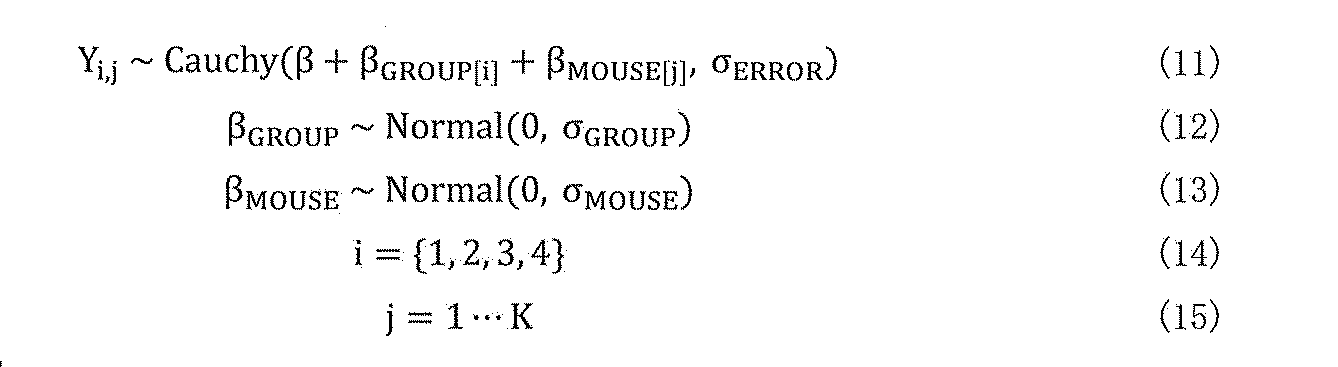
すべてのσを標準的な半正規分布からサンプリングした。TSの群間差を、σMOUSEの標準偏差で正規分布ノイズを有するβおよびβGROUPの合計である事後分布から各群の平均TSを推定して比較した。
All σ were sampled from a standard semi-normal distribution. The differences between groups of T S, and compared to estimate the average T S for each group from the posterior distribution is the sum of beta and beta GROUP having a normal distribution noise with a standard deviation of sigma MOUSE.
QIHおよび通常の条件下での体温調節系を評価するために、動物の熱損失および熱生産を階層的多層モデル(図3c-k、コードフォルダQIH_GTRH)で記述した。2つの代謝条件、すなわち正常およびQIHにおける3つのパラメータG、TRおよびHを、種々のTAにおける動物の代謝的に安定な状態から推定した。詳細な方法は先に述べた3。要するに、制御可能なパラメータTAと観測可能なパラメータTBとVO2から成る線形モデルを、正規分布ノイズを有する予測因子としてTAを用い、TBとVO2の両方についての実験結果に適合させた。次に、各モデルの傾きと切片係数の事後分布を用いて、G、TR、およびHを推定した。この分析では、ノイズの標準偏差の事前確率密度関数は標準的な半正規分布であり、他のパラメータは負の値に起因する均一な分布を使用したTBの切片係数を除いて均一な分布の正の領域を用いた。
To evaluate the thermoregulatory system under QIH and normal conditions, animal heat loss and heat production were described in a hierarchical multi-layer model (Fig. 3ck, code folder QIH_GTRH). Two metabolic conditions, i.e. three parameters G in normal and QIH, the T R and H, were estimated from metabolically stable condition of animals in the various T A. The detailed method is described above 3 . In short, a linear model composed of controllable parameter T A and observable parameters T B and VO 2, with T A as predictors having a normal distribution noise, fit the experimental results for both T B and VO 2 I let you. Next, using the posterior distribution of the slope and intercept coefficients of each model were estimated G, T R, and H. In this analysis, the prior probability density function of the standard deviation of the noise is a standard semi-normal distribution, uniform distribution except for sections coefficient of other parameters T B using a uniform distribution due to a negative value The positive region of was used.
Q‐TeTxLCマウスにおける代謝の概日変化は、記録された値をL期およびD期にクラスター化することにより代謝をモデリングすることにより解析した(コードフォルダTeTxLC_LD)。特に、Yが第j相のi群の観察されたTBである場合、Yは基礎代謝(L相代謝)とD相間の差の和として表すことができ、正規分布の観察ノイズは次のようになる。
Circadian changes in metabolism in Q-TeTxLC mice were analyzed by modeling metabolism by clustering recorded values in phases L and D (code folder TeTxLC_LD). In particular, when Y is i group observed T B of the j-phase, Y is can be represented as the sum of the difference D phase and basal metabolism (L phase metabolism), the normal distribution observation noise follows Will be.

すべてのσを標準的な半正規分布からサンプリングした。VO2のモデリングでは、VO2は正の実数のみを想定しているため、観測誤差を対数正規分布としてモデル化した以外は、基本的なモデル構造はTBモデリングと同一であった。
Q-TeTxLCマウスにおけるFIT中の代謝は、階層的多層モデル(図4d、コードフォルダTeTxLC_FIT)でモデル化した。セクションjにおけるあるグループiの最小値Yは、グループβ0[i]の平均代謝と差異パラメータβ1[i,j]の合計として表すことができる。 All σ were sampled from a standard semi-normal distribution. The modeling VO 2, VO 2 is because it is assumed only positive real number except for modeling the measurement error as a log-normal distribution, the basic model structure was identical to T B modeling.
Metabolism during FIT in Q-TeTxLC mice was modeled with a hierarchical multi-layer model (Fig. 4d, code folder TeTxLC_FIT). The minimum value Y of a group i in section j can be expressed as the sum of the mean metabolism of group β 0 [i] and the difference parameter β 1 [i, j].

代謝の分散をモデリングするために、観察された値Yの予測因子としてマウスの同一性を含めた。このようにして、あるセクション(SECTION)の所定のグループのYは正規分布としてモデル化され、この正規分布は、マウス依存の平均αMOUSEとグループおよびセクション依存のパラメータβGROUP,SECTIONを平均として、σGROUP,SECTIONを標準偏差として用いた。
Mouse identity was included as a predictor of the observed value Y to model metabolic variance. In this way, the Y of a given group of a section is modeled as a normal distribution, which averages the mouse-dependent mean α MOUSE and the group and section-dependent parameters β GROUP, SECTION . σ GROUP and SECTION were used as standard deviations.

式(22)~(24)および(28)~(30)の全σを標準の半正規分布からサンプリングした。これらのモデルは、TB、VO2、およびRQのモデリングに用いられた。これらのモデルのYでさえ、理論的には負の実数を受け入れることができ、後者はうまく収束したため、このモデルをVO2とRQにも適用した。
All σ of equations (22)-(24) and (28)-(30) were sampled from a standard semi-normal distribution. These models, T B, was used to model the VO 2, and RQ. Even Y in these models could theoretically accept negative real numbers, and the latter converged well, so we applied this model to VO 2 and Race Queen as well.
[実験と結果]
[Experiments and results]
実施例1:化学的に定義された視床下部ニューロン集団による代謝低下の誘発
視床下部神経ペプチドであるピログルタミン化RFアミドペプチド(QRFP)は、もともと新しいRF‐アミドペプチドを発見することを目的としたバイオインフォマティクスアプローチを通して発見された9,10。Qrfpペプチドはまた、オーファンG-タンパク質結合受容体hGPR103の内因性リガンドとしてラット脳から同定および精製された11。prepro‐Qrfp mRNAは視床下部にのみ局在し、脳室周囲核(Pe)、視床下部外側野(LHA)、および灰白隆起(TC)11に分布する。Qrfpは、食物摂取、交感神経調節、および不安に関係するとされてきた11,12。発明者らは、Qrfp遺伝子にコドン改良Creリコンビナーゼ(iCre)をノックインしたマウス(Qrfp-iCreマウス)を作製した。発明者らは、iCre発現ニューロンにのみhM3Dq-mCherryを発現するマウス(Qrfp-iCre;Rosa26dreaddm3 マウス)を得るために、CAG-hM3Dq-mCherryをRosa26遺伝子座に上流のfloxed転写停止エレメントを挿入したRosa26ddreadm3 マウスと交配した。Qrfp‐iCre;Rosa26dreaddm3マウスを用いた興奮性化学遺伝学的実験中に、これらのマウスは運動活性の顕著な低下を示し、最終的にクロザピン‐N‐オキシド(CNO)の腹腔内(IP)注射から約30分後に始まる重度で持続的な不動状態となった。これらのマウスの姿勢は日内休眠(tropor)中に観察された姿勢と類似していることに気づいたので、Qrfp‐iCreマウスにおけるiCre陽性細胞の活性化は日内休眠(tropor)様状態を誘発し、不動性と低いTBを特徴とすると最初に仮説した(後述するように、ここで誘導された低体温は日内休眠ではなく、冬眠様状態であることが明らかとなっている)。この仮説を評価するために、サーモグラフィーカメラを用いて表面体温(TS)を測定し、Qrfp‐iCre;Rosa26dreaddm3マウスにおけるCNO誘発性不動状態が、顕著で持続性の低体温を伴うことを見出した(図1b)。TSの減少はCNO投与の約5分後から始まり、ほぼ12時間持続した。その後、マウスは外部からの再加温なしに低体温状態から自発的に回復した。 Example 1: Induction of hypometabolism by a chemically defined hypothalamic neuron population The hypothalamic neuropeptide, pyroglutaminated RF amide peptide (QRFP), was originally intended to discover a new RF-amide peptide. Discovered through a bioinformatics approach 9,10 . Qrfp peptide may also be identified and purified from rat brain as an endogenous ligand of the orphan G- protein coupled receptors hGPR103 11. The prepro-Qrfp mRNA is localized only in the hypothalamus and is distributed in the periventricular nucleus (Pe), the lateral hypothalamic area (LHA), and the tuber cinereum (TC) 11. Qrfp has been implicated in food intake, sympathetic regulation, and anxiety 11,12 . The inventors generated mice (Qrfp-iCre mice) in which a codon-improved Cre recombinase (iCre) was knocked into the Qrfp gene. In order to obtain mice expressing hM3Dq-mCherry only in iCre-expressing neurons (Qrfp-iCre; Rosa26 dreddm3 mice), the inventors inserted a CAG-hM3Dq-mCherry upstream frozen transcription arrest element at the Rosa26 locus. It was bred with Rosa26 ddreadm3 mice. During excitatory chemical genetic experiments with Qrfp-iCre; Rosa26 dreddm3 mice, these mice showed a marked decrease in motor activity and eventually clozapine-N-oxide (CNO) intraperitoneally (IP). Severe and persistent immobility began about 30 minutes after injection. Since the postures of these mice were found to be similar to those observed during diapause (tropor), activation of iCre-positive cells in Qrfp-iCre mice induced a diapause (tropor) -like state. were initially hypothesized characterized immobility and low T B (as described below, where hypothermia induced by not the diurnal dormancy, it is hibernating like state has been revealed). To evaluate this hypothesis, measuring the surface temperature (T S) using a thermographic camera, Qrfp-iCre; Rosa26 dreaddm3 CNO-induced immobility in mice, found that with hypothermia markedly persistent (Fig. 1b). Decrease in T S begins about 5 minutes after the CNO administration lasted approximately 12 hours. The mice then spontaneously recovered from hypothermia without external reheating.
視床下部神経ペプチドであるピログルタミン化RFアミドペプチド(QRFP)は、もともと新しいRF‐アミドペプチドを発見することを目的としたバイオインフォマティクスアプローチを通して発見された9,10。Qrfpペプチドはまた、オーファンG-タンパク質結合受容体hGPR103の内因性リガンドとしてラット脳から同定および精製された11。prepro‐Qrfp mRNAは視床下部にのみ局在し、脳室周囲核(Pe)、視床下部外側野(LHA)、および灰白隆起(TC)11に分布する。Qrfpは、食物摂取、交感神経調節、および不安に関係するとされてきた11,12。発明者らは、Qrfp遺伝子にコドン改良Creリコンビナーゼ(iCre)をノックインしたマウス(Qrfp-iCreマウス)を作製した。発明者らは、iCre発現ニューロンにのみhM3Dq-mCherryを発現するマウス(Qrfp-iCre;Rosa26dreaddm3 マウス)を得るために、CAG-hM3Dq-mCherryをRosa26遺伝子座に上流のfloxed転写停止エレメントを挿入したRosa26ddreadm3 マウスと交配した。Qrfp‐iCre;Rosa26dreaddm3マウスを用いた興奮性化学遺伝学的実験中に、これらのマウスは運動活性の顕著な低下を示し、最終的にクロザピン‐N‐オキシド(CNO)の腹腔内(IP)注射から約30分後に始まる重度で持続的な不動状態となった。これらのマウスの姿勢は日内休眠(tropor)中に観察された姿勢と類似していることに気づいたので、Qrfp‐iCreマウスにおけるiCre陽性細胞の活性化は日内休眠(tropor)様状態を誘発し、不動性と低いTBを特徴とすると最初に仮説した(後述するように、ここで誘導された低体温は日内休眠ではなく、冬眠様状態であることが明らかとなっている)。この仮説を評価するために、サーモグラフィーカメラを用いて表面体温(TS)を測定し、Qrfp‐iCre;Rosa26dreaddm3マウスにおけるCNO誘発性不動状態が、顕著で持続性の低体温を伴うことを見出した(図1b)。TSの減少はCNO投与の約5分後から始まり、ほぼ12時間持続した。その後、マウスは外部からの再加温なしに低体温状態から自発的に回復した。 Example 1: Induction of hypometabolism by a chemically defined hypothalamic neuron population The hypothalamic neuropeptide, pyroglutaminated RF amide peptide (QRFP), was originally intended to discover a new RF-amide peptide. Discovered through a bioinformatics approach 9,10 . Qrfp peptide may also be identified and purified from rat brain as an endogenous ligand of the orphan G- protein coupled receptors hGPR103 11. The prepro-Qrfp mRNA is localized only in the hypothalamus and is distributed in the periventricular nucleus (Pe), the lateral hypothalamic area (LHA), and the tuber cinereum (TC) 11. Qrfp has been implicated in food intake, sympathetic regulation, and anxiety 11,12 . The inventors generated mice (Qrfp-iCre mice) in which a codon-improved Cre recombinase (iCre) was knocked into the Qrfp gene. In order to obtain mice expressing hM3Dq-mCherry only in iCre-expressing neurons (Qrfp-iCre; Rosa26 dreddm3 mice), the inventors inserted a CAG-hM3Dq-mCherry upstream frozen transcription arrest element at the Rosa26 locus. It was bred with Rosa26 ddreadm3 mice. During excitatory chemical genetic experiments with Qrfp-iCre; Rosa26 dreddm3 mice, these mice showed a marked decrease in motor activity and eventually clozapine-N-oxide (CNO) intraperitoneally (IP). Severe and persistent immobility began about 30 minutes after injection. Since the postures of these mice were found to be similar to those observed during diapause (tropor), activation of iCre-positive cells in Qrfp-iCre mice induced a diapause (tropor) -like state. were initially hypothesized characterized immobility and low T B (as described below, where hypothermia induced by not the diurnal dormancy, it is hibernating like state has been revealed). To evaluate this hypothesis, measuring the surface temperature (T S) using a thermographic camera, Qrfp-iCre; Rosa26 dreaddm3 CNO-induced immobility in mice, found that with hypothermia markedly persistent (Fig. 1b). Decrease in T S begins about 5 minutes after the CNO administration lasted approximately 12 hours. The mice then spontaneously recovered from hypothermia without external reheating.
対照的に、Qrfp-iCre;Rosa26dreaddm4マウスのiCre陽性ニューロンにおけるhM4Diの活性化を介した抑制性DREADD操作は、TSに対していかなる効果も示さなかった(図1b)。重要なことは、Qrfp-iCre;Rosa26dreadm3マウスにおけるiCre陽性ニューロンのhM3Dq介在性活性化は、両対立遺伝子においてprepro-Qrfp配列が完全にiCreに置換されたホモ接合性Qrfp-iCreマウスにおいてさえ、重度の低体温を誘発したことである(図1b)。このことは、Qrfpペプチド自体は低体温を誘導するために必須ではないことを示唆する。むしろ、低体温の程度はQrfpノックアウト(Qrfp‐iCreホモ接合体)マウスでより顕著であり、内因性Qrfp自体が低体温に対抗する可能性を示唆する。これは、Qrfpが中枢投与時に交感神経の流出を増加させ、心拍数および血圧を上昇させるという発明者らの以前の観察11と一致する。
In contrast, Qrfp-iCre; Rosa26 dreaddm4 mouse inhibitory DREADD operation via the activation of hM4Di in ICRE positive neurons did not show any effect on T S (FIG. 1b). Importantly, hM3Dq-mediated activation of iCre-positive neurons in Qrfp-iCre; Rosa26 dradm3 mice is even in homozygous Qrfp-iCre mice in which the prepro-Qrfp sequence is completely replaced by iCre in both alleles. It induced severe hypothermia (Fig. 1b). This suggests that the Qrfp peptide itself is not essential for inducing hypothermia. Rather, the degree of hypothermia is more pronounced in Qrfp knockout (Qrfp-iCre homozygous) mice, suggesting that endogenous Qrfp itself may counter hypothermia. This, Qrfp causes the increased outflow of sympathetic during central administration, consistent with previous observations 11 of inventors that increasing the heart rate and blood pressure.
そこで、低体温誘導ニューロンの化学マーカーとしてQrfpを同定した。次に、iCre陽性ニューロンは視床下部にのみ観察されるが、Qrfp-iCreマウスのいくつかの離散した視床下部領域に分布しているので、低体温を誘導する視床下部領域の同定を試みた。2つの異なる定位座標;内側基底(MB)注射または側方(LH)注射(方法を参照)を用いて、flip-excision(FLEX)スイッチ13を有するCre活性化AAVベクターをQrfp‐iCreマウスの視床下部に注入することにより、視床下部の外側と内側の領域のiCre陽性ニューロンを別々に操作した。Cre依存性AAVベクターのMB注入により、視床下部の内側領域、すなわち前腹側脳室周囲核(AVPe)、内側視索前野(MPA)およびPeにおけるiCre陽性ニューロンの特定の遺伝子を発現させることができたが、LHAでは発現することができなかった(図1c)。マルチカラー蛍光in situハイブリダイゼーション分析により、これらの領域の大部分のmCherry陽性細胞がQrfp mRNAを発現することが確認された。Qrfp-iCreマウスにAAV10-EF1a-DIO-hM3Dq-mCherryをMB注入してこの領域にhM3Dqを発現させた後、これらのマウスから作成した視床下部スライスを用いて電気生理学的研究を実施し、CNOの浴中適用がmCherry陽性ニューロンを強く興奮させたことを確認した。これらのマウスにCNOをIP注入すると、Qrfp-iCre;Rosa26dreaddm3マウスで観察される重度の不動状態よりも深く長く続く低体温症を引き起こすことがわかった(図1b、図1d)。非常に低いTB状態(30℃未満)は48時間以上続いた(図1d)。抗Fosおよび抗mCherry抗体を用いた免疫染色により、AVPe、MPAおよびPeにおいて多数のmCherryおよびFos二重陽性ニューロンが明らかにされ、CNOによるこれらのニューロンのin vivoでの興奮が確認された(図1e)。
Therefore, Qrfp was identified as a chemical marker for hypothermia-inducing neurons. Next, since iCre-positive neurons are observed only in the hypothalamus, but are distributed in several discrete hypothalamic regions of Qrfp-iCre mice, we attempted to identify the hypothalamic region that induces hypothermia. Two different stereotactic coordinates; using medial basal (MB) or lateral (LH) injections (see Method) , a Cre-activated AAV vector with flip-excision (FLEX) switch 13 in the thalamus of Qrfp-iCre mice. By injecting into the lower part, iCre-positive neurons in the outer and inner regions of the hypothalamus were manipulated separately. MB injection of Cre-dependent AAV vectors can express specific genes for iCre-positive neurons in the medial region of the hypothalamus, namely the anterior ventricular periventricular nucleus (AVPe), medial preoptic area (MPA) and Pe. It was possible, but could not be expressed in LHA (Fig. 1c). Multicolor fluorescence in situ hybridization analysis confirmed that most mCherry-positive cells in these regions express Qrfp mRNA. After MB injection of AAV 10- EF1a-DIO-hM3Dq-mCherry into Qrfp-iCre mice to express hM3Dq in this region, electrophysiological studies were performed using hypothalamic slices prepared from these mice. It was confirmed that the bath application of CNO strongly excited mCherry-positive neurons. IP injection of CNO into these mice was found to cause hypothermia that lasted deeper and longer than the severe immobility observed in Qrfp-iCre; Rosa26 dreddm3 mice (FIGS. 1b, 1d). Very low T B state (less than 30 ° C.) lasted more than 48 hours (Fig. 1d). Immunostaining with anti-Fos and anti-mCherry antibodies revealed a large number of mCherry and Fos double-positive neurons in AVPe, MPA and Pe, confirming the in vivo excitement of these neurons by CNO (Fig. 1e).
これらの観察から、Qrfp‐iCreマウスのAVPe/MPAおよびPeにおけるiCre陽性ニューロン(これらのニューロンを静止誘発ニューロンまたはQニューロンと後述する)は主に誘導低体温状態の原因であると結論した。以下の実験において、発明者らは、特に明記しない限り、低体温の誘導のために、AAV10-EF1a-DIO-hM3Dq-mCherry(Q-hM3Dマウスと呼ばれる)のMB注射を伴うQrfp-iCreマウスを基本的に使用した。
From these observations, it was concluded that iCre-positive neurons in AVPe / MPA and Pe in Qrfp-iCre mice (these neurons are referred to as rest-inducing neurons or Q-neurons below) are primarily responsible for induced hypothermia. In the following experiments, the inventors, unless otherwise stated, Qrfp-iCre mice with MB injection of AAV 10- EF1a-DIO-hM3Dq-mCherry (called Q-hM3D mice) for the induction of hypothermia. Was basically used.
誘導低体温状態をさらに解析するために、Q‐hM3Dマウスの腹腔内に遠隔測定温度センサーを移植し、呼吸ガス分析により代謝を連続的に分析した(図1f)。本研究は、Q‐hM3DマウスにおけるCNO誘発低体温状態が、O2消費速度(VO2:酸素消費量)の著しい低下(図1g)を伴い、CNO投与後のTSと共にTBが同時に減少することを確認した。対照的に、AAV10-EF1a-DIO-hM3Dq-mCherryのLH注射によるQrfp-iCreマウスの視床下部外側領域(LHAおよびTC)のiCre陽性ニューロンの興奮性DREADD操作は、低体温を誘発しなかった(図1g)。
To further analyze the induced hypothermia, a telemetric temperature sensor was implanted intraperitoneally in Q-hM3D mice and respiratory gas analysis was performed to continuously analyze metabolism (Fig. 1f). This study, CNO induced hypothermia states in Q-hM3D mice, O 2 consumption rate: with a significant reduction in (VO 2 oxygen consumption) (FIG. 1 g), reducing T B at the same time with T S after CNO administration Confirmed to do. In contrast, excitatory DREADD manipulation of iCre-positive neurons in the lateral hypothalamic region (LHA and TC) of Qrfp-iCre mice by LH injection of AAV 10-EF1a-DIO-hM3Dq-mCherry did not induce hypothermia. (Fig. 1 g).
Qニューロン誘発低体温/低代謝(QIH)状態の間に、心拍数は著しく減少した(CNO注射の2時間前と2時間後、それぞれ758拍/分と215拍/分)(n=3の平均)。呼吸数は333呼吸/分から検出不可能な状態にまで減少した(1回換気量は検出限界未満)。これらのタイミングで、VO2は3.60から1.17ml/g/hrに減少した。QIH中、マウスは非常に低い振幅脳波(EEG)を示し、これは高振幅徐波を特徴とする非急速眼球運動睡眠で観察されるものとは明らかに異なっていた。血清化学データは、血糖値がQIH中に低下することを示唆し、これはおそらく交感神経緊張の低下による糖新生の低下によるものと思われる。これらの観察はさらに、多くの身体機能がQIH中のTBとVO2の減少と共にロバストに減少することを示唆する。
During the Q-neuron-induced hypothermia / hypometabolism (QIH) state, heart rate decreased significantly (758 beats / minute and 215 beats / minute, respectively, 2 hours and 2 hours after CNO injection) (n = 3). average). Respiratory rate decreased from 333 breaths / min to undetectable (tidal volume below detection limit). At these timings, VO 2 decreased from 3.60 to 1.17 ml / g / hr. During QIH, mice showed very low amplitude electroencephalogram (EEG), which was clearly different from that observed in non-rapid eye movement sleep characterized by high amplitude slow waves. Serum chemistry data suggest that blood glucose levels decrease during QIH, probably due to decreased gluconeogenesis due to decreased sympathetic tone. These observations further suggest that many body functions decreases robust with decreasing T B and VO 2 in QIH.
DREADDを介する効果は通常、CNO注射後数時間しか持続しないが、Q‐hM3DマウスにおけるDREADD誘発QIHは非常に長く持続した。驚いたことに、TA=20℃では、30℃未満のTBのQIHは、CNOを1回投与(1mg/kg)しただけで48時間以上持続し、VO2が完全に正常に回復するのに約1週間かかった(図1h)。QIHからの回復後、マウスは健康であり、正常に振る舞うようであった。QIHは、同じマウスに反復CNO注射後に再現可能であり、この操作の可逆性を示した(図1h)。
DREADD-mediated effects usually lasted only a few hours after CNO injection, but DREADD-induced QIH in Q-hM3D mice lasted very long. Surprisingly, QIH of T A = at 20 ° C., of less than 30 ° C. T B lasts one dose (1 mg / kg) was only more than 48 hours the CNO, VO 2 is completely normal recovery It took about a week (Fig. 1h). After recovery from QIH, the mice were healthy and appeared to behave normally. QIH was reproducible after repeated CNO injections in the same mice, demonstrating the reversibility of this operation (Fig. 1h).
実施例2:Qニューロンは視床下部背内側に作用してQIHを誘導する
QIHを誘導する機構を明らかにするために、Qニューロンの軸索投射を解析した。Qrfp-iCreマウスにAAV10-EF1a-DIO-GFPを注射してQニューロンに特異的にGFPを発現させた後(図2a、b)、MPA、VOLT、室傍核(PVN)、視索上核(SON)、視床下部背内側(DMH)、LHA、結節乳頭核(TMN)、内側乳頭核(MM)、中脳水道周囲灰白質(PAG)、外側結合腕傍核(LPB)、青斑核(LLC)、延髄吻側腹外側野(RVLM)、および淡蒼縫線核(RPa)(体温調節調節および交感神経制御に関わる領域)におけるGFP陽性線維を観察した(図2c)14。発明者らは、DMHが特に豊富な投射を受けたことを見出した。ScaleS法で明らかになった脳の解析から、QニューロンとDMHへの投射の位置がさらに示唆された(図2d)。
次に、Qニューロンの三重カラーin situハイブリダイゼーションを用いて、これらのQニューロンが抑制性か興奮性かを確認した。CNO注射がQ‐hM3DマウスにおいてQIHを効果的に誘導することを確認した後、これらのマウスをin situハイブリダイゼーション組織化学的検査に供した。興奮性および抑制性マーカーであるmCherryをコードする転写産物、小胞性グルタミン酸トランスポーター2(Vglut2)および小胞性GABAトランスポーター(Vgat)をコードするプローブを用いた。われわれは、Qニューロンの約2/3がVgat陽性であり、約2/5がVglut2陽性であることを見出した(図2e-i)。 Example 2: Q neurons act on the dorsomedial hypothalamus to induce QIH In order to clarify the mechanism that induces QIH, axon projection of Q neurons was analyzed. After injecting AAV 10- EF1a-DIO-GFP into Qrfp-iCre mice to express GFP specifically in Q neurons (Fig. 2a, b), MPA, VOLT, paraventricular nucleus (PVN), supraoptic nucleus Nucleus (SON), dorsomedial hypothalamus (DMH), LHA, nodular papillary nucleus (TMN), medial papillary nucleus (MM), periaqueductal gray (PAG), paraventricular nucleus of the lateral connection (LPB), locus coeruleus GFP-positive fibers were observed in the nucleus (LLC), the paraventricular nucleus of the medulla oblongata (RVLM), and the nucleus raphe pneumoniae (RPa), a region involved in temperature regulation and sympathetic regulation (Fig. 2c) 14 . The inventors have found that DMH received a particularly abundant projection. Brain analysis revealed by the ScaleS method further suggested the location of projections on Q neurons and DMH (Fig. 2d).
Next, triple-color in situ hybridization of Q neurons was used to confirm whether these Q neurons were inhibitory or excitatory. After confirming that CNO injection effectively induces QIH in Q-hM3D mice, these mice were subjected to in situ hybridization histochemical examination. Probes encoding the transcripts encoding the excitatory and inhibitory markers mCherry, the vesicular glutamate transporter 2 (Vglut2) and the vesicular GABA transporter (Vgat) were used. We found that about two-thirds of Q neurons were Vgat-positive and about two-fifths were Vglut2-positive (Fig. 2ei).
Qニューロンによる豊富な投射を含む領域(図2c)の中で、我々はDMHに焦点を当てた。なぜなら、熱産生促進ニューロンは以前にDMHで同定されたからである15。DMHへのQニューロンの軸索投射の機能を明らかにするために、光遺伝学的アプローチを用いた。Qrfp-iCreマウス(Q-SSFOマウス)にAAV10-DIO-SSFO-eYFPを注入することにより、Qニューロンで安定化したstep function opsin (SSFO)16を発現させた(図2j)。SSFOはAVPe,MPAおよびPeで発現することを確認した。TSに対する光遺伝学的興奮の効果を確認するために、まず、Qニューロンの多くの細胞体が見出されるAVPe/MPAに光ファイバーを移植し(図2j)、光パルス(1秒幅の光パルス)を印加することによりSSFO陽性細胞体の光発生的興奮をマウスにかけた。この状態では、Qニューロンの光遺伝学的興奮が急速に強い低体温を誘発し、約20分続いた(図2k)。Qニューロンを30分ごとに4回繰り返し興奮させると、TA(22℃)と同程度に低いTSを伴う著明な低体温になった。興奮後のAVPe/MPAのSSFO‐eYFP陽性細胞では多くのFos陽性ニューロンが同定された(図2j)。光遺伝学的に誘発されたQIHは、QニューロンのhM3Dq介在性の薬理遺伝学的興奮によって誘発されるQIHよりも明らかに短時間持続し(図2k)、このことは、遺伝子発現プロフィールの変化を導くQニューロンにおけるGq介在性の代謝調節性シグナル伝達がQIHの長期持続性を作り出す役割を果たしている可能性を示唆している。
Within the region containing abundant projections by Q neurons (Fig. 2c), we focused on DMH. This is because heat production promoting neuron is because identified in DMH previously 15. An optogenetic approach was used to elucidate the function of axon projection of Q neurons to DMH. By injecting AAV 10- DIO-SSFO-eYFP into Qrfp-iCre mice (Q-SSFO mice), step function opsin (SSFO) 16 stabilized in Q neurons was expressed (Fig. 2j). It was confirmed that SSFO is expressed in AVPe, MPA and Pe. In order to confirm the effect of the optical genetic excitement for T S, first, implanted with fiber to AVpe / MPA many cell bodies Q neurons are found (Fig. 2j), of the optical pulse (1-second-wide optical pulses ) Was applied to the mice for photogenic excitement of SSFO-positive cell bodies. In this state, optogenetic excitement of Q neurons rapidly induced strong hypothermia, which lasted for about 20 minutes (Fig. 2k). When the repeated 4 times Q neurons every 30 minutes excitement became marked hypothermia with low T S to the same extent as T A (22 ℃). Many Fos-positive neurons were identified in SSFO-eYFP-positive cells of AVPe / MPA after excitement (Fig. 2j). Optogenetically induced QIH persists significantly shorter than hM3Dq-mediated pharmacogenetic excitement-induced QIH in Q neurons (Fig. 2k), which indicates altered gene expression profiles. It is suggested that Gq-mediated pharmacogenetic signaling in Q neurons that leads to QIH may play a role in creating long-term persistence of QIH.
次に、Q‐SSFOマウスの両側にDMHに光ファイバーを移植し、光刺激を軸索線維に適用した。この操作は効果的にTSを減少させたが、AVPe/MPAの細胞体刺激によって誘導されるものよりわずかに弱かった(図2k,l)。対照として、RPaは褐色脂肪組織制御を介する熱産生のための交感神経性運動前ニューロンを含むことが知られている17ため、RPaにおけるQニューロン線維の光刺激の影響も検討した。また、TSに対するRPaにおけるQニューロン線維の光発生的興奮の微妙な作用も観察された(図2k,l)。これらの結果から、Qニューロンは主にDMHに作用し、RPaに対してはより小さい程度で作用し、QIHを誘導すると仮定した。
Next, optical fibers were transplanted into DMH on both sides of Q-SSFO mice, and light stimulation was applied to axons. This procedure effectively reduced the T S, was slightly weaker than that induced by cell body stimulation AVpe / MPA (Figure 2k, l). As a control, RPa is because 17 is known to contain a sympathetic movement before neurons for heat production through brown adipose tissue control, it was also investigated the effect of light stimuli Q neuronal fibers in RPa. Also, subtle effects of light generating excitement of Q neuronal fibers in RPa for T S was also observed (Fig. 2k, l). From these results, it was hypothesized that Q neurons mainly act on DMH, act on RPa to a lesser extent, and induce QIH.
実施例3:理論的設定温度はQIH中に低下する
QIH誘発直後にマウス尾部の温度上昇が観察され、Qニューロンの光遺伝学的または薬理遺伝学的興奮によって誘発されたことから、TBの減少中に末梢血管が拡張して熱を放出することが示唆された(図1d、図2k)。TBの増加を伴わない末梢血管拡張は、冬眠動物の冬眠状態で見られるように、理論体温設定値(TR)を正常状態より低い値に再設定されていることを示唆する。これを評価するために、QIH中のマウスの体温調節系の特徴分析を行った。動物が外部仕事を持たず、代謝が安定している条件下では、複数の周囲温度(TA)下で、TBおよびVO2から熱コンダクタンス(G)、HおよびTRを推定することができる3。Q‐hM3Dマウスを調製し、種々のTA(8、12、16、20、24、28および32℃)下でQIH中にTBおよびVO2を記録した(図3a)。生理食塩水またはCNOのIP注射後の11時間平均TBおよびVO2を比較した。QIH中、動物は対応する対照と比較して、全てのTAで低いTBとVO2を示した(図3b)。熱産生システムが適切に機能しているとき、すなわち、TRがTBより高く、体温調節システムがVO2を増加させてTRに到達しようとしているとき(図3c)、TAが増加するとTBは増加し、VO2は減少する。TBとVO2はそれぞれ異なるTAで最小値を示し、TAでは16~24℃の範囲の協調した熱生成特性のみを示した(図3b)。したがって、QIH中のTA=16、20、および24℃の代謝データを用いてさらに分析した。まず、TB-TAとVO2の関係からGを推定した(図3d)。Gの89%の最高後部密度間隔(HPDI)は、正常及びQIH条件下でそれぞれ[0.212,0.221]ml/g/hr/℃及び[0.182,0.220]ml/g/hr/℃(図3e;以下89%HPDIは2つの数字で四角括弧で示す)であった。量的に、両Gの差の後方分布(ΔG)は[-0.0040,0.0348]ml/g/hr/℃(図3f)であり、0を含んでおり、正常条件下とQIH条件下のGが区別できないことを示唆している。これは通常の条件よりも低いGを示す日内休眠(torpor)とは異なっていた3。第二に、TBとVO2からHとTRを推定した(図3g)。Hは正常状態で[3.43、8.72]ml/g/hr/℃、QIHで[0.181、0.369]ml/g/hr/℃(図3h)であり、各中央値で95.3%の減少であった。差の事後分布(ΔH)は[3.17,8.48]ml/g/hr/℃(図3i)であり、陽性であり、これらの条件が異なる確率が89%以上であることが示唆された。このH低下は、空腹時誘発日内休眠(FIT)時のH低下と類似していた3。特に、
TRは正常状態で[36.04、36.60]℃、QIHで[26.83、29.13]℃と推定された(図3j)。TRの差は各中央値で8.41℃であり、差の後方分布(ΔTR)は[7.18,9.57]℃であり、QIH中のTRの低下を明確に示している(図3k)。FITにおける非常に小さな理論的設定温度シフト3を考慮すると、この観察は、QIHと冬眠の間の類似性、ならびにQIHと日内休眠(torpor)の間の差を強調する。 Example 3: Theoretical preset temperature increase in temperature of the mouse tail was observed immediately after QIH induction decreases during QIH, since induced by light genetically or pharmacogenetics excitement Q neurons, the T B It was suggested that the peripheral blood vessels dilate and release heat during the decrease (Fig. 1d, Fig. 2k). T telangiectasia without an increase in B, as seen in the hibernation state of hibernation animals, suggesting that it is re-set theoretical temperature set value (T R) lower than the normal state value. To evaluate this, a feature analysis of the thermoregulatory system of mice during QIH was performed. Animal no external work, under conditions where metabolism is stable, more at ambient temperature (T A) below, the thermal conductance from the T B and VO 2 (G), to estimate the H and T R I can do it 3 . Q-hM3D mice were prepared and recorded T B and VO 2 in QIH in various T A (8,12,16,20,24,28 and 32 ° C.) lower (Figure 3a). Were compared 11 hours Mean T B and VO 2 after IP injection of saline or CNO. During QIH, animals as compared to the corresponding control showed low T B and VO 2 in all T A (Figure 3b). When heat production system is functioning properly, i.e., T R is higher than T B, when the thermoregulatory system is trying to reach T R by increasing the VO 2 (Fig. 3c), the T A is increased T B increases, VO 2 decreases. T B and VO 2 shows a minimum at different T A, respectively, showed only coordinated thermogenic properties in the range of T A in 16 ~ 24 ° C. (Fig. 3b). Thus, it was further analyzed using the metabolic data of T A = 16, 20, and 24 ° C. in QIH. First, to estimate the G from the relationship of T B -T A and VO 2 (Figure 3d). The 89% maximum posterior density interval (HPDI) of G is [0.212, 0.221] ml / g / hr / ° C. and [0.182, 0.220] ml / g under normal and QIH conditions, respectively. / Hr / ° C. (Fig. 3e; hereinafter 89% HPDI is indicated by two numbers in square brackets). Quantitatively, the posterior distribution (ΔG) of the difference between the two Gs is [-0.0040, 0.0348] ml / g / hr / ° C. (FIG. 3f), containing 0, under normal conditions and QIH. It suggests that G under the conditions is indistinguishable. It was different from the inner dormant date exhibit a low G than normal conditions (torpor) 3. Second, to estimate the H and T R from the T B and VO 2 (Fig. 3 g). H is [3.43, 8.72] ml / g / hr / ° C in the normal state, and [0.181, 0.369] ml / g / hr / ° C in QIH (FIG. 3h), which are medians of each. It was a decrease of 95.3%. The posterior distribution (ΔH) of the difference is [3.17, 8.48] ml / g / hr / ° C (Fig. 3i), which is positive, suggesting that the probability of these conditions being different is 89% or more. Was done. This decrease in H was similar to the decrease in H during fasting-induced diapause (FIT) 3 . Especially,
T R is [36.04,36.60] ℃, was estimated to be [26.83,29.13] ℃ in QIH in the normal state (Fig. 3j). Difference T R is 8.41 ° C. In each median posterior distribution of the difference ([Delta] T R) is [7.18,9.57] ℃, clearly show a reduction in T R in QIH (Fig. 3k). Given the very small theoretical set temperature shift 3 in FIT, this observation highlights the similarities between QIH and hibernation, as well as the differences between QIH and diapause (torpor).
冬眠の顕著な特徴であるQIH中のTR減少の証拠をさらに提供するために、QIH中にTAが動的に変化したとき、個々のマウス内の姿勢と代謝の間の関係を観察した(n=4、図3lにおける1匹の代表データ、および他の3匹のデータ)。冬眠動物におけるようなQIHの非常に安定で長期にわたる低代謝状態は、これをマウスで調べることを可能にした。Q-hM3DマウスをTA=28℃に設定し、FITを誘導した(図3lのAおよびB)。24時間の回復の後、QIHはCNO投与により誘導された(図3lのC、D、E、およびF)。興味深いことに、TA=28℃で、マウスはQIH中に伸びた姿勢を示したが、この姿勢は通常高温環境に暴露された動物で見られるものである(図3lのD)。これは、TA=28℃でのFIT中に観察される典型的な座位姿勢とは明らかに異なっていた(図3lのB)。この行動観察はさらに、TRがFITおよび正常状態よりもQIHで低いことを示している。さらに、TAを12℃に下げたところ、日内休眠(torpor)に似た座位(図3lのE)に戻り、震えが始まった。これらの結果は、QIH中、TRは低下するが、身体機能および行動はTAの変化に適応するために依然として調節されることを強く支持する。
To provide evidence of T R decreases in a hallmark of hibernation QIH Furthermore, when the T A is changed dynamically while QIH, and observed the relationship between the metabolism posture in individual mice (N = 4, one representative data in FIG. 3l, and three other data). The very stable and long-term hypometabolic state of QIH, such as in hibernating animals, has made it possible to examine this in mice. Set the Q-hM3D mouse T A = 28 ° C., to induce FIT (Figure 3l of A and B). After a 24-hour recovery, QIH was induced by CNO administration (C, D, E, and F in FIG. 3 l). Interestingly, T A = 28 ° C., mice showed an attitude extending in QIH, this position is that seen in animals exposed to the normal high-temperature environment (D in Fig. 3l). This was clearly different from the typical seated position observed during FIT at T A = 28 ℃ (Figure 3l of B). The behavioral observations further, T R is shown that lower in QIH than FIT and normal state. In addition, as a result of lowering the T A to 12 ℃, to return to the day in the dormant (torpor) in similar loci (Fig. 3l of E), trembling began. These results are in QIH, although T R is reduced, physical function and behavior strongly support that still be adjusted to accommodate changes in T A.
動物は冬眠中は代謝率が低いが、TAに反応してその代謝は活発に調節されていることがよく知られている。同様に、QIHでは、16℃以下のTAに曝露された動物は、20℃または28℃のTAに曝露された動物と比較して、かなり大きなVO2を示した(図3b)。実際、これは、TAを一定のレベルに下げた場合に代謝増加を示した冬眠動物に関する以前の報告18と類似している。QIHのこの活発な代謝低下の特徴は、個々の動物でも確認された(図3l)。QIH中のマウスの行動および代謝反応は、身体機能がTBを狭い範囲に維持しようとしている正常な状態で観察されたものとは全く異なっていた。
Animal is during hibernation low metabolic rate, the metabolic response to T A is well known to have been adjusted actively. Similarly, in QIH, 16 ℃ exposed to the following T A animals, compared to 20 ° C. or 28 ° C. T A in the exposed animals showed considerably large VO 2 (Figure 3b). In fact, this is similar to a previous report 18 relating to hibernation animals showed metabolic increase when lowering the T A at a constant level. This active hypometabolism feature of QIH was also confirmed in individual animals (Fig. 3l). Behavioral and metabolic reactions in mice in QIH were quite different from those physical function were observed in normal state trying to maintain a narrow range of T B.
QIH中の代謝機能を麻酔状態と比較するために、発明者らは複数TA下で全身麻酔中の代謝転移を記録した。予想通り、麻酔下の動物は低いTAに曝露しても、VO2の増加も姿勢の変化も示さなかった。また、低体温状態を誘発するために用いられているアデノシンA1ARアゴニスト(6)N-シクロヘキシルアデノシン(CHA)の全身送達によって誘導される代謝状態も調べた19。野生型マウスへのCHA(2.5mg/kg)のIP注射は低体温/低代謝状態を効果的に誘導したが、マウスはVO2の増加または行動(姿勢と震え)のいずれかによって低TA(12℃)に反応しなかった。20℃でのTBは、QIHよりもCHA誘発性低体温で高い傾向があったが、TBとVO2はCHA誘発性低体温でさらに減少した。一方、TAを12℃に設定した場合、これらのパラメータはQIHで増加した(図3b)。これらの観察は、QIHが全身麻酔またはCHA誘発受動的低体温とは完全に異なり、それはTBの調節系を遮断することによって低体温状態を誘発することを示している。
The metabolic function in QIH for comparison with anesthesia, it recorded the metabolic transition during general anesthesia under a plurality T A. As expected, even when exposed to animals low T A anesthetized increase in VO 2 also showed no change in the attitude. Also, metabolic state induced by systemic delivery of adenosine A1AR agonists have been used to induce hypothermia status (6) N-cyclohexyl-adenosine (CHA) were also examined 19. IP injection of CHA (2.5 mg / kg) into wild-type mice effectively induced hypothermia / hypometabolism, whereas mice had low T due to either increased VO 2 or behavior (posture and tremor). It did not react to A (12 ° C). T B at 20 ° C. is tended to be higher in CHA-induced hypothermia than QIH, T B and VO 2 was further reduced in CHA-induced hypothermia. On the other hand, if you set the T A to 12 ° C., these parameters were increased in QIH (Figure 3b). These observations, QIH is completely different from the general anesthesia or CHA induced passive hypothermia, which indicates that induce hypothermia state by blocking the regulatory system of T B.
実施例4:Qニューロンは正常な絶食誘発性の日内休眠(torpor)に関与している
QIHは日内休眠(torpor)よりも冬眠に似ているが、日内休眠(torpor)は冬眠の軽い状態と考えられることがあるため、Qニューロンが日内休眠(torpor)にも関与しているかどうかを検討した。また、共通または類似のメカニズムが冬眠および日内休眠(torpor)を誘導する役割を果たしているかもしれない20。日内休眠(torpor)におけるQニューロンの役割を調べるため、Qrfp-iCreマウス(Q-TeTxLCマウス)にAAV2/9-hSyn-DIO-TeTxLC-eYFPを注入することによりQニューロンに特異的に破傷風毒素軽鎖(TeTxLC)を発現させ、QニューロンのSNARE複合体介在性神経伝達の遮断がFITに影響を及ぼすかどうかを調べた(図4b)。AAV2/9‐hSyn‐DIO‐TeTxLC‐eYFPとAAV10‐EF1a‐DIO‐hM3Dq‐mCherryの同時注入はTBに対するCNOの作用を完全に消失させ、SNARE複合体の遮断がQニューロンのQIH誘導能を消失させることを示唆した。われわれは、すべてのQ-TeTxLCマウスにおいて、FITの正常な構造が崩壊することを見出した。絶食中のこれらのマウスでは、代謝の急速な振動変動は見られなかった(図4c,d)。このことは、Qニューロンの機能がFIT中のTBの急速な低下を誘発するために必要であることを示唆する。興味深いことに、これらのマウスで観察されたTBの漸減は、FIT中にQニューロン非依存性の代謝低下機構が存在することを意味する。加えて、Q‐TeTxLCマウスは対照マウスよりもTBの概日変動が少なく、TBの概日調節におけるQニューロンの主要な役割を示唆した。特に、QRFPペプチドを欠くホモ接合のQrfp-iCreマウスは、正常なFITを示した(図4e)。これらの観察は、Qニューロンは日内休眠(torpor)を誘導する必須の構成要素であり、日内休眠(torpor)における体温の急速なシフトに重要な役割を果たしているが、QRFPは果たしていないことを示唆する。 Example 4: Q neurons are involved in normal fasting-induced diapause (torpor) QIH is more like hibernation than diapause (torpor), but diapause (torpor) is a mild state of hibernation. Since there are some possibilities, we examined whether Q neurons are also involved in diurnal diapause (torpor). Also, common or similar mechanisms may play a role in inducing hibernation and diapause (torpor) 20 . To investigate the role of Q neurons in diapause (torpor), tetanus toxins are specifically injected into Q neurons by injecting AAV 2/9-hSyn-DIO-TeTxLC-eYFP into Qrfp-iCre mice (Q-TeTxLC mice). We expressed a light chain (TeTxLC) and investigated whether blocking of SNARE complex-mediated neurotransmission in Q neurons affects FIT (Fig. 4b). AAV 2/9 coinjection of -hSyn-DIO-TeTxLC-eYFP and AAV 10 -EF1a-DIO-hM3Dq- mCherry completely abolished the effect of CNO for T B, QIH induction blocking of Q neurons SNARE complex It was suggested that the ability would be lost. We have found that the normal structure of FIT is disrupted in all Q-TeTxLC mice. No rapid oscillating fluctuations in metabolism were observed in these fasted mice (Fig. 4c, d). This suggests the need for the function of Q neurons induces a rapid decrease in T B in FIT. Interestingly, decreasing the T B observed in these mice, mean that Q neuron independent of metabolic reduction mechanism are present in the FIT. In addition, Q-TeTxLC mice less circadian variation of T B than control mice, suggesting a major role of Q neurons in circadian regulation of T B. In particular, homozygous Qrfp-iCre mice lacking the QRFP peptide showed normal FIT (Fig. 4e). These observations suggest that Q neurons are essential components that induce diurnal diapause (torpor) and play an important role in the rapid shift in body temperature during diurnal dormancy (torpor), but QRFP does not. To do.
QIHは日内休眠(torpor)よりも冬眠に似ているが、日内休眠(torpor)は冬眠の軽い状態と考えられることがあるため、Qニューロンが日内休眠(torpor)にも関与しているかどうかを検討した。また、共通または類似のメカニズムが冬眠および日内休眠(torpor)を誘導する役割を果たしているかもしれない20。日内休眠(torpor)におけるQニューロンの役割を調べるため、Qrfp-iCreマウス(Q-TeTxLCマウス)にAAV2/9-hSyn-DIO-TeTxLC-eYFPを注入することによりQニューロンに特異的に破傷風毒素軽鎖(TeTxLC)を発現させ、QニューロンのSNARE複合体介在性神経伝達の遮断がFITに影響を及ぼすかどうかを調べた(図4b)。AAV2/9‐hSyn‐DIO‐TeTxLC‐eYFPとAAV10‐EF1a‐DIO‐hM3Dq‐mCherryの同時注入はTBに対するCNOの作用を完全に消失させ、SNARE複合体の遮断がQニューロンのQIH誘導能を消失させることを示唆した。われわれは、すべてのQ-TeTxLCマウスにおいて、FITの正常な構造が崩壊することを見出した。絶食中のこれらのマウスでは、代謝の急速な振動変動は見られなかった(図4c,d)。このことは、Qニューロンの機能がFIT中のTBの急速な低下を誘発するために必要であることを示唆する。興味深いことに、これらのマウスで観察されたTBの漸減は、FIT中にQニューロン非依存性の代謝低下機構が存在することを意味する。加えて、Q‐TeTxLCマウスは対照マウスよりもTBの概日変動が少なく、TBの概日調節におけるQニューロンの主要な役割を示唆した。特に、QRFPペプチドを欠くホモ接合のQrfp-iCreマウスは、正常なFITを示した(図4e)。これらの観察は、Qニューロンは日内休眠(torpor)を誘導する必須の構成要素であり、日内休眠(torpor)における体温の急速なシフトに重要な役割を果たしているが、QRFPは果たしていないことを示唆する。 Example 4: Q neurons are involved in normal fasting-induced diapause (torpor) QIH is more like hibernation than diapause (torpor), but diapause (torpor) is a mild state of hibernation. Since there are some possibilities, we examined whether Q neurons are also involved in diurnal diapause (torpor). Also, common or similar mechanisms may play a role in inducing hibernation and diapause (torpor) 20 . To investigate the role of Q neurons in diapause (torpor), tetanus toxins are specifically injected into Q neurons by injecting AAV 2/9-hSyn-DIO-TeTxLC-eYFP into Qrfp-iCre mice (Q-TeTxLC mice). We expressed a light chain (TeTxLC) and investigated whether blocking of SNARE complex-mediated neurotransmission in Q neurons affects FIT (Fig. 4b). AAV 2/9 coinjection of -hSyn-DIO-TeTxLC-eYFP and AAV 10 -EF1a-DIO-hM3Dq- mCherry completely abolished the effect of CNO for T B, QIH induction blocking of Q neurons SNARE complex It was suggested that the ability would be lost. We have found that the normal structure of FIT is disrupted in all Q-TeTxLC mice. No rapid oscillating fluctuations in metabolism were observed in these fasted mice (Fig. 4c, d). This suggests the need for the function of Q neurons induces a rapid decrease in T B in FIT. Interestingly, decreasing the T B observed in these mice, mean that Q neuron independent of metabolic reduction mechanism are present in the FIT. In addition, Q-TeTxLC mice less circadian variation of T B than control mice, suggesting a major role of Q neurons in circadian regulation of T B. In particular, homozygous Qrfp-iCre mice lacking the QRFP peptide showed normal FIT (Fig. 4e). These observations suggest that Q neurons are essential components that induce diurnal diapause (torpor) and play an important role in the rapid shift in body temperature during diurnal dormancy (torpor), but QRFP does not. To do.
Qニューロンの活性を調節するニューロンのメカニズムを解明するために、われわれは、組換え型仮性型狂犬病ウイルスベクター(SADΔG(EnvA))媒介性ラベリング21(図4f)によってQニューロンと直接シナプス接触する上流ニューロン集団を同定した。Qrfp‐iCreマウスのCre活性化AAVベクター22を用いてQニューロンにおいてTVA‐mCherryおよび狂犬病糖蛋白質(RG)を発現させた後、SADΔG‐GFP(EnvA)を同じ部位に注入した。TVA-mCherryおよびGFPに対して二重陽性であるスターター細胞がAVPe/MPAおよびPeに認められた(図4g)。正中視索前核(MnPO)、PVNおよびMPAにおいて、Qニューロン(GFPは陽性だがmCherryは陰性)に直接シナプス入力する入力ニューロンを同定した(図4h)。入力ニューロンはAVPe/Peの内部および周囲にも観察され、Qニューロンの機能を調節する局所介在ニューロンの存在、およびQニューロンがAVPe/MPAおよびPe内の介在ニューロンと微小回路を構成する可能性を示唆した。これらの観察は、Qニューロンが視床下部内領域から比較的まばらな直接入力を受けることを示唆する。FITは絶食により誘導されるので、Qニューロンは負のエネルギーバランスをモニターすることが期待される。PVHのニューロンはARCから豊富な入力を受けることが示されている23ため、PVHからQニューロンへの入力は栄養状態に関する情報を伝達する役割を果たしている可能性がある。PVH入力は視交叉上核(SCN)からの概日情報を伝達することもある。
To elucidate the neuronal mechanism that regulates the activity of Q neurons, we use recombinant pseudo-rabies virus vector (SADΔG (EnvA)) mediated labeling 21 (Fig. 4f) upstream to make direct synaptic contact with Q neurons. A neuron population was identified. After expressing TVA-mCherry and rabies sugar protein (RG) in Q neurons using Cre-activated AAV vector 22 from Qrfp-iCre mice, SADΔG-GFP (EnvA) was injected into the same site. Starter cells that were double positive for TVA-mCherry and GFP were found in AVPe / MPA and Pe (Fig. 4g). In the median preoptic nucleus (MnPO), PVN and MPA, we identified input neurons that synapse directly into Q neurons (GFP positive but mCherry negative) (Fig. 4h). Input neurons are also observed inside and around AVPe / Pe, the presence of local interneurons that regulate the function of Q neurons, and the possibility that Q neurons form microcircuits with interneurons within AVPe / MPA and Pe. Suggested. These observations suggest that Q neurons receive relatively sparse direct inputs from the intrathalamic region. Since FIT is induced by fasting, Q neurons are expected to monitor negative energy balance. PVH neurons for 23 have been shown to undergo extensive input from ARC, the input to the Q neurons from PVH is likely to play a role in conveying information about the nutritional status. The PVH input may also convey circadian information from the suprachiasmatic nucleus (SCN).
MPAはTBの調節に関与している24,25ので、QニューロンとMPA間の相互相互作用は体温調節に重要な役割を果たしている可能性がある。VMPOには入力ニューロンも含まれている。以前の研究では、視索前野腹内側核(VMPO)の温感受性ニューロンがBDNFとPACAP二重陽性ニューロンとして同定された。これらの細胞の興奮も低体温を誘発した26。この効果はQニューロンの興奮によって誘導されるものよりはるかに小さいが、VMPOPACAP/BDNFニューロンとQニューロンの機能的相互作用が存在する可能性がある。また、POAにおけるTRPM2陽性細胞のDREADD励起は低体温を誘導することが示された。TRPM2は、AVPe/MPAおよびQニューロンへの入力ニューロンを含む領域を含むPOAで遍在的に高発現されるので、TRPM2誘導低体温は、Qニューロンの直接的および/または間接的活性化によって誘導される可能性がある27。
Regulatory 24,25 so involved in the MPA is T B, the mutual interaction between the Q neurons and MPA is likely to play an important role in body temperature regulation. The VMPO also includes input neurons. In a previous study, temperature-sensitive neurons in the preoptic area ventromedial nucleus (VMPO) were identified as BDNF and PACAP double-positive neurons. Excitement of these cells was also induced hypothermia 26. Although this effect is much smaller than that induced by the excitement of Q neurons, there may be a functional interaction between VMPO PACAP / BDNF neurons and Q neurons. It was also shown that DREADD excitation of TRPM2-positive cells in POA induces hypothermia. TRPM2-induced hypothermia is induced by direct and / or indirect activation of Q neurons, as TRPM2 is ubiquitously highly expressed in POA containing regions containing AVPe / MPA and input neurons to Q neurons. May be 27 .
Qニューロンは第3脳室(3V)に沿って局在し、これらのニューロンの樹状突起は3Vの上衣および脳室周囲器官に近い領域に沿って伸びるため(図1c)、タニサイトおよび上衣細胞によって放出される体液性因子、脳脊髄液中の因子、または毛細血管も感知する可能性がある。
Q neurons are localized along the third ventricle (3V), and the dendrites of these neurons extend along the 3V ependyma and the region close to the periventricular organs (Fig. 1c), so ependymal and ependyma. It may also sense humoral factors released by cells, factors in cerebrospinal fluid, or capillaries.
[考察]
ここでは、マウスにおける特定の化学的(=Qrfpを発現する)および組織的(AVPe/MPA)特徴を有する新規視床下部ニューロン集団の存在を示し、この集団の興奮は冬眠と非常に類似した能動的代謝低下を誘発する。この状態であるQIHは、冬眠と2つの主要な性質を共有している。1つはTRの減少であり、もう1つは活発に調節される低代謝である。QIHの間、マウスは外界環境に従って身体機能を活発に調節する。心拍数の減少、呼吸の弱さ、低電位脳波などの他の多くの生理学的パラメータは、QIHと冬眠との類似性を示唆する29。Fos発現解析により、ジュウサンセンジリス30において3V付近の細胞が冬眠中に活性化されることが以前の研究で示された。この活性化パターンはQニューロンが局在する領域と非常によく似ており、冬眠神経もまたQニューロンを利用して冬眠を誘導する可能性を示唆している。 [Discussion]
Here we show the presence of a novel hypothalamic neuron population with specific chemical (= Qrfp expressing) and histological (AVPe / MPA) characteristics in mice, the excitement of this population being active, very similar to hibernation. Induces hypometabolism. In this state, QIH shares two major properties with hibernation. One is a decrease in T R, a low metabolic and one that is regulated actively. During QIH, mice actively regulate physical function according to the external environment. Decrease in heart rate, weakness of the respiratory, many other physiological parameters, such as low voltage EEG suggests similarities to the hibernation and QIH 29. Previous studies have shown that Fos expression analysis activates cells near 3V in the thirteen-lined squirrel 30 during hibernation. This activation pattern is very similar to the region where Q neurons are localized, suggesting that hibernation nerves may also utilize Q neurons to induce hibernation.
マウスが冬眠様の状態(=QIH)に入ることができたことは、非常に驚くべきことである。げっ歯類、イヌ亜目、さらには霊長類を含む遠縁の哺乳類には冬眠する能力があるので、冬眠の神経機構は広範囲の哺乳類種で保存されているが、これらの系は非冬眠動物では正常な状態では動員されないと仮定することは理にかなっている。Qrfp遺伝子はヒトでも保存されているので、Qニューロンが興奮すると活性の低代謝状態を示す可能性も推測できる。本研究では、DMHがQニューロンの主要なエフェクター部位であることも確認した。DMHにおけるQIH誘発ニューロンを同定する今後の研究は、QIHのメカニズムをさらに明らかにするであろう。Qニューロンは本研究で同定された他の領域にも作用する可能性がある。例えば、SONは最近、全身麻酔および睡眠において重要な役割を果たすことが報告された32。
It is very surprising that mice were able to enter a hibernating state (= QIH). The neural mechanisms of hibernation are conserved in a wide range of mammal species because distant mammals, including rodents, Caniformia, and even primates, are capable of hibernating, but these systems are found in non-hibernating animals. It makes sense to assume that it will not be mobilized under normal conditions. Since the Qrfp gene is also conserved in humans, it can be inferred that when Q neurons are excited, they may exhibit a hypometabolic state of activity. In this study, we also confirmed that DMH is a major effector site of Q neurons. Future studies identifying QIH-induced neurons in DMH will further elucidate the mechanism of QIH. Q-neurons may also act on other regions identified in this study. For example, SON recently, it plays an important role in the general anesthesia and sleep has been reported 32.
また、Qニューロンはマウスの絶食誘発の日内休眠(torpor)に必要であることを見出した。しかし、Fos染色またはファイバー測光法(データは示さず)によって、マウスの絶食中のQニューロン活性の増加を検出することは繰り返しできず、Qニューロンの低レベルの活性化が日内休眠(torpor)における低体温を誘導するのに十分である可能性を示唆した。
We also found that Q neurons are required for fasting-induced diapause (torpor) in mice. However, fos staining or fiber metering (data not shown) cannot be repeated to detect increased Q-neuron activity during fasting in mice, and low levels of Q-neuron activation during diurnal diapause (torpor). It was suggested that it may be sufficient to induce hypothermia.
本研究で示された非冬眠動物における誘導冬眠は、活発な代謝低下のニューロン機構を理解するための有望な前進であり、各組織が冬眠様代謝低下状態をどのように採用するかを検討するための方法を提供する。さらに、Qニューロンを選択的に興奮させる方法の将来の発展に伴い、QIHは、心臓発作または脳卒中後の全身組織損傷を減少させる可能性のある、または臓器移植の保存に有用な、医学において大きな利点であるヒトにおける合成冬眠の臨床応用を可能にする方法の開発のための新しいアプローチを提供するであろう。
Induced hibernation in non-hibernating animals presented in this study is a promising step in understanding the neuronal mechanism of active hypometabolism and examines how each tissue adopts a hibernation-like hypometabolism state. Provide a way for. In addition, with the future development of methods of selectively exciting Q neurons, QIH has the potential to reduce systemic tissue damage after a heart attack or stroke, or is useful in preserving organ transplants, and is significant in medicine. It will provide a new approach for the development of methods that enable the clinical application of synthetic hibernation in humans, which is an advantage.
参考文献
1. Bouma, H. R. et al. Induction of torpor: Mimicking natural metabolic suppression for biomedical applications. J. Cell. Physiol. 227, 12851290 (2012).
2. Geiser, F. Metabolic Rate and Body Temperature Reduction During Hibernation and Daily Torpor. Annu. Rev. Physiol. 66, 239274 (2004).
3. Sunagawa, G. A. & Takahashi, M. Hypometabolism during Daily Torpor in Mice is Dominated by Reduction in the Sensitivity of the Thermoregulatory System. Sci. Rep. 6, 37011 (2016).
4. Florant, G. L. & Heller, H. C. CNS regulation of body temperature in euthermic and hibernating marmots (Marmota flaviventris). Am. J. Physiol. 232, R203-8 (1977).
5. Heller, H. C. & Colliver, G. W. CNS regulation of body temperature during hibernation. Am. J. Physiol. 227, 5839 (1974).
6. Iliff, B. W. & Swoap, S. J. Central adenosine receptor signaling is necessary for daily torpor in mice. AJP Regul. Integr. Comp. Physiol. 303, R477R484 (2012).
7. Melvin, R. G. & Andrews, M. T. Torpor induction in mammals: recent discoveries fueling new ideas. Trends Endocrinol. Metab. 20, 490498 (2009).
8. Griko, Y. & Regan, M. D. Synthetic torpor: A method for safely and practically transporting experimental animals aboard spaceflight missions to deep space. Life Sci. Sp. Res. 16, 101107 (2018).
9. Fukusumi, S. et al. A New Peptidic Ligand and Its Receptor Regulating Adrenal Function in Rats. J. Biol. Chem. 278, 4638746395 (2003).
10. Chartrel, N. et al. Identification of 26RFa, a hypothalamic neuropeptide of the RFamide peptide family with orexigenic activity. Proc. Natl. Acad. Sci. 100, 1524715252 (2003).
11. Takayasu, S. et al. A neuropeptide ligand of the G protein-coupled receptor GPR103 regulates feeding, behavioral arousal, and blood pressure in mice. Proc Natl Acad Sci U S A 103, 74387443 (2006).
12. Okamoto, K. et al. QRFP-Deficient Mice Are Hypophagic, Lean, Hypoactive and Exhibit Increased Anxiety-Like Behavior. PLoS One 11, e0164716 (2016).
13. Atasoy, D., Aponte, Y., Su, H. H. & Sternson, S. M. A FLEX switch targets Channelrhodopsin-2 to multiple cell types for imaging and long-range circuit mapping. J. Neurosci. 28, 702530 (2008).
14. Nakamura, K. Central circuitries for body temperature regulation and fever. Am. J. Physiol. Integr. Comp. Physiol. 301, R1207R1228 (2011).
15. Zhao, Z.-D. et al. A hypothalamic circuit that controls body temperature. Proc. Natl. Acad. Sci. U. S. A. 114, 20422047 (2017).
16. Yizhar, O. et al. Neocortical excitation/inhibition balance in information processing and social dysfunction. Nature 477, 171 (2011).
17. Morrison, S. F. Central control of body temperature. F1000Research 5, (2016).
18. Ortmann, S. & Heldmaier, G. Regulation of body temperature and energy requirements of hibernating alpine marmots (Marmota marmota). Am. J. Physiol. Regul. Integr. Comp. Physiol. 278, R698-704 (2000).
19. Tupone, D., Madden, C. J. & Morrison, S. F. Central activation of the A1 adenosine receptor (A1AR) induces a hypothermic, torpor-like state in the rat. J. Neurosci. 33, 1451225 (2013).
20. Heldmaier, G., Ortmann, S. & Elvert, R. Natural hypometabolism during hibernation and daily torpor in mammals. Respir. Physiol. Neurobiol. 141, 317329 (2004).
21. Wickersham, I. R. et al. Monosynaptic Restriction of Transsynaptic Tracing from Single, Genetically Targeted Neurons. Neuron 53, 639647 (2007).
22. Saito, Y. C. et al. Monoamines Inhibit GABAergic Neurons in Ventrolateral Preoptic Area That Make Direct Synaptic Connections to Hypothalamic Arousal Neurons. J. Neurosci. 38, 63666378 (2018).
23. Andermann, M. L. & Lowell, B. B. Toward a Wiring Diagram Understanding of Appetite Control. Neuron 95, 757778 (2017).
24. Wang, T. A. et al. Thermoregulation via Temperature-Dependent PGD2 Production in Mouse Preoptic Area. Neuron 114 (2019). doi:10.1016/j.neuron.2019.04.035
25. Tan, C. L. & Knight, Z. A. Review Regulation of Body Temperature by the Nervous System. Neuron 98, 3148 (2018).
26. Tan, C. L. et al. Warm-Sensitive Neurons that Control Body Temperature. Cell 167, (2016).
27. Song, K. et al. The TRPM2 channel is a hypothalamic heat sensor that limits fever and can drive hypothermia. Science (80-. ). 353, 13931398 (2016).
28. Oomura, Y. et al. A new brain glucosensor and its physiological significance. Am. J. Clin. Nutr. 55, 278S-282S (1992).
29. Walker, J. M., Glotzbach, S. F., Berger, R. J. & Heller, H. C. Sleep and hibernation in ground squirrels (Citellus spp): electrophysiological observations. Am. J. Physiol. Integr. Comp. Physiol. 233, R213R221 (1977).
30. Bratincsak, A. et al. Spatial and temporal activation of brain regions in hibernation: c-fos expression during the hibernation bout in thirteen-lined ground squirrel. J. Comp. Neurol. 505, 44358 (2007).
31. Dausmann, K. H., Glos, J., Ganzhorn, J. U. & Heldmaier, G. Hibernation in a tropical primate. Nature 429, 825826 (2004).
32. Jiang-Xie, L.-F. et al. A Common Neuroendocrine Substrate for Diverse General Anesthetics and Sleep. Neuron 113 (2019). doi:10.1016/j.neuron.2019.03.033
33. Mieda, M. et al. Cellular clocks in AVP neurons of the scn are critical for interneuronal coupling regulating circadian behavior rhythm. Neuron 85, 11031116 (2015).
34. Osakada, F. & Callaway, E. M. Design and generation of recombinant rabies virus vectors. Nat. Protoc. 8, 1583601 (2013).
35. Sunagawa, G. A. et al. Mammalian Reverse Genetics without Crossing Reveals Nr3a as a Short-Sleeper Gene. Cell Rep. 14, 662677 (2016).
36. Keith, B.J., Franklin & Paxinos, G. The mouse brain in stereotaxic coordinates. (Academic Press, 2007).
37. Hama, H. et al. ScaleS: an optical clearing palette for biological imaging. Nat. Neurosci. 18, 15181529 (2015).
38. Stan Development Team. RStan: the R interface to Stan. (2018).
39. R Core Team. R: A language and environment for statistical computing. (2017).
40. McElreath, R. Statistical Rethinking: A Bayesian Course with Examples in R and Stan. (CRC Press, 2016).
References 1. Bouma, H. et al. R. et al. Induction of torpor: Mimicking natural metabolism support for biomedical applications. J. Cell. Physiol. 227, 12851290 (2012).
2. Geyser, F. Metabolic Rate and Body Temperature Temperature Duration Duration Hibernation and Daily Torpor. Annu. Rev. Physiol. 66, 239274 (2004).
3. 3. Sunagawa, G.M. A. & Takahashi, M.D. Hypermetabolism daring Daily Torpor in Machine is Dominated by Reduction in the Sensitivity of the Thermoregulation System. Sci. Rep. 6, 37011 (2016).
4. Florant, G.M. L. & Heller, H. C. CNS regulation of body temperature in nature and hibernation marmots (Marmota flavientris). Am. J. Physiol. 232, R203-8 (1977).
5. Heller, H. C. & Colliver, G.M. W. CNS regulation of body temperature hibernation. Am. J. Physiol. 227, 5839 (1974).
6. Iliff, B. W. & Swoop, S.M. J. Central adenosine receptor signaling is sensitivity for daily torpor in mice. AJP Regul. Integra. Comp. Physiol. 303, R477R484 (2012).
7. Melvin, R.M. G. & Andrews, M.D. T. Torpor induction in mammals: torpor inducation in mammals. Trends Endocrinol. Metab. 20, 490498 (2009).
8. Griko, Y. & Regan, M. D. Synthetic torpor: A method for spaceflight and practically transporting experimental animals aboard spaceflight missions to deep space. Life Sci. Sp. Res. 16, 101107 (2018).
9. Fukusumi, S.A. et al. A New Peptidic Ligand and It's Receptor Regulating Adrenal Function in Rats. J. Biol. Chem. 278, 4638746395 (2003).
10. Chartrel, N.M. et al. Identity of 26RFa, a hypothalamic neuropeptide of the RFamide peptide family with orexigenic activity. Proc. Natl. Acad. Sci. 100, 1524715252 (2003).
11. Takayasu, S.A. et al. A neuropeptide ligand of the G protein-coupled receptor GPR103 regulents feeding, behavioral arousal, and blood pressure in microphone. Proc Natl Acad Sci USA 103, 74387443 (2006).
12. Okamoto, K.K. et al. QRFP-Deficient Mice Are Hyperphagic, Lean, Hyperactive and Hypoactive Increased Anxiety-Like Behavior.PLoS One 11, e0164716 (2016).
13. Atasoy, D.M. , Aponte, Y. , Su, H. H. & Sternson, S.A. M. A FLEX switch targets Channel hoodopsin-2 to multiple cell type for imaging and long-range circuit mapping. J. Neurosci. 28, 702530 (2008).
14. Nakamura, K.K. Central cycles for body temperature regulation and fever. Am. J. Physiol. Integra. Comp. Physiol. 301, R1207R1228 (2011).
15. Zhao, Z. -D. et al. A hypothalamic circuit that control temperature body temperature. Proc. Natl. Acad. Sci. U.S. S. A. 114, 20422047 (2017).
16. Yizhar, O.D. et al. Neocortical excitation / inhibition balance in information processing and social dysfunction. Nature 477, 171 (2011).
17. Morrisons, S.M. F. Central control of body temperature.F1000Research 5, (2016).
18. Ortmann, S.M. & Heldmaier, G.M. Regulation of body temperature and energy requirements of hibernation alpine marmots (Marmota marmota). Am. J. Physiol. Regul. Integra. Comp. Physiol. 278, R698-704 (2000).
19. Tupone, D.I. , Madden, C.I. J. & Morrisons, S.M. F. Central activation of the A1 adenosine receptor (A1AR) induces a hypothermic, tropor-like state in the rat. J. Neurosci. 33, 1451225 (2013).
20. Heldmaier, G.M. , Ortmann, S.A. & Elvert, R.M. Natural mammal duling hibernation and daily torpor in mammals. Respir. Physiol. Neurobiol. 141, 317329 (2004).
21. Wickersham, I. R. et al. Monosynaptic Restriction of Restrictive Tracing from Single, Geneticly Targeted Neurons. Neuron 53, 369647 (2007).
22. Saito, Y. C. et al. Monoamines Inhibit GABAergic Neurons in Ventrolateral Preoptic Area That Make Direct Synaptic Connections to Hypothalamic Neurons. J. Neurosci. 38, 6366678 (2018).
23. Andermann, M.D. L. & Lowell, B. B. Towerd a Wiring Diagram Understanding of Appetite Control. Neuron 95, 757778 (2017).
24. Wang, T.M. A. et al. Thermoregulation via Temperature-Dependent PGD2 Production in Mouse Preoptic Area. Neuron 114 (2019). doi: 10.016 / j. neuron. 2019.04.035
25. Tan, C.I. L. & Knight, Z. A. Review Regulation of Body Temperature Temperature by the Nervous System.Neuron 98, 3148 (2018).
26. Tan, C.I. L. et al. Warm-Sensitive Neurons that Control Body Temperature. Cell 167, (2016).
27. Song, K.K. et al. The TRPM2 channel is a hypothalamic heat sensor that limits fever and can drive hypothermia. Science (80-.). 353, 13931398 (2016).
28. Omura, Y. et al. A new brain glucosensor and it's physiologic significance. Am. J. Clin. Nutr. 55, 278S-282S (1992).
29. Walker, J.M. M. , Grotzbach, S.A. F. , Berger, R.M. J. & Heller, H. C. Sleep and hibernation in ground squirrels (Citellus spp): electrophysiological observations. Am. J. Physiol. Integra. Comp. Physiol. 233, R213R221 (1977).
30. Bratincsak, A.M. et al. Spatial and temperal activation of brain regions in hibernation: c-fos expression during the hibernation bout in thirteen-lined ground squirrel. J. Comp. Neurol. 505, 44358 (2007).
31. Dausmann, K.K. H. , Glos, J. et al. , Ganzhorn, J. et al. U.S. & Heldmaier, G.M. Hibernation in a tropical prime. Nature 429, 825826 (2004).
32. Jiang-Xie, L.M. -F. et al. A Common Neuroendocrine Substrate for Diverse General Anesthetics and Sleep. Neuron 113 (2019). doi: 10.016 / j. neuron. 2019.03.033
33. Mieda, M.M. et al. Cellular blocks in AVP neurons of the scnare critical for interneuronal coupling regulating circadian behavior rhythm. Neuron 85, 11031116 (2015).
34. Osakada, F.M. & Callaway, E.I. M. Design and generation of recombinant rabies viruses. Nat. Protocol. 8, 1583601 (2013).
35. Sunagawa, G.M. A. et al. Mammal Reverse Genetics without Crossing Reveals Nr3a as a Short-Sleeper Gene. Cell Rep. 14, 662677 (2016).
36. Keith, B. J. , Franklin & Paxinos, G.M. The mouse brain in stereotactic coordinates. (Academic Press, 2007).
37. Hama, H. et al. ScaleS: an optical cleaning palette for biological imaging. Nat. Neurosci. 18, 15181259 (2015).
38. Stand Development Team. RStan: the R interface to Stan. (2018).
39. R Core Team. R: A language and ancient for statistical computing. (2017).
40. McElreath, R.M. Statistical Rethinking: A Bayesian Course with Examples in R and Tan. (CRC Press, 2016).
1. Bouma, H. R. et al. Induction of torpor: Mimicking natural metabolic suppression for biomedical applications. J. Cell. Physiol. 227, 12851290 (2012).
2. Geiser, F. Metabolic Rate and Body Temperature Reduction During Hibernation and Daily Torpor. Annu. Rev. Physiol. 66, 239274 (2004).
3. Sunagawa, G. A. & Takahashi, M. Hypometabolism during Daily Torpor in Mice is Dominated by Reduction in the Sensitivity of the Thermoregulatory System. Sci. Rep. 6, 37011 (2016).
4. Florant, G. L. & Heller, H. C. CNS regulation of body temperature in euthermic and hibernating marmots (Marmota flaviventris). Am. J. Physiol. 232, R203-8 (1977).
5. Heller, H. C. & Colliver, G. W. CNS regulation of body temperature during hibernation. Am. J. Physiol. 227, 5839 (1974).
6. Iliff, B. W. & Swoap, S. J. Central adenosine receptor signaling is necessary for daily torpor in mice. AJP Regul. Integr. Comp. Physiol. 303, R477R484 (2012).
7. Melvin, R. G. & Andrews, M. T. Torpor induction in mammals: recent discoveries fueling new ideas. Trends Endocrinol. Metab. 20, 490498 (2009).
8. Griko, Y. & Regan, M. D. Synthetic torpor: A method for safely and practically transporting experimental animals aboard spaceflight missions to deep space. Life Sci. Sp. Res. 16, 101107 (2018).
9. Fukusumi, S. et al. A New Peptidic Ligand and Its Receptor Regulating Adrenal Function in Rats. J. Biol. Chem. 278, 4638746395 (2003).
10. Chartrel, N. et al. Identification of 26RFa, a hypothalamic neuropeptide of the RFamide peptide family with orexigenic activity. Proc. Natl. Acad. Sci. 100, 1524715252 (2003).
11. Takayasu, S. et al. A neuropeptide ligand of the G protein-coupled receptor GPR103 regulates feeding, behavioral arousal, and blood pressure in mice. Proc Natl Acad Sci U S A 103, 74387443 (2006).
12. Okamoto, K. et al. QRFP-Deficient Mice Are Hypophagic, Lean, Hypoactive and Exhibit Increased Anxiety-Like Behavior. PLoS One 11, e0164716 (2016).
13. Atasoy, D., Aponte, Y., Su, H. H. & Sternson, S. M. A FLEX switch targets Channelrhodopsin-2 to multiple cell types for imaging and long-range circuit mapping. J. Neurosci. 28, 702530 (2008).
14. Nakamura, K. Central circuitries for body temperature regulation and fever. Am. J. Physiol. Integr. Comp. Physiol. 301, R1207R1228 (2011).
15. Zhao, Z.-D. et al. A hypothalamic circuit that controls body temperature. Proc. Natl. Acad. Sci. U. S. A. 114, 20422047 (2017).
16. Yizhar, O. et al. Neocortical excitation/inhibition balance in information processing and social dysfunction. Nature 477, 171 (2011).
17. Morrison, S. F. Central control of body temperature. F1000Research 5, (2016).
18. Ortmann, S. & Heldmaier, G. Regulation of body temperature and energy requirements of hibernating alpine marmots (Marmota marmota). Am. J. Physiol. Regul. Integr. Comp. Physiol. 278, R698-704 (2000).
19. Tupone, D., Madden, C. J. & Morrison, S. F. Central activation of the A1 adenosine receptor (A1AR) induces a hypothermic, torpor-like state in the rat. J. Neurosci. 33, 1451225 (2013).
20. Heldmaier, G., Ortmann, S. & Elvert, R. Natural hypometabolism during hibernation and daily torpor in mammals. Respir. Physiol. Neurobiol. 141, 317329 (2004).
21. Wickersham, I. R. et al. Monosynaptic Restriction of Transsynaptic Tracing from Single, Genetically Targeted Neurons. Neuron 53, 639647 (2007).
22. Saito, Y. C. et al. Monoamines Inhibit GABAergic Neurons in Ventrolateral Preoptic Area That Make Direct Synaptic Connections to Hypothalamic Arousal Neurons. J. Neurosci. 38, 63666378 (2018).
23. Andermann, M. L. & Lowell, B. B. Toward a Wiring Diagram Understanding of Appetite Control. Neuron 95, 757778 (2017).
24. Wang, T. A. et al. Thermoregulation via Temperature-Dependent PGD2 Production in Mouse Preoptic Area. Neuron 114 (2019). doi:10.1016/j.neuron.2019.04.035
25. Tan, C. L. & Knight, Z. A. Review Regulation of Body Temperature by the Nervous System. Neuron 98, 3148 (2018).
26. Tan, C. L. et al. Warm-Sensitive Neurons that Control Body Temperature. Cell 167, (2016).
27. Song, K. et al. The TRPM2 channel is a hypothalamic heat sensor that limits fever and can drive hypothermia. Science (80-. ). 353, 13931398 (2016).
28. Oomura, Y. et al. A new brain glucosensor and its physiological significance. Am. J. Clin. Nutr. 55, 278S-282S (1992).
29. Walker, J. M., Glotzbach, S. F., Berger, R. J. & Heller, H. C. Sleep and hibernation in ground squirrels (Citellus spp): electrophysiological observations. Am. J. Physiol. Integr. Comp. Physiol. 233, R213R221 (1977).
30. Bratincsak, A. et al. Spatial and temporal activation of brain regions in hibernation: c-fos expression during the hibernation bout in thirteen-lined ground squirrel. J. Comp. Neurol. 505, 44358 (2007).
31. Dausmann, K. H., Glos, J., Ganzhorn, J. U. & Heldmaier, G. Hibernation in a tropical primate. Nature 429, 825826 (2004).
32. Jiang-Xie, L.-F. et al. A Common Neuroendocrine Substrate for Diverse General Anesthetics and Sleep. Neuron 113 (2019). doi:10.1016/j.neuron.2019.03.033
33. Mieda, M. et al. Cellular clocks in AVP neurons of the scn are critical for interneuronal coupling regulating circadian behavior rhythm. Neuron 85, 11031116 (2015).
34. Osakada, F. & Callaway, E. M. Design and generation of recombinant rabies virus vectors. Nat. Protoc. 8, 1583601 (2013).
35. Sunagawa, G. A. et al. Mammalian Reverse Genetics without Crossing Reveals Nr3a as a Short-Sleeper Gene. Cell Rep. 14, 662677 (2016).
36. Keith, B.J., Franklin & Paxinos, G. The mouse brain in stereotaxic coordinates. (Academic Press, 2007).
37. Hama, H. et al. ScaleS: an optical clearing palette for biological imaging. Nat. Neurosci. 18, 15181529 (2015).
38. Stan Development Team. RStan: the R interface to Stan. (2018).
39. R Core Team. R: A language and environment for statistical computing. (2017).
40. McElreath, R. Statistical Rethinking: A Bayesian Course with Examples in R and Stan. (CRC Press, 2016).
2. Geyser, F. Metabolic Rate and Body Temperature Temperature Duration Duration Hibernation and Daily Torpor. Annu. Rev. Physiol. 66, 239274 (2004).
3. 3. Sunagawa, G.M. A. & Takahashi, M.D. Hypermetabolism daring Daily Torpor in Machine is Dominated by Reduction in the Sensitivity of the Thermoregulation System. Sci. Rep. 6, 37011 (2016).
4. Florant, G.M. L. & Heller, H. C. CNS regulation of body temperature in nature and hibernation marmots (Marmota flavientris). Am. J. Physiol. 232, R203-8 (1977).
5. Heller, H. C. & Colliver, G.M. W. CNS regulation of body temperature hibernation. Am. J. Physiol. 227, 5839 (1974).
6. Iliff, B. W. & Swoop, S.M. J. Central adenosine receptor signaling is sensitivity for daily torpor in mice. AJP Regul. Integra. Comp. Physiol. 303, R477R484 (2012).
7. Melvin, R.M. G. & Andrews, M.D. T. Torpor induction in mammals: torpor inducation in mammals. Trends Endocrinol. Metab. 20, 490498 (2009).
8. Griko, Y. & Regan, M. D. Synthetic torpor: A method for spaceflight and practically transporting experimental animals aboard spaceflight missions to deep space. Life Sci. Sp. Res. 16, 101107 (2018).
9. Fukusumi, S.A. et al. A New Peptidic Ligand and It's Receptor Regulating Adrenal Function in Rats. J. Biol. Chem. 278, 4638746395 (2003).
10. Chartrel, N.M. et al. Identity of 26RFa, a hypothalamic neuropeptide of the RFamide peptide family with orexigenic activity. Proc. Natl. Acad. Sci. 100, 1524715252 (2003).
11. Takayasu, S.A. et al. A neuropeptide ligand of the G protein-coupled receptor GPR103 regulents feeding, behavioral arousal, and blood pressure in microphone. Proc Natl Acad Sci USA 103, 74387443 (2006).
12. Okamoto, K.K. et al. QRFP-Deficient Mice Are Hyperphagic, Lean, Hyperactive and Hypoactive Increased Anxiety-Like Behavior.
13. Atasoy, D.M. , Aponte, Y. , Su, H. H. & Sternson, S.A. M. A FLEX switch targets Channel hoodopsin-2 to multiple cell type for imaging and long-range circuit mapping. J. Neurosci. 28, 702530 (2008).
14. Nakamura, K.K. Central cycles for body temperature regulation and fever. Am. J. Physiol. Integra. Comp. Physiol. 301, R1207R1228 (2011).
15. Zhao, Z. -D. et al. A hypothalamic circuit that control temperature body temperature. Proc. Natl. Acad. Sci. U.S. S. A. 114, 20422047 (2017).
16. Yizhar, O.D. et al. Neocortical excitation / inhibition balance in information processing and social dysfunction. Nature 477, 171 (2011).
17. Morrisons, S.M. F. Central control of body temperature.
18. Ortmann, S.M. & Heldmaier, G.M. Regulation of body temperature and energy requirements of hibernation alpine marmots (Marmota marmota). Am. J. Physiol. Regul. Integra. Comp. Physiol. 278, R698-704 (2000).
19. Tupone, D.I. , Madden, C.I. J. & Morrisons, S.M. F. Central activation of the A1 adenosine receptor (A1AR) induces a hypothermic, tropor-like state in the rat. J. Neurosci. 33, 1451225 (2013).
20. Heldmaier, G.M. , Ortmann, S.A. & Elvert, R.M. Natural mammal duling hibernation and daily torpor in mammals. Respir. Physiol. Neurobiol. 141, 317329 (2004).
21. Wickersham, I. R. et al. Monosynaptic Restriction of Restrictive Tracing from Single, Geneticly Targeted Neurons. Neuron 53, 369647 (2007).
22. Saito, Y. C. et al. Monoamines Inhibit GABAergic Neurons in Ventrolateral Preoptic Area That Make Direct Synaptic Connections to Hypothalamic Neurons. J. Neurosci. 38, 6366678 (2018).
23. Andermann, M.D. L. & Lowell, B. B. Towerd a Wiring Diagram Understanding of Appetite Control. Neuron 95, 757778 (2017).
24. Wang, T.M. A. et al. Thermoregulation via Temperature-Dependent PGD2 Production in Mouse Preoptic Area. Neuron 114 (2019). doi: 10.016 / j. neuron. 2019.04.035
25. Tan, C.I. L. & Knight, Z. A. Review Regulation of Body Temperature Temperature by the Nervous System.
26. Tan, C.I. L. et al. Warm-Sensitive Neurons that Control Body Temperature. Cell 167, (2016).
27. Song, K.K. et al. The TRPM2 channel is a hypothalamic heat sensor that limits fever and can drive hypothermia. Science (80-.). 353, 13931398 (2016).
28. Omura, Y. et al. A new brain glucosensor and it's physiologic significance. Am. J. Clin. Nutr. 55, 278S-282S (1992).
29. Walker, J.M. M. , Grotzbach, S.A. F. , Berger, R.M. J. & Heller, H. C. Sleep and hibernation in ground squirrels (Citellus spp): electrophysiological observations. Am. J. Physiol. Integra. Comp. Physiol. 233, R213R221 (1977).
30. Bratincsak, A.M. et al. Spatial and temperal activation of brain regions in hibernation: c-fos expression during the hibernation bout in thirteen-lined ground squirrel. J. Comp. Neurol. 505, 44358 (2007).
31. Dausmann, K.K. H. , Glos, J. et al. , Ganzhorn, J. et al. U.S. & Heldmaier, G.M. Hibernation in a tropical prime. Nature 429, 825826 (2004).
32. Jiang-Xie, L.M. -F. et al. A Common Neuroendocrine Substrate for Diverse General Anesthetics and Sleep. Neuron 113 (2019). doi: 10.016 / j. neuron. 2019.03.033
33. Mieda, M.M. et al. Cellular blocks in AVP neurons of the scnare critical for interneuronal coupling regulating circadian behavior rhythm. Neuron 85, 11031116 (2015).
34. Osakada, F.M. & Callaway, E.I. M. Design and generation of recombinant rabies viruses. Nat. Protocol. 8, 1583601 (2013).
35. Sunagawa, G.M. A. et al. Mammal Reverse Genetics without Crossing Reveals Nr3a as a Short-Sleeper Gene. Cell Rep. 14, 662677 (2016).
36. Keith, B. J. , Franklin & Paxinos, G.M. The mouse brain in stereotactic coordinates. (Academic Press, 2007).
37. Hama, H. et al. ScaleS: an optical cleaning palette for biological imaging. Nat. Neurosci. 18, 15181259 (2015).
38. Stand Development Team. RStan: the R interface to Stan. (2018).
39. R Core Team. R: A language and ancient for statistical computing. (2017).
40. McElreath, R.M. Statistical Rethinking: A Bayesian Course with Examples in R and Tan. (CRC Press, 2016).
Claims (12)
- 生きている対象の脳において、前腹側脳室周囲核(AVPe)、内側視索前野(MPA)および脳室周囲核(Pe)からなる群から選択される1以上の領域内のピログルタミン化RFアミドペプチド(QRFP)産生ニューロンを刺激する装置であって、
電圧の発生を制御する制御信号を送信する制御部と、
前記制御部からの制御信号を受信して電圧を発生する電圧発生部と、
前記電圧発生部と近位で電気的に接続され、遠位に電気刺激電極を有する刺激プローブであって、脳表面からQRFP産生ニューロンにアクセスするために十分な長さを有し、前記電圧発生部からの電圧により遠位の電気刺激電極において電気刺激を発生させる刺激プローブと、
外気温計と、
深部体温計と、
呼気ガス中の酸素濃度を測定する呼気ガス分析部と、
測定された外気温と、深部体温および酸素濃度からなる群から選択される少なくとも1つの数値とを記録する記録部と、
を含む、装置。 Pyroglutamic acidation in the brain of a living subject within one or more regions selected from the group consisting of anterior ventricular periventricular nucleus (AVPe), medial preoptic area (MPA) and periventricular nucleus (Pe). A device that stimulates RF-amide peptide (QRFP) -producing neurons.
A control unit that transmits a control signal that controls the generation of voltage,
A voltage generating unit that receives a control signal from the control unit and generates a voltage,
A stimulus probe that is electrically connected proximally to the voltage generator and has an electrical stimulus electrode distally, has a sufficient length to access QRFP-producing neurons from the brain surface, and the voltage generator. A stimulation probe that generates electrical stimulation at the distal electrical stimulation electrode by the voltage from the part,
With an outside temperature gauge
With a core thermometer,
An exhaled gas analyzer that measures the oxygen concentration in the exhaled gas,
A recording unit that records the measured outside air temperature and at least one numerical value selected from the group consisting of core body temperature and oxygen concentration.
Including equipment. - 生きている対象の脳において、前腹側脳室周囲核(AVPe)、内側視索前野(MPA)および脳室周囲核(Pe)からなる群から選択される1以上の領域内のピログルタミン化RFアミドペプチド(QRFP)産生ニューロンを刺激する装置であって、
QRFP産生ニューロン刺激性化合物の放出を制御する制御信号を送信する制御部と、
前記化合物の貯蔵部と、
前記制御部からの制御信号を受信して化合物の貯蔵部から前記化合物を貯蔵部から送出する化合物送出部と、
化合物放出口と放出口までの化合物の流路を備え、前記化合物をQRFP産生ニューロンにまで送達するガイドと、
外気温計と、
深部体温計と、
呼気ガス中の酸素濃度を測定する呼気ガス分析部と、
測定された外気温と、深部体温および酸素濃度からなる群から選択される少なくとも1つの数値とを記録する記録部と、
を含む、装置。 Pyroglutamic acidation in the brain of a living subject within one or more regions selected from the group consisting of anterior ventricular periventricular nucleus (AVPe), medial preoptic area (MPA) and periventricular nucleus (Pe). A device that stimulates RF-amide peptide (QRFP) -producing neurons.
A control unit that transmits control signals that control the release of QRFP-producing neuronal stimulant compounds,
The storage part of the compound and
A compound delivery unit that receives a control signal from the control unit and sends the compound from the storage unit from the compound storage unit, and a compound transmission unit.
A guide that provides a compound outlet and a flow path for the compound to the outlet, and delivers the compound to QRFP-producing neurons.
With an outside temperature gauge
With a core thermometer,
An exhaled gas analyzer that measures the oxygen concentration in the exhaled gas,
A recording unit that records the measured outside air temperature and at least one numerical value selected from the group consisting of core body temperature and oxygen concentration.
Including equipment. - 前記記録部に記録された外気温と深部体温とから、対象が低体温状態であるかを決定する決定部をさらに含む、請求項1または2に記載の装置。 The device according to claim 1 or 2, further comprising a determination unit for determining whether or not the subject is in a hypothermic state from the outside air temperature and the core body temperature recorded in the recording unit.
- 前記記録部に記録された外気温と、深部体温、および酸素濃度とから対象が低代謝状態であるか否かを決定する決定部をさらに含む、請求項1~3のいずれか一項に記載の装置。 The invention according to any one of claims 1 to 3, further comprising a determination unit for determining whether or not the subject is in a hypometabolic state from the outside air temperature recorded in the recording unit, the core body temperature, and the oxygen concentration. Equipment.
- 前記記録部に記録された外気温と、深部体温、および酸素濃度とから、対象が冬眠様状態であるか否かを決定する決定部をさらに含む、請求項1~4のいずれか一項に記載の装置。 In any one of claims 1 to 4, the determination unit for determining whether or not the subject is hibernating based on the outside air temperature, the core body temperature, and the oxygen concentration recorded in the recording unit is further included. The device described.
- 前記制御部が、対象が、低体温状態、低代謝状態、および冬眠様状態からなる群から選択されるいずれか1つの状態であると決定されるまで連続的にまたは間欠的にQRFP産生ニューロンを刺激するための制御信号を送信する、請求項3~5のいずれか一項に記載の装置。 The control unit continuously or intermittently produces QRFP-producing neurons until the subject is determined to be in any one state selected from the group consisting of hypothermic, hypometabolic, and hibernating states. The device according to any one of claims 3 to 5, which transmits a control signal for stimulating.
- ほ乳類の対象において体温の理論的設定温度を低下させる方法であって、ピログルタミン化RFアミドペプチド(QRFP)産生ニューロンに興奮性刺激を与えることを含む、方法。 A method of lowering the theoretically set temperature of body temperature in a mammalian subject, which comprises giving an excitatory stimulus to a pyroglutaminated RF amide peptide (QRFP) producing neuron.
- QRFP産生ニューロンが、前腹側脳室周囲核(AVPe)、内側視索前野(MPA)および脳室周囲核(Pe)からなる群から選択される1以上の領域のニューロンである、請求項7に記載の方法。 7. A QRFP-producing neuron is a neuron in one or more regions selected from the group consisting of anterior ventricular periventricular nucleus (AVPe), medial preoptic area (MPA) and periventricular nucleus (Pe). The method described in.
- 興奮性刺激が、化学的刺激、磁気的刺激および電気的刺激からなる群から選択される刺激である、請求項7または8に記載の方法。 The method according to claim 7 or 8, wherein the excitatory stimulus is a stimulus selected from the group consisting of a chemical stimulus, a magnetic stimulus and an electrical stimulus.
- 前腹側脳室周囲核(AVPe)、内側視索前野(MPA)および脳室周囲核(Pe)の領域内に存在するピログルタミン化RFアミドペプチド(QRFP)産生ニューロンに興奮性刺激を与える物質をスクリーニングする方法であって、
被検化合物と前記QRFP産生ニューロンとを接触させることと、
前記QRFP産生ニューロンの興奮を測定することと、
前記QRFP産生ニューロンに興奮性刺激を与える被検化合物を選択することと、
を含む、方法。 A substance that excitatory stimulates pyroglutaminated RF amide peptide (QRFP) -producing neurons located in the regions of the anterior ventricular periventricular nucleus (AVPe), medial preoptic area (MPA), and periventricular nucleus (Pe). Is a method of screening
Contacting the test compound with the QRFP-producing neuron
Measuring the excitement of the QRFP-producing neurons and
To select a test compound that stimulates excitatory stimuli to the QRFP-producing neurons,
Including methods. - 哺乳動物において被検化合物が冬眠を誘発しているか否かを決定する方法であって、
被検化合物が前腹側脳室周囲核(AVPe)、内側視索前野(MPA)および脳室周囲核(Pe)の領域に投与されたヒトなどの哺乳動物において、投与前および投与後のそれぞれにおいてそれぞれ少なくとも2つの異なる周辺環境温度条件下において記録された酸素消費量および深部体温を提供することと、
投与前および投与後のそれぞれにおいて、酸素消費量と深部体温との相関関係を推定することと、
推定された相関関係から、深部体温が低下したときの酸素消費量の低下の程度が、投与前と比較して投与後において低下するか否かを決定すること、および、酸素消費量が0であると仮定したときの深部体温の推定値が、投与前と比較して投与後において低下するか否かを決定することを含み、
深部体温が低下したときの酸素消費量の低下の程度が、投与前と比較して投与後において低下し、かつ、酸素消費量が0であると仮定したときの深部体温の推定値が、投与前と比較して投与後において低下すること、前記哺乳動物が冬眠したこと示す、方法。 A method of determining whether a test compound induces hibernation in a mammal.
In mammals such as humans in which the test compound was administered to the anterior ventricular periventricular nucleus (AVPe), medial preoptic area (MPA) and periventricular nucleus (Pe) regions, respectively To provide recorded oxygen consumption and core body temperature under at least two different ambient temperature conditions, respectively.
Estimating the correlation between oxygen consumption and core body temperature before and after administration, respectively.
From the estimated correlation, it is determined whether or not the degree of decrease in oxygen consumption when the core body temperature decreases is decreased after administration as compared with before administration, and when oxygen consumption is 0. Including determining whether the estimated core body temperature, if any, is lower after administration compared to before administration.
The estimated value of core body temperature when it is assumed that the degree of decrease in oxygen consumption when the core body temperature decreases after administration is lower than that before administration and the oxygen consumption is 0 is the administration. A method of indicating that the mammal has hibernated, which is reduced after administration as compared to before. - 冬眠を判定する装置であって、
被検化合物が前腹側脳室周囲核(AVPe)、内側視索前野(MPA)および脳室周囲核(Pe)の領域に投与されたヒトなどの哺乳動物において、投与前および投与後のそれぞれにおいてそれぞれ少なくとも2つの異なる周辺環境温度条件下において記録された酸素消費量および深部体温を記録する記録部と、
投与前および投与後のそれぞれにおいて、酸素消費量と深部体温との相関関係を推定し、推定された相関関係から、深部体温が低下したときの酸素消費量の低下の程度が、投与前と比較して投与後において低下するか否かを決定すること、および、酸素消費量が0であると仮定したときの深部体温の推定値が、投与前と比較して投与後において低下するか否かを決定する演算部とを備え、
深部体温が低下したときの酸素消費量の低下の程度が、投与前と比較して投与後において低下し、かつ、酸素消費量が0であると仮定したときの深部体温の推定値が、投与前と比較して投与後において低下した場合に、前記哺乳動物が冬眠したと判定する判定部と
を備えた装置。
A device that determines hibernation
In mammals such as humans in which the test compound was administered to the regions of the anterior ventricular periventricular nucleus (AVPe), medial preoptic area (MPA) and periventricular nucleus (Pe), before and after administration, respectively. A recording unit that records oxygen consumption and core body temperature recorded under at least two different ambient temperature conditions, respectively.
The correlation between oxygen consumption and core body temperature was estimated before and after administration, and the degree of decrease in oxygen consumption when core body temperature decreased was compared with that before administration from the estimated correlation. To determine whether or not it will decrease after administration, and whether or not the estimated core body temperature, assuming that oxygen consumption is 0, will decrease after administration compared to before administration. Equipped with an arithmetic unit that determines
The estimated value of core body temperature when it is assumed that the degree of decrease in oxygen consumption when the core body temperature decreases after administration is lower than that before administration and the oxygen consumption is 0 is the administration. A device provided with a determination unit for determining that the mammal has hibernated when it decreases after administration as compared with the previous one.
Priority Applications (3)
| Application Number | Priority Date | Filing Date | Title |
|---|---|---|---|
| US17/764,839 US20220323761A1 (en) | 2019-09-30 | 2020-09-30 | Method for inducing hibernation-like state and device same |
| JP2021523537A JP7105429B2 (en) | 2019-09-30 | 2020-09-30 | Method and apparatus for inducing a hibernation-like state |
| JP2022107088A JP2022165960A (en) | 2019-09-30 | 2022-07-01 | Method for inducing hibernation-like state and apparatus for the same |
Applications Claiming Priority (2)
| Application Number | Priority Date | Filing Date | Title |
|---|---|---|---|
| JP2019-178611 | 2019-09-30 | ||
| JP2019178611 | 2019-09-30 |
Publications (1)
| Publication Number | Publication Date |
|---|---|
| WO2021066053A1 true WO2021066053A1 (en) | 2021-04-08 |
Family
ID=75336970
Family Applications (1)
| Application Number | Title | Priority Date | Filing Date |
|---|---|---|---|
| PCT/JP2020/037268 WO2021066053A1 (en) | 2019-09-30 | 2020-09-30 | Method for inducing hibernation-like state and device therefor |
Country Status (3)
| Country | Link |
|---|---|
| US (1) | US20220323761A1 (en) |
| JP (2) | JP7105429B2 (en) |
| WO (1) | WO2021066053A1 (en) |
Cited By (1)
| Publication number | Priority date | Publication date | Assignee | Title |
|---|---|---|---|---|
| WO2024004500A1 (en) * | 2022-06-29 | 2024-01-04 | Okinawa Institute Of Science And Technology School Corporation | Utilization of a hibernation-like state in neurological diseases |
Citations (10)
| Publication number | Priority date | Publication date | Assignee | Title |
|---|---|---|---|---|
| JPH07250837A (en) * | 1993-11-01 | 1995-10-03 | Polartechnics Ltd | Tissue type recognition and device for that |
| US20040102368A1 (en) * | 2002-11-27 | 2004-05-27 | Katz Laurence M. | Methods of treating cerebral ischemia |
| US20070048731A1 (en) * | 2005-05-20 | 2007-03-01 | Neurosilicon | High throughput use-dependent assay based on stimulation of cells on a silicon surface |
| JP2012508635A (en) * | 2008-11-12 | 2012-04-12 | エコーレ ポリテクニーク フェデラーレ デ ローザンヌ (イーピーエフエル) | Micromachined nerve stimulation device |
| JP2014500716A (en) * | 2010-11-05 | 2014-01-16 | ザ ボード オブ トラスティーズ オブ ザ レランド スタンフォード ジュニア ユニバーシティー | Photo-activated chimeric opsin and method of use thereof |
| US20140039450A1 (en) * | 2010-10-06 | 2014-02-06 | Isis Innovation Ltd. | Method and Apparatus for Treating Respiratory Disease |
| US20150238513A1 (en) * | 2014-02-27 | 2015-08-27 | University Of Alaska Fairbanks | Methods and compositions for the treatment of ischemic injury to tissue using therapeutic hypothermia |
| US20160144185A1 (en) * | 2014-11-25 | 2016-05-26 | Medtronic Bakken Research Center B.V. | System for neurostimulation and/or neurorecording |
| WO2017195901A1 (en) * | 2016-05-13 | 2017-11-16 | 国立大学法人 東京大学 | Obesity-related disease therapeutic agent by hepatic secretory metabolic regulator inhibitory action |
| WO2019177142A1 (en) * | 2018-03-16 | 2019-09-19 | 脳科学香料株式会社 | Prophylactic or therapeutic agent for hypoxic injury, ischaemia-reperfusion injury and inflammation, cell protection agent for transplantation, and bio-preservation agent |
-
2020
- 2020-09-30 JP JP2021523537A patent/JP7105429B2/en active Active
- 2020-09-30 US US17/764,839 patent/US20220323761A1/en active Pending
- 2020-09-30 WO PCT/JP2020/037268 patent/WO2021066053A1/en active Application Filing
-
2022
- 2022-07-01 JP JP2022107088A patent/JP2022165960A/en active Pending
Patent Citations (10)
| Publication number | Priority date | Publication date | Assignee | Title |
|---|---|---|---|---|
| JPH07250837A (en) * | 1993-11-01 | 1995-10-03 | Polartechnics Ltd | Tissue type recognition and device for that |
| US20040102368A1 (en) * | 2002-11-27 | 2004-05-27 | Katz Laurence M. | Methods of treating cerebral ischemia |
| US20070048731A1 (en) * | 2005-05-20 | 2007-03-01 | Neurosilicon | High throughput use-dependent assay based on stimulation of cells on a silicon surface |
| JP2012508635A (en) * | 2008-11-12 | 2012-04-12 | エコーレ ポリテクニーク フェデラーレ デ ローザンヌ (イーピーエフエル) | Micromachined nerve stimulation device |
| US20140039450A1 (en) * | 2010-10-06 | 2014-02-06 | Isis Innovation Ltd. | Method and Apparatus for Treating Respiratory Disease |
| JP2014500716A (en) * | 2010-11-05 | 2014-01-16 | ザ ボード オブ トラスティーズ オブ ザ レランド スタンフォード ジュニア ユニバーシティー | Photo-activated chimeric opsin and method of use thereof |
| US20150238513A1 (en) * | 2014-02-27 | 2015-08-27 | University Of Alaska Fairbanks | Methods and compositions for the treatment of ischemic injury to tissue using therapeutic hypothermia |
| US20160144185A1 (en) * | 2014-11-25 | 2016-05-26 | Medtronic Bakken Research Center B.V. | System for neurostimulation and/or neurorecording |
| WO2017195901A1 (en) * | 2016-05-13 | 2017-11-16 | 国立大学法人 東京大学 | Obesity-related disease therapeutic agent by hepatic secretory metabolic regulator inhibitory action |
| WO2019177142A1 (en) * | 2018-03-16 | 2019-09-19 | 脳科学香料株式会社 | Prophylactic or therapeutic agent for hypoxic injury, ischaemia-reperfusion injury and inflammation, cell protection agent for transplantation, and bio-preservation agent |
Cited By (1)
| Publication number | Priority date | Publication date | Assignee | Title |
|---|---|---|---|---|
| WO2024004500A1 (en) * | 2022-06-29 | 2024-01-04 | Okinawa Institute Of Science And Technology School Corporation | Utilization of a hibernation-like state in neurological diseases |
Also Published As
| Publication number | Publication date |
|---|---|
| JP7105429B2 (en) | 2022-07-25 |
| JP2022165960A (en) | 2022-11-01 |
| US20220323761A1 (en) | 2022-10-13 |
| JPWO2021066053A1 (en) | 2021-12-09 |
Similar Documents
| Publication | Publication Date | Title |
|---|---|---|
| Takahashi et al. | A discrete neuronal circuit induces a hibernation-like state in rodents | |
| Nguyen et al. | Nicotine inhibits the VTA-to-amygdala dopamine pathway to promote anxiety | |
| Padilla et al. | Kisspeptin neurons in the arcuate nucleus of the hypothalamus orchestrate circadian rhythms and metabolism | |
| Nakamura et al. | Medullary reticular neurons mediate neuropeptide Y-induced metabolic inhibition and mastication | |
| González et al. | Inhibitory interplay between orexin neurons and eating | |
| Jego et al. | Optogenetic identification of a rapid eye movement sleep modulatory circuit in the hypothalamus | |
| Hommel et al. | Leptin receptor signaling in midbrain dopamine neurons regulates feeding | |
| Claret et al. | AMPK is essential for energy homeostasis regulation and glucose sensing by POMC and AgRP neurons | |
| Bouchard et al. | Influence of experimental diabetes on sarcoplasmic reticulum function in rat ventricular muscle | |
| EP3225108A1 (en) | Optogenetic control of reward-related behaviors | |
| Molosh et al. | Changes in central sodium and not osmolarity or lactate induce panic-like responses in a model of panic disorder | |
| Paton et al. | Functionally intact in vitro preparation generating respiratory activity in neonatal and mature mammals | |
| WO2021066053A1 (en) | Method for inducing hibernation-like state and device therefor | |
| Broadhead et al. | A common role for astrocytes in rhythmic behaviours? | |
| Fukushima et al. | An oxytocinergic neural pathway that stimulates thermogenic and cardiac sympathetic outflow | |
| Egom et al. | New insights and new hope for pulmonary arterial hypertension: natriuretic peptides clearance receptor as a novel therapeutic target for a complex disease | |
| Bárez-López et al. | Imaging the hypothalamo-neurohypophysial system | |
| MacDonald et al. | Brain-body control of glucose homeostasis—insights from model organisms | |
| Kim et al. | Amygdala, medial prefrontal cortex and glucocorticoid interactions produce stress-like effects on memory | |
| Onimaru et al. | Relationship between the distribution of the paired-like homeobox gene (Phox2b) expressing cells and blood vessels in the parafacial region of the ventral medulla of neonatal rats | |
| US20240180485A1 (en) | Method and device for inducing hypometabolic state in subject with disease | |
| WO2022210833A1 (en) | Method and device for inducing hypometabolic state in subject with disease | |
| Ross et al. | Prefrontal cortex melanocortin 4 receptors (MC4R) mediate food intake behavior in male mice | |
| Wang et al. | Casein kinase I epsilon gene transfer into the suprachiasmatic nucleus via electroporation lengthens circadian periods of tau mutant hamsters | |
| WO2011115106A1 (en) | Pharmaceutical for pseudo-exercise therapy |
Legal Events
| Date | Code | Title | Description |
|---|---|---|---|
| ENP | Entry into the national phase |
Ref document number: 2021523537 Country of ref document: JP Kind code of ref document: A |
|
| 121 | Ep: the epo has been informed by wipo that ep was designated in this application |
Ref document number: 20871612 Country of ref document: EP Kind code of ref document: A1 |
|
| NENP | Non-entry into the national phase |
Ref country code: DE |
|
| 122 | Ep: pct application non-entry in european phase |
Ref document number: 20871612 Country of ref document: EP Kind code of ref document: A1 |