WO2021029368A1 - Combined use of artificial adjuvant vector cells and an immunostimulant - Google Patents
Combined use of artificial adjuvant vector cells and an immunostimulant Download PDFInfo
- Publication number
- WO2021029368A1 WO2021029368A1 PCT/JP2020/030402 JP2020030402W WO2021029368A1 WO 2021029368 A1 WO2021029368 A1 WO 2021029368A1 JP 2020030402 W JP2020030402 W JP 2020030402W WO 2021029368 A1 WO2021029368 A1 WO 2021029368A1
- Authority
- WO
- WIPO (PCT)
- Prior art keywords
- cells
- antibody
- immunostimulant
- antigen
- cd1d
- Prior art date
Links
- 230000003308 immunostimulating effect Effects 0.000 title claims abstract description 95
- 229960001438 immunostimulant agent Drugs 0.000 title claims abstract description 79
- 239000003022 immunostimulating agent Substances 0.000 title claims abstract description 79
- 239000002671 adjuvant Substances 0.000 title claims abstract description 50
- 239000013598 vector Substances 0.000 title claims abstract description 50
- 239000000203 mixture Substances 0.000 claims abstract description 11
- 239000000427 antigen Substances 0.000 claims description 102
- 108091007433 antigens Proteins 0.000 claims description 102
- 102000036639 antigens Human genes 0.000 claims description 102
- 229940076838 Immune checkpoint inhibitor Drugs 0.000 claims description 73
- 239000012274 immune-checkpoint protein inhibitor Substances 0.000 claims description 73
- 108091008026 Inhibitory immune checkpoint proteins Proteins 0.000 claims description 72
- 102000037984 Inhibitory immune checkpoint proteins Human genes 0.000 claims description 72
- 206010028980 Neoplasm Diseases 0.000 claims description 39
- 239000008194 pharmaceutical composition Substances 0.000 claims description 35
- 230000036039 immunity Effects 0.000 claims description 34
- 230000015788 innate immune response Effects 0.000 claims description 31
- 239000003446 ligand Substances 0.000 claims description 27
- 102000004127 Cytokines Human genes 0.000 claims description 24
- 108090000695 Cytokines Proteins 0.000 claims description 24
- 108020004707 nucleic acids Proteins 0.000 claims description 18
- 102000039446 nucleic acids Human genes 0.000 claims description 18
- 150000007523 nucleic acids Chemical class 0.000 claims description 18
- 102000037982 Immune checkpoint proteins Human genes 0.000 claims description 17
- 108091008036 Immune checkpoint proteins Proteins 0.000 claims description 17
- 229940126547 T-cell immunoglobulin mucin-3 Drugs 0.000 claims description 11
- 230000003213 activating effect Effects 0.000 claims description 10
- VQFKFAKEUMHBLV-BYSUZVQFSA-N 1-O-(alpha-D-galactosyl)-N-hexacosanoylphytosphingosine Chemical compound CCCCCCCCCCCCCCCCCCCCCCCCCC(=O)N[C@H]([C@H](O)[C@H](O)CCCCCCCCCCCCCC)CO[C@H]1O[C@H](CO)[C@H](O)[C@H](O)[C@H]1O VQFKFAKEUMHBLV-BYSUZVQFSA-N 0.000 claims description 8
- 238000009472 formulation Methods 0.000 claims description 5
- 230000004719 natural immunity Effects 0.000 abstract 1
- 210000004027 cell Anatomy 0.000 description 131
- 210000001744 T-lymphocyte Anatomy 0.000 description 56
- 210000000581 natural killer T-cell Anatomy 0.000 description 39
- 108010002350 Interleukin-2 Proteins 0.000 description 33
- 102000000588 Interleukin-2 Human genes 0.000 description 33
- 108090000623 proteins and genes Proteins 0.000 description 33
- 125000003275 alpha amino acid group Chemical group 0.000 description 32
- 102000004169 proteins and genes Human genes 0.000 description 24
- 239000000556 agonist Substances 0.000 description 21
- 108010058846 Ovalbumin Proteins 0.000 description 19
- 101710165473 Tumor necrosis factor receptor superfamily member 4 Proteins 0.000 description 19
- 102100022153 Tumor necrosis factor receptor superfamily member 4 Human genes 0.000 description 19
- 229940092253 ovalbumin Drugs 0.000 description 19
- 210000000952 spleen Anatomy 0.000 description 19
- 102000003812 Interleukin-15 Human genes 0.000 description 18
- 108090000172 Interleukin-15 Proteins 0.000 description 18
- 239000003795 chemical substances by application Substances 0.000 description 18
- 210000001151 cytotoxic T lymphocyte Anatomy 0.000 description 16
- 102100036678 Interleukin-27 subunit alpha Human genes 0.000 description 14
- 239000003814 drug Substances 0.000 description 14
- 230000001965 increasing effect Effects 0.000 description 14
- 108010065805 Interleukin-12 Proteins 0.000 description 13
- 102000013462 Interleukin-12 Human genes 0.000 description 13
- 241000699670 Mus sp. Species 0.000 description 13
- 201000011510 cancer Diseases 0.000 description 13
- 229940117681 interleukin-12 Drugs 0.000 description 13
- 210000004443 dendritic cell Anatomy 0.000 description 12
- 239000002609 medium Substances 0.000 description 12
- 241000699666 Mus <mouse, genus> Species 0.000 description 11
- 108020004999 messenger RNA Proteins 0.000 description 11
- 101000801234 Homo sapiens Tumor necrosis factor receptor superfamily member 18 Proteins 0.000 description 10
- 102100033728 Tumor necrosis factor receptor superfamily member 18 Human genes 0.000 description 10
- 101000889276 Homo sapiens Cytotoxic T-lymphocyte protein 4 Proteins 0.000 description 9
- 102100040678 Programmed cell death protein 1 Human genes 0.000 description 9
- 101710089372 Programmed cell death protein 1 Proteins 0.000 description 9
- 230000001717 pathogenic effect Effects 0.000 description 9
- 210000001519 tissue Anatomy 0.000 description 9
- 102100034458 Hepatitis A virus cellular receptor 2 Human genes 0.000 description 8
- 101710083479 Hepatitis A virus cellular receptor 2 homolog Proteins 0.000 description 8
- 101000666896 Homo sapiens V-type immunoglobulin domain-containing suppressor of T-cell activation Proteins 0.000 description 8
- 102100038282 V-type immunoglobulin domain-containing suppressor of T-cell activation Human genes 0.000 description 8
- IJJVMEJXYNJXOJ-UHFFFAOYSA-N fluquinconazole Chemical compound C=1C=C(Cl)C=C(Cl)C=1N1C(=O)C2=CC(F)=CC=C2N=C1N1C=NC=N1 IJJVMEJXYNJXOJ-UHFFFAOYSA-N 0.000 description 8
- 230000002401 inhibitory effect Effects 0.000 description 8
- 210000000822 natural killer cell Anatomy 0.000 description 8
- 102100032912 CD44 antigen Human genes 0.000 description 7
- 101000868273 Homo sapiens CD44 antigen Proteins 0.000 description 7
- 101000852998 Homo sapiens Interleukin-27 subunit alpha Proteins 0.000 description 7
- 102000003810 Interleukin-18 Human genes 0.000 description 7
- 108090000171 Interleukin-18 Proteins 0.000 description 7
- 102100030704 Interleukin-21 Human genes 0.000 description 7
- 108010065637 Interleukin-23 Proteins 0.000 description 7
- 102000013264 Interleukin-23 Human genes 0.000 description 7
- 108010066979 Interleukin-27 Proteins 0.000 description 7
- 108010002586 Interleukin-7 Proteins 0.000 description 7
- 102000000704 Interleukin-7 Human genes 0.000 description 7
- 101150030213 Lag3 gene Proteins 0.000 description 7
- 239000012190 activator Substances 0.000 description 7
- 230000006870 function Effects 0.000 description 7
- 108010074108 interleukin-21 Proteins 0.000 description 7
- 229940124829 interleukin-23 Drugs 0.000 description 7
- 229940100994 interleukin-7 Drugs 0.000 description 7
- 208000035473 Communicable disease Diseases 0.000 description 6
- 108060003951 Immunoglobulin Proteins 0.000 description 6
- 108010038453 Interleukin-2 Receptors Proteins 0.000 description 6
- 102000010789 Interleukin-2 Receptors Human genes 0.000 description 6
- 230000004913 activation Effects 0.000 description 6
- 150000001413 amino acids Chemical class 0.000 description 6
- 102000018358 immunoglobulin Human genes 0.000 description 6
- 230000001939 inductive effect Effects 0.000 description 6
- 239000000126 substance Substances 0.000 description 6
- 229940045513 CTLA4 antagonist Drugs 0.000 description 5
- 102100039498 Cytotoxic T-lymphocyte protein 4 Human genes 0.000 description 5
- 101001002657 Homo sapiens Interleukin-2 Proteins 0.000 description 5
- 108010076504 Protein Sorting Signals Proteins 0.000 description 5
- 230000000735 allogeneic effect Effects 0.000 description 5
- 210000003719 b-lymphocyte Anatomy 0.000 description 5
- 230000000694 effects Effects 0.000 description 5
- 238000000684 flow cytometry Methods 0.000 description 5
- 230000014509 gene expression Effects 0.000 description 5
- 108090000765 processed proteins & peptides Proteins 0.000 description 5
- 210000004989 spleen cell Anatomy 0.000 description 5
- 238000011282 treatment Methods 0.000 description 5
- 108010047041 Complementarity Determining Regions Proteins 0.000 description 4
- 101000960954 Homo sapiens Interleukin-18 Proteins 0.000 description 4
- 101001128431 Homo sapiens Myeloid-derived growth factor Proteins 0.000 description 4
- 102000017578 LAG3 Human genes 0.000 description 4
- 230000006044 T cell activation Effects 0.000 description 4
- 230000009471 action Effects 0.000 description 4
- 230000004721 adaptive immunity Effects 0.000 description 4
- 229940079593 drug Drugs 0.000 description 4
- 239000013604 expression vector Substances 0.000 description 4
- 239000012634 fragment Substances 0.000 description 4
- 230000003394 haemopoietic effect Effects 0.000 description 4
- 102000043959 human IL18 Human genes 0.000 description 4
- 102000056374 human MYDGF Human genes 0.000 description 4
- 229940126546 immune checkpoint molecule Drugs 0.000 description 4
- 208000015181 infectious disease Diseases 0.000 description 4
- 244000052769 pathogen Species 0.000 description 4
- 210000001082 somatic cell Anatomy 0.000 description 4
- 230000001629 suppression Effects 0.000 description 4
- 230000001225 therapeutic effect Effects 0.000 description 4
- 210000001239 CD8-positive, alpha-beta cytotoxic T lymphocyte Anatomy 0.000 description 3
- 108020004414 DNA Proteins 0.000 description 3
- 241000282412 Homo Species 0.000 description 3
- 101001055157 Homo sapiens Interleukin-15 Proteins 0.000 description 3
- 101001010621 Homo sapiens Interleukin-21 Proteins 0.000 description 3
- 101001043807 Homo sapiens Interleukin-7 Proteins 0.000 description 3
- 101710194995 Interleukin-12 subunit alpha Proteins 0.000 description 3
- 102100030698 Interleukin-12 subunit alpha Human genes 0.000 description 3
- 101710187487 Interleukin-12 subunit beta Proteins 0.000 description 3
- 102100036701 Interleukin-12 subunit beta Human genes 0.000 description 3
- 102100036705 Interleukin-23 subunit alpha Human genes 0.000 description 3
- 101710184597 Interleukin-23 subunit alpha Proteins 0.000 description 3
- 206010025323 Lymphomas Diseases 0.000 description 3
- 241001465754 Metazoa Species 0.000 description 3
- -1 Muc-2 Proteins 0.000 description 3
- 206010057249 Phagocytosis Diseases 0.000 description 3
- 241000700159 Rattus Species 0.000 description 3
- 240000004808 Saccharomyces cerevisiae Species 0.000 description 3
- 241000700605 Viruses Species 0.000 description 3
- MYYARUCLXARAEE-ZATZPJRKSA-N alpha-C-GalCer Chemical compound CCCCCCCCCCCCCCCCCCCCCCCCCCC(=O)N[C@@H](CC[C@H]1O[C@H](CO)[C@H](O)[C@H](O)[C@H]1O)[C@H](O)[C@H](O)CCCCCCCCCCCCCCCCC MYYARUCLXARAEE-ZATZPJRKSA-N 0.000 description 3
- 210000000612 antigen-presenting cell Anatomy 0.000 description 3
- 238000012258 culturing Methods 0.000 description 3
- 201000010099 disease Diseases 0.000 description 3
- 208000037265 diseases, disorders, signs and symptoms Diseases 0.000 description 3
- 239000012636 effector Substances 0.000 description 3
- 239000000833 heterodimer Substances 0.000 description 3
- 102000056003 human IL15 Human genes 0.000 description 3
- 102000052622 human IL7 Human genes 0.000 description 3
- 210000005260 human cell Anatomy 0.000 description 3
- 210000002865 immune cell Anatomy 0.000 description 3
- 239000002955 immunomodulating agent Substances 0.000 description 3
- 229940121354 immunomodulator Drugs 0.000 description 3
- 210000004698 lymphocyte Anatomy 0.000 description 3
- 210000002540 macrophage Anatomy 0.000 description 3
- 210000003071 memory t lymphocyte Anatomy 0.000 description 3
- 238000000034 method Methods 0.000 description 3
- 230000000813 microbial effect Effects 0.000 description 3
- 230000008782 phagocytosis Effects 0.000 description 3
- 239000000546 pharmaceutical excipient Substances 0.000 description 3
- 239000002243 precursor Substances 0.000 description 3
- 238000002360 preparation method Methods 0.000 description 3
- 210000002966 serum Anatomy 0.000 description 3
- 230000003612 virological effect Effects 0.000 description 3
- IRPOZWRRAFKYMQ-LMIAXWKISA-N 1-O-(alpha-D-galactopyranuronosyl)-N-tetradecanoyldihydrosphingosine Chemical compound CCCCCCCCCCCCCCC[C@@H](O)[C@@H](NC(=O)CCCCCCCCCCCCC)CO[C@H]1O[C@H](C(O)=O)[C@H](O)[C@H](O)[C@H]1O IRPOZWRRAFKYMQ-LMIAXWKISA-N 0.000 description 2
- 208000024893 Acute lymphoblastic leukemia Diseases 0.000 description 2
- 208000014697 Acute lymphocytic leukaemia Diseases 0.000 description 2
- 208000031261 Acute myeloid leukaemia Diseases 0.000 description 2
- 208000009746 Adult T-Cell Leukemia-Lymphoma Diseases 0.000 description 2
- 206010001413 Adult T-cell lymphoma/leukaemia Diseases 0.000 description 2
- 208000003174 Brain Neoplasms Diseases 0.000 description 2
- 102100025475 Carcinoembryonic antigen-related cell adhesion molecule 5 Human genes 0.000 description 2
- 206010009944 Colon cancer Diseases 0.000 description 2
- 241000699800 Cricetinae Species 0.000 description 2
- IAJILQKETJEXLJ-UHFFFAOYSA-N Galacturonsaeure Natural products O=CC(O)C(O)C(O)C(O)C(O)=O IAJILQKETJEXLJ-UHFFFAOYSA-N 0.000 description 2
- 229930186217 Glycolipid Natural products 0.000 description 2
- 101001010600 Homo sapiens Interleukin-12 subunit alpha Proteins 0.000 description 2
- 101000852992 Homo sapiens Interleukin-12 subunit beta Proteins 0.000 description 2
- 101001003140 Homo sapiens Interleukin-15 receptor subunit alpha Proteins 0.000 description 2
- 101000852980 Homo sapiens Interleukin-23 subunit alpha Proteins 0.000 description 2
- 101001137987 Homo sapiens Lymphocyte activation gene 3 protein Proteins 0.000 description 2
- 101150112877 IGSF11 gene Proteins 0.000 description 2
- 102100021032 Immunoglobulin superfamily member 11 Human genes 0.000 description 2
- 108020004684 Internal Ribosome Entry Sites Proteins 0.000 description 2
- 108060001084 Luciferase Proteins 0.000 description 2
- 239000005089 Luciferase Substances 0.000 description 2
- 102100020862 Lymphocyte activation gene 3 protein Human genes 0.000 description 2
- 241000124008 Mammalia Species 0.000 description 2
- 206010027476 Metastases Diseases 0.000 description 2
- 208000033776 Myeloid Acute Leukemia Diseases 0.000 description 2
- 102000004473 OX40 Ligand Human genes 0.000 description 2
- 108010042215 OX40 Ligand Proteins 0.000 description 2
- 108700020796 Oncogene Proteins 0.000 description 2
- 241000283973 Oryctolagus cuniculus Species 0.000 description 2
- 206010035226 Plasma cell myeloma Diseases 0.000 description 2
- 208000006664 Precursor Cell Lymphoblastic Leukemia-Lymphoma Diseases 0.000 description 2
- 241000315672 SARS coronavirus Species 0.000 description 2
- 208000000277 Splenic Neoplasms Diseases 0.000 description 2
- 230000006052 T cell proliferation Effects 0.000 description 2
- 102000006601 Thymidine Kinase Human genes 0.000 description 2
- 108020004440 Thymidine kinase Proteins 0.000 description 2
- 208000009956 adenocarcinoma Diseases 0.000 description 2
- 208000032852 chronic lymphocytic leukemia Diseases 0.000 description 2
- 210000001072 colon Anatomy 0.000 description 2
- 208000029742 colonic neoplasm Diseases 0.000 description 2
- 230000004069 differentiation Effects 0.000 description 2
- 239000002552 dosage form Substances 0.000 description 2
- 239000003102 growth factor Substances 0.000 description 2
- 210000004408 hybridoma Anatomy 0.000 description 2
- 230000003053 immunization Effects 0.000 description 2
- 238000001727 in vivo Methods 0.000 description 2
- 238000007912 intraperitoneal administration Methods 0.000 description 2
- 230000002601 intratumoral effect Effects 0.000 description 2
- 238000001990 intravenous administration Methods 0.000 description 2
- 208000032839 leukemia Diseases 0.000 description 2
- 238000004519 manufacturing process Methods 0.000 description 2
- 230000007246 mechanism Effects 0.000 description 2
- 230000009401 metastasis Effects 0.000 description 2
- 210000000440 neutrophil Anatomy 0.000 description 2
- 238000007911 parenteral administration Methods 0.000 description 2
- 102000004196 processed proteins & peptides Human genes 0.000 description 2
- 230000002062 proliferating effect Effects 0.000 description 2
- 102000005962 receptors Human genes 0.000 description 2
- 108020003175 receptors Proteins 0.000 description 2
- 210000003289 regulatory T cell Anatomy 0.000 description 2
- 208000000649 small cell carcinoma Diseases 0.000 description 2
- 201000002471 spleen cancer Diseases 0.000 description 2
- 210000004988 splenocyte Anatomy 0.000 description 2
- 238000007920 subcutaneous administration Methods 0.000 description 2
- 210000000115 thoracic cavity Anatomy 0.000 description 2
- 238000013518 transcription Methods 0.000 description 2
- 230000035897 transcription Effects 0.000 description 2
- 230000009466 transformation Effects 0.000 description 2
- 238000013519 translation Methods 0.000 description 2
- 210000004881 tumor cell Anatomy 0.000 description 2
- DGVVWUTYPXICAM-UHFFFAOYSA-N β‐Mercaptoethanol Chemical compound OCCS DGVVWUTYPXICAM-UHFFFAOYSA-N 0.000 description 2
- TYMMXVZAUGQKRF-UHFFFAOYSA-N (3-bromo-2,5-dimethoxy-7-bicyclo[4.2.0]octa-1(6),2,4-trienyl)methanamine;hydrobromide Chemical compound Br.COC1=CC(Br)=C(OC)C2=C1C(CN)C2 TYMMXVZAUGQKRF-UHFFFAOYSA-N 0.000 description 1
- CABVTRNMFUVUDM-VRHQGPGLSA-N (3S)-3-hydroxy-3-methylglutaryl-CoA Chemical compound O[C@@H]1[C@H](OP(O)(O)=O)[C@@H](COP(O)(=O)OP(O)(=O)OCC(C)(C)[C@@H](O)C(=O)NCCC(=O)NCCSC(=O)C[C@@](O)(CC(O)=O)C)O[C@H]1N1C2=NC=NC(N)=C2N=C1 CABVTRNMFUVUDM-VRHQGPGLSA-N 0.000 description 1
- HVCOBJNICQPDBP-UHFFFAOYSA-N 3-[3-[3,5-dihydroxy-6-methyl-4-(3,4,5-trihydroxy-6-methyloxan-2-yl)oxyoxan-2-yl]oxydecanoyloxy]decanoic acid;hydrate Chemical compound O.OC1C(OC(CC(=O)OC(CCCCCCC)CC(O)=O)CCCCCCC)OC(C)C(O)C1OC1C(O)C(O)C(O)C(C)O1 HVCOBJNICQPDBP-UHFFFAOYSA-N 0.000 description 1
- WEVYNIUIFUYDGI-UHFFFAOYSA-N 3-[6-[4-(trifluoromethoxy)anilino]-4-pyrimidinyl]benzamide Chemical compound NC(=O)C1=CC=CC(C=2N=CN=C(NC=3C=CC(OC(F)(F)F)=CC=3)C=2)=C1 WEVYNIUIFUYDGI-UHFFFAOYSA-N 0.000 description 1
- 102100034540 Adenomatous polyposis coli protein Human genes 0.000 description 1
- 108010088751 Albumins Proteins 0.000 description 1
- 102000009027 Albumins Human genes 0.000 description 1
- 108010025188 Alcohol oxidase Proteins 0.000 description 1
- 241000228212 Aspergillus Species 0.000 description 1
- 208000010839 B-cell chronic lymphocytic leukemia Diseases 0.000 description 1
- 108010074708 B7-H1 Antigen Proteins 0.000 description 1
- 208000032791 BCR-ABL1 positive chronic myelogenous leukemia Diseases 0.000 description 1
- 101150013616 BHRF1 gene Proteins 0.000 description 1
- 101150062763 BMRF1 gene Proteins 0.000 description 1
- 101150009389 BZLF1 gene Proteins 0.000 description 1
- 102100021663 Baculoviral IAP repeat-containing protein 5 Human genes 0.000 description 1
- 206010005003 Bladder cancer Diseases 0.000 description 1
- 206010005949 Bone cancer Diseases 0.000 description 1
- 208000018084 Bone neoplasm Diseases 0.000 description 1
- 241000283690 Bos taurus Species 0.000 description 1
- 108091008048 CMVpp65 Proteins 0.000 description 1
- 102100025570 Cancer/testis antigen 1 Human genes 0.000 description 1
- 241000222120 Candida <Saccharomycetales> Species 0.000 description 1
- 241000282472 Canis lupus familiaris Species 0.000 description 1
- 101710132601 Capsid protein Proteins 0.000 description 1
- 108010022366 Carcinoembryonic Antigen Proteins 0.000 description 1
- 102100035904 Caspase-1 Human genes 0.000 description 1
- 108090000426 Caspase-1 Proteins 0.000 description 1
- 241000700198 Cavia Species 0.000 description 1
- 241000700199 Cavia porcellus Species 0.000 description 1
- 102100025064 Cellular tumor antigen p53 Human genes 0.000 description 1
- 241000282693 Cercopithecidae Species 0.000 description 1
- 206010008342 Cervix carcinoma Diseases 0.000 description 1
- 241000606161 Chlamydia Species 0.000 description 1
- 102100022641 Coagulation factor IX Human genes 0.000 description 1
- 102100035167 Coiled-coil domain-containing protein 54 Human genes 0.000 description 1
- AEMOLEFTQBMNLQ-YMDCURPLSA-N D-galactopyranuronic acid Chemical compound OC1O[C@H](C(O)=O)[C@H](O)[C@H](O)[C@H]1O AEMOLEFTQBMNLQ-YMDCURPLSA-N 0.000 description 1
- AEMOLEFTQBMNLQ-AQKNRBDQSA-N D-glucopyranuronic acid Chemical compound OC1O[C@H](C(O)=O)[C@@H](O)[C@H](O)[C@H]1O AEMOLEFTQBMNLQ-AQKNRBDQSA-N 0.000 description 1
- 241000702421 Dependoparvovirus Species 0.000 description 1
- 239000006144 Dulbecco’s modified Eagle's medium Substances 0.000 description 1
- 206010058314 Dysplasia Diseases 0.000 description 1
- 101150059079 EBNA1 gene Proteins 0.000 description 1
- 102100027723 Endogenous retrovirus group K member 6 Rec protein Human genes 0.000 description 1
- 206010014733 Endometrial cancer Diseases 0.000 description 1
- 206010014759 Endometrial neoplasm Diseases 0.000 description 1
- 101710091045 Envelope protein Proteins 0.000 description 1
- 241001482630 Epinnula magistralis Species 0.000 description 1
- 241000283086 Equidae Species 0.000 description 1
- 108010076282 Factor IX Proteins 0.000 description 1
- 241000282326 Felis catus Species 0.000 description 1
- 102000002464 Galactosidases Human genes 0.000 description 1
- 108010093031 Galactosidases Proteins 0.000 description 1
- 108010001515 Galectin 4 Proteins 0.000 description 1
- 102000000805 Galectin 4 Human genes 0.000 description 1
- 102100031351 Galectin-9 Human genes 0.000 description 1
- 101710121810 Galectin-9 Proteins 0.000 description 1
- 241000287828 Gallus gallus Species 0.000 description 1
- 206010017993 Gastrointestinal neoplasms Diseases 0.000 description 1
- 108090000288 Glycoproteins Proteins 0.000 description 1
- 102000003886 Glycoproteins Human genes 0.000 description 1
- 241000282575 Gorilla Species 0.000 description 1
- 102000018713 Histocompatibility Antigens Class II Human genes 0.000 description 1
- 108010027412 Histocompatibility Antigens Class II Proteins 0.000 description 1
- 208000017604 Hodgkin disease Diseases 0.000 description 1
- 208000021519 Hodgkin lymphoma Diseases 0.000 description 1
- 208000010747 Hodgkins lymphoma Diseases 0.000 description 1
- 101000924577 Homo sapiens Adenomatous polyposis coli protein Proteins 0.000 description 1
- 101000856237 Homo sapiens Cancer/testis antigen 1 Proteins 0.000 description 1
- 101000721661 Homo sapiens Cellular tumor antigen p53 Proteins 0.000 description 1
- 101000737052 Homo sapiens Coiled-coil domain-containing protein 54 Proteins 0.000 description 1
- 101001057504 Homo sapiens Interferon-stimulated gene 20 kDa protein Proteins 0.000 description 1
- 101001055144 Homo sapiens Interleukin-2 receptor subunit alpha Proteins 0.000 description 1
- 101000578784 Homo sapiens Melanoma antigen recognized by T-cells 1 Proteins 0.000 description 1
- 101001133056 Homo sapiens Mucin-1 Proteins 0.000 description 1
- 101001012157 Homo sapiens Receptor tyrosine-protein kinase erbB-2 Proteins 0.000 description 1
- 101000825253 Homo sapiens Sperm protein associated with the nucleus on the X chromosome A Proteins 0.000 description 1
- 101000824971 Homo sapiens Sperm surface protein Sp17 Proteins 0.000 description 1
- 101000914484 Homo sapiens T-lymphocyte activation antigen CD80 Proteins 0.000 description 1
- 101000894428 Homo sapiens Transcriptional repressor CTCFL Proteins 0.000 description 1
- 206010020460 Human T-cell lymphotropic virus type I infection Diseases 0.000 description 1
- 241000714260 Human T-lymphotropic virus 1 Species 0.000 description 1
- 241000701109 Human adenovirus 2 Species 0.000 description 1
- 241000701044 Human gammaherpesvirus 4 Species 0.000 description 1
- 101100122503 Human herpesvirus 6A (strain Uganda-1102) gN gene Proteins 0.000 description 1
- 101100321817 Human parvovirus B19 (strain HV) 7.5K gene Proteins 0.000 description 1
- 102000008070 Interferon-gamma Human genes 0.000 description 1
- 108010074328 Interferon-gamma Proteins 0.000 description 1
- 102100026878 Interleukin-2 receptor subunit alpha Human genes 0.000 description 1
- 102000015696 Interleukins Human genes 0.000 description 1
- 108010063738 Interleukins Proteins 0.000 description 1
- 208000007766 Kaposi sarcoma Diseases 0.000 description 1
- 208000008839 Kidney Neoplasms Diseases 0.000 description 1
- ROHFNLRQFUQHCH-YFKPBYRVSA-N L-leucine Chemical compound CC(C)C[C@H](N)C(O)=O ROHFNLRQFUQHCH-YFKPBYRVSA-N 0.000 description 1
- 101150113776 LMP1 gene Proteins 0.000 description 1
- 108090001090 Lectins Proteins 0.000 description 1
- 102000004856 Lectins Human genes 0.000 description 1
- 241000589248 Legionella Species 0.000 description 1
- 208000007764 Legionnaires' Disease Diseases 0.000 description 1
- ROHFNLRQFUQHCH-UHFFFAOYSA-N Leucine Natural products CC(C)CC(N)C(O)=O ROHFNLRQFUQHCH-UHFFFAOYSA-N 0.000 description 1
- 206010058467 Lung neoplasm malignant Diseases 0.000 description 1
- 208000031422 Lymphocytic Chronic B-Cell Leukemia Diseases 0.000 description 1
- 102100028389 Melanoma antigen recognized by T-cells 1 Human genes 0.000 description 1
- 102100034256 Mucin-1 Human genes 0.000 description 1
- 102100023123 Mucin-16 Human genes 0.000 description 1
- 108010063954 Mucins Proteins 0.000 description 1
- 208000034578 Multiple myelomas Diseases 0.000 description 1
- 101100346932 Mus musculus Muc1 gene Proteins 0.000 description 1
- 101100407308 Mus musculus Pdcd1lg2 gene Proteins 0.000 description 1
- 108010057466 NF-kappa B Proteins 0.000 description 1
- 102000003945 NF-kappa B Human genes 0.000 description 1
- 206010029260 Neuroblastoma Diseases 0.000 description 1
- 208000015914 Non-Hodgkin lymphomas Diseases 0.000 description 1
- 102000043276 Oncogene Human genes 0.000 description 1
- 206010033128 Ovarian cancer Diseases 0.000 description 1
- 206010061535 Ovarian neoplasm Diseases 0.000 description 1
- 108010070641 PEC-60 polypeptide Proteins 0.000 description 1
- 108060006580 PRAME Proteins 0.000 description 1
- 102000036673 PRAME Human genes 0.000 description 1
- 241000282579 Pan Species 0.000 description 1
- 241000282576 Pan paniscus Species 0.000 description 1
- 241001494479 Pecora Species 0.000 description 1
- 102100021768 Phosphoserine aminotransferase Human genes 0.000 description 1
- 241000282405 Pongo abelii Species 0.000 description 1
- 108700030875 Programmed Cell Death 1 Ligand 2 Proteins 0.000 description 1
- 102100024216 Programmed cell death 1 ligand 1 Human genes 0.000 description 1
- 102100024213 Programmed cell death 1 ligand 2 Human genes 0.000 description 1
- 206010060862 Prostate cancer Diseases 0.000 description 1
- 208000000236 Prostatic Neoplasms Diseases 0.000 description 1
- 229940096437 Protein S Drugs 0.000 description 1
- 101710188315 Protein X Proteins 0.000 description 1
- 239000012980 RPMI-1640 medium Substances 0.000 description 1
- 102100030086 Receptor tyrosine-protein kinase erbB-2 Human genes 0.000 description 1
- 208000015634 Rectal Neoplasms Diseases 0.000 description 1
- 206010038389 Renal cancer Diseases 0.000 description 1
- 108091027981 Response element Proteins 0.000 description 1
- 101100098971 Saccharomyces cerevisiae (strain ATCC 204508 / S288c) TCB2 gene Proteins 0.000 description 1
- 206010039491 Sarcoma Diseases 0.000 description 1
- 208000000453 Skin Neoplasms Diseases 0.000 description 1
- 102100022327 Sperm protein associated with the nucleus on the X chromosome A Human genes 0.000 description 1
- 101710198474 Spike protein Proteins 0.000 description 1
- 208000005718 Stomach Neoplasms Diseases 0.000 description 1
- 241000282887 Suidae Species 0.000 description 1
- 108010002687 Survivin Proteins 0.000 description 1
- 108700005078 Synthetic Genes Proteins 0.000 description 1
- 102100027222 T-lymphocyte activation antigen CD80 Human genes 0.000 description 1
- 108010017842 Telomerase Proteins 0.000 description 1
- 208000024770 Thyroid neoplasm Diseases 0.000 description 1
- 102100021393 Transcriptional repressor CTCFL Human genes 0.000 description 1
- 108090000992 Transferases Proteins 0.000 description 1
- 102000004357 Transferases Human genes 0.000 description 1
- 102100039094 Tyrosinase Human genes 0.000 description 1
- 108060008724 Tyrosinase Proteins 0.000 description 1
- 208000007097 Urinary Bladder Neoplasms Diseases 0.000 description 1
- 208000006105 Uterine Cervical Neoplasms Diseases 0.000 description 1
- 208000002495 Uterine Neoplasms Diseases 0.000 description 1
- 239000002253 acid Substances 0.000 description 1
- 150000007513 acids Chemical class 0.000 description 1
- 230000033289 adaptive immune response Effects 0.000 description 1
- 230000000996 additive effect Effects 0.000 description 1
- 210000001789 adipocyte Anatomy 0.000 description 1
- 210000004100 adrenal gland Anatomy 0.000 description 1
- 201000005188 adrenal gland cancer Diseases 0.000 description 1
- 208000024447 adrenal gland neoplasm Diseases 0.000 description 1
- 230000010056 antibody-dependent cellular cytotoxicity Effects 0.000 description 1
- 239000003963 antioxidant agent Substances 0.000 description 1
- 230000003078 antioxidant effect Effects 0.000 description 1
- 229960003852 atezolizumab Drugs 0.000 description 1
- 229950002916 avelumab Drugs 0.000 description 1
- 244000052616 bacterial pathogen Species 0.000 description 1
- 201000000053 blastoma Diseases 0.000 description 1
- 210000001185 bone marrow Anatomy 0.000 description 1
- 239000000872 buffer Substances 0.000 description 1
- 230000005880 cancer cell killing Effects 0.000 description 1
- 238000002619 cancer immunotherapy Methods 0.000 description 1
- 238000004113 cell culture Methods 0.000 description 1
- 201000010881 cervical cancer Diseases 0.000 description 1
- 230000008859 change Effects 0.000 description 1
- 238000007796 conventional method Methods 0.000 description 1
- 230000004041 dendritic cell maturation Effects 0.000 description 1
- 238000011161 development Methods 0.000 description 1
- 230000018109 developmental process Effects 0.000 description 1
- 235000014113 dietary fatty acids Nutrition 0.000 description 1
- 210000001198 duodenum Anatomy 0.000 description 1
- 229950009791 durvalumab Drugs 0.000 description 1
- 210000003162 effector t lymphocyte Anatomy 0.000 description 1
- 201000008184 embryoma Diseases 0.000 description 1
- 210000002889 endothelial cell Anatomy 0.000 description 1
- 230000002708 enhancing effect Effects 0.000 description 1
- 210000003979 eosinophil Anatomy 0.000 description 1
- 210000001339 epidermal cell Anatomy 0.000 description 1
- 210000002919 epithelial cell Anatomy 0.000 description 1
- 229960004222 factor ix Drugs 0.000 description 1
- 229930195729 fatty acid Natural products 0.000 description 1
- 239000000194 fatty acid Substances 0.000 description 1
- 150000004665 fatty acids Chemical class 0.000 description 1
- 230000001605 fetal effect Effects 0.000 description 1
- 210000002950 fibroblast Anatomy 0.000 description 1
- 238000001943 fluorescence-activated cell sorting Methods 0.000 description 1
- 150000002270 gangliosides Chemical class 0.000 description 1
- 206010017758 gastric cancer Diseases 0.000 description 1
- 210000004907 gland Anatomy 0.000 description 1
- 239000003862 glucocorticoid Substances 0.000 description 1
- 229940097043 glucuronic acid Drugs 0.000 description 1
- 210000002443 helper t lymphocyte Anatomy 0.000 description 1
- 208000006454 hepatitis Diseases 0.000 description 1
- 231100000283 hepatitis Toxicity 0.000 description 1
- 102000055277 human IL2 Human genes 0.000 description 1
- 102000055061 human IL23A Human genes 0.000 description 1
- 210000003405 ileum Anatomy 0.000 description 1
- 230000005934 immune activation Effects 0.000 description 1
- 210000000987 immune system Anatomy 0.000 description 1
- 230000002584 immunomodulator Effects 0.000 description 1
- 230000006872 improvement Effects 0.000 description 1
- 230000006698 induction Effects 0.000 description 1
- 238000001802 infusion Methods 0.000 description 1
- 230000005764 inhibitory process Effects 0.000 description 1
- 238000002347 injection Methods 0.000 description 1
- 239000007924 injection Substances 0.000 description 1
- 210000005007 innate immune system Anatomy 0.000 description 1
- 229960003130 interferon gamma Drugs 0.000 description 1
- 229940047122 interleukins Drugs 0.000 description 1
- 229960005386 ipilimumab Drugs 0.000 description 1
- 210000001630 jejunum Anatomy 0.000 description 1
- 210000003734 kidney Anatomy 0.000 description 1
- 201000010982 kidney cancer Diseases 0.000 description 1
- 210000003292 kidney cell Anatomy 0.000 description 1
- 210000001821 langerhans cell Anatomy 0.000 description 1
- 239000002523 lectin Substances 0.000 description 1
- 150000002632 lipids Chemical class 0.000 description 1
- 210000004185 liver Anatomy 0.000 description 1
- 201000007270 liver cancer Diseases 0.000 description 1
- 208000014018 liver neoplasm Diseases 0.000 description 1
- 210000004072 lung Anatomy 0.000 description 1
- 201000005202 lung cancer Diseases 0.000 description 1
- 208000020816 lung neoplasm Diseases 0.000 description 1
- 210000001165 lymph node Anatomy 0.000 description 1
- 238000012423 maintenance Methods 0.000 description 1
- 201000004792 malaria Diseases 0.000 description 1
- 210000004962 mammalian cell Anatomy 0.000 description 1
- 210000005075 mammary gland Anatomy 0.000 description 1
- 230000035800 maturation Effects 0.000 description 1
- 230000001404 mediated effect Effects 0.000 description 1
- 201000001441 melanoma Diseases 0.000 description 1
- 210000003584 mesangial cell Anatomy 0.000 description 1
- 230000005012 migration Effects 0.000 description 1
- 238000013508 migration Methods 0.000 description 1
- 230000035772 mutation Effects 0.000 description 1
- 201000000050 myeloid neoplasm Diseases 0.000 description 1
- 210000004498 neuroglial cell Anatomy 0.000 description 1
- 210000002569 neuron Anatomy 0.000 description 1
- 229960003301 nivolumab Drugs 0.000 description 1
- 210000004882 non-tumor cell Anatomy 0.000 description 1
- 210000001672 ovary Anatomy 0.000 description 1
- 230000036961 partial effect Effects 0.000 description 1
- 238000003909 pattern recognition Methods 0.000 description 1
- 210000005259 peripheral blood Anatomy 0.000 description 1
- 239000011886 peripheral blood Substances 0.000 description 1
- 230000002093 peripheral effect Effects 0.000 description 1
- 229920001184 polypeptide Polymers 0.000 description 1
- 230000002265 prevention Effects 0.000 description 1
- 230000035755 proliferation Effects 0.000 description 1
- 230000001737 promoting effect Effects 0.000 description 1
- 238000011321 prophylaxis Methods 0.000 description 1
- 210000002307 prostate Anatomy 0.000 description 1
- 108700042226 ras Genes Proteins 0.000 description 1
- 206010038038 rectal cancer Diseases 0.000 description 1
- 210000000664 rectum Anatomy 0.000 description 1
- 201000001275 rectum cancer Diseases 0.000 description 1
- 230000001105 regulatory effect Effects 0.000 description 1
- 150000003839 salts Chemical class 0.000 description 1
- 201000004409 schistosomiasis Diseases 0.000 description 1
- 210000003491 skin Anatomy 0.000 description 1
- 201000000849 skin cancer Diseases 0.000 description 1
- 210000000813 small intestine Anatomy 0.000 description 1
- 241000894007 species Species 0.000 description 1
- 210000000130 stem cell Anatomy 0.000 description 1
- 230000004936 stimulating effect Effects 0.000 description 1
- 210000002784 stomach Anatomy 0.000 description 1
- 201000011549 stomach cancer Diseases 0.000 description 1
- 238000005728 strengthening Methods 0.000 description 1
- 210000002536 stromal cell Anatomy 0.000 description 1
- 239000004094 surface-active agent Substances 0.000 description 1
- 230000004083 survival effect Effects 0.000 description 1
- 208000011580 syndromic disease Diseases 0.000 description 1
- 230000002194 synthesizing effect Effects 0.000 description 1
- 101150047061 tag-72 gene Proteins 0.000 description 1
- 229940126625 tavolimab Drugs 0.000 description 1
- 238000012360 testing method Methods 0.000 description 1
- 201000002510 thyroid cancer Diseases 0.000 description 1
- 210000001685 thyroid gland Anatomy 0.000 description 1
- 238000012546 transfer Methods 0.000 description 1
- 229950007217 tremelimumab Drugs 0.000 description 1
- 241000701161 unidentified adenovirus Species 0.000 description 1
- 241000701447 unidentified baculovirus Species 0.000 description 1
- 241001529453 unidentified herpesvirus Species 0.000 description 1
- 241000712461 unidentified influenza virus Species 0.000 description 1
- 241001430294 unidentified retrovirus Species 0.000 description 1
- 201000005112 urinary bladder cancer Diseases 0.000 description 1
- 206010046766 uterine cancer Diseases 0.000 description 1
- 210000004291 uterus Anatomy 0.000 description 1
- 229960005486 vaccine Drugs 0.000 description 1
- 239000011782 vitamin Substances 0.000 description 1
- 229940088594 vitamin Drugs 0.000 description 1
- 229930003231 vitamin Natural products 0.000 description 1
- 235000013343 vitamin Nutrition 0.000 description 1
- 150000003722 vitamin derivatives Chemical class 0.000 description 1
- 238000005406 washing Methods 0.000 description 1
- 230000003313 weakening effect Effects 0.000 description 1
Images


Classifications
-
- A—HUMAN NECESSITIES
- A61—MEDICAL OR VETERINARY SCIENCE; HYGIENE
- A61P—SPECIFIC THERAPEUTIC ACTIVITY OF CHEMICAL COMPOUNDS OR MEDICINAL PREPARATIONS
- A61P31/00—Antiinfectives, i.e. antibiotics, antiseptics, chemotherapeutics
-
- A—HUMAN NECESSITIES
- A61—MEDICAL OR VETERINARY SCIENCE; HYGIENE
- A61K—PREPARATIONS FOR MEDICAL, DENTAL OR TOILETRY PURPOSES
- A61K35/00—Medicinal preparations containing materials or reaction products thereof with undetermined constitution
- A61K35/12—Materials from mammals; Compositions comprising non-specified tissues or cells; Compositions comprising non-embryonic stem cells; Genetically modified cells
- A61K35/14—Blood; Artificial blood
- A61K35/15—Cells of the myeloid line, e.g. granulocytes, basophils, eosinophils, neutrophils, leucocytes, monocytes, macrophages or mast cells; Myeloid precursor cells; Antigen-presenting cells, e.g. dendritic cells
-
- A—HUMAN NECESSITIES
- A61—MEDICAL OR VETERINARY SCIENCE; HYGIENE
- A61K—PREPARATIONS FOR MEDICAL, DENTAL OR TOILETRY PURPOSES
- A61K38/00—Medicinal preparations containing peptides
- A61K38/16—Peptides having more than 20 amino acids; Gastrins; Somatostatins; Melanotropins; Derivatives thereof
- A61K38/17—Peptides having more than 20 amino acids; Gastrins; Somatostatins; Melanotropins; Derivatives thereof from animals; from humans
- A61K38/19—Cytokines; Lymphokines; Interferons
- A61K38/20—Interleukins [IL]
-
- A—HUMAN NECESSITIES
- A61—MEDICAL OR VETERINARY SCIENCE; HYGIENE
- A61K—PREPARATIONS FOR MEDICAL, DENTAL OR TOILETRY PURPOSES
- A61K39/00—Medicinal preparations containing antigens or antibodies
- A61K39/39—Medicinal preparations containing antigens or antibodies characterised by the immunostimulating additives, e.g. chemical adjuvants
-
- A—HUMAN NECESSITIES
- A61—MEDICAL OR VETERINARY SCIENCE; HYGIENE
- A61K—PREPARATIONS FOR MEDICAL, DENTAL OR TOILETRY PURPOSES
- A61K45/00—Medicinal preparations containing active ingredients not provided for in groups A61K31/00 - A61K41/00
- A61K45/06—Mixtures of active ingredients without chemical characterisation, e.g. antiphlogistics and cardiaca
-
- A—HUMAN NECESSITIES
- A61—MEDICAL OR VETERINARY SCIENCE; HYGIENE
- A61P—SPECIFIC THERAPEUTIC ACTIVITY OF CHEMICAL COMPOUNDS OR MEDICINAL PREPARATIONS
- A61P35/00—Antineoplastic agents
-
- A—HUMAN NECESSITIES
- A61—MEDICAL OR VETERINARY SCIENCE; HYGIENE
- A61P—SPECIFIC THERAPEUTIC ACTIVITY OF CHEMICAL COMPOUNDS OR MEDICINAL PREPARATIONS
- A61P37/00—Drugs for immunological or allergic disorders
- A61P37/02—Immunomodulators
- A61P37/04—Immunostimulants
-
- A—HUMAN NECESSITIES
- A61—MEDICAL OR VETERINARY SCIENCE; HYGIENE
- A61P—SPECIFIC THERAPEUTIC ACTIVITY OF CHEMICAL COMPOUNDS OR MEDICINAL PREPARATIONS
- A61P43/00—Drugs for specific purposes, not provided for in groups A61P1/00-A61P41/00
-
- C—CHEMISTRY; METALLURGY
- C07—ORGANIC CHEMISTRY
- C07K—PEPTIDES
- C07K16/00—Immunoglobulins [IGs], e.g. monoclonal or polyclonal antibodies
- C07K16/18—Immunoglobulins [IGs], e.g. monoclonal or polyclonal antibodies against material from animals or humans
- C07K16/28—Immunoglobulins [IGs], e.g. monoclonal or polyclonal antibodies against material from animals or humans against receptors, cell surface antigens or cell surface determinants
- C07K16/2803—Immunoglobulins [IGs], e.g. monoclonal or polyclonal antibodies against material from animals or humans against receptors, cell surface antigens or cell surface determinants against the immunoglobulin superfamily
- C07K16/2818—Immunoglobulins [IGs], e.g. monoclonal or polyclonal antibodies against material from animals or humans against receptors, cell surface antigens or cell surface determinants against the immunoglobulin superfamily against CD28 or CD152
-
- C—CHEMISTRY; METALLURGY
- C07—ORGANIC CHEMISTRY
- C07K—PEPTIDES
- C07K16/00—Immunoglobulins [IGs], e.g. monoclonal or polyclonal antibodies
- C07K16/18—Immunoglobulins [IGs], e.g. monoclonal or polyclonal antibodies against material from animals or humans
- C07K16/28—Immunoglobulins [IGs], e.g. monoclonal or polyclonal antibodies against material from animals or humans against receptors, cell surface antigens or cell surface determinants
- C07K16/2878—Immunoglobulins [IGs], e.g. monoclonal or polyclonal antibodies against material from animals or humans against receptors, cell surface antigens or cell surface determinants against the NGF-receptor/TNF-receptor superfamily, e.g. CD27, CD30, CD40, CD95
-
- C—CHEMISTRY; METALLURGY
- C12—BIOCHEMISTRY; BEER; SPIRITS; WINE; VINEGAR; MICROBIOLOGY; ENZYMOLOGY; MUTATION OR GENETIC ENGINEERING
- C12N—MICROORGANISMS OR ENZYMES; COMPOSITIONS THEREOF; PROPAGATING, PRESERVING, OR MAINTAINING MICROORGANISMS; MUTATION OR GENETIC ENGINEERING; CULTURE MEDIA
- C12N5/00—Undifferentiated human, animal or plant cells, e.g. cell lines; Tissues; Cultivation or maintenance thereof; Culture media therefor
- C12N5/10—Cells modified by introduction of foreign genetic material
-
- A—HUMAN NECESSITIES
- A61—MEDICAL OR VETERINARY SCIENCE; HYGIENE
- A61K—PREPARATIONS FOR MEDICAL, DENTAL OR TOILETRY PURPOSES
- A61K39/00—Medicinal preparations containing antigens or antibodies
- A61K2039/505—Medicinal preparations containing antigens or antibodies comprising antibodies
-
- A—HUMAN NECESSITIES
- A61—MEDICAL OR VETERINARY SCIENCE; HYGIENE
- A61K—PREPARATIONS FOR MEDICAL, DENTAL OR TOILETRY PURPOSES
- A61K39/00—Medicinal preparations containing antigens or antibodies
- A61K2039/51—Medicinal preparations containing antigens or antibodies comprising whole cells, viruses or DNA/RNA
- A61K2039/515—Animal cells
- A61K2039/5154—Antigen presenting cells [APCs], e.g. dendritic cells or macrophages
-
- A—HUMAN NECESSITIES
- A61—MEDICAL OR VETERINARY SCIENCE; HYGIENE
- A61K—PREPARATIONS FOR MEDICAL, DENTAL OR TOILETRY PURPOSES
- A61K39/00—Medicinal preparations containing antigens or antibodies
- A61K2039/51—Medicinal preparations containing antigens or antibodies comprising whole cells, viruses or DNA/RNA
- A61K2039/515—Animal cells
- A61K2039/5156—Animal cells expressing foreign proteins
-
- A—HUMAN NECESSITIES
- A61—MEDICAL OR VETERINARY SCIENCE; HYGIENE
- A61K—PREPARATIONS FOR MEDICAL, DENTAL OR TOILETRY PURPOSES
- A61K39/00—Medicinal preparations containing antigens or antibodies
- A61K2039/555—Medicinal preparations containing antigens or antibodies characterised by a specific combination antigen/adjuvant
- A61K2039/55511—Organic adjuvants
- A61K2039/55516—Proteins; Peptides
-
- A—HUMAN NECESSITIES
- A61—MEDICAL OR VETERINARY SCIENCE; HYGIENE
- A61K—PREPARATIONS FOR MEDICAL, DENTAL OR TOILETRY PURPOSES
- A61K39/00—Medicinal preparations containing antigens or antibodies
- A61K2039/555—Medicinal preparations containing antigens or antibodies characterised by a specific combination antigen/adjuvant
- A61K2039/55511—Organic adjuvants
- A61K2039/55522—Cytokines; Lymphokines; Interferons
- A61K2039/55527—Interleukins
-
- A—HUMAN NECESSITIES
- A61—MEDICAL OR VETERINARY SCIENCE; HYGIENE
- A61K—PREPARATIONS FOR MEDICAL, DENTAL OR TOILETRY PURPOSES
- A61K39/00—Medicinal preparations containing antigens or antibodies
- A61K2039/57—Medicinal preparations containing antigens or antibodies characterised by the type of response, e.g. Th1, Th2
- A61K2039/572—Medicinal preparations containing antigens or antibodies characterised by the type of response, e.g. Th1, Th2 cytotoxic response
-
- A—HUMAN NECESSITIES
- A61—MEDICAL OR VETERINARY SCIENCE; HYGIENE
- A61K—PREPARATIONS FOR MEDICAL, DENTAL OR TOILETRY PURPOSES
- A61K38/00—Medicinal preparations containing peptides
Definitions
- the present invention relates to a composition for use in activating innate immunity, which comprises an immunostimulant (immune checkpoint inhibitor or immunostimulant) that acts on an immune checkpoint to activate immunity.
- an immunostimulant immunostimulant
- the present invention also relates to the combined use of artificial adjuvant vector cells and an immunostimulant (immune checkpoint inhibitor or immunostimulant).
- Non-Patent Document 1 Cells obtained by pulsing cells that co-express CD1d and the target protein with a CD1d ligand are called artificial adjuvant vector cells (aAVC) and induce innate immunity and acquired immunity to the target protein in the administered subject.
- aAVC artificial adjuvant vector cells
- immune checkpoint inhibitors weaken or cancel the inhibitory effect on T cells by inhibiting the action of immune checkpoint molecules, thereby It can improve immunity and have a therapeutic effect on cancer and infectious diseases.
- An immunostimulant is a preparation that activates immunity. More specifically, immune checkpoint inhibitors are antigen-specific cytotoxic T cells by weakening or relieving the inhibition of antigen-specific cytotoxic T cells by the action of immune checkpoint molecules. Can promote the killing of cancer cells and cells infected by pathogens (Patent Document 4). Immune activators can enhance the function of antigen-specific cytotoxic T cells.
- the present invention provides a composition for use in activating innate immunity, which comprises an immunostimulant (immune checkpoint inhibitor or immunostimulant).
- an immunostimulant immunostimulant
- the present invention also provides a combination of artificial adjuvant vector cells and immune checkpoint inhibitors.
- the present inventors increase invariant natural killer T cells (iNKT cells) in which an immune activator (immune checkpoint inhibitor or immunostimulator) is involved in innate immunity, and antigen-specific cytotoxic T cells. It has been found to increase cells (CTL, or CD8 positive T cells).
- the present inventors increased iNKT cells by using an immune checkpoint inhibitor in combination with an artificial adjuvant vector cell (aAVC), and antigen-specific cytotoxic T cells (CTL,) against an antigen expressed in aAVC. Or it was found to increase CD8 positive T cells).
- aAVC artificial adjuvant vector cell
- CTL antigen-specific cytotoxic T cells
- the present invention is based on these findings.
- a pharmaceutical composition containing artificial adjuvant vector cells used in combination with an immunostimulant is a pharmaceutical composition that expresses CD1d, is pulsed by a CD1d ligand, and expresses an antigen.
- An artificial adjuvant vector cell is a pharmaceutical composition that expresses CD1d, is pulsed by a CD1d ligand, and expresses an antigen.
- the pharmaceutical composition according to (1) above, wherein the artificial adjuvant vector cell has at least one of a foreign nucleic acid encoding CD1d and a foreign nucleic acid encoding a tumor antigen.
- the CD1d ligand is ⁇ -galactosylceramide.
- the immunostimulants are anti-PD-1 antibody, anti-Lag3 antibody, anti-TIM-3 antibody, anti-CTLA4 antibody (above, immune checkpoint inhibitor), anti-OX40 antibody, and anti-GITR antibody (above, immunostimulant).
- the pharmaceutical composition according to any one of (1) to (3) above, wherein the immunostimulant is a cytokine that activates immunity.
- Artificial adjuvant vector An immunostimulant that acts on an immune-related molecule to activate immunity, which is used in combination with a cell or a pharmaceutical composition containing the cell.
- An artificial adjuvant vector cell is a pharmaceutical composition that expresses CD1d, is pulsed by a CD1d ligand, and expresses an antigen.
- Artificial adjuvant vector cells are a combination formulation that expresses CD1d, is pulsed by a CD1d ligand, and expresses an antigen.
- a composition for use in activating innate immunity which comprises an immunostimulant (immune checkpoint inhibitor or immunostimulant) that acts on an immune checkpoint to activate immunity.
- FIG. 1 shows a scheme of combination of an artificial adjuvant vector cell (aAVC) and an immunostimulant (immune checkpoint inhibitor or immunostimulant) (antibody: Ab) in an example.
- FIG. 2 shows antigen-specific cytotoxic T cells (CD8-positive T cells) in mice co-administered with aAVC expressing ovalbumin (OVA) as an antigen in the scheme according to FIG. 1 and the indicated immune checkpoint inhibitor. Or CLT) flow cytometry is shown.
- FIG. 3 is a graph showing the number and proportion of antigen-specific CTLs obtained in FIG. FIG.
- FIG. 4 shows the flow cytometry of invariant natural killer T cells (iNKT cells) in mice co-administered with aAVC expressing ovalbumin (OVA) as an antigen in the scheme according to FIG. 1 and the indicated immune checkpoint inhibitor. Shows metric.
- FIG. 5 is a graph showing the number and proportion of iNKT cells obtained in FIG.
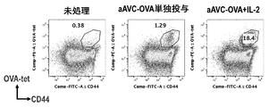
- FIG. 6 shows the scheme of the combined use of the artificial adjuvant vector cell (aAVC) and the immunostimulatory agent (IL-2) in the examples.
- FIG. 7 shows the proportion of antigen-specific T cells in the spleen of day 14 mice in the scheme shown in FIG.
- FIG. 8 is a graph of the results of FIG. 7.
- FIG. 9 shows a scheme of combined use of an artificial adjuvant vector cell (aAVC) and an immunostimulatory agent (described cytokine) in an example.
- FIG. 10 shows the percentage of antigen-specific T cells in the spleen of day 14 mice in the scheme shown in FIG.
- FIG. 11 is a graph of the results of FIG. 10 (number of antigen-specific T cells in the spleen).
- FIG. 12 is a graph of the results of FIG. 10 (ratio of antigen-specific T cells in the spleen (%)).
- FIG. 13 shows the percentage of iNKT cells in the spleen of mice on day 14 in the scheme shown in FIG.
- FIG. 14 shows the number of iNKT cells in the spleen of mice on day 14 in the scheme shown in FIG.
- the "subject" can be a mammal, including, for example, humans and non-human mammals.
- treatment is used to include therapeutic treatment and prophylactic treatment.
- treatment is used to include delaying or stopping the progression of a disease or condition, as well as improving and curing the disease or condition.
- prevention is used to include suppressing the onset of a disease or condition and reducing its incidence.
- an "artificial adjuvant vector cell” is a cell that expresses CD1d and an antigen (for example, a molecule expressed on a target such as a cancer antigen, a viral antigen, and a microbial antigen).
- Means cells pulsed with CD1d Cells pulsed with CD1d can be obtained by culturing cells expressing CD1d with a CD1d ligand. After culturing, the CD1d ligand that did not adhere to the cells can be removed by washing.
- CD1d is a protein of the CD1 family and is an MHC class I-like glycoprotein. CD1d can present glycolipids and activate, for example, iNKT cells.
- CD1d includes, for example, human CD1d, eg, a protein having an amino acid sequence registered under GenBank registration number: AAH27926.1.
- CD1d ligand means a glycolipid that binds to CD1d.
- examples of the CD1d ligand include ⁇ -GalCer ( ⁇ -galactosylceramide), ⁇ -C-GalCer ( ⁇ -C-galactosylceramide), iGB3 (isoglobotolihexosylceramide), GD3 (ganglioside 3), and GSL-1. ( ⁇ -linked glucuronic acid), GSL-1'SA (galacturonic acid) can be mentioned and used as a CD1d ligand.
- the CD1d ligand for example, ⁇ -GalCer and ⁇ -C-GalCer can be preferably used.
- aAVC When aAVC is administered to mammals, it activates iNKT cells, matures dendritic cells, induces innate immunity, and induces acquired immunity. When aAVC expresses a protein, acquired immunity to the protein is also induced. aAVC induces innate and adaptive immunity using any of dendritic cells, cancer cells, and other cells (eg, somatic cells). aAVC induces innate and adaptive immunity in a subject, whether homologous or allogeneic to the subject. If the aAVC is allogeneic, after innate and adaptive immunity is induced in the subject, the aAVC itself can be eliminated as non-self by the subject's immune system.
- the target protein is the full-length protein or a partial peptide.
- aAVC expresses CD1d and is pulsed by the CD1d ligand.
- aAVC may have an exogenous gene encoding CD1d operably linked to a control sequence.
- the aAVC may have an exogenous gene encoding a target protein operably linked to a control sequence.
- aAVC can express an endogenous target protein.
- the "cell” is a mammalian cell, for example, a human cell.
- cells include dendritic cells and non-dendritic cells.
- cells include tumor cells and non-tumor cells.
- cells include cancer cells and non-cancer cells.
- cells include immortalized cells and primary cells.
- the cells may be allogeneic or allogeneic to the administration subject.
- examples of cells include human cells and human cell lines, such as human fetal kidney cell lines, such as HEK293 cells.
- innate immunity is a mechanism for detecting and eliminating invading foreign substances and cells in which the above is generated by phagocytosis and pattern recognition. Innate immunity is primarily responsible for neutrophils, macrophages, natural killer cells (NK cells), natural killer T cells (NKT cells), and dendritic cells.
- acquired immunity is a mechanism for effectively eliminating a foreign substance that re-enters by memorizing the characteristics of the foreign substance. Acquired immunity is mainly borne by lymphocytes such as T cells (cytotoxic T cells (CTL) and helper T cells) and B cells. Acquired immunity is established after innate immunity, so it takes time to respond.
- innate immunity dendritic cells present in peripheral tissues phagocytose and take up pathogens and break them down into peptides. Then, through migration to lymph nodes and spleen and maturation, the antigen peptide is presented to T cells that control acquired immunity. Then, acquired immunity is induced, killer T cells and B cells against the antigen are activated, and an attack on a foreign substance is started. In this way, innate immunity induces acquired immunity via dendritic cells. Immature dendritic cells do not have the ability to present antigens and have a strong phagocytosis, and when they mature, the phagocytosis weakens and the ability to present antigens increases.
- aAVC is a vaccine system that can activate both innate immunity and acquired immunity by inducing activation of innate immune lymphocytes, innate natural killer T cells (iNKT cells), and promoting dendritic cell maturation. .. aAVC activates iNKT cells via CD1d bound to an iNKT cell ligand (eg, ⁇ -GarCer), but aAVC itself is destroyed in the body and taken up by dendritic cells. In addition, aAVC recruits killer T cells to tumor tissues and the like.
- iNKT cells innate natural killer T cells
- antibody means an immunoglobulin, which has a structure in which two heavy chains (H chains) stabilized by a pair of disulfide bonds and two light chains (L chains) are associated with each other.
- the heavy chain consists of a heavy chain variable region VH, a heavy chain constant region CH1, CH2, CH3, and a hinge region located between CH1 and CH2, and the light chain includes a light chain variable region VL and a light chain constant region CL. Consists of.
- the variable region fragment (Fv) composed of VH and VL is a region that is directly involved in antigen binding and imparts diversity to the antibody.
- the antigen-binding region consisting of VL, CL, VH, and CH1 is referred to as a Fab region, and the region consisting of a hinge region, CH2, and CH3 is referred to as an Fc region.
- the regions that come into direct contact with the antigen have a particularly large change and are called complementarity-determining regions (CDRs).
- CDRs complementarity-determining regions
- the part other than the CDR with relatively few mutations is called a framework (FR).
- FR framework
- the term "anti-A antibody” means an antibody that binds to A.
- the anti-A antibody can specifically bind to A.
- “specifically binding to A” means having a binding affinity for A that is stronger than that for proteins other than A.
- the antibody may be a monoclonal antibody or a polyclonal antibody. Further, the antibody may be any isotype of IgG, IgM, IgA, IgD and IgE. It may be produced by immunizing a non-human animal such as a mouse, rat, hamster, guinea pig, rabbit, or chicken, or it may be a recombinant antibody, a chimeric antibody, a humanized antibody, or a fully humanized antibody. And so on.
- a chimeric antibody is an antibody in which fragments of antibodies derived from different species are linked.
- “Humanized antibody” is an amino acid sequence characteristic of a non-human antibody, and means an antibody in which the corresponding position of the human antibody is substituted.
- a heavy chain of an antibody prepared by immunizing a mouse or rat examples include those having CDRs 1-3 and light chains CDR1-3, all of which are derived from human antibodies, including four framework regions (FRs) for each of the heavy and light chains.
- Such antibodies are sometimes referred to as CDR-transplanted antibodies.
- the term "humanized antibody” may also include a human chimeric antibody.
- a "human chimeric antibody” is a non-human antibody in which the constant region of a non-human-derived antibody is replaced with a constant region of a human antibody.
- the subtype of the human antibody used in the constant region can be IgG1 from the viewpoint of enhancing ADCC activity.
- the term "antigen-binding fragment” refers to a fragment of an antibody that has an affinity for binding to an antigen. Specifically, a Fab consisting of VL, VH, CL and CH1 regions; F (ab') 2 in which two Fabs are linked by a disulfide bond in a hinge region; Fv consisting of VL and VH; VL and VH are artificially produced.
- scFv which is a single-chain antibody linked with the polypeptide linker of the above
- bispecific antibodies such as diabody type, scDb type, tandem scFv type, and leucine zipper type can be mentioned. Not limited to.
- immunocheckpoint inhibitor means an agent that activates immunity by releasing the suppression of immune cells caused by an immune checkpoint molecule.
- immunoactivator means an agent that activates immune cells (eg, an agent that points out T cells, particularly an agent that stimulates CTL, such as an agent that stimulates an immunoco-stimulatory molecule). ..
- the immunostimulatory agent can be a drug selective for the target.
- an immune checkpoint inhibitor and an immunostimulant are collectively referred to as an "immunostimulant”.
- Immunocheckpoint molecules include programmed cell death-1 (PD-1), cytotoxic T-lymphocyte-associated protein 4 (CTLA-4), T cell immunoglobulin domain and mutin domain.
- T-cell immunoglobulin domine and mucin domain-3, or TIM-3 T-cell immunoglobulin domine and mucin domain-3, or TIM-3
- lymphocyte activation gene 3 lymphocyte activation gene 3, or LAG-3
- suppression of V-type immunoglobulin domain-containing T cell activation Factors V-type immunoglobulin domine-contining support of T-cell activation, or VISTA
- the immune checkpoints that each bears are called PD-1 immune checkpoints, CTLA-4 immune checkpoints, TIM-3 immune checkpoints, LAG-3 immune checkpoints, and VISTA immune checkpoints.
- Immune checkpoint inhibitors can inhibit the function of immune checkpoints, for example by binding to immune checkpoint molecules or their ligands.
- the PD-1 immune checkpoint can be inhibited by inhibiting the binding of PD-1 to PD-L1 or PD-L2.
- CTLA-4 immune checkpoints can be inhibited by inhibiting the binding of CTLA-4 to CD80 or CD86.
- the TIM-3 immune checkpoint can be inhibited by inhibiting the binding between TIM-3 and galectin-9.
- the LAG-3 immune checkpoint can be inhibited by inhibiting the binding of LAG-3 to the MHC class II molecule.
- the VISTA immune checkpoint can be inhibited by inhibiting the binding between VISTA and VISIG-3 / IGSF11.
- Immune checkpoints can be blocked.
- Antibodies that block the binding of the two proteins can bind to the receptor or ligand.
- the antibody that inhibits the PD-1 immune checkpoint is an antibody selected from the group consisting of anti-PD-1 antibody, anti-PD-L1 antibody, and anti-PD-L2 antibody (eg, nivolumab, pemprolizumab, avelumab, etc.). Atezolizumab, and durvalumab).
- the antibody that inhibits the CTLA-4 immune checkpoint can be an antibody selected from the group consisting of anti-CDLA-4 antibody, anti-CD80 antibody, and anti-CD86 antibody (eg, ipilimumab and tremelimumab).
- the antibody that inhibits the TIM-3 immune checkpoint can be an antibody (eg, MGB453) selected from the group consisting of anti-TIM-3 antibody and anti-galectin-9 antibody.
- the antibody that inhibits the VISTA immune checkpoint can be an antibody selected from the group consisting of anti-VISTA antibody and anti-VSIG-3 / IGSF11 antibody (eg, JNJ-61610588).
- immunoco-stimulatory molecules include OX40 and glucocorticoid-induced TNF-related protein, or GITR.
- GITR glucocorticoid-induced TNF-related protein
- killer T cells are activated and regulatory when stimulated with their respective agonist antibodies (ie, agonist antibodies that bind to GITR and agonist antibodies that bind to OX40) or their respective agonists. The function of T cells is suppressed. In this way, immunoco-stimulatory molecules can be stimulated, for example, one or more immunoco-stimulatory molecules selected from the group consisting of GITR and OX40.
- the immunostimulant against the GITR immunostimulatory molecule can be an anti-GITR antibody and a GITR agonist (eg, TRX518, and MEDI1873).
- immunomodulators for OX40 immunoco-stimulatory molecules can be anti-OX40 antibodies and OX40 agonists (eg, GSK3179498, MOXR0916, PF-04518600, and MEDI0562).
- any of the anti-OX40 agonist antibodies described in WO2012027328A, WO2013202831A, WO2015153514A, and WO20150954223A can be used.
- the immunostimulatory agent can be a drug that activates immunity.
- the immunostimulatory drug may contain cytokines that activate immunity, such as cytokines that activate T cells (eg, interleukins that activate T cells). Cytokines that activate immunity include cytokines such as IL-2.
- IL-2 is a cytokine classified into a group called Th1 cytokine, and is mainly produced from activated T cells.
- the human IL-2 protein can have, for example, the amino acid sequence registered as NBCI reference number: NP_000577.2.
- human IL-2 can be, for example, IL-2 having a sequence corresponding to the amino acid sequence registered as NBCI reference number: NP_000577.2.
- the 1st to 20th amino acids are considered to be the signal sequence
- the mature IL-2 has the 21st to 153rd amino acid sequence. Therefore, when expressing in cells, a nucleic acid encoding IL-2 containing a signal sequence is introduced into cells, and when administered as a protein, IL-2 containing no signal sequence can be used.
- IL-2 As a drug that activates immunity, IL-2, IL-2 / anti-IL-2 antibody complex, IL-7, IL-15, IL-15 / anti-IL15R ⁇ antibody complex, IL-12, IL.
- Factors selected from the group consisting of -18, IL-21, IL-23, and IL-27 can be used in combination with aAVC in the present invention.
- the IL-2 / anti-IL-2 antibody complex is a complex of interleukin-2 (IL-2) and an anti-IL-2 antibody (that is, an antibody that binds to IL-2).
- IL-2 is a factor that binds to the IL-2 receptor and induces IL-2 mediated T cell proliferation and the like.
- anti-IL-2 antibodies regulatory T cell (Treg) -inducing antibodies that suppress immunity and immunostimulatory types that induce effector T cells.
- Treg regulatory T cell
- JES6-1 for mice, 5344 for humans
- it is an antibody that easily binds to cells strongly expressing the IL-2 receptor ⁇ chain (CD25 (IL-2R ⁇ )). Yes, the complex acts on Tregs.
- anti-IL-2 antibody for example, S4B6 for mice, MAB602 for humans, or TCB2
- cells with high expression of IL-2 receptor ⁇ chain CD122 (IL-2R ⁇ )
- IL-2R ⁇ IL-2 receptor ⁇ chain
- an antibody that binds more strongly to a cell that highly expresses the IL-2 receptor ⁇ chain for example, an antibody that binds more strongly to a cell that highly expresses the IL-2 receptor ⁇ chain, and IL-2 Antibodies that enhance function (eg, antibodies that act on cells selected from effectors or memory T cells and NK cells or antibodies that induce such cells) can be used to form a complex with IL-2. It has also been reported that the half-life of soluble IL-2 in vivo is as short as several hours, but the half-life of the IL-2 / anti-IL-2 antibody complex is extended.
- IL-2 the function of IL-2 is enhanced by using a complex of IL-2 and an antibody that binds to the ⁇ chain of the IL-2 receptor.
- IL-2 and anti-IL-2 antibodies can be administered in a pre-complexed form.
- IL-2 can be administered in the form of a complex with the anti-IL-2 antibody.
- the IL-2 can be a human IL-2 and the antibody can be an anti-human IL-2 antibody, a human chimeric antibody, a humanized antibody, or a human antibody.
- human IL-2 include IL-2 having an amino acid sequence registered with GenBank registration number: AAB46883.1 or human IL-2 having an amino acid sequence corresponding thereto.
- the term "having an amino acid sequence corresponding to the amino acid sequence X" is used to include a homolog (for example, a natural homolog) of the protein that retains the function of the protein having the amino acid sequence X.
- Interleukin-7 is a cytokine that plays an important role in the development of T cells and the maintenance of naive and memory CD8-positive T cells.
- IL-7 can be human IL-7.
- human IL-7 include IL-7 having an amino acid sequence registered with GenBank registration number: AAH47698.1 or human IL-7 having an amino acid sequence corresponding thereto.
- Interleukin-15 is a cytokine involved in T cell activation and proliferation.
- IL-15 can be human IL-15.
- human IL-15 include IL-15 having an amino acid sequence registered with GenBank registration number: CAA71044.1 or human IL-15 having an amino acid sequence corresponding thereto.
- IL-15 is a cytokine that activates T cells as described above.
- IL-15Ra is a receptor to which IL-15 binds.
- effectors and memory T cells eg, CD4T cells and CD8T cells
- NK cells in a bound state of IL-15Ra
- these cells are more strongly stimulated (this phenomenon).
- trans-presentation Dubois S et al. Immunoty 2002, 17: 537.
- the half-life of IL-15 is extended by forming a complex of IL-15 and IL-15Ra (Guo Y et al.
- IL-15 can be administered to a subject as a complex with IL-15Ra.
- IL-15 can be administered to a subject in the form of a complex with IL-15Ra.
- IL-15Ra can be human IL-15Ra, and examples thereof include IL-15Ra having an amino acid sequence registered with GenBank registration number: CAG3345.1 or human IL-15Ra having an amino acid sequence corresponding thereto.
- Interleukin-12 is a heterodimer of IL-12A and IL-12B.
- IL-12 is said to be involved in the activation and differentiation induction of T cells and NK cells.
- IL-12 can be human IL-12 (ie, a heterodimer of human IL-12 subunit ⁇ and human IL-12 subunit ⁇ ).
- Human IL-12A can be a protein of 35 kDa, including, for example, IL-12A having an amino acid sequence registered with GenBank registration number: AAI04985.1 or human IL-12A having a corresponding amino acid sequence.
- human IL-12B include IL-12B having an amino acid sequence registered with GenBank registration number: EAW61576.1 or human IL-12B having an amino acid sequence corresponding thereto.
- IL-12 can be provided by synthesizing IL-12A and IL-12B.
- Interleukin-18 is a cytokine that induces interferon gamma. It is said to have functional commonality with IL-12. IL-18 is present intracellularly as a precursor of 24 kDa, but when cleaved by active caspase-1, it is released extracellularly as a mature 18 kDa cytokine. In the present invention, this mature IL-18 can be used as the IL-18.
- IL-18 can be human IL-18, and as human IL-18, human IL-18 having the amino acid sequence registered with UniProtKB / Swiss-Prot registration number: Q1416-1, or an amino acid corresponding thereto. Human IL-18 having a sequence can be mentioned.
- Interleukin-21 is a type I cytokine having homology with IL-2 and IL-15. IL-21 is mainly produced by activated T cells. Examples of IL-21 include human IL-21. Examples of human IL-21 include IL-21 having an amino acid sequence registered with GenBank registration number: EAX05222.1 or human IL-21 having an amino acid sequence corresponding thereto.
- Interleukin-23 is a cytokine composed of the IL-23 subunit ⁇ and the p40 subunit of IL-12.
- IL-23 is said to promote the production and survival of a subset of T cells called TH17 cells.
- IL-23 can be human IL-23.
- human IL-23A for example, the amino acid sequence of positions 20 to 189 of the amino acid sequence of the human IL-23 subunit ⁇ precursor (amino acid numbers 1 to 189) registered in NCBI reference sequence: NP_05766.8 or the amino acid sequence thereof. It can be an IL-23 subunit ⁇ with the corresponding amino acid sequence.
- the IL-23 subunit ⁇ and the p40 subunit of IL-12 can be provided as a complex.
- Interleukin-27 is a heterodimer of interleukin-30 (IL-30) and the Epstein-Barr virus-inducing gene 3 (EBI3) subunit of interleukin-27.
- IL-27 is said to be involved in the proliferation of antigen-specific naive CD4-positive T cells and the promotion of differentiation into the Th1 phenotype.
- the IL-30 can be a human IL-30, and the human IL-30 is, for example, an amino acid sequence registered with UniProtKB / Swiss-Prot registration number: Q8NEV9.2 (amino acids 1 to 28 are signal sequences). It can be human IL-30 with (is) or human IL-30 with the corresponding amino acid sequence.
- the EBI3 subunit of IL-27 can be the EBI3 subunit of human IL-27, and the EBI3 subunit of human IL-27 is an amino acid sequence registered with UniProtKB / Swiss-Prot registration number: Q8K3I6.1. It can be the EBI3 subunit of human IL-27 having (amino acids numbers 1-28 are said to be the signal sequence) or the EBI3 subunit of human IL-27 having the corresponding amino acid sequence.
- cytokines selected from the group consisting of IL-2, IL-7, IL-15, IL-12, IL-18, IL-21, IL-23, and IL-27 signal from any of them.
- the peptide may be excised.
- the present inventors have found that the combined use of an immunostimulant (immune checkpoint inhibitor or immunostimulant) with aAVC increases the number and ratio of antigen-specific CTLs to antigens expressed by aAVC.
- the present inventors have also found that the combined use of an immune checkpoint inhibitor and aAVC increases the number and proportion of iNKT cells.
- the action of an immune checkpoint inhibitor is thought to be to elicit the original action of CTL by inhibiting the immune checkpoint of antigen-specific CTL.
- immune checkpoint inhibitors increased lymphocytes in the innate immune system and increased antigen-specific CTL.
- an immunostimulant can activate innate immunity and induce antigen-specific CTL. Therefore, according to the present invention, an immunostimulant (immune checkpoint inhibitor or immunostimulant), including an immunostimulant, for use in activating innate immunity and / or inducing an antigen-specific CTL.
- an immunostimulant including an immunostimulant, for use in activating innate immunity and / or inducing an antigen-specific CTL.
- the composition for use in is provided.
- inducing antigen-specific CTLs means increasing the number and proportion of antigen-specific CTLs.
- an immunostimulant can enhance the ability of aAVC to induce antigen-specific CLT and iNKT cells. Therefore, according to the present invention
- B An immune checkpoint inhibitor used in combination with an artificial adjuvant vector cell or a pharmaceutical composition containing the cell.
- Artificial adjuvant vector cells are pharmaceutical compositions that express CD1d, are pulsed by a CD1d ligand, and express tumor antigens;
- C A combination preparation of an immunostimulant (immune checkpoint inhibitor or immunostimulant) and an artificial adjuvant vector cell.
- Artificial adjuvant vector cells express CD1d, are pulsed by a CD1d ligand, and express tumor antigens, a combination formulation ⁇ where the combination formulation is an immunostimulant (immune checkpoint inhibitor or immune activation).
- a pharmaceutical composition comprising an agent) and an artificial adjuvant vector cell ⁇ ; and
- Pharmaceutical compositions for use in increasing antigen-specific CTLs, including checkpoint inhibitors, are provided (the above (a)-(d) may be collectively referred to as the medicament of the present invention).
- the artificial adjuvant vector cells can express endogenous CD1d.
- the artificial adjuvant vector cells may express exogenous CD1d.
- the exogenous CD1d can be expressed in artificial adjuvant vector cells by introducing a nucleic acid encoding the exogenous CD1d. If the nucleic acid is DNA, the DNA is operably linked to a promoter. The nucleic acid may be mRNA. When endogenous CD1d is highly expressed, it is not necessary to introduce exogenous CD1d into the artificial adjuvant vector cells.
- Artificial adjuvant vector cells may express the antigen. Examples of the antigen include a tumor antigen and an antigen of an invading foreign substance.
- Tumor antigens are antigens that are more expressed in tumors than in normal cells. Tumor antigens are preferably 2 times, 3 times, 4 times, 5 times, 6 times, 7 times, 8 times, 9 times, 10 times, 20 times, and 30 times the number of surface molecules in tumors. , 40 times, 50 times, 60 times, 70 times, 80 times, 90 times, 100 times, 200 times, 300 times, 400 times, 500 times, 600 times, 700 times, 800 times, 900 times, and 1,000 times. It can be an antigen that is expressed more than twice as high. Tumor antigens include solid tumors, including epithelial and non-epithelial tumors, and tumor antigens in hematopoietic tissues.
- the tumor antigen is not particularly limited, but is, for example, MART-1, ZMelan-A, Mage-1, Mage-3, gpl00, tyrosinase, CEA, PSA, CA-125 erb-2, Muc-1, Muc-2, TAG-72, AES, FBP, C Lectin, NY-ESO-1, galectin-4 / NY-CO-27, Pec60, HER-2 / erbB-2 / neu, telomerase, G250, Hspl05, point mutant ras oncogene , Oncogenes such as point mutant p53 oncogene, and carcinoembryonic antigen, and neoantigens (for example, JP-A-2005-139118, JP-A-2004-147649, JP-A-2002-112780, Japanese Patent Laid-Open No.
- the antigen of a tumor (eg, leukemia) in hematopoietic tissue is not particularly limited, but is, for example, proteinase3, WT-1, hTERT, PRAME, PML / RAR-a, DEK / CAN, cyclofilin, TEL-MAL1, BCR-ABL. , OFA-iLRP, Survivin, idiotype, Sperm protein 17, SPAN-Xb, CT27, and MUC1.
- the pathogenic antigen can be a pathogenic viral antigen, a pathogenic microbial antigen, or a pathogenic protozoan antigen.
- the pathogenic virus antigen is not particularly limited, but for example, GP-120, pl7, GP-160 (or more, HIV), NP, HA (or more, influenza virus), HBs Ag, HBV envelope protein, core protein, polymerase protein.
- NS3, NS5 (above, hepatitis virus), HSVdD (simple herpesvirus), EBNA1, 2, 3A, 3B and 3C, LMP1 and 2, BZLF1, BMLF1, BMRF1, BHRF1 (above, EB virus), Tax (HTLV-I), SARS-CoV spike protein (SARS virus), CMV pp50, CMV pp65, IE-1 (above, CMV), E6, E7 protein (above, HPV) (for example, JP-A-2004- 222,726 (see).
- pathogenic microbial antigens include antigens expressed in pathogenic bacteria (eg, Chlamydia, mycobacteria, Legionella) and pathogenic yeasts (eg, Aspergillus, Candida).
- pathogenic protozoan antigens include antigens expressed in malaria and schistosomiasis.
- the artificial adjuvant vector cell can be an isolated cell or a purified cell.
- the artificial adjuvant vector cell may be a cell line.
- Artificial adjuvant vector cells can be cells of human or non-human animals (eg, non-human mammals, and non-human primates). Non-human animals include monkeys, chimpanzees, gorillas, orangutans, bonobos, dogs, cats, cows, pigs, sheep, horses, mice, rats, guinea pigs, hamsters, and rabbits.
- the artificial adjuvant vector cell may be an autologous cell, an allogeneic cell, or a heterologous cell with respect to the administration subject. This is because it is ultimately phagocytosed by dendritic cells.
- the artificial adjuvant vector cell can be a somatic cell.
- Somatic cells include stomach, small intestine (eg duodenum, jejunum, ileum, colon), colon, rectum, lung, spleen, kidney, liver, thoracic gland, spleen, thyroid, adrenal gland, prostate, ovary, uterus, bone marrow, skin, Peripheral blood can be mentioned.
- the cell of the present invention may also be a specific cell type in the above tissue or a cell type existing in a tissue other than the above tissue.
- Such cell types include, for example, epithelial cells, endothelial cells, epidermal cells, stromal cells, fibroblasts, fat cells, mammary gland cells, mesangial cells, spleen cells, nerve cells, glial cells, immune cells (eg, eg). T cells, B cells, NK cells, NKT cells, macrophages, obese cells, neutrophils, basal spheres, eosinophils, monospheres), and precursor cells and stem cells of these cells.
- the antigen expressed in the artificial adjuvant vector cell may be an antigen derived from the cell or an antigen introduced into a foreign country.
- the antigen expressed in the artificial adjuvant vector cell is an antigen derived from the cell
- the artificial adjuvant vector cell can be prepared from, for example, a tumor cell or a pathogen-infected cell.
- Pathogen-infected cells include virus-infected cells, microbial-infected cells, and protozoan-infected cells.
- the artificial adjuvant vector cell may be a non-transformant or a transformant. Transformation refers to artificial gene transfer. For example, cells into which DNA or mRNA has been introduced exogenously are called transformants. When the artificial adjuvant vector cell expresses CD1d and the antigen endogenously, the artificial adjuvant vector cell can be prepared without transformation.
- Artificial adjuvant vector cells can be prepared from antigen-presenting cells (APC) or non-APC.
- APCs include dendritic cells, macrophages, B cells, Langerhans cells, and activated T cells.
- Artificial adjuvant vector cells can be created by pulsing with a CD1d ligand.
- the pulse of cells with the CD1d ligand can be performed by culturing the cells expressing CD1d in the medium in the presence of the CD1d ligand. By doing so, cells expressing the target antigen and expressing CD1d can be made to present the CD1d ligand.
- immunoactivation against the target antigen can occur in vivo.
- the medium a medium that can be used for cell culture can be appropriately used.
- the medium include MEM medium, DMEM medium, ⁇ MEM medium, ham medium RPMI1640 medium, Fisher's medium, and a mixed medium thereof.
- the medium may be, for example, serum (eg, FCS), serum substitute (eg, knockout Serum Replacement (KSR)), fatty acid or lipid, amino acid, vitamin, growth factor, cytokine, antioxidant, 2-mercaptoethanol, pyruvin. It can contain additional components selected from the group consisting of acids, buffers, inorganic salts and the like.
- the nucleic acid encoding the exogenous CD1d and the nucleic acid encoding the exogenous antigen can be introduced into cells using a gene expression vector.
- the nucleic acid encoding the exogenous CD1d and the nucleic acid encoding the exogenous antigen may be contained in one gene expression vector or may be contained in separate gene expression vectors.
- a nucleic acid encoding an exogenous CD1d and a nucleic acid encoding an exogenous antigen are contained in one gene expression vector, they are polycistronicly contained in the mRNA so as to be transcribed as one mRNA. It may be monocistronicly contained in the mRNA so that it is transcribed as a separate mRNA.
- an internal ribosome entry site can be intervened between the two genes.
- the nucleic acid encoding the exogenous CD1d, and the nucleic acid encoding the exogenous antigen can be operably linked to a promoter suitable for transcription into mRNA in the cell.
- a promoter means the smallest sequence sufficient to induce transcription within a cell.
- the promoters include viral promoters, mammalian promoters, and yeast promoters (eg, mammalian CMV promoter, yeast alcohol oxidase, phosphoglycerokinase promoter, lactose-inducible promoter, galactosidase promoter, adeno-associated virus promoter, baculovirus promoter, poxvirus).
- mammalian promoters eg, mammalian CMV promoter, yeast alcohol oxidase, phosphoglycerokinase promoter, lactose-inducible promoter, galactosidase promoter, adeno-associated virus promoter, baculovirus promoter, poxvirus.
- yeast promoters eg, mammalian CMV promoter, yeast alcohol oxidase, phosphoglycerokinase promoter, lactose-inducible promoter, galactosidase promoter, adeno-associated virus
- the nucleic acid encoding the exogenous CD1d and the nucleic acid encoding the exogenous antigen can be mRNA, eg, the mRNA described above. mRNA contains elements necessary for translation of the encoding protein, and when introduced into cells, induces translation of the encoding protein. The introduction of a gene into a cell can be carried out by a conventional method.
- the immune checkpoint inhibitors include PD-1 immune checkpoint inhibitors, CTLA-4 immune checkpoint inhibitors, TIM-3 immune checkpoint inhibitors, and Lag3 immune checkpoint inhibitors. Immune checkpoint inhibitors and VISTA immune checkpoint inhibitors can be mentioned.
- the immunostimulant is selected from an immunostimulant against GITR and an immunostimulant against OX40 (for example, each agonist and GITR antibody as each agonist antibody, and OX40 antibody. One or more antibodies). In the present invention, it may be an OX40 antibody, which is an immunostimulatory agent against OX40.
- aAVC and an immunostimulant may be administered simultaneously or separately.
- the administration of aAVC may be a single administration or a plurality of administrations.
- the immunostimulant (immune checkpoint inhibitor or immunostimulant) may be administered as a single dose or multiple doses.
- the administration of aAVC may be a single dose and the administration of an immunostimulant (immune checkpoint inhibitor or immunostimulant) may be multiple doses.
- aAVC and an immunostimulant may be provided in one package or in separate packages.
- the aAVC and immune checkpoint inhibitors may be mixed in the same pharmaceutical composition or separately contained in separate pharmaceutical compositions. ..
- the aAVC and immune checkpoint inhibitors are provided separately in separate packages.
- the aAVC and immune checkpoint inhibitors are separately contained in separate pharmaceutical compositions within one package.
- aAVC and / or an immunostimulant is used in combination with aAVC and an immunostimulant (immune checkpoint inhibitor or immunostimulant). It may further include an attachment, including instructions for.
- the aAVC can be provided frozen.
- the immunostimulant immunostimulant (immune checkpoint inhibitor or immunostimulant) may be provided as an infusion or injection.
- the aAVC can be provided in a frozen and gamma-sterilized state.
- the present invention also provides a pharmaceutical composition comprising an immunostimulant (immune checkpoint inhibitor or immunostimulant) for use in activating innate immunity (particularly iNKT cells). Activation of innate immunity (particularly iNKT cells) can increase antigen-specific CTL. Therefore, according to the present invention, there is also provided a pharmaceutical composition for use in increasing antigen-specific CTLs, which comprises an immunostimulant (immune checkpoint inhibitor or immunostimulant). According to the present invention, an increase in antigen-specific CTL can occur through activation of innate immunity.
- the immune checkpoint inhibitors include PD-1 immune checkpoint inhibitor, CTLA-4 immune checkpoint inhibitor, TIM-3 immune checkpoint inhibitor, and Lag3 system.
- the pharmaceutical composition for use in activating innate immunity can be a pharmaceutical composition for use in proliferating iNKT cells.
- the pharmaceutical composition for use in activating innate immunity (particularly iNKT cells) or for proliferating iNKT cells is an anti-PD-1 antibody, anti-Lag3. It may include an immunostimulatory agent selected from the group consisting of antibodies, anti-VISTA antibodies, anti-OX40 antibodies, and anti-CTLA4 antibodies (eg, anti-OX40 antibodies that are agonist antibodies).
- the pharmaceutical composition for use in increasing antigen-specific CTL is selected from the group consisting of anti-PD-1 antibody, anti-TIM-3 antibody, anti-CTLA4 antibody, and anti-OX40 antibody. It may contain an immunostimulatory agent (eg, an anti-OX40 antibody which is an agonist antibody).
- OX40 agonist antibody for example, a hybridoma that produces an antibody that binds to OX40 is screened by the affinity of the antibody produced by the hybridoma for OX40, and then the obtained OX40 antibody is controlled by an NF- ⁇ B response element.
- OX40-expressing cells eg, HEK293 cells
- reporter eg, luciferase
- Such test systems can be purchased, for example, from BPS Bioscience (Catalog # 60482).
- the immunostimulant (immune checkpoint inhibitor or immunostimulant) of the present invention may further contain an excipient.
- the aAVC of the present invention may further contain excipients. As the excipient, suitable ones can be included.
- the immune checkpoint inhibitor of the present invention can be administered by parenteral administration such as intravenous administration, intratumoral administration, subcutaneous administration, and intraperitoneal administration, and is formulated in a dosage form suitable for these administration routes. sell.
- the aAVC of the present invention can be administered by parenteral administration such as intravenous administration, intratumoral administration, subcutaneous administration, and intraperitoneal administration, and can be formulated in a dosage form suitable for these administration routes.
- the medicament of the present invention can be used for the treatment of cancer and / or infectious diseases.
- Cancers include, for example, solid tumors (eg, epithelial tumors, non-epithelial tumors), tumors in hematopoietic tissue. More specifically, solid tumors that can be treated by the medicament of the present invention include, for example, gastrointestinal cancer (eg, gastric cancer, colon cancer, colon cancer, rectal cancer), lung cancer (eg, small cell cancer).
- Non-small cell cancer Non-small cell cancer
- spleen cancer kidney cancer, liver cancer, thoracic adenocarcinoma, spleen cancer, thyroid cancer, adrenal cancer, prostate cancer, bladder cancer, ovarian cancer, uterus Cancer (eg, endometrial cancer, cervical cancer), bone cancer, skin cancer, sarcoma (eg, Kaposi sarcoma), melanoma, blastoma (eg, neuroblastoma), adenocarcinoma , Flat cell cancer, non-flat cell cancer, brain tumors, and cancers due to recurrence and metastasis of these solid tumors.
- sarcoma eg, Kaposi sarcoma
- melanoma melanoma
- blastoma eg, neuroblastoma
- adenocarcinoma Flat cell cancer, non-flat cell cancer, brain tumors, and cancers due to recurrence and metastasis of these solid tumors.
- Tumors in hematopoietic tissue that can be treated by the medicament of the present invention include leukemia (eg, acute myelogenous leukemia (AML), chronic myelogenous leukemia (CML), acute lymphocytic leukemia (ALL), chronic lymphocytic leukemia) (CLL), adult T-cell leukemia lymphoma (ATL), myelogenous dysplasia syndrome (MDS)), lymphoma (eg, T lymphoma, B lymphoma, Hodgkin lymphoma, non-Hodgkin lymphoma), myeloma (multiple myeloma), and Cancer due to recurrence and metastasis of these tumors can be mentioned.
- Infectious diseases that can be treated by the medicament of the present invention include, for example, infectious diseases caused by the above-mentioned pathogens.
- a AVC is provided.
- the aAVC can express an antigen specific for the cancer or infection to be treated (foreign body that causes the infection).
- the medicament of the present invention can be administered.
- administration of the medicament of the present invention can be carried out until an increase in the number of cytotoxic T cells is confirmed.
- the invention may further comprise measuring the number of cytotoxic T cells after administration of the medicament of the invention.
- the invention may include detecting an increase in the number of cytotoxic T cells in an administration subject after administration of the medicament of the invention.
- Cytotoxic T cells may have antigen specificity for the antigen expressed by aAVC.
- the therapeutically effective amount is an amount that exhibits therapeutic efficacy against cancer or infection, but can be an amount that increases cytotoxic T cells in the subject.
- an immunostimulant immunostimulant
- immunostimulant immunostimulant
- / or aAVC immune checkpoint inhibitors and / or aAVC for use in the treatment methods of the present invention are provided.
- Example 1 Combination of artificial adjuvant vector (aAVC) and immune checkpoint inhibitor
- aAVC artificial adjuvant vector
- immunostimulant immune checkpoint inhibitor or immunostimulant
- the administration scheme was as shown in FIG. Specifically, aAVC expressing ovalbumin (OVA) was administered on the 0th day, and an immune checkpoint inhibitor (antibody) was administered at 200 ⁇ g / administration on the 0th, 3rd, and 5th days, respectively. Then, on the 7th day, the number and proportion of antigen-specific CD8-positive T cells and the number and proportion of iNKT cells were examined.
- OVA ovalbumin
- antibody immune checkpoint inhibitor
- aAVC-OVA is obtained by expressing a gene encoding CD1d and a gene encoding OVA as an antigen in NIH3T3 cells, and then adding 0.5 mg / mL ⁇ -galactosylceramide ( ⁇ -GalCer) to the medium. It was. Administration was performed at a dose of 5 ⁇ 10 5 cells / animal.
- anti-PD-1 antibody BioXcell manufacturer, product number: BE0146
- anti-Lag3 antibody BioXcell manufacturer, product number: BE0174
- anti-TIM-3 antibody BioXcell
- anti-VISTA antibody BioXcell manufacturer, product number: BE0310
- anti-CTLA4 antibody BioXcell manufacturer, product number: BE0164
- an anti-OX40 antibody BioXcell manufacturer, product number: BE0031
- an anti-GITR antibody BioXcell manufacturer, product number: BE0063
- AAVC-OVA was administered intravenously, and the immunomodulator was administered intraperitoneally.
- mice spleens were collected and analyzed for antigen-specific CD8-positive T cells (cytotoxic T cells) and invariant natural killer T cells (iNKT cells).
- CD8-positive T cells spleen cells were isolated from the spleen, stained with CD3, CD8, CD44 antibody and OVA-tetramer, and analyzed using FACS (manufacturer BD, product name Cancto II). Spleen cells were gated with CD3 and CD8 positive cells to CD8 T cells and expanded with CD44 and OVA-tetramer. The number and proportion of CD44-positive and OVA-tet-positive cells were measured as OVA-specific CD8-positive T cells. The ratio is the ratio (%) of OVA-specific CD8-positive T cells to CD8 T cells.
- FIG. 2 An example was observed in which the proportion of OVA-specific CD8-positive T cells was increased in the immunostimulant combination group as compared with the aAVC-OVA alone administration group. Specifically, the proportion of OVA-specific CD8-positive T cells is increasing in the anti-PD-1 antibody combination group, the anti-TIM-3 antibody combination group, the anti-CTLA4 antibody combination group, and the anti-OX40 antibody combination group. In particular, the increase in the proportion of OVA-specific CD8-positive T cells was remarkable in the anti-OX40 antibody combination group.
- the number and proportion of OVA-specific CD8-positive T cells are graphed and shown in FIG. As shown in FIG.
- the number and proportion of OVA-specific CD8-positive T cells in the anti-PD-1 antibody combination group, the anti-TIM-3 antibody combination group, the anti-CTLA4 antibody combination group, and the anti-OX40 antibody combination group The number and proportion of OVA-specific CD8-positive T cells increased significantly in the anti-OX40 antibody combination group.
- iNKT cells in splenocytes were also analyzed. Specifically, 14 days after cell administration, the spleen was isolated and labeled with afficocyanin (APC) -labeled ⁇ -GalCer preloaded CD1d tetramer CD1d-tetramer / Gal-APC and PerCP / Cy5 (R) . The CD3 was stained with CD3-PerCP / Cy5 (R) , and the cells of CD3 + and CD1d-tetramer / Gal + were counted as iNKT cells. Next, the frequency of iNKT cells in the spleen was calculated.
- APC afficocyanin
- R PerCP / Cy5
- the total number of cells in the spleen of each immunized mouse was multiplied by the above frequency to calculate the number of iNKT cells.
- the number and proportion of iNKT cells in the anti-PD-1 antibody combination group, the anti-Lag3 antibody combination group, the anti-VISTA antibody combination group, the anti-OX40 antibody combination group, and the anti-CTLA4 antibody combination group were increased in the anti-OX40 antibody combination group.
- iNKT cells induce activation of natural killer T cells (NK cells) and dendritic cells responsible for innate immunity, as well as activation of B cells and T cells responsible for the adaptive immune response.
- aAVC has been shown to enhance innate and adaptive immunity.
- the immune checkpoint inhibitor enhances immunity by T cells by releasing the suppression of T cells (cytotoxic T cells; CTL) responsible for acquired immunity, and the immunostimulator used this time is direct immunity. It enhances immunity by T cells by stimulating function. This result shows that the combined use of an immunostimulant with aAVC enhances the innate immunity activating effect, and simply induces innate immunity by aAVC and acquired immunity by an immune checkpoint inhibitor.
- the immunostimulant enhances the ability of aAVC to induce innate immunity and not only releases the suppression of cytotoxic T cells, but also increases the number of cytotoxic T cells. It has become clear that it can be a method for realizing more effective cancer immunotherapy.
- immune checkpoint inhibitors can be expected to enhance the ability of aAVC to activate innate immunity and the ability to induce antigen-specific immunity.
- the immunostimulant can be expected to enhance the therapeutic effect of aAVC on cancer and infectious diseases.
- Example 2 Combination of aAVC and immunostimulatory agent
- aAVC and cytokines were used in combination as shown in FIG.
- Mice were administered aAVC-OVA in the same manner as in FIG. From the 7th day after administration, 5 ⁇ 10 3 U / dose of IL-2 was administered daily for 7 days.
- the mouse spleen of the mouse was collected.
- CD8 T cells were gated from spleen cells with CD3 and CD8 positive cells and expanded with CD44 and OVA-tetramer.
- the number and proportion of CD44-positive and OVA-tet-positive cells were measured as OVA-specific CD8-positive T cells.
- the ratio is the ratio (%) of OVA-specific CD8-positive T cells to CD8 T cells.
- aAVC-OVA was prepared as in Example 1. Administration of aAVC-OVA was carried out at 5 ⁇ 10 5 cells / mouse dose as in Example 1.
- FIGS. 7 and 8 The results were as shown in FIGS. 7 (flow cytometry results) and 8 (number and proportion of antigen-specific CD8-positive T cells (left) and proportion (right)).
- FIGS. 7 and 8 in the group in which aAVC-OVA and IL-2 were used in combination, the number and proportion of antigen-specific CD8-positive T cells were significantly increased as compared with the case of aAVC-OVA alone. It was. This indicates that the immunostimulatory agent is useful in combination with aAVC.
- the number and proportion of CD44-positive and OVA-tet-positive cells were measured as OVA-specific CD8-positive T cells.
- the ratio is the ratio (%) of OVA-specific CD8-positive T cells to CD8 T cells.
- aAVC-OVA was prepared as in Example 1. Administration of aAVC-OVA was carried out at 5 ⁇ 10 5 cells / mouse dose as in Example 1.
- FIG. 10 results of flow cytometry
- FIG. 11 number of antigen-specific CD8-positive T cells in the spleen
- FIG. 12 ratio of antigen-specific CD8-positive T cells.
- the number and proportion of antigen-specific CD8-positive T cells increased, but IL-2, when any of the cytokines were used in combination, as compared with the administration of aAVC alone.
- the increase was significant when either the anti-IL-2 antibody complex or IL-15 was administered.
- Example 1 the number and proportion of iNKT cells in splenocytes were also analyzed. As a result, it was as shown in FIG. 13 (result of flow cytometry) and FIG. 14 (number of iNKT cells in the spleen). As shown in FIGS. 13 and 14, the number of iNKT cells in the spleen was increased as compared with aAVC alone administration, regardless of which cytokine was used in combination. As described above, the administered immunostimulatory agent was able to enhance the innate immunity-inducing ability of aAVC.
Landscapes
- Health & Medical Sciences (AREA)
- Chemical & Material Sciences (AREA)
- Life Sciences & Earth Sciences (AREA)
- Immunology (AREA)
- General Health & Medical Sciences (AREA)
- Organic Chemistry (AREA)
- Medicinal Chemistry (AREA)
- Veterinary Medicine (AREA)
- Pharmacology & Pharmacy (AREA)
- Animal Behavior & Ethology (AREA)
- Public Health (AREA)
- Engineering & Computer Science (AREA)
- Genetics & Genomics (AREA)
- Epidemiology (AREA)
- Proteomics, Peptides & Aminoacids (AREA)
- Biochemistry (AREA)
- General Chemical & Material Sciences (AREA)
- Chemical Kinetics & Catalysis (AREA)
- Zoology (AREA)
- Bioinformatics & Cheminformatics (AREA)
- Molecular Biology (AREA)
- Biophysics (AREA)
- Biomedical Technology (AREA)
- Biotechnology (AREA)
- Hematology (AREA)
- Nuclear Medicine, Radiotherapy & Molecular Imaging (AREA)
- Microbiology (AREA)
- Cell Biology (AREA)
- Wood Science & Technology (AREA)
- Developmental Biology & Embryology (AREA)
- Virology (AREA)
- Mycology (AREA)
- General Engineering & Computer Science (AREA)
- Gastroenterology & Hepatology (AREA)
- Communicable Diseases (AREA)
- Oncology (AREA)
- Medicines That Contain Protein Lipid Enzymes And Other Medicines (AREA)
- Medicines Containing Material From Animals Or Micro-Organisms (AREA)
Abstract
The present invention provides a composition that contains an immunostimulant and is used to activate natural immunity. The present invention also provides the combined use of artificial adjuvant vector cells and an immunostimulant.
Description
本発明は、免疫チェックポイントに作用して免疫を賦活化する免疫賦活剤(免疫チェックポイント阻害剤、または免疫活性化剤)を含む、自然免疫を活性化することに用いるための組成物に関する。本発明はまた、人工アジュバントベクター細胞と免疫賦活剤(免疫チェックポイント阻害剤または免疫活性化剤)との併用に関する。
The present invention relates to a composition for use in activating innate immunity, which comprises an immunostimulant (immune checkpoint inhibitor or immunostimulant) that acts on an immune checkpoint to activate immunity. The present invention also relates to the combined use of artificial adjuvant vector cells and an immunostimulant (immune checkpoint inhibitor or immunostimulant).
CD1dと標的タンパク質を共発現する細胞をCD1dリガンドでパルスして得られる細胞は、人工アジュバントベクター細胞(aAVC)と呼ばれ、投与された対象において自然免疫と、当該標的タンパク質に対する獲得免疫を誘発する(特許文献1~3および非特許文献1)。
Cells obtained by pulsing cells that co-express CD1d and the target protein with a CD1d ligand are called artificial adjuvant vector cells (aAVC) and induce innate immunity and acquired immunity to the target protein in the administered subject. (Patent Documents 1 to 3 and Non-Patent Document 1).
免疫賦活剤(免疫チェックポイント阻害剤または免疫活性化剤)の中で、免疫チェックポイント阻害剤は、免疫チェックポイント分子の働きを阻害することによってT細胞に対する抑制効果を弱めるまたは解除し、これにより免疫能を向上させて、がんや感染症に対する治療効果を奏することができる。免疫賦活剤は免疫を賦活させる製剤である。より具体的には、免疫チェックポイント阻害剤は、抗原特異的な細胞傷害性T細胞が免疫チェックポイント分子の働きによって受けた抑制を弱めるまたは解除することによって、抗原特異的な細胞傷害性T細胞ががん細胞や病原体により感染した細胞を殺傷することを促進することができる(特許文献4)。免疫活性化剤は抗原特異的な細胞傷害性T細胞の機能を高めることができる。
Among immune checkpoint inhibitors (immune checkpoint inhibitors or immune activators), immune checkpoint inhibitors weaken or cancel the inhibitory effect on T cells by inhibiting the action of immune checkpoint molecules, thereby It can improve immunity and have a therapeutic effect on cancer and infectious diseases. An immunostimulant is a preparation that activates immunity. More specifically, immune checkpoint inhibitors are antigen-specific cytotoxic T cells by weakening or relieving the inhibition of antigen-specific cytotoxic T cells by the action of immune checkpoint molecules. Can promote the killing of cancer cells and cells infected by pathogens (Patent Document 4). Immune activators can enhance the function of antigen-specific cytotoxic T cells.
本発明は、免疫賦活剤(免疫チェックポイント阻害剤または免疫活性化剤)を含む、自然免疫を活性化することに用いるための組成物を提供する。本発明はまた、人工アジュバントベクター細胞と免疫チェックポイント阻害剤との併用を提供する。
The present invention provides a composition for use in activating innate immunity, which comprises an immunostimulant (immune checkpoint inhibitor or immunostimulant). The present invention also provides a combination of artificial adjuvant vector cells and immune checkpoint inhibitors.
本発明者らは、免疫賦活剤(免疫チェックポイント阻害剤または免疫活性化剤)が自然免疫に関与するインバリアントナチュラルキラーT細胞(iNKT細胞)を増加させ、かつ、抗原特異的細胞傷害性T細胞(CTL、またはCD8陽性T細胞)を増加させることを見出した。本発明者らは、免疫チェックポイント阻害剤と人工アジュバントベクター細胞(aAVC)との併用により、iNKT細胞を増加させ、かつ、aAVCに発現させた抗原に対する抗原特異的細胞傷害性T細胞(CTL、またはCD8陽性T細胞)を増加させることを見出した。本発明は、これらの知見に基づく。
The present inventors increase invariant natural killer T cells (iNKT cells) in which an immune activator (immune checkpoint inhibitor or immunostimulator) is involved in innate immunity, and antigen-specific cytotoxic T cells. It has been found to increase cells (CTL, or CD8 positive T cells). The present inventors increased iNKT cells by using an immune checkpoint inhibitor in combination with an artificial adjuvant vector cell (aAVC), and antigen-specific cytotoxic T cells (CTL,) against an antigen expressed in aAVC. Or it was found to increase CD8 positive T cells). The present invention is based on these findings.
本発明によれば、以下の産業上利用可能な発明が提供される。
(1)免疫賦活剤(免疫チェックポイント阻害剤または免疫活性化剤)と併用される、人工アジュバントベクター細胞を含む医薬組成物であって、
人工アジュバントベクター細胞は、CD1dを発現し、CD1dリガンドによってパルスされており、かつ、抗原を発現する、医薬組成物。
(2)人工アジュバントベクター細胞が、CD1dをコードする外来の核酸および腫瘍抗原をコードする外来の核酸の少なくとも1つを有する、上記(1)に記載の医薬組成物。
(3)CD1dリガンドが、α-ガラクトシルセラミドである、上記(1)または(2)に記載の医薬組成物。
(4)免疫賦活剤が、抗PD-1抗体、抗Lag3抗体、抗TIM-3抗体、抗CTLA4抗体(以上、免疫チェックポイント阻害剤)、および抗OX40抗体、抗GITR抗体(以上、免疫賦活剤)から選択される少なくとも1つの抗体である、上記(1)~(3)のいずれかに記載の医薬組成物。
(5)免疫賦活剤が、上記(1)~(3)のいずれかに記載の医薬組成物。
(6)免疫賦活剤が、免疫を活性化するサイトカインである、上記(1)~(3)のいずれかに記載の医薬組成物。
(7)人工アジュバントベクター細胞または当該細胞を含む医薬組成物と併用される、免疫関連分子に作用して免疫を賦活化する免疫賦活剤であって、
人工アジュバントベクター細胞は、CD1dを発現し、CD1dリガンドによってパルスされており、かつ、抗原を発現する、医薬組成物。
(8)免疫チェックポイントに作用して免疫を賦活化する免疫賦活剤(免疫チェックポイント阻害剤または免疫活性化剤)と人工アジュバントベクター細胞との組合せ製剤であって、
人工アジュバントベクター細胞は、CD1dを発現し、CD1dリガンドによってパルスされており、かつ、抗原を発現する、組合せ製剤。
(9)免疫チェックポイントに作用して免疫を賦活化する免疫賦活剤(免疫チェックポイント阻害剤または免疫活性化剤)を含む、自然免疫を活性化させることに用いるための組成物。 The present invention provides the following industrially applicable inventions.
(1) A pharmaceutical composition containing artificial adjuvant vector cells used in combination with an immunostimulant (immune checkpoint inhibitor or immunostimulant).
An artificial adjuvant vector cell is a pharmaceutical composition that expresses CD1d, is pulsed by a CD1d ligand, and expresses an antigen.
(2) The pharmaceutical composition according to (1) above, wherein the artificial adjuvant vector cell has at least one of a foreign nucleic acid encoding CD1d and a foreign nucleic acid encoding a tumor antigen.
(3) The pharmaceutical composition according to (1) or (2) above, wherein the CD1d ligand is α-galactosylceramide.
(4) The immunostimulants are anti-PD-1 antibody, anti-Lag3 antibody, anti-TIM-3 antibody, anti-CTLA4 antibody (above, immune checkpoint inhibitor), anti-OX40 antibody, and anti-GITR antibody (above, immunostimulant). The pharmaceutical composition according to any one of (1) to (3) above, which is at least one antibody selected from the agent).
(5) The pharmaceutical composition according to any one of (1) to (3) above, wherein the immunostimulant is.
(6) The pharmaceutical composition according to any one of (1) to (3) above, wherein the immunostimulant is a cytokine that activates immunity.
(7) Artificial adjuvant vector An immunostimulant that acts on an immune-related molecule to activate immunity, which is used in combination with a cell or a pharmaceutical composition containing the cell.
An artificial adjuvant vector cell is a pharmaceutical composition that expresses CD1d, is pulsed by a CD1d ligand, and expresses an antigen.
(8) A combination preparation of an immunostimulant (immune checkpoint inhibitor or immunostimulant) that acts on an immune checkpoint to activate immunity and an artificial adjuvant vector cell.
Artificial adjuvant vector cells are a combination formulation that expresses CD1d, is pulsed by a CD1d ligand, and expresses an antigen.
(9) A composition for use in activating innate immunity, which comprises an immunostimulant (immune checkpoint inhibitor or immunostimulant) that acts on an immune checkpoint to activate immunity.
本明細書では、「対象」とは、哺乳動物であり得、例えば、ヒトおよび非ヒト哺乳動物が含まれる。
As used herein, the "subject" can be a mammal, including, for example, humans and non-human mammals.
本明細書では、「処置」とは、治療的処置および予防的処置を含む意味で用いられる。本明細書では、「治療」とは、疾患もしくは状態の進行の遅延または停止、並びに疾患もしくは状態の改善および治癒を含む意味で用いられる。本明細書では「予防」とは、疾患または状態の発症を抑制すること、および、その発症率を低下させることを含む意味で用いられる。
As used herein, the term "treatment" is used to include therapeutic treatment and prophylactic treatment. As used herein, "treatment" is used to include delaying or stopping the progression of a disease or condition, as well as improving and curing the disease or condition. As used herein, "prevention" is used to include suppressing the onset of a disease or condition and reducing its incidence.
本明細書では、「人工アジュバントベクター細胞」(aAVC)とは、CD1dと抗原(例えば、がん抗原、ウイルス抗原、および微生物の抗原等の標的に発現する分子)を発現する細胞であって、CD1dでパルスされた細胞を意味する。CD1dでパルスされた細胞は、CD1dを発現する細胞をCD1dリガンドと共に培養することによって得られ得る。培養後、細胞に付着しなかったCD1dリガンドは、洗浄によって除去することができる。CD1dは、CD1ファミリーのタンパク質であり、MHCクラスI様の糖タンパク質である。CD1dは、糖脂質を提示して、例えば、iNKT細胞を活性化させ得る。CD1dとしては、例えば、ヒトのCD1d、例えば、GenBank登録番号:AAH27926.1の下で登録されたアミノ酸配列を有するタンパク質が挙げられる。CD1dリガンドは、CD1dに結合する糖脂質を意味する。CD1dリガンドとしては、例えば、α-GalCer(α-ガラクトシルセラミド)、α-C-GalCer(α-C-ガラクトシルセラミド)、iGB3(イソグロボトリヘキソシルセラミド)、GD3(ガングリオシド3)、GSL-1(α-結合型グルクロン酸)、GSL-1’SA(ガラクツロン酸)が挙げられ、CD1dリガンドとして用いられ得る。CD1dリガンドとしては、例えば、α-GalCer、α-C-GalCerが好ましく用いられ得る。
As used herein, an "artificial adjuvant vector cell" (aAVC) is a cell that expresses CD1d and an antigen (for example, a molecule expressed on a target such as a cancer antigen, a viral antigen, and a microbial antigen). Means cells pulsed with CD1d. Cells pulsed with CD1d can be obtained by culturing cells expressing CD1d with a CD1d ligand. After culturing, the CD1d ligand that did not adhere to the cells can be removed by washing. CD1d is a protein of the CD1 family and is an MHC class I-like glycoprotein. CD1d can present glycolipids and activate, for example, iNKT cells. CD1d includes, for example, human CD1d, eg, a protein having an amino acid sequence registered under GenBank registration number: AAH27926.1. CD1d ligand means a glycolipid that binds to CD1d. Examples of the CD1d ligand include α-GalCer (α-galactosylceramide), α-C-GalCer (α-C-galactosylceramide), iGB3 (isoglobotolihexosylceramide), GD3 (ganglioside 3), and GSL-1. (Α-linked glucuronic acid), GSL-1'SA (galacturonic acid) can be mentioned and used as a CD1d ligand. As the CD1d ligand, for example, α-GalCer and α-C-GalCer can be preferably used.
aAVCは、哺乳動物に投与されると、iNKT細胞を活性化し、樹状細胞を成熟化させ、自然免疫を誘導すると共に獲得免疫を誘導する。aAVCがタンパク質を発現する場合、当該タンパク質に対する獲得免疫も誘導される。aAVCは、樹状細胞、がん細胞、およびそれ以外の細胞(例えば、体細胞)のいずれを用いても、自然免疫および獲得免疫を誘導する。aAVCは、投与対象と同種同系であっても同種異系であっても、対象において自然免疫および獲得免疫を誘導する。aAVCが同種異系の場合、対象において自然免疫および獲得免疫を誘導した後に、aAVC自体は、対象の免疫系により非自己として排除され得る。標的タンパク質をaAVCに発現させることによって、aAVCを投与された対象において、当該標的タンパク質特異的な獲得免疫を誘導させることができる。標的タンパク質は、aAVCにおいては、蛋白全長、或いは部分ペプチドである。aAVCは、CD1dを発現し、かつCD1dリガンドによってパルスされている。aAVCは、制御配列に作動可能に連結したCD1dをコードする外来性の遺伝子を有し得る。aAVCは、制御配列に作動可能に連結した標的タンパク質をコードする外来性の遺伝子を有し得る。aAVCは、内在性の標的タンパク質を発現し得る。
When aAVC is administered to mammals, it activates iNKT cells, matures dendritic cells, induces innate immunity, and induces acquired immunity. When aAVC expresses a protein, acquired immunity to the protein is also induced. aAVC induces innate and adaptive immunity using any of dendritic cells, cancer cells, and other cells (eg, somatic cells). aAVC induces innate and adaptive immunity in a subject, whether homologous or allogeneic to the subject. If the aAVC is allogeneic, after innate and adaptive immunity is induced in the subject, the aAVC itself can be eliminated as non-self by the subject's immune system. By expressing the target protein in aAVC, it is possible to induce acquired immunity specific to the target protein in the subject to which aAVC is administered. In aAVC, the target protein is the full-length protein or a partial peptide. aAVC expresses CD1d and is pulsed by the CD1d ligand. aAVC may have an exogenous gene encoding CD1d operably linked to a control sequence. The aAVC may have an exogenous gene encoding a target protein operably linked to a control sequence. aAVC can express an endogenous target protein.
本明細書では、「細胞」は、哺乳動物の細胞であり、例えば、ヒト細胞であり得る。細胞としては、樹状細胞および非樹状細胞が挙げられる。細胞としては、腫瘍細胞および非腫瘍細胞が挙げられる。細胞としては、がん細胞および非がん細胞が挙げられる。細胞としては、不死化細胞および初代細胞が挙げられる。細胞としては、体細胞およびそれ以外の細胞が挙げられる。細胞は、投与対象に対して、同種同系であってもよく、同種異系であってもよい。細胞としては、例えば、ヒト細胞およびヒト細胞株、例えば、ヒト胎児腎細胞株、例えば、HEK293細胞が挙げられる。
In the present specification, the "cell" is a mammalian cell, for example, a human cell. Examples of cells include dendritic cells and non-dendritic cells. Examples of cells include tumor cells and non-tumor cells. Examples of cells include cancer cells and non-cancer cells. Examples of cells include immortalized cells and primary cells. Examples of cells include somatic cells and other cells. The cells may be allogeneic or allogeneic to the administration subject. Examples of cells include human cells and human cell lines, such as human fetal kidney cell lines, such as HEK293 cells.
本明細書では、「自然免疫」とは、貪食およびパターン認識により、侵入してきた異物や以上が生じた細胞を検知し、排除する仕組みである。自然免疫は、主に、好中球、マクロファージ、ナチュラルキラー細胞(NK細胞)、ナチュラルキラーT細胞(NKT細胞)、および樹状細胞に担われる。これに対して、「獲得免疫」は、異物の特徴を記憶することによって、再侵入する異物を効果的に排除する仕組みである。獲得免疫は、主にT細胞(細胞傷害性T細胞(CTL)およびヘルパーT細胞)およびB細胞などのリンパ球が担う。獲得免疫は自然免疫の後で確立されるため、応答までに時間を要する。自然免疫において、末梢組織内に存在する樹状細胞は、病原体を貪食して取り込み、それらをペプチドに分解する。その後、リンパ節や脾臓への移動と成熟を経て、獲得免疫を司るT細胞に抗原ペプチドを提示する。そうすると、獲得免疫が誘導されて、当該抗原に対するキラーT細胞やB細胞が活性化し、異物への攻撃が始まる。このように、自然免疫は、樹状細胞を介して獲得免疫を誘導する。未成熟な樹状細胞は、抗原提示能を有さず、強力な貪食作用を有し、成熟すると、貪食作用が弱まり、抗原提示能が強まる。また、接着分子や共刺激分子を高発現し、強いT細胞活性化能を獲得する。aAVCは、自然免疫リンパ球であるインバリアントナチュラルキラーT細胞(iNKT細胞)の活性化を引き起こし、樹状細胞の成熟化を促すことで自然免疫と獲得免疫の両方を賦活化できるワクチンシステムである。aAVCは、iNKT細胞リガンド(例えば、α-GarCer)を結合したCD1dを介してiNKT細胞を活性化させるが、aAVC自体は体内で破壊されて樹状細胞に取り込まれる。また、aAVCは、腫瘍組織などにキラーT細胞をリクルートする。
In the present specification, "innate immunity" is a mechanism for detecting and eliminating invading foreign substances and cells in which the above is generated by phagocytosis and pattern recognition. Innate immunity is primarily responsible for neutrophils, macrophages, natural killer cells (NK cells), natural killer T cells (NKT cells), and dendritic cells. On the other hand, "acquired immunity" is a mechanism for effectively eliminating a foreign substance that re-enters by memorizing the characteristics of the foreign substance. Acquired immunity is mainly borne by lymphocytes such as T cells (cytotoxic T cells (CTL) and helper T cells) and B cells. Acquired immunity is established after innate immunity, so it takes time to respond. In innate immunity, dendritic cells present in peripheral tissues phagocytose and take up pathogens and break them down into peptides. Then, through migration to lymph nodes and spleen and maturation, the antigen peptide is presented to T cells that control acquired immunity. Then, acquired immunity is induced, killer T cells and B cells against the antigen are activated, and an attack on a foreign substance is started. In this way, innate immunity induces acquired immunity via dendritic cells. Immature dendritic cells do not have the ability to present antigens and have a strong phagocytosis, and when they mature, the phagocytosis weakens and the ability to present antigens increases. In addition, it highly expresses adhesion molecules and co-stimulatory molecules and acquires strong T cell activation ability. aAVC is a vaccine system that can activate both innate immunity and acquired immunity by inducing activation of innate immune lymphocytes, innate natural killer T cells (iNKT cells), and promoting dendritic cell maturation. .. aAVC activates iNKT cells via CD1d bound to an iNKT cell ligand (eg, α-GarCer), but aAVC itself is destroyed in the body and taken up by dendritic cells. In addition, aAVC recruits killer T cells to tumor tissues and the like.
本明細書では、「抗体」は、免疫グロブリンを意味し、一対のジスルフィド結合で安定化された2本の重鎖(H鎖)と2本の軽鎖(L鎖)が会合した構造をとるタンパク質をいう。重鎖は、重鎖可変領域VH、重鎖定常領域CH1、CH2、CH3、及びCH1とCH2の間に位置するヒンジ領域からなり、軽鎖は、軽鎖可変領域VLと軽鎖定常領域CLとからなる。この中で、VHとVLからなる可変領域断片(Fv)が、抗原結合に直接関与し、抗体に多様性を与える領域である。また、VL、CL、VH、CH1からなる抗原結合領域をFab領域と呼び、ヒンジ領域、CH2、CH3からなる領域をFc領域と呼ぶ。
可変領域のうち、直接抗原と接触する領域は特に変化が大きく、相補性決定領域(complementarity-determining region: CDR)と呼ばれる。CDR以外の比較的変異の少ない部分をフレームワーク(framework region: FR)と呼ぶ。軽鎖と重鎖の可変領域には、それぞれ3つのCDRが存在し、それぞれN末端側から順に、重鎖CDR1~3及び軽鎖CDR1~3と呼ばれる。本明細書では、「抗A抗体」とは、Aに結合する抗体を意味する。抗A抗体は、Aに特異的に結合し得る。本明細書では、「Aに特異的に結合する」とは、Aに対してA以外の他のタンパク質に対する結合親和性よりも強い結合親和性を有することを意味する。
抗体は、モノクローナル抗体であっても、ポリクローナル抗体であってもよい。また、抗体は、IgG、IgM、IgA、IgD、IgEのいずれのアイソタイプであってもよい。マウス、ラット、ハムスター、モルモット、ウサギ、ニワトリなどの非ヒト動物を免疫して作製したものであってもよいし、組換え抗体であってもよく、キメラ抗体、ヒト化抗体、完全ヒト化抗体等であってもよい。キメラ型抗体とは、異なる種に由来する抗体の断片が連結された抗体をいう。
「ヒト化抗体」とは、非ヒト由来の抗体に特徴的なアミノ酸配列で、ヒト抗体の対応する位置を置換した抗体を意味し、例えば、マウス又はラットを免疫して作製した抗体の重鎖CDR1~3及び軽鎖CDR1~3を有し、重鎖及び軽鎖のそれぞれ4つのフレームワーク領域(FR)を含むその他のすべての領域がヒト抗体に由来するものが挙げられる。かかる抗体は、CDR移植抗体と呼ばれる場合もある。用語「ヒト化抗体」は、ヒトキメラ抗体を含む場合もある。
「ヒトキメラ抗体」は、非ヒト由来の抗体において、非ヒト由来の抗体の定常領域がヒトの抗体の定常領域に置換されている抗体である。ヒトキメラ抗体では、ADCC活性を高める観点では、例えば、定常領域に用いるヒトの抗体のサブタイプはIgG1とすることができる。 As used herein, "antibody" means an immunoglobulin, which has a structure in which two heavy chains (H chains) stabilized by a pair of disulfide bonds and two light chains (L chains) are associated with each other. Refers to protein. The heavy chain consists of a heavy chain variable region VH, a heavy chain constant region CH1, CH2, CH3, and a hinge region located between CH1 and CH2, and the light chain includes a light chain variable region VL and a light chain constant region CL. Consists of. Among them, the variable region fragment (Fv) composed of VH and VL is a region that is directly involved in antigen binding and imparts diversity to the antibody. The antigen-binding region consisting of VL, CL, VH, and CH1 is referred to as a Fab region, and the region consisting of a hinge region, CH2, and CH3 is referred to as an Fc region.
Among the variable regions, the regions that come into direct contact with the antigen have a particularly large change and are called complementarity-determining regions (CDRs). The part other than the CDR with relatively few mutations is called a framework (FR). There are three CDRs in each of the variable regions of the light chain and the heavy chain, and they are called heavy chain CDR1 to 3 and light chain CDR1 to 3, respectively, in order from the N-terminal side. As used herein, the term "anti-A antibody" means an antibody that binds to A. The anti-A antibody can specifically bind to A. As used herein, "specifically binding to A" means having a binding affinity for A that is stronger than that for proteins other than A.
The antibody may be a monoclonal antibody or a polyclonal antibody. Further, the antibody may be any isotype of IgG, IgM, IgA, IgD and IgE. It may be produced by immunizing a non-human animal such as a mouse, rat, hamster, guinea pig, rabbit, or chicken, or it may be a recombinant antibody, a chimeric antibody, a humanized antibody, or a fully humanized antibody. And so on. A chimeric antibody is an antibody in which fragments of antibodies derived from different species are linked.
"Humanized antibody" is an amino acid sequence characteristic of a non-human antibody, and means an antibody in which the corresponding position of the human antibody is substituted. For example, a heavy chain of an antibody prepared by immunizing a mouse or rat. Examples include those having CDRs 1-3 and light chains CDR1-3, all of which are derived from human antibodies, including four framework regions (FRs) for each of the heavy and light chains. Such antibodies are sometimes referred to as CDR-transplanted antibodies. The term "humanized antibody" may also include a human chimeric antibody.
A "human chimeric antibody" is a non-human antibody in which the constant region of a non-human-derived antibody is replaced with a constant region of a human antibody. In the human chimeric antibody, for example, the subtype of the human antibody used in the constant region can be IgG1 from the viewpoint of enhancing ADCC activity.
本明細書において、「抗原結合フラグメント」とは、抗体のフラグメントであって、抗原への結合親和性を有するフラグメントをいう。具体的には、VL、VH、CL及びCH1領域からなるFab;2つのFabがヒンジ領域でジスルフィド結合によって連結されているF(ab’)2;VL及びVHからなるFv;VL及びVHを人工のポリペプチドリンカーで連結した一本鎖抗体であるscFvのほか、ダイアボディ(diabody)型、scDb型、タンデム(tandem) scFv型、ロイシンジッパー型などの二重特異性抗体等が挙げられるが、これらに限定されない。
As used herein, the term "antigen-binding fragment" refers to a fragment of an antibody that has an affinity for binding to an antigen. Specifically, a Fab consisting of VL, VH, CL and CH1 regions; F (ab') 2 in which two Fabs are linked by a disulfide bond in a hinge region; Fv consisting of VL and VH; VL and VH are artificially produced. In addition to scFv, which is a single-chain antibody linked with the polypeptide linker of the above, bispecific antibodies such as diabody type, scDb type, tandem scFv type, and leucine zipper type can be mentioned. Not limited to.
本明細書では、「免疫チェックポイント阻害剤」は、免疫チェックポイント分子によって引き起こされる免疫細胞の抑制を解除することによって免疫を賦活する薬剤を意味する。本明細書では、「免疫活性化剤」は免疫細胞を活性化する薬剤(例えば、T細胞を指摘する薬剤、特にCTLを刺激する薬剤、例えば、免疫共刺激分子を刺激する薬剤)を意味する。免疫活性化剤は、標的に対して選択的な薬剤であり得る。本明細書では、免疫チェックポイント阻害剤と免疫活性化剤を合わせて「免疫賦活剤」という。免疫チェックポイント分子としては、プログラム細胞死-1(PD-1)、細胞傷害性Tリンパ球関連タンパク質4(cytotoxic T-lymphocyte-associated protein 4;CTLA-4)、T細胞免疫グロブリンドメインおよびムチンドメイン-3(T-cell immunoglobulin domain and mucin domain-3、またはTIM-3)、リンパ球活性化遺伝子3(lymphocyte activation gene 3、またはLAG-3)、およびV型免疫グロブリンドメイン含有T細胞活性化抑制因子(V-type immunoglobulin domain-containing suppressor of T-cell activation、またはVISTA)が挙げられる。それぞれが担う免疫チェックポイントはPD-1系免疫チェックポイント、CTLA-4系免疫チェックポイント、TIM-3系免疫チェックポイント、LAG-3系免疫チェックポイント、およびVISTA系免疫チェックポイントとよばれる。免疫チェックポイント阻害剤は、例えば免疫チェックポイント分子又はそのリガンドに結合して、免疫チェックポイントの機能を阻害し得る。例えば、PD-1とPD-L1又はPD-L2との結合を阻害することにより、PD-1系免疫チェックポイントを阻害することができる。また、CTLA-4とCD80又はCD86との結合を阻害することにより、CTLA-4系免疫チェックポイントを阻害することができる。また、TIM-3とガレクチン-9との結合を阻害することにより、TIM-3系免疫チェックポイントを阻害することができる。また、LAG-3とMHCクラスII分子との結合を阻害することにより、LAG-3系免疫チェックポイントを阻害することができる。また、VISTAとVSIG-3/IGSF11との結合を阻害することにより、VISTA系免疫チェックポイントを阻害することができる。このようにして、PD-1系免疫チェックポイント、CTLA-4系免疫チェックポイント、TIM-3系免疫チェックポイント、Lag3系免疫チェックポイント、およびVISTA系免疫チェックポイントからなる群から選択される1以上の免疫チェックポイントを阻害することができる。2つのタンパク質の結合を阻害する抗体は、受容体またはリガンドに結合することができる。例えば、PD-1系免疫チェックポイントを阻害する抗体は、抗PD-1抗体、抗PD-L1抗体、および抗PD-L2抗体からなる群から選択される抗体(例えば、ニボルマブ、ペムプロリズマブ、アベルマブ、アテゾリズマブ、およびヂュルバルマブ)であり得る。また、CTLA-4系免疫チェックポイントを阻害する抗体は、抗CDLA-4抗体、抗CD80抗体、および抗CD86抗体からなる群から選択される抗体(例えば、イピリムマブ、およびトレメリムマブ)であり得る。また、TIM-3系免疫チェックポイントを阻害する抗体は、抗TIM-3抗体および抗ガレクチン-9抗体からなる群から選択される抗体(例えば、MGB453)であり得る。また、また、VISTA系免疫チェックポイントを阻害する抗体は、抗VISTA抗体および抗VSIG-3/IGSF11抗体からなる群から選択される抗体(例えば、JNJ-61610588)であり得る。
また、免疫共刺激分子としては、OX40およびグルココルチコイド誘発TNF関連タンパク質(Glucocorticoid-induced TNF-related protein、またはGITR)が挙げられる。GITRおよびOX40などの免疫共刺激分子については、各々のアゴニスト抗体(すなわち、GITRに結合するアゴニスト抗体およびOX40に結合するアゴニスト抗体)または各々のアゴニストにより刺激すると、キラーT細胞は活性化し、制御性T細胞は機能が抑制される。このようにして、免疫共刺激分子を刺激することができ、例えば、GITRおよびOX40からなる群から選択される1以上の免疫共刺激分子を刺激することができる。GITR免疫共刺激分子に対する免疫賦活剤(GITR免疫共刺激分子のアゴニスト抗体またはアゴニスト)は、抗GITR抗体およびGITRアゴニスト(例えば、TRX518、およびMEDI1873)であり得る。また、OX40免疫共刺激分子に対する免疫調整剤(OX40免疫共刺激分子のアゴニスト抗体またはアゴニスト)は、抗OX40抗体およびOX40アゴニスト(例えば、GSK3174998、MOXR0916、PF-04518600、およびMEDI0562)であり得る。その他、抗OX40アゴニスト抗体としては、WO2012027328A、WO2013028231A、WO2015153514A、およびWO2015095423Aに記載される抗OX40アゴニスト抗体のいずれかを用い得る。 As used herein, "immune checkpoint inhibitor" means an agent that activates immunity by releasing the suppression of immune cells caused by an immune checkpoint molecule. As used herein, "immune activator" means an agent that activates immune cells (eg, an agent that points out T cells, particularly an agent that stimulates CTL, such as an agent that stimulates an immunoco-stimulatory molecule). .. The immunostimulatory agent can be a drug selective for the target. In the present specification, an immune checkpoint inhibitor and an immunostimulant are collectively referred to as an "immunostimulant". Immunocheckpoint molecules include programmed cell death-1 (PD-1), cytotoxic T-lymphocyte-associated protein 4 (CTLA-4), T cell immunoglobulin domain and mutin domain. -3 (T-cell immunoglobulin domine and mucin domain-3, or TIM-3), lymphocyte activation gene 3 (lymphocyte activation gene 3, or LAG-3), and suppression of V-type immunoglobulin domain-containing T cell activation Factors (V-type immunoglobulin domine-contining support of T-cell activation, or VISTA) can be mentioned. The immune checkpoints that each bears are called PD-1 immune checkpoints, CTLA-4 immune checkpoints, TIM-3 immune checkpoints, LAG-3 immune checkpoints, and VISTA immune checkpoints. Immune checkpoint inhibitors can inhibit the function of immune checkpoints, for example by binding to immune checkpoint molecules or their ligands. For example, the PD-1 immune checkpoint can be inhibited by inhibiting the binding of PD-1 to PD-L1 or PD-L2. In addition, CTLA-4 immune checkpoints can be inhibited by inhibiting the binding of CTLA-4 to CD80 or CD86. In addition, the TIM-3 immune checkpoint can be inhibited by inhibiting the binding between TIM-3 and galectin-9. In addition, the LAG-3 immune checkpoint can be inhibited by inhibiting the binding of LAG-3 to the MHC class II molecule. In addition, the VISTA immune checkpoint can be inhibited by inhibiting the binding between VISTA and VISIG-3 / IGSF11. In this way, one or more selected from the group consisting of PD-1 immune checkpoints, CTLA-4 immune checkpoints, TIM-3 immune checkpoints, Lag3 immune checkpoints, and VISTA immune checkpoints. Immune checkpoints can be blocked. Antibodies that block the binding of the two proteins can bind to the receptor or ligand. For example, the antibody that inhibits the PD-1 immune checkpoint is an antibody selected from the group consisting of anti-PD-1 antibody, anti-PD-L1 antibody, and anti-PD-L2 antibody (eg, nivolumab, pemprolizumab, avelumab, etc.). Atezolizumab, and durvalumab). In addition, the antibody that inhibits the CTLA-4 immune checkpoint can be an antibody selected from the group consisting of anti-CDLA-4 antibody, anti-CD80 antibody, and anti-CD86 antibody (eg, ipilimumab and tremelimumab). In addition, the antibody that inhibits the TIM-3 immune checkpoint can be an antibody (eg, MGB453) selected from the group consisting of anti-TIM-3 antibody and anti-galectin-9 antibody. Also, the antibody that inhibits the VISTA immune checkpoint can be an antibody selected from the group consisting of anti-VISTA antibody and anti-VSIG-3 / IGSF11 antibody (eg, JNJ-61610588).
In addition, immunoco-stimulatory molecules include OX40 and glucocorticoid-induced TNF-related protein, or GITR. For immunoco-stimulatory molecules such as GITR and OX40, killer T cells are activated and regulatory when stimulated with their respective agonist antibodies (ie, agonist antibodies that bind to GITR and agonist antibodies that bind to OX40) or their respective agonists. The function of T cells is suppressed. In this way, immunoco-stimulatory molecules can be stimulated, for example, one or more immunoco-stimulatory molecules selected from the group consisting of GITR and OX40. The immunostimulant against the GITR immunostimulatory molecule (agonist antibody or agonist of the GITR immunostimulatory molecule) can be an anti-GITR antibody and a GITR agonist (eg, TRX518, and MEDI1873). Also, immunomodulators for OX40 immunoco-stimulatory molecules (agonist antibodies or agonists of OX40 immunoco-stimulatory molecules) can be anti-OX40 antibodies and OX40 agonists (eg, GSK3179498, MOXR0916, PF-04518600, and MEDI0562). In addition, as the anti-OX40 agonist antibody, any of the anti-OX40 agonist antibodies described in WO2012027328A, WO2013202831A, WO2015153514A, and WO20150954223A can be used.
また、免疫共刺激分子としては、OX40およびグルココルチコイド誘発TNF関連タンパク質(Glucocorticoid-induced TNF-related protein、またはGITR)が挙げられる。GITRおよびOX40などの免疫共刺激分子については、各々のアゴニスト抗体(すなわち、GITRに結合するアゴニスト抗体およびOX40に結合するアゴニスト抗体)または各々のアゴニストにより刺激すると、キラーT細胞は活性化し、制御性T細胞は機能が抑制される。このようにして、免疫共刺激分子を刺激することができ、例えば、GITRおよびOX40からなる群から選択される1以上の免疫共刺激分子を刺激することができる。GITR免疫共刺激分子に対する免疫賦活剤(GITR免疫共刺激分子のアゴニスト抗体またはアゴニスト)は、抗GITR抗体およびGITRアゴニスト(例えば、TRX518、およびMEDI1873)であり得る。また、OX40免疫共刺激分子に対する免疫調整剤(OX40免疫共刺激分子のアゴニスト抗体またはアゴニスト)は、抗OX40抗体およびOX40アゴニスト(例えば、GSK3174998、MOXR0916、PF-04518600、およびMEDI0562)であり得る。その他、抗OX40アゴニスト抗体としては、WO2012027328A、WO2013028231A、WO2015153514A、およびWO2015095423Aに記載される抗OX40アゴニスト抗体のいずれかを用い得る。 As used herein, "immune checkpoint inhibitor" means an agent that activates immunity by releasing the suppression of immune cells caused by an immune checkpoint molecule. As used herein, "immune activator" means an agent that activates immune cells (eg, an agent that points out T cells, particularly an agent that stimulates CTL, such as an agent that stimulates an immunoco-stimulatory molecule). .. The immunostimulatory agent can be a drug selective for the target. In the present specification, an immune checkpoint inhibitor and an immunostimulant are collectively referred to as an "immunostimulant". Immunocheckpoint molecules include programmed cell death-1 (PD-1), cytotoxic T-lymphocyte-associated protein 4 (CTLA-4), T cell immunoglobulin domain and mutin domain. -3 (T-cell immunoglobulin domine and mucin domain-3, or TIM-3), lymphocyte activation gene 3 (
In addition, immunoco-stimulatory molecules include OX40 and glucocorticoid-induced TNF-related protein, or GITR. For immunoco-stimulatory molecules such as GITR and OX40, killer T cells are activated and regulatory when stimulated with their respective agonist antibodies (ie, agonist antibodies that bind to GITR and agonist antibodies that bind to OX40) or their respective agonists. The function of T cells is suppressed. In this way, immunoco-stimulatory molecules can be stimulated, for example, one or more immunoco-stimulatory molecules selected from the group consisting of GITR and OX40. The immunostimulant against the GITR immunostimulatory molecule (agonist antibody or agonist of the GITR immunostimulatory molecule) can be an anti-GITR antibody and a GITR agonist (eg, TRX518, and MEDI1873). Also, immunomodulators for OX40 immunoco-stimulatory molecules (agonist antibodies or agonists of OX40 immunoco-stimulatory molecules) can be anti-OX40 antibodies and OX40 agonists (eg, GSK3179498, MOXR0916, PF-04518600, and MEDI0562). In addition, as the anti-OX40 agonist antibody, any of the anti-OX40 agonist antibodies described in WO2012027328A, WO2013202831A, WO2015153514A, and WO20150954223A can be used.
免疫活性化剤は、免疫を活性化する医薬であり得る。免疫を活性化する医薬は、免疫を活性化するサイトカイン、例えば、T細胞を活性化するサイトカイン(例えば、T細胞を活性化するインターロイキン)を含んでいてもよい。免疫を活性化するサイトカインとしては、IL-2などのサイトカインが挙げられる。IL-2は、Th1サイトカインと呼ばれるグループに分類されるサイトカインであり、主に活性化したT細胞から産生される。ヒトIL-2タンパク質は、例えば、NBCI参照番号:NP_000577.2として登録されたアミノ酸配列を有し得る。そして、ヒトIL-2は、例えば、NBCI参照番号:NP_000577.2として登録されたアミノ酸配列に対応する配列を有するIL-2であり得る。NBCI参照番号:NP_000577.2として登録されたアミノ酸配列のうち、1~20番目のアミノ酸は、シグナル配列であるとされ、成熟したIL-2は、21~153番目のアミノ酸配列を有する。従って、細胞内で発現させるときには、シグナル配列を含むIL-2をコードする核酸を細胞に導入し、タンパク質として投与するときには、シグナル配列を含まないIL-2を用いることができる。
The immunostimulatory agent can be a drug that activates immunity. The immunostimulatory drug may contain cytokines that activate immunity, such as cytokines that activate T cells (eg, interleukins that activate T cells). Cytokines that activate immunity include cytokines such as IL-2. IL-2 is a cytokine classified into a group called Th1 cytokine, and is mainly produced from activated T cells. The human IL-2 protein can have, for example, the amino acid sequence registered as NBCI reference number: NP_000577.2. And human IL-2 can be, for example, IL-2 having a sequence corresponding to the amino acid sequence registered as NBCI reference number: NP_000577.2. Of the amino acid sequences registered as NBCI reference number: NP_000577.2, the 1st to 20th amino acids are considered to be the signal sequence, and the mature IL-2 has the 21st to 153rd amino acid sequence. Therefore, when expressing in cells, a nucleic acid encoding IL-2 containing a signal sequence is introduced into cells, and when administered as a protein, IL-2 containing no signal sequence can be used.
また、免疫を活性化する医薬としては、IL-2、IL-2/抗IL-2抗体複合体、IL-7、IL-15、IL-15/抗IL15Rα抗体複合体、IL-12、IL-18、IL-21、IL-23、およびIL-27からなる群から選択される因子が挙げられ、本発明においてaAVCと併用し得る。
In addition, as a drug that activates immunity, IL-2, IL-2 / anti-IL-2 antibody complex, IL-7, IL-15, IL-15 / anti-IL15Rα antibody complex, IL-12, IL. Factors selected from the group consisting of -18, IL-21, IL-23, and IL-27 can be used in combination with aAVC in the present invention.
IL-2/抗IL-2抗体複合体は、インターロイキン-2(IL-2)と抗IL-2抗体(すなわち、IL-2に結合する抗体)との複合体である。IL-2は、IL-2受容体に結合して、IL-2媒介T細胞増殖などを惹起する因子である。抗IL-2抗体には、免疫を抑制する制御性T細胞(Treg)誘導抗体とエフェクターT細胞を誘導する免疫賦活化タイプの2種類がある。前者の抗IL-2抗体(マウスであればJES6-1、ヒトであれば5344)の場合は、IL-2受容体α鎖(CD25(IL-2Rα))強発現細胞に結合しやすい抗体であり、その複合体はTregに作用する。後者の抗IL-2抗体(例えば、マウスであればS4B6、ヒトであればMAB602、或いはTCB2)の場合は、IL-2受容体β鎖(CD122(IL-2Rβ))の発現の高い細胞に結合しやすい抗体であり、その複合体は、エフェクターおよびメモリーT細胞(例えば、CD4T細胞およびCD8T細胞)ならびにNK細胞に作用すると報告されている(PNAS 2010,107:11906,PNAS 2010,107:2171)。本発明では、IL-2受容体β鎖を高発現する細胞に対してより強く結合する抗体、例えば、IL-2受容体β鎖を高発現する細胞に対してより強く結合し、IL-2機能を増強する抗体(例えば、エフェクターまたはメモリーT細胞およびNK細胞から選択される細胞に作用する抗体または当該細胞を誘導する抗体)をIL-2との複合体形成に用いることができる。また、可溶型IL-2のin vivoでの半減期は数時間と短いが、IL-2/抗IL-2抗体複合体の半減期は延長することも報告されている。よって、IL-2受容体β鎖に結合する抗体とIL-2との複合体を使用することでIL-2の機能が増強される。特に、IL-2と抗IL-2抗体は、あらかじめ複合体形成をさせた形態で投与され得る。このようにIL-2は、抗IL-2抗体と複合体を形成した形態で投与され得る。IL-2は、ヒトIL-2であり得、かつ、抗体は、抗ヒトIL-2抗体であり得、ヒトキメラ抗体、ヒト化抗体、またはヒト抗体であり得る。ヒトIL-2は、例えば、GenBank登録番号:AAB46883.1で登録されたアミノ酸配列を有するIL-2またはこれに対応するアミノ酸配列を有するヒトIL-2が挙げられる。本明細書では、「アミノ酸配列Xと対応するアミノ酸配列を有する」とは、アミノ酸配列Xを有するタンパク質の機能を保持する当該タンパク質のホモログ(例えば、天然のホモログ)を含む意味で用いられる。
The IL-2 / anti-IL-2 antibody complex is a complex of interleukin-2 (IL-2) and an anti-IL-2 antibody (that is, an antibody that binds to IL-2). IL-2 is a factor that binds to the IL-2 receptor and induces IL-2 mediated T cell proliferation and the like. There are two types of anti-IL-2 antibodies: regulatory T cell (Treg) -inducing antibodies that suppress immunity and immunostimulatory types that induce effector T cells. In the case of the former anti-IL-2 antibody (JES6-1 for mice, 5344 for humans), it is an antibody that easily binds to cells strongly expressing the IL-2 receptor α chain (CD25 (IL-2Rα)). Yes, the complex acts on Tregs. In the case of the latter anti-IL-2 antibody (for example, S4B6 for mice, MAB602 for humans, or TCB2), cells with high expression of IL-2 receptor β chain (CD122 (IL-2Rβ)) It is an antibody that binds easily, and its complex has been reported to act on effectors and memory T cells (eg, CD4T cells and CD8T cells) as well as NK cells (PNAS 2010, 107: 11906, PNAS 2010, 107: 2171). ). In the present invention, an antibody that binds more strongly to a cell that highly expresses the IL-2 receptor β chain, for example, an antibody that binds more strongly to a cell that highly expresses the IL-2 receptor β chain, and IL-2 Antibodies that enhance function (eg, antibodies that act on cells selected from effectors or memory T cells and NK cells or antibodies that induce such cells) can be used to form a complex with IL-2. It has also been reported that the half-life of soluble IL-2 in vivo is as short as several hours, but the half-life of the IL-2 / anti-IL-2 antibody complex is extended. Therefore, the function of IL-2 is enhanced by using a complex of IL-2 and an antibody that binds to the β chain of the IL-2 receptor. In particular, IL-2 and anti-IL-2 antibodies can be administered in a pre-complexed form. In this way, IL-2 can be administered in the form of a complex with the anti-IL-2 antibody. The IL-2 can be a human IL-2 and the antibody can be an anti-human IL-2 antibody, a human chimeric antibody, a humanized antibody, or a human antibody. Examples of human IL-2 include IL-2 having an amino acid sequence registered with GenBank registration number: AAB46883.1 or human IL-2 having an amino acid sequence corresponding thereto. As used herein, the term "having an amino acid sequence corresponding to the amino acid sequence X" is used to include a homolog (for example, a natural homolog) of the protein that retains the function of the protein having the amino acid sequence X.
インターロイキン-7(IL-7)は、T細胞の発生とナイーブおよびメモリーCD8陽性T細胞の維持に重要な役割を果たすサイトカインである。IL-7は、ヒトIL-7であり得る。ヒトIL-7は、例えば、GenBank登録番号:AAH47698.1で登録されたアミノ酸配列を有するIL-7またはこれに対応するアミノ酸配列を有するヒトIL-7が挙げられる。
Interleukin-7 (IL-7) is a cytokine that plays an important role in the development of T cells and the maintenance of naive and memory CD8-positive T cells. IL-7 can be human IL-7. Examples of human IL-7 include IL-7 having an amino acid sequence registered with GenBank registration number: AAH47698.1 or human IL-7 having an amino acid sequence corresponding thereto.
インターロイキン-15(IL-15)は、IL-2と同様に、T細胞活性化および増殖に関与するサイトカインである。IL-15は、ヒトIL-15であり得る。ヒトIL-15は、例えば、GenBank登録番号:CAA71044.1で登録されたアミノ酸配列を有するIL-15またはこれに対応するアミノ酸配列を有するヒトIL-15が挙げられる。
Interleukin-15 (IL-15), like IL-2, is a cytokine involved in T cell activation and proliferation. IL-15 can be human IL-15. Examples of human IL-15 include IL-15 having an amino acid sequence registered with GenBank registration number: CAA71044.1 or human IL-15 having an amino acid sequence corresponding thereto.
IL-15/IL-15Rα複合体について説明する。IL-15は、上述の通りT細胞活性化するサイトカインである。またIL-15Raは、IL-15が結合する受容体である。IL-15はIL-15Raに結合した状態でエフェクターおよびメモリーT細胞(例えば、CD4T細胞およびCD8T細胞)ならびにNK細胞に提示されると、これらの細胞に対してより強力に刺激が入る(この現象は“trans-presentation”と呼ばれる)ことが知られている(Dubois S et al. Immunity 2002, 17:537)。また、IL-15とIL-15Raの複合体にすることにより、IL-15の半減期が延長するといわれいる(Guo Y et al. Cytokine Growth Factor Rev 2017, 38:10)。従って、IL-15は、IL-15Raとの複合体として対象に投与することができる。このように、IL-15は、IL-15Raとの複合体の形態で対象に投与され得る。IL-15Raは、ヒトIL-15Raであり得、例えば、GenBank登録番号:CAG33345.1で登録されたアミノ酸配列を有するIL-15Raまたはこれに対応するアミノ酸配列を有するヒトIL-15Raが挙げられる。
The IL-15 / IL-15Rα complex will be described. IL-15 is a cytokine that activates T cells as described above. IL-15Ra is a receptor to which IL-15 binds. When IL-15 is presented to effectors and memory T cells (eg, CD4T cells and CD8T cells) and NK cells in a bound state of IL-15Ra, these cells are more strongly stimulated (this phenomenon). Is known to be "trans-presentation") (Dubois S et al. Immunoty 2002, 17: 537). In addition, it is said that the half-life of IL-15 is extended by forming a complex of IL-15 and IL-15Ra (Guo Y et al. Cytokine Growth Factor Rev 2017, 38:10). Therefore, IL-15 can be administered to a subject as a complex with IL-15Ra. Thus, IL-15 can be administered to a subject in the form of a complex with IL-15Ra. IL-15Ra can be human IL-15Ra, and examples thereof include IL-15Ra having an amino acid sequence registered with GenBank registration number: CAG3345.1 or human IL-15Ra having an amino acid sequence corresponding thereto.
インターロイキン-12(IL-12)は、IL-12AとIL-12Bとのヘテロ二量体である。IL-12はT細胞とNK細胞の活性化や分化誘導に関与するとされる。IL-12は、ヒトIL-12(すなわち、ヒトIL-12サブユニットαとヒトIL-12サブユニットβとのヘテロ二量体)であり得る。ヒトIL-12Aは、35kDaのタンパク質であり得、例えば、GenBank登録番号:AAI04985.1で登録されたアミノ酸配列を有するIL-12Aまたはこれに対応するアミノ酸配列を有するヒトIL-12Aが挙げられる。ヒトIL-12Bとしては、例えば、GenBank登録番号:EAW61576.1で登録されたアミノ酸配列を有するIL-12Bまたはこれに対応するアミノ酸配列を有するヒトIL-12Bが挙げられる。IL-12は、IL-12AとIL-12Bとを複合体化することによって提供され得る。
Interleukin-12 (IL-12) is a heterodimer of IL-12A and IL-12B. IL-12 is said to be involved in the activation and differentiation induction of T cells and NK cells. IL-12 can be human IL-12 (ie, a heterodimer of human IL-12 subunit α and human IL-12 subunit β). Human IL-12A can be a protein of 35 kDa, including, for example, IL-12A having an amino acid sequence registered with GenBank registration number: AAI04985.1 or human IL-12A having a corresponding amino acid sequence. Examples of human IL-12B include IL-12B having an amino acid sequence registered with GenBank registration number: EAW61576.1 or human IL-12B having an amino acid sequence corresponding thereto. IL-12 can be provided by synthesizing IL-12A and IL-12B.
インターロイキン-18(IL-18)は、インターフェロンγを誘導するサイトカインである。IL-12と機能的な共通性を有するとされる。IL-18は、24kDaの前駆体として細胞内に存在するが、活性型カスパーゼ-1によって切断されると成熟型の18kDaのサイトカインとして細胞外に放出される。本発明では、IL-18としては、この成熟型のIL-18を用いることができる。IL-18は、ヒトIL-18であり得、ヒトIL-18としては、UniProtKB/Swiss-Prot登録番号:Q14116.1で登録されたアミノ酸配列を有するヒトIL-18またはまたはこれに対応するアミノ酸配列を有するヒトIL-18が挙げられる。
Interleukin-18 (IL-18) is a cytokine that induces interferon gamma. It is said to have functional commonality with IL-12. IL-18 is present intracellularly as a precursor of 24 kDa, but when cleaved by active caspase-1, it is released extracellularly as a mature 18 kDa cytokine. In the present invention, this mature IL-18 can be used as the IL-18. IL-18 can be human IL-18, and as human IL-18, human IL-18 having the amino acid sequence registered with UniProtKB / Swiss-Prot registration number: Q1416-1, or an amino acid corresponding thereto. Human IL-18 having a sequence can be mentioned.
インターロイキン-21(IL-21)は、IL-2およびIL-15と相同性を有するI型サイトカインである。IL-21は、主に活性化したT細胞から産生される。IL-21としては、ヒトIL-21が挙げられる。ヒトIL-21としては、例えば、GenBank登録番号:EAX05226.1で登録されたアミノ酸配列を有するIL-21またはこれに対応するアミノ酸配列を有するヒトIL-21が挙げられる。
Interleukin-21 (IL-21) is a type I cytokine having homology with IL-2 and IL-15. IL-21 is mainly produced by activated T cells. Examples of IL-21 include human IL-21. Examples of human IL-21 include IL-21 having an amino acid sequence registered with GenBank registration number: EAX05222.1 or human IL-21 having an amino acid sequence corresponding thereto.
インターロイキン-23(IL-23)は、IL-23サブユニットαとIL-12のp40サブユニットから構成されるサイトカインである。IL-23は、TH17細胞と呼ばれるT細胞サブセットの産生と生存を促進するとされる。IL-23は、ヒトIL-23であり得る。ヒトIL-23Aとしては、例えば、NCBI参照配列:NP_057668.1で登録されたヒトIL-23サブユニットα前駆体(アミノ酸番号1~189)のアミノ酸配列の20~189番目のアミノ酸配列またはこれに対応するアミノ酸配列を有するIL-23サブユニットαであり得る。IL-23サブユニットαとIL-12のp40サブユニットは、複合体として提供され得る。
Interleukin-23 (IL-23) is a cytokine composed of the IL-23 subunit α and the p40 subunit of IL-12. IL-23 is said to promote the production and survival of a subset of T cells called TH17 cells. IL-23 can be human IL-23. As human IL-23A, for example, the amino acid sequence of positions 20 to 189 of the amino acid sequence of the human IL-23 subunit α precursor (amino acid numbers 1 to 189) registered in NCBI reference sequence: NP_05766.8 or the amino acid sequence thereof. It can be an IL-23 subunit α with the corresponding amino acid sequence. The IL-23 subunit α and the p40 subunit of IL-12 can be provided as a complex.
インターロイキン-27(IL-27)は、インターロイキン-30(IL-30)とインターロイキン-27のエプスタイン・バー・ウイルス誘導遺伝子3(EBI3)サブユニットとのヘテロダイマーである。IL-27は、抗原特異的なナイーブCD4陽性T細胞の増殖やTh1表現型への分化促進に関与するとされる。IL-30は、ヒトIL-30であり得、ヒトIL-30は、例えば、UniProtKB/Swiss-Prot登録番号:Q8NEV9.2で登録されるアミノ酸配列(アミノ酸番号1~28はシグナル配列であるとされている)を有するヒトIL-30またはこれに対応するアミノ酸配列を有するヒトIL-30であり得る。また、IL-27のEBI3サブユニットは、ヒトIL-27のEBI3サブユニットであり得、ヒトIL-27のEBI3サブユニットは、UniProtKB/Swiss-Prot登録番号:Q8K3I6.1で登録されるアミノ酸配列(アミノ酸番号1~28はシグナル配列であるとされている)を有するヒトIL-27のEBI3サブユニットまたはこれに対応するアミノ酸配列を有するヒトIL-27のEBI3サブユニットであり得る。
Interleukin-27 (IL-27) is a heterodimer of interleukin-30 (IL-30) and the Epstein-Barr virus-inducing gene 3 (EBI3) subunit of interleukin-27. IL-27 is said to be involved in the proliferation of antigen-specific naive CD4-positive T cells and the promotion of differentiation into the Th1 phenotype. The IL-30 can be a human IL-30, and the human IL-30 is, for example, an amino acid sequence registered with UniProtKB / Swiss-Prot registration number: Q8NEV9.2 (amino acids 1 to 28 are signal sequences). It can be human IL-30 with (is) or human IL-30 with the corresponding amino acid sequence. Further, the EBI3 subunit of IL-27 can be the EBI3 subunit of human IL-27, and the EBI3 subunit of human IL-27 is an amino acid sequence registered with UniProtKB / Swiss-Prot registration number: Q8K3I6.1. It can be the EBI3 subunit of human IL-27 having (amino acids numbers 1-28 are said to be the signal sequence) or the EBI3 subunit of human IL-27 having the corresponding amino acid sequence.
ある態様では、IL-2、IL-7、IL-15、IL-12、IL-18、IL-21、IL-23、およびIL-27からなる群から選択されるサイトカインは、いずれからもシグナルペプチドが切除されていてよい。
In some embodiments, cytokines selected from the group consisting of IL-2, IL-7, IL-15, IL-12, IL-18, IL-21, IL-23, and IL-27 signal from any of them. The peptide may be excised.
本発明者らは、免疫賦活剤(免疫チェックポイント阻害剤または免疫活性化剤)とaAVCを併用すると、aAVCが発現する抗原に対する抗原特異的CTLの数および割合が増加することを見出した。本発明者らはまた、免疫チェックポイント阻害剤とaAVCを併用すると、iNKT細胞の数およびその割合が増加することを見出した。免疫チェックポイント阻害剤の作用は、抗原特異的CTLの免疫チェックポイントを阻害することにより、CTLの本来の作用を引き出すことであると考えられる。本発明者によれば、免疫チェックポイント阻害剤は、自然免疫系のリンパ球を増加させ、抗原特異的CTLを増加させた。
The present inventors have found that the combined use of an immunostimulant (immune checkpoint inhibitor or immunostimulant) with aAVC increases the number and ratio of antigen-specific CTLs to antigens expressed by aAVC. The present inventors have also found that the combined use of an immune checkpoint inhibitor and aAVC increases the number and proportion of iNKT cells. The action of an immune checkpoint inhibitor is thought to be to elicit the original action of CTL by inhibiting the immune checkpoint of antigen-specific CTL. According to the present inventor, immune checkpoint inhibitors increased lymphocytes in the innate immune system and increased antigen-specific CTL.
本発明によれば、免疫賦活剤(免疫チェックポイント阻害剤または免疫活性化剤)は、自然免疫を活性化させることができ、抗原特異的CTLを誘導することができる。したがって、本発明によれば、免疫賦活剤(免疫チェックポイント阻害剤または免疫活性化剤)を含む、自然免疫を活性化させることに用いるための、および/または、抗原特異的CTLを誘導することに用いるための組成物が提供される。ここで、抗原特異的CTLを誘導するとは、抗原特異的CTLの数および割合を増加させることを意味する。
According to the present invention, an immunostimulant (immune checkpoint inhibitor or immunostimulant) can activate innate immunity and induce antigen-specific CTL. Therefore, according to the present invention, an immunostimulant (immune checkpoint inhibitor or immunostimulant), including an immunostimulant, for use in activating innate immunity and / or inducing an antigen-specific CTL. The composition for use in is provided. Here, inducing antigen-specific CTLs means increasing the number and proportion of antigen-specific CTLs.
本発明によれば、免疫賦活剤(免疫チェックポイント阻害剤または免疫活性化剤)は、aAVCによる抗原特異的CLTおよびiNKT細胞の誘導能を増強することができる。したがって、本発明によれば、
(a)免疫チェックポイント阻害剤と併用される、人工アジュバントベクター細胞を含む医薬組成物であって、
人工アジュバントベクター細胞は、CD1dを発現し、CD1dリガンドによってパルスされており、かつ、腫瘍抗原を発現する、医薬組成物;
(b)人工アジュバントベクター細胞または当該細胞を含む医薬組成物と併用される、免疫チェックポイント阻害剤であって、
人工アジュバントベクター細胞は、CD1dを発現し、CD1dリガンドによってパルスされており、かつ、腫瘍抗原を発現する、医薬組成物;
(c)免疫賦活剤(免疫チェックポイント阻害剤または免疫活性化剤)と人工アジュバントベクター細胞との組合せ製剤であって、
人工アジュバントベクター細胞は、CD1dを発現し、CD1dリガンドによってパルスされており、かつ、腫瘍抗原を発現する、組合せ製剤{ここで、組合せ製剤は、免疫賦活剤(免疫チェックポイント阻害剤または免疫活性化剤)と人工アジュバントベクター細胞を含む、医薬組成物であってもよい};および
(d)免疫チェックポイント阻害剤を含む、自然免疫を賦活化させることに用いるための医薬組成物、または、免疫チェックポイント阻害剤を含む、抗原特異的CTLを増加させることに用いるための医薬組成物
が提供される(上記(a)~(d)を総称して本発明の医薬ということがある)。 According to the present invention, an immunostimulant (immune checkpoint inhibitor or immunostimulant) can enhance the ability of aAVC to induce antigen-specific CLT and iNKT cells. Therefore, according to the present invention
(A) A pharmaceutical composition containing artificial adjuvant vector cells used in combination with an immune checkpoint inhibitor.
Artificial adjuvant vector cells are pharmaceutical compositions that express CD1d, are pulsed by a CD1d ligand, and express tumor antigens;
(B) An immune checkpoint inhibitor used in combination with an artificial adjuvant vector cell or a pharmaceutical composition containing the cell.
Artificial adjuvant vector cells are pharmaceutical compositions that express CD1d, are pulsed by a CD1d ligand, and express tumor antigens;
(C) A combination preparation of an immunostimulant (immune checkpoint inhibitor or immunostimulant) and an artificial adjuvant vector cell.
Artificial adjuvant vector cells express CD1d, are pulsed by a CD1d ligand, and express tumor antigens, a combination formulation {where the combination formulation is an immunostimulant (immune checkpoint inhibitor or immune activation). A pharmaceutical composition comprising an agent) and an artificial adjuvant vector cell}; and (d) a pharmaceutical composition for use in activating innate immunity, comprising an immune checkpoint inhibitor, or immunity. Pharmaceutical compositions for use in increasing antigen-specific CTLs, including checkpoint inhibitors, are provided (the above (a)-(d) may be collectively referred to as the medicament of the present invention).
上記(a)および(b)の医薬組成物並びに上記(c)に記載の組合せ製剤においては、人工アジュバントベクター細胞は、内在性のCD1dを発現することができる。また、人工アジュバントベクター細胞は、外来性のCD1dを発現していてもよい。外来性のCD1dは、外来のCD1dをコードする核酸を導入することによって、人工アジュバントベクター細胞に発現させることができる。核酸が、DNAである場合には、当該DNAはプロモーターに作動可能に連結されている。核酸は、mRNAであってもよい。内在性のCD1dが高発現している場合には、人工アジュバントベクター細胞には、外来性CD1dを導入する必要はない。
人工アジュバントベクター細胞は、抗原を発現していてもよい。抗原としては、腫瘍抗原や侵入してきた異物の抗原が挙げられる。 In the pharmaceutical compositions of (a) and (b) above and the combination formulation described in (c) above, the artificial adjuvant vector cells can express endogenous CD1d. In addition, the artificial adjuvant vector cells may express exogenous CD1d. The exogenous CD1d can be expressed in artificial adjuvant vector cells by introducing a nucleic acid encoding the exogenous CD1d. If the nucleic acid is DNA, the DNA is operably linked to a promoter. The nucleic acid may be mRNA. When endogenous CD1d is highly expressed, it is not necessary to introduce exogenous CD1d into the artificial adjuvant vector cells.
Artificial adjuvant vector cells may express the antigen. Examples of the antigen include a tumor antigen and an antigen of an invading foreign substance.
腫瘍抗原は、正常細胞と比較して腫瘍において多く発現している抗原である。腫瘍抗原は、好ましくは、腫瘍において、表面分子数において、正常細胞の2倍、3倍、4倍、5倍、6倍、7倍、8倍、9倍、10倍、20倍、30倍、40倍、50倍、60倍、70倍、80倍、90倍、100倍、200倍、300倍、400倍、500倍、600倍、700倍、800倍、900倍、および1,000倍以上高く発現している抗原とすることができる。腫瘍抗原としては、上皮性及び非上皮性腫瘍を含む固形腫瘍、造血組織における腫瘍の抗原が挙げられる。腫瘍抗原としては、特に限定されないが例えば、MART-1、ZMelan-A、Mage-1、Mage-3、gpl00、チロシナーゼ、CEA、PSA、CA-125 erb-2、Muc-1、Muc-2、TAG-72、AES、FBP、Cレクチン、NY-ESO-1、galectin-4/NY-CO-27、Pec60、HER-2/erbB-2/neu、テロメラーゼ、G250、Hspl05、点変異ras癌遺伝子、点変異p53癌遺伝子、および癌胎児性抗原などのオンコアンチゲン並びにネオアンチゲンが挙げられる(例えば、特開2005-139118号公報、特開2004-147649号公報、特開2002-112780号公報、特開2004-222726号公報を参照)。造血組織における腫瘍(例、白血病)の抗原としては特に限定されないが、例えば、proteinase3、WT-1、hTERT、PRAME、PML/RAR-a、DEK/CAN、シクロフイリン、TEL-MAL1、BCR-ABL、OFA-iLRP、Survivin、idiotype、Sperm protein 17、SPAN-Xb、CT27、およびMUC1が挙げられる。
Tumor antigens are antigens that are more expressed in tumors than in normal cells. Tumor antigens are preferably 2 times, 3 times, 4 times, 5 times, 6 times, 7 times, 8 times, 9 times, 10 times, 20 times, and 30 times the number of surface molecules in tumors. , 40 times, 50 times, 60 times, 70 times, 80 times, 90 times, 100 times, 200 times, 300 times, 400 times, 500 times, 600 times, 700 times, 800 times, 900 times, and 1,000 times. It can be an antigen that is expressed more than twice as high. Tumor antigens include solid tumors, including epithelial and non-epithelial tumors, and tumor antigens in hematopoietic tissues. The tumor antigen is not particularly limited, but is, for example, MART-1, ZMelan-A, Mage-1, Mage-3, gpl00, tyrosinase, CEA, PSA, CA-125 erb-2, Muc-1, Muc-2, TAG-72, AES, FBP, C Lectin, NY-ESO-1, galectin-4 / NY-CO-27, Pec60, HER-2 / erbB-2 / neu, telomerase, G250, Hspl05, point mutant ras oncogene , Oncogenes such as point mutant p53 oncogene, and carcinoembryonic antigen, and neoantigens (for example, JP-A-2005-139118, JP-A-2004-147649, JP-A-2002-112780, Japanese Patent Laid-Open No. (See Kai 2004-222726). The antigen of a tumor (eg, leukemia) in hematopoietic tissue is not particularly limited, but is, for example, proteinase3, WT-1, hTERT, PRAME, PML / RAR-a, DEK / CAN, cyclofilin, TEL-MAL1, BCR-ABL. , OFA-iLRP, Survivin, idiotype, Sperm protein 17, SPAN-Xb, CT27, and MUC1.
侵入してきた異物の抗原としては、病原体抗原が挙げられる。病原体抗原は、病原性ウイルス抗原、病原性微生物抗原、又は病原性原生動物抗原であり得る。病原性ウイルス抗原としては特に限定されないが、例えば、GP-120、pl7、GP-160(以上、HIV)、NP、HA(以上、インフルエンザウイルス)、HBs Ag、HBVエンベロープタンパク質、コアタンパク質、ポリメラーゼタンパク質、NS3、NS5(以上、肝炎ウイルス)、HSVdD(単純へルぺスウイルス)、EBNA1、2、3A、3B及び3C、LMP1及び2、BZLF1、BMLF1、BMRF1、BHRF1(以上、EBウイルス)、Tax(HTLV-I)、SARS-CoVスパイクタンパク質(SARSウイルス)、CMV pp50、CMV pp65、IE-1(以上、CMV)、E6、E7タンパク質(以上、HPV)が挙げられる(例えば、特開2004-222726号公報参照)。病原性微生物抗原としては、例えば、病原性細菌(例、クラミジァ、ミコバクテリア、レジオネラ)、病原性酵母(例、アスペルギルス、カンジダ)に発現している抗原が挙げられる。病原性原生動物抗原としては、例えば、マラリア、住血吸虫に発現している抗原が挙げられる。
Examples of foreign substance antigens that have invaded include pathogen antigens. The pathogenic antigen can be a pathogenic viral antigen, a pathogenic microbial antigen, or a pathogenic protozoan antigen. The pathogenic virus antigen is not particularly limited, but for example, GP-120, pl7, GP-160 (or more, HIV), NP, HA (or more, influenza virus), HBs Ag, HBV envelope protein, core protein, polymerase protein. , NS3, NS5 (above, hepatitis virus), HSVdD (simple herpesvirus), EBNA1, 2, 3A, 3B and 3C, LMP1 and 2, BZLF1, BMLF1, BMRF1, BHRF1 (above, EB virus), Tax (HTLV-I), SARS-CoV spike protein (SARS virus), CMV pp50, CMV pp65, IE-1 (above, CMV), E6, E7 protein (above, HPV) (for example, JP-A-2004- 222,726 (see). Examples of pathogenic microbial antigens include antigens expressed in pathogenic bacteria (eg, Chlamydia, mycobacteria, Legionella) and pathogenic yeasts (eg, Aspergillus, Candida). Examples of pathogenic protozoan antigens include antigens expressed in malaria and schistosomiasis.
人工アジュバントベクター細胞は、単離された細胞または精製された細胞であり得る。人工アジュバントベクター細胞は、細胞株であってもよい。人工アジュバントベクター細胞は、ヒトまたは非ヒト動物(例えば、非ヒト哺乳動物、および非ヒト霊長類)の細胞であり得る。非ヒト動物としては、サル、チンパンジー、ゴリラ、オランウータン、ボノボ、イヌ、ネコ、ウシ、ブタ、ヒツジ、ウマ、マウス、ラット、モルモット、ハムスター、およびウサギが挙げられる。人工アジュバントベクター細胞は、投与対象に対して、自己細胞であっても、同種異系細胞であっても、異種細胞であってもよい。最終的には、樹状細胞に貪食されるためである。
The artificial adjuvant vector cell can be an isolated cell or a purified cell. The artificial adjuvant vector cell may be a cell line. Artificial adjuvant vector cells can be cells of human or non-human animals (eg, non-human mammals, and non-human primates). Non-human animals include monkeys, chimpanzees, gorillas, orangutans, bonobos, dogs, cats, cows, pigs, sheep, horses, mice, rats, guinea pigs, hamsters, and rabbits. The artificial adjuvant vector cell may be an autologous cell, an allogeneic cell, or a heterologous cell with respect to the administration subject. This is because it is ultimately phagocytosed by dendritic cells.
人工アジュバントベクター細胞は、体細胞であり得る。体細胞としては、胃、小腸 (例、十二指腸、空腸、回腸、結腸)、大腸、直腸、肺、脾臓、腎臓、肝臓、胸腺、脾臓、甲状腺、副腎、前立腺、卵巣、子宮、骨髄、皮膚、末梢血が挙げられる。本発明の細胞はまた、上記組織における特定の細胞種又は上記組織以外の組織に存在する細胞種であり得る。このような細胞種としては、例えば、上皮細胞、内皮細胞、表皮細胞、間質細胞、繊維芽細胞、脂肪細胞、乳腺細胞、メサンギウム細胞、脾臓細胞、神経細胞、グリア細胞、免疫細胞(例、T細胞、B細胞、NK細胞、NKT細胞、マクロファージ、肥満細胞、好中球、好塩基球、好酸球、単球)、ならびにこれら細胞の前駆細胞及び幹細胞が挙げられる。
The artificial adjuvant vector cell can be a somatic cell. Somatic cells include stomach, small intestine (eg duodenum, jejunum, ileum, colon), colon, rectum, lung, spleen, kidney, liver, thoracic gland, spleen, thyroid, adrenal gland, prostate, ovary, uterus, bone marrow, skin, Peripheral blood can be mentioned. The cell of the present invention may also be a specific cell type in the above tissue or a cell type existing in a tissue other than the above tissue. Such cell types include, for example, epithelial cells, endothelial cells, epidermal cells, stromal cells, fibroblasts, fat cells, mammary gland cells, mesangial cells, spleen cells, nerve cells, glial cells, immune cells (eg, eg). T cells, B cells, NK cells, NKT cells, macrophages, obese cells, neutrophils, basal spheres, eosinophils, monospheres), and precursor cells and stem cells of these cells.
人工アジュバントベクター細胞に発現する抗原は、当該細胞に由来する抗原であってもよいし、外来に導入された抗原であってもよい。人工アジュバントベクター細胞に発現する抗原が、当該細胞に由来する抗原である場合には、人工アジュバントベクター細胞は、例えば、腫瘍細胞、または病原体感染細胞から作成することができる。病原体感染細胞としては、ウイルス感染細胞、微生物感染細胞、および原生生物感染細胞が挙げられる。
The antigen expressed in the artificial adjuvant vector cell may be an antigen derived from the cell or an antigen introduced into a foreign country. When the antigen expressed in the artificial adjuvant vector cell is an antigen derived from the cell, the artificial adjuvant vector cell can be prepared from, for example, a tumor cell or a pathogen-infected cell. Pathogen-infected cells include virus-infected cells, microbial-infected cells, and protozoan-infected cells.
人工アジュバントベクター細胞は、非形質転換体であっても形質転換体であってもよい。形質転換とは、人為的な遺伝子導入をいう。例えば、DNAやmRNAが外来的に導入された細胞は、形質転換体という。人工アジュバントベクター細胞が、CD1dおよび抗原を内在的に発現している場合、人工アジュバントベクター細胞は、形質転換をせずに作成することができる。
The artificial adjuvant vector cell may be a non-transformant or a transformant. Transformation refers to artificial gene transfer. For example, cells into which DNA or mRNA has been introduced exogenously are called transformants. When the artificial adjuvant vector cell expresses CD1d and the antigen endogenously, the artificial adjuvant vector cell can be prepared without transformation.
人工アジュバントベクター細胞は、抗原提示細胞(APC)または非APCから作製することができる。APCとしては、例えば、樹状細胞、マクロファージ、B細胞、ランゲルハンス細胞、および活性化T細胞が挙げられる。
Artificial adjuvant vector cells can be prepared from antigen-presenting cells (APC) or non-APC. Examples of APCs include dendritic cells, macrophages, B cells, Langerhans cells, and activated T cells.
人工アジュバントベクター細胞は、CD1dリガンドでパルスして作成することができる。CD1dリガンドによる細胞のパルスは、培地中でCD1dを発現する細胞をCD1dリガンド存在下で培養することによって行うことができる。このようにすることによって、標的抗原を発現し、かつCD1dを発現する細胞にCD1dリガンドを提示させることができる。標的抗原を発現し、かつCD1dを発現する細胞がCD1dリガンドを提示すると、標的抗原に対する免疫活性化が生体内において生じ得る。
Artificial adjuvant vector cells can be created by pulsing with a CD1d ligand. The pulse of cells with the CD1d ligand can be performed by culturing the cells expressing CD1d in the medium in the presence of the CD1d ligand. By doing so, cells expressing the target antigen and expressing CD1d can be made to present the CD1d ligand. When cells expressing the target antigen and expressing the CD1d present the CD1d ligand, immunoactivation against the target antigen can occur in vivo.
培地としては、細胞の培養に用いることができる培地を適宜用いることができる。培地としては、MEM培地、DMEM培地、αMEM培地、ハム培地 RPMI1640培地、Fischer’s培地、およびこれらの混合培地が挙げられる。培地は、例えば、血清(例えば、FCS)、血清代替物(例えば、knockout Serum Re placement (KSR))、脂肪酸又は脂質、アミノ酸、ビタミン、増殖因子、サイトカイン、抗酸化剤、2-メルカプトエタノール、ピルビン酸、緩衝剤、および無機塩類等からなる群から選択される追加の成分を含むことができる。
As the medium, a medium that can be used for cell culture can be appropriately used. Examples of the medium include MEM medium, DMEM medium, αMEM medium, ham medium RPMI1640 medium, Fisher's medium, and a mixed medium thereof. The medium may be, for example, serum (eg, FCS), serum substitute (eg, knockout Serum Replacement (KSR)), fatty acid or lipid, amino acid, vitamin, growth factor, cytokine, antioxidant, 2-mercaptoethanol, pyruvin. It can contain additional components selected from the group consisting of acids, buffers, inorganic salts and the like.
外来性のCD1dをコードする核酸、および外来性の抗原をコードする核酸は、遺伝子発現ベクターを用いて細胞に導入することができる。外来性のCD1dをコードする核酸、および外来性の抗原をコードする核酸は、1つの遺伝子発現ベクター内に含まれていてもよいし、別々の遺伝子発現ベクター内に含まれていてもよい。外来性のCD1dをコードする核酸、および外来性の抗原をコードする核酸が1つの遺伝子発現ベクター内に含まれている場合には、1つのmRNAとして転写されるようにポリシストロニックにmRNAに含まれていてもよいし、別々のmRNAとして転写されるようにモノシストロニックにmRNAに含まれていてもよい。2つの遺伝子がポリシストロニックにmRNAに含まれる場合には、それぞれの遺伝子の間に内部リボソームエントリーサイト(IRES)を介在させることができる。外来性のCD1dをコードする核酸、および外来性の抗原をコードする核酸は、細胞内でのmRNAへの転写に適したプロモーターに作動可能に連結させ得る。プロモーターは、細胞内で転写を誘導するために十分な最小の配列を意味する。プロモーターとしては、ウイルスプロモーター、哺乳動物プロモーター、および酵母プロモーター(例えば、哺乳動物CMVプロモーター、酵母アルコールオキシダーゼ、ホスホグリセロキナーゼプロモーター、ラクトース誘導性プロモーター、ガラクトシダーゼプロモーター、アデノ随伴ウイルスプロモーター、バキュロウイルスプロモーター、ポックスウイルスプロモーター、レトロウイルスプロモーター、アデノウイルスプロモーター、SV40プロモーターHMG(ヒドロキシメチルグルタリルコエンザイムA)プロモーター、TK(チミジンキナーゼ)プロモーター、7.5KまたはH5Rポックスウイルスプロモーター、アデノウイルス2型MPC後期プロモーター、αアントリプシン(antrypsine)プロモーター、第IX因子プロモーター、免疫グロブリンプロモーター、CFTRサーファクタントプロモーター、アルブミンプロモーター、およびトランスフェリンプロモーター)が挙げられる。外来性のCD1dをコードする核酸、および外来性の抗原をコードする核酸は、mRNAであり得、例えば、上記mRNAであり得る。mRNAは、コードするタンパク質の翻訳に必要な要素を含み、細胞に導入されると、コードするタンパク質の翻訳を誘発する。細胞への遺伝子の導入は、常法によって行うことができる。
The nucleic acid encoding the exogenous CD1d and the nucleic acid encoding the exogenous antigen can be introduced into cells using a gene expression vector. The nucleic acid encoding the exogenous CD1d and the nucleic acid encoding the exogenous antigen may be contained in one gene expression vector or may be contained in separate gene expression vectors. When a nucleic acid encoding an exogenous CD1d and a nucleic acid encoding an exogenous antigen are contained in one gene expression vector, they are polycistronicly contained in the mRNA so as to be transcribed as one mRNA. It may be monocistronicly contained in the mRNA so that it is transcribed as a separate mRNA. When two genes are polycistronically included in mRNA, an internal ribosome entry site (IRES) can be intervened between the two genes. The nucleic acid encoding the exogenous CD1d, and the nucleic acid encoding the exogenous antigen, can be operably linked to a promoter suitable for transcription into mRNA in the cell. A promoter means the smallest sequence sufficient to induce transcription within a cell. The promoters include viral promoters, mammalian promoters, and yeast promoters (eg, mammalian CMV promoter, yeast alcohol oxidase, phosphoglycerokinase promoter, lactose-inducible promoter, galactosidase promoter, adeno-associated virus promoter, baculovirus promoter, poxvirus). Promoter, retrovirus promoter, adenovirus promoter, SV40 promoter HMG (hydroxymethylglutaryl coenzyme A) promoter, TK (thymidine kinase) promoter, 7.5K or H5R poxvirus promoter, adenovirus type 2 MPC late promoter, α-anthripsin (Antrypine) promoter, factor IX promoter, immunoglobulin promoter, CFTR surfactant promoter, albumin promoter, and transferase promoter). The nucleic acid encoding the exogenous CD1d and the nucleic acid encoding the exogenous antigen can be mRNA, eg, the mRNA described above. mRNA contains elements necessary for translation of the encoding protein, and when introduced into cells, induces translation of the encoding protein. The introduction of a gene into a cell can be carried out by a conventional method.
本発明では、免疫賦活剤のうち、免疫チェックポイント阻害剤としては、PD-1系免疫チェックポイント阻害剤、CTLA-4系免疫チェックポイント阻害剤、TIM-3系免疫チェックポイント阻害剤、Lag3系免疫チェックポイント阻害剤、およびVISTA系免疫チェックポイント阻害剤が挙げられる。また、免疫賦活剤のうち、免疫活性化剤としては、GITRに対する免疫活性化剤およびOX40に対する免疫活性化剤(例えば、各々のアゴニストおよび各々のアゴニスト抗体としてのGITR抗体、およびOX40抗体から選択される1以上の抗体)が挙げられる。本発明では、より好ましくは、OX40に対する免疫活性化剤であるOX40抗体であり得る。
In the present invention, among the immune activators, the immune checkpoint inhibitors include PD-1 immune checkpoint inhibitors, CTLA-4 immune checkpoint inhibitors, TIM-3 immune checkpoint inhibitors, and Lag3 immune checkpoint inhibitors. Immune checkpoint inhibitors and VISTA immune checkpoint inhibitors can be mentioned. Among the immunostimulants, the immunostimulant is selected from an immunostimulant against GITR and an immunostimulant against OX40 (for example, each agonist and GITR antibody as each agonist antibody, and OX40 antibody. One or more antibodies). In the present invention, it may be an OX40 antibody, which is an immunostimulatory agent against OX40.
本発明では、aAVCと免疫賦活剤(免疫チェックポイント阻害剤または免疫活性化剤)は、同時に投与されてもよく、別々に投与されてもよい。本発明では、aAVCの投与は、単回投与でもよく、複数回投与でもよい。本発明では、免疫賦活剤(免疫チェックポイント阻害剤または免疫活性化剤)の投与は、単回投与でもよく、複数回投与でもよい。本発明のある態様では、aAVCの投与は、単回投与であり、免疫賦活剤(免疫チェックポイント阻害剤または免疫活性化剤)の投与は、複数回投与であり得る。
In the present invention, aAVC and an immunostimulant (immune checkpoint inhibitor or immunostimulant) may be administered simultaneously or separately. In the present invention, the administration of aAVC may be a single administration or a plurality of administrations. In the present invention, the immunostimulant (immune checkpoint inhibitor or immunostimulant) may be administered as a single dose or multiple doses. In some aspects of the invention, the administration of aAVC may be a single dose and the administration of an immunostimulant (immune checkpoint inhibitor or immunostimulant) may be multiple doses.
本発明では、aAVCと免疫賦活剤(免疫チェックポイント阻害剤または免疫活性化剤)とは、一つのパッケージにおいて提供されてもよく、別々のパッケージにおいて提供されてもよい。一つのパッケージにおいて提供される場合には、aAVCと免疫チェックポイント阻害剤とは、同一の医薬組成物中に混合されていてもよく、別々の医薬組成物中に別々に含まれていてもよい。本発明のある態様では、aAVCと免疫チェックポイント阻害剤とは、別々のパッケージに別々に提供される。本発明のある態様では、aAVCと免疫チェックポイント阻害剤は、一つのパッケージ内に別々の医薬組成物に別々に含まれる。本発明のある態様では、aAVCおよび/または免疫賦活剤(免疫チェックポイント阻害剤または免疫活性化剤)と、aAVCと免疫賦活剤(免疫チェックポイント阻害剤または免疫活性化剤)を併用することについての指示とを含む添付文書と、をさらに含み伴い得る。
In the present invention, aAVC and an immunostimulant (immune checkpoint inhibitor or immunostimulant) may be provided in one package or in separate packages. When provided in one package, the aAVC and immune checkpoint inhibitors may be mixed in the same pharmaceutical composition or separately contained in separate pharmaceutical compositions. .. In some aspects of the invention, the aAVC and immune checkpoint inhibitors are provided separately in separate packages. In some aspects of the invention, the aAVC and immune checkpoint inhibitors are separately contained in separate pharmaceutical compositions within one package. In certain aspects of the invention, aAVC and / or an immunostimulant (immune checkpoint inhibitor or immunostimulant) is used in combination with aAVC and an immunostimulant (immune checkpoint inhibitor or immunostimulant). It may further include an attachment, including instructions for.
本発明のある態様では、aAVCは、凍結状態で提供され得る。本発明のある態様では、免疫賦活剤(免疫チェックポイント阻害剤または免疫活性化剤)は、点滴剤または注射剤として提供され得る。本発明のある態様では、aAVCは、凍結状態でガンマ線滅菌された状態で提供され得る。
In some aspects of the invention, the aAVC can be provided frozen. In certain aspects of the invention, the immunostimulant (immune checkpoint inhibitor or immunostimulant) may be provided as an infusion or injection. In some aspects of the invention, the aAVC can be provided in a frozen and gamma-sterilized state.
本発明によればまた、免疫賦活剤(免疫チェックポイント阻害剤または免疫活性化剤)を含む、自然免疫(特にiNKT細胞)を賦活化させることに用いるための医薬組成物が提供される。自然免疫(特にiNKT細胞)を賦活化させると、抗原特異的CTLが増加しうる。したがって、本発明によれば、また、免疫賦活剤(免疫チェックポイント阻害剤または免疫活性化剤)を含む、抗原特異的CTLを増加させることに用いるための医薬組成物が提供される。本発明によれば、抗原特異的CTLの増加は、自然免疫の賦活化を介して生じ得る。本発明によれば、この態様において、免疫チェックポイント阻害剤としては、PD-1系免疫チェックポイント阻害剤、CTLA-4系免疫チェックポイント阻害剤、TIM-3系免疫チェックポイント阻害剤、Lag3系免疫チェックポイント阻害剤、およびVISTA系免疫チェックポイント阻害剤からなる群から選択される1以上の免疫チェックポイント阻害剤(例えば、抗体)が挙げられる。この態様においてはまた、GITR、およびOX40からなる群から選択される1以上の免疫共刺激分子の活性化剤(例えば、OX40L、アゴニストまたはアゴニスト抗体)が挙げられ、特にOX40の活性化剤(例えば、OX40L、OX40のアゴニストまたはアゴニスト抗体)であり得る。
本発明のある態様では、自然免疫(特にiNKT細胞)を賦活化させることに用いるための医薬組成物は、iNKT細胞を増殖させることに用いるための医薬組成物であり得る。本発明のある態様では、自然免疫(特にiNKT細胞)を賦活化させることに用いるための医薬組成物またはiNKT細胞を増殖させることに用いるための医薬組成物は、抗PD-1抗体、抗Lag3抗体、抗VISTA抗体、抗OX40抗体、および抗CTLA4抗体からなる群から選択される免疫賦活化剤(例えば、アゴニスト抗体である抗OX40抗体)を含むものであり得る。
本発明のある態様では、抗原特異的CTLを増加させることに用いるための医薬組成物は、抗PD-1抗体、抗TIM-3抗体、抗CTLA4抗体、および抗OX40抗体からなる群から選択される免疫賦活化剤(例えば、アゴニスト抗体である抗OX40抗体)を含むものであり得る。
OX40のアゴニスト抗体は、例えば、OX40に結合する抗体を産生するハイブリドーマを、当該ハイブリドーマが産生する抗体のOX40に対する親和性によってスクリーニングし、その後、得られたOX40抗体をNF-κB応答エレメントの制御下でレポーター(例えば、ルシフェラーゼ)遺伝子を発現するOX40発現細胞(例えば、HEK293細胞)を用いてルシフェラーゼ活性の向上を指標として選択することができる。このような試験系は、例えば、BPS Bioscienceから購入することができる(Catalog # 60482)。 The present invention also provides a pharmaceutical composition comprising an immunostimulant (immune checkpoint inhibitor or immunostimulant) for use in activating innate immunity (particularly iNKT cells). Activation of innate immunity (particularly iNKT cells) can increase antigen-specific CTL. Therefore, according to the present invention, there is also provided a pharmaceutical composition for use in increasing antigen-specific CTLs, which comprises an immunostimulant (immune checkpoint inhibitor or immunostimulant). According to the present invention, an increase in antigen-specific CTL can occur through activation of innate immunity. According to the present invention, in this embodiment, the immune checkpoint inhibitors include PD-1 immune checkpoint inhibitor, CTLA-4 immune checkpoint inhibitor, TIM-3 immune checkpoint inhibitor, and Lag3 system. Included is one or more immune checkpoint inhibitors (eg, antibodies) selected from the group consisting of immune checkpoint inhibitors and VISTA immune checkpoint inhibitors. In this embodiment also includes an activator of one or more immunostimulatory molecules selected from the group consisting of GITR and OX40 (eg, OX40L, agonist or agonist antibody), particularly an activator of OX40 (eg, eg). , OX40L, an agonist or agonist antibody of OX40).
In some aspects of the invention, the pharmaceutical composition for use in activating innate immunity (particularly iNKT cells) can be a pharmaceutical composition for use in proliferating iNKT cells. In some aspects of the invention, the pharmaceutical composition for use in activating innate immunity (particularly iNKT cells) or for proliferating iNKT cells is an anti-PD-1 antibody, anti-Lag3. It may include an immunostimulatory agent selected from the group consisting of antibodies, anti-VISTA antibodies, anti-OX40 antibodies, and anti-CTLA4 antibodies (eg, anti-OX40 antibodies that are agonist antibodies).
In some embodiments of the invention, the pharmaceutical composition for use in increasing antigen-specific CTL is selected from the group consisting of anti-PD-1 antibody, anti-TIM-3 antibody, anti-CTLA4 antibody, and anti-OX40 antibody. It may contain an immunostimulatory agent (eg, an anti-OX40 antibody which is an agonist antibody).
For the OX40 agonist antibody, for example, a hybridoma that produces an antibody that binds to OX40 is screened by the affinity of the antibody produced by the hybridoma for OX40, and then the obtained OX40 antibody is controlled by an NF-κB response element. OX40-expressing cells (eg, HEK293 cells) expressing the reporter (eg, luciferase) gene can be used to select an improvement in luciferase activity as an index. Such test systems can be purchased, for example, from BPS Bioscience (Catalog # 60482).
本発明の免疫賦活剤(免疫チェックポイント阻害剤または免疫活性化剤)は、賦形剤をさらに含みうる。本発明のaAVCは、賦形剤をさらに含みうる。賦形剤としては、それぞれ適したものを含むことができる。本発明の免疫チェックポイント阻害剤は、静脈内投与、腫瘍内投与、皮下投与、および腹腔内投与などの非経口投与により投与することができ、これらの投与経路に適した剤型で製剤化されうる。本発明のaAVCは、静脈内投与、腫瘍内投与、皮下投与、および腹腔内投与などの非経口投与により投与することができ、これらの投与経路に適した剤型で製剤化されうる。
The immunostimulant (immune checkpoint inhibitor or immunostimulant) of the present invention may further contain an excipient. The aAVC of the present invention may further contain excipients. As the excipient, suitable ones can be included. The immune checkpoint inhibitor of the present invention can be administered by parenteral administration such as intravenous administration, intratumoral administration, subcutaneous administration, and intraperitoneal administration, and is formulated in a dosage form suitable for these administration routes. sell. The aAVC of the present invention can be administered by parenteral administration such as intravenous administration, intratumoral administration, subcutaneous administration, and intraperitoneal administration, and can be formulated in a dosage form suitable for these administration routes.
本発明の医薬は、がんおよび/または感染症の処置に用いることができる。がんとしては、例えば、固形腫瘍(例、上皮性腫瘍、非上皮性腫瘍)、造血組織における腫瘍が挙げられる。より詳細には、本発明の医薬により処置され得る固形腫瘍としては、例えば、消化器がん(例、胃がん、結腸がん、大腸がん、直腸がん)、肺がん(例、小細胞がん、非小細胞がん)、脾臓がん、腎臓がん、肝臓がん、胸腺がん、脾臓がん、甲状腺がん、副腎がん、前立腺がん、膀胱がん、卵巣がん、子宮がん(例、子宮内膜がん、子宮頸がん)、骨がん、皮膚がん、肉腫(例、カポジ肉腫)、黒色腫、芽細胞腫(例、神経芽細胞腫)、腺がん、扁平細胞がん、非扁平細胞がん、脳腫瘍、ならびにこれらの固形腫瘍の再発及び転移によるがんが挙げられる。本発明の医薬により処置され得る、造血組織における腫瘍としては、白血病(例、急性骨髄性白血病(AML)、慢性骨髄性白血病(CML)、急性リンパ球性白血病(ALL)、慢性リンパ球性白血病(CLL)、成人T細胞白血病リンパ腫(ATL)、骨髄異形成症候群(MDS))、リンパ腫(例、Tリンパ腫、Bリンパ腫、ホジキンリンパ腫、非ホジキンリンパ腫)、骨髄腫(多発性骨髄腫)、ならびにこれらの腫瘍の再発および転移によるがんが挙げられる。本発明の医薬により処置され得る感染症としては、例えば、上述の病原体により引き起こされる感染症が挙げられる。
The medicament of the present invention can be used for the treatment of cancer and / or infectious diseases. Cancers include, for example, solid tumors (eg, epithelial tumors, non-epithelial tumors), tumors in hematopoietic tissue. More specifically, solid tumors that can be treated by the medicament of the present invention include, for example, gastrointestinal cancer (eg, gastric cancer, colon cancer, colon cancer, rectal cancer), lung cancer (eg, small cell cancer). , Non-small cell cancer), spleen cancer, kidney cancer, liver cancer, thoracic adenocarcinoma, spleen cancer, thyroid cancer, adrenal cancer, prostate cancer, bladder cancer, ovarian cancer, uterus Cancer (eg, endometrial cancer, cervical cancer), bone cancer, skin cancer, sarcoma (eg, Kaposi sarcoma), melanoma, blastoma (eg, neuroblastoma), adenocarcinoma , Flat cell cancer, non-flat cell cancer, brain tumors, and cancers due to recurrence and metastasis of these solid tumors. Tumors in hematopoietic tissue that can be treated by the medicament of the present invention include leukemia (eg, acute myelogenous leukemia (AML), chronic myelogenous leukemia (CML), acute lymphocytic leukemia (ALL), chronic lymphocytic leukemia) (CLL), adult T-cell leukemia lymphoma (ATL), myelogenous dysplasia syndrome (MDS)), lymphoma (eg, T lymphoma, B lymphoma, Hodgkin lymphoma, non-Hodgkin lymphoma), myeloma (multiple myeloma), and Cancer due to recurrence and metastasis of these tumors can be mentioned. Infectious diseases that can be treated by the medicament of the present invention include, for example, infectious diseases caused by the above-mentioned pathogens.
本発明によれば、その必要のある対象において、がんまたは感染症を処置する方法であって、前記対象に、免疫賦活剤(免疫チェックポイント阻害剤または免疫活性化剤)を投与することと、aAVCを投与することを含む方法が提供される。aAVCは、処置されるがんまたは感染症(感染症の原因となる異物)に特異的な抗原を発現するものとすることができる。本発明によれば、本発明の医薬を投与することができる。ある態様では、本発明の医薬の投与は、細胞傷害性T細胞の数の増加が確認されるまで行うことができる。ある態様では、本発明は、本発明の医薬を投与した後に、細胞傷害性T細胞の数を測定することをさらに含み得る。ある態様では、本発明は、本発明の医薬を投与した後に、投与対象において細胞傷害性T細胞の数の増加を検出することを含み得る。細胞傷害性T細胞は、aAVCが発現した抗原に対する抗原特異性を有し得る。ある態様では、治療上有効量は、がんまたは感染症に対して治療上の有効性を示す量であるが、投与対象において細胞傷害性T細胞を増加させる量とすることができる。
According to the present invention, a method for treating a cancer or an infectious disease in a subject in need thereof, wherein an immunostimulant (immune checkpoint inhibitor or immunostimulant) is administered to the subject. , A AVC is provided. The aAVC can express an antigen specific for the cancer or infection to be treated (foreign body that causes the infection). According to the present invention, the medicament of the present invention can be administered. In some embodiments, administration of the medicament of the present invention can be carried out until an increase in the number of cytotoxic T cells is confirmed. In some embodiments, the invention may further comprise measuring the number of cytotoxic T cells after administration of the medicament of the invention. In some embodiments, the invention may include detecting an increase in the number of cytotoxic T cells in an administration subject after administration of the medicament of the invention. Cytotoxic T cells may have antigen specificity for the antigen expressed by aAVC. In some embodiments, the therapeutically effective amount is an amount that exhibits therapeutic efficacy against cancer or infection, but can be an amount that increases cytotoxic T cells in the subject.
本発明によれば、本発明の医薬の製造における、免疫賦活剤(免疫チェックポイント阻害剤または免疫活性化剤)および/またはaAVCの使用が提供される。本発明によれば、本発明の処置方法において使用するための免疫チェックポイント阻害剤および/またはaAVCが提供される。
According to the present invention, the use of an immunostimulant (immune checkpoint inhibitor or immunostimulant) and / or aAVC in the manufacture of the medicament of the present invention is provided. According to the present invention, immune checkpoint inhibitors and / or aAVC for use in the treatment methods of the present invention are provided.
実施例1:人工アジュバントベクター(aAVC)と免疫チェックポイント阻害剤の併用
本実施例では、aAVCと免疫賦活剤(免疫チェックポイント阻害剤または免疫活性化剤)の併用の効果を検証した。 Example 1: Combination of artificial adjuvant vector (aAVC) and immune checkpoint inhibitor In this example, the effect of combination of aAVC and an immunostimulant (immune checkpoint inhibitor or immunostimulant) was verified.
本実施例では、aAVCと免疫賦活剤(免疫チェックポイント阻害剤または免疫活性化剤)の併用の効果を検証した。 Example 1: Combination of artificial adjuvant vector (aAVC) and immune checkpoint inhibitor In this example, the effect of combination of aAVC and an immunostimulant (immune checkpoint inhibitor or immunostimulant) was verified.
マウス(C57BL/6マウス、6週齢、n=2)に対してaAVCと免疫チェックポイント阻害剤を組み合わせて投与した。投与スキームは図1に示される通りであった。具体的には、オボアルブミン(OVA)を発現したaAVCを0日目に投与し、免疫チェックポイント阻害剤(抗体)を0日目、3日目、および5日目にそれぞれ200μg/投与で投与し、7日目に抗原特異的CD8陽性T細胞の数および割合とiNKT細胞の数および割合を調べた。
AAVC and an immune checkpoint inhibitor were administered in combination to mice (C57BL / 6 mice, 6 weeks old, n = 2). The administration scheme was as shown in FIG. Specifically, aAVC expressing ovalbumin (OVA) was administered on the 0th day, and an immune checkpoint inhibitor (antibody) was administered at 200 μg / administration on the 0th, 3rd, and 5th days, respectively. Then, on the 7th day, the number and proportion of antigen-specific CD8-positive T cells and the number and proportion of iNKT cells were examined.
aAVC-OVAは、NIH3T3細胞にCD1dをコードする遺伝子と抗原としてOVAをコードする遺伝子を発現させ、その後、0.5 mg/mLのα-ガラクトシルセラミド(α-GalCer)を培地に添加して得た。投与は、5×105細胞/匹の用量で行った。
aAVC-OVA is obtained by expressing a gene encoding CD1d and a gene encoding OVA as an antigen in NIH3T3 cells, and then adding 0.5 mg / mL α-galactosylceramide (α-GalCer) to the medium. It was. Administration was performed at a dose of 5 × 10 5 cells / animal.
免疫調整剤としては、免疫チェックポイント阻害剤として、抗PD-1抗体(BioXcell製造者、製品番号:BE0146)、抗Lag3抗体(BioXcell製造者、製品番号:BE0174)、抗TIM-3抗体(BioXcell製造者、製品番号:B8.2C12)、抗VISTA抗体(BioXcell製造者、製品番号:BE0310)、抗CTLA4抗体(BioXcell製造者、製品番号:BE0164)を用いた。免疫賦活剤としては、抗OX40抗体(BioXcell製造者、製品番号:BE0031)又は抗GITR抗体(BioXcell製造者、製品番号:BE0063)を用いた。投与は、いずれも200μg/匹/投与の用量で行った。
As immunomodulators, as immune checkpoint inhibitors, anti-PD-1 antibody (BioXcell manufacturer, product number: BE0146), anti-Lag3 antibody (BioXcell manufacturer, product number: BE0174), anti-TIM-3 antibody (BioXcell) Manufacturer, product number: B8.2C12), anti-VISTA antibody (BioXcell manufacturer, product number: BE0310), anti-CTLA4 antibody (BioXcell manufacturer, product number: BE0164) were used. As the immunostimulant, an anti-OX40 antibody (BioXcell manufacturer, product number: BE0031) or an anti-GITR antibody (BioXcell manufacturer, product number: BE0063) was used. All administrations were performed at a dose of 200 μg / animal / administration.
aAVC-OVAは、静脈内投与し、免疫調節剤は、腹腔内投与した。
AAVC-OVA was administered intravenously, and the immunomodulator was administered intraperitoneally.
aAVC-OVA投与後7日目にマウスの脾臓を回収し、抗原特異的CD8陽性T細胞(細胞傷害性T細胞)およびインバリアントナチュラルキラーT細胞(iNKT細胞)を分析した。CD8陽性T細胞は、脾臓から脾細胞を単離し、CD3, CD8, CD44抗体及びOVA-tetramerで染色し、FACS(製造者BD、製品名Cancto II)を用いて解析した。脾細胞をCD3およびCD8陽性細胞により、CD8 T細胞をゲートし、CD44およびOVA-tetramerで展開した。CD44陽性かつOVA-tet陽性の細胞をOVA特異的CD8陽性T細胞としてその数および細胞の占める割合を測定した。割合は、CD8T細胞に占めるOVA特異的CD8陽性T細胞の割合(%)である。
On the 7th day after administration of aAVC-OVA, mouse spleens were collected and analyzed for antigen-specific CD8-positive T cells (cytotoxic T cells) and invariant natural killer T cells (iNKT cells). For CD8-positive T cells, spleen cells were isolated from the spleen, stained with CD3, CD8, CD44 antibody and OVA-tetramer, and analyzed using FACS (manufacturer BD, product name Cancto II). Spleen cells were gated with CD3 and CD8 positive cells to CD8 T cells and expanded with CD44 and OVA-tetramer. The number and proportion of CD44-positive and OVA-tet-positive cells were measured as OVA-specific CD8-positive T cells. The ratio is the ratio (%) of OVA-specific CD8-positive T cells to CD8 T cells.
結果は、図2に示される通りであった。図2に示されるように、aAVC-OVA単独投与群と比較して、免疫賦活剤併用群では、OVA特異的CD8陽性T細胞の割合が増加している例が認められた。具体的には、抗PD-1抗体併用群、抗TIM-3抗体併用群、抗CTLA4抗体併用群、および抗OX40抗体併用群において、OVA特異的CD8陽性T細胞の割合が増加しており、特に、抗OX40抗体併用群においてOVA特異的CD8陽性T細胞の割合の増加が顕著であった。OVA特異的CD8陽性T細胞の数および割合をグラフ化して図3に示した。図3に示されるように、抗PD-1抗体併用群、抗TIM-3抗体併用群、抗CTLA4抗体併用群、および抗OX40抗体併用群において、OVA特異的CD8陽性T細胞の数および割合が増加しており、特に、抗OX40抗体併用群においてOVA特異的CD8陽性T細胞の数および割合の増加が顕著であった。
The results were as shown in Fig. 2. As shown in FIG. 2, an example was observed in which the proportion of OVA-specific CD8-positive T cells was increased in the immunostimulant combination group as compared with the aAVC-OVA alone administration group. Specifically, the proportion of OVA-specific CD8-positive T cells is increasing in the anti-PD-1 antibody combination group, the anti-TIM-3 antibody combination group, the anti-CTLA4 antibody combination group, and the anti-OX40 antibody combination group. In particular, the increase in the proportion of OVA-specific CD8-positive T cells was remarkable in the anti-OX40 antibody combination group. The number and proportion of OVA-specific CD8-positive T cells are graphed and shown in FIG. As shown in FIG. 3, the number and proportion of OVA-specific CD8-positive T cells in the anti-PD-1 antibody combination group, the anti-TIM-3 antibody combination group, the anti-CTLA4 antibody combination group, and the anti-OX40 antibody combination group The number and proportion of OVA-specific CD8-positive T cells increased significantly in the anti-OX40 antibody combination group.
さらに脾細胞中のiNKT細胞についてもその数および割合を分析した。具体的には、細胞投与14日後に脾臓を単離して、アフィコシアニン(APC)で標識したα-GalCerプレロードしたCD1dテトラマーであるCD1d-tetramer/Gal-APCと、PerCP/Cy5(R)で標識したCD3であるCD3-PerCP/Cy5(R)で染色し、CD3+かつCD1d-tetramer/Gal+の細胞をiNKT細胞として計数した。次に、脾臓中のiNKT細胞の頻度を算出した。更にそれぞれ免疫したマウスの脾臓の総細胞数と前記頻度とを掛け合わせ、iNKT細胞数を算出した。その結果、図5に示されるように、抗PD-1抗体併用群、抗Lag3抗体併用群、抗VISTA抗体併用群、抗OX40抗体併用群、および抗CTLA4抗体併用群においてiNKT細胞の数および割合が増加しており、特に、抗OX40抗体併用群においてiNKT細胞の数および割合が増加していた。
Furthermore, the number and proportion of iNKT cells in splenocytes were also analyzed. Specifically, 14 days after cell administration, the spleen was isolated and labeled with afficocyanin (APC) -labeled α-GalCer preloaded CD1d tetramer CD1d-tetramer / Gal-APC and PerCP / Cy5 (R) . The CD3 was stained with CD3-PerCP / Cy5 (R) , and the cells of CD3 + and CD1d-tetramer / Gal + were counted as iNKT cells. Next, the frequency of iNKT cells in the spleen was calculated. Further, the total number of cells in the spleen of each immunized mouse was multiplied by the above frequency to calculate the number of iNKT cells. As a result, as shown in FIG. 5, the number and proportion of iNKT cells in the anti-PD-1 antibody combination group, the anti-Lag3 antibody combination group, the anti-VISTA antibody combination group, the anti-OX40 antibody combination group, and the anti-CTLA4 antibody combination group. In particular, the number and proportion of iNKT cells increased in the anti-OX40 antibody combination group.
iNKT細胞は、自然免疫を担当するナチュラルキラーT細胞(NK細胞)および樹状細胞の活性化、並びに獲得免疫応答を担うB細胞およびT細胞の活性化を誘導する。免疫チェックポイント阻害剤との併用によって、aAVCは、自然免疫惹起能および獲得免疫惹起能を高めることが示された。
免疫チェックポイント阻害剤は、獲得免疫を担うT細胞(細胞傷害性T細胞;CTL)の抑制を解除することによってT細胞による免疫を強化するものであり、今回用いた免疫活性化剤は直接免疫機能を刺激することによってT細胞による免疫を強化するものである。今回の結果は、免疫賦活剤をaAVCと併用することによって、自然免疫の賦活化作用が高まることが示されたものであり、単に、aAVCによる自然免疫の惹起と免疫チェックポイント阻害剤による獲得免疫の強化との相加効果が奏されたと考えることはできない。
今回の結果によれば、免疫賦活剤が、aAVCによる自然免疫の惹起能を高め、かつ、細胞傷害性T細胞の抑制を単に解除するだけではなく、細胞傷害性T細胞の数を増加させ、より効果的ながん免疫療法を実現する手法となり得ることが明らかになった。
もちろん、免疫チェックポイント阻害剤は、aAVCによる自然免疫の賦活化能および抗原特異的免疫誘導能を強化することが期待できる。また、免疫賦活剤は、aAVCによる、がんや感染症の治療効果を強化することが期待できる。 iNKT cells induce activation of natural killer T cells (NK cells) and dendritic cells responsible for innate immunity, as well as activation of B cells and T cells responsible for the adaptive immune response. When used in combination with an immune checkpoint inhibitor, aAVC has been shown to enhance innate and adaptive immunity.
The immune checkpoint inhibitor enhances immunity by T cells by releasing the suppression of T cells (cytotoxic T cells; CTL) responsible for acquired immunity, and the immunostimulator used this time is direct immunity. It enhances immunity by T cells by stimulating function. This result shows that the combined use of an immunostimulant with aAVC enhances the innate immunity activating effect, and simply induces innate immunity by aAVC and acquired immunity by an immune checkpoint inhibitor. It cannot be considered that the additive effect with the strengthening of was achieved.
According to the present results, the immunostimulant enhances the ability of aAVC to induce innate immunity and not only releases the suppression of cytotoxic T cells, but also increases the number of cytotoxic T cells. It has become clear that it can be a method for realizing more effective cancer immunotherapy.
Of course, immune checkpoint inhibitors can be expected to enhance the ability of aAVC to activate innate immunity and the ability to induce antigen-specific immunity. In addition, the immunostimulant can be expected to enhance the therapeutic effect of aAVC on cancer and infectious diseases.
実施例2:aAVCと免疫活性化剤の併用
次に、図6に記載のaAVCとサイトカインとを併用するスキームにより、免疫活性化剤とaAVCの併用の効果を確認した。図1と同様にマウスにaAVC-OVAを投与した。投与後7日目から毎日5×10³U/投与のIL-2を7日間にわたり投与した。14日目にマウスのマウスの脾臓を回収した。CD3およびCD8陽性細胞により、脾臓細胞からCD8 T細胞をゲートし、CD44およびOVA-tetramerで展開した。CD44陽性かつOVA-tet陽性の細胞をOVA特異的CD8陽性T細胞としてその数および細胞の占める割合を測定した。割合は、CD8T細胞に占めるOVA特異的CD8陽性T細胞の割合(%)である。aAVC-OVAは、実施例1の通りに調製した。aAVC-OVAの投与は、実施例1の通りに5×10⁵細胞/匹の用量で行った。 Example 2: Combination of aAVC and immunostimulatory agent Next, the effect of the combined use of the immunostimulatory agent and aAVC was confirmed by the scheme in which aAVC and cytokines are used in combination as shown in FIG. Mice were administered aAVC-OVA in the same manner as in FIG. From the 7th day after administration, 5 × 10 3 U / dose of IL-2 was administered daily for 7 days. On the 14th day, the mouse spleen of the mouse was collected. CD8 T cells were gated from spleen cells with CD3 and CD8 positive cells and expanded with CD44 and OVA-tetramer. The number and proportion of CD44-positive and OVA-tet-positive cells were measured as OVA-specific CD8-positive T cells. The ratio is the ratio (%) of OVA-specific CD8-positive T cells to CD8 T cells. aAVC-OVA was prepared as in Example 1. Administration of aAVC-OVA was carried out at 5 × 10 5 cells / mouse dose as in Example 1.
次に、図6に記載のaAVCとサイトカインとを併用するスキームにより、免疫活性化剤とaAVCの併用の効果を確認した。図1と同様にマウスにaAVC-OVAを投与した。投与後7日目から毎日5×10³U/投与のIL-2を7日間にわたり投与した。14日目にマウスのマウスの脾臓を回収した。CD3およびCD8陽性細胞により、脾臓細胞からCD8 T細胞をゲートし、CD44およびOVA-tetramerで展開した。CD44陽性かつOVA-tet陽性の細胞をOVA特異的CD8陽性T細胞としてその数および細胞の占める割合を測定した。割合は、CD8T細胞に占めるOVA特異的CD8陽性T細胞の割合(%)である。aAVC-OVAは、実施例1の通りに調製した。aAVC-OVAの投与は、実施例1の通りに5×10⁵細胞/匹の用量で行った。 Example 2: Combination of aAVC and immunostimulatory agent Next, the effect of the combined use of the immunostimulatory agent and aAVC was confirmed by the scheme in which aAVC and cytokines are used in combination as shown in FIG. Mice were administered aAVC-OVA in the same manner as in FIG. From the 7th day after administration, 5 × 10 3 U / dose of IL-2 was administered daily for 7 days. On the 14th day, the mouse spleen of the mouse was collected. CD8 T cells were gated from spleen cells with CD3 and CD8 positive cells and expanded with CD44 and OVA-tetramer. The number and proportion of CD44-positive and OVA-tet-positive cells were measured as OVA-specific CD8-positive T cells. The ratio is the ratio (%) of OVA-specific CD8-positive T cells to CD8 T cells. aAVC-OVA was prepared as in Example 1. Administration of aAVC-OVA was carried out at 5 × 10 5 cells / mouse dose as in Example 1.
結果は、図7(フローサイトメトリーの結果)および図8(抗原特異的CD8陽性T細胞の数(左)および割合(右))に示される通りであった。図7および8に示されるように、aAVC-OVAとIL-2を併用した群では、aAVC-OVA単独投与の場合よりも、抗原特異的CD8陽性T細胞の数および割合が顕著に増加していた。
これにより、免疫活性化剤が、aAVCとの併用に有用であることが示された。 The results were as shown in FIGS. 7 (flow cytometry results) and 8 (number and proportion of antigen-specific CD8-positive T cells (left) and proportion (right)). As shown in FIGS. 7 and 8, in the group in which aAVC-OVA and IL-2 were used in combination, the number and proportion of antigen-specific CD8-positive T cells were significantly increased as compared with the case of aAVC-OVA alone. It was.
This indicates that the immunostimulatory agent is useful in combination with aAVC.
さらに、図9に記載のaAVCとサイトカインとを併用するスキームにより、免疫活性化剤(IL-2/抗IL-2抗体複合体、IL-7、またはIL-15)とaAVCとの併用の効果を確認した。図1と同様にマウスにaAVC-OVAを投与した。投与後7日目から、マウスあたり記載された用量でサイトカインを7日間にわたり毎日投与した。14日目にマウスのマウスの脾臓を回収した。CD3およびCD8陽性細胞により、脾臓細胞からCD8 T細胞をゲートし、CD44およびOVA-tetramerで展開した。CD44陽性かつOVA-tet陽性の細胞をOVA特異的CD8陽性T細胞としてその数および細胞の占める割合を測定した。割合は、CD8T細胞に占めるOVA特異的CD8陽性T細胞の割合(%)である。aAVC-OVAは、実施例1の通りに調製した。aAVC-OVAの投与は、実施例1の通りに5×10⁵細胞/匹の用量で行った。
Furthermore, the effect of the combined use of the immunostimulator (IL-2 / anti-IL-2 antibody complex, IL-7, or IL-15) and aAVC by the scheme of using aAVC and cytokines shown in FIG. 9 in combination. It was confirmed. Mice were administered aAVC-OVA in the same manner as in FIG. From day 7 post-dose, cytokines were administered daily for 7 days at the doses described per mouse. On the 14th day, the mouse spleen of the mouse was collected. CD8 T cells were gated from spleen cells with CD3 and CD8 positive cells and expanded with CD44 and OVA-tetramer. The number and proportion of CD44-positive and OVA-tet-positive cells were measured as OVA-specific CD8-positive T cells. The ratio is the ratio (%) of OVA-specific CD8-positive T cells to CD8 T cells. aAVC-OVA was prepared as in Example 1. Administration of aAVC-OVA was carried out at 5 × 10 5 cells / mouse dose as in Example 1.
結果は、図10(フローサイトメトリーの結果)、図11(脾臓における抗原特異的CD8陽性T細胞数)、および図12(抗原特異的CD8陽性T細胞の割合)に示される通りであった。図10~12に示されるように、いずれのサイトカインを併用した場合であっても、aAVC単独投与と比較して、抗原特異的なCD8陽性T細胞の数および割合は増加したが、IL-2/抗IL-2抗体複合体およびIL-15のいずれかを投与した場合にはその増加が顕著であった。
The results were as shown in FIG. 10 (results of flow cytometry), FIG. 11 (number of antigen-specific CD8-positive T cells in the spleen), and FIG. 12 (ratio of antigen-specific CD8-positive T cells). As shown in FIGS. 10 to 12, the number and proportion of antigen-specific CD8-positive T cells increased, but IL-2, when any of the cytokines were used in combination, as compared with the administration of aAVC alone. The increase was significant when either the anti-IL-2 antibody complex or IL-15 was administered.
さらに実施例1に記載の通りに脾細胞中のiNKT細胞についてもその数および割合を分析した。その結果、図13(フローサイトメトリーの結果)および図14(脾臓におけるiNKT細胞の数)に示される通りであった。図13および14に示されるように、いずれのサイトカインを併用した場合であっても、aAVC単独投与と比較して、脾臓におけるiNKT細胞の数は増加した。このように、投与した免疫活性化剤は、aAVCによる自然免疫惹起能を増強することができた。
Further, as described in Example 1, the number and proportion of iNKT cells in splenocytes were also analyzed. As a result, it was as shown in FIG. 13 (result of flow cytometry) and FIG. 14 (number of iNKT cells in the spleen). As shown in FIGS. 13 and 14, the number of iNKT cells in the spleen was increased as compared with aAVC alone administration, regardless of which cytokine was used in combination. As described above, the administered immunostimulatory agent was able to enhance the innate immunity-inducing ability of aAVC.
Claims (9)
- 免疫賦活剤と併用される、人工アジュバントベクター細胞を含む医薬組成物であって、
人工アジュバントベクター細胞は、CD1dを発現し、CD1dリガンドによってパルスされており、かつ、抗原を発現する、医薬組成物。 A pharmaceutical composition containing artificial adjuvant vector cells to be used in combination with an immunostimulant.
An artificial adjuvant vector cell is a pharmaceutical composition that expresses CD1d, is pulsed by a CD1d ligand, and expresses an antigen. - 人工アジュバントベクター細胞が、CD1dをコードする外来の核酸および腫瘍抗原をコードする外来の核酸の少なくとも1つを有する、請求項1に記載の医薬組成物。 The pharmaceutical composition according to claim 1, wherein the artificial adjuvant vector cell has at least one of a foreign nucleic acid encoding CD1d and a foreign nucleic acid encoding a tumor antigen.
- CD1dリガンドが、α-ガラクトシルセラミドである、請求項1または2に記載の医薬組成物。 The pharmaceutical composition according to claim 1 or 2, wherein the CD1d ligand is α-galactosylceramide.
- 免疫賦活剤が、抗PD-1抗体、抗Lag3抗体、抗TIM-3抗体、抗CTLA4抗体、および抗OX40抗体から選択される少なくとも1つの抗体である、請求項1~3のいずれか一項に記載の医薬組成物。 Any one of claims 1 to 3, wherein the immunostimulant is at least one antibody selected from anti-PD-1 antibody, anti-Lag3 antibody, anti-TIM-3 antibody, anti-CTLA4 antibody, and anti-OX40 antibody. The pharmaceutical composition according to.
- 免疫賦活剤が、抗OX40抗体である、請求項1~3のいずれか一項に記載の医薬組成物。 The pharmaceutical composition according to any one of claims 1 to 3, wherein the immunostimulant is an anti-OX40 antibody.
- 免疫賦活剤が、免疫を活性化するサイトカインである、請求項1~3のいずれか一項に記載の医薬組成物。 The pharmaceutical composition according to any one of claims 1 to 3, wherein the immunostimulant is a cytokine that activates immunity.
- 人工アジュバントベクター細胞または当該細胞を含む医薬組成物と併用される、免疫チェックポイント阻害剤であって、
人工アジュバントベクター細胞は、CD1dを発現し、CD1dリガンドによってパルスされており、かつ、抗原を発現する、医薬組成物。 An immune checkpoint inhibitor used in combination with an artificial adjuvant vector cell or a pharmaceutical composition containing the cell.
An artificial adjuvant vector cell is a pharmaceutical composition that expresses CD1d, is pulsed by a CD1d ligand, and expresses an antigen. - 免疫チェックポイントに作用して免疫を賦活化する免疫賦活剤と人工アジュバントベクター細胞との組合せ製剤であって、
人工アジュバントベクター細胞は、CD1dを発現し、CD1dリガンドによってパルスされており、かつ、抗原を発現する、組合せ製剤。 A combination of an immunostimulant that acts on immune checkpoints to activate immunity and artificial adjuvant vector cells.
Artificial adjuvant vector cells are a combination formulation that expresses CD1d, is pulsed by a CD1d ligand, and expresses an antigen. - 免疫チェックポイントに作用して免疫を賦活化する免疫賦活剤を含む、自然免疫を活性化させることに用いるための組成物。 A composition for use in activating innate immunity, which contains an immunostimulant that acts on immune checkpoints to activate immunity.
Priority Applications (3)
| Application Number | Priority Date | Filing Date | Title |
|---|---|---|---|
| US17/633,353 US20220296705A1 (en) | 2019-08-09 | 2020-08-07 | Combined use of artificial adjuvant vector cells and an immunostimulant |
| EP20852111.2A EP4011389A1 (en) | 2019-08-09 | 2020-08-07 | Combined use of artificial adjuvant vector cells and an immunostimulant |
| JP2021539277A JPWO2021029368A1 (en) | 2019-08-09 | 2020-08-07 |
Applications Claiming Priority (4)
| Application Number | Priority Date | Filing Date | Title |
|---|---|---|---|
| JP2019147019 | 2019-08-09 | ||
| JP2019-147019 | 2019-08-09 | ||
| JP2020086468 | 2020-05-18 | ||
| JP2020-086468 | 2020-05-18 |
Publications (1)
| Publication Number | Publication Date |
|---|---|
| WO2021029368A1 true WO2021029368A1 (en) | 2021-02-18 |
Family
ID=74569659
Family Applications (1)
| Application Number | Title | Priority Date | Filing Date |
|---|---|---|---|
| PCT/JP2020/030402 WO2021029368A1 (en) | 2019-08-09 | 2020-08-07 | Combined use of artificial adjuvant vector cells and an immunostimulant |
Country Status (4)
| Country | Link |
|---|---|
| US (1) | US20220296705A1 (en) |
| EP (1) | EP4011389A1 (en) |
| JP (1) | JPWO2021029368A1 (en) |
| WO (1) | WO2021029368A1 (en) |
Cited By (1)
| Publication number | Priority date | Publication date | Assignee | Title |
|---|---|---|---|---|
| WO2021206104A1 (en) * | 2020-04-08 | 2021-10-14 | アステラス製薬株式会社 | pp65-CONTAINING ARTIFICIAL ADJUVANT VECTOR CELLS USED IN TREATMENT OF CANCER |
Citations (16)
| Publication number | Priority date | Publication date | Assignee | Title |
|---|---|---|---|---|
| JP2002112780A (en) | 2000-10-03 | 2002-04-16 | Keio Gijuku | Human bladder cancer antigen |
| WO2004004771A1 (en) | 2002-07-03 | 2004-01-15 | Ono Pharmaceutical Co., Ltd. | Immunopotentiating compositions |
| JP2004147649A (en) | 2002-10-11 | 2004-05-27 | Kumamoto Technology & Industry Foundation | Antigen to head and neck cancer |
| JP2004222726A (en) | 1994-10-03 | 2004-08-12 | Usa Government | Composition comprising recombinant virus expressing antigen and recombinant virus expressing immunostimulatory molecule |
| JP2005139118A (en) | 2003-11-07 | 2005-06-02 | Ortho Mcneil Pharmaceut Inc | Cell therapy method for treating tumor |
| WO2007097370A1 (en) | 2006-02-22 | 2007-08-30 | Riken | IMMUNOTHERAPY BY USING CELL CAPABLE OF CO-EXPRESSING TARGET ANTIGEN AND CD1d AND PULSED WITH CD1d LIGAND |
| JP2010083863A (en) * | 2008-10-02 | 2010-04-15 | Snu R & Db Foundation | Anticancer drug containing anti-pd-1 antibody or anti-pd-l1 antibody |
| WO2010061930A1 (en) | 2008-11-28 | 2010-06-03 | 独立行政法人理化学研究所 | IMMUNOTHERAPEUTIC METHOD USING ALLO-CELLS WHICH CO-EXPRESS CD1d AND TARGET ANTIGEN |
| WO2012027328A2 (en) | 2010-08-23 | 2012-03-01 | Board Of Regents, The University Of Texas System | Anti-ox40 antibodies and methods of using the same |
| WO2013018778A1 (en) | 2011-07-29 | 2013-02-07 | 独立行政法人理化学研究所 | Cell for use in immunotherapy which contains modified nucleic acid construct encoding wilms tumor gene product or fragment thereof, method for producing said cell, and said nucleic acid construct |
| WO2013028231A1 (en) | 2011-08-23 | 2013-02-28 | Board Of Regents, The University Of Texas System | Anti-ox40 antibodies and methods of using the same |
| WO2015069697A2 (en) * | 2013-11-05 | 2015-05-14 | Nkt Therapeutics Inc. | Combination therapy |
| WO2015095423A2 (en) | 2013-12-17 | 2015-06-25 | Genentech, Inc. | Combination therapy comprising ox40 binding agonists and pd-1 axis binding antagonists |
| WO2015112793A2 (en) * | 2014-01-27 | 2015-07-30 | St. Jude Children's Research Hospital, Inc. | Methods of expanding ex vivo natural killer t (nkt) cells and therapeutic uses thereof |
| WO2015153514A1 (en) | 2014-03-31 | 2015-10-08 | Genentech, Inc. | Combination therapy comprising anti-angiogenesis agents and ox40 binding agonists |
| JP2018516548A (en) * | 2015-04-23 | 2018-06-28 | ベイラー カレッジ オブ メディスンBaylor College Of Medicine | NKT cell subsets for persistence and therapeutic activity in vivo and their proliferation |
-
2020
- 2020-08-07 US US17/633,353 patent/US20220296705A1/en active Pending
- 2020-08-07 EP EP20852111.2A patent/EP4011389A1/en active Pending
- 2020-08-07 WO PCT/JP2020/030402 patent/WO2021029368A1/en unknown
- 2020-08-07 JP JP2021539277A patent/JPWO2021029368A1/ja active Pending
Patent Citations (16)
| Publication number | Priority date | Publication date | Assignee | Title |
|---|---|---|---|---|
| JP2004222726A (en) | 1994-10-03 | 2004-08-12 | Usa Government | Composition comprising recombinant virus expressing antigen and recombinant virus expressing immunostimulatory molecule |
| JP2002112780A (en) | 2000-10-03 | 2002-04-16 | Keio Gijuku | Human bladder cancer antigen |
| WO2004004771A1 (en) | 2002-07-03 | 2004-01-15 | Ono Pharmaceutical Co., Ltd. | Immunopotentiating compositions |
| JP2004147649A (en) | 2002-10-11 | 2004-05-27 | Kumamoto Technology & Industry Foundation | Antigen to head and neck cancer |
| JP2005139118A (en) | 2003-11-07 | 2005-06-02 | Ortho Mcneil Pharmaceut Inc | Cell therapy method for treating tumor |
| WO2007097370A1 (en) | 2006-02-22 | 2007-08-30 | Riken | IMMUNOTHERAPY BY USING CELL CAPABLE OF CO-EXPRESSING TARGET ANTIGEN AND CD1d AND PULSED WITH CD1d LIGAND |
| JP2010083863A (en) * | 2008-10-02 | 2010-04-15 | Snu R & Db Foundation | Anticancer drug containing anti-pd-1 antibody or anti-pd-l1 antibody |
| WO2010061930A1 (en) | 2008-11-28 | 2010-06-03 | 独立行政法人理化学研究所 | IMMUNOTHERAPEUTIC METHOD USING ALLO-CELLS WHICH CO-EXPRESS CD1d AND TARGET ANTIGEN |
| WO2012027328A2 (en) | 2010-08-23 | 2012-03-01 | Board Of Regents, The University Of Texas System | Anti-ox40 antibodies and methods of using the same |
| WO2013018778A1 (en) | 2011-07-29 | 2013-02-07 | 独立行政法人理化学研究所 | Cell for use in immunotherapy which contains modified nucleic acid construct encoding wilms tumor gene product or fragment thereof, method for producing said cell, and said nucleic acid construct |
| WO2013028231A1 (en) | 2011-08-23 | 2013-02-28 | Board Of Regents, The University Of Texas System | Anti-ox40 antibodies and methods of using the same |
| WO2015069697A2 (en) * | 2013-11-05 | 2015-05-14 | Nkt Therapeutics Inc. | Combination therapy |
| WO2015095423A2 (en) | 2013-12-17 | 2015-06-25 | Genentech, Inc. | Combination therapy comprising ox40 binding agonists and pd-1 axis binding antagonists |
| WO2015112793A2 (en) * | 2014-01-27 | 2015-07-30 | St. Jude Children's Research Hospital, Inc. | Methods of expanding ex vivo natural killer t (nkt) cells and therapeutic uses thereof |
| WO2015153514A1 (en) | 2014-03-31 | 2015-10-08 | Genentech, Inc. | Combination therapy comprising anti-angiogenesis agents and ox40 binding agonists |
| JP2018516548A (en) * | 2015-04-23 | 2018-06-28 | ベイラー カレッジ オブ メディスンBaylor College Of Medicine | NKT cell subsets for persistence and therapeutic activity in vivo and their proliferation |
Non-Patent Citations (8)
| Title |
|---|
| "GenBank", Database accession no. EAX05226.1 |
| "NBCI", Database accession no. NP 000577.2 |
| "NCBI", Database accession no. NP 057668.1 |
| "UniProtKB", Database accession no. Q8K3I6.1 |
| DUBOIS S ET AL., IMMUNITY, vol. 17, 2002, pages 537 |
| GUO Y ET AL., CYTOKINE GROWTH FACTOR REV, vol. 38, 2017, pages 10 |
| LIU, J. ET AL.: "Blockade of TIM3 relieves immunosuppression through reducing regulatory T cells in head and neck cancer", JOURNAL OF EXPERIMENTAL & CLINICAL CANCER RESEARCH, vol. 37, no. 1, 5 March 2018 (2018-03-05), pages 44, XP055793219 * |
| SHIMIZU K. ET AL., J. IMMUNOL., vol. 178, 2007, pages 2853 - 2861 |
Cited By (1)
| Publication number | Priority date | Publication date | Assignee | Title |
|---|---|---|---|---|
| WO2021206104A1 (en) * | 2020-04-08 | 2021-10-14 | アステラス製薬株式会社 | pp65-CONTAINING ARTIFICIAL ADJUVANT VECTOR CELLS USED IN TREATMENT OF CANCER |
Also Published As
| Publication number | Publication date |
|---|---|
| US20220296705A1 (en) | 2022-09-22 |
| JPWO2021029368A1 (en) | 2021-02-18 |
| EP4011389A1 (en) | 2022-06-15 |
Similar Documents
| Publication | Publication Date | Title |
|---|---|---|
| US20240132609A1 (en) | Combination therapies with anti cd40 antibodies | |
| US20230279074A1 (en) | Modified Monocytes/Macrophage Expressing Chimeric Antigen Receptors and Uses Thereof | |
| Moran et al. | The TNFRs OX40, 4-1BB, and CD40 as targets for cancer immunotherapy | |
| Driessens et al. | Costimulatory and coinhibitory receptors in anti‐tumor immunity | |
| JP2022033776A (en) | Hematopoietic stem cells in combinatorial therapy with immune checkpoint inhibitors against cancer | |
| CN113293131A (en) | Method for preparing T cells for T cell therapy | |
| Dolina et al. | Liver‐primed CD8+ T cells suppress antiviral adaptive immunity through galectin‐9‐independent T‐cell immunoglobulin and mucin 3 engagement of high‐mobility group box 1 in mice | |
| JP2021534088A (en) | Combination of immunotherapy and MDM2 inhibitor | |
| JP2022531911A (en) | Manipulated immune cells targeting BCMA and their use | |
| JP2020503252A (en) | Method for promoting a T cell response | |
| Emerson et al. | Overcoming tumor-induced immune suppression: from relieving inhibition to providing costimulation with T cell agonists | |
| KR20210035805A (en) | Increased immune activity through regulation of postcellular signaling factors | |
| JP2023528632A (en) | Treatment of CD30 positive cancer | |
| Kong et al. | On the other side: manipulating the immune checkpoint landscape of dendritic cells to enhance cancer immunotherapy | |
| WO2021029368A1 (en) | Combined use of artificial adjuvant vector cells and an immunostimulant | |
| JP2024038512A (en) | Medicaments, combinations, pharmaceutical compositions, immunoresponsive cells, nucleic acid delivery vehicles, and products for treating cancer | |
| WO2021100756A1 (en) | Immune checkpoint inhibiting antibody | |
| Burkhardt et al. | IL-15 augments antitumoral activity of an ErbB2/HER2 cancer vaccine targeted to professional antigen-presenting cells | |
| Lees et al. | MUC1 immunotherapy against a metastatic mammary adenocarcinoma model: Importance of IFN-gamma | |
| Rotte et al. | Promising immunotherapeutic approaches in clinical trials | |
| Moy et al. | Immunotherapy in Head and Neck Squamous Cell Carcinoma (HNSCC) | |
| Hong | CD30-Redirected Chimeric Antigen Receptor T Cells Target Embryonal Carcinoma via Antigen-Dependent and Fas/FasL Interactions | |
| Riccione | An Agonist CD27 Antibody for Brain Tumor Immunotherapy | |
| JP2009108017A (en) | Antitumor vaccine | |
| KR20110011595A (en) | Tapasin augmentation for enhanced immune response |
Legal Events
| Date | Code | Title | Description |
|---|---|---|---|
| 121 | Ep: the epo has been informed by wipo that ep was designated in this application |
Ref document number: 20852111 Country of ref document: EP Kind code of ref document: A1 |
|
| ENP | Entry into the national phase |
Ref document number: 2021539277 Country of ref document: JP Kind code of ref document: A |
|
| NENP | Non-entry into the national phase |
Ref country code: DE |
|
| ENP | Entry into the national phase |
Ref document number: 2020852111 Country of ref document: EP Effective date: 20220309 |