WO2020167845A1 - Crystalline forms of a jak2 inhibitor - Google Patents
Crystalline forms of a jak2 inhibitor Download PDFInfo
- Publication number
- WO2020167845A1 WO2020167845A1 PCT/US2020/017765 US2020017765W WO2020167845A1 WO 2020167845 A1 WO2020167845 A1 WO 2020167845A1 US 2020017765 W US2020017765 W US 2020017765W WO 2020167845 A1 WO2020167845 A1 WO 2020167845A1
- Authority
- WO
- WIPO (PCT)
- Prior art keywords
- acid
- salt
- compound
- pattern
- complex
- Prior art date
Links
- JOOXLOJCABQBSG-UHFFFAOYSA-N CC(C)(C)NS(c1cc(Nc2c(C)cnc(Nc(cc3)ccc3OCCN3CCCC3)n2)ccc1)(=O)=O Chemical compound CC(C)(C)NS(c1cc(Nc2c(C)cnc(Nc(cc3)ccc3OCCN3CCCC3)n2)ccc1)(=O)=O JOOXLOJCABQBSG-UHFFFAOYSA-N 0.000 description 2
Classifications
-
- C—CHEMISTRY; METALLURGY
- C07—ORGANIC CHEMISTRY
- C07D—HETEROCYCLIC COMPOUNDS
- C07D239/00—Heterocyclic compounds containing 1,3-diazine or hydrogenated 1,3-diazine rings
- C07D239/02—Heterocyclic compounds containing 1,3-diazine or hydrogenated 1,3-diazine rings not condensed with other rings
- C07D239/24—Heterocyclic compounds containing 1,3-diazine or hydrogenated 1,3-diazine rings not condensed with other rings having three or more double bonds between ring members or between ring members and non-ring members
- C07D239/28—Heterocyclic compounds containing 1,3-diazine or hydrogenated 1,3-diazine rings not condensed with other rings having three or more double bonds between ring members or between ring members and non-ring members with hetero atoms or with carbon atoms having three bonds to hetero atoms with at the most one bond to halogen, directly attached to ring carbon atoms
- C07D239/46—Two or more oxygen, sulphur or nitrogen atoms
- C07D239/48—Two nitrogen atoms
-
- C—CHEMISTRY; METALLURGY
- C07—ORGANIC CHEMISTRY
- C07D—HETEROCYCLIC COMPOUNDS
- C07D403/00—Heterocyclic compounds containing two or more hetero rings, having nitrogen atoms as the only ring hetero atoms, not provided for by group C07D401/00
- C07D403/02—Heterocyclic compounds containing two or more hetero rings, having nitrogen atoms as the only ring hetero atoms, not provided for by group C07D401/00 containing two hetero rings
- C07D403/12—Heterocyclic compounds containing two or more hetero rings, having nitrogen atoms as the only ring hetero atoms, not provided for by group C07D401/00 containing two hetero rings linked by a chain containing hetero atoms as chain links
-
- A—HUMAN NECESSITIES
- A61—MEDICAL OR VETERINARY SCIENCE; HYGIENE
- A61K—PREPARATIONS FOR MEDICAL, DENTAL OR TOILETRY PURPOSES
- A61K31/00—Medicinal preparations containing organic active ingredients
- A61K31/33—Heterocyclic compounds
- A61K31/395—Heterocyclic compounds having nitrogen as a ring hetero atom, e.g. guanethidine or rifamycins
- A61K31/495—Heterocyclic compounds having nitrogen as a ring hetero atom, e.g. guanethidine or rifamycins having six-membered rings with two or more nitrogen atoms as the only ring heteroatoms, e.g. piperazine or tetrazines
- A61K31/505—Pyrimidines; Hydrogenated pyrimidines, e.g. trimethoprim
- A61K31/506—Pyrimidines; Hydrogenated pyrimidines, e.g. trimethoprim not condensed and containing further heterocyclic rings
-
- A—HUMAN NECESSITIES
- A61—MEDICAL OR VETERINARY SCIENCE; HYGIENE
- A61K—PREPARATIONS FOR MEDICAL, DENTAL OR TOILETRY PURPOSES
- A61K9/00—Medicinal preparations characterised by special physical form
- A61K9/14—Particulate form, e.g. powders, Processes for size reducing of pure drugs or the resulting products, Pure drug nanoparticles
-
- A—HUMAN NECESSITIES
- A61—MEDICAL OR VETERINARY SCIENCE; HYGIENE
- A61P—SPECIFIC THERAPEUTIC ACTIVITY OF CHEMICAL COMPOUNDS OR MEDICINAL PREPARATIONS
- A61P1/00—Drugs for disorders of the alimentary tract or the digestive system
-
- A—HUMAN NECESSITIES
- A61—MEDICAL OR VETERINARY SCIENCE; HYGIENE
- A61K—PREPARATIONS FOR MEDICAL, DENTAL OR TOILETRY PURPOSES
- A61K9/00—Medicinal preparations characterised by special physical form
- A61K9/20—Pills, tablets, discs, rods
- A61K9/2004—Excipients; Inactive ingredients
- A61K9/2022—Organic macromolecular compounds
- A61K9/205—Polysaccharides, e.g. alginate, gums; Cyclodextrin
- A61K9/2054—Cellulose; Cellulose derivatives, e.g. hydroxypropyl methylcellulose
-
- C—CHEMISTRY; METALLURGY
- C07—ORGANIC CHEMISTRY
- C07B—GENERAL METHODS OF ORGANIC CHEMISTRY; APPARATUS THEREFOR
- C07B2200/00—Indexing scheme relating to specific properties of organic compounds
- C07B2200/13—Crystalline forms, e.g. polymorphs
Definitions
- the present invention provides compounds, and compositions thereof, useful as inhibitors of protein kinases.
- Protein kinases constitute a large family of structurally related enzymes that are responsible for the control of a variety of signal transduction processes within the cell. Protein kinases are thought to have evolved from a common ancestral gene due to the conservation of their structure and catalytic function. Almost all kinases contain a similar 250-300 amino acid catalytic domain. The kinases may be categorized into families by the substrates they phosphorylate (e.g., protein-tyrosine, protein-serine/threonine, lipids, etc.).
- protein kinases mediate intracellular signaling by effecting a phosphoryl transfer from a nucleoside triphosphate to a protein acceptor that is involved in a signaling pathway. These phosphorylation events act as molecular on/off switches that can modulate or regulate the target protein biological function. These phosphorylation events are ultimately triggered in response to a variety of extracellular and other stimuli.
- Examples of such stimuli include environmental and chemical stress signals (e.g., osmotic shock, heat shock, ultraviolet radiation, bacterial endotoxin, and H2O2), cytokines (e.g., interleukin-1 (IL-1) and tumor necrosis factor a (TNF-a)), and growth factors (e.g., granulocyte macrophage-colony-stimulating factor (GM-CSF), and fibroblast growth factor (FGF)).
- IL-1 interleukin-1
- TNF-a tumor necrosis factor a
- growth factors e.g., granulocyte macrophage-colony-stimulating factor (GM-CSF), and fibroblast growth factor (FGF)
- An extracellular stimulus may affect one or more cellular responses related to cell growth, migration, differentiation, secretion of hormones, activation of transcription factors, muscle contraction, glucose metabolism, control of protein synthesis, and regulation of the cell cycle.
- the present disclosure provides one or more crystalline forms of Compound 1:
- the present disclosure provides one or more complex forms comprising Compound 1 and a co-former X,
- X is selected from the group consisting of hydrobromic acid, sulfuric acid, toluenesulfonic acid, methanesulfonic acid, 2-naphthalenesulfonic acid, phosphoric acid, DL-tartaric acid, succinic acid, gentisic acid, hippuric acid, adipic acid, galactaric acid, naphthalene- 1, 5-disulfonic acid, fV)-camphor- 10-sulfonic acid, ethane-1, 2-disulfonic acid, ethanesulfonic acid, benzenesulfonic acid, oxalic acid, maleic acid, pamoic acid, l-hydroxy-2-naphthoic acid, malonic acid, L-tartaric acid, fumaric acid, citric acid, L-lactic acid, acetic acid, propionic acid, DL-lactic acid, D- gluconic acid, DL-malic acid, glutaric acid, camphoric acid,
- Compound 1, or a crystalline form or complex thereof is useful in treating a myeloproliferative disorder.
- a myeloproliferative disorder is selected from myelofibrosis, polycythemia vera and essential thrombocythemia.
- myelofibrosis is selected from primary myelofibrosis or secondary myelofibrosis.
- secondary myelofibrosis is selected from post polycythemia vera and post-essential thrombocythemia.
- the present disclosure provides a method of inhibiting activity of a JAK2 kinase, or a mutant thereof, in a biological sample comprising the step of contacting said biological sample with Compound 1, or a crystalline form or complex thereof, or a composition thereof.
- the present disclosure relates to a method of inhibiting activity of a JAK2 kinase, or a mutant thereof, in a patient comprising the step of administering to said patient Compound 1, or a crystalline form or complex thereof, or a composition thereof.
- the present disclosure provides a method for treating a JAK2-mediated disease or disorder, in a patient in need thereof, comprising the step of administering to said patient Compound 1, or a crystalline form or complex thereof, or a composition thereof.
- Figure 1 depicts the X-ray powder diffraction (XRPD) pattern of Form A of Compound 1.
- FIG. 1 depicts the thermogravimetric analysis (TGA) pattern of Form A of Compound 1.
- Figure 2B depicts the differential scanning calorimetry (DSC) pattern of Form A of Compound 1.
- Figure 2C depicts the dynamic vapor sorption (DVS) isotherm of Form A of Compound 1.
- Figure 3 depicts the XRPD pattern of Form B of Compound 1.
- Figure 4A depicts the TGA pattern of Form B of Compound 1.
- Figure 4B depicts the DSC pattern of Form B of Compound 1.
- Figure 5 depicts the XRPD pattern of Form C of Compound 1.
- Figure 6A depicts the TGA pattern of Form C of Compound 1.
- Figure 6B depicts the DSC pattern of Form C of Compound 1.
- Figure 7 depicts the DVS isotherm of Form C of Compound 1.
- Figure 8 depicts the XRPD pattern of Form D of Compound 1.
- Figure 9A depicts the TGA pattern of Form D of Compound 1.
- Figure 9B depicts the DSC pattern of Form D of Compound 1.
- Figure 10 depicts the FT-Raman spectrum of Form A hydrobromide salt of Compound 1.
- Figure 11 depicts the XRPD pattern of Form A hydrobromide salt of Compound 1.
- Figure 12 depicts the TGA pattern of Form A hydrobromide salt of Compound 1 (12A), and the DSC pattern of Form A hydrobromide salt of Compound 1 (12B).
- Figure 13 depicts the FT-Raman spectrum of Form B hydrobromide salt of
- Figure 14 depicts the XRPD pattern of Form B hydrobromide salt of Compound 1.
- Figure 15 depicts the TGA pattern of Form B hydrobromide salt of Compound 1 (15A), and the DSC pattern of Form B hydrobromide salt of Compound 1 (15B).
- Figure 16 depicts the dynamic vapor sorption (DVS) isotherm of Form B hydrobromide salt of Compound 1.
- Figure 17 depicts the XRPD pattern of Form B hydrobromide salt of Compound 1 post-DVS.
- Figure 18 depicts the FT-Raman spectrum of Form A sulfate salt of Compound 1.
- Figure 19 depicts the XRPD pattern of Form A sulfate salt of Compound 1.
- Figure 20 depicts the TGA pattern of Form A sulfate salt of Compound 1 (20A), and the DSC pattern of Form A sulfate salt of Compound 1 (20B).
- Figure 21 depicts the FT-Raman spectrum of Form B sulfate salt of Compound 1.
- Figure 22 depicts the XRPD pattern of Form B sulfate salt of Compound 1.
- Figure 23 depicts the TGA pattern of Form B sulfate salt of Compound 1 (23 A), and the DSC pattern of Form B sulfate salt of Compound 1 (23B).
- Figure 24 depicts the FT-Raman spectrum of Form C sulfate salt of Compound 1.
- Figure 25 depicts the XRPD pattern of Form C sulfate salt of Compound 1.
- Figure 26 depicts the DSC pattern of Form C sulfate salt of Compound 1.
- Figure 27 depicts the FT-Raman spectrum of Form D sulfate salt of Compound 1.
- Figure 28 depicts the XRPD pattern of Form D sulfate salt of Compound 1.
- Figure 29 depicts the TGA pattern of Form D sulfate salt of Compound 1 (29A), and the DSC pattern of Form D sulfate salt of Compound 1 (29B).
- Figure 30 depicts the XRPD pattern of Form A tosylate salt of Compound 1.
- Figure 31 depicts the TGA pattern of Form A tosylate salt of Compound 1 (31 A), and the DSC pattern of Form A tosylate salt of Compound 1 (3 IB).
- Figure 32 depicts the XRPD pattern of Form B tosylate salt of Compound 1.
- Figure 33 depicts the TGA pattern of Form B tosylate salt of Compound 1 (33A), and the DSC pattern of Form B tosylate salt of Compound 1 (33B).
- Figure 34 depicts the FT-Raman spectrum of Form C tosylate salt of Compound 1.
- Figure 35 depicts the XRPD pattern of Form C tosylate salt of Compound 1.
- Figure 36 depicts the TGA pattern of Form C tosylate salt of Compound 1 (36A), and the DSC pattern of Form C tosylate salt of Compound 1 (36B).
- Figure 37 depicts the DVS isotherm of Form C tosylate salt of Compound 1.
- Figure 38 depicts the XRPD pattern of Form C tosylate salt of Compound 1 post- DVS.
- Figure 39 depicts the 1 H-NMR spectrum of Form C tosylate salt of Compound 1.
- Figure 40 depicts the FT-Raman spectrum of Form A mesylate salt of Compound 1.
- Figure 41 depicts the XRPD pattern of Form A mesylate salt of Compound 1.
- Figure 42 depicts the TGA pattern of a dried sample of Form A mesylate salt of Compound 1 (42A), and the DSC pattern of a dried sample of Form A mesylate salt of Compound 1 (42B).
- Figure 43 depicts the 1 H-NMR spectrum of Form A mesylate salt of Compound 1.
- Figure 44 depicts the XRPD pattern of Form B mesylate salt of Compound 1.
- Figure 45 depicts the XRPD pattern of Form C mesylate salt of Compound 1.
- Figure 46 depicts the DSC pattern of Form A mesylate salt of Compound 1 (46A), the DSC pattern of Form B mesylate salt of Compound 1 (46B), and the DSC pattern of Form C mesylate salt of Compound 1 (46C).
- Figure 47 depicts the FT-Raman spectrum of Form A 2-naphthalenesulfonate salt of Compound 1.
- Figure 48 depicts the XRPD pattern of Form A 2-naphthalenesulfonate salt of Compound 1.
- Figure 49 depicts the XRPD pattern of a mixture of Form A and Form B 2- naphthalene sulfonate salt of Compound 1.
- Figure 50 depicts the TGA pattern of Form A 2-naphthalenesulfonate salt of Compound 1 (50A), and the DSC pattern of Form A 2-naphthalenesulfonate salt of Compound 1 (50B).
- Figure 51 depicts the 3 ⁇ 4 NMR of a mixture of Form A and Form B 2- naphthalenesulfonate salt of Compound 1.
- Figure 52 depicts the XRPD pattern of Form A phosphate salt of Compound 1.
- Figure 53 depicts the XRPD pattern of Form B phosphate salt of Compound 1.
- Figure 54 depicts the XRPD pattern of Form C phosphate salt of Compound 1.
- Figure 55 depicts the XRPD pattern of Form D phosphate salt of Compound 1.
- Figure 56 depicts the DSC pattern of Form A phosphate salt of Compound 1 (56A), the DSC pattern of Form B phosphate salt of Compound 1 (56B), the DSC pattern of Form C phosphate salt of Compound 1 (56C), and the DSC pattern of Form D phosphate salt of Compound 1 (56D).
- Figure 57 depicts the FT-Raman spectrum of Form E phosphate salt of Compound 1.
- Figure 58 depicts the XRPD pattern of Form E phosphate salt of Compound 1.
- Figure 59 depicts the TGA pattern of Form E phosphate salt of Compound 1 (59A), and the DSC pattern of Form E phosphate salt of Compound 1 (59B).
- Figure 60 depicts the FT-Raman spectrum of Form A DL-tartrate salt of Compound
- Figure 61 depicts the XRPD pattern of Form A DL-tartrate salt of Compound 1.
- Figure 62 depicts the TGA pattern of Form A DL-tartrate salt of Compound 1 (62A), and the DSC pattern of Form A DL-tartrate salt of Compound 1 (62B).
- Figure 63 depicts the DVS isotherm of Form A DL-tartrate salt of Compound 1.
- Figure 64 depicts the 'H-NMR spectrum of Form A DL-tartrate salt of Compound 1.
- Figure 65 depicts the XRPD pattern of Form B DL-tartrate salt of Compound 1.
- Figure 66 depicts the TGA pattern of Form B DL-tartrate salt of Compound 1 (66A), and the DSC pattern of Form B DL-tartrate salt of Compound 1 (66B).
- Figure 67 depicts the XRPD pattern of Form A succinate salt of Compound 1.
- Figure 68 depicts the TGA pattern of Form A succinate salt of Compound 1 (68A), and the DSC pattern of Form A succinate salt of Compound 1 (68B).
- Figure 69 depicts the FT-Raman spectrum of Form B succinate salt of Compound 1.
- Figure 70 depicts the XRPD pattern of Form B succinate salt of Compound 1.
- Figure 71 depicts the TGA pattern of Form B succinate salt of Compound 1 (71 A), and the DSC pattern of Form B succinate salt of Compound 1 (7 IB).
- Figure 72 depicts the 1 H-NMR spectrum of Form B succinate salt of Compound 1.
- Figure 73 depicts the FT-Raman spectrum of Form A gentisate salt of Compound 1.
- Figure 74 depicts the XRPD pattern of Form A gentisate salt of Compound 1.
- Figure 75 depicts the TGA pattern of Form A gentisate salt of Compound 1 (75A), and the DSC pattern of Form A gentisate salt of Compound 1 (75B).
- Figure 76 depicts the 1 H-NMR spectrum of Form A gentisate salt of Compound 1.
- Figure 77 depicts the FT-Raman spectrum of Form A hippurate salt of Compound 1.
- Figure 78 depicts the XRPD pattern of Form A hippurate salt of Compound 1.
- Figure 79 depicts the TGA pattern of Form A hippurate salt of Compound 1 (79A), and the DSC pattern of Form A hippurate salt of Compound 1 (79B).
- Figure 80 depicts the 1 H-NMR spectrum of Form A hippurate salt of Compound 1.
- Figure 81 depicts the XRPD pattern of Form A adipate salt of Compound 1.
- Figure 82 depicts the TGA pattern of Form A adipate salt of Compound 1 (82A), and the DSC pattern of Form A adipate salt of Compound 1 (82B).
- Figure 83 depicts the FT-Raman spectrum of Form C adipate salt of Compound 1.
- Figure 84 depicts the XRPD pattern of Form C adipate salt of Compound 1.
- Figure 85 depicts the TGA pattern of Form C adipate salt of Compound 1 (85 A), and the DSC pattern of Form C adipate salt of Compound 1 (85B).
- Figure 86 depicts the 1 H-NMR spectrum of Form C adipate salt of Compound 1.
- Figure 87 depicts the FT-Raman spectrum of Form A galactarate salt of Compound
- Figure 88 depicts the XRPD pattern of Form A galactarate salt of Compound 1.
- Figure 89 depicts the TGA pattern of Form A galactarate salt of Compound 1 (89A), and the DSC pattern of Form A galactarate salt of Compound 1 (89B).
- Figure 90 depicts the 1 H-NMR spectrum of Form A galactarate salt of Compound 1.
- Figure 91 depicts the XRPD pattern of Form A napadisylate salt of Compound 1.
- Figure 92 depicts the XRPD pattern of Form B napadisylate salt of Compound 1.
- Figure 93 depicts the XRPD pattern of Form C napadisylate salt of Compound 1.
- Figure 94 depicts the DSC pattern of Form A napadisylate salt of Compound 1 (94A), the DSC pattern of Form B napadisylate salt of Compound 1 (94B), and the DSC pattern of Form C napadisylate salt of Compound 1 (94C).
- Figure 95 depicts the FT-Raman spectrum of Form A fV)-camphorsulfonate salt of Compound 1.
- Figure 96 depicts the XRPD pattern of Form A fV)-camphorsulfonate salt of Compound 1.
- Figure 97 depicts the TGA pattern of Form A (X)-camphorsulfonate salt of Compound 1 (97A), and the DSC pattern of Form A fV)-camphorsulfonate salt of Compound 1 (97B).
- Figure 98 depicts the FT-Raman spectrum of Form B fV)-camphorsulfonate salt of Compound 1.
- Figure 99 depicts the XRPD pattern of Form B fV)-camphorsulfonate salt of Compound 1.
- Figure 100 depicts the TGA pattern of Form B (X)-camphorsulfonate salt of Compound 1 (100A), and the DSC pattern of Form B fV)-camphorsulfonate salt of Compound 1 (100B).
- Figure 101 depicts the XRPD pattern of Form A edisylate salt of Compound 1.
- Figure 102 depicts the XRPD pattern of Form B edisylate salt of Compound 1.
- Figure 103 depicts the XRPD pattern of Form C edisylate salt of Compound 1.
- Figure 104 depicts the XRPD pattern of Form D edisylate salt of Compound 1.
- Figure 105 depicts the TGA pattern of Form A edisylate salt salt of Compound 1 (105A), and the DSC pattern of Form A edisylate salt salt of Compound 1 (105B).
- Figure 106 depicts the DSC pattern of Form C edisylate salt of Compound 1 (106A), the DSC pattern of Form B edisylate salt of Compound 1 (106B), the DSC pattern of Form D edisylate salt of Compound 1 (106C), and the DSC pattern of Form A edisylate salt of Compound 1 (106D).
- Figure 107 depicts the XRPD pattern of Form A esylate salt of Compound 1.
- Figure 108 depicts the XRPD pattern of Form B esylate salt of Compound 1.
- Figure 109 depicts the TGA pattern of Form A esylate salt of Compound 1 (109A), and the DSC pattern of Form A esylate salt of Compound 1 (109B).
- Figure 110 depicts the TGA pattern of Form B esylate salt of Compound 1 (110A), and the DSC pattern of Form B esylate salt of Compound 1 (110B).
- Figure 111 depicts the XRPD pattern of Form A besylate salt of Compound 1.
- Figure 112 depicts the XRPD pattern of Form B besylate salt of Compound 1.
- Figure 113 depicts the XRPD pattern of Form C besylate salt of Compound 1.
- Figure 114 depicts the XRPD pattern of Form D besylate salt of Compound 1.
- Figure 115 depicts the DSC pattern of Form A besylate salt of Compound 1 (115A), the DSC pattern of Form B besylate salt of Compound 1 (115B), the DSC pattern of Form C besylate salt of Compound 1 (115C), and the DSC pattern of Form D besylate salt of Compound 1 (115D).
- Figure 116 depicts the TGA pattern of Form D besylate salt of Compound 1 (116A), and the DSC pattern of Form D besylate salt of Compound 1 (116B).
- Figure 117 depicts the XRPD pattern of Form A oxalate salt of Compound 1.
- Figure 118 depicts the XRPD pattern of Form B oxalate salt of Compound 1.
- Figure 119 depicts the TGA pattern of Form A oxalate salt of Compound 1 (119A), and the DSC pattern of Form A oxalate salt of Compound 1 (119B).
- Figure 120 depicts the TGA pattern of Form B oxalate salt of Compound 1 (120A), and the DSC pattern of Form B oxalate salt of Compound 1 (120B).
- Figure 121 depicts the XRPD pattern of Form A maleate salt of Compound 1.
- Figure 122 depicts the TGA pattern of Form A maleate salt of Compound 1 (122A), and the DSC pattern of Form A maleate salt of Compound 1 (122B).
- Figure 123 depicts the XRPD pattern of Form A pamoate salt of Compound 1.
- Figure 124 depicts the TGA pattern of Form A pamoate salt of Compound 1 (124A), and the DSC pattern of Form A pamoate salt of Compound 1 (124B).
- Figure 125 depicts the XRPD pattern of Form A 1 -hydroxy -2-naphthoate salt of Compound 1.
- Figure 126 depicts the DSC pattern of Form A l-hydroxy-2-naphthoate salt of Compound 1.
- Figure 127 depicts the XRPD pattern of Form A malonate salt of Compound 1.
- Figure 128 depicts the TGA pattern of Form A malonate salt of Compound 1 (128A), and the DSC pattern of Form A malonate salt of Compound 1 (128B).
- Figure 129 depicts the XRPD pattern of Form B malonate salt of Compound 1.
- Figure 130 depicts the TGA pattern of Form B malonate salt of Compound 1 (130A), and the DSC pattern of Form B malonate salt of Compound 1 (130B).
- Figure 131 depicts the XRPD pattern of Form C malonate salt of Compound 1.
- Figure 132 depicts the DSC pattern of Form C malonate salt of Compound 1.
- Figure 133 depicts the XRPD pattern of Form A L-tartrate salt of Compound 1.
- Figure 134 depicts the TGA pattern of Form A L-tartrate salt of Compound 1 (134A), and the DSC pattern of Form A L-tartrate salt of Compound 1 (134B).
- Figure 135 depicts the XRPD pattern of Form B L-tartrate salt of Compound 1.
- Figure 136 depicts the DSC pattern of Form B L-tartrate salt of Compound 1.
- Figure 137 depicts the XRPD pattern of Form C L-tartrate salt of Compound 1.
- Figure 138 depicts the TGA pattern of Form C L-tartrate salt of Compound 1 (138A), and the DSC pattern of Form C L-tartrate salt of Compound 1 (138B).
- Figure 139 depicts the XRPD pattern of Form D L-tartrate salt of Compound 1.
- Figure 140 depicts the TGA pattern of Form D L-tartrate salt of Compound 1 (140A), and the DSC pattern of Form D L-tartrate salt of Compound 1 (140B).
- Figure 141 depicts the XRPD pattern of Form A fumarate salt of Compound 1.
- Figure 142 depicts the TGA pattern of Form A fumarate salt of Compound 1 (142A), and the DSC pattern of Form A fumarate salt of Compound 1 (142B).
- Figure 143 depicts the XRPD pattern of Form B fumarate salt of Compound 1.
- Figure 144 depicts the DSC pattern of Form B fumarate salt of Compound 1.
- Figure 145 depicts the XRPD pattern of Form C fumarate salt of Compound 1.
- Figure 146 depicts the TGA pattern of Form C fumarate salt of Compound 1 (146A), and the DSC pattern of Form C fumarate salt of Compound 1 (146B).
- Figure 147 depicts the XRPD pattern of Form D fumarate salt of Compound 1.
- Figure 148 depicts the TGA pattern of Form D fumarate salt of Compound 1 (148A), and the DSC pattern of Form D fumarate salt of Compound 1 (148B).
- Figure 149 depicts the XRPD pattern of Form A citrate salt of Compound 1.
- Figure 150 depicts the TGA pattern of Form A citrate salt of Compound 1 (150A), and the DSC pattern of Form A citrate salt of Compound 1 (150B).
- Figure 151 depicts the XRPD pattern of Form A L-lactate salt of Compound 1.
- Figure 152 depicts the TGA pattern of Form A L-lactate salt of Compound 1 (152A), and the DSC pattern of Form A L-lactate salt of Compound 1 (152B).
- Figure 153 depicts the XRPD pattern of Form A acetate salt of Compound 1.
- Figure 154 depicts the TGA pattern of Form A acetate salt of Compound 1 (154A), and the DSC pattern of Form A acetate salt of Compound 1 (154B).
- Figure 155 depicts the XRPD pattern of Form B acetate salt of Compound 1.
- Figure 156 depicts the TGA pattern of Form B acetate salt of Compound 1 (156A), and the DSC pattern of Form B acetate salt of Compound 1 (156B).
- Figure 157 depicts the XRPD pattern of Form A propionate salt of Compound 1.
- Figure 158 depicts the TGA pattern of Form A propionate salt of Compound 1 (158A), and the DSC pattern of Form A propionate salt of Compound 1 (158B).
- Figure 159 depicts the XRPD pattern of Form A DL-lactate salt of Compound 1.
- Figure 160 depicts the TGA pattern of Form A DL-lactate salt of Compound 1 (160A), and the DSC pattern of Form A DL-lactate salt of Compound 1 (160B).
- Figure 161 depicts the XRPD pattern of Form A D-gluconate salt of Compound 1.
- Figure 162 depicts the DSC pattern of Form A D-gluconate salt of Compound 1.
- Figure 163 depicts the XRPD pattern of Form A DL-malate salt of Compound 1.
- Figure 164 depicts the TGA pattern of Form A DL-malate salt of Compound 1 (164A), and the DSC pattern of Form A DL-malate salt of Compound 1 (164B).
- Figure 165 depicts the XRPD pattern of Form B DL-malate salt of Compound 1.
- Figure 166 depicts the TGA pattern of Form B DL-malate salt of Compound 1 (166A), and the DSC pattern of Form B DL-malate salt of Compound 1 (166B).
- Figure 167 depicts the XRPD pattern of Form A glycolate salt of Compound 1.
- Figure 168 depicts the TGA pattern of Form A glycolate salt of Compound 1 (168A), and the DSC pattern of Form A glycolate salt of Compound 1 (168B).
- Figure 169 depicts the XRPD pattern of Form A glutarate salt of Compound 1.
- Figure 170 depicts the TGA pattern of Form A glutarate salt of Compound 1 (170A), and the DSC pattern of Form A glutarate salt of Compound 1 (170B).
- Figure 171 depicts the XRPD pattern of Form B glutarate salt of Compound 1.
- Figure 172 depicts the TGA pattern of Form B glutarate salt of Compound 1 (172A), and the DSC pattern of Form B glutarate salt of Compound 1 (172B).
- Figure 173 depicts the XRPD pattern of Form A L-malate salt of Compound 1.
- Figure 174 depicts the TGA pattern of Form A L-malate salt of Compound 1 (174A), and the DSC pattern of Form A L-malate salt of Compound 1 (174B).
- Figure 175 depicts the XRPD pattern of Form A camphorate salt of Compound 1.
- Figure 176 depicts the TGA pattern of Form A camphorate salt of Compound 1 (176A), and the DSC pattern of Form A camphorate salt of Compound 1 (176B).
- Figure 177 depicts the XRPD pattern of Form B camphorate salt of Compound 1.
- Figure 178 depicts the TGA pattern of Form B camphorate salt of Compound 1 (178A), and the DSC pattern of Form B camphorate salt of Compound 1 (178B).
- Figure 179 depicts the XRPD pattern of Form C camphorate salt of Compound 1.
- Figure 180 depicts the TGA pattern of Form C camphorate salt of Compound 1 (180A), and the DSC pattern of Form C camphorate salt of Compound 1 (180B).
- Figure 181 depicts the XRPD pattern of Form D camphorate salt of Compound 1.
- Figure 182 depicts the TGA pattern of Form D camphorate salt of Compound 1 (182A), and the DSC pattern of Form D camphorate salt of Compound 1 (182B).
- Figure 183 depicts the XRPD pattern of Form A DL-mandelate salt of Compound 1.
- Figure 184 depicts the TGA pattern of Form A DL-mandelate salt of Compound 1 (184A), and the DSC pattern of Form A DL-mandelate salt of Compound 1 (184B).
- Figure 185 depicts the XRPD pattern of Form B DL-mandelate salt of Compound 1.
- Figure 186 depicts the TGA pattern of Form B DL-mandelate salt of Compound 1 (186A), and the DSC pattern of Form B DL-mandelate salt of Compound 1 (186B).
- Figure 187 depicts the XRPD pattern of Form C DL-mandelate salt of Compound 1.
- Figure 188 depicts the TGA pattern of Form C DL-mandelate salt of Compound 1 (188A), and the DSC pattern of Form C DL-mandelate salt of Compound 1 (188B).
- Figure 189 depicts the FT-Raman spectrum of Form A saccharin co-crystal of Compound 1.
- Figure 190 depicts the XRPD pattern of Form A saccharin co-crystal of Compound
- Figure 191 depicts the TGA pattern of Form A saccharin co-crystal of Compound 1 (191 A), and the DSC pattern of Form A saccharin co-crystal of Compound 1 (191B).
- Figure 192 depicts the 1 H-NMR spectrum of Form A saccharin co-crystal of Compound 1.
- Figure 193 depicts the FT-Raman spectrum of Form A nicotinic acid salt of Compound 1.
- Figure 194 depicts the XRPD pattern of Form A nicotinic acid salt of Compound 1.
- Figure 195 depicts the TGA pattern of Form A nicotinic acid salt of Compound 1 (195A), and the DSC pattern of Form A nicotinic acid salt of Compound 1 (195B).
- Figure 196 depicts the 1 H-NMR spectrum of Form A nicotinic acid salt of Compound 1.
- Figure 197 depicts the XRPD pattern of Form B nicotinic acid salt of Compound 1.
- Figure 198 depicts the TGA pattern of Form B nicotinic acid salt of Compound 1.
- Figure 198B depicts the DSC pattern of Form B nicotinic acid salt of Compound 1.
- Figure 199 depicts the XRPD pattern of Form C nicotinic acid salt of Compound 1.
- Figure 200 depicts the TGA pattern of Form C nicotinic acid salt of Compound 1 (200A), and the DSC pattern of Form C nicotinic acid salt of Compound 1 (200B).
- Figure 201 depicts the FT-Raman spectrum of Form A ascorbic acid salt of Compound 1.
- Figure 202 depicts the XRPD pattern of Form A ascorbic acid salt of Compound 1.
- Figure 203 depicts the TGA pattern of Form A ascorbic acid salt of Compound 1 (203 A), and the DSC pattern of Form A ascorbic acid salt of Compound 1 (203B).
- Figure 204 depicts the 3 ⁇ 4-NMK spectrum of Form A ascorbic acid salt of Compound
- Figure 205 depicts the FT-Raman spectrum of Form A gallic acid salt of Compound
- Figure 206 depicts the XRPD pattern of Form A gallic acid salt of Compound 1.
- Figure 207 depicts the TGA pattern of Form A gallic acid salt of Compound 1 (207A), and the DSC pattern of Form A gallic acid salt of Compound 1 (207B).
- Figure 208 depicts the 1 H-NMR spectrum of Form A gallic acid salt of Compound 1.
- Figure 209 depicts the FT-Raman spectrum of Form A salicylic acid salt of Compound 1.
- Figure 210 depicts the XRPD pattern of Form A salicylic acid salt of Compound 1.
- Figure 211 depicts the TGA pattern of Form A salicylic acid salt of Compound 1 (211 A), and the DSC pattern of Form A salicylic acid salt of Compound 1 (21 IB).
- Figure 212 depicts the 3 ⁇ 4-NMR spectrum of Form A salicylic acid salt of Compound
- Figure 213 depicts the XRPD pattern of Form A orotic acid salt of Compound 1.
- Figure 214 depicts the TGA pattern of Form A orotic acid salt of Compound 1 (214A), and the DSC pattern of Form A orotic acid salt of Compound 1 (214B).
- Figure 215 depicts the XRPD pattern of a mixture of Form B and Form E orotic acid salts of Compound 1.
- Figure 216 depicts the XRPD pattern of a mixture of Form C and Form E orotic acid salts of Compound 1.
- Figure 217 depicts the XRPD pattern of Form D orotic acid salt of Compound 1.
- Figure 218 depicts the TGA pattern of Form D orotic acid salt of Compound 1 (218A), and the DSC pattern of Form D orotic acid salt of Compound 1 (218B).
- Figure 219 depicts the XRPD pattern of Form E orotic acid salt of Compound 1.
- Figure 220 depicts the TGA pattern of Form E orotic acid salt of Compound 1 (220A), and the DSC pattern of Form E orotic acid salt of Compound 1 (220B).
- Figure 221 depicts the XRPD pattern of Form G orotic acid salt of Compound 1.
- Figure 222 depicts the FT-Raman spectrum of Form F orotic acid salt of Compound
- Figure 223 depicts the XRPD pattern of Form F orotic acid salt of Compound 1.
- Figure 224 depicts the TGA pattern of Form F orotic acid salt of Compound 1 (224A), and the DSC pattern of Form F orotic acid salt of Compound 1 (224B).
- Figure 225 depicts the 1 H-NMR spectrum of Form F orotic acid salt of Compound 1.
- Figure 226 depicts the FT-Raman spectrum of Form H orotic acid salt of Compound
- Figure 227 depicts the XRPD pattern of Form H orotic acid salt of Compound 1.
- Figure 228 depicts the TGA pattern of Form H orotic acid salt of Compound 1 (228A), and the DSC pattern of Form H orotic acid salt of Compound 1 (228B).
- Figure 229 depicts the 1 H-NMR spectrum of Form H orotic acid salt of Compound 1.
- Figure 230 depicts the XRPD pattern of a mixture of Form A of Compound 1, Form
- Figure 231 depicts the XRPD pattern of Form A pyrogallol co-crystal of Compound 1 likely mixed with one or more forms of Compound 1 free base.
- Figure 232 depicts the TGA pattern of Form A pyrogallol co-crystal of Compound 1 likely mixed with one or more forms of Compound 1 free base (232A), and the DSC pattern of a mixture of Form A pyrogallol co-crystal of Compound 1 likely mixed with one or more forms of Compound 1 free base (232B).
- Figure 233 depicts the XRPD pattern of Form A xylitol co-crystal of Compound 1 likely mixed with one or more forms of Compound 1 free base, and xylitol co-former.
- Figure 234 depicts the XRPD pattern of Form B ascorbic acid salt of Compound 1.
- Figure 235 depicts the TGA pattern of Form B ascorbic acid salt of Compound 1 (235A), and the DSC pattern of Form B ascorbic acid salt of Compound 1 (235B).
- Figure 236 depicts the XRPD pattern of mixture of Form A gallic acid salt of Compound 1 and Form B gallic acid salt of Compound 1.
- Figure 237 depicts the XRPD pattern of Form B salicylic acid salt of Compound 1.
- Figure 238 depicts the TGA pattern of Form B salicylic acid salt of Compound 1, (238A), and the DSC pattern of Form B salicylic acid salt of Compound 1 (238B).
- Figure 239 depicts the XRPD pattern of Form B acetylsalicylic acid salt of Compound 1.
- Figure 240 depicts the TGA pattern of Form B acetylsalicylic acid salt of Compound 1 (240A), and the DSC pattern of Form B acetylsalicylic acid salt of Compound 1 (240B).
- Compound 1 is active in a variety of assays and therapeutic models demonstrating inhibition of Janus kinase 2 (JAK2). Accordingly, Compound 1, or a crystalline form or complex thereof, is useful for treating one or more disorders associated with activity of JAK2.
- the present disclosure provides a crystalline form of Compound 1. It will be appreciated that a crystalline form of Compound 1 can exist in a neat or unsolvated form, a hydrated form, and/or a solvated form. In some embodiments, a crystalline form of Compound 1 is a neat or unsolvated crystal form and thus does not have any water or solvent incorporated into the crystal structure. In some embodiments, a crystalline form of Compound 1 is a hydrated or solvated form. In some embodiments, a crystalline form of Compound 1 is a hydrate/solvate form (also referred to herein as a“heterosolvate”).
- the present disclosure provides one or more crystalline anhydrous forms of Compound 1:
- the present disclosure provides one or more crystalline hydrate forms of Compound 1:
- the present disclosure provides one or more crystalline solvate forms of Compound 1:
- the present disclosure provides a sample comprising a crystalline form of Compound 1, wherein the sample is substantially free of impurities.
- the term“substantially free of impurities” means that the sample contains no significant amount of extraneous matter.
- a sample comprising a crystalline form of Compound 1 is substantially free of amorphous Compound 1.
- the sample comprises at least about 90% by weight of a crystalline form of Compound 1.
- the sample comprises at least about 95% by weight of a crystalline form of Compound 1.
- the sample comprises at least about 99% by weight of a crystalline form of Compound 1.
- the sample comprises at least about 95, 97, 97.5, 98.0, 98.5, 99, 99.5, 99.8 weight percent (wt%) of a crystalline form of Compound 1, where the percentages are based on the total weight of the sample.
- a sample comprising a crystalline form of Compound 1 comprises no more than about 5.0 percent of total organic impurities.
- a sample comprising a crystalline form of Compound 1 comprises no more than about 3.0 percent of total organic impurities.
- a sample comprising a crystalline form of Compound 1 comprises no more than about 1.5 percent of total organic impurities.
- a sample comprising a crystalline form of Compound 1 comprises no more than about 1.0 percent of total organic impurities. In some embodiments, a sample comprising a crystalline form of Compound 1 comprises no more than about 0.6 percent of total organic impurities. In some embodiments, a sample comprising a crystalline form of Compound 1 comprises no more than about 0.5 percent of total organic impurities. In some embodiments, the percent of total organic impurities is measured by HPLC.
- Compound 1 can exist in at least four distinct crystal forms, or polymorphs.
- an anhydrous form of Compound 1 is a crystalline anhydrous form of Compound 1.
- a crystalline anhydrous form of Compound 1 is characterized by one or more peaks in its X-ray powder diffraction pattern selected from 9.7, 14.6, 19.5, 24.3, and 25.6 ⁇ 0.2 degrees 2Q.
- a crystalline anhydrous form of Compound 1 is Form A.
- Form A of Compound 1 is characterized by the following peaks in its X-ray powder diffraction pattern:
- Form A of Compound 1 is characterized by the x-ray powder diffraction (XRPD) pattern depicted in Figure 1.
- Form A of Compound 1 is characterized by the thermogravimetric analysis (TGA) pattern depicted in Figure 2A.
- Form A of Compound 1 is characterized by the differential scanning calorimetry (DSC) pattern depicted in Figure 2B.
- Form A of Compound 1 is characterized by the dynamic vapor sorption (DVS) isotherm depicted in Figure 2C.
- a solvate form of Compound 1 is a 2-methyl-tetrahydrofuran solvate.
- a 2-methyl-tetrahydrofuran solvate form of Compound 1 is a crystalline 2-methyl-tetrahydrofuran solvate form of Compound 1.
- a crystalline 2-methyl-tetrahydrofuran solvate form of Compound 1 is characterized by one or more peaks in its X-ray powder diffraction pattern selected from 12.5, 18.3, 18.9, 20.1, and 23.8 ⁇ 0.2 degrees 2Q.
- a crystalline 2-methyl-tetrahydrofuran solvate form of Compound 1 is Form B.
- Form B of Compound 1 is characterized by the following peaks in its X-ray powder diffraction pattern:
- Form B of Compound 1 is characterized by the x-ray powder diffraction (XRPD) pattern depicted in Figure 3.
- Form B of Compound 1 is characterized by the thermogravimetric analysis (TGA) pattern depicted in Figure 4A.
- Form B of Compound 1 is characterized by the differential scanning calorimetry (DSC) pattern depicted in Figure 4B.
- a hydrate form of Compound 1 is a crystalline hydrate form of Compound 1.
- a crystalline hydrate form of Compound 1 is a monohydrate.
- a crystalline monohydrate form of Compound 1 is characterized by one or more peaks in its X-ray powder diffraction pattern selected from 8.7, 15.2, 17.3, 18.0, and 19.4 ⁇ 0.2 degrees 2Q.
- a crystalline monohydrate form of Compound 1 is Form C.
- Form C of Compound 1 is characterized by the following peaks in its X-ray powder diffraction pattern:
- Form C of Compound 1 is characterized by the x-ray powder diffraction (XRPD) pattern depicted in Figure 5.
- Form C of Compound 1 is characterized by the thermogravimetric analysis (TGA) pattern depicted in Figure 6A.
- Form C of Compound 1 is characterized by the differential scanning calorimetry (DSC) pattern depicted in Figure 6B.
- Form C of Compound 1 is characterized by the dynamic vapor sorption (DVS) isotherm depicted in Figure 7.
- a crystalline hydrate form of Compound l is a tetrahydrate.
- a crystalline tetrahydrate form of Compound 1 is characterized by one or more peaks in its X-ray powder diffraction pattern selected from 12.4, 18.5, 19.3, 20.3, and 23.6 ⁇ 0.2 degrees 2Q.
- a crystalline tetrahydrate form of Compound 1 is Form D.
- Form D of Compound 1 is characterized by the following peaks in its X-ray powder diffraction pattern:
- Form D of Compound 1 is characterized by the x-ray powder diffraction (XRPD) pattern depicted in Figure 8.
- Form D of Compound 1 is characterized by the thermogravimetric analysis (TGA) pattern depicted in Figure 9A.
- Form D of Compound 1 is characterized by the differential scanning calorimetry (DSC) pattern depicted in Figure 9B.
- the present disclosure provides a complex comprising Compound 1: and a co-former X;
- X is selected from the group consisting of hydrobromic acid, sulfuric acid, toluenesulfonic acid, methanesulfonic acid, 2-naphthalenesulfonic acid, phosphoric acid, DL-tartaric acid, succinic acid, gentisic acid, hippuric acid, adipic acid, galactaric acid, naphthalene- 1, 5-disulfonic acid, fV)-camphor- 10-sulfonic acid, ethane-1, 2-disulfonic acid, ethanesulfonic acid, benzenesulfonic acid, oxalic acid, maleic acid, pamoic acid, l-hydroxy-2-naphthoic acid, malonic acid, L-tartaric acid, fumaric acid, citric acid, L-lactic acid, acetic acid, propionic acid, DL-lactic acid, D- gluconic acid, DL-malic acid, glutaric acid, camphoric acid,
- a complex comprising Compound 1 and a co-former X can exist in a neat or unsolvated form, a hydrated form, a solvated form, and/or a heterosolvated form.
- a complex comprising Compound 1 and a co-former X is a neat or unsolvated crystal form and thus does not have any water or solvent incorporated into the crystal structure.
- a complex comprising Compound 1 and a co-former X is a hydrated or solvated form.
- a complex comprising Compound 1 and a co former X is a hydrate/solvate form (also referred to herein as a“heterosolvate”).
- the present disclosure provides an anhydrous form of a complex comprising Compound 1: 1
- X is selected from the group consisting of hydrobromic acid, sulfuric acid, toluenesulfonic acid, methanesulfonic acid, 2-naphthalenesulfonic acid, phosphoric acid, DL-tartaric acid, succinic acid, gentisic acid, hippuric acid, adipic acid, galactaric acid, naphthalene- 1, 5-disulfonic acid, fV)-camphor- 10-sulfonic acid, ethane-1, 2-disulfonic acid, ethanesulfonic acid, benzenesulfonic acid, oxalic acid, maleic acid, pamoic acid, l-hydroxy-2-naphthoic acid, malonic acid, L-tartaric acid, fumaric acid, citric acid, L-lactic acid, acetic acid, propionic acid, DL-lactic acid, D- gluconic acid, DL-malic acid, glutaric acid, camphoric acid,
- the present disclosure provides a hydrate form of a complex comprising Compound 1:
- X is selected from the group consisting of hydrobromic acid, sulfuric acid, toluenesulfonic acid, methanesulfonic acid, 2-naphthalenesulfonic acid, phosphoric acid, DL-tartaric acid, succinic acid, gentisic acid, hippuric acid, adipic acid, galactaric acid, naphthalene- 1, 5-disulfonic acid, fV)-camphor- 10-sulfonic acid, ethane-1, 2-disulfonic acid, ethanesulfonic acid, benzenesulfonic acid, oxalic acid, maleic acid, pamoic acid, l-hydroxy-2-naphthoic acid, malonic acid, L-tartaric acid, fumaric acid, citric acid, L-lactic acid, acetic acid, propionic acid, DL-lactic acid, D- gluconic acid, DL-malic acid, glutaric acid, camphoric acid,
- the present disclosure provides a solvate form of a complex comprising Compound 1:
- X is selected from the group consisting of hydrobromic acid, sulfuric acid, toluenesulfonic acid, methanesulfonic acid, 2-naphthalenesulfonic acid, phosphoric acid, DL-tartaric acid, succinic acid, gentisic acid, hippuric acid, adipic acid, galactaric acid, naphthalene-1, 5-disulfonic acid, (ri)-camphor-l 0-sulfonic acid, ethane-1, 2-disulfonic acid, ethanesulfonic acid, benzenesulfonic acid, oxalic acid, maleic acid, pamoic acid, l-hydroxy-2-naphthoic acid, malonic acid, L-tartaric acid, fumaric acid, citric acid, L-lactic acid, acetic acid, propionic acid, DL-lactic acid, D- gluconic acid, DL-malic acid, glutaric acid, camphoric
- X is selected from the group consisting of hydrobromic acid, sulfuric acid, toluenesulfonic acid, methanesulfonic acid, 2-naphthalenesulfonic acid, phosphoric acid, DL-tartaric acid, succinic acid, gentisic acid, hippuric acid, adipic acid, galactaric acid, naphthalene- 1, 5-disulfonic acid, fV)-camphor- 10-sulfonic acid, ethane-1, 2-disulfonic acid, ethanesulfonic acid, benzenesulfonic acid, oxalic acid, maleic acid, pamoic acid, l-hydroxy-2-naphthoic acid, malonic acid, L-tartaric acid, fumaric acid, citric acid, L-lactic acid, acetic acid, propionic acid, DL-lactic acid, D- gluconic acid, DL-malic acid, glutaric acid, camphoric acid,
- the term “complex” is used herein to refer to a form comprising Compound 1 non-covalently associated with a co-former.
- Such non-covalent associations include, by way of example, ionic interactions, dipole-dipole interactions, p- stacking interactions, hydrogen bond interactions, etc.
- complex encompasses salt forms resulting from an ionic interaction between Compound 1 and an acid or base, as well as non-ionic associations between Compound 1 and a neutral species.
- the term “complex” is used herein to refer to a form comprising Compound 1 ionically associated with a co-former. Accordingly, in some such embodiments, the term“complex” is used herein to refer to a salt comprising Compound 1 and an acid or a base.
- a“complex” is an inclusion complex, a salt form, a co-crystal, a clathrate, or hydrates and/or solvates thereof, etc.
- the term“complex” is used to refer to a 1 : 1 (i.e., stoichiometric) ratio of Compound 1 and co-former.
- the term “complex” does not necessarily indicate any particular ratio of Compound 1 to co-former.
- a complex is a salt form, or a hydrate or solvate thereof.
- a complex is a co-crystal form, or a hydrate or solvate thereof.
- a complex is an inclusion complex, or a hydrate or solvate thereof.
- a complex is a clathrate, or a hydrate or solvate thereof.
- co-former X and Compound 1 are ionically associated. In some embodiments, Compound 1 is non-covalently associated with co-former X.
- a complex form of Compound 1 can exist in a variety of physical forms.
- a complex form of Compound 1 can be in solution, suspension, or in solid form.
- a complex form of Compound 1 is in solution form.
- a complex form of Compound 1 is in solid form.
- said compound may be amorphous, crystalline, or a mixture thereof.
- a complex form of Compound 1 is an amorphous solid.
- a complex form of Compound 1 is a crystalline solid. Exemplary complex forms of Compound 1 are described in more detail below.
- a complex comprising Compound 1 and a co-former X can comprise one equivalent of X. Accordingly, in some embodiments, complexes described herein comprise Compound 1 and one equivalent of X. In some embodiments, complexes described herein comprise Compound 1 and two equivalents of X. In some embodiments, complexes described herein comprise Compound 1 and three equivalents of X. In some embodiments, complexes described herein comprise Compound 1 and 0.5-2.5 equivalents of X (e.g., 0.5, 0.9, 1.2, 1.5, etc., equivalents of X).
- the present invention provides a sample comprising a complex form of Compound 1, wherein the sample is substantially free of impurities.
- a sample comprising a complex form of Compound 1 is substantially free of any of excess co-former X, excess Compound 1, residual solvents, or any other impurities that may result from the preparation of, and/or isolation of, a complex form of Compound 1.
- the sample comprises at least about 90% by weight of a complex form of Compound 1.
- the sample comprises at least about 95% by weight of a complex form of Compound 1.
- the sample comprises at least about 99% by weight of a complex form of Compound 1.
- the sample comprises at least about 95, 97, 97.5, 98.0, 98.5, 99, 99.5, 99.8 weight percent (wt%) of a complex form of Compound 1, where the percentages are based on the total weight of the sample.
- a sample comprising a complex form of Compound 1 comprises no more than about 5.0 percent of total organic impurities.
- a sample comprising a complex form of Compound 1 comprises no more than about 3.0 percent of total organic impurities.
- a sample comprising a complex form of Compound 1 comprises no more than about 1.5 percent of total organic impurities.
- a sample comprising a complex form of Compound 1 comprises no more than about 1.0 percent of total organic impurities. In some embodiments, a sample comprising a complex form of Compound 1 comprises no more than about 0.6 percent of total organic impurities. In some embodiments, a sample comprising a complex form of Compound 1 comprises no more than about 0.5 percent of total organic impurities. In some embodiments, the percent of total organic impurities is measured by HPLC.
- the structure depicted for a complex form of Compound 1 includes compounds that differ only in the presence of one or more isotopically enriched atoms.
- compounds having the present structure except for the replacement of hydrogen by deuterium or tritium, or the replacement of a carbon by a 13 C- or 14 C-enriched carbon are within the scope of this invention.
- a complex form of Compound 1 is crystalline, wherein X is selected from the group consisting of hydrobromic acid, sulfuric acid, toluenesulfonic acid, methanesulfonic acid, 2-naphthalenesulfonic acid, phosphoric acid, DL-tartaric acid, succinic acid, gentisic acid, hippuric acid, adipic acid, galactaric acid, naphthalene- 1, 5-disulfonic acid, (X)-camphor- 10-sulfonic acid, ethane-1, 2-disulfonic acid, ethanesulfonic acid, benzenesulfonic acid, oxalic acid, maleic acid, pamoic acid, 1 -hydroxy -2-naphthoic acid, malonic acid, L-tartaric acid, fumaric acid, citric acid, L-lactic acid, acetic acid, propionic acid, DL-lactic acid, D-
- X is selected from the group consisting of 2- naphthalenesulfonic acid, succinic acid, gentisic acid, hippuric acid, adipic acid, galactaric acid, naphthalene- 1, 5-disulfonic acid, fV)-camphor- 10-sulfonic acid, ethane-1, 2-disulfonic acid, ethanesulfonic acid, benzenesulfonic acid, maleic acid, pamoic acid, l-hydroxy-2-naphthoic acid, malonic acid, fumaric acid, L-lactic acid, propionic acid, DL-lactic acid, D-gluconic acid, DL-malic acid, glutaric acid, camphoric acid, glutamic acid, glycolic acid, L-malic acid, L- aspartic acid, benzoic acid, saccharin, nicotinic acid, ascorbic acid, gallic acid, salicylic acid, orotic acid
- X is selected from the group consisting of 2- naphthalenesulfonic acid, succinic acid, gentisic acid, hippuric acid, adipic acid, galactaric acid, naphthalene-1, 5-disulfonic acid, (ri)-camphor-l 0-sulfonic acid, ethane-1, 2-disulfonic acid, ethanesulfonic acid, benzenesulfonic acid, maleic acid, pamoic acid, l-hydroxy-2-naphthoic acid, malonic acid, fumaric acid, L-lactic acid, propionic acid, DL-lactic acid, D-gluconic acid, DL-malic acid, glutaric acid, camphoric acid, glycolic acid, L-malic acid, saccharin, nicotinic acid, ascorbic acid, gallic acid, salicylic acid, orotic acid, and acetylsalicylic acid.
- X is hydrobromic acid.
- a complex form of Compound 1 is a hydrobromide salt.
- a complex form of Compound 1 comprises one equivalent of hydrobromic acid.
- a hydrobromide salt of Compound 1 is a crystalline hydrobromide salt.
- a crystalline hydrobromide salt of Compound 1 is characterized by one or more peaks in its X-ray powder diffraction pattern selected from 9.3, 13.9, 16.6, 19.0 and 20.0 ⁇ 0.2 degrees 2Q.
- a complex form of Compound 1 is Form A hydrobromide salt.
- Form A hydrobromide salt is characterized by the following peaks in its X-ray powder diffraction pattern:
- Form A hydrobromide salt is characterized by the FT-Raman spectrum depicted in Figure 10.
- Form A hydrobromide salt is characterized by the x-ray powder diffraction (XRPD) pattern depicted in Figure 11.
- Form A hydrobromide salt is characterized by the thermogravimetric analysis (TGA) pattern depicted in Figure 12, trace 12A.
- Form A hydrobromide salt is characterized by the differential scanning calorimetry (DSC) pattern depicted in Figure 12, trace 12B.
- a complex form of Compound 1 comprises two equivalents of hydrobromic acid.
- a hydrobromide salt of Compound 1 is a hydrate.
- a hydrate form of a hydrobromide salt of Compound 1 is a crystalline hydrate form of a hydrobromide salt.
- a crystalline hydrate form of a hydrobromide salt of Compound 1 is characterized by one or more peaks in its X-ray powder diffraction pattern selected from 8.4, 9.8, 18.4, and 25.8 ⁇ 0.2 degrees 2Q.
- a complex form of Compound 1 is Form B hydrobromide salt.
- Form B hydrobromide salt is characterized by the following peaks in its X-ray powder diffraction pattern:
- Form B hydrobromide salt is characterized by the FT-Raman spectrum depicted in Figure 13.
- Form B hydrobromide salt is characterized by the x-ray powder diffraction (XRPD) pattern depicted in Figure 14.
- Form B hydrobromide salt is characterized by the thermogravimetric analysis (TGA) pattern depicted in Figure 15, trace 15A .
- Form B hydrobromide salt is characterized by the differential scanning calorimetry (DSC) pattern depicted in Figure 15, trace 15B .
- Form B hydrobromide salt is characterized by the dynamic vapor sorption (DVS) isotherm depicted in Figure 16.
- X is sulfuric acid.
- a complex form of Compound 1 is a sulfate salt.
- a sulfate salt of Compound l is a crystalline sulfate salt.
- a sulfate salt of Compound 1 is a hydrate.
- a hydrate form of a sulfate salt of Compound 1 is a crystalline hydrate form of a sulfate salt.
- a crystalline hydrate form of a sulfate salt of Compound 1 is characterized by one or more peaks in its X-ray powder diffraction pattern selected from 5.9, 7.4, 10.8, 11.8, 15.7, 17.1, and 17.7 ⁇ 0.2 degrees 2Q.
- a complex form of Compound 1 is Form A sulfate salt.
- Form A sulfate salt is characterized by the following peaks in its X-ray powder diffraction pattern:
- Form A sulfate salt is characterized by the FT-Raman spectrum depicted in Figure 18.
- Form A sulfate salt is characterized by the x-ray powder diffraction (XRPD) pattern depicted in Figure 19.
- Form A sulfate salt is characterized by the thermogravimetric analysis (TGA) pattern depicted in Figure 20, trace 20A .
- TGA thermogravimetric analysis
- DSC differential scanning calorimetry
- a sulfate salt of Compound 1 is a heterosolvate.
- a heterosolvate form of a sulfate salt of Compound 1 is a watentetrahydrofuran heterosolvate.
- a watentetrahydrofuran heterosolvate form of a sulfate salt of Compound 1 is a crystalline watentetrahydrofuran heterosolvate form of a sulfate salt.
- a crystalline watentetrahydrofuran heterosolvate form of a sulfate salt of Compound 1 is characterized by one or more peaks in its X-ray powder diffraction pattern selected from 5.3, 6.9, 7.5, 10.5, 18.1, and 18.8 ⁇ 0.2 degrees 2Q.
- a complex form of Compound 1 is Form B sulfate salt.
- Form B sulfate salt is characterized by the following peaks in its X-ray powder diffraction pattern:
- Form B sulfate salt is characterized by the FT-Raman spectrum depicted in Figure 21.
- Form B sulfate salt is characterized by the x-ray powder diffraction (XRPD) pattern depicted in Figure 22.
- Form B sulfate salt is characterized by the thermogravimetric analysis (TGA) pattern depicted in Figure 23, trace 23 A .
- TGA thermogravimetric analysis
- DSC differential scanning calorimetry
- a crystalline sulfate salt of Compound 1 is characterized by one or more peaks in its X-ray powder diffraction pattern selected from 6.1, 6.5, and 7.1 ⁇ 0.2 degrees 2Q.
- a complex form of Compound 1 is Form C sulfate salt.
- Form C sulfate salt is characterized by the following peaks in its X-ray powder diffraction pattern:
- Form C sulfate salt is characterized by the FT-Raman spectrum depicted in Figure 24.
- Form C sulfate salt is characterized by the x-ray powder diffraction (XRPD) pattern depicted in Figure 25.
- Form C sulfate salt is characterized by the differential scanning calorimetry (DSC) pattern depicted in Figure 26.
- a complex form of Compound 1 comprises 0.5 equivalents of sulfuric acid.
- a sulfate salt of Compound 1 is a solvate.
- a solvate form of a sulfate salt of Compound 1 is an acetone solvate.
- a solvate form of a sulfate salt of Compound 1 is a bis-acetone solvate.
- a bis-acetone solvate form of a sulfate salt of Compound 1 is a crystalline bis- acetone solvate form of a sulfate salt.
- a crystalline bis-acetone solvate form of a sulfate salt of Compound 1 is characterized by one or more peaks in its X-ray powder diffraction pattern selected from 6.9, 11.6, 12.1, 16.4, 16,9, and 18.8 ⁇ 0.2 degrees 2Q.
- a complex form of Compound 1 is Form D sulfate salt.
- Form D sulfate salt is characterized by the following peaks in its X-ray powder diffraction pattern:
- Form D sulfate salt is characterized by the FT-Raman spectrum depicted in Figure 27.
- Form D sulfate salt is characterized by the x-ray powder diffraction (XRPD) pattern depicted in Figure 28. [00339] In some embodiments, Form D sulfate salt is characterized by the thermogravimetric analysis (TGA) pattern depicted in Figure 29, trace 29A .
- XRPD x-ray powder diffraction
- TGA thermogravimetric analysis
- Form D sulfate salt is characterized by the differential scanning calorimetry (DSC) pattern depicted in Figure 29, trace 29B.
- X is p-toluenesulfonic acid.
- a complex form of Compound 1 is a p-toluenesulfonate salt (also referred to as a“tosylate” salt).
- a tosylate salt of Compound l is a crystalline tosylate salt.
- a crystalline tosylate salt of Compound 1 is characterized by one or more peaks in its X-ray powder diffraction pattern selected from 4.3, 7.1, 8.6, 9.3, 17.2, and 17.8 ⁇ 0.2 degrees 2Q.
- a complex form of Compound 1 is Form A tosylate salt.
- Form A tosylate salt is characterized by the following peaks in its X-ray powder diffraction pattern:
- Form A tosylate salt is characterized by the x-ray powder diffraction (XRPD) pattern depicted in Figure 30.
- Form A tosylate salt is characterized by the thermogravimetric analysis (TGA) pattern depicted in Figure 31, trace 31A .
- Form A tosylate salt is characterized by the differential scanning calorimetry (DSC) pattern depicted in Figure 31, trace 3 IB.
- a crystalline tosylate salt of Compound 1 is characterized by one or more peaks in its X-ray powder diffraction pattern selected from 5.5, 9.3, 11.0, 15.2, 15.7, and 16.5 ⁇ 0.2 degrees 2Q.
- a complex form of Compound 1 is Form B tosylate salt.
- Form B tosylate salt is characterized by the following peaks in its X-ray powder diffraction pattern:
- Form B tosylate salt is characterized by the x-ray powder diffraction (XRPD) pattern depicted in Figure 32.
- Form B tosylate salt is characterized by the thermogravimetric analysis (TGA) pattern depicted in Figure 33, trace 33A .
- Form B tosylate salt is characterized by the differential scanning calorimetry (DSC) pattern depicted in Figure 33, trace 33B.
- a complex form of Compound 1 comprises one equivalent of p-toluenesulfonic acid.
- a crystalline tosylate salt of Compound 1 is characterized by one or more peaks in its X-ray powder diffraction pattern selected from 7.6, 12.0, 15.9, 17.9, and 19.8 ⁇ 0.2 degrees 2Q.
- a complex form of Compound 1 is Form C tosylate salt.
- Form C tosylate salt is characterized by the following peaks in its X-ray powder diffraction pattern:
- Form C tosylate salt is characterized by the FT-Raman spectrum depicted in Figure 34.
- Form C tosylate salt is characterized by the x-ray powder diffraction (XRPD) pattern depicted in Figure 35.
- Form C tosylate salt is characterized by the thermogravimetric analysis (TGA) pattern depicted in Figure 36, trace 36A .
- TGA thermogravimetric analysis
- DSC differential scanning calorimetry
- Form C tosylate salt is characterized by the dynamic vapor sorption (DVS) isotherm depicted in Figure 37.
- Form C tosylate salt is characterized by the post-DVS x-ray powder diffraction (XRPD) pattern depicted in Figure 38.
- Form C tosylate salt is characterized by the 3 ⁇ 4 NMR depicted in Figure 39.
- X is methanesulfonic acid.
- a complex form of Compound 1 is a methansulfonate salt (also referred to as a“mesylate” salt).
- a complex form of Compound 1 comprises 1.2 equivalents of methanesulfonic acid.
- a mesylate salt of Compound 1 is a crystalline mesylate salt.
- a crystalline mesylate salt of Compound 1 is characterized by one or more peaks in its X-ray powder diffraction pattern selected from 12.2, 12.6, 13.2, and 18.9 ⁇ 0.2 degrees 2Q.
- a complex form of Compound 1 is Form A mesylate salt.
- Form A mesylate salt is characterized by the following peaks in its X-ray powder diffraction pattern:
- Form A mesylate salt is characterized by the FT-Raman spectrum depicted in Figure 40.
- Form A mesylate salt is characterized by the x-ray powder diffraction (XRPD) pattern depicted in Figure 41.
- Form A mesylate salt is characterized by the thermogravimetric analysis (TGA) pattern depicted in Figure 42, trace 42A .
- Form A mesylate salt is characterized by the differential scanning calorimetry (DSC) pattern depicted in Figure 42, trace 42B .
- Form A mesylate salt is characterized by the 3 ⁇ 4 NMR depicted in Figure 43.
- a crystalline mesylate salt of Compound 1 is characterized by one or more peaks in its X-ray powder diffraction pattern selected from 13.4, 13.6, 14.0, and 18.9 ⁇ 0.2 degrees 2Q.
- a complex form of Compound 1 is Form B mesylate salt.
- Form B mesylate salt is characterized by the following peaks in its X-ray powder diffraction pattern:
- Form B mesylate salt is characterized by the x-ray powder diffraction (XRPD) pattern depicted in Figure 44.
- Form B mesylate salt is characterized by the differential scanning calorimetry (DSC) pattern depicted in Figure 46, trace 46B.
- a crystalline mesylate salt of Compound 1 is characterized by one or more peaks in its X-ray powder diffraction pattern selected from 4.6, 8.9, 9.1, 13.0, 13.3, 13.6, 17.8, and 18.2 ⁇ 0.2 degrees 2Q.
- a complex form of Compound 1 is Form C mesylate salt.
- Form C mesylate salt is characterized by the following peaks in its X-ray powder diffraction pattern:
- Form C mesylate salt is characterized by the x-ray powder diffraction (XRPD) pattern depicted in Figure 45.
- Form C mesylate salt is characterized by the differential scanning calorimetry (DSC) pattern depicted in Figure 46, trace 46C.
- X is 2-naphthalenesulfonic acid.
- a complex form of Compound 1 is a 2-naphthalenesulfonate salt.
- a 2-naphthalenesulfonate salt of Compound 1 is a crystalline 2- naphthalenesulfonate salt.
- a complex form of Compound 1 comprises 1.5 equivalents of 2-naphthalenesulfonic acid.
- a 2-naphthalenesulfonate salt of Compound l is a hemi solvate.
- a hemi solvate form of a 2-naphthalenesulfonate salt of Compound 1 is a hemi acetone solvate.
- a hemi acetone solvate form of a 2-naphthalenesulfonate salt of Compound 1 is a crystalline hemi acetone solvate form of a 2-naphthalenesulfonate salt.
- a crystalline hemi acetone solvate form of a 2- naphthalenesulfonate salt of Compound 1 is characterized by one or more peaks in its X-ray powder diffraction pattern selected from 6.6, 10.5, 10.9, 11.1, 12.6, 16.8, and 17.5 ⁇ 0.2 degrees 2Q.
- a complex form of Compound 1 is Form A 2- naphthalenesulfonate salt.
- Form A 2-naphthalenesulfonate salt is characterized by the following peaks in its X-ray powder diffraction pattern:
- Form A 2-naphthalenesulfonate salt is characterized by the FT-Raman spectrum depicted in Figure 47.
- Form A 2-naphthalenesulfonate salt is characterized by the x- ray powder diffraction (XRPD) pattern depicted in Figure 48.
- Form A 2-naphthalenesulfonate salt is characterized by the thermogravimetric analysis (TGA) pattern depicted in Figure 50, trace 50A.
- Form A 2-naphthalenesulfonate salt is characterized by the differential scanning calorimetry (DSC) pattern depicted in Figure 50, trace 50B.
- X is phosphoric acid.
- a complex form of Compound 1 is a phosphate salt.
- a phosphate salt of Compound 1 is a crystalline phosphate salt.
- a crystalline phosphate salt of Compound 1 is characterized by one or more peaks in its X-ray powder diffraction pattern selected from 9.2, 10.9, 13.5, 15.0, and 16.7 ⁇ 0.2 degrees 2Q.
- a complex form of Compound 1 is Form A phosphate salt.
- Form A phosphate salt is characterized by the following peaks in its X-ray powder diffraction pattern:
- Form A phosphate salt is characterized by the x-ray powder diffraction (XRPD) pattern depicted in Figure 52.
- Form A phosphate salt is characterized by the differential scanning calorimetry (DSC) pattern depicted in Figure 56, trace 56A.
- a crystalline phosphate salt of Compound 1 is characterized by one or more peaks in its X-ray powder diffraction pattern selected from 4.9, 8.3, 9.8, 11.0, 17.2, and 19.7 ⁇ 0.2 degrees 2Q.
- a complex form of Compound 1 is Form B phosphate salt.
- Form B phosphate salt is characterized by the following peaks in its X-ray powder diffraction pattern:
- Form B phosphate salt is characterized by the x-ray powder diffraction (XRPD) pattern depicted in Figure 53.
- Form B phosphate salt is characterized by the differential scanning calorimetry (DSC) pattern depicted in Figure 56, trace 56B.
- a crystalline phosphate salt of Compound 1 is characterized by one or more peaks in its X-ray powder diffraction pattern selected from 7.4, 9.9, 10.4, 12.3, and 14.5 ⁇ 0.2 degrees 2Q.
- a complex form of Compound 1 is Form C phosphate salt.
- Form C phosphate salt is characterized by the following peaks in its X-ray powder diffraction pattern:
- Form C phosphate salt is characterized by the x-ray powder diffraction (XRPD) pattern depicted in Figure 54.
- Form C phosphate salt is characterized by the differential scanning calorimetry (DSC) pattern depicted in Figure 56, trace 56C.
- a crystalline phosphate salt of Compound 1 is characterized by one or more peaks in its X-ray powder diffraction pattern selected from 7.1, 11.1, 14.2, 16.9, and 22.3 ⁇ 0.2 degrees 2Q.
- a complex form of Compound 1 is Form D phosphate salt.
- Form D phosphate salt is characterized by the following peaks in its X-ray powder diffraction pattern:
- Form D phosphate salt is characterized by the x-ray powder diffraction (XRPD) pattern depicted in Figure 55.
- Form D phosphate salt is characterized by the differential scanning calorimetry (DSC) pattern depicted in Figure 56, trace 56D.
- a complex form of Compound 1 comprises one equivalent of phosphoric acid.
- a phosphate salt of Compound 1 is a solvate.
- a solvate form of a phosphate salt of Compound l is a methanol solvate.
- a methanol solvate form of a phosphate salt of Compound 1 is a crystalline methanol solvate.
- a crystalline methanol solvate form of a phosphate salt of Compound 1 is characterized by one or more peaks in its X-ray powder diffraction pattern selected from 8.2, 10.1, 10.9, 14.5, 14.8, 18.0, and 19.5 ⁇ 0.2 degrees 2Q.
- a complex form of Compound 1 is Form E phosphate salt.
- Form E phosphate salt is characterized by the following peaks in its X-ray powder diffraction pattern:
- Form E phosphate salt is characterized by the FT-Raman spectrum depicted in Figure 57.
- Form E phosphate salt is characterized by the x-ray powder diffraction (XRPD) pattern depicted in Figure 58.
- Form E phosphate salt is characterized by the thermogravimetric analysis (TGA) pattern depicted in Figure 59, trace 59A.
- Form E phosphate salt is characterized by the differential scanning calorimetry (DSC) pattern depicted in Figure 59, trace 59B.
- DSC differential scanning calorimetry
- X is DL-tartaric acid.
- a complex form of Compound 1 is a DL-tartrate salt.
- a complex form of Compound 1 comprises one equivalent of DL-tartaric acid.
- a DL-tartrate salt of Compound l is a crystalline DL-tartrate salt.
- a DL-tartrate salt of Compound 1 is a hydrate.
- a hydrate form of a DL-tartrate salt of Compound 1 is a crystalline hydrate form of a DL-tartrate salt.
- a crystalline hydrate form of a DL-tartrate salt of Compound 1 is characterized by one or more peaks in its X-ray powder diffraction pattern selected from 4.7, 7.4, 9.3, 11.0, and 13.0 ⁇ 0.2 degrees 2Q.
- a complex form of Compound 1 is Form A DL-tartrate salt.
- Form A DL-tartrate salt is characterized by the following peaks in its X-ray powder diffraction pattern:
- Form A DL-tartrate salt is characterized by the FT-Raman spectrum depicted in Figure 60.
- Form A DL-tartrate salt is characterized by the x-ray powder diffraction (XRPD) pattern depicted in Figure 61.
- Form A DL-tartrate salt is characterized by the thermogravimetric analysis (TGA) pattern depicted in Figure 62, trace 62A.
- TGA thermogravimetric analysis
- Form A DL-tartrate salt is characterized by the differential scanning calorimetry (DSC) pattern depicted in Figure 62, trace 62B.
- DSC differential scanning calorimetry
- Form A DL-tartrate salt is characterized by the dynamic vapor sorption (DVS) isotherm pattern depicted in Figure 63.
- Form A DL-tartrate salt is characterized by the 'H NMR depicted in Figure 64.
- a crystalline DL-tartrate salt of Compound 1 is characterized by one or more peaks in its X-ray powder diffraction pattern selected from 5.9, 9.7, 13.1, 13.4, 16.9, and 17.9 ⁇ 0.2 degrees 2Q.
- a complex form of Compound 1 is Form B DL-tartrate salt.
- Form B DL-tartrate salt is characterized by the following peaks in its X-ray powder diffraction pattern:
- Form B DL-tartrate salt is characterized by the x-ray powder diffraction (XRPD) pattern depicted in Figure 65.
- Form B DL-tartrate salt is characterized by the thermogravimetric analysis (TGA) pattern depicted in Figure 66, trace 66A.
- TGA thermogravimetric analysis
- Form B DL-tartrate salt is characterized by the differential scanning calorimetry (DSC) pattern depicted in Figure 66, trace 66B.
- DSC differential scanning calorimetry
- X is succinic acid.
- a complex form of Compound 1 is a succinate salt.
- a succinate salt of Compound 1 is a crystalline succinate salt.
- a crystalline succinate salt of Compound 1 is characterized by one or more peaks in its X-ray powder diffraction pattern selected from 5.0, 5.4, 6.0, 6.4, 6.8, and 16.7 ⁇ 0.2 degrees 2Q.
- a complex form of Compound 1 is Form A succinate salt.
- Form A succinate salt is characterized by the following peaks in its X-ray powder diffraction pattern:
- Form A succinate salt is characterized by the x-ray powder diffraction (XRPD) pattern depicted in Figure 67.
- Form A succinate salt is characterized by the thermogravimetric analysis (TGA) pattern depicted in Figure 68, trace 68A.
- Form A succinate salt is characterized by the differential scanning calorimetry (DSC) pattern depicted in Figure 68, trace 68B.
- a complex form of Compound 1 comprises one equivalent of succinic acid.
- a crystalline succinate salt of Compound 1 is characterized by one or more peaks in its X-ray powder diffraction pattern selected from 4.7, 5.8, 6.2, 6.7, 9.4, and 10.0 ⁇ 0.2 degrees 2Q.
- a complex form of Compound 1 is Form B succinate salt.
- Form B succinate salt is characterized by the following peaks in its X-ray powder diffraction pattern:
- Form B succinate salt is characterized by the FT-Raman spectrum depicted in Figure 69.
- Form B succinate salt is characterized by the x-ray powder diffraction (XRPD) pattern depicted in Figure 70.
- Form B succinate salt is characterized by the thermogravimetric analysis (TGA) pattern depicted in Figure 71, trace 71A.
- Form B succinate salt is characterized by the differential scanning calorimetry (DSC) pattern depicted in Figure 71, trace 71B.
- Form B succinate salt is characterized by the 3 ⁇ 4 NMR depicted in Figure 72.
- X is gentisic acid.
- a complex form of Compound l is a gentisate salt.
- a complex form of Compound 1 comprises one equivalent of gentisic acid.
- a gentisate salt of Compound l is a crystalline gentisate salt.
- a crystalline gentisate salt of Compound 1 is characterized by one or more peaks in its X-ray powder diffraction pattern selected from 3.9, 7.9, 11.9, 15.8, and 17.0 ⁇ 0.2 degrees 2Q.
- a complex form of Compound 1 is Form A gentisate salt.
- Form A gentisate salt is characterized by the following peaks in its X-ray powder diffraction pattern:
- Form A gentisate salt is characterized by the FT-Raman spectrum depicted in Figure 73.
- Form A gentisate salt is characterized by the x-ray powder diffraction (XRPD) pattern depicted in Figure 74.
- Form A gentisate salt is characterized by the thermogravimetric analysis (TGA) pattern depicted in Figure 75, trace 75A.
- Form A gentisate salt is characterized by the differential scanning calorimetry (DSC) pattern depicted in Figure 75, trace 75B.
- Form A gentisate salt is characterized by the 3 ⁇ 4 NMR depicted in Figure 76.
- X is hippuric acid.
- a complex form of Compound 1 is a hippurate salt.
- a complex form of Compound 1 comprises one equivalent of hippuric acid.
- a hippurate salt of Compound 1 is a crystalline hippurate salt.
- a crystalline hippurate salt of Compound 1 is characterized by one or more peaks in its X-ray powder diffraction pattern selected from 7.6, 9.7, 11.4, 15.2, and 18.6 ⁇ 0.2 degrees 2Q.
- a complex form of Compound 1 is Form A hippurate salt.
- Form A hippurate salt is characterized by the following peaks in its X-ray powder diffraction pattern:
- Form A hippurate salt is characterized by the FT-Raman spectrum depicted in Figure 77.
- Form A hippurate salt is characterized by the x-ray powder diffraction (XRPD) pattern depicted in Figure 78.
- Form A hippurate salt is characterized by the thermogravimetric analysis (TGA) pattern depicted in Figure 79, trace 79A.
- Form A hippurate salt is characterized by the differential scanning calorimetry (DSC) pattern depicted in Figure 79, trace 79B.
- Form A hippurate salt is characterized by the 3 ⁇ 4 NMR depicted in Figure 80.
- X is adipic acid.
- a complex form of Compound 1 is an adipate salt.
- a complex form of Compound 1 comprises 0.9 equivalents of adipic acid.
- an adipate salt of Compound 1 is a crystalline adipate salt.
- a crystalline adipate salt of Compound 1 is characterized by one or more peaks in its X-ray powder diffraction pattern selected from 8.0, 8.6, 9.5, 12.0, 12.6, 13.0, 15.4, and 16.1 ⁇ 0.2 degrees 2Q.
- a complex form of Compound 1 is Form A adipate salt.
- Form A adipate salt is characterized by the following peaks in its X-ray powder diffraction pattern:
- Form A adipate salt is characterized by the x-ray powder diffraction (XRPD) pattern depicted in Figure 81.
- Form A adipate salt is characterized by the thermogravimetric analysis (TGA) pattern depicted in Figure 82, trace 82A.
- Form A adipate salt is characterized by the differential scanning calorimetry (DSC) pattern depicted in Figure 82, trace 82B.
- a crystalline adipate salt of Compound 1 is characterized by one or more peaks in its X-ray powder diffraction pattern selected from 8.1, 9.5, 12.1, 15.7, 16.1, 20.2, and 20.5 ⁇ 0.2 degrees 2Q.
- a complex form of Compound 1 is Form C adipate salt.
- Form C adipate salt is characterized by the following peaks in its X-ray powder diffraction pattern:
- Form C adipate salt is characterized by the FT-Raman spectrum depicted in Figure 83.
- Form C adipate salt is characterized by the x-ray powder diffraction (XRPD) pattern depicted in Figure 84.
- Form C adipate salt is characterized by the thermogravimetric analysis (TGA) pattern depicted in Figure 85, trace 85A.
- Form C adipate salt is characterized by the differential scanning calorimetry (DSC) pattern depicted in Figure 85, trace 85B.
- Form C adipate salt is characterized by the 3 ⁇ 4 NMR depicted in Figure 86.
- X is galactaric acid.
- a complex form of Compound 1 is a galactarate salt.
- a complex form of Compound 1 comprises one equivalent of galactaric acid.
- a galactarate salt of Compound 1 is a crystalline galactarate salt.
- a crystalline galactarate salt of Compound 1 is characterized by one or more peaks in its X-ray powder diffraction pattern selected from 9.3, 12.1, 12.5, 15.2, 16.6, and 17.0 ⁇ 0.2 degrees 2Q.
- a complex form of Compound 1 is Form A galactarate salt.
- Form A galactarate salt is characterized by the following peaks in its X-ray powder diffraction pattern:
- Form A galactarate salt is characterized by the x-ray powder diffraction (XRPD) pattern depicted in Figure 87.
- Form A galactarate salt is characterized by the thermogravimetric analysis (TGA) pattern depicted in Figure 89, trace 89A.
- Form A galactarate salt is characterized by the differential scanning calorimetry (DSC) pattern depicted in Figure 89, trace 89B.
- Form A galactarate salt is characterized by the 3 ⁇ 4 NMR depicted in Figure 90.
- X is 1,5- naphthalenedisulfonic acid.
- a complex form of Compound 1 is a 1,5-naphthalenedisulfonate salt (also referred to as a“napadisylate” salt).
- a napadisylate salt of Compound 1 is a crystalline napadisylate salt.
- a crystalline napadisylate salt of Compound 1 is characterized by one or more peaks in its X-ray powder diffraction pattern selected from 3.8, 6.5, and 7.5 ⁇ 0.2 degrees 2Q.
- a complex form of Compound 1 is Form A napadisylate salt.
- Form A napadisylate salt is characterized by the following peaks in its X-ray powder diffraction pattern:
- Form A napadisylate salt is characterized by the x-ray powder diffraction (XRPD) pattern depicted in Figure 91.
- Form A napadisylate salt is characterized by the differential scanning calorimetry (DSC) pattern depicted in Figure 94, trace 94A.
- a crystalline napadisylate salt of Compound 1 is characterized by one or more peaks in its X-ray powder diffraction pattern selected from 4.0, 7.9, and 11.8 ⁇ 0.2 degrees 2Q.
- a complex form of Compound 1 is Form B napadisylate salt.
- Form B napadisylate salt is characterized by the following peaks in its X-ray powder diffraction pattern:
- Form B napadisylate salt is characterized by the x-ray powder diffraction (XRPD) pattern depicted in Figure 92.
- Form B napadisylate salt is characterized by the differential scanning calorimetry (DSC) pattern depicted in Figure 94, trace 94B.
- a crystalline napadisylate salt of Compound 1 is characterized by one or more peaks in its X-ray powder diffraction pattern selected from 5.6, 13.4, and 14.4 ⁇ 0.2 degrees 2Q.
- a complex form of Compound 1 is Form C napadisylate salt.
- Form C napadisylate salt is characterized by the following peaks in its X-ray powder diffraction pattern:
- Form C napadisylate salt is characterized by the x-ray powder diffraction (XRPD) pattern depicted in Figure 93.
- Form C napadisylate salt is characterized by the differential scanning calorimetry (DSC) pattern depicted in Figure 94, trace 94C.
- X is (ri)-camphorsulfonic acid.
- a complex form of Compound 1 is a fV)-camphorsulfonate salt.
- a fV)-camphorsulfonate salt of Compound 1 is a crystalline (S)- camphorsulfonate salt.
- a crystalline fV)-camphorsulfonate salt of Compound 1 is characterized by one or more peaks in its X-ray powder diffraction pattern selected from 5.0, 9.9, 10.4, 11.1, and 14.3 ⁇ 0.2 degrees 2Q.
- a complex form of Compound 1 is Form A fV)-camphorsulfonate salt.
- Form A fV)-camphorsulfonate salt is characterized by the following peaks in its X-ray powder diffraction pattern:
- Form A fV)-camphorsulfonate salt is characterized by the FT- Raman spectrum depicted in Figure 95.
- Form A (A)-camphorsulfonate salt is characterized by the x- ray powder diffraction (XRPD) pattern depicted in Figure 96.
- Form A fV)-camphorsulfonate salt is characterized by the thermogravimetric analysis (TGA) pattern depicted in Figure 97, trace 97A.
- Form A fV)-camphorsulfonate salt is characterized by the differential scanning calorimetry (DSC) pattern depicted in Figure 97, trace 97B.
- a crystalline (ri)-camphorsulfonate salt of Compound 1 is characterized by one or more peaks in its X-ray powder diffraction pattern selected from 6.9, 10.2, 11.4, and 12.4 ⁇ 0.2 degrees 2Q.
- a complex form of Compound 1 is Form B fV)-camphorsulfonate salt.
- Form B fV)-camphorsulfonate salt is characterized by the following peaks in its X-ray powder diffraction pattern:
- Form B fV)-camphorsulfonate salt is characterized by the FT- Raman spectrum depicted in Figure 98.
- Form B fV)-camphorsulfonate salt is characterized by the x- ray powder diffraction (XRPD) pattern depicted in Figure 99.
- Form B fV)-camphorsulfonate salt is characterized by the thermogravimetric analysis (TGA) pattern depicted in Figure 100, trace 100A.
- Form B fV)-camphorsulfonate salt is characterized by the differential scanning calorimetry (DSC) pattern depicted in Figure 100, trace 100B.
- X is 1,2-ethanedisulfonic acid.
- a complex form of Compound 1 is a 1,2-ethanedi sulfonate salt (also referred to as an“edisylate” salt).
- an edisylate salt of Compound 1 is a crystalline edisylate salt.
- an edisylate salt is a hydrate.
- a hydrate form of an edisylate salt of Compound 1 is a crystalline hydrate form of an edisylate salt.
- a crystalline hydrate form of an edisylate salt of Compound 1 is characterized by one or more peaks in its X-ray powder diffraction pattern selected from 9.1, 10.7, 11.1, 14.0, 14.7, 18.2, and 19.0 ⁇ 0.2 degrees 2Q.
- a complex form of Compound 1 is Form A edisylate salt.
- Form A edisylate salt is characterized by the following peaks in its X-ray powder diffraction pattern:
- Form A edisylate salt is characterized by the x-ray powder diffraction (XRPD) pattern depicted in Figure 101.
- Form A edisylate salt is characterized by the thermogravimetric analysis (TGA) pattern depicted in Figure 105, trace 105 A.
- TGA thermogravimetric analysis
- Form A edisylate salt is characterized by the differential scanning calorimetry (DSC) pattern depicted in Figure 105, trace 105B.
- a crystalline edisylate salt of Compound 1 is characterized by one or more peaks in its X-ray powder diffraction pattern selected from 9.8, 10.9, 13.1, 13.6, and 19.5 ⁇ 0.2 degrees 2Q.
- a complex form of Compound 1 is Form B edisylate salt.
- Form B edisylate salt is characterized by the following peaks in its X-ray powder diffraction pattern:
- Form B edisylate salt is characterized by the x-ray powder diffraction (XRPD) pattern depicted in Figure 102.
- Form B edisylate salt is characterized by the differential scanning calorimetry (DSC) pattern depicted in Figure 106, trace 106B.
- a crystalline edisylate salt of Compound 1 is characterized by one or more peaks in its X-ray powder diffraction pattern selected from 7.0, 12.8, 13.3, 13.7, and 16.7 ⁇ 0.2 degrees 2Q.
- a complex form of Compound 1 is Form C edisylate salt.
- Form C edisylate salt is characterized by the following peaks in its X-ray powder diffraction pattern:
- Form C edisylate salt is characterized by the x-ray powder diffraction (XRPD) pattern depicted in Figure 103.
- Form C edisylate salt is characterized by the differential scanning calorimetry (DSC) pattern depicted in Figure 106, trace 106A.
- a crystalline edisylate salt of Compound 1 is characterized by one or more peaks in its X-ray powder diffraction pattern selected from 6.1, 10.2, 10.4, 12.5, 15.8, 16.0, and 17.0 ⁇ 0.2 degrees 2Q.
- a complex form of Compound 1 is Form D edisylate salt.
- Form D edisylate salt is characterized by the following peaks in its X-ray powder diffraction pattern:
- Form D edisylate salt is characterized by the x-ray powder diffraction (XRPD) pattern depicted in Figure 104.
- Form D edisylate salt is characterized by the differential scanning calorimetry (DSC) pattern depicted in Figure 106, trace 106C.
- X is ethanesulfonic acid.
- a complex form of Compound 1 is an esylate salt.
- an esylate salt of Compound 1 is a crystalline esylate salt.
- a crystalline esylate salt of Compound 1 is characterized by one or more peaks in its X-ray powder diffraction pattern selected from 8.4, 17.0, 17.4, 18.2, 18.7, and 25.2 ⁇ 0.2 degrees 2Q.
- a complex form of Compound 1 is Form A esylate salt.
- Form A esylate salt is characterized by the following peaks in its X-ray powder diffraction pattern:
- Form A esylate salt is characterized by the x-ray powder diffraction (XRPD) pattern depicted in Figure 107.
- Form A esylate salt is characterized by the thermogravimetric analysis (TGA) pattern depicted in Figure 109, trace 109A.
- Form A esylate salt is characterized by the differential scanning calorimetry (DSC) pattern depicted in Figure 109, trace 109B.
- a crystalline esylate salt of Compound 1 is characterized by one or more peaks in its X-ray powder diffraction pattern selected from 6.5, 9.8, 12.5, 12.9, and 14.8 ⁇ 0.2 degrees 2Q.
- a complex form of Compound 1 is Form B esylate salt.
- Form B esylate salt is characterized by the following peaks in its X-ray powder diffraction pattern:
- Form B esylate salt is characterized by the x-ray powder diffraction (XRPD) pattern depicted in Figure 108.
- Form B esylate salt is characterized by the thermogravimetric analysis (TGA) pattern depicted in Figure 110, trace 110A.
- Form B esylate salt is characterized by the differential scanning calorimetry (DSC) pattern depicted in Figure 110, trace 110B.
- X is benzenesulfonic acid.
- a complex form of Compound 1 is a benzenesulfonate salt (also referred to as a“besylate” salt).
- a besylate salt of Compound 1 is a crystalline besylate salt.
- a crystalline besylate salt of Compound 1 is characterized by one or more peaks in its X-ray powder diffraction pattern selected from 5.5, 7.5, 10.4, 11.0, 12.8, 14.3, and 14.9 ⁇ 0.2 degrees 2Q.
- a complex form of Compound 1 is Form A besylate salt.
- Form A besylate salt is characterized by the following peaks in its X-ray powder diffraction pattern:
- Form A besylate salt is characterized by the x-ray powder diffraction (XRPD) pattern depicted in Figure 111.
- Form A besylate salt is characterized by the differential scanning calorimetry (DSC) pattern depicted in Figure 115, trace 115 A.
- a crystalline besylate salt of Compound 1 is characterized by one or more peaks in its X-ray powder diffraction pattern selected from 7.5, 9.2, 11.1, 12.1, 14.1, and 15.1 ⁇ 0.2 degrees 2Q.
- a complex form of Compound 1 is Form B besylate salt.
- Form B besylate salt is characterized by the following peaks in its X-ray powder diffraction pattern:
- Form B besylate salt is characterized by the x-ray powder diffraction (XRPD) pattern depicted in Figure 112.
- Form B besylate salt is characterized by the differential scanning calorimetry (DSC) pattern depicted in Figure 115, trace 115B.
- a crystalline besylate salt of Compound 1 is characterized by one or more peaks in its X-ray powder diffraction pattern selected from 4.1, 8.2, 12.3, 16.4, and 20.5 ⁇ 0.2 degrees 2Q.
- a complex form of Compound 1 is Form C besylate salt.
- Form C besylate salt is characterized by the following peaks in its X-ray powder diffraction pattern:
- Form C besylate salt is characterized by the x-ray powder diffraction (XRPD) pattern depicted in Figure 113.
- Form C besylate salt is characterized by the differential scanning calorimetry (DSC) pattern depicted in Figure 115, trace 115C.
- a besylate salt of Compound 1 is a hydrate.
- a hydrate form of a besylate salt of Compound l is a crystalline hydrate form of a besylate salt.
- a crystalline hydrate form of a besylate salt of Compound 1 is characterized by one or more peaks in its X-ray powder diffraction pattern selected from 6.1, 7.2, 11.5, 12.1, 12.6, and 12.9 ⁇ 0.2 degrees 2Q.
- a complex form of Compound 1 is Form D besylate salt.
- Form D besylate salt is characterized by the following peaks in its X-ray powder diffraction pattern:
- Form D besylate salt is characterized by the x-ray powder diffraction (XRPD) pattern depicted in Figure 114.
- Form D besylate salt is characterized by the thermogravimetric analysis (TGA) pattern depicted in Figure 116, trace 116A.
- Form D besylate salt is characterized by the differential scanning calorimetry (DSC) pattern depicted in Figure 116, trace 116B.
- X is oxalic acid.
- a complex form of Compound 1 is an oxalate salt.
- an oxalate salt of Compound 1 is a crystalline oxalate salt.
- an oxalate salt of Compound l is a hydrate.
- a hydrate form of an oxalate salt of Compound 1 is a crystalline hydrate form of an oxalate salt.
- a crystalline hydrate form of an oxalate salt of Compound 1 is characterized by one or more peaks in its X-ray powder diffraction pattern selected from 4.7, 6.5, 9.4, 11.0, 11.9, and 12.5 ⁇ 0.2 degrees 2Q.
- a complex form of Compound 1 is Form A oxalate salt.
- Form A oxalate salt is characterized by the following peaks in its X-ray powder diffraction pattern:
- Form A oxalate salt is characterized by the x-ray powder diffraction (XRPD) pattern depicted in Figure 117.
- Form A oxalate salt is characterized by the thermogravimetric analysis (TGA) pattern depicted in Figure 119, trace 119A.
- Form A oxalate salt is characterized by the differential scanning calorimetry (DSC) pattern depicted in Figure 119, trace 119B.
- a crystalline oxalate salt of Compound 1 is characterized by one or more peaks in its X-ray powder diffraction pattern selected from 5.3, 8.7, and 12.9 ⁇ 0.2 degrees 2Q.
- a complex form of Compound 1 is Form B oxalate salt.
- Form B oxalate salt is characterized by the following peaks in its X-ray powder diffraction pattern:
- Form B oxalate salt is characterized by the x-ray powder diffraction (XRPD) pattern depicted in Figure 118.
- Form B oxalate salt is characterized by the thermogravimetric analysis (TGA) pattern depicted in Figure 120, trace 120A.
- Form B oxalate salt is characterized by the differential scanning calorimetry (DSC) pattern depicted in Figure 120, trace 120B.
- X is maleic acid.
- a complex form of Compound 1 is a maleate salt.
- a maleate salt of Compound l is a crystalline maleate salt.
- a maleate salt of Compound 1 is a hydrate.
- a hydrate form of a maleate salt of Compound 1 is a crystalline hydrate form of a maleate salt.
- a crystalline hydrate form of a maleate salt of Compound 1 is characterized by one or more peaks in its X-ray powder diffraction pattern selected from 7.7, 11.5, 14.1, 15.4, 15.8, and 16.1 ⁇ 0.2 degrees 2Q.
- a complex form of Compound 1 is Form A maleate salt.
- Form A maleate salt is characterized by the following peaks in its X-ray powder diffraction pattern:
- Form A maleate salt is characterized by the x-ray powder diffraction (XRPD) pattern depicted in Figure 121.
- Form A maleate salt is characterized by the thermogravimetric analysis (TGA) pattern depicted in Figure 122, trace 122A.
- Form A maleate salt is characterized by the differential scanning calorimetry (DSC) pattern depicted in Figure 122, trace 122B.
- X is pamoic acid.
- a complex form of Compound 1 is a pamoate salt.
- a pamoate salt of Compound 1 is a crystalline pamoate salt.
- a crystalline pamoate salt of Compound 1 is characterized by one or more peaks in its X-ray powder diffraction pattern selected from 6.1, 10.7, 13.9, 15.4, 20.8, and 21.5 ⁇ 0.2 degrees 2Q.
- a complex form of Compound 1 is Form A pamoate salt.
- Form A pamoate salt is characterized by the following peaks in its X-ray powder diffraction pattern:
- Form A pamoate salt is characterized by the x-ray powder diffraction (XRPD) pattern depicted in Figure 123.
- Form A pamoate salt is characterized by the thermogravimetric analysis (TGA) pattern depicted in Figure 124, trace 124A.
- TGA thermogravimetric analysis
- Form A pamoate salt is characterized by the differential scanning calorimetry (DSC) pattern depicted in Figure 124, trace 124B.
- DSC differential scanning calorimetry
- X is 1 -hydroxyl- naphthoic acid.
- a complex form of Compound 1 is a l-hydroxy-2- naphthoate salt.
- a l-hydroxy-2-naphthoate salt of Compound 1 is a crystalline l-hydroxy-2-naphthoate salt.
- a crystalline l-hydroxy-2- naphthoate salt of Compound 1 is characterized by one or more peaks in its X-ray powder diffraction pattern selected from 6.7, 8.4, 9.7, 10.8, and 16.0 ⁇ 0.2 degrees 2Q.
- a complex form of Compound 1 is Form A l-hydroxy-2-naphthoate salt.
- Form A l-hydroxy-2-naphthoate salt is characterized by the following peaks in its X-ray powder diffraction pattern:
- Form A l-hydroxy-2-naphthoate salt is characterized by the x- ray powder diffraction (XRPD) pattern depicted in Figure 125.
- Form A l-hydroxy-2-naphthoate salt is characterized by the differential scanning calorimetry (DSC) pattern depicted in Figure 126.
- X is malonic acid.
- a complex form of Compound 1 is a malonate salt.
- a malonate salt of Compound 1 is a crystalline malonate salt.
- a crystalline malonate salt of Compound 1 is characterized by one or more peaks in its X-ray powder diffraction pattern selected from 7.8, 11.7, 13.2, 13.7, and 15.6 ⁇ 0.2 degrees 2Q.
- a complex form of Compound 1 is Form A malonate salt.
- Form A malonate salt is characterized by the following peaks in its X-ray powder diffraction pattern:
- Form A malonate salt is characterized by the x-ray powder diffraction (XRPD) pattern depicted in Figure 127.
- Form A malonate salt is characterized by the thermogravimetric analysis (TGA) pattern depicted in Figure 128, trace 128A.
- Form A malonate salt is characterized by the differential scanning calorimetry (DSC) pattern depicted in Figure 128, trace 128B.
- a crystalline malonate salt of Compound 1 is characterized by one or more peaks in its X-ray powder diffraction pattern selected from 5.6, 7.3, 11.2, 12.3, 14.5, and 16.8 ⁇ 0.2 degrees 2Q.
- a complex form of Compound 1 is Form B malonate salt.
- Form B malonate salt is characterized by the following peaks in its X-ray powder diffraction pattern:
- Form B malonate salt is characterized by the x-ray powder diffraction (XRPD) pattern depicted in Figure 129.
- Form B malonate salt is characterized by the thermogravimetric analysis (TGA) pattern depicted in Figure 130, trace 130A.
- Form B malonate salt is characterized by the differential scanning calorimetry (DSC) pattern depicted in Figure 130, trace 130B.
- a crystalline malonate salt of Compound 1 is characterized by one or more peaks in its X-ray powder diffraction pattern selected from 7.8, 11.7, 15.7, and 17.7 ⁇ 0.2 degrees 2Q.
- a complex form of Compound 1 is Form C malonate salt.
- Form C malonate salt is characterized by the following peaks in its X-ray powder diffraction pattern:
- Form C malonate salt is characterized by the x-ray powder diffraction (XRPD) pattern depicted in Figure 131.
- Form C malonate salt is characterized by the differential scanning calorimetry (DSC) pattern depicted in Figure 132.
- X is L-tartaric acid.
- a complex form of Compound 1 is an L-tartrate salt.
- an L-tartrate salt of Compound 1 is a crystalline L-tartrate salt.
- a crystalline L-tartrate salt of Compound 1 is characterized by one or more peaks in its X-ray powder diffraction pattern selected from 9.7, 11.1, 14.9, 16.6, 19.8, and 21.0 ⁇ 0.2 degrees 2Q.
- a complex form of Compound 1 is Form A L-tartrate salt.
- Form A L-tartrate salt is characterized by the following peaks in its X-ray powder diffraction pattern:
- Form A L-tartrate salt is characterized by the x-ray powder diffraction (XRPD) pattern depicted in Figure 133.
- Form A L-tartrate salt is characterized by the thermogravimetric analysis (TGA) pattern depicted in Figure 134, trace 134A.
- Form A L-tartrate salt is characterized by the differential scanning calorimetry (DSC) pattern depicted in Figure 134, trace 134B.
- a crystalline L-tartrate salt of Compound 1 is characterized by one or more peaks in its X-ray powder diffraction pattern selected from 7.4, 9.7, 11.2, 11.7, and 14.9 ⁇ 0.2 degrees 2Q.
- a complex form of Compound 1 is Form B L-tartrate salt.
- Form B L-tartrate salt is characterized by the following peaks in its X-ray powder diffraction pattern:
- Form B L-tartrate salt is characterized by the x-ray powder diffraction (XRPD) pattern depicted in Figure 135.
- Form B L-tartrate salt is characterized by the differential scanning calorimetry (DSC) pattern depicted in Figure 136.
- a crystalline L-tartrate salt of Compound 1 is characterized by one or more peaks in its X-ray powder diffraction pattern selected from 7.4, 9.7, 11.2, 12.5, and 14.9 ⁇ 0.2 degrees 2Q.
- a complex form of Compound 1 is Form C L-tartrate salt.
- Form C L-tartrate salt is characterized by the following peaks in its X-ray powder diffraction pattern:
- Form C L-tartrate salt is characterized by the x-ray powder diffraction (XRPD) pattern depicted in Figure 137.
- Form C L-tartrate salt is characterized by the thermogravimetric analysis (TGA) pattern depicted in Figure 138, trace 138A.
- TGA thermogravimetric analysis
- Form C L-tartrate salt is characterized by the differential scanning calorimetry (DSC) pattern depicted in Figure 138, trace 138B.
- a crystalline L-tartrate salt of Compound 1 is characterized by one or more peaks in its X-ray powder diffraction pattern selected from 4.7, 7.4, 9.5, 11.1, 13.1, 13.5, and 18.3 ⁇ 0.2 degrees 2Q.
- a complex form of Compound 1 is Form D L-tartrate salt.
- Form D L-tartrate salt is characterized by the following peaks in its X-ray powder diffraction pattern:
- Form D L-tartrate salt is characterized by the x-ray powder diffraction (XRPD) pattern depicted in Figure 139.
- Form D L-tartrate salt is characterized by the thermogravimetric analysis (TGA) pattern depicted in Figure 140, trace 140A.
- Form D L-tartrate salt is characterized by the differential scanning calorimetry (DSC) pattern depicted in Figure 140, trace 140B.
- X is fumaric acid.
- a complex form of Compound 1 is a fumarate salt.
- a fumarate salt of Compound 1 is a crystalline fumarate salt.
- a crystalline fumarate salt of Compound 1 is characterized by one or more peaks in its X-ray powder diffraction pattern selected from 6.7, 12.3, 13.4, 14.3, and 15.4 ⁇ 0.2 degrees 2Q.
- a complex form of Compound 1 is Form A fumarate salt.
- Form A fumarate salt is characterized by the following peaks in its X-ray powder diffraction pattern:
- Form A fumarate salt is characterized by the x-ray powder diffraction (XRPD) pattern depicted in Figure 141.
- Form A fumarate salt is characterized by the thermogravimetric analysis (TGA) pattern depicted in Figure 142, trace 142A.
- Form A fumarate salt is characterized by the differential scanning calorimetry (DSC) pattern depicted in Figure 142, trace 142B.
- a crystalline fumarate salt of Compound 1 is characterized by one or more peaks in its X-ray powder diffraction pattern selected from 7.0, 14.1, 14.6, 15.3, and 19.0 ⁇ 0.2 degrees 2Q.
- a complex form of Compound 1 is Form B fumarate salt.
- Form B fumarate salt is characterized by the following peaks in its X-ray powder diffraction pattern:
- Form B fumarate salt is characterized by the x-ray powder diffraction (XRPD) pattern depicted in Figure 143.
- Form B fumarate salt is characterized by the differential scanning calorimetry (DSC) pattern depicted in Figure 144.
- a crystalline fumarate salt of Compound 1 is characterized by one or more peaks in its X-ray powder diffraction pattern selected from 7.6, 11.4, 15.2, and 19.0 ⁇ 0.2 degrees 2Q.
- a complex form of Compound 1 is Form C fumarate salt.
- Form C fumarate salt is characterized by the following peaks in its X-ray powder diffraction pattern:
- Form C fumarate salt is characterized by the x-ray powder diffraction (XRPD) pattern depicted in Figure 145.
- Form C fumarate salt is characterized by the thermogravimetric analysis (TGA) pattern depicted in Figure 146, trace 146A.
- Form C fumarate salt is characterized by the differential scanning calorimetry (DSC) pattern depicted in Figure 146, trace 146B.
- DSC differential scanning calorimetry
- a crystalline fumarate salt of Compound 1 is characterized by one or more peaks in its X-ray powder diffraction pattern selected from 14.0, 17.6, 23.3, 23.9, and 25.1 ⁇ 0.2 degrees 2Q.
- a complex form of Compound 1 is Form D fumarate salt.
- Form D fumarate salt is characterized by the following peaks in its X-ray powder diffraction pattern:
- Form D fumarate salt of Compound 1 is characterized by the x-ray powder diffraction (XRPD) pattern depicted in Figure 147.
- Form D fumarate salt of Compound 1 is characterized by the thermogravimetric analysis (TGA) pattern depicted in Figure 148, trace 148A.
- Form D fumarate salt of Compound 1 is characterized by the differential scanning calorimetry (DSC) pattern depicted in Figure 148, trace 148B.
- X is citric acid.
- a complex form of Compound 1 is a citrate salt.
- a citrate salt of Compound 1 is a crystalline citrate salt.
- a crystalline citrate salt of Compound 1 is characterized by one or more peaks in its X-ray powder diffraction pattern selected from 7.5, 11.3, 13.5, 15.1, 18.9, and 19.2 ⁇ 0.2 degrees 2Q.
- a complex form of Compound 1 is Form A citrate salt.
- Form A citrate salt is characterized by the following peaks in its X-ray powder diffraction pattern:
- Form A citrate salt is characterized by the x-ray powder diffraction (XRPD) pattern depicted in Figure 149.
- Form A citrate salt is characterized by the thermogravimetric analysis (TGA) pattern depicted in Figure 150, trace 150A.
- Form A citrate salt is characterized by the differential scanning calorimetry (DSC) pattern depicted in Figure 150, trace 150B.
- X is L-lactic acid.
- a complex form of Compound 1 is an L-lactate salt.
- an L-lactate salt of Compound 1 is a crystalline L-lactate salt.
- a crystalline L-lactate salt of Compound 1 is characterized by one or more peaks in its X-ray powder diffraction pattern selected from 7.5, 8.2, 11.2, 12.3, and 16.0 ⁇ 0.2 degrees 2Q.
- a complex form of Compound 1 is Form A L-lactate salt.
- Form A L-lactate salt is characterized by the following peaks in its X-ray powder diffraction pattern:
- Form A L-lactate salt is characterized by the x-ray powder diffraction (XRPD) pattern depicted in Figure 151.
- Form A L-lactate salt is characterized by the thermogravimetric analysis (TGA) pattern depicted in Figure 152, trace 152A.
- Form A L-lactate salt is characterized by the differential scanning calorimetry (DSC) pattern depicted in Figure 152, trace 152B.
- X is acetic acid.
- a complex form of Compound 1 is an acetate salt.
- an acetate salt of Compound 1 is a crystalline acetate salt.
- a crystalline acetate salt of Compound 1 is characterized by one or more peaks in its X-ray powder diffraction pattern selected from 8.9, 11.6, 11.9, 13.5, 14.1, and 17.9 ⁇ 0.2 degrees 2Q.
- a complex form of Compound 1 is Form A acetate salt.
- Form A acetate salt is characterized by the following peaks in its X-ray powder diffraction pattern:
- Form A acetate salt is characterized by the x-ray powder diffraction (XRPD) pattern depicted in Figure 153.
- Form A acetate salt is characterized by the thermogravimetric analysis (TGA) pattern depicted in Figure 154, trace 154A.
- Form A acetate salt is characterized by the differential scanning calorimetry (DSC) pattern depicted in Figure 154, trace 154B.
- a crystalline acetate salt of Compound 1 is characterized by one or more peaks in its X-ray powder diffraction pattern selected from 10.3, 11.6, 12.8, 15.6, 17.6, and 19.1 ⁇ 0.2 degrees 2Q.
- a complex form of Compound 1 is Form B acetate salt.
- Form B acetate salt is characterized by the following peaks in its X-ray powder diffraction pattern:
- Form B acetate salt is characterized by the x-ray powder diffraction (XRPD) pattern depicted in Figure 155.
- Form B acetate salt is characterized by the thermogravimetric analysis (TGA) pattern depicted in Figure 156, trace 156A.
- Form B acetate salt is characterized by the differential scanning calorimetry (DSC) pattern depicted in Figure 156, trace 156B.
- X is propionic acid.
- a complex form of Compound 1 is a propionate salt.
- a propionate salt of Compound 1 is a crystalline propionate salt.
- a crystalline propionate salt of Compound 1 is characterized by one or more peaks in its X-ray powder diffraction pattern selected from 8.6, 9.7, 12.4, 14.0, 16.4, and 17.7 ⁇ 0.2 degrees 2Q.
- a complex form of Compound 1 is Form A propionate salt.
- Form A propionate salt is characterized by the following peaks in its X-ray powder diffraction pattern:
- Form A propionate salt is characterized by the x-ray powder diffraction (XRPD) pattern depicted in Figure 157.
- Form A propionate salt is characterized by the thermogravimetric analysis (TGA) pattern depicted in Figure 158, trace 158A.
- Form A propionate salt is characterized by the differential scanning calorimetry (DSC) pattern depicted in Figure 158, trace 158B.
- X is DL-lactic acid.
- a complex form of Compound 1 is a DL-lactate salt.
- a DL-lactate salt of Compound 1 is a crystalline DL-lactate salt.
- a crystalline DL-lactate salt of Compound 1 is characterized by one or more peaks in its X-ray powder diffraction pattern selected from 8.3, 12.4, 15.9, 17.6, and 18.8 ⁇ 0.2 degrees 2Q.
- a complex form of Compound 1 is Form A DL-lactate salt.
- Form A DL-lactate salt is characterized by the following peaks in its X-ray powder diffraction pattern:
- Form A DL-lactate salt is characterized by the x-ray powder diffraction (XRPD) pattern depicted in Figure 159.
- Form A DL-lactate salt is characterized by the thermogravimetric analysis (TGA) pattern depicted in Figure 160, trace 160A.
- Form A DL-lactate salt is characterized by the differential scanning calorimetry (DSC) pattern depicted in Figure 160, trace 160B.
- X is D-gluconic acid.
- a complex form of Compound 1 is a D-gluconate salt.
- a D-gluconate salt of Compound 1 is a crystalline D-gluconate salt.
- a crystalline D-gluconate salt of Compound 1 is characterized by one or more peaks in its X-ray powder diffraction pattern selected from 7.1, 11.7, 14.7, 16.1, and 16.5 ⁇ 0.2 degrees 2Q.
- a complex form of Compound 1 is Form A D-gluconate salt.
- Form A D-gluconate salt is characterized by the following peaks in its X-ray powder diffraction pattern:
- Form A D-gluconate salt is characterized by the x-ray powder diffraction (XRPD) pattern depicted in Figure 161.
- Form A D-gluconate salt is characterized by the differential scanning calorimetry (DSC) pattern depicted in Figure 162.
- X is DL-malic acid.
- a complex form of Compound 1 is a DL-malate salt.
- a DL-malate salt of Compound 1 is a crystalline DL-malate salt.
- a crystalline DL-malate salt of Compound 1 is characterized by one or more peaks in its X-ray powder diffraction pattern selected from 7.5, 9.7, 11.3, 15.1, 16.3, and 21.0 ⁇ 0.2 degrees 2Q.
- a complex form of Compound 1 is Form A DL-malate salt.
- Form A DL-malate salt is characterized by the following peaks in its X-ray powder diffraction pattern:
- Form A DL-malate salt is characterized by the x-ray powder diffraction (XRPD) pattern depicted in Figure 163. [00647] In some embodiments, Form A DL-malate salt is characterized by the thermogravimetric analysis (TGA) pattern depicted in Figure 164, trace 164A.
- TGA thermogravimetric analysis
- Form A DL-malate salt is characterized by the differential scanning calorimetry (DSC) pattern depicted in Figure 164, trace 164B.
- DSC differential scanning calorimetry
- a crystalline DL-malate salt of Compound 1 is characterized by one or more peaks in its X-ray powder diffraction pattern selected from 4.6, 8.3, 11.7, 13.9, and 18.6 ⁇ 0.2 degrees 2Q.
- a complex form of Compound 1 is Form B DL-malate salt.
- Form B DL-malate salt is characterized by the following peaks in its X-ray powder diffraction pattern:
- Form B DL-malate salt is characterized by the x-ray powder diffraction (XRPD) pattern depicted in Figure 165.
- Form B DL-malate salt is characterized by the thermogravimetric analysis (TGA) pattern depicted in Figure 166, trace 166A.
- TGA thermogravimetric analysis
- Form B DL-malate salt is characterized by the differential scanning calorimetry (DSC) pattern depicted in Figure 166, trace 166B.
- DSC differential scanning calorimetry
- X is glycolic acid.
- a complex form of Compound 1 is a glycolate salt.
- a glycolate salt of Compound 1 is a crystalline glycolate salt.
- a crystalline glycolate salt of Compound 1 is characterized by one or more peaks in its X-ray powder diffraction pattern selected from 8.4, 8.6, 10.6, 12.7, and 16.1 ⁇ 0.2 degrees 2Q.
- a complex form of Compound 1 is Form A glycolate salt.
- Form A glycolate salt is characterized by the following peaks in its X-ray powder diffraction pattern:
- Form A glycolate salt is characterized by the x-ray powder diffraction (XRPD) pattern depicted in Figure 167.
- Form A glycolate salt is characterized by the thermogravimetric analysis (TGA) pattern depicted in Figure 168, trace 168 A.
- TGA thermogravimetric analysis
- DSC differential scanning calorimetry
- X is glutaric acid.
- a complex form of Compound 1 is a glutarate salt.
- a glutarate salt of Compound 1 is a crystalline glutarate salt.
- a crystalline glutarate salt of Compound 1 is characterized by one or more peaks in its X-ray powder diffraction pattern selected from 7.4, 11.1, 14.9, 16.1, 18.6, and 18.9 ⁇ 0.2 degrees 2Q.
- a complex form of Compound 1 is Form A glutarate salt.
- Form A glutarate salt is characterized by the following peaks in its X-ray powder diffraction pattern:
- Form A glutarate salt is characterized by the x-ray powder diffraction (XRPD) pattern depicted in Figure 169.
- Form A glutarate salt is characterized by the thermogravimetric analysis (TGA) pattern depicted in Figure 170, trace 170A.
- Form A glutarate salt is characterized by the differential scanning calorimetry (DSC) pattern depicted in Figure 170, trace 170B.
- DSC differential scanning calorimetry
- a crystalline glutarate salt of Compound 1 is characterized by one or more peaks in its X-ray powder diffraction pattern selected from 4.8, 5.8, 9.5, 11.3, and 14.8 ⁇ 0.2 degrees 2Q.
- a complex form of Compound 1 is Form B glutarate salt.
- Form B glutarate salt is characterized by the following peaks in its X-ray powder diffraction pattern:
- Form B glutarate salt is characterized by the x-ray powder diffraction (XRPD) pattern depicted in Figure 171.
- Form B glutarate salt is characterized by the thermogravimetric analysis (TGA) pattern depicted in Figure 172, trace 172A.
- Form B glutarate salt is characterized by the differential scanning calorimetry (DSC) pattern depicted in Figure 172, trace 172B.
- X is L-malic acid.
- a complex form of Compound 1 is an L-malate salt.
- an L-malate salt of Compound 1 is a crystalline L-malate salt.
- a crystalline L-malate salt of Compound 1 is characterized by one or more peaks in its X-ray powder diffraction pattern selected from 7.5, 9.6, 11.3, 15.1, 16.2, and 16.7 ⁇ 0.2 degrees 2Q.
- a complex form of Compound 1 is Form A L-malate salt.
- Form A L-malate salt is characterized by the following peaks in its X-ray powder diffraction pattern:
- Form A L-malate salt is characterized by the x-ray powder diffraction (XRPD) pattern depicted in Figure 173.
- Form A L-malate salt is characterized by the thermogravimetric analysis (TGA) pattern depicted in Figure 174, trace 174A.
- TGA thermogravimetric analysis
- Form A L-malate salt is characterized by the differential scanning calorimetry (DSC) pattern depicted in Figure 174, trace 174B.
- DSC differential scanning calorimetry
- X is camphoric acid.
- a complex form of Compound 1 is a camphorate salt.
- a camphorate salt of Compound 1 is a crystalline camphorate salt.
- a crystalline camphorate salt of Compound 1 is characterized by one or more peaks in its X-ray powder diffraction pattern selected from 6.7, 8.3, 9.9, 15.0, and 15.2 ⁇ 0.2 degrees 2Q.
- a complex form of Compound 1 is Form A camphorate salt.
- Form A camphorate salt is characterized by the following peaks in its X-ray powder diffraction pattern:
- Form A camphorate salt is characterized by the x-ray powder diffraction (XRPD) pattern depicted in Figure 175.
- Form A camphorate salt is characterized by the thermogravimetric analysis (TGA) pattern depicted in Figure 176, trace 176A.
- Form A camphorate salt is characterized by the differential scanning calorimetry (DSC) pattern depicted in Figure 176, trace 176B.
- a crystalline camphorate salt of Compound 1 is characterized by one or more peaks in its X-ray powder diffraction pattern selected from 6.9, 9.9, 11.5, 15.3, 16.1, and 16.8 ⁇ 0.2 degrees 2Q.
- a complex form of Compound 1 is Form B camphorate salt.
- Form B camphorate salt is characterized by the following peaks in its X-ray powder diffraction pattern:
- Form B camphorate salt is characterized by the x-ray powder diffraction (XRPD) pattern depicted in Figure 177.
- Form B camphorate salt is characterized by the thermogravimetric analysis (TGA) pattern depicted in Figure 178, trace 178 A.
- TGA thermogravimetric analysis
- Form B camphorate salt is characterized by the differential scanning calorimetry (DSC) pattern depicted in Figure 178, trace 178B.
- DSC differential scanning calorimetry
- a crystalline camphorate salt of Compound 1 is characterized by one or more peaks in its X-ray powder diffraction pattern selected from 4.9, 10.3, 13.6, 15.5, and 16.2 ⁇ 0.2 degrees 2Q.
- a complex form of Compound 1 is Form C camphorate salt.
- Form C camphorate salt is characterized by the following peaks in its X-ray powder diffraction pattern:
- Form C camphorate salt is characterized by the x-ray powder diffraction (XRPD) pattern depicted in Figure 179.
- Form C camphorate salt is characterized by the thermogravimetric analysis (TGA) pattern depicted in Figure 180, trace 180A.
- Form C camphorate salt is characterized by the differential scanning calorimetry (DSC) pattern depicted in Figure 180, trace 180B.
- a crystalline camphorate salt of Compound 1 is characterized by one or more peaks in its X-ray powder diffraction pattern selected from 7.7, 8.6, 9.6, 12.1, 13.5, and 15.3 ⁇ 0.2 degrees 2Q.
- a complex form of Compound 1 is Form D camphorate salt.
- Form D camphorate salt is characterized by the following peaks in its X-ray powder diffraction pattern:
- Form D camphorate salt is characterized by the x-ray powder diffraction (XRPD) pattern depicted in Figure 181.
- Form D camphorate salt is characterized by the thermogravimetric analysis (TGA) pattern depicted in Figure 182, trace 182A.
- Form D camphorate salt is characterized by the differential scanning calorimetry (DSC) pattern depicted in Figure 182, trace 182B.
- X is DL-mandelic acid.
- a complex form of Compound 1 is a DL-mandelate salt.
- a DL-mandelate salt of Compound 1 is a crystalline DL-mandelate salt.
- a crystalline DL-mandelate salt of Compound 1 is characterized by one or more peaks in its X-ray powder diffraction pattern selected from 7.4, 11.1, 13.8, 14.9, and 16.3 ⁇ 0.2 degrees 2Q.
- a complex form of Compound 1 is Form A DL- mandelate salt.
- Form A DL-mandelate salt is characterized by the following peaks in its X-ray powder diffraction pattern:
- Form A DL-mandelate salt is characterized by the x-ray powder diffraction (XRPD) pattern depicted in Figure 183.
- Form A DL-mandelate salt is characterized by the thermogravimetric analysis (TGA) pattern depicted in Figure 184, trace 184A.
- Form A DL-mandelate salt is characterized by the differential scanning calorimetry (DSC) pattern depicted in Figure 184, trace 184B.
- a crystalline DL-mandelate salt of Compound 1 is characterized by one or more peaks in its X-ray powder diffraction pattern selected from 7.5, 9.2, 11.3, 15.1, and 15.9 ⁇ 0.2 degrees 2Q.
- a complex form of Compound 1 is Form B DL-mandelate salt.
- Form B DL-mandelate salt is characterized by the following peaks in its X-ray powder diffraction pattern:
- Form B DL-mandelate salt is characterized by the x-ray powder diffraction (XRPD) pattern depicted in Figure 185.
- Form B DL-mandelate salt is characterized by the thermogravimetric analysis (TGA) pattern depicted in Figure 186, trace 186A.
- TGA thermogravimetric analysis
- Form B DL-mandelate salt is characterized by the differential scanning calorimetry (DSC) pattern depicted in Figure 186, trace 186B.
- DSC differential scanning calorimetry
- a crystalline DL-mandelate salt of Compound 1 is characterized by one or more peaks in its X-ray powder diffraction pattern selected from 8.4, 9.9, 10.9, 14.0, and 14.6 ⁇ 0.2 degrees 2Q.
- a complex form of Compound 1 is Form C DL-mandelate salt.
- Form C DL-mandelate salt is characterized by the following peaks in its X-ray powder diffraction pattern:
- Form C DL-mandelate salt is characterized by the x-ray powder diffraction (XRPD) pattern depicted in Figure 187.
- Form C DL-mandelate salt is characterized by the thermogravimetric analysis (TGA) pattern depicted in Figure 188, trace 188 A.
- Form C DL-mandelate salt is characterized by the differential scanning calorimetry (DSC) pattern depicted in Figure 188, trace 188B.
- X is saccharin.
- a complex form of Compound 1 is a saccharin co-crystal.
- a saccharin co-crystal of Compound 1 is a crystalline saccharin co-crystal.
- a complex form of Compound 1 comprises one equivalent of saccharin.
- a crystalline saccharin co-crystal of Compound 1 is characterized by one or more peaks in its X-ray powder diffraction pattern selected from 3.9, 7.9, 11.8, 15.0, and 15.8 ⁇ 0.2 degrees 2Q.
- a complex form of Compound 1 is Form A saccharin co-crystal.
- Form A saccharin co-crystal is characterized by the following peaks in its X-ray powder diffraction pattern:
- Form A saccharin co-crystal is characterized by the FT-Raman spectrum depicted in Figure 189.
- Form A saccharin co-crystal is characterized by the x-ray powder diffraction (XRPD) pattern depicted in Figure 190.
- Form A saccharin co-crystal is characterized by the thermogravimetric analysis (TGA) pattern depicted in Figure 191, trace 191A.
- TGA thermogravimetric analysis
- Form A saccharin co-crystal is characterized by the differential scanning calorimetry (DSC) pattern depicted in Figure 191, trace 19 IB.
- DSC differential scanning calorimetry
- Form A saccharin co-crystal is characterized by the 'H NMRspectrum depicted in Figure 192.
- X is nicotinic acid.
- a complex form of Compound 1 is a nicotinate salt.
- a nicotinate salt of Compound 1 is a crystalline nicotinate salt.
- a complex form of Compound 1 comprises one equivalent of nicotinic acid.
- a crystalline nicotinate salt of Compound 1 is characterized by one or more peaks in its X-ray powder diffraction pattern selected from 7.8, 8.9, 14.0, 16.8, and 17.9 ⁇ 0.2 degrees 2Q.
- a complex form of Compound 1 is Form A nicotinate salt.
- Form A nicotinate salt is characterized by the following peaks in its X-ray powder diffraction pattern:
- Form A nicotinic acid salt is characterized by the FT-Raman spectrum depicted in Figure 193.
- Form A nicotinic acid salt is characterized by the x-ray powder diffraction (XRPD) pattern depicted in Figure 194.
- Form A nicotinic acid salt is characterized by the thermogravimetric analysis (TGA) pattern depicted in Figure 195, trace 195 A.
- TGA thermogravimetric analysis
- Form A nicotinic acid salt is characterized by the differential scanning calorimetry (DSC) pattern depicted in Figure 195, trace 195B.
- DSC differential scanning calorimetry
- Form A nicotinic acid salt is characterized by the 'H NMR spectrum depicted in Figure 196.
- a nicotinate salt of Compound 1 is a hydrate.
- a hydrate form of a nicotinate salt of Compound 1 is a crystalline hydrate form of a nicotinate salt.
- a crystalline hydrate form of a nicotinate salt of Compound 1 is characterized by one or more peaks in its X-ray powder diffraction pattern selected from 8.2, 12.4, 15.3, 17.9, and 18.2 ⁇ 0.2 degrees 2Q.
- a complex form of Compound 1 is Form B nicotinate salt.
- Form B nicotinate salt is characterized by the following peaks in its X-ray powder diffraction pattern:
- Form B nicotinic acid salt is characterized by the x-ray powder diffraction (XRPD) pattern depicted in Figure 197.
- Form B nicotinic acid salt is characterized by the thermogravimetric analysis (TGA) pattern depicted in Figure 198, trace 198 A.
- TGA thermogravimetric analysis
- Form B nicotinic acid salt is characterized by the differential scanning calorimetry (DSC) pattern depicted in Figure 198, trace 198B.
- DSC differential scanning calorimetry
- a crystalline nicotinate salt of Compound 1 is characterized by one or more peaks in its X-ray powder diffraction pattern selected from 3.8, 7.5, 11.3, 15.0, and 18.7 ⁇ 0.2 degrees 2Q.
- a complex form of Compound 1 is Form C nicotinate salt.
- Form C nicotinate salt is characterized by the following peaks in its X-ray powder diffraction pattern:
- Form C nicotinic acid salt is characterized by the x-ray powder diffraction (XRPD) pattern depicted in Figure 199.
- Form C nicotinic acid salt is characterized by the thermogravimetric analysis (TGA) pattern depicted in Figure 200, trace 200A.
- Form C nicotinic acid salt is characterized by the differential scanning calorimetry (DSC) pattern depicted in Figure 200, trace 200B.
- X is ascorbic acid.
- a complex form of Compound 1 is an ascorbate salt.
- an ascorbate salt of Compound 1 is a crystalline ascorbate salt.
- a complex form of Compound 1 comprises one equivalent of ascorbic acid.
- an ascorbate salt of Compound 1 is a hydrate.
- a hydrate form of an ascorbate salt of Compound 1 is a crystalline hydrate form of an ascorbate salt.
- a crystalline hydrate form of an ascorbate salt of Compound 1 is characterized by one or more peaks in its X-ray powder diffraction pattern selected from 3.7, 7.5, 11.3, 15.0, and 18.8 ⁇ 0.2 degrees 2Q.
- a complex form of Compound 1 is Form A ascorbate salt.
- Form A ascorbate salt is characterized by the following peaks in its X-ray powder diffraction pattern:
- Form A ascorbate salt is characterized by the FT-Raman spectrum depicted in Figure 201.
- Form A ascorbate salt is characterized by the x-ray powder diffraction (XRPD) pattern depicted in Figure 202.
- Form A ascorbate salt is characterized by the thermogravimetric analysis (TGA) pattern depicted in Figure 203, trace 203 A.
- TGA thermogravimetric analysis
- Form A ascorbate salt is characterized by the differential scanning calorimetry (DSC) pattern depicted in Figure 203, trace 203B.
- Form A ascorbate salt is characterized by the 3 ⁇ 4 NMR spectrum depicted in Figure 204.
- a crystalline ascorbate salt of Compound 1 is characterized by one or more peaks in its X-ray powder diffraction pattern selected from 7.4, 9.8, 11.2, 14.9, and 16.1 ⁇ 0.2 degrees 2Q.
- a complex form of Compound 1 is Form B ascorbate salt.
- Form B ascorbate salt is characterized by the following peaks in its X-ray powder diffraction pattern:
- Form B ascorbate salt is characterized by the x-ray powder diffraction (XRPD) pattern depicted in Figure 234.
- Form B ascorbate salt is characterized by the thermogravimetric analysis (TGA) pattern depicted in Figure 235, trace 235 A.
- TGA thermogravimetric analysis
- Form B ascorbate salt is characterized by the differential scanning calorimetry (DSC) pattern depicted in Figure 235, trace 235B.
- X is gallic acid.
- a complex form of Compound 1 is a gallate salt.
- a gallate salt of Compound l is a crystalline gallate salt.
- a complex form of Compound 1 comprises one equivalent of gallic acid.
- a gallate salt of Compound l is a hydrate.
- a hydrate form of a gallate salt of Compound 1 is a crystalline hydrate form of a gallate salt
- a crystalline hydrate form of a gallate salt of Compound 1 is characterized by one or more peaks in its X-ray powder diffraction pattern selected from 3.8, 7.6, 11.5, 15.4, and 19.2 ⁇ 0.2 degrees 2Q.
- a complex form of Compound 1 is Form A gallate salt.
- Form A gallate salt is characterized by the following peaks in its X-ray powder diffraction pattern:
- Form A gallate salt is characterized by the FT-Raman spectrum depicted in Figure 205.
- Form A gallate salt is characterized by the x-ray powder diffraction (XRPD) pattern depicted in Figure 206.
- Form A gallate salt is characterized by the thermogravimetric analysis (TGA) pattern depicted in Figure 207, trace 207A.
- TGA thermogravimetric analysis
- Form A gallate salt is characterized by the differential scanning calorimetry (DSC) pattern depicted in Figure 207, trace 207B.
- DSC differential scanning calorimetry
- Form A gallate salt is characterized by the 3 ⁇ 4 NMR spectrum depicted in Figure 208.
- X is salicylic acid.
- a complex form of Compound 1 is a salicylate salt.
- a salicylate salt of Compound 1 is a crystalline salicylate salt.
- a salicylate salt of Compound 1 is a hydrate.
- a hydrate form of a salicylate salt of Compound l is a crystalline hydrate form of a salicylate salt.
- a crystalline hydrate form of a salicylate salt of Compound 1 is characterized by one or more peaks in its X-ray powder diffraction pattern selected from 3.8, 7.6, 11.5, 15.4, and 19.2 ⁇ 0.2 degrees 2Q.
- a complex form of Compound 1 is Form A salicylate salt.
- Form A salicylate salt is characterized by the following peaks in its X-ray powder diffraction pattern:
- Form A salicylate salt is characterized by the FT-Raman spectrum depicted in Figure 209.
- Form A salicylate salt is characterized by the x-ray powder diffraction (XRPD) pattern depicted in Figure 210.
- Form A salicylate salt is characterized by the thermogravimetric analysis (TGA) pattern depicted in Figure 211, trace 211 A.
- Form A salicylate salt is characterized by the differential scanning calorimetry (DSC) pattern depicted in Figure 211, trace 21 IB.
- Form A salicylate salt is characterized by the 3 ⁇ 4 NMR spectrum depicted in Figure 212.
- a crystalline salicylate salt of Compound 1 is characterized by one or more peaks in its X-ray powder diffraction pattern selected from 5.1, 7.0, 10.9, 13.9, 15.9, and 16.2 ⁇ 0.2 degrees 2Q.
- a complex form of Compound 1 is Form B salicylate salt.
- Form B salicylate salt is characterized by the following peaks in its X-ray powder diffraction pattern:
- Form B salicylate salt is characterized by the x-ray powder diffraction (XRPD) pattern depicted in Figure 241.
- Form B salicylate salt is characterized by the thermogravimetric analysis (TGA) pattern depicted in Figure 242, trace 242A.
- Form B salicylate salt is characterized by the differential scanning calorimetry (DSC) pattern depicted in Figure 242, trace 242B.
- X is orotic acid.
- a complex form of Compound 1 is an orotate salt.
- an orotate salt of Compound l is a crystalline orotate salt.
- a complex form of Compound 1 comprises one equivalent of orotic acid.
- a crystalline orotate salt of Compound 1 is characterized by one or more peaks in its X-ray powder diffraction pattern selected from 4.7, 17.6, and 20.9 ⁇ 0.2 degrees 2Q.
- a complex form of Compound 1 is Form A orotate salt.
- Form A orotate salt is characterized by the following peaks in its X-ray powder diffraction pattern:
- Form A orotate salt is characterized by the x-ray powder diffraction (XRPD) pattern depicted in Figure 213.
- Form A orotate salt is characterized by the thermogravimetric analysis (TGA) pattern depicted in Figure 214, trace 214A.
- Form A orotate salt is characterized by the differential scanning calorimetry (DSC) pattern depicted in Figure 214, trace 214B.
- DSC differential scanning calorimetry
- a crystalline orotate salt of Compound 1 is characterized by one or more peaks in its X-ray powder diffraction pattern selected from 4.8, 8.6, 9.5, 10.0, 15.5, and 21.1 ⁇ 0.2 degrees 2Q.
- a complex form of Compound 1 is Form D orotate salt.
- Form D orotate salt is characterized by the following peaks in its X-ray powder diffraction pattern:
- Form D orotate salt is characterized by the x-ray powder diffraction (XRPD) pattern depicted in Figure 217.
- Form D orotate salt is characterized by the thermogravimetric analysis (TGA) pattern depicted in Figure 218, trace 218 A.
- Form D orotate salt is characterized by the differential scanning calorimetry (DSC) pattern depicted in Figure 218, trace 218B.
- a crystalline orotate salt of Compound 1 is characterized by one or more peaks in its X-ray powder diffraction pattern selected from 4.4, 5.0, 6.2, 9.9, 12.4, and 14.9 ⁇ 0.2 degrees 2Q.
- a complex form of Compound 1 is Form F orotate salt.
- Form F orotate salt is characterized by the following peaks in its X-ray powder diffraction pattern:
- Form F orotate salt is characterized by the FT-Raman spectrum depicted in Figure 222.
- Form F orotate salt is characterized by the x-ray powder diffraction (XRPD) pattern depicted in Figure 223.
- Form F orotate salt is characterized by the thermogravimetric analysis (TGA) pattern depicted in Figure 224, trace 224A.
- TGA thermogravimetric analysis
- Form F orotate salt is characterized by the differential scanning calorimetry (DSC) pattern depicted in Figure 224, trace 224B.
- DSC differential scanning calorimetry
- Form F orotate salt is characterized by the 3 ⁇ 4 NMR spectrum depicted in Figure 225.
- a crystalline orotate salt of Compound 1 is characterized by one or more peaks in its X-ray powder diffraction pattern selected from 5.3, 9.0, 11.9, 13.9, 16.8, and 20.3 ⁇ 0.2 degrees 2Q.
- a complex form of Compound 1 is Form H orotate salt.
- Form H orotate salt is characterized by the following peaks in its X-ray powder diffraction pattern:
- Form H orotate salt is characterized by the FT-Raman spectrum depicted in Figure 226.
- Form H orotate salt is characterized by the x-ray powder diffraction (XRPD) pattern depicted in Figure 227.
- Form H orotate salt is characterized by the thermogravimetric analysis (TGA) pattern depicted in Figure 228, trace 228A.
- Form H orotate salt is characterized by the differential scanning calorimetry (DSC) pattern depicted in Figure 228, trace 228B.
- Form H orotate salt is characterized by the 3 ⁇ 4 NMR spectrum depicted in Figure 229.
- X is acetylsalicylic acid.
- a complex form of Compound 1 is an acetylsalicylate salt.
- an acetylsalicylate salt of Compound 1 is a crystalline acetylsalicylate salt.
- a crystalline acetylsalicylate salt of Compound 1 is characterized by one or more peaks in its X-ray powder diffraction pattern selected from 7.6, 10.3, 11.4, 13.5, and 15.3 ⁇ 0.2 degrees 2Q.
- a complex form of Compound 1 is Form A acetylsalicylate salt.
- Form A acetylsalicylate salt is characterized by the following peaks in its X-ray powder diffraction pattern:
- a crystalline acetylsalicylate salt of Compound 1 is characterized by one or more peaks in its X-ray powder diffraction pattern selected from 3.6, 5.0, 5.6, 7.0, 7.9, 9.0, 9.9, and 10.5 ⁇ 0.2 degrees 2Q.
- a complex form of Compound 1 is Form B acetylsalicylate salt.
- Form B acetylsalicylate salt is characterized by the following peaks in its X-ray powder diffraction pattern:
- Form B acetylsalicylate salt is characterized by the x-ray powder diffraction (XRPD) pattern depicted in Figure 239.
- Form B acetylsalicylate salt is characterized by the thermogravimetric analysis (TGA) pattern depicted in Figure 240, trace 240A.
- Form B acetylsalicylate salt is characterized by the differential scanning calorimetry (DSC) pattern depicted in Figure 240, trace 240B.
- the present disclosure provides a composition comprising Compound 1, or a crystalline form or complex thereof, and a pharmaceutically acceptable carrier, adjuvant, or vehicle.
- the amount of Compound 1, or a crystalline form or complex thereof, in compositions of this disclosure is such that it is effective to measurably inhibit JAK2, or a mutant thereof, in a biological sample or in a patient.
- a composition of this disclosure is formulated for administration to a patient in need of such composition.
- a composition of this disclosure is formulated for oral administration to a patient.
- Compounds and compositions, according to method of the present invention are administered using any amount and any route of administration effective for treating or lessening the severity of a disorder provided herein (i.e., a JAK2-mediated disease or disorder).
- a disorder provided herein i.e., a JAK2-mediated disease or disorder.
- the exact amount required will vary from subject to subject, depending on the species, age, and general condition of the subject, the severity of the infection, the particular agent, its mode of administration, and the like.
- Compound 1, or a crystalline form or complex thereof is preferably formulated in unit dosage form for ease of administration and uniformity of dosage.
- compositions of the present disclosure may be administered orally, parenterally, by inhalation spray, topically, rectally, nasally, buccally, vaginally, intraperitoneally, intracistemally or via an implanted reservoir.
- the compositions are administered orally, intraperitoneally or intravenously.
- Sterile injectable forms of the compositions of this disclosure may be aqueous or oleaginous suspension. These suspensions may be formulated according to techniques known in the art using suitable dispersing or wetting agents and suspending agents.
- the sterile injectable preparation may also be a sterile injectable solution or suspension in a non-toxic parenterally acceptable diluent or solvent, for example as a solution in 1,3-butanediol.
- a non-toxic parenterally acceptable diluent or solvent for example as a solution in 1,3-butanediol.
- the acceptable vehicles and solvents that may be employed are water, Ringer's solution and isotonic sodium chloride solution.
- sterile, fixed oils are conventionally employed as a solvent or suspending medium.
- any bland fixed oil may be employed including synthetic mono- or di-glycerides.
- Fatty acids such as oleic acid and its glyceride derivatives are useful in the preparation of injectables, as are natural pharmaceutically-acceptable oils, such as olive oil or castor oil, especially in their polyoxyethylated versions.
- These oil solutions or suspensions may also contain a long-chain alcohol diluent or dispersant, such as carboxymethyl cellulose or similar dispersing agents that are commonly used in the formulation of pharmaceutically acceptable dosage forms including emulsions and suspensions.
- Other commonly used surfactants such as Tweens, Spans and other emulsifying agents or bioavailability enhancers which are commonly used in the manufacture of pharmaceutically acceptable solid, liquid, or other dosage forms may also be used for the purposes of formulation.
- Injectable formulations can be sterilized, for example, by filtration through a bacterial-retaining filter, or by incorporating sterilizing agents in the form of sterile solid compositions which can be dissolved or dispersed in sterile water or other sterile injectable medium prior to use.
- Injectable depot forms are made by forming microencapsule matrices of the compound in biodegradable polymers such as polylactide-polyglycolide. Depending upon the ratio of compound to polymer and the nature of the particular polymer employed, the rate of compound release can be controlled. Examples of other biodegradable polymers include poly(orthoesters) and poly(anhydrides). Depot injectable formulations are also prepared by entrapping Compound 1, or a crystalline form or complex thereof, in liposomes or microemulsions that are compatible with body tissues.
- biodegradable polymers such as polylactide-polyglycolide.
- Depot injectable formulations are also prepared by entrapping Compound 1, or a crystalline form or complex thereof, in liposomes or microemulsions that are compatible with body tissues.
- provided pharmaceutically acceptable compositions are formulated for oral administration. Such formulations may be administered with or without food. In some embodiments, pharmaceutically acceptable compositions of this disclosure are administered without food. In other embodiments, pharmaceutically acceptable compositions of this disclosure are administered with food.
- compositions of this disclosure may be orally administered in any orally acceptable dosage form including, but not limited to, capsules, tablets, aqueous suspensions or solutions.
- carriers commonly used include lactose and corn starch.
- Lubricating agents such as magnesium stearate, are also typically added.
- useful diluents include lactose and dried cornstarch.
- aqueous suspensions are required for oral use, the active ingredient is combined with emulsifying and suspending agents. If desired, certain sweetening, flavoring or coloring agents may also be added.
- Solid dosage forms for oral administration include capsules, tablets, pills, powders, and granules.
- Compound 1, or a crystalline form or complex thereof is mixed with at least one inert, pharmaceutically acceptable excipient or carrier such as sodium citrate or dicalcium phosphate and/or a) fillers or extenders such as starches, lactose, sucrose, glucose, mannitol, and silicic acid, b) binders such as, for example, carboxymethylcellulose, alginates, gelatin, polyvinylpyrrolidinone, sucrose, and acacia, c) humectants such as glycerol, d) disintegrating agents such as agar-agar, calcium carbonate, potato or tapioca starch, alginic acid, certain silicates, and sodium carbonate, e) solution retarding agents such as paraffin, f) absorption accelerators such as quaternary ammonium compounds, g) wetting
- excipient or carrier such as
- Solid compositions of a similar type may also be employed as fillers in soft and hard- filled gelatin capsules using such excipients as lactose or milk sugar as well as high molecular weight polyethylene glycols and the like.
- the solid dosage forms of tablets, dragees, capsules, pills, and granules can be prepared with coatings and shells such as enteric coatings and other coatings well known in the pharmaceutical formulating art. They may optionally contain opacifying agents and can also be of a composition that they release the active ingredient(s) only, or preferentially, in a certain part of the intestinal tract, optionally, in a delayed manner. Examples of embedding compositions that can be used include polymeric substances and waxes. Solid compositions of a similar type may also be employed as fillers in soft and hard-filled gelatin capsules using such excipients as lactose or milk sugar as well as high molecular weight polyethylene glycols and the like.
- Compound 1, or a crystalline form or complex thereof can also be in micro- encapsulated form with one or more excipients as noted above.
- the solid dosage forms of tablets, dragees, capsules, pills, and granules can be prepared with coatings and shells such as enteric coatings, release controlling coatings and other coatings well known in the pharmaceutical formulating art.
- Compound 1, or a crystalline form or complex thereof may be admixed with at least one inert diluent such as sucrose, lactose or starch.
- Such dosage forms may also comprise, as is normal practice, additional substances other than inert diluents, e.g., tableting lubricants and other tableting aids such a magnesium stearate and microcrystalline cellulose.
- additional substances other than inert diluents e.g., tableting lubricants and other tableting aids such a magnesium stearate and microcrystalline cellulose.
- the dosage forms may also comprise buffering agents. They may optionally contain opacifying agents and can also be of a composition that they release the active ingredient(s) only, or preferentially, in a certain part of the intestinal tract, optionally, in a delayed manner. Examples of embedding compositions that can be used include polymeric substances and waxes.
- Liquid dosage forms for oral administration include, but are not limited to, pharmaceutically acceptable emulsions, microemulsions, solutions, suspensions, syrups and elixirs.
- the liquid dosage forms may contain inert diluents commonly used in the art such as, for example, water or other solvents, solubilizing agents and emulsifiers such as ethyl alcohol, isopropyl alcohol, ethyl carbonate, ethyl acetate, benzyl alcohol, benzyl benzoate, propylene glycol, 1,3 -butylene glycol, dimethylformamide, oils (in particular, cottonseed, groundnut, com, germ, olive, castor, and sesame oils), glycerol, tetrahydrofurfuryl alcohol, polyethylene glycols and fatty acid esters of sorbitan, and mixtures thereof.
- inert diluents commonly used in the art such as, for example, water or other solvent
- compositions of this disclosure may be administered in the form of suppositories for rectal administration.
- suppositories for rectal administration.
- These can be prepared by mixing Compound 1, or a crystalline form or complex thereof, with a suitable non-irritating excipient that is solid at room temperature but liquid at rectal temperature and therefore will melt in the rectum to release the drug.
- suitable non-irritating excipient include cocoa butter, beeswax and polyethylene glycols.
- compositions for rectal or vaginal administration are preferably suppositories which can be prepared by mixing Compound 1, or a crystalline form or complex thereof, with suitable non-irritating excipients or carriers such as cocoa butter, polyethylene glycol or a suppository wax which are solid at ambient temperature but liquid at body temperature and therefore melt in the rectum or vaginal cavity and release the active compound.
- suitable non-irritating excipients or carriers such as cocoa butter, polyethylene glycol or a suppository wax which are solid at ambient temperature but liquid at body temperature and therefore melt in the rectum or vaginal cavity and release the active compound.
- compositions of this disclosure may also be administered topically, especially when the target of treatment includes areas or organs readily accessible by topical application, including diseases of the eye, the skin, or the lower intestinal tract. Suitable topical formulations are readily prepared for each of these areas or organs.
- Topical application for the lower intestinal tract can be effected in a rectal suppository formulation (see above) or in a suitable enema formulation. Topically-transdermal patches may also be used.
- compositions may be formulated in a suitable ointment containing Compound 1, or a crystalline form or complex thereof, suspended or dissolved in one or more carriers.
- Carriers for topical administration of Compound 1, or a crystalline form or complex thereof include, but are not limited to, mineral oil, liquid petrolatum, white petrolatum, propylene glycol, polyoxyethylene, polyoxypropylene compound, emulsifying wax and water.
- provided pharmaceutically acceptable compositions can be formulated in a suitable lotion or cream containing Compound 1, or a crystalline form or complex thereof, suspended or dissolved in one or more pharmaceutically acceptable carriers.
- Suitable carriers include, but are not limited to, mineral oil, sorbitan monostearate, polysorbate 60, cetyl esters wax, cetearyl alcohol, 2-octyldodecanol, benzyl alcohol and water.
- compositions may be formulated as micronized suspensions in isotonic, pH adjusted sterile saline, or, preferably, as solutions in isotonic, pH adjusted sterile saline, either with or without a preservative such as benzyl alkonium chloride.
- the pharmaceutically acceptable compositions may be formulated in an ointment such as petrolatum.
- compositions of this invention may also be administered by nasal aerosol or inhalation.
- Such compositions are prepared according to techniques well- known in the art of pharmaceutical formulation and may be prepared as solutions in saline, employing benzyl alcohol or other suitable preservatives, absorption promoters to enhance bioavailability, fluorocarbons, and/or other conventional solubilizing or dispersing agents.
- Dosage forms for topical or transdermal administration of Compound 1, or a crystalline form or complex thereof include ointments, pastes, creams, lotions, gels, powders, solutions, sprays, inhalants or patches.
- Compound 1, or a crystalline form or complex thereof is admixed under sterile conditions with a pharmaceutically acceptable carrier and any needed preservatives or buffers as may be required. Ophthalmic formulation, ear drops, and eye drops are also contemplated as being within the scope of this invention. Additionally, the present disclosure contemplates the use of transdermal patches, which have the added advantage of providing controlled delivery of Compound 1, or a crystalline form or complex thereof, to the body. Such dosage forms can be made by dissolving or dispensing the compound in the proper medium. Absorption enhancers can also be used to increase the flux of Compound 1, or a crystalline form or complex thereof, across the skin. The rate can be controlled by either providing a rate controlling membrane or by dispersing Compound 1, or a crystalline form or complex thereof, in a polymer matrix or gel.
- compositions described herein comprise an amount of Compound 1, or a crystalline form or complex thereof, that is the molar equivalent to free base A-tert-butyl-3-[(5-methyl-2- ⁇ [4-(2-pyrrolidin-l-ylethoxy)phenyl]amino ⁇ pyrimidin-4- yl)amino]benzenesulfonamide.
- the present disclosure provides a composition comprising Compound 1, or a crystalline form or complex thereof, and one or more pharmaceutically acceptable excipients.
- the one or more pharmaceutically acceptable excipients are selected from a binder and a lubricant.
- the binder is a microcrystalline cellulose.
- the microcrystalline cellulose is silicified microcrystalline cellulose.
- the binder is sodium stearyl fumarate.
- the composition comprises:
- the composition comprises:
- Compounds and compositions described herein are generally useful for the inhibition of kinase activity of one or more enzymes.
- Examples of kinases that are inhibited by the compounds and compositions described herein and against which the methods described herein are useful include JAK2, or a mutant thereof.
- Compound 1, or a crystalline form or complex thereof, utilized as an inhibitor of a JAK2 kinase, or a mutant thereof may be assayed in vitro , in vivo or in a cell line.
- In vitro assays include assays that determine inhibition of either the phosphorylation activity and/or the subsequent functional consequences, or ATPase activity of activated JAK2 kinase, or a mutant thereof.
- the invention relates to a method of inhibiting protein kinase activity in a biological sample comprising the step of contacting said biological sample with Compound 1, or a crystalline form or complex thereof, or a composition thereof.
- the invention relates to a method of inhibiting activity of a JAK2 kinase, or a mutant thereof, in a biological sample comprising the step of contacting said biological sample with Compound 1, or a crystalline form or complex thereof, or a composition thereof.
- the invention relates to a method of inhibiting activity of a JAK2 kinase, or a mutant thereof, in a patient comprising the step of administering to said patient Compound 1, or a crystalline form or complex thereof, or a composition thereof.
- the present disclosure provides a method for treating a JAK2-mediated disease or disorder, in a patient in need thereof, comprising the step of administering to said patient Compound 1, or a crystalline form or complex thereof, or pharmaceutically acceptable composition thereof. Such disorders are described in detail herein.
- Compound 1, or a crystalline form or complex thereof is useful in treating a variety of disorders, including, but not limited to, for example, myeloproliferative disorders, proliferative diabetic retinopathy and other angiogenic-associated disorders including solid tumors and other types of cancer, eye disease, inflammation, psoriasis, and a viral infection.
- the kinds of cancer that can be treated include, but are not limited to, an alimentary/gastrointestinal tract cancer, colon cancer, liver cancer, skin cancer, breast cancer, ovarian cancer, prostate cancer, lymphoma, leukemia (including acute myelogenous leukemia and chronic myelogenous leukemia), kidney cancer, lung cancer, muscle cancer, bone cancer, bladder cancer or brain cancer.
- Some examples of the diseases and disorders that can be treated also include ocular neovasculariaztion, infantile haemangiomas; organ hypoxia, vascular hyperplasia, organ transplant rejection, lupus, multiple sclerosis, rheumatoid arthritis, psoriasis, Type 1 diabetes and complications from diabetes, inflammatory disease, acute pancreatitis, chronic pancreatitis, asthma, allergies, adult respiratory distress syndrome, cardiovascular disease, liver disease, other blood disorders, asthma, rhinitis, atopic, dermatitits, autoimmune thryroid disorders, ulerative colitis, Crohn's disease, metastatic melanoma, Kaposi's sarcoma, multiple myeloma, conditions associated with cytokines, and other autoimmune diseases including glomerulonephritis,, scleroderma, chronic thyroiditis, Graves' disease, autoimmune gastritis, autoimmune hemolytic anemia, autoimmune neutropenia,
- Examples of some additional diseases and disorders that can be treated also include cell mediated hypersensitivity (allergic contact dermatitis, hypersensitivity pneumonitis), rheumatic diseases (e.g., systemic lupus erythematosus (SLE), juvenile arthritis, Sjogren's Syndrome, scleroderma, polymyositis, ankylosing spondylitis, psoriatic arthritis), viral diseases (Epstein Barr Virus, Hepatitis B, Hepatitis C, HIV, HTLVI, Vaicella-Zoster Virus, Human Papilloma Virus), food allergy, cutaneous inflammation, and immune suppression induced by solid tumors.
- SLE systemic lupus erythematosus
- rheumatic diseases e.g., systemic lupus erythematosus (SLE), juvenile arthritis, Sjogren's Syndrome, scleroderma, polymyositis, ankylosing spondylitis, ps
- Compound 1, or a crystalline form or complex thereof is useful in treating a treating a myeloproliferative disorder.
- the myeloproliferative disorder is selected from primary myelofibrosis, polycythemia vera, and essential thrombocythemia.
- the myeloproliferative disorder is selected from primary myelofibrosis and secondary myelofibrosis.
- the myeloproliferative disorder is secondary myelofibrosis.
- the secondary myelofibrosis is selected from post-polycythemia vera myelofibrosis and post essential thrombocythemia myelofibrosis.
- a provided method comprises administering Compound 1, or a crystalline form or complex thereof, to a patient previously treated with a JAK2 inhibitor. In some such embodiments, a provided method comprises administering Compound 1, or a crystalline form or complex thereof, to a patient previously treated with ruxolitinib (JAKAFI®).
- a provided method comprises administering Compound 1, or a crystalline form or complex thereof, to a patient suffering from or diagnosed with a myeloproliferative disorder that is unresponsive to ruxolitinib.
- the patient is suffering from or has been diagnosed with a myeloproliferative disorder that is refractory or resistant to ruxolitinib.
- the patient has relapsed during or following ruxolitinib therapy.
- the patient is intolerant to ruxolitinib.
- patient intolerance to ruxolitinib is evidenced by a hematological toxicity (e.g., anemia, thrombocytopenia, etc.) or a non-hematological toxicity.
- the patient has had an inadequate response to or is intolerant to hydroxyurea.
- the patient is exhibiting or experiencing, or has exhibited or experienced, one or more of the following during treatment with ruxolitinib: lack of response, disease progression, or loss of response at any time during ruxolitinib treatment.
- disease progression is evidenced by an increase in spleen size during ruxolitinib treatment.
- a patient previously treated with ruxolitinib has a somatic mutation or clonal marker associated with or indicative of a myeloproliferative disorder.
- the somatic mutation is selected from a JAK2 mutation, a CALR mutation or a MPL mutation.
- the JAK2 mutation is V617F.
- the CALR mutation is a mutation in exon 9.
- the MPL mutation is selected from W515K and W515L.
- the present disclosure provides a method of treating a relapsed or refractory myeloproliferative disorder, wherein the myeloproliferative disorder is relapsed or refractory to ruxolitinib.
- a myeloproliferative disorder is selected from intermediate risk myelofibrosis and high risk myelofibrosis.
- the intermediate risk myelofibrosis is selected from primary myelofibrosis, post-polycythemia vera (post-PV) myelofibrosis and post-essential thrombocythemia (post-ET) myelofibrosis.
- the myelofibrosis is intermediate risk 1 (also referred to as intermediate- 1 risk).
- the myelofibrosis is intermediate risk 2 (also referred to as intermediate-2 risk).
- the high risk myelofibrosis is selected from primary myelofibrosis, post-polycythemia vera (post-PV) myelofibrosis and post-essential thrombocythemia (post-ET) myelofibrosis.
- post-PV post-polycythemia vera
- post-ET post-essential thrombocythemia
- the present disclosure provides an article of manufacture comprising a packaging material and a pharmaceutical composition contained within the packaging material.
- the packaging material comprises a label which indicates that the pharmaceutical composition can be used for treatment of one or more disorders identified above.
- Embodiment 1 A crystalline form of Compound 1:
- Embodiment 2 The crystalline form of embodiment 1, wherein the form is unsolvated.
- Embodiment 3 The crystalline form of embodiment 2, wherein the form is characterized by one or more peaks in its X-ray powder diffraction pattern selected from 9.7, 14.6, 19.5, 24.3, and 25.6 ⁇ 0.2 degrees 2Q.
- Embodiment 4 The crystalline form of embodiment 2, wherein the form is characterized by the following peaks in its X-ray powder diffraction pattern:
- Embodiment 5 The crystalline form of embodiment 1, wherein the form is solvated.
- Embodiment 6 The crystalline form of embodiment 5, wherein the form is a 2-methyl- tetrahydrofuran solvate.
- Embodiment 7 The crystalline form of embodiment 6, wherein the form is characterized by one or more peaks in its X-ray powder diffraction pattern selected from 12.5, 18.3, 18.9, 20.1, and 23.8 ⁇ 0.2 degrees 2Q.
- Embodiment 8 The crystalline form of embodiment 6, wherein the form is characterized by the following peaks in its X-ray powder diffraction pattern:
- Embodiment 9 The crystalline form of embodiment 1, wherein the form is a hydrate.
- Embodiment 10 The crystalline form of embodiment 9, wherein the form is a monohydrate.
- Embodiment 11 The crystalline form of embodiment 10, wherein the form is characterized by one or more peaks in its X-ray powder diffraction pattern selected from 8.7, 15.2, 17.3, 18.0, and 19.4 ⁇ 0.2 degrees 2Q.
- Embodiment 12 The crystalline form of embodiment 10, wherein the form is characterized by the following peaks in its X-ray powder diffraction pattern:
- Embodiment 13 The crystalline form of embodiment 9, wherein the form is a tetrahydrate.
- Embodiment 14 The crystalline form of embodiment 13, wherein the form is characterized by one or more peaks in its X-ray powder diffraction pattern selected from 12.4, 18.5, 19.3, 20.3, and 23.6 ⁇ 0.2 degrees 2Q.
- Embodiment 15 The crystalline form of embodiment 13, wherein the form is characterized by the following peaks in its X-ray powder diffraction pattern:
- Embodiment 16 A sample comprising the crystalline form of any one of embodiments 1-15, wherein the sample is substantially free of impurities.
- Embodiment 17 The sample of embodiment 16, wherein the sample comprises at least about 90% by weight of Compound 1.
- Embodiment 18 The sample of embodiment 16, wherein the sample comprises at least about 95% by weight of Compound 1.
- Embodiment 19 The sample of embodiment 16, wherein the sample comprises at least about 99% by weight of Compound 1.
- Embodiment 20 The sample of embodiment 16, wherein the sample comprises no more than about 5.0 percent of total organic impurities.
- Embodiment 21 The sample of embodiment 16, wherein the sample comprises no more than about 3.0 percent of total organic impurities.
- Embodiment 22. The sample of embodiment 16, wherein the sample comprises no more than about 1.5 percent of total organic impurities.
- Embodiment 23 The sample of embodiment 16, wherein the sample comprises no more than about 1.0 percent of total organic impurities.
- Embodiment 24 The sample of embodiment 16, wherein the sample comprises no more than about 0.5 percent of total organic impurities.
- Embodiment 25 A complex comprising Compound 1:
- X is selected from the group consisting of hydrobromic acid, sulfuric acid, toluenesulfonic acid, methanesulfonic acid, 2-naphthalenesulfonic acid, phosphoric acid, DL-tartaric acid, succinic acid, gentisic acid, hippuric acid, adipic acid, galactaric acid, naphthalene- 1,5-disulfonic acid, fV)-camphor- 10- sulfonic acid, ethane- 1,2-disulfonic acid, ethanesulfonic acid, benzenesulfonic acid, oxalic acid, maleic acid, pamoic acid, l-hydroxy-2-naphthoic acid, malonic acid, L-tartaric acid, fumaric acid, citric acid, L-lactic acid, acetic acid, propionic acid, DL-lactic acid, D-gluconic acid, DL-malic acid, glutaric acid, camphoric acid,
- Embodiment 26 A complex comprising Compound 1:
- X is selected from the group consisting of 2-naphthalenesulfonic acid, succinic acid, gentisic acid, hippuric acid, adipic acid, galactaric acid, naphthalene-1, 5- disulfonic acid, fV)-camphor- 10-sulfonic acid, ethane- 1,2-disulfonic acid, ethanesulfonic acid, benzenesulfonic acid, maleic acid, pamoic acid, 1- hydroxy-2-naphthoic acid, malonic acid, fumaric acid, L-lactic acid, propionic acid, DL-lactic acid, D-gluconic acid, DL-malic acid, glutaric acid, camphoric acid, glutamic acid, glycolic acid, L-malic acid, L-aspartic acid, benzoic acid, saccharin, nicotinic acid, ascorbic acid, gallic acid, salicylic acid, orotic acid, acetylsalicylic acid
- Embodiment 27 The complex of embodiment 25, wherein X is hydrobromic acid.
- Embodiment 28 The complex of embodiment 25, wherein X is sulfuric acid.
- Embodiment 29 The complex of embodiment 25, wherein X is toluenesulfonic acid.
- Embodiment 30 The complex of embodiment 25, wherein X is methanesulfonic acid.
- Embodiment 31 The complex of embodiment 25 or embodiment 26, wherein X is 2- naphthalenesulfonic acid.
- Embodiment 32 The complex of embodiment 25, wherein X is phosphoric acid.
- Embodiment 33 The complex of embodiment 25, wherein X is DL-tartaric acid.
- Embodiment 34 The complex of embodiment 25 or embodiment 26, wherein X is succinic acid.
- Embodiment 35 The complex of embodiment 25 or embodiment 26, wherein X is gentisic acid.
- Embodiment 36 The complex of embodiment 25 or embodiment 26, wherein X is hippuric acid.
- Embodiment 37 The complex of embodiment 25 or embodiment 26, wherein X is adipic acid.
- Embodiment 38 The complex of embodiment 25 or embodiment 26, wherein X is galactaric acid.
- Embodiment 39 The complex of embodiment 25 or embodiment 26, wherein X is 1,5- naphthalenedisulfonic acid.
- Embodiment 40 The complex of embodiment 25 or embodiment 26, wherein X is (S)- camphorsulfonic acid.
- Embodiment 41 The complex of embodiment 25 or embodiment 26, wherein X is 1,2- ethanedisulfonic acid.
- Embodiment 42 The complex of embodiment 25 or embodiment 26, wherein X is ethanesulfonic acid.
- Embodiment 43 The complex of embodiment 25 or embodiment 26, wherein X is benzenesulfonic acid.
- Embodiment 44 The complex of embodiment 25, wherein X is oxalic acid.
- Embodiment 45 The complex of embodiment 25 or embodiment 26, wherein X is maleic acid.
- Embodiment 46 The complex of embodiment 25 or embodiment 26, wherein X is pamoic acid.
- Embodiment 47 The complex of embodiment 25 or embodiment 26, wherein X is 1 -hydroxyl- naphthoic acid.
- Embodiment 48 The complex of embodiment 25 or embodiment 26, wherein X is malonic acid.
- Embodiment 49 The complex of embodiment 25, wherein X is L-tartaric acid.
- Embodiment 50 The complex of embodiment 25 or embodiment 26, wherein X is fumaric acid.
- Embodiment 51 The complex of embodiment 25, wherein X is citric acid.
- Embodiment 52 The complex of embodiment 25 or embodiment 26, wherein X is L-lactic acid.
- Embodiment 53 The complex of embodiment 25, wherein X is acetic acid.
- Embodiment 54 The complex of embodiment 25 or embodiment 26, wherein X is propionic acid.
- Embodiment 55 The complex of embodiment 25 or embodiment 26, wherein X is DL-lactic acid.
- Embodiment 56 The complex of embodiment 25 or embodiment 26, wherein X is D-gluconic acid.
- Embodiment 57 The complex of embodiment 25 or embodiment 26, wherein X is DL-malic acid.
- Embodiment 58 The complex of embodiment 25 or embodiment 26, wherein X is glycolic acid.
- Embodiment 59 The complex of embodiment 25 or embodiment 26, wherein X is glutaric acid.
- Embodiment 60 The complex of embodiment 25 or embodiment 26, wherein X is L-malic acid.
- Embodiment 61 The complex of embodiment 25 or embodiment 26, wherein X is camphoric acid.
- Embodiment 62 The complex of embodiment 25, wherein X is DL-mandelic acid.
- Embodiment 63 The complex of embodiment 25 or embodiment 26, wherein X is saccharin.
- Embodiment 64 The complex of embodiment 25 or embodiment 26, wherein X is nicotinic acid.
- Embodiment 65 The complex of embodiment 25 or embodiment 26, wherein X is ascorbic acid.
- Embodiment 66 The complex of embodiment 25 or embodiment 26, wherein X is gallic acid.
- Embodiment 67 The complex of embodiment 25 or embodiment 26, wherein X is salicylic acid.
- Embodiment 68 The complex of embodiment 25 or embodiment 26, wherein X is orotic acid.
- Embodiment 69 The complex of embodiment 25 or embodiment 26, wherein X is acetylsalicylic acid.
- Embodiment 70 A sample comprising the complex of any one of embodiments 25-69, wherein the sample is substantially free of impurities.
- Embodiment 71 The sample of embodiment 70, wherein the sample comprises at least about 90% by weight of the complex.
- Embodiment 72 The sample of embodiment 70, wherein the sample comprises at least about 95% by weight of the complex.
- Embodiment 73 The sample of embodiment 70, wherein the sample comprises at least about 99% by weight of the complex.
- Embodiment 74 The sample of embodiment 70, wherein the sample comprises no more than about 5.0 percent of total organic impurities.
- Embodiment 75 The sample of embodiment 70, wherein the sample comprises no more than about 3.0 percent of total organic impurities.
- Embodiment 76 The sample of embodiment 70, wherein the sample comprises no more than about 1.5 percent of total organic impurities.
- Embodiment 77 The sample of embodiment 70, wherein the sample comprises no more than about 1.0 percent of total organic impurities.
- Embodiment 78 The sample of embodiment 70, wherein the sample comprises no more than about 0.5 percent of total organic impurities.
- Embodiment 79. A method of inhibiting activity of a JAK2 kinase, or a mutant thereof, in a biological sample comprising the step of contacting said biological sample with a crystalline form of any one of embodiments 1-15, or a composition thereof.
- Embodiment 80 A method of inhibiting activity of a JAK2 kinase, or a mutant thereof, in a patient comprising the step of administering to said patient a crystalline form of any one of embodiments 1-15, or a composition thereof.
- Embodiment 81 A method for treating a JAK2-mediated disease or disorder, in a patient in need thereof, comprising the step of administering to the patient a crystalline form of any one of embodiments 1-15, or pharmaceutically acceptable composition thereof.
- Embodiment 82 A method of inhibiting activity of a JAK2 kinase, or a mutant thereof, in a biological sample comprising the step of contacting said biological sample with a complex of any one of embodiments 25-69, or a composition thereof.
- Embodiment 83 A method of inhibiting activity of a JAK2 kinase, or a mutant thereof, in a patient comprising the step of administering to said patient a complex of any one of embodiments 25-69, or a composition thereof.
- Embodiment 84 A method for treating a JAK2-mediated disease or disorder, in a patient in need thereof, comprising the step of administering to the patient a complex of any one of embodiments 25-69, or a pharmaceutically acceptable composition thereof.
- Embodiment 85 The complex of embodiment 27, wherein the complex comprises one equivalent of hydrobromic acid.
- Embodiment 86 The complex of embodiment 27, wherein the complex comprises two equivalents of hydrobromic acid.
- Embodiment 87 The complex of embodiment 28, wherein the complex comprises 0.5 equivalents of sulfuric acid.
- Embodiment 88 The complex of embodiment 29, wherein the complex comprises one equivalent of toluenesulfonic acid.
- Embodiment 89 The complex of embodiment 30, wherein the complex comprises 1.2 equivalents of methanesulfonic acid.
Landscapes
- Chemical & Material Sciences (AREA)
- Health & Medical Sciences (AREA)
- Organic Chemistry (AREA)
- General Health & Medical Sciences (AREA)
- Life Sciences & Earth Sciences (AREA)
- Veterinary Medicine (AREA)
- Public Health (AREA)
- Medicinal Chemistry (AREA)
- Animal Behavior & Ethology (AREA)
- Pharmacology & Pharmacy (AREA)
- Epidemiology (AREA)
- Bioinformatics & Cheminformatics (AREA)
- Engineering & Computer Science (AREA)
- Nuclear Medicine, Radiotherapy & Molecular Imaging (AREA)
- General Chemical & Material Sciences (AREA)
- Chemical Kinetics & Catalysis (AREA)
- Pharmaceuticals Containing Other Organic And Inorganic Compounds (AREA)
Abstract
The present disclosure provides crystalline forms of a JAK2 inhibitor, compositions thereof and methods of treating a JAK2-mediated disorder.
Description
CRYSTALLINE FORMS OF A JAK2 INHIBITOR
CROSS-REFERENCE TO RELATED APPLICATIONS
[0001] The present application claims priority to U.S. provisional application number 62/804,332, filed February 12, 2019, the entirety of which is hereby incorporated by reference.
FIELD OF THE INVENTION
[0002] The present invention provides compounds, and compositions thereof, useful as inhibitors of protein kinases.
BACKGROUND OF THE INVENTION
[0003] The search for new therapeutic agents has been greatly aided in recent years by a better understanding of the structure of enzymes and other biomolecules associated with diseases. One important class of enzymes that has been the subject of extensive study is protein kinases.
[0004] Protein kinases constitute a large family of structurally related enzymes that are responsible for the control of a variety of signal transduction processes within the cell. Protein kinases are thought to have evolved from a common ancestral gene due to the conservation of their structure and catalytic function. Almost all kinases contain a similar 250-300 amino acid catalytic domain. The kinases may be categorized into families by the substrates they phosphorylate (e.g., protein-tyrosine, protein-serine/threonine, lipids, etc.).
[0005] In general, protein kinases mediate intracellular signaling by effecting a phosphoryl transfer from a nucleoside triphosphate to a protein acceptor that is involved in a signaling pathway. These phosphorylation events act as molecular on/off switches that can modulate or regulate the target protein biological function. These phosphorylation events are ultimately triggered in response to a variety of extracellular and other stimuli. Examples of such stimuli include environmental and chemical stress signals (e.g., osmotic shock, heat shock, ultraviolet radiation, bacterial endotoxin, and H2O2), cytokines (e.g., interleukin-1 (IL-1) and tumor necrosis factor a (TNF-a)), and growth factors (e.g., granulocyte macrophage-colony-stimulating factor (GM-CSF), and fibroblast growth factor (FGF)). An extracellular stimulus may affect one or
more cellular responses related to cell growth, migration, differentiation, secretion of hormones, activation of transcription factors, muscle contraction, glucose metabolism, control of protein synthesis, and regulation of the cell cycle.
[0006] Many diseases are associated with abnormal cellular responses triggered by protein kinase-mediated events as described above. These diseases include, but are not limited to, autoimmune diseases, inflammatory diseases, bone diseases, metabolic diseases, neurological and neurodegenerative diseases, cancer, cardiovascular diseases, allergies and asthma, Alzheimer’s disease, and hormone-related diseases. Accordingly, there remains a need to find protein kinase inhibitors useful as therapeutic agents.
SUMMARY OF THE INVENTION
[0007] In some embodiments, the present disclosure provides one or more crystalline forms of Compound 1:
1
[0008] In some embodiments, the present disclosure provides one or more complex forms comprising Compound 1 and a co-former X,
wherein:
X is selected from the group consisting of hydrobromic acid, sulfuric acid, toluenesulfonic acid, methanesulfonic acid, 2-naphthalenesulfonic acid, phosphoric acid, DL-tartaric acid, succinic acid, gentisic acid, hippuric acid, adipic acid, galactaric acid, naphthalene- 1, 5-disulfonic acid, fV)-camphor- 10-sulfonic acid, ethane-1, 2-disulfonic acid, ethanesulfonic acid, benzenesulfonic acid, oxalic acid, maleic acid, pamoic acid, l-hydroxy-2-naphthoic acid, malonic acid, L-tartaric acid, fumaric acid, citric acid, L-lactic acid, acetic acid, propionic acid, DL-lactic acid, D- gluconic acid, DL-malic acid, glutaric acid, camphoric acid, DL-mandelic acid,
glutamic acid, glycolic acid, L-mandelic acid, L-malic acid, L-aspartic acid, benzoic acid, saccharin, nicotinic acid, ascorbic acid, gallic acid, salicylic acid, orotic acid, acetylsalicylic acid, choline, potassium hydroxide, and sodium hydroxide.
[0009] In some embodiments, Compound 1, or a crystalline form or complex thereof, is useful in treating a myeloproliferative disorder. In some embodiments, a myeloproliferative disorder is selected from myelofibrosis, polycythemia vera and essential thrombocythemia. In some embodiments, myelofibrosis is selected from primary myelofibrosis or secondary myelofibrosis. In some embodiments, secondary myelofibrosis is selected from post polycythemia vera and post-essential thrombocythemia.
[0010] In some embodiments, the present disclosure provides a method of inhibiting activity of a JAK2 kinase, or a mutant thereof, in a biological sample comprising the step of contacting said biological sample with Compound 1, or a crystalline form or complex thereof, or a composition thereof.
[0011] According to another embodiment, the present disclosure relates to a method of inhibiting activity of a JAK2 kinase, or a mutant thereof, in a patient comprising the step of administering to said patient Compound 1, or a crystalline form or complex thereof, or a composition thereof. In other embodiments, the present disclosure provides a method for treating a JAK2-mediated disease or disorder, in a patient in need thereof, comprising the step of administering to said patient Compound 1, or a crystalline form or complex thereof, or a composition thereof.
Brief Description of the Drawings
[0012] Figure 1 depicts the X-ray powder diffraction (XRPD) pattern of Form A of Compound 1.
[0013] Figure 2A depicts the thermogravimetric analysis (TGA) pattern of Form A of Compound 1. Figure 2B depicts the differential scanning calorimetry (DSC) pattern of Form A of Compound 1.
[0014] Figure 2C depicts the dynamic vapor sorption (DVS) isotherm of Form A of Compound 1.
[0015] Figure 3 depicts the XRPD pattern of Form B of Compound 1.
[0016] Figure 4A depicts the TGA pattern of Form B of Compound 1. Figure 4B depicts the DSC pattern of Form B of Compound 1.
[0017] Figure 5 depicts the XRPD pattern of Form C of Compound 1.
[0018] Figure 6A depicts the TGA pattern of Form C of Compound 1. Figure 6B depicts the DSC pattern of Form C of Compound 1.
[0019] Figure 7 depicts the DVS isotherm of Form C of Compound 1.
[0020] Figure 8 depicts the XRPD pattern of Form D of Compound 1.
[0021] Figure 9A depicts the TGA pattern of Form D of Compound 1. Figure 9B depicts the DSC pattern of Form D of Compound 1.
[0022] Figure 10 depicts the FT-Raman spectrum of Form A hydrobromide salt of Compound 1.
[0023] Figure 11 depicts the XRPD pattern of Form A hydrobromide salt of Compound 1.
[0024] Figure 12 depicts the TGA pattern of Form A hydrobromide salt of Compound 1 (12A), and the DSC pattern of Form A hydrobromide salt of Compound 1 (12B).
[0025] Figure 13 depicts the FT-Raman spectrum of Form B hydrobromide salt of
Compound 1.
[0026] Figure 14 depicts the XRPD pattern of Form B hydrobromide salt of Compound 1.
[0027] Figure 15 depicts the TGA pattern of Form B hydrobromide salt of Compound 1 (15A), and the DSC pattern of Form B hydrobromide salt of Compound 1 (15B).
[0028] Figure 16 depicts the dynamic vapor sorption (DVS) isotherm of Form B hydrobromide salt of Compound 1.
[0029] Figure 17 depicts the XRPD pattern of Form B hydrobromide salt of Compound 1 post-DVS.
[0030] Figure 18 depicts the FT-Raman spectrum of Form A sulfate salt of Compound 1.
[0031] Figure 19 depicts the XRPD pattern of Form A sulfate salt of Compound 1.
[0032] Figure 20 depicts the TGA pattern of Form A sulfate salt of Compound 1 (20A), and the DSC pattern of Form A sulfate salt of Compound 1 (20B).
[0033] Figure 21 depicts the FT-Raman spectrum of Form B sulfate salt of Compound 1.
[0034] Figure 22 depicts the XRPD pattern of Form B sulfate salt of Compound 1.
[0035] Figure 23 depicts the TGA pattern of Form B sulfate salt of Compound 1 (23 A), and the DSC pattern of Form B sulfate salt of Compound 1 (23B).
[0036] Figure 24 depicts the FT-Raman spectrum of Form C sulfate salt of Compound 1.
[0037] Figure 25 depicts the XRPD pattern of Form C sulfate salt of Compound 1.
[0038] Figure 26 depicts the DSC pattern of Form C sulfate salt of Compound 1.
[0039] Figure 27 depicts the FT-Raman spectrum of Form D sulfate salt of Compound 1.
[0040] Figure 28 depicts the XRPD pattern of Form D sulfate salt of Compound 1.
[0041] Figure 29 depicts the TGA pattern of Form D sulfate salt of Compound 1 (29A), and the DSC pattern of Form D sulfate salt of Compound 1 (29B).
[0042] Figure 30 depicts the XRPD pattern of Form A tosylate salt of Compound 1.
[0043] Figure 31 depicts the TGA pattern of Form A tosylate salt of Compound 1 (31 A), and the DSC pattern of Form A tosylate salt of Compound 1 (3 IB).
[0044] Figure 32 depicts the XRPD pattern of Form B tosylate salt of Compound 1.
[0045] Figure 33 depicts the TGA pattern of Form B tosylate salt of Compound 1 (33A), and the DSC pattern of Form B tosylate salt of Compound 1 (33B).
[0046] Figure 34 depicts the FT-Raman spectrum of Form C tosylate salt of Compound 1.
[0047] Figure 35 depicts the XRPD pattern of Form C tosylate salt of Compound 1.
[0048] Figure 36 depicts the TGA pattern of Form C tosylate salt of Compound 1 (36A), and the DSC pattern of Form C tosylate salt of Compound 1 (36B).
[0049] Figure 37 depicts the DVS isotherm of Form C tosylate salt of Compound 1.
[0050] Figure 38 depicts the XRPD pattern of Form C tosylate salt of Compound 1 post- DVS.
[0051] Figure 39 depicts the 1H-NMR spectrum of Form C tosylate salt of Compound 1.
[0052] Figure 40 depicts the FT-Raman spectrum of Form A mesylate salt of Compound 1.
[0053] Figure 41 depicts the XRPD pattern of Form A mesylate salt of Compound 1.
[0054] Figure 42 depicts the TGA pattern of a dried sample of Form A mesylate salt of Compound 1 (42A), and the DSC pattern of a dried sample of Form A mesylate salt of Compound 1 (42B).
[0055] Figure 43 depicts the 1H-NMR spectrum of Form A mesylate salt of Compound 1.
[0056] Figure 44 depicts the XRPD pattern of Form B mesylate salt of Compound 1.
[0057] Figure 45 depicts the XRPD pattern of Form C mesylate salt of Compound 1.
[0058] Figure 46 depicts the DSC pattern of Form A mesylate salt of Compound 1 (46A), the DSC pattern of Form B mesylate salt of Compound 1 (46B), and the DSC pattern of Form C mesylate salt of Compound 1 (46C).
[0059] Figure 47 depicts the FT-Raman spectrum of Form A 2-naphthalenesulfonate salt of Compound 1.
[0060] Figure 48 depicts the XRPD pattern of Form A 2-naphthalenesulfonate salt of Compound 1.
[0061] Figure 49 depicts the XRPD pattern of a mixture of Form A and Form B 2- naphthalene sulfonate salt of Compound 1.
[0062] Figure 50 depicts the TGA pattern of Form A 2-naphthalenesulfonate salt of Compound 1 (50A), and the DSC pattern of Form A 2-naphthalenesulfonate salt of Compound 1 (50B).
[0063] Figure 51 depicts the ¾ NMR of a mixture of Form A and Form B 2- naphthalenesulfonate salt of Compound 1.
[0064] Figure 52 depicts the XRPD pattern of Form A phosphate salt of Compound 1.
[0065] Figure 53 depicts the XRPD pattern of Form B phosphate salt of Compound 1.
[0066] Figure 54 depicts the XRPD pattern of Form C phosphate salt of Compound 1.
[0067] Figure 55 depicts the XRPD pattern of Form D phosphate salt of Compound 1.
[0068] Figure 56 depicts the DSC pattern of Form A phosphate salt of Compound 1 (56A), the DSC pattern of Form B phosphate salt of Compound 1 (56B), the DSC pattern of Form C phosphate salt of Compound 1 (56C), and the DSC pattern of Form D phosphate salt of Compound 1 (56D).
[0069] Figure 57 depicts the FT-Raman spectrum of Form E phosphate salt of Compound 1.
[0070] Figure 58 depicts the XRPD pattern of Form E phosphate salt of Compound 1.
[0071] Figure 59 depicts the TGA pattern of Form E phosphate salt of Compound 1 (59A), and the DSC pattern of Form E phosphate salt of Compound 1 (59B).
[0072] Figure 60 depicts the FT-Raman spectrum of Form A DL-tartrate salt of Compound
1
[0073] Figure 61 depicts the XRPD pattern of Form A DL-tartrate salt of Compound 1.
[0074] Figure 62 depicts the TGA pattern of Form A DL-tartrate salt of Compound 1 (62A), and the DSC pattern of Form A DL-tartrate salt of Compound 1 (62B).
[0075] Figure 63 depicts the DVS isotherm of Form A DL-tartrate salt of Compound 1.
[0076] Figure 64 depicts the 'H-NMR spectrum of Form A DL-tartrate salt of Compound 1.
[0077] Figure 65 depicts the XRPD pattern of Form B DL-tartrate salt of Compound 1.
[0078] Figure 66 depicts the TGA pattern of Form B DL-tartrate salt of Compound 1 (66A), and the DSC pattern of Form B DL-tartrate salt of Compound 1 (66B).
[0079] Figure 67 depicts the XRPD pattern of Form A succinate salt of Compound 1.
[0080] Figure 68 depicts the TGA pattern of Form A succinate salt of Compound 1 (68A), and the DSC pattern of Form A succinate salt of Compound 1 (68B).
[0081] Figure 69 depicts the FT-Raman spectrum of Form B succinate salt of Compound 1.
[0082] Figure 70 depicts the XRPD pattern of Form B succinate salt of Compound 1.
[0083] Figure 71 depicts the TGA pattern of Form B succinate salt of Compound 1 (71 A), and the DSC pattern of Form B succinate salt of Compound 1 (7 IB).
[0084] Figure 72 depicts the 1H-NMR spectrum of Form B succinate salt of Compound 1.
[0085] Figure 73 depicts the FT-Raman spectrum of Form A gentisate salt of Compound 1.
[0086] Figure 74 depicts the XRPD pattern of Form A gentisate salt of Compound 1.
[0087] Figure 75 depicts the TGA pattern of Form A gentisate salt of Compound 1 (75A), and the DSC pattern of Form A gentisate salt of Compound 1 (75B).
[0088] Figure 76 depicts the 1H-NMR spectrum of Form A gentisate salt of Compound 1.
[0089] Figure 77 depicts the FT-Raman spectrum of Form A hippurate salt of Compound 1.
[0090] Figure 78 depicts the XRPD pattern of Form A hippurate salt of Compound 1.
[0091] Figure 79 depicts the TGA pattern of Form A hippurate salt of Compound 1 (79A), and the DSC pattern of Form A hippurate salt of Compound 1 (79B).
[0092] Figure 80 depicts the 1H-NMR spectrum of Form A hippurate salt of Compound 1.
[0093] Figure 81 depicts the XRPD pattern of Form A adipate salt of Compound 1.
[0094] Figure 82 depicts the TGA pattern of Form A adipate salt of Compound 1 (82A), and the DSC pattern of Form A adipate salt of Compound 1 (82B).
[0095] Figure 83 depicts the FT-Raman spectrum of Form C adipate salt of Compound 1.
[0096] Figure 84 depicts the XRPD pattern of Form C adipate salt of Compound 1.
[0097] Figure 85 depicts the TGA pattern of Form C adipate salt of Compound 1 (85 A), and the DSC pattern of Form C adipate salt of Compound 1 (85B).
[0098] Figure 86 depicts the 1H-NMR spectrum of Form C adipate salt of Compound 1.
[0099] Figure 87 depicts the FT-Raman spectrum of Form A galactarate salt of Compound
1
[00100] Figure 88 depicts the XRPD pattern of Form A galactarate salt of Compound 1.
[00101] Figure 89 depicts the TGA pattern of Form A galactarate salt of Compound 1 (89A), and the DSC pattern of Form A galactarate salt of Compound 1 (89B).
[00102] Figure 90 depicts the 1H-NMR spectrum of Form A galactarate salt of Compound 1.
[00103] Figure 91 depicts the XRPD pattern of Form A napadisylate salt of Compound 1.
[00104] Figure 92 depicts the XRPD pattern of Form B napadisylate salt of Compound 1.
[00105] Figure 93 depicts the XRPD pattern of Form C napadisylate salt of Compound 1.
[00106] Figure 94 depicts the DSC pattern of Form A napadisylate salt of Compound 1 (94A), the DSC pattern of Form B napadisylate salt of Compound 1 (94B), and the DSC pattern of Form C napadisylate salt of Compound 1 (94C).
[00107] Figure 95 depicts the FT-Raman spectrum of Form A fV)-camphorsulfonate salt of Compound 1.
[00108] Figure 96 depicts the XRPD pattern of Form A fV)-camphorsulfonate salt of Compound 1.
[00109] Figure 97 depicts the TGA pattern of Form A (X)-camphorsulfonate salt of Compound 1 (97A), and the DSC pattern of Form A fV)-camphorsulfonate salt of Compound 1 (97B).
[00110] Figure 98 depicts the FT-Raman spectrum of Form B fV)-camphorsulfonate salt of Compound 1.
[00111] Figure 99 depicts the XRPD pattern of Form B fV)-camphorsulfonate salt of Compound 1.
[00112] Figure 100 depicts the TGA pattern of Form B (X)-camphorsulfonate salt of Compound 1 (100A), and the DSC pattern of Form B fV)-camphorsulfonate salt of Compound 1 (100B).
[00113] Figure 101 depicts the XRPD pattern of Form A edisylate salt of Compound 1.
[00114] Figure 102 depicts the XRPD pattern of Form B edisylate salt of Compound 1.
[00115] Figure 103 depicts the XRPD pattern of Form C edisylate salt of Compound 1.
[00116] Figure 104 depicts the XRPD pattern of Form D edisylate salt of Compound 1.
[00117] Figure 105 depicts the TGA pattern of Form A edisylate salt salt of Compound 1 (105A), and the DSC pattern of Form A edisylate salt salt of Compound 1 (105B).
[00118] Figure 106 depicts the DSC pattern of Form C edisylate salt of Compound 1 (106A), the DSC pattern of Form B edisylate salt of Compound 1 (106B), the DSC pattern of Form D edisylate salt of Compound 1 (106C), and the DSC pattern of Form A edisylate salt of Compound 1 (106D).
[00119] Figure 107 depicts the XRPD pattern of Form A esylate salt of Compound 1.
[00120] Figure 108 depicts the XRPD pattern of Form B esylate salt of Compound 1.
[00121] Figure 109 depicts the TGA pattern of Form A esylate salt of Compound 1 (109A), and the DSC pattern of Form A esylate salt of Compound 1 (109B).
[00122] Figure 110 depicts the TGA pattern of Form B esylate salt of Compound 1 (110A), and the DSC pattern of Form B esylate salt of Compound 1 (110B).
[00123] Figure 111 depicts the XRPD pattern of Form A besylate salt of Compound 1.
[00124] Figure 112 depicts the XRPD pattern of Form B besylate salt of Compound 1.
[00125] Figure 113 depicts the XRPD pattern of Form C besylate salt of Compound 1.
[00126] Figure 114 depicts the XRPD pattern of Form D besylate salt of Compound 1.
[00127] Figure 115 depicts the DSC pattern of Form A besylate salt of Compound 1 (115A), the DSC pattern of Form B besylate salt of Compound 1 (115B), the DSC pattern of Form C besylate salt of Compound 1 (115C), and the DSC pattern of Form D besylate salt of Compound 1 (115D).
[00128] Figure 116 depicts the TGA pattern of Form D besylate salt of Compound 1 (116A), and the DSC pattern of Form D besylate salt of Compound 1 (116B).
[00129] Figure 117 depicts the XRPD pattern of Form A oxalate salt of Compound 1.
[00130] Figure 118 depicts the XRPD pattern of Form B oxalate salt of Compound 1.
[00131] Figure 119 depicts the TGA pattern of Form A oxalate salt of Compound 1 (119A), and the DSC pattern of Form A oxalate salt of Compound 1 (119B).
[00132] Figure 120 depicts the TGA pattern of Form B oxalate salt of Compound 1 (120A), and the DSC pattern of Form B oxalate salt of Compound 1 (120B).
[00133] Figure 121 depicts the XRPD pattern of Form A maleate salt of Compound 1.
[00134] Figure 122 depicts the TGA pattern of Form A maleate salt of Compound 1 (122A), and the DSC pattern of Form A maleate salt of Compound 1 (122B).
[00135] Figure 123 depicts the XRPD pattern of Form A pamoate salt of Compound 1.
[00136] Figure 124 depicts the TGA pattern of Form A pamoate salt of Compound 1 (124A), and the DSC pattern of Form A pamoate salt of Compound 1 (124B).
[00137] Figure 125 depicts the XRPD pattern of Form A 1 -hydroxy -2-naphthoate salt of Compound 1.
[00138] Figure 126 depicts the DSC pattern of Form A l-hydroxy-2-naphthoate salt of Compound 1.
[00139] Figure 127 depicts the XRPD pattern of Form A malonate salt of Compound 1.
[00140] Figure 128 depicts the TGA pattern of Form A malonate salt of Compound 1 (128A), and the DSC pattern of Form A malonate salt of Compound 1 (128B).
[00141] Figure 129 depicts the XRPD pattern of Form B malonate salt of Compound 1.
[00142] Figure 130 depicts the TGA pattern of Form B malonate salt of Compound 1 (130A), and the DSC pattern of Form B malonate salt of Compound 1 (130B).
[00143] Figure 131 depicts the XRPD pattern of Form C malonate salt of Compound 1.
[00144] Figure 132 depicts the DSC pattern of Form C malonate salt of Compound 1.
[00145] Figure 133 depicts the XRPD pattern of Form A L-tartrate salt of Compound 1.
[00146] Figure 134 depicts the TGA pattern of Form A L-tartrate salt of Compound 1 (134A), and the DSC pattern of Form A L-tartrate salt of Compound 1 (134B).
[00147] Figure 135 depicts the XRPD pattern of Form B L-tartrate salt of Compound 1.
[00148] Figure 136 depicts the DSC pattern of Form B L-tartrate salt of Compound 1.
[00149] Figure 137 depicts the XRPD pattern of Form C L-tartrate salt of Compound 1.
[00150] Figure 138 depicts the TGA pattern of Form C L-tartrate salt of Compound 1 (138A), and the DSC pattern of Form C L-tartrate salt of Compound 1 (138B).
[00151] Figure 139 depicts the XRPD pattern of Form D L-tartrate salt of Compound 1.
[00152] Figure 140 depicts the TGA pattern of Form D L-tartrate salt of Compound 1 (140A), and the DSC pattern of Form D L-tartrate salt of Compound 1 (140B).
[00153] Figure 141 depicts the XRPD pattern of Form A fumarate salt of Compound 1.
[00154] Figure 142 depicts the TGA pattern of Form A fumarate salt of Compound 1 (142A), and the DSC pattern of Form A fumarate salt of Compound 1 (142B).
[00155] Figure 143 depicts the XRPD pattern of Form B fumarate salt of Compound 1.
[00156] Figure 144 depicts the DSC pattern of Form B fumarate salt of Compound 1.
[00157] Figure 145 depicts the XRPD pattern of Form C fumarate salt of Compound 1.
[00158] Figure 146 depicts the TGA pattern of Form C fumarate salt of Compound 1 (146A), and the DSC pattern of Form C fumarate salt of Compound 1 (146B).
[00159] Figure 147 depicts the XRPD pattern of Form D fumarate salt of Compound 1.
[00160] Figure 148 depicts the TGA pattern of Form D fumarate salt of Compound 1 (148A), and the DSC pattern of Form D fumarate salt of Compound 1 (148B).
[00161] Figure 149 depicts the XRPD pattern of Form A citrate salt of Compound 1.
[00162] Figure 150 depicts the TGA pattern of Form A citrate salt of Compound 1 (150A), and the DSC pattern of Form A citrate salt of Compound 1 (150B).
[00163] Figure 151 depicts the XRPD pattern of Form A L-lactate salt of Compound 1.
[00164] Figure 152 depicts the TGA pattern of Form A L-lactate salt of Compound 1 (152A), and the DSC pattern of Form A L-lactate salt of Compound 1 (152B).
[00165] Figure 153 depicts the XRPD pattern of Form A acetate salt of Compound 1.
[00166] Figure 154 depicts the TGA pattern of Form A acetate salt of Compound 1 (154A), and the DSC pattern of Form A acetate salt of Compound 1 (154B).
[00167] Figure 155 depicts the XRPD pattern of Form B acetate salt of Compound 1.
[00168] Figure 156 depicts the TGA pattern of Form B acetate salt of Compound 1 (156A), and the DSC pattern of Form B acetate salt of Compound 1 (156B).
[00169] Figure 157 depicts the XRPD pattern of Form A propionate salt of Compound 1.
[00170] Figure 158 depicts the TGA pattern of Form A propionate salt of Compound 1 (158A), and the DSC pattern of Form A propionate salt of Compound 1 (158B).
[00171] Figure 159 depicts the XRPD pattern of Form A DL-lactate salt of Compound 1.
[00172] Figure 160 depicts the TGA pattern of Form A DL-lactate salt of Compound 1 (160A), and the DSC pattern of Form A DL-lactate salt of Compound 1 (160B).
[00173] Figure 161 depicts the XRPD pattern of Form A D-gluconate salt of Compound 1.
[00174] Figure 162 depicts the DSC pattern of Form A D-gluconate salt of Compound 1.
[00175] Figure 163 depicts the XRPD pattern of Form A DL-malate salt of Compound 1.
[00176] Figure 164 depicts the TGA pattern of Form A DL-malate salt of Compound 1 (164A), and the DSC pattern of Form A DL-malate salt of Compound 1 (164B).
[00177] Figure 165 depicts the XRPD pattern of Form B DL-malate salt of Compound 1.
[00178] Figure 166 depicts the TGA pattern of Form B DL-malate salt of Compound 1 (166A), and the DSC pattern of Form B DL-malate salt of Compound 1 (166B).
[00179] Figure 167 depicts the XRPD pattern of Form A glycolate salt of Compound 1.
[00180] Figure 168 depicts the TGA pattern of Form A glycolate salt of Compound 1 (168A), and the DSC pattern of Form A glycolate salt of Compound 1 (168B).
[00181] Figure 169 depicts the XRPD pattern of Form A glutarate salt of Compound 1.
[00182] Figure 170 depicts the TGA pattern of Form A glutarate salt of Compound 1 (170A), and the DSC pattern of Form A glutarate salt of Compound 1 (170B).
[00183] Figure 171 depicts the XRPD pattern of Form B glutarate salt of Compound 1.
[00184] Figure 172 depicts the TGA pattern of Form B glutarate salt of Compound 1 (172A), and the DSC pattern of Form B glutarate salt of Compound 1 (172B).
[00185] Figure 173 depicts the XRPD pattern of Form A L-malate salt of Compound 1.
[00186] Figure 174 depicts the TGA pattern of Form A L-malate salt of Compound 1 (174A), and the DSC pattern of Form A L-malate salt of Compound 1 (174B).
[00187] Figure 175 depicts the XRPD pattern of Form A camphorate salt of Compound 1.
[00188] Figure 176 depicts the TGA pattern of Form A camphorate salt of Compound 1 (176A), and the DSC pattern of Form A camphorate salt of Compound 1 (176B).
[00189] Figure 177 depicts the XRPD pattern of Form B camphorate salt of Compound 1.
[00190] Figure 178 depicts the TGA pattern of Form B camphorate salt of Compound 1 (178A), and the DSC pattern of Form B camphorate salt of Compound 1 (178B).
[00191] Figure 179 depicts the XRPD pattern of Form C camphorate salt of Compound 1.
[00192] Figure 180 depicts the TGA pattern of Form C camphorate salt of Compound 1 (180A), and the DSC pattern of Form C camphorate salt of Compound 1 (180B).
[00193] Figure 181 depicts the XRPD pattern of Form D camphorate salt of Compound 1.
[00194] Figure 182 depicts the TGA pattern of Form D camphorate salt of Compound 1 (182A), and the DSC pattern of Form D camphorate salt of Compound 1 (182B).
[00195] Figure 183 depicts the XRPD pattern of Form A DL-mandelate salt of Compound 1.
[00196] Figure 184 depicts the TGA pattern of Form A DL-mandelate salt of Compound 1 (184A), and the DSC pattern of Form A DL-mandelate salt of Compound 1 (184B).
[00197] Figure 185 depicts the XRPD pattern of Form B DL-mandelate salt of Compound 1.
[00198] Figure 186 depicts the TGA pattern of Form B DL-mandelate salt of Compound 1 (186A), and the DSC pattern of Form B DL-mandelate salt of Compound 1 (186B).
[00199] Figure 187 depicts the XRPD pattern of Form C DL-mandelate salt of Compound 1.
[00200] Figure 188 depicts the TGA pattern of Form C DL-mandelate salt of Compound 1 (188A), and the DSC pattern of Form C DL-mandelate salt of Compound 1 (188B).
[00201] Figure 189 depicts the FT-Raman spectrum of Form A saccharin co-crystal of Compound 1.
[00202] Figure 190 depicts the XRPD pattern of Form A saccharin co-crystal of Compound
1
[00203] Figure 191 depicts the TGA pattern of Form A saccharin co-crystal of Compound 1 (191 A), and the DSC pattern of Form A saccharin co-crystal of Compound 1 (191B).
[00204] Figure 192 depicts the 1H-NMR spectrum of Form A saccharin co-crystal of Compound 1.
[00205] Figure 193 depicts the FT-Raman spectrum of Form A nicotinic acid salt of Compound 1.
[00206] Figure 194 depicts the XRPD pattern of Form A nicotinic acid salt of Compound 1.
[00207] Figure 195 depicts the TGA pattern of Form A nicotinic acid salt of Compound 1 (195A), and the DSC pattern of Form A nicotinic acid salt of Compound 1 (195B).
[00208] Figure 196 depicts the 1H-NMR spectrum of Form A nicotinic acid salt of Compound 1.
[00209] Figure 197 depicts the XRPD pattern of Form B nicotinic acid salt of Compound 1.
[00210] Figure 198 depicts the TGA pattern of Form B nicotinic acid salt of Compound 1.
Figure 198B depicts the DSC pattern of Form B nicotinic acid salt of Compound 1.
[00211] Figure 199 depicts the XRPD pattern of Form C nicotinic acid salt of Compound 1.
[00212] Figure 200 depicts the TGA pattern of Form C nicotinic acid salt of Compound 1 (200A), and the DSC pattern of Form C nicotinic acid salt of Compound 1 (200B).
[00213] Figure 201 depicts the FT-Raman spectrum of Form A ascorbic acid salt of Compound 1.
[00214] Figure 202 depicts the XRPD pattern of Form A ascorbic acid salt of Compound 1.
[00215] Figure 203 depicts the TGA pattern of Form A ascorbic acid salt of Compound 1 (203 A), and the DSC pattern of Form A ascorbic acid salt of Compound 1 (203B).
[00216] Figure 204 depicts the ¾-NMK spectrum of Form A ascorbic acid salt of Compound
1
[00217] Figure 205 depicts the FT-Raman spectrum of Form A gallic acid salt of Compound
1
[00218] Figure 206 depicts the XRPD pattern of Form A gallic acid salt of Compound 1.
[00219] Figure 207 depicts the TGA pattern of Form A gallic acid salt of Compound 1 (207A), and the DSC pattern of Form A gallic acid salt of Compound 1 (207B).
[00220] Figure 208 depicts the 1H-NMR spectrum of Form A gallic acid salt of Compound 1.
[00221] Figure 209 depicts the FT-Raman spectrum of Form A salicylic acid salt of Compound 1.
[00222] Figure 210 depicts the XRPD pattern of Form A salicylic acid salt of Compound 1.
[00223] Figure 211 depicts the TGA pattern of Form A salicylic acid salt of Compound 1 (211 A), and the DSC pattern of Form A salicylic acid salt of Compound 1 (21 IB).
[00224] Figure 212 depicts the ¾-NMR spectrum of Form A salicylic acid salt of Compound
1
[00225] Figure 213 depicts the XRPD pattern of Form A orotic acid salt of Compound 1.
[00226] Figure 214 depicts the TGA pattern of Form A orotic acid salt of Compound 1 (214A), and the DSC pattern of Form A orotic acid salt of Compound 1 (214B).
[00227] Figure 215 depicts the XRPD pattern of a mixture of Form B and Form E orotic acid salts of Compound 1.
[00228] Figure 216 depicts the XRPD pattern of a mixture of Form C and Form E orotic acid salts of Compound 1.
[00229] Figure 217 depicts the XRPD pattern of Form D orotic acid salt of Compound 1.
[00230] Figure 218 depicts the TGA pattern of Form D orotic acid salt of Compound 1 (218A), and the DSC pattern of Form D orotic acid salt of Compound 1 (218B).
[00231] Figure 219 depicts the XRPD pattern of Form E orotic acid salt of Compound 1.
[00232] Figure 220 depicts the TGA pattern of Form E orotic acid salt of Compound 1 (220A), and the DSC pattern of Form E orotic acid salt of Compound 1 (220B).
[00233] Figure 221 depicts the XRPD pattern of Form G orotic acid salt of Compound 1.
[00234] Figure 222 depicts the FT-Raman spectrum of Form F orotic acid salt of Compound
1
[00235] Figure 223 depicts the XRPD pattern of Form F orotic acid salt of Compound 1.
[00236] Figure 224 depicts the TGA pattern of Form F orotic acid salt of Compound 1 (224A), and the DSC pattern of Form F orotic acid salt of Compound 1 (224B).
[00237] Figure 225 depicts the 1H-NMR spectrum of Form F orotic acid salt of Compound 1.
[00238] Figure 226 depicts the FT-Raman spectrum of Form H orotic acid salt of Compound
1
[00239] Figure 227 depicts the XRPD pattern of Form H orotic acid salt of Compound 1.
[00240] Figure 228 depicts the TGA pattern of Form H orotic acid salt of Compound 1 (228A), and the DSC pattern of Form H orotic acid salt of Compound 1 (228B).
[00241] Figure 229 depicts the 1H-NMR spectrum of Form H orotic acid salt of Compound 1.
[00242] Figure 230 depicts the XRPD pattern of a mixture of Form A of Compound 1, Form
A isonicotinamide co-crystal of Compound 1 and isonicotinamide co-former.
[00243] Figure 231 depicts the XRPD pattern of Form A pyrogallol co-crystal of Compound 1 likely mixed with one or more forms of Compound 1 free base.
[00244] Figure 232 depicts the TGA pattern of Form A pyrogallol co-crystal of Compound 1 likely mixed with one or more forms of Compound 1 free base (232A), and the DSC pattern of a mixture of Form A pyrogallol co-crystal of Compound 1 likely mixed with one or more forms of Compound 1 free base (232B).
[00245] Figure 233 depicts the XRPD pattern of Form A xylitol co-crystal of Compound 1 likely mixed with one or more forms of Compound 1 free base, and xylitol co-former.
[00246] Figure 234 depicts the XRPD pattern of Form B ascorbic acid salt of Compound 1.
[00247] Figure 235 depicts the TGA pattern of Form B ascorbic acid salt of Compound 1 (235A), and the DSC pattern of Form B ascorbic acid salt of Compound 1 (235B).
[00248] Figure 236 depicts the XRPD pattern of mixture of Form A gallic acid salt of Compound 1 and Form B gallic acid salt of Compound 1.
[00249] Figure 237 depicts the XRPD pattern of Form B salicylic acid salt of Compound 1.
[00250] Figure 238 depicts the TGA pattern of Form B salicylic acid salt of Compound 1, (238A), and the DSC pattern of Form B salicylic acid salt of Compound 1 (238B).
[00251] Figure 239 depicts the XRPD pattern of Form B acetylsalicylic acid salt of Compound 1.
[00252] Figure 240 depicts the TGA pattern of Form B acetylsalicylic acid salt of Compound 1 (240A), and the DSC pattern of Form B acetylsalicylic acid salt of Compound 1 (240B).
DETAILED DESCRIPTION OF THE INVENTION
General Description of Certain Aspects of the Invention
[00253] United States patent 7,528,143, issued May 5, 2009 (“the‘143 patent”), the entirety of which is hereby incorporated herein by reference, describes certain 2, 4-di substituted pyrimidine compounds that are useful in treating myeloproliferative disorders, including polycythemia vera, essential thrombocythemia and myelofibrosis (e.g., primary myelofibrosis and secondary myelofibrosis such as post-polycythemia vera myelofibrosis and post-essential thrombocythemia myelofibrosis). Such compounds include Compound 1:

1
[00254] Compound 1, /V-tert-butyl-3-[(5-methyl-2-{[4-(2-pyrrolidin-l- ylethoxy)phenyl]amino}pyrimidin-4-yl)amino]benzenesulfonamide, is designated as compound number LVII and the synthesis of Compound 1 is described in detail at Example 90 of the‘143 patent.
[00255] Compound 1 is active in a variety of assays and therapeutic models demonstrating inhibition of Janus kinase 2 (JAK2). Accordingly, Compound 1, or a crystalline form or complex thereof, is useful for treating one or more disorders associated with activity of JAK2.
Crystalline Forms of Compound 1
[00256] In some embodiments, the present disclosure provides a crystalline form of Compound 1. It will be appreciated that a crystalline form of Compound 1 can exist in a neat or unsolvated form, a hydrated form, and/or a solvated form. In some embodiments, a crystalline form of Compound 1 is a neat or unsolvated crystal form and thus does not have any water or solvent incorporated into the crystal structure. In some embodiments, a crystalline form of
Compound 1 is a hydrated or solvated form. In some embodiments, a crystalline form of Compound 1 is a hydrate/solvate form (also referred to herein as a“heterosolvate”).
[00257] Accordingly, in some embodiments, the present disclosure provides one or more crystalline anhydrous forms of Compound 1:

1
[00258] In some embodiments, the present disclosure provides one or more crystalline hydrate forms of Compound 1:

1
[00259] In some embodiments, the present disclosure provides one or more crystalline solvate forms of Compound 1:

1
[00260] In some embodiments, the present disclosure provides a sample comprising a crystalline form of Compound 1, wherein the sample is substantially free of impurities. As used herein, the term“substantially free of impurities” means that the sample contains no significant amount of extraneous matter. In some embodiments, a sample comprising a crystalline form of Compound 1 is substantially free of amorphous Compound 1. In certain embodiments, the sample comprises at least about 90% by weight of a crystalline form of Compound 1. In certain embodiments, the sample comprises at least about 95% by weight of a crystalline form of
Compound 1. In still other embodiments, the sample comprises at least about 99% by weight of a crystalline form of Compound 1.
[00261] According to some embodiments, the sample comprises at least about 95, 97, 97.5, 98.0, 98.5, 99, 99.5, 99.8 weight percent (wt%) of a crystalline form of Compound 1, where the percentages are based on the total weight of the sample. According to some embodiments, a sample comprising a crystalline form of Compound 1 comprises no more than about 5.0 percent of total organic impurities. In some embodiments, a sample comprising a crystalline form of Compound 1 comprises no more than about 3.0 percent of total organic impurities. In some embodiments, a sample comprising a crystalline form of Compound 1 comprises no more than about 1.5 percent of total organic impurities. In some embodiments, a sample comprising a crystalline form of Compound 1 comprises no more than about 1.0 percent of total organic impurities. In some embodiments, a sample comprising a crystalline form of Compound 1 comprises no more than about 0.6 percent of total organic impurities. In some embodiments, a sample comprising a crystalline form of Compound 1 comprises no more than about 0.5 percent of total organic impurities. In some embodiments, the percent of total organic impurities is measured by HPLC.
[00262] It has been found that Compound 1 can exist in at least four distinct crystal forms, or polymorphs.
[00263] In some embodiments, the present disclosure provides an anhydrous form of Compound 1. In some embodiments, an anhydrous form of Compound 1 is a crystalline anhydrous form of Compound 1. In some embodiments, a crystalline anhydrous form of Compound 1 is characterized by one or more peaks in its X-ray powder diffraction pattern selected from 9.7, 14.6, 19.5, 24.3, and 25.6 ± 0.2 degrees 2Q. In some such embodiments, a crystalline anhydrous form of Compound 1 is Form A.
[00264] In some embodiments, Form A of Compound 1 is characterized by the following peaks in its X-ray powder diffraction pattern:

[00265] In some embodiments, Form A of Compound 1 is characterized by the x-ray powder diffraction (XRPD) pattern depicted in Figure 1.
[00266] In some embodiments, Form A of Compound 1 is characterized by the thermogravimetric analysis (TGA) pattern depicted in Figure 2A.
[00267] In some embodiments, Form A of Compound 1 is characterized by the differential scanning calorimetry (DSC) pattern depicted in Figure 2B.
[00268] In some embodiments, Form A of Compound 1 is characterized by the dynamic vapor sorption (DVS) isotherm depicted in Figure 2C.
[00269] In some embodiments, the present disclosure provides a solvate form of Compound 1. In some such embodiments, a solvate form of Compound 1 is a 2-methyl-tetrahydrofuran solvate. In some embodiments, a 2-methyl-tetrahydrofuran solvate form of Compound 1 is a crystalline 2-methyl-tetrahydrofuran solvate form of Compound 1. In some embodiments, a crystalline 2-methyl-tetrahydrofuran solvate form of Compound 1 is characterized by one or more peaks in its X-ray powder diffraction pattern selected from 12.5, 18.3, 18.9, 20.1, and 23.8 ± 0.2 degrees 2Q. In some such embodiments, a crystalline 2-methyl-tetrahydrofuran solvate form of Compound 1 is Form B.
[00270] In some embodiments, Form B of Compound 1 is characterized by the following peaks in its X-ray powder diffraction pattern:

[00271] In some embodiments, Form B of Compound 1 is characterized by the x-ray powder diffraction (XRPD) pattern depicted in Figure 3.
[00272] In some embodiments, Form B of Compound 1 is characterized by the thermogravimetric analysis (TGA) pattern depicted in Figure 4A.
[00273] In some embodiments, Form B of Compound 1 is characterized by the differential scanning calorimetry (DSC) pattern depicted in Figure 4B.
[00274] In some embodiments, the present disclosure provides a hydrate form of Compound 1. In some embodiments, a hydrate form of Compound 1 is a crystalline hydrate form of Compound 1. In some embodiments, a crystalline hydrate form of Compound 1 is a monohydrate. In some embodiments, a crystalline monohydrate form of Compound 1 is characterized by one or more peaks in its X-ray powder diffraction pattern selected from 8.7, 15.2, 17.3, 18.0, and 19.4 ± 0.2 degrees 2Q. In some such embodiments, a crystalline monohydrate form of Compound 1 is Form C.
[00275] In some embodiments, Form C of Compound 1 is characterized by the following peaks in its X-ray powder diffraction pattern:

[00276] In some embodiments, Form C of Compound 1 is characterized by the x-ray powder diffraction (XRPD) pattern depicted in Figure 5.
[00277] In some embodiments, Form C of Compound 1 is characterized by the thermogravimetric analysis (TGA) pattern depicted in Figure 6A.
[00278] In some embodiments, Form C of Compound 1 is characterized by the differential scanning calorimetry (DSC) pattern depicted in Figure 6B.
[00279] In some embodiments, Form C of Compound 1 is characterized by the dynamic vapor sorption (DVS) isotherm depicted in Figure 7.
[00280] In some embodiments, a crystalline hydrate form of Compound l is a tetrahydrate. In some embodiments, a crystalline tetrahydrate form of Compound 1 is characterized by one or more peaks in its X-ray powder diffraction pattern selected from 12.4, 18.5, 19.3, 20.3, and 23.6 ± 0.2 degrees 2Q. In some such embodiments, a crystalline tetrahydrate form of Compound 1 is Form D.
[00281] In some embodiments, Form D of Compound 1 is characterized by the following peaks in its X-ray powder diffraction pattern:

[00282] In some embodiments, Form D of Compound 1 is characterized by the x-ray powder diffraction (XRPD) pattern depicted in Figure 8.
[00283] In some embodiments, Form D of Compound 1 is characterized by the thermogravimetric analysis (TGA) pattern depicted in Figure 9A.
[00284] In some embodiments, Form D of Compound 1 is characterized by the differential scanning calorimetry (DSC) pattern depicted in Figure 9B.
[00285] In some embodiments, it would be desirable to provide a form of Compound 1 that, as compared to Compound 1, imparts characteristics such as improved aqueous solubility, stability and ease of formulation. Accordingly, the present invention provides complexes of Compound 1.
Complex Forms of Compound 1
[00286] In some embodiments, the present disclosure provides a complex comprising Compound 1:
and a co-former X;
wherein:
X is selected from the group consisting of hydrobromic acid, sulfuric acid, toluenesulfonic acid, methanesulfonic acid, 2-naphthalenesulfonic acid, phosphoric acid, DL-tartaric acid, succinic acid, gentisic acid, hippuric acid, adipic acid, galactaric acid, naphthalene- 1, 5-disulfonic acid, fV)-camphor- 10-sulfonic acid, ethane-1, 2-disulfonic acid, ethanesulfonic acid, benzenesulfonic acid, oxalic acid, maleic acid, pamoic acid, l-hydroxy-2-naphthoic acid, malonic acid, L-tartaric acid, fumaric acid, citric acid, L-lactic acid, acetic acid, propionic acid, DL-lactic acid, D- gluconic acid, DL-malic acid, glutaric acid, camphoric acid, DL-mandelic acid, glutamic acid, glycolic acid, L-mandelic acid, L-malic acid, L-aspartic acid, benzoic acid, saccharin, nicotinic acid, ascorbic acid, gallic acid, salicylic acid, orotic acid, acetylsalicylic acid, choline, potassium hydroxide, and sodium hydroxide.
[00287] It will be appreciated that a complex comprising Compound 1 and a co-former X can exist in a neat or unsolvated form, a hydrated form, a solvated form, and/or a heterosolvated form. In some embodiments, a complex comprising Compound 1 and a co-former X is a neat or unsolvated crystal form and thus does not have any water or solvent incorporated into the crystal structure. In some embodiments, a complex comprising Compound 1 and a co-former X is a hydrated or solvated form. In some embodiments, a complex comprising Compound 1 and a co former X is a hydrate/solvate form (also referred to herein as a“heterosolvate”). In some embodiments, the present disclosure provides an anhydrous form of a complex comprising Compound 1:
1
and a co-former X;
wherein:
X is selected from the group consisting of hydrobromic acid, sulfuric acid, toluenesulfonic acid, methanesulfonic acid, 2-naphthalenesulfonic acid, phosphoric acid, DL-tartaric acid, succinic acid, gentisic acid, hippuric acid, adipic acid, galactaric acid, naphthalene- 1, 5-disulfonic acid, fV)-camphor- 10-sulfonic acid, ethane-1, 2-disulfonic acid, ethanesulfonic acid, benzenesulfonic acid, oxalic acid, maleic acid, pamoic acid, l-hydroxy-2-naphthoic acid, malonic acid, L-tartaric acid, fumaric acid, citric acid, L-lactic acid, acetic acid, propionic acid, DL-lactic acid, D- gluconic acid, DL-malic acid, glutaric acid, camphoric acid, DL-mandelic acid, glutamic acid, glycolic acid, L-mandelic acid, L-malic acid, L-aspartic acid, benzoic acid, saccharin, nicotinic acid, ascorbic acid, gallic acid, salicylic acid, orotic acid, acetylsalicylic acid, choline, potassium hydroxide, and sodium hydroxide.
[00288] In some embodiments, the present disclosure provides a hydrate form of a complex comprising Compound 1:

1
and a co-former X;
wherein:
X is selected from the group consisting of hydrobromic acid, sulfuric acid, toluenesulfonic acid, methanesulfonic acid, 2-naphthalenesulfonic acid, phosphoric
acid, DL-tartaric acid, succinic acid, gentisic acid, hippuric acid, adipic acid, galactaric acid, naphthalene- 1, 5-disulfonic acid, fV)-camphor- 10-sulfonic acid, ethane-1, 2-disulfonic acid, ethanesulfonic acid, benzenesulfonic acid, oxalic acid, maleic acid, pamoic acid, l-hydroxy-2-naphthoic acid, malonic acid, L-tartaric acid, fumaric acid, citric acid, L-lactic acid, acetic acid, propionic acid, DL-lactic acid, D- gluconic acid, DL-malic acid, glutaric acid, camphoric acid, DL-mandelic acid, glutamic acid, glycolic acid, L-mandelic acid, L-malic acid, L-aspartic acid, benzoic acid, saccharin, nicotinic acid, ascorbic acid, gallic acid, salicylic acid, orotic acid, acetylsalicylic acid, choline, potassium hydroxide, and sodium hydroxide.
[00289] In some embodiments, the present disclosure provides a solvate form of a complex comprising Compound 1:

1
and a co-former X;
wherein:
X is selected from the group consisting of hydrobromic acid, sulfuric acid, toluenesulfonic acid, methanesulfonic acid, 2-naphthalenesulfonic acid, phosphoric acid, DL-tartaric acid, succinic acid, gentisic acid, hippuric acid, adipic acid, galactaric acid, naphthalene-1, 5-disulfonic acid, (ri)-camphor-l 0-sulfonic acid, ethane-1, 2-disulfonic acid, ethanesulfonic acid, benzenesulfonic acid, oxalic acid, maleic acid, pamoic acid, l-hydroxy-2-naphthoic acid, malonic acid, L-tartaric acid, fumaric acid, citric acid, L-lactic acid, acetic acid, propionic acid, DL-lactic acid, D- gluconic acid, DL-malic acid, glutaric acid, camphoric acid, DL-mandelic acid, glutamic acid, glycolic acid, L-mandelic acid, L-malic acid, L-aspartic acid, benzoic acid, saccharin, nicotinic acid, ascorbic acid, gallic acid, salicylic acid, orotic acid, acetylsalicylic acid, choline, potassium hydroxide, and sodium hydroxide.
[00290] In some embodiments, the present disclosure provides a heterosolvate form of a complex comprising Compound 1:

1
and a co-former X;
wherein:
X is selected from the group consisting of hydrobromic acid, sulfuric acid, toluenesulfonic acid, methanesulfonic acid, 2-naphthalenesulfonic acid, phosphoric acid, DL-tartaric acid, succinic acid, gentisic acid, hippuric acid, adipic acid, galactaric acid, naphthalene- 1, 5-disulfonic acid, fV)-camphor- 10-sulfonic acid, ethane-1, 2-disulfonic acid, ethanesulfonic acid, benzenesulfonic acid, oxalic acid, maleic acid, pamoic acid, l-hydroxy-2-naphthoic acid, malonic acid, L-tartaric acid, fumaric acid, citric acid, L-lactic acid, acetic acid, propionic acid, DL-lactic acid, D- gluconic acid, DL-malic acid, glutaric acid, camphoric acid, DL-mandelic acid, glutamic acid, glycolic acid, L-mandelic acid, L-malic acid, L-aspartic acid, benzoic acid, saccharin, nicotinic acid, ascorbic acid, gallic acid, salicylic acid, orotic acid, acetylsalicylic acid, choline, potassium hydroxide, and sodium hydroxide.
[00291] In some embodiments, the term “complex” is used herein to refer to a form comprising Compound 1 non-covalently associated with a co-former. Such non-covalent associations include, by way of example, ionic interactions, dipole-dipole interactions, p- stacking interactions, hydrogen bond interactions, etc.
[00292] It will be appreciated that the term“complex” encompasses salt forms resulting from an ionic interaction between Compound 1 and an acid or base, as well as non-ionic associations between Compound 1 and a neutral species.
[00293] In some embodiments, the term “complex” is used herein to refer to a form comprising Compound 1 ionically associated with a co-former. Accordingly, in some such
embodiments, the term“complex” is used herein to refer to a salt comprising Compound 1 and an acid or a base.
[00294] In some embodiments, a“complex” is an inclusion complex, a salt form, a co-crystal, a clathrate, or hydrates and/or solvates thereof, etc. In some embodiments, the term“complex” is used to refer to a 1 : 1 (i.e., stoichiometric) ratio of Compound 1 and co-former. In some embodiments, the term “complex” does not necessarily indicate any particular ratio of Compound 1 to co-former. In some embodiments, a complex is a salt form, or a hydrate or solvate thereof. In some embodiments, a complex is a co-crystal form, or a hydrate or solvate thereof. In some embodiments, a complex is an inclusion complex, or a hydrate or solvate thereof. In some embodiments, a complex is a clathrate, or a hydrate or solvate thereof.
[00295] In some embodiments, co-former X and Compound 1 are ionically associated. In some embodiments, Compound 1 is non-covalently associated with co-former X.
[00296] A complex form of Compound 1 can exist in a variety of physical forms. For example, a complex form of Compound 1 can be in solution, suspension, or in solid form. In some embodiments, a complex form of Compound 1 is in solution form. In certain embodiments, a complex form of Compound 1 is in solid form. When a complex of Compound 1 is in solid form, said compound may be amorphous, crystalline, or a mixture thereof. In some embodiments, a complex form of Compound 1 is an amorphous solid. In some embodiments, a complex form of Compound 1 is a crystalline solid. Exemplary complex forms of Compound 1 are described in more detail below.
[00297] It will be appreciated that a complex comprising Compound 1 and a co-former X can comprise one equivalent of X. Accordingly, in some embodiments, complexes described herein comprise Compound 1 and one equivalent of X. In some embodiments, complexes described herein comprise Compound 1 and two equivalents of X. In some embodiments, complexes described herein comprise Compound 1 and three equivalents of X. In some embodiments, complexes described herein comprise Compound 1 and 0.5-2.5 equivalents of X (e.g., 0.5, 0.9, 1.2, 1.5, etc., equivalents of X).
[00298] In some embodiments, the present invention provides a sample comprising a complex form of Compound 1, wherein the sample is substantially free of impurities. In some embodiments, a sample comprising a complex form of Compound 1 is substantially free of any of excess co-former X, excess Compound 1, residual solvents, or any other impurities that may
result from the preparation of, and/or isolation of, a complex form of Compound 1. In certain embodiments, the sample comprises at least about 90% by weight of a complex form of Compound 1. In certain embodiments, the sample comprises at least about 95% by weight of a complex form of Compound 1. In still other embodiments, the sample comprises at least about 99% by weight of a complex form of Compound 1.
[00299] According to some embodiments, the sample comprises at least about 95, 97, 97.5, 98.0, 98.5, 99, 99.5, 99.8 weight percent (wt%) of a complex form of Compound 1, where the percentages are based on the total weight of the sample. According to some embodiments, a sample comprising a complex form of Compound 1 comprises no more than about 5.0 percent of total organic impurities. In some embodiments, a sample comprising a complex form of Compound 1 comprises no more than about 3.0 percent of total organic impurities. In some embodiments, a sample comprising a complex form of Compound 1 comprises no more than about 1.5 percent of total organic impurities. In some embodiments, a sample comprising a complex form of Compound 1 comprises no more than about 1.0 percent of total organic impurities. In some embodiments, a sample comprising a complex form of Compound 1 comprises no more than about 0.6 percent of total organic impurities. In some embodiments, a sample comprising a complex form of Compound 1 comprises no more than about 0.5 percent of total organic impurities. In some embodiments, the percent of total organic impurities is measured by HPLC.
[00300] The structure depicted for a complex form of Compound 1 includes compounds that differ only in the presence of one or more isotopically enriched atoms. For example, compounds having the present structure except for the replacement of hydrogen by deuterium or tritium, or the replacement of a carbon by a 13C- or 14C-enriched carbon are within the scope of this invention.
[00301] In some embodiments, a complex form of Compound 1 is crystalline, wherein X is selected from the group consisting of hydrobromic acid, sulfuric acid, toluenesulfonic acid, methanesulfonic acid, 2-naphthalenesulfonic acid, phosphoric acid, DL-tartaric acid, succinic acid, gentisic acid, hippuric acid, adipic acid, galactaric acid, naphthalene- 1, 5-disulfonic acid, (X)-camphor- 10-sulfonic acid, ethane-1, 2-disulfonic acid, ethanesulfonic acid, benzenesulfonic acid, oxalic acid, maleic acid, pamoic acid, 1 -hydroxy -2-naphthoic acid, malonic acid, L-tartaric acid, fumaric acid, citric acid, L-lactic acid, acetic acid, propionic acid, DL-lactic acid, D-
gluconic acid, DL-malic acid, glutaric acid, camphoric acid, glycolic acid, L-malic acid, saccharin, nicotinic acid, ascorbic acid, gallic acid, salicylic acid, orotic acid, and acetylsalicylic acid.
[00302] In some embodiments, X is selected from the group consisting of 2- naphthalenesulfonic acid, succinic acid, gentisic acid, hippuric acid, adipic acid, galactaric acid, naphthalene- 1, 5-disulfonic acid, fV)-camphor- 10-sulfonic acid, ethane-1, 2-disulfonic acid, ethanesulfonic acid, benzenesulfonic acid, maleic acid, pamoic acid, l-hydroxy-2-naphthoic acid, malonic acid, fumaric acid, L-lactic acid, propionic acid, DL-lactic acid, D-gluconic acid, DL-malic acid, glutaric acid, camphoric acid, glutamic acid, glycolic acid, L-malic acid, L- aspartic acid, benzoic acid, saccharin, nicotinic acid, ascorbic acid, gallic acid, salicylic acid, orotic acid, acetylsalicylic acid, and choline.
[00303] In some embodiments, X is selected from the group consisting of 2- naphthalenesulfonic acid, succinic acid, gentisic acid, hippuric acid, adipic acid, galactaric acid, naphthalene-1, 5-disulfonic acid, (ri)-camphor-l 0-sulfonic acid, ethane-1, 2-disulfonic acid, ethanesulfonic acid, benzenesulfonic acid, maleic acid, pamoic acid, l-hydroxy-2-naphthoic acid, malonic acid, fumaric acid, L-lactic acid, propionic acid, DL-lactic acid, D-gluconic acid, DL-malic acid, glutaric acid, camphoric acid, glycolic acid, L-malic acid, saccharin, nicotinic acid, ascorbic acid, gallic acid, salicylic acid, orotic acid, and acetylsalicylic acid.
[00304] In some embodiments of a complex form of Compound 1, X is hydrobromic acid. In some such embodiments, a complex form of Compound 1 is a hydrobromide salt. In some embodiments, a complex form of Compound 1 comprises one equivalent of hydrobromic acid. In some embodiments, a hydrobromide salt of Compound 1 is a crystalline hydrobromide salt. In some embodiments, a crystalline hydrobromide salt of Compound 1 is characterized by one or more peaks in its X-ray powder diffraction pattern selected from 9.3, 13.9, 16.6, 19.0 and 20.0 ± 0.2 degrees 2Q. In some such embodiments, a complex form of Compound 1 is Form A hydrobromide salt.
[00305] In some embodiments, Form A hydrobromide salt is characterized by the following peaks in its X-ray powder diffraction pattern:

[00306] In some embodiments, Form A hydrobromide salt is characterized by the FT-Raman spectrum depicted in Figure 10.
[00307] In some embodiments, Form A hydrobromide salt is characterized by the x-ray powder diffraction (XRPD) pattern depicted in Figure 11.
[00308] In some embodiments, Form A hydrobromide salt is characterized by the thermogravimetric analysis (TGA) pattern depicted in Figure 12, trace 12A.
[00309] In some embodiments, Form A hydrobromide salt is characterized by the differential scanning calorimetry (DSC) pattern depicted in Figure 12, trace 12B.
[00310] In some embodiments, a complex form of Compound 1 comprises two equivalents of hydrobromic acid. In some embodiments, a hydrobromide salt of Compound 1 is a hydrate. In some embodiments, a hydrate form of a hydrobromide salt of Compound 1 is a crystalline hydrate form of a hydrobromide salt. In some embodiments, a crystalline hydrate form of a hydrobromide salt of Compound 1 is characterized by one or more peaks in its X-ray powder diffraction pattern selected from 8.4, 9.8, 18.4, and 25.8 ± 0.2 degrees 2Q. In some such embodiments, a complex form of Compound 1 is Form B hydrobromide salt.
[00311] In some embodiments, Form B hydrobromide salt is characterized by the following peaks in its X-ray powder diffraction pattern:

[00312] In some embodiments, Form B hydrobromide salt is characterized by the FT-Raman spectrum depicted in Figure 13.
[00313] In some embodiments, Form B hydrobromide salt is characterized by the x-ray powder diffraction (XRPD) pattern depicted in Figure 14.
[00314] In some embodiments, Form B hydrobromide salt is characterized by the thermogravimetric analysis (TGA) pattern depicted in Figure 15, trace 15A .
[00315] In some embodiments, Form B hydrobromide salt is characterized by the differential scanning calorimetry (DSC) pattern depicted in Figure 15, trace 15B .
[00316] In some embodiments, Form B hydrobromide salt is characterized by the dynamic vapor sorption (DVS) isotherm depicted in Figure 16.
[00317] In some embodiments of a complex form of Compound 1, X is sulfuric acid. In some such embodiments, a complex form of Compound 1 is a sulfate salt. In some embodiments, a sulfate salt of Compound l is a crystalline sulfate salt.
[00318] In some embodiments, a sulfate salt of Compound 1 is a hydrate. In some embodiments, a hydrate form of a sulfate salt of Compound 1 is a crystalline hydrate form of a sulfate salt. In some embodiments, a crystalline hydrate form of a sulfate salt of Compound 1 is characterized by one or more peaks in its X-ray powder diffraction pattern selected from 5.9, 7.4, 10.8, 11.8, 15.7, 17.1, and 17.7 ± 0.2 degrees 2Q. In some such embodiments, a complex form of Compound 1 is Form A sulfate salt.
[00319] In some embodiments, Form A sulfate salt is characterized by the following peaks in its X-ray powder diffraction pattern:

[00320] In some embodiments, Form A sulfate salt is characterized by the FT-Raman spectrum depicted in Figure 18.
[00321] In some embodiments, Form A sulfate salt is characterized by the x-ray powder diffraction (XRPD) pattern depicted in Figure 19.
[00322] In some embodiments, Form A sulfate salt is characterized by the thermogravimetric analysis (TGA) pattern depicted in Figure 20, trace 20A .
[00323] In some embodiments, Form A sulfate salt is characterized by the differential scanning calorimetry (DSC) pattern depicted in Figure 20, trace 20B .
[00324] In some embodiments, a sulfate salt of Compound 1 is a heterosolvate. In some such embodiments, a heterosolvate form of a sulfate salt of Compound 1 is a watentetrahydrofuran heterosolvate. In some embodiments, a watentetrahydrofuran heterosolvate form of a sulfate salt of Compound 1 is a crystalline watentetrahydrofuran heterosolvate form of a sulfate salt. In some embodiments, a crystalline watentetrahydrofuran heterosolvate form of a sulfate salt of Compound 1 is characterized by one or more peaks in its X-ray powder diffraction pattern selected from 5.3, 6.9, 7.5, 10.5, 18.1, and 18.8 ± 0.2 degrees 2Q. In some such embodiments, a complex form of Compound 1 is Form B sulfate salt.
[00325] In some embodiments, Form B sulfate salt is characterized by the following peaks in its X-ray powder diffraction pattern:

[00326] In some embodiments, Form B sulfate salt is characterized by the FT-Raman spectrum depicted in Figure 21.
[00327] In some embodiments, Form B sulfate salt is characterized by the x-ray powder diffraction (XRPD) pattern depicted in Figure 22.
[00328] In some embodiments, Form B sulfate salt is characterized by the thermogravimetric analysis (TGA) pattern depicted in Figure 23, trace 23 A .
[00329] In some embodiments, Form B sulfate salt is characterized by the differential scanning calorimetry (DSC) pattern depicted in Figure 23, trace 23B.
[00330] In some embodiments, a crystalline sulfate salt of Compound 1 is characterized by one or more peaks in its X-ray powder diffraction pattern selected from 6.1, 6.5, and 7.1 ± 0.2 degrees 2Q. In some such embodiments, a complex form of Compound 1 is Form C sulfate salt.
[00331] In some embodiments, Form C sulfate salt is characterized by the following peaks in its X-ray powder diffraction pattern:

[00332] In some embodiments, Form C sulfate salt is characterized by the FT-Raman spectrum depicted in Figure 24.
[00333] In some embodiments, Form C sulfate salt is characterized by the x-ray powder diffraction (XRPD) pattern depicted in Figure 25.
[00334] In some embodiments, Form C sulfate salt is characterized by the differential scanning calorimetry (DSC) pattern depicted in Figure 26.
[00335] In some embodiments, a complex form of Compound 1 comprises 0.5 equivalents of sulfuric acid. In some embodiments, a sulfate salt of Compound 1 is a solvate. In some embodiments, a solvate form of a sulfate salt of Compound 1 is an acetone solvate. In some such embodiments, a solvate form of a sulfate salt of Compound 1 is a bis-acetone solvate. In some embodiments, a bis-acetone solvate form of a sulfate salt of Compound 1 is a crystalline bis- acetone solvate form of a sulfate salt. In some embodiments, a crystalline bis-acetone solvate form of a sulfate salt of Compound 1 is characterized by one or more peaks in its X-ray powder diffraction pattern selected from 6.9, 11.6, 12.1, 16.4, 16,9, and 18.8 ± 0.2 degrees 2Q. In some such embodiments, a complex form of Compound 1 is Form D sulfate salt.
[00336] In some embodiments, Form D sulfate salt is characterized by the following peaks in its X-ray powder diffraction pattern:

[00337] In some embodiments, Form D sulfate salt is characterized by the FT-Raman spectrum depicted in Figure 27.
[00338] In some embodiments, Form D sulfate salt is characterized by the x-ray powder diffraction (XRPD) pattern depicted in Figure 28.
[00339] In some embodiments, Form D sulfate salt is characterized by the thermogravimetric analysis (TGA) pattern depicted in Figure 29, trace 29A .
[00340] In some embodiments, Form D sulfate salt is characterized by the differential scanning calorimetry (DSC) pattern depicted in Figure 29, trace 29B.
[00341] In some embodiments of a complex form of Compound 1, X is p-toluenesulfonic acid. In some such embodiments, a complex form of Compound 1 is a p-toluenesulfonate salt (also referred to as a“tosylate” salt). In some embodiments, a tosylate salt of Compound l is a crystalline tosylate salt.
[00342] In some embodiments, a crystalline tosylate salt of Compound 1 is characterized by one or more peaks in its X-ray powder diffraction pattern selected from 4.3, 7.1, 8.6, 9.3, 17.2, and 17.8 ± 0.2 degrees 2Q. In some such embodiments, a complex form of Compound 1 is Form A tosylate salt.
[00343] In some embodiments, Form A tosylate salt is characterized by the following peaks in its X-ray powder diffraction pattern:

[00345] In some embodiments, Form A tosylate salt is characterized by the thermogravimetric analysis (TGA) pattern depicted in Figure 31, trace 31A .
[00346] In some embodiments, Form A tosylate salt is characterized by the differential scanning calorimetry (DSC) pattern depicted in Figure 31, trace 3 IB.
[00347] In some embodiments, a crystalline tosylate salt of Compound 1 is characterized by one or more peaks in its X-ray powder diffraction pattern selected from 5.5, 9.3, 11.0, 15.2, 15.7, and 16.5 ± 0.2 degrees 2Q. In some such embodiments, a complex form of Compound 1 is Form B tosylate salt.
[00348] In some embodiments, Form B tosylate salt is characterized by the following peaks in its X-ray powder diffraction pattern:

[00349] In some embodiments, Form B tosylate salt is characterized by the x-ray powder diffraction (XRPD) pattern depicted in Figure 32.
[00350] In some embodiments, Form B tosylate salt is characterized by the thermogravimetric analysis (TGA) pattern depicted in Figure 33, trace 33A .
[00351] In some embodiments, Form B tosylate salt is characterized by the differential scanning calorimetry (DSC) pattern depicted in Figure 33, trace 33B.
[00352] In some embodiments, a complex form of Compound 1 comprises one equivalent of p-toluenesulfonic acid. In some embodiments, a crystalline tosylate salt of Compound 1 is characterized by one or more peaks in its X-ray powder diffraction pattern selected from 7.6, 12.0, 15.9, 17.9, and 19.8 ± 0.2 degrees 2Q. In some such embodiments, a complex form of Compound 1 is Form C tosylate salt.
[00353] In some embodiments, Form C tosylate salt is characterized by the following peaks in its X-ray powder diffraction pattern:

[00354] In some embodiments, Form C tosylate salt is characterized by the FT-Raman spectrum depicted in Figure 34.
[00355] In some embodiments, Form C tosylate salt is characterized by the x-ray powder diffraction (XRPD) pattern depicted in Figure 35.
[00356] In some embodiments, Form C tosylate salt is characterized by the thermogravimetric analysis (TGA) pattern depicted in Figure 36, trace 36A .
[00357] In some embodiments, Form C tosylate salt is characterized by the differential scanning calorimetry (DSC) pattern depicted in Figure 36, trace 36B .
[00358] In some embodiments, Form C tosylate salt is characterized by the dynamic vapor sorption (DVS) isotherm depicted in Figure 37.
[00359] In some embodiments, Form C tosylate salt is characterized by the post-DVS x-ray powder diffraction (XRPD) pattern depicted in Figure 38.
[00360] In some embodiments, Form C tosylate salt is characterized by the ¾ NMR depicted in Figure 39.
[00361] In some embodiments of a complex form of Compound 1, X is methanesulfonic acid. In some such embodiments, a complex form of Compound 1 is a methansulfonate salt (also referred to as a“mesylate” salt). In some embodiments, a complex form of Compound 1 comprises 1.2 equivalents of methanesulfonic acid. In some embodiments, a mesylate salt of Compound 1 is a crystalline mesylate salt.
[00362] In some embodiments, a crystalline mesylate salt of Compound 1 is characterized by one or more peaks in its X-ray powder diffraction pattern selected from 12.2, 12.6, 13.2, and 18.9 ± 0.2 degrees 2Q. In some such embodiments, a complex form of Compound 1 is Form A mesylate salt.
[00363] In some embodiments, Form A mesylate salt is characterized by the following peaks in its X-ray powder diffraction pattern:


[00364] In some embodiments, Form A mesylate salt is characterized by the FT-Raman spectrum depicted in Figure 40.
[00365] In some embodiments, Form A mesylate salt is characterized by the x-ray powder diffraction (XRPD) pattern depicted in Figure 41.
[00366] In some embodiments, Form A mesylate salt is characterized by the thermogravimetric analysis (TGA) pattern depicted in Figure 42, trace 42A .
[00367] In some embodiments, Form A mesylate salt is characterized by the differential scanning calorimetry (DSC) pattern depicted in Figure 42, trace 42B .
[00368] In some embodiments, Form A mesylate salt is characterized by the ¾ NMR depicted in Figure 43.
[00369] In some embodiments, a crystalline mesylate salt of Compound 1 is characterized by one or more peaks in its X-ray powder diffraction pattern selected from 13.4, 13.6, 14.0, and 18.9 ± 0.2 degrees 2Q. In some such embodiments, a complex form of Compound 1 is Form B mesylate salt.
[00370] In some embodiments, Form B mesylate salt is characterized by the following peaks in its X-ray powder diffraction pattern:


[00371] In some embodiments, Form B mesylate salt is characterized by the x-ray powder diffraction (XRPD) pattern depicted in Figure 44.
[00372] In some embodiments, Form B mesylate salt is characterized by the differential scanning calorimetry (DSC) pattern depicted in Figure 46, trace 46B.
[00373] In some embodiments, a crystalline mesylate salt of Compound 1 is characterized by one or more peaks in its X-ray powder diffraction pattern selected from 4.6, 8.9, 9.1, 13.0, 13.3, 13.6, 17.8, and 18.2 ± 0.2 degrees 2Q. In some such embodiments, a complex form of Compound 1 is Form C mesylate salt.
[00374] In some embodiments, Form C mesylate salt is characterized by the following peaks in its X-ray powder diffraction pattern:

[00376] In some embodiments, Form C mesylate salt is characterized by the differential scanning calorimetry (DSC) pattern depicted in Figure 46, trace 46C.
[00377] In some embodiments of a complex form of Compound 1, X is 2-naphthalenesulfonic acid. In some such embodiments, a complex form of Compound 1 is a 2-naphthalenesulfonate salt. In some embodiments, a 2-naphthalenesulfonate salt of Compound 1 is a crystalline 2- naphthalenesulfonate salt.
[00378] In some embodiments, a complex form of Compound 1 comprises 1.5 equivalents of 2-naphthalenesulfonic acid. In some embodiments, a 2-naphthalenesulfonate salt of Compound l is a hemi solvate. In some such embodiments, a hemi solvate form of a 2-naphthalenesulfonate salt of Compound 1 is a hemi acetone solvate. In some embodiments, a hemi acetone solvate form of a 2-naphthalenesulfonate salt of Compound 1 is a crystalline hemi acetone solvate form of a 2-naphthalenesulfonate salt.
[00379] In some embodiments, a crystalline hemi acetone solvate form of a 2- naphthalenesulfonate salt of Compound 1 is characterized by one or more peaks in its X-ray powder diffraction pattern selected from 6.6, 10.5, 10.9, 11.1, 12.6, 16.8, and 17.5 ± 0.2 degrees 2Q. In some such embodiments, a complex form of Compound 1 is Form A 2- naphthalenesulfonate salt.
[00380] In some embodiments, Form A 2-naphthalenesulfonate salt is characterized by the following peaks in its X-ray powder diffraction pattern:


[00381] In some embodiments, Form A 2-naphthalenesulfonate salt is characterized by the FT-Raman spectrum depicted in Figure 47.
[00382] In some embodiments, Form A 2-naphthalenesulfonate salt is characterized by the x- ray powder diffraction (XRPD) pattern depicted in Figure 48.
[00383] In some embodiments, Form A 2-naphthalenesulfonate salt is characterized by the thermogravimetric analysis (TGA) pattern depicted in Figure 50, trace 50A.
[00384] In some embodiments, Form A 2-naphthalenesulfonate salt is characterized by the differential scanning calorimetry (DSC) pattern depicted in Figure 50, trace 50B.
[00385] In some embodiments of a complex form of Compound 1, X is phosphoric acid. In some such embodiments, a complex form of Compound 1 is a phosphate salt. In some embodiments, a phosphate salt of Compound 1 is a crystalline phosphate salt.
[00386] In some embodiments, a crystalline phosphate salt of Compound 1 is characterized by one or more peaks in its X-ray powder diffraction pattern selected from 9.2, 10.9, 13.5, 15.0, and 16.7 ± 0.2 degrees 2Q. In some such embodiments, a complex form of Compound 1 is Form A phosphate salt.
[00387] In some embodiments, Form A phosphate salt is characterized by the following peaks in its X-ray powder diffraction pattern:

[00388] In some embodiments, Form A phosphate salt is characterized by the x-ray powder diffraction (XRPD) pattern depicted in Figure 52.
[00389] In some embodiments, Form A phosphate salt is characterized by the differential scanning calorimetry (DSC) pattern depicted in Figure 56, trace 56A.
[00390] In some embodiments, a crystalline phosphate salt of Compound 1 is characterized by one or more peaks in its X-ray powder diffraction pattern selected from 4.9, 8.3, 9.8, 11.0, 17.2, and 19.7 ± 0.2 degrees 2Q. In some such embodiments, a complex form of Compound 1 is Form B phosphate salt.
[00391] In some embodiments, Form B phosphate salt is characterized by the following peaks in its X-ray powder diffraction pattern:


[00392] In some embodiments, Form B phosphate salt is characterized by the x-ray powder diffraction (XRPD) pattern depicted in Figure 53.
[00393] In some embodiments, Form B phosphate salt is characterized by the differential scanning calorimetry (DSC) pattern depicted in Figure 56, trace 56B.
[00394] In some embodiments, a crystalline phosphate salt of Compound 1 is characterized by one or more peaks in its X-ray powder diffraction pattern selected from 7.4, 9.9, 10.4, 12.3, and 14.5 ± 0.2 degrees 2Q. In some such embodiments, a complex form of Compound 1 is Form C phosphate salt.
[00395] In some embodiments, Form C phosphate salt is characterized by the following peaks in its X-ray powder diffraction pattern:

[00397] In some embodiments, Form C phosphate salt is characterized by the differential scanning calorimetry (DSC) pattern depicted in Figure 56, trace 56C.
[00398] In some embodiments, a crystalline phosphate salt of Compound 1 is characterized by one or more peaks in its X-ray powder diffraction pattern selected from 7.1, 11.1, 14.2, 16.9, and 22.3 ± 0.2 degrees 2Q. In some such embodiments, a complex form of Compound 1 is Form D phosphate salt.
[00399] In some embodiments, Form D phosphate salt is characterized by the following peaks in its X-ray powder diffraction pattern:

[00400] In some embodiments, Form D phosphate salt is characterized by the x-ray powder diffraction (XRPD) pattern depicted in Figure 55.
[00401] In some embodiments, Form D phosphate salt is characterized by the differential scanning calorimetry (DSC) pattern depicted in Figure 56, trace 56D.
[00402] In some embodiments, a complex form of Compound 1 comprises one equivalent of phosphoric acid. In some embodiments, a phosphate salt of Compound 1 is a solvate. In some embodiments, a solvate form of a phosphate salt of Compound l is a methanol solvate. In some
embodiments, a methanol solvate form of a phosphate salt of Compound 1 is a crystalline methanol solvate. In some embodiments, a crystalline methanol solvate form of a phosphate salt of Compound 1 is characterized by one or more peaks in its X-ray powder diffraction pattern selected from 8.2, 10.1, 10.9, 14.5, 14.8, 18.0, and 19.5 ± 0.2 degrees 2Q. In some such embodiments, a complex form of Compound 1 is Form E phosphate salt.
[00403] In some embodiments, Form E phosphate salt is characterized by the following peaks in its X-ray powder diffraction pattern:

[00404] In some embodiments, Form E phosphate salt is characterized by the FT-Raman spectrum depicted in Figure 57.
[00405] In some embodiments, Form E phosphate salt is characterized by the x-ray powder diffraction (XRPD) pattern depicted in Figure 58.
[00406] In some embodiments, Form E phosphate salt is characterized by the thermogravimetric analysis (TGA) pattern depicted in Figure 59, trace 59A.
[00407] In some embodiments, Form E phosphate salt is characterized by the differential scanning calorimetry (DSC) pattern depicted in Figure 59, trace 59B.
[00408] In some embodiments of a complex form of Compound 1, X is DL-tartaric acid. In some such embodiments, a complex form of Compound 1 is a DL-tartrate salt. In some embodiments, a complex form of Compound 1 comprises one equivalent of DL-tartaric acid. In some embodiments, a DL-tartrate salt of Compound l is a crystalline DL-tartrate salt.
[00409] In some embodiments, a DL-tartrate salt of Compound 1 is a hydrate. In some embodiments, a hydrate form of a DL-tartrate salt of Compound 1 is a crystalline hydrate form of a DL-tartrate salt. In some embodiments, a crystalline hydrate form of a DL-tartrate salt of Compound 1 is characterized by one or more peaks in its X-ray powder diffraction pattern selected from 4.7, 7.4, 9.3, 11.0, and 13.0 ± 0.2 degrees 2Q. In some such embodiments, a complex form of Compound 1 is Form A DL-tartrate salt.
[00410] In some embodiments, Form A DL-tartrate salt is characterized by the following peaks in its X-ray powder diffraction pattern:

[00411] In some embodiments, Form A DL-tartrate salt is characterized by the FT-Raman spectrum depicted in Figure 60.
[00412] In some embodiments, Form A DL-tartrate salt is characterized by the x-ray powder diffraction (XRPD) pattern depicted in Figure 61.
[00413] In some embodiments, Form A DL-tartrate salt is characterized by the thermogravimetric analysis (TGA) pattern depicted in Figure 62, trace 62A.
[00414] In some embodiments, Form A DL-tartrate salt is characterized by the differential scanning calorimetry (DSC) pattern depicted in Figure 62, trace 62B.
[00415] In some embodiments, Form A DL-tartrate salt is characterized by the dynamic vapor sorption (DVS) isotherm pattern depicted in Figure 63.
[00416] In some embodiments, Form A DL-tartrate salt is characterized by the 'H NMR depicted in Figure 64.
[00417] In some embodiments, a crystalline DL-tartrate salt of Compound 1 is characterized by one or more peaks in its X-ray powder diffraction pattern selected from 5.9, 9.7, 13.1, 13.4, 16.9, and 17.9 ± 0.2 degrees 2Q. In some such embodiments, a complex form of Compound 1 is Form B DL-tartrate salt.
[00418] In some embodiments, Form B DL-tartrate salt is characterized by the following peaks in its X-ray powder diffraction pattern:

[00420] In some embodiments, Form B DL-tartrate salt is characterized by the thermogravimetric analysis (TGA) pattern depicted in Figure 66, trace 66A.
[00421] In some embodiments, Form B DL-tartrate salt is characterized by the differential scanning calorimetry (DSC) pattern depicted in Figure 66, trace 66B.
[00422] In some embodiments of a complex form of Compound 1, X is succinic acid. In some such embodiments, a complex form of Compound 1 is a succinate salt. In some embodiments, a succinate salt of Compound 1 is a crystalline succinate salt. In some embodiments, a crystalline succinate salt of Compound 1 is characterized by one or more peaks in its X-ray powder diffraction pattern selected from 5.0, 5.4, 6.0, 6.4, 6.8, and 16.7 ± 0.2 degrees 2Q. In some such embodiments, a complex form of Compound 1 is Form A succinate salt.
[00423] In some embodiments, Form A succinate salt is characterized by the following peaks in its X-ray powder diffraction pattern:

[00425] In some embodiments, Form A succinate salt is characterized by the thermogravimetric analysis (TGA) pattern depicted in Figure 68, trace 68A.
[00426] In some embodiments, Form A succinate salt is characterized by the differential scanning calorimetry (DSC) pattern depicted in Figure 68, trace 68B.
[00427] In some embodiments, a complex form of Compound 1 comprises one equivalent of succinic acid. In some embodiments, a crystalline succinate salt of Compound 1 is characterized by one or more peaks in its X-ray powder diffraction pattern selected from 4.7, 5.8, 6.2, 6.7, 9.4, and 10.0 ± 0.2 degrees 2Q. In some such embodiments, a complex form of Compound 1 is Form B succinate salt.
[00428] In some embodiments, Form B succinate salt is characterized by the following peaks in its X-ray powder diffraction pattern:

[00430] In some embodiments, Form B succinate salt is characterized by the x-ray powder diffraction (XRPD) pattern depicted in Figure 70.
[00431] In some embodiments, Form B succinate salt is characterized by the thermogravimetric analysis (TGA) pattern depicted in Figure 71, trace 71A.
[00432] In some embodiments, Form B succinate salt is characterized by the differential scanning calorimetry (DSC) pattern depicted in Figure 71, trace 71B.
[00433] In some embodiments, Form B succinate salt is characterized by the ¾ NMR depicted in Figure 72.
[00434] In some embodiments of a complex form of Compound 1, X is gentisic acid. In some such embodiments, a complex form of Compound l is a gentisate salt. In some embodiments, a complex form of Compound 1 comprises one equivalent of gentisic acid. In some embodiments, a gentisate salt of Compound l is a crystalline gentisate salt. In some embodiments, a crystalline gentisate salt of Compound 1 is characterized by one or more peaks in its X-ray powder diffraction pattern selected from 3.9, 7.9, 11.9, 15.8, and 17.0 ± 0.2 degrees 2Q. In some such embodiments, a complex form of Compound 1 is Form A gentisate salt.
[00435] In some embodiments, Form A gentisate salt is characterized by the following peaks in its X-ray powder diffraction pattern:


[00436] In some embodiments, Form A gentisate salt is characterized by the FT-Raman spectrum depicted in Figure 73.
[00437] In some embodiments, Form A gentisate salt is characterized by the x-ray powder diffraction (XRPD) pattern depicted in Figure 74.
[00438] In some embodiments, Form A gentisate salt is characterized by the thermogravimetric analysis (TGA) pattern depicted in Figure 75, trace 75A.
[00439] In some embodiments, Form A gentisate salt is characterized by the differential scanning calorimetry (DSC) pattern depicted in Figure 75, trace 75B.
[00440] In some embodiments, Form A gentisate salt is characterized by the ¾ NMR depicted in Figure 76.
[00441] In some embodiments of a complex form of Compound 1, X is hippuric acid. In some such embodiments, a complex form of Compound 1 is a hippurate salt. In some embodiments, a complex form of Compound 1 comprises one equivalent of hippuric acid. In some embodiments, a hippurate salt of Compound 1 is a crystalline hippurate salt. In some embodiments, a crystalline hippurate salt of Compound 1 is characterized by one or more peaks in its X-ray powder diffraction pattern selected from 7.6, 9.7, 11.4, 15.2, and 18.6 ± 0.2 degrees 2Q. In some such embodiments, a complex form of Compound 1 is Form A hippurate salt.
[00442] In some embodiments, Form A hippurate salt is characterized by the following peaks in its X-ray powder diffraction pattern:


[00443] In some embodiments, Form A hippurate salt is characterized by the FT-Raman spectrum depicted in Figure 77.
[00444] In some embodiments, Form A hippurate salt is characterized by the x-ray powder diffraction (XRPD) pattern depicted in Figure 78.
[00445] In some embodiments, Form A hippurate salt is characterized by the thermogravimetric analysis (TGA) pattern depicted in Figure 79, trace 79A.
[00446] In some embodiments, Form A hippurate salt is characterized by the differential scanning calorimetry (DSC) pattern depicted in Figure 79, trace 79B.
[00447] In some embodiments, Form A hippurate salt is characterized by the ¾ NMR depicted in Figure 80.
[00448] In some embodiments of a complex form of Compound 1, X is adipic acid. In some such embodiments, a complex form of Compound 1 is an adipate salt. In some embodiments, a complex form of Compound 1 comprises 0.9 equivalents of adipic acid. In some embodiments, an adipate salt of Compound 1 is a crystalline adipate salt. In some embodiments, a crystalline adipate salt of Compound 1 is characterized by one or more peaks in its X-ray powder diffraction pattern selected from 8.0, 8.6, 9.5, 12.0, 12.6, 13.0, 15.4, and 16.1 ± 0.2 degrees 2Q. In some such embodiments, a complex form of Compound 1 is Form A adipate salt.
[00449] In some embodiments, Form A adipate salt is characterized by the following peaks in its X-ray powder diffraction pattern:

[00450] In some embodiments, Form A adipate salt is characterized by the x-ray powder diffraction (XRPD) pattern depicted in Figure 81.
[00451] In some embodiments, Form A adipate salt is characterized by the thermogravimetric analysis (TGA) pattern depicted in Figure 82, trace 82A.
[00452] In some embodiments, Form A adipate salt is characterized by the differential scanning calorimetry (DSC) pattern depicted in Figure 82, trace 82B.
[00453] In some embodiments, a crystalline adipate salt of Compound 1 is characterized by one or more peaks in its X-ray powder diffraction pattern selected from 8.1, 9.5, 12.1, 15.7, 16.1, 20.2, and 20.5 ± 0.2 degrees 2Q. In some such embodiments, a complex form of Compound 1 is Form C adipate salt.
[00454] In some embodiments, Form C adipate salt is characterized by the following peaks in its X-ray powder diffraction pattern:

[00455] In some embodiments, Form C adipate salt is characterized by the FT-Raman spectrum depicted in Figure 83.
[00456] In some embodiments, Form C adipate salt is characterized by the x-ray powder diffraction (XRPD) pattern depicted in Figure 84.
[00457] In some embodiments, Form C adipate salt is characterized by the thermogravimetric analysis (TGA) pattern depicted in Figure 85, trace 85A.
[00458] In some embodiments, Form C adipate salt is characterized by the differential scanning calorimetry (DSC) pattern depicted in Figure 85, trace 85B.
[00459] In some embodiments, Form C adipate salt is characterized by the ¾ NMR depicted in Figure 86.
[00460] In some embodiments of a complex form of Compound 1, X is galactaric acid. In some such embodiments, a complex form of Compound 1 is a galactarate salt. In some embodiments, a complex form of Compound 1 comprises one equivalent of galactaric acid. In some embodiments, a galactarate salt of Compound 1 is a crystalline galactarate salt. In some embodiments, a crystalline galactarate salt of Compound 1 is characterized by one or more peaks
in its X-ray powder diffraction pattern selected from 9.3, 12.1, 12.5, 15.2, 16.6, and 17.0 ± 0.2 degrees 2Q. In some such embodiments, a complex form of Compound 1 is Form A galactarate salt.
[00461] In some embodiments, Form A galactarate salt is characterized by the following peaks in its X-ray powder diffraction pattern:

[00462] In some embodiments, Form A galactarate salt is characterized by the x-ray powder diffraction (XRPD) pattern depicted in Figure 87.
[00463] In some embodiments, Form A galactarate salt is characterized by the thermogravimetric analysis (TGA) pattern depicted in Figure 89, trace 89A.
[00464] In some embodiments, Form A galactarate salt is characterized by the differential scanning calorimetry (DSC) pattern depicted in Figure 89, trace 89B.
[00465] In some embodiments, Form A galactarate salt is characterized by the ¾ NMR depicted in Figure 90.
[00466] In some embodiments of a complex form of Compound 1, X is 1,5- naphthalenedisulfonic acid. In some such embodiments, a complex form of Compound 1 is a 1,5-naphthalenedisulfonate salt (also referred to as a“napadisylate” salt). In some embodiments,
a napadisylate salt of Compound 1 is a crystalline napadisylate salt. In some embodiments, a crystalline napadisylate salt of Compound 1 is characterized by one or more peaks in its X-ray powder diffraction pattern selected from 3.8, 6.5, and 7.5 ± 0.2 degrees 2Q. In some such embodiments, a complex form of Compound 1 is Form A napadisylate salt.
[00467] In some embodiments, Form A napadisylate salt is characterized by the following peaks in its X-ray powder diffraction pattern:

[00468] In some embodiments, Form A napadisylate salt is characterized by the x-ray powder diffraction (XRPD) pattern depicted in Figure 91.
[00469] In some embodiments, Form A napadisylate salt is characterized by the differential scanning calorimetry (DSC) pattern depicted in Figure 94, trace 94A.
[00470] In some embodiments, a crystalline napadisylate salt of Compound 1 is characterized by one or more peaks in its X-ray powder diffraction pattern selected from 4.0, 7.9, and 11.8 ± 0.2 degrees 2Q. In some such embodiments, a complex form of Compound 1 is Form B napadisylate salt.
[00471] In some embodiments, Form B napadisylate salt is characterized by the following peaks in its X-ray powder diffraction pattern:

[00472] In some embodiments, Form B napadisylate salt is characterized by the x-ray powder diffraction (XRPD) pattern depicted in Figure 92.
[00473] In some embodiments, Form B napadisylate salt is characterized by the differential scanning calorimetry (DSC) pattern depicted in Figure 94, trace 94B.
[00474] In some embodiments, a crystalline napadisylate salt of Compound 1 is characterized by one or more peaks in its X-ray powder diffraction pattern selected from 5.6, 13.4, and 14.4 ± 0.2 degrees 2Q. In some such embodiments, a complex form of Compound 1 is Form C napadisylate salt.
[00475] In some embodiments, Form C napadisylate salt is characterized by the following peaks in its X-ray powder diffraction pattern:
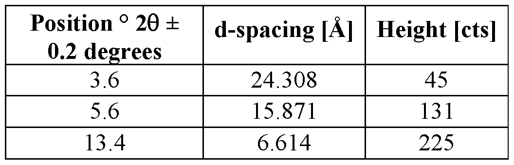

[00476] In some embodiments, Form C napadisylate salt is characterized by the x-ray powder diffraction (XRPD) pattern depicted in Figure 93.
[00477] In some embodiments, Form C napadisylate salt is characterized by the differential scanning calorimetry (DSC) pattern depicted in Figure 94, trace 94C.
[00478] In some embodiments of a complex form of Compound 1, X is (ri)-camphorsulfonic acid. In some such embodiments, a complex form of Compound 1 is a fV)-camphorsulfonate salt. In some embodiments, a fV)-camphorsulfonate salt of Compound 1 is a crystalline (S)- camphorsulfonate salt. In some embodiments, a crystalline fV)-camphorsulfonate salt of Compound 1 is characterized by one or more peaks in its X-ray powder diffraction pattern selected from 5.0, 9.9, 10.4, 11.1, and 14.3 ± 0.2 degrees 2Q. In some such embodiments, a complex form of Compound 1 is Form A fV)-camphorsulfonate salt.
[00479] In some embodiments, Form A fV)-camphorsulfonate salt is characterized by the following peaks in its X-ray powder diffraction pattern:


[00480] In some embodiments, Form A fV)-camphorsulfonate salt is characterized by the FT- Raman spectrum depicted in Figure 95.
[00481] In some embodiments, Form A (A)-camphorsulfonate salt is characterized by the x- ray powder diffraction (XRPD) pattern depicted in Figure 96.
[00482] In some embodiments, Form A fV)-camphorsulfonate salt is characterized by the thermogravimetric analysis (TGA) pattern depicted in Figure 97, trace 97A.
[00483] In some embodiments, Form A fV)-camphorsulfonate salt is characterized by the differential scanning calorimetry (DSC) pattern depicted in Figure 97, trace 97B.
[00484] In some embodiments, a crystalline (ri)-camphorsulfonate salt of Compound 1 is characterized by one or more peaks in its X-ray powder diffraction pattern selected from 6.9, 10.2, 11.4, and 12.4 ± 0.2 degrees 2Q. In some such embodiments, a complex form of Compound 1 is Form B fV)-camphorsulfonate salt.
[00485] In some embodiments, Form B fV)-camphorsulfonate salt is characterized by the following peaks in its X-ray powder diffraction pattern:


[00486] In some embodiments, Form B fV)-camphorsulfonate salt is characterized by the FT- Raman spectrum depicted in Figure 98.
[00487] In some embodiments, Form B fV)-camphorsulfonate salt is characterized by the x- ray powder diffraction (XRPD) pattern depicted in Figure 99.
[00488] In some embodiments, Form B fV)-camphorsulfonate salt is characterized by the thermogravimetric analysis (TGA) pattern depicted in Figure 100, trace 100A.
[00489] In some embodiments, Form B fV)-camphorsulfonate salt is characterized by the differential scanning calorimetry (DSC) pattern depicted in Figure 100, trace 100B.
[00490] In some embodiments of a complex form of Compound 1, X is 1,2-ethanedisulfonic acid. In some such embodiments, a complex form of Compound 1 is a 1,2-ethanedi sulfonate salt (also referred to as an“edisylate” salt). In some embodiments, an edisylate salt of Compound 1 is a crystalline edisylate salt. In some embodiments, an edisylate salt is a hydrate. In some embodiments, a hydrate form of an edisylate salt of Compound 1 is a crystalline hydrate form of an edisylate salt. In some embodiments, a crystalline hydrate form of an edisylate salt of Compound 1 is characterized by one or more peaks in its X-ray powder diffraction pattern selected from 9.1, 10.7, 11.1, 14.0, 14.7, 18.2, and 19.0 ± 0.2 degrees 2Q. In some such embodiments, a complex form of Compound 1 is Form A edisylate salt.
[00491] In some embodiments, Form A edisylate salt is characterized by the following peaks in its X-ray powder diffraction pattern:

[00492] In some embodiments, Form A edisylate salt is characterized by the x-ray powder diffraction (XRPD) pattern depicted in Figure 101.
[00493] In some embodiments, Form A edisylate salt is characterized by the thermogravimetric analysis (TGA) pattern depicted in Figure 105, trace 105 A.
[00494] In some embodiments, Form A edisylate salt is characterized by the differential scanning calorimetry (DSC) pattern depicted in Figure 105, trace 105B.
[00495] In some embodiments, a crystalline edisylate salt of Compound 1 is characterized by one or more peaks in its X-ray powder diffraction pattern selected from 9.8, 10.9, 13.1, 13.6, and 19.5 ± 0.2 degrees 2Q. In some such embodiments, a complex form of Compound 1 is Form B edisylate salt.
[00496] In some embodiments, Form B edisylate salt is characterized by the following peaks in its X-ray powder diffraction pattern:

[00497] In some embodiments, Form B edisylate salt is characterized by the x-ray powder diffraction (XRPD) pattern depicted in Figure 102.
[00498] In some embodiments, Form B edisylate salt is characterized by the differential scanning calorimetry (DSC) pattern depicted in Figure 106, trace 106B.
[00499] In some embodiments, a crystalline edisylate salt of Compound 1 is characterized by one or more peaks in its X-ray powder diffraction pattern selected from 7.0, 12.8, 13.3, 13.7, and 16.7 ± 0.2 degrees 2Q. In some such embodiments, a complex form of Compound 1 is Form C edisylate salt.
[00500] In some embodiments, Form C edisylate salt is characterized by the following peaks in its X-ray powder diffraction pattern:


[00501] In some embodiments, Form C edisylate salt is characterized by the x-ray powder diffraction (XRPD) pattern depicted in Figure 103.
[00502] In some embodiments, Form C edisylate salt is characterized by the differential scanning calorimetry (DSC) pattern depicted in Figure 106, trace 106A.
[00503] In some embodiments, a crystalline edisylate salt of Compound 1 is characterized by one or more peaks in its X-ray powder diffraction pattern selected from 6.1, 10.2, 10.4, 12.5, 15.8, 16.0, and 17.0 ± 0.2 degrees 2Q. In some such embodiments, a complex form of Compound 1 is Form D edisylate salt.
[00504] In some embodiments, Form D edisylate salt is characterized by the following peaks in its X-ray powder diffraction pattern:
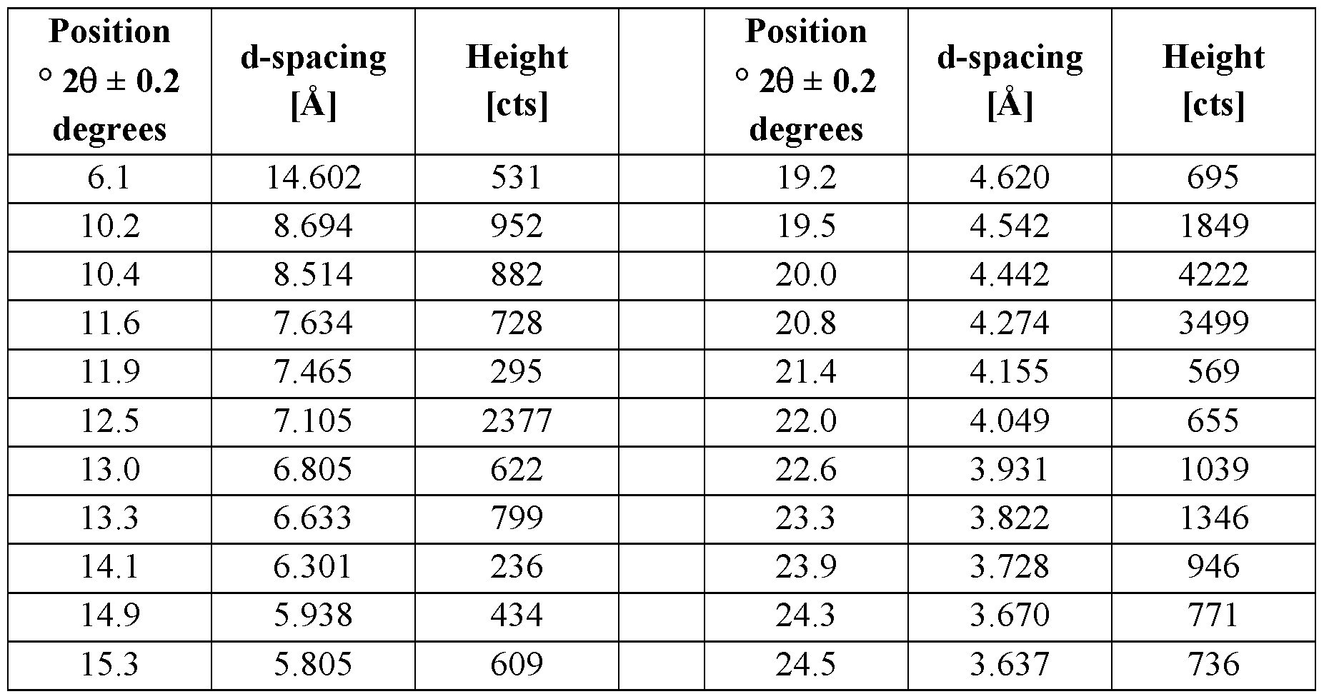

[00505] In some embodiments, Form D edisylate salt is characterized by the x-ray powder diffraction (XRPD) pattern depicted in Figure 104.
[00506] In some embodiments, Form D edisylate salt is characterized by the differential scanning calorimetry (DSC) pattern depicted in Figure 106, trace 106C.
[00507] In some embodiments of a complex form of Compound 1, X is ethanesulfonic acid. In some such embodiments, a complex form of Compound 1 is an esylate salt. In some embodiments, an esylate salt of Compound 1 is a crystalline esylate salt. In some embodiments, a crystalline esylate salt of Compound 1 is characterized by one or more peaks in its X-ray powder diffraction pattern selected from 8.4, 17.0, 17.4, 18.2, 18.7, and 25.2 ± 0.2 degrees 2Q. In some such embodiments, a complex form of Compound 1 is Form A esylate salt.
[00508] In some embodiments, Form A esylate salt is characterized by the following peaks in its X-ray powder diffraction pattern:

[00510] In some embodiments, Form A esylate salt is characterized by the thermogravimetric analysis (TGA) pattern depicted in Figure 109, trace 109A.
[00511] In some embodiments, Form A esylate salt is characterized by the differential scanning calorimetry (DSC) pattern depicted in Figure 109, trace 109B.
[00512] In some embodiments, a crystalline esylate salt of Compound 1 is characterized by one or more peaks in its X-ray powder diffraction pattern selected from 6.5, 9.8, 12.5, 12.9, and 14.8 ± 0.2 degrees 2Q. In some such embodiments, a complex form of Compound 1 is Form B esylate salt.
[00513] In some embodiments, Form B esylate salt is characterized by the following peaks in its X-ray powder diffraction pattern:

[00515] In some embodiments, Form B esylate salt is characterized by the thermogravimetric analysis (TGA) pattern depicted in Figure 110, trace 110A.
[00516] In some embodiments, Form B esylate salt is characterized by the differential scanning calorimetry (DSC) pattern depicted in Figure 110, trace 110B.
[00517] In some embodiments of a complex form of Compound 1, X is benzenesulfonic acid. In some such embodiments, a complex form of Compound 1 is a benzenesulfonate salt (also referred to as a“besylate” salt). In some embodiments, a besylate salt of Compound 1 is a crystalline besylate salt. In some embodiments, a crystalline besylate salt of Compound 1 is characterized by one or more peaks in its X-ray powder diffraction pattern selected from 5.5, 7.5, 10.4, 11.0, 12.8, 14.3, and 14.9 ± 0.2 degrees 2Q. In some such embodiments, a complex form of Compound 1 is Form A besylate salt.
[00518] In some embodiments, Form A besylate salt is characterized by the following peaks in its X-ray powder diffraction pattern:

[00520] In some embodiments, Form A besylate salt is characterized by the differential scanning calorimetry (DSC) pattern depicted in Figure 115, trace 115 A.
[00521] In some embodiments, a crystalline besylate salt of Compound 1 is characterized by one or more peaks in its X-ray powder diffraction pattern selected from 7.5, 9.2, 11.1, 12.1, 14.1, and 15.1 ± 0.2 degrees 2Q. In some such embodiments, a complex form of Compound 1 is Form B besylate salt.
[00522] In some embodiments, Form B besylate salt is characterized by the following peaks in its X-ray powder diffraction pattern:

[00523] In some embodiments, Form B besylate salt is characterized by the x-ray powder diffraction (XRPD) pattern depicted in Figure 112.
[00524] In some embodiments, Form B besylate salt is characterized by the differential scanning calorimetry (DSC) pattern depicted in Figure 115, trace 115B.
[00525] In some embodiments, a crystalline besylate salt of Compound 1 is characterized by one or more peaks in its X-ray powder diffraction pattern selected from 4.1, 8.2, 12.3, 16.4, and 20.5 ± 0.2 degrees 2Q. In some such embodiments, a complex form of Compound 1 is Form C besylate salt.
[00526] In some embodiments, Form C besylate salt is characterized by the following peaks in its X-ray powder diffraction pattern:

[00527] In some embodiments, Form C besylate salt is characterized by the x-ray powder diffraction (XRPD) pattern depicted in Figure 113.
[00528] In some embodiments, Form C besylate salt is characterized by the differential scanning calorimetry (DSC) pattern depicted in Figure 115, trace 115C.
[00529] In some embodiments, a besylate salt of Compound 1 is a hydrate. In some embodiments, a hydrate form of a besylate salt of Compound l is a crystalline hydrate form of a besylate salt. In some embodiments, a crystalline hydrate form of a besylate salt of Compound 1 is characterized by one or more peaks in its X-ray powder diffraction pattern selected from 6.1, 7.2, 11.5, 12.1, 12.6, and 12.9 ± 0.2 degrees 2Q. In some such embodiments, a complex form of Compound 1 is Form D besylate salt.
[00530] In some embodiments, Form D besylate salt is characterized by the following peaks in its X-ray powder diffraction pattern:

[00531] In some embodiments, Form D besylate salt is characterized by the x-ray powder diffraction (XRPD) pattern depicted in Figure 114.
[00532] In some embodiments, Form D besylate salt is characterized by the thermogravimetric analysis (TGA) pattern depicted in Figure 116, trace 116A.
[00533] In some embodiments, Form D besylate salt is characterized by the differential scanning calorimetry (DSC) pattern depicted in Figure 116, trace 116B.
[00534] In some embodiments of a complex form of Compound 1, X is oxalic acid. In some such embodiments, a complex form of Compound 1 is an oxalate salt. In some embodiments, an oxalate salt of Compound 1 is a crystalline oxalate salt. In some embodiments, an oxalate salt of Compound l is a hydrate. In some embodiments, a hydrate form of an oxalate salt of Compound 1 is a crystalline hydrate form of an oxalate salt. In some embodiments, a crystalline hydrate form of an oxalate salt of Compound 1 is characterized by one or more peaks in its X-ray powder diffraction pattern selected from 4.7, 6.5, 9.4, 11.0, 11.9, and 12.5 ± 0.2 degrees 2Q. In some such embodiments, a complex form of Compound 1 is Form A oxalate salt.
[00535] In some embodiments, Form A oxalate salt is characterized by the following peaks in its X-ray powder diffraction pattern:

[00536] In some embodiments, Form A oxalate salt is characterized by the x-ray powder diffraction (XRPD) pattern depicted in Figure 117.
[00537] In some embodiments, Form A oxalate salt is characterized by the thermogravimetric analysis (TGA) pattern depicted in Figure 119, trace 119A.
[00538] In some embodiments, Form A oxalate salt is characterized by the differential scanning calorimetry (DSC) pattern depicted in Figure 119, trace 119B.
[00539] In some embodiments, a crystalline oxalate salt of Compound 1 is characterized by one or more peaks in its X-ray powder diffraction pattern selected from 5.3, 8.7, and 12.9 ± 0.2 degrees 2Q. In some such embodiments, a complex form of Compound 1 is Form B oxalate salt.
[00540] In some embodiments, Form B oxalate salt is characterized by the following peaks in its X-ray powder diffraction pattern:


[00541] In some embodiments, Form B oxalate salt is characterized by the x-ray powder diffraction (XRPD) pattern depicted in Figure 118.
[00542] In some embodiments, Form B oxalate salt is characterized by the thermogravimetric analysis (TGA) pattern depicted in Figure 120, trace 120A.
[00543] In some embodiments, Form B oxalate salt is characterized by the differential scanning calorimetry (DSC) pattern depicted in Figure 120, trace 120B.
[00544] In some embodiments of a complex form of Compound 1, X is maleic acid. In some such embodiments, a complex form of Compound 1 is a maleate salt. In some embodiments, a maleate salt of Compound l is a crystalline maleate salt. In some embodiments, a maleate salt of Compound 1 is a hydrate. In some embodiments, a hydrate form of a maleate salt of Compound 1 is a crystalline hydrate form of a maleate salt. In some embodiments, a crystalline hydrate form of a maleate salt of Compound 1 is characterized by one or more peaks in its X-ray powder diffraction pattern selected from 7.7, 11.5, 14.1, 15.4, 15.8, and 16.1 ± 0.2 degrees 2Q. In some such embodiments, a complex form of Compound 1 is Form A maleate salt.
[00545] In some embodiments, Form A maleate salt is characterized by the following peaks in its X-ray powder diffraction pattern:


[00546] In some embodiments, Form A maleate salt is characterized by the x-ray powder diffraction (XRPD) pattern depicted in Figure 121.
[00547] In some embodiments, Form A maleate salt is characterized by the thermogravimetric analysis (TGA) pattern depicted in Figure 122, trace 122A.
[00548] In some embodiments, Form A maleate salt is characterized by the differential scanning calorimetry (DSC) pattern depicted in Figure 122, trace 122B.
[00549] In some embodiments of a complex form of Compound 1, X is pamoic acid. In some such embodiments, a complex form of Compound 1 is a pamoate salt. In some embodiments, a pamoate salt of Compound 1 is a crystalline pamoate salt. In some embodiments, a crystalline pamoate salt of Compound 1 is characterized by one or more peaks in its X-ray powder diffraction pattern selected from 6.1, 10.7, 13.9, 15.4, 20.8, and 21.5 ± 0.2 degrees 2Q. In some such embodiments, a complex form of Compound 1 is Form A pamoate salt.
[00550] In some embodiments, Form A pamoate salt is characterized by the following peaks in its X-ray powder diffraction pattern:


[00551] In some embodiments, Form A pamoate salt is characterized by the x-ray powder diffraction (XRPD) pattern depicted in Figure 123.
[00552] In some embodiments, Form A pamoate salt is characterized by the thermogravimetric analysis (TGA) pattern depicted in Figure 124, trace 124A.
[00553] In some embodiments, Form A pamoate salt is characterized by the differential scanning calorimetry (DSC) pattern depicted in Figure 124, trace 124B.
[00554] In some embodiments of a complex form of Compound 1, X is 1 -hydroxyl- naphthoic acid. In some such embodiments, a complex form of Compound 1 is a l-hydroxy-2- naphthoate salt. In some embodiments, a l-hydroxy-2-naphthoate salt of Compound 1 is a crystalline l-hydroxy-2-naphthoate salt. In some embodiments, a crystalline l-hydroxy-2- naphthoate salt of Compound 1 is characterized by one or more peaks in its X-ray powder diffraction pattern selected from 6.7, 8.4, 9.7, 10.8, and 16.0 ± 0.2 degrees 2Q. In some such embodiments, a complex form of Compound 1 is Form A l-hydroxy-2-naphthoate salt.
[00555] In some embodiments, Form A l-hydroxy-2-naphthoate salt is characterized by the following peaks in its X-ray powder diffraction pattern:


[00556] In some embodiments, Form A l-hydroxy-2-naphthoate salt is characterized by the x- ray powder diffraction (XRPD) pattern depicted in Figure 125.
[00557] In some embodiments, Form A l-hydroxy-2-naphthoate salt is characterized by the differential scanning calorimetry (DSC) pattern depicted in Figure 126.
[00558] In some embodiments of a complex form of Compound 1, X is malonic acid. In some such embodiments, a complex form of Compound 1 is a malonate salt. In some embodiments, a malonate salt of Compound 1 is a crystalline malonate salt. In some embodiments, a crystalline malonate salt of Compound 1 is characterized by one or more peaks in its X-ray powder diffraction pattern selected from 7.8, 11.7, 13.2, 13.7, and 15.6 ± 0.2 degrees 2Q. In some such embodiments, a complex form of Compound 1 is Form A malonate salt.
[00559] In some embodiments, Form A malonate salt is characterized by the following peaks in its X-ray powder diffraction pattern:


[00560] In some embodiments, Form A malonate salt is characterized by the x-ray powder diffraction (XRPD) pattern depicted in Figure 127.
[00561] In some embodiments, Form A malonate salt is characterized by the thermogravimetric analysis (TGA) pattern depicted in Figure 128, trace 128A.
[00562] In some embodiments, Form A malonate salt is characterized by the differential scanning calorimetry (DSC) pattern depicted in Figure 128, trace 128B.
[00563] In some embodiments, a crystalline malonate salt of Compound 1 is characterized by one or more peaks in its X-ray powder diffraction pattern selected from 5.6, 7.3, 11.2, 12.3, 14.5, and 16.8 ± 0.2 degrees 2Q. In some such embodiments, a complex form of Compound 1 is Form B malonate salt.
[00564] In some embodiments, Form B malonate salt is characterized by the following peaks in its X-ray powder diffraction pattern:

[00566] In some embodiments, Form B malonate salt is characterized by the thermogravimetric analysis (TGA) pattern depicted in Figure 130, trace 130A.
[00567] In some embodiments, Form B malonate salt is characterized by the differential scanning calorimetry (DSC) pattern depicted in Figure 130, trace 130B.
[00568] In some embodiments, a crystalline malonate salt of Compound 1 is characterized by one or more peaks in its X-ray powder diffraction pattern selected from 7.8, 11.7, 15.7, and 17.7 ± 0.2 degrees 2Q. In some such embodiments, a complex form of Compound 1 is Form C malonate salt.
[00569] In some embodiments, Form C malonate salt is characterized by the following peaks in its X-ray powder diffraction pattern:

[00570] In some embodiments, Form C malonate salt is characterized by the x-ray powder diffraction (XRPD) pattern depicted in Figure 131.
[00571] In some embodiments, Form C malonate salt is characterized by the differential scanning calorimetry (DSC) pattern depicted in Figure 132.
[00572] In some embodiments of a complex form of Compound 1, X is L-tartaric acid. In some such embodiments, a complex form of Compound 1 is an L-tartrate salt. In some embodiments, an L-tartrate salt of Compound 1 is a crystalline L-tartrate salt. In some embodiments, a crystalline L-tartrate salt of Compound 1 is characterized by one or more peaks in its X-ray powder diffraction pattern selected from 9.7, 11.1, 14.9, 16.6, 19.8, and 21.0 ± 0.2
degrees 2Q. In some such embodiments, a complex form of Compound 1 is Form A L-tartrate salt.
[00573] In some embodiments, Form A L-tartrate salt is characterized by the following peaks in its X-ray powder diffraction pattern:

[00574] In some embodiments, Form A L-tartrate salt is characterized by the x-ray powder diffraction (XRPD) pattern depicted in Figure 133.
[00575] In some embodiments, Form A L-tartrate salt is characterized by the thermogravimetric analysis (TGA) pattern depicted in Figure 134, trace 134A.
[00576] In some embodiments, Form A L-tartrate salt is characterized by the differential scanning calorimetry (DSC) pattern depicted in Figure 134, trace 134B.
[00577] In some embodiments, a crystalline L-tartrate salt of Compound 1 is characterized by one or more peaks in its X-ray powder diffraction pattern selected from 7.4, 9.7, 11.2, 11.7, and 14.9 ± 0.2 degrees 2Q. In some such embodiments, a complex form of Compound 1 is Form B L-tartrate salt.
[00578] In some embodiments, Form B L-tartrate salt is characterized by the following peaks in its X-ray powder diffraction pattern:

[00579] In some embodiments, Form B L-tartrate salt is characterized by the x-ray powder diffraction (XRPD) pattern depicted in Figure 135.
[00580] In some embodiments, Form B L-tartrate salt is characterized by the differential scanning calorimetry (DSC) pattern depicted in Figure 136.
[00581] In some embodiments, a crystalline L-tartrate salt of Compound 1 is characterized by one or more peaks in its X-ray powder diffraction pattern selected from 7.4, 9.7, 11.2, 12.5, and 14.9 ± 0.2 degrees 2Q. In some such embodiments, a complex form of Compound 1 is Form C L-tartrate salt.
[00582] In some embodiments, Form C L-tartrate salt is characterized by the following peaks in its X-ray powder diffraction pattern:


[00583] In some embodiments, Form C L-tartrate salt is characterized by the x-ray powder diffraction (XRPD) pattern depicted in Figure 137.
[00584] In some embodiments, Form C L-tartrate salt is characterized by the thermogravimetric analysis (TGA) pattern depicted in Figure 138, trace 138A.
[00585] In some embodiments, Form C L-tartrate salt is characterized by the differential scanning calorimetry (DSC) pattern depicted in Figure 138, trace 138B.
[00586] In some embodiments, a crystalline L-tartrate salt of Compound 1 is characterized by one or more peaks in its X-ray powder diffraction pattern selected from 4.7, 7.4, 9.5, 11.1, 13.1, 13.5, and 18.3 ± 0.2 degrees 2Q. In some such embodiments, a complex form of Compound 1 is Form D L-tartrate salt.
[00587] In some embodiments, Form D L-tartrate salt is characterized by the following peaks in its X-ray powder diffraction pattern:

[00588] In some embodiments, Form D L-tartrate salt is characterized by the x-ray powder diffraction (XRPD) pattern depicted in Figure 139.
[00589] In some embodiments, Form D L-tartrate salt is characterized by the thermogravimetric analysis (TGA) pattern depicted in Figure 140, trace 140A.
[00590] In some embodiments, Form D L-tartrate salt is characterized by the differential scanning calorimetry (DSC) pattern depicted in Figure 140, trace 140B.
[00591] In some embodiments of a complex form of Compound 1, X is fumaric acid. In some such embodiments, a complex form of Compound 1 is a fumarate salt. In some embodiments, a fumarate salt of Compound 1 is a crystalline fumarate salt. In some embodiments, a crystalline fumarate salt of Compound 1 is characterized by one or more peaks in its X-ray powder diffraction pattern selected from 6.7, 12.3, 13.4, 14.3, and 15.4 ± 0.2 degrees 2Q. In some such embodiments, a complex form of Compound 1 is Form A fumarate salt.
[00592] In some embodiments, Form A fumarate salt is characterized by the following peaks in its X-ray powder diffraction pattern:

[00594] In some embodiments, Form A fumarate salt is characterized by the thermogravimetric analysis (TGA) pattern depicted in Figure 142, trace 142A.
[00595] In some embodiments, Form A fumarate salt is characterized by the differential scanning calorimetry (DSC) pattern depicted in Figure 142, trace 142B.
[00596] In some embodiments, a crystalline fumarate salt of Compound 1 is characterized by one or more peaks in its X-ray powder diffraction pattern selected from 7.0, 14.1, 14.6, 15.3, and 19.0 ± 0.2 degrees 2Q. In some such embodiments, a complex form of Compound 1 is Form B fumarate salt.
[00597] In some embodiments, Form B fumarate salt is characterized by the following peaks in its X-ray powder diffraction pattern:

[00598] In some embodiments, Form B fumarate salt is characterized by the x-ray powder diffraction (XRPD) pattern depicted in Figure 143.
[00599] In some embodiments, Form B fumarate salt is characterized by the differential scanning calorimetry (DSC) pattern depicted in Figure 144.
[00600] In some embodiments, a crystalline fumarate salt of Compound 1 is characterized by one or more peaks in its X-ray powder diffraction pattern selected from 7.6, 11.4, 15.2, and 19.0
± 0.2 degrees 2Q. In some such embodiments, a complex form of Compound 1 is Form C fumarate salt.
[00601] In some embodiments, Form C fumarate salt is characterized by the following peaks in its X-ray powder diffraction pattern:

[00602] In some embodiments, Form C fumarate salt is characterized by the x-ray powder diffraction (XRPD) pattern depicted in Figure 145.
[00603] In some embodiments, Form C fumarate salt is characterized by the thermogravimetric analysis (TGA) pattern depicted in Figure 146, trace 146A.
[00604] In some embodiments, Form C fumarate salt is characterized by the differential scanning calorimetry (DSC) pattern depicted in Figure 146, trace 146B.
[00605] In some embodiments, a crystalline fumarate salt of Compound 1 is characterized by one or more peaks in its X-ray powder diffraction pattern selected from 14.0, 17.6, 23.3, 23.9, and 25.1 ± 0.2 degrees 2Q. In some such embodiments, a complex form of Compound 1 is Form D fumarate salt.
[00606] In some embodiments, Form D fumarate salt is characterized by the following peaks in its X-ray powder diffraction pattern:

[00607] In some embodiments, Form D fumarate salt of Compound 1 is characterized by the x-ray powder diffraction (XRPD) pattern depicted in Figure 147.
[00608] In some embodiments, Form D fumarate salt of Compound 1 is characterized by the thermogravimetric analysis (TGA) pattern depicted in Figure 148, trace 148A.
[00609] In some embodiments, Form D fumarate salt of Compound 1 is characterized by the differential scanning calorimetry (DSC) pattern depicted in Figure 148, trace 148B.
[00610] In some embodiments of a complex form of Compound 1, X is citric acid. In some such embodiments, a complex form of Compound 1 is a citrate salt. In some embodiments, a citrate salt of Compound 1 is a crystalline citrate salt. In some embodiments, a crystalline citrate salt of Compound 1 is characterized by one or more peaks in its X-ray powder diffraction pattern selected from 7.5, 11.3, 13.5, 15.1, 18.9, and 19.2 ± 0.2 degrees 2Q. In some such embodiments, a complex form of Compound 1 is Form A citrate salt.
[00611] In some embodiments, Form A citrate salt is characterized by the following peaks in its X-ray powder diffraction pattern:


[00612] In some embodiments, Form A citrate salt is characterized by the x-ray powder diffraction (XRPD) pattern depicted in Figure 149.
[00613] In some embodiments, Form A citrate salt is characterized by the thermogravimetric analysis (TGA) pattern depicted in Figure 150, trace 150A.
[00614] In some embodiments, Form A citrate salt is characterized by the differential scanning calorimetry (DSC) pattern depicted in Figure 150, trace 150B.
[00615] In some embodiments of a complex form of Compound 1, X is L-lactic acid. In some such embodiments, a complex form of Compound 1 is an L-lactate salt. In some embodiments, an L-lactate salt of Compound 1 is a crystalline L-lactate salt. In some embodiments, a crystalline L-lactate salt of Compound 1 is characterized by one or more peaks in its X-ray powder diffraction pattern selected from 7.5, 8.2, 11.2, 12.3, and 16.0 ± 0.2 degrees 2Q. In some such embodiments, a complex form of Compound 1 is Form A L-lactate salt.
[00616] In some embodiments, Form A L-lactate salt is characterized by the following peaks in its X-ray powder diffraction pattern:


[00617] In some embodiments, Form A L-lactate salt is characterized by the x-ray powder diffraction (XRPD) pattern depicted in Figure 151.
[00618] In some embodiments, Form A L-lactate salt is characterized by the thermogravimetric analysis (TGA) pattern depicted in Figure 152, trace 152A.
[00619] In some embodiments, Form A L-lactate salt is characterized by the differential scanning calorimetry (DSC) pattern depicted in Figure 152, trace 152B.
[00620] In some embodiments of a complex form of Compound 1, X is acetic acid. In some such embodiments, a complex form of Compound 1 is an acetate salt. In some embodiments, an acetate salt of Compound 1 is a crystalline acetate salt. In some embodiments, a crystalline acetate salt of Compound 1 is characterized by one or more peaks in its X-ray powder diffraction pattern selected from 8.9, 11.6, 11.9, 13.5, 14.1, and 17.9 ± 0.2 degrees 2Q. In some such embodiments, a complex form of Compound 1 is Form A acetate salt.
[00621] In some embodiments, Form A acetate salt is characterized by the following peaks in its X-ray powder diffraction pattern:
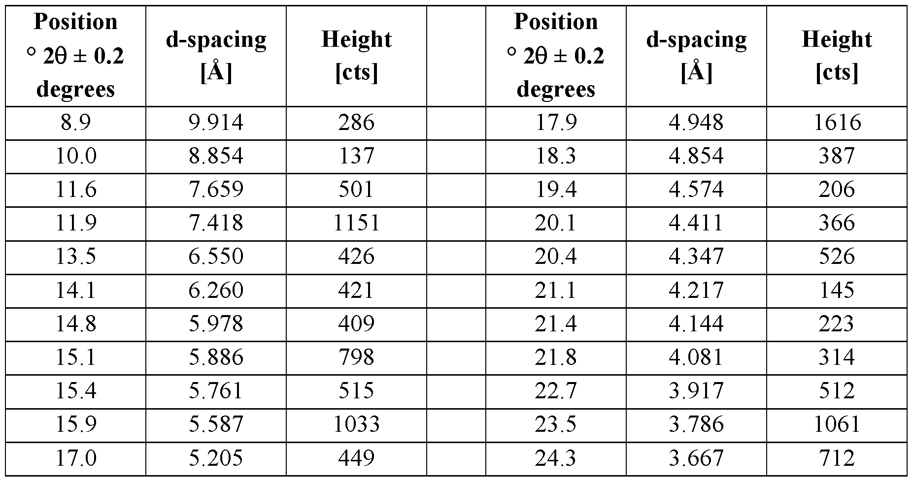

[00622] In some embodiments, Form A acetate salt is characterized by the x-ray powder diffraction (XRPD) pattern depicted in Figure 153.
[00623] In some embodiments, Form A acetate salt is characterized by the thermogravimetric analysis (TGA) pattern depicted in Figure 154, trace 154A.
[00624] In some embodiments, Form A acetate salt is characterized by the differential scanning calorimetry (DSC) pattern depicted in Figure 154, trace 154B.
[00625] In some embodiments, a crystalline acetate salt of Compound 1 is characterized by one or more peaks in its X-ray powder diffraction pattern selected from 10.3, 11.6, 12.8, 15.6, 17.6, and 19.1 ± 0.2 degrees 2Q. In some such embodiments, a complex form of Compound 1 is Form B acetate salt.
[00626] In some embodiments, Form B acetate salt is characterized by the following peaks in its X-ray powder diffraction pattern:

[00628] In some embodiments, Form B acetate salt is characterized by the thermogravimetric analysis (TGA) pattern depicted in Figure 156, trace 156A.
[00629] In some embodiments, Form B acetate salt is characterized by the differential scanning calorimetry (DSC) pattern depicted in Figure 156, trace 156B.
[00630] In some embodiments of a complex form of Compound 1, X is propionic acid. In some such embodiments, a complex form of Compound 1 is a propionate salt. In some embodiments, a propionate salt of Compound 1 is a crystalline propionate salt. In some embodiments, a crystalline propionate salt of Compound 1 is characterized by one or more peaks in its X-ray powder diffraction pattern selected from 8.6, 9.7, 12.4, 14.0, 16.4, and 17.7 ± 0.2 degrees 2Q. In some such embodiments, a complex form of Compound 1 is Form A propionate salt.
[00631] In some embodiments, Form A propionate salt is characterized by the following peaks in its X-ray powder diffraction pattern:


[00632] In some embodiments, Form A propionate salt is characterized by the x-ray powder diffraction (XRPD) pattern depicted in Figure 157.
[00633] In some embodiments, Form A propionate salt is characterized by the thermogravimetric analysis (TGA) pattern depicted in Figure 158, trace 158A.
[00634] In some embodiments, Form A propionate salt is characterized by the differential scanning calorimetry (DSC) pattern depicted in Figure 158, trace 158B.
[00635] In some embodiments of a complex form of Compound 1, X is DL-lactic acid. In some such embodiments, a complex form of Compound 1 is a DL-lactate salt. In some embodiments, a DL-lactate salt of Compound 1 is a crystalline DL-lactate salt. In some embodiments, a crystalline DL-lactate salt of Compound 1 is characterized by one or more peaks in its X-ray powder diffraction pattern selected from 8.3, 12.4, 15.9, 17.6, and 18.8 ± 0.2 degrees 2Q. In some such embodiments, a complex form of Compound 1 is Form A DL-lactate salt.
[00636] In some embodiments, Form A DL-lactate salt is characterized by the following peaks in its X-ray powder diffraction pattern:

[00638] In some embodiments, Form A DL-lactate salt is characterized by the thermogravimetric analysis (TGA) pattern depicted in Figure 160, trace 160A.
[00639] In some embodiments, Form A DL-lactate salt is characterized by the differential scanning calorimetry (DSC) pattern depicted in Figure 160, trace 160B.
[00640] In some embodiments of a complex form of Compound 1, X is D-gluconic acid. In some such embodiments, a complex form of Compound 1 is a D-gluconate salt. In some embodiments, a D-gluconate salt of Compound 1 is a crystalline D-gluconate salt. In some embodiments, a crystalline D-gluconate salt of Compound 1 is characterized by one or more peaks in its X-ray powder diffraction pattern selected from 7.1, 11.7, 14.7, 16.1, and 16.5 ± 0.2 degrees 2Q. In some such embodiments, a complex form of Compound 1 is Form A D-gluconate salt.
[00641] In some embodiments, Form A D-gluconate salt is characterized by the following peaks in its X-ray powder diffraction pattern:

[00643] In some embodiments, Form A D-gluconate salt is characterized by the differential scanning calorimetry (DSC) pattern depicted in Figure 162.
[00644] In some embodiments of a complex form of Compound 1, X is DL-malic acid. In some such embodiments, a complex form of Compound 1 is a DL-malate salt. In some embodiments, a DL-malate salt of Compound 1 is a crystalline DL-malate salt. In some embodiments, a crystalline DL-malate salt of Compound 1 is characterized by one or more peaks in its X-ray powder diffraction pattern selected from 7.5, 9.7, 11.3, 15.1, 16.3, and 21.0 ± 0.2 degrees 2Q. In some such embodiments, a complex form of Compound 1 is Form A DL-malate salt.
[00645] In some embodiments, Form A DL-malate salt is characterized by the following peaks in its X-ray powder diffraction pattern:

[00646] In some embodiments, Form A DL-malate salt is characterized by the x-ray powder diffraction (XRPD) pattern depicted in Figure 163.
[00647] In some embodiments, Form A DL-malate salt is characterized by the thermogravimetric analysis (TGA) pattern depicted in Figure 164, trace 164A.
[00648] In some embodiments, Form A DL-malate salt is characterized by the differential scanning calorimetry (DSC) pattern depicted in Figure 164, trace 164B.
[00649] In some embodiments, a crystalline DL-malate salt of Compound 1 is characterized by one or more peaks in its X-ray powder diffraction pattern selected from 4.6, 8.3, 11.7, 13.9, and 18.6 ± 0.2 degrees 2Q. In some such embodiments, a complex form of Compound 1 is Form B DL-malate salt.
[00650] In some embodiments, Form B DL-malate salt is characterized by the following peaks in its X-ray powder diffraction pattern:

[00651] In some embodiments, Form B DL-malate salt is characterized by the x-ray powder diffraction (XRPD) pattern depicted in Figure 165.
[00652] In some embodiments, Form B DL-malate salt is characterized by the thermogravimetric analysis (TGA) pattern depicted in Figure 166, trace 166A.
[00653] In some embodiments, Form B DL-malate salt is characterized by the differential scanning calorimetry (DSC) pattern depicted in Figure 166, trace 166B.
[00654] In some embodiments of a complex form of Compound 1, X is glycolic acid. In some such embodiments, a complex form of Compound 1 is a glycolate salt. In some embodiments, a glycolate salt of Compound 1 is a crystalline glycolate salt. In some embodiments, a crystalline glycolate salt of Compound 1 is characterized by one or more peaks in its X-ray powder diffraction pattern selected from 8.4, 8.6, 10.6, 12.7, and 16.1 ± 0.2 degrees 2Q. In some such embodiments, a complex form of Compound 1 is Form A glycolate salt.
[00655] In some embodiments, Form A glycolate salt is characterized by the following peaks in its X-ray powder diffraction pattern:

[00656] In some embodiments, Form A glycolate salt is characterized by the x-ray powder diffraction (XRPD) pattern depicted in Figure 167.
[00657] In some embodiments, Form A glycolate salt is characterized by the thermogravimetric analysis (TGA) pattern depicted in Figure 168, trace 168 A.
[00658] In some embodiments, Form A glycolate salt is characterized by the differential scanning calorimetry (DSC) pattern depicted in Figure 168, trace 168B.
[00659] In some embodiments of a complex form of Compound 1, X is glutaric acid. In some such embodiments, a complex form of Compound 1 is a glutarate salt. In some embodiments, a glutarate salt of Compound 1 is a crystalline glutarate salt. In some embodiments, a crystalline glutarate salt of Compound 1 is characterized by one or more peaks in its X-ray powder diffraction pattern selected from 7.4, 11.1, 14.9, 16.1, 18.6, and 18.9 ± 0.2 degrees 2Q. In some such embodiments, a complex form of Compound 1 is Form A glutarate salt.
[00660] In some embodiments, Form A glutarate salt is characterized by the following peaks in its X-ray powder diffraction pattern:

[00661] In some embodiments, Form A glutarate salt is characterized by the x-ray powder diffraction (XRPD) pattern depicted in Figure 169.
[00662] In some embodiments, Form A glutarate salt is characterized by the thermogravimetric analysis (TGA) pattern depicted in Figure 170, trace 170A.
[00663] In some embodiments, Form A glutarate salt is characterized by the differential scanning calorimetry (DSC) pattern depicted in Figure 170, trace 170B.
[00664] In some embodiments, a crystalline glutarate salt of Compound 1 is characterized by one or more peaks in its X-ray powder diffraction pattern selected from 4.8, 5.8, 9.5, 11.3, and 14.8 ± 0.2 degrees 2Q. In some such embodiments, a complex form of Compound 1 is Form B glutarate salt.
[00665] In some embodiments, Form B glutarate salt is characterized by the following peaks in its X-ray powder diffraction pattern:

[00666] In some embodiments, Form B glutarate salt is characterized by the x-ray powder diffraction (XRPD) pattern depicted in Figure 171.
[00667] In some embodiments, Form B glutarate salt is characterized by the thermogravimetric analysis (TGA) pattern depicted in Figure 172, trace 172A.
[00668] In some embodiments, Form B glutarate salt is characterized by the differential scanning calorimetry (DSC) pattern depicted in Figure 172, trace 172B.
[00669] In some embodiments of a complex form of Compound 1, X is L-malic acid. In some such embodiments, a complex form of Compound 1 is an L-malate salt. In some embodiments, an L-malate salt of Compound 1 is a crystalline L-malate salt. In some embodiments, a crystalline L-malate salt of Compound 1 is characterized by one or more peaks in its X-ray
powder diffraction pattern selected from 7.5, 9.6, 11.3, 15.1, 16.2, and 16.7 ± 0.2 degrees 2Q. In some such embodiments, a complex form of Compound 1 is Form A L-malate salt.
[00670] In some embodiments, Form A L-malate salt is characterized by the following peaks in its X-ray powder diffraction pattern:

[00671] In some embodiments, Form A L-malate salt is characterized by the x-ray powder diffraction (XRPD) pattern depicted in Figure 173.
[00672] In some embodiments, Form A L-malate salt is characterized by the thermogravimetric analysis (TGA) pattern depicted in Figure 174, trace 174A.
[00673] In some embodiments, Form A L-malate salt is characterized by the differential scanning calorimetry (DSC) pattern depicted in Figure 174, trace 174B.
[00674] In some embodiments of a complex form of Compound 1, X is camphoric acid. In some such embodiments, a complex form of Compound 1 is a camphorate salt. In some embodiments, a camphorate salt of Compound 1 is a crystalline camphorate salt. In some embodiments, a crystalline camphorate salt of Compound 1 is characterized by one or more peaks in its X-ray powder diffraction pattern selected from 6.7, 8.3, 9.9, 15.0, and 15.2 ± 0.2
degrees 2Q. In some such embodiments, a complex form of Compound 1 is Form A camphorate salt.
[00675] In some embodiments, Form A camphorate salt is characterized by the following peaks in its X-ray powder diffraction pattern:

[00676] In some embodiments, Form A camphorate salt is characterized by the x-ray powder diffraction (XRPD) pattern depicted in Figure 175.
[00677] In some embodiments, Form A camphorate salt is characterized by the thermogravimetric analysis (TGA) pattern depicted in Figure 176, trace 176A.
[00678] In some embodiments, Form A camphorate salt is characterized by the differential scanning calorimetry (DSC) pattern depicted in Figure 176, trace 176B.
[00679] In some embodiments, a crystalline camphorate salt of Compound 1 is characterized by one or more peaks in its X-ray powder diffraction pattern selected from 6.9, 9.9, 11.5, 15.3, 16.1, and 16.8 ± 0.2 degrees 2Q. In some such embodiments, a complex form of Compound 1 is Form B camphorate salt.
[00680] In some embodiments, Form B camphorate salt is characterized by the following peaks in its X-ray powder diffraction pattern:

[00681] In some embodiments, Form B camphorate salt is characterized by the x-ray powder diffraction (XRPD) pattern depicted in Figure 177.
[00682] In some embodiments, Form B camphorate salt is characterized by the thermogravimetric analysis (TGA) pattern depicted in Figure 178, trace 178 A.
[00683] In some embodiments, Form B camphorate salt is characterized by the differential scanning calorimetry (DSC) pattern depicted in Figure 178, trace 178B.
[00684] In some embodiments, a crystalline camphorate salt of Compound 1 is characterized by one or more peaks in its X-ray powder diffraction pattern selected from 4.9, 10.3, 13.6, 15.5, and 16.2 ± 0.2 degrees 2Q. In some such embodiments, a complex form of Compound 1 is Form C camphorate salt.
[00685] In some embodiments, Form C camphorate salt is characterized by the following peaks in its X-ray powder diffraction pattern:

[00686] In some embodiments, Form C camphorate salt is characterized by the x-ray powder diffraction (XRPD) pattern depicted in Figure 179.
[00687] In some embodiments, Form C camphorate salt is characterized by the thermogravimetric analysis (TGA) pattern depicted in Figure 180, trace 180A.
[00688] In some embodiments, Form C camphorate salt is characterized by the differential scanning calorimetry (DSC) pattern depicted in Figure 180, trace 180B.
[00689] In some embodiments, a crystalline camphorate salt of Compound 1 is characterized by one or more peaks in its X-ray powder diffraction pattern selected from 7.7, 8.6, 9.6, 12.1, 13.5, and 15.3 ± 0.2 degrees 2Q. In some such embodiments, a complex form of Compound 1 is Form D camphorate salt.
[00690] In some embodiments, Form D camphorate salt is characterized by the following peaks in its X-ray powder diffraction pattern:

[00691] In some embodiments, Form D camphorate salt is characterized by the x-ray powder diffraction (XRPD) pattern depicted in Figure 181.
[00692] In some embodiments, Form D camphorate salt is characterized by the thermogravimetric analysis (TGA) pattern depicted in Figure 182, trace 182A.
[00693] In some embodiments, Form D camphorate salt is characterized by the differential scanning calorimetry (DSC) pattern depicted in Figure 182, trace 182B.
[00694] In some embodiments of a complex form of Compound 1, X is DL-mandelic acid. In some such embodiments, a complex form of Compound 1 is a DL-mandelate salt. In some embodiments, a DL-mandelate salt of Compound 1 is a crystalline DL-mandelate salt. In some embodiments, a crystalline DL-mandelate salt of Compound 1 is characterized by one or more peaks in its X-ray powder diffraction pattern selected from 7.4, 11.1, 13.8, 14.9, and 16.3 ± 0.2 degrees 2Q. In some such embodiments, a complex form of Compound 1 is Form A DL- mandelate salt.
[00695] In some embodiments, Form A DL-mandelate salt is characterized by the following peaks in its X-ray powder diffraction pattern:
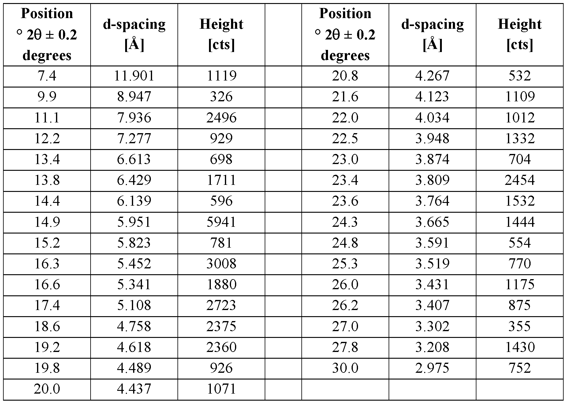
[00696] In some embodiments, Form A DL-mandelate salt is characterized by the x-ray powder diffraction (XRPD) pattern depicted in Figure 183.
[00697] In some embodiments, Form A DL-mandelate salt is characterized by the thermogravimetric analysis (TGA) pattern depicted in Figure 184, trace 184A.
[00698] In some embodiments, Form A DL-mandelate salt is characterized by the differential scanning calorimetry (DSC) pattern depicted in Figure 184, trace 184B.
[00699] In some embodiments, a crystalline DL-mandelate salt of Compound 1 is characterized by one or more peaks in its X-ray powder diffraction pattern selected from 7.5, 9.2, 11.3, 15.1, and 15.9 ± 0.2 degrees 2Q. In some such embodiments, a complex form of Compound 1 is Form B DL-mandelate salt.
[00700] In some embodiments, Form B DL-mandelate salt is characterized by the following peaks in its X-ray powder diffraction pattern:

[00701] In some embodiments, Form B DL-mandelate salt is characterized by the x-ray powder diffraction (XRPD) pattern depicted in Figure 185.
[00702] In some embodiments, Form B DL-mandelate salt is characterized by the thermogravimetric analysis (TGA) pattern depicted in Figure 186, trace 186A.
[00703] In some embodiments, Form B DL-mandelate salt is characterized by the differential scanning calorimetry (DSC) pattern depicted in Figure 186, trace 186B.
[00704] In some embodiments, a crystalline DL-mandelate salt of Compound 1 is characterized by one or more peaks in its X-ray powder diffraction pattern selected from 8.4, 9.9, 10.9, 14.0, and 14.6 ± 0.2 degrees 2Q. In some such embodiments, a complex form of Compound 1 is Form C DL-mandelate salt.
[00705] In some embodiments, Form C DL-mandelate salt is characterized by the following peaks in its X-ray powder diffraction pattern:


[00706] In some embodiments, Form C DL-mandelate salt is characterized by the x-ray powder diffraction (XRPD) pattern depicted in Figure 187.
[00707] In some embodiments, Form C DL-mandelate salt is characterized by the thermogravimetric analysis (TGA) pattern depicted in Figure 188, trace 188 A.
[00708] In some embodiments, Form C DL-mandelate salt is characterized by the differential scanning calorimetry (DSC) pattern depicted in Figure 188, trace 188B.
[00709] In some embodiments of a complex form of Compound 1, X is saccharin. In some such embodiments, a complex form of Compound 1 is a saccharin co-crystal. In some embodiments, a saccharin co-crystal of Compound 1 is a crystalline saccharin co-crystal. In some embodiments, a complex form of Compound 1 comprises one equivalent of saccharin. In some embodiments, a crystalline saccharin co-crystal of Compound 1 is characterized by one or more peaks in its X-ray powder diffraction pattern selected from 3.9, 7.9, 11.8, 15.0, and 15.8 ± 0.2 degrees 2Q. In some such embodiments, a complex form of Compound 1 is Form A saccharin co-crystal.
[00710] In some embodiments, Form A saccharin co-crystal is characterized by the following peaks in its X-ray powder diffraction pattern:


[00711] In some embodiments, Form A saccharin co-crystal is characterized by the FT-Raman spectrum depicted in Figure 189.
[00712] In some embodiments, Form A saccharin co-crystal is characterized by the x-ray powder diffraction (XRPD) pattern depicted in Figure 190.
[00713] In some embodiments, Form A saccharin co-crystal is characterized by the thermogravimetric analysis (TGA) pattern depicted in Figure 191, trace 191A.
[00714] In some embodiments, Form A saccharin co-crystal is characterized by the differential scanning calorimetry (DSC) pattern depicted in Figure 191, trace 19 IB.
[00715] In some embodiments, Form A saccharin co-crystal is characterized by the 'H NMRspectrum depicted in Figure 192.
[00716] In some embodiments of a complex form of Compound 1, X is nicotinic acid. In some such embodiments, a complex form of Compound 1 is a nicotinate salt. In some embodiments, a nicotinate salt of Compound 1 is a crystalline nicotinate salt. In some embodiments, a complex form of Compound 1 comprises one equivalent of nicotinic acid. In some embodiments, a crystalline nicotinate salt of Compound 1 is characterized by one or more peaks in its X-ray powder diffraction pattern selected from 7.8, 8.9, 14.0, 16.8, and 17.9 ± 0.2 degrees 2Q. In some such embodiments, a complex form of Compound 1 is Form A nicotinate salt.
[00717] In some embodiments, Form A nicotinate salt is characterized by the following peaks in its X-ray powder diffraction pattern:

[00718] In some embodiments, Form A nicotinic acid salt is characterized by the FT-Raman spectrum depicted in Figure 193.
[00719] In some embodiments, Form A nicotinic acid salt is characterized by the x-ray powder diffraction (XRPD) pattern depicted in Figure 194.
[00720] In some embodiments, Form A nicotinic acid salt is characterized by the thermogravimetric analysis (TGA) pattern depicted in Figure 195, trace 195 A.
[00721] In some embodiments, Form A nicotinic acid salt is characterized by the differential scanning calorimetry (DSC) pattern depicted in Figure 195, trace 195B.
[00722] In some embodiments, Form A nicotinic acid salt is characterized by the 'H NMR spectrum depicted in Figure 196.
[00723] In some embodiments, a nicotinate salt of Compound 1 is a hydrate. In some embodiments, a hydrate form of a nicotinate salt of Compound 1 is a crystalline hydrate form of a nicotinate salt. In some embodiments, a crystalline hydrate form of a nicotinate salt of Compound 1 is characterized by one or more peaks in its X-ray powder diffraction pattern selected from 8.2, 12.4, 15.3, 17.9, and 18.2 ± 0.2 degrees 2Q. In some such embodiments, a complex form of Compound 1 is Form B nicotinate salt.
[00724] In some embodiments, Form B nicotinate salt is characterized by the following peaks in its X-ray powder diffraction pattern:

[00725] In some embodiments, Form B nicotinic acid salt is characterized by the x-ray powder diffraction (XRPD) pattern depicted in Figure 197.
[00726] In some embodiments, Form B nicotinic acid salt is characterized by the thermogravimetric analysis (TGA) pattern depicted in Figure 198, trace 198 A.
[00727] In some embodiments, Form B nicotinic acid salt is characterized by the differential scanning calorimetry (DSC) pattern depicted in Figure 198, trace 198B.
[00728] In some embodiments, a crystalline nicotinate salt of Compound 1 is characterized by one or more peaks in its X-ray powder diffraction pattern selected from 3.8, 7.5, 11.3, 15.0, and 18.7 ± 0.2 degrees 2Q. In some such embodiments, a complex form of Compound 1 is Form C nicotinate salt.
[00729] In some embodiments, Form C nicotinate salt is characterized by the following peaks in its X-ray powder diffraction pattern:

[00730] In some embodiments, Form C nicotinic acid salt is characterized by the x-ray powder diffraction (XRPD) pattern depicted in Figure 199.
[00731] In some embodiments, Form C nicotinic acid salt is characterized by the thermogravimetric analysis (TGA) pattern depicted in Figure 200, trace 200A.
[00732] In some embodiments, Form C nicotinic acid salt is characterized by the differential scanning calorimetry (DSC) pattern depicted in Figure 200, trace 200B.
[00733] In some embodiments of a complex form of Compound 1, X is ascorbic acid. In some such embodiments, a complex form of Compound 1 is an ascorbate salt. In some embodiments, an ascorbate salt of Compound 1 is a crystalline ascorbate salt. In some embodiments, a complex form of Compound 1 comprises one equivalent of ascorbic acid. In some embodiments, an ascorbate salt of Compound 1 is a hydrate. In some embodiments, a hydrate form of an ascorbate salt of Compound 1 is a crystalline hydrate form of an ascorbate salt. In some embodiments, a crystalline hydrate form of an ascorbate salt of Compound 1 is characterized by one or more peaks in its X-ray powder diffraction pattern selected from 3.7, 7.5, 11.3, 15.0, and 18.8 ± 0.2 degrees 2Q. In some such embodiments, a complex form of Compound 1 is Form A ascorbate salt.
[00734] In some embodiments, Form A ascorbate salt is characterized by the following peaks in its X-ray powder diffraction pattern:

[00735] In some embodiments, Form A ascorbate salt is characterized by the FT-Raman spectrum depicted in Figure 201.
[00736] In some embodiments, Form A ascorbate salt is characterized by the x-ray powder diffraction (XRPD) pattern depicted in Figure 202.
[00737] In some embodiments, Form A ascorbate salt is characterized by the thermogravimetric analysis (TGA) pattern depicted in Figure 203, trace 203 A.
[00738] In some embodiments, Form A ascorbate salt is characterized by the differential scanning calorimetry (DSC) pattern depicted in Figure 203, trace 203B.
[00739] In some embodiments, Form A ascorbate salt is characterized by the ¾ NMR spectrum depicted in Figure 204.
[00740] In some embodiments, a crystalline ascorbate salt of Compound 1 is characterized by one or more peaks in its X-ray powder diffraction pattern selected from 7.4, 9.8, 11.2, 14.9, and 16.1 ± 0.2 degrees 2Q. In some such embodiments, a complex form of Compound 1 is Form B ascorbate salt.
[00741] In some embodiments, Form B ascorbate salt is characterized by the following peaks in its X-ray powder diffraction pattern:

[00742] In some embodiments, Form B ascorbate salt is characterized by the x-ray powder diffraction (XRPD) pattern depicted in Figure 234.
[00743] In some embodiments, Form B ascorbate salt is characterized by the thermogravimetric analysis (TGA) pattern depicted in Figure 235, trace 235 A.
[00744] In some embodiments, Form B ascorbate salt is characterized by the differential scanning calorimetry (DSC) pattern depicted in Figure 235, trace 235B.
[00745] In some embodiments of a complex form of Compound 1, X is gallic acid. In some such embodiments, a complex form of Compound 1 is a gallate salt. In some embodiments, a gallate salt of Compound l is a crystalline gallate salt. In some embodiments, a complex form of Compound 1 comprises one equivalent of gallic acid. In some embodiments, a gallate salt of Compound l is a hydrate. In some embodiments, a hydrate form of a gallate salt of Compound 1 is a crystalline hydrate form of a gallate salt In some embodiments, a crystalline hydrate form of a gallate salt of Compound 1 is characterized by one or more peaks in its X-ray powder diffraction pattern selected from 3.8, 7.6, 11.5, 15.4, and 19.2 ± 0.2 degrees 2Q. In some such embodiments, a complex form of Compound 1 is Form A gallate salt.
[00746] In some embodiments, Form A gallate salt is characterized by the following peaks in its X-ray powder diffraction pattern:

[00747] In some embodiments, Form A gallate salt is characterized by the FT-Raman spectrum depicted in Figure 205.
[00748] In some embodiments, Form A gallate salt is characterized by the x-ray powder diffraction (XRPD) pattern depicted in Figure 206.
[00749] In some embodiments, Form A gallate salt is characterized by the thermogravimetric analysis (TGA) pattern depicted in Figure 207, trace 207A.
[00750] In some embodiments, Form A gallate salt is characterized by the differential scanning calorimetry (DSC) pattern depicted in Figure 207, trace 207B.
[00751] In some embodiments, Form A gallate salt is characterized by the ¾ NMR spectrum depicted in Figure 208.
[00752] In some embodiments of a complex form of Compound 1, X is salicylic acid. In some such embodiments, a complex form of Compound 1 is a salicylate salt. In some embodiments, a salicylate salt of Compound 1 is a crystalline salicylate salt. In some embodiments, a salicylate salt of Compound 1 is a hydrate. In some embodiments, a hydrate form of a salicylate salt of Compound l is a crystalline hydrate form of a salicylate salt. In some embodiments, a crystalline hydrate form of a salicylate salt of Compound 1 is characterized by one or more peaks in its X-ray powder diffraction pattern selected from 3.8, 7.6, 11.5, 15.4, and
19.2 ± 0.2 degrees 2Q. In some such embodiments, a complex form of Compound 1 is Form A salicylate salt.
[00753] In some embodiments, Form A salicylate salt is characterized by the following peaks in its X-ray powder diffraction pattern:

[00754] In some embodiments, Form A salicylate salt is characterized by the FT-Raman spectrum depicted in Figure 209.
[00755] In some embodiments, Form A salicylate salt is characterized by the x-ray powder diffraction (XRPD) pattern depicted in Figure 210.
[00756] In some embodiments, Form A salicylate salt is characterized by the thermogravimetric analysis (TGA) pattern depicted in Figure 211, trace 211 A.
[00757] In some embodiments, Form A salicylate salt is characterized by the differential scanning calorimetry (DSC) pattern depicted in Figure 211, trace 21 IB.
[00758] In some embodiments, Form A salicylate salt is characterized by the ¾ NMR spectrum depicted in Figure 212.
[00759] In some embodiments, a crystalline salicylate salt of Compound 1 is characterized by one or more peaks in its X-ray powder diffraction pattern selected from 5.1, 7.0, 10.9, 13.9, 15.9, and 16.2 ± 0.2 degrees 2Q. In some such embodiments, a complex form of Compound 1 is Form B salicylate salt.
[00760] In some embodiments, Form B salicylate salt is characterized by the following peaks in its X-ray powder diffraction pattern:

[00761] In some embodiments, Form B salicylate salt is characterized by the x-ray powder diffraction (XRPD) pattern depicted in Figure 241.
[00762] In some embodiments, Form B salicylate salt is characterized by the thermogravimetric analysis (TGA) pattern depicted in Figure 242, trace 242A.
[00763] In some embodiments, Form B salicylate salt is characterized by the differential scanning calorimetry (DSC) pattern depicted in Figure 242, trace 242B.
[00764] In some embodiments of Compound 1, X is orotic acid. In some such embodiments, a complex form of Compound 1 is an orotate salt. In some embodiments, an orotate salt of
Compound l is a crystalline orotate salt. In some embodiments, a complex form of Compound 1 comprises one equivalent of orotic acid. In some embodiments, a crystalline orotate salt of Compound 1 is characterized by one or more peaks in its X-ray powder diffraction pattern selected from 4.7, 17.6, and 20.9 ± 0.2 degrees 2Q. In some such embodiments, a complex form of Compound 1 is Form A orotate salt.
[00765] In some embodiments, Form A orotate salt is characterized by the following peaks in its X-ray powder diffraction pattern:

[00766] In some embodiments, Form A orotate salt is characterized by the x-ray powder diffraction (XRPD) pattern depicted in Figure 213.
[00767] In some embodiments, Form A orotate salt is characterized by the thermogravimetric analysis (TGA) pattern depicted in Figure 214, trace 214A.
[00768] In some embodiments, Form A orotate salt is characterized by the differential scanning calorimetry (DSC) pattern depicted in Figure 214, trace 214B.
[00769] In some embodiments, a crystalline orotate salt of Compound 1 is characterized by one or more peaks in its X-ray powder diffraction pattern selected from 4.8, 8.6, 9.5, 10.0, 15.5, and 21.1 ± 0.2 degrees 2Q. In some such embodiments, a complex form of Compound 1 is Form D orotate salt.
[00770] In some embodiments, Form D orotate salt is characterized by the following peaks in its X-ray powder diffraction pattern:

[00771] In some embodiments, Form D orotate salt is characterized by the x-ray powder diffraction (XRPD) pattern depicted in Figure 217.
[00772] In some embodiments, Form D orotate salt is characterized by the thermogravimetric analysis (TGA) pattern depicted in Figure 218, trace 218 A.
[00773] In some embodiments, Form D orotate salt is characterized by the differential scanning calorimetry (DSC) pattern depicted in Figure 218, trace 218B.
[00774] In some embodiments, a crystalline orotate salt of Compound 1 is characterized by one or more peaks in its X-ray powder diffraction pattern selected from 4.4, 5.0, 6.2, 9.9, 12.4,
and 14.9 ± 0.2 degrees 2Q. In some such embodiments, a complex form of Compound 1 is Form F orotate salt.
[00775] In some embodiments, Form F orotate salt is characterized by the following peaks in its X-ray powder diffraction pattern:

[00776] In some embodiments, Form F orotate salt is characterized by the FT-Raman spectrum depicted in Figure 222.
[00777] In some embodiments, Form F orotate salt is characterized by the x-ray powder diffraction (XRPD) pattern depicted in Figure 223.
[00778] In some embodiments, Form F orotate salt is characterized by the thermogravimetric analysis (TGA) pattern depicted in Figure 224, trace 224A.
[00779] In some embodiments, Form F orotate salt is characterized by the differential scanning calorimetry (DSC) pattern depicted in Figure 224, trace 224B.
[00780] In some embodiments, Form F orotate salt is characterized by the ¾ NMR spectrum depicted in Figure 225.
[00781] In some embodiments, a crystalline orotate salt of Compound 1 is characterized by one or more peaks in its X-ray powder diffraction pattern selected from 5.3, 9.0, 11.9, 13.9, 16.8, and 20.3 ± 0.2 degrees 2Q. In some such embodiments, a complex form of Compound 1 is Form H orotate salt.
[00782] In some embodiments, Form H orotate salt is characterized by the following peaks in its X-ray powder diffraction pattern:

[00783] In some embodiments, Form H orotate salt is characterized by the FT-Raman spectrum depicted in Figure 226.
[00784] In some embodiments, Form H orotate salt is characterized by the x-ray powder diffraction (XRPD) pattern depicted in Figure 227.
[00785] In some embodiments, Form H orotate salt is characterized by the thermogravimetric analysis (TGA) pattern depicted in Figure 228, trace 228A.
[00786] In some embodiments, Form H orotate salt is characterized by the differential scanning calorimetry (DSC) pattern depicted in Figure 228, trace 228B.
[00787] In some embodiments, Form H orotate salt is characterized by the ¾ NMR spectrum depicted in Figure 229.
[00788] In some embodiments of a complex form of Compound 1, X is acetylsalicylic acid. In some such embodiments, a complex form of Compound 1 is an acetylsalicylate salt. In some embodiments, an acetylsalicylate salt of Compound 1 is a crystalline acetylsalicylate salt. In some embodiments, a crystalline acetylsalicylate salt of Compound 1 is characterized by one or more peaks in its X-ray powder diffraction pattern selected from 7.6, 10.3, 11.4, 13.5, and 15.3 ± 0.2 degrees 2Q. In some such embodiments, a complex form of Compound 1 is Form A acetylsalicylate salt.
[00789] In some embodiments, Form A acetylsalicylate salt is characterized by the following peaks in its X-ray powder diffraction pattern:

[00790] In some embodiments, a crystalline acetylsalicylate salt of Compound 1 is characterized by one or more peaks in its X-ray powder diffraction pattern selected from 3.6, 5.0,
5.6, 7.0, 7.9, 9.0, 9.9, and 10.5 ± 0.2 degrees 2Q. In some such embodiments, a complex form of Compound 1 is Form B acetylsalicylate salt.
[00791] In some embodiments, Form B acetylsalicylate salt is characterized by the following peaks in its X-ray powder diffraction pattern:

[00792] In some embodiments, Form B acetylsalicylate salt is characterized by the x-ray powder diffraction (XRPD) pattern depicted in Figure 239.
[00793] In some embodiments, Form B acetylsalicylate salt is characterized by the thermogravimetric analysis (TGA) pattern depicted in Figure 240, trace 240A.
[00794] In some embodiments, Form B acetylsalicylate salt is characterized by the differential scanning calorimetry (DSC) pattern depicted in Figure 240, trace 240B.
Uses, Formulation and Administration
Pharmaceutically Acceptable Compositions
[00795] According to another embodiment, the present disclosure provides a composition comprising Compound 1, or a crystalline form or complex thereof, and a pharmaceutically acceptable carrier, adjuvant, or vehicle. In certain embodiments, the amount of Compound 1, or
a crystalline form or complex thereof, in compositions of this disclosure is such that it is effective to measurably inhibit JAK2, or a mutant thereof, in a biological sample or in a patient. In certain embodiments, a composition of this disclosure is formulated for administration to a patient in need of such composition. In some embodiments, a composition of this disclosure is formulated for oral administration to a patient.
[00796] Compounds and compositions, according to method of the present invention, are administered using any amount and any route of administration effective for treating or lessening the severity of a disorder provided herein (i.e., a JAK2-mediated disease or disorder). The exact amount required will vary from subject to subject, depending on the species, age, and general condition of the subject, the severity of the infection, the particular agent, its mode of administration, and the like. Compound 1, or a crystalline form or complex thereof, is preferably formulated in unit dosage form for ease of administration and uniformity of dosage.
[00797] Compositions of the present disclosure may be administered orally, parenterally, by inhalation spray, topically, rectally, nasally, buccally, vaginally, intraperitoneally, intracistemally or via an implanted reservoir. In some embodiments, the compositions are administered orally, intraperitoneally or intravenously.
[00798] Sterile injectable forms of the compositions of this disclosure may be aqueous or oleaginous suspension. These suspensions may be formulated according to techniques known in the art using suitable dispersing or wetting agents and suspending agents. The sterile injectable preparation may also be a sterile injectable solution or suspension in a non-toxic parenterally acceptable diluent or solvent, for example as a solution in 1,3-butanediol. Among the acceptable vehicles and solvents that may be employed are water, Ringer's solution and isotonic sodium chloride solution. In addition, sterile, fixed oils are conventionally employed as a solvent or suspending medium.
[00799] For this purpose, any bland fixed oil may be employed including synthetic mono- or di-glycerides. Fatty acids, such as oleic acid and its glyceride derivatives are useful in the preparation of injectables, as are natural pharmaceutically-acceptable oils, such as olive oil or castor oil, especially in their polyoxyethylated versions. These oil solutions or suspensions may also contain a long-chain alcohol diluent or dispersant, such as carboxymethyl cellulose or similar dispersing agents that are commonly used in the formulation of pharmaceutically acceptable dosage forms including emulsions and suspensions. Other commonly used
surfactants, such as Tweens, Spans and other emulsifying agents or bioavailability enhancers which are commonly used in the manufacture of pharmaceutically acceptable solid, liquid, or other dosage forms may also be used for the purposes of formulation.
[00800] Injectable formulations can be sterilized, for example, by filtration through a bacterial-retaining filter, or by incorporating sterilizing agents in the form of sterile solid compositions which can be dissolved or dispersed in sterile water or other sterile injectable medium prior to use.
[00801] In order to prolong the effect of Compound 1, or a crystalline form or complex thereof, it is often desirable to slow the absorption of the compound from subcutaneous or intramuscular injection. This may be accomplished by the use of a liquid suspension of crystalline or amorphous material with poor water solubility. The rate of absorption of Compound 1, or a crystalline form or complex thereof, then depends upon its rate of dissolution that, in turn, may depend upon crystal size and crystalline form. Alternatively, delayed absorption of a parenterally administered Compound 1, or a crystalline form or complex thereof, is accomplished by dissolving or suspending the compound in an oil vehicle. Injectable depot forms are made by forming microencapsule matrices of the compound in biodegradable polymers such as polylactide-polyglycolide. Depending upon the ratio of compound to polymer and the nature of the particular polymer employed, the rate of compound release can be controlled. Examples of other biodegradable polymers include poly(orthoesters) and poly(anhydrides). Depot injectable formulations are also prepared by entrapping Compound 1, or a crystalline form or complex thereof, in liposomes or microemulsions that are compatible with body tissues.
[00802] In some embodiments, provided pharmaceutically acceptable compositions are formulated for oral administration. Such formulations may be administered with or without food. In some embodiments, pharmaceutically acceptable compositions of this disclosure are administered without food. In other embodiments, pharmaceutically acceptable compositions of this disclosure are administered with food.
[00803] Pharmaceutically acceptable compositions of this disclosure may be orally administered in any orally acceptable dosage form including, but not limited to, capsules, tablets, aqueous suspensions or solutions. In the case of tablets for oral use, carriers commonly used include lactose and corn starch. Lubricating agents, such as magnesium stearate, are also
typically added. For oral administration in a capsule form, useful diluents include lactose and dried cornstarch. When aqueous suspensions are required for oral use, the active ingredient is combined with emulsifying and suspending agents. If desired, certain sweetening, flavoring or coloring agents may also be added.
[00804] Solid dosage forms for oral administration include capsules, tablets, pills, powders, and granules. In such solid dosage forms, Compound 1, or a crystalline form or complex thereof, is mixed with at least one inert, pharmaceutically acceptable excipient or carrier such as sodium citrate or dicalcium phosphate and/or a) fillers or extenders such as starches, lactose, sucrose, glucose, mannitol, and silicic acid, b) binders such as, for example, carboxymethylcellulose, alginates, gelatin, polyvinylpyrrolidinone, sucrose, and acacia, c) humectants such as glycerol, d) disintegrating agents such as agar-agar, calcium carbonate, potato or tapioca starch, alginic acid, certain silicates, and sodium carbonate, e) solution retarding agents such as paraffin, f) absorption accelerators such as quaternary ammonium compounds, g) wetting agents such as, for example, cetyl alcohol and glycerol monostearate, h) absorbents such as kaolin and bentonite clay, and/or i) lubricants such as talc, calcium stearate, magnesium stearate, solid polyethylene glycols, sodium lauryl sulfate, and mixtures thereof. In the case of capsules, tablets and pills, the dosage form may also comprise buffering agents.
[00805] Solid compositions of a similar type may also be employed as fillers in soft and hard- filled gelatin capsules using such excipients as lactose or milk sugar as well as high molecular weight polyethylene glycols and the like. The solid dosage forms of tablets, dragees, capsules, pills, and granules can be prepared with coatings and shells such as enteric coatings and other coatings well known in the pharmaceutical formulating art. They may optionally contain opacifying agents and can also be of a composition that they release the active ingredient(s) only, or preferentially, in a certain part of the intestinal tract, optionally, in a delayed manner. Examples of embedding compositions that can be used include polymeric substances and waxes. Solid compositions of a similar type may also be employed as fillers in soft and hard-filled gelatin capsules using such excipients as lactose or milk sugar as well as high molecular weight polyethylene glycols and the like.
[00806] Compound 1, or a crystalline form or complex thereof, can also be in micro- encapsulated form with one or more excipients as noted above. The solid dosage forms of tablets, dragees, capsules, pills, and granules can be prepared with coatings and shells such as
enteric coatings, release controlling coatings and other coatings well known in the pharmaceutical formulating art. In such solid dosage forms Compound 1, or a crystalline form or complex thereof, may be admixed with at least one inert diluent such as sucrose, lactose or starch. Such dosage forms may also comprise, as is normal practice, additional substances other than inert diluents, e.g., tableting lubricants and other tableting aids such a magnesium stearate and microcrystalline cellulose. In the case of capsules, tablets and pills, the dosage forms may also comprise buffering agents. They may optionally contain opacifying agents and can also be of a composition that they release the active ingredient(s) only, or preferentially, in a certain part of the intestinal tract, optionally, in a delayed manner. Examples of embedding compositions that can be used include polymeric substances and waxes.
[00807] Liquid dosage forms for oral administration include, but are not limited to, pharmaceutically acceptable emulsions, microemulsions, solutions, suspensions, syrups and elixirs. In addition to Compound 1, or a crystalline form or complex thereof, the liquid dosage forms may contain inert diluents commonly used in the art such as, for example, water or other solvents, solubilizing agents and emulsifiers such as ethyl alcohol, isopropyl alcohol, ethyl carbonate, ethyl acetate, benzyl alcohol, benzyl benzoate, propylene glycol, 1,3 -butylene glycol, dimethylformamide, oils (in particular, cottonseed, groundnut, com, germ, olive, castor, and sesame oils), glycerol, tetrahydrofurfuryl alcohol, polyethylene glycols and fatty acid esters of sorbitan, and mixtures thereof. Besides inert diluents, the oral compositions can also include adjuvants such as wetting agents, emulsifying and suspending agents, sweetening, flavoring, and perfuming agents.
[00808] Alternatively, pharmaceutically acceptable compositions of this disclosure may be administered in the form of suppositories for rectal administration. These can be prepared by mixing Compound 1, or a crystalline form or complex thereof, with a suitable non-irritating excipient that is solid at room temperature but liquid at rectal temperature and therefore will melt in the rectum to release the drug. Such materials include cocoa butter, beeswax and polyethylene glycols.
[00809] Compositions for rectal or vaginal administration are preferably suppositories which can be prepared by mixing Compound 1, or a crystalline form or complex thereof, with suitable non-irritating excipients or carriers such as cocoa butter, polyethylene glycol or a suppository
wax which are solid at ambient temperature but liquid at body temperature and therefore melt in the rectum or vaginal cavity and release the active compound.
[00810] Pharmaceutically acceptable compositions of this disclosure may also be administered topically, especially when the target of treatment includes areas or organs readily accessible by topical application, including diseases of the eye, the skin, or the lower intestinal tract. Suitable topical formulations are readily prepared for each of these areas or organs.
[00811] Topical application for the lower intestinal tract can be effected in a rectal suppository formulation (see above) or in a suitable enema formulation. Topically-transdermal patches may also be used.
[00812] For topical applications, provided pharmaceutically acceptable compositions may be formulated in a suitable ointment containing Compound 1, or a crystalline form or complex thereof, suspended or dissolved in one or more carriers. Carriers for topical administration of Compound 1, or a crystalline form or complex thereof, include, but are not limited to, mineral oil, liquid petrolatum, white petrolatum, propylene glycol, polyoxyethylene, polyoxypropylene compound, emulsifying wax and water. Alternatively, provided pharmaceutically acceptable compositions can be formulated in a suitable lotion or cream containing Compound 1, or a crystalline form or complex thereof, suspended or dissolved in one or more pharmaceutically acceptable carriers. Suitable carriers include, but are not limited to, mineral oil, sorbitan monostearate, polysorbate 60, cetyl esters wax, cetearyl alcohol, 2-octyldodecanol, benzyl alcohol and water.
[00813] For ophthalmic use, provided pharmaceutically acceptable compositions may be formulated as micronized suspensions in isotonic, pH adjusted sterile saline, or, preferably, as solutions in isotonic, pH adjusted sterile saline, either with or without a preservative such as benzyl alkonium chloride. Alternatively, for ophthalmic uses, the pharmaceutically acceptable compositions may be formulated in an ointment such as petrolatum.
[00814] Pharmaceutically acceptable compositions of this invention may also be administered by nasal aerosol or inhalation. Such compositions are prepared according to techniques well- known in the art of pharmaceutical formulation and may be prepared as solutions in saline, employing benzyl alcohol or other suitable preservatives, absorption promoters to enhance bioavailability, fluorocarbons, and/or other conventional solubilizing or dispersing agents.
[00815] Dosage forms for topical or transdermal administration of Compound 1, or a crystalline form or complex thereof, include ointments, pastes, creams, lotions, gels, powders, solutions, sprays, inhalants or patches. Compound 1, or a crystalline form or complex thereof, is admixed under sterile conditions with a pharmaceutically acceptable carrier and any needed preservatives or buffers as may be required. Ophthalmic formulation, ear drops, and eye drops are also contemplated as being within the scope of this invention. Additionally, the present disclosure contemplates the use of transdermal patches, which have the added advantage of providing controlled delivery of Compound 1, or a crystalline form or complex thereof, to the body. Such dosage forms can be made by dissolving or dispensing the compound in the proper medium. Absorption enhancers can also be used to increase the flux of Compound 1, or a crystalline form or complex thereof, across the skin. The rate can be controlled by either providing a rate controlling membrane or by dispersing Compound 1, or a crystalline form or complex thereof, in a polymer matrix or gel.
[00816] In some embodiments, compositions described herein comprise an amount of Compound 1, or a crystalline form or complex thereof, that is the molar equivalent to free base A-tert-butyl-3-[(5-methyl-2-{[4-(2-pyrrolidin-l-ylethoxy)phenyl]amino}pyrimidin-4- yl)amino]benzenesulfonamide. For example, a 100 mg formulation of Compound 1 (i.e., unsolvated free base parent A -tert-buty 1 -3 - [(5 -methyl -2- { [4-(2-pyrrol idin- 1 - ylethoxy)phenyl]amino}pyrimidin-4-yl)amino]benzenesulfonamide, MW = 524.26) comprises 117.30 mg of a dihydrochloride monohydrate form of Compound 1 (MW = 614.22) .
[00817] In some embodiments, the present disclosure provides a composition comprising Compound 1, or a crystalline form or complex thereof, and one or more pharmaceutically acceptable excipients. In some embodiments, the one or more pharmaceutically acceptable excipients are selected from a binder and a lubricant.
[00818] In some embodiments, the binder is a microcrystalline cellulose. In some such embodiments, the microcrystalline cellulose is silicified microcrystalline cellulose.
[00819] In some embodiments, the binder is sodium stearyl fumarate.
[00820] In some embodiments, the composition comprises:

[00821] In certain embodiments, the composition comprises:

Uses of Compounds and Pharmaceutically Acceptable Compositions
[00822] Compounds and compositions described herein are generally useful for the inhibition of kinase activity of one or more enzymes. Examples of kinases that are inhibited by the compounds and compositions described herein and against which the methods described herein are useful include JAK2, or a mutant thereof.
[00823] The activity of Compound 1, or a crystalline form or complex thereof, utilized as an inhibitor of a JAK2 kinase, or a mutant thereof, may be assayed in vitro , in vivo or in a cell line. In vitro assays include assays that determine inhibition of either the phosphorylation activity and/or the subsequent functional consequences, or ATPase activity of activated JAK2 kinase, or a mutant thereof.
[00824] According to one embodiment, the invention relates to a method of inhibiting protein kinase activity in a biological sample comprising the step of contacting said biological sample with Compound 1, or a crystalline form or complex thereof, or a composition thereof.
[00825] According to another embodiment, the invention relates to a method of inhibiting activity of a JAK2 kinase, or a mutant thereof, in a biological sample comprising the step of
contacting said biological sample with Compound 1, or a crystalline form or complex thereof, or a composition thereof.
[00826] According to another embodiment, the invention relates to a method of inhibiting activity of a JAK2 kinase, or a mutant thereof, in a patient comprising the step of administering to said patient Compound 1, or a crystalline form or complex thereof, or a composition thereof. In other embodiments, the present disclosure provides a method for treating a JAK2-mediated disease or disorder, in a patient in need thereof, comprising the step of administering to said patient Compound 1, or a crystalline form or complex thereof, or pharmaceutically acceptable composition thereof. Such disorders are described in detail herein.
[00827] Compound 1, or a crystalline form or complex thereof, is useful in treating a variety of disorders, including, but not limited to, for example, myeloproliferative disorders, proliferative diabetic retinopathy and other angiogenic-associated disorders including solid tumors and other types of cancer, eye disease, inflammation, psoriasis, and a viral infection. The kinds of cancer that can be treated include, but are not limited to, an alimentary/gastrointestinal tract cancer, colon cancer, liver cancer, skin cancer, breast cancer, ovarian cancer, prostate cancer, lymphoma, leukemia (including acute myelogenous leukemia and chronic myelogenous leukemia), kidney cancer, lung cancer, muscle cancer, bone cancer, bladder cancer or brain cancer.
[00828] Some examples of the diseases and disorders that can be treated also include ocular neovasculariaztion, infantile haemangiomas; organ hypoxia, vascular hyperplasia, organ transplant rejection, lupus, multiple sclerosis, rheumatoid arthritis, psoriasis, Type 1 diabetes and complications from diabetes, inflammatory disease, acute pancreatitis, chronic pancreatitis, asthma, allergies, adult respiratory distress syndrome, cardiovascular disease, liver disease, other blood disorders, asthma, rhinitis, atopic, dermatitits, autoimmune thryroid disorders, ulerative colitis, Crohn's disease, metastatic melanoma, Kaposi's sarcoma, multiple myeloma, conditions associated with cytokines, and other autoimmune diseases including glomerulonephritis,, scleroderma, chronic thyroiditis, Graves' disease, autoimmune gastritis, autoimmune hemolytic anemia, autoimmune neutropenia, thrombocytopenia, atopy (e.g., allergic asthma, atopic dermatitis, or allergic rhinitis), chronic active hepatitis, myasthenia graivs, multiple scleroiss, inflammatory bowel disease, graft vs host disease, neurodegenerative diseases including motor neuron disease, Alzheimer's disease, Parkinson's disease, amyotrophic lateral scelerosis,
Huntington's disease, cerebral ischemia, or neurodegenerative disease caused by traumatic injury, strike, gluatamate neurtoxicity or hypoxia; ischemic/reperfusion injury in stroke, myocardial ischemica, renal ischemia, heart attacks, cardiac hypertrophy, atherosclerosis and arteriosclerosis, organ hyoxia, and platelet aggregation.
[00829] Examples of some additional diseases and disorders that can be treated also include cell mediated hypersensitivity (allergic contact dermatitis, hypersensitivity pneumonitis), rheumatic diseases (e.g., systemic lupus erythematosus (SLE), juvenile arthritis, Sjogren's Syndrome, scleroderma, polymyositis, ankylosing spondylitis, psoriatic arthritis), viral diseases (Epstein Barr Virus, Hepatitis B, Hepatitis C, HIV, HTLVI, Vaicella-Zoster Virus, Human Papilloma Virus), food allergy, cutaneous inflammation, and immune suppression induced by solid tumors.
[00830] In some embodiments, Compound 1, or a crystalline form or complex thereof, is useful in treating a treating a myeloproliferative disorder. In some embodiments, the myeloproliferative disorder is selected from primary myelofibrosis, polycythemia vera, and essential thrombocythemia. In some embodiments, the myeloproliferative disorder is selected from primary myelofibrosis and secondary myelofibrosis. In some embodiments, the myeloproliferative disorder is secondary myelofibrosis. In some such embodiments, the secondary myelofibrosis is selected from post-polycythemia vera myelofibrosis and post essential thrombocythemia myelofibrosis.
[00831] In some embodiments, a provided method comprises administering Compound 1, or a crystalline form or complex thereof, to a patient previously treated with a JAK2 inhibitor. In some such embodiments, a provided method comprises administering Compound 1, or a crystalline form or complex thereof, to a patient previously treated with ruxolitinib (JAKAFI®).
[00832] In some embodiments, a provided method comprises administering Compound 1, or a crystalline form or complex thereof, to a patient suffering from or diagnosed with a myeloproliferative disorder that is unresponsive to ruxolitinib. In some embodiments, the patient is suffering from or has been diagnosed with a myeloproliferative disorder that is refractory or resistant to ruxolitinib.
[00833] In some embodiments, the patient has relapsed during or following ruxolitinib therapy.
[00834] In some embodiments, the patient is intolerant to ruxolitinib. In some embodiments, patient intolerance to ruxolitinib is evidenced by a hematological toxicity (e.g., anemia, thrombocytopenia, etc.) or a non-hematological toxicity.
[00835] In some embodiments, the patient has had an inadequate response to or is intolerant to hydroxyurea.
[00836] In some embodiments, the patient is exhibiting or experiencing, or has exhibited or experienced, one or more of the following during treatment with ruxolitinib: lack of response, disease progression, or loss of response at any time during ruxolitinib treatment. In some embodiments, disease progression is evidenced by an increase in spleen size during ruxolitinib treatment.
[00837] In some embodiments, a patient previously treated with ruxolitinib has a somatic mutation or clonal marker associated with or indicative of a myeloproliferative disorder. In some embodiments, the somatic mutation is selected from a JAK2 mutation, a CALR mutation or a MPL mutation. In some embodiments, the JAK2 mutation is V617F. In some embodiments, the CALR mutation is a mutation in exon 9. In some embodiments, the MPL mutation is selected from W515K and W515L.
[00838] In some embodiments, the present disclosure provides a method of treating a relapsed or refractory myeloproliferative disorder, wherein the myeloproliferative disorder is relapsed or refractory to ruxolitinib.
[00839] In some embodiments, a myeloproliferative disorder is selected from intermediate risk myelofibrosis and high risk myelofibrosis.
[00840] In some embodiments, the intermediate risk myelofibrosis is selected from primary myelofibrosis, post-polycythemia vera (post-PV) myelofibrosis and post-essential thrombocythemia (post-ET) myelofibrosis. In some embodiments, the myelofibrosis is intermediate risk 1 (also referred to as intermediate- 1 risk). In some embodiments, the myelofibrosis is intermediate risk 2 (also referred to as intermediate-2 risk).
[00841] In some embodiments, the high risk myelofibrosis is selected from primary myelofibrosis, post-polycythemia vera (post-PV) myelofibrosis and post-essential thrombocythemia (post-ET) myelofibrosis.
[00842] In some embodiments, the present disclosure provides an article of manufacture comprising a packaging material and a pharmaceutical composition contained within the
packaging material. In some embodiments, the packaging material comprises a label which indicates that the pharmaceutical composition can be used for treatment of one or more disorders identified above.
Additional Embodiments
Embodiment 1. A crystalline form of Compound 1:

Embodiment 2. The crystalline form of embodiment 1, wherein the form is unsolvated.
Embodiment 3. The crystalline form of embodiment 2, wherein the form is characterized by one or more peaks in its X-ray powder diffraction pattern selected from 9.7, 14.6, 19.5, 24.3, and 25.6 ± 0.2 degrees 2Q.
Embodiment 4. The crystalline form of embodiment 2, wherein the form is characterized by the following peaks in its X-ray powder diffraction pattern:


Embodiment 5. The crystalline form of embodiment 1, wherein the form is solvated.
Embodiment 6. The crystalline form of embodiment 5, wherein the form is a 2-methyl- tetrahydrofuran solvate.
Embodiment 7. The crystalline form of embodiment 6, wherein the form is characterized by one or more peaks in its X-ray powder diffraction pattern selected from 12.5, 18.3, 18.9, 20.1, and 23.8 ± 0.2 degrees 2Q.
Embodiment 8. The crystalline form of embodiment 6, wherein the form is characterized by the following peaks in its X-ray powder diffraction pattern:

Embodiment 9. The crystalline form of embodiment 1, wherein the form is a hydrate.
Embodiment 10. The crystalline form of embodiment 9, wherein the form is a monohydrate.
Embodiment 11. The crystalline form of embodiment 10, wherein the form is characterized by one or more peaks in its X-ray powder diffraction pattern selected from 8.7, 15.2, 17.3, 18.0, and 19.4 ± 0.2 degrees 2Q.
Embodiment 12. The crystalline form of embodiment 10, wherein the form is characterized by the following peaks in its X-ray powder diffraction pattern:

Embodiment 13. The crystalline form of embodiment 9, wherein the form is a tetrahydrate.
Embodiment 14. The crystalline form of embodiment 13, wherein the form is characterized by one or more peaks in its X-ray powder diffraction pattern selected from 12.4, 18.5, 19.3, 20.3, and 23.6 ± 0.2 degrees 2Q.
Embodiment 15. The crystalline form of embodiment 13, wherein the form is characterized by the following peaks in its X-ray powder diffraction pattern:

Embodiment 16. A sample comprising the crystalline form of any one of embodiments 1-15, wherein the sample is substantially free of impurities.
Embodiment 17. The sample of embodiment 16, wherein the sample comprises at least about 90% by weight of Compound 1.
Embodiment 18. The sample of embodiment 16, wherein the sample comprises at least about 95% by weight of Compound 1.
Embodiment 19. The sample of embodiment 16, wherein the sample comprises at least about 99% by weight of Compound 1.
Embodiment 20. The sample of embodiment 16, wherein the sample comprises no more than about 5.0 percent of total organic impurities.
Embodiment 21. The sample of embodiment 16, wherein the sample comprises no more than about 3.0 percent of total organic impurities.
Embodiment 22. The sample of embodiment 16, wherein the sample comprises no more than about 1.5 percent of total organic impurities.
Embodiment 23. The sample of embodiment 16, wherein the sample comprises no more than about 1.0 percent of total organic impurities.
Embodiment 24. The sample of embodiment 16, wherein the sample comprises no more than about 0.5 percent of total organic impurities.
Embodiment 25. A complex comprising Compound 1:

1
and a co-former X;
wherein the complex is crystalline and
X is selected from the group consisting of hydrobromic acid, sulfuric acid, toluenesulfonic acid, methanesulfonic acid, 2-naphthalenesulfonic acid, phosphoric acid, DL-tartaric acid, succinic acid, gentisic acid, hippuric acid, adipic acid, galactaric acid, naphthalene- 1,5-disulfonic acid, fV)-camphor- 10- sulfonic acid, ethane- 1,2-disulfonic acid, ethanesulfonic acid, benzenesulfonic acid, oxalic acid, maleic acid, pamoic acid, l-hydroxy-2-naphthoic acid, malonic acid, L-tartaric acid, fumaric acid, citric acid, L-lactic acid, acetic acid, propionic acid, DL-lactic acid, D-gluconic acid, DL-malic acid, glutaric acid, camphoric acid, DL-mandelic acid, glutamic acid, glycolic acid, L- mandelic acid, L-malic acid, L-aspartic acid, benzoic acid, saccharin, nicotinic acid, ascorbic acid, gallic acid, salicylic acid, orotic acid, acetylsalicylic acid, choline, potassium hydroxide, and sodium hydroxide.
Embodiment 26. A complex comprising Compound 1:

and a co-former X;
wherein:
X is selected from the group consisting of 2-naphthalenesulfonic acid, succinic acid, gentisic acid, hippuric acid, adipic acid, galactaric acid, naphthalene-1, 5- disulfonic acid, fV)-camphor- 10-sulfonic acid, ethane- 1,2-disulfonic acid, ethanesulfonic acid, benzenesulfonic acid, maleic acid, pamoic acid, 1- hydroxy-2-naphthoic acid, malonic acid, fumaric acid, L-lactic acid, propionic acid, DL-lactic acid, D-gluconic acid, DL-malic acid, glutaric acid, camphoric acid, glutamic acid, glycolic acid, L-malic acid, L-aspartic acid, benzoic acid, saccharin, nicotinic acid, ascorbic acid, gallic acid, salicylic acid, orotic acid, acetylsalicylic acid, and choline.
Embodiment 27. The complex of embodiment 25, wherein X is hydrobromic acid.
Embodiment 28. The complex of embodiment 25, wherein X is sulfuric acid. Embodiment 29. The complex of embodiment 25, wherein X is toluenesulfonic acid. Embodiment 30. The complex of embodiment 25, wherein X is methanesulfonic acid.
Embodiment 31. The complex of embodiment 25 or embodiment 26, wherein X is 2- naphthalenesulfonic acid.
Embodiment 32. The complex of embodiment 25, wherein X is phosphoric acid.
Embodiment 33. The complex of embodiment 25, wherein X is DL-tartaric acid.
Embodiment 34. The complex of embodiment 25 or embodiment 26, wherein X is succinic acid.
Embodiment 35. The complex of embodiment 25 or embodiment 26, wherein X is gentisic acid.
Embodiment 36. The complex of embodiment 25 or embodiment 26, wherein X is hippuric acid.
Embodiment 37. The complex of embodiment 25 or embodiment 26, wherein X is adipic acid.
Embodiment 38. The complex of embodiment 25 or embodiment 26, wherein X is galactaric acid.
Embodiment 39. The complex of embodiment 25 or embodiment 26, wherein X is 1,5- naphthalenedisulfonic acid.
Embodiment 40. The complex of embodiment 25 or embodiment 26, wherein X is (S)- camphorsulfonic acid.
Embodiment 41. The complex of embodiment 25 or embodiment 26, wherein X is 1,2- ethanedisulfonic acid.
Embodiment 42. The complex of embodiment 25 or embodiment 26, wherein X is ethanesulfonic acid.
Embodiment 43. The complex of embodiment 25 or embodiment 26, wherein X is benzenesulfonic acid.
Embodiment 44. The complex of embodiment 25, wherein X is oxalic acid.
Embodiment 45. The complex of embodiment 25 or embodiment 26, wherein X is maleic acid.
Embodiment 46. The complex of embodiment 25 or embodiment 26, wherein X is pamoic acid.
Embodiment 47. The complex of embodiment 25 or embodiment 26, wherein X is 1 -hydroxyl- naphthoic acid.
Embodiment 48. The complex of embodiment 25 or embodiment 26, wherein X is malonic acid. Embodiment 49. The complex of embodiment 25, wherein X is L-tartaric acid.
Embodiment 50. The complex of embodiment 25 or embodiment 26, wherein X is fumaric acid.
Embodiment 51. The complex of embodiment 25, wherein X is citric acid.
Embodiment 52. The complex of embodiment 25 or embodiment 26, wherein X is L-lactic acid.
Embodiment 53. The complex of embodiment 25, wherein X is acetic acid.
Embodiment 54. The complex of embodiment 25 or embodiment 26, wherein X is propionic acid.
Embodiment 55. The complex of embodiment 25 or embodiment 26, wherein X is DL-lactic acid.
Embodiment 56. The complex of embodiment 25 or embodiment 26, wherein X is D-gluconic acid.
Embodiment 57. The complex of embodiment 25 or embodiment 26, wherein X is DL-malic acid.
Embodiment 58. The complex of embodiment 25 or embodiment 26, wherein X is glycolic acid.
Embodiment 59. The complex of embodiment 25 or embodiment 26, wherein X is glutaric acid.
Embodiment 60. The complex of embodiment 25 or embodiment 26, wherein X is L-malic acid.
Embodiment 61. The complex of embodiment 25 or embodiment 26, wherein X is camphoric acid.
Embodiment 62. The complex of embodiment 25, wherein X is DL-mandelic acid.
Embodiment 63. The complex of embodiment 25 or embodiment 26, wherein X is saccharin.
Embodiment 64. The complex of embodiment 25 or embodiment 26, wherein X is nicotinic acid.
Embodiment 65. The complex of embodiment 25 or embodiment 26, wherein X is ascorbic acid.
Embodiment 66. The complex of embodiment 25 or embodiment 26, wherein X is gallic acid.
Embodiment 67. The complex of embodiment 25 or embodiment 26, wherein X is salicylic acid.
Embodiment 68. The complex of embodiment 25 or embodiment 26, wherein X is orotic acid.
Embodiment 69. The complex of embodiment 25 or embodiment 26, wherein X is acetylsalicylic acid.
Embodiment 70. A sample comprising the complex of any one of embodiments 25-69, wherein the sample is substantially free of impurities.
Embodiment 71. The sample of embodiment 70, wherein the sample comprises at least about 90% by weight of the complex.
Embodiment 72. The sample of embodiment 70, wherein the sample comprises at least about 95% by weight of the complex.
Embodiment 73. The sample of embodiment 70, wherein the sample comprises at least about 99% by weight of the complex.
Embodiment 74. The sample of embodiment 70, wherein the sample comprises no more than about 5.0 percent of total organic impurities.
Embodiment 75. The sample of embodiment 70, wherein the sample comprises no more than about 3.0 percent of total organic impurities.
Embodiment 76. The sample of embodiment 70, wherein the sample comprises no more than about 1.5 percent of total organic impurities.
Embodiment 77. The sample of embodiment 70, wherein the sample comprises no more than about 1.0 percent of total organic impurities.
Embodiment 78. The sample of embodiment 70, wherein the sample comprises no more than about 0.5 percent of total organic impurities.
Embodiment 79. A method of inhibiting activity of a JAK2 kinase, or a mutant thereof, in a biological sample comprising the step of contacting said biological sample with a crystalline form of any one of embodiments 1-15, or a composition thereof.
Embodiment 80. A method of inhibiting activity of a JAK2 kinase, or a mutant thereof, in a patient comprising the step of administering to said patient a crystalline form of any one of embodiments 1-15, or a composition thereof.
Embodiment 81. A method for treating a JAK2-mediated disease or disorder, in a patient in need thereof, comprising the step of administering to the patient a crystalline form of any one of embodiments 1-15, or pharmaceutically acceptable composition thereof.
Embodiment 82. A method of inhibiting activity of a JAK2 kinase, or a mutant thereof, in a biological sample comprising the step of contacting said biological sample with a complex of any one of embodiments 25-69, or a composition thereof.
Embodiment 83. A method of inhibiting activity of a JAK2 kinase, or a mutant thereof, in a patient comprising the step of administering to said patient a complex of any one of embodiments 25-69, or a composition thereof.
Embodiment 84. A method for treating a JAK2-mediated disease or disorder, in a patient in need thereof, comprising the step of administering to the patient a complex of any one of embodiments 25-69, or a pharmaceutically acceptable composition thereof.
Embodiment 85. The complex of embodiment 27, wherein the complex comprises one equivalent of hydrobromic acid.
Embodiment 86. The complex of embodiment 27, wherein the complex comprises two equivalents of hydrobromic acid.
Embodiment 87. The complex of embodiment 28, wherein the complex comprises 0.5 equivalents of sulfuric acid.
Embodiment 88. The complex of embodiment 29, wherein the complex comprises one equivalent of toluenesulfonic acid.
Embodiment 89. The complex of embodiment 30, wherein the complex comprises 1.2 equivalents of methanesulfonic acid.
Embodiment 90. The complex of embodiment 31, wherein the complex comprises 1.5 equivalents of 2-naphthalenesulfonic acid.
Embodiment 91. The complex of embodiment 32, wherein the complex comprises one equivalent of phosphoric acid.
Embodiment 92. The complex of embodiment 33, wherein the complex comprises one equivalent of DL-tartaric acid.
Embodiment 93. The complex of embodiment 34, wherein the complex comprises one equivalent of succinic acid.
Embodiment 94. The complex of embodiment 35, wherein the complex comprises one equivalent of gentisic acid.
Embodiment 95. The complex of embodiment 36, wherein the complex comprises one equivalent of hippuric acid.
Embodiment 96. The complex of embodiment 37, wherein the complex comprises 0.9 equivalents of adipic acid.
Embodiment 97. The complex of embodiment 38, wherein the complex comprises one equivalent of galactaric acid.
Embodiment 98. The complex of embodiment 63, wherein the complex comprises one equivalent of saccharin.
Embodiment 99. The complex of embodiment 64, wherein the complex comprises one equivalent of nicotinic acid.
Embodiment 100. The complex of embodiment 65, wherein the complex comprises one equivalent of ascorbic acid.
Embodiment 101. The complex of embodiment 66, wherein the complex comprises one equivalent of gallic acid.
Embodiment 102. The complex of embodiment 68, wherein the complex comprises one equivalent of orotic acid.
Embodiment 103. The complex of any one of embodiments 27, 33, 41, 43, 44, 45, 64, 65, 66, 67, 86, and 92 wherein the complex is a hydrate.
Embodiment 104. The complex of embodiment 28, wherein the complex is a heterosolvate.
Embodiment 105. The complex of embodiment 104, wherein the heterosolvate is water : tetr ahy drofur an .
Embodiment 106. The complex of any one of embodiments 28, 32, and 91, wherein the complex is a solvate.
Embodiment 107. The complex of embodiment 106, wherein the solvate is an acetone solvate.
Embodiment 108. The complex of embodiment 106, wherein the solvate is a methanol solvate.
Exemplification
Instrumentation
[00843] FT-Raman Spectroscopy. Raman spectra were collected with a Nicolet NXR9650 or NXR 960 spectrometer (Thermo Electron) equipped with 1064 nm Nd:YV04 excitation laser, InGaAs and liquid-N2 cooled Ge detectors, and a MicroStage. All spectra were acquired at 4 cm 1 resolution, 64 scans, using Happ-Genzel apodization function and 24evel zero-filling.
[00844] Powder X-Ray Diffraction (PXRD or XRPD). PXRD (or XRPD) diffractograms were acquired on PANalytical X’Pert Pro diffractometer using Ni-filtered Cu Ka (45 kV/40 mA) radiation and a step size of 0.02° 2Q and X'celerator™ RTMS (Real Time Multi-Strip) detector. Configuration on the incidental beam side: fixed divergence slit (0.25°), 0.04 rad Soller slits, anti-scatter slit (0.25°), and 10mm beam mask. Configuration on the diffracted beam side: fixed
divergence slit (0.25°) and 0.04 rad Soller slit. Samples were mounted flat on zero-background Si wafers.
[00845] Differential Scanning Calorimetry (DSC). DSC was conducted with a TA Instruments Q100 differential scanning calorimeter equipped with an autosampler and a refrigerated cooling system under 40 mL/min N2 purge. DSC thermograms were obtained at 15°C/min in crimped A1 pans.
[00846] Thermogravimetric Analysis (TGA). TGA thermograms were obtained with a TA Instruments Q500 thermogravimetric analyzer under 40 mL/min N2 purge at 15°C/min in Pt or A1 pans.
[00847] Thermogravimetric Analysis with IR Off-Gas Detection (TGA-IR). TGA-IR was conducted with a TA Instruments Q5000 thermogravimetric analyzer interfaced to a Nicolet 6700 FT-IR spectrometer (Thermo Electron) equipped with an external TGA-IR module with a gas flow cell and DTGS detector. TGA was conducted with 60 mL/min N2 flow and heating rate of 15°C/min in Pt or A1 pans. IR spectra were collected at 4 cm 1 resolution and 32 scans at each time point.
[00848] High-performance Liquid Chromatography (HPLC). HPLC analyses were conducted with an HP1100 system equipped with a G1131 Quad pump, G1367A autosampler, and G1315B diode array detector. Column: Luna C18(2) (50 x 2.0 mm, 3 pm). Mobile phase: 100% water (0.05%TFA) to 95% ACN (0.05% TFA) over 8 min and 2 min re-equilibration. Flow rate: 1 mL/min. Detection: 254 nm.
[00849] Proton Nuclear Magnetic Resonance (*H NMR). Solution for ¾ NMR was prepared by dissolving the solids in DMSO-d6. The spectra were collected using Agilent DD2 500 MHz spectrometer with TMS reference.
[00850] Ion Chromatography (IC). Ion chromatography was performed on a Dionex ICS- 3000. Column: Dionex IonPac AS12A 4x200mm; Detection: Suppressed conductivity, ASRS 300 with suppressor current at 22 mA; Eluent (2.7 mM Na2CCh/0.3 mM NaHCCh) at 1.5 mL/min.
Example 1. Compound 1 Free Base [Form C)
[00851] Compound 1 dihydrochloride (44.5 g) was dissolved in water (498 mL). Aqueous sodium hydroxide (2.0 eq; 5N; 28.9 mL) was slowly added, followed by acetonitrile (80 mL) and
crystalline seeds of Compound 1 Form C (400 mg). The suspension was stirred at RT for 2 hours. The crystalline solids were isolated via vacuum filtration, washed with water (2 x 100 mL) and MTBE (2 x 50 mL), and air-dried under vacuum for 1 hour and dried in a vacuum oven at 40 °C with nitrogen bleed for 24 hours. The yield of crystalline free base was 97.5% (37 g).
[00852] Compound 1 Form C is a white crystalline powder and was characterized by XRPD (Figure 5), TGA (Figure 6A), DSC (Figure 6B) and DVS (Figure 7). Thermal data shows that the free base is a monohydrate form with a weight loss of 3.2% water. HPLC analysis indicated a purity of 99.5%. IC data did not detect the presence of chloride, confirming conversion to the free base.
[00853] Solubility of Compound 1 free base (Form C) was estimated by visual assessment of dissolution in various solvents at RT and 40 °C. Aliquots of solvents were added to 10 mg of free base at RT until complete dissolution or until a maximum volume of 1.8 mL was added. Suspensions not dissolved at RT were heated to 40 °C and checked for dissolution. Following visual solubility assessment, additional Form C was added to the samples which dissolved to yield thin suspensions. The suspensions were stirred at RT for 18 h, and the solids were isolated by vacuum-filtration. The solids were analyzed by PXRD and compared to the parent groups identified during the concurrent salt screening.
Example 2. Primary Salt Screen of Fedratinib
[00854] Fedratinib has two basic sites (pKa = 9.3, 6.4) for salt formation. Fifty-three counterions and stoichiometric combinations were selected. Table 1 provides a summary of the additives, pKa values, method of dosing and equivalents dosed for each additive.
Table 1- Additives Utilized in the Screening Studies


[00855] Multiple modes of crystallization were utilized for the salt screening studies and are as follows:
1. Temperature-cycled ripening of solutions/suspensions between 40 °C and 5 °C for two days.
2. Fast evaporation of solvents under reduced pressure.
3. Cooling of solutions at 5 °C for up to two days.
4. Slow evaporation of solvents at RT for up to seven days.
[00856] All samples were examined for crystallinity by polarized light microscopy (PLM) at the end of each crystallization mode. If an experiment yielded a birefringent hit, the solids were isolated by vacuum filtration, air-dried for up to two hours with vacuum pull at room temperature. The solids were analyzed by FT-Raman spectroscopy and/or PXRD.
[00857] FT-Raman spectra/PXRD pattern of samples prepared using the same additive were compared to determine whether they were the same crystal form. Representative samples from each unique group were subjected to further characterization using PXRD, DSC, TGA and TGA- IR analyses (as appropriate).
[00858] The results from the salt screening study are summarized in Table 2. Salt screening experiments led to crystalline salt hits from 36 of the 42 unique additives. All remaining
experiments yielded non-crystalline products (gums/amorphous glassy material) and were not isolated.
Table 2 - Results from Salt Screening of Fedratinib


LEGEND:
Letters represent Raman/PXRD groupings for each counterion

Example 3. Secondary Salt Screen of Fedratinib
[00859] Of the 36 salt hits, the following 13 salts were scaled up to 200 mg scale: HBr (Forms A and B), sulfate (Form A), tosylate (Form A), mesylate (Form A), 2-naphthalenesulfonate
(Forms A/B mixture), phosphate (Form D), DL-tartrate (Form A), succinate (Form A), gentisate (Form A), hippurate (Form A), adipate (Form A) and galactarate (Form A).
Example 3.1. Hydrobromide Salt
[00860] Two crystalline forms of hydrobromide salt were identified from salt screening experiments and designated Form A and Form B. Form A was identified using one equivalent of HBr, while Form B was identified using two equivalents of HBr. Both Forms A and B had promising thermal properties and were selected for scale up.
[00861] Preparation of Form A. THF (6.3 mL) was combined with crystalline free base Form C (315 mg) and aqueous HBr acid (1.0 equivalent; 3M in water; 200 pL). Crystalline seeds of Form A hydrobromide salt (~1 mg) were added. The suspension was stirred at RT (~25 °C) for 16 hours. The crystalline solids were isolated via vacuum filtration, air-dried under vacuum for 1 hour and dried in a vacuum oven at 40 °C for 1 hour. The yield of crystalline Form A was 89.9% (327 mg).
[00862] Form A was crystalline by FT-Raman (Figure 10) and PXRD (Figure 11), and the material was birefringent with tiny irregular particles by PLM. DSC analysis showed two large endotherms at 215 and 231 °C (Figure 12, trace 12B), while TGA analysis showed a weight loss of 0.4% up to 100 °C (Figure 12, trace 12A). Form A was determined to be a 1.1 : 1.0 (counterion : parent) salt by ion-chromatography. The slight excess of HBr could be due to a trace of Form B (di-HBr salt).
[00863] Preparation of Form B. 2-Propanol (6.0 mL) was combined with crystalline free base Form C (300 mg) and aqueous HBr acid (2.0 equivalent; 3M in water; 381 pL). Crystalline seeds of HBr salt (~1 mg) were added. The suspension was stirred at RT (~25 °C) for 16 hours. The crystalline solids were isolated via vacuum filtration, air-dried under vacuum for 1 hour and dried in a vacuum oven at 40 °C for 1 hour. The yield of crystalline Form B was 83.8% (329 mg).
[00864] Form B was crystalline by FT-Raman (Figure 13) and PXRD (Figure 14), and the material was birefringent with tiny needles by PLM. DSC analysis showed a small broad endotherm at 72 °C and large sharp endotherm at 233 °C (Figure 15, trace 15B), while TGA-IR analysis showed a weight loss of 2.4% water with trace IPA up to 100 °C (Figure 15, trace 15 A). DVS analysis showed 0.9% moisture uptake between 5-95% RH (Figure 16). PXRD pattern of
the post DVS sample did not show any change in crystal form (Figure 17). Form B was determined to be a 2.0 : 1.0 (counterion : parent) salt by ion-chromatography.
Example 3.2. Sulfate Salt
[00865] At least three crystalline forms of the sulfate salt were identified from salt screening experiments and designated Forms A, B and C. Form A was characterized by FT-Raman (Figure 18), PXRD (Figure 19), TGA-IR (Figure 20, trace 20A), and DSC (Figure 20, trace 20B). Form B was characterized by FT-Raman (Figure 21), PXRD (Figure 22), TGA-IR (Figure 23, trace 23 A), and DSC (Figure 23, trace 23B). Form C was characterized by FT- Raman (Figure 24), PXRD (Figure 25), and DSC (Figure 26).
[00866] Form A had the most promising thermal properties and was selected for scale-up. A new form - Form D - was identified from the scale up experiment.
[00867] Preparation of Form D. Acetone (7.4 mL) was combined with crystalline free base Form C (372 mg) and aqueous sulfuric acid (0.5 equivalent; 2.5M; 142 pL). Crystalline seeds of sulfate salt (~1 mg) were added. The suspension was stirred at RT (~25 °C) for 16 hours. The crystalline solids were isolated via vacuum filtration, air-dried under vacuum for 1 hour and dried in a vacuum oven at 40 °C for 4 hours. The yield of crystalline sulfate salt was 77.4% (315 mg).
[00868] Form D was crystalline by FT-Raman (Figure 27) and PXRD (Figure 28) but did not match Form A. DSC analysis showed multiple complex endotherms (Figure 29, trace 29B), while TGA-IR analysis showed a weight loss of 1.0% water followed by 6.7% acetone up to 160 °C (Figure 29, trace 29A). Thermal data suggests that Form D is an acetone solvate. Form D was determined to be a 0.5 : 1.0 (counterion : parent) sulfate salt by ion-chromatography.
Example 3.3. Tosylate Salt
[00869] Two crystalline forms were identified from salt screening experiments and designated Form A and Form B. Form A was identified using one equivalent of p-toluenesulfonic acid, while Form B was identified using two equivalents of p-toluenesulfonic acid. Form A was characterized by PXRD (Figure 30), TGA-IR (Figure 31, trace 31 A), and DSC (Figure 31, trace 3 IB). Form B was characterized by PXRD (Figure 32), TGA-IR (Figure 33, trace 33 A), and DSC (Figure 33, trace 33B).
[00870] Form A had the most promising thermal properties and was selected for scale up. A new form - Form C - was identified from the scale up experiment.
[00871] Preparation of Form C. Acetone (5.3 mL) was combined with crystalline free base Form C (265 mg) and aqueous tosic acid (1.0 equivalent; 3M; 168 pL). Crystalline seeds of tosylate salt (Form A, ~1 mg) were added. The suspension was stirred at RT (~25 °C) for 16 hours. The crystalline solids were isolated via vacuum filtration, air-dried under vacuum for 1 hour and dried in a vacuum oven at 40 °C for 4 hours. The yield of crystalline tosylate salt was 86.7% (305 mg).
[00872] The tosylate salt was crystalline by FT-Raman (Figure 34) and PXRD (Figure 35) but did not match Form A. DSC analysis (Figure 36, trace 36B) showed a sharp, higher temperature endotherm at 241 °C, while TGA analysis (Figure 36, trace 36A) showed a 0.1% weight loss up to 100 °C. Thermal data suggests that Form C is a nonsolvated and more stable form than Form A. DVS analysis (Figure 37) showed 1.2% moisture uptake between 5-95% RH. PXRD pattern of the post DVS sample did not show any change in crystal form (Figure 38). Form C was determined to be a 1.0 : 1.0 (counterion : parent) tosylate salt by 'H NMR (Figure 39).
Example 3.4. Mesylate Salt
[00873] Three crystalline forms were identified from salt screening experiments and designated Forms A, B and C. Forms A and B were identified using one equivalent of methanesulfonic acid, while Form C was identified using two equivalents of methanesulfonic acid. Form B was characterized by PXRD (Figure 44) and DSC (Figure 46, trace 46B). Form C was characterized by PXRD (Figure 45) and DSC (Figure 46, trace 46C). Form A had the most promising thermal properties and was selected for scale up.
[00874] Preparation of Form A. Acetone (6.0 mL) was combined with crystalline free base Form C (298 mg) and aqueous mesic acid (1.0 equivalent; 3M; 189 pL). Crystalline seeds of the mesylate salt (Form A, ~1 mg) were added to the solution, and the solution was concentrated to dryness in vacuo. Acetone (3.0 mL) was added, and the suspension was reseeded with Form A. The suspension was stirred at RT (~25 °C) for 16 hours. The crystalline solids were isolated via vacuum filtration, air-dried under vacuum for 1 hour and dried in a vacuum oven at 40 °C for 4 hours. The yield of crystalline mesylate salt was 91.3% (322 mg).
[00875] The mesylate salt was crystalline by FT-Raman (Figure 40) and PXRD (Figure 41) and was mostly consistent with Form A. DSC analysis (Figure 42, trace 42B) showed a sharp endotherm at 207 °C, while TGA analysis (Figure 42, trace 42A) showed a 0.3% weight loss up to 100 °C. Form A was determined to be a 1.2 : 1.0 (counterion : parent) mesylate salt by ¾ NMR (Figure 43). The 'H NMR data suggests that the trace extra peaks in PXRD for Form A could be due to a di-mesylate salt impurity and that controlling stoichiometry may be difficult.
Example 3.5. 2-Naphthalenesulfonate Salt
[00876] One crystalline form (Form A) of 2-naphthalenesulfonate salt was identified from salt screening experiments, using either one or two equivalents of 2-naphthalenesulfonic acid. Form A had promising thermal properties and was selected for scale up.
[00877] Preparation of Form A. Acetone (5.0 mL) was combined with crystalline free base Form C (252 mg) and 2-naphthalenesulfonic acid (1.0 equivalent; 3M in THF; 160 pL). Crystalline seeds of 2-naphthalenesulfonate salt (Form A, ~lmg) were added. The suspension was stirred at RT (~25 °C) for 16 hours. The crystalline solids were isolated via vacuum filtration, air-dried under vacuum for 1 hour and dried in a vacuum oven at 40 °C for 4 hours. The yield of crystalline 2-naphthalenesulfonate salt was 86.8% (349 mg).
[00878] The 2-naphthalenesulfonate salt was crystalline by FT-Raman (Figure 47) and PXRD (Figure 48). Form A was found to be a mixture with Group B (an acetone solvate) (Figure 49). Thermal data was very complex and showed a step-wise loss of 0.9% water up to 75 °C followed by a loss of 2.6% acetone from 75-175 °C (Figure 50). Form A was determined to be a 1.5 : 1.0 (counterion : parent) 2-naphthalenesulfonate salt and has 0.5 equivalents of acetone by 'H NMR (Figure 51). The thermal and 'H NMR data suggests that an acetone solvate impurity (Form B) is present and that controlling stoichiometry may be difficult.
Example 3.6. Phosphate Salt
[00879] Four crystalline forms of the phosphate salt were identified from salt screening experiments and designated Forms A, B, C and D. Form A was characterized by PXRD (Figure 52) and DSC (Figure 56, trace 56A). Form B was characterized by PXRD (Figure 53) and DSC
(Figure 56, trace 56B). Form C was characterized by PXRD (Figure 54) and DSC (Figure 56, trace 56C). Form D was characterized by PXRD (Figure 55) and DSC (Figure 56, trace 56D).
[00880] Form D had the most promising thermal properties and was selected for scale up. A new form - Form E - was identified from the scale up experiment.
[00881] Preparation of Form E. Methanol (7.0 mL) was combined with crystalline free base Form C (350 mg) and aqueous phosphoric acid (1.0 equivalent; 3M; 222 pL). Crystalline seeds of the phosphate salt (Form D, ~1 mg) were added to the solution, and the solution was concentrated to dryness in vacuo. Methanol (3.0 mL) was added, and the suspension was reseeded. The suspension was stirred at RT (~25 °C) for 16 hours. The crystalline solids were isolated via vacuum filtration, air-dried under vacuum for 1 hour and dried in a vacuum oven at 40 °C for 4 hours. The yield of crystalline phosphate salt was 81.4% (338 mg).
[00882] The phosphate salt was crystalline by FT-Raman (Figure 57) and PXRD (Figure 58) but did not match the targeted form, Form D. DSC analysis showed multiple complex endotherms (Figure 59, trace 59B), while TGA-IR analysis showed a weight loss of 3.8% water and methanol up to 125 °C (Figure 59, trace 59A). Thermal data suggests that Form E is a methanol solvate. Form E was determined to be a 1.0 : 1.0 (counterion : parent) phosphate salt by ion-chromatography.
Example 3.7. DL-Tartrate Salt
[00883] Crystalline DL-tartrate salt hits were isolated from all eight salt formation experiments. These eight hits were sorted into two groups based on FT-Raman spectral match (designated as Form A and Form B). Form A was isolated from seven of the eight experiments and scaled-up on 200 mg scale. Form B was characterized by PXRD (Figure 65), TGA (Figure 66, trace 66 A) and DSC (Figure 66, trace 66B).
[00884] Preparation of Form A. THF (4.0 mL) was combined with crystalline free base Form C (198.88 mg) and DL-tartaric acid (1.0 equivalent, dosed as solid). Crystalline seeds of DL- tartrate salt (~1 mg) was added. The suspension was heated to 50 °C, stirred at 50 °C for 15 minutes, cooled slowly (0.1 °C/min) to 25 °C and stirred at 25 °C for 16 hours. The crystalline solids were isolated via vacuum filtration, air-dried under vacuum for 2 hours and dried in a vacuum oven at 40 °C for 4 hours. The yield of crystalline DL-tartrate salt was 66.8% (171 mg).
[00885] Form A was crystalline by FT-Raman (Figure 60) and PXRD (Figure 61). DSC data showed a small, broad endotherm with onset at 25.4 °C followed by a second sharp endotherm at 194.4 °C (Figure 62, trace 62B). TGA data showed ~3 %wt loss between 30-85 °C (Figure 62, trace 62A). TGA-IR analysis of evolving gases showed loss of water suggesting that Form A of DL-tartrate salt is a hydrate. DVS analysis (Figure 63) showed -2.2% moisture uptake between 5-95% RH. PXRD pattern of the post DVS sample did not show any change in crystal form. The stoichiometry of DL-tartrate salt showed 1.0 : 1.0 (counterion : parent) by 1H NMR analysis (Figure 64).
Example 3.8. Succinate Salt
[00886] Crystalline succinate salt hits were isolated from four of the eight salt formation experiments. FT-Raman spectra of all four hits were consistent with each other indicative of a single crystal form (designated as Form A). Form A was characterized by PXRD (Figure 67), TGA (Figure 68, trace 68A) and DSC (Figure 68, trace 68B). An attempt to prepare Form A of succinate salt on a 200 mg scale was unsuccessful and yielded a new crystal form (designated as Form B).
[00887] Preparation of Form B. IPA (7.5 mL) was combined with crystalline free base Form C (213.26 mg) and succinic acid (1.0 equivalent, dosed as solid). Crystalline seeds of succinate salt (-1 mg) was added. The suspension was heated to 40 °C, stirred at 40 °C for five hours, cooled slowly (0.1 °C/min) to 25 °C and stirred at 25 °C for 16 hours. To the suspension MeOH (0.75 mL) was added. The suspension was heated to 50 °C, stirred at 50 °C for five hours, cooled slowly (0.1 °C/min) to 25 °C and stirred at 25 °C for 16 hours. The crystalline solids were isolated via vacuum filtration, air-dried under vacuum for 2 hours and dried in a vacuum oven at 40 °C for 4 hours. The yield of crystalline succinate salt was 76.2% (199.3 mg).
[00888] Form B was crystalline by FT-Raman (Figure 69) and PXRD (Figure 70). DSC data (Figure 71, trace 71B) showed a single endotherm at 153.2 °C. TGA data (Figure 71, trace 71A) showed -0.8 %wt loss between 30-165 °C suggesting that Form B is likely a non-solvated form. The stoichiometry of succinate salt showed 1.0 : 1.0 (counterion : parent) by 'H NMR analysis (Figure 72).
Example 3.9. Gentisate Salt
[00889] Crystalline gentisate salt hits were isolated from six of the eight salt formation experiments. The remaining experiments yielded gum/oil. FT-Raman spectra of all six hits were consistent with each other indicative of a single crystal form (designated as Form A). Form A was scaled-up on 200 mg scale.
[00890] Preparation of Form A. IPA (7.5 mL) was combined with crystalline free base Form C (230.82 mg) and gentisic acid (1.0 equivalent, dosed as solid). Crystalline seeds of gentisate salt (~1 mg) was added. The suspension was heated to 40 °C, stirred at 40 °C for five hours, cooled slowly (0.1 °C/min) to 25 °C and stirred at 25 °C for 16 hours. The crystalline solids were isolated via vacuum filtration, air-dried under vacuum for 2 hours and dried in a vacuum oven at 40 °C for 4 hours. The yield of crystalline gentisate salt was 79.3% (237.2 mg).
[00891] Form A was crystalline by FT-Raman (Figure 73) and PXRD (Figure 74). DSC data showed a single endotherm at 200.2 °C (Figure 75, trace 75B). TGA data showed -0.8 %wt loss between 30-196 °C suggesting that Form A gentisate salt is likely a non-solvated form (Figure 75, trace 75 A). The stoichiometry of gentisate salt showed 1.0 : 1.0 (counterion : parent) by ¾ NMR analysis (Figure 76).
Example 3.10. Hippurate Salt
[00892] Crystalline hippurate salt hits were isolated from six of the eight salt formation experiments. The remaining experiments yielded gum/oil. FT-Raman spectra of all six hits were consistent with each other indicative of a single crystal form (designated as Form A). Form A of hippurate salt was scaled-up on 200 mg scale.
[00893] Preparation of Form A. Acetone (7.5 mL) was combined with crystalline free base Form C (218.98 mg) and hippuric acid (1.0 equivalent, dosed as solid). Crystalline seeds of hippurate salt (~lmg) was added. The suspension was heated to 40 °C, stirred at 40 °C for five hours, cooled slowly (0.1 °C/min) to 25 °C and stirred at 25 °C for 16 hours. The crystalline solids were isolated via vacuum filtration, air-dried under vacuum for 2 hours and dried in a vacuum oven at 40 °C for 4 hours. The yield of crystalline hippurate salt was 73.7% (217 mg).
[00894] Form A was crystalline by FT-Raman (Figure 77) and PXRD (Figure 78). DSC data showed a single endotherm at 170.1 °C (Figure 79, trace 79B). TGA data showed -0.1 %wt.
loss between 30-157 °C suggesting that Form A of hippurate salt is a non-solvated form (Figure 79, trace 79A). The stoichiometry of hippurate salt showed 1.0 : 1.0 (counterion : parent) by ¾ NMR analysis (Figure 80).
Example 3.11. Adipate Salt
[00895] Crystalline adipate salt hits were isolated from six of the eight salt formation experiments. FT-Raman spectra of five of the six crystalline hits were consistent with each other indicative of a single crystal form (designated as Form A) while the FT-Raman spectrum of the sample isolated from acetone suggest a mixture of forms. Form A was characterized by PXRD (Figure 81), TGA (Figure 82, trace 82A) and DSC (Figure 82, trace 82B). An attempt to prepare Form A on a 200 mg scale was unsuccessful and yielded a new crystal form (designated as Form C).
[00896] Preparation of Group C. EtOAc (7.5 mL) was combined with crystalline free base Form C (210.27 mg) and adipic acid (1.0 equivalent, dosed as solid). Crystalline seeds of adipate salt (~1 mg) was added. The suspension was heated to 40 °C, stirred at 40 °C for five hours, cooled slowly (0.1 °C/min) to 25 °C and stirred at 25 °C for 16 hours. The suspension was heated to 50 °C, stirred at 50 °C for five hours, cooled slowly (0.1 °C/min) to 25 °C and stirred at 25 °C for 16 hours. The crystalline solids were isolated via vacuum filtration, air-dried under vacuum for 2 hours and dried in a vacuum oven at 40 °C for 4 hours. The yield of crystalline adipate salt was 76.2% (205.2 mg).
[00897] Form C was crystalline by FT-Raman (Figure 83) and PXRD (Figure 84). DSC data showed a small endotherm with onset at 93.2 °C followed two sharp endotherms at 132.6 °C and 171.2 °C (Figure 85, trace 85B). TGA data showed -0.9 %wt loss between 30-180 °C (Figure 85, trace 85 A). The stoichiometry of adipate salt showed 0.9 : 1.0 (counterion : parent) by 'H NMR analysis (Figure 86).
Example 3.12. Galactarate Salt
[00898] Crystalline galactarate salt hits were isolated from five of the eight salt formation experiments. The remaining experiments yielded gum/oil, free-base or counterion. FT-Raman spectra of all five salt hits were consistent with each other indicative of a single crystal form (designated as Form A). Form A of galactarate salt was scaled-up on 200 mg scale.
[00899] Preparation of Form A. Acetone (7.5 mL) was combined with crystalline free base Form C (194.89 mg) and galactaric acid (1.0 equivalent, dosed as solid). Crystalline seeds of galactarate salt (~1 mg) was added. The suspension was heated to 40 °C, stirred at 40 °C for five hours, cooled slowly (0.1 °C/min) to 25 °C and stirred at 25 °C for 16 hours. The crystalline solids were isolated via vacuum filtration, air-dried under vacuum for 2 hours and dried in a vacuum oven at 40 °C for 4 hours. The yield of crystalline galactarate salt was 86.9 (237.5 mg).
[00900] Form A was crystalline by FT-Raman (Figure 87) and PXRD (Figure 88). DSC data showed a single endotherm at 184.4 °C (Figure 89, trace 89B). TGA data showed -0.7 %wt. loss between 30-157 °C suggesting that Form A of galactarate salt is a non-solvated form (Figure 89, trace 89A). The stoichiometry of galactarate salt showed 1.0 : 1.0 (counterion : parent) by 1H NMR analysis (Figure 90).
Example 3.13. Crystalline Salt Hits
[00901] In addition to the crystalline salts discussed in Examples 3.1-3.12, the salt screening study also yielded salts from a variety of additives. The characterization data of these salt hits are provided in Table 3.
Table 3 - Crystalline Hits from Screen


Example 4. Primary Co-Crystal Screen of Fedratinib
[00902] A total of 24 co-crystal formers (CCF) were selected based on hydrogen-bonding propensities, molecular diversity, and pharmaceutical acceptability. One equivalent of CCF was dosed in all screening experiments. Table 4 presents the set of CCFs utilized.
Table 4 - Co-crystal Formers Utilized in the Screen
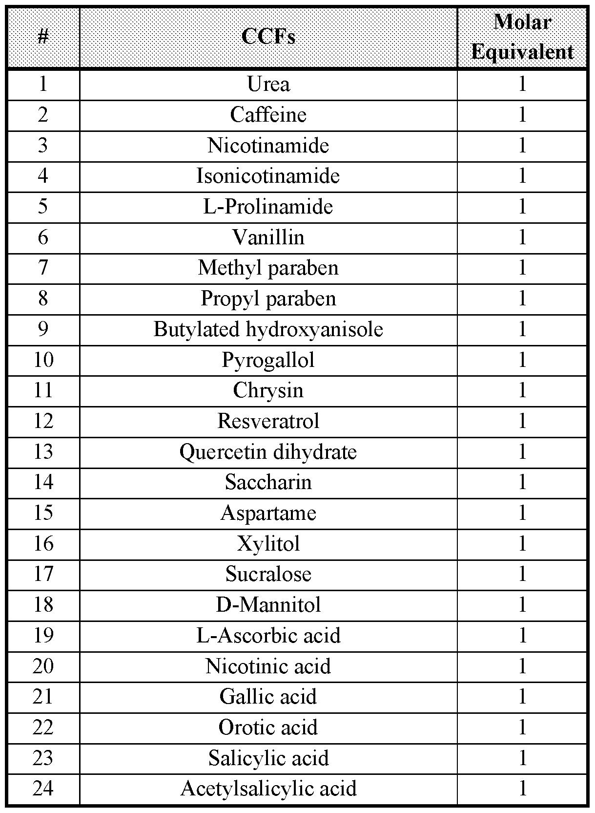
[00903] A total of five neat solvents and two binary mixtures were utilized in the presented cocrystal screening experiments: THF, EtOAc, DCM, MIBK, MeOH, THF/cyclohexane (2:8 v/v), and IPA:water (9: 1 v/v). The selection was based on diversity of molecular structure and properties of the solvent (e.g., polarity, chemical diversity), and solubility of free base Form C (“API”) from visual solubility assessment.
[00904] A total of -240 co-crystal-screening experiments were conducted using 24 CCFs and a combination of i) solvent-drop grinding (SDG) - with four solvents, ii) slurry-ripening (SR) in six solvents, and iii) evaporation of solutions obtained in step ii.
[00905] Solvent-Drop Grinding (SDG). Several preliminary experiments were conducted to determine appropriate milling parameters for the SDG experiments. The results of these
experiments are summarized in Table 5 (15 minutes of grinding at 15Hz with one milling ball). The data indicated that 15 minutes of grinding at 15Hz with one milling ball was appropriate for 100 mg API with 2-15 pL solvent. The specific (initial) solvent volumes selected for the four solvents were: THF - 5 pL; EtOAc, DCM, and MIBK - 15 pL.
Table 5 - Determination of Appropriate Solvent-Drop Grinding (SDG) Parameters
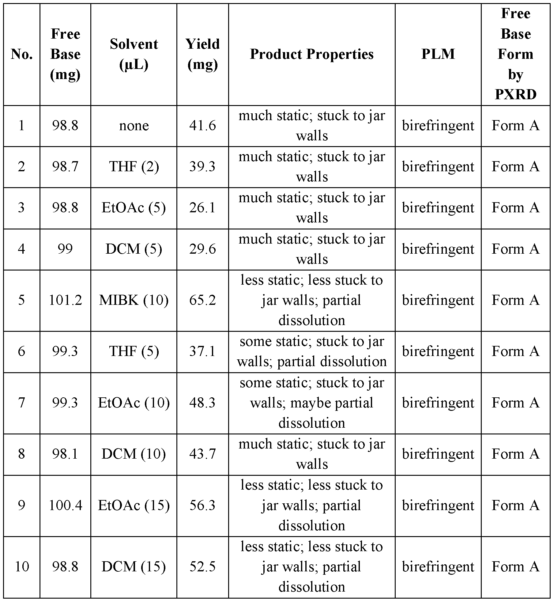
[00906] For the SDG experiments, the API (-100 mg), a stoichiometric amount of CCF (1 eq), and solvent THF, EtOAc, DCM, or MIBK were combined in a stainless steel milling jar (10
mL). Grinding was conducted on a Retsch Mill (Model MM301) at room temperature (~23 °C) with one milling ball (7 mm) at 15Hz for 15 minutes. In cases where these parameters were observed or expected (based on properties of the CCF) to result in low yield or gumming, the milling time was reduced to 10 minutes or manual grinding via a mortar and pestle was used.
[00907] Slurry-Ripening (SR). Products from the SDG experiments were utilized and combined with the same four neat solvents used in the SDG experiments to conduct SR studies, except that THF: cyclohexane (2:8 v/v) was substituted for THF. For CCFs that yielded potential cocrystals (or salts) from SDG, saturated solutions of the CCFs were prepared in the specific solvents that yielded potential co-crystals or salts and used for SR experiments.
[00908] For two additional solvents (MeOH and IPA:water (9: 1 v/v)), 1 : 1 (APFCCF) equivalent mixtures were prepared and combined with the two solvent systems.
[00909] The saturated solutions of CCFs were prepared by combining the CCF (estimated amount to achieve suspension) with 2 mL of solvent, then mixing at 23 °C for 16 hours. Suspensions were filtered through a 0.20 pm PTFE filter membrane to yield saturated solutions.
[00910] SR experiments were conducted in 2 mL vial s containing a tumble-stir disc and employed up to 1.9 mL solvent [THF: cyclohexane (2:8 v/v), EtOAc, DCM, MIBK, MeOH, or IPA:water (9: 1 v/v)]. The samples were mixed and temperature-cycled between 40 °C and 5 °C for seven days, followed by mixing at 25 °C for five days. During this processing time, additional solvent was added to yield mixable suspensions with sufficient solids for isolation and analysis. Suspended solids were isolated by filtration and air-dried for 18 hours.
[00911] Evaporation (EV). Solutions that were obtained in slurry -ripening experiments were slowly evaporated (by loosening the vial cap) in a fume hood until dry. Products were examined first by PLM for birefringence, and further analyzed by PXRD if birefringent.
[00912] All solid outputs of the screen were analyzed by PXRD to assess co-crystal formation. Likely co-crystals were analyzed by additional techniques as appropriate and as sample quantity permitted (FT-Raman, DSC, TGA-IR, PLM, etc.).
[00913] The conducted experiments yielded potential co-crystals (pure or in mixture with parent and/or CCF) of Form C free base with isonicotinamide, pyrogallol, saccharin, and xylitol, and potential salts with L-ascorbic acid, nicotinic acid, gallic acid, orotic acid, salicylic acid, and acetylsalicylic acid. Most potential co-crystals (or salts) were obtained from SR/EV experiments. The PXRD patterns of salicylic acid Form A and acetylsalicylic acid Form A were observed to
be identical. Proton NMR analysis confirmed that the acetylsalicylic acid salt Form A was consistent with salicylic acid salt Form A, as no acetyl group was observed. This may be due to hydrolysis of acetylsalicylic acid to salicylic acid during slurry-ripening.
[00914] Co-crystal formers that did not yield potential co-crystals included urea, caffeine, nicotinamide, L-prolinamide, vanillin, methyl paraben, propyl paraben, butylated hydroxyanisole, chrysin, resveratrol, quercetin, aspartame, sucralose, and D-mannitol. These co crystal formers yielded amorphous materials, parent forms, CCF, or a combination thereof. The products obtained in the SDG and SR/EV experiments are shown in Table 6 and Table 7, respectively.
Table 6 - Co-crystal or Salt Screening Products Obtained from SDG Approach


Notes: A, B - crystal forms identified
Attorney Docket No. : 2007878-1081
Table 7 - Co-crystal or Salt Screening Products Obtained from Slurry-Ripening or Evaporation Approach

Page 165 of 182
9616029V 1
Attorney Docket No. : 2007878-1081

LEGEND:
Notes: A, B, etc. - crystal forms
+ - mixture, likely with unidentified forms of parent free base
* - poorly crystalline
(ev) - from solution evaporation

ND - form undetermined
9616029V 1 Page 166 of 182
Table 8 - Attributes of Scaled-Up Co-Crystals or Salts

Example 5. Scale up of Co-crystals.
[00915] Of the potential co-crystal (or salt) hits, the following seven exhibited desirable physiochemical properties and were scaled up on a 250 mg scale: saccharin Form A, nicotinic acid Form A, ascorbic acid Form A, gallic acid Form A, salicylic acid Form A, and orotic acid Forms F and H. Results are described in detail below.
Pc p 1 L7 r>f 1 87
Example 5.1. Saccharin Co-Crystal
[00916] Saccharin co-crystal hits were obtained from six SR experiments. PXRD analysis of the samples indicated one form, designated Form A. Form A (non-solvated) was scaled up (250 mg scale) and subjected to detailed characterization.
[00917] Preparation of Form A (Non-solvated) . Form C free base (244.5 mg) was combined with saccharin (83.1 mg; 1 eq) and solvent (DCM, 3.5 mL), and mixed at 40 °C for 30 minutes yielding a suspension. Seeds (~5 mg) were added, and the suspension was mixed at 40 °C for two hours, slow-cooled to 20 °C, and mixed at 20 °C for 60 hours yielding a moderately thick slurry. The solids were isolated by vacuum-filtration for two hours and dried at 40 °C in a vacuum oven for 18 hours. The product weight was 287 mg of Form A (87% yield relative to the cocrystal).
[00918] Form A was determined to be a crystalline powder by FT-Raman (Figure 189) and PXRD (Figure 190). DSC analysis showed a melting endotherm with onset at 183.8 °C (DH = 104.2 J/g) (Figure 191, trace 191B). TGA analysis showed 0.1% weight loss between 26-174 °C, indicating a non-solvated form (Figure 191, trace 191 A). Proton NMR analysis of Form A indicated that Form A comprises 1 equivalent of saccharin (Figure 192).
Example 5.2. Nicotinic Acid Salt
[00919] Nicotinic acid salt hits were obtained from three SR and one EV experiments. PXRD analysis of the samples indicated three forms, designated as Form A, Form B and Form C. Form A (non-solvated) was scaled up (250 mg scale) and subjected to detailed characterization. Form B was characterized by PXRD (Figure 197), TGA (Figure 198, trace 198 A), and DSC (Figure 198, trace 198B). Form C was characterized by PXRD (Figure 199), TGA (Figure 200, trace 200A), and DSC (Figure 200, trace 200B).
[00920] Preparation of Form A (Non-solvated) . Form C free base (252.8 mg) was combined with nicotinic acid (57.9 mg; 1 eq) and solvent (THF/cyclohexane (2:8), 3.0 mL), and mixed at 40 °C for 30 minutes yielding a suspension. Seeds (~5mg) were added, and the suspension was mixed at 40 °C for two hours, slow-cooled to 20 °C, and mixed at 20 °C for 60 hours yielding a moderately thick slurry. The solids were isolated by vacuum-filtration for two hours and dried at 40 °C in a vacuum oven for 18 hours. The product weight was 247 mg of nicotinic acid salt Form A (79% yield relative to the salt).
[00921] Form A was determined to be a crystalline powder by FT-Raman (Figure 193) and PXRD (Figure 194). DSC analysis showed a melting endotherm with onset at 179.9 °C (DH = 120.4 J/g) (Figure 195, trace 195B). TGA analysis showed 0.2% weight loss between 29-168 °C, indicating a non-solvated form (Figure 195, trace 195A). Proton NMR analysis of Form A indicated that Form A comprises 1 equivalent of nicotinic acid (Figure 196).
Example 5.3. L-Ascorbic Acid Salt
[00922] Ascorbic acid salt hits were obtained from six SR experiments. PXRD analysis of the samples indicated two forms, designated as Form A and Form B. Form A (hydrate) was scaled up (250 mg scale) and subjected to detailed characterization.
[00923] Preparation of Form A (Hydrate). Form C free base (249.7 mg) was combined with L-ascorbic acid (81.6 mg; 1 eq) and solvent (IP A/water (9: 1) v/v, 6.0 mL), and mixed at 40 °C for 30 minutes yielding a suspension. Seeds (~5 mg) were added, and the suspension was mixed at 40 °C for two hours, slow-cooled to 20 °C, and mixed at 20 °C for 60 hours yielding a moderately thick slurry. The solids were isolated by vacuum-filtration for four hours and left open in a fume hood for 18 hours. The product weight was 294 mg of ascorbic acid salt Form A (83% yield relative to the salt).
[00924] Form A was determined to be a crystalline powder by FT-Raman (Figure 201) and PXRD (Figure 202). DSC analysis showed a dehydration endotherm with onset at 46.0 °C (DH = 168.5 J/g) followed by a small endotherm at 116.8 °C (DH = 7.5 J/g) and a melting endotherm (possibly two merged) with onset at 157.0 °C (DH = 71.4 J/g) (Figure 203, trace 203B). TGA analysis showed 5.4% weight (2.2 eq) loss of water between 29-140 °C, indicating a hydrated form (Figure 203, trace 203A). Proton NMR analysis of Form A indicated that Form A comprises 1 equivalent of L-ascorbic acid (Figure 204).
Example 5.4. Gallic Acid Salt
[00925] Gallic acid salt hits were obtained from four SR experiments. PXRD analysis of the samples indicated two forms, designated as Form A and Form B. Form A was obtained in pure form while Form B was obtained only in mixture with Form A. Form A (hydrate) of the gallic acid salt was scaled up (250 mg scale) and subjected to detailed characterization.
[00926] Preparation of Form A (Hydrate). Form C free base (245.0 mg) was combined with gallic acid (77.0 mg; 1 eq) and solvent (MeOH, 4.0 mL), and mixed at 40 °C for 30 minutes yielding a suspension. Seeds (~5 mg) were added, and the suspension was mixed at 40 °C for two hours, slow-cooled to 20 °C, and mixed at 20 °C for 60 hours yielding a moderately thick slurry. The solids were isolated by vacuum-filtration for four hours and left open in a fume hood for 18 hours. The product weight was 256 mg of gallic acid salt Form A (77% yield relative to the salt).
[00927] Form A was determined to be a crystalline powder by FT-Raman (Figure 205) and PXRD (Figure 206). DSC analysis showed a dehydration endotherm with onset at 48.5 °C (DH = 79.8 J/g) followed by a melting endotherm with onset at 193.5 °C (DH = 176.1 J/g) (Figure 207, trace 207B). TGA analysis showed 2.4% weight (1.0 eq) loss of water between 22-89 °C, indicating a hydrated form (Figure 207, trace 207A). Proton NMR analysis of Form A indicated that Form B comprises 1 equivalent of gallic acid (Figure 208).
Example 5.5. Salicylic Acid Salt
[00928] Salicylic acid salt hits were obtained from one SDG experiment and six SR experiments; however, the hit from SDG was a mixture of a potential salt, parent, and CCF. PXRD analysis of the six SR hits indicated two forms, designated as Form A and Form B. Most hits (5/6) were consistent with Form A. Form A (hydrate) of the salicylic acid salt was scaled up (250 mg scale) and subjected to detailed characterization.
[00929] Preparation of Form A (Hydrate). Form C free base (253.8 mg) was combined with salicylic acid (64.7 mg; 1 eq) and solvent (IP A/water 9: 1, 4.5 mL), and mixed at 40 °C for 30 minutes yielding a suspension. Seeds (~5 mg) were added, and the suspension was mixed at 40 °C for two hours, slow-cooled to 20 °C, and mixed at 20 °C for 60 hours yielding a moderately thick slurry. The solids were isolated by vacuum-filtration for 18 hours. The product weight was 272 mg of salicylic acid salt Form A (83% yield relative to the salt).
[00930] Form A was determined to be a crystalline powder by FT-Raman (Figure 209) and PXRD (Figure 210). DSC analysis showed a dehydration endotherm with onset at 34.9 °C (DH = 71.0 J/g) followed by a melting endotherm with onset at 159.8 °C (DH = 83.8 J/g) (Figure 211, trace 21 IB). TGA analysis showed 2.5% weight (1.0 eq) loss of water between 26-96 °C,
indicating a hydrated form (Figure 211, trace 211 A). Proton NMR analysis of Form A indicated that Form A comprises 1 equivalent of salicylic acid (Figure 212).
Example 5.6. Orotic Acid Salt
[00931] Orotic acid salt hits were obtained from six SR experiments. PXRD analysis of the hits indicated six forms, designated as Form A, Form B, Form C, Form D, Form E and Form F. Scale-up experiments (250 mg) were conducted for Forms E and F (hydrates), and the other groups were deprioritized due to solvation or because they were mixtures of two groups as shown in Table 7. The Form E scale-up experiment was unsuccessful and produced two new groups: Form G and Form H. Form G is a MeOH/water solvate that desolvates under ambient conditions to Form H, a hydrate. Form A was characterized by PXRD (Figure 213), TGA (Figure 214, trace 214A) and DSC (Figure 214, trace 214B). The mixture of Form B and Form E was characterized by PXRD (Figure 215). The mixture of Form C and Form E was characterized by PXRD (Figure 216). Form D was characterized by PXRD (Figure 217), TGA (Figure 218, trace 218A) and DSC (Figure 218, trace 218B). Form E was characterized by PXRD (Figure 219), TGA (Figure 220, trace 220A) and DSC (Figure 220, trace 220B). Form G was characterized by PXRD (Figure 221). Form F and Form H (hydrates) of the orotic acid salt were scaled up (250 mg) and subjected to detailed characterization.
[00932] Preparation of Form F (Hydrate). Form C free base (250.0 mg) was combined with orotic acid (77.0 mg; 1 eq) and solvent (IP A/water 9: 1, 10.0 mL), and mixed at 40 °C for 30 minutes yielding a suspension. Seeds (~5mg) were added, and the suspension was mixed at 40 °C for two hours, slow-cooled to 20 °C, and mixed at 20 °C for 60 hours yielding a moderately thick slurry. The solids were isolated by vacuum-filtration for 22 hours. The product weight was 297 mg of orotic acid salt Form F (82% yield relative to the co-crystal).
[00933] Form F was determined to be a crystalline powder by FT-Raman (Figure 222) and PXRD (Figure 223). DSC analysis showed two dehydration endotherms with onsets at 56.5 °C (DH = 86.1 J/g) and 104.7 °C (DH = 15.4 J/g), respectively, immediately followed by a melting endotherm with onset at 135.2 °C (DH = 12.3 J/g) (Figure 224, trace 224B). TGA analysis showed 10.8% weight (4.5 eq) loss of water between 24 °-129°C, indicating a hydrated form (Figure 224, trace 224A). Proton NMR analysis of Form F indicated that Form F comprises 1 equivalent of orotic acid (Figure 225).
[00934] PXRD analysis of the sample post heating indicated significant loss of crystallinity but no change in form.
[00935] Preparation of Form H (Hydrate). Form C free base (251.7 mg) was combined with orotic acid (72.7 mg; 1 eq) and solvent (MeOH, 1.0 mL), and mixed at 40 °C for 10 minutes yielding a near clear solution. Seeds (Group E, ~5mg) were added, and the suspension became very thick, so additional solvent was added (MeOH, 1.5 mL). The suspension was mixed at 40 °C for two hours, slow-cooled to 20 °C, and mixed at 20 °C for 18 hours yielding a moderately thick slurry. PXRD indicated a new form, and DSC/TGA-IR indicated a MeOH/water solvate, which was designated Form G. The batch solids were isolated by vacuum-filtration for 18 hours. The product weight was 178 mg. PXRD indicated yet a new form, and DSC/TGA-IR indicated a hydrate, which was designated Form H (53% yield relative to the salt).
[00936] Form H was determined to be a crystalline powder by FT-Raman (Figure 226) and PXRD (Figure 227). DSC analysis showed a broad dehydration endotherm with onset at 34.3°C (DH = 23.4 J/g), followed by two small endotherms at 134.5 °C and 144.4 °C, respectively, a large endotherm with onset at 165.8 °C (DH = 44.6 J/g), and a broad endotherm with onset at 203.4 °C (DH = 11.1 J/g) (Figure 228, trace 228B). TGA analysis showed 3.2% weight (1.2 eq) loss of water between 23-95 °C, indicating a hydrated form (Figure 228, trace 228A). Proton NMR analysis of Form H indicated that Form H comprises 1 equivalent of orotic acid (Figure 229).
[00937] PXRD analysis of the post-heated sample indicated some loss of crystallinity and a loss of several major peaks.
Example 5.7. Other Co-crystal or Salt Hits
[00938] Acetylsalicylic acid salt Form A was scaled up, however the PXRD pattern was observed to be identical to that of salicylic acid salt Form A. Proton NMR analysis confirmed that the acetylsalicylic acid salt Form A was consistent with salicylic acid salt Form A, as no acetyl group was observed. This may be due to hydrolysis of acetylsalicylic acid to salicylic acid during slurry-ripening.
[00939] In addition to the scaled up co-crystals (or salts), several other potential co-crystals were obtained from screening. These hits were not completely characterized and/or scaled up due to:
- limited sample amounts,
- undesirable physiochemical properties (poor crystallinity/poor thermal properties)
- being identified as a mixture with parent and/or CCF.
[00940] Representative samples of these co-crystal (or salt) hits are summarized in Table 9.
Table 9 - Attributes of Other Co-Crystal or Salt Hits Identified in Screening


Example 6. Aqueous Solubility of Certain Complexes
[00941] The solid/salt forms (-20-30 mg) were transferred to clear glass vials (4 ml). To each vial containing solid forms, the water (-0.2 -2 ml) was separately added. The volume of water added and the weight of the solid/salt form was appropriately adjusted to yield excess undissolved solid/salt form. The vials containing the solid/salt form/water mixture were transferred on to the rack that were kept at rotation and the samples were equilibrated with agitation at ambient temperature for 24 hr. At the end of the equilibration process, visual observations of the suspensions were made and the samples were withdrawn and centrifuged (14,000 rpm for 3 min) in a Costar SPIN-X polypropylene centrifuge tube (2.0 ml) filter (0.22 mm Nylon filter) to separate any un-dissolved drug. The clear filtrate was assayed for drug content to determine solubility of the active in the solution following appropriate dilution where necessary in acetonitrile/water (50:50). A standard curve in the concentration range of 0.126 mg/ml to 0.001 mg/ml was prepared using the free base. The samples and standards were assayed for drug content using the HPLC. Results are set forth in Table 10:
Table 10 - Solubility of Certain Forms of Compound 1


Claims
1. A crystalline form of Compound 1 :

2. The crystalline form of claim 1, wherein the form is unsolvated.
3. The crystalline form of claim 2, wherein the form is characterized by one or more peaks in its X-ray powder diffraction pattern selected from 9.7, 14.6, 19.5, 24.3, and 25.6 ± 0.2 degrees 2Q.
[00942] 4. The crystalline form of claim 2, wherein the form is characterized by the following peaks in its X-ray powder diffraction pattern:

5. The crystalline form of claim 1, wherein the form is solvated.
6. The crystalline form of claim 5, wherein the form is a 2-methyl-tetrahydrofuran solvate.
7. The crystalline form of claim 6, wherein the form is characterized by one or more peaks in its X-ray powder diffraction pattern selected from 12.5, 18.3, 18.9, 20.1, and 23.8 ± 0.2 degrees 2Q.
8. The crystalline form of claim 6, wherein the form is characterized by the following peaks in its X-ray powder diffraction pattern:

9. The crystalline form of claim 1, wherein the form is a hydrate.
10. The crystalline form of claim 9, wherein the form is a monohydrate.
11. The crystalline form of claim 10, wherein the form is characterized by one or more peaks in its X-ray powder diffraction pattern selected from 8.7, 15.2, 17.3, 18.0, and 19.4 ± 0.2 degrees 2Q.
12. The crystalline form of claim 10, wherein the form is characterized by the following peaks in its X-ray powder diffraction pattern:
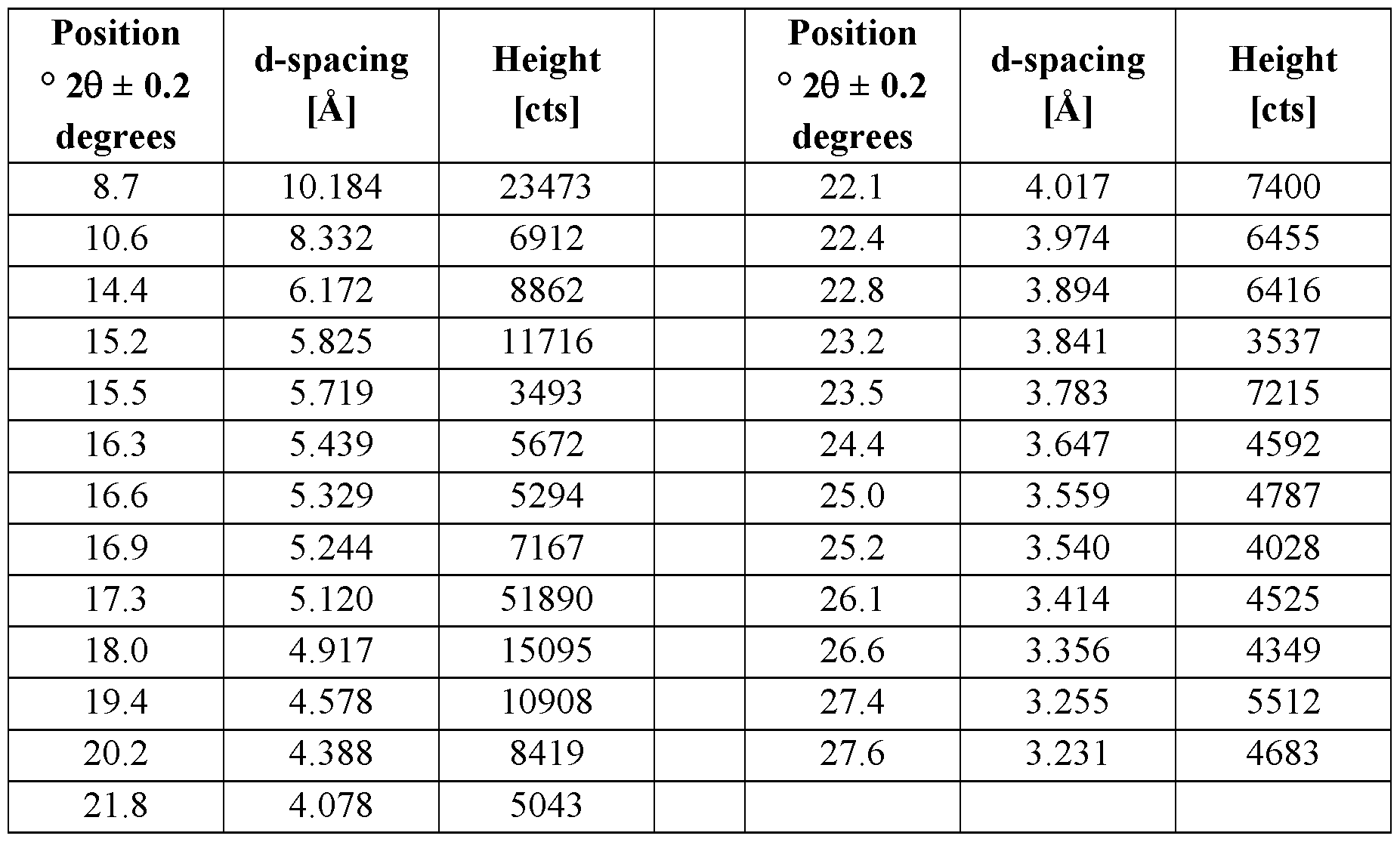
13. The crystalline form of claim 9, wherein the form is a tetrahydrate.
14. The crystalline form of claim 13, wherein the form is characterized by one or more peaks in its X-ray powder diffraction pattern selected from 12.4, 18.5, 19.3, 20.3, and 23.6 ± 0.2 degrees 2Q.
15. The crystalline form of claim 13, wherein the form is characterized by the following peaks in its X-ray powder diffraction pattern:


16. A sample comprising the crystalline form of any one of claims 1-15, wherein the samples substantially free of impurities.
17. A complex comprising Compound 1:

1
and a co-former X;
wherein the complex is crystalline and
X is selected from the group consisting of hydrobromic acid, sulfuric acid, toluenesulfonic acid, methanesulfonic acid, 2-naphthalenesulfonic acid, phosphoric acid, DL-tartaric acid, succinic acid, gentisic acid, hippuric acid, adipic acid, galactaric acid, naphthalene- 1,5-disulfonic acid, fV)-camphor- 10- sulfonic acid, ethane- 1,2-disulfonic acid, ethanesulfonic acid, benzenesulfonic acid, oxalic acid, maleic acid, pamoic acid, l-hydroxy-2-naphthoic acid, malonic acid, L-tartaric acid, fumaric acid, citric acid, L-lactic acid, acetic acid, propionic acid, DL-lactic acid, D-gluconic acid, DL-malic acid, glutaric acid, camphoric acid, DL-mandelic acid, glutamic acid, glycolic acid, L- mandelic acid, L-malic acid, L-aspartic acid, benzoic acid, saccharin, nicotinic acid, ascorbic acid, gallic acid, salicylic acid, orotic acid, acetylsalicylic acid, choline, potassium hydroxide, and sodium hydroxide.
18. A complex comprising Compound 1:
1
and a co-former X;
wherein:
X is selected from the group consisting of 2-naphthalenesulfonic acid, succinic acid, gentisic acid, hippuric acid, adipic acid, galactaric acid, naphthalene-1, 5- disulfonic acid, fV)-camphor- 10-sulfonic acid, ethane- 1,2-disulfonic acid, ethanesulfonic acid, benzenesulfonic acid, maleic acid, pamoic acid, 1- hydroxy-2-naphthoic acid, malonic acid, fumaric acid, L-lactic acid, propionic acid, DL-lactic acid, D-gluconic acid, DL-malic acid, glutaric acid, camphoric acid, glutamic acid, glycolic acid, L-malic acid, L-aspartic acid, benzoic acid, saccharin, nicotinic acid, ascorbic acid, gallic acid, salicylic acid, orotic acid, acetylsalicylic acid, and choline.
19. A sample comprising the complex of claim 17 or claim 18, wherein the sample is substantially free of impurities.
20. A method of inhibiting activity of a JAK2 kinase, or a mutant thereof, in a biological sample comprising the step of contacting said biological sample with a crystalline form of any one of claims 1-15, or a composition thereof.
21. A method of inhibiting activity of a JAK2 kinase, or a mutant thereof, in a patient comprising the step of administering to said patient a crystalline form of any one of claims 1-15, or a composition thereof.
22. A method for treating a JAK2-mediated disease or disorder, in a patient in need thereof, comprising the step of administering to the patient a crystalline form of any one of claims 1-15, or pharmaceutically acceptable composition thereof.
23. A method of inhibiting activity of a JAK2 kinase, or a mutant thereof, in a biological sample comprising the step of contacting said biological sample with a complex of claim 17 or claim 18, or a composition thereof.
24. A method of inhibiting activity of a JAK2 kinase, or a mutant thereof, in a patient comprising the step of administering to said patient a complex of claim 17 or claim 18, or a composition thereof.
25. A method for treating a JAK2-mediated disease or disorder, in a patient in need thereof, comprising the step of administering to the patient a complex of claim 17 or claim 18, or a pharmaceutically acceptable composition thereof.
Priority Applications (5)
| Application Number | Priority Date | Filing Date | Title |
|---|---|---|---|
| CN202080018226.8A CN113840604A (en) | 2019-02-12 | 2020-02-11 | Crystalline forms of a JAK2 inhibitor |
| KR1020217029151A KR20210145735A (en) | 2019-02-12 | 2020-02-11 | Crystalline Forms of JAK2 Inhibitors |
| US17/430,188 US20230250068A1 (en) | 2019-02-12 | 2020-02-11 | Crystalline forms of a jak2 inhibitor |
| JP2021547093A JP2022519885A (en) | 2019-02-12 | 2020-02-11 | Crystal form of JAK2 inhibitor |
| EP20756000.4A EP3923948A4 (en) | 2019-02-12 | 2020-02-11 | Crystalline forms of a jak2 inhibitor |
Applications Claiming Priority (2)
| Application Number | Priority Date | Filing Date | Title |
|---|---|---|---|
| US201962804332P | 2019-02-12 | 2019-02-12 | |
| US62/804,332 | 2019-02-12 |
Publications (1)
| Publication Number | Publication Date |
|---|---|
| WO2020167845A1 true WO2020167845A1 (en) | 2020-08-20 |
Family
ID=72045436
Family Applications (1)
| Application Number | Title | Priority Date | Filing Date |
|---|---|---|---|
| PCT/US2020/017765 WO2020167845A1 (en) | 2019-02-12 | 2020-02-11 | Crystalline forms of a jak2 inhibitor |
Country Status (6)
| Country | Link |
|---|---|
| US (1) | US20230250068A1 (en) |
| EP (1) | EP3923948A4 (en) |
| JP (1) | JP2022519885A (en) |
| KR (1) | KR20210145735A (en) |
| CN (1) | CN113840604A (en) |
| WO (1) | WO2020167845A1 (en) |
Cited By (4)
| Publication number | Priority date | Publication date | Assignee | Title |
|---|---|---|---|---|
| WO2022132933A1 (en) | 2020-12-16 | 2022-06-23 | Impact Biomedicines, Inc. | Dosing of fedratinib |
| US11400092B2 (en) | 2018-09-25 | 2022-08-02 | Impact Biomedicines, Inc. | Methods of treating myeloproliferative disorders |
| WO2022182857A1 (en) | 2021-02-25 | 2022-09-01 | Impact Biomedicines, Inc. | Use of a bet inhibitor alone or in combination with fedratinib or ruxolitinib for treating a hematological malignancy such as myelofibrosis |
| WO2023044297A1 (en) | 2021-09-14 | 2023-03-23 | Impact Biomedicines, Inc. | Fedratinib for treating myeloproliferative disorders |
Citations (5)
| Publication number | Priority date | Publication date | Assignee | Title |
|---|---|---|---|---|
| US20130243853A1 (en) * | 2010-11-07 | 2013-09-19 | Targegen, Inc. | Compositions and methods for treating myelofibrosis |
| US20150361050A1 (en) * | 2014-06-12 | 2015-12-17 | Gilead Sciences, Inc. | (n-(cyanomethyl)-4-(2-(4-morpholinophenylamino)pyrimidin-4-yl)benzamide |
| WO2016138458A1 (en) * | 2015-02-26 | 2016-09-01 | Genentech, Inc. | Crystalline forms of a pyrrolopyridine compound |
| US20160332993A1 (en) * | 2014-01-31 | 2016-11-17 | Dana-Farber Cancer Institute, Inc. | Diaminopyrimidine benzenesulfone derivatives and uses thereof |
| US20180078561A1 (en) * | 2015-03-31 | 2018-03-22 | Lam Therapeutics, Inc. | Active metabolites of apilimod and uses thereof |
Family Cites Families (2)
| Publication number | Priority date | Publication date | Assignee | Title |
|---|---|---|---|---|
| HUE028987T2 (en) * | 2005-11-01 | 2017-01-30 | Targegen Inc | Bi-aryl meta-pyrimidine inhibitors of kinases |
| WO2013013195A1 (en) * | 2011-07-21 | 2013-01-24 | Sanofi | Compositions and methods for treating polycythemia vera and essential thrombocythemia |
-
2020
- 2020-02-11 EP EP20756000.4A patent/EP3923948A4/en active Pending
- 2020-02-11 CN CN202080018226.8A patent/CN113840604A/en active Pending
- 2020-02-11 JP JP2021547093A patent/JP2022519885A/en active Pending
- 2020-02-11 WO PCT/US2020/017765 patent/WO2020167845A1/en unknown
- 2020-02-11 KR KR1020217029151A patent/KR20210145735A/en unknown
- 2020-02-11 US US17/430,188 patent/US20230250068A1/en active Pending
Patent Citations (5)
| Publication number | Priority date | Publication date | Assignee | Title |
|---|---|---|---|---|
| US20130243853A1 (en) * | 2010-11-07 | 2013-09-19 | Targegen, Inc. | Compositions and methods for treating myelofibrosis |
| US20160332993A1 (en) * | 2014-01-31 | 2016-11-17 | Dana-Farber Cancer Institute, Inc. | Diaminopyrimidine benzenesulfone derivatives and uses thereof |
| US20150361050A1 (en) * | 2014-06-12 | 2015-12-17 | Gilead Sciences, Inc. | (n-(cyanomethyl)-4-(2-(4-morpholinophenylamino)pyrimidin-4-yl)benzamide |
| WO2016138458A1 (en) * | 2015-02-26 | 2016-09-01 | Genentech, Inc. | Crystalline forms of a pyrrolopyridine compound |
| US20180078561A1 (en) * | 2015-03-31 | 2018-03-22 | Lam Therapeutics, Inc. | Active metabolites of apilimod and uses thereof |
Non-Patent Citations (1)
| Title |
|---|
| See also references of EP3923948A4 * |
Cited By (4)
| Publication number | Priority date | Publication date | Assignee | Title |
|---|---|---|---|---|
| US11400092B2 (en) | 2018-09-25 | 2022-08-02 | Impact Biomedicines, Inc. | Methods of treating myeloproliferative disorders |
| WO2022132933A1 (en) | 2020-12-16 | 2022-06-23 | Impact Biomedicines, Inc. | Dosing of fedratinib |
| WO2022182857A1 (en) | 2021-02-25 | 2022-09-01 | Impact Biomedicines, Inc. | Use of a bet inhibitor alone or in combination with fedratinib or ruxolitinib for treating a hematological malignancy such as myelofibrosis |
| WO2023044297A1 (en) | 2021-09-14 | 2023-03-23 | Impact Biomedicines, Inc. | Fedratinib for treating myeloproliferative disorders |
Also Published As
| Publication number | Publication date |
|---|---|
| CN113840604A (en) | 2021-12-24 |
| EP3923948A1 (en) | 2021-12-22 |
| KR20210145735A (en) | 2021-12-02 |
| JP2022519885A (en) | 2022-03-25 |
| US20230250068A1 (en) | 2023-08-10 |
| EP3923948A4 (en) | 2022-11-16 |
Similar Documents
| Publication | Publication Date | Title |
|---|---|---|
| US20230250068A1 (en) | Crystalline forms of a jak2 inhibitor | |
| EA038195B1 (en) | Malate salt of n-(4-{[6,7-bis(methyloxy)quinolin-4-yl]oxy}phenyl-n'-(4-fluorophenyl)cyclopropane-1,1-dicarboxamide and use thereof for the treatment of kidney and liver cancer | |
| US9221815B2 (en) | Solid state form of vemurafenib choline salt | |
| WO2019134573A1 (en) | Method for preparing deuterated diphenylaminopyrimidine compound and crystal form thereof | |
| US11945832B2 (en) | Psilocybin and O-acetylpsilocin, salts and solid state forms thereof | |
| WO2020244349A1 (en) | Method for synthesizing furoimidazopyridine compound, polymorphic substance and polymorphic substance of salt | |
| KR20140040671A (en) | Aprepitant l-proline composition and cocrystal | |
| US20230279017A1 (en) | Salt and solid forms of tabernanthalog | |
| AU2020220512B2 (en) | FGFR inhibitor compound in solid form and preparation method therefor | |
| TW202313621A (en) | Crystalline forms of 3-{4-[(2r)-2-aminopropoxy]phenyl}-n-[(1r)-1-(3-fluorophenyl)ethyl]imidazo[1,2-b]pyridazin-6-amine and salts thereof | |
| US20220332706A1 (en) | Crystalline forms of a jak2 inhibitor | |
| EP3941472A1 (en) | <smallcaps/>? ? ?n? ? ? ? ?crystalline and amorphous forms of-(5-((4-ethylpiperazin-1-yl)methyl)pyridine-2-yl)-5-fluoro-4-(3-isopropyl-2-methyl-2 <ns1:i>h</ns1:i>?-indazol-5-yl)pyrimidin-2-amine and its salts, and preparation methods and therapeutic uses thereof | |
| JP2023531078A (en) | crystalline form of the compound | |
| JP2023524622A (en) | 6-chloro-7-(4-(4-chlorobenzyl)piperazin-1-yl)-2-(1,3-dimethyl-1H-pyrazol-4-yl)-3H-imidazo[4,5-b] Salts and polymorphs of pyridine | |
| WO2022072470A1 (en) | Crystalline form of tegavivint, method of preparation, and use thereof | |
| JP2024505670A (en) | Salts and crystal forms of nitrogen-containing fused heterocyclic compounds and their preparation, pharmaceutical compositions and uses | |
| WO2016101912A1 (en) | Crystal form of salt of epidermal growth factor receptor kinase inhibitor and preparation method thereof | |
| TW201425309A (en) | Monohydrate crystalline form of 3-(5-(4-(3-fluoropropyl)-piperazin-1-yl)benzimidazol-2-yl)-1-azaazulen-2-one, preparation method, and pharmaceutical composition thereof |
Legal Events
| Date | Code | Title | Description |
|---|---|---|---|
| 121 | Ep: the epo has been informed by wipo that ep was designated in this application |
Ref document number: 20756000 Country of ref document: EP Kind code of ref document: A1 |
|
| ENP | Entry into the national phase |
Ref document number: 2021547093 Country of ref document: JP Kind code of ref document: A |
|
| NENP | Non-entry into the national phase |
Ref country code: DE |
|
| ENP | Entry into the national phase |
Ref document number: 2020756000 Country of ref document: EP Effective date: 20210913 |