WO2018186032A1 - Function inhibitor of swi/snf complexes - Google Patents
Function inhibitor of swi/snf complexes Download PDFInfo
- Publication number
- WO2018186032A1 WO2018186032A1 PCT/JP2018/005527 JP2018005527W WO2018186032A1 WO 2018186032 A1 WO2018186032 A1 WO 2018186032A1 JP 2018005527 W JP2018005527 W JP 2018005527W WO 2018186032 A1 WO2018186032 A1 WO 2018186032A1
- Authority
- WO
- WIPO (PCT)
- Prior art keywords
- seq
- sequence
- expression
- swi
- cells
- Prior art date
Links
Images


Classifications
-
- C—CHEMISTRY; METALLURGY
- C12—BIOCHEMISTRY; BEER; SPIRITS; WINE; VINEGAR; MICROBIOLOGY; ENZYMOLOGY; MUTATION OR GENETIC ENGINEERING
- C12N—MICROORGANISMS OR ENZYMES; COMPOSITIONS THEREOF; PROPAGATING, PRESERVING, OR MAINTAINING MICROORGANISMS; MUTATION OR GENETIC ENGINEERING; CULTURE MEDIA
- C12N15/00—Mutation or genetic engineering; DNA or RNA concerning genetic engineering, vectors, e.g. plasmids, or their isolation, preparation or purification; Use of hosts therefor
- C12N15/09—Recombinant DNA-technology
- C12N15/11—DNA or RNA fragments; Modified forms thereof; Non-coding nucleic acids having a biological activity
- C12N15/113—Non-coding nucleic acids modulating the expression of genes, e.g. antisense oligonucleotides; Antisense DNA or RNA; Triplex- forming oligonucleotides; Catalytic nucleic acids, e.g. ribozymes; Nucleic acids used in co-suppression or gene silencing
-
- A—HUMAN NECESSITIES
- A61—MEDICAL OR VETERINARY SCIENCE; HYGIENE
- A61K—PREPARATIONS FOR MEDICAL, DENTAL OR TOILETRY PURPOSES
- A61K31/00—Medicinal preparations containing organic active ingredients
- A61K31/70—Carbohydrates; Sugars; Derivatives thereof
- A61K31/7088—Compounds having three or more nucleosides or nucleotides
- A61K31/713—Double-stranded nucleic acids or oligonucleotides
-
- A—HUMAN NECESSITIES
- A61—MEDICAL OR VETERINARY SCIENCE; HYGIENE
- A61K—PREPARATIONS FOR MEDICAL, DENTAL OR TOILETRY PURPOSES
- A61K31/00—Medicinal preparations containing organic active ingredients
- A61K31/70—Carbohydrates; Sugars; Derivatives thereof
- A61K31/7088—Compounds having three or more nucleosides or nucleotides
- A61K31/7105—Natural ribonucleic acids, i.e. containing only riboses attached to adenine, guanine, cytosine or uracil and having 3'-5' phosphodiester links
-
- A—HUMAN NECESSITIES
- A61—MEDICAL OR VETERINARY SCIENCE; HYGIENE
- A61K—PREPARATIONS FOR MEDICAL, DENTAL OR TOILETRY PURPOSES
- A61K35/00—Medicinal preparations containing materials or reaction products thereof with undetermined constitution
- A61K35/66—Microorganisms or materials therefrom
- A61K35/76—Viruses; Subviral particles; Bacteriophages
-
- A—HUMAN NECESSITIES
- A61—MEDICAL OR VETERINARY SCIENCE; HYGIENE
- A61K—PREPARATIONS FOR MEDICAL, DENTAL OR TOILETRY PURPOSES
- A61K38/00—Medicinal preparations containing peptides
- A61K38/16—Peptides having more than 20 amino acids; Gastrins; Somatostatins; Melanotropins; Derivatives thereof
-
- A—HUMAN NECESSITIES
- A61—MEDICAL OR VETERINARY SCIENCE; HYGIENE
- A61K—PREPARATIONS FOR MEDICAL, DENTAL OR TOILETRY PURPOSES
- A61K45/00—Medicinal preparations containing active ingredients not provided for in groups A61K31/00 - A61K41/00
-
- A—HUMAN NECESSITIES
- A61—MEDICAL OR VETERINARY SCIENCE; HYGIENE
- A61K—PREPARATIONS FOR MEDICAL, DENTAL OR TOILETRY PURPOSES
- A61K48/00—Medicinal preparations containing genetic material which is inserted into cells of the living body to treat genetic diseases; Gene therapy
-
- A—HUMAN NECESSITIES
- A61—MEDICAL OR VETERINARY SCIENCE; HYGIENE
- A61P—SPECIFIC THERAPEUTIC ACTIVITY OF CHEMICAL COMPOUNDS OR MEDICINAL PREPARATIONS
- A61P25/00—Drugs for disorders of the nervous system
-
- A—HUMAN NECESSITIES
- A61—MEDICAL OR VETERINARY SCIENCE; HYGIENE
- A61P—SPECIFIC THERAPEUTIC ACTIVITY OF CHEMICAL COMPOUNDS OR MEDICINAL PREPARATIONS
- A61P35/00—Antineoplastic agents
-
- A—HUMAN NECESSITIES
- A61—MEDICAL OR VETERINARY SCIENCE; HYGIENE
- A61P—SPECIFIC THERAPEUTIC ACTIVITY OF CHEMICAL COMPOUNDS OR MEDICINAL PREPARATIONS
- A61P43/00—Drugs for specific purposes, not provided for in groups A61P1/00-A61P41/00
-
- C—CHEMISTRY; METALLURGY
- C07—ORGANIC CHEMISTRY
- C07K—PEPTIDES
- C07K14/00—Peptides having more than 20 amino acids; Gastrins; Somatostatins; Melanotropins; Derivatives thereof
- C07K14/435—Peptides having more than 20 amino acids; Gastrins; Somatostatins; Melanotropins; Derivatives thereof from animals; from humans
- C07K14/46—Peptides having more than 20 amino acids; Gastrins; Somatostatins; Melanotropins; Derivatives thereof from animals; from humans from vertebrates
- C07K14/47—Peptides having more than 20 amino acids; Gastrins; Somatostatins; Melanotropins; Derivatives thereof from animals; from humans from vertebrates from mammals
- C07K14/4701—Peptides having more than 20 amino acids; Gastrins; Somatostatins; Melanotropins; Derivatives thereof from animals; from humans from vertebrates from mammals not used
- C07K14/4702—Regulators; Modulating activity
- C07K14/4703—Inhibitors; Suppressors
-
- C—CHEMISTRY; METALLURGY
- C07—ORGANIC CHEMISTRY
- C07K—PEPTIDES
- C07K14/00—Peptides having more than 20 amino acids; Gastrins; Somatostatins; Melanotropins; Derivatives thereof
- C07K14/435—Peptides having more than 20 amino acids; Gastrins; Somatostatins; Melanotropins; Derivatives thereof from animals; from humans
- C07K14/52—Cytokines; Lymphokines; Interferons
- C07K14/54—Interleukins [IL]
- C07K14/5412—IL-6
-
- C—CHEMISTRY; METALLURGY
- C07—ORGANIC CHEMISTRY
- C07K—PEPTIDES
- C07K14/00—Peptides having more than 20 amino acids; Gastrins; Somatostatins; Melanotropins; Derivatives thereof
- C07K14/435—Peptides having more than 20 amino acids; Gastrins; Somatostatins; Melanotropins; Derivatives thereof from animals; from humans
- C07K14/52—Cytokines; Lymphokines; Interferons
- C07K14/54—Interleukins [IL]
- C07K14/5421—IL-8
-
- A—HUMAN NECESSITIES
- A61—MEDICAL OR VETERINARY SCIENCE; HYGIENE
- A61K—PREPARATIONS FOR MEDICAL, DENTAL OR TOILETRY PURPOSES
- A61K45/00—Medicinal preparations containing active ingredients not provided for in groups A61K31/00 - A61K41/00
- A61K45/06—Mixtures of active ingredients without chemical characterisation, e.g. antiphlogistics and cardiaca
-
- C—CHEMISTRY; METALLURGY
- C12—BIOCHEMISTRY; BEER; SPIRITS; WINE; VINEGAR; MICROBIOLOGY; ENZYMOLOGY; MUTATION OR GENETIC ENGINEERING
- C12N—MICROORGANISMS OR ENZYMES; COMPOSITIONS THEREOF; PROPAGATING, PRESERVING, OR MAINTAINING MICROORGANISMS; MUTATION OR GENETIC ENGINEERING; CULTURE MEDIA
- C12N2310/00—Structure or type of the nucleic acid
- C12N2310/10—Type of nucleic acid
- C12N2310/14—Type of nucleic acid interfering N.A.
-
- C—CHEMISTRY; METALLURGY
- C12—BIOCHEMISTRY; BEER; SPIRITS; WINE; VINEGAR; MICROBIOLOGY; ENZYMOLOGY; MUTATION OR GENETIC ENGINEERING
- C12N—MICROORGANISMS OR ENZYMES; COMPOSITIONS THEREOF; PROPAGATING, PRESERVING, OR MAINTAINING MICROORGANISMS; MUTATION OR GENETIC ENGINEERING; CULTURE MEDIA
- C12N2310/00—Structure or type of the nucleic acid
- C12N2310/50—Physical structure
- C12N2310/53—Physical structure partially self-complementary or closed
- C12N2310/531—Stem-loop; Hairpin
Definitions
- the present invention relates to an inhibitor that inhibits gene expression control by NF- ⁇ B or a corepressor complex, which is dependent on a SWI / SNF complex involved in chromatin remodeling.
- the inhibitor of the present invention can be used for treatment of cancers involving SWI / SNF complexes (such as SWI / SNF-mediated cancers).
- the genomic DNA of eukaryotic multicellular organisms wraps around histone octamers and builds a dense chromatin structure.
- factors such as transcription factors can directly bind to genomic DNA.
- One of the mechanisms responsible for such epigenetic control is chromatin structure conversion.
- the SWI / SNF complex has been found as an evolutionarily conserved representative factor having such activity.
- mutations and deletions have been found in the genes of the subunits that make up this SWI / SNF complex in human cancer, and the involvement of the SWI / SNF complex in tumor development has attracted attention (patent documents). 1, 2).
- the SWI / SNF complex is responsible for the transcriptional regulation of a very wide range of genes for each cell type, its potential for use in the treatment of specific diseases such as cancer is limited.
- the transcription factor NF- ⁇ B was found to be involved in transcription of immunoglobulin ⁇ chain in B cells. At present, it is known that NF- ⁇ B controls a number of life phenomena including activation / differentiation, cell proliferation, apoptosis and the like through activation of expression of cytokine growth factors, adhesion molecules and the like in a wide range of cells. In addition, constitutive or excessive NF- ⁇ B activity is not only found in inflammatory diseases and autoimmune diseases, but also constitutive activation is observed in many tumor cells and leukemia cells, Inhibitors have also been created (Patent Documents 3 to 5).
- Non-Patent Documents 2 to 4 Non-Patent Documents 2 to 4).
- the target genes that undergo transcriptional activation by a larger complex formed by NF- ⁇ B and the SWI / SNF complex, the mechanism, and the like have not yet been elucidated.
- Tailless (also named TLX, NR2E1) belonging to the nuclear receptor family is a transcriptional regulatory factor expressed in the Drosophila embryo end, mouse forebrain, etc. during development.
- TLX is expressed in neural stem cells existing in the subventricular zone etc. in the human adult brain, and plays an important role in neurogenesis and is involved in brain tumors derived from neural stem cells TLX knockout mice have been shown to have a central nervous system phenotype, such as limbic deficits, and depression, anxiety, and attack when TLX function is altered.
- GIC Glioma stem cells
- An object of the present invention is to provide an anticancer agent based on a novel mechanism.
- the present inventors have found that inhibitors that inhibit SWI / SNF complex-dependent gene expression control by NF- ⁇ B or corepressor complex have anticancer activity.
- d4 family proteins that are essential for SWI / SNF complex-dependent NF- ⁇ B target gene expression in epithelial cancers where the SWI / SNF complex is functioning
- siRNA1 and shRNA that knockdown gene expression, particularly knockdown expression of genes encoding DPF2 and DPF3a / 3b, exhibit strong anticancer activity against the epithelial cancer.
- CT1 peptide a region consisting of 84 amino acid residues at the N-terminus that is highly conserved among DPF1, DPF2, and DPF3a / 3b (referred to as CT1 peptide) is used for the above-mentioned siRNA, shRNA against epithelial cancer. It was found to show stronger anticancer activity.
- SWI / SNF complex plays an essential role in maintaining stem cell properties in GIC via d4 family proteins, particularly DPF1 and DPF3a.
- d4 family proteins particularly DPF1 and DPF3a.
- siRNA, shRNA knocks down the expression of genes encoding these d4 families, in particular the expression of genes encoding DPF1, DPF3a, SWI / SNF complex-dependent nuclear receptors TLX and It was found that strong anticancer activity was exhibited by suppressing the activity of the corepressor complex containing LSD1 / RCOR2.
- the present invention is based on the above findings and provides the following inhibitors, pharmaceutical compositions and the like.
- the inhibitor according to [1], wherein the inhibition is competitive inhibition of d4 family protein function or suppression of d4 family protein expression.
- the d4 family protein is at least one selected from DPF1, DPF2, DPF3a, and DPF3b.
- the dominant negative mutant is a polypeptide comprising the amino acid sequence represented by any of SEQ ID NOs: 1 to 7, or 90% or more with respect to the amino acid sequence represented by any of SEQ ID NOs: 1 to 7
- the inhibitor according to [4] above which is a polypeptide having an amino acid sequence having the same identity and having competitive inhibitory activity against the d4 family.
- the inhibitor according to any one of [1] to [5] which binds to a SWI / SNF complex.
- the inhibitor according to any one of [4] to [6] which binds to NF- ⁇ B and / or a corepressor complex.
- RNAi-producing RNA or an antisense oligonucleotide that suppresses the expression of a d4 family gene is an RNAi-producing RNA or an antisense oligonucleotide that suppresses the expression of a d4 family gene.
- the RNA exhibiting RNAi that suppresses the expression of the d4 family gene is specific to the d4 family gene of any of SEQ ID NOs: 74 to 77, and is an siRNA or shRNA that suppresses the expression of the d4 family gene.
- the polynucleotide that suppresses the expression of the d4 family gene is a double-stranded RNA consisting of a sequence selected from the group consisting of SEQ ID NOs: 41 to 73 and a complementary sequence of the sequence, and a lack of the sequence.
- the RNA that inhibits the expression of the d4 family gene is a combination of the sequence of SEQ ID NO: 41 and the sequence of SEQ ID NO: 42, a combination of the sequence of SEQ ID NO: 43 and the sequence of SEQ ID NO: 44, or the sequence of SEQ ID NO: 45
- the sequence of SEQ ID NO: 46, the combination of the sequence of SEQ ID NO: 47 and the sequence of SEQ ID NO: 48, the combination of the sequence of SEQ ID NO: 49 and the sequence of SEQ ID NO: 50, the sequence of SEQ ID NO: 51 and the sequence of SEQ ID NO: 52 A combination of sequences, a combination of the sequence of SEQ ID NO: 60 and a sequence of SEQ ID NO: 61, a combination of the sequence of SEQ ID NO: 62 and the sequence of SEQ ID NO: 63, a combination of the sequence of SEQ ID NO: 64 and the sequence of SEQ ID NO: 65,
- a pharmaceutical composition comprising the inhibitor according to any one of [1] to [15].
- the pharmaceutical composition according to the above [16] which is a therapeutic agent for glioblastoma.
- the pharmaceutical composition according to the above [19] comprising a polynucleotide that suppresses the expression of the DPF1 gene as an inhibitor.
- the present invention makes it possible to provide an anticancer agent based on a novel mechanism by inhibiting / modulating a specific function dependent on the SWI / SNF complex responsible for chromatin remodeling. More specifically, a polymorphism that suppresses the expression of dominant negative mutants derived from d4 family proteins (such as CT1) or genes encoding d4 family proteins against epithelial cancers where the SWI / SNF complex functions.
- Pharmaceutical compositions comprising nucleotide inhibitors (siRNA, shRNA, etc.) can be provided.
- a pharmaceutical composition containing a polynucleotide inhibitor (siRNA, shRNA, etc.) that suppresses the expression of a gene encoding a d4 family protein can be provided for glioma.
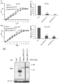
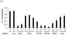
- FIG. 1 shows the effect of each gene knockdown encoding d4 family proteins and Brm and BRG1 on anchorage-independent growth of HeLaS3 (A) and A549 (B).
- FIG. 2 shows the structure and amino acid sequence of the d4 family protein and its dominant negative mutant.
- A Schematic diagram of the structure of mutant CT1 with the d4 family protein and its C-terminal excised.
- B An alignment of amino acid sequences corresponding to CT1 in the N-terminal region of DPF1, DPF2 and DPF3. box indicates a nuclear translocation signal sequence.
- FIG. 2 (C) and (D) are ectopic of Halo-DPF2-CT1 and Halo-DPF3-CT1 on cell proliferation of A549 and HeLaS3 cells in monolayer culture (C) and in soft agar (D) The effect of various expression is shown.
- (E) shows the result of analyzing the expression of Halo-tagged protein introduced into A549 cells in monolayer culture by Western blotting using an anti-Halo antibody.
- FIG. 3 (A) shows the expression levels of mRNAs of IL-6, IL-8, TNF- ⁇ and ICAM1, which are NF- ⁇ B target genes induced by 1 hour TNF- ⁇ stimulation in A549 cells and HeLaS3 cells.
- FIG. 3 shows the results of extracting RNA from the culture maintained without TNF- ⁇ treatment and measuring the mRNA levels of the four genes as in (A).
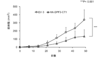
- 4A and 4B show the results of functional analysis of a dominant negative mutant HA-DPF3-CT1 with an HA tag. The effect of ectopically expressed HA-DPF3-CT1 on cell proliferation of A549 cells and HeLaS3 cells in monolayer culture (A) and soft agar (B) is shown.
- FIG. 4 (C) and (D) show the expression of HA-DPF3-CT1 under the same conditions (C), and the catalytic subunit of SWI / SNF complex and HA-DPF3-CT1, HA-DPF3a and HA-DPF3b The result (D) of co-immunoprecipitation experiment with this is shown.
- FIG. 5 shows the ectopic expression and biological activity of DPF3-CT1-derived deletion mutants with HA tag in A549 cells.
- FIG. 6 shows the ectopic expression effect of HA-DPF3-CT1 on tumorigenicity in a mouse Xenograft model using A549 cells.
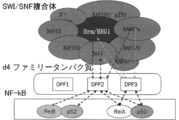
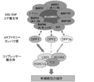
- FIG. 7 shows a schematic diagram of the interaction between the SWI / SNF complex, d4 family proteins, and NF- ⁇ B.
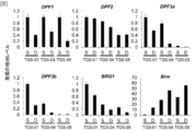
- FIG. 8 (A) shows the mRNA encoding the SWI / SNF complex and the subunit of the protein that binds strongly to the complex in three types of GICs from different patients and the corresponding differentiated cells derived from them. The result of having measured the change of expression by RT-PCR method is shown.
- FIG. 8B shows a bar graph of the results of DPF1, DPF2, DPF3a, DPF3b, BRG1, and Brm mRNA among the results of (A).
- FIG. 9 (A) shows the knockdown effect of d4 family proteins, BRG1, and Brm on the sphere-forming activity of TGS-01 cells.
- FIG. 9 (B) shows that the gene knockdown effect on the sphere-forming activity of FIG. 9 (A) was rescued by co-expression of BRG1, DPF1 and DPF3a cDNAs.
- FIG. 9C shows that TGS-01 cells ectopically expressing DPF3a were sensitive to DPF1 knockdown, whereas TGS-01 cells ectopically expressing DPF1 were sensitive to DPF3 knockdown. It shows that there was resistance.
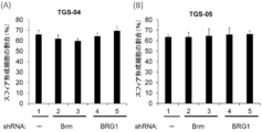
- FIGS. 10 (A) and (B) show the effect of BRG1 or Brm knockdown on the sphere-forming activity of TGS-04 cells (A) and TGS-05 cells (B).
- FIG. 10 (A) and (B) show the effect of BRG1 or Brm knockdown on the sphere-forming activity of TGS-04 cells (A) and TGS-05 cells (B).
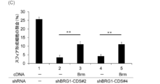
- FIG. 10 shows that the knockdown effect of BRG in TGS-01 cells was partially rescued by co-expression of Brm cDNA.
- FIG. 11 shows Kaplan-Meier survival curves of nude mice transplanted orthotopically with TGS-01 cells.
- FIGS. 12A and 12B show the results of detecting a larger SWI / SNF complex containing TLX and LSD1 / RCOR2 in TGS-01 cells by Western blotting for immunoprecipitates.
- FIGS. 12 (C) and (D) show that Western blotting of immunoprecipitates was performed using a lysate of TGS-01 cells ectopically expressing DPF1, DPF2 or DPF3a to which a FLAG tag was added.
- FIG. 13 shows the results of detection by the PLA method that the SWI / SNF complex, TLX, and LSD1 / RCOR2 are localized proximally in TGS-01 cells.
- FIG. 14 shows the results of detecting changes in the signal (dot number) of SWI / SNF complex and TLX (or LSD1 / RCOR2) located proximally in TGS-01 cells by DPF1 knockdown by PLA.
- FIG. 15 shows a schematic diagram of the interaction between a SWI / SNF complex in GIC, a d4 family protein, and a corepressor complex including TLX.
- FIG. 16 shows the results of examination of shRNA against d4 family genes suitable for GIC stem cell loss.
- the present invention provides an inhibitor that inhibits gene expression control by a corepressor complex containing NF- ⁇ B or TLX in a SWI / SNF complex-dependent manner.
- the inhibitor of the present invention inhibits gene expression control by a corepressor complex comprising SWI / SNF complex-dependent NF- ⁇ B or TLX both in vitro and in vivo.
- SWI / SNF complex refers to a complex composed of various proteins having a molecular weight of about 2 M Dalton.
- specific examples of the SWI / SNF complex mainly include human and other mammalian Brm (GenBank accession number X72889) or BRG1 (GenBank accession number U29175) as a DNA-dependent ATPase subunit. By complex, it may include yeast SWI / SNF or other homologs in Drosophila Brahma in humans and mammals.
- SWI / SNF complexes in mammals such as humans are roughly classified into two types, those containing Brm and those containing BRG1.
- SWI / SNF complex components include, but are not limited to, INI1, BAF250, BAF60a, BAF60b, BAF60c, BAF170, BAF57, BAF155, BAF53, ⁇ -actin in the case of mammals ( Vignali, M. et al. (2000) Mol. Cell. Biol. 20, 1899-1910).
- An example of the components of the SWI / SNF complex is shown below. (Posted with some modifications from “Kyoto Medical University Journal 124 (12), 825-838, 2015”)
- the SWI / SNF complex has a chromatin remodeling function by DNA-dependent ATPase.
- Chromatin remodeling refers to the process of changing the chromatin structure of DNA.
- the chromatin structure is mainly composed of DNA and histones, and 165 bp (about 2 rounds) of DNA around histone octamers formed by four types of histone molecules (H2A, H2B, H3 and H4).
- a nucleosome formed by winding is used as a basic unit structure, and it refers to a structure in which this nucleosome is folded and compressed. This highly folded DNA hinders transcription, replication, repair, recombination, and the like.
- the SWI / SNF complex has a function of changing the chromatin structure by moving and / or removing nucleosomes using the energy of ATP hydrolysis. At this time, it plays an important role in gene expression and / or regulation together with histone modifying enzymes (histone acetylase, deacetylase, phosphorylase, methylase, etc.). For example, when mRNA is transcribed from DNA, the SWI / SNF complex changes the chromatin structure by the above-mentioned chromatin remodeling and exposes the target DNA sequence, allowing transcriptional regulators to access the DNA. Become.
- SWI / SNF complex-dependent transcriptional regulation by NF- ⁇ B or a corepressor complex refers, in one embodiment, to an intracellular SWI / SNF complex and a d4 family protein, NF ⁇ It means that a corepressor complex containing ⁇ B (usually composed of a dimer of NF- ⁇ B family protein subunits) or TLX exerts transcriptional control activity by forming a huge complex.
- SWI / SNF complex can bind to various transcription control factors via a core unit or subunit.
- BRG1 binds to glucocorticoid receptor and estrogen receptor (Peterson, CL et al. (2000) urr Curr. Opin. Gen. Dev. 10, 187-192), and Ini1 binds c-Myc ( Yudkovsky, N. et al. (1999). Genes Dev. 13, 369 2369-2374), c-Fos and c-Jun bind to BAF60a (Ito, T. et al. (2001) J. Biol. Chem. 276: 2852-2857) is known.
- dPF family proteins such as DPF3a / b and DPF2 function as adapters and have been reported to bind to NF- ⁇ B (Rel A / p50, Rel B / p52) (Tando, T et al., J Biol. Chem. 285, 21951-21960 (2010); Non-patent documents 1 to 3).
- a SWI / SNF complex is newly bound to a corepressor complex containing TLX using a d4 family protein as an adapter.
- the SWI / SNF complex-dependent transcriptional control in the present invention specifically targets transcriptional control by a large complex consisting of a transcriptional regulator of either a d4 family protein and a corepressor complex containing NF- ⁇ B or TLX.
- Means for detecting the interaction between the SWI / SNF complex and the target transcriptional regulator include means for detecting the binding between the SWI / SNF complex and the target transcriptional regulator, the SWI / SNF complex
- a means of detecting the proximity of the transcription factor to the target and the transcriptional activity (up-regulation or down-regulation) of the target transcription factor in the presence of the SWI / SNF complex Examples include, but are not limited to, and various other means known to those skilled in the art may be used for detection. Specific detection means include, for example, immunoprecipitation, density gradient centrifugation, gel shift assay, ChIP (chromatin immunoprecipitation) assay, far western blot, yeast two-hybrid method, and the like.
- Means for detecting the proximity of the SWI / SNF complex and the transcriptional regulator of interest include Proximity Ligation assay (PLA) (methods described in the examples herein) and fluorescence resonance. Examples thereof include an energy transfer (FRET) method and a bioluminescence resonance energy transfer (BRET) method. Reporter assay using reporter (luciferase, green fluorescent protein, alkaline phosphatase, etc.) as a means of detecting the transcription activity (up-regulation or down-regulation) of the transcription factor of interest in the presence of SWI / SNF complex, Examples include, but are not limited to, quantitative RT-PCR (qRT-PCR), microarray, and Northern hybridization.
- suppression of gene expression control by a SWI / SNF complex-dependent transcriptional regulatory factor is the coexistence of the SWI / SNF complex and the transcriptional regulatory factor, and increases or decreases the expression level or activity of one.
- various cells such as proliferation, differentiation, anchorage independence, cell surface molecular changes, or host cell growth arrest or delay, dormancy, apoptosis, etc. It also means causing changes in the level.
- the SWI / SNF complex can cause changes in the host cell through interaction with various transcriptional regulatory factors and the like in the cell.
- NF- ⁇ B also called nuclear factor ⁇ B
- nuclear factor ⁇ B was found as a transcription factor that binds to an enhancer region necessary for the immunoglobulin ⁇ light chain gene to be expressed specifically in mature B cells.
- RelA p65
- RelB c-Rel
- NF- ⁇ B1 p105 / p50
- NF- ⁇ B2 p100 / p52
- intracellular functions such as immunity, inflammation, development, cell proliferation, and apoptosis, and also induces the expression of many exogenous viral genomic genes.
- NF- ⁇ B needs to activate the promoters of the target genes with selectivity, but there are many unclear points about the details of the mechanism. Thus, for example, it is important to elucidate the molecular mechanism that gives promoter selectivity to each NF- ⁇ B dimer at the most downstream of signal transduction. It is thought that the nearby chromatin environment has a great influence on the expression control of the target gene of NF- ⁇ B.
- NF- ⁇ B is associated with many diseases, such as acute and chronic inflammatory reactions, Crohn's disease, rheumatoid arthritis, cancer, septic shock, human immunodeficiency virus (HIV), cytomegalovirus, etc. Viral diseases, chronic obstructive pulmonary disease (COPD), cystic fibrosis, neurodegenerative diseases and the like.
- diseases such as acute and chronic inflammatory reactions, Crohn's disease, rheumatoid arthritis, cancer, septic shock, human immunodeficiency virus (HIV), cytomegalovirus, etc.
- HIV human immunodeficiency virus
- COPD chronic obstructive pulmonary disease
- cystic fibrosis neurodegenerative diseases and the like.
- I ⁇ B ⁇ undergoes phosphorylation by the I ⁇ B kinase (IKK) complex, etc., leading to the transfer of NF- ⁇ B into the cell nucleus.
- IKK I ⁇ B kinase
- NF- ⁇ B has many cytokines and growth factors (eg, IL-2, IL-8, IFN- ⁇ , M-CSF, G-CSF, VEGF), as well as various transcription factors and signal regulators (eg, I ⁇ B ⁇ , Induces transcription of IRF-1 and IRF-2).
- the function of NF- ⁇ B in epithelial cancer is based on the ability to express or produce a protein selected from growth factors IL-6, IL-8 and TNF- ⁇ , and the cell adhesion molecule ICAM1.
- Other known NF- ⁇ B-dependent proteins can also be used as indicators. Examples of other indicators include, but are not limited to, confirmation of cell proliferation ability and apoptosis inducing ability.
- the present invention provides an inhibitor that inhibits gene expression control by a corepressor complex.
- the corepressor complex refers to a factor belonging to a cofactor.
- a cofactor refers to a factor that promotes or suppresses transcription by providing a bridge between a transcriptional regulatory factor and a basic transcription factor, and includes coactivator complexes and corepressor complexes.
- the various subunits constituting the coactivator complex may include those having a function of promoting transcription by modifying histones (for example, histone acetyltransferase: HAT), while constituting the corepressor complex.
- Subunits may include those having a function of removing transcription of histones to suppress transcription (for example, histone deacetylase (HDAC), histone demethylase, etc.) 2015-147794, Karin Meier & Alexander Brehm (2014), Epigenetics, 9:11, 1485-1495, DOI: 10.4161 / 15592294.2014.971580).
- Some corepressor complexes contain at least one molecule selected from TLX, RCOR2, and LSD1 (Yang et al., Stem Cells. 2011 29: 791-801). These coactivator complexes and corepressor complexes can be combined with the SWI / SNF complexes described above to form larger complexes.
- a person skilled in the art such as Western blot using an antibody against a constituent subunit (such as anti-HDAC antibody), detection means such as immunoprecipitation, ChIP assay, means for detecting histone deacetylase activity, etc. Any known means can be used.
- the nuclear receptor family is generally a receptor for a ligand such as steroid, thyroid hormone, female hormone, male hormone, retinoic acid, retinoid, vitamin D, glucocorticoid, mineral corticoid, and the like.
- a ligand such as steroid, thyroid hormone, female hormone, male hormone, retinoic acid, retinoid, vitamin D, glucocorticoid, mineral corticoid, and the like.
- Nuclear receptor family proteins have a similar domain structure including a DNA-binding domain and a ligand-binding domain, and are responsible for functions such as metabolism, homeostasis, differentiation, growth, development, aging, and reproduction of the living body.
- TLX is a member of the nuclear receptor family and is also known as tailless (Monaghan et al., 1995, Development 121: 839-853; Pigigni et al, 1990, Cell 62: 151-163). This TLX is expressed in Drosophila (Pignoniet al, 1990), mouse (Tu et al., 1994, Nature 370: 375-379; Monaghan et al., 1995), Xenopus (Holleman et al., 1996, GenBank accession number U67886), chicken (Yu et al. 1994), humans (GenBank accession number Y13276; Jackson et al., Royal Free Hospital, London).
- TLX is an orphan receptor whose ligand is unknown. TLX is expressed in the ends of the developing Drosophila embryo (Pignoni et al, 1990) and in the developing chicken and mouse forebrain (Yu et al, 1994; Monaghan et al, 1995). Knockout mice of the TLX gene have been shown to have a phenotype such as a lack of limbic system (Monaghan et al., 1997, Nature 390: 515-517). Homozygous mutant mice can grow at birth, but the olfactory brain and marginal structures, including the olfactory cortex, inferior olfactory cortex, and internal olfactory cortex, tonsils and dentate gyrus are small in size.
- TLX function Loss of TLX function leads to the absence of all encephalonomic neuroblasts (Younossi-Hartenstein et al., 1997, Developmental Biology 182: 270-283).
- Abnormal TLX functions include depression, anxiety, aggressive disease, stroke, multiple sclerosis, Alzheimer's disease, Parkinson's disease, neuropathic pain, inflammatory diseases of the central nervous system and other neurodegenerative diseases Problems with the eyes and / or central nervous system can occur, such as central nervous system disease.
- TLX forms a complex with the corepressor atrophin 1 (ATN1) (Zhang, CL. Et al., 2006, Genes and Dev. 20: 1308-1320), histone deacetylase complex 1 (HDAC1) Recruiting (Sun, GQ. Et al., PNAS, 2007, 104, 39, pp.15282-15287), working with LSD1 and BCL11A (Corso-Diaz, X et al., BMC Genomics, 2016 , 17: 832).
- ATN1 corepressor atrophin 1
- HDAC1 histone deacetylase complex 1
- d4 family proteins are known to function as transcription coupling factors (cofactors).
- This d4 family protein includes DPF1, DPF2, DPF3a, and DPF3b. These family proteins have an N-terminal region containing a nuclear localization signal, a central C2H2-type kruppel-like zinc finger motif, and a d4 domain containing one or two PHD zinc finger motifs (PHD1, PHD2) on the C-terminal side.
- PHD1, PHD2 PHD zinc finger motifs
- DPF1 (also referred to as Neud4, BAF45b) has been reported in humans as isoforms a, b, d, etc., and human isoform a has 414 amino acids (GenBank accession number: NP_001128627; Chestkov AV et al., Genomics). 36 (1), 174-177 (1996)).
- the corresponding cDNA sequence is NM_001135155 (SEQ ID NO: 74), which is Homo sapiens double PHD fers 1 (DPF1), transcript variant 1, mRNA.
- DPF1 and the following DPF3a / b have been reported to bind to SWI / SNF complexes in neurons in a manner specific for differentiation (Lessard, J. et al., 2007, Neuron 55, 201-215). ).
- DPF2 (also called requiem, BAF45d) has been reported in humans as isoforms 1 and 2, etc., and human isoform 1 has 391 amino acids (GenBank accession number: NP_006259.1).
- the corresponding cDNA sequence is NM_006268.4 (SEQ ID NO: 75): Homo sapiens double PHD fers 2 (DPF2), transcript variant 1, mRNA.
- DPF2 and DPF3a / b below function as an adapter molecule between NF- ⁇ B and SWI / SNF complex and activate transcription of NF- ⁇ B in most of the downstream of the non-standard pathway of NF- ⁇ B (Tando et al., 2010, J. BIOL. CHEM., 285, 29, pp. 21951-21960, Non-Patent Document 1).
- DPF3 (also called Cerd4, BAF45c) has two splicing variants (DPF3a and DPF3b), and their products are different on the C-terminal side.
- Human DPF3a and DPF3b have 357 and 378 amino acids, respectively (GenBank accession numbers: NP_036206.3; Liu, H. et al., Int J Clin Exp Pathol 7 (7), 3966-3974 (2014), and GenBank accession Session number: Q92784.3; Lange, M. et al., 2008, Genes Dev.22, 2370-2384).
- the corresponding cDNA sequences are NM_012074.4 (SEQ ID NO: 76) for DPF3a, Homo sapiens D4, zinc and double PHD fingers, family 3 (DPF3), transcript variant 1, mRNA, and DPF3b is NM_001280542.1 (SEQ ID NO: 77) Homo sapiens D4, zinc and double PHD fingers, family 3 (DPF3), transcript variant 2, mRNA.
- DPF3b protein has all the characteristics of other d4 family members, such as binding activity to either methylated or acetylated residues of histones H3 and H4 via two PHD fingers, but DPF3a One deficient PHD finger lacks d4 domain ends within the PHD domain and lacks the ability to bind these modified histones.
- the adapter function means a function responsible for the interaction (binding) between the SWI / SNF complex and a transcriptional regulatory factor, and the provision of a new activity related to transcriptional control.
- the expression of one or more components of the SWI / SNF complex has been reduced or eliminated. Reducing existing functional SWI / SNF complex levels was performed (Patent Documents 1 and 2).
- the gene groups targeted by the various transcription factors known to interact with the SWI / SNF complex are not necessarily limited to those that are controlled depending on the SWI / SNF complex. It is also known that it is controlled by this transcription factor without SWI / SNF complex.
- SWI / SNF complex for example, a corepressor complex including NF- ⁇ B and TLX
- a new activity related to transcription control is provided.
- SWI / SNF complex-dependent transcription control is achieved by inhibiting the adapter function of a substance (protein) that functions as an adapter between the SWI / SNF complex and various transcription factors.
- a method for reducing the action of factors is mentioned.
- the inhibition of the adapter function can be appropriately detected and quantified by using the above-described means for detecting a change in interaction or a specific transcription control activity.
- an inhibitory protein that inhibits the function is also included.
- the present invention provides means for inhibiting adapter function between a SWI / SNF complex and a specific transcriptional regulatory factor using a dominant negative mutant of the adapter protein.
- the present invention provides means for inhibiting adapter function between a SWI / SNF complex and a specific transcriptional regulatory factor by inhibiting gene expression of the adapter protein as a means for reducing adapter function. provide.
- the “dominant negative mutant for the d4 family protein” of the present invention competes for binding between the d4 family protein and the SWI / SNF complex and / or the d4 family protein and the transcriptional regulator in the cell, and the wild type d4
- the d4 family protein has an adapter function between a SWI / SNF complex and a transcriptional regulatory factor (NF- ⁇ B, corepressor complex) (Non-patent Document 1, etc.).
- dominant negative mutants for d4 family proteins can be prepared by removing sites other than the binding sites between d4 family proteins and SWI / SNF complexes and / or binding sites between d4 family proteins and transcription factors There is sex.
- the dominant negative mutant of the present invention "binds to the SWI / SNF complex” means that when this mutant is present in the cell nucleus, it can bind to the endogenous SWI / SNF complex to form a complex. means.
- the dominant negative mutant of the present invention “binds to NF- ⁇ B and / or the corepressor complex” means that when the mutant is present in the cytoplasm or cell nucleus, preferably in the cell nucleus, it is endogenous. NF- ⁇ B, which can bind to the corepressor complex to form a complex.
- the dominant negative mutant of the present invention forms a complex with a SWI / SNF complex and / or NF- ⁇ B, a corepressor complex in a cell, as a result, the SWI / SNF of the NF- ⁇ B, the corepressor complex. SNF complex-dependent transcriptional regulation can be inhibited.
- genes that are transcriptionally regulated by NF- ⁇ B in a SWI / SNF complex-dependent manner such as IL-6 and IL-8 genes Transcription is significantly inhibited by the dominant negative mutant of the present invention, but transcription of genes that are transcriptionally regulated by NF- ⁇ B in a SWI / SNF complex-independent manner, such as TNF- ⁇ gene and ICAM1 gene, are not significantly affected by dominant negative mutants.
- the dominant negative mutant for the d4 family protein according to the present invention is, for example, a polypeptide at the N-terminal portion of the d4 family protein, and includes a nuclear translocation signal (NLS) of the d4 family protein.
- the dominant negative mutant of the present invention is a polypeptide comprising any one of the amino acid sequences of SEQ ID NOS: 1-7 provided herein, any of SEQ ID NOs: 1-7.
- the ability to compete with the binding between, Rabbi in / or alter the transcriptional activity of the transcriptional regulatory factor, etc. (preferably reduce or inhibit) is to function, has a detection function that can be quantified by means of measuring change in the interaction described above.
- the polypeptide variant having each identity described above is 10% or more, preferably 20% or more, preferably 30% or more, 40% or more for at least one of the above functions as compared to the original polypeptide variant. %, 50%, 60%, 70%, 80%, or 90% or more specific activity.
- the dominant negative mutant for the d4 family protein of the present invention may comprise only a more limited polypeptide of the functional part of the d4 family protein.
- the dominant negative mutant of the present invention has a more limited portion of the amino acid sequence of SEQ ID NOs: 1 to 3, for example, the sequence of amino acid residues 9 to 84 of SEQ ID NO: 3 (SEQ ID NO: 4), Includes the sequence of amino acid residues 33 to 84 (SEQ ID NO: 5), the sequence of amino acid residues 40 to 84 (SEQ ID NO: 6), or the sequence of amino acid residues 53 to 84 (SEQ ID NO: 7)
- a peptide, or an amino acid sequence derived from SEQ ID NO: 1 or 2 corresponding to these amino acid sequences, which inhibits the adapter function between the SWI / SNF complex and NF- ⁇ B, and is dependent on the SWI / SNF complex-dependent NF Peptides that reduce the transcriptional regulatory function of - ⁇ B may also be included
- the corresponding positions of the amino acid sequences of SEQ ID NOs: 1 to 3 can be determined based on the amino acid sequence alignment of the d4 family protein (eg, FIG. 2B).
- the dominant negative mutant of the present invention further comprises an amino acid sequence of the above-mentioned limited portion, 85% or more, 90% or more, 91% or more, 92% or more, 93% or more, 94% or more, 95% or more, 96 %, 97% or more, or a polypeptide comprising an amino acid sequence having 98% or more identity, comprising an adapter function between a SWI / SNF complex and a transcriptional regulatory factor such as NF- ⁇ B, Variants that alter (preferably reduce or inhibit) the transcriptional activity of transcriptional regulators may also be included.
- amino acid residues that can be substituted for each other in the protein (polypeptide) of the present invention are shown below.
- Amino acid residues contained in the same group can be substituted for each other.
- Group A leucine, isoleucine, norleucine, valine, norvaline, alanine, 2-aminobutanoic acid, methionine, o-methylserine, t-butylglycine, t-butylalanine, cyclohexylalanine
- Group B aspartic acid, glutamic acid, isoaspartic acid, isoglutamic acid, 2-aminoadipic acid, 2-aminosuberic acid
- Group C asparagine, glutamine
- Group D lysine, arginine, ornithine, 2,4-diaminobutanoic acid, 2,3-diaminopropionic acid
- Group E proline, 3-hydroxyproline, 4-hydroxyproline
- Group F se
- protein and “polypeptide” are used interchangeably, and are intended to be polymers of amino acids.
- the polypeptide used in the present specification has the N-terminus (amino terminus) at the left end and the C-terminus (carboxyl terminus) at the right end in accordance with the convention of peptide designation.
- the partial peptide of the polypeptide of the present invention (in the present specification, sometimes abbreviated as the partial peptide of the present invention) is the partial peptide of the polypeptide of the present invention described above, preferably the polypeptide of the present invention described above. It has the same properties as a peptide.
- the dominant negative mutant of the d4 family protein according to the present invention may be in the form of a polynucleotide that allows the polypeptide of interest to be expressed in a cell.
- a polynucleotide includes the amino acid sequence of SEQ ID NOs: 1 to 3, the amino acid sequence of SEQ ID NOs: 1 to 3 and the amino acid sequence having the above-mentioned predetermined identity, or the amino acid sequence of SEQ ID NOs: 1 to 3
- a polynucleotide encoding a dominant negative mutant having an amino acid sequence of a specific portion is exemplified.
- polynucleotides of the present invention include, for example, the polynucleotides of SEQ ID NOs: 10 to 13, 80% or more, 81% or more, 82% or more, 83% or more, 84% or more, 85% or more, 86% with these polynucleotides Above 87%, above 88%, above 89%, above 90%, above 91%, above 92%, above 93%, above 94%, above 95%, above 96%, above 97%, above 98%, A polynucleotide having an identity of 99% or more, 99.1% or more, 99.2% or more, 99.3% or more, 99.4% or more, 99.5% or more, 99.6% or more, 99.7% or more, 99.8% or more, 99.9% or more, d4 Mention may be made of those encoding dominant negative variants of family proteins.
- DPF1 is 26 amino acid residues that are not in the conserved region (78 nucleotides, nucleotides underlined in SEQ ID NO: 10) long.
- the CT1 peptide derived from DPF1 may or may not contain the 26 amino acid residues.
- the present invention also relates to a polynucleotide that hybridizes with a polynucleotide comprising a sequence complementary to the polynucleotide sequence of SEQ ID NOs: 10 to 13 under a stringent condition and encodes a dominant negative mutant of a d4 family protein.
- a polynucleotide that hybridizes under stringent conditions means a colony hybridization method using as a probe all or part of a polynucleotide that is complementary to the polynucleotide sequence of SEQ ID NOs: 10 to 13.
- a polynucleotide for example, DNA obtained by using a plaque hybridization method or a Southern hybridization method.
- stringent conditions may be any of low stringency conditions, moderate stringency conditions, and high stringency conditions.
- Low stringent conditions are, for example, conditions of 5 ⁇ SSC, 5 ⁇ Denhardt's solution, 0.5% SDS, 50% formamide, 32 ° C.
- Medium stringent conditions are, for example, conditions of 5 ⁇ SSC, 5 ⁇ Denhardt's solution, 0.5% SDS, 50% formamide, and 42 ° C.
- “High stringent conditions” are, for example, conditions of 5 ⁇ SSC, 5 ⁇ Denhardt's solution, 0.5% SDS, 50% formamide, 50 ° C. Under these conditions, it can be expected that a polynucleotide having high homology (eg, DNA) can be efficiently obtained as the temperature is increased.
- a polynucleotide having high homology eg, DNA
- multiple factors such as temperature, probe concentration, probe length, ionic strength, time, and salt concentration can be considered as factors that affect hybridization stringency. Those skilled in the art will select these factors as appropriate. It is possible to achieve similar stringency.
- amino acid substitution or nucleotide substitution appropriately used in the present invention can be prepared using various gene manipulation techniques known to those skilled in the art. For such genetic manipulation procedures, see, for example, Molecular Cloning 3rd Edition, J.Sambrook et al., Cold Spring Harbor Lab. Press. 2001, Current Protocols in Molecular Biology, John Wiley & Sons 1987-1997, etc. Can do.
- BLAST and Gapped BLAST programs When using BLAST and Gapped BLAST programs, the default parameters of each program are used. In the present specification, the higher the sequence identity or homology described above with respect to a specific amino acid sequence or polynucleotide sequence, the better. Further, in the present specification, a polypeptide or polynucleotide having a specific identity or homology with a specific amino acid sequence or polynucleotide sequence functions equally as compared with the polypeptide or polynucleotide of the original sequence. What is possible is intended. In this specification, being able to function equally means that the function is 10% or more, preferably 20% or more, 30% or more, 40% or more, 50% or more, 60% or more when compared with the original sequence. A polypeptide or polynucleotide that exhibits a specific activity of greater than 70%, greater than 80%, greater than 90%, or greater.
- nucleic acid As used herein, the term “polynucleotide” is used interchangeably with “nucleic acid”, “gene” or “nucleic acid molecule” and is intended to be a polymer of nucleotides.
- nucleotide sequence is used interchangeably with “nucleic acid sequence” or “base sequence” and refers to the sequence of deoxyribonucleotides (abbreviated A, G, C, and T). As shown.
- a polynucleotide comprising a nucleotide sequence of SEQ ID NO: 1 or a fragment thereof intends a polynucleotide comprising the sequence represented by each deoxynucleotide A, G, C and / or T of SEQ ID NO: 1 or a fragment thereof. Is done.
- polynucleotide may exist in the form of DNA (for example, cDNA or genomic DNA), but may be in the form of RNA (for example, mRNA) in some cases.
- RNA for example, mRNA
- Each polynucleotide used herein can be double-stranded or single-stranded DNA.
- single-stranded DNA or RNA it may be the coding strand (also known as the sense strand) or the non-coding strand (also known as the antisense strand).
- the polynucleotide that suppresses the expression of the gene encoding the d4 family protein of the present invention is not particularly limited, and is appropriately selected from known nucleotides used for suppressing gene expression according to the purpose.
- siRNA, shRNA, miRNA, antisense oligonucleotide and the like that can suppress the expression of the d4 family gene can be mentioned.
- siRNA or shRNA is desirable because it has a high effect of suppressing the expression of the d4 family gene.
- These polynucleotides can suppress the expression of d4 family proteins when introduced into target cells in the form of nucleotides or incorporated into vectors known in the art.
- gene expression refers to the process of expressing mRNA under the control of a promoter sequence using a genomic gene as a template (transcription) and / or the synthesis of a protein using this mRNA as a template. Process (translation). Therefore, “inhibiting the expression of a d4 family gene” of the present invention means inhibiting the transcription process or translation process of a d4 family gene (preferably endogenous).
- an antisense oligonucleotide that suppresses gene expression refers to an oligonucleotide having a nucleotide sequence of an antisense strand relative to the sense strand of a gene.
- Antisense technology is known as a method for suppressing the expression of a specific endogenous gene, and is described in various literatures (for example, Hirashima and Inoue: Shinsei Kagaku Kougaku Kenkyu 2 Nucleic Acid IV). Replication and expression of genes (edited by the Japanese Biochemical Society, Tokyo Kagaku Dojin) pp.319-347, 1993).
- the sequence of the antisense oligonucleotide is preferably a sequence complementary to the endogenous gene or a part thereof, but may not be completely complementary as long as the expression of the gene can be effectively suppressed.
- the sequence of the antisense oligonucleotide of the invention may comprise one to several nucleotides deleted, substituted, inserted and / or added to the endogenous gene or part thereof.
- the length of the antisense oligonucleotide may be 11-20 bases long, 12-19 bases long, 13-18 bases long, 14-17 bases long or 14-16 bases long.
- antisense oligonucleotides can include natural (unmodified) nucleotides (deoxyribonucleotides, ribonucleotides, or both) and / or non-natural (modified) nucleotides.
- RNA that suppresses gene expression and plays RNAi refers to RNA that suppresses gene expression by RNA interference (RNAi).
- RNAi refers to a phenomenon in which the expression of a target endogenous gene or introduced foreign gene is suppressed by introducing a double-stranded RNA having the same or similar sequence as the target gene sequence into the cell.
- examples of the RNA used here include siRNA (small ⁇ ⁇ interfering RNA) that is a double-stranded RNA that plays RNAi having a length of 21 to 25 bases, or cleaved with Dicer (RNase III enzyme) in a cell to produce the siRNA.
- shRNA short hairpin RNA
- the loop sequence is not particularly limited as long as the double-stranded RNA having RNAi can be used as a single-stranded RNA that forms a hairpin loop structure.
- RNA that plays RNAi can be locally delivered to a desired site by a delivery system such as a liposome, or can be expressed at a desired site using an expression vector that generates the above siRNA or shRNA. .
- RNA (RNA, siRNA, shRNA) preparation method, usage method, etc. can be designed based on the sequence of the target gene using known techniques described in many literatures (Special Table 2002- No. 516062; U.S. Publication No.
- RNA that suppresses the expression of d4 family genes and plays RNAi” of the present invention is specific to, for example, the polynucleotide sequence of any of SEQ ID NOs: 74 to 77.
- SiRNA and shRNA that inhibit d4 family gene expression can be designed based on a sequence at a position specific to the d4 family gene using a sequence database such as BLAST.
- the RNA of the present invention may be used alone or in combination of two or more.
- RNA of the present invention may be, for example, an shRNA that is specific for the polynucleotide sequences shown in SEQ ID NOs: 74 to 77 and inhibits the expression of d4 family genes.
- shRNA comprising at least one polynucleotide sequence selected from the group consisting of SEQ ID NOs: 14-40.
- Such shRNA is converted into siRNA by an intracellular enzyme (such as Dicer).
- RNA that inhibits expression from d4 family genes includes, for example, any sequence selected from the group consisting of SEQ ID NOs: 41 to 73, and deletions, substitutions, insertions, and It may also contain 1 to several added nucleotides. Further, siRNA having a sequence having at least 90% identity to the above sequence may be used. For example, if the siRNA is double stranded with overhanging ends (eg, 2 nucleotides), it may comprise a portion that is complementary to one strand and a few (eg, 2) nucleotide portions that are not complementary.
- the “RNA that inhibits expression from the d4 family gene” of the present invention is a combination of the sequence of SEQ ID NO: 41 and the sequence of SEQ ID NO: 42, or a combination of the sequence of SEQ ID NO: 43 and the sequence of SEQ ID NO: 44.
- the nucleotide sequence may contain 1 to several nucleotides deleted, substituted, inserted and
- the RNA of the present invention may contain non-natural nucleotides such as peptide nucleic acid (PNA), locked nucleic acid (LNA), and arabino nucleic acid (FANA) for the purpose of improving in vivo stability. Yes (special table 2010-521973 etc.).
- the siRNA of the present invention is a phosphorothioate, alkylphosphonate, phosphorodithioate, alkylphosphonothioate, phosphoramidate, carbamate, carbonate, phosphate triester, acetamidate, carboxy as a nucleotide modification.
- Known modified nucleotides selected from methyl esters and combinations thereof may be included.
- the present invention provides a pharmaceutical composition for the treatment of cancer such as epithelial cancer.
- epithelial cancer refers to cancerous epithelial cells.
- the epithelium refers to a cell layer covering the outer surface of an animal body or the inner surfaces of various organs of a body cavity. The epithelium has no blood vessels, and each cell is clustered and dense.
- epithelium examples include, but are not limited to, a coated (lid) epithelium covering the surface of the skin or mucous membrane, a glandular epithelium that forms a parenchymal cell of the gland duct, a linear (ciliary) hair epithelium, a pigment epithelium .
- the epithelial cancer according to the present invention includes both malignant and benign.
- epithelial cancer examples include squamous cell carcinoma, epithelial species (including epithelial tumors, including papilloma, adenoma, cyst, etc.), neuroepithelioma, epithelial malignant tumor (hepatocellular carcinoma, basal cell Cancer, skin cancer, etc.) and adenocarcinoma in situ.
- the present invention provides a pharmaceutical composition for the treatment of glioma (glioma), particularly glioblastoma (glioblastoma).
- Glioblastoma Intiating blastCell Glioblastoma Intiating blastCell (GIC) is thought to contribute to treatment resistance and tumor recurrence in glioblastoma, a fatal and important brain tumor in adults (Singh, SK et al., Nature 432, 396-401 (2004); Bao, S. et al. Nature 444, 756-760 (2006); Chen, J. et al. Nature 488, 522-526 (2012)). Modification of chromatin structure has been shown to be an important determinant of the maintenance of GIC stemness and induction of their differentiation (Natsume, A.
- the present invention provides a pharmaceutical composition comprising a dominant negative mutant for a d4 family protein, a polynucleotide that inhibits the expression of a d4 family gene.
- a pharmaceutical composition means a pharmaceutical composition comprising only a main drug or a combination of a main drug and an excipient. If necessary, it can be combined with suitable excipients described below, for example.
- the pharmaceutical composition refers to various dosage forms, such as oral, parenteral (intravenous), muscle, oral mucosa, rectum, vagina, transdermal, nasal or inhalation. Examples include, but are not limited to, dosage forms.
- the dosage of the pharmaceutical composition of the present invention is, for example, 1 to 5000 mg per day for an adult (for example, 60 kg body weight), preferably 10 to 1000 mg, 10 to 800 mg, 10 to 600 mg, 10 to 500 mg, 10 to 400 mg, 10 to The dose may be in the range of 300 mg, 10-200 mg, 10-100 mg, 10-75 mg, 10-50 mg and the like. These daily doses may be administered in a plurality of times, for example, divided into two times or more, three times or more.
- the pharmaceutical composition of the present invention comprises a polynucleotide encoding a dominant negative mutant for a d4 family protein, or a polynucleotide encoding a polynucleotide that inhibits expression of the d4 family.
- a polynucleotide can be designed in combination with an appropriate expression cassette containing a promoter and the like so that the inhibitor of interest can be expressed after being delivered into the target cell.
- expression cassettes can be delivered to target cells using, for example, a suitable viral vector.
- viral vectors are suitable for treating diseases because they can introduce genes into target cells with high efficiency.
- viral vectors examples include, but are not limited to, retrovirus vectors, adenovirus vectors, adeno-associated virus vectors, herpes simplex virus vectors, and the like (Miller, AD et al). (1991) J. Virol 65, 2220-2224; Miyake, S. et al. (1994) Proc. Natl. Acad. Sci. USA, 91, 8802-8806; Samulski, R. J. et al. 1989) J. Virol. 63, 3822-3828; Special Table 2004-528836).
- the introduction of a foreign gene in the present invention is preferably performed via a retrovirus vector or a herpes virus vector.
- the retrovirus vector includes a lentivirus vector.
- retrovirus vectors include ecotropic virus vectors (Kitamura, T. et al. (1995) Proc. Natl. Acad. Sci. USA.92, 9146-9150), amphotropic virus vectors, VSV-G, etc.
- Viral vectors Arai, T. et al. (1998) J. Virol. 72, 1115-1121), lentiviral vectors such as HIV vectors (Shimada, T. et al. (1991) J. Clin. Inv.88, 1043-1047) and the like.
- herpes virus vectors examples include neurotrophic herpes virus vectors, B lymphocyte tropic herpes virus vectors, T lymphocyte tropic herpes virus vectors, herpes simplex virus (HSV) vectors, and the like.
- HSV herpes simplex virus
- a herpes simplex virus vector derived from H S V-1 or HSV-2 can be used.
- any of the following viruses varicella-zoster virus (VZV), herpes virus type 6 (HSV-6), Epstein-Barr virus, cytomegalovirus, HHV6, and HHV7 can be used as a viral vector.
- the herpes virus vector may contain an inactivating mutation of one or more viral endogenous genes.
- Examples of the expression control sequence combined with the polynucleotide encoding the inhibitor of the present invention include an endogenous promoter of a target cancer cell, or an exogenous promoter such as a virus and / or an enhancer.
- viral promoters include retroviral LTRs (including viruses such as MuLV, MMTV, RSV, HIV, FIV), SV40 promoter, CMV promoter, and HPV promoter.
- the endogenous promoter of the host include EF1 ⁇ promoter and ⁇ -globin promoter.
- the dosage is, for example, 1 to 5000 mg, preferably 10 to 1000 mg, more preferably 10 to 800 mg, 10 to 10 mg per day for an adult (for example, body weight 60 kg). It can be 600 mg, 10-500 mg, 10-400 mg, 10-300 mg, 10-200 mg, 10-100 mg, 10-75 mg, 10-50 mg and the like. These daily doses may be administered in a plurality of times, for example, divided into two times or more, three times or more.
- the pharmaceutical composition of the present invention When the pharmaceutical composition of the present invention is administered, it can be administered, for example, orally, parenterally (intravenously), muscle, oral mucosa, rectum, vagina, transdermal, nasal or via inhalation, but parenterally. Is preferably administered. Intravenous administration is more preferred.
- the active ingredients of the pharmaceutical composition of the present invention may be blended singly or in combination.
- a pharmaceutically acceptable carrier or pharmaceutical additive may be blended with the active ingredient and provided in the form of a pharmaceutical preparation. it can.
- the active ingredient of the present invention is, for example, 0.1% by weight or more and 99.9% by weight or less in the preparation, preferably 90% by weight or less, 80% by weight or less, 70% by weight or less, 60% by weight or less.
- the active ingredient of the pharmaceutical composition of the present invention may be blended alone or in combination with a plurality of active ingredients.
- the total of the active ingredients of the present invention is, for example, 0.1% by weight or more and 99.9% by weight or less, preferably 90% by weight or less, 80% by weight or less, 70% by weight or less, 60% by weight in the preparation. % Or less, 50% or less, 40% or less, 30% or less, 20% or less, or 10% or less.
- Examples of the pharmaceutically acceptable carrier or additive include excipients, disintegrants, disintegration aids, binders, lubricants, coating agents, dyes, diluents, solubilizers, solubilizers, An isotonic agent, a pH adjuster, a stabilizer and the like can be used.
- preparations suitable for oral administration include powders, tablets, capsules, fine granules, granules, liquids or syrups.
- various excipients such as microcrystalline cellulose, sodium citrate, calcium carbonate, dipotassium phosphate, glycine are added to starch, preferably corn, potato or tapioca starch, and alginic acid and certain species. It can be used with various disintegrants such as silicate double salts and granulating binders such as polyvinylpyrrolidone, sucrose, gelatin, gum arabic.
- lubricants such as magnesium stearate, sodium lauryl sulfate, and talc are often very effective for tablet formation.
- the same kind of solid composition can also be used by filling gelatin capsules.
- suitable substances in this connection include lactose or lactose as well as high molecular weight polyethylene glycols.
- the active ingredient is used in combination with various sweeteners or flavors, colorants or dyes, and if necessary, an emulsifier and / or suspending agent is also used.
- an emulsifier and / or suspending agent is also used.
- preparations suitable for parenteral administration include injections and suppositories.
- parenteral administration a solution in which the active ingredient of the present invention is dissolved in either sesame oil or peanut oil or dissolved in an aqueous propylene glycol solution can be used.
- the aqueous solution should be buffered as appropriate (preferably pH 8 or more), and the liquid diluent must first be made isotonic.
- physiological saline can be used.
- the prepared aqueous solution is suitable for intravenous injection, while the oily solution is suitable for intra-articular, intramuscular and subcutaneous injection. All these solutions can be prepared aseptically by standard pharmaceutical techniques well known to those skilled in the art.
- the active ingredient of the present invention can also be administered locally such as on the skin. In this case, topical administration in the form of creams, jellies, pastes, ointments is desirable according to standard pharmaceutical practice.
- plasmid means various known genetic elements such as plasmids, phages, transposons, cosmids, chromosomes and the like.
- a plasmid can replicate in a particular host and transport gene sequences between cells.
- a plasmid contains various known nucleotides (DNA, RNA, etc.), and may be single-stranded or double-stranded, but preferably is double-stranded.
- the method for introducing a foreign gene into a cancer cell targeted in the present invention is not particularly limited, and a known gene introduction method can be used.
- a known gene introduction method such as calcium phosphate coprecipitation method, lipofection, DEAE dextran method, a method of directly injecting a DNA solution into a tissue by an injection needle or the like, introduction by gene gun, etc. can be mentioned. .
- A549, HeLaS3 and MDA-MB-231 cell culture and proliferation assays Human cancer cell line A549 (non-small cell lung cancer); HeLaS3 (cervical cancer); MDA-MB-231 (human breast cancer) were all cultured and maintained in Dulbecco's modified Eagle's medium containing 10% fetal bovine serum .
- A549 cell line and MDA-MB-231 cell line were purchased from American Type Culture Collection.
- the HeLaS3 cell line was obtained from the Medical Cell Resource Center of the Institute of Aging Medicine, Tohoku University. Cell proliferation in monolayer cultures was measured using Cell-Titer Glo (Promega, Madison, Wis.) According to manufacturer's instructions.
- the anchorage independent proliferation assay was performed as previously described (Tando, T. et al., J Biol Chem 285: 21951-60 (2010)).
- TGS-01, TGS-04 and TGS-05 A primary grade IV glioblastoma sample was obtained during surgery after obtaining approval from the institutional review board of the University of Tokyo Medical Hospital. Three independent glioma stem cells (GIC) called TGS-01, TGS-04 and TGS-05 were established from the sample. All GICs and experimental procedures using them were approved by the Ethics Committee of the University of Tokyo Hospital (24-69-250809) and the Chiba University Research Center for Fungal Medicine (# 10).
- GIC using DMEM / F12 serum-free medium (Thermo Fisher) supplemented with B27 (Thermo Fisher Scientific), 20 ng / ml EGF, and 20 ng / ml bFGF (both from PeproTech) using ultra-low adhesion dishes or flasks. Scientific).
- Dulbecco's modified Eagle medium containing 10% fetal bovine serum (FBS) was used for induction of GIC differentiation and passage of HEK293FT cells and PLAT-A cells (human embryonic kidney cells).
- Nuclear extracts are prepared from A549 cells or HeLaS3 cells using NE-PER Nuclear and Cytoplasmic Extraction Reagents (Pierce, Rockford, IL) according to the manufacturer's instructions and buffer [50 mM Tris-HCl (pH 7.5). ), 100 mM KCl, 1 mM DTT, 20% glycerol and protease inhibitor cocktail (Nacalai Tesque)]. Immunoprecipitation was performed using Dynabeads Protein G (Invitrogen) and anti-HA antibody (C29F4; Cell Signaling) according to the manufacturer's instructions.
- IL-6 shRNA The oligonucleotide pair encoding IL-6 shRNA was similarly inserted into pmU6 and digested with BamHI and EcoRI to obtain pLSP (Haraguchi T. et al., Nucleic Acids Res 37: e434 (2009)) (EV-4)
- pLSP Hardaguchi T. et al., Nucleic Acids Res 37: e434 (2009)
- EV-4 lentiviral vector plasmid for IL-6 shRNA expression was prepared by inserting between these sites.
- pFN21K Halo-tagged CMV Flexi vector Promega
- SgfI / PmeI fragment encoding the Barnane gene, a lethal gene of E.
- adapter oligonucleotide or SgfI / PmeI end sequence PFN21K-Halo and pFN21K-Halo-DPF2-CT1 were prepared by substituting the contained DNA fragment encoding DPF2-CT1.
- NheI and BamHI By double digesting these plasmids with NheI and BamHI, Halo and Halo-DPF2-CT1-containing fragments were isolated and replaced with the SpeI / BamHI fragment of pXL001 (26112, Addgene) encoding tTR-KRABi.
- -Halo (EV-2) and pXL001-Halo-DPF2-CT1 were prepared.
- pXL001-Halo-DPF3-CT1 For construction of pXL001-Halo-DPF3-CT1, a DPF3-CT1 fragment (approximately 250 bp) prepared by PCR using a primer pair containing an SgfI or PmeI site was replaced with an SgfI / PmeI digest of pXL001-Halo. did.
- synthetic DNA encoding HA sequence is inserted into BamHI / EcoRI site of pcDNA3.1 (+) (Invitrogen) to create pcDNA3.1 (+)-HA-MCS did.
- DPF3-CT1, DPF3a and DPF3b fragments obtained by PCR using a primer containing EcoRI terminal sequence and a primer containing BglII-EcoRV terminal sequence were digested with EcoRI and EcoRV, and pcDNA3.1 ( +)-HA-MCS inserted into EcoRI / EcoRV site, pcDNA3.1 (+)-HA-DPF3-CT1, pcDNA3.1 (+)-HA-DPF3a and pcDNA3.1 (+)-HA, respectively -DPF3b was produced.
- Brm and BRG1 expression plasmids (pLE-Brm-IP, pLE-BRG1-IP) can be obtained by inserting the Brm and BRG1 cDNA fragments obtained from pCAG-Brm and BRG1 expression vectors into pLE-IP (EV-6). Prepared.
- VSV-G vesicular stomatitis virus-G pseudotype retroviral vector
- PAT-A prepackaging cell line
- VSV-G pseudotype lentiviral vector was generated using the prepackaging cell line HEK293FT using the ViraPower Lentiviral Expression System (Thermo Fisher Scientific) according to the manufacturer's instructions.
- Viral vectors used for infection of epithelial cancer cell lines were replaced with DMEM 3 hours after transfection.
- Transfection supernatants were collected 24 and 48 hours after transfection and filtered through 0.45 ⁇ m filters.
- the epithelial cancer cell line was incubated at 37 ° C. overnight with viral vector stock and polybrene (final concentration 8 ⁇ g / ml) for transduction.
- viral vector stock and polybrene final concentration 8 ⁇ g / ml
- the virus vector was replaced with a virus-producing serum-free medium (VP-SFM; Thermo Fisher Scientific) containing 4 mM L-glutamine.
- Transfection supernatant was collected 24 and 48 hours after transfection, filtered through a 0.45 ⁇ m filter, centrifuged at 4 ° C. for 16 hours at 6000 ⁇ g, and the resulting pellet was used as a culture medium for GIC culture. Suspended in. GIC was incubated with the virus vector stock at 37 ° C. for 4 hours for transduction.
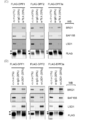
- Total protein extract was prepared by boiling the cells in SDS sample buffer at 95 ° C. for 10 minutes. The proteins were then electrophoresed by 10% SDS-PAGE and transferred onto Immobilon-P SQ membranes or Immobilon-P PVDF membranes (Millipore). The following proteins: Brm (ab15597; Abcam), BRG1 (sc-10768; Santa Cruz), Halo-Tag (G928A; Promega), HA-Tag (C29F4; Cell Signaling) and ⁇ -actin (sc-47778; Santa Immunoblotting was performed by incubating the membrane overnight at 4 ° C. with a primary antibody against Cruz).
- Western blotting was performed by incubating the membranes overnight at 4 ° C. in Can Get Signal Solution I (TOYOBO) containing the primary antibody. After washing 3 times with Tris-buffered saline (TBS) containing Tween 20, the membrane was treated with a secondary antibody (donkey anti-rabbit horseradish peroxidase antibody (AP182P) or donkey anti-mouse horseradish peroxidase antibody) for 1 hour at room temperature. (AP192P); Millipore). The signal was detected on AE-9300H-CP Ez-CaptureMG (ATTO) using ECL reagent (Promega) or ImmunoStar LD (Wako). The amount of protein sample electrophoresed was normalized to ⁇ -actin.
- Immunoprecipitation (2) Using a buffer containing 50 mM Tris-HCl (pH 7.5), 140 mM NaCl, 1 mM MgCl 2 , 0.5 mM DTT, 0.1% Tween 20, protease inhibitor cocktail (Nacalai Tesque) and phosphatase inhibitor cocktail (Nacalai Tesque) Cells were lysed. Immunoprecipitation was performed using Dynabeads Protein G (Thermo Fisher Scientific) according to the manufacturer's instructions.
- Samples were then incubated with each primary antibody in blocking buffer overnight at 4 ° C. Samples were washed twice and then incubated with Alexa Fluor 546 or 488 conjugated secondary antibody (Thermo Fisher Scientific, 1: 1000) in blocking buffer for 1 hour at RT in the dark. Samples were mounted in Vectashield Mounting Medium (Vector Laboratories) containing DAPI. Fluorescence was detected using a fluorescence microscope (BZ-X710; Keyence). Images were processed using Adobe Photoshop CS3 software.
- proximal ligation assay Proximity Ligation Assay: PLA
- GIC was seeded, processed and incubated with the primary antibody as described above.
- PLA was performed according to the manufacturer's instructions using Duolink In Situ Starter SetANGORANGE (SIGMA).
- SIGMA Duolink In Situ Starter SetANGORANGE
- Anti-mouse MINUS and anti-rabbit PLUS PLA probes were used. Fluorescence was detected using a fluorescence microscope (BZ-X710; Keyence).
- d4 family proteins have a conserved N-terminal domain, a central C2H2-type kruppel zinc finger motif, and a C-terminal paired plant homeobox domain (PHD) finger (modified histone, etc.)
- PLD plant homeobox domain
- the present inventors constructed a plurality of deletion mutants of the DPF2 protein, and tested the binding activity of the mutant to a subunit of the SWI / SNF complex or the p52 protein. It was found that the obtained region (amino acid residues 1 to 84; hereinafter referred to as CT1 peptide, SEQ ID NO: 2) is sufficient for binding to all of the target proteins (FIG. 2A). When the amino acid sequences of all d4 family proteins are aligned and compared, the CT1 region is highly conserved among the four d4 family proteins (DPF3a and DPF3b have the same N-terminal region), as shown in FIG. 2B.
- CT1 peptides originating from any of the d4 family proteins when expressed at high levels, are subunits of the SWI / SNF complex of d4 family proteins and subunits of NF- ⁇ B. Competing with the role as an adapter molecule between them, we predicted that they could function as dominant negative mutants for all d4 family proteins.
- CT1 derived from DPF2 or DPF3 was fused to the C-terminus or N-terminus of a Halo tag (this tag is often stabilized when a short peptide is fused).
- DPF2 CT1 peptide hereinafter referred to as “Halo-DPF2-CT1”
- Halo-DPF2-CT1 DPF2 CT1 peptide fused with Halo tag on the N-terminal side is expressed ectopically in HeLaS3 cells and A549 cells, (Data not shown).
- the cells ectopically expressing Halo-DPF2-CT1 had only a slight decrease in growth rate in monolayer culture (FIG. 2C), but in the anchorage independent state in soft agar.
- IL-6 (Libermann, TA et al., Mol Cell Biol 10: 2327-34 (1990); Ray A. et al., Mol Cell Biol 10: 5736-46 (1990)), IL-8 (Kunsch C. Mol Cell Biol 13: 6137-46 (1993)), TNF- ⁇ (Collart MA, et al. Mol Cell Biol 10: 1498-506 (1990); Shakhov AN et al., Gene 95: 215-21 (1990)) And four genes of ICAM1 (van de Stolpe A.
- CT1 inhibits the induction of a specific subset of NF- ⁇ B target genes
- Each promoter of a gene containing an NF- ⁇ B binding site can make a large difference with respect to inducibility by TNF- ⁇ treatment that activates NF- ⁇ B. This inducibility is also affected by several factors, such as the status of the promoter chromatin in the cell type used and other transcription factors that are secondarily induced by NF- ⁇ B after TNF- ⁇ treatment. .
- NF- ⁇ B target genes Based on the list of NF- ⁇ B target genes confirmed by promoter analysis (https; // www.bu.edu/nf-kb/, 594 probes for NF- ⁇ B target genes), we Microarray analysis of CT1 sensitivity of NF- ⁇ B target gene expression induced by TNF- ⁇ treatment was performed on two cell lines, A549 cells and HeLaS3 cells (data not shown). 54 genes of A549 cells and 50 genes of HeLaS3 cells were induced more than 1.5 times by 1 hour of TNF- ⁇ treatment. The 71 genes induced by TNF- ⁇ in either A549 cells or HeLaS3 cells included 19 cytokine genes.
- RNA sample used for the microarray analysis was analyzed by quantitative reverse transcription PCR (qRT-PCR), the same result as that obtained by the microarray analysis was obtained (FIG. 3A).
- CT1 quantitative reverse transcription PCR
- Halo-CT1 (HA1-tagged CT1 also functions as an effective dominant-negative mutant and an adapter connecting SWI / SNF and NF- ⁇ B)
- Halo-CT1 was very stable in the cells, but HA-CT1 added with an HA tag, which is a shorter tag than the Halo tag, on the N-terminal side could also be stably expressed in the cells.
- HA-CT1 has the same dominant negative function as Halo-CT1, and when ectopically expressed in A549 cells and HeLaS3 cells, it does not affect cell growth in monolayer culture and is independent of anchorage. Proliferation was reduced. (FIGS. 4A and 4B).
- the ⁇ 40-76 peptide lacking the 40th to 76th amino acid residues of CT1 and the ⁇ 53-76 peptide lacking the 53rd to 76th amino acid residues are induced by IL-6 induction by TNF- ⁇ and The inhibitory effect on anchorage-dependent growth was lost.
- ⁇ 1-8 lacking the first to eighth amino acid residues retained a considerable degree of the original dominant negative activity.
- the ⁇ 1-39 peptide lacking the 1st to 39th amino acid residues and the ⁇ 1 lacking the 1st to 32nd amino acid residues The -32 peptide was unstable and undetectable (specific data not shown).
- the amino acid residues of CT1 include the SWI / SNF complex of d4 family and NF- ⁇ B in the 53rd to 76th region (SEQ ID NO: 78) or the 40th to 76th region (SEQ ID NO: 79). It strongly suggests that the binding region is included.
- a peptide was synthesized by adding an intracellular introduction signal and a nuclear translocation signal. When these synthetic peptides are used, they can function as dominant negative mutants for d4 family proteins as in CT1 (data not shown).
- CT1 strongly suppresses tumorigenicity of epithelial cancer cells in a mouse xenograft model
- A549 cells ectopically expressing HA-DPF3-CT1 were injected into immunodeficient mice.
- the onset of tumor formation was greatly delayed by the introduction of CT1, and the growth of the tumor was greatly reduced. This result demonstrated the therapeutic effect of the peptide of the present invention on epithelial cancer.
- FIG. FIG. 6 shows the effect of knockdown of d4 family proteins and each of Brm and BRG1 on anchorage-independent growth of HeLaS3 cells (A) and A549 cells (B).
- the number of colonies (with a diameter of 100 nm or more) formed in soft agar was measured and compared to the number of shCre # 4-expressing cells (control, Kobayashi, K. et al., Sci. Rep. 5: 8427 (2015)). Normalized.
- shDPF1-3'UTR # 2 (SEQ ID NO: 49 or SEQ ID NO: 16) and # 4 (SEQ ID NO: 51 or SEQ ID NO: 18) (lanes 3 and 4), shDPF2-3'UTR # 3 (SEQ ID NO: 55 or SEQ ID NO: 20) ), # 4 (SEQ ID NO: 56 or SEQ ID NO: 22) and # 6 (SEQ ID NO: 57 or SEQ ID NO: 24) (lanes 5, 6, 7), shDPF3a-CDS # 2 (SEQ ID NO: 68 or SEQ ID NO: 26), and shDPF3a-3'UTR # 4 (SEQ ID NO: 64 or SEQ ID NO: 28) (lanes 8 and 9), shDPF3b-CDS # 6 (SEQ ID NO: 72 or SEQ ID NO: 30) and # 7 (SEQ ID NO: 73 or SEQ ID NO: 32) (Lanes 10, 11), shBrm- # 4 (SEQ ID NO: 34) and # 8 (SEQ ID NO: 36
- FIG. 1 is a schematic diagram showing the structure of d4 family protein and its mutant CT1.
- NLS Nuclear translocation signal.
- Zn C2H2 type Kruppel type zinc finger motif.
- PHD Plant homeodomain. In the frame indicated by a broken line, an N-terminal conserved region between d4 family proteins is shown.
- B shows alignment of amino acid sequences of N-terminal regions of DPF1, DPF2 and DPF3.
- DPF1 has an additional sequence consisting of 27 amino acids at the N-terminus relative to DPF2 and DPF3.
- An asterisk (*) indicates a position having the same amino acid residue.
- a colon (:) indicates a position where amino acid residues are highly conserved.
- Panels (C) and (D) show ectopic expression of Halo-DPF2-CT1 and Halo-DPF3-CT1 on cell proliferation of A549 and HeLaS3 cells in monolayer culture (C) and soft agar (D)
- the effect of In the experiment of (C) a Lentiviral vector carrying Halo-DPF2-CT1 or Halo-DPF3-CT1 was transduced (introduced) into each of A549 cells and HeLaS3 cells. After puromycin selection, the cells were seeded in a 96-well plate, and cell proliferation during monolayer culture was monitored every 24 hours using CellTiter-Glo (registered trademark) Luminescent Cell Viability Assay.
- FIG. 3 The inhibitory effect by CT1 after TNF- ⁇ stimulation was tested by quantitative RT-PCR analysis.
- Halo-DPF3-CT1 expression retroviral vector or control empty vector (EV-2) was transduced (introduced) into HeLaS3 cells or A549 cells.
- Total RNA was prepared from each cell stimulated with TNF- ⁇ for 1 hour (A) or for 30, 60, 90 and 120 minutes (B).
- IL-6, IL-8, TNF- ⁇ and ICAM1 mRNA levels were measured by quantitative RT-PCR.
- A the ratio to the mRNA level of control HeLa cells (introduced with EV-2) is shown.
- the RNA level before TNF- ⁇ treatment was 1.0.
- C At the same time, RNA is extracted from the culture maintained without TNF- ⁇ treatment, and mRNA levels of four genes, IL-6, IL-8, TNF and ICAM1, are measured as shown in (A). Showed.
- FIG. 4 The effect of ectopically expressed HA-DPF3-CT1 on cell growth in monolayer culture (A) and soft agar (B) of A549 or HeLaS3 cells was tested.
- C shows the results of a co-immunoprecipitation experiment with the catalytic subunit of the SWI / SNF complex and HA-DPF3-CT1, HA-DPF3a and HA-DPF3b.
- Cell nuclei were prepared from cells expressing HA-DPF3a, HA-DPF3b or HADPF3-CT1, and proteins derived from the cell nuclei were immunoprecipitated with anti-HA antibodies, and the precipitates were subjected to SDS-PAGE. Brm and BRG1 were detected by Western blot.
- EV-3 represents cells expressing only the HA tag and was used as a negative control.
- FIG. 5 The biological activity in A549 cells of deletion mutants derived from HA-DPF3-CT1 was summarized.
- C The relative colony forming ability with respect to the control cells was indicated by a numerical value.
- FIG. Figure 5 shows the tumorigenic potential of A549 cells expressing HA-DPF3-CT1 in xenotransplantation.
- Figure 7 Schematic representation of the interaction between SWI / SNF complex, d4 family protein, and NF- ⁇ B.
- GMM glioblastoma stem cells
- DPF3a knockdown was also rescued by ectopic expression of DPF1, but DPF1 knockdown was not significantly rescued by ectopic expression of DPF3a (FIG. 9B).
- DPF1 was more effective than DPF3a in maintaining stem cells.
- Rescue experiments using DPF3b cDNA in DPF3b knockdown cells were unsuccessful, and DPF3b mRNA levels were close to the limit of detection by qRT-PCR in these GIC strains. Has not analyzed.
- the present inventors further found that the sphere-forming activity of TGS-04 cells and TGS-05 cells is not sensitive to knockdown of either BRG1 or Brm (FIGS. 10A and 10B). This finding was in contrast to the result of TRG-01 cells that caused a decrease in sphere-forming activity by BRG1 knockdown (FIG. 9A). Since both BRG1 and Brm expression is high in TGS-04 cells and TGS-05 cells, it is considered that either single knockdown may not significantly affect the sphere-forming activity. This suggested that Brm could also function as an effective catalytic subunit of the SWI / SNF complex for GIC stem cell maintenance.
- DPF1 and DPF3a knockdown in GIC provides strong anti-tumorigenic activity
- TGS-01 cells introduced with shDPF1 expression vector, shDPF3a expression vector or EV-2 (control) were transplanted to immunocompromised mice (nude mice) did. Survival of mice inoculated with control cells decreased rapidly, but mice inoculated with cells expressing shDPF1 or shDPF3a showed improved survival compared to those inoculated with control cells (FIG. 11).
- shDPF1 expression produced a much greater effect compared to shDPF3a (5 out of 6 mice transplanted with shDPF1-expressing cells survived during the study).
- DPF1 is extremely important in maintaining the stem cell nature of GIC and may be a therapeutic target in GBM.
- This result demonstrated the therapeutic effect of the shRNA of the present invention on glioblastoma.
- the effect of shRNA on DPF1 was particularly remarkable among d4 family genes. This tendency is different from the result (FIG. 1) in which shRNA against DPF2 and DPF3 was more effective than DPF1 in the case of epithelial cancer.
- d4 family proteins were shown to function as adapter proteins that link SWI / SNF complexes and NF- ⁇ B dimers.
- SWI / SNF-dependent NF- ⁇ B activation contributes to the biological activity of GIC (maintaining stem cell), considering that the expression of BRG1, DPF1 and DPF3a was fairly strongly involved in maintaining GIC stemness It was next tested to see if it did.
- I ⁇ B ⁇ SR a mutant of the NF- ⁇ B activation pathway inhibitor I ⁇ B ⁇ , was introduced into TGS-01 cells, the inventors did not detect any effect on sphere-forming activity.
- the percentage of sphere-forming cells in control EV-2 transduced cells and I ⁇ B ⁇ SR-expressing cells was 20.57 ⁇ 1.59% and 21.35 ⁇ 0.60%, respectively (data not shown).
- DPF1 and DPF3a have distinctly different functions other than the adapter between the SWI / SNF complex and NF- ⁇ B, and these functions play an essential role in maintaining the stem cell nature of GCI. Suggested that it needs to be fulfilled.
- DPF1 or DPF3a is required as an adapter for the formation of large complexes including SWI / SNF core complex and corepressor complex
- TLX (NR2E1), LSD1 (lysine-specific demethylase 1), and RCOR2 (REST corepressor 2; CoREST2) among candidates that may form giant complexes with the SWI / SNF core complex as a result of trial and error was found to co-immunoprecipitate both BRG1 and BAF155 simultaneously from lysates of TGS-01 cells (FIGS. 12A, 12B).
- TLX known as a nuclear orphan receptor with transcriptional repression function, has been shown to be essential for the control of neuronal stem cells and maintenance of GIC stem cell properties by knockdown experiments.
- LSD1 a negative epigenetic regulator with histone demethylation activity
- RCOR2 REST corepressor 2: CoREST2
- DPF1, DPF2 or DPF3a with FLAG tag were immunoprecipitated with anti-TLX antibody or anti-LSD1 antibody, DPF1, DPF2 or DPF3a, and BRG1, BAF155 And LSD1 were detected (FIGS. 12C and 12D). Similar results were observed when FLAG-tagged DPF2 was used, but the expression level of FLAG-tagged DPF2 was low. Collectively, these results suggest that DPF1 and DPF3a may function as adapter proteins that link SWI / SNF complexes and corepressor complexes including TLX and LSD1 / RCOR2.
- Position Ligation Assay was performed. This method visualizes the interaction between proteins located proximal in the cell after immobilizing a large, unstable complex. All of the analyzed proteins (BRG1, BAF155, TLX and LSD1) were first confirmed to be strictly localized in the nucleus by immunofluorescence assay, and the same antibody as the assay was used for PLA.
- Anti-BRG1 / anti-TLX antibody pair, anti-BRG1 / anti-LSD1 antibody pair and anti-LSD1 / anti-TLX antibody pair as well as positive control using antibody pair of BRG1 / BAF155 (two subunits of SWI / SNF core complex) , And anti-LSD1 / anti-BAF155 antibody pair, it was detected in GIC that these five proteins are located proximally (FIG. 13). Only a small signal was detected when each antibody alone was used. TGS-01 cells transfected with the FLAG-tagged DPF1 expression vector were analyzed by PLA and found to be located proximally between DPF1 and TLX, DPF1 and LSD1, and DPF1 and BAF155.
- ShDPF1-CDS # 1 which is an shRNA against DPF1 used in Example 9, (the corresponding siRNA sense strand sequence is SEQ ID NO: 41, the same applies hereinafter), shDPF1-3′UTR # 4 (SEQ ID NO: 51) together with shDPF1 -Examine the effects of CDS # 2 (SEQ ID NO: 43), shDPF1-CDS # 3 (SEQ ID NO: 45), shDPF1-3'UTR # 1 (SEQ ID NO: 47), shDPF1-3'UTR # 2 (SEQ ID NO: 49) It was.
- shDPF1-CDS # 3 and shDPF1-3'UTR # 1 showed the same effect as shDPF1-CDS # 1 and shDPF1-3'UTR # 4, but shDPF1-CDS # 2 and shDPF1-3'UTR # For # 2, there was no significant inhibitory effect on the sphere formation of TGS-01 cells.
- shDPF3-CDS # 1 SEQ ID NO: 58
- shDPF3-CDS # 3 SEQ ID NO: 59
- ShDPF3a-CDS # 1 (SEQ ID NO: 66) together with shDPF3a-3′UTR # 2 (SEQ ID NO: 60) and shDPF3a-3′UTR # 4 (SEQ ID NO: 64), which are shRNAs for DPF3a used in Example 9.
- the effects of shDPF3a-CDS # 2 (SEQ ID NO: 68) and shDPF3a-3′UTR # 3 (SEQ ID NO: 62) were examined.
- shDPF3a-CDS # 1 and shDPF3a-CDS # 2 showed a sphere formation inhibitory effect equivalent to or higher than shDPF3a-3'UTR # 2 and shDPF3a-3'UTR # 4 used in Example 9.
- the inhibitory effect of shDPF3a-3'UTR # 3 was weak.
- FIG. 8 Heat map representation of mRNA expression of subunits of the SWI / SNF core complex and related genes in preparations from three GICs and their corresponding preparations from differentiated cells.
- A mRNA expression during sphere culture of TGS-01, TGS-04 or TGS-05 was analyzed by qRT-PCR and compared with that of differentiated monolayer culture.
- B is a bar graph of the relative gene expression levels of d4 family members, BRG1 and BRM, analyzed by qRT-PCR as described in (A).
- S sphere culture
- D monolayer culture for differentiation. Error bars represent the standard deviation of the average from triplicate experiments.
- FIG. 9 Knockdown effect of d4 family member, BRG1 or Brm, on sphere-forming activity of TGS-01 cells.
- A Shows the relative sphere-forming ability of TGS-01 cells expressing various shRNAs or introduced with a control (EV-1: lane 1) retroviral vector: shDPF1-CDS # 1 (SEQ ID NO: 41) (Lane 3), shDPF1-3'UTR # 4 (SEQ ID NO: 51 or SEQ ID NO: 18) (lane 4), shDPF2-3'UTR # 3 (SEQ ID NO: 55), shDPF2-3'UTR # 4 (SEQ ID NO: 56) and shDPF2-3′UTR # 6 (SEQ ID NO: 57) (lanes 5-7), shDPF3a-3′UTR # 2 (SEQ ID NO: 60) (lane 8), shDPF3a-3′UTR # 4 (SEQ ID NO: 64) ) (Lane 9), shDPF3b-CDS # 6 (SEQ ID NO: 72
- TDP-01 cells transfected with a d4 family protein expression lentiviral vector and an empty vector (EV-3) were treated with shDPF1-3'UTR # 4, shDPF3a-3'UTR # 4, or shBRG1-CDS # 2.
- Figure 10 Effect of knockdown of BRG1 or Brm on sphere-forming activity of TGS-04 and TGS-05 cells.
- shBrm # 4 SEQ ID NO: 34
- shBrm # 8 SEQ ID NO: 36
- SEQ ID NO: 38 shBRG1-CDS # 2
- SEQ ID NO: 40 shBRG1-CDS # 4 (SEQ ID NO: 40) (lanes 4, 5 ).
- C It relates to a rescue experiment.
- FIG. Kaplan-Meier survival curve of mice transplanted orthotopically with TGS-01 cells Lentiviral vector or empty vector (EV-2) expressing shDPF1-3′UTR # 4 (SEQ ID NO: 51 or SEQ ID NO: 18) or shDPF3a-3′UTR # 4 (SEQ ID NO: 64 or SEQ ID NO: 28) was introduced TGS-01 cells were sorted by flow cytometry 2 days after transfection and the resulting cells (3 ⁇ 10 3 cells each) were injected into the cerebral hemisphere of 6 week old female nude mice. * P ⁇ 0.05, ** p ⁇ 0.01 by log rank test.
- FIG. Detection of larger SWI / SNF complexes including TLX and LSD1 / RCOR2 in GIC The results of co-immunoprecipitation of SWI / SNF complex subunits with a corepressor complex are shown.
- A, B Lysates of TGS-01 cells were immunoprecipitated with anti-TLX antibody (A), or anti-LSD1 antibody or anti-RCOR2 antibody (B), and the resulting immunoprecipitates were analyzed by Western blotting.
- C TGS-01 cell lysate ectopically expressing 3 ⁇ FLAG-DPF1, 3 ⁇ FLAG-DPF2 or 3 ⁇ FLAG-DPF3a was treated with anti-TLX antibody (C) or anti-LSD1 antibody (D). Immunoprecipitation was performed and the immunoprecipitate was analyzed by Western blotting. Arrow: d4 family protein with FLAG tag; arrowhead: IgG heavy chain. Samples were from the same experiment and gel / blots were processed in parallel.
- FIG. Proximal localization of SWI / SNF core complex, TLX and LSD1 / RCOR2 in TGS-01 cells detected by PLA.
- TGS-01 cells were fixed and incubated with a single antibody or antibody pair were detected by PLA. Dots indicate protein-protein interactions and nuclei were stained with DAPI. Fluorescence images were obtained using a BZ-X710 (Keyence) quick-full-focus at a depth of about 10 ⁇ m. The scale bar indicates 10 ⁇ m.
- Rb rabbit
- Ms mouse.
- FIG. A schematic diagram of the interaction between the SWI / SNF complex, d4 family proteins, and TLX is shown.
- FIG. 1 The examination result of shRNA with respect to d4 family gene suitable for the stem cell loss of GIC is shown using the sphere formation inhibitory effect of TGS-01 cells as an index.
- the medicament according to the present invention can be expected to be used as a novel medicament for epithelial cancer and glioma.
- dominant negative mutants of d4 family proteins can act as dominant negative mutants for all d4 family proteins and can be an effective means of treating epithelial cancer by suppressing only the ability of NF- ⁇ B to enhance tumors.
- suppression of DPF1 by RNAi can be an effective means for the treatment of glioma.
- SEQ ID NO: 1 DPF1-CT1 amino acid sequence
- SEQ ID NO: 2 DPF2-CT1 amino acid sequence
- SEQ ID NO: 3 DPF3a / b-CT1 amino acid sequence
- SEQ ID NO: 4 ⁇ 1-8 mutant (9 to 84 residues) of DPF3-CT1
- Amino acid sequence SEQ ID NO: 5 Amino acid sequence number of DPF3-CT1 ⁇ 1-32 variant (33 to 84 residues)
- 6 Amino acid sequence sequence number of DPF3-CT1 ⁇ 1-39 variant (40 to 84 residues)
- 7 Amino acid sequence of the DPF3-CT1 ⁇ 1-52 variant (residues 53 to 84)
- SEQ ID NO: 8 Amino acid sequence of the DPF3-CT1 ⁇ 40-76 variant
- Amino acid sequence SEQ ID NO: 10 nucleotide sequence of DPF
Landscapes
- Health & Medical Sciences (AREA)
- Life Sciences & Earth Sciences (AREA)
- Chemical & Material Sciences (AREA)
- General Health & Medical Sciences (AREA)
- Medicinal Chemistry (AREA)
- Veterinary Medicine (AREA)
- Public Health (AREA)
- Animal Behavior & Ethology (AREA)
- Pharmacology & Pharmacy (AREA)
- Organic Chemistry (AREA)
- Molecular Biology (AREA)
- Epidemiology (AREA)
- Genetics & Genomics (AREA)
- Engineering & Computer Science (AREA)
- Biochemistry (AREA)
- Zoology (AREA)
- Bioinformatics & Cheminformatics (AREA)
- Proteomics, Peptides & Aminoacids (AREA)
- Biophysics (AREA)
- Gastroenterology & Hepatology (AREA)
- Biomedical Technology (AREA)
- Toxicology (AREA)
- Microbiology (AREA)
- General Chemical & Material Sciences (AREA)
- Chemical Kinetics & Catalysis (AREA)
- Biotechnology (AREA)
- Nuclear Medicine, Radiotherapy & Molecular Imaging (AREA)
- Virology (AREA)
- Mycology (AREA)
- General Engineering & Computer Science (AREA)
- Wood Science & Technology (AREA)
- Neurology (AREA)
- Neurosurgery (AREA)
- Immunology (AREA)
- Physics & Mathematics (AREA)
- Plant Pathology (AREA)
- Pharmaceuticals Containing Other Organic And Inorganic Compounds (AREA)
- Medicines That Contain Protein Lipid Enzymes And Other Medicines (AREA)
- Medicines Containing Material From Animals Or Micro-Organisms (AREA)
Abstract
The present invention provides an anticancer agent based on a novel mechanism. Specifically, the present invention relates to an inhibitor that inhibits gene expression regulation by SWI/SNF complex-dependent NF-κB or corepressor complexes that participate in chromatin remodeling. More specifically, the present invention provides an inhibitor that reduces the interactions (such as binding and transcription regulation) of endogenous SWI/SNF complexes and NF-κB or corepressor complexes or an inhibitor that inhibits the adapter function of d4 family proteins. The inhibitor of the present invention can be utilized in the treatment of cancers involving SWI/SNF complexes (such as SWI/SNF-mediated cancers).
Description
本発明は、クロマチンリモデリングに関与するSWI/SNF複合体依存的な、NF-κBまたはコリプレッサー複合体による遺伝子発現制御を阻害する阻害剤に関する。本発明の阻害剤は、SWI/SNF複合体が関与するがん(SWI/SNF介在性がんなど)の治療に利用できる。
The present invention relates to an inhibitor that inhibits gene expression control by NF-κB or a corepressor complex, which is dependent on a SWI / SNF complex involved in chromatin remodeling. The inhibitor of the present invention can be used for treatment of cancers involving SWI / SNF complexes (such as SWI / SNF-mediated cancers).
真核多細胞生物のゲノムDNAは、ヒストン八量体に巻き付いていて密なクロマチン構造を構築する。ゲノム遺伝子の転写、複製、修復などを行うためには,このクロマチン構造の密な構造を解いて、転写因子等の因子がゲノムDNAに直接、結合できるようにする必要がある。このようなエピジェネティカルな制御を担う機構の一つとしてクロマチン構造変換がある。このような活性をもつ進化的に保存された代表的な因子としてSWI/SNF複合体が見出されている。近年、ヒトのがんにおいてこのSWI/SNF複合体を構成するサブユニットの遺伝子に変異や欠失が見出され、SWI/SNF複合体の腫瘍の発生への関与が注目されている(特許文献1、2)。しかしながら、SWI/SNF複合体は各細胞種ごとに極めて幅広い遺伝子群の転写調節を担うため、がんなどの特定の疾患治療に利用する可能性が制限されている。
The genomic DNA of eukaryotic multicellular organisms wraps around histone octamers and builds a dense chromatin structure. In order to perform transcription, replication, repair, etc. of genomic genes, it is necessary to solve the dense structure of the chromatin structure so that factors such as transcription factors can directly bind to genomic DNA. One of the mechanisms responsible for such epigenetic control is chromatin structure conversion. The SWI / SNF complex has been found as an evolutionarily conserved representative factor having such activity. In recent years, mutations and deletions have been found in the genes of the subunits that make up this SWI / SNF complex in human cancer, and the involvement of the SWI / SNF complex in tumor development has attracted attention (patent documents). 1, 2). However, since the SWI / SNF complex is responsible for the transcriptional regulation of a very wide range of genes for each cell type, its potential for use in the treatment of specific diseases such as cancer is limited.
転写因子NF-κBは、B細胞の免疫グロブリンκ鎖の転写に関与するものとして発見された。現在ではこのNF-κBは、幅広い細胞で、サイトカイン増殖因子、接着分子等の発現の活性化を通して発生・分化、細胞増殖およびアポトーシス等を含む多数の生命現象を制御していることが知られる。また、構成的または過剰なNF-κB活性が炎症性疾患や、自己免疫疾患において見出されているばかりか、多くの腫瘍細胞や白血病細胞においても恒常的な活性化が観察されることから、その阻害剤も作成されている(特許文献3~5)。しかしながら、NF-κBの生物学的効果が非常に広範であるため、副作用の観点からNF-κB阻害剤は疾患治療に利用する可能性が制限されている。本発明者らは、これまでにd4ファミリータンパク質(DPF1、DPF2、DPF3a/b)が、NF-κBとSWI/SNF複合体を連結するアダプタータンパク質として機能することを報告した(非特許文献2~3)。しかしながら、NF-κBとSWI/SNF複合体とが形成するより大きな複合体により転写活性化を受ける標的遺伝子群やその機構などについてはまだ解明されていない。
The transcription factor NF-κB was found to be involved in transcription of immunoglobulin κ chain in B cells. At present, it is known that NF-κB controls a number of life phenomena including activation / differentiation, cell proliferation, apoptosis and the like through activation of expression of cytokine growth factors, adhesion molecules and the like in a wide range of cells. In addition, constitutive or excessive NF-κB activity is not only found in inflammatory diseases and autoimmune diseases, but also constitutive activation is observed in many tumor cells and leukemia cells, Inhibitors have also been created (Patent Documents 3 to 5). However, since the biological effects of NF-κB are very broad, the possibility of using NF-κB inhibitors for disease treatment is limited from the viewpoint of side effects. The present inventors have previously reported that d4 family proteins (DPF1, DPF2, DPF3a / b) function as adapter proteins that link NF-κB and SWI / SNF complexes (Non-Patent Documents 2 to 4). 3). However, the target genes that undergo transcriptional activation by a larger complex formed by NF-κB and the SWI / SNF complex, the mechanism, and the like have not yet been elucidated.
核内レセプターファミリーに属するテイルレス(tailless:TLX、NR2E1とも命名される)は発生過程において、ショウジョウバエ胚の末端やマウスの前脳などにおいて発現される転写調節因子である。TLXは、ヒト成人の脳内では「脳室下帯(subventicular zone)等に存在する神経幹細胞で発現しており、神経発生に重要な機能を果たすと共に、神経幹細胞に由来する脳腫瘍に関与することも示されている。TLX遺伝子のノックアウトマウスは、辺縁系の欠損など、中枢神経系に関する表現型を有することが示されている。TLXの機能が改変されると、鬱病、不安症、攻撃性疾患、卒中、多発性硬化症、アルツハイマー病、パーキンソン病、ニューロパシー性の痛み、中枢神経系の炎症性疾患および他の神経変性疾患などの中枢神経系の疾患、目の欠陥および中枢神経系の発達などに影響し得る(特許文献6)。
Tailless (also named TLX, NR2E1) belonging to the nuclear receptor family is a transcriptional regulatory factor expressed in the Drosophila embryo end, mouse forebrain, etc. during development. TLX is expressed in neural stem cells existing in the subventricular zone etc. in the human adult brain, and plays an important role in neurogenesis and is involved in brain tumors derived from neural stem cells TLX knockout mice have been shown to have a central nervous system phenotype, such as limbic deficits, and depression, anxiety, and attack when TLX function is altered. Diseases of the central nervous system such as sexually transmitted disease, stroke, multiple sclerosis, Alzheimer's disease, Parkinson's disease, neuropathic pain, inflammatory diseases of the central nervous system and other neurodegenerative diseases, eye defects and central nervous system It can affect development and the like (Patent Document 6).
神経膠腫幹細胞(glioma initiating cell:GIC)は、成人の致死性原発性脳腫瘍である神経膠芽腫(glioblastoma)の治療抵抗性および腫瘍再発の原因になっていると考えられている。GICの幹細胞様特性、例えば自己複製能および腫瘍形成能はエピジェネティカルに制御されるが、これらの細胞において、ヒトの主要なクロマチンリモデリング複合体であるSWI/SNF複合体などの因子の関与やこれと関連する転写調節因子の役割などは詳細には解明されていない(非特許文献4)。
Glioma stem cells (GIC) are thought to be responsible for the treatment resistance and tumor recurrence of glioblastoma, a fatal primary brain tumor in adults. GIC stem cell-like properties such as self-renewal and tumorigenicity are epigenetically controlled, but in these cells, factors such as the SWI / SNF complex, the major human chromatin remodeling complex, are involved In addition, the role of transcriptional regulatory factors associated therewith has not been elucidated in detail (Non-patent Document 4).
本発明は、新規のメカニズムに基づく抗がん剤を提供することを目的とする。
An object of the present invention is to provide an anticancer agent based on a novel mechanism.
本発明者らは、SWI/SNF複合体依存性の、NF-κBまたはコリプレッサー複合体による遺伝子発現制御を阻害する阻害剤が抗がん活性を有することを見出した。
The present inventors have found that inhibitors that inhibit SWI / SNF complex-dependent gene expression control by NF-κB or corepressor complex have anticancer activity.
詳細には、本発明者らは、SWI/SNF複合体が機能している上皮がんにおいて、SWI/SNF複合体依存的なNF-κB標的遺伝子の発現に必須であるd4ファミリータンパク質をコードする遺伝子の発現をノックダウン、特にDPF2、DPF3a/3bをコードする遺伝子の発現をノックダウンするsiRNA1、shRNAが、前記上皮がんに対して強い抗がん活性を示すことを見出した。更に、本発明者らは、DPF1、DPF2、DPF3a/3b間で高度に保存されたN末端の84アミノ酸残基からなる領域(CT1ペプチドと称する)が、上皮がんに対して前記siRNA、shRNAよりも強い抗がん活性を示すことを見出した。
Specifically, we encode d4 family proteins that are essential for SWI / SNF complex-dependent NF-κB target gene expression in epithelial cancers where the SWI / SNF complex is functioning It has been found that siRNA1 and shRNA that knockdown gene expression, particularly knockdown expression of genes encoding DPF2 and DPF3a / 3b, exhibit strong anticancer activity against the epithelial cancer. Furthermore, the present inventors have found that a region consisting of 84 amino acid residues at the N-terminus that is highly conserved among DPF1, DPF2, and DPF3a / 3b (referred to as CT1 peptide) is used for the above-mentioned siRNA, shRNA against epithelial cancer. It was found to show stronger anticancer activity.
本発明者らはまた、SWI/SNF複合体が、d4ファミリータンパク質、特にDPF1、DPF3aを介して、GICにおける幹細胞性の維持に必須の役割を果たしていることを見出した。詳細には、これらd4ファミリーをコードする遺伝子の発現をノックダウン、特にDPF1、DPF3aをコードする遺伝子の発現をノックダウンするsiRNA、shRNAが、SWI/SNF複合体依存的な核内受容体TLXおよびLSD1/RCOR2を含むコリプレッサー複合体の活性を抑制することで強い抗がん活性を示すことを見出した。
The present inventors have also found that the SWI / SNF complex plays an essential role in maintaining stem cell properties in GIC via d4 family proteins, particularly DPF1 and DPF3a. Specifically, siRNA, shRNA, knocks down the expression of genes encoding these d4 families, in particular the expression of genes encoding DPF1, DPF3a, SWI / SNF complex-dependent nuclear receptors TLX and It was found that strong anticancer activity was exhibited by suppressing the activity of the corepressor complex containing LSD1 / RCOR2.
本発明は、上記知見に基づくものであり、次に示す阻害剤、医薬品組成物などを提供する。
[1] (a) SWI/SNF複合体依存性の、NF-κBもしくはコリプレッサー複合体による遺伝子発現制御を阻害する、または(b) d4ファミリータンパク質のアダプター機能を阻害する、阻害剤。
[2] 前記阻害がd4ファミリータンパク質の機能に対する競合阻害、またはd4ファミリータンパク質の発現の抑制である、前記[1]に記載の阻害剤。
[3] 前記d4ファミリータンパク質が、DPF1、DPF2、DPF3aおよびDPF3bから選択される少なくとも1つである、前記[2]に記載の阻害剤。
[4] 前記阻害剤が、d4ファミリータンパク質に対するドミナントネガティブ変異体である、前記[1]~[3]のいずれかに記載の阻害剤。
[5] 前記ドミナントネガティブ変異体が、配列番号1~7のいずれかで表されるアミノ酸配列を含むポリペプチド、または配列番号1~7のいずれかで表されるアミノ酸配列に対して90%以上の同一性を有するアミノ酸配列を含み、d4ファミリーに対する競合阻害活性を有するポリペプチドである、前記[4]に記載の阻害剤。
[6] SWI/SNF複合体と結合する、前記[1]~[5]のいずれかに記載の阻害剤。
[7] NF-κBおよび/またはコリプレッサー複合体と結合する、前記[4]~[6]のいずれかに記載の阻害剤。
[8] IL-6および/またはIL-8の発現を低減する、前記[1]~[7]のいずれかに記載の阻害剤。
[9] 配列番号1~7のいずれかで表されるアミノ酸配列、または配列番号1~7のいずれかで表されるアミノ酸配列に対して90%以上の同一性を有するアミノ酸配列をコードするポリヌクレオチドを含む発現ベクターである、前記[1]~[3]のいずれかに記載の阻害剤。
[10] d4ファミリー遺伝子の発現を抑制するポリヌクレオチドである、前記[1]~[3]のいずれかに記載の阻害剤。
[11] 前記ポリヌクレオチドが、d4ファミリー遺伝子の発現を抑制する、RNAiを奏するRNA、またはアンチセンスオリゴヌクレオチドである、前記[10]に記載の阻害剤。
[12] 前記d4ファミリー遺伝子の発現を抑制する、RNAiを奏するRNAが、配列番号74~77のいずれかのd4ファミリー遺伝子に特異的であり、d4ファミリー遺伝子の発現を抑制するsiRNAまたはshRNAである、前記[11]に記載の阻害剤。
[13] 前記d4ファミリー遺伝子の発現を抑制するポリヌクレオチドが、配列番号41~73からなる群から選択される配列およびその配列の相補的配列からなる二本鎖RNA、ならびに前記配列に対して欠失、置換、挿入および/または付加されたヌクレオチドを1~数個含む配列およびその相補的配列を含む二本鎖RNAから選択されるsiRNAまたはshRNAである、前記[11]または[12]に記載の阻害剤。
[14] 前記d4ファミリー遺伝子の発現を阻害するRNAが、配列番号41の配列と配列番号42の配列との組合せ、配列番号43の配列と配列番号44の配列との組合せ、配列番号45の配列と配列番号46の配列との組合せ、配列番号47の配列と配列番号48の配列との組合せ、配列番号49の配列と配列番号50の配列との組合せ、配列番号51の配列と配列番号52の配列との組合せ、配列番号60の配列と配列番号61の配列との組合せ、配列番号62の配列と配列番号63の配列との組合せ、配列番号64の配列と配列番号65の配列との組合せ、配列番号66の配列と配列番号67の配列との組合せ、および配列番号68の配列と配列番号69の配列との組合せから選択される組のポリヌクレオチドがアニーリングしてなるsiRNAまたは前記siRNAの配列とループ配列を含んでなるshRNAである、前記[11]~[13]のいずれかに記載の阻害剤。
[15] 配列番号16~33のいずれかから選択される配列を有するポリヌクレオチドを含むshRNAである、前記[11]に記載の阻害剤。
[16] 前記[1]~[15]のいずれかに記載の阻害剤を含む医薬品組成物。
[17] 抗がん剤である、前記[16]に記載の医薬品組成物。
[18] 上皮がん治療剤である、前記[16]に記載の医薬品組成物。
[19] 神経膠芽腫治療剤である、前記[16]に記載の医薬品組成物。
[20] DPF1遺伝子の発現を抑制するポリヌクレオチドを阻害剤として含んでなる、前記[19]に記載の医薬品組成物。 The present invention is based on the above findings and provides the following inhibitors, pharmaceutical compositions and the like.
[1] An inhibitor that inhibits gene expression control by NF-κB or a corepressor complex, which is dependent on a SWI / SNF complex, or (b) an adapter function of a d4 family protein.
[2] The inhibitor according to [1], wherein the inhibition is competitive inhibition of d4 family protein function or suppression of d4 family protein expression.
[3] The inhibitor according to [2], wherein the d4 family protein is at least one selected from DPF1, DPF2, DPF3a, and DPF3b.
[4] The inhibitor according to any one of [1] to [3], wherein the inhibitor is a dominant negative mutant for the d4 family protein.
[5] The dominant negative mutant is a polypeptide comprising the amino acid sequence represented by any of SEQ ID NOs: 1 to 7, or 90% or more with respect to the amino acid sequence represented by any of SEQ ID NOs: 1 to 7 The inhibitor according to [4] above, which is a polypeptide having an amino acid sequence having the same identity and having competitive inhibitory activity against the d4 family.
[6] The inhibitor according to any one of [1] to [5], which binds to a SWI / SNF complex.
[7] The inhibitor according to any one of [4] to [6], which binds to NF-κB and / or a corepressor complex.
[8] The inhibitor according to any one of [1] to [7], which reduces the expression of IL-6 and / or IL-8.
[9] Poly encoding an amino acid sequence represented by any one of SEQ ID NOs: 1 to 7, or an amino acid sequence having 90% or more identity to the amino acid sequence represented by any of SEQ ID NOs: 1 to 7 The inhibitor according to any one of [1] to [3] above, which is an expression vector containing a nucleotide.
[10] The inhibitor according to any one of [1] to [3], which is a polynucleotide that suppresses the expression of a d4 family gene.
[11] The inhibitor according to [10] above, wherein the polynucleotide is an RNAi-producing RNA or an antisense oligonucleotide that suppresses the expression of a d4 family gene.
[12] The RNA exhibiting RNAi that suppresses the expression of the d4 family gene is specific to the d4 family gene of any of SEQ ID NOs: 74 to 77, and is an siRNA or shRNA that suppresses the expression of the d4 family gene The inhibitor according to [11] above.
[13] The polynucleotide that suppresses the expression of the d4 family gene is a double-stranded RNA consisting of a sequence selected from the group consisting of SEQ ID NOs: 41 to 73 and a complementary sequence of the sequence, and a lack of the sequence. The above-mentioned [11] or [12], which is an siRNA or shRNA selected from a double-stranded RNA containing one to several nucleotides deleted, substituted, inserted and / or added and a complementary sequence thereof Inhibitors.
[14] The RNA that inhibits the expression of the d4 family gene is a combination of the sequence of SEQ ID NO: 41 and the sequence of SEQ ID NO: 42, a combination of the sequence of SEQ ID NO: 43 and the sequence of SEQ ID NO: 44, or the sequence of SEQ ID NO: 45 And the sequence of SEQ ID NO: 46, the combination of the sequence of SEQ ID NO: 47 and the sequence of SEQ ID NO: 48, the combination of the sequence of SEQ ID NO: 49 and the sequence of SEQ ID NO: 50, the sequence of SEQ ID NO: 51 and the sequence of SEQ ID NO: 52 A combination of sequences, a combination of the sequence of SEQ ID NO: 60 and a sequence of SEQ ID NO: 61, a combination of the sequence of SEQ ID NO: 62 and the sequence of SEQ ID NO: 63, a combination of the sequence of SEQ ID NO: 64 and the sequence of SEQ ID NO: 65, A siRNA obtained by annealing a set of polynucleotides selected from the combination of the sequence of SEQ ID NO: 66 and the sequence of SEQ ID NO: 67, and the combination of the sequence of SEQ ID NO: 68 and the sequence of SEQ ID NO: 69; The inhibitor according to any one of [11] to [13], wherein is an shRNA comprising the siRNA sequence and a loop sequence.
[15] The inhibitor according to [11] above, which is a shRNA comprising a polynucleotide having a sequence selected from any one of SEQ ID NOs: 16 to 33.
[16] A pharmaceutical composition comprising the inhibitor according to any one of [1] to [15].
[17] The pharmaceutical composition according to the above [16], which is an anticancer agent.
[18] The pharmaceutical composition according to the above [16], which is a therapeutic agent for epithelial cancer.
[19] The pharmaceutical composition according to the above [16], which is a therapeutic agent for glioblastoma.
[20] The pharmaceutical composition according to the above [19], comprising a polynucleotide that suppresses the expression of the DPF1 gene as an inhibitor.
本発明によって、クロマチンリモデリングを担うSWI/SNF複合体依存性の特定の機能を阻害/調節することによって、新規メカニズムに基づく抗がん剤を提供することが可能になる。より具体的には、SWI/SNF複合体が機能している上皮がんに対して、d4ファミリータンパク質由来のドミナントネガティブ変異体(CT1など)またはd4ファミリータンパク質をコードする遺伝子の発現を抑制するポリヌクレオチド阻害剤(siRNA、shRNAなど)を含む医薬品組成物を提供することができる。また、神経膠腫に対して、d4ファミリータンパク質をコードする遺伝子の発現を抑制するポリヌクレオチド阻害剤(siRNA、shRNAなど)を含む医薬品組成物を提供することができる。
The present invention makes it possible to provide an anticancer agent based on a novel mechanism by inhibiting / modulating a specific function dependent on the SWI / SNF complex responsible for chromatin remodeling. More specifically, a polymorphism that suppresses the expression of dominant negative mutants derived from d4 family proteins (such as CT1) or genes encoding d4 family proteins against epithelial cancers where the SWI / SNF complex functions. Pharmaceutical compositions comprising nucleotide inhibitors (siRNA, shRNA, etc.) can be provided. In addition, a pharmaceutical composition containing a polynucleotide inhibitor (siRNA, shRNA, etc.) that suppresses the expression of a gene encoding a d4 family protein can be provided for glioma.
本発明の各実施形態および各用語について以下に詳細に説明するが、以下の実施形態は、本発明を説明するための例示であり、本発明をそのような実施の形態のみに限定する趣旨ではない。すなわち、本発明は、その要旨を逸脱しない限り、様々な形態で実施をすることができる。
Each embodiment and each term of the present invention will be described in detail below, but the following embodiment is an example for explaining the present invention, and is intended to limit the present invention only to such an embodiment. Absent. That is, the present invention can be implemented in various forms without departing from the gist thereof.
本発明は、SWI/SNF複合体依存的にNF-κB、またはTLXを含むコリプレッサー複合体による遺伝子発現制御を阻害する阻害剤を提供する。好ましくは、本発明の阻害剤は、インビトロおよびインビボの両方においてSWI/SNF複合体依存的なNF-κBまたはTLXを含むコリプレッサー複合体による遺伝子発現制御を阻害する。
The present invention provides an inhibitor that inhibits gene expression control by a corepressor complex containing NF-κB or TLX in a SWI / SNF complex-dependent manner. Preferably, the inhibitor of the present invention inhibits gene expression control by a corepressor complex comprising SWI / SNF complex-dependent NF-κB or TLX both in vitro and in vivo.
(1.SWI/SNF複合体について)
本明細書中、「SWI/SNF複合体」とは分子量約2Mダルトンの多種タンパク質から構成される複合体を言う。本明細書中、SWI/SNF複合体の具体例としては、主としてヒトおよびその他の哺乳動物のBrm(GenBankアクセッション番号X72889)またはBRG1(GenBankアクセッション番号U29175)をDNA依存性ATPaseサブユニットとして含む複合体を意味するが、酵母のSWI/SNFまたはDrosophilaのBrahmaのヒトや哺乳動物におけるその他のホモログも含み得る。 (1. About SWI / SNF complex)
In the present specification, the “SWI / SNF complex” refers to a complex composed of various proteins having a molecular weight of about 2 M Dalton. In the present specification, specific examples of the SWI / SNF complex mainly include human and other mammalian Brm (GenBank accession number X72889) or BRG1 (GenBank accession number U29175) as a DNA-dependent ATPase subunit. By complex, it may include yeast SWI / SNF or other homologs in Drosophila Brahma in humans and mammals.
ヒトなどの哺乳動物におけるSWI/SNF複合体は、Brmを含むものとBRG1を含むものの2種に大別される。その他のSWI/SNF複合体の構成成分としては、哺乳動物の場合、INI1、BAF250、BAF60a、BAF60b、BAF60c、BAF170、BAF57、BAF155、BAF53、β-アクチン等が挙げられるが、これらに制限されない(Vignali, M.ら、 (2000) Mol. Cell. Biol. 20, 1899-1910)。SWI/SNF複合体の構成因子の一例を以下に示す。

(「京府医大誌 124(12),825~838,2015」より一部改変して転記)
SWI / SNF complexes in mammals such as humans are roughly classified into two types, those containing Brm and those containing BRG1. Examples of other SWI / SNF complex components include, but are not limited to, INI1, BAF250, BAF60a, BAF60b, BAF60c, BAF170, BAF57, BAF155, BAF53, β-actin in the case of mammals ( Vignali, M. et al. (2000) Mol. Cell. Biol. 20, 1899-1910). An example of the components of the SWI / SNF complex is shown below.
(Posted with some modifications from “Kyoto Medical University Journal 124 (12), 825-838, 2015”)
SWI/SNF複合体は、DNA依存性ATPaseによってクロマチンリモデリング機能を有する。クロマチンリモデリングとは、DNAのクロマチン構造を変化させる過程を指す。クロマチン構造とは、主としてDNAとヒストンとを主成分とし、4種類のヒストン分子(H2A、H2B、H3およびH4)によって形成されたヒストン八量体の周りに165bp(約2周分)のDNAが巻き付いて形成されたヌクレオソームを基本単位構造とし、このヌクレオソームが折りたたまれて圧縮された構造を指す。この高度に折りたたまれたDNAは転写、複製、修復、組換えなどに対して妨げとなる。そこで、SWI/SNF複合体は、ATP加水分解のエネルギーを利用してヌクレオソームを移動および/または除去することによって、クロマチン構造を変化させる機能を有している。このとき、ヒストンの修飾酵素(ヒストンアセチル化酵素、脱アセチル化酵素、リン酸化酵素、メチル化酵素など)と一緒になって遺伝子の発現および/または制御に重要な働きをする。SWI/SNF複合体は、例えばDNAからmRNAが転写される際、上記のクロマチンリモデリングによってクロマチン構造を変化させて、標的のDNA配列を露出させることで、転写調節因子などがDNAにアクセス可能になる。
The SWI / SNF complex has a chromatin remodeling function by DNA-dependent ATPase. Chromatin remodeling refers to the process of changing the chromatin structure of DNA. The chromatin structure is mainly composed of DNA and histones, and 165 bp (about 2 rounds) of DNA around histone octamers formed by four types of histone molecules (H2A, H2B, H3 and H4). A nucleosome formed by winding is used as a basic unit structure, and it refers to a structure in which this nucleosome is folded and compressed. This highly folded DNA hinders transcription, replication, repair, recombination, and the like. Thus, the SWI / SNF complex has a function of changing the chromatin structure by moving and / or removing nucleosomes using the energy of ATP hydrolysis. At this time, it plays an important role in gene expression and / or regulation together with histone modifying enzymes (histone acetylase, deacetylase, phosphorylase, methylase, etc.). For example, when mRNA is transcribed from DNA, the SWI / SNF complex changes the chromatin structure by the above-mentioned chromatin remodeling and exposes the target DNA sequence, allowing transcriptional regulators to access the DNA. Become.
本明細書中、「SWI/SNF複合体依存性の、NF-κBまたはコリプレッサー複合体による転写制御」とは、一実施形態において、細胞内のSWI/SNF複合体とd4ファミリータンパク質、NF-κB(通常NF-κBファミリータンパク質サブユニットの2量体から構成される)またはTLXを含んでいるコリプレッサー複合体が巨大な複合体を形成することで転写制御活性を奏することを意味する。
As used herein, “SWI / SNF complex-dependent transcriptional regulation by NF-κB or a corepressor complex” refers, in one embodiment, to an intracellular SWI / SNF complex and a d4 family protein, NF− It means that a corepressor complex containing κB (usually composed of a dimer of NF-κB family protein subunits) or TLX exerts transcriptional control activity by forming a huge complex.
SWI/SNF複合体は、コアユニットまたはサブユニットを介して種々の転写制御因子と結合できることが公知である。例えば、BRG1にはグルココルチコイドレセプターおよびエストロゲンレセプターが結合すること(Peterson, C.L.ら、(2000) Curr.Opin. Gen. Dev. 10, 187-192)、Ini1にはc-Mycが結合すること(Yudkovsky, N.ら、(1999). Genes Dev. 13, 2369-2374)、BAF60aにはc-Fosおよびc-Junが結合すること(Ito, T.ら、(2001) J. Biol.Chem. 276:2852-2857)が知られている。また、d4ファミリータンパク質であるDPF3a/b、DPF2などがアダプターとして機能して、NF-κB(Rel A/p50、Rel B/p52)と結合することが報告されている(Tando, Tら、J. Biol. Chem. 285, 21951-21960(2010);非特許文献1~3)。本発明においては、新たにSWI/SNF複合体がd4ファミリータンパク質をアダプターとしてTLXを含むコリプレッサー複合体と結合していることを明らかにした。本発明におけるSWI/SNF複合体依存性の転写制御とは、特にd4ファミリータンパク質、およびNF-κBまたはTLXを含むコリプレッサー複合体のいずれかの転写制御因子からなる巨大複合体による転写制御を対象とする。
It is known that the SWI / SNF complex can bind to various transcription control factors via a core unit or subunit. For example, BRG1 binds to glucocorticoid receptor and estrogen receptor (Peterson, CL et al. (2000) urr Curr. Opin. Gen. Dev. 10, 187-192), and Ini1 binds c-Myc ( Yudkovsky, N. et al. (1999). Genes Dev. 13, 369 2369-2374), c-Fos and c-Jun bind to BAF60a (Ito, T. et al. (2001) J. Biol. Chem. 276: 2852-2857) is known. In addition, dPF family proteins such as DPF3a / b and DPF2 function as adapters and have been reported to bind to NF-κB (Rel A / p50, Rel B / p52) (Tando, T et al., J Biol. Chem. 285, 21951-21960 (2010); Non-patent documents 1 to 3). In the present invention, it has been clarified that a SWI / SNF complex is newly bound to a corepressor complex containing TLX using a d4 family protein as an adapter. The SWI / SNF complex-dependent transcriptional control in the present invention specifically targets transcriptional control by a large complex consisting of a transcriptional regulator of either a d4 family protein and a corepressor complex containing NF-κB or TLX. And
SWI/SNF複合体と対象の転写制御因子との相互作用の変化を検出する手段としては、SWI/SNF複合体と対象の転写制御因子とが結合することを検出する手段、SWI/SNF複合体と対象の転写制御因子とが近位に位置していることを検出する手段、SWI/SNF複合体の存在下での対象となる転写制御因子の転写活性(上方制御または下方制御)を検出する手段が挙げられるが、これらに限定されず、当業者に公知の種々の他の手段を用いて検出してもよい。具体的な検出手段としては、例えば、免疫沈降、密度勾配遠心分離法、ゲルシフトアッセイ、ChIP(クロマチン免疫沈降)アッセイ、ファーウェスタンブロット、酵母ツーハイブリッド法、などの方法が挙げられる。SWI/SNF複合体と対象の転写調節因子とが近位に位置していることを検出する手段としては、Proximity Ligationアッセイ(PLA)(本明細書中の実施例に記載の方法)や蛍光共鳴エネルギー移動(FRET)法、生物発光共鳴エネルギー移動(BRET)法などが挙げられる。SWI/SNF複合体の存在下での対象となる転写調節因子の転写活性(上方制御または下方制御)を検出する手段としては、レポーター(ルシフェラーゼ、緑色蛍光タンパク質、アルカリホスファターゼなど)を用いるレポーターアッセイ、定量的RT-PCR(qRT-PCR)、マイクロアレイ、ノーザンハイブリダイゼーションなどが挙げられるが、これらに限定されない。
Means for detecting the interaction between the SWI / SNF complex and the target transcriptional regulator include means for detecting the binding between the SWI / SNF complex and the target transcriptional regulator, the SWI / SNF complex A means of detecting the proximity of the transcription factor to the target and the transcriptional activity (up-regulation or down-regulation) of the target transcription factor in the presence of the SWI / SNF complex Examples include, but are not limited to, and various other means known to those skilled in the art may be used for detection. Specific detection means include, for example, immunoprecipitation, density gradient centrifugation, gel shift assay, ChIP (chromatin immunoprecipitation) assay, far western blot, yeast two-hybrid method, and the like. Means for detecting the proximity of the SWI / SNF complex and the transcriptional regulator of interest include Proximity Ligation assay (PLA) (methods described in the examples herein) and fluorescence resonance. Examples thereof include an energy transfer (FRET) method and a bioluminescence resonance energy transfer (BRET) method. Reporter assay using reporter (luciferase, green fluorescent protein, alkaline phosphatase, etc.) as a means of detecting the transcription activity (up-regulation or down-regulation) of the transcription factor of interest in the presence of SWI / SNF complex, Examples include, but are not limited to, quantitative RT-PCR (qRT-PCR), microarray, and Northern hybridization.
また、本明細書中、SWI/SNF複合体依存性の転写制御因子による遺伝子発現制御を抑制することは、SWI/SNF複合体と当該転写制御因子との共存、一方の発現量や活性の上下、変異などの変化が生じた結果、細胞が増殖、分化、足場非依存性、細胞表面分子の変化などの異常を示すこと、あるいは宿主細胞の増殖停止もしくは遅滞、休眠、アポトーシスなど、様々な細胞レベルでの変化を引き起こすことも意味する。上記のとおり、SWI/SNF複合体は、細胞内で種々の転写調節因子などとの相互作用を介して、その宿主細胞に変化を生じさせ得る。SWI/SNF複合体のサブユニットにおける変異は、様々ながんで発見され、SWI/SNF複合体とがんの関連が注目されている(Wang Xら、Clin Cancer Res 2014, 20, 21-27;Biegel J.A.ら、Am J Med Genet C Semin Med Genet 2014, 166C, 350-66;Helming K.C.ら、Cancer Cell 2014, 26, 309-317;Wilson, B.G.ら、Nat. Rev. Cancer 2011, 11:481-492)。
In addition, in the present specification, suppression of gene expression control by a SWI / SNF complex-dependent transcriptional regulatory factor is the coexistence of the SWI / SNF complex and the transcriptional regulatory factor, and increases or decreases the expression level or activity of one. As a result of changes such as mutations, various cells such as proliferation, differentiation, anchorage independence, cell surface molecular changes, or host cell growth arrest or delay, dormancy, apoptosis, etc. It also means causing changes in the level. As described above, the SWI / SNF complex can cause changes in the host cell through interaction with various transcriptional regulatory factors and the like in the cell. Mutations in the subunits of the SWI / SNF complex have been found in various cancers, and the association between the SWI / SNF complex and cancer has attracted attention (Wang (X et al., Clin Cancer Res 2014, 20, 21-27; Biegel JA et al., Am J Med Genet C Semin Med Genet 2014, 166C, 350-66; Helming KC et al., Cancer 2014, 26, 309-317; Wilson, BG et al., Nat. Rev. Cancer 2011, 11: 481- 492).
(2.NF-κBについて)
NF-κBは、核内因子κBともいい、免疫グロブリンκ軽鎖遺伝子が成熟B細胞特異的に発現するために必要なエンハンサー領域に結合する転写因子として見出された。NF-κBは、RelA(p65)、RelB、c-Rel、NF-κB1 (p105/p50)、NF-κB2(p100/p52)の5種類のRelファミリータンパク質がそれぞれホモダイマーあるいはヘテロダイマーを形成することによって構成され、免疫、炎症、発生、細胞増殖、アポトーシスなどの細胞内機能を誘導し、さらには外因性である多くのウイルスゲノム遺伝子の発現も誘導する。これら多彩な生命現象を正しく維持するためには、NF-κBが選択性をもって標的遺伝子群のプロモーターを活性化させる必要があるが、その機構の詳細については未解明な点が多い。そこで、例えば、シグナル伝達の最下流で各NF-κBダイマーにプロモーター選択性を与える分子機構の解明が重要である。NF-κBの標的遺伝子の発現制御には、近傍のクロマチン環境も大きく影響を及ぼしていると考えられている。 (2. About NF-κB)
NF-κB, also called nuclear factor κB, was found as a transcription factor that binds to an enhancer region necessary for the immunoglobulin κ light chain gene to be expressed specifically in mature B cells. In NF-κB, five types of Rel family proteins RelA (p65), RelB, c-Rel, NF-κB1 (p105 / p50), and NF-κB2 (p100 / p52) form homodimers or heterodimers, respectively. And induces intracellular functions such as immunity, inflammation, development, cell proliferation, and apoptosis, and also induces the expression of many exogenous viral genomic genes. In order to correctly maintain these various life phenomena, NF-κB needs to activate the promoters of the target genes with selectivity, but there are many unclear points about the details of the mechanism. Thus, for example, it is important to elucidate the molecular mechanism that gives promoter selectivity to each NF-κB dimer at the most downstream of signal transduction. It is thought that the nearby chromatin environment has a great influence on the expression control of the target gene of NF-κB.
NF-κBは数多くの疾患と関係しており、前記疾患としては例えば、急性および慢性の炎症反応、クローン病、関節リウマチ、がん、敗血症ショック、ヒト免疫不全ウイルス(HIV)やサイトメガロウイルスなどのウイルス性疾患、慢性閉塞性肺疾患(COPD),嚢胞性線維症、神経変性疾患などが挙げられる。
NF-κB is associated with many diseases, such as acute and chronic inflammatory reactions, Crohn's disease, rheumatoid arthritis, cancer, septic shock, human immunodeficiency virus (HIV), cytomegalovirus, etc. Viral diseases, chronic obstructive pulmonary disease (COPD), cystic fibrosis, neurodegenerative diseases and the like.
TNF-αまたはLPSのような炎症性刺激による誘導において、IκBαがIκBキナーゼ(IKK)複合体によるリン酸化等を経て、NF-κBの細胞核内への移行が引き起こされる。NF-κBは、多くのサイトカインおよび増殖因子(例えば、IL-2、IL-8、IFN-β、M-CSF、G-CSF、VEGF)、ならびに種々の転写因子およびシグナルレギュレーター(例えば、IκBα、IRF-1、IRF-2)の転写を誘導する。本発明では、上皮がんにおけるNF-κBの機能として、増殖因子IL-6、IL-8およびTNF-α、ならびに細胞接着分子ICAM1から選択されるタンパク質を発現または産生する能力を指標とするが、他の公知のNF-κB依存性のタンパク質を指標として用いることもできる。その他の指標としては、例えば、細胞増殖能の確認、アポトーシス誘導能が挙げられるがこれらに限定されない。
In the induction by inflammatory stimuli such as TNF-α or LPS, IκBα undergoes phosphorylation by the IκB kinase (IKK) complex, etc., leading to the transfer of NF-κB into the cell nucleus. NF-κB has many cytokines and growth factors (eg, IL-2, IL-8, IFN-β, M-CSF, G-CSF, VEGF), as well as various transcription factors and signal regulators (eg, IκBα, Induces transcription of IRF-1 and IRF-2). In the present invention, the function of NF-κB in epithelial cancer is based on the ability to express or produce a protein selected from growth factors IL-6, IL-8 and TNF-α, and the cell adhesion molecule ICAM1. Other known NF-κB-dependent proteins can also be used as indicators. Examples of other indicators include, but are not limited to, confirmation of cell proliferation ability and apoptosis inducing ability.
(3.コリプレッサー複合体について)
一実施形態において、本発明は、コリプレッサー複合体による遺伝子発現制御を阻害する阻害剤を提供する。本発明において、コリプレッサー複合体とはコファクターに属する因子を指す。コファクターとは、転写制御因子と基本転写因子との間の橋渡しを行って転写を促進または抑制する因子を指し、コアクチベーター複合体およびコリプレッサー複合体が含まれる。例えば、コアクチベーター複合体を構成する種々のサブユニットにはヒストンを修飾して転写促進を行う機能を有するもの(例えば、ヒストンアセチルトランスフェラーゼ:HAT)を含み得、一方コリプレッサー複合体を構成するサブユニットにはヒストンの修飾を除去して転写抑制を行う機能を有するもの(例えば、ヒストン脱アセチル化酵素(HDAC)、ヒストン脱メチル化酵素など)を含み得る(特許文献1、2、特開2015-147794、Karin Meier & Alexander Brehm (2014), Epigenetics, 9:11, 1485-1495, DOI: 10.4161/15592294.2014.971580など)。また、コリプレッサー複合体の中に、TLX、RCOR2、LSD1から選択される少なくとも1つの分子が含まれているものもある(Yangら、Stem Cells. 2011 29:791-801)。これらコアクチベーター複合体およびコリプレッサー複合体は、上記のSWI/SNF複合体と一緒になってより巨大な複合体を形成し得る。 (3. About the corepressor complex)
In one embodiment, the present invention provides an inhibitor that inhibits gene expression control by a corepressor complex. In the present invention, the corepressor complex refers to a factor belonging to a cofactor. A cofactor refers to a factor that promotes or suppresses transcription by providing a bridge between a transcriptional regulatory factor and a basic transcription factor, and includes coactivator complexes and corepressor complexes. For example, the various subunits constituting the coactivator complex may include those having a function of promoting transcription by modifying histones (for example, histone acetyltransferase: HAT), while constituting the corepressor complex. Subunits may include those having a function of removing transcription of histones to suppress transcription (for example, histone deacetylase (HDAC), histone demethylase, etc.) 2015-147794, Karin Meier & Alexander Brehm (2014), Epigenetics, 9:11, 1485-1495, DOI: 10.4161 / 15592294.2014.971580). Some corepressor complexes contain at least one molecule selected from TLX, RCOR2, and LSD1 (Yang et al., Stem Cells. 2011 29: 791-801). These coactivator complexes and corepressor complexes can be combined with the SWI / SNF complexes described above to form larger complexes.
コリプレッサー複合体を検出するには、構成サブユニットに対する抗体(抗HDAC抗体など)を用いるウエスタンブロット、免疫沈降、ChIPアッセイなどの検出手段、ヒストン脱アセチル化酵素活性を検出する手段など、当業者に公知の手段が利用可能である。
To detect the corepressor complex, a person skilled in the art, such as Western blot using an antibody against a constituent subunit (such as anti-HDAC antibody), detection means such as immunoprecipitation, ChIP assay, means for detecting histone deacetylase activity, etc. Any known means can be used.
核内受容体ファミリーとは、一般的には、リガンドであるステロイド、甲状腺ホルモン、女性ホルモン、男性ホルモン、レチノイン酸、レチノイド、ビタミンD、グルココルチコイド、ミネラルコルチコイド等の受容体であって、こうしたリガンドが結合すると核内で転写因子として機能するタンパク質のファミリーを指す(Parker (1993) Curr. Op. Cell. Biol. 5: 499-504、Laudetら(1992) EMBO J. 11: 1003-1013、Mangelsdorfら,1995 Cell 83: 835-839, Loprs de SilvaおよびBurbach、(1995) Trends Neurosci. 18: 542-548など)。核内受容体ファミリータンパク質は、DNA結合ドメインおよびリガンド結合ドメインを含む類似のドメイン構造を有しており、生体の代謝、恒常性、分化、成長、発生、老化、生殖などの機能を担う。
The nuclear receptor family is generally a receptor for a ligand such as steroid, thyroid hormone, female hormone, male hormone, retinoic acid, retinoid, vitamin D, glucocorticoid, mineral corticoid, and the like. Refers to a family of proteins that function as transcription factors in the nucleus (Parker (1993) Curr. Op. Cell. Biol. 5: 499-504, Laudet et al. (1992) EMBO J. 11: 1003-1013, Mangelsdorf 1995, Cell 83: 835-839, Loprs de Silva and Burbach, (1995) Trends Neurosci. 18: 542-548, etc.). Nuclear receptor family proteins have a similar domain structure including a DNA-binding domain and a ligand-binding domain, and are responsible for functions such as metabolism, homeostasis, differentiation, growth, development, aging, and reproduction of the living body.
TLXは、核内受容体ファミリーの一員であり、テイルレスとしても知られる(Monaghanら、1995, Development 121:839-853;Pignoniら、1990, Cell 62: 151-163)。このTLXは、ショウジョウバエ(Pignoniet al,1990)、マウス(Tuら、1994, Nature 370: 375-379;Monaghanら、1995)、アフリカツメガエル(Hollemanら、1996, GenBank受託番号U67886)、ニワトリ(Yuら、1994)、ヒト(GenBank受託番号Y13276;Jacksonら、Royal Free Hospital, London)等において見出されている。TLXは、リガンドが判明していないオーファンレセプターである。TLXは、発達中のショウジョウバエ胚の末端(Pignoni et al,1990)および発達中のニワトリおよびマウスの前脳(Yu et al,1994;Monaghan et al,1995)において発現している。TLX遺伝子のノックアウトマウスは、辺縁系が欠損しているなどの表現型を有することが示されている(Monaghan et al,1997, Nature 390:515-517)。ホモ接合変異マウスは誕生時には生育可能であるが、嗅覚皮質、下嗅覚皮質および内嗅覚皮質を含む嗅脳および辺縁構造、扁桃および歯状回のサイズが小さい。TLXの機能欠失はすべての原脳神経芽細胞の不存在を招く(Younossi-Hartensteinら、1997, Developmental Biology 182:270-283)。TLXの機能が異常となると、鬱病、不安症、攻撃性疾患、卒中、多発性硬化症、アルツハイマー病、パーキンソン病、ニューロパシー性の痛み、中枢神経系の炎症性疾患および他の神経変性疾患を包含する中枢神経系の疾病など、目および/または中枢神経系に問題が生じ得る。
TLX is a member of the nuclear receptor family and is also known as tailless (Monaghan et al., 1995, Development 121: 839-853; Pigigni et al, 1990, Cell 62: 151-163). This TLX is expressed in Drosophila (Pignoniet al, 1990), mouse (Tu et al., 1994, Nature 370: 375-379; Monaghan et al., 1995), Xenopus (Holleman et al., 1996, GenBank accession number U67886), chicken (Yu et al. 1994), humans (GenBank accession number Y13276; Jackson et al., Royal Free Hospital, London). TLX is an orphan receptor whose ligand is unknown. TLX is expressed in the ends of the developing Drosophila embryo (Pignoni et al, 1990) and in the developing chicken and mouse forebrain (Yu et al, 1994; Monaghan et al, 1995). Knockout mice of the TLX gene have been shown to have a phenotype such as a lack of limbic system (Monaghan et al., 1997, Nature 390: 515-517). Homozygous mutant mice can grow at birth, but the olfactory brain and marginal structures, including the olfactory cortex, inferior olfactory cortex, and internal olfactory cortex, tonsils and dentate gyrus are small in size. Loss of TLX function leads to the absence of all encephalonomic neuroblasts (Younossi-Hartenstein et al., 1997, Developmental Biology 182: 270-283). Abnormal TLX functions include depression, anxiety, aggressive disease, stroke, multiple sclerosis, Alzheimer's disease, Parkinson's disease, neuropathic pain, inflammatory diseases of the central nervous system and other neurodegenerative diseases Problems with the eyes and / or central nervous system can occur, such as central nervous system disease.
TLXは、コリプレッサーであるアトロフィン1(ATN1)と複合体を形成すること(Zhang, CL.ら、2006, Genes and Dev. 20: 1308-1320)、ヒストン脱アセチル化酵素複合体1(HDAC1)を動員(リクルート)すること(Sun, GQ.ら、PNAS, 2007, 104, 39, pp.15282-15287)、LSD1およびBCL11Aと一緒に作用すること(Corso-Diaz, Xら、BMC Genomics, 2016, 17:832)など報告されている。しかしながら、SWI/SNF複合体との相互作用や依存性については明確にされたとはいえない。
TLX forms a complex with the corepressor atrophin 1 (ATN1) (Zhang, CL. Et al., 2006, Genes and Dev. 20: 1308-1320), histone deacetylase complex 1 (HDAC1) Recruiting (Sun, GQ. Et al., PNAS, 2007, 104, 39, pp.15282-15287), working with LSD1 and BCL11A (Corso-Diaz, X et al., BMC Genomics, 2016 , 17: 832). However, the interaction and dependence with the SWI / SNF complex has not been clarified.
(4.d4ファミリータンパク質について)
d4ファミリータンパク質は、転写共役因子(コファクター)として機能することが知られている。このd4ファミリータンパク質には、DPF1、DPF2、DPF3a、DPF3bが含まれる。これらファミリータンパク質は核移行シグナルを含むN末端側の領域、中央のC2H2型クルッペル様ジンクフィンガーモチーフ、C末端側にPHDジンクフィンガーモチーフ(PHD1、PHD2)を1つまたは2つを含むd4ドメインを有することにより特徴付けられる(図2(A)、Buchman, V.L.ら、1992, Nucleic Acids Res.20, 5579-5585)。 (4. About d4 family proteins)
d4 family proteins are known to function as transcription coupling factors (cofactors). This d4 family protein includes DPF1, DPF2, DPF3a, and DPF3b. These family proteins have an N-terminal region containing a nuclear localization signal, a central C2H2-type kruppel-like zinc finger motif, and a d4 domain containing one or two PHD zinc finger motifs (PHD1, PHD2) on the C-terminal side. (FIG. 2 (A), Buchman, VL et al., 1992, Nucleic Acids Res. 20, 5579-5585).
DPF1(Neud4、BAF45bとも称される)は、ヒトではアイソフォームa、b、d等が報告されており、ヒトアイソフォームaは414アミノ酸を有する(GenBankアクセッション番号: NP_001128627;Chestkov AVら、Genomics 36 (1), 174-177 (1996))。またこれに対応するcDNA配列は NM_001135155(配列番号74) で、Homo sapiens double PHD fingers 1 (DPF1), transcript variant 1, mRNAである。このDPF1および下記のDPF3a/bは、神経細胞内で分化に特異的な様式でSWI/SNF複合体と結合することが報告されている(Lessard, J.ら、2007, Neuron 55, 201-215)。
DPF1 (also referred to as Neud4, BAF45b) has been reported in humans as isoforms a, b, d, etc., and human isoform a has 414 amino acids (GenBank accession number: NP_001128627; Chestkov AV et al., Genomics). 36 (1), 174-177 (1996)). The corresponding cDNA sequence is NM_001135155 (SEQ ID NO: 74), which is Homo sapiens double PHD fers 1 (DPF1), transcript variant 1, mRNA. This DPF1 and the following DPF3a / b have been reported to bind to SWI / SNF complexes in neurons in a manner specific for differentiation (Lessard, J. et al., 2007, Neuron 55, 201-215). ).
DPF2(requiem、BAF45dとも称される)は、ヒトではアイソフォーム1、2等が報告されており、ヒトアイソフォーム1は391アミノ酸を有する(GenBankアクセッション番号:NP_006259.1)。これに対応するcDNA配列はNM_006268.4(配列番号75)でHomo sapiens double PHD fingers 2 (DPF2), transcript variant 1, mRNAである。このDPF2および下記のDPF3a/bは、NF-κBとSWI/SNF複合体との間のアダプター分子として機能し、NF-κBの非標準の経路の下流の大部分においてNF-κBの転写活性化に重要な役割を担うことが報告されている(Tandoら、2010, J. BIOL. CHEM., 285, 29, pp. 21951-21960、非特許文献1)。
DPF2 (also called requiem, BAF45d) has been reported in humans as isoforms 1 and 2, etc., and human isoform 1 has 391 amino acids (GenBank accession number: NP_006259.1). The corresponding cDNA sequence is NM_006268.4 (SEQ ID NO: 75): Homo sapiens double PHD fers 2 (DPF2), transcript variant 1, mRNA. This DPF2 and DPF3a / b below function as an adapter molecule between NF-κB and SWI / SNF complex and activate transcription of NF-κB in most of the downstream of the non-standard pathway of NF-κB (Tando et al., 2010, J. BIOL. CHEM., 285, 29, pp. 21951-21960, Non-Patent Document 1).
DPF3(Cerd4、BAF45cとも称される)は、2つのスプライシングバリアント(DPF3aおよびDPF3b)があり、それら産物はC末端側が異なる。ヒトDPF3aおよびDPF3bは、それぞれ357アミノ酸および378アミノ酸を有する(GenBankアクセッション番号: NP_036206.3;Liu, H.ら、Int J Clin Exp Pathol 7 (7), 3966-3974 (2014)、およびGenBankアクセッション番号:Q92784.3;Lange, M.ら、2008, Genes Dev.22, 2370-2384)。対応するcDNA配列はそれぞれDPF3aはNM_012074.4(配列番号76)で Homo sapiens D4, zinc and double PHD fingers, family 3 (DPF3), transcript variant 1, mRNA、またDPF3bはNM_001280542.1(配列番号77)で Homo sapiens D4, zinc and double PHD fingers, family 3 (DPF3), transcript variant 2, mRNAである。DPF3bタンパク質は、2つのPHDフィンガーを介したヒストンH3およびH4のメチル化残基またはアセチル化残基のいずれかに対する結合活性など、他のd4ファミリーメンバーの特徴を全て有しているが、DPF3aはPHDドメイン内にあるd4ドメイン末端が欠如して不完全なPHDフィンガーが1つであり、これら修飾ヒストンとの結合機能を欠いている。
DPF3 (also called Cerd4, BAF45c) has two splicing variants (DPF3a and DPF3b), and their products are different on the C-terminal side. Human DPF3a and DPF3b have 357 and 378 amino acids, respectively (GenBank accession numbers: NP_036206.3; Liu, H. et al., Int J Clin Exp Pathol 7 (7), 3966-3974 (2014), and GenBank accession Session number: Q92784.3; Lange, M. et al., 2008, Genes Dev.22, 2370-2384). The corresponding cDNA sequences are NM_012074.4 (SEQ ID NO: 76) for DPF3a, Homo sapiens D4, zinc and double PHD fingers, family 3 (DPF3), transcript variant 1, mRNA, and DPF3b is NM_001280542.1 (SEQ ID NO: 77) Homo sapiens D4, zinc and double PHD fingers, family 3 (DPF3), transcript variant 2, mRNA. DPF3b protein has all the characteristics of other d4 family members, such as binding activity to either methylated or acetylated residues of histones H3 and H4 via two PHD fingers, but DPF3a One deficient PHD finger lacks d4 domain ends within the PHD domain and lacks the ability to bind these modified histones.
(a.アダプター機能について)
本発明において、アダプター機能とは、SWI/SNF複合体と転写制御因子との相互作用(結合)や転写制御に関わる新たな活性の付与などを担う機能を意味する。SWI/SNF複合体と転写制御因子との相互作用を変化させる手段として、従来では、SWI/SNF複合体の成分(1つまたはそれ以上の成分)の発現を低下または欠損させるなど、細胞内に存在する機能的SWI/SNF複合体レベルを低下させることが行われた(特許文献1、2)。しかしながら、SWI/SNF複合体と相互作用することが知られる上記の様々な転写制御因子の標的となっている遺伝子群は、必ずしもSWI/SNF複合体に依存して制御を受ける場合だけではなく、SWI/SNF複合体なしにこの転写制御因子により制御を受ける場合も知られる。 (A. Adapter function)
In the present invention, the adapter function means a function responsible for the interaction (binding) between the SWI / SNF complex and a transcriptional regulatory factor, and the provision of a new activity related to transcriptional control. As a means of changing the interaction between the SWI / SNF complex and a transcriptional regulator, conventionally, the expression of one or more components of the SWI / SNF complex has been reduced or eliminated. Reducing existing functional SWI / SNF complex levels was performed (Patent Documents 1 and 2). However, the gene groups targeted by the various transcription factors known to interact with the SWI / SNF complex are not necessarily limited to those that are controlled depending on the SWI / SNF complex. It is also known that it is controlled by this transcription factor without SWI / SNF complex.
本発明において、アダプター機能とは、SWI/SNF複合体と転写制御因子との相互作用(結合)や転写制御に関わる新たな活性の付与などを担う機能を意味する。SWI/SNF複合体と転写制御因子との相互作用を変化させる手段として、従来では、SWI/SNF複合体の成分(1つまたはそれ以上の成分)の発現を低下または欠損させるなど、細胞内に存在する機能的SWI/SNF複合体レベルを低下させることが行われた(特許文献1、2)。しかしながら、SWI/SNF複合体と相互作用することが知られる上記の様々な転写制御因子の標的となっている遺伝子群は、必ずしもSWI/SNF複合体に依存して制御を受ける場合だけではなく、SWI/SNF複合体なしにこの転写制御因子により制御を受ける場合も知られる。 (A. Adapter function)
In the present invention, the adapter function means a function responsible for the interaction (binding) between the SWI / SNF complex and a transcriptional regulatory factor, and the provision of a new activity related to transcriptional control. As a means of changing the interaction between the SWI / SNF complex and a transcriptional regulator, conventionally, the expression of one or more components of the SWI / SNF complex has been reduced or eliminated. Reducing existing functional SWI / SNF complex levels was performed (
したがって、本発明では、SWI/SNF複合体依存性の特定の転写制御因子(例えば、NF-κB、TLXを含むコリプレッサー複合体)との相互作用や転写制御に関わる新たな活性を低減させることによって、その転写制御因子の特定の機能を阻害する方法を提供する。このような方法として、例えば、SWI/SNF複合体と種々の転写制御因子との間のアダプターとして機能する物質(タンパク質)のアダプター機能を阻害することにより、SWI/SNF複合体依存性の転写制御因子の作用を低減させる方法が挙げられる。このアダプター機能の阻害は、上記の相互作用の変化や特定の転写制御活性を検出する手段を用いることによって、適宜検出し定量化することが可能である。
Therefore, in the present invention, it is possible to reduce the interaction with a specific transcription control factor dependent on a SWI / SNF complex (for example, a corepressor complex including NF-κB and TLX) and a new activity related to transcription control. Provides a method of inhibiting a specific function of the transcriptional regulator. For example, SWI / SNF complex-dependent transcription control is achieved by inhibiting the adapter function of a substance (protein) that functions as an adapter between the SWI / SNF complex and various transcription factors. A method for reducing the action of factors is mentioned. The inhibition of the adapter function can be appropriately detected and quantified by using the above-described means for detecting a change in interaction or a specific transcription control activity.
本発明において、SWI/SNF複合体と特定の転写制御因子との間のアダプター機能を阻害する手段として、当該機能を阻害する阻害タンパク質も含まれる。一実施形態において、本発明は、上記アダプタータンパク質のドミナントネガティブ変異体を用いて、SWI/SNF複合体と特定の転写制御因子との間のアダプター機能を阻害する手段を提供する。別の実施形態において、本発明は、アダプター機能を低減させる手段として上記アダプタータンパク質の遺伝子発現を阻害することによって、SWI/SNF複合体と特定の転写制御因子の間のアダプター機能を阻害する手段を提供する。
In the present invention, as means for inhibiting the adapter function between the SWI / SNF complex and a specific transcriptional regulatory factor, an inhibitory protein that inhibits the function is also included. In one embodiment, the present invention provides means for inhibiting adapter function between a SWI / SNF complex and a specific transcriptional regulatory factor using a dominant negative mutant of the adapter protein. In another embodiment, the present invention provides means for inhibiting adapter function between a SWI / SNF complex and a specific transcriptional regulatory factor by inhibiting gene expression of the adapter protein as a means for reducing adapter function. provide.
(b.本発明のドミナントネガティブ変異体について)
本発明の「d4ファミリータンパク質に対するドミナントネガティブ変異体」は、細胞内でd4ファミリータンパク質とSWI/SNF複合体および/またはd4ファミリータンパク質と転写制御因子との結合に対して競合し、野生型のd4ファミリータンパク質、SWI/SNF複合体、転写制御因子を含む、巨大複合体が奏する機能(転写制御など)を低減させ得る。d4ファミリータンパク質は、SWI/SNF複合体と転写制御因子(NF-κB、コリプレッサー複合体)とのアダプター機能を有している(非特許文献1など)。例えば、d4ファミリータンパク質とSWI/SNF複合体との結合部および/またはd4ファミリータンパク質と転写制御因子との結合部位以外の部位を除去することにより、d4ファミリータンパク質に対するドミナントネガティブ変異体を調製できる可能性がある。 (B. Dominant negative mutant of the present invention)
The “dominant negative mutant for the d4 family protein” of the present invention competes for binding between the d4 family protein and the SWI / SNF complex and / or the d4 family protein and the transcriptional regulator in the cell, and the wild type d4 The functions (transcriptional control, etc.) performed by the giant complex, including family proteins, SWI / SNF complexes, and transcriptional regulatory factors, can be reduced. The d4 family protein has an adapter function between a SWI / SNF complex and a transcriptional regulatory factor (NF-κB, corepressor complex) (Non-patent Document 1, etc.). For example, dominant negative mutants for d4 family proteins can be prepared by removing sites other than the binding sites between d4 family proteins and SWI / SNF complexes and / or binding sites between d4 family proteins and transcription factors There is sex.
本発明の「d4ファミリータンパク質に対するドミナントネガティブ変異体」は、細胞内でd4ファミリータンパク質とSWI/SNF複合体および/またはd4ファミリータンパク質と転写制御因子との結合に対して競合し、野生型のd4ファミリータンパク質、SWI/SNF複合体、転写制御因子を含む、巨大複合体が奏する機能(転写制御など)を低減させ得る。d4ファミリータンパク質は、SWI/SNF複合体と転写制御因子(NF-κB、コリプレッサー複合体)とのアダプター機能を有している(非特許文献1など)。例えば、d4ファミリータンパク質とSWI/SNF複合体との結合部および/またはd4ファミリータンパク質と転写制御因子との結合部位以外の部位を除去することにより、d4ファミリータンパク質に対するドミナントネガティブ変異体を調製できる可能性がある。 (B. Dominant negative mutant of the present invention)
The “dominant negative mutant for the d4 family protein” of the present invention competes for binding between the d4 family protein and the SWI / SNF complex and / or the d4 family protein and the transcriptional regulator in the cell, and the wild type d4 The functions (transcriptional control, etc.) performed by the giant complex, including family proteins, SWI / SNF complexes, and transcriptional regulatory factors, can be reduced. The d4 family protein has an adapter function between a SWI / SNF complex and a transcriptional regulatory factor (NF-κB, corepressor complex) (
本発明のドミナントネガティブ変異体が「SWI/SNF複合体と結合する」とは、この変異体が細胞核内に存在する場合、内在性のSWI/SNF複合体と結合して複合体を形成できることを意味する。また、本発明のドミナントネガティブ変異体が「NF-κB、および/またはコリプレッサー複合体と結合する」とは、この変異体が細胞質または細胞核内、好ましくは細胞核内、に存在する場合、内在性のNF-κB、コリプレッサー複合体と結合して複合体を形成できることを意味する。
The dominant negative mutant of the present invention "binds to the SWI / SNF complex" means that when this mutant is present in the cell nucleus, it can bind to the endogenous SWI / SNF complex to form a complex. means. In addition, the dominant negative mutant of the present invention “binds to NF-κB and / or the corepressor complex” means that when the mutant is present in the cytoplasm or cell nucleus, preferably in the cell nucleus, it is endogenous. NF-κB, which can bind to the corepressor complex to form a complex.
本発明の前記ドミナントネガティブ変異体は、細胞内でSWI/SNF複合体および/またはNF-κB、コリプレッサー複合体と複合体を形成する場合、結果としてNF-κB、コリプレッサー複合体のSWI/SNF複合体依存性の転写制御を阻害することができる。例えば、NF-κBを活性化させる外部シグナル(TNF-α)に応答する場合、NF-κBによってSWI/SNF複合体依存性に転写調節される遺伝子、例えばIL-6遺伝子およびIL-8遺伝子の転写は本発明のドミナントネガティブ変異体によって有意に阻害されるが、NF-κBによってSWI/SNF複合体非依存性に転写調節される遺伝子、例えばTNF-α遺伝子およびICAM1遺伝子の転写は、本発明のドミナントネガティブ変異体によって有意には影響されない。
When the dominant negative mutant of the present invention forms a complex with a SWI / SNF complex and / or NF-κB, a corepressor complex in a cell, as a result, the SWI / SNF of the NF-κB, the corepressor complex. SNF complex-dependent transcriptional regulation can be inhibited. For example, in response to an external signal (TNF-α) that activates NF-κB, genes that are transcriptionally regulated by NF-κB in a SWI / SNF complex-dependent manner, such as IL-6 and IL-8 genes Transcription is significantly inhibited by the dominant negative mutant of the present invention, but transcription of genes that are transcriptionally regulated by NF-κB in a SWI / SNF complex-independent manner, such as TNF-α gene and ICAM1 gene, Are not significantly affected by dominant negative mutants.
本発明に係るd4ファミリータンパク質に対するドミナントネガティブ変異体は、例えば、d4ファミリータンパク質のN末側部分のポリペプチドであって、d4ファミリータンパク質の核移行シグナル(NLS)を含む。より具体的な例としては、本発明のドミナントネガティブ変異体は、本明細書中に提供される配列番号1~7のいずれかのアミノ酸配列を含むポリペプチド、配列番号1~7のいずれかのアミノ酸配列と85%以上、90%以上、91%以上、92%以上、93%以上、94%以上、95%以上、96%以上、97%以上、もしくは98%以上の同一性、または配列番号1~7のいずれかのアミノ酸配列に対してアミノ酸の欠失、置換および/もしくは付加されたアミノ酸残基を1~数個有するアミノ酸配列を含むポリペプチドであって、SWI/SNF複合体、NF-κB、コリプレッサー複合体などの転写制御因子との結合能を有し、d4ファミリータンパク質による内在性のSWI/SNF複合体および/もしくはNF-κB、コリプレッサー複合体などの転写制御因子との間の結合と競合する機能、ならびに/またはその転写調節因子の転写活性を変化(好ましくは低減もしくは阻害)させる機能など、前述の相互作用の変化を測定する手段によって検出し定量化され得る機能を有する。上記の同一性の割合は高いほど好ましい。また上記の各同一性を有するポリペプチド変異体は、元のポリペプチド変異体と比較して、上記の各機能の少なくとも1つについて、10%以上、好ましくは20%以上、30%以上、40%以上、50%以上、60%以上、70%以上、80%以上、または90%以上の比活性を有し得る。
The dominant negative mutant for the d4 family protein according to the present invention is, for example, a polypeptide at the N-terminal portion of the d4 family protein, and includes a nuclear translocation signal (NLS) of the d4 family protein. As a more specific example, the dominant negative mutant of the present invention is a polypeptide comprising any one of the amino acid sequences of SEQ ID NOS: 1-7 provided herein, any of SEQ ID NOs: 1-7. 85% or more, 90% or more, 91% or more, 92% or more, 93% or more, 94% or more, 95% or more, 96% or more, 97% or more, or 98% or more identity with the amino acid sequence, or SEQ ID NO: A polypeptide comprising an amino acid sequence having 1 to several amino acid residues deleted, substituted and / or added to any one of amino acid sequences 1 to 7, comprising a SWI / SNF complex, NF -It has the ability to bind to transcriptional regulators such as κB and corepressor complexes, and is associated with endogenous SWI / SNF complexes and / or transcriptional regulators such as NF-κB and corepressor complexes by d4 family proteins. The ability to compete with the binding between, Rabbi in / or alter the transcriptional activity of the transcriptional regulatory factor, etc. (preferably reduce or inhibit) is to function, has a detection function that can be quantified by means of measuring change in the interaction described above. The higher the above-mentioned identity ratio is preferable. In addition, the polypeptide variant having each identity described above is 10% or more, preferably 20% or more, preferably 30% or more, 40% or more for at least one of the above functions as compared to the original polypeptide variant. %, 50%, 60%, 70%, 80%, or 90% or more specific activity.
本発明のさらなる実施形態において、本発明のd4ファミリータンパク質に対するドミナントネガティブ変異体は、d4ファミリータンパク質の機能部分のさらに限定されたポリペプチドのみを含み得る。例えば、本発明のドミナントネガティブ変異体は、配列番号1~3のアミノ酸配列のうちのさらに限定された部分、例えば、配列番号3の9~84番目のアミノ酸残基の配列(配列番号4)、33~84番目のアミノ酸残基の配列(配列番号5)、40~84番目のアミノ酸残基の配列(配列番号6)、または53~84番目のアミノ酸残基の配列(配列番号7)を含むペプチド、あるいはこれらアミノ酸配列と対応する配列番号1または2由来のアミノ酸配列であって、SWI/SNF複合体とNF-κBとの間のアダプター機能を阻害し、SWI/SNF複合体依存性のNF-κBの転写調節機能を低減させるペプチドも含み得る(図5を参照のこと)。なお、配列番号1~3のアミノ酸配列の対応箇所は、d4ファミリータンパク質のアミノ酸配列アライメント(図2Bなど)に基づいて決定できる。本発明のドミナントネガティブ変異体はさらに、上記の限定された部分のアミノ酸配列と、85%以上、90%以上、91%以上、92%以上、93%以上、94%以上、95%以上、96%以上、97%以上、または98%以上の同一性を有するアミノ酸配列を含むポリペプチドであって、SWI/SNF複合体とNF-κBなどの転写調節因子との間のアダプター機能を備え、その転写調節因子の転写活性を変化(好ましくは低減もしくは阻害)させる変異体も含み得る。
In a further embodiment of the present invention, the dominant negative mutant for the d4 family protein of the present invention may comprise only a more limited polypeptide of the functional part of the d4 family protein. For example, the dominant negative mutant of the present invention has a more limited portion of the amino acid sequence of SEQ ID NOs: 1 to 3, for example, the sequence of amino acid residues 9 to 84 of SEQ ID NO: 3 (SEQ ID NO: 4), Includes the sequence of amino acid residues 33 to 84 (SEQ ID NO: 5), the sequence of amino acid residues 40 to 84 (SEQ ID NO: 6), or the sequence of amino acid residues 53 to 84 (SEQ ID NO: 7) A peptide, or an amino acid sequence derived from SEQ ID NO: 1 or 2 corresponding to these amino acid sequences, which inhibits the adapter function between the SWI / SNF complex and NF-κB, and is dependent on the SWI / SNF complex-dependent NF Peptides that reduce the transcriptional regulatory function of -κB may also be included (see Figure 5). The corresponding positions of the amino acid sequences of SEQ ID NOs: 1 to 3 can be determined based on the amino acid sequence alignment of the d4 family protein (eg, FIG. 2B). The dominant negative mutant of the present invention further comprises an amino acid sequence of the above-mentioned limited portion, 85% or more, 90% or more, 91% or more, 92% or more, 93% or more, 94% or more, 95% or more, 96 %, 97% or more, or a polypeptide comprising an amino acid sequence having 98% or more identity, comprising an adapter function between a SWI / SNF complex and a transcriptional regulatory factor such as NF-κB, Variants that alter (preferably reduce or inhibit) the transcriptional activity of transcriptional regulators may also be included.
本発明のタンパク質(ポリペプチド)において相互に置換可能なアミノ酸残基の例を以下に示す。同一群に含まれるアミノ酸残基は相互に置換可能である。
A群:ロイシン、イソロイシン、ノルロイシン、バリン、ノルバリン、アラニン、2-アミノブタン酸、メチオニン、o-メチルセリン、t-ブチルグリシン、t-ブチルアラニン、シクロヘキシルアラニン;
B群:アスパラギン酸、グルタミン酸、イソアスパラギン酸、イソグルタミン酸、2-アミノアジピン酸、2-アミノスベリン酸;
C群:アスパラギン、グルタミン;
D群:リジン、アルギニン、オルニチン、2,4-ジアミノブタン酸、2,3-ジアミノプロピオン酸;
E群:プロリン、3-ヒドロキシプロリン、4-ヒドロキシプロリン;
F群:セリン、スレオニン、ホモセリン;
G群:フェニルアラニン、チロシン。 Examples of amino acid residues that can be substituted for each other in the protein (polypeptide) of the present invention are shown below. Amino acid residues contained in the same group can be substituted for each other.
Group A: leucine, isoleucine, norleucine, valine, norvaline, alanine, 2-aminobutanoic acid, methionine, o-methylserine, t-butylglycine, t-butylalanine, cyclohexylalanine;
Group B: aspartic acid, glutamic acid, isoaspartic acid, isoglutamic acid, 2-aminoadipic acid, 2-aminosuberic acid;
Group C: asparagine, glutamine;
Group D: lysine, arginine, ornithine, 2,4-diaminobutanoic acid, 2,3-diaminopropionic acid;
Group E: proline, 3-hydroxyproline, 4-hydroxyproline;
Group F: serine, threonine, homoserine;
Group G: phenylalanine, tyrosine.
本明細書において、「タンパク質」と「ポリペプチド」とは相互に交換可能に用いられ、アミノ酸の重合体が意図される。本明細書において使用されるポリペプチドは、ペプチド標記の慣例に従って左端がN末端(アミノ末端)、右端がC末端(カルボキシル末端)である。本発明のポリペプチドの部分ペプチド(本明細書中、本発明の部分ペプチドと略記する場合がある)としては、前記した本発明のポリペプチドの部分ペプチドで、好ましくは、前記した本発明のポリペプチドと同様の性質を有するものである。
In the present specification, “protein” and “polypeptide” are used interchangeably, and are intended to be polymers of amino acids. The polypeptide used in the present specification has the N-terminus (amino terminus) at the left end and the C-terminus (carboxyl terminus) at the right end in accordance with the convention of peptide designation. The partial peptide of the polypeptide of the present invention (in the present specification, sometimes abbreviated as the partial peptide of the present invention) is the partial peptide of the polypeptide of the present invention described above, preferably the polypeptide of the present invention described above. It has the same properties as a peptide.
別の実施形態において、本発明に係るd4ファミリータンパク質のドミナントネガティブ変異体は、目的のポリペプチドを細胞内で発現可能とするポリヌクレオチドの形態であってもよい。そのようなポリヌクレオチドとしては、配列番号1~3のアミノ酸配列、配列番号1~3のアミノ酸配列と上記の所定の同一性を有するアミノ酸配列、または配列番号1~3のアミノ酸配列のうちのさらに特定部分のアミノ酸配列を有するドミナントネガティブ変異体をコードするポリヌクレオチドが挙げられる。これら本発明のポリヌクレオチドとしては、例えば、配列番号10~13のポリヌクレオチド、それらポリヌクレオチドと80%以上、81%以上、82%以上、83%以上、84%以上、85%以上、86%以上、87%以上、88%以上、89%以上、90%以上、91%以上、92%以上、93%以上、94%以上、95%以上、96%以上、97%以上、98%以上、99%以上、99.1%以上、99.2%以上、99.3%以上、99.4%以上、99.5%以上、99.6%以上、99.7%以上99.8%以上、99.9%以上の同一性を有するポリヌクレオチドであって、d4ファミリータンパク質のドミナントネガティブ変異体をコードするものを挙げることができる。これら本発明のポリヌクレオチドは、当該分野で公知の発現カセット、発現ベクターと組み合わせることによって、目的の細胞内で発現可能にできる。
CT1領域の塩基配列
DPF1>NM_001135155.2 Homo sapiens double PHD fingers 1 (DPF1), transcript variant 1, mRNA 由来 (110 アミノ酸残基)
ATGGGCGGCCTCAGCGCCCGCCCGACCGCTGGGAGGACCGACCCGGCGGGGACCTGCTGGGGGCAGGACCCGGGGAGCAAGATGGCCACTGTCATCCCTGGCCCCCTGAGCCTAGGCGAGGACTTCTACCGCGAGGCCATCGAGCACTGCCGCAGTTACAACGCGCGCCTGTGCGCCGAGCGCAGCCTGCGACTGCCCTTCCTCGACTCGCAGACCGGCGTGGCCCAGAACAACTGCTACATCTGGATGGAGAAGACCCACCGCGGGCCGGGTTTGGCCCCGGGACAGATTTACACGTACCCCGCCCGCTGTTGGAGGAAGAAACGGAGA(配列番号10)
DPF2>NM_006268.4 Homo sapiens double PHD fingers 2 (DPF2), transcript variant 1, mRNA由来 (84 アミノ酸残基)
ATGGCGGCTGTGGTGGAGAATGTAGTGAAGCTCCTTGGGGAGCAGTACTACAAAGATGCCATGGAGCAGTGCCACAATTACAATGCTCGCCTCTGTGCTGAGCGCAGCGTGCGCCTGCCTTTCTTGGACTCACAGACCGGAGTAGCCCAGAGCAATTGTTACATCTGGATGGAAAAGCGACACCGGGGTCCAGGATTGGCCTCCGGACAGCTGTACTCCTACCCTGCCCGGCGCTGGCGGAAAAAGCGGCGA(配列番号11)
DPF3a>NM_012074.4 Homo sapiens double PHD fingers 3 (DPF3), transcript variant
1, mRNA (84 アミノ酸残基)
ATGGCGACTGTCATTCACAACCCCCTGAAAGCGCTCGGGGACCAGTTCTACAAGGAAGCCATTGAGCACTGCCGGAGTTACAACTCACGGCTGTGTGCAGAGCGCAGCGTGCGTCTTCCCTTCCTGGACTCACAGACTGGGGTGGCCCAGAACAACTGCTACATCTGGATGGAGAAGAGGCACCGAGGCCCAGGCCTTGCCCCGGGCCAGCTGTATACATACCCTGCCCGCTGCTGGCGCAAGAAGAGACGA(配列番号12)
DPF3b>NM_001280542.1 Homo sapiens double PHD fingers 3 (DPF3), transcript variant 2, mRNA(CT1領域を含むN末端配列はDPF3aと同じ配列) (84 アミノ酸残基)
ATGGCGACTGTCATTCACAACCCCCTGAAAGCGCTCGGGGACCAGTTCTACAAGGAAGCCATTGAGCACTGCCGGAGTTACAACTCACGGCTGTGTGCAGAGCGCAGCGTGCGTCTTCCCTTCCTGGACTCACAGACTGGGGTGGCCCAGAACAACTGCTACATCTGGATGGAGAAGAGGCACCGAGGCCCAGGCCTTGCCCCGGGCCAGCTGTATACATACCCTGCCCGCTGCTGGCGCAAGAAGAGACGA(配列番号13)

各d4ファミリータンパク質のN末端からNLSの最後のアルギニン(R)残基までをコードする塩基配列を上に示す(配列番号10~13)。DPF1は、保存領域にない26アミノ酸残基分(78ヌクレオチド、上記配列番号10の下線部のヌクレオチド)長い。DPF1由来のCT1ペプチドは前記26アミノ酸残基を含んでいてもよいし、含んでいなくてもよい。
In another embodiment, the dominant negative mutant of the d4 family protein according to the present invention may be in the form of a polynucleotide that allows the polypeptide of interest to be expressed in a cell. Such a polynucleotide includes the amino acid sequence of SEQ ID NOs: 1 to 3, the amino acid sequence of SEQ ID NOs: 1 to 3 and the amino acid sequence having the above-mentioned predetermined identity, or the amino acid sequence of SEQ ID NOs: 1 to 3 A polynucleotide encoding a dominant negative mutant having an amino acid sequence of a specific portion is exemplified. These polynucleotides of the present invention include, for example, the polynucleotides of SEQ ID NOs: 10 to 13, 80% or more, 81% or more, 82% or more, 83% or more, 84% or more, 85% or more, 86% with these polynucleotides Above 87%, above 88%, above 89%, above 90%, above 91%, above 92%, above 93%, above 94%, above 95%, above 96%, above 97%, above 98%, A polynucleotide having an identity of 99% or more, 99.1% or more, 99.2% or more, 99.3% or more, 99.4% or more, 99.5% or more, 99.6% or more, 99.7% or more, 99.8% or more, 99.9% or more, d4 Mention may be made of those encoding dominant negative variants of family proteins. These polynucleotides of the present invention can be expressed in a target cell by combining with an expression cassette or expression vector known in the art.
Base sequence of CT1 region
DPF1> NM_001135155.2 Homo sapiens double PHD fingers 1 (DPF1),transcript variant 1, derived from mRNA (110 amino acid residues)
ATGGGCGGCCTCAGCGCCCGCCCGACCGCTGGGAGGACCGACCCGGCGGGGACCTGCTGGGGGCAGGACCCGGGGAGCAAG ATGGCCACTGTCATCCCTGGCCCCCTGAGCCTAGGCGAGGACTTCTACCGCGAGGCCATCGAGCACTGCCGCAGTTACAACGCGCGCCTGTGCGCCGAGCGCAGCCTGCGACTGCCCTTCCTCGACTCGCAGACCGGCGTGGCCCAGAACAACTGCTACATCTGGATGGAGAAGACCCACCGCGGGCCGGGTTTGGCCCCGGGACAGATTTACACGTACCCCGCCCGCTGTTGGAGGAAGAAACGGAGA (SEQ ID NO: 10)
DPF2> NM_006268.4 Homo sapiens double PHD fingers 2 (DPF2),transcript variant 1, derived from mRNA (84 amino acid residues)
ATGGCGGCTGTGGTGGAGAATGTAGTGAAGCTCCTTGGGGAGCAGTACTACAAAGATGCCATGGAGCAGTGCCACAATTACAATGCTCGCCTCTGTGCTGAGCGCAGCGTGCGCCTGCCTTTCTTGGACTCACGCACCGGAGTAGCCCAGAGCAGAGTGT
DPF3a> NM_012074.4 Homo sapiens double PHD fingers 3 (DPF3), transcript variant
1, mRNA (84 amino acid residues)
ATGGCGACTGTCATTCACAACCCCCTGAAAGCGCTCGGGGACCAGTTCTACAAGGAAGCCATTGAGCACTGCCGGAGTTACAACTCACGGCTGTGTGCAGAGCGCAGCGTGCGTCTTCCCTTCCTGGACTCACAGACTGGGGTGGCCCAGAAGCACTGCTACATCTGGATGGAGAGGAGGCCGGCTGTGGGCCCCGCGCCGTGCCGG
DPF3b> NM_001280542.1 Homo sapiens double PHD fingers 3 (DPF3),transcript variant 2, mRNA (N-terminal sequence including CT1 region is the same as DPF3a) (84 amino acid residues)
ATGGCGACTGTCATTCACAACCCCCTGAAAGCGCTCGGGGACCAGTTCTACAAGGAAGCCATTGAGCACTGCCGGAGTTACAACTCACGGCTGTGTGCAGAGCGCAGCGTGCGTCTTCCCTTCCTGGACTCACAGACTGGGGTGGCCCAGAGCAACTGCTACATCTGGATGGAGAGGAGGCCTGCCGTGGGGGCCGCGCGCTG
The base sequences encoding from the N-terminus of each d4 family protein to the last arginine (R) residue of NLS are shown above (SEQ ID NOs: 10 to 13). DPF1 is 26 amino acid residues that are not in the conserved region (78 nucleotides, nucleotides underlined in SEQ ID NO: 10) long. The CT1 peptide derived from DPF1 may or may not contain the 26 amino acid residues.
CT1領域の塩基配列
DPF1>NM_001135155.2 Homo sapiens double PHD fingers 1 (DPF1), transcript variant 1, mRNA 由来 (110 アミノ酸残基)
ATGGGCGGCCTCAGCGCCCGCCCGACCGCTGGGAGGACCGACCCGGCGGGGACCTGCTGGGGGCAGGACCCGGGGAGCAAGATGGCCACTGTCATCCCTGGCCCCCTGAGCCTAGGCGAGGACTTCTACCGCGAGGCCATCGAGCACTGCCGCAGTTACAACGCGCGCCTGTGCGCCGAGCGCAGCCTGCGACTGCCCTTCCTCGACTCGCAGACCGGCGTGGCCCAGAACAACTGCTACATCTGGATGGAGAAGACCCACCGCGGGCCGGGTTTGGCCCCGGGACAGATTTACACGTACCCCGCCCGCTGTTGGAGGAAGAAACGGAGA(配列番号10)
DPF2>NM_006268.4 Homo sapiens double PHD fingers 2 (DPF2), transcript variant 1, mRNA由来 (84 アミノ酸残基)
ATGGCGGCTGTGGTGGAGAATGTAGTGAAGCTCCTTGGGGAGCAGTACTACAAAGATGCCATGGAGCAGTGCCACAATTACAATGCTCGCCTCTGTGCTGAGCGCAGCGTGCGCCTGCCTTTCTTGGACTCACAGACCGGAGTAGCCCAGAGCAATTGTTACATCTGGATGGAAAAGCGACACCGGGGTCCAGGATTGGCCTCCGGACAGCTGTACTCCTACCCTGCCCGGCGCTGGCGGAAAAAGCGGCGA(配列番号11)
DPF3a>NM_012074.4 Homo sapiens double PHD fingers 3 (DPF3), transcript variant
1, mRNA (84 アミノ酸残基)
ATGGCGACTGTCATTCACAACCCCCTGAAAGCGCTCGGGGACCAGTTCTACAAGGAAGCCATTGAGCACTGCCGGAGTTACAACTCACGGCTGTGTGCAGAGCGCAGCGTGCGTCTTCCCTTCCTGGACTCACAGACTGGGGTGGCCCAGAACAACTGCTACATCTGGATGGAGAAGAGGCACCGAGGCCCAGGCCTTGCCCCGGGCCAGCTGTATACATACCCTGCCCGCTGCTGGCGCAAGAAGAGACGA(配列番号12)
DPF3b>NM_001280542.1 Homo sapiens double PHD fingers 3 (DPF3), transcript variant 2, mRNA(CT1領域を含むN末端配列はDPF3aと同じ配列) (84 アミノ酸残基)
ATGGCGACTGTCATTCACAACCCCCTGAAAGCGCTCGGGGACCAGTTCTACAAGGAAGCCATTGAGCACTGCCGGAGTTACAACTCACGGCTGTGTGCAGAGCGCAGCGTGCGTCTTCCCTTCCTGGACTCACAGACTGGGGTGGCCCAGAACAACTGCTACATCTGGATGGAGAAGAGGCACCGAGGCCCAGGCCTTGCCCCGGGCCAGCTGTATACATACCCTGCCCGCTGCTGGCGCAAGAAGAGACGA(配列番号13)
Base sequence of CT1 region
DPF1> NM_001135155.2 Homo sapiens double PHD fingers 1 (DPF1),
ATGGGCGGCCTCAGCGCCCGCCCGACCGCTGGGAGGACCGACCCGGCGGGGACCTGCTGGGGGCAGGACCCGGGGAGCAAG ATGGCCACTGTCATCCCTGGCCCCCTGAGCCTAGGCGAGGACTTCTACCGCGAGGCCATCGAGCACTGCCGCAGTTACAACGCGCGCCTGTGCGCCGAGCGCAGCCTGCGACTGCCCTTCCTCGACTCGCAGACCGGCGTGGCCCAGAACAACTGCTACATCTGGATGGAGAAGACCCACCGCGGGCCGGGTTTGGCCCCGGGACAGATTTACACGTACCCCGCCCGCTGTTGGAGGAAGAAACGGAGA (SEQ ID NO: 10)
DPF2> NM_006268.4 Homo sapiens double PHD fingers 2 (DPF2),
ATGGCGGCTGTGGTGGAGAATGTAGTGAAGCTCCTTGGGGAGCAGTACTACAAAGATGCCATGGAGCAGTGCCACAATTACAATGCTCGCCTCTGTGCTGAGCGCAGCGTGCGCCTGCCTTTCTTGGACTCACGCACCGGAGTAGCCCAGAGCAGAGTGT
DPF3a> NM_012074.4 Homo sapiens double PHD fingers 3 (DPF3), transcript variant
1, mRNA (84 amino acid residues)
ATGGCGACTGTCATTCACAACCCCCTGAAAGCGCTCGGGGACCAGTTCTACAAGGAAGCCATTGAGCACTGCCGGAGTTACAACTCACGGCTGTGTGCAGAGCGCAGCGTGCGTCTTCCCTTCCTGGACTCACAGACTGGGGTGGCCCAGAAGCACTGCTACATCTGGATGGAGAGGAGGCCGGCTGTGGGCCCCGCGCCGTGCCGG
DPF3b> NM_001280542.1 Homo sapiens double PHD fingers 3 (DPF3),
ATGGCGACTGTCATTCACAACCCCCTGAAAGCGCTCGGGGACCAGTTCTACAAGGAAGCCATTGAGCACTGCCGGAGTTACAACTCACGGCTGTGTGCAGAGCGCAGCGTGCGTCTTCCCTTCCTGGACTCACAGACTGGGGTGGCCCAGAGCAACTGCTACATCTGGATGGAGAGGAGGCCTGCCGTGGGGGCCGCGCGCTG
また、本発明は、配列番号10~13のポリヌクレオチド配列と相補的な配列からなるポリヌクレオチドとストリンジェントな条件下でハイブリダイズし、かつd4ファミリータンパク質のドミナントネガティブ変異体をコードするポリヌクレオチドも包含する。ここで、「ストリンジェントな条件下でハイブリダイズするポリヌクレオチド」とは、配列番号10~13のポリヌクレオチド配列と相補的な配列からなるポリヌクレオチドの全部または一部をプローブとして、コロニーハイブリダイゼーション法、プラークハイブリダイゼーション法またはサザンハイブリダイゼーション法などを用いることにより得られるポリヌクレオチド(例えば、DNA)をいう。ハイブリダイゼーションの方法としては、例えばMolecular Cloning 3rd Ed.、Current Protocols in Molecular Biology, John Wiley & Sons 1987-1997などに記載されている方法を利用することができる。また、「ストリンジェントな条件」は、低ストリンジェントな条件、中ストリンジェントな条件および高ストリンジェントな条件のいずれでもよい。「低ストリンジェントな条件」は、例えば、5×SSC、5×デンハルト溶液、0.5%SDS、50%ホルムアミド、32℃の条件である。また、「中ストリンジェントな条件」は、例えば、5×SSC、5×デンハルト溶液、0.5%SDS、50%ホルムアミド、42℃の条件である。「高ストリンジェントな条件」は、例えば、5×SSC、5×デンハルト溶液、0.5%SDS、50%ホルムアミド、50℃の条件である。これらの条件において、温度を上げるほど高い相同性を有するポリヌクレオチド(例えば、DNA)が効率的に得られることが期待できる。ただし、ハイブリダイゼーションのストリンジェンシーに影響する要素としては温度、プローブ濃度、プローブの長さ、イオン強度、時間、塩濃度など複数の要素が考えられ、当業者であればこれら要素を適宜選択することで同様のストリンジェンシーを実現することが可能である。
The present invention also relates to a polynucleotide that hybridizes with a polynucleotide comprising a sequence complementary to the polynucleotide sequence of SEQ ID NOs: 10 to 13 under a stringent condition and encodes a dominant negative mutant of a d4 family protein. Include. Here, “a polynucleotide that hybridizes under stringent conditions” means a colony hybridization method using as a probe all or part of a polynucleotide that is complementary to the polynucleotide sequence of SEQ ID NOs: 10 to 13. And a polynucleotide (for example, DNA) obtained by using a plaque hybridization method or a Southern hybridization method. As a hybridization method, for example, methods described in Molecular® Cloning® 3rd Ed., Current Protocols® in Molecular Molecular Biology, John Wiley®, and Sons® 1987-1997 can be used. The “stringent conditions” may be any of low stringency conditions, moderate stringency conditions, and high stringency conditions. “Low stringent conditions” are, for example, conditions of 5 × SSC, 5 × Denhardt's solution, 0.5% SDS, 50% formamide, 32 ° C. “Medium stringent conditions” are, for example, conditions of 5 × SSC, 5 × Denhardt's solution, 0.5% SDS, 50% formamide, and 42 ° C. “High stringent conditions” are, for example, conditions of 5 × SSC, 5 × Denhardt's solution, 0.5% SDS, 50% formamide, 50 ° C. Under these conditions, it can be expected that a polynucleotide having high homology (eg, DNA) can be efficiently obtained as the temperature is increased. However, multiple factors such as temperature, probe concentration, probe length, ionic strength, time, and salt concentration can be considered as factors that affect hybridization stringency. Those skilled in the art will select these factors as appropriate. It is possible to achieve similar stringency.
これらハイブリダイズ可能なポリヌクレオチドとしては、FASTA、BLASTなどの相同性検索ソフトウェアにより、デフォルトのパラメーターを用いて計算したときに、配列番号10~13のポリヌクレオチド配列と80%以上、81%以上、82%以上、83%以上、84%以上、85%以上、86%以上、87%以上、88%以上、89%以上、90%以上、91%以上、92%以上、93%以上、94%以上、95%以上、96%以上、97%以上、98%以上、99%以上、99.1%以上、99.2%以上、99.3%以上、99.4%以上、99.5%以上、99.6%以上、99.7%以上99.8%以上、99.9%以上の同一性を有する配列を有するポリヌクレオチドであって、本発明の上記のドミナントネガティブ変異体として機能するポリペプチドをコードするポリヌクレオチドが挙げられる。
As these hybridizable polynucleotides, when calculated using homology search software such as FASTA and BLAST using default parameters, the polynucleotide sequences of SEQ ID NOs: 10 to 13 and 80% or more, 81% or more, 82% or more, 83% or more, 84% or more, 85% or more, 86% or more, 87% or more, 88% or more, 89% or more, 90% or more, 91% or more, 92% or more, 93% or more, 94% More than 95%, 96% or more, 97% or more, 98% or more, 99% or more, 99.1% or more, 99.2% or more, 99.3% or more, 99.4% or more, 99.5% or more, 99.6% or more, 99.7% or more 99.8 And a polynucleotide encoding a polypeptide that functions as the above-described dominant negative mutant of the present invention.
本発明において適宜利用されるアミノ酸置換またはヌクレオチド置換は、当業者に公知である種々の遺伝子操作技術を用いて作製することができる。このような遺伝子操作手順については、例えば、Molecular Cloning 3rd Edition, J.Sambrook et al.,Cold Spring Harbor Lab. Press. 2001、Current Protocols in Molecular Biology, John Wiley & Sons 1987-1997などを参照することができる。
The amino acid substitution or nucleotide substitution appropriately used in the present invention can be prepared using various gene manipulation techniques known to those skilled in the art. For such genetic manipulation procedures, see, for example, Molecular Cloning 3rd Edition, J.Sambrook et al., Cold Spring Harbor Lab. Press. 2001, Current Protocols in Molecular Biology, John Wiley & Sons 1987-1997, etc. Can do.
上記のアミノ酸配列やポリヌクレオチド配列の同一性または相同性は、カーリンおよびアルチュールによるアルゴリズムBLAST(Proc. Natl. Acad. Sci. USA 872264-2268,1990; Proc. Natl. Acad. Sci USA 90: 5873, 1993)を用いて決定できる。BLASTのアルゴリズムに基づいたBLASTNやBLASTXと呼ばれるプログラムが開発されている(Altschul SF, et al: J Mol Biol 215: 403,1990)。本願発明においてBLASTNを用いて塩基配列を解析する場合は、パラメーターとしてscore=100、wordlength=12を用いるものとする。また、BLASTXを用いてアミノ酸配列を解析する場合、パラメーターとしてscore=50、wordlength=3を用いるものとする。BLASTとGapped BLASTプログラムを用いる場合は、各プログラムのデフォルトパラメーターを用いる。
本明細書中、特定のアミノ酸配列またはポリヌクレオチド配列に対する上記の配列同一性または相同性は高いほど好ましい。また、本明細書中、特定のアミノ酸配列またはポリヌクレオチド配列に対して特定の同一性または相同性を有するポリペプチドまたはポリヌクレオチドは、元の配列のポリペプチドまたはポリヌクレオチドと比較して同等に機能できるものが意図される。本明細書中、同等に機能できるとは、その機能について元の配列のものと比較した場合、10%以上、好ましくは20%以上、30%以上、40%以上、50%以上、60%以上、70%以上、80%以上、90%以上、またはそれ以上の比活性を示すポリペプチドまたはポリヌクレオチドが意図される。 The identity or homology of the above amino acid sequences and polynucleotide sequences is determined by the algorithm BLAST (Proc. Natl. Acad. Sci. USA 872264-2268, 1990; Proc. Natl. Acad. Sci USA 90: 5873 by Carlin and Arthur. , 1993). Programs called BLASTN and BLASTX based on the BLAST algorithm have been developed (Altschul SF, et al: J Mol Biol 215: 403, 1990). In the present invention, when analyzing a base sequence using BLASTN, score = 100 and wordlength = 12 are used as parameters. When amino acid sequences are analyzed using BLASTX, score = 50 and wordlength = 3 are used as parameters. When using BLAST and Gapped BLAST programs, the default parameters of each program are used.
In the present specification, the higher the sequence identity or homology described above with respect to a specific amino acid sequence or polynucleotide sequence, the better. Further, in the present specification, a polypeptide or polynucleotide having a specific identity or homology with a specific amino acid sequence or polynucleotide sequence functions equally as compared with the polypeptide or polynucleotide of the original sequence. What is possible is intended. In this specification, being able to function equally means that the function is 10% or more, preferably 20% or more, 30% or more, 40% or more, 50% or more, 60% or more when compared with the original sequence. A polypeptide or polynucleotide that exhibits a specific activity of greater than 70%, greater than 80%, greater than 90%, or greater.
本明細書中で使用される場合、用語「ポリヌクレオチド」は、「核酸」、「遺伝子」または「核酸分子」と交換可能に使用され、ヌクレオチドの重合体が意図される。本明細書中で使用される場合、用語「ヌクレオチド配列」は、「核酸配列」または「塩基配列」と交換可能に使用され、デオキシリボヌクレオチド(A、G、CおよびTと省略される)の配列として示される。例えば、「配列番号1のヌクレオチド配列を含むポリヌクレオチドまたはそのフラグメント」とは、配列番号1の各デオキシヌクレオチドA、G、Cおよび/またはTによって示される配列を含むポリヌクレオチドまたはその断片部分が意図される。
As used herein, the term “polynucleotide” is used interchangeably with “nucleic acid”, “gene” or “nucleic acid molecule” and is intended to be a polymer of nucleotides. As used herein, the term “nucleotide sequence” is used interchangeably with “nucleic acid sequence” or “base sequence” and refers to the sequence of deoxyribonucleotides (abbreviated A, G, C, and T). As shown. For example, “a polynucleotide comprising a nucleotide sequence of SEQ ID NO: 1 or a fragment thereof” intends a polynucleotide comprising the sequence represented by each deoxynucleotide A, G, C and / or T of SEQ ID NO: 1 or a fragment thereof. Is done.
本発明に係る「ポリヌクレオチド」は各々、DNAの形態(例えば、cDNAまたはゲノムDNA)で存在し得るが、場合によってはRNA(例えば、mRNA)の形態であってもよい。本明細書において使用されるポリヌクレオチドは各々、二本鎖または一本鎖のDNAであり得る。一本鎖DNAまたはRNAの場合、コード鎖(センス鎖としても知られる)であっても、非コード鎖(アンチセンス鎖としても知られる)であってもよい。
The “polynucleotide” according to the present invention may exist in the form of DNA (for example, cDNA or genomic DNA), but may be in the form of RNA (for example, mRNA) in some cases. Each polynucleotide used herein can be double-stranded or single-stranded DNA. In the case of single-stranded DNA or RNA, it may be the coding strand (also known as the sense strand) or the non-coding strand (also known as the antisense strand).
(c.本発明のd4ファミリー遺伝子の発現を抑制するポリヌクレオチドについて)
本発明のd4ファミリータンパク質をコードする遺伝子(d4ファミリー遺伝子)の発現を抑制するポリヌクレオチドとしては、特に制限はなく、遺伝子発現の抑制に用いられる公知のヌクレオチドの中から、目的に応じて適宜選択することができ、例えば、d4ファミリー遺伝子の発現を抑制することができるsiRNA、shRNA、miRNA、アンチセンスオリゴヌクレオチドなどが挙げられる。中でも、d4ファミリー遺伝子の発現抑制効果が高い点で、siRNAまたはshRNAが望ましい。これらのポリヌクレオチドは、ヌクレオチドの形態、当該分野で公知のベクターに組み込まれた形態で、標的の細胞に導入された場合には、d4ファミリータンパク質の発現を抑制することができる。 (C. Polynucleotide that suppresses expression of d4 family gene of the present invention)
The polynucleotide that suppresses the expression of the gene encoding the d4 family protein of the present invention (d4 family gene) is not particularly limited, and is appropriately selected from known nucleotides used for suppressing gene expression according to the purpose. For example, siRNA, shRNA, miRNA, antisense oligonucleotide and the like that can suppress the expression of the d4 family gene can be mentioned. Among them, siRNA or shRNA is desirable because it has a high effect of suppressing the expression of the d4 family gene. These polynucleotides can suppress the expression of d4 family proteins when introduced into target cells in the form of nucleotides or incorporated into vectors known in the art.
本明細書中で使用される場合、「遺伝子の発現」とは、ゲノム遺伝子を鋳型として、プロモーター配列の制御下でmRNAを発現するプロセス(転写)、および/またはこのmRNAを鋳型としてタンパク質を合成するプロセス(翻訳)を含む。よって、本発明の「d4ファミリー遺伝子の発現を阻害する」とは、d4ファミリー遺伝子(好ましくは内在性のもの)の転写プロセスまたは翻訳プロセスを阻害することを意味する。
As used herein, “gene expression” refers to the process of expressing mRNA under the control of a promoter sequence using a genomic gene as a template (transcription) and / or the synthesis of a protein using this mRNA as a template. Process (translation). Therefore, “inhibiting the expression of a d4 family gene” of the present invention means inhibiting the transcription process or translation process of a d4 family gene (preferably endogenous).
本明細書中、「遺伝子の発現を抑制するアンチセンスオリゴヌクレオチド」とは、遺伝子のセンス鎖に対するアンチセンス鎖のヌクレオチド配列を有するオリゴヌクレオチドをいう。アンチセンス技術は、特定の内在性遺伝子の発現を抑制する方法として公知であり、種々の文献に記載されている(例えば、平島および井上:新生化学実験講座2 核酸IV
遺伝子の複製と発現(日本生化学会編,東京化学同人)pp.319-347,1993などを参照)。アンチセンスオリゴヌクレオチドの配列は、内在性遺伝子またはその一部と相補的な配列であることが好ましいが、遺伝子の発現を有効に抑制できる限りにおいて、完全に相補的でなくてもよい。例えば、一実施形態において、本発明のアンチセンスオリゴヌクレオチドの配列は、内在性遺伝子またはその一部に対して欠失、置換、挿入および/または付加されたヌクレオチドを1~数個含んでもよい。アンチセンスオリゴヌクレオチドの長さは11~20塩基長、12~19塩基長、13~18塩基長、14~17塩基長または14~16塩基長であってよい。本発明において、アンチセンスオリゴヌクレオチドは、天然(非修飾)ヌクレオチド(デオキシリボヌクレオチド、リボヌクレオチド、若しくは両者)および/または非天然(修飾)ヌクレオチドを含み得る。 In the present specification, “an antisense oligonucleotide that suppresses gene expression” refers to an oligonucleotide having a nucleotide sequence of an antisense strand relative to the sense strand of a gene. Antisense technology is known as a method for suppressing the expression of a specific endogenous gene, and is described in various literatures (for example, Hirashima and Inoue: ShinseiKagaku Kougaku Kenkyu 2 Nucleic Acid IV).
Replication and expression of genes (edited by the Japanese Biochemical Society, Tokyo Kagaku Dojin) pp.319-347, 1993). The sequence of the antisense oligonucleotide is preferably a sequence complementary to the endogenous gene or a part thereof, but may not be completely complementary as long as the expression of the gene can be effectively suppressed. For example, in one embodiment, the sequence of the antisense oligonucleotide of the invention may comprise one to several nucleotides deleted, substituted, inserted and / or added to the endogenous gene or part thereof. The length of the antisense oligonucleotide may be 11-20 bases long, 12-19 bases long, 13-18 bases long, 14-17 bases long or 14-16 bases long. In the present invention, antisense oligonucleotides can include natural (unmodified) nucleotides (deoxyribonucleotides, ribonucleotides, or both) and / or non-natural (modified) nucleotides.
遺伝子の複製と発現(日本生化学会編,東京化学同人)pp.319-347,1993などを参照)。アンチセンスオリゴヌクレオチドの配列は、内在性遺伝子またはその一部と相補的な配列であることが好ましいが、遺伝子の発現を有効に抑制できる限りにおいて、完全に相補的でなくてもよい。例えば、一実施形態において、本発明のアンチセンスオリゴヌクレオチドの配列は、内在性遺伝子またはその一部に対して欠失、置換、挿入および/または付加されたヌクレオチドを1~数個含んでもよい。アンチセンスオリゴヌクレオチドの長さは11~20塩基長、12~19塩基長、13~18塩基長、14~17塩基長または14~16塩基長であってよい。本発明において、アンチセンスオリゴヌクレオチドは、天然(非修飾)ヌクレオチド(デオキシリボヌクレオチド、リボヌクレオチド、若しくは両者)および/または非天然(修飾)ヌクレオチドを含み得る。 In the present specification, “an antisense oligonucleotide that suppresses gene expression” refers to an oligonucleotide having a nucleotide sequence of an antisense strand relative to the sense strand of a gene. Antisense technology is known as a method for suppressing the expression of a specific endogenous gene, and is described in various literatures (for example, Hirashima and Inoue: Shinsei
Replication and expression of genes (edited by the Japanese Biochemical Society, Tokyo Kagaku Dojin) pp.319-347, 1993). The sequence of the antisense oligonucleotide is preferably a sequence complementary to the endogenous gene or a part thereof, but may not be completely complementary as long as the expression of the gene can be effectively suppressed. For example, in one embodiment, the sequence of the antisense oligonucleotide of the invention may comprise one to several nucleotides deleted, substituted, inserted and / or added to the endogenous gene or part thereof. The length of the antisense oligonucleotide may be 11-20 bases long, 12-19 bases long, 13-18 bases long, 14-17 bases long or 14-16 bases long. In the present invention, antisense oligonucleotides can include natural (unmodified) nucleotides (deoxyribonucleotides, ribonucleotides, or both) and / or non-natural (modified) nucleotides.
本明細書中、「遺伝子の発現を抑制する、RNAiを奏するRNA」とは、RNA干渉(RNAi)によって遺伝子の発現を抑制するRNAのことをいう。「RNAi」とは、標的遺伝子配列と同一もしくは類似した配列を有する二本鎖RNAを細胞内に導入することで、標的内在性遺伝子または導入した外来遺伝子の発現が抑制される現象のことを指す。ここで用いられるRNAとしては、例えば、21~25塩基長のRNAiを奏する二本鎖RNAであるsiRNA(small interfering RNA)または細胞内でDicer(RNase III酵素)で切断されて前記siRNAを生じる、siRNAの前記二本鎖RNAをつなぐループ配列を含んでなるshRNA(short hairpin RNA)が挙げられる。前記ループ配列としては特に制限はなく、RNAiを奏する二本鎖RNAが、ヘアピンループ構造を形成する一本鎖RNAとして発現させるのに使えるヌクレオチド配列であればよい。 RNAiを奏するRNAは、リポソームなどの送達システムにより所望の部位に局所送達させることも可能であり、また上記siRNAやshRNAが生成されるような発現ベクターを用いて所望の部位において発現させることができる。このようなRNA(RNA、siRNA、shRNA)の調製方法、使用方法などは多くの文献に記載される公知技術を利用して、目的遺伝子の配列に基づいて設計することができる(特表2002-516062号公報; 米国公開許第2002/086356A号; Nature Genetics, 24(2), 180-183, 2000 Feb.; Genesis, 26(4), 240-244, 2000 April; Nature, 407:6802, 319-20, 2002 Sep. 21; Genes & Dev., Vol.16, (8), 948-958, 2002 Apr.15; Proc. Natl. Acad. Sci. USA., 99(8), 5515-5520, 2002 Apr. 16; Science, 296(5567), 550-553, 2002 Apr. 19; Proc Natl. Acad. Sci. USA, 99:9, 6047-6052, 2002 Apr. 30; Nature Biotechnology, Vol.20 (5), 497-500, 2002 May; Nature Biotechnology, Vol. 20(5), 500-505, 2002 May; Nucleic Acids Res., 30:10, e46,2002 May 15等参照)。
In the present specification, “RNA that suppresses gene expression and plays RNAi” refers to RNA that suppresses gene expression by RNA interference (RNAi). “RNAi” refers to a phenomenon in which the expression of a target endogenous gene or introduced foreign gene is suppressed by introducing a double-stranded RNA having the same or similar sequence as the target gene sequence into the cell. . Examples of the RNA used here include siRNA (small あ る interfering RNA) that is a double-stranded RNA that plays RNAi having a length of 21 to 25 bases, or cleaved with Dicer (RNase III enzyme) in a cell to produce the siRNA. Examples include shRNA (short hairpin RNA) comprising a loop sequence connecting the double-stranded RNA of siRNA. The loop sequence is not particularly limited as long as the double-stranded RNA having RNAi can be used as a single-stranded RNA that forms a hairpin loop structure. RNA that plays RNAi can be locally delivered to a desired site by a delivery system such as a liposome, or can be expressed at a desired site using an expression vector that generates the above siRNA or shRNA. . Such RNA (RNA, siRNA, shRNA) preparation method, usage method, etc. can be designed based on the sequence of the target gene using known techniques described in many literatures (Special Table 2002- No. 516062; U.S. Publication No. 2002 / 086356A; Nature Genetics, 24 (2), 180-183, 2000 Feb .; Genesis, 26 (4), 240-244, 2000 April; Nature, 407: 6802, 319 -20, 2002 Sep. 21; Genes Dev, Vol. 16, (8), 948-958, 2002 Apr. 15; Proc. Natl. Acad. Sci. USA., 99 (8), 5515-5520, 2002 Apr. 16; Science, 296 (5567), 550-553, 2002 Apr. 19; Proc Natl. Acad. Sci. USA, 99: 9, 6047-6052, 2002 Apr. 30; Nature Biotechnology, Vol.20 ( 5), 497-500, 2002 May; Nature Biotechnology, Vol. 20 (5), 500-505, 2002 May; Nucleic Acids Res., 30:10, e46, 2002 May 15, etc.).
本発明の「d4ファミリー遺伝子の発現を抑制する、RNAiを奏するRNA」(以下、「本発明のRNA」ともいう)は、例えば、配列番号74~77のいずれかに記載のポリヌクレオチド配列に特異的であり、d4ファミリー遺伝子の発現を阻害するsiRNAやshRNAが挙げられる。このような本発明のRNAは、BLAST等の配列データベースを利用してd4ファミリー遺伝子に特定的な箇所の配列を基に、本発明のRNAを設計することができる。本発明のRNAは1種を単独で使用してもよく、2種以上を併用してもよい。
The “RNA that suppresses the expression of d4 family genes and plays RNAi” of the present invention (hereinafter also referred to as “RNA of the present invention”) is specific to, for example, the polynucleotide sequence of any of SEQ ID NOs: 74 to 77. SiRNA and shRNA that inhibit d4 family gene expression. Such an RNA of the present invention can be designed based on a sequence at a position specific to the d4 family gene using a sequence database such as BLAST. The RNA of the present invention may be used alone or in combination of two or more.
また、本発明のRNAは、例えば、配列番号74~77に記載のポリヌクレオチド配列に特異的であり、d4ファミリー遺伝子の発現を阻害するshRNAであってもよい。このようなRNAの例としては、配列番号14~40からなる群から選択される少なくとも1つのポリヌクレオチド配列からなるshRNAが挙げられる。このようなshRNAは、細胞内の酵素(Dicerなど)によってsiRNAに変換される。
The RNA of the present invention may be, for example, an shRNA that is specific for the polynucleotide sequences shown in SEQ ID NOs: 74 to 77 and inhibits the expression of d4 family genes. Examples of such RNA include shRNA comprising at least one polynucleotide sequence selected from the group consisting of SEQ ID NOs: 14-40. Such shRNA is converted into siRNA by an intracellular enzyme (such as Dicer).
さらに本発明の「d4ファミリー遺伝子からの発現を阻害するRNA」は、例えば、配列番号41~73からなる群から選択されるいずれかの配列、およびその配列に対して欠失、置換、挿入および/または付加されたヌクレオチドを1~数個含んでもよい。また、上記配列に対して少なくとも90%以上の同一性を有する配列を有するsiRNAであってもよい。例えば、siRNAが突出末端(例えば2個のヌクレオチド)を有する二本鎖である場合、一方の鎖と相補的な部分と相補的でない数個(例えば、2個)のヌクレオチド部分とを含み得る。さらに好ましくは、本発明の「d4ファミリー遺伝子からの発現を阻害するRNA」は、配列番号41の配列と配列番号42の配列との組合せ、配列番号43の配列と配列番号44の配列との組合せ、配列番号45の配列と配列番号46の配列との組合せ、配列番号47の配列と配列番号48の配列との組合せ、配列番号49の配列と配列番号50の配列との組合せ、配列番号51の配列と配列番号52の配列と組合せ、配列番号60の配列と配列番号61の配列と組合せ、配列番号62の配列と配列番号63の配列と組合せ、配列番号64の配列と配列番号65の配列と組合せ、および配列番号66の配列と配列番号67の配列と組合せ、および配列番号68の配列と配列番号69の配列と組合せから選択される組のヌクレオチド配列がアニーリングしてなるsiRNA、または前記siRNAの配列とループ配列を含んでなるshRNAを少なくとも1つ含み得る。上記ヌクレオチド配列は欠失、置換、挿入および/または付加されたヌクレオチドを1~数個含んでもよく、よって得られるsiRNAは二本鎖を形成していないヌクレオチドを1~数個含み得る。
Furthermore, “RNA that inhibits expression from d4 family genes” of the present invention includes, for example, any sequence selected from the group consisting of SEQ ID NOs: 41 to 73, and deletions, substitutions, insertions, and It may also contain 1 to several added nucleotides. Further, siRNA having a sequence having at least 90% identity to the above sequence may be used. For example, if the siRNA is double stranded with overhanging ends (eg, 2 nucleotides), it may comprise a portion that is complementary to one strand and a few (eg, 2) nucleotide portions that are not complementary. More preferably, the “RNA that inhibits expression from the d4 family gene” of the present invention is a combination of the sequence of SEQ ID NO: 41 and the sequence of SEQ ID NO: 42, or a combination of the sequence of SEQ ID NO: 43 and the sequence of SEQ ID NO: 44. A combination of the sequence of SEQ ID NO: 45 and the sequence of SEQ ID NO: 46; a combination of the sequence of SEQ ID NO: 47 and the sequence of SEQ ID NO: 48; a combination of the sequence of SEQ ID NO: 49 and the sequence of SEQ ID NO: 50; A combination of the sequence and SEQ ID NO: 52; a combination of the sequence SEQ ID NO: 60 and the sequence of SEQ ID NO: 61; a combination of the sequence of SEQ ID NO: 62 and the sequence of SEQ ID NO: 63; A combination and a nucleotide sequence of a set selected from the sequence of SEQ ID NO: 66 and the sequence of SEQ ID NO: 67 and the sequence of SEQ ID NO: 68 and the sequence and combination of SEQ ID NO: 69 Grayed and comprising siRNA, or an shRNA comprising a sequence and loop sequence of the siRNA can comprise at least one. The nucleotide sequence may contain 1 to several nucleotides deleted, substituted, inserted and / or added, and the resulting siRNA may contain 1 to several nucleotides not forming a duplex.
さらに、本発明のRNAは、生体内安定性の向上などの目的のため、例えば、ペプチド核酸(PNA)、ロックド核酸(LNA)、アラビノ核酸(FANA)などの、非天然のヌクレオチドを含むことができる(特表2010-521973など)。さらにまた、本発明のsiRNAは、ヌクレオチドの修飾として、ホスホロチオエート、アルキルホスホナート、ホスホロジチオアート、アルキルホスホノチオアート、ホスホロアミダート、カルバメート、炭酸塩、リン酸トリエステル、アセトアミダート、カルボキシメチルエステル、およびこれらの組合せから選択される、公知の修飾ヌクレオチドを含んでもよい。
Furthermore, the RNA of the present invention may contain non-natural nucleotides such as peptide nucleic acid (PNA), locked nucleic acid (LNA), and arabino nucleic acid (FANA) for the purpose of improving in vivo stability. Yes (special table 2010-521973 etc.). Furthermore, the siRNA of the present invention is a phosphorothioate, alkylphosphonate, phosphorodithioate, alkylphosphonothioate, phosphoramidate, carbamate, carbonate, phosphate triester, acetamidate, carboxy as a nucleotide modification. Known modified nucleotides selected from methyl esters and combinations thereof may be included.
(5.本発明に係る医薬品組成物)
本発明は、一実施形態において、上皮がんなどのがんの治療用の医薬品組成物を提供する。本明細書中、「上皮がん」とは、上皮細胞ががん化したものを指す。上皮とは、動物の体の外表面あるいは体腔諸臓器の内面を覆う細胞層を指す。上皮には血管がなく各細胞が集団をなし密になっている。上皮としては、皮膚ないし粘膜の表面を被覆している被覆(蓋)上皮、腺管部の実質細胞を構成する腺上皮、線(繊)毛上皮、色素上皮などが挙げられるがこれに限定されない。本発明に係る上皮がんは悪性のものも良性のものも含む。上皮がんの例としては、扁平上皮がん、上皮種(上皮性腫瘍をいい、乳頭腫、腺腫、嚢腫などを含む)、神経上皮腫、上皮性悪性腫瘍(肝細胞がん、基底細胞がん、皮膚がんなど)、上皮内腺がんなどが挙げられる。 (5. Pharmaceutical composition according to the present invention)
In one embodiment, the present invention provides a pharmaceutical composition for the treatment of cancer such as epithelial cancer. In the present specification, “epithelial cancer” refers to cancerous epithelial cells. The epithelium refers to a cell layer covering the outer surface of an animal body or the inner surfaces of various organs of a body cavity. The epithelium has no blood vessels, and each cell is clustered and dense. Examples of the epithelium include, but are not limited to, a coated (lid) epithelium covering the surface of the skin or mucous membrane, a glandular epithelium that forms a parenchymal cell of the gland duct, a linear (ciliary) hair epithelium, a pigment epithelium . The epithelial cancer according to the present invention includes both malignant and benign. Examples of epithelial cancer include squamous cell carcinoma, epithelial species (including epithelial tumors, including papilloma, adenoma, cyst, etc.), neuroepithelioma, epithelial malignant tumor (hepatocellular carcinoma, basal cell Cancer, skin cancer, etc.) and adenocarcinoma in situ.
本発明は、別の実施形態において、神経膠(グリア細胞)腫(グリオーマ)、特に神経膠芽腫(グリオブラストーマ)の治療用の医薬品組成物を提供する。グリオブラストーマ幹細胞(Glioblastoma Intiating Cell: GIC)は、成人における致死的かつ重要な脳腫瘍である神経膠芽腫において、治療耐性や腫瘍再発に寄与すると考えられている(Singh, S.K.ら、Nature 432, 396-401 (2004);Bao, S.ら、Nature 444, 756-760 (2006);Chen, J.ら、Nature 488, 522-526 (2012))。クロマチン構造の修飾は、GICの幹細胞性の維持およびそれらの分化の誘導の重要な決定因子であることが示されている(Natsume, A.ら、Cancer Res 73, 4559-4570 (2013);Safa, A.R.ら、J Biomed Res 30, (2015))。近年、幹細胞様の細胞集団および分化した細胞集団の両方からの遺伝子発現データを用いて見出された、4種の中心的な転写因子であるPOU3F2、SALL2、SOX2、OLIG2の異所性発現により、分化した神経膠芽腫細胞をスフェロイド形成性の幹細胞に類似した腫瘍増殖性細胞へとリプログラミングできることが示された(Suva, M. L.ら、Cell 157, 580-594 (2014))。これらの結果は、神経膠芽腫細胞集団における分化状態としての可逆的な階層性を示し、これら生物学的プロセスにおけるエピジェネティックな制御の重要性を明らかにしている(Gronych, J.ら、Cell 157, 525-527 (2014))。
In another embodiment, the present invention provides a pharmaceutical composition for the treatment of glioma (glioma), particularly glioblastoma (glioblastoma). Glioblastoma Intiating blastCell (GIC) is thought to contribute to treatment resistance and tumor recurrence in glioblastoma, a fatal and important brain tumor in adults (Singh, SK et al., Nature 432, 396-401 (2004); Bao, S. et al. Nature 444, 756-760 (2006); Chen, J. et al. Nature 488, 522-526 (2012)). Modification of chromatin structure has been shown to be an important determinant of the maintenance of GIC stemness and induction of their differentiation (Natsume, A. et al., Cancer Res 73, 4559-4570 2013 (2013); Safa , AR et al., J Biomed Res 30, (2015)). Recently, ectopic expression of four central transcription factors, POU3F2, SALL2, SOX2, and OLIG2, found using gene expression data from both stem cell-like and differentiated cell populations. It has been shown that differentiated glioblastoma cells can be reprogrammed into tumor proliferating cells similar to spheroid-forming stem cells (Suva, M. L. et al., Cell 157, 580-594 (2014)). These results show a reversible hierarchy as a differentiation state in glioblastoma cell populations and reveal the importance of epigenetic control in these biological processes (Gronych, J. et al., Cell 157, 525-527 (2014)).
一実施形態において、本発明は、d4ファミリータンパク質に対するドミナントネガティブ変異体、d4ファミリー遺伝子の発現を阻害するポリヌクレオチドを含む医薬品組成物を提供する。本明細書中、医薬品組成物とは、主薬のみの医薬、または主薬と賦形剤とを組み合わせた医薬品組成物を意味する。必要に応じて、例えば以下に説明される適当な賦形剤と組み合わせることができる。本明細書中、医薬品組成物とは、種々の剤形のものを指し、例えば、経口、非経口(静脈内)、筋肉、口腔粘膜、直腸、膣、経皮、鼻腔経由または吸入経由などの剤形が挙げられるがこれらに限定されない。本発明の医薬品組成物の投与量は、例えば、成人(例えば、体重60kg)1日当たり1~5000mg、好ましくは10~1000mg、10~800mg、10~600mg、10~500mg、10~400mg、10~300mg、10~200mg、10~100mg、10~75mg、10~50mgなどの範囲内の用量とすることができる。これらの1日投与量は複数回、例えば、2回以上、3回以上などに分けて投与されても良い。
In one embodiment, the present invention provides a pharmaceutical composition comprising a dominant negative mutant for a d4 family protein, a polynucleotide that inhibits the expression of a d4 family gene. In this specification, a pharmaceutical composition means a pharmaceutical composition comprising only a main drug or a combination of a main drug and an excipient. If necessary, it can be combined with suitable excipients described below, for example. In the present specification, the pharmaceutical composition refers to various dosage forms, such as oral, parenteral (intravenous), muscle, oral mucosa, rectum, vagina, transdermal, nasal or inhalation. Examples include, but are not limited to, dosage forms. The dosage of the pharmaceutical composition of the present invention is, for example, 1 to 5000 mg per day for an adult (for example, 60 kg body weight), preferably 10 to 1000 mg, 10 to 800 mg, 10 to 600 mg, 10 to 500 mg, 10 to 400 mg, 10 to The dose may be in the range of 300 mg, 10-200 mg, 10-100 mg, 10-75 mg, 10-50 mg and the like. These daily doses may be administered in a plurality of times, for example, divided into two times or more, three times or more.
別の実施形態において、本発明の医薬品組成物は、d4ファミリータンパク質に対するドミナントネガティブ変異体をコードするポリヌクレオチド、またはd4ファミリーの発現を阻害するポリヌクレオチドをコードするポリヌクレオチドを含む。このようなポリヌクレオチドは、プロモーターなどを含む適当な発現カセットと組み合わせて、標的の細胞内に送達された後、目的の阻害剤を発現することができるように設計され得る。このような発現カセットは、例えば、好適なウイルスベクターを利用して、標的の細胞に送達することができる。一般に、ウイルスベクターは、標的細胞に対して高い効率で遺伝子を導入することができるため、疾患の治療において好適である。本発明の医薬品組成物において利用可能なウイルスベクターとしては、例えば、レトロウイルスベクター、アデノウイルスベクター、アデノ随伴ウイルスベクター、単純ヘルペスウイルスベクターなどが挙げられるが、これらに制限されない(Miller, A.D. et al. (1991) J. Virol. 65, 2220-2224; Miyake,S. et al. (1994) Proc. Natl. Acad. Sci. USA, 91, 8802-8806;Samulski, R. J. et al. (1989) J. Virol. 63, 3822-3828;特表2004-528836号公報)。特に本発明における外来遺伝子導入は、レトロウイルスベクターまたはヘルペスウイルスベクターを介して行うことが好ましい。なお、レトロウイルスベクターにはレンチウイルスベクターも含まれる。レトロウイルスベクターとしては、例えばエコトロフィックウイルスベクター(Kitamura, T. et al. (1995) Proc. Natl. Acad. Sci. USA.92, 9146-9150)、アンフォトロピックウイルスベクター、VSV-G等によりシュードタイプ化されたウイルスベクター(Arai, T. et al. (1998) J. Virol. 72, 1115-1121)、HIVベクター等のレンチウイルスベクター(Shimada, T. et al. (1991) J. Clin. Inv.88, 1043-1047)などが含まれるが、これらに限定されない。ヘルペスウイルスベクターとしては、例えば神経向性ヘルペスウイルスベクター、Bリンパ球向性ヘルペスウイルスベクター、Tリンパ球向性ヘルペスウイルスベクター、単純ヘルペスウイルス(HSV)ベクターなどが挙げられる。単純ヘルペスウイルスベクターとしては、例えば、H S V-1またはHSV-2の由来の単純ヘルペスウイルスベクターが使用され得る。または、以下のウイルス:水痘帯状疱疹ウイルス(VZV)、ヘルペスウイルス6型(HSV-6)、エプスタインバーウイルス、サイトメガロウイルス、HHV6、およびHHV7のうちの任意のものがウイルスベクターとして使用され得る。前記ヘルペスウイルスベクターは、1つ以上のウイルス内在遺伝子の不活化変異を含んでいてもよい。また、本願発明の阻害剤をコードするポリヌクレオチドと組み合わされる発現制御配列としては、標的となるがん細胞の内在性プロモーター、またはウイルスなどの外因性のプロモーターおよび/またはエンハンサーが挙げられる。具体的には、例えばウイルスプロモーターとしては、レトロウイルスLTR(MuLV、MMTV、RSV、HIV、FIVなどのウイルスを含む)、SV40プロモーター、CMVプロモーター、およびHPVプロモーターなどが挙げられる。また、宿主の内在性プロモーターとしては、EF1αプロモーター、β-グロビンプロモーターなどが挙げられる。このようなベクターを含む本発明の医薬を投与する場合、投与量として、例えば、成人(例えば、体重60kg)1日当たり1~5000mg、好ましくは10~1000mg、より好ましくは、10~800mg、10~600mg、10~500mg、10~400mg、10~300mg、10~200mg、10~100mg、10~75mg、10~50mgなどとすることができる。これらの1日投与量は複数回、例えば、2回以上、3回以上などに分けて投与されても良い。
In another embodiment, the pharmaceutical composition of the present invention comprises a polynucleotide encoding a dominant negative mutant for a d4 family protein, or a polynucleotide encoding a polynucleotide that inhibits expression of the d4 family. Such a polynucleotide can be designed in combination with an appropriate expression cassette containing a promoter and the like so that the inhibitor of interest can be expressed after being delivered into the target cell. Such expression cassettes can be delivered to target cells using, for example, a suitable viral vector. In general, viral vectors are suitable for treating diseases because they can introduce genes into target cells with high efficiency. Examples of viral vectors that can be used in the pharmaceutical composition of the present invention include, but are not limited to, retrovirus vectors, adenovirus vectors, adeno-associated virus vectors, herpes simplex virus vectors, and the like (Miller, AD et al). (1991) J. Virol 65, 2220-2224; Miyake, S. et al. (1994) Proc. Natl. Acad. Sci. USA, 91, 8802-8806; Samulski, R. J. et al. 1989) J. Virol. 63, 3822-3828; Special Table 2004-528836). In particular, the introduction of a foreign gene in the present invention is preferably performed via a retrovirus vector or a herpes virus vector. The retrovirus vector includes a lentivirus vector. Examples of retrovirus vectors include ecotropic virus vectors (Kitamura, T. et al. (1995) Proc. Natl. Acad. Sci. USA.92, 9146-9150), amphotropic virus vectors, VSV-G, etc. Viral vectors (Arai, T. et al. (1998) J. Virol. 72, 1115-1121), lentiviral vectors such as HIV vectors (Shimada, T. et al. (1991) J. Clin. Inv.88, 1043-1047) and the like. Examples of herpes virus vectors include neurotrophic herpes virus vectors, B lymphocyte tropic herpes virus vectors, T lymphocyte tropic herpes virus vectors, herpes simplex virus (HSV) vectors, and the like. As the herpes simplex virus vector, for example, a herpes simplex virus vector derived from H S V-1 or HSV-2 can be used. Alternatively, any of the following viruses: varicella-zoster virus (VZV), herpes virus type 6 (HSV-6), Epstein-Barr virus, cytomegalovirus, HHV6, and HHV7 can be used as a viral vector. The herpes virus vector may contain an inactivating mutation of one or more viral endogenous genes. Examples of the expression control sequence combined with the polynucleotide encoding the inhibitor of the present invention include an endogenous promoter of a target cancer cell, or an exogenous promoter such as a virus and / or an enhancer. Specifically, for example, viral promoters include retroviral LTRs (including viruses such as MuLV, MMTV, RSV, HIV, FIV), SV40 promoter, CMV promoter, and HPV promoter. Examples of the endogenous promoter of the host include EF1α promoter and β-globin promoter. When administering the medicament of the present invention containing such a vector, the dosage is, for example, 1 to 5000 mg, preferably 10 to 1000 mg, more preferably 10 to 800 mg, 10 to 10 mg per day for an adult (for example, body weight 60 kg). It can be 600 mg, 10-500 mg, 10-400 mg, 10-300 mg, 10-200 mg, 10-100 mg, 10-75 mg, 10-50 mg and the like. These daily doses may be administered in a plurality of times, for example, divided into two times or more, three times or more.
本発明の医薬品組成物を投与する場合、例えば、経口、非経口(静脈内)、筋肉、口腔粘膜、直腸、膣、経皮、鼻腔経由または吸入経由などですることができるが、非経口的に投与するのが好ましい。静脈内投与がさらに好ましい。本発明の医薬品組成物の有効成分は単独で、あるいは組み合わせて配合されても良いが、これに製薬学的に許容しうる担体あるいは製剤用添加物を配合して製剤の形態で提供することもできる。この場合、本発明の有効成分は、例えば、製剤中、0.1重量%以上であって、99.9重量%以下、好ましくは、90重量%以下、80重量%以下、70重量%以下、60重量%以下、50重量%以下、40重量%以下、30重量%以下、20重量%以下、10重量%以下含有することができる。本発明の医薬品組成物の有効成分は単独で、あるいは複数の有効成分が組み合わせて配合されても良い。この場合、本発明の有効成分の合計は、例えば、製剤中、0.1重量%以上であって、99.9重量%以下、好ましくは、90重量%以下、80重量%以下、70重量%以下、60重量%以下、50重量%以下、40重量%以下、30重量%以下、20重量%以下、10重量%以下含有することができる。
When the pharmaceutical composition of the present invention is administered, it can be administered, for example, orally, parenterally (intravenously), muscle, oral mucosa, rectum, vagina, transdermal, nasal or via inhalation, but parenterally. Is preferably administered. Intravenous administration is more preferred. The active ingredients of the pharmaceutical composition of the present invention may be blended singly or in combination. However, a pharmaceutically acceptable carrier or pharmaceutical additive may be blended with the active ingredient and provided in the form of a pharmaceutical preparation. it can. In this case, the active ingredient of the present invention is, for example, 0.1% by weight or more and 99.9% by weight or less in the preparation, preferably 90% by weight or less, 80% by weight or less, 70% by weight or less, 60% by weight or less. 50% by weight or less, 40% by weight or less, 30% by weight or less, 20% by weight or less, or 10% by weight or less. The active ingredient of the pharmaceutical composition of the present invention may be blended alone or in combination with a plurality of active ingredients. In this case, the total of the active ingredients of the present invention is, for example, 0.1% by weight or more and 99.9% by weight or less, preferably 90% by weight or less, 80% by weight or less, 70% by weight or less, 60% by weight in the preparation. % Or less, 50% or less, 40% or less, 30% or less, 20% or less, or 10% or less.
また、製薬学的に許容しうる担体あるいは添加剤としては、例えば賦形剤、崩壊剤、崩壊補助剤、結合剤、滑沢剤、コーティング剤、色素、希釈剤、溶解剤、溶解補助剤、等張化剤、pH調整剤、安定化剤等を用いることができる。
Examples of the pharmaceutically acceptable carrier or additive include excipients, disintegrants, disintegration aids, binders, lubricants, coating agents, dyes, diluents, solubilizers, solubilizers, An isotonic agent, a pH adjuster, a stabilizer and the like can be used.
経口投与に適する製剤の例としては、例えば散剤、錠剤、カプセル剤、細粒剤、顆粒剤、液剤またはシロップ剤等を挙げることが出来る。経口投与の場合、微晶質セルロース、クエン酸ナトリウム、炭酸カルシウム、リン酸ジカリウム、グリシンのような種々の賦形剤を、澱粉、好適にはとうもろこし、じゃがいもまたはタピオカの澱粉、およびアルギン酸やある種のケイ酸複塩のような種々の崩壊剤、およびポリビニルピロリドン、蔗糖、ゼラチン、アラビアゴムのような顆粒形成結合剤と共に使用することができる。また、ステアリン酸マグネシウム、ラウリル硫酸ナトリウム、タルク等の滑沢剤も錠剤形成に非常に有効であることが多い。同種の固体組成物をゼラチンカプセルに充填して使用することもできる。これに関連して好適な物質としてラクトースまたは乳糖の他、高分子量のポリエチレングリコールを挙げることができる。経口投与用として水性懸濁液および/またはエリキシルにしたい場合、活性成分を各種の甘味料または香味料、着色料または染料と併用する他、必要であれば乳化剤および/または懸濁化剤も併用し、水、エタノール、プロピレングリコール、グリセリン等、およびそれらを組み合わせた希釈剤と共に使用することができる。
Examples of preparations suitable for oral administration include powders, tablets, capsules, fine granules, granules, liquids or syrups. For oral administration, various excipients such as microcrystalline cellulose, sodium citrate, calcium carbonate, dipotassium phosphate, glycine are added to starch, preferably corn, potato or tapioca starch, and alginic acid and certain species. It can be used with various disintegrants such as silicate double salts and granulating binders such as polyvinylpyrrolidone, sucrose, gelatin, gum arabic. In addition, lubricants such as magnesium stearate, sodium lauryl sulfate, and talc are often very effective for tablet formation. The same kind of solid composition can also be used by filling gelatin capsules. Suitable substances in this connection include lactose or lactose as well as high molecular weight polyethylene glycols. When an aqueous suspension and / or elixir is desired for oral administration, the active ingredient is used in combination with various sweeteners or flavors, colorants or dyes, and if necessary, an emulsifier and / or suspending agent is also used. And can be used with water, ethanol, propylene glycol, glycerin, and the like, and diluents that combine them.
非経口投与に適する製剤としては、例えば注射剤、坐剤等を挙げることが出来る。非経口投与の場合、本発明の有効成分をゴマ油または落花生油のいずれかに溶解するか、あるいはプロピレングリコール水溶液に溶解した溶液を使用することができる。水溶液は必要に応じて適宜に緩衝し(好適にはpH8以上)、液体希釈剤をまず等張にする必要がある。このような液体希釈剤としては、例えば、生理食塩水を使用できる。調製された水溶液は静脈内注射に適し、一方、油性溶液は関節内注射、筋肉注射および皮下注射に適する。これらすべての溶液を無菌状態で製造するには、当業者に周知の標準的な製薬技術で容易に達成することができる。さらに、本発明の有効成分は皮膚など局所的に投与することも可能である。この場合は標準的な医薬慣行によりクリーム、ゼリー、ペースト、軟膏の形で局所投与するのが望ましい。
Examples of preparations suitable for parenteral administration include injections and suppositories. In the case of parenteral administration, a solution in which the active ingredient of the present invention is dissolved in either sesame oil or peanut oil or dissolved in an aqueous propylene glycol solution can be used. The aqueous solution should be buffered as appropriate (preferably pH 8 or more), and the liquid diluent must first be made isotonic. As such a liquid diluent, for example, physiological saline can be used. The prepared aqueous solution is suitable for intravenous injection, while the oily solution is suitable for intra-articular, intramuscular and subcutaneous injection. All these solutions can be prepared aseptically by standard pharmaceutical techniques well known to those skilled in the art. Furthermore, the active ingredient of the present invention can also be administered locally such as on the skin. In this case, topical administration in the form of creams, jellies, pastes, ointments is desirable according to standard pharmaceutical practice.
本明細書において、用語「プラスミド」は、種々の公知の遺伝子要素、例えば、プラスミド、ファージ、トランスポゾン、コスミド、染色体等を意味する。プラスミドは特定の宿主において複製することができ、そして細胞間で遺伝子配列を輸送できる。本明細書において、プラスミドは、種々の公知のヌクレオチド(DNA、RNAなど)を含み、一本鎖であっても二本鎖であってもよいが、好ましくは二本鎖である。
In the present specification, the term “plasmid” means various known genetic elements such as plasmids, phages, transposons, cosmids, chromosomes and the like. A plasmid can replicate in a particular host and transport gene sequences between cells. In the present specification, a plasmid contains various known nucleotides (DNA, RNA, etc.), and may be single-stranded or double-stranded, but preferably is double-stranded.
本発明において標的とされるがん細胞への外来遺伝子の導入方法は、特に制限されず、公知の遺伝子導入方法を利用できる。例えば発現プラスミド等を細胞や組織に導入するには、リン酸カルシウム共沈殿法、リポフェクション、DEAEデキストラン法、注射針等により直接DNA溶液を組織に注入する方法、ジーンガンによる導入などの公知の方法が挙げられる。
The method for introducing a foreign gene into a cancer cell targeted in the present invention is not particularly limited, and a known gene introduction method can be used. For example, in order to introduce an expression plasmid or the like into a cell or tissue, a known method such as calcium phosphate coprecipitation method, lipofection, DEAE dextran method, a method of directly injecting a DNA solution into a tissue by an injection needle or the like, introduction by gene gun, etc. can be mentioned. .
本明細書中、特には説明されない用語については、当業者が通常理解する用語が意味する範囲を指すことが意図される。
In the present specification, terms that are not particularly explained are intended to indicate the meanings of terms that are normally understood by those skilled in the art.
(実験手順)
(A549細胞、HeLaS3細胞およびMDA-MB-231細胞の培養および増殖アッセイ)
ヒトがん細胞株A549(非小細胞肺癌);HeLaS3(子宮頸癌);MDA-MB-231(ヒト乳がん)については全て10%ウシ胎仔血清を含有するダルベッコ改変イーグル培地中で培養、維持した。A549細胞株、MDA-MB-231細胞株は、アメリカン・タイプ・カルチャー・コレクションから購入した。HeLaS3細胞株は、東北大学加齢医学研究所の医用細胞資源センターから取得した。単層培養物中での細胞増殖を、Cell-Titer Glo(Promega、Madison、WI)を用いて、製造業者の指示書にしたがって測定した。足場非依存性増殖アッセイについては、以前に記載されたようにして実施した(Tando, T.ら、J Biol Chem 285: 21951-60 (2010))。 (Experimental procedure)
(A549, HeLaS3 and MDA-MB-231 cell culture and proliferation assays)
Human cancer cell line A549 (non-small cell lung cancer); HeLaS3 (cervical cancer); MDA-MB-231 (human breast cancer) were all cultured and maintained in Dulbecco's modified Eagle's medium containing 10% fetal bovine serum . A549 cell line and MDA-MB-231 cell line were purchased from American Type Culture Collection. The HeLaS3 cell line was obtained from the Medical Cell Resource Center of the Institute of Aging Medicine, Tohoku University. Cell proliferation in monolayer cultures was measured using Cell-Titer Glo (Promega, Madison, Wis.) According to manufacturer's instructions. The anchorage independent proliferation assay was performed as previously described (Tando, T. et al., J Biol Chem 285: 21951-60 (2010)).
(TGS-01、TGS-04およびTGS-05の細胞培養)
国立大学法人東京大学医学部附属病院の施設内倫理委員会によって認可を得た上で、同意した患者から外科手術中に原発性グレードIV神経膠芽腫試料を得た。前記試料からTGS-01、TGS-04およびTGS-05と呼ばれる3つの独立したグリオーマ幹細胞(GIC)を樹立した。全てのGICとそれを使った実験手順については東京大学医学部附属病院(24-69-250809)および千葉大学真菌医学研究センター(#10)の倫理委員会による承認を得た。GICを、超低接着皿またはフラスコを用いて、B27(Thermo Fisher Scientific)、20ng/mlのEGF、および20ng/mlのbFGF(両方ともPeproTechから)を補充したDMEM/F12無血清培地(Thermo Fisher Scientific)中で継代した。GICの分化の誘導、ならびにHEK293FT細胞およびPLAT-A細胞(ヒト胚性腎臓細胞)の継代のために、10%ウシ胎仔血清(FBS)を含有するダルベッコ改変イーグル培地を用いた。 (Cell culture of TGS-01, TGS-04 and TGS-05)
A primary grade IV glioblastoma sample was obtained during surgery after obtaining approval from the institutional review board of the University of Tokyo Medical Hospital. Three independent glioma stem cells (GIC) called TGS-01, TGS-04 and TGS-05 were established from the sample. All GICs and experimental procedures using them were approved by the Ethics Committee of the University of Tokyo Hospital (24-69-250809) and the Chiba University Research Center for Fungal Medicine (# 10). GIC using DMEM / F12 serum-free medium (Thermo Fisher) supplemented with B27 (Thermo Fisher Scientific), 20 ng / ml EGF, and 20 ng / ml bFGF (both from PeproTech) using ultra-low adhesion dishes or flasks. Scientific). Dulbecco's modified Eagle medium containing 10% fetal bovine serum (FBS) was used for induction of GIC differentiation and passage of HEK293FT cells and PLAT-A cells (human embryonic kidney cells).
(抽出物の単離および免疫沈降(1))
核抽出物を、NE-PER Nuclear and Cytoplasmic Extraction Reagents(Pierce、Rockford、IL)を用いて、製造業者の指示書にしたがってA549細胞またはHeLaS3細胞から調製し、バッファー[50mM Tris-HCl(pH7.5)、100mM KCl、1mM DTT、20%グリセロールおよびプロテアーゼ阻害剤カクテル(ナカライテスク)]を用いて8倍に最終希釈した。免疫沈降はDynabeads Protein G(Invitrogen)と抗HA抗体(C29F4; Cell Signaling)を用いて、製造業者の指示書にしたがって実施した。 (Isolation of extract and immunoprecipitation (1))
Nuclear extracts are prepared from A549 cells or HeLaS3 cells using NE-PER Nuclear and Cytoplasmic Extraction Reagents (Pierce, Rockford, IL) according to the manufacturer's instructions and buffer [50 mM Tris-HCl (pH 7.5). ), 100 mM KCl, 1 mM DTT, 20% glycerol and protease inhibitor cocktail (Nacalai Tesque)]. Immunoprecipitation was performed using Dynabeads Protein G (Invitrogen) and anti-HA antibody (C29F4; Cell Signaling) according to the manufacturer's instructions.
(レトロ/レンチウイルスベクターのためのプラスミド調製)
遺伝子特異的ショートヘアピンRNA(shRNA)をコードするオリゴヌクレオチドの対を合成し(配列番号14~40)、pmU6(Haraguchi T.ら、Nucleic Acids Res 37: e434 (2009))のBbsI/EcoRI部位に挿入した。
shCre#4(Haraguchiら、FEBS Letters;581:4949-4954 (2007))[ネガティブコントロール(NC)として用いられる]およびshBrm#4(Yamamichiら、Oncogene 24:5471-5481 (2005))の配列については以前の論文に記載されている(Kobayashi, K.ら、Sci. Rep. 5: 8427 (2015))。これらのプラスミドをBamHIおよびEcoRIで二重に消化して得られるshRNAをコードする断片を、pSSSP(Yamamichiら、Oncogene 24:5471-5481 (2005))(EV-1)の同じ部位に挿入して、レトロウイルスベクタープラスミドを作製した。IL-6 shRNAをコードするオリゴヌクレオチド対については、同様にpmU6に挿入後これをBamHIおよびEcoRIで消化し、pLSP(Haraguchi T.ら、Nucleic Acids Res 37: e434 (2009))(EV-4)中のこれらの部位の間に挿入しIL-6 shRNA発現用のレンチウイルスベクタープラスミドを作製した。Haloタグ付きCT1発現ベクターのために、pFN21K HaloタグCMV Flexiベクター(Promega)のSgfI/PmeI断片(大腸菌の致死遺伝子であるBarnane遺伝子をコードする)を、アダプターオリゴヌクレオチドまたはSgfI/PmeIの末端配列を含有するDPF2-CT1をコードするDNA断片で置換して、pFN21K-HaloとpFN21K-Halo-DPF2-CT1をそれぞれ作製した。これらのプラスミドをNheIおよびBamHIで二重消化することによって、HaloおよびHalo-DPF2-CT1含有断片を単離し、tTR-KRABiをコードするpXL001(26112、Addgene)のSpeI/BamHI断片と置換し、pXL001-Halo(EV-2)およびpXL001-Halo-DPF2-CT1を作製した。pXL001-Halo-DPF3-CT1の構築には、SgfIまたはPmeI部位を含有するプライマー対を用いたPCRによって調製されたDPF3-CT1断片(約250bp)を、pXL001-HaloのSgfI/PmeI消化物と置換した。HAタグ付きベクター構築のために、HA配列をコードする合成オリゴヌクレオチドを、pcDNA3.1(+)(Invitrogen)のBamHI/EcoRI部位に挿入して、pcDNA3.1(+)-HA-MCSを作製した。 (Plasmid preparation for retro / lentiviral vectors)
A pair of oligonucleotides encoding a gene-specific short hairpin RNA (shRNA) was synthesized (SEQ ID NOs: 14-40) and placed at the BbsI / EcoRI site of pmU6 (Haraguchi T. et al., Nucleic Acids Res 37: e434 (2009)). Inserted.
About sequences of shCre # 4 (Haraguchi et al., FEBS Letters; 581: 4949-4954 (2007)) [used as negative control (NC)] and shBrm # 4 (Yamamichi et al., Oncogene 24: 5471-5481 (2005)) Has been described in a previous paper (Kobayashi, K. et al., Sci. Rep. 5: 8427 (2015)). A fragment encoding shRNA obtained by double digestion of these plasmids with BamHI and EcoRI was inserted into the same site of pSSSP (Yamamichi et al., Oncogene 24: 5471-5481 (2005)) (EV-1). A retroviral vector plasmid was prepared. The oligonucleotide pair encoding IL-6 shRNA was similarly inserted into pmU6 and digested with BamHI and EcoRI to obtain pLSP (Haraguchi T. et al., Nucleic Acids Res 37: e434 (2009)) (EV-4) A lentiviral vector plasmid for IL-6 shRNA expression was prepared by inserting between these sites. For the Halo-tagged CT1 expression vector, pFN21K Halo-tagged CMV Flexi vector (Promega) SgfI / PmeI fragment (encoding the Barnane gene, a lethal gene of E. coli), adapter oligonucleotide or SgfI / PmeI end sequence PFN21K-Halo and pFN21K-Halo-DPF2-CT1 were prepared by substituting the contained DNA fragment encoding DPF2-CT1. By double digesting these plasmids with NheI and BamHI, Halo and Halo-DPF2-CT1-containing fragments were isolated and replaced with the SpeI / BamHI fragment of pXL001 (26112, Addgene) encoding tTR-KRABi. -Halo (EV-2) and pXL001-Halo-DPF2-CT1 were prepared. For construction of pXL001-Halo-DPF3-CT1, a DPF3-CT1 fragment (approximately 250 bp) prepared by PCR using a primer pair containing an SgfI or PmeI site was replaced with an SgfI / PmeI digest of pXL001-Halo. did. For construction of HA-tagged vector, synthetic DNA encoding HA sequence is inserted into BamHI / EcoRI site of pcDNA3.1 (+) (Invitrogen) to create pcDNA3.1 (+)-HA-MCS did.
EcoRIの末端配列を含むプライマーとBglII-EcoRVの末端配列を含むプライマーのセットを用いたPCRによって得られたDPF3-CT1断片、DPF3a断片およびDPF3b断片を、EcoRIおよびEcoRVで消化し、pcDNA3.1(+)-HA-MCSのEcoRI/EcoRV部位に挿入して、それぞれ、pcDNA3.1(+)-HA-DPF3-CT1、pcDNA3.1(+)-HA-DPF3aおよびpcDNA3.1(+)-HA-DPF3bを作製した。これらをNheIおよびBglIIで二重消化することによってHAタグ付きタンパク質をコードするDNA断片ならびにHA断片を得て、pXL001のSpeI/BamHI中に挿入し、pXL001-HA-DPF3-CT1、pXL001-HA-DPF3a、pXL001-HA-DPF3bおよびpXL001-HA(EV-3)をそれぞれ作製した。
DPF3-CT1, DPF3a and DPF3b fragments obtained by PCR using a primer containing EcoRI terminal sequence and a primer containing BglII-EcoRV terminal sequence were digested with EcoRI and EcoRV, and pcDNA3.1 ( +)-HA-MCS inserted into EcoRI / EcoRV site, pcDNA3.1 (+)-HA-DPF3-CT1, pcDNA3.1 (+)-HA-DPF3a and pcDNA3.1 (+)-HA, respectively -DPF3b was produced. These were double-digested with NheI and BglII to obtain a DNA fragment encoding the HA-tagged protein as well as the HA fragment, inserted into SpeI / BamHI of pXL001, pXL001-HA-DPF3-CT1, pXL001-HA- DPF3a, pXL001-HA-DPF3b and pXL001-HA (EV-3) were prepared respectively.
BrmおよびBRG1発現プラスミド(pLE-Brm-IP、pLE-BRG1-IP)は、pCAG-BrmおよびBRG1発現ベクターにより得られるBrmおよびBRG1 cDNA断片をpLE-IP(EV-6)中に挿入することにより調製した。
Brm and BRG1 expression plasmids (pLE-Brm-IP, pLE-BRG1-IP) can be obtained by inserting the Brm and BRG1 cDNA fragments obtained from pCAG-Brm and BRG1 expression vectors into pLE-IP (EV-6). Prepared.
(DNAのトランスフェクションおよびレトロ/レンチウイルスの調製)
各細胞株へのプラスミドのトランスフェクションには、Lipofectamine 2000(Thermo Fisher Scientific)を、製造業者の指示書にしたがって用いた。水疱性口内炎ウイルス-G(VSV-G)シュードタイプレトロウイルスベクターは、プレパッケージング細胞株(PLAT-A)により生成した。VSV-Gシュードタイプレンチウイルスベクターは、ViraPower Lentiviral Expression System(Thermo Fisher Scientific)を用いて、製造業者の指示書にしたがってプレパッケージング細胞株HEK293FTを用いて作製した。上皮がん細胞株への感染に用いるウイルスベクターはトランスフェクションの3時間後、DMEMに交換した。トランスフェクション上清を、トランスフェクションの24時間後および48時間後に収集し、0.45μmフィルターを通して濾過した。上皮がん細胞株をウイルスベクターストックとpolybrene (終濃度8μg/ml)と共に37℃で一晩保温してトランスダクションを行った。GICへの感染に用いるウイルスベクターの場合はトランスフェクションの3時間後、4mM L-グルタミンを含有するウイルス産生無血清培地(VP-SFM;Thermo Fisher Scientific)に交換した。トランスフェクション上清を、トランスフェクションの24時間後および48時間後に収集し、0.45μmフィルターを通して濾過後、4℃で16時間、6000xgで遠心し、得られたペレットを、GICの培養に用いる培養培地中に懸濁した。GICをウイルスベクターストックと共に37℃で4時間保温してトランスダクションを行った。 (DNA transfection and retro / lentivirus preparation)
Lipofectamine 2000 (Thermo Fisher Scientific) was used for plasmid transfection into each cell line according to the manufacturer's instructions. The vesicular stomatitis virus-G (VSV-G) pseudotype retroviral vector was generated by a prepackaging cell line (PLAT-A). The VSV-G pseudotype lentiviral vector was generated using the prepackaging cell line HEK293FT using the ViraPower Lentiviral Expression System (Thermo Fisher Scientific) according to the manufacturer's instructions. Viral vectors used for infection of epithelial cancer cell lines were replaced withDMEM 3 hours after transfection. Transfection supernatants were collected 24 and 48 hours after transfection and filtered through 0.45 μm filters. The epithelial cancer cell line was incubated at 37 ° C. overnight with viral vector stock and polybrene (final concentration 8 μg / ml) for transduction. In the case of a viral vector used for infection with GIC, 3 hours after transfection, the virus vector was replaced with a virus-producing serum-free medium (VP-SFM; Thermo Fisher Scientific) containing 4 mM L-glutamine. Transfection supernatant was collected 24 and 48 hours after transfection, filtered through a 0.45 μm filter, centrifuged at 4 ° C. for 16 hours at 6000 × g, and the resulting pellet was used as a culture medium for GIC culture. Suspended in. GIC was incubated with the virus vector stock at 37 ° C. for 4 hours for transduction.
各細胞株へのプラスミドのトランスフェクションには、Lipofectamine 2000(Thermo Fisher Scientific)を、製造業者の指示書にしたがって用いた。水疱性口内炎ウイルス-G(VSV-G)シュードタイプレトロウイルスベクターは、プレパッケージング細胞株(PLAT-A)により生成した。VSV-Gシュードタイプレンチウイルスベクターは、ViraPower Lentiviral Expression System(Thermo Fisher Scientific)を用いて、製造業者の指示書にしたがってプレパッケージング細胞株HEK293FTを用いて作製した。上皮がん細胞株への感染に用いるウイルスベクターはトランスフェクションの3時間後、DMEMに交換した。トランスフェクション上清を、トランスフェクションの24時間後および48時間後に収集し、0.45μmフィルターを通して濾過した。上皮がん細胞株をウイルスベクターストックとpolybrene (終濃度8μg/ml)と共に37℃で一晩保温してトランスダクションを行った。GICへの感染に用いるウイルスベクターの場合はトランスフェクションの3時間後、4mM L-グルタミンを含有するウイルス産生無血清培地(VP-SFM;Thermo Fisher Scientific)に交換した。トランスフェクション上清を、トランスフェクションの24時間後および48時間後に収集し、0.45μmフィルターを通して濾過後、4℃で16時間、6000xgで遠心し、得られたペレットを、GICの培養に用いる培養培地中に懸濁した。GICをウイルスベクターストックと共に37℃で4時間保温してトランスダクションを行った。 (DNA transfection and retro / lentivirus preparation)
Lipofectamine 2000 (Thermo Fisher Scientific) was used for plasmid transfection into each cell line according to the manufacturer's instructions. The vesicular stomatitis virus-G (VSV-G) pseudotype retroviral vector was generated by a prepackaging cell line (PLAT-A). The VSV-G pseudotype lentiviral vector was generated using the prepackaging cell line HEK293FT using the ViraPower Lentiviral Expression System (Thermo Fisher Scientific) according to the manufacturer's instructions. Viral vectors used for infection of epithelial cancer cell lines were replaced with
(定量的RT-PCR)
総RNAを、mirVanaマイクロRNA単離キット(AmbionまたはThermo Fisher Scientific)を用いて抽出した。場合により、次いで、全てのRNA試料を、TURBO DNase酵素(TURBO DNA-freeキット;Thermo Fisher Scientific)で処理した。コード遺伝子のmRNAを検出するために、製造業者の指示書にしたがって、cDNA Eraser(Perfect Real Time)を含むPrimeScript(商標)RT Reagent Kit(TaKaRa Bio)、またはPrimeScript RT Master Mix(TaKaRa Bio)を用いてcDNAを合成した。定量的リアルタイムRT-PCR(qRT-PCR)を、SYBR(登録商標)Select Master Mix(Applied BiosystemsまたはThermo Fisher Scientific)を用いて実施した。GAPDH mRNAを内部コントロールとして用いた。StepOne PlusリアルタイムPCRシステム(Applied Biosystems またはThermo Fisher Scientific)を用いて、PCRを3回実施した。 (Quantitative RT-PCR)
Total RNA was extracted using a mirVana microRNA isolation kit (Ambion or Thermo Fisher Scientific). Optionally, all RNA samples were then treated with TURBO DNase enzyme (TURBO DNA-free kit; Thermo Fisher Scientific). Use PrimeScript ™ RT Reagent Kit (TaKaRa Bio) with cDNA Eraser (Perfect Real Time) or PrimeScript RT Master Mix (TaKaRa Bio) to detect mRNA of the coding gene according to the manufacturer's instructions. CDNA was synthesized. Quantitative real-time RT-PCR (qRT-PCR) was performed using SYBR® Select Master Mix (Applied Biosystems or Thermo Fisher Scientific). GAPDH mRNA was used as an internal control. PCR was performed three times using a StepOne Plus real-time PCR system (Applied Biosystems or Thermo Fisher Scientific).
(ウェスタンブロッティング)
総タンパク質抽出物を、95℃で10分、SDS試料バッファー中で細胞を煮沸することにより調製した。次いで、10% SDS-PAGEによりタンパク質を電気泳動し、Immobilon-PSQ膜またはImmobilon-P PVDF膜(Millipore)上にトランスファーした。以下の各タンパク質:Brm(ab15597;Abcam)、BRG1(sc-10768;Santa Cruz)、Halo-Tag(G928A;Promega)、HA-Tag(C29F4;Cell Signalling)およびβ-アクチン(sc-47778;Santa Cruz)に対する一次抗体と共に、4℃で一晩、膜をインキュベートすることによってイムノブロッティングを行った。あるいは、一次抗体を含有するCan Get Signal Solution I(TOYOBO)中、4℃で一晩、膜をインキュベートすることによってウェスタンブロッティングを行った。Tween 20を含有するTris緩衝化生理食塩水(TBS)で3回洗浄した後、室温で1時間、膜を二次抗体(ロバ抗ウサギ西洋わさびペルオキシダーゼ抗体(AP182P)またはロバ抗マウス西洋わさびペルオキシダーゼ抗体(AP192P);Millipore)と共にインキュベートした。シグナルを、ECL試薬(Promega)またはImmunoStar LD(Wako)を用いてAE-9300H-CP Ez-CaptureMG(ATTO)上でシグナルを検出した。電気泳動したタンパク質試料の量を、β-アクチンに対して正規化した。 (Western blotting)
Total protein extract was prepared by boiling the cells in SDS sample buffer at 95 ° C. for 10 minutes. The proteins were then electrophoresed by 10% SDS-PAGE and transferred onto Immobilon-P SQ membranes or Immobilon-P PVDF membranes (Millipore). The following proteins: Brm (ab15597; Abcam), BRG1 (sc-10768; Santa Cruz), Halo-Tag (G928A; Promega), HA-Tag (C29F4; Cell Signaling) and β-actin (sc-47778; Santa Immunoblotting was performed by incubating the membrane overnight at 4 ° C. with a primary antibody against Cruz). Alternatively, Western blotting was performed by incubating the membranes overnight at 4 ° C. in Can Get Signal Solution I (TOYOBO) containing the primary antibody. After washing 3 times with Tris-buffered saline (TBS) containingTween 20, the membrane was treated with a secondary antibody (donkey anti-rabbit horseradish peroxidase antibody (AP182P) or donkey anti-mouse horseradish peroxidase antibody) for 1 hour at room temperature. (AP192P); Millipore). The signal was detected on AE-9300H-CP Ez-CaptureMG (ATTO) using ECL reagent (Promega) or ImmunoStar LD (Wako). The amount of protein sample electrophoresed was normalized to β-actin.
総タンパク質抽出物を、95℃で10分、SDS試料バッファー中で細胞を煮沸することにより調製した。次いで、10% SDS-PAGEによりタンパク質を電気泳動し、Immobilon-PSQ膜またはImmobilon-P PVDF膜(Millipore)上にトランスファーした。以下の各タンパク質:Brm(ab15597;Abcam)、BRG1(sc-10768;Santa Cruz)、Halo-Tag(G928A;Promega)、HA-Tag(C29F4;Cell Signalling)およびβ-アクチン(sc-47778;Santa Cruz)に対する一次抗体と共に、4℃で一晩、膜をインキュベートすることによってイムノブロッティングを行った。あるいは、一次抗体を含有するCan Get Signal Solution I(TOYOBO)中、4℃で一晩、膜をインキュベートすることによってウェスタンブロッティングを行った。Tween 20を含有するTris緩衝化生理食塩水(TBS)で3回洗浄した後、室温で1時間、膜を二次抗体(ロバ抗ウサギ西洋わさびペルオキシダーゼ抗体(AP182P)またはロバ抗マウス西洋わさびペルオキシダーゼ抗体(AP192P);Millipore)と共にインキュベートした。シグナルを、ECL試薬(Promega)またはImmunoStar LD(Wako)を用いてAE-9300H-CP Ez-CaptureMG(ATTO)上でシグナルを検出した。電気泳動したタンパク質試料の量を、β-アクチンに対して正規化した。 (Western blotting)
Total protein extract was prepared by boiling the cells in SDS sample buffer at 95 ° C. for 10 minutes. The proteins were then electrophoresed by 10% SDS-PAGE and transferred onto Immobilon-P SQ membranes or Immobilon-P PVDF membranes (Millipore). The following proteins: Brm (ab15597; Abcam), BRG1 (sc-10768; Santa Cruz), Halo-Tag (G928A; Promega), HA-Tag (C29F4; Cell Signaling) and β-actin (sc-47778; Santa Immunoblotting was performed by incubating the membrane overnight at 4 ° C. with a primary antibody against Cruz). Alternatively, Western blotting was performed by incubating the membranes overnight at 4 ° C. in Can Get Signal Solution I (TOYOBO) containing the primary antibody. After washing 3 times with Tris-buffered saline (TBS) containing
(単一細胞スフェア形成アッセイ)
shRNA発現レトロウイルスベクター(pSSCG)またはshRNA/cDNA共発現レンチウイルスベクター(pLE-IG)による形質導入の2日後、TrypLE express(Thermo Fisher Scientific)を用いてGICを解離させ、100μlのDMEM/F12無血清培地中、超低接着96ウェルプレート(Corning)中にウェルあたり1個の細胞の密度でFACS ARIA1またはARIA2(Becton Dickinson)によりGFP(+)/7-AAD(-)細胞を選別した。2週間後、スフェアを含有するウェルのパーセンテージを算出した。 (Single cell sphere formation assay)
Two days after transduction with shRNA-expressing retroviral vector (pSSCG) or shRNA / cDNA co-expressing lentiviral vector (pLE-IG), GIC was dissociated using TrypLE express (Thermo Fisher Scientific) and 100 μl of DMEM / F12 free GFP (+) / 7-AAD (-) cells were sorted by FACS ARIA1 or ARIA2 (Becton Dickinson) at a density of 1 cell per well in anultra-low adhesion 96 well plate (Corning) in serum medium. After 2 weeks, the percentage of wells containing spheres was calculated.
shRNA発現レトロウイルスベクター(pSSCG)またはshRNA/cDNA共発現レンチウイルスベクター(pLE-IG)による形質導入の2日後、TrypLE express(Thermo Fisher Scientific)を用いてGICを解離させ、100μlのDMEM/F12無血清培地中、超低接着96ウェルプレート(Corning)中にウェルあたり1個の細胞の密度でFACS ARIA1またはARIA2(Becton Dickinson)によりGFP(+)/7-AAD(-)細胞を選別した。2週間後、スフェアを含有するウェルのパーセンテージを算出した。 (Single cell sphere formation assay)
Two days after transduction with shRNA-expressing retroviral vector (pSSCG) or shRNA / cDNA co-expressing lentiviral vector (pLE-IG), GIC was dissociated using TrypLE express (Thermo Fisher Scientific) and 100 μl of DMEM / F12 free GFP (+) / 7-AAD (-) cells were sorted by FACS ARIA1 or ARIA2 (Becton Dickinson) at a density of 1 cell per well in an
(頭蓋内増殖アッセイ)
shRNA発現レンチウイルスベクター(pLE-IG)による形質導入の2日後、GICを回収し、FACS ARIA1またはARIA2によってGFP(+)/7-AAD(-)細胞を選別し、遠心分離し、DMEM/F12無血清培地中に再懸濁した。3x103個の細胞(2μl)を、5週齢の雌のBALB/c nu/nuマウスの右大脳半球中、3mmの深さに定位的に注入した。全ての動物実験プロトコールを、東京大学の動物実験委員会(Animal Ethics Committee)の方針にしたがって実施した。 (Intracranial proliferation assay)
Two days after transduction with the shRNA-expressing lentiviral vector (pLE-IG), GICs were collected, GFP (+) / 7-AAD (-) cells were selected by FACS ARIA1 or ARIA2, centrifuged, and DMEM / F12 Resuspended in serum-free medium. 3 × 10 3 cells (2 μl) were stereotaxically injected at a depth of 3 mm in the right cerebral hemisphere of 5-week-old female BALB / c nu / nu mice. All animal experiment protocols were carried out according to the policies of the University of Tokyo Animal Ethics Committee.
(免疫沈降(2))
50mM Tris-HCl(pH7.5)、140mM NaCl、1mM MgCl2、0.5mM DTT、0.1%Tween20、プロテアーゼ阻害剤カクテル(ナカライテスク)およびホスファターゼ阻害剤カクテル(ナカライテスク)を含有するバッファーを用いて、細胞を溶解した。Dynabeads Protein G(Thermo Fisher Scientific)を用いて、製造業者の指示書にしたがって免疫沈降を実施した。 (Immunoprecipitation (2))
Using a buffer containing 50 mM Tris-HCl (pH 7.5), 140 mM NaCl, 1 mM MgCl 2 , 0.5 mM DTT, 0.1% Tween 20, protease inhibitor cocktail (Nacalai Tesque) and phosphatase inhibitor cocktail (Nacalai Tesque) Cells were lysed. Immunoprecipitation was performed using Dynabeads Protein G (Thermo Fisher Scientific) according to the manufacturer's instructions.
50mM Tris-HCl(pH7.5)、140mM NaCl、1mM MgCl2、0.5mM DTT、0.1%Tween20、プロテアーゼ阻害剤カクテル(ナカライテスク)およびホスファターゼ阻害剤カクテル(ナカライテスク)を含有するバッファーを用いて、細胞を溶解した。Dynabeads Protein G(Thermo Fisher Scientific)を用いて、製造業者の指示書にしたがって免疫沈降を実施した。 (Immunoprecipitation (2))
Using a buffer containing 50 mM Tris-HCl (pH 7.5), 140 mM NaCl, 1 mM MgCl 2 , 0.5 mM DTT, 0.1
(蛍光免疫染色および近接ライゲーションアッセイ)
蛍光免疫染色のために、GICを、ポリ-L-リシンでコーティングされた8ウェルLab-Tek IIチャンバースライド(Nunc)上に播種した。細胞を、室温(RT)で10min、4%パラホルムアルデヒド(ナカライテスク)を含有するPBSで固定し、PBSで2回洗浄し、室温で15min、0.2% Triton X-100 PBSで透過処理した。PBSで2回洗浄した後、37℃で1時間、5%BSA、0.02% NaN3PBSと、Blocking one(ナカライテスク)との1:1混合物を用いて、ブロッキングを実施した。次いで、試料を、4℃で一晩、ブロッキングバッファー中で各一次抗体と共にインキュベートした。試料を2回洗浄した後、暗室中、RTで1時間、ブロッキングバッファー中のAlexa Fluor 546または488コンジュゲート化二次抗体(Thermo Fisher Scientific、1:1000)と共にインキュベートした。試料を、DAPIを含むVectashield Mounting Medium(Vector Laboratories)中でマウントした。蛍光顕微鏡(BZ-X710;Keyence)を用いて、蛍光を検出した。Adobe Photoshop CS3ソフトウェアを用いて、画像を処理した。 (Fluorescent immunostaining and proximity ligation assay)
For fluorescent immunostaining, GICs were seeded on 8-well Lab-Tek II chamber slides (Nunc) coated with poly-L-lysine. Cells were fixed with PBS containing 4% paraformaldehyde (Nacalai Tesque) for 10 min at room temperature (RT), washed twice with PBS, and permeabilized with 0.2% Triton X-100 PBS for 15 min at room temperature. After washing twice with PBS, blocking was performed using a 1: 1 mixture of 5% BSA, 0.02% NaN 3 PBS and Blocking one (Nacalai Tesque) for 1 hour at 37 ° C. Samples were then incubated with each primary antibody in blocking buffer overnight at 4 ° C. Samples were washed twice and then incubated with Alexa Fluor 546 or 488 conjugated secondary antibody (Thermo Fisher Scientific, 1: 1000) in blocking buffer for 1 hour at RT in the dark. Samples were mounted in Vectashield Mounting Medium (Vector Laboratories) containing DAPI. Fluorescence was detected using a fluorescence microscope (BZ-X710; Keyence). Images were processed using Adobe Photoshop CS3 software.
近位ライゲーションアッセイ(Proximity Ligation Assay: PLA)のために、GICを播種し、処理し、上記のような一次抗体と共にインキュベートした。Duolink In Situ Starter Set ORANGE(SIGMA)を用いて、製造業者の指示書にしたがってPLAを実施した。抗マウスMINUSおよび抗ウサギPLUS PLAプローブを用いた。蛍光顕微鏡(BZ-X710;Keyence)を用いて蛍光を検出した。
For proximal ligation assay (Proximity Ligation Assay: PLA), GIC was seeded, processed and incubated with the primary antibody as described above. PLA was performed according to the manufacturer's instructions using Duolink In Situ Starter SetANGORANGE (SIGMA). Anti-mouse MINUS and anti-rabbit PLUS PLA probes were used. Fluorescence was detected using a fluorescence microscope (BZ-X710; Keyence).
(統計分析)
得られた結果を、平均±標準偏差(S.D.)として示す。定量的RT-PCRアッセイに関する統計的有意性および単一細胞スフェア形成アッセイに関する統計的有意性を、両側ステューデントt検定を用いて決定した。図6および図11における生存分析について、生存率の差異を、ログランク検定によって評価した。 (Statistical analysis)
The results obtained are shown as mean ± standard deviation (SD). Statistical significance for quantitative RT-PCR assays and statistical significance for single cell sphere formation assays were determined using a two-sided Student t test. For the survival analysis in FIGS. 6 and 11, the difference in survival rate was evaluated by log rank test.
(d4ファミリータンパク質DPF1、DPF2、DPF3a/bのシングルノックダウンは腫瘍細胞株の足場非依存性増殖を抑制する)
2種のヒトの上皮に由来するがん細胞株(子宮頸部腫瘍を起源とするもの(HeLaS3)および非小胞肺がん細胞(A549))を用いて足場非依存性増殖について各d4ファミリータンパク質の発現のsiRNAによるノックダウンの効果を試験した。使用したsiRNAをコードするDNA配列を表2に示す。前記がん細胞株は発現レベルは異なるが3種のd4ファミリータンパク質全てを発現している(データは示さず)。HeLaS3細胞は、BrmとBRG1を共に発現しており、一方A549はBRG1の発現は無い。これら細胞におけるd4ファミリー遺伝子のいずれかの発現抑制によって、単層培養における(A549細胞の)細胞増殖はわずかに影響を受けたが(データは示さず)、軟寒天中でのコロニー形成(足場非依存性増殖)は、コントロール細胞(shRNA発現ベクター(shCre#4)を導入したもの)に対して30~60%まで低減された(図1A(HeLaS3細胞)および1B(A549細胞))。この結果は、d4ファミリータンパク質のいずれのシングルノックダウンも、SWI/SNF複合体とNF-κBダイマーとの間のアダプターを介した連結を部分的には阻害可能であり、これらがん細胞株の形質転換活性を抑制することを示唆している。しかしながら、足場非依存性増殖の抑制はかなり穏やかであった。これはd4ファミリータンパク質のシングルノックダウンの後であっても、発現の低下していないd4ファミリータンパク質の他のメンバーが存在するためと考えられる。各d4ファミリータンパク質の発現レベルは、上皮腫瘍細胞株の間で異なるため、d4ファミリータンパク質全ての同時の抑制が、上皮がん細胞の種類に関係なく効果的なNF-κB阻害剤を開発するのに必要だとと考えられた。


(上記の各ヌクレオチド配列は配列番号14~40にそれぞれ記載される)
(Single knockdown of d4 family proteins DPF1, DPF2, and DPF3a / b suppresses anchorage-independent growth of tumor cell lines)
For each d4 family protein for anchorage-independent growth using two human epithelial cancer cell lines (one originating from cervical tumors (HeLaS3) and non-vesicular lung cancer cells (A549)) The effect of knockdown by expression siRNA was tested. The DNA sequence encoding the siRNA used is shown in Table 2. The cancer cell lines express all three d4 family proteins although the expression levels are different (data not shown). HeLaS3 cells express both Brm and BRG1, whereas A549 does not express BRG1. Suppression of either d4 family gene expression in these cells slightly affected cell growth (in A549 cells) in monolayer culture (data not shown), but colonization in soft agar (no scaffolding) Dependent proliferation) was reduced to 30-60% relative to control cells (introduced with shRNA expression vector (shCre # 4)) (FIG. 1A (HeLaS3 cells) and 1B (A549 cells)). This result indicates that any single knockdown of d4 family proteins can partially inhibit the adapter-mediated linkage between the SWI / SNF complex and the NF-κB dimer. This suggests that the transformation activity is suppressed. However, the suppression of anchorage-independent growth was fairly mild. This is thought to be due to the presence of other members of the d4 family protein whose expression is not reduced even after a single knockdown of the d4 family protein. Since the expression level of each d4 family protein varies among epithelial tumor cell lines, simultaneous suppression of all d4 family proteins will develop effective NF-κB inhibitors regardless of the type of epithelial cancer cells It was considered necessary.
(The above nucleotide sequences are described in SEQ ID NOs: 14 to 40, respectively)
2種のヒトの上皮に由来するがん細胞株(子宮頸部腫瘍を起源とするもの(HeLaS3)および非小胞肺がん細胞(A549))を用いて足場非依存性増殖について各d4ファミリータンパク質の発現のsiRNAによるノックダウンの効果を試験した。使用したsiRNAをコードするDNA配列を表2に示す。前記がん細胞株は発現レベルは異なるが3種のd4ファミリータンパク質全てを発現している(データは示さず)。HeLaS3細胞は、BrmとBRG1を共に発現しており、一方A549はBRG1の発現は無い。これら細胞におけるd4ファミリー遺伝子のいずれかの発現抑制によって、単層培養における(A549細胞の)細胞増殖はわずかに影響を受けたが(データは示さず)、軟寒天中でのコロニー形成(足場非依存性増殖)は、コントロール細胞(shRNA発現ベクター(shCre#4)を導入したもの)に対して30~60%まで低減された(図1A(HeLaS3細胞)および1B(A549細胞))。この結果は、d4ファミリータンパク質のいずれのシングルノックダウンも、SWI/SNF複合体とNF-κBダイマーとの間のアダプターを介した連結を部分的には阻害可能であり、これらがん細胞株の形質転換活性を抑制することを示唆している。しかしながら、足場非依存性増殖の抑制はかなり穏やかであった。これはd4ファミリータンパク質のシングルノックダウンの後であっても、発現の低下していないd4ファミリータンパク質の他のメンバーが存在するためと考えられる。各d4ファミリータンパク質の発現レベルは、上皮腫瘍細胞株の間で異なるため、d4ファミリータンパク質全ての同時の抑制が、上皮がん細胞の種類に関係なく効果的なNF-κB阻害剤を開発するのに必要だとと考えられた。
For each d4 family protein for anchorage-independent growth using two human epithelial cancer cell lines (one originating from cervical tumors (HeLaS3) and non-vesicular lung cancer cells (A549)) The effect of knockdown by expression siRNA was tested. The DNA sequence encoding the siRNA used is shown in Table 2. The cancer cell lines express all three d4 family proteins although the expression levels are different (data not shown). HeLaS3 cells express both Brm and BRG1, whereas A549 does not express BRG1. Suppression of either d4 family gene expression in these cells slightly affected cell growth (in A549 cells) in monolayer culture (data not shown), but colonization in soft agar (no scaffolding) Dependent proliferation) was reduced to 30-60% relative to control cells (introduced with shRNA expression vector (shCre # 4)) (FIG. 1A (HeLaS3 cells) and 1B (A549 cells)). This result indicates that any single knockdown of d4 family proteins can partially inhibit the adapter-mediated linkage between the SWI / SNF complex and the NF-κB dimer. This suggests that the transformation activity is suppressed. However, the suppression of anchorage-independent growth was fairly mild. This is thought to be due to the presence of other members of the d4 family protein whose expression is not reduced even after a single knockdown of the d4 family protein. Since the expression level of each d4 family protein varies among epithelial tumor cell lines, simultaneous suppression of all d4 family proteins will develop effective NF-κB inhibitors regardless of the type of epithelial cancer cells It was considered necessary.
(ドミナント変異体として機能するd4ファミリータンパク質欠失変異体の設計)
d4ファミリータンパク質は、保存されているN末端側ドメイン、中央のC2H2型クルッペルジンクフィンガーモチーフ、およびC末端側の対になった植物ホメオボックスドメイン(PHD)フィンガー(修飾されたヒストンなど、タンパク質との相互作用を媒介すると考えられている)を保有している(Lange, M.ら、Genes Dev 22: 2370-84 (2008)、図2A)。GST-DPF2融合タンパク質がSWI/SNF複合体のサブユニットであるBrm、BRG1、Ini1、BAF60a、およびNF-κBのサブユニットであるp52に直接結合することを、本発明者らは以前に示した(Tando, T.ら、J Biol Chem 285: 21951-60 (2010))。 (Design of d4 family protein deletion mutants that function as dominant mutants)
d4 family proteins have a conserved N-terminal domain, a central C2H2-type kruppel zinc finger motif, and a C-terminal paired plant homeobox domain (PHD) finger (modified histone, etc.) (Lange, M. et al., Genes Dev 22: 2370-84 (2008), FIG. 2A). We have previously shown that the GST-DPF2 fusion protein binds directly to subunits Brm, BRG1, Ini1, BAF60a, and subunits NF-κB, which are subunits of the SWI / SNF complex. (Tando, T. et al., J Biol Chem 285: 21951-60 (2010)).
本発明者らは、DPF2タンパク質の複数の欠失変異体を構築し、前記変異体のSWI/SNF複合体のサブユニットまたはp52タンパク質に対する結合活性を試験したところ、DPF2のN末端側領域の限られた領域(アミノ酸残基1~84;以降ではCT1ペプチドと称する、配列番号2)が上記対象タンパク質の全てとの結合に十分であることを見出した(図2A)。d4ファミリータンパク質全てのアミノ酸配列を整列して比較すると、図2Bに示すように、4種のd4ファミリータンパク質(DPF3aとDPF3bとはN末端領域が同じである)の間でCT1領域は高度に保存されていることが分かった。重要なことに、CT1のC末端側の8アミノ酸残基は、代表的な核移行シグナルから構成されており、前記8アミノ酸残基は細胞核へのCT1の移送を促進していると考えられる。そこで、本発明者らは、d4ファミリータンパク質のいずれかを起源とするCT1ペプチドは、高レベルで発現される場合に、d4ファミリータンパク質のSWI/SNF複合体のサブユニットおよびNF-κBのサブユニット間のアダプター分子としての役割と競合することで、全てのd4ファミリータンパク質に対するドミナントネガティブ変異体として機能し得ると予想した。
The present inventors constructed a plurality of deletion mutants of the DPF2 protein, and tested the binding activity of the mutant to a subunit of the SWI / SNF complex or the p52 protein. It was found that the obtained region (amino acid residues 1 to 84; hereinafter referred to as CT1 peptide, SEQ ID NO: 2) is sufficient for binding to all of the target proteins (FIG. 2A). When the amino acid sequences of all d4 family proteins are aligned and compared, the CT1 region is highly conserved among the four d4 family proteins (DPF3a and DPF3b have the same N-terminal region), as shown in FIG. 2B. I found out that Importantly, the 8 amino acid residues on the C-terminal side of CT1 are composed of typical nuclear translocation signals, and the 8 amino acid residues are thought to promote the transfer of CT1 to the cell nucleus. Thus, the inventors have found that CT1 peptides originating from any of the d4 family proteins, when expressed at high levels, are subunits of the SWI / SNF complex of d4 family proteins and subunits of NF-κB. Competing with the role as an adapter molecule between them, we predicted that they could function as dominant negative mutants for all d4 family proteins.
(CT1ペプチドの異所性発現は腫瘍細胞株の足場非依存性増殖を阻害する)
DPF2もしくはDPF3に由来するCT1をHaloタグ(このタグは短いペプチドを融合すると安定化させることが多い)のC末端、またはN末端と融合させた。N末端側にHaloタグを融合したDPF2のCT1ペプチド(以下「Halo-DPF2-CT1」と称する)を、HeLaS3細胞およびA549細胞内で異所性発現した場合、抗Haloタグ抗体による染色によって細胞核内に局在することが示された(データは示さず)。Halo-DPF2-CT1を異所性発現した前記細胞は単層培養においては増殖速度がほんのわずかに低下しただけであったが(図2C)、軟寒天中の足場非依存的な状態においては前記細胞のコロニー形成能が強力に阻害され(図2D)、そのコロニー数は空ベクター(EV-2)を導入した細胞と比較すると著しく低下していた。さらにDPF3の CT1領域に由来するHalo-DPF3-CT1の効果も足場非依存性増殖と単層での増殖おいて、Halo-DPF2-CT1の効果と非常に類似していることが観察された(図2Cおよび2D)。 (Ectopic expression of CT1 peptide inhibits anchorage-independent growth of tumor cell lines)
CT1 derived from DPF2 or DPF3 was fused to the C-terminus or N-terminus of a Halo tag (this tag is often stabilized when a short peptide is fused). When DPF2 CT1 peptide (hereinafter referred to as “Halo-DPF2-CT1”) fused with Halo tag on the N-terminal side is expressed ectopically in HeLaS3 cells and A549 cells, (Data not shown). The cells ectopically expressing Halo-DPF2-CT1 had only a slight decrease in growth rate in monolayer culture (FIG. 2C), but in the anchorage independent state in soft agar. The colony-forming ability of the cells was strongly inhibited (FIG. 2D), and the number of colonies was significantly reduced as compared with the cells into which the empty vector (EV-2) was introduced. Furthermore, the effect of Halo-DPF3-CT1 derived from the CT1 region of DPF3 was observed to be very similar to that of Halo-DPF2-CT1 in anchorage-independent growth and monolayer growth ( Figures 2C and 2D).
上記の結果は、CT1ペプチドが、どのd4ファミリータンパク質に由来するものであっても全ての野生型d4ファミリータンパク質に対するドミナントネガティブ変異体として機能することを示唆している。また、上皮がん細胞のがん形質を反映している軟寒天中のコロニー形成においては、単層培養での増殖よりもより高レベルのSWI/SNF複合体依存性のNF-κB活性を必要とすることが明らかになった。Halo-DPF3-CT1はHeLaS3細胞およびA549細胞の両方においてHalo-DPF2-CT1よりも足場非依存性増殖をより効果的に阻害した。このことは、同じ発現ベクターにより発現される場合、Halo-DPF3-CT1がHalo-DPF2-CT1よりも高レベルで発現していることを反映するものと考えられる(図2E)。
The above results suggest that the CT1 peptide functions as a dominant negative mutant for all wild-type d4 family proteins, regardless of which d4 family protein is derived. In addition, colonization in soft agar reflecting the cancer traits of epithelial cancer cells requires a higher level of SWI / SNF complex-dependent NF-κB activity than growth in monolayer culture It became clear that. Halo-DPF3-CT1 inhibited anchorage-independent growth more effectively than Halo-DPF2-CT1 in both HeLaS3 and A549 cells. This is considered to reflect that Halo-DPF3-CT1 is expressed at a higher level than Halo-DPF2-CT1 when expressed by the same expression vector (FIG. 2E).
(NF-κB標的遺伝子の特定の一部は、その遺伝子発現にSWI/SNF複合体を必要とする)
本発明者らは以前にd4ファミリーに属するタンパク質に対するshRNAならびにBrmおよびBRG1に対するshRNAが、細胞の染色体に組み込まれたNF-κBレポーターのルシフェラーゼ活性を抑制することを報告した(Ishizuka A.ら、J. Biol. Chem. 287: 11924-33 (2012))。さらに、本発明者らを含むいくつかのグループによって、内在性NF-κB標的遺伝子の特定の一部がSWI/SNF複合体によるクロマチンリモデリングを必要とすることが報告されている(Ramirez-Carrozzi V.R.ら、Cell 138: 114-28 (2009))。 (Some specific NF-κB target genes require SWI / SNF complexes for their gene expression)
We have previously reported that shRNAs against proteins belonging to the d4 family and shRNAs against Brm and BRG1 suppress the luciferase activity of the NF-κB reporter integrated into the cell's chromosome (Ishizuka A. et al., J Biol. Chem. 287: 11924-33 (2012)). In addition, several groups, including the present inventors, have reported that certain parts of the endogenous NF-κB target gene require chromatin remodeling by the SWI / SNF complex (Ramirez-Carrozzi VR et al., Cell 138: 114-28 (2009)).
そこで、次に本発明者らは、代表的なNF-κB標的遺伝子のSWI/SNF複合体依存性を試験した。試験遺伝子として、IL-6(Libermann, T.A.ら、Mol Cell Biol 10: 2327-34 (1990);Ray A.ら、Mol Cell Biol 10: 5736-46(1990))、IL-8(Kunsch C.ら、Mol Cell Biol 13: 6137-46 (1993))、TNF-α(Collart M.A,ら、Mol Cell Biol 10: 1498-506 (1990);Shakhov A.N.ら、Gene 95: 215-21 (1990))およびICAM1(van de Stolpe A.ら、J Biol Chem 269: 6185-92 (1994))の4種の遺伝子を選択した。これら遺伝子はいずれも、数種の上皮がん細胞株において、TNF-α刺激により、前記遺伝子のプロモーター上に存在するNF-κB結合部位を介して発現誘導されることが知られていた。A549細胞では、SWI/SNF複合体のサブユニットであるBrmのノックダウンは、TNF-α刺激によるIL-6およびIL-8の誘導を大きく低減させたが、TNF-αおよびICAM1の誘導には影響を与えなかった。TNF-α刺激により発現が誘導されるこれら4種の遺伝子のBrm依存性に明確な差があることは、TNF-αで刺激しない基盤レベルの発現においても認めることができる(データは示さず)。
Therefore, the present inventors next tested the dependence of a typical NF-κB target gene on the SWI / SNF complex. IL-6 (Libermann, TA et al., Mol Cell Biol 10: 2327-34 (1990); Ray A. et al., Mol Cell Biol 10: 5736-46 (1990)), IL-8 (Kunsch C. Mol Cell Biol 13: 6137-46 (1993)), TNF-α (Collart MA, et al. Mol Cell Biol 10: 1498-506 (1990); Shakhov AN et al., Gene 95: 215-21 (1990)) And four genes of ICAM1 (van de Stolpe A. et al., J Biol Chem 269: 6185-92 (1994)) were selected. It has been known that expression of these genes is induced in several types of epithelial cancer cell lines through NF-κB binding site existing on the promoter of the gene by TNF-α stimulation. In A549 cells, knock-down of Brm, a subunit of the SWI / SNF complex, greatly reduced IL-6 and IL-8 induction by TNF-α stimulation, but not TNF-α and ICAM1 induction. There was no effect. A clear difference in the Brm dependence of these four genes whose expression is induced by TNF-α stimulation can be observed even at the basal level of expression that is not stimulated by TNF-α (data not shown) .
BrmとBRG1のいずれをも発現しないSW13細胞にBrmまたはBRG1を導入し発現させると、空ベクターを移入したSW13細胞と比較して、IL-6遺伝子およびIL-8遺伝子の基底レベルの発現は劇的に上昇した(37倍以上)。これら2種の遺伝子と比較して、TNF-α遺伝子およびICAM1遺伝子の基底レベルの発現は、遺伝子導入されたBrmの発現によっても大きくは影響されなかった(データは示さず)。
When Brm or BRG1 is introduced and expressed in SW13 cells that do not express either Brm or BRG1, the basal level expression of IL-6 and IL-8 genes is dramatic compared to SW13 cells transfected with an empty vector. Increased (over 37 times). Compared to these two genes, the basal level expression of the TNF-α gene and the ICAM1 gene was not significantly affected by the expression of the transgenic Brm (data not shown).
上記の機能欠失実験および機能獲得実験は、NF-κBの標的遺伝子がSWI/SNF複合体への依存性に関して2つのサブタイプに分類できること、上皮がん細胞株ではNF-κBによるIL-6遺伝子およびIL-8遺伝子の活性化のためにSWI/SNF複合体を必要とするが、TNF-α遺伝子およびICAM1遺伝子の活性化には前記複合体を必要としないことを示すものである。
The above loss-of-function and gain-of-function experiments show that the target gene of NF-κB can be classified into two subtypes with respect to dependence on the SWI / SNF complex. In epithelial cancer cell lines, IL-6 induced by NF-κB This shows that the SWI / SNF complex is required for the activation of the gene and the IL-8 gene, but the complex is not required for the activation of the TNF-α gene and the ICAM1 gene.
(CT1はNF-κB標的遺伝子の特定のサブセットの誘導を阻害する)
NF-κB結合部位を含む遺伝子の各プロモーターは、NF-κBを活性化するTNF-α処理による誘導性に関して大きな差が生じ得る。また、この誘導性は、使用された細胞型におけるプロモーターのクロマチンの状況、TNF-α処理後にNF-κBによって二次的に誘導される他の転写因子などのいくつかの要因によっても影響を受ける。 (CT1 inhibits the induction of a specific subset of NF-κB target genes)
Each promoter of a gene containing an NF-κB binding site can make a large difference with respect to inducibility by TNF-α treatment that activates NF-κB. This inducibility is also affected by several factors, such as the status of the promoter chromatin in the cell type used and other transcription factors that are secondarily induced by NF-κB after TNF-α treatment. .
プロモーター解析によって確認された(https;//www.bu.edu/nf-kb/、NF-κB標的遺伝子のための594種のプローブ)NF-κB標的遺伝子のリストに基づき、本発明者らは、A549細胞とHeLaS3細胞の2種の細胞株について、TNF-α処理により誘導されたNF-κB標的遺伝子発現のCT1感受性に関するマイクロアレイ解析を行った(データは示さず)。A549細胞の54遺伝子およびHeLaS3細胞の50遺伝子は、1時間のTNF-α処理によって1.5倍超に誘導された。A549細胞またはHeLaS3細胞のいずれかの細胞でTNF-αによって誘導された71種の遺伝子には、19種のサイトカインの遺伝子が含まれていた。コントロールのEV-2ベクターを導入した細胞と比較して、CT1を異所性発現した細胞では、前記サイトカイン遺伝子のうち6種の遺伝子の発現が、コントロールに比べ66%未満まで抑制された。また、発現がSWI/SNF複合体依存性であるIL-6およびIL-8の遺伝子は予想通りこれら誘導される遺伝子中に検出された。
Based on the list of NF-κB target genes confirmed by promoter analysis (https; // www.bu.edu/nf-kb/, 594 probes for NF-κB target genes), we Microarray analysis of CT1 sensitivity of NF-κB target gene expression induced by TNF-α treatment was performed on two cell lines, A549 cells and HeLaS3 cells (data not shown). 54 genes of A549 cells and 50 genes of HeLaS3 cells were induced more than 1.5 times by 1 hour of TNF-α treatment. The 71 genes induced by TNF-α in either A549 cells or HeLaS3 cells included 19 cytokine genes. Compared with cells transfected with the control EV-2 vector, in the cells ectopically expressing CT1, the expression of 6 of the cytokine genes was suppressed to less than 66% compared to the control. In addition, IL-6 and IL-8 genes whose expression is dependent on the SWI / SNF complex were detected in these induced genes as expected.
前記マイクロアレイ解析に用いたRNAサンプルを定量的逆転写PCR(qRT-PCR)によっても分析したところ、マイクロアレイ解析により得られたのと同様の結果が得られた(図3A)。CT1を発現させることで、TNF-α刺激後少なくとも100分以内に、誘導プロフィール自体には影響を与えずにIL-6およびIL-8の発現レベルが低減されることを確認した(図3B)。
When the RNA sample used for the microarray analysis was analyzed by quantitative reverse transcription PCR (qRT-PCR), the same result as that obtained by the microarray analysis was obtained (FIG. 3A). By expressing CT1, it was confirmed that the expression level of IL-6 and IL-8 was reduced without affecting the induction profile itself within at least 100 minutes after TNF-α stimulation (FIG. 3B). .
HeLaS3細胞およびA549細胞を通常の培地中で維持した場合、上記の4種の遺伝子は、低い基盤レベルで発現している。A549細胞について、これら4種の遺伝子の基盤レベルの発現を、qRT-PCRによって測定した。重要なことに、基盤レベルでの発現は、TNF-αによる刺激後のCT1発現に対する挙動と類似しており、IL-6遺伝子およびIL-8遺伝子はCT1の異所性発現に感受性であったが、TNF-α遺伝子およびICAM1遺伝子は前記異所性発現に非感受性であった(図3C)。このことはCT1感受性によるこれらの4種の遺伝子の発現のSWI/SNF複合体依存性は、基盤レベルでの発現とTNF-α刺激後の発現の間で変わらないことを示している。
When maintaining HeLaS3 cells and A549 cells in a normal medium, the above four genes are expressed at a low basal level. For A549 cells, basal level expression of these four genes was measured by qRT-PCR. Importantly, expression at the basal level was similar to the behavior for CT1 expression after stimulation with TNF-α, and the IL-6 and IL-8 genes were sensitive to ectopic expression of CT1 However, the TNF-α gene and the ICAM1 gene were insensitive to the ectopic expression (FIG. 3C). This indicates that the SWI / SNF complex dependence of the expression of these four genes by CT1 sensitivity does not change between the expression at the basal level and the expression after TNF-α stimulation.
(HAタグ付CT1も効果的なドミナントネガティブ変異体、およびSWI/SNFとNF-κBの間をつなぐアダプターとして機能する)
Halo-CT1は細胞内で非常に安定していたが、Haloタグよりも短いタグであるHAタグをN末側に付加したHA-CT1も細胞中で安定的に発現させることができた。HA-CT1はHalo-CT1と同様のドミナントネガティブ機能を有しており、A549細胞やHeLaS3細胞で異所性発現した場合に、単層培養における細胞増殖には影響を与えずに足場非依存性増殖を低減した。(図4Aおよび4B)。HA-CT1が細胞内でBrmまたはBRG1に結合できるかどうかを試験するために、HA-CT1を異所性発現させたA549細胞とHeLaS3細胞の全ライセート(図4C)を抗HA抗体で免疫沈降させた。BrmまたはBRG1のいずれも、全長のHA-DPF3aおよびHA-DPF3bと同様の効率で、HeLaS3細胞において共沈した(図4C、D)。BRG1が発現していないA549細胞の場合、BrmはHA-CT1と共沈した。CT1のドミナントネガティブ機能に必要な領域を絞り込むために、CT1の末端切除変異体または欠失変異体を数種構築した(図5)。CT1の40番目から76番目までのアミノ酸残基を欠失したΔ40-76ペプチドおよび53番目から76番目までのアミノ酸残基を欠失したΔ53-76ペプチドは、TNF-αによるIL-6誘導および足場依存性増殖に対する抑制効果を失っていた。1番目から8番目までのアミノ酸残基を欠失したΔ1-8は、元のドミナントネガティブ活性を相当程度保持した。ウイルスベクターを用いてA549細胞内で発現させようとした場合、1番目から39番目までのアミノ酸残基を欠失したΔ1-39ペプチドおよび1番目から32番目までのアミノ酸残基を欠失したΔ1-32ペプチドは不安定であり検出不能だった(具体的なデータは示さず)。 (HA1-tagged CT1 also functions as an effective dominant-negative mutant and an adapter connecting SWI / SNF and NF-κB)
Halo-CT1 was very stable in the cells, but HA-CT1 added with an HA tag, which is a shorter tag than the Halo tag, on the N-terminal side could also be stably expressed in the cells. HA-CT1 has the same dominant negative function as Halo-CT1, and when ectopically expressed in A549 cells and HeLaS3 cells, it does not affect cell growth in monolayer culture and is independent of anchorage. Proliferation was reduced. (FIGS. 4A and 4B). In order to test whether HA-CT1 can bind to Brm or BRG1 in cells, all 549 lysates (Figure 4C) in which HA-CT1 was ectopically expressed were immunoprecipitated with anti-HA antibody. I let you. Either Brm or BRG1 co-precipitated in HeLaS3 cells with the same efficiency as full-length HA-DPF3a and HA-DPF3b (FIGS. 4C, D). In the case of A549 cells not expressing BRG1, Brm co-precipitated with HA-CT1. In order to narrow down the region necessary for the dominant negative function of CT1, several truncated or deleted mutants of CT1 were constructed (FIG. 5). The Δ40-76 peptide lacking the 40th to 76th amino acid residues of CT1 and the Δ53-76 peptide lacking the 53rd to 76th amino acid residues are induced by IL-6 induction by TNF-α and The inhibitory effect on anchorage-dependent growth was lost. Δ1-8 lacking the first to eighth amino acid residues retained a considerable degree of the original dominant negative activity. When expressed in A549 cells using a viral vector, the Δ1-39 peptide lacking the 1st to 39th amino acid residues and the Δ1 lacking the 1st to 32nd amino acid residues The -32 peptide was unstable and undetectable (specific data not shown).
前記の結果は、CT1のアミノ酸残基で53番目から76番目の領域(配列番号78)または40番目から76番目の領域(配列番号79)にd4ファミリーのSWI/SNF複合体およびNF-κBとの結合領域が含まれていることを強く示唆している。これら2つの領域を含むペプチドについては、細胞内導入シグナルと核内移行シグナルを付加したものをペプチド合成した。これら合成ペプチドを使う場合、CT1同様にd4ファミリータンパク質に対するドミナントネガティブ変異体として機能させることが可能である(データは示さず)。
The above results indicate that the amino acid residues of CT1 include the SWI / SNF complex of d4 family and NF-κB in the 53rd to 76th region (SEQ ID NO: 78) or the 40th to 76th region (SEQ ID NO: 79). It strongly suggests that the binding region is included. For the peptide containing these two regions, a peptide was synthesized by adding an intracellular introduction signal and a nuclear translocation signal. When these synthetic peptides are used, they can function as dominant negative mutants for d4 family proteins as in CT1 (data not shown).
(CT1は、マウス異種移植モデルにおいて上皮がん細胞の腫瘍原性を強く抑制する)
CT1の腫瘍抑制活性を試験するために、HA-DPF3-CT1を異所性発現するA549細胞を免疫不全マウスに注射した。図6に示すように、CT1の導入によって腫瘍形成の開始は大きく遅延され、腫瘍の成長が大きく低減された。本結果によって、本発明のペプチドの上皮がんに対する治療効果が実証された。 (CT1 strongly suppresses tumorigenicity of epithelial cancer cells in a mouse xenograft model)
In order to test the tumor suppressive activity of CT1, A549 cells ectopically expressing HA-DPF3-CT1 were injected into immunodeficient mice. As shown in FIG. 6, the onset of tumor formation was greatly delayed by the introduction of CT1, and the growth of the tumor was greatly reduced. This result demonstrated the therapeutic effect of the peptide of the present invention on epithelial cancer.
(図面の説明)
図1。HeLaS3細胞(A)およびA549細胞(B)の足場非依存性増殖に対する、d4ファミリータンパク質ならびにBrmおよびBRG1の各々のノックダウンの効果を示す。軟寒天中で形成したコロニー(直径100nm以上のもの)の数を測定し、shCre#4発現細胞(コントロール、Kobayashi, K.ら、Sci. Rep. 5: 8427 (2015))の数に対して正規化した。shDPF1-3’UTR#2(配列番号49または配列番号16)および#4(配列番号51または配列番号18)(レーン3および4)、shDPF2-3’UTR#3(配列番号55または配列番号20)、#4(配列番号56または配列番号22)および#6(配列番号57または配列番号24)(レーン5、6、7)、shDPF3a-CDS#2(配列番号68または配列番号26)、およびshDPF3a-3’UTR#4(配列番号64または配列番号28)(レーン8、9)、shDPF3b-CDS#6(配列番号72または配列番号30)および#7(配列番号73、または配列番号32)(レーン10、11)、shBrm-#4(配列番号34)および#8(配列番号36)(レーン12、13)、ならびにshBRG1-CDS#2(配列番号38)および#4(配列番号40)(レーン14、15)。データはn=3の平均±標準偏差を表す。 (Explanation of drawings)
FIG. FIG. 6 shows the effect of knockdown of d4 family proteins and each of Brm and BRG1 on anchorage-independent growth of HeLaS3 cells (A) and A549 cells (B). The number of colonies (with a diameter of 100 nm or more) formed in soft agar was measured and compared to the number of shCre # 4-expressing cells (control, Kobayashi, K. et al., Sci. Rep. 5: 8427 (2015)). Normalized. shDPF1-3'UTR # 2 (SEQ ID NO: 49 or SEQ ID NO: 16) and # 4 (SEQ ID NO: 51 or SEQ ID NO: 18) (lanes 3 and 4), shDPF2-3'UTR # 3 (SEQ ID NO: 55 or SEQ ID NO: 20) ), # 4 (SEQ ID NO: 56 or SEQ ID NO: 22) and # 6 (SEQ ID NO: 57 or SEQ ID NO: 24) ( lanes 5, 6, 7), shDPF3a-CDS # 2 (SEQ ID NO: 68 or SEQ ID NO: 26), and shDPF3a-3'UTR # 4 (SEQ ID NO: 64 or SEQ ID NO: 28) (lanes 8 and 9), shDPF3b-CDS # 6 (SEQ ID NO: 72 or SEQ ID NO: 30) and # 7 (SEQ ID NO: 73 or SEQ ID NO: 32) (Lanes 10, 11), shBrm- # 4 (SEQ ID NO: 34) and # 8 (SEQ ID NO: 36) (lanes 12, 13), and shBRG1-CDS # 2 (SEQ ID NO: 38) and # 4 (SEQ ID NO: 40) (Lane 14, 15). Data represent the mean ± standard deviation of n = 3.
図1。HeLaS3細胞(A)およびA549細胞(B)の足場非依存性増殖に対する、d4ファミリータンパク質ならびにBrmおよびBRG1の各々のノックダウンの効果を示す。軟寒天中で形成したコロニー(直径100nm以上のもの)の数を測定し、shCre#4発現細胞(コントロール、Kobayashi, K.ら、Sci. Rep. 5: 8427 (2015))の数に対して正規化した。shDPF1-3’UTR#2(配列番号49または配列番号16)および#4(配列番号51または配列番号18)(レーン3および4)、shDPF2-3’UTR#3(配列番号55または配列番号20)、#4(配列番号56または配列番号22)および#6(配列番号57または配列番号24)(レーン5、6、7)、shDPF3a-CDS#2(配列番号68または配列番号26)、およびshDPF3a-3’UTR#4(配列番号64または配列番号28)(レーン8、9)、shDPF3b-CDS#6(配列番号72または配列番号30)および#7(配列番号73、または配列番号32)(レーン10、11)、shBrm-#4(配列番号34)および#8(配列番号36)(レーン12、13)、ならびにshBRG1-CDS#2(配列番号38)および#4(配列番号40)(レーン14、15)。データはn=3の平均±標準偏差を表す。 (Explanation of drawings)
FIG. FIG. 6 shows the effect of knockdown of d4 family proteins and each of Brm and BRG1 on anchorage-independent growth of HeLaS3 cells (A) and A549 cells (B). The number of colonies (with a diameter of 100 nm or more) formed in soft agar was measured and compared to the number of shCre # 4-expressing cells (control, Kobayashi, K. et al., Sci. Rep. 5: 8427 (2015)). Normalized. shDPF1-3'UTR # 2 (SEQ ID NO: 49 or SEQ ID NO: 16) and # 4 (SEQ ID NO: 51 or SEQ ID NO: 18) (
図2。(A)はd4ファミリータンパク質およびその変異体CT1の構造を模式図で表す。NLS:核内移行シグナル。Zn:C2H2型クルッペルタイプジンクフィンガーモチーフ。PHD:植物ホメオドメイン。破線で示した枠内は、d4ファミリータンパク質の間のN末端側保存領域を示す。(B)はDPF1、DPF2およびDPF3のN末端側領域のアミノ酸配列のアライメントを示す。DPF1は、DPF2、DPF3に対して、27アミノ酸からなる付加的な配列をN末端に有する。アスタリスク(*)は、同一のアミノ酸残基を有する位置を示す。コロン(:)は、アミノ酸残基が高度に保存されている位置を示す。(C)および(D)のパネルは単層培養(C)ならびに軟寒天中(D)における、A549細胞およびHeLaS3細胞の細胞増殖に対するHalo-DPF2-CT1とHalo-DPF3-CT1の異所性発現の効果を示す。(C)の実験ではA549細胞とHeLaS3細胞それぞれに、Halo-DPF2-CT1、またはHalo-DPF3-CT1を保有するレンチウイルスベクターをトランスダクション(導入)した。ピューロマイシン選択の後、細胞を96ウェルプレートに播種し、CellTiter-Glo(登録商標) Luminescent Cell Viability Assayを用いて単層培養時の細胞増殖を24時間ごとにモニタリングした。単層培養用にプレートに蒔いたのと同時に、一部の細胞を60mmプレートの軟寒天中に懸濁し、播種の18~25日後にコロニー(直径100mn以上のもの)の数を測定した。コントロールのEV-2を移入した細胞により形成されたコロニー数を1.0とし相対値で表した(D)。データは平均(n=3)±標準偏差を示す。(E)は単層培養でのA549細胞に導入したHaloタグ付加タンパク質の発現を示す。培養後に全タンパク質を調製し、導入した遺伝子の発現を抗Haloタグ抗体を用いてウェスタンブロットによって分析した。β-アクチンを内部標準として用いた。
Figure 2. (A) is a schematic diagram showing the structure of d4 family protein and its mutant CT1. NLS: Nuclear translocation signal. Zn: C2H2 type Kruppel type zinc finger motif. PHD: Plant homeodomain. In the frame indicated by a broken line, an N-terminal conserved region between d4 family proteins is shown. (B) shows alignment of amino acid sequences of N-terminal regions of DPF1, DPF2 and DPF3. DPF1 has an additional sequence consisting of 27 amino acids at the N-terminus relative to DPF2 and DPF3. An asterisk (*) indicates a position having the same amino acid residue. A colon (:) indicates a position where amino acid residues are highly conserved. Panels (C) and (D) show ectopic expression of Halo-DPF2-CT1 and Halo-DPF3-CT1 on cell proliferation of A549 and HeLaS3 cells in monolayer culture (C) and soft agar (D) The effect of In the experiment of (C), a Lentiviral vector carrying Halo-DPF2-CT1 or Halo-DPF3-CT1 was transduced (introduced) into each of A549 cells and HeLaS3 cells. After puromycin selection, the cells were seeded in a 96-well plate, and cell proliferation during monolayer culture was monitored every 24 hours using CellTiter-Glo (registered trademark) Luminescent Cell Viability Assay. At the same time as seeding on a plate for monolayer culture, some cells were suspended in soft agar on a 60 mm plate, and the number of colonies (with a diameter of 100 mn or more) was measured 18 to 25 days after seeding. The number of colonies formed by cells transfected with control EV-2 was 1.0 and expressed as a relative value (D). Data show mean (n = 3) ± standard deviation. (E) shows the expression of a Halo-tagged protein introduced into A549 cells in monolayer culture. Total protein was prepared after culturing, and expression of the introduced gene was analyzed by Western blot using an anti-Halo tag antibody. β-actin was used as an internal standard.
図3。TNF-α刺激後のCT1による阻害効果を定量的RT-PCR分析によって試験した。Halo-DPF3-CT1発現レトロウイルスベクターまたはコントロールの空ベクター(EV-2)をHeLaS3細胞またはA549細胞にトランスダクション(導入)した。これら細胞をTNF-αにより1時間(A)または30分、60分、90分および120分の間(B)刺激したそれぞれの細胞から全RNAを調製した。IL-6、IL-8、TNF-αおよびICAM1のmRNAレベルを定量的RT-PCRにより測定した。(A)では、コントロールのHeLa細胞(EV-2を導入したもの)のmRNAレベルに対する比率で示す。(B)では、TNF-α処理前のRNAレベルを1.0とした。(C)同時に、TNF-α未処理にて維持した培養物からもRNAを抽出し、(A)のようにIL-6、IL-8、TNFおよびICAM1の4種の遺伝子のmRNAレベルを測定して示した。
Figure 3. The inhibitory effect by CT1 after TNF-α stimulation was tested by quantitative RT-PCR analysis. Halo-DPF3-CT1 expression retroviral vector or control empty vector (EV-2) was transduced (introduced) into HeLaS3 cells or A549 cells. Total RNA was prepared from each cell stimulated with TNF-α for 1 hour (A) or for 30, 60, 90 and 120 minutes (B). IL-6, IL-8, TNF-α and ICAM1 mRNA levels were measured by quantitative RT-PCR. In (A), the ratio to the mRNA level of control HeLa cells (introduced with EV-2) is shown. In (B), the RNA level before TNF-α treatment was 1.0. (C) At the same time, RNA is extracted from the culture maintained without TNF-α treatment, and mRNA levels of four genes, IL-6, IL-8, TNF and ICAM1, are measured as shown in (A). Showed.
図4。(A、B)A549細胞またはHeLaS3細胞の単層培養(A)および軟寒天中(B)の細胞増殖に対する、異所性発現させたHA-DPF3-CT1の効果を試験した。(C)はSWI/SNF複合体の触媒サブユニットとHA-DPF3-CT1、HA-DPF3aおよびHA-DPF3bとの共免疫沈降実験の結果を示す。HA-DPF3a、HA-DPF3bまたはHADPF3-CT1を発現する細胞から細胞核を調製し、細胞核由来のタンパク質を抗HA抗体により免疫沈降させ、沈降物のSDS-PAGEを行った。BrmおよびBRG1をウェスタンブロットにより検出した。EV-3はHAタグのみを発現する細胞を表し、ネガティブコントロールとして用いた。
Figure 4. (A, B) The effect of ectopically expressed HA-DPF3-CT1 on cell growth in monolayer culture (A) and soft agar (B) of A549 or HeLaS3 cells was tested. (C) shows the results of a co-immunoprecipitation experiment with the catalytic subunit of the SWI / SNF complex and HA-DPF3-CT1, HA-DPF3a and HA-DPF3b. Cell nuclei were prepared from cells expressing HA-DPF3a, HA-DPF3b or HADPF3-CT1, and proteins derived from the cell nuclei were immunoprecipitated with anti-HA antibodies, and the precipitates were subjected to SDS-PAGE. Brm and BRG1 were detected by Western blot. EV-3 represents cells expressing only the HA tag and was used as a negative control.
図5。HA-DPF3-CT1由来の欠失変異体のA549細胞中での生物学的活性をまとめた。A:タンパク質発現を抗体による免疫染色によって評価した。B:TNF-αによる処理1時間後の、CT1またはその変異体の発現ウイルスベクターをトランスダクション(導入)したA549細胞におけるIL-6およびIL-8の遺伝子レベルをRT-PCRによって試験し、コントロールのEV-3を導入したA549細胞のそれら遺伝子発現レベルに対するパーセンテージによって示す。C:前記コントロール細胞に対する相対的なコロニー形成能を数値で示した。
Figure 5. The biological activity in A549 cells of deletion mutants derived from HA-DPF3-CT1 was summarized. A: Protein expression was assessed by immunostaining with antibodies. B: Expression of CT1 or a variant thereof 1 hour after treatment with TNF-α IL-6 and IL-8 gene levels in A549 cells transduced (introduced) with a viral vector were tested by RT-PCR and controlled It is shown by the percentage with respect to those gene expression levels of A549 cells which introduce | transduced EV-3. C: The relative colony forming ability with respect to the control cells was indicated by a numerical value.
図6。異種移植でのHA-DPF3-CT1を発現するA549細胞の腫瘍形成能を示す。HA-DPF3-CT1を発現するまたはコントロール(EV-1)のレンチウイルスベクターをMOI=3.0で導入したA549細胞を、ヌードマウスの左側胴体に注入した(1×10⁶個)。腫瘍体積を測定し、テューキーの事後範囲検定(P<0.001)を用いた2要因ANOVAによって分析し、平均(n=5)±標準偏差として示した。
FIG. Figure 5 shows the tumorigenic potential of A549 cells expressing HA-DPF3-CT1 in xenotransplantation. A549 cells into which HA-DPF3-CT1 expression or control (EV-1) lentiviral vector was introduced at MOI = 3.0 were injected into the left torso of nude mice (1 × 10 6 cells). Tumor volume was measured and analyzed by two-factor ANOVA using Tukey's post hoc test (P <0.001) and presented as mean (n = 5) ± standard deviation.
図7。SWI/SNF複合体、d4ファミリータンパク質、およびNF-κBの間の相互作用に関する模式図を示す。
Figure 7. Schematic representation of the interaction between SWI / SNF complex, d4 family protein, and NF-κB.
(上皮がんにおけるNF-κB標的遺伝子に対するd4ファミリータンパク質のドミナントネガティブ変異体の効果のまとめ)
腫瘍壊死因子TNF-α誘導性NF-κB標的遺伝子をCT1感受性に従って分類したところ、IL-6遺伝子およびIL-8遺伝子の誘導はSWI/SNF依存性であり、かつCT1の抑制を受けた。一方、ICAM1およびTNF-α遺伝子は、SWI/SNF非依存性でありCT1非感受性であった。これら特性は、上皮がん治療剤として好都合である。 (Summary of the effects of dominant negative mutants of d4 family proteins on NF-κB target genes in epithelial cancer)
When the tumor necrosis factor TNF-α-inducible NF-κB target genes were classified according to CT1 sensitivity, the induction of IL-6 and IL-8 genes was SWI / SNF-dependent and was subject to CT1 suppression. On the other hand, the ICAM1 and TNF-α genes were independent of SWI / SNF and insensitive to CT1. These characteristics are advantageous as a therapeutic agent for epithelial cancer.
(DPF1およびDPF3a/bの転写物はスフェア培養物中のGIC中に豊富であるが、分化すると発現が抑制される)
神経膠芽腫(以下「GBM」という)患者由来の3つの独立した細胞株(TGS-01、TGS-04およびTGS-05)は、血清を含まない特定の培地中で非接着性のスフェアとして培養すると幹細胞(神経膠腫幹細胞、以下「GIC」という)様特性を有することが以前に報告されている(Ikushima, H.ら、Cell Stem Cell 5, 504-514 (2009))。前記3種の単離されたGIC株の分化を、血清含有培地中で接着性の単層として細胞培養することによって誘導できる(Lee, J.ら、Cancer Cell 9, 391-403 (2006))。本発明者らは、3種の細胞それぞれのスフェアでのGIC培養物、および分化させたGIC培養物から全RNAサンプルおよび全タンパク質サンプルを調製した。前記GIC調製サンプルを用いたqRT-PCRおよびウェスタンブロッティングを行い、まず、分化した細胞を幹細胞に再構成するため、および幹細胞がその幹細胞性の維持のために必要であると報告されているPOU3F2、SOX2、SALL2およびOLIG2の発現を調べた(Suva, M. L.ら、Cell 157, 580-594 (2014))(図8A)。スフェアで培養した細胞と分化させた細胞について、RNAレベルでの発現を比較することにより、発明者は4つの転写因子すべてがスフェア培養時に、分化した細胞より高レベルで発現していて、これらのGIC培養物が、以前に報告された幹様腫瘍増殖細胞(TPC)(Lee, J.ら、Cancer Cell 9, 391-403 (2006))と非常に類似の特性を有することを示した。 (The transcripts of DPF1 and DPF3a / b are abundant in GIC in sphere cultures, but their expression is suppressed when differentiated)
Three independent cell lines (TGS-01, TGS-04, and TGS-05) from patients with glioblastoma (hereinafter “GBM”) are non-adherent spheres in certain media without serum Previously reported to have stem cell (glioma stem cells, hereinafter referred to as “GIC”)-like characteristics when cultured (Ikushima, H. et al.,Cell Stem Cell 5, 504-514 (2009)). Differentiation of the three isolated GIC strains can be induced by cell culture as an adherent monolayer in serum-containing medium (Lee, J. et al., Cancer Cell 9, 391-403 (2006)) . We prepared total RNA samples and total protein samples from GIC cultures in spheres of each of the three types of cells, and differentiated GIC cultures. QRT-PCR and Western blotting using the GIC-prepared sample, first POU3F2, which is reported to be necessary to reconstitute differentiated cells into stem cells and to maintain their stemness, The expression of SOX2, SALL2 and OLIG2 was examined (Suva, ML et al., Cell 157, 580-594 (2014)) (FIG. 8A). By comparing the expression at the RNA level for cells cultured in spheres and differentiated cells, the inventor found that all four transcription factors were expressed at higher levels than differentiated cells during sphere culture. GIC cultures have been shown to have very similar properties to previously reported stem-like tumor proliferating cells (TPC) (Lee, J. et al., Cancer Cell 9, 391-403 (2006)).
神経膠芽腫(以下「GBM」という)患者由来の3つの独立した細胞株(TGS-01、TGS-04およびTGS-05)は、血清を含まない特定の培地中で非接着性のスフェアとして培養すると幹細胞(神経膠腫幹細胞、以下「GIC」という)様特性を有することが以前に報告されている(Ikushima, H.ら、Cell Stem Cell 5, 504-514 (2009))。前記3種の単離されたGIC株の分化を、血清含有培地中で接着性の単層として細胞培養することによって誘導できる(Lee, J.ら、Cancer Cell 9, 391-403 (2006))。本発明者らは、3種の細胞それぞれのスフェアでのGIC培養物、および分化させたGIC培養物から全RNAサンプルおよび全タンパク質サンプルを調製した。前記GIC調製サンプルを用いたqRT-PCRおよびウェスタンブロッティングを行い、まず、分化した細胞を幹細胞に再構成するため、および幹細胞がその幹細胞性の維持のために必要であると報告されているPOU3F2、SOX2、SALL2およびOLIG2の発現を調べた(Suva, M. L.ら、Cell 157, 580-594 (2014))(図8A)。スフェアで培養した細胞と分化させた細胞について、RNAレベルでの発現を比較することにより、発明者は4つの転写因子すべてがスフェア培養時に、分化した細胞より高レベルで発現していて、これらのGIC培養物が、以前に報告された幹様腫瘍増殖細胞(TPC)(Lee, J.ら、Cancer Cell 9, 391-403 (2006))と非常に類似の特性を有することを示した。 (The transcripts of DPF1 and DPF3a / b are abundant in GIC in sphere cultures, but their expression is suppressed when differentiated)
Three independent cell lines (TGS-01, TGS-04, and TGS-05) from patients with glioblastoma (hereinafter “GBM”) are non-adherent spheres in certain media without serum Previously reported to have stem cell (glioma stem cells, hereinafter referred to as “GIC”)-like characteristics when cultured (Ikushima, H. et al.,
次いで、上記と同じサンプルを用いて、SWI/SNF複合体のコア成分の発現レベルおよびSWI/SNF複合体と強く関連する数種のタンパク質の発現を調べた。胚および神経幹細胞の幹細胞性維持に関与すると考えられる触媒サブユニットであるBRG1のmRNA(図8A、B)およびタンパク質レベル(データ示さず)は、GBM患者より単離した3種の独立した細胞株(TGS-01、TGS-04およびTGS-05)の全てで、スフェア培養した時の方が分化させた時よりも高かった。興味深いことに、3つの細胞株のスフェア培養において、DPF3a/bの発現レベルは差があった(図8B)。また、d4ファミリーメンバーの中では、DPF1およびDPF3a/bの発現が、スフェア培養に、分化させた時よりも高いことが判明した(図8A)。
Next, using the same sample as above, the expression level of the core component of the SWI / SNF complex and the expression of several proteins strongly related to the SWI / SNF complex were examined. BRG1 mRNA (FIGS. 8A, B) and protein levels (data not shown), a catalytic subunit thought to be involved in the maintenance of embryonic and neural stem cell stemness, were expressed in three independent cell lines isolated from GBM patients. In all of (TGS-01, TGS-04 and TGS-05), the sphere culture was higher than the differentiated one. Interestingly, there was a difference in the expression level of DPF3a / b in the sphere cultures of the three cell lines (FIG. 8B). It was also found that among d4 family members, DPF1 and DPF3a / b expression was higher than when differentiated into sphere culture (FIG. 8A).
(DPF1とDPF3aはGICの幹細胞性の維持に重要な役割を果たす)
GICにおける幹細胞性の維持においてBRG1とd4ファミリータンパク質とが関与しているかどうかを試験するため、効率的な抑制活性を有する少なくとも2セットのshRNAを用いてそれぞれの遺伝子発現のノックダウン実験を行った。予備実験の結果から、DPF1およびDPF3aのノックダウンの生物学的効果が非常に速いため、非接着性のスフェア培養物中の安定した形質導入体をピューロマイシン選択によって単離できない可能性が考えられた。そこで、GFPを同時発現するshRNA発現レンチウイルスベクターを使用した。GICの幹細胞性を評価するために、形質導入後48時間でGFP陽性細胞をウェルに個々に分別して、スフェア形成アッセイを行ってスフェア形成活性を評価した。このアッセイにおいて、BRG1、DPF1、DPF3a、およびDPF3bのノックダウンはTGS-01細胞のスフェア形成活性を劇的に低下させたが、DPF2ノックダウンはわずかな効果しか示さなかった(図9A)。DPF1とDPF3aの幹細胞性維持における重要な機能が、他のGIC株にも拡張できるかどうかを調べるために、我々はTGS-04細胞およびTGS-05細胞をDPF1またはDPF3aノックダウンに供したところ、TGS-01細胞と同様に、両方の細胞においてスフェア形成活性が低減されることを見出した(データ示さず)。 (DPF1 and DPF3a play an important role in maintaining GIC stemness)
In order to test whether BRG1 and d4 family proteins are involved in the maintenance of stem cells in GIC, knockdown experiments of each gene expression were performed using at least two sets of shRNAs with efficient suppressive activity. . Preliminary results indicate that stable transductants in non-adherent sphere cultures may not be isolated by puromycin selection because the biological effects of DPF1 and DPF3a knockdown are so fast. It was. Therefore, an shRNA-expressing lentiviral vector that co-expresses GFP was used. In order to evaluate the stem cell nature of GIC, GFP positive cells were individually sorted into wells 48 hours after transduction, and a sphere formation assay was performed to evaluate sphere formation activity. In this assay, knockdown of BRG1, DPF1, DPF3a, and DPF3b dramatically reduced the sphere-forming activity of TGS-01 cells, whereas DPF2 knockdown had only a minor effect (FIG. 9A). In order to investigate whether the important functions of DPF1 and DPF3a in maintaining stemness can be extended to other GIC strains, we used TGS-04 and TGS-05 cells for DPF1 or DPF3a knockdown. Similar to TGS-01 cells, we found that sphere-forming activity was reduced in both cells (data not shown).
shRNAコンストラクトによる標的以外への作用(オフターゲット効果)の可能性を排除するために、レスキュー実験を行った。GICの幹細胞性の喪失についてはd4ファミリー遺伝子のshRNAコンストラクト導入後の非常に早期に起こるので、shRNA(mU6 pol IIIプロモーターによって駆動される)および対応するcDNA(幹細胞での遺伝子サイレンシングに対して比較的耐性であることが知られているEF1αpol IIプロモーターによって駆動される)の両方の発現ユニットを有するデュアルレンチウイルス発現ベクターを開発した。このベクターでは、上記cDNAを、対応するshRNAに抵抗性となるように設計した。BRG1、DPF1およびDPF3aの場合、対応するcDNAの同時発現によってスフェアの形成活性が部分的にレスキューされた(図9B)。興味深いことに、DPF3aノックダウンはDPF1の異所性発現によってもレスキューされたが、DPF1ノックダウンはDPF3aの異所性発現によって有意にはレスキューされなかった(図9B)。これらの結果は、DPF1が幹細胞の維持に関してDPF3aよりも効果が優勢であることを示唆した。DPF3bノックダウン細胞におけるDPF3b cDNAを用いたレスキュー実験は成功しておらず、また、DPF3bのmRNAレベルはこれらのGIC株におけてqRT-PCRによる検出限界に近かったため、GICにおけるDPF3bのレスキュー実験については解析を行っていない。
In order to eliminate the possibility of non-target action (off-target effect) by the shRNA construct, a rescue experiment was conducted. GIC stem cell loss occurs very early after introduction of shRNA constructs for d4 family genes, so shRNA (driven by mU6 pol III promoter) and corresponding cDNA (compared to gene silencing in stem cells) We developed a dual lentiviral expression vector with both expression units (driven by the EF1αpol II promoter known to be resistant). In this vector, the cDNA was designed to be resistant to the corresponding shRNA. In the case of BRG1, DPF1, and DPF3a, the sphere formation activity was partially rescued by co-expression of the corresponding cDNA (FIG. 9B). Interestingly, DPF3a knockdown was also rescued by ectopic expression of DPF1, but DPF1 knockdown was not significantly rescued by ectopic expression of DPF3a (FIG. 9B). These results suggested that DPF1 was more effective than DPF3a in maintaining stem cells. Rescue experiments using DPF3b cDNA in DPF3b knockdown cells were unsuccessful, and DPF3b mRNA levels were close to the limit of detection by qRT-PCR in these GIC strains. Has not analyzed.
FLAGタグ付のd4ファミリータンパク質を異所性発現しているTGS-01細胞由来の細胞を使い、その細胞でもd4ファミリー遺伝子のノックダウン実験を行った結果、デュアルレンチウイルス発現ベクターの実験と同様の傾向がみられた(図9C)。また、この実験において、d4ファミリータンパク質の異所性発現がGIC細胞株のスフェア形成活性を増加させなかったことは注目に値する。即ち、これらのタンパク質の内在遺伝子からの発現レベルは飽和レベルであり、内在する遺伝子からの発現だけでGICの幹細胞性を維持するのに十分だったことを意味する。
Using cells derived from TGS-01 cells ectopically expressing FLAG-tagged d4 family proteins, we also performed d4 family gene knockdown experiments in these cells. A trend was seen (FIG. 9C). It is also noteworthy that in this experiment ectopic expression of d4 family proteins did not increase the sphere-forming activity of the GIC cell line. That is, the expression level of these proteins from the endogenous gene was a saturation level, which means that the expression from the endogenous gene alone was sufficient to maintain the stem cell nature of GIC.
DPF3aを異所性発現する細胞は、さらなるDPF1ノックダウンに対して感受性であることが見出された(図9C)。これらの結果から、DPF1がDPF3aに対してドミナントであるという、先のデュアル発現ベクターの結果が再確認できた。しかしながら、これらの実験では、対応するcDNA発現によるスフェア形成活性のレスキューは総じて不完全であり、この理由は、部分的には、EF1αプロモーターを用いる場合であっても長期の幹細胞培養中に、レンチウイルスからのcDNA発現が遺伝子サイレンシングに向かう傾向にあるためと考えられる。
Cells ectopically expressing DPF3a were found to be sensitive to further DPF1 knockdown (FIG. 9C). From these results, the result of the previous dual expression vector that DPF1 is dominant with respect to DPF3a was reconfirmed. However, in these experiments, rescue of the sphere-forming activity by the corresponding cDNA expression was generally incomplete, partly because of long-term stem cell culture, even when using the EF1α promoter. This is probably because cDNA expression from viruses tends to be gene silencing.
(BRG1は、GICの幹細胞性への寄与において、SWI/SNFコア複合体の触媒サブユニットであるBrmで機能的に置換できる)
分化した状態の細胞ではBrm mRNAがより高い発現レベルである点は、3種のGIC株の間で基本的に類似していたが(図8A)、TGS-04細胞およびTGS-05細胞のスフェア培養物中のBrm mRNAレベルは、TGS-01培養物よりも有意に高いことを見出した(図8B)。一方、TGS-04細胞およびTGS-05細胞のBrmのタンパク質レベルでの発現をウェスタンブロッティングにより分析すると、スフェア培養物中のBrmレベルは分化した培養物中よりも高かった(データ示さず)。このことは、上記細胞において、Brm発現の転写後調節がいくつか存在することを示している。これらの結果はまた、3種のGIC培養物の間でSWI/SNF成分が有意に不均質であることを示唆した。 (BRG1 can be functionally replaced by Brm, the catalytic subunit of the SWI / SNF core complex, in the contribution of GIC to stem cell properties)
The higher expression level of Brm mRNA in the differentiated cells was basically similar among the three GIC strains (FIG. 8A), but the spheres of TGS-04 and TGS-05 cells. We found that Brm mRNA levels in the culture were significantly higher than the TGS-01 culture (FIG. 8B). On the other hand, when the Brm protein level expression of TGS-04 and TGS-05 cells was analyzed by Western blotting, the Brm level in the sphere culture was higher than that in the differentiated culture (data not shown). This indicates that there are some post-transcriptional regulation of Brm expression in the cells. These results also suggested that the SWI / SNF component was significantly heterogeneous among the three GIC cultures.
本発明者らはさらに、TGS-04細胞とTGS-05細胞のスフェア形成活性は、BRG1またはBrmいずれか一方のノックダウンに感受性がないことを見出した(図10A、10B)。この発見は、TGS-01細胞ではBRG1ノックダウンによってスフェア形成活性の低下を引き起こした結果(図9A)とは対照的であった。TGS-04細胞およびTGS-05細胞ではBRG1およびBrmの発現が両方とも高いため、いずれかの単独のノックダウンではスフェア形成活性に大きくは影響しない可能性があると考えられる。このことは、BrmがGICの幹細胞維持のためのSWI/SNF複合体の効果的な触媒サブユニットとしても機能できることを示唆した。
The present inventors further found that the sphere-forming activity of TGS-04 cells and TGS-05 cells is not sensitive to knockdown of either BRG1 or Brm (FIGS. 10A and 10B). This finding was in contrast to the result of TRG-01 cells that caused a decrease in sphere-forming activity by BRG1 knockdown (FIG. 9A). Since both BRG1 and Brm expression is high in TGS-04 cells and TGS-05 cells, it is considered that either single knockdown may not significantly affect the sphere-forming activity. This suggested that Brm could also function as an effective catalytic subunit of the SWI / SNF complex for GIC stem cell maintenance.
次に、BrmがBRG1を実際に機能的に置換できるかどうかを試験するために、TGS-01細胞においてBrmおよびshBRG1を異所性に共発現させた。BRG1のノックダウンによるスフェア形成活性の低下は、異所性のBrm発現によってレスキューされた(図10C)。一方、Brmの異所性発現それ自体は細胞のスフェア形成活性には実質的に影響しなかった(データは示さず)。これらの結果から、BrmがGICにおいて高レベルで発現される場合、幹細胞性の維持にも寄与し得ることが示された。BRG1およびBrmの発現が高いTGS-04細胞およびTGS-05細胞ではBRG1とBrmを同時にノックダウンすると、細胞のスフェア形成活性が著しく低下した(データは示さず)。
Next, in order to test whether Brm can actually replace BRG1 functionally, Brm and shBRG1 were ectopically co-expressed in TGS-01 cells. The reduction in sphere-forming activity due to BRG1 knockdown was rescued by ectopic Brm expression (FIG. 10C). On the other hand, ectopic expression of Brm itself did not substantially affect cell sphere-forming activity (data not shown). These results indicate that when Brm is expressed at high levels in GIC, it can also contribute to maintenance of stemness. In TGS-04 cells and TGS-05 cells with high expression of BRG1 and Brm, when BRG1 and Brm were knocked down simultaneously, the sphere-forming activity of the cells was significantly reduced (data not shown).
(GICにおけるDPF1およびDPF3aノックダウンは強力な抗腫瘍形成活性をもたらす)
DPF1およびDPF3aの幹細胞性維持機能をさらに確認するため、shDPF1発現ベクター、shDPF3a発現ベクターまたはEV-2(コントロール)を導入したTGS-01細胞を、免疫無防備状態のマウス(ヌードマウス)に同所移植した。コントロール細胞を接種したマウスの生存率は急速な低下が認められたが、shDPF1またはshDPF3aを発現する細胞を接種したマウスでは、コントロール細胞を接種したものと比較して生存率の向上がみられた(図11)。特に、shDPF1の発現は、shDPF3aと比較してもはるかに大きな効果を生じた(shDPF1の発現細胞を移植した6匹中5匹のマウスが試験の間に生存した)。これらの結果は、DPF1がGICの幹細胞性の維持に極めて重要であり、GBMにおける治療標的となり得る可能性を示唆した。本結果によって、本発明のshRNAの神経膠芽腫に対する治療効果が実証された。特に、予想していなかったが、d4ファミリー遺伝子の中でも特にDPF1に対するshRNAの効果は極めて顕著であった。この傾向は、上皮がんの場合ではDPF1よりもDPF2, DPF3に対するshRNAがより効果を持っていた結果(図1)とは異なる。 (DPF1 and DPF3a knockdown in GIC provides strong anti-tumorigenic activity)
In order to further confirm the stem cell maintenance function of DPF1 and DPF3a, TGS-01 cells introduced with shDPF1 expression vector, shDPF3a expression vector or EV-2 (control) were transplanted to immunocompromised mice (nude mice) did. Survival of mice inoculated with control cells decreased rapidly, but mice inoculated with cells expressing shDPF1 or shDPF3a showed improved survival compared to those inoculated with control cells (FIG. 11). In particular, shDPF1 expression produced a much greater effect compared to shDPF3a (5 out of 6 mice transplanted with shDPF1-expressing cells survived during the study). These results suggested that DPF1 is extremely important in maintaining the stem cell nature of GIC and may be a therapeutic target in GBM. This result demonstrated the therapeutic effect of the shRNA of the present invention on glioblastoma. In particular, although not expected, the effect of shRNA on DPF1 was particularly remarkable among d4 family genes. This tendency is different from the result (FIG. 1) in which shRNA against DPF2 and DPF3 was more effective than DPF1 in the case of epithelial cancer.
(DPF1およびDPF3aによる幹細胞性の維持には、NF-κB活性化を必要としない)
上皮がん細胞株を用いた実施例では、d4ファミリータンパク質は、SWI/SNF複合体とNF-κB二量体を連結するアダプタータンパク質として機能することが示された。BRG1、DPF1およびDPF3aの発現がGICの幹細胞性維持へかなり強く関与していた点を考慮して、SWI/SNF依存性NF-κB活性化がGICの生物学的活性(幹細胞性維持)に寄与しているかどうかを次に試験した。NF-κB活性化経路阻害剤IκBαの変異体であるIκBαSRをTGS-01細胞に導入したとき、発明者らはスフェア形成活性に対する何らの影響も検出しなかった。即ち、コントロールのEV-2形質導入細胞およびIκBαSR発現細胞におけるスフェア形成細胞のパーセンテージはそれぞれ20.57±1.59%および21.35±0.60%で差が無かった(データは示さず)。これは、GICにおいてはDPF1とDPF3aがSWI/SNF複合体とNF-κBとの間のアダプター以外の明らかに異なる機能を有していて、その機能によってGCIの幹細胞性の維持に必須の役割を果たす必要があることを示唆した。また、NF-κBのサブユニットであるRelAまたはRelBの免疫染色では、そのシグナルが主に細胞質に存在していたこと、および、後でも述べる近接ライゲーションアッセイ(PLA)で、SWI/SNF複合体とNF-κBの共局在がみられなかったことからも同様の可能性が示唆された(データは示さず)。 (Maintenance of stem cells by DPF1 and DPF3a does not require NF-κB activation)
In examples using epithelial cancer cell lines, d4 family proteins were shown to function as adapter proteins that link SWI / SNF complexes and NF-κB dimers. SWI / SNF-dependent NF-κB activation contributes to the biological activity of GIC (maintaining stem cell), considering that the expression of BRG1, DPF1 and DPF3a was fairly strongly involved in maintaining GIC stemness It was next tested to see if it did. When IκBαSR, a mutant of the NF-κB activation pathway inhibitor IκBα, was introduced into TGS-01 cells, the inventors did not detect any effect on sphere-forming activity. That is, the percentage of sphere-forming cells in control EV-2 transduced cells and IκBαSR-expressing cells was 20.57 ± 1.59% and 21.35 ± 0.60%, respectively (data not shown). This is because in GIC, DPF1 and DPF3a have distinctly different functions other than the adapter between the SWI / SNF complex and NF-κB, and these functions play an essential role in maintaining the stem cell nature of GCI. Suggested that it needs to be fulfilled. In addition, in the immunostaining of RelA or RelB, which is a subunit of NF-κB, the signal was mainly present in the cytoplasm, and in the proximity ligation assay (PLA) described later, the SWI / SNF complex and A similar possibility was suggested by the absence of NF-κB colocalization (data not shown).
(SWI/SNFコア複合体およびコリプレッサー複合体を含む巨大複合体の形成には、DPF1またはDPF3aがアダプターとして必要である)
shDPF1によって、および程度はより低いがshDPF3aよって、GICの幹細胞性の維持が迅速かつ強力に抑制された結果から、発明者らはDPF1およびDPF3aは、GICの幹細胞性の維持に重要な、NF-κB以外の転写調節因子とSWI/SNFコア複合体との間のアダプターとして機能し得るという仮説を立てた。試行錯誤の結果、SWI/SNFコア複合体と巨大複合体を形成する可能性のある候補の中で、TLX(NR2E1)、LSD1(リジン特異的デメチラーゼ1)およびRCOR2(RESTコリプレッサー2; CoREST2)に対する抗体が、TGS-01細胞のライセートからBRG1およびBAF155の両方を同時に共免疫沈降させることを見出した(図12A、12B)。転写抑制機能を有する核オーファンレセプターとして知られているTLXは、これまでにニューロン幹細胞を制御すること、ノックダウン実験によってGICの幹細胞性の維持に必須であることが示されている。ヒストン脱メチル化活性を有する負のエピジェネティックレギュレーターであるLSD1も、よく知られているコリプレクターであるRCOR2(REST corepressor 2:CoREST2)と一緒にGICの幹細胞性を調節すると報告されている。重要なことに、TLXのタンパク質としての発現レベルは、3種類のGICのスフェア培養時と分化誘導後で変化しなかったが、LSD1およびRCOR2のレベルはスフェア培養時の方が高かった。TGS-01細胞のライセートを抗TLX抗体を用いて免疫沈降させると、この免疫沈降物中にLSD1およびHDAC2が検出された(図12A)。さらに、これらの細胞ライセートを抗LSD1抗体で免疫沈降させるとRCOR2が検出され、逆に抗RCOR2抗体で免疫沈降させるとLSD1が検出された(図12B)。このことは、以前に報告された、これら2つのタンパク質間で強固な2量体が形成されることを追認するものである。同様に、TGS-04細胞およびTGS-05細胞の細胞ライセートを抗TLX抗体で免疫沈降させた場合、免疫沈降物中にBRG1およびBAF155の両方が検出された(データは示さず)。さらに、FLAGタグを付加したDPF1、DPF2またはDPF3aを異所性に発現するTGS-01細胞のライセートを抗TLX抗体または抗LSD1抗体で免疫沈降させた場合、DPF1、DPF2またはDPF3a、ならびにBRG1、BAF155およびLSD1が検出された(図12C、図12D)。FLAGタグ付加したDPF2を用いた場合も同様の結果がみられたが、FLAGタグ付加DPF2の発現レベルは低かった。総括すると、これらの結果は、DPF1およびDPF3aが、TLXおよびLSD1/RCOR2を含むSWI/SNF複合体およびコリプレッサー複合体を連結するアダプタータンパク質として機能し得ることを示唆する。 (DPF1 or DPF3a is required as an adapter for the formation of large complexes including SWI / SNF core complex and corepressor complex)
From the results that shDPF1 and, to a lesser extent, shDPF3a rapidly and strongly suppressed GIC stem cell maintenance, we found that DPF1 and DPF3a are important for maintaining GIC stem cellity, NF- We hypothesized that it could function as an adapter between transcriptional regulators other than κB and the SWI / SNF core complex. TLX (NR2E1), LSD1 (lysine-specific demethylase 1), and RCOR2 (REST corepressor 2; CoREST2) among candidates that may form giant complexes with the SWI / SNF core complex as a result of trial and error Was found to co-immunoprecipitate both BRG1 and BAF155 simultaneously from lysates of TGS-01 cells (FIGS. 12A, 12B). TLX, known as a nuclear orphan receptor with transcriptional repression function, has been shown to be essential for the control of neuronal stem cells and maintenance of GIC stem cell properties by knockdown experiments. LSD1, a negative epigenetic regulator with histone demethylation activity, has also been reported to regulate GIC stem cellity along with the well-known corerector RCOR2 (REST corepressor 2: CoREST2). Importantly, the expression level of TLX as a protein did not change during sphere culture of the three types of GIC and after induction of differentiation, whereas the levels of LSD1 and RCOR2 were higher during sphere culture. When TGS-01 cell lysate was immunoprecipitated with anti-TLX antibody, LSD1 and HDAC2 were detected in the immunoprecipitate (FIG. 12A). Furthermore, when these cell lysates were immunoprecipitated with anti-LSD1 antibody, RCOR2 was detected, and conversely, when immunoprecipitated with anti-RCOR2 antibody, LSD1 was detected (FIG. 12B). This confirms the previously reported formation of a strong dimer between these two proteins. Similarly, when TGS-04 and TGS-05 cell lysates were immunoprecipitated with anti-TLX antibodies, both BRG1 and BAF155 were detected in the immunoprecipitate (data not shown). Furthermore, when lysates of TGS-01 cells ectopically expressing DPF1, DPF2 or DPF3a with FLAG tag were immunoprecipitated with anti-TLX antibody or anti-LSD1 antibody, DPF1, DPF2 or DPF3a, and BRG1, BAF155 And LSD1 were detected (FIGS. 12C and 12D). Similar results were observed when FLAG-tagged DPF2 was used, but the expression level of FLAG-tagged DPF2 was low. Collectively, these results suggest that DPF1 and DPF3a may function as adapter proteins that link SWI / SNF complexes and corepressor complexes including TLX and LSD1 / RCOR2.
shDPF1によって、および程度はより低いがshDPF3aよって、GICの幹細胞性の維持が迅速かつ強力に抑制された結果から、発明者らはDPF1およびDPF3aは、GICの幹細胞性の維持に重要な、NF-κB以外の転写調節因子とSWI/SNFコア複合体との間のアダプターとして機能し得るという仮説を立てた。試行錯誤の結果、SWI/SNFコア複合体と巨大複合体を形成する可能性のある候補の中で、TLX(NR2E1)、LSD1(リジン特異的デメチラーゼ1)およびRCOR2(RESTコリプレッサー2; CoREST2)に対する抗体が、TGS-01細胞のライセートからBRG1およびBAF155の両方を同時に共免疫沈降させることを見出した(図12A、12B)。転写抑制機能を有する核オーファンレセプターとして知られているTLXは、これまでにニューロン幹細胞を制御すること、ノックダウン実験によってGICの幹細胞性の維持に必須であることが示されている。ヒストン脱メチル化活性を有する負のエピジェネティックレギュレーターであるLSD1も、よく知られているコリプレクターであるRCOR2(REST corepressor 2:CoREST2)と一緒にGICの幹細胞性を調節すると報告されている。重要なことに、TLXのタンパク質としての発現レベルは、3種類のGICのスフェア培養時と分化誘導後で変化しなかったが、LSD1およびRCOR2のレベルはスフェア培養時の方が高かった。TGS-01細胞のライセートを抗TLX抗体を用いて免疫沈降させると、この免疫沈降物中にLSD1およびHDAC2が検出された(図12A)。さらに、これらの細胞ライセートを抗LSD1抗体で免疫沈降させるとRCOR2が検出され、逆に抗RCOR2抗体で免疫沈降させるとLSD1が検出された(図12B)。このことは、以前に報告された、これら2つのタンパク質間で強固な2量体が形成されることを追認するものである。同様に、TGS-04細胞およびTGS-05細胞の細胞ライセートを抗TLX抗体で免疫沈降させた場合、免疫沈降物中にBRG1およびBAF155の両方が検出された(データは示さず)。さらに、FLAGタグを付加したDPF1、DPF2またはDPF3aを異所性に発現するTGS-01細胞のライセートを抗TLX抗体または抗LSD1抗体で免疫沈降させた場合、DPF1、DPF2またはDPF3a、ならびにBRG1、BAF155およびLSD1が検出された(図12C、図12D)。FLAGタグ付加したDPF2を用いた場合も同様の結果がみられたが、FLAGタグ付加DPF2の発現レベルは低かった。総括すると、これらの結果は、DPF1およびDPF3aが、TLXおよびLSD1/RCOR2を含むSWI/SNF複合体およびコリプレッサー複合体を連結するアダプタータンパク質として機能し得ることを示唆する。 (DPF1 or DPF3a is required as an adapter for the formation of large complexes including SWI / SNF core complex and corepressor complex)
From the results that shDPF1 and, to a lesser extent, shDPF3a rapidly and strongly suppressed GIC stem cell maintenance, we found that DPF1 and DPF3a are important for maintaining GIC stem cellity, NF- We hypothesized that it could function as an adapter between transcriptional regulators other than κB and the SWI / SNF core complex. TLX (NR2E1), LSD1 (lysine-specific demethylase 1), and RCOR2 (
細胞を溶解して抽出物からタンパク質複合体を単離するのに使用される生化学的プロトコールが、細胞核内の真正なタンパク質相互作用を破壊しがちであることを考慮して、インサイチューで近位ライゲーションアッセイ(PLA)を行った。この方法は、大きく、不安定な複合体を固定した後に、細胞内の近位に位置しているタンパク質の間の相互作用を可視化するものである。分析したタンパク質(BRG1、BAF155、TLXおよびLSD1)はいずれも、免疫蛍光アッセイによって核内に厳密に局在することを先ず確認し、前記アッセイと同じ抗体をPLAに用いた。BRG1/BAF155(SWI/SNFコア複合体の2つのサブユニット)の抗体ペアを用いるポジティブコントロールと同様に、抗BRG1/抗TLX抗体ペア、抗BRG1/抗LSD1抗体ペアおよび抗LSD1/抗TLX抗体ペア、および抗LSD1/抗BAF155抗体ペアを用いて、GICにおいてはこれら5組のタンパク質同士が近位に位置していることを検出した(図13)。各々の抗体単独を使用する場合、わずかなシグナルのみを検出した。FLAGタグ付きDPF1発現ベクターを導入したTGS-01細胞をPLAによって分析したところ、DPF1とTLX、DPF1とLSD1、DPF1とBAF155との間の近位に位置していることを検出できたが、抗Flag抗体のみを使用した場合、ごくわずかなシグナルしかみられなかった(データ示さず)。これらの結果から、TLXおよびLSD1を含むコリプレッサー複合体は、SWI/SNFコア複合体と密接に結合することが示された。前記のより巨大なSWI/SNF複合体に対するDPF1ノックダウンの影響を調べるために、shDPF1を発現するTGS-01細胞をPLAによって分析した。この結果、BRG1およびTLXの近位の位置またはBRG1およびLSD1の近位の位置を検出するシグナルは劇的に低下した。一方、BRG1およびBAF155の近位の位置またはLSD1およびTLXの近位の位置の検出シグナルには影響を及ぼさなかった(図14)。これらの結果は、DPF1がTLXおよびLSD1/RCOR2を含むSWI/SNFコア複合体およびコリプレッサー複合体を連結させるアダプターとして働くという考えと矛盾しない。
Considering that the biochemical protocol used to lyse cells and isolate protein complexes from the extract tends to disrupt authentic protein interactions in the cell nucleus, it is close in situ. Position Ligation Assay (PLA) was performed. This method visualizes the interaction between proteins located proximal in the cell after immobilizing a large, unstable complex. All of the analyzed proteins (BRG1, BAF155, TLX and LSD1) were first confirmed to be strictly localized in the nucleus by immunofluorescence assay, and the same antibody as the assay was used for PLA. Anti-BRG1 / anti-TLX antibody pair, anti-BRG1 / anti-LSD1 antibody pair and anti-LSD1 / anti-TLX antibody pair as well as positive control using antibody pair of BRG1 / BAF155 (two subunits of SWI / SNF core complex) , And anti-LSD1 / anti-BAF155 antibody pair, it was detected in GIC that these five proteins are located proximally (FIG. 13). Only a small signal was detected when each antibody alone was used. TGS-01 cells transfected with the FLAG-tagged DPF1 expression vector were analyzed by PLA and found to be located proximally between DPF1 and TLX, DPF1 and LSD1, and DPF1 and BAF155. When only the Flag antibody was used, very little signal was seen (data not shown). These results indicated that the corepressor complex containing TLX and LSD1 binds closely to the SWI / SNF core complex. To examine the effect of DPF1 knockdown on the larger SWI / SNF complex, TGS-01 cells expressing shDPF1 were analyzed by PLA. As a result, the signal to detect the proximal position of BRG1 and TLX or the proximal position of BRG1 and LSD1 was dramatically reduced. On the other hand, it did not affect the detection signal at the proximal position of BRG1 and BAF155 or the proximal position of LSD1 and TLX (FIG. 14). These results are consistent with the notion that DPF1 acts as an adapter to link SWI / SNF core complexes and corepressor complexes, including TLX and LSD1 / RCOR2.
(GICの幹細胞性を失わせるDPF1およびDPF3に対するshRNA配列に関する最適化検討)
実施例11に示したように、GICにおけるDPF1および/またはDPF3aのノックダウンはGICの幹細胞性を喪失させることで強力な抗腫瘍形成活性を示した。そこで、GIC の幹細胞性を失わせ、神経膠芽腫の治療にも適するd4ファミリー遺伝子に対するshRNAについてより詳細に検討した。具体的には、実施例9で用いたshRNAに加え、DPF1に特異的なshRNA、DPF3aとDPF3bに対して共通に効果をもつことが期待されるshRNA、およびDPF3aに特異的なshRNAをそれぞれ新たに複数作製し、各shRNAについてTGS-01細胞のスフェア形成に対する効果を検討した。その結果を図16に示す。 (Optimization study on shRNA sequences for DPF1 and DPF3 that cause GIC stem cell loss)
As shown in Example 11, knockdown of DPF1 and / or DPF3a in GIC showed potent antitumor forming activity by losing GIC stemness. Therefore, we investigated in more detail shRNA against d4 family genes that lost GIC stem cell properties and are suitable for the treatment of glioblastoma. Specifically, in addition to the shRNA used in Example 9, shRNA specific to DPF1, shRNA expected to have a common effect on DPF3a and DPF3b, and shRNA specific to DPF3a are newly added. A plurality of shRNAs were prepared, and the effect of each shRNA on the sphere formation of TGS-01 cells was examined. The result is shown in FIG.
実施例9で使用したDPF1に対するshRNAであるshDPF1-CDS#1(対応するsiRNAのセンス鎖の配列は配列番号41、以下同様)、shDPF1-3’UTR#4(配列番号51)と一緒にshDPF1-CDS#2(配列番号43)、shDPF1-CDS#3(配列番号45)、shDPF1-3’UTR#1(配列番号47)、shDPF1-3’UTR#2(配列番号49)の効果を調べた。その結果、shDPF1-CDS#3とshDPF1-3’UTR#1はshDPF1-CDS#1、shDPF1-3’UTR#4と同等の効果を示したが、shDPF1-CDS#2とshDPF1-3’UTR#2についてはTGS-01細胞のスフェア形成において顕著な阻害効果は示さなかった。
ShDPF1-CDS # 1, which is an shRNA against DPF1 used in Example 9, (the corresponding siRNA sense strand sequence is SEQ ID NO: 41, the same applies hereinafter), shDPF1-3′UTR # 4 (SEQ ID NO: 51) together with shDPF1 -Examine the effects of CDS # 2 (SEQ ID NO: 43), shDPF1-CDS # 3 (SEQ ID NO: 45), shDPF1-3'UTR # 1 (SEQ ID NO: 47), shDPF1-3'UTR # 2 (SEQ ID NO: 49) It was. As a result, shDPF1-CDS # 3 and shDPF1-3'UTR # 1 showed the same effect as shDPF1-CDS # 1 and shDPF1-3'UTR # 4, but shDPF1-CDS # 2 and shDPF1-3'UTR # For # 2, there was no significant inhibitory effect on the sphere formation of TGS-01 cells.
DPF3aとDPF3bに対して共通にノックダウン効果が期待される2つのshRNA、shDPF3-CDS#1(配列番号58)、shDPF3-CDS#3(配列番号59)については、shDPF3-CDS#1が顕著なスフェア形成阻害効果を示すのとは対象的にshDPF3-CDS#3の阻害効果は弱かった。
For two shRNAs, shDPF3-CDS # 1 (SEQ ID NO: 58) and shDPF3-CDS # 3 (SEQ ID NO: 59), which are expected to have a knockdown effect in common with DPF3a and DPF3b, shDPF3-CDS # 1 is prominent In contrast, the inhibitory effect of shDPF3-CDS # 3 was weak.
実施例9で使用したDPF3aに対するshRNAであるshDPF3a-3’UTR#2(配列番号60)、shDPF3a-3’UTR#4(配列番号64)と一緒にshDPF3a-CDS#1(配列番号66)、shDPF3a-CDS#2(配列番号68)、shDPF3a-3’UTR#3(配列番号62)の効果を調べた。その結果、shDPF3a-CDS#1とshDPF3a-CDS#2は実施例9で使用したshDPF3a-3’UTR#2、shDPF3a-3’UTR#4と同等かそれ以上のスフェア形成阻害効果を示したが、shDPF3a-3’UTR#3の阻害効果は弱かった。
ShDPF3a-CDS # 1 (SEQ ID NO: 66) together with shDPF3a-3′UTR # 2 (SEQ ID NO: 60) and shDPF3a-3′UTR # 4 (SEQ ID NO: 64), which are shRNAs for DPF3a used in Example 9. The effects of shDPF3a-CDS # 2 (SEQ ID NO: 68) and shDPF3a-3′UTR # 3 (SEQ ID NO: 62) were examined. As a result, shDPF3a-CDS # 1 and shDPF3a-CDS # 2 showed a sphere formation inhibitory effect equivalent to or higher than shDPF3a-3'UTR # 2 and shDPF3a-3'UTR # 4 used in Example 9. The inhibitory effect of shDPF3a-3'UTR # 3 was weak.
(図面の説明)
図8。3つのGICからの調製物、およびそれらに対応する分化細胞からの調製物中のSWI/SNFコア複合体のサブユニットコードするmRNA、および関連する遺伝子のmRNAの発現のヒートマップによる表示。(A)TGS-01、TGS-04またはTGS-05のスフェア培養時のmRNA発現をqRT-PCRによって分析し、分化させた単層培養のものと比較した。(B)は(A)に記載のqRT-PCRによって分析したd4ファミリーメンバー、BRG1とBRMの相対的な遺伝子発現レベルを棒グラフ化したもの。S:スフェア培養、D:分化のための単層培養。エラーバーは、3つ組の実験からの平均の標準偏差を表す。 (Explanation of drawings)
Figure 8. Heat map representation of mRNA expression of subunits of the SWI / SNF core complex and related genes in preparations from three GICs and their corresponding preparations from differentiated cells. (A) mRNA expression during sphere culture of TGS-01, TGS-04 or TGS-05 was analyzed by qRT-PCR and compared with that of differentiated monolayer culture. (B) is a bar graph of the relative gene expression levels of d4 family members, BRG1 and BRM, analyzed by qRT-PCR as described in (A). S: sphere culture, D: monolayer culture for differentiation. Error bars represent the standard deviation of the average from triplicate experiments.
図9。TGS-01細胞のスフェア形成活性に対するd4ファミリーメンバー、BRG1またはBrmのノックダウン効果。(A)様々なshRNAを発現する、またはコントロール(EV-1:レーン1)のレトロウイルスベクターを導入したTGS-01細胞の相対的なスフェア形成能を示す:shDPF1-CDS#1(配列番号41)(レーン3)、shDPF1-3'UTR#4(配列番号51または配列番号18)(レーン4)、shDPF2-3'UTR#3(配列番号55)、shDPF2-3'UTR#4(配列番号56)およびshDPF2-3'UTR#6(配列番号57)(レーン5~7)、shDPF3a-3'UTR#2(配列番号60)(レーン8)、shDPF3a-3'UTR#4(配列番号64)(レーン9)、shDPF3b-CDS#6(配列番号72)およびshDPF3b-CDS#7(配列番号73)(レーン10、11)、shBRG1-CDS#2(配列番号38)およびshBRG1-CDS#4(配列番号40)(レーン12、13)、shBrm#4(配列番号34)およびshBrm#8(配列番号36)(レーン14、15)。(B)レスキュー実験に関する。mRNA(3×FLAG-DPF1、3×FLAG-DPF3aおよび3×FLAG-BRG1)とshRNA(shCre#4(Kobayashi, K.ら、Sci. Rep. 5: 8427 (2015))、shDPF1-3'UTR#4(配列番号51または配列番号18)、shDPF3a-3'UTR#4(配列番号64または配列番号28)、shBRG1-CDS#2(配列番号38))とを同時に発現する、pLE-IGに基づくデュアルレンチウイルスベクターを移入したTGS-01細胞の相対的なスフェア形成の比率。(c)d4ファミリータンパク質発現レンチウイルスベクターならびに空ベクター(EV-3)を移入したTGS-01細胞に、shDPF1-3'UTR#4、shDPF3a-3'UTR#4、またはshBRG1-CDS#2を発現するレンチウイルスベクターおよび空ベクター(EV-2)を二次的に導入した。エラーバーは、3つ組の実験からの平均の標準偏差を表す。NS=有意ではない、スチューデントt検定により*p<0.05、** p<0.01。
Figure 9. Knockdown effect of d4 family member, BRG1 or Brm, on sphere-forming activity of TGS-01 cells. (A) Shows the relative sphere-forming ability of TGS-01 cells expressing various shRNAs or introduced with a control (EV-1: lane 1) retroviral vector: shDPF1-CDS # 1 (SEQ ID NO: 41) (Lane 3), shDPF1-3'UTR # 4 (SEQ ID NO: 51 or SEQ ID NO: 18) (lane 4), shDPF2-3'UTR # 3 (SEQ ID NO: 55), shDPF2-3'UTR # 4 (SEQ ID NO: 56) and shDPF2-3′UTR # 6 (SEQ ID NO: 57) (lanes 5-7), shDPF3a-3′UTR # 2 (SEQ ID NO: 60) (lane 8), shDPF3a-3′UTR # 4 (SEQ ID NO: 64) ) (Lane 9), shDPF3b-CDS # 6 (SEQ ID NO: 72) and shDPF3b-CDS # 7 (SEQ ID NO: 73) (lanes 10, 11), shBRG1-CDS # 2 (SEQ ID NO: 38) and shBRG1-CDS # 4 (SEQ ID NO: 40) (lanes 12, 13), shBrm # 4 (SEQ ID NO: 34) and shBrm # 8 (SEQ ID NO: 36) (lanes 14, 15). (B) It relates to a rescue experiment. mRNA (3 × FLAG-DPF1, 3 × FLAG-DPF3a and 3 × FLAG-BRG1) and shRNA (shCre # 4 (Kobayashi, K. et al., Sci. Rep. 5: 8427 (2015)), shDPF1-3'UTR # 4 (SEQ ID NO: 51 or SEQ ID NO: 18), shDPF3a-3'UTR # 4 (SEQ ID NO: 64 or SEQ ID NO: 28), shBRG1-CDS # 2 (SEQ ID NO: 38)) are simultaneously expressed in pLE-IG Relative sphere formation ratio of TGS-01 cells transfected with a dual lentiviral vector based. (C) TDP-01 cells transfected with a d4 family protein expression lentiviral vector and an empty vector (EV-3) were treated with shDPF1-3'UTR # 4, shDPF3a-3'UTR # 4, or shBRG1-CDS # 2. The expressing lentiviral vector and empty vector (EV-2) were secondarily introduced. Error bars represent the standard deviation of the average from triplicate experiments. NS = not significant, * p <0.05, ** p <0.01 by Student's t test.
図10。TGS-04およびTGS-05細胞のスフェア形成活性に対するBRG1またはBrmのノックダウンの効果。shBrmもしくはshBRG1を発現するレンチウイルスベクターまたは空ベクター(EV-2;レーン1)を導入したTGS-04(A)およびTGS-05(B)の培養におけるスフェア形成細胞の割合。shBrm#4(配列番号34)、shBrm#8(配列番号36)(レーン2、3)、shBRG1-CDS#2(配列番号38)、shBRG1-CDS#4(配列番号40)(レーン4、5)。(C)レスキュー実験に関する。3×FLAG-BrmおよびshBRG1を同時発現するデュアルレンチウイルスベクターを導入したTGS-01培養におけるスフェア形成細胞の割合。レーン2および3はshBRG1-CDS#2であり、レーン4および5はshBRG1-CDS#4である。エラーバーは、3つ組の実験からの平均の標準偏差を表す。NS=有意ではない、**スチューデントt検定によりp<0.01。
Figure 10. Effect of knockdown of BRG1 or Brm on sphere-forming activity of TGS-04 and TGS-05 cells. Percentage of sphere-forming cells in the culture of TGS-04 (A) and TGS-05 (B) introduced with a lentiviral vector expressing shBrm or shBRG1 or an empty vector (EV-2; lane 1). shBrm # 4 (SEQ ID NO: 34), shBrm # 8 (SEQ ID NO: 36) (lanes 2, 3), shBRG1-CDS # 2 (SEQ ID NO: 38), shBRG1-CDS # 4 (SEQ ID NO: 40) (lanes 4, 5 ). (C) It relates to a rescue experiment. The ratio of sphere-forming cells in TGS-01 culture into which a dual lentiviral vector coexpressing 3 × FLAG-Brm and shBRG1 was introduced. Lanes 2 and 3 are shBRG1-CDS # 2, and lanes 4 and 5 are shBRG1-CDS # 4. Error bars represent the standard deviation of the average from triplicate experiments. NS = not significant, ** p <0.01 by Student t test.
図11。TGS-01細胞を同所移植したマウスのカプラン-マイヤー生存曲線。shDPF1-3'UTR#4(配列番号51または配列番号18)またはshDPF3a-3'UTR#4(配列番号64または配列番号28)を発現するレンチウイルスベクターまたは空ベクター(EV-2)を導入したTGS-01細胞を形質移入の2日後にフローサイトメトリーによって選別し、得られた細胞(それぞれ3×10³個の細胞)を6週齢の雌性ヌードマウスの大脳半球に注入した。ログランク検定により*p<0.05、** p<0.01。
FIG. Kaplan-Meier survival curve of mice transplanted orthotopically with TGS-01 cells. Lentiviral vector or empty vector (EV-2) expressing shDPF1-3′UTR # 4 (SEQ ID NO: 51 or SEQ ID NO: 18) or shDPF3a-3′UTR # 4 (SEQ ID NO: 64 or SEQ ID NO: 28) was introduced TGS-01 cells were sorted by flow cytometry 2 days after transfection and the resulting cells (3 × 10 3 cells each) were injected into the cerebral hemisphere of 6 week old female nude mice. * P <0.05, ** p <0.01 by log rank test.
図12。GIC内のTLXおよびLSD1/RCOR2を含むより巨大なSWI/SNF複合体の検出。
コリプレッサー複合体を有するSWI/SNF複合体サブユニットの共免疫沈降の結果を示す。(A、B)TGS-01細胞のライセートを、抗TLX抗体(A)、または抗LSD1抗体もしくは抗RCOR2抗体(B)で免疫沈降し、得られた免疫沈降物をウェスタンブロッティングで分析した。(C、D)3×FLAG-DPF1、3×FLAG-DPF2または3×FLAG-DPF3aを異所性に発現するTGS-01細胞のライセートを抗TLX抗体(C)または抗LSD1抗体(D)で免疫沈降し、免疫沈降物をウェスタンブロッティングにより分析した。矢印:FLAGタグ付d4ファミリータンパク質;矢頭:IgG重鎖。試料は同一の実験に由来するものであり、ゲル/ブロットを並列処理した。 FIG. Detection of larger SWI / SNF complexes including TLX and LSD1 / RCOR2 in GIC.
The results of co-immunoprecipitation of SWI / SNF complex subunits with a corepressor complex are shown. (A, B) Lysates of TGS-01 cells were immunoprecipitated with anti-TLX antibody (A), or anti-LSD1 antibody or anti-RCOR2 antibody (B), and the resulting immunoprecipitates were analyzed by Western blotting. (C, D) TGS-01 cell lysate ectopically expressing 3 × FLAG-DPF1, 3 × FLAG-DPF2 or 3 × FLAG-DPF3a was treated with anti-TLX antibody (C) or anti-LSD1 antibody (D). Immunoprecipitation was performed and the immunoprecipitate was analyzed by Western blotting. Arrow: d4 family protein with FLAG tag; arrowhead: IgG heavy chain. Samples were from the same experiment and gel / blots were processed in parallel.
図13。PLAによって検出されたTGS-01細胞におけるSWI/SNFコア複合体、TLXおよびLSD1/RCOR2の近位の局在。
TGS-01細胞を固定し、単独の抗体または抗体ペアと一緒にインキュベートしたものをPLAによって検出した。ドットはタンパク質間の相互作用を示し、核はDAPIにより染色した。蛍光画像は、約10μm深度でBZ-X710(キーエンス)のクイックフルフォーカス(quick-full-focus)を用いて得た。スケールバーは10μmを示す。Rb:ウサギ、Ms:マウス。 FIG. Proximal localization of SWI / SNF core complex, TLX and LSD1 / RCOR2 in TGS-01 cells detected by PLA.
TGS-01 cells were fixed and incubated with a single antibody or antibody pair were detected by PLA. Dots indicate protein-protein interactions and nuclei were stained with DAPI. Fluorescence images were obtained using a BZ-X710 (Keyence) quick-full-focus at a depth of about 10 μm. The scale bar indicates 10 μm. Rb: rabbit, Ms: mouse.
図14。PLAによって検出された、TGS-01細胞内のSWI/SNFコア複合体、TLXおよびLSD1/RCOR2の近位の局在に対するDPF1ノックダウンの効果。
shDPF1-3'UTR#4(配列番号51または配列番号18)の発現レンチウイルスベクターまたは空のベクター(EV-2)を導入したTGS-01細胞を、前記導入の3日後に固定し、抗体ペアと共にインキュベートした。GFP陽性細胞の核あたりのドット数を数えた。エラーバーは平均の95%信頼区間を表す(N=50)。 FIG. Effect of DPF1 knockdown on the proximal localization of SWI / SNF core complex, TLX and LSD1 / RCOR2 in TGS-01 cells detected by PLA.
TGS-01 cells into which an expression lentiviral vector of shDPF1-3′UTR # 4 (SEQ ID NO: 51 or SEQ ID NO: 18) or an empty vector (EV-2) was introduced were fixed 3 days after the introduction, and an antibody pair Incubated with. The number of dots per nucleus of GFP positive cells was counted. Error bars represent the average 95% confidence interval (N = 50).
図15。SWI/SNF複合体、d4ファミリータンパク質、およびTLXの間の相互作用に関する模式図を示す。
FIG. A schematic diagram of the interaction between the SWI / SNF complex, d4 family proteins, and TLX is shown.
図16。TGS-01細胞のスフェア形成阻害効果を指標とした、GICの幹細胞性喪失に適した、d4ファミリー遺伝子に対するshRNAの検討結果を示す。
FIG. The examination result of shRNA with respect to d4 family gene suitable for the stem cell loss of GIC is shown using the sphere formation inhibitory effect of TGS-01 cells as an index.
(d4ファミリータンパク質の発現阻害ポリヌクレオチドを用いる効果のまとめ)
GICを同所移植したマウス異種移植モデルにおいて、DPF1またはDPF3a遺伝子のshRNA媒介性ノックダウンによりスフェア形成能および腫瘍形成活性の両方が有意に低下した。レスキュー実験によって、DPF1がDPF3aに対して優勢な効果を有することを明らかにした。DPF1またはDPF3aのいずれかをノックダウンするとGICの幹細胞性が速やかに喪失することが示された。これらの実験から、d4ファミリータンパク質を介して、SWI/SNFコア複合体が特定のコリプレッサー複合体と巨大SWI/SNF複合体を形成すること、前記巨大SWI/SNF複合体がGICの重要な特徴の必須決定因子であることが実証された(図15)。したがって、DPF1およびDPF3a、特にDPF1は、GBMの新しい治療標的であることが明らかになった。 (Summary of effects using polynucleotides that inhibit the expression of d4 family proteins)
In a mouse xenograft model orthotopically transplanted with GIC, shRNA-mediated knockdown of the DPF1 or DPF3a gene significantly reduced both sphere-forming ability and tumorigenic activity. Rescue experiments revealed that DPF1 had a dominant effect on DPF3a. Knockdown of either DPF1 or DPF3a has been shown to rapidly lose GIC stem cell properties. From these experiments, it was found that the SWI / SNF core complex forms a specific corepressor complex and a giant SWI / SNF complex via the d4 family protein, and the giant SWI / SNF complex is an important feature of GIC. It was demonstrated to be an essential determinant of (Fig. 15). Thus, DPF1 and DPF3a, especially DPF1, have been shown to be new therapeutic targets for GBM.
本発明に係る医薬は、上皮がん、神経膠腫に対する新規の医薬として利用することが期待できる。すなわち、d4ファミリータンパク質のドミナントネガティブ変異体は、d4ファミリータンパク質全てに対するドミナントネガティブ変異体として作用して、NF-κBによる腫瘍亢進能のみを抑制して上皮がんの治療に有効な手段になり得る。また、DPF1をRNAiによって抑制することは神経膠腫の治療に有効な手段になり得る。
The medicament according to the present invention can be expected to be used as a novel medicament for epithelial cancer and glioma. In other words, dominant negative mutants of d4 family proteins can act as dominant negative mutants for all d4 family proteins and can be an effective means of treating epithelial cancer by suppressing only the ability of NF-κB to enhance tumors. . Moreover, suppression of DPF1 by RNAi can be an effective means for the treatment of glioma.
配列番号 1:DPF1-CT1アミノ酸配列
配列番号 2:DPF2-CT1アミノ酸配列
配列番号 3:DPF3a/b-CT1アミノ酸配列
配列番号 4:DPF3-CT1のΔ1-8変異体(9~84残基)のアミノ酸配列
配列番号 5:DPF3-CT1のΔ1-32変異体(33~84残基)のアミノ酸配列
配列番号 6:DPF3-CT1のΔ1-39変異体(40~84残基)のアミノ酸配列
配列番号 7:DPF3-CT1のΔ1-52変異体(53~84残基)のアミノ酸配列
配列番号 8:DPF3-CT1のΔ40-76変異体のアミノ酸配列
配列番号 9:DPF3-CT1のΔ53-76変異体のアミノ酸配列
配列番号10:DPF1-CT1のヌクレオチド配列
配列番号11:DPF2-CT1のヌクレオチド配列
配列番号12:DPF3a-CT1のヌクレオチド配列
配列番号13:DPF3b-CT1のヌクレオチド配列
配列番号14:shIL-6-3'UTR#1-senseオリゴヌクレオチド配列(DNAに置換したもの)
配列番号15:shIL-6-3'UTR#1-antisenseオリゴヌクレオチド配列(DNAに置換したもの)
配列番号16:shDPF1-3’UTR#2-senseオリゴヌクレオチド配列(DNAに置換したもの)
配列番号17:shDPF1-3'UTR#2-antisenseオリゴヌクレオチド配列(DNAに置換したもの)
配列番号18:shDPF1-3'UTR#4-senseオリゴヌクレオチド配列(DNAに置換したもの)
配列番号19:shDPF1-3'UTR#4-antisenseオリゴヌクレオチド配列(DNAに置換したもの)
配列番号20:shDPF2-3'UTR#3-senseオリゴヌクレオチド配列(DNAに置換したもの)
配列番号21:shDPF2-3'UTR#3-antisenseオリゴヌクレオチド配列(DNAに置換したもの)
配列番号22:shDPF2-3'UTR#4-senseオリゴヌクレオチド配列(DNAに置換したもの)
配列番号23:shDPF2-3'UTR#4-antisenseオリゴヌクレオチド配列(DNAに置換したもの)
配列番号24:shDPF2-3'UTR#6-senseオリゴヌクレオチド配列(DNAに置換したもの)
配列番号25:shDPF2-3'UTR#6-antisenseオリゴヌクレオチド配列(DNAに置換したもの)
配列番号26:shDPF3a-CDS#2-senseオリゴヌクレオチド配列(DNAに置換したもの)
配列番号27:shDPF3a-CDS#2-antisenseオリゴヌクレオチド配列(DNAに置換したもの)
配列番号28:shDPF3a-3'UTR#4-senseオリゴヌクレオチド配列(DNAに置換したもの)
配列番号29:shDPF3a-3'UTR#4-antisenseオリゴヌクレオチド配列(DNAに置換したもの)
配列番号30:shDPF3b-CDS#6-senseオリゴヌクレオチド配列(DNAに置換したもの)
配列番号31:shDPF3b-CDS#6-antisenseオリゴヌクレオチド配列(DNAに置換したもの)
配列番号32:shDPF3b-CDS#7-senseオリゴヌクレオチド配列(DNAに置換したもの)
配列番号33:shDPF3b-CDS#7-antisenseオリゴヌクレオチド配列(DNAに置換したもの)
配列番号34:shBrm#4-senseオリゴヌクレオチド配列(DNAに置換したもの)
配列番号35:shBrm#4-antisenseオリゴヌクレオチド配列(DNAに置換したもの)
配列番号36:shBrm#8-senseオリゴヌクレオチド配列(DNAに置換したもの)
配列番号37:shBrm#8-antisenseオリゴヌクレオチド配列(DNAに置換したもの)
配列番号38:shBRG1-CDS#2-senseオリゴヌクレオチド配列(DNAに置換したもの)
配列番号39:shBRG1-CDS#2-antisenseオリゴヌクレオチド配列(DNAに置換したもの)
配列番号40:shBRG1-CDS#4-senseオリゴヌクレオチド配列(DNAに置換したもの)
配列番号41:shDPF1-CDS#1-senseオリゴヌクレオチド配列(DNAに置換したもの)
配列番号42:shDPF1-CDS#1-antisenseオリゴヌクレオチド配列(DNAに置換したもの)
配列番号43:shDPF1-CDS#2-senseオリゴヌクレオチド配列(DNAに置換したもの)
配列番号44:shDPF1-CDS#2-antisenseオリゴヌクレオチド配列(DNAに置換したもの)
配列番号45:shDPF1-CDS#3-senseオリゴヌクレオチド配列(DNAに置換したもの)
配列番号46:shDPF1-CDS#3-antisenseオリゴヌクレオチド配列(DNAに置換したもの)
配列番号47:shDPF1-3'UTR#1-senseオリゴヌクレオチド配列(DNAに置換したもの)
配列番号48:shDPF1-3'UTR#1-antisenseオリゴヌクレオチド配列(DNAに置換したもの)
配列番号49:shDPF1-3'UTR#2-senseオリゴヌクレオチド配列(DNAに置換したもの)
配列番号50:shDPF1-3'UTR#2-antisenseオリゴヌクレオチド配列(DNAに置換したもの)
配列番号51:shDPF1-3'UTR#4-senseオリゴヌクレオチド配列(DNAに置換したもの)
配列番号52:shDPF1-3'UTR#4-antisenseオリゴヌクレオチド配列(DNAに置換したもの)
配列番号53:shDPF2-#1-senseオリゴヌクレオチド配列(DNAに置換したもの)
配列番号54:shDPF2-#2-senseオリゴヌクレオチド配列(DNAに置換したもの)
配列番号55:shDPF2-3'UTR#3-senseオリゴヌクレオチド配列(DNAに置換したもの)
配列番号56:shDPF2-3'UTR#4-senseオリゴヌクレオチド配列(DNAに置換したもの)
配列番号57:shDPF2-3'UTR#6-senseオリゴヌクレオチド配列(DNAに置換したもの)
配列番号58:shDPF3-CDS#1-senseオリゴヌクレオチド配列(DNAに置換したもの)
配列番号59:shDPF3-CDS#3-senseオリゴヌクレオチド配列(DNAに置換したもの)
配列番号60:shDPF3a-3'UTR#2-senseオリゴヌクレオチド配列(DNAに置換したもの)
配列番号61:shDPF3a-3'UTR#2-antisenseオリゴヌクレオチド配列(DNAに置換したもの)
配列番号62:shDPF3a-3'UTR#3-senseオリゴヌクレオチド配列(DNAに置換したもの)
配列番号63:shDPF3a-3'UTR#3-antisenseオリゴヌクレオチド配列(DNAに置換したもの)
配列番号64:shDPF3a-3'UTR#4-senseオリゴヌクレオチド配列(DNAに置換したもの)
配列番号65:shDPF3a-3'UTR#4-antisenseオリゴヌクレオチド配列(DNAに置換したもの)
配列番号66:shDPF3a-CDS#1-senseオリゴヌクレオチド配列(DNAに置換したもの)
配列番号67:shDPF3a-CDS#1-antisenseオリゴヌクレオチド配列(DNAに置換したもの)
配列番号68:shDPF3a-CDS#2-senseオリゴヌクレオチド配列(DNAに置換したもの)
配列番号69:shDPF3a-CDS#2-antisenseオリゴヌクレオチド配列(DNAに置換したもの)
配列番号70:shDPF3b-CDS#3-senseオリゴヌクレオチド配列(DNAに置換したもの)
配列番号71:shDPF3b-CDS#4-senseオリゴヌクレオチド配列(DNAに置換したもの)
配列番号72:shDPF3b-CDS#6-senseオリゴヌクレオチド配列(DNAに置換したもの)
配列番号73:shDPF3b-CDS#7-senseオリゴヌクレオチド配列(DNAに置換したもの)
配列番号74:GenBankアクセッション番号NM_001135155.2のヌクレオチド配列
配列番号75:GenBankアクセッション番号NM_006268.4のヌクレオチド配列
配列番号76:GenBankアクセッション番号NM_012074.4のヌクレオチド配列
配列番号77:GenBankアクセッション番号NM_001280542.1のヌクレオチド配列
配列番号78:DPF3-CT1のアミノ酸残基で53番目から76番目の領域
配列番号79:DPF3-CT1のアミノ酸残基で40番目から76番目の領域 SEQ ID NO: 1: DPF1-CT1 amino acid sequence SEQ ID NO: 2: DPF2-CT1 amino acid sequence SEQ ID NO: 3: DPF3a / b-CT1 amino acid sequence SEQ ID NO: 4: Δ1-8 mutant (9 to 84 residues) of DPF3-CT1 Amino acid sequence SEQ ID NO: 5: Amino acid sequence number of DPF3-CT1 Δ1-32 variant (33 to 84 residues) 6: Amino acid sequence sequence number of DPF3-CT1 Δ1-39 variant (40 to 84 residues) 7: Amino acid sequence of the DPF3-CT1 Δ1-52 variant (residues 53 to 84) SEQ ID NO: 8: Amino acid sequence of the DPF3-CT1 Δ40-76 variant SEQ ID NO: 9: DPF3-CT1 of the Δ53-76 variant Amino acid sequence SEQ ID NO: 10: nucleotide sequence of DPF1-CT1 SEQ ID NO: 11: nucleotide sequence of DPF2-CT1 SEQ ID NO: 12: nucleotide sequence of DPF3a-CT1 SEQ ID NO: 13: nucleotide sequence of DPF3b-CT1 SEQ ID NO: 14: shIL-6 -3'UTR # 1-sense oligonucleotide sequence (replaced with DNA)
SEQ ID NO: 15: shIL-6-3'UTR # 1-antisense oligonucleotide sequence (substituted with DNA)
SEQ ID NO: 16: shDPF1-3'UTR # 2-sense oligonucleotide sequence (substituted with DNA)
SEQ ID NO: 17: shDPF1-3'UTR # 2-antisense oligonucleotide sequence (substituted with DNA)
SEQ ID NO: 18: shDPF1-3′UTR # 4-sense oligonucleotide sequence (substituted with DNA)
SEQ ID NO: 19: shDPF1-3′UTR # 4-antisense oligonucleotide sequence (substituted with DNA)
SEQ ID NO: 20: shDPF2-3′UTR # 3-sense oligonucleotide sequence (substituted with DNA)
SEQ ID NO: 21: shDPF2-3'UTR # 3-antisense oligonucleotide sequence (substituted with DNA)
SEQ ID NO: 22: shDPF2-3'UTR # 4-sense oligonucleotide sequence (substituted with DNA)
SEQ ID NO: 23: shDPF2-3'UTR # 4-antisense oligonucleotide sequence (substituted with DNA)
SEQ ID NO: 24: shDPF2-3'UTR # 6-sense oligonucleotide sequence (substituted with DNA)
SEQ ID NO: 25: shDPF2-3'UTR # 6-antisense oligonucleotide sequence (substituted with DNA)
SEQ ID NO: 26: shDPF3a-CDS # 2-sense oligonucleotide sequence (substituted with DNA)
SEQ ID NO: 27: shDPF3a-CDS # 2-antisense oligonucleotide sequence (substituted with DNA)
SEQ ID NO: 28: shDPF3a-3'UTR # 4-sense oligonucleotide sequence (substituted with DNA)
SEQ ID NO: 29: shDPF3a-3'UTR # 4-antisense oligonucleotide sequence (substituted with DNA)
SEQ ID NO: 30: shDPF3b-CDS # 6-sense oligonucleotide sequence (substituted with DNA)
SEQ ID NO: 31: shDPF3b-CDS # 6-antisense oligonucleotide sequence (substituted with DNA)
SEQ ID NO: 32: shDPF3b-CDS # 7-sense oligonucleotide sequence (substituted with DNA)
SEQ ID NO: 33: shDPF3b-CDS # 7-antisense oligonucleotide sequence (substituted with DNA)
SEQ ID NO: 34: shBrm # 4-sense oligonucleotide sequence (substituted with DNA)
SEQ ID NO: 35: shBrm # 4-antisense oligonucleotide sequence (substituted with DNA)
SEQ ID NO: 36: shBrm # 8-sense oligonucleotide sequence (substituted with DNA)
SEQ ID NO: 37: shBrm # 8-antisense oligonucleotide sequence (substituted with DNA)
SEQ ID NO: 38: shBRG1-CDS # 2-sense oligonucleotide sequence (substituted with DNA)
SEQ ID NO: 39: shBRG1-CDS # 2-antisense oligonucleotide sequence (substituted with DNA)
SEQ ID NO: 40: shBRG1-CDS # 4-sense oligonucleotide sequence (substituted with DNA)
SEQ ID NO: 41: shDPF1-CDS # 1-sense oligonucleotide sequence (substituted with DNA)
SEQ ID NO: 42: shDPF1-CDS # 1-antisense oligonucleotide sequence (substituted with DNA)
SEQ ID NO: 43: shDPF1-CDS # 2-sense oligonucleotide sequence (substituted with DNA)
SEQ ID NO: 44: shDPF1-CDS # 2-antisense oligonucleotide sequence (substituted with DNA)
SEQ ID NO: 45: shDPF1-CDS # 3-sense oligonucleotide sequence (substituted with DNA)
SEQ ID NO: 46: shDPF1-CDS # 3-antisense oligonucleotide sequence (substituted with DNA)
SEQ ID NO: 47: shDPF1-3′UTR # 1-sense oligonucleotide sequence (substituted with DNA)
SEQ ID NO: 48: shDPF1-3′UTR # 1-antisense oligonucleotide sequence (substituted with DNA)
SEQ ID NO: 49: shDPF1-3′UTR # 2-sense oligonucleotide sequence (substituted with DNA)
SEQ ID NO: 50: shDPF1-3'UTR # 2-antisense oligonucleotide sequence (substituted with DNA)
SEQ ID NO: 51: shDPF1-3'UTR # 4-sense oligonucleotide sequence (substituted with DNA)
SEQ ID NO: 52: shDPF1-3'UTR # 4-antisense oligonucleotide sequence (substituted with DNA)
SEQ ID NO: 53: shDPF2- # 1-sense oligonucleotide sequence (substituted with DNA)
SEQ ID NO: 54: shDPF2- # 2-sense oligonucleotide sequence (substituted with DNA)
SEQ ID NO: 55: shDPF2-3'UTR # 3-sense oligonucleotide sequence (substituted with DNA)
SEQ ID NO: 56: shDPF2-3'UTR # 4-sense oligonucleotide sequence (substituted with DNA)
SEQ ID NO: 57: shDPF2-3′UTR # 6-sense oligonucleotide sequence (substituted with DNA)
SEQ ID NO: 58: shDPF3-CDS # 1-sense oligonucleotide sequence (substituted with DNA)
SEQ ID NO: 59: shDPF3-CDS # 3-sense oligonucleotide sequence (substituted with DNA)
SEQ ID NO: 60: shDPF3a-3'UTR # 2-sense oligonucleotide sequence (replaced with DNA)
SEQ ID NO: 61: shDPF3a-3'UTR # 2-antisense oligonucleotide sequence (substituted with DNA)
SEQ ID NO: 62: shDPF3a-3'UTR # 3-sense oligonucleotide sequence (substituted with DNA)
SEQ ID NO: 63: shDPF3a-3′UTR # 3-antisense oligonucleotide sequence (substituted with DNA)
SEQ ID NO: 64: shDPF3a-3′UTR # 4-sense oligonucleotide sequence (substituted with DNA)
SEQ ID NO: 65: shDPF3a-3'UTR # 4-antisense oligonucleotide sequence (substituted with DNA)
SEQ ID NO: 66: shDPF3a-CDS # 1-sense oligonucleotide sequence (substituted with DNA)
SEQ ID NO: 67: shDPF3a-CDS # 1-antisense oligonucleotide sequence (substituted with DNA)
SEQ ID NO: 68: shDPF3a-CDS # 2-sense oligonucleotide sequence (substituted with DNA)
SEQ ID NO: 69: shDPF3a-CDS # 2-antisense oligonucleotide sequence (replaced with DNA)
SEQ ID NO: 70: shDPF3b-CDS # 3-sense oligonucleotide sequence (substituted with DNA)
SEQ ID NO: 71: shDPF3b-CDS # 4-sense oligonucleotide sequence (substituted with DNA)
SEQ ID NO: 72: shDPF3b-CDS # 6-sense oligonucleotide sequence (substituted with DNA)
SEQ ID NO: 73: shDPF3b-CDS # 7-sense oligonucleotide sequence (substituted with DNA)
SEQ ID NO: 74: Nucleotide sequence of GenBank accession number NM_001135155.2 SEQ ID NO: 75: Nucleotide sequence of GenBank accession number NM_006268.4 SEQ ID NO: 76: Nucleotide sequence of GenBank accession number NM_012074.4 SEQ ID NO: 77: GenBank accession number NM — 001280542.1 nucleotide sequence SEQ ID NO: 78: 53rd to 76th region of amino acid residues of DPF3-CT1 SEQ ID NO: 79: 40th to 76th region of amino acid residues of DPF3-CT1
Claims (20)
- (a) SWI/SNF複合体依存性の、NF-κBもしくはコリプレッサー複合体による遺伝子発現制御を阻害する、または
(b) d4ファミリータンパク質のアダプター機能を阻害する、阻害剤。 (a) an inhibitor that inhibits SWI / SNF complex-dependent gene expression control by NF-κB or a corepressor complex, or (b) inhibits the adapter function of a d4 family protein. - 前記阻害がd4ファミリータンパク質の機能に対する競合阻害、またはd4ファミリータンパク質の発現の抑制である、請求項1に記載の阻害剤。 The inhibitor according to claim 1, wherein the inhibition is competitive inhibition for the function of the d4 family protein or suppression of expression of the d4 family protein.
- 前記d4ファミリータンパク質が、DPF1、DPF2、DPF3aおよびDPF3bから選択される少なくとも1つである、請求項2に記載の阻害剤。 The inhibitor according to claim 2, wherein the d4 family protein is at least one selected from DPF1, DPF2, DPF3a and DPF3b.
- 前記阻害剤が、d4ファミリータンパク質に対するドミナントネガティブ変異体である、請求項1~3のいずれか一項に記載の阻害剤。 The inhibitor according to any one of claims 1 to 3, wherein the inhibitor is a dominant negative mutant for a d4 family protein.
- 前記ドミナントネガティブ変異体が、配列番号1~7のいずれかで表されるアミノ酸配列を含むポリペプチド、または配列番号1~7のいずれかで表されるアミノ酸配列に対して90%以上の同一性を有するアミノ酸配列を含み、d4ファミリーに対する競合阻害活性を有するポリペプチドである、請求項4に記載の阻害剤。 The dominant negative mutant is a polypeptide comprising the amino acid sequence represented by any of SEQ ID NOs: 1 to 7, or 90% or more identity to the amino acid sequence represented by any of SEQ ID NOs: 1 to 7 The inhibitor according to claim 4, which is a polypeptide having a competitive inhibitory activity against the d4 family.
- SWI/SNF複合体と結合する、請求項1~5のいずれか一項に記載の阻害剤。 The inhibitor according to any one of claims 1 to 5, which binds to a SWI / SNF complex.
- NF-κBおよび/またはコリプレッサー複合体と結合する、請求項4~6のいずれか一項に記載の阻害剤。 The inhibitor according to any one of claims 4 to 6, which binds to NF-κB and / or a corepressor complex.
- IL-6および/またはIL-8の発現を低減する、請求項1~7のいずれか一項に記載の阻害剤。 The inhibitor according to any one of claims 1 to 7, which reduces the expression of IL-6 and / or IL-8.
- 配列番号1~7のいずれかで表されるアミノ酸配列、または配列番号1~7のいずれかで表されるアミノ酸配列に対して90%以上の同一性を有するアミノ酸配列をコードするポリヌクレオチドを含む発現ベクターである、請求項1~3のいずれか一項に記載の阻害剤。 A polynucleotide encoding an amino acid sequence represented by any one of SEQ ID NOs: 1 to 7 or an amino acid sequence having 90% or more identity to the amino acid sequence represented by any of SEQ ID NOs: 1 to 7 The inhibitor according to any one of claims 1 to 3, which is an expression vector.
- d4ファミリー遺伝子の発現を抑制するポリヌクレオチドである、請求項1~3のいずれか一項に記載の阻害剤。 The inhibitor according to any one of claims 1 to 3, which is a polynucleotide that suppresses expression of a d4 family gene.
- 前記ポリヌクレオチドが、d4ファミリー遺伝子の発現を抑制する、RNAiを奏するRNA、またはアンチセンスオリゴヌクレオチドである、請求項10に記載の阻害剤。 The inhibitor according to claim 10, wherein the polynucleotide is an RNAi-producing RNA or an antisense oligonucleotide that suppresses the expression of a d4 family gene.
- 前記d4ファミリー遺伝子の発現を抑制する、RNAiを奏するRNAが、配列番号74~77のいずれかのd4ファミリー遺伝子に特異的であり、d4ファミリー遺伝子の発現を抑制するsiRNAまたはshRNAである、請求項11に記載の阻害剤。 The RNA exhibiting RNAi that suppresses expression of the d4 family gene is specific to the d4 family gene of any of SEQ ID NOs: 74 to 77, and is an siRNA or shRNA that suppresses expression of the d4 family gene. 11. Inhibitor according to 11.
- 前記d4ファミリー遺伝子の発現を抑制するポリヌクレオチドが、配列番号41~73からなる群から選択される配列およびその配列の相補的配列からなる二本鎖RNA、ならびに前記配列に対して欠失、置換、挿入および/または付加されたヌクレオチドを1~数個含む配列およびその相補的配列を含む二本鎖RNAから選択されるsiRNAまたはshRNAである、請求項11または12に記載の阻害剤。 A polynucleotide that suppresses the expression of the d4 family gene is a double-stranded RNA comprising a sequence selected from the group consisting of SEQ ID NOs: 41 to 73 and a complementary sequence thereof, and a deletion or substitution with respect to the sequence The inhibitor according to claim 11 or 12, which is an siRNA or shRNA selected from a double-stranded RNA comprising a sequence comprising 1 to several inserted and / or added nucleotides and a complementary sequence thereof.
- 前記d4ファミリー遺伝子の発現を阻害するRNAが、配列番号41の配列と配列番号42の配列との組合せ、配列番号43の配列と配列番号44の配列との組合せ、配列番号45の配列と配列番号46の配列との組合せ、配列番号47の配列と配列番号48の配列との組合せ、配列番号49の配列と配列番号50の配列との組合せ、配列番号51の配列と配列番号52の配列との組合せ、配列番号60の配列と配列番号61の配列との組合せ、配列番号62の配列と配列番号63の配列との組合せ、配列番号64の配列と配列番号65の配列との組合せ、配列番号66の配列と配列番号67の配列との組合せ、および配列番号68の配列と配列番号69の配列との組合せから選択される組のポリヌクレオチドがアニーリングしてなるsiRNAまたは前記siRNAの配列とループ配列を含んでなるshRNAである、請求項11~13のいずれか一項に記載の阻害剤。 RNA that inhibits the expression of the d4 family gene is a combination of the sequence of SEQ ID NO: 41 and the sequence of SEQ ID NO: 42, a combination of the sequence of SEQ ID NO: 43 and the sequence of SEQ ID NO: 44, the sequence of SEQ ID NO: 45 and the sequence of SEQ ID NO: 46, the combination of the sequence of SEQ ID NO: 47 and the sequence of SEQ ID NO: 48, the combination of the sequence of SEQ ID NO: 49 and the sequence of SEQ ID NO: 50, the sequence of SEQ ID NO: 51 and the sequence of SEQ ID NO: 52 A combination of a sequence of SEQ ID NO: 60 and a sequence of SEQ ID NO: 61, a combination of a sequence of SEQ ID NO: 62 and a sequence of SEQ ID NO: 63, a combination of a sequence of SEQ ID NO: 64 and a sequence of SEQ ID NO: 65, SEQ ID NO: 66 A combination of the sequence of SEQ ID NO: 67 and the sequence of SEQ ID NO: 68 and the combination of the sequence of SEQ ID NO: 68 with the sequence of SEQ ID NO: 69 The inhibitor according to any one of claims 11 to 13, which is an shRNA comprising the siRNA sequence and a loop sequence.
- 配列番号16~33のいずれかから選択される配列を有するポリヌクレオチドを含むshRNAである、請求項11に記載の阻害剤。 The inhibitor according to claim 11, which is a shRNA comprising a polynucleotide having a sequence selected from any of SEQ ID NOs: 16 to 33.
- 請求項1~15のいずれか一項に記載の阻害剤を含む医薬品組成物。 A pharmaceutical composition comprising the inhibitor according to any one of claims 1 to 15.
- 抗がん剤である、請求項16に記載の医薬品組成物。 The pharmaceutical composition according to claim 16, which is an anticancer agent.
- 上皮がん治療剤である、請求項16に記載の医薬品組成物。 The pharmaceutical composition according to claim 16, which is a therapeutic agent for epithelial cancer.
- 神経膠芽腫治療剤である、請求項16に記載の医薬品組成物。 The pharmaceutical composition according to claim 16, which is a therapeutic agent for glioblastoma.
- DPF1遺伝子の発現を抑制するポリヌクレオチドを阻害剤として含んでなる、請求項19に記載の医薬品組成物。 The pharmaceutical composition according to claim 19, comprising a polynucleotide that suppresses the expression of the DPF1 gene as an inhibitor.
Priority Applications (4)
| Application Number | Priority Date | Filing Date | Title |
|---|---|---|---|
| EP18781089.0A EP3607971A4 (en) | 2017-04-05 | 2018-02-16 | Function inhibitor of swi/snf complexes |
| CN201880036961.4A CN110769852A (en) | 2017-04-05 | 2018-02-16 | Functional inhibitors of SWI/SNF complexes |
| US16/501,000 US20200147120A1 (en) | 2017-04-05 | 2018-02-16 | Function inhibitor of swi/snf complexes |
| JP2019511085A JPWO2018186032A1 (en) | 2017-04-05 | 2018-02-16 | Function inhibitor of SWI / SNF complex |
Applications Claiming Priority (4)
| Application Number | Priority Date | Filing Date | Title |
|---|---|---|---|
| JP2017075506 | 2017-04-05 | ||
| JP2017-075506 | 2017-04-05 | ||
| JP2017081615 | 2017-04-17 | ||
| JP2017-081615 | 2017-04-17 |
Publications (1)
| Publication Number | Publication Date |
|---|---|
| WO2018186032A1 true WO2018186032A1 (en) | 2018-10-11 |
Family
ID=63713306
Family Applications (1)
| Application Number | Title | Priority Date | Filing Date |
|---|---|---|---|
| PCT/JP2018/005527 WO2018186032A1 (en) | 2017-04-05 | 2018-02-16 | Function inhibitor of swi/snf complexes |
Country Status (5)
| Country | Link |
|---|---|
| US (1) | US20200147120A1 (en) |
| EP (1) | EP3607971A4 (en) |
| JP (1) | JPWO2018186032A1 (en) |
| CN (1) | CN110769852A (en) |
| WO (1) | WO2018186032A1 (en) |
Citations (7)
| Publication number | Priority date | Publication date | Assignee | Title |
|---|---|---|---|---|
| US20020086356A1 (en) | 2000-03-30 | 2002-07-04 | Whitehead Institute For Biomedical Research | RNA sequence-specific mediators of RNA interference |
| JP2003299486A (en) | 2001-08-08 | 2003-10-21 | Center For Advanced Science & Technology Incubation Ltd | Method for controlling gene silencing and utilization of the method |
| JP2004536579A (en) * | 2001-04-13 | 2004-12-09 | ヒューマン ジノーム サイエンシーズ, インコーポレイテッド | Vascular endothelial growth factor 2 |
| WO2005014846A2 (en) * | 2003-07-24 | 2005-02-17 | Sequenom, Inc. | Methods for identifying risk of breast cancer and treatments thereof |
| WO2006132204A1 (en) | 2005-06-06 | 2006-12-14 | Anges Mg, Inc. | Transcription factor decoy |
| WO2009028411A1 (en) * | 2007-08-24 | 2009-03-05 | Keio University | Immunosuppression weaning agent comprising tumor cell and antitumor agent using the same |
| JP2015147794A (en) | 2005-02-03 | 2015-08-20 | トポターゲット ユーケー リミテッド | Combination therapies using hdac inhibitors |
Family Cites Families (3)
| Publication number | Priority date | Publication date | Assignee | Title |
|---|---|---|---|---|
| US7205103B2 (en) * | 2000-02-11 | 2007-04-17 | The Salk Institute For Biological Studies | Method of regulating transcription in a cell |
| US20040023385A1 (en) * | 2002-08-05 | 2004-02-05 | Isis Pharmaceuticals Inc. | Antisense modulation of requiem expression |
| AU2003293141A1 (en) * | 2002-11-25 | 2004-06-18 | Sequenom, Inc. | Methods for identifying risk of breast cancer and treatments thereof |
-
2018
- 2018-02-16 EP EP18781089.0A patent/EP3607971A4/en not_active Withdrawn
- 2018-02-16 CN CN201880036961.4A patent/CN110769852A/en active Pending
- 2018-02-16 WO PCT/JP2018/005527 patent/WO2018186032A1/en active Search and Examination
- 2018-02-16 US US16/501,000 patent/US20200147120A1/en not_active Abandoned
- 2018-02-16 JP JP2019511085A patent/JPWO2018186032A1/en active Pending
Patent Citations (7)
| Publication number | Priority date | Publication date | Assignee | Title |
|---|---|---|---|---|
| US20020086356A1 (en) | 2000-03-30 | 2002-07-04 | Whitehead Institute For Biomedical Research | RNA sequence-specific mediators of RNA interference |
| JP2004536579A (en) * | 2001-04-13 | 2004-12-09 | ヒューマン ジノーム サイエンシーズ, インコーポレイテッド | Vascular endothelial growth factor 2 |
| JP2003299486A (en) | 2001-08-08 | 2003-10-21 | Center For Advanced Science & Technology Incubation Ltd | Method for controlling gene silencing and utilization of the method |
| WO2005014846A2 (en) * | 2003-07-24 | 2005-02-17 | Sequenom, Inc. | Methods for identifying risk of breast cancer and treatments thereof |
| JP2015147794A (en) | 2005-02-03 | 2015-08-20 | トポターゲット ユーケー リミテッド | Combination therapies using hdac inhibitors |
| WO2006132204A1 (en) | 2005-06-06 | 2006-12-14 | Anges Mg, Inc. | Transcription factor decoy |
| WO2009028411A1 (en) * | 2007-08-24 | 2009-03-05 | Keio University | Immunosuppression weaning agent comprising tumor cell and antitumor agent using the same |
Non-Patent Citations (86)
| Title |
|---|
| "GenBank", Database accession no. NM_001280542.1 |
| "Molecular Cloning 3rd Ed., Current Protocols in Molecular Biology", 1987, JOHN WILEY & SONS |
| ALTSCHUL SF ET AL., J MOL BIOL, vol. 215, 1990, pages 403 |
| ARAI, T. ET AL., J. VIROL., vol. 72, 1998, pages 1115 - 1121 |
| BAO, S. ET AL., NATURE, vol. 444, 2006, pages 756 - 760 |
| BIEGEL J.A. ET AL., AM J MED GENET C SEMIN MED GENET, vol. 166C, 2014, pages 350 - 66 |
| BUCHMAN, V.L. ET AL., NUCLEIC ACIDS RES., vol. 20, 1992, pages 5579 - 5585 |
| CHEN, J. ET AL., NATURE, vol. 488, 2012, pages 522 - 526 |
| CHESTKOV ET AL., GENOMICS, vol. 36, no. 1, 1996, pages 174 - 177 |
| CORSO-DIAZ, X ET AL., BMC GENOMICS, vol. 17, 2016, pages 832 |
| CRUICKSHANK, V. A . ET AL.: "SWI/SNF subunits SMARCA4, SMARCD2 and DPF2 collaborate in MLL- rearranged leukaemia maintenance", PLOS ONE, vol. 10, no. 11, 16 November 2015 (2015-11-16), pages e0142806, XP055555981 * |
| GENES & DEV., vol. 16, no. 8, 15 April 2002 (2002-04-15), pages 948 - 958 |
| GENESIS, vol. 26, no. 4, April 2000 (2000-04-01), pages 240 - 244 |
| GONZALEZ-HUARRIZ, M. ET AL.: "Abstract CB-023: Alternative splicing in glioblastoma: A big new world ahead", NEURO- ONCOLOGY, vol. 15, no. 3, November 2013 (2013-11-01), pages iii17, XP055555999 * |
| GRONYCH, J. ET AL., CELL, vol. 157, 2014, pages 525 - 527 |
| HARAGUCHI ET AL., FEBS LETTERS, vol. 581, 2007, pages 4949 - 4954 |
| HARAGUCHI T. ET AL., NUCLEIC ACIDS RES, vol. 37, 2009, pages e434 |
| HELMING K.C. ET AL., CANCER CELL, vol. 26, 2014, pages 309 - 317 |
| HIRAMATSU H. ET AL.: "The 38th Annual Meeting of the Molecular Biology Society of Japan", THE 88TH ANNUAL MEETING OF THE JAPANESE BIOCHEMICAL SOCIETY, 1P-1053, 16 November 2015 (2015-11-16) |
| HIRAMATSU, H. ET AL.: "The role of the SWI/SNF chromatin remodeling complex in maintaining the stemness of glioma initiating cells", SCIENTIFIC REPORTS, vol. 7, no. 1, August 2017 (2017-08-01), XP055555968 * |
| HIRAMATSU, H.: "The role of the SWI/SNF chromatin remodeling complex in maintaining the stemness of glioma initiating cells", CANCER SCIENCE, vol. 109, no. suppl. 1, January 2018 (2018-01-01), pages 879, XP009517457, DOI: 10.1111/cas.13499 * |
| IDOATE, M. A. ET AL.: "Aberrant alternative splicing of DPF2(BAF45D) in the maintenance of an undifferentiated cellular state in glioblastoma", LABORATORY INVESTIGATION, vol. 94, no. S1, 2014, pages 438A, XP009517460, DOI: 10.1038/labinvest.2014.31 * |
| IKUSHIMA, H. ET AL., CELL STEM CELL, vol. 5, 2009, pages 504 - 514 |
| ISHIZAKA, A. ET AL., JOURNAL OF BIOLOGICAL CHEMISTRY, vol. 287, no. 15, 2012, pages 11924 - 11933 |
| ISHIZAKA, A. ET AL.: "Double plant homeodomain(PHD) finger proteins DPF3a and -3b are required as transcriptional co-activators in SWI/SNF complex- dependent activation of NF-KB RelA/p50 heterodimer", JOURNAL OF BIOLOGICAL CHEMISTRY, vol. 287, no. 15, 6 April 2012 (2012-04-06), pages 11924 - 11933, XP055555954 * |
| ISHIZUKA A. ET AL., J. BIOL. CHEM., vol. 287, 2012, pages 11924 - 33 |
| ITO, T. ET AL., J. BIOL. CHEM., vol. 276, 2001, pages 2852 - 2857 |
| J. SAMBROOK ET AL.: "Molecular Cloning", 2001, COLD SPRING HARBOR LAB. PRESS |
| JOURNAL OF KYOTO PREFECTURAL UNIVERSITY OF MEDICINE, vol. 124, no. 12, 2015, pages 825 - 838 |
| KARIN MEIERALEXANDER BREHM, EPIGENETICS, vol. 9, no. 11, 2014, pages 1485 - 1495 |
| KARLINALTSCHUL, PROC. NATL. ACAD. SCI. USA, 1990, pages 872264 - 2268 |
| KITAMURA, T. ET AL., PROC. NATL. ACAD. SCI. USA., vol. 92, 1995, pages 9146 - 9150 |
| KOBAYASHI, K. ET AL., SCI. REP., vol. 5, 2015, pages 8427 |
| KOBAYASHI, K. ET AL., THE 74TH ANNUAL MEETING OF THE JAPANESE CANCER ASSOCIATION, J-1183, 18 September 2015 (2015-09-18) |
| KOBAYASHI, K. ET AL.: "Tumor suppression via inhibition of SWI/SNF complex-dependent NF-KB activation", SCIENTIFIC REPORTS, vol. 7, no. 1, September 2017 (2017-09-01), XP055555973 * |
| KRAMER, K. F. ET AL.: "BRD9 inhibition, alone or in combination with cytostatic compounds as a therapeutic approach in rhabdoid tumors", INTERNATIONAL JOURNAL OF MOLECULAR SCIENCES, vol. 18, no. E1537, July 2017 (2017-07-01), pages 1 - 12, XP055555977 * |
| KUNSCH C. ET AL., MOL CELL BIOL, vol. 13, 1993, pages 6137 - 347 |
| LANGE, M. ET AL., GENES DEV, vol. 22, 2008, pages 2370 - 84 |
| LANGE, M. ET AL., GENES DEV., vol. 22, 2008, pages 2370 - 2384 |
| LAUDET ET AL., EMBO J., vol. 11, 1992, pages 1003 - 1013 |
| LEE, J. ET AL., CANCER CELL, vol. 9, 2006, pages 391 - 403 |
| LESSARD, J. ET AL., NEURON, vol. 55, 2007, pages 201 - 215 |
| LESSARD, J. ET AL.: "An essential switch in subunit composition of a chromatin remodeling complex during neural development", NEURON, vol. 55, no. 2, July 2007 (2007-07-01), pages 201 - 215, XP055555947 * |
| LIBERMANN, T.A. ET AL., MOL CELL BIOL, vol. 10, 1990, pages 1498 - 506 |
| LIU, H. ET AL., INT J CLIN EXP PATHOL, vol. 7, no. 7, 2014, pages 3966 - 3974 |
| LOPRS DE SILVABURBACH, TRENDS NEUROSCI., vol. 18, 1995, pages 542 - 548 |
| MANGELSDORF ET AL., CELL, vol. 83, 1995, pages 835 - 839 |
| MILLER, A.D. ET AL., J. VIROL., vol. 65, 1991, pages 2220 - 2224 |
| MIYAKE, S. ET AL., PROC. NATL. ACAD. SCI. USA, vol. 91, 1994, pages 8802 - 8806 |
| MONAGHAN ET AL., DEVELOPMENT, vol. 121, 1995, pages 839 - 853 |
| MONAGHAN ET AL., NATURE, vol. 390, 1997, pages 515 - 517 |
| NAKAMURA, S. ET AL.: "The 38th Annual Meeting of the Molecular Biology Society of Japan", THE 88TH ANNUAL MEETING OF THE JAPANESE BIOCHEMICAL SOCIETY, 2LBA-050, 16 November 2015 (2015-11-16) |
| NATSUME, A. ET AL., CANCER RES, vol. 73, 2013, pages 4559 - 4570 |
| NATURE BIOTECHNOLOGY, vol. 20, no. 5, May 2002 (2002-05-01), pages 500 - 505 |
| NATURE GENETICS, vol. 24, no. 2, February 2000 (2000-02-01), pages 180 - 183 |
| NATURE, vol. 407, no. 6802, 21 September 2002 (2002-09-21), pages 319 - 20 |
| NUCLEIC ACIDS RES., vol. 30, no. 10, 15 May 2002 (2002-05-15), pages e46 |
| PARKER, CURR. OP. CELL. BIOL., vol. 5, 1993, pages 499 - 504 |
| PETERSON, C.L. ET AL., CURR. OPIN. GEN. DEV., vol. 10, 2000, pages 187 - 192 |
| PIGNONI ET AL., CELL, vol. 62, 1990, pages 151 - 163 |
| PROC NATL. ACAD. SCI. USA, vol. 99, no. 9, 30 April 2002 (2002-04-30), pages 6047 - 6052 |
| PROC. NATL. ACAD. SCI USA, vol. 90, 1993, pages 5873 |
| PROC. NATL. ACAD. SCI. USA., vol. 99, no. 8, 16 April 2002 (2002-04-16), pages 5515 - 5520 |
| RAMIREZ-CARROZZI V.R. ET AL., CELL, vol. 138, 2009, pages 114 - 28 |
| SAFA, A.R. ET AL., J BIOMED RES, vol. 30, 2015 |
| SAMULSKI, R. J. ET AL., J. VIROL., vol. 63, 1989, pages 3822 - 3828 |
| SCIENCE, vol. 296, no. 5567, 19 April 2002 (2002-04-19), pages 550 - 553 |
| See also references of EP3607971A4 * |
| SHAKHOV A.N. ET AL., GENE, vol. 95, 1990, pages 215 - 21 |
| SHIMADA, T. ET AL., J. CLIN. INV., vol. 88, 1991, pages 1043 - 1047 |
| SINGH, S.K. ET AL., NATURE, vol. 432, 2004, pages 396 - 401 |
| SUN, GQ. ET AL., PNAS, vol. 104, no. 39, 2007, pages 15282 - 15287 |
| TANDO ET AL., J. BIOL. CHEM., vol. 285, no. 29, 2010, pages 21951 - 21960 |
| TANDO, T ET AL., J. BIOL. CHEM., vol. 285, 2010, pages 21951 - 21960 |
| TANDO, T. ET AL., J BIOL CHEM, vol. 285, 2010, pages 21951 - 60 |
| TANDO, T. ET AL.: "Requiem protein links RelB/p52 and the Brm-type SWI/SNF complex in a noncanonical NF-KB pathway", JOURNAL OF BIOLOGICAL CHEMISTRY, vol. 285, no. 29, 16 July 2010 (2010-07-16), pages 21951 - 21960, XP055555945 * |
| TU ET AL., NATURE, vol. 370, 1994, pages 375 - 379 |
| VAN DE STOLPE A. ET AL., J BIOL CHEM, vol. 269, 1994, pages 6185 - 92 |
| VIGNALI, M. ET AL., MOL. CELL. BIOL., vol. 20, 2000, pages 1899 - 1910 |
| WANG X ET AL., CLIN CANCER RES, vol. 20, 2014, pages 21 - 27 |
| WILSON, B.G. ET AL., NAT. REV. CANCE, vol. 11, 2011, pages 481 - 492 |
| YAMAMICHI ET AL., ONCOGENE, vol. 24, 2005, pages 5471 - 5481 |
| YANG ET AL., STEM CELLS., vol. 29, 2011, pages 791 - 801 |
| YOUNOSSI-HARTENSTEIN ET AL., DEVELOPMENTAL BIOLOGY, vol. 182, 1997, pages 270 - 283 |
| YUDKOVSKY, N. ET AL., GENES DEV., vol. 13, 1999, pages 2369 - 2374 |
| ZHANG, CL. ET AL., GENES AND DEV., vol. 20, 2006, pages 1308 - 1320 |
Also Published As
| Publication number | Publication date |
|---|---|
| JPWO2018186032A1 (en) | 2020-02-13 |
| CN110769852A (en) | 2020-02-07 |
| EP3607971A4 (en) | 2021-01-06 |
| US20200147120A1 (en) | 2020-05-14 |
| EP3607971A1 (en) | 2020-02-12 |
Similar Documents
| Publication | Publication Date | Title |
|---|---|---|
| Kim et al. | Transcriptional co-repressor function of the hippo pathway transducers YAP and TAZ | |
| Kim et al. | The gamma catenin/CBP complex maintains survivin transcription in β-catenin deficient/depleted cancer cells | |
| Sancho et al. | Depletion of human histone H1 variants uncovers specific roles in gene expression and cell growth | |
| Swartling et al. | Cyclic GMP-dependent protein kinase II inhibits cell proliferation, Sox9 expression and Akt phosphorylation in human glioma cell lines | |
| Krämer et al. | Mechanism for ubiquitylation of the leukemia fusion proteins AML1‐ETO and PML‐RARα | |
| Peng et al. | miR-23a promotes cisplatin chemoresistance and protects against cisplatin-induced apoptosis in tongue squamous cell carcinoma cells through Twist | |
| CN108025034B (en) | Regulation of hepatitis b virus replication | |
| Gao et al. | Dapper1 is a nucleocytoplasmic shuttling protein that negatively modulates Wnt signaling in the nucleus | |
| Park et al. | Protective role of STAT3 in NMDA and glutamate-induced neuronal death: negative regulatory effect of SOCS3 | |
| Steffen et al. | AML1/ETO induces self-renewal in hematopoietic progenitor cells via the Groucho-related amino-terminal AES protein | |
| Stoll et al. | Amphiregulin carboxy-terminal domain is required for autocrine keratinocyte growth | |
| Foveau et al. | The receptor tyrosine kinase EphA2 is a direct target gene of hypermethylated in cancer 1 (HIC1) | |
| Cao et al. | RNA interference-mediated silencing of NANOG leads to reduced proliferation and self-renewal, cell cycle arrest and apoptosis in T-cell acute lymphoblastic leukemia cells via the p53 signaling pathway | |
| Fan et al. | miR‑15a inhibits cell apoptosis and inflammation in a temporal lobe epilepsy model by downregulating GFAP | |
| US20200326343A1 (en) | Gene and its expression product promoting the occurrence and development of cancer and application | |
| Guo et al. | A host factor GPNMB restricts porcine circovirus type 2 (PCV2) replication and interacts with PCV2 ORF5 protein | |
| Zhang et al. | SOCS6 promotes mitochondrial fission and cardiomyocyte apoptosis and is negatively regulated by quaking-mediated miR-19b | |
| Heyne et al. | NIR, an inhibitor of histone acetyltransferases, regulates transcription factor TAp63 and is controlled by the cell cycle | |
| Dong et al. | HIF‐1α‐induced upregulated miR‐322 forms a feedback loop by targeting Smurf2 and Smad7 to activate Smad3/β‐catenin/HIF‐1α, thereby improving myocardial ischemia‐reperfusion injury | |
| WO2018186032A1 (en) | Function inhibitor of swi/snf complexes | |
| Huang et al. | Myc-interacting zinc-finger protein 1 positively regulates Wnt signalling by protecting Dishevelled from Dapper1-mediated degradation | |
| WO2022137964A1 (en) | Pharmaceutical composition for preventing or treating cartilage/bone/joint diseases, and method for screening drug for preventing or treating cartilage/bone/joint diseases | |
| WO2016032230A1 (en) | Antibody for recognizing specific motif of wls protein, and pharmaceutical composition containing same | |
| Pan et al. | Inhibition of the nuclear export of p65 and IQCG in leukemogenesis by NUP98-IQCG | |
| Sun et al. | NSUN5 facilitates viral RNA recognition by RIG-I receptor |
Legal Events
| Date | Code | Title | Description |
|---|---|---|---|
| 121 | Ep: the epo has been informed by wipo that ep was designated in this application |
Ref document number: 18781089 Country of ref document: EP Kind code of ref document: A1 |
|
| DPE1 | Request for preliminary examination filed after expiration of 19th month from priority date (pct application filed from 20040101) | ||
| ENP | Entry into the national phase |
Ref document number: 2019511085 Country of ref document: JP Kind code of ref document: A |
|
| NENP | Non-entry into the national phase |
Ref country code: DE |
|
| ENP | Entry into the national phase |
Ref document number: 2018781089 Country of ref document: EP Effective date: 20191105 |