WO2017120670A1 - Oncolytic virus and checkpoint inhibitor combination therapy - Google Patents
Oncolytic virus and checkpoint inhibitor combination therapy Download PDFInfo
- Publication number
- WO2017120670A1 WO2017120670A1 PCT/CA2017/050031 CA2017050031W WO2017120670A1 WO 2017120670 A1 WO2017120670 A1 WO 2017120670A1 CA 2017050031 W CA2017050031 W CA 2017050031W WO 2017120670 A1 WO2017120670 A1 WO 2017120670A1
- Authority
- WO
- WIPO (PCT)
- Prior art keywords
- cancer
- virus
- oncolytic
- tumor
- checkpoint inhibitor
- Prior art date
Links
- 229940076838 Immune checkpoint inhibitor Drugs 0.000 title claims abstract description 136
- 239000012274 immune-checkpoint protein inhibitor Substances 0.000 title claims abstract description 136
- 244000309459 oncolytic virus Species 0.000 title claims abstract description 62
- 238000002648 combination therapy Methods 0.000 title description 23
- 206010028980 Neoplasm Diseases 0.000 claims abstract description 221
- 238000011282 treatment Methods 0.000 claims abstract description 109
- 201000011510 cancer Diseases 0.000 claims abstract description 72
- 230000000174 oncolytic effect Effects 0.000 claims description 83
- 239000000427 antigen Substances 0.000 claims description 73
- 108091007433 antigens Proteins 0.000 claims description 73
- 102000036639 antigens Human genes 0.000 claims description 73
- 238000000034 method Methods 0.000 claims description 48
- 241000124008 Mammalia Species 0.000 claims description 43
- 108010021064 CTLA-4 Antigen Proteins 0.000 claims description 37
- 102000008203 CTLA-4 Antigen Human genes 0.000 claims description 37
- 108090000623 proteins and genes Proteins 0.000 claims description 35
- 102100040678 Programmed cell death protein 1 Human genes 0.000 claims description 33
- 101710089372 Programmed cell death protein 1 Proteins 0.000 claims description 29
- 102000004169 proteins and genes Human genes 0.000 claims description 28
- 229960002621 pembrolizumab Drugs 0.000 claims description 26
- 230000037452 priming Effects 0.000 claims description 20
- 101001005719 Homo sapiens Melanoma-associated antigen 3 Proteins 0.000 claims description 19
- 229960003301 nivolumab Drugs 0.000 claims description 19
- 229940045513 CTLA4 antagonist Drugs 0.000 claims description 17
- 208000002154 non-small cell lung carcinoma Diseases 0.000 claims description 17
- 208000029729 tumor suppressor gene on chromosome 11 Diseases 0.000 claims description 16
- 239000013598 vector Substances 0.000 claims description 15
- 102100025082 Melanoma-associated antigen 3 Human genes 0.000 claims description 14
- 108020001507 fusion proteins Proteins 0.000 claims description 14
- 102000037865 fusion proteins Human genes 0.000 claims description 14
- 206010006187 Breast cancer Diseases 0.000 claims description 13
- 208000026310 Breast neoplasm Diseases 0.000 claims description 13
- 230000036039 immunity Effects 0.000 claims description 13
- 102100029822 B- and T-lymphocyte attenuator Human genes 0.000 claims description 11
- 101710144268 B- and T-lymphocyte attenuator Proteins 0.000 claims description 11
- 101001137987 Homo sapiens Lymphocyte activation gene 3 protein Proteins 0.000 claims description 11
- 102100020862 Lymphocyte activation gene 3 protein Human genes 0.000 claims description 11
- 102100038929 V-set domain-containing T-cell activation inhibitor 1 Human genes 0.000 claims description 11
- 210000001550 testis Anatomy 0.000 claims description 11
- 238000002560 therapeutic procedure Methods 0.000 claims description 11
- 101001012157 Homo sapiens Receptor tyrosine-protein kinase erbB-2 Proteins 0.000 claims description 10
- 241000701806 Human papillomavirus Species 0.000 claims description 10
- 102100030086 Receptor tyrosine-protein kinase erbB-2 Human genes 0.000 claims description 10
- 102100024834 T-cell immunoreceptor with Ig and ITIM domains Human genes 0.000 claims description 10
- 101710090983 T-cell immunoreceptor with Ig and ITIM domains Proteins 0.000 claims description 10
- 241000701161 unidentified adenovirus Species 0.000 claims description 10
- 108010043610 KIR Receptors Proteins 0.000 claims description 9
- 102000002698 KIR Receptors Human genes 0.000 claims description 9
- 208000003721 Triple Negative Breast Neoplasms Diseases 0.000 claims description 9
- 201000001441 melanoma Diseases 0.000 claims description 9
- 102100034458 Hepatitis A virus cellular receptor 2 Human genes 0.000 claims description 8
- 101000628547 Homo sapiens Metalloreductase STEAP1 Proteins 0.000 claims description 8
- 101710145634 Antigen 1 Proteins 0.000 claims description 7
- 102100038078 CD276 antigen Human genes 0.000 claims description 7
- 101001068133 Homo sapiens Hepatitis A virus cellular receptor 2 Proteins 0.000 claims description 7
- 101100407308 Mus musculus Pdcd1lg2 gene Proteins 0.000 claims description 7
- 108700030875 Programmed Cell Death 1 Ligand 2 Proteins 0.000 claims description 7
- 102100024213 Programmed cell death 1 ligand 2 Human genes 0.000 claims description 7
- 229950009791 durvalumab Drugs 0.000 claims description 7
- 206010009944 Colon cancer Diseases 0.000 claims description 6
- 208000001333 Colorectal Neoplasms Diseases 0.000 claims description 6
- 102100031351 Galectin-9 Human genes 0.000 claims description 6
- 101710121810 Galectin-9 Proteins 0.000 claims description 6
- 108010061593 Member 14 Tumor Necrosis Factor Receptors Proteins 0.000 claims description 6
- 102000012220 Member 14 Tumor Necrosis Factor Receptors Human genes 0.000 claims description 6
- 208000006265 Renal cell carcinoma Diseases 0.000 claims description 6
- 102000054725 human STEAP1 Human genes 0.000 claims description 6
- 229960005386 ipilimumab Drugs 0.000 claims description 6
- 101710185679 CD276 antigen Proteins 0.000 claims description 5
- 206010008342 Cervix carcinoma Diseases 0.000 claims description 5
- 206010033128 Ovarian cancer Diseases 0.000 claims description 5
- 206010061535 Ovarian neoplasm Diseases 0.000 claims description 5
- 239000012270 PD-1 inhibitor Substances 0.000 claims description 5
- 239000012668 PD-1-inhibitor Substances 0.000 claims description 5
- 206010039491 Sarcoma Diseases 0.000 claims description 5
- 208000006105 Uterine Cervical Neoplasms Diseases 0.000 claims description 5
- 201000010881 cervical cancer Diseases 0.000 claims description 5
- 238000001990 intravenous administration Methods 0.000 claims description 5
- 229940121655 pd-1 inhibitor Drugs 0.000 claims description 5
- 229950010773 pidilizumab Drugs 0.000 claims description 5
- 208000022679 triple-negative breast carcinoma Diseases 0.000 claims description 5
- 108010079206 V-Set Domain-Containing T-Cell Activation Inhibitor 1 Proteins 0.000 claims description 4
- 230000006023 anti-tumor response Effects 0.000 claims description 4
- 201000010536 head and neck cancer Diseases 0.000 claims description 4
- 208000014829 head and neck neoplasm Diseases 0.000 claims description 4
- 229950007217 tremelimumab Drugs 0.000 claims description 4
- 206010061902 Pancreatic neoplasm Diseases 0.000 claims description 3
- 206010060862 Prostate cancer Diseases 0.000 claims description 3
- 208000000236 Prostatic Neoplasms Diseases 0.000 claims description 3
- 229950002916 avelumab Drugs 0.000 claims description 3
- 208000014018 liver neoplasm Diseases 0.000 claims description 3
- 208000015486 malignant pancreatic neoplasm Diseases 0.000 claims description 3
- 201000002528 pancreatic cancer Diseases 0.000 claims description 3
- 208000008443 pancreatic carcinoma Diseases 0.000 claims description 3
- 241000710929 Alphavirus Species 0.000 claims description 2
- 101710083479 Hepatitis A virus cellular receptor 2 homolog Proteins 0.000 claims description 2
- 108060003951 Immunoglobulin Proteins 0.000 claims description 2
- 206010058467 Lung neoplasm malignant Diseases 0.000 claims description 2
- 208000021712 Soft tissue sarcoma Diseases 0.000 claims description 2
- 230000006044 T cell activation Effects 0.000 claims description 2
- 210000000612 antigen-presenting cell Anatomy 0.000 claims description 2
- 229960003852 atezolizumab Drugs 0.000 claims description 2
- 238000002512 chemotherapy Methods 0.000 claims description 2
- 102000018358 immunoglobulin Human genes 0.000 claims description 2
- 201000007270 liver cancer Diseases 0.000 claims description 2
- 201000008443 lung non-squamous non-small cell carcinoma Diseases 0.000 claims description 2
- 239000013612 plasmid Substances 0.000 claims description 2
- 230000003362 replicative effect Effects 0.000 claims description 2
- 241001430294 unidentified retrovirus Species 0.000 claims description 2
- 230000003442 weekly effect Effects 0.000 claims description 2
- 239000013604 expression vector Substances 0.000 claims 2
- 208000003174 Brain Neoplasms Diseases 0.000 claims 1
- 241000711970 Vesiculovirus Species 0.000 claims 1
- 201000005202 lung cancer Diseases 0.000 claims 1
- 208000020816 lung neoplasm Diseases 0.000 claims 1
- 210000004027 cell Anatomy 0.000 description 50
- 102100037850 Interferon gamma Human genes 0.000 description 45
- 108010074328 Interferon-gamma Proteins 0.000 description 45
- 210000001266 CD8-positive T-lymphocyte Anatomy 0.000 description 37
- 241000700605 Viruses Species 0.000 description 36
- 102000037984 Inhibitory immune checkpoint proteins Human genes 0.000 description 35
- 108091008026 Inhibitory immune checkpoint proteins Proteins 0.000 description 35
- 239000005090 green fluorescent protein Substances 0.000 description 35
- 210000001744 T-lymphocyte Anatomy 0.000 description 30
- 108010043121 Green Fluorescent Proteins Proteins 0.000 description 26
- 102000004144 Green Fluorescent Proteins Human genes 0.000 description 26
- 230000014509 gene expression Effects 0.000 description 23
- 230000004083 survival effect Effects 0.000 description 22
- 241000699670 Mus sp. Species 0.000 description 21
- 108700012920 TNF Proteins 0.000 description 21
- 241000711975 Vesicular stomatitis virus Species 0.000 description 17
- 101000883798 Homo sapiens Probable ATP-dependent RNA helicase DDX53 Proteins 0.000 description 16
- 102100038236 Probable ATP-dependent RNA helicase DDX53 Human genes 0.000 description 16
- 230000028993 immune response Effects 0.000 description 16
- 230000003248 secreting effect Effects 0.000 description 16
- 108010002350 Interleukin-2 Proteins 0.000 description 15
- 102000000588 Interleukin-2 Human genes 0.000 description 15
- 238000004458 analytical method Methods 0.000 description 14
- 230000000694 effects Effects 0.000 description 14
- 241001465754 Metazoa Species 0.000 description 13
- 239000000203 mixture Substances 0.000 description 13
- 238000010186 staining Methods 0.000 description 13
- 101001117317 Homo sapiens Programmed cell death 1 ligand 1 Proteins 0.000 description 12
- 102100024216 Programmed cell death 1 ligand 1 Human genes 0.000 description 12
- 239000003795 chemical substances by application Substances 0.000 description 12
- 102000037982 Immune checkpoint proteins Human genes 0.000 description 11
- 108091008036 Immune checkpoint proteins Proteins 0.000 description 11
- 102100031413 L-dopachrome tautomerase Human genes 0.000 description 10
- 241001372913 Maraba virus Species 0.000 description 10
- 230000000890 antigenic effect Effects 0.000 description 10
- 238000011284 combination treatment Methods 0.000 description 10
- 108010051081 dopachrome isomerase Proteins 0.000 description 10
- 102100034922 T-cell surface glycoprotein CD8 alpha chain Human genes 0.000 description 9
- 108060008682 Tumor Necrosis Factor Proteins 0.000 description 9
- 102100040247 Tumor necrosis factor Human genes 0.000 description 9
- 238000001574 biopsy Methods 0.000 description 9
- 238000011260 co-administration Methods 0.000 description 9
- 239000007924 injection Substances 0.000 description 9
- 238000002347 injection Methods 0.000 description 9
- 238000002203 pretreatment Methods 0.000 description 9
- 230000008685 targeting Effects 0.000 description 9
- 230000003612 virological effect Effects 0.000 description 9
- SSOORFWOBGFTHL-OTEJMHTDSA-N (4S)-5-[[(2S)-1-[[(2S)-1-[[(2S)-1-[[(2S)-1-[[(2S)-1-[[(2S)-1-[[(2S)-1-[[(2S)-6-amino-1-[[(2S)-1-[[(2S)-1-[[(2S)-1-[[(2S)-1-[[2-[(2S)-2-[[(2S)-1-[[(2S)-1-[[(2S)-1-[[(2S)-1-[[(2S)-1-[[(2S)-1-[[(2S)-6-amino-1-[[(2S)-1-[[(2S)-1-[[(2S,3S)-1-[[(2S)-1-[[(2S)-1-[[(2S)-6-amino-1-[[(2S)-1-[[(2S)-1-[[(2S)-1-[[(2S)-1-[[(2S)-1-[[(2S)-5-amino-1-[[(2S)-1-[[(2S)-1-[[(2S)-6-amino-1-[[(2S)-6-amino-1-[[(2S)-1-[[(2S)-1-[[(2S)-5-amino-1-[[(2S)-5-carbamimidamido-1-[[(2S)-5-carbamimidamido-1-[[(1S)-4-carbamimidamido-1-carboxybutyl]amino]-1-oxopentan-2-yl]amino]-1-oxopentan-2-yl]amino]-1,5-dioxopentan-2-yl]amino]-5-carbamimidamido-1-oxopentan-2-yl]amino]-5-carbamimidamido-1-oxopentan-2-yl]amino]-1-oxohexan-2-yl]amino]-1-oxohexan-2-yl]amino]-5-carbamimidamido-1-oxopentan-2-yl]amino]-4-methyl-1-oxopentan-2-yl]amino]-1,5-dioxopentan-2-yl]amino]-4-methyl-1-oxopentan-2-yl]amino]-3-hydroxy-1-oxopropan-2-yl]amino]-3-hydroxy-1-oxopropan-2-yl]amino]-3-hydroxy-1-oxopropan-2-yl]amino]-1-oxopropan-2-yl]amino]-1-oxohexan-2-yl]amino]-3-hydroxy-1-oxopropan-2-yl]amino]-1-oxo-3-phenylpropan-2-yl]amino]-3-methyl-1-oxopentan-2-yl]amino]-3-methyl-1-oxobutan-2-yl]amino]-5-carbamimidamido-1-oxopentan-2-yl]amino]-1-oxohexan-2-yl]amino]-3-methyl-1-oxobutan-2-yl]amino]-5-carbamimidamido-1-oxopentan-2-yl]amino]-3-methyl-1-oxobutan-2-yl]amino]-4-methyl-1-oxopentan-2-yl]amino]-1-oxopropan-2-yl]amino]-5-carbamimidamido-1-oxopentan-2-yl]carbamoyl]pyrrolidin-1-yl]-2-oxoethyl]amino]-3-(1H-indol-3-yl)-1-oxopropan-2-yl]amino]-4-methyl-1-oxopentan-2-yl]amino]-1-oxo-3-phenylpropan-2-yl]amino]-5-carbamimidamido-1-oxopentan-2-yl]amino]-1-oxohexan-2-yl]amino]-3-methyl-1-oxobutan-2-yl]amino]-5-carbamimidamido-1-oxopentan-2-yl]amino]-4-methyl-1-oxopentan-2-yl]amino]-1-oxo-3-phenylpropan-2-yl]amino]-3-(1H-imidazol-4-yl)-1-oxopropan-2-yl]amino]-3-methyl-1-oxobutan-2-yl]amino]-4-methyl-1-oxopentan-2-yl]amino]-4-[[(2S)-2-[[(2S)-2-[[(2S)-2,6-diaminohexanoyl]amino]-3-methylbutanoyl]amino]propanoyl]amino]-5-oxopentanoic acid Chemical compound CC[C@H](C)[C@H](NC(=O)[C@@H](NC(=O)[C@H](CCCNC(N)=N)NC(=O)[C@H](CCCCN)NC(=O)[C@@H](NC(=O)[C@H](CCCNC(N)=N)NC(=O)[C@@H](NC(=O)[C@H](CC(C)C)NC(=O)[C@H](C)NC(=O)[C@H](CCCNC(N)=N)NC(=O)[C@@H]1CCCN1C(=O)CNC(=O)[C@H](Cc1c[nH]c2ccccc12)NC(=O)[C@H](CC(C)C)NC(=O)[C@H](Cc1ccccc1)NC(=O)[C@H](CCCNC(N)=N)NC(=O)[C@H](CCCCN)NC(=O)[C@@H](NC(=O)[C@H](CCCNC(N)=N)NC(=O)[C@H](CC(C)C)NC(=O)[C@H](Cc1ccccc1)NC(=O)[C@H](Cc1c[nH]cn1)NC(=O)[C@@H](NC(=O)[C@H](CC(C)C)NC(=O)[C@H](CCC(O)=O)NC(=O)[C@H](C)NC(=O)[C@@H](NC(=O)[C@@H](N)CCCCN)C(C)C)C(C)C)C(C)C)C(C)C)C(C)C)C(C)C)C(=O)N[C@@H](Cc1ccccc1)C(=O)N[C@@H](CO)C(=O)N[C@@H](CCCCN)C(=O)N[C@@H](C)C(=O)N[C@@H](CO)C(=O)N[C@@H](CO)C(=O)N[C@@H](CO)C(=O)N[C@@H](CC(C)C)C(=O)N[C@@H](CCC(N)=O)C(=O)N[C@@H](CC(C)C)C(=O)N[C@@H](CCCNC(N)=N)C(=O)N[C@@H](CCCCN)C(=O)N[C@@H](CCCCN)C(=O)N[C@@H](CCCNC(N)=N)C(=O)N[C@@H](CCCNC(N)=N)C(=O)N[C@@H](CCC(N)=O)C(=O)N[C@@H](CCCNC(N)=N)C(=O)N[C@@H](CCCNC(N)=N)C(=O)N[C@@H](CCCNC(N)=N)C(O)=O SSOORFWOBGFTHL-OTEJMHTDSA-N 0.000 description 8
- 108091028043 Nucleic acid sequence Proteins 0.000 description 8
- 230000008901 benefit Effects 0.000 description 8
- 210000004369 blood Anatomy 0.000 description 8
- 239000008280 blood Substances 0.000 description 8
- 239000003446 ligand Substances 0.000 description 8
- 206010061289 metastatic neoplasm Diseases 0.000 description 8
- 230000004044 response Effects 0.000 description 8
- 229960005486 vaccine Drugs 0.000 description 8
- 108091032973 (ribonucleotides)n+m Proteins 0.000 description 7
- 102000004127 Cytokines Human genes 0.000 description 7
- 108090000695 Cytokines Proteins 0.000 description 7
- 101000955999 Homo sapiens V-set domain-containing T-cell activation inhibitor 1 Proteins 0.000 description 7
- 241000699666 Mus <mouse, genus> Species 0.000 description 7
- 150000001413 amino acids Chemical class 0.000 description 7
- 230000000903 blocking effect Effects 0.000 description 7
- 239000003112 inhibitor Substances 0.000 description 7
- 238000012360 testing method Methods 0.000 description 7
- 101000611936 Homo sapiens Programmed cell death protein 1 Proteins 0.000 description 6
- 238000000684 flow cytometry Methods 0.000 description 6
- 239000007928 intraperitoneal injection Substances 0.000 description 6
- 230000001394 metastastic effect Effects 0.000 description 6
- 108090000765 processed proteins & peptides Proteins 0.000 description 6
- 230000009467 reduction Effects 0.000 description 6
- 230000010076 replication Effects 0.000 description 6
- 230000004614 tumor growth Effects 0.000 description 6
- 239000000872 buffer Substances 0.000 description 5
- 230000008859 change Effects 0.000 description 5
- 239000003814 drug Substances 0.000 description 5
- 238000009472 formulation Methods 0.000 description 5
- 102000045750 human MAGEA3 Human genes 0.000 description 5
- 238000010208 microarray analysis Methods 0.000 description 5
- 230000035772 mutation Effects 0.000 description 5
- 210000005259 peripheral blood Anatomy 0.000 description 5
- 239000011886 peripheral blood Substances 0.000 description 5
- 210000003819 peripheral blood mononuclear cell Anatomy 0.000 description 5
- 210000003289 regulatory T cell Anatomy 0.000 description 5
- 230000000638 stimulation Effects 0.000 description 5
- 210000001519 tissue Anatomy 0.000 description 5
- 210000004881 tumor cell Anatomy 0.000 description 5
- -1 ΤΓΜ-3 Proteins 0.000 description 5
- 108091003079 Bovine Serum Albumin Proteins 0.000 description 4
- 101000666896 Homo sapiens V-type immunoglobulin domain-containing suppressor of T-cell activation Proteins 0.000 description 4
- 101710085938 Matrix protein Proteins 0.000 description 4
- 101710127721 Membrane protein Proteins 0.000 description 4
- 102000043276 Oncogene Human genes 0.000 description 4
- 108700019146 Transgenes Proteins 0.000 description 4
- 102100038282 V-type immunoglobulin domain-containing suppressor of T-cell activation Human genes 0.000 description 4
- 101100514842 Xenopus laevis mtus1 gene Proteins 0.000 description 4
- 230000000259 anti-tumor effect Effects 0.000 description 4
- 238000005516 engineering process Methods 0.000 description 4
- 210000003236 esophagogastric junction Anatomy 0.000 description 4
- 238000002474 experimental method Methods 0.000 description 4
- 239000012091 fetal bovine serum Substances 0.000 description 4
- IJJVMEJXYNJXOJ-UHFFFAOYSA-N fluquinconazole Chemical compound C=1C=C(Cl)C=C(Cl)C=1N1C(=O)C2=CC(F)=CC=C2N=C1N1C=NC=N1 IJJVMEJXYNJXOJ-UHFFFAOYSA-N 0.000 description 4
- 238000001802 infusion Methods 0.000 description 4
- 238000007918 intramuscular administration Methods 0.000 description 4
- 238000010253 intravenous injection Methods 0.000 description 4
- 230000003902 lesion Effects 0.000 description 4
- 230000037361 pathway Effects 0.000 description 4
- CUIHSIWYWATEQL-UHFFFAOYSA-N pazopanib Chemical compound C1=CC2=C(C)N(C)N=C2C=C1N(C)C(N=1)=CC=NC=1NC1=CC=C(C)C(S(N)(=O)=O)=C1 CUIHSIWYWATEQL-UHFFFAOYSA-N 0.000 description 4
- 239000002953 phosphate buffered saline Substances 0.000 description 4
- 238000002271 resection Methods 0.000 description 4
- 206010041823 squamous cell carcinoma Diseases 0.000 description 4
- 230000001225 therapeutic effect Effects 0.000 description 4
- 206010005003 Bladder cancer Diseases 0.000 description 3
- 201000009030 Carcinoma Diseases 0.000 description 3
- 239000006144 Dulbecco’s modified Eagle's medium Substances 0.000 description 3
- 108091006027 G proteins Proteins 0.000 description 3
- 102000030782 GTP binding Human genes 0.000 description 3
- 108091000058 GTP-Binding Proteins 0.000 description 3
- 101000884279 Homo sapiens CD276 antigen Proteins 0.000 description 3
- 239000003798 L01XE11 - Pazopanib Substances 0.000 description 3
- 108050008953 Melanoma-associated antigen Proteins 0.000 description 3
- 102000000440 Melanoma-associated antigen Human genes 0.000 description 3
- 206010027476 Metastases Diseases 0.000 description 3
- 206010027480 Metastatic malignant melanoma Diseases 0.000 description 3
- 239000004480 active ingredient Substances 0.000 description 3
- 208000009956 adenocarcinoma Diseases 0.000 description 3
- 229940021704 adenovirus vaccine Drugs 0.000 description 3
- 210000000577 adipose tissue Anatomy 0.000 description 3
- 230000005809 anti-tumor immunity Effects 0.000 description 3
- 230000005975 antitumor immune response Effects 0.000 description 3
- 230000008090 antitumoral immunity Effects 0.000 description 3
- 230000003915 cell function Effects 0.000 description 3
- 238000011161 development Methods 0.000 description 3
- 201000010099 disease Diseases 0.000 description 3
- 208000037265 diseases, disorders, signs and symptoms Diseases 0.000 description 3
- 231100000371 dose-limiting toxicity Toxicity 0.000 description 3
- 230000002708 enhancing effect Effects 0.000 description 3
- 229940088597 hormone Drugs 0.000 description 3
- 239000005556 hormone Substances 0.000 description 3
- 238000011534 incubation Methods 0.000 description 3
- 208000015181 infectious disease Diseases 0.000 description 3
- 230000000977 initiatory effect Effects 0.000 description 3
- 238000003780 insertion Methods 0.000 description 3
- 230000037431 insertion Effects 0.000 description 3
- 230000003993 interaction Effects 0.000 description 3
- 230000003834 intracellular effect Effects 0.000 description 3
- 208000032839 leukemia Diseases 0.000 description 3
- 210000004698 lymphocyte Anatomy 0.000 description 3
- 231100000682 maximum tolerated dose Toxicity 0.000 description 3
- 238000005259 measurement Methods 0.000 description 3
- 208000021039 metastatic melanoma Diseases 0.000 description 3
- 208000003154 papilloma Diseases 0.000 description 3
- 239000002245 particle Substances 0.000 description 3
- 229960000639 pazopanib Drugs 0.000 description 3
- 208000000587 small cell lung carcinoma Diseases 0.000 description 3
- 210000000952 spleen Anatomy 0.000 description 3
- 238000011301 standard therapy Methods 0.000 description 3
- 238000007920 subcutaneous administration Methods 0.000 description 3
- 238000012384 transportation and delivery Methods 0.000 description 3
- FWMNVWWHGCHHJJ-SKKKGAJSSA-N 4-amino-1-[(2r)-6-amino-2-[[(2r)-2-[[(2r)-2-[[(2r)-2-amino-3-phenylpropanoyl]amino]-3-phenylpropanoyl]amino]-4-methylpentanoyl]amino]hexanoyl]piperidine-4-carboxylic acid Chemical compound C([C@H](C(=O)N[C@H](CC(C)C)C(=O)N[C@H](CCCCN)C(=O)N1CCC(N)(CC1)C(O)=O)NC(=O)[C@H](N)CC=1C=CC=CC=1)C1=CC=CC=C1 FWMNVWWHGCHHJJ-SKKKGAJSSA-N 0.000 description 2
- 241001330999 Almpiwar virus Species 0.000 description 2
- 241000172103 Aruac virus Species 0.000 description 2
- 241000255797 Bahia Grande virus Species 0.000 description 2
- 241001674104 Bangoran virus Species 0.000 description 2
- 241000479165 Barur virus Species 0.000 description 2
- 206010004146 Basal cell carcinoma Diseases 0.000 description 2
- 241000172078 BeAn 157575 virus Species 0.000 description 2
- 241001674102 Bimbo virus Species 0.000 description 2
- 241000897511 Bivens Arm virus Species 0.000 description 2
- 241001674094 Boteke virus Species 0.000 description 2
- 239000012275 CTLA-4 inhibitor Substances 0.000 description 2
- 101100463133 Caenorhabditis elegans pdl-1 gene Proteins 0.000 description 2
- 241000219076 Calchaqui virus Species 0.000 description 2
- 241000238097 Callinectes sapidus Species 0.000 description 2
- 108010022366 Carcinoembryonic Antigen Proteins 0.000 description 2
- 102100025475 Carcinoembryonic antigen-related cell adhesion molecule 5 Human genes 0.000 description 2
- 241001435774 Chaco virus Species 0.000 description 2
- 241000711969 Chandipura virus Species 0.000 description 2
- 241001331000 Charleville virus Species 0.000 description 2
- 208000006332 Choriocarcinoma Diseases 0.000 description 2
- 241000296422 Coastal Plains virus Species 0.000 description 2
- 241000501789 Cocal virus Species 0.000 description 2
- 241000172104 Connecticut virus Species 0.000 description 2
- 241000172082 DakArK 7292 virus Species 0.000 description 2
- ZBNZXTGUTAYRHI-UHFFFAOYSA-N Dasatinib Chemical compound C=1C(N2CCN(CCO)CC2)=NC(C)=NC=1NC(S1)=NC=C1C(=O)NC1=C(C)C=CC=C1Cl ZBNZXTGUTAYRHI-UHFFFAOYSA-N 0.000 description 2
- 239000012591 Dulbecco’s Phosphate Buffered Saline Substances 0.000 description 2
- 241001460770 Eel virus American Species 0.000 description 2
- 241000224431 Entamoeba Species 0.000 description 2
- 208000006168 Ewing Sarcoma Diseases 0.000 description 2
- 241001470863 Flanders hapavirus Species 0.000 description 2
- 241001331001 Fukuoka virus Species 0.000 description 2
- 241001674098 Garba virus Species 0.000 description 2
- 241001518816 Gossas virus Species 0.000 description 2
- 241000172083 Gray Lodge virus Species 0.000 description 2
- 241001144654 Hart Park virus Species 0.000 description 2
- 208000017604 Hodgkin disease Diseases 0.000 description 2
- 208000010747 Hodgkins lymphoma Diseases 0.000 description 2
- 241000282412 Homo Species 0.000 description 2
- 101100510618 Homo sapiens LAG3 gene Proteins 0.000 description 2
- 101000917826 Homo sapiens Low affinity immunoglobulin gamma Fc region receptor II-a Proteins 0.000 description 2
- 101000917824 Homo sapiens Low affinity immunoglobulin gamma Fc region receptor II-b Proteins 0.000 description 2
- 101000917858 Homo sapiens Low affinity immunoglobulin gamma Fc region receptor III-A Proteins 0.000 description 2
- 101000917839 Homo sapiens Low affinity immunoglobulin gamma Fc region receptor III-B Proteins 0.000 description 2
- 241001330996 Humpty doo virus Species 0.000 description 2
- 102100036981 Interferon regulatory factor 1 Human genes 0.000 description 2
- 241001109688 Isfahan virus Species 0.000 description 2
- 241000172090 Joinjakaka virus Species 0.000 description 2
- 241001481498 Jurona vesiculovirus Species 0.000 description 2
- 241001144663 Kamese virus Species 0.000 description 2
- 241000172091 Kannamangalam virus Species 0.000 description 2
- 241000479164 Kern Canyon virus Species 0.000 description 2
- 241000479170 Keuraliba virus Species 0.000 description 2
- 241000897510 Klamath virus Species 0.000 description 2
- 241000479166 Kolongo virus Species 0.000 description 2
- 241000182270 Koolpinyah virus Species 0.000 description 2
- 241000479160 Kotonkon virus Species 0.000 description 2
- 241000172088 Kwatta virus Species 0.000 description 2
- 239000005551 L01XE03 - Erlotinib Substances 0.000 description 2
- 239000002147 L01XE04 - Sunitinib Substances 0.000 description 2
- 239000002067 L01XE06 - Dasatinib Substances 0.000 description 2
- 102000017578 LAG3 Human genes 0.000 description 2
- 241000172089 La Joya virus Species 0.000 description 2
- 101150030213 Lag3 gene Proteins 0.000 description 2
- 241000172086 Landjia virus Species 0.000 description 2
- 241001331002 Le Dantec virus Species 0.000 description 2
- 102100029204 Low affinity immunoglobulin gamma Fc region receptor II-a Human genes 0.000 description 2
- 102100029185 Low affinity immunoglobulin gamma Fc region receptor III-B Human genes 0.000 description 2
- 241001481497 Malpais Spring vesiculovirus Species 0.000 description 2
- 241000172085 Manitoba virus Species 0.000 description 2
- 241001435771 Marco virus Species 0.000 description 2
- 201000005505 Measles Diseases 0.000 description 2
- 241001144665 Mosqueiro virus Species 0.000 description 2
- 241001144667 Mossuril virus Species 0.000 description 2
- 241000479161 Mount Elgon bat virus Species 0.000 description 2
- 241000256205 Muir Springs virus Species 0.000 description 2
- 238000011495 NanoString analysis Methods 0.000 description 2
- 241001674100 Nasoule virus Species 0.000 description 2
- 241000172094 Navarro virus Species 0.000 description 2
- 206010029260 Neuroblastoma Diseases 0.000 description 2
- 241000172092 New Minto virus Species 0.000 description 2
- 241001471094 Ngaingan hapavirus Species 0.000 description 2
- 241000479169 Nkolbisson virus Species 0.000 description 2
- 206010030155 Oesophageal carcinoma Diseases 0.000 description 2
- 108700020796 Oncogene Proteins 0.000 description 2
- 239000012271 PD-L1 inhibitor Substances 0.000 description 2
- 241001481499 Perinet vesiculovirus Species 0.000 description 2
- 108010029485 Protein Isoforms Proteins 0.000 description 2
- 102000001708 Protein Isoforms Human genes 0.000 description 2
- 201000000582 Retinoblastoma Diseases 0.000 description 2
- 241000220010 Rhode Species 0.000 description 2
- 239000006146 Roswell Park Memorial Institute medium Substances 0.000 description 2
- 241000489194 Sawgrass virus Species 0.000 description 2
- 241000172098 Sena Madureira virus Species 0.000 description 2
- 206010041067 Small cell lung cancer Diseases 0.000 description 2
- 208000005718 Stomach Neoplasms Diseases 0.000 description 2
- 238000000692 Student's t-test Methods 0.000 description 2
- NKANXQFJJICGDU-QPLCGJKRSA-N Tamoxifen Chemical compound C=1C=CC=CC=1C(/CC)=C(C=1C=CC(OCCN(C)C)=CC=1)/C1=CC=CC=C1 NKANXQFJJICGDU-QPLCGJKRSA-N 0.000 description 2
- 208000024313 Testicular Neoplasms Diseases 0.000 description 2
- 241001435769 Timbo virus Species 0.000 description 2
- 241001329715 Tupaia virus Species 0.000 description 2
- 208000007097 Urinary Bladder Neoplasms Diseases 0.000 description 2
- 208000002495 Uterine Neoplasms Diseases 0.000 description 2
- 241001517166 Vesicular stomatitis Alagoas virus Species 0.000 description 2
- 102000013529 alpha-Fetoproteins Human genes 0.000 description 2
- 108010026331 alpha-Fetoproteins Proteins 0.000 description 2
- 239000002246 antineoplastic agent Substances 0.000 description 2
- 238000013459 approach Methods 0.000 description 2
- KQNZDYYTLMIZCT-KQPMLPITSA-N brefeldin A Chemical compound O[C@@H]1\C=C\C(=O)O[C@@H](C)CCC\C=C\[C@@H]2C[C@H](O)C[C@H]21 KQNZDYYTLMIZCT-KQPMLPITSA-N 0.000 description 2
- JUMGSHROWPPKFX-UHFFFAOYSA-N brefeldin-A Natural products CC1CCCC=CC2(C)CC(O)CC2(C)C(O)C=CC(=O)O1 JUMGSHROWPPKFX-UHFFFAOYSA-N 0.000 description 2
- 210000003169 central nervous system Anatomy 0.000 description 2
- 229960002271 cobimetinib Drugs 0.000 description 2
- RESIMIUSNACMNW-BXRWSSRYSA-N cobimetinib fumarate Chemical compound OC(=O)\C=C\C(O)=O.C1C(O)([C@H]2NCCCC2)CN1C(=O)C1=CC=C(F)C(F)=C1NC1=CC=C(I)C=C1F.C1C(O)([C@H]2NCCCC2)CN1C(=O)C1=CC=C(F)C(F)=C1NC1=CC=C(I)C=C1F RESIMIUSNACMNW-BXRWSSRYSA-N 0.000 description 2
- 150000001875 compounds Chemical class 0.000 description 2
- 239000003636 conditioned culture medium Substances 0.000 description 2
- 239000012228 culture supernatant Substances 0.000 description 2
- 230000009089 cytolysis Effects 0.000 description 2
- 230000001461 cytolytic effect Effects 0.000 description 2
- 210000001151 cytotoxic T lymphocyte Anatomy 0.000 description 2
- 229940127089 cytotoxic agent Drugs 0.000 description 2
- 229960002448 dasatinib Drugs 0.000 description 2
- 230000002950 deficient Effects 0.000 description 2
- 238000012217 deletion Methods 0.000 description 2
- 210000004443 dendritic cell Anatomy 0.000 description 2
- 238000010201 enrichment analysis Methods 0.000 description 2
- AAKJLRGGTJKAMG-UHFFFAOYSA-N erlotinib Chemical compound C=12C=C(OCCOC)C(OCCOC)=CC2=NC=NC=1NC1=CC=CC(C#C)=C1 AAKJLRGGTJKAMG-UHFFFAOYSA-N 0.000 description 2
- 102000015694 estrogen receptors Human genes 0.000 description 2
- 108010038795 estrogen receptors Proteins 0.000 description 2
- 238000011156 evaluation Methods 0.000 description 2
- 238000000605 extraction Methods 0.000 description 2
- 230000006870 function Effects 0.000 description 2
- 206010017758 gastric cancer Diseases 0.000 description 2
- 206010073071 hepatocellular carcinoma Diseases 0.000 description 2
- 102000048362 human PDCD1 Human genes 0.000 description 2
- 210000002865 immune cell Anatomy 0.000 description 2
- 210000000987 immune system Anatomy 0.000 description 2
- 230000006872 improvement Effects 0.000 description 2
- 230000002401 inhibitory effect Effects 0.000 description 2
- 230000005764 inhibitory process Effects 0.000 description 2
- NOESYZHRGYRDHS-UHFFFAOYSA-N insulin Chemical compound N1C(=O)C(NC(=O)C(CCC(N)=O)NC(=O)C(CCC(O)=O)NC(=O)C(C(C)C)NC(=O)C(NC(=O)CN)C(C)CC)CSSCC(C(NC(CO)C(=O)NC(CC(C)C)C(=O)NC(CC=2C=CC(O)=CC=2)C(=O)NC(CCC(N)=O)C(=O)NC(CC(C)C)C(=O)NC(CCC(O)=O)C(=O)NC(CC(N)=O)C(=O)NC(CC=2C=CC(O)=CC=2)C(=O)NC(CSSCC(NC(=O)C(C(C)C)NC(=O)C(CC(C)C)NC(=O)C(CC=2C=CC(O)=CC=2)NC(=O)C(CC(C)C)NC(=O)C(C)NC(=O)C(CCC(O)=O)NC(=O)C(C(C)C)NC(=O)C(CC(C)C)NC(=O)C(CC=2NC=NC=2)NC(=O)C(CO)NC(=O)CNC2=O)C(=O)NCC(=O)NC(CCC(O)=O)C(=O)NC(CCCNC(N)=N)C(=O)NCC(=O)NC(CC=3C=CC=CC=3)C(=O)NC(CC=3C=CC=CC=3)C(=O)NC(CC=3C=CC(O)=CC=3)C(=O)NC(C(C)O)C(=O)N3C(CCC3)C(=O)NC(CCCCN)C(=O)NC(C)C(O)=O)C(=O)NC(CC(N)=O)C(O)=O)=O)NC(=O)C(C(C)CC)NC(=O)C(CO)NC(=O)C(C(C)O)NC(=O)C1CSSCC2NC(=O)C(CC(C)C)NC(=O)C(NC(=O)C(CCC(N)=O)NC(=O)C(CC(N)=O)NC(=O)C(NC(=O)C(N)CC=1C=CC=CC=1)C(C)C)CC1=CN=CN1 NOESYZHRGYRDHS-UHFFFAOYSA-N 0.000 description 2
- 238000001361 intraarterial administration Methods 0.000 description 2
- 238000007912 intraperitoneal administration Methods 0.000 description 2
- 230000002601 intratumoral effect Effects 0.000 description 2
- 229950011263 lirilumab Drugs 0.000 description 2
- 210000004072 lung Anatomy 0.000 description 2
- 238000004519 manufacturing process Methods 0.000 description 2
- 239000000463 material Substances 0.000 description 2
- 230000007246 mechanism Effects 0.000 description 2
- 208000037843 metastatic solid tumor Diseases 0.000 description 2
- 238000010172 mouse model Methods 0.000 description 2
- 108091008819 oncoproteins Proteins 0.000 description 2
- 201000008968 osteosarcoma Diseases 0.000 description 2
- 230000001575 pathological effect Effects 0.000 description 2
- 229940121656 pd-l1 inhibitor Drugs 0.000 description 2
- 230000010412 perfusion Effects 0.000 description 2
- 239000008194 pharmaceutical composition Substances 0.000 description 2
- 230000003389 potentiating effect Effects 0.000 description 2
- 238000002360 preparation method Methods 0.000 description 2
- 230000002265 prevention Effects 0.000 description 2
- 102000003998 progesterone receptors Human genes 0.000 description 2
- 108090000468 progesterone receptors Proteins 0.000 description 2
- 102000005962 receptors Human genes 0.000 description 2
- 108020003175 receptors Proteins 0.000 description 2
- 201000009410 rhabdomyosarcoma Diseases 0.000 description 2
- 238000009097 single-agent therapy Methods 0.000 description 2
- 239000000243 solution Substances 0.000 description 2
- 241000894007 species Species 0.000 description 2
- 238000007619 statistical method Methods 0.000 description 2
- 201000011549 stomach cancer Diseases 0.000 description 2
- 229960001796 sunitinib Drugs 0.000 description 2
- WINHZLLDWRZWRT-ATVHPVEESA-N sunitinib Chemical compound CCN(CC)CCNC(=O)C1=C(C)NC(\C=C/2C3=CC(F)=CC=C3NC\2=O)=C1C WINHZLLDWRZWRT-ATVHPVEESA-N 0.000 description 2
- 238000001356 surgical procedure Methods 0.000 description 2
- 239000000725 suspension Substances 0.000 description 2
- 230000002195 synergetic effect Effects 0.000 description 2
- 238000012353 t test Methods 0.000 description 2
- 229940120982 tarceva Drugs 0.000 description 2
- 230000002381 testicular Effects 0.000 description 2
- 201000003120 testicular cancer Diseases 0.000 description 2
- 229940124597 therapeutic agent Drugs 0.000 description 2
- 231100000419 toxicity Toxicity 0.000 description 2
- 230000001988 toxicity Effects 0.000 description 2
- 229960004066 trametinib Drugs 0.000 description 2
- LIRYPHYGHXZJBZ-UHFFFAOYSA-N trametinib Chemical compound CC(=O)NC1=CC=CC(N2C(N(C3CC3)C(=O)C3=C(NC=4C(=CC(I)=CC=4)F)N(C)C(=O)C(C)=C32)=O)=C1 LIRYPHYGHXZJBZ-UHFFFAOYSA-N 0.000 description 2
- 230000009261 transgenic effect Effects 0.000 description 2
- 230000037455 tumor specific immune response Effects 0.000 description 2
- 201000005112 urinary bladder cancer Diseases 0.000 description 2
- 206010046766 uterine cancer Diseases 0.000 description 2
- 210000003501 vero cell Anatomy 0.000 description 2
- 229940055760 yervoy Drugs 0.000 description 2
- 208000024893 Acute lymphoblastic leukemia Diseases 0.000 description 1
- 208000014697 Acute lymphocytic leukaemia Diseases 0.000 description 1
- 208000031261 Acute myeloid leukaemia Diseases 0.000 description 1
- 241001428876 Adelaide River virus Species 0.000 description 1
- 229920000936 Agarose Polymers 0.000 description 1
- 201000003076 Angiosarcoma Diseases 0.000 description 1
- 206010003571 Astrocytoma Diseases 0.000 description 1
- 208000036170 B-Cell Marginal Zone Lymphoma Diseases 0.000 description 1
- 208000010839 B-cell chronic lymphocytic leukemia Diseases 0.000 description 1
- 238000011725 BALB/c mouse Methods 0.000 description 1
- 229940125565 BMS-986016 Drugs 0.000 description 1
- 229940125431 BRAF inhibitor Drugs 0.000 description 1
- 229930194845 Bahia Natural products 0.000 description 1
- 241001331006 Berrimah virus Species 0.000 description 1
- 206010004593 Bile duct cancer Diseases 0.000 description 1
- 206010005949 Bone cancer Diseases 0.000 description 1
- 208000018084 Bone neoplasm Diseases 0.000 description 1
- 241000283690 Bos taurus Species 0.000 description 1
- 241000712462 Bovine ephemeral fever virus Species 0.000 description 1
- 206010055113 Breast cancer metastatic Diseases 0.000 description 1
- 208000011691 Burkitt lymphomas Diseases 0.000 description 1
- 238000011740 C57BL/6 mouse Methods 0.000 description 1
- 101100510617 Caenorhabditis elegans sel-8 gene Proteins 0.000 description 1
- 241000702749 Carajas virus Species 0.000 description 1
- 208000009458 Carcinoma in Situ Diseases 0.000 description 1
- 208000017897 Carcinoma of esophagus Diseases 0.000 description 1
- 102100025597 Caspase-4 Human genes 0.000 description 1
- 206010057248 Cell death Diseases 0.000 description 1
- 206010007953 Central nervous system lymphoma Diseases 0.000 description 1
- 102000019034 Chemokines Human genes 0.000 description 1
- 108010012236 Chemokines Proteins 0.000 description 1
- 201000009047 Chordoma Diseases 0.000 description 1
- 108020004705 Codon Proteins 0.000 description 1
- 206010052358 Colorectal cancer metastatic Diseases 0.000 description 1
- 208000009798 Craniopharyngioma Diseases 0.000 description 1
- 239000004971 Cross linker Substances 0.000 description 1
- 208000002699 Digestive System Neoplasms Diseases 0.000 description 1
- 101100326341 Drosophila melanogaster brun gene Proteins 0.000 description 1
- 238000002965 ELISA Methods 0.000 description 1
- 101150029707 ERBB2 gene Proteins 0.000 description 1
- 201000009051 Embryonal Carcinoma Diseases 0.000 description 1
- 206010014733 Endometrial cancer Diseases 0.000 description 1
- 206010014759 Endometrial neoplasm Diseases 0.000 description 1
- 206010014967 Ependymoma Diseases 0.000 description 1
- 208000006536 Ephemeral Fever Diseases 0.000 description 1
- 208000031637 Erythroblastic Acute Leukemia Diseases 0.000 description 1
- 208000036566 Erythroleukaemia Diseases 0.000 description 1
- 208000000461 Esophageal Neoplasms Diseases 0.000 description 1
- 108091006020 Fc-tagged proteins Proteins 0.000 description 1
- 201000008808 Fibrosarcoma Diseases 0.000 description 1
- PWNAWOCHVWERAR-UHFFFAOYSA-N Flumetralin Chemical compound [O-][N+](=O)C=1C=C(C(F)(F)F)C=C([N+]([O-])=O)C=1N(CC)CC1=C(F)C=CC=C1Cl PWNAWOCHVWERAR-UHFFFAOYSA-N 0.000 description 1
- 208000032612 Glial tumor Diseases 0.000 description 1
- 206010018338 Glioma Diseases 0.000 description 1
- 241000696272 Gull adenovirus Species 0.000 description 1
- 208000001258 Hemangiosarcoma Diseases 0.000 description 1
- 241000276608 Herichthys cyanoguttatus Species 0.000 description 1
- 241000238631 Hexapoda Species 0.000 description 1
- 101000864344 Homo sapiens B- and T-lymphocyte attenuator Proteins 0.000 description 1
- 101000933112 Homo sapiens Caspase-4 Proteins 0.000 description 1
- 101000889276 Homo sapiens Cytotoxic T-lymphocyte protein 4 Proteins 0.000 description 1
- 101001117312 Homo sapiens Programmed cell death 1 ligand 2 Proteins 0.000 description 1
- 101000738771 Homo sapiens Receptor-type tyrosine-protein phosphatase C Proteins 0.000 description 1
- 101000984753 Homo sapiens Serine/threonine-protein kinase B-raf Proteins 0.000 description 1
- 101000831007 Homo sapiens T-cell immunoreceptor with Ig and ITIM domains Proteins 0.000 description 1
- 206010062904 Hormone-refractory prostate cancer Diseases 0.000 description 1
- 102000004877 Insulin Human genes 0.000 description 1
- 108090001061 Insulin Proteins 0.000 description 1
- 238000010824 Kaplan-Meier survival analysis Methods 0.000 description 1
- 208000008839 Kidney Neoplasms Diseases 0.000 description 1
- 241001331013 Kimberley virus Species 0.000 description 1
- 208000031671 Large B-Cell Diffuse Lymphoma Diseases 0.000 description 1
- 206010023825 Laryngeal cancer Diseases 0.000 description 1
- 244000147568 Laurus nobilis Species 0.000 description 1
- 235000017858 Laurus nobilis Nutrition 0.000 description 1
- 208000018142 Leiomyosarcoma Diseases 0.000 description 1
- 241000713666 Lentivirus Species 0.000 description 1
- 208000031422 Lymphocytic Chronic B-Cell Leukemia Diseases 0.000 description 1
- 206010025323 Lymphomas Diseases 0.000 description 1
- 208000025205 Mantle-Cell Lymphoma Diseases 0.000 description 1
- 208000007054 Medullary Carcinoma Diseases 0.000 description 1
- 208000000172 Medulloblastoma Diseases 0.000 description 1
- 108010090054 Membrane Glycoproteins Proteins 0.000 description 1
- 102000012750 Membrane Glycoproteins Human genes 0.000 description 1
- 102000018697 Membrane Proteins Human genes 0.000 description 1
- 108010052285 Membrane Proteins Proteins 0.000 description 1
- 206010027406 Mesothelioma Diseases 0.000 description 1
- 102100026712 Metalloreductase STEAP1 Human genes 0.000 description 1
- 208000034578 Multiple myelomas Diseases 0.000 description 1
- 241001529936 Murinae Species 0.000 description 1
- 101100519207 Mus musculus Pdcd1 gene Proteins 0.000 description 1
- 208000033776 Myeloid Acute Leukemia Diseases 0.000 description 1
- 208000002454 Nasopharyngeal Carcinoma Diseases 0.000 description 1
- 206010061306 Nasopharyngeal cancer Diseases 0.000 description 1
- 102000011931 Nucleoproteins Human genes 0.000 description 1
- 108010061100 Nucleoproteins Proteins 0.000 description 1
- 241001331020 Oak-Vale virus Species 0.000 description 1
- 241000468053 Obodhiang virus Species 0.000 description 1
- 241000172093 Oita virus Species 0.000 description 1
- 201000010133 Oligodendroglioma Diseases 0.000 description 1
- 241001674105 Ouango virus Species 0.000 description 1
- 239000012272 PD-L2 inhibitor Substances 0.000 description 1
- 241001330997 Parry Creek virus Species 0.000 description 1
- 208000007641 Pinealoma Diseases 0.000 description 1
- 241000711965 Piry virus Species 0.000 description 1
- 206010035226 Plasma cell myeloma Diseases 0.000 description 1
- 208000006664 Precursor Cell Lymphoblastic Leukemia-Lymphoma Diseases 0.000 description 1
- 241000125945 Protoparvovirus Species 0.000 description 1
- 238000010802 RNA extraction kit Methods 0.000 description 1
- 239000013614 RNA sample Substances 0.000 description 1
- 206010037742 Rabies Diseases 0.000 description 1
- 241000711798 Rabies lyssavirus Species 0.000 description 1
- 102100037422 Receptor-type tyrosine-protein phosphatase C Human genes 0.000 description 1
- 208000015634 Rectal Neoplasms Diseases 0.000 description 1
- 206010038389 Renal cancer Diseases 0.000 description 1
- 241000702263 Reovirus sp. Species 0.000 description 1
- 206010038687 Respiratory distress Diseases 0.000 description 1
- 241000479163 Sandjimba virus Species 0.000 description 1
- 201000010208 Seminoma Diseases 0.000 description 1
- 102100027103 Serine/threonine-protein kinase B-raf Human genes 0.000 description 1
- 240000004672 Sigmavirus Species 0.000 description 1
- 208000000453 Skin Neoplasms Diseases 0.000 description 1
- 241000489196 Sripur virus Species 0.000 description 1
- 241000172096 Sweetwater Branch virus Species 0.000 description 1
- 230000024932 T cell mediated immunity Effects 0.000 description 1
- 230000005867 T cell response Effects 0.000 description 1
- 206010057644 Testis cancer Diseases 0.000 description 1
- 208000024770 Thyroid neoplasm Diseases 0.000 description 1
- 241001330998 Tibrogargan virus Species 0.000 description 1
- 108060008683 Tumor Necrosis Factor Receptor Proteins 0.000 description 1
- 108091008605 VEGF receptors Proteins 0.000 description 1
- 241000700618 Vaccinia virus Species 0.000 description 1
- 102100033177 Vascular endothelial growth factor receptor 2 Human genes 0.000 description 1
- 208000014070 Vestibular schwannoma Diseases 0.000 description 1
- 108010067390 Viral Proteins Proteins 0.000 description 1
- 208000033559 Waldenström macroglobulinemia Diseases 0.000 description 1
- 208000008383 Wilms tumor Diseases 0.000 description 1
- 241000172097 Xiburema virus Species 0.000 description 1
- 241000172105 Yata virus Species 0.000 description 1
- 239000003070 absorption delaying agent Substances 0.000 description 1
- 238000010521 absorption reaction Methods 0.000 description 1
- 238000009825 accumulation Methods 0.000 description 1
- 208000004064 acoustic neuroma Diseases 0.000 description 1
- 208000017733 acquired polycythemia vera Diseases 0.000 description 1
- 239000013543 active substance Substances 0.000 description 1
- 230000001154 acute effect Effects 0.000 description 1
- 208000021841 acute erythroid leukemia Diseases 0.000 description 1
- 208000037844 advanced solid tumor Diseases 0.000 description 1
- 230000002411 adverse Effects 0.000 description 1
- 230000000172 allergic effect Effects 0.000 description 1
- 230000004075 alteration Effects 0.000 description 1
- 239000003242 anti bacterial agent Substances 0.000 description 1
- 230000000844 anti-bacterial effect Effects 0.000 description 1
- 230000001093 anti-cancer Effects 0.000 description 1
- 230000003466 anti-cipated effect Effects 0.000 description 1
- 239000003429 antifungal agent Substances 0.000 description 1
- 229940121375 antifungal agent Drugs 0.000 description 1
- 238000003556 assay Methods 0.000 description 1
- 208000010668 atopic eczema Diseases 0.000 description 1
- 230000002238 attenuated effect Effects 0.000 description 1
- 230000009286 beneficial effect Effects 0.000 description 1
- 229960000397 bevacizumab Drugs 0.000 description 1
- 201000007180 bile duct carcinoma Diseases 0.000 description 1
- 201000009036 biliary tract cancer Diseases 0.000 description 1
- 208000020790 biliary tract neoplasm Diseases 0.000 description 1
- 238000001815 biotherapy Methods 0.000 description 1
- 201000001531 bladder carcinoma Diseases 0.000 description 1
- 210000004204 blood vessel Anatomy 0.000 description 1
- 210000000133 brain stem Anatomy 0.000 description 1
- 210000000481 breast Anatomy 0.000 description 1
- 201000008275 breast carcinoma Diseases 0.000 description 1
- 208000003362 bronchogenic carcinoma Diseases 0.000 description 1
- 239000000969 carrier Substances 0.000 description 1
- 238000005119 centrifugation Methods 0.000 description 1
- 238000006243 chemical reaction Methods 0.000 description 1
- 230000001684 chronic effect Effects 0.000 description 1
- 208000024207 chronic leukemia Diseases 0.000 description 1
- 208000032852 chronic lymphocytic leukemia Diseases 0.000 description 1
- 238000000576 coating method Methods 0.000 description 1
- 239000000084 colloidal system Substances 0.000 description 1
- 210000001072 colon Anatomy 0.000 description 1
- 201000002660 colon sarcoma Diseases 0.000 description 1
- 201000010989 colorectal carcinoma Diseases 0.000 description 1
- 230000000295 complement effect Effects 0.000 description 1
- 201000010918 connective tissue cancer Diseases 0.000 description 1
- 238000000315 cryotherapy Methods 0.000 description 1
- 208000035250 cutaneous malignant susceptibility to 1 melanoma Diseases 0.000 description 1
- 208000002445 cystadenocarcinoma Diseases 0.000 description 1
- 230000001086 cytosolic effect Effects 0.000 description 1
- 229960002465 dabrafenib Drugs 0.000 description 1
- BFSMGDJOXZAERB-UHFFFAOYSA-N dabrafenib Chemical compound S1C(C(C)(C)C)=NC(C=2C(=C(NS(=O)(=O)C=3C(=CC=CC=3F)F)C=CC=2)F)=C1C1=CC=NC(N)=N1 BFSMGDJOXZAERB-UHFFFAOYSA-N 0.000 description 1
- 230000003111 delayed effect Effects 0.000 description 1
- 230000037430 deletion Effects 0.000 description 1
- 230000001419 dependent effect Effects 0.000 description 1
- 239000007933 dermal patch Substances 0.000 description 1
- 238000013461 design Methods 0.000 description 1
- 206010012818 diffuse large B-cell lymphoma Diseases 0.000 description 1
- 239000012470 diluted sample Substances 0.000 description 1
- 239000003085 diluting agent Substances 0.000 description 1
- 239000002612 dispersion medium Substances 0.000 description 1
- 229940079593 drug Drugs 0.000 description 1
- 239000003937 drug carrier Substances 0.000 description 1
- 230000008030 elimination Effects 0.000 description 1
- 238000003379 elimination reaction Methods 0.000 description 1
- 230000002124 endocrine Effects 0.000 description 1
- 208000037828 epithelial carcinoma Diseases 0.000 description 1
- 210000002919 epithelial cell Anatomy 0.000 description 1
- 230000008029 eradication Effects 0.000 description 1
- 210000003743 erythrocyte Anatomy 0.000 description 1
- 201000004101 esophageal cancer Diseases 0.000 description 1
- 201000005619 esophageal carcinoma Diseases 0.000 description 1
- 230000001747 exhibiting effect Effects 0.000 description 1
- 208000024519 eye neoplasm Diseases 0.000 description 1
- 230000002349 favourable effect Effects 0.000 description 1
- 238000001914 filtration Methods 0.000 description 1
- 238000001943 fluorescence-activated cell sorting Methods 0.000 description 1
- 238000012239 gene modification Methods 0.000 description 1
- 238000010353 genetic engineering Methods 0.000 description 1
- 230000005017 genetic modification Effects 0.000 description 1
- 235000013617 genetically modified food Nutrition 0.000 description 1
- 210000004602 germ cell Anatomy 0.000 description 1
- 238000003306 harvesting Methods 0.000 description 1
- 210000003128 head Anatomy 0.000 description 1
- 230000036541 health Effects 0.000 description 1
- 208000025750 heavy chain disease Diseases 0.000 description 1
- 201000002222 hemangioblastoma Diseases 0.000 description 1
- 230000005745 host immune response Effects 0.000 description 1
- 102000053464 human BTLA Human genes 0.000 description 1
- 102000048770 human CD276 Human genes 0.000 description 1
- 102000043321 human CTLA4 Human genes 0.000 description 1
- 102000049109 human HAVCR2 Human genes 0.000 description 1
- 102000048119 human PDCD1LG2 Human genes 0.000 description 1
- 102000049823 human TIGIT Human genes 0.000 description 1
- 230000003463 hyperproliferative effect Effects 0.000 description 1
- 238000009217 hyperthermia therapy Methods 0.000 description 1
- 230000002998 immunogenetic effect Effects 0.000 description 1
- 230000004957 immunoregulator effect Effects 0.000 description 1
- 238000009169 immunotherapy Methods 0.000 description 1
- 238000002513 implantation Methods 0.000 description 1
- 230000001976 improved effect Effects 0.000 description 1
- 238000000338 in vitro Methods 0.000 description 1
- 238000001727 in vivo Methods 0.000 description 1
- 230000006698 induction Effects 0.000 description 1
- 230000001939 inductive effect Effects 0.000 description 1
- 230000002458 infectious effect Effects 0.000 description 1
- 108091006086 inhibitor proteins Proteins 0.000 description 1
- 229940125396 insulin Drugs 0.000 description 1
- 230000010468 interferon response Effects 0.000 description 1
- 208000020082 intraepithelial neoplasia Diseases 0.000 description 1
- 239000007951 isotonicity adjuster Substances 0.000 description 1
- 210000003734 kidney Anatomy 0.000 description 1
- 201000010982 kidney cancer Diseases 0.000 description 1
- 206010023841 laryngeal neoplasm Diseases 0.000 description 1
- 201000004962 larynx cancer Diseases 0.000 description 1
- 230000000670 limiting effect Effects 0.000 description 1
- 210000000088 lip Anatomy 0.000 description 1
- 206010024627 liposarcoma Diseases 0.000 description 1
- 239000007788 liquid Substances 0.000 description 1
- 239000006193 liquid solution Substances 0.000 description 1
- 239000006194 liquid suspension Substances 0.000 description 1
- 201000005296 lung carcinoma Diseases 0.000 description 1
- 208000037841 lung tumor Diseases 0.000 description 1
- 208000037829 lymphangioendotheliosarcoma Diseases 0.000 description 1
- 208000012804 lymphangiosarcoma Diseases 0.000 description 1
- 239000012139 lysis buffer Substances 0.000 description 1
- 230000036210 malignancy Effects 0.000 description 1
- 230000003211 malignant effect Effects 0.000 description 1
- 201000007924 marginal zone B-cell lymphoma Diseases 0.000 description 1
- 208000021937 marginal zone lymphoma Diseases 0.000 description 1
- 239000003550 marker Substances 0.000 description 1
- 208000023356 medullary thyroid gland carcinoma Diseases 0.000 description 1
- 108020004999 messenger RNA Proteins 0.000 description 1
- 230000009401 metastasis Effects 0.000 description 1
- 208000011645 metastatic carcinoma Diseases 0.000 description 1
- 229930182817 methionine Natural products 0.000 description 1
- 125000001360 methionine group Chemical group N[C@@H](CCSC)C(=O)* 0.000 description 1
- 238000012737 microarray-based gene expression Methods 0.000 description 1
- 230000003278 mimic effect Effects 0.000 description 1
- 238000012986 modification Methods 0.000 description 1
- 230000004048 modification Effects 0.000 description 1
- 210000000214 mouth Anatomy 0.000 description 1
- 238000012243 multiplex automated genomic engineering Methods 0.000 description 1
- 210000001167 myeloblast Anatomy 0.000 description 1
- 208000001611 myxosarcoma Diseases 0.000 description 1
- 201000011216 nasopharynx carcinoma Diseases 0.000 description 1
- 210000000822 natural killer cell Anatomy 0.000 description 1
- 208000025189 neoplasm of testis Diseases 0.000 description 1
- 230000003472 neutralizing effect Effects 0.000 description 1
- 238000010899 nucleation Methods 0.000 description 1
- 150000007523 nucleic acids Chemical group 0.000 description 1
- 239000002773 nucleotide Substances 0.000 description 1
- 125000003729 nucleotide group Chemical group 0.000 description 1
- 201000008106 ocular cancer Diseases 0.000 description 1
- 231100000590 oncogenic Toxicity 0.000 description 1
- 230000002246 oncogenic effect Effects 0.000 description 1
- 201000005443 oral cavity cancer Diseases 0.000 description 1
- 230000002611 ovarian Effects 0.000 description 1
- 210000001672 ovary Anatomy 0.000 description 1
- 210000000496 pancreas Anatomy 0.000 description 1
- 208000004019 papillary adenocarcinoma Diseases 0.000 description 1
- 201000010198 papillary carcinoma Diseases 0.000 description 1
- 230000036961 partial effect Effects 0.000 description 1
- 229940121654 pd-l2 inhibitor Drugs 0.000 description 1
- 239000008188 pellet Substances 0.000 description 1
- CWEFIMQKSZFZNY-UHFFFAOYSA-N pentyl 2-[4-[[4-[4-[[4-[[4-(pentoxycarbonylamino)phenyl]methyl]phenyl]carbamoyloxy]butoxycarbonylamino]phenyl]methyl]phenyl]acetate Chemical compound C1=CC(CC(=O)OCCCCC)=CC=C1CC(C=C1)=CC=C1NC(=O)OCCCCOC(=O)NC(C=C1)=CC=C1CC1=CC=C(NC(=O)OCCCCC)C=C1 CWEFIMQKSZFZNY-UHFFFAOYSA-N 0.000 description 1
- 210000003800 pharynx Anatomy 0.000 description 1
- 238000009520 phase I clinical trial Methods 0.000 description 1
- 238000009521 phase II clinical trial Methods 0.000 description 1
- 208000024724 pineal body neoplasm Diseases 0.000 description 1
- 201000004123 pineal gland cancer Diseases 0.000 description 1
- 238000011518 platinum-based chemotherapy Methods 0.000 description 1
- 208000037244 polycythemia vera Diseases 0.000 description 1
- 230000002980 postoperative effect Effects 0.000 description 1
- 208000016800 primary central nervous system lymphoma Diseases 0.000 description 1
- 102000004196 processed proteins & peptides Human genes 0.000 description 1
- 230000000750 progressive effect Effects 0.000 description 1
- 230000000770 proinflammatory effect Effects 0.000 description 1
- 230000002035 prolonged effect Effects 0.000 description 1
- 210000004765 promyelocyte Anatomy 0.000 description 1
- 230000001681 protective effect Effects 0.000 description 1
- 238000000746 purification Methods 0.000 description 1
- 238000011002 quantification Methods 0.000 description 1
- 230000005855 radiation Effects 0.000 description 1
- 206010038038 rectal cancer Diseases 0.000 description 1
- 201000001275 rectum cancer Diseases 0.000 description 1
- 230000002829 reductive effect Effects 0.000 description 1
- 230000001105 regulatory effect Effects 0.000 description 1
- 208000015347 renal cell adenocarcinoma Diseases 0.000 description 1
- 210000002345 respiratory system Anatomy 0.000 description 1
- 230000001177 retroviral effect Effects 0.000 description 1
- 230000002441 reversible effect Effects 0.000 description 1
- 239000000523 sample Substances 0.000 description 1
- 201000008407 sebaceous adenocarcinoma Diseases 0.000 description 1
- 230000035945 sensitivity Effects 0.000 description 1
- 201000000849 skin cancer Diseases 0.000 description 1
- 150000003384 small molecules Chemical class 0.000 description 1
- 239000007787 solid Substances 0.000 description 1
- 239000002904 solvent Substances 0.000 description 1
- 210000004988 splenocyte Anatomy 0.000 description 1
- 208000017572 squamous cell neoplasm Diseases 0.000 description 1
- 230000006641 stabilisation Effects 0.000 description 1
- 238000011105 stabilization Methods 0.000 description 1
- 230000000087 stabilizing effect Effects 0.000 description 1
- 230000004936 stimulating effect Effects 0.000 description 1
- 238000006467 substitution reaction Methods 0.000 description 1
- 230000008093 supporting effect Effects 0.000 description 1
- 201000010965 sweat gland carcinoma Diseases 0.000 description 1
- 206010042863 synovial sarcoma Diseases 0.000 description 1
- 238000012385 systemic delivery Methods 0.000 description 1
- 230000009885 systemic effect Effects 0.000 description 1
- 229960001603 tamoxifen Drugs 0.000 description 1
- 238000002626 targeted therapy Methods 0.000 description 1
- 210000000115 thoracic cavity Anatomy 0.000 description 1
- 201000002510 thyroid cancer Diseases 0.000 description 1
- 210000002105 tongue Anatomy 0.000 description 1
- 231100000331 toxic Toxicity 0.000 description 1
- 230000002588 toxic effect Effects 0.000 description 1
- 238000011222 transcriptome analysis Methods 0.000 description 1
- 229960000575 trastuzumab Drugs 0.000 description 1
- 238000011269 treatment regimen Methods 0.000 description 1
- 102000003298 tumor necrosis factor receptor Human genes 0.000 description 1
- 238000011870 unpaired t-test Methods 0.000 description 1
- 230000003827 upregulation Effects 0.000 description 1
- 229950005972 urelumab Drugs 0.000 description 1
- 208000010570 urinary bladder carcinoma Diseases 0.000 description 1
- 230000002485 urinary effect Effects 0.000 description 1
- 238000002255 vaccination Methods 0.000 description 1
- 239000003981 vehicle Substances 0.000 description 1
- 229960003862 vemurafenib Drugs 0.000 description 1
- GPXBXXGIAQBQNI-UHFFFAOYSA-N vemurafenib Chemical compound CCCS(=O)(=O)NC1=CC=C(F)C(C(=O)C=2C3=CC(=CN=C3NC=2)C=2C=CC(Cl)=CC=2)=C1F GPXBXXGIAQBQNI-UHFFFAOYSA-N 0.000 description 1
- 230000029812 viral genome replication Effects 0.000 description 1
- 239000013603 viral vector Substances 0.000 description 1
Classifications
-
- A—HUMAN NECESSITIES
- A61—MEDICAL OR VETERINARY SCIENCE; HYGIENE
- A61K—PREPARATIONS FOR MEDICAL, DENTAL OR TOILETRY PURPOSES
- A61K39/00—Medicinal preparations containing antigens or antibodies
- A61K39/0005—Vertebrate antigens
- A61K39/0011—Cancer antigens
- A61K39/00119—Melanoma antigens
-
- A—HUMAN NECESSITIES
- A61—MEDICAL OR VETERINARY SCIENCE; HYGIENE
- A61K—PREPARATIONS FOR MEDICAL, DENTAL OR TOILETRY PURPOSES
- A61K35/00—Medicinal preparations containing materials or reaction products thereof with undetermined constitution
- A61K35/66—Microorganisms or materials therefrom
- A61K35/76—Viruses; Subviral particles; Bacteriophages
- A61K35/766—Rhabdovirus, e.g. vesicular stomatitis virus
-
- A—HUMAN NECESSITIES
- A61—MEDICAL OR VETERINARY SCIENCE; HYGIENE
- A61K—PREPARATIONS FOR MEDICAL, DENTAL OR TOILETRY PURPOSES
- A61K39/00—Medicinal preparations containing antigens or antibodies
- A61K39/0005—Vertebrate antigens
- A61K39/0011—Cancer antigens
-
- A—HUMAN NECESSITIES
- A61—MEDICAL OR VETERINARY SCIENCE; HYGIENE
- A61K—PREPARATIONS FOR MEDICAL, DENTAL OR TOILETRY PURPOSES
- A61K39/00—Medicinal preparations containing antigens or antibodies
- A61K39/0005—Vertebrate antigens
- A61K39/0011—Cancer antigens
- A61K39/001184—Cancer testis antigens, e.g. SSX, BAGE, GAGE or SAGE
- A61K39/001186—MAGE
-
- A—HUMAN NECESSITIES
- A61—MEDICAL OR VETERINARY SCIENCE; HYGIENE
- A61K—PREPARATIONS FOR MEDICAL, DENTAL OR TOILETRY PURPOSES
- A61K39/00—Medicinal preparations containing antigens or antibodies
- A61K39/12—Viral antigens
-
- A—HUMAN NECESSITIES
- A61—MEDICAL OR VETERINARY SCIENCE; HYGIENE
- A61K—PREPARATIONS FOR MEDICAL, DENTAL OR TOILETRY PURPOSES
- A61K39/00—Medicinal preparations containing antigens or antibodies
- A61K39/395—Antibodies; Immunoglobulins; Immune serum, e.g. antilymphocytic serum
- A61K39/39533—Antibodies; Immunoglobulins; Immune serum, e.g. antilymphocytic serum against materials from animals
- A61K39/3955—Antibodies; Immunoglobulins; Immune serum, e.g. antilymphocytic serum against materials from animals against proteinaceous materials, e.g. enzymes, hormones, lymphokines
-
- A—HUMAN NECESSITIES
- A61—MEDICAL OR VETERINARY SCIENCE; HYGIENE
- A61K—PREPARATIONS FOR MEDICAL, DENTAL OR TOILETRY PURPOSES
- A61K39/00—Medicinal preparations containing antigens or antibodies
- A61K39/395—Antibodies; Immunoglobulins; Immune serum, e.g. antilymphocytic serum
- A61K39/39533—Antibodies; Immunoglobulins; Immune serum, e.g. antilymphocytic serum against materials from animals
- A61K39/39558—Antibodies; Immunoglobulins; Immune serum, e.g. antilymphocytic serum against materials from animals against tumor tissues, cells, antigens
-
- A—HUMAN NECESSITIES
- A61—MEDICAL OR VETERINARY SCIENCE; HYGIENE
- A61K—PREPARATIONS FOR MEDICAL, DENTAL OR TOILETRY PURPOSES
- A61K45/00—Medicinal preparations containing active ingredients not provided for in groups A61K31/00 - A61K41/00
- A61K45/06—Mixtures of active ingredients without chemical characterisation, e.g. antiphlogistics and cardiaca
-
- A—HUMAN NECESSITIES
- A61—MEDICAL OR VETERINARY SCIENCE; HYGIENE
- A61P—SPECIFIC THERAPEUTIC ACTIVITY OF CHEMICAL COMPOUNDS OR MEDICINAL PREPARATIONS
- A61P35/00—Antineoplastic agents
-
- C—CHEMISTRY; METALLURGY
- C07—ORGANIC CHEMISTRY
- C07K—PEPTIDES
- C07K16/00—Immunoglobulins [IGs], e.g. monoclonal or polyclonal antibodies
- C07K16/18—Immunoglobulins [IGs], e.g. monoclonal or polyclonal antibodies against material from animals or humans
- C07K16/28—Immunoglobulins [IGs], e.g. monoclonal or polyclonal antibodies against material from animals or humans against receptors, cell surface antigens or cell surface determinants
- C07K16/2803—Immunoglobulins [IGs], e.g. monoclonal or polyclonal antibodies against material from animals or humans against receptors, cell surface antigens or cell surface determinants against the immunoglobulin superfamily
- C07K16/2818—Immunoglobulins [IGs], e.g. monoclonal or polyclonal antibodies against material from animals or humans against receptors, cell surface antigens or cell surface determinants against the immunoglobulin superfamily against CD28 or CD152
-
- C—CHEMISTRY; METALLURGY
- C12—BIOCHEMISTRY; BEER; SPIRITS; WINE; VINEGAR; MICROBIOLOGY; ENZYMOLOGY; MUTATION OR GENETIC ENGINEERING
- C12N—MICROORGANISMS OR ENZYMES; COMPOSITIONS THEREOF; PROPAGATING, PRESERVING, OR MAINTAINING MICROORGANISMS; MUTATION OR GENETIC ENGINEERING; CULTURE MEDIA
- C12N7/00—Viruses; Bacteriophages; Compositions thereof; Preparation or purification thereof
-
- A—HUMAN NECESSITIES
- A61—MEDICAL OR VETERINARY SCIENCE; HYGIENE
- A61K—PREPARATIONS FOR MEDICAL, DENTAL OR TOILETRY PURPOSES
- A61K39/00—Medicinal preparations containing antigens or antibodies
- A61K2039/505—Medicinal preparations containing antigens or antibodies comprising antibodies
-
- A—HUMAN NECESSITIES
- A61—MEDICAL OR VETERINARY SCIENCE; HYGIENE
- A61K—PREPARATIONS FOR MEDICAL, DENTAL OR TOILETRY PURPOSES
- A61K39/00—Medicinal preparations containing antigens or antibodies
- A61K2039/51—Medicinal preparations containing antigens or antibodies comprising whole cells, viruses or DNA/RNA
- A61K2039/525—Virus
- A61K2039/5256—Virus expressing foreign proteins
-
- A—HUMAN NECESSITIES
- A61—MEDICAL OR VETERINARY SCIENCE; HYGIENE
- A61K—PREPARATIONS FOR MEDICAL, DENTAL OR TOILETRY PURPOSES
- A61K39/00—Medicinal preparations containing antigens or antibodies
- A61K2039/545—Medicinal preparations containing antigens or antibodies characterised by the dose, timing or administration schedule
-
- A—HUMAN NECESSITIES
- A61—MEDICAL OR VETERINARY SCIENCE; HYGIENE
- A61K—PREPARATIONS FOR MEDICAL, DENTAL OR TOILETRY PURPOSES
- A61K39/00—Medicinal preparations containing antigens or antibodies
- A61K2039/555—Medicinal preparations containing antigens or antibodies characterised by a specific combination antigen/adjuvant
- A61K2039/55511—Organic adjuvants
- A61K2039/55516—Proteins; Peptides
-
- A—HUMAN NECESSITIES
- A61—MEDICAL OR VETERINARY SCIENCE; HYGIENE
- A61K—PREPARATIONS FOR MEDICAL, DENTAL OR TOILETRY PURPOSES
- A61K39/00—Medicinal preparations containing antigens or antibodies
- A61K2039/80—Vaccine for a specifically defined cancer
- A61K2039/86—Lung
-
- A—HUMAN NECESSITIES
- A61—MEDICAL OR VETERINARY SCIENCE; HYGIENE
- A61K—PREPARATIONS FOR MEDICAL, DENTAL OR TOILETRY PURPOSES
- A61K2300/00—Mixtures or combinations of active ingredients, wherein at least one active ingredient is fully defined in groups A61K31/00 - A61K41/00
-
- C—CHEMISTRY; METALLURGY
- C07—ORGANIC CHEMISTRY
- C07K—PEPTIDES
- C07K2317/00—Immunoglobulins specific features
- C07K2317/70—Immunoglobulins specific features characterized by effect upon binding to a cell or to an antigen
- C07K2317/76—Antagonist effect on antigen, e.g. neutralization or inhibition of binding
-
- C—CHEMISTRY; METALLURGY
- C12—BIOCHEMISTRY; BEER; SPIRITS; WINE; VINEGAR; MICROBIOLOGY; ENZYMOLOGY; MUTATION OR GENETIC ENGINEERING
- C12N—MICROORGANISMS OR ENZYMES; COMPOSITIONS THEREOF; PROPAGATING, PRESERVING, OR MAINTAINING MICROORGANISMS; MUTATION OR GENETIC ENGINEERING; CULTURE MEDIA
- C12N2710/00—MICROORGANISMS OR ENZYMES; COMPOSITIONS THEREOF; PROPAGATING, PRESERVING, OR MAINTAINING MICROORGANISMS; MUTATION OR GENETIC ENGINEERING; CULTURE MEDIA dsDNA viruses
- C12N2710/00011—Details
- C12N2710/20011—Papillomaviridae
- C12N2710/20034—Use of virus or viral component as vaccine, e.g. live-attenuated or inactivated virus, VLP, viral protein
-
- C—CHEMISTRY; METALLURGY
- C12—BIOCHEMISTRY; BEER; SPIRITS; WINE; VINEGAR; MICROBIOLOGY; ENZYMOLOGY; MUTATION OR GENETIC ENGINEERING
- C12N—MICROORGANISMS OR ENZYMES; COMPOSITIONS THEREOF; PROPAGATING, PRESERVING, OR MAINTAINING MICROORGANISMS; MUTATION OR GENETIC ENGINEERING; CULTURE MEDIA
- C12N2760/00—MICROORGANISMS OR ENZYMES; COMPOSITIONS THEREOF; PROPAGATING, PRESERVING, OR MAINTAINING MICROORGANISMS; MUTATION OR GENETIC ENGINEERING; CULTURE MEDIA ssRNA viruses negative-sense
- C12N2760/00011—Details
- C12N2760/20011—Rhabdoviridae
- C12N2760/20032—Use of virus as therapeutic agent, other than vaccine, e.g. as cytolytic agent
-
- C—CHEMISTRY; METALLURGY
- C12—BIOCHEMISTRY; BEER; SPIRITS; WINE; VINEGAR; MICROBIOLOGY; ENZYMOLOGY; MUTATION OR GENETIC ENGINEERING
- C12N—MICROORGANISMS OR ENZYMES; COMPOSITIONS THEREOF; PROPAGATING, PRESERVING, OR MAINTAINING MICROORGANISMS; MUTATION OR GENETIC ENGINEERING; CULTURE MEDIA
- C12N2760/00—MICROORGANISMS OR ENZYMES; COMPOSITIONS THEREOF; PROPAGATING, PRESERVING, OR MAINTAINING MICROORGANISMS; MUTATION OR GENETIC ENGINEERING; CULTURE MEDIA ssRNA viruses negative-sense
- C12N2760/00011—Details
- C12N2760/20011—Rhabdoviridae
- C12N2760/20034—Use of virus or viral component as vaccine, e.g. live-attenuated or inactivated virus, VLP, viral protein
-
- C—CHEMISTRY; METALLURGY
- C12—BIOCHEMISTRY; BEER; SPIRITS; WINE; VINEGAR; MICROBIOLOGY; ENZYMOLOGY; MUTATION OR GENETIC ENGINEERING
- C12N—MICROORGANISMS OR ENZYMES; COMPOSITIONS THEREOF; PROPAGATING, PRESERVING, OR MAINTAINING MICROORGANISMS; MUTATION OR GENETIC ENGINEERING; CULTURE MEDIA
- C12N2760/00—MICROORGANISMS OR ENZYMES; COMPOSITIONS THEREOF; PROPAGATING, PRESERVING, OR MAINTAINING MICROORGANISMS; MUTATION OR GENETIC ENGINEERING; CULTURE MEDIA ssRNA viruses negative-sense
- C12N2760/00011—Details
- C12N2760/20011—Rhabdoviridae
- C12N2760/20041—Use of virus, viral particle or viral elements as a vector
- C12N2760/20043—Use of virus, viral particle or viral elements as a vector viral genome or elements thereof as genetic vector
-
- C—CHEMISTRY; METALLURGY
- C12—BIOCHEMISTRY; BEER; SPIRITS; WINE; VINEGAR; MICROBIOLOGY; ENZYMOLOGY; MUTATION OR GENETIC ENGINEERING
- C12N—MICROORGANISMS OR ENZYMES; COMPOSITIONS THEREOF; PROPAGATING, PRESERVING, OR MAINTAINING MICROORGANISMS; MUTATION OR GENETIC ENGINEERING; CULTURE MEDIA
- C12N2760/00—MICROORGANISMS OR ENZYMES; COMPOSITIONS THEREOF; PROPAGATING, PRESERVING, OR MAINTAINING MICROORGANISMS; MUTATION OR GENETIC ENGINEERING; CULTURE MEDIA ssRNA viruses negative-sense
- C12N2760/00011—Details
- C12N2760/20011—Rhabdoviridae
- C12N2760/20071—Demonstrated in vivo effect
-
- C—CHEMISTRY; METALLURGY
- C12—BIOCHEMISTRY; BEER; SPIRITS; WINE; VINEGAR; MICROBIOLOGY; ENZYMOLOGY; MUTATION OR GENETIC ENGINEERING
- C12N—MICROORGANISMS OR ENZYMES; COMPOSITIONS THEREOF; PROPAGATING, PRESERVING, OR MAINTAINING MICROORGANISMS; MUTATION OR GENETIC ENGINEERING; CULTURE MEDIA
- C12N2760/00—MICROORGANISMS OR ENZYMES; COMPOSITIONS THEREOF; PROPAGATING, PRESERVING, OR MAINTAINING MICROORGANISMS; MUTATION OR GENETIC ENGINEERING; CULTURE MEDIA ssRNA viruses negative-sense
- C12N2760/00011—Details
- C12N2760/20011—Rhabdoviridae
- C12N2760/20211—Vesiculovirus, e.g. vesicular stomatitis Indiana virus
- C12N2760/20232—Use of virus as therapeutic agent, other than vaccine, e.g. as cytolytic agent
-
- C—CHEMISTRY; METALLURGY
- C12—BIOCHEMISTRY; BEER; SPIRITS; WINE; VINEGAR; MICROBIOLOGY; ENZYMOLOGY; MUTATION OR GENETIC ENGINEERING
- C12N—MICROORGANISMS OR ENZYMES; COMPOSITIONS THEREOF; PROPAGATING, PRESERVING, OR MAINTAINING MICROORGANISMS; MUTATION OR GENETIC ENGINEERING; CULTURE MEDIA
- C12N2760/00—MICROORGANISMS OR ENZYMES; COMPOSITIONS THEREOF; PROPAGATING, PRESERVING, OR MAINTAINING MICROORGANISMS; MUTATION OR GENETIC ENGINEERING; CULTURE MEDIA ssRNA viruses negative-sense
- C12N2760/00011—Details
- C12N2760/20011—Rhabdoviridae
- C12N2760/20211—Vesiculovirus, e.g. vesicular stomatitis Indiana virus
- C12N2760/20241—Use of virus, viral particle or viral elements as a vector
- C12N2760/20243—Use of virus, viral particle or viral elements as a vector viral genome or elements thereof as genetic vector
Definitions
- This invention relates generally to virology and medicine.
- the invention relates to combination therapy with oncolytic viruses, particularly oncolytic rhabdoviruses and checkpoint inhibitors for the treatment of cancer.
- Oncolytic viruses specifically infect, replicate in, and kill malignant cells leaving normal tissues unaffected.
- Several oncolytic viruses have reached advanced stages of clinical evaluation for the treatment of a variety of neoplasms. Rhabdoviruses displaying oncolytic activity have been described, including vesicular stomatitis virus (VSV) and Maraba virus. The inherent oncotropism of these viruses can be further enhanced by mutations which increase the sensitivity of the virus to host immune responses.
- VSV vesicular stomatitis virus
- Maraba virus Maraba virus
- oncolytic viruses The efficacy of oncolytic viruses depends not only on their cytolytic activity but also on their ability to stimulate antitumoral immunity.
- One approach to enhancing the clinical effectiveness of oncolytic viruses is to express a tumor antigen from the virus.
- VSV engineered to express a tumor antigen can be used as an oncolytic viral immunotherapy.
- the antitumoral efficacy of VSV expressing a tumor antigen has been shown to be enhanced by first administering the tumor antigen prior to the engineered VSV to prime antitumoral immunity and subsequently administering the oncolytic virus expressing the same tumor antigen to boost the existing antitumoral immunity (Bridle et ah, Mol. Then, 18(8): 1430- 1439 (2010)).
- the present application provides a combination therapy for use in the treatment and/or prevention of cancer and/or the establishment of metastases in a mammal and/or for use in initiating, enhancing or prolonging an anti-tumor response in a mammal comprising co-administering to the mammal (i) an oncolytic virus in combination with (ii) one or more immune checkpoint inhibitors.
- co-administration of an oncolytic virus and immune checkpoint inhibitor to a subject with cancer provides an enhanced and even synergistic anti -tumor immunity compared to either treatment alone.
- the anti-tumor effects of the combination therapy persist even after clearance of the virus and may extend to one or more non- infected tumors.
- a method for enhancing, potentiating or prolonging the effects of a checkpoint inhibitor or enabling the toxicity or dose or number of treatments of a checkpoint inhibitor to be reduced comprising administering to a mammal in need thereof (i) an oncolytic virus in combination with (ii) one or more immune checkpoint inhibitors.
- the oncolytic virus according to the combination therapy is a replication competent oncolytic rhabdovirus.
- Such oncolytic rhabdovirusus include, without limitation, wild type or genetically modified Arajas virus, Chandipura virus, Cocal virus, Isfahan virus, Maraba virus, Piry virus, Vesicular stomatitis Alagoas virus, BeAn 157575 virus, Boteke virus, Calchaqui virus, Eel virus American, Gray Lodge virus, Jurona virus, Klamath virus, Kwatta virus, La Joya virus, Malpais Spring virus, Mount Elgon bat virus, Perinet virus, Tupaia virus, Farmington, Bahia Grande virus, Muir Springs virus, Reed Collins virus, Hart Park virus, Flanders virus, Kamese virus, Mosqueiro virus, Mossuril virus, Barur virus, Fukuoka virus, Kern Canyon virus, Nkolbisson virus, Le Dantec virus, Keuraliba virus, Connecticut virus, New Minto virus, Sawgrass
- the oncolytic rhabdovims is a wild type or recombinant vesiculovims. In other preferred embodiments, the oncolytic rhabdovims is a wild type or recombinant VSV, Farmington, Maraba, Carajas, Muir Springs or Bahia grande vims, including variants thereof. In particularly preferred embodiments, the oncolytic rhabdovims is a VSV or Maraba rhabdovims. In other particularly preferred embodiments, the oncolytic rhabdovims is a VSV or Maraba rhabdovims comprising one or more genetic modifications that increase tumor selectivity and/or oncolytic effect of the vims.
- the oncolytic vims according to the combination therapy is engineered to express one or more tumor antigens, such as those mentioned in paragraphs [0071]-[0082] of WIPO publication no. WO 2014/127478 and paragraph [0042] of U.S. Patent Application Publication No. 2012/0014990, the contents of both of which are incorporated herein by reference.
- the oncolytic vims is an oncolytic rhabdovims (e.g. VSV or Maraba strain) that expresses MAGEA3, Human Papilloma Vims E6/E7 fusion protein, human Six-Transmembrane Epithelial Antigen of the Prostate protein, or Cancer Testis Antigen 1, or a variant thereof.
- the oncolytic vims is an oncolytic rhadovims selected from Maraba MGl and VSVdelta51 that expresses MAGEA3, Human Papilloma Vims E6/E7 fusion protein, human Six-Transmembrane Epithelial Antigen of the Prostate protein, or Cancer Testis Antigen 1, or a variant thereof.
- a combination therapy for treating and/or preventing cancer in a mammal comprising co-administering to the mammal (i) an oncolytic rhabdovims (e.g. VSVdelta51 or Maraba MGl) expressing a tumor antigen to which the mammal has a preexisting immunity selected from MAGEA3, Human Papilloma Vims E6/E7 fusion protein, human Six-Transmembrane Epithelial Antigen of the Prostate protein, or Cancer Testis Antigen 1, or a variant thereof and (ii) a checkpoint inhibitor (e.g. a monoclonal antibody against CTLA4 or PD-1/PD-L1).
- an oncolytic rhabdovims e.g. VSVdelta51 or Maraba MGl
- a tumor antigen e.g. VSVdelta51 or Maraba MGl
- a preexisting immunity selected from MAGEA3, Human Papilloma Vims E6/E7
- the pre-existing immunity in the mammal is established by vaccinating the mammal with the tumor antigen prior to administration of the oncolytic vims.
- a first dose of checkpoint inhibitor is administered prior to a first dose of oncolytic rhabdovirus expressing the tumor antigen and subsequent doses of checkpoint inhibitor may be administered after a first (or second, third and so on) of oncolytic rhabdovirus expressing the tumor antigen.
- the oncolytic rhabdovirus expresses the checkpoint inhibitor (e.g. the oncolytic rhabodvirus expresses a single chain antibody against a checkpoint inhibitor protein) and optionally also expresses a tumor-associated antigen as herein described.
- the oncolytic virus of the combination may be administered as one or more doses of 10, 100, 10 3 , 10 4 , 10 5 , 10 6 , 10 7 , 10 8 , 10 9 , 10 10 , 10 11 , 10 12 , 10 13 , 10 14 , or more viral particles (vp) or plaque forming units (pfu).
- the oncolytic virus is an oncolytic rhabdovirus (e.g.
- the wild type or genetically modified VSV or Maraba optionally expressing one or more tumor antigens
- Administration can be by intraperitoneal, intravenous, intra-arterial, intramuscular, intradermal, subcutaneous, or intranasal administration.
- the oncolytic virus is administered systemically, particularly by intravascular administration, which includes injection, perfusion and the like.
- a checkpoint inhibitor of the combination is a biologic therapeutic or small molecule.
- the checkpoint inhibitor is a monoclonal antibody, a humanized antibody, a human antibody, a fusion protein or a combination thereof.
- the checkpoint inhibitor inhibits a checkpoint protein including without limitation cytotoxic T-lymphocyte antigen-4 (CTLA4), programmed cell death protein 1 (PD-1) and its ligands PD-L1 and PD-L2, B7-H3, B7-H4, herpesvirus entry mediator (HVEM), T cell membrane protein 3 (TIM3), galectin 9 (GAL9), lymphocyte activation gene 3 (LAG3), V- domain immunoglobulin (Ig)-containing suppressor of T-cell activation (VISTA), Killer-Cell Immunoglobulin-Like Receptor (KIR), B and T lymphocyte attenuator (BTLA), T cell immunoreceptor with Ig and ITIM domains (TIGIT) or a combination
- CTL4 cytotoxic T
- the checkpoint inhibitor interacts with a ligand of a checkpoint protein including without limitation CTLA4, PD-1, B7-H3, B7-H4, HVEM, TIM3, GAL9, LAG3, VISTA, KIR, BTLA, TIGIT or a combination thereof.
- the oncolytic virus e.g. oncolytic rhabdovirus
- CTLA4 checkpoint inhibitors include, without limitation, monoclonal antibodies such as Ipilimumab (Yervoy®; BMS) and Tremelimumab ( AstraZ eneca/Medlmmune) .
- the oncolytic virus e.g. oncolytic rhabdovirus
- an inhibitor of PD-1 or its ligand PD-L1 checkpoint inhibitors
- PD-1/PD-L1 checkpoint inhibitors include, without limitation, monoclonal antibodies against PD-1 such as Nivolumab (Opdivo®; Bristol-Myers Squibb; code name BMS-936558), Pembrolizumab (Keytruda®) and Pidilizumab, anti-PD-1 fusion proteins such as AMP-224 (composed of the extracellular domain of PD-L2 and the Fc region of human IgGl), and monoclonal antibodies against PD-L1 such as BMS- 936559 (MDX-1105), Atezolizumab (Genentech/Roche; MPDL3280A), Durvalumab (AstraZeneca/Medlmmune; MEDI4736) and Avelumab (Merck KGaA).
- BMS- 936559 MDX
- the oncolytic virus e.g. oncolytic rhabdovirus
- immune checkpoint inhibitor are administered simultaneously or sequentially to the mammal in need thereof and may be administered as part of the same formulation or in different formulations.
- treatment with the oncolytic virus is initiated prior to initiating treatment with the checkpoint inhibitor.
- Cancers to be treated according to the combination described herein include, without limitation, leukemia, acute lymphocytic leukemia, acute myelocytic leukemia, myeloblasts promyelocyte, myelomonocytic monocytic erythroleukemia, chronic leukemia, chronic myelocytic (granulocytic) leukemia, chronic lymphocytic leukemia, mantle cell lymphoma, primary central nervous system lymphoma, Burkitt's lymphoma and marginal zone B cell lymphoma, Polycythemia vera Lymphoma, Hodgkin's disease, non-Hodgkin's disease, multiple myeloma, Waldenstrom's macroglobulinemia, heavy chain disease, solid tumors, sarcomas, and carcinomas, fibrosarcoma, myxosarcoma, liposarcoma, chrondrosarcoma, osteogenic sarcoma, osteosarcoma, chord
- the cancer to be treated is selected from squamous or non-squamous non-small cell lung cancer (NSCLC), breast cancer (e.g. hormone refractory metastatic breast cancer), head and neck cancer (e.g. head and neck squamous cell cancer), metastatic colorectal cancer, hormone sensitive or hormone refractory prostate cancer, colorectal cancer, ovarian cancer, hepatocellular cancer, renal cell cancer, soft tissue sarcoma and small cell lung cancer.
- NSCLC non-squamous non-small cell lung cancer
- breast cancer e.g. hormone refractory metastatic breast cancer
- head and neck cancer e.g. head and neck squamous cell cancer
- metastatic colorectal cancer e.g., hormone sensitive or hormone refractory prostate cancer
- colorectal cancer ovarian cancer
- hepatocellular cancer renal cell cancer
- soft tissue sarcoma hepatocellular cancer
- small cell lung cancer small cell lung cancer
- the cancer to be treated is ER/PR-, HER2+ breast cancer, triple negative (negative for expression of progesterone receptor, estrogen receptor and human epidermal growth factor receptor-2) breast cancer, ER and/or PR+ HER2+ breast cancer, NSCLC (squamous and/or nonsquamous) or gastroesophageal junction (GEJ) cancer.
- the subject to be treated with the combination is a human with a cancer that is refractory to (has progressed on) treatment with one or more chemotherapeutic agents and/or refractory to treatment with one or more antibodies.
- the checkpoint inhibitor and oncolytic virus combination of the invention may be administered to a human with cancer identified as a candidate for checkpoint inhibitor therapy.
- the oncolytic virus is administered to potentiate the effects of checkpoint inhibitor therapy and is administered prior to administering the checkpoint inhibitor.
- treatment is determined by a clinical outcome such as, without limitation, increase, enhancement or prolongation of anti-tumor activity by T cells, an increase in the number of anti-tumor T cells or activated T cells as compared with the number prior to treatment or a combination thereof.
- clinical outcome is tumor stabilization, tumor regression, tumor shrinkage, and/or increase in overall survival.
- the method further comprises administering a chemotherapeutic agent, targeted therapy, radiation, cryotherapy, or hyperthermia therapy to the subject prior to simultaneously with or after treatment with the combination therapy.
- the present invention provides a pharmaceutical combination for use in the treatment of cancer or for use in the manufacture of a medicament for treating cancer, in a mammal wherein the combination comprises an oncolytic virus, preferably an oncolytic rhabdovirus, and a checkpoint inhibitor.
- the pharmaceutical combination comprises a human or humanized monoclonal antibody against CTLA4 or PD-1/PD-L1 and a VSV or Maraba strain rhabdovirus optionally modified to increase selectivity for cancer cells such as, without limitation, VS Vdelta51 or Maraba MG1.
- kits for use in inducing an immune response in a mammal including an oncolytic virus, preferably an oncolytic rhabodvirus and a checkpoint inhibitor.
- the kit comprises a VSV or Maraba strain rhabdovirus optionally modified to increase selectivity for cancer cells such as, without limitation, VSVdelta51 or Maraba MG1 that expresses MAGEA3, a Human Papilloma Virus E6/E7 fusion protein, human Six- Transmembrane Epithelial Antigen of the Prostate Protein, Cancer Testis Antigen 1 or a variant thereof and a checkpoint inhibitor, preferably a PD-1, PD-L1 and/or CTLA-4 checkpoint inhibitor and optionally may further comprise a second virus that is immunologically distinct from the oncolytic rhadovirus so that it may act as the "prime" in a heterologous prime-boost vaccination and which expresses the same antigen as the oncolytic rhabd
- inhibiting when used in the claims and/or the specification includes any measurable decrease or complete inhibition to achieve a desired result. Desired results include but are not limited to palliation, reduction, slowing, or eradication of a cancerous or hyperproliferative condition, as well as an improved quality or extension of life.
- the words “comprising” (and any form of comprising, such as “comprise” and “comprises”), “having” (and any form of having, such as “have” and “has”), "including” (and any form of including, such as “includes” and “include”) or “containing” (and any form of containing, such as “contains” and “contain”) are inclusive or open-ended and do not exclude additional, unrecited elements or method steps.
- a "checkpoint inhibitor” as used herein means an agent which acts on surface proteins which are members of either the TNF receptor or B7 superfamilies, including agents which bind to negative co- stimulatory molecules including without limitation CTLA-4, PD-1 , ⁇ -3, BTLA, VISTA, LAG -3, and/or their respective ligands, including PD-L1.
- the terms “Programmed Death 1 ", “Programmed Cell Death 1 ", “Protein PD-1 " "PD-1 " and “PD1 " are used interchangeably, and include variants, isoforms, species homologs of human PD-1, and analogs having at least one common epitope with PD-1.
- the complete PD-1 sequence can be found under GenBank Accession No. U64863.
- cytotoxic T lymphocyte-associated antigen-4 "CTLA-4,” “CTLA4,” and “CTLA-4 antigen” are used interchangeably, and include variants, isoforms, species homologs of human CTLA-4, and analogs having at least one common epitope with CTLA-4.
- CTLA-4 nucleic acid sequence can be found under GenBank Accession No. LI 5006.
- “combination therapy” envisages the simultaneous, sequential or separate administration of the components of the combination.
- “combination therapy” envisages simultaneous administration of the oncolytic virus and checkpoint inhibitor.
- “combination therapy” envisages sequential administration of the oncolytic virus and checkpoint inhibitor.
- “combination therapy” envisages separate administration of the oncolytic virus and checkpoint inhibitor. Where the administration of the oncolytic virus and checkpoint inhibitor is sequential or separate, the oncolytic virus and checkpoint inhibitor are administered within time intervals that allow that the therapeutic agents show a cooperative e.g., synergistic, effect.
- the oncolytic virus and checkpoint inhibitor are administered within 1, 2, 3, 6, 12, 24, 48, 72 hours, or within 4, 5, 6 or 7 days or within 8, 9, 10, 11, 12, 13, 14, 15, 16, 17, 18, 19, 20, 21, 22, 23, 24, 25, 26, 27, 28, 29, 30 or 31 days of each other.
- a first dose of the oncolytic virus is administered (i.e. treatment with the oncolytic virus is initiated) prior to a first dose of the checkpoint inhibitor (i.e. prior to initiating treatment with the checkpoint inhibitor) or vice versa and may include a phase where treatment with the oncolytic virus and treatment with the checkpoint inhibitor overlap.
- a first dose of the oncolytic virus may be administered on or about the same time as a first dose of the checkpoint inhibitor.
- a first dose of oncolytic virus is administered after a first dose (or second, third or subsequent dose) of checkpoint inhibitor and may include a phase where treatment with the oncolytic virus and treatment with the checkpoint inhibitor overlap.
- FIG. 1 Treatment schema for co-administration of a checkpoint inhibitor (aCTLA4; anti-CTLA4 antibody) and an oncolytic rhabdovirus (MG1 GFP; Maraba double mutant expressing green fluorescent protein (GFP)) to mice carrying subcutaneous CT26 tumors.
- aCTLA4 checkpoint inhibitor
- MG1 GFP oncolytic rhabdovirus
- GFP Maraba double mutant expressing green fluorescent protein
- Group 1 received PBS
- Group 2 received 3 intravenous injections of MG1 GFP only on days 1, 3 and 5
- Group 3 received 3 intravenous injections of MG1 GFP on days 1, 3, and 5 and 8 intraperitoneal injections of anti-CTLA4 antibody on days 1, 4, 7, 10, 13, 16, 19 and 22
- Group 4 received 8 intraperitoneal injections of anti- CTLA4 antibody alone on days 1, 4, 7, 10, 13, 16, 19 and 22.
- Immune analysis was performed on day 10.
- FIG. 2. CT26-specific immune response on day 10 - total IFN- ⁇ response.
- the percentage of CD8+ T cells secreting IFN- ⁇ after ex vivo exposure to AH1, the immunodominant CT26 epitope (gp70 42 3-43 i) is shown for each Group.
- Co-administration of MGl/GFP and CTLA4 increased the percentage of CD8 T cells secreting IFN- ⁇ in response to AH1.
- FIG. 3. CT26-specific immune response on day 10 - IFN- ⁇ single positive T cells.
- the percentage of CD8+ T cells secreting IFN- ⁇ (but not TNFa) after ex vivo exposure to AHl, the immunodominant CT26 epitope (gp70 42 3-43i) is shown for each Group.
- Co-administration of MG1/GFP and CTLA4 increased the percentage of IFN- ⁇ single positive CD8+ T cells in response to AHl .
- FIG. 4 CT26-specific immune response on day 10 - IFN-y/TNFa double positive T cells.
- the percentage of CD8+ T cells secreting IFN- ⁇ and TNFa after ex vivo exposure to AHl, the immunodominant CT26 epitope (gp70 423- 3 i) is shown for each Group.
- Co-administration of MG1/GFP and CTLA4 increased the percentage of IFN-y/TNFa double positive CD8+ T cells in response to AHl .
- FIG. 5 Tumor growth curve. The tumor volume of mice from each treatment Group over time beginning at Day 0 is depicted.
- FIG. 6. Kaplan-Meier survival curve. The percent survival of mice from each treatment Group over time beginning at Day 0 is depicted.
- FIG. 7. Treatment schema for co-administration of a checkpoint inhibitor (anti-PD-1 antibody) and an oncolytic rhabdovirus expressing the hDCT tumor antigen (MGl hDCT) following a priming administration with adenovirus expressing the hDCT tumor antigen (AdhDCT); to mice carrying metastatic lung tumors.
- a checkpoint inhibitor anti-PD-1 antibody
- MGl hDCT oncolytic rhabdovirus expressing the hDCT tumor antigen
- AdhDCT adenovirus expressing the hDCT tumor antigen
- Group 1 (Control) received PBS;
- Group 2 (aPD-1) received 11 intraperitoneal injections of anti-PD-1 antibody only on days 8, 10, 13, 15, 17, 20, 22, 24, 27, 29 and 31;
- Group 3 (Ad MGl hDCT) received a single administration of 2 x 10 8 pfu of AdhDCT on day 5 followed by 2 intravenous injections of MGl hDCT on days 14 and 17;
- Group 4 (Ad:MGl hDCT + aPD-1) received a single administration of 2 x 10 8 pfu of AdhDCT on day 5 followed by (i) 2 intravenous injections of MGl hDCT on days 14 and 17 and (ii) 11 intraperitoneal injections of anti-PD-1 antibody only on days 8, 10, 13, 15, 17, 20, 22, 24, 27, 29 and 31. Immune analyses were performed on Days 14, 20 and 27.
- FIGS. 8A-8F Immune analysis at peak prime timepoint (Day 14).
- Figures 8A and 8B illustrate the percentage of lymphocytes staining positive for CD8 and CD4 markers in PBMCs from each treatment Group at Day 14.
- Figure 8C illustrates the percentage of CD8+ T cells secreting IFN- ⁇ (in total).
- Figures 8D-8F illustrate the percentage of CD8+ T cells secreting IFN- ⁇ only ( Figure 8D), IFN- ⁇ and TNFa ( Figure 8E) and IFN- ⁇ , TNFa and IL-2 ( Figure 8F) from each treatment Group after ex vivo exposure to S VY, the immunodominant epitope of DCT FIGS. 9A-9D. Immune Analysis at Peak Boost (Day 20).
- Figures 9A-9B illustrate the percentage of lymphocytes staining positive for CD8 markers in PBMCs from each treatment Group ( Figure 9A) and the number of CD8+ T cells in blood from each treatment Group ( Figure 9B) at Day 20.
- Figures 9C-9D illustrate the percentage of CD8+ T cells secreting IFN- ⁇ in total and the number of CD8+ T cells secreting IFN- ⁇ in total per ⁇ from each treatment Group in response to SVY at Day 20.
- FIGs. 10A-F Phenotype analysis of SVY-specific T cells at peak boost (Day 20).
- Figs. lOA-lOC illustrate the percentage of CD8+ T cells secreting IFN- ⁇ only (i.e. excluding those that also secrete TNFa and/or IL-2) ( Figure 10A), IFN- ⁇ and TNFa ( Figure 10B) and IFN- ⁇ , TNFa and IL-2 ( Figure IOC) from each treatment Group after ex vivo exposure to SVY.
- Figure 10A Phenotype analysis of SVY-specific T cells at peak boost (Day 20).
- Figure 10A illustrate the percentage of CD8+ T cells secreting IFN- ⁇ only (i.e. excluding those that also secrete TNFa and/or IL-2)
- Figure 10B IFN- ⁇ and TNFa
- Figure IOC IFN- ⁇ , TNFa and IL-2
- FIGS. 10D-10F illustrate the number of CD8+ T cells secreting IFN- ⁇ only (Figure 10D), IFN- ⁇ and TNFa ( Figure 10E) and IFN- ⁇ , TNFa and IL-2 ( Figure 10F) per ⁇ of blood from each treatment Group after ex vivo exposure to SVY.
- FIGs. 11A-11D Immune Analysis - Late Boost (Day 27).
- Figures 11A-11B compare the percentage of lymphocytes staining positive for CD8 markers (Figure 11 A) and the number of CD8+ T cells in blood ( Figure 1 IB) in the MGl-hDCT treatment Group ("Prime:Boost") and the combination treatment Group (MGl-hDCT + anti-PD-1 antibody; "Prime:boost PD1”) at Day 27.
- Figures 1 lC-1 ID compares the percentage of CD8+ T cells secreting IFN- ⁇ in total and the number of CD8+ T cells secreting IFN- ⁇ in total per ⁇ in blood from these treatment Groups in repsonse to SVY at Day 27.
- FIGS. 12A-12F Phenotype analysis of SVY specific T cells at late boost (Day 27).
- Figs. 12A-12C illustrate the percentage of CD8+ T cells secreting IFN- ⁇ only (i.e. excluding those that also secrete TNFa and/or IL-2) ( Figure 12A), IFN- ⁇ and TNFa ( Figure 12B) and IFN- ⁇ , TNFa and IL-2 ( Figure 12C) from the specified treatment Groups after ex vivo exposure to SVY.
- Figs. 12D-12F illustrate the number of CD8+ T cells secreting IFN- ⁇ only ( Figure 12D), IFN- ⁇ and TNFa ( Figure 12E) and IFN- ⁇ , TNFa and IL-2 ( Figure 12F) per ⁇ of blood from the specified treatment Groups after ex vivo exposure to SVY.
- FIG. 13 Kaplan-Meier Survival Curve. The percent survival of mice from each treatment Group over time beginning at Day 0 is depicted
- FIGS. 14A-C Graphs illustrating the effect of anti PD-1 antibody administered as a single dose at the same time as a priming administration of hDCT ("Ab day 7 (concomitant)") ( Figure 14A), as a single dose 3 days after priming administration of hDCT ("Ab day 10 (sequential)") ( Figure 14B) and as multiple doses starting 3 days after priming administration of hDCT ("Ab continuous (starting day 10)”) ( Figure 14C) on mouse weight compared to prime- boost alone ("No Ab”).
- FIG. 15 Graph illustrating the effect of anti PD-1 antibody treatment, initiated on the same day as priming administration of hDCT ("Ab day 7 (concomitant)"), on Maraba virus titers compared to prime-boost treatment alone ("No Ab”).
- Figs. 16A-16B Figure 16A: Microarray analysis of 4T1 cells infected for 24h at an MOI of 3 with MGl-GFP or irradiated MGl-GFP. The heat map includes the top genes that were enriched more than 4-fold as compared to uninfected cells.
- Figure 16B Microarray analysis of EMT6 cells infected for 24h at an MOI of 3 with MGl-GFP or irradiated MGl-GFP.
- the heat map includes the top genes that were enriched more than 4-fold as compared to uninfected cells.
- Figs. 17A-17B Fig 17A: Flow cytometry analysis of surface PDL1 expression of 4T1 cells after a 24h incubation in virus-cleared, MGl-infected 4T1 conditioned media.
- Fig 17B 4T1 -tumor bearing mice were treated IT for 5 consecutive days with MGl-GFP.
- the graphs show the percentage of the T cells that were Tregs in the spleens (left panel) and tumors (right panel) 12 days after the last virus injection.
- Two-tailed unpaired T-test **: p ⁇ 0.01.
- Fig 18 A 4T1 -tumor bearing mice were treated IT for 5 consecutive days with MGl-GFP followed by a combination of anti-CTLA4 and anti-PDl (100 ⁇ g each) injected IP, every second day, for a total of 5 injections. The tumors were collected and measured. Each tumor volume was divided by the average tumor volume of the control animals for each experiment (4 experiments are included on the graph). Statistical analysis using unpaired two- tailed t-test: *: p ⁇ 0.05, **: p ⁇ 0.01 ***: p ⁇ 0.001. Fig.
- Fig. 19 Schematic of treatment arms in a Phase I/PhaseII clinical trial examining the effects of a prime:boost strategy employing adenovirus vaccine (AdMA3) and MG1 (MG1MAE3), each with transgenic MAGE- A3 insertion in patients with incurable MAGE-A3- expressing solid tumors. Arm B and C begin AdMA3 dosing on day (-14).
- AdMA3 adenovirus vaccine
- MG1MAE3 MG1
- Fig. 20 Graph showing the fold change in PDL1 expression (post-treatment vs. pre- treatment) in individual tumor biopsies from patients of the clinical trial of Fig 19 treated with AdMA3 ("Ad"), MG1MA3 ("MG1"), or both at the indicated dose.
- Fig. 21 Graph showing the fold change in PDL1 expression (post-treatment vs. pre- treatment) from pooled tumor biopsies for all doses in Arms A, B and C in patients of the current clinical trial.
- combination therapy with an oncolytic virus results in unexpected improvement in the treatment of cancer.
- an oncolytic virus e.g. oncolytic rhabdovirus
- a checkpoint inhibitor When administered simultaneously, sequentially or separately, the oncolytic virus and the checkpoint inhibitor interact cooperatively and even synergistically to significantly improve survival relative to single administration of either component with no apparent adverse effects or reduction in virus titer. This unexpected effect may allow a reduction in the effective dose of each component, leading to a reduction in side effects and enhancement of clinical effectiveness of the compounds and treatment.
- a combination therapy for use in the treatment and/or prevention of cancer and/or the establishment of metastases in a mammal comprising co- administering to the mammal (i) a replication competent oncolytic virus in combination with (ii) an immune checkpoint inhibitor.
- the replication competent oncolytic virus is administered prior to the immune checkpoint inhibitor.
- the replication competent oncolytic virus of the combination is an oncolytic rhabdovirus.
- Oncolytic rhabdoviruses have several advantages as the oncolytic virus for use in the combination including the following: (1) Antibodies to the oncolytic rhabdoviruses will be rare to non-existent in most populations of the world. (2) rhabdoviruses replicate more quickly than other oncolytic viruses such as adenovirus, reovirus, measles, parvovirus, retrovirus, and HSV. (3) Rhabdovirus grow to high titers and are filterable through 0.2 micron filter.
- the oncolytic rhabdoviruses and recombinants thereof have a broad host range, capable of infecting many different types of cancer cells and are not limited by receptors on a particular cell (e.g., coxsackie, measles, adenovirus).
- the rhabdovirus of the invention is amenable to genetic manipulation.
- the rhabdovirus also has a cytoplasmic life cycle and do not integrate in the genetic material a host cell, which imparts a more favorable safety profile.
- the archetypal rhabdoviruses are rabies and vesicular stomatitis virus (VSV), the most studied of this virus family.
- Rhabdovirus is a family of bullet shaped viruses having non- segmented (-)sense RNA genomes.
- the family Rhabdovirus includes, but is not limited to: Arajas virus, Chandipura virus (AF128868 / gi:4583436, AJ810083 / gi:57833891, AY871800 / gi:62861470, AY871799 / gi:62861468, AY871798 / gi:62861466, AY871797 / gi:62861464, AY871796 / gi:62861462, AY871795 / gi:62861460, AY871794 / gi:62861459, AY871793 / gi:62861457, AY871792 / gi:62861455, AY871791 / gi:62861453), Cocal virus (AF045556 / gi:2865658), Isfahan virus (AJ810084 / gi:57834038), Mar
- the oncolytic virus of the combination is a wild type Maraba strain rhabdovirus or a variant thereof that has optionally been genetically modified e.g. to enhance tumor selectivity.
- the Maraba virus may be e.g. a Maraba virus containing a substitution at amino acid 242 of the G protein and/or at amino acid 123 of the M protein as described at col. 2, lines 24-42 of U.S. Patent No. 9,045,729, the entire contents of which are incorporated herein by reference.
- the Maraba virus is Maraba MG1 as described in Brun ei or/., Mol. Ther., 18(8): 1440-1449 (2010).
- Maraba MG1 is a genetically modified Maraba strain rhabdovirus containing a G protein mutation (Q242R) and an M protein mutation (L123W) that renders the virus hypervirulent in cancer cells yet attenuated in normal cells.
- the oncolytic rhadovirus is a VSV strain or a variant thereof that has optionally been genetically modified e.g. to enhance tumor selectivity.
- the VSV comprises a deletion of methionine at position 51 of the M protein as described in Stojdl et al, Cancer Cell., 4(4):263-75 (2003), the contents of which are incorporated herein by reference.
- the oncolytic rhabdovirus expresses one or more tumor associated antigens such as oncofetal antigens such as alphafetoprotein (AFP) and carcinoembryonic antigen (CEA), surface glycoproteins such as CA 125, oncogenes such as Her2, melanoma-associated antigens such as dopachrome tautomerase (DCT), GPlOO and MARTI, cancer-testes antigens such as the MAGE proteins and NY-ESOl, viral oncogenes such as HPV E6 and E7, and proteins ectopically expressed in tumours that are usually restricted to embryonic or extraembryonic tissues such as PLAC or a variant of a tumor-associated antigen.
- oncofetal antigens such as alphafetoprotein (AFP) and carcinoembryonic antigen (CEA)
- CEA carcinoembryonic antigen
- surface glycoproteins such as CA 125
- oncogenes such as Her2
- the combination therapy is preferably administered to a human with a cancer expressing the tumor associated antigen.
- a "variant" of a tumor associated antigen refers to a protein that (a) includes at least one tumor associated antigenic epitope from the tumor associated antigenic protein and (b) is at least 70%, preferably at least 80%, more preferably at least 90% or at least 95% identical to the tumor associated antigenic protein.
- a database summarizing well accepted antigenic epitopes is provided by Van der Bruggen P, Stroobant V, Vigneron N, Van den Eynde B in "Database of T cell-defined human tumor antigens: the 2013 update.” Cancer Immun 2013 13 : 15 and www.cancerimmunity.org/peptide.
- the oncolytic rhabdovirus (e.g. VSVdelta51 or Maraba MG1) of the combination encodes a protein comprising an amino acid sequence of SEQ ID NO: 1, SEQ ID NO: 4, SEQ ID NO: 7, SEQ ID NO: 10, SEQ ID NO: 13 or a variant at least 95% identical thereto.
- the oncolytic rhabdovirus of the combination includes a reverse complement and RNA version of a transgene comprising a nucleotide sequence of SEQ ID NO: 2, 3, 5, 6, 8, 9, 11, 12, or 14.
- the oncolytic rhadovirus expresses MAGEA3,
- Prime-boost as used herein means administering (preferably intravascularly) to a mammal with cancer an (replicative) oncolytic rhabodvirus expressing a natural tumor-associated antigen associated with that cancer and to which the mammal has a pre-existing immunity to boost a pre-existing immunity, wherein the pre-existing immunity in the mammal is preferably established by a priming administration of the tumor-associated antigen to the mammal prior to administering the oncolytic rhabdovirus.
- the mammal has a cancer in which expression of the tumor- associated antigen has been detected/identified.
- the priming step may be accomplished by administering (using any suitable administration route including but limited to intravenous, intramuscular or intranasal administration) the tumor-associated antigen per se or, preferably, by administering the tumor- associated antigen via a vector such as an adenoviral, poxviral (e.g. vaccinia virus), retroviral (e.g. lentivirus) or alpha virus (e.g. semliki forest) vector, or a plasmid or loaded antigen- presenting cell such as a dendritic cell.
- a vector used to administer the priming administration with tumor-associated antigen is immunologically distinct from (i.e.
- the oncolytic virus expressing tumor-associated antigen administered to boost immunity in the mammal e.g. in the case where the oncolytic virus expressing tumor-associated antigen is an oncolytic rhabdovirus, the priming vector is either not a rhabdovirus or is an immunologically distinct rhabdovirus).
- the vector is modified to express the antigen using well- established recombinant technology and is administered in an amount effective to generate an immune response in the mammal.
- intramuscular administration of at least about 10 7 pfu of adenoviral vector expressing a tumor-associated antigen to a mouse is sufficient to generate an immune response.
- about 10 8 -10 12 , 10 9 -10 n or 10 10 pfu of adenovral vector expressing a tumor-associated antigen may be administered to generate a priming immune response.
- the oncolytic rhabdovirus expressing the same tumor-associated antigen in an amount effective for oncolytic viral therapy is administered at least once within a suitable immune response interval which may be for example, at least about 24 hours, preferably at least about 2-4 days or longer, e.g. within about one week, within about two weeks, within about three weeks or within about four weeks.
- a first boosting administration of oncolytic rhabdovirus expressing a tumor-associated antigen occurs about two weeks after a single priming administration of the same tumor-associated antigen (e.g.
- a first dose of the checkpoint inhibitor is administered after a single priming administration and prior to a first boosting administration of the oncolytic rhabdovirus expressing the same tumor-associated antigen and preferably includes a treatment phase wherein administration of the checkpoint inhibitor and administration of the oncolytic rhabdovirus expressing the same tumor-assocaited antigen overlap.
- a second dose of the checkpoint inhibitor is administered after a first, second (and optionally third, fourth, fifth and so on) boosting administration.
- the checkpoint inhibitor is administered weekly, every other week or every three weeks.
- MAGEA3 is expressed in a wide variety of tumours including melanoma, non-small cell lung cancer, head and neck cancer, colorectal cancer and bladder cancer. Tumor associated antigenic epitopes have been already identified for MAGEA3.
- a variant of the MAGEA3 protein may be, for example, an antigenic protein that includes at least one tumor associated antigenic epitope selected from the group consisting of: EVDPIGHLY (SEQ ID NO: 1), FLWGPRALV (SEQ ID NO: 2), KVAELVHFL (SEQ ID NO: 3), TFPDLESEF (SEQ ID NO:4), VAELVHFLL (SEQ ID NO: 5), MEVDPIGHLY (SEQ ID NO: 6), EVDPIGHLY (SEQ ID NO: 7), REPVTKAEML (SEQ ID NO: 8), AELVHFLLL (SEQ ID NO: 9), MEVDPIGHLY (SEQ ID NO: 10), WQYFFPVIF (SEQ ID NO: 11), EGDCAPEEK (SEQ ID NO: 12), KKLLTQHFVQENYLEY (SEQ ID NO: 13), RKVAELVHFLLLKYR (SEQ ID NO: 14), KKLLTQHFVQEN
- variants of a tumor associated antigenic protein may include proteins that include at least one antigenic epitope selected from the group consisting of: FLWGPRALV (SEQ ID NO: 25), KVAELVHFL (SEQ ID NO: 26), EGDCAPEEK (SEQ ID NO: 27), KKLLTQHFVQENYLEY (SEQ ID NO: 28), RKVAELVHFLLLKYR (SEQ ID NO: 29), and KKLLTQHFVQENYLEY (SEQ ID NO: 30); and that is at least 70%, 80%, 90% or 95% identical to the MAGE A3 protein.
- FLWGPRALV SEQ ID NO: 25
- KVAELVHFL SEQ ID NO: 26
- EGDCAPEEK SEQ ID NO: 27
- KKLLTQHFVQENYLEY SEQ ID NO: 28
- RKVAELVHFLLLKYR SEQ ID NO: 29
- KKLLTQHFVQENYLEY SEQ ID NO: 30
- HPV Human Papilloma Virus
- HPV types 16 and 18 are the cause of 75% of cervical cancer (Walboomers JM (1999) J Pathol 189: 12-19).
- huSTEAP Six-Transmembrane Epithelial Antigen of the Prostate
- huSTEAP Six-Transmembrane Epithelial Antigen of the Prostate
- the STEAP gene encodes a protein with six potential membrane-spanning regions flanked by hydrophilic amino- and carboxyl-terminal domains.
- An oncolytic rhabdovirus expressing huSTEAP has been shown to increase the number and percentage of antigen-specific CD8+ T cells in a heterologous prime:boost setting.
- Cancer Testis Antigen 1 is a cancer/testis antigen expressed in normal adult tissues, such as testis and ovary, and in various cancers (Nicholaou T et al., (2006) Immunol Cell Biol 84:303-317). Cancer testis antigens are a unique family of antigens, which have restricted expression to testicular germ cells in a normal adult but are aberrantly expressed on a variety of solid tumours, including soft tissue sarcomas, melanoma and epithelial cancers. An oncolytic rhabdovirus expressing NYES01 has been shown to increase the number and percentage of antigen-specific CD8+ T cells in a heterologous prime :boost setting.
- an oncolytic rhabdovirus expressing a tumor-associated antigen is co-administered with a checkpoint inhibitor to a mammal with cancer, wherein the mammal has a naturally existing immunity to the tumor-associated antigen.
- a method for treating and/or preventing cancer in a mammal comprising co-administering to a mammal with cancer (i) an oncolytic rhabdovirus expressing a natural tumor associated antigen naturally associated with the cancer and to which the mammal has a pre-existing immunity and (ii) a checkpoint inhibitor, whereby the pre-existing immunity in the mammal is preferably established by administering the tumor antigen to the mammal prior to administering the oncolytic rhabdovirus.
- the oncolytic rhabdovirus is intravascularly administered to the mammal.
- the pre-existing immunity in the mammal is established by administering a viral vector (e.g. adenovirus) expressing the tumor-associated antigen to the mammal prior to administering the oncolytic rhabdovirus.
- a viral vector e.g. adenovirus
- Routes of administration of the oncolytic virus of the combination will vary, naturally, with the location and nature of the lesion, and include, e.g., intradermal, transdermal, parenteral, intravascular (intravenous or intra-arterial), intramuscular, intranasal, subcutaneous, regional, percutaneous, intratracheal, intraperitoneal, intravesical, intratumoral, inhalation, perfusion, lavage, direct injection, alimentary, and oral administration and formulation.
- a pharmaceutical composition comprising the oncolytic virus (e.g.
- oncolytic rhabdovirus of the combination and a pharmaceutically acceptable carrier is administered to a mammal with cancer by intratumoral injection and/or is administered intravascularly, although the pharmaceutical composition may alternatively be administered intratumorally, parenterally, intravenously, intrarterially, intradermally, intramuscularly, transdermally or even intraperitoneally as described in U.S. Patents 5,543,158, 5,641,515 and 5,399,363 (each specifically incorporated herein by reference in its entirety).
- carrier includes any and all solvents, dispersion media, vehicles, coatings, diluents, antibacterial and antifungal agents, isotonic and absorption delaying agents, buffers, carrier solutions, suspensions, colloids, and the like.
- carrier includes any and all solvents, dispersion media, vehicles, coatings, diluents, antibacterial and antifungal agents, isotonic and absorption delaying agents, buffers, carrier solutions, suspensions, colloids, and the like.
- the use of such media and agents for pharmaceutical active substances is well known in the art. Except insofar as any conventional media or agent is incompatible with the active ingredient, its use in the therapeutic compositions is contemplated. Supplementary active ingredients can also be incorporated into the compositions.
- the tumor being treated may not, at least initially, be resectable.
- Treatments with therapeutic viral constructs may increase the resectability of the tumor due to shrinkage at the margins or by elimination of certain particularly invasive portions. Following treatments, resection may be possible. Additional treatments subsequent to resection will serve to eliminate microscopic residual disease at the tumor site.
- a typical course of treatment, for a primary tumor or a post-excision tumor bed, will involve multiple doses.
- Typical primary tumor treatment involves a 1, 2, 3, 4, 5, 6 or more dose application over a 1, 2, 3, 4, 5, 6-week period or more.
- a two-week regimen may be repeated one, two, three, four, five, six or more times.
- the need to complete the planned dosings may be re-evaluated.
- Unit dose is defined as containing a predetermined quantity of the therapeutic composition.
- the quantity to be administered, and the particular route and formulation, are within the skill of those in the clinical arts.
- a unit dose need not be administered as a single injection but may comprise continuous infusion over a set period of time.
- Unit dose of the present invention may conveniently be described in terms of plaque forming units (pfu) or viral particles for viral constructs. Unit doses range from 10 3 , 10 4 , 10 5 , 10 6 , 10 7 , 10 8 , 10 9 , 10 10 , 10 11 , 10 12 , 10 13 pfu or vp and higher.
- infectious viral particles vp
- phrases "pharmaceutically-acceptable” or “pharmacologically-acceptable” refers to molecular entities and compositions that do not produce an allergic or similar untoward reaction when administered to a human.
- the preparation of an aqueous composition that contains a protein as an active ingredient is well understood in the art.
- such compositions are prepared as injectables, either as liquid solutions or suspensions; solid forms suitable for solution in, or suspension in, liquid prior to injection can also be prepared.
- T cells play a central role in cell-mediated immunity.
- Checkpoint proteins interact with specific ligands which send a signal into the T cell and switch off or inhibit T cell function.
- Cancer cells in turn exploit this by driving high level expression of checkpoint proteins on their surface resulting in control of the T cell expressing checkpoint proteins on the surface of T cells that enter the tumor microenvironment, thus suppressing the anti-cancer immune response.
- An immune checkpoint inhibitor for use in the combination is any compound inhibiting the function of an immune checkpoint protein. Inhibition includes reduction of function and full blockade.
- the immune checkpoint protein is a human immune checkpoint protein.
- the immune checkpoint inhibitor preferably is an inhibitor of a human immune checkpoint protein. Immune checkpoint proteins are described in the art (see e.g. Pardoll, Nature Rev. Cancer 12(4): 252-264 (2012).
- Checkpoint proteins include, without limitation CTLA4, PD-1 and its ligands PD-L1 and PD-L2, B7-H3, B7-H4, HVEM, TIM3, GAL9, LAG3, VISTA, KIR, TIGIT, and BTLA.
- the pathways involving LAG-3, BTLA, B7H3, B7H4, TEVI3, and KIR are recognized in the art to constitute immune checkpoint pathways similar to the CTLA-4 and PD-1 dependent pathways (see e.g. Pardoll, 2012. Nature Rev Cancer 12:252-264; Mellman et al, 2011. Nature 480:480- 489).
- Preferred immune checkpoint protein inhibitors are antibodies, preferably human or humanized monoclonal antibodies, that specifically recognize immune checkpoint proteins.
- CTLA-4 checkpoint inhibitors include, without limitation, ipilimumab (a fully human
- CTLA-4 blocking antibody presently marketed under the name Yervoy® (Bristol-Myers Squibb)), tremelimumab (referenced in Ribas et al., J. Clin. Oncol. 31 :616-622 (2013)), antibodies disclosed in U.S. Patent Application Publication Nos. 2005/0201994, 2002/0039581, and 2002/086014, the contents of each of which are incorporated herein by reference, and antibodies disclosed in U.S. Patent Nos.
- PD-1 inhibitors include without limitation humanized antibodies blocking human PD-1 such as lambrolizumab (e.g. disclosed as hPD109A and its humanized derivatives h409Al l, h409A16 and h409A17 in U.S. Patent No. 8,354,509, incorporated herein by reference; and in Hamid et al, N. Engl. J. Med. 369: 134-144 (2013)), pidilizumab (CT-011; disclosed in Rosenblatt et al, J Immunother.
- lambrolizumab e.g. disclosed as hPD109A and its humanized derivatives h409Al l, h409A16 and h409A17 in U.S. Patent No. 8,354,509, incorporated herein by reference; and in Hamid et al, N. Engl. J. Med. 369: 134-144 (2013)
- pidilizumab CT-011; disclosed in Rosenblatt et al, J Immun
- Pidilizumab is a fully human IgG4 monoclonal antibody that has shown efficacy for treatment of diffuse large B-cell lymphoma in human clinical trials.
- Nivolumab is a fully human IgG4 monoclonal antibody that has shown efficacy for treatment of advanced treatment-refractory malignancies (e.g. melanoma, renal cell carcinoma, and NSCLC).
- Other PD-1 inhibitors may include fusion proteins such as the PD-L2- Fc fusion protein also known as B7-DC-Ig or AMP-244 (disclosed in Mkrtichyan M, et al. J Immunol. 189:2338-47 2012).
- AMP224 is undergoing phase I testing as a monotherapy in treatment of subjects with advanced cancer.
- the immune checkpoint inhibitor is nivolumab or an isolated anti-PD-1 antibody comprising a heavy chain variable region comprising the heavy chain variable region amino acid sequence of nivolumab and/or a light chain variable region comprising the light chain variable region amino acid sequence of nivolumab.
- the heavy chain sequence of nivolumab is:
- the light chain sequence of nivolumab is: EIVLTQSPATLSLSPGERATLSCRASQS VSS YLAWYQQKPGQAPRLLIYDASNRATGIPA RFSGSGSGTDFTLTISSLEPEDFAVYYCQQSSNWPRTFGQGTKVEIKRTVAAPSVFIFPPS DEQLKSGTASVVCLLNNFYPREAKVQWKVDNALQSGNSQESVTEQDSKDSTYSLSSTL TLSKADYEKHKVYACEVTHQGLS SP VTK SFNRGEC (SEQ ID NO: 32)
- the checkpoint inhibitor comprises a heavy chain and/or a light chain sequence at least 85%, at least 90%, at least 91%, at least 92%, at least 93%, at least 94%, at least 95%, at least 96%, at least 98%, at least 99% or 100% to the heavy chain and/or light chain sequence of nivolumab.
- Immune checkpoint inhibitors also include, without limitation, humanized or fully human antibodies blocking PD-Ll such as pembrolizumab (CAS Registry Number 1374853-91-4; also known as MK-3475) (disclosed in WO2009/114335), MEDI-4736 (disclosed in U.S. Patent No. 8,779, 108, incorporated herein by reference) , MPDL33280A (disclosed in U.S. Patent No. 8,217,149, the contents of which are incorporated herein by reference), MIH1 (Affymetrix obtainable via eBioscience (16.5983.82)), BMS-936559 and MSB0010718C (Avelumab) or an antibody comprising the heavy and light chain variable regions of any of these antibodies.
- PD-Ll such as pembrolizumab (CAS Registry Number 1374853-91-4; also known as MK-3475) (disclosed in WO2009/114335), MEDI-4736 (disclosed in U.S. Patent
- BMS- 936559 is a fully human IgG4 monoclonal antibody demonstrated to show efficacy in treatment of melanoma, NSCLC, renal cell carcinoma and ovarian cancer in human clinical trials (administered bi-weekly).
- Pembrolizumab is a humanized IgG4 monoclonal antibody with a stabilizing SER228PRO sequence alteration in the Fc region undergoing clinical trials for treatment of progressive, locally advanced or metastatic carcinoma, melanoma or NSCLC, which binds to PD-1 and prevents the interaction of PD-1 with its ligands PD-L1 and PD-L2.
- MPDL33280A is a monoclonal antibody undergoing testing in combination with the BRAF inhibitor vemurafenib in subjects with BRAF V600-mutant metastatic melanoma and in combination with bevacizumab which targets VEGFR in subjects with advanced solid tumors.
- MEDI-4736 is in phase I clinical testing in patients with advanced malignant melanoma, renal cell carcinoma, NSCLC and colorectal cancer.
- the immune checkpoint inhibitor is pembrolizumab or an isolated anti-PD-1 antibody comprising a heavy chain variable region comprising the heavy chain variable region amino acid sequence of pembrolizumab and/or a light chain variable region comprising the light chain variable region amino acid sequence of pembrolizumab.
- the heavy chain sequence of pembrolizumab is:
- the light chain sequence of pembrolizumab is:
- the checkpoint inhibitor comprises a heavy chain and/or a light chain sequence at least 85%, at least 90%, at least 91%, at least 92%, at least 93%, at least 94%, at least 95%, at least 96%, at least 98%, at least 99% or 100% to the heavy chain and/or light chain sequence of pembrolizumab.
- an immune checkpoint inhibitor of the combination is selected from a CTLA-4, PD-1 or PD-L1 inhibitor, such as, without limitation, pembrolizumab, ipilimumab, tremelimumab, labrolizumab, nivolumab, pidilizumab, AMP-244, MEDI-4736, MPDL33280A, or MIHl .
- a CTLA-4, PD-1 or PD-L1 inhibitor such as, without limitation, pembrolizumab, ipilimumab, tremelimumab, labrolizumab, nivolumab, pidilizumab, AMP-244, MEDI-4736, MPDL33280A, or MIHl .
- Known inhibitors of these immune checkpoint proteins may be used as such or analogues may be used, in particular chimerized, humanized or human forms of antibodies.
- immune checkpoint inhibitors of the combination include, without limitation, agents targeting immune checkpoint proteins and pathways involving PD-L2, LAG3, BTLA, B7H4, TIM3 and TIGIT.
- human PD-L2 inhibitors known in the art include MIHl 8 (described in Pfistershammer et al., Eur J Immunol. 36: 1104-1113 (2006)).
- LAG3 inhibitors known in the art include soluble LAG3 (FMP321, or LAG3-Ig disclosed in U.S. Patent Application Publication No. 2011-0008331, incorporated herein by reference, and in Brumble et al., Clin. Cancer Res.
- BTLA inhibitors of the combination include without limitation antibodies blocking human BTLA interaction with its ligand (such as 4C7 disclosed in U.S. Patent No. 8,563,694, incorporated herein by reference).
- B7H4 checkpoint inhibitors include, without limitation, antibodies to human B7H4 (disclosed in WO 2013025779 Al, and in U.S. Patent Application Publication No. 2014/0294861, incorporated herein by reference) or soluble recombinant forms of B7H4 (such as disclosed in U.S. Patent Application Publication No. 2012/0177645, incorporated herein by reference, or Anti-human B7H4 clone H74: eBiocience # 14-5948) .
- B7-H3 checkpoint inhibitors include, without limitation, antibodies neutralizing human B7-H3 (e.g. MGA271 disclosed as BRCA84D and derivatives in U.S. Patent Application Publication No. 2012/0294796, incorporated herein by reference).
- ⁇ 3 checkpoint inhibitors include, without limitation, antibodies targeting human ⁇ 3 (e.g. as disclosed in U.S. Patent No. 8,841,418, incorporated herein by reference, or the anti- human TIM3, blocking antibody F38-2E2 disclosed by Jones et al, J Exp Med., 205(12):2763- 79 (2008)) .
- KIR checkpoint inhibitors include, without limitation, Lirilumab (described in Romagne et al, Blood, 114(13):2667-2677 (2009))
- Known inhibitors of immune checkpoint proteins may be used in their known form or analogues may be used, in particular chimerized forms of antibodies, most preferably humanized forms.
- TIGIT checkpoint inhibitors preferably inhibit interaction of TIGIT with polovirus receptor (CD 155) and include, without limitation, antibodies targeting human TIGIT, such as those disclosed in U.S. Patent No. 9,499,596 and U.S. Patent Application Publication Nos. 20160355589, 20160176963 and polovirus variants such as those disclosed in U.S. Patent No. 9,327,014.
- the combination described herein includes (i) more than one immune checkpoint inhibitor and (ii) an oncolytic virus within the various aspects of the invention.
- the more than one immune checkpoint inhibitor is selected from a CTLA-4, a PD-1 or a PD-L1 inhibitor.
- concurrent therapy of ipilimumab (anti-CTLA4) with Nivolumab (anti-PDl) has demonstrated clinical activity that appears to be distinct from that obtained in monotherapy (Wolchok et al, N. Eng. J. Med., 369: 122-33 (2013)).
- Other examples include a LAG3 checkpoint inhibitor and an anti-PD-1 checkpoint inhibitor (Woo et al., Cancer Res. 72:917-27 (2012)) or a LAG3 checkpoint inhibitor and a PD-Ll checkpoint inhibitor (Butler et al., Nat. Immunol., 13 : 188-195 (2011)).
- the combination described herein includes (i) one or more checkpoint inhibitors and one or more additional therapeutic agents that have been shown to improve the efficacy of the one or more checkpoint inhibitors and (ii) an oncolytic virus.
- Lirilumab also known as anti-KIR, BMS- 986015 or IPH2102, as disclosed in U.S. Patent No. 8119775 in combination with ipilimumab (clinicaltrials.gov NCT01750580) or in combination with nivolumab (clinicaltrials.gov NCT01714739).
- Another example is an agent targeting ICOS and a CTLA-4 checkpoint inhibitor (Fu et al., Cancer Res., 71 :5445-54 (2011), or an agent targeting 4-1BB (e.g. urelumab) and a CTLA-4 checkpoint inhibitor (Curran et al., PloS 6(4):9499 (2011)).
- Other examples include PD-1/PD-L1 checkpoint inhibitors and pazopanib, sunitinib, dasatinib, INCR024360, PegIFN-2b, Tarceva, Cobimetinib, and/or Trametinib, Debrafinib.
- the combination comprises an oncolytic rhabdovirus and (i) Nivolumab + Pazopanib/Sunitinib/Ipilumamb, (ii) Nivolumab + Dasatinib, (iii) Pembrolizumab + INCR024360 (iv) Pembrolizumab + pazopanib (v) Pembrolizumab + PegIFN-2b (vi) MED14736 + Dabrafenib/Trametinib (vii) MPDL3280A + Tarceva or (viii) MPDL3280A + Cobimetinib.
- the checkpoint inhibitor as disclosed herein can be administered by various routes including, for example, orally or parenterally, such as intravenously, intramuscularly, subcutaneously, intraorbitally, intracapsularly, intraperitoneally, intrarectally, intracisternally, intratumorally, intravasally, intradermally or by passive or facilitated absorption through the skin using, for example, a skin patch or transdermal iontophoresis, respectively.
- the checkpoint inhibitor also can be administered to the site of a pathologic condition, for example, intravenously or intra-arterially into a blood vessel supplying a tumor.
- the total amount of an agent to be administered in practicing a method of the invention can be administered to a subject as a single dose, either as a bolus or by infusion over a relatively short period of time, or can be administered using a fractionated treatment protocol, in which multiple doses are administered over a prolonged period of time.
- a fractionated treatment protocol in which multiple doses are administered over a prolonged period of time.
- the amount of the composition to treat a pathologic condition in a subject depends on many factors including the age and general health of the subject as well as the route of administration and the number of treatments to be administered. In view of these factors, the skilled artisan would adjust the particular dose as necessary.
- the formulation of the composition and the routes and frequency of administration are determined, initially, using Phase I and Phase II clinical trials.
- the checkpoint inhibitor is administered in 0.01-0.05 mg/kg, 0.05-0.1 mg/kg, 0.1-0.2 mg/kg, 0.2-0.3 mg/kg, 0.3-0.5 mg/kg, 0.5-0.7 mg/kg, 0.7-1 mg/kg, 1-2 mg/kg, 2-3 mg/kg, 3-4 mg/kg, 4-5 mg/kg, 5-6 mg/kg, 6-7 mg/kg, 7-8 mg/kg, 8-9 mg/kg, 9-10 mg/kg, at least 10 mg/kg, or any combination thereof doses.
- the checkpoint inhibitor is administered at least once a week, at least twice a week, at least three times a week, at least once every two weeks, at least once every three weeks, or at least once every month or multiple months.
- the checkpoint inhibitor is administered once per week, once every other week, once every three weeks or once every month. In certain embodiments, the checkpoint inhibitor is administered as a single dose, in two doses, in three doses, in four doses, in five doses, or in 6 or more doses. In a preferred embodiment, the checkpoint inhibitor is pembrolizumab and is administered at a schedule of 2 mg/kg (preferably as an intravenous infusion over 30 minutes) once every 3 weeks.
- mice were engrafted with 5 x 10 5 CT26 (colon carcinoma) cells subcutaneously. Tumors were allowed to grow until they reached approximately 250 mm 3 . Mice were randomized to one of 4 groups (Table 1) ensuring equal mean tumour and variances:
- MG1/GFP a genetically modified Maraba strain rhabdovirus containing a G protein mutation (Q242R) and an M protein mutation (L123W) and expressing the heterologous protein GFP (green fluorescent protein) was administered at a dose of 2 x 10 8 plaque forming units (PFUs) intravenously on days 1 and 3 and 5 x 10 8 PFU intravenously on day 5.
- PFUs plaque forming units
- Mouse-derived anti- CTLA4 monoclonal antibody (Clone 9D9; BioXCell Cat. No. BE0164) was administered by intraperitoneal injection at a dose of 100 ⁇ g every three days. The co-administration regimen is depicted at Figure 1. Tumor measurements were recorded 3 days a week by caliper measurement.
- PBMCs peripheral blood mononuclear cells
- cytokines to assess the quantity of CT26 AHl -specific T cells as well as determining poly-functionality. Polyfunctionality was assessed by quantifying IFN- ⁇ single positive and IFN-y/TNF-a double positive.
- Antibodies for flow cytometry were from BD Biosciences: IFNy-APC Cat# 554413; TNFa-FITC Cat #554418; CD107a-PE Cat# 558661 or from eBiosciences: CD8-Alexa700 Cat# 56-0081-82; CD4-PerCp- Cy5.5 Cat# 45-0042-82.
- CT26 AH1 - SPSYVYHQF VSV/MG1 N - MPYLIDFGL.
- CT26-specific T cell responses were measured on Day 10.
- Peripheral blood mononucleated cells were incubated in complete RPMI with CT26 AH1 peptide for CT26-specific CD8+ T-cell (re-)stimulation. Incubation was performed in incubator (37 C, 5% C0 2 , 95% humidity) for 5 hours and 40 minutes, with brefeldin A (1 ⁇ g/ml) during the last 4 hours.
- Cells were treated with antibodies targeting CD16/CD32 before staining with fluorescent-labeled antibodies targeting T-cell surface markers. Then, cells were permeabilized and fixed and stained for intracellular cytokines. Data were acquired using a FACSCanto flow cytometer.
- FIG. 2 illustrates the percentage of CD8+ T cells expressing IFN- ⁇ in total in response to CT26 antigen for mice in each of the four Groups.
- Figures 3 and 4 illustrate the percentage of CD8+ T cells secreting only IFN- ⁇ (single positive, excluding cells that also express T F- ⁇ ) and secreting IFN- ⁇ and TNFa (double positive, excluding cells that only express IFN- ⁇ ) respectively in response to CT26 antigen.
- Figures 2-4 demonstrate that co-administering a checkpoint inhibitor with an oncolytic rhabdovirus increases the percentage of CD8+ T cells specific for the immunodominant CT26 antigen.
- Tumor Size Tumors in control animals (Control, Figure 5) reached a mean size of 2,000 mm 3 by Day 15. Treatment with anti-CTLA4 antibody alone did not slow tumor growth (CLTA4, Figure 5). Treatment with MGl/GFP alone slowed tumor growth, although by Day 22, tumors in all mice reached a mean size of 1800 mm 3 (MGl/GFP, Figure 5). Treatment with a combination of MGl/GFP and CTLA4 inhibitor was statistically superior to control, anti-CTLA- 4 and MG-l/GFP alone in terms of tumor growth and tumors in animals treated with the combination of MGl/GFP and CTLA4 did not exceed 1500 mm 3 throughout the evaluation period (MGl/GFP + CTLA4, Figure 5). Survival Analysis.
- mice C57BL/6 mice were engrafted with 2.5 x 10 5 B 16F10 mouse melanoma cells intravenously and tumors were allowed to seed for 5 days. Mice were assigned to one of 4 groups (Table 2)
- Ad-hDCT a replication-deficient adenovirus (El/E3-deletion) based on human serotype 5 engineered to express the human dopachrome tautomerase (hDCT) transgene
- hDCT human dopachrome tautomerase
- Immune analyses were performed on Day 14 (following prime) and Day 20 (anticipated peak boost) and Day 27. Immune analyses were completed on PBMCs by ex vivo peptide re- stimulation and were stained for a panel of cytokines to assess the quantity of DCT-specific T cells as well as determining poly-functionality. Polyfunctionality was assessed by quantifying IFN- ⁇ single positive, IFN-y/TNF-a double positive, and IFN-Y/TNF-a/IL-2 triple positive cells.
- CD 107a marker staining detects cytolytic activity of CD8+ T cells by measuring degranulation, a prerequisite for cytolysis.
- Antibodies for flow cytometry were from BD Biosciences: IFN- ⁇ - APC Cat#554413; TNFa-FITC Cat#554418; IL-2-BV421 Cat#562969; CD107a-PE Cat#558661 or from eBiosciences: CD8-Alexa700 Cat#56-0081-82; CD4-PerCp-CY5.5 Cat#45-0042-82.
- Peripheral blood mononucleated cells were incubated in complete RPMI with SVY peptide (corresponding to the immunodominant epitope of DCT (DCTigo-m) that binds to H-2K b ; 2 ⁇ g/ml) for DCT-specific CD8+ T-cell (re-)stimulation. Incubation was performed in incubator (37 C, 5% C0 2 , 95% humidity) for 5 hours and 40 minutes, with brefeldin A (1 ⁇ g/ml) during the last 4 hours. Cells were treated with antibodies targeting CD16/CD32 before staining with fluorescent-labeled antibodies targeting T-cell surface markers. Then, cells were permeabilized and fixed and stained for intracellular cytokines. Data were acquired using a FACSCanto flow cytometer
- Intracellular cytokine staining following 5 hours and 40 minutes of peptide stimulations of peripheral blood (staining with antibodies recognizing IFN- ⁇ , TNF-a and IL-2) at the peak prime timepoint (Day 14) revealed an increase in the percentage of CD8+ T cells staining for the following cytokine(s): IFN- ⁇ (single positive), IFN- ⁇ + TNF-a (double positive) and IFN- ⁇ + TNF-a + IL-2 (triple positive) for the combination treatment group versus either treatment alone.
- ICS Intracellular cytokine staining
- the combination of MG1 and immune checkpoint inhibitor greatly improves efficacy in a triple negative breast cancer model
- Triple-negative breast cancer is an aggressive systemic disease for which limited treatments are available.
- Triple-negative breast cancers are negative for the expression of the estrogen receptor, progesterone receptor and human epidermal growth factor receptor-2 and thus are refractory to conventional endocrine treatments including Tamoxifen and Trastuzumab which are commonly used for hormone-sensitive breast cancers (Hudis, C. A. & Gianni, L. Triple-negative breast cancer: an unmet medical need.
- Oncologist 16 Suppl 1, 1-11 (2011) and the disseminated nature of late-stage forms further complicates treatment. The lack of options for patients with chemotherapy-resistant forms is pushing forward the rapid development of alternative strategies.
- DMEM Dulbecco's Modified Eagle's Medium
- FBS fetal bovine serum
- MG1-GFP MG1-GFP
- DPBS Dulbecco's phosphate buffered saline
- Viral titers were determined by plaque assay. Briefly, serially diluted samples were transferred to monolayers of Vero cells, incubated for lh and then overlaid with 0.5% agarose/DMEM supplemented with 10% FBS. Plaques were counted 24h later. In some experiments the virus was irradiated by exposure to 120m J/cm 2 for 2 minutes using a Spectrolinker XL- 1000 UV crosslinker as described previously (Zhang, J. et al. Maraba MGl virus enhances natural killer cell function via conventional dendritic cells to reduce postoperative metastatic disease. Mol. Ther. 22, 1320-32 (2014)).
- Splenocytes were processed as previously described (Roy, D. G. et al. Programmable insect cell carriers for systemic delivery of integrated cancer biotherapy. J. Control. Release 220, 210-221 (2015)). Briefly, spleens were harvested and mashed through a 70 ⁇ strainer (Fisher Scientific, Waltham, MA) prior to lysis of red blood cells using ACK lysis buffer and resuspension in FACS buffer (PBS, 3% FBS). For tumor cell extraction, we used the mouse tumor cocktail (Miltenyi) according the manufacturer's protocol with gentleMACS tubes and a gentleMACS Dissociator (Miltenyi).
- the immune checkpoint inhibitors (anti-PDl (clone RMPI-14, BioXcell) and anti-CTLA4 (clone 9D9, BioXcell)) were injected intraperitoneally (IP) at a dose of 100 ⁇ g each every second day for a total of 5 injections.
- IP intraperitoneally
- lxlO 5 cells were injected subcutaneously to the left flank of the animals.
- the tumors were treated at the indicated time points and resected 7 days after the first treatment.
- Four days after surgery a higher dose of tumor cells (3xl0 5 cells) was seeded into the second right fat pad.
- the subset of mice that were rechallenged a second time more than 100 days post-tumor seeding were injected with 3xl0 5 EMT6 and 4T1 cells intra fat- pad bi-laterally.
- ICI immune checkpoint inhibitor
- Fig. 17A virus-cleared 4T1 conditioned media induced the surface expression of PDLl as determined by flow cytometry.
- Tregs CD3+, CD4+, FoxP3+ cells
- cytokines and chemokines are induced by virus treatment
- the immune checkpoint inhibitor (ICI) molecule PDL1 is also upregulated by tumor cells following MGl infection.
- virus treatment induces an anti-tumor immune response
- cancers that would otherwise be refractory to ICI therapy could now be rendered sensitive.
- ICI therapy we investigated if the combination with this second treatment could further improve outcomes. Data demonstrates that the combination of MGl with ICIs effectively cured most of the animals. The combination of both treatments increased survival to 60% in the aggressive 4T1 T BC murine model.
- MG1MA3 is an RNA oncolytic virus (Maraba Rhabdovirus MGl) expressing human MAGE-A3 (transgenic MAGE-A3 insertion) that has the potential to selectively kill cancer cells through at least two major mechanisms. These include selective viral replication in cancer cells through a defective interferon response relative to normal cells. In addition to the replication of this virus in cancer cells the virus has also been engineered to express MAGE-A3 tumor associated antigens. Thus the host will generate a T cell immune response to this tumor antigen at the same time that the host immune system responds to the foreign viral protein.
- This immune response is considerably amplified if another virus (AdMA3; replication-defective, El- and E3 -deleted adenovirus serotype 5 with a transgene encoding human MAGE- A3) is used to initiate or "prime" a specific immune response to the MAGE- A3 tumor antigen prior to delivery of MG1MA3.
- AdMA3 replication-defective, El- and E3 -deleted adenovirus serotype 5 with a transgene encoding human MAGE- A3
- the oncolytic virus vaccine leads to increased efficacy of MG1MA3.
- phase I/II study of MGl Marab a/M AGE- A3 (MG1MA3) with and Without Adenovirus Vaccine (AdMA3) was initiated in patients with incurable advanced/metastatic MAGE- A3 -expressing solid tumors.
- MG1MA3 MGl Marab a/M AGE- A3
- AdMA3 Adenovirus Vaccine
- MAGE-A3 primary or metastatic lesion
- NSCLC Non-small cell lung cancer
- HER2+ breast cancer that is ER/PR- HER2+; triple negative; ER and/or PR+ HER2; Esophageal/GEJ (gastro-esophageal junction) cancer
- Arm C - AdMA3 plus MG1MA3 (prime + boost) - patients receive prime AdMA3 vaccine administered as a single dose of 1 x 10 10 pfu IM on day (-14) followed by dose escalation of MG1MA3 boost, IV administered on day 1 and day 4 at a starting dose of 1 log below the recommended Maximum Tolerated Dose (MTD) as determined in Arm A of the study.
- MG1MA3 dose will be escalated until a DLT is reached in a majority of the patients receiving that dose.
- For arms A and C a minimum of 3 patients are entered at each dose level, until the MTD is reached.
- Core/excisional tumor biopsies will be taken pre-treatment and post-treatment and analyzed for changes in gene expression of key markers in the tumor microenvironment including PDL 1.
- Figure 21 shows the fold change in PDL1 levels from pooled tumor biopsies (Post-treatment versus Pre-treatment) for all doses in Arms A (Ad only), B (MGl only) and C (Ad/MGl) in current clinical trial.
- the data demonstrates that MGl and Ad/MGl treatment leads to an increase in PDL1 expression in the tumors in a number of patients, supporting a combination therapy with a checkpoint inhibitor according to the methods herein described.
- MG1MA3 MGl Maraba/MAGE-A3
- AdMA3 adenovirus vaccine with trangenic MAGE-A3 insertion
- NSCLC metastatic non-small cell lung cancer
- Patients will have histological subtype squamous or non-squamous NSCLC tumors with positive expression of MAGE-A3 (primary or metastatic lesion) who have completed a first standard therapy with a platinum-based chemotherapy.
- Patients will receive a single dose of prime AdMA3 vaccine at a dose of 1 x 10 10 pfu administered intramuscularly (IM) on day (-14) and will be administered MG1MA3 by IV infusion at a dose level of 1 x 10 10 pfu on day 1 and day 4 (boost). If this dose is tolerated in combination with pembrolizumab, a second cohort will be treated with 1 x 10 11 MG1MA3 on day 1 and 4.
- prime AdMA3 vaccine at a dose of 1 x 10 10 pfu administered intramuscularly (IM) on day (-14) and will be administered MG1MA3 by IV infusion at a dose level of 1 x 10 10 pfu on day 1 and day 4 (boost). If this
- Tumor biopsies will be taken pre-treatment and post-treatment and analyzed for changes in gene expression of key markers in the tumor microenvironment including PDL1.
- the objective tumor response rate (ORR) based on RECIST vl .1 will be evaluated in phase 2.
- Protein sequence of full length, wild type, human MAGEA3 (SEQ ID NO: 35):
- Codon optimized DNA sequence encoding full length, wild type, human MAGEA3 protein (SEQ ID NO: 37):
- Protein sequence of a variant of full length, wild type, human MAGEA3 (SEQ ID NO: 38):
- Protein sequence of huSTEAP protein (SEQ ID NO: 42):
Landscapes
- Health & Medical Sciences (AREA)
- Life Sciences & Earth Sciences (AREA)
- Chemical & Material Sciences (AREA)
- Medicinal Chemistry (AREA)
- General Health & Medical Sciences (AREA)
- Immunology (AREA)
- Public Health (AREA)
- Pharmacology & Pharmacy (AREA)
- Animal Behavior & Ethology (AREA)
- Veterinary Medicine (AREA)
- Epidemiology (AREA)
- Microbiology (AREA)
- Mycology (AREA)
- Organic Chemistry (AREA)
- Oncology (AREA)
- Engineering & Computer Science (AREA)
- Bioinformatics & Cheminformatics (AREA)
- Virology (AREA)
- Genetics & Genomics (AREA)
- Biomedical Technology (AREA)
- Wood Science & Technology (AREA)
- Zoology (AREA)
- Biochemistry (AREA)
- Nuclear Medicine, Radiotherapy & Molecular Imaging (AREA)
- Chemical Kinetics & Catalysis (AREA)
- General Chemical & Material Sciences (AREA)
- Biotechnology (AREA)
- Proteomics, Peptides & Aminoacids (AREA)
- Molecular Biology (AREA)
- Biophysics (AREA)
- General Engineering & Computer Science (AREA)
- Endocrinology (AREA)
- Medicines Containing Material From Animals Or Micro-Organisms (AREA)
- Medicines That Contain Protein Lipid Enzymes And Other Medicines (AREA)
- Medicines Containing Antibodies Or Antigens For Use As Internal Diagnostic Agents (AREA)
- Peptides Or Proteins (AREA)
- Micro-Organisms Or Cultivation Processes Thereof (AREA)
- Pharmaceuticals Containing Other Organic And Inorganic Compounds (AREA)
Abstract
Description
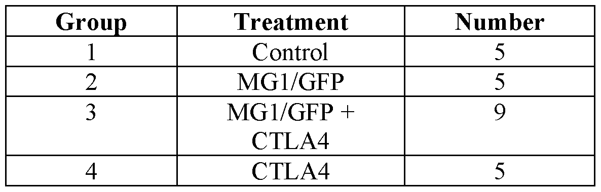
Claims
Priority Applications (10)
| Application Number | Priority Date | Filing Date | Title |
|---|---|---|---|
| KR1020187023161A KR20190038470A (en) | 2016-01-11 | 2017-01-11 | Combination of oncolytic virus and checkpoint inhibitor |
| JP2018536150A JP2019501205A (en) | 2016-01-11 | 2017-01-11 | Oncolytic virus and checkpoint inhibitor combination therapy |
| US16/069,136 US20190022203A1 (en) | 2016-01-11 | 2017-01-11 | Oncolytic virus and checkpoint inhibitor combination therapy |
| CA3011157A CA3011157A1 (en) | 2016-01-11 | 2017-01-11 | Oncolytic virus and checkpoint inhibitor combination therapy |
| EP17738058.1A EP3402500A4 (en) | 2016-01-11 | 2017-01-11 | Oncolytic virus and checkpoint inhibitor combination therapy |
| CN201780006365.7A CN109069561A (en) | 2016-01-11 | 2017-01-11 | Oncolytic virus and checkpoint inhibitor combination treatment |
| BR112018013995A BR112018013995A2 (en) | 2016-01-11 | 2017-01-11 | oncolytic virus combination therapy and checkpoint inhibitor |
| MX2018008346A MX2018008346A (en) | 2016-01-11 | 2017-01-11 | Oncolytic virus and checkpoint inhibitor combination therapy. |
| AU2017207532A AU2017207532A1 (en) | 2016-01-11 | 2017-01-11 | Oncolytic virus and checkpoint inhibitor combination therapy |
| US16/059,914 US20190070280A1 (en) | 2016-01-11 | 2018-08-09 | Oncolytic virus and checkpoint inhibitor combination therapy |
Applications Claiming Priority (2)
| Application Number | Priority Date | Filing Date | Title |
|---|---|---|---|
| US201662277352P | 2016-01-11 | 2016-01-11 | |
| US62/277,352 | 2016-01-11 |
Related Child Applications (2)
| Application Number | Title | Priority Date | Filing Date |
|---|---|---|---|
| US16/069,136 A-371-Of-International US20190022203A1 (en) | 2016-01-11 | 2017-01-11 | Oncolytic virus and checkpoint inhibitor combination therapy |
| US16/059,914 Continuation US20190070280A1 (en) | 2016-01-11 | 2018-08-09 | Oncolytic virus and checkpoint inhibitor combination therapy |
Publications (1)
| Publication Number | Publication Date |
|---|---|
| WO2017120670A1 true WO2017120670A1 (en) | 2017-07-20 |
Family
ID=59310481
Family Applications (1)
| Application Number | Title | Priority Date | Filing Date |
|---|---|---|---|
| PCT/CA2017/050031 WO2017120670A1 (en) | 2016-01-11 | 2017-01-11 | Oncolytic virus and checkpoint inhibitor combination therapy |
Country Status (10)
| Country | Link |
|---|---|
| US (2) | US20190022203A1 (en) |
| EP (1) | EP3402500A4 (en) |
| JP (1) | JP2019501205A (en) |
| KR (1) | KR20190038470A (en) |
| CN (1) | CN109069561A (en) |
| AU (1) | AU2017207532A1 (en) |
| BR (1) | BR112018013995A2 (en) |
| CA (1) | CA3011157A1 (en) |
| MX (1) | MX2018008346A (en) |
| WO (1) | WO2017120670A1 (en) |
Cited By (8)
| Publication number | Priority date | Publication date | Assignee | Title |
|---|---|---|---|---|
| WO2018170133A1 (en) * | 2017-03-15 | 2018-09-20 | Amgen Inc. | Use of oncolytic viruses, alone or in combination with a checkpoint inhibitor, for the treatment of cancer |
| WO2019032431A1 (en) * | 2017-08-07 | 2019-02-14 | Amgen Inc. | Treatment of triple negative breast cancer or colorectal cancer with liver metastases with an anti pd-l1 antibody and an oncolytic virus |
| WO2019048689A1 (en) | 2017-09-11 | 2019-03-14 | Imba - Institut Für Molekulare Biotechnologie Gmbh | Tumor organoid model |
| WO2020025642A1 (en) | 2018-08-03 | 2020-02-06 | Ludwig Institute For Cancer Research Ltd. | Viral vectors encoding cancer/testis antigens for use in a method of prevention or treatment of cancer |
| WO2020104694A1 (en) * | 2018-11-23 | 2020-05-28 | Vira Therapeutics Gmbh | Vsv chimeric vectors |
| WO2020223639A1 (en) * | 2019-05-01 | 2020-11-05 | Sensei Biotherapeutics, Inc. | Combination therapies for cancer |
| WO2023159102A1 (en) * | 2022-02-17 | 2023-08-24 | Regeneron Pharmaceuticals, Inc. | Combinations of checkpoint inhibitors and oncolytic virus for treating cancer |
| US11802292B2 (en) | 2018-01-05 | 2023-10-31 | Ottawa Hospital Research Institute | Modified orthopoxvirus vectors |
Families Citing this family (12)
| Publication number | Priority date | Publication date | Assignee | Title |
|---|---|---|---|---|
| JP7198666B2 (en) * | 2016-08-26 | 2023-01-04 | 哲治 奥野 | Microvascular blood flow reducing agent and its use |
| CN111494432A (en) * | 2019-01-31 | 2020-08-07 | 惠君生物医药科技(杭州)有限公司 | Pharmaceutical composition for treating tumor or cancer and application thereof |
| CN111606999B (en) * | 2019-02-26 | 2022-09-06 | 南京惟亚德生物医药有限公司 | Replicative oncolytic adenovirus with functions of activating immune co-stimulatory signaling pathway and blocking immune checkpoint and application thereof |
| WO2021022155A1 (en) * | 2019-07-31 | 2021-02-04 | Aivita Biomedical, Inc. | Soluble programmed cell death protein-1 as a biomarker in cancer patients |
| CN110564767A (en) * | 2019-08-08 | 2019-12-13 | 董春升 | attenuated virus vector system, application of attenuated virus vector system in preparation of anti-malignant tumor medicine and use method of medicine |
| CN114364390A (en) * | 2019-08-26 | 2022-04-15 | 拜耳诺克斯有限公司 | Pharmaceutical composition for treating cancer comprising anticancer virus, immune checkpoint inhibitor and hydroxyurea as active ingredients |
| KR20210101620A (en) * | 2020-02-10 | 2021-08-19 | 주식회사 천랩 | anti-cancer therapy using Faecalibacterium |
| CN111286493B (en) * | 2020-05-12 | 2020-10-27 | 上海荣瑞医药科技有限公司 | Oncolytic virus vaccine and medicine for treating tumor by combining oncolytic virus vaccine with immune cells |
| CN111467489B (en) * | 2020-05-12 | 2022-06-03 | 上海荣瑞医药科技有限公司 | Medicine for treating tumor |
| CN116802278A (en) * | 2020-09-18 | 2023-09-22 | 成都美杰赛尔生物科技有限公司 | Oncolytic viruses and engineered immune cells in combination for treatment of tumors |
| EP4288140A1 (en) | 2021-02-05 | 2023-12-13 | Iovance Biotherapeutics, Inc. | Adjuvant therapy for cancer |
| CN113368246B (en) * | 2021-05-12 | 2023-05-26 | 中山大学 | Synergistic antitumor drug |
Citations (2)
| Publication number | Priority date | Publication date | Assignee | Title |
|---|---|---|---|---|
| WO2015123496A1 (en) * | 2014-02-14 | 2015-08-20 | Immune Design Corp. | Immunotherapy of cancer through combination of local and systemic immune stimulation |
| WO2016128542A1 (en) * | 2015-02-13 | 2016-08-18 | Transgene Sa | Immunotherapeutic vaccine and antibody combination therapy |
Family Cites Families (4)
| Publication number | Priority date | Publication date | Assignee | Title |
|---|---|---|---|---|
| CN102448487B (en) * | 2009-03-16 | 2016-03-16 | 麦克马斯特大学 | Method of vaccination |
| WO2014127478A1 (en) * | 2013-02-21 | 2014-08-28 | Children's Hospital Of Eastern Ontario Research Institute Inc. | Vaccine composition |
| US10188713B2 (en) * | 2014-03-19 | 2019-01-29 | Mayo Foundation For Medical Education And Research | Methods and materials for treating cancer |
| WO2015143221A1 (en) * | 2014-03-19 | 2015-09-24 | Mayo Foundation For Medical Education And Research | Methods and materials for treating cancer |
-
2017
- 2017-01-11 MX MX2018008346A patent/MX2018008346A/en unknown
- 2017-01-11 WO PCT/CA2017/050031 patent/WO2017120670A1/en active Application Filing
- 2017-01-11 JP JP2018536150A patent/JP2019501205A/en active Pending
- 2017-01-11 AU AU2017207532A patent/AU2017207532A1/en not_active Abandoned
- 2017-01-11 KR KR1020187023161A patent/KR20190038470A/en unknown
- 2017-01-11 CN CN201780006365.7A patent/CN109069561A/en active Pending
- 2017-01-11 US US16/069,136 patent/US20190022203A1/en not_active Abandoned
- 2017-01-11 BR BR112018013995A patent/BR112018013995A2/en not_active IP Right Cessation
- 2017-01-11 CA CA3011157A patent/CA3011157A1/en not_active Abandoned
- 2017-01-11 EP EP17738058.1A patent/EP3402500A4/en not_active Withdrawn
-
2018
- 2018-08-09 US US16/059,914 patent/US20190070280A1/en not_active Abandoned
Patent Citations (2)
| Publication number | Priority date | Publication date | Assignee | Title |
|---|---|---|---|---|
| WO2015123496A1 (en) * | 2014-02-14 | 2015-08-20 | Immune Design Corp. | Immunotherapy of cancer through combination of local and systemic immune stimulation |
| WO2016128542A1 (en) * | 2015-02-13 | 2016-08-18 | Transgene Sa | Immunotherapeutic vaccine and antibody combination therapy |
Non-Patent Citations (3)
| Title |
|---|
| IBRAHIM ET AL.: "Viro-immune therapy: A new strategy for treatment of pancreatic cance r", WORLD J GASTRO, vol. 22, no. 2, 14 January 2016 (2016-01-14), pages 748 - 763, XP055398735, [retrieved on 20170313] * |
| See also references of EP3402500A4 * |
| SHIM ET AL.: "Inhibitory Receptors Induced by VSV Viroimmunotherapy are Not Necessarilty Targets for Improving Treatment Efficacy", MOLECULAR THERAPY, vol. 25, no. 4, 1 April 2017 (2017-04-01), pages 962 - 975, XP055571752, ISSN: 1525-0016, DOI: 10.1016/j.ymthe.2017.01.023 * |
Cited By (10)
| Publication number | Priority date | Publication date | Assignee | Title |
|---|---|---|---|---|
| WO2018170133A1 (en) * | 2017-03-15 | 2018-09-20 | Amgen Inc. | Use of oncolytic viruses, alone or in combination with a checkpoint inhibitor, for the treatment of cancer |
| CN110461346A (en) * | 2017-03-15 | 2019-11-15 | 美国安进公司 | Oncolytic virus is independent or the purposes for being used for treating cancer is combined with checkpoint inhibitor |
| WO2019032431A1 (en) * | 2017-08-07 | 2019-02-14 | Amgen Inc. | Treatment of triple negative breast cancer or colorectal cancer with liver metastases with an anti pd-l1 antibody and an oncolytic virus |
| CN111246883A (en) * | 2017-08-07 | 2020-06-05 | 美国安进公司 | Treatment of triple negative breast cancer or colorectal cancer with anti-PD-L1 antibody and oncolytic virus with liver metastasis |
| WO2019048689A1 (en) | 2017-09-11 | 2019-03-14 | Imba - Institut Für Molekulare Biotechnologie Gmbh | Tumor organoid model |
| US11802292B2 (en) | 2018-01-05 | 2023-10-31 | Ottawa Hospital Research Institute | Modified orthopoxvirus vectors |
| WO2020025642A1 (en) | 2018-08-03 | 2020-02-06 | Ludwig Institute For Cancer Research Ltd. | Viral vectors encoding cancer/testis antigens for use in a method of prevention or treatment of cancer |
| WO2020104694A1 (en) * | 2018-11-23 | 2020-05-28 | Vira Therapeutics Gmbh | Vsv chimeric vectors |
| WO2020223639A1 (en) * | 2019-05-01 | 2020-11-05 | Sensei Biotherapeutics, Inc. | Combination therapies for cancer |
| WO2023159102A1 (en) * | 2022-02-17 | 2023-08-24 | Regeneron Pharmaceuticals, Inc. | Combinations of checkpoint inhibitors and oncolytic virus for treating cancer |
Also Published As
| Publication number | Publication date |
|---|---|
| KR20190038470A (en) | 2019-04-08 |
| AU2017207532A1 (en) | 2018-08-16 |
| EP3402500A1 (en) | 2018-11-21 |
| CA3011157A1 (en) | 2017-07-20 |
| US20190070280A1 (en) | 2019-03-07 |
| BR112018013995A2 (en) | 2019-02-05 |
| US20190022203A1 (en) | 2019-01-24 |
| JP2019501205A (en) | 2019-01-17 |
| CN109069561A (en) | 2018-12-21 |
| EP3402500A4 (en) | 2019-06-12 |
| MX2018008346A (en) | 2019-07-04 |
Similar Documents
| Publication | Publication Date | Title |
|---|---|---|
| US20190070280A1 (en) | Oncolytic virus and checkpoint inhibitor combination therapy | |
| WO2014047350A1 (en) | Oncolytic virus encoding pd-1 binding agents and uses of the same | |
| AU2018254626B2 (en) | Oncolytic vaccinia virus and checkpoint inhibitor combination therapy | |
| JP2023089171A (en) | Use of oncolytic viruses alone or in combination with checkpoint inhibitor for treatment of cancer | |
| US20220249621A1 (en) | TREATMENT OF CANCERS USING sEphB4-HSA FUSION PROTEINS | |
| KR20180015113A (en) | Therapeutic compositions and methods of use for treating cancer | |
| JP2019515019A (en) | Pseudotyped oncolytic rhabdoviruses and their use in combination therapy | |
| KR20130101048A (en) | Vesicular stomatitis viruses | |
| JP2022546539A (en) | A novel oncolytic virus platform for treating cancer with myxoma virus | |
| WO2018075447A1 (en) | Combination of braf inhibitor, talimogene laherparepvec, and immune checkpoint inhibitor for use in the treatment cancer (melanoma) | |
| US20210128727A1 (en) | A pharmaceutical combination for use in the treatment of cancer | |
| US20240092852A1 (en) | Multi-armed myxoma virus | |
| JP2024514707A (en) | Compositions and methods for use in immunotherapy | |
| JP2023552203A (en) | Oncolytic herpes simplex virus type I for the treatment of brain tumors | |
| US20180153978A1 (en) | TUMORS EXPRESSING IgG1 Fc INDUCE ROBUST CD8 T CELL RESPONSES | |
| US20190083556A1 (en) | Analytical methods and arrays for use in the same | |
| CN110678192B (en) | Oncolytic vaccinia virus and immune checkpoint inhibitor combination therapy | |
| EP4262835A1 (en) | Use of a birnavirus alone or in combination therapy for the treatment of cancer | |
| TW202223085A (en) | Oncolytic herpes simplex type 1 viruses for treatment of brain tumors |
Legal Events
| Date | Code | Title | Description |
|---|---|---|---|
| 121 | Ep: the epo has been informed by wipo that ep was designated in this application |
Ref document number: 17738058 Country of ref document: EP Kind code of ref document: A1 |
|
| WWE | Wipo information: entry into national phase |
Ref document number: MX/A/2018/008346 Country of ref document: MX |
|
| WWE | Wipo information: entry into national phase |
Ref document number: 260505 Country of ref document: IL |
|
| ENP | Entry into the national phase |
Ref document number: 2018536150 Country of ref document: JP Kind code of ref document: A |
|
| WWE | Wipo information: entry into national phase |
Ref document number: 3011157 Country of ref document: CA |
|
| NENP | Non-entry into the national phase |
Ref country code: DE |
|
| REG | Reference to national code |
Ref country code: BR Ref legal event code: B01A Ref document number: 112018013995 Country of ref document: BR |
|
| ENP | Entry into the national phase |
Ref document number: 20187023161 Country of ref document: KR Kind code of ref document: A |
|
| WWE | Wipo information: entry into national phase |
Ref document number: 2017738058 Country of ref document: EP |
|
| ENP | Entry into the national phase |
Ref document number: 2017207532 Country of ref document: AU Date of ref document: 20170111 Kind code of ref document: A |
|
| ENP | Entry into the national phase |
Ref document number: 2017738058 Country of ref document: EP Effective date: 20180813 |
|
| ENP | Entry into the national phase |
Ref document number: 112018013995 Country of ref document: BR Kind code of ref document: A2 Effective date: 20180709 |