WO2017103182A1 - Radiolabelled mglur2/3 pet ligands - Google Patents
Radiolabelled mglur2/3 pet ligands Download PDFInfo
- Publication number
- WO2017103182A1 WO2017103182A1 PCT/EP2016/081542 EP2016081542W WO2017103182A1 WO 2017103182 A1 WO2017103182 A1 WO 2017103182A1 EP 2016081542 W EP2016081542 W EP 2016081542W WO 2017103182 A1 WO2017103182 A1 WO 2017103182A1
- Authority
- WO
- WIPO (PCT)
- Prior art keywords
- compound
- formula
- imaging
- pharmaceutically acceptable
- solvate
- Prior art date
Links
- MIZFVHFZLAHCQM-ZDUSSCGKSA-N C[C@@H](CN1c2ccc(C(F)(F)F)c(CBr)c2)[n]2ncc(C#Cc3ccc(N)nc3)c2C1=O Chemical compound C[C@@H](CN1c2ccc(C(F)(F)F)c(CBr)c2)[n]2ncc(C#Cc3ccc(N)nc3)c2C1=O MIZFVHFZLAHCQM-ZDUSSCGKSA-N 0.000 description 2
- XQRYBTKEWNVVGT-ZDUSSCGKSA-N C[C@@H](CN1c2ccc(C(F)(F)F)c(CO)c2)[n]2ncc(C#Cc3ccc(N)nc3)c2C1=O Chemical compound C[C@@H](CN1c2ccc(C(F)(F)F)c(CO)c2)[n]2ncc(C#Cc3ccc(N)nc3)c2C1=O XQRYBTKEWNVVGT-ZDUSSCGKSA-N 0.000 description 2
- OSGJYIVFYXIVRY-ZDUSSCGKSA-N C[C@@H](CN1c2ccc(C(F)(F)F)c(CF)c2)[n]2ncc(C#Cc3ccc(N)nc3)c2C1=O Chemical compound C[C@@H](CN1c2ccc(C(F)(F)F)c(CF)c2)[n]2ncc(C#Cc3ccc(N)nc3)c2C1=O OSGJYIVFYXIVRY-ZDUSSCGKSA-N 0.000 description 1
Classifications
-
- A—HUMAN NECESSITIES
- A61—MEDICAL OR VETERINARY SCIENCE; HYGIENE
- A61K—PREPARATIONS FOR MEDICAL, DENTAL OR TOILETRY PURPOSES
- A61K51/00—Preparations containing radioactive substances for use in therapy or testing in vivo
- A61K51/02—Preparations containing radioactive substances for use in therapy or testing in vivo characterised by the carrier, i.e. characterised by the agent or material covalently linked or complexing the radioactive nucleus
- A61K51/04—Organic compounds
- A61K51/041—Heterocyclic compounds
- A61K51/044—Heterocyclic compounds having nitrogen as a ring hetero atom, e.g. guanethidine, rifamycins
- A61K51/0459—Heterocyclic compounds having nitrogen as a ring hetero atom, e.g. guanethidine, rifamycins having six-membered rings with two nitrogen atoms as the only ring hetero atoms, e.g. piperazine
-
- A—HUMAN NECESSITIES
- A61—MEDICAL OR VETERINARY SCIENCE; HYGIENE
- A61P—SPECIFIC THERAPEUTIC ACTIVITY OF CHEMICAL COMPOUNDS OR MEDICINAL PREPARATIONS
- A61P25/00—Drugs for disorders of the nervous system
- A61P25/18—Antipsychotics, i.e. neuroleptics; Drugs for mania or schizophrenia
-
- A—HUMAN NECESSITIES
- A61—MEDICAL OR VETERINARY SCIENCE; HYGIENE
- A61P—SPECIFIC THERAPEUTIC ACTIVITY OF CHEMICAL COMPOUNDS OR MEDICINAL PREPARATIONS
- A61P25/00—Drugs for disorders of the nervous system
- A61P25/24—Antidepressants
-
- A—HUMAN NECESSITIES
- A61—MEDICAL OR VETERINARY SCIENCE; HYGIENE
- A61P—SPECIFIC THERAPEUTIC ACTIVITY OF CHEMICAL COMPOUNDS OR MEDICINAL PREPARATIONS
- A61P25/00—Drugs for disorders of the nervous system
- A61P25/28—Drugs for disorders of the nervous system for treating neurodegenerative disorders of the central nervous system, e.g. nootropic agents, cognition enhancers, drugs for treating Alzheimer's disease or other forms of dementia
-
- A—HUMAN NECESSITIES
- A61—MEDICAL OR VETERINARY SCIENCE; HYGIENE
- A61P—SPECIFIC THERAPEUTIC ACTIVITY OF CHEMICAL COMPOUNDS OR MEDICINAL PREPARATIONS
- A61P43/00—Drugs for specific purposes, not provided for in groups A61P1/00-A61P41/00
-
- C—CHEMISTRY; METALLURGY
- C07—ORGANIC CHEMISTRY
- C07B—GENERAL METHODS OF ORGANIC CHEMISTRY; APPARATUS THEREFOR
- C07B59/00—Introduction of isotopes of elements into organic compounds ; Labelled organic compounds per se
- C07B59/002—Heterocyclic compounds
-
- C—CHEMISTRY; METALLURGY
- C07—ORGANIC CHEMISTRY
- C07D—HETEROCYCLIC COMPOUNDS
- C07D487/00—Heterocyclic compounds containing nitrogen atoms as the only ring hetero atoms in the condensed system, not provided for by groups C07D451/00 - C07D477/00
- C07D487/02—Heterocyclic compounds containing nitrogen atoms as the only ring hetero atoms in the condensed system, not provided for by groups C07D451/00 - C07D477/00 in which the condensed system contains two hetero rings
- C07D487/04—Ortho-condensed systems
Definitions
- the present invention relates to novel, radiolabelled mGluR2/3 ligands, selective versus other mGlu receptors, which are useful for imaging and quantifying the metabotropic glutamate receptors mGlu2 and 3 in tissues, using positron-emission tomography (PET).
- PET positron-emission tomography
- the invention is also directed to compositions comprising such compounds, to processes for preparing such compounds and compositions, to the use of such compounds and compositions for imaging a tissue, cells or a mammal, in vitro or in vivo and to precursors of said compounds.
- the glutamatergic system in the CNS is one of the neurotransmitter systems that play a key role in several brain functions.
- Metabotropic glutamate receptors (mGluR) belong to the G-protein-coupled family, and eight different subtypes have been identified to date, which are distributed to various brain regions (Ferraguti & Shigemoto, Cell & Tissue Research, 326:483-504, 2006).
- mGluRs participate in the modulation of synaptic transmission and neuronal excitability in the CNS by the binding of glutamate. This activates the receptor to engage intracellular signaling partners, leading to cellular events (Niswender & Conn, Annual Review of Pharmacology & Toxicology
- mGluRs are further divided into three subgroups based on their pharmacological and structural properties: group-I (mGluRl and mGluR5), group-II (mGluR2 and mGluR3) and group-Ill (mGluR4, mGluR6, mGluR7 and mGluR8).
- group-II ligands both orthosteric and allosteric modulating, are considered to be potentially useful in the treatment of various neurological disorders, including psychosis, mood disorders, Alzheimer's disease and cognitive or memory deficiencies. This is consistent with their primary localisation in brain areas such as the cortex, hippocampus and the striatum (Ferraguti & Shigemoto, Cell & Tissue Research 326:483-504, 2006).
- WO 2013066736 (Merck Sharp & Dohme Corp.) describes quinoline carboxamide and quinoline carbonitrile compounds as mGluR2 NAMs.
- WO2013174822 (Domain therapeutics) describes 4H-pyrazolo[l,5-a]quinazolin-5-ones and 4H-pyrrolo
- WO 2014064028 F. Hoffman-La Roche AG discloses a selection of mGlu2/3 negative allosteric modulators and their potential use in the treatment of Autistic Spectrum Disorders (ASD).
- WO2014195311 Janssen Pharmaceutica NV discloses 6,7- dihydropyrazolo[l,5-a]pyrazine-4(5H)-one compounds and their use as mGluR2 NAMs.
- the group-II receptors are mainly located on presynaptic nerve terminals where they exert a negative feedback loop to the release of glutamate into the synapse (Kelmendi et al, Primary Psychiatry 13:80-86, 2006). Functional inhibition of these receptors by antagonists or negative allosteric modulators therefore lifts the brake on glutamate release, resulting in enhanced glutamatergic signaling. This effect is believed to underlie the antidepressant-like and procognitive effects observed in preclinical species with inhibitors of the Group-II receptor.
- BDNF brain derived neurotrophic factor
- Positron Emission Tomography is a non-invasive imaging technique that offers the highest spatial and temporal resolution of all nuclear imaging techniques and has the added advantage that it can allow for true quantification of tracer concentrations in tissues. It uses positron emitting radionuclides such as, for example, 15 0, 13 N, U C and
- the present invention relates to a compound having the Formula (I)
- R 1 is -CH 2 F and R 2 is -H, or R 1 is -H and R 2 is -CH 2 F, and wherein at least one atom is radiactive, or a pharmaceutically acceptable salt or a solvate thereof.
- the compound of Formula (I) is compound 1 ?
- the compound of Formula (I) is compound 2
- the invention also relates to precursor compounds for the synthesis of compound 1.
- the present invention also relates to c
- the invention also relates to precursor compounds for the synthesis of compound 2.
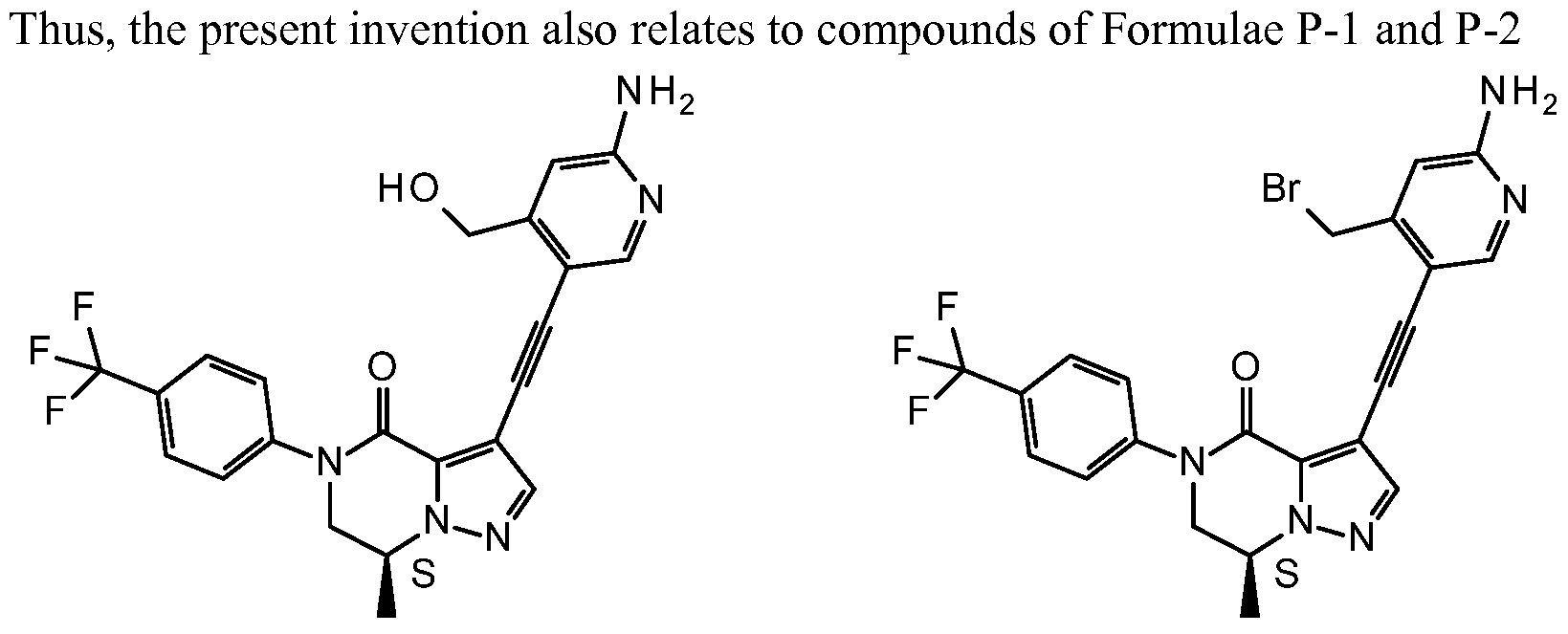
- the present invention also relates to compounds of Formulae P-3 and P-4
- the invention also relates to a pharmaceutical composition
- a pharmaceutical composition comprising a compound of Formula (I) or a pharmaceutically acceptable salt thereof and a pharmaceutically acceptable carrier or diluent.
- said pharmaceutical composition is particularly suitable for diagnosis and may be referred to therefore as a diagnostic pharmaceutical composition.
- said pharmaceutical composition is a sterile solution.
- illustrative of the invention is a sterile solution comprising a compound of Formula (I) described herein.
- the invention further relates to the use of a compound of Formula (I) as an imaging agent. Therefore, exemplifying the invention is a use of a compound of Formula (I) as described herein, for, or a method of, imaging a tissue, cells or a mammal, in vitro or in vivo.
- the invention relates to a compound of Formula (I) as described herein, for use as a contrast agent for imaging a tissue, cells or a mammal, in vitro, ex vivo, or in vivo.
- the invention further relates to a composition comprising a compound of Formula (I) for use as a contrast agent for imaging a tissue, cells or a mammal, in vitro, ex vivo, or in vivo.
- the invention also relates to a method for imaging a tissue, cells or a mammal, comprising contacting with or providing or administering a detectable amount of a labelled compound of Formula (I) as described herein to a tissue, cells or a mammal, and detecting the compound of Formula (I).
- the invention is a method of imaging a tissue, cells or a mammal, comprising contacting with or providing or administering to a tissue, cells or a mammal, a compound of Formula (I) as described herein, and imaging the tissue, cells or mammal with a positron-emission tomography imaging system. Additionally, the invention refers to a process for the preparation of a compound according to Formula (I) as described herein, comprising
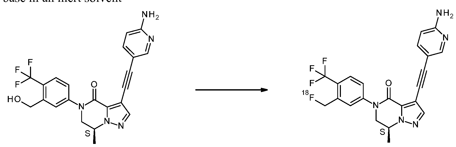
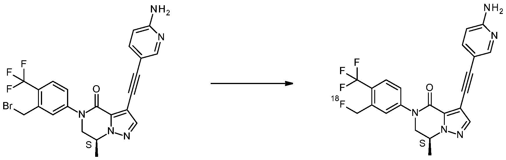
- Suitable nucleophilic radioactive fluorinating reagents in steps (a-2), (b), (c-2) and (d) are, for instance, K[ 18 F]/Kryptofix 222 or tetraalkylammonium salts incorporating
- Suitable bases in steps (a-2), (b), (c-2) and (d), are, for instance K 2 CO 3 or CS 2 CO 3 .
- Suitable solvents in steps (a-2), (b), (c-2) and (d), are, for instance, DMSO, CH 3 CN or DMF, optionally with the addition of a small amount of water.
- Figure la shows the bio distribution of [ 18 F]-l in brain areas in SD rats.
- Figure lb shows the biodistribution of [ 18 F]-l in periphery in SD rats.
- Figure 2 shows the time activity curves for the uptake of [ F]-l with and without treatment of compound A (a NAM compound, selective for mGlu2/3 ( ⁇ 20 fold selective for 2 over 3) vs other mGluRs), indicated in the figure as mGlu2/3 NAM in SD rats.
- compound A a NAM compound, selective for mGlu2/3 ( ⁇ 20 fold selective for 2 over 3) vs other mGluRs
- Figure 3a shows the biodistribution of [ F]-2 in brain areas in SD rats.
- Figure 3b shows the biodistribution of [ 18 F]-2 in periphery in SD rats.
- Figure 4 shows the time activity curves for the uptake of [ F]-2 with and without treatment of compound A (a NAM compound, selective for mGlu2/3 ( ⁇ 20 fold selective for 2 over 3) vs other mGluRs), indicated in the figure as mGlu2/3 NAM in SD rats.
- compound A a NAM compound, selective for mGlu2/3 ( ⁇ 20 fold selective for 2 over 3) vs other mGluRs
- the compounds of Formula (I) and compositions comprising the compounds of Formula (I) can be used for imaging a tissue, cells or a mammal, in vitro or in vivo.
- the invention relates to a method of imaging or quantifying the mGluR2/3 receptors in a tissue, cells or a mammal in vitro or in vivo.
- the cells and tissues are preferably central nervous system cells and tissues in which the mGluR2/3 receptors are abundant.
- the mGluR2/3 receptors are abundant in central nervous system tissue, more in particular, in central nervous system tissue forming the brain; more in particular, forming the cerebral cortex, thalamic regions, accessory olfactory bulb, hippocampus, amygdala, caudate-putamen and nucleus accumbens.
- the compound of Formula (I) can be administered intravenously, for example, by injection with a syringe or by means of a peripheral intravenous line, such as a short catheter.
- the compound of Formula (I) or a sterile solution comprising a compound of Formula (I) may in particular be administered by intravenous administration in the arm, into any identifiable vein, in particular in the back of the hand, or in the median cubital vein at the elbow.
- the invention relates to a method of imaging a tissue or cells in a mammal, comprising the intravenous administration of a compound of Formula (I), as defined herein, or a composition comprising a compound of Formula (I) to the mammal, and imaging the tissue or cells with a positron-emission tomography imaging system.
- the invention relates to a method of imaging a tissue or cells in a human, comprising the intravenous administration of a compound of Formula (I), as defined herein, or a sterile formulation comprising a compound of Formula (I) to the human, and imaging the tissue or cells with a positron-emission tomography imaging system.
- the invention relates to a method of imaging or quantifying the mGluR2/3 receptors in a mammal, comprising the intravenous administration of a compound of Formula (I), or a composition comprising a compound of Formula (I) to the mammal, and imaging with a positron-emission tomography imaging system.
- the invention relates to the use of a compound of Formula (I) for imaging a tissue, cells or a mammal, in vitro or in vivo, or the invention relates to a compound of Formula (I), for use in imaging a tissue, cells or a mammal in vitro or in vivo, using positron-emission tomography.
- the invention also relates to a method for imaging or quantifying the mGlu2 and 3 receptors in a mammal, the method comprising providing a detectable amount of a compound of Formula (I) to a mammal and detecting the compound of Formula (I) associated with mGlu2 and 3 receptors.
- the method also allows for determining mGlu2 and 3 receptor occupancy by other non-radiolabelled compounds, therefore, the invention relates to the compound of Formula (I) as defined herein, or the
- the invention relates to a method of assessing a disorder or predisposition thereto related to the mGlu2 and 3 receptors in a subject, the method comprising providing a detectable amount of a compound of Formula (I) or pharmaceutical composition according to the invention, wherein the compound of Formula (I) passes the blood-brain barrier and preferentially binds to mGlu2 and 3 receptors in brain tissue, allowing the compound to distribute into the brain tissue, and imaging the brain tissue.
- the compound is provided to a subject in a detectable amount and after sufficient time has passed for the compound to become associated with the mGlu2 and 3 receptors, the labelled compound is detected noninvasively.
- composition is intended to encompass a product comprising the specified ingredients in the specified amounts, as well as any product which results, directly or indirectly, from combinations of the specified ingredients in the specified amounts.
- detecttable amount refers to the concentration of compound above the lowest limit of detection of the imaging instrument, in particular, of the PET scanning instrument.
- Acceptable salts of the compounds of the invention are those wherein the counterion is pharmaceutically acceptable.
- salts of acids and bases which are non- pharmaceutically acceptable may also find use, for example, in the preparation or purification of a pharmaceutically acceptable compound. All salts, whether
- the pharmaceutically acceptable salts are defined to comprise the therapeutically active non-toxic acid addition salt forms that the compounds according to the invention are able to form.
- Said salts can be obtained by treating the base form of the compounds according to the invention with appropriate acids, for example inorganic acids, for example hydrohalic acid, in particular hydrochloric acid, hydrobromic acid, sulphuric acid, nitric acid and phosphoric acid; organic acids, for example acetic acid, hydroxyacetic acid, propanoic acid, lactic acid, pyruvic acid, oxalic acid, malonic acid, succinic acid, maleic acid, fumaric acid, malic acid, tartaric acid, citric acid, methanesulfonic acid, ethanesulfonic acid, benzensulfonic acid, p- toluenesulfonic acid, cyclamic acid, salicylic acid, p-aminosalicylic acid and pamoic
- inorganic acids for example hydrohalic acid, in
- salt forms can be converted into the free base form by treatment with an appropriate base.
- some of the compounds of the present invention may form solvates with water (i.e., hydrates) or common organic solvents, and such solvates are also intended to be encompassed within the scope of this invention.
- subject refers to an animal, preferably a mammal, most preferably a human, who is or has been the object of treatment, observation or experiment. Unless otherwise stated, “subject” includes both, healthy animals and animals afflicted by different diseases or disorders.
- mammal refers, in particular to humans, mice, dogs and rats.
- cell refers to a cell expressing or incorporating the mGlu2 and/or 3 receptor.
- the compounds according to the present invention find various applications for imaging tissues, cells or a mammal, both in vitro and in vivo. Thus, for instance, they can be used to map the differential distribution of mGluR2/3 in subjects of different age and sex. Further, they allow one to explore for differential distribution of mGluR2/3 in subjects afflicted by different diseases or disorders. Thus, abnormal distribution may be helpful in diagnosis, case finding, stratification of subject populations, and in monitoring disease progression in individual subjects.
- the radioligands may further find utility in determining mGluR2/3 site occupancy by other ligands. Since the radioligand is administered in trace amounts, i.e. in detectable amounts for example for PET imaging, no therapeutic effect may be attributed to the administration of the radioligands according to the invention.
- aq means aqueous
- DCM dichloromethane
- DIPE diisopropyl ether
- DMF N,N-dimethylformamide
- DMSO dimethyl sulfoxide
- DSC differential scanning calorimetry
- Et 3 N/TEA means triethylamine
- EtOH means ethanol
- EtOAc means ethyl acetate
- h means hours
- HPLC high-performance liquid chromatography
- LCMS liquid chromatography/mass spectrometry
- iPrOH means isopropyl alcohol
- MeOH means methanol
- [M+H] + means the protonated mass of the free base of the compound
- min means minutes
- m.p. means melting point
- PdCl 2 (PPh 3 ) 2 means bis(triphenylphosphine)palladium(II) chloride and PPI1 3 means triphenylphosphine
- RP means reverse phase
- r.t./RT means room temperature
- R t means retention time (in minutes)
- sat means saturated
- sol means solution
- XtalFluor-E® means (diethylamino)difluorosulfonium tetrafluoroborate.
- TLC Thin layer chromatography
- silica gel 60 F254 plates Merck
- Open column chromatography was performed on silica gel, mesh 230-400 particle size and 60 A pore size (Merck) under standard techniques.
- Automated flash column chromatography was performed using ready-to -connect cartridges from Merck, on irregular silica gel, particle size 15-40 ⁇ (normal phase disposable flash columns) on an SPOT or LAFLASH system from Armen Instrument.
- Preparative HPLC was performed on an Xbridge C18 column (4.6 x 250 mm, 5 ⁇ ; Waters, Milford USA), using EtOH / 0.01 M phosphate buffer in water pH 7.4 (39/61 v/v) at flow rate 1 mL-min 1 and wavelength of 254 nm (method A).
- radiotracers The identity of the radiotracers was confirmed using the same analytical HPLC methods as described above after co-injection with their non-radioactive analogue.
- Millex GV filters were obtained from Millipore (Amsterdam, The Netherlands).
- Radioactivity was counted using the Wizard 1480 automated gamma counter (Perkin Elmer, Waltham, USA).
- [ F]F " was collected by purging the proton irradiated target content (98%> 0-H 2 0) over a QMA (Waters, Milford USA) cartridge. Next the QMA cartridge was eluted, using CH 3 CN/water (700 of 95/5 v/v) containing Kryptofix 222 (26 mg) and K 2 C0 3 (2.5 mg) to the reaction vial. The solution was dried under a gentle helium flow at 110 °C for 6 min, followed twice by an addition of CH 3 CN (1 mL) and dried under helium at 110 °C for 5 min each.
- CH 3 CN/water 700 of 95/5 v/v
- Kryptofix 222 26 mg
- K 2 C0 3 2.5 mg
- the mesyl precursor (2 mg) in dry DMSO (0.5 mL) was added, and reacted for 10 min at 120 °C.
- the r.m. was diluted and [ F]-l or [ F]-2 was subsequently purified using HPLC method A.
- the collected fraction was then passed over a sterile millex GV filter, and was further diluted with saline to a concentration of 10% EtOH.
- the conversion was of 50-80 % (as rough estimate) according to TLC.
- a fraction of the non converted alcohol precursor was always present in the precursor mixture used for the radiosynthesis, as well as some side products.
- Values are peak values, and are obtained with experimental uncertainties that are commonly associated with this analytical method.
- Mettler Toledo Mettler FP 81HT / FP90 apparatus B: For a number of compounds, melting points were determined in open capillary tubes on a Mettler FP 81HT / FP90 apparatus. Melting points were measured with a temperature gradient of 1, 3, 5 or 10 °C/minute. Maximum temperature was 300 °C. The melting point was read from a digital display.
- HPLC High Performance Liquid Chromatography
- MS Mass Spectrometer
- n.d. means not determined.
- [ ⁇ ] ⁇ ⁇ (100a) / (/ x c) : where / is the path length in dm and c is the concentration in g/100 ml for a sample at a temperature T (°C) and a wavelength ⁇ (in nm). If the wavelength of light used is 589 nm (the sodium D line), then the symbol D might be used instead.
- T temperature
- ⁇ in nm
- the wavelength of light used is 589 nm (the sodium D line)
- the symbol D might be used instead.
- the sign of the rotation (+ or -) should always be given. When using this equation the concentration and solvent are always provided in parentheses after the rotation. The rotation is reported using degrees and no units of concentration are given (it is assumed to be g/100 mL).
- [ H] -compound A (a NAM compound, selective for mGlu2/3 (-20 fold selective for 2 over 3) vs other mGluRs) binding
- membranes from human mGlu2 and mGlu3 HEK293 cells, and also rat cortical membranes were used. After thawing, membranes were homogenized using an Ultra Turrax homogenizer and suspended in ice-cold binding buffer containing 50 mM Tris-HCl (pH 7.4), 10 mM MgCl 2 , 2 mM CaCl 2 . Displacement studies were done using 6 nM of radioligand, except for human mGlu3 membranes where 25 nM was used.
- Assay mixtures were incubated for 60 min at RT in a volume of 0.5 ml containing 7.5 mg, 75-100 mg or 75 ⁇ g membrane protein of human mGlu2, human mGlu3 or rat cortex, respectively.
- Non-specific binding was estimated in the presence of 10 mM compound B (a NAM with IC 50 ⁇ 10 nM against hmGlu2 and IC 50 -200 nM against hmGlu3).
- Filtration was performed using Whatman GF/C filter sheets pre-soaked in 0.1% PEI and a Brandell harvester 96.
- Radioligand competition binding data were calculated as percentage of total binding measured in the absence of test compound. Inhibition curves, plotting percentage of total binding versus the log concentration of the test compound, were generated using the Lexis software. Sigmoid inhibition curves were analyzed using non-linear regression analysis.
- Animal PET imaging was performed on a lutetium oxyorthosilicate detector-based tomograph (microPET FOCUS-220; Siemens Medical Solutions USA, KnoxviUe, TN), which had a transaxial resolution of 1.35 mm (full-width at half-maximum). Data were acquired in a 128 x 128x95 matrix with a pixel width of 0.475 mm and a slice thickness of 0.796 mm.
- rats were kept under gas anesthesia (2.5 % isoflurane in oxygen at a flow rate of 1 1/min), and their body temperature was maintained between 36.5 and 37 °C using a heating pad. PET data were analyzed using Pmod software version 3.2 (Pmod, Zurich Switzerland).
- Sprague-Dawley rats obtained from Harlan (the Netherlands), were housed in groups of four to six per cage until treatment. They were kept at a constant temperature of 21°C and at a 12-h light/dark cycle, in which lights were switched on at 8:00 a.m. Animals had unrestricted access to food (Teklad Global 16% Protein Rodent Diet, Harlan, Madison, WI, USA) and water. All animal experiments were performed in compliance with Belgium laws on animal experimentation and after approval by the local animal ethics committee.
- the brain uptake of [ 18 F]-2 showed a variable tracer concentration with slow washout in the studied regions. The highest uptake was observed in the cortex, and the lowest brain uptake in the pons.
- All pretreatment solutions were 1 mg/mL solutions in 20% ⁇ -cyclodextrine in saline with a pH ranging between 6 and 8, and sterile filtered over a Millex GV filter prior to use.
- a high brain uptake was observed for [ 18 F]-l, and especially the frontal cortex and the striatum show a high uptake.
- the uptake was the lowest in the pons, while the uptake of the pons was in the same range after pretreatment with compound A. In the other regions the uptake was reduced after pretreatment with compound A. The peak uptake was probably not reached within 90 min, since the time activity curves kept on increasing as a function of time post injection.
- 39-49 MBq of [ F]-2 was injected in 3 SD rats weighing 301- 310 g in a tail vein, and scanned simultaneously during a 90-min dynamic PET scan.
- the same animals were injected with 10 mg/kg compound A s.c. 60 min prior to injection of 46-50 MBq [ 18 F]-2 in the tail vein, and scanned simultaneously during a 90 min dynamic PET scan.
- the ⁇ scans obtained after injection of [ F]-2 showed some mixed results.
- the peak uptake was higher after pretreatment, but generally showed a faster washout, while the uptake of the brain regions at the baseline scan did not show this early peak.
- the cortex showed an increase of uptake over time, which might be due to partial volume effects from skull that due to tracer defluorination showed high and increasing radioactivity concentration.
- the uptake in the periphery of the ex-vivo biodistribution showed the highest uptake in the liver, as well as a high kidney uptake, followed by urinary excretion.
- the bone uptake [ 1 1 8 0 F]-1 was low at start, but increased slightly up over time hinting for some defluorination.
- [ 18 F]-2 showed substantial increase of bone uptake over time, indicating massive defluorination, with subsequent binding of [ 18 F]F " to bone.
- [ F]-2 showed a fast washout from the different brain areas.
- the pons is considered to be a reference region as with absence of mGluR2 or mGluR3 expression, while on all other regions both mGluR2 and mGluR3 are present, with the highest expression levels in the cerebral cortex (Farinha A. et al. BJPharmacol, 2015, 172, 2383-2396).
- [ 18 F]-2 showed the highest uptake in the cortex, with a low uptake in the pons in combination with a faster washout from the pons compared to the other regions, and suggesting a good mGluR2/3 specificity, since the brain uptake reflects the reported distribution
- [ 18 F]-l showed peculiar brain kinetics with persistently increasing activity concentration in the frontal cortex and the striatum as a function of time whereas activity in the pons remained low.
- the high uptake in frontal cortex and hippocampus could be blocked by pretreatment with compound A.
- This continuous uptake pattern might be due to a high affinity of [ 18 F]-l for either or both mGluR2 and mGluR3, or could be due to (pseudo)irreversible binding.
- These data also indicate good mGluR2/3 specificity, since the uptake in all brain regions was reduced by compound A pretreatment to about the same height as the uptake in the pons.
Landscapes
- Health & Medical Sciences (AREA)
- Chemical & Material Sciences (AREA)
- Organic Chemistry (AREA)
- Life Sciences & Earth Sciences (AREA)
- General Health & Medical Sciences (AREA)
- Veterinary Medicine (AREA)
- Public Health (AREA)
- Medicinal Chemistry (AREA)
- Pharmacology & Pharmacy (AREA)
- Animal Behavior & Ethology (AREA)
- Bioinformatics & Cheminformatics (AREA)
- Engineering & Computer Science (AREA)
- Nuclear Medicine, Radiotherapy & Molecular Imaging (AREA)
- Chemical Kinetics & Catalysis (AREA)
- Biomedical Technology (AREA)
- General Chemical & Material Sciences (AREA)
- Neurology (AREA)
- Neurosurgery (AREA)
- Psychiatry (AREA)
- Physics & Mathematics (AREA)
- Proteomics, Peptides & Aminoacids (AREA)
- Optics & Photonics (AREA)
- Epidemiology (AREA)
- Pain & Pain Management (AREA)
- Hospice & Palliative Care (AREA)
- Medicines Containing Antibodies Or Antigens For Use As Internal Diagnostic Agents (AREA)
- Organic Low-Molecular-Weight Compounds And Preparation Thereof (AREA)
- Nitrogen Condensed Heterocyclic Rings (AREA)
Abstract
Description


Claims


Priority Applications (13)
| Application Number | Priority Date | Filing Date | Title |
|---|---|---|---|
| ES16812961T ES2828976T3 (en) | 2015-12-18 | 2016-12-16 | Radiolabeled mGluR2 / 3 Ligands for TEP |
| JP2018531628A JP6927974B2 (en) | 2015-12-18 | 2016-12-16 | Radiolabeled mGluR2 / 3PET ligand |
| AU2016374571A AU2016374571B2 (en) | 2015-12-18 | 2016-12-16 | Radiolabelled mGluR2/3 PET ligands |
| PL16812961T PL3389727T3 (en) | 2015-12-18 | 2016-12-16 | Radiolabelled mglur2/3 pet ligands |
| US16/061,437 US11033641B2 (en) | 2015-12-18 | 2016-12-16 | Radiolabelled mGluR2/3 pet ligands |
| DK16812961.7T DK3389727T3 (en) | 2015-12-18 | 2016-12-16 | RADIOACTIVELY LABELED MGLUR2 / 3-PET LIGANDER |
| CA3003998A CA3003998A1 (en) | 2015-12-18 | 2016-12-16 | Radiolabelled mglur2/3 pet ligands |
| RS20201288A RS60981B1 (en) | 2015-12-18 | 2016-12-16 | Radiolabelled mglur2/3 pet ligands |
| LTEP16812961.7T LT3389727T (en) | 2015-12-18 | 2016-12-16 | Radiolabelled mglur2/3 pet ligands |
| EP16812961.7A EP3389727B1 (en) | 2015-12-18 | 2016-12-16 | Radiolabelled mglur2/3 pet ligands |
| SI201630916T SI3389727T1 (en) | 2015-12-18 | 2016-12-16 | Radiolabelled mglur2/3 pet ligands |
| HRP20201372TT HRP20201372T1 (en) | 2015-12-18 | 2020-08-28 | Radiolabelled mglur2/3 pet ligands |
| CY20201101051T CY1124935T1 (en) | 2015-12-18 | 2020-11-09 | RADIOLABELED MGLUR2/3 TAGS FOR PET |
Applications Claiming Priority (2)
| Application Number | Priority Date | Filing Date | Title |
|---|---|---|---|
| EP15201240 | 2015-12-18 | ||
| EP15201240.7 | 2015-12-18 |
Publications (1)
| Publication Number | Publication Date |
|---|---|
| WO2017103182A1 true WO2017103182A1 (en) | 2017-06-22 |
Family
ID=55023944
Family Applications (1)
| Application Number | Title | Priority Date | Filing Date |
|---|---|---|---|
| PCT/EP2016/081542 WO2017103182A1 (en) | 2015-12-18 | 2016-12-16 | Radiolabelled mglur2/3 pet ligands |
Country Status (16)
| Country | Link |
|---|---|
| US (1) | US11033641B2 (en) |
| EP (1) | EP3389727B1 (en) |
| JP (1) | JP6927974B2 (en) |
| AU (1) | AU2016374571B2 (en) |
| CA (1) | CA3003998A1 (en) |
| CY (1) | CY1124935T1 (en) |
| DK (1) | DK3389727T3 (en) |
| ES (1) | ES2828976T3 (en) |
| HR (1) | HRP20201372T1 (en) |
| HU (1) | HUE050665T2 (en) |
| LT (1) | LT3389727T (en) |
| PL (1) | PL3389727T3 (en) |
| PT (1) | PT3389727T (en) |
| RS (1) | RS60981B1 (en) |
| SI (1) | SI3389727T1 (en) |
| WO (1) | WO2017103182A1 (en) |
Cited By (7)
| Publication number | Priority date | Publication date | Assignee | Title |
|---|---|---|---|---|
| US10005786B2 (en) | 2014-08-01 | 2018-06-26 | Janssen Pharmaceutica Nv | Substituted 6,7-dihydropyrazolo[1,5-a]pyrazines as negative allosteric modulators of MGLUR2 receptors |
| US10005785B2 (en) | 2014-08-01 | 2018-06-26 | Janssen Pharmaceutica Nv | Substituted 6,7-dihydropyrazolo[1,5-a] pyrazines as negative allosteric modulators of mGlUR2 receptors |
| US10072014B2 (en) | 2014-12-03 | 2018-09-11 | Janssen Pharmaceutica Nv | 6,7-dihydropyrazolo[1,5-a]pyrazin-4(5H)-one compounds and their use as negative allosteric modulators of MGLUR2 receptors |
| US10953008B2 (en) | 2017-11-24 | 2021-03-23 | Sumitomo Dainippon Pharma Co., Ltd. | Substituted pyrazolo[1,5-a]pyrazines as negative allosteric modulators of group II metabotropic glutamate receptor |
| US10967078B2 (en) | 2014-12-03 | 2021-04-06 | Janssen Pharmaceutica Nv | Radiolabelled mGluR2 PET ligands |
| US11033641B2 (en) | 2015-12-18 | 2021-06-15 | Janssen Pharmaceutica Nv | Radiolabelled mGluR2/3 pet ligands |
| US11045562B2 (en) | 2015-12-18 | 2021-06-29 | Janssen Pharmaceutica Nv | Radiolabelled mGluR2/3 PET ligands |
Citations (2)
| Publication number | Priority date | Publication date | Assignee | Title |
|---|---|---|---|---|
| WO2012062752A1 (en) * | 2010-11-08 | 2012-05-18 | Janssen Pharmaceuticals, Inc. | RADIOLABELLED mGLuR2 PET LIGANDS |
| WO2016016381A1 (en) * | 2014-08-01 | 2016-02-04 | Janssen Pharmaceutica Nv | 6,7-dihydropyrazolo[1,5-a]pyrazin-4(5h)-one compounds and their use as negative allosteric modulators of mglur2 receptors |
Family Cites Families (52)
| Publication number | Priority date | Publication date | Assignee | Title |
|---|---|---|---|---|
| EP0756200B1 (en) | 1995-07-26 | 1999-11-10 | Konica Corporation | Silver halide color photographic light-sensitive material |
| DE19653647A1 (en) | 1996-12-20 | 1998-06-25 | Hoechst Ag | Vitronectin receptor antagonists, their preparation and their use |
| US6482821B2 (en) | 1996-12-20 | 2002-11-19 | Hoechst Aktiengellschaft | Vitronectin receptor antagonists, their preparation and their use |
| US6831074B2 (en) | 2001-03-16 | 2004-12-14 | Pfizer Inc | Pharmaceutically active compounds |
| US20030114448A1 (en) | 2001-05-31 | 2003-06-19 | Millennium Pharmaceuticals, Inc. | Inhibitors of factor Xa |
| TWI372050B (en) | 2003-07-03 | 2012-09-11 | Astex Therapeutics Ltd | (morpholin-4-ylmethyl-1h-benzimidazol-2-yl)-1h-pyrazoles |
| US7329662B2 (en) | 2003-10-03 | 2008-02-12 | Hoffmann-La Roche Inc. | Pyrazolo-pyridine |
| WO2005061507A1 (en) | 2003-12-16 | 2005-07-07 | Pfizer Products Inc. | Bicyclic pyrazol-4-one cannabinoid receptor ligands and uses thereof |
| ES2299044T3 (en) | 2004-06-21 | 2008-05-16 | F. Hoffmann-La Roche Ag | DERIVATIVES OF PIRRAZOL-PYRIMIDINE. |
| WO2006030847A1 (en) | 2004-09-17 | 2006-03-23 | Dainippon Sumitomo Pharma Co., Ltd. | Novel bicyclic pyrazole derivative |
| DE102004054665A1 (en) | 2004-11-12 | 2006-05-18 | Bayer Cropscience Gmbh | Substituted bicyclic and tricyclic pyrazole derivatives Methods for the preparation and use as herbicides and plant growth regulators |
| EP1851225B1 (en) | 2005-02-11 | 2011-06-15 | F. Hoffmann-La Roche AG | Pyrazolo-pyrimidine derivatives as mglur2 antagonists |
| CA2602444C (en) | 2005-03-23 | 2013-03-19 | F.Hoffmann-La Roche Ag | Acetylenyl-pyrazolo-pyrimidine derivatives as mglur2 antagonists |
| WO2007084314A2 (en) | 2006-01-12 | 2007-07-26 | Incyte Corporation | MODULATORS OF 11-ß HYDROXYL STEROID DEHYDROGENASE TYPE 1, PHARMACEUTICAL COMPOSITIONS THEREOF, AND METHODS OF USING THE SAME |
| US7553836B2 (en) | 2006-02-06 | 2009-06-30 | Bristol-Myers Squibb Company | Melanin concentrating hormone receptor-1 antagonists |
| JP5523829B2 (en) | 2006-06-29 | 2014-06-18 | アステックス、セラピューティックス、リミテッド | Compound drug |
| ES2395583T3 (en) | 2007-05-10 | 2013-02-13 | Ge Healthcare Limited | IMIDAZOL (1,2-A) PIRIDINES and compounds related to activity against CB2 cannabinoid receptors |
| EP2085390A1 (en) | 2008-01-31 | 2009-08-05 | Institut National De La Sante Et De La Recherche Medicale (Inserm) | Labelled analogues of halobenzamides as multimodal radiopharmaceuticals and their precursors |
| US9446995B2 (en) | 2012-05-21 | 2016-09-20 | Illinois Institute Of Technology | Synthesis of therapeutic and diagnostic drugs centered on regioselective and stereoselective ring opening of aziridinium ions |
| PL2268612T3 (en) | 2008-03-24 | 2015-02-27 | Novartis Ag | Arylsulfonamide-based matrix metalloprotease inhibitors |
| WO2009130232A1 (en) | 2008-04-24 | 2009-10-29 | Glaxo Group Limited | Pyrazolo [1, 5 -a] pyrazine derivatives as antagonists of v1b receptors |
| EP2327704A4 (en) | 2008-08-29 | 2012-05-09 | Shionogi & Co | Ring-fused azole derivative having pi3k-inhibiting activity |
| MX2011002042A (en) | 2008-09-02 | 2011-06-20 | Ortho Mcneil Janssen Pharm | 3-azabicyclo[3.1.0]hexyl derivatives as modulators of metabotropic glutamate receptors. |
| MX2011011964A (en) | 2009-05-12 | 2012-02-23 | Janssen Pharmaceuticals Inc | 1,2,4-triazolo [4,3-a] pyridine derivatives and their use for the treatment or prevention of neurological and psychiatric disorders. |
| MY153913A (en) | 2009-05-12 | 2015-04-15 | Janssen Pharmaceuticals Inc | 7-aryl-1,2,4-triazolo[4,3-a]pyridine derivatives and their use as positive allosteric modulators of mglur2 receptors |
| EP2627648A1 (en) | 2010-09-16 | 2013-08-21 | Novartis AG | 17aHYDROXYLASE/C17,20-LYASE INHIBITORS |
| AU2011343477A1 (en) | 2010-12-17 | 2013-07-04 | Vanderbilt University | Bicyclic triazole and pyrazole lactams as allosteric modulators of mGluR5 receptors |
| GB201106817D0 (en) | 2011-04-21 | 2011-06-01 | Astex Therapeutics Ltd | New compound |
| CA2842190A1 (en) | 2011-07-19 | 2013-01-24 | Infinity Pharmaceuticals Inc. | Heterocyclic compounds and uses thereof |
| US8969363B2 (en) | 2011-07-19 | 2015-03-03 | Infinity Pharmaceuticals, Inc. | Heterocyclic compounds and uses thereof |
| CN104010504B (en) | 2011-11-03 | 2016-04-06 | 默沙东公司 | As mGluR2 bear the derivative of the quinoline formyl amine of allosteric modulators and quinolinecarbonitriles, composition, and uses thereof |
| JP2013189395A (en) | 2012-03-14 | 2013-09-26 | Dainippon Sumitomo Pharma Co Ltd | Dihydropyrrolopyradinone derivative |
| US8940742B2 (en) | 2012-04-10 | 2015-01-27 | Infinity Pharmaceuticals, Inc. | Heterocyclic compounds and uses thereof |
| US20130281397A1 (en) | 2012-04-19 | 2013-10-24 | Rvx Therapeutics Inc. | Treatment of diseases by epigenetic regulation |
| EP2666775A1 (en) | 2012-05-21 | 2013-11-27 | Domain Therapeutics | Substituted pyrazoloquinazolinones and pyrroloquinazolinones as allosteric modulators of group II metabotropic glutamate receptors |
| WO2013192343A1 (en) | 2012-06-20 | 2013-12-27 | Vanderbilt University | Substituted bicyclic alkoxy pyrazole analogs as allosteric modulators of mglur5 receptors |
| US20130345204A1 (en) | 2012-06-20 | 2013-12-26 | Vanderbilt University | Substituted bicyclic cycloalkyl pyrazole lactam analogs as allosteric modulators of mglur5 receptors |
| US20130345205A1 (en) | 2012-06-20 | 2013-12-26 | Vanderbilt University | Substituted bicyclic aralkyl pyrazole lactam analogs as allosteric modulators of mglur5 receptors |
| JP6109934B2 (en) | 2012-07-03 | 2017-04-05 | 3−ブイ・バイオサイエンシーズ・インコーポレイテッド3−V Biosciences,Inc. | Heterocyclic modulator of lipid synthesis |
| WO2014064028A1 (en) | 2012-10-23 | 2014-05-01 | F. Hoffmann-La Roche Ag | Mglu2/3 antagonists for the treatment of autistic disorders |
| JO3368B1 (en) | 2013-06-04 | 2019-03-13 | Janssen Pharmaceutica Nv | 6,7-DIHYDROPYRAZOLO[1,5-a]PYRAZIN-4(5H)-ONE COMPOUNDS AND THEIR USE AS NEGATIVE ALLOSTERIC MODULATORS OF MGLUR2 RECEPTORS |
| CN105473592B (en) | 2013-06-27 | 2018-10-26 | 爱尔兰詹森科学公司 | Bi Kabing [ for treating virus infection and other diseases;3,2-d]Pyrimidine derivatives |
| FI3421462T3 (en) | 2013-06-27 | 2023-06-26 | Pfizer | Heteroaromatic compounds and their use as dopamine d1 ligands |
| JOP20150177B1 (en) | 2014-08-01 | 2021-08-17 | Janssen Pharmaceutica Nv | 6,7-DIHYDROPYRAZOLO[1,5-a]PYRAZIN-4(5H)-ONE COMPOUNDS AND THEIR USE AS NEGATIVE ALLOSTERIC MODULATORS OF MGLUR2 RECEPTORS |
| RU2696135C2 (en) | 2014-08-01 | 2019-07-31 | Янссен Фармацевтика Нв | 6,7-DIHYDROPYRAZOLO[1,5-α]PYRAZIN-4(5H)-ONE COMPOUNDS AND USE THEREOF AS NEGATIVE ALLOSTERIC MODULATORS OF mGluR2 RECEPTORS |
| CN107018661B (en) | 2014-08-01 | 2020-01-03 | 詹森药业有限公司 | 6, 7-dihydropyrazolo [1,5-a ] pyrazin-4 (5H) -one compounds and their use as negative allosteric modulators of MGLUR2 receptors |
| JOP20150179B1 (en) | 2014-08-01 | 2021-08-17 | Janssen Pharmaceutica Nv | 6,7-DIHYDROPYRAZOLO[1,5-a]PYRAZIN-4(5H)-ONE COMPOUNDS AND THEIR USE AS NEGATIVE ALLOSTERIC MODULATORS OF MGLUR2 RECEPTORS |
| WO2016087489A1 (en) | 2014-12-03 | 2016-06-09 | Janssen Pharmaceutica Nv | Radiolabelled mglur2 pet ligands |
| US10072014B2 (en) | 2014-12-03 | 2018-09-11 | Janssen Pharmaceutica Nv | 6,7-dihydropyrazolo[1,5-a]pyrazin-4(5H)-one compounds and their use as negative allosteric modulators of MGLUR2 receptors |
| JP2016124810A (en) | 2014-12-26 | 2016-07-11 | 大日本住友製薬株式会社 | Novel condensed pyrazole derivative and medical uses thereof |
| WO2017103182A1 (en) | 2015-12-18 | 2017-06-22 | Janssen Pharmaceutica Nv | Radiolabelled mglur2/3 pet ligands |
| AU2016374568B2 (en) | 2015-12-18 | 2020-08-27 | Janssen Pharmaceutica Nv | Radiolabelled mGluR2/3 PET ligands |
-
2016
- 2016-12-16 WO PCT/EP2016/081542 patent/WO2017103182A1/en active Application Filing
- 2016-12-16 AU AU2016374571A patent/AU2016374571B2/en active Active
- 2016-12-16 ES ES16812961T patent/ES2828976T3/en active Active
- 2016-12-16 EP EP16812961.7A patent/EP3389727B1/en active Active
- 2016-12-16 PT PT168129617T patent/PT3389727T/en unknown
- 2016-12-16 HU HUE16812961A patent/HUE050665T2/en unknown
- 2016-12-16 SI SI201630916T patent/SI3389727T1/en unknown
- 2016-12-16 LT LTEP16812961.7T patent/LT3389727T/en unknown
- 2016-12-16 US US16/061,437 patent/US11033641B2/en active Active
- 2016-12-16 PL PL16812961T patent/PL3389727T3/en unknown
- 2016-12-16 CA CA3003998A patent/CA3003998A1/en active Pending
- 2016-12-16 RS RS20201288A patent/RS60981B1/en unknown
- 2016-12-16 DK DK16812961.7T patent/DK3389727T3/en active
- 2016-12-16 JP JP2018531628A patent/JP6927974B2/en active Active
-
2020
- 2020-08-28 HR HRP20201372TT patent/HRP20201372T1/en unknown
- 2020-11-09 CY CY20201101051T patent/CY1124935T1/en unknown
Patent Citations (2)
| Publication number | Priority date | Publication date | Assignee | Title |
|---|---|---|---|---|
| WO2012062752A1 (en) * | 2010-11-08 | 2012-05-18 | Janssen Pharmaceuticals, Inc. | RADIOLABELLED mGLuR2 PET LIGANDS |
| WO2016016381A1 (en) * | 2014-08-01 | 2016-02-04 | Janssen Pharmaceutica Nv | 6,7-dihydropyrazolo[1,5-a]pyrazin-4(5h)-one compounds and their use as negative allosteric modulators of mglur2 receptors |
Non-Patent Citations (1)
| Title |
|---|
| LYNNE GILFILLAN ET AL: "Synthesis and biological evaluation of novel 2,3-dihydro-1H-1,5-benzodiazepin-2-ones; potential imaging agents of the metabotropic glutamate 2 receptor", MEDCHEMCOMM, vol. 4, no. 7, 1 January 2013 (2013-01-01), pages 1118 - 1123, XP055190194, ISSN: 2040-2503, DOI: 10.1039/C3MD00110E * |
Cited By (10)
| Publication number | Priority date | Publication date | Assignee | Title |
|---|---|---|---|---|
| US10005786B2 (en) | 2014-08-01 | 2018-06-26 | Janssen Pharmaceutica Nv | Substituted 6,7-dihydropyrazolo[1,5-a]pyrazines as negative allosteric modulators of MGLUR2 receptors |
| US10005785B2 (en) | 2014-08-01 | 2018-06-26 | Janssen Pharmaceutica Nv | Substituted 6,7-dihydropyrazolo[1,5-a] pyrazines as negative allosteric modulators of mGlUR2 receptors |
| US10512646B2 (en) | 2014-08-01 | 2019-12-24 | Janssen Pharmaceutica Nv | Substituted 6,7-dihydropyrazolo[1,5-a]pyrazines as negative allosteric modulators of mGluR2 receptors |
| US10519162B2 (en) | 2014-08-01 | 2019-12-31 | Janssen Pharmaceutica Nv | 6,7-dihydropyrazolo[1,5-α]pyrazin-4(5H)-one compounds and their use as negative allosteric modulators of mGluR2 receptors |
| US10072014B2 (en) | 2014-12-03 | 2018-09-11 | Janssen Pharmaceutica Nv | 6,7-dihydropyrazolo[1,5-a]pyrazin-4(5H)-one compounds and their use as negative allosteric modulators of MGLUR2 receptors |
| US10967078B2 (en) | 2014-12-03 | 2021-04-06 | Janssen Pharmaceutica Nv | Radiolabelled mGluR2 PET ligands |
| US11033641B2 (en) | 2015-12-18 | 2021-06-15 | Janssen Pharmaceutica Nv | Radiolabelled mGluR2/3 pet ligands |
| US11045562B2 (en) | 2015-12-18 | 2021-06-29 | Janssen Pharmaceutica Nv | Radiolabelled mGluR2/3 PET ligands |
| US10953008B2 (en) | 2017-11-24 | 2021-03-23 | Sumitomo Dainippon Pharma Co., Ltd. | Substituted pyrazolo[1,5-a]pyrazines as negative allosteric modulators of group II metabotropic glutamate receptor |
| US11633395B2 (en) | 2017-11-24 | 2023-04-25 | Sumitomo Pharma Co., Ltd. | Substituted pyrazolo[1,5-a]pyrazines as negative allosteric modulators of group II metabotropic glutamate receptor |
Also Published As
| Publication number | Publication date |
|---|---|
| PL3389727T3 (en) | 2021-02-08 |
| HUE050665T2 (en) | 2020-12-28 |
| AU2016374571A1 (en) | 2018-05-17 |
| CA3003998A1 (en) | 2017-06-22 |
| AU2016374571B2 (en) | 2021-05-06 |
| JP6927974B2 (en) | 2021-09-01 |
| EP3389727A1 (en) | 2018-10-24 |
| LT3389727T (en) | 2020-10-12 |
| ES2828976T3 (en) | 2021-05-28 |
| CY1124935T1 (en) | 2022-07-22 |
| PT3389727T (en) | 2020-10-30 |
| US20200306389A1 (en) | 2020-10-01 |
| SI3389727T1 (en) | 2020-10-30 |
| DK3389727T3 (en) | 2020-11-02 |
| HRP20201372T1 (en) | 2020-11-27 |
| JP2019502700A (en) | 2019-01-31 |
| RS60981B1 (en) | 2020-11-30 |
| EP3389727B1 (en) | 2020-08-12 |
| US11033641B2 (en) | 2021-06-15 |
Similar Documents
| Publication | Publication Date | Title |
|---|---|---|
| AU2016374571B2 (en) | Radiolabelled mGluR2/3 PET ligands | |
| AU2016374568B2 (en) | Radiolabelled mGluR2/3 PET ligands | |
| AU2015357169B2 (en) | Radiolabelled mGluR2 PET ligands | |
| JP6987840B2 (en) | Radioligand for IDO1 enzyme imaging | |
| KR20110119670A (en) | Synthesis of 18f-radiolabeled styrylpyridines from tosylate precursors and stable pharmaceutical compositions thereof | |
| WO2007033080A2 (en) | Alzheimer's disease imaging agents | |
| Salabert et al. | Radiolabeling of [18F]-fluoroethylnormemantine and initial in vivo evaluation of this innovative PET tracer for imaging the PCP sites of NMDA receptors | |
| Becker et al. | Preclinical evaluation of [18 F] 2FNQ1P as the first fluorinated serotonin 5-HT 6 radioligand for PET imaging | |
| Halldin et al. | [11C] NNC 687 and [11C] NNC 756, dopamine D-1 receptor ligands. Preparation, autoradiography and PET investigation in monkey | |
| Thomae et al. | Synthesis and preclinical evaluation of an 18F labeled PDE7 inhibitor for PET neuroimaging | |
| Beaurain et al. | Pharmacological Characterization of [18F]-FNM and Evaluation of NMDA Receptors Activation in a Rat Brain Injury Model | |
| AU2017300585A1 (en) | Tau PET imaging ligands | |
| L’Estrade et al. | Development in ankylosing spondylitis has been suspended after the drug failed to show clinical benefit over methotrexate in a phase II trial. Menu |
Legal Events
| Date | Code | Title | Description |
|---|---|---|---|
| 121 | Ep: the epo has been informed by wipo that ep was designated in this application |
Ref document number: 16812961 Country of ref document: EP Kind code of ref document: A1 |
|
| ENP | Entry into the national phase |
Ref document number: 3003998 Country of ref document: CA |
|
| ENP | Entry into the national phase |
Ref document number: 2016374571 Country of ref document: AU Date of ref document: 20161216 Kind code of ref document: A |
|
| ENP | Entry into the national phase |
Ref document number: 2018531628 Country of ref document: JP Kind code of ref document: A |
|
| NENP | Non-entry into the national phase |
Ref country code: DE |
|
| WWE | Wipo information: entry into national phase |
Ref document number: 2016812961 Country of ref document: EP |
|
| ENP | Entry into the national phase |
Ref document number: 2016812961 Country of ref document: EP Effective date: 20180718 |