WO2016178431A1 - Nanoreactor using polyion complex polymersomes, and method for producing same - Google Patents
Nanoreactor using polyion complex polymersomes, and method for producing same Download PDFInfo
- Publication number
- WO2016178431A1 WO2016178431A1 PCT/JP2016/063684 JP2016063684W WO2016178431A1 WO 2016178431 A1 WO2016178431 A1 WO 2016178431A1 JP 2016063684 W JP2016063684 W JP 2016063684W WO 2016178431 A1 WO2016178431 A1 WO 2016178431A1
- Authority
- WO
- WIPO (PCT)
- Prior art keywords
- enzyme
- picsome
- polyion complex
- asp
- pharmaceutical composition
- Prior art date
Links
Images


Classifications
-
- A—HUMAN NECESSITIES
- A61—MEDICAL OR VETERINARY SCIENCE; HYGIENE
- A61K—PREPARATIONS FOR MEDICAL, DENTAL OR TOILETRY PURPOSES
- A61K38/00—Medicinal preparations containing peptides
- A61K38/16—Peptides having more than 20 amino acids; Gastrins; Somatostatins; Melanotropins; Derivatives thereof
- A61K38/43—Enzymes; Proenzymes; Derivatives thereof
- A61K38/46—Hydrolases (3)
-
- A—HUMAN NECESSITIES
- A61—MEDICAL OR VETERINARY SCIENCE; HYGIENE
- A61K—PREPARATIONS FOR MEDICAL, DENTAL OR TOILETRY PURPOSES
- A61K47/00—Medicinal preparations characterised by the non-active ingredients used, e.g. carriers or inert additives; Targeting or modifying agents chemically bound to the active ingredient
- A61K47/30—Macromolecular organic or inorganic compounds, e.g. inorganic polyphosphates
- A61K47/42—Proteins; Polypeptides; Degradation products thereof; Derivatives thereof, e.g. albumin, gelatin or zein
-
- A—HUMAN NECESSITIES
- A61—MEDICAL OR VETERINARY SCIENCE; HYGIENE
- A61K—PREPARATIONS FOR MEDICAL, DENTAL OR TOILETRY PURPOSES
- A61K9/00—Medicinal preparations characterised by special physical form
- A61K9/10—Dispersions; Emulsions
- A61K9/127—Liposomes
-
- A—HUMAN NECESSITIES
- A61—MEDICAL OR VETERINARY SCIENCE; HYGIENE
- A61P—SPECIFIC THERAPEUTIC ACTIVITY OF CHEMICAL COMPOUNDS OR MEDICINAL PREPARATIONS
- A61P35/00—Antineoplastic agents
-
- C—CHEMISTRY; METALLURGY
- C12—BIOCHEMISTRY; BEER; SPIRITS; WINE; VINEGAR; MICROBIOLOGY; ENZYMOLOGY; MUTATION OR GENETIC ENGINEERING
- C12Y—ENZYMES
- C12Y302/00—Hydrolases acting on glycosyl compounds, i.e. glycosylases (3.2)
- C12Y302/01—Glycosidases, i.e. enzymes hydrolysing O- and S-glycosyl compounds (3.2.1)
- C12Y302/0102—Alpha-glucosidase (3.2.1.20)
Definitions
- the present invention relates to a nanoreactor using a polyion complex type polymersome and a method for producing the nanoreactor.
- Patent Document 1 provides a nanocapsule encapsulating an alcohol oxidase enzyme and a catalase enzyme.
- the nanocapsules of Patent Document 1 have been proposed as those for degrading blood alcohol and treating symptoms such as hangover and acute alcoholism.
- Patent Document 2 describes a technique for encapsulating a drug or nucleic acid in a vesicle used for drug delivery.
- Patent Document 3 describes a vesicle formed of a water-soluble and charged polymer as a technique for reducing an environmental load and a procedure when creating a vesicle.
- the present invention is a nanoreactor using a polyion complex type polymersome (nano-sized reaction field) as a technique for preventing leakage of encapsulated molecules to the outside of a vesicle and stably exhibiting enzyme activity in blood for a long period of time. And a manufacturing method thereof.
- the present inventors have found that a polyion complex type polymersome encapsulating an enzyme can stabilize the enzyme encapsulated in blood for a long period of time and can maintain the activity of the enzyme for a long period of time.
- the present inventors have also found that the permeability of a substance can be controlled by the degree of crosslinking of the cationic polymer and anionic polymer constituting the polyion complex type polymersome.
- the present inventors have revealed that the L-asparaginase-encapsulating polyion complex type polymersome can hydrolyze asparagine in blood extremely efficiently.
- the present inventors have clarified that other enzymes can be encapsulated in the polyion complex type polymersome, and that the entrapment improves the residence time in blood and maintains the enzyme activity well.
- the present invention is based on these findings.
- a polyion complex-type polymersome encapsulating an enzyme The enzyme is an enzyme whose substrate is a substance that permeates the membrane of a polyion complex polymersome.
- the polyion complex type polymersome according to (1) wherein the molecular weight of the enzyme is 5 kDa or more.
- Polyion complex type polymersome is A polymer (A) containing an amino acid having a COOH group in the side chain as a monomer unit;
- a pharmaceutical composition comprising the polyion complex polymersome of (10) above.
- the pharmaceutical composition is a pharmaceutical composition for administration to a patient suffering from a disease caused by an enzyme deficiency or abnormality, and the enzyme is an enzyme that is deficient or has an abnormality in the subject
- the pharmaceutical composition according to (11) above, wherein the enzyme is an enzyme selected from L-asparaginase, uricase, ⁇ -galactosidase and ⁇ -glucosidase.
- the enzyme is an enzyme that degrades nutrients necessary for growth of a neoplasm or a microorganism.
- the pharmaceutical composition according to the above (13), wherein the enzyme is L-asparaginase, and for treating an asparagine-requiring tumor.
- the asparagine-requiring tumor is a tumor in which the expression level of the asparagine-producing enzyme is 80% or less with respect to the expression level of the enzyme in normal cells.
- the enzyme is ⁇ -galactosidase and used for treating a disease caused by abnormality of ⁇ -galactosidase.
- the pharmaceutical composition according to (11) or (12) above, wherein the enzyme is uricase and is used for treating hyperuricemia or a disease caused by hyperuricemia.
- the enzyme supplemented with the pharmaceutical composition of the present invention is advantageous in that it has high blood stability and can reduce the immunogenicity of the enzyme produced in a different organism.
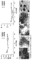
- FIG. 1 shows the particle size distribution (FIG. 1A) of a polyion complex-type polymersome encapsulating L-asparaginase and its transmission electron microscope (TEM) image.
- FIG. 2 shows the results of analysis by fluorescence correlation spectroscopy of L-asparaginase and naked L-asparaginase encapsulated in polyion complex type polymersomes.
- FIG. 3 shows a Michaelis-Menten plot (FIG. 3A) and a Lineweaver-Burk plot (FIG. 3B) of naked L-asparaginase.
- FIG. 4 shows a Michaelis-Menten plot (FIG. 4A) and a Lineweaver-Burk plot (FIG.
- FIG. 4B shows L-asparaginase encapsulated in polyion complex type polymersomes.
- FIG. 5 shows the blood retention of L-asparaginase encapsulated in polyion complex polymersomes.
- FIG. 6 shows the time course of the enzyme reaction rate of L-asparaginase.
- FIG. 7 shows the blood ammonia nitrogen concentration (FIG. 7A) and the increase in blood ammonia nitrogen concentration (FIG. 7B) in mice administered with L-asparaginase via the tail vein.
- FIG. 8 shows the EDC equivalent as a crosslinking agent, the FT-IR spectrum (FIG. 8A) of the obtained polyion complex type polymersome, and the relationship between the crosslinking ratio (%) and the EDC equivalent (FIG.
- FIG. 9 shows the particle size (FIG. 9A), polydispersity (PDI) (FIG. 9B), and TEM image (FIG. 9C) of the polyion complex type polymersome before and after crosslinking.
- FIG. 10 shows the relationship between the size of molecules to be encapsulated and the concentration of the molecules used, and the particle size of the resulting polyion complex-type polymersome.
- FIG. 11 shows changes over time in the release rate of inclusions (FIG. 11A) and the relationship between the cross-linking rate and the release rate constant in polyion complex type polymersomes having various cross-linking rates (%) shown in the figure (FIG. 11B). ).
- FIG. 11A shows the release rate of inclusions (FIG. 11A) and the relationship between the cross-linking rate and the release rate constant in polyion complex type polymersomes having various cross-linking rates (%) shown in the figure (FIG. 11B).
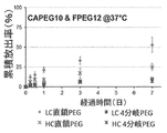
- FIG. 12 shows the change over time in the release ratio of polyethylene glycol (PEG) having different molecular weights from polyion complex-type polymersomes.
- PEG6 molecular weight 6k
- PEG20 molecular weight 20k
- PEG42 molecular weight 42k
- LC indicates the release rate in the polyion complex type polymersome having a crosslinking rate of less than 40%
- HC indicates the release rate in the polyion complex type polymersome having a crosslinking rate of 80% or more.
- FPEG means fluorescein labeled PEG.
- FIG. 13 is a diagram showing the difference in the release rate between linear PEG and branched PEG.
- FIG. 14 is a diagram showing the relationship between the release rate constant of PEG from the polyion complex type polymersome and the temperature.
- LC in the figure indicates the release rate in the polyion complex type polymersome having a crosslinking rate of less than 40%
- HC indicates the release rate in the polyion complex type polymersome having a crosslinking rate of 80% or more.
- FIG. 15 is a graph showing the relationship between the molecular weight of PEG and the release constant.
- the circle mark indicates the release rate constant at 4 ° C.
- the triangle mark indicates 25 ° C.
- the square mark indicates 37 ° C.
- the diamond mark indicates the release rate constant at 50 ° C. It shows that it is a release rate constant in PICsome.
- FIG. 16 shows the relationship between the cumulative release amount (%) at time t and time t. In the figure, the slope is proportional to the diffusion coefficient of the molecules in the membrane of the polyion complex type polymersome.
- FIG. 17 shows the particle size distribution (FIG. 17A) of PICsome encapsulating ⁇ -galactosidase (hereinafter also referred to as “ ⁇ -GAL”), and naked free ⁇ -galactosidase and PICsome-encapsulated ⁇ -particles by fluorescence correlation spectroscopy. The analysis result of galactosidase is shown.
- FIG. 17A particle size distribution of PICsome encapsulating ⁇ -galactosidase
- FIG. 18 shows the analysis results of PICsome-encapsulated ⁇ -glucosidase or uricase by fluorescence correlation spectroscopy by fluorescence correlation spectroscopy.
- FIG. 19 shows a measurement scheme for retention in blood of PICsome-encapsulated ⁇ -GAL (FIG. 19A) and its enzyme activity (FIG. 19B).
- polymer or “polymer” refers to a molecule formed by polymerizing monomer units, and is used to include homopolymers, copolymers, and block copolymers.
- homopolymer or “homopolymer” refers to a polymer obtained by polymerizing one type of monomer unit.
- copolymer refers to a polymer obtained by polymerizing two or more types of monomer units, and is used to include a block copolymer.
- block copolymer is a copolymer formed by linking a block in which the same type of monomer units are continuously formed and a block in which another type of monomer units is continuously formed. Say polymer.
- polyion complex type polymersome is also called PICsome and means hollow fine particles formed by the polyion complex. It is known that the outer surface of PICsome is preferably modified with polyethylene glycol from the viewpoint of residence time in blood.
- polyion complex means that a copolymer of PEG and an anionic block and a copolymer of PEG and a cationic block are charged in an aqueous solution. It is an ionic layer formed between the cationic block and the anionic block of both block copolymers when mixed so as to neutralize.
- the significance of linking PEG and the above-mentioned charged chain is that the polyion complex is prevented from aggregating and precipitating, and thereby the polyion complex has a monodisperse core-shell structure with a particle size of several tens of nm. Forming nano-particles.
- one charged block copolymer does not require a PEG moiety and may be replaced with a homopolymer, surfactant, nucleic acid and / or enzyme.
- at least one of the anionic polymer and the cationic polymer forms a copolymer with PEG, and both of them may form a copolymer with PEG. Further, when the PEG content is reduced, PICsome is likely to be formed.
- PICsome and the enzyme can be mixed and vigorously stirred. Stirring can be performed using a stirring device such as a vortex mixer (trademark). Moreover, even if an enzyme coexists at the time of PICsome formation, the enzyme can be included in PICsome.
- enzyme means a protein having catalytic activity. It is known that the molecular weight of an enzyme is generally widely distributed around 10 kDa to 200 kDa.
- subject refers to mammals including humans.
- the subject may be a healthy subject or a subject suffering from some disease.
- the present inventors encapsulated the enzyme in PICsome and brought the substrate of the enzyme into contact with the PICsome from the outside of the PICsome.
- the enzyme was kept inside the PICsome, but the substrate permeates the PICsome membrane. Furthermore, it was found that a substrate outside of PICsome can react with an enzyme in PICsome, and that the reaction product is released out of PICsome.
- the present inventors have also found that an enzyme encapsulated in PICsome has a significantly improved retention in blood compared with a naked enzyme.
- the enzyme can be an enzyme that uses a substance that permeates the membrane of the polyion complex polymersome as a substrate.
- the enzyme has a molecular weight of 5 kDa or more, 10 kDa or more, 20 kDa or more, 30 kDa or more, 40 kDa or more, 50 kDa or more, 60 kDa or more, 70 kDa or more, 80 kDa or more, 90 kDa or more, 100 kDa or more, 110 kDa or more, 120 kDa.
- an enzyme of 130 kDa or more or 140 kDa or more can be used. If the molecular weight is 5 kDa or more, it can be retained inside the PICsome, but the larger the molecular weight, the less likely to pass through the PICsome and the easier it is to be retained inside.
- the enzyme substrate can have a molecular weight of less than 5 kDa, 4 kDa or less, 3 kDa or less, 2 kDa or less, 1 kDa or less, 750 Da or less, 500 Da or less, 400 Da or less, 300 Da or less, or 200 Da or less. If the molecular weight is less than 5 kDa, it can permeate the PICsome membrane. However, the smaller the molecular weight, the easier it is to contact the enzyme through the PICsome membrane, and the reaction efficiency increases.
- the fibrous polymer is also advantageously held in the PICsome, but in the branched polymer, the membrane permeability of the PICsome is further lowered and is more advantageously held in the interior. Therefore, in the present invention, a fibrous protein can be used as the enzyme, but a globular protein can be preferably used.
- the enzyme encapsulated in PICsome is more stable than the naked enzyme under physiological conditions and in blood.
- an enzyme encapsulated in PICsome is more stable in blood than a naked enzyme.
- the enzyme can be an enzyme whose substrate is a plasma component. By doing in this way, the substrate in blood can be processed effectively, maintaining an enzyme and its activity stably in the blood.
- L-asparaginase can be used as the enzyme.
- L-asparaginase (hereinafter sometimes referred to as “L-ASP”) is a protein having a molecular weight of about 141 kDa, and hydrolyzes asparagine to produce aspartic acid and NH 3 .
- L-ASP is commercially available for the treatment of acute lymphoblastic leukemia, for example, commercially available from Kyowa Hakko Kirin Co., Ltd. under the trade name Leinase (trademark).
- Leinase trademark
- L-ASP is also used to treat mastocytoma.
- L-ASP can be administered by intravenous injection.
- L-ASP is thought to exert an anti-tumor effect by hydrolyzing L-asparagine in the blood and bringing the asparagine-requiring tumor cells into a nutrient-deficient state.
- asparaginase one derived from Escherichia coli or one derived from Erwinia chrysanthemi can be used.
- asparaginase derived from these bacteria can cause allergic reactions in humans.
- asparaginase is encapsulated in PICsome and allergic reaction problems are reduced.
- L-ASP PEG asparaginase modified with PEG may be used.
- L-ASP encapsulated in PICsome (hereinafter sometimes referred to as “encapsulated L-ASP”) is referred to as naked L-ASP (hereinafter referred to as “free L-ASP”). Compared to (A), it showed higher blood retention and NH 3 production ability (ie, asparagine hydrolyzing ability). Therefore, according to the present invention, it can be used more effectively than conventional L-ASP in the treatment of asparagine-requiring or sensitive tumor cells. Therefore, according to the present invention, there is provided a pharmaceutical composition containing PICsome, wherein the encapsulated enzyme is L-ASP.
- the pharmaceutical composition of the present invention containing PICsome whose encapsulating enzyme is L-ASP can be used for treating an asparagine-requiring tumor.
- Asparagine-requiring tumors include acute leukemias such as acute lymphoblastic leukemia, acute lymphoblastic leukemia, particularly childhood acute lymphoblastic leukemia, acute myeloid leukemia, other acute leukemias, and malignant lymphomas such as T cells Examples include malignant lymphoma, Hodgkin's disease, reticulosarcoma and lymphosarcoma.
- asparagine-requiring tumors can be identified by examining the expression of asparagine-producing enzymes.
- an asparagine-requiring tumor has an expression of an asparagine-producing enzyme of 80% or less, 70% or less, 60% or less, 50% or less, 40% or less, or 30% relative to normal cells.
- the tumor can be 20% or less, 10% or less, 5% or less, 3% or less, or 1% or less.
- the asparagine-requiring tumor can be either NK cell leukemia or acute myeloid leukemia.
- the pharmaceutical composition containing PICsome whose encapsulated enzyme is L-ASP according to the present invention is administered to a tumor patient who has developed allergic symptoms (for example, anaphylactic symptoms) by chemotherapy using L-ASP. be able to.
- PICsome encapsulates enzymes that break down nutrients required by microorganisms such as bacteria and viruses, and induces death and reduction of neoplasms and microorganisms. be able to.
- uricase can be used as the enzyme to be encapsulated.
- Uricase EC1.7.33
- urate oxidase can break down uric acid involved in purine metabolism and caffeine metabolism.
- uricase can be used for the treatment of hyperuricemia and high uric acidemia (for example, complications) in which the blood uric acid level is high.
- Diseases caused by hyperuricemia include gout (eg gout nodules and gout arthritis) and hyperuricemia such as urate deposition and uric acid stones, interstitial nephritis, renal failure, and arteriosclerosis. Examples include the cause of the disease
- a pharmaceutical composition comprising PICsome whose encapsulated enzyme is uricase, and used for treating hyperuricemia or diseases caused by hyperuricemia. Is done.
- the enzyme encapsulated in PICsome is stable in blood, it can be effectively used to transport the enzyme from the blood into cells by encapsulating the enzyme in PICsome.
- Numerous enzymes are present in cells, particularly lysosomes, and lysosomal diseases are caused by the deficiency or abnormality of the enzymes. Therefore, in one aspect of the present invention, an enzyme that causes lysosomal disease due to a deficiency or abnormality (for example, a decrease in enzyme activity) is encapsulated in PICsome, and the subject suffers from lysosomal disease due to the enzyme deficiency or abnormality. Can be administered.
- an enzyme in lysosome for example, ⁇ -galactosidase (hereinafter also referred to as “ ⁇ -GAL”) can be used as an enzyme to be encapsulated.
- ⁇ -Galactosidase (EC 3.2.1.22) has an activity of hydrolyzing ⁇ -D-galactoside and has an activity of promoting O-transfer of ⁇ -D-galactoside to various alcohol derivatives.
- ⁇ -Galactosidase which lacks its activity, causes a type of lysosomal disease, Fabry disease.
- ⁇ -Galactosidase can suppress the improvement and progression of Fabry disease in replacement therapy by infusion.
- a pharmaceutical composition comprising PICsome whose encapsulating enzyme is ⁇ -galactosidase is a disease caused by an abnormality of ⁇ -galactosidase, for example, an ⁇ -galactosidase such as lysosomal disease (eg, Fabry disease). It can be used to treat diseases caused by decreased activity or defects.
- a pharmaceutical composition comprising PICsome whose enzyme to be encapsulated is ⁇ -galactosidase, which is used for treating a disease caused by abnormality of ⁇ -galactosidase (for example, lysosomal disease) A composition is provided.
- ⁇ -glucosidase can be used as an enzyme to be encapsulated.
- ⁇ -Glucosidase (EC 3.2.1.20) has an activity to hydrolyze ⁇ -1,4-glucoside bond of sugar. If the activity of ⁇ -glucosidase is deficient, it causes lysosomal disease (eg Pombe disease (OMIM No .: 232300)). Therefore, ⁇ -glucosidase can be used to treat diseases caused by abnormalities (eg, decreased activity or deficiency) of ⁇ -glucosidase.
- abnormalities eg, decreased activity or deficiency
- Examples of the disease caused by decreased activity or deficiency of ⁇ -glucosidase include diseases caused by decreased activity or deficiency of ⁇ -glucosidase, such as lysosomal disease (eg, Pombe disease). Therefore, according to the present invention, a pharmaceutical composition comprising PICsome whose internal enzyme is ⁇ -glucosidase, which is used for treating a disease caused by decreased activity or deficiency of ⁇ -glucosidase.
- a pharmaceutical composition is provided.
- the enzyme of the present invention is not limited to the enzymes described herein, and can be widely used for enzyme replacement therapy for compensating for a deficient enzyme due to deficiency or abnormality in vivo.
- Examples of the block copolymer forming PICsome include a block copolymer of a PEG block and a polycation block and a homopolyanion, or a block copolymer of a PEG block and a polyanion block and a homopolycation.
- As the block copolymer it is preferable to use a biodegradable block copolymer, and various copolymers are known as such copolymers, and any of them can be used in principle. .
- the polycation block includes, for example, cationic natural amino acids and cationic unnatural amino acids, for example, cationic natural amino acids such as histidine, tryptophan, ornithine, arginine and lysine, and / or — (NH— ( A group represented by CH 2 ) 2 ) p —NH 2 , wherein p is an integer of 1 to 5.
- a cationic side chain for example, a cationic non-natural amino acid polymer block having the cationic side chain, for example, a cationic non-chain such as aspartic acid or glutamic acid having the cationic side chain. Examples include natural amino acid polymer blocks.
- the polycation block is a group represented by — (NH— (CH 2 ) 2 ) p —NH 2 , where p is an integer of 1 to 5.
- p is an integer of 1 to 5.
- the cationic natural amino acid is preferably histidine, tryptophan, ornithine, arginine and lysine, more preferably arginine, ornithine and lysine, still more preferably ornithine and lysine, and still more preferably. Includes lysine.
- block copolymers having high biocompatibility and biodegradability examples include poly (aspartic acid-tetraethylenepentamine) block copolymers and polyethylene glycol-poly ((5-aminopentyl) -asparagine. Acid) block copolymers can be used.
- the polycation block In the polycation block, a cationic amino acid and an amino acid having a cationic side chain may be mixed. That is, in one embodiment of the present invention, the polycation block is a polymer of monomer units containing a cationic natural amino acid, a cationic unnatural amino acid, or a cationic natural amino acid and a cationic unnatural amino acid. In some embodiments of the invention, the bond between monomer units in the polycation block is a peptide bond.
- the cationic unnatural amino acid is a group represented by — (NH— (CH 2 ) 2 ) p —NH 2 as a side chain ⁇ where n is an integer of 1 to 5 ⁇ It is an amino acid having
- the polycation block includes a cationic natural amino acid and a group represented by — (NH— (CH 2 ) 2 ) p —NH 2 , where p is an integer of 1 to 5. is there.
- 40%, 50%, 60%, 70%, 80%, 90%, 95%, 98%, or 100% of the monomer units in the polymer have-(NH- (CH 2 2 ) a group represented by p- NH 2 ⁇ wherein p is an integer of 1 to 5. ⁇ .
- PICsome can be formed by a block copolymer of a PEG block and a polycation block and a polyion complex of a homopolyanion.
- the block copolymer of PEG block and polycation block can be a copolymer of PEG block and poly (aminopentyl-aspartic acid).
- the homopolyanion can be polyaspartic acid.
- PICsome is a block copolymer of PEG block and polycation block and homopolyanion is a copolymer of PEG block and poly (aminopentyl-aspartic acid) and polyaspartic acid, respectively. can do.
- the degree of polymerization of the PEG block, polyaspartic acid block and poly (aminopentyl-aspartic acid) block is each independently an integer from 5 to 20,000, preferably from 10 to 5,000. It can be an integer, more preferably an integer of 40 to 500, still more preferably an integer of 5 to 1,000, and even more preferably an integer of 10 to 200.
- examples of the polycation block include a PEG-poly (N ′-[N- (2-aminoethyl) -2-aminoethyl] -aspartic acid) block copolymer (PEG-P (Asp-DET)). ))
- PEG-P (Asp-DET) can be prepared according to a conventional method (see Chem. Med. Chem. 1 (2006) 439-444).
- Asp-DET has a side chain carboxyl group substituted with a diethyltriamine (DET) group (—NH—CH 2 —CH 2 —NH—CH 2 —CH 2 —NH 2 ). Aspartic acid.
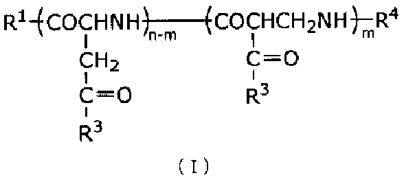
- the structure of P (Asp-DET) is represented by the following chemical formula.
- R 1 is a hydroxyl group, a protecting group, a hydrophobic group, or a polymerizable group
- R 4 is H, a protecting group, a hydrophobic group, or a polymerizable group
- R 3 is a group represented by — (NH— (CH 2 ) 2 ) 2 —NH 2
- n is an integer of 0 to 5000, for example, an integer of 0 to 500
- m is an integer from 0 to 5000, for example, an integer from 0 to 500
- m + n is an integer from 2 to 5000, for example, an integer from 2 to 500
- n ⁇ m is an integer of 0 or more
- Each repeating unit in the formula is shown in a specific order for convenience of description, but each repeating unit can be present in any order, each repeating unit may be present randomly, and each repeating unit is They may be the same or different.
- R 1 or R 4 represents a bond
- the polyethylene glycol forms a copolymer with the polycation block via the bond.
- each repeating unit is bonded by a peptide bond.
- the PICsome may crosslink the cationic polymer and the anionic polymer after forming the PICsome.
- Crosslinking can be suitably performed using a method well known to those skilled in the art.
- the PICsome of the present invention is formed using a cationic polymer that may be modified with PEG and an anionic polymer that may be modified with PEG, and then the cationic polymer and the anionic polymer. And can be obtained by crosslinking.
- the cationic polymer contains a cationic amino acid having an NH 2 group in the side chain as a monomer unit, and has a polymer that may be modified with PEG and a COOH group in the side chain.
- a polymer containing an anionic amino acid as a monomer unit and optionally modified with PEG is mixed in an aqueous solution to form PICsome, and then 1-ethyl-3- (3-dimethylamino as a crosslinking agent. It may be obtained by crosslinking a cationic polymer and an anionic polymer using propyl) carbodiimide hydrochloride. Crosslinking can be performed after encapsulating the enzyme in PICsome.
- PICsome is A polymer (A) containing an amino acid having a COOH group in the side chain as a monomer unit; PICsome with a polymer (B) containing an amino acid having an NH 2 group in the side chain as a monomer unit may be used.
- PICsome with a polymer (B) containing an amino acid having an NH 2 group in the side chain as a monomer unit may be used.
- 1-ethyl-3- (3-dimethylaminopropyl) carbodiimide hydrochloride (EDC) can be used to crosslink the polymer (A) and the polymer (B).
- the crosslinking rate (%) between the cationic polymer and the anionic polymer in PICsome refer to the spectrum of the PEG-PAsp polymer (uncrosslinked) by, for example, Fourier transform infrared spectroscopy (FT-IR) And the ratio of the peak derived from the carboxyl group (1600 cm ⁇ 1 ; derived from the stretching vibration of the COO ⁇ group) to the peak derived from PEG (1460 cm ⁇ 1 ; derived from the bending vibration of the C—H group). Can be calculated.
- the crosslinking rate can be increased by increasing the amount of crosslinking agent used.
- a method is provided.
- the permeability to molecules of less than 5 kDa, 4 kDa or less, 3 kDa or less, 2 kDa or less, 1 kDa or less, 750 Da or less, 500 Da or less, 400 Da or less, 300 Da or less or 200 Da or less is 50% or more and 60% or more. 70% or more, 80% or more, 90% or more, or substantially maintained.
- a molecule that decreases permeability or a molecule that decreases release rate is 5 kDa or more, 10 kDa or more, 20 kDa or more, 30 kDa or more, 40 kDa or more, 50 kDa or more, 60 kDa or more, 70 kDa or more, 80 kDa or more, 90 kDa or more.
- an enzyme of 100 kDa or more, 110 kDa or more, 120 kDa or more, 130 kDa or more, or 140 kDa or more can be used.
- the crosslinking rate (%) between the cationic polymer and the anionic polymer constituting PICsome increases, the release rate of molecules from the inside of PICsome decreases and the release rate constant decreases.
- the crosslinking rate (%) can be 25% or more, 30% or more, 40% or more, 50% or more, 60% or more, 70% or more, or 80% or more.
- the crosslinking rate may be 90% or more.
- the crosslinking rate (%) is preferably large, and can be preferably 50% or more, 60% or more, 70% or more, or 80% or more.
- the present invention by increasing the crosslinking rate of the cationic polymer and anionic polymer constituting PICsome, it is possible to reduce the release amount of the encapsulated enzyme to the outside of PICsome, and a 10 mM phosphate buffer solution ( When measured at 37 ° C in pH 7.4), the cumulative release rate (%) of the encapsulated enzyme is 60% or less, 50% or less, 40% or less, 30% on the seventh day after contact with the aqueous solution.
- PICsome that is 20% or less, 10% or less, or 5% or less is provided.
- the release rate constant k of a linear polyethylene glycol having a number average molecular weight of 2 kDa is 5 ⁇ 10 ⁇ 3 or less when measured in a 10 mM phosphate buffer solution (pH 7.4) at 37 ° C.
- a PICsome is provided that is 4 ⁇ 10 ⁇ 3 or less, 3 ⁇ 10 ⁇ 3 or less, 2 ⁇ 10 ⁇ 3 or less, or 1 ⁇ 10 ⁇ 3 or less.
- the enzyme is 5 kDa or more, 10 kDa or more, 20 kDa or more, 30 kDa or more, 40 kDa or more, 50 kDa or more, 60 kDa or more, 70 kDa or more, 80 kDa or more, 90 kDa or more, 100 kDa or more, 110 kDa or more, 120 kDa or more, 130 kDa or more, or 140 kDa.
- the above enzymes can be used.
- a composition containing PICsome for use in encapsulating L-ASP in this aspect, in some embodiments, the PICsome can be a PICsome of the present invention.
- the composition can be incorporated into PICsome by mixing with L-ASP. Even asparagine outside PICsome can penetrate the PICsome membrane and reach the inside, the composition of the present invention can be used for hydrolysis of asparagine by contacting with asparagine.
- a composition containing PICsome for use in encapsulating ⁇ -galactosidase in some embodiments, can be a PICsome of the present invention.
- the composition of the present invention can encapsulate ⁇ -galactosidase inside PICsome by mixing with ⁇ -galactosidase. Since a substrate other than PICsome (for example, globotriaosylceramide) can penetrate the PICsome membrane and reach the inside, the composition of the present invention can be hydrolyzed asparagine by contacting with ⁇ -galactosidase. Can be used.
- a composition containing PICsome for use in encapsulating uricase is provided.
- the PICsome can be a PICsome of the present invention.
- the composition of the present invention can be encapsulated in PICsome by mixing with uricase. Since a substrate other than PICsome (for example, uric acid) can penetrate the PICsome membrane and reach the inside, the composition of the present invention can be used for hydrolysis of urea by contacting with uricase.
- a composition containing PICsome for use in encapsulating ⁇ -glucosidase is provided.
- the PICsome can be a PICsome of the present invention.
- the composition of the present invention can encapsulate ⁇ -glucosidase inside PICsome by mixing with ⁇ -glucosidase. Since a substrate other than PICsome (for example, glycogen) can penetrate the PICsome membrane and reach the inside, the composition of the present invention can be linked to ⁇ -1,4-glucoside by contacting with ⁇ -galactosidase. It can be used for hydrolysis of.
- a composition containing L-ASP for use in inclusion in PICsome in some embodiments, can be a PICsome of the present invention.
- the composition can be incorporated into PICsome by mixing with L-ASP. Even asparagine outside PICsome can penetrate the PICsome membrane and reach the inside, the composition of the present invention can be used for hydrolysis of asparagine by contacting with asparagine.
- a composition containing ⁇ -galactosidase for use in inclusion in PICsome in this aspect, in some embodiments, the PICsome can be a PICsome of the present invention.
- the composition can include ⁇ -galactosidase within the PICsome by mixing with ⁇ -galactosidase. Since even a substrate outside of PICsome can penetrate the PICsome membrane and reach the inside, the composition of the present invention can be used for hydrolysis of the substrate by contacting with ⁇ -galactosidase.
- a composition containing uricase for use in inclusion in PICsome is provided.
- the PICsome can be a PICsome of the present invention.
- the composition can be encapsulated in PICsome by mixing with uricase. Even urea outside PICsome can penetrate the PICsome membrane and reach the inside, so that the composition of the present invention can be used for hydrolysis of urea by contacting with uricase.
- a composition containing ⁇ -glucosidase is provided for use in inclusion in PICsome.
- the PICsome can be a PICsome of the present invention.
- the composition can encapsulate ⁇ -glucosidase in the interior of PICsome by mixing with uricase. Since even a substrate outside of PICsome can penetrate the PICsome membrane and reach the inside, the composition of the present invention can be used for hydrolysis of the substrate by contacting with ⁇ -glucosidase.
- a combination of a polycation that may be PEGylated, a polyanion, and L-ASP.
- a combination of a polycation, an optionally PEGylated polyanion, and L-ASP there is provided a combination of a polycation, an optionally PEGylated polyanion, and L-ASP.
- the combination of the present invention can be used to prepare a PICsome containing L-ASP.
- a combination of a polycation that may be PEGylated, a polyanion, and ⁇ -galactosidase is provided.
- a combination of a polycation that may be PEGylated, a polyanion, and a uricase is provided.
- a combination of a polycation that may be PEGylated, a polyanion, and an ⁇ -glucosidase is provided.
- a combination of a polycation, a polyanion that may be PEGylated, and ⁇ -galactosidase is provided.
- a combination of a polycation, an optionally PEGylated polyanion, and a uricase is provided.
- a combination of a polycation, an optionally PEGylated polyanion, and ⁇ -glucosidase is provided. These combinations can be used to prepare PICsome containing the enzyme as described above.
- a method for treating a subject comprising administering a PICsome of the present invention encapsulating an enzyme to a subject in need of administration of the enzyme.
- a subject can be a subject suffering from a disease due to lack of a specific enzyme or reduced expression thereof.
- Enzymes include L-ASP, and diseases include asparagine-requiring tumors.
- diseases include asparagine-requiring tumors.
- a method of treating an asparagine-requiring tumor comprising administering to a subject in need thereof an effective amount of PICsome containing L-ASP.
- Asparagine-requiring tumors include acute leukemias such as acute lymphoblastic leukemia, acute lymphoblastic leukemia, particularly childhood acute lymphoblastic leukemia, acute myeloid leukemia, other acute leukemias, and malignant lymphomas such as T cell malignancies. Examples include lymphoma, Hodgkin's disease, reticulosarcoma and lymphosarcoma.
- Asparagine-requiring tumors have an expression of asparagine-producing enzyme that is 80% or less, 70% or less, 60% or less, 50% or less, 40% or less, 30% or less, compared to normal cells.
- the tumor may be 20% or less, 10% or less, 5% or less, 3% or less, or 1% or less.
- the subject can be a tumor patient who has developed allergic symptoms (for example, anaphylactic symptoms) by chemotherapy using L-APS.
- the enzyme includes ⁇ -galactosidase
- the disease includes a disease caused by abnormality of ⁇ -galactosidase (for example, Fabry disease).
- a method for treating a disease caused by an abnormality in ⁇ -galactosidase for example, a disease caused by decreased activity or deficiency of ⁇ -galactosidase such as lysosomal disease (eg, Fabry disease)).
- a method comprising administering to a subject in need thereof an effective amount of PICsome comprising ⁇ -galactosidase.
- the subject can be a tumor patient who has developed allergic symptoms (eg, anaphylactic symptoms) by chemotherapy using ⁇ -galactosidase.
- the enzyme includes uricase, and the disease can be used to treat hyperuricemia and diseases caused by hyperuricemia.
- gout eg gout nodules and gout arthritis
- hyperuricemia such as urate deposition and uric acid stones, interstitial nephritis, renal failure, and arteriosclerosis. Examples include the disease that causes it. Therefore, according to the present invention, a method for treating hyperuricemia and diseases caused by hyperuricemia, comprising administering to a subject in need thereof an effective amount of PICsome containing uricase.
- a method comprising:
- the subject can be a tumor patient who has developed allergic symptoms (for example, anaphylactic symptoms) by chemotherapy using uricase.
- the enzyme includes ⁇ -glucosidase
- the disease includes a disease caused by abnormality of ⁇ -glucosidase (for example, Pombe disease). Therefore, according to the present invention, a method for treating a disease caused by an abnormality in ⁇ -glucosidase (for example, a disease caused by decreased activity or deficiency of ⁇ -glucosidase such as lysosomal disease (eg, Pombe disease)).
- a method comprising administering to a subject in need thereof an effective amount of PICsome comprising ⁇ -glucosidase.
- the subject can be a tumor patient who has developed allergic symptoms (for example, anaphylactic symptoms) by chemotherapy using ⁇ -glucosidase.
- treatment means to cure, prevent or ameliorate a disease or disorder or to reduce the rate of progression of the disease or disorder. Treatment can be accomplished by administering a therapeutically effective amount of a pharmaceutical composition.
- Example 1 Preparation of PICsome Encapsulating Enzyme
- PICsome encapsulating the enzyme L-asparaginase (hereinafter also referred to as “L-ASP”) was prepared.
- PEG-P polyethylene glycol-poly ( ⁇ -benzyl-L-aspartate) block copolymer
- PEG-PBLA polyethylene glycol-poly ( ⁇ -benzyl-L-aspartate) block copolymer
- BLA-NCA ⁇ -benzyl-L-aspartate-N-carboxylic acid anhydride
- PEG-P polyethylene glycol-polyaspartic acid block copolymer
- poly ( ⁇ -benzyl-L-aspartate) (homo PBLA polymer) was obtained by polymerization of BLA-NCA. Specifically, 20 g of ⁇ -benzyl-L-aspartate-N-carboxylic anhydride (BLA-NCA) is dissolved in 33.3 mL of N, N′-dimethylformamide (DMF) and 300 mL of dichloromethane. Add 89.0 ⁇ L of N-butylamine to the BLA-NCA solution. The mixed solution was polymerized for 40 hours while maintaining at 35 ° C.
- BLA-NCA ⁇ -benzyl-L-aspartate-N-carboxylic anhydride
- Cy5-labeled L-ASP 75 mg of L-ASP was dissolved in 50 mL of 10 mM phosphate buffer (PB, pH 7.4, 0 mM NaCl).
- Cy5-N-hydroxysuccinimide ester dye pack (manufactured by GE Healthcare, product number: PA25001) 20Vial was dissolved in 5 mL of DMSO, added to the L-ASP solution, and reacted at 25 ° C. for 4 hours. Thereafter, unreacted Cy5-N-hydroxysuccinimide ester molecules were removed using an ultrafiltration tube with a membrane having a fractional molecular weight of 10,000.
- a 10 mM phosphate buffer (PB, pH 7.4, 0 mM NaCl) solution of DyLight 488 labeled L-ASP or Cy5 labeled L-ASP is concentrated by ultrafiltration, and a fluorescent label of 5.6 mg / mL
- a modified L-ASP solution was prepared.
- 5.6 mg / mL L-ASP solution or 5.0 mL of fluorescence-labeled L-ASP solution was mixed with 9.0 mL of an empty PICsome solution and stirred by vortexing (2000 rpm) for 2 minutes.
- Example 2 Characterization of PICsome
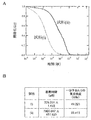
- the size (Z average particle diameter) and polydispersity index (PDI) of the obtained PICsome were measured with a Zetasizer (Malvern).
- the shape of PICsome was observed after staining with uranyl acetate using a transmission electron microscope (TEM, JEM-1400).
- the size (Z average particle diameter) and polydispersity index (PDI) of the obtained L-ASP-encapsulated PICsome were measured with a zeta sizer (Malvern). Size was measured by the diffusion of particles moving by Brownian motion, and the measurement results were converted to particle size and particle size distribution using Stokes-Einstein equation. The micelle shape was evaluated using a transmission electron microscope (TEM, JEM-1400).
- TEM transmission electron microscope
- JEM-1400 transmission electron microscope
- the Z average particle diameter is data obtained by analyzing measurement data of a dynamic light scattering method such as a particle dispersion using a cumulant analysis method.
- an average value of the particle diameter and a polydispersity index (PDI) are obtained.
- this average particle diameter is defined as the Z average particle diameter.
- LN (G1) a + bt + ct 2 + dt 3 + et 4 +...
- the constant b in is called the second-order cumulant or Z-average diffusion coefficient.
- the value obtained by converting the value of the Z average diffusion coefficient into the particle size using the viscosity of the dispersion medium and some device constants is the Z average particle size, and is a value suitable for quality control purposes as an index of dispersion stability.
- the particle size distribution and transmission electron microscope (TEM) image of the PICsome containing the obtained L-ASP were as shown in FIGS. 1A and 1B, respectively.
- the obtained PICsome containing L-ASP shows a monodisperse particle size distribution having a mode value of about 100 nm in diameter, and as shown in FIG. Many PICsomes having a diameter of about 100 nm were observed.
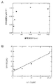
- Fluorescence correlation spectroscopy was performed as follows. A confocal laser scanning microscope (LSM 510 META / Confocal3, Carl Zeiss) equipped with a 488 nm Ar laser line was used. The sample solution was dropped into an 8-well chamber, and excitation light irradiation and fluorescence detection were performed through a water immersion objective lens. The fluctuation of the fluorescence intensity was analyzed using an autocorrelation function, and the diffusion time and the fluorescence intensity per particle were calculated.
- the enzyme activity was evaluated for each of free L-ASP and encapsulated L-ASP.
- a Michaelis-Menten plot and a Lineweaver-Burk plot for the reaction with 7-amido-4-methylcoumarin) also referred to as “Asp-AMC” were generated. Specifically, first, each solution of Asp-AMC, free L-ASP, and encapsulated L-ASP (in PBS) was allowed to stand at 37 ° C. in a thermostatic bath.
- the Asp-AMC solution was mixed with each solution of free L-ASP or encapsulated L-ASP, and a predetermined amount was quickly poured into a 96-well plate (Tecan).
- the plate was placed in a multi-plate reader that had been kept warm at 37 ° C., and the fluorescence intensity was measured over time for a certain time.
- the reaction rate V was determined by converting a change in fluorescence intensity into a product production rate using a calibration curve prepared using a standard solution of AMC prepared in advance.
- the Michaelis-Menten coefficient (K m ) was calculated by varying the substrate concentration [S], taking [S] on the horizontal axis, V on the vertical axis, and applying the following equation to the plot results.
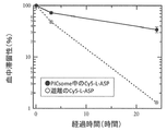
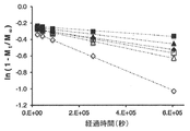
- FIG. 5 shows the relative fluorescence intensity at each time point with the fluorescence intensity immediately after administration as 100%. As shown in FIG. 5, the blood retention of encapsulated L-ASP was much higher than that of free L-ASP.
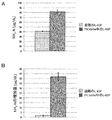
- the concentration of ammonia nitrogen in plasma was overwhelmingly higher in the group administered with encapsulated L-ASP than in the group administered with free L-ASP (FIG. 7A). Further, the amount of increase in ammonia nitrogen in the plasma was calculated from the ammonia nitrogen concentration before and after administration, and as shown in FIG. 7B, the plasma ammonia nitrogen concentration hardly increased in the group administered with free L-ASP. On the other hand, in the group administered with the encapsulated L-ASP, the plasma ammonia nitrogen concentration increased dramatically. As described above, the encapsulated L-ASP reacted with the plasma component upon administration, and the plasma component was converted by the enzyme activity to efficiently produce ammonia nitrogen.
- Example 3 Effect of Chemical Modification of PICsome on Enzyme Reaction
- L-ASP-encapsulated PICsome was prepared as described in Example 1 except that the concentration of EDC as a cross-linking agent was changed, and the crosslinking rate and the properties of PICsome I investigated the relationship.
- FIG. 8A shows the FT-IR spectrum of PICsome obtained by treating the EDC concentration of Example 1 with an EDC of 0.5 times, 3 times, 5 times or 10 times that of EDC.
- the relationship of the crosslinking ratio is shown in FIG. 8B.
- the crosslinking ratio was calculated by FT-IR.
- PEG-poly present in Asp copolymer COO - of the group the proportion (%) as a crosslinking ratio of those cross-linked.
- FIG. 9 shows the relationship between the amount of EDC used for crosslinking and the particle size, the relationship between the EDC amount and polydispersity (PDI), and the relationship between the EDC amount and the PICsome shape.
- FIG. 9A As a result, as shown in FIG. 9A, no significant difference was observed in particle size (FIG. 9A), polydispersity (FIG. 9B), and PICsome shape (FIG. 9C) at any EDC amount. Moreover, in any EDC amount, the particle size distribution showing monodispersion was obtained with the particle size of 100 nm being the mode value.
- Example 4 The molecular weight of the substance to be encapsulated and the shape number of the obtained PICsome PEGs of various sizes ranging from 6000 to 42000 in average molecular weight were encapsulated in the PICsome, and the shape of the obtained PICsome was confirmed.
- PEG was labeled with fluorescein and included in PICsome by the method described in Example 1 at a concentration of 0.5 mg / mL, 1 mg / mL, 3 mg / mL, 5 mg / mL, or 10 mg / mL.
- PEG6 NOF Corporation, MEPA-50H
- PEG6 having a number average molecular weight of 6k
- 12k PEG12 NOF Corporation, MEPA-12T
- 20k PEG20 NOF Corporation, MEPA-20T
- 42k PEG42 NOF Corporation, MEPA-40T
- the particle size was approximately 100 nm, and PICsome having a monodispersed particle size distribution was formed.
- Example 5 Material permeability of cross-linked PICsome
- PEG12-encapsulated PICsome was prepared as described in Example 1 except that fluorescein-labeled PEG12 was used at a concentration of 4 mg / mL to crosslink at various EDC amounts. The amount of PEG12 released from PICsome was examined.
- the cumulative release amount of PEG12 from PEG12-encapsulated PICsome obtained by adjusting the crosslinking rate by adjusting the EDC equivalent amount from Example 3 was determined by size exclusion chromatography and fluorescence intensity analysis. Size exclusion chromatography was performed using a gel filtration chromatography column: Superdex 200-10 / 300GL, GE Healthcare, and a high performance liquid chromatograph: JASCO Corporation, LC-2000plus. The fluorescence intensity was determined by comparing the fraction with an earlier retention time (PEG encapsulated in PICsome) and the later fraction (released PEG) with the fluorescence intensity of fluorescein by a conventional method. The solution conditions were 10 mM phosphate buffer solution (pH 7.4, 37 ° C.). The result was as shown in FIG. 11A.
- the amount of PEG12 released from PICsome varied depending on the crosslinking rate. Specifically, the higher the crosslinking rate, the more PEG12 was retained in the PICsome.
- the release rate constant was calculated by applying the obtained cumulative release rate result to the following equation obtained from the primary release model.
- the release rates of PEG6, PEG20 and PEG42 were examined with a PICsome having a low crosslinking rate (LC) with a crosslinking rate of 40% or less and a PICsome with a high crosslinking rate (HC) having a crosslinking rate of 80% or more. All PEGs were labeled with fluorescein, and the release rate constant was determined as described above. The result was as shown in FIG.
- PEG6 observed a large release of PEG from PICsome within 1 day, while PEG20 and PEG42 slowly released PEG from PICsome at a constant rate over 0-7 days. It was done. Also, the higher the crosslinking rate, the more the release of PEG from PICsome was suppressed. From this, it was clarified that small molecules easily pass through PICsome and large molecules hardly pass through PICsome. Further, it was revealed that the permeability is relatively high at a molecular weight of 6000 or less, and the permeability is relatively low at a molecular weight of 20000 or more.
- Example 6 Relationship between molecular structure and PICsome permeability
- linear PEG PEG12 labeled with fluorescein was used at 4 mg / mL (concentration when mixed with PICsome solution)
- branched PEG 4-branched PEG (NOF Corporation, PTE-100PA, several PICsome was obtained in the same manner as in Example 1 except that the average molecular weight 10k) was used at 4 mg / mL (concentration when mixed with the PICsome solution).
- Example 7 Temperature dependence of release rate constant In Examples 5 and 6, the release of inclusions from PICsome was examined under 37 ° C conditions. In this example, the temperature dependence of the release rate constant was clarified.
- the temperature dependence of the release rate constant was remarkable in the PICsome having a low crosslinking rate of less than 40%.
- the circle indicates the release rate constant at 4 ° C.
- the triangle indicates 25 ° C.
- the square indicates 37 ° C.
- the rhombus indicates 50 ° C.
- the white mark indicates the low crosslinking rate and the black indicates the high crosslinking rate in PICsome. Indicates a release rate constant.
- the left side represents the cumulative release amount (%) at time t
- D represents the diffusion coefficient of the encapsulated substance in the PICsome film
- l represents the thickness ( ⁇ 15 nm) of the PICsome film.
- PICsome due to the nature of PICsome, if PICsome encapsulates a large molecular weight enzyme with a small molecular weight as a substrate to dramatically improve the retention in blood, PICsome can It was revealed that it was possible to continue to react with the substrate in the blood while maintaining a stable state.
- L-ASP is known to have a growth inhibitory effect on asparagine-requiring tumor cells, and is clinically used as a therapeutic agent for childhood acute lymphoblastic leukemia.
- This Example shows that the L-ASP-encapsulating PICsome of the present invention has a high effect as a therapeutic drug for childhood acute lymphoblastic leukemia.
- Example 8A Encapsulation of other enzymes and evaluation of stability
- ⁇ -galactosidase, ⁇ -glucosidase and uricase were examined for blood stability.
- ⁇ -GAL-encapsulated PICsome solution As in item 5 of Example 1, an empty PICsome solution was obtained, and then 11.4 mg of ⁇ -galactosidase (hereinafter also referred to as “ ⁇ -GAL”) was added to 10 mM phosphate buffer.
- the solution (PB, pH 7.4, 0 mM NaCl) was dissolved in 5.7 mL to prepare a 2 mg / mL ⁇ -GAL solution.
- 1 mL of a 2 mg / mL ⁇ -GAL solution was mixed with 1 mL of an empty PICsome solution and stirred by vortexing for 2 minutes (2000 rpm).
- ⁇ -Glucosidase and uricase-encapsulating PICsome were prepared in the same manner. As above, an empty PICsome solution was obtained and then 6.2 mg and 10.4 mg of ⁇ -glucosidase and uricase, respectively, 3.1 mL and 5 mg of 10 mM phosphate buffer (PB, pH 7.4, 0 mM NaCl). Dissolve in 2 mL to prepare a 2 mg / mL ⁇ -glucosidase solution and a 2 mg / mL uricase solution.
- PB phosphate buffer
- a 2 mg / mL ⁇ -glucosidase solution and a 2 mg / mL uricase solution were each mixed with 1 mL of an empty PICsome solution and vortexed for 2 minutes (2000 rpm). Thereafter, 5.6 mL of a PB solution containing EDC (10 mg / mL), which is a water-soluble condensing agent, was added and allowed to stand overnight to crosslink the polyion complex. Thereafter, using an ultrafiltration tube with a membrane having a molecular weight cut off of 300,000, polymers not involved in PICsome formation, ⁇ -glucosidase and uricase not encapsulated in PICsome, EDC, and the like were removed. Fluorescently labeled ⁇ -glucosidase and uricase solutions were similarly prepared.
- FCS fluorescence correlation spectroscopy
- each solution of Gal-AMC, free ⁇ -GAL, and encapsulated ⁇ -GAL was allowed to stand at 37 ° C. in a thermostatic bath. Thereafter, a solution of Gal-AMC was mixed with each solution of free ⁇ -GAL or encapsulated ⁇ -GAL, and a predetermined amount was quickly poured into a 96-well plate (Tecan). The plate was placed in a multi-plate reader that had been kept warm at 37 ° C., and the fluorescence intensity was measured over time for a certain time. Enzyme activity was evaluated by creating a Michaelis-Menten plot and a Lineweaver-Burk plot.
- reaction rate V was determined by converting a change in fluorescence intensity into a product production rate using a calibration curve prepared using a standard solution of AMC prepared in advance.
- the Michaelis-Menten coefficient (K m ) was calculated by varying the substrate concentration [S], taking [S] on the horizontal axis, V on the vertical axis, and applying the following equation to the plot results. The results were as shown in Table 2.
- ⁇ -GAL encapsulated in PICsome was stable in vivo and maintained enzyme activity equivalent to that of free ⁇ -GAL.
Landscapes
- Health & Medical Sciences (AREA)
- Chemical & Material Sciences (AREA)
- Life Sciences & Earth Sciences (AREA)
- Animal Behavior & Ethology (AREA)
- Veterinary Medicine (AREA)
- Medicinal Chemistry (AREA)
- Public Health (AREA)
- General Health & Medical Sciences (AREA)
- Pharmacology & Pharmacy (AREA)
- Epidemiology (AREA)
- Proteomics, Peptides & Aminoacids (AREA)
- Immunology (AREA)
- General Chemical & Material Sciences (AREA)
- Gastroenterology & Hepatology (AREA)
- Engineering & Computer Science (AREA)
- Bioinformatics & Cheminformatics (AREA)
- Organic Chemistry (AREA)
- Nuclear Medicine, Radiotherapy & Molecular Imaging (AREA)
- Chemical Kinetics & Catalysis (AREA)
- Dispersion Chemistry (AREA)
- Inorganic Chemistry (AREA)
- Medicinal Preparation (AREA)
- Medicines That Contain Protein Lipid Enzymes And Other Medicines (AREA)
- Enzymes And Modification Thereof (AREA)
Abstract
Description
(1)酵素を内包したポリイオンコンプレックス型ポリマーソームであって、
酵素は、ポリイオンコンプレックス型ポリマーソームの膜を透過する物質を基質とする酵素である、ポリイオンコンプレックス型ポリマーソーム。
(2)酵素の分子量が、5kDa以上である、上記(1)に記載のポリイオンコンプレックス型ポリマーソーム。
(3)酵素の基質の分子量が、1kDa以下である、上記(1)または(2)に記載のポリイオンコンプレックス型ポリマーソーム。
(4)酵素が、球状タンパク質である、上記(1)~(3)のいずれかに記載のポリイオンコンプレックス型ポリマーソーム。
(5)酵素が、L-アスパラギナーゼ、ウリカーゼ、α-ガラクトシダーゼおよびα-グルコシダーゼから選択される酵素である、上記(1)に記載のポリイオンコンプレックス型ポリマーソーム。
(6)ポリイオンコンプレックス型ポリマーソームが、
COOH基を側鎖に有するアミノ酸を単量体単位として含む重合体(A)と、
NH2基を側鎖に有するアミノ酸を単量体単位として含む重合体(B)と
のポリイオンコンプレックス型ポリマーソームである、上記(1)~(5)のいずれかに記載のポリイオンコンプレックス型ポリマーソーム。
(7)重合体(A)に存在するCOOH基の50%以上が重合体(B)のNH2基と架橋されている、上記(6)に記載のポリイオンコンプレックス型ポリマーソーム。
(8)10mM リン酸緩衝溶液(pH7.4)中において37℃条件下で測定した場合に、内包した酵素の累積放出率(%)が前記水溶液と接触後7日目に20%以下である、上記(1)~(7)のいずれかに記載のポリイオンコンプレックス型ポリマーソーム。
(9)10mM リン酸緩衝溶液(pH7.4)中において37℃条件下で測定した場合に、数平均分子量2kDaの直鎖状ポリエチレングリコールの放出速度定数kが5×10⁻³以下である、上記(7)に記載のポリイオンコンプレックス型ポリマーソーム。
(10)上記(1)~(9)のいずれかに記載のポリイオンコンプレックス型ポリマーソームであって、内包される酵素が、血漿成分を基質とする、ポリイオンコンプレックス型ポリマーソーム。
(11)上記(10)のポリイオンコンプレックス型ポリマーソームを含んでなる、医薬組成物。
(12)医薬組成物が、酵素欠損または異常を起因とする疾患に罹患している患者に投与するための医薬組成物であり、酵素が、該対象において欠損した、または異常を有する酵素である、上記(11)に記載の医薬組成物。
(13)酵素が、L-アスパラギナーゼ、ウリカーゼ、α-ガラクトシダーゼおよびα-グルコシダーゼから選択される酵素である、上記(11)に記載の医薬組成物。
(14)酵素が、新生物または微生物の生育に必要な栄養素を分解する酵素である、上記(11)に記載の医薬組成物。
(15)酵素が、L-アスパラギナーゼであり、アスパラギン要求性腫瘍を処置するための、上記(13)に記載の医薬組成物。
(16)アスパラギン要求性腫瘍が、アスパラギン産生酵素の発現量が、正常細胞における該酵素の発現量に対して80%以下である腫瘍である、上記(15)に記載の医薬組成物。
(17)アスパラギン要求性腫瘍が、急性リンパ性白血病、T細胞悪性リンパ腫、NK細胞性白血病および急性骨髄性白血病からなる群から選択される、上記(15)または(16)に記載の医薬組成物。
(18)酵素が、α-ガラクトシダーゼであり、α-ガラクトシダーゼの異常を原因とする疾患を処置することに用いるための、上記(11)または(12)に記載の医薬組成物。
(19)酵素が、ウリカーゼであり、高尿酸血症または高尿酸血症を原因とする疾患を処置することに用いるための、上記(11)または(12)に記載の医薬組成物。
(20)酵素が、α-グルコシダーゼであり、α-グルコシダーゼの異常を原因とする疾患を処置することに用いるための、上記(11)または(12)に記載の医薬組成物。 That is, the present invention provides the following inventions.
(1) A polyion complex-type polymersome encapsulating an enzyme,
The enzyme is an enzyme whose substrate is a substance that permeates the membrane of a polyion complex polymersome.
(2) The polyion complex type polymersome according to (1), wherein the molecular weight of the enzyme is 5 kDa or more.
(3) The polyion complex type polymersome according to (1) or (2) above, wherein the molecular weight of the substrate of the enzyme is 1 kDa or less.
(4) The polyion complex type polymersome according to any one of (1) to (3) above, wherein the enzyme is a globular protein.
(5) The polyion complex type polymersome according to (1) above, wherein the enzyme is an enzyme selected from L-asparaginase, uricase, α-galactosidase and α-glucosidase.
(6) Polyion complex type polymersome is
A polymer (A) containing an amino acid having a COOH group in the side chain as a monomer unit;
The polyion complex polymersome according to any one of (1) to (5) above, which is a polyion complex polymersome with a polymer (B) containing an amino acid having an NH 2 group in the side chain as a monomer unit .
(7) The polyion complex type polymersome according to (6), wherein 50% or more of the COOH groups present in the polymer (A) are crosslinked with the NH 2 groups of the polymer (B).
(8) When measured in a 10 mM phosphate buffer solution (pH 7.4) at 37 ° C., the cumulative release rate (%) of the encapsulated enzyme is 20% or less on the seventh day after contact with the aqueous solution. The polyion complex type polymersome according to any one of (1) to (7) above.
(9) When measured under conditions of 37 ° C. in a 10 mM phosphate buffer solution (pH 7.4), the release rate constant k of the linear polyethylene glycol having a number average molecular weight of 2 kDa is 5 × 10 −3 or less. The polyion complex type polymersome according to (7) above.
(10) The polyion complex type polymersome according to any one of (1) to (9) above, wherein the encapsulated enzyme uses a plasma component as a substrate.
(11) A pharmaceutical composition comprising the polyion complex polymersome of (10) above.
(12) The pharmaceutical composition is a pharmaceutical composition for administration to a patient suffering from a disease caused by an enzyme deficiency or abnormality, and the enzyme is an enzyme that is deficient or has an abnormality in the subject The pharmaceutical composition according to (11) above.
(13) The pharmaceutical composition according to (11) above, wherein the enzyme is an enzyme selected from L-asparaginase, uricase, α-galactosidase and α-glucosidase.
(14) The pharmaceutical composition according to (11) above, wherein the enzyme is an enzyme that degrades nutrients necessary for growth of a neoplasm or a microorganism.
(15) The pharmaceutical composition according to the above (13), wherein the enzyme is L-asparaginase, and for treating an asparagine-requiring tumor.
(16) The pharmaceutical composition according to (15), wherein the asparagine-requiring tumor is a tumor in which the expression level of the asparagine-producing enzyme is 80% or less with respect to the expression level of the enzyme in normal cells.
(17) The pharmaceutical composition according to the above (15) or (16), wherein the asparagine-requiring tumor is selected from the group consisting of acute lymphocytic leukemia, T cell malignant lymphoma, NK cell leukemia and acute myeloid leukemia .
(18) The pharmaceutical composition according to (11) or (12) above, wherein the enzyme is α-galactosidase and used for treating a disease caused by abnormality of α-galactosidase.
(19) The pharmaceutical composition according to (11) or (12) above, wherein the enzyme is uricase and is used for treating hyperuricemia or a disease caused by hyperuricemia.
(20) The pharmaceutical composition according to (11) or (12) above, wherein the enzyme is α-glucosidase and is used for treating a disease caused by abnormality of α-glucosidase.
腫瘍のような新生物以外にも、細菌およびウイルスなどの微生物が必要とする栄養源を分解する酵素をPICsomeに内包して生体に投与することにより、新生物や微生物の死滅・減少を誘導することができる。 The pharmaceutical composition containing PICsome whose encapsulated enzyme is L-ASP according to the present invention is administered to a tumor patient who has developed allergic symptoms (for example, anaphylactic symptoms) by chemotherapy using L-ASP. be able to.
In addition to neoplasms such as tumors, PICsome encapsulates enzymes that break down nutrients required by microorganisms such as bacteria and viruses, and induces death and reduction of neoplasms and microorganisms. be able to.
本発明のある態様では、内包させる酵素として、ライソゾーム内の酵素、例えば、α-ガラクトシダーゼ(以下、「α-GAL」ともいう)を用いることができる。α-ガラクトシダーゼ(EC3.2.1.22)は、α-D-ガラクトシドを加水分解する活性を有しており、種々のアルコール誘導体へのα-D-ガラクトシドのO-転移を促進する活性を有する。α-ガラクトシダーゼは、その活性が欠乏するとライソゾーム病の一種、ファブリー病の原因となる。α-ガラクトシダーゼは、点滴投与による補充療法において、ファブリー病の症状の改善や進行を抑えることができる。 Furthermore, since the enzyme encapsulated in PICsome is stable in blood, it can be effectively used to transport the enzyme from the blood into cells by encapsulating the enzyme in PICsome. Numerous enzymes are present in cells, particularly lysosomes, and lysosomal diseases are caused by the deficiency or abnormality of the enzymes. Therefore, in one aspect of the present invention, an enzyme that causes lysosomal disease due to a deficiency or abnormality (for example, a decrease in enzyme activity) is encapsulated in PICsome, and the subject suffers from lysosomal disease due to the enzyme deficiency or abnormality. Can be administered.
In an embodiment of the present invention, an enzyme in lysosome, for example, α-galactosidase (hereinafter also referred to as “α-GAL”) can be used as an enzyme to be encapsulated. α-Galactosidase (EC 3.2.1.22) has an activity of hydrolyzing α-D-galactoside and has an activity of promoting O-transfer of α-D-galactoside to various alcohol derivatives. α-Galactosidase, which lacks its activity, causes a type of lysosomal disease, Fabry disease. α-Galactosidase can suppress the improvement and progression of Fabry disease in replacement therapy by infusion.
本発明の酵素は、ここに記載した酵素に限定されるものではなく、生体内で欠損又は異常により、不足している酵素を補うための酵素補充療法に広く利用することできる。 In one embodiment of the present invention, α-glucosidase can be used as an enzyme to be encapsulated. α-Glucosidase (EC 3.2.1.20) has an activity to hydrolyze α-1,4-glucoside bond of sugar. If the activity of α-glucosidase is deficient, it causes lysosomal disease (eg Pombe disease (OMIM No .: 232300)). Therefore, α-glucosidase can be used to treat diseases caused by abnormalities (eg, decreased activity or deficiency) of α-glucosidase. Examples of the disease caused by decreased activity or deficiency of α-glucosidase include diseases caused by decreased activity or deficiency of α-glucosidase, such as lysosomal disease (eg, Pombe disease). Therefore, according to the present invention, a pharmaceutical composition comprising PICsome whose internal enzyme is α-glucosidase, which is used for treating a disease caused by decreased activity or deficiency of α-glucosidase. A pharmaceutical composition is provided.
The enzyme of the present invention is not limited to the enzymes described herein, and can be widely used for enzyme replacement therapy for compensating for a deficient enzyme due to deficiency or abnormality in vivo.
R1は、水酸基、保護基、疎水性基、または重合性基であり、
R4は、H、保護基、疎水性基、または重合性基であり、
R3は、-(NH-(CH2)2)2-NH2で表される基であり、
nは、0~5000のいずれかの整数であり、例えば、0~500のいずれかの整数であり、
mは、0~5000のいずれかの整数であり、例えば、0~500のいずれかの整数であり、
m+nは、2~5000のいずれかの整数であり、例えば、2~500のいずれかの整数であり、
n-mは、0以上の整数であり、
式中の各繰り返し単位は記載の都合上特定の順で示しているが、各繰り返し単位は順不同に存在することができ、各繰り返し単位はランダムに存在してもよく、また、各繰り返し単位は同一であっても異なっていてもよい。}
ポリカチオンブロックが、ポリエチレングリコールと共重合体を形成している場合には、R1またはR4が結合を表し、ポリエチレングリコールは、該結合を介してポリカチオンブロックと共重合体を形成させることができる。なお、上記一般式(I)のポリマーでは、各繰り返し単位がペプチド結合により結合している。 P (Asp-DET)
R 1 is a hydroxyl group, a protecting group, a hydrophobic group, or a polymerizable group;
R 4 is H, a protecting group, a hydrophobic group, or a polymerizable group;
R 3 is a group represented by — (NH— (CH 2 ) 2 ) 2 —NH 2 ;
n is an integer of 0 to 5000, for example, an integer of 0 to 500,
m is an integer from 0 to 5000, for example, an integer from 0 to 500,
m + n is an integer from 2 to 5000, for example, an integer from 2 to 500,
n−m is an integer of 0 or more,
Each repeating unit in the formula is shown in a specific order for convenience of description, but each repeating unit can be present in any order, each repeating unit may be present randomly, and each repeating unit is They may be the same or different. }
When the polycation block forms a copolymer with polyethylene glycol, R 1 or R 4 represents a bond, and the polyethylene glycol forms a copolymer with the polycation block via the bond. Can do. In the polymer of the general formula (I), each repeating unit is bonded by a peptide bond.
COOH基を側鎖に有するアミノ酸を単量体単位として含む重合体(A)と、
NH2基を側鎖に有するアミノ酸を単量体単位として含む重合体(B)と
のPICsomeとしてもよい。この態様では、重合体(A)と重合体(B)とを架橋するために、1-エチル-3-(3-ジメチルアミノプロピル)カルボジイミド塩酸塩(EDC)を用いることができる。 Thus, in one aspect of the invention, PICsome is
A polymer (A) containing an amino acid having a COOH group in the side chain as a monomer unit;
PICsome with a polymer (B) containing an amino acid having an NH 2 group in the side chain as a monomer unit may be used. In this embodiment, 1-ethyl-3- (3-dimethylaminopropyl) carbodiimide hydrochloride (EDC) can be used to crosslink the polymer (A) and the polymer (B).
本発明のある態様では、ポリカチオンと、PEG化されていてもよいポリアニオンと、α-ガラクトシダーゼとの組合せが提供される。本発明のある態様では、ポリカチオンと、PEG化されていてもよいポリアニオンと、ウリカーゼとの組合せが提供される。本発明のある態様では、ポリカチオンと、PEG化されていてもよいポリアニオンと、α-グルコシダーゼとの組合せが提供される。これらの組合せは、上記の通り、前記酵素を内包したPICsomeを調製することに用いることができる。 In one aspect of the invention, a combination of a polycation that may be PEGylated, a polyanion, and α-galactosidase is provided. In one aspect of the invention, a combination of a polycation that may be PEGylated, a polyanion, and a uricase is provided. A combination of a polycation that may be PEGylated, a polyanion, and an α-glucosidase is provided.
In one aspect of the invention, a combination of a polycation, a polyanion that may be PEGylated, and α-galactosidase is provided. In one aspect of the invention, a combination of a polycation, an optionally PEGylated polyanion, and a uricase is provided. In one aspect of the invention, a combination of a polycation, an optionally PEGylated polyanion, and α-glucosidase is provided. These combinations can be used to prepare PICsome containing the enzyme as described above.
あるいは、酵素としては、α-ガラクトシダーゼが挙げられ、疾患としては、α-ガラクトシダーゼの異常を原因とする疾患(例えば、ファブリー病)が挙げられる。従って、本発明によれば、α-ガラクトシダーゼの異常を原因とする疾患(例えば、ライソゾーム病(例えば、ファブリー病)などのα-ガラクトシダーゼの活性低下または欠損を原因とする疾患)を処置する方法であって、その必要のある対象に、α-ガラクトシダーゼを含む有効量のPICsomeを投与することを含んでなる、方法が提供される。本発明のある態様では、対象を、α-ガラクトシダーゼを用いた化学療法によりアレルギー症状(例えば、アナフィラキシー症状)を呈するに至った腫瘍患者を対象とすることができる。
あるいは、酵素としては、ウリカーゼが挙げられ、疾患としては、高尿酸血症および高尿酸血症を原因とする疾患の処置に用いることができる。高尿酸血症を原因とする疾患としては、痛風(例えば、痛風結節および痛風関節炎)並びに尿酸塩沈着症および尿酸結石、間質性腎炎、腎不全、および動脈硬化症などの高尿酸血症を原因とする疾患が挙げられる。従って、本発明によれば、高尿酸血症および高尿酸血症を原因とする疾患を処置する方法であって、その必要のある対象に、ウリカーゼを含む有効量のPICsomeを投与することを含んでなる、方法が提供される。本発明のある態様では、対象を、ウリカーゼを用いた化学療法によりアレルギー症状(例えば、アナフィラキシー症状)を呈するに至った腫瘍患者を対象とすることができる。
あるいは、酵素としては、α-グルコシダーゼが挙げられ、疾患としては、α-グルコシダーゼの異常を原因とする疾患(例えば、ポンベ病)が挙げられる。従って、本発明によれば、α-グルコシダーゼの異常を原因とする疾患(例えば、ライソゾーム病(例えば、ポンベ病)などのα-グルコシダーゼの活性低下または欠損を原因とする疾患)を処置する方法であって、その必要のある対象に、α-グルコシダーゼを含む有効量のPICsomeを投与することを含んでなる、方法が提供される。本発明のある態様では、対象を、α-グルコシダーゼを用いた化学療法によりアレルギー症状(例えば、アナフィラキシー症状)を呈するに至った腫瘍患者を対象とすることができる。 Enzymes include L-ASP, and diseases include asparagine-requiring tumors. Thus, according to the present invention there is provided a method of treating an asparagine-requiring tumor comprising administering to a subject in need thereof an effective amount of PICsome containing L-ASP. . Asparagine-requiring tumors include acute leukemias such as acute lymphoblastic leukemia, acute lymphoblastic leukemia, particularly childhood acute lymphoblastic leukemia, acute myeloid leukemia, other acute leukemias, and malignant lymphomas such as T cell malignancies. Examples include lymphoma, Hodgkin's disease, reticulosarcoma and lymphosarcoma. Asparagine-requiring tumors have an expression of asparagine-producing enzyme that is 80% or less, 70% or less, 60% or less, 50% or less, 40% or less, 30% or less, compared to normal cells. The tumor may be 20% or less, 10% or less, 5% or less, 3% or less, or 1% or less. In one embodiment of the present invention, the subject can be a tumor patient who has developed allergic symptoms (for example, anaphylactic symptoms) by chemotherapy using L-APS.
Alternatively, the enzyme includes α-galactosidase, and the disease includes a disease caused by abnormality of α-galactosidase (for example, Fabry disease). Therefore, according to the present invention, a method for treating a disease caused by an abnormality in α-galactosidase (for example, a disease caused by decreased activity or deficiency of α-galactosidase such as lysosomal disease (eg, Fabry disease)). There is provided a method comprising administering to a subject in need thereof an effective amount of PICsome comprising α-galactosidase. In one embodiment of the present invention, the subject can be a tumor patient who has developed allergic symptoms (eg, anaphylactic symptoms) by chemotherapy using α-galactosidase.
Alternatively, the enzyme includes uricase, and the disease can be used to treat hyperuricemia and diseases caused by hyperuricemia. Diseases caused by hyperuricemia include gout (eg gout nodules and gout arthritis) and hyperuricemia such as urate deposition and uric acid stones, interstitial nephritis, renal failure, and arteriosclerosis. Examples include the disease that causes it. Therefore, according to the present invention, a method for treating hyperuricemia and diseases caused by hyperuricemia, comprising administering to a subject in need thereof an effective amount of PICsome containing uricase. A method is provided comprising: In one embodiment of the present invention, the subject can be a tumor patient who has developed allergic symptoms (for example, anaphylactic symptoms) by chemotherapy using uricase.
Alternatively, the enzyme includes α-glucosidase, and the disease includes a disease caused by abnormality of α-glucosidase (for example, Pombe disease). Therefore, according to the present invention, a method for treating a disease caused by an abnormality in α-glucosidase (for example, a disease caused by decreased activity or deficiency of α-glucosidase such as lysosomal disease (eg, Pombe disease)). There is provided a method comprising administering to a subject in need thereof an effective amount of PICsome comprising α-glucosidase. In one embodiment of the present invention, the subject can be a tumor patient who has developed allergic symptoms (for example, anaphylactic symptoms) by chemotherapy using α-glucosidase.
本実施例では、酵素L-アスパラギナーゼ(以下、「L-ASP」とも言う)を封入したPICsomeを調製した。 Example 1 Preparation of PICsome Encapsulating Enzyme In this example, PICsome encapsulating the enzyme L-asparaginase (hereinafter also referred to as “L-ASP”) was prepared.
まず、ポリエチレングリコール-ポリ(β-ベンジル-L-アスパルテート)ブロック共重合体(PEG-PBLA)をβ-ベンジル-L-アスパルテート-N-カルボン酸無水物(BLA-NCA)(中央化製品社に製造委託して得た)の重合により得た。具体的には、BLA-NCA 18.9gをN,N'-ジメチルホルムアミド(DMF)20mLに溶解する。メトキシ基の末端とアミノエチル基の末端を有するポリエチレングリコール(Me-O-PEG-NH2)(分子量2,000)2.0gをDMF 20mLに溶解し、その溶液をBLA-NCA溶液に加える。混合溶液を35℃に保ちながら40時間重合した。赤外分光(IR)分析で重合反応が終了したことを確認した後、反応混合物をジエチルエーテル2Lに滴下して沈澱したポリマーを吸引濾過により回収し、ジエチルエーテルで洗浄した後に真空乾燥してPEG-PBLA 15.51g(収率79%)を得た。
次に、PEG-PBLAからポリエチレングリコール-ポリアスパラギン酸ブロック共重合体(PEG-P(Asp.)を合成した。具体的には、PEG-PBLA 1.0gを0.5N水酸化ナトリウムに懸濁しながら室温でベンジルエステルを加水分解した。コポリマーが溶解した後、透析膜(分画分子量6,000-8,000)を用いて水中で透析した。膜内の溶液を凍結乾燥してPEG-P(Asp) 654mg(収率78%)を得た。 1. Synthesis of PEG-P (Asp) First, polyethylene glycol-poly (β-benzyl-L-aspartate) block copolymer (PEG-PBLA) was converted to β-benzyl-L-aspartate-N-carboxylic acid anhydride ( BLA-NCA) (obtained by subcontracting to Centralized Products). Specifically, 18.9 g of BLA-NCA is dissolved in 20 mL of N, N′-dimethylformamide (DMF). 2.0 g of polyethylene glycol (Me-O-PEG-NH 2 ) (molecular weight 2,000) having a methoxy group end and an aminoethyl group end is dissolved in 20 mL of DMF, and the solution is added to the BLA-NCA solution. The mixed solution was polymerized for 40 hours while maintaining at 35 ° C. After confirming the completion of the polymerization reaction by infrared spectroscopic (IR) analysis, the reaction mixture was dropped into 2 L of diethyl ether, and the precipitated polymer was collected by suction filtration, washed with diethyl ether, vacuum-dried, and PEG -15.51 g (yield 79%) of PBLA was obtained.
Next, a polyethylene glycol-polyaspartic acid block copolymer (PEG-P (Asp.)) Was synthesized from PEG-PBLA. Specifically, 1.0 g of PEG-PBLA was suspended in 0.5N sodium hydroxide. The benzyl ester was hydrolyzed at room temperature while the copolymer was dissolved, and dialyzed in water using a dialysis membrane (fractionated molecular weight: 6,000-8,000). (Asp) 654 mg (yield 78%) was obtained.
まず、ポリ(β-ベンジル-L-アスパルテート)(ホモPBLAポリマー)をBLA-NCAの重合により得た。具体的には、β-ベンジル-L-アスパルテート-N-カルボン酸無水物(BLA-NCA)20gをN,N’-ジメチルホルムアミド(DMF)33.3mL、ジクロロメタン300mLに溶解する。N-ブチルアミン89.0μLを上記BLA-NCA溶液に加える。混合溶液を35℃に保ちながら40時間重合した。赤外分光(IR)分析で重合反応が終了したことを確認したのち、反応混合物をヘキサン/酢酸エチル溶液(ヘキサン:酢酸エチル=6:4)1Lに滴下して沈澱したポリマーを吸引濾過により回収し、ジエチルエーテルで洗浄した後に真空乾燥してホモPBLAポリマー 14.82g(79%)を得た。
次に、得られたホモPBLAポリマーからポリ((5-アミノペンチル)-アスパラギン酸)(ホモP(Asp.-AP))を合成した。具体的には、ベンゼン凍結乾燥をしたホモPBLA 1gをN-メチル-2-ピロリドン(NMP)10mLに溶解する。DAP 17.2mLをNMP 17.2mLに溶解し、ホモPBLA溶液に加える。混合溶液を5℃に保ちながら40分反応した。その後、反応液に20重量%の酢酸水溶液10mLを添加し、透析膜(分画分子量6,000-8,000)を用いて水中で透析した。膜内の溶液を凍結乾燥してP(Asp.-AP) 0.76g(82%)を得た。 2. Synthesis of Homo P (Asp-AP) First, poly (β-benzyl-L-aspartate) (homo PBLA polymer) was obtained by polymerization of BLA-NCA. Specifically, 20 g of β-benzyl-L-aspartate-N-carboxylic anhydride (BLA-NCA) is dissolved in 33.3 mL of N, N′-dimethylformamide (DMF) and 300 mL of dichloromethane. Add 89.0 μL of N-butylamine to the BLA-NCA solution. The mixed solution was polymerized for 40 hours while maintaining at 35 ° C. After confirming the completion of the polymerization reaction by infrared spectroscopy (IR) analysis, the reaction mixture was dropped into 1 L of hexane / ethyl acetate solution (hexane: ethyl acetate = 6: 4), and the precipitated polymer was collected by suction filtration. After washing with diethyl ether and vacuum drying, 14.82 g (79%) of homo PBLA polymer was obtained.
Next, poly ((5-aminopentyl) -aspartic acid) (homo P (Asp.-AP)) was synthesized from the obtained homo PBLA polymer. Specifically, 1 g of homo-PBLA freeze-dried benzene is dissolved in 10 mL of N-methyl-2-pyrrolidone (NMP). Dissolve 17.2 mL DAP in 17.2 mL NMP and add to homo PBLA solution. The mixed solution was reacted for 40 minutes while maintaining at 5 ° C. Thereafter, 10 mL of a 20% by weight acetic acid aqueous solution was added to the reaction solution, and dialyzed in water using a dialysis membrane (fractionated molecular weight: 6,000-8,000). The solution in the membrane was lyophilized to obtain 0.76 g (82%) of P (Asp.-AP).
L-アスパラギナーゼ(L-ASP;Sigma-Aldrich製、製品番号:A3809) 15mgを10mM リン酸緩衝液(PB、pH 7.4、0mM NaCl)10mLに溶解した。DyLight 488-N-ヒドロキシスクシンイミドエステル(Thermo scientific社製、製品番号:46402)2mgを、1mLのDMSOに溶解し、L-ASP溶液に加え、25℃で4時間反応させた。その後、分画分子量10,000の膜のついた限外濾過チューブを用いて、未反応のDyLight 488-N-ヒドロキシスクシンイミドエステル分子を除去した。 3. Preparation of DyLight 488 Labeled L-
L-ASP 75mgを10mM リン酸緩衝液(PB、pH 7.4、0mM NaCl)50mLに溶解した。Cy5-N-ヒドロキシスクシンイミドエステル dye pack(GE Healthcare社製、製品番号:PA25001)20Vialを、5mLのDMSOに溶解し、L-ASP溶液に加え、25℃で4時間反応させた。その後、分画分子量10,000の膜のついた限外濾過チューブを用いて、未反応のCy5-N-ヒドロキシスクシンイミドエステル分子を除去した。 4). Preparation of Cy5-labeled L-
PEG-P(Asp.)20mgを10mM リン酸緩衝液(PB、pH7.4、0mM NaCl)20mLに溶解し、1mg/mLのPEG-P(Asp.)溶液を調製した。また、ホモP(Asp.-AP)30mgも同様にPB 50mLに溶解し、1mg/mLのホモP(Asp.-AP)溶液を調製した。次に、PEG-P(Asp.)溶液およびホモP(Asp.-AP)溶液のそれぞれ4.0mLおよび5.0mLを50mLのコニカルチューブ中で混合し、ボルテックスにより2分間撹拌した(2000rpm)。(以下、この溶液を空のPICsome溶液と呼ぶ。)
その後、L-ASP 32mgを10mM リン酸緩衝液(PB、pH 7.4、0mM NaCl)5.7mLに溶解し、5.6mg/mLのL-ASP溶液を調製した。または、DyLight 488ラベル化L-ASPもしくはCy5ラベル化L-ASPの10mM リン酸緩衝液(PB、pH 7.4、0mM NaCl)溶液を限外濾過により濃縮し、5.6mg/mLの蛍光ラベル化L-ASP溶液を調製した。
5.6mg/mLのL-ASP溶液もしくは蛍光ラベル化L-ASP溶液5.0mLを、空のPICsome溶液9.0mLに混合し、ボルテックスにより2分間撹拌した(2000rpm)。その後、水溶性の縮合剤であるEDC(10mg/mL)を含有するPB溶液5.6mLを加え、一晩静置してポリイオンコンプレクスを架橋した。その後、分画分子量300,000の膜のついた限外濾過チューブを用いて、PICsome形成に関与していないポリマー、PICsomeに封入されなかったL-ASP、およびEDCなどを除去した。 5). Preparation of L-ASP-encapsulated
Thereafter, 32 mg of L-ASP was dissolved in 5.7 mL of 10 mM phosphate buffer (PB, pH 7.4, 0 mM NaCl) to prepare a 5.6 mg / mL L-ASP solution. Alternatively, a 10 mM phosphate buffer (PB, pH 7.4, 0 mM NaCl) solution of DyLight 488 labeled L-ASP or Cy5 labeled L-ASP is concentrated by ultrafiltration, and a fluorescent label of 5.6 mg / mL A modified L-ASP solution was prepared.
5.6 mg / mL L-ASP solution or 5.0 mL of fluorescence-labeled L-ASP solution was mixed with 9.0 mL of an empty PICsome solution and stirred by vortexing (2000 rpm) for 2 minutes. Thereafter, 5.6 mL of a PB solution containing EDC (10 mg / mL), which is a water-soluble condensing agent, was added and allowed to stand overnight to crosslink the polyion complex. Thereafter, an ultrafiltration tube with a membrane having a molecular weight cut off of 300,000 was used to remove a polymer not involved in PICsome formation, L-ASP not encapsulated in PICsome, EDC, and the like.
本実施例では、得られたPICsomeのサイズ(Z平均粒子径)および多分散指数(PDI)は、ゼータサイザー(Malvern)で測定した。また、PICsomeの形状は、透過型電子顕微鏡(TEM、JEM-1400)を用いて、酢酸ウラニルで染色後に観察した。 Example 2: Characterization of PICsome In this example, the size (Z average particle diameter) and polydispersity index (PDI) of the obtained PICsome were measured with a Zetasizer (Malvern). The shape of PICsome was observed after staining with uranyl acetate using a transmission electron microscope (TEM, JEM-1400).
得られたL-ASP封入PICsomeのサイズ(Z平均粒子径)および多分散指数(PDI)は、ゼータサイザー(Malvern)で測定した。サイズはブラウン運動により移動している粒子の拡散を測定し、その測定結果をストークス・アインシュタインの式を用いて粒子径と粒度分布に変換した。またミセルの形状は、透過型電子顕微鏡(TEM、JEM-1400)を用いて評価した。ここで、Z平均粒子径とは、粒子分散 物等の動的光散乱法の測定データを、キュムラント解析法を用いて解析したデータである。キュムラント解析においては、粒子径の平均値と多分散指数(PDI)が得られ、本発明においては、この平均粒子径をZ平均粒子径と定義する。厳密には、測定で得られたG1相関関数の対数に、多項式をフィットさせる作業を、キュムラント解析といい、下式:
LN(G1)=a+bt+ct2+dt3+et4+・・・
における定数bが、二次キュムラントまたは、Z平均拡散係数と呼ばれる。Z平均拡散係数の値を分散媒の粘度と幾つかの装置定数を用いて粒子径に換算した値がZ平均粒子径であり、分散安定性の指標として品質管理目的に適した値である。 1. Evaluation of the obtained L-ASP-encapsulated PICsome with a zeta sizer The size (Z average particle diameter) and polydispersity index (PDI) of the obtained L-ASP-encapsulated PICsome were measured with a zeta sizer (Malvern). Size was measured by the diffusion of particles moving by Brownian motion, and the measurement results were converted to particle size and particle size distribution using Stokes-Einstein equation. The micelle shape was evaluated using a transmission electron microscope (TEM, JEM-1400). Here, the Z average particle diameter is data obtained by analyzing measurement data of a dynamic light scattering method such as a particle dispersion using a cumulant analysis method. In the cumulant analysis, an average value of the particle diameter and a polydispersity index (PDI) are obtained. In the present invention, this average particle diameter is defined as the Z average particle diameter. Strictly speaking, the work of fitting a polynomial to the logarithm of the G1 correlation function obtained by measurement is called cumulant analysis.
LN (G1) = a + bt + ct 2 + dt 3 + et 4 +...
The constant b in is called the second-order cumulant or Z-average diffusion coefficient. The value obtained by converting the value of the Z average diffusion coefficient into the particle size using the viscosity of the dispersion medium and some device constants is the Z average particle size, and is a value suitable for quality control purposes as an index of dispersion stability.
次に、L-ASPがPICsome内に内包されていることを確認するため、遊離のL-ASP(すなわち、PICsomeに内包されていないものをいい、「遊離型L-ASP」とも言う)と、PICsome内に内包されているL-ASP(「内包型L-ASP」とも言う)の溶液中での拡散速度を蛍光相関分光法(FCS)により評価した。 2. Confirmation that L-ASP is included in PICsome Next, in order to confirm that L-ASP is included in PICsome, free L-ASP (ie, what is not included in PICsome) The diffusion rate in a solution of L-ASP (also referred to as “encapsulated L-ASP”) contained in PICsome and fluorescence correlation spectroscopy (FCS) It was evaluated by.

さらに、マウスを用いて遊離型L-ASPと内包型L-ASPの血中滞留性を評価した。Cy5標識した遊離型L-ASP 8UとCy5標識した内包型L-ASP 250μg(PICsomeを構成するポリマーの見積もり重量)をマウス(Balb/c、5週齢、n=5)に尾静脈投与し、投与から0時間後、3時間後および24時間後にマウスを犠牲にして血液を採取した。血液は遠心分離して上澄みの蛍光強度(Ex/Em=650nm/670nm)を回収し、上澄みをマルチプレートリーダー(TECAN、M1000PRO)を用いて測定した。結果は図5に示される通りであった。 3. Retention in blood of L-ASP encapsulated in PICsome Furthermore, the retention in blood of free L-ASP and encapsulated L-ASP was evaluated using mice. 250 μg of Cy5-labeled free L-ASP 8U and 250 μg of Cy5-labeled encapsulated L-ASP (estimated weight of polymer constituting PICsome) were administered to mice (Balb / c, 5 weeks old, n = 5) via the tail vein, Blood was collected at the sacrifice of mice at 0, 3, and 24 hours after administration. The blood was centrifuged to collect the fluorescence intensity of the supernatant (Ex / Em = 650 nm / 670 nm), and the supernatant was measured using a multiplate reader (TECAN, M1000PRO). The result was as shown in FIG.
10%FBSを含む水溶液中に遊離型L-ASPおよび内包型L-ASPをそれぞれ4.8μg/mLまたは2mg/mL(PICsomeを構成するポリマーの見積もり濃度)の濃度で溶解させ、37℃で0時間、3時間、6時間、12時間または24時間静置して、その後、1000μMのL-アスパラギン酸 β-7-アミド-4-メチルクマリン(Asp-AMC)(Sigma-Aldrich、A1057)を基質として用いて各時点での酵素の反応速度を求めた。静置時間が0時間のサンプルの反応速度を1として、各時点での反応速度を標準化し、相対的反応速度を求めた。結果は、図6に示される通りであった。 4). Changes in enzyme activity over time under physiological conditions 4.8 μg / mL or 2 mg / mL of free L-ASP and encapsulated L-ASP in an aqueous solution containing 10% FBS, respectively (Estimation of polymer constituting PICsome) Concentration) and left to stand at 37 ° C. for 0, 3, 6, 12 or 24 hours, and then 1000 μM L-aspartic acid β-7-amido-4-methylcoumarin (Asp -AMC) (Sigma-Aldrich, A1057) was used as a substrate to determine the enzyme reaction rate at each time point. The reaction rate at the time point was normalized by setting the reaction rate of the sample having a standing time of 0 hour as 1, and the relative reaction rate was obtained. The result was as shown in FIG.
マウス(Balb/c、5週齢、n=3)に遊離型L-ASPまたは内包型L-ASPそれぞれ100U/kgを尾静脈投与し、24時間後の血中アンモニア窒素濃度(NH3-N)を測定した。投与24時間後に、マウスを犠牲にして血液を採取した。採取した血液を遠心分離して得られた上澄み(血漿)を回収し、上澄み中のアンモニア窒素濃度を生化学自動分析装置(Drichem)を用いて測定し、予め作成した検量線を用いて値を較正した。結果は、図7に示される通りであった。なお、L-ASPは、L-アスパラギンを分解してL-アスパラギン酸とアンモニアを発生することが知られている。 5). Enzyme activity of L-ASP encapsulated in PICsome in blood (Balb / c, 5 weeks old, n = 3) was administered 100 U / kg of free L-ASP or encapsulated L-ASP, respectively, via the tail vein, After 24 hours, blood ammonia nitrogen concentration (NH 3 -N) was measured. Twenty-four hours after administration, blood was collected at the sacrifice of the mouse. The supernatant (plasma) obtained by centrifuging the collected blood is collected, the ammonia nitrogen concentration in the supernatant is measured using a biochemical automatic analyzer (Drichem), and the value is obtained using a calibration curve prepared in advance. Calibrated. The result was as shown in FIG. L-ASP is known to decompose L-asparagine to generate L-aspartic acid and ammonia.
架橋剤であるEDCの濃度を変える以外は実施例1に記載された通りにL-ASP内包PICsomeを調製し、架橋率とPICsomeの性質との関係を調べた。 Example 3 Effect of Chemical Modification of PICsome on Enzyme Reaction L-ASP-encapsulated PICsome was prepared as described in Example 1 except that the concentration of EDC as a cross-linking agent was changed, and the crosslinking rate and the properties of PICsome I investigated the relationship.
数平均分子量6000~42000までの様々な大きさのPEGをPICsomeに内包させて、得られたPICsomeの形状を確認した。 Example 4: The molecular weight of the substance to be encapsulated and the shape number of the obtained PICsome PEGs of various sizes ranging from 6000 to 42000 in average molecular weight were encapsulated in the PICsome, and the shape of the obtained PICsome was confirmed.
本実施例では、フルオレセイン標識PEG12を4mg/mLの濃度で用いて様々なEDC量で架橋する以外は実施例1に記載された通りにPEG12内包PICsomeを調製し、PICsomeからのPEG12の放出量を調べた。 Example 5: Material permeability of cross-linked PICsome In this example, PEG12-encapsulated PICsome was prepared as described in Example 1 except that fluorescein-labeled PEG12 was used at a concentration of 4 mg / mL to crosslink at various EDC amounts. The amount of PEG12 released from PICsome was examined.

本実施例では、直鎖状PEGと分岐型PEGとでPICsomeの透過性が相違するか否かを調べた。 Example 6: Relationship between molecular structure and PICsome permeability In this example, it was investigated whether the permeability of PICsome differs between linear PEG and branched PEG.
実施例5および6では、PICsomeからの内包物の放出を37℃条件下で調べた。本実施例では、放出速度定数の温度依存性を明らかにした。 Example 7: Temperature dependence of release rate constant In Examples 5 and 6, the release of inclusions from PICsome was examined under 37 ° C conditions. In this example, the temperature dependence of the release rate constant was clarified.

本実施例では、α-ガラクトシダーゼ、α-グルコシダーゼおよびウリカーゼについて血中安定性を調べた。 Example 8A: Encapsulation of other enzymes and evaluation of stability In this example, α-galactosidase, α-glucosidase and uricase were examined for blood stability.
実施例1の項目5と同様に、空のPICsome溶液を得て、その後、α-ガラクトシダーゼ(以下、「α-GAL」ともいう)11.4mgを10mM リン酸緩衝液(PB、pH 7.4、0mM NaCl)5.7mLに溶解し、2mg/mLのα-GAL溶液を調製した。
2mg/mLのα-GAL溶液1mLを、空のPICsome溶液1mLに混合し、ボルテックスにより2分間撹拌した(2000rpm)。その後、水溶性の縮合剤であるEDC(10mg/mL)を含有するPB溶液5.6mLを加え、一晩静置してポリイオンコンプレクスを架橋した。その後、分画分子量300,000の膜のついた限外濾過チューブを用いて、PICsome形成に関与していないポリマー、PICsomeに封入されなかったα-GALおよびEDCなどを除去した。蛍光ラベル化α-GAL溶液も同様に調製した。 1. Preparation of α-GAL-encapsulated PICsome solution As in
1 mL of a 2 mg / mL α-GAL solution was mixed with 1 mL of an empty PICsome solution and stirred by vortexing for 2 minutes (2000 rpm). Thereafter, 5.6 mL of a PB solution containing EDC (10 mg / mL), which is a water-soluble condensing agent, was added and allowed to stand overnight to crosslink the polyion complex. Thereafter, using an ultrafiltration tube with a membrane having a molecular weight cut off of 300,000, polymers not involved in PICsome formation, α-GAL and EDC not encapsulated in PICsome, and the like were removed. A fluorescently labeled α-GAL solution was prepared in the same manner.
上記と同様に、空のPICsome溶液を得て、その後、α-グルコシダーゼおよびウリカーゼそれぞれ6.2mgおよび10.4mgを10mM リン酸緩衝液(PB、pH 7.4、0mM NaCl)3.1mLおよび5.2mLに溶解し、2mg/mLのα-グルコシダーゼ溶液および2mg/mLのウリカーゼ溶液を調製した。
2mg/mLのα-グルコシダーゼ溶液および2mg/mLのウリカーゼ溶液をそれぞれ、空のPICsome溶液1mLに混合し、ボルテックスにより2分間撹拌した(2000rpm)。その後、水溶性の縮合剤であるEDC(10mg/mL)を含有するPB溶液5.6mLを加え、一晩静置してポリイオンコンプレクスを架橋した。その後、分画分子量300,000の膜のついた限外濾過チューブを用いて、PICsome形成に関与していないポリマー、PICsomeに封入されなかったα-グルコシダーゼおよびウリカーゼ並びにEDCなどを除去した。蛍光ラベル化α-グルコシダーゼおよびウリカーゼ溶液も同様に調製した。 α-Glucosidase and uricase-encapsulating PICsome were prepared in the same manner.
As above, an empty PICsome solution was obtained and then 6.2 mg and 10.4 mg of α-glucosidase and uricase, respectively, 3.1 mL and 5 mg of 10 mM phosphate buffer (PB, pH 7.4, 0 mM NaCl). Dissolve in 2 mL to prepare a 2 mg / mL α-glucosidase solution and a 2 mg / mL uricase solution.
A 2 mg / mL α-glucosidase solution and a 2 mg / mL uricase solution were each mixed with 1 mL of an empty PICsome solution and vortexed for 2 minutes (2000 rpm). Thereafter, 5.6 mL of a PB solution containing EDC (10 mg / mL), which is a water-soluble condensing agent, was added and allowed to stand overnight to crosslink the polyion complex. Thereafter, using an ultrafiltration tube with a membrane having a molecular weight cut off of 300,000, polymers not involved in PICsome formation, α-glucosidase and uricase not encapsulated in PICsome, EDC, and the like were removed. Fluorescently labeled α-glucosidase and uricase solutions were similarly prepared.
実施例2の項目1に記載されたように、得られたα-GAL内包PICsomeのサイズ(Z平均粒子径)および多分散指数(PDI)は、ゼータサイザー(Malvern)で測定した。サイズはブラウン運動により移動している粒子の拡散を測定し、その測定結果をストークス・アインシュタインの式を用いて粒子径と粒度分布に変換した。結果は、図17Aに示される通りであった。図17Aに示されるように、L-ASP内包PICsomeと同様に単分散でサイズの均質なPICsomeが得られた。 2. Evaluation of α-GAL-encapsulated PICsome with zeta sizer As described in
実施例2の項目2に記載されたように、α-GALがPICsome中に内包されていることを確認するため、遊離型α-GALと内包型α-GALの溶液を対象として、分子の拡散速度を蛍光相関分光法(FCS)により評価した。結果は、図17Bに示される砦あった。図17Bに示されるように、内包型α-GALは、遊離型α-GALよりも大きな拡散時間および1粒子あたりの蛍光強度を示し、確かにα-GALがPICsome中に内包されていることが確認された。また、表1に示されるように、一粒子当たりの蛍光強度の比較から、1つのPICsome内にはα-GALが3分子封入されていると見積もられた。 3. Confirmation that α-GAL is Encapsulated in PICsome As described in
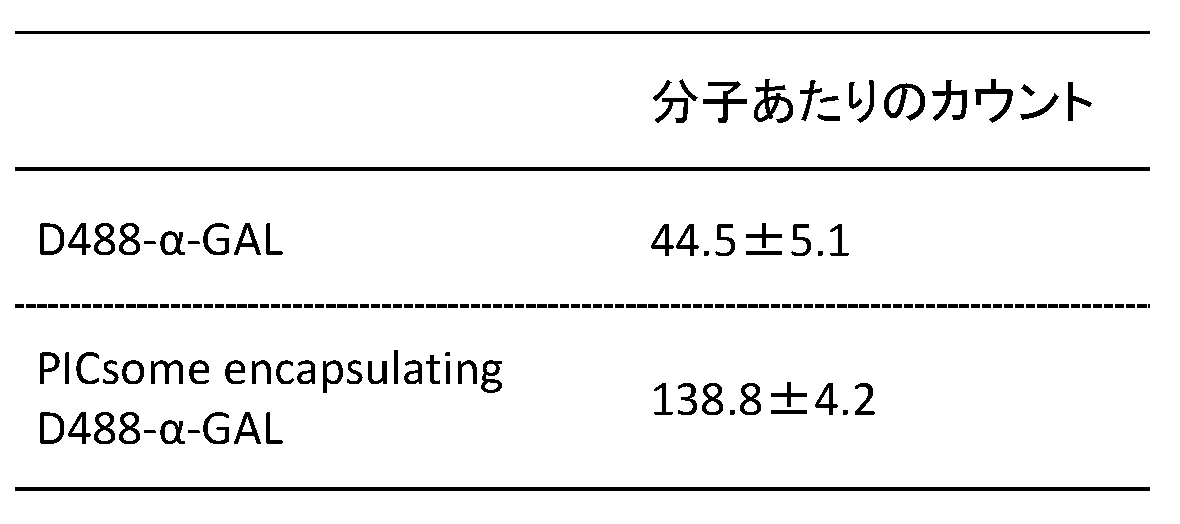
実施例2の項目3に記載されたように、遊離型α-GALと内包型α-GALの血中滞留性を評価した。マウス(Balb/c、6週齢、n=3)にCy5-α-GALを内包したPICsomeを尾静脈投与し、12時間後に採血して、上澄みの蛍光強度(Ex/Em=650nm/670nm)を測定した。結果は、図19Aに示される通りであった。図19Aに示されるように、内包型α-GALの血中滞留性は、遊離型α-GALの血中滞留性と比較して格段に高かった。 4). Retention in blood of α-GAL encapsulated in PICsome As described in
次に、遊離型α-GALと内包型α-GALについてそれぞれ、酵素活性を評価した。α-GALの酵素活性は、図19Bに示されるように、基質α-ガラクトシル-β-(7-アミド-4-メチルクマリン)(「Gal-AMC」とも言う)との反応により確認した。Gal-AMCは、α-GALの酵素活性により分解されて7-アミノ-4-メチルクマリン(「AMC」ともいう)を生じ、蛍光(Ex/Em=350 nm/450 nm)を発する。
具体的には、まず、Gal-AMC、遊離型α-GAL、および内包型α-GALの各溶液(PBS中)を恒温槽に静置して37℃とした。その後、Gal-AMCの溶液を遊離型α-GALもしくは内包型α-GALの各溶液と混合し、速やかに所定量を96ウェルプレート(テカン)に注いだ。プレートを予め37℃に保温しておいたマルチプレートリーダーの内部に入れ、蛍光強度を一定時間経時的に測定した。
酵素活性は、ミカエリス・メンテンプロットおよびラインウィーバー・バークプロットを作成して評価した。この際、予め調製したAMCの標準溶液を用いて作製した検量線を用い、蛍光強度の変化を生成物の生成速度に変換することで反応速度Vを求めた。基質濃度[S]を様々に変え、横軸に[S]を、縦軸にVを取り、プロットの結果に次式を当てはめることでミカエリス・メンテン係数(Km)を算出した。結果は、表2に示される通りであった。 5). Maintenance of enzyme activity under physiological conditions Next, the enzyme activity of free α-GAL and encapsulated α-GAL was evaluated. The enzyme activity of α-GAL was confirmed by reaction with the substrate α-galactosyl-β- (7-amido-4-methylcoumarin) (also referred to as “Gal-AMC”) as shown in FIG. 19B. Gal-AMC is decomposed by the enzymatic activity of α-GAL to produce 7-amino-4-methylcoumarin (also referred to as “AMC”), and emits fluorescence (Ex / Em = 350 nm / 450 nm).
Specifically, first, each solution of Gal-AMC, free α-GAL, and encapsulated α-GAL (in PBS) was allowed to stand at 37 ° C. in a thermostatic bath. Thereafter, a solution of Gal-AMC was mixed with each solution of free α-GAL or encapsulated α-GAL, and a predetermined amount was quickly poured into a 96-well plate (Tecan). The plate was placed in a multi-plate reader that had been kept warm at 37 ° C., and the fluorescence intensity was measured over time for a certain time.
Enzyme activity was evaluated by creating a Michaelis-Menten plot and a Lineweaver-Burk plot. At this time, the reaction rate V was determined by converting a change in fluorescence intensity into a product production rate using a calibration curve prepared using a standard solution of AMC prepared in advance. The Michaelis-Menten coefficient (K m ) was calculated by varying the substrate concentration [S], taking [S] on the horizontal axis, V on the vertical axis, and applying the following equation to the plot results. The results were as shown in Table 2.

As shown in Table 2, α-GAL encapsulated in PICsome was stable in vivo and maintained enzyme activity equivalent to that of free α-GAL.
Claims (20)
- 酵素を内包したポリイオンコンプレックス型ポリマーソームであって、
酵素は、ポリイオンコンプレックス型ポリマーソームの膜を透過する物質を基質とする酵素である、ポリイオンコンプレックス型ポリマーソーム。 A polyion complex type polymersome encapsulating an enzyme,
The enzyme is an enzyme whose substrate is a substance that permeates the membrane of a polyion complex polymersome. - 酵素の分子量が、5kDa以上である、請求項1に記載のポリイオンコンプレックス型ポリマーソーム。 The polyion complex type polymersome according to claim 1, wherein the molecular weight of the enzyme is 5 kDa or more.
- 酵素の基質の分子量が、1kDa以下である、請求項1または2に記載のポリイオンコンプレックス型ポリマーソーム。 The polyion complex type polymersome according to claim 1 or 2, wherein the molecular weight of the substrate of the enzyme is 1 kDa or less.
- 酵素が、球状タンパク質である、請求項1~3のいずれか一項に記載のポリイオンコンプレックス型ポリマーソーム。 The polyion complex type polymersome according to any one of claims 1 to 3, wherein the enzyme is a globular protein.
- 酵素が、L-アスパラギナーゼ、ウリカーゼ、α-ガラクトシダーゼおよびα-グルコシダーゼから選択される酵素である、請求項1に記載のポリイオンコンプレックス型ポリマーソーム。 2. The polyion complex type polymersome according to claim 1, wherein the enzyme is an enzyme selected from L-asparaginase, uricase, α-galactosidase and α-glucosidase.
- ポリイオンコンプレックス型ポリマーソームが、
COOH基を側鎖に有するアミノ酸を単量体単位として含む重合体(A)と、
NH2基を側鎖に有するアミノ酸を単量体単位として含む重合体(B)と
のポリイオンコンプレックス型ポリマーソームである、請求項1~5のいずれか一項に記載のポリイオンコンプレックス型ポリマーソーム。 Polyion complex type polymersome,
A polymer (A) containing an amino acid having a COOH group in the side chain as a monomer unit;
The polyion complex polymersome according to any one of claims 1 to 5, which is a polyion complex polymersome with a polymer (B) containing an amino acid having an NH 2 group in the side chain as a monomer unit. - 重合体(A)に存在するCOOH基の50%以上が重合体(B)のNH2基と架橋され
ている、請求項6に記載のポリイオンコンプレックス型ポリマーソーム。 The polyion complex type polymersome according to claim 6, wherein 50% or more of the COOH groups present in the polymer (A) are crosslinked with the NH 2 groups of the polymer (B). - 10mM リン酸緩衝溶液(pH7.4)中において37℃条件下で測定した場合に、内包した酵素の累積放出率(%)が前記水溶液と接触後7日目に20%以下である、請求項1~7のいずれか一項に記載のポリイオンコンプレックス型ポリマーソーム。 The cumulative release rate (%) of the encapsulated enzyme is 20% or less on the seventh day after contact with the aqueous solution when measured in a 10 mM phosphate buffer solution (pH 7.4) at 37 ° C. 8. The polyion complex type polymersome according to any one of 1 to 7.
- 10mM リン酸緩衝溶液(pH7.4)中において37℃条件下で測定した場合に、数平均分子量2kDaの直鎖状ポリエチレングリコールの放出速度定数kが5×10⁻³以下である、請求項7に記載のポリイオンコンプレックス型ポリマーソーム。 8. The release rate constant k of a linear polyethylene glycol having a number average molecular weight of 2 kDa is 5 × 10 −3 or less when measured in a 10 mM phosphate buffer solution (pH 7.4) at 37 ° C. The polyion complex type polymersome described in 1.
- 請求項1~9のいずれか一項に記載のポリイオンコンプレックス型ポリマーソームであって、内包される酵素が、血漿成分を基質とする、ポリイオンコンプレックス型ポリマーソーム。 The polyion complex polymersome according to any one of claims 1 to 9, wherein the encapsulated enzyme uses a plasma component as a substrate.
- 請求項10のポリイオンコンプレックス型ポリマーソームを含んでなる、医薬組成物。 A pharmaceutical composition comprising the polyion complex-type polymersome of claim 10.
- 医薬組成物が、酵素欠損または異常を起因とする疾患に罹患している患者に投与するための医薬組成物であり、酵素が、該対象において欠損した、または異常を有する酵素である、請求項11に記載の医薬組成物。 The pharmaceutical composition is a pharmaceutical composition for administration to a patient suffering from a disease caused by an enzyme deficiency or abnormality, and the enzyme is an enzyme that is deficient or has an abnormality in the subject. 11. A pharmaceutical composition according to 11.
- 酵素が、L-アスパラギナーゼ、ウリカーゼ、α-ガラクトシダーゼ、およびα-グルコシダーゼから選択される酵素である、請求項11または12に記載の医薬組成物。 The pharmaceutical composition according to claim 11 or 12, wherein the enzyme is an enzyme selected from L-asparaginase, uricase, α-galactosidase, and α-glucosidase.
- 酵素が、新生物または微生物の生育に必要な栄養素を分解する酵素である、請求項11に記載の医薬組成物。 The pharmaceutical composition according to claim 11, wherein the enzyme is an enzyme that degrades nutrients necessary for growth of a neoplasm or a microorganism.
- アスパラギン要求性腫瘍を処置するための、請求項13に記載の医薬組成物。 The pharmaceutical composition according to claim 13, for treating an asparagine-requiring tumor.
- アスパラギン要求性腫瘍が、アスパラギン産生酵素の発現量が、正常細胞における該酵素の発現量に対して80%以下である腫瘍である、15に記載の医薬組成物。 16. The pharmaceutical composition according to 15, wherein the asparagine-requiring tumor is a tumor in which the expression level of the asparagine-producing enzyme is 80% or less with respect to the expression level of the enzyme in normal cells.
- アスパラギン要求性腫瘍が、急性リンパ性白血病、T細胞悪性リンパ腫、NK細胞性白血病および急性骨髄性白血病からなる群から選択される、請求項15または16に記載の医薬組成物。 The pharmaceutical composition according to claim 15 or 16, wherein the asparagine-requiring tumor is selected from the group consisting of acute lymphocytic leukemia, T cell malignant lymphoma, NK cell leukemia and acute myeloid leukemia.
- 酵素が、α-ガラクトシダーゼであり、α-ガラクトシダーゼの異常を原因とする疾患を処置することに用いるための、請求項11または12に記載の医薬組成物。 13. The pharmaceutical composition according to claim 11 or 12, wherein the enzyme is α-galactosidase and is used for treating a disease caused by abnormality of α-galactosidase.
- 酵素が、ウリカーゼであり、高尿酸血症または高尿酸血症を原因とする疾患を処置することに用いるための、請求項11または12に記載の医薬組成物。 The pharmaceutical composition according to claim 11 or 12, wherein the enzyme is uricase and is used for treating hyperuricemia or a disease caused by hyperuricemia.
- 酵素が、α-グルコシダーゼであり、α-グルコシダーゼの異常を原因とする疾患を処置することに用いるための、請求項11または12に記載の医薬組成物。 13. The pharmaceutical composition according to claim 11 or 12, wherein the enzyme is α-glucosidase and is used for treating a disease caused by abnormality of α-glucosidase.
Priority Applications (4)
| Application Number | Priority Date | Filing Date | Title |
|---|---|---|---|
| EP16789567.1A EP3292871B1 (en) | 2015-05-07 | 2016-05-06 | Nanoreactor using polyion complex polymersomes, and method for producing same |
| JP2017516622A JP6821192B2 (en) | 2015-05-07 | 2016-05-06 | Nanoreactor using polyion complex type polymersome and its manufacturing method |
| US15/571,217 US11096991B2 (en) | 2015-05-07 | 2016-05-06 | Nanoreactor using polyion complex polymersomes, and method for producing same |
| US17/380,226 US20210346472A1 (en) | 2015-05-07 | 2021-07-20 | Nanoreactor using polyion complex polymersomes, and method for producing same |
Applications Claiming Priority (2)
| Application Number | Priority Date | Filing Date | Title |
|---|---|---|---|
| JP2015095278 | 2015-05-07 | ||
| JP2015-095278 | 2015-05-07 |
Related Child Applications (2)
| Application Number | Title | Priority Date | Filing Date |
|---|---|---|---|
| US15/571,217 A-371-Of-International US11096991B2 (en) | 2015-05-07 | 2016-05-06 | Nanoreactor using polyion complex polymersomes, and method for producing same |
| US17/380,226 Continuation US20210346472A1 (en) | 2015-05-07 | 2021-07-20 | Nanoreactor using polyion complex polymersomes, and method for producing same |
Publications (1)
| Publication Number | Publication Date |
|---|---|
| WO2016178431A1 true WO2016178431A1 (en) | 2016-11-10 |
Family
ID=57217904
Family Applications (1)
| Application Number | Title | Priority Date | Filing Date |
|---|---|---|---|
| PCT/JP2016/063684 WO2016178431A1 (en) | 2015-05-07 | 2016-05-06 | Nanoreactor using polyion complex polymersomes, and method for producing same |
Country Status (4)
| Country | Link |
|---|---|
| US (2) | US11096991B2 (en) |
| EP (1) | EP3292871B1 (en) |
| JP (1) | JP6821192B2 (en) |
| WO (1) | WO2016178431A1 (en) |
Cited By (3)
| Publication number | Priority date | Publication date | Assignee | Title |
|---|---|---|---|---|
| CN106900652A (en) * | 2017-03-16 | 2017-06-30 | 青岛大学 | A kind of construction method of hyperuricemia model |
| WO2019176916A1 (en) * | 2017-10-05 | 2019-09-19 | 公益財団法人川崎市産業振興財団 | Composition controlling pharmacokinetics in the body |
| WO2023199723A1 (en) * | 2022-04-14 | 2023-10-19 | 国立研究開発法人量子科学技術研究開発機構 | Single-polymer particles, active molecular complex, method for producing single-polymer particles, method for measuring tumor size, method for measuring fine structure within tumor, method for imaging biological tissue, drug delivery system, and contrast agent kit |
Families Citing this family (3)
| Publication number | Priority date | Publication date | Assignee | Title |
|---|---|---|---|---|
| CN114432436B (en) * | 2020-11-05 | 2023-06-02 | 北京快乐星生物科技有限公司 | Adjuvant composition and method for preparing same |
| CN114558134B (en) * | 2022-03-02 | 2023-09-05 | 国家纳米科学中心 | Polymer-entrapped erythrocyte membrane composite material and preparation method and application thereof |
| WO2024085242A2 (en) | 2022-10-21 | 2024-04-25 | Kawasaki Institute Of Industrial Promotion | Non-fouling or super stealth vesicle |
Citations (4)
| Publication number | Priority date | Publication date | Assignee | Title |
|---|---|---|---|---|
| JPH01246226A (en) * | 1988-03-28 | 1989-10-02 | Sumitomo Pharmaceut Co Ltd | Stable composition containing modified asparaginase |
| JP2004503598A (en) * | 2000-07-18 | 2004-02-05 | デューク ユニヴァーシティー | Treatment of type II glycogenosis |
| WO2014001509A1 (en) * | 2012-06-29 | 2014-01-03 | Consejo Superior De Investigaciones Científicas (Csic) | Functionalized liposomes useful for the delivery of bioactive compounds |
| WO2014133172A1 (en) * | 2013-03-01 | 2014-09-04 | 独立行政法人科学技術振興機構 | Substance-containing vesicle, and production method therefor |
Family Cites Families (4)
| Publication number | Priority date | Publication date | Assignee | Title |
|---|---|---|---|---|
| CN104958258A (en) | 2010-05-21 | 2015-10-07 | 国立研究开发法人科学技术振兴机构 | Substance-encapsulating vesicle and process for producing the same |
| EP2599834B1 (en) | 2010-07-28 | 2016-06-15 | The University of Tokyo | Electrostatically bonded vesicle |
| EP2729569B1 (en) | 2011-07-06 | 2019-12-11 | The Regents of The University of California | Multiple-enzyme nanocomplexes |
| AU2013330729A1 (en) | 2012-10-12 | 2015-05-14 | Teijin Limited | Electrostatic-bonding-type vesicle including metal microparticles |
-
2016
- 2016-05-06 JP JP2017516622A patent/JP6821192B2/en active Active
- 2016-05-06 WO PCT/JP2016/063684 patent/WO2016178431A1/en active Application Filing
- 2016-05-06 EP EP16789567.1A patent/EP3292871B1/en active Active
- 2016-05-06 US US15/571,217 patent/US11096991B2/en active Active
-
2021
- 2021-07-20 US US17/380,226 patent/US20210346472A1/en active Pending
Patent Citations (4)
| Publication number | Priority date | Publication date | Assignee | Title |
|---|---|---|---|---|
| JPH01246226A (en) * | 1988-03-28 | 1989-10-02 | Sumitomo Pharmaceut Co Ltd | Stable composition containing modified asparaginase |
| JP2004503598A (en) * | 2000-07-18 | 2004-02-05 | デューク ユニヴァーシティー | Treatment of type II glycogenosis |
| WO2014001509A1 (en) * | 2012-06-29 | 2014-01-03 | Consejo Superior De Investigaciones Científicas (Csic) | Functionalized liposomes useful for the delivery of bioactive compounds |
| WO2014133172A1 (en) * | 2013-03-01 | 2014-09-04 | 独立行政法人科学技術振興機構 | Substance-containing vesicle, and production method therefor |
Non-Patent Citations (1)
| Title |
|---|
| KIMIYOSHI ICHIDA: "New antihyperuricemic medicine: febuxoslat, Puricase, etc.", JAPANESE JOURNAL OF CLINICAL MEDICINE, vol. 66, no. 4, 2008, pages 759 - 765, XP009507050 * |
Cited By (7)
| Publication number | Priority date | Publication date | Assignee | Title |
|---|---|---|---|---|
| CN106900652A (en) * | 2017-03-16 | 2017-06-30 | 青岛大学 | A kind of construction method of hyperuricemia model |
| WO2019176916A1 (en) * | 2017-10-05 | 2019-09-19 | 公益財団法人川崎市産業振興財団 | Composition controlling pharmacokinetics in the body |
| US20210038634A1 (en) * | 2017-10-05 | 2021-02-11 | Kawasaki Institute Of Industrial Promotion | Composition controlling pharmacokinetics in the body |
| JPWO2019176916A1 (en) * | 2017-10-05 | 2021-02-25 | 公益財団法人川崎市産業振興財団 | Compositions that control pharmacokinetics in the body |
| JP7434145B2 (en) | 2017-10-05 | 2024-02-20 | 公益財団法人川崎市産業振興財団 | Compositions that control pharmacokinetics in the body |
| US11957708B2 (en) | 2017-10-05 | 2024-04-16 | Kawasaki Institute Of Industrial Promotion | Composition controlling pharmacokinetics in the body |
| WO2023199723A1 (en) * | 2022-04-14 | 2023-10-19 | 国立研究開発法人量子科学技術研究開発機構 | Single-polymer particles, active molecular complex, method for producing single-polymer particles, method for measuring tumor size, method for measuring fine structure within tumor, method for imaging biological tissue, drug delivery system, and contrast agent kit |
Also Published As
| Publication number | Publication date |
|---|---|
| US11096991B2 (en) | 2021-08-24 |
| EP3292871A4 (en) | 2018-12-26 |
| JP6821192B2 (en) | 2021-01-27 |
| JPWO2016178431A1 (en) | 2018-03-08 |
| EP3292871B1 (en) | 2021-06-23 |
| US20210346472A1 (en) | 2021-11-11 |
| EP3292871A1 (en) | 2018-03-14 |
| US20180140680A1 (en) | 2018-05-24 |
Similar Documents
| Publication | Publication Date | Title |
|---|---|---|
| WO2016178431A1 (en) | Nanoreactor using polyion complex polymersomes, and method for producing same | |
| US10322092B2 (en) | Substance-containing vesicle, and production method therefor | |
| Wang et al. | Polylactide-cholesterol stereocomplex micelle encapsulating chemotherapeutic agent for improved antitumor efficacy and safety | |
| Lee et al. | Endolysosomal environment-responsive photodynamic nanocarrier to enhance cytosolic drug delivery via photosensitizer-mediated membrane disruption | |
| US10357454B2 (en) | Substance-encapsulating vesicle and process for producing the same | |
| WO2015075942A1 (en) | Carrier for drug delivery and conjugate, composition containing same, and method for administering same | |
| JP2008526995A (en) | Nanoparticulate carrier modified with amine polymer | |
| Chen et al. | In vitro cytotoxicity, hemolysis assay, and biodegradation behavior of biodegradable poly (3‐hydroxybutyrate)–poly (ethylene glycol)–poly (3‐hydroxybutyrate) nanoparticles as potential drug carriers | |
| US10821082B2 (en) | Disintegratable core/shell silica particles for encapsulating and releasing bioactive macromolecules | |
| Levit et al. | Bio-inspired amphiphilic block-copolymers based on synthetic glycopolymer and poly (amino acid) as potential drug delivery systems | |
| Wang et al. | Water-soluble chitosan nanoparticles as a novel carrier system for protein delivery | |
| Wang et al. | A versatile method to prepare protein nanoclusters for drug delivery | |
| Haladjova et al. | Polyplex Particles Based on Comb‐Like Polyethylenimine/Poly (2‐ethyl‐2‐oxazoline) Copolymers: Relating Biological Performance with Morphology and Structure | |
| Adeyemi et al. | Synthesis and in vitro characterization of a pH-responsive chitosan-polyethylenimine nanosystem for the delivery of therapeutic proteins | |
| Berry et al. | Enhancing intracranial delivery of clinically relevant non-viral gene vectors | |
| Kolitz-Domb et al. | Engineered narrow size distribution high molecular weight proteinoids, proteinoid-poly (L-lactic acid) copolymers and nano/micro-hollow particles for biomedical applications | |
| McManus et al. | Co-encapsulation by Flash NanoPrecipitation of Insulin, Trypsin Inhibitor and Caprate Permeabilization Enhancer for Oral Administration | |
| Chen et al. | Polyethyleneimine-grafted poly (N-3-hydroxypropyl) aspartamide as a biodegradable gene vector for efficient gene transfection | |
| WO2020230793A1 (en) | Technology for controlling migration of hollow nanoparticles to brain from blood | |
| Fujii | Polymeric core-crosslinked particles prepared via a nanoemulsion-mediated process: from particle design and structural characterization to in vivo behavior in chemotherapy | |
| Hu et al. | Sustained-release behavior and the antitumor effect of charge-convertible poly (amino acid) s drug-loaded nanoparticles | |
| Kotova et al. | Properties of core–shell nanoparticles based on PLGA and human serum albumin prepared by different methods | |
| EP3031447A1 (en) | Nucleic acid-encapsulating polymer micelle complex and method for producing same | |
| WO2023150169A1 (en) | Ionizable polyesters, polyplexes and methods of use |
Legal Events
| Date | Code | Title | Description |
|---|---|---|---|
| 121 | Ep: the epo has been informed by wipo that ep was designated in this application |
Ref document number: 16789567 Country of ref document: EP Kind code of ref document: A1 |
|
| ENP | Entry into the national phase |
Ref document number: 2017516622 Country of ref document: JP Kind code of ref document: A |
|
| NENP | Non-entry into the national phase |
Ref country code: DE |
|
| WWE | Wipo information: entry into national phase |
Ref document number: 2016789567 Country of ref document: EP |
|
| WWE | Wipo information: entry into national phase |
Ref document number: 15571217 Country of ref document: US |