WO2016001810A1 - Bispecific heterodimeric diabodies and uses thereof - Google Patents
Bispecific heterodimeric diabodies and uses thereof Download PDFInfo
- Publication number
- WO2016001810A1 WO2016001810A1 PCT/IB2015/054829 IB2015054829W WO2016001810A1 WO 2016001810 A1 WO2016001810 A1 WO 2016001810A1 IB 2015054829 W IB2015054829 W IB 2015054829W WO 2016001810 A1 WO2016001810 A1 WO 2016001810A1
- Authority
- WO
- WIPO (PCT)
- Prior art keywords
- domain
- seq
- cad
- sequence
- sub
- Prior art date
Links
Classifications
-
- C—CHEMISTRY; METALLURGY
- C07—ORGANIC CHEMISTRY
- C07K—PEPTIDES
- C07K16/00—Immunoglobulins [IGs], e.g. monoclonal or polyclonal antibodies
- C07K16/46—Hybrid immunoglobulins
- C07K16/468—Immunoglobulins having two or more different antigen binding sites, e.g. multifunctional antibodies
-
- A—HUMAN NECESSITIES
- A61—MEDICAL OR VETERINARY SCIENCE; HYGIENE
- A61P—SPECIFIC THERAPEUTIC ACTIVITY OF CHEMICAL COMPOUNDS OR MEDICINAL PREPARATIONS
- A61P35/00—Antineoplastic agents
-
- A—HUMAN NECESSITIES
- A61—MEDICAL OR VETERINARY SCIENCE; HYGIENE
- A61P—SPECIFIC THERAPEUTIC ACTIVITY OF CHEMICAL COMPOUNDS OR MEDICINAL PREPARATIONS
- A61P43/00—Drugs for specific purposes, not provided for in groups A61P1/00-A61P41/00
-
- C—CHEMISTRY; METALLURGY
- C07—ORGANIC CHEMISTRY
- C07K—PEPTIDES
- C07K16/00—Immunoglobulins [IGs], e.g. monoclonal or polyclonal antibodies
- C07K16/18—Immunoglobulins [IGs], e.g. monoclonal or polyclonal antibodies against material from animals or humans
-
- C—CHEMISTRY; METALLURGY
- C07—ORGANIC CHEMISTRY
- C07K—PEPTIDES
- C07K16/00—Immunoglobulins [IGs], e.g. monoclonal or polyclonal antibodies
- C07K16/18—Immunoglobulins [IGs], e.g. monoclonal or polyclonal antibodies against material from animals or humans
- C07K16/28—Immunoglobulins [IGs], e.g. monoclonal or polyclonal antibodies against material from animals or humans against receptors, cell surface antigens or cell surface determinants
- C07K16/2803—Immunoglobulins [IGs], e.g. monoclonal or polyclonal antibodies against material from animals or humans against receptors, cell surface antigens or cell surface determinants against the immunoglobulin superfamily
- C07K16/2809—Immunoglobulins [IGs], e.g. monoclonal or polyclonal antibodies against material from animals or humans against receptors, cell surface antigens or cell surface determinants against the immunoglobulin superfamily against the T-cell receptor (TcR)-CD3 complex
-
- A—HUMAN NECESSITIES
- A61—MEDICAL OR VETERINARY SCIENCE; HYGIENE
- A61K—PREPARATIONS FOR MEDICAL, DENTAL OR TOILETRY PURPOSES
- A61K39/00—Medicinal preparations containing antigens or antibodies
- A61K2039/505—Medicinal preparations containing antigens or antibodies comprising antibodies
-
- C—CHEMISTRY; METALLURGY
- C07—ORGANIC CHEMISTRY
- C07K—PEPTIDES
- C07K2317/00—Immunoglobulins specific features
- C07K2317/30—Immunoglobulins specific features characterized by aspects of specificity or valency
- C07K2317/31—Immunoglobulins specific features characterized by aspects of specificity or valency multispecific
-
- C—CHEMISTRY; METALLURGY
- C07—ORGANIC CHEMISTRY
- C07K—PEPTIDES
- C07K2317/00—Immunoglobulins specific features
- C07K2317/50—Immunoglobulins specific features characterized by immunoglobulin fragments
- C07K2317/52—Constant or Fc region; Isotype
- C07K2317/524—CH2 domain
-
- C—CHEMISTRY; METALLURGY
- C07—ORGANIC CHEMISTRY
- C07K—PEPTIDES
- C07K2317/00—Immunoglobulins specific features
- C07K2317/50—Immunoglobulins specific features characterized by immunoglobulin fragments
- C07K2317/52—Constant or Fc region; Isotype
- C07K2317/526—CH3 domain
-
- C—CHEMISTRY; METALLURGY
- C07—ORGANIC CHEMISTRY
- C07K—PEPTIDES
- C07K2317/00—Immunoglobulins specific features
- C07K2317/50—Immunoglobulins specific features characterized by immunoglobulin fragments
- C07K2317/56—Immunoglobulins specific features characterized by immunoglobulin fragments variable (Fv) region, i.e. VH and/or VL
- C07K2317/565—Complementarity determining region [CDR]
-
- C—CHEMISTRY; METALLURGY
- C07—ORGANIC CHEMISTRY
- C07K—PEPTIDES
- C07K2317/00—Immunoglobulins specific features
- C07K2317/60—Immunoglobulins specific features characterized by non-natural combinations of immunoglobulin fragments
- C07K2317/62—Immunoglobulins specific features characterized by non-natural combinations of immunoglobulin fragments comprising only variable region components
- C07K2317/626—Diabody or triabody
-
- C—CHEMISTRY; METALLURGY
- C07—ORGANIC CHEMISTRY
- C07K—PEPTIDES
- C07K2317/00—Immunoglobulins specific features
- C07K2317/70—Immunoglobulins specific features characterized by effect upon binding to a cell or to an antigen
- C07K2317/72—Increased effector function due to an Fc-modification
-
- C—CHEMISTRY; METALLURGY
- C07—ORGANIC CHEMISTRY
- C07K—PEPTIDES
- C07K2317/00—Immunoglobulins specific features
- C07K2317/70—Immunoglobulins specific features characterized by effect upon binding to a cell or to an antigen
- C07K2317/73—Inducing cell death, e.g. apoptosis, necrosis or inhibition of cell proliferation
- C07K2317/732—Antibody-dependent cellular cytotoxicity [ADCC]
-
- C—CHEMISTRY; METALLURGY
- C07—ORGANIC CHEMISTRY
- C07K—PEPTIDES
- C07K2317/00—Immunoglobulins specific features
- C07K2317/70—Immunoglobulins specific features characterized by effect upon binding to a cell or to an antigen
- C07K2317/76—Antagonist effect on antigen, e.g. neutralization or inhibition of binding
-
- C—CHEMISTRY; METALLURGY
- C07—ORGANIC CHEMISTRY
- C07K—PEPTIDES
- C07K2317/00—Immunoglobulins specific features
- C07K2317/90—Immunoglobulins specific features characterized by (pharmaco)kinetic aspects or by stability of the immunoglobulin
- C07K2317/94—Stability, e.g. half-life, pH, temperature or enzyme-resistance
Definitions
- the .txt file contains a sequence listing entitled "PC72054A_Sequence_Listing.txt” created on June 12, 2015, and having a size of 248 KB.
- the sequence listing contained in this .txt file is part of the
- the present invention relates to bispecific heterodimeric diabodies and uses thereof in the treatment of cancer.
- the present invention provides for bispecific heterodimeric diabodies, wherein the bispecific heterodimeric diabody is capable of specific binding to an epitope of P- cadherin and to an epitope of CD3.
- the present invention provides for bispecific heterodimeric diabodies, wherein the bispecific heterodimeric diabody is capable of specific binding to an epitope of P- cadherin and to an epitope of CD3, wherein the bispecific heterodimeric diabody comprises a first polypeptide chain and a second polypeptide chain, wherein: a. the first polypeptide comprises, in the N-terminal to C-terminal direction: i. a Domain 1 , comprising a sub-Domain 1A and a sub-Domain 1 B, and ii. a first heterodimer- promoting domain; and b. the second polypeptide chain comprises, in the N-terminal to C-terminal direction: i.
- a Domain 2 comprising a sub-Domain 2A and a sub-Domain 2B, and ii. a second heterodimer-promoting domain
- sub-Domain 1A and sub- Domain 2A form a P-cadherin VLA/H binding domain comprising a variable heavy (VH) domain of an anti-P-cadherin antibody (P-CAD VH) and a variable light (VL) domain of an anti-P-cadherin antibody (P-CAD VL)
- sub-Domain 1 B and sub-Domain 2B form a CD3 VL/VH binding domain comprising a VL domain of an anti-CD3 antibody (CD3 VL) and a VH binding domain of an anti-CD3 antibody (CD3 VH); or wherein sub- Domain 1A and sub-Domain 2A form a CD3 VL/VH binding domain comprising a CD3 VL and a CD3 VH, and sub-Domain 1 B and sub-Domain 2B form a P-
- the sub-Domain 1 A comprises a P-CAD VL or CD3 VL
- the sub-Domain 1 B comprises a P-CAD VH, if the sub-Domain 1 A comprises CD3 VL, or a CD3 VH, if the sub-Domain 1 A comprises P-CAD VL
- the sub-Domain 2B comprises a P-CAD VL or a CD3 VL depending on the VL domain selected for sub-Domain 1 A
- the sub-Domain 2A comprises P-CAD VH, if the sub-Domain 2B comprises CD3 VL, or a CD3 VH, if the sub-Domain 2B comprises P-CAD VL.
- the sub-Domain 1 A comprises a P-CAD VL and the sub-Domain 1 B comprises a CD3 VH
- the sub-Domain 2B comprises a CD3 VL and the sub-Domain 2A comprises a P-CAD VH
- the P-CAD VL of the sub-Domain 1A and the P-CAD VH of the sub-Domain 2A form a VL/VH binding domain capable of specifically binding to an epitope of P-cadherin
- the CD3 VH of the sub-Domain 1 B and the CD3 VL of the sub-Domain 2B form a VL/VH binding domain capable of specifically binding to an epitope of CD3.
- sub-Domain 1 A comprises a CD3 VL and the sub-Domain 1 B comprises a P-CAD VH
- the sub-Domain 2B comprises a P-CAD VL and the sub-Domain 2A a CD3 VH; and wherein the CD3 VL of the sub-Domain 1 A and the CD3 VH of the sub-Domain 2A form a VL/VH binding domain capable of specifically binding to an epitope of CD3, and the P-CAD VH of the sub-Domain 1 B and the P-CAD VL of the sub-Domain 2B form a VL/VH binding domain capable of specifically binding to an epitope of P-cadherin.
- the sub-Domain 1 A comprises a VH binding domain of either an anti-P-cadherin antibody (P-CAD VH) or an anti-CD3 antibody (CD3 VH)
- the sub-Domain 1 B comprises a VL binding domain of either an anti-P-cadherin antibody (P-CAD VL), if the sub-Domain 1 A comprises CD3 VH, or an anti-CD3 antibody (CD3 VL), if the sub-Domain 1A comprises a P-CAD VH
- the sub-Domain 2B comprises a P-CAD VH or a CD3 VH depending on the VH domain selected for sub- Domain 1A
- the sub-Domain 2A comprises P-CAD VL
- the sub-Domain 2B comprises a CD3 VH, or a CD3 VL
- the sub-Domain 2B comprises a P-CAD VH.
- the sub-Domain 1A comprises a P-CAD VH and the sub- Domain 1 B comprises a CD3 VL
- the sub-Domain 2B comprises a CD3 VH and the sub-Domain 2A comprises a P-CAD VL
- the P-CAD VH of the sub-Domain 1 A and the P-CAD VL of the sub-Domain 2A form a VL/VH binding domain capable of specifically binding to an epitope of P-cadherin
- the CD3 VL of the sub-Domain 1 B and the CD3 VH of the sub-Domain 2B form a VL/VH binding domain capable of specifically binding to an epitope of CD3.
- the sub-Domain 1 A comprises a CD3 VH and the sub- Domain 1 B comprises a P-CAD VL
- the sub-Domain 2B comprises a P-CAD VH and the sub-Domain 2A comprises a CD3 VL
- the CD3 VH of the sub- Domain 1 A and the CD3 VL of the sub-Domain 2A form a VL/VH binding domain capable of specifically binding to an epitope of CD3
- the P-CAD VL of the sub- Domain 1 B and the P-CAD VH of the sub-Domain 2B form a VL/VH binding domain capable of specifically binding to an epitope of P-cadherin.
- the present invention further provides for bispecific heterodimeric diabodies wherein the first heterodimer-promoting domain and the second heterodimer-promoting domain comprise an IgG Fc region comprising a CH2 and a CH3 domain, wherein the amino acid sequence of the CH2 domain and/or the CH3 domain comprises at least one amino acid modification, as compared to a wild-type IgG Fc region, to form a knob or a hole.
- the first heterodimer-promoting domain and the second heterodimer-promoting domain comprise an IgG Fc region comprising a CH2 and a CH3 domain, wherein the amino acid sequence of the CH2 domain and/or the CH3 domain comprises at least one amino acid modification, as compared to a wild-type IgG Fc region, to form a knob or a hole.
- the first heterodimer-promoting domain and the second heterodimer-promoting domain comprise an IgG Fc region comprising a CH2 and a CH3 domain, wherein the amino acid sequence of the CH
- heterodimer-promoting domain are not both knobs or both holes; and/or wherein the first heterodimer-promoting domain and the second heterodimer-promoting domain form an IgG immunoglobulin Fc region.
- the IgG Fc region forming the knob comprises a sequence of SEQ ID NO: 63
- the IgG Fc region forming the hole comprises a sequence of SEQ ID NO: 64.
- the present invention further provides for bispecific heterodimeric diabodies, wherein the first heterodimer-promoting domain and the second heterodimer-promoting domain comprise an E-coil region comprising glutamic acid and a negatively charged alpha-helical coil or a K-coil region comprising lysine and a positively charged helical coil; and wherein the first heterodimer-promoting domain and the second heterodimer- promoting domain are not both E-coil regions or both K-coil regions.
- the E-coil region comprises a sequence of SEQ ID NO: 61
- the K-coil region comprises a sequence of SEQ ID NO: 62.
- the present invention further provides for bispecific heterodimeric diabodies, wherein the sub-Domain 1A and the sub-Domain 1 B may be linked by a glycine-serine linker (Linker 1 ) and do not associate to form a VL/VH epitope binding domain, and the sub-Domain 2B and the sub-Domain 2A may be linked by a glycine-serine linker (Linker 1) and do not associate to form a VL/VH epitope binding domain.
- the glycine-serine linker (Linker 1 ) comprises a sequence of SEQ ID NO: 68 or SEQ ID NO: 69.
- the present invention further provides for bispecific heterodimeric diabodies, wherein the first heterodimer-promoting domain comprises a cysteine linker (Linker 2) on sub-Domain 1 B and/or the second heterodimer-promoting domain comprises a cysteine linker (Linker 2) on sub-Domain 2A.
- the Linker 2 of the first heterodimer-promoting domain and/or the second heterodimer-promoting domain further comprises at least one glycine residue.
- the Linker 2 of the first heterodimer-promoting domain and/or the second heterodimer-promoting domain comprises a sequence of GFNRGEC (SEQ ID NO: 70), GVEPKSC (SEQ ID NO: 71), GGCGGG (SEQ ID NO: 72), GCPPCP (SEQ ID NO: 73), GGTGGCPPCP (SEQ ID NO: 74), GEPKSSDKTHTCPPCP (SEQ ID NO: 75) or GGTGGGEPKSSDKTHTCPPCP (SEQ ID NO: 76).
- the Linker 2 of the first heterodimer-promoting domain comprises the sequence of GCPPCP (SEQ ID NO: 73), GGTGGCPPCP (SEQ ID NO: 74), GEPKSSDKTHTCPPCP (SEQ ID NO: 75) or
- GGTGGGEPKSSDKTHTCPPCP SEQ ID NO: 76
- the Linker 2 of the second heterodimer-promoting domain comprises the sequence of GCPPCP (SEQ ID NO: 73), GGTGGCPPCP (SEQ ID NO: 74), GEPKSSDKTHTCPPCP (SEQ ID NO: 75) or GGTGGGEPKSSDKTHTCPPCP (SEQ ID NO: 76).
- the Linker 2 of the first heterodimer-promoting domain comprises the sequence of GGCGGG (SEQ ID NO: 72)
- the Linker 2 of the second heterodimer-promoting domain comprises the sequence of GGCGGG (SEQ ID NO: 72).
- the Linker 2 of the first heterodimer-promoting domain comprises the sequence of GFNRGEC (SEQ ID NO: 70) and the Linker 2 of the second heterodimer-promoting domain comprises the sequence of GVEPKSC (SEQ ID NO: 71 ), or the Linker 2 of the first heterodimer-promoting domain comprises the sequence of GVEPKSC (SEQ ID NO: 71 ) and the Linker 2 of the second heterodimer-promoting domain comprises the sequence of GFNRGEC (SEQ ID NO: 70).
- the present invention provides for bispecific heterodimeric diabodies, wherein the bispecific heterodimeric diabodies specifically bind to extracellular domain 3 (ECD3) of human P-cadherin.
- ECD3 extracellular domain 3
- the present invention further provides for bispecific heterodimeric diabodies, wherein the bispecific heterodimeric diabody specifically bind to an epitope on P-cadherin but does not bind to an epitope on E-cadherin or VE-cadherin.
- the present invention also provides for bispecific heterodimeric diabodies, wherein the bispecific heterodimeric diabodies specifically bind to an epitope on human P-cadherin but do not bind to an epitope on mouse P-cadherin.
- the present invention provides for bispecific heterodimeric diabodies, wherein the bispecific heterodimeric diabodies demonstrate an extended serum and tumor half-life. Pharmacokinetic analysis may conducted by various assays, such as ELISA.
- the present invention further provides for bispecific heterodimeric diabodies, wherein the bispecific heterodimeric diabodies demonstrate a lower EC50 in the presence of increased P-cadherin expression levels or increased receptor density levels.
- the EC50 may be determined by various in vitro and in vivo cytotoxicity assays
- the present invention provides for bispecific heterodimeric diabodies, comprising a
- the present invention provides for bispecific heterodimeric diabodies, comprising the P-CAD VL CDR1 comprises a sequence of SEQ ID NOS: 35 or 36, the P-CAD VL CDR2 comprises a sequence of SEQ ID NOS: 37 or 38, and the P-CAD VL CDR3 comprises a sequence of SEQ ID NOS: 39, 40, 41 , 42, 43 or 44;
- the CD3 VH CDR1 comprises a sequence of SEQ ID NO: 48
- the CD3 VH CDR2 comprises a sequence of SEQ ID NOS: 49 or 50
- the CD3 VH CDR3 comprises a sequence of SEQ ID NO: 51 ;
- the CD3 VL CDR1 comprises a sequence of SEQ ID NO: 55, the CD3 VL CDR2 comprises a sequence of SEQ ID NO: 56, and the CD3 VL CDR3 comprises a sequence of SEQ ID NO: 57;
- the P-CAD VH CDR1 comprises a sequence of SEQ ID NOS: 25
- the P-CAD VL CDR1 comprises the sequence of SEQ ID NO: 35
- the P-CAD VL CDR2 comprises the sequence of SEQ ID NO: 37
- the P-CAD VL CDR3 comprises the sequence of SEQ ID NO: 41
- the P-CAD VH CDR1 comprises the sequence of SEQ ID NO: 25
- the P-CAD VH CDR2 comprises the sequence of SEQ ID NO: 26
- the P-CAD VH CDR3 comprises the sequence of SEQ ID NO: 28.
- the P-CAD VL CDR1 comprises the sequence of SEQ ID NO: 35; the P-CAD VL CDR2 comprises the sequence of SEQ ID NO: 37; and the P-CAD VL CDR3 comprises the sequence of SEQ ID NO: 42, the P-CAD VH CDR1 comprises the sequence of SEQ ID NO: 25, the P-CAD VH CDR2 comprises the sequence of SEQ ID NO: 26, and the P-CAD VH CDR3 comprises the sequence of SEQ ID NO: 29.
- the P-CAD VL CDR1 comprises the sequence of SEQ ID NO: 35; the P-CAD VL CDR2 comprises the sequence of SEQ ID NO: 37, the P-CAD VL CDR3 comprises the sequence of SEQ ID NO: 43, the P-CAD VH CDR1 comprises the sequence of SEQ ID NO: 25, the P-CAD VH CDR2 comprises the sequence of SEQ ID NO: 26, and the P-CAD VH CDR3 comprises the sequence of SEQ ID NO: 30.
- the P-CAD VL CDR1 comprises the sequence of SEQ ID NO: 35; the P-CAD VL CDR2 comprises the sequence of SEQ ID NO: 37, the P-CAD VL CDR3 comprises the sequence of SEQ ID NO: 39, the P-CAD VH CDR1 comprises the sequence of SEQ ID NO: 25, the P-CAD VH CDR2 comprises the sequence of SEQ ID NO: 26, and the P-CAD VH CDR3 comprises the sequence of SEQ ID NO: 31.
- the present invention further provides for bispecific heterodimeric diabodies, comprising a P-CAD VL comprising a sequence of SEQ ID NOS: 1 , 3, 5, 7, 9, 1 1 , 13, 15, 17, 19, 21 or 23; a CD3 VH comprising a sequence of SEQ ID NOS: 45 or 46; a CD3 VL comprising the sequence of SEQ ID NO: 47; and a P-CAD VH comprising a sequence of SEQ ID NOS: 2, 4, 6, 8, 10, 12, 14, 16, 18, 20, 22 or 24.
- the P-CAD VL comprises the sequence of SEQ ID NO: 5
- the P-CAD VH comprises the sequence of SEQ ID NO: 6.
- the P-CAD VL comprises the sequence of SEQ ID NO: 7 and the P-CAD VH comprises the sequence of SEQ ID NO: 8.
- the P-CAD VL comprises the sequence of SEQ ID NO: 9 and the P-CAD VH comprises the sequence of SEQ ID NO: 10.
- the P-CAD VL comprises the sequence of SEQ ID NO: 15 and the P-CAD VH comprises the sequence of SEQ ID NO: 16.
- the present invention further provides for bispecific heterodimeric diabodies capable of specific binding to an epitope of P-cadherin and to an epitope of CD3 comprising a first polypeptide chain and a second polypeptide chain, wherein the first polypeptide chain comprises a sequence of SEQ ID NO: 90 and the second polypeptide chain comprises a sequence of SEQ ID NO: 91 .
- the present invention further provides for bispecific heterodimeric diabodies capable of specific binding to an epitope of P- cadherin and to an epitope of CD3 comprising a first polypeptide chain and a second polypeptide chain, wherein the first polypeptide chain comprises a sequence of SEQ ID NO: 92 and the second polypeptide chain comprises a sequence of SEQ ID NO: 93.
- the present invention further provides for bispecific heterodimeric diabodies capable of specific binding to an epitope of P-cadherin and to an epitope of CD3 comprising a first polypeptide chain and a second polypeptide chain, wherein the first polypeptide chain comprises a sequence of SEQ ID NO: 88 and the second polypeptide chain comprises a sequence of SEQ ID NO: 89.
- the present invention further provides for bispecific heterodimeric diabodies wherein the first and second polypeptide chains may be covalently bonded to one another by at least one disulfide bond.
- the present invention further provides for bispecific heterodimeric diabodies capable of specific binding to an epitope of P-cadherin and to an epitope of CD3 comprising a first polypeptide chain and a second polypeptide chain, wherein the first polypeptide chain and second polypeptide chain comprises sequences as set forth in Figure 30A, 30B, 31 A, 31 B, 32A or 32B.
- the bispecific heterodimeric diabodies comprise a first polypeptide chain, a second polypeptide chain and a third polypeptide chain as set forth in Table 5.
- the present invention provides for bispecific heterodimeric diabodies, wherein the bispecific heterodimeric diabodies may be conjugated to a detectable label, including but not limited to, fluorophore or a radionuclide.
- a detectable label including but not limited to, fluorophore or a radionuclide.
- the present invention provides for bispecific heterodimeric diabodies that bind to P-cadherin and compete for binding to bispecific heterodimeric diabodies disclosed herein.
- the present invention further provides for a pharmaceutical composition comprising a therapeutically effective amount of a bispecific heterodimeric diabody disclosed herein and a pharmaceutically acceptable carrier.
- the present invention further provides for a method of treating P-cadherin associated disorders in a patient in need thereof, comprising administering to the patient a bispecific heterodimeric diabody disclosed herein or a pharmaceutical composition comprising a bispecific heterodimeric diabody disclosed herein.
- the present invention further provides for a method of treating a P-cadherin associated disorders in a patient in need thereof, comprising administering to the patient the bispecific heterodimeric disclosed herein or a pharmaceutical composition comprising a bispecific heterodimeric diabody disclosed herein, wherein a cytolytic T cell response is activated or induced.
- the present invention further provides bispecific heterodimeric diabodies disclosed herein for use in therapy.
- the present invention further provides the use of bispecific heterodimeric diabodies disclosed herein in the manufacture of a medicament for use in therapy.
- the therapy is treatment of P-cadherin associated disorder.
- the present invention further provides bispecific heterodimeric diabodies disclosed herein for use in therapy, wherein the therapy activates or induces a cytolytic T cell response.
- the P-cadherin associated disorder is cancer.
- the cancer is a P-cadherin expressing or P cadherin positive cancer.
- the cancers include, but are not limited to, breast, colorectal, ovarian, gastric, thyroid, prostate, cervical, pancreatic, lung, bladder, liver, endometrial, head and neck, testicular, and glioblastoma cancer.
- the present invention further provides isolated host cells that recombinantly produce bispecific heterodimeric diabodies described herein, isolated polynucleotides comprising nucleotide sequences encoding the bispecific heterodimeric diabodies disclosed herein, and vectors comprising the polynucleotides.
- the present invention further provides methods of producing bispecific
- heterodimeric diabodies disclosed herein comprising culturing the host cells disclosed herein under conditions that result in production of the bispecific heterodimeric diabodies, and purifying the bispecific heterodimeric diabodies from the culture supernatant.
- the present invention further provides for bispecific heterodimeric diabodies disclosed herein, wherein a crystal structure assembles into a compact spherical structure stabilized by a disulfide linkage between a pair of cysteine residues consisting of a cysteine residue at position 239 (Cys 239 ) of sub-Domain 1 B and a cysteine residue at position 246 (Cys 246 ) of sub-Domain 2A; and, wherein the crystal diffracts X-rays for determination of atomic coordinates to provide resolution of better than about 2.0 Angstroms.
- the present invention further provides for bispecific heterodimeric diabodies capable of specific binding to an epitope of P-cadherin and to an epitope of CD3 comprising a first polypeptide chain and a second polypeptide chain, wherein the first polypeptide chain comprises, in the N-terminal to C-terminal direction: a Domain 1 , comprising a sub-Domain 1 A which comprises a VL binding domain of an anti-CD3 antibody (CD3 VL) comprising a sequence of SEQ ID NO: 47, and a sub-Domain 1 B which comprises a VH binding domain of either an anti-P-cadherin antibody (P-CAD VH) comprising a sequence of SEQ ID NO: 6, wherein the sub-Domain 1A and sub- Domain 1 B may be covalently linked by a glycine-serine linker (Linker 1 ) and do not associate to form a VL/VH epitope binding domain; and the second polypeptide chain comprises, in the N-terminal to
- the present invention further provides methods of identifying additional locations for cysteine residues to form a disulfide linkage within bispecific heterodimeric diabodies based on the crystal structures disclosed herein, wherein the crystal structure is analyzed for a pair of amino acid residue locations for substitution with a pair of cysteine residues that form a disulfide linkage.
- the pair of amino acid residue locations for substitution with a pair of cysteine residues that form a disulfide linkage may be selected from the group consisting of amino acid locations Gln 121 (VH1 B)Gly 160 (VL1A), Val 129 (VH1 B)Gly 2 (VL1A),
- the disulfide linkage may reduce solvent accessibility and reduce the length of linkers.
- the present invention further provides methods of engineering bispecific heterodimeric diabody variants in an attempt to form a more stable interdomain association, wherein the engineering is through amino acid site-directed mutagenesis in the interdomain interface based on the crystal structure disclosed herein, wherein the amino acid site-directed mutagenesis fills up large interior voids/holes.
- the site-directed mutagenesis may increase amino acid side chain volumes by replacing small amino acids for amino acids with bulky aromatic side-chains.
- the small amino acids may be selected from the group consisting of amino acids at positions Ala (VL1 A), Val 213 (VH2A), Leu 238 (VH2A), or Met 231 (VL2B).
- the amino acids with bulky aromatic side-chains may be selected from the group consisting of phenylalanine, tyrosine, or tryptophan.
- the present invention further provides for antibodies that binds to P-cadherin, comprising a CDR1 , a CDR2, and a CDR3 of a light chain variable region (VL) comprising the sequence of SEQ ID NOS: 1 , 3, 5, 7, 9, 1 1 , 13, 15, 17, 19, 21 or 23 and/or a CDR1 , a CDR2, and a CDR3 of a heavy chain variable region (VH) comprising the sequence of SEQ ID NOS: 2, 4, 6, 8, 10, 12, 14, 16, 18, 20, 22 or 24.
- VL light chain variable region
- VH heavy chain variable region
- the antibodies disclosed herein comprise a VL CDR1 comprising the sequence of SEQ ID NO: 35 or 36, a VLCDR2 comprising the sequence of SEQ ID NO: 37 or 38, a VL CDR3 comprising the sequence of SEQ ID NO: 39, 40, 41 , 42, 43 or 44, a VH CDR1 comprising the sequence of SEQ ID NO: 25 or 33, a VH CDR2 comprising the sequence of SEQ ID NO: 26 or 34, and a VH CDR3 comprising the sequence of SEQ ID NO: 27, 28, 29, 30, 31 or 32.
- the antibodies disclosed herein comprise a VL CDR1 comprising the sequence of SEQ ID NO: 35 or 36, a VL CDR2 comprising the sequence of SEQ ID NO: 37 or 38, a VL CDR3 comprising the sequence of SEQ ID NO: 39, 40, 41 , 42, 43 or 44, a VH CDR1 comprising the sequence of SEQ ID NO: 25, a VH CDR2 comprising the sequence of SEQ ID NO: 26, and a VH CDR3 comprising the sequence of SEQ ID NO: 27, 28, 29, 30, 31 or 32.
- the antibodies disclosed herein comprise a VL CDR1 comprising the sequence of SEQ ID NO: 35, a VL CDR2 comprising the sequence of SEQ ID NO: 37, a VL CDR3 comprising the sequence of SEQ ID NO: 42, a VH CDR1 comprising the sequence of SEQ ID NO: 25, a VH CDR2 comprising the sequence of SEQ ID NO: 26, and a VH CDR3 comprising the sequence of SEQ ID NO: 29.
- the present invention further provides for antibodies that binds to P-cadherin, comprising a light chain variable region comprising the sequence of SEQ ID NOS: 1 , 3, 5, 7, 9, 1 1 , 13, 15, 17, 19, 21 and 23, and/or a heavy chain variable region comprising the sequence of SEQ ID NOS: 2, 4, 6, 8, 10, 12, 14, 16, 18, 20, 22 or 24.
- the antibodies disclosed herein comprise a light chain variable region amino acid comprising the sequence of SEQ ID NO: 5, and/or a heavy chain variable region comprising the sequence of SEQ ID NO: 6.
- the antibodies disclosed herein comprise a light chain variable region comprising the sequence of SEQ ID NO: 7, and/or a heavy chain variable region comprising the sequence of SEQ ID NO: 8.
- the antibodies disclosed herein comprise a light chain variable region comprising the sequence of SEQ ID NO: 9 and/or a heavy chain variable region comprising the sequence of SEQ ID NO: 10. In another aspect of the invention, the antibodies disclosed herein comprise a light chain variable region comprising the sequence of SEQ ID NO: 15 and/or a heavy chain variable region comprising the sequence of SEQ ID NO: 16.
- the present invention further provides for antibodies that bind to P-cadherin and compete for binding to P-cadherin with antibodies disclosed herein.
- the present invention further provides for pharmaceutical compositions comprising a therapeutically effective amount of an antibody disclosed herein and a pharmaceutically acceptable carrier.
- the present invention further provides a method of treating P-cadherin associated disorder in a patient in need thereof, comprising administering to the patient an antibody disclosed herein or a pharmaceutical composition comprising an antibody disclosed herein.
- the present invention further provides antibodies disclosed herein for use in therapy.
- the present invention further provides the use of antibodies disclosed herein in the manufacture of a medicament for use in therapy.
- the P-cadherin associated disorder is cancer.
- the cancer is a P-cadherin expressing or P cadherin positive cancer.
- the cancers include, but are not limited to, breast, colorectal, ovarian, gastric, thyroid, prostate, cervical, pancreatic, lung, bladder, liver, endometrial, head and neck, testicular, and glioblastoma cancer.
- the present invention further provides nucleic acids that encode the antibodies disclosed herein.
- the nucleic acids may comprise a sequence that is selected from the group consisting of SEQ ID NOS: 98, 100, 102, 104, 106, 108, 1 10, 1 12, 1 14 and 1 16.
- the nucleic acids may comprise a sequence that is selected from the group consisting of SEQ ID NOS: 97, 99, 101 , 103, 105, 107, 109, 1 1 1 , 1 13 and 1 15.
- the present invention further provides vectors comprising nucleic acids disclosed herein and host cells comprising vectors disclosed herein.
- the present invention further provides for bispecific heterodimeric diabodies or antibodies disclosed herein, wherein the P-CAD VL comprises a P-CAD VL CDR1 sequence comprising XLI .I -XLI .2-XLI .3-XLI .4- XL1.5- XLI .6-XLI .7-XLI .8- XLI .9-XLI .I O- XLI .I I - XLI .2-XLI .I 3, wherein X L i .i to XLI .I 3 each independently is an amino acid residue according to column 3 of Table 57, Table 58, or Table 59; a P-CAD VL CDR2 sequence comprising XL2.I -XL2.2-XL2.3-XL2.4-XL2.5-XL2.6- XL.2.7, wherein X L 2.1 to X L 2. 7 each independently is an amino acid residue according to column 3 of Table 57, Table 58, or Table 59;
- X L 3.i to XL3.I I each independently is an amino acid residue according to column 3 of Table 63, Table 64, or Table 65; and the P-CAD VH comprises: a P-CAD VH CDR1 sequence comprising XHI .I -XHI .2-XHI .3-XHI .4- XHI .5-XHI .6- ⁇ .7- ⁇ .8- ⁇ .9- ⁇ . ⁇ ⁇ ⁇ .5, XHI .6-XHI .7-XHI .8-XHI .9- XHI .I O, wherein X
- XH2.4- XH2.5-XH2.6- XH2.7"XH2.8-XH2.9-XH2.I O wherein XH2.I to XH2 17 each independently is an amino acid residue according to column 3 of Table 51 , Table 52, or Table 53; and a P-CAD VH CDR3 sequence comprising X H 3.I -XH3.2-XH3.3-XH3.4- XH3.5-XH3.6- XH3.7-XH3.S- XH3.9, wherein XH3.I to XH3.9 each independently is an amino acid residue according to column 3 of Table 54, Table 55, or Table 56.
- X L i .i to XLI .I3 each independently is an amino acid residue according to column 3 of Table 57. In another aspect, X L i .i to XLI .I3 each independently is an amino acid residue according to column 3 of Table 58. In another aspect, X L i .i to XLI .I3 each independently is an amino acid residue according to column 3 of Table 59. In another aspect, X L 2.i to X L 2. 7 each independently is an amino acid residue according to column 3 of Table 60. In another aspect, X L 2.i to X L 2. 7 each independently is an amino acid residue according to column 3 of Table 61 . In another aspect, X L 2.i to X L 2.
- X L 3.i to X L 3.n each independently is an amino acid residue according to column 3 of Table 63.
- X L 3.-i to XL3.H each independently is an amino acid residue according to column 3 of Table 64.
- X L 3.i to X L 3.n each independently is an amino acid residue according to column 3 of or Table 65.
- X H i .i to XHI .I O each independently is an amino acid residue according to column 3 of Table 48.
- X H i .i to XHH O each independently is an amino acid residue according to column 3 of Table 48.
- X H 3.i to X H 3.9 each independently is an amino acid residue according to column 3 of Table 55.
- XH3.I to X H 3.9 each independently is an amino acid residue according to column 3 of Table 56.
- the present invention further provides for bispecific heterodimeric diabodies or antibodies disclosed herein, wherein the P-CAD VL comprises a P-CAD VL CDR1 sequence comprising XLI.I-XLI.2-XLI.3-XLI.4- XL1.5- XLI.6-XLI.7-XLI.8- XLI.9-XLI.IO- XLI.II- XLI.2-XLI.I3, wherein X L -u to XLI.I3 each independently is an amino acid residue according to column 3 or column 4 of Table 69; a P-CAD VL CDR2 sequence comprising XL2.I-XL2.2-XL2.3-XL2.4-XL2.5-XL2.6- XL2.7, wherein X L 2.1 to X L 2.7 each
- X L 3.i to XL3.II each independently is an amino acid residue according to column 3 or column 4 of Table 71 ; and the P-CAD VH comprises a P-CAD VH CDR1 sequence comprising XHI.I-XHI.2-XHI.3-XM.4- XHI.5-XHI.6- XHI.7-XHI.8- XHI.9-XHI.IO ⁇ .5, XHI.6-XHI.7-XHI.8-XHI.9- XHI.IO, wherein Xm.i to Xm.10 each independently is an amino acid residue according to column 3 or column 4 of Table 71 ; and the P-CAD VH comprises a P-CAD VH CDR1 sequence comprising XHI.I-XHI.2-XHI.3-XM.4- XHI.5-XHI.6- XHI.7-XHI.8- XHI.9-XHI.IO ⁇ .5, XHI.6-XHI
- XH2.6- XH2.7-XH2.8-XH2.9-XH2.IO wherein X H 2.i to XH217 each independently is an amino acid residue according to column 3 or column 4 of Table 67, and a P-CAD VH CDR3 sequence comprising XH3.I-XH3.2-XH3.3-XH3.4- XH3.5-XH3.6- XH3.7-XH3.8-XH3.9, wherein X H3 .1 to XH3.9 each independently is an amino acid residue according to column 3 or column 4 of Table 68.
- X L i .i to Xu .13 each independently is an amino acid residue according to column 3 of Table 69.
- X L i.i to Xu .13 each independently is an amino acid residue according to column 4 of Table 69.
- X L 2.i to Xi_2 . 7 each independently is an amino acid residue according to column 3 of Table 70.
- X L 2.i to X L 2 .7 each independently is an amino acid residue according to column 4 of Table 70.
- X L 3.-i to X L 3.n each independently is an amino acid residue according to column 3 of Table 71.
- X L 3.i to X1_3.11 each independently is an amino acid residue according to column 4 of Table 71.
- XHI .
- I to Xm .10 each independently is an amino acid residue according to column 3 of Table 66.
- X H i .i to Xm .10 each independently is an amino acid residue according to column 4 of Table 66.
- X H 2.i to XH2 17 each independently is an amino acid residue according to column 3 of Table 67.
- XH2 . I to XH2 17 each independently is an amino acid residue according to column 4 of Table 67.
- X H 3.i to X H 3. 9 each independently is an amino acid residue according to column 3 of Table 68.
- XH3.I to XH3.9 each independently is an amino acid residue according to column 4 of Table 68.
- the present invention provides for X H 1 . 8 is G, X H 2 .5 is Y, XH3.I is I, XH3 . 7 is F, XL3.3 is W, XH2.6 is N, X H2 1 6 is Q, XHS.S is N, X H3 .9 is I, Xu . 8 is G, X L 2. 2 is N, X L2 Z is N, Xi_3 . 2 is T, and/or XL3.4 is D.
- the present invention also provides for any combination of the aspects described herein.
- Figures 1A and 1 B provide schematics of four alternative representations of LP- DART P-cadherin/CD3 bispecific heterodimeric diabodies having a first heterodimer- promoting domain and second heterodimer-promoting domain comprising an Fc region optimized to associate via a "knob-in-hole" association.
- Figure 2 provides a schematic representation of a VF-DART bispecific heterodimeric diabody that simultaneously targets and activates T cells (via CD3 antigen) and tumor cells (via P-cadherin antigen) having a first polypeptide chain (1 ) wherein a first heterodimer-promoting domain comprises a C-terminal peptide that forms a disulfide bond for stability with a C-terminal peptide of a second heterodimer- promoting domain of a second polypeptide chain (2).
- Figure 3 provides a schematic representation of an assembled VF-DART bispecific heterodimeric diabody having a first polypeptide chain (1 ) and second polypeptide chain (2) wherein a first heterodimer-promoting domain (such as SEQ ID NO: 70 or SEQ ID NO: 71 , described in detail below) and a second heterodimer- promoting domain (such as SEQ ID NO: 71 or SEQ ID NO: 70, depending on the selection of the first heterodimer promoting domain) comprise a cysteine residue.
- a first heterodimer-promoting domain such as SEQ ID NO: 70 or SEQ ID NO: 71 , described in detail below
- a second heterodimer- promoting domain such as SEQ ID NO: 71 or SEQ ID NO: 70, depending on the selection of the first heterodimer promoting domain
- Figure 4 provides a schematic representation of an assembled EK-DART bispecific heterodimeric diabody having a first polypeptide chain (1 ) wherein a first heterodimer-promoting domain comprises a glutamic acid-rich region that forms a negatively charged alpha-helical coil (E-coil), and a second polypeptide chain (2) wherein a second heterodimer-promoting domain comprises a lysine rich region that forms a positively charged helical coil (K-coil).
- Figure 5 provides a schematic representation of an assembled LP-DART bispecific heterodimeric diabody having a first heterodimer-promoting domain and a second heterodimer-promoting domain comprising an Fc region optimized to associate via a "knob-in-hole" association.
- FIG. 6 provides the structures of an alternative bispecific heterodimeric diabody having an Fc domain termed MP3-DART.
- the bispecific heterodimeric diabody is attached to the N-terminal side of the Fc domain and is thus termed "N-terminal MP3- DART.”
- the N-terminal MP3-DART comprises a first, second and third polypeptide chain which are schematically represented.
- the final structure of the N-terminal MP3- DART is represented.
- FIG 7 provides the structures of an alternative bispecific heterodimeric diabody having an Fc domain termed MP3-DART.
- the bispecific heterodimeric diabody is attached to the C-terminal side of the Fc domain and is thus termed "C-terminal MP3 DART.”
- the C-terminal MP3-DART comprises a first, second and third polypeptide chain which are schematically represented.
- the final structure of the C-terminal MP3- DART is represented.
- Figure 8 provides a linear regression analysis comparing cytotoxic T lymphocyte (CTL) activity to relative cell surface P-cadherin expression.
- CTL cytotoxic T lymphocyte
- Figure 9 demonstrates the in vivo ability of 177 EK-DART to decrease tumor volume in murine HCT116 colorectal cancer model.
- Figure 10 demonstrates the in vivo ability of 153 EK-DART to decrease tumor volume in murine HCT1 16 colorectal cancer model.
- Figure 11 demonstrates the in vivo ability of 154 EK-DART to decrease tumor volume in murine HCT1 16 colorectal cancer model.
- Figure 12 demonstrates the in vivo ability of 35 EK-DART to decrease tumor volume in murine HCT1 16 colorectal cancer model.
- Figure 13 demonstrates the in vivo ability of 153 LP-DART to decrease tumor volume in murine HCT116 colorectal cancer model.
- Figure 14 demonstrates the in vivo ability of 153 LP-DART to decrease tumor volume in murine HCT1 16 colorectal cancer model.
- Figure 15 demonstrates the in vivo ability of 153 LP-DART to decrease tumor volume in murine Du145 prostate cancer model.
- Figure 16 demonstrates the in vivo ability of 153 LP-DART to decrease tumor volume in murine H1650 lung cancer model.
- Figures 17A-17D demonstrate the in vivo ability of 153 LP-DART to decrease tumor volume in an established tumor + engrafted human T cell model, 17E
- Figures 18A and 18B demonstrate the in vivo ability of 153 LP-DART to decrease tumor volume in an established tumor human T cell adoptive transfer model.
- Figure 19 demonstrates the in vivo ability of 153 LP-DART to decrease tumor volume in a P-cadherin positive patient derived colorectal tumor xenograft
- FIG 20 shows a 153 LP-DART mediated dose dependent increase in tumor infiltrating lymphocytes (TILs).
- Figures 21A-21 D provide in vitro assays to evaluate the properties of fluorophore Labeled 153 LP-DART and T cells.
- Figures 22A-22D provide biodistribution and targeting of 153 LP-DART in a HCT1 16 xenograft model using longitudinal FMT imaging.
- Figures 23A-23C show the pharmacokinetic data (ELISA) from the FMT imaging study.
- Figures 24A shows the evaluation of T cell activity of fluorphore labeled and unlabeled T cells and 24B shows the cellular tracking kinetics of fluorophore labeled T cells in a HCT1 16 tumor model using FMT imaging (in vivo).
- Figure 25 provides a schematic representation of a DART construct designed for crystallography, having a C-terminal His tag designated "6XHIS" covalently linked to the P-CAD VH domain and a C-terminal FLAG designated "FLAG” covalently linked to the CD3 VH domain.
- Figure 26 provides a graphical depiction of the crystal structure of the
- Figure 27 provides a graphical depiction of the crystallography 35 DART protein showing the antigen binding sites for the anti-CD3 CDR regions and the anti-P-CAD CDR regions.
- Figure 28 provides a schematic of a crystallography 35 DART protein variant with a more stable interdomain association through site-directed mutagenesis in the interdomain interface containing large interior voids/holes that may be filled up with increasing side chain volumes with various amino acids.
- Figure 29 provides a graphical depiction of the crystal structure of a
- Figures 30A and 30B provide amino acid sequences for first and second polypeptide chains of VF-DART bispecific heterodimeric diabodies of the present invention.
- Figures 31 A and 31 B provide amino acid sequences for first and second polypeptide chains of EK-DART bispecific heterodimeric diabodies of the present invention.
- Figures 32A and 32B provide amino acid sequences for first and second polypeptide chains of LP-DART bispecific heterodimeric diabodies of the present invention.
- Figure 33 provides amino acid sequences for first and second polypeptide chains of DART bispecific heterodimeric diabodies of the present invention.
- Figure 34 provides P-cadherin- extra cellular domain (ECD) -Fc fusion protein constructs for eptitope mapping of P-cadherin 153 LP- DART .
- ECD extra cellular domain
- Figure 35 provides the full length P-Cadherin epitope sequence (UniProt P22223, CADH3, Human Cadherin-3).
- the present invention describes novel bispecific heterodimeric diabodies comprising an antigen binding domain that specifically binds P-cadherin, and an antigen binding domain that specifically binds CD3 for the recruitment of and activation of cytolytic T cells.
- the bispecific heterodimeric diabodies may further comprise a Fc domain.
- the bispecific heterodimeric diabodies successfully direct and activate T cell cytotoxicity to tumor cells expressing P- cadherin.
- the bispecific heterodimeric diabodies of the present invention have enhanced pharmacokinetic properties to extend in vivo half-life, and were designed to engage and activate polyclonal T cell populations via the CD3 complex in the presence of P-cadherin expressing tumors.
- the present invention describes the isolation and characterization of fully human single-chain antibody binding domains against P-cadherin derived from human phage display libraries.
- the invention further provides covalently linking these P-cadherin binding domains to antigen binding domains of anti-CD3 antibodies in the previously described DART format (Moore et al., Blood, 1 17(17): 4542-4551 , 201 1 ) to create bispecific heterodimeric diabodies that are capable of simultaneous binding to P- cadherin and CD3.
- the anti-P-cadherin/anti-CD3 DART proteins also known as bispecific heterodimeric diabodies, successfully direct and activate T cell cytotoxicity of cells expressing P-cadherin.
- P-cadherin (Cadherin-3, CDH3) belongs to the classical cadherin superfamily of transmembrane glycolproteins that regulate calcium-dependent cell-cell adhesion during development and tissue homeostasis (Gumbiner et al., J. Cell Biol., 148: 399-403, (2000)).
- the cadherin intracellular domains directly interact with cytoplasmic catenins that link to the actin cytoskeletal network, providing the molecular basis for stable cell interactions.
- the cadherin/catenin complex, as well as the signaling pathways controlled by this structure, represent a major regulatory mechanism that guide cell fate decisions, through its influence on cell growth, differentiation, motility, and survival (Cavallaro et al, 201 1 ).
- Classical cadherins include the E-cadherin (CDH1 ), N-cadherin (CDH2) and P-cadherin (CDH3).
- CDH1 E-cadherin
- CDH2 N-cadherin
- CDH3 P-cadherin
- P-cadherin expression in normal tissues is low and is restricted primarily to myoepithelial cells and the basal layers of stratified epithelium.
- Increased expression of P-cadherin has been reported in various tumors, including breast, gastric, endometrial, pancreatic and colorectal cancers and is a good
- CD3 (cluster of differentiation 3) is a T cell co-receptor protein complex that comprising four distinct chains ⁇ , ⁇ , ⁇ and ⁇ that form ⁇ , ⁇ and ⁇ dimers.
- TCR T cell receptor
- CD3 CD3 molecules together comprise the TCR complex. It is well known that anti-CD3 antibodies elicit the generation of cytotoxic T cells through the activation of endogenous lymphokine production and are capable of selectively killing tumor targets (Yun et al., Cancer Research, 49: 4770-4774 (1989)).
- T cells express TCR complexes that are able to induce antigen specific immune responses (Smith-Garvin et al, 2009).
- Antigens are peptides expressed by tumor cells and virally infected cells capable of stimulating immune responses. Intracellular ⁇ expressed antigens are bound to major histocompatibility class I (MHC class I) molecules and transported to the surface where they are exposed to T cells. If the binding affinity of the TCR to the MHC class I in complex with the antigen is sufficient the formation of an immune synapse will be initiated. Signaling through the immune synapse is mediated through the CD3 co-receptors that form ⁇ , ⁇ and ⁇ dimers. These dimers associate with the TCR and generate an activation signal in T lymphocytes. This signaling cascade directs T cell mediated killing of the cell expressing the antigen. Cytotoxicity is mediated by release and transfer of granzyme B and perforin from the T cell to the target cell.
- the bispecific heterodimer diabodies of the present invention may allow the T cell to circumvent the need for the interaction of the TCR and MHC class I in complex with antigen, and instead redirects T cells to target cells through direct co-engagement of CD3 (such as CD3 epsilon) expressed on the T cell and P-cadherin expressed on the tumor.
- CD3 such as CD3 epsilon
- antibody function as used herein is meant as the capacity to support a biochemical event that results from the interaction of an anti-CD3 binding domain of a bispecific heterodimeric diabody with a cytotoxic T cell.
- antibody and “antibodies” refer to monoclonal antibodies, multispecific antibodies, human antibodies, humanized antibodies, synthetic antibodies, chimeric antibodies, polyclonal antibodies, camelized antibodies, single- chain Fvs (scFv), single chain antibodies, Fab fragments, F(ab') fragments, disulfide- linked bispecific Fvs (sdFv), intrabodies, and anti-idiotypic (anti-Id) antibodies (including, e.g. anti-Id and anti-anti-ld antibodies to antibodies of the invention), and epitope- binding fragments or binding domains of any of the above.
- antibodies include immunoglobulin molecules and immunologically active fragments of
- immunoglobulin molecules i.e., molecules that contain an antigen binding site.
- Immunoglobulin molecules can be of any type (e.g. IgG, IgE, IgM, IgD, IgA and IgY), class (e.g. IgG-i, lgG 2 , lgG3, lgG 4 , IgAi and lgA 2 ) or subclass.
- type e.g. IgG, IgE, IgM, IgD, IgA and IgY
- class e.g. IgG-i, lgG 2 , lgG3, lgG 4 , IgAi and lgA 2
- subclass e.g. IgG-i, lgG 2 , lgG3, lgG 4 , IgAi and lgA 2
- bispecific heterodimeric diabody refers to a complex of two or more polypeptide chains or proteins, each comprising at least one antibody VL and one antibody VH domain or fragment thereof, wherein both antibody binding domains are comprised within a single polypeptide chain and wherein the VL and VH domains in each polypeptide chain are from different antibodies.
- immunospecifically binds As used herein, the terms “immunospecifically binds,” “immunospecifically recognizes,” “specifically binds,” “specifically recognizes” and analogous terms refer to molecules e.g. binding domains, that specifically bind to an antigen (e.g. epitope or immune complex) and do not specifically bind to another molecule.
- a molecule that specifically binds to an antigen may bind to other peptides or polypeptides with lower affinity as determined by assays known in the art e.g. immunoassays, BIACORE ® , or other assays.
- molecules that specifically bind an antigen do not cross-react with other proteins.
- variable domain refers to domains in naturally occurring immunoglobulins and the corresponding domains of recombinant binding proteins (e.g. humanized antibodies, single chain antibodies, chimeric antibodies, etc.).
- the basic structural unit of naturally occurring immunoglobulins is a tetramer having two light chains and two heavy chains, usually expressed as a glycoprotein of about 150,000 Da.
- the amino-terminal (N-terminal) portion of each chain includes a variable region of about 100 to 1 10 or more amino acids primarily responsible for antigen recognition.
- the carboxy-terminal (C-terminal) portion of each chain defines a constant region.
- Each light chain is comprised a light chain variable domain (VL) and a light chain constant domain (CL).
- Each heavy chain is comprised of a heavy chain variable region (VH) and a heavy chain constant region, having CH1 , hinge, CH2 and CH3 domains.
- the variable regions of an IgG molecule comprise regions of hypervariability, termed the
- CDRs complementarity determining regions
- FR framework regains
- Each VH and VL comprises three CDRs and four FRs, arranged from amino-terminus to carboxy-terminus in the following structure: n-FR1 , CDR1 , FR2, CDR2, FR3, CDR3, FR4-C.
- Immunoglobulin molecules can be of any type (e.g., IgG, IgE, IgM, IgD, IgA and IgY) and class (e.g., IgGI, lgG2, IgG 3, lgG4, IgAI and lgA2) or subclass.
- the "hinge region” or “hinge domain” is generally defined as stretching from Glu216 to Pro230 of human lgG-1. Hinge regions of other IgG isotypes may be aligned with the lgG1 sequence by placing the first and last cysteine residues forming inter- heavy chain S-S binds in the same positions.
- the Fc region of an IgG comprises two constant domains, CH2 and CH3.
- the CH2 domain of a human IgG Fc region usually extends from amino acids 231 to amino acid 341 according to the numbering system of Kabat.
- the CH3 domain of a human IgG Fc region usually extends from amino acids 342 to 447 according to the numbering system of Kabat.
- the CH2 domain of a human IgG Fc region (also referred to as "Cy 2" domain) is unique in that it is not closely paired with another domain. Rather, two N-linked branched carbohydrate chains are interposed between the two CH2 domains of an intact native IgG.
- binding proteins binding domains, or antibodies (as broadly defined herein)
- the assignment of amino acids to each domain is in accordance with the definitions of Kabat, Chothia, the accumulation of both Kabat and Chothia, AbM, contact, and/or conformational definitions or any method of CDR determination well known in the art.
- Antibody CDRs may be identified as the hypervariable regions originally defined by Kabat et al. See, e.g., Kabat et al., 1992, Sequences of Proteins of Immunological Interest, 5th ed., Public Health Service, NIH, Washington D.C. The positions of the CDRs may also be identified as the structural loop structures originally described by Chothia and others.
- CDR identification includes the "AbM definition,” which is a compromise between Kabat and Chothia and is derived using Oxford Molecular's AbM antibody modeling software (Accelrys, San Diego, CA), or the "contact definition" of CDRs based on observed antigen contacts, set forth in MacCallum et al., 1996, J. Mol. Biol., 262:732-745.
- the positions of the CDRs may be identified as the residues that make enthalpic contributions to antigen binding.
- CDR may refer to CDRs defined by any approach known in the art, including combinations of approaches. The methods used herein may utilize CDRs defined according to any of these approaches. For any given aspect containing more than one CDR, the CDRs may be defined in accordance with any of Kabat, Chothia, extended, AbM, contact, and/or conformational definitions.
- humanized antibody refers to forms of non-human (e.g. murine) antibodies that are chimeric immunoglobulins, immunoglobulin chains, or fragments thereof (such as Fv, Fab, Fab', F(ab') 2 or other antigen-binding subsequences or binding domains of antibodies) that contain minimal sequences derived from non-human immunoglobulins.
- humanized antibodies are human immunoglobulins (recipient antibody) in which residues from a complementarity determining region (CDR) of the recipient are replaced by residues from a CDR of a non-human species (donor antibody) such as mouse, rat, or rabbit having the desired specificity, affinity, and capacity.
- CDR complementarity determining region
- monoclonal antibody refers to an antibody that is derived from a single clone, including any eukaryotic, prokaryotic, or phage clone, and not the method by which it is produced.
- Monoclonal antibodies from which binding domains of the bispecific heterodimeric diabodies of the invention can be prepared using a wide variety of techniques known in the art including the use of hybridoma, recombinant, and phage display technologies, or a combination thereof.
- monoclonal antibodies can be produced using hybridoma techniques including those known in the art.
- Antibodies can also be generated using various phage display methods known in the art.
- phage display methods functional antibody domains are displayed on the surface of phage particles which carry the polynucleotide sequences encoding them.
- phage can be utilized to display antigen binding domains, such as Fab and Fv or disulfide-bond stabilized Fv, expressed from a repertoire or combinatorial antibody library (e.g. human or murine).
- Phage expressing an antigen binding domain that binds the antigen of interest can be selected or identified with antigen, e.g. using labeled antigen or antigen bound or captured to a solid surface or bead.
- Phage used in these methods are typically filamentous phage, including fd and M13.
- the antigen binding domains are expressed as a recombinantly fused protein to either the phage gene III or gene VIII protein.
- Examples of phage display methods that can be used to make the immunoglobulins, or fragments thereof, of the present invention include those disclosed in Brinkmann et al. (1995) "Phage Display Of
- the functional characteristics of the multiple IgG isotypes, and domains thereof, are well known in the art.
- the amino acid sequences of lgG1 , lgG2, lgG3 and lgG4 are known in the art.
- Selection and/or combinations of two or more domains from specific IgG isotypes for use in the methods of the invention may be based on any known parameter of the parent isotypes including affinity to FcyR. For example, use of regions or domains from IgG isotypes that exhibit limited or no binding to FcyRIIB, e.g.
- lgG2 or lgG4 may find particular use where a bispecific heterodimeric diabody is desired to be engineered to maximize binding to an activating receptor and minimize binding to an inhibitory receptor.
- use of Fc regions or domains from IgG isotypes known to preferentially bind C1 q or FcyRIIIA, e.g. lgG3 may be combined with Fc amino acid modifications known in the art to enhance antibody-dependent cell mediated cytotoxicity (ADCC) and/or complement dependent cytotoxicity (CDC), to engineer a bispecific heterodimeric diabody molecule such that effector function activity, e.g. complement activation or ADCC, is maximized.
- ADCC antibody-dependent cell mediated cytotoxicity
- CDC complement dependent cytotoxicity
- mutations may be made in the Fc regions or domains of IgG isotypes that minimize or eliminate the effector function of the Fc region.
- epitope refers to that portion of a molecule capable of being
- Epitopes often consist of a chemically active surface grouping of molecules such as amino acids or sugar side chains and have specific three- dimensional structural characteristics as well as specific charge characteristics.
- antigenic epitope as used herein, is defined as a portion of a polypeptide to which an antibody can specifically bind as determined by any method well known in the art, for example, by conventional immunoassays.
- “conformational epitope” comprises noncontiguous polypeptides (or amino acids) within the antigenic protein to which an antibody specific to the epitope binds. Once a desired epitope on an antigen is determined, it is possible to generate antibodies to that epitope, e.g. using the techniques described herein. During the discovery process, the generation and characterization of antibodies may elucidate information about desirable epitopes. From this information, it is then possible to competitively screen antibodies for binding to the same epitope. An approach to achieve this is to conduct competition and cross-competition studies to find antibodies that compete or cross-compete with one another e.g. the antibodies compete for binding to the antigen.
- epitope mapping One method is to identify the epitope to which antibodies bind, or "epitope mapping.” There are many methods known in the art for mapping and characterizing the location of epitopes on proteins, including solving the crystal structure of an antibody-antigen complex, competition assays, gene fragment expression assays, and synthetic peptide-based assays, as described, for example, in Chapter 1 1 of Harlow and Lane, Using Antibodies, a Laboratory Manual, Cold Spring Harbor Laboratory Press, Cold Spring Harbor, N.Y., 1999. In an additional example, epitope mapping can be used to determine the sequence to which an antibody binds. Epitope mapping is commercially available from various sources, for example, Pepscan Systems (Edelhertweg 15, 8219 PH Lelystad, The Netherlands).
- mutagenesis of an antigen binding domain can be performed to identify residues required, sufficient, and/or necessary for epitope binding.
- the binding affinity and the off-rate of an antigen-binding domain interaction can be determined by competitive binding assays.
- a competitive binding assay is a radioimmunoassay comprising the incubation of labeled antigen, and the detection of the molecule bound to the labeled antigen.
- the affinity of the molecule of the present invention for an antigen and the binding off-rates can be determined from the saturation data by Scatchard analysis.
- the affinities and binding properties of the molecules of the invention for an antigen may be initially determined using in vitro assays (biochemical or immunological based assays) known in the art for antigen-binding domain, including but not limited to enzyme-linked immunosorbent assay (ELISA) assay, surface plasmon resonance (SPR) assay, Bio-Layer Interferometry, or immunoprecipitation assays.
- ELISA enzyme-linked immunosorbent assay
- SPR surface plasmon resonance
- Bio-Layer Interferometry Bio-Layer Interferometry
- immunoprecipitation assays immunoprecipitation assays.
- the molecules of the invention may have similar binding properties in in vivo models (such as those described and disclosed herein) as those in in vitro based assays.
- the present invention does not exclude molecules of the invention that do not exhibit the desired phenotype in in vitro based assays but do exhibit the desired phenotype in vivo.
- binding affinity is intended to refer to the dissociation rate of a particular antigen-antibody interaction.
- the K D is the ratio of the rate of dissociation, also called the “off-rate (k 0 ff)", to the association rate, or "on- rate (kon)".
- K D equals k 0 ff / k on and is expressed as a molar concentration (M). It follows that the smaller the K D , the stronger the affinity of binding. Therefore, a K D of 1 ⁇ indicates weak binding affinity compared to a K D of 1 nM.
- K D values for antibodies can be determined using methods well established in the art.
- One method for determining the K D of an antibody is by using surface plasmon resonance (SPR), typically using a biosensor system such as a BIACORE® system.
- SPR surface plasmon resonance
- BIAcore kinetic analysis comprises analyzing the binding and dissociation of an antigen from chips with immobilized molecules (e.g. molecules comprising epitope binding domains), on their surface.
- Another method for determining the K D of an antibody is by using Bio-Layer Interferometry, typically using OCTET ® technology (Octet QK e system, ForteBio).
- nucleic acids and “nucleotide sequences” include DNA molecules (e.g. cDNA or genomic DNA), RNA molecules (e.g. mRNA), combinations of DNA and RNA molecules or hybrid DNA/RNA molecules, and analogs of DNA or RNA molecules.
- Such analogs can be generated using, for example, nucleotide analogs, which include, but are not limited to, inosine or tritylated bases.
- Such analogs can also comprise DNA or RNA molecules comprising modified backbones that lend beneficial attributes to the molecules such as, for example, nuclease resistance or an increased ability to cross cellular membranes.
- nucleic acids or nucleotide sequences can be single-stranded, double-stranded, may contain both single-stranded and double-stranded portions, and may contain triple-stranded portions, but preferably is double-stranded DNA.
- the present invention also includes polynucleotides that encode the bispecific heterodimeric diabodies of the invention, including the polypeptides and binding regions of the antibodies.
- the polynucleotides encoding the molecules of the invention may be obtained, and the nucleotide sequence of the polynucleotides determined, by any method known in the art.
- the polynucleotides that encode the bispecific heterodimeric diabodies of the present invention may include the following: only the coding sequence for the variant, the coding sequence for the variant and additional coding sequences such as a functional polypeptide, or a signal or secretory sequence or a pro-protein sequence; the coding sequence for the antibody and non-coding sequence, such as introns or non- coding sequence 5' and/or 3' of the coding sequence for the antibody.
- polynucleotide encoding a bispecific heterodimeric diabody encompasses a polynucleotide which includes additional coding sequence for the variant but also a polynucleotide which includes additional coding and/or non-coding sequence.
- cancer refers to a neoplasm or tumor resulting from abnormal uncontrolled growth of cells.
- cancer refers to a benign tumor, which has remained localized.
- cancer refers to a malignant tumor, which has invaded and destroyed neighboring body structures and spread to distant sites.
- the cancer is associated with a specific cancer antigen.
- a "therapeutically effective amount” refers to that amount of the therapeutic agent sufficient to treat or manage a disease or disorder.
- a therapeutically effective amount may refer to the amount of therapeutic agent sufficient to delay or minimize the onset of disease, e.g. delay or minimize the spread of cancer.
- a therapeutically effective amount may also refer to the amount of the therapeutic agent that provides a therapeutic benefit in the treatment or management of a disease.
- a therapeutically effective amount with respect to a therapeutic agent of the invention means the amount of therapeutic agent alone, or in combination with other therapies, that provides a therapeutic benefit in the treatment or management of a disease.
- prophylactic agent and “prophylactic agents” refer to any agent(s) which can be used in the prevention of a disorder, or prevention of recurrence or spread of a disorder.
- a prophylactically effective amount may refer to the amount of prophylactic agent sufficient to prevent the recurrence or spread of hyperproliferative disease, particularly cancer, or the occurrence of such in a patient, including but not limited to those predisposed to hyperproliferative disease, for example those genetically predisposed to cancer or previously exposed to carcinogens.
- a prophylactically effective amount may also refer to the amount of the prophylactic agent that provides a prophylactic benefit in the prevention of disease. Further, a
- prophylactically effective amount with respect to a prophylactic agent of the invention means that amount of prophylactic agent alone, or in combination with other agents, that provides a prophylactic benefit in the prevention of disease.
- the terms “prevent”, “preventing” and “prevention” refer to the prevention of the recurrence or onset of one or more symptoms of a disorder in a subject as result of the administration of a prophylactic or therapeutic agent.
- the term “in combination” refers to the use of more than one prophylactic and/or therapeutic agents. The use of the term “in combination” does not restrict the order in which prophylactic and/or therapeutic agents are administered to a subject with a disorder. In other words, the combination therapy may be done by separately, sequentially, or simultaneously treating with the therapeutic agents.
- the vector for the production of the molecules may be produced by recombinant DNA technology using techniques well known in the art.
- the polynucleotides encoding the bispecific heterodimeric diabody binding domains of the present invention may include an expression control polynucleotide sequence operably linked to the antibody coding sequences, including naturally- associated or heterologous promoter regions known in the art.
- the expression control sequences may be eukaryotic promoter systems in vectors capable of transforming or transfecting eukaryotic host cells, but control sequences for prokaryotic hosts may also be used. Once the vector has been incorporated into the appropriate host cell line, the host cell is propagated under conditions suitable for expressing the nucleotide sequences, and, as desired, for the collection and purification of the antibodies.
- Eukaryotic cell lines include the CHO cell lines, various COS cell lines, HeLa cells, myeloma cell lines, transformed B-cells, or human embryonic kidney cell lines.
- bispecific heterodimeric diabody refers to a complex of two or more polypeptide chains or proteins, each comprising at least one antibody VL domain and one antibody VH domain or fragment thereof, wherein both antibody binding domains are comprised within a single polypeptide chain and wherein the VL and VH domains in each polypeptide chain are from different antibodies.
- bispecific heterodimeric diabody includes dimers or tetramers of polypeptide chains containing both a VL and VH domain.
- the individual polypeptide chains comprising the multimeric proteins may be covalently joined to at least one other peptide of the multimer by interchain disulfide bonds.
- Bispecific heterodimeric diabodies of the present invention are comprised of anti- P-cadherin (P-CAD) binding domains covalently linked to anti-CD3 (CD3) binding domains utilizing a Dual-Affinity Re-Targeting (DART ® ) platform technology (US Patent Application Publications Nos. 2007/0004909, 2009/0060910 and 2010/0174053).
- the DART technology provides a covalent linkage which results in a molecule that may have superior stability, optimal heavy and light chain pairing and antigen recognition.
- the minimal linker size and content may decrease the potential for
- the DART proteins of the present invention simultaneously target T cells (CD3) and tumor cells (P-CAD) and successfully direct and activate T cell cytoxicity to tumor cells expressing P-cadherin.
- Each polypeptide chain of the bispecific heterodimeric diabody comprises a VL domain and a VH domain, which may be covalently linked (Linker 1 ) such that the antibody binding domains are constrained from self-assembly.
- each polypeptide chain comprises a heterodimerization domain, which promotes
- the heterodimerization domain may be located at the N-terminus of the polypeptide chain or the C-terminus.
- the heterodimerization domain may comprise a cysteine linker (Linker 2) that is 1 , 2, 3, 4, 5, 6, or more amino acid residues in length. Interaction of two of the polypeptide chains may produce two VL/VH pairings, forming two epitope binding domains, i.e., a bivalent molecule.
- Neither the VH or VL domain is constrained to any position within the polypeptide chain, i.e. restricted to the amino or carboxy terminus, nor are the domains restricted in their relative positions to one another, i.e. the VL domain may be N-terminal to the VH domain and vice versa.
- the only restriction is that a complimentary
- polypeptide chain be available in order to form a functional diabody.
- VL and VH domains are derived from antibodies specific for different antigens
- formation of a functional bispecific heterodimeric diabody requires the interaction of two different polypeptide chains, i.e. formation of a heterodimer.
- two differing polypeptide chains are free to interact, e.g. in a recombinant expression system, one comprising a VLA and a VHB (A, being a first epitope and B, being a second epitope) and the other comprising a VLB and a VHA
- two differing binding sites may form: VLA- VHA and VLB-VHB.
- bispecific heterodimeric diabody polypeptide chain pairs misalignment or mis-binding of the two chains is a possibility, e.g. interaction of VL-VL or VH-VH domains.
- purification of functional bispecific heterodimeric diabodies is easily managed based on the immunospecificity of the properly dimerized binding site using any affinity based method known in the art or exemplified herein, e.g. affinity chromatography.
- the bispecific heterodimeric diabodies of the invention may simultaneously bind two separate and distinct epitopes.
- at least one epitope binding site is specific for the CD3 determinant expressed on an immune effector cell e.g.
- the bispecific heterodimeric diabody molecule binds to the effector cell determinant and also activates the effector cell.
- the epitope-binding domain is capable of binding the P-cadherin tumor- associated antigen that is associated with breast, colorectal, ovarian, gastric, thyroid, prostate, cervical, pancreatic, lung (including but not limited to non-small cell lung cancer and small cell lunch cancer), bladder, liver, endometrial, head and neck, testicular, and glioblastoma cancer.
- the bispecific heterodimeric diabodies of the present invention comprise antigen binding domains generally derived from immunoglobulins or antibodies.
- the antibodies from which the binding domains used in the methods of the invention are derived may be from any animal origin including birds and mammals (e.g. human, non-human primate, murine, donkey, sheep, rabbit, goat, guinea pig, camel, horse, or chicken).
- the antibodies are human or humanized monoclonal antibodies.
- "human” antibodies include antibodies having the amino acid sequence of a human immunoglobulin and include antibodies isolated from human immunoglobulin libraries or libraries of synthetic human immunoglobulin coding sequences or from mice that express antibodies from human genes.
- the bispecific heterodimeric diabodies of the present invention may be characterized in a variety of ways.
- molecules of the invention may be assayed for the ability to immunospecifically bind to an antigen.
- Molecules that have been identified to immunospecifically bind to an antigen can then be assayed for their specificity and affinity for the antigen.
- the bispecific heterodimeric diabodies of the present invention may be produced using a variety of methods well known in the art, including de novo protein synthesis and recombinant expression of nucleic acids encoding the binding proteins.
- the desired nucleic acid sequences may be produced by recombinant methods (e.g. PCR mutagenesis of an earlier prepared variant of the desired polynucleotide) or by solid- phase DNA synthesis. Usually recombinant expression methods are used.
- the invention provides a polynucleotide that comprises a sequence encoding an anti-CD3 VH and/or VL; in another aspect, the invention provides a polynucleotide that comprises a sequence encoding an anti-P-cadherin VH and/or VL. Because of the degeneracy of the genetic code, a variety of nucleic acid sequences encode each immunoglobulin amino acid sequence, and the present invention includes all nucleic acids encoding the binding proteins described herein.
- the polypeptide chains of the bispecific heterodimeric diabodies may be comprise various linkers and peptides.
- the linkers and peptides may be 0, 1 , 2, 3, 4, 5, 6, 7, 8, 9 or more amino acids.
- the polypeptide chains of the bispecific heterodimeric diabodies may comprise a glycine-serine linker (Linker 1 ).
- the Linker 1 may comprise, but is not limited to, the sequences of GGGSGGGG (SEQ ID NO: 68) and GGGGSGGGG (SEQ ID NO 69).
- the polypeptide chains of the bispecific heterodimeric diabodies may comprise a cysteine linker (Linker 2).
- the Linker 2 may further comprise at least one glycine residue.
- the Linker 2 may comprise, but is not limited to, the sequences selected of GFNRGEC (SEQ ID NO: 70), GVEPKSC (SEQ ID NO: 71 ), and GGCGGG (SEQ ID NO: 72).
- the Linker 2 may comprise a truncated lgG1 hinge region having the sequence CPPCP (SEQ ID NO 60) and at least one glycine residue.
- the Linker 2 may comprise, but is not limited to, the sequences of GCPPCP (SEQ ID NO: 73), GGTGGCPPCP (SEQ ID NO: 74), GEPKSSDKTHTCPPCP (SEQ ID NO: 75) and GGTGGGEPKSSDKTHTCPPCP (SEQ ID NO: 76).
- the polypeptide chains of the bispecific heterodimeric diabodies may comprise a Linker 3.
- the Linker 3 may comprise, but is not limited to, the sequence of KAPSSSPME (SEQ ID NO 77).
- the polypeptide chains of the bispecific heterodimeric diabodies may comprise a Peptide 1.
- the Peptide 1 may comprise a truncated lgG1 hinge region having the sequence CPPCP (SEQ ID NO 60).
- the Peptide 1 may comprise, but is not limited to, the sequence of
- a bispecific heterodimeric diabody known as a VF-DART, as shown in Figures 2 and 3, comprises a first polypeptide chain (1 ) and a second polypeptide chain (2).
- the first polypeptide chain (1 ) may comprise: a Domain 1 , comprising (i) a sub-Domain 1 A which comprises a VL binding region of either an anti- P-cadherin antibody (P-CAD VL) or an anti-CD3 antibody (CD3 VL), (ii) a sub-Domain 1 B which comprises a VH binding region of either an anti-P-cadherin antibody (P-CAD VH), if such sub-Domain 1A comprises a CD3 VL, or an anti-CD3 antibody (CD3 VH), if the sub-Domain 1A comprises a P-CAD VL, and the sub-Domain 1A and sub-Domain 1 B may be covalently linked by a glycine-serine linker (Linker 1 ) such that the sub- Domain 1 B and sub-Domain 1A do not associate to form an epitope binding site; and may comprise a first heterodimer promoting domain comprising a cysteine linker (Linker 2)
- the first polypeptide chain (1 ) may comprise: a Domain 1 , comprising (i) a sub-Domain 1A which comprises a P-CAD VH or a CD3 VH, (ii) a sub- Domain 1 B which comprises either a P-CAD VL, if the sub-Domain 1 A comprises a CD3 VH, or a CD3 VL, if the sub-Domain 1A comprises a P-CAD VH, the sub-Domain 1A and sub-Domain 1 B may be covalently linked by a glycine-serine linker (Linker 1 ) such that the sub-Domain 1 B and sub-Domain 1 A do not associate to form an epitope binding site; and may comprise a first heterodimer promoting domain comprising a cysteine linker (Linker 2) on the sub-Domain 1 B.
- a Domain 1 comprising (i) a sub-Domain 1A which comprises a P-CAD VH or a CD3 VH, (ii
- the first polypeptide chain (1 ) may comprise: a Domain 1 , comprising (i) a sub-Domain 1 A which comprises a P-CAD VL and (ii) a sub-Domain 1 B which comprises a CD3 VH.
- the first polypeptide chain (1 ) may comprise: a Domain 1 , comprising (i) a sub-Domain 1A which comprises a CD3 VL and (ii) a sub-Domain 1 B which comprises a P-CAD VH.
- the first polypeptide chain (1 ) may comprise: a Domain 1 , comprising (i) a sub-Domain 1 A which comprises a P-CAD VH and (ii) a sub-Domain 1 B which comprises a CD3 VL.
- the first polypeptide chain (1 ) may comprise: a Domain 1 , comprising (i) a sub-Domain 1 A which comprises a CD3 VH and (ii) a sub-Domain 1 B which comprises a P-CAD VL.
- the second polypeptide chain (2) may comprise: a Domain 2, comprising (i) a sub-Domain 2B, which comprises a VL binding region of either an anti- P-cadherin antibody (P-CAD VL) or an anti-CD3 antibody (CD3 VL) depending on the VL domain selected for sub-Domain 1 A, (ii) a sub-Domain 2A which comprises a VH binding region of either an anti-P-cadherin antibody (P-CAD VH), if such sub-Domain 2B comprises a CD3 VL, or an anti-CD3 antibody (CD3 VH), if such sub-Domain 2B comprises a P-CAD VL, and the sub-Domain 2B and sub-Domain 2A may be covalently linked by a glycine-serine linker (Linker 1 ) such that the sub-Domain 2B and sub- Domain 2A do not associate to form an epitope binding site; and may comprise a second heterodimer promoting domain comprising (i
- the second polypeptide chain (2) may comprise: a Domain 2, comprising (i) a sub-Domain 2B which comprises a P-CAD VH or a CD3 VH, (ii) a sub- Domain 2A which comprises either a P-CAD VL, if such sub-Domain 2B comprises a CD3 VH, or a CD3 VL, if such sub-Domain 2B comprises a P-CAD VH, and the sub- Domain 2B and sub-Domain 2A may be covalently linked by a glycine-serine linker (Linker 1 ) such that the sub-Domain 2B and sub-Domain 2A do not associate to form an epitope binding site; and may comprise a second heterodimer promoting domain comprising a cysteine linker (Linker 2) on the sub-Domain 2A.
- a Domain 2 comprising (i) a sub-Domain 2B which comprises a P-CAD VH or a CD3 VH, (ii) a sub
- the second polypeptide chain (2) may comprise: a Domain 2, comprising (i) a sub-Domain 2B which comprises a P-CAD VL and (ii) a sub-Domain 2A which comprises a CD3 VH.
- the second polypeptide chain (2) may comprise: a Domain 2, comprising (i) a sub-Domain 2B which comprises a CD3 VL and (ii) a sub-Domain 2A which comprises a P-CAD VH.
- the second polypeptide chain (2) may comprise: a Domain 2, comprising (i) a sub-Domain 2B which comprises a P-CAD VH and (ii) a sub-Domain 2A which comprises a CD3 VL.
- the second polypeptide chain (2) may comprise: a Domain 2, comprising (i) a sub-Domain 2B which comprises a CD3 VH and (ii) a sub-Domain 2A which comprises a P-CAD VL.
- the sub-Domain 1A and the sub-Domain 2A may associate to form a first binding site, having a VL/VH binding domain, that binds to an epitope on CD3 or P-cadherin
- the sub-Domain 1 B and the sub-Domain 2B may associate to form a second binding site, having a VL/VH binding domain, that binds to an epitope on P-cadherin or CD3.
- the first polypeptide chain (1 ) and second polypeptide chain (2) may be covalently bonded to one another, for example at the cysteine residues located within the first and second heterodimer promoting domain.
- the cysteine residue in Linker 2 of the first heterodimer promoting domain and the cysteine residue in the Linker 2 of the second heterodimer promoting domain may be linked by at least one disulfide bond.
- the CD3 VL may comprise the sequence of SEQ ID NO: 48 and the CD3 VH may be a sequence selected from the group consisting of SEQ ID NOS: 47 and 89.
- the P-CAD VL may be a sequence selected from the group consisting of SEQ ID NOS: 1 , 3, 5, 7, 9, 11 , 13, 15, 17, 19, 21 , and 23 and the P-CAD VH may be a sequence selected from the group consisting of SEQ ID NOS: 2, 4, 6, 8, 10, 12, 14, 16, 18, 20, 22, and 24.
- the glycine- serine linker (Linker 1 ) may comprise a sequence selected from the group consisting of GGGSGGGG (SEQ ID NO: 68) and GGGGSGGGG (SEQ ID NO 69).
- the cysteine linker (Linker 2) of the first heterodimer promoting domain may comprise the sequence of GFNRGEC (SEQ ID NO: 70) and the cysteine linker (Linker 2) of the second heterodimer promoting domain may comprise the sequence of GVEPKSC (SEQ ID NO: 71 ).
- the cysteine linker (Linker 2) of the first heterodimer promoting domain may comprise the sequence of GVEPKSC (SEQ ID NO: 71 ) and the cysteine linker (Linker 2) of the second heterodimer promoting domain may comprise the sequence of GFNRGEC (SEQ ID NO: 70).
- Heterodimer formation of the two different polypeptide chains of a bispecific heterodimeric diabody may also be promoted by the use of domains that have affinity for the opposite member and repulsion towards itself.
- An example of such a domain is a coiled-coil domain.
- a first polypeptide chain (1 ) may include a first heterodimer promoting domain having a glutamic acid-rich region that forms a negatively charged alpha-helical coil (E-coil), and a second polypeptide chain (2) may include a second heterodimer promoting domain having a lysine rich region that forms a positively charged helical coil (K-coil), allowing for an electrostatic interaction to promote heterodimer formation.
- Homodimer formation may be reduced as an E-coil would have repulsive force for another E-coil-containing polypeptide and vice-versa for the K-coil.
- a bispecific heterodimeric diabody known as an EK-DART, as shown in Figure 4, comprises a first polypeptide chain (1 ) and a second polypeptide chain (2).
- the first polypeptide chain (1 ) may comprise: a Domain 1 , comprising (i) a sub-Domain 1A which comprises a VL binding region of either an anti- P-cadherin antibody (P-CAD VL) or an anti-CD3 antibody (CD3 VL), (ii) a sub-Domain 1 B which comprises a VH binding region of either an anti-P-cadherin antibody (P-CAD VH), if the sub-Domain 1 A comprises a CD3 VL, or an anti-CD3 antibody (CD3 VH), if the sub-Domain 1A comprises a P-CAD VL, and the sub-Domain 1A and sub-Domain 1 B may be covalently linked by a glycine-serine linker (Linker 1 ) such that the sub- Domain 1 A and sub-Domain 1 B do not associate to form an epitope binding site; and may comprise a first heterodimer promoting domain comprising (i) a cysteine linker (
- the first polypeptide chain (1 ) may comprise: a Domain 1 , comprising (i) a sub-Domain 1A which comprises a P-CAD VH or a CD3 VH, (ii) a sub- Domain 1 B which comprises either a P-CAD VL, if the sub-Domain 1 A comprises a CD3 VH, or a CD3 VL, if the sub-Domain 1 A comprises a P-CAD VH, and the sub-Domain 1A and sub-Domain 1 B may be covalently linked by a glycine-serine linker (Linker 1 ) such that the sub-Domain 1 A and sub-Domain 1 B do not associate to form an epitope binding site; and may comprise a first heterodimer promoting domain comprising (i) a cysteine linker (Linker 2) on the sub-Domain 1 B, and (ii) an E-coil region covalently linked to the Linker 2.
- a Domain 1 comprising (i) a sub-
- the first polypeptide chain (1 ) may comprise: a Domain 1 , comprising (i) a sub-Domain 1 A which comprises a P-CAD VL and (ii) a sub-Domain 1 B which comprises a CD3 VH.
- the first polypeptide chain (1 ) may comprise: a Domain 1 , comprising (i) a sub-Domain 1A which comprises a CD3 VL and (ii) a sub-Domain 1 B which comprises a P-CAD VH.
- the first polypeptide chain (1 ) may comprise: a Domain 1 , comprising (i) a sub-Domain 1 A which comprises a P-CAD VH and (ii) a sub-Domain 1 B which comprises a CD3 VL.
- the first polypeptide chain (1 ) may comprise: a Domain 1 , comprising (i) a sub-Domain 1 A which comprises a CD3 VH and (ii) a sub-Domain 1 B which comprises a P-CAD VL.
- the second polypeptide chain (2) may comprise: a Domain 2, comprising (i) a sub-Domain 2B which comprises a VL binding region of either an anti- P-cadherin antibody (P-CAD VL) or an anti-CD3 antibody (CD3 VL) depending on the VL domain selected for sub-Domain 1A, (ii) a sub-Domain 2A which comprises a VH binding region of either an anti-P-cadherin antibody (P-CAD VH), if the sub-Domain 2B comprises a CD3 VL, or an anti-CD3 antibody (CD3 VH), if the sub-Domain 2B comprises a P-CAD VL, and the sub-Domain 2B and sub-Domain 2A may be covalently linked by a glycine-serine linker (Linker 1 ) such that the sub-Domain 2B and sub- Domain 2A do not associate to form an epitope binding site; and may comprise a second heterodimer promoting domain comprising
- the second polypeptide chain (2) may comprise: a Domain 2, comprising (i) a sub-Domain 2B which comprises a P-CAD VH or a CD3 VH, (ii) a sub- Domain 2A which comprises either a P-CAD VL, if the sub-Domain 2B comprises a CD3 VH, or a CD3 VL, if the sub-Domain 2B comprises a P-CAD VH, and the sub-Domain 2B and sub-Domain 2A may be covalently linked by a glycine-serine linker (Linker 1 ) such that the sub-Domain 2B and sub-Domain 2A do not associate to form an epitope binding site; and may comprise a second heterodimer promoting domain comprising (i) a cysteine linker (Linker 2) on the sub-Domain 2A and (ii) a K-coil region covalently linked to the Linker 2.
- a Domain 2 comprising (i) a sub-Domain 2B
- the second polypeptide chain (2) may comprise: a Domain 2, comprising (i) a sub-Domain 2B which comprises a P-CAD VL and (ii) a sub-Domain 2A which comprises a CD3 VH.
- the second polypeptide chain (2) may comprise: a Domain 2, comprising (i) a sub-Domain 2B which comprises a CD3 VL and (ii) a sub-Domain 2A which comprises a P-CAD VH.
- the second polypeptide chain (2) may comprise: a Domain 2, comprising (i) a sub-Domain 2B which comprises a P-CAD VH and (ii) a sub-Domain 2A which comprises a CD3 VL.
- the second polypeptide chain (2) may comprise: a Domain 2, comprising (i) a sub-Domain 2B which comprises a CD3 VH and (ii) a sub-Domain 2A which comprises a P-CAD VL.
- the sub-Domain 1A and the sub-Domain 2A may associate to form a first binding site, having a VL/VH binding domain region, that binds to an epitope on CD3 or P-cadherin
- the sub-Domain 1 B and the sub-Domain 2B may associate to form a second binding site, having a VL/VH binding domain region, that binds to an epitope on P-cadherin or CD3.
- the first polypeptide chain (1 ) and second polypeptide chain (2) may be covalently bonded to one another, for example at the cysteine residues located within the first and second heterodimer promoting domain.
- the cysteine residue in Linker 2 of the first heterodimer promoting domain and the cysteine residue in the Linker 2 of the second heterodimer promoting domain may be linked by at least one disulfide bond.
- the CD3 VL may comprise the sequence of SEQ ID NO: 48 and the CD3 VH may be a sequence selected from the group consisting of SEQ ID NOS: 47 and 89.
- the P-CAD VL may be a sequence selected from the group consisting of SEQ ID NOS: 1 , 3, 5, 7, 9, 11 , 13, 15, 17, 19, 21 , and 23 and the P-CAD VH may be a sequence selected from the group consisting of SEQ ID NOS: 2, 4, 6, 8, 10, 12, 14, 16, 18, 20, 22, and 24.
- the glycine- serine linker (Linker 1 ) may comprise a sequence selected from the group consisting of GGGSGGGG (SEQ ID NO: 68) and GGGGSGGGG (SEQ ID NO 69).
- the cysteine linker (Linker 2) of the first heterodimer promoting domain and the second heterodimer domain may comprise the sequence of GGCGGG (SEQ ID NO: 72).
- first heterodimer promoting domain further comprises a glutamic acid-rich region (E-coil) comprising the sequence of
- the second heterodimer promoting domain further comprises a lysine rich region (K-coil) comprising the sequence of KVAA L KE KVAA L KE KVAA LKE KVAA LKE KVAA LKE (SEQ ID NO: 62).
- Table 1 and Figures 31 A and 31 B provide the sequences for the first and second polypeptide chains of EK-DART proteins with the various P-cadherin and CD3 antibodies described herein.
- CD3-1-2VL X P-CAD 35VH-E-COIL SEQ ID NO: 81
- the invention encompasses bispecific heterodimeric diabodies comprising Fc regions or domains, or portions thereof.
- the Fc region, or portion(s) thereof comprises one or more constant domain(s) of the Fc region of lgG1 , lgG2, lgG3 or lgG4 (e.g. a CH2 or CH3 domain).
- the invention encompasses molecules comprising an Fc region or portion thereof, wherein the Fc region or portion thereof comprises at least one amino acid modification (e.g. substitution) relative to a comparable wild-type Fc region or portion thereof.
- Variant Fc regions are well known in the art, and are primarily used to alter the phenotype of the antibody comprising the variant Fc region as assayed in any of the binding activity or effector function assays well known in the art, e.g. ELISA, SPR analysis, or ADCC.
- Such variant Fc regions, or portions thereof may extend the plasma half-life and stability exhibited by a diabody molecule of the invention comprising an Fc region or portion thereof.
- the invention encompasses the use of any Fc variant known in the art.
- one or more modifications to the amino acids of the Fc region reduce the affinity and avidity of the Fc region and, thus, the diabody molecule of the invention, for one or more FcyR receptors.
- the invention reduces the affinity and avidity of the Fc region and, thus, the diabody molecule of the invention, for one or more FcyR receptors.
- the invention encompasses diabodies comprising a variant Fc region, or portion thereof, wherein the variant Fc region comprises at least one amino acid modification relative to a wild type Fc region which variant Fc region only binds one FcyR, wherein the FcyR is FCYRI I IA.
- the invention encompasses diabodies comprising a variant Fc region, or portion thereof, wherein the variant Fc region comprises at least one amino acid modification relative to a wild type Fc region, which variant Fc region only binds one FcyR, wherein the FcyR is FcyRIIA.
- the invention encompasses diabodies comprising a variant Fc region or portion thereof, wherein the variant Fc region comprises at least one amino acid modification relative to a wild type Fc region, which variant Fc region only binds one FcyR, wherein the FCYR is FCYRI I B.
- the invention encompasses molecules comprising a variant Fc region wherein the variant confers or mediates decreased ADCC activity (or other effector function) and/or an increased binding to FcyRIIB (CD32B), relative to a molecule comprising no Fc region or comprising a wild-type Fc region, as measured using methods known to one skilled in the art and described herein.
- the invention also encompasses the use of an Fc region comprising domains or regions from two or more IgG isotypes.
- amino acid modification of the Fc region may profoundly affect Fc-mediated effector function and/or binding activity.
- these alterations in functional characteristics may be further refined and/or manipulated when implemented in the context of selected IgG isotypes.
- the native characteristics of the isotype Fc may be manipulated by one or more amino acid modifications.
- the multiple IgG isotypes i.e., lgG1 , lgG2, lgG3 and lgG4
- the amino acid modification and IgG Fc region are independently selected based on their respective, separate binding and/or effector function activities in order to engineer a diabody with desired characteristics.
- the amino acid modifications and IgG hinge/Fc regions have been separately assayed for binding and/or effector function activity as described herein or known in the art in the context of an IgGl
- the amino acid modification and IgG hinge/Fc region display similar functionality, e.g. decreased ADCC activity (or other effector function) and/or an increased binding to FcyRIIB in the context of the diabody molecule or other Fc-containing molecule (e.g. and immunoglobulin).
- the invention encompasses variant Fc regions comprising combinations of amino acid modifications known in the art and selected IgG regions that exhibit novel properties, which properties were not detectable when the modifications and/or regions were independently assayed as described herein.
- a bispecific heterodimeric diabody having an Fc region termed LP-DART
- An LP- DART may be comprised of two scFv domains that bind a target antigen and CD3, which is fused to a Fc domain of IgG.
- the bispecific heterodimeric diabody may be comprised of two scFv domains that bind P-cadherin and CD3 epsilon, which is fused to a Fc domain of lgG1.
- Figures 1 A and 1 B show four alternative representations of anti-P-cadherin/anti-human CD3 LP-DART and Figure 5 shows a general schematic of an assembled LP-DART.
- the LP-DART is similar to the structure described above for the VF-DART or EK-DART, except the first and second
- heterodimer promoting domains comprise an immunoglobulin Fc region having CH2 and/or CH3 domains of an immunoglobulin Fc region, wherein the CH2 and/or CH3 domains have been altered to comprise a knob (protuberance) or a hole (cavity).
- a LP-DART bispecific heterodimeric diabody as shown in Figures 1A, 1 B and 5, comprises a first polypeptide chain (1) and a second polypeptide chain (2).
- the first polypeptide chain (1 ) may comprise: a Domain 1 , comprising (i) a sub-Domain 1 A which comprises a VL binding region of either an anti- P-cadherin antibody (P-CAD VL) or an anti-CD3 antibody (CD3 VL), (ii) a sub-Domain 1 B which comprises a VH binding region of either an anti-P-cadherin antibody (P-CAD VH), if such sub-Domain 1 A comprises a CD3 VL, or an anti-CD3 antibody (CD3 VH), if such sub-Domain 1A comprises a P-CAD VL, and the sub-Domain 1A and sub-Domain 1 B may be covalently linked by a glycine-serine linker (Linker 1 ) such that the sub- Domain 1A and sub-Domain 1 B do not associate to form an epitope binding site; and may comprise a first heterodimer promoting domain comprising (i) a cysteine linker (
- the first polypeptide chain (1 ) may comprise: a Domain 1 , comprising (i) a sub-Domain 1A which comprises a P-CAD VH or a CD3 VH, (ii) a sub- Domain 1 B which comprises either a P-CAD VL, if such sub-Domain 1 A comprises a CD3 VH, or a CD3 VL, if such sub-Domain 1 A comprises a P-CAD VH, and the sub- Domain 1A and sub-Domain 1 B may be covalently linked by a glycine-serine linker
- Linker 1 such that the sub-Domain 1A and sub-Domain 1 B do not associate to form an epitope binding site; and may comprise a first heterodimer promoting domain comprising (i) a cysteine linker (Linker 2) on the sub-Domain 1 B and (ii) a Fc region having a CH2 and/or CH3 domain altered to comprise a knob Fc chain (protuberance) or a hole Fc chain (cavity) covalently linked to the Linker 2.
- the first polypeptide chain (1 ) may comprise: a Domain 1 , comprising (i) a sub-Domain 1 A which comprises a P-CAD VL and (ii) a sub-Domain 1 B which comprises a CD3 VH.
- the first polypeptide chain (1 ) may comprise: a Domain 1 , comprising (i) a sub-Domain 1 A which comprises a CD3 VL and (ii) a sub-Domain 1 B which comprises a P-CAD VH.
- the first polypeptide chain (1 ) may comprise: a Domain 1 , comprising (i) a sub-Domain 1 A which comprises a P-CAD VH and (ii) a sub-Domain 1 B which comprises a CD3 VL.
- the first polypeptide chain (1 ) may comprise: a Domain 1 , comprising (i) a sub-Domain 1 A which comprises a CD3 VH and (ii) a sub-Domain 1 B which comprises a P-CAD VL.
- the second polypeptide chain (2) may comprise: a Domain 2, comprising (i) a sub-Domain 2B which comprises a VL binding region of either an anti- P-cadherin antibody (P-CAD VL) or an anti-CD3 antibody (CD3 VL) depending on the VL domain selected for sub-Domain 1A, (ii) a sub-Domain 2A which comprises a VH binding region of either an anti-P-cadherin antibody (P-CAD VH), if such sub-Domain 2B comprises a CD3 VL, or an anti-CD3 antibody (CD3 VH), if such sub-Domain 2B comprises a P-CAD VL, and the sub-Domain 2B and sub-Domain 2A may be covalently linked by a glycine-serine linker (Linker 1 ) such that the sub-Domain 2B and sub- Domain 2A do not associate to form an epitope binding site; and may comprise a second heterodimer promoting domain comprising
- the second polypeptide chain (2) may comprise: a Domain 2, comprising (i) a sub-Domain 2B which comprises a P-CAD VH or a CD3 VH, (ii) a sub- Domain 2A which comprises either a P-CAD VL, if such sub-Domain 2B comprises a CD3 VH, or a CD3 VL, if such sub-Domain 2B comprises a P-CAD VH, and the sub- Domain 2B and sub-Domain 2A may be covalently linked by a glycine-serine linker (Linker 1 ) such that the sub-Domain 2B and sub-Domain 2A do not associate to form an epitope binding site; and may comprise a second heterodimer promoting domain comprising (i) a cysteine linker (Linker 2) on the sub-Domain 2A and (ii) a Fc region having a CH2 and/or CH3 domain altered to comprise either a knob Fc chain
- the second polypeptide chain (2) may comprise: a Domain 2, comprising (i) a sub-Domain 2B which comprises a P-CAD VL and (ii) a sub-Domain 2A which comprises a CD3 VH. In another aspect, the second polypeptide chain (2) may comprise: a Domain 2, comprising (i) a sub-Domain 2B which comprises a CD3 VL and (ii) a sub-Domain 2A which comprises a P-CAD VH.
- the second polypeptide chain (2) may comprise: a Domain 2, comprising (i) a sub-Domain 2B which comprises a P-CAD VH and (ii) a sub-Domain 2A which comprises a CD3 VL.
- the second polypeptide chain (2) may comprise: a Domain 2, comprising (i) a sub-Domain 2B which comprises a CD3 VH and (ii) a sub-Domain 2A which comprises a P-CAD VL.
- the sub-Domain 1A and the sub-Domain 2A may associate to form a first binding site, having a VL/VH binding domain region, that binds to an epitope on CD3 or P-cadherin
- the sub-Domain 1 B and the sub-Domain 2B may associate to form a second binding site, having a VL/VH binding domain region, that binds to an epitope on P-cadherin or CD3.
- the first polypeptide chain (1 ) and second polypeptide chain (2) may be covalently bonded to one another, for example at the cysteine residues located within the first and second heterodimer promoting domain.
- the cysteine residue in Linker 2 of the first heterodimer promoting domain and the cysteine residue in the Linker 2 of the second heterodimer promoting domain may be linked by at least one disulfide bond.
- the CD3 VL may comprise the sequence of SEQ ID NO: 48 and the CD3 VH may be a sequence selected from the group consisting of SEQ ID NOS: 47 and 89.
- the P-CAD VL may be a sequence selected from the group consisting of SEQ ID NOS: 1 , 3, 5, 7, 9, 11 , 13, 15, 17, 19, 21 , and 23 and the P-CAD VH may be a sequence selected from the group consisting of SEQ ID NOS: 2, 4, 6, 8, 10, 12, 14, 16, 18, 20, 22, and 24.
- the glycine- serine linker (Linker 1 ) may comprise a sequence selected from the group consisting of GGGSGGGG (SEQ ID NO: 68) and GGGGSGGGG (SEQ ID NO 69).
- the cysteine linker may comprise a truncated lgG1 hinge region having the sequence CPPCP (SEQ ID NO 60) and at least one glycine residue preceding the hinge region.
- the Linker 2 may comprise a sequence selected from the group consisting of GCPPCP (SEQ ID NO: 73), GGTGGCPPCP (SEQ ID NO 74), GEPKSSDKTHTCPPCP (SEQ ID NO 75) and GGTGGGEPKSSDKTHTCPPCP (SEQ ID NO 76).
- the first heterodimer promoting domain may comprise a Fc region having a CH2 and/or CH3 domain altered to comprise either a knob Fc chain
- the second heterodimer promoting domain may comprise a Fc region having a CH2 and/or CH3 domain altered to comprise either a knob Fc chain (protuberance) comprising a sequence of SEQ ID NO: 63, if such first heterodimer region comprises a hole Fc chain (cavity), or a hole Fc chain (cavity) comprising a sequence of SEQ ID NO: 64, if such first heterodimer region comprises a knob Fc chain (protuberance).
- Table 2 and Figures 32A and 32B provide the amino acid sequences for the first and second polypeptide chains of LP-DART proteins with the various P-cadherin and CD3 antibodies described herein.
- SEQ ID NOs 1 17-124 provide the corresponding nucleotide sequences are referenced in parenthesis.
- VSKLTVDKSRWQQGNVFSCSVMHEALHNHYTQKSLSLSPG SEQ ID NO: 118
- P-CAD 153VL x CD3-1VH- nob 153 LP-DART B
- VSKLTVDKSRWQQGNVFSCSVMHEALHNHYTQKSLSLSPG SEQ ID NO: 120
- VSKLTVDKSRWQQGNVFSCSVMHEALHNHYTQKSLSLSPG SEQ ID NO: 122
- VSKLTVDKSRWQQGNVFSCSVMHEALHNHYTQKSLSLSPG (SEQ ID NO: 124) Effector null mutations in human lqG1 CH2-CH3 and Removal of C-terminal Lvs.
- the Fc region of human lgG1 was modified to remove the C-terminal lysine residue often cleaved post-translationally, as well as, introducing mutations L234A, L235A and G237A using standard primer-directed PCR mutagenesis to oblate effector function due to binding to FCYR I I I (effector null phenotype).
- 'Knobs-in-holes' is an effective design strategy known in the art for engineering antibody heavy chain homodimers for heterodimerization.
- a 'knob' variant was obtained by replacement of a small amino acid with a larger one in one chain of the Fc region of lgG1 e.g. Y349C and T366W.
- the 'knob' was designed to insert into a 'hole' in the CH3 domain of the complimentary chain of the Fc region created by replacement of a large residue with a smaller one e.g. S354C, T366S, L368A and Y407V.
- knob-in-hole designation is analogous to the protuberance and cavity designation and may be used interchangeably.
- a “protuberance” or “knob” refers to at least one amino acid side chain which projects from the interface of the first polypeptide and is therefore positionable in a compensatory cavity in the adjacent interface (i.e. the interface of the second polypeptide) so as to stabilize the heterodimer, and thereby favor heterodimer formation over homodimer formation, for example.
- the protuberance may exist in the original interface or may be introduced synthetically (e.g. by altering nucleic acid encoding the interface). Normally, nucleic acid encoding the interface of the first polypeptide is altered to encode the protuberance.
- nucleic acid encoding at least one "original” amino acid residue in the interface of the first polypeptide is replaced with nucleic acid encoding at least one "import” amino acid residue which has a larger side chain volume than the original amino acid residue.
- the upper limit for the number of original residues which are replaced is the total number of residues in the interface of the first polypeptide.
- Certain import residues for the formation of a protuberance are generally naturally occurring amino acid residues and are preferably selected from arginine (R), phenylalanine (F), tyrosine (Y) and tryptophan (W).
- a “cavity” or “hole” refers to at least one amino acid side chain which is recessed from the interface of the second polypeptide and therefore accommodates a
- the cavity may exist in the original interface or may be introduced synthetically (e.g. by altering nucleic acid encoding the interface). Normally, nucleic acid encoding the interface of the second polypeptide is altered to encode the cavity. To achieve this, the nucleic acid encoding at least one "original" amino acid residue in the interface of the second polypeptide is replaced with DNA encoding at least one "import" amino acid residue which has a smaller side chain volume than the original amino acid residue. The upper limit for the number of original residues which are replaced is the total number of residues in the interface of the second polypeptide.
- Certain import residues for the formation of a cavity are usually naturally occurring amino acid residues and are preferably selected from alanine (A), serine (S), threonine (T) and valine (V).
- an “original” amino acid residue is one which is replaced by an "import” residue which can have a smaller or larger side chain volume than the original residue.
- the import amino acid residue can be a naturally occurring or non-naturally occurring amino acid residue, but preferably is the former.
- “Naturally occurring” amino acid residues are those residues encoded by the genetic code.
- “non-naturally occurring” amino acid residue is meant a residue which is not encoded by the genetic code, but which is able to covalently bind adjacent amino acid residue(s) in the polypeptide chain. Examples of non-naturally occurring amino acid residues are norleucine, ornithine, norvaline, homoserine and other amino acid residue analogues such as those described in Ellman et al., Meth.
- the method of the instant invention involves replacing at least one original amino acid residue, but more than one original residue can be replaced. Normally, no more than the total residues in the interface of the first or second polypeptide may comprise original amino acid residues which are replaced.
- the protuberance or knob is "positionable" in the cavity or hole which means that the spatial location of the protuberance and cavity on the interface of the first
- protuberances such as Tyr, Phe and Trp do not typically extend perpendicularly from the axis of the interface
- the alignment of a protuberance with a corresponding cavity relies on modeling the protuberance/cavity pair based upon a three-dimensional structure such as that obtained by X-ray crystallography or nuclear magnetic resonance (NMR). This can be achieved using widely accepted techniques in the art.
- each Fc chain would carry one set of mutations, Y349C and T366W for the knob (or protuberance) Fc chain (SEQ ID NO: 63), or S354C, T366S, L368A and Y407V for the hole (or cavity) Fc chain (SEQ ID NO: 64), as provided in Table 3.
- SEQ ID NO: 63 the DNA encoding the amino acid sequences of SEQ ID NOS: 63 and 64 produce an Fc region that is predominantly bispecific heterodimeric possessing one knob (or protuberance) Fc chain associated with one hole (or cavity) Fc chain.
- TCCGGA 5' BspEI restriction enzyme recognition sequence TCCGGA (SEQ ID NO 67) was introduced for use in subcloning the knob (protuberance) and hole (cavity) Fc chains to each DART chain containing a 3' BspEI site.
- This restriction site codes for an in-frame insertion of the amino acids serine and glycine (ser-gly) at the DART-Fc junction.
- Anti-P-cadherin/anti-CD3 LP-DART proteins were constructed as described in Example 6.
- the anti-P-cadherin/anti-CD3 LP-DART proteins of the present invention are stable against aggregation in thermal stability studies and, potent bispecific diabody-Fc fusions targeting both human CD3 and P-Cadherin.
- the knob-in-hole Fc domain allows for improved expression in CHO cells and improved purification resulting in high purity of desired heterodimer.
- the mutations engineered within the Fc domain abrogate FcyR binding thus potentially avoiding ADCC mediated T cell depletion.
- the incorporation of the Fc domain to a DART protein enhances stability of the molecule as shown by differential scanning calorimetry (DSC) and forced aggregation assays when compared to clinically approved monoclonal antibodies. Additionally, the Fc domain improves the pharmacokinetic profile.
- FIGS 6 and 7 are schematic representations of an N-terminal MP3-DART and a C-terminal MP3- DART bispecific heterodimeric diabody, respectively.
- Such MP3- DART proteins are composed of three polypeptide chains: a first polypeptide chain (1 ), a second polypeptide chain (2) and a third polypeptide chain (3).
- an N-terminal MP3- DART may comprises, in an N-terminal to C-terminal orientation, the first polypeptide chain (1 ) comprising: a Domain 1 , comprising (i) a sub-Domain 1 A which comprises a VL binding region of either an anti-P-cadherin antibody (P-CAD VL) or an anti-CD3 antibody (CD3 VL), (ii) a sub-Domain 1 B which comprises a VH binding region of either an anti-P-cadherin antibody (P-CAD VH), if such sub-Domain 1 A comprises a CD3 VL, or an anti-CD3 antibody (CD3 VH), if such sub-Domain 1 A comprises a P-CAD VL, and the sub-Domain 1A and sub-Domain 1 B may be covalently linked by a glycine-serine linker (Linker 1 ) such that the sub-Domain 1 A and sub-Domain 1 B do
- a C-terminal MP3- DART may comprise, in an N-terminal to C-terminal orientation, the first polypeptide chain (1 ) comprising: a first heterodimer promoting domain, comprising (i) a cysteine Peptide 1 , (ii) a Fc region having CH2 and/or CH3 domain of an IgG Fc domain, and (iii) a Linker 3 on the Fc region; and a Domain 1 , comprising (i) a sub-Domain 1 A which comprises a VL binding region of either an anti-P-cadherin antibody (P-CAD VL) or an anti-CD3 antibody (CD3 VL), (ii) a sub-Domain 1 B which comprises a VH binding region of either an anti-P-cadherin antibody (P-CAD VH), if such sub-Domain 1 A comprises a CD3 VL, or an anti-CD3 antibody (CD3 VH), if
- the first polypeptide chain (1 ) of the N-terminal MP3- DART or C-terminal MP3- DART may comprises: a Domain 1 , comprising (i) a sub-Domain 1A which comprises a P-CAD VH or a CD3 VH, (ii) a sub-Domain 1 B which comprises either a P-CAD VL, if such sub-Domain 1 A comprises a CD3 VH, or a CD3 VL, if such sub-Domain 1A comprises a P-CAD VH.
- the first polypeptide chain (1 ) of the N-terminal MP3- DART or C- terminal MP3- DART may comprise: a Domain 1 , comprising (i) a sub-Domain 1 A which comprises a P-CAD VL and (ii) a sub-Domain 1 B having a CD3 VH.
- the first polypeptide chain (1 ) may comprise: a Domain 1 , comprising (i) a sub-Domain 1 A which comprises a CD3 VL and (ii) a sub-Domain 1 B which comprises a P-CAD VH.
- the first polypeptide chain (1 ) may comprise: a Domain 1 , comprising (i) a sub-Domain 1A which comprises a P-CAD VH and (ii) a sub-Domain 1 B which comprises a CD3 VL.
- the first polypeptide chain (1 ) may comprise: a Domain 1 , comprising (i) a sub-Domain 1A having a CD3 VH and (ii) a sub- Domain 1 B which comprises a P-CAD VL.
- the second polypeptide chain (2) of an N-terminal MP3- DART or C-terminal MP3- DART may comprise: a Domain 2, comprising (i) a sub-Domain 2B which comprises a VL binding region of either an anti-P-cadherin antibody (P-CAD VL) or an anti-CD3 antibody (CD3 VL) depending upon the VL domain selected for the first polypeptide chain of the MP3- DART, (ii) a sub-Domain 2A which comprises a VH binding region of either an anti-P-cadherin antibody (P-CAD VH), if such sub-Domain 2B comprises a CD3 VL, or an anti-CD3 antibody (CD3 VH), if such sub-Domain 2B comprises a P-CAD VL, and the sub-Domain 2B and sub-Domain 2A may be covalently linked by a glycine-serine linker (Linker 1 ) such that the sub-Domain 2B and sub- Domain 2
- the second polypeptide chain (2) may comprise: a Domain 2, comprising (i) a sub-Domain 2B which comprises a P-CAD VH or a CD3 VH, (ii) a sub- Domain 2A which comprises either a P-CAD VL, if such sub-Domain 2B comprises a CD3 VH, or a CD3 VL, if such sub-Domain 2B comprises a P-CAD VH.
- the second polypeptide chain (2) may comprise: a Domain 2, comprises (i) a sub-Domain 2B which comprises a P-CAD VL and (ii) a sub-Domain 2A having a CD3 VH.
- the second polypeptide chain (2) may comprise: a Domain 2, comprising (i) a sub-Domain 2B which comprises a CD3 VL and (ii) a sub- Domain 2A which comprises a P-CAD VH.
- the second polypeptide chain (2) may comprise: a Domain 2, comprising (i) a sub-Domain 2B which comprises a P-CAD VH and (ii) a sub-Domain 2A which comprises a CD3 VL.
- the second polypeptide chain (2) may comprise: a Domain 2, comprising (i) a sub- Domain 2B which comprises a CD3 VH and (ii) a sub-Domain 2A which comprises a P- CAD VL.
- the third polypeptide chain (3) of an N-terminal MP3- DART or C- terminal MP3- DART, in an N-terminal to C-terminal orientation may comprise: a third heterodimer promoting domain comprising (i) a cysteine Peptide 1 and (ii) a Fc region having a CH2 and/or CH3 domain of an IgG Fc domain.
- the sub-Domain 1 A and the sub-Domain 2A may associate to form a first binding site, having a VL/VH region, that binds to an epitope on CD3 or P-cadherin
- the sub-Domain 1 B and the sub-Domain 2B may associate to form a second binding site, having a VL/VH region, that binds to an epitope on P- cadherin or CD3.
- the first polypeptide chain (1 ) and second polypeptide chain (2) may be covalently bonded to one another, for example at the cysteine residues located within the first and second heterodimer promoting domains.
- the cysteine residue in Linker 2 on the first heterodimer promoting domain and the cysteine residue in the Linker 2 on the second heterodimer promoting region may be linked by at least one disulfide bond.
- the first heterodimer promoting domain of the first polypeptide chain (1 ) and the third heterodimer promoting domain of the third polypeptide chain (3) may associate with one another to form an immunoglobulin Fc region.
- the first polypeptide chain (1 ) and third polypeptide chain (3) may be covalently bonded to one another, for example at cysteine residues located within the first and third heterodimer promoting domains.
- the cysteine residue in Peptide 1 on the first heterodimer promoting domain and the cysteine residue in the Peptide 1 on the third heterodimer promoting domain may be linked by at least one disulfide bond.
- the CD3 VL may comprise the sequence of SEQ ID NO: 48 and the CD3 VH may be a sequence selected from the group consisting of SEQ ID NOS: 47 and 89.
- the P-CAD VL may be a sequence selected from the group consisting of SEQ ID NOS: 1 , 3, 5, 7, 9, 11 , 13, 15, 17, 19, 21 , and 23 and the P-CAD VH may be a sequence selected from the group consisting of SEQ ID NOS: 2, 4, 6, 8, 10, 12, 14, 16, 18, 20, 22, and 24.
- the glycine- serine linker (Linker 1 ) may comprise a sequence selected from the group consisting of GGGSGGGG (SEQ ID NO: 68) and GGGGSGGGG (SEQ ID NO 69).
- the cysteine linker (Linker 2) may comprise a sequence selected from the group consisting of GFNRGEC (SEQ ID NO: 70) and GVEPKSC (SEQ ID NO: 71 ).
- the cysteine linker (Linker 2) of the first heterodimer promoting domain may comprise the sequence of GFNRGEC (SEQ ID NO: 70) and the cysteine linker (Linker 2) of the second heterodimer promoting domain may comprise the sequence of GVEPKSC (SEQ ID NO: 71 ).
- the Linker 3 of the third heterodimer promoting region may comprise the sequence of KAPSSSPME (SEQ ID NO 77).
- the Peptide 1 on the first and third heterodimer promoting regions may comprise a truncated lgG1 hinge region having the sequence CPPCP (SEQ ID NO 60).
- the Peptide 1 may comprise, but is not limited to, the sequence of DKTHTCPPCP (SEQ ID NO 96).
- the knob and hole strategy described herein may be utilized in the MP3- DART constructs to ensure proper pairing of the first and third polypeptide chains.
- the Fc region of human lgG1 may be modified to remove the C-terminal lysine residue often cleaved post-translationally, as well as, to introduce mutations L234A, L235A and G237A mutations to oblate effector function due to binding to FCYRI I I (effector null phenotype).
- a knob is created by modifying a native IgG Fc region to contain the modification T366W.
- a hole is created by modifying a native IgG Fc region to contain the modification T366S, L368A and Y407V.
- the first polypeptide chain comprises the knob and the third polypeptide chain comprises the hole.
- the protein A binding site of the CH2 and CH3 domains of the third polypeptide chain is preferably mutated by amino acid substitution at position 435 (H435R).
- H435R amino acid substitution at position 435
- knob (or protuberance) Fc chain may have the sequences of SEQ ID NO: 65 and the hole (or cavity) Fc chain may have sequence of SEQ ID NO: 66, as provided in Table 4. Table 4. Sequences of Fc reqions.
- Table 5 describes the first (1 ), second (2) and third (3) polypeptide chains of exemplary N-terminal MP3- DART and C-terminal MP3- DART proteins that were constructed with the various P-cadherin and CD3-1 antibodies described herein.
- SEQ ID NOs.125-158 provide the amino acid and nucleotide sequences (referenced in parenthesis) for polypeptide chains 1 , 2 and 3 for the N-terminal and C-terminal MP3-
- each polypeptide chain of the bispecific heterodimeric diabody may be engineered to comprise at least one cysteine residue that may interact with a counterpart cysteine residue on another polypeptide chain of the invention to form an inter-chain disulfide bond.
- the inter-chain disulfide bonds may serve to stabilize the diabody molecule, improving expression and recovery in recombinant systems, resulting in a stable and consistent formulation, as well as, improving the stability of the isolated and/or purified product in vivo.
- the cysteine residue or residues may be introduced as a single amino acid or as part of larger amino-acid sequence, e.g. hinge region, in any portion of the polypeptide chain.
- at least one cysteine residue is engineered to occur at the C-terminus of the polypeptide chain.
- the invention encompasses methods and compositions for treatment, prevention or management of cancer in a subject, comprising administering to the subject a therapeutically effective amount of a bispecific heterodimeric diabody engineered in accordance with the invention, which molecule further binds a cancer antigen.
- Molecules of the invention may be particularly useful for the prevention, inhibition, reduction of growth and/or regression of primary tumors and metastasis of cancer cells. Although not intending to be bound by a particular mechanism of action, molecules of the invention may mediate effector function which may result in tumor clearance, tumor reduction or a combination thereof.
- the invention provides methods of preventing or treating cancer characterized by a P-cadherin cancer antigen by engineering the bispecific
- heterodimeric diabody to immunospecifically recognize the P-cadherin cancer antigen and a CD3 antigen on T cells.
- the bispecific heterodimeric diabodies that have been engineered according to the invention are useful for prevention or treatment of cancer, since they have a cytotoxic activity by anti-CD3 induced activated killer T cells.
- an aspect of the present invention provides a method of treating P- cadherin positive cancer in a patient in need thereof, wherein the cancer is selected from the group consisting of breast, colorectal, ovarian, gastric, thyroid, prostate, cervical, pancreatic, lung (including but not limited to non-small cell lung cancer and small cell lunch cancer), bladder, liver, endometrial, head and neck, testicular, and glioblastoma cancer.
- the invention also provides a bispecific heterodimeric diabody of the invention for use in the method of treating cancer defined herein.
- the invention provides the use of a bispecific heterodimeric diabody of the invention in the
- a bispecific heterodimeric diabody of the invention inhibits or reduces the growth of cancer cells by at least 99%, at least 95%, at least 90%, at least 85%, at least 80%, at least 75%, at least 70%, at least 60%, at least 50%, at least 45%, at least 40%, at least 45%, at least 35%, at least 30%, at least 25%, at least 20%, or at least 10% relative to the growth of cancer cells in the absence of the bispecific heterodimeric diabody of the invention.
- a bispecific heterodimeric diabody of the invention kills cells or inhibits or reduces the growth of cancer cells at least 5%, at least 10%, at least 20%, at least 25%, at least 30%, at least 35%, at least 40%, at least 45%, at least 50%, at least 60%, at least 70%, at least 75%, at least 80%, at least 85%, at least 90%, at least 95%, or at least 100% better than in the absence of the bispecific heterodimeric diabody of the invention.
- the invention further encompasses administering the molecules of the invention in combination with other therapies known to those skilled in the art for the treatment or prevention of cancer including but not limited to, current standard and experimental chemotherapies, biological therapies, immunotherapies, radiation therapies, or surgery.
- the molecules of the invention may be administered in combination with a therapeutically or prophylactically effective amount of one or more agents, therapeutic antibodies or other agents known to those skilled in the art for the treatment and/or prevention of cancer.
- methods for treating cancer include administering to a patient in need thereof an effective amount of a bispecific heterodimeric diabody of the present invention in combination with a chemotherapeutic agent.
- Such combination treatment may be administered separately, sequentially, or simultaneously.
- chemotherapeutic agents include, but are not limited to, 5-fluorouracil, asparaginase, BCNU, bleomycin, calicheamicin, camptothecins, carboplatin, cisplatin,
- cyclophosphamide cytarabine
- Cytoxan dacarbazine, dactinomycin, daunorubicin, docetaxel, doxorubicin, etoposide, hydroxyurea, idarubicin, ifosfamide, irinotecan, L- asparaginase, mercaptopurine, methotrexate, mitomycin, mitoxantrone, nitrogen mustards, nitrosoureas, paclitaxel, plicamycin, procarbizine, taxol, thioguanine, topotecan, vinblastine, vincristine, and vinorelbine,
- the dosage amounts and frequencies of administration provided herein are encompassed by the terms therapeutically effective and prophylactically effective.
- the dosage and frequency further may vary according to factors specific for each patient depending on the specific therapeutic or prophylactic agents administered, the severity and type of cancer, the route of administration, as well as age, body weight, response, and the past medical history of the patient. Suitable regimens may be selected by one skilled in the art by considering such factors and by following, for example, dosages reported in the literature and recommended in the Physician's Desk Reference (56 th ed., 2002).
- the bispecific heterodimeric diabodies of the present invention may be in the form of a pharmaceutical composition for administration that are formulated to be appropriate for the selected mode of administration, and pharmaceutically acceptable diluent or excipients, such as buffers, surfactants, preservatives, solubilizing agents, isotonicity agents, stabilizing agents, carriers, and the like.
- pharmaceutically acceptable diluent or excipients such as buffers, surfactants, preservatives, solubilizing agents, isotonicity agents, stabilizing agents, carriers, and the like.
- compositions may be administered by any means known in the art that achieve the generally intended purpose to treat cancer.
- the route of administration may be parenteral, defined herein as referring to modes of administration that include but not limited to intravenous, intramuscular, intraperitoneal, subcutaneous, and intraarticular injection and infusion.
- the dosage administered may be dependent upon the age, health, and weight of the recipient, kind of concurrent treatment, if any, frequency of treatment, and the nature of the effect desired.
- compositions within the scope of the invention include all compositions wherein a bispecific heterodimeric diabody is present in an amount that is effective to achieve the desired medical effect for treating cancer. While individual needs may vary from one patient to another, the determination of the optimal ranges of effective amounts of all of the components is within the ability of the clinician of ordinary skill.
- kits that may be used in the above methods.
- a kit comprises a bispecific heterodimeric diabody of the invention.
- a kit further comprises one or more other prophylactic or therapeutic agents useful for the treatment of cancer, in one or more containers.
- the other prophylactic or therapeutic agent is a chemotherapeutic.
- the prophylactic or therapeutic agent is a biological or hormonal therapeutic.
- compositions, prophylactic, or therapeutic agents of the invention are preferably tested in vitro, in a cell culture system, and in an animal model organism, such as a rodent animal model system, for the desired therapeutic activity prior to use in humans.
- Toxicity and efficacy of the prophylactic and/or therapeutic protocols of the instant invention maybe determined by standard pharmaceutical procedures in cell cultures or experimental animals, e.g. for determining the LD 50 (the dose lethal to 50% of the population) and the ED 50 (the dose therapeutically effective in 50% of the population).
- the dose ratio between toxic and therapeutic effects is the therapeutic index and it may be expressed as the ratio LD50/ED50.
- Prophylactic and/or therapeutic agents that exhibit large therapeutic indices are preferred.
- any assays known to those skilled in the art may be used to evaluate the prophylactic and/or therapeutic utility of the therapies or combinatorial therapies disclosed herein for treatment or prevention of cancer.
- Vector 153 LP-DART A having ATCC Accession No. PTA-120809 comprises a recombinant human DNA insert encoding P- cadherin LP-DART (Fc-fusion diabody) Chain A
- vector 153 LP-DART B having ATCC Accession No. PTA-120810 comprises a recombinant human DNA insert encoding P-cadherin LP-DART (Fc-fusion diabody) Chain B.
- Bispecific heterodimeric diabodies were constructed to assess the recombinant production, purification and binding characteristics of each.
- the affinity purified bispecific heterodimeric diabodies were produced by the recombinant expression systems described herein.
- ELISA and SPR analysis further revealed that the covalent bispecific heterodimeric diabodies exhibited affinity for both target antigens, P-cadherin and CD-3, and could bind both antigens simultaneously.
- Single chain fragment variable (scFv) moieties that bind to the extracellular domain (ECD) of P-cadherin were identified with a phage display library composed of scFv derived from non-immunized human donors utilizing techniques known in the art. The binding of the scFvs expressed on the surface of phage was measured on P- cadherin protein constructs and P-cadherin-expressing cells by standard ELISA techniques.
- Methods for adapting novel anti-P-cadherin scFv from the phage display library are as follows: fragments were amplified by standard polymerase chain reaction (PCR) with primers incorporating BamHI/ BspEI for VH and
- anti-P-cadherin VH P-CAD VH
- VL P-CAD VL
- scFv were gel purified (QIAGEN ® Gel Purification Kit) and ligated separately into mammalian expression vectors containing either the anti-CD3 VL (CD3 VL) or VH (CD3 VH) scFv.
- the DART expression vector further comprised a first and second heterodimer promoting domain each having a cysteine linker (Linker 2), such as GFNRGEC (SEQ ID NO: 70) or GVEPKSC (SEQ ID NO: 77).
- Linker 2 such as GFNRGEC (SEQ ID NO: 70) or GVEPKSC (SEQ ID NO: 77).
- the DART expression vector further comprised a first and second heterodimer promoting domain each having a cysteine linker (Linker 2), such as GGCGGG (SEQ ID NO: 72), and either an E-coil domain (SEQ ID NO: 61 ) or a K-coil domain (SEQ ID NO: 62).
- Linker 2 such as GGCGGG (SEQ ID NO: 72)
- E-coil domain SEQ ID NO: 61
- K-coil domain SEQ ID NO: 62
- the DART expression vectors further comprised a first and second heterodimer promoting domain each having a cysteine linker (Linker 2), such as GCPPCP (SEQ ID NO: 73), GGTGGCPPCP (SEQ ID NO 74), GEPKSSDKTHTCPPCP (SEQ ID NO 75), or
- GGTGGGEPKSSDKTHTCPPCP SEQ ID NO 76
- a "knob” Fc chain SEQ ID NO: 63
- a "hole” Fc chain SEQ ID NO: 64
- VH and VL domains from the anti-P-cadherin clinical mAb PF-03732010 were also sub-cloned into the various DART expression vectors described above to act as a positive control.
- the DART proteins were then transiently expressed in mammalian cells by co- transfecting expression vectors into HEK 293 cells.
- DART proteins retaining P- cadherin affinity were affinity purified using anti-coiled-coil mAb 15F1 coupled to CNBr- activated sepharose 4B (GE Healthcare, Piscataway, NJ).
- the biochemical properties of the purified DART proteins were assessed by SDS-PAGE and SEC.
- Table 6 provides the VL and VH amino acid sequences for the anti-P-cadherin scFv clones generated by the techniques described herein. CDRs for clones 20 and 30 are underlined according to Kabat. SEQ ID NOs 97-1 16 provide the corresponding nucleotide sequences referenced in parenthesis.
- EIK SEQ ID NO: 239 EVQLVESGGGVVQPGRSLRLSCAASGFTFSSYGMHWVRQAPGKGLEWVAVISY P-CAD 30 VH DGSNKYYADSVKGRFTISRDNSKNTLYLQMNSLRAEDTAVYYCAKSSSDRTFDYW
- Tables 7a and 7b provide the CDR sequences for the anti-P-cadherin VH scFv clones generated, using the Kabat and AbM numbering designations, respectively.
- SEQ ID NO: 33 SEQ ID NO: 34 SEQ ID NO: 30 GYTFTSYGIS WISAYNGNTN IDTASAFDI
- SEQ ID NO: 33 SEQ ID NO: 34 SEQ ID NO: 32
- Tables 7c provides the CDR sequences for the anti-P-cadherin VL scFv clones generated.
- Table 8 shows the amino acid mutations found in affinity-matured P-cadherin clones. The data suggests potential effects on VL/VH folding and ligand interaction based on the solved crystal structure. Mutations within the CDR binding pocket showed potential for improved ligand interaction. Mutations found outside the CDR binding pocket suggested a role for improved VL/VH folding.
- Table 9 provides the VL and VH amino acid sequences for the anti-CD3 scFv clones generated by the techniques described herein.
- Tables 10a and 10b provide the CDR sequences for the anti-CD3 VH scFv clones generated, using the Kabat and AbM numbering designations, respectively.
- CD3-1 TYAMN RIRSKYNNYATYYADSVKG HGNFGNSYVSWFAY
- SEQ ID NO: 48 SEQ ID NO: 49 SEQ ID NO: 51 CD3-2 TYAMN RIRSKYNNYATYYADSVKD HGNFGNSYVSWFAY
- Tables 10c provides the CDR sequences for the anti-CD3 VL scFv clones generated.
- Anti-P-cadherin/anti-human CD3 DART proteins were amplified by PCR such that each DART protein contained a restriction enzyme cloning site at each end for cloning into the mammalian expression vector, see Table 1 1 and Figures 33A and 33B.
- nucleic acids encoding the DART protein sequences were fused to each nucleic acid encoding the modified Fc constructs e.g. the "knob” Fc chain (SEQ ID NO: 63) and the “hole” Fc chain (SEQ ID NO: 64), each connected to the C-terminus of the DART with a cysteine linker, such as GCPPCP (SEQ ID NO: 73), GGTGGCPPCP (SEQ ID NO 74), GEPKSSDKTHTCPPCP (SEQ ID NO 75) and
- both the DART and Fc chain nucleic acids were digested with BspEI and either Hind 111 (5' DART cloning site) or EcoRI (3' Fc cloning site).
- Digested DNAs were isolated by excision from agarose gels and subsequent purification prior to ligation into the Hindlll-EcoRI digested expression vector for 30 minutes at room temperature. Ligations were then transformed into competent E.coli DH5a and grown overnight at 37 ° C on agarose plates containing 100pg/mL carbenicillin. Colonies were counted, picked and grown overnight in YT-broth + 100pg/mL
- P-cadherin scFv-Fc constructs P-cadherin VH and VL fused to a human IgG Fc region
- DART proteins to P-cadherin protein constructs and to P-cadherin expressing cells were analyzed using standard ELISA techniques.
- Table 12 demonstrates that the phage derived P-cadherin scFv-Fc constructs had strong binding to recombinant human P-cadherin and no detectable binding to murine P-cadherin.
- the binding properties of P-cadherin scFv-Fc to various P-cadherin expressing cell types in cell-based ELISA is shown in Table 13.
- the phage derived P-cadherin scFv-Fc constructs demonstrated strong binding to H1650 cells and to the SW480- huPcad and CHO-huPcad cell types.
- EK-DART proteins of the present invention demonstrated strong binding in ELISA format to cell surface expressed P- cadherin on H1650 cells and to recombinant human P-cadherin-Fc but no binding to other cadherin family members, E-cadherin and VE-cadherin.
- the EK-DART is an E/K coil domain construct.
- the PF EK-DART is a positive control utilizing the VH and VL from anti-P-cadherin clinical mAb PF-03732010 as described in Example 1. All EK- DART proteins in Table 14 comprise CD3-2.
- Binding affinity of EK-DART proteins to P-cadherin and CD3 was analyzed by SPR using a BIACORE ® 3000 biosensor (GE Healthcare).
- the antigens huCD3 £6 and either human P-cadherin-Fc or human/cynomolgus monkey/murine P-cadherin-ECD were immobilized on the CM-5 sensor chip by amine coupling kit as recommended by the manufacturer.
- Table 15 shows the binding affinity of phage derived EK-DART proteins against cynomolgus monkey ("cyno") P-cadherin ECD and
- Table 16 shows the binding affinity of phage derived EK-DART proteins against human soluble CD3 protein. All EK-DART proteins in Tables 15 and 16 comprise CD3-2.
- the Biacore data demonstrates the binding affinity of the parental 35 EK-DART and optimized EK-DART proteins to soluble CD3 6£ . All EK-DART proteins in Table 18 comprise CD3-2.
- 153 VF-DART demonstrated the highest binding affinity and fastest on rate.
- the KDs were similar to the 153 EK-DART and 153 LP-DART proteins.
- 153 MP3-DART proteins had slightly lower binding affinities to cyno P-cadherin as indicated by slower on rates.
- the % activity (Rmax) was low and similar for all 153 DART proteins. All DART proteins in Table 19 comprise CD3-1 , except 153 EK-DART which comprises CD3-2. t1/2 Rmax *% %
- the binding affinity versus shCD3 (soluble human CD3) at pH7.4 of various DART formats is shown in Table 20.
- DART proteins in the VF, EK, LP, MP3 C-terminal and MP3 N-terminal formats with P-cadherin clone #153 VH and VL binding domains were compared.
- 153 VF-DART demonstrated the fastest on rate and highest binding affinity to shCD3.
- the 153 EK, LP and MP3 DART proteins had similar affinities to shCD3.
- the % activity (Rmax) for shCD3 was better than that observed for cyno P- cadherin.
- All DART proteins in Table 20 comprise CD3-1 , except 153 EK-DART which comprises CD3-2. t1/2 Rmax *% %
- DSC Differential scanning calorimetry
- Table 22b shows functional characterization of affinity-matured P-cadherin VF- DART proteins.
- Data from two types of cytotoxic T lymphocyte (CTL) assays showed retained cytotoxicity of a bispecific heterodimeric diabody without E-K coils. Higher affinity to ligands corresponded to higher potency in CTL, as was seen in the EK-DART.
- All VF-DART proteins in Table 22b comprise CD3-1.
- Table 22c contains biophysical characterization data of affinity-matured P- cadherin VF-DART proteins. Analysis of VF-DART proteins indicated that no significant increase in aggregation was detected until 65°C. Data suggests that the VF-DART format improved forced aggregation profile at low temperatures while not impacting Tm1. LC/MS data indicated proper heterodimer formation following expression and purification. All VF-DART proteins in Table 22c comprise CD3-1 .
- Table 23 summarizes various functional and biophysical data for eight different P-cadherin LP-DART constructs (#1 -#8) generated with the parental clone P-CAD 35 and CD3-2.
- the first polypeptide chain (1 ) of LP-DART #1 has the designation: CD3 VL x P-CAD 35 VH-knob, where the CD3 VL refers to the anti-CD3-2 antibody VL binding region of SEQ ID NO: 47, P-CAD 35 VH refers to the anti-P-cadherin antibody clone 35 VH binding region of SEQ ID NO: 6, and "knob" refers to the Fc knob chain of SEQ ID NO: 63.
- the second polypeptide chain (2) of LP-DART #1 has the designation: P-CAD 35VL x CD3VH-hole, where the P-CAD 35VL refers to the anti-P-cadherin antibody clone 35 VL binding region of SEQ ID NO: 5, CD3 VH refers to the anti-CD3-2 antibody VH binding region of SEQ ID NO: 46, and "hole” refers to the Fc hole chain of SEQ ID NO: 64.
- Various cysteine linkers (Linker 2) with a truncated lgG1 hinge having the sequence CPPCP (SEQ ID NO 60) and at least one glycine residue were analyzed.
- the LP-DART constructs #1 -#8 were compared to parental 35 EK-DART.
- Table 24 shows the in vitro functional characterization of several P-cadherin LP- DART proteins incorporating the high affinity P-cadherin VH and VL binding domains as well as the high affinity anti-CD3 VH and VL binding domains.
- the data demonstrate that an increased affinity correlates to increased CTL potency; similar to the correlation observed for the P-cadherin VF-DART and EK-DART formats.
- All LP-DART proteins in Table 24 comprise CD3-1. As with the 33, 34 and 35 parental clones, 153 LP-DART did not bind to murine P-cadherin.
- Table 25 shows the biophysical characterization of several P-cadherin LP-DART proteins.
- DSC of the P-cadherin LP-DART proteins showed a similar Tm1 value as was obtained with the P-cadherin EK-DART and VF-DART proteins.
- Forced aggregation analysis suggested an improved stability at lower temperatures over the P-cadherin EK- DART format and an overall profile similar to clinical mAbs.
- LC/MS data indicated proper heterodimer formation following expression and purification. All LP-DART proteins in Table 25 comprise CD3-1.
- Table 26 provides the Biacore SPR data of P-cadherin LP-DART proteins, 153 LP-DART and 154 LP-DART, binding to cyno P-cadherin ECD and soluble CD33 ⁇ 4. The data demonstrate that there was a minor decrease in affinity to each ligand compared to the affinity of the corresponding EK-DART. All LP-DART proteins in Table 26 comprise CD3-1 .
- PF-03732010 binding to determine the relative level of endogenous cell surface P- cadherin expression across a panel of 10 cancer cell lines. All tumor cells lines for in vitro testing were engineered to express luciferase. Tumor cell lines demonstrating a range of P-cadherin receptor expression were collected as described for saturation FACS binding. 2.5x10 5 cells per sample were transferred to 96-well round bottom polypropylene plates.
- Cells were stained with 5pg/ml and 10 ⁇ g/ml phycoerythrin (PE) labeled anti-human P-cadherin mAb (PF-03732010) conjugated at a ratio of 1 : 1 mAb to PE (eBioscience) or with PE conjugated to a control mouse lgG1 mAb (Biolegend) for 30 minutes at room temperature.
- PE phycoerythrin
- PF-03732010 phycoerythrin
- QuantiBRITE PE beads (BD Pharmingen) were reconstituted using 0.5 ml_ FACS buffer and acquired using the LSRII with the same voltage settings as the tumor cell samples.
- the PE geometric mean fluorescent intensity of both beads and tumor cells was used to calculate the number of PE labeled antibodies bound per cell (ABC) following the manufacturer prescribed protocol for the QuantiBRITE PE Bead Kit.
- a linear regression curve fit between receptor density and cytotoxicity EC50 was determined by plotting the Log-io value of the average receptor density determined by Quantibrite assay at 5 g/mL and 10 ⁇ g/mL PE labeled anti-P-cadherin mAb for each tumor cell line versus the Log-io value of the average in vitro cytotoxicity EC50 for that same target tumor cell line using GraphPad Prism 5.0 software.
- Cells were stained with 5 pg/ml and 10 ⁇ g/ml phycoerythrin (PE) labeled anti-human P-cadherin mAb conjugated at a ratio of 1 :1 mAb to PE (eBioscience) or with 7.5 pg/mL PE conjugated to a control mouse lgG1 mAb (Biolegend) for 30 minutes at room temperature. Cells were washed and resuspended with FACS buffer plus 10 ng/mL propidium iodide prior to acquisition using LSRII with FACS Diva software.
- PE phycoerythrin
- QuantiBRITE PE beads (BD Pharmingen) were reconstituted using 0.5ml FACS buffer and acquired using the LSRII with the same voltage settings as the tumor cell samples.
- the average PE geometric mean fluorescent intensity of both beads and tumor cells was used to calculate the number of PE labeled antibodies bound per cell (ABC) following the manufacturer prescribed protocol for the QuantiBRITE PE Bead Kit.
- a broad range of cell surface P-cadherin expression was observed on cell lines derived from colorectal and breast cancer and ranked accordingly; HCT1 16 > SUM149 > SW480 > Ls174T > SW620, as shown in Table 28.
- the isotype control mAb did not show appreciable binding to any of the cell lines.
- EK-DART proteins showing strong recombinant P-cadherin/CD3 protein binding as well as a strong cell binding signal were screened in T cell directed cell killing assays as previously described (Moore et al., Blood, 1 17(17): 4542-4551 , 201 1 ).
- Table 29 demonstrates effective T cell directed cytotoxicity for P-cadherin EK-DART proteins of the present invention.
- CHO cells engineered to over express human P-cadherin and a cancer cell line that expresses endogenously high levels of P- cadherin were both effectively lysed by the EK-DART proteins following 24 hour incubation as measured using an LDH release assay. Extending incubation time to 40 hours further increased the potency of cell killing as anticipated by the mechanism of action of redirected T cell killing with the P-cadherin EK-DART molecules. In contrast, no killing was observed on the parental CHO cell line as anticipated based on lack of P-cadherin expression. All EK-DART proteins in Table 29 comprise CD3-2.
- Table 29 T cell directed cytotoxicity for P-cadherin EK-DART proteins.
- T cell directed cytotoxicity of optimized P-cadherin EK-DART proteins against tumor cell expressing human P-cadherin and a constitutively-expressed luciferase reporter construct is shown in Table 30. All EK-DART proteins in Table 30 comprise CD3-2.
- Table 30 T cell directed cytotoxicity for P-cadherin EK-DART proteins.
- cytotoxic T-lymphocyte CTL
- CHO parental and CHO P-cadherin cells at a 10:1 effector (T cell) to target (CHO cell) ratio were incubated with CD3+ T cells in the presence of increasing concentrations of 153 LP-DART.
- Cell viability was measured after 48 hours and a half maximal effective concentration (EC50) was calculated to be 0.41 pM.
- EC50 half maximal effective concentration
- cytotoxic T-lymphocyte T cell directed cytotoxicity of optimized 153 LP-DART proteins against tumor cell expressing human P-cadherin and a constitutively-expressed luciferase reporter construct was analyzed.
- CD3+ T cells were incubated with a panel of cancer cell lines at a 3: 1 effector (T cell) to target (cancer cell) ratio in the presence of increasing concentrations of 153 LP-DART.
- Cell viability was measured after 72 hours and a half maximal effective concentration (EC50) for each cancer cell line was calculated, as shown in Table 31.
- EC50 half maximal effective concentration
- S.D. standard deviation
- pM picomolar
- NA no activity.
- Tumor cell lines from section A demonstrating a range of P-cadherin receptor expression were collected and 2.5x10 5 cells per sample were transferred to 96-well round bottom polypropylene plates.
- Cells were stained with 5pg/ml and 10ng/ml phycoerythrin (PE) labeled anti-human P-cadherin mAb (Pfizer PF-03732010) conjugated at a ratio of 1 :1 mAb to PE (eBioscience) or with PE conjugated to a control mouse lgG1 mAb (Biolegend) for 30 minutes at room temperature.
- PE phycoerythrin
- QuantiBRITE PE beads (BD Pharmingen) were reconstituted using 0.5 ml_ FACS buffer and acquired using the LSRII with the same voltage settings as the tumor cell samples.
- the PE geometric mean fluorescent intensity of both beads and tumor cells was used to calculate the number of PE labeled antibodies bound per cell (ABC) following the manufacturer prescribed protocol for the QuantiBRITE PE Bead Kit.
- PBMCs from healthy human donors were isolated from whole blood by using Ficoll gradient centrifugation.
- whole blood was diluted 1 :1 with sterile PBS.
- Thirty-five mL of the diluted blood was layered onto 15 mL FICOLL-PAQUETM Plus in 50-mL tubes and the tubes were centrifuged at 1400 rpm for 20 min with the brake off.
- the buffy coat layer between the two phases was collected into a 50 mL tube and washed with 45 mL PBS by centrifuging the tubes at 600 x g (1620 rpm) for 5 min. The supernatant was discarded and the cell pellet was washed once with PBS and viable cell count was determined by Trypan Blue dye exclusion.
- the PBMCs were re- suspended to a final concentration of 2.5x10 6 cells/mL in complete medium (RPMI 1640, 10% FBS, 2 mM Glutamine, 10mM HEPES, ⁇ ⁇ / ⁇ ⁇ /mL
- P/S penicillin/Streptomycin
- Human T cells were isolated from heparinized whole blood according to the manufacturer's protocol provided in the RosettaSep T cell isolation kit. The purified T cells were subsequently activated by exposing the cells to anti-CD3 (OKT-3; 1 pg/mL) and anti-CD28 (66 pg/mL) antibodies for a period of 48 hours. Following stimulation, the cells were grown in RPMI 1640 medium with 10% FBS and 1 % penicillin/streptomycin in the presence of IL-2 (7.6 ng/mL) for up to 3 weeks.
- C Tumor model
- Human T cells and tumor cells were combined at a ratio of 1 :5 (1 x 10 6 and 5 x 10 6 , respectively) and suspended in 200 ⁇ _ of sterile saline and injected subcutaneously (SC) on Study Day 0 (SDO).
- SC subcutaneously
- Each treatment group contained eight animals.
- P-CAD 177 EK-DART in a HCT1 16 Tumor Model was administered at a dose of 0.5, 0.2, 0.1 or 0.05 mg/kg to four treatment groups and a vehicle control (HCT1 16 cells alone implanted or + T cells) was administered to one treatment group (0 mg/kg).
- P-CAD 153 EK-DART in a HCT1 16 Tumor Model P-CAD 153 EK-DART was administered at a dose of 0.5, 0.2, 0.1 or 0.05 mg/kg to four treatment groups and a vehicle control (HCT1 16 cells alone implanted or + T cells) was administered to one treatment group (0 mg/kg).
- P-CAD 154 EK-DART in a HCT1 16 Tumor Model P-CAD 154 EK-DART was administered at a dose of 0.5, 0.2, 0.1 or 0.05 mg/kg to four treatment groups and a vehicle control (HCT1 16 cells alone implanted or + T cells) was administered to one treatment group (0 mg/kg).
- P-CAD 35 EK-DART in a HCT1 16 Tumor Model P-CAD 35 EK-DART was administered at a dose of 0.5, 0.2, 0.1 or 0.05 mg/kg to four treatment groups and a vehicle control (HCT1 16 cells alone implanted or + T cells) was administered to one treatment group (0 mg/kg).
- P-CAD 153 LP-DART in a HCT1 16 Tumor Model Treatment groups were administered P-CAD 153 LP-DART at a dose of 0.5, 0.2, 0.1 or 0.05 mg/kg, or a vehicle control (HCT1 16 cells alone implanted or + T cells).
- P-CAD 153 LP-DART in a HCT1 16 Tumor Model Treatment groups were administered P-CAD 153 LP-DART at a dose of 100, 10, 1 , 0.1 , or 0.01 pg/kg, or a vehicle control (HCT1 16 cells implanted alone), a vehicle control (HCT1 16 cells implanted + T cells) or a 4420-hXR32 LP-DART (100 pg/kg) control.
- P-CAD 153 LP-DART in a Du145 Tumor Model Treatment groups were administered P-CAD 153 LP-DART at a dose of 100, 10, 1 , 0.1 , or 0.01 g/kg or a vehicle control (Du145 cells implanted alone), a vehicle control (HCT1 16 cells implanted
- P-CAD 153 LP-DART in an H1650 Tumor Model Treatment groups were administered P-CAD 153 LP-DART at a dose of 100, 10, 1 , 0.1 , or 0.01 g/kg, or a vehicle control (H1650 cells implanted alone), a vehicle control (H1650 cells implanted +
- Animal weights Individual animal weights were recorded twice weekly until study completion beginning at the time of tumor cell injection.
- Moribundity/Mortality Animals were observed twice weekly for general moribundity and daily for mortality. Animal deaths were assessed as drug-related or technical based on factors including gross observation and weight loss; animal deaths were recorded daily.
- Tumor volume Individual tumor volumes were recorded twice weekly beginning within one week of tumor implantation and continuing until study completion.
- Tumor growth inhibition Tumor growth inhibition (TGI) values were calculated for each group containing treated animals using the formula:
- Partial/Complete Tumor Response Individual mice possessing tumors measuring less than 1 mm 3 on Day 1 were classified as having partial response (PR) and a percent tumor regression (%TR) value was determined using the formula: . Final Tumor Volume (mm 3 ) .
- Tumor Volume Statistics Statistical analyses were carried out between treated and control groups comparing tumor volumes. For these analyses, a two-way analysis of variance followed by a Bonferroni post-test were employed. All analyses were performed using GraphPad PRISM ® software (version 5.02). Weight and tumor data from individual animals experiencing technical or drug-related deaths were censored from analysis. However, tumor data from animals reporting partial or complete responses were included in these calculations.
- SC subcutaneous
- Treatment with the various P-cadherin EK-DART, LP-DART or a control DART proteins was initiated on the same day the tumor cell/T cell mixture was implanted [(SDO)] and proceeded subsequently with daily injections for an additional 3 days for a total of 4 daily injections.
- the animals were treated with P- cadherin EK-DART, LP-DART or a control DART proteins at 4 dose levels (0.5, 0.2, 0.1 , and 0.05 mg/kg). Results are shown in Figures 9-14.
- HCT1 16 tumors in the vehicle-treated group demonstrated a relatively aggressive growth profile in vivo.
- SD20 study day 20
- Treatment with 153 LP-DART was initiated on the same day the tumor cell/T cell mixture was implanted [(SD0)] and proceeded subsequently with daily injections for an additional 3 days for a total of 4 daily injections.
- the animals were treated with 153 LP-DART at 5 dose levels (100, 10, 1 , 0.1 , and 0.01 Mg/kg). Results are shown in Figure 15.
- the Du145 tumors in the vehicle-treated group demonstrated a relatively aggressive growth profile in vivo.
- the growth of the Du145 tumors was significantly inhibited at 100, 10 and 1 g/kg dose level of 153 LP- DART and by the end of the experiment (SD74) the average volume of the tumors was 0 mm 3
- the average volume of the tumors was approximately 500 mm 3 .
- Treatment with 153 LP-DART was initiated on the same day the tumor cell/T cell mixture was implanted [(SD0)] and proceeded subsequently with daily injections for an additional 3 days for a total of 4 daily injections.
- the animals were treated with 153 LP-DART at 5 dose levels (100, 10, 1 , 0.1 , and 0.01 pg/kg). Results are shown in Figure 16.
- the H1650 tumors in the vehicle-treated group demonstrated a relatively aggressive growth profile in vivo.
- the growth of the H1650 tumors was significantly inhibited at 100, 10 and 1 g/kg dose level of 153 LP- DART and by the end of the experiment (SD49) the average volume of the tumors was 50 mm 3 .
- SD49 the average volume of the tumors was approximately 400 mm 3 .
- HCT1 16 demonstrates the highest relative level of cell surface P-cadherin expression (43,801 ABC, Table 28)
- SW480 demonstrates a relatively moderate level of cell surface P- cadherin expression (12,665 ABC, Table 28)
- Ls174T demonstrates a relatively low level of cell surface P-cadherin expression (4, 160 ABC, Table 28).
- NOD scid gamma (NSG) animals were subcutaneously implanted into the right flank with 5x10 6 HCT116, 5x10 6 SW480, 2x10 6 Ls174T or 5x10 6 SW620 tumor cells on day 0. Seven days prior to randomization (day 5) based upon anticipated tumor growth, animals were inoculated with 5x10 6 freshly isolated human peripheral blood
- PBMC mononuclear cells
- PBMC implant day 12
- the red blood cells from each sample were lysed with BD Pharmlyse solution (BD Pharmingen) and resulting cell pellets stained for human CD3, human CD4, and human CD8 and analyzed by flow cytometry using a BD Pharmingen LSRII with FACS Diva software.
- the human CD3+ cells as a percent of total lymphocytes determined by forward scatter versus side scatter gating was established for each animal. Tumor measurements were collected using a digital Vernier caliper (Mitutoyo America, Aurora, IL), and volumes were calculated by use of the modified ellipsoid formula 1 ⁇ 2(width 2 ⁇ length).
- Animals were dosed intravenously with 0.05 mg/kg or 0.5 mg/kg 153 LP-DART (CD3-1 ), 0.5 mg/kg negative control bispecific (heterodimeric diabody Fc fusion protein against FITC and CD3 epsilon) or DPBS as vehicle to the lateral tail vein of each animal.
- Biweekly tumor measurements were collected to assess tumor growth inhibition versus controls, along with continuous monitoring for signs of graft versus host disease (e.g. decreased body weight, hair loss, hunched posture).
- FIGS 17A-17D Tumor growth plots demonstrating the tumor volume (mm 3 ) plotted versus time are shown in Figures 17A-17D and corresponding data tables are shown below in Tables 32-35.
- the data demonstrates that weekly 153 LP- DART administrations resulted in tumor regression at the highest dose (0.5 mg/kg) for the HCT1 16 (high), SW480 (medium) tumor models and moderate efficacy at the highest dose (0.5 mg/kg) for the Ls174T (low) tumor model.
- Administration of the negative control bispecific showed no increase in tumor inhibition compared to vehicle treated mice.
- Figure 17A shows a 61.9% and 105.1 % tumor growth inhibition (TGI) at 0.05 and 0.5 mg/kg doses of 153 LP-DART, respectively.
- Figures 17A-17D further demonstrate that tumor growth inhibition of the established xenograft tumors was dependent on P-cadherin level expression.
- Table 33 Tumor growth inhibition in SW480 tumor models.
- Figure 17E shows one way ANOVA analysis of the tumor volume (mm 3 ) collected at day 25 post implant of the HCT1 16 tumor model with Dunnet's multiple comparison test to the vehicle control group.
- the data demonstrates significant tumor growth inhibition (*p ⁇ 0.05) in both the 0.05 mg/kg and 0.5 mg/kg dosed groups with 153 LP-DART.
- HCT1 16 demonstrates the highest relative level of cell surface P-cadherin expression (43,801 ABC, Table 28) and SUM149 demonstrates a relatively moderate level of cell surface P-cadherin expression (15,426 ABC, Table 28).
- T cells were resuspended to 0.5x10 6 cells/ml with OpTmizer CTS T Cell Expansion Serum-Free Media supplemented with 2X GlutaMax-1 , 1 % PenStrep, and 20ng/ml recombinant human IL-2 (Life Technologies, Carlsbad, CA).
- Dynabeads Human T-Expander CD3/CD28 magnetic beads (Life Technologies) were then added to T cells at 1x10e6 beads/ml (2 beads/cell), and the cells were cultured for one week according to the manufacturer's protocol. At the time of harvest, beads were removed with a magnet, and cells were resuspended in DPBS at 1x10 7 cells/ml for in vivo inoculation.
- mice were inoculated with either 5x10 6 HCT1 16 cells in the flank or 5x10 6 SUM149 cells in the mammary fat pad in a total injection volume of 0.2mL.
- HCT1 16 cells were suspended in DPBS, while SUM149 cells were suspended in growth media and mixed 1 :1 with Matrigel Basement Membrane Matrix (BD).
- mice were randomized and received the initial dose once tumors had reached growth phase, and 2x10 6 cultured T cells/mouse were inoculated the following day. Mice were dosed in 0.2 mL bolus injection weekly up to 5 times, and all compound and T cell administrations were intravenous via the lateral tail vein of each animal. Biweekly tumor measurements were collected along with continuous monitoring for signs of graft versus host disease (e.g. decreased body weight, hair loss, hunched posture).
- signs of graft versus host disease e.g. decreased body weight, hair loss, hunched posture.
- FIG 18A Tumor growth inhibition with adoptive transfer of human T cells of HCT1 16 (colon) is shown in Figure 18A (Table 37) and SUM149 (breast) is shown in Figure 18B (Table 38).
- Weekly administration of 153 LP- DART (CD3-1 ) resulted in a dose response with tumor regressions at the highest dose (0.5 mg/kg) and tumor stasis with eventual relapse at the lower dose (0.05 mg/kg).
- Administration of the negative control LP- DART showed no increase in tumor inhibition compared to vehicle treated mice.
- the adoptive T cell transfer model allows for prolonged 153 LP- DART dosing before the mice succumb to GVHD (2 weeks and >6 weeks, respectively).
- Table 37 Tumor rowth inhibition with ado tive transfer of human T cells of HCT1 16.
- Table 38 Tumor growth inhibition with adoptive transfer of human T cells of SUM149.
- P- cadherin positive patient derived colorectal tumor xenograft PDX-CRX-1 1260
- One day following the initial dose all animals received 2x10 6 in vitro expanded human T cells.
- Tumor growth inhibition of PDX-CRX-1 1260 is shown in Figure 19A (Table 39).
- the 0.05 mg/kg 153 LP- DART dose group demonstrated 3 of 7 animals having complete responses and the 0.5 mg/kg 153 LP- DART dose group demonstrated 7 of 7 animals having complete responses.
- the data demonstrates that 153 LP- DART exhibits potent tumor growth inhibition of a P-cadherin positive colon tumor PDX.
- Table 39 Tumor growth inhibition P-cadherin positive PDX-CRX-1 1260.
- TILs Tumor Infiltrating Lymphocytes
- PBMC peripheral blood mononuclear cells
- CD3-1 153 LP- DART
- Tumor samples were collected into gentleMACS C tubes containing human tumor cell dissociation buffer (Miltenyi Biotech) and processed to single cell suspension using the manufacturer suggested protocol for soft human tumors using the gentleMACS tissue dissociator (Miltenyi Biotech).
- NSG or athymic nude mice with established subcutaneous HCT1 16 xenografts were used.
- Studies that included engraftment of T cells received T cells isolated from healthy human volunteers. The bio-distribution studies were initiated when the tumors reached 300-500 mm 3 P-cadherin 153 LP- DART (CD3-1) or a negative control- DART (non-targeted domain x CD3 binding domain) was conjugated with a near-infrared fluorophore VivoTag680XL (VT680), with ratios of fluorphore to bispecific labeling between 1 to 2.75, resulting in: P-Cadherin 153 LP-DART-VivoTag680XL and Control- LP-DART-VivoTag680XL.
- VT680 near-infrared fluorophore VivoTag680XL
- the labeling efficiency was determined by spectrophotometer. T cells used in trafficking studies were labeled with CellVue815 (CV815). Cell surface P-cadherin expression and P-cadherin 153 LP- DART binding was determined by flow cytometry. T cell activity was measured with cytotoxic T-lymphocyte (CTL) assays. FMT imaging was performed longitudinally post injection of labeled bispecifics. Data was analyzed using TrueQuant software. Plasma and tissues were collected at various time points for PK analysis by ELISA.
- CTL cytotoxic T-lymphocyte
- VT680 labeling affected the molecule properties: quality control studies were performed: in vitro binding to cells by FACS analysis and cytotoxicity assays for molecule activity. The properties were compared for VT680 labeled and unlabeled control LP- DART and P-cadherin 153 LP- DART. For T cells labeled with CV815, T cell expansion and also cytotoxicity on effector cells were evaluated. Table 41 shows the molecules used for the biodistribution study.
- Figure 21A demonstrates that VT680 labeling to P-cadherin 153 LP- DART had minimal effect on P- cadherin binding. The minimal decrease was DOL dependent. Control-LP- DART
- Figure 21 B demonstrates that VT680 labeling to P-cadherin 153 LP- DART significantly reduced binding to CD3 epsilon/delta protein. The decrease in binding was DOL dependent.
- Control LP- DART had lower binding than P-cadherin 153 LP-DART.
- VT680 labeling significantly reduced the binding to CD3 epsilon/delta.
- HCT1 16 cells expressing luciferase were utilized as "target" cells for in vitro cytotoxicity with purified human T lymphocyte "effector" cells in a 24 hour assay at a 10: 1 effector to target ratio against a titration of P-cadherin 153 LP-DART.
- the cytotoxicity plot shown in Figure 21 C illustrates that the cytotoxicity was reduced when
- P-Cadherin 153 LP- DART was labeled with VT680 at 2.0 DOL.
- HCT1 16 xenograft model Biodistribution and targeting of P-cadherin 153 LP- DART in HCT1 16 xenograft model was analyzed using longitudinal FMT imaging.
- One million HCT1 16 cells in 50% matrigel were injected into the SQ flanks of the female nu/nu mice (8 weeks old). The tumors were allowed to grow up to -300-500 mm 3 in size. The animals were injected with probes (VT680 labeled biologic molecules) equivalent to I nmoles of VT680 (Table
- Non-invasive in vivo FMT imaging revealed high levels of P-cadherin 153 LP- DART accumulation in the tumors (images not shown) compared to control LP- DART.
- the in vivo kinetics revealed that the peak accumulation in tumors was about 96 hours post injection, as shown in Figure 22A.
- Whole body distribution and clearance data demonstrating the composite of accumulation and clearance from various tissues, tumor and vascular compartments showed no overall significant difference between P- cadherin 153 LP- DART and control-LP-DART, as shown in Figure 22B.
- HCT1 16 model using FMT imaging was conducted.
- One million HCT1 16 cells in 50% matrigel were injected into the SQ flanks of the female NSG mice (8 weeks old).
- P- cadherin 153 LP- DART (labeled or unlabeled with VT680) was injected via SQ route.
- five million T cells labeled with CV815 were injected via IV route.
- FMT imaging was performed longitudinally after cell engraftment. Sequential imaging was performed using 680nm and 800nm laser for P-cadherin 153 LP-DART-VT680 and T Cell-CV815 groups.
- FMT images demonstrated targeting of VivoTag680 labeled P- cadherin 153 LP- DART and trafficking of CV815 labeled T cells to tumor on day 5 and day 7 post cell engraftment.
- This group received CV815 labeled T cells + P-Cadherin 153 LP- DART labeled with VivoTag680.
- P-Cadherin 153 LP-DART-VT680 with a DOL of 1 .0 was used.
- DOL of 1 .0 has shown to minimally affect the binding to P-Cadherin and CD3 proteins.
- the DART protein (having a clone 35 P-CAD VL/VH binding domain and a clone CD3 binding domain) was designed for crystallography without heterodimer promoting domains and having a C-terminal His tag HHHHHH (SEQ ID NO: 78) designated "6XHIS" covalently linked to the P-CAD 35 VH domain and a C-terminal FLAG sequence GGCGGDYKDDDDK (SEQ ID NO: 79) designated "FLAG” covalently linked to a CD3 VH domain (herein after designated "crystallography 35 DART").
- the purified crystallography 35 DART was concentrated to 9.6 mg/ml in a protein solution containing TBS.
- the crystals were obtained by hanging-drop vapor-diffusion method from a condition containing 15% PEG 8K and 0.5 M lithium sulfate.
- the crystals were cryo-protected using reservoir solution containing 25% ethylene glycol and were flash frozen in liquid nitrogen.
- a data set to a 2.0 A resolution was collected from a single frozen crystal at IMCA beamline 17-ID at the Argonne National Laboratory (APS). The data were processed and scaled using autoPROC and SCALA. The final data set was 96.8 % complete with average redundancy of 9.9 and with R sym of 14.2 %.
- the structure was determined by molecular replacement with PHASER starting with the single chain Fv fragment models prepared from the Brookhaven PDB entry code, 1 moe.
- the solution was obtained by searching for each of the four subunits of the crystallography 35 DART molecule separately.
- the final crystallography 35 DART model comprised two chains, heavy (H) and light (L), with residues 1 -1 10, 1 17- 239 of chain H and residues 2-1 10, 1 16-246 of chain L.
- Non-protein atoms present in the model included 574 water molecules and 5 sulfate ions. Missing amino acids in the linker regions (1 1 residues) were not modeled into the structure because of the lack of electron density, very likely due to disorder.
- Figure 26 shows a graphical depiction of the crystal structure of the
- crystallography 35 DART protein In the crystal, crystallography 35 DART assembled into a very compact spherical structure that differed considerably from the previously published diabody structures ( Figure 27) (see Carmichael et al., J. Mol. Biol. 326: 341 - 351 (2003); Perisic, et al., Structure, 2(12): 1217-1226, (1994)). It has an unexpectedly extensive interface between subunits and clear evidence for stabilization of the structure by a disulfide linkage between the two polypeptide chains, Cys 239 (VH1 B) - Cys 246 (VL2A).
- VL1A VH1 B, VL2B and VH2A The four subunits, VL1A VH1 B, VL2B and VH2A, all contributed to the binding interface, with the most contribution coming from the framework amino acid residues of both chains.
- crystallography 35 DART interface was not as highly enriched and as shaped complementary as naturally occurring interfaces in Fab molecules, characterized by a high degree of interchain association between the heavy and light chain domains.
- crystallography 35 DART a few depressions in one chain or domain that were not complemented by protrusions in the other resulted in small interior packing defects and large voids, filled with bound water in the crystal structure (Figure 28).
- the two antigen binding sites on the crystallography 35 DART were separated from each other by about 40 A and were located on the orthogonally opposite sides of the molecule.
- “orthogonally opposite sides” means the two binding sites on the crystallography 35 DART were opposing each other at an angle of -90°.
- the anti-CD3 CDR regions were positioned remotely from the subunit interface whereas the anti-P-CAD CDR regions were within a more adjacent area (see Figure 27).
- novel single chain DART (scDART) variants were generated to address the problem of two chain diabody mispairing (various VH and VL chain directionalities may be used based on the crystal structure). Additional cysteine residues were engineered to induce the covalent linkage within scDART proteins to further improve the stability of the single chain diabody.
- the method was a variation of Dani et al. (Prot. Eng. 16(3): 187-193 (2003)) utilizing further analysis to rank the quality of each site.
- Various disulfide bond locations were also engineered using this method to reduce solvent accessibility of the disulfide bond as well as to reduce the length of linkers.
- DART variants including scDART variants
- Figure 26 DART variants
- increasing side chain volumes at positions Ala (VL1A), Val 213 (VH2A), Leu 238 (VH2A), or Met 231 (VL2B) by replacing the relatively small amino acids for amino acids with bulky aromatic side-chains like phenylalanine, tyrosine, or tryptophan.
- Each P-cadherin-Fc construct comprised a signal peptide, pro-peptide and a P-cadherin ECD subdomain region (either ECD1 , ECD1 -2, ECD1 -3, ECD1 -4, or ECD1 -5) genetically fused to the hinge and CH2 and CH3 domains of human lgG1 via cleavable linker, as shown in Figure 34.
- ECD1 , ECD2, ECD3, ECD4, and ECD5 P-cadherin subdomain fragments used to generate the ECD-Fc constructs correspond with the sequences identified in the full length p-cadherin epitope (UniProt P22223, CADH3, Human Cadherin-3), shown in Figure 35 and further characterized in Table 42.
- P-cadherin Extra Cellular Domain 1 -5 108-650 171 All constructs were confirmed by DNA sequencing and transiently transfected into FreestyleTM 293 HEK cells (Life Technologies, Grand Island, NY) according to the manufacturer's method and expressed over 5-7 days.
- an expression vector containing the PACE cleavage enzyme was co- transfected along with the P-cadherin-containing vector.
- Soluble protein of interest was purified using standard Protein A chromatographic techniques (Protein A FF, GE Healthcare, Piscataway, NJ) followed by gel filtration size exclusion chromatography (Superdex200, GE Healthcare, Piscataway, NJ).
- Purified protein was characterized for purity and activity by binding ELISA using commercially-available anti-human P- cadherin monoclonal and polyclonal antibodies.
- Purified P-cadherin ECD-Fc constructs were used in a protein ELISA to determine which ECD subdomain(s) of P-cadherin the 153 LP-DART bound to.
- Table 43 shows the binding results of the 153 LP-DART, and various other anti- P-cadherin DART molecules to the P-cadherin ECD-Fc constructs generated, along with a polyclonal Ab (R&D Systems, Minneapolis, MN) used to confirm proper expression and folding the ECD-Fc constructs.
- the results show that 153 LP-DART did not bind to ECD1-Fc or ECD1 -2-Fc, but did bind to all constructs that contain ECD3 (ECD1 -3-FC, ECD1 -4-Fc and ECD1 -5-Fc). This data demonstrates that 153 LP-DART binds to human P-cadherin within ECD3.
- 153 LP-DART does not bind to murine P- cadherin, therefore 153 LP-DART recognizes an epitope on ECD3 of human P-cadherin (SEQ ID NO: 164) where it differs in sequence from murine P-cadherin ECD3 (SEQ ID NO: 172).
- Table 44 shows an alignment of human and murine P-cadherin ECD-3 illustrates the differences between these orthologs. (;) represents similar, (.) represents different and (-) represents very different residues. Table 44.
- huPcadECD3 DPQKYEAHVPENAVGHEVQRLTVTDLDAPNSPAWRATYLIMGGDDGDHFTITTHPESNQGILTTRK muPcadECD3 EPQKYEAWVPENEVGHEVQRLTVTDLDVPNSPAWRATYHIVGGDDGDHFTITTHPETNQGVLTTKK huPcadECD3 GLDFEAKNQHTLYVEVTNEAPFVLKLPTSTATIWHVEDVNEAPVF (SEQ ID NO: 164) muPcadECD3 GLDFEAQDQHTLYVEVTNEAPFAVKLPTATATVWHVKDVNEAPVF (SEQ ID NO: 172)
- cytotoxic T- lymphocyte (CTL) assays were analyzed.
- the cytotoxicity values were compared to the binding affinity to soluble and cell surface-expressed P-cadherin by Biacore and ELISA (see Tables 14-16, 24).
- the PF-DART binds to P-cadherin in ECD2 with single digit nM binding affinity and has sub- nM CTL cytotoxic activity.
- the 35 EK-DART (parental clone of 153 EK-DART) binds to P-cadherin in ECD3 with a binding affinity of approximately 35 nM, almost 35 times less than that of the PF-DART but still kills with similar potency (sub-nM). This data suggests that binding in ECD3, which is closer to the cell membrane, results in higher CTL potency.
- the 153 LP-DART also binds to P-cadherin in ECD3 with much stronger binding affinity than 35 EK-DART (0.5nM), and shows the highest CTL activity (sub-pM)
- oligo length was adjusted for each oligo to have comparable predicted melting temperature (Tm) values.
- Tm predicted melting temperature
- Each oligo was designed to be the reverse complement of the sense strand of clone 153 scFv and to be flanked by 15-21 bases on either side of the "AGC” (reverse complement of alanine codon, GCT).
- AGC reverse complement of alanine codon
- phage expression vector was transformed into CJ236 electro-competent cells according to the manufacturer's protocol (Lucigen, Middleton, Wl) and 0.05% of cells were plated on LB/Amp plates.
- Oligos were ordered in normalized form in tris-EDTA buffer (50 ⁇ in 200 L). Next, 200 pmoles of each oligo (4 L) were phosphorylated in 96-well format at 37°C for 90 minutes and heat inactivated 65°C for 20 minutes.
- the reaction mixture contained the following: 4 pL oligo, 2 pL 10x PNK buffer, 2 L 10 mM ATP, 1 L 100 mM DTT, 1 1 L water, 2 L T4 PNK (T4 Polynucleotide Kinase).
- Site-directed mutagenesis reactions were set up in 96-well format as follows: 2.2 ⁇ _ single-stranded uracil template DNA (200 ng), 2 ⁇ _ Pfu Turbo Cx buffer, 2 pL 100 mM DTT, 0.4 ⁇ _ NAD+, 0.16 L dNTPs, 10 ⁇ _ water, 0.2 ⁇ _ (0.5 units of Pfu turbo Cx), 1 ⁇ _ Taq DNA Ligase, 2 ⁇ _ of 20-fold diluted phosphorylation reaction (0.9 pmoles). The reaction was incubated as follows: 95°C for 3 min, 55°C for 90 sec, 68°C for 15 min, 45°C for 15 min.
- ScFvs may be expressed on the surface of a phage particle.
- phage expressing scFv on their surface, 96-deep well plates containing 1 ml_ 2X TY media with 2% glucose/ 100 ⁇ g/mL ampicillin were inoculated with 0.5-1 ⁇ _ from thawed glycerol stocks (one clone per well) using the QPixTM2 Colony picker (Molecular Devices, Sunnyvale CA) and grown at 37°C (900 rpm) for ⁇ 4 hours.
- helper phage 8.3 x 10 13 pfu
- 5 ⁇ _ of a 1 :29 dilution of helper phage 8.3 x 10 13 pfu
- P-cadherin-ECD1 -3-Fc protein or negative control protein was coated overnight at 4°C on 96-well Nunc Maxisorp® plates (Thermo Fisher Scientific, Madison, CT) at a concentration of 2 ⁇ g/mL in PBS+ Ca 2+ and Mg 2+ . Plates were washed three times using PBS + Ca 2+ and Mg 2+ and blocked for 1 hour at room temperature in 3% milk/PBS + Ca 2+ and Mg 2+ . Phage samples prepared as described above were added to the blocked plates for 1 hour at room temperature. Plates were washed three times with PBS + Ca and Mg ⁇ prior to the addition of secondary antibody (anti M13-HRP 1 :2000, GE Healthcare, Piscataway, NJ).
- Binding of phage preparations of anti-P-cadherin scFv 153 alanine-mutants to human P-cadherin-Fc protein were tested by ELISA. Data was plotted as total binding signal (OD450) and as a percent of binding compared to the parental clone 153. A Z- score was then calculated based on the standard deviation of the percent binding of clone 153. Data for this assay is presented in Table 46 and Table 47.
- Results indicate good expression of scFv on phage and demonstrate expected binding activity of clone 153.
- Certain CDR residues are deemed important because mutation to Ala negatively impacts human P-cadherin binding and has a Z-score of lower than -1 .
- Certain CDR residues are deemed moderately important because mutation to Ala results in a Z-score between -0.2 and -1 .0.
- H-i.8, H 2 .5, H3.1 , H 3 7 and L 3. 3 are important AAs for human P-cadherin binding, while H 2 6 , H2.16, H3.5, H3.9, L-1.8, L2.2, L 2 .3, L3.2 and L 3. 2 are moderately important for binding.
- Example 15 The Alanine scan data and structural information (Example 15) provided herein suggest that a significant number of CDR residues in anti-P-cadherin scFv 153 could be substituted without significantly impact antigen binding. Thus further analyses were conducted to determine which CDR residues may be substituted without significant impact on P-cadherin binding.
- Tables 48-65 show the predicted Delta Delta G (DDG) stability for each position using the Discovery Studio 4.0 Stability prediction algorithm. Stability score (DDG) indicate the predicted effect of the mutation on protein stability.
- DDG Delta Delta G
Landscapes
- Health & Medical Sciences (AREA)
- Chemical & Material Sciences (AREA)
- Immunology (AREA)
- Organic Chemistry (AREA)
- Life Sciences & Earth Sciences (AREA)
- Medicinal Chemistry (AREA)
- General Health & Medical Sciences (AREA)
- Genetics & Genomics (AREA)
- Proteomics, Peptides & Aminoacids (AREA)
- Molecular Biology (AREA)
- Biophysics (AREA)
- Biochemistry (AREA)
- Chemical Kinetics & Catalysis (AREA)
- Veterinary Medicine (AREA)
- Pharmacology & Pharmacy (AREA)
- General Chemical & Material Sciences (AREA)
- Public Health (AREA)
- Nuclear Medicine, Radiotherapy & Molecular Imaging (AREA)
- Animal Behavior & Ethology (AREA)
- Engineering & Computer Science (AREA)
- Bioinformatics & Cheminformatics (AREA)
- Peptides Or Proteins (AREA)
- Micro-Organisms Or Cultivation Processes Thereof (AREA)
- Medicines Containing Antibodies Or Antigens For Use As Internal Diagnostic Agents (AREA)
- Preparation Of Compounds By Using Micro-Organisms (AREA)
- Medicines That Contain Protein Lipid Enzymes And Other Medicines (AREA)
Abstract
Description

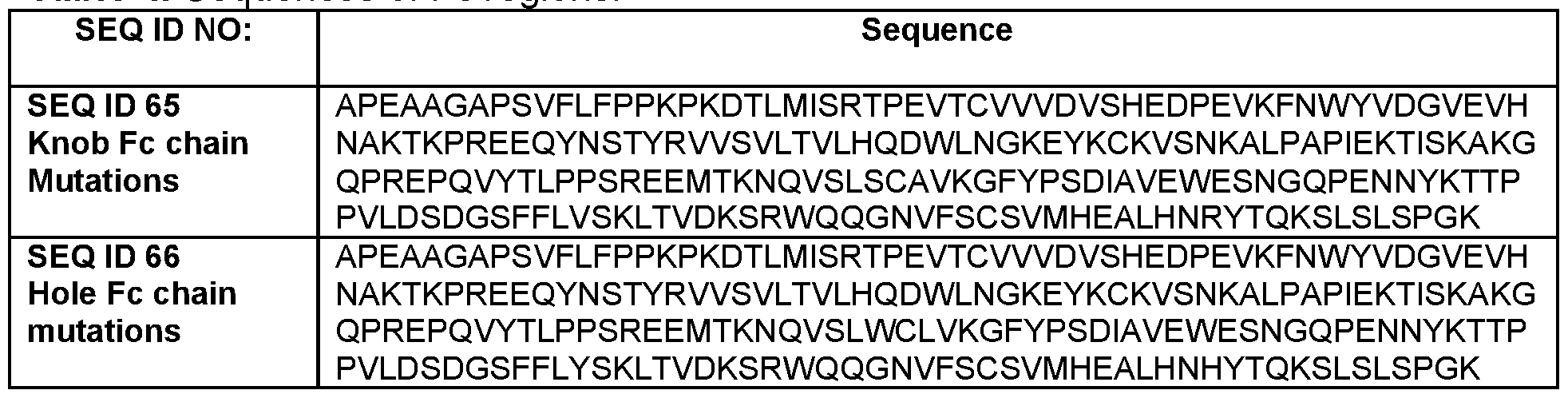

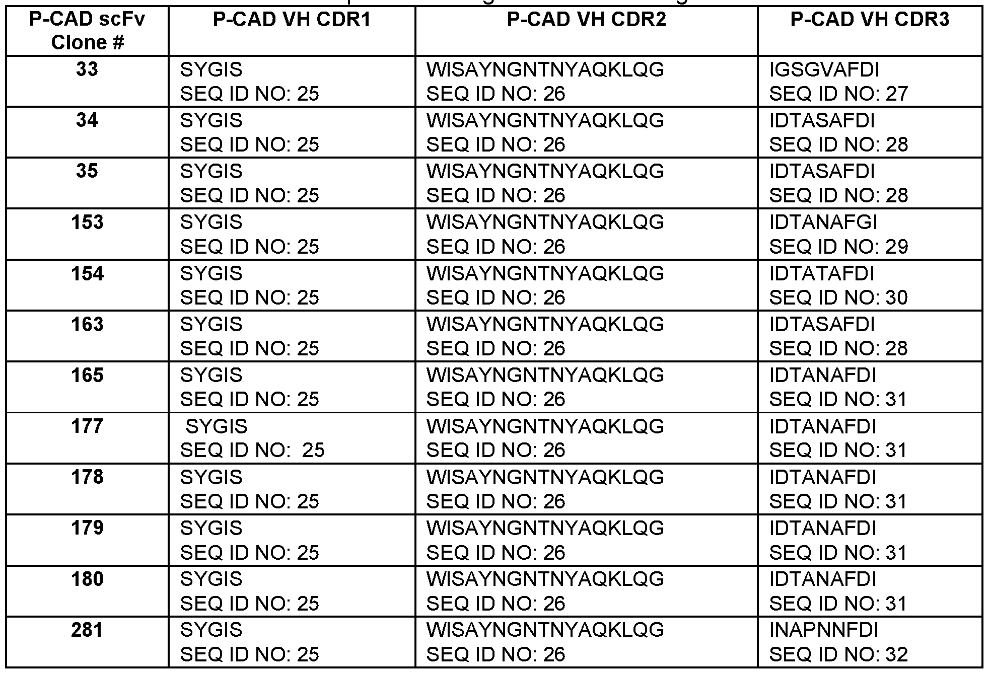


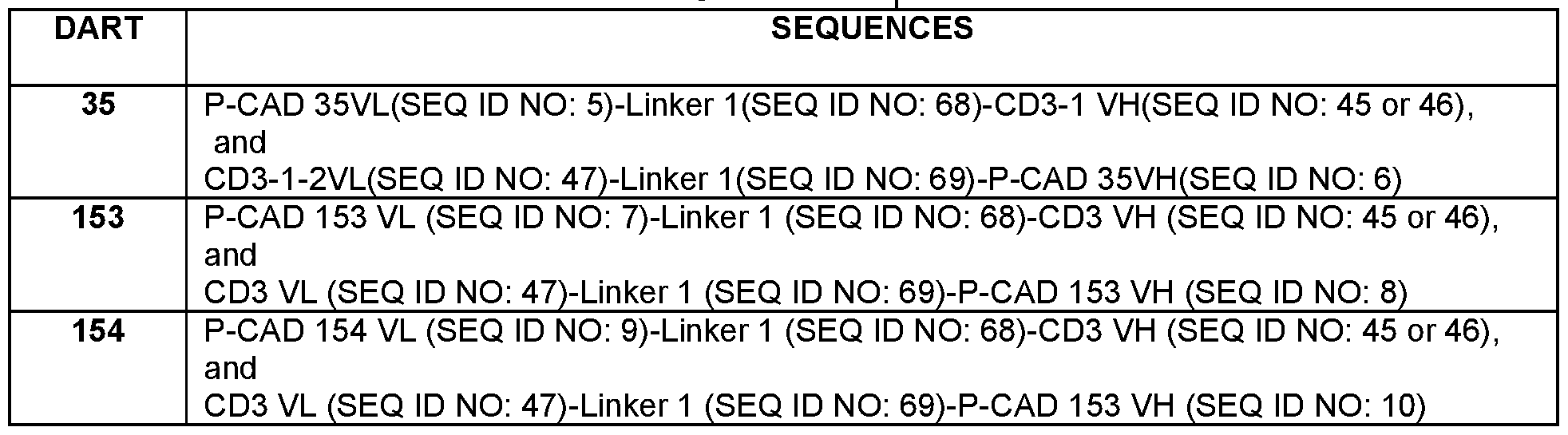


Claims
Priority Applications (13)
| Application Number | Priority Date | Filing Date | Title |
|---|---|---|---|
| JP2016575335A JP2017520575A (en) | 2014-07-01 | 2015-06-26 | Bispecific heterodimeric diabody and uses thereof |
| SG11201609707WA SG11201609707WA (en) | 2014-07-01 | 2015-06-26 | Bispecific heterodimeric diabodies and uses thereof |
| AU2015283704A AU2015283704A1 (en) | 2014-07-01 | 2015-06-26 | Bispecific heterodimeric diabodies and uses thereof |
| CN201580035515.8A CN106661119A (en) | 2014-07-01 | 2015-06-26 | Bispecific heterodimeric diabodies and uses thereof |
| BR112016030740-2A BR112016030740A2 (en) | 2014-07-01 | 2015-06-26 | bispecific heterodimeric bodies and their uses |
| MX2016017393A MX2016017393A (en) | 2014-07-01 | 2015-06-26 | Bispecific heterodimeric diabodies and uses thereof. |
| EP15741347.7A EP3164417A1 (en) | 2014-07-01 | 2015-06-26 | Bispecific heterodimeric diabodies and uses thereof |
| RU2016151645A RU2016151645A (en) | 2014-07-01 | 2015-06-26 | SPECIFIC HETERODIMERIC DIANTISTS AND THEIR APPLICATION |
| KR1020167036814A KR20170010863A (en) | 2014-07-01 | 2015-06-26 | Bispecific heterodimeric diabodies and uses thereof |
| PH12016502491A PH12016502491A1 (en) | 2014-07-01 | 2016-12-13 | Bispecific heterodimeric diabodies and uses thereof |
| IL249889A IL249889A0 (en) | 2014-07-01 | 2017-01-01 | Bispecific heterodimeric diabodies and uses thereof |
| CONC2017/0000016A CO2017000016A2 (en) | 2014-07-01 | 2017-01-03 | Bispecific heterodimeric diabodies capable of specifically binding to a p-cadherin epitope and a cd3 epitope comprising a first polypeptide chain and a second polypeptide chain and pharmaceutical compositions thereof |
| AU2018274919A AU2018274919A1 (en) | 2014-07-01 | 2018-12-05 | Bispecific heterodimeric diabodies and uses thereof |
Applications Claiming Priority (4)
| Application Number | Priority Date | Filing Date | Title |
|---|---|---|---|
| US201462019762P | 2014-07-01 | 2014-07-01 | |
| US62/019,762 | 2014-07-01 | ||
| US201562148920P | 2015-04-17 | 2015-04-17 | |
| US62/148,920 | 2015-04-17 |
Publications (1)
| Publication Number | Publication Date |
|---|---|
| WO2016001810A1 true WO2016001810A1 (en) | 2016-01-07 |
Family
ID=53718063
Family Applications (1)
| Application Number | Title | Priority Date | Filing Date |
|---|---|---|---|
| PCT/IB2015/054829 WO2016001810A1 (en) | 2014-07-01 | 2015-06-26 | Bispecific heterodimeric diabodies and uses thereof |
Country Status (17)
| Country | Link |
|---|---|
| US (2) | US9884921B2 (en) |
| EP (1) | EP3164417A1 (en) |
| JP (1) | JP2017520575A (en) |
| KR (1) | KR20170010863A (en) |
| CN (1) | CN106661119A (en) |
| AU (2) | AU2015283704A1 (en) |
| BR (1) | BR112016030740A2 (en) |
| CA (1) | CA2895659A1 (en) |
| CO (1) | CO2017000016A2 (en) |
| IL (1) | IL249889A0 (en) |
| MX (1) | MX2016017393A (en) |
| PE (1) | PE20170286A1 (en) |
| PH (1) | PH12016502491A1 (en) |
| RU (1) | RU2016151645A (en) |
| SG (1) | SG11201609707WA (en) |
| TW (2) | TW201936638A (en) |
| WO (1) | WO2016001810A1 (en) |
Cited By (29)
| Publication number | Priority date | Publication date | Assignee | Title |
|---|---|---|---|---|
| WO2016090329A3 (en) * | 2014-12-05 | 2016-07-28 | Memorial Sloan-Kettering Cancer Center | Antibodies targeting g-protein coupled receptor and methods of use |
| WO2016166360A1 (en) * | 2015-04-17 | 2016-10-20 | Bayer Pharma Aktiengesellschaft | Bispecific antibody constructs for cdh3 and cd3 |
| WO2017008169A1 (en) * | 2015-07-15 | 2017-01-19 | Zymeworks Inc. | Drug-conjugated bi-specific antigen-binding constructs |
| CN106496331A (en) * | 2016-11-08 | 2017-03-15 | 北京启康兴业生物医药科技有限公司 | Novel FSH-Fc fusion protein and preparation method and application thereof |
| WO2018021450A1 (en) * | 2016-07-29 | 2018-02-01 | 中外製薬株式会社 | Bispecific antibody exhibiting increased alternative fviii-cofactor-function activity |
| WO2019166946A1 (en) | 2018-02-28 | 2019-09-06 | Pfizer Inc. | Il-15 variants and uses thereof |
| WO2019224715A1 (en) | 2018-05-23 | 2019-11-28 | Pfizer Inc. | Antibodies specific for cd3 and uses thereof |
| WO2019224716A2 (en) | 2018-05-23 | 2019-11-28 | Pfizer Inc. | Antibodies specific for gucy2c and uses thereof |
| CN110520523A (en) * | 2017-03-29 | 2019-11-29 | 台北医学大学 | Has the T cell and application thereof of antigenic specificity |
| US10633426B2 (en) | 2014-12-05 | 2020-04-28 | Memorial Sloan Kettering Cancer Center | Chimeric antigen receptors targeting G-protein coupled receptor and uses thereof |
| WO2020128893A1 (en) | 2018-12-21 | 2020-06-25 | Pfizer Inc. | Combination treatments of cancer comprising a tlr agonist |
| US10759870B2 (en) | 2017-09-29 | 2020-09-01 | Chugai Seiyaku Kabushiki Kaisha | Multispecific antigen-binding molecules having blood coagulation factor VIII (FVIII) cofactor function-substituting activity and pharmaceutical formulations containing such a molecule as an active ingredient |
| WO2021009676A1 (en) | 2019-07-17 | 2021-01-21 | Pfizer Inc. | Imidazo[4,5-c]pyridine derivatives as toll-like receptor agonsits |
| WO2021124073A1 (en) | 2019-12-17 | 2021-06-24 | Pfizer Inc. | Antibodies specific for cd47, pd-l1, and uses thereof |
| WO2021124096A1 (en) | 2019-12-18 | 2021-06-24 | Pfizer Inc. | Once daily cancer treatment regimen with a prmt5 inhibitor |
| WO2021181233A2 (en) | 2020-03-09 | 2021-09-16 | Pfizer Inc. | Fusion proteins and uses thereof |
| WO2021229507A2 (en) | 2020-05-13 | 2021-11-18 | Pfizer Inc. | Methods, therapies and uses for treating cancer |
| WO2022013775A1 (en) | 2020-07-17 | 2022-01-20 | Pfizer Inc. | Therapeutic antibodies and their uses |
| WO2022053990A1 (en) | 2020-09-14 | 2022-03-17 | Pfizer Inc. | Methods, therapies and uses for treating cancer |
| EP3790585A4 (en) * | 2018-05-07 | 2022-05-11 | Dragonfly Therapeutics, Inc. | A protein binding nkg2d, cd16 and a tumor-associated antigen |
| WO2022153161A1 (en) | 2021-01-14 | 2022-07-21 | Pfizer Inc. | Treatment of cancer using a prmt5 inhibitor |
| WO2023218320A1 (en) | 2022-05-11 | 2023-11-16 | Pfizer Inc. | Anti-lymphotoxin beta receptor antibodies and methods of use thereof |
| US11834506B2 (en) | 2017-02-08 | 2023-12-05 | Dragonfly Therapeutics, Inc. | Multi-specific binding proteins that bind NKG2D, CD16, and a tumor-associated antigen for activation of natural killer cells and therapeutic uses thereof to treat cancer |
| WO2023242769A1 (en) | 2022-06-17 | 2023-12-21 | Pfizer Inc. | Il-12 variants, anti-pd1 antibodies, fusion proteins, and uses thereof |
| WO2024003773A1 (en) | 2022-07-01 | 2024-01-04 | Pfizer Inc. | 2,7-naphthyridine compounds as mastl inhibitors |
| WO2024009191A1 (en) | 2022-07-05 | 2024-01-11 | Pfizer Inc. | Pyrido[4,3-d]pyrimidine compounds |
| US11884733B2 (en) | 2018-02-08 | 2024-01-30 | Dragonfly Therapeutics, Inc. | Antibody variable domains targeting the NKG2D receptor |
| WO2024074977A1 (en) | 2022-10-04 | 2024-04-11 | Pfizer Inc. | Substituted 1 h-pyrazolo-pyridine and-pyrimidine compounds |
| WO2024084364A1 (en) | 2022-10-18 | 2024-04-25 | Pfizer Inc. | Compounds for the treatment of cancer |
Families Citing this family (12)
| Publication number | Priority date | Publication date | Assignee | Title |
|---|---|---|---|---|
| WO2018178076A1 (en) * | 2017-03-29 | 2018-10-04 | F. Hoffmann-La Roche Ag | Bispecific antigen binding molecule for a costimulatory tnf receptor |
| CN110573528B (en) | 2017-03-29 | 2023-06-09 | 豪夫迈·罗氏有限公司 | Bispecific antigen binding molecules to costimulatory TNF receptors |
| CN108866635B (en) * | 2017-05-09 | 2021-11-26 | 安升(上海)医药科技有限公司 | Multispecific protein medicine and library thereof, and preparation method and application thereof |
| BR112019024742A2 (en) | 2017-06-02 | 2020-06-16 | Pfizer Inc. | RECOMBINANT ROBO2 PROTEINS, COMPOSITIONS, METHODS AND USES OF THE SAME |
| CN111093702A (en) * | 2017-06-22 | 2020-05-01 | 财团法人生物技术开发中心 | Asymmetric heterodimeric Fc-ScFv fusion antibody format with engagement and activation of target cell-dependent T cells for cancer therapy |
| KR101973060B1 (en) * | 2017-10-20 | 2019-04-26 | 주식회사 녹십자 | Antibody against cd3 and pharmaceutical composition for treating cancer comprising the same |
| CN109706163A (en) * | 2017-10-26 | 2019-05-03 | 深圳新诺微环生物科技有限公司 | Bridging molecules and its application of minicircle dna expression connection people and animal target cells and effector cell |
| CN109706164A (en) * | 2017-10-26 | 2019-05-03 | 深圳新诺微环生物科技有限公司 | The bridging molecules and its application of minicircle dna expression connection people and monkey CD20 and effector cell's antigen |
| US20230357410A1 (en) * | 2018-12-21 | 2023-11-09 | Zhejiang shimai Pharmaceutical Co.,Ltd. | Protease cleavable bispecific antibodies and uses thereof |
| CN110357969B (en) * | 2019-05-27 | 2022-03-08 | 北京志道生物科技有限公司 | GLP1-EGFA heterodimer protein, function and method |
| CN110669137B (en) * | 2019-10-24 | 2021-07-16 | 高新 | Multi-specificity antibody and preparation method and application thereof |
| CN114539420B (en) * | 2022-01-20 | 2024-05-17 | 荣昌生物制药(烟台)股份有限公司 | Anti-B7-H3 monoclonal antibody, anti-B7-H3 xCD 3 bispecific antibody, preparation method and application thereof |
Citations (10)
| Publication number | Priority date | Publication date | Assignee | Title |
|---|---|---|---|---|
| WO2002097395A2 (en) * | 2001-05-31 | 2002-12-05 | Chiron Corporation | P-cadherin as a target for anti-cancer therapy |
| WO2004110345A2 (en) * | 2002-10-29 | 2004-12-23 | Pharmacia Corporation | Differentially expressed genes involved in cancer, the polypeptides encoded thereby, and methods of using the same |
| US20060240001A1 (en) * | 2005-04-26 | 2006-10-26 | Agouron Pharmaceuticals, Inc. | P-cadherin antibodies |
| US20070004909A1 (en) | 2005-04-15 | 2007-01-04 | Macrogenics, Inc. | Covalent diabodies and uses thereof |
| US20090060910A1 (en) | 2005-04-15 | 2009-03-05 | Macrogenics, Inc. | Covalent diabodies and uses thereof |
| WO2010001585A1 (en) * | 2008-06-30 | 2010-01-07 | Oncotherapy Science, Inc. | Anti-cdh3 antibodies labeled with radioisotope label and uses thereof |
| US20100174053A1 (en) | 2005-04-15 | 2010-07-08 | Macrogenics, Inc. | Covalent diabodies and uses thereof |
| WO2012018687A1 (en) * | 2010-08-02 | 2012-02-09 | Macrogenics, Inc. | Covalent diabodies and uses thereof |
| WO2012158818A2 (en) * | 2011-05-16 | 2012-11-22 | Fabion Pharmaceuticals, Inc. | Multi-specific fab fusion proteins and methods of use |
| WO2013166448A1 (en) * | 2012-05-03 | 2013-11-07 | Amgen Inc. | Stable formulations containing anti-pcsk9 antibodies |
Family Cites Families (186)
| Publication number | Priority date | Publication date | Assignee | Title |
|---|---|---|---|---|
| DE3382317D1 (en) | 1982-03-15 | 1991-07-25 | Schering Corp | HYBRID DNA, BOND COMPOSITION MADE THEREFOR, AND METHOD FOR THIS. |
| GB8308235D0 (en) | 1983-03-25 | 1983-05-05 | Celltech Ltd | Polypeptides |
| US4816567A (en) | 1983-04-08 | 1989-03-28 | Genentech, Inc. | Recombinant immunoglobin preparations |
| US5869620A (en) | 1986-09-02 | 1999-02-09 | Enzon, Inc. | Multivalent antigen-binding proteins |
| US4946778A (en) | 1987-09-21 | 1990-08-07 | Genex Corporation | Single polypeptide chain binding molecules |
| US5091513A (en) | 1987-05-21 | 1992-02-25 | Creative Biomolecules, Inc. | Biosynthetic antibody binding sites |
| WO1991001752A1 (en) | 1989-07-27 | 1991-02-21 | Fred Hutchinson Cancer Research Center | Immunosuppressive monoclonal antibodies, hybridomas and methods of transplantation |
| GB9015198D0 (en) | 1990-07-10 | 1990-08-29 | Brien Caroline J O | Binding substance |
| US5811267A (en) | 1990-10-29 | 1998-09-22 | Chiron Corporation | Isolated nucleic acid molecules encoding antigen binding sites of antibody molecules specific for cancer antigens |
| DE4118120A1 (en) | 1991-06-03 | 1992-12-10 | Behringwerke Ag | TETRAVALENT BISPECIFIC RECEPTORS, THEIR PRODUCTION AND USE |
| US6129914A (en) | 1992-03-27 | 2000-10-10 | Protein Design Labs, Inc. | Bispecific antibody effective to treat B-cell lymphoma and cell line |
| DE69334287D1 (en) | 1992-09-25 | 2009-07-09 | Avipep Pty Ltd | Targeting molecules-binding polypeptides consisting of an IG-like VL domain bound to an IG-like VH domain |
| WO1994012520A1 (en) | 1992-11-20 | 1994-06-09 | Enzon, Inc. | Linker for linked fusion polypeptides |
| JP3720353B2 (en) | 1992-12-04 | 2005-11-24 | メディカル リサーチ カウンシル | Multivalent and multispecific binding proteins, their production and use |
| DE69327229T2 (en) | 1992-12-11 | 2000-03-30 | Dow Chemical Co | Multivalent single chain antibodies |
| CA2155335C (en) | 1993-02-04 | 2001-06-05 | HANS CHRISTIAN THõGERSEN | Improved method for the refolding of proteins |
| US5747654A (en) | 1993-06-14 | 1998-05-05 | The United States Of America As Represented By The Department Of Health And Human Services | Recombinant disulfide-stabilized polypeptide fragments having binding specificity |
| US6476198B1 (en) | 1993-07-13 | 2002-11-05 | The Scripps Research Institute | Multispecific and multivalent antigen-binding polypeptide molecules |
| WO1995009917A1 (en) | 1993-10-07 | 1995-04-13 | The Regents Of The University Of California | Genetically engineered bispecific tetravalent antibodies |
| IT1271461B (en) | 1993-12-01 | 1997-05-28 | Menarini Ricerche Sud Spa | ANTI-CD3 / ANTI-EGFR MONOCLONAL ANTI-BODY ANTIBODY, PROCESS FOR PRODUCTION AND ITS USE. |
| US5731168A (en) | 1995-03-01 | 1998-03-24 | Genentech, Inc. | Method for making heteromultimeric polypeptides |
| US5989830A (en) | 1995-10-16 | 1999-11-23 | Unilever Patent Holdings Bv | Bifunctional or bivalent antibody fragment analogue |
| EP0937103B1 (en) | 1996-07-12 | 2007-11-28 | McGILL UNIVERSITY | Compounds and methods for modulating cell adhesion |
| CA2267620A1 (en) | 1996-07-16 | 1998-01-22 | Andreas Pluckthun | Immunoglobulin superfamily domains and fragments with increased solubility |
| WO1998041613A1 (en) | 1997-03-14 | 1998-09-24 | Otten Gillis R | Targeted cytolysis of cancer cells |
| US20020062010A1 (en) | 1997-05-02 | 2002-05-23 | Genentech, Inc. | Method for making multispecific antibodies having heteromultimeric and common components |
| US7951917B1 (en) | 1997-05-02 | 2011-05-31 | Genentech, Inc. | Method for making multispecific antibodies having heteromultimeric and common components |
| AU2152299A (en) | 1997-10-27 | 1999-05-24 | Unilever Plc | Multivalent antigen-binding proteins |
| US6485910B1 (en) | 1998-02-09 | 2002-11-26 | Incyte Genomics, Inc. | Ras association domain containing protein |
| AU3295299A (en) | 1998-02-19 | 1999-09-06 | Xcyte Therapies, Inc. | Compositions and methods for regulating lymphocyte activation |
| US6783961B1 (en) | 1999-02-26 | 2004-08-31 | Genset S.A. | Expressed sequence tags and encoded human proteins |
| CZ121599A3 (en) | 1998-04-09 | 1999-10-13 | Aventis Pharma Deutschland Gmbh | Single-chain molecule binding several antigens, process of its preparation and medicament in which the molecule is comprised |
| BRPI9909860B8 (en) | 1998-04-21 | 2021-05-25 | Amgen Res Munich Gmbh | multifunctional single-chain polypeptide, polynucleotide, vector, prokaryotic, yeast or unicellular cell, composition, uses of polypeptide and polynucleotide, and methods for preparing said polypeptide and for identifying activators or inhibitors of activation or stimulation of t cells |
| DE19819846B4 (en) | 1998-05-05 | 2016-11-24 | Deutsches Krebsforschungszentrum Stiftung des öffentlichen Rechts | Multivalent antibody constructs |
| GB9812545D0 (en) | 1998-06-10 | 1998-08-05 | Celltech Therapeutics Ltd | Biological products |
| WO2000006605A2 (en) | 1998-07-28 | 2000-02-10 | Micromet Ag | Heterominibodies |
| US6534633B1 (en) | 1998-10-21 | 2003-03-18 | Altor Bioscience Corporation | Polyspecific binding molecules and uses thereof |
| US20020142000A1 (en) | 1999-01-15 | 2002-10-03 | Digan Mary Ellen | Anti-CD3 immunotoxins and therapeutic uses therefor |
| DE19905048A1 (en) | 1999-02-08 | 2000-08-10 | Gsf Forschungszentrum Umwelt | Use of depletoric or mitogenic antibodies in immunotherapy |
| AU2001243142A1 (en) | 2000-02-03 | 2001-08-14 | Hyseq, Inc. | Novel nucleic acids and polypeptides |
| NZ521540A (en) | 2000-04-11 | 2004-09-24 | Genentech Inc | Multivalent antibodies and uses therefor |
| US20040180046A1 (en) | 2000-04-26 | 2004-09-16 | Jeff Himawan | Bispecific molecules and uses thereof |
| US20040047854A1 (en) | 2001-07-27 | 2004-03-11 | Black Roy A. | Human disintegrin protein |
| GB0025307D0 (en) | 2000-10-16 | 2000-11-29 | Celltech Chiroscience Ltd | Biological products |
| US20030133939A1 (en) | 2001-01-17 | 2003-07-17 | Genecraft, Inc. | Binding domain-immunoglobulin fusion proteins |
| US7754208B2 (en) | 2001-01-17 | 2010-07-13 | Trubion Pharmaceuticals, Inc. | Binding domain-immunoglobulin fusion proteins |
| AU2002327164A1 (en) | 2001-01-29 | 2002-12-09 | Idec Pharmaceuticals Corporation | Engineered tetravalent antibodies and methods of use |
| US20040053245A1 (en) | 2001-02-05 | 2004-03-18 | Tang Y. Tom | Novel nucleic acids and polypeptides |
| AU2002240751A1 (en) | 2001-03-09 | 2002-09-24 | William Herman | Targeted ligands |
| WO2002072635A2 (en) | 2001-03-13 | 2002-09-19 | University College London | Specific binding members |
| US7189507B2 (en) | 2001-06-18 | 2007-03-13 | Pdl Biopharma, Inc. | Methods of diagnosis of ovarian cancer, compositions and methods of screening for modulators of ovarian cancer |
| DE60237282D1 (en) | 2001-06-28 | 2010-09-23 | Domantis Ltd | DOUBLE-SPECIFIC LIGAND AND ITS USE |
| WO2003022987A2 (en) | 2001-07-26 | 2003-03-20 | Eos Biotechnology, Inc. | Methods of diagnosis of hepatitis c infection, compositions and methods of screening for modulators of hepatitis c infection |
| US6833441B2 (en) | 2001-08-01 | 2004-12-21 | Abmaxis, Inc. | Compositions and methods for generating chimeric heteromultimers |
| US20040142325A1 (en) | 2001-09-14 | 2004-07-22 | Liat Mintz | Methods and systems for annotating biomolecular sequences |
| ATE346866T1 (en) | 2001-09-14 | 2006-12-15 | Affimed Therapeutics Ag | MULTIMERIC, SINGLE CHAIN, TANDEM FV ANTIBODIES |
| EP2075256A2 (en) | 2002-01-14 | 2009-07-01 | William Herman | Multispecific binding molecules |
| US7317091B2 (en) | 2002-03-01 | 2008-01-08 | Xencor, Inc. | Optimized Fc variants |
| ATE512989T1 (en) | 2002-04-15 | 2011-07-15 | Chugai Pharmaceutical Co Ltd | METHOD FOR PRODUCING SCDB LIBRARIES |
| EP1354600A1 (en) | 2002-04-19 | 2003-10-22 | Affimed Therapeutics AG | Antibody combination useful for tumor therapy |
| US20040018493A1 (en) | 2002-07-12 | 2004-01-29 | Anastasio Alison E. | Haplotypes of the CD3E gene |
| AU2003248081A1 (en) | 2002-07-18 | 2004-02-09 | Cellfree Sciences Co., Ltd. | Single chain antibody and utilization thereof |
| CA2495389A1 (en) | 2002-08-19 | 2004-02-26 | Genentech, Inc. | Compositions and methods for the diagnosis and treatment of tumor |
| US7820166B2 (en) | 2002-10-11 | 2010-10-26 | Micromet Ag | Potent T cell modulating molecules |
| US9701754B1 (en) | 2002-10-23 | 2017-07-11 | City Of Hope | Covalent disulfide-linked diabodies and uses thereof |
| US7456153B2 (en) | 2002-11-14 | 2008-11-25 | Adherex Technologies Inc. | Compounds and methods for modulating functions of classical cadherins |
| EP2311870A1 (en) | 2002-11-26 | 2011-04-20 | Genentech, Inc. | Compositions and methods for the treatment of immune related diseases |
| MXPA05012080A (en) | 2003-05-08 | 2006-02-22 | Xcyte Therapies Inc | Generation and isolation of antigen-specific t cells. |
| AU2004242846A1 (en) | 2003-05-31 | 2004-12-09 | Micromet Ag | Pharmaceutical compositions comprising bispecific anti-CD3, anti-CD19 antibody constructs for the treatment of B-cell related disorders |
| WO2004106380A2 (en) | 2003-05-31 | 2004-12-09 | Micromet Ag | Human-anti-human cd3 binding molecules |
| CA2530972A1 (en) | 2003-06-11 | 2004-12-23 | Wyeth | Platelet glycoprotein ib alpha variant fusion polypeptides and methods of use thereof |
| AU2004252170B2 (en) | 2003-06-27 | 2011-01-27 | Biogen Ma Inc. | Use of hydrophobic-interaction-chromatography or hinge-region modifications for the production of homogeneous antibody-solutions |
| SI1639011T1 (en) | 2003-06-30 | 2009-04-30 | Domantis Ltd | Pegylated single domain antibodies (dAb) |
| US7754209B2 (en) | 2003-07-26 | 2010-07-13 | Trubion Pharmaceuticals | Binding constructs and methods for use thereof |
| US20070037204A1 (en) | 2003-08-08 | 2007-02-15 | Hiroyuki ABURANTAI | Gene overexpressed in cancer |
| WO2005019258A2 (en) | 2003-08-11 | 2005-03-03 | Genentech, Inc. | Compositions and methods for the treatment of immune related diseases |
| WO2005028507A1 (en) | 2003-09-24 | 2005-03-31 | Monash University | CRYSTAL STRUCTURE OF CD3ϵϜ/OKT3 COMPLEX |
| US7235641B2 (en) | 2003-12-22 | 2007-06-26 | Micromet Ag | Bispecific antibodies |
| DK1593690T3 (en) | 2004-05-05 | 2009-11-09 | Micromet Ag | Preparation of ScFv antibody fragments |
| US7728114B2 (en) | 2004-06-03 | 2010-06-01 | Novimmune S.A. | Anti-CD3 antibodies and methods of use thereof |
| JP2008512352A (en) | 2004-07-17 | 2008-04-24 | イムクローン システムズ インコーポレイティド | Novel tetravalent bispecific antibody |
| AU2005282700A1 (en) | 2004-09-02 | 2006-03-16 | Genentech, Inc. | Heteromultimeric molecules |
| EA018897B1 (en) | 2005-01-05 | 2013-11-29 | Ф-Стар Биотехнологише Форшунгс- Унд Энтвиклунгсгез.М.Б.Х. | Molecules of immunoglobulin comprising modification in a structural loop regions with binding properties and method for manufacturing same |
| WO2006085684A2 (en) | 2005-02-10 | 2006-08-17 | Oncotherapy Science, Inc. | Method of diagnosing bladder cancer |
| US20120237442A1 (en) | 2005-04-06 | 2012-09-20 | Ibc Pharmaceuticals, Inc. | Design and Construction of Novel Multivalent Antibodies |
| CA2606227A1 (en) | 2005-04-28 | 2006-11-02 | Mcgill University | Compounds and methods for modulating cadherin-mediated processes |
| US9241994B2 (en) | 2005-06-10 | 2016-01-26 | Chugai Seiyaku Kabushiki Kaisha | Pharmaceutical compositions containing sc(Fv)2 |
| JP2006345852A (en) | 2005-06-16 | 2006-12-28 | Virxsys Corp | Antibody conjugate |
| KR20080042905A (en) | 2005-09-12 | 2008-05-15 | 노비뮨 에스 에이 | Anti-cd3 antibody formulations |
| ES2616316T3 (en) | 2005-10-11 | 2017-06-12 | Amgen Research (Munich) Gmbh | Compositions comprising specific antibodies for different species and uses thereof |
| EP2527370A1 (en) | 2005-12-21 | 2012-11-28 | Amgen Research (Munich) GmbH | Compounds having resistance to soluble CEA |
| WO2007075672A2 (en) | 2005-12-23 | 2007-07-05 | Lankenau Institute For Medical Research | Prognostic cancer markers |
| JP2009528257A (en) * | 2006-02-28 | 2009-08-06 | オンコセラピー・サイエンス株式会社 | Method for damaging cells using effector function of anti-CDH3 antibody |
| WO2007106507A2 (en) | 2006-03-14 | 2007-09-20 | Petrie Howard T | Detection of gene expression in mixed sample or tissue |
| GEP20135917B (en) | 2006-03-17 | 2013-09-10 | Biogen Idec Inc | Stabilized polypeptide compositions |
| CA2646965C (en) | 2006-03-24 | 2016-06-21 | Jonathan H. Davis | Engineered heterodimeric protein domains |
| US8535677B2 (en) | 2006-06-06 | 2013-09-17 | Oxford Biotherapeutics, Ltd. | Antibody drug conjugate treatment of colorectal cancer |
| TWI615403B (en) | 2007-02-21 | 2018-02-21 | 腫瘤療法 科學股份有限公司 | Peptide vaccines for cancers expressing tumor-associated antigens |
| WO2008119567A2 (en) * | 2007-04-03 | 2008-10-09 | Micromet Ag | Cross-species-specific cd3-epsilon binding domain |
| RU2769948C2 (en) | 2007-04-03 | 2022-04-11 | Эмджен Рисерч (Мьюник) Гмбх | Cd3-epsilon-binding domain with interspecific specificity |
| WO2008124858A2 (en) | 2007-04-11 | 2008-10-23 | F-Star Biotechnologische Forschungs- Und Entwicklungsges. M.B.H. | Targeted receptor |
| ES2456963T3 (en) | 2007-06-04 | 2014-04-24 | Rappaport Family Institute For Research In The Medical Sciences | Agents for the treatment of inflammatory diseases and methods for using them |
| CA2694488A1 (en) | 2007-07-31 | 2009-02-05 | Medimmune, Llc | Multispecific epitope binding proteins and uses thereof |
| CA2696263C (en) | 2007-08-15 | 2017-06-13 | Bing Liu | Monospecific and multispecific antibodies and method of use |
| WO2009025116A1 (en) | 2007-08-20 | 2009-02-26 | Oncotherapy Science, Inc. | Cdh3 peptide and medicinal agent comprising the same |
| EP2535351A3 (en) | 2007-09-26 | 2013-04-03 | UCB Pharma S.A. | Dual specificity antibody fusions |
| US20100310451A1 (en) | 2007-11-01 | 2010-12-09 | The Royal Institution For The Advancement Of Learn | Altered n-cadherin processing in tumor cells by furin and proprotein convertase 5a (pc5a) |
| CN101932608A (en) | 2007-11-30 | 2010-12-29 | 葛兰素集团有限公司 | Antigen-binding constructs |
| US8227577B2 (en) | 2007-12-21 | 2012-07-24 | Hoffman-La Roche Inc. | Bivalent, bispecific antibodies |
| US9266967B2 (en) | 2007-12-21 | 2016-02-23 | Hoffmann-La Roche, Inc. | Bivalent, bispecific antibodies |
| US20090162359A1 (en) | 2007-12-21 | 2009-06-25 | Christian Klein | Bivalent, bispecific antibodies |
| EP2080812A1 (en) | 2008-01-18 | 2009-07-22 | Transmedi SA | Compositions and methods of detecting post-stop peptides |
| BRPI0910482A2 (en) | 2008-04-29 | 2019-09-24 | Abbott Lab | double variable domain immunoglobins and their uses |
| TW201008574A (en) | 2008-08-19 | 2010-03-01 | Oncotherapy Science Inc | INHBB epitope peptides and vaccines containing the same |
| US20120034228A1 (en) | 2008-10-01 | 2012-02-09 | Micromet Ag | CROSS-SPECIES-SPECIFIC PSCAxCD3, CD19xCD3, C-METxCD3, ENDOSIALINxCD3, EPCAMxCD3, IGF-1RxCD3 OR FAPALPHAxCD3 BISPECIFIC SINGLE CHAIN ANTIBODY |
| EP3375790A1 (en) | 2008-10-01 | 2018-09-19 | Amgen Research (Munich) GmbH | Cross-species-specific single domain bispecific single chain antibody |
| CA2738566C (en) | 2008-10-01 | 2024-04-30 | Micromet Ag | Bispecific single chain antibodies with specificity for high molecular weight target antigens |
| JP6126782B2 (en) | 2008-10-01 | 2017-05-10 | アムゲン リサーチ (ミュンヘン) ゲーエムベーハー | Cross-species specific PSMA × CD3 bispecific single chain antibody |
| WO2010040545A1 (en) | 2008-10-06 | 2010-04-15 | Novoplant Gmbh | Proteolytically stable antibody formats |
| SG172754A1 (en) | 2008-10-10 | 2011-08-29 | Trubion Pharmaceuticals Inc | Tcr complex immunotherapeutics |
| US20110020221A1 (en) | 2009-04-09 | 2011-01-27 | The Johns Hopkins University | Cancer stem cell expression patterns and compounds to target cancer stem cells |
| CA2760642A1 (en) | 2009-05-01 | 2010-11-04 | The University Of Tokyo | Anti-cadherin antibody |
| SG176219A1 (en) | 2009-05-27 | 2011-12-29 | Hoffmann La Roche | Tri- or tetraspecific antibodies |
| US8703132B2 (en) | 2009-06-18 | 2014-04-22 | Hoffmann-La Roche, Inc. | Bispecific, tetravalent antigen binding proteins |
| AU2010289527C1 (en) | 2009-09-01 | 2014-10-30 | Abbvie Inc. | Dual variable domain immunoglobulins and uses thereof |
| US9493578B2 (en) | 2009-09-02 | 2016-11-15 | Xencor, Inc. | Compositions and methods for simultaneous bivalent and monovalent co-engagement of antigens |
| WO2011031833A2 (en) | 2009-09-09 | 2011-03-17 | Theraclone Sciences, Inc. | Human anti-cancer antibodies |
| WO2011030107A1 (en) | 2009-09-10 | 2011-03-17 | Ucb Pharma S.A. | Multivalent antibodies |
| JP5885664B2 (en) | 2009-09-25 | 2016-03-15 | ゾーマ テクノロジー リミテッド | Screening method |
| WO2011050106A2 (en) | 2009-10-20 | 2011-04-28 | Tolerx, Inc. | Anti-cd3 antibody dosing in autoimmune disease |
| AU2010315101B2 (en) | 2009-11-04 | 2016-01-28 | Fabrus Llc | Methods for affinity maturation-based antibody optimization |
| GB0920127D0 (en) | 2009-11-17 | 2009-12-30 | Ucb Pharma Sa | Antibodies |
| WO2011071541A2 (en) | 2009-12-11 | 2011-06-16 | The Scripps Research Institute | Cadherin modulatory agents |
| WO2011076781A1 (en) | 2009-12-22 | 2011-06-30 | Novartis Ag | Tetravalent cd47-antibody constant region fusion protein for use in therapy |
| US20110165161A1 (en) | 2009-12-23 | 2011-07-07 | Shih-Yao Lin | Anti-epcam antibodies that induce apoptosis of cancer cells and methods using same |
| EP2519542B1 (en) | 2009-12-28 | 2018-10-10 | OncoTherapy Science, Inc. | Anti-cdh3 antibodies and uses thereof |
| CN102014475B (en) | 2010-01-08 | 2012-01-04 | 华为技术有限公司 | Resource mapping and code division multiplexing method and device |
| WO2011085178A1 (en) | 2010-01-11 | 2011-07-14 | Trustees Of Dartmouth College | Monomeric bi-specific fusion protein |
| CA2789310C (en) | 2010-02-10 | 2018-01-09 | Fujifilm Ri Pharma Co., Ltd. | Radioactive metal-labeled anti-cadherin antibody |
| ES2582001T3 (en) | 2010-02-25 | 2016-09-08 | Affimed Gmbh | Molecule that binds to antigen and uses thereof |
| WO2011109588A1 (en) | 2010-03-03 | 2011-09-09 | Health Research Inc. | Novel cd3 epsilon immunogens and antibodies |
| TWI653333B (en) | 2010-04-01 | 2019-03-11 | 安進研究(慕尼黑)有限責任公司 | Cross-species specific PSMAxCD3 bispecific single chain antibody |
| KR20130079384A (en) | 2010-05-03 | 2013-07-10 | 제넨테크, 인크. | Compositions and methods for the diagnosis and treatment of tumor |
| CA2807585A1 (en) | 2010-08-13 | 2012-02-16 | Baylor Research Institute | Novel vaccine adjuvants based on targeting adjuvants to antibodies directly to antigen-presenting cells |
| US9062101B2 (en) | 2010-08-14 | 2015-06-23 | AbbVie Deutschland GmbH & Co. KG | Amyloid-beta binding proteins |
| CN103261220B (en) | 2010-08-16 | 2016-06-15 | 诺夫免疫股份有限公司 | For generating the method for polyspecific and multivalent antibody |
| NZ610267A (en) | 2010-10-08 | 2015-05-29 | Hope City | A monoclonal antibody framework binding interface for meditopes, meditope delivery systems and methods for their use |
| JP2014015396A (en) | 2010-10-29 | 2014-01-30 | Perseus Proteomics Inc | High-affinity anti-cdh3 antibody |
| PL3404043T3 (en) | 2010-10-29 | 2023-01-02 | Perseus Proteomics Inc. | Anti-cdh3 antibody having high internalization capacity |
| BR112013013311A2 (en) | 2010-11-30 | 2017-09-19 | Chugai Pharmaceutical Co Ltd | cytotoxicity-inducing therapeutic agent |
| UY33827A (en) | 2010-12-22 | 2012-07-31 | Abbott Lab | MEDIUM-IMMUNOGLOBULIN UNION PROTEINS AND ITS USES |
| UY33826A (en) | 2010-12-22 | 2012-07-31 | Abbott Lab | UNION PROTEINS WITH TRIVARIABLE DOMAINS AND ITS USES |
| US10689447B2 (en) | 2011-02-04 | 2020-06-23 | Genentech, Inc. | Fc variants and methods for their production |
| AR085633A1 (en) | 2011-03-08 | 2013-10-16 | Baylor Res Inst | COADYUVANTS BASED ON ANTIBODIES THAT ARE DIRECTLY DIRECTED TO CELLS PRESENTING IN ANTIGENS |
| CN103619876A (en) | 2011-03-10 | 2014-03-05 | Hco抗体股份有限公司 | Bispecific three-chain antibody-like molecules |
| EP2686345B1 (en) | 2011-03-16 | 2018-04-25 | Amgen Inc. | Fc variants |
| KR20140019385A (en) | 2011-03-17 | 2014-02-14 | 라모트 앳 텔-아비브 유니버시티 리미티드 | Bi- and monospecific, asymmetric antibodies and methods of generating the same |
| AU2012228100B2 (en) | 2011-03-17 | 2016-09-08 | The University Of Birmingham | Re-directed immunotherapy |
| EP2688591A4 (en) | 2011-03-22 | 2014-10-01 | Baylor Res Inst | Dendritic cells (dcs) targeting for tuberculosis (tb) vaccine |
| EP2688592A4 (en) | 2011-03-25 | 2015-07-22 | Baylor Res Inst | Compositions and methods to immunize against hepatitis c virus |
| TWI588156B (en) | 2011-03-28 | 2017-06-21 | 賽諾菲公司 | Dual variable region antibody-like binding proteins having cross-over binding region orientation |
| US20140178368A1 (en) | 2011-04-19 | 2014-06-26 | Leslie Lynne SHARP | Combinations of anti-4-1bb antibodies and adcc-inducing antibodies for the treatment of cancer |
| BR112013027224A8 (en) | 2011-04-22 | 2018-08-14 | Emergent Product Dev Seattle | PROSTATE-SPECIFIC MEMBRANE ANTIGEN BINDING PROTEINS AND RELATED COMPOSITIONS AND METHODS |
| UA111612C2 (en) | 2011-05-21 | 2016-05-25 | Макродженікс, Інк. | DOMAINS WHICH ARE CONNECTED TO DEIMMUNIZED SERUM |
| BR122016016837A2 (en) | 2011-05-21 | 2019-08-27 | Macrogenics Inc | cd3 binding molecules; cd3 binding antibodies; pharmaceutical compositions; and uses of the cd3 binding molecule |
| CN106432506A (en) | 2011-05-24 | 2017-02-22 | 泽恩格尼亚股份有限公司 | Multivalent and monovalent multispecific complexes and their uses |
| WO2012162583A1 (en) | 2011-05-26 | 2012-11-29 | Ibc Pharmaceuticals, Inc. | Design and construction of novel multivalent antibodies |
| AU2012265156B2 (en) | 2011-05-27 | 2017-01-19 | F. Hoffmann-La Roche Ag | Dual targeting |
| MX2013014789A (en) | 2011-06-16 | 2014-01-20 | Novartis Ag | Soluble proteins for use as therapeutics. |
| US9127061B2 (en) | 2011-06-24 | 2015-09-08 | Perseus Proteomics Inc. | Anti-human P-cadherin (CDH3) recombinant antibody |
| WO2013012414A1 (en) | 2011-07-18 | 2013-01-24 | Medimmune, Llc | Dosing regimens for treatment of cea-expressing cancers |
| CA2842649C (en) | 2011-07-22 | 2020-01-21 | Affimed Therapeutics Ag | Multivalent antigen-binding fv molecule |
| CN103889452B (en) | 2011-08-23 | 2017-11-03 | 罗切格利卡特公司 | To T cell activation antigen and the bispecific antibody and application method of specific for tumour antigen |
| CA2844538C (en) | 2011-08-23 | 2020-09-22 | Roche Glycart Ag | Bispecific antigen binding molecules |
| KR101723273B1 (en) | 2011-08-23 | 2017-04-04 | 로슈 글리카트 아게 | Fc-free antibodies comprising two fab fragments and methods of use |
| ES2659764T3 (en) | 2011-08-23 | 2018-03-19 | Roche Glycart Ag | Bispecific T-cell activating antigen binding molecules |
| US20130058947A1 (en) | 2011-09-02 | 2013-03-07 | Stem Centrx, Inc | Novel Modulators and Methods of Use |
| WO2013041865A1 (en) | 2011-09-22 | 2013-03-28 | Immunocore Limited | T cell receptors |
| CA2846432A1 (en) | 2011-09-23 | 2013-03-28 | Amgen Research (Munich) Gmbh | Bispecific binding molecules for 5t4 and cd3 |
| TWI679212B (en) | 2011-11-15 | 2019-12-11 | 美商安進股份有限公司 | Binding molecules for e3 of bcma and cd3 |
| SI2794658T1 (en) | 2011-12-19 | 2017-05-31 | Synimmune Gmbh | Bispecific antibody molecule |
| GB201203442D0 (en) | 2012-02-28 | 2012-04-11 | Univ Birmingham | Immunotherapeutic molecules and uses |
| US20150037334A1 (en) | 2012-03-01 | 2015-02-05 | Amgen Research (Munich) Gmbh | Long life polypeptide binding molecules |
| JP6113717B2 (en) | 2012-04-04 | 2017-04-12 | 株式会社ペルセウスプロテオミクス | Drug conjugate of anti-CDH3 (P-cadherin) antibody |
| IN2014MN02293A (en) | 2012-04-20 | 2015-08-07 | Emergent Product Dev Seattle | |
| US10647756B2 (en) * | 2015-05-18 | 2020-05-12 | Pfizer Inc. | Humanized antibodies |
-
2015
- 2015-06-26 BR BR112016030740-2A patent/BR112016030740A2/en not_active Application Discontinuation
- 2015-06-26 US US14/751,704 patent/US9884921B2/en not_active Expired - Fee Related
- 2015-06-26 CN CN201580035515.8A patent/CN106661119A/en active Pending
- 2015-06-26 JP JP2016575335A patent/JP2017520575A/en active Pending
- 2015-06-26 SG SG11201609707WA patent/SG11201609707WA/en unknown
- 2015-06-26 KR KR1020167036814A patent/KR20170010863A/en active Search and Examination
- 2015-06-26 WO PCT/IB2015/054829 patent/WO2016001810A1/en active Application Filing
- 2015-06-26 RU RU2016151645A patent/RU2016151645A/en not_active Application Discontinuation
- 2015-06-26 EP EP15741347.7A patent/EP3164417A1/en not_active Withdrawn
- 2015-06-26 MX MX2016017393A patent/MX2016017393A/en unknown
- 2015-06-26 CA CA2895659A patent/CA2895659A1/en not_active Abandoned
- 2015-06-26 PE PE2016002813A patent/PE20170286A1/en unknown
- 2015-06-26 AU AU2015283704A patent/AU2015283704A1/en not_active Abandoned
- 2015-06-29 TW TW107142295A patent/TW201936638A/en unknown
- 2015-06-29 TW TW104120955A patent/TWI659042B/en not_active IP Right Cessation
-
2016
- 2016-12-13 PH PH12016502491A patent/PH12016502491A1/en unknown
-
2017
- 2017-01-01 IL IL249889A patent/IL249889A0/en unknown
- 2017-01-03 CO CONC2017/0000016A patent/CO2017000016A2/en unknown
- 2017-12-13 US US15/840,019 patent/US20180155452A1/en not_active Abandoned
-
2018
- 2018-12-05 AU AU2018274919A patent/AU2018274919A1/en not_active Abandoned
Patent Citations (10)
| Publication number | Priority date | Publication date | Assignee | Title |
|---|---|---|---|---|
| WO2002097395A2 (en) * | 2001-05-31 | 2002-12-05 | Chiron Corporation | P-cadherin as a target for anti-cancer therapy |
| WO2004110345A2 (en) * | 2002-10-29 | 2004-12-23 | Pharmacia Corporation | Differentially expressed genes involved in cancer, the polypeptides encoded thereby, and methods of using the same |
| US20070004909A1 (en) | 2005-04-15 | 2007-01-04 | Macrogenics, Inc. | Covalent diabodies and uses thereof |
| US20090060910A1 (en) | 2005-04-15 | 2009-03-05 | Macrogenics, Inc. | Covalent diabodies and uses thereof |
| US20100174053A1 (en) | 2005-04-15 | 2010-07-08 | Macrogenics, Inc. | Covalent diabodies and uses thereof |
| US20060240001A1 (en) * | 2005-04-26 | 2006-10-26 | Agouron Pharmaceuticals, Inc. | P-cadherin antibodies |
| WO2010001585A1 (en) * | 2008-06-30 | 2010-01-07 | Oncotherapy Science, Inc. | Anti-cdh3 antibodies labeled with radioisotope label and uses thereof |
| WO2012018687A1 (en) * | 2010-08-02 | 2012-02-09 | Macrogenics, Inc. | Covalent diabodies and uses thereof |
| WO2012158818A2 (en) * | 2011-05-16 | 2012-11-22 | Fabion Pharmaceuticals, Inc. | Multi-specific fab fusion proteins and methods of use |
| WO2013166448A1 (en) * | 2012-05-03 | 2013-11-07 | Amgen Inc. | Stable formulations containing anti-pcsk9 antibodies |
Non-Patent Citations (21)
| Title |
|---|
| "Current Protocols in Immunology", 1999 |
| "Current Protocols in Molecular Biology", 1987 |
| BRINKMANN ET AL.: "J. Immunol. Methods", vol. 182, 1995, article "Phage Display Of Disulfide-Stabilized Fv Fragments", pages: 41 - 50 |
| CARMICHAEL ET AL., J. MOL. BIOL., vol. 326, 2003, pages 341 - 351 |
| CHOTHIA ET AL., NATURE, vol. 342, 1989, pages 877 - 883 |
| CORBETT ET AL.: "Anticancer Drug Development Guide", 2004, HUMANA, pages: 99 - 123 |
| DANI ET AL., PROT. ENG., vol. 16, no. 3, 2003, pages 187 - 193 |
| ELLMAN ET AL., METH. ENZYM., vol. 202, 1991, pages 301 - 336 |
| GUMBINER ET AL., J. CELL BIOL., vol. 148, 2000, pages 399 - 403 |
| HARLOW; LANE: "Using Antibodies, a Laboratory Manual", 1999, COLD SPRING HARBOR LABORATORY PRESS |
| JOHNSON ET AL., J MOL BIOL, vol. 399, 2010, pages 436 - 449 |
| MACCALLUM ET AL., J. MOL. BIOL., vol. 262, 1996, pages 732 - 745 |
| MAKABE ET AL., JOURNAL OF BIOLOGICAL CHEMISTRY, vol. 283, 2008, pages 1156 - 1166 |
| MOORE ET AL., BLOOD, vol. 117, no. 17, 2011, pages 4542 - 4551 |
| MOORE ET AL., BLOOD, vol. 117, no. 17, 2011, pages 4542 - 51 |
| PERISIC ET AL., STRUCTURE, vol. 2, no. 12, 1994, pages 1217 - 1226 |
| SAMBROOK; RUSSEL: "Molecular Cloning: A Laboratory Manual" |
| See also references of EP3164417A1 |
| TONIKIAN R ET AL., NAT. PROTOC., vol. 2, no. 6, 2007, pages 1368 - 86 |
| YUN ET AL., CANCER RESEARCH, vol. 49, 1989, pages 4770 - 4774 |
| ZHANG ET AL., CLIN CANCER RES., vol. 16, no. 21, 2010, pages 5177 - 5188 |
Cited By (46)
| Publication number | Priority date | Publication date | Assignee | Title |
|---|---|---|---|---|
| WO2016090329A3 (en) * | 2014-12-05 | 2016-07-28 | Memorial Sloan-Kettering Cancer Center | Antibodies targeting g-protein coupled receptor and methods of use |
| US11566071B2 (en) | 2014-12-05 | 2023-01-31 | Memorial Sloan Kettering Cancer Center | Nucleic acid molecules encoding anti-GPRC5D antibodies |
| US10906956B2 (en) | 2014-12-05 | 2021-02-02 | Memorial Sloan Kettering Cancer Center | Methods of treatments using chimeric antigen receptors targeting G-protein coupled receptor |
| US11820806B2 (en) | 2014-12-05 | 2023-11-21 | Memorial Sloan-Kettering Cancer Center | Chimeric antigen receptors targeting G-protein coupled receptor and uses thereof |
| US11866478B2 (en) | 2014-12-05 | 2024-01-09 | Memorial Sloan-Kettering Cancer Center | Nucleic acid molecules encoding chimeric antigen receptors targeting G-protein coupled receptor |
| US10633426B2 (en) | 2014-12-05 | 2020-04-28 | Memorial Sloan Kettering Cancer Center | Chimeric antigen receptors targeting G-protein coupled receptor and uses thereof |
| US10590196B2 (en) | 2014-12-05 | 2020-03-17 | Memorial Sloan-Kettering Cancer Center | Antibodies targeting G-protein coupled receptor and methods of use |
| JP2018512860A (en) * | 2015-04-17 | 2018-05-24 | アムゲン リサーチ (ミュンヘン) ゲーエムベーハーAMGEN Research(Munich)GmbH | Bispecific antibody constructs against CDH3 and CD3 |
| WO2016166360A1 (en) * | 2015-04-17 | 2016-10-20 | Bayer Pharma Aktiengesellschaft | Bispecific antibody constructs for cdh3 and cd3 |
| EP4276116A3 (en) * | 2015-04-17 | 2024-01-17 | Amgen Research (Munich) GmbH | Bispecific antibody constructs for cdh3 and cd3 |
| US11926666B2 (en) | 2015-04-17 | 2024-03-12 | Amgen Research (Munich) Gmbh | Bispecific antibody constructs for CDH3 and CD3 |
| US11028171B2 (en) | 2015-04-17 | 2021-06-08 | Amgen Research (Munich) Gmbh | Bispecific antibody constructs for CDH3 and CD3 |
| US11147886B2 (en) | 2015-07-15 | 2021-10-19 | Zymeworks Inc. | Drug-conjugated bi-specific antigen-binding constructs |
| WO2017008169A1 (en) * | 2015-07-15 | 2017-01-19 | Zymeworks Inc. | Drug-conjugated bi-specific antigen-binding constructs |
| JPWO2018021450A1 (en) * | 2016-07-29 | 2019-05-23 | 中外製薬株式会社 | Bispecific antibodies having enhanced FVIII cofactor alternative activity |
| WO2018021450A1 (en) * | 2016-07-29 | 2018-02-01 | 中外製薬株式会社 | Bispecific antibody exhibiting increased alternative fviii-cofactor-function activity |
| CN109415444A (en) * | 2016-07-29 | 2019-03-01 | 中外制药株式会社 | Show the bispecific antibody of increased alternative FVIII co-factor functional activity |
| CN109415444B (en) * | 2016-07-29 | 2024-03-01 | 中外制药株式会社 | Bispecific antibodies exhibiting increased functional activity of alternative FVIII cofactors |
| JP7050677B2 (en) | 2016-07-29 | 2022-04-08 | 中外製薬株式会社 | Bispecific antibody with enhanced FVIII cofactor function substitution activity |
| CN106496331A (en) * | 2016-11-08 | 2017-03-15 | 北京启康兴业生物医药科技有限公司 | Novel FSH-Fc fusion protein and preparation method and application thereof |
| CN106496331B (en) * | 2016-11-08 | 2020-03-13 | 北京智岭生物医药科技有限公司 | FSH-Fc fusion protein and preparation method and application thereof |
| US11834506B2 (en) | 2017-02-08 | 2023-12-05 | Dragonfly Therapeutics, Inc. | Multi-specific binding proteins that bind NKG2D, CD16, and a tumor-associated antigen for activation of natural killer cells and therapeutic uses thereof to treat cancer |
| CN110520523A (en) * | 2017-03-29 | 2019-11-29 | 台北医学大学 | Has the T cell and application thereof of antigenic specificity |
| US10759870B2 (en) | 2017-09-29 | 2020-09-01 | Chugai Seiyaku Kabushiki Kaisha | Multispecific antigen-binding molecules having blood coagulation factor VIII (FVIII) cofactor function-substituting activity and pharmaceutical formulations containing such a molecule as an active ingredient |
| US11939384B1 (en) | 2018-02-08 | 2024-03-26 | Dragonfly Therapeutics, Inc. | Antibody variable domains targeting the NKG2D receptor |
| US11884733B2 (en) | 2018-02-08 | 2024-01-30 | Dragonfly Therapeutics, Inc. | Antibody variable domains targeting the NKG2D receptor |
| WO2019166946A1 (en) | 2018-02-28 | 2019-09-06 | Pfizer Inc. | Il-15 variants and uses thereof |
| EP3790585A4 (en) * | 2018-05-07 | 2022-05-11 | Dragonfly Therapeutics, Inc. | A protein binding nkg2d, cd16 and a tumor-associated antigen |
| WO2019224715A1 (en) | 2018-05-23 | 2019-11-28 | Pfizer Inc. | Antibodies specific for cd3 and uses thereof |
| WO2019224716A2 (en) | 2018-05-23 | 2019-11-28 | Pfizer Inc. | Antibodies specific for gucy2c and uses thereof |
| US11434292B2 (en) | 2018-05-23 | 2022-09-06 | Pfizer Inc. | Antibodies specific for CD3 and uses thereof |
| WO2020128893A1 (en) | 2018-12-21 | 2020-06-25 | Pfizer Inc. | Combination treatments of cancer comprising a tlr agonist |
| WO2021009676A1 (en) | 2019-07-17 | 2021-01-21 | Pfizer Inc. | Imidazo[4,5-c]pyridine derivatives as toll-like receptor agonsits |
| WO2021124073A1 (en) | 2019-12-17 | 2021-06-24 | Pfizer Inc. | Antibodies specific for cd47, pd-l1, and uses thereof |
| WO2021124096A1 (en) | 2019-12-18 | 2021-06-24 | Pfizer Inc. | Once daily cancer treatment regimen with a prmt5 inhibitor |
| WO2021181233A2 (en) | 2020-03-09 | 2021-09-16 | Pfizer Inc. | Fusion proteins and uses thereof |
| WO2021229507A2 (en) | 2020-05-13 | 2021-11-18 | Pfizer Inc. | Methods, therapies and uses for treating cancer |
| WO2022013775A1 (en) | 2020-07-17 | 2022-01-20 | Pfizer Inc. | Therapeutic antibodies and their uses |
| WO2022053990A1 (en) | 2020-09-14 | 2022-03-17 | Pfizer Inc. | Methods, therapies and uses for treating cancer |
| WO2022153161A1 (en) | 2021-01-14 | 2022-07-21 | Pfizer Inc. | Treatment of cancer using a prmt5 inhibitor |
| WO2023218320A1 (en) | 2022-05-11 | 2023-11-16 | Pfizer Inc. | Anti-lymphotoxin beta receptor antibodies and methods of use thereof |
| WO2023242769A1 (en) | 2022-06-17 | 2023-12-21 | Pfizer Inc. | Il-12 variants, anti-pd1 antibodies, fusion proteins, and uses thereof |
| WO2024003773A1 (en) | 2022-07-01 | 2024-01-04 | Pfizer Inc. | 2,7-naphthyridine compounds as mastl inhibitors |
| WO2024009191A1 (en) | 2022-07-05 | 2024-01-11 | Pfizer Inc. | Pyrido[4,3-d]pyrimidine compounds |
| WO2024074977A1 (en) | 2022-10-04 | 2024-04-11 | Pfizer Inc. | Substituted 1 h-pyrazolo-pyridine and-pyrimidine compounds |
| WO2024084364A1 (en) | 2022-10-18 | 2024-04-25 | Pfizer Inc. | Compounds for the treatment of cancer |
Also Published As
| Publication number | Publication date |
|---|---|
| CA2895659A1 (en) | 2016-01-01 |
| BR112016030740A2 (en) | 2018-02-20 |
| CO2017000016A2 (en) | 2017-05-19 |
| PE20170286A1 (en) | 2017-03-30 |
| CN106661119A (en) | 2017-05-10 |
| MX2016017393A (en) | 2017-09-05 |
| KR20170010863A (en) | 2017-02-01 |
| EP3164417A1 (en) | 2017-05-10 |
| PH12016502491A1 (en) | 2017-03-22 |
| AU2015283704A1 (en) | 2016-12-15 |
| TW201612194A (en) | 2016-04-01 |
| TW201936638A (en) | 2019-09-16 |
| IL249889A0 (en) | 2017-03-30 |
| US9884921B2 (en) | 2018-02-06 |
| JP2017520575A (en) | 2017-07-27 |
| AU2018274919A1 (en) | 2019-01-03 |
| TWI659042B (en) | 2019-05-11 |
| RU2016151645A (en) | 2018-08-03 |
| US20180155452A1 (en) | 2018-06-07 |
| SG11201609707WA (en) | 2017-01-27 |
| RU2016151645A3 (en) | 2019-02-20 |
| US20160002357A1 (en) | 2016-01-07 |
Similar Documents
| Publication | Publication Date | Title |
|---|---|---|
| US9884921B2 (en) | Bispecific heterodimeric diabodies and uses thereof | |
| DK2982694T3 (en) | Bispecific antibodies against CD3epsilon and BCMA | |
| US20190389933A1 (en) | PD-1 TARGETED HETERODIMERIC FUSION PROTEINS CONTAINING IL-15/IL-15Ra Fc-FUSION PROTEINS AND PD-1 ANTIGEN BINDING DOMAINS AND USES THEREOF | |
| JP2020018298A (en) | Antibody constructs for cldn18.2 and cd3 | |
| TW202012443A (en) | Anti-cd3 antibodies and uses thereof | |
| JP7209008B2 (en) | antibody | |
| TWI830151B (en) | TRISPECIFIC ANTIBODY AGAINST GPRC5DxBCMAxCD3 AND USE THEREOF | |
| WO2022242680A1 (en) | Anti-cea and anti-cd137 multispecific antibodies and methods of use | |
| EP4245317A1 (en) | Bispecific antibody for claudin 18a2 and cd3 and application of bispecific antibody | |
| EP4155318A1 (en) | Bispecific antibody and use thereof | |
| WO2022063819A1 (en) | Novel human antibodies binding to human cd3 epsilon | |
| WO2022242682A1 (en) | Anti-gpc3 and anti-cd137 multispecific antibodies and methods of use | |
| WO2022242679A1 (en) | Anti-cd137 antibodies and methods of use |
Legal Events
| Date | Code | Title | Description |
|---|---|---|---|
| 121 | Ep: the epo has been informed by wipo that ep was designated in this application |
Ref document number: 15741347 Country of ref document: EP Kind code of ref document: A1 |
|
| WWE | Wipo information: entry into national phase |
Ref document number: IDP00201608145 Country of ref document: ID |
|
| WWE | Wipo information: entry into national phase |
Ref document number: 12016502491 Country of ref document: PH |
|
| ENP | Entry into the national phase |
Ref document number: 2015283704 Country of ref document: AU Date of ref document: 20150626 Kind code of ref document: A |
|
| WWE | Wipo information: entry into national phase |
Ref document number: MX/A/2016/017393 Country of ref document: MX |
|
| ENP | Entry into the national phase |
Ref document number: 2016575335 Country of ref document: JP Kind code of ref document: A |
|
| WWE | Wipo information: entry into national phase |
Ref document number: 002813-2016 Country of ref document: PE Ref document number: 1020167036814 Country of ref document: KR |
|
| WWE | Wipo information: entry into national phase |
Ref document number: 249889 Country of ref document: IL |
|
| NENP | Non-entry into the national phase |
Ref country code: DE |
|
| WWE | Wipo information: entry into national phase |
Ref document number: NC2017/0000016 Country of ref document: CO |
|
| REG | Reference to national code |
Ref country code: BR Ref legal event code: B01A Ref document number: 112016030740 Country of ref document: BR |
|
| REEP | Request for entry into the european phase |
Ref document number: 2015741347 Country of ref document: EP |
|
| WWE | Wipo information: entry into national phase |
Ref document number: 2015741347 Country of ref document: EP |
|
| ENP | Entry into the national phase |
Ref document number: 2016151645 Country of ref document: RU Kind code of ref document: A |
|
| ENP | Entry into the national phase |
Ref document number: 112016030740 Country of ref document: BR Kind code of ref document: A2 Effective date: 20161228 |