WO2015151503A1 - Thoracic cavity simulator - Google Patents
Thoracic cavity simulator Download PDFInfo
- Publication number
- WO2015151503A1 WO2015151503A1 PCT/JP2015/001842 JP2015001842W WO2015151503A1 WO 2015151503 A1 WO2015151503 A1 WO 2015151503A1 JP 2015001842 W JP2015001842 W JP 2015001842W WO 2015151503 A1 WO2015151503 A1 WO 2015151503A1
- Authority
- WO
- WIPO (PCT)
- Prior art keywords
- model
- rib
- cavity simulator
- human skeleton
- chest cavity
- Prior art date
Links
Images


Classifications
-
- G—PHYSICS
- G09—EDUCATION; CRYPTOGRAPHY; DISPLAY; ADVERTISING; SEALS
- G09B—EDUCATIONAL OR DEMONSTRATION APPLIANCES; APPLIANCES FOR TEACHING, OR COMMUNICATING WITH, THE BLIND, DEAF OR MUTE; MODELS; PLANETARIA; GLOBES; MAPS; DIAGRAMS
- G09B23/00—Models for scientific, medical, or mathematical purposes, e.g. full-sized devices for demonstration purposes
- G09B23/28—Models for scientific, medical, or mathematical purposes, e.g. full-sized devices for demonstration purposes for medicine
- G09B23/30—Anatomical models
- G09B23/34—Anatomical models with removable parts
-
- G—PHYSICS
- G09—EDUCATION; CRYPTOGRAPHY; DISPLAY; ADVERTISING; SEALS
- G09B—EDUCATIONAL OR DEMONSTRATION APPLIANCES; APPLIANCES FOR TEACHING, OR COMMUNICATING WITH, THE BLIND, DEAF OR MUTE; MODELS; PLANETARIA; GLOBES; MAPS; DIAGRAMS
- G09B23/00—Models for scientific, medical, or mathematical purposes, e.g. full-sized devices for demonstration purposes
- G09B23/28—Models for scientific, medical, or mathematical purposes, e.g. full-sized devices for demonstration purposes for medicine
- G09B23/285—Models for scientific, medical, or mathematical purposes, e.g. full-sized devices for demonstration purposes for medicine for injections, endoscopy, bronchoscopy, sigmoidscopy, insertion of contraceptive devices or enemas
-
- G—PHYSICS
- G09—EDUCATION; CRYPTOGRAPHY; DISPLAY; ADVERTISING; SEALS
- G09B—EDUCATIONAL OR DEMONSTRATION APPLIANCES; APPLIANCES FOR TEACHING, OR COMMUNICATING WITH, THE BLIND, DEAF OR MUTE; MODELS; PLANETARIA; GLOBES; MAPS; DIAGRAMS
- G09B9/00—Simulators for teaching or training purposes
Definitions
- the present invention relates to a thoracoscopic simulator for thoracoscopic surgery training and learning.
- the percutaneous technique simulator disclosed in Patent Document 1 includes a convex body having a curved surface part, a placement part for placing a trachea (human body organ substitute), and skin (human body) so as to cover a part of the trachea.
- a skin fixing part that fixes the skin substitute) to the main body, and a placement part displacement mechanism that advances and retracts the placement part in the direction perpendicular to the curved surface part.
- a configuration in which the trachea is covered with the skin is realized in the same manner as the configuration of the human body in which the skin is covered with the organ.
- the curved surface portion is a substitute for the human body surface.
- the present invention provides a chest cavity simulator that can faithfully reproduce a human body shape and texture and simulate a surgical environment for a human body with many restrictions, for training and learning of thoracoscopic surgery. With the goal.
- the chest cavity simulator of the present invention is a device comprising at least a human skeleton model that simulates a rib and a casing that stores the human skeleton model, and an opening is provided in the rib portion of the casing.
- the diaphragm portion can be opened and closed, and the organ model can be stored inside the ribs of the human skeleton model. Since the thoracic cavity and abdominal cavity of the human body are separated by the diaphragm and separated into the thoracic and visceral organs, the above organ models are the lung, heart, esophagus and trachea.
- the internal organ model within the ribs of the human skeleton model approximates the surface and the internal shape and configuration, hardness and texture to the human body.
- Organ models such as soft lungs, bronchi, and veins that are close to the actual texture reproduce the realistic surgical environment by reproducing the texture such as softness.
- the casing corresponds to a human subcutaneous tissue or skin. Although the hardness and texture may be approximated to the human body, the casing itself may be a hard resin.
- a surgical tool such as forceps used in thoracoscopic surgery can be inserted into the rib from the gap of the rib.
- the structure of the diaphragm can be opened and closed, such as being removable, and the organ model housed inside the rib of the human skeleton model can be exchanged.
- the human skeleton model in the chest cavity simulator of the present invention includes a rib, a sternum, a spine, and a scapula.
- the human skeleton model is at least from the third rib on either or both sides It is preferable that an opening for exposing the 6 ribs is provided, and that the bottom lid having a convex portion simulating the diaphragm is detachable.
- the human skeleton model is composed of ribs, sternum, spine, and scapula, and it is difficult to provide an opening that exposes at least the third to sixth ribs on either or both sides This is to more faithfully simulate the surgical environment.
- the thoracic cavity simulator of the present invention includes a gripping member that can slide along the spine or sternum of the human skeleton model, the organ model is attached to the gripping member, and the gripping member is slidably stored inside the rib of the human skeleton model.
- the organ model can be stored inside the rib.
- the spine and sternum of the human body skeleton model are bones that extend straight and have some irregularities, but use them as slide rails, and slide the gripping member along the spine or sternum.
- the organ model is attached to the grasping member, and the organ model is accommodated inside the rib by slidingly accommodating the grasping member inside the rib of the human skeleton model.
- a grasping member that can be slidably housed inside the ribs of the human skeleton model, the convenience of placement, fixation, and replacement of the organ model can be improved. It can be used repeatedly by exchanging chest organs such as the lungs, and the same surgical operation can be performed using an actual surgical instrument. By changing the thoracic organs to be stored, it is possible to deal with a wide range of procedures.
- the chest cavity simulator of the present invention includes a lid member that closes the opening, and a through-hole into which a surgical tool used for thoracoscopic procedures can be inserted is provided in the lid member. Since surgical tools such as forceps used in thoracoscopic surgery are instruments that are inserted through a small-diameter hole, a small-diameter hole is provided in the lid member to reproduce a realistic surgical environment for thoracoscopic surgery.
- the casing and the lid member are preferably made of a translucent material.
- the casing is transparent or translucent, such as transparent, and the position of the ribs from the outside, The position of the lungs and heart that have a built-in chest cavity, and the position of forceps used in thoracoscopic surgery should be confirmed.
- beginners of thoracoscopic surgery can perform thoracoscopic surgery training by hiding the inside with a lid member. It can be expected that the user of the chest cavity simulator of the present invention is useful for grasping the actual sense of distance and actual size of thoracoscopic surgery.
- the portion corresponding to the costal cartilage is made of a soft material so that the gap between adjacent ribs can be widened.
- Surgical tools such as forceps used in thoracoscopic surgery are inserted through the gaps in the ribs.
- the organ model in the chest cavity simulator of the present invention is a biological textured organ model, and the biological textured organ model can be contracted and expanded and includes means for changing the model size.
- the model size of the living body texture organ model it is possible to train a technique for a lung or heart model having a motion such as pulsation.
- the living body texture organ model improves the real feeling by using a soft material that approximates the actual texture and aims to acquire practical techniques.
- DICOM Digital Imaging and Communications in Medicine
- 3D shape data is produced, and 3D modeling model is based on that.
- a method for producing a biological texture organ model is described, for example, in an international pamphlet (WO2012 / 132463).
- the organ model in the chest cavity simulator of the present invention is a biological textured organ model, and the biological textured organ model is provided with a tube that simulates a blood vessel on the surface of the model or around the model, and means for liquid to flow out when the tube is broken. It is preferable to provide. According to such a structure, it is possible to simulate a case in which bleeding occurs during an operation and train a coping method in that case.
- a living organ image is mapped on the inner wall of a casing or a lid member.
- mapping a living organ image on the inner wall of the casing it is possible to train with a more realistic image when a monitoring camera is inserted from the opening hole of the casing or the lid member.
- the biological texture organ model is a reproduction of a three-dimensional structure inside the organ. By reproducing the internal three-dimensional structure of the organ, it becomes possible to perform a realistic training during the excision operation in the training of the procedure.
- the chest cavity simulator of the present invention there is an effect that, for training and learning of thoracoscopic surgery, a human body shape and texture can be faithfully reproduced, and a surgical environment for a human body with many restrictions can be simulated.
- FIG. 1 is an explanatory diagram of the chest cavity simulator of Example 1 External view of another gripping member of the chest cavity simulator of the first embodiment
- Explanatory drawing 2 of the chest cavity simulator of Example 1 is an explanatory diagram of the surgical environment of the chest cavity simulator according to the first embodiment.
- Explanatory drawing 2 of the surgical environment of the chest cavity simulator of Example 1 Attachment drawing of the lid part of the chest cavity simulator of Example 1
- Explanatory drawing of the surgical environment under attachment of the lid part of the chest cavity simulator of Example 1 Explanatory drawing of other embodiments
- Explanatory drawing of other embodiments (2) Diagram showing human skeleton model of ribs and sternum
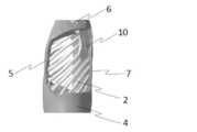
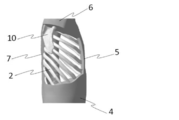
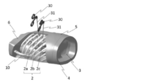
- FIG. 1 shows an external view of the chest cavity simulator 1.
- 2 to 7 show a front view, a rear view, a right side view, a left side view, a plan view, and a bottom view of the chest cavity simulator 1, respectively.
- the chest cavity simulator 1 includes a human skeleton model that simulates the ribs 2, the sternum, the spine, and the scapula 10, and casings (4, 5, 6) that store the human skeleton model. An opening is provided in the rib portion of the casing.
- a bottom lid portion 4 having a convex portion simulating the diaphragm 3 is detachable from the casing, can be opened and closed at the diaphragm portion, and an organ model such as a lung or heart can be stored inside the rib of the human skeleton model.
- the opening portion in the rib portion of the casing exposes the eighth rib from the second rib of the rib, and an opening is provided on each of the left side surface and the right side surface.
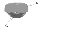
- the sternum of the human skeleton model is hidden inside the casing front part 5. Further, the spine of the human skeleton model is hidden inside the casing back surface portion 7. 2 to 7 show a state in which the organ model is not stored in the ribs of the human skeleton model. Further, a sternum portion 6 a is present on the upper surface (planar) portion 6 of the casing.
- FIG. 8 shows a perspective view of the bottom lid portion 4 of the chest cavity simulator 1.
- a convex portion simulating the diaphragm 3 is formed on the bottom lid portion 4, and the intrathoracic volume of the thoracic cavity simulator 1 is limited so as to reproduce the actual intrathoracic environment.
- a concave portion 4a is formed in the peripheral edge portion of the bottom cover portion 4, and can be fitted to the casing body.
- FIG. 9 is an external view of the gripping member 8 of the chest cavity simulator 1.
- the gripping member 8 is formed with recesses (8a, 8b) serving as receiving portions for the spine and the sternum so as to slide along the spine and sternum of the human skeleton model.
- the gripping member 8 is formed with an engaging portion 8c so that an organ model can be attached.
- the built-in models (20, 21) such as the lungs and the heart are attached and fixed to the grasping member 8, and the grasping member 8 is slid along the spine and sternum so that the organ is placed inside the rib 2 in the casing.
- the intrathoracic space is constructed
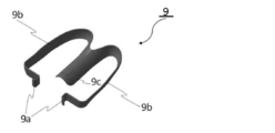
- FIG. 11 shows an external view of another gripping member 9 of the chest cavity simulator 1.
- the gripping member 9 is formed with a concave portion 9 c that serves as a receiving portion of the spine so that it can slide along the spine 11.
- Arms (9b, 9c) are provided on the left and right sides of the receiving part of the spine. The left and right opening widths can be adjusted by the distal end portion 9a of the arms (9b, 9c), whereby the posture of the gripping member 9 inside the rib 2 can be stabilized.
- a built-in model 22 such as a lung or heart is placed on the dish 9d on the grasping member 9, and the grasping member 9 is slid along the spine 11, so that the organ model is placed inside the rib 2 in the casing. Storing. And the intrathoracic space is constructed
- FIGS. 13 and 14 show the situation of the thoracoscopic surgical environment reproduced using the chest cavity simulator 1.
- the forceps 31 is inserted into the gap between the third rib 2a and the fourth rib 2b, and the gap between the fourth rib 2b and the fifth rib 2c, and the thoracoscopic surgery of the chest organ is trained with the forceps handle 30. Is shown.
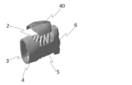
- FIG. 15 shows how the lid 40 of the chest cavity simulator 1 is attached.
- the lid portion 40 is a member that closes the opening portion on the side surface of the chest cavity simulator.
- the lid portion 40 is provided with a through hole 41 into which a forceps 31 used in a thoracoscopic procedure can be inserted, and the forceps 31 is inserted through the through hole 41. Can be trained in thoracoscopic surgery.
- the casing front part 5, the casing back part 7, the casing side part and the lid part 40 of the chest cavity simulator 1 are translucent.
- thoracoscopic surgery is performed while observing the thoracoscope with a camera.
- the casing is made translucent, the position of the ribs from the outside, and the lung with a built-in chest cavity. And the position of the forceps used in thoracoscopic surgery.
- a soft resin is used in a portion corresponding to the rib cartilage so as to widen the gap between adjacent ribs. This is because forceps used in thoracoscopic surgery are inserted through the gap between the ribs, so that a realistic surgical environment similar to that of an actual human rib can be reproduced.
- the organ model housed inside the rib 2 of the chest cavity simulator 1 approximates the shape and configuration of the surface and the interior, the hardness and the texture, etc., to the human body using known materials and techniques. By creating a soft organ model that is close to the actual texture, it reproduces the texture such as softness and reproduces a realistic surgical environment.
- the size and position of the opening of the casing of the chest cavity simulator 1 are not particularly limited as long as the rib portion is exposed. Moreover, in addition to the opening part which exposes a rib part, you may expose a sternum, a spine, etc. (2)
- the shape of the casing of the chest cavity simulator 1 is preferably approximated to an actual human body. However, as in the case of the chest cavity simulator 1 of the first embodiment, not only the chest cavity site but also the arm and head, There may be an abdominal region.
- FIG. 17 shows a configuration in which the biological texture organ model can be expanded and contracted.
- the living body texture organ model 60 is manufactured using a stretchable resin material, and a hollow portion 61 is provided therein.
- the biological texture organ model 60 is expanded and the model size is increased.
- the biological texture organ model 60 is reduced and the model size is reduced. Thereby, for example, pulsations such as the heart can be reproduced, and a realistic technique can be trained.
- FIG. 18 shows an image in which a blood vessel simulation tube 65 for simulating blood vessels is provided on the surface of the living body texture organ model 60 or around the model.
- a hollow portion 61 is provided inside the biological texture organ model 60 and filled with a liquid.
- the tube 66 is connected to the cavity portion 61, and when the blood vessel simulation tube 65 is broken, the liquid filled in the cavity portion 61 of the biological texture organ model 60 flows out. According to such a structure, it is possible to simulate a case in which bleeding occurs during an operation and train a coping method in that case.
- the present invention is useful for training and learning of thoracoscopic surgery, and can be used as a surgery support device or a surgery simulation device.
Landscapes
- Engineering & Computer Science (AREA)
- General Physics & Mathematics (AREA)
- Physics & Mathematics (AREA)
- Theoretical Computer Science (AREA)
- Health & Medical Sciences (AREA)
- Educational Technology (AREA)
- Educational Administration (AREA)
- Business, Economics & Management (AREA)
- Medicinal Chemistry (AREA)
- Computational Mathematics (AREA)
- Mathematical Analysis (AREA)
- Mathematical Optimization (AREA)
- Mathematical Physics (AREA)
- Pure & Applied Mathematics (AREA)
- Algebra (AREA)
- Medical Informatics (AREA)
- General Health & Medical Sciences (AREA)
- Chemical & Material Sciences (AREA)
- Pulmonology (AREA)
- Radiology & Medical Imaging (AREA)
- Instructional Devices (AREA)
Abstract
Description
近年、複数樹脂の同時噴射により、硬性樹脂と柔軟性樹脂を組み合わせ、機械的性質の異なる樹脂を用いた3次元造形モデルを作製できる3次元プリンタが知られており、3次元プリンタを用いて、その形状構造に関して表面や内部構造まで再現できるようになっている。 The need for 3D visualization of affected areas and specific parts of the body in the medical field is increasing in the fields of informed consent, decision on medical policy, medical education, and medical research. In particular, in the case of three-dimensional visualization using a three-dimensional modeling model, a lot of information that cannot be communicated by a computer image can be conveyed by actually touching and viewing a three-dimensional shape as well as vision.
In recent years, a three-dimensional printer that can produce a three-dimensional modeling model using a resin having different mechanical properties by combining a hard resin and a flexible resin by simultaneous injection of a plurality of resins has been known. The surface structure and internal structure can be reproduced with respect to the shape structure.
特許文献1に開示された経皮手技シミュレータは、凸状で曲面部を有する本体と、気管(人体臓器代替物)を載置する載置部と、気管の一部を覆うように皮膚(人体皮膚代替物)を本体に固定する皮膚固定部と、載置部を曲面部の面直方向に進退させる載置部変位機構とを有する。この経皮手技シミュレータを用いることで、臓器を皮膚が覆っている人体の構成と同様に、気管を皮膚が覆っている構成が実現される。また曲面部が人体表面の代替物となる。代替物の表面及び内部の形状や構成、硬さや質感などを人体に近似させることにより、実際の手技と同様のシミュレーションを行える。 In addition, percutaneous technique simulators that enable students to acquire advanced techniques even if they do not have clinical experience through training that approximates actual techniques sensuously, such as incisions and skin sutures. (For example, refer to Patent Document 1).
The percutaneous technique simulator disclosed in
また、人の肋骨は第1肋骨から第12肋骨まであるが、できるだけ忠実に人体骨格モデルを再現する。ケーシングは人の皮下組織や皮膚に相当する。硬さや質感などを人体に近似させてもいいが、ケーシング自体は硬質性の樹脂でも構わない。
ケーシングの肋骨部分に開孔部を設けることにより、胸腔鏡下手術で用いる鉗子など手術用具を肋骨の骨の隙間から肋骨内部へ挿入することができる。横隔膜の部分は取り外しできるなど開閉できる構造となっており、人体骨格モデルの肋骨内部に収納する臓器モデルを交換することができる。 The internal organ model within the ribs of the human skeleton model approximates the surface and the internal shape and configuration, hardness and texture to the human body. Organ models such as soft lungs, bronchi, and veins that are close to the actual texture reproduce the realistic surgical environment by reproducing the texture such as softness.
In addition, there are human ribs from the first rib to the twelfth rib, but the human skeleton model is reproduced as faithfully as possible. The casing corresponds to a human subcutaneous tissue or skin. Although the hardness and texture may be approximated to the human body, the casing itself may be a hard resin.
By providing an opening in the rib portion of the casing, a surgical tool such as forceps used in thoracoscopic surgery can be inserted into the rib from the gap of the rib. The structure of the diaphragm can be opened and closed, such as being removable, and the organ model housed inside the rib of the human skeleton model can be exchanged.
また、横隔膜を模擬した凸部を有する底蓋部を着脱自在にすることにより、胸腔内臓の臓器モデルを交換することができる。 The human skeleton model in the chest cavity simulator of the present invention includes a rib, a sternum, a spine, and a scapula. When the sternum side is the front surface and the spine side is the back surface, the human skeleton model is at least from the third rib on either or both sides It is preferable that an opening for exposing the 6 ribs is provided, and that the bottom lid having a convex portion simulating the diaphragm is detachable. The human skeleton model is composed of ribs, sternum, spine, and scapula, and it is difficult to provide an opening that exposes at least the third to sixth ribs on either or both sides This is to more faithfully simulate the surgical environment. In the case of surgery on the thoracic viscera, access from the sternum, spine, and scapula is rare, and access is most often through the gaps in the ribs. For this reason, openings are provided on the left and right side surfaces. Further, human ribs are from the first rib to the twelfth rib, but are often accessed from the gap between the third rib and the sixth rib, and an opening is provided in a portion where the sixth rib is exposed from the third rib. .
Further, by making the bottom lid part having a convex part simulating the diaphragm detachable, the organ model of the thoracic organ can be exchanged.
把持部材には、臓器モデルが取付けられる構成とし、把持部材を人体骨格モデルの肋骨内部にスライド収納することにより、臓器モデルを肋骨内部に収納する。
人体骨格モデルの肋骨内部にスライド収納できる把持部材を用いることにより、臓器モデルの配置、固定、交換の利便性が向上できる。肺など胸腔臓器を交換するなどして繰り返し使用でき、実際の手術器械を用いて、実際と同じ手術操作を行うことができる。収納する胸腔臓器を変更することで、幅広い手技に対応することが可能である。 The thoracic cavity simulator of the present invention includes a gripping member that can slide along the spine or sternum of the human skeleton model, the organ model is attached to the gripping member, and the gripping member is slidably stored inside the rib of the human skeleton model. Thus, it is preferable that the organ model can be stored inside the rib. The spine and sternum of the human body skeleton model are bones that extend straight and have some irregularities, but use them as slide rails, and slide the gripping member along the spine or sternum.
The organ model is attached to the grasping member, and the organ model is accommodated inside the rib by slidingly accommodating the grasping member inside the rib of the human skeleton model.
By using a grasping member that can be slidably housed inside the ribs of the human skeleton model, the convenience of placement, fixation, and replacement of the organ model can be improved. It can be used repeatedly by exchanging chest organs such as the lungs, and the same surgical operation can be performed using an actual surgical instrument. By changing the thoracic organs to be stored, it is possible to deal with a wide range of procedures.
胸腔鏡下手術の初心者は、胸腔内外から見比べて、上級者は蓋部材で内部を隠して、胸腔鏡下手術のトレーニングを行うことができる。本発明の胸腔シミュレータのユーザにとって、胸腔鏡下手術の実際の距離感、実態サイズの把握に役立つことが期待できる。 Here, the casing and the lid member are preferably made of a translucent material. Originally, thoracoscopic surgery advances the procedure while observing the thoracoscope with a camera, but since the purpose is training and learning, the casing is transparent or translucent, such as transparent, and the position of the ribs from the outside, The position of the lungs and heart that have a built-in chest cavity, and the position of forceps used in thoracoscopic surgery should be confirmed.
Compared with the inside and outside of the chest cavity, beginners of thoracoscopic surgery can perform thoracoscopic surgery training by hiding the inside with a lid member. It can be expected that the user of the chest cavity simulator of the present invention is useful for grasping the actual sense of distance and actual size of thoracoscopic surgery.
ここで、生体質感臓器モデルは、実際の質感に近似した軟質性素材を用いてリアル感を向上させ、実践的な手技習得を図るものである。X線CTやMRI(Magnetic Resonance Imaging)などの医療診断装置から得られたDICOM(Digital Imaging and Communications in Medicine)データを用い、3次元形状データを製作し、それをもとにして3次元造形モデルを作製するものであり、人体の各部位(骨・臓器など)の質感(可視化・感触・硬さ・柔らかさなど)を持った3次元の生体臓器の造形物である。生体質感臓器モデルの作製方法としては、例えば、国際公開パンフレット(WO2012/132463)に記載されている。 It is preferable that the organ model in the chest cavity simulator of the present invention is a biological textured organ model, and the biological textured organ model can be contracted and expanded and includes means for changing the model size. By changing the model size of the living body texture organ model, it is possible to train a technique for a lung or heart model having a motion such as pulsation.
Here, the living body texture organ model improves the real feeling by using a soft material that approximates the actual texture and aims to acquire practical techniques. Using DICOM (Digital Imaging and Communications in Medicine) data obtained from medical diagnostic equipment such as X-ray CT and MRI (Magnetic Resonance Imaging), 3D shape data is produced, and 3D modeling model is based on that. It is a three-dimensional model of a living organ with the texture (visualization, touch, hardness, softness, etc.) of each part (bone, organ, etc.) of the human body. A method for producing a biological texture organ model is described, for example, in an international pamphlet (WO2012 / 132463).
胸腔シミュレータ1は、肋骨2,胸骨,背骨および肩甲骨10を模擬した人体骨格モデルと、人体骨格モデルを収納するケーシング(4,5,6)とから成る。ケーシングの肋骨部分に開孔部が設けられている。横隔膜3を模擬した凸部を有する底蓋部4がケーシングから着脱自在であり、横隔膜の部分で開閉でき、人体骨格モデルの肋骨内部に肺や心臓などの臓器モデルを収納できる。ケーシングの肋骨部分に開孔部は、肋骨の第2肋骨から第8肋骨を露出しており、左側面および右側面にそれぞれ開口部が設けられている。
人体骨格モデルの胸骨は、ケーシング正面部位5の内側に隠れている。また、人体骨格モデルの背骨は、ケーシング背面部位7の内側に隠れている。
なお、図2~7は、人体骨格モデルの肋骨内部に臓器モデルは未収納の状態を示している。また、ケーシング上面(平面)部位6には、胸骨部6aが存在している。 FIG. 1 shows an external view of the
The
The sternum of the human skeleton model is hidden inside the casing
2 to 7 show a state in which the organ model is not stored in the ribs of the human skeleton model. Further, a
図10に示すように、把持部材8に肺や心臓など内蔵モデル(20,21)を取付固定し、把持部材8を背骨および胸骨に沿ってスライドさせて、ケーシング内の肋骨2の内部に臓器モデルを収納する。そして、横隔膜3を模擬した凸部を有する底蓋部4をケーシングに取付けることにより、胸腔内空間を構築する。 FIG. 9 is an external view of the gripping
As shown in FIG. 10, the built-in models (20, 21) such as the lungs and the heart are attached and fixed to the grasping
背骨の受け部の左右両側にアーム(9b,9c)が設けられている。アーム(9b,9c)の先端部9aによって左右の開き幅を調整でき、これにより肋骨2の内部における把持部材9の姿勢を安定させることができる。
図12に示すように、把持部材9に肺や心臓など内蔵モデル22を皿部9dの上に載せ、把持部材9を背骨11に沿ってスライドさせて、ケーシング内の肋骨2の内部に臓器モデルを収納する。そして、横隔膜3を模擬した凸部を有する底蓋部4をケーシングに取付けることにより、胸腔内空間を構築する。 FIG. 11 shows an external view of another gripping
Arms (9b, 9c) are provided on the left and right sides of the receiving part of the spine. The left and right opening widths can be adjusted by the
As shown in FIG. 12, a built-in
(1)胸腔シミュレータ1のケーシングの開孔部の大きさおよび位置は、肋骨部分を露出させるものであれば特に制限されるものではない。また、肋骨部分を露出させる開孔部に加えて、胸骨、背骨などを露出させるものでもよい。
(2)胸腔シミュレータ1のケーシングの形状については、実際の人体に近似させるものが好ましいが、実施例1の胸腔シミュレータ1のケーシングのように胸腔部位のみでなくとも腕部や頭部、さらには腹腔部位があるものでもよい。 (Other examples)
(1) The size and position of the opening of the casing of the
(2) The shape of the casing of the
2,2a~2c 肋骨
3 横隔膜
4 底蓋部
5 ケーシング正面部位
6 ケーシング上面(平面)部位
6a 胸骨部
7 ケーシング背面部位
8,9 把持部材
10 肩甲骨
11 背骨
20,21,22 内蔵モデル
30 鉗子ハンドル
31 鉗子
40 蓋部
41 貫通孔
50 肋骨
51 肋軟骨
52 胸骨
53 背骨
60 生体質感臓器モデル
61 空洞部
62,66 チューブ
63 空気注入ユニット
65 血管模擬チューブ
DESCRIPTION OF
Claims (8)
- 少なくとも肋骨を模擬した人体骨格モデルと、
該人体骨格モデルを収納するケーシングとから成る装置であって、
前記ケーシングの肋骨部分に開孔部が設けられ、
横隔膜の部分が開閉でき、
前記人体骨格モデルの肋骨内部に臓器モデルを収納し得る、
ことを特徴とする胸腔シミュレータ。 At least a human skeleton model simulating the ribs,
An apparatus comprising a casing for storing the human skeleton model,
An opening is provided in the rib portion of the casing,
The diaphragm part can be opened and closed
An organ model can be stored inside the rib of the human skeleton model,
A thoracic cavity simulator characterized by that. - 前記人体骨格モデルが、肋骨,胸骨,背骨および肩甲骨から成り、胸骨側を正面、背骨側を背面として場合に、左右側面のいずれか若しくは両側に、少なくとも第3肋骨から第6肋骨を露出する前記開孔部が設けられ、
横隔膜を模擬した凸部を有する底蓋部が着脱自在である、
ことを特徴とする請求項1に記載の胸腔シミュレータ。 The human skeleton model is composed of a rib, a sternum, a spine, and a scapula. When the sternum side is a front surface and the spine side is a back surface, at least the third rib to the sixth rib are exposed on either or both sides. The opening is provided;
The bottom lid part having a convex part simulating the diaphragm is detachable,
The chest cavity simulator according to claim 1. - 前記人体骨格モデルの背骨もしくは胸骨に沿ってスライドし得る把持部材を備え、
該把持部材に前記臓器モデルが取付けられ、前記把持部材を前記人体骨格モデルの肋骨内部にスライド収納することにより、前記臓器モデルを肋骨内部に収納する、
ことを特徴とする請求項1又は2に記載の胸腔シミュレータ。 A gripping member that can slide along the spine or sternum of the human skeleton model,
The organ model is attached to the gripping member, and the organ model is stored inside the rib by slidingly storing the gripping member inside the rib of the human skeleton model.
The thoracic cavity simulator according to claim 1 or 2. - 前記開孔部を塞ぐ蓋部材を備え、
該蓋部材に胸腔鏡下手技で用いる手術具を挿入し得る貫通孔が設けられた、
ことを特徴とする請求項1~3の何れかに記載の胸腔シミュレータ。 A lid member for closing the opening portion;
The lid member was provided with a through hole into which a surgical tool used in thoracoscopic procedures can be inserted.
The chest cavity simulator according to any one of claims 1 to 3, wherein: - 前記ケーシングまたは前記蓋部材が透光性素材から成る、
ことを特徴とする請求項1~4の何れかに記載の胸腔シミュレータ。 The casing or the lid member is made of a translucent material,
The chest cavity simulator according to any one of claims 1 to 4, wherein: - 前記人体骨格モデルの肋骨において、
隣接する肋骨の隙間を広げられるように肋軟骨に相当する部位が軟性素材から成る、
ことを特徴とする請求項1~5の何れかに記載の胸腔シミュレータ。 In the ribs of the human skeleton model,
The part corresponding to the costal cartilage is made of a soft material so that the gap between adjacent ribs can be widened,
The chest cavity simulator according to any one of claims 1 to 5, wherein: - 前記臓器モデルが生体質感臓器モデルであり、前記生体質感臓器モデルは縮膨張可能であり、モデルサイズが変化する手段を備えたことを特徴とする請求項1~6の何れかに記載の胸腔シミュレータ。 The chest cavity simulator according to any one of claims 1 to 6, further comprising means for changing the model size, wherein the organ model is a living body organ model, the living body organ model can be expanded and contracted. .
- 前記臓器モデルが生体質感臓器モデルであり、前記生体質感臓器モデルはモデル表面あるいはモデル周辺に血管を模擬するチューブが設けられ、該チューブが破断した場合に、液体が流出する手段を備えたことを特徴とする請求項1~6の何れかに記載の胸腔シミュレータ。
The organ model is a biological textured organ model, and the biological textured organ model is provided with a tube that simulates a blood vessel on the surface of the model or around the model, and provided with means for flowing out liquid when the tube is broken. The chest cavity simulator according to any one of claims 1 to 6, characterized in that:
Priority Applications (4)
| Application Number | Priority Date | Filing Date | Title |
|---|---|---|---|
| JP2016511385A JP6183735B2 (en) | 2014-03-31 | 2015-03-30 | Chest cavity simulator |
| CN201580017677.9A CN106463068B (en) | 2014-03-31 | 2015-03-30 | Thoracic cavity simulator |
| US15/129,985 US10283016B2 (en) | 2014-03-31 | 2015-03-30 | Thoracic cavity simulator |
| EP15772214.1A EP3128502A4 (en) | 2014-03-31 | 2015-03-30 | Thoracic cavity simulator |
Applications Claiming Priority (2)
| Application Number | Priority Date | Filing Date | Title |
|---|---|---|---|
| JP2014-072653 | 2014-03-31 | ||
| JP2014072653 | 2014-03-31 |
Publications (1)
| Publication Number | Publication Date |
|---|---|
| WO2015151503A1 true WO2015151503A1 (en) | 2015-10-08 |
Family
ID=54239848
Family Applications (1)
| Application Number | Title | Priority Date | Filing Date |
|---|---|---|---|
| PCT/JP2015/001842 WO2015151503A1 (en) | 2014-03-31 | 2015-03-30 | Thoracic cavity simulator |
Country Status (5)
| Country | Link |
|---|---|
| US (1) | US10283016B2 (en) |
| EP (1) | EP3128502A4 (en) |
| JP (1) | JP6183735B2 (en) |
| CN (1) | CN106463068B (en) |
| WO (1) | WO2015151503A1 (en) |
Cited By (8)
| Publication number | Priority date | Publication date | Assignee | Title |
|---|---|---|---|---|
| WO2017126313A1 (en) * | 2016-01-19 | 2017-07-27 | 株式会社ファソテック | Surgery training and simulation system employing bio-texture modeling organ |
| JP2018112646A (en) * | 2017-01-11 | 2018-07-19 | 村上 貴志 | Surgery training system |
| CN109841136A (en) * | 2019-01-11 | 2019-06-04 | 挪度医疗器械(苏州)有限公司 | A kind of chest compression system |
| WO2020080318A1 (en) | 2018-10-15 | 2020-04-23 | 株式会社ファソテック | Organ model fixing tool for thoracic cavity simulator |
| WO2020079759A1 (en) * | 2018-10-16 | 2020-04-23 | 株式会社ファソテック | Stand for chest cavity simulator |
| WO2020079779A1 (en) * | 2018-10-17 | 2020-04-23 | 朝日インテック株式会社 | Human body simulation device, method for controlling human body simulation device, and computer program |
| JP6735433B1 (en) * | 2019-08-02 | 2020-08-05 | 株式会社ファソテック | Mitral valve model and fixture |
| CN115575267A (en) * | 2022-11-25 | 2023-01-06 | 中国科学院宁波材料技术与工程研究所 | Chest cavity movement simulator |
Families Citing this family (8)
| Publication number | Priority date | Publication date | Assignee | Title |
|---|---|---|---|---|
| CN107316555A (en) * | 2017-07-12 | 2017-11-03 | 浙江大学 | Decollement full-thickness excisional model box under a kind of stomach endoscopic mucosal |
| CN107705685A (en) * | 2017-11-03 | 2018-02-16 | 南昌大学 | A kind of survival kit |
| EP3893228A4 (en) * | 2018-12-03 | 2022-06-15 | Asahi Intecc Co., Ltd. | Heart model |
| CN110444076A (en) * | 2019-09-11 | 2019-11-12 | 苏州尚领医疗科技有限公司 | A kind of simulation people thoracic cavity elastic mechanism and simulation people |
| US11602231B2 (en) | 2019-11-04 | 2023-03-14 | Hbi Branded Apparel Enterprises, Llc | Breast motion simulator |
| CN111862756A (en) * | 2020-07-02 | 2020-10-30 | 营口巨成教学科技开发有限公司 | Simulation dummy for simulation chest and chest compression training |
| CN112735241B (en) * | 2020-12-30 | 2023-12-26 | 上海市东方医院(同济大学附属东方医院) | Thoracoscope intervention training model and preparation method |
| CN113763794B (en) * | 2021-09-10 | 2022-04-12 | 杭州大牧医疗科技有限公司 | Respiratory motion simulation device |
Citations (5)
| Publication number | Priority date | Publication date | Assignee | Title |
|---|---|---|---|---|
| US2678505A (en) * | 1951-07-06 | 1954-05-18 | Horace J Munson | Doll for playing at surgery |
| US20040126746A1 (en) * | 2000-10-23 | 2004-07-01 | Toly Christopher C. | Medical physiological simulator including a conductive elastomer layer |
| JP2007528029A (en) * | 2004-03-08 | 2007-10-04 | ザ ジョンズ ホプキンス ユニバーシティ | Medical training and evaluation apparatus and method |
| JP2011203699A (en) * | 2010-03-26 | 2011-10-13 | Terumo Corp | Skeleton model and human body model |
| JP2013190604A (en) * | 2012-03-14 | 2013-09-26 | Terumo Corp | Procedure simulator and simulation method using the same |
Family Cites Families (17)
| Publication number | Priority date | Publication date | Assignee | Title |
|---|---|---|---|---|
| US4773865A (en) * | 1987-06-26 | 1988-09-27 | Baldwin Jere F | Training mannequin |
| US5061188A (en) * | 1990-11-15 | 1991-10-29 | Mccollum Linda L | Pneumothorax diagnostic and treatment manikin |
| US5356295A (en) * | 1993-03-08 | 1994-10-18 | Grosz Claudia M | Anatomical teaching tool and method for teaching anatomy |
| US5374194A (en) * | 1994-02-07 | 1994-12-20 | Worcester Polytechnic Institute | Open chest cardiac massage simulator |
| ES2144375B1 (en) * | 1998-07-08 | 2001-01-01 | Ubach Servet Mariano | TRAINING DEVICE FOR LAPAROSCOPIC SURGERY. |
| US6206703B1 (en) * | 1999-03-01 | 2001-03-27 | Lear Corporation | Biofidelic human seating surrogate apparatus |
| EP1169693A2 (en) * | 1999-03-02 | 2002-01-09 | Peter Yong | Thoracic training model for endoscopic cardiac surgery |
| US6780016B1 (en) * | 2000-10-23 | 2004-08-24 | Christopher C. Toly | Human surgical trainer and methods for training |
| WO2008151202A2 (en) * | 2007-06-03 | 2008-12-11 | The Regents Of The University Of California | Elastic deformable heart and torso phantom for nuclear imaging of heart with realistic modes of cardiac and respiratory motion |
| JP2010085512A (en) | 2008-09-30 | 2010-04-15 | Terumo Corp | Endermic technique simulator |
| US8840403B2 (en) * | 2010-06-30 | 2014-09-23 | Stuart C. Segall | Wearable partial task surgical simulator |
| US8613621B2 (en) * | 2010-07-15 | 2013-12-24 | Colorado State University Research Foundation | Simulated tissue, body lumens and body wall and methods of making same |
| US9953548B2 (en) * | 2010-11-30 | 2018-04-24 | Cae Healthcare Canada Inc. | Removable tension-pneumothorax-simulator cartridge for use with a patient simulating mannequin |
| CN202523292U (en) * | 2012-01-20 | 2012-11-07 | 徐鑫 | Thoracoscopic simulation training box |
| US10078973B2 (en) * | 2013-05-01 | 2018-09-18 | Northwestern University | Surgical simulators and methods associated with the same |
| CN203490888U (en) * | 2013-10-18 | 2014-03-19 | 中国人民解放军第三军医大学 | Simulation and training device of minimally invasive thoracic cavity mirror children heart operation |
| US20160078784A1 (en) * | 2014-09-17 | 2016-03-17 | Humanetics Innovative Solutions, Inc. | Internal organ assembly for crash test dummy |
-
2015
- 2015-03-30 EP EP15772214.1A patent/EP3128502A4/en not_active Withdrawn
- 2015-03-30 WO PCT/JP2015/001842 patent/WO2015151503A1/en active Application Filing
- 2015-03-30 US US15/129,985 patent/US10283016B2/en active Active
- 2015-03-30 CN CN201580017677.9A patent/CN106463068B/en active Active
- 2015-03-30 JP JP2016511385A patent/JP6183735B2/en active Active
Patent Citations (5)
| Publication number | Priority date | Publication date | Assignee | Title |
|---|---|---|---|---|
| US2678505A (en) * | 1951-07-06 | 1954-05-18 | Horace J Munson | Doll for playing at surgery |
| US20040126746A1 (en) * | 2000-10-23 | 2004-07-01 | Toly Christopher C. | Medical physiological simulator including a conductive elastomer layer |
| JP2007528029A (en) * | 2004-03-08 | 2007-10-04 | ザ ジョンズ ホプキンス ユニバーシティ | Medical training and evaluation apparatus and method |
| JP2011203699A (en) * | 2010-03-26 | 2011-10-13 | Terumo Corp | Skeleton model and human body model |
| JP2013190604A (en) * | 2012-03-14 | 2013-09-26 | Terumo Corp | Procedure simulator and simulation method using the same |
Non-Patent Citations (1)
| Title |
|---|
| See also references of EP3128502A4 * |
Cited By (17)
| Publication number | Priority date | Publication date | Assignee | Title |
|---|---|---|---|---|
| WO2017126313A1 (en) * | 2016-01-19 | 2017-07-27 | 株式会社ファソテック | Surgery training and simulation system employing bio-texture modeling organ |
| JPWO2017126313A1 (en) * | 2016-01-19 | 2018-11-22 | 株式会社ファソテック | Surgical training and simulation system using biological texture organs |
| JP2018112646A (en) * | 2017-01-11 | 2018-07-19 | 村上 貴志 | Surgery training system |
| JP6728513B1 (en) * | 2018-10-15 | 2020-07-22 | 株式会社ファソテック | Organ model fixture for chest cavity simulator |
| WO2020079739A1 (en) * | 2018-10-15 | 2020-04-23 | 株式会社ファソテック | Organ model fixture for chest cavity simulator |
| WO2020080318A1 (en) | 2018-10-15 | 2020-04-23 | 株式会社ファソテック | Organ model fixing tool for thoracic cavity simulator |
| JP6757483B1 (en) * | 2018-10-16 | 2020-09-16 | 株式会社ファソテック | Thoracic simulator stand |
| WO2020079759A1 (en) * | 2018-10-16 | 2020-04-23 | 株式会社ファソテック | Stand for chest cavity simulator |
| WO2020079779A1 (en) * | 2018-10-17 | 2020-04-23 | 朝日インテック株式会社 | Human body simulation device, method for controlling human body simulation device, and computer program |
| JPWO2020079779A1 (en) * | 2018-10-17 | 2021-09-09 | 朝日インテック株式会社 | Human body simulation equipment, methods for controlling human body simulation equipment, and computer programs |
| JP7170054B2 (en) | 2018-10-17 | 2022-11-11 | 朝日インテック株式会社 | Human body simulation device, method for controlling human body simulation device, and computer program |
| US11908342B2 (en) | 2018-10-17 | 2024-02-20 | Asahi Intecc Co., Ltd. | Human body simulation device, method for controlling human body simulation device, and computer program |
| CN109841136A (en) * | 2019-01-11 | 2019-06-04 | 挪度医疗器械(苏州)有限公司 | A kind of chest compression system |
| JP6735433B1 (en) * | 2019-08-02 | 2020-08-05 | 株式会社ファソテック | Mitral valve model and fixture |
| WO2021024320A1 (en) | 2019-08-02 | 2021-02-11 | 株式会社ファソテック | Mitral valve model and fixing jig |
| EP4009306A4 (en) * | 2019-08-02 | 2023-01-25 | Fasotec Co., Ltd. | Mitral valve model and fixing jig |
| CN115575267A (en) * | 2022-11-25 | 2023-01-06 | 中国科学院宁波材料技术与工程研究所 | Chest cavity movement simulator |
Also Published As
| Publication number | Publication date |
|---|---|
| CN106463068A (en) | 2017-02-22 |
| EP3128502A1 (en) | 2017-02-08 |
| US10283016B2 (en) | 2019-05-07 |
| CN106463068B (en) | 2019-01-29 |
| EP3128502A4 (en) | 2017-09-13 |
| US20170140674A1 (en) | 2017-05-18 |
| JP6183735B2 (en) | 2017-08-23 |
| JPWO2015151503A1 (en) | 2017-04-13 |
Similar Documents
| Publication | Publication Date | Title |
|---|---|---|
| JP6183735B2 (en) | Chest cavity simulator | |
| JP6794008B2 (en) | Neuroendoscope box trainer | |
| US10192465B2 (en) | Peritoneal cavity simulator | |
| Ungi et al. | Perk Tutor: an open-source training platform for ultrasound-guided needle insertions | |
| JP2007528029A (en) | Medical training and evaluation apparatus and method | |
| RU2691524C1 (en) | Simulator for developing skills of performing kidney surgeries | |
| CA3126623A1 (en) | Medical learning device based on integrating physical and virtual reality with the aim of studying and simulating surgical approaches at anatomical locations | |
| WO2017126313A1 (en) | Surgery training and simulation system employing bio-texture modeling organ | |
| KR102646090B1 (en) | Appendectomy model | |
| RU2713986C1 (en) | Laparoscopic simulator | |
| Marecik et al. | A lifelike patient simulator for teaching robotic colorectal surgery: how to acquire skills for robotic rectal dissection | |
| JP3125070U (en) | Lung model | |
| JP7280446B2 (en) | 3D Trachea/Bronchi Model and Airway Reconstruction Training Method Using the Same | |
| Kuhnapfel et al. | HapticIO: Haptic interface-systems for virtual-reality training in minimally-invasive surgery | |
| Laing | A patient-specific cardiac phantom for training and pre-procedure surgical planning | |
| RU178270U1 (en) | A training complex for teaching minimally invasive surgical interventions under ultrasound guidance on the abdominal organs | |
| KR101463792B1 (en) | Universal human phantom for surgical training, and method for manufacturing the same | |
| US20220198958A1 (en) | Systems and methods for surgical training | |
| ES2346025B2 (en) | SYSTEM FOR THE SIMULATION OF SURGICAL PRACTICES. | |
| EA044118B1 (en) | SIMULATOR FOR PERFORMING ENDOVIDEOSURGICAL THYROIDECTOMY | |
| ES2736348A1 (en) | Development of a device that simulates solid or cystic tumors within a human organ for use in the practice of puncture guided by echoendoscopy. (Machine-translation by Google Translate, not legally binding) | |
| Pierce | Improving Surgery with 3D Printed Practice Organs | |
| WO2023170598A1 (en) | Endoscopic endonasal skull base surgery trainer | |
| CN115862408A (en) | Medical student operation simulation dummy and use method | |
| MJELSTAD et al. | 100 Medicine Meets Virtual Reality 11 JD Westwood et al.(Eds.) IOS Press, 2003 |
Legal Events
| Date | Code | Title | Description |
|---|---|---|---|
| 121 | Ep: the epo has been informed by wipo that ep was designated in this application |
Ref document number: 15772214 Country of ref document: EP Kind code of ref document: A1 |
|
| DPE2 | Request for preliminary examination filed before expiration of 19th month from priority date (pct application filed from 20040101) | ||
| ENP | Entry into the national phase |
Ref document number: 2016511385 Country of ref document: JP Kind code of ref document: A |
|
| WWE | Wipo information: entry into national phase |
Ref document number: 15129985 Country of ref document: US |
|
| NENP | Non-entry into the national phase |
Ref country code: DE |
|
| REEP | Request for entry into the european phase |
Ref document number: 2015772214 Country of ref document: EP |
|
| WWE | Wipo information: entry into national phase |
Ref document number: 2015772214 Country of ref document: EP |