WO2015143029A1 - Psma-based molecular-genetic reporter system - Google Patents
Psma-based molecular-genetic reporter system Download PDFInfo
- Publication number
- WO2015143029A1 WO2015143029A1 PCT/US2015/021233 US2015021233W WO2015143029A1 WO 2015143029 A1 WO2015143029 A1 WO 2015143029A1 US 2015021233 W US2015021233 W US 2015021233W WO 2015143029 A1 WO2015143029 A1 WO 2015143029A1
- Authority
- WO
- WIPO (PCT)
- Prior art keywords
- psma
- construct
- gene
- reporter
- protein
- Prior art date
Links
- 101000892862 Homo sapiens Glutamate carboxypeptidase 2 Proteins 0.000 claims abstract description 204
- 102100041003 Glutamate carboxypeptidase 2 Human genes 0.000 claims abstract description 166
- 238000003384 imaging method Methods 0.000 claims abstract description 111
- 108700008625 Reporter Genes Proteins 0.000 claims abstract description 69
- 238000000034 method Methods 0.000 claims abstract description 57
- 239000000523 sample Substances 0.000 claims description 77
- 108090000623 proteins and genes Proteins 0.000 claims description 56
- 230000003612 virological effect Effects 0.000 claims description 25
- 230000002103 transcriptional effect Effects 0.000 claims description 22
- 108091026890 Coding region Proteins 0.000 claims description 20
- 150000001413 amino acids Chemical class 0.000 claims description 17
- 235000001014 amino acid Nutrition 0.000 claims description 16
- DHMQDGOQFOQNFH-UHFFFAOYSA-N Glycine Chemical compound NCC(O)=O DHMQDGOQFOQNFH-UHFFFAOYSA-N 0.000 claims description 15
- 238000002600 positron emission tomography Methods 0.000 claims description 15
- 241000701022 Cytomegalovirus Species 0.000 claims description 13
- 230000027455 binding Effects 0.000 claims description 13
- 238000002603 single-photon emission computed tomography Methods 0.000 claims description 13
- 206010028980 Neoplasm Diseases 0.000 claims description 11
- 230000003321 amplification Effects 0.000 claims description 11
- 238000003199 nucleic acid amplification method Methods 0.000 claims description 11
- OLWVRJUNLXQDSP-RYUDHWBXSA-N (2s)-2-[[(1s)-1-carboxy-5-[(6-fluoropyridine-3-carbonyl)amino]pentyl]carbamoylamino]pentanedioic acid Chemical compound OC(=O)CC[C@@H](C(O)=O)NC(=O)N[C@H](C(O)=O)CCCCNC(=O)C1=CC=C(F)N=C1 OLWVRJUNLXQDSP-RYUDHWBXSA-N 0.000 claims description 10
- QIVBCDIJIAJPQS-VIFPVBQESA-N L-tryptophane Chemical compound C1=CC=C2C(C[C@H](N)C(O)=O)=CNC2=C1 QIVBCDIJIAJPQS-VIFPVBQESA-N 0.000 claims description 8
- QIVBCDIJIAJPQS-UHFFFAOYSA-N Tryptophan Natural products C1=CC=C2C(CC(N)C(O)=O)=CNC2=C1 QIVBCDIJIAJPQS-UHFFFAOYSA-N 0.000 claims description 8
- 238000003776 cleavage reaction Methods 0.000 claims description 8
- 230000007017 scission Effects 0.000 claims description 8
- 239000004471 Glycine Substances 0.000 claims description 7
- 229930193140 Neomycin Natural products 0.000 claims description 7
- 229930027917 kanamycin Natural products 0.000 claims description 7
- 229960000318 kanamycin Drugs 0.000 claims description 7
- SBUJHOSQTJFQJX-NOAMYHISSA-N kanamycin Chemical compound O[C@@H]1[C@@H](O)[C@H](O)[C@@H](CN)O[C@@H]1O[C@H]1[C@H](O)[C@@H](O[C@@H]2[C@@H]([C@@H](N)[C@H](O)[C@@H](CO)O2)O)[C@H](N)C[C@@H]1N SBUJHOSQTJFQJX-NOAMYHISSA-N 0.000 claims description 7
- 229930182823 kanamycin A Natural products 0.000 claims description 7
- 229960004927 neomycin Drugs 0.000 claims description 7
- 108010031111 EBV-encoded nuclear antigen 1 Proteins 0.000 claims description 6
- 201000011510 cancer Diseases 0.000 claims description 6
- 238000011144 upstream manufacturing Methods 0.000 claims description 6
- 108700002232 Immediate-Early Genes Proteins 0.000 claims description 5
- 230000004913 activation Effects 0.000 claims description 5
- 230000003287 optical effect Effects 0.000 claims description 5
- 108010078532 Gal-VP16 Proteins 0.000 claims description 3
- 230000002285 radioactive effect Effects 0.000 claims description 3
- 125000003917 carbamoyl group Chemical group [H]N([H])C(*)=O 0.000 claims description 2
- 235000018417 cysteine Nutrition 0.000 claims description 2
- XUJNEKJLAYXESH-UHFFFAOYSA-N cysteine Natural products SCC(N)C(O)=O XUJNEKJLAYXESH-UHFFFAOYSA-N 0.000 claims description 2
- 238000003325 tomography Methods 0.000 claims description 2
- OLWVRJUNLXQDSP-MVBOSPHXSA-N (2s)-2-[[(1s)-1-carboxy-5-[(6-fluoranylpyridine-3-carbonyl)amino]pentyl]carbamoylamino]pentanedioic acid Chemical group OC(=O)CC[C@@H](C(O)=O)NC(=O)N[C@H](C(O)=O)CCCCNC(=O)C1=CC=C([18F])N=C1 OLWVRJUNLXQDSP-MVBOSPHXSA-N 0.000 claims 1
- IDTMSHGCAZPVLC-RYUDHWBXSA-N n-({(1r)-1-carboxy-2-[(4-fluorobenzyl)sulfanyl]ethyl}carbamoyl)-l-glutamic acid Chemical compound OC(=O)CC[C@@H](C(O)=O)NC(=O)N[C@H](C(O)=O)CSCC1=CC=C(F)C=C1 IDTMSHGCAZPVLC-RYUDHWBXSA-N 0.000 claims 1
- 210000001519 tissue Anatomy 0.000 abstract description 13
- 210000004027 cell Anatomy 0.000 description 107
- 230000014509 gene expression Effects 0.000 description 42
- 239000013598 vector Substances 0.000 description 27
- 241000701161 unidentified adenovirus Species 0.000 description 20
- 101000716693 Homo sapiens Sodium/iodide cotransporter Proteins 0.000 description 16
- 108010082117 matrigel Proteins 0.000 description 15
- FVAUCKIRQBBSSJ-UHFFFAOYSA-M sodium iodide Chemical compound [Na+].[I-] FVAUCKIRQBBSSJ-UHFFFAOYSA-M 0.000 description 15
- 239000005090 green fluorescent protein Substances 0.000 description 14
- 108010043121 Green Fluorescent Proteins Proteins 0.000 description 13
- 102000004144 Green Fluorescent Proteins Human genes 0.000 description 13
- 239000005089 Luciferase Substances 0.000 description 13
- 238000001727 in vivo Methods 0.000 description 13
- 102000004169 proteins and genes Human genes 0.000 description 13
- 239000007924 injection Substances 0.000 description 12
- 238000002347 injection Methods 0.000 description 12
- 235000018102 proteins Nutrition 0.000 description 12
- 239000000725 suspension Substances 0.000 description 12
- 238000000338 in vitro Methods 0.000 description 11
- 208000015181 infectious disease Diseases 0.000 description 11
- 239000003446 ligand Substances 0.000 description 11
- 241001465754 Metazoa Species 0.000 description 10
- 230000000694 effects Effects 0.000 description 10
- 229920001481 poly(stearyl methacrylate) Polymers 0.000 description 10
- IRSCQMHQWWYFCW-UHFFFAOYSA-N ganciclovir Chemical compound O=C1NC(N)=NC2=C1N=CN2COC(CO)CO IRSCQMHQWWYFCW-UHFFFAOYSA-N 0.000 description 9
- 210000003205 muscle Anatomy 0.000 description 9
- 108060001084 Luciferase Proteins 0.000 description 8
- 108700019146 Transgenes Proteins 0.000 description 8
- 210000000170 cell membrane Anatomy 0.000 description 8
- 230000035772 mutation Effects 0.000 description 8
- 239000000700 radioactive tracer Substances 0.000 description 8
- 108020004414 DNA Proteins 0.000 description 7
- 238000004458 analytical method Methods 0.000 description 7
- 230000001413 cellular effect Effects 0.000 description 7
- 210000004185 liver Anatomy 0.000 description 7
- 108091033319 polynucleotide Proteins 0.000 description 7
- 102000040430 polynucleotide Human genes 0.000 description 7
- 239000002157 polynucleotide Substances 0.000 description 7
- CEIVUGLBKBWVAE-UHFFFAOYSA-N 2-amino-9-[3-(fluoromethyl)-4-hydroxybutyl]-3h-purin-6-one Chemical compound O=C1NC(N)=NC2=C1N=CN2CCC(CO)CF CEIVUGLBKBWVAE-UHFFFAOYSA-N 0.000 description 6
- 241000700584 Simplexvirus Species 0.000 description 6
- 238000002591 computed tomography Methods 0.000 description 6
- 238000011161 development Methods 0.000 description 6
- 230000018109 developmental process Effects 0.000 description 6
- 230000002255 enzymatic effect Effects 0.000 description 6
- 239000013612 plasmid Substances 0.000 description 6
- 239000013603 viral vector Substances 0.000 description 6
- 241000699670 Mus sp. Species 0.000 description 5
- 102000006601 Thymidine Kinase Human genes 0.000 description 5
- 108020004440 Thymidine kinase Proteins 0.000 description 5
- 241000700605 Viruses Species 0.000 description 5
- 238000003556 assay Methods 0.000 description 5
- 230000008901 benefit Effects 0.000 description 5
- 239000002299 complementary DNA Substances 0.000 description 5
- 238000001514 detection method Methods 0.000 description 5
- 238000001415 gene therapy Methods 0.000 description 5
- 230000007246 mechanism Effects 0.000 description 5
- 230000004048 modification Effects 0.000 description 5
- 238000012986 modification Methods 0.000 description 5
- 238000011580 nude mouse model Methods 0.000 description 5
- 230000010076 replication Effects 0.000 description 5
- 235000009518 sodium iodide Nutrition 0.000 description 5
- 102100039556 Galectin-4 Human genes 0.000 description 4
- 241000699666 Mus <mouse, genus> Species 0.000 description 4
- 238000012879 PET imaging Methods 0.000 description 4
- 206010060862 Prostate cancer Diseases 0.000 description 4
- 208000000236 Prostatic Neoplasms Diseases 0.000 description 4
- -1 biological molecule Substances 0.000 description 4
- 201000010099 disease Diseases 0.000 description 4
- 208000037265 diseases, disorders, signs and symptoms Diseases 0.000 description 4
- 239000003623 enhancer Substances 0.000 description 4
- 238000002513 implantation Methods 0.000 description 4
- 102000005962 receptors Human genes 0.000 description 4
- 108020003175 receptors Proteins 0.000 description 4
- 230000035945 sensitivity Effects 0.000 description 4
- 230000008685 targeting Effects 0.000 description 4
- 238000012384 transportation and delivery Methods 0.000 description 4
- IDTMSHGCAZPVLC-STUNTBJNSA-N (2s)-2-[[(1r)-1-carboxy-2-[(4-fluoranylphenyl)methylsulfanyl]ethyl]carbamoylamino]pentanedioic acid Chemical compound OC(=O)CC[C@@H](C(O)=O)NC(=O)N[C@H](C(O)=O)CSCC1=CC=C([18F])C=C1 IDTMSHGCAZPVLC-STUNTBJNSA-N 0.000 description 3
- 108010078791 Carrier Proteins Proteins 0.000 description 3
- 108091029430 CpG site Proteins 0.000 description 3
- IAZDPXIOMUYVGZ-UHFFFAOYSA-N Dimethylsulphoxide Chemical compound CS(C)=O IAZDPXIOMUYVGZ-UHFFFAOYSA-N 0.000 description 3
- 108010001515 Galectin 4 Proteins 0.000 description 3
- 108700039691 Genetic Promoter Regions Proteins 0.000 description 3
- 102100036981 Interferon regulatory factor 1 Human genes 0.000 description 3
- PIWKPBJCKXDKJR-UHFFFAOYSA-N Isoflurane Chemical compound FC(F)OC(Cl)C(F)(F)F PIWKPBJCKXDKJR-UHFFFAOYSA-N 0.000 description 3
- 241000699660 Mus musculus Species 0.000 description 3
- 241000464917 Vieja Species 0.000 description 3
- 238000009825 accumulation Methods 0.000 description 3
- 210000004102 animal cell Anatomy 0.000 description 3
- 230000004700 cellular uptake Effects 0.000 description 3
- 239000003814 drug Substances 0.000 description 3
- 239000000975 dye Substances 0.000 description 3
- 238000001943 fluorescence-activated cell sorting Methods 0.000 description 3
- 230000006870 function Effects 0.000 description 3
- 230000002068 genetic effect Effects 0.000 description 3
- 210000005260 human cell Anatomy 0.000 description 3
- 238000003018 immunoassay Methods 0.000 description 3
- 229960002725 isoflurane Drugs 0.000 description 3
- 239000003550 marker Substances 0.000 description 3
- 238000005259 measurement Methods 0.000 description 3
- 238000012634 optical imaging Methods 0.000 description 3
- 230000008569 process Effects 0.000 description 3
- 230000002035 prolonged effect Effects 0.000 description 3
- 238000000746 purification Methods 0.000 description 3
- 230000001105 regulatory effect Effects 0.000 description 3
- 239000000758 substrate Substances 0.000 description 3
- 210000001685 thyroid gland Anatomy 0.000 description 3
- 238000001890 transfection Methods 0.000 description 3
- 238000012800 visualization Methods 0.000 description 3
- 238000001262 western blot Methods 0.000 description 3
- 102100026189 Beta-galactosidase Human genes 0.000 description 2
- 241000283690 Bos taurus Species 0.000 description 2
- 108010035563 Chloramphenicol O-acetyltransferase Proteins 0.000 description 2
- 102000004190 Enzymes Human genes 0.000 description 2
- 108090000790 Enzymes Proteins 0.000 description 2
- 241000283086 Equidae Species 0.000 description 2
- 241000282412 Homo Species 0.000 description 2
- 101000938391 Homo sapiens Transmembrane protein Proteins 0.000 description 2
- 241000701044 Human gammaherpesvirus 4 Species 0.000 description 2
- 241000283973 Oryctolagus cuniculus Species 0.000 description 2
- 108010029485 Protein Isoforms Proteins 0.000 description 2
- 102000001708 Protein Isoforms Human genes 0.000 description 2
- 240000004808 Saccharomyces cerevisiae Species 0.000 description 2
- 238000012300 Sequence Analysis Methods 0.000 description 2
- 241000282887 Suidae Species 0.000 description 2
- 108010067390 Viral Proteins Proteins 0.000 description 2
- 230000009471 action Effects 0.000 description 2
- 230000004075 alteration Effects 0.000 description 2
- 238000011717 athymic nude mouse Methods 0.000 description 2
- QVGXLLKOCUKJST-UHFFFAOYSA-N atomic oxygen Chemical compound [O] QVGXLLKOCUKJST-UHFFFAOYSA-N 0.000 description 2
- 230000001580 bacterial effect Effects 0.000 description 2
- 108010005774 beta-Galactosidase Proteins 0.000 description 2
- 210000004899 c-terminal region Anatomy 0.000 description 2
- 230000024245 cell differentiation Effects 0.000 description 2
- 210000003793 centrosome Anatomy 0.000 description 2
- 238000012512 characterization method Methods 0.000 description 2
- 239000003795 chemical substances by application Substances 0.000 description 2
- MYSWGUAQZAJSOK-UHFFFAOYSA-N ciprofloxacin Chemical compound C12=CC(N3CCNCC3)=C(F)C=C2C(=O)C(C(=O)O)=CN1C1CC1 MYSWGUAQZAJSOK-UHFFFAOYSA-N 0.000 description 2
- 150000001875 compounds Chemical class 0.000 description 2
- 230000001086 cytosolic effect Effects 0.000 description 2
- 238000011156 evaluation Methods 0.000 description 2
- 238000002474 experimental method Methods 0.000 description 2
- 239000013604 expression vector Substances 0.000 description 2
- 230000004927 fusion Effects 0.000 description 2
- 210000003494 hepatocyte Anatomy 0.000 description 2
- 239000012216 imaging agent Substances 0.000 description 2
- 230000002163 immunogen Effects 0.000 description 2
- 230000005847 immunogenicity Effects 0.000 description 2
- 210000003734 kidney Anatomy 0.000 description 2
- 210000004962 mammalian cell Anatomy 0.000 description 2
- 238000004519 manufacturing process Methods 0.000 description 2
- 239000012528 membrane Substances 0.000 description 2
- 230000004060 metabolic process Effects 0.000 description 2
- 239000000203 mixture Substances 0.000 description 2
- 239000013642 negative control Substances 0.000 description 2
- 150000007523 nucleic acids Chemical group 0.000 description 2
- 239000002773 nucleotide Substances 0.000 description 2
- 125000003729 nucleotide group Chemical group 0.000 description 2
- 239000001301 oxygen Substances 0.000 description 2
- 229910052760 oxygen Inorganic materials 0.000 description 2
- 230000037361 pathway Effects 0.000 description 2
- 230000008488 polyadenylation Effects 0.000 description 2
- 229920001184 polypeptide Polymers 0.000 description 2
- 108090000765 processed proteins & peptides Proteins 0.000 description 2
- 102000004196 processed proteins & peptides Human genes 0.000 description 2
- 238000012545 processing Methods 0.000 description 2
- 210000002307 prostate Anatomy 0.000 description 2
- 210000000512 proximal kidney tubule Anatomy 0.000 description 2
- 230000022532 regulation of transcription, DNA-dependent Effects 0.000 description 2
- 230000000717 retained effect Effects 0.000 description 2
- 230000009919 sequestration Effects 0.000 description 2
- 230000019491 signal transduction Effects 0.000 description 2
- 238000002415 sodium dodecyl sulfate polyacrylamide gel electrophoresis Methods 0.000 description 2
- 238000010186 staining Methods 0.000 description 2
- 238000007619 statistical method Methods 0.000 description 2
- 239000000126 substance Substances 0.000 description 2
- 238000006467 substitution reaction Methods 0.000 description 2
- 238000003786 synthesis reaction Methods 0.000 description 2
- 229940124597 therapeutic agent Drugs 0.000 description 2
- 238000012546 transfer Methods 0.000 description 2
- 238000013519 translation Methods 0.000 description 2
- 108091032973 (ribonucleotides)n+m Proteins 0.000 description 1
- IPVFGAYTKQKGBM-BYPJNBLXSA-N 1-[(2r,3s,4r,5r)-3-fluoro-4-hydroxy-5-(hydroxymethyl)oxolan-2-yl]-5-iodopyrimidine-2,4-dione Chemical compound F[C@H]1[C@H](O)[C@@H](CO)O[C@H]1N1C(=O)NC(=O)C(I)=C1 IPVFGAYTKQKGBM-BYPJNBLXSA-N 0.000 description 1
- RNAMYOYQYRYFQY-UHFFFAOYSA-N 2-(4,4-difluoropiperidin-1-yl)-6-methoxy-n-(1-propan-2-ylpiperidin-4-yl)-7-(3-pyrrolidin-1-ylpropoxy)quinazolin-4-amine Chemical compound N1=C(N2CCC(F)(F)CC2)N=C2C=C(OCCCN3CCCC3)C(OC)=CC2=C1NC1CCN(C(C)C)CC1 RNAMYOYQYRYFQY-UHFFFAOYSA-N 0.000 description 1
- CEIVUGLBKBWVAE-KXMUYVCJSA-N 2-amino-9-[3-(fluoranylmethyl)-4-hydroxybutyl]-3h-purin-6-one Chemical compound N1C(N)=NC(=O)C2=C1N(CCC(CO)C[18F])C=N2 CEIVUGLBKBWVAE-KXMUYVCJSA-N 0.000 description 1
- BFSVOASYOCHEOV-UHFFFAOYSA-N 2-diethylaminoethanol Chemical compound CCN(CC)CCO BFSVOASYOCHEOV-UHFFFAOYSA-N 0.000 description 1
- QXTWSNWAWSAIAB-UHFFFAOYSA-N 2-phenyl-1-pyridin-1-ium-1-ylethenamine Chemical compound C=1C=CC=C[N+]=1C(N)=CC1=CC=CC=C1 QXTWSNWAWSAIAB-UHFFFAOYSA-N 0.000 description 1
- FWMNVWWHGCHHJJ-SKKKGAJSSA-N 4-amino-1-[(2r)-6-amino-2-[[(2r)-2-[[(2r)-2-[[(2r)-2-amino-3-phenylpropanoyl]amino]-3-phenylpropanoyl]amino]-4-methylpentanoyl]amino]hexanoyl]piperidine-4-carboxylic acid Chemical compound C([C@H](C(=O)N[C@H](CC(C)C)C(=O)N[C@H](CCCCN)C(=O)N1CCC(N)(CC1)C(O)=O)NC(=O)[C@H](N)CC=1C=CC=CC=1)C1=CC=CC=C1 FWMNVWWHGCHHJJ-SKKKGAJSSA-N 0.000 description 1
- LRSASMSXMSNRBT-UHFFFAOYSA-N 5-methylcytosine Chemical compound CC1=CNC(=O)N=C1N LRSASMSXMSNRBT-UHFFFAOYSA-N 0.000 description 1
- 206010001258 Adenoviral infections Diseases 0.000 description 1
- 208000010370 Adenoviridae Infections Diseases 0.000 description 1
- 206010060931 Adenovirus infection Diseases 0.000 description 1
- 206010002091 Anaesthesia Diseases 0.000 description 1
- 238000009020 BCA Protein Assay Kit Methods 0.000 description 1
- 241000283725 Bos Species 0.000 description 1
- 241000282465 Canis Species 0.000 description 1
- 241000282472 Canis lupus familiaris Species 0.000 description 1
- 241000283707 Capra Species 0.000 description 1
- 241000700198 Cavia Species 0.000 description 1
- 241000282693 Cercopithecidae Species 0.000 description 1
- 108020004638 Circular DNA Proteins 0.000 description 1
- 206010009944 Colon cancer Diseases 0.000 description 1
- 108010035601 Coxsackie and Adenovirus Receptor Like Membrane Protein Proteins 0.000 description 1
- 102000008198 Coxsackie and Adenovirus Receptor Like Membrane Protein Human genes 0.000 description 1
- 102000052510 DNA-Binding Proteins Human genes 0.000 description 1
- 101710096438 DNA-binding protein Proteins 0.000 description 1
- 102000004163 DNA-directed RNA polymerases Human genes 0.000 description 1
- 108090000626 DNA-directed RNA polymerases Proteins 0.000 description 1
- 229920002307 Dextran Polymers 0.000 description 1
- 239000006144 Dulbecco’s modified Eagle's medium Substances 0.000 description 1
- 241000283074 Equus asinus Species 0.000 description 1
- 241000283073 Equus caballus Species 0.000 description 1
- 241000588724 Escherichia coli Species 0.000 description 1
- 241000206602 Eukaryota Species 0.000 description 1
- 241000282324 Felis Species 0.000 description 1
- 241000282326 Felis catus Species 0.000 description 1
- 229930182566 Gentamicin Natural products 0.000 description 1
- CEAZRRDELHUEMR-URQXQFDESA-N Gentamicin Chemical compound O1[C@H](C(C)NC)CC[C@@H](N)[C@H]1O[C@H]1[C@H](O)[C@@H](O[C@@H]2[C@@H]([C@@H](NC)[C@@](C)(O)CO2)O)[C@H](N)C[C@@H]1N CEAZRRDELHUEMR-URQXQFDESA-N 0.000 description 1
- 101710088172 HTH-type transcriptional regulator RipA Proteins 0.000 description 1
- 206010056522 Hepatic infection Diseases 0.000 description 1
- 241001272567 Hominoidea Species 0.000 description 1
- 101000608765 Homo sapiens Galectin-4 Proteins 0.000 description 1
- 101000666896 Homo sapiens V-type immunoglobulin domain-containing suppressor of T-cell activation Proteins 0.000 description 1
- 108090000144 Human Proteins Proteins 0.000 description 1
- 102000003839 Human Proteins Human genes 0.000 description 1
- 241000282596 Hylobatidae Species 0.000 description 1
- DGAQECJNVWCQMB-PUAWFVPOSA-M Ilexoside XXIX Chemical compound C[C@@H]1CC[C@@]2(CC[C@@]3(C(=CC[C@H]4[C@]3(CC[C@@H]5[C@@]4(CC[C@@H](C5(C)C)OS(=O)(=O)[O-])C)C)[C@@H]2[C@]1(C)O)C)C(=O)O[C@H]6[C@@H]([C@H]([C@@H]([C@H](O6)CO)O)O)O.[Na+] DGAQECJNVWCQMB-PUAWFVPOSA-M 0.000 description 1
- 108091092195 Intron Proteins 0.000 description 1
- 241000283953 Lagomorpha Species 0.000 description 1
- 241000283986 Lepus Species 0.000 description 1
- 241000282553 Macaca Species 0.000 description 1
- 241000124008 Mammalia Species 0.000 description 1
- NTIZESTWPVYFNL-UHFFFAOYSA-N Methyl isobutyl ketone Chemical compound CC(C)CC(C)=O NTIZESTWPVYFNL-UHFFFAOYSA-N 0.000 description 1
- 108091061960 Naked DNA Proteins 0.000 description 1
- 206010029113 Neovascularisation Diseases 0.000 description 1
- 108091028043 Nucleic acid sequence Proteins 0.000 description 1
- 101100338491 Oryza sativa subsp. japonica HCT1 gene Proteins 0.000 description 1
- 239000002033 PVDF binder Substances 0.000 description 1
- 241000282579 Pan Species 0.000 description 1
- 229930040373 Paraformaldehyde Natural products 0.000 description 1
- 241001494479 Pecora Species 0.000 description 1
- 241000282405 Pongo abelii Species 0.000 description 1
- 241000288906 Primates Species 0.000 description 1
- 229940124158 Protease/peptidase inhibitor Drugs 0.000 description 1
- 108010076504 Protein Sorting Signals Proteins 0.000 description 1
- 239000012980 RPMI-1640 medium Substances 0.000 description 1
- 241000700159 Rattus Species 0.000 description 1
- 101100353123 Rattus norvegicus Ppp1r15a gene Proteins 0.000 description 1
- 241001068295 Replication defective viruses Species 0.000 description 1
- 241000283984 Rodentia Species 0.000 description 1
- 101100495309 Saccharomyces cerevisiae (strain ATCC 204508 / S288c) CDH1 gene Proteins 0.000 description 1
- 108090000088 Symporters Proteins 0.000 description 1
- IQFYYKKMVGJFEH-XLPZGREQSA-N Thymidine Chemical class O=C1NC(=O)C(C)=CN1[C@@H]1O[C@H](CO)[C@@H](O)C1 IQFYYKKMVGJFEH-XLPZGREQSA-N 0.000 description 1
- 102100027624 Thymidine kinase 2, mitochondrial Human genes 0.000 description 1
- 102000008235 Toll-Like Receptor 9 Human genes 0.000 description 1
- 108010060818 Toll-Like Receptor 9 Proteins 0.000 description 1
- 101710195626 Transcriptional activator protein Proteins 0.000 description 1
- 102100038282 V-type immunoglobulin domain-containing suppressor of T-cell activation Human genes 0.000 description 1
- 102000005789 Vascular Endothelial Growth Factors Human genes 0.000 description 1
- 108010019530 Vascular Endothelial Growth Factors Proteins 0.000 description 1
- 241000251539 Vertebrata <Metazoa> Species 0.000 description 1
- 208000036142 Viral infection Diseases 0.000 description 1
- 238000002835 absorbance Methods 0.000 description 1
- 239000012190 activator Substances 0.000 description 1
- 208000011589 adenoviridae infectious disease Diseases 0.000 description 1
- 230000037005 anaesthesia Effects 0.000 description 1
- 230000033115 angiogenesis Effects 0.000 description 1
- 238000010171 animal model Methods 0.000 description 1
- 230000003622 anti-hsv Effects 0.000 description 1
- 230000000692 anti-sense effect Effects 0.000 description 1
- 230000002146 bilateral effect Effects 0.000 description 1
- 230000031018 biological processes and functions Effects 0.000 description 1
- 230000029918 bioluminescence Effects 0.000 description 1
- 238000005415 bioluminescence Methods 0.000 description 1
- 239000000090 biomarker Substances 0.000 description 1
- 230000015572 biosynthetic process Effects 0.000 description 1
- 210000004556 brain Anatomy 0.000 description 1
- 229910000389 calcium phosphate Inorganic materials 0.000 description 1
- 239000001506 calcium phosphate Substances 0.000 description 1
- 235000011010 calcium phosphates Nutrition 0.000 description 1
- 238000004422 calculation algorithm Methods 0.000 description 1
- 238000004364 calculation method Methods 0.000 description 1
- 230000000747 cardiac effect Effects 0.000 description 1
- 125000002091 cationic group Chemical group 0.000 description 1
- 238000004113 cell culture Methods 0.000 description 1
- 239000006143 cell culture medium Substances 0.000 description 1
- 239000012578 cell culture reagent Substances 0.000 description 1
- 230000010261 cell growth Effects 0.000 description 1
- 238000002659 cell therapy Methods 0.000 description 1
- 230000033077 cellular process Effects 0.000 description 1
- 230000008859 change Effects 0.000 description 1
- 238000006243 chemical reaction Methods 0.000 description 1
- 239000003153 chemical reaction reagent Substances 0.000 description 1
- 210000000349 chromosome Anatomy 0.000 description 1
- 229960003405 ciprofloxacin Drugs 0.000 description 1
- 238000010367 cloning Methods 0.000 description 1
- 208000029742 colonic neoplasm Diseases 0.000 description 1
- 238000013170 computed tomography imaging Methods 0.000 description 1
- 238000004590 computer program Methods 0.000 description 1
- 230000021615 conjugation Effects 0.000 description 1
- 238000011109 contamination Methods 0.000 description 1
- 230000001276 controlling effect Effects 0.000 description 1
- 238000012937 correction Methods 0.000 description 1
- OPTASPLRGRRNAP-UHFFFAOYSA-N cytosine Chemical class NC=1C=CNC(=O)N=1 OPTASPLRGRRNAP-UHFFFAOYSA-N 0.000 description 1
- 238000012217 deletion Methods 0.000 description 1
- 230000037430 deletion Effects 0.000 description 1
- 238000011033 desalting Methods 0.000 description 1
- 238000003745 diagnosis Methods 0.000 description 1
- 238000010586 diagram Methods 0.000 description 1
- 238000009792 diffusion process Methods 0.000 description 1
- 238000006471 dimerization reaction Methods 0.000 description 1
- 239000006196 drop Substances 0.000 description 1
- 229940079593 drug Drugs 0.000 description 1
- 230000005684 electric field Effects 0.000 description 1
- 238000004520 electroporation Methods 0.000 description 1
- 230000007613 environmental effect Effects 0.000 description 1
- 230000009088 enzymatic function Effects 0.000 description 1
- 210000003527 eukaryotic cell Anatomy 0.000 description 1
- 230000007717 exclusion Effects 0.000 description 1
- 239000003889 eye drop Substances 0.000 description 1
- 229940012356 eye drops Drugs 0.000 description 1
- 235000013861 fat-free Nutrition 0.000 description 1
- 230000001605 fetal effect Effects 0.000 description 1
- 238000001917 fluorescence detection Methods 0.000 description 1
- 238000000799 fluorescence microscopy Methods 0.000 description 1
- IJJVMEJXYNJXOJ-UHFFFAOYSA-N fluquinconazole Chemical compound C=1C=C(Cl)C=C(Cl)C=1N1C(=O)C2=CC(F)=CC=C2N=C1N1C=NC=N1 IJJVMEJXYNJXOJ-UHFFFAOYSA-N 0.000 description 1
- 238000009472 formulation Methods 0.000 description 1
- 239000012634 fragment Substances 0.000 description 1
- 238000002825 functional assay Methods 0.000 description 1
- 108020001507 fusion proteins Proteins 0.000 description 1
- 102000037865 fusion proteins Human genes 0.000 description 1
- 229960002963 ganciclovir Drugs 0.000 description 1
- 230000002496 gastric effect Effects 0.000 description 1
- UYTPUPDQBNUYGX-UHFFFAOYSA-N guanine Chemical class O=C1NC(N)=NC2=C1N=CN2 UYTPUPDQBNUYGX-UHFFFAOYSA-N 0.000 description 1
- 238000003306 harvesting Methods 0.000 description 1
- 230000036541 health Effects 0.000 description 1
- 230000006801 homologous recombination Effects 0.000 description 1
- 238000002744 homologous recombination Methods 0.000 description 1
- 230000008105 immune reaction Effects 0.000 description 1
- 230000036039 immunity Effects 0.000 description 1
- 238000011503 in vivo imaging Methods 0.000 description 1
- 238000011534 incubation Methods 0.000 description 1
- 230000006698 induction Effects 0.000 description 1
- 230000001939 inductive effect Effects 0.000 description 1
- 239000003112 inhibitor Substances 0.000 description 1
- 238000003780 insertion Methods 0.000 description 1
- 230000037431 insertion Effects 0.000 description 1
- 230000010354 integration Effects 0.000 description 1
- 210000005061 intracellular organelle Anatomy 0.000 description 1
- 238000007917 intracranial administration Methods 0.000 description 1
- 238000007918 intramuscular administration Methods 0.000 description 1
- 238000007912 intraperitoneal administration Methods 0.000 description 1
- 238000007919 intrasynovial administration Methods 0.000 description 1
- 238000007913 intrathecal administration Methods 0.000 description 1
- 238000001990 intravenous administration Methods 0.000 description 1
- 238000007914 intraventricular administration Methods 0.000 description 1
- 230000000366 juvenile effect Effects 0.000 description 1
- 230000003902 lesion Effects 0.000 description 1
- 230000000670 limiting effect Effects 0.000 description 1
- 239000002502 liposome Substances 0.000 description 1
- 230000004807 localization Effects 0.000 description 1
- 230000033001 locomotion Effects 0.000 description 1
- 239000012139 lysis buffer Substances 0.000 description 1
- 238000002595 magnetic resonance imaging Methods 0.000 description 1
- 230000014759 maintenance of location Effects 0.000 description 1
- 239000000463 material Substances 0.000 description 1
- 108020004999 messenger RNA Proteins 0.000 description 1
- 230000001394 metastastic effect Effects 0.000 description 1
- 206010061289 metastatic neoplasm Diseases 0.000 description 1
- 235000013336 milk Nutrition 0.000 description 1
- 239000008267 milk Substances 0.000 description 1
- 210000004080 milk Anatomy 0.000 description 1
- 238000012544 monitoring process Methods 0.000 description 1
- 238000010172 mouse model Methods 0.000 description 1
- 238000002703 mutagenesis Methods 0.000 description 1
- 231100000350 mutagenesis Toxicity 0.000 description 1
- 230000004770 neurodegeneration Effects 0.000 description 1
- 208000015122 neurodegenerative disease Diseases 0.000 description 1
- 230000009871 nonspecific binding Effects 0.000 description 1
- 238000012633 nuclear imaging Methods 0.000 description 1
- 210000004940 nucleus Anatomy 0.000 description 1
- 239000002674 ointment Substances 0.000 description 1
- 229920002866 paraformaldehyde Polymers 0.000 description 1
- 230000007310 pathophysiology Effects 0.000 description 1
- 239000000137 peptide hydrolase inhibitor Substances 0.000 description 1
- 230000026731 phosphorylation Effects 0.000 description 1
- 238000006366 phosphorylation reaction Methods 0.000 description 1
- 230000004962 physiological condition Effects 0.000 description 1
- 230000035479 physiological effects, processes and functions Effects 0.000 description 1
- 229920002981 polyvinylidene fluoride Polymers 0.000 description 1
- 230000016833 positive regulation of signal transduction Effects 0.000 description 1
- 239000000843 powder Substances 0.000 description 1
- 238000012910 preclinical development Methods 0.000 description 1
- 238000002360 preparation method Methods 0.000 description 1
- 208000023958 prostate neoplasm Diseases 0.000 description 1
- 230000006916 protein interaction Effects 0.000 description 1
- 230000004850 protein–protein interaction Effects 0.000 description 1
- 210000001938 protoplast Anatomy 0.000 description 1
- 238000011002 quantification Methods 0.000 description 1
- 230000005855 radiation Effects 0.000 description 1
- 230000005258 radioactive decay Effects 0.000 description 1
- 238000010814 radioimmunoprecipitation assay Methods 0.000 description 1
- 239000002287 radioligand Substances 0.000 description 1
- 239000012217 radiopharmaceutical Substances 0.000 description 1
- 229940121896 radiopharmaceutical Drugs 0.000 description 1
- 230000002799 radiopharmaceutical effect Effects 0.000 description 1
- 230000002829 reductive effect Effects 0.000 description 1
- 230000003362 replicative effect Effects 0.000 description 1
- 238000011160 research Methods 0.000 description 1
- 230000004044 response Effects 0.000 description 1
- 108091008146 restriction endonucleases Proteins 0.000 description 1
- 230000001177 retroviral effect Effects 0.000 description 1
- 238000011808 rodent model Methods 0.000 description 1
- 238000005070 sampling Methods 0.000 description 1
- 238000003345 scintillation counting Methods 0.000 description 1
- 230000003248 secreting effect Effects 0.000 description 1
- 238000002864 sequence alignment Methods 0.000 description 1
- 239000011734 sodium Substances 0.000 description 1
- 229910052708 sodium Inorganic materials 0.000 description 1
- 241000894007 species Species 0.000 description 1
- 238000001228 spectrum Methods 0.000 description 1
- 239000007921 spray Substances 0.000 description 1
- 238000003153 stable transfection Methods 0.000 description 1
- 210000002784 stomach Anatomy 0.000 description 1
- 238000007920 subcutaneous administration Methods 0.000 description 1
- 238000012385 systemic delivery Methods 0.000 description 1
- 230000009885 systemic effect Effects 0.000 description 1
- 238000012353 t test Methods 0.000 description 1
- 238000012360 testing method Methods 0.000 description 1
- 108010036893 thymidine kinase 2 Proteins 0.000 description 1
- 230000036962 time dependent Effects 0.000 description 1
- 238000013518 transcription Methods 0.000 description 1
- 230000035897 transcription Effects 0.000 description 1
- 238000010361 transduction Methods 0.000 description 1
- 230000026683 transduction Effects 0.000 description 1
- 239000012096 transfection reagent Substances 0.000 description 1
- 230000009261 transgenic effect Effects 0.000 description 1
- QORWJWZARLRLPR-UHFFFAOYSA-H tricalcium bis(phosphate) Chemical compound [Ca+2].[Ca+2].[Ca+2].[O-]P([O-])([O-])=O.[O-]P([O-])([O-])=O QORWJWZARLRLPR-UHFFFAOYSA-H 0.000 description 1
- 210000005166 vasculature Anatomy 0.000 description 1
- 210000003462 vein Anatomy 0.000 description 1
- 108700026220 vif Genes Proteins 0.000 description 1
- 230000009385 viral infection Effects 0.000 description 1
Classifications
-
- G—PHYSICS
- G01—MEASURING; TESTING
- G01N—INVESTIGATING OR ANALYSING MATERIALS BY DETERMINING THEIR CHEMICAL OR PHYSICAL PROPERTIES
- G01N33/00—Investigating or analysing materials by specific methods not covered by groups G01N1/00 - G01N31/00
- G01N33/48—Biological material, e.g. blood, urine; Haemocytometers
- G01N33/50—Chemical analysis of biological material, e.g. blood, urine; Testing involving biospecific ligand binding methods; Immunological testing
- G01N33/53—Immunoassay; Biospecific binding assay; Materials therefor
- G01N33/574—Immunoassay; Biospecific binding assay; Materials therefor for cancer
- G01N33/57407—Specifically defined cancers
- G01N33/57434—Specifically defined cancers of prostate
-
- C—CHEMISTRY; METALLURGY
- C12—BIOCHEMISTRY; BEER; SPIRITS; WINE; VINEGAR; MICROBIOLOGY; ENZYMOLOGY; MUTATION OR GENETIC ENGINEERING
- C12Q—MEASURING OR TESTING PROCESSES INVOLVING ENZYMES, NUCLEIC ACIDS OR MICROORGANISMS; COMPOSITIONS OR TEST PAPERS THEREFOR; PROCESSES OF PREPARING SUCH COMPOSITIONS; CONDITION-RESPONSIVE CONTROL IN MICROBIOLOGICAL OR ENZYMOLOGICAL PROCESSES
- C12Q1/00—Measuring or testing processes involving enzymes, nucleic acids or microorganisms; Compositions therefor; Processes of preparing such compositions
- C12Q1/68—Measuring or testing processes involving enzymes, nucleic acids or microorganisms; Compositions therefor; Processes of preparing such compositions involving nucleic acids
- C12Q1/6897—Measuring or testing processes involving enzymes, nucleic acids or microorganisms; Compositions therefor; Processes of preparing such compositions involving nucleic acids involving reporter genes operably linked to promoters
-
- A—HUMAN NECESSITIES
- A61—MEDICAL OR VETERINARY SCIENCE; HYGIENE
- A61K—PREPARATIONS FOR MEDICAL, DENTAL OR TOILETRY PURPOSES
- A61K49/00—Preparations for testing in vivo
- A61K49/001—Preparation for luminescence or biological staining
- A61K49/0013—Luminescence
- A61K49/0017—Fluorescence in vivo
- A61K49/0019—Fluorescence in vivo characterised by the fluorescent group, e.g. oligomeric, polymeric or dendritic molecules
- A61K49/0021—Fluorescence in vivo characterised by the fluorescent group, e.g. oligomeric, polymeric or dendritic molecules the fluorescent group being a small organic molecule
-
- A—HUMAN NECESSITIES
- A61—MEDICAL OR VETERINARY SCIENCE; HYGIENE
- A61K—PREPARATIONS FOR MEDICAL, DENTAL OR TOILETRY PURPOSES
- A61K51/00—Preparations containing radioactive substances for use in therapy or testing in vivo
- A61K51/02—Preparations containing radioactive substances for use in therapy or testing in vivo characterised by the carrier, i.e. characterised by the agent or material covalently linked or complexing the radioactive nucleus
- A61K51/04—Organic compounds
- A61K51/0404—Lipids, e.g. triglycerides; Polycationic carriers
- A61K51/0406—Amines, polyamines, e.g. spermine, spermidine, amino acids, (bis)guanidines
-
- A—HUMAN NECESSITIES
- A61—MEDICAL OR VETERINARY SCIENCE; HYGIENE
- A61K—PREPARATIONS FOR MEDICAL, DENTAL OR TOILETRY PURPOSES
- A61K51/00—Preparations containing radioactive substances for use in therapy or testing in vivo
- A61K51/02—Preparations containing radioactive substances for use in therapy or testing in vivo characterised by the carrier, i.e. characterised by the agent or material covalently linked or complexing the radioactive nucleus
- A61K51/04—Organic compounds
- A61K51/041—Heterocyclic compounds
- A61K51/044—Heterocyclic compounds having nitrogen as a ring hetero atom, e.g. guanethidine, rifamycins
- A61K51/0455—Heterocyclic compounds having nitrogen as a ring hetero atom, e.g. guanethidine, rifamycins having six-membered rings with one nitrogen as the only ring hetero atom
-
- C—CHEMISTRY; METALLURGY
- C07—ORGANIC CHEMISTRY
- C07K—PEPTIDES
- C07K14/00—Peptides having more than 20 amino acids; Gastrins; Somatostatins; Melanotropins; Derivatives thereof
- C07K14/005—Peptides having more than 20 amino acids; Gastrins; Somatostatins; Melanotropins; Derivatives thereof from viruses
-
- C—CHEMISTRY; METALLURGY
- C12—BIOCHEMISTRY; BEER; SPIRITS; WINE; VINEGAR; MICROBIOLOGY; ENZYMOLOGY; MUTATION OR GENETIC ENGINEERING
- C12N—MICROORGANISMS OR ENZYMES; COMPOSITIONS THEREOF; PROPAGATING, PRESERVING, OR MAINTAINING MICROORGANISMS; MUTATION OR GENETIC ENGINEERING; CULTURE MEDIA
- C12N15/00—Mutation or genetic engineering; DNA or RNA concerning genetic engineering, vectors, e.g. plasmids, or their isolation, preparation or purification; Use of hosts therefor
- C12N15/09—Recombinant DNA-technology
- C12N15/63—Introduction of foreign genetic material using vectors; Vectors; Use of hosts therefor; Regulation of expression
- C12N15/79—Vectors or expression systems specially adapted for eukaryotic hosts
- C12N15/85—Vectors or expression systems specially adapted for eukaryotic hosts for animal cells
- C12N15/86—Viral vectors
-
- C—CHEMISTRY; METALLURGY
- C12—BIOCHEMISTRY; BEER; SPIRITS; WINE; VINEGAR; MICROBIOLOGY; ENZYMOLOGY; MUTATION OR GENETIC ENGINEERING
- C12N—MICROORGANISMS OR ENZYMES; COMPOSITIONS THEREOF; PROPAGATING, PRESERVING, OR MAINTAINING MICROORGANISMS; MUTATION OR GENETIC ENGINEERING; CULTURE MEDIA
- C12N9/00—Enzymes; Proenzymes; Compositions thereof; Processes for preparing, activating, inhibiting, separating or purifying enzymes
- C12N9/14—Hydrolases (3)
- C12N9/48—Hydrolases (3) acting on peptide bonds (3.4)
- C12N9/485—Exopeptidases (3.4.11-3.4.19)
-
- C—CHEMISTRY; METALLURGY
- C12—BIOCHEMISTRY; BEER; SPIRITS; WINE; VINEGAR; MICROBIOLOGY; ENZYMOLOGY; MUTATION OR GENETIC ENGINEERING
- C12Y—ENZYMES
- C12Y304/00—Hydrolases acting on peptide bonds, i.e. peptidases (3.4)
- C12Y304/17—Metallocarboxypeptidases (3.4.17)
- C12Y304/17021—Glutamate carboxypeptidase II (3.4.17.21)
-
- G—PHYSICS
- G01—MEASURING; TESTING
- G01N—INVESTIGATING OR ANALYSING MATERIALS BY DETERMINING THEIR CHEMICAL OR PHYSICAL PROPERTIES
- G01N33/00—Investigating or analysing materials by specific methods not covered by groups G01N1/00 - G01N31/00
- G01N33/48—Biological material, e.g. blood, urine; Haemocytometers
- G01N33/50—Chemical analysis of biological material, e.g. blood, urine; Testing involving biospecific ligand binding methods; Immunological testing
- G01N33/53—Immunoassay; Biospecific binding assay; Materials therefor
- G01N33/574—Immunoassay; Biospecific binding assay; Materials therefor for cancer
- G01N33/57484—Immunoassay; Biospecific binding assay; Materials therefor for cancer involving compounds serving as markers for tumor, cancer, neoplasia, e.g. cellular determinants, receptors, heat shock/stress proteins, A-protein, oligosaccharides, metabolites
- G01N33/57492—Immunoassay; Biospecific binding assay; Materials therefor for cancer involving compounds serving as markers for tumor, cancer, neoplasia, e.g. cellular determinants, receptors, heat shock/stress proteins, A-protein, oligosaccharides, metabolites involving compounds localized on the membrane of tumor or cancer cells
-
- G—PHYSICS
- G01—MEASURING; TESTING
- G01N—INVESTIGATING OR ANALYSING MATERIALS BY DETERMINING THEIR CHEMICAL OR PHYSICAL PROPERTIES
- G01N33/00—Investigating or analysing materials by specific methods not covered by groups G01N1/00 - G01N31/00
- G01N33/48—Biological material, e.g. blood, urine; Haemocytometers
- G01N33/50—Chemical analysis of biological material, e.g. blood, urine; Testing involving biospecific ligand binding methods; Immunological testing
- G01N33/53—Immunoassay; Biospecific binding assay; Materials therefor
- G01N33/574—Immunoassay; Biospecific binding assay; Materials therefor for cancer
- G01N33/57484—Immunoassay; Biospecific binding assay; Materials therefor for cancer involving compounds serving as markers for tumor, cancer, neoplasia, e.g. cellular determinants, receptors, heat shock/stress proteins, A-protein, oligosaccharides, metabolites
- G01N33/57496—Immunoassay; Biospecific binding assay; Materials therefor for cancer involving compounds serving as markers for tumor, cancer, neoplasia, e.g. cellular determinants, receptors, heat shock/stress proteins, A-protein, oligosaccharides, metabolites involving intracellular compounds
-
- C—CHEMISTRY; METALLURGY
- C12—BIOCHEMISTRY; BEER; SPIRITS; WINE; VINEGAR; MICROBIOLOGY; ENZYMOLOGY; MUTATION OR GENETIC ENGINEERING
- C12N—MICROORGANISMS OR ENZYMES; COMPOSITIONS THEREOF; PROPAGATING, PRESERVING, OR MAINTAINING MICROORGANISMS; MUTATION OR GENETIC ENGINEERING; CULTURE MEDIA
- C12N2710/00—MICROORGANISMS OR ENZYMES; COMPOSITIONS THEREOF; PROPAGATING, PRESERVING, OR MAINTAINING MICROORGANISMS; MUTATION OR GENETIC ENGINEERING; CULTURE MEDIA dsDNA viruses
- C12N2710/00011—Details
- C12N2710/10011—Adenoviridae
- C12N2710/10041—Use of virus, viral particle or viral elements as a vector
- C12N2710/10043—Use of virus, viral particle or viral elements as a vector viral genome or elements thereof as genetic vector
-
- C—CHEMISTRY; METALLURGY
- C12—BIOCHEMISTRY; BEER; SPIRITS; WINE; VINEGAR; MICROBIOLOGY; ENZYMOLOGY; MUTATION OR GENETIC ENGINEERING
- C12N—MICROORGANISMS OR ENZYMES; COMPOSITIONS THEREOF; PROPAGATING, PRESERVING, OR MAINTAINING MICROORGANISMS; MUTATION OR GENETIC ENGINEERING; CULTURE MEDIA
- C12N2710/00—MICROORGANISMS OR ENZYMES; COMPOSITIONS THEREOF; PROPAGATING, PRESERVING, OR MAINTAINING MICROORGANISMS; MUTATION OR GENETIC ENGINEERING; CULTURE MEDIA dsDNA viruses
- C12N2710/00011—Details
- C12N2710/10011—Adenoviridae
- C12N2710/10041—Use of virus, viral particle or viral elements as a vector
- C12N2710/10044—Chimeric viral vector comprising heterologous viral elements for production of another viral vector
-
- C—CHEMISTRY; METALLURGY
- C12—BIOCHEMISTRY; BEER; SPIRITS; WINE; VINEGAR; MICROBIOLOGY; ENZYMOLOGY; MUTATION OR GENETIC ENGINEERING
- C12N—MICROORGANISMS OR ENZYMES; COMPOSITIONS THEREOF; PROPAGATING, PRESERVING, OR MAINTAINING MICROORGANISMS; MUTATION OR GENETIC ENGINEERING; CULTURE MEDIA
- C12N2710/00—MICROORGANISMS OR ENZYMES; COMPOSITIONS THEREOF; PROPAGATING, PRESERVING, OR MAINTAINING MICROORGANISMS; MUTATION OR GENETIC ENGINEERING; CULTURE MEDIA dsDNA viruses
- C12N2710/00011—Details
- C12N2710/10011—Adenoviridae
- C12N2710/10071—Demonstrated in vivo effect
-
- C—CHEMISTRY; METALLURGY
- C12—BIOCHEMISTRY; BEER; SPIRITS; WINE; VINEGAR; MICROBIOLOGY; ENZYMOLOGY; MUTATION OR GENETIC ENGINEERING
- C12N—MICROORGANISMS OR ENZYMES; COMPOSITIONS THEREOF; PROPAGATING, PRESERVING, OR MAINTAINING MICROORGANISMS; MUTATION OR GENETIC ENGINEERING; CULTURE MEDIA
- C12N2710/00—MICROORGANISMS OR ENZYMES; COMPOSITIONS THEREOF; PROPAGATING, PRESERVING, OR MAINTAINING MICROORGANISMS; MUTATION OR GENETIC ENGINEERING; CULTURE MEDIA dsDNA viruses
- C12N2710/00011—Details
- C12N2710/16011—Herpesviridae
- C12N2710/16111—Cytomegalovirus, e.g. human herpesvirus 5
- C12N2710/16141—Use of virus, viral particle or viral elements as a vector
- C12N2710/16143—Use of virus, viral particle or viral elements as a vector viral genome or elements thereof as genetic vector
-
- C—CHEMISTRY; METALLURGY
- C12—BIOCHEMISTRY; BEER; SPIRITS; WINE; VINEGAR; MICROBIOLOGY; ENZYMOLOGY; MUTATION OR GENETIC ENGINEERING
- C12N—MICROORGANISMS OR ENZYMES; COMPOSITIONS THEREOF; PROPAGATING, PRESERVING, OR MAINTAINING MICROORGANISMS; MUTATION OR GENETIC ENGINEERING; CULTURE MEDIA
- C12N2710/00—MICROORGANISMS OR ENZYMES; COMPOSITIONS THEREOF; PROPAGATING, PRESERVING, OR MAINTAINING MICROORGANISMS; MUTATION OR GENETIC ENGINEERING; CULTURE MEDIA dsDNA viruses
- C12N2710/00011—Details
- C12N2710/16011—Herpesviridae
- C12N2710/16211—Lymphocryptovirus, e.g. human herpesvirus 4, Epstein-Barr Virus
- C12N2710/16222—New viral proteins or individual genes, new structural or functional aspects of known viral proteins or genes
-
- C—CHEMISTRY; METALLURGY
- C12—BIOCHEMISTRY; BEER; SPIRITS; WINE; VINEGAR; MICROBIOLOGY; ENZYMOLOGY; MUTATION OR GENETIC ENGINEERING
- C12N—MICROORGANISMS OR ENZYMES; COMPOSITIONS THEREOF; PROPAGATING, PRESERVING, OR MAINTAINING MICROORGANISMS; MUTATION OR GENETIC ENGINEERING; CULTURE MEDIA
- C12N2830/00—Vector systems having a special element relevant for transcription
- C12N2830/60—Vector systems having a special element relevant for transcription from viruses
-
- G—PHYSICS
- G01—MEASURING; TESTING
- G01N—INVESTIGATING OR ANALYSING MATERIALS BY DETERMINING THEIR CHEMICAL OR PHYSICAL PROPERTIES
- G01N2333/00—Assays involving biological materials from specific organisms or of a specific nature
- G01N2333/435—Assays involving biological materials from specific organisms or of a specific nature from animals; from humans
- G01N2333/705—Assays involving receptors, cell surface antigens or cell surface determinants
-
- G—PHYSICS
- G01—MEASURING; TESTING
- G01N—INVESTIGATING OR ANALYSING MATERIALS BY DETERMINING THEIR CHEMICAL OR PHYSICAL PROPERTIES
- G01N2458/00—Labels used in chemical analysis of biological material
Definitions
- Reporter gene imaging is a component of molecular imaging that can provide noninvasive assessments of endogenous biologic processes in living subjects and that can be performed using different imaging modalities (Brader et al, 2013). Reporter genes were first developed using the bacterial ⁇ -galactosidase and chloramphenicol acetyltransferase genes to study various cellular processes both in vitro and in vivo (Forss et al, 1990; Overbeek et al. 1985). Visualization of these first reporter enzymes required postmortem tissue sampling and processing (Forss et al, 1990; Overbeek et al.
- reporter genes including the mutant herpes simplex virus thymidine kinase (HSV-sr39tk) and the human sodium iodide symporter (hNIS) have been clinically applied (Barton et al. 2008; Yaghoubi et al, 2001).
- HSV-sr39tk mutant herpes simplex virus thymidine kinase
- hNIS human sodium iodide symporter
- the presently disclosed subject matter provides a method for using prostate-specific membrane antigen (PSMA) as an imaging reporter, the method comprising: a) introducing a reporter gene construct comprising a PSMA gene operably linked to a transcriptional promoter to a cell; b) allowing the cell to express PSMA protein; c) adding an imaging probe that can detect the PSMA protein to the cell; and d) imaging the imaging probe, thereby detecting the PSMA protein.
- PSMA prostate-specific membrane antigen
- the presently disclosed subject matter provides an adenoviral reporter gene construct comprising a prostate-specific membrane antigen (PSMA) gene operably linked to a cytomegalovirus immediate-early gene (CMV) promoter.
- PSMA prostate-specific membrane antigen
- CMV cytomegalovirus immediate-early gene
- the presently disclosed subject matter provides a non-viral, episomal reporter gene construct comprising a prostate-specific membrane antigen (PSMA) gene operably linked to a PEG-Promoter.
- PSMA prostate-specific membrane antigen
- the presently disclosed subject matter provides a kit comprising a presently disclosed construct.
- FIGS. 1A-1D show in vitro characterization of reporter adenovirus:
- A reporter expression was validated by western blot in HCTl 16 cells and by fluorescent cellular immunoassay in PC3-CAR cells 48 hours after infection of reporter adenovirus; and functionality of each reporter was verified in HCTl 16 and PC3-CAR cells by (B) [ 3 H]GCV uptake, (C) 125 I uptake, and (D) YC-I-27 binding.
- Data were normalized to cellular GFP expression and plotted as mean ⁇ standard error of the mean of 3 replicates. *P ⁇ 0.01;
- FIG. 2 shows in vivo GFP imaging of PC3-CAR cells infected in vitro with equal MOI of a PET imaging reporter or control adenovirus (AdTrack-Luciferase), separately mixed with matrigel, and implanted contralaterally into the upper flanks of a nude mouse (24 hours post-infection);
- a PET imaging reporter or control adenovirus AdTrack-Luciferase
- FIG. 3 shows the bioluminescent imaging of the matrigel suspension model from FIG. 2;
- FIGS. 4A-4G show in vivo comparison of imaging reporters: time-activity curves for respective reporter-probe systems in matrigel suspension model for: (A) HSV-sr39TK:FHBG; (B) hNIS: 125 I; and (C) PSMA:DCFPyL. Reporter derived signal-to-control and reporter derived signal-to-muscle ratios for: (D) HSV-sr39TK; (E) hNIS; and (F) PSMA systems. (G) Peak signal-to-noise ratios of each reporter- probe pair where compared. Data are mean ⁇ standard error of the mean of four animals. *P ⁇ 0.05;
- FIGS. 5A-5B show PET/CT imaging of reporter-probe systems in which representative maximum intensity projections of reporter-probe systems of data were collected from 60-90 min after radiotracer injection and images were cropped above the liver and below the thyroid to display the ability of each reporter to sequester its respective probe (A); and uncropped images demonstrate background associated with each reporter-probe system (B). All images were decay corrected and scaled to the same maximum value;
- FIGS. 6A-6D show (A) uptake of adenovirus expressing PSMA or a luciferase gene as compared to muscle (control); (B) target to background ratio of PSMA relative to luciferase and muscle; (C) PSMA imaging using 18 F-DCFBC as a probe at 60 min post injection; and (D) PSMA imaging using 18 F-DCFBC as a probe at 4 hr post injection.
- Mice were implanted with matrigel suspensions of cells expressing either PSMA (left) or a negative control gene (luciferase, right) in the upper flanks;
- FIGS. 7A-7B show monitoring of hepatic infection and transgene expression by imaging PSMA in a murine model: athymic nude mice were administered adenovirus expressing PSMA or a luciferase (negative control) gene intravenously and specific uptake was observed in the liver of animals by (A) optical and (B) SPECT imaging (obtained at 15 min post-injection of radiotracer);
- FIGS. 8A-8E show an embodiment of a non-viral expression system for PSMA: (A) basic expression vector using the promoter for rat progression elevated gene-3 (PEG-Prom); (B) clinically compatible version of vector (A) equipped with CpG-free vector backbone and CpG-free PSMA cDNA; (C) episomal amplisome vector: GAL4, yeast Gal4 DNA binding protein; VP 16; transcriptional enhancer from CMV; 5x UAS (5 repetitions of the upstream activation sequence); (D) schematic diagram of PSMA: CD, cytoplasmic domain; TM, transmembrane domain; ED, extracellular domain; and (E) various truncations of PSMA: FL, full length; W2G, point mutation at the second amino acid from tryptophan to glycine; C-625, truncations that lacks C-terminal part of the gene from amino acid 625; N-l 1, lacks N-terminal 1 1 amino acids;
- FIGS. 9A-9B show that PSMA was expressed on the cell surface of the transfected cells: (A) FACS analyses of transfected cells using fluorescent labeled PSMA-targeted compound (YC-VIII-46); and (B) immunofluorescent analyses of transfected cells using anti-PSMA antibody;
- FIG. 10 shows an in vitro radio uptake assay using MDA-MB-231 cell lines transiently transfected with the basic non-viral vector with full-length and truncations of PSMA;
- FIG. 1 1 shows a map of a representative CpG-free-PEG-PSMA vector.
- This clinically compatible vector contains CpG-free PSMA cDNA, a CpG-free neo r selection marker, and a CpG-free S/MAR sequence; and
- FIG. 12 shows a representative cDNA sequence of a CpG-free PSMA gene
- PSMA prostate-specific membrane antigen
- the prostate-specific membrane antigen (PSMA) is a human transmembrane protein that possesses many properties that are desirable as a reporter for an imaging system. PSMA is internalized upon the binding of certain ligands (Liu et al, 1998), providing a mechanism to accumulate PSMA- targeting agents within cells. The expression of PSMA is limited to the prostate, the proximal tubules of the kidney, and the brain (Rajasekaran et al, 2005).
- PSMA expression is low and isolated in healthy tissues, allowing imaging at high sensitivity and specificity for metastatic prostate tumors that overexpress the cell surface isoform (Afshar-Oromieh et al, 2013; Barrett et al, 2013; Cho et al, 2012).
- PSMA-binding ligands that have been developed for the delivery of imaging and therapeutic agents for prostate cancer, including low-molecular-weight nuclear, fluorescent, and multi-modality imaging probes (Chen et al, 2009; Banerjee et al, 2011).
- PSMA tissue-restricted expression
- human biocompatibility human biocompatibility
- available probe diversity available probe diversity
- proven clinical utility are some of the advantages of using PSMA as an imaging reporter.
- the presently disclosed subject matter provides a PSMA-based gene reporter/probe system that can be generalized for a variety of cell and tissue types.
- the presently disclosed subject matter provides a reporter gene construct comprising a PSMA gene.
- the PSMA gene may be from a variety of organisms, although in preferred embodiments, the gene will be a human gene (e.g., GenBank Accession No. DD461260 or NG_029170) and the PSMA protein expressed from the gene will be a human protein (e.g., GenBank Accession No. NP_004467).
- the PSMA gene will have substantial percent identity to the sequence identified as GenBank Accession No. DD461260 NG_029170 and the PSMA protein will have substantial percent identity to the sequence identified as GenBank Accession No. NP_004467.
- identity is a relationship between two or more polypeptide sequences or two or more polynucleotide sequences, as determined by comparing the sequences.
- identity also means the degree of sequence relatedness between polypeptide or polynucleotide sequences, as the case may be, as determined by the match between strings of such sequences.
- Identity and similarity can be readily calculated by known methods, including but not limited to those described in: Computational Molecular Biology (Lesk, A. M., ed.) Oxford University Press, New York (1988); Biocomputing: Informatics and Genome Projects (Smith, D.
- sequence analysis software refers to any computer algorithm or software program that is useful for the analysis of nucleotide or amino acid sequences.
- Sequence analysis software may be commercially available or independently developed. Typical sequence analysis software will include but is not limited to the GCG suite of programs (Wisconsin Package Version 9.0, Genetics Computer Group (GCG), Madison, Wis.), BLASTP, BLASTN, BLASTX (Altschul et al. (1990) J. Mol. Biol. 215:403-410, and DNASTAR (DNASTAR, Inc., Madison, Wis.).
- sequence identity or “substantial percent identity” in its various grammatical forms comprises a sequence that has a desired percent identity, for example, at least 60%, 61%, 62%, 63%, 64%, 65%, 66%, 67%, 68%, 69%, 70%, 71%, 72%, 73%, 74%, 75%, 76%, 77%, 78%, 79%, 80%, 81%, 82%, 83%, 84%, 85%, 86%, 87%, 88%, 89%, 90%, 91%, 92%, 93%, 94%, 95%, 96%, 97%, 98% or 99% compared to a reference sequence using one of the alignment programs described using standard parameters.
- imaging reporter refers to a protein that, in combination with an imaging probe, can be used to mark cells, intracellular organelles, and the like.
- reporter systems include reporters that encode ⁇ -galactosidase, chloramphenicol acetyltransferase, luciferase, and green fluorescent protein (GFP).
- the imaging reporter may act as both an imaging probe to visualize the target molecule, as well as a therapeutic agent, such as to inhibit a disease process, for example.
- reporter gene construct refers to a vector comprising the gene encoding for the imaging reporter.
- the reporter gene construct can contain a number of components, including, but not limited to one or more of the following: an origin of replication; a selectable marker gene; one or more expression control elements, such as a transcriptional control element (e.g., a promoter, an enhancer, a terminator), and/or one or more translation signals; and a signal sequence or leader sequence for targeting to the secretory pathway in a selected host cell.
- the presently disclosed construct comprises all or part of a PSMA gene.
- promoter refers to a DNA sequence capable of controlling the expression of a coding sequence or functional RNA.
- a coding sequence is located 3 ' to a promoter sequence. Promoters may be derived in their entirety from a native gene, or be composed of different elements derived from different promoters found in nature, or even comprise synthetic DNA segments. It is understood by those skilled in the art that different promoters may direct the expression of a gene in different tissues or cell types, or at different stages of development, or in response to different environmental or physiological conditions.
- Promoters that cause a gene to be expressed in most cell types at most times are commonly referred to as “constitutive promoters.” Promoters that cause a gene to be expressed in a specific cell type are commonly referred to as “cell-specific promoters” or “tissue-specific promoters.” Promoters that cause a gene to be expressed at a specific stage of development or cell differentiation are commonly referred to as “developmentally-specific promoters” or “cell differentiation-specific promoters.” Promoters that are induced and cause a gene to be expressed following exposure or treatment of the cell with an agent, biological molecule, chemical, ligand, light, or the like that induces the promoter are commonly referred to as “inducible promoters” or “regulatable promoters.” In some embodiments, the promoter is tumor-specific, such as the promoter from a minimal promoter region of progression elevated gene-3 (Peg- 3), which allows cancer-selective expression. It is further recognized that since in most cases the exact boundaries of regulatory sequences have not been
- a coding sequence is "under the control" of transcriptional and translational control sequences in a cell when RNA polymerase transcribes the coding sequence into mRNA, which is then spliced (if the coding sequence contains introns) and translated into the protein encoded by the coding sequence.
- Transcriptional and translational control sequences are DNA regulatory sequences, such as promoters, enhancers, terminators, and the like, that provide for the expression of a coding sequence in a host cell.
- promoters such as promoters, enhancers, terminators, and the like.
- terminators such as promoters, enhancers, terminators, and the like.
- polyadenylation signals are control sequences.
- operably linked refers to the association of nucleic acid sequences on a single nucleic acid fragment so that the function of one is affected by the other.
- a promoter is operably linked with a coding sequence when it is capable of affecting the expression of that coding sequence (i.e., that the coding sequence is under the transcriptional control of the promoter).
- Coding sequences can be operably linked to regulatory sequences in sense or antisense orientation.
- the reporter gene construct is based on a viral vector, such as a retroviral vector including a lentiviral vector, an adenoviral vector, an adeno-associated viral vector, and the like.
- the reporter gene construct is based on an adenoviral vector.
- Advantages of adenoviral vectors include, but are not limited to, that they are a means to transfer equal amounts of transgene reporters, the adenovirus genome is maintained episomally and is not influenced by integration site or copy number, and adenoviruses have been applied successfully in imaging reporter gene comparisons (Gil et al, 2012; Min et al, 2003; Miyagawa et al, 2005).
- the presently disclosed subject matter provides an adenoviral reporter gene construct comprising a prostate-specific membrane antigen (PSMA) gene operably linked to a cytomegalovirus immediate-early gene (CMV) promoter.
- PSMA prostate-specific membrane antigen
- CMV cytomegalovirus immediate-early gene
- the presently disclosed subject matter provides a non- viral, episomal reporter gene construct.
- episomes are closed circular DNA molecules that are replicated in the nucleus and remain physically separate from host cell chromosomes.
- the presently disclosed subject matter provides a non-viral, episomal reporter gene construct comprising a prostate-specific membrane antigen (PSMA) gene operably linked to a PEG- Promoter, a tumor-specific promoter from a minimal promoter region of progression elevated gene-3 (Peg-3), which allows cancer-selective expression of PSMA.
- PSMA prostate-specific membrane antigen
- the non-viral, episomal reporter gene construct further comprises a Gal4-VP16 and Upstream Activation Sequence (UAS) transcriptional amplification system.
- the Gal4-VP 16 component a fusion protein of yeast transcriptional activator protein Gal4 and herpes simplex virus VP 16 acts as an activator and the UAS component acts as an enhancer to which Gal4 specifically binds to activate gene transcription.
- each UAS is 17 base pairs long, roughly palindromic, and in the form of CGG- 11-CCG.
- the UAS is 5' -(T/C)GGAGTACTGTCCTCCG-3 ' (SEQ ID NO: 1). Multiple copies of the UAS can be inserted onto one construct, such as 2, 3, 4, 5, 6, 7, 8, 9, 10, or more. These copies may be separated by other nucleotides.
- the transcriptional amplification system comprises five copies of the UAS.
- the five copies of the UAS has the sequence: CGGAGTACTGTCCTCCGAGCGGAGTACTGTCCTCCGAGCGGAGTACTGTC CTCCGAGCGGAGTACTGTCCTCCGAGCGGAGTACTGTCCTCCGAG (SEQ ID NO: 2).
- the non-viral, episomal reporter gene construct further comprises the EBNA-1 (Epstein-Barr nuclear antigen 1) gene, which encodes a viral protein associated with the Epstein-Barr virus, for prolonged action in transfected cells.
- the non-viral, episomal gene construct further comprises OriP, a viral origin of plasmid replication.
- the PSMA gene comprises the coding sequence of the full-length PSMA protein.
- the PSMA gene is truncated at the 5' end of the coding sequence of the PSMA protein. It has been found that N- terminal truncations of the PSMA protein, such as truncation of the first 1 1 amino acids, still allows the N-terminally modified PSMA to successfully localize to the cell membrane and centrosome. In still other embodiments, the truncation results in a mutated PSMA protein that is lacking the first 11 amino acids of the wild-type PSMA protein. In addition, substitution of the second amino acid in the PSMA protein from glycine to tryptophan also allows the modified PSMA protein to localize correctly.
- the PSMA protein is mutated such that its second amino acid is glycine instead of tryptophan.
- the PSMA gene is mutated such that the construct expresses a mutated PSMA protein that has glycine as a second amino acid instead of tryptophan.
- mutation or “mutated” is used herein to refer to an alteration in a polynucleotide sequence, which results in an altered gene product or protein.
- a mutation according to the presently disclosed subject matter can involve substitution, insertion or deletion.
- a polynucleotide or protein in which a mutation has occurred is called a "mutant”.
- Mutation may be introduced to one or both strands of a double-stranded polynucleotide.
- the strand of a double-stranded polynucleotide in which a mutation has occurred is referred to as a "mutant strand”; the strand with no mutation introduced is called a "non-mutant strand”.
- the term “mutagenesis” according to the invention refers to the introduction of mutations into a polynucleotide sequence.
- wild-type as used herein refers to the normal, non-mutated version of a gene or protein.
- PSMA expression has not significantly altered cellular phenotype in vitro in multiple mammalian cell types.
- functional modifications that alter PSMA enzymatic activity, ligand binding ability, cellular internalization, and the like are employed, for example in cases of negative effects of transgene expression (Anilkumar et al, 2003; Rajasekaran et al, 2003).
- a CpG-free version of PSMA is used that has a modified cDNA sequence that contains no CpG sequences. This modification at the DNA level does not change the protein sequence. Therefore, the CpG-free PSMA also has substantial percent identity to the protein sequence identified as GenBank Accession No. NP_004467. Accordingly, in some embodiments, the PSMA gene is CpG-free.
- the reporter gene construct comprising a PSMA gene is
- a presently disclosed clinically compatible vector is equipped with CpG-free sequences.
- a CpG-free- PEG-PSMA vector comprises a CpG-free R6K gamma origin of replication, a CpG- free polyadenylation signal, a CpG-free Neomycin/Kanamycin resistant gene, a CpG- free S/MAR sequence and a CpG-free PSMA.
- Using the CpG-free DNA prevents harmful immune reaction in human via Toll-like receptor 9 (Bauer et al, 2001) and using a Neomycin/Kanamycin resistant gene allows the presently disclosed vectors to be employed for clinical use (FDA, 1996).
- a"CpG site is a region of DNA where a cytosine nucleotide occurs next to a guanine nucleotide. Cytosines in CpG sites can be methylated to form 5-methylcytosine. As used herein, the term “CpG-free” means that the gene and/or construct does not have any CpG sites.
- the presently disclosed subject matter provides a kit comprising a presently disclosed construct.
- a presently disclosed kit contains some or all of the components, reagents, supplies, and the like to practice a method according to the presently disclosed subject matter.
- the term "kit” refers to any intended article of manufacture (e.g., a package or a container) comprising a presently disclosed reporter gene construct and a set of particular instructions for practicing the methods of the presently disclosed subject matter.
- the presently disclosed subject matter provides a method for using prostate-specific membrane antigen (PSMA) as an imaging reporter, the method comprising: a) introducing a reporter gene construct comprising a PSMA gene operably linked to a transcriptional promoter to a cell; b) allowing the cell to express PSMA protein; c) adding an imaging probe that can detect the PSMA protein to the cell; and d) imaging the imaging probe, thereby detecting the PSMA protein.
- PSMA prostate-specific membrane antigen
- the reporter gene construct is a viral reporter gene construct.
- the viral reporter gene construct is an adenoviral reporter gene construct.
- the reporter gene construct is a non-viral, episomal reporter gene construct.
- the transcriptional promoter is a constitutive promoter.
- the constitutive promoter is the cytomegalovirus immediate- early gene (CMV) promoter.
- the transcriptional promoter is the PEG-Promoter.
- the non-viral, episomal reporter gene construct further comprises a Gal4-VP16 and Upstream Activation Sequence (UAS) transcriptional amplification system.
- UAS Upstream Activation Sequence
- the transcriptional amplification system comprises five copies of the UAS.
- the non-viral, episomal reporter gene construct further comprises the EBNA-1 gene. In other embodiments, the non-viral, episomal reporter gene construct further comprises OriP.
- the PSMA gene comprises the coding sequence of the full-length PSMA protein. In other embodiments, the PSMA gene is truncated at the 5' end of the coding sequence of the PSMA protein. In still other embodiments, the truncation results in a mutated PSMA protein that is lacking the first 1 1 amino acids of the wild-type PSMA protein. In further embodiments, the PSMA protein is mutated such that its second amino acid is glycine instead of tryptophan. In some embodiments, the PSMA gene is CpG-free. In other embodiments, the reporter gene construct comprising a PSMA gene is CpG-free. In still other embodiments, the reporter gene construct comprising a PSMA gene further comprises a neomycin or kanamycin resistant gene.
- the presently disclosed method comprises introducing a reporter gene construct comprising a PSMA gene operably linked to a transcriptional promoter to a cell.
- Introducing the reporter gene construct to a cell may be performed using electroporation, DEAE Dextran transfection, calcium phosphate transfection, cationic liposome fusion, protoplast fusion, creation of an in vivo electric field, DNA- coated microprojectile bombardment, injection with recombinant replication-defective viruses, homologous recombination, in vivo gene therapy, ex vivo gene therapy, viral vectors, naked DNA transfer, and any other method that will allow the reporter gene construct to enter the cell.
- the cell is found in a subject, in which case introducing the reporter gene construct to a cell may also occur by any suitable route of administration, including orally, nasally, transmucosally, ocularly, rectally, intravaginally, parenterally, including intramuscular, subcutaneous, intramedullary injections, as well as intrathecal, direct intraventricular, intravenous, intra-articular, intra-sternal, intra-synovial, intra-hepatic, intralesional, intracranial, intraperitoneal, intranasal, or intraocular injections, intracisternally, topically, as by powders, ointments or drops (including eyedrops), including buccally and sublingually, transdermally, through an inhalation spray, or other modes of delivery known in the art.
- any suitable route of administration including orally, nasally, transmucosally, ocularly, rectally, intravaginally, parenterally, including intramuscular, subcutaneous, intramedullary
- the cell is desirably a human cell, although it is to be understood that the methods described herein are effective with respect to the cells of all vertebrate species, which are intended to be included in the term "subject.”
- Suitable animal cells include mammals including, but not limited to, primates, e.g., humans, monkeys, apes, gibbons, chimpanzees, orangutans, macaques and the like; bovines, e.g., cattle, oxen, and the like; ovines, e.g., sheep and the like; caprines, e.g., goats and the like; porcines, e.g., pigs, hogs, and the like; equines, e.g., horses, donkeys, zebras, and the like; felines, including wild and domestic cats; canines, including dogs; lagomorphs, including rabbits, hares, and the like; and rod
- An animal cell may be a transgenic animal cell.
- the cell is a human cell including, but not limited to, fetal, neonatal, infant, juvenile, and adult human cells.
- a "cell" can include a diseased cell, such as a cancer cell.
- the subject is human.
- the methods comprise allowing the cell to express the PSMA protein.
- expressing it is meant the process by which information from a gene is used in the synthesis of a functional gene product, such as a protein.
- an imaging probe is then added to detect the PSMA protein.
- detect or “detecting”, it is meant that the imaging probe binds, is cleaved by, or in some way interacts with the expressed PSMA protein.
- a variety of imaging probes can be used, although in a preferred embodiment, the imaging probe allows for sensitive, non-invasive imaging. In another preferred embodiment, the imaging probe is compatible with human use (non-immunogenic).
- PSMA-binding ligands can be used as imaging probes, such as low-molecular-weight nuclear, fluorescent, and multi-modality imaging probes.
- the imaging probe is chosen depending on the pharmacokinetics and sensitivity required for a particular indication.
- a high-affinity imaging probe is used that does not serve as a substrate.
- the imaging probe may bind the active site of PSMA electrostatically, treating it essentially as a receptor ("receptor" probe).
- the imaging probe may be a PSMA inhibitor.
- the imaging probe detects the PSMA protein by binding to the active site of the PSMA protein.
- (aminostyryl)pyridinium (ASP) dyes are used as imaging probes.
- the imaging probe is 2-(3- ⁇ l-carboxy-5-[(6-[ 18 F]fluoro-pyridine-3- carbonyl)-amino]-pentyl ⁇ -ureido)-pentanedioic acid (DCFPyL) or N-[N-[(S)-1,3- dicarboxypropyl]carbamoyl]-4-[ 18 F]fluorobenzyl-L-cysteine (DCFBC).
- the imaging probe may be an "enzymatic" probe, the cleavage products of which can be fluorescent and/or radioactive.
- the cleavage products of the probe intercalate within the target cell membrane.
- an enzymatic imaging probe is used for the detection of cellular events.
- the presently disclosed methods further comprise the cleavage of the imaging probe by the PSMA protein.
- cleavage of the imaging probe results in at least one cleavage product that is fluorescent and/or radioactive.
- the presently disclosed methods comprise imaging the probe, thereby detecting the PSMA protein.
- Imaging can be performed with a range of instruments, most of which utilize a specific region of the electromagnetic spectrum. These imaging modalities include, but are not limited to, magnetic resonance imaging (MRI), x-ray computed tomography (CT), positron emission tomography (PET), single photon emission computed tomography (SPECT), and optical (bioluminescence and fluorescence imaging).
- imaging the PSMA protein occurs by at least one method selected from the group consisting of near- infrared (NIR) optical tomography, positron emission tomography (PET), and single photon emission computed tomography (SPECT).
- NIR near- infrared
- PET positron emission tomography
- SPECT single photon emission computed tomography
- the PSMA protein is detected on the surface of the cell. In other embodiments, the PSMA protein is detected inside the cell. In still other embodiments, the imaging probe is imaged on the cell surface. In further embodiments, the imaging probe is imaged on the cell surface.
- the imaging probe is imaged inside the cell.
- surface of the cell or “cell surface” as used herein means the outer surface of the cell, including the cell or plasma membrane.
- the PSMA protein or imaging probe may intercalate within the cell membrane, which means herein that the PSMA protein or imaging probe is still on the cell surface.
- the presently disclosed subject matter enables noninvasive imaging of in vivo gene expression, such as for the study of signal transduction pathways and protein interaction networks, the study of the underlying physiology or pathophysiology of disease, such as cancer and neurodegenerative diseases, help in developing new, selective drugs for various diseases, and the like.
- the term "about,” when referring to a value can be meant to encompass variations of, in some embodiments, ⁇ 100% in some embodiments ⁇ 50%, in some embodiments ⁇ 20%, in some embodiments ⁇ 10%, in some embodiments ⁇ 5%, in some embodiments ⁇ 1%, in some embodiments ⁇ 0.5%, and in some embodiments ⁇ 0.1% from the specified amount, as such variations are appropriate to perform the disclosed methods or employ the disclosed compositions.
- Genetic reporters provide a non-invasive method to monitor and evaluate a population of cells.
- the ideal properties of a gene reporter-probe system include biocompatibility, lack of immunogenicity, low background expression or signal, and high sensitivity of detection.
- the prostate-specific membrane antigen (PSMA) is an attractive candidate for a genetic reporter as it is a human transmembrane protein with a very selective expression pattern and there are several PSMA imaging agents available for clinical and pre-clinical applications.
- the present studies evaluated the use of PSMA as a genetic imaging reporter by comparison to two clinically established reporters, the mutant herpes simplex virus type I thymidine kinase and the human sodium-iodide symporter.
- Adenoviruses expressing each reporter were constructed and validated in vitro for expression and function.
- a bilateral matrigel suspension model was established with nude mice bearing cells equally infected with each reporter or control adenovirus. Dynamic PET scans were performed and time-activity curves were generated for each reporter-probe pair. A comparison of peak target-to-background ratios revealed that PSMA offered the highest ratio relative to the control matrigel suspension as well as muscle. Further, PSMA was applied as an imaging reporter to monitor adenoviral liver transduction with both nuclear and optical imaging probes.
- Cell culture reagents were purchased from Mediatech, Inc. (Manassas, VA), Gemini Bio-Products (Woodland, CA), and Invitrogen (Carlsbad, CA). Restriction enzymes and DNA modifying enzymes were purchased from New England Biolabs (Ipswich, MA). Plasmid and gel purification components were purchased from Qiagen, Inc. (Valencia, CA) or Sigma-Aldrich. Transfection reagent was purchased from Sigma-Aldrich (St. Louis, MO).
- Cell culture reagents were purchased from Mediatech, Inc. (Manassas, VA), Gemini Bio-Products (Woodland, CA), and Invitrogen (Carlsbad, CA). Restriction enzymes and DNA modifying enzymes were purchased from New England Biolabs (Ipswich, MA). Plasmid and gel purification components were purchased from Qiagen, Inc. (Valencia, CA) or Sigma-Aldrich.
- HCTl 16 cells were obtained from Dr. Bert Vogelstein (Johns Hopkins University, MD) and grown in McCoy's 5A supplemented with 10% FBS.
- DPL cells were generated in the lab of Dr. Ronald Rodriguez and grown in DMEM supplemented with 10% FBS (Hoti et al, 2007).
- PC3-CAR cells were obtained from Dr. J.T. Hsieh (University of Texas Soiled Medical School, TX) and grown in RPMI 1640 medium supplemented with 10% FBS and 300 ⁇ g/mL G418.
- Viruses were generated using the AdEasy vector system (Luo et al, 2007).
- PSMA was sub-cloned from pDonor-CMV-PSMA into pAd-Track-CMV using Bglll and Not!
- hNIS was sub-cloned from pDonor- CMV-hNIS into pAd-Track-CMV using Bglll and Notl.
- the CMV promoter was sub-cloned by PCR using primers 5' -CMV
- the resulting plasmids, pAd-Track-CMV- PSMA, pAd-Track-CMV-NIS, or RpS-CMV-mNLS-sr39tk-EGFP were recombined with pAdEasy-1 vector in BJ5183 Escherichia coli and selected for kanamycin resistance.
- Each resulting viral genome was linearized with Pac I and transfected into DPL cells for virus production.
- the resulting adenoviruses were column purified using Adenopure adenovirus purification kit and titered in HEK293 cells by GFP and Hexon staining with Adeno-X rapid titer kit (Clonetech, Mountain View, CA) 36 hours post- infection.
- J591 Antibody Conjugated to IRDye 800CW The J591 antibody was labeled by using IRDye® 800CW NHS Ester (LI-COR, Lincoln, NE). Briefly, 20 of 3 mM IRDye® 800CW NHS Ester (-10 molar fold excess in DMSO) were added to 200 of J591 antibody (5 mg/mL in PBS) and kept 1 h at ambient temperature. The unreacted dye was removed by passing through Zeba desalting column (Pierce, Thermo Fisher Scientific, Rockford, IL). The conjugation of dye was confirmed by measuring Absorbance at 280 and 750 nm and by SDS- PAGE.
- Equal amounts of proteins were separated by SDS-PAGE and transferred to PVDF membrane, blocked with 5% nonfat dry milk in PBS for 1 hour at room temperature, and then incubated with primary antibody (mouse monoclonal anti-hNIS from ABCAM [SPM186], mouse monoclonal anti-PSMA J591, rabbit polyclonal anti-HSV TK obtained from Dr. William Summers (Yale University, New Haven, CT) overnight at 4°C.
- the membrane was then probed with anti-mouse IR800 conjugated secondary antibody or anti-rabbit IR680 conjugated secondary antibody for 1 hour and scanned on the
- uptake of 125 I cells were harvested in PBS using a cell scraper and placed in a FACS tube for quantification in an automated ⁇ -counter (1282 Compugamma CS, Pharmacia/LKB Nuclear, Inc., Gaithersburg, MD).
- an automated ⁇ -counter (1282 Compugamma CS, Pharmacia/LKB Nuclear, Inc., Gaithersburg, MD).
- [ 3 H]GCV the amount of radiolabeled substrate retained was quantified by scintillation counting on a Microbeta Wallac (Perkin-Elmer, Waltham, MA).
- YC-I-27 uptake was also measured using the automated ⁇ -counter. In each case, uptake was calculated as amount of radioactivity in the cells divided by radioactivity in the media and normalized by the degree of expression of green fluorescent protein (GFP).
- GFP green fluorescent protein
- PET was performed for in vivo reporter comparison.
- 9-(4-[ 18 F]Fluoro-3- [hydroxymethyl]butyl)guanine (FHBG) and 2-(3- ⁇ l-carboxy-5-[(6-[ 18 F]fluoro- pyridine-3-carbonyl)-amino]-pentyl ⁇ -ureido)-pentanedioic acid (DCFPyL) were synthesized as previously described (Ponde et al, 2004; Chen et al, 201 1).
- [ 124 I]NaI was kindly provided by Dr. Jason Lewis (Memorial Sloan-Kettering Cancer Center, NY).
- mice contralaterally implanted with reporter or control expressing matrigel suspensions in the upper flanks were used for imaging.
- Mice were anesthetized with 3% isoflurane in oxygen for induction and were maintained under 1.5% isoflurane in oxygen at a flow rate of 0.8 L/min.
- Mice were then placed in the prone position on the gantry of a GE eXplore VISTA small animal PET scanner (GE Healthcare Life Sciences, Pittsburgh, PA) and injected intravenously with ⁇ 7.4 MBq (200 ⁇ ) in 200 of PBS of each respective radiotracer. Images were acquired as a dynamic scan of 18 five-minute frames at 250 to 700 keV for 18 F and 400-700keV for 124 I.
- Images were reconstructed using the FORE/2D-OSEM method (1 iteration, 16 subsets) and included correction for radioactive decay, scanner dead time, and scattered radiation.
- the mobile mouse holder was placed on the gantry of an X-SPECT (Gamma Medica-Ideas, Salem, NH) small animal imaging device to acquire the corresponding CT.
- PET and CT data were co- registered using AMIDE software (sourceforge.net).
- PSMA as a reporter by imaging transduced hepatocytes
- MR near- infrared
- SPECT single photon emission computed tomographic
- Nude mice were injected intravenously with lxlO 7 IU of Ad-Track-PSMA or Ad-Track-Luciferase in a 200 ⁇ ⁇ volume. Luciferase was not used for imaging in this instance but as a control gene.
- NIR imaging 24 hours after Ad-Track-PSMA or Ad-Track-Luciferase animals were administered 15 ⁇ g or 30 ⁇ g of Anti-PSMA Antibody, J591 conjugated with IR800 in 200 of PBS via the lateral tail vein.
- NIR images were acquired 48 hours post- injection of conjugated J591 and imaged using the Pearl Impulse (LI-COR, Lincoln, NE), while SPECT images were acquired after 15 minutes of radiopharmaceutical uptake using the X- SPECT.
- mice were scanned over 180° in 5.5°, 30 second increments.
- SPECT images were co-registered with CT.
- images for Ad-Track-PSMA or Ad-Track-Luciferase were acquired at the same parameter settings and scaled to the same maximum values.
- Radiotracers used for PET i.e., FHBG, [ 124 I]NaI,
- DCFPyL and N- [N-[(S)- 1 ,3 -dicarboxypropyl]carbamoyl] -4- [ l 8 F] fluorobenzyl-L- cysteine (DCFBC) (Cho et al, 2012), were synthesized at specific activities of 340 ⁇ 14 Ci/mmol (12.6 ⁇ 0.5GBq ⁇ mol), with no carrier added, 1, 180 ⁇ 665 Ci/mmol (43.7 ⁇ 24.6 GBq/ ⁇ ) and 21,233 ⁇ 14,651 Ci/mmol (786 ⁇ 542 GBq/ ⁇ ),
- the presently disclosed subject matter provides the development of PSMA as an imaging reporter and the comparison of PSMA to established imaging reporters.
- Three putative reporter genes, PSMA, mutant herpes simplex virus thymidine kinase (HSV-sr39tk), and human sodium iodide symporter (hNIS) were sub-cloned downstream of a cytomegalovirus (CMV) promoter in the El region of non- replicating serotype 5 adenoviruses (Table 1).
- CMV cytomegalovirus
- HSV-sr39tk and hNIS have been applied clinically (Barton et al, 2008; Yaghoubi et al, 2001).
- Each viral construct also expressed GFP for fluorescence detection of viral infection and titering.
- Each reporter was detected at the appropriate molecular weight in infected cells and no endogenous expression was visible under mock-infected or Ad-Track (GFP) infected control conditions (FIG. 1A).
- GFP Ad-Track
- specific transgene delivery and expression were detected by a cell-based fluorescent immunoassay (FIG. 1A).
- Table 1 Description of reporter adenoviruses and respective imaging modalities used for detection of reporter genes.
- Ad-HSV-sr39tk herpes simplex virus mutant PET thymidine kinase
- FIG. 2 In Vivo Comparison of Adenoviral Imaging Reporters To compare reporter-probe systems in vivo, a matrigel suspension model was developed (FIG. 2). In this model, PC3-CAR cells were infected in vitro with equal MOI (multiplicity of infection) of a PET imaging reporter or control adenovirus (AdTrack-Luciferase) and then each population of infected cells was separately mixed with matrigel and implanted contralaterally into the upper flanks of a nude mouse (24 hours post- infection). Luciferase was not used for imaging but as a control gene.
- MOI multiplicity of infection
- AdTrack-Luciferase control adenovirus
- This model was used for comparing the reporter-probe systems for at least three reasons: (1) in vitro infection allowed equal transgene delivery of reporters and direct evaluation of implantation by GFP imaging; (2) the application of a relatively small number of cells and the short duration of the experiment minimized the influence of biologic events, such as cell growth or substantial development of tumor vasculature, on imaging reporter activity; and (3) the reporter virus was compared to a control virus within the same animal, providing an additional control to obviate animal-to-animal variation.
- the target-to-background profiles revealed that a signal-to-noise ratio of greater than two (relative to muscle) could be detected after 45 minutes for HSV-sr39tk, 20 minutes for hNIS, and 10 minutes for PSMA (FIGS. 4D-4F).
- FIGS. 8A-8C Three non-viral vector systems were created for systemic delivery and expression of PSMA as an imaging reporter (FIGS. 8A-8C). These vectors employed PEG-Promoter, a tumor-specific promoter from a minimal promoter region of progression elevated gene-3 (Peg-3), for cancer-selective expression of PSMA.
- PEG-Promoter a tumor-specific promoter from a minimal promoter region of progression elevated gene-3 (Peg-3), for cancer-selective expression of PSMA.
- One of the vectors comprised additional features to enhance its functionality as an imaging reporter vector (FIG. 8C).
- This vector had the Gal4-VP 16 and UAS transcriptional amplification system to enhance the expression level of PSMA.
- this vector used OriP, a viral origin of plasmid replication, and EBNA-1, a viral protein associated with the Epstein-Barr virus, for prolonged action in transfected cells.
- FIG. 8B shows a representative cDNA sequence of a CpG-free PSMA gene (SEQ ID NO:5).
- FIG. 8A The basic expression vectors (FIG. 8A) were tested for the ability to uptake a radio-labeled PSMA-targeted molecule ( 125 I-YC27).
- FIG. 10 shows an in vitro radio uptake assay using MDA-MB-231 cell lines transiently transfected with the basic non- viral vector with full-length and truncations of PSMA. Twenty-four hours after the transfection, cells were treated with ⁇ ⁇ of 125 I-YC27, washed twice and the uptake was measured. Only full-length and N-terminally truncated PSMA were able to specifically uptake the substrate (FIG. 10).
- PSMA a biomarker for prostate cancer located on the cell membrane which possesses receptor, transporter and enzymatic functions, as an imaging reporter.
- the results demonstrate that PSMA is able to sequester imaging ligands with high affinity and with a high target-to- background ratio. Additional studies support PSMA as an imaging reporter with PET, SPECT, and optical imaging modalities.
- Enzymatic and transporter based reporter genes are able to amplify the signal by accumulation of probe, whereas receptor based reporters can only yield a 1 : 1 stoichiometry of probe to reporter gene molecule.
- receptor based reporters can only yield a 1 : 1 stoichiometry of probe to reporter gene molecule.
- the presently disclosed subject matter suggests that ligand binding alone is sufficient for detection and yields a higher signal-to-noise ratio with the PSMA-based reporter-probe system than the hNIS and HSV-sr39tk reporter-probe systems.
- PSMA can internalize and the enzymatic and/or transporter properties of PSMA can be exploited for signal retention and amplification.
- PSMA can provide a sensitive and non-immunogenic reporter by which to follow their fate in vivo, non-invasively.
- Gaudin, P.B. Prostate-specific membrane antigen is produced in tumor-associated neovasculature. Clin Cancer Res. 1999; 5(10):2674-2681.
- PSMA prostate-specific membrane antigen
- PSMA regulates angiogenesis independently of VEGF during ocular
- Luo, J.; Deng, Z.L.; Luo, X. et al. A protocol for rapid generation of recombinant adenoviruses using the AdEasy system. Nature Protocols. 2007,
- PSMA prostate-specific membrane antigen
Landscapes
- Health & Medical Sciences (AREA)
- Life Sciences & Earth Sciences (AREA)
- Chemical & Material Sciences (AREA)
- Engineering & Computer Science (AREA)
- Genetics & Genomics (AREA)
- Organic Chemistry (AREA)
- General Health & Medical Sciences (AREA)
- Molecular Biology (AREA)
- Biomedical Technology (AREA)
- Immunology (AREA)
- Zoology (AREA)
- Wood Science & Technology (AREA)
- Biochemistry (AREA)
- Biotechnology (AREA)
- Bioinformatics & Cheminformatics (AREA)
- Physics & Mathematics (AREA)
- Medicinal Chemistry (AREA)
- General Engineering & Computer Science (AREA)
- Microbiology (AREA)
- Cell Biology (AREA)
- Hematology (AREA)
- Urology & Nephrology (AREA)
- Proteomics, Peptides & Aminoacids (AREA)
- Biophysics (AREA)
- Public Health (AREA)
- Epidemiology (AREA)
- Veterinary Medicine (AREA)
- Animal Behavior & Ethology (AREA)
- Analytical Chemistry (AREA)
- Oncology (AREA)
- General Physics & Mathematics (AREA)
- Food Science & Technology (AREA)
- Pathology (AREA)
- Hospice & Palliative Care (AREA)
- Pharmacology & Pharmacy (AREA)
- Optics & Photonics (AREA)
- Virology (AREA)
- Plant Pathology (AREA)
- Gastroenterology & Hepatology (AREA)
- Micro-Organisms Or Cultivation Processes Thereof (AREA)
Abstract
The presently disclosed subject matter provides methods, reporter gene constructs, and kits for using prostate-specific membrane antigen (PSMA) as an imaging reporter to image a variety of cells and tissues.
Description
PSMA-BASED MOLECULAR-GENETIC REPORTER SYSTEM
CROSS-REFERENCE TO RELATED APPLICATIONS This application claims the benefit of U.S. Provisional Application No.
61/954,947, filed March 18, 2014, and U.S. Provisional Application No. 61/972,833, filed March 31, 2014, which are incorporated herein by reference in their entirety.
FEDERALLY SPONSORED RESEARCH OR DEVELOPMENT
This invention was made in part with United States Government support under EB005324 awarded by the National Institutes of Health (NIH). The U.S. Government has certain rights in the invention.
BACKGROUND
Reporter gene imaging is a component of molecular imaging that can provide noninvasive assessments of endogenous biologic processes in living subjects and that can be performed using different imaging modalities (Brader et al, 2013). Reporter genes were first developed using the bacterial β-galactosidase and chloramphenicol acetyltransferase genes to study various cellular processes both in vitro and in vivo (Forss et al, 1990; Overbeek et al. 1985). Visualization of these first reporter enzymes required postmortem tissue sampling and processing (Forss et al, 1990; Overbeek et al. 1985), but advances in cell biology led to the development of novel noninvasive visualization systems that could provide accurate and sensitive measurements in animal models of human disease (Brader et al, 2013). In particular, gene reporter-probe systems currently offer a non-invasive means to monitor gene therapy, to track the movement of cells or the activation of signal transduction pathways, and to study protein-protein interactions and other aspects of signal transduction (Brader et al, 2013).
A number of reporter genes, including the mutant herpes simplex virus thymidine kinase (HSV-sr39tk) and the human sodium iodide symporter (hNIS) have been clinically applied (Barton et al. 2008; Yaghoubi et al, 2001). However, in general, current reporter-probe systems still face limitations for clinical translation. Such limitations include, for example, the lack of biocompatibility due to
immunogenicity, low sensitivity due to insufficient reporter expression per cell or lack
of a signal amplification mechanism, and high background due to non-specific binding of probe.
SUMMARY
In one aspect, the presently disclosed subject matter provides a method for using prostate-specific membrane antigen (PSMA) as an imaging reporter, the method comprising: a) introducing a reporter gene construct comprising a PSMA gene operably linked to a transcriptional promoter to a cell; b) allowing the cell to express PSMA protein; c) adding an imaging probe that can detect the PSMA protein to the cell; and d) imaging the imaging probe, thereby detecting the PSMA protein.
In certain aspects, the presently disclosed subject matter provides an adenoviral reporter gene construct comprising a prostate-specific membrane antigen (PSMA) gene operably linked to a cytomegalovirus immediate-early gene (CMV) promoter.
In other aspects, the presently disclosed subject matter provides a non-viral, episomal reporter gene construct comprising a prostate-specific membrane antigen (PSMA) gene operably linked to a PEG-Promoter.
In further aspects, the presently disclosed subject matter provides a kit comprising a presently disclosed construct.
Certain aspects of the presently disclosed subject matter having been stated hereinabove, which are addressed in whole or in part by the presently disclosed subject matter, other aspects will become evident as the description proceeds when taken in connection with the accompanying Examples and Figures as best described herein below.
BRIEF DESCRIPTION OF THE DRAWINGS
Having thus described the presently disclosed subject matter in general terms, reference will now be made to the accompanying Figures, which are not necessarily drawn to scale, and wherein:
FIGS. 1A-1D show in vitro characterization of reporter adenovirus: (A) reporter expression was validated by western blot in HCTl 16 cells and by fluorescent cellular immunoassay in PC3-CAR cells 48 hours after infection of reporter adenovirus; and functionality of each reporter was verified in HCTl 16 and PC3-CAR
cells by (B) [3H]GCV uptake, (C) 125I uptake, and (D) YC-I-27 binding. Data were normalized to cellular GFP expression and plotted as mean ± standard error of the mean of 3 replicates. *P < 0.01;
FIG. 2 shows in vivo GFP imaging of PC3-CAR cells infected in vitro with equal MOI of a PET imaging reporter or control adenovirus (AdTrack-Luciferase), separately mixed with matrigel, and implanted contralaterally into the upper flanks of a nude mouse (24 hours post-infection);
FIG. 3 shows the bioluminescent imaging of the matrigel suspension model from FIG. 2;
FIGS. 4A-4G show in vivo comparison of imaging reporters: time-activity curves for respective reporter-probe systems in matrigel suspension model for: (A) HSV-sr39TK:FHBG; (B) hNIS:125I; and (C) PSMA:DCFPyL. Reporter derived signal-to-control and reporter derived signal-to-muscle ratios for: (D) HSV-sr39TK; (E) hNIS; and (F) PSMA systems. (G) Peak signal-to-noise ratios of each reporter- probe pair where compared. Data are mean ± standard error of the mean of four animals. *P < 0.05;
FIGS. 5A-5B show PET/CT imaging of reporter-probe systems in which representative maximum intensity projections of reporter-probe systems of data were collected from 60-90 min after radiotracer injection and images were cropped above the liver and below the thyroid to display the ability of each reporter to sequester its respective probe (A); and uncropped images demonstrate background associated with each reporter-probe system (B). All images were decay corrected and scaled to the same maximum value;
FIGS. 6A-6D show (A) uptake of adenovirus expressing PSMA or a luciferase gene as compared to muscle (control); (B) target to background ratio of PSMA relative to luciferase and muscle; (C) PSMA imaging using 18F-DCFBC as a probe at 60 min post injection; and (D) PSMA imaging using 18F-DCFBC as a probe at 4 hr post injection. Mice were implanted with matrigel suspensions of cells expressing either PSMA (left) or a negative control gene (luciferase, right) in the upper flanks;
FIGS. 7A-7B show monitoring of hepatic infection and transgene expression by imaging PSMA in a murine model: athymic nude mice were administered adenovirus expressing PSMA or a luciferase (negative control) gene intravenously
and specific uptake was observed in the liver of animals by (A) optical and (B) SPECT imaging (obtained at 15 min post-injection of radiotracer);
FIGS. 8A-8E show an embodiment of a non-viral expression system for PSMA: (A) basic expression vector using the promoter for rat progression elevated gene-3 (PEG-Prom); (B) clinically compatible version of vector (A) equipped with CpG-free vector backbone and CpG-free PSMA cDNA; (C) episomal amplisome vector: GAL4, yeast Gal4 DNA binding protein; VP 16; transcriptional enhancer from CMV; 5x UAS (5 repetitions of the upstream activation sequence); (D) schematic diagram of PSMA: CD, cytoplasmic domain; TM, transmembrane domain; ED, extracellular domain; and (E) various truncations of PSMA: FL, full length; W2G, point mutation at the second amino acid from tryptophan to glycine; C-625, truncations that lacks C-terminal part of the gene from amino acid 625; N-l 1, lacks N-terminal 1 1 amino acids;
FIGS. 9A-9B show that PSMA was expressed on the cell surface of the transfected cells: (A) FACS analyses of transfected cells using fluorescent labeled PSMA-targeted compound (YC-VIII-46); and (B) immunofluorescent analyses of transfected cells using anti-PSMA antibody;
FIG. 10 shows an in vitro radio uptake assay using MDA-MB-231 cell lines transiently transfected with the basic non-viral vector with full-length and truncations of PSMA;
FIG. 1 1 shows a map of a representative CpG-free-PEG-PSMA vector. This clinically compatible vector contains CpG-free PSMA cDNA, a CpG-free neor selection marker, and a CpG-free S/MAR sequence; and
FIG. 12 shows a representative cDNA sequence of a CpG-free PSMA gene
DETAILED DESCRIPTION
The presently disclosed subject matter now will be described more fully hereinafter with reference to the accompanying Figures, in which some, but not all embodiments of the inventions are shown. Like numbers refer to like elements throughout. The presently disclosed subject matter may be embodied in many different forms and should not be construed as limited to the embodiments set forth herein; rather, these embodiments are provided so that this disclosure will satisfy applicable legal requirements. Indeed, many modifications and other embodiments of
the presently disclosed subject matter set forth herein will come to mind to one skilled in the art to which the presently disclosed subject matter pertains having the benefit of the teachings presented in the foregoing descriptions and the associated Figures. Therefore, it is to be understood that the presently disclosed subject matter is not to be limited to the specific embodiments disclosed and that modifications and other embodiments are intended to be included within the scope of the appended claims.
Molecular-genetic imaging is the visualization, characterization, and measurement of biological processes at the molecular and cellular levels by using reporter gene/reporter probe systems. The prostate-specific membrane antigen (PSMA) is a human transmembrane protein that possesses many properties that are desirable as a reporter for an imaging system. PSMA is internalized upon the binding of certain ligands (Liu et al, 1998), providing a mechanism to accumulate PSMA- targeting agents within cells. The expression of PSMA is limited to the prostate, the proximal tubules of the kidney, and the brain (Rajasekaran et al, 2005). Alternative processing results in both cell surface and cytoplasmic isoforms, the latter of which is primarily expressed in the healthy prostate (Mlcochova et al, 2009). PSMA expression is low and isolated in healthy tissues, allowing imaging at high sensitivity and specificity for metastatic prostate tumors that overexpress the cell surface isoform (Afshar-Oromieh et al, 2013; Barrett et al, 2013; Cho et al, 2012). There are several PSMA-binding ligands that have been developed for the delivery of imaging and therapeutic agents for prostate cancer, including low-molecular-weight nuclear, fluorescent, and multi-modality imaging probes (Chen et al, 2009; Banerjee et al, 2011). This combination of tissue-restricted expression, human biocompatibility, available probe diversity, and proven clinical utility are some of the advantages of using PSMA as an imaging reporter. The presently disclosed subject matter provides a PSMA-based gene reporter/probe system that can be generalized for a variety of cell and tissue types.
I. PSMA REPORTER GENE CONSTRUCTS AND KITS
In some embodiments, the presently disclosed subject matter provides a reporter gene construct comprising a PSMA gene. The PSMA gene may be from a variety of organisms, although in preferred embodiments, the gene will be a human gene (e.g., GenBank Accession No. DD461260 or NG_029170) and the PSMA
protein expressed from the gene will be a human protein (e.g., GenBank Accession No. NP_004467). In some embodiments, the PSMA gene will have substantial percent identity to the sequence identified as GenBank Accession No. DD461260 NG_029170 and the PSMA protein will have substantial percent identity to the sequence identified as GenBank Accession No. NP_004467.
The term "percent identity," as known in the art, is a relationship between two or more polypeptide sequences or two or more polynucleotide sequences, as determined by comparing the sequences. In the art, "identity" also means the degree of sequence relatedness between polypeptide or polynucleotide sequences, as the case may be, as determined by the match between strings of such sequences. "Identity" and "similarity" can be readily calculated by known methods, including but not limited to those described in: Computational Molecular Biology (Lesk, A. M., ed.) Oxford University Press, New York (1988); Biocomputing: Informatics and Genome Projects (Smith, D. W., ed.) Academic Press, New York (1993); Computer Analysis of Sequence Data, Part I (Griffin, A. M., and Griffin, H. G., eds.) Humana Press, New Jersey (1994); Sequence Analysis in Molecular Biology (von Heinje, G., ed.)
Academic Press (1987); and Sequence Analysis Primer (Gribskov, M. and Devereux, J., eds.) Stockton Press, New York (1991). Preferred methods to determine identity are designed to give the best match between the sequences tested. Methods to determine identity and similarity are codified in publicly available computer programs. Sequence alignments and percent identity calculations may be performed using the Megalign program of the LASERGENE bioinformatics computing suite (DNASTAR Inc., Madison, Wis.). Multiple alignments of the sequences may be performed using the Clustal method of alignment (Higgins and Sharp (1989) CABIOS. 5: 151-153) with the default parameters, including default parameters for pairwise alignments.
The term "sequence analysis software" refers to any computer algorithm or software program that is useful for the analysis of nucleotide or amino acid sequences. "Sequence analysis software" may be commercially available or independently developed. Typical sequence analysis software will include but is not limited to the GCG suite of programs (Wisconsin Package Version 9.0, Genetics Computer Group (GCG), Madison, Wis.), BLASTP, BLASTN, BLASTX (Altschul et al. (1990) J. Mol. Biol. 215:403-410, and DNASTAR (DNASTAR, Inc., Madison, Wis.). Within
the context of this application it will be understood that where sequence analysis software is used for analysis, that the results of the analysis will be based on the "default values" of the program referenced, unless otherwise specified. As used herein "default values" will mean any set of values or parameters which originally load with the software when first initialized.
It is well understood by one skilled in the art that many levels of sequence identity are useful in identifying genes and proteins for use in the presently disclosed subject matter, for example wherein genes and proteins from a different species but have the same or similar function or activity. The term "substantial identity" or "substantial percent identity" in its various grammatical forms comprises a sequence that has a desired percent identity, for example, at least 60%, 61%, 62%, 63%, 64%, 65%, 66%, 67%, 68%, 69%, 70%, 71%, 72%, 73%, 74%, 75%, 76%, 77%, 78%, 79%, 80%, 81%, 82%, 83%, 84%, 85%, 86%, 87%, 88%, 89%, 90%, 91%, 92%, 93%, 94%, 95%, 96%, 97%, 98% or 99% compared to a reference sequence using one of the alignment programs described using standard parameters.
The term "imaging reporter", as used herein, refers to a protein that, in combination with an imaging probe, can be used to mark cells, intracellular organelles, and the like. Examples of reporter systems include reporters that encode β-galactosidase, chloramphenicol acetyltransferase, luciferase, and green fluorescent protein (GFP). In some embodiments, the imaging reporter may act as both an imaging probe to visualize the target molecule, as well as a therapeutic agent, such as to inhibit a disease process, for example.
The term "reporter gene construct", as used herein, refers to a vector comprising the gene encoding for the imaging reporter. The reporter gene construct can contain a number of components, including, but not limited to one or more of the following: an origin of replication; a selectable marker gene; one or more expression control elements, such as a transcriptional control element (e.g., a promoter, an enhancer, a terminator), and/or one or more translation signals; and a signal sequence or leader sequence for targeting to the secretory pathway in a selected host cell. In some embodiments, the presently disclosed construct comprises all or part of a PSMA gene.
The term "promoter" or "transcriptional promoter" refers to a DNA sequence capable of controlling the expression of a coding sequence or functional RNA. In
general, a coding sequence is located 3 ' to a promoter sequence. Promoters may be derived in their entirety from a native gene, or be composed of different elements derived from different promoters found in nature, or even comprise synthetic DNA segments. It is understood by those skilled in the art that different promoters may direct the expression of a gene in different tissues or cell types, or at different stages of development, or in response to different environmental or physiological conditions. Promoters that cause a gene to be expressed in most cell types at most times are commonly referred to as "constitutive promoters." Promoters that cause a gene to be expressed in a specific cell type are commonly referred to as "cell-specific promoters" or "tissue-specific promoters." Promoters that cause a gene to be expressed at a specific stage of development or cell differentiation are commonly referred to as "developmentally-specific promoters" or "cell differentiation-specific promoters." Promoters that are induced and cause a gene to be expressed following exposure or treatment of the cell with an agent, biological molecule, chemical, ligand, light, or the like that induces the promoter are commonly referred to as "inducible promoters" or "regulatable promoters." In some embodiments, the promoter is tumor-specific, such as the promoter from a minimal promoter region of progression elevated gene-3 (Peg- 3), which allows cancer-selective expression. It is further recognized that since in most cases the exact boundaries of regulatory sequences have not been completely defined, DNA fragments of different lengths may have identical promoter activity.
A coding sequence is "under the control" of transcriptional and translational control sequences in a cell when RNA polymerase transcribes the coding sequence into mRNA, which is then spliced (if the coding sequence contains introns) and translated into the protein encoded by the coding sequence.
"Transcriptional and translational control sequences" are DNA regulatory sequences, such as promoters, enhancers, terminators, and the like, that provide for the expression of a coding sequence in a host cell. In eukaryotic cells,
polyadenylation signals are control sequences.
The term "operably linked" refers to the association of nucleic acid sequences on a single nucleic acid fragment so that the function of one is affected by the other. For example, a promoter is operably linked with a coding sequence when it is capable of affecting the expression of that coding sequence (i.e., that the coding sequence is
under the transcriptional control of the promoter). Coding sequences can be operably linked to regulatory sequences in sense or antisense orientation.
In some embodiments, the reporter gene construct is based on a viral vector, such as a retroviral vector including a lentiviral vector, an adenoviral vector, an adeno-associated viral vector, and the like. In some embodiments, the reporter gene construct is based on an adenoviral vector. Advantages of adenoviral vectors include, but are not limited to, that they are a means to transfer equal amounts of transgene reporters, the adenovirus genome is maintained episomally and is not influenced by integration site or copy number, and adenoviruses have been applied successfully in imaging reporter gene comparisons (Gil et al, 2012; Min et al, 2003; Miyagawa et al, 2005).
In some embodiments, the presently disclosed subject matter provides an adenoviral reporter gene construct comprising a prostate-specific membrane antigen (PSMA) gene operably linked to a cytomegalovirus immediate-early gene (CMV) promoter. The CMV promoter induces high-level constitutive expression in a variety of mammalian cell lines.
In some embodiments, the presently disclosed subject matter provides a non- viral, episomal reporter gene construct. In general, in eukaryotes, episomes are closed circular DNA molecules that are replicated in the nucleus and remain physically separate from host cell chromosomes. In other embodiments, the presently disclosed subject matter provides a non-viral, episomal reporter gene construct comprising a prostate-specific membrane antigen (PSMA) gene operably linked to a PEG- Promoter, a tumor-specific promoter from a minimal promoter region of progression elevated gene-3 (Peg-3), which allows cancer-selective expression of PSMA.
In some embodiments, the non-viral, episomal reporter gene construct further comprises a Gal4-VP16 and Upstream Activation Sequence (UAS) transcriptional amplification system. In this system, the Gal4-VP 16 component, a fusion protein of yeast transcriptional activator protein Gal4 and herpes simplex virus VP 16, acts as an activator and the UAS component acts as an enhancer to which Gal4 specifically binds to activate gene transcription. In other embodiments, each UAS is 17 base pairs long, roughly palindromic, and in the form of CGG- 11-CCG. In other
embodiments, the UAS is 5' -(T/C)GGAGTACTGTCCTCCG-3 ' (SEQ ID NO: 1). Multiple copies of the UAS can be inserted onto one construct, such as 2, 3, 4, 5, 6, 7,
8, 9, 10, or more. These copies may be separated by other nucleotides. In further embodiments, the transcriptional amplification system comprises five copies of the UAS. In still further embodiments, the five copies of the UAS has the sequence: CGGAGTACTGTCCTCCGAGCGGAGTACTGTCCTCCGAGCGGAGTACTGTC CTCCGAGCGGAGTACTGTCCTCCGAGCGGAGTACTGTCCTCCGAG (SEQ ID NO: 2).
In some embodiments, the non-viral, episomal reporter gene construct further comprises the EBNA-1 (Epstein-Barr nuclear antigen 1) gene, which encodes a viral protein associated with the Epstein-Barr virus, for prolonged action in transfected cells. In other embodiments, the non-viral, episomal gene construct further comprises OriP, a viral origin of plasmid replication.
In some embodiments, the PSMA gene comprises the coding sequence of the full-length PSMA protein. In other embodiments, the PSMA gene is truncated at the 5' end of the coding sequence of the PSMA protein. It has been found that N- terminal truncations of the PSMA protein, such as truncation of the first 1 1 amino acids, still allows the N-terminally modified PSMA to successfully localize to the cell membrane and centrosome. In still other embodiments, the truncation results in a mutated PSMA protein that is lacking the first 11 amino acids of the wild-type PSMA protein. In addition, substitution of the second amino acid in the PSMA protein from glycine to tryptophan also allows the modified PSMA protein to localize correctly. In further embodiments, the PSMA protein is mutated such that its second amino acid is glycine instead of tryptophan. In still further embodiments, the PSMA gene is mutated such that the construct expresses a mutated PSMA protein that has glycine as a second amino acid instead of tryptophan.
As used herein, the term "mutation" or "mutated" is used herein to refer to an alteration in a polynucleotide sequence, which results in an altered gene product or protein. A mutation according to the presently disclosed subject matter can involve substitution, insertion or deletion. A polynucleotide or protein in which a mutation has occurred is called a "mutant". Mutation may be introduced to one or both strands of a double-stranded polynucleotide. The strand of a double-stranded polynucleotide in which a mutation has occurred is referred to as a "mutant strand"; the strand with no mutation introduced is called a "non-mutant strand". The term "mutagenesis" according to the invention refers to the introduction of mutations into a polynucleotide
sequence. The term "wild-type" as used herein refers to the normal, non-mutated version of a gene or protein.
Previous efforts have shown that PSMA expression has not significantly altered cellular phenotype in vitro in multiple mammalian cell types. In still further embodiments, functional modifications that alter PSMA enzymatic activity, ligand binding ability, cellular internalization, and the like are employed, for example in cases of negative effects of transgene expression (Anilkumar et al, 2003; Rajasekaran et al, 2003).
In some embodiments, a CpG-free version of PSMA is used that has a modified cDNA sequence that contains no CpG sequences. This modification at the DNA level does not change the protein sequence. Therefore, the CpG-free PSMA also has substantial percent identity to the protein sequence identified as GenBank Accession No. NP_004467. Accordingly, in some embodiments, the PSMA gene is CpG-free.
In other embodiments, the reporter gene construct comprising a PSMA gene is
CpG-free. In still other embodiments, a presently disclosed clinically compatible vector is equipped with CpG-free sequences. In further embodiments, a CpG-free- PEG-PSMA vector comprises a CpG-free R6K gamma origin of replication, a CpG- free polyadenylation signal, a CpG-free Neomycin/Kanamycin resistant gene, a CpG- free S/MAR sequence and a CpG-free PSMA. Using the CpG-free DNA prevents harmful immune reaction in human via Toll-like receptor 9 (Bauer et al, 2001) and using a Neomycin/Kanamycin resistant gene allows the presently disclosed vectors to be employed for clinical use (FDA, 1996).
As used herein, a"CpG site" is a region of DNA where a cytosine nucleotide occurs next to a guanine nucleotide. Cytosines in CpG sites can be methylated to form 5-methylcytosine. As used herein, the term "CpG-free" means that the gene and/or construct does not have any CpG sites.
In some embodiments, the presently disclosed subject matter provides a kit comprising a presently disclosed construct. In general, a presently disclosed kit contains some or all of the components, reagents, supplies, and the like to practice a method according to the presently disclosed subject matter. In other embodiments, the term "kit" refers to any intended article of manufacture (e.g., a package or a container) comprising a presently disclosed reporter gene construct and a set of
particular instructions for practicing the methods of the presently disclosed subject matter.
II. METHODS FOR USING PSMA AS AN IMAGING REPORTER
In some embodiments, the presently disclosed subject matter provides a method for using prostate-specific membrane antigen (PSMA) as an imaging reporter, the method comprising: a) introducing a reporter gene construct comprising a PSMA gene operably linked to a transcriptional promoter to a cell; b) allowing the cell to express PSMA protein; c) adding an imaging probe that can detect the PSMA protein to the cell; and d) imaging the imaging probe, thereby detecting the PSMA protein. The presently disclosed constructs can be used in the presently disclosed methods and are described in detail hereinabove.
In some embodiments, the reporter gene construct is a viral reporter gene construct. In other embodiments, the viral reporter gene construct is an adenoviral reporter gene construct. In still other embodiments, the reporter gene construct is a non-viral, episomal reporter gene construct.
In some embodiments, the transcriptional promoter is a constitutive promoter. In other embodiments, the constitutive promoter is the cytomegalovirus immediate- early gene (CMV) promoter. In still other embodiments, the transcriptional promoter is the PEG-Promoter.
In some embodiments, the non-viral, episomal reporter gene construct further comprises a Gal4-VP16 and Upstream Activation Sequence (UAS) transcriptional amplification system. In other embodiments, the transcriptional amplification system comprises five copies of the UAS.
In some embodiments, the non-viral, episomal reporter gene construct further comprises the EBNA-1 gene. In other embodiments, the non-viral, episomal reporter gene construct further comprises OriP.
In some embodiments, the PSMA gene comprises the coding sequence of the full-length PSMA protein. In other embodiments, the PSMA gene is truncated at the 5' end of the coding sequence of the PSMA protein. In still other embodiments, the truncation results in a mutated PSMA protein that is lacking the first 1 1 amino acids of the wild-type PSMA protein. In further embodiments, the PSMA protein is mutated such that its second amino acid is glycine instead of tryptophan.
In some embodiments, the PSMA gene is CpG-free. In other embodiments, the reporter gene construct comprising a PSMA gene is CpG-free. In still other embodiments, the reporter gene construct comprising a PSMA gene further comprises a neomycin or kanamycin resistant gene.
In some embodiments, the presently disclosed method comprises introducing a reporter gene construct comprising a PSMA gene operably linked to a transcriptional promoter to a cell. Introducing the reporter gene construct to a cell may be performed using electroporation, DEAE Dextran transfection, calcium phosphate transfection, cationic liposome fusion, protoplast fusion, creation of an in vivo electric field, DNA- coated microprojectile bombardment, injection with recombinant replication-defective viruses, homologous recombination, in vivo gene therapy, ex vivo gene therapy, viral vectors, naked DNA transfer, and any other method that will allow the reporter gene construct to enter the cell.
In some embodiments, the cell is found in a subject, in which case introducing the reporter gene construct to a cell may also occur by any suitable route of administration, including orally, nasally, transmucosally, ocularly, rectally, intravaginally, parenterally, including intramuscular, subcutaneous, intramedullary injections, as well as intrathecal, direct intraventricular, intravenous, intra-articular, intra-sternal, intra-synovial, intra-hepatic, intralesional, intracranial, intraperitoneal, intranasal, or intraocular injections, intracisternally, topically, as by powders, ointments or drops (including eyedrops), including buccally and sublingually, transdermally, through an inhalation spray, or other modes of delivery known in the art.
In those embodiments where the cell is found in a subject, the cell is desirably a human cell, although it is to be understood that the methods described herein are effective with respect to the cells of all vertebrate species, which are intended to be included in the term "subject." Suitable animal cells include mammals including, but not limited to, primates, e.g., humans, monkeys, apes, gibbons, chimpanzees, orangutans, macaques and the like; bovines, e.g., cattle, oxen, and the like; ovines, e.g., sheep and the like; caprines, e.g., goats and the like; porcines, e.g., pigs, hogs, and the like; equines, e.g., horses, donkeys, zebras, and the like; felines, including wild and domestic cats; canines, including dogs; lagomorphs, including rabbits, hares, and the like; and rodents, including mice, rats, guinea pigs, and the like. An animal
cell may be a transgenic animal cell. In some embodiments, the cell is a human cell including, but not limited to, fetal, neonatal, infant, juvenile, and adult human cells. Further, a "cell" can include a diseased cell, such as a cancer cell. In other embodiments, the subject is human.
In some embodiments, after introducing a reporter gene construct comprising a PSMA gene operably linked to a transcriptional promoter of a cell, the methods comprise allowing the cell to express the PSMA protein. By "express" or
"expressing", it is meant the process by which information from a gene is used in the synthesis of a functional gene product, such as a protein.
In some embodiments, an imaging probe is then added to detect the PSMA protein. By "detect" or "detecting", it is meant that the imaging probe binds, is cleaved by, or in some way interacts with the expressed PSMA protein. A variety of imaging probes can be used, although in a preferred embodiment, the imaging probe allows for sensitive, non-invasive imaging. In another preferred embodiment, the imaging probe is compatible with human use (non-immunogenic).
In some embodiments, PSMA-binding ligands can be used as imaging probes, such as low-molecular-weight nuclear, fluorescent, and multi-modality imaging probes. In other embodiments, the imaging probe is chosen depending on the pharmacokinetics and sensitivity required for a particular indication. In still other embodiments, a high-affinity imaging probe is used that does not serve as a substrate. In still further embodiments, the imaging probe may bind the active site of PSMA electrostatically, treating it essentially as a receptor ("receptor" probe). In further embodiments, the imaging probe may be a PSMA inhibitor.
Accordingly, in some embodiments, the imaging probe detects the PSMA protein by binding to the active site of the PSMA protein. In other embodiments, (aminostyryl)pyridinium (ASP) dyes are used as imaging probes. In still other embodiments, the imaging probe is 2-(3-{ l-carboxy-5-[(6-[18F]fluoro-pyridine-3- carbonyl)-amino]-pentyl}-ureido)-pentanedioic acid (DCFPyL) or N-[N-[(S)-1,3- dicarboxypropyl]carbamoyl]-4-[18F]fluorobenzyl-L-cysteine (DCFBC).
An advantage of using PSMA as an imaging reporter is that it possesses enzymatic activity. Therefore, in some embodiments, the imaging probe may be an "enzymatic" probe, the cleavage products of which can be fluorescent and/or radioactive. In other embodiments, the cleavage products of the probe intercalate
within the target cell membrane. In still other embodiments, an enzymatic imaging probe is used for the detection of cellular events. Accordingly, in some embodiments, the presently disclosed methods further comprise the cleavage of the imaging probe by the PSMA protein. In other embodiments, cleavage of the imaging probe results in at least one cleavage product that is fluorescent and/or radioactive.
In some embodiments, after adding the imaging probe, the presently disclosed methods comprise imaging the probe, thereby detecting the PSMA protein.
Molecular imaging can be performed with a range of instruments, most of which utilize a specific region of the electromagnetic spectrum. These imaging modalities include, but are not limited to, magnetic resonance imaging (MRI), x-ray computed tomography (CT), positron emission tomography (PET), single photon emission computed tomography (SPECT), and optical (bioluminescence and fluorescence imaging). In other embodiments, imaging the PSMA protein occurs by at least one method selected from the group consisting of near- infrared (NIR) optical tomography, positron emission tomography (PET), and single photon emission computed tomography (SPECT).
In some embodiments, the PSMA protein is detected on the surface of the cell. In other embodiments, the PSMA protein is detected inside the cell. In still other embodiments, the imaging probe is imaged on the cell surface. In further
embodiments, the imaging probe is imaged inside the cell. The term "surface of the cell" or "cell surface" as used herein means the outer surface of the cell, including the cell or plasma membrane. In further embodiments, the PSMA protein or imaging probe may intercalate within the cell membrane, which means herein that the PSMA protein or imaging probe is still on the cell surface.
In some embodiments, the presently disclosed subject matter enables noninvasive imaging of in vivo gene expression, such as for the study of signal transduction pathways and protein interaction networks, the study of the underlying physiology or pathophysiology of disease, such as cancer and neurodegenerative diseases, help in developing new, selective drugs for various diseases, and the like.
Although specific terms are employed herein, they are used in a generic and descriptive sense only and not for purposes of limitation. Unless otherwise defined, all technical and scientific terms used herein have the same meaning as commonly
understood by one of ordinary skill in the art to which this presently described subject matter belongs.
Following long-standing patent law convention, the terms "a," "an," and "the" refer to "one or more" when used in this application, including the claims. Thus, for example, reference to "a subject" includes a plurality of subjects, unless the context clearly is to the contrary (e.g., a plurality of subjects), and so forth.
Throughout this specification and the claims, the terms "comprise,"
"comprises," and "comprising" are used in a non-exclusive sense, except where the context requires otherwise. Likewise, the term "include" and its grammatical variants are intended to be non-limiting, such that recitation of items in a list is not to the exclusion of other like items that can be substituted or added to the listed items.
For the purposes of this specification and appended claims, unless otherwise indicated, all numbers expressing amounts, sizes, dimensions, proportions, shapes, formulations, parameters, percentages, parameters, quantities, characteristics, and other numerical values used in the specification and claims, are to be understood as being modified in all instances by the term "about" even though the term "about" may not expressly appear with the value, amount or range. Accordingly, unless indicated to the contrary, the numerical parameters set forth in the following specification and attached claims are not and need not be exact, but may be approximate and/or larger or smaller as desired, reflecting tolerances, conversion factors, rounding off, measurement error and the like, and other factors known to those of skill in the art depending on the desired properties sought to be obtained by the presently disclosed subject matter. For example, the term "about," when referring to a value can be meant to encompass variations of, in some embodiments, ± 100% in some embodiments ± 50%, in some embodiments ± 20%, in some embodiments ± 10%, in some embodiments ± 5%, in some embodiments ±1%, in some embodiments ± 0.5%, and in some embodiments ± 0.1% from the specified amount, as such variations are appropriate to perform the disclosed methods or employ the disclosed compositions.
Further, the term "about" when used in connection with one or more numbers or numerical ranges, should be understood to refer to all such numbers, including all numbers in a range and modifies that range by extending the boundaries above and below the numerical values set forth. The recitation of numerical ranges by endpoints includes all numbers, e.g., whole integers, including fractions thereof, subsumed
within that range (for example, the recitation of 1 to 5 includes 1, 2, 3, 4, and 5, as well as fractions thereof, e.g., 1.5, 2.25, 3.75, 4.1, and the like) and any range within that range.
EXAMPLES
The following Examples have been included to provide guidance to one of ordinary skill in the art for practicing representative embodiments of the presently disclosed subject matter. In light of the present disclosure and the general level of skill in the art, those of skill can appreciate that the following Examples are intended to be exemplary only and that numerous changes, modifications, and alterations can be employed without departing from the scope of the presently disclosed subject matter. The following Examples are offered by way of illustration and not by way of limitation.
EXAMPLE 1
Genetic reporters provide a non-invasive method to monitor and evaluate a population of cells. The ideal properties of a gene reporter-probe system include biocompatibility, lack of immunogenicity, low background expression or signal, and high sensitivity of detection. The prostate-specific membrane antigen (PSMA) is an attractive candidate for a genetic reporter as it is a human transmembrane protein with a very selective expression pattern and there are several PSMA imaging agents available for clinical and pre-clinical applications. The present studies evaluated the use of PSMA as a genetic imaging reporter by comparison to two clinically established reporters, the mutant herpes simplex virus type I thymidine kinase and the human sodium-iodide symporter. Adenoviruses expressing each reporter were constructed and validated in vitro for expression and function. To compare PSMA to existing imaging reporters, a bilateral matrigel suspension model was established with nude mice bearing cells equally infected with each reporter or control adenovirus. Dynamic PET scans were performed and time-activity curves were generated for each reporter-probe pair. A comparison of peak target-to-background ratios revealed that PSMA offered the highest ratio relative to the control matrigel suspension as well as muscle. Further, PSMA was applied as an imaging reporter to monitor adenoviral liver transduction with both nuclear and optical imaging probes.
Materials and Methods
Chemicals and primers were purchased from Sigma-Aldrich (St. Louis, MO). Cell culture reagents were purchased from Mediatech, Inc. (Manassas, VA), Gemini Bio-Products (Woodland, CA), and Invitrogen (Carlsbad, CA). Restriction enzymes and DNA modifying enzymes were purchased from New England Biolabs (Ipswich, MA). Plasmid and gel purification components were purchased from Qiagen, Inc. (Valencia, CA) or Sigma-Aldrich. Transfection reagent was purchased from
Invitrogen. Adenovirus purification kits were purchased from Puresyn, Inc. (Malvern, Pennsylvania). [3H]Gancyclovir ([3H]GCV) was purchased from Moravek
Biochemicals (Brea, CA). [125I]NaI was purchased from MP Biomedicals (Solon, OH). The J591 antibody was kindly provided by Dr. Neil Bander (Weill Cornell Medical College, NY).
Cell Culture. Cell culture media contained 50 μg/mL gentamycin and 5 μg/mL ciprofloxacin to prevent bacterial contamination. HCTl 16 cells were obtained from Dr. Bert Vogelstein (Johns Hopkins University, MD) and grown in McCoy's 5A supplemented with 10% FBS. DPL cells were generated in the lab of Dr. Ronald Rodriguez and grown in DMEM supplemented with 10% FBS (Hoti et al, 2007). PC3-CAR cells were obtained from Dr. J.T. Hsieh (University of Texas Southwestern Medical School, TX) and grown in RPMI 1640 medium supplemented with 10% FBS and 300 μg/mL G418.
Cloning of Adenoviral Vectors. Viruses were generated using the AdEasy vector system (Luo et al, 2007). PSMA was sub-cloned from pDonor-CMV-PSMA into pAd-Track-CMV using Bglll and Not! hNIS was sub-cloned from pDonor- CMV-hNIS into pAd-Track-CMV using Bglll and Notl. The CMV promoter was sub-cloned by PCR using primers 5' -CMV
(ACGAAGATCTTAATAGTAATCAATTACGGGG) (SEQ ID NO: 3) and 3'-CMV (TCCGGAATTCCTAGCGGATCTGACGGTTCAC) (SEQ ID NO: 4) upstream of sr39tk-GFP in the shuttle plasmid pGal-mNLS-sr39tk-EGFP using Bglll and EcoRI restriction sites. The expression cassette from pCMV-mNLS-sr39tk-EGFP was then cloned into RpSWCl using Notl and Sail restriction sites. The resulting plasmids, pAd-Track-CMV- PSMA, pAd-Track-CMV-NIS, or RpS-CMV-mNLS-sr39tk-EGFP were recombined with pAdEasy-1 vector in BJ5183 Escherichia coli and selected for
kanamycin resistance. Each resulting viral genome was linearized with Pac I and transfected into DPL cells for virus production. The resulting adenoviruses were column purified using Adenopure adenovirus purification kit and titered in HEK293 cells by GFP and Hexon staining with Adeno-X rapid titer kit (Clonetech, Mountain View, CA) 36 hours post- infection.
Preparation of J591 Antibody Conjugated to IRDye 800CW. The J591 antibody was labeled by using IRDye® 800CW NHS Ester (LI-COR, Lincoln, NE). Briefly, 20 of 3 mM IRDye® 800CW NHS Ester (-10 molar fold excess in DMSO) were added to 200 of J591 antibody (5 mg/mL in PBS) and kept 1 h at ambient temperature. The unreacted dye was removed by passing through Zeba desalting column (Pierce, Thermo Fisher Scientific, Rockford, IL). The conjugation of dye was confirmed by measuring Absorbance at 280 and 750 nm and by SDS- PAGE.
Western Blot. Cells were washed with lx PBS and re-suspended in RIPA lysis buffer supplemented with protease inhibitor cocktail (Roche, Indianapolis, IN) and incubated on ice for 30 minutes and then centrifuged for 10 minutes at 4°C. Total protein concentration was measured using the Micro BCA Protein Assay Kit (Thermo Scientific, Rockford, IL) according to the manufacturer's protocol. Equal amounts of proteins were separated by SDS-PAGE and transferred to PVDF membrane, blocked with 5% nonfat dry milk in PBS for 1 hour at room temperature, and then incubated with primary antibody (mouse monoclonal anti-hNIS from ABCAM [SPM186], mouse monoclonal anti-PSMA J591, rabbit polyclonal anti-HSV TK obtained from Dr. William Summers (Yale University, New Haven, CT) overnight at 4°C. The membrane was then probed with anti-mouse IR800 conjugated secondary antibody or anti-rabbit IR680 conjugated secondary antibody for 1 hour and scanned on the
Odyssey Infrared Imager (LI-COR, Lincoln, NE) using the manufacturer's protocol.
Fluorescent Cellular Immunoassay. Control cells or cells infected with reporter virus were plated on 96-well plates. When 90% confluent, cells were washed with lx PBS and fixed with 4% paraformaldehyde for 30 minutes at room
temperature. After fixation, cells were washed with lx PBS, blocked with 1% BSA in PBS for 1 hour at room temperature, and incubated with primary antibodies described above at 37°C for 1 hour. Cells were then probed with secondary antibody conjugated with IR800 for 1 hour and scanned on the Odyssey Infrared Imager using
the manufacturer's protocol.
Cell Uptake Assays. Cellular uptake of 125I was measured in a fashion similar to that of De la Vieja (De la Vieja et al, 2005), while HSV-SR39TK expression was measured as described by Yaghoubi et al. (2006). PSMA expression was assessed by measuring cellular uptake of the known, high-affinity PSMA targeting ligand, 2-[3-[l- carboxy-5-(4-[125I]iodo-benzoylamino)-pentyl]-ureido]-pentanedioic acid (YC-I-27) (Chen et al, 2008). For uptake of 125I, cells were harvested in PBS using a cell scraper and placed in a FACS tube for quantification in an automated γ-counter (1282 Compugamma CS, Pharmacia/LKB Nuclear, Inc., Gaithersburg, MD). For [3H]GCV, the amount of radiolabeled substrate retained was quantified by scintillation counting on a Microbeta Wallac (Perkin-Elmer, Waltham, MA). YC-I-27 uptake was also measured using the automated γ-counter. In each case, uptake was calculated as amount of radioactivity in the cells divided by radioactivity in the media and normalized by the degree of expression of green fluorescent protein (GFP).
In Vivo Imaging. Studies were performed according to protocols approved by the Animal Care and Use Committee at Johns Hopkins University. A matrigel suspension model was developed to enable imaging of similar numbers of cells in vivo. Four- to six-week-old athymic male nude mice (Harlan Laboratories Inc., Frederick, MD) were used for all experiments. 4xl06 infected cells were injected subcutaneously in the presence of Matrigel (BD Biosciences, Bedford, MA) at a 2: 1 (celkmatrigel) ratio.
PET was performed for in vivo reporter comparison. 9-(4-[18F]Fluoro-3- [hydroxymethyl]butyl)guanine (FHBG) and 2-(3- { l-carboxy-5-[(6-[18F]fluoro- pyridine-3-carbonyl)-amino]-pentyl}-ureido)-pentanedioic acid (DCFPyL) were synthesized as previously described (Ponde et al, 2004; Chen et al, 201 1). [124I]NaI was kindly provided by Dr. Jason Lewis (Memorial Sloan-Kettering Cancer Center, NY). Athymic nude mice contralaterally implanted with reporter or control expressing matrigel suspensions in the upper flanks were used for imaging. Mice were anesthetized with 3% isoflurane in oxygen for induction and were maintained under 1.5% isoflurane in oxygen at a flow rate of 0.8 L/min. Mice were then placed in the prone position on the gantry of a GE eXplore VISTA small animal PET scanner (GE Healthcare Life Sciences, Pittsburgh, PA) and injected intravenously with ~7.4 MBq (200 μθϊ) in 200 of PBS of each respective radiotracer. Images were
acquired as a dynamic scan of 18 five-minute frames at 250 to 700 keV for 18F and 400-700keV for 124I. Images were reconstructed using the FORE/2D-OSEM method (1 iteration, 16 subsets) and included correction for radioactive decay, scanner dead time, and scattered radiation. After PET imaging, the mobile mouse holder was placed on the gantry of an X-SPECT (Gamma Medica-Ideas, Salem, NH) small animal imaging device to acquire the corresponding CT. PET and CT data were co- registered using AMIDE software (sourceforge.net).
To apply PSMA as a reporter by imaging transduced hepatocytes, near- infrared (MR) optical and single photon emission computed tomographic (SPECT) imaging were performed. Nude mice were injected intravenously with lxlO7 IU of Ad-Track-PSMA or Ad-Track-Luciferase in a 200 μϊ^ volume. Luciferase was not used for imaging in this instance but as a control gene. For NIR imaging, 24 hours after Ad-Track-PSMA or Ad-Track-Luciferase animals were administered 15 μg or 30 μg of Anti-PSMA Antibody, J591 conjugated with IR800 in 200 of PBS via the lateral tail vein. For SPECT animals received ~7.4MBq (200 μθί) of the known, high-affinity PSMA targeting ligand, 2-(3-[l-carboxy-5-[(5-[125I]iodo-pyridine-3- carbonyl)-amino]-pentyl]-ureido)-pentanedioic acid (YC-VI-1 1) (Chen et al, 2008) in 200 μΐ, of PBS also at 24 hours after Ad-Track-PSMA or Ad-Track-Luciferase. Animals received inhalational anesthesia (isoflurane) through a nose cone attached to the imaging bed. NIR images were acquired 48 hours post- injection of conjugated J591 and imaged using the Pearl Impulse (LI-COR, Lincoln, NE), while SPECT images were acquired after 15 minutes of radiopharmaceutical uptake using the X- SPECT. For SPECT scans, mice were scanned over 180° in 5.5°, 30 second increments. SPECT images were co-registered with CT. For each modality, images for Ad-Track-PSMA or Ad-Track-Luciferase were acquired at the same parameter settings and scaled to the same maximum values.
Statistical Analysis. Statistical analysis was performed using GraphPad Prism software (GraphPad Software, Inc., La Jolla, CA). An unpaired, 2-tailed t test was used and P-values < 0.05 were considered significant.
Radiochemical Syntheses. Radiotracers used for PET, i.e., FHBG, [124I]NaI,
DCFPyL and N- [N-[(S)- 1 ,3 -dicarboxypropyl]carbamoyl] -4- [l 8F] fluorobenzyl-L- cysteine (DCFBC) (Cho et al, 2012), were synthesized at specific activities of 340 ± 14 Ci/mmol (12.6 ± 0.5GBq^mol), with no carrier added, 1, 180 ± 665 Ci/mmol (43.7
± 24.6 GBq/μηιοΙ) and 21,233 ± 14,651 Ci/mmol (786 ± 542 GBq/μηιοΙ),
respectively. Despite the significant difference in specific activity between DCFPyL and DCFBC, similar results were obtained on imaging (see herein below). Adenoviral Reporter Comparison Model
The presently disclosed subject matter provides the development of PSMA as an imaging reporter and the comparison of PSMA to established imaging reporters. Three putative reporter genes, PSMA, mutant herpes simplex virus thymidine kinase (HSV-sr39tk), and human sodium iodide symporter (hNIS), were sub-cloned downstream of a cytomegalovirus (CMV) promoter in the El region of non- replicating serotype 5 adenoviruses (Table 1). HSV-sr39tk and hNIS have been applied clinically (Barton et al, 2008; Yaghoubi et al, 2001). Each viral construct also expressed GFP for fluorescence detection of viral infection and titering. Two cell line models, HCT116 colon cancer cells and PC3 prostate cancer cells engineered to express stably the coxsackie adenovirus receptor (PC3-CAR), were applied to viral reporter studies. The expression of each reporter was validated by western blot analysis of infected HCT1 16 cells (multiplicity of infection, MOI=l) 48 hours after infection.
Each reporter was detected at the appropriate molecular weight in infected cells and no endogenous expression was visible under mock-infected or Ad-Track (GFP) infected control conditions (FIG. 1A). In addition, specific transgene delivery and expression were detected by a cell-based fluorescent immunoassay (FIG. 1A). Specifically, PC3-CAR cells were equally infected with each reporter adenovirus (MOI=500) and fixed 48 hours after infection. Reporter protein expression was detected by primary antibody incubation followed by fluorescent secondary antibody staining and detection on a fluorescent plate reader. The results confirm homogeneous transgene expression and specificity by immunodetection.
Table 1. Description of reporter adenoviruses and respective imaging modalities used for detection of reporter genes.
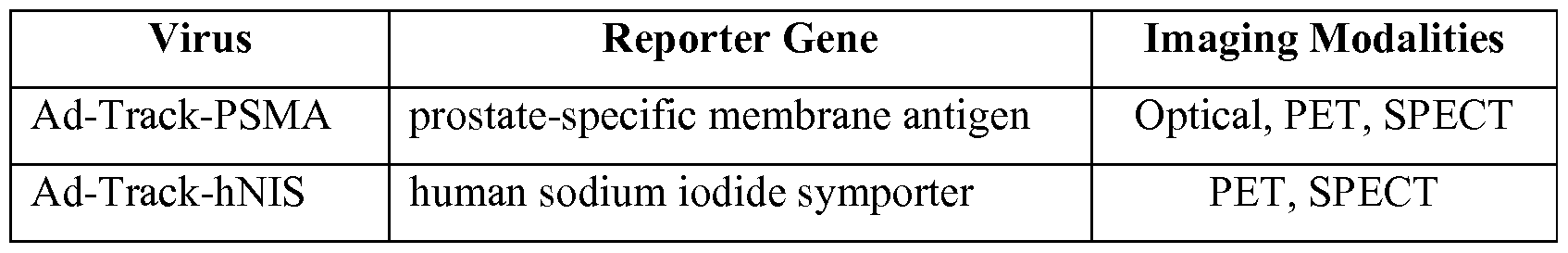
In Vitro Functionality of Adenoviral Reporter Vectors Functional assays for each reporter were analyzed using HCT116 and PC3- CAR cell lines by [3H]GCV uptake, 125I uptake, and YC-I-27 binding. Cells were infected with each reporter adenovirus. After 48 hours, the level of adenovirus infection was quantified by GFP fluorescence and the level of imaging reporter activity was quantified by functional binding and/or uptake of radiolabeled probes. Specific assay conditions were applied for each reporter gene due to the different mechanisms of reporter probe binding and/or uptake. Conditions were based on previously established assay conditions from previous efforts and in the literature (De la Vieja, 2005; Yaghoubi et al, 2006). For GCV uptake, a prolonged washout period was not performed and as a result does not differentiate between phosphorylated GCV and unphosphorylated GCV that is associated with the cells at time of harvest. YC-I- 27 uptake in PSMA infected cells proved more time dependent than the corresponding radiotracers were for NIS or HSV-sr39tk. Without wishing to be bound to any one particular theory, it is believed that this may be due to the mechanism of
sequestration, whereby [124I]NaI and GCV were able to diffuse passively or are transported into cells where they are trapped, whereas YC-I-27 can only enter the cell via binding and internalization of PSMA at a 1 : 1 stoichometry. Despite those caveats, in all cases specific and significant uptake was observed in cells infected with reporter adenovirus resulting in a signal-to-noise ratio of at least 10- to 20-fold relative to control adenovirus (FIGS. IB-ID).
In Vivo Comparison of Adenoviral Imaging Reporters To compare reporter-probe systems in vivo, a matrigel suspension model was developed (FIG. 2). In this model, PC3-CAR cells were infected in vitro with equal MOI (multiplicity of infection) of a PET imaging reporter or control adenovirus (AdTrack-Luciferase) and then each population of infected cells was separately mixed with matrigel and implanted contralaterally into the upper flanks of a nude mouse (24 hours post- infection). Luciferase was not used for imaging but as a control gene.
Twenty-four hours after implantation, in vivo GFP imaging was performed to ensure
that a similar level of control and reporter-infected cells remained at the sites of implantation. The contralateral cells masses were generally within 30% as measured by GFP imaging in vivo (FIG. 3).
The next day (48 hours after implantation), animals were subjected to a dynamic PET study whereby -7.4 MBq (200 μθί) of each respective radiotracer was administered intravenously and cellular uptake was quantified for a duration of 90 minutes. FHBG was applied for HSV-sr39tk imaging, [124I]NaI for hNIS imaging, and DCFPyL was applied for imaging of PSMA (Yaghoubi et al, 2001; Chen et al, 2011 ; Dingli et al, 2006). This model was used for comparing the reporter-probe systems for at least three reasons: (1) in vitro infection allowed equal transgene delivery of reporters and direct evaluation of implantation by GFP imaging; (2) the application of a relatively small number of cells and the short duration of the experiment minimized the influence of biologic events, such as cell growth or substantial development of tumor vasculature, on imaging reporter activity; and (3) the reporter virus was compared to a control virus within the same animal, providing an additional control to obviate animal-to-animal variation.
Kinetic uptake of each imaging reporter in target and control tissue was represented as a standardized uptake value (SUV) (FIGS. 4A-4C) or SUV relative to control tissue (FIGS. 4D-4F). For the HSV- sr39tk reporter, high initial uptake was observed in all tissues followed by rapid clearance from cells (FIG. 4A). HSV- sr39tk-specific sequestration of the radiotracer was observed after 30 to 40 minutes. The initial and rapid uptake of FHBG was attributed to diffusion of the compound into the cells where the unphosphorylated radiotracer diffuses out of the cells while phosphorylated tracer was retained. A peak target-to-background ratio of 2.81 ± 0.27 relative to control cells and 4.99 ± 0.38 relative to muscle was observed (FIG. 4G). The signal accumulation in hNIS expressing cells occurred quickly and reached a plateau roughly 80 to 90 minutes after injection (FIG. 4B). No significant uptake was observed in control tissue. A peak target-to-background ratio of 3.13 ± 0.20 relative to control cells and 4.96 ± 0.65 relative to muscle was observed (FIG. 4G). Despite differences in the mechanism of reporter probe accumulation, both the maximum signal-to-background ratios and the maximum percentage injected dose per cubic centimeter of tissue (%ID/cc) values of these two imaging reporters were similar.
This supports the utility of the matrigel suspension model as a means to compare PSMA to the existing reporter-probe systems.
Kinetics of the PSMA imaging reporter using DCFPyL displayed rapid probe uptake with the signal peaking 15 minutes post- injection, followed by a plateau shortly thereafter (FIG. 4C). Clearance from control tissue occurred more slowly for DCFPyL than for the radioligands used for the hNIS and HSV-sr39tk systems. A peak target-to-background ratio of 5.19 ± 0.78 relative to control cells and 13.80 ± 3.52 relative to muscle were observed (FIG. 4G). The maximum intensity projection images, cropped above the liver and below the thyroid for each reporter-probe system, demonstrate the specific uptake in the reporter expressing cells when compared to the muscle or to control matrigel suspension (FIG. 5A). The target-to-background profiles revealed that a signal-to-noise ratio of greater than two (relative to muscle) could be detected after 45 minutes for HSV-sr39tk, 20 minutes for hNIS, and 10 minutes for PSMA (FIGS. 4D-4F).
Maximum signal-to-noise ratios were observed at 90 minutes, with the PSMA system having the highest ratio relative both to the luciferase matrigel suspension and the muscle (FIG. 4G). Uncropped images from each scan highlight the differences in efficiency of the reporter to sequester its cognate probe and demonstrate background due to natural expression of reporter genes or probe metabolism (FIG. 5B). High gastrointestinal uptake was observed in the HSV-sr39tk system, which has previously been attributed to probe metabolism (Tjuvajev et al, 2002). Background signal from the stomach and thyroid were observed in the hNIS system due to natural expression of hNIS in these tissues. For PSMA, kidney uptake was observed in part due to renal clearance as well as PSMA expression in the proximal tubules (Bacich et al, 2001). The PSMA reporter-probe system was also evaluated using DCFBC, which has been administered to patients (Cho et al, 2012; Mease et al, 2008). Results from DCFBC were similar to results obtained with DCFPyL, with the exception of a slower clearance from the control matrigel suspension (FIG. 6). Application of the Adenoviral PSMA Reporter System
It has been previously reported in rodent models that adenoviral vectors administered via systemic injection will result in hepatocyte infection and transgene expression (Johnson et al, 2006). Optical imaging of mouse livers was performed 72
hours post-infection and 48 hours post-administration of 15 or 30 μg of J591-IR800 (FIG. 7A). Regions of interest were drawn around the liver and a signal-to-noise ratio of approximately 3: 1 was observed for the PSMA imaging reporter relative to control adenovirus. This model was also applied to PSMA-specific imaging with SPECT using YC-VI-11 (Chen et al., 2008), with similar results, where a signal-to-noise ratio of approximately 2: 1 was observed 15 min post-administration of radiotracer (FIG. 7B). Similar levels of radioactivity were observed in the liver at 4 h post- injection. Collectively, these results reveal PSMA as a sensitive and specific imaging reporter that can be applied to multiple imaging modalities.
Non-Viral PSMA Reporter System
Three non-viral vector systems were created for systemic delivery and expression of PSMA as an imaging reporter (FIGS. 8A-8C). These vectors employed PEG-Promoter, a tumor-specific promoter from a minimal promoter region of progression elevated gene-3 (Peg-3), for cancer-selective expression of PSMA.
One of the vectors comprised additional features to enhance its functionality as an imaging reporter vector (FIG. 8C). This vector had the Gal4-VP 16 and UAS transcriptional amplification system to enhance the expression level of PSMA. In addition, this vector used OriP, a viral origin of plasmid replication, and EBNA-1, a viral protein associated with the Epstein-Barr virus, for prolonged action in transfected cells.
A third vector was created to comply with the clinical requirement for human application. This vector featured CpG-free sequences for reduced immunity and used the FDA-approved Neomycin resistant gene as a selection marker (FIG. 8B). This vector used a CpG-free R6K origin for plasmid replication and a CpG-free S/MAR sequence for elongated expression (FIG. 11). FIG. 12 shows a representative cDNA sequence of a CpG-free PSMA gene (SEQ ID NO:5).
Various truncations of PSMA were also created in addition to the full-length gene (FIGS. 8D-8E) to test the effect on the physiological status of the transfected cells. It was found that the full-length (FL) and two N-terminally modified PSMA (FL(W2G) and N-l 1) were successfully localized to the cell membrane and centrosome as examined by FACS analyses (FIG. 9A) and an immunofluorescent study (FIG. 9B), whereas the C-terminally truncated PSMA (C625 and N-l l/C-625)
failed to localize to the plasma membrane, indicating the importance of the C-terminal dimerization domain for its proper folding and localization.
The basic expression vectors (FIG. 8A) were tested for the ability to uptake a radio-labeled PSMA-targeted molecule (125I-YC27). FIG. 10 shows an in vitro radio uptake assay using MDA-MB-231 cell lines transiently transfected with the basic non- viral vector with full-length and truncations of PSMA. Twenty-four hours after the transfection, cells were treated with Ι μθί of 125I-YC27, washed twice and the uptake was measured. Only full-length and N-terminally truncated PSMA were able to specifically uptake the substrate (FIG. 10).
Discussion
The presently disclosed subject matter provides PSMA, a biomarker for prostate cancer located on the cell membrane which possesses receptor, transporter and enzymatic functions, as an imaging reporter. The results demonstrate that PSMA is able to sequester imaging ligands with high affinity and with a high target-to- background ratio. Additional studies support PSMA as an imaging reporter with PET, SPECT, and optical imaging modalities.
The PSMA probes evaluated herein, as well as other probes in clinical and preclinical development, bind to the active site of PSMA on the surface of the cell. Enzymatic and transporter based reporter genes are able to amplify the signal by accumulation of probe, whereas receptor based reporters can only yield a 1 : 1 stoichiometry of probe to reporter gene molecule. The presently disclosed subject matter suggests that ligand binding alone is sufficient for detection and yields a higher signal-to-noise ratio with the PSMA-based reporter-probe system than the hNIS and HSV-sr39tk reporter-probe systems. In addition, after ligand binding, PSMA can internalize and the enzymatic and/or transporter properties of PSMA can be exploited for signal retention and amplification. As advances in cell engineering and gene therapies emerge, PSMA can provide a sensitive and non-immunogenic reporter by which to follow their fate in vivo, non-invasively.
REFERENCES
All publications, patent applications, patents, and other references mentioned in the specification are indicative of the level of those skilled in the art to which the
presently disclosed subject matter pertains. All publications, patent applications, patents, and other references are herein incorporated by reference to the same extent as if each individual publication, patent application, patent, and other reference was specifically and individually indicated to be incorporated by reference. It will be understood that, although a number of patent applications, patents, and other references are referred to herein, such reference does not constitute an admission that any of these documents forms part of the common general knowledge in the art.
Afshar-Oromieh, A.; Malcher, A.; Eder, M. et al. PET imaging with a
[68Ga]gallium-labelled PSMA ligand for the diagnosis of prostate cancer:
biodistribution in humans and first evaluation of tumour lesions. Eur. J. Nucl. Med. Mol. I. 2013, 40(4):486-495.
Anilkumar, G.; Rajasekaran, S.A.; Wang, S.; Hankinson, O.; Bander, N.H.; Rajasekaran, A.K. Prostate-specific membrane antigen association with filamin A modulates its internalization and NAALADase activity. Cancer Research. 2003, 63(10):2645-2648.
Bacich, D.J.; Pinto, J.T.; Tong, W.P.; Heston, W.D. Cloning, expression, genomic localization, and enzymatic activities of the mouse homolog of prostate- specific membrane antigen/NAALADase/folate hydrolase. Mamm Genome. 2001, 12(2): 117-123.
Banerjee, S.R.; Pullambhatla, M.; Byun, Y. et al. Sequential SPECT and optical imaging of experimental models of prostate cancer with a dual modality inhibitor of the prostate-specific membrane antigen. Angewandte Chemie
(International ed.) 2011, 50(39):9167-9170.
Banerjee, S.R.; Pullambhatla, M.; Shallal, H.; Lisok, A.; Mease, R.C.;
Pomper, M.G. A modular strategy to prepare multivalent inhibitors of prostate- specific membrane antigen (PSMA). Oncotarget 2011; 2(12): 1244-1253.
Barrett, J. A.; Coleman, R.E.; Goldsmith, S.J. et al. First-in-man evaluation of 2 high-affinity PSMA-avid small molecules for imaging prostate cancer. J Nucl Med. 2013; 54(3):380-387.
Barton, K.N.; Strieker, FL; Brown, S.L. et al. Phase I study of noninvasive imaging of adenovirus -mediated gene expression in the human prostate. Mol Ther. 2008, 16(10): 1761-1769.
Bauer S, Kirschning CJ, Hacker H, Redecke V, Hausmann S, Akira S, Wagner H, Lip ford GB. Human TLR9 confers responsiveness to bacterial DNA via species- specific CpG motif recognition. Proceedings of the National Academy of Sciences of the United States of America 2001;98(16):9237-9242.
Beeres, S.L.; Bengel, F.M.; Bartunek, J. et al. Role of imaging in cardiac stem cell therapy. J. Am. Coll. Cardiol. 2007, 49(11): 1137-1 148.
Brader, P.; Serganova, I.; Blasberg, R.G. Noninvasive molecular imaging using reporter genes. JNucl Med. 2013, 54(2): 167-172.
Campbell, D.O.; Yaghoubi, S.S.; Su, Y. et al. Structure-guided engineering of human thymidine kinase 2 as a positron emission tomography reporter gene for enhanced phosphorylation of non-natural thymidine analog reporter probe. The J. Biol. Chem. 2012, 287(l):446-454.
Chang, S.S.; O'Keefe, D.S.; Bacich, D.J.; Reuter, V.E.; Heston, W.D.;
Gaudin, P.B. Prostate-specific membrane antigen is produced in tumor-associated neovasculature. Clin Cancer Res. 1999; 5(10):2674-2681.
Chen, Y.; Dhara, S.; Banerjee, S.R. et al. A low molecular weight PSMA- based fluorescent imaging agent for cancer. Biochem. Biophys. Res.
Comm. 2009, 390(3):624-629.
Chen, Y.; Foss, C.A.; Byun, Y. et al. Radiohalogenated prostate-specific membrane antigen (PSMA)-based ureas as imaging agents for prostate cancer. J. Med. Chem. 2008, 51(24):7933-7943.
Chen, Y.; Pullambhatla, M.; Foss, C.A. et al. 2-(3- { l-Carboxy-5-[(6- [18F]fluoropyridine-3-carbonyl)-amino]-pentyl}-ureido)-pentanedioic acid,
[18F]DCFPyL, a PSMA-based PET imaging agent for prostate cancer. Clin Cancer Res. 2011, 17(24):7645-7653.
Cho, S.Y.; Gage, K.L.; Mease, R.C. et al. Biodistribution, tumor detection, and radiation dosimetry of 18F-DCFBC, a low-molecular-weight inhibitor of prostate- specific membrane antigen, in patients with metastatic prostate cancer. JNucl Med. 2012, 53(12): 1883-1891.
De la Vieja, A.; Ginter, C.S.; Carrasco, N. Molecular analysis of a congenital iodide transport defect: G543E impairs maturation and trafficking of the Na+/I- symporter. Mol. Endocrinol. 2005, 19(11):2847-2858.
Dingli, D.; Kemp, B.J.; O'Connor, M.K.; Morris, J.C.; Russell, S.J.; Lowe, V.J. Combined 1-124 positron emission tomography/computed tomography imaging of MS gene expression in animal models of stably transfected and intravenously transfected tumor. Mol Imaging Biol. 2006; 8(1): 16-23.
FDA. Points to consider on plasmid DNA vaccines for preventive infectious disease indications. Center for Biologies Evaluation and Research 1996;Docket No. :96N-0400.
Forss-Petter S, Danielson PE, Catsicas S, et al. Transgenic mice expressing betagalactosidase in mature neurons under neuron-specific enolase promoter control. Neuron 1990, 5: 187-197.
Gil, J.S.; Machado, H.B.; Herschman, H.R. A method to rapidly and accurately compare the relative efficacies of non-invasive imaging reporter genes in a mouse model and its application to luciferase reporters. Mol Imaging Biol. 2012, 14(4):462-471.
Grant, C.L.; Caromile, L.A.; Ho, V. et al. Prostate specific membrane antigen
(PSMA) regulates angiogenesis independently of VEGF during ocular
neovascularization. PloS One 2012, 7(7):e41285.
Haberkorn, U. Gene therapy with sodium/iodide symporter in
hepatocarcinoma. Exp Clin Endocrinol Diabetes. 2001, 109(l):60-62.
Heston, W.D. Characterization and glutamyl preferring carboxypeptidase function of prostate specific membrane antigen: a novel folate hydrolase. Urology 1997, 49(3A Suppl): 104-112.
Hoti, N.; Li, Y.; Chen, C.L. et al. Androgen receptor attenuation of Ad5 replication: implications for the development of conditionally replication competent adenoviruses. Mol Ther. 2007, 15(8): 1495-1503.
Hies, M.A.; Seitz, W.A.; Ghiviriga, I. et al. (2004) Pyridinium cationic lipids in gene delivery: a structure-activity correlation study. J Med Chem 2004, 47:3744- 3754.
Johnson, M.; Huyn, S.; Burton, J.; Sato, M.; Wu, L. Differential
biodistribution of adenoviral vector in vivo as monitored by bioluminescence imaging and quantitative polymerase chain reaction. Hum. Gene Ther. 2006; 17(12): 1262- 1269.
Likar, Y.; Zurita, J.; Dobrenkov, K. et al. A new pyrimidine-specific reporter gene: a mutated human deoxycytidine kinase suitable for PET during treatment with acycloguanosine-based cytotoxic drugs. JNucl Med. 2010, 51 (9) : 1395 - 1403.
Liu, H.; Rajasekaran, A.K.; Moy, P. et al. Constitutive and antibody-induced internalization of prostate-specific membrane antigen. Cancer Res. 1998,
58(18):4055-4060.
Luo, J.; Deng, Z.L.; Luo, X. et al. A protocol for rapid generation of recombinant adenoviruses using the AdEasy system. Nature Protocols. 2007,
2(5): 1236-1247.
Mease, R.C.; Dusich, C.L.; Foss, C.A. et al. N-[N-[(S)-1,3-
Dicarboxypropyl]carbamoyl]-4-[18F]fluorobenzyl-L-cysteine, [18F]DCFBC: a new imaging probe for prostate cancer. Clin Cancer Res. 2008; 14(10):3036-3043.
Min, J.J.; Iyer, M.; Gambhir, S.S. Comparison of [18F]FHBG and [14C]FIAU for imaging of HSVl-tk reporter gene expression: adenoviral infection vs stable transfection. Eur. J. Nucl. Med. Mol. I. 2003, 30(11): 1547-1560.
Miyagawa, M.; Anton, M.; Wagner, B. et al. Non- invasive imaging of cardiac transgene expression with PET: comparison of the human sodium/iodide
symporter gene and HSVl-tk as the reporter gene. Eur. J. Nucl. Med. Mol. I. 2005; 32(9): 1108-1 114.
Mlcochova, P.; Barinka, C; Tykvart, J.; Sacha, P.; Konvalinka, J. Prostate- specific membrane antigen and its truncated form PSM'. The Prostate. 2009,
69(5):471-479.
Overbeek PA, Chepelinsky AB, Khillan JS, Piatigorsky J, Westphal H. Lens- specific expression and developmental regulation of the bacterial chloramphenicol acetyltransferase gene driven by the murine alpha A-crystallin promoter in transgenic mice. Proc. Natl. Acad. Sci. U.S.A. 1985, 82:7815-7819.
Ponde, D.E.; Dence, C.S.; Schuster, D.P.; Welch, M.J. Rapid and reproducible radiosynthesis of [18F] FHBG. Nucl. Med.Biol. 2004, 31(1): 133-138.
Ponomarev, V.; Doubrovin, M.; Shavrin, A. et al. A human-derived reporter gene for noninvasive imaging in humans: mitochondrial thymidine kinase type 2. J Nucl Med. 2007; 48(5):819-826.
Rajasekaran, A.K.; Anilkumar, G.; Christiansen, J.J. Is prostate-specific membrane antigen a multifunctional protein? Am. J. Physiol. 2005, 288(5):C975-981.
Rajasekaran, S.A.; Anilkumar, G.; Oshima, E. et al. A novel cytoplasmic tail MXXXL motif mediates the internalization of prostate-specific membrane antigen. Mol. Biol. Cell 2003; 14(12):4835-4845.
Tjuvajev, J.G.; Doubrovin, M.; Akhurst, T. et al. Comparison of radiolabeled nucleoside probes (FIAU, FHBG, and FHPG) for PET imaging of HSV 1 -tk gene expression. JNucl Med. 2002; 43(8): 1072-1083.
van der Woude, I.; Wagenaar, A.; Meekel, A. A. et al. Novel pyridinium surfactants for efficient, nontoxic in vitro gene delivery. Proc Natl Acad Sci USA 1997, 94: 1 160-1 165.
Yaghoubi, S.S.; Barrio, J.R.; Dahlbom, M. et al. Human pharmacokinetic and dosimetry studies of [(18)F]FHBG: a reporter probe for imaging herpes simplex virus type-1 thymidine kinase reporter gene expression. JNucl Med. 2001, 42(8): 1225- 1234.
Yaghoubi, S.S.; Campbell, D.O.; Radu, C.G.; Czernin, J. Positron emission tomography reporter genes and reporter probes: gene and cell therapy applications. Theranostics 2012, 2(4):374-391.
Yaghoubi, S.S.; Gambhir, S.S. Measuring herpes simplex virus thymidine kinase reporter gene expression in vitro. Nat. Protoc. 2006, 1(4):2137-2142.
Yao, V.; Berkman, C.E.; Choi, J.K.; O'Keefe, D.S.; Bacich, D.J. Expression of prostate-specific membrane antigen (PSMA), increases cell folate uptake and proliferation and suggests a novel role for PSMA in the uptake of the non- polyglutamated folate, folic acid. The Prostate 2010; 70(3):305-316.
Although the foregoing subject matter has been described in some detail by way of illustration and example for purposes of clarity of understanding, it will be understood by those skilled in the art that certain changes and modifications can be practiced within the scope of the appended claims.
Claims
1. A method for using prostate-specific membrane antigen (PSMA) as an imaging reporter, the method comprising:
a) introducing a reporter gene construct comprising a PSMA gene operably linked to a transcriptional promoter to a cell;
b) allowing the cell to express PSMA protein;
c) adding an imaging probe that can detect the PSMA protein to the cell; and d) imaging the imaging probe, thereby detecting the PSMA protein.
2. The method of claim 1, wherein the reporter gene construct is a viral reporter gene construct.
3. The method of claim 2, wherein the viral reporter gene construct is an adenoviral reporter gene construct.
4. The method of claim 3, wherein the transcriptional promoter is a constitutive promoter.
5. The method of claim 4, wherein the constitutive promoter is the cytomegalovirus immediate-early gene (CMV) promoter.
6. The method of claim 1 , wherein the reporter gene construct is a non- viral, episomal reporter gene construct.
7. The method of claim 6, wherein the transcriptional promoter is the PEG-Promoter.
8. The method of claim 7, wherein the cell is a cancer cell.
9. The method of claim 6, wherein the non-viral, episomal reporter gene construct further comprises a Gal4-VP16 and Upstream Activation Sequence (UAS) transcriptional amplification system.
10. The method of claim 9, wherein the transcriptional amplification system comprises five copies of the UAS.
11. The method of claim 6, wherein the non-viral, episomal reporter gene construct further comprises the EBNA-1 gene.
12. The method of claim 6, wherein the non-viral, episomal reporter gene construct further comprises OriP.
13. The method of claim 1, wherein the PSMA gene comprises the coding sequence of the full-length PSMA protein.
14. The method of claim 1, wherein the PSMA gene is truncated at the 5' end of the coding sequence of the PSMA protein.
15. The method of claim 14, wherein the truncation results in a mutated PSMA protein that is lacking the first 11 amino acids of the wild-type PSMA protein.
16. The method of claim 1, wherein the PSMA protein is mutated such that its second amino acid is glycine instead of tryptophan.
17. The method of claim 1, wherein the PSMA gene is CpG-free.
18. The method of claim 1, wherein the reporter gene construct comprising a PSMA gene is CpG-free.
19. The method of claim 1, wherein the reporter gene construct comprising a PSMA gene further comprises a neomycin or kanamycin resistant gene.
20. The method of claim 1, wherein the imaging probe detects the PSMA protein by binding to the active site of the PSMA protein.
21. The method of claim 1, further comprising the cleavage of the imaging probe by the PSMA protein.
22. The method of claim 21, wherein cleavage of the imaging probe results in at least one cleavage product that is fluorescent and/or radioactive.
23. The method of claim 1, wherein the imaging probe is 2-(3-{ 1-carboxy- 5-[(6-[18F]fluoro-pyridine-3-carbonyl)-amino]-pentyl}-ureido)-pentanedioic acid (DCFPyL) or N-[N-[(S)-l,3-dicarboxypropyl]carbamoyl]-4-[18F]fluorobenzyl-L- cysteine (DCFBC).
24. The method of claim 1, wherein imaging the PSMA protein occurs by at least one method selected from the group consisting of near-infrared (NIR) optical tomography, positron emission tomography (PET), and single photon emission computed tomography (SPECT).
25. The method of claim 1, wherein the PSMA protein is detected on the surface of the cell.
26. The method of claim 1, wherein the PSMA protein is detected inside the cell.
27. The method of claim 1, wherein the cell is found in a subject.
28. The method of claim 27, wherein the subject is human.
29. An adenoviral reporter gene construct comprising a prostate-specific membrane antigen (PSMA) gene operably linked to a cytomegalovirus immediate- early gene (CMV) promoter.
30. The construct of claim 29, wherein the PSMA gene comprises the coding sequence of the full-length PSMA protein.
31. The construct of claim 29, wherein the PSMA gene is truncated at the 5' end of the coding sequence of the PSMA protein.
32. The construct of claim 31, wherein the truncation results in a mutated PSMA protein that is lacking the first 11 amino acids of the wild-type PSMA protein.
33. The construct of claim 29, wherein the PSMA gene is mutated such that the construct expresses a mutated PSMA protein that has glycine as a second amino acid instead of tryptophan.
34. The construct of claim 29, wherein the PSMA gene is CpG-free.
35. The construct of claim 29, wherein the adenoviral reporter gene construct comprising a PSMA gene is CpG-free.
36. The construct of claim 29, wherein the adenoviral reporter gene construct comprising a PSMA gene further comprises a neomycin or kanamycin resistant gene.
37. A non-viral, episomal reporter gene construct comprising a prostate- specific membrane antigen (PSMA) gene operably linked to a PEG-Promoter.
38. The construct of claim 37, wherein the construct further comprises a Gal4-VP 16 and Upstream Activation Sequence (UAS) transcriptional amplification system.
39. The construct of claim 38, wherein the transcriptional amplification system comprises five copies of the UAS.
40. The construct of claim 37, wherein the construct further comprises the EBNA-1 gene.
41. The construct of claim 37, wherein the construct further comprises
OriP.
42. The construct of claim 37, wherein the PSMA gene comprises the coding sequence of the full-length PSMA protein.
43. The construct of claim 37, wherein the PSMA gene is truncated at the 5' end of the coding sequence of the PSMA protein.
44. The construct of claim 43, wherein the truncation results in a mutated
PSMA protein that is lacking the first 11 amino acids of the wild-type PSMA protein.
45. The construct of claim 37, wherein the PSMA gene is mutated such that the construct expresses a mutated PSMA protein that has glycine as a second amino acid instead of tryptophan.
46. The construct of claim 37, wherein the PSMA gene is CpG-free.
47. The construct of claim 37, wherein the reporter gene construct comprising a PSMA gene is CpG-free.
48. The construct of claim 37, wherein the reporter gene construct comprising a PSMA gene further comprises a neomycin or kanamycin resistant gene.
49. A kit comprising a construct of claim 29 or claim 37.
Priority Applications (2)
| Application Number | Priority Date | Filing Date | Title |
|---|---|---|---|
| US15/126,749 US11124845B2 (en) | 2014-03-18 | 2015-03-18 | PSMA-based molecular-genetic reporter system |
| US16/934,137 US20200347464A1 (en) | 2014-03-18 | 2020-07-21 | Psma-based molecular-genetic reporter system |
Applications Claiming Priority (4)
| Application Number | Priority Date | Filing Date | Title |
|---|---|---|---|
| US201461954947P | 2014-03-18 | 2014-03-18 | |
| US61/954,947 | 2014-03-18 | ||
| US201461972833P | 2014-03-31 | 2014-03-31 | |
| US61/972,833 | 2014-03-31 |
Related Child Applications (2)
| Application Number | Title | Priority Date | Filing Date |
|---|---|---|---|
| US15/126,749 A-371-Of-International US11124845B2 (en) | 2014-03-18 | 2015-03-18 | PSMA-based molecular-genetic reporter system |
| US16/934,137 Division US20200347464A1 (en) | 2014-03-18 | 2020-07-21 | Psma-based molecular-genetic reporter system |
Publications (1)
| Publication Number | Publication Date |
|---|---|
| WO2015143029A1 true WO2015143029A1 (en) | 2015-09-24 |
Family
ID=54145268
Family Applications (1)
| Application Number | Title | Priority Date | Filing Date |
|---|---|---|---|
| PCT/US2015/021233 WO2015143029A1 (en) | 2014-03-18 | 2015-03-18 | Psma-based molecular-genetic reporter system |
Country Status (2)
| Country | Link |
|---|---|
| US (2) | US11124845B2 (en) |
| WO (1) | WO2015143029A1 (en) |
Cited By (7)
| Publication number | Priority date | Publication date | Assignee | Title |
|---|---|---|---|---|
| WO2018134691A2 (en) | 2017-01-20 | 2018-07-26 | Juno Therapeutics Gmbh | Cell surface conjugates and related cell compositions and methods |
| WO2018187791A1 (en) | 2017-04-07 | 2018-10-11 | Juno Therapeutics, Inc | Engineered cells expressing prostate-specific membrane antigen (psma) or a modified form thereof and related methods |
| US10815200B2 (en) | 2015-09-30 | 2020-10-27 | Deutsches Krebsforschungszentrum | 18F—tagged inhibitors of prostate specific membrane antigen (PSMA) and their use as imaging agents for prostate cancer |
| WO2022216524A1 (en) | 2021-04-07 | 2022-10-13 | Century Therapeutics, Inc. | Combined artificial cell death/reporter system polypeptide for chimeric antigen receptor cell and uses thereof |
| WO2022216514A1 (en) | 2021-04-07 | 2022-10-13 | Century Therapeutics, Inc. | Compositions and methods for generating gamma-delta t cells from induced pluripotent stem cells |
| WO2022216624A1 (en) | 2021-04-07 | 2022-10-13 | Century Therapeutics, Inc. | Compositions and methods for generating alpha-beta t cells from induced pluripotent stem cells |
| WO2023240147A1 (en) | 2022-06-08 | 2023-12-14 | Century Therapeutics, Inc. | Genetically engineered cells expressing cd16 variants and nkg2d and uses thereof |
Families Citing this family (1)
| Publication number | Priority date | Publication date | Assignee | Title |
|---|---|---|---|---|
| KR102282378B1 (en) | 2013-10-18 | 2021-07-27 | 도이체스 크렙스포르슝스첸트룸 | Labeled inhibitors of prostate specific membrane antigen (psma), their use as imaging agents and pharmaceutical agents for the treatment of prostate cancer |
Citations (3)
| Publication number | Priority date | Publication date | Assignee | Title |
|---|---|---|---|---|
| US20030059942A1 (en) * | 2000-09-28 | 2003-03-27 | Myung-Sam Cho | Enhanced transfection system |
| US8435777B1 (en) * | 2012-01-20 | 2013-05-07 | Cayla | CPG-free gene for a new secreted reporter protein |
| US20130263296A1 (en) * | 2010-10-28 | 2013-10-03 | The Johns Hopkins University | Cancer imaging with therapy: theranostics |
Family Cites Families (3)
| Publication number | Priority date | Publication date | Assignee | Title |
|---|---|---|---|---|
| WO2002040059A2 (en) * | 2000-11-01 | 2002-05-23 | American Foundation For Biological Research, Inc. | Methods and compositions for inducing cell-mediated immune responses |
| FR2821855B1 (en) * | 2001-03-09 | 2004-04-02 | Cayla | SYNTHETIC GENES AND BACTERIAL PLASMIDS WITHOUT GIC |
| US20050215472A1 (en) * | 2001-10-23 | 2005-09-29 | Psma Development Company, Llc | PSMA formulations and uses thereof |
-
2015
- 2015-03-18 US US15/126,749 patent/US11124845B2/en active Active
- 2015-03-18 WO PCT/US2015/021233 patent/WO2015143029A1/en active Application Filing
-
2020
- 2020-07-21 US US16/934,137 patent/US20200347464A1/en active Pending
Patent Citations (3)
| Publication number | Priority date | Publication date | Assignee | Title |
|---|---|---|---|---|
| US20030059942A1 (en) * | 2000-09-28 | 2003-03-27 | Myung-Sam Cho | Enhanced transfection system |
| US20130263296A1 (en) * | 2010-10-28 | 2013-10-03 | The Johns Hopkins University | Cancer imaging with therapy: theranostics |
| US8435777B1 (en) * | 2012-01-20 | 2013-05-07 | Cayla | CPG-free gene for a new secreted reporter protein |
Non-Patent Citations (4)
| Title |
|---|
| ASAKAWA, KAZUHIDE ET AL.: "Targeted gene expression by the Ga14-UAS system in zebrafish", DEVELOPMENT, GROWTH & DIFFERENTIATION, vol. 50, no. 6, August 2008 (2008-08-01), pages 391 - 399, XP055226996 * |
| CASTANARES, MARK A. ET AL.: "Development and application of a reporter-probe system based on the prostate specific membrane antigen", THESIS FOR PH. D., 2012, pages 1 - 154, XP055226928 * |
| CASTANARES, MARK A. ET AL.: "Evaluation of prostate-specific membrane antigen as an imaging reporter", JOURNAL OF NUCLEAR MEDICINE, vol. 55, no. 5, 3 April 2014 (2014-04-03), pages 805 - 811, XP055227003 * |
| OSBORNE, JOSEPH R. ET AL.: "Prostate-specific membrane antigen-based imaging", UROLOGIC ONCOLOGY, vol. 31, no. 2, February 2013 (2013-02-01), pages 144 - 154, XP055226997 * |
Cited By (8)
| Publication number | Priority date | Publication date | Assignee | Title |
|---|---|---|---|---|
| US10815200B2 (en) | 2015-09-30 | 2020-10-27 | Deutsches Krebsforschungszentrum | 18F—tagged inhibitors of prostate specific membrane antigen (PSMA) and their use as imaging agents for prostate cancer |
| WO2018134691A2 (en) | 2017-01-20 | 2018-07-26 | Juno Therapeutics Gmbh | Cell surface conjugates and related cell compositions and methods |
| US11517627B2 (en) | 2017-01-20 | 2022-12-06 | Juno Therapeutics Gmbh | Cell surface conjugates and related cell compositions and methods |
| WO2018187791A1 (en) | 2017-04-07 | 2018-10-11 | Juno Therapeutics, Inc | Engineered cells expressing prostate-specific membrane antigen (psma) or a modified form thereof and related methods |
| WO2022216524A1 (en) | 2021-04-07 | 2022-10-13 | Century Therapeutics, Inc. | Combined artificial cell death/reporter system polypeptide for chimeric antigen receptor cell and uses thereof |
| WO2022216514A1 (en) | 2021-04-07 | 2022-10-13 | Century Therapeutics, Inc. | Compositions and methods for generating gamma-delta t cells from induced pluripotent stem cells |
| WO2022216624A1 (en) | 2021-04-07 | 2022-10-13 | Century Therapeutics, Inc. | Compositions and methods for generating alpha-beta t cells from induced pluripotent stem cells |
| WO2023240147A1 (en) | 2022-06-08 | 2023-12-14 | Century Therapeutics, Inc. | Genetically engineered cells expressing cd16 variants and nkg2d and uses thereof |
Also Published As
| Publication number | Publication date |
|---|---|
| US20200347464A1 (en) | 2020-11-05 |
| US11124845B2 (en) | 2021-09-21 |
| US20170218464A1 (en) | 2017-08-03 |
Similar Documents
| Publication | Publication Date | Title |
|---|---|---|
| US20200347464A1 (en) | Psma-based molecular-genetic reporter system | |
| Groot-Wassink et al. | Adenovirus biodistribution and noninvasive imaging of gene expression in vivo by positron emission tomography using human sodium/iodide symporter as reporter gene | |
| Phuong et al. | Use of a vaccine strain of measles virus genetically engineered to produce carcinoembryonic antigen as a novel therapeutic agent against glioblastoma multiforme | |
| Blasberg et al. | Molecular-genetic imaging: current and future perspectives | |
| Liang et al. | Noninvasive, repetitive, quantitative measurement of gene expression from a bicistronic message by positron emission tomography, following gene transfer with adenovirus | |
| Jacobs et al. | Functional coexpression of HSV-1 thymidine kinase and green fluorescent protein: implications for noninvasive imaging of transgene expression | |
| Penheiter et al. | Sodium iodide symporter (NIS)-mediated radiovirotherapy for pancreatic cancer | |
| Trujillo et al. | A probasin promoter, conditionally replicating adenovirus that expresses the sodium iodide symporter (NIS) for radiovirotherapy of prostate cancer | |
| Jacobs et al. | Molecular imaging of gliomas | |
| Xu et al. | Somatostatin receptor based imaging and radionuclide therapy | |
| Blasberg et al. | In vivo molecular‐genetic imaging | |
| Jacobs et al. | Imaging-guided gene therapy of experimental gliomas | |
| KR101429696B1 (en) | Recombinant adenovirus with enhanced safety and anti-cancer activity and use thereof | |
| Li et al. | Genetic incorporation of HSV-1 thymidine kinase into the adenovirus protein IX for functional display on the virion | |
| Kim et al. | Image-aided suicide gene therapy utilizing multifunctional hTERT-targeting adenovirus for clinical translation in hepatocellular carcinoma | |
| US20200384135A1 (en) | Cancer imaging with therapy: theranostics | |
| US7527942B2 (en) | Transcription amplification system for molecular imaging | |
| Cho | A transporter gene (sodium iodide symporter) for dual purposes in gene therapy: imaging and therapy | |
| Peñuelas et al. | Positron emission tomography and gene therapy: basic concepts and experimental approaches for in vivo gene expression imaging | |
| Brader et al. | Imaging of lymph node micrometastases using an oncolytic herpes virus and [18F] FEAU PET | |
| Robertson et al. | Cancer imaging and therapy utilizing a novel NIS-expressing adenovirus: The role of adenovirus death protein deletion | |
| Jiang et al. | Androgen-independent molecular imaging vectors to detect castration-resistant and metastatic prostate cancer | |
| Oneal et al. | Characterization of infectivity-enhanced conditionally replicating adenovectors for prostate cancer radiovirotherapy | |
| Deroose et al. | Preliminary validation of varicella zoster virus thymidine kinase as a novel reporter gene for PET | |
| Brader et al. | Combination of pet imaging with viral vectors for identification of cancer metastases |
Legal Events
| Date | Code | Title | Description |
|---|---|---|---|
| 121 | Ep: the epo has been informed by wipo that ep was designated in this application |
Ref document number: 15765635 Country of ref document: EP Kind code of ref document: A1 |
|
| WWE | Wipo information: entry into national phase |
Ref document number: 15126749 Country of ref document: US |
|
| NENP | Non-entry into the national phase |
Ref country code: DE |
|
| 122 | Ep: pct application non-entry in european phase |
Ref document number: 15765635 Country of ref document: EP Kind code of ref document: A1 |