WO2013150132A2 - Polypeptide markers for diagnosis and assessment of strokes - Google Patents
Polypeptide markers for diagnosis and assessment of strokes Download PDFInfo
- Publication number
- WO2013150132A2 WO2013150132A2 PCT/EP2013/057182 EP2013057182W WO2013150132A2 WO 2013150132 A2 WO2013150132 A2 WO 2013150132A2 EP 2013057182 W EP2013057182 W EP 2013057182W WO 2013150132 A2 WO2013150132 A2 WO 2013150132A2
- Authority
- WO
- WIPO (PCT)
- Prior art keywords
- markers
- polypeptide
- sample
- absence
- amplitude
- Prior art date
Links
Classifications
-
- G—PHYSICS
- G01—MEASURING; TESTING
- G01N—INVESTIGATING OR ANALYSING MATERIALS BY DETERMINING THEIR CHEMICAL OR PHYSICAL PROPERTIES
- G01N33/00—Investigating or analysing materials by specific methods not covered by groups G01N1/00 - G01N31/00
- G01N33/48—Biological material, e.g. blood, urine; Haemocytometers
- G01N33/50—Chemical analysis of biological material, e.g. blood, urine; Testing involving biospecific ligand binding methods; Immunological testing
- G01N33/68—Chemical analysis of biological material, e.g. blood, urine; Testing involving biospecific ligand binding methods; Immunological testing involving proteins, peptides or amino acids
- G01N33/6893—Chemical analysis of biological material, e.g. blood, urine; Testing involving biospecific ligand binding methods; Immunological testing involving proteins, peptides or amino acids related to diseases not provided for elsewhere
-
- G—PHYSICS
- G01—MEASURING; TESTING
- G01N—INVESTIGATING OR ANALYSING MATERIALS BY DETERMINING THEIR CHEMICAL OR PHYSICAL PROPERTIES
- G01N2800/00—Detection or diagnosis of diseases
- G01N2800/70—Mechanisms involved in disease identification
- G01N2800/7019—Ischaemia
Definitions
- the present invention relates to the use of the presence or absence of one or more peptide markers in a sample of an individual for the diagnosis and assessment of ischemic stroke and transistoric, ischemic attacks as well as a method for the diagnosis and assessment of ischemic stroke and transistoric ischemic attacks or absence of the peptide marker (s) is indicative of ischemic stroke and of transient ischemic attacks.
- a transient ischemic attack is a circulatory disorder of the brain that causes neurological deficits that completely regress within 24 hours.
- Typical symptoms include hemiplegia of the arm and / or leg, speech problems and - possibly half-sided - visual disturbances. Especially in the case of TIA, some of the symptoms have already subsided during hospital admission.
- the diagnosis is essentially based on imaging techniques.
- the sensitivity of magnetic resonance imaging with diffusion weighting was 83%, while computed tomography without contrast medium had only a sensitivity of 16%.
- the false-negative rate of magnetic resonance imaging is still 17%.
- the ability or ability to easily and reliably detect and assess a stroke, especially a minor stroke or a TIA would have many advantages.
- the object of the present invention was to provide a method with which this diagnosis can take place, in particular also the severity of a stroke can be assessed.
- the object is achieved by a method for diagnosing a stroke, comprising the step of determining the presence or absence or amplitude of at least three polypeptide markers in a urine sample, the polypeptide markers being selected from the markers shown in Table 1 by values for the molecular masses, the migration time and / or optionally their peptide sequence are characterized.
- the evaluation of the measured polypeptides can be based on the presence or absence and / or amplitude of the markers taking into account the following reference or regulation values.
- the specified logarithm of the amplitude is a measure of a higher or lower occurrence of a marker in the respective groups.
- the control group is a group of patients who did not have a stroke.
- markers are used whose sequence is given in Table 3.
- Specificity is defined as the number of actual negative samples divided by the sum of the number of actual negatives and the number of false positives. A specificity of 100% means that a test identifies all healthy persons as healthy, i. no healthy person is identified as ill. This does not say anything about how well the test detects sick patients.
- Sensitivity is defined as the number of actual positive samples divided by the sum of the number of actual positives and the number of false negatives. A sensitivity of 100% means that the test detects all patients. He does not say how well the test detects healthy people.
- markers according to the invention it is possible to detect IS or TIA with a specificity of at least 60, preferably at least 70, more preferably 80, even more preferably at least 90 and most preferably at least 95%.
- markers according to the invention it is possible to detect IS or TIA with a sensitivity of at least 60, preferably at least 70, more preferably 80, even more preferably at least 90 and most preferably at least 95%.
- Migration time is determined by capillary electrophoresis (CE) - e.g. executed in point 2 - determined.
- CE time capillary electrophoresis
- the eluent used is, for example, 30% methanol, 0.5% formic acid in water or 20% acetonitrile, 0.25% formic acid. All percentages in% by volume.
- CE migration time can vary. However, the order in which the polypeptide labels elute is typically the same for any CE system used under the conditions indicated. To the- To compensate for any remaining differences in migration time, the system can be normalized using standards for which migration times are well known. These standards can be, for example, the polypeptides given in the examples (see Example 3), or certain known peptides from urine, as described, for example, in Jantos-Siwy et al. (Quantitative Urinary Proteome Analysis for Biomarker Evaluation in Chronic Kidney Disease, J. Proteome Res., 8: 268-281 (2009)).
- CE-MS capillary electrophoresis mass spectrometry
- polypeptide markers according to the invention are proteins or peptides or degradation products of proteins or peptides. They may be chemically modified, e.g. by post-translational modifications such as glycation, phosphorylation, alkylation or disulfide bridging, or by other reactions, e.g. in the context of mining, to be changed. Based on the parameters that determine the polypeptide markers (molecular mass and migration time), it is possible to identify the sequence of the corresponding polypeptides by methods known in the art.
- the polypeptides of the invention are used to diagnose IS or TIA. Diagnosis is the process of the recognition Winning by assigning symptoms or phenomena to a disease or injury. The presence or absence of a polypeptide marker can be measured by any method known in the art. Methods that can be used are exemplified below.
- a polypeptide marker is present when its reading is at least as high as the threshold. If its reading is below that, the polypeptide marker is absent.
- the threshold value can either be determined by the sensitivity of the measurement method (detection limit) or defined based on experience.
- the threshold is preferably exceeded when the sample reading for a given molecular mass is at least twice that of a blank (e.g., only buffer or solvent).
- the polypeptide marker (s) is / are used to measure its presence or absence, the presence or absence being indicative of IS or TIA.
- polypeptide markers that are typically present in individuals with IS or TIA, but are less common or absent in individuals without IS or TIA.
- polypeptide markers which are present in patients with IS or TIA but are absent or only rarely present in patients without IS or TIA.
- the frequency with which a marker appears in the group with IS or TIA or in the control group is given in Table 2 as frequency.
- the amplitudes can also be used for diagnosis.
- the amplitudes are used in such a way that it is not the presence or absence that is decisive, but the height of the signal (the amplitude) when the signal is present in both groups.
- a nomination procedure makes sense in order to achieve comparability between differently concentrated samples or different measurement methods.
- collagen fragments are preferably used, as in Jantos Siwy et al. (Quantitative Urinary Proteome Analysis for Biomarker Evaluation in Chronic Kidney Disease, J. Proteome Res., 8: 268-281 (2009)). wrote.
- the decision to make a diagnosis depends on how high the amplitude of the respective polypeptide markers in the patient sample is compared to the mean amplitudes in the control group or the "sick" group. If the value is close to the mean amplitude of the "sick" group, it is to be assumed that the occurrence of a stroke, it corresponds more to the mean amplitudes of the control group, is not to assume a stroke.
- the distance to the mean amplitude can be interpreted as a probability of belonging to a group. Alternatively, the distance between the measured value and the mean amplitude may be considered as a probability of belonging to a group.
- both the frequency and the amplitude are used for the evaluation.
- the p-value is a measure of the likelihood that the association of the markers with the two groups (IS or TIA and control) is based on a random distribution that is not related to IS or TIA.
- the individual from whom the sample is derived, in which the presence or absence or amplitude of one or more polypeptide markers is determined, may be any individual who may be suffering from IS or TIA.
- the subject is a mammal, most preferably a human.
- the sample measuring the presence or absence of the polypeptide marker (s) of the invention may be any sample recovered from the subject's body.
- the sample is a sample having a polypeptide composition suitable for making statements about the condition of the individual.
- it may be blood, urine, synovial fluid, tissue fluid, body secretions, sweat, cerebrospinal fluid, lymph, intestinal, gastric, pancreatic, bile, tear fluid, tissue sample, sperm, vaginal fluid or stool sample ,
- it is a liquid sample.
- the sample is a urine sample.
- Urine samples may be known as known in the art.
- a mid-jet urine sample is used.
- the urine sample may e.g. by means of a catheter or also with the aid of a urination apparatus, as described in WO 01/74275.
- the presence or absence or amplitude of a polypeptide marker in the sample can be determined by any method known in the art suitable for measuring polypeptide markers. Those skilled in such methods are known. In principle, the presence or absence of a polypeptide marker can be determined by direct methods such as e.g. Mass spectrometry, or indirect methods, such as by ligands or specific probes such as antibodies.
- the sample of the subject eg, the urine sample
- the treatment may include, for example, purification, separation, dilution or concentration.
- the methods may include, for example, centrifugation, filtration, ultrafiltration, dialysis, precipitation or chromatographic methods such as affinity separation or separation by ion exchange chromatography, or electrophoretic separation.
- a mass spectrometric method is used to determine the presence or absence or amplitude of a polypeptide marker, which method may precede purification or separation of the sample.
- the mass spectrometric analysis has the advantage over current methods that the concentration of many (> 100) polypeptides of a sample can be determined by a single analysis. Any type of mass spectrometer can be used. With mass spectrometry, it is possible to routinely measure 10 fmoles of a polypeptide marker, that is, 0.1 ng of a 10 kDa protein with a measurement accuracy of approximately ⁇ 0.01% from a complex mixture. In mass spectrometers, an ion-forming unit is coupled to a suitable analyzer.
- electrospray ionization (ESI) interfaces are commonly used to measure ions from liquid samples
- matrix assisted laser desorption / ionization (MALDI) technique is used to measure ions from sample crystallized with a matrix
- ESI electrospray ionization
- MALDI matrix assisted laser desorption / ionization
- quadrupoles ion traps or time-of-flight (TOF) analyzers are used.
- TOF time-of-flight
- electrospray ionization the molecules present in solution are sprayed, inter alia, under the influence of high voltage (eg 1-8 kV), resulting in charged droplets formed by evaporation of the solvent get smaller.
- high voltage eg 1-8 kV
- Coulomb explosions lead to the formation of free ions, which can then be analyzed and detected.
- TOF analyzers have a very high scanning speed and achieve a very high resolution.
- Preferred methods for determining the presence or absence of polypeptide markers include gas phase ion spectrometry, such as laser desorption / ionization mass spectrometry, MALDI-TOF-MS, SELDI-TOF-MS (surface enhanced laser desorption ionization), LC-MS (liquid chromatography mass spectrometry), 2D-PAGE-MS and capillary electrophoresis mass spectrometry (CE-MS). All of the methods mentioned are known to the person skilled in the art.
- gas phase ion spectrometry such as laser desorption / ionization mass spectrometry, MALDI-TOF-MS, SELDI-TOF-MS (surface enhanced laser desorption ionization), LC-MS (liquid chromatography mass spectrometry), 2D-PAGE-MS and capillary electrophoresis mass spectrometry (CE-MS). All of the methods mentioned are known to the person skilled in the art.
- CE-MS in which capillary electrophoresis is coupled with mass spectrometry. This method is described in detail e.g. in German patent application DE 10021737, in Kaiser et al. (J. Chromatogr. A, 2003, Vol. 1013: 157-171, and Electrophoresis, 2004, 25: 2044-2055), in Wittke et al. (J. Chromatogr. A, 2003, 1013: 173-181) and Ref.
- the CE-MS technique allows to determine the presence of several hundreds of polypeptide markers of a sample simultaneously in a short time, a small volume and high sensitivity. After a sample has been measured, a pattern of the measured polypeptide markers is prepared (see below). This can be compared with reference patterns of ill or healthy individuals. In most cases it is sufficient to use a limited number of polypeptide markers for the diagnosis of diseases. More preferred is a CE-MS method which includes CE coupled online to an ESI-TOF-MS.
- the use of volatile solvents is preferred, and it is best to work under substantially salt-free conditions. conditions.
- suitable solvents include acetonitrile, methanol and the like.
- the solvents may be diluted with water and an acid (eg 0.1% to 1% formic acid) added to protonate the analyte, preferably the polypeptides.
- Capillary electrophoresis makes it possible to separate molecules according to their charge and size. Neutral particles migrate at the rate of electroosmotic flow upon application of a current, cations are accelerated to the cathode and anions are retarded.
- the advantage of capillaries in electrophoresis is the favorable ratio of surface area to volume, which enables a good removal of the Joule heat arising during the current flow. This in turn allows the application of high voltages (usually up to 30 kV) and thus a high separation efficiency and short analysis times.
- quartz glass capillaries with internal diameters of typically 50 to 75 ⁇ m are normally used. The used lengths are 30-100 cm.
- the capillaries usually consist of plastic-coated quartz glass.
- the capillaries may be both untreated, i. on the inside show their hydrophilic groups, as well as be coated on the inside. A hydrophobic coating can be used to improve the resolution.
- a pressure which is typically in the range of 0-1 psi may also be applied. The pressure can also be created during the separation or changed during the process.
- the markers of the sample are separated by capillary electrophoresis, then directly ionized and transferred online to a mass spectrometer coupled thereto for detection.
- several polypeptide markers can advantageously be used for diagnostics. Preferred is the use of at least 5, 6, 8, or 10 markers. In one embodiment, 20 to 50 markers are used.
- the biomarkers identified according to the invention can also be used to determine the severity of the stroke.
- the biomarkers show correlation with the National Institute of Health Stroke Scale (NIHSS).
- NIHSS National Institute of Health Stroke Scale
- the combination of the normalized amplitudes of the individual biomarkers can thus be used to determine the severity of the stroke, thus giving valuable information on the therapy.
- Urine was used to detect polypeptide markers for diagnosis. Urine was collected from healthy donors (peer group) and from patients who had a stroke. For the subsequent CE-MS measurement, those also had to be in urine of patients in higher concentration occurring proteins such as albumin and immunoglobulins are separated by ultrafiltration. For this purpose, 700 .mu.l of urine were removed and treated with 700 .mu.l Filtrationspuffer (2M urea, lOmM ammonia, 0.02% SDS). These 1.4 ml sample volumes were ultrafiltered (20 kDa, Sartorius, Göttingen, DE). The UF was carried out at 3000 rpm in a centrifuge until 1.1 ml ultrafiltrate was obtained.
- the CE-MS measurements were performed with a capillary electrophoresis system from Beckman Coulter (P / ACE MDQ System, Beckman Coulter Inc, Fullerton, USA) and a Bruker ESI-TOF mass spectrometer (micro-TOF MS, Bruker Daltonik, Bremen, D).
- the CE capillaries were purchased from New Objective, they had an ID / OD of 50/360 ⁇ and a length of 90 cm.
- the mobile phase for the CE separation consisted of 20% acetonitrile and 0.25% formic acid in water. 30% isopropanol with 0.5% formic acid was used for the "sheath flow" at the MS, here with a flow rate of 20 ⁇ / h.
- CE-ESI-MS sprayer kit (Agilent Technologies, Waldbronn, DE).
- the duration of the injection was 99 seconds. With these parameters about 300 nl of the sample were injected into the capillary, this corresponds to about 10% of the capillary volume.
- a "stacking" technique was used. An IM NH 3 solution is injected for 7 seconds (at 1 psi) prior to sample injection. After applying the separation voltage (25 kV), the analytes are automatically concentrated between these solutions.
- CE separation was performed with a pressure method: 0 psi for 30 minutes, 0.1 psi for 1 min, 0.2 psi for 1 min, 0.3 psi for 1 min, 0.4 psi for 1 min, and 0.5 psi for 35 min.
- the total duration of a separation run was thus 70 minutes.
- no "Nebulizer gas” was used.
- the voltage applied to the spray needle to generate the electrospray was 4000-4800 V.
- the remaining settings on the mass spectrometer were optimized as instructed by the manufacturer of peptide detection. The spectra were recorded over a mass range of m / z 400 to m / z 3000 and accumulated every 3 seconds.
- the proteins / polypeptides are each used in a concentration of 10 pmol / ⁇ in water.
- REV amino acid sequence
- ELM amino acid sequence polypeptide
- KINCON amino acid sequence polypeptides
- a further peptide (peptide 2) is selected from the measurement and attempts to identify a suitable polypeptide marker, again taking into account a corresponding time window. If, in turn, several markers can be found with a corresponding mass, the most probable assignment is that in which there is a substantially linear relationship between the shift for the peptide 1 and for the peptide 2.
- further proteins from his sample for the assignment, for example ten proteins.
- migration times are either lengthened or shortened by certain absolute values, or upsets or knocks occur throughout the course. Co-migrating peptides also co-migrate under such conditions.
- the urine samples are analyzed according to Carty DM et al. (Urinary proteomics for predication of preeclampsia, Hypertension 2011; 57: 561-9) with a Dionex Ultimate 3000 RSLS nanoflous system (Dionex, Camberley, UK) (LC / MS). Samples (5 ⁇ ) were spun at a flow rate of 5 ⁇ / min in 0.1% formic acid and 2% acetonitrile onto a Dionex C18 nano trap. Column (0.1 x 20 mm, 5 ⁇ m) abandoned.
- the sample was eluted at a flow rate of 0.3 ⁇ / min onto an Acclaim-PepMap C18 nano-column (75 ⁇ m x 15cm, 2 ⁇ m, 100%), the trap column and the nanofluid column were opened
- the mass spectrometer was operated in the MS / MS mode with a scan range from m / z 380 to 2000 amu.
- the 10 largest multiply charged ions were selected from each scan for MS / MS analysis; the fragmentation method was HCD with 35% collision energy.
- the ions were selected by a data-dependent method with a repetition number of 1 and an exclusion time of 15 s for MS2.
- the ion dissolution was 60000 in MSI and 7500 for HCD-MS2.
- the files were used for a search against the human non-redundant IPI database using the Open Mass Spectrometry Search Algorithm (OMSSA, see pubchem.ncbi.nlm.nih.gov/omssa) and SEQUEST (using the Thermo Proteome Discoverer), without any enzyme specificity. No fixed modification and oxidation of methionine and proline were chosen as variable modifications. The permissible mass error window for MS or MS / MS was 10 ppm or 0.05 Da. In the case of SEQUEST, the peptide data were extracted using high peptide-confidence and top-one peptide rank filters. 1% FDR was used as the threshold for identifying identified peptides.
- OMSSA Open Mass Spectrometry Search Algorithm
- SEQUEST using the Thermo Proteome Discoverer
- Sample 14289 and Sample 14238 were classified with a classifier based on the biomarkers of the invention in a blinded assay. When all 229 biomarkers were used, the scoring for subject was 14289-0788, so no stroke, the scoring for subject 14238 was 1,058, so positive for stroke.
- Marker 123902 has an amplitude of 2.35 in the control group and an amplitude of 1.86 in the stroke group. The subject found an amplitude of 2.62, i. still above the amplitude for the control group. This results in a negative score for the patient.
- Marker 70413 has an amplitude of 2.16 in the control group and a score of 3.02 in the stroke group. The found amplitude is 2.85; this is closer to the group for stroke. He gets a positive score.
- Marker 4609 has a score of 0.06 in the healthy group and a score of 1.27 in the stroke group. The amplitude of 2.31 is still above the amplitude of the sick group. This gives a strong positive score. This results in a total score for subject 14238 of +0.910.
- Marker 70413 has an amplitude of 2.48. The score is slightly closer to the healthy than to the sick group, so there is a small negative score.
- Marker 4906 was not found in the sample. The marker is rare in the control (3%) and also with small amplitudes. His absence is a strong negative score.
- subject 14289 was found to be the urine sample of a patient who did not have a stroke, while subject 14238 was from a stroke patient, so in both cases the diagnosis was correct.
Landscapes
- Life Sciences & Earth Sciences (AREA)
- Health & Medical Sciences (AREA)
- Engineering & Computer Science (AREA)
- Molecular Biology (AREA)
- Chemical & Material Sciences (AREA)
- Biomedical Technology (AREA)
- Urology & Nephrology (AREA)
- Hematology (AREA)
- Immunology (AREA)
- Biotechnology (AREA)
- Analytical Chemistry (AREA)
- Cell Biology (AREA)
- Proteomics, Peptides & Aminoacids (AREA)
- Food Science & Technology (AREA)
- Medicinal Chemistry (AREA)
- Physics & Mathematics (AREA)
- Microbiology (AREA)
- Biochemistry (AREA)
- General Health & Medical Sciences (AREA)
- General Physics & Mathematics (AREA)
- Pathology (AREA)
- Other Investigation Or Analysis Of Materials By Electrical Means (AREA)
- Investigating Or Analysing Biological Materials (AREA)
Abstract
A method for diagnosis and assessment of ischemic stroke and transient ischemic attacks comprising the step of determining a presence or absence or amplitude of at least three polypeptide markers in a urine sample, wherein said polypeptide markers are selected from the markers that are characterised in table 1 by values for the molecular masses and the migration time.
Description
Polypeptidmarker zur Diagnostik und Polypeptide marker for diagnosis and
Beurteilung von Schlaganfällen Assessment of strokes
Beschreibung der Erfindung Description of the invention
Die vorliegende Erfindung betrifft die Verwendung der An- oder Abwesenheit eines oder mehrerer Peptidmarker in einer Probe eines Individuums zur Diagnostik und Beurteilung von ischämischem Schlaganfall und transistorischen, ischämischen Attacken sowie ein Verfahren zur Diagnostik und Beurteilung von ischämischem Schlaganfall und transistorischen ischämischen Attacken, wobei die An- oder Abwesenheit des oder der Peptidmarker(s) indikativ für ischämi- schem Schlaganfall und transistorischen ischämischen Attacken ist. The present invention relates to the use of the presence or absence of one or more peptide markers in a sample of an individual for the diagnosis and assessment of ischemic stroke and transistoric, ischemic attacks as well as a method for the diagnosis and assessment of ischemic stroke and transistoric ischemic attacks or absence of the peptide marker (s) is indicative of ischemic stroke and of transient ischemic attacks.
Eine transistorische ischämische Attacke (TIA) ist eine Durchblutungsstörung des Gehirns, welche neurologische Ausfallserscheinungen hervorruft, die sich innerhalb von 24 Stunden vollständig zurückbilden. A transient ischemic attack (TIA) is a circulatory disorder of the brain that causes neurological deficits that completely regress within 24 hours.
Erfolgt keine vollständige Rückbildung der Symptomatik, handelt es sich defi- nitionsgemäß um einen ischämischen Schlaganfall (IS). If there is no complete regression of the symptoms, it is by definition an ischemic stroke (IS).
Typische Symptome sind halbseitige Lähmung von Arm und/oder Bein, Sprachstörungen und - eventuell halbseitige - Sehstörungen. Insbesondere bei der TIA ist häufig bei der Einlieferung in einem Krankenhaus ein Teil der Symptome bereits abgeklungen Für die Diagnose wird im Wesentlichen auf bildgebende Verfahren zurückgegriffen. Typical symptoms include hemiplegia of the arm and / or leg, speech problems and - possibly half-sided - visual disturbances. Especially in the case of TIA, some of the symptoms have already subsided during hospital admission. The diagnosis is essentially based on imaging techniques.
Es wird jedoch davon ausgegangen, dass selbst wenn ein Spezialist die Auswertung vornimmt, bei etwa ein Fünftel der Patienten mit einem ischämischen Schlaganfall und bis zu 50% der Patienten mit einer TIA eine andere Erkrankung diagnostiziert wird. Insbesondere ist häufig die Abgrenzung zu einer Gehirnblutung schwierig. However, even if a specialist evaluates, it is estimated that about one-fifth of patients with ischemic stroke and up to 50% of patients with TIA will be diagnosed with another condition. In particular, delineation to a cerebral hemorrhage is often difficult.
In einer Studie war die Sensitivität von Magnetresonanztomographie mit Diffu- sionsgewichtung 83%, während Computertomographie ohne Kontrastmittel nur eine Sensitivität von 16% hatte. Damit liegt die falsch-negativ Rate von Magnetresonanztomographie immer noch bei 17%.
Die Fähigkeit oder Möglichkeit, einen Schlaganfall, insbesondere einen kleineren Schlaganfall oder eine TIA einfach und zuverlässig festzustellen und zu beurteilen, hätte viele Vorteile. In one study, the sensitivity of magnetic resonance imaging with diffusion weighting was 83%, while computed tomography without contrast medium had only a sensitivity of 16%. Thus, the false-negative rate of magnetic resonance imaging is still 17%. The ability or ability to easily and reliably detect and assess a stroke, especially a minor stroke or a TIA would have many advantages.
Aufgabe der vorliegenden Erfindung war es, ein Verfahren bereitzustellen, mit dem diese Diagnostik erfolgen kann, insbesondere auch der Schweregrad eines Schlaganfalls beurteilt werden kann. The object of the present invention was to provide a method with which this diagnosis can take place, in particular also the severity of a stroke can be assessed.
Gelöst wird die Aufgabe durch ein Verfahren zur Diagnostik eines Schlaganfalls umfassend den Schritt der Bestimmung einer An- oder Abwesenheit oder Amplitude von mindestens drei Polypeptidmarkern in einer Urinprobe, wobei die Polypeptidmarker ausgewählt sind aus den Markern, die in Tabelle 1 durch Werte für die Molekularmassen, die Migrationszeit und/oder gegebenenfalls ihre Peptidsequenz charakterisiert sind.
The object is achieved by a method for diagnosing a stroke, comprising the step of determining the presence or absence or amplitude of at least three polypeptide markers in a urine sample, the polypeptide markers being selected from the markers shown in Table 1 by values for the molecular masses, the migration time and / or optionally their peptide sequence are characterized.


Die Kontrollgruppe ist eine Gruppe von Patienten, die keinen Schlaganfall erlitten haben. The control group is a group of patients who did not have a stroke.


Die Sequenzen sind in Tabelle 3 gezeigt.
The sequences are shown in Table 3.

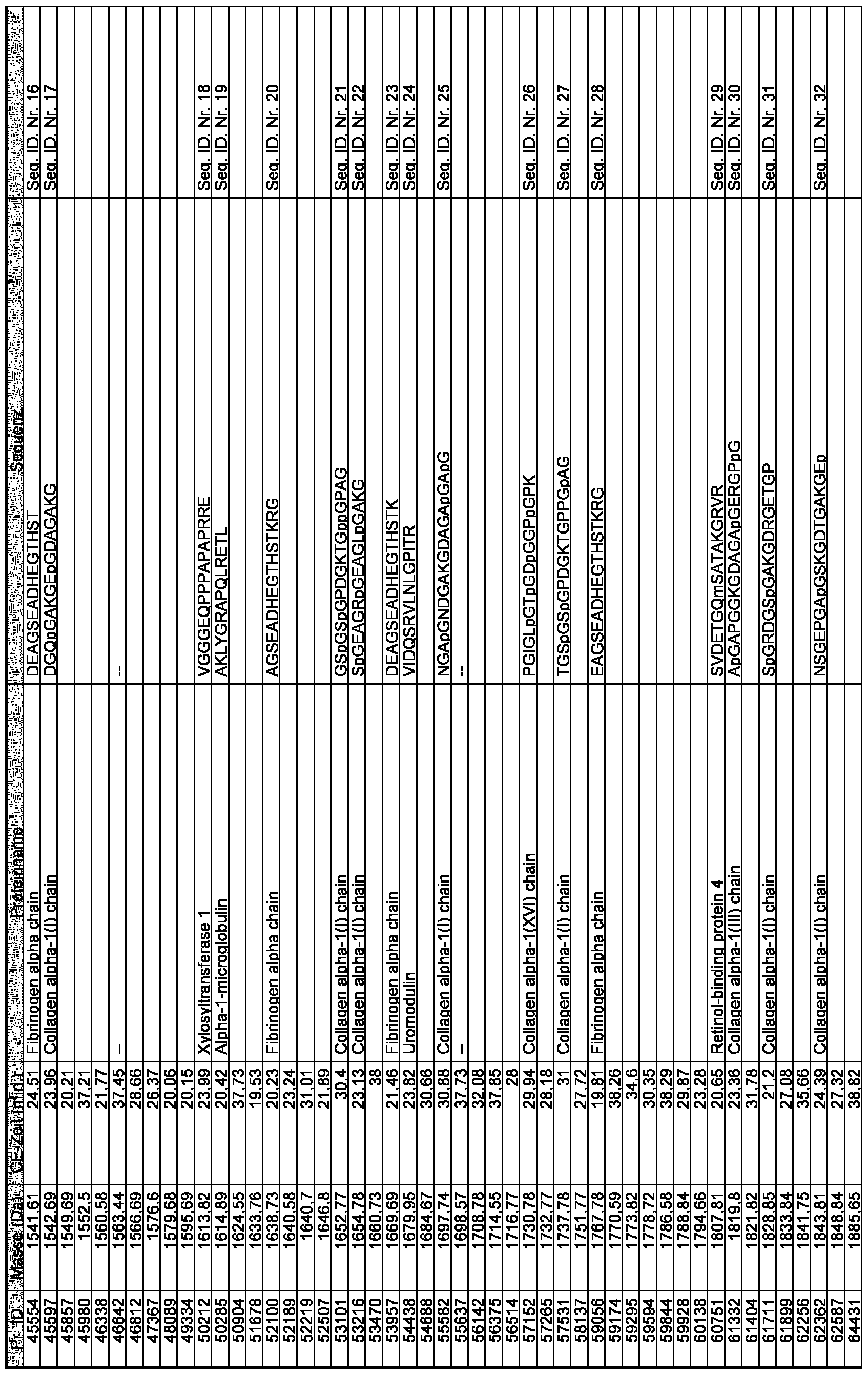


In einer Ausführungsform der Erfindung werden nur Marker verwendet, deren Sequenz in Tabelle 3 angegeben sind. In one embodiment of the invention only markers are used whose sequence is given in Table 3.
Spezifität ist definiert als die Nummer der tatsächlich negativen Proben geteilt durch die Summe der Anzahl der tatsächlich Negativen und der Anzahl der Falsch-Positiven. Eine Spezifität von 100% bedeutet, dass ein Test alle gesunden Personen als gesund erkennt, d.h. kein Gesunder wird als krank identifiziert. Dies trifft keine Aussage darüber, wie gut der Test kranke Patienten erkennt. Specificity is defined as the number of actual negative samples divided by the sum of the number of actual negatives and the number of false positives. A specificity of 100% means that a test identifies all healthy persons as healthy, i. no healthy person is identified as ill. This does not say anything about how well the test detects sick patients.
Sensitivität ist definiert als die Anzahl der tatsächlichen positiven Proben ge- teilt durch die Summe der Anzahl der tatsächlich Positiven und die Anzahl der Falsch-Negativen. Eine Sensitivität von 100% bedeutet, dass der Test alle Kranken erkennt. Er trifft keine Aussage, wie gut der Test gesunde Personen erkennt. Sensitivity is defined as the number of actual positive samples divided by the sum of the number of actual positives and the number of false negatives. A sensitivity of 100% means that the test detects all patients. He does not say how well the test detects healthy people.
Durch die erfindungsgemäßen Markern ist es möglich, IS oder TIA mit einer Spezifität von mindestens 60, bevorzugt mindestens 70, mehr bevorzugt 80, noch mehr bevorzugt mindestens 90 und am meisten bevorzugt mindestens 95% zu detektieren. By the markers according to the invention it is possible to detect IS or TIA with a specificity of at least 60, preferably at least 70, more preferably 80, even more preferably at least 90 and most preferably at least 95%.
Durch die erfindungsgemäßen Markern ist es möglich, IS oder TIA mit einer Sensitivität von mindestens 60, bevorzugt mindestens 70, mehr bevorzugt 80, noch mehr bevorzugt mindestens 90 und am meisten bevorzugt mindestens 95% zu detektieren. By the markers according to the invention it is possible to detect IS or TIA with a sensitivity of at least 60, preferably at least 70, more preferably 80, even more preferably at least 90 and most preferably at least 95%.
Die Migrationszeit (CE-Zeit) wird mittels Kapillarelektrophorese (capillary electrophoresis, CE) - wie z.B. im Punkt 2 ausgeführt - bestimmt. In diesem Beispiel wird eine 90 cm lange Glaskapillare mit einem inneren Durchmesser (ID) von 50 Mm und einem äußeren Durchmesser (OD) von 360 μm bei einer angelegten Spannung von 30 kV betrieben. Als Laufmittel wird zum Beispiel 30% Methanol, 0,5% Ameisensäure in Wasser oder 20% Acetonitril, 0,25% Ameisensäure verwendet. Alle %-Angaben in Vol.-%. Migration time (CE time) is determined by capillary electrophoresis (CE) - e.g. executed in point 2 - determined. In this example, a 90 cm long glass capillary with an inner diameter (ID) of 50 μm and an outer diameter (OD) of 360 μm is operated at an applied voltage of 30 kV. The eluent used is, for example, 30% methanol, 0.5% formic acid in water or 20% acetonitrile, 0.25% formic acid. All percentages in% by volume.
Es ist bekannt, das die CE-Migrationszeit variieren kann. Dennoch ist die Rei- henfolge, mit der die Polypeptidmarker eluieren, für jedes verwendete CE System unter den angegebenen Bedingungen typischerweise gleich. Um den-
noch auftretende Unterschiede in der Migrationszeit auszugleichen, kann das System unter Verwendung von Standards, für die die Migrationszeiten genau bekannt sind, normiert werden. Diese Standards können z.B. die in den Beispielen angegebenen Polypeptide sein (siehe Beispiel Punkt 3), oder bestimm- te, bekannte Peptide aus Urin, wie z.B. bei Jantos-Siwy et al. (Quantitative Urinary Proteome Analysis for Biomarker Evaluation in Chronic Kidney Disease. J. Proteome. Res. 8, 268-281 (2009)) beschrieben. It is known that the CE migration time can vary. However, the order in which the polypeptide labels elute is typically the same for any CE system used under the conditions indicated. To the- To compensate for any remaining differences in migration time, the system can be normalized using standards for which migration times are well known. These standards can be, for example, the polypeptides given in the examples (see Example 3), or certain known peptides from urine, as described, for example, in Jantos-Siwy et al. (Quantitative Urinary Proteome Analysis for Biomarker Evaluation in Chronic Kidney Disease, J. Proteome Res., 8: 268-281 (2009)).
Die Charakterisierung der Polypeptide, die in der Tabelle 1 gezeigt sind, wurde mittels Kapillarelektrophorese-Massenspektrometrie (CE-MS) bestimmt, einem Verfahren, das z.B. ausführlich von z.B. Neuhoff et al. Rapid Communications in mass spectrometry , 2004, Bd. 20, Seite 149-156), Mischak, H. et al. (Capil- lary electrophoresis-mass spectrometry as a powerful tool in biomarker dis- covery and clinical diagnosis: An update of recent developments. Mass Spec- trom. Rev. 28, 703-724 (2009)), Mischak, H. et al. (Comprehensive human urine Standards for comparability and standardization in clinical proteome analysis. Proteomics Clin Appl. 4, 464-478 (2010)), Good,D.M. et al. (Natu- rally occurring human urinary peptides for use in diagnosis of chronic kidney disease. Mol. Cell Proteomics 9, 2424-2437 (2010)) beschrieben wurde. Die Variation der Molekülmassen zwischen einzelnen Messungen oder zwischen verschiedenen Massenspektrometern ist bei exakter Kalibrierung relativ klein, typischerweise im Bereich von ± 0,05%, mehr bevorzugt ± 0,03%, noch mehr bevorzugt ± 0,01% oder 0,005%. The characterization of the polypeptides shown in Table 1 was determined by capillary electrophoresis mass spectrometry (CE-MS), a method which is described e.g. in detail from e.g. Neuhoff et al. Rapid Communications in mass spectrometry, 2004, Vol. 20, pages 149-156), Mischak, H. et al. (Cap- philary electrophoresis mass spectrometry as a powerful tool in biomarker dis- covery and clinical di- agnosis: An update of recent developments: Mass Spectrom Rev. 28, 703-724 (2009)), Mischak, H. et al , (Comprehensive human urine standards for comparability and standardization in clinical proteome analysis, Proteomics Clin Appl., 4, 464-478 (2010)), Good, D.M. et al. (Natu- rally occurring human urinary peptides for use in the diagnosis of chronic kidney disease, Mol. Cell Proteomics 9, 2424-2437 (2010)). The variation of molecular masses between individual measurements or between different mass spectrometers is relatively small with exact calibration, typically in the range of ± 0.05%, more preferably ± 0.03%, even more preferably ± 0.01% or 0.005%.
Die erfindungsgemäßen Polypeptidmarker sind Proteine oder Peptide oder Abbauprodukte von Proteinen oder Peptiden. Sie können chemisch modifiziert sein, z.B. durch posttranslationale Modifikationen wie Glykolisierung, Phosphorylierung, Alkylierung oder Disulfidverbrückung, oder durch andere Reaktionen, z.B. im Rahmen des Abbaus, verändert sein. Ausgehend von den Parametern, die die Polypeptidmarker bestimmen (Molekularmasse und Migrationszeit), ist es möglich, durch im Stand der Technik bekannte Verfah- ren die Sequenz der entsprechenden Polypeptide zu identifizieren. The polypeptide markers according to the invention are proteins or peptides or degradation products of proteins or peptides. They may be chemically modified, e.g. by post-translational modifications such as glycation, phosphorylation, alkylation or disulfide bridging, or by other reactions, e.g. in the context of mining, to be changed. Based on the parameters that determine the polypeptide markers (molecular mass and migration time), it is possible to identify the sequence of the corresponding polypeptides by methods known in the art.
Die erfindungsgemäßen Polypeptide werden verwendet, um IS oder TIA zu diagnostizieren. Unter Diagnose versteht man den Vorgang der Erkenntnisge-
winnung durch die Zuordnung von Symptomen oder Phänomenen zu einer Krankheit oder Verletzung. Die An- oder Abwesenheit eines Polypeptidmarkers kann durch jedes im Stand der Technik bekannte Verfahren gemessen werden. Verfahren, die verwendet werden können, sind weiter unten beispielhaft auf- geführt. The polypeptides of the invention are used to diagnose IS or TIA. Diagnosis is the process of the recognition Winning by assigning symptoms or phenomena to a disease or injury. The presence or absence of a polypeptide marker can be measured by any method known in the art. Methods that can be used are exemplified below.
Ein Polypeptidmarker ist anwesend, wenn sein Messwert mindestens so hoch ist wie der Schwellenwert. Liegt sein Messwert darunter, ist der Polypeptidmarker abwesend. Der Schwellenwert kann entweder durch die Sensitivität des Messverfahrens (Nachweisgrenze) bestimmt werden oder anhand von Erfahrungen definiert werden. A polypeptide marker is present when its reading is at least as high as the threshold. If its reading is below that, the polypeptide marker is absent. The threshold value can either be determined by the sensitivity of the measurement method (detection limit) or defined based on experience.
Im Zusammenhang mit der vorliegenden Erfindung wird der Schwellenwert vorzugsweise überschritten, wenn der Messwert der Probe für eine bestimmte Molekularmasse mindestens doppelt so hoch ist, wie der einer Leerprobe (z.B. nur Puffer oder Lösungsmittel). Der oder die Polypeptidmarker wird/werden in der Weise verwendet, dass seine/ihre An- oder Abwesenheit gemessen wird, wobei die An- oder Abwesenheit indikativ für IS oder TIA ist. So gibt es Polypeptidmarker, die typischerweise bei Individuen mit IS oder TIA vorhanden sind, jedoch bei Individuen ohne IS oder TIA seltener oder gar nicht auftreten. Weiterhin gibt es Polypeptidmarker, die bei Patienten mit IS oder TIA vorhan- den sind, jedoch bei Patienten ohne IS oder TIA nicht oder nur seltener vorhanden sind. Die Häufigkeit, mit der ein Marker in der Gruppe mit IS oder TIA bzw. in der Kontrollgruppe auftritt, ist in Tabelle 2 als Frequenz angegeben. In the context of the present invention, the threshold is preferably exceeded when the sample reading for a given molecular mass is at least twice that of a blank (e.g., only buffer or solvent). The polypeptide marker (s) is / are used to measure its presence or absence, the presence or absence being indicative of IS or TIA. Thus, there are polypeptide markers that are typically present in individuals with IS or TIA, but are less common or absent in individuals without IS or TIA. Furthermore, there are polypeptide markers which are present in patients with IS or TIA, but are absent or only rarely present in patients without IS or TIA. The frequency with which a marker appears in the group with IS or TIA or in the control group is given in Table 2 as frequency.
Zusätzlich oder auch alternativ zu den Frequenzen (Bestimmung der An- oder Abwesenheit) können auch die Amplituden zur Diagnose verwendet werden. Die Amplituden werden in der Weise verwendet, dass nicht die An- oder Abwesenheit entscheidend ist, sondern die Höhe des Signals (die Amplitude) bei Anwesenheit des Signals in beiden Gruppen entscheidet. Dabei ist ein Nomi- nierungsverfahren sinnvoll, um eine Vergleichbarkeit zwischen unterschiedlich konzentrierten Proben oder unterschiedlichen Messmethoden zu erreichen. Hierzu werden bevorzugt Kollagen-Fragmente eingesetzt, wie bei Jantos Siwy et al. (Quantitative Urinary Proteome Analysis for Biomarker Evaluation in Chronic Kidney Disease. J. Proteome. Res. 8, 268-281 (2009)) detailliert be-
schrieben. Es wird die lineare Korrelation zwischen den Referenz werten für die Amplitude der vorgegebenen bekannten Peptide (sogenannte "housekeeping Peptides") und den experimentell ermittelten Werten bestimmt. Die Steigerung der Regressionsgeraden entspricht gerade der relativen Konzentration und wird als Normierungsfaktor zur Kalibration aller Peptidsignale dieser Probe mit einem gemeinsamen Normierungsfaktor eingesetzt. In addition or alternatively to the frequencies (determination of the presence or absence), the amplitudes can also be used for diagnosis. The amplitudes are used in such a way that it is not the presence or absence that is decisive, but the height of the signal (the amplitude) when the signal is present in both groups. A nomination procedure makes sense in order to achieve comparability between differently concentrated samples or different measurement methods. For this purpose, collagen fragments are preferably used, as in Jantos Siwy et al. (Quantitative Urinary Proteome Analysis for Biomarker Evaluation in Chronic Kidney Disease, J. Proteome Res., 8: 268-281 (2009)). wrote. It is determined the linear correlation between the reference values for the amplitude of the given known peptides (so-called "housekeeping peptides") and the experimentally determined values. The increase in the regression line just corresponds to the relative concentration and is used as a normalization factor for the calibration of all peptide signals of this sample with a common normalization factor.
Die Entscheidung zu einer Diagnose fällt dabei je nachdem, wie hoch die Amplitude der jeweiligen Polypeptidmarker in der Patientenprobe im Vergleich zu den mittleren Amplituden in der Kontrollgruppe bzw. der "Krank"-Gruppe ist. Liegt der Wert nahe an der mittleren Amplitude der "Krank"-Gruppe, ist von dem Vorliegen eines Schlaganfalls auszugehen, entspricht sie eher den mittleren Amplituden der Kontroll-Gruppe, ist nicht von einem Schlaganfall auszugehen. Der Abstand zur mittleren Amplitude kann als eine Wahrscheinlichkeit für die Zugehörigkeit zu einer Gruppe interpretiert werden. Alternativ kann der Abstand zwischen dem Messwert und der mittleren Amplitude als eine Wahrscheinlichkeit für die Zugehörigkeit zu einer Gruppe betrachtet werden. The decision to make a diagnosis depends on how high the amplitude of the respective polypeptide markers in the patient sample is compared to the mean amplitudes in the control group or the "sick" group. If the value is close to the mean amplitude of the "sick" group, it is to be assumed that the occurrence of a stroke, it corresponds more to the mean amplitudes of the control group, is not to assume a stroke. The distance to the mean amplitude can be interpreted as a probability of belonging to a group. Alternatively, the distance between the measured value and the mean amplitude may be considered as a probability of belonging to a group.
Bevorzugt wird zur Auswertung sowohl die Frequenz als auch die Amplitude herangezogen. Jeweils lassen sich aus Messdaten einer unbekannten Probe Wahrscheinlichkeiten für die Zuordnung zu den Gruppen "IS oder TIA" oder 'Gesund' ableiten, aus den sich dann eine Gesamtwahrscheinlichkeit ergibt. Preferably, both the frequency and the amplitude are used for the evaluation. In each case, it is possible to derive probabilities for the assignment to the groups "IS or TIA" or "healthy" from measurement data of an unknown sample, from which then a total probability results.
Der p-value ist ein Maß für die Wahrscheinlichkeit, dass die Zuordnung der Marker zu den beiden Gruppen (IS oder TIA und Kontrolle) auf einer zufälligen Verteilung beruht, die nicht mit IS oder TIA in Verbindung steht. Je kleiner der Wilcox-p-value, desto wahrscheinlicher ist die Korrelation mit IS oder TIA. Das Individuum, von dem die Probe stammt, in der die An- oder Abwesenheit oder Amplitude eines oder mehrerer Polypeptidmarker bestimmt wird, kann jedes Individuum sein, das an IS oder TIA leiden kann. Vorzugsweise handelt es sich bei dem Individuum um ein Säugetier, am meisten bevorzugt handelt es sich um einen Menschen. The p-value is a measure of the likelihood that the association of the markers with the two groups (IS or TIA and control) is based on a random distribution that is not related to IS or TIA. The smaller the Wilcox p-value, the more likely it is to correlate with IS or TIA. The individual from whom the sample is derived, in which the presence or absence or amplitude of one or more polypeptide markers is determined, may be any individual who may be suffering from IS or TIA. Preferably, the subject is a mammal, most preferably a human.
In einer bevorzugten Ausführungsform der Erfindung werden nicht nur drei Polypeptidmarker, sondern eine größere Kombination von Markern verwendet. Durch Vergleich einer Mehrzahl von Polypeptidmarkern kann die Verfälschung
des Gesamtergebnisses durch einzelne individuelle Abweichungen von der typischen Anwesenheitswahrscheinlichkeit im einzelnen Individuum reduziert oder vermieden werden. In a preferred embodiment of the invention not only three polypeptide markers but a larger combination of markers are used. By comparison of a plurality of polypeptide markers, the adulteration of the overall result can be reduced or avoided by individual individual deviations from the typical probability of presence in the individual.
Bei der Probe, in der die An- oder Abwesenheit des oder der erfindungsgemä- ßen Polypeptidmarker gemessen werden, kann es sich um jede Probe handeln, die aus dem Körper des Individuums gewonnen wird. Bei der Probe handelt es sich um eine Probe, die über eine Polypeptidzusammensetzung verfügt, die geeignet ist, Aussagen über den Zustand des Individuums zu treffen. Beispielsweise kann es sich um Blut, Urin, eine Gelenkflüssigkeit, eine Gewebe- flüssigkeit, ein Körpersekret, Schweiß, Liquor, Lymphe, Darm-, Magen-, Pank- reassaft, Galle, Tränenflüssigkeit, eine Gewebeprobe, Sperma, Vaginalflüssigkeit oder eine Stuhlprobe handeln. Vorzugsweise handelt es sich um eine Flüssigprobe. In einer bevorzugten Ausführungsform handelt es sich bei der Probe um eine Urinprobe. The sample measuring the presence or absence of the polypeptide marker (s) of the invention may be any sample recovered from the subject's body. The sample is a sample having a polypeptide composition suitable for making statements about the condition of the individual. For example, it may be blood, urine, synovial fluid, tissue fluid, body secretions, sweat, cerebrospinal fluid, lymph, intestinal, gastric, pancreatic, bile, tear fluid, tissue sample, sperm, vaginal fluid or stool sample , Preferably, it is a liquid sample. In a preferred embodiment, the sample is a urine sample.
Urinproben können wie im Stand der Technik bekannt genommen werden. Vorzugsweise wird im Zusammenhang mit der vorliegenden Erfindung eine Mittelstrahlurinprobe verwendet. Die Urinprobe kann z.B. mittels eines Katheters oder auch mit Hilfe eines Urinierungsapparates, wie in WO 01/74275 beschrieben, entnommen werden. Urine samples may be known as known in the art. Preferably, in the context of the present invention, a mid-jet urine sample is used. The urine sample may e.g. by means of a catheter or also with the aid of a urination apparatus, as described in WO 01/74275.
Die An- oder Abwesenheit oder Amplitude eines Polypeptidmarkers in der Probe kann durch jedes im Stand der Technik bekannte Verfahren, das zur Messung von Polypeptidmarkern geeignet ist, bestimmt werden. Dem Fachmann sind solche Verfahren bekannt. Grundsätzlich kann die An- oder Abwesenheit eines Polypeptidmarkers durch direkte Verfahren, wie z.B. Massen- spektrometrie, oder indirekte Verfahren, wie z.B. mittels Liganden oder spezifischer Sonden wie Antikörper, bestimmt werden. The presence or absence or amplitude of a polypeptide marker in the sample can be determined by any method known in the art suitable for measuring polypeptide markers. Those skilled in such methods are known. In principle, the presence or absence of a polypeptide marker can be determined by direct methods such as e.g. Mass spectrometry, or indirect methods, such as by ligands or specific probes such as antibodies.
Falls erforderlich oder wünschenswert kann die Probe des Individuums, z.B. die Urinprobe, vor der Messung der An- oder Abwesenheit des oder der Polypeptidmarker durch jedes geeignete Mittel vorbehandelt und z.B. aufgereinigt oder aufgetrennt werden. Die Behandlung kann z.B. eine Aufreinigung, Trennung, Verdünnung oder Konzentrierung umfassen. Die Verfahren können beispielsweise eine Zentrifugation, Filtration, Ultrafiltration, Dialyse, eine Fällung
oder chromatographische Verfahren wie Affinitätstrennung oder Trennung mittels Ionenaustauscherchromatographie, oder eine elektrophoretische Trennung sein. Besondere Beispiele hierfür sind Gelelektrophorese, zweidimensionale Polyacrylamidgelelektrophorese (2D-PAGE), Kapillarelektrophorese, Me- tallaffinitätschromatographie, immobilisierte Metallaffinitätschromatographie (IMAC), Affinitätschromatographie auf der Basis von Lektinen, Flüssigchromatographie, Hochleistungsflüssigchromato-graphie (HPLC), Normal- und Um- kehrphasen-HPLC, Kationenaustauscherchromatographie und selektive Bindung an Oberflächen. Alle diese Verfahren sind dem Fachmann gut bekannt und der Fachmann wird das Verfahren in Abhängigkeit von der verwendeten Probe und dem Verfahren zur Bestimmung der An- oder Abwesenheit des oder der Polypeptidmarker auswählen können. If necessary or desirable, the sample of the subject, eg, the urine sample, may be pretreated and, for example, purified or separated, prior to measuring the presence or absence of the polypeptide marker (s) by any suitable means. The treatment may include, for example, purification, separation, dilution or concentration. The methods may include, for example, centrifugation, filtration, ultrafiltration, dialysis, precipitation or chromatographic methods such as affinity separation or separation by ion exchange chromatography, or electrophoretic separation. Specific examples thereof are gel electrophoresis, two-dimensional polyacrylamide gel electrophoresis (2D-PAGE), capillary electrophoresis, metal affinity chromatography, immobilized metal affinity chromatography (IMAC), affinity chromatography on the basis of lectins, liquid chromatography, high performance liquid chromatography (HPLC), normal and reverse phase HPLC , Cation exchange chromatography and selective bonding to surfaces. All of these methods are well known to those skilled in the art and one skilled in the art will be able to select the method depending on the sample used and the method for determining the presence or absence of the polypeptide marker (s).
Vorzugsweise wird ein massenspektrometrisches Verfahren verwendet, um die An- oder Abwesenheit oder Amplitude eines Polypeptidmarkers zu bestimmen, wobei diesem Verfahren eine Aufreinigung oder Auftrennung der Probe vorgeschaltet werden kann. Die massenspektrometrische Analyse besitzt gegenüber den derzeit gängigen Verfahren den Vorteil, dass die Konzentration vieler (> 100) Polypeptide einer Probe mittels einer einzigen Analyse bestimmt werden kann. Jeder Typ eines Massenspektrometers kann verwendet werden. Mit der Massenspektrometrie ist es möglich, routinemäßig 10 fmol eines Polypeptidmarkers, also 0.1 ng eines 10 kDa Proteins mit einer Messgenauigkeit von ca. ±0.01% aus einem komplexen Gemisch zu vermessen. Bei Massenspekt- rometern ist eine Ionen-bildende Einheit mit einem geeigneten Analysegerät gekoppelt. Zum Beispiel werden meistens Elektrospray-Ionisations (ESI) Inter- faces verwendet, um Ionen aus Flüssigproben zu vermessen, wohingegen die Matrix-assisted-laser-desorption/ionisation (MALDI) Technik verwendet wird, um Ionen aus mit einer Matrix kristallisierten Probe zu vermessen. Zur Analyse der entstandenen Ionen können z.B. Quadrupole, Ionenfallen oder Time-of- flight (TOF) Analysatoren verwendet werden. Preferably, a mass spectrometric method is used to determine the presence or absence or amplitude of a polypeptide marker, which method may precede purification or separation of the sample. The mass spectrometric analysis has the advantage over current methods that the concentration of many (> 100) polypeptides of a sample can be determined by a single analysis. Any type of mass spectrometer can be used. With mass spectrometry, it is possible to routinely measure 10 fmoles of a polypeptide marker, that is, 0.1 ng of a 10 kDa protein with a measurement accuracy of approximately ± 0.01% from a complex mixture. In mass spectrometers, an ion-forming unit is coupled to a suitable analyzer. For example, electrospray ionization (ESI) interfaces are commonly used to measure ions from liquid samples, whereas the matrix assisted laser desorption / ionization (MALDI) technique is used to measure ions from sample crystallized with a matrix , For analysis of the resulting ions, e.g. Quadrupoles, ion traps or time-of-flight (TOF) analyzers are used.
Bei der Elektrosprayionisation (ESI) werden die in Lösung vorliegenden Moleküle u.a. unter dem Einfluss von Hochspannung (z.B. 1-8 kV) versprüht, wobei sich geladene Tröpfchen bilden, die durch Verdampfen des Lösungsmittels
kleiner werden. Schließlich kommt es durch sog. Coulomb-Explosionen zur Bildung freier Ionen, die dann analysiert und detektiert werden können. In electrospray ionization (ESI), the molecules present in solution are sprayed, inter alia, under the influence of high voltage (eg 1-8 kV), resulting in charged droplets formed by evaporation of the solvent get smaller. Finally, so-called Coulomb explosions lead to the formation of free ions, which can then be analyzed and detected.
Bei der Analyse der Ionen mittels TOF wird eine bestimmte Beschleunigungsspannung angelegt, die den Ionen eine gleich große kinetische Energie ver- leiht. Dann wird sehr genau die Zeit gemessen, die die jeweiligen Ionen benötigen, um eine Driftstrecke durch das Flugrohr zurückzulegen. Da bei gleicher kinetische Energie die Geschwindigkeit der Ionen von Ihrer Masse abhängt, kann diese somit bestimmt werden. TOF-Analysatoren haben eine sehr hohe Scan-Geschwindigkeit und erreichen eine sehr hohe Auflösung. In the analysis of the ions by means of TOF, a specific acceleration voltage is applied, which gives the ions an equal kinetic energy. Then, the time required for the respective ions to travel a drift path through the flight tube is measured very accurately. Since with the same kinetic energy, the speed of the ions depends on your mass, this can thus be determined. TOF analyzers have a very high scanning speed and achieve a very high resolution.
Bevorzugte Verfahren zur Bestimmung der An- oder Abwesenheit von Polypeptidmarkern schließen Gasphasenionenspektrometrie, wie Laserdesorp- tions/Ionisations-Massenspektrometrie, MALDI-TOF-MS, SELDI-TOF-MS (Surface enhanced laser desorption Ionisation), LC-MS (Liquid chromatography- mass spectrometry), 2D-PAGE-MS und Kapillarelektrophore- se-Massenspektrometrie (CE-MS) ein. Alle genannten Verfahren sind dem Fachmann bekannt. Preferred methods for determining the presence or absence of polypeptide markers include gas phase ion spectrometry, such as laser desorption / ionization mass spectrometry, MALDI-TOF-MS, SELDI-TOF-MS (surface enhanced laser desorption ionization), LC-MS (liquid chromatography mass spectrometry), 2D-PAGE-MS and capillary electrophoresis mass spectrometry (CE-MS). All of the methods mentioned are known to the person skilled in the art.
Ein besonders bevorzugtes Verfahren ist CE-MS, in welchem die Kapillarelektrophorese mit Massenspektrometrie gekoppelt wird. Dieses Verfahren ist ausführlich z.B. in der deutschen Patentanmeldung DE 10021737, bei Kaiser et al. (J. Chromatogr. A, 2003, Bd. 1013 : 157-171, sowie Electrophoresis, 2004, 25 :2044-2055), bei Wittke et al. (J. Chromatogr. A, 2003, 1013 : 173-181) und in Ref. 2 beschrieben. Die CE-MS Technik erlaubt, das Vorhandensein einiger Hunderter Polypeptidmarker einer Probe gleichzeitig in kurzer Zeit, einem geringen Volumen und hoher Sensitivität zu bestimmen. Nachdem eine Probe vermessen wurde, wird ein Muster der gemessenen Polypeptidmarker hergestellt (siehe unten). Dieses kann mit Referenzmustern von kranken bzw. gesunden Individuen verglichen werden. In den meisten Fällen ist es ausreichend, eine begrenzte Anzahl von Polypeptidmarkern für die Diagnostik von Erkrankungen zu verwenden. Weiter bevorzugt ist ein CE-MS Verfahren, das CE online an ein ESI-TOF-MS gekoppelt, einschließt. A particularly preferred method is CE-MS, in which capillary electrophoresis is coupled with mass spectrometry. This method is described in detail e.g. in German patent application DE 10021737, in Kaiser et al. (J. Chromatogr. A, 2003, Vol. 1013: 157-171, and Electrophoresis, 2004, 25: 2044-2055), in Wittke et al. (J. Chromatogr. A, 2003, 1013: 173-181) and Ref. The CE-MS technique allows to determine the presence of several hundreds of polypeptide markers of a sample simultaneously in a short time, a small volume and high sensitivity. After a sample has been measured, a pattern of the measured polypeptide markers is prepared (see below). This can be compared with reference patterns of ill or healthy individuals. In most cases it is sufficient to use a limited number of polypeptide markers for the diagnosis of diseases. More preferred is a CE-MS method which includes CE coupled online to an ESI-TOF-MS.
Für CE-MS ist die Verwendung von flüchtigen Lösungsmitteln bevorzugt, außerdem arbeitet man am besten unter im Wesentlichen salzfreien Bedin-
gungen. Beispiele geeigneter Lösungsmittel umfassen Acetonitril, Methanol und ähnliche. Die Lösungsmittel können mit Wasser verdünnt und mit einer Säure (z.B. 0,1% bis 1% Ameisensäure) versetzt sein, um den Analyten, vorzugsweise die Polypeptide, zu protonieren. For CE-MS, the use of volatile solvents is preferred, and it is best to work under substantially salt-free conditions. conditions. Examples of suitable solvents include acetonitrile, methanol and the like. The solvents may be diluted with water and an acid (eg 0.1% to 1% formic acid) added to protonate the analyte, preferably the polypeptides.
Mit der Kapillarelektrophorese ist es möglich, Moleküle nach ihrer Ladung und Größe zu trennen. Neutrale Teilchen wandern beim Anlegen eines Stromes mit der Geschwindigkeit des elektroosmotischen Flusses, Kationen werden zur Kathode beschleunigt und Anionen verzögert. Der Vorteil von Kapillaren in der Elektrophorese besteht im günstigen Verhältnis von Oberfläche zu Volumen, was einen guten Abtransport der beim Stromfluss entstehenden Jouleschen Wärme ermöglicht. Dies wiederum erlaubt das Anlegen hoher Spannungen (üblicherweise bis 30 kV) und damit eine hohe Trennleistung und kurze Analysezeiten. Capillary electrophoresis makes it possible to separate molecules according to their charge and size. Neutral particles migrate at the rate of electroosmotic flow upon application of a current, cations are accelerated to the cathode and anions are retarded. The advantage of capillaries in electrophoresis is the favorable ratio of surface area to volume, which enables a good removal of the Joule heat arising during the current flow. This in turn allows the application of high voltages (usually up to 30 kV) and thus a high separation efficiency and short analysis times.
Bei der Kapillarelektrophorese werden normalerweise Quarzglaskapillaren mit Innendurchmessern von typischerweise 50 bis 75 μm eingesetzt. Die verwendeten Längen betragen 30-100 cm. Darüber hinaus bestehen die Kapillaren in der Regel aus kunststoffumhüllten Quarzglas. Die Kapillaren können sowohl unbehandelt sei, d.h. auf der Innenseite ihre hydrophilen Gruppen zeigen, als auch auf der Innenseite beschichtet sein. Eine hydrophobe Beschichtung kann verwendet werden, um die Auflösung zu verbessern. Zusätzlich zur Spannung kann auch ein Druck angelegt werden, der typischerweise im Bereich von 0-1 psi liegt. Der Druck kann dabei auch erst während der Trennung angelegt oder währenddessen verändert werden. In capillary electrophoresis, quartz glass capillaries with internal diameters of typically 50 to 75 μm are normally used. The used lengths are 30-100 cm. In addition, the capillaries usually consist of plastic-coated quartz glass. The capillaries may be both untreated, i. on the inside show their hydrophilic groups, as well as be coated on the inside. A hydrophobic coating can be used to improve the resolution. In addition to the voltage, a pressure which is typically in the range of 0-1 psi may also be applied. The pressure can also be created during the separation or changed during the process.
In einem bevorzugten Verfahren zur Messung von Polypeptidmarkern werden die Marker der Probe mittels Kapillarelektrophorese getrennt, anschließend direkt ionisiert und online in ein daran gekoppeltes Massenspektrometer zur Detektion überführt. In dem erfindungsgemäßen Verfahren können in vorteilhafter Weise mehrere Polypeptidmarker zur Diagnostik verwendet werden. Bevorzugt ist die Verwendung von mindestens 5, 6, 8, oder 10 Markern. In einer Ausführungsform werden 20 bis 50 Marker verwendet. In a preferred method of measuring polypeptide markers, the markers of the sample are separated by capillary electrophoresis, then directly ionized and transferred online to a mass spectrometer coupled thereto for detection. In the method according to the invention, several polypeptide markers can advantageously be used for diagnostics. Preferred is the use of at least 5, 6, 8, or 10 markers. In one embodiment, 20 to 50 markers are used.
Um die Wahrscheinlichkeit für das Vorliegen einer Erkrankung bei Verwendung mehrerer Marker zu bestimmen, können dem Fachmann bekannte Verfahren
verwendet werden. Beispielsweise kann das von Weissinger et al. Kidney Int., 2004, 65 :2426-2434) beschriebene Random-Forests-Verfahren unter Verwendung eines Computerprogramms wie z.B. S-Plus oder die in der selben Veröffentlichung beschriebenen support-vector-machines verwendet werden. Weite- re Informationen können beispielsweise Dakna et al. BMC Bioinformatics 11 (2010) 594 entnommen werden. In order to determine the probability of the presence of a disease when using multiple markers, methods known to the person skilled in the art can be used be used. For example, the Weissinger et al. Kidney Int., 2004, 65: 2426-2434) using a computer program such as S-Plus or the support vector machines described in the same publication. Further information can be found, for example, Dakna et al. BMC Bioinformatics 11 (2010) 594.
Diese Verfahren kombinieren mehrere Biomarker zu einer einzigen Variable, die sowohl mit der Wahrscheinlichkeit für das Vorliegen der Erkrankung assoziiert ist, als auch mit dem Schweregrad der Erkrankung. Diese Variable kann somit auch eingesetzt werden um die Ausprägung bzw. den Schweregrad der Erkrankung zu bestimmen (z.B. gezeigt in Schiffer et al., Prediction of muscle- invasive bladder Cancer using urinary proteomics. Clin. Cancer Res. 15, 4935- 4943 (2009) für Blasenkarzinom), als auch um ein mögliches Ansprechen auf Therapie abzubilden (wie z.B. gezeigt in Haubitz et al., Identification and vali- dation of urinary biomarkers for differential diagnosis and evaluation of therapeutic Intervention in ANCA associated vasculitis. Mol. Cell. Proteomics 8, 2296-2307 (2009) für ANCA-assoziierte Vasculitius, oder in Andersen et al., Urinary proteome analysis enables assessment of renoprotective treatment in type 2 diabetic patients with microalbuminuria. BMC Nephro/. 11, 29 (2010) für diabetische Nephropathie). These methods combine multiple biomarkers into a single variable associated with both the likelihood of disease and the severity of the disease. This variable can thus also be used to determine the severity or severity of the disease (eg shown in Schiffer et al., Prediction of muscle invasive bladder cancer using urinary proteomics., Clin. Cancer Res. 15, 4935-4943 (2009 for bladder carcinoma) as well as to reflect a possible response to therapy (as shown, for example, in Haubitz et al., Identification and Validation of Urinary Biomarkers for Differential Diagnosis and Evaluation of Therapeutic Intervention in ANCA Associated Vasculitis, Mol. Cell. Proteomics 8, 2296-2307 (2009) for ANCA-associated vasculitius, or Andersen et al., Urinary proteome analysis, assessing renoprotective treatment in type 2 diabetic patients with microalbuminuria, BMC Nephro, 11, 29 (2010) for diabetic nephropathy ).
Auch die erfindungsgemäß identifizierten Biomarker können herangezogen werden, um die Schwere des Schlaganfalls zu bestimmen. Die Biomarker zeigen Korrelation mit der National Institute of Health Stroke Scale (NIHSS). Die Kombination der normierten Amplituden der einzelnen Biomarker kann somit herangezogen werden, um den Schweregrad des Schlaganfalls zu bestimmen, und so wertvolle Hinweise zur Therapie zu geben. The biomarkers identified according to the invention can also be used to determine the severity of the stroke. The biomarkers show correlation with the National Institute of Health Stroke Scale (NIHSS). The combination of the normalized amplitudes of the individual biomarkers can thus be used to determine the severity of the stroke, thus giving valuable information on the therapy.
Beispiel 1: Example 1:
1. Probenvorbereitung : 1. Sample preparation:
Zur Detektion der Polypeptidmarker zur Diagnostik wurde Urin verwendet. Urin wurde von gesunden Spendern (Vergleichsgruppe) sowie Patienten, die einen Schlaganfall erlitten haben, abgenommen. Für die nachfolgende CE-MS Messung mussten die auch in Urin von Patienten in höherer Konzentration
vorkommenden Proteine wie Albumin und Immunoglobuline durch Ultrafiltration abgetrennt werden. Dazu wurden 700 μΙ Urin entnommen und mit 700 μΙ Filtrationspuffer (2M Harnstoff, lOmM Ammoniak, 0.02% SDS) versetzt. Diese 1.4 ml Probenvolumen wurden ultrafiltriert (20 kDa, Sartorius, Göttingen, DE). Die UF wurde bei 3000 U/min in einer Zentrifuge durchgeführt bis 1.1 ml Ult- rafiltrat erhalten wurden. Urine was used to detect polypeptide markers for diagnosis. Urine was collected from healthy donors (peer group) and from patients who had a stroke. For the subsequent CE-MS measurement, those also had to be in urine of patients in higher concentration occurring proteins such as albumin and immunoglobulins are separated by ultrafiltration. For this purpose, 700 .mu.l of urine were removed and treated with 700 .mu.l Filtrationspuffer (2M urea, lOmM ammonia, 0.02% SDS). These 1.4 ml sample volumes were ultrafiltered (20 kDa, Sartorius, Göttingen, DE). The UF was carried out at 3000 rpm in a centrifuge until 1.1 ml ultrafiltrate was obtained.
Die erhaltenen 1.1 ml Filtrat wurden dann auf eine PD 10 Säule aufgetragen (Amersham Bioscience, Uppsala, Schweden) und mit 2.5 ml einer 0.01% NH4OH eluiert und lyophillisert. Zur CE-MS Messung wurden die Polypeptide dann mit 20 μΙ Wasser (HPLC-Reinheit, Merck) resuspendiert. The resulting 1.1 ml filtrate was then applied to a PD 10 column (Amersham Bioscience, Uppsala, Sweden) and eluted with 2.5 ml of 0.01% NH 4 OH and lyophillized. For CE-MS measurement, the polypeptides were then resuspended with 20 μΙ water (HPLC grade, Merck).
2. CE-MS Messung : 2. CE-MS measurement:
Die CE-MS Messungen wurden mit einem Kapillarelektrophoresesystem von Beckman Coulter (P/ACE MDQ System; Beckman Coulter Inc, Fullerton, USA) und einem ESI-TOF Massenspektrometer von Bruker (micro-TOF MS, Bruker Daltonik, Bremen, D) durchgeführt. Die CE Kapillaren wurden von New Objective bezogen, sie hatten einen ID/OD von 50/360 μιτι und eine Länge von 90 cm. Die mobile Phase für die CE Trennung bestand aus 20% Acetonitril und 0.25% Ameisensäure in Wasser. Für den "Sheath-Flow" am MS wurde 30% Isopropanol mit 0.5% Ameisensäure verwendet, hier mit einer Flussrate von 20 μΙ/h. Die Kopplung von CE und MS wurde durch ein CE-ESI-MS Sprayer Kit (Agilent Technologies, Waldbronn, DE) realisiert. Um die Probe zu injizieren, wurde 1 bis max. 6 psi Druck angelegt, die Dauer der Injektion betrug 99 Sekunden. Mit diesen Parametern wurden ca. 300 nl der Probe in die Kapillare injiziert, dieses entspricht ca. 10% des Kapillarvolumens. Um die Probe in der Kapillare aufzukonzentrieren wurde eine "Stacking"-Technik verwendet. Dabei wird vor der Probeninjektion für 7 Sek. (bei 1 psi) eine IM NH3 Lösung injiziert. Nach Anlegen der Trennspannung (25 kV) werden die Analyten zwischen diesen Lösungen automatisch aufkonzentriert. Die folgende CE-Trennung wurde mit einer Druckmethode durchgeführt: 30 Minuten mit 0 psi, dann für 1 min 0.1 psi, für 1 min 0.2 psi, für 1 min 0.3 psi, für 1 min 0.4 psi, abschließend 35 min bei 0.5 psi. Die Gesamtdauer eines Trennlaufes betrug damit 70 Minuten.
Um auf der Seite des MS eine möglichst gute Signalintensität zu erhalten, wurde kein "Nebulizer Gas" eingesetzt. Die an der Spraynadel angelegte Spannung zur Erzeugung des Elektrosprays betrug 4000 - 4800 V. Die übrigen Einstellungen am Massenspektrometer wurden gemäß Anweisung des Herstel- lers für Peptiddetektion optimiert. Die Spektren wurden über einen Massenbereich von m/z 400 bis m/z 3000 aufgenommen und alle 3 Sek. akkumuliert.The CE-MS measurements were performed with a capillary electrophoresis system from Beckman Coulter (P / ACE MDQ System, Beckman Coulter Inc, Fullerton, USA) and a Bruker ESI-TOF mass spectrometer (micro-TOF MS, Bruker Daltonik, Bremen, D). The CE capillaries were purchased from New Objective, they had an ID / OD of 50/360 μιτι and a length of 90 cm. The mobile phase for the CE separation consisted of 20% acetonitrile and 0.25% formic acid in water. 30% isopropanol with 0.5% formic acid was used for the "sheath flow" at the MS, here with a flow rate of 20 μΙ / h. The coupling of CE and MS was realized by a CE-ESI-MS sprayer kit (Agilent Technologies, Waldbronn, DE). To inject the sample, 1 to max. 6 psi pressure applied, the duration of the injection was 99 seconds. With these parameters about 300 nl of the sample were injected into the capillary, this corresponds to about 10% of the capillary volume. To concentrate the sample in the capillary, a "stacking" technique was used. An IM NH 3 solution is injected for 7 seconds (at 1 psi) prior to sample injection. After applying the separation voltage (25 kV), the analytes are automatically concentrated between these solutions. The following CE separation was performed with a pressure method: 0 psi for 30 minutes, 0.1 psi for 1 min, 0.2 psi for 1 min, 0.3 psi for 1 min, 0.4 psi for 1 min, and 0.5 psi for 35 min. The total duration of a separation run was thus 70 minutes. In order to get the best possible signal intensity on the side of the MS, no "Nebulizer gas" was used. The voltage applied to the spray needle to generate the electrospray was 4000-4800 V. The remaining settings on the mass spectrometer were optimized as instructed by the manufacturer of peptide detection. The spectra were recorded over a mass range of m / z 400 to m / z 3000 and accumulated every 3 seconds.
3. Standards für die CE-Messung 3. Standards for CE measurement
Zur Kontrolle und Kalibrierung der CE-Messung wurden die folgenden Proteine bzw. Polypeptide eingesetzt, welche unter den gewählten Bedingungen durch die unten aufgeführten CE-Migrationszeiten charakterisiert sind : To control and calibrate the CE measurement, the following proteins or polypeptides were used, which are characterized under the selected conditions by the CE migration times listed below:
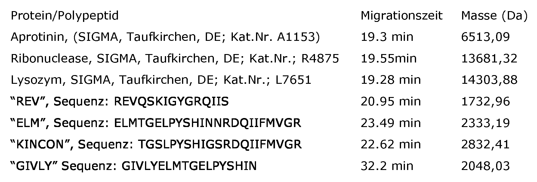
Die Proteine/Polypeptide werden jeweils in einer Konzentration von 10 pmol/μΙ in Wasser eingesetzt. "REV", "ELM", "KINCON" und "GIVLY" stellen synthetische Peptide dar. The proteins / polypeptides are each used in a concentration of 10 pmol / μΙ in water. "REV", "ELM", "KINCON" and "GIVLY" represent synthetic peptides.
Es ist dem Fachmann prinzipiell bekannt, dass bei kapillarelektrophoretischen Trennungen geringe Schwankungen der Migrationszeiten auftreten können. Unter den beschriebenen Bedingungen ändert sich jedoch die Migrationsreihenfolge nicht. Es ist für den Fachmann in Kenntnis der angegebenen Massen und CE-Zeiten problemlos möglich, eigene Messungen den erfindungsgemäßen Polypeptidmarkern zuzuordnen. Hierzu kann er beispielsweise wie folgt vorgehen : zunächst wählt er eines der in seiner Messung gefundenen Polypeptide (Peptid 1) aus und versucht, innerhalb eines Zeitfensters der angegebenen CE-Zeit (beispielsweise + 5 min) eine oder mehrere übereinstimmende Massen zu finden. Findet er innerhalb dieses Intervalls nur eine übereinstimmende Masse, ist die Zuordnung fertig gestellt. Findet er mehrere passende Massen,
muss noch eine Entscheidung über die Zuordnung gefällt werden. Hierzu wird ein weiteres Peptid (Peptid 2) aus der Messung ausgewählt und versucht, hierfür einen passenden Polypeptidmarker zu identifizieren, wobei wieder ein entsprechendes Zeitfenster berücksichtigt wird. Lassen sich nun wiederum mit einer entsprechenden Masse mehrere Marker finden, ist die wahrscheinlichste Zuordnung die, bei der zwischen der Verschiebung für das Peptide 1 und für das Peptid 2 ein im Wesentlichen linearer Zusammenhang besteht. In Abhängigkeit von der Komplexität des Zuordnungsproblems bietet es sich für den Fachmann an, gegebenenfalls weitere Proteine aus seiner Probe für die Zuord- nung zu verwenden, beispielsweise zehn Proteine. Typischerweise sind die Migrationszeiten entweder um gewisse absolute Werte verlängert oder verkürzt oder es treten Stauchungen oder Strickungen des gesamten Verlaufs auf. Co-migrierende Peptide co-migrieren aber auch unter solchen Bedingungen. It is known in principle to those skilled in the art that small variations in the migration times can occur in the case of capillary electrophoretic separations. Under the conditions described, however, the migration order does not change. It is readily possible for a person skilled in the art with knowledge of the indicated masses and CE times to assign their own measurements to the polypeptide markers according to the invention. For this he can proceed as follows, for example: first, he selects one of the polypeptides found in his measurement (peptide 1) and attempts to find one or more matching masses within a time window of the indicated CE time (for example + 5 min). If he only finds a matching mass within this interval, the assignment is completed. Does he find several suitable masses, must still be made a decision on the assignment. For this purpose, a further peptide (peptide 2) is selected from the measurement and attempts to identify a suitable polypeptide marker, again taking into account a corresponding time window. If, in turn, several markers can be found with a corresponding mass, the most probable assignment is that in which there is a substantially linear relationship between the shift for the peptide 1 and for the peptide 2. Depending on the complexity of the assignment problem, it is obvious to a person skilled in the art to optionally use further proteins from his sample for the assignment, for example ten proteins. Typically, migration times are either lengthened or shortened by certain absolute values, or upsets or knocks occur throughout the course. Co-migrating peptides also co-migrate under such conditions.
Zudem kann der Fachmann sich die von Zuerbig et al. in Electrophoresis 27 (2006), Seiten 2111 - 2125 beschriebenen Migrationsmuster zu nutze machen. Wenn er mit Hilfe eines einfachen Diagramms seine Messung in Form von m/z versus Migrationszeit aufträgt, werden ebenfalls die beschriebenen Linienmuster sichtbar. Durch Abzählen der Linien ist nun eine einfache Zuord- nung der einzelnen Polypeptide möglich. Auch andere Vorgehensweisen zur Zuordnung sind möglich. Grundsätzlich könnte der Fachmann auch die oben genannten Peptide als internen Standard verwenden, um seine CE-Messungen zuzuordnen In addition, the person skilled in the art can use the methods described by Zuerbig et al. in Electrophoresis 27 (2006), pages 2111-2125. If he uses a simple diagram to plot his measurements in terms of m / z versus migration time, the line patterns described will also be visible. By counting the lines, a simple assignment of the individual polypeptides is now possible. Other procedures for assignment are possible. In principle, one skilled in the art could also use the above peptides as an internal standard to assign its CE measurements
4. Sequenzierung von Polypeptiden 4. Sequencing of polypeptides
Die Sequenzierung der durch dieses Verfahren ermittelten Polypeptide ist dem Fachmann grundsätzlich bekannt. Es kann folgendes Verfahren verwendet werden. The sequencing of the polypeptides determined by this method is known in principle to the person skilled in the art. The following method can be used.
Die Urinproben werden gemäß Carty DM et al. (Urinary proteomics for predic- tion of preeclampsia. Hypertension 2011;57: 561-9) mit einem Dionex- Ultimate-3000-RSLS-Nanoflusssystem (Dionex, Camberley, UK) (LC/MS) analysiert. Die Proben (5 μΙ) wurden mit einer Fließgeschwindigkeit von 5 μΙ/min in 0,1% Ameisensäure und 2% Acetonitril auf eine Dionex-C18-Nano-Trap-
Säule (0.1 x 20 mm, 5 μm) aufgegeben. Die Probe wurde mit einer Fließgeschwindigkeit von 0,3 μΙ/min auf eine Acclaim-PepMap-C18-Nanosäule (75μm x 15cm, 2μm, 100 eluiert. Die Trap- und die Nanoflusssäule wurden auf
 The urine samples are analyzed according to Carty DM et al. (Urinary proteomics for predication of preeclampsia, Hypertension 2011; 57: 561-9) with a Dionex Ultimate 3000 RSLS nanoflous system (Dionex, Camberley, UK) (LC / MS). Samples (5 μΙ) were spun at a flow rate of 5 μΙ / min in 0.1% formic acid and 2% acetonitrile onto a Dionex C18 nano trap. Column (0.1 x 20 mm, 5 μm) abandoned. The sample was eluted at a flow rate of 0.3 μΙ / min onto an Acclaim-PepMap C18 nano-column (75μm x 15cm, 2μm, 100%), the trap column and the nanofluid column were opened
35 °C gehalten. Die Proben wurden über 100 min mit einem Gradienten von Lösungsmittel A, 0,1% Ameisensäure, gegen Lösungsmittel B, Acetonitril, beginnend mit 5% B und ansteigend auf 50% B, eluiert. Die Säule wurde mit 90% B gewaschen, bevor sie vor der nächsten Probe äquilibriert wurde. Der Eluent aus der Säule wurde unter Verwendung einer Proxeon-Nanospray-ESI- Quelle (Thermo Fisher, Hemel Hempstead, UK), die im positive-Ionen-Modus arbeitete, in ein Orbitrap-Velos-FTMS hinein ionisiert. Die Ionisierungsspannung betrug 2,5 kV, und die Kapillartemperatur betrug 200 °C. Das Massens- pektrometer wurde im MS/MS-Modus mit einem Scanbereich von m/z 380 bis 2000 amu betrieben. Die 10 größten mehrfach geladenen Ionen wurden aus jedem Scan für die MS/MS-Analyse ausgewählt; bei dem Fragmentierungsver- fahren handelte es sich um HCD mit 35% Kollisionsenergie. Die Ionen wurden mit Hilfe eines datenabhängigen Verfahrens mit einer Wiederholungszahl von 1 und Ausschlusszeit von 15 s für MS2 ausgewählt. Die Ionenauflösung betrug 60000 in MSI und 7500 für HCD-MS2. Die Dateien wurden für eine Recherche gegen die humane nichtredundante Datenbank IPI unter Verwendung des Open Mass Spectrometry Search Algorithm (OMSSA, siehe pubchem.ncbi.nlm.nih.gov/omssa) und SEQUEST (unter Verwendung des Thermo Proteome Discoverer) verwendet, ohne jede Enzymspezifität. Es wurden keine festgelegte Modifikation und Oxidation von Methionin und Prolin als variable Modifikationen gewählt. Das zulässige Massenfehlerfenster für MS bzw. MS/MS betrug 10 ppm bzw. 0.05 Da. Im Falle von SEQUEST wurden die Peptiddaten unter Verwendung von hoher Peptidkonfidenz- und Top-One- Peptidrangfilter herausgezogen. 1% FDR wurde als Schwellenwert für die Angabe identifizierter Peptide verwendet. Die Korrelation zwischen Peptidladung beim Arbeits-pH von 2 und CE-Migrationszeit wurde ausgenutzt, um das Auf- treten falsch positive Identifikationen zu minimieren (Zürbig,P. et al. Bio- marker discovery by CE-MS enables sequence analysis via MS/MS with plat- form-independent Separation. Electrophoresis 27, 2111-2125 (2006)). Die auf der Basis der Zahl der basischen Aminosäuren berechnete CE-Migrationszeit
wurde mit der experimentellen Migrationszeit verglichen. Akzeptiert wurden nur diejenigen Peptide, die mit beiden Suchalgorithmen (OMSSA und SEQUEST) gefunden wurden und die eine Masseabweichung von unter ±10 ppm aufwiesen, Fragmentionen mit einer Masseabweichung von unter ±0.05 Da und einer Abweichung der CE-Migrationszeit von unter ±2 min. Kept at 35 ° C. The samples were eluted for 100 minutes with a gradient of solvent A, 0.1% formic acid, solvent B, acetonitrile starting with 5% B and increasing to 50% B. The column was washed with 90% B before being equilibrated before the next sample. The eluent from the column was ionized into an Orbitrap Velos FTMS using a Proxeon nanospray ESI source (Thermo Fisher, Hemel Hempstead, UK) operating in the positive ion mode. The ionization voltage was 2.5 kV and the capillary temperature was 200 ° C. The mass spectrometer was operated in the MS / MS mode with a scan range from m / z 380 to 2000 amu. The 10 largest multiply charged ions were selected from each scan for MS / MS analysis; the fragmentation method was HCD with 35% collision energy. The ions were selected by a data-dependent method with a repetition number of 1 and an exclusion time of 15 s for MS2. The ion dissolution was 60000 in MSI and 7500 for HCD-MS2. The files were used for a search against the human non-redundant IPI database using the Open Mass Spectrometry Search Algorithm (OMSSA, see pubchem.ncbi.nlm.nih.gov/omssa) and SEQUEST (using the Thermo Proteome Discoverer), without any enzyme specificity. No fixed modification and oxidation of methionine and proline were chosen as variable modifications. The permissible mass error window for MS or MS / MS was 10 ppm or 0.05 Da. In the case of SEQUEST, the peptide data were extracted using high peptide-confidence and top-one peptide rank filters. 1% FDR was used as the threshold for identifying identified peptides. The correlation between peptide loading at the working pH of 2 and CE migration time was exploited to minimize the occurrence of false positives (Zürbig, P. et al., Bio- marker discovery by CE-MS enables sequence analysis via MS / MS with platform-independent separation Electrophoresis 27, 2111-2125 (2006)). The calculated on the basis of the number of basic amino acids CE migration time was compared to the experimental migration time. Only those peptides that were found with both search algorithms (OMSSA and SEQUEST) and that had a mass deviation of less than ± 10 ppm, fragment ions with a mass deviation of less than ± 0.05 Da and a deviation of the CE migration time of less than ± 2 min were accepted.
Beispiel 2 Example 2
Probe 14289, und Probe 14238 wurden mit einem Klassifikator, basierend auf den erfindungsgemäßen Biomarkern, in einer geblindeten Analyse klassifiziert. Bei Verwendung aller 229 Biomarker war das Scoring für Proband 14289 -0.978, mithin kein Schlaganfall, das Scoring für Proband 14238 war 1.058, also positiv für Schlaganfall. Sample 14289 and Sample 14238 were classified with a classifier based on the biomarkers of the invention in a blinded assay. When all 229 biomarkers were used, the scoring for subject was 14289-0788, so no stroke, the scoring for subject 14238 was 1,058, so positive for stroke.
Beispiel 3 Example 3
Bei Verwendung von nur 3 Biomarkern ergaben sich für 2 Probanden folgende Ergebnisse: Using only 3 biomarkers, the following results were obtained for 2 subjects:

Die Auswertung erfolgt wie folgt: The evaluation is as follows:
Proband 14238: Subject 14238:
Marker 123902 hat in der Kontrollgruppe eine Amplitude von 2,35 und in der Schlaganfall-Gruppe eine Amplitude von 1,86. Beim Probanden wurde eine Amplitude von 2,62 gefunden, d.h. noch oberhalb der Amplitude für die Kontrollgruppe. Dies ergibt einen negativen Score für den Patienten. Marker 123902 has an amplitude of 2.35 in the control group and an amplitude of 1.86 in the stroke group. The subject found an amplitude of 2.62, i. still above the amplitude for the control group. This results in a negative score for the patient.
Marker 70413 hat in der Kontrollgruppe eine Amplitude von 2,16 und in der Schlaganfall-Gruppe einen Score von 3,02. Die gefundene Amplitude liegt bei 2,85; dies ist dichter an der Gruppe für Schlaganfall. Er bekommt damit einen positiven Score. Marker 70413 has an amplitude of 2.16 in the control group and a score of 3.02 in the stroke group. The found amplitude is 2.85; this is closer to the group for stroke. He gets a positive score.
Marker 4609 hat in der gesunden Gruppe einen Score von 0,06 und in der Schlaganfall-Gruppe einen Score von 1,27. Die Amplitude von 2,31 liegt noch
oberhalb der Amplitude der Krankgruppe. Dies ergibt einen starken positiven Score. Damit ergibt sich insgesamt ein Score für Proband 14238 von +0,910.Marker 4609 has a score of 0.06 in the healthy group and a score of 1.27 in the stroke group. The amplitude of 2.31 is still above the amplitude of the sick group. This gives a strong positive score. This results in a total score for subject 14238 of +0.910.
Proband 14289: Test person 14289:
Für Proband 14289 ergibt sich eine Amplitude für Marker 123902 von 2,57. Diese liegt damit oberhalb der Kontrollgruppe "gesund" und erhält damit einen negativen Score. For subject 14289, there is an amplitude for marker 123902 of 2.57. This is thus above the control group "healthy" and thus receives a negative score.
Marker 70413 hat eine Amplitude von 2,48. Der Score ist geringfügig dichter an der gesunden als an der kranken Gruppe, so dass es einen kleinen negativen Score gibt. Marker 70413 has an amplitude of 2.48. The score is slightly closer to the healthy than to the sick group, so there is a small negative score.
Marker 4906 wurde in der Probe nicht gefunden. Der Marker kommt in der Kontrolle selten vor (3%) und zudem mit kleinen Amplituden. Seine Abwesenheit ist ein starker negativer Scorewert. Marker 4906 was not found in the sample. The marker is rare in the control (3%) and also with small amplitudes. His absence is a strong negative score.
Insgesamt ergab sich damit ein Scorewert von -1,020. Overall, this resulted in a score of -1.020.
Beispiel 4 Example 4
Nach Entblindung wurde festgestellt, dass es sich bei Proband Probe 14289 um die Urinprobe eines Patienten handelt, der keinen Schlaganfall erlitten hatte, während Proband 14238 von einem Patienten mit Schlaganfall stammt, mithin also in beiden Fällen die Diagnose korrekt war.
After unblinding, subject 14289 was found to be the urine sample of a patient who did not have a stroke, while subject 14238 was from a stroke patient, so in both cases the diagnosis was correct.
Claims
1. Verfahren zur Diagnose und Beurteilung von ischämischem Schlaganfall und transistorischen ischämischen Attacken umfassend den Schritt der Bestimmung einer An- oder Abwesenheit oder Amplitude von mindestens drei Polypeptidmarkern in einer Urinprobe, wobei die Polypeptidmarker ausgewählt sind aus den Markern, die in Tabelle 1 durch Werte für die Molekularmassen und die Migrationszeit charakterisiert sind. A method for diagnosing and assessing ischemic stroke and transient ischemic attacks comprising the step of determining the presence or absence or amplitude of at least three polypeptide markers in a urine sample, wherein the polypeptide markers are selected from the markers given in Table 1 by values for the molecular masses and the migration time are characterized.
2. Verfahren nach Anspruch 1, dadurch gekennzeichnet, dass eine Auswertung der bestimmten An- oder Abwesenheit oder Amplitude der Marker anhand der in Tabelle 2 aufgeführten Regulation erfolgt. 2. Method according to claim 1, characterized in that an evaluation of the determined presence or absence or amplitude of the markers is carried out on the basis of the regulation listed in Table 2.
3. Verfahren nach mindestens einem der Ansprüche 1 bis 2, wobei mindestens fünf, mindestens sechs, mindestens acht, mindestens zehn, mindestens 20 oder mindestens 50 Polypeptidmarker verwendet werden, wie sie in Anspruch 1 definiert sind. 3. The method according to at least one of claims 1 to 2, wherein at least five, at least six, at least eight, at least ten, at least 20 or at least 50 polypeptide markers are used, as defined in claim 1.
4. Verfahren nach einem der Ansprüche 1 bis 3, wobei die Probe eines Individuums eine Mittelstrahlurinprobe ist. The method of any one of claims 1 to 3, wherein the sample of an individual is a midbeam urine sample.
5. Verfahren nach einem der Ansprüche 1 bis 4, wobei Kapillarelektrophorese, HPLC, Gasphasenionenspektrometrie und/oder Massenspektromet- rie zur Bestimmung der An- oder Abwesenheit oder Amplitude der Polypeptidmarker verwendet wird. 5. The method according to any one of claims 1 to 4, wherein capillary electrophoresis, HPLC, gas phase ion spectrometry and / or mass spectrometry is used to determine the presence or absence or amplitude of the polypeptide marker.
6. Verfahren nach einem der Ansprüche 1 bis 5, wobei vor einer Messung der Molekularmasse der Polypeptidmarker eine Trennung, bevorzugt durch Kapillarelektrophorese durchgeführt wird. 6. The method according to any one of claims 1 to 5, wherein before a measurement of the molecular mass of the polypeptide marker, a separation, preferably carried out by capillary electrophoresis.
7. Verfahren nach einem der Ansprüche 1 bis 6, wobei Massenspektromet- rie zum Nachweis der An- oder Abwesenheit des/der Polypeptidmarker verwendet wird. 7. A method according to any one of claims 1 to 6, wherein mass spectrometry is used to detect the presence or absence of the polypeptide marker (s).
8. Verwendung von mindestens drei Polypeptidmarker ausgewählt aus den Markern gemäß Tabelle 1, die durch die Werte für die Molekularmassen und die Migrationszeit charakterisiert ist, zur Diagnostik von von ischämischem Schlaganfall und transistorischen ischämischen Attacken. 8. Use of at least three polypeptide markers selected from the markers according to Table 1, which is characterized by the values for the molecular masses and the migration time, for the diagnosis of ischemic stroke and transistoric ischemic attacks.
9. Verfahren zur Diagnose von ischämischem Schlaganfall und transistorischen ischämischen Attacken umfassend die Schritte a) der Auftrennung einer Probe in mindestens fünf, bevorzugt 10 Teilproben, 9. A method for diagnosing ischemic stroke and transistoric ischemic attacks comprising the steps a) the separation of a sample into at least five, preferably 10 subsamples,
b) Analyse von mindestens fünf Teilproben zur Bestimmung einer An- oder Abwesenheit oder Amplitude mindestens eines Polypep- tidmarkers in der Teilprobe, wobei der Polypeptidmarker ausgewählt ist aus den Markern der Tabelle 1, die durch die Molekularmassen und Migrationszeit (CE-Zeit) charakterisiert sind. b) analysis of at least five subsamples to determine the presence or absence or amplitude of at least one polypeptide marker in the subsample, the polypeptide marker being selected from the markers of Table 1 characterized by molecular masses and migration time (CE time) ,
10. Verfahren nach Anspruch 9, wobei mindestens 10 Teilproben gemessen werden. 10. The method of claim 9, wherein at least 10 subsamples are measured.
11. Verfahren nach mindestens einem der Ansprüche 1 bis 10, dadurch gekennzeichnet, dass die CE-Zeit bezogen ist auf eine 90 cm lange Glaskapillare mit einem inneren Durchmesser (ID) von 50 μm bei einer angelegten Spannung von 25 kV, wobei als Laufmittel 20% Acetonitril, 0,25% Ameisensäure in Wasser verwendet wird. 11. The method according to at least one of claims 1 to 10, characterized in that the CE time is based on a 90 cm long glass capillary having an inner diameter (ID) of 50 microns at an applied voltage of 25 kV, being used as eluent 20th % Acetonitrile, 0.25% formic acid in water is used.
12. Verfahren nach mindestens einem der Ansprüche 1 bis 7 oder 9 bis 11, wobei die Sensitivität mindestens 60% und der Spezifität mindestens 40% beträgt. 12. The method according to at least one of claims 1 to 7 or 9 to 11, wherein the sensitivity is at least 60% and the specificity at least 40%.
13. Verfahren nach mindestens einem der Ansprüche 1 bis 12, wobei die Beurteilung einer transistorischen ischämischen Attacke erfolgt. 13. The method according to at least one of claims 1 to 12, wherein the assessment of a transistoric ischemic attack takes place.
14. Verfahren nach mindestens einem der Ansprüche 1 bis 13, wobei die Beurteilung eines ischämischem Schlaganfalls erfolgt. 14. The method according to at least one of claims 1 to 13, wherein the assessment of an ischemic stroke occurs.
15. Verfahren zur Beurteilung der Entwicklung einer transistorischen ischämischen Attacke umfassend die Durchführung des Verfahrens gemäß einem der Ansprüche 1 bis 13 zu zwei verschiedenen Zeitpunkten. 15. A method for evaluating the development of a transistoric ischemic attack comprising performing the method according to one of claims 1 to 13 at two different times.
16. Verfahren nach Anspruch 15 zur Beurteilung einer therapeutischen Intervention. 16. The method of claim 15 for evaluating a therapeutic intervention.
Applications Claiming Priority (2)
| Application Number | Priority Date | Filing Date | Title |
|---|---|---|---|
| EP12163316.8 | 2012-04-05 | ||
| EP12163316 | 2012-04-05 |
Publications (2)
| Publication Number | Publication Date |
|---|---|
| WO2013150132A2 true WO2013150132A2 (en) | 2013-10-10 |
| WO2013150132A3 WO2013150132A3 (en) | 2014-01-16 |
Family
ID=48095827
Family Applications (1)
| Application Number | Title | Priority Date | Filing Date |
|---|---|---|---|
| PCT/EP2013/057182 WO2013150132A2 (en) | 2012-04-05 | 2013-04-05 | Polypeptide markers for diagnosis and assessment of strokes |
Country Status (1)
| Country | Link |
|---|---|
| WO (1) | WO2013150132A2 (en) |
Citations (2)
| Publication number | Priority date | Publication date | Assignee | Title |
|---|---|---|---|---|
| WO2001074275A1 (en) | 2000-03-30 | 2001-10-11 | Orde Levinson | Urination apparatus |
| DE10021737A1 (en) | 2000-05-04 | 2001-11-15 | Hermann Haller | Method and device for the qualitative and / or quantitative determination of a protein and / or peptide pattern of a liquid sample which is taken from the human or animal body |
Family Cites Families (2)
| Publication number | Priority date | Publication date | Assignee | Title |
|---|---|---|---|---|
| SG173371A1 (en) * | 2006-07-13 | 2011-08-29 | Univ Iowa Res Found | Methods and reagents for treatment and diagnosis of vascular disorders and age-related macular degeneration |
| EP2478377A2 (en) * | 2009-09-14 | 2012-07-25 | Mosaiques Diagnostics And Therapeutics AG | Polypeptide marker for diagnosing and assessing vascular diseases |
-
2013
- 2013-04-05 WO PCT/EP2013/057182 patent/WO2013150132A2/en active Application Filing
Patent Citations (2)
| Publication number | Priority date | Publication date | Assignee | Title |
|---|---|---|---|---|
| WO2001074275A1 (en) | 2000-03-30 | 2001-10-11 | Orde Levinson | Urination apparatus |
| DE10021737A1 (en) | 2000-05-04 | 2001-11-15 | Hermann Haller | Method and device for the qualitative and / or quantitative determination of a protein and / or peptide pattern of a liquid sample which is taken from the human or animal body |
Non-Patent Citations (17)
| Title |
|---|
| ANDERSEN ET AL.: "Urinary proteome analysis enables assessment of renoprotective treatment in type 2 diabetic patients with microalbuminuria", BMC NEPHROL., vol. 11, 2010, pages 29 |
| CARTY DM ET AL.: "Urinary proteomics for prediction of preeclampsia", HYPERTENSION, vol. 57, 2011, pages 561 - 9 |
| DAKNA ET AL., BMC BIOINFORMATICS, vol. 11, 2010, pages 594 |
| ELECTROPHORESIS, vol. 25, 2004, pages 2044 - 2055 |
| GOOD,D.M. ET AL.: "Naturally occurring human urinary peptides for use in diagnosis of chronic kidney disease", MOL. CELL PROTEOMICS, vol. 9, 2010, pages 2424 - 2437 |
| HAUBITZ ET AL.: "Identification and validation of urinary biomarkers for differential diagnosis and evaluation of therapeutic intervention in ANCA associated vasculitis", MOL. CELL. PROTEOMICS, vol. 8, 2009, pages 2296 - 2307 |
| JANTOS SIWY ET AL.: "Quantitative Urinary Proteome Analysis for Biomarker Evaluation in Chronic Kidney Disease", J. PROTEOME. RES., vol. 8, 2009, pages 268 - 281 |
| JANTOS-SIWY ET AL.: "Quantitative Urinary Proteome Analysis for Biomarker Evaluation in Chronic Kidney Disease", J. PROTEOME. RES., vol. 8, 2009, pages 268 - 281 |
| KAISER ET AL., J. CHROMATOGR. A, vol. 1013, 2003, pages 157 - 171 |
| MISCHAK, H. ET AL.: "Capillary electrophoresis-mass spectrometry as a powerful tool in biomarker discovery and clinical diagnosis: An update of recent developments", MASS SPECTROM. REV., vol. 28, 2009, pages 703 - 724 |
| MISCHAK, H. ET AL.: "Comprehensive human urine standards for comparability and standardization in clinical proteome analysis", PROTEOMICS CLIN APPL., vol. 4, 2010, pages 464 - 478 |
| NEUHOFF ET AL., RAPID COMMUNICATIONS IN MASS SPECTROMETRY, vol. 20, 2004, pages 149 - 156 |
| SCHIFFER ET AL.: "Prediction of muscleinvasive bladder cancer using urinary proteomics", CLIN. CANCER RES., vol. 15, 2009, pages 4935 - 4943 |
| WEISSINGER ET AL., KIDNEY INT., vol. 65, 2004, pages 2426 - 2434 |
| WITTKE ET AL., J. CHROMATOGR. A, vol. 1013, 2003, pages 173 - 181 |
| ZUERBIG ET AL., ELECTROPHORESIS, vol. 27, 2006, pages 2111 - 2125 |
| ZÜRBIG,P. ET AL.: "Biomarker discovery by CE-MS enables sequence analysis via MS/MS with platform-independent separation", ELECTROPHORESIS, vol. 27, 2006, pages 2111 - 2125 |
Also Published As
| Publication number | Publication date |
|---|---|
| WO2013150132A3 (en) | 2014-01-16 |
Similar Documents
| Publication | Publication Date | Title |
|---|---|---|
| WO2010031822A1 (en) | Kidney cell carcinoma | |
| EP2118667A2 (en) | Method and markers for the diagnosis of renal diseases | |
| WO2009115570A2 (en) | Method and marker for diagnosis of tubular kidney damage and illnesses | |
| EP1917531A2 (en) | Polypeptide marker for the diagnosis of bladder cancer | |
| EP1955064A1 (en) | Polypeptide marker for the diagnosis and evaluation of ureteropelvic junction | |
| EP2437064B1 (en) | Biomarker for liver inflammation | |
| EP2449385A1 (en) | Method and markers for diagnosing acute renal failure | |
| EP2810077A2 (en) | Polypeptide markers for diagnosis and assessment of heart failure | |
| DE10158180A1 (en) | Method for the detection of Alzheimer's disease and to differentiate Alzheimer's disease from other dementia diseases, associated peptides and their use | |
| EP2212703A1 (en) | Method and marker for diagnosing diabetes mellitus | |
| WO2006106129A2 (en) | Polypeptide markers for the diagnosis of prostate cancer | |
| EP2478377A2 (en) | Polypeptide marker for diagnosing and assessing vascular diseases | |
| WO2010079076A2 (en) | Autosomal dominant polycystic kidney disease (adpkd) | |
| WO2009047280A2 (en) | Polypeptide marker for the diagnosis of prostate cancer | |
| EP1896856A1 (en) | Polypeptide marker for detecting the rejection of transplanted kidneys at an early stage | |
| WO2013150132A2 (en) | Polypeptide markers for diagnosis and assessment of strokes | |
| EP1784649B1 (en) | Polypeptide marker for the diagnosis of arteriosclerosis | |
| EP2649450A1 (en) | Method and marker for the diagnosis of a bile duct stricture and of a cholangiocellular carcinoma in bile | |
| WO2006106115A2 (en) | Polypeptide marker for diagnosing alzheimer's disease | |
| WO2007063089A2 (en) | Polypeptide marker for the diagnosis and evaluation of vascular diseases | |
| DE102005042132A1 (en) | Method for diagnosis, assessment and monitoring of sepsis or generalized inflammation, comprises measuring content of immunoglobulin M in a patient sample |
Legal Events
| Date | Code | Title | Description |
|---|---|---|---|
| 121 | Ep: the epo has been informed by wipo that ep was designated in this application |
Ref document number: 13716255 Country of ref document: EP Kind code of ref document: A2 |
|
| 122 | Ep: pct application non-entry in european phase |
Ref document number: 13716255 Country of ref document: EP Kind code of ref document: A2 |