WO2012139080A2 - Lipid-based nanoparticles - Google Patents
Lipid-based nanoparticles Download PDFInfo
- Publication number
- WO2012139080A2 WO2012139080A2 PCT/US2012/032649 US2012032649W WO2012139080A2 WO 2012139080 A2 WO2012139080 A2 WO 2012139080A2 US 2012032649 W US2012032649 W US 2012032649W WO 2012139080 A2 WO2012139080 A2 WO 2012139080A2
- Authority
- WO
- WIPO (PCT)
- Prior art keywords
- liposomal composition
- phospholipid
- polymer
- imaging
- aromatic compound
- Prior art date
Links
- 0 C*C(CC=C1OC2)=CC=C1N=C2c(cc1)ccc1C(C=N)=C[*@](*)C=C** Chemical compound C*C(CC=C1OC2)=CC=C1N=C2c(cc1)ccc1C(C=N)=C[*@](*)C=C** 0.000 description 1
- GVPJZVASFZPBSJ-UHFFFAOYSA-N CN(C)c1cccc(-c(cc2)ccc2-c2cnc(cccc3)c3n2)c1 Chemical compound CN(C)c1cccc(-c(cc2)ccc2-c2cnc(cccc3)c3n2)c1 GVPJZVASFZPBSJ-UHFFFAOYSA-N 0.000 description 1
Classifications
-
- A—HUMAN NECESSITIES
- A61—MEDICAL OR VETERINARY SCIENCE; HYGIENE
- A61K—PREPARATIONS FOR MEDICAL, DENTAL OR TOILETRY PURPOSES
- A61K49/00—Preparations for testing in vivo
- A61K49/06—Nuclear magnetic resonance [NMR] contrast preparations; Magnetic resonance imaging [MRI] contrast preparations
- A61K49/18—Nuclear magnetic resonance [NMR] contrast preparations; Magnetic resonance imaging [MRI] contrast preparations characterised by a special physical form, e.g. emulsions, microcapsules, liposomes
- A61K49/1806—Suspensions, emulsions, colloids, dispersions
- A61K49/1812—Suspensions, emulsions, colloids, dispersions liposomes, polymersomes, e.g. immunoliposomes
-
- A—HUMAN NECESSITIES
- A61—MEDICAL OR VETERINARY SCIENCE; HYGIENE
- A61K—PREPARATIONS FOR MEDICAL, DENTAL OR TOILETRY PURPOSES
- A61K47/00—Medicinal preparations characterised by the non-active ingredients used, e.g. carriers or inert additives; Targeting or modifying agents chemically bound to the active ingredient
- A61K47/50—Medicinal preparations characterised by the non-active ingredients used, e.g. carriers or inert additives; Targeting or modifying agents chemically bound to the active ingredient the non-active ingredient being chemically bound to the active ingredient, e.g. polymer-drug conjugates
- A61K47/69—Medicinal preparations characterised by the non-active ingredients used, e.g. carriers or inert additives; Targeting or modifying agents chemically bound to the active ingredient the non-active ingredient being chemically bound to the active ingredient, e.g. polymer-drug conjugates the conjugate being characterised by physical or galenical forms, e.g. emulsion, particle, inclusion complex, stent or kit
- A61K47/6905—Medicinal preparations characterised by the non-active ingredients used, e.g. carriers or inert additives; Targeting or modifying agents chemically bound to the active ingredient the non-active ingredient being chemically bound to the active ingredient, e.g. polymer-drug conjugates the conjugate being characterised by physical or galenical forms, e.g. emulsion, particle, inclusion complex, stent or kit the form being a colloid or an emulsion
- A61K47/6911—Medicinal preparations characterised by the non-active ingredients used, e.g. carriers or inert additives; Targeting or modifying agents chemically bound to the active ingredient the non-active ingredient being chemically bound to the active ingredient, e.g. polymer-drug conjugates the conjugate being characterised by physical or galenical forms, e.g. emulsion, particle, inclusion complex, stent or kit the form being a colloid or an emulsion the form being a liposome
-
- A—HUMAN NECESSITIES
- A61—MEDICAL OR VETERINARY SCIENCE; HYGIENE
- A61K—PREPARATIONS FOR MEDICAL, DENTAL OR TOILETRY PURPOSES
- A61K49/00—Preparations for testing in vivo
- A61K49/04—X-ray contrast preparations
- A61K49/0433—X-ray contrast preparations containing an organic halogenated X-ray contrast-enhancing agent
- A61K49/0447—Physical forms of mixtures of two different X-ray contrast-enhancing agents, containing at least one X-ray contrast-enhancing agent which is a halogenated organic compound
- A61K49/0461—Dispersions, colloids, emulsions or suspensions
- A61K49/0466—Liposomes, lipoprotein vesicles, e.g. HDL or LDL lipoproteins, phospholipidic or polymeric micelles
-
- A—HUMAN NECESSITIES
- A61—MEDICAL OR VETERINARY SCIENCE; HYGIENE
- A61K—PREPARATIONS FOR MEDICAL, DENTAL OR TOILETRY PURPOSES
- A61K2123/00—Preparations for testing in vivo
Definitions
- AD Alzheimer's disease
- AD is a neurodegenerative illness characterized by memory loss and other cognitive deficits.
- AD is the most common form of dementia and affects one in every eight people over the age of 65 and one in every two over the age of 85.
- AD is the sixth leading cause of death in the United States.
- Over 5.5 million Americans suffer from AD, with an estimated annual cost of $200 billion USD.
- By 2050 it is projected that AD will affect over 20 million Americans at an annual price tag of $1.1 Trillion USD (in 2011 dollars).
- USD $600 billion
- AD Alzheimer's disease
- compositions and methods suitable for in vivo imaging of intracranial ⁇ plaque deposits for diagnostic purposes and to monitor the effectiveness of therapies targeted at preventing ⁇ plaque deposits.
- Current approaches suffer from one or more of a myriad of drawbacks, including invasiveness, lack of specificity of the imaging agents for ⁇ deposits, unsuitable resolution, the inability of the imaging agents to cross the blood-brain barrier ("BBB") effectively, a tendency on the part of the imaging agents to induce an unsuitably high pro-inflammatory response in the vicinity of the ⁇ deposits, and unsuitable cytotoxicity.
- BBB blood-brain barrier
- compositions and methods that are suitable for in vivo imaging of intracranial ⁇ plaque deposits, but that do not suffer from one or more of the drawbacks of current approaches.
- compositions and methods suitable to treat or aid treatment or prophylaxis of AD are suitable to treat or aid treatment or prophylaxis of AD.
- R R 2 , Ri', R 2 ' H, F, CI, Br, I, alkyl, aryl, OH, O-alkyl, O-aryl, NH 2 , NH- alkyl, N-dialkyl, carboxyl, sulfonyl, carbamoyl, or glycosyl.
- the aromatic heterocycle of Formula I may be conjugated with a hydrophilic polymer, e.g., polyethylene glycol (“PEG”) and the like, and a lipid, e.g., l,2-dipalmitoyl-sn-glycero-3-phosphocholine (“DPPC"), 1,2- distearoyl-sn-glycero-3-phosphoethanolamine (“DSPE”), 1 ,2-distearoyl-sn-glycero-3- phosphocholine (“DSPC”), 1 ,2-Dipalmitoyl-sn-glycero-3-phosphoethanolamine (“DPPE”), and the like, to form a lipid-hydrophilic polymer-Formula I ligand conjugate.
- the lipid-hydrophilic polymer-Formula I ligand conjugate may be incorporated into a liposomal composition.
- a method for imaging amyloid deposits in a patient comprising:
- a liposomal composition comprising a lipid-hydrophilic polymer-Formula I ligand conjugate
- R R 2 , Ri', R 2 ' H, F, CI, Br, I, alkyl, aryl, OH, O-alkyl, O-aryl, NH 2 , NH- alkyl, N-dialkyl, carboxyl, sulfonyl, carbamoyl, or glycosyl.
- the aromatic heterocycle of Formula II may be conjugated with a hydrophilic polymer, e.g., PEG and the like, and a lipid, e.g., DPPC, DSPE, DSPC, DPPE, and the like, to form a lipid-hydrophilic polymer- Formula II ligand conjugate.
- a hydrophilic polymer e.g., PEG and the like
- a lipid e.g., DPPC, DSPE, DSPC, DPPE, and the like
- the phospholipid-hydrophilic polymer-Formula II ligand conjugate may be incorporated into a liposomal composition.
- a method for imaging amyloid deposits in a patient comprising:
- a liposomal composition comprising a lipid-hydrophilic polymer-Formula II ligand conjugate
- a compound of Formula III or a pharmaceutically acceptable salt or prodrug thereof, is provided:
- R R 2 , Ri' , R 2 ' H, F, CI, Br, I, alkyl, aryl, OH, O-alkyl, O-aryl, NH 2 , NH- alkyl, N-dialkyl, carboxyl, sulfonyl, carbamoyl, or glycosyl.
- the aromatic heterocycle of Formula III may be conjugated with a hydrophilic polymer, e.g., PEG and the like, and a lipid, e.g., DPPC, DSPE, DSPC, DPPE, and the like, to form a lipid-hydrophilic polymer- Formula III ligand conjugate.
- a lipid-hydrophilic polymer- Formula III ligand conjugate may be incorporated into a liposomal composition.
- a liposomal composition comprising a lipid-hydrophilic polymer-Formula III ligand conjugate
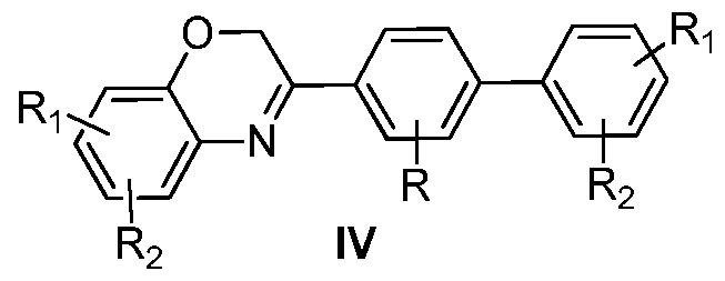
- a compound of Formula IV or a pharmaceutically acceptable salt or prodrug thereof, is provided:
- R R 2 , Ri' , R 2 ' H, F, CI, Br, I, alkyl, aryl, OH, O-alkyl, O-aryl, NH 2 , NH- alkyl, N-dialkyl, carboxyl, sulfonyl, carbamoyl, or glycosyl.
- the aromatic heterocycle of Formula IV may be conjugated with a hydrophilic polymer, e.g., PEG and the like, and a lipid, e.g., DPPC, DSPE, DSPC, DPPE, and the like, to form a lipid-hydrophilic polymer- Formula IV ligand conjugate.
- a hydrophilic polymer e.g., PEG and the like
- a lipid e.g., DPPC, DSPE, DSPC, DPPE, and the like
- the lipid-hydrophilic polymer- Formula IV ligand conjugate may be incorporated into a liposomal composition.
- a method for imaging amyloid deposits in a patient comprising:
- a detectable quantity of a liposomal composition comprising a lipid-hydrophilic polymer-Formula IV ligand conjugate; allowing sufficient time for the liposomal composition to be associated with one or more amyloid deposits;
- R R 2 , Ri' , R 2 ' H, F, CI, Br, I, alkyl, aryl, OH, O-alkyl, O-aryl, NH 2 , NH- alkyl, N-dialkyl, carboxyl, sulfonyl, carbamoyl, or glycosyl.
- the aromatic heterocycle of Formula V may be conjugated with a hydrophilic polymer, e.g., PEG and the like, and a lipid, e.g., DPPC, DSPE, DSPC, DPPE, and the like, to form a lipid-hydrophilic polymer- Formula V ligand conjugate.
- a hydrophilic polymer e.g., PEG and the like
- a lipid e.g., DPPC, DSPE, DSPC, DPPE, and the like
- the lipid-hydrophilic polymer- Formula V ligand conjugate may be incorporated into a liposomal composition.
- a method for imaging amyloid deposits in a patient comprising:
- a liposomal composition comprising a lipid-hydrophilic polymer-Formula V ligand conjugate
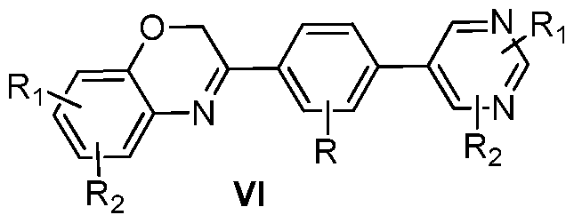
- a compound of Formula VI or a pharmaceutically acceptable salt or prodrug thereof, is provided:
- R R 2 , Ri' , R 2 ' H, F, CI, Br, I, alkyl, aryl, OH, O-alkyl, O-aryl, NH 2 , NH- alkyl, N-dialkyl, carboxyl, sulfonyl, carbamoyl, or glycosyl.
- the aromatic heterocycle of Formula VI may be conjugated with a hydrophilic polymer, e.g., PEG and the like, and a lipid, e.g., DPPC, DSPE, DSPC, DPPE, and the like, to form a lipid-hydrophilic polymer- Formula VI ligand conjugate.
- a hydrophilic polymer e.g., PEG and the like
- a lipid e.g., DPPC, DSPE, DSPC, DPPE, and the like
- the lipid-hydrophilic polymer- Formula VI ligand conjugate may be incorporated into a liposomal composition.
- a method for imaging amyloid deposits in a patient comprising:
- a liposomal composition comprising a lipid-hydrophilic polymer-Formula VI ligand conjugate
- the aromatic compound of Formula VII may be conjugated with a hydrophilic polymer, e.g., PEG and the like, and a lipid, e.g., DPPC, DSPE, DSPC, DPPE, and the like, to form a lipid-hydrophilic polymer- Formula VII ligand conjugate.
- a hydrophilic polymer e.g., PEG and the like
- a lipid e.g., DPPC, DSPE, DSPC, DPPE, and the like
- the lipid-hydrophilic polymer- Formula VII ligand conjugate may be incorporated into a liposomal composition.
- a method for imaging amyloid deposits in a patient comprising:
- a liposomal composition comprising a lipid-hydrophilic polymer-Formula VII ligand conjugate
- a liposomal composition comprising:
- cholesterol or another stabilizing excipient, such as another sterol or a fatty acid;
- a phospholipid which is derivatized with a polymer and a conjugate comprising an aromatic compound having any one of Formulas I- VII, such as a conjugate in a form of a lipid-hydrophilic polymer- aromatic conjugate as described herein.
- the liposomal composition comprises:
- Gd- DTPA-BSA gadolinium salt
- n i.e., the number of ethylene glycol repeating units
- a method for imaging amyloid deposits in a patient comprising:
- a detectable quantity of a liposomal composition comprising a phospholipid; cholesterol, or another stabilizing excipient, such as another sterol or a fatty acid; a nonradioactive gadolinium-containing contrast enhancing agent; a phospholipid which is derivatized with a polymer; and a conjugate comprising an aromatic compound having any one of Formulas I- VII, such as a conjugate in a form of a lipid-hydrophilic polymer-aromatic compound conjugate as described herein;
- the detecting comprises detecting by fluorescence imaging (FI). In another embodiment, the detecting comprises detecting by magnetic resonance imaging (MRI). In another embodiment, the detecting comprises detecting by X-ray imaging, including computed tomography (CT) imaging. In one embodiment, the detecting comprises detecting by SPECT imaging and/or PET imaging, and the non-radioactive contrast enhancing agent is replaced with a radioactive contrast enhancing agent, comprising for example those agents deemed appropriate for use with SPECT imaging and/or PET imaging in the National Institute of Health's Molecular Imaging and Contrast Agent Database (“MIC AD").
- FI fluorescence imaging
- MRI magnetic resonance imaging
- CT computed tomography
- the detecting comprises detecting by SPECT imaging and/or PET imaging, and the non-radioactive contrast enhancing agent is replaced with a radioactive contrast enhancing agent, comprising for example those agents deemed appropriate for use with SPECT imaging and/or PET imaging in the National Institute of Health's Molecular Imaging and Contrast Agent Database (“MIC AD").
- a liposomal composition comprising: a phospholipid; cholesterol, or another stabilizing excipient, such as another sterol or a fatty acid; a nonradioactive iodinated contrast enhancing agent; a phospholipid which is derivatized with a polymer; and a conjugate comprising an aromatic compound having any one of Formulas I- VII, such as a conjugate in a form of a lipid-hydrophilic polymer-aromatic compound conjugate as described herein.
- the liposomal composition comprises:
- DPPC DPPC
- cholesterol iodixanol
- a method for imaging amyloid deposits in a patient comprising:
- a detectable quantity of a liposomal composition comprising a phospholipid; cholesterol, or another stabilizing excipient, such as another sterol or a fatty acid; a nonradioactive iodinated contrast enhancing agent; a phospholipid which is derivatized with a polymer; and a conjugate comprising an aromatic compound having any one of Formulas I- VII, such as a conjugate in a form of a lipid-hydrophilic polymer- aromatic compound conjugate as described herein; allowing sufficient time for the liposomal composition to be associated with one or more amyloid deposits; and
- the detecting comprises detecting by FI. In another embodiment, the detecting comprises detecting by X-ray imaging, including CT imaging. In one embodiment, the detecting comprises detecting by SPECT imaging and/or PET imaging, and the non-radioactive contrast enhancing agent is replaced with a radioactive contrast enhancing agent, comprising for example those agents deemed appropriate for use with SPECT imaging and/or PET imaging in the National Institute of Health's Molecular Imaging and Contrast Agent Database( "MICAD").
- MICAD National Institute of Health's Molecular Imaging and Contrast Agent Database
- Figure 1 illustrates an example schematic for the synthesis of the lipid-hydrophilic polymer-aromatic ligand conjugate, DSPE-PEG n -methoxy-X04
- Figure 1A illustrates example transmission electron microscope (“TEM”) images of liposomal
- Figure 2A illustrates the binding affinity of Me-X04-labeled liposomes to synthetic ⁇ (1-40) fibrils.
- Figure 2B illustrates example results of the competition between Me-X04-labeled liposomes, Chrysamine G ("CG"), and free methoxy-X04 ligand, for binding sites on synthetic ⁇ (1-40) fibrils.
- CG Chrysamine G
- Figure 3 illustrates example results of ex vivo staining of mouse brain tissue with Me-X04-labeled liposomes.
- Figure 4 illustrates example results of the competition between
- Me-X04-labeled liposomes and CG for binding sites on ⁇ plaque deposits on mouse brain tissue, ex vivo.
- Figure 5 illustrates example results of in vivo staining of mouse brain tissue with Me-X04-labeled liposomes, and with free methoxy-X04 ligand.
- Figure 6 illustrates an optical reconstruction of example confocal microscope images from a sagittal section of a mouse brain injected with Me-X04-labeled liposomes.
- Figure 7 illustrates an example comparison of inflammatory potential between free methoxy-X04 ligand and the Me-X04 conjugate.
- Figure 8 illustrates an example comparison of cellular toxicity between free methoxy-X04 ligand and the Me-X04 conjugate.
- Figure 9A illustrates a comparison of cellular toxicity between
- Figure 9B illustrates a comparison of inflammatory potential between DSPE-PEG-4-aminopyrimidine boronic acid conjugate and the free ligand, 4-aminopyrimidine boronic acid.
- a compound of Formula I or a pharmaceutically acceptable salt or prodrug thereof, is provided:
- R, R R 2 , Ri', R 2 ' H, F, CI, Br, I, alkyl, aryl, OH, O-alkyl, O-aryl, NH 2 , NH-alkyl, N-dialkyl, carboxyl, sulfonyl, carbamoyl, or glycosyl.
- a compound of Formula I is the 1 ,4-quinoxaline phenyl 1,3-benzodioxolyl compound IA:
- R H
- Ri H
- R 2 H
- Ri ' H
- the aromatic heterocycle of Formula I may be conjugated with a hydrophilic polymer, e.g., PEG (having, e.g., a molecular weight ranging from 500 - 10,000 Da) and the like, and a lipid, e.g., DPPC, DSPE, DSPC, DPPE, and the like, to form a lipid-hydrophilic polymer-Formula I ligand conjugate.
- a hydrophilic polymer e.g., PEG (having, e.g., a molecular weight ranging from 500 - 10,000 Da) and the like
- a lipid e.g., DPPC, DSPE, DSPC, DPPE, and the like
- the lipid-hydrophilic polymer-Formula I ligand conjugate comprises:
- n is about 10 to about 100, or about 30 to about 60.
- the lipid-hydrophilic polymer-Formula [0055] In another embodiment, the lipid-hydrophilic polymer-Formula
- I ligand conjugate comprises:
- n is about 10 to about 100, or about 30 to about 60.
- the lipid-hydrophilic polymer-Formula I ligand conjugate may be incorporated into a liposomal composition.
- a method for imaging amyloid deposits in a patient comprising:
- a liposomal composition comprising a lipid-hydrophilic polymer-Formula I ligand conjugate
- the detecting comprises detecting by FI. In another embodiment, the detecting comprises detecting by MR imaging. In another embodiment, the detecting comprises detecting by X-ray imaging, including CT imaging. In one embodiment, the detecting comprises detecting by SPECT imaging and/or PET imaging, and the non-radioactive contrast enhancing agent is replaced with a radioactive contrast enhancing agent, comprising for example those agents deemed appropriate for use with SPECT imaging and/or PET imaging in the National Institute of Health's Molecular Imaging and Contrast Agent Database ("MIC AD").
- MIC AD National Institute of Health's Molecular Imaging and Contrast Agent Database
- a compound of Formula II or a pharmaceutically acceptable salt or prodrug thereof, is provided:
- R, R R 2 , Ri', R 2 ' H, F, CI, Br, I, alkyl, aryl, OH, O-alkyl, O-aryl, NH 2 , NH-alkyl, N-dialkyl, carboxyl, sulfonyl, carbamoyl, or glycosyl.
- a compound of Formula II is the 1,4-quinoxaline phen pyridinyl compound IIA:
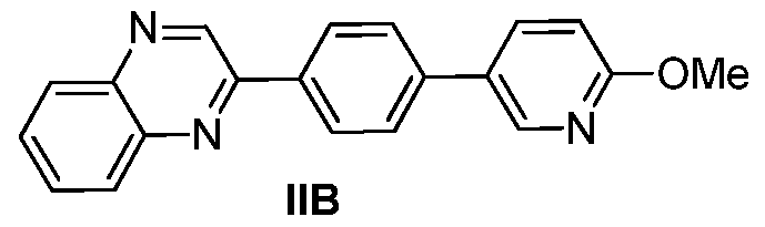
- Another example of a compound of Formula II is the 1,4- quinoxaline phenyl pyridinyl compound IIB :
- the aromatic heterocycle of Formula II may be conjugated with a hydrophilic polymer, e.g., PEG (having, e.g., a molecular weight ranging from 500 - 10,000 Da) and the like, and a lipid, e.g., DPPC, DSPE, DSPC, DPPE, and the like, to form a lipid-hydrophilic polymer-Formula II ligand conjugate.
- a hydrophilic polymer e.g., PEG (having, e.g., a molecular weight ranging from 500 - 10,000 Da) and the like
- a lipid e.g., DPPC, DSPE, DSPC, DPPE, and the like
- the lipid-hydrophilic polymer-Formula II ligand conjugate comprises:
- n is about 10 to about 100, or about 30 to about 60.
- the lipid-hydrophilic polymer-Formula [0064] In another embodiment, the lipid-hydrophilic polymer-Formula
- n is about 10 to about 100, or about 30 to about 60.
- the lipid-hydrophilic polymer-Formula II ligand conjugate may be incorporated into a liposomal composition.
- a method for imaging amyloid deposits in a patient comprising:
- a liposomal composition comprising a lipid-hydrophilic polymer-Formula II ligand conjugate
- the detecting comprises detecting by FI. In another embodiment, the detecting comprises detecting by MR imaging. In another embodiment, the detecting comprises detecting by X-ray imaging, including CT imaging. In one embodiment, the detecting comprises detecting by SPECT imaging and/or PET imaging, and the non-radioactive contrast enhancing agent is replaced with a radioactive contrast enhancing agent, comprising for example those agents deemed appropriate for use with SPECT imaging and/or PET imaging in the National Institute of Health's Molecular Imaging and Contrast Agent Database ("MIC AD").
- MIC AD National Institute of Health's Molecular Imaging and Contrast Agent Database
- a compound of Formula III or a pharmaceutically acceptable salt or prodrug thereof, is provided:
- R, R R 2 , Ri', R 2 ' H, F, CI, Br, I, alkyl, aryl, OH, O-alkyl, O-aryl, NH 2 , NH-alkyl, N-dialkyl, carboxyl, sulfonyl, carbamoyl, or glycosyl.
- R H
- Ri H
- R 2 H
- Ri' OMe
- R 2 ' OMe.
- a compound of Formula III is the 1,4-quinoxaline phenyl pyrimidinyl compound IIIA:
- the aromatic heterocycle of Formula III may be conjugated with a hydrophilic polymer, e.g., PEG (having, e.g., a molecular weight ranging from 500 - 10,000 Da) and the like, and a lipid, e.g., DPPC, DSPE, DSPC, DPPE, and the like, to form a lipid-hydrophilic polymer-Formula III ligand conjugate.
- a hydrophilic polymer e.g., PEG (having, e.g., a molecular weight ranging from 500 - 10,000 Da) and the like
- a lipid e.g., DPPC, DSPE, DSPC, DPPE, and the like
- the lipid-hydrophilic polymer-Formula III ligand conjugate comprises:
- n is about 10 to about 100, or about 30 to about 60.
- the lipid-hydrophilic polymer-Formula III ligand conjugate may be incorporated into a liposomal composition.
- a method for imaging amyloid deposit in a patient comprising:
- a liposomal composition comprising a lipid-hydrophilic polymer-Formula III ligand conjugate
- the detecting comprises detecting by FI. In another embodiment, the detecting comprises detecting by MR imaging. In another embodiment, the detecting comprises detecting by X-ray imaging, including CT imaging. In one embodiment, the detecting comprises detecting by SPECT imaging and/or PET imaging, and the non-radioactive contrast enhancing agent is replaced with a radioactive contrast enhancing agent, comprising for example those agents deemed appropriate for use with SPECT imaging and/or PET imaging in the National Institute of Health's Molecular Imaging and Contrast Agent Database ("MIC AD").
- MIC AD National Institute of Health's Molecular Imaging and Contrast Agent Database
- a compound of Formula IV or a pharmaceutically acceptable salt or prodrug thereof, is provided:
- R, R R 2 , Ri', R 2 ' H, F, CI, Br, I, alkyl, aryl, OH, O-alkyl, O-aryl, NH 2 , NH-alkyl, N-dialkyl, carboxyl, sulfonyl, carbamoyl, or glycosyl.
- a compound of Formula IV is the 1 ,4-benzoxazine phenyl 1,3-benzodioxolyl compound IV A:
- R H
- Ri Me
- R 2 H
- Ri' H
- the aromatic heterocycle of Formula IV may be conjugated with a hydrophilic polymer, e.g., PEG (having, e.g., a molecular weight ranging from 500 - 10,000 Da) and the like, and a lipid, e.g., DPPC, DSPE, DSPC, DPPE, and the like, to form a lipid-hydrophilic polymer-Formula IV ligand conjugate.
- a hydrophilic polymer e.g., PEG (having, e.g., a molecular weight ranging from 500 - 10,000 Da) and the like
- a lipid e.g., DPPC, DSPE, DSPC, DPPE, and the like
- the lipid-hydrophilic polymer-Formula IV ligand conjugate comprises:
- n is about 10 to about 100, or about 30 to about 60.
- the lipid-hydrophilic polymer-Formula [0080] In another embodiment, the lipid-hydrophilic polymer-Formula
- IV ligand conjugate comprises:
- n is about 10 to about 100, or about 30 to about 60.
- the lipid-hydrophilic polymer-Formula IV ligand conjugate may be incorporated into a liposomal composition.
- a method for imaging amyloid deposits in a patient comprising:
- a liposomal composition comprising a lipid-hydrophilic polymer-Formula IV ligand conjugate
- the detecting comprises detecting by FI. In another embodiment, the detecting comprises detecting by MR imaging. In another embodiment, the detecting comprises detecting by X-ray imaging, including CT imaging. In one embodiment, the detecting comprises detecting by SPECT imaging and/or PET imaging, and the non-radioactive contrast enhancing agent is replaced with a radioactive contrast enhancing agent, comprising for example those agents deemed appropriate for use with SPECT imaging and/or PET imaging in the National Institute of Health's Molecular Imaging and Contrast Agent Database ("MIC AD").
- MIC AD National Institute of Health's Molecular Imaging and Contrast Agent Database
- a compound of Formula V or a pharmaceutically acceptable salt or prodrug thereof, is provided:
- R, R R 2 , Ri', R 2 ' H, F, CI, Br, I, alkyl, aryl, OH, O-alkyl, O-aryl, NH 2 , NH-alkyl, N-dialkyl, carboxyl, sulfonyl, carbamoyl, or glycosyl.
- a compound of Formula V is the 1 ,4-benzoxazine phenyl pyridinyl compound VA:
- the aromatic heterocycle of Formula V may be conjugated with a hydrophilic polymer, e.g., PEG (having, e.g., a molecular weight ranging from 500 - 10,000 Da) and the like, and a lipid, e.g., DPPC, DSPE, DSPC, DPPE, and the like, to form a lipid-hydrophilic polymer-Formula V ligand conjugate.
- a hydrophilic polymer e.g., PEG (having, e.g., a molecular weight ranging from 500 - 10,000 Da) and the like
- a lipid e.g., DPPC, DSPE, DSPC, DPPE, and the like
- the lipid-hydrophilic polymer-Formula V ligand conjugate comprises:
- n is about 10 to about 100, or about 30 to about 60.
- the lipid-hydrophilic polymer-Formula V ligand conjugate may be incorporated into a liposomal composition.
- a method for imaging amyloid deposits in a patient comprising: introducing into the patient a detectable quantity of a liposomal composition comprising a lipid-hydrophilic polymer-Formula V ligand conjugate;
- the detecting comprises detecting by FI. In another embodiment, the detecting comprises detecting by MR imaging. In another embodiment, the detecting comprises detecting by X-ray imaging, including CT imaging. In one embodiment, the detecting comprises detecting by SPECT imaging and/or PET imaging, and the non-radioactive contrast enhancing agent is replaced with a radioactive contrast enhancing agent, comprising for example those agents deemed appropriate for use with SPECT imaging and/or PET imaging in the National Institute of Health's Molecular Imaging and Contrast Agent Database ("MIC AD").
- MIC AD National Institute of Health's Molecular Imaging and Contrast Agent Database
- a compound of Formula VI or a pharmaceutically acceptable salt or prodrug thereof, is provided:
- R, R R 2 , Ri', R 2 ' H, F, CI, Br, I, alkyl, aryl, OH, O-alkyl, O-aryl, NH 2 , NH-alkyl, N-dialkyl, carboxyl, sulfonyl, carbamoyl, or glycosyl.
- R H
- Ri Me
- R 2 H
- Ri' OMe
- R 2 ' OMe.
- a compound of Formula VI is the 1 ,4-quinoxaline phenyl pyrimidinyl compound VIA:
- the aromatic heterocycle of Formula VI may be conjugated with a hydrophilic polymer, e.g., PEG (having, e.g., a molecular weight ranging from 500 - 10,000 Da) and the like, and a lipid, e.g., DPPC, DSPE, DSPC, DPPE, and the like, to form a lipid-hydrophilic polymer-Formula VI ligand conjugate.
- a hydrophilic polymer e.g., PEG (having, e.g., a molecular weight ranging from 500 - 10,000 Da) and the like
- a lipid e.g., DPPC, DSPE, DSPC, DPPE, and the like
- the lipid-hydrophilic polymer-Formula VI ligand conjugate comprises:
- n is about 10 to about 100, or about 30 to about 60.
- the lipid-hydrophilic polymer-Formula VI ligand conjugate may be incorporated into a liposomal composition.
- a method for imaging amyloid deposits in a patient comprising:
- a liposomal composition comprising a lipid-hydrophilic polymer-Formula VI ligand conjugate
- the detecting comprises detecting by FI. In another embodiment, the detecting comprises detecting by MR imaging. In another embodiment, the detecting comprises detecting by X-ray imaging, including CT imaging. In one embodiment, the detecting comprises detecting by SPECT imaging and/or PET imaging, and the non-radioactive contrast enhancing agent is replaced with a radioactive contrast enhancing agent, comprising for example those agents deemed appropriate for use with SPECT imaging and/or PET imaging in the National Institute of Health's Molecular Imaging and Contrast Agent Database ("MIC AD").
- MIC AD National Institute of Health's Molecular Imaging and Contrast Agent Database
- R OMe
- Rj H
- R 2 O-alkyl
- Rj' OH
- R 2 ' H.
- a compound of Formula VII is the divinyl benzene compound VIIA ("methoxy-X04"):
- the aromatic compound of Formula VII may be conjugated with a hydrophilic polymer, e.g., PEG (having, e.g., a molecular weight ranging from 500 - 10,000 Da) and the like, and a lipid, e.g., DPPC, DSPE, DSPC, DPPE, and the like, to form a lipid-hydrophilic polymer-Formula VII ligand conjugate.
- a hydrophilic polymer e.g., PEG (having, e.g., a molecular weight ranging from 500 - 10,000 Da) and the like
- a lipid e.g., DPPC, DSPE, DSPC, DPPE, and the like
- the methoxy-X04 ligand may be conjugated with PEG and DSPE to form the DSPE-PEG n -Methoxy-X04 conjugate shown as "1" in Figure 1 (and sometimes referred to hereinafter as "Me-X04"):
- the lipid-hydrophilic polymer-Formula VII ligand conjugate e.g., Me-X04, and even more particularly, DPSE-PEG 3 4 00 - Methoxy-X04 (where 3400 signifies the molecular weight of the polyethyelene glycol), may be incorporated into a liposomal composition.
- a method for imaging amyloid deposits in a patient comprising:
- a liposomal composition comprising a lipid-hydrophilic polymer-Formula VII ligand conjugate
- the detecting comprises detecting by FI. In another embodiment, the detecting comprises detecting by MR imaging. In another embodiment, the detecting comprises detecting by X-ray imaging, including CT imaging. In one embodiment, the detecting comprises detecting by SPECT imaging and/or PET imaging, and the non-radioactive contrast enhancing agent is replaced with a radioactive contrast enhancing agent, comprising for example those agents deemed appropriate for use with SPECT imaging and/or PET imaging in the National Institute of Health's Molecular Imaging and Contrast Agent Database ("MIC AD").
- MIC AD National Institute of Health's Molecular Imaging and Contrast Agent Database
- one or more alternative amyloid ligands may be conjugated with a hydrophilic polymer, e.g., PEG (having, e.g., a molecular weight ranging from 500 - 10,000 Da) and the like, and a lipid, e.g., DPPC, DSPE, DSPC, DPPE, and the like, to form a lipid-hydrophilic polymer- amyloid ligand conjugate.
- the lipid-hydrophilic polymer- amyloid ligand conjugate may be incorporated into a liposomal composition.
- a method for imaging amyloid lesions in a patient comprising:
- a detectable quantity of a liposomal composition comprising a lipid-hydrophilic polymer- amyloid ligand conjugate
- the liposomal compositions described herein may further enable delivery of therapeutic molecules to amyloid lesions, thus enabling treatment of the lesions.
- a liposomal composition comprising: a phospholipid; cholesterol, or another stabilizing excipient, such as another sterol or a fatty acid; a nonradioactive gadolinium- containing contrast enhancing agent; a phospholipid which is derivatized with a polymer; and a conjugate comprising an aromatic compound having any one of Formulas I- VII, such as a conjugate in a form of a lipid-hydrophilic polymer- aromatic conjugate as described herein.
- a method for imaging amyloid deposits in a patient comprising:
- a detectable quantity of a liposomal composition comprising a phospholipid; cholesterol, or another stabilizing excipient, such as another sterol or a fatty acid; a nonradioactive gadolinium-containing contrast enhancing agent; a phospholipid which is derivatized with a polymer; and a conjugate comprising an aromatic compound having any one of Formulas I- VII, such as a conjugate in a form of a lipid-hydrophilic polymer- aromatic compound conjugate as described herein;
- the detecting comprises detecting by FI.
- the detecting comprises detecting by MRI.
- hydrophilic paramagnetic chelates such as GdDTPA, GdDOTA, GdHPD03A, GdDTPA-BMA, and GdDTPA-BSA are known MRI contrast agents. See U.S. Patent No. 5,676,928 issued to Klaveness et al., which is incorporated by reference herein in its entirety.
- the detecting comprises detecting by X-ray imaging, including CT imaging.
- the detecting comprises detecting by SPECT imaging and/or PET imaging, and the non-radioactive contrast enhancing agent is replaced with a radioactive contrast enhancing agent, comprising for example those agents deemed appropriate for use with SPECT imaging and/or PET imaging in the National Institute of Health's Molecular Imaging and Contrast Agent Database ("MICAD").
- MICAD National Institute of Health's Molecular Imaging and Contrast Agent Database
- Paramagnetic chelates such as GdDTPA and its derivatives, may also be used as X-ray contrast agents. See U.S. Patent No. 5,204,085 issued to VanDeripe, which is incorporated by reference herein in its entirety.
- a liposomal composition comprises: a phospholipid; cholesterol; a phospholipid which is derivatized with a polymer; and a conjugate comprising a compound having any one of Formulas I- VII, wherein the liposomal composition encapsulates a nonradioactive iodinated contrast enhancing agent.
- Suitable phospholipids may include those disclosed herein, and may further include those disclosed in U.S. Patent No. 7,785,568 issued to Annapragada et al., which is incorporated by reference herein in its entirety.
- Suitable polymer derivatized phospholipids may include those disclosed herein, and may further include those disclosed in U.S. Patent No. 7,785,568.
- Suitable nonradioactive iodinated contrast enhancing agents may include, for example, iodixanol and iohexol, and may further include those disclosed in U.S. Patent No. 7,785,568.
- a method for imaging amyloid deposits in a patient comprising: introducing into the patient a detectable quantity of a liposomal composition comprising a phospholipid; cholesterol; a phospholipid which is derivatized with a polymer; and a conjugate comprising a compound having any one of Formulas I- VII, wherein the liposomal composition encapsulates a nonradioactive iodinated contrast enhancing agent;
- the detecting comprises detecting by FI. In another embodiment, the detecting comprises detecting by X-ray imaging, including CT imaging. In one embodiment, the detecting comprises detecting by SPECT imaging and/or PET imaging, and the non-radioactive contrast enhancing agent is replaced with a radioactive contrast enhancing agent, comprising for example those agents deemed appropriate for use with SPECT imaging and/or PET imaging in the National Institute of Health's Molecular Imaging and Contrast Agent Database (“MICAD"). Any other suitable type of imaging methodology known by those skilled in the art is contemplated, including, but not limited to, PET imaging.
- MICAD National Institute of Health's Molecular Imaging and Contrast Agent Database
- a method for capturing an image of a region of interest of a subject comprising: introducing a composition into the bloodstream of the subject, wherein the composition comprises liposomes, the liposomes comprising: at least one phospholipid; at least one phospholipid which is derivatized with a polymer; a phospholipid-polymer- aromatic compound conjugate; and cholesterol, wherein the liposomes encapsulate at least one iodinated nonradioactive contrast enhancing agent; and irradiating the region of interest with X-rays.
- the region of interest is a region of a human brain, and, more particularly, a region of the human brain that has ⁇ plaque deposited thereon.
- Example 1 Preparation of DSPE-PEG 34 oo-Methoxy-X04 conjugate 1.
- linker moiety was installed quantitatively to give aldehyde 12, which was exposed to sulfone 7 under optimized Julia- Kocienski conditions to obtain the desired E,E-isomer 13 in 69% yield after column chromatography purification.
- Global deprotection of the MOM and Boc groups with HC1 gave the linker-methoxy-X04 moiety 14, as the hydrochloride salt.
- Example 2 Preparation and characterization of Gd- containing Me-X04-labeled liposomes.
- a lipid mixture (50 mM) comprising DPPC, cholesterol, Gd- DTPA-BSA, DSPE-mPEG-2000, Me-X04, and Rhodamine-DHPE (for optical detection) in about a 32.4: 40: 25: 2: 0.5: 0.1 molar ratio, respectively, was employed.
- Other ratios are contemplated, including a lipid mixture comprising DPPC, cholesterol, Gd-DTPA-BSA, DSPE-mPEG-2000, and Me-X04 in about a 32.5: 40: 25: 2: 0.5 molar ratio.
- the DSPE-mPEG-2000 may be replaced altogether with the Me-X04 conjugate, for a DPPC, cholesterol, Gd-DTPA-BSA, Me- X04 conjugate ratio of 32.5: 40: 25: 2.5.
- the upper limit on the PEG-bearing molecule may be about 15-25%, and the lower limit on cholesterol may be about 15- 20%.
- Particle size distribution was determined by TEM ( Figures 1A and IB), thereby confirming a mean diameter of about 100.8 nm and PDI of about 0.05.
- concentration of Me-X04 ligand in the particles was determined using a fluorescence standard curve generated for Me-X04 ligand to be 26 ⁇ .
- the above protocol results in roughly equal distribution of the targeting ligand between the inner and the outer faces of the lipid bilayer. This implies that for each reported concentration of Me-X04 ligand in the nanoparticles, approximately 50% of the total Me-X04 ligands are available for binding.
- Me-X04 ligand is highly intrinsically fluorescent and so are the nanoparticles bearing Me-X04 ligand. This property was used as a reporter on the locations of nanoparticles in the course of all of the experiments.
- Example 3 In vitro binding affinity of Gd-containing Me- X04-labeled liposomes for synthetic ⁇ fibrils.
- Figure 2A illustrates the binding affinity of the Gd-containing Me-X04-labeled liposomes of Example 2 to synthetic ⁇ (1-40) aggregates.
- Figure 2B illustrates the ability of Gd-containing Me-X04-labeled liposomes to compete for binding sites with free Me-X04 ligand.
- Example 4 Ex vivo staining of mouse brain tissue.
- mice were administered to 5 and 7 month old APP/PSEN1 mice by tail vain injection. 48 h following injection, the mice were euthanized and their brains sectioned for confocal light microscopic studies. Identical mice were injected with molecular/free methoxy- X04 ligand to serve as a positive control, and with untargeted liposomes and saline as negative controls.
- Example 6 Inflammatory potential of Me-X04.
- LPS lipopolysaccharides
- the inflammatory potential of Me-X04 was compared to free (i.e., unconjugated) methoxy-X04 ligand, LPS, and an untreated control ("UTC").
- UTC untreated control
- Translocation of NF-kb from cytosol to the nucleus is an early event in the inflammatory reaction.
- NF-kb moves from the cytoplasm to the nucleus and induces gene transcription. Therefore translocation of NF-kB is a widely used marker for inflammation.
- the protocol is outlined below.
- the cells were incubated with 1 % BSA in PBS-T (PBS with 0.1 % Tween-20) for 45 min at RT. At the end of the incubation period, the cells were further incubated with primary antibody against NF-kB at 1 :50 dilution in PBST for 1 h at RT. The cells were again washed with PBS three times (5 min each). The cells were incubated with the secondary antibody in PBST for 1 h at RT, and were again washed three times (5 min each) with PBS. 100 ⁇ of DAPI (1 ⁇ g/mL) was placed in each well and kept at 4 °C until further analysis.
- PBS-T PBS with 0.1 % Tween-20
- the cells were scanned on a Cell Lab IC-100 image cytometer (Beckman-Coulter, CA). The data were further analyzed using Cytseer software from Vala Sciences, CA, and represented as Pearson's Correlation coefficient (PCC) of the protein intensity present over the nuclear mask.
- PCC Pearson's Correlation coefficient
- Me-X04 was found to be less inflammatory than free methoxy-X04 ligand at all but the highest concentrations (500 nM) tested. The results are depicted in Figure 7.
- conjugated and/or liposomal amyloid binding ligand is less (or at least not more) inflammatory than the free ligand.
- Example 7 Cytotoxicity of Me-X04.
- the cytotoxity of Me-X04 was compared to free (i.e., not conjugated) methoxy-X04 ligand and an untreated control.
- the toxicity of the test compounds was evaluated using standard MTT assays. 15,000 Hela cells were plated in each of 96 well plates and allowed to stand overnight. The cells were treated with three different concentrations of the test compounds for 2 h in a 37 °C incubator. The positive controls were treated with 1 mg/mL LPS. At the end of the incubation period, a MTS cell toxicity assay kit (Cell titer 96 AQueous Assay kit from Promega) was used according to the manufacturer's protocol. At the end of the incubation period, cells were treated with 15 MTS reagent/100 ⁇ L ⁇ media for 3 h at 37 °C. After 3 h of incubation, the absorbance was recorded at 490 nm using a plate reader.
- Example 9 Preparation and characterization of iodine- encapsulated Me-X04-labeled liposomes.
- a lipid mixture (30 mM) comprising DPPC, cholesterol, DSPE-mPEG-2000, and Me-X04 in about a 57:40:2.5:0.5 molar ratio was dissolved in ethanol.
- other component molar ratios are contemplated for the various liposome compositions, whether encapsulating iodinated contrast enhancing agent as exemplified in this Example 9, or encapsulating and/or bound to a gadolinium contrast enhancing agent (as exemplified in Example 2), including, for example, phospholipid:cholesterol:phospholipid derivatized with a polymenphospholipid-hydrophilic polymer- aromatic compound conjugate, in about a 40-70:10-50:0.1-10:0.1-10 molar ratio, respectively.
- a solution of iodixanol (525 mg/ml) was prepared by dissolving iodixanol powder in distilled water at 60 °C. The ethanol solution was hydrated with the iodixanol solution and sequentially extruded on a Lipexthermoline extruder (Northern Lipids Inc., Canada) with eight passes through a 400 nm Nuclepore membrane. The resulting solution was diafiltered using a MicroKros ® module (Spectrum Laboratories, CA) of 500 kDa molecular weight cut-off to remove un-encapsulated iodixanol.
- a MicroKros ® module Spectrum Laboratories, CA
- the iodine concentration in the final liposomal solution was quantified by UV absorption spectrophotometry ⁇ Abs at 245 nm) to be about 8.01 mg I/ml liposome composition. It should be noted that other iodine concentrations are contemplated for the various liposome compositions as described herein, including between about 5 mg I/ml liposome composition and about 200 mg I ml liposome composition.
- the liposome composition which comprises a phospholipid, cholesterol, a phospholipid derivatized with a polymer, and a phospholipid-hydrophilic polymer- aromatic compound conjugate, in about a 40- 70: 10-50:0.1-10:0.1-10 molar ratio, may encapsulate about 150 mg I/ml of liposome composition.
- the concentration of Me-X04 in the final product was determined using UV absorption spectrophotometry ⁇ Abs at 365 nm).
- the liposomes may have an average diameter of from about 50 to about 400 nm. In one embodiment, the liposomes have an average diameter of from about 100 to about 150 nm.
- Example 10 In vitro binding of iodine-encapsulated Me- X04-labeled liposomes to ⁇ fibrils.
- ⁇ fibrils Preparation of ⁇ fibrils: ⁇ -amyloid (1-40, ultrapure, TFA, rPeptide) was dissolved in PBS buffer to a final concentration of 100 ⁇ with continuous overnight stirring at RT. 20 ⁇ of fibrils were incubated with 1.25 ⁇ of iodine-encapsulated Me-X04-labeled liposomes for 1 h. Unbound iodine- encapsulated Me-X04-labeled liposomes were removed by allowing the fibrils to settle by gravity for 24 h at 4 °C, and separating by low-speed centrifugation at 5,000 rpm at 4 °C for 10 min to form a pellet. The fibril pellet was washed three times with PBS buffer. The fibrils were re-suspended in 0.5 ml of PBS buffer by gently tapping the tubes.
- a 2% agar solution was prepared in sterile water at 80-100 °C. The agar solution was allowed to cool down to RT. During the cooling step, 0.5 ml of the test sample ( ⁇ fibrils only; iodine- encapsulated Me-X04-labeled liposomes; or ⁇ fibrils and iodine-encapsulated Me- X04-labeled liposomes) was added to the agar solution and shaken to homogenize before the agar began to gel (-45 °C). The phantoms were allowed to gel completely and kept at 4 °C until further use.
- Imaging The phantoms were imaged using UV light, a micro-CT scanner, and a clinical CT scanner. Pictures of the samples under UV radiation (365 nm) were captured with a 9 pixel camera. CT imaging was performed on a Siemens Inveon micro-CT scanner or on a clinical GE LightSpeed64-slice MDCT. The following scan parameters were used for micro-CT imaging: 80 kVp, 500 microA, 750 msec exposure time. Micro-CT images were acquired at a voxel size of 0.14 mm. The following parameters were used for clinical 64-MDCT: 80 kVp, 320 mA, 1.375 spiral pitch factor. Clinical CT mages were acquired at a voxel size of 0.625 mm.
- Figure 10 illustrates a comparison of: (1) UV (365) fluorescence of a sample containing ⁇ fibrils only, a sample containing liposomal Me-X04 encapsulating an iodinated contrast agent, and a sample containing ⁇ fibrils and liposomal Me-X04 encapsulating an iodinated contrast agent; (2) commercial-grade CT images of a sample containing ⁇ fibrils only, a sample containing liposomal Me-X04 encapsulating an iodinated contrast agent, and a sample containing ⁇ fibrils and liposomal Me-X04 encapsulating an iodinated contrast agent; and (3) clinical-grade CT images of a sample containing ⁇ fibrils only, a sample containing liposomal Me-X04 encapsulating an iodinated contrast agent, and a sample containing ⁇ fibrils and liposomal Me-X04 encapsulating an iodinated contrast agent.
- liposomal Me-X04 encapsulating an iodinated contrast agent fluoresces to a bright light (column titled "ADx-CT only"). The light is homogenously distributed so that the entire sample glows without any distinct spots.
- ADx-CT plus fibrils clusters of ⁇ fibrils are introduced in the mixture
- liposomal Me-X04 encapsulating an iodinated contrast agent binds to the fibrils resulting in focal bright spots, due to concentration of the fluorescence around the fibrils.
- the results are confirmed when the samples are placed in commercial and clinical-CT imaging instruments, wherein clusters of ⁇ fibrils with bound liposomal Me-X04 encapsulating an iodinated contrast agent appear as bright spots in the samples.
Landscapes
- Health & Medical Sciences (AREA)
- Chemical & Material Sciences (AREA)
- General Health & Medical Sciences (AREA)
- Veterinary Medicine (AREA)
- Dispersion Chemistry (AREA)
- Public Health (AREA)
- Epidemiology (AREA)
- Life Sciences & Earth Sciences (AREA)
- Animal Behavior & Ethology (AREA)
- Radiology & Medical Imaging (AREA)
- Nuclear Medicine, Radiotherapy & Molecular Imaging (AREA)
- Immunology (AREA)
- Engineering & Computer Science (AREA)
- Bioinformatics & Cheminformatics (AREA)
- Medicinal Chemistry (AREA)
- Pharmacology & Pharmacy (AREA)
- Medicines Containing Antibodies Or Antigens For Use As Internal Diagnostic Agents (AREA)
- Medicinal Preparation (AREA)
- Addition Polymer Or Copolymer, Post-Treatments, Or Chemical Modifications (AREA)
- Pharmaceuticals Containing Other Organic And Inorganic Compounds (AREA)
Abstract
Description


Claims


Priority Applications (16)
| Application Number | Priority Date | Filing Date | Title |
|---|---|---|---|
| AU2012239888A AU2012239888B9 (en) | 2011-04-06 | 2012-04-06 | Lipid-based nanoparticles |
| PL12767275T PL2694116T3 (en) | 2011-04-06 | 2012-04-06 | Lipid-based nanoparticles |
| CN201280017548.6A CN104144708B (en) | 2011-04-06 | 2012-04-06 | Nano-particle based on lipid |
| EP18167725.3A EP3366313A1 (en) | 2011-04-06 | 2012-04-06 | Lipid-based nanoparticles |
| MX2013011231A MX357227B (en) | 2011-04-06 | 2012-04-06 | Lipid-based nanoparticles. |
| ES12767275.6T ES2685824T3 (en) | 2011-04-06 | 2012-04-06 | Lipid-based nanoparticles |
| KR1020137029510A KR101973063B1 (en) | 2011-04-06 | 2012-04-06 | Lipid-based nanoparticles |
| JP2014504057A JP6212032B2 (en) | 2011-04-06 | 2012-04-06 | Lipid-based nanoparticles |
| BR112013025633A BR112013025633A2 (en) | 2011-04-06 | 2012-04-06 | lipid-based nanoparticles |
| DK12767275.6T DK2694116T3 (en) | 2011-04-06 | 2012-04-06 | LIPID-BASED NANOPARTICLES |
| KR1020197007384A KR102035187B1 (en) | 2011-04-06 | 2012-04-06 | Lipid-based nanoparticles |
| EP12767275.6A EP2694116B1 (en) | 2011-04-06 | 2012-04-06 | Lipid-based nanoparticles |
| CA2831480A CA2831480C (en) | 2011-04-06 | 2012-04-06 | Lipid-based nanoparticles |
| HK15104390.1A HK1203827A1 (en) | 2011-04-06 | 2015-05-08 | Lipid-based nanoparticles |
| AU2017232156A AU2017232156C1 (en) | 2011-04-06 | 2017-09-21 | Lipid –based nanoparticles |
| AU2019246775A AU2019246775A1 (en) | 2011-04-06 | 2019-10-08 | Lipid –based nanoparticles |
Applications Claiming Priority (2)
| Application Number | Priority Date | Filing Date | Title |
|---|---|---|---|
| US201161472605P | 2011-04-06 | 2011-04-06 | |
| US61/472,605 | 2011-04-06 |
Publications (2)
| Publication Number | Publication Date |
|---|---|
| WO2012139080A2 true WO2012139080A2 (en) | 2012-10-11 |
| WO2012139080A3 WO2012139080A3 (en) | 2014-05-01 |
Family
ID=46966277
Family Applications (1)
| Application Number | Title | Priority Date | Filing Date |
|---|---|---|---|
| PCT/US2012/032649 WO2012139080A2 (en) | 2011-04-06 | 2012-04-06 | Lipid-based nanoparticles |
Country Status (15)
| Country | Link |
|---|---|
| US (1) | US9801957B2 (en) |
| EP (2) | EP3366313A1 (en) |
| JP (3) | JP6212032B2 (en) |
| KR (2) | KR101973063B1 (en) |
| CN (1) | CN104144708B (en) |
| AU (3) | AU2012239888B9 (en) |
| BR (1) | BR112013025633A2 (en) |
| CA (1) | CA2831480C (en) |
| DK (1) | DK2694116T3 (en) |
| ES (1) | ES2685824T3 (en) |
| HK (1) | HK1203827A1 (en) |
| MX (1) | MX357227B (en) |
| PL (1) | PL2694116T3 (en) |
| PT (1) | PT2694116T (en) |
| WO (1) | WO2012139080A2 (en) |
Cited By (7)
| Publication number | Priority date | Publication date | Assignee | Title |
|---|---|---|---|---|
| US20150166492A1 (en) * | 2013-12-12 | 2015-06-18 | Albert Einstein College Of Medicine Of Yeshiva University | Retinoic acid receptor antagonists as chaperone-mediated autophagy modulators and uses thereof |
| US9744251B2 (en) | 2014-10-08 | 2017-08-29 | Texas Children's Hospital | MRI imaging of amyloid plaque using liposomes |
| US9801957B2 (en) | 2011-04-06 | 2017-10-31 | Ananth Annapragada | Lipid-based nanoparticles |
| US10130326B2 (en) | 2012-01-20 | 2018-11-20 | Ananth Annapragada | Methods and compositions for objectively characterizing medical images |
| WO2021163585A1 (en) * | 2020-02-12 | 2021-08-19 | Texas Children's Hospital | Targeted contrast agents for mri of alpha-synuclein deposition |
| US11779664B2 (en) | 2020-02-12 | 2023-10-10 | Texas Children's Hospital | Targeted contrast agents for MRI of alpha-synuclein deposition |
| US11786468B2 (en) | 2015-06-08 | 2023-10-17 | King's College London | Nanoparticles |
Families Citing this family (4)
| Publication number | Priority date | Publication date | Assignee | Title |
|---|---|---|---|---|
| CN115919769A (en) * | 2016-05-16 | 2023-04-07 | 德克萨斯大学系统董事会 | Compositions for delivering tRNA as nanoparticles and methods of use thereof |
| WO2018100540A1 (en) * | 2016-11-30 | 2018-06-07 | Texas Children's Hospital | Hydrophilic fluorinated molecules for liposomal 19f mri probes with unique mr signatures |
| EP3914230A4 (en) * | 2019-01-24 | 2023-06-07 | Alzeca Biosciences, Llc | <smallcaps/>? ? ?functionalized liposomes for imaging misfolded proteins |
| WO2021155128A1 (en) * | 2020-01-29 | 2021-08-05 | Texas Children's Hospital | Targeted contrast agents for mri of amyloid deposition |
Citations (2)
| Publication number | Priority date | Publication date | Assignee | Title |
|---|---|---|---|---|
| US5676928A (en) | 1994-03-28 | 1997-10-14 | Nycomed Imaging As | Liposomes |
| US7785568B2 (en) | 2004-04-21 | 2010-08-31 | Marval Biosciences, Inc. | Compositions and methods for enhancing contrast in imaging |
Family Cites Families (37)
| Publication number | Priority date | Publication date | Assignee | Title |
|---|---|---|---|---|
| US4331751A (en) * | 1980-11-17 | 1982-05-25 | Eastman Kodak Company | Electrically photosensitive materials and elements for photoelectrophoretic imaging processes |
| US5204085A (en) | 1985-01-08 | 1993-04-20 | Mallinckrodt Medical, Inc. | Method for enhancing the safety of metal-ligand chelates as X-ray contrast agents |
| US5674468A (en) | 1992-03-06 | 1997-10-07 | Nycomed Imaging As | Contrast agents comprising gas-containing or gas-generating polymer microparticles or microballoons |
| US6071532A (en) | 1996-10-15 | 2000-06-06 | Emory University | Synthesis of glycophospholipid and peptide-phospholipid conjugates and uses thereof |
| JP4829454B2 (en) * | 1999-11-30 | 2011-12-07 | ジ・アリゾナ・ボード・オブ・リージェンツ・オン・ビハーフ・オブ・ザ・ユニバーシティ・オブ・アリゾナ | Radiosensitive liposomes |
| AU2002211517A1 (en) * | 2000-10-04 | 2002-04-15 | California Institute Of Technology | Magnetic resonance imaging agents for in vivo labeling and detection of amyloid deposits |
| AU2002314794A1 (en) * | 2001-05-23 | 2002-12-03 | New York University | Detection of alzheimer's amyloid by magnetic resonance imaging |
| US7138136B2 (en) | 2002-03-05 | 2006-11-21 | Cleveland State University | Agglomerated particles for aerosol drug delivery |
| EP1641742A4 (en) | 2003-05-01 | 2006-11-29 | Nst Neurosurvival Technologies | Compounds that selectively bind to membranes of apoptotic cells |
| US7208174B2 (en) * | 2003-12-18 | 2007-04-24 | Hoffmann-La Roche Inc. | Liposome compositions |
| US20100031378A1 (en) | 2008-08-04 | 2010-02-04 | Edwards Joel A | Novel gene disruptions, compositions and methods relating thereto |
| US8357351B2 (en) | 2004-04-21 | 2013-01-22 | Ananth Annapragada | Nano-scale contrast agents and methods of use |
| WO2006026184A2 (en) * | 2004-08-20 | 2006-03-09 | Washington University | Blood brain barrier permeation peptides |
| DE602005025659D1 (en) * | 2004-09-23 | 2011-02-10 | Guerbet Sa | |
| EP2279726A3 (en) * | 2005-05-26 | 2012-06-20 | Biorest Ltd. | Compositions and methods using same for delivering agents into a target organ protected by a blood barrier |
| US20070160658A1 (en) * | 2005-10-20 | 2007-07-12 | The Penn State Research Foundation | Delivery system for diagnostic and therapeutic agents |
| WO2008054498A2 (en) * | 2006-04-07 | 2008-05-08 | The Board Of Trustees Of The University Of Illinois | Hydrophilic polymer-conjugated lipids for peptide and protein folding disorders |
| US20090123047A1 (en) | 2007-03-21 | 2009-05-14 | Yfantis Spyros A | Method and system for characterizing prostate images |
| EP1982733A1 (en) | 2007-04-17 | 2008-10-22 | Bayer Schering Pharma Aktiengesellschaft | Use of a contrast agent for magnetic resonance imaging of endoleaks |
| EP2182924A1 (en) | 2007-07-26 | 2010-05-12 | Nanoscan Imaging, Llc | Methods for imaging using improved nanoparticulate contrast agents |
| CN104274840A (en) | 2007-12-05 | 2015-01-14 | 马维尔生物科学公司 | Nano-scale contrast agents and methods of use |
| US20100286067A1 (en) | 2008-01-08 | 2010-11-11 | Biogenerix Ag | Glycoconjugation of polypeptides using oligosaccharyltransferases |
| ITMI20081052A1 (en) * | 2008-06-10 | 2009-12-11 | Univ Milano Bicocca | LIPOSOMAS ABLE TO EFFECTIVELY TIE THE BETA-AMYLOID PEPTIDE |
| US20090311191A1 (en) | 2008-06-13 | 2009-12-17 | Ananth Annapragada | Imaging of atherosclerotic plaques using liposomal imaging agents |
| US20110208064A1 (en) | 2008-07-31 | 2011-08-25 | Ran Chongzhao | Curcumin Derivatives for Amyloid-Beta Plaque Imaging |
| AR074760A1 (en) | 2008-12-18 | 2011-02-09 | Metabolex Inc | GPR120 RECEIVER AGONISTS AND USES OF THE SAME IN MEDICINES FOR THE TREATMENT OF DIABETES AND METABOLIC SYNDROME. |
| JP2012520898A (en) | 2009-03-19 | 2012-09-10 | マーヴァル バイオサイエンシーズ,インコーポレイテッド | Compositions and methods for contrast enhancement in imaging |
| US9334304B2 (en) * | 2009-04-02 | 2016-05-10 | The Johns Hopkins University | Self-assembling peptides bearing organic electronic functionality and applications employing the same |
| WO2011045415A2 (en) | 2009-10-15 | 2011-04-21 | Guerbet | New imaging agents and their use for the diagnostic in vivo of neurodegenerative diseases, notably alzheimer's disease and derivative diseases |
| DK2582674T3 (en) | 2010-06-16 | 2014-12-15 | Cymabay Therapeutics Inc | GPR120 receptor agonists and uses thereof. |
| WO2012119117A1 (en) | 2011-03-02 | 2012-09-07 | Sensulin, Llc | Vesicle compositions |
| PT2694116T (en) | 2011-04-06 | 2018-10-12 | Univ Texas | Lipid-based nanoparticles |
| GB201112056D0 (en) | 2011-07-14 | 2011-08-31 | Univ Leuven Kath | Antibodies |
| JP6130401B2 (en) | 2012-01-20 | 2017-05-17 | アンナプラガダ,アナンス | Methods and compositions for objectively characterizing medical images |
| WO2013164763A2 (en) | 2012-04-30 | 2013-11-07 | Innovative Health Diagnostics | A biological complex specific for alzheimer's disease detection in vitro and use thereof |
| BR112015022226A2 (en) | 2013-03-14 | 2017-07-18 | Celtaxsys Inc | leukotriene a4 hydrolase inhibitors |
| AU2015330824A1 (en) | 2014-10-08 | 2017-05-25 | Ananth Annapragada | MRI imaging of amyloid plaque using liposomes |
-
2012
- 2012-04-06 PT PT12767275T patent/PT2694116T/en unknown
- 2012-04-06 BR BR112013025633A patent/BR112013025633A2/en not_active Application Discontinuation
- 2012-04-06 EP EP18167725.3A patent/EP3366313A1/en not_active Withdrawn
- 2012-04-06 KR KR1020137029510A patent/KR101973063B1/en active IP Right Review Request
- 2012-04-06 CA CA2831480A patent/CA2831480C/en not_active Expired - Fee Related
- 2012-04-06 AU AU2012239888A patent/AU2012239888B9/en not_active Ceased
- 2012-04-06 ES ES12767275.6T patent/ES2685824T3/en active Active
- 2012-04-06 WO PCT/US2012/032649 patent/WO2012139080A2/en active Application Filing
- 2012-04-06 PL PL12767275T patent/PL2694116T3/en unknown
- 2012-04-06 JP JP2014504057A patent/JP6212032B2/en not_active Expired - Fee Related
- 2012-04-06 US US13/441,816 patent/US9801957B2/en not_active Expired - Fee Related
- 2012-04-06 MX MX2013011231A patent/MX357227B/en active IP Right Grant
- 2012-04-06 CN CN201280017548.6A patent/CN104144708B/en not_active Expired - Fee Related
- 2012-04-06 DK DK12767275.6T patent/DK2694116T3/en active
- 2012-04-06 KR KR1020197007384A patent/KR102035187B1/en active IP Right Grant
- 2012-04-06 EP EP12767275.6A patent/EP2694116B1/en not_active Not-in-force
-
2015
- 2015-05-08 HK HK15104390.1A patent/HK1203827A1/en not_active IP Right Cessation
-
2017
- 2017-09-14 JP JP2017176383A patent/JP6533813B2/en not_active Expired - Fee Related
- 2017-09-21 AU AU2017232156A patent/AU2017232156C1/en not_active Ceased
-
2019
- 2019-05-27 JP JP2019098174A patent/JP2019151664A/en active Pending
- 2019-10-08 AU AU2019246775A patent/AU2019246775A1/en not_active Abandoned
Patent Citations (2)
| Publication number | Priority date | Publication date | Assignee | Title |
|---|---|---|---|---|
| US5676928A (en) | 1994-03-28 | 1997-10-14 | Nycomed Imaging As | Liposomes |
| US7785568B2 (en) | 2004-04-21 | 2010-08-31 | Marval Biosciences, Inc. | Compositions and methods for enhancing contrast in imaging |
Non-Patent Citations (1)
| Title |
|---|
| See also references of EP2694116A4 |
Cited By (15)
| Publication number | Priority date | Publication date | Assignee | Title |
|---|---|---|---|---|
| US9801957B2 (en) | 2011-04-06 | 2017-10-31 | Ananth Annapragada | Lipid-based nanoparticles |
| US10130326B2 (en) | 2012-01-20 | 2018-11-20 | Ananth Annapragada | Methods and compositions for objectively characterizing medical images |
| US10189827B2 (en) | 2013-12-12 | 2019-01-29 | Albert Einstein College Of Medicine Of Yeshiva University | Retinoic acid receptor antagonists as chaperone-mediated autophagy modulators and uses thereof |
| US9890143B2 (en) | 2013-12-12 | 2018-02-13 | Albert Einstein College Of Medicine, Inc. | Retinoic acid receptor antagonists as chaperone-mediated authophagy modulators and uses thereof |
| US9512092B2 (en) * | 2013-12-12 | 2016-12-06 | Albert Einstein College Of Medicine, Inc. | Retinoic acid receptor antagonists as chaperone-mediated autophagy modulators and uses thereof |
| US20150166492A1 (en) * | 2013-12-12 | 2015-06-18 | Albert Einstein College Of Medicine Of Yeshiva University | Retinoic acid receptor antagonists as chaperone-mediated autophagy modulators and uses thereof |
| US10766886B2 (en) | 2013-12-12 | 2020-09-08 | Albert Einstein College Of Medicine | Retinoic acid receptor antagonists as chaperone-mediated autophagy modulators and uses thereof |
| US11591324B2 (en) | 2013-12-12 | 2023-02-28 | Albert Einstein College Of Medicine, Inc. | Retinoic acid receptor antagonists as chaperone-mediated autophagy modulators and uses thereof |
| US9744251B2 (en) | 2014-10-08 | 2017-08-29 | Texas Children's Hospital | MRI imaging of amyloid plaque using liposomes |
| US10537649B2 (en) | 2014-10-08 | 2020-01-21 | Texas Children's Hospital | MRI imaging of amyloid plaque using liposomes |
| US11141495B2 (en) | 2014-10-08 | 2021-10-12 | Texas Children's Hospital | MRI imaging of amyloid plaque using liposomes |
| US11786468B2 (en) | 2015-06-08 | 2023-10-17 | King's College London | Nanoparticles |
| WO2021163585A1 (en) * | 2020-02-12 | 2021-08-19 | Texas Children's Hospital | Targeted contrast agents for mri of alpha-synuclein deposition |
| US11779664B2 (en) | 2020-02-12 | 2023-10-10 | Texas Children's Hospital | Targeted contrast agents for MRI of alpha-synuclein deposition |
| EP4103239A4 (en) * | 2020-02-12 | 2024-03-20 | Texas Childrens Hospital | Targeted contrast agents for mri of alpha-synuclein deposition |
Also Published As
Similar Documents
| Publication | Publication Date | Title |
|---|---|---|
| EP3366313A1 (en) | Lipid-based nanoparticles | |
| US20200179540A1 (en) | Mri imaging of amyloid plaque using liposomes | |
| US10434193B2 (en) | Composition for use in medical imaging and a method for preparing thereof | |
| US10124078B2 (en) | Lipid-based nanoparticles | |
| US20230022136A1 (en) | Diffusivity contrast agents for medical imaging |
Legal Events
| Date | Code | Title | Description |
|---|---|---|---|
| 121 | Ep: the epo has been informed by wipo that ep was designated in this application |
Ref document number: 12767275 Country of ref document: EP Kind code of ref document: A2 |
|
| ENP | Entry into the national phase |
Ref document number: 2831480 Country of ref document: CA |
|
| WWE | Wipo information: entry into national phase |
Ref document number: MX/A/2013/011231 Country of ref document: MX |
|
| ENP | Entry into the national phase |
Ref document number: 2014504057 Country of ref document: JP Kind code of ref document: A |
|
| WWE | Wipo information: entry into national phase |
Ref document number: 2012767275 Country of ref document: EP |
|
| ENP | Entry into the national phase |
Ref document number: 20137029510 Country of ref document: KR Kind code of ref document: A |
|
| ENP | Entry into the national phase |
Ref document number: 2012239888 Country of ref document: AU Date of ref document: 20120406 Kind code of ref document: A |
|
| REG | Reference to national code |
Ref country code: BR Ref legal event code: B01A Ref document number: 112013025633 Country of ref document: BR |
|
| ENP | Entry into the national phase |
Ref document number: 112013025633 Country of ref document: BR Kind code of ref document: A2 Effective date: 20131004 |