WO2010126066A1 - Anticorps anti-il-3rα destiné à être utilisé dans le traitement d'hématomes - Google Patents
Anticorps anti-il-3rα destiné à être utilisé dans le traitement d'hématomes Download PDFInfo
- Publication number
- WO2010126066A1 WO2010126066A1 PCT/JP2010/057510 JP2010057510W WO2010126066A1 WO 2010126066 A1 WO2010126066 A1 WO 2010126066A1 JP 2010057510 W JP2010057510 W JP 2010057510W WO 2010126066 A1 WO2010126066 A1 WO 2010126066A1
- Authority
- WO
- WIPO (PCT)
- Prior art keywords
- seq
- antibody
- amino acid
- cells
- acid sequence
- Prior art date
Links
Images


Classifications
-
- C—CHEMISTRY; METALLURGY
- C07—ORGANIC CHEMISTRY
- C07K—PEPTIDES
- C07K16/00—Immunoglobulins [IGs], e.g. monoclonal or polyclonal antibodies
- C07K16/18—Immunoglobulins [IGs], e.g. monoclonal or polyclonal antibodies against material from animals or humans
- C07K16/28—Immunoglobulins [IGs], e.g. monoclonal or polyclonal antibodies against material from animals or humans against receptors, cell surface antigens or cell surface determinants
- C07K16/2866—Immunoglobulins [IGs], e.g. monoclonal or polyclonal antibodies against material from animals or humans against receptors, cell surface antigens or cell surface determinants against receptors for cytokines, lymphokines, interferons
-
- A—HUMAN NECESSITIES
- A61—MEDICAL OR VETERINARY SCIENCE; HYGIENE
- A61P—SPECIFIC THERAPEUTIC ACTIVITY OF CHEMICAL COMPOUNDS OR MEDICINAL PREPARATIONS
- A61P35/00—Antineoplastic agents
-
- A—HUMAN NECESSITIES
- A61—MEDICAL OR VETERINARY SCIENCE; HYGIENE
- A61P—SPECIFIC THERAPEUTIC ACTIVITY OF CHEMICAL COMPOUNDS OR MEDICINAL PREPARATIONS
- A61P35/00—Antineoplastic agents
- A61P35/02—Antineoplastic agents specific for leukemia
-
- C—CHEMISTRY; METALLURGY
- C07—ORGANIC CHEMISTRY
- C07K—PEPTIDES
- C07K16/00—Immunoglobulins [IGs], e.g. monoclonal or polyclonal antibodies
- C07K16/18—Immunoglobulins [IGs], e.g. monoclonal or polyclonal antibodies against material from animals or humans
- C07K16/28—Immunoglobulins [IGs], e.g. monoclonal or polyclonal antibodies against material from animals or humans against receptors, cell surface antigens or cell surface determinants
- C07K16/30—Immunoglobulins [IGs], e.g. monoclonal or polyclonal antibodies against material from animals or humans against receptors, cell surface antigens or cell surface determinants from tumour cells
- C07K16/3061—Blood cells
-
- G—PHYSICS
- G01—MEASURING; TESTING
- G01N—INVESTIGATING OR ANALYSING MATERIALS BY DETERMINING THEIR CHEMICAL OR PHYSICAL PROPERTIES
- G01N33/00—Investigating or analysing materials by specific methods not covered by groups G01N1/00 - G01N31/00
- G01N33/48—Biological material, e.g. blood, urine; Haemocytometers
- G01N33/50—Chemical analysis of biological material, e.g. blood, urine; Testing involving biospecific ligand binding methods; Immunological testing
- G01N33/53—Immunoassay; Biospecific binding assay; Materials therefor
- G01N33/574—Immunoassay; Biospecific binding assay; Materials therefor for cancer
- G01N33/57407—Specifically defined cancers
- G01N33/57426—Specifically defined cancers leukemia
-
- G—PHYSICS
- G01—MEASURING; TESTING
- G01N—INVESTIGATING OR ANALYSING MATERIALS BY DETERMINING THEIR CHEMICAL OR PHYSICAL PROPERTIES
- G01N33/00—Investigating or analysing materials by specific methods not covered by groups G01N1/00 - G01N31/00
- G01N33/48—Biological material, e.g. blood, urine; Haemocytometers
- G01N33/50—Chemical analysis of biological material, e.g. blood, urine; Testing involving biospecific ligand binding methods; Immunological testing
- G01N33/53—Immunoassay; Biospecific binding assay; Materials therefor
- G01N33/574—Immunoassay; Biospecific binding assay; Materials therefor for cancer
- G01N33/57484—Immunoassay; Biospecific binding assay; Materials therefor for cancer involving compounds serving as markers for tumor, cancer, neoplasia, e.g. cellular determinants, receptors, heat shock/stress proteins, A-protein, oligosaccharides, metabolites
- G01N33/57492—Immunoassay; Biospecific binding assay; Materials therefor for cancer involving compounds serving as markers for tumor, cancer, neoplasia, e.g. cellular determinants, receptors, heat shock/stress proteins, A-protein, oligosaccharides, metabolites involving compounds localized on the membrane of tumor or cancer cells
-
- C—CHEMISTRY; METALLURGY
- C07—ORGANIC CHEMISTRY
- C07K—PEPTIDES
- C07K2317/00—Immunoglobulins specific features
- C07K2317/20—Immunoglobulins specific features characterized by taxonomic origin
- C07K2317/21—Immunoglobulins specific features characterized by taxonomic origin from primates, e.g. man
-
- C—CHEMISTRY; METALLURGY
- C07—ORGANIC CHEMISTRY
- C07K—PEPTIDES
- C07K2317/00—Immunoglobulins specific features
- C07K2317/30—Immunoglobulins specific features characterized by aspects of specificity or valency
- C07K2317/33—Crossreactivity, e.g. for species or epitope, or lack of said crossreactivity
-
- C—CHEMISTRY; METALLURGY
- C07—ORGANIC CHEMISTRY
- C07K—PEPTIDES
- C07K2317/00—Immunoglobulins specific features
- C07K2317/50—Immunoglobulins specific features characterized by immunoglobulin fragments
- C07K2317/51—Complete heavy chain or Fd fragment, i.e. VH + CH1
-
- C—CHEMISTRY; METALLURGY
- C07—ORGANIC CHEMISTRY
- C07K—PEPTIDES
- C07K2317/00—Immunoglobulins specific features
- C07K2317/50—Immunoglobulins specific features characterized by immunoglobulin fragments
- C07K2317/515—Complete light chain, i.e. VL + CL
-
- C—CHEMISTRY; METALLURGY
- C07—ORGANIC CHEMISTRY
- C07K—PEPTIDES
- C07K2317/00—Immunoglobulins specific features
- C07K2317/50—Immunoglobulins specific features characterized by immunoglobulin fragments
- C07K2317/56—Immunoglobulins specific features characterized by immunoglobulin fragments variable (Fv) region, i.e. VH and/or VL
- C07K2317/565—Complementarity determining region [CDR]
-
- C—CHEMISTRY; METALLURGY
- C07—ORGANIC CHEMISTRY
- C07K—PEPTIDES
- C07K2317/00—Immunoglobulins specific features
- C07K2317/70—Immunoglobulins specific features characterized by effect upon binding to a cell or to an antigen
- C07K2317/73—Inducing cell death, e.g. apoptosis, necrosis or inhibition of cell proliferation
- C07K2317/732—Antibody-dependent cellular cytotoxicity [ADCC]
-
- C—CHEMISTRY; METALLURGY
- C07—ORGANIC CHEMISTRY
- C07K—PEPTIDES
- C07K2317/00—Immunoglobulins specific features
- C07K2317/70—Immunoglobulins specific features characterized by effect upon binding to a cell or to an antigen
- C07K2317/75—Agonist effect on antigen
-
- C—CHEMISTRY; METALLURGY
- C07—ORGANIC CHEMISTRY
- C07K—PEPTIDES
- C07K2317/00—Immunoglobulins specific features
- C07K2317/90—Immunoglobulins specific features characterized by (pharmaco)kinetic aspects or by stability of the immunoglobulin
- C07K2317/92—Affinity (KD), association rate (Ka), dissociation rate (Kd) or EC50 value
-
- G—PHYSICS
- G01—MEASURING; TESTING
- G01N—INVESTIGATING OR ANALYSING MATERIALS BY DETERMINING THEIR CHEMICAL OR PHYSICAL PROPERTIES
- G01N2333/00—Assays involving biological materials from specific organisms or of a specific nature
- G01N2333/435—Assays involving biological materials from specific organisms or of a specific nature from animals; from humans
- G01N2333/705—Assays involving receptors, cell surface antigens or cell surface determinants
- G01N2333/715—Assays involving receptors, cell surface antigens or cell surface determinants for cytokines; for lymphokines; for interferons
- G01N2333/7155—Assays involving receptors, cell surface antigens or cell surface determinants for cytokines; for lymphokines; for interferons for interleukins [IL]
Definitions
- the present invention relates to an antibody against human IL-3R ⁇ protein (also known as human CD123).
- the present invention also relates to inventions for therapeutic agents and diagnostic agents for myeloid malignant tumors, particularly acute myeloid leukemia (AML), comprising human IL-3R ⁇ antibody as an active ingredient.
- AML acute myeloid leukemia
- malignant tumors Malignant tumors (cancer) are the leading cause of death in Japan, and the number of patients is increasing year by year, and the development of highly effective and safe drugs and treatment methods is strongly desired. .
- causes of malignant tumor formation include DNA mutation caused by radiation, ultraviolet rays, and various carcinogens.
- Research on malignant tumors has focused on molecularly identifying these genetic changes. As a result, it is thought that tumor formation is caused by accumulation of a large number of mutations.
- Some critical mutations have been shown to be directly linked to tumorigenesis by cell line models and the like.
- leukemia which is one of the target diseases of the present invention, many chromosomal abnormalities are recognized and classified.
- translocation-related genes have already been identified for the major chromosomal translocations. From the functional analysis of translocation-related genes, there are known examples in which the genes are involved in the development of leukemia.
- Cancer stem cells On the other hand, from the viewpoint of cell biology, a so-called cancer stem cell hypothesis that stem cells are the origin of malignant tumors as well as normal tissues has been proposed for a long time.
- Stem cells are defined as cells having self-replicating ability and pluripotency, and are generally divided into totipotent stem cells and tissue stem cells. Tissue stem cells originate from specific tissues and organs such as the blood system, liver, and nervous system, and are present at a very low frequency.
- Non-patent Document 1 Cancer stem cells, unlike normal stem cells, have not been able to capture the entity for a long time, and research has been delayed. However, in 1997, Dick et al. Identified cancer stem cells for the first time in acute myeloid leukemia. Since then, the presence of cancer stem cells has been reported in various malignant tumors. Collectively, it is present at a frequency of several percent or less of the entire tumor, and is a rare cell like normal stem cells. The remaining cells that form the tumor are considered to be tumor progenitor cells or tumor cells with limited amplification capacity.
- cancer stem cells are thought to retain various characteristics of normal stem cells. For example, there are similarities with respect to being a rare cell, being present in a microenvironment (niche), expressing a multidrug resistance gene, having stopped the cell cycle, and the like.
- Non-patent Document 2 The multidrug resistance gene BCRP is a pump that attenuates the drug efficacy by discharging various anticancer drugs out of the cell, and a method for collecting stem cells using the activity has been reported (Non-patent Document 2).
- Non-Patent Document 3 being in the resting phase of the cell cycle and being in a “hibernation” state (Non-Patent Document 3) has led to a decrease in sensitivity to many anticancer agents and radiation focused on early cell growth of cancer (Non-Patent Document 3). 4 and 5).
- cancer stem cells that are resistant to treatment are considered to be the cause of tumor recurrence.
- About molecular targeted drugs Three main strategies for the treatment of malignant tumors are anticancer drug therapy, radiation therapy, and resection. In blood tumors, as described above, cancer stem cells can be resistant to these treatments, limited to anticancer drug therapy and radiation therapy. Another problem is that the two treatments have significant side effects because they affect the whole body. Molecularly-targeted drugs are expected to be solutions to this problem. Side effects can be reduced by exerting a medicinal effect only in cells in which the target molecule is expressed.
- Imatinib and rituximab are typical drugs in the blood disease area of molecular target medicine.
- Imatinib targets a leukemia factor called Bcr-Abl produced by a chromosomal abnormality (Philadelphia chromosome) observed in 95% of CML patients. It is a small molecule drug that induces suicide of leukemia cells by inhibiting the function of Bcr-Abl.
- Rituximab is an antibody drug that recognizes CD20, a surface molecule on B cells, and has an antitumor effect against malignant tumors of B cells (such as non-Hodgkin lymphoma).
- Various forms of molecular target drugs are being researched and developed, including antibody drugs, small molecule drugs, peptide drugs, in vivo protein preparations such as cytokines, siRNA, and aptamers.
- the use of antibodies as therapeutic agents is useful for the treatment of pathological conditions in which diseased cells express specific antigens due to their specificity.
- the antibody binds a protein expressed on the cell surface as an antigen and acts effectively on the bound cell.
- An antibody has characteristics such as a long half-life in blood and high specificity to an antigen, and is also very useful as an antitumor agent.
- an antibody targets a tumor-specific antigen
- the administered antibody accumulates in the tumor and is mediated by complement-dependent cytotoxicity (CDC) or antibody-dependent cellular cytotoxicity (ADCC). Attacks on tumor cells can be expected. Further, by binding a radioactive substance or a cytotoxic substance to the antibody, it becomes possible to efficiently deliver and act on the tumor site. At the same time, the amount of drug reaching nonspecific other tissues can be reduced, and side effects can be reduced.
- administer an antibody with agonistic activity, or neutralize if the tumor-specific antigen is involved in cell growth and survival Tumor growth arrest or regression can be expected by administering an antibody having activity.
- the antibody is considered to be suitable for use as an antitumor agent because of its characteristics described above.
- Chimeric antibodies have been developed as one approach to avoid such problems (Patent Documents 1 and 2).
- a chimeric antibody comprises a portion of an antibody from two or more species, such as a murine antibody variable region and a human antibody constant region. Although the advantage of such a chimeric antibody retains the characteristics of a mouse antibody, it has human Fc and can stimulate human complement or cytotoxic activity. However, such chimeric antibodies are still known to elicit a “human anti-chimeric antibody” or “HACA” response (Non-Patent Document 7).
- Non-patent Documents 3 and 4 Using CDR grafting technology, an antibody consisting of a mouse CDR, a human variable region framework and a constant region, a so-called “humanized antibody” can be prepared (Non-patent Document 8). Furthermore, versatile techniques are also provided for the production of fully human antibodies by using human antibody-producing mice or screening using human antibody libraries (Non-patent Documents 9 and 10). About IL-3R ⁇ IL3R ⁇ is an ⁇ chain of the IL-3 receptor, belongs to the cytokine receptor family, and exhibits weak binding to IL-3, which is a ligand.
- CDR complementarity determining region
- IL-3R ⁇ By forming a heteroreceptor with the ⁇ chain (CD131, hereinafter also referred to as IL-3R ⁇ ), it becomes an IL-3 receptor that has strong binding, and signals such as proliferation and differentiation are intracellularly transmitted through the intracellular site of the ⁇ chain To communicate.
- the ⁇ chain is shared with the IL-5 receptor ⁇ chain and the GM-CSF receptor ⁇ chain.
- IL-3R ⁇ is a type I membrane protein that transmembranes once, and it is known from the sequence that an IL-3 binding site and a fibronectin type III site exist in the extramembrane region. It is known that there is no structure capable of transmitting a signal in the intramembrane region.
- the three-dimensional structure of IL-3R ⁇ has not been deciphered, but the cytokine receptors are conserved among families, and many of the positions of Cysteine residues that form structurally important SS bonds are conserved. Can be estimated. Among the same cytokine receptors, the crystal structures of IL-13 receptor ⁇ chain, IL-4 receptor ⁇ chain, and GM-CSF receptor ⁇ chain have been analyzed.
- IL-3R ⁇ is largely divided into three domains (A-B-C domain).
- Antibody 7G3 that recognizes the human IL-3R ⁇ A domain is known to block the IL-3 signal (Non-patent Document 11).
- IL-3R ⁇ molecules lacking the A domain are expressed (Non-patent Document 12).
- an antibody that recognizes the A domain does not recognize the A domain-deficient IL-3R ⁇ .
- the C domain is the root of the IL-3R ⁇ molecule and is considered highly likely to sterically inhibit the association of IL-3R ⁇ and IL-3R ⁇ .
- IL-3R ⁇ is the only known ligand for IL-3R ⁇ .
- IL-3 is a hematopoietic factor known to promote the following colony formation: red blood cells, megakaryocytes, neutrophils, eosinophils, basophils, mast cells, monocyte cells.
- IL-3 is also known to stimulate pluripotent progenitor cells, but rather than immature stem cells with self-replicating ability, they are committed progenitors (Commiting) It is said to promote cell differentiation.
- IL-3R ⁇ is known to be involved in the proliferation and differentiation of myeloid cells by forming a heterodimer with the ⁇ chain and transmitting IL-3 signals into the cell via the Serine / Threonine phosphorylation pathway. ing. IL-3R ⁇ expression is known to be expressed in Granulocyte-MacrophageProgenitor (GMP) or Common MyeloidProgenitor (CMP) among hematopoietic progenitor cells, and to neutrophils and macrophages via IL-3 signal Induces proliferation and differentiation.
- GMP Granulocyte-MacrophageProgenitor
- CMP Common MyeloidProgenitor
- MEP MegakaryocyteMErythroid Progenitor downstream of CMP is reported to have no expression of IL-3R ⁇ unlike GMP which is also downstream of CMP.
- Non-patent Document 13 the high potential of IL-3R ⁇ as a marker for leukemia stem cells as well as AML stem cells has been reported.
- cancer treatment including leukemia it is important to remove only cancer cells without damaging normal cells as much as possible. This difference in the expression of IL-3R ⁇ normal and leukemic stem cells targets leukemia stem cells. I think it is useful for treatment.
- Non-patent Document 17 There is no report that IL-3R ⁇ , which forms a heterodimer with IL-3R ⁇ , is highly expressed in leukemia stem cells. Has not been identified as a molecule with enhanced (Non-patent Document 17). There is no report that IL-3R ⁇ , which forms a heterodimer with IL-3R ⁇ , is highly expressed in leukemia stem cells. Has not been identified as a molecule with enhanced (Non-patent Document 18).
- leukemia cells that depend on IL-3 have been known for a long time, but the old study focused on blasts that account for most leukemia cells. In today's research on leukemia stem cells, it is said that leukemia stem cells acquire resistance to anticancer drugs by suppressing proliferation as much as possible. In addition, IL-3 reactive blasts are considered to be highly proliferative, and it is speculated that these cells are effective for normal anticancer drug treatment.
- IL-3 itself has been administered to patients with hematopoietic insufficiency for a long time, but as a result it has not become a drug.
- a clinical trial is underway for a fusion protein in which diphtheria toxin is added to IL-3, targeting leukemia.
- IL-3 and diphtheria toxin-added IL-3 bind strongly to IL-3R ⁇ and ⁇ heteroproteins, not to IL-3R ⁇ alone, due to the nature of IL-3. It is not suitable as a drug targeting cells whose expression is specifically elevated.
- Non-patent Document 19 the results of the first phase of IL-3R ⁇ human mouse chimeric antibody 7G3 have been reported (Non-patent Document 19).
- 7G3 chimeric antibody has a mechanism of AML treatment that blocks IL-3 signal, and is not a drug intended to remove IL-3R ⁇ -positive cells.
- several IL-3R ⁇ antibodies are known (9F5 (BectonickDickinson), 6H6 (SANTA CRUZ BIOTECHNOLOGY), AC145 (Miltenyi-Biotec)), but removal of cells that highly express IL-3R ⁇ It has no ability.
- An object of the present invention is to provide a therapeutic agent capable of removing only leukemia stem cells and hardly adversely affecting normal cells (having less side effects).
- an antibody against human IL-3R ⁇ chain that does not inhibit IL-3 signal and binds to the B domain of human IL-3R ⁇ chain and does not bind to the C domain, a composition comprising the antibody, and the antibody It is providing the treatment method using this, or the detection method.
- the present invention relates to the following (1) to (9).
- the antibody according to (1) above which has higher antibody-dependent cytotoxic activity (ADCC).
- a high antibody-dependent cytotoxic activity (ADCC) is 10% with a specific lysis rate of 0.01 ⁇ g / mL or less in the Colon-26 / hCD123ADCC assay using PBMC cultured in IL-2.
- a heavy chain variable region comprising the amino acid sequence from the 20th glutamine (Q) to the 139th serine (S) of the amino acid sequence represented by SEQ ID NO: 57 and the 23rd amino acid sequence of SEQ ID NO: 59 A light chain variable region comprising the amino acid sequence from valine (V) to 129th lysine (K).
- the heavy chain variable region comprising the amino acid sequence from the 20th glutamine (Q) to the 139th serine (S) of the amino acid sequence shown in SEQ ID NO: 61 and the 23rd of the amino acid sequence shown in SEQ ID NO: 63
- a light chain variable region comprising an amino acid sequence from aspartic acid (D) to 129th lysine (K).
- a heavy chain variable region comprising the amino acid sequence from the 20th glutamine (Q) to the 139th serine (S) of the amino acid sequence represented by SEQ ID NO: 65 and the 23rd amino acid sequence represented by SEQ ID NO: 67 A light chain variable region comprising an amino acid sequence from aspartic acid (D) to 129th lysine (K).
- a heavy chain variable region comprising the amino acid sequence from the 20th glutamine (Q) to the 138th serine (S) of the amino acid sequence represented by SEQ ID NO: 69 and the 23rd amino acid sequence represented by SEQ ID NO: 71 A light chain variable region comprising an amino acid sequence from aspartic acid (D) to 129th lysine (K).
- a heavy chain comprising an amino acid sequence in which 1 to 3 amino acid residues are deleted, substituted, added or inserted into the heavy chain variable region and / or light chain variable region shown in (a) to (e) Variable region and / or light chain variable region.
- (6) A cell in which IL-3R ⁇ is expressed in bone marrow or peripheral blood in a subject, comprising the IL-3R ⁇ antibody according to any one of (1) to (5) as an active ingredient
- a composition for preventing or treating a recognized blood tumor comprising the IL-3R ⁇ antibody according to any one of (1) to (5) as an active ingredient Of blood tumors in which cells are found.
- IL-3R ⁇ is expressed in bone marrow or peripheral blood
- a composition for detecting a blood tumor in which cells are observed is observed.
- AML acute myeloid leukemia
- an antibody against human IL-3R ⁇ chain that does not inhibit IL-3 signal and binds to the B domain of human IL-3R ⁇ chain and does not bind to the C domain, a composition comprising the antibody, and the antibody A treatment method and a detection method using can be provided.
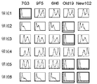
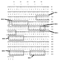
- FIG. 4 is a diagram showing, by dotted lines, regions of the human IL-3R ⁇ molecule in which the regions 1 to 7 arranged outside the molecule are replaced with GM-CSFR ⁇ sequences in the A and B domain nucleic acid and amino acid sequences.
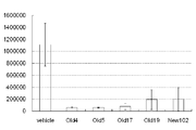
- the vertical axis shows the cell growth inhibition rate (%), and the horizontal axis shows the names of various IL-3R ⁇ antibodies.
- GM indicates Granulocyte / Macrophage strain
- E indicates Erythroid colony
- GEMM indicates mixed colony.
- FIG. 8 used PBMC not cultured with IL-2
- FIG. 9 used PBMC cultured with IL-2.
- the present invention relates to human IL-3R ⁇ that does not inhibit IL-3 signal and binds to the B domain of human IL-3 receptor ⁇ chain (hereinafter abbreviated as IL-3R ⁇ ) and does not bind to the C domain. Relates to antibodies to the chain.
- IL-3R ⁇ human IL-3 receptor ⁇ chain
- IL-3 receptor (hereinafter abbreviated as IL-3R), particularly IL-3R ⁇ , is expressed on the cell surface of leukemia stem cells.
- IL-3R ⁇ the IL-3 receptor ⁇ chain
- IL-3R ⁇ the IL-3 receptor ⁇ chain
- IL-3R ⁇ the IL-3 receptor ⁇ chain
- IL-3R ⁇ the IL-3 receptor ⁇ chain
- IL-3R ⁇ the IL-3 receptor ⁇ chain
- IL-3R ⁇ IL-3 receptor ⁇ chain
- IL-3R ⁇ IL-3 receptor ⁇
- IL-3R ⁇ IL-3 receptor ⁇ gene is a type I transmembrane protein belonging to the cytokine receptor family.
- IL-3R ⁇ molecules are expressed in some hematopoietic progenitor cells, basophils, and some dendritic cells.
- expression in hematopoietic tumors and leukemias is mainly known.
- tumors expressing IL-3R ⁇ include AML, acutely transformed CML blasts, leukemia stem cells and differentiation marker negative CD34 positive CD38 negative fractions, AML, CML, MDS, ALL, It is known to be expressed in SM.
- IL-3 a known ligand of IL-3R ⁇ , is expressed in the blood by activated T cells, natural killer cells, mast cells, and some megakaryocyte cells.
- IL-3R ⁇ is also called CD123.
- IL-3R ⁇ includes mammalian (eg, primate, human) IL-3R ⁇ .
- IL-3R ⁇ sequences such as human IL-3R ⁇ include polymorphic variants.
- Specific examples of full-length human IL-3R ⁇ include the following amino acid sequences.
- the C domain As the A domain, from the 18th glutamine (Q) of the amino acid of SEQ ID NO: 2 to the 100th serine (S), as the B domain, from the 101st glycine (G) of the amino acid of SEQ ID NO: 2 to the 203rd Up to serine (S), the C domain has a region from 204th glutamine (Q) to 308th leucine (L) of the amino acid of SEQ ID NO: 2. Furthermore, among the A and B domains, the following seven regions can be mentioned as the regions arranged outside the molecule.
- region 1 from the 55th aspartic acid (D) to 61st proline (P) of the amino acid of SEQ ID NO: 2, and as region 2, the 63rd valine (V) of the amino acid of SEQ ID NO: 2 to 70th Up to phenylalanine (F), region 3 and from the 91st serine (S) of amino acid of SEQ ID NO: 2 to 98th glutamic acid (E), as region 4, the 97th amino acid of SEQ ID NO: 2 From proline (P) to 104th tryptophan (W), as region 5, from amino acid 122 of cysteine (C) of amino acid of SEQ ID NO: 2 to 128th proline (P), as region 6, SEQ ID NO: 2 From region 182 to isoleucine (I) of amino acid 182 to serine (S) of position 188, examples of region 7 include 192nd glycine (G) to amino acid 198 of lysine (K) of amino acid SEQ ID NO: 2.
- the antibody of the present invention is an antibody that binds to the 101st to 203rd amino acid sequence and does not bind to the 204th to 308th amino acid sequence in the amino acid sequence of SEQ ID NO: 2, which is the extracellular domain of IL-3R ⁇ . Furthermore, antibodies that bind to amino acid sequences 182 to 188 and 192 to 198 in the amino acid sequence shown in SEQ ID NO: 2 can be mentioned. The antibody of the present invention binds to the specific region described above of the extracellular domain of IL-3R ⁇ and does not inhibit IL-3 signal.
- “does not inhibit IL-3 signal” means that IL-3 does not inhibit intracellular signal via IL-3R, and does not inhibit the association of IL-3 and IL-3R and This includes not inhibiting the binding of IL-3R ⁇ and ⁇ chains.
- the cell growth inhibition rate shown in FIG. 5 is 40% or more, preferably 60% or more, more preferably 80% or more when the antibody concentration is 10 ⁇ g / mL. It means that.
- “blocking of IL-3 signal” and “inhibition of IL-3 signal” are used interchangeably and are not distinguished from each other. The ability to inhibit a signal.
- the antibody of the present invention has high antibody-dependent cytotoxic activity (ADCC).
- An IL-3R ⁇ antibody having ADCC activity refers to an antibody that binds to cells expressing IL-3R ⁇ and can kill IL-3R ⁇ -expressing cells via effector cells having cytotoxic activity such as NK cells.
- the high ADCC activity is specific when the antibody concentration is 0.01 ⁇ g / mL or less as measured by the Colon-26 / hCD123ADCC measurement method using IL-2 cultured PBMC described in Example 11. This means that the dissolution rate is 10% or more.
- IL-3R ⁇ -expressing cells include blood tumor cells (acutemyeloidleukemia (AML) cells, chronicmyeloidleukemia (CML) cells, myelodysplasticsyndromes (MDS) cells, acutelymphoidleukemia (ALL) cells, chroniclymphoidleukemia (CLL) cells, multiple myeloma) : MM) cells, systemicmastocytoma (SM) cells, etc., regulatory T cells (eg CD4 positive CD25 positive cells), antigen presenting cells (eg dendritic cells, monocytes / macrophages and similar cells (liver stellate cells, Osteoclasts, microglial cells, intraepidermal macrophages, dust cells (alveolar macrophages))), basophils and the like.
- AML acutemyeloidleukemia
- CML chronicmyeloidleukemia
- MDS myelodysplasticsyndromes
- ALL acutelymphoidleukemia
- AML cells, CML cells, ALL cells, CLL cells, MDS cells, SM cells, MM cells, and various lymphoma cells include respective tumor stem cells.
- the tumor stem cell is one of a group of cells constituting a tumor, for example, lineage ( ⁇ ) CD34 (+) CD38 ( ⁇ ) bone marrow cells in acute myeloid leukemia (AML). Therefore, since the antibody of the present invention has high ADCC activity, it induces reduction or elimination of cells in which IL-3R ⁇ is expressed.
- the IL-3R ⁇ antibody of the present invention includes an IL-3R ⁇ antibody having a heavy chain CDR and a light chain CDR selected from the group consisting of the following (a) to (e).
- the heavy chain CDRs 1 to 3 are amino acid sequences represented by SEQ ID NOs: 113 to 115 and the light chain CDRs 1 to 3 are represented by SEQ ID NOs: 131 to 133.
- the heavy chain CDRs 1 to 3 are SEQ ID NOs: The amino acid sequence of 116 to 118 and the light chain CDRs 1 to 3 are represented by SEQ ID NOs: 134 to 136 (c) The heavy chain CDRs 1 to 3 of the amino acid sequence represented by SEQ ID NOs: 119 to 121 and the light chain CDRs 1 to 3 are represented by SEQ ID NOs: 137 to 139 (d) Heavy chain CDRs 1 to 3 are represented by SEQ ID NOs: 122 to 124 and light chain CDRs are represented by SEQ ID NOs: 140 to 142 Amino acid sequence (e) The heavy chain CDRs 1 to 3 are represented by SEQ ID NOs: 125 to 127, and the light chain CDRs 1 to 3 are represented by SEQ ID NOs: 143 to 145.
- IL-3R ⁇ antibody having a heavy chain variable region and a light chain variable region selected from the group consisting of (a) to (f) of the above (in parentheses, the name of the antibody of the below-mentioned Examples from which each variable region sequence is derived) Show.)
- Antibody name: Old5 (c) the heavy chain variable region comprising the amino acid sequence from the 20th glutamine (Q) to the 139th serine (S) of the amino acid sequence shown in SEQ ID NO: 61 and the 23rd of the amino acid sequence shown in SEQ ID NO: 63 A light chain variable region comprising an amino acid sequence from aspartic acid (D) to 129th lysine (K).
- Antibody name: Old17 (d) a heavy chain variable region comprising the amino acid sequence from the 20th glutamine (Q) to the 139th serine (S) of the amino acid sequence represented by SEQ ID NO: 65 and the 23rd amino acid sequence represented by SEQ ID NO: 67 A light chain variable region comprising an amino acid sequence from aspartic acid (D) to 129th lysine (K).
- antibody name: Old19 a heavy chain variable region comprising the amino acid sequence from the 20th glutamine (Q) to the 138th serine (S) of the amino acid sequence represented by SEQ ID NO: 69 and the 23rd amino acid sequence represented by SEQ ID NO: 71 A light chain variable region comprising an amino acid sequence from aspartic acid (D) to 129th lysine (K).
- a heavy chain comprising an amino acid sequence in which 1 to 3 amino acid residues are deleted, substituted, added or inserted in the heavy chain variable region and / or light chain variable region represented by (a) to (e) Variable region and / or light chain variable region.
- the term antibody is used in the broadest sense, and includes monoclonal antibodies, polyclonal antibodies, multivalent antibodies, multispecific antibodies (e.g., bispecific antibodies), and antibody fragments as long as they exhibit their desired biological activity. Including.
- the antibody comprises a mature heavy or light chain variable region sequence.
- Antibodies also have modified and mutated forms, such as substitutions within or outside of the antibody constant region, complementarity determining region (CDR) or framework (FR) region of the mature heavy or light chain variable region sequence. Including. In certain embodiments, the substitution includes a conservative amino acid substitution.
- the antibody also includes a partial sequence of a mature heavy or light chain variable region sequence. In particular embodiments, the subsequence is selected from Fab, Fab ′, F (ab ′) 2 , Fv, Fd, single chain Fv (scFv), disulfide bond Fv (sdFv) and VL or VH.
- the antibody also includes a heterologous domain.
- the heterologous domain includes a tag, a detectable label, or a cytotoxic agent.
- Antibodies include monoclonal and polyclonal antibodies, any isotype or subclass thereof.
- the antibody is an IgG (eg, IgG1, IgG2, IgG3 or IgG4), IgA, IgM, IgE, or IgD isotype.
- a “monoclonal” antibody is based on a single clone, including a eukaryotic clone, a prokaryotic clone, or a phage clone, and is derived from a single clone, including a eukaryotic clone, a prokaryotic clone, or a phage clone, or a eukaryotic clone.
- a “monoclonal” antibody is therefore structurally defined and not the method by which it is produced.
- IL-3R ⁇ antibody, anti-IL-3R ⁇ and anti-IL-3R ⁇ antibody refer to antibodies that specifically bind to IL-3R ⁇ . Specific binding is selective for an epitope present in IL-3R ⁇ . Specific binding can be distinguished from non-specific binding using assays known in the art (eg, immunoprecipitation, ELISA, flow cytometry, Western blotting).
- IL-3R ⁇ antibody specifically binds to another protein with high sequence or structural homology to the IL-3R ⁇ epitope, depending on the sequence or degree of structural homology of the IL-3R ⁇ epitope. there is a possibility.
- IL-3R ⁇ antibodies may bind to different proteins when epitopes with sufficient sequence or structural homology exist in different proteins.
- IL-3R ⁇ antibodies include isolated and purified antibodies.
- Antibodies of the present invention, including isolated or purified IL-3R ⁇ antibodies, include humans.
- isolated used as a modifier of a composition means that the composition is made by hand or one or more other components in a naturally occurring in vivo environment. Generally means separated by one or more operational steps or processes. In general, a composition so separated is substantially free of one or more materials to which such composition normally binds naturally, eg, one or more proteins, nucleic acids, lipids, carbohydrates, cell membranes. . Thus, an isolated composition is separated from other biological components in the cells of the organism in which it naturally occurs, or from an artificial medium from which the composition is produced (eg, synthetically or by cell culture). ing.
- an isolated IL-3R ⁇ antibody is obtained from an animal from which the antibody is produced (eg, a non-transgenic mammal or a transgenic mammal (such as a rodent (mouse) or ungulate (bovine) animal)). Separated from other polypeptides and nucleic acids. Therefore, the serum containing the antibody obtained from the animal is considered to be isolated.
- isolated does not exclude other physical forms; for example, an isolated antibody can include antibody subsequences, chimeric, multimeric, or derivatized forms.
- purified used as a composition modifier refers to a composition that is generally free of most or substantially all of the materials with which it naturally binds. Purified antibodies are generally removed from components normally present in the antibody environment. Therefore, it is considered that the antibody supernatant separated from the antibody-producing hybridoma cell culture is purified. Thus, purification does not require absolute purity, but is a context specific. Furthermore, a “purified” composition can be combined with one or more other molecules. As such, the term “purified” does not exclude combinations of compositions.
- Purity should be determined by any appropriate method such as, for example, UV spectroscopy, chromatography (eg, HPLC, gas phase), gel electrophoresis (eg, silver or Coomassie staining) and sequence analysis (peptides and nucleic acids). Can do.
- Protein and nucleic acids include proteins and nucleic acids obtained by standard purification methods. The term also includes proteins and nucleic acids obtained by recombinant expression or chemical synthesis in a host cell.
- purified means that the level of contaminants is higher than the level approved by the supervisory authority for administration to humans or non-human animals, for example, the Food and Drug Administration (FDA). Sometimes refers to a low composition.
- FDA Food and Drug Administration
- IL-3R ⁇ antibodies include antibodies that bind to IL-3R ⁇ and modulate IL-3R ⁇ function or activity in vivo or in vitro (eg, in a subject).
- modulate and grammatical variations thereof when used with respect to IL-3R ⁇ activity or function may affect, modify or alter IL-3R ⁇ activity or function in a detectable manner. Means that inhibiting IL-3 signaling is not included.
- an IL-3R ⁇ antibody that modulates the activity or function of IL-3R ⁇ is affected, altered or altered so that one or more IL-3R ⁇ activity or function can be detected without inhibiting the IL-3R signal.
- Such an IL-3R ⁇ activity or function includes, for example, IL-3R ⁇ binding to an IL-3R ⁇ ligand (eg, IL-3), IL-3R ⁇ -mediated signaling or IL-3R ⁇ -mediated cells.
- IL-3R ⁇ ligand eg, IL-3
- IL-3R ⁇ -mediated signaling e.g. IL-3
- IL-3R ⁇ -mediated cells e.g. IL-3R ⁇ -mediated cell.
- Responses or cellular responses that can be modulated by IL-3R ⁇ , or other IL-3R ⁇ activities or functions described or otherwise known or known can be included.
- IL-3R ⁇ activities and functions that can be modulated include, for example, IL-3R ⁇ -mediated signaling or IL-3R ⁇ -mediated cellular responses or cell responses that can be modulated by IL-3R ⁇ , cell proliferation or Increase (eg, AML cells, CML cells, ALL cells, CLL cells, MDS cells, MM cells, SM cells, various lymphoma cells, monocytes, macrophages, mast cells, basophils, helper T cells, regulatory T cells, Natural killer cells, myeloid progenitor cells, lymphoid progenitor cells, etc.), cell survival or apoptosis (eg, AML cells, CML cells, ALL cells, CLL cells, MDS cells, MM cells, SM cells, various lymphoma cells, Monocytes, macrophages, mast cells, basophils, helper T cells, regulatory T cells, natural killer cells, myeloid progenitor cells, lymphocyte progenitor cells, etc.), cytokines (eg For example, AML
- Specific cytokines that are modulated include, but are not limited to, IL-1, IL-2, IL-4, IL-5, IL-6, IL-9, IL-10, IL-14, IL-16, IL-17, IL-23, IL-26, TNF- ⁇ and interferon ⁇ (in vivo or in vitro).
- Specific anti-apoptotic protein or pro-apoptotic protein expression includes, but is not limited to, Bcl-xL, Bcl-2, Bad, Bim and Mcl-1.
- exemplary IL-3R ⁇ antibodies described herein include one or more IL-3R ⁇ -mediated signaling or IL-3R ⁇ -mediated cellular responses or cell responses induced by IL-3R ⁇ , cell proliferation (eg, , AML cells, CML cells, ALL cells, CLL cells, MDS cells, MM cells, SM cells, various lymphoma cells, monocytes, macrophages, mast cells, basophils, helper T cells, regulatory T cells, natural killer cells , Myeloid progenitor cells, lymphoid progenitor cells, etc.), cell survival or apoptosis (eg, AML cells, CML cells, ALL cells, CLL cells, MDS cells, MM cells, SM cells, various lymphoma cells, monocytes, Macrophages, mast cells, basophils, helper T cells, regulatory T cells, natural killer cells, myeloid progenitor cells, lymphoid progenitor cells, etc.), cytokines (eg Th1, Th2 and other non-
- an IL-3R ⁇ antibody of the invention modulates AML cell proliferation or survival and other blood tumor cells (eg, CML cells, ALL cells, CLL cells, MDS cells, MM cells, SM cells). , Or various lymphoma cells), monocytes, macrophages, mast cells, basophils, helper T cells, regulatory T cells, natural killer cells, myeloid progenitor cells, lymphocyte progenitor cells, etc. Modulates the growth or survival of non-hematologic tumor cells, or reduces, disappears or depletes AML, CML, ALL, CLL, MDS, MM, SM, or various lymphoma cells.
- AML cell proliferation or survival and other blood tumor cells eg, CML cells, ALL cells, CLL cells, MDS cells, MM cells, SM cells.
- IL-3R ⁇ antibodies include modified forms such as substitutions (eg, amino acid substitutions), additions and deletions (eg, partial sequences or fragments), also referred to as “variants”. Such modified antibody forms and variants are capable of binding at least a portion of the IL-3R ⁇ antibody function or activity of the present invention, eg, IL-3R ⁇ , or IL-3R ⁇ activity or function (eg, IL-3R ⁇ ). -3R ⁇ signaling) is retained.
- a modified IL-3R ⁇ antibody can retain, for example, the ability to modulate at least some IL-3R ⁇ binding or one or more IL-3R ⁇ functions or activities (eg, signaling, cellular responses, etc.). it can.
- Modify and grammatical variations thereof mean that the composition is out of the reference composition.
- Modified proteins, nucleic acids and other compositions have higher or lower activity than unmodified reference proteins, nucleic acids or other compositions, or are unmodified reference proteins, nucleic acids or other compositions Can have different functions.
- nucleic acid sequences encoding antibodies that contain amino acid substitutions are also provided.
- identity or “identical” means that two or more referenced entities are the same. Thus, when two protein sequences (eg, IL-3R ⁇ antibodies) are identical, they have the same amino acid sequence at least within the referenced region or portion. “Identity region” refers to the same portion of two or more referenced entities. Thus, if two protein sequences are identical in one or more sequence regions, they share identity within that region.
- “Substantial identity” means that a molecule has a function or activity of at least part of one or more reference molecule functions or activities, or an associated / corresponding region or part of a reference molecule with which the molecule shares identity Means that it is structurally or functionally preserved, as expected.
- a polypeptide having substantial identity eg, IL-3R ⁇ antibody
- one or more modifications that retain at least some activity or function of an unmodified IL-3R ⁇ antibody eg, deletion, substitution, addition, or addition of 1-3 amino acid residues
- An IL-3R ⁇ antibody having an insertion is considered to have substantial identity to a reference IL-3R ⁇ antibody.
- the amount of sequence identity required to retain function or activity will depend on the protein, the region and the function or activity of the region.
- a protein can retain some activity or function with only 30% amino acid sequence identity but is generally higher than the reference sequence, e.g., 50%, 60%, 75%, 85 There is%, 90%, 95%, 96%, 97%, or 98% identity.
- the degree of identity between two sequences can be ascertained using computer programs and mathematical algorithms known in the art. Such algorithms that calculate percent sequence identity (homology) generally account for sequence gaps and mismatches across the comparison region. For example, a BLAST (eg, BLAST 2.0) search algorithm (see, eg, Altschul et al., J. Mol.
- Biol. 215: 403 (1990), publicly available through NCBI) is an exemplary search parameter such as: Has: mismatch-2; gap start 5; gap extension 2.
- the BLASTP algorithm is typically used in combination with a score matrix such as PAM100, PAM 250, BLOSUM 62 or BLOSUM50.FASTA (eg, FASTA2 and FASTA3), and the SSEARCH sequence comparison program also quantifies the degree of identity (Pearson et al., Proc. Natl. Acad. Sci USA 85: 2444 (1988); Pearson, Methods Mol Biol. 132: 185 (2000); and Smith et al., J. Mol. Biol. 147: 195 (1981)) .
- a program for quantifying protein structural similarity using Delaunay-based phase mapping has also been developed (Bostick et al., Biochem Biophys ResCommun. 304: 320 (2003)).
- a “conservative substitution” is a substitution of one amino acid with a biologically, chemically or structurally similar residue.
- Biologically similar means that the substitution does not destroy biological activity, eg, IL-3R ⁇ binding activity.
- Structurally similar means that the amino acids have side chains of the same length (eg, alanine, glycine and serine) or are of similar size.
- Chemical similarity means that the residues have the same charge or are hydrophilic or hydrophobic.
- substitution of one hydrophobic residue with another such as isoleucine, valine, leucine or methionine, or substitution of another monopolar residue, for example, substitution of arginine with lysine
- substitution of glutamic acid with aspartic acid substitution of glutamine with asparagine, substitution of serine with threonine, and the like.
- Modified antibodies also include peptides having one or more D-amino acids, structural and functional analogs substituted with L-amino acids (and mixtures thereof), eg, synthetic or unnatural amino acids or amino acid analogs Also included are mimetics and derivatized forms. Modifications include cyclic structures such as end-to-end amide bonds or intra- or intermolecular disulfide bonds between amino and carboxy terminals.
- Non-limiting further specific examples of amino acid modifications include IL-3R ⁇ subsequences and fragments.
- Exemplary IL-3R ⁇ subsequences and fragments comprise a portion of an IL-3R ⁇ sequence to which an exemplary IL-3R ⁇ antibody of the invention binds.
- Exemplary IL-3R ⁇ subsequences and fragments also include an immunogenic portion, eg, a portion of IL-3R ⁇ that includes a sequence to which an exemplary IL-3R ⁇ antibody of the invention binds.
- a nucleic acid encoding an IL-3R ⁇ antibody and an IL-3R ⁇ antibody partial sequence or fragment that retains at least part of the function or activity of the unmodified or reference IL-3R ⁇ antibody is provided.
- the term “subsequence” or “fragment” means a portion of a full-length molecule.
- a partial sequence of an IL-3R ⁇ antibody that encodes an IL-3R ⁇ antibody has at least one less amino acid than the full-length IL-3R ⁇ (eg, deletion of one or more internal or terminal amino acids from either the amino or carboxy terminus) ).
- the partial sequence of the IL-3R ⁇ antibody has at least one fewer amino acid than the full-length IL-3R ⁇ antibody.
- the nucleic acid subsequence has at least one fewer nucleotide than the full length comparison nucleic acid sequence.
- the partial sequence can be any length up to the full length native IL-3R ⁇ .
- IL-3R ⁇ antibody subsequences and fragments have a binding affinity as a full-length antibody, a binding specificity as a full-length antibody, or one or more activities or functions as a full-length antibody, such as the function of an IL-3R ⁇ antagonist or agonist antibody Or it may have activity.
- the terms “functional subsequence” and “functional fragment” when referring to an antibody retain one or more functions or activities as a full-length reference antibody, eg, at least part of the function or activity of an IL-3R ⁇ antibody. Refers to the antibody portion. For example, an antibody subsequence or fragment that binds to IL-3R ⁇ or a fragment of IL-3R ⁇ is considered a functional subsequence.
- Antibody partial sequences and fragments can be combined.
- VL or VH subsequences can be linked by a linker sequence, thereby forming a VL-VH chimera.
- Combinations of single chain Fv (scFv) subsequences can be linked by a linker sequence, thereby forming an scFv-scFv chimera.
- IL-3R ⁇ antibody subsequences and fragments include single chain antibodies or variable regions alone or in combination with all or part of other IL-3R ⁇ antibody subsequences.
- Antibody subsequences and fragments can be prepared by proteolytic hydrolysis of the antibody, for example, pepsin or papain digestion of whole antibodies. Antibody subsequences and fragments generated by enzymatic cleavage with pepsin yield a 5S fragment denoted as F (ab ′) 2 . This fragment can be further cleaved using a thiol reducing agent to create a 3.5SFab ′ monovalent fragment. Alternatively, enzymatic cleavage with pepsin directly results in two monovalent Fab ′ and Fc fragments (eg, US Pat. Nos. 4,036,945 and 4,331,647; and Edelman et al., Methods Enymol. 1: 422 ( 1967)). Other antibody cleavage methods may also be used, such as heavy chain separation to form a monovalent light-heavy chain fragment, further cleavage of the fragment, or other enzymatic or chemical methods.
- Proteins and antibodies, and their partial sequences and fragments can be created by genetic methods.
- the technology involves expressing all or part of a gene encoding a protein or antibody in a host cell such as Cos cells or E. coli.
- Recombinant host cells synthesize full-length or partial sequences, eg, scFv (eg, Whitlow et al., In: Methods: A Companion to Methods inEnzymology 2:97 (1991), Bird et al., Science 242: 423 (1988); And US Pat. No. 4,946,778).
- Single chain Fv and antibodies are described in U.S. Pat.Nos.
- Modified forms include derivatized sequences such as free amino groups forming amine hydrochlorides, p-toluenesulfonyl groups, carbobenzoxy groups; free carboxy groups forming salts, methyl and ethyl esters.
- Modifications can be made using methods known in the art (eg, PCR-based site-specific, deletion and insertion mutagenesis, chemical modification and mutagenesis, cross-linking, etc.).
- Modified forms of proteins include adducts and inserts.
- an addition can be a covalent or non-covalent association of any type of molecule with a protein (eg, antibody), nucleic acid or other composition.
- Adducts and inserts include fusion (chimeric) polypeptide or nucleic acid sequences that have one or more molecules that are not normally present in a reference natural (wild-type) sequence covalently linked to said sequence. It is.
- a specific example is the amino acid sequence of another protein (eg, antibody) to create a multifunctional protein (eg, multispecific antibody).
- the antibodies of the present invention also include chimeras or fusions in which one or more additional domains are covalently linked to provide different or accessory functions or activities.
- Antibodies include chimeras or fusions that do not naturally occur in nature, in which two or more amino acid sequences are linked together.
- an IL-3R ⁇ antibody containing a heterologous domain and a nucleic acid encoding the IL-3R ⁇ antibody are provided.
- a heterologous domain can be an amino acid adduct or an insert, but is not limited to amino acid residues.
- a heterologous domain can consist of any of a variety of different types of small or large functional parts.
- moieties include nucleic acids, peptides, carbohydrates, lipids or small organic compounds such as drugs, metals (gold, silver) and the like.
- heterologous domains include, for example, tags, detectable labels and cytotoxic agents.
- tags and detectable labels include T7-, His-, myc-, HA- and FLAG-tags; enzymes (horseradish peroxidase, urease, catalase, alkaline phosphatase, ⁇ -galactosidase, chloramphenicol Enzyme substrate; ligand (eg, biotin); receptor (avidin); radionuclide (eg, C14, S35, P32, P33, H3, I125 and I131); electron density reagent; energy transfer molecule; paramagnetic label Fluorophores (fluorescein, rhodamine, phycoerythrin); chromophores; chemiluminescent agents (imidazole, luciferase); and bioluminescent agents.
- cytotoxic agents include diphtheria toxin (diptheria, toxin), cholera
- a linker sequence such that the two entities at least partially maintain different functions or activities between a protein (eg, antibody), nucleic acid, or other composition and an adduct or insert (eg, a heterologous domain) May be inserted.
- a linker sequence may have one or more properties that can promote or interact with either domain, such as flexible structure, Subsequent structures cannot be formed or include hydrophobicity or chargeability.
- Amino acids commonly found in the flexible protein region include glycine, asparagine and serine. Other near neutral amino acids such as threonine and alanine may also be used in the linker sequence.
- the length of the linker sequence can vary (see, eg, US Pat. No. 6,087,329).
- Linkers include chemical crosslinkers and conjugating agents such as sulfo-succinimidyl derivatives (sulfo-SMCC, sulfo-SMPB), disuccinimidyl suberate (DSS), disuccinimidyl glutarate (DSG) and tartaric acid Further included is disuccinimidyl (DST).
- sulfo-succinimidyl derivatives sulfo-SMCC, sulfo-SMPB
- DSS disuccinimidyl suberate
- DSG disuccinimidyl glutarate
- tartaric acid Further included is disuccinimidyl (DST).
- Modified and mutated antibodies are those that can retain the detectable activity of an IL-3R ⁇ antibody.
- the modified antibody has binding activity to an IL-3R ⁇ molecule and induces reduction or elimination of IL-3R ⁇ -expressing cells by an immune system centered on effector cells. It is involved in the functional control of IL-3R ⁇ -expressing cells and induces cell survival, proliferation, rest, cell death, and the like. Cell death includes apoptosis, necrosis, autophagy and the like.
- the present invention further provides cell-free methods (eg, in solution, in solid phase) and cell-based methods (eg, in vitro or in vivo) for screening, detecting, and identifying IL-3R ⁇ .
- the These methods can be performed in solution, using biomaterials or samples in vitro, and in vivo, for example, in samples of animal-derived cells (eg, lymphocytes).
- the method comprises contacting a biomaterial or sample with an antibody that binds IL-3R ⁇ under conditions that allow binding of the antibody to IL-3R ⁇ ; binding of the antibody to IL-3R ⁇ ; Assaying for.
- the presence of IL-3R ⁇ is detected by binding the antibody to IL-3R ⁇ .
- IL-3R ⁇ is present in a cell or tissue.
- the biomaterial or sample is obtained from a mammalian subject.
- composition such as a protein (eg, IL-3R ⁇ antibody), material, sample, or treatment
- contacting refers to that composition (eg, an IL-3R ⁇ antibody) and other references.
- a specific example of direct interaction is binding.
- a specific example of indirect interaction is when the composition acts on an intermediate molecule that acts on the next referenced entity.
- a cell eg, lymphocyte
- the antibody is bound to the cell (eg, through binding to IL-3R ⁇ ) or the antibody is intermediate And this intermediate then acts on the cell.
- the terms “assaying” and “measuring” and grammatical variations thereof are used interchangeably herein and refer to either qualitative or quantitative measurements, or qualitative and quantitative measurements. Refers to both. When these terms are used in reference to binding, any means of assessing relative amounts, binding affinity or specificity is contemplated, including various methods described herein and known in the art.
- binding of an IL-3R ⁇ antibody to IL-3R ⁇ can be assayed or measured by a flow cytometry assay. (Preparation of antibody)
- the present invention also provides a method for producing a human IL-3R ⁇ antibody having IL-3R ⁇ positive cytotoxic activity.
- the method comprises transforming a human IL-3R ⁇ extracellular domain or IL-3R ⁇ transgenic cell conjugated with a human Fc recombinant protein into an animal capable of expressing human immunoglobulin (eg, a transgenic mouse or a trans Screening the animal for expression of human IL-3R ⁇ antibody; selecting an animal producing human IL-3R ⁇ antibody; isolating the antibody from the selected animal .
- a human immunoglobulin eg, a transgenic mouse or a trans Screening the animal for expression of human IL-3R ⁇ antibody; selecting an animal producing human IL-3R ⁇ antibody; isolating the antibody from the selected animal .
- IL-3R ⁇ protein suitable for antibody production can be produced by any of a variety of standard protein purification or recombinant expression techniques.
- IL-3R ⁇ sequences can be generated by standard peptide synthesis techniques, such as solid phase synthesis.
- a portion of the protein may include an amino acid sequence such as a FLAG tag, T7 tag or polyhistidine sequence to facilitate purification of the expressed or synthesized protein.
- the protein can be expressed in cells and purified.
- the protein can be expressed as part of a larger protein (eg, a fusion or chimera) by recombinant methods.
- Suitable forms of IL-3R ⁇ for raising an immune response include IL-3R ⁇ subsequences, such as immunogenic fragments. Additional forms of IL-3R ⁇ include IL-3R ⁇ expressing cells, IL-3R ⁇ containing preparations or cell extracts or fractions, partially purified IL-3R ⁇ .
- IL-3R ⁇ or an immunogenic fragment thereof is optionally conjugated with a carrier such as keyhole limpet hemocyanin (KLH) or ovalbumin (eg BSA), or an adjuvant such as Freund's complete or incomplete adjuvant. And is used to immunize animals.
- KLH keyhole limpet hemocyanin
- BSA ovalbumin
- an adjuvant such as Freund's complete or incomplete adjuvant.
- splenocytes from immunized animals that respond to IL-3R ⁇ can be isolated and fused with myeloma cells.
- Monoclonal antibodies produced by hybridomas can be screened for reactivity with IL-3R ⁇ or immunogenic fragments thereof.
- Immunized animals include primates, mice, rats, rabbits, goats, sheep, cows, or guinea pigs.
- the initial and optional boosts may be by intravenous, intraperitoneal, intramuscular, or subcutaneous routes.
- the antigen can be combined with another protein such as ovalbumin or keyhole limpet hemocyanin (KLH), thyroglobulin and tetanus toxoid, or such as Freund's complete or incomplete adjuvant. Can be mixed with an adjuvant.
- KLH keyhole limpet hemocyanin
- thyroglobulin and tetanus toxoid or such as Freund's complete or incomplete adjuvant.
- the initial and optional booster immunization may be by the intraperitoneal route, intramuscular route, intraocular route, or subcutaneous route.
- the booster immunizations may be the same or different concentrations of IL-3R ⁇ preparation and may be regular or irregular intervals.
- Animals include those that have been genetically modified to include human loci and can be used to create human antibodies.
- Transgenic animals having one or more human immunoglobulin genes are described, for example, in US Pat. No. 5,939,598, WO02 / 43478, and WO 02/092812.
- splenocytes from immunized mice that are highly responsive to antigen can be isolated and fused with myeloma cells.
- Monoclonal antibodies that bind to IL-3R ⁇ can be obtained.
- human when used in reference to an antibody means that the amino acid sequence of the antibody is a fully human amino acid sequence, ie, a human heavy chain and human light chain variable region and a human constant region. Thus, all of the amino acids are human amino acids or are present in human antibodies.
- An antibody that is a non-human antibody can be made a fully human antibody by replacing non-human amino acid residues with amino acid residues present in the human antibody.
- Amino acid residues, CDR region maps and human antibody consensus residues present in human antibodies are known in the art (eg, Kabat, Sequences of Proteins of Immunological Interest, 44th edition USDepartmentofHealthandHumanServices.PublicHealthService (1987) See Chothia and Lesk (1987), a human VH subgroup III consensus sequence based on a survey of 22 known human VHIII sequences, and a human VL based on a survey of 30 known human ⁇ I sequences. Consensus sequences for kappa chain subgroup I are described in Padlan Mol. Immunol.31: 169 (1994); and PadlanMol.Immunol.28: 489 (1991). Are substituted with one or more amino acids present in any other human antibody.
- IL-3R ⁇ antibodies include, for example, CDR-grafting (EP 239,400; W091 / 09967; U.S. Pat.No. 5,225,539; do 5,530,101; and 5,585,089), veneering or reser Resurfacing (EP592,106; EP519,596; Padlan, MolecularImmunol. 28: 489 (1991); Studnicka et al., Protein Engineering 7: 805 (1994); Roguska. Et al., Proc. Nat'l Acad. Sci. USA 91: 969 (1994)), and humanized antibodies that can be generated using techniques known in the art such as chain shuffling (US Pat. No. 5,565,332). Human consensus sequences (Padlan, Mol.
- humanized when used in reference to an antibody refers to one or more complementarity determining region (CDR) non-human amino acids whose antibody amino acid sequence specifically binds to a desired antigen in an acceptor human immunoglobulin molecule. Having residues (eg, mouse, rat, goat, rabbit, etc.) and one or more human amino acid residues in the Fv framework region (FR) (which are amino acid residues flanking the CDR) means.
- CDR complementarity determining region
- Antibodies referred to as “primatization” include any human amino acid residue in the acceptor human immunoglobulin molecule and framework region in addition to any human residue (eg, monkey, gibbon, gorilla, It is within the meaning of “humanization” except that it can be a chimpanzee orangutan, macaque monkey).
- the human FR residues of the immunoglobulin can be replaced with corresponding non-human residues.
- a humanized antibody may comprise residues that are found neither in the human antibody nor in the donor CDRs or framework sequences.
- FR substitutions at a particular position not found in human antibodies or donor non-human antibodies can be expected to improve binding affinity or specific human antibodies at that position.
- Antibody frameworks and CDR substitutions based on molecular modeling are well known in the art, for example, modeling of CDR and framework residue interactions and specific locations to identify framework residues important for antigen binding. By sequence comparison to identify unusual framework residues in (see, eg, US Pat. No. 5,585,089; and Riechmann et al., Nature 332: 323 (1988)).
- IL-3R ⁇ antibody includes chimeric antibody.
- the term “chimera” and grammatical variations thereof when used with respect to an antibody is derived from two or more different species, wherein the amino acid sequence of the antibody is derived from two or more different species, or simply Means containing one or more moieties that are separated or based on two or more different species.
- a portion of the antibody can be human (eg, a constant region) and another portion of the antibody can be non-human (eg, a murine heavy chain or murine light chain variable region).
- an example of a chimeric antibody is an antibody in which different portions of the antibody are of different species origin. Chimeric antibodies, unlike humanized or primatized antibodies, can have different species of sequences in any region of the antibody.
- IL-3R ⁇ antibodies can also be produced using hybridoma technology, recombinant technology, and phage display technology, or combinations thereof (US Pat. Nos. 4,902,614, 4,543,439, and 4,411,993). See MonoclonalAntibodies.Hybridomas: ANewDimensioninBiologicalAnalyses, PlenumPress, Kennett, McKearn, and Bechtol (ed.), 1980, and Harlow et al., Antibodies: ALaboratoryManual, ColdSpringHarborLaboratoryPress, 2nd edition 1988)
- the human anti-human IL-3R ⁇ antibody of the present invention uses chromosome-transfected mice (KM mice (trademark)) immunized with cell lines expressing various forms of solubilized recombinant human IL-3R ⁇ protein or IL-3R ⁇ . (WO02 / 43478, WO 02/092812, and Ishida, et al., IBC's 11th Antibody Engineering Meeting. Abstract (2000)).
- the human anti-human antibody stains the human IL-3R ⁇ stable transfected cell line, such as Jurkat-IL-3R ⁇ and L929-IL-3R ⁇ cells, in a detectable manner, rather than the non-transformed parental cell line. Have been shown to bind specifically to human IL-3R ⁇ .
- the antibodies of the present invention may be a kappa or lambda light chain sequence, either one full length, as in a naturally occurring antibody, a mixture thereof (ie, a fusion of a kappa and lambda chain sequence), And their partial sequences / fragments.
- Naturally occurring antibody molecules contain two kappa or two lambda light chains.
- the present invention further provides a method for producing an antibody that specifically binds to IL-3R ⁇ .
- a method for making an IL-3R ⁇ antibody comprises a human IL-3R ⁇ , subsequence or fragment (eg, IL-3R ⁇ extracellular domain), optionally conjugated to a human Fc recombinant protein.
- this method determines whether a human IL-3R ⁇ antibody has IL-3R ⁇ antagonist or agonist activity.
- Effector activity refers to antibody-dependent activity induced through the Fc region of an antibody.
- Antibody-dependent cytotoxic activity (ADCC activity), complement-dependent cytotoxic activity (CDC activity), macrophages and dendritic cells
- ADCC activity Antibody-dependent cytotoxic activity
- CDC activity complement-dependent cytotoxic activity
- macrophages and dendritic cells
- Antibody-dependent phagocytosis (antibody dependent phagocytosis, ADP activity) by phagocytic cells is known.
- N-acetylglucosamine present at the reducing end of an N-linked complex type sugar chain that binds to the 297th asparagine (Asn) of the Fc region of the antibody A method for controlling the amount of fucose (also called core fucose) that binds ⁇ -1,6 to (GlcNAc) (WO2005 / 035586, WO2002 / 31140, WO00 / 61739), or modification of the amino acid residue in the Fc region of an antibody
- the method of controlling with is known.
- the effector activity can be controlled by any method using the anti-IL-3R ⁇ monoclonal antibody of the present invention.
- the effector activity of the antibody can be increased or decreased.
- Examples of a method for reducing the content of fucose bound to the N-linked complex sugar chain bound to Fc of the antibody include defucosylated (non-fucosylated). Defucosylation refers to expressing an antibody using CHO cells lacking the ⁇ 1,6-fucose transferase gene, and an antibody to which fucose is not bound can be obtained. Antibodies without fucose binding have high ADCC activity.
- the antibody is expressed using a host cell into which an ⁇ 1,6-fucose transferase gene has been introduced.
- an antibody to which fucose is bound can be obtained.
- An antibody to which fucose is bound has a lower ADCC activity than an antibody to which fucose is not bound.
- ADCC activity and CDC activity can be increased or decreased by modifying amino acid residues in the Fc region of the antibody.
- the CDC activity of the antibody can be increased.
- ADCC activity or CDC activity can be increased or decreased by performing the amino acid modification described in US6,737,056, US7,297,775, or US7,317,091.
- nucleic acid sequences of the present invention such as vectors.
- the vector includes a nucleic acid sequence encoding an IL-3R ⁇ antibody, subsequence or fragment thereof.
- Nucleic acids can be of various lengths.
- the length of the nucleic acid encoding the IL-3R ⁇ antibody of the present invention or a partial sequence thereof is generally about 100 nucleotides to 600 nucleotides, or any numerical value or numerical range within such a length range, 150, 150-200, 200-250, 250-300, 300-350, 350-400, 400-450, 450-500, 500-550, or about 550-600 nucleotides Covers any number or range or value (anynumerical value or range or value) within a length or range of such lengths.
- the length of the nucleic acid that specifically hybridizes with the nucleic acid encoding the IL-3R ⁇ antibody of the present invention or a partial sequence thereof is generally about 10-20, 20-30, 30-50, 50-100, 100-150, 150-200, 200-250, 250-300, 300-400, 400-500, 500-600 nucleotides, or any number within such a range Or the numerical range.
- nucleic acid and “polynucleotide” refer to at least two or more ribo- or deoxy-ribonucleobase pairs (nucleotides) joined by a phosphate ester bond or equivalent.
- Nucleic acids include polynucleotides and polynucleosides. Nucleic acids include single molecules, double molecules or triple molecules, circular molecules or linear molecules. Exemplary nucleic acids include, but are not limited to: RNA, DNA, cDNA, genomic nucleic acids, naturally occurring and non-natural nucleic acids, eg, synthetic nucleic acids.
- Short nucleic acids and polynucleotides are generally “oligonucleotides” or “probes” of single- or double-stranded DNA. Called.
- Nucleic acids can be created using a variety of standard cloning and chemical synthesis techniques. Techniques include, but are not limited to, nucleic acid amplification of genomic DNA or cDNA targets using primers (eg, degenerate primer mixtures) that can be annealed to antibody coding sequences, such as polymerase chain reaction (PCR). It is done. Nucleic acids can also be created by chemical synthesis (eg, solid phase phosphoramidite synthesis) or transcription from a gene.
- primers eg, degenerate primer mixtures
- PCR polymerase chain reaction
- the generated sequence is then translated in vitro or cloned into a plasmid, propagated, and then in a cell (eg, a host cell such as yeast or bacteria, eukaryote (such as an animal or mammalian cell or plant)) Can be expressed.
- a cell eg, a host cell such as yeast or bacteria, eukaryote (such as an animal or mammalian cell or plant)
- a cell eg, a host cell such as yeast or bacteria, eukaryote (such as an animal or mammalian cell or plant)
- a vector is a mediator that can be manipulated by insertion or incorporation of nucleic acids.
- Vectors include plasmid vectors, viral vectors, prokaryotic (bacterial) vectors and eukaryotic (plant, fungal, mammalian) vectors.
- Vectors can be used for expression of nucleic acids in vitro or in vivo.
- Such vectors are referred to as “expression vectors” and include the introduction of nucleic acids, including nucleic acids encoding IL-3R ⁇ antibodies, subsequences and fragments thereof, or in vitro of encoded proteins (eg, in solution). Useful in expression in a subject in cells or in vivo.
- Vectors can also be used to manipulate nucleic acids.
- a “cloning vector” can be used to transcribe or translate an inserted nucleic acid in vitro (eg, in solution or in solid phase), in a cell, or in a subject in vivo.
- Vectors generally contain an origin of replication for propagation in cells in vitro or in vivo.
- Control elements such as expression control elements present in the vector can be included to facilitate transcription and translation, if desired.
- ⁇ Vectors may contain selectable markers.
- a “selectable marker” is a gene that allows for selection of cells containing the gene. “Positive selection” refers to the process by which positive selection occurs to select cells containing a selectable marker. Drug resistance is an example of a positive selection marker, cells containing the marker survive in the drug-containing culture medium, and cells without the marker die. Selectable markers include drug resistance genes such as neo that confer G418 resistance; hygr that confer hygromycin resistance; and puro that confer puromycin resistance. Other positive selectable marker genes include genes that allow identification or screening of cells that contain the marker.
- GFP and GFP-like chromophore, luciferase are surface markers such as fluorescent protein (GFP and GFP-like chromophore, luciferase) gene, lacZ gene, alkaline phosphatase gene, and CD8, among others.
- Negative selection refers to the process of exposing cells containing a negative selection marker to exposure to an appropriate negative selection agent.
- cells containing the herpes simplex virus thymidine kinase (HSV-tk) gene (Wigler et al., Cell 11: 223 (1977)) are sensitive to the drug ganciclovir (GANC).
- GANC herpes simplex virus thymidine kinase
- the gpt gene renders cells sensitive to 6-thioxanthine.
- Viral vectors include retrovirus (a lentivirus for infecting not only dividing cells but also non-dividing cells), foamy viruses (US Pat. Nos. 5,624,820, 5,693,508, 5,665,577, 6,013,516 and 5,674,703; WO92 / 05266 and WO92 / 14829), adenovirus (US Pat. Nos. 5,700,470, 5,731,172 and 5,928,944), adeno-associated virus (AAV) (US Pat. No. 5,604,090) ), Herpes simplex virus vectors (US Pat. No. 5,501,979), cytomegalovirus (CMV) vectors (US Pat. No.
- retrovirus a lentivirus for infecting not only dividing cells but also non-dividing cells
- foamy viruses US Pat. Nos. 5,624,820, 5,693,508, 5,665,577, 6,013,516 and 5,674,703; WO92 / 05266 and
- Adenoviruses can efficiently infect slowly replicating and / or terminally differentiated cells and can be used to target slowly replicating and / or terminally differentiated cells.
- Additional viral vectors useful for expression include parvovirus, norwalk virus, coronavirus, paramyxovirus and rhabdovirus, togavirus (eg, Sindbis virus and Semliki Forest virus) and vesicular stomatitis virus (VSV). It is done.
- a vector containing a nucleic acid can be expressed when the nucleic acid is operably linked to an expression control element.
- operably linked refers to a physical or functional relationship between the elements that allows them to function as intended.
- a nucleic acid “operably linked” to an expression control element means that the control element modulates nucleic acid transcription, and optionally, translation of the transcript.
- an “expression control element” or “expression control sequence” is a polynucleotide that affects the expression of operably linked nucleic acids. Promoters and enhancers are non-limiting specific examples of expression control elements and sequences.
- a “promoter” is a cis-acting DNA regulatory region capable of initiating transcription of a downstream (3 ′ direction) nucleic acid sequence. The promoter sequence includes nucleotides that promote transcription initiation. Enhancers also regulate nucleic acid expression but act away from the transcription start site of the nucleic acid to which it is operably linked. Enhancers also act when present at either the 5 ′ or 3 ′ end of a nucleic acid and also when present within a nucleic acid (eg, an intron or coding sequence).
- Additional expression control elements include leader and fusion partner sequences, endogenous ribosome binding site (IRES) elements for the creation of multiple genes, or polycistronic messages, intron splicing signals, in-frame translation of mRNA Maintenance of the correct reading frame of the gene to do, a polyadenylation signal that results in proper polyadenylation of the transcript of interest, and a stop codon.
- IRS endogenous ribosome binding site
- Expression control elements include “constitutive” elements in which transcription of operably linked nucleic acids occurs in the absence of a signal or stimulus.
- An expression control element that provides expression in response to a signal or stimulus and increases or decreases the expression of a operably linked nucleic acid is “regulatable”.
- a regulatable element that increases the expression of a linked nucleic acid in a manner that is responsive to a signal or stimulus is called an “inducible element”.
- a regulatable element that reduces the expression of a linked nucleic acid in a manner that is functionally responsive to a signal or stimulus is a “suppressor element” (ie, the signal decreases expression; if the signal is removed or absent) The expression increases).
- constitutive promoters include T7 and inducible promoters such as bacteriophage ⁇ pL, plac, ptrp, ptac (ptrp-lac hybrid promoter).
- constitutive or inducible promoters eg, ecdysone
- yeast constitutive promoters include, for example, inducible promoters such as ADH or LEU2 and GAL (eg, Ausubel et al., In: CurrentProtocolsin Molecular Biology, Volume 2, Chapter 13, GreenePublish. Assoc.
- a constitutive promoter of virus or other origin may be used.
- LTR viral long terminal repeat
- mammalian cell genome eg, metallothionein IIA promoter; heat shock promoter, steroid / thyroid hormone / retinoic acid response element
- mammalian virus eg, An inducible promoter derived from an adenovirus late promoter; mouse breast cancer virus LTR
- the expression control element includes an element that is active in a specific tissue or cell type, and such an element is called a “tissue-specific expression control element”.
- a tissue-specific expression control element is generally more active in a particular cell or tissue type, which means that the tissue-specific expression control element is that particular cell or tissue compared to other cells or tissue types. This is because it is recognized by transcription-activating proteins active in the species, or other transcriptional regulators.
- Non-limiting specific examples of such expression control elements include hexokinase II, COX-2, ⁇ -fetoprotein, carcinoembryonic antigen, DE3 / MUC1, prostate specific antigen, C-erB2 / neu, glucose dependent insulin Promoters such as secretory stimulating polypeptides (GIP), telomerase reverse transcriptase and hypoxia-responsive promoters.
- GIP secretory stimulating polypeptides
- a host cell transformed or transfected with the IL-3R ⁇ nucleic acid or vector of the present invention includes, but are not limited to, prokaryotic and eukaryotic cells such as cells of bacteria, fungi (yeast), plants, insects, and animals (eg, mammals such as primates and humans). .
- prokaryotic and eukaryotic cells such as cells of bacteria, fungi (yeast), plants, insects, and animals (eg, mammals such as primates and humans).
- Non-limiting examples of transformed cells include bacteria transformed with recombinant bacteriophage nucleic acid, plasmid nucleic acid or cosmid nucleic acid expression vectors; yeast transformed with recombinant yeast expression vectors; recombinant virus expression vectors (for example, plant cells infected with cauliflower mosaic virus, CaMV; tobacco mosaic virus, TMV) or transformed with recombinant plasmid expression vectors (eg, Ti plasmid); recombinant virus expression vectors (eg, baculovirus) Infected insect cells; and animal cells infected with recombinant viral expression vectors (eg, retroviruses, adenoviruses, vaccinia viruses), or transformed animal cells engineered for stable expression.
- recombinant virus expression vectors for example, plant cells infected with cauliflower mosaic virus, CaMV; tobacco mosaic virus, TMV
- recombinant virus expression vectors eg, Ti plasmid
- Non-limiting examples of mammalian host cells that express IL-3R ⁇ antibodies, subsequences and fragments thereof include CHO cells.
- the host cell may be a primary cell isolate, an isolated secondary cell or subculture cell, or a plurality of cells or cell populations derived from a cell line or immortal cell culture.
- transfected when used in reference to a cell (eg, a host cell) or organism refers to an exogenous molecule, eg, a protein or nucleic acid (eg, a transgene), into the cell. It means a genetic change in cells after uptake.
- a “transfected” or “transformed” cell is a cell into which an exogenous molecule has been introduced by the hand of man, eg, by recombinant DNA technology, or a progeny thereof.
- the nucleic acid or protein can be stably or transiently transfected or transformed (expressed) in the cell and its progeny.
- Cells can be grown and the introduced protein can be expressed or the nucleic acid can be transcribed. Because there may be mutations that occur during replication, the progeny of the transfected or transformed cell may not be identical to the parent cell.
- vectors are used for cell transfection or transformation.
- the vector can be contained within a viral particle or vesicle and, if desired, can be directed to a particular cell type by including a protein on the particle or vesicle surface that binds to a target cell ligand or receptor.
- viral particles or vesicles themselves, or viral surface proteins can be made to target cells for transfection or transformation in vitro, exvivo or in vivo.
- viral and non-viral vector delivery techniques to cells, tissues or organs in vitro, in vivo and exvivo are included.
- introduction of a nucleic acid into a target cell can also be performed by methods known in the art such as osmotic impact (eg, calcium phosphate), electroporation, microinjection, cell fusion, and the like. Can do. Introduction of nucleic acids and polypeptides in vitro, exvivo and in vivo can also be performed using other techniques.
- polymeric materials such as polyester, polyamic acid, hydrogel, polyvinyl pyrrolidone, ethylene-vinyl acetate, methyl cellulose, carboxymethyl cellulose, protamine sulfate, or lactide / glycolide copolymer, polylactide / glycolide copolymer, or ethylene vinyl acetate copolymer.
- Nucleic acids are produced by coacervation techniques or by interfacial polymerization, for example, in microcapsules prepared using hydroxymethylcellulose or gelatin-microcapsules, or poly (methylmethacrolate) microcapsules, respectively.
- it can be encapsulated in a colloidal system.
- Colloidal dispersions include systems based on polymer complexes, nanocapsules, microspheres, beads, and lipids (such as oil-in-water emulsions, micelles, mixed micelles, and liposomes).
- Liposomes for introducing various compositions into cells are known in the art and include, for example, phosphatidylcholine, phosphatidylserine, lipofectin and DOTAP (eg, US Pat. Nos. 4,844,904, 5,000,959, 4,863,740, and 4,975,282; moths and GIBCO-BRL, Gaithersburg, Md).
- Piperazine-based amphilic cationic lipids based on piperazine useful for gene therapy are also known (see, for example, US Pat. No. 5,861,397).
- Cationic lipid systems are also known (see, eg, US Pat. No. 5,459,127).
- polymeric materials, microcapsules and colloidal dispersions such as liposomes
- vesicles are collectively referred to as “vesicles”.
- IL-3R ⁇ antibody isotype can be determined using an ELISA assay, for example, human Ig can be identified using mouse Ig absorbed anti-human Ig.
- Binding affinity can be determined by binding (Ka) and dissociation (Kd) rates.
- the equilibrium affinity constant, KD is the Ka / Kd ratio.
- Binding (Ka) and dissociation (Kd) rates can be measured using surface plasmon resonance (SPR) (Rich and Myszka, Curr. Opin. Biotechnol 11:54 (2000); Englebienne, Analyst. 123: 1599 ( 1998)). Instrumentation and methods for real-time detection and monitoring of binding rates are known and commercially available (BiaCore 2000, Biacore AB, Upsala, Sweden; and Malmqvist, Biochem. Soc. Trans. 27: 335 (1999)).
- the KD value can be defined as the IL-3R ⁇ antibody concentration required to saturate half (50%) of the binding sites in IL-3R ⁇ .
- chimpanzees have major limitations, such as being unable to conduct an anatomical test after the last dose and impossible to carry out a reproductive toxicity test. Therefore, being able to confirm the drug efficacy in monkeys (cynomolgus monkeys and / or rhesus monkeys) is useful also from the viewpoint of advancing toxicity tests and the like essential for drug development.
- the method for confirming monkey crossing can be confirmed by a known method such as immunochemical tissue staining, solid-phase enzyme immunoassay (hereinafter, “ELISA”), flow cytometry (FCM) and the like.
- ELISA solid-phase enzyme immunoassay
- FCM flow cytometry
- the antibody can be included in a pharmaceutical composition.
- the antibody comprises a pharmaceutically acceptable carrier, stabilizer or excipient and is prepared in the form of an aqueous solution or as a lyophilized formulation. Typically, an appropriate amount of pharmaceutically acceptable salt is used to make the formulation isotonic.
- Acceptable carriers, stabilizers or excipients include, for example, buffers such as phosphate, citrate, and other organic acids; low molecular weight (less than 10 residues) polypeptides; serum albumin, gelatin, or immune Proteins such as globulins; hydrophilic polymers such as polyvinylpyrrolidone; amino acids such as glycine, glutamine, asparagine, histidine, arginine, or lysine; monosaccharides such as glucose, mannose or dextrin, disaccharides and other carbohydrates; Chelating agents; sugars such as sucrose, mannitol, trehalose or sorbitol, salt-forming counterions such as sodium; antioxidants including methionine and ascorbic acid; metal complexes (eg Zn-protein complexes); preservatives (eg octadecyldimethylbenzyl) Ammonium chloride; hexamethonium chloride ; Benzalkon
- Diseases that are considered for therapeutic use include blood tumor cells that express IL-3R ⁇ (AML cells, CML cells, MDS cells, ALL cells, CLL cells, multiple myeloma cells, etc.), mast cells, basophils Spheres, helper T cells (eg Th1 cells, Th17 cells), regulatory T cells (eg CD4 positive CD25 positive cells) antigen presenting cells (eg dendritic cells, monocytes / macrophages and similar cells (liver
- AML acute myeloid leukemia
- M0 differentiated based on the FAB classification ⁇ ⁇ ⁇ (French-American-British criteria) ⁇ ⁇ ⁇ based on which stage of the cell has differentiated from hematopoietic stem cells to various blood cells.
- M1 undifferentiated myeloblastic leukemia
- M2 differentiateiated myeloblastic leukemia
- M3 promyelocytic leukemia
- M4 myelomonocytic leukemia
- M5 Monocytic leukemia
- M6 erythroleukemia
- M7 megakaryocytic leukemia
- Additional diseases include, for example, acute lymphocytic leukemia, atypical leukemia, chronic lymphocytic leukemia, adult T-cell leukemia, NK / T cell lymphoma, granular lymphocytosis (LGL leukemia), true erythrocytosis Polysis, essential thrombocythemia, hypereosinophilic syndrome, Hodgkin lymphoma, non-Hodgkin lymphoma, follicular lymphoma, MALT lymphoma, mantle cell lymphoma, diffuse large B-cell lymphoma, Burkitt lymphoma, lymphoblastic Includes lymphoma and Catsleman disease.
- acute lymphocytic leukemia atypical leukemia, chronic lymphocytic leukemia, adult T-cell leukemia, NK / T cell lymphoma, granular lymphocytosis (LGL leukemia), true erythrocytosis Polysis, essential thrombocyth
- the method of the present invention including administration or delivery of an anti-tumor substance targeting IL-3R ⁇ antibody or IL-3R ⁇ -expressing cells can be carried out by any acceptable method. In certain embodiments, they are administered locally, locally, or systemically to the subject. In addition, anti-tumor substances that target IL-3R ⁇ antibodies or IL-3R ⁇ -expressing cells to treat the above diseases are combined with other therapeutic agents (typically chemotherapeutic agents) suitable for similar diseases. Combination with radiation therapy can also be considered.
- Suitable therapeutic agents include cytarabine (Ara-C), anthracycline antitumor agents (typically daunorubicin (DNR), idarubicin (IDA)), all-trans retinoic acid (ATRA), differentiation-inducing therapeutic agents such as arsenite and Am80 (tamibarotene), gemtuzumab ozogamicin (ozogamicin-conjugated anti-CD33 antibody), topotecan, fludarabine, cyclosporine, mitoxantrone (MIT), interferon and imatinib , But is not limited to these, and includes combinations with clinically effective therapies.
- cytarabine Ara-C
- anthracycline antitumor agents typically daunorubicin (DNR), idarubicin (IDA)
- ATRA all-trans retinoic acid
- differentiation-inducing therapeutic agents such as arsenite and Am80 (tamibarotene), gemtuzum
- Subjects that can be treated according to the present invention include mammals (eg, humans).
- a subject suspected of being a blood tumor or having been treated for a blood tumor a subject suspected of having an IL-3R ⁇ -mediated cellular response or an IL-3R ⁇ -mediated cellular response
- Subjects who have been treated for subjects who are suspected of having myeloid malignancies or who have been treated for myeloid malignancies; subjects who are suspected of having acute myeloid leukemia or acute bone marrow Subject treated for sexual leukemia.
- the terms “treat”, “treatment”, “treatment” and grammatical variations thereof are performed in each subject or patient in whom it is desirable to obtain a physiological effect or outcome in the patient.
- the methods of the present invention include, among other things, treatments and therapies that provide measurable improvements or beneficial effects in a particular subject's disorder, disease, physiological condition, condition or symptom.
- a measurable improvement or beneficial effect is any objective or subjective, excessive, temporary, or long-term improvement in a disorder, disease, physiological condition, condition or symptom, or the disorder, disease, physiological Reduction in the onset, severity, duration or frequency of adverse symptoms associated with or resulting from a condition, medical condition or condition.
- the method of the present invention does not necessarily have an immediate effect, with a slight delay, and may see a final improvement or beneficial effect over time, resulting in stabilization or improvement in a particular subject.
- Example 1 Preparation of human cynomolgus monkey, rhesus monkey IL-3R ⁇ expressing cells (molecular cloning of IL-3R ⁇ cDNA and preparation of expression vector) Human IL-3R ⁇ cDNA was amplified from blood cell-derived cDNA (CLONTECHHumanMTCPanel) by PCR using ExTaq (Takara Bio Inc.).
- a PCR device GeneAmpPCRSystem 9700 (Applied Biosystems, hereinafter, the PCR device is the same in this specification) was used. The PCR reaction was carried out at a denaturation step of 94 ° C. for 5 minutes, followed by 40 cycles of 94 ° C. for 30 seconds-55 ° C.
- PCR primers used were as follows: IL-3R ⁇ _Fw: 5'-CGGCAATTGCCACCATGGTCCTCCTTTGGCTCAC-3 '(SEQ ID NO: 3)
- IL-3R ⁇ _Re 5'-ATTGCGGCCGCTCAAGTTTTCTGCACGACCT-3 '(SEQ ID NO: 4)
- the obtained PCR product was subjected to 0.8% agarose gel electrophoresis (135 V, 15 minutes, TAE buffer). DNA was visualized by ethidium bromide staining. A band around 1.2 kb was cut out and DNA was extracted using JetSorb (Genomed).
- the extracted DNA was cleaved with MfeI and NotI, mixed with pEGFP-N1vector (Clontech) or pEF6 / Myc-Hisvector cleaved with EcoRI and NotI, and ligated using TaKaRa Ligation Kit.
- ligation samples and DH10B competent cells were mixed and plated on LB plates (containing kanamycin).
- the insert check of pEGFP-N1 vector was performed by colony direct PCR using LA Taq (Takara). The PCR reaction was carried out at a denaturation step of 94 ° C. for 5 minutes, followed by 40 cycles of 94 ° C. for 30 seconds-55 ° C. for 30 seconds-72 ° C. for 2 minutes, followed by 99 ° C. for 30 seconds.
- IL-3R ⁇ _Fw and IL-3R ⁇ _Re were used as PCR primers.
- the obtained PCR product was subjected to 0.8% agarose gel electrophoresis (135 V, 15 minutes, TAE buffer). DNA was visualized by ethidium bromide staining. The base sequence was determined by direct sequencing for the colony from which amplification of about 1.2 kb was obtained. The sequence sample reaction was carried out using BigDye (R) Terminator v3.1 Cycle Sequencing Kit (Applied Biosystems) and GeneAmp PCR System 9700 (Applied Biosystems) (these were used for all DNA sequence analysis in this specification).
- the primers used were IL-3R ⁇ _Fw, IL-3R ⁇ _Re and the following primers: IL-3R ⁇ _seqF1: 5'-GTCTTCACTACAAAACGGAT-3 '(SEQ ID NO: 5)
- ABI3700XL DNA analyzer (Applied Biosystems) was used as the sequence analyzer.
- a clone having the same sequence as the coding region of GenBank accession number NP_002174.1 was selected, and plasmid DNA was extracted by the miniprep method.
- the vector names are pEGFR-N1 / hCD123 and pEF6 / Myc-His / hCD123, respectively.
- the sequence of the insert (MfeI to NotI) is as follows: (SEQ ID NO: 6) Cynomolgus monkey and rhesus monkey IL-3R ⁇ cDNA was amplified from cynomolgus monkey bone marrow-derived cDNA or rhesus monkey bone marrow-derived cDNA by PCR using LATAq (Takara Bio Inc.). GeneAmpPCRSystem9700 (Applied Biosystems) was used as the PCR device. The PCR reaction was performed at a denaturation step of 95 ° C. for 1 minute, followed by 40 cycles of 3 steps of 95 ° C. for 15 seconds-56 ° C. for 15 seconds-72 ° C. for 70 seconds, followed by 72 ° C.
- DNA was visualized by ethidium bromide staining. A band around 1.2 kb was cut out and DNA was extracted using GelExtractionKit (QIAGEN). The extracted DNA was mixed with pGEM-T Easy vector (Promega) and ligated using TaKaRa Ligation Kit. For transformation, ligation samples and DH10B competent cells were mixed and plated on LB plates (containing ampicillin). The insert check of pGEM-TEasyvector was performed by colony direct PCR using LATAq (Takara).
- the PCR reaction was performed at a denaturation step of 95 ° C for 1 minute, followed by 35 cycles of 95 ° C for 15 seconds-56 ° C for 15 seconds-72 ° C for 1 minute followed by 72 ° C for 2 minutes.
- the following primers were used: T7: TAATACGACTCACTATAGGG (SEQ ID NO: 9)
- SP6 GATTTAGGTGACACTATAG (SEQ ID NO: 10)
- the obtained PCR product was subjected to 0.8% agarose gel electrophoresis (135 V, 15 minutes, TAE buffer). DNA was visualized by ethidium bromide staining.
- the base sequence was determined by direct sequencing for the colony from which amplification of about 1.2 kb was obtained.
- PCR primers used were T7 and SP6. A clone showing no mutation by PCR was selected, and plasmid DNA was extracted by the miniprep method. The obtained DNA was cleaved with MfeI and NotI, mixed with pEGFP-N1vector (Clontech) cleaved with EcoRI and NotI, and ligated using TaKaRaLigationKit. For transformation, ligation samples and DH10B competent cells were mixed and plated on LB plates (containing kanamycin).
- the insert check of pEGFP-N1vector was performed by colony direct PCR using LATAq (Takara).
- the PCR reaction was carried out at a denaturation step of 94 ° C. for 5 minutes, followed by 40 cycles of 94 ° C. for 30 seconds-55 ° C. for 30 seconds-72 ° C. for 2 minutes, followed by 99 ° C. for 30 seconds.
- Rhe123Fw1 and Rhe123Rv1 were used as PCR primers.
- the obtained PCR product was subjected to 0.8% agarose gel electrophoresis (135 V, 15 minutes, TAE buffer). DNA was visualized by ethidium bromide staining.
- the base sequence was determined by direct sequencing for the colony from which amplification of about 1.2 kb was obtained.
- the sequence sample reaction was carried out using BigDye (R) Terminator v3.1 Cycle Sequencing Kit (Applied Biosystems) and GeneAmp PCR System 9700 (Applied Biosystems) (these were used for all DNA sequence analysis in this specification).
- Rhe123Fw1 and Rhe123Rv1 were used as primers.
- the vector names are pEGFR-N1 / cyCD123 and pEGFR-N1 / rhCD123, respectively.
- the sequence of the cynomolgus IL-3R ⁇ insert (MfeI to NotI) is as follows: (SEQ ID NO: 11)
- the sequence of the rhesus monkey IL-3R ⁇ insert (MfeI to NotI) is as follows: (SEQ ID NO: 12) (Preparation of IL-3R ⁇ forced expression cell line) LE9 cells (ATCC) and Colon-26 cells (ATCC) were infected with pEGFP-N1vector / hCD123 or pEF6 / Myc-His vector / hCD123 using electroporation (BTX). Specifically, 10-20 ⁇ g of DNA was mixed with 100,000 cells and reacted at 300 V and 950 ⁇ F.
- Drug-resistant cells were selected using neomycin (Calbiochem) for pEGFP-N1 / hCD123 and blasticidin (Invitrogen) for pEF6 / Myc-His / hCD123.
- the selected cells were further selected by sorting GFP positive cells or IL-3R ⁇ (CD123) highly expressing cells by flow cytometry (FACSVantage, FACSAria, etc., BD Biosciences), L929 / hCD123, and Colon-26 / It was named hCD123.
- L929 and Colon-26 were prepared in the same manner as human IL-3R ⁇ forced expression cell lines, and L929 / cyCD123, Colon-26 / cyCD123, L929 They are named / rhCD123 and Colon-26 / rhCD123.
- Example 2 Production of Solubilized Extracellular IL-3R ⁇ Protein (Preparation of Solubilized Extracellular Human IL-3R ⁇ Protein Expression Vector) A cDNA encoding the extracellular region of human IL-3R ⁇ was amplified by PCR, and a FLAG tag was linked downstream.
- cDNA encoding the extracellular region of human IL-3R ⁇ was amplified by a PCR method using Platinum Pfu polymerase (Invitrogen) using pEF6 / Myc-His / hCD123 plasmid DNA as a template.
- the PCR reaction was followed by a denaturation step at 96 ° C. for 2 minutes, followed by 30 cycles of a 3-step reaction at 96 ° C. for 20 seconds-55 ° C. for 30 seconds-68 ° C. for 65 seconds.
- the PCR primers used were IL-3R ⁇ _Fw and the following primers: hIL-3R ⁇ sol-FLAG-NotI: 5′-ATTGCGGCCGCTCACTTATCGTCGTCATCCTTGTAGTCCCGCCAGGCACGTGTGTTTG-3 ′ (SEQ ID NO: 13)
- the obtained PCR product was subjected to 0.8% agarose gel electrophoresis (135 V, 15 minutes, TAE buffer). DNA was visualized by ethidium bromide staining. DNA was extracted using JetSorb (Genomed). The purified DNA was digested with MfeI and NotI, and 0.8% agarose gel electrophoresis (135V, 15 minutes, TAE buffer) was performed again.
- a band around 1.0 kb was cut out and DNA was extracted using JetSorb (Genomed).
- the purified DNA and the pTracer-CMV / Bsd vector cleaved with the same enzyme were mixed and ligated using TaKaRa Ligation Kit.
- ligation samples and DH10B competent cells were mixed and plated on LB plates (containing ampicillin). Insert check was performed by colony direct PCR using LATAq (Takara Bio Inc.).
- the PCR reaction was performed at a denaturation step of 95 ° C. for 1 minute, followed by 35 cycles of 95 ° C. 15 seconds-56 ° C. 15 seconds-72 ° C. 40 seconds, followed by extension reaction at 72 ° C. for 2 minutes.
- the PCR primers used were IL-3R ⁇ _Fw and IL-3R ⁇ sol-FLAG-NotI.
- the obtained PCR product was subjected to 0.8% agarose gel electrophoresis (135 V, 15 minutes, TAE buffer). DNA was visualized by ethidium bromide staining. Plasmid DNA was extracted by a miniprep method from a colony in which amplification around 1.0 kb was obtained. The purified plasmid DNA was confirmed by DNA sequence analysis to have the same sequence as that region of GenBank accession number NP_002174.1.
- the sequence of the insert (MfeI to NotI) is as follows: (SEQ ID NO: 14) (Preparation of solubilized human IL-3R ⁇ protein)
- the plasmid DNA of the pTracerCMV expression vector containing the solubilized IL-3R ⁇ sequence was purified using the QIAGEN Plasmid Maxi Kit.
- CHOras1 cells were used as host cells for expression.
- CHOras1 cells were cultured with shaking using SFM II medium (Invitrogen) (37 ° C., 5% CO 2 ).
- the PEI method was used for gene transfer.
- Polyethylenimine, Linear, MW 25,000 (Polysciences) was weighed and dissolved in PBS while adjusting the pH to around 7.0 with HCl (1 g / L). After stirring for 1 hour, the solution was sterilized by filtration with a membrane filter MILLEX-GV (Millipore) having a pore size of 0.22 ⁇ m.
- Solution A was prepared by mixing 1 mg of purified plasmid DNA and 20 mL of Opti-Pro SFM (Invitrogen).
- a solution B was prepared by mixing 2.5 mL of PEI solution (1 g / L) and 20 mL of Opti-ProSFM (Invitrogen). Solution A and solution B were mixed, allowed to stand for 10 minutes, and then added to CHOras1 cells (1000000 cells per mL). After 6 days, the cell supernatant was collected and used for protein purification.
- the solubilized IL-3R ⁇ protein was purified from the culture supernatant by the following method.
- the culture supernatant containing the solubilized IL-3R ⁇ protein was collected by centrifugation 6 days after the gene introduction and passed through a filter.
- the solution was diluted 5-fold with Tris-buffered saline (TBS), prepared using Anti-FLAGM2AgaroseAffinityGel (Sigma), and applied to an Anti-FLAG column using HiLoad Pump P-50 (Pharmacia Biotech). Elution was performed using FLAG peptide (Sigma) and following the manual.
- the eluate was fractionated, and each fraction was subjected to SDS-PAGE (Multigel II Mini 10/20% Gradient Gel; Cosmo Bio) under reducing conditions, followed by silver staining and Western blotting.
- Silver staining reagent “Daiichi” (Daiichi Kagaku) was used for silver staining.
- anti-FLAG M2 antibody (Sigma) and alkaline phosphatase-labeled rabbit anti-mouse immunoglobulin antibody were used. Fractions in which the target protein was observed were concentrated using Amicon Ultra-4 10K (Millipore) and subjected to gel filtration chromatography using Superdex200 gp (GE Healthcare).
- each fraction was subjected to SDS-PAGE under reducing conditions (Multigel II Mini 10/20% Gradient Gel; Cosmo Bio), followed by silver staining and Western blotting.
- Silver staining reagent “Daiichi” (Daiichi Kagaku) was used for silver staining.
- Western blotting anti-FLAG M2 antibody (Sigma) and alkaline phosphatase-labeled rabbit anti-mouse immunoglobulin antibody were used.
- the fraction in which the target protein was observed was concentrated using Amicon Ultra-4 10K (Millipore) and washed with PBS.
- the solution was sterilized by filtration with a membrane filter MILLEX-GV (Millipore) having a pore size of 0.22 ⁇ m to obtain a solubilized human IL-3R ⁇ protein.
- a membrane filter MILLEX-GV Micropore
- endotoxin was not detected.
- the concentration of solubilized IL-3R ⁇ protein was calculated by measuring the absorbance at 280 nm and setting 1 mg / mL to 1.4 OD.
- Example 3 Production of anti-human IL-3R ⁇ human antibody using human antibody-producing mouse (human antibody-producing mouse)
- the mouse used for immunization has a homozygous genetic background for both endogenous Ig heavy chain and kappa light chain disruption and chromosome 14 fragment containing the human Ig heavy chain locus (SC20) And the human Ig ⁇ chain transgene (KCo5).
- SC20 human Ig heavy chain locus
- KCo5 human Ig ⁇ chain transgene
- This mouse was produced by crossing a strain A mouse having a human Ig heavy chain locus with a strain B mouse having a human Ig kappa chain transgene.
- Strain A is a mouse strain that is homozygous for both endogenous Ig heavy chain and kappa light chain disruption and retains a chromosome 14 fragment (SC20) capable of offspring transmission.
- strain B is a mouse strain (transgenic mouse) that is homozygous for both endogenous Ig heavy chain and ⁇ light chain disruption and carries the human Ig ⁇ chain transgene (KCo5), for example, as reported by Fishwild et al. [Nat Biotechnol, (1996), l14: 845].
- IL-3R ⁇ as an immunogen uses IL-3R ⁇ -expressing L929 cells (CCL-1, ATCC), IL-3R ⁇ -expressing Colon-26 cells (CellResourceCenter for Biomedical Research Institute of Development, Aging and Cancer Tohoku University) or solubilized human IL-3R ⁇ human Fc fusion protein It was.
- the above KM mouse was used as an immunized animal.
- KM mice were treated with IL-3R ⁇ -expressing L929 cells or IL-3R ⁇ -expressing Colon-26 cells prepared in Example 1 intraperitoneally every 1 to 2 weeks. A total of 4 immunizations were performed at a dose of 1 ⁇ 10 7 cells / animal. Three days before the removal of the spleen described below, 20 ⁇ g / mouse of solubilized human IL-3R ⁇ protein was administered via the tail vein. The spleen was surgically obtained from the immunized mouse, placed in PBS, and crushed using a syringe piston on a mesh (Cell Strainer: Falcon).
- the cell suspension that passed through the mesh was centrifuged to precipitate the cells, and then resuspended in Red Blood Cell Lysing Buffer (Sigma). After incubation for 5 minutes at room temperature, add serum-free DMEM medium (Invitrogen) containing 350 mg / mL sodium bicarbonate, 50 units / mL penicillin, 50 ⁇ g / mL streptomycin (hereinafter referred to as “serum-free DMEM medium”), and add cells. Precipitated. The cell number was again measured by suspending in serum-free DMEM medium.
- myeloma cells SP2 / 0 is a DMEM medium (Invitrogen) containing 10% FCS (Invitrogen), 50 units / mL penicillin, 50 ⁇ g / mL streptomycin (hereinafter referred to as “DMEM medium with serum”). )
- DMEM medium with serum 10% FCS (Invitrogen), 50 units / mL penicillin, 50 ⁇ g / mL streptomycin
- the fusion cells obtained by removing the supernatant were treated with 10% FCS (Invitrogen), penicillin-streptomycin-glutamine (Sigma), IL-6 (5 ng / mL), 2-mercaptoethanol (Invitrogen) ) In a DMEM medium (Invitrogen) (hereinafter referred to as “IL-6-containing DMEM medium”) and cultured in the presence of 5% carbon dioxide at 37 ° C. On the next day, the cells were collected by pipetting, and the cell pellet precipitated by centrifugation was resuspended in DMEM medium containing IL-6. The suspended cells were limiting diluted in 96-well plates and cultured for about 7 to 14 days.
- the culture supernatant was used for the hybridoma screening shown in the examples below. (Screening of human monoclonal antibody-producing hybridomas that bind to human IL-3R ⁇ ) Hybridomas were screened using the cell supernatant prepared in the above example. The method was simply performed by flow cytometry using a human IL-3R ⁇ stable expression cell line.
- a combination of human IL-3R ⁇ -expressing L929 cells and parental strain L929 cells, or a combination of human IL3R ⁇ -expressing Colon-26 cells and parental strain Colon-26 cells, each mixed with the hybridoma supernatant, 4 ° C., 30 Let sit for a minute. After washing twice with staining medium (Dulbecco'sPBS containing 2% fetal bovine serum, 2 mM EDTA, 0.05% NaN 3 ), Goat F (ab ') 2Anti-Human IgG-PE (Southern Biotech) ) was added and allowed to stand at 4 ° C. for 30 minutes. After washing twice with staining medium, analysis was performed with FACSCalibur (BD Biosciences). Hybridomas that reacted only with human IL-3R ⁇ -expressing L929 cells were collected.
- staining medium Dulbecco'sPBS containing 2% fetal bovine serum, 2 mM EDTA, 0.05%
- the selected hybridomas were subjected to limiting dilution and screened using the culture supernatant. Specifically, human IL-3R ⁇ -expressing L929 cells and parental L929 cells were each mixed with the hybridoma supernatant and allowed to stand at 4 ° C. for 30 minutes. After washing twice with staining medium, GoatF (ab ′) 2Anti-HumanKappa-PE (Dako) was added as a secondary antibody and allowed to stand at 4 ° C. for 30 minutes. After washing twice with staining medium, analysis was performed with FACSCalibur (BD Biosciences). Hybridomas that reacted only with human IL-3R ⁇ -expressing L929 cells were collected.
- Example 4 Production of recombinant anti-human IL-3R ⁇ human antibody (Acquisition of anti-human IL-3R ⁇ antibody gene from hybridoma and production of expression vector) From the hybridoma obtained in Example 3, clones Old4, Old5, Old17, Old19, New102, and Old6 were added with 10 ng / mL IL-6 (R & DSystems), 10% Fetal Bovine Serum (SIGMA) containing eRDF medium (Kyokuto Pharmaceutical) ), Cells were collected by centrifugation, TRIZOL (GIBCO) was added, and total RNA was extracted according to the instruction manual. The cloning of the variable region of the antibody cDNA was performed using SMART RACE cDNA amplification Kit (Clontech) according to the attached instructions.
- variable region was cloned, and the obtained cDNA was linked to the DNA encoding the human IgG1 constant region to obtain a chimeric antibody expression vector was made. Specifically, cells were collected by centrifuging cryopreserved hybridomas, TRIZOL (GIBCO) was added, and Total RNA was extracted according to the instruction manual. The cloning of the variable region of the antibody cDNA was performed according to the attached instructions using a primer specific for mouse IgG antibody in addition to SMART RACE cDNA amplification Kit (Clontech).
- First strand cDNA was prepared using 5 ⁇ g of total RNA as a template. 1) Synthesis of 1st strand cDNA TotalRNA 5 ⁇ gm / 3 ⁇ L 5'CDS1 ⁇ L SMARToligo 1 ⁇ L After incubating the reaction solution having the above composition at 70 ° C. for 2 minutes, 5 ⁇ Buffer2 ⁇ L DTT1 ⁇ L DNTPmix 1 ⁇ L SuperscriptII 1 ⁇ L And incubated at 42 ° C. for 1.5 hours.
- UPM SMARTRACEcDNAamplificationKit; Clontech
- hh-6 primer 5'-GGTCCGGGAGATCATGAGGGTGTCCTT-3 ') (SEQ ID NO: 15) were used, and a cycle of 98 ° C for 1 second and 68 ° C for 30 seconds was performed for 30 cycles. Repeated times.
- 1 ⁇ L of this reaction solution was used as a template, NUP (SMARTRACEcDNAamplificationKit; Clontech) and hh-3 primer (5′-GTGCACGCCGCT GGT CAGGGCGCCTG-3 ′) (SEQ ID NO: 16) (98 ° C. for 1 second, 68 ° C.) The 30 second cycle was repeated 20 times.
- the amplified PCR product was purified with a PCR purification kit (Qiagen), and the nucleotide sequence of hh-4 (5′-GGT GCC AGG GGG AAG ACC GAT GG-3 ′) (SEQ ID NO: 17) was used as a primer. Made a decision. Based on the sequence information, the following specific primers were synthesized, and the sequence was determined from the opposite direction using this primer.
- Old4 heavy chain specific primer Fw (5'-AGAGAGAGAGGTCGACCACCATGGACTGGACCTGGAGGTTCCTCTTTG T-3 ') (SEQ ID NO: 18) Old4 heavy chain specific primer Rv (5'-AGAGAGAGGCTAGCTGAAGAGACGGTGACCATTGTCCC -3 ') (SEQ ID NO: 19) Old5 heavy chain specific primer Fw (5'-AGAGAGAGAGGTCGACCACCATGGACTGGACCTGGAGGTTCCTCT TTG T-3 ') (SEQ ID NO: 20) Old5 heavy chain specific primer Rv (5'-AGAGAGAGGCTAGCTGAAGAGACGGTGACCATTGTCCC -3 ') (SEQ ID NO: 21) Old17 heavy chain specific primer Fw (5'-AGAGAGAGGTCGACCACCATGGACTGGACCTGGAGGTTCCTCT TTG T-3 ') (SEQ ID NO: 22) Old17 heavy chain specific primer Rv (5'-AGAGAGAGGCTAGCTGAGGAGACGGTGACA
- the cycle of 98 ° C. for 1 second and 68 ° C. for 30 seconds was repeated 30 times. Furthermore, using 1 ⁇ L of this reaction solution as a template, a cycle of 98 ° C. for 1 second and 68 ° C. for 30 seconds using NUP (SMARTRACEcDNAamplificationKit; Clontech) and mH_Rv2 primer (5′-GCACACYRCTGGACAGGGATCCAGAGTTCC-3 ′) (SEQ ID NO: 31) was repeated 20 times. Thereafter, the amplified PCR product was purified with a PCR purification kit (Qiagen), and the base sequence of the heavy chain variable region was determined using the mH_Rv2 primer (SEQ ID NO: 31) as a primer.
- NUP SMARTRACEcDNAamplificationKit
- mH_Rv2 primer 5′-GCACACYRCTGGACAGGGATCCAGAGTTCC-3 ′
- the light chain is 98 ° C. for 1 second at 68 ° C. using UPM (SMARTRACEcDNAamplificationKit; Clontech) and hk-2 (5′-GTT GAAGCT CTT TGT GAC GGG CGA GC-3 ′) (SEQ ID NO: 34) primer.
- the second cycle was amplified 30 times.
- 1 ⁇ L of this reaction solution was used as a template, NUP (SMARTRACEcDNAamplificationKit; Clontech) and hk-6 (5′-TGGCGGGAAGATG AAG ACA GAT GGT G-3 ′) (SEQ ID NO: 35) were used at 98 ° C. for 1 second, A cycle of 68 ° C. for 30 seconds was repeated 20 times.
- the amplified PCR product was purified with a PCR purification kit (Qiagen), and the nucleotide sequence was determined using the hk-6 primer. Based on the sequence information, the following specific primers were synthesized, and the sequence was determined from the opposite direction.
- Old4 light chain specific primer Fw (5'-AGAGAGAGATCTCTCACCATGGACATGAGGGTCC CCG CTC AGC -3 ') (SEQ ID NO: 36) Old4 light chain specific primer Rv (5'-AGAGAGAGAGCGTACGTTTGATCTCCAGCTTGGTCC CCT G-3 ') (SEQ ID NO: 37) Old5 light chain specific primer Fw (5'-AGA GAGAGATCTCTCACCATGGACATGAGGGTCCCCG CTC AGC-3 ') (SEQ ID NO: 38) Old5 light chain specific primer Rv (5'-AGAGAGAGAGCGTACGTTTGATCTCCAGCTTGGTCC CCT G-3 ') (SEQ ID NO: 39) Old17 light chain specific primer Fw (5'-AGAGAGAGATCTCTCACCATGGACATGAGGGTCC TCG CTC AG -3 ') (SEQ ID NO: 40) Old17 light chain specific primer Rv (5'-AGAGAGAGCGTACGTTTGATCTCCAGCT
- Amplification was performed by repeating a cycle of 98 ° C. for 1 second and 68 ° C. for 30 seconds 30 times. Furthermore, using 1 ⁇ L of this reaction solution as a template, NUP (SMARTRACEcDNAamplificationKit; Clontech) and mK_Rv2 (5′-GTAGGTGCTGTCTTTGCTGTCCTGATCAGT-3 ′) (SEQ ID NO: 49) were used for a cycle of 98 ° C. for 1 second and 68 ° C. for 30 seconds. Repeated 20 times. Thereafter, the amplified PCR product was purified with a PCR purification kit (Qiagen), and the nucleotide sequence was determined using mK_Rv2 primer.
- NUP SMARTRACEcDNAamplificationKit
- mK_Rv2 5′-GTAGGTGCTGTCTTTGCTGTCCTGATCAGT-3 ′
- the heavy chain variable region is from the N-terminus of SEQ ID NO: 53 to the 139th serine (S) residue, and the 140th alanine (A) and subsequent are the constant regions.
- the heavy chain signal sequence was predicted from the N-terminus of SEQ ID NO: 53 to the 19th serine (S).
- the N-terminus of the mature body is considered to be the 20th glutamine (Q) of SEQ ID NO: 53.
- ⁇ Old4 light chain variable region > CACAGATCTCTCACC ATG GACATGAGGGTCCCCGCTCAGCTCCTGGGGCTCCTGCTGCTCTGGCTCCCAGGTGCCAGATGTGTCATCTGGATGACCCAGTCTCCATCCTTACTCTCTGCATCTACAGGAGACAGAGTCACCATCAGTTGTCGGATGAGTCAGGGCATTAGGAGTTATTTAGCCTGGTATCAGCAAAAACCAGGGAAAGCCCCTGAGCTCCTGATCTATGCTGCATCCACTTTGCAAAGTGGGGTCCCATCAAGGTTCAGTGGCAGTGGATCTGGGACAGATTTCACTCTCACCATCAGCAGCCTGCAGTCTGAAGATTTTGCAACTTATTACTGTCAACAGTATTATAGTTTCCCGTACACTTTTGGCCAGGGGACCAAGCTGGAGATCAATCAA A CGTACGGTGG (SEQ ID NO: 54) ⁇ Old4 light chain variable region> MDMRVPAQLLGLLWLPGARCVIWMTQSPSLLSASTGDRVTISCR
- the light chain variable region extends from the N-terminus of SEQ ID NO: 55 to the 129th lysine (K) residue, and the 130th and subsequent arginine (R) are constant regions.
- the signal sequence of the light chain was predicted from the N-terminus of SEQ ID NO: 55 to the 22nd cysteine (C) by the gene sequence prediction software (Signal P ver. 2).
- the N-terminus of the mature body is considered to be the 23rd valine (V) of SEQ ID NO: 55.
- the heavy chain variable region is from the N-terminus of SEQ ID NO: 57 to the 139th serine (S) residue, and the 140th alanine (A) and later are constant regions.
- the signal sequence of the heavy chain was predicted from the N-terminus of SEQ ID NO: 57 to the 19th serine (S).
- the N-terminus of the mature body is considered to be the 20th glutamine (Q) of SEQ ID NO: 57.
- the light chain variable region is from the N-terminus of SEQ ID NO: 59 to the 129th lysine (K) residue, and the 130th and subsequent arginine (R) are constant regions.
- the signal sequence of the light chain was predicted from the N-terminus of SEQ ID NO: 59 to the 22nd cysteine (C) by the gene sequence prediction software (Signal P ver. 2).
- the N-terminus of the mature body is considered to be the 23rd valine (V) of SEQ ID NO: 59.
- the heavy chain variable region is from the N-terminus of SEQ ID NO: 61 to the 139th serine (S) residue, and the 140th alanine (A) and thereafter are constant regions.
- the signal sequence of the heavy chain was predicted from the N-terminus of SEQ ID NO: 61 to the 19th serine (S).
- the N-terminus of the mature body is considered to be the 20th glutamine (Q) of SEQ ID NO: 61.
- the light chain variable region is from the N-terminus of SEQ ID NO: 63 to the 129th lysine (K) residue, and the 130th and subsequent arginine (R) are constant regions.
- the signal sequence of the light chain was predicted from the N-terminus of SEQ ID NO: 63 to the 22nd cysteine (C) by the gene sequence prediction software (Signal P ver. 2).
- the N-terminus of the mature body is considered to be the 23rd aspartic acid (D) of SEQ ID NO: 63.
- the heavy chain variable region is from the N-terminus of SEQ ID NO: 65 to the 139th serine (S) residue, and the 140th alanine (A) and later are constant regions.
- the heavy chain signal sequence was predicted from the N-terminus of SEQ ID NO: 65 to the 19th serine (S) by gene sequence prediction software (Signal P ver. 2).
- the N-terminus of the mature body is considered to be the 20th glutamine (Q) of SEQ ID NO: 65.
- the light chain variable region extends from the N-terminus of SEQ ID NO: 67 to the 129th lysine (K) residue, and the 130th and subsequent arginine (R) are constant regions.
- the signal sequence of the light chain was predicted from the N-terminus of SEQ ID NO: 67 to the 22nd cysteine (C) by the gene sequence prediction software (Signal P ver. 2).
- the N-terminus of the mature body is considered to be the 23rd aspartic acid (D) of SEQ ID NO: 67.
- the heavy chain variable region is from the N-terminus of SEQ ID NO: 69 to the 138th serine (S) residue, and the 139th alanine (A) and later are constant regions.
- the signal sequence of the heavy chain was predicted from the N-terminus of SEQ ID NO: 69 to the 19th serine (S).
- the N-terminus of the mature body is considered to be the 20th glutamine (Q) of SEQ ID NO: 69.
- the light chain variable region extends from the N-terminus of SEQ ID NO: 71 to the 129th lysine (K) residue, and the 130th and subsequent arginine (R) are constant regions.
- the signal sequence of the light chain was predicted from the N-terminus of SEQ ID NO: 71 to the 22nd cysteine (C) by the gene sequence prediction software (Signal P ver. 2).
- the N-terminus of the mature body is considered to be the 23rd aspartic acid (D) of SEQ ID NO: 71.
- the heavy chain variable region is from the N-terminus of SEQ ID NO: 73 to the 136th serine (S) residue, and the 137th alanine (A) and later are constant regions.
- the heavy chain signal sequence was predicted from the N-terminus of SEQ ID NO: 73 to the 19th cysteine (C) by the gene sequence prediction software (Signal P ver. 2).
- the N-terminus of the mature body is considered to be the 20th glutamic acid (E) of SEQ ID NO: 73.
- the light chain variable region extends from the N-terminus of SEQ ID NO: 75 to the 129th lysine (K) residue, and the 130th and subsequent arginine (R) are constant regions.
- the signal sequence of the light chain was predicted from the N-terminus of SEQ ID NO: 75 to the 23rd cysteine (C) by the gene sequence prediction software (Signal P ver. 2).
- the N-terminus of the mature body is considered to be the 24th alanine (A) of SEQ ID NO: 75.
- the heavy chain variable region is from the N-terminus of SEQ ID NO: 77 to the 139th alanine (A) residue, and the 140th alanine (A) and later are constant regions.
- the signal sequence of the heavy chain was predicted from the N-terminus of SEQ ID NO: 77 to the 19th serine (S) by the gene sequence prediction software (Signal P ver. 2).
- the N-terminus of the mature body is considered to be the 20th glutamic acid (E) of SEQ ID NO: 77.
- the light chain variable region is from the N-terminus of SEQ ID NO: 79 to the 133rd lysine (K) residue, and the 134th arginine (R) and thereafter are constant regions.
- the signal sequence of the light chain was predicted from the N-terminus of SEQ ID NO: 79 to the 22nd glycine (G) by the gene sequence prediction software (Signal P ver. 2).
- the N-terminus of the mature body is considered to be the 22nd aspartic acid (D) of SEQ ID NO: 79.
- HEK293F (Invitrogen) was used as a host cell for expression.
- An expression vector was introduced into HEK293F using 293fectin (Invitrogen).
- HEK293F was cultured in a CO 2 5%, 37 ° C. environment using a shaker, and the culture supernatant was collected after about 5 days.
- the collected culture supernatant is rmpProtein A (Amersham Pharmacia Biotech) and 0.8 x 40 cm column (BioRad) depending on the amount of purification, PBS as an adsorption buffer, 0.02 M glycine buffer (pH) as an elution buffer 3) was used for affinity purification.
- the elution fraction was adjusted to around pH 7.2 by adding 1M Tris (pH 9.0).
- the prepared antibody solution was replaced with PBS using a dialysis membrane (10000 cut, SpectrumLaboratories), sterilized by filtration with a 0.22 ⁇ m membrane filter MILLEX-GV (Millipore), and purified human anti-IL-3R ⁇ monoclonal antibody Got.
- the concentration of the purified antibody was calculated by measuring the absorbance at 280 nm and 1 mg / mL as 1.4 OD.
- Table 1 shows a list of amino acid sequences and SEQ ID NOs of CDRs (complementarity-determining regions) of each human antibody.
- Example 5 Purification of anti-IL-3R ⁇ human antibody from hybridoma culture supernatant Hybridoma was cultured after acclimating to E-RDF medium (Kyokuto Pharmaceutical Co., Ltd.) from IL-6-containing DMEM medium used in Example 3. The antibody was purified from the supernatant. Antibody purification was performed according to the method described in Example 4. First, a human anti-IL-3R ⁇ monoclonal antibody-producing hybridoma was acclimated to an eRDF medium (Kyokuto Pharmaceutical) containing 10 ng / ml IL-6 and 10% Fetal Calf Serum (FCS: SIGMA).
- E-RDF medium Kerokuto Pharmaceutical Co., Ltd.
- FCS Fetal Calf Serum
- bovine insulin (5 ⁇ g / mL, Gibco BRL), human transferrin (5 ⁇ g / mL, Gibco BRL), ethanolamine (0.01 mM, Sigma), sodium selenite (2.5 ⁇ 10 -5 mM, Sigma) and 1% Low IgG FCS (HyClone) -containing eRDF medium (Kyokuto Pharmaceutical).
- the acclimated hybridoma was cultured in a flask, and the culture supernatant was collected. The collected supernatant was applied to a 10 ⁇ m filter and a 0.2 ⁇ m filter (German Science) to remove miscellaneous wastes such as hybridomas.
- Example 6 Calculation of Binding Dissociation Constant Using Purified Anti-IL-3R ⁇ Human Antibody Analysis device based on the principle of surface plasmon resonance for the binding dissociation constant of purified anti-IL-3R ⁇ antibody (Biacore, GE Healthcare, GE) was used for analysis. Briefly, anti-human antibody or anti-mouse antibody is immobilized on a CM5 sensor chip, then anti-IL-3R ⁇ human or mouse antibody is allowed to flow and bind, and then the solubilized IL-3R ⁇ protein prepared in Example 2 is used. Flow was observed and bond dissociation was observed using a Biacore 2000. Throughout the entire experimental process, reference was basically made to the experimental method for calculating the binding dissociation constant of GEHealthcare.
- CM5 Research Grade
- the CM5 chip was activated by flowing an equal amount of 400 mM MEDC (N-ethyl-N ′-(3-dimethylaminopropyl) carbodiimide hydrochloride) and 100 mM NHS (N-hydroxysuccinimide) through the CM5 chip.
- MEDC N-ethyl-N ′-(3-dimethylaminopropyl) carbodiimide hydrochloride
- NHS N-hydroxysuccinimide
- mouse antibody used as a control, an antibody against the mouse antibody attached to MouseAntibodyCaptureKit (GE) (hereinafter referred to as anti-mouse antibody antibody) was diluted in the solution supplied with the kit, and the required amount was immobilized on a CM5 chip. . Next, 1M ethanolamidehydrochloride was flowed to block and inactivate the activated chip surface. In the steps so far described, the dissociation constant K D of Preparation measurable CM5 sensor chip was completed.
- GE MouseAntibodyCaptureKit
- anti-IL-3R ⁇ antibody is diluted to 5 ⁇ g / mL in HBS-EP buffer (GE), one type per flow cell, and allowed to bind to the immobilized anti-human antibody antibody or anti-mouse antibody antibody. It was.
- solubilized IL-3R ⁇ protein was run. In order to dissociate the bound anti-IL-3R ⁇ antibody and solubilized IL-3R ⁇ protein, 3M MgCl 2 attached to Human AntibodyCaptureKit or pH1.7Glycine-HCl attached to MouseAntibodyCaptureKit was passed in the amount attached to the kit. The process up to this point is defined as one step, and the same process is repeated at a plurality of concentrations of solubilized IL-3R ⁇ protein to obtain data (sensorgram) for calculating the binding dissociation constant.
- the concentration of solubilized human IL-3R ⁇ protein flowed as an analyte was calculated by measuring the absorbance at 280 nm and setting 1 mg / mL as 1.4 OD.
- the molecular weight of the solubilized human IL-3R ⁇ protein was calculated as follows.
- the human IL-3R ⁇ protein has a molecular weight of 360 amino acids, 6 N-type sugar chain binding sites, and a molecular weight of 70 KDa (TheCytokine FactsBook second edition, AcademicPress).
- the molecular weight of the solubilized human IL-3R ⁇ protein was calculated to be about 63 kDa by subtracting the molecular weight of the transmembrane region and the intramembrane region from the literature information of 70 kDa and adding the amino acid of the Flag sequence.
- Example 7 Epitope analysis of anti-human IL-3R ⁇ human antibody (production of cells expressing IL-3R ⁇ / GM-CSFR ⁇ chimeric protein)
- IL-3R ⁇ human antibody production of cells expressing IL-3R ⁇ / GM-CSFR ⁇ chimeric protein
- a chimeric protein in which a part of the IL-3R ⁇ extra-membrane region is replaced with GM-CSFR ⁇ is expressed in the cell, and the binding property of each anti-IL-3R ⁇ antibody to the cell is expressed. Analyzed.
- the IL-3R ⁇ molecule and the GM-CSFR ⁇ molecule are divided into three regions (A, B, C domains from the N-terminal described above), and secondly, the A, B, Each vector was constructed to express a molecule in which each C domain was replaced with the corresponding domain of GM-CSFR ⁇ .
- the vector was forcibly expressed in HEK293F cells, and the fourth was labeled with a fluorescent dye by flow cytometry. It was observed whether each anti-IL-3R ⁇ antibody bound.
- GM-CSFR ⁇ , CD116 Human GM-CSF receptor ⁇ chain (GM-CSFR ⁇ , CD116) cDNA was amplified from spleen-derived cDNA (CLONTECHHumanMTC Panel) by PCR using KOD-Plus-Ver.2 (Toyobo Co., Ltd.). GeneAmp PCR System 9700 (Applied Biosystems) was used as the PCR device. The PCR reaction was a denaturation step at 94 ° C. for 2 minutes, followed by 35 cycles of a 3-step reaction at 98 ° C. for 10 seconds-55 ° C. for 30 seconds-68 ° C. for 75 seconds.
- PCR primers used are as follows: hCD116Fw-MfeI: 5'-CGGCAATTGCCACCATGCTTCTCCTGGTGACAAGCCT-3 '(SEQ ID NO: 80) hCD116Rv-NotI: 5'-ATTGCGGCCGCTCAGGTAATTTCCTTCACGG-3 '(SEQ ID NO: 81)
- TAE buffer 0.8% agarose gel electrophoresis
- DNA was visualized by ethidium bromide staining. A band around 1.2 kb was excised and the DNA was extracted using the JETsorb kit (Genomed, Bad Oeynhausen, Germany) and then digested with NotI and MfeI.
- pEF6 / Myc-HisC plasmid DNA (Invitrogen) was digested with EcoRI and NotI. Each DNA was subjected to 0.8% agarose gel electrophoresis, the bands around 1.2 kb and 6 kb were cut out, and the DNA was extracted using a JETsorb kit (Genomed, Bad Oeynhausen, Germany).
- the pEF6 / Myc-HisC plasmid DNA-derived DNA solution 0.5 uL and the PCR product-derived DNA solution 4 uL were mixed and ligated using TaKaRaLigationKit (Takara Bio Inc.). For transformation, ligation samples and DH5alpha competent cells were mixed and plated on LB plates.
- Insert check was performed by colony direct PCR using LA Taq (Takara Bio Inc.). The PCR reaction was performed at a denaturation step of 94 ° C for 5 minutes, followed by 40 cycles of 3 steps of 94 ° C for 30 seconds-55 ° C for 30 seconds-72 ° C for 2 minutes, followed by treatment at 99 ° C for 30 minutes.
- PCR primers used are as follows: hCD116Fw-MfeI: 5'-CGGCAATTGCCACCATGCTTCTCCTGGTGACAAGCCT-3 '(SEQ ID NO: 82) hCD116Rv-NotI: 5'-ATTGCGGCCGCTCAGGTAATTTCCTTCACGG-3 '(SEQ ID NO: 83)
- the obtained PCR product was subjected to 0.8% agarose gel electrophoresis (135 V, 15 minutes, TAE buffer). DNA was visualized by ethidium bromide staining.
- the base sequence was determined by direct sequencing for the colony from which amplification of about 1.2 kb was obtained.
- telomere sequence samples were performed using BigDye® Terminator v3.1Cycle Sequencing Kit (Applied Biosystems) and GeneAmp PCR System 9700 (Applied Biosystems) (these are used for all DNA sequence analysis in this specification).
- the PCR primers used are as follows: hCD116Fw-MfeI: 5'-CGGCAATTGCCACCATGCTTCTCCTGGTGACAAGCCT-3 '(SEQ ID NO: 84) hCD116Rv-NotI: 5'-ATTGCGGCCGCTCAGGTAATTTCCTTCACGG-3 '(SEQ ID NO: 85) hCD116SeqFw1: 5'-TGAACTGTACCTGGGCGAGG-3 '(SEQ ID NO: 86) hCD116SeqFw2: 5'-CTGGCACGGAAAACCTACTG-3 '(SEQ ID NO: 87) hCD116SeqRv1: 5'-CCTGAATTTGGATAAAGCAG-3
- IL-3RA-FLAG / pEGFP-N1 A full-length cDNA of human IL-3R ⁇ (CD123) was amplified by PCR, and a FLAG tag was ligated downstream (IL-3RA-FLAG / pEGFP-N1).
- the cDNA of human IL-3RA was amplified by PCR using LATaq (Takara Bio Inc.) using hCD123 / pEGFP-N1 plasmid DNA as a template.
- the PCR reaction was carried out after a denaturation step at 95 ° C. for 30 seconds, followed by 10 cycles of 95 ° C. 15 seconds-56 ° C. 15 seconds-72 ° C. 60 seconds, followed by a 2-minute extension reaction.
- the PCR primers used are as follows: T7: 5'-TAATACGACTCACTATAGGG -3 '(SEQ ID NO: 89) hCD123-C-FLAG-R1: 5'-TCGTCATCGTCCTTGTAGTCAGTTTTCTGCACGACCTGTA-3 '(SEQ ID NO: 90)
- the obtained PCR product 2 uL was used as a template and amplified by the PCR method using LA Taq (Takara Bio Inc.).
- the PCR reaction was carried out at a denaturation step of 95 ° C. for 1 minute, followed by 15 cycles of 95 ° C. 15 seconds-56 ° C. 15 seconds-72 ° C. 60 seconds, followed by extension reaction at 72 ° C. for 2 minutes.
- PCR primers used are as follows: IL-3R ⁇ _Fw: 5'-CGGCAATTGCCACCATGGTCCTCCTTTGGCTCAC-3 '(SEQ ID NO: 91)
- C-FLAG-NotR2 5'-AAAAGCGGCCGCTCACTTGTCGTCATCGTCCTTGTAGTC-3 '(SEQ ID NO: 92)
- the obtained PCR product was subjected to 0.8% agarose gel electrophoresis (135 V, 15 minutes, TAE buffer). DNA was visualized by ethidium bromide staining. A band around 1 kb was cut out, and DNA was extracted using WizardSV Gel and PCR Clean-Up System.
- the total amount of the extracted DNA was digested with MfeI and NotI, and 0.8% agarose gel electrophoresis (135 V, 15 minutes, TAE buffer) was performed. DNA was visualized by ethidium bromide staining. A band around 1 kb was cut out and DNA was extracted using WizardSVGeland PCR Clean-UpSystem.
- the extracted IL-3RA-FLAG cDNA 5uL and pEGFP-N1 plasmid DNA 1uL cleaved with EcoRI and NotI were mixed and ligated using TaKaRa Ligation Kit (Takara Bio Inc.). For transformation, ligation samples and DH10B competent cells were mixed and plated on LB plates (containing kanamycin).
- Insert check was performed by colony direct PCR using LA Taq (Takara Bio Inc.). The PCR reaction was performed at a denaturation step of 95 ° C. for 1 minute, followed by 35 cycles of 95 ° C. 15 seconds-56 ° C. 15 seconds-72 ° C. 60 seconds, followed by extension reaction at 72 ° C. for 2 minutes.
- PCR primers used are as follows: pEGFP-N1-Fw: 5'-CGTGTACGGTGGGAGGTCTA-3 '(SEQ ID NO: 93) pEGFP-N1-Re: 5'-TTTATGTTTCAGGTTCAGG-3 '(SEQ ID NO: 94) Plasmid DNA was extracted by a miniprep method from a colony in which amplification around 0.8 kb was obtained.
- the purified IL-3RA-FLAG / pEGFP-N1 plasmid DNA was confirmed to be free of PCR mutation and the presence of a FLAG tag by DNA sequence analysis.
- Primers used for DNA sequence analysis are as follows: pEGFP-N1-Fw: 5'-CGTGTACGGTGGGAGGTCTA-3 '(SEQ ID NO: 95)
- pEGFP-N1-Re 5'-TTTATGTTTCAGGTTCAGG-3 '(SEQ ID NO: 96) (IL-3R ⁇ domain mapping)
- IL-13R ⁇ As a result of BLASTP search (database: ProteinDataBankproteins (pdb)), the IL-13 receptor alpha chain (IL-13R ⁇ ) was hit with the highest score (PDB: 3BPNC; ChainC, CrystalStructureOfTheIl4-Il4r-Il13raTernaryComplex).
- the three-dimensional structure of the IL-13R ⁇ protein was visualized, and the three domains (A, B and C domains described above) constituting the extracellular region were divided.
- the IL-3R ⁇ amino acid sequence was compared with the IL-13R ⁇ amino acid sequence using MUSCLE, a multiple alignment software, and the IL-3R ⁇ extracellular region was also divided into three domains.
- GM-CSFR ⁇ and IL-3R ⁇ were similarly compared, and the GM-CSFR ⁇ extracellular region was also divided into three domains.
- a protein in which the three domains of IL-3R ⁇ divided as described above are replaced with the corresponding domain of GM-CSFR ⁇ one by one is expressed on the cell membrane, The presence or absence of antibody binding was confirmed.
- Amplification was performed by PCR using PrimeSTAR® HS DNA Polymerase (Takara Bio Inc.) using IL-3RA-FLAG / pEGFP-N1 plasmid DNA as a template. The PCR reaction was 25 cycles of a 2-step reaction at 98 ° C. for 10 seconds to 68 ° C. for 6 minutes.
- the PCR primers used are as follows: A domain deficiency; CD123R11pEGFPN1: AAAGGTACCGAATTCGAAGCTTGAGCTC (SEQ ID NO: 97) CD123F11: AAAGGTACCGGGAAGCCTTGGGCAGGT (SEQ ID NO: 98) B domain deficiency; CD123R12-2: AAAGGTACCACTGTTCTCAGGGAAGAGGAT (SEQ ID NO: 99) CD123F12-2: AAAGGTACCCAGATTGAGATATTAACTCC (SEQ ID NO: 100) C domain deficiency; CD123R13: AAAGGTACCTGAAAAGACGACAAACTT (SEQ ID NO: 101) CD123F13: AAAGGTACCTCGCTGCTGATCGCGCTG (SEQ ID NO: 102) The obtained PCR product was subjected to 0.8% agarose gel electrophoresis (135 V, 15 minutes, TAE buffer).
- DNA was visualized by ethidium bromide staining. After confirming amplification, it was purified using WizardSVGelandPCRClean-UpSystem. The obtained DNA was digested with KpnI and DpnI, purified using WizardSVGeland PCR Clean-Up System, and ligated using TaKaRaLigationKit. For transformation, ligation samples and DH10B competent cells were mixed and plated on LB plates (containing kanamycin). Insert check was performed by colony direct PCR using LA Taq (Takara Bio Inc.). The PCR reaction was performed at a denaturation step of 95 ° C. for 1 minute, followed by 38 cycles of a 3-step reaction at 95 ° C.
- PCR primers used are as follows: pEGFP-N1-Fw: 5'-CGTGTACGGTGGGAGGTCTA-3 '(SEQ ID NO: 103) pEGFP-N1-Re: 5'-TTTATGTTTCAGGTTCAGG-3 '(SEQ ID NO: 104)
- the obtained PCR product was subjected to 0.8% agarose gel electrophoresis (135 V, 15 minutes, TAE buffer). DNA was visualized by ethidium bromide staining. Plasmid DNA was extracted by a miniprep method from a colony in which amplification around 1 kb was obtained.
- Amplification was performed by PCR using PrimeSTAR® HS DNA Polymerase (Takara Bio Inc.) using GM-CSFR / pEF6 / Myc-HisC plasmid DNA as a template.
- the PCR reaction was 25 cycles of a 2-step reaction at 98 ° C. for 10 seconds to 68 ° C. for 30 seconds.
- GM-CSFRF11 AAAGGTACCGCCACCATGCTTCTCCTGGTGACA (SEQ ID NO: 105)
- GM-CSFRR11 AAAGGTACCTGAATTTGGATAAAGCAG (SEQ ID NO: 106)
- B domain insertion GM-CSFRF12: AAAGGTACCGGAAGGGAGGGTACCGCT (SEQ ID NO: 107)
- GM-CSFRR12 AAAGGTACCCTTTGTGTCCAAAAGTGA (SEQ ID NO: 108)
- C domain insertion GM-CSFRF13: AAAGGTACCAAAATAGAACGATTCAAC (SEQ ID NO: 109)
- GM-CSFRR13 AAAGGTACCAATGTACACAGAGCCGAG (SEQ ID NO: 110)
- the obtained PCR product was subjected to 0.8% agarose gel electrophoresis (135 V, 15 minutes, TAE buffer). DNA was visualized by ethidium bromide staining. After confirming a
- the resulting DNA was digested with KpnI, purified using QIAquick Gel Extraction Kit (Qiagen), and IL-3RA-FLAG / pEGFP-N1 plasmid DNA lacking the corresponding domain (cleaved and purified with KpnI) and Mixed and ligated using TaKaRa Ligation Kit.
- ligation samples and DH10B competent cells were mixed and plated on LB plates (containing kanamycin). Insert check was performed by colony direct PCR using LA Taq (Takara Bio Inc.). The PCR reaction was performed at a denaturation step of 95 ° C. for 1 minute, followed by 38 cycles of a 3-step reaction at 95 ° C. for 15 seconds-56 ° C.
- PCR primers used are as follows: pEGFP-N1-Fw: 5'-CGTGTACGGTGGGAGGTCTA -3 '(SEQ ID NO: 111)
- pEGFP-N1-Re 5'-TTTATGTTTCAGGTTCAGG-3 '(SEQ ID NO: 112)
- the obtained PCR product was subjected to 0.8% agarose gel electrophoresis (135 V, 15 minutes, TAE buffer). DNA was visualized by ethidium bromide staining. Plasmid DNA was extracted by a miniprep method from a colony in which amplification around 1 kb was obtained.
- each human antibody was labeled with a fluorescent dye AlexaFlour488 (Molecular Probe, Invitrogen).
- AlexaFlour488 Molecular Probe, Invitrogen
- the labeling method was in accordance with the manual of Invitrogen distribution, and the fluorescence was detected by FL1 of flow cytometry (FACSCalibur, BD Biosciences).
- the anti-human IL-3R ⁇ antibody labeled with AlexaFlour488 obtained as described above measures the absorbance at wavelengths of 280 nm and 494 nm (A280 and A494, respectively) using an absorptiometer, and calculates the antibody concentration using the following formula: did.
- Antibody concentration (mg / mL) (A280-A494 x 0.11) /1.4 (Flow cytometric analysis of IL-3R ⁇ / GM-CSFR ⁇ chimeric protein expressing cells using labeled anti-IL-3R ⁇ antibody) HEK293T cells (ATCC CRL 1268) were used for the production of IL-3R ⁇ / GM-CSFR ⁇ chimeric protein expressing cells.
- HEK293T The plasmid DNA obtained above was introduced into HEK293T as an expression vector using 293fectin (Invitrogen).
- HEK293T into which the expression vector was introduced was cultured in a CO 2 5%, 37 ° C. environment using a shaker, and used for flow cytometry analysis two days after the introduction.
- AlexaFlour488-labeled human antibody at a concentration of 1 ⁇ g / mL or a commercially available FITC-labeled anti-IL-3R ⁇ mouse antibody (7G3, 9F5: BDBiosciences, 6H6: AcrisAntibodies, AC145: MiltenyiBiotec, 107D2.08: Dendritics) was reacted on ice for 30 minutes.
- a staining medium Dulbecco's PBS supplemented with 2% fetal bovine serum, 2 mM EDTA, 0.05% NaN 3 ) was used for antibody and cell dilution.
- the cells reacted with the antibody were washed three times with staining medium, and it was confirmed by flow cytometry whether the antibody labeled with the cells was bound.
- TF-1 cells were diluted in RPMI 1640 medium (TF-1 medium) containing 1 ng / mL of IL-3 and 10% fetal calf serum, and seeded in a 96-well plate. Furthermore, human serum-derived IgG as various IL-3R ⁇ antibodies and negative control antibodies were diluted in TF-1 medium, transferred to a 96-well plate, and added so that the final concentrations of the antibodies were 10 and 100 ⁇ g / mL. As a control, a well containing only medium without cells and a well containing TF-1 cells were provided. The cells were cultured for 3 days at 37 ° C.
- the solution was sterilized by filtration through a membrane filter MILLEX-GV (Millipore) having a pore size of 0.22 ⁇ m to obtain an antibody using PBS as a solvent.
- the antibody concentration was calculated by measuring absorbance at 280 nm and 1 mg / mL as 1.4 OD. The results are shown in FIG. Old4 antibody, Old5 antibody, Old17 antibody, Old19 antibody, New102 antibody, 9F5 antibody, 6H6 antibody was found not to inhibit IL-3 signal, while 7G3 antibody, Old6 antibody, 107D2.08 antibody did not inhibit IL-3 signal. It was found to inhibit.
- Example 9 Examination of influence on colony forming ability using anti-IL-3R ⁇ human antibody A colony assay was performed to determine whether various IL-3R ⁇ antibodies had an effect on the ability of hematopoietic progenitor cells to form colonies.
- AllCells umbilical cord blood-derived CD34 positive cells
- Methocult medium StemCellTechnologie
- erythropoietin IL-3
- G-CSF G-CSF
- Stem Cell Factor Stem Cell Factor at 400 cells / mL for 14 days. After 16 days, the number of colonies was measured. Colonies were classified into Granulocyte / Macrophage colonies (CFU-GM), Erythroid colonies (BFU-E), and mixed colonies (CFU-Mix or CFU-GEMM) and counted.
- CFU-GM Granulocyte / Macrophage colonies
- BFU-E Erythroid colonies
- CFU-Mix or CFU-GEMM mixed colonies
- chimeric 7G3 antibody was used as an antibody in which blocking ability of IL-3 signal was recognized in Example 8, and New102 antibody was used as an antibody in which blocking ability was not recognized.
- the results are shown in FIG.
- a decrease in the number of colonies and a decrease in colony size were observed by the addition of 7G3 antibody having the ability to block IL-3 signal.
- Example 10 Antitumor effect in mouse tumor-bearing model using anti-IL-3R ⁇ human antibody
- the obtained anti-IL-3R ⁇ antibody was administered to a mouse cancer-bearing model, and the antitumor effect was examined. Briefly, leukemia cells were transferred to the mouse from the tail vein, the antibody was administered the next day, and the number of leukemia cells in bone marrow cells collected from the bone of the mouse was counted about 3 weeks later.
- anti-Asialo GM1 antiserum (Wako Pure Chemical Industries) was administered to scid mice (Claire Japan) in an amount equivalent to 0.01 mL in physiological saline (Day-1).
- MOLM13 ATCC
- MOLM13 a cell line of acute myeloid leukemia
- 10 ⁇ g of anti-IL-3R ⁇ antibody was intraperitoneally administered.
- mice were sacrificed, bone marrow was collected from the femur and tibia, and bone marrow cells were stained with FITC-labeled human CD45 antibody and PE-labeled anti-IL-3R ⁇ antibody (both BDBiosciences).
- antibodies were added to about 1 million bone marrow cells so that the final concentration was 1 ⁇ g / mL, and the mixture was allowed to stand on ice for 30 minutes. Then, using staining medium (PBS (GIBCO) added with 2% fetal bovine serum, 0.05% sodium azide, 2 mM EDTA), the cells stained with antibody were washed 3 times, and flow cytometry (FACSCalibur BD Biosciences), human CD45-positive and human IL-3R ⁇ -positive cells were detected. In addition, when collecting mouse bone marrow, the number of bone marrow cells was counted using a Turku solution. Furthermore, the absolute number of MOLM13 cells contained per femur was measured by adding quantified fluorescent beads (Flow-Count, Beckman® Coulter) simultaneously with the antibody staining.
- PBS staining medium
- flow cytometry FACSCalibur BD Biosciences
- PBMC peripheral blood-derived mononuclear cells
- Peripheral blood was collected from healthy volunteers and anticoagulant was added. The blood was allowed to stand on Ficoll-PlaquePlus (GE Healthcare) and centrifuged at 2000 rpm for 20 minutes using a large centrifuge (CF9RX, Hitachi, etc.) so as not to disturb the interface. An intermediate layer containing cells was collected and washed with PBS, and platelets were removed by centrifugation at 900 rpm for 20 minutes. Peripheral blood-derived mononuclear cells (PBMC) were used as effectors that exert ADCC.
- PBMC Peripheral blood-derived mononuclear cells
- PBMC RPMI1640 medium containing 10% fetal bovine serum supplemented with human IL-2 (Peprotech) at a final concentration of 4 ng / mL (40 IU / mL or higher), cultured overnight at 37 ° C in a 5% CO 2 environment
- the PBMC was also used as an effector for the ADCC assay.
- target cells are cultured in the presence of an antibody and PBMC, and a specific target cell lysis rate by the antibody is measured.
- Colon-26 / hCD123ADCC measurement method was used to measure the dissolution rate.
- IL-3R ⁇ forced expression Colon-26 cells as target cells are sodium chromate labeled with the radioisotope 51 Cr (Na 2 51 CrO 4 , PerkinElmer, NEZ030S) and 37 ° C, 5% CO 2
- the target cells were labeled with 51 Cr by culturing under 1 hour.
- the labeled target cells were washed 3 times to remove excess 51 Cr, suspended in a medium, and transferred to a 96-well plate to which antibodies were added in advance at various concentrations.
- the anti-IL-3R ⁇ antibody purified in Example 4 was used as the antibody, and human serum-derived IgG (Sigma) was used as the negative control.
- human serum-derived IgG Sigma
- the 96-well plate containing the mixed solution was cultured at 37 ° C. in the presence of 5% CO 2 for 4 hours.
- the solubility of the target cells was determined by measuring the amount of 51 Cr in sodium chromate released from the cells after cell lysis. That is, the value obtained by subtracting the value of the well to which no antibody is added from the value of each well is the value of the well to which no antibody is added from the value of the well to which Triton-X100 is added (specific lysis rate is 100%). The “specific lysis rate” was calculated by dividing the value by the subtracted value.
- FIGS. Various IL-3R ⁇ antibodies showed ADCC activity against target cells in a concentration-dependent manner. In addition, it exhibited high ADCC ability compared to the control chimeric 7G3 antibody. This indicates that the IL-3R ⁇ antibody exhibits a high ADCC ability for IL-3R ⁇ -expressing cells, and shows the possibility of treatment with IL-3R ⁇ -positive cell removal as a medicinal effect.
- Example 12 Binding test of anti-IL-3R ⁇ antibody to monkey IL-3R ⁇ protein The presence or absence of binding of the obtained anti-human IL-3R ⁇ antibody to monkey IL-3R ⁇ was determined by forced cynomolgus IL-3R ⁇ prepared in Example 1. Whether the anti-human IL-3R ⁇ antibody prepared in Example 7 was bound to the expressed cells was analyzed using flow cytometry.
- a PE-labeled anti-human antibody ⁇ chain specific antibody (Southern Bio) was reacted in a staining medium at a final concentration of 1 ⁇ g / mL, and washed with the staining medium three times in the same manner. Finally, the cells were mixed with staining medium, and the presence or absence of PE was analyzed by flow cytometry.
- Example 13 Detailed Epitope Analysis of Anti-human IL-3R ⁇ Human Antibody (Preparation of IL-3R ⁇ / GM-CSFR ⁇ Chimera Protein Expression Cell)
- a chimeric protein in which a region smaller than the domain of IL-3R ⁇ extra-membrane region was replaced with GM-CSFR ⁇ was expressed in the cell, and each anti-IL-3R ⁇ against that cell Antibody binding was analyzed.
- the region that is thought to be located outside is determined from the three-dimensional structure prediction of the IL-3R ⁇ molecule, and secondly, IL-3R ⁇ in which the small region is replaced with GM-CSFR ⁇ .
- Vectors for expressing molecules were constructed, and thirdly, forced expression in HEK293F cells, and fourth, flow cytometry was used to observe whether each anti-IL-3R ⁇ antibody labeled with a fluorescent dye was bound.
- IL-3R ⁇ domain mapping Of the three domains classified from Example 7, analysis was made in detail by focusing on the A and B domains recognized by the obtained antibodies Old19 and New102.
- IL-4 receptor alpha chain (IL-4R ⁇ , CD124) (PDB: 3BPNC; ChainC, CrystalStructureOfTheIl4-Il4r-Il13raTernaryComplex) SWISS-MODEL -MODEL.html) was used to model the three-dimensional structure of IL-3R ⁇ protein.
- the predicted IL-3R ⁇ protein structure was visualized using the graphic software RasMol (http://rasmol.org/), and seven amino acid regions thought to be located outside the IL-3R ⁇ molecule were determined (FIG. 4). .
- each of the six amino acid regions of IL-3R ⁇ divided as described above was replaced with a corresponding region of GM-CSFR ⁇ , and expressed on the cell membrane. The presence or absence of antibody binding was confirmed.
- Amplification was carried out by PCR using PrimeSTAR® HS DNA Polymerase (Takara Bio Inc.) using IL-3RA-FLAG / pEGFP-N1 plasmid DNA as a template. The PCR reaction was 25 cycles of a 2-step reaction at 98 ° C. for 10 seconds to 68 ° C. for 5 minutes.
- CD123-Fw21 CGTGGAACCCGCAGTGAACAATAGCTATT (SEQ ID NO: 149)
- CD123-Re21 ACTCTGTTCTTTTTAACACACTCGATATCG (SEQ ID NO: 150)
- Region 2 deficiency CD123-Fw22: CTTTATCCAAATAACAGTGGGAAGCCTTG (SEQ ID NO: 151)
- CD123-Re22 CAGTTTCTGTTGGAATGGTGGGTTGGCCACT (SEQ ID NO: 152)
- CD123-Fw23 AGGGAGGGTACCGGTGCGGAGAATCTGACCTGCT (SEQ ID NO: 153)
- CD123-Re23 TCCTGAATTTGGATAGAAGAGGATCCACGTGG (SEQ ID NO: 154)
- Region 4 deficiency CD123-Fw24: GGTCCGACGGCCCCCGCGGACGTCCAGTA (SEQ ID NO: 155)
- CD123-Re24 GGTCCGACGGCCCCCGCGGACGT
- DNA was visualized by ethidium bromide staining. After confirming amplification, it was purified using WizardSVGelandPCRClean-UpSystem. The obtained DNA was phosphorylated with Polynucleotidekinase (New England Biolabs), and after ethanol precipitation, a part was reacted with TaKaRa Ligation Kit. For transformation, ligation samples and DH10B competent cells were mixed and plated on LB plates (containing kanamycin). Plasmid DNA was extracted from the obtained colonies by the Miniprep method, digested with XhoI and NotI, and the insert was confirmed.
- HEK293T cells were used for the production of IL-3R ⁇ / GM-CSFR ⁇ chimeric protein expressing cells.
- the plasmid DNA obtained above was introduced into HEK293T as an expression vector.
- HEK293T into which the expression vector was introduced was cultured in an environment of 5% CO 2 and 37 ° C and used for flow cytometry analysis two days after the introduction.
- AlexaFlour488 labeled human antibody at a concentration of 1 ⁇ g / mL or commercially available FITC-labeled anti-IL-3R ⁇ mouse antibody (7G3, 9F5: BDBiosciences, 6H6: AcrisAntibodies)
- FITC-labeled anti-IL-3R ⁇ mouse antibody 7G3, 9F5: BDBiosciences, 6H6: AcrisAntibodies
- a staining medium Dulbecco's PBS supplemented with 2% fetal bovine serum, 2 mM EDTA, 0.05% NaN 3
- washed three times with staining medium was confirmed by flow cytometry whether the antibody labeled with the cells was bound.
- an antibody against human IL-3R ⁇ protein also known as human CD123
- a therapeutic agent for myeloid malignant tumors particularly acute myeloid leukemia (AML)
- AML acute myeloid leukemia
- SEQ ID NO: 3 IL-3R ⁇ _Fw primer
- SEQ ID NO: 4 IL-3R ⁇ _Re primer
- SEQ ID NO: 5 IL-3R ⁇ _seqF1 primer
- Sequence number 6 Insert (from MfeI to NotI)
- Sequence number 7 Rhe123Fw1 primer
- Sequence number 8 Rhe123Rv1 primer Sequence number 9: T7 primer
- SEQ ID NO: 10 SP6 primer
- SEQ ID NO: 11 Cynomolgus IL-3R ⁇ insert (from MfeI to NotI)
- SEQ ID NO: 12 Rhesus monkey IL-3R ⁇ insert (from MfeI to NotI)
- SEQ ID NO: 14 Insert (from MfeI to NotI)
- SEQ ID NO: 16 hh-3 primer
Landscapes
- Health & Medical Sciences (AREA)
- Life Sciences & Earth Sciences (AREA)
- Immunology (AREA)
- Chemical & Material Sciences (AREA)
- Engineering & Computer Science (AREA)
- Hematology (AREA)
- Molecular Biology (AREA)
- Cell Biology (AREA)
- Urology & Nephrology (AREA)
- Biomedical Technology (AREA)
- Medicinal Chemistry (AREA)
- General Health & Medical Sciences (AREA)
- Biochemistry (AREA)
- Oncology (AREA)
- Organic Chemistry (AREA)
- Physics & Mathematics (AREA)
- Food Science & Technology (AREA)
- Analytical Chemistry (AREA)
- Hospice & Palliative Care (AREA)
- Pathology (AREA)
- Biotechnology (AREA)
- Microbiology (AREA)
- General Physics & Mathematics (AREA)
- Genetics & Genomics (AREA)
- Biophysics (AREA)
- Proteomics, Peptides & Aminoacids (AREA)
- General Chemical & Material Sciences (AREA)
- Chemical Kinetics & Catalysis (AREA)
- Nuclear Medicine, Radiotherapy & Molecular Imaging (AREA)
- Public Health (AREA)
- Pharmacology & Pharmacy (AREA)
- Animal Behavior & Ethology (AREA)
- Veterinary Medicine (AREA)
- Peptides Or Proteins (AREA)
- Medicines Containing Antibodies Or Antigens For Use As Internal Diagnostic Agents (AREA)
- Micro-Organisms Or Cultivation Processes Thereof (AREA)
- Preparation Of Compounds By Using Micro-Organisms (AREA)
- Medicines That Contain Protein Lipid Enzymes And Other Medicines (AREA)
Abstract
Priority Applications (11)
| Application Number | Priority Date | Filing Date | Title |
|---|---|---|---|
| CN201080027383.1A CN102459342B (zh) | 2009-04-27 | 2010-04-27 | 用于治疗血液肿瘤的抗il-3ra抗体 |
| EP10769764.1A EP2426148B1 (fr) | 2009-04-27 | 2010-04-27 | Anticorps anti-il-3ra destiné à être utilisé dans le traitement d'hématomes |
| PL10769764T PL2426148T3 (pl) | 2009-04-27 | 2010-04-27 | Przeciwciało anty-IL-3RA do stosowania w leczeniu nowotworu krwi |
| ES10769764.1T ES2550639T3 (es) | 2009-04-27 | 2010-04-27 | Anticuerpo anti-IL-3Ralfa para su utilización en el tratamiento de un tumor de la sangre |
| CA2764432A CA2764432C (fr) | 2009-04-27 | 2010-04-27 | Anticorps se liant a la chaine alpha du recepteur d'interleukine-3 destine a traiter la leucemie |
| KR1020117025066A KR101732201B1 (ko) | 2009-04-27 | 2010-04-27 | 혈액 종양 치료를 목적으로 하는 항IL-3Rα 항체 |
| US13/266,603 US8492119B2 (en) | 2009-04-27 | 2010-04-27 | Antibody to human IL-3 receptor alpha chain |
| JP2011511422A JP5694923B2 (ja) | 2009-04-27 | 2010-04-27 | 血液腫瘍治療を目的とした抗IL−3Rα抗体 |
| AU2010242598A AU2010242598C1 (en) | 2009-04-27 | 2010-04-27 | Anti-IL-3Ralpha antibody for use in treatment of blood tumor |
| US13/912,978 US9394370B2 (en) | 2009-04-27 | 2013-06-07 | Antibody to human IL-3 receptor alpha chain |
| US15/182,891 US9540441B2 (en) | 2009-04-27 | 2016-06-15 | Polynucleotide encoding antibody to human IL-3 receptor alpha chain |
Applications Claiming Priority (2)
| Application Number | Priority Date | Filing Date | Title |
|---|---|---|---|
| US17292309P | 2009-04-27 | 2009-04-27 | |
| US61/172923 | 2009-04-27 |
Related Child Applications (2)
| Application Number | Title | Priority Date | Filing Date |
|---|---|---|---|
| US13/266,603 A-371-Of-International US8492119B2 (en) | 2009-04-27 | 2010-04-27 | Antibody to human IL-3 receptor alpha chain |
| US13/912,978 Continuation US9394370B2 (en) | 2009-04-27 | 2013-06-07 | Antibody to human IL-3 receptor alpha chain |
Publications (1)
| Publication Number | Publication Date |
|---|---|
| WO2010126066A1 true WO2010126066A1 (fr) | 2010-11-04 |
Family
ID=43032210
Family Applications (1)
| Application Number | Title | Priority Date | Filing Date |
|---|---|---|---|
| PCT/JP2010/057510 WO2010126066A1 (fr) | 2009-04-27 | 2010-04-27 | Anticorps anti-il-3rα destiné à être utilisé dans le traitement d'hématomes |
Country Status (12)
| Country | Link |
|---|---|
| US (3) | US8492119B2 (fr) |
| EP (2) | EP2426148B1 (fr) |
| JP (2) | JP5694923B2 (fr) |
| KR (1) | KR101732201B1 (fr) |
| CN (2) | CN102459342B (fr) |
| AU (1) | AU2010242598C1 (fr) |
| CA (1) | CA2764432C (fr) |
| ES (1) | ES2550639T3 (fr) |
| HU (1) | HUE025966T2 (fr) |
| PL (1) | PL2426148T3 (fr) |
| PT (1) | PT2426148E (fr) |
| WO (1) | WO2010126066A1 (fr) |
Cited By (40)
| Publication number | Priority date | Publication date | Assignee | Title |
|---|---|---|---|---|
| WO2011108502A1 (fr) | 2010-03-02 | 2011-09-09 | 協和発酵キリン株式会社 | Composition d'anticorps modifié |
| AU2011253598B1 (en) * | 2010-08-17 | 2012-01-19 | Csl Limited | Humanized anti-interleukin 3 receptor alpha chain antibodies |
| WO2012021934A1 (fr) * | 2010-08-17 | 2012-02-23 | Csl Limited | Anticorps humanisés de la chaîne alpha des anti-récepteurs de l'interleukine-3 |
| EP2470212A1 (fr) * | 2009-10-01 | 2012-07-04 | CSL Limited | Procédé de traitement de la leucémie à chromosome philadelphie positive |
| KR20150002758A (ko) * | 2012-04-05 | 2015-01-07 | 에프. 호프만-라 로슈 아게 | 인간 tweak 및 인간 il17에 대한 이중특이적 항체 및 이의 용도 |
| AU2012202125B2 (en) * | 2010-08-17 | 2015-03-19 | Csl Limited | Humanized Anti-Interleukin 3 Receptor Alpha Chain Antibodies |
| WO2016090034A2 (fr) | 2014-12-03 | 2016-06-09 | Novartis Ag | Méthodes de pré-conditionnement de cellules b dans une thérapie car |
| US9573988B2 (en) | 2013-02-20 | 2017-02-21 | Novartis Ag | Effective targeting of primary human leukemia using anti-CD123 chimeric antigen receptor engineered T cells |
| WO2017114497A1 (fr) | 2015-12-30 | 2017-07-06 | Novartis Ag | Thérapies à base de cellules effectrices immunitaires dotées d'une efficacité accrue |
| JP2017519502A (ja) * | 2014-06-17 | 2017-07-20 | セレクティスCellectis | Cd123特異的多重鎖キメラ抗原受容体 |
| WO2017149515A1 (fr) | 2016-03-04 | 2017-09-08 | Novartis Ag | Cellules exprimant de multiples molécules de récepteur d'antigène chimère (car) et leurs utilisations |
| US9758585B2 (en) | 2009-02-18 | 2017-09-12 | Csl Limited | Compositions and methods for targeting type 1 interferon producing cells |
| WO2017165683A1 (fr) | 2016-03-23 | 2017-09-28 | Novartis Ag | Mini-corps sécrétés par des cellules et leurs usages |
| JP2017529838A (ja) * | 2014-09-05 | 2017-10-12 | ヤンセン ファーマシューティカ エヌ.ベー. | Cd123結合剤及びその使用 |
| WO2017181119A2 (fr) | 2016-04-15 | 2017-10-19 | Novartis Ag | Compositions et méthodes pour l'expression sélective d'une protéine |
| US9815901B2 (en) | 2014-08-19 | 2017-11-14 | Novartis Ag | Treatment of cancer using a CD123 chimeric antigen receptor |
| WO2018026819A2 (fr) | 2016-08-01 | 2018-02-08 | Novartis Ag | Traitement du cancer au moyen d'un récepteur d'antigène chimérique en combinaison avec un inhibiteur d'une molécule de macrophage pro-m2 |
| JP2018524361A (ja) * | 2015-07-14 | 2018-08-30 | 協和発酵キリン株式会社 | 抗体と併用投与されるido阻害剤含有腫瘍治療剤 |
| JP2018524349A (ja) * | 2015-06-29 | 2018-08-30 | イミュノジェン・インコーポレーテッド | 抗cd123抗体、ならびにその複合体及び誘導体 |
| WO2019079569A1 (fr) | 2017-10-18 | 2019-04-25 | Novartis Ag | Compositions et méthodes pour la dégradation sélective d'une protéine |
| JP2019522647A (ja) * | 2016-06-15 | 2019-08-15 | バイエル・ファルマ・アクティエンゲゼルシャフト | Ksp阻害剤および抗cd123抗体を含む特異的抗体−薬物−コンジュゲート(adc) |
| WO2019210153A1 (fr) | 2018-04-27 | 2019-10-31 | Novartis Ag | Thérapies reposant sur des cellules car-t présentant une efficacité améliorée |
| WO2019213282A1 (fr) | 2018-05-01 | 2019-11-07 | Novartis Ag | Biomarqueurs pour évaluer des cellules car-t pour prédire un résultat clinique |
| WO2019227003A1 (fr) | 2018-05-25 | 2019-11-28 | Novartis Ag | Polythérapie comprenant des thérapies par récepteur antigénique chimérique (car) |
| WO2019237035A1 (fr) | 2018-06-08 | 2019-12-12 | Intellia Therapeutics, Inc. | Compositions et procédés d'immuno-oncologie |
| WO2020012337A1 (fr) | 2018-07-10 | 2020-01-16 | Novartis Ag | Dérivés de 3-(5-amino-1-oxoisoindoline-2-yl)pipéridine-2,6-dione et leur utilisation dans le traitement de maladies dépendant des doigts de zinc 2 de la famille ikaros (ikzf2) |
| WO2020128972A1 (fr) | 2018-12-20 | 2020-06-25 | Novartis Ag | Schéma posologique et combinaison pharmaceutique comprenant des dérivés de 3-(1-oxoisoindoline-2-yl) pipéridine-2,6-dione |
| WO2020165834A1 (fr) | 2019-02-15 | 2020-08-20 | Novartis Ag | Dérivés de 3-(1-oxoisoindoline-2-yl)pipéridine-2,6-dione substitués et leurs utilisations |
| WO2020165833A1 (fr) | 2019-02-15 | 2020-08-20 | Novartis Ag | Dérivés de 3-(1-oxo-5-(pipéridin-4-yl)isoindolin-2-yl)pipéridine-2,6-dione et leurs utilisations |
| WO2020219742A1 (fr) | 2019-04-24 | 2020-10-29 | Novartis Ag | Compositions et procédés de dégradation sélective de protéines |
| US10906978B2 (en) | 2015-01-23 | 2021-02-02 | Sanofi | Anti-CD3 antibodies, anti-CD123 antibodies and bispecific antibodies specifically binding to CD3 and/or CD123 |
| WO2021123996A1 (fr) | 2019-12-20 | 2021-06-24 | Novartis Ag | Utilisations d'anticorps anti-tgf-bêtas et inhibiteurs de point de contrôle pour le traitement des maladies prolifératives |
| WO2021252920A1 (fr) | 2020-06-11 | 2021-12-16 | Novartis Ag | Inhibiteurs de zbtb32 et leurs utilisations |
| WO2021260528A1 (fr) | 2020-06-23 | 2021-12-30 | Novartis Ag | Schéma posologique comprenant des dérivés de 3-(1-oxoisoindoline-2-yl) pipéridine-2,6-dione |
| WO2022029573A1 (fr) | 2020-08-03 | 2022-02-10 | Novartis Ag | Dérivés de 3-(1-oxoisoindolin-2-yl)pipéridine-2,6-dione substitués par hétéroaryle et leurs utilisations |
| EP4043485A1 (fr) | 2017-01-26 | 2022-08-17 | Novartis AG | Compositions de cd28 et procédés pour une thérapie à base de récepteur antigénique chimérique |
| WO2022215011A1 (fr) | 2021-04-07 | 2022-10-13 | Novartis Ag | UTILISATIONS D'ANTICORPS ANTI-TGFβ ET D'AUTRES AGENTS THÉRAPEUTIQUES POUR LE TRAITEMENT DE MALADIES PROLIFÉRATIVES |
| US11692039B2 (en) | 2020-12-31 | 2023-07-04 | Innate Pharma | Multifunctional natural killer (NK) cell engagers binding to NKp46 and CD123 |
| WO2023214325A1 (fr) | 2022-05-05 | 2023-11-09 | Novartis Ag | Dérivés de pyrazolopyrimidine et leurs utilisations en tant qu'inhibiteurs de tet2 |
| US11939389B2 (en) | 2018-06-13 | 2024-03-26 | Novartis Ag | BCMA chimeric antigen receptors and uses thereof |
Families Citing this family (18)
| Publication number | Priority date | Publication date | Assignee | Title |
|---|---|---|---|---|
| EP2426148B1 (fr) | 2009-04-27 | 2015-08-05 | Kyowa Hakko Kirin Co., Ltd. | Anticorps anti-il-3ra destiné à être utilisé dans le traitement d'hématomes |
| AU2015266958A1 (en) | 2014-05-28 | 2016-12-08 | Agenus Inc. | Anti-GITR antibodies and methods of use thereof |
| ES2688035T3 (es) * | 2014-08-29 | 2018-10-30 | Gemoab Monoclonals Gmbh | Receptor de antígeno universal que expresa células inmunes para direccionamiento de antígenos múltiples diversos, procedimiento para fabricación del mismo y utilización del mismo para tratamiento de cáncer, infecciones y enfermedades autoinmunes |
| US9969807B2 (en) | 2015-04-08 | 2018-05-15 | Sorrento Therapeutics, Inc. | Antibody therapeutics that bind CD123 |
| US10100118B2 (en) | 2015-04-08 | 2018-10-16 | Sorrento Therapeutics, Inc. | Antibody therapeutics that bind CD123 |
| WO2017082214A1 (fr) | 2015-11-09 | 2017-05-18 | 国立大学法人京都工芸繊維大学 | Procédé de criblage d'anticorps à chaîne unique et anticorps à chaîne unique |
| CN108883173B (zh) | 2015-12-02 | 2022-09-06 | 阿吉纳斯公司 | 抗体和其使用方法 |
| KR20180104036A (ko) | 2016-01-22 | 2018-09-19 | 머크 샤프 앤드 돔 코포레이션 | 항-응고 인자 xi 항체 |
| SG11201808167VA (en) | 2016-03-24 | 2018-10-30 | Bayer Pharma AG | Prodrugs of cytotoxic active agents having enzymatically cleavable groups |
| CN116425879A (zh) | 2016-06-14 | 2023-07-14 | 默沙东有限责任公司 | 抗-凝血因子xi抗体 |
| CN107840889A (zh) * | 2016-09-19 | 2018-03-27 | 上海吉倍生物技术有限公司 | 高亲和力的抗cd123抗体及其应用 |
| SG10202102846SA (en) * | 2016-09-21 | 2021-04-29 | Aptevo Res & Development Llc | Cd123 binding proteins and related compositions and methods |
| KR20190099250A (ko) | 2016-12-21 | 2019-08-26 | 바이엘 악티엔게젤샤프트 | 효소적으로 절단가능한 기를 갖는 세포독성 활성제의 전구약물 |
| WO2018114804A1 (fr) | 2016-12-21 | 2018-06-28 | Bayer Pharma Aktiengesellschaft | Conjugués anticorps-principe actif (adc) spécifiques renfermant des inhibiteurs de ksp |
| IL310558A (en) | 2016-12-21 | 2024-03-01 | Bayer Pharma AG | Drug-antibody conjugates with enzymatically cleavable groups |
| WO2019072824A1 (fr) * | 2017-10-09 | 2019-04-18 | Cellectis | Car anti-cd123 amélioré dans des lymphocytes t immunitaires modifiés universels |
| CN112040951A (zh) | 2018-01-31 | 2020-12-04 | 拜耳股份公司 | 具有nampt抑制剂的抗体药物缀合物(adc) |
| WO2021013693A1 (fr) | 2019-07-23 | 2021-01-28 | Bayer Pharma Aktiengesellschaft | Conjugués anticorps-médicament (adc) avec des inhibiteurs de nampt |
Citations (61)
| Publication number | Priority date | Publication date | Assignee | Title |
|---|---|---|---|---|
| US4036945A (en) | 1976-05-03 | 1977-07-19 | The Massachusetts General Hospital | Composition and method for determining the size and location of myocardial infarcts |
| US4331647A (en) | 1980-03-03 | 1982-05-25 | Goldenberg Milton David | Tumor localization and therapy with labeled antibody fragments specific to tumor-associated markers |
| US4411993A (en) | 1981-04-29 | 1983-10-25 | Steven Gillis | Hybridoma antibody which inhibits interleukin 2 activity |
| EP0120694A2 (fr) | 1983-03-25 | 1984-10-03 | Celltech Limited | Procédés pour la production des polypeptides ou protéines à chaînes multiples |
| EP0125023A1 (fr) | 1983-04-08 | 1984-11-14 | Genentech, Inc. | Préparations d'immunoglobuline recombinante, méthodes pour leur préparation, séquences d'ADN, vecteurs d'expression et cellules d'hôtes recombinantes |
| US4543439A (en) | 1982-12-13 | 1985-09-24 | Massachusetts Institute Of Technology | Production and use of monoclonal antibodies to phosphotyrosine-containing proteins |
| EP0239400A2 (fr) | 1986-03-27 | 1987-09-30 | Medical Research Council | Anticorps recombinants et leurs procédés de production |
| US4844904A (en) | 1985-11-22 | 1989-07-04 | Takeda Chemical Industries, Ltd. | Liposome composition |
| US4863740A (en) | 1984-04-09 | 1989-09-05 | Sandoz Ltd. | Interleukin therapy |
| US4902614A (en) | 1984-12-03 | 1990-02-20 | Teijin Limited | Monoclonal antibody to human protein C |
| US4946778A (en) | 1987-09-21 | 1990-08-07 | Genex Corporation | Single polypeptide chain binding molecules |
| US4975282A (en) | 1985-06-26 | 1990-12-04 | The Liposome Company, Inc. | Multilamellar liposomes having improved trapping efficiencies |
| US5000959A (en) | 1987-04-16 | 1991-03-19 | Takeda Chemical Industries, Ltd. | Liposome composition and production thereof |
| WO1991009967A1 (fr) | 1989-12-21 | 1991-07-11 | Celltech Limited | Anticorps humanises |
| WO1992001047A1 (fr) | 1990-07-10 | 1992-01-23 | Cambridge Antibody Technology Limited | Procede de production de chainon de paires a liaison specifique |
| WO1992005266A2 (fr) | 1990-09-21 | 1992-04-02 | Viagene, Inc. | Cellules d'encapsidation |
| WO1992014829A1 (fr) | 1991-02-19 | 1992-09-03 | The Regents Of The University Of California | Particules virales agissant sur une gamme d'hotes modifiee |
| EP0519596A1 (fr) | 1991-05-17 | 1992-12-23 | Merck & Co. Inc. | Procédé pour réduire l'immunogénécité des domaines variables d'anticorps |
| US5225539A (en) | 1986-03-27 | 1993-07-06 | Medical Research Council | Recombinant altered antibodies and methods of making altered antibodies |
| US5258498A (en) | 1987-05-21 | 1993-11-02 | Creative Biomolecules, Inc. | Polypeptide linkers for production of biosynthetic proteins |
| EP0592106A1 (fr) | 1992-09-09 | 1994-04-13 | Immunogen Inc | Remodelage d'anticorps des rongeurs |
| US5413923A (en) | 1989-07-25 | 1995-05-09 | Cell Genesys, Inc. | Homologous recombination for universal donor cells and chimeric mammalian hosts |
| US5459127A (en) | 1990-04-19 | 1995-10-17 | Vical, Inc. | Cationic lipids for intracellular delivery of biologically active molecules |
| US5501979A (en) | 1989-02-01 | 1996-03-26 | The General Hospital Corporation | Herpes simplex virus type I expression vector |
| US5530101A (en) | 1988-12-28 | 1996-06-25 | Protein Design Labs, Inc. | Humanized immunoglobulins |
| US5545806A (en) | 1990-08-29 | 1996-08-13 | Genpharm International, Inc. | Ransgenic non-human animals for producing heterologous antibodies |
| US5561063A (en) | 1987-06-26 | 1996-10-01 | Syntro Corporation | Recombinant human cytomegalovirus containing foreign gene |
| US5565332A (en) | 1991-09-23 | 1996-10-15 | Medical Research Council | Production of chimeric antibodies - a combinatorial approach |
| US5569825A (en) | 1990-08-29 | 1996-10-29 | Genpharm International | Transgenic non-human animals capable of producing heterologous antibodies of various isotypes |
| WO1996033735A1 (fr) | 1995-04-27 | 1996-10-31 | Abgenix, Inc. | Anticorps humains derives d'une xenosouris immunisee |
| WO1996034096A1 (fr) | 1995-04-28 | 1996-10-31 | Abgenix, Inc. | Anticorps humains derives de xeno-souris immunisees |
| US5604090A (en) | 1994-06-06 | 1997-02-18 | Fred Hutchinson Cancer Research Center | Method for increasing transduction of cells by adeno-associated virus vectors |
| US5625126A (en) | 1990-08-29 | 1997-04-29 | Genpharm International, Inc. | Transgenic non-human animals for producing heterologous antibodies |
| US5624820A (en) | 1993-11-12 | 1997-04-29 | Case Western Reserve University | Episomal expression vector for human gene therapy |
| US5633425A (en) | 1990-08-29 | 1997-05-27 | Genpharm International, Inc. | Transgenic non-human animals capable of producing heterologous antibodies |
| US5661016A (en) | 1990-08-29 | 1997-08-26 | Genpharm International Inc. | Transgenic non-human animals capable of producing heterologous antibodies of various isotypes |
| US5665577A (en) | 1989-02-06 | 1997-09-09 | Dana-Farber Cancer Institute | Vectors containing HIV packaging sequences, packaging defective HIV vectors, and uses thereof |
| US5674703A (en) | 1992-12-02 | 1997-10-07 | Woo; Savio L. C. | Episomal vector systems and related methods |
| US5693508A (en) | 1994-11-08 | 1997-12-02 | Chang; Lung-Ji | Retroviral expression vectors containing MoMLV/CMV-IE/HIV-TAR chimeric long terminal repeats |
| US5700470A (en) | 1995-03-15 | 1997-12-23 | Sumitomo Pharmaceuticals Company, Limited | Recombinant adenovirus with removed EZA gene and method of preparation |
| US5719054A (en) | 1991-03-14 | 1998-02-17 | Cantab Pharmaceuticals Research Limited | Recombinant virus vectors encoding human papillomavirus proteins |
| US5731172A (en) | 1994-03-09 | 1998-03-24 | Sumitomo Pharmaceuticals Company, Ltd. | Recombinant adenovirus and process for producing the same |
| WO1998024893A2 (fr) | 1996-12-03 | 1998-06-11 | Abgenix, Inc. | MAMMIFERES TRANSGENIQUES POSSEDANT DES LOCI DE GENES D'IMMUNOGLOBULINE D'ORIGINE HUMAINE, DOTES DE REGIONS VH ET Vλ, ET ANTICORPS PRODUITS A PARTIR DE TELS MAMMIFERES |
| US5807715A (en) | 1984-08-27 | 1998-09-15 | The Board Of Trustees Of The Leland Stanford Junior University | Methods and transformed mammalian lymphocyte cells for producing functional antigen-binding protein including chimeric immunoglobulin |
| US5814318A (en) | 1990-08-29 | 1998-09-29 | Genpharm International Inc. | Transgenic non-human animals for producing heterologous antibodies |
| US5861397A (en) | 1996-10-03 | 1999-01-19 | Vical Incorporated | Piperazine based cytofectins |
| US5885793A (en) | 1991-12-02 | 1999-03-23 | Medical Research Council | Production of anti-self antibodies from antibody segment repertoires and displayed on phage |
| US5916771A (en) | 1996-10-11 | 1999-06-29 | Abgenix, Inc. | Production of a multimeric protein by cell fusion method |
| US5928944A (en) | 1994-02-04 | 1999-07-27 | The United States Of America As Represented By The Department Of Health And Human Services | Method of adenoviral-medicated cell transfection |
| US5939598A (en) | 1990-01-12 | 1999-08-17 | Abgenix, Inc. | Method of making transgenic mice lacking endogenous heavy chains |
| US6001358A (en) | 1995-11-07 | 1999-12-14 | Idec Pharmaceuticals Corporation | Humanized antibodies to human gp39, compositions containing thereof |
| US6013516A (en) | 1995-10-06 | 2000-01-11 | The Salk Institute For Biological Studies | Vector and method of use for nucleic acid delivery to non-dividing cells |
| US6087329A (en) | 1991-10-25 | 2000-07-11 | Immunex Corporation | CD40 ligand polypeptide |
| WO2002031140A1 (fr) | 2000-10-06 | 2002-04-18 | Kyowa Hakko Kogyo Co., Ltd. | Cellules produisant des compositions d'anticorps |
| WO2002043478A2 (fr) | 2000-11-30 | 2002-06-06 | Medarex, Inc. | Rongeurs transgeniques et transchromosomiques pour la fabrication d'anticorps humains |
| WO2002092812A1 (fr) | 2001-05-11 | 2002-11-21 | Kirin Beer Kabushiki Kaisha | CHROMOSOME HUMAIN ARTIFICIEL CONTENANT LE GENE A CHAINE LEGERE DE L'ANTICORPS HUMAIN $g(l) |
| US6737056B1 (en) | 1999-01-15 | 2004-05-18 | Genentech, Inc. | Polypeptide variants with altered effector function |
| WO2005035586A1 (fr) | 2003-10-08 | 2005-04-21 | Kyowa Hakko Kogyo Co., Ltd. | Composition proteique hybride |
| US20070148165A1 (en) | 2005-07-22 | 2007-06-28 | Kyowa Hakko Kogyo Co., Ltd. | Recombinant antibody composition |
| US7297775B2 (en) | 1998-04-02 | 2007-11-20 | Genentech, Inc. | Polypeptide variants |
| US7317091B2 (en) | 2002-03-01 | 2008-01-08 | Xencor, Inc. | Optimized Fc variants |
Family Cites Families (10)
| Publication number | Priority date | Publication date | Assignee | Title |
|---|---|---|---|---|
| JPH06506600A (ja) | 1991-04-19 | 1994-07-28 | シェリング・コーポレーション | ヒトインターロイキン 3 レセプターのサブユニット |
| WO1997024373A1 (fr) * | 1995-12-29 | 1997-07-10 | Medvet Science Pty. Limited | Antagonistes d'anticorps monoclonaux diriges contre des facteurs de croissance hematopoietiques |
| EP2275540B1 (fr) | 1999-04-09 | 2016-03-23 | Kyowa Hakko Kirin Co., Ltd. | Procédé de contrôle de l'activité de molécule fonctionnelle immunologique |
| CA2895884C (fr) | 2000-03-06 | 2019-04-23 | University Of Kentucky Research Foundation | Compose qui se lie selectivement au cd123 et qui utilise ce mecanisme pour tuer les progeniteurs dans les cancers hematologiques |
| CN101248089A (zh) * | 2005-07-01 | 2008-08-20 | 米德列斯公司 | 抗程序性死亡配体1(pd-l1)的人单克隆抗体 |
| WO2008127735A1 (fr) | 2007-04-13 | 2008-10-23 | Stemline Therapeutics, Inc. | Conjugués d'anticorps il3ralpha et leurs utilisations |
| BRPI0819887A2 (pt) | 2007-12-06 | 2017-05-23 | Csl Ltd | métodos e agentes para inibição de celulas-tronco leucêmicas, e para o tratamento de uma condição de câncer hematológico em um paciente, e, uso de uma molécula de ligação a antigeno |
| EP2426148B1 (fr) * | 2009-04-27 | 2015-08-05 | Kyowa Hakko Kirin Co., Ltd. | Anticorps anti-il-3ra destiné à être utilisé dans le traitement d'hématomes |
| EP2470212A4 (fr) | 2009-10-01 | 2013-12-04 | Csl Ltd | Procédé de traitement de la leucémie à chromosome philadelphie positive |
| NZ604510A (en) | 2010-08-17 | 2013-10-25 | Csl Ltd | Dilutable biocidal compositions and methods of use |
-
2010
- 2010-04-27 EP EP10769764.1A patent/EP2426148B1/fr active Active
- 2010-04-27 PT PT107697641T patent/PT2426148E/pt unknown
- 2010-04-27 KR KR1020117025066A patent/KR101732201B1/ko active IP Right Grant
- 2010-04-27 AU AU2010242598A patent/AU2010242598C1/en not_active Ceased
- 2010-04-27 ES ES10769764.1T patent/ES2550639T3/es active Active
- 2010-04-27 PL PL10769764T patent/PL2426148T3/pl unknown
- 2010-04-27 HU HUE10769764A patent/HUE025966T2/en unknown
- 2010-04-27 CA CA2764432A patent/CA2764432C/fr not_active Expired - Fee Related
- 2010-04-27 WO PCT/JP2010/057510 patent/WO2010126066A1/fr active Application Filing
- 2010-04-27 US US13/266,603 patent/US8492119B2/en active Active
- 2010-04-27 CN CN201080027383.1A patent/CN102459342B/zh not_active Expired - Fee Related
- 2010-04-27 CN CN201410818341.XA patent/CN104558179A/zh active Pending
- 2010-04-27 JP JP2011511422A patent/JP5694923B2/ja not_active Expired - Fee Related
- 2010-04-27 EP EP15172601.5A patent/EP2949673A1/fr not_active Withdrawn
-
2013
- 2013-06-07 US US13/912,978 patent/US9394370B2/en active Active
-
2015
- 2015-02-05 JP JP2015021138A patent/JP5913657B2/ja not_active Expired - Fee Related
-
2016
- 2016-06-15 US US15/182,891 patent/US9540441B2/en active Active
Patent Citations (65)
| Publication number | Priority date | Publication date | Assignee | Title |
|---|---|---|---|---|
| US4036945A (en) | 1976-05-03 | 1977-07-19 | The Massachusetts General Hospital | Composition and method for determining the size and location of myocardial infarcts |
| US4331647A (en) | 1980-03-03 | 1982-05-25 | Goldenberg Milton David | Tumor localization and therapy with labeled antibody fragments specific to tumor-associated markers |
| US4411993A (en) | 1981-04-29 | 1983-10-25 | Steven Gillis | Hybridoma antibody which inhibits interleukin 2 activity |
| US4543439A (en) | 1982-12-13 | 1985-09-24 | Massachusetts Institute Of Technology | Production and use of monoclonal antibodies to phosphotyrosine-containing proteins |
| EP0120694A2 (fr) | 1983-03-25 | 1984-10-03 | Celltech Limited | Procédés pour la production des polypeptides ou protéines à chaînes multiples |
| US4816397A (en) | 1983-03-25 | 1989-03-28 | Celltech, Limited | Multichain polypeptides or proteins and processes for their production |
| US4816567A (en) | 1983-04-08 | 1989-03-28 | Genentech, Inc. | Recombinant immunoglobin preparations |
| EP0125023A1 (fr) | 1983-04-08 | 1984-11-14 | Genentech, Inc. | Préparations d'immunoglobuline recombinante, méthodes pour leur préparation, séquences d'ADN, vecteurs d'expression et cellules d'hôtes recombinantes |
| US4863740A (en) | 1984-04-09 | 1989-09-05 | Sandoz Ltd. | Interleukin therapy |
| US5807715A (en) | 1984-08-27 | 1998-09-15 | The Board Of Trustees Of The Leland Stanford Junior University | Methods and transformed mammalian lymphocyte cells for producing functional antigen-binding protein including chimeric immunoglobulin |
| US4902614A (en) | 1984-12-03 | 1990-02-20 | Teijin Limited | Monoclonal antibody to human protein C |
| US4975282A (en) | 1985-06-26 | 1990-12-04 | The Liposome Company, Inc. | Multilamellar liposomes having improved trapping efficiencies |
| US4844904A (en) | 1985-11-22 | 1989-07-04 | Takeda Chemical Industries, Ltd. | Liposome composition |
| GB2188638A (en) | 1986-03-27 | 1987-10-07 | Gregory Paul Winter | Chimeric antibodies |
| EP0239400A2 (fr) | 1986-03-27 | 1987-09-30 | Medical Research Council | Anticorps recombinants et leurs procédés de production |
| US5225539A (en) | 1986-03-27 | 1993-07-06 | Medical Research Council | Recombinant altered antibodies and methods of making altered antibodies |
| US5000959A (en) | 1987-04-16 | 1991-03-19 | Takeda Chemical Industries, Ltd. | Liposome composition and production thereof |
| US5258498A (en) | 1987-05-21 | 1993-11-02 | Creative Biomolecules, Inc. | Polypeptide linkers for production of biosynthetic proteins |
| US5561063A (en) | 1987-06-26 | 1996-10-01 | Syntro Corporation | Recombinant human cytomegalovirus containing foreign gene |
| US4946778A (en) | 1987-09-21 | 1990-08-07 | Genex Corporation | Single polypeptide chain binding molecules |
| US5585089A (en) | 1988-12-28 | 1996-12-17 | Protein Design Labs, Inc. | Humanized immunoglobulins |
| US5530101A (en) | 1988-12-28 | 1996-06-25 | Protein Design Labs, Inc. | Humanized immunoglobulins |
| US5501979A (en) | 1989-02-01 | 1996-03-26 | The General Hospital Corporation | Herpes simplex virus type I expression vector |
| US5665577A (en) | 1989-02-06 | 1997-09-09 | Dana-Farber Cancer Institute | Vectors containing HIV packaging sequences, packaging defective HIV vectors, and uses thereof |
| US5413923A (en) | 1989-07-25 | 1995-05-09 | Cell Genesys, Inc. | Homologous recombination for universal donor cells and chimeric mammalian hosts |
| WO1991009967A1 (fr) | 1989-12-21 | 1991-07-11 | Celltech Limited | Anticorps humanises |
| US5939598A (en) | 1990-01-12 | 1999-08-17 | Abgenix, Inc. | Method of making transgenic mice lacking endogenous heavy chains |
| US5459127A (en) | 1990-04-19 | 1995-10-17 | Vical, Inc. | Cationic lipids for intracellular delivery of biologically active molecules |
| WO1992001047A1 (fr) | 1990-07-10 | 1992-01-23 | Cambridge Antibody Technology Limited | Procede de production de chainon de paires a liaison specifique |
| US5814318A (en) | 1990-08-29 | 1998-09-29 | Genpharm International Inc. | Transgenic non-human animals for producing heterologous antibodies |
| US5633425A (en) | 1990-08-29 | 1997-05-27 | Genpharm International, Inc. | Transgenic non-human animals capable of producing heterologous antibodies |
| US5569825A (en) | 1990-08-29 | 1996-10-29 | Genpharm International | Transgenic non-human animals capable of producing heterologous antibodies of various isotypes |
| US5661016A (en) | 1990-08-29 | 1997-08-26 | Genpharm International Inc. | Transgenic non-human animals capable of producing heterologous antibodies of various isotypes |
| US5625126A (en) | 1990-08-29 | 1997-04-29 | Genpharm International, Inc. | Transgenic non-human animals for producing heterologous antibodies |
| US5545806A (en) | 1990-08-29 | 1996-08-13 | Genpharm International, Inc. | Ransgenic non-human animals for producing heterologous antibodies |
| WO1992005266A2 (fr) | 1990-09-21 | 1992-04-02 | Viagene, Inc. | Cellules d'encapsidation |
| WO1992014829A1 (fr) | 1991-02-19 | 1992-09-03 | The Regents Of The University Of California | Particules virales agissant sur une gamme d'hotes modifiee |
| US5719054A (en) | 1991-03-14 | 1998-02-17 | Cantab Pharmaceuticals Research Limited | Recombinant virus vectors encoding human papillomavirus proteins |
| EP0519596A1 (fr) | 1991-05-17 | 1992-12-23 | Merck & Co. Inc. | Procédé pour réduire l'immunogénécité des domaines variables d'anticorps |
| US5565332A (en) | 1991-09-23 | 1996-10-15 | Medical Research Council | Production of chimeric antibodies - a combinatorial approach |
| US6087329A (en) | 1991-10-25 | 2000-07-11 | Immunex Corporation | CD40 ligand polypeptide |
| US5885793A (en) | 1991-12-02 | 1999-03-23 | Medical Research Council | Production of anti-self antibodies from antibody segment repertoires and displayed on phage |
| EP0592106A1 (fr) | 1992-09-09 | 1994-04-13 | Immunogen Inc | Remodelage d'anticorps des rongeurs |
| US5674703A (en) | 1992-12-02 | 1997-10-07 | Woo; Savio L. C. | Episomal vector systems and related methods |
| US5624820A (en) | 1993-11-12 | 1997-04-29 | Case Western Reserve University | Episomal expression vector for human gene therapy |
| US5928944A (en) | 1994-02-04 | 1999-07-27 | The United States Of America As Represented By The Department Of Health And Human Services | Method of adenoviral-medicated cell transfection |
| US5731172A (en) | 1994-03-09 | 1998-03-24 | Sumitomo Pharmaceuticals Company, Ltd. | Recombinant adenovirus and process for producing the same |
| US5604090A (en) | 1994-06-06 | 1997-02-18 | Fred Hutchinson Cancer Research Center | Method for increasing transduction of cells by adeno-associated virus vectors |
| US5693508A (en) | 1994-11-08 | 1997-12-02 | Chang; Lung-Ji | Retroviral expression vectors containing MoMLV/CMV-IE/HIV-TAR chimeric long terminal repeats |
| US5700470A (en) | 1995-03-15 | 1997-12-23 | Sumitomo Pharmaceuticals Company, Limited | Recombinant adenovirus with removed EZA gene and method of preparation |
| WO1996033735A1 (fr) | 1995-04-27 | 1996-10-31 | Abgenix, Inc. | Anticorps humains derives d'une xenosouris immunisee |
| WO1996034096A1 (fr) | 1995-04-28 | 1996-10-31 | Abgenix, Inc. | Anticorps humains derives de xeno-souris immunisees |
| US6013516A (en) | 1995-10-06 | 2000-01-11 | The Salk Institute For Biological Studies | Vector and method of use for nucleic acid delivery to non-dividing cells |
| US6001358A (en) | 1995-11-07 | 1999-12-14 | Idec Pharmaceuticals Corporation | Humanized antibodies to human gp39, compositions containing thereof |
| US5861397A (en) | 1996-10-03 | 1999-01-19 | Vical Incorporated | Piperazine based cytofectins |
| US5916771A (en) | 1996-10-11 | 1999-06-29 | Abgenix, Inc. | Production of a multimeric protein by cell fusion method |
| WO1998024893A2 (fr) | 1996-12-03 | 1998-06-11 | Abgenix, Inc. | MAMMIFERES TRANSGENIQUES POSSEDANT DES LOCI DE GENES D'IMMUNOGLOBULINE D'ORIGINE HUMAINE, DOTES DE REGIONS VH ET Vλ, ET ANTICORPS PRODUITS A PARTIR DE TELS MAMMIFERES |
| US7297775B2 (en) | 1998-04-02 | 2007-11-20 | Genentech, Inc. | Polypeptide variants |
| US6737056B1 (en) | 1999-01-15 | 2004-05-18 | Genentech, Inc. | Polypeptide variants with altered effector function |
| WO2002031140A1 (fr) | 2000-10-06 | 2002-04-18 | Kyowa Hakko Kogyo Co., Ltd. | Cellules produisant des compositions d'anticorps |
| WO2002043478A2 (fr) | 2000-11-30 | 2002-06-06 | Medarex, Inc. | Rongeurs transgeniques et transchromosomiques pour la fabrication d'anticorps humains |
| WO2002092812A1 (fr) | 2001-05-11 | 2002-11-21 | Kirin Beer Kabushiki Kaisha | CHROMOSOME HUMAIN ARTIFICIEL CONTENANT LE GENE A CHAINE LEGERE DE L'ANTICORPS HUMAIN $g(l) |
| US7317091B2 (en) | 2002-03-01 | 2008-01-08 | Xencor, Inc. | Optimized Fc variants |
| WO2005035586A1 (fr) | 2003-10-08 | 2005-04-21 | Kyowa Hakko Kogyo Co., Ltd. | Composition proteique hybride |
| US20070148165A1 (en) | 2005-07-22 | 2007-06-28 | Kyowa Hakko Kogyo Co., Ltd. | Recombinant antibody composition |
Non-Patent Citations (77)
| Title |
|---|
| "A Guide to Monoclonal Antibody Experimental Operations", 1991, KODANSHA |
| "Eukaryotic Viral Vectors", 1982, COLD SPRING HARBOR LABORATORY |
| "The Cytokine Facts Book", ACADEMIC PRESS |
| ALTSCHUL ET AL., J. MOL. BIOL, vol. 215, 1990, pages 403 |
| AUSUBEL ET AL.: "Current Protocols in Molecular Biology", vol. 2, 1988, GREENE PUBLISH. ASSOC. & WILEY INTERSCIENCE |
| BAO S ET AL., NATURE, vol. 444, 2006, pages 756 - 60 |
| BARRY., S.C. ET AL.: "Roles of the N and C terminal domains of the interleukin-3 receptor alpha chain in receptor function.", BLOOD, vol. 89, no. 3, 1 February 1997 (1997-02-01), pages 842 - 852, XP008154469 * |
| BIRD ET AL., SCIENCE, vol. 242, 1988, pages 423 |
| BITTER: "Methods in Enzymology", vol. 152, 1987, ACAD. PRESS, pages: 673 - 684 |
| BLOOD, vol. 112, no. 11, 2008 |
| BONNET ET AL., NAT MED, vol. 3, 1997, pages 73 0 |
| BOSTICK ET AL., BIOCHEM. BIOPHYS. RES. COMMUN., vol. 304, 2003, pages 320 |
| BOULIANNE ET AL., NATURE, vol. 312, 1984, pages 643 |
| BRUGGEMANN ET AL., J. EXP. MED, vol. 170, 1989, pages 2153 - 2157 |
| CAPON ET AL., NATURE, vol. 337, 1989, pages 525 |
| CARTER ET AL., PROC. NATL. ACAD SCI. USA, vol. 89, 1992, pages 4285 |
| CHEN ET AL., JBIOL CHEM, vol. 284, 2009, pages 5763 |
| CHEN, J. ET AL.: "A new isoform of interleukin-3 receptor alpha with novel differentiation activity and high affinity binding mode.", J. BIOL. CHEM., vol. 284, no. 9, 27 February 2009 (2009-02-27), pages 5763 - 5773, XP008154471 * |
| CONE ET AL., PROC. NATL. ACAD SCI. USA, vol. 81, 1984, pages 6349 |
| DU, X. ET AL.: "New immunotoxins targeting CD123, a stem cell antigen on acute myeloid leukemia cells.", J. IMMUNOTHER., vol. 30, no. 6, September 2007 (2007-09-01), pages 607 - 613, XP008116347 * |
| EDELMAN ET AL., METHODS ENZYMOL., vol. 1, 1967, pages 422 |
| ENGLEBIENNE, ANALYST., vol. 123, 1998, pages 1599 |
| FISHWILD ET AL., NAT. BIOTECHNOL, vol. 114, 1996, pages 845 |
| GILLIES ET AL., J. IMMUNOL. METHODS, vol. 125, 1989, pages 191 |
| GLOVER: "DNA Cloning", vol. 11, 1986, IRL PRESS |
| GOODELL MA ET AL., J EXP MED, vol. 183, 1996, pages 1797 - 806 |
| GRANT ET AL., METHODS IN ENZYMOLOGY, vol. 153, 1987, pages 516 - 544 |
| HAEMATOLOGICA, vol. 86, 2001, pages 1261 |
| HARLOW ET AL.: "Antibodies: A Laboratory Manual", 1988, COLD SPRING HARBOR LABORATORY PRESS |
| HUSTON ET AL., METHODS ENZYMOL, vol. 203, 1991, pages 46 |
| ISHIDA ET AL., IBC'S 11TH ANTIBODY ENGINEERING MEETING, 2000 |
| ISHIDA I ET AL., CLONING STEM CELLS., vol. 4, 2002, pages 91 - 102 |
| ISHIDA; LONBERG, IBC'S 11TH ANTIBODY ENGINEERING |
| ISHIKAWA F ET AL., NAT BIOTECHNOL., vol. 25, 2007, pages 1315 - 21 |
| JORDAN C.T. ET AL.: "The interleukin-3 receptor alpha chain is a unique marker for human acute myelogenous leukemia stem cells.", LEUKEMIA, vol. 14, no. 10, October 2000 (2000-10-01), pages 1777 - 1784, XP002390122 * |
| JORDAN ET AL., LEUKEMIA, vol. 14, 2000, pages 1777 |
| KABAT: "Sequences of Proteins of Immunological Interest", 1987, PUBLIC HEALTH SERVICE |
| KENNETT; MCKEARN; BECHTOL ET AL.: "Monoclonal Antibodies. Hybridomas: A New Dimensionin Biological Analyses", 1980, PLENUM PRESS |
| KEN'YA SHITARA: "Potelligent Antibodies as Next Generation Therapeutic Antibodies", YAKUGAKU ZASSHI, vol. 129, no. 1, January 2009 (2009-01-01), pages 3 - 9, XP008154444 * |
| KUROIWA ET AL., NAT. BIOTECHNOL., vol. 20, 2002, pages 889 |
| LEUKLYMPHOMA, vol. 47, 2006, pages 207 |
| MAJETI ET AL., PROC NATL ACAD SCI USA., vol. 106, 2009, pages 3396 |
| MALMQVIST, BIOCHEM. SOC. TRANS., vol. 27, 1999, pages 335 |
| MORRISON ET AL., PROC. NAT'L. ACAD. SCI. USA, vol. 81, 1984, pages 6851 |
| MORRISON, SCIENCE, vol. 229, 1985, pages 1202 |
| MUNOZ, L. ET AL.: "Interleukin-3 receptor alpha chain (CD123) is widely expressed in hematologic malignancies.", HAEMATOLOGICA, vol. 86, no. 12, December 2001 (2001-12-01), pages 1261 - 1269, XP008154442 * |
| MUNRO, NATURE, vol. 312, 1984, pages 597 |
| NEUBERGER ET AL., NATURE, vol. 312, 1984, pages 604 |
| OI ET AL., BIOTECHNIQUES, vol. 4, 1986, pages 214 |
| OSAWA M ET AL., SCIENCE, vol. 273, no. 2, 1996, pages 42 - 5 |
| PADLAN, MOL. IMMUNOL., vol. 28, 1991, pages 489 |
| PADLAN, MOL. IMMUNOL., vol. 31, 1994, pages 169 |
| PADLAN, MOLECULAR IMMUNOL., vol. 28, 1991, pages 489 |
| PEARSON ET AL., PROC. NATL. ACAD SCI. USA, vol. 85, 1988, pages 2444 |
| PEARSON, METHODS MOL. BIO., vol. 132, 2000, pages 185 |
| PRESTA ET AL., J. IMMUNOL., vol. 151, 1993, pages 2623 |
| RICH; MYSZKA, CURR. OPIN. BIOTECHNOL., vol. 11, 2000, pages 54 |
| RIECHMANN ET AL., NATURE, vol. 332, 1988, pages 323 |
| RIECHMANN ET AL., NATURE, vol. 332, 1988, pages 323 - 327 |
| ROGUSKA ET AL., PROC. NAT'L ACAD SCI. USA, vol. 91, 1994, pages 969 |
| SARVER ET AL., MOL. CELL. BIOL., vol. 1, 1981, pages 486 |
| SCHIFF ET AL., CANC. RES., vol. 45, 1985, pages 879 - 885 |
| See also references of EP2426148A4 * |
| SHARON ET AL., NATURE, vol. 309, 1984, pages 364 |
| SHU ET AL., PROC. NATL. ACAD SCI. USA, vol. 90, 1993, pages 7995 |
| SKERRA ET AL., SCIENCE, vol. 240, 1988, pages 1038 |
| SMITH ET AL., J. MOL. BIOL., vol. 147, 1981, pages 195 |
| STRATHEM ET AL.: "The Molecular Biology of the Yeast Saccharomyces", vol. 1, 11, 1982, COLD SPRING HARBOR PRESS |
| STUDNICKA ET AL., PROTEIN ENGINEERING, vol. 7, 1994, pages 805 |
| SUN ET AL., BLOOD, vol. 87, 1996, pages 83 |
| SUN, Q. ET AL.: "Monoclonal antibody 7G3 recognizes the N-terminal domain of the human interleukin-3 (IL-3) receptor alpha-chain and functions as a specific IL-3 receptor antagonist.", BLOOD, vol. 87, no. 1, January 1996 (1996-01-01), pages 83 - 92, XP002105606 * |
| TOMIZUKA ET AL., PROC. NATL. ACAD SCI. USA, vol. 97, 2000, pages 722 |
| TRAUNECKER ET AL., NATURE, vol. 339, 1989, pages 68 |
| WHITLOW ET AL., METHODS: A COMPANION TO METHODS IN ENZYMOLOGY, vol. 2, 1991, pages 97 |
| WIGLER ET AL., CELL, vol. 11, 1977, pages 223 |
| WU ET AL., JMOL BIOL., vol. 19, 1999, pages 151 - 62 |
| YAMAZAKI S ET AL., EMBO J., vol. 25, 2006, pages 3515 - 23 |
Cited By (60)
| Publication number | Priority date | Publication date | Assignee | Title |
|---|---|---|---|---|
| US9758585B2 (en) | 2009-02-18 | 2017-09-12 | Csl Limited | Compositions and methods for targeting type 1 interferon producing cells |
| EP2470212A4 (fr) * | 2009-10-01 | 2013-12-04 | Csl Ltd | Procédé de traitement de la leucémie à chromosome philadelphie positive |
| EP2470212A1 (fr) * | 2009-10-01 | 2012-07-04 | CSL Limited | Procédé de traitement de la leucémie à chromosome philadelphie positive |
| WO2011108502A1 (fr) | 2010-03-02 | 2011-09-09 | 協和発酵キリン株式会社 | Composition d'anticorps modifié |
| US8569461B2 (en) | 2010-08-17 | 2013-10-29 | Csl Limited | Humanized anti-interleukin 3 receptor alpha chain antibodies |
| US10047161B2 (en) | 2010-08-17 | 2018-08-14 | Csl Limited | Humanized anti-interleukin 3 receptor alpha chain antibodies |
| GB2488091A (en) * | 2010-08-17 | 2012-08-15 | Csl Ltd | Humanized anti-interleukin 3 receptor alpha chain antibodies |
| EP2778175A1 (fr) * | 2010-08-17 | 2014-09-17 | CSL Limited | Anticorps humanisés dirigés contre la chaîne alpha du récepteur à l'interkeufine-3 destinés à être utilisés dans le traitement de la leucémie aiguë myéloblastique (LAM) |
| AU2012202125B2 (en) * | 2010-08-17 | 2015-03-19 | Csl Limited | Humanized Anti-Interleukin 3 Receptor Alpha Chain Antibodies |
| AU2011253598B1 (en) * | 2010-08-17 | 2012-01-19 | Csl Limited | Humanized anti-interleukin 3 receptor alpha chain antibodies |
| WO2012021934A1 (fr) * | 2010-08-17 | 2012-02-23 | Csl Limited | Anticorps humanisés de la chaîne alpha des anti-récepteurs de l'interleukine-3 |
| GB2488091B (en) * | 2010-08-17 | 2013-05-01 | Csl Ltd | Humanized anti-interleukin 3 receptor alpha chain antibodies |
| KR20150002758A (ko) * | 2012-04-05 | 2015-01-07 | 에프. 호프만-라 로슈 아게 | 인간 tweak 및 인간 il17에 대한 이중특이적 항체 및 이의 용도 |
| KR101674784B1 (ko) | 2012-04-05 | 2016-11-09 | 에프. 호프만-라 로슈 아게 | 인간 tweak 및 인간 il17에 대한 이중특이적 항체 및 이의 용도 |
| US11028177B2 (en) | 2013-02-20 | 2021-06-08 | Novartis Ag | Effective targeting of primary human leukemia using anti-CD123 chimeric antigen receptor engineered T cells |
| US9573988B2 (en) | 2013-02-20 | 2017-02-21 | Novartis Ag | Effective targeting of primary human leukemia using anti-CD123 chimeric antigen receptor engineered T cells |
| JP2017519502A (ja) * | 2014-06-17 | 2017-07-20 | セレクティスCellectis | Cd123特異的多重鎖キメラ抗原受容体 |
| US10703819B2 (en) | 2014-08-09 | 2020-07-07 | The Trustees Of The University Of Pennsylvania | Treatment of cancer using a CD123 chimeric antigen receptor |
| US9815901B2 (en) | 2014-08-19 | 2017-11-14 | Novartis Ag | Treatment of cancer using a CD123 chimeric antigen receptor |
| US11591404B2 (en) | 2014-08-19 | 2023-02-28 | Novartis Ag | Treatment of cancer using a CD123 chimeric antigen receptor |
| JP2017529838A (ja) * | 2014-09-05 | 2017-10-12 | ヤンセン ファーマシューティカ エヌ.ベー. | Cd123結合剤及びその使用 |
| WO2016090034A2 (fr) | 2014-12-03 | 2016-06-09 | Novartis Ag | Méthodes de pré-conditionnement de cellules b dans une thérapie car |
| US10906978B2 (en) | 2015-01-23 | 2021-02-02 | Sanofi | Anti-CD3 antibodies, anti-CD123 antibodies and bispecific antibodies specifically binding to CD3 and/or CD123 |
| US11332535B2 (en) | 2015-06-29 | 2022-05-17 | Immunogen, Inc. | Anti-CD123 antibodies and conjugates and derivatives thereof |
| US11897961B2 (en) | 2015-06-29 | 2024-02-13 | Immunogen, Inc. | Anti-CD123 antibodies and conjugates and derivatives thereof |
| JP2018524349A (ja) * | 2015-06-29 | 2018-08-30 | イミュノジェン・インコーポレーテッド | 抗cd123抗体、ならびにその複合体及び誘導体 |
| JP7203497B2 (ja) | 2015-06-29 | 2023-01-13 | イミュノジェン・インコーポレーテッド | 抗cd123抗体、ならびにその複合体及び誘導体 |
| JP2018524361A (ja) * | 2015-07-14 | 2018-08-30 | 協和発酵キリン株式会社 | 抗体と併用投与されるido阻害剤含有腫瘍治療剤 |
| EP3991749A2 (fr) | 2015-07-14 | 2022-05-04 | Kyowa Kirin Co., Ltd. | Agent thérapeutique pour tumeur comprenant un inhibiteur de l'ido administré en association avec un anticorps |
| EP4219689A2 (fr) | 2015-12-30 | 2023-08-02 | Novartis AG | Thérapies cellulaires effectrices immunitaires à efficacité améliorée |
| WO2017114497A1 (fr) | 2015-12-30 | 2017-07-06 | Novartis Ag | Thérapies à base de cellules effectrices immunitaires dotées d'une efficacité accrue |
| WO2017149515A1 (fr) | 2016-03-04 | 2017-09-08 | Novartis Ag | Cellules exprimant de multiples molécules de récepteur d'antigène chimère (car) et leurs utilisations |
| WO2017165683A1 (fr) | 2016-03-23 | 2017-09-28 | Novartis Ag | Mini-corps sécrétés par des cellules et leurs usages |
| EP4219721A2 (fr) | 2016-04-15 | 2023-08-02 | Novartis AG | Compositions et procédés pour l'expression sélective de protéines |
| WO2017181119A2 (fr) | 2016-04-15 | 2017-10-19 | Novartis Ag | Compositions et méthodes pour l'expression sélective d'une protéine |
| JP7022707B2 (ja) | 2016-06-15 | 2022-02-18 | バイエル・ファルマ・アクティエンゲゼルシャフト | Ksp阻害剤および抗cd123抗体を含む特異的抗体-薬物-コンジュゲート(adc) |
| JP2021113217A (ja) * | 2016-06-15 | 2021-08-05 | バイエル・ファルマ・アクティエンゲゼルシャフト | Ksp阻害剤および抗cd123抗体を含む特異的抗体−薬物−コンジュゲート(adc) |
| JP2019522647A (ja) * | 2016-06-15 | 2019-08-15 | バイエル・ファルマ・アクティエンゲゼルシャフト | Ksp阻害剤および抗cd123抗体を含む特異的抗体−薬物−コンジュゲート(adc) |
| JP7134292B2 (ja) | 2016-06-15 | 2022-09-09 | バイエル・ファルマ・アクティエンゲゼルシャフト | Ksp阻害剤および抗cd123抗体を含む特異的抗体-薬物-コンジュゲート(adc) |
| WO2018026819A2 (fr) | 2016-08-01 | 2018-02-08 | Novartis Ag | Traitement du cancer au moyen d'un récepteur d'antigène chimérique en combinaison avec un inhibiteur d'une molécule de macrophage pro-m2 |
| EP4043485A1 (fr) | 2017-01-26 | 2022-08-17 | Novartis AG | Compositions de cd28 et procédés pour une thérapie à base de récepteur antigénique chimérique |
| WO2019079569A1 (fr) | 2017-10-18 | 2019-04-25 | Novartis Ag | Compositions et méthodes pour la dégradation sélective d'une protéine |
| WO2019210153A1 (fr) | 2018-04-27 | 2019-10-31 | Novartis Ag | Thérapies reposant sur des cellules car-t présentant une efficacité améliorée |
| WO2019213282A1 (fr) | 2018-05-01 | 2019-11-07 | Novartis Ag | Biomarqueurs pour évaluer des cellules car-t pour prédire un résultat clinique |
| WO2019227003A1 (fr) | 2018-05-25 | 2019-11-28 | Novartis Ag | Polythérapie comprenant des thérapies par récepteur antigénique chimérique (car) |
| WO2019237035A1 (fr) | 2018-06-08 | 2019-12-12 | Intellia Therapeutics, Inc. | Compositions et procédés d'immuno-oncologie |
| US11952428B2 (en) | 2018-06-13 | 2024-04-09 | Novartis Ag | BCMA chimeric antigen receptors and uses thereof |
| US11939389B2 (en) | 2018-06-13 | 2024-03-26 | Novartis Ag | BCMA chimeric antigen receptors and uses thereof |
| WO2020012337A1 (fr) | 2018-07-10 | 2020-01-16 | Novartis Ag | Dérivés de 3-(5-amino-1-oxoisoindoline-2-yl)pipéridine-2,6-dione et leur utilisation dans le traitement de maladies dépendant des doigts de zinc 2 de la famille ikaros (ikzf2) |
| WO2020128972A1 (fr) | 2018-12-20 | 2020-06-25 | Novartis Ag | Schéma posologique et combinaison pharmaceutique comprenant des dérivés de 3-(1-oxoisoindoline-2-yl) pipéridine-2,6-dione |
| WO2020165833A1 (fr) | 2019-02-15 | 2020-08-20 | Novartis Ag | Dérivés de 3-(1-oxo-5-(pipéridin-4-yl)isoindolin-2-yl)pipéridine-2,6-dione et leurs utilisations |
| WO2020165834A1 (fr) | 2019-02-15 | 2020-08-20 | Novartis Ag | Dérivés de 3-(1-oxoisoindoline-2-yl)pipéridine-2,6-dione substitués et leurs utilisations |
| WO2020219742A1 (fr) | 2019-04-24 | 2020-10-29 | Novartis Ag | Compositions et procédés de dégradation sélective de protéines |
| WO2021123996A1 (fr) | 2019-12-20 | 2021-06-24 | Novartis Ag | Utilisations d'anticorps anti-tgf-bêtas et inhibiteurs de point de contrôle pour le traitement des maladies prolifératives |
| WO2021252920A1 (fr) | 2020-06-11 | 2021-12-16 | Novartis Ag | Inhibiteurs de zbtb32 et leurs utilisations |
| WO2021260528A1 (fr) | 2020-06-23 | 2021-12-30 | Novartis Ag | Schéma posologique comprenant des dérivés de 3-(1-oxoisoindoline-2-yl) pipéridine-2,6-dione |
| WO2022029573A1 (fr) | 2020-08-03 | 2022-02-10 | Novartis Ag | Dérivés de 3-(1-oxoisoindolin-2-yl)pipéridine-2,6-dione substitués par hétéroaryle et leurs utilisations |
| US11692039B2 (en) | 2020-12-31 | 2023-07-04 | Innate Pharma | Multifunctional natural killer (NK) cell engagers binding to NKp46 and CD123 |
| WO2022215011A1 (fr) | 2021-04-07 | 2022-10-13 | Novartis Ag | UTILISATIONS D'ANTICORPS ANTI-TGFβ ET D'AUTRES AGENTS THÉRAPEUTIQUES POUR LE TRAITEMENT DE MALADIES PROLIFÉRATIVES |
| WO2023214325A1 (fr) | 2022-05-05 | 2023-11-09 | Novartis Ag | Dérivés de pyrazolopyrimidine et leurs utilisations en tant qu'inhibiteurs de tet2 |
Also Published As
| Publication number | Publication date |
|---|---|
| JP2015145366A (ja) | 2015-08-13 |
| JP5694923B2 (ja) | 2015-04-01 |
| ES2550639T3 (es) | 2015-11-11 |
| EP2426148A1 (fr) | 2012-03-07 |
| CA2764432A1 (fr) | 2010-11-04 |
| CN102459342B (zh) | 2015-01-07 |
| US20160280792A1 (en) | 2016-09-29 |
| US9394370B2 (en) | 2016-07-19 |
| US8492119B2 (en) | 2013-07-23 |
| EP2426148A4 (fr) | 2012-09-05 |
| KR101732201B1 (ko) | 2017-05-02 |
| CN102459342A (zh) | 2012-05-16 |
| KR20120022811A (ko) | 2012-03-12 |
| EP2949673A1 (fr) | 2015-12-02 |
| AU2010242598C1 (en) | 2015-02-05 |
| US20120070448A1 (en) | 2012-03-22 |
| CA2764432C (fr) | 2017-10-24 |
| EP2426148B1 (fr) | 2015-08-05 |
| AU2010242598A1 (en) | 2011-12-15 |
| AU2010242598B2 (en) | 2014-06-19 |
| US9540441B2 (en) | 2017-01-10 |
| HUE025966T2 (en) | 2016-05-30 |
| JPWO2010126066A1 (ja) | 2012-11-01 |
| PT2426148E (pt) | 2015-10-26 |
| JP5913657B2 (ja) | 2016-04-27 |
| CN104558179A (zh) | 2015-04-29 |
| US20130295107A1 (en) | 2013-11-07 |
| PL2426148T3 (pl) | 2016-01-29 |
Similar Documents
| Publication | Publication Date | Title |
|---|---|---|
| JP5694923B2 (ja) | 血液腫瘍治療を目的とした抗IL−3Rα抗体 | |
| JP5748653B2 (ja) | 抗tim−3抗体を用いた血液腫瘍治療法 | |
| KR102384845B1 (ko) | 항-pd-l1 항체 및 그의 용도 | |
| CN110662552A (zh) | 包含tigit结合剂的癌症治疗方法 | |
| JP2016536020A (ja) | 新規の抗クローディン抗体および使用方法 | |
| CA2865415A1 (fr) | Anticorps anti-sez6 et procedes d'utilisation associes | |
| AU2011338425A1 (en) | Novel modulators and methods of use | |
| JP2017500028A (ja) | 新規の抗dpep3抗体および使用方法 | |
| JP2016538318A (ja) | 新規sez6モジュレーターおよび使用方法 | |
| AU2018396877A1 (en) | Anti-LAG3 antibody and uses thereof | |
| WO2019129137A1 (fr) | Anticorps anti-lag-3 et utilisations associées | |
| KR20230009459A (ko) | 인간 ceacam1/3/5에 특이적으로 결합하는 신규 항체 및 그의 용도 | |
| AU2014227539B2 (en) | Anti-IL-3Ra antibody for use in treatment of blood tumor |
Legal Events
| Date | Code | Title | Description |
|---|---|---|---|
| WWE | Wipo information: entry into national phase |
Ref document number: 201080027383.1 Country of ref document: CN |
|
| 121 | Ep: the epo has been informed by wipo that ep was designated in this application |
Ref document number: 10769764 Country of ref document: EP Kind code of ref document: A1 |
|
| WWE | Wipo information: entry into national phase |
Ref document number: 2011511422 Country of ref document: JP |
|
| ENP | Entry into the national phase |
Ref document number: 20117025066 Country of ref document: KR Kind code of ref document: A |
|
| NENP | Non-entry into the national phase |
Ref country code: DE |
|
| WWE | Wipo information: entry into national phase |
Ref document number: 2010242598 Country of ref document: AU |
|
| WWE | Wipo information: entry into national phase |
Ref document number: 2010769764 Country of ref document: EP |
|
| WWE | Wipo information: entry into national phase |
Ref document number: 13266603 Country of ref document: US |
|
| WWE | Wipo information: entry into national phase |
Ref document number: 2764432 Country of ref document: CA |
|
| ENP | Entry into the national phase |
Ref document number: 2010242598 Country of ref document: AU Date of ref document: 20100427 Kind code of ref document: A |