WO2010119721A1 - Stable artificial bioluminescent enzyme having super-high brightness - Google Patents
Stable artificial bioluminescent enzyme having super-high brightness Download PDFInfo
- Publication number
- WO2010119721A1 WO2010119721A1 PCT/JP2010/052511 JP2010052511W WO2010119721A1 WO 2010119721 A1 WO2010119721 A1 WO 2010119721A1 JP 2010052511 W JP2010052511 W JP 2010052511W WO 2010119721 A1 WO2010119721 A1 WO 2010119721A1
- Authority
- WO
- WIPO (PCT)
- Prior art keywords
- amino acid
- gluc
- mutant
- acid residue
- enzyme
- Prior art date
Links
Images


Classifications
-
- C—CHEMISTRY; METALLURGY
- C12—BIOCHEMISTRY; BEER; SPIRITS; WINE; VINEGAR; MICROBIOLOGY; ENZYMOLOGY; MUTATION OR GENETIC ENGINEERING
- C12Q—MEASURING OR TESTING PROCESSES INVOLVING ENZYMES, NUCLEIC ACIDS OR MICROORGANISMS; COMPOSITIONS OR TEST PAPERS THEREFOR; PROCESSES OF PREPARING SUCH COMPOSITIONS; CONDITION-RESPONSIVE CONTROL IN MICROBIOLOGICAL OR ENZYMOLOGICAL PROCESSES
- C12Q1/00—Measuring or testing processes involving enzymes, nucleic acids or microorganisms; Compositions therefor; Processes of preparing such compositions
- C12Q1/66—Measuring or testing processes involving enzymes, nucleic acids or microorganisms; Compositions therefor; Processes of preparing such compositions involving luciferase
-
- C—CHEMISTRY; METALLURGY
- C12—BIOCHEMISTRY; BEER; SPIRITS; WINE; VINEGAR; MICROBIOLOGY; ENZYMOLOGY; MUTATION OR GENETIC ENGINEERING
- C12N—MICROORGANISMS OR ENZYMES; COMPOSITIONS THEREOF; PROPAGATING, PRESERVING, OR MAINTAINING MICROORGANISMS; MUTATION OR GENETIC ENGINEERING; CULTURE MEDIA
- C12N9/00—Enzymes; Proenzymes; Compositions thereof; Processes for preparing, activating, inhibiting, separating or purifying enzymes
- C12N9/0004—Oxidoreductases (1.)
- C12N9/0069—Oxidoreductases (1.) acting on single donors with incorporation of molecular oxygen, i.e. oxygenases (1.13)
-
- C—CHEMISTRY; METALLURGY
- C12—BIOCHEMISTRY; BEER; SPIRITS; WINE; VINEGAR; MICROBIOLOGY; ENZYMOLOGY; MUTATION OR GENETIC ENGINEERING
- C12Y—ENZYMES
- C12Y113/00—Oxidoreductases acting on single donors with incorporation of molecular oxygen (oxygenases) (1.13)
- C12Y113/12—Oxidoreductases acting on single donors with incorporation of molecular oxygen (oxygenases) (1.13) with incorporation of one atom of oxygen (internal monooxygenases or internal mixed function oxidases)(1.13.12)
- C12Y113/12005—Renilla-luciferin 2-monooxygenase (1.13.12.5), i.e. renilla-luciferase
Definitions
- the present invention relates to establishment of a technique for improving luminescent enzyme function and luminescence stability by genetic modification of marine animal luminescent enzymes, and an ultra-bright and stable artificial bioluminescent enzyme synthesized by applying the technique. Is.
- Non-patent Documents 6 and 7 the nuclear translocation of transcription factors and non-genomic protein-protein interactions in the cytoplasm were measured using a protein splicing method.
- Non-patent Documents 8 and 9 a single-molecule bioluminescent probe was developed in which all elements necessary for signal recognition and bioluminescence were integrated in one molecule. After that, this probe was multicolored and developed so that a plurality of signal transmission processes can be imaged simultaneously.
- Non-patent Document 11 a circular permutation
- Non-Patent Document 12 a molecular design technique using a small luminescent enzyme
- Non-patent Document 6 Non-patent Document 6
- the bioluminescent enzyme Since the bioluminescent enzyme has low brightness, a highly sensitive measuring device is required, which is not suitable for single cell imaging or organelle search.
- Gaussia luciferase which is one of marine animal-derived luminescent enzymes that are not only small in size but also high in luminance compared to other luminescent enzymes.
- Various luminescence imaging methods using the Gaussia luciferase have been developed (Japanese Patent Application Nos. 2008-116098, 2009-0225229, 2007-332253).
- marine animal luminescent enzymes such as Gaussia luciferase (GLuc) have relatively high luminescence brightness but poor luminescence stability. When used as a luminescent probe, there are problems with protein folding, and luminescence intensity is sufficient. In many cases, it did not go up.
- the present invention establishes a technique for enhancing luminance, shifting to a long wavelength and improving the stability of luminescence in a bioluminescent enzyme, and applying the technique to an artificial bioluminescent enzyme that is ultra-bright and stable.
- the purpose is to provide.
- Another object is to establish a luminescence imaging method using the luminescent enzyme.
- the present inventors have focused on marine animal luminescent enzymes among bioluminescent enzymes.
- the bioluminescent enzyme as a whole has various types of structures, has poor genetic similarity, and it is difficult to find general rules between enzymes.
- the substrate can be shared and the similarity of the sequences is very high. The idea was that the interaction between the enzyme and the substrate could be predicted three-dimensionally and the central region of the enzyme activity could be estimated. Then, the modification of the estimated enzyme active center and its vicinity was modified by genetic engineering, aiming at enhancement of luminance, shift to a longer wavelength, and improvement of light emission stability.
- the gene sequence (GENE ACCESSION #: FJ010198) of gaucia-derived luminescent enzyme (GLuc) with the lowest molecular weight was used to synthesize and optimize the estimated activity.
- GLuc gaucia-derived luminescent enzyme
- one or more amino acid residues corresponding to amino acid positions 89, 90, 95, 97, 100, 108, 112, 115, or 118 in GLuc were replaced with “conservative amino acid substitution ( It was confirmed that by performing “conservative amino acid replacement” ”, the luminance activity of the luminescent enzyme is increased, and at the same time, the luminescence intensity is stabilized, or an improvement in the luminescent enzyme activity function of shifting to a long wavelength occurs. Specifically, when the hydrophobic amino acid residues corresponding to the 89th, 90th, 97th, 108th, 112th, 115th, or 118th position are mutated to other hydrophobic amino acid residues, respectively.
- the activity of the luminescent enzyme activity is improved such that the luminescence intensity increases or shifts to a longer wavelength.
- mutations from hydrophobic nonpolar and aliphatic amino acid residues at positions 90, 108, 112, 115, or 118 to other hydrophobic nonpolar and aliphatic amino acid residues Or by causing a mutation from the aromatic amino acid residue at position 89 or 97 to other aromatic amino acid residues, the intensity of the luminescent enzyme increases and stabilizes, and a shift to a longer wavelength occurs. It was the remarkable improvement of the luminescent enzyme activity function.
- the luminescent enzyme newly synthesized in the present invention is mounted as a reporter gene in a reporter gene assay, a eukaryotic two-hybrid assay, or the like, a bioluminescent probe method
- a split luminescent enzyme When used as a split luminescent enzyme, the high luminance was stably maintained even in these assay systems, and the effect of shortening the time required for these assays and the effect of improving the S / N ratio could be confirmed.
- the use of the mutant luminescent enzyme of the present invention in an analysis method using a reporter gene was a revolutionary improvement technique of the conventional analysis method, and therefore, a separate Japanese application was filed on the same date as the present application. .
- the present invention is specifically as follows.
- a mutant luminescent enzyme in which at least one of the amino acid residues corresponding to positions 89 to 118 on the amino acid sequence of Gaussia luciferase (GLuc) is substituted in the amino acid sequence of marine animal luminescent enzyme Wherein the substitution is at a position selected from at least one of positions corresponding to positions 89, 90, 95, 97, 100, 108, 112, 115, and 118.
- a mutant luminescent enzyme having an improved luminescent function, wherein conservative amino acid replacement is performed on amino acid residues.
- the amino acid residue at the position corresponding to position 89 on the amino acid sequence of GLuc is substituted with tryptophan, or the amino acid residue at the position corresponding to position 115 is replaced with leucine.
- the method further includes substituting the amino acid residue at position 95 corresponding to the amino acid sequence of GLuc with glutamic acid or glutamine, and substituting the amino acid residue corresponding to position 97 with tryptophan.
- the marine animal luminescent enzyme is any one of luminescent enzymes selected from Gaussia luciferase (GLuc) and Caucasian luciferase (MLuc, MpLuc1, and MpLuc2)
- GLuc Gaussia luciferase
- MLuc Gaussia luciferase
- MpLuc1 African luciferase
- MpLuc2 African luciferase
- An expression vector comprising the DNA of [11].
- At least one amino acid residue in a luminescent enzyme active region widely preserved in general and marine animal luminescent enzymes, including GLuc, and its peripheral region is classified into a hydrophobic amino acid residue, an aromatic amino acid residue and / or
- a hydrophobic amino acid residue an amino acid residue that forms a hydrogen bond
- the luminance of the luminescent enzyme increases, or a shift to a long wavelength occurs, and the luminescent enzyme activity function of increasing the amount of luminescence in the long wavelength region improves, Improved stability has also been achieved.
- the introduction of a hydrophobic group at the 90th position of GLuc or the amino acid site of the marine animal luminescent enzyme corresponding thereto could provide an excellent mutant enzyme with extremely high luminance and high luminescence stability.
- the high-intensity marine animal luminescent enzyme of the present invention has been conventionally used in reporter gene assay, Yeast Two-hybrid assay, Mammalian Two-hybrid assay, protein splicing assay, protein complementation assay, circular permutation assay, By using it as an element of a luminescent enzyme in bioluminescence resonance energy transfer (BRET) assay (Non-patent Documents 3 to 12), it is possible to produce a luminescent probe with high brightness and high stability. It can be improved dramatically.
- BRET bioluminescence resonance energy transfer
- B is the crystal structure of firefly luciferase, the chemical structure of coelenterazine, and the crystal structure of the active region of Renilla luciferase from the left.
- Mutation introduction location map in the amino acid sequence of GLuc. Comparison diagram of bioluminescence intensity of each GLuc mutant (1). It is the graph which compared the peak value of the emission spectrum measured with the spectrophotometer, and has shown the relative bioluminescence intensity compared with the emission intensity (left end) of original GLuc. Here, since the cell lysis time is 5 minutes, the organelle is not lysed. Comparison diagram of bioluminescence intensity of each GLuc mutant (2).
- each mutant is very stable and exhibits strong bioluminescence.
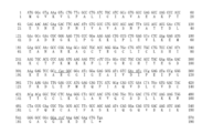
- the base sequence of the I90L mutant of GLuc which consists of codons suitable for mammals including humans and introduced with a restriction enzyme site.
- the underline indicates the position of the restriction enzyme site, and the italic letters indicate the mutation site.
- Nucleotide sequence of a GLuc enzyme mutant (Mon3 with mutations at four positions including I90L) that consists of codons suitable for mammals and has EcoRV restriction enzyme sites introduced.
- the underline indicates the position of the restriction enzyme site, and the italic letters indicate the mutation site.
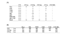
- Substrate selectivity of the GLuc variant of the present invention (A) Comparison of substrate selectivity of mutants using luminescence intensity as an index. “++++” indicates the luminescence intensity of a standard substrate (coelenterazine; CTZ), and indicates the relative luminescence value produced in comparison thereto. (B) Maximum emission wavelength ( ⁇ max) of the emission spectrum for each synthetic coelenterazine. It can be seen that each mutant changes its emission color depending on the type of synthetic coelenterazine.
- the substrate selectivity spectrum of the GLuc variant of this invention As an example, the bioluminescence spectrum of each mutant under conditions with coelenterazine fcp.
- Reporter gene assay based on the GLuc variant of the present invention (A) Principle diagram of the assay. The male hormone receptor is activated by ligand (male hormone) stimulation, dimerizes, and then binds to the male hormone response element (ARE). As a result, a reporter gene (GLuc mutant) is expressed. Reporter gene assay based on the GLuc variant of the present invention.
- C Time course of bioluminescence of the expressed reporter protein. When I90L was installed, it was confirmed that the intensity and stability of the light emission value were excellent.
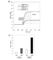
- Reporter gene assay based on the GLuc variant of the present invention (D) Comparison of luminescence intensity in the reporter gene assay between the case where the conventional Gaussia luciferase (GLuc) is loaded and the case where the GLuc mutant (I90L) of the present invention is loaded. It can be confirmed that the area of the emission intensity graph is wider when I90L is used. (E) Comparison graph of the light emission area obtained in (D). Mammalian two-hybrid assay based on the GLuc mutant of the present invention. (A) The principle diagram of the two-hybrid assay. Src ⁇ ⁇ ⁇ ⁇ SH2 domain and ER LBD were used as model proteins.
- GLuc mutant When the SH2 domain is connected to Gal4 and ER LBD is connected to VP16, and both are bound by female hormone stimulation, the GAL response element of pG5 responds and a reporter protein (GLuc mutant) is expressed.
- B Change in bioluminescence intensity with female hormone stimulation time. When the GLuc mutant of the present invention is mounted, it exhibits sufficiently strong bioluminescence upon stimulation with female hormones for 6 hours. Mammalian two-hybrid assay based on the GLuc mutant of the present invention.
- C Change in bioluminescence value with time when the original GLuc is mounted (pG5-GLuc).
- D Change in bioluminescence value with time when the GLuc mutant of the present invention is mounted (pG5-I90L).
- the absolute value of luminescence is higher than that of GLuc, and the bioluminescence is efficiently increased by a short female hormone stimulation.
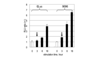
- E Bioluminescence value (measured with a luminescence photometer) after female hormone stimulation (GL0 and GLuc mutant (9096) mounted on pG5 vector) after stimulation with female hormones (0 hours, 3 hours, 6 hours, 18 hours).
- the intensity of bioluminescence according to the concentration of female hormone is significantly increased in the case of the GLuc variant of the invention.
- the GLuc mutant was divided into two, and a stress hormone receptor (glucocorticoid receptor) and an LXXLL motif were inserted between them.
- a GLuc-based one (“SimGR3”) was used.
- 8990N is a GLuc mutant with a mutation in F89W / I90L
- “Mon3N” is a GLuc mutant with a mutation in F89W / I90L / H95E / Y97W.
- B Molecular structure diagram of each single-molecule type bioluminescent probe equipped with the GLuc mutant of the present invention. A single molecule bioluminescent enzyme probe using the GLuc mutant of the present invention.
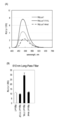
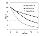
- Luminescence spectra of MLuc itself, MLuc I123L mutant (I123L), MLuc Y122W / I123L / H128E / Y130W mutation (MLuc4).
- B Measurement of luminescence stability of MLuc mutant. MLuc4 shows more stable bioluminescence than conventional MLuc itself. Luminescent properties of MLuc mutant.
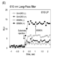
- C Measurement of long wavelength bioluminescence value (more than 610 nm) of MLuc mutant using 610 nmLong-Pass filter.
- D Temporal change in luminescence value of MLuc mutant before and after substrate introduction.
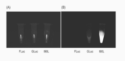
- A Examination of animal tissue permeability of bioluminescence by GLuc mutant (8990N) having mutation in F89W / I90L of the present invention.
- the term “marine animal luminescent enzyme” broadly means, together with Gausia, Renilla reniformis, Cypridina, Katria, etc., Obelin, a luminescent plankton companion. ), Aqualine Pleuromanma, Oplophorus, etc.
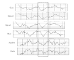
- these "luminescent marine animals” refer to the luminescent enzymes (luciferases) produced by Gaussia luciferase (GLuc) and caucasian luciferase (Metridia longa origin) MLuc and Metridia pacifica derived luminescent enzyme group MpLuc1, MpLuc2, etc.) have a very similar distribution of hydrophilic and hydrophobic amino acids in each enzyme, and amino acid sequence similarity of the putative enzyme active region. Since it is extremely high (FIG. 1), these luciferase groups are also simply referred to as “marine animal luminescent enzymes”. Also referred to as a shear such luciferase ".
- GLuc Gene Bank Assession Number: AY015993
- MLuc Gene Bank Assession Number: AY364164
- MpLuc1 Gene Bank Assession Number: AB195233
- MpLuc2 Gene Bank Assession Number: AB195234
- an amino acid sequence and / or base sequence having 80% or more, preferably 90% or more, more preferably 95% or more identity to the encoded base sequence is also included.
- Such a Gaussia luciferase can be easily obtained by performing a homology search with the base sequence of GLuc, MLuc, MpLuc1, or MpLuc2 in a database such as PSI-BLAST.
- probes or primers can be prepared based on these base sequences and can be picked up from natural Gaussia marine animal genes.
- the Gaucian luciferases MLuc, MpLuc1, or MpLuc2 are slightly different in terms of molecular weight compared to GLuc, but in addition to the similarity in sequence as described above, the enzymatic properties such as substrate and luminescence activity are also GLuc.
- the following is a description of the typical Gaussia luciferase (GLuc).
- the knowledge gained in GLuc can be applied to other Gaussia luciferases.
- the indication of the modified position is represented by the position on the amino acid sequence of GLuc.
- Gaussia luciferase has the following characteristics.
- A Since it is the smallest bioluminescent enzyme discovered so far, when applied to molecular imaging, the burden on host cells and host proteins is much lighter.
- II Extremely strong resistance to pH, surfactants and chemical modifiers.
- C Highest brightness among bioluminescent enzymes.
- D Turn-over of enzyme is fast. 7 times faster than conventional firefly luciferase.
- bioluminescence generated by GLuc is easily affected by reaction conditions, and the drop in emission intensity is severe and unstable, so it is not suitable for use as a reporter (index) for emission analysis where stability is an important condition. It had been.
- the main aim is to increase the emission intensity and to improve the stability of the emission intensity over time, it also includes shifting the emission wavelength to a longer wavelength. Shifting to the longer wavelength side increases permeability from cells, skin, etc., which is an important property for expanding the use of reporter genes.
- the emission intensity can be obtained by measuring the emission intensity in a specific wavelength region using a conventional emission spectrophotometer after the addition of the substrate, so that two-dimensional information can be obtained. It can be measured. A light-emitting plate reader can also be used, and more accurate data can be acquired because of excellent sample processing ability. At that time, the area of each emission spectrum is measured after sufficient lysis time (about 20 minutes). Further, the shift to the long wavelength side can be measured by a wavelength scanning method or by providing a long wavelength filter.
- a desired amino acid residue of the amino acid sequence itself can be chemically changed.
- the base corresponding to the amino acid to be substituted in the base sequence encoding the enzyme may be point mutated.
- a base mutation method a known method such as a site mutation method can be appropriately used.
- the case of introducing a point mutation according to the “quick change method” (Non-patent Document 20) will be described, but the present invention is not limited to this.
- a variant for further functional improvement may be used as a template DNA for the quick change method.
- a template of DNA containing the full length of the Gaussia luciferase gene can be prepared by a method in which the primers are arranged in sense and antisense to cause PCR reaction, and each fragment is stretched.
- This template can be mass-produced by introducing it into eukaryotic cell expression vector pcDNA3.1 (+) and culturing it in a prokaryotic cell such as E. coli.
- amino acids that can be expected to form hydrogen bonds with the substrate in (a) are amino acids that do not participate in peptide bonds, or polar amino acids having a carboxyl group, an amide group, or a hydroxyl group.
- amino acids that can be expected to form hydrogen bonds with the substrate in (a) are amino acids that do not participate in peptide bonds, or polar amino acids having a carboxyl group, an amide group, or a hydroxyl group.
- amino acids that can be expected to form hydrogen bonds with the substrate in (a) are amino acids that do not participate in peptide bonds, or polar amino acids having a carboxyl group, an amide group, or a hydroxyl group.
- amino acids at positions 99 and 100 were substituted.
- Amino acids that can expect an aromatic ring interaction with the phenyl group of the substrate are specifically aromatic amino acids or heterocyclic amino acids having ⁇ orbital electrons, specifically phenylalanine (Phe ), Tyrosine (Tyr), tryptophan (Trp), and histidine (His). These were used to substitute amino acids at positions 90, 93-95, 97 and 130.
- Tyrosine (Tyr) and histidine (His) are also amino acids belonging to the above (a), and phenylalanine (Phe) and tryptophan (Trp) are also amino acids belonging to the following (c).
- C Specific examples of amino acids that can expect interaction between the hydrophobic residues of the substrate and the enzyme include Leu (leucine), Ile (isoleucine), and Val, which are hydrophobic and nonpolar amino acid residues. (Valine), Ala (alanine), Phe (phenylalanine), Pro (proline), Met (methionine), Trp (tryptophan), Gly (glycine).
- Leu leucine
- Ile isoleucine
- Val valine
- phenylalanine Phe
- tryptophan Trp
- Gly glycine
- the substitution with the highest improvement effect corresponds to the same amino acid substitution according to (c) at the position corresponding to the 90th, 108th, 112th, 115th, or 118th position, and the same 89th position or 97th position
- the amino acid substitution was similar to that in (a) at the position where the luminescent enzyme increased in intensity, stabilized in luminescence, and shifted to a longer wavelength.
- a hydrophobic and nonpolar (particularly aliphatic) amino acid residue (specifically, isoleucine) at the position corresponding to the 90th, 108th, 112th, 115th, or 118th position is replaced with another hydrophobic Substitution to non-polar (especially aliphatic) amino acid residues (specifically leucine, tryptophan and valine) and aromatic amino acid residues at positions 89 or 97 (specifically position 89) Substitution of phenylalanine or tyrosine at position 97) with other aromatic amino acid residues (specifically tryptophan) resulted in high emission intensity and stabilization, and a shift to longer wavelengths.
- the above-mentioned variant (I90L) obtained by substituting isoleucine at position 90 with leucine was an excellent mutant luminescent enzyme that was extremely stable and bright and shifted to the long wavelength side.
- the 90-position leucine is substituted with tyrosine, it can be said to be a conservative substitution in the sense of hydrophobic amino acid residues, but in this case, although no increase in luminescence intensity was seen, the long wavelength shift is I was able to wake up.
- substitution from phenylalanine at position 89 or tyrosine at position 97 to tryptophan can also be said to be a substitution from a hydrophobic amino acid residue to a hydrophobic amino acid residue, indicating that ⁇ amino acid position 89, 90, 97 in GLuc
- the hydrophobic amino acid residues corresponding to positions 108, 112, 115, or 118 are mutated to other hydrophobic amino acid residues, respectively, the emission intensity is increased and stabilized, and / or the wavelength is increased. In other words, the function of the luminescent enzyme activity that a shift occurs is improved.
- the light emitting function is further improved by introducing mutations along the strategies (a), (b) and (c) to a plurality of positions. It was confirmed that the improvement of That is, in the present invention, it is also effective to perform mutations at a plurality of positions simultaneously.
- mutations for increasing luminescence intensity and improving luminescence stability (specifically, corresponding to positions 89, 90, 108, 112, 115, and 118 on the amino acid sequence of GLuc) Mutation to shift the wavelength of emitted light to the longer wavelength side (specifically, equivalent to positions 90, 95, 97 and 100)
- isoleucine (I) at a position corresponding to position 90 on the amino acid sequence of GLuc is replaced with leucine (L), and phenylalanine (F) at a position corresponding to position 89 is tryptophan (W).
- modified luciferase F89W / I90L substituted with leucine (L) at the position corresponding to position 90 and leucine (I) at the position corresponding to position 115 with leucine (L).
- the modified luciferase (I90L / I115L) substituted with) was an epoch-making luciferase that stably showed extremely high luminescence intensity and also caused a long wavelength shift.
- the stability of the bioluminescence intensity is performed by introducing a substrate (dissolved in PBS buffer) into each mutant and observing the change in bioluminescence with time.
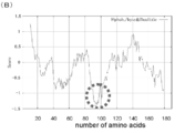
- the typical GLuc variants I90L, H95E, and Y97W when observed for 5 seconds, showed relatively stable luminescence intensity (Fig. 7A). While the value (white circle) decreased drastically, the emission values of I90L (black square) and I90V (gray triangle) showed relatively stable emission intensity (FIG. 7B).
- the result of observing the change in bioluminescence over time for a short time (5 seconds) after introducing a substrate (dissolved in Matthew modified buffer) into each mutant was the same (FIG. 7C).
- Gaussia luciferases derived from caussia (Metridia pacifica or Metridia longa), which are gausian marine animals very similar to Gaussia, specifically MpLuc1 (derived from Metridia pacifica) and In MLuc (from Metridia longa) luciferase, a mutant (MpLuc1-I114L mutant) due to conservative amino acid substitution at position 114 of MpLuc1, corresponding to position 90 of the amino acid sequence of GLuc, and conservative at position 123 of MLuc MpLuc1 Y113W / I114L / H119E / Y121W mutant (MpLuc4) and MLuc Y122W corresponding to mutants of GLuc F89W / I90L / H95E / Y97W (Mon3) as well as mutants by amino acid substitution (MLuc-I123L mutant) / I123L / H128E / Y130W mutant (MLuc4) was prepared
- the amino acid sequence of Gaussia luciferase (GLuc) Among amino acid residues corresponding to positions up to 118, from at least one of positions corresponding to positions 89, 90, 95, 97, 100, 108, 112, 115, and 118 "Conservative amino acid replacement" is applied to the amino acid residue at the selected position, specifically, positions 89, 90, 97, 108 on the amino acid sequence of GLuc , 112, 115, or 118 position of the hydrophobic amino acid residue to another hydrophobic amino acid residue, or a hydrophilic amino acid residue that forms a hydrogen bond at the 95th or 100th position Including substitution of groups with other hydrophilic amino acid residues It is intended.
- the hydrophobic amino acid is preferably a hydrophobic amino acid having a larger molecular weight than a small hydrophobic amino acid such as glycine (Gly). If it is hydrophobic and nonpolar, it is aromatic such as tryptophan (Trp). It may be an amino acid.
- leucine (Leu), valine (Val), isoleucine (Ile) and tryptophan (Trp) which are aliphatic amino acids among hydrophobic amino acids are preferable, and Leu (leucine), Val (valine) and Trp (tryptophan) are preferable. Further preferred.
- the substitution from the hydrophobic amino acid residue to another hydrophobic amino acid residue is a hydrophobic position at positions 90, 108, 112, 115, or 118 on the amino acid sequence of GLuc, and Substitution of non-polar aliphatic amino acid residues to other hydrophobic and non-polar aliphatic amino acid residues (particularly, substitution of isoleucine with any amino acid residue selected from leucine, tryptophan and valine) ), Or substitution of a hydrophobic and aromatic amino acid residue at position 89 or 97 with another hydrophobic and aromatic amino acid residue (phenylalanine (Phe) at position 89 or position 97 When tyrosine (Tyr) is replaced with tryptophan (Trp), the resulting mutant luminescent enzyme stabilizes the luminescence intensity at the same time as the brightness of the luminescent enzyme increases or shifts to a longer wavelength.
- the most preferred combination of the mutation position and the mutation amino acid is the substitution of position 89 with tryptophan (Trp), the substitution of position 90 with Leu (leucine) or Val (valine), and the same. This is the case where position 108 is replaced with Val (valine).
- substitution of a hydrophilic amino acid residue forming a hydrogen bond with another hydrophilic amino acid residue can be achieved by replacing histidine (His) at position 95 on glutamic acid (Glu) or amino acid position at position 100 on the amino acid sequence of GLuc.
- Aspartic acid (Asp) is replaced with asparagine (Asn), and the modified luciferase in that case is shifted to a longer wavelength.
- modified luciferases the ones with the highest brightness, increased stability, and a long wavelength shift have been performed by substituting leucine for isoleucine at the position corresponding to position 90 on the amino acid sequence of GLuc.
- the modified luciferase (GLuc-I90L, MpLuc1-I114L, MpLuc1-I114L). More preferably, the position corresponding to position 89 on the amino acid sequence of GLuc is tryptophan, position 90 is leucine or valine, position 95 is glutamic acid, position 97 is tryptophan, position 108 is valine, position 112 is leucine.
- valine, position 115 with valine, or position 118 with leucine or valine shows particularly significant emission intensity enhancement and also a long wavelength shift.
- the leucine mutants at position 90 are excellent modified luciferases that have not only high brightness but also a remarkable long wavelength shift.
- mutant luminescent enzyme modified luciferase
- GLuc which is a region corresponding to the active center
- the amino acid sequence of the original Gaussia luciferase has no mutation other than the above-mentioned “similar amino acid substitution”.
- a mutation within the range in which the three-dimensional structure of the whole enzyme does not change greatly is acceptable, and in particular, a part of the N- or C-terminal portion, for example, 1 to 50, preferably 1 to 30, More preferably 1 to 20, even more preferably 1 to 10 amino acid residues are deleted.
- the amino acid sequence excluding the region corresponding to positions 89 to 118 on the GLuc amino acid sequence is 70% or more, preferably 80% or more, more preferably Can be expressed as an amino acid sequence having 90% or more, most preferably 95% identity.
- Host cells at that time include not only mammalian cells (COS cells, CHO-K1 cells, HeLa cells, HEK293 cells, NIH3T3 cells) used for general genetic recombination, but also bacterial cells such as yeast cells and Escherichia coli, Insect cells and the like may be used, but their main use is often in vivo in mammals including humans or in vitro mammalian cells.
- the mutant luminescent enzyme gene of the present invention is preferably modified to a base sequence that is easy to express in the host cell by changing to a codon suitable for the cell depending on the host cell to be used. In addition, it is necessary to appropriately perform modification to provide a restriction enzyme site for insertion into a vector.
- a well-known method can also be applied to these steps, but as an example, a modification strategy for producing a modified body assumed to be used in a living body of a mammal such as a human or in a mammalian cell in vitro is shown.
- the codon corresponding to each amino acid is changed to a codon suitable for mammals such as humans, so that the base sequence can be easily expressed in mammals such as humans, as well as desired.
- Such a modification strategy for improving functions is specifically shown below, but is not limited to these methods.
- the original extracellular secretion signal (1-17AA) upstream of the GLuc gene is not directly related to the luminescence activity, so it is deleted or replaced with another known extracellular secretion signal or intracellular localization signal.
- the secretion signal may be left as it is, and an intracellular localization signal may be connected to the C-terminal side separately.
- a template of DNA encoding GLuc was designed in consideration of the above points (i) to (iii) using the known GLuc sequence information as a sample.
- the primer set for DNA synthesis was constructed, and a template for the GLuc gene was completed by PCR reaction.
- This template was introduced into the eukaryotic cell expression vector pcDNA3.1 (+) to perform subclone.
- a point mutation was introduced based on the above-mentioned “quick change method”. Specifically, a PCR reaction was performed in the presence of about 30 base sense and antisense primers prepared so as to include mutation points to induce specific site-specific mutations. Subsequently, only the plasmid derived from Escherichia coli carrying the template GLuc gene was decomposed by DpnI enzyme treatment, and the gene sequence was analyzed to confirm the presence or absence of the introduction of the mutation.
- the enzyme activity of mutated GLuc can be verified, for example, by the following method. First, using a known lipid reagent for gene introduction, an expression vector having a GLuc mutant was introduced into COS-7 cells derived from African monkeys, and an expression vector having a conventional GLuc that does not contain a mutation for comparison, Introduce into cells by the same method. After a certain time (10 to 20 hours, for example, 16 hours) has passed since the introduction of the vector, a cell lysate is prepared using a known cell lysis reagent.
- the cell lysate and a known substrate solution containing coelenterazine are mixed to measure the color development intensity and the temporal stability of luminescence.
- the emission intensity can be measured by measuring the intensity at a specific wavelength using a conventional emission spectrophotometer after the addition of a substrate, and the stability over time can be measured by measuring every minute. To measure the shift to longer wavelengths, scan all wavelengths.
- variants from hydrophobic amino acids to other hydrophobic amino acids at positions 89, 90, 108, 112, 115, and 118 in GLuc show enhanced luminescence intensity. It was.
- the leucine mutant (I90L) at the 90th position had not only high brightness but also excellent luminescence with a long wavelength shift. It is an enzyme variant. Furthermore, two-site mutants (F89W / I90L, I90L / I115L) in which the 90-position tryptophan substitution or the 115-position leucine substitution was performed simultaneously with the 90-position leucine substitution, and the 90-position leucine substitution, The four mutants (F89W / I90L / H95E / Y97W) in which tryptophan substitution at position 89 and glutamic acid substitution at position 95 and tryptophan substitution at position 97 were simultaneously performed showed remarkable emission intensity and longer wavelength side.
- mutants at positions 90 and 97 are leucine (Leu) which is a hydrophobic amino acid, tyrosine (Tyr) which is an amino acid that forms a hydrogen bond and is an aromatic amino acid, or aromatic It was confirmed that any mutation of tryptophan (Trp), which is a systemic amino acid and a hydrophobic amino acid, showed bioluminescence shifted to the longer wavelength side.
- the gene encoding the mutant luminescent enzyme of the present invention is a bioluminescent probe that is currently being researched and developed along with its use in various analytical systems as a reporter gene for which a luciferase gene has been used. The use as is expected most.
- the marine animal luminescent enzyme mutants of the present invention are conventionally used in reporter gene assay, Yeast Two-hybrid assay, Mammalian Two-hybrid assay, protein splicing assay, protein complementation assay, circular permutation assay, It can be installed as a core element in bioluminescence resonance energy transfer assay (BRET) etc., and the measurement performance of the assay can be dramatically improved.
- BRET bioluminescence resonance energy transfer assay
- the molecular weight is large and it takes time until expression, which places a heavy burden on the host cell.
- the luminescence intensity of the reporter is extremely high (particularly in the case of the I90L mutant, 14,000 times stronger than the conventional firefly luciferase). Since there is an advantage that measurement can be performed, the measurement time can be greatly shortened compared to the conventional method, and the stability of luminescence over time is high, so that luminescence measurement can be performed even in cell lines with poor gene transfer efficiency. Moreover, since it has shifted to the long wavelength side, the permeability through the cell membrane and the skin is increased, so the measurement sensitivity is also high.
- the luminescent enzyme is linked to a known eukaryotic cell expression vector carrying a special promoter upstream, After introduction into a cell and after a certain period of time, it may be used for measurement under the condition that there is no signal (stimulation) (Non-patent Document 13).
- a known pTransLucent vector can be used and can be easily mounted using a known method.
- a commercially available pG5Luc vector can be used by simply mounting the luminescent enzyme using a known method.
- the high-intensity luminescent enzyme of the present invention is a single molecule luminescent probe according to the invention for which the present inventors have already applied for a patent (Non-patent Document 9, Non-patent Document 10).
- Non-Patent Document 11 Non-Patent Document 12
- Japanese Patent Application 2007-005144 Japanese Patent Application 2007-202308, Japanese Patent Application 2007-332253, Japanese Patent Application 2008-116098, Japanese Patent Application 2009-025229, PCT / JP2008 / 050370, US12 / 025532 , US12 / 343830
- Non-Patent Document 4 Non-Patent Document 6, Non-Patent Document 7
- the presence or absence of a ligand and the activity intensity of the ligand can be observed with high luminance.
- a high-performance luminescent probe can be constructed in a form in which a recognition protein that recognizes that a ligand is bound to is linked.
- the enzyme fragment divided into two can complement and change the enzyme activity. At this time, due to the high brightness and stability of the split enzyme, the detection limit can be improved and the measurement can be performed with high reliability.
- the term “single molecule luminescent probe” is a kind of a known bioluminescent probe characterized in that all components used for visual imaging are integrated in a single fusion molecule ( Patent Document 7).
- a fusion protein comprising N- and C-terminal fragments obtained by dividing the marine animal luminescent enzyme variant of the present invention into two as a basic component, a ligand-binding protein and a ligand-binding protein recognition protein.
- the N-terminal fragment and the C-terminal fragment of the marine animal luminescent enzyme variant of the present invention are contained in a fusion protein containing a ligand-binding protein and a recognition protein.
- bioluminescent probe present in each Refers to the type of bioluminescent probe present in each.
- these marine animal luminescent enzyme high-intensity mutants of the present invention are used in these bioluminescent probes, it is necessary to divide the N-terminal fragment and the C-terminal fragment into two, and the position of the division is known in the art. This is the same position as the dividing position in the marine animal luminescent enzyme used in the “single molecule luminescent probe” and “bimolecular luminescent probe”. That is, in the case of the GLuc mutant of the present invention, it may be divided at positions 105 to 110, and in the case of MLuc, the division at positions 138 to 142 is applied.
- the high-luminance bioluminescent enzyme was stably mounted on the circular permutation probe (Non-patent Document 11; Non-patent Document 12) by the present inventors. Intracellular molecular phenomena can be measured efficiently with strong emission intensity.
- the circular permutation probe corresponds to cPresso and cPressoMax shown in the sequence listing.
- a specific method for using the high-intensity luminescent enzyme of the present invention as a single molecule luminescent probe follows the method described in detail in (Patent Document 7, Japanese Patent Application No. 2007-332253, Japanese Patent Application No. 2009-025229).
- a chimeric DNA encoding is designed.
- the clone is subcloned into a vector suitable for the cell in which the chimera DNA is to be expressed, and the vector is introduced into the cell and expressed in the cell.
- the cells of interest are preferably cells derived from mammals including humans, and may be cells that exist in vivo or cultured cells that maintain their original functions. .
- prokaryotic cells such as Escherichia coli may be used.
- the specific type of vector is not particularly limited, and a vector that can be expressed in the host used for expression can be appropriately selected.
- a method for introduction into cells a known transfection method such as a microinjection method or an electroporation method can be used.
- intracellular introduction methods using lipids BioPORTER (Gene Therapy Systems), Chariot (Active Motif), etc.
- bioluminescent probe using the high-intensity luminescent enzyme of the present invention is expressed as a fusion protein in the cell after being introduced into the cell as a chimeric DNA, ligand stimulation is performed on the transformed cell, By measuring the change in the amount of luminescence from the cells, the nature of the ligand, the degree of activity, etc. can be evaluated.
- the “ligand binding protein” that can be mounted together with the bioluminescent enzyme is intended to be a protein that binds a ligand to its ligand binding site.
- the ligand-binding protein can be, for example, one that changes in a three-dimensional structure, undergoes phosphorylation, or promotes protein-protein interaction upon binding of a ligand.
- a ligand-binding protein for example, a nuclear receptor (NR), a cytokine receptor, or various protein kinases having a hormone, chemical substance or signal transduction protein as a ligand is used.
- the ligand binding protein is appropriately selected depending on the target ligand.
- the ligand that binds to the ligand-binding protein is not particularly limited as long as it binds to the ligand-binding protein, and may be an extracellular ligand that is taken into the cell from outside the cell. It may be an intracellular ligand produced in For example, it can be an agonist or antagonist for a receptor protein (eg, nuclear receptor, G protein-coupled receptor, etc.).
- signaling proteins such as cytokines, chemokines, and insulin that specifically bind to proteins involved in intracellular signal transduction, intracellular second messengers, lipid second messengers, phosphorylated amino acid residues, G protein-coupled receptor ligands Etc.
- the binding domain of each second messenger can be used as a ligand-binding protein.
- the second messenger is intended to be another type of intracellular signaling substance that is newly generated in the cell by binding extracellular signaling substances such as hormones and neurotransmitters to receptors present on the cell membrane.
- Examples of the second messenger include cGMP, AMP, PIP, PIP 2 , PIP 3 , inositol triphosphate (IP 3 : inositol triphosphate), IP 4 , Ca 2+ , diacylglycerol, and arachidonic achid.
- calmodulin CaM
- CaM calmodulin
- the ultra-bright luminescent enzyme can be stably introduced into various cell lines.
- the bioluminescent enzyme can be stably introduced into a new universal cell (iPS). Since the new type of universal cells (iPS) itself does not shine, it was very difficult to search for molecular phenomena and tissue specificity that occur inside.
- a molecular probe containing the luminescent enzyme is introduced into a somatic cell, and then an embryo is created and differentiated into various organ tissues. Then, a specific molecular phenomenon that occurs in each organ can be measured with high sensitivity. In that case, the method of Yamanaka et al.
- Non-patent Document 19 Non-patent Document 19
- a suitable signal peptide to this ultra-bright bioluminescence enzyme, it can be used for high-brightness imaging of each organelle.
- the GCC-43-derived MLCCMRRTKQV sequence can be localized to the cell membrane by attaching it to the N or C terminus of GLuc.
- GRKKRRQRRR sequence is attached for cytoplasmic localization.
- localization in the endoplasmic reticulum (ER) and cell nucleus is possible by linking the KDEL and DPKKKRKV sequences, respectively.
- HIS-tag HHHHHH
- FLAG-tag DYKDDDDK
- Myc-tag EQKLISEEDL
- HA-tag YPYDVPDYA
- V5-tag GKPIPNPLLGLDST
- T7-tag MASMTGGQQMG
- Example 1 Construction of a gene sequence encoding GLuc
- GLuc Gaussia luciferase
- a known mammal is used.
- the codon was optimized for animal cells. Specifically, in order to be optimally expressed in mammalian cells, it must be a codon rich in G and C.
- the bases encoding the amino acid Phe include TTT and TTC, but TTC is most suitable for mammalian cells.
- GCCRCC ATG G was placed before and after the translation initiation codon (AUG) in order to increase the protein expression level.
- sense and antisense primers were alternately arranged, and a PCR reaction was caused to synthesize a full-length sequence.
- the full length of GLuc was used as a template in other mutagenesis experiments. Its full length is shown in FIG.
- Inference 1 The crystal structure (FIG. 3C) formed by Renilla luciferase (RLuc) in combination with coelenterazine, a common substrate of marine animal-derived luminescent enzymes, is shown in detail in the enzyme active site of RLuc. It was found that (1) there is a region where hydrophobic amino acids appear at regular intervals (2-4), and (2) the structure is such that the substrate is sandwiched between the hydrophilic and hydrophobic amino acid sites. (FIG. 2B).
- Non-patent Document 18 By introducing a hydrophobic amino acid in the vicinity of the fluorescent chromophore so as to allow hydrophobic interaction with the fluorescent chromophore, stable high-luminance fluorescent coloring can be achieved (Non-patent Document 18).
- introduction of mutations into amino acids near the chromophore of the fluorescent protein may be effective in improving the fluorescent protein.
- the substrate of a marine animal-derived luminescent enzyme is compared with the chromophore of a fluorescent protein, the structure is very similar. Therefore, “A chromophore has almost the same chemical structure.
- the luminescent enzyme is in the outside of the molecule as a substrate "(Fig. 3C).
- Inference 5 In the previous studies on the luminescent enzyme of the present inventors, there is an example in which the luminescent performance can be improved by partially overlapping the sequence in the vicinity of the enzyme active site. From this case, improvement of the properties of the luminescent enzyme can also be expected by a method of overlapping a part of the amino acid sequence near the enzyme active site.
- Example 3 Comparison of bioluminescence intensity of GLuc introduced with amino acid mutations COS-7 cells obtained by culturing pcDNA3.1 (+) vector inserted with GLuc mutant DNA prepared in Example 2 on a 12-well plate And incubated for 16 hours. Thereafter, a cell lysate was prepared from COS-7 cells in each well (5 minutes lysis), and the spectrum in the presence of a substrate specific to the luminescent enzyme (coelenterazine) was measured using a fluorimeter (F-7000, Hitachi) The effectiveness of the introduced mutation was examined. The results are shown in FIGS. 4 (B-1) and (C). According to FIGS.
- the I90L mutant also causes a maximum shift of 15 nm toward the long wavelength side (FIG. 4B-2). Since the above-mentioned mutants exhibit long-wavelength bioluminescence, they are particularly advantageous in observing in vivo bioluminescence. As a result of observing bioluminescence (high tissue permeability) of 610 nm or more emitted by each mutant using a 610 nm Long-Pass filter, it is shown in FIG. 4 (E). As predicted, it was found that I90L etc. showed much higher light intensity than the conventional GLuc.
- FIG. 4B-1 shows a graph using an emission spectrophotometer (F-7000, Hitachi) and using the peak of each emission spectrum as an index.
- F-7000 emission spectrophotometer
- the luminance was measured using an emission spectrum area measured by a light emission plate reader as an index.
- FIG. 4 (B-2) when the original GLuc was changed to I90L, H95E, Y97W, F89W / I90L, I90L / I115L, F89W / I90L / H95E / Y98W, the brightness was particularly high. It can also be seen that these mutants are all shifted to longer wavelengths. From the above results, it was confirmed that the bioluminescence itself has extremely high brightness by introducing an amino acid mutation that interacts with the substrate in a hydrophobic region in the region corresponding to the active center of GLuc. .
- I90L is 7.1 times (hereinafter area ratio), H95E is 3.0 times, Y97W is 2.1 times, F89W / I90L is 10.2 times, I90L / I115L is 4.9 times, F89W / I90L / H95E / Y98W showed 4.4 times the emission intensity.
- bioluminescence is 7.1 times stronger than conventional GLuc and 10,000 times stronger than FLuc.
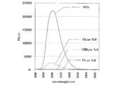
- FIG. 5 shows luminescence spectrum values obtained by comparing the GLuc mutant (I90L) of the present invention with conventional luminescent enzyme groups (firefly luciferase (FLuc), Gaussia luciferase (GLuc), click beetle luciferase (CBLuc), etc.). This figure also shows that the emission intensity of the GLuc mutant is exceptionally high.
- Example 4 Stability measurement of mutant bioluminescent enzyme
- the gene encoding each GLuc mutant was cloned into pcDNA3.1 (+), and the same amount of plasmid was introduced into COS-7 cells. Cells were collected 16 hours after introduction, lysed with a cell lysate, and mounted on a microplate reader.
- a PBS buffer in which coelenterazine was dissolved was set in the substrate jet machine of the plate reader.
- the substrate was automatically sprayed into each well, and then the change in luminescence value was measured in increments of 0.05 seconds.
- the enzyme activities of GLuc itself and F89W, I90L, H95E, and Y97W were measured.
- mutants such as I90L, H95E, and Y97W showed relatively stable bioluminescence.
- GLuc and F89W showed relatively unstable emission intensity (FIG. 7A).
- the composition of the Matthew modified bar fur is as follows: 100 mM potassium phosphate, 500 mM sodium chloride, 1 mM ethylenediaminetetraacetic acid (EDTA), 0.15 mg / ml porcine gelatin, pH 5.0, 1% Nonylphenol ethoxylates (Tergitol NP-9) and 1% Antifoam, 25 mM thiourea, 0.1% DMSO .
- FIG. 8 The synthetic GLuc I90L mutant shown in FIG. 8A is (a) close to a human gene codon (GC rich) based on the human codon ratio information of NCBI-GenBank so that it is optimally expressed in human cells.
- a restriction enzyme site was added in the middle or tail of the gene.
- SacII and EcoRV were introduced at the 312 and 340 base positions, respectively.
- This site was previously found as an appropriate cleavage site by the inventors' research (Non-Patent Document 12), and is the optimal insertion position for protein insertion.
- BamHI was introduced at the tail of the gene.
- This site is one of the multicoloning sites, so that it can be used effectively when connecting with DNA of other proteins.
- GLuc enzyme mutants (Mon3: FIG. 8B shows an example in which F89W / I90L / H95E / Y97W) was prepared using a human-compatible codon.
- the specific modification strategy of Mon3 is as follows.
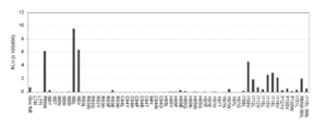
- Example 6 Examination of substrate selectivity of each GLuc variant The substrate selectivity of the GLuc variant obtained by the present invention was examined (Fig. 9). COS-7 cells are cultured in 24-well plates. Thereafter, a plasmid encoding each GLuc mutant was introduced into the cells and further cultured for 16 hours. Thereafter, 30 ⁇ L of cell lysate is added to the cells. Then, the luminescence value for each mutant was measured under various substrate coexistence conditions. The result is shown in FIG. From the results, it was confirmed that each mutant had substrate selectivity.
- each mutant including the original GLuc itself, showed strong bioluminescence only when coelenterazine (CTZ), coelenterazine fcp, and coelenterazine ip coexisted.
- CTZ coelenterazine
- coelenterazine fcp coelenterazine fcp
- coelenterazine ip coexisted.
- strong bioluminescence was not exhibited under the coexistence conditions of coelenterazine cp, coelenterazine hcp, and coelenterazine 400A.
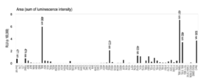
- FIG. 9B A typical spectrum is shown in FIG. 9B, and the maximum emission values for each substrate are summarized in FIG. 9C. According to the results, it was found that the spectrum of each mutant including the original GLuc itself was affected by the type of substrate. Each mutant had a light emission value shifted to a long wavelength depending on the substrate, and some mutants shifted to a short wavelength. From this result, it was found that each mutant can
- FIG. 10 Reporter gene assay loaded with GLuc mutant
- the conventional reporter gene assay system pG5Luc Promega
- FIG. 10A a vector system having an androgen responsive element (ARE) upstream was prepared, and a gene encoding a conventional GLuc or a GLuc mutant of the present invention was linked downstream thereof by genetic engineering (FIG. 10A).
- This new vector was introduced into COS-7 cells, and the difference in bioluminescence value was measured with a luminometer under the condition of presence and absence of androgen (male hormone) (FIG. 10B).
- the change in emission intensity from the introduction of the substrate to a certain time (1 second) can be quantitatively compared by area.
- GLuc area for the control
- I90L change in luminescence intensity of the reporter gene
- Example 8 Two-hybrid assay loaded with GLuc mutant
- a eukaryotic two-hybrid assay system loaded with the GLuc mutant was constructed, and the interaction between the protein and the protein.
- the following plasmid groups were first prepared based on the conventional typical two-hybrid assay plasmids pACT, pBIND, and pG5Luc: (1) plasmids encoding ER LBD and BIND; (2) A plasmid encoding SH2 domain and ACT.
- a pG5 plasmid carrying a conventional GLuc or the high-intensity mutant of the present invention (I90L) as a reporter gene Living cells co-introduced with the above plasmids (1), (2), and (3) were given solvent (0.5% DMSO) or female hormone (estrogen) stimulation for 0 hours and 6 hours, and then the cells were recovered, The bioluminescence value was measured with a luminescence photometer (FIG. 11B). As a result, the cells carrying the plasmid carrying the GLuc mutant of the present invention showed an excellent S / N ratio against female hormone stimulation.
- pG5-I90L expressing the GLuc mutant was introduced into the experimental group. After culturing for 12 hours, the cells were stimulated with estrogen, and then continued for another 8 hours. Thereafter, a lysis buffer was added to the cells, and the luminescence value was measured for 2 seconds after introducing the substrate by an automatic program. As a result, compared with the control group, the experimental group showed a significant difference in luminescence value even with 10 ⁇ 7 M estrogen stimulation, and the degree became remarkable at a higher concentration. As a result, the superior luminescence properties of the GLuc mutant led to an improvement in detection limit as a result.
- the transformed cells carrying the plasmid carrying the GLuc mutant of the present invention showed a better S / N ratio for female hormone stimulation.
- the luminescence value increased up to 2 times after 3 hours of stimulation, but could not be distinguished by the conventional method.
- This probe was introduced into a living cell, and the difference in the luminescence value was measured under the condition with and without male hormone.
- a conventional GLuc itself was similarly divided into two, and a probe with GR LBD and LXXLL motifs inserted between them was created (named SimGR3), and S / N was used with and without stress hormone (cortisol). The ratio was measured.
- SimGR3 GR LBD and LXXLL motifs inserted between them
- cortisol stress hormone
- the operating principle of this probe is shown in FIG. First, stress hormone receptors recognize stress hormones and cause structural changes. As a result, since it becomes bound to the adjacent LXXLL motif, recombination occurs between the two luminescent enzyme fragments. At this time, stress hormone activity can be measured using the recovered enzyme activity as an index.
- the stress hormone sensitivity of SimGR3, 8990N and Mon3N made for this experiment was measured (FIG. 13B).
- 8990N carrying the GLuc mutant of the present invention showed strong bioluminescence, and its sensitivity was not inferior to that of the conventional one.
- the emission characteristics of 8990N showed similar results even when the luminescence intensity was measured over time (FIG. 13C).
- the 8990N of the present invention showed light emission characteristics superior to those of the conventional one.
- conventional SimGR3 did not show a significant difference in luminescence value until it was possible to distinguish between the conditions with and without stress hormones (FIG. 13D).
- conventional MpLuc1 itself, a mutant-introduced product of MpLuc1 to Y113 (hereinafter referred to as Y113F; this corresponds to F89 of GLuc), a mutant-introduced product of MpLuc1 to I114 (hereinafter referred to as I114L; This is equivalent to I90L of GLuc), and also has a mutation (Y113W / I114L / H119E / Y121W) with 4 mutations, including I114L of MpLuc1; it is named MpLuc4; F89, I90, H95, and Y97) were respectively synthesized by genetic engineering. The same amount of each mutant was introduced into living cells, and the relative luminescence intensity was measured.
- Example 11 Measurement of luminescence activity of Mluc and its mutants
- MLuc I123L mutation-introduced body (hereinafter referred to as I123L; this corresponds to GLuc I90) and MLuc I123L-mutated mutant (Y122W) / I123L / H128E / Y130W mutation; named MLuc4; these correspond to GLuc F89, I90, H95, and Y97, respectively).
- I123L MLuc I123L mutation-introduced body
- MLuc4 MLuc I123L-mutated mutant
- the time-dependent change in luminescence intensity after substrate introduction was measured in 0.5 second increments (FIG. 15B).
- MLuc mutant MLuc4 with mutations introduced at 4 sites showed the most stable bioluminescence.
- the conventional MLuc loses 90% of the luminescence intensity in only 10 seconds after the introduction of the substrate.
- the luminescence value was measured using a 610 nm long-pass filter, and as a result, it was found that I123L emits bioluminescence with a strong red wavelength.
- Example 12 Biological imaging using GLuc mutant Utilizing the improved points (high luminance, long wavelength shift, stability, etc.) of the GLuc mutant, its applicability to biological systems was verified (Fig. 16). ).
- a pcDNA3.1 plasmid encoding 8990, a high brightness mutant of GLuc was constructed and introduced into COS-7 cells.
- a pcDNA3.1 plasmid encoding the conventional GLuc itself was similarly introduced into COS-7 cells. Thereafter, both transformed cells were transplanted into the left and right upper / subcutaneous tissues of BALB / c nude mice (five week old females).
- a living cell equipped with a mammalian two-hybrid system expressing the GLuc mutant was prepared, and a biological imaging system using the same was tried (FIG. 16B).
- plasmids were constructed in which the female hormone receptor (ER LBD) and Src SH2 domains were linked to BIND and ACT, respectively (pBIND and pACT).
- ER LBD female hormone receptor
- Src SH2 domains were linked to BIND and ACT, respectively
- pG5-I90L a plasmid having a Gal4 response element upstream and expressing I90L was also prepared (pG5-I90L).
- a plasmid expressing the conventional GLuc itself was prepared (pG5-GLuc).
- the three types of plasmids (pBIND, pACT, pG5-I90L or pG5-GLuc) were co-transfected into COS-7 cells to prepare transformed cells.
- the transformed cells were transplanted into the left and right upper and subcutaneous tissues of BALB / c nude mice (five week old females), respectively.
- the left upper part was transplanted with control (pG5-GLuc), and the right upper part was transplanted with transformed cells having pG5-I90L.
- 12 hours were allowed for the transplantation to stabilize.
- female hormone or solvent (0.1% DMSO) was injected into each mouse, and the luminescence intensity was observed after another 6 hours (FIG. 16B).
- mice subjected to female hormone stimulation showed stronger bioluminescence.
- the right upper (I90L expression) showed stronger bioluminescence than the left upper (GLuc expression).
- I90L showed higher bioluminescence because it was more stable and brighter.
- the measurement performance of the assay can be dramatically improved by using ligand measurement based on the reporter gene assay method that has been widely used in the past, or using it instead of the bioluminescent enzyme in the conventional bioluminescent probe, etc. It can be used widely in applications such as diagnostic reagents development in academic research, medicine, pharmacy, and analytical chemistry.
Landscapes
- Chemical & Material Sciences (AREA)
- Organic Chemistry (AREA)
- Life Sciences & Earth Sciences (AREA)
- Health & Medical Sciences (AREA)
- Engineering & Computer Science (AREA)
- Zoology (AREA)
- Wood Science & Technology (AREA)
- Genetics & Genomics (AREA)
- Bioinformatics & Cheminformatics (AREA)
- General Health & Medical Sciences (AREA)
- General Engineering & Computer Science (AREA)
- Biochemistry (AREA)
- Microbiology (AREA)
- Molecular Biology (AREA)
- Biotechnology (AREA)
- Proteomics, Peptides & Aminoacids (AREA)
- Analytical Chemistry (AREA)
- Physics & Mathematics (AREA)
- Immunology (AREA)
- Biophysics (AREA)
- Medicinal Chemistry (AREA)
- Biomedical Technology (AREA)
- Measuring Or Testing Involving Enzymes Or Micro-Organisms (AREA)
- Investigating Or Analysing Materials By The Use Of Chemical Reactions (AREA)
- Enzymes And Modification Thereof (AREA)
- Micro-Organisms Or Cultivation Processes Thereof (AREA)
Abstract
Disclosed is a mutant luminescent enzyme which is produced by the genetic engineering modification of a marine animal-derived luminescent enzyme such as gaussia luciferase, has high brightness, and has high light-emitting stability or can emit a light having a wavelength shifted to a longer wave length side. Specifically disclosed is a mutant luminescent enzyme having an amino acid sequence produced by substituting at least one amino acid residue selected from the amino acid residues lying between position-89 to position-118, specifically by carrying out the conservative amino acid replacement of an amino acid residue located at at least one position selected from position-89, position-90, position-95, position-97, position-100, position-108, position-112, position-115 and position-118, in the amino acid sequence for a marine animal-derived luminescent enzyme gaussia luciferase (GLuc). The above-mentioned substitution in a marine animal-derived luminescent enzyme enables the improvement in the active function of the luminescent enzyme. Also disclosed is a bioluminescent probe having an improved light-emitting function, which is produced using the mutant luminescent enzyme.
Description
本発明は、海洋動物発光酵素の遺伝子改変による発光酵素機能及び発光の安定性向上のための手法の確立、ならびに当該手法を適用して合成された、超高輝度で安定な人工生物発光酵素に関するものである。
The present invention relates to establishment of a technique for improving luminescent enzyme function and luminescence stability by genetic modification of marine animal luminescent enzymes, and an ultra-bright and stable artificial bioluminescent enzyme synthesized by applying the technique. Is.
細胞・非細胞系を問わず、自然界における多くの分子現象(例えば、蛋白質の構造変化、リン酸化、蛋白質間の相互作用、2次信号伝達物質の産生等)は、我々が、自然を理解し、その生命現象に係る分子メカニズムを解明する上で極めて重要な指標となる。今まで、このような分子現象を計測するために、(ア)FRET (非特許文献1,2)、(イ)BRET(非特許文献3)、(ウ)タンパク質間相互作用検出法(protein-fragment complementation assay)(非特許文献4,5)等の手法が開発された。
近年、本発明者らは、独自の分子設計技術を利用した生物発光イメージングに関する研究開発を行ってきた。具体的には、転写因子の核内移行や細胞質内非ゲノム的な蛋白質-蛋白質間相互作用を、蛋白質スプライシング法を用いて計測した(非特許文献6,7)。また、一つの分子内に、信号認識と生物発光に必要なすべての要素を集積した形態の一分子型生物発光プローブを開発した(非特許文献8,9)。その後、このプローブをマルチカラー化し、複数の信号伝達過程を同時にイメージングできるように発展させた(非特許文献10)。さらに、生物発光プローブそのもののリガンド感受性を高める手法として、円順列置換(非特許文献11)や小さい発光酵素を用いた分子設計技術(非特許文献12)を開発した。これらの研究は、いずれも細胞・非細胞系における分子現象を効率よく計測する手段として使われてきた。
細胞内・外の分子現象を探索する主な方法で、発光イメージング以上に広く使用されている方法として、蛍光イメージングがある。しかし、蛍光蛋白質は、自己蛍光のためバックグラウンドが高く、外部光源を必要とし、蛍光顕微鏡のような大きな装置と精密なフィルターシステムを必要とする。また蛍光発色団が成熟するまでに短くても数時間から数日がかかる問題点があった。また蛍光顕微鏡を使う場合、一回に観察できる細胞数に限界があり、定量性に問題があった(非特許文献6)。
一方、生物発光酵素を用いる発光イメージングの場合、多くの長所にも関らず、蛍光イメージングに比べて使用されにくい最大の原因は、生物発光酵素の低輝度にあった。生物発光酵素が低輝度であるために、高感度の計測装置を必要とし、単一細胞イメージングや細胞小器官の探索などには不向きであるとされていた。
また、蛍光蛋白質については、多色蛍光蛋白質の研究が十分進んでおり、その発色原理に関する知見が多く得られているため、これらの研究成果を利用して多様な蛍光特質を持つ蛍光蛋白質が多く開発されている。ところが、生物発光酵素については、多色生物発光を示す酵素そのものの数が乏しかった。発光の多色化の利点として、(ア)マルチ信号の同時計測、(イ)長波長発光の生体組織透過性のよさが取り上げられるにもかかわらず、今まで生物発光酵素に関しては、その発光原理に基づいた体系的な多色化研究がほとんどなされてなかった。
したがって、生物発光酵素に対しても、高輝度化及び発光強度の安定化を図れる体系的な変異導入手法の確立が強く望まれていた。また、同時に発光色を長波長へシフトさせるための体系的な研究も急務であった。 Regardless of cell or non-cellular system, many molecular phenomena in nature (for example, protein structural changes, phosphorylation, protein-protein interactions, production of secondary signaling substances, etc.) It is an extremely important index for elucidating the molecular mechanism related to the life phenomenon. Until now, in order to measure such molecular phenomena, (a) FRET (Non-patentDocuments 1 and 2), (b) BRET (Non-patent Document 3), (c) Protein-protein interaction detection method (protein- Fragment complementation assay) (Non-Patent Documents 4 and 5) have been developed.
In recent years, the present inventors have conducted research and development related to bioluminescence imaging using a unique molecular design technique. Specifically, the nuclear translocation of transcription factors and non-genomic protein-protein interactions in the cytoplasm were measured using a protein splicing method (Non-patent Documents 6 and 7). In addition, a single-molecule bioluminescent probe was developed in which all elements necessary for signal recognition and bioluminescence were integrated in one molecule (Non-Patent Documents 8 and 9). After that, this probe was multicolored and developed so that a plurality of signal transmission processes can be imaged simultaneously (Non-patent Document 10). Furthermore, as a method for enhancing the ligand sensitivity of the bioluminescent probe itself, a circular permutation (Non-patent Document 11) and a molecular design technique using a small luminescent enzyme (Non-Patent Document 12) were developed. All of these studies have been used as a means of efficiently measuring molecular phenomena in cellular and non-cellular systems.
Fluorescence imaging is one of the main methods for searching for molecular phenomena inside and outside cells, and a method widely used beyond luminescence imaging. However, fluorescent proteins have high background due to autofluorescence, require an external light source, and require a large apparatus such as a fluorescence microscope and a precise filter system. There is also a problem that it takes several hours to several days at the shortest time for the fluorescent chromophore to mature. In addition, when using a fluorescence microscope, there is a limit to the number of cells that can be observed at one time, and there has been a problem in quantitative properties (Non-patent Document 6).
On the other hand, in the case of luminescence imaging using a bioluminescent enzyme, despite the many advantages, the greatest cause of being difficult to use compared to fluorescence imaging is the low brightness of the bioluminescent enzyme. Since the bioluminescent enzyme has low brightness, a highly sensitive measuring device is required, which is not suitable for single cell imaging or organelle search.
As for fluorescent proteins, research on multicolor fluorescent proteins has progressed sufficiently and much knowledge about the color development principle has been obtained, and many fluorescent proteins with various fluorescent properties have been obtained using these research results. Has been developed. However, with regard to bioluminescent enzymes, the number of enzymes themselves that exhibit multicolor bioluminescence was scarce. Despite the fact that (a) simultaneous measurement of multiple signals and (b) good biological tissue permeability of long-wavelength light emission are taken up as advantages of multicolor emission, the luminescence principle of bioluminescent enzymes has been discussed so far. There has been almost no systematic multicolor research based on.
Therefore, establishment of a systematic mutagenesis method capable of increasing the brightness and stabilizing the luminescence intensity has been strongly desired for bioluminescent enzymes. At the same time, there was an urgent need for systematic research to shift the emission color to longer wavelengths.
近年、本発明者らは、独自の分子設計技術を利用した生物発光イメージングに関する研究開発を行ってきた。具体的には、転写因子の核内移行や細胞質内非ゲノム的な蛋白質-蛋白質間相互作用を、蛋白質スプライシング法を用いて計測した(非特許文献6,7)。また、一つの分子内に、信号認識と生物発光に必要なすべての要素を集積した形態の一分子型生物発光プローブを開発した(非特許文献8,9)。その後、このプローブをマルチカラー化し、複数の信号伝達過程を同時にイメージングできるように発展させた(非特許文献10)。さらに、生物発光プローブそのもののリガンド感受性を高める手法として、円順列置換(非特許文献11)や小さい発光酵素を用いた分子設計技術(非特許文献12)を開発した。これらの研究は、いずれも細胞・非細胞系における分子現象を効率よく計測する手段として使われてきた。
細胞内・外の分子現象を探索する主な方法で、発光イメージング以上に広く使用されている方法として、蛍光イメージングがある。しかし、蛍光蛋白質は、自己蛍光のためバックグラウンドが高く、外部光源を必要とし、蛍光顕微鏡のような大きな装置と精密なフィルターシステムを必要とする。また蛍光発色団が成熟するまでに短くても数時間から数日がかかる問題点があった。また蛍光顕微鏡を使う場合、一回に観察できる細胞数に限界があり、定量性に問題があった(非特許文献6)。
一方、生物発光酵素を用いる発光イメージングの場合、多くの長所にも関らず、蛍光イメージングに比べて使用されにくい最大の原因は、生物発光酵素の低輝度にあった。生物発光酵素が低輝度であるために、高感度の計測装置を必要とし、単一細胞イメージングや細胞小器官の探索などには不向きであるとされていた。
また、蛍光蛋白質については、多色蛍光蛋白質の研究が十分進んでおり、その発色原理に関する知見が多く得られているため、これらの研究成果を利用して多様な蛍光特質を持つ蛍光蛋白質が多く開発されている。ところが、生物発光酵素については、多色生物発光を示す酵素そのものの数が乏しかった。発光の多色化の利点として、(ア)マルチ信号の同時計測、(イ)長波長発光の生体組織透過性のよさが取り上げられるにもかかわらず、今まで生物発光酵素に関しては、その発光原理に基づいた体系的な多色化研究がほとんどなされてなかった。
したがって、生物発光酵素に対しても、高輝度化及び発光強度の安定化を図れる体系的な変異導入手法の確立が強く望まれていた。また、同時に発光色を長波長へシフトさせるための体系的な研究も急務であった。 Regardless of cell or non-cellular system, many molecular phenomena in nature (for example, protein structural changes, phosphorylation, protein-protein interactions, production of secondary signaling substances, etc.) It is an extremely important index for elucidating the molecular mechanism related to the life phenomenon. Until now, in order to measure such molecular phenomena, (a) FRET (Non-patent
In recent years, the present inventors have conducted research and development related to bioluminescence imaging using a unique molecular design technique. Specifically, the nuclear translocation of transcription factors and non-genomic protein-protein interactions in the cytoplasm were measured using a protein splicing method (
Fluorescence imaging is one of the main methods for searching for molecular phenomena inside and outside cells, and a method widely used beyond luminescence imaging. However, fluorescent proteins have high background due to autofluorescence, require an external light source, and require a large apparatus such as a fluorescence microscope and a precise filter system. There is also a problem that it takes several hours to several days at the shortest time for the fluorescent chromophore to mature. In addition, when using a fluorescence microscope, there is a limit to the number of cells that can be observed at one time, and there has been a problem in quantitative properties (Non-patent Document 6).
On the other hand, in the case of luminescence imaging using a bioluminescent enzyme, despite the many advantages, the greatest cause of being difficult to use compared to fluorescence imaging is the low brightness of the bioluminescent enzyme. Since the bioluminescent enzyme has low brightness, a highly sensitive measuring device is required, which is not suitable for single cell imaging or organelle search.
As for fluorescent proteins, research on multicolor fluorescent proteins has progressed sufficiently and much knowledge about the color development principle has been obtained, and many fluorescent proteins with various fluorescent properties have been obtained using these research results. Has been developed. However, with regard to bioluminescent enzymes, the number of enzymes themselves that exhibit multicolor bioluminescence was scarce. Despite the fact that (a) simultaneous measurement of multiple signals and (b) good biological tissue permeability of long-wavelength light emission are taken up as advantages of multicolor emission, the luminescence principle of bioluminescent enzymes has been discussed so far. There has been almost no systematic multicolor research based on.
Therefore, establishment of a systematic mutagenesis method capable of increasing the brightness and stabilizing the luminescence intensity has been strongly desired for bioluminescent enzymes. At the same time, there was an urgent need for systematic research to shift the emission color to longer wavelengths.
本発明者らは、以前から、サイズが小さいばかりでなく、輝度が他の発光酵素と比較して高い海洋動物由来の発光酵素の1種であるガウシアルシフェラーゼ(特許文献1)に着目し、当該ガウシアルシフェラーゼを利用した各種発光イメージング法を開発してきた(特願2008‐116098 、特願2009‐025229、特願2007‐332253)。しかし、ガウシアルシフェラーゼ(GLuc:Gaussia luciferase)など海洋動物発光酵素は、発光輝度は比較的高いものの、発光の安定性が悪く、発光プローブとして用いる場合、蛋白質フォールディングに問題があり、発光強度が十分に上がらない場合が多かった。ガウシアルシフェラーゼに対しては、遺伝子工学的に変異を導入した多数の変異体が知られているが、輝度及び安定性の面で満足のいく変異酵素は提供されていない(特許文献2~6)。本発明者らが最近開発したペプチドアンカー法(特願2009‐025229)により、発光プローブとしての安定性はかなり上がったが、さらなる輝度の向上及び、安定性の向上が求められていた。
The inventors of the present invention focused on Gaussia luciferase (Patent Document 1), which is one of marine animal-derived luminescent enzymes that are not only small in size but also high in luminance compared to other luminescent enzymes, Various luminescence imaging methods using the Gaussia luciferase have been developed (Japanese Patent Application Nos. 2008-116098, 2009-0225229, 2007-332253). However, marine animal luminescent enzymes such as Gaussia luciferase (GLuc) have relatively high luminescence brightness but poor luminescence stability. When used as a luminescent probe, there are problems with protein folding, and luminescence intensity is sufficient. In many cases, it did not go up. For Gaussia luciferase, many mutants into which mutations have been introduced by genetic engineering are known, but mutant enzymes that are satisfactory in terms of luminance and stability have not been provided (Patent Documents 2 to 6). ). The peptide anchor method recently developed by the present inventors (Japanese Patent Application No. 2009-025229) has significantly improved the stability as a luminescent probe, but further improvements in luminance and stability have been demanded.
本発明は、生物発光酵素における輝度の増強、長波長へのシフト及び発光の安定性の向上のための手法を確立し、ならびに当該手法を適用して超高輝度で安定な人工生物発光酵素を提供することを目的とする。さらに、当該発光酵素を用いた発光イメージング法の確立も目的とする。
The present invention establishes a technique for enhancing luminance, shifting to a long wavelength and improving the stability of luminescence in a bioluminescent enzyme, and applying the technique to an artificial bioluminescent enzyme that is ultra-bright and stable. The purpose is to provide. Another object is to establish a luminescence imaging method using the luminescent enzyme.
本発明者らは、上記課題を解決するために、生物発光酵素のうちでも、海洋動物発光酵素に注目した。生物発光酵素を全体としてみると、様々な種類の構造を有しており、遺伝的な類似性が乏しくて、酵素間における一般ルールを見出すことは困難である。しかし、生物発光酵素のうちでも海洋動物発光酵素に限ってみれば、基質が共有できる上、配列の類似性がきわめて高いため、各海洋動物発光酵素における従来からの知見を総合することで、当該酵素と基質との相互作用を立体構造的に予想し、酵素活性の中心領域を推定することが可能であると着想したわけである。そして、推定した酵素活性中心及びその近傍の改変を遺伝子工学的に改変し、輝度の増強、長波長へのシフト及び発光の安定性向上を目指した。具体的には、海洋動物発光酵素の中で、最小分子量のガウシア由来発光酵素(GLuc)の遺伝子配列(GENE ACCESSION #: FJ010198)を用い、コドンを最適化した上で合成し、推定された活性部位又はその近傍のアミノ酸に変異を導入した多数の変異体を作製し、それぞれの輝度及び安定度の変化、又は長波長へのシフト傾向がみられるかを観察した。
その結果、GLucにおけるアミノ酸位置89位、90位、95位、97位、100位、108位、112位、115位、もしくは118位に対応するアミノ酸残基の1個以上を「同類アミノ酸置換(conservative amino acid replacement)」を施すことによって、発光酵素の輝度が高まると同時に発光強度が安定化し、又は長波長へのシフトが起こるという発光酵素活性機能の向上が達成できることが確認できた。具体的には、同89位、90位、97位、108位、112位、115位、もしくは118位に対応する疎水性アミノ酸残基をそれぞれ他の疎水性アミノ酸残基に変異させた場合に、発光強度増加又は長波長へのシフトが起こるという発光酵素活性機能が向上する。特に、同90位、108位、112位、115位、もしくは118位の疎水性の非極性でかつ脂肪族アミノ酸残基から、他の疎水性の非極性でかつ脂肪族アミノ酸残基への変異、又は同89位もしくは97位の位置の芳香族アミノ酸残基から他の芳香族アミノ酸残基への変異を起こさせることで発光酵素の強度が高まると共に安定化し、しかも長波長へのシフトが起こるという顕著な発光酵素活性機能の向上であった。また、同95位もしくは100位の位置の水素結合を形成する親水性アミノ酸残基から他の親水性アミノ酸残基への変異により長波長へのシフトが起こることが確認できた。
以上の知見を得たことで、本発明を完成することができた。
さらに、GLucと同様の海洋動物発光酵素としてMetridia longa(MLuc)とMetridia pacifica(MpLuc1)を用い上記GLucの変異箇所に相当する位置に変異を導入して、GLucと同様に発光値の上昇と長波長シフトを観測することができ、上記変異箇所が、海洋動物発光酵素における共通の性能改善部位であることを実証した。
また、当該発明で新規合成された発光酵素を、レポータージーン・アッセイ(reportergene assay)や真核細胞ツーハイブリッド・アッセイ(mammalian two-hybrid assay)などにレポーター遺伝子として搭載した場合、また、生物発光プローブ法における分割発光酵素として用いたところ、これらのアッセイ系においても安定に高輝度を保持することができ、これらアッセイに要する時間の短縮効果、及びS/N比改善効果が確認できた。このうち、本発明変異発光酵素のレポーター遺伝子を用いる分析方法での使用は、従来の分析法の画期的な改良技術であったことから、本出願と同日付で別途日本出願をしている。 In order to solve the above-mentioned problems, the present inventors have focused on marine animal luminescent enzymes among bioluminescent enzymes. The bioluminescent enzyme as a whole has various types of structures, has poor genetic similarity, and it is difficult to find general rules between enzymes. However, among the bioluminescent enzymes, if they are limited to marine animal luminescent enzymes, the substrate can be shared and the similarity of the sequences is very high. The idea was that the interaction between the enzyme and the substrate could be predicted three-dimensionally and the central region of the enzyme activity could be estimated. Then, the modification of the estimated enzyme active center and its vicinity was modified by genetic engineering, aiming at enhancement of luminance, shift to a longer wavelength, and improvement of light emission stability. Specifically, among the marine animal luminescent enzymes, the gene sequence (GENE ACCESSION #: FJ010198) of gaucia-derived luminescent enzyme (GLuc) with the lowest molecular weight was used to synthesize and optimize the estimated activity. A large number of mutants in which mutations were introduced into the amino acids at or near the site were prepared, and it was observed whether each of the brightness and stability changes or a tendency to shift to a long wavelength was observed.
As a result, one or more amino acid residues corresponding to amino acid positions 89, 90, 95, 97, 100, 108, 112, 115, or 118 in GLuc were replaced with “conservative amino acid substitution ( It was confirmed that by performing “conservative amino acid replacement” ”, the luminance activity of the luminescent enzyme is increased, and at the same time, the luminescence intensity is stabilized, or an improvement in the luminescent enzyme activity function of shifting to a long wavelength occurs. Specifically, when the hydrophobic amino acid residues corresponding to the 89th, 90th, 97th, 108th, 112th, 115th, or 118th position are mutated to other hydrophobic amino acid residues, respectively. In addition, the activity of the luminescent enzyme activity is improved such that the luminescence intensity increases or shifts to a longer wavelength. In particular, mutations from hydrophobic nonpolar and aliphatic amino acid residues atpositions 90, 108, 112, 115, or 118 to other hydrophobic nonpolar and aliphatic amino acid residues , Or by causing a mutation from the aromatic amino acid residue at position 89 or 97 to other aromatic amino acid residues, the intensity of the luminescent enzyme increases and stabilizes, and a shift to a longer wavelength occurs. It was the remarkable improvement of the luminescent enzyme activity function. In addition, it was confirmed that a shift to a long wavelength occurred due to a mutation from a hydrophilic amino acid residue forming a hydrogen bond at the 95th or 100th position to another hydrophilic amino acid residue.
By obtaining the above knowledge, the present invention could be completed.
In addition, we used Metridia longa (MLuc) and Metridia pacifica (MpLuc1) as marine animal luminescent enzymes similar to GLuc, and introduced mutations at positions corresponding to the above-mentioned GLuc mutation sites. A wavelength shift can be observed, and it was demonstrated that the above-mentioned mutation site is a common performance improvement site in marine animal luminescent enzymes.
In addition, when the luminescent enzyme newly synthesized in the present invention is mounted as a reporter gene in a reporter gene assay, a eukaryotic two-hybrid assay, or the like, a bioluminescent probe method When used as a split luminescent enzyme, the high luminance was stably maintained even in these assay systems, and the effect of shortening the time required for these assays and the effect of improving the S / N ratio could be confirmed. Among them, the use of the mutant luminescent enzyme of the present invention in an analysis method using a reporter gene was a revolutionary improvement technique of the conventional analysis method, and therefore, a separate Japanese application was filed on the same date as the present application. .
その結果、GLucにおけるアミノ酸位置89位、90位、95位、97位、100位、108位、112位、115位、もしくは118位に対応するアミノ酸残基の1個以上を「同類アミノ酸置換(conservative amino acid replacement)」を施すことによって、発光酵素の輝度が高まると同時に発光強度が安定化し、又は長波長へのシフトが起こるという発光酵素活性機能の向上が達成できることが確認できた。具体的には、同89位、90位、97位、108位、112位、115位、もしくは118位に対応する疎水性アミノ酸残基をそれぞれ他の疎水性アミノ酸残基に変異させた場合に、発光強度増加又は長波長へのシフトが起こるという発光酵素活性機能が向上する。特に、同90位、108位、112位、115位、もしくは118位の疎水性の非極性でかつ脂肪族アミノ酸残基から、他の疎水性の非極性でかつ脂肪族アミノ酸残基への変異、又は同89位もしくは97位の位置の芳香族アミノ酸残基から他の芳香族アミノ酸残基への変異を起こさせることで発光酵素の強度が高まると共に安定化し、しかも長波長へのシフトが起こるという顕著な発光酵素活性機能の向上であった。また、同95位もしくは100位の位置の水素結合を形成する親水性アミノ酸残基から他の親水性アミノ酸残基への変異により長波長へのシフトが起こることが確認できた。
以上の知見を得たことで、本発明を完成することができた。
さらに、GLucと同様の海洋動物発光酵素としてMetridia longa(MLuc)とMetridia pacifica(MpLuc1)を用い上記GLucの変異箇所に相当する位置に変異を導入して、GLucと同様に発光値の上昇と長波長シフトを観測することができ、上記変異箇所が、海洋動物発光酵素における共通の性能改善部位であることを実証した。
また、当該発明で新規合成された発光酵素を、レポータージーン・アッセイ(reportergene assay)や真核細胞ツーハイブリッド・アッセイ(mammalian two-hybrid assay)などにレポーター遺伝子として搭載した場合、また、生物発光プローブ法における分割発光酵素として用いたところ、これらのアッセイ系においても安定に高輝度を保持することができ、これらアッセイに要する時間の短縮効果、及びS/N比改善効果が確認できた。このうち、本発明変異発光酵素のレポーター遺伝子を用いる分析方法での使用は、従来の分析法の画期的な改良技術であったことから、本出願と同日付で別途日本出願をしている。 In order to solve the above-mentioned problems, the present inventors have focused on marine animal luminescent enzymes among bioluminescent enzymes. The bioluminescent enzyme as a whole has various types of structures, has poor genetic similarity, and it is difficult to find general rules between enzymes. However, among the bioluminescent enzymes, if they are limited to marine animal luminescent enzymes, the substrate can be shared and the similarity of the sequences is very high. The idea was that the interaction between the enzyme and the substrate could be predicted three-dimensionally and the central region of the enzyme activity could be estimated. Then, the modification of the estimated enzyme active center and its vicinity was modified by genetic engineering, aiming at enhancement of luminance, shift to a longer wavelength, and improvement of light emission stability. Specifically, among the marine animal luminescent enzymes, the gene sequence (GENE ACCESSION #: FJ010198) of gaucia-derived luminescent enzyme (GLuc) with the lowest molecular weight was used to synthesize and optimize the estimated activity. A large number of mutants in which mutations were introduced into the amino acids at or near the site were prepared, and it was observed whether each of the brightness and stability changes or a tendency to shift to a long wavelength was observed.
As a result, one or more amino acid residues corresponding to amino acid positions 89, 90, 95, 97, 100, 108, 112, 115, or 118 in GLuc were replaced with “conservative amino acid substitution ( It was confirmed that by performing “conservative amino acid replacement” ”, the luminance activity of the luminescent enzyme is increased, and at the same time, the luminescence intensity is stabilized, or an improvement in the luminescent enzyme activity function of shifting to a long wavelength occurs. Specifically, when the hydrophobic amino acid residues corresponding to the 89th, 90th, 97th, 108th, 112th, 115th, or 118th position are mutated to other hydrophobic amino acid residues, respectively. In addition, the activity of the luminescent enzyme activity is improved such that the luminescence intensity increases or shifts to a longer wavelength. In particular, mutations from hydrophobic nonpolar and aliphatic amino acid residues at
By obtaining the above knowledge, the present invention could be completed.
In addition, we used Metridia longa (MLuc) and Metridia pacifica (MpLuc1) as marine animal luminescent enzymes similar to GLuc, and introduced mutations at positions corresponding to the above-mentioned GLuc mutation sites. A wavelength shift can be observed, and it was demonstrated that the above-mentioned mutation site is a common performance improvement site in marine animal luminescent enzymes.
In addition, when the luminescent enzyme newly synthesized in the present invention is mounted as a reporter gene in a reporter gene assay, a eukaryotic two-hybrid assay, or the like, a bioluminescent probe method When used as a split luminescent enzyme, the high luminance was stably maintained even in these assay systems, and the effect of shortening the time required for these assays and the effect of improving the S / N ratio could be confirmed. Among them, the use of the mutant luminescent enzyme of the present invention in an analysis method using a reporter gene was a revolutionary improvement technique of the conventional analysis method, and therefore, a separate Japanese application was filed on the same date as the present application. .
すなわち、本発明は、具体的には以下の通りのものである。
〔1〕 海洋動物発光酵素のアミノ酸配列において、ガウシアルシフェラーゼ(GLuc)のアミノ酸配列上の89~118位までに相当する位置のアミノ酸残基のうち少なくとも1残基が置換されている変異発光酵素であって、当該置換が、同89位、90位、95位、97位、100位、108位、112位、115位、及び118位に相当する位置の少なくとも1つから選ばれた位置のアミノ酸残基に対して同類アミノ酸置換(conservative amino acid replacement)が施されていることを特徴とする、発光機能の向上した変異発光酵素。
〔2〕 前記同類アミノ酸置換が、GLucのアミノ酸配列上の89位、90位、97位、108位、112位、115位、もしくは118位に相当する位置の疎水性アミノ酸残基から、他の疎水性アミノ酸残基への置換、又は同95位もしくは100位の位置の水素結合を形成する親水性アミノ酸残基から他の親水性アミノ酸残基への置換を含むものである、前記〔1〕に記載の変異発光酵素。
〔3〕 前記疎水性アミノ酸残基から、他の疎水性アミノ酸残基への置換が、GLucのアミノ酸配列上の90位、108位、112位、115位、もしくは118位の位置に相当する位置の疎水性でかつ非極性の脂肪族アミノ酸残基から、他の疎水性でかつ非極性の脂肪族アミノ酸残基への置換、又は同89位もしくは97位の位置に相当する位置の疎水性でかつ芳香族アミノ酸残基から他の疎水性でかつ芳香族アミノ酸残基への置換を含むものである、前記〔2〕に記載の変異発光酵素。
〔4〕 前記疎水性でかつ非極性の脂肪族アミノ酸残基から、他の疎水性でかつ非極性の脂肪族アミノ酸残基への置換が、イソロイシンをロイシン、トリプトファン及びバリンから選ばれたいずれかのアミノ酸残基に置換するものである、前記〔3〕に記載の変異発光酵素。
〔5〕 前記疎水性でかつ芳香族アミノ酸残基から他の疎水性でかつ芳香族アミノ酸残基への置換が、GLucのアミノ酸配列上の89位又は同97位に相当する位置のアミノ酸残基をトリプトファン(W)に置換するものである、前記〔3〕に記載の変異発光酵素。
〔6〕 前記水素結合を形成する親水性アミノ酸残基から他の親水性アミノ酸残基への置換が、GLucのアミノ酸配列上の95位に相当する位置のアミノ酸残基をグルタミン酸(E)に、又は同100位に相当する位置のアミノ酸残基をアスパラギン(N)に置換するものである、前記〔2〕に記載の変異発光酵素。
〔7〕 海洋動物発光酵素のアミノ酸配列において、GLucのアミノ酸配列上の90位に相当する位置のアミノ酸残基をロイシンに置換することを含む同類アミノ酸置換が施された変異発光酵素。
〔8〕 さらに、GLucのアミノ酸配列上の89位に相当する位置のアミノ酸残基をトリプトファンに置換するか、又は同115位に相当する位置のアミノ酸残基をロイシンに置換することを含むものである、前記〔7〕に記載の変異発光酵素。
〔9〕 さらに、GLucのアミノ酸配列上の95位に相当する位置のアミノ酸残基をグルタミン酸もしくはグルタミンに置換し、かつ同97位に相当する位置のアミノ酸残基をトリプトファンに置換することを含むものである、前記〔7〕又は〔8〕に記載の変異発光酵素。
〔10〕 前記海洋動物発光酵素が、ガウシアルシフェラーゼ(GLuc)並びにカイシアルシフェラーゼ(MLuc、MpLuc1、及びMpLuc2)から選択されたいずれかの発光酵素である、前記〔1〕~〔9〕のいずれかに記載の変異発光酵素。
〔11〕 前記〔1〕~〔10〕のいずれかに記載の変異発光酵素をコードするDNA。
〔12〕 前記〔11〕に記載のDNAを含む発現ベクター。
〔13〕 前記〔11〕に記載のDNAが発現可能な状態で導入された形質転換細胞。
〔14〕 海洋動物発光酵素のアミノ酸配列において、ガウシアルシフェラーゼ(GLuc)のアミノ酸配列上の89~118位までに相当する位置のアミノ酸残基のうち少なくとも1残基を置換するものであり、当該置換が、同89位、90位、95位、97位、100位、108位、112位、115位、及び118位に相当する位置の少なくとも1つから選ばれた位置のアミノ酸残基に対して同類アミノ酸置換を施す置換を含むことを特徴とする、海洋動物発光酵素の発光機能の向上方法。
〔15〕 2分割された生物発光酵素が会合することで発光機能を回復する機構を利用した生物発光プローブにおいて、前記〔1〕~〔10〕のいずれかに記載の変異発光酵素を当該生物発光酵素として用いることを特徴とする、発光機能の向上した生物発光プローブ。
〔16〕 前記〔15〕に記載の生物発光プローブをコードするDNA。 That is, the present invention is specifically as follows.
[1] A mutant luminescent enzyme in which at least one of the amino acid residues corresponding topositions 89 to 118 on the amino acid sequence of Gaussia luciferase (GLuc) is substituted in the amino acid sequence of marine animal luminescent enzyme Wherein the substitution is at a position selected from at least one of positions corresponding to positions 89, 90, 95, 97, 100, 108, 112, 115, and 118. A mutant luminescent enzyme having an improved luminescent function, wherein conservative amino acid replacement is performed on amino acid residues.
[2] The above-mentioned conservative amino acid substitution is carried out from a hydrophobic amino acid residue at a position corresponding to position 89, 90, 97, 108, 112, 115, or 118 on the amino acid sequence of GLuc, The above-mentioned [1], which includes substitution with a hydrophobic amino acid residue, or substitution of a hydrophilic amino acid residue that forms a hydrogen bond at the 95th or 100th position with another hydrophilic amino acid residue Mutant luminescent enzyme.
[3] Position where substitution from the hydrophobic amino acid residue to another hydrophobic amino acid residue corresponds to position 90, 108, 112, 115 or 118 on the amino acid sequence of GLuc Substitution of other hydrophobic and nonpolar aliphatic amino acid residues with other hydrophobic and nonpolar aliphatic amino acid residues, or hydrophobicity corresponding to position 89 or 97 The mutant luminescent enzyme according to the above [2], which further comprises substitution from an aromatic amino acid residue to another hydrophobic and aromatic amino acid residue.
[4] The replacement of the hydrophobic and nonpolar aliphatic amino acid residue with another hydrophobic and nonpolar aliphatic amino acid residue is selected from isoleucine selected from leucine, tryptophan and valine The mutant luminescent enzyme according to the above [3], which is substituted with the amino acid residue of [3].
[5] Amino acid residue at a position corresponding to position 89 orposition 97 on the amino acid sequence of GLuc, wherein the hydrophobic and aromatic amino acid residue is replaced with another hydrophobic and aromatic amino acid residue. The mutant luminescent enzyme according to the above [3], wherein is substituted with tryptophan (W).
[6] The amino acid residue at the position corresponding to position 95 on the amino acid sequence of GLuc is substituted with glutamic acid (E) by substitution of the hydrophilic amino acid residue forming the hydrogen bond with another hydrophilic amino acid residue. Alternatively, the mutant luminescent enzyme according to [2] above, wherein the amino acid residue at the position corresponding to position 100 is substituted with asparagine (N).
[7] A mutant luminescent enzyme having a conservative amino acid substitution including substitution of an amino acid residue at a position corresponding to position 90 on the amino acid sequence of GLuc with leucine in the amino acid sequence of a marine animal luminescent enzyme.
[8] Further, the amino acid residue at the position corresponding to position 89 on the amino acid sequence of GLuc is substituted with tryptophan, or the amino acid residue at the position corresponding to position 115 is replaced with leucine. The mutant luminescent enzyme according to [7] above.
[9] The method further includes substituting the amino acid residue at position 95 corresponding to the amino acid sequence of GLuc with glutamic acid or glutamine, and substituting the amino acid residue corresponding to position 97 with tryptophan. The mutant luminescent enzyme according to [7] or [8] above.
[10] Any of the above [1] to [9], wherein the marine animal luminescent enzyme is any one of luminescent enzymes selected from Gaussia luciferase (GLuc) and Caucasian luciferase (MLuc, MpLuc1, and MpLuc2) The mutant luminescent enzyme according toclaim 1.
[11] A DNA encoding the mutant luminescent enzyme according to any one of [1] to [10].
[12] An expression vector comprising the DNA of [11].
[13] A transformed cell into which the DNA according to [11] has been introduced.
[14] In the amino acid sequence of marine animal luminescent enzyme, at least one of amino acid residues at positions corresponding topositions 89 to 118 on the amino acid sequence of Gaussia luciferase (GLuc) is substituted, For the amino acid residue at which the substitution is selected from at least one of the positions corresponding to positions 89, 90, 95, 97, 100, 108, 112, 115, and 118 A method for improving the luminescent function of a marine animal luminescent enzyme, comprising a substitution for conservative amino acid substitution.
[15] In the bioluminescent probe using a mechanism for recovering the luminescent function by the association of the two-divided bioluminescent enzyme, the mutant luminescent enzyme according to any one of [1] to [10] is used. A bioluminescent probe having an improved luminescent function, characterized by being used as an enzyme.
[16] DNA encoding the bioluminescent probe according to [15].
〔1〕 海洋動物発光酵素のアミノ酸配列において、ガウシアルシフェラーゼ(GLuc)のアミノ酸配列上の89~118位までに相当する位置のアミノ酸残基のうち少なくとも1残基が置換されている変異発光酵素であって、当該置換が、同89位、90位、95位、97位、100位、108位、112位、115位、及び118位に相当する位置の少なくとも1つから選ばれた位置のアミノ酸残基に対して同類アミノ酸置換(conservative amino acid replacement)が施されていることを特徴とする、発光機能の向上した変異発光酵素。
〔2〕 前記同類アミノ酸置換が、GLucのアミノ酸配列上の89位、90位、97位、108位、112位、115位、もしくは118位に相当する位置の疎水性アミノ酸残基から、他の疎水性アミノ酸残基への置換、又は同95位もしくは100位の位置の水素結合を形成する親水性アミノ酸残基から他の親水性アミノ酸残基への置換を含むものである、前記〔1〕に記載の変異発光酵素。
〔3〕 前記疎水性アミノ酸残基から、他の疎水性アミノ酸残基への置換が、GLucのアミノ酸配列上の90位、108位、112位、115位、もしくは118位の位置に相当する位置の疎水性でかつ非極性の脂肪族アミノ酸残基から、他の疎水性でかつ非極性の脂肪族アミノ酸残基への置換、又は同89位もしくは97位の位置に相当する位置の疎水性でかつ芳香族アミノ酸残基から他の疎水性でかつ芳香族アミノ酸残基への置換を含むものである、前記〔2〕に記載の変異発光酵素。
〔4〕 前記疎水性でかつ非極性の脂肪族アミノ酸残基から、他の疎水性でかつ非極性の脂肪族アミノ酸残基への置換が、イソロイシンをロイシン、トリプトファン及びバリンから選ばれたいずれかのアミノ酸残基に置換するものである、前記〔3〕に記載の変異発光酵素。
〔5〕 前記疎水性でかつ芳香族アミノ酸残基から他の疎水性でかつ芳香族アミノ酸残基への置換が、GLucのアミノ酸配列上の89位又は同97位に相当する位置のアミノ酸残基をトリプトファン(W)に置換するものである、前記〔3〕に記載の変異発光酵素。
〔6〕 前記水素結合を形成する親水性アミノ酸残基から他の親水性アミノ酸残基への置換が、GLucのアミノ酸配列上の95位に相当する位置のアミノ酸残基をグルタミン酸(E)に、又は同100位に相当する位置のアミノ酸残基をアスパラギン(N)に置換するものである、前記〔2〕に記載の変異発光酵素。
〔7〕 海洋動物発光酵素のアミノ酸配列において、GLucのアミノ酸配列上の90位に相当する位置のアミノ酸残基をロイシンに置換することを含む同類アミノ酸置換が施された変異発光酵素。
〔8〕 さらに、GLucのアミノ酸配列上の89位に相当する位置のアミノ酸残基をトリプトファンに置換するか、又は同115位に相当する位置のアミノ酸残基をロイシンに置換することを含むものである、前記〔7〕に記載の変異発光酵素。
〔9〕 さらに、GLucのアミノ酸配列上の95位に相当する位置のアミノ酸残基をグルタミン酸もしくはグルタミンに置換し、かつ同97位に相当する位置のアミノ酸残基をトリプトファンに置換することを含むものである、前記〔7〕又は〔8〕に記載の変異発光酵素。
〔10〕 前記海洋動物発光酵素が、ガウシアルシフェラーゼ(GLuc)並びにカイシアルシフェラーゼ(MLuc、MpLuc1、及びMpLuc2)から選択されたいずれかの発光酵素である、前記〔1〕~〔9〕のいずれかに記載の変異発光酵素。
〔11〕 前記〔1〕~〔10〕のいずれかに記載の変異発光酵素をコードするDNA。
〔12〕 前記〔11〕に記載のDNAを含む発現ベクター。
〔13〕 前記〔11〕に記載のDNAが発現可能な状態で導入された形質転換細胞。
〔14〕 海洋動物発光酵素のアミノ酸配列において、ガウシアルシフェラーゼ(GLuc)のアミノ酸配列上の89~118位までに相当する位置のアミノ酸残基のうち少なくとも1残基を置換するものであり、当該置換が、同89位、90位、95位、97位、100位、108位、112位、115位、及び118位に相当する位置の少なくとも1つから選ばれた位置のアミノ酸残基に対して同類アミノ酸置換を施す置換を含むことを特徴とする、海洋動物発光酵素の発光機能の向上方法。
〔15〕 2分割された生物発光酵素が会合することで発光機能を回復する機構を利用した生物発光プローブにおいて、前記〔1〕~〔10〕のいずれかに記載の変異発光酵素を当該生物発光酵素として用いることを特徴とする、発光機能の向上した生物発光プローブ。
〔16〕 前記〔15〕に記載の生物発光プローブをコードするDNA。 That is, the present invention is specifically as follows.
[1] A mutant luminescent enzyme in which at least one of the amino acid residues corresponding to
[2] The above-mentioned conservative amino acid substitution is carried out from a hydrophobic amino acid residue at a position corresponding to position 89, 90, 97, 108, 112, 115, or 118 on the amino acid sequence of GLuc, The above-mentioned [1], which includes substitution with a hydrophobic amino acid residue, or substitution of a hydrophilic amino acid residue that forms a hydrogen bond at the 95th or 100th position with another hydrophilic amino acid residue Mutant luminescent enzyme.
[3] Position where substitution from the hydrophobic amino acid residue to another hydrophobic amino acid residue corresponds to position 90, 108, 112, 115 or 118 on the amino acid sequence of GLuc Substitution of other hydrophobic and nonpolar aliphatic amino acid residues with other hydrophobic and nonpolar aliphatic amino acid residues, or hydrophobicity corresponding to position 89 or 97 The mutant luminescent enzyme according to the above [2], which further comprises substitution from an aromatic amino acid residue to another hydrophobic and aromatic amino acid residue.
[4] The replacement of the hydrophobic and nonpolar aliphatic amino acid residue with another hydrophobic and nonpolar aliphatic amino acid residue is selected from isoleucine selected from leucine, tryptophan and valine The mutant luminescent enzyme according to the above [3], which is substituted with the amino acid residue of [3].
[5] Amino acid residue at a position corresponding to position 89 or
[6] The amino acid residue at the position corresponding to position 95 on the amino acid sequence of GLuc is substituted with glutamic acid (E) by substitution of the hydrophilic amino acid residue forming the hydrogen bond with another hydrophilic amino acid residue. Alternatively, the mutant luminescent enzyme according to [2] above, wherein the amino acid residue at the position corresponding to position 100 is substituted with asparagine (N).
[7] A mutant luminescent enzyme having a conservative amino acid substitution including substitution of an amino acid residue at a position corresponding to position 90 on the amino acid sequence of GLuc with leucine in the amino acid sequence of a marine animal luminescent enzyme.
[8] Further, the amino acid residue at the position corresponding to position 89 on the amino acid sequence of GLuc is substituted with tryptophan, or the amino acid residue at the position corresponding to position 115 is replaced with leucine. The mutant luminescent enzyme according to [7] above.
[9] The method further includes substituting the amino acid residue at position 95 corresponding to the amino acid sequence of GLuc with glutamic acid or glutamine, and substituting the amino acid residue corresponding to position 97 with tryptophan. The mutant luminescent enzyme according to [7] or [8] above.
[10] Any of the above [1] to [9], wherein the marine animal luminescent enzyme is any one of luminescent enzymes selected from Gaussia luciferase (GLuc) and Caucasian luciferase (MLuc, MpLuc1, and MpLuc2) The mutant luminescent enzyme according to
[11] A DNA encoding the mutant luminescent enzyme according to any one of [1] to [10].
[12] An expression vector comprising the DNA of [11].
[13] A transformed cell into which the DNA according to [11] has been introduced.
[14] In the amino acid sequence of marine animal luminescent enzyme, at least one of amino acid residues at positions corresponding to
[15] In the bioluminescent probe using a mechanism for recovering the luminescent function by the association of the two-divided bioluminescent enzyme, the mutant luminescent enzyme according to any one of [1] to [10] is used. A bioluminescent probe having an improved luminescent function, characterized by being used as an enzyme.
[16] DNA encoding the bioluminescent probe according to [15].
本発明においては、GLucを含め、広く海洋動物発光酵素一般に保存されている発光酵素活性領域及びその周辺領域の少なくとも1つのアミノ酸残基を疎水性アミノ酸残基、芳香族系アミノ酸残基及び/又は水素結合を形成するアミノ酸残基に置換するだけで、発光酵素の輝度が高まるか、長波長へのシフトが起こり、長波長領域での発光量が増大するという発光酵素活性機能が向上すると共に、安定性の向上も達成した。特に、GLucの90位か、又は、それに相当する海洋動物発光酵素のアミノ酸部位への疎水性基導入により、きわめて高い輝度で発光安定性も高い優れた変異酵素を提供できた。
本発明の高輝度海洋動物発光酵素は、従来、広く使われているレポータージーンアッセイ(reporter gene assay)、Yeast Two-hybrid assay、Mammalian Two-hybrid assay、protein splicing assay、protein complementation assay、circular permutation assay、bioluminescence resonance energy transfer (BRET) assay(非特許文献3~12)等における発光酵素の要素として用いることで、高輝度でかつ安定性の高い発光プローブを作製することができるので、アッセイの計測性能を飛躍的に向上できる。
なお、本発明で引用した先行文献又は特許出願明細書の記載内容は、本明細書の記載として組み入れるものとする。 In the present invention, at least one amino acid residue in a luminescent enzyme active region widely preserved in general and marine animal luminescent enzymes, including GLuc, and its peripheral region is classified into a hydrophobic amino acid residue, an aromatic amino acid residue and / or By simply substituting an amino acid residue that forms a hydrogen bond, the luminance of the luminescent enzyme increases, or a shift to a long wavelength occurs, and the luminescent enzyme activity function of increasing the amount of luminescence in the long wavelength region improves, Improved stability has also been achieved. In particular, the introduction of a hydrophobic group at the 90th position of GLuc or the amino acid site of the marine animal luminescent enzyme corresponding thereto could provide an excellent mutant enzyme with extremely high luminance and high luminescence stability.
The high-intensity marine animal luminescent enzyme of the present invention has been conventionally used in reporter gene assay, Yeast Two-hybrid assay, Mammalian Two-hybrid assay, protein splicing assay, protein complementation assay, circular permutation assay, By using it as an element of a luminescent enzyme in bioluminescence resonance energy transfer (BRET) assay (Non-patent Documents 3 to 12), it is possible to produce a luminescent probe with high brightness and high stability. It can be improved dramatically.
In addition, the description content of the prior art document or patent application specification cited in the present invention is incorporated as the description of this specification.
本発明の高輝度海洋動物発光酵素は、従来、広く使われているレポータージーンアッセイ(reporter gene assay)、Yeast Two-hybrid assay、Mammalian Two-hybrid assay、protein splicing assay、protein complementation assay、circular permutation assay、bioluminescence resonance energy transfer (BRET) assay(非特許文献3~12)等における発光酵素の要素として用いることで、高輝度でかつ安定性の高い発光プローブを作製することができるので、アッセイの計測性能を飛躍的に向上できる。
なお、本発明で引用した先行文献又は特許出願明細書の記載内容は、本明細書の記載として組み入れるものとする。 In the present invention, at least one amino acid residue in a luminescent enzyme active region widely preserved in general and marine animal luminescent enzymes, including GLuc, and its peripheral region is classified into a hydrophobic amino acid residue, an aromatic amino acid residue and / or By simply substituting an amino acid residue that forms a hydrogen bond, the luminance of the luminescent enzyme increases, or a shift to a long wavelength occurs, and the luminescent enzyme activity function of increasing the amount of luminescence in the long wavelength region improves, Improved stability has also been achieved. In particular, the introduction of a hydrophobic group at the 90th position of GLuc or the amino acid site of the marine animal luminescent enzyme corresponding thereto could provide an excellent mutant enzyme with extremely high luminance and high luminescence stability.
The high-intensity marine animal luminescent enzyme of the present invention has been conventionally used in reporter gene assay, Yeast Two-hybrid assay, Mammalian Two-hybrid assay, protein splicing assay, protein complementation assay, circular permutation assay, By using it as an element of a luminescent enzyme in bioluminescence resonance energy transfer (BRET) assay (
In addition, the description content of the prior art document or patent application specification cited in the present invention is incorporated as the description of this specification.
1.海洋動物発光酵素について
本発明において「海洋動物発光酵素」というとき、広義には、ガウシア、ウミシイタケ(Renilla reniformis)、ウミホタル(Cypridina)、カイシア(Metridia)などと共に、発光プランクトンの仲間であるオベリン(Obelin)、アクアリン(aqualine)Pleuromanma、Oplophorus等も含まれ、これらの「発光性の海洋動物」が産生する発光酵素(ルシフェラーゼ)を指すが、ガウシアルシフェラーゼ(GLuc)、及びカイシアルシフェラーゼ(Metridia longa由来のMLucや、Metridia pacifica由来の発光酵素群であるMpLuc1、MpLuc2など)は、各酵素全体の親水性疎水性アミノ酸の分布が非常に類似しており、しかも推定酵素活性領域のアミノ酸配列類似性が極めて高い(図1)ので、これらのルシフェラーゼ群を単に「海洋動物発光酵素」ともいうが、特に「ガウシア類ルシフェラーゼ」ともいう。これらのGLuc(Gene Bank Assession Number: AY015993)、MLuc(Gene Bank Assession Number: AY364164)、MpLuc1(Gene Bank Assession Number: AB195233)、又はMpLuc2(Gene Bank Assession Number: AB195234)のアミノ酸配列及び/又はそれをコードする塩基配列に対して、80%以上、好ましくは90%以上、より好ましくは95%以上の同一性を有するアミノ酸配列及び/又は塩基配列にコードされる場合も包含される。このようなガウシア類ルシフェラーゼは、上記GLuc、MLuc、MpLuc1、又はMpLuc2の塩基配列とのホモロジー検索をPSI-BLASTなどのデータベース内で行えば簡単に取得できる。また、これら塩基配列をもとにプローブもしくはプライマーを作製して天然ガウシア類海洋動物遺伝子から釣り上げることもできる。
ガウシア類ルシフェラーゼであるMLuc、MpLuc1、又はMpLuc2は、GLucよりも分子量が大きい点で若干異なっているが、上記のごとく配列上の類似性の他、基質や発光活性などの酵素的な特性もGLucとほとんど一致しているので、以下典型的なガウシアルシフェラーゼ(GLuc)について主に記述する。GLucにおいて得られた知見は他のガウシア類ルシフェラーゼにも適用することができる。以下、特に断らない限り、改変位置の表示は、GLucのアミノ酸配列上の位置で表す。例えば、GLucにおける90位のアミノ酸というとき、MLucのアミノ酸配列上では123位のアミノ酸に相当する。また、MpLuc1やMpLuc2の場合には、それぞれ114位、93位に相当する。
ガウシアルシフェラーゼ(GLuc)は、以下のような特徴を持つ。
(ア)今まで発見された生物発光酵素の中で最も小さいため、分子イメージングに応用された場合、宿主細胞やホスト蛋白質への負担が格段に軽い。
(イ)pH・界面活性剤・化学変性剤に対して非常に抵抗力が強い。
(ウ)生物発光酵素の中で最も高輝度である。
(エ)酵素のturn-overが速い。従来のホタルルシフェラーゼに比べ7倍以上速い。
その反面、GLucによる生物発光は反応条件に影響され易く、発光強度の落ち込みが激しく不安定なため、安定性が重要な条件となる発光分析用のレポーター(指標)としての使用には適さないとされていた。 1. About the marine animal luminescent enzyme In the present invention, the term “marine animal luminescent enzyme” broadly means, together with Gausia, Renilla reniformis, Cypridina, Katria, etc., Obelin, a luminescent plankton companion. ), Aqualine Pleuromanma, Oplophorus, etc. are included, and these "luminescent marine animals" refer to the luminescent enzymes (luciferases) produced by Gaussia luciferase (GLuc) and caucasian luciferase (Metridia longa origin) MLuc and Metridia pacifica derived luminescent enzyme group MpLuc1, MpLuc2, etc.) have a very similar distribution of hydrophilic and hydrophobic amino acids in each enzyme, and amino acid sequence similarity of the putative enzyme active region. Since it is extremely high (FIG. 1), these luciferase groups are also simply referred to as “marine animal luminescent enzymes”. Also referred to as a shear such luciferase ". The amino acid sequence of these GLuc (Gene Bank Assession Number: AY015993), MLuc (Gene Bank Assession Number: AY364164), MpLuc1 (Gene Bank Assession Number: AB195233), or MpLuc2 (Gene Bank Assession Number: AB195234) and / or The case where it is encoded by an amino acid sequence and / or base sequence having 80% or more, preferably 90% or more, more preferably 95% or more identity to the encoded base sequence is also included. Such a Gaussia luciferase can be easily obtained by performing a homology search with the base sequence of GLuc, MLuc, MpLuc1, or MpLuc2 in a database such as PSI-BLAST. In addition, probes or primers can be prepared based on these base sequences and can be picked up from natural Gaussia marine animal genes.
The Gaucian luciferases MLuc, MpLuc1, or MpLuc2 are slightly different in terms of molecular weight compared to GLuc, but in addition to the similarity in sequence as described above, the enzymatic properties such as substrate and luminescence activity are also GLuc. The following is a description of the typical Gaussia luciferase (GLuc). The knowledge gained in GLuc can be applied to other Gaussia luciferases. Hereinafter, unless otherwise specified, the indication of the modified position is represented by the position on the amino acid sequence of GLuc. For example, the 90th amino acid in GLuc corresponds to the 123rd amino acid in the MLuc amino acid sequence. In the case of MpLuc1 and MpLuc2, they correspond to the 114th and 93rd positions, respectively.
Gaussia luciferase (GLuc) has the following characteristics.
(A) Since it is the smallest bioluminescent enzyme discovered so far, when applied to molecular imaging, the burden on host cells and host proteins is much lighter.
(I) Extremely strong resistance to pH, surfactants and chemical modifiers.
(C) Highest brightness among bioluminescent enzymes.
(D) Turn-over of enzyme is fast. 7 times faster than conventional firefly luciferase.
On the other hand, bioluminescence generated by GLuc is easily affected by reaction conditions, and the drop in emission intensity is severe and unstable, so it is not suitable for use as a reporter (index) for emission analysis where stability is an important condition. It had been.
2.本発明の海洋動物発光酵素の発光機能向上のための改変
(2-1)海洋動物発光酵素の発光機能向上について
本発明において海洋動物発光酵素の機能向上というとき、レポーター遺伝子発現を観察する上で重要な発光強度の増強及び発光強度の経時的な安定性向上を主にめざすものであるが、発光の波長を長波長へシフトさせることも含まれる。長波長側へシフトさせることによって細胞、皮膚などからの透過性が増すことになるため、レポーター遺伝子の用途を広げる上で重要な性質である。
発光強度は、基質を添加後に従来の発光分光光度計を用い、特定の波長領域における発光強度を計測すれば二次元情報を収得でき、当該計測を経時的に行えば発光の経時的安定性も測定できる。発光プレートリーダーを用いることもでき、サンプル処理能が優れているためより正確なデータが取得できる。その際、細胞溶解(ライシス)時間を充分に(20分程度)とった上で各発光スペクトルの面積を測定する。また、長波長側へのシフトは、波長スキャンする方法、又は長波長フィルターを設けることによって測定できる。 2. Modification for improving luminescent function of marine animal luminescent enzyme of the present invention (2-1) Improvement of luminescent function of marine animal luminescent enzyme In the present invention, when observing reporter gene expression Although the main aim is to increase the emission intensity and to improve the stability of the emission intensity over time, it also includes shifting the emission wavelength to a longer wavelength. Shifting to the longer wavelength side increases permeability from cells, skin, etc., which is an important property for expanding the use of reporter genes.
The emission intensity can be obtained by measuring the emission intensity in a specific wavelength region using a conventional emission spectrophotometer after the addition of the substrate, so that two-dimensional information can be obtained. It can be measured. A light-emitting plate reader can also be used, and more accurate data can be acquired because of excellent sample processing ability. At that time, the area of each emission spectrum is measured after sufficient lysis time (about 20 minutes). Further, the shift to the long wavelength side can be measured by a wavelength scanning method or by providing a long wavelength filter.
(2-2)改変方法について
本発明の海洋動物発光酵素のアミノ酸配列を改変するためには、当該アミノ酸配列自体の所望のアミノ酸残基を化学的に変更することも可能であるが、一般的には当該酵素をコードする塩基配列中の、置換すべきアミノ酸に対応する塩基をポイント変異させればよい。塩基の変異方法は部位突然変異法など周知の方法を適宜用いることができる。以下、「クイックチェンジ法」(非特許文献20)に従ってポイント変異を導入する場合について説明するが、それには限定されない。
具体的には、(i)GLucをコードするDNAを含む大腸菌由来のプラスミド鋳型に、変異点を含むように調製した30ベース程度のセンスプライマー及びアンチセンスプライマーを共存させてPCR反応を行うことにより、特定部位特異的な突然変異を誘発する。(ii)PCR産物中鋳型となったプラスミドを分解するために、大腸菌由来のプラスミドのみを分解するDpnI酵素処理を行う。(iii)突然変異の導入有無を、遺伝子配列を解析することにより確認する。
鋳型となるGLucをコードするDNAとしては、公知のデータベースに登録された配列(GENE ACCESSION #: FJ010198)通りの塩基配列をそのまま又は一部改変して用いてもよい。その際に、下記3.で述べるような、さらなる機能改善のための改変体をクイックチェンジ法の鋳型DNAとして用いてもいい。また、このGLucをコードするDNA、又は改変DNAを合成するためには、まず複数のプライマーセットを用意する。次に、当該プライマーをセンスとアンチセンスに配置してPCR反応を起こす方法で、各断片を伸ばす方法で、ガウシアルシフェラーゼ遺伝子全長を含むDNAの鋳型を作成できる。この鋳型は、例えば真核細胞発現ベクターのpcDNA3.1 (+)に導入して、大腸菌のような原核細胞で培養することによって大量生産ができる。 (2-2) Modification Method In order to modify the amino acid sequence of the marine animal luminescent enzyme of the present invention, a desired amino acid residue of the amino acid sequence itself can be chemically changed. In this case, the base corresponding to the amino acid to be substituted in the base sequence encoding the enzyme may be point mutated. As a base mutation method, a known method such as a site mutation method can be appropriately used. Hereinafter, the case of introducing a point mutation according to the “quick change method” (Non-patent Document 20) will be described, but the present invention is not limited to this.
Specifically, (i) by carrying out a PCR reaction in the presence of about 30 base sense and antisense primers prepared so as to contain mutation points in a plasmid template derived from E. coli containing DNA encoding GLuc. Induces specific site-specific mutations. (Ii) In order to degrade the plasmid that has become a template in the PCR product, DpnI enzyme treatment is performed to degrade only the plasmid derived from E. coli. (Iii) The presence or absence of the mutation is confirmed by analyzing the gene sequence.
As a DNA encoding GLuc as a template, a base sequence as described in a known database (GENE ACCESSION #: FJ010198) may be used as it is or after partial modification. At that time, the following 3. As described in the above, a variant for further functional improvement may be used as a template DNA for the quick change method. In order to synthesize DNA encoding GLuc or modified DNA, first, a plurality of primer sets are prepared. Next, a template of DNA containing the full length of the Gaussia luciferase gene can be prepared by a method in which the primers are arranged in sense and antisense to cause PCR reaction, and each fragment is stretched. This template can be mass-produced by introducing it into eukaryotic cell expression vector pcDNA3.1 (+) and culturing it in a prokaryotic cell such as E. coli.
(2-3)発光機能向上のための改変のストラテジー
本発明者らは、海洋動物発光酵素において、アミノ酸残基を置換する候補位置を決定するために、まず以下の点に注目した。
(i)GLucの全アミノ酸配列の疎水性配列解析によると、酵素の中央部に親水性アミノ酸領域が検索される(図4B)。蛋白質の中央部における親水性領域はこの部位が外部に露出されていることを意味し、基質が容易に接近できる部位であり、基質との相互作用位置、すなわち酵素の活性中心を含む領域であると推測した。
(ii)海洋動物発光酵素のウミシイタケ由来発光酵素(Renilla luciferase; RLuc)に対する結晶解析情報(非特許文献15)によると、(i)の親水性領域からなる推定酵素活性部位近傍には、イソロイシン(Ile)などの疎水性アミノ酸が、一定間隔で規則正しく現れる。これらの規則正しい疎水性アミノ酸の出現は、GLucでも観察されることから、このイソロイシン(Ile)反復配列近傍が基質との相互作用が行われる、酵素活性の重要部位であると推定した。
(iii)本発明者らは、以前、GLucの中央部を6ヶ所切断する一連の分割研究を行った結果、アミノ酸番号93と98の近傍の領域が酵素活性の重要部位であることを見出していた(非特許文献14)(図3A)。この知見は、上記(i)、(ii)の知見とよく一致する。
上記(i)~(iii)の知見を総合的に勘案し、「海洋動物発光酵素のアミノ酸配列の中央部で、GLucにおける74位から131位に対応する位置が酵素の活性中心である」と推定した。そして、当該推定活性中心領域にある疎水性アミノ酸反復配列付近が、実際に基質との相互作用が行われる位置であると推定した。
そこで、以下、GLuc配列において、74位から131位に含まれる疎水性アミノ酸を中心にポイント変異導入を行う。
そして、置換するアミノ酸の選択に際し、活性中心で起きている基質との相互作用として、(ア)基質との水素結合(イ)基質のフェニル基との芳香族環相互作用(ウ)基質と酵素の疎水性残基間相互作用の3種類の相互作用を想定し、これら相互作用を調整することで酵素活性基の改善が図られるという前提を設けた。
また、アミノ酸残基を置換する際には、酵素作用の行われる酵素活性中心部分の環境自体の変更を避けるために、類似したアミノ酸残基の中から候補を選択する、「同類アミノ酸置換(conservative amino acid replacement)」を大前提とした。
すなわち、(ア)の基質と水素結合を見込めるアミノ酸類とは、ペプチド結合に関与しないアミノ基、もしくはカルボキシル基、アミド基、又は水酸基を有する極性アミノ酸であり、具体的には、中性アミノ酸であるアスパラギン、セリン、スレオニン、グルタミン、システインなど、酸性アミノ酸であるアスパラギン酸及びグルタミン酸が、塩基性アミノ酸であるアルギニン、ヒスチジン、及びリシンがある。そこで、それぞれに典型的なものとして、中性アミノ酸としては、アスパラギン、スレオニン、セリン及びグルタミンを、また、酸性アミノ酸としてグルタミン酸、塩基性アミノ酸としてヒスチジンを選択して、90、93~95、97、99、100位の位置のアミノ酸を置換した。
(イ)基質のフェニル基と芳香族環相互作用を見込めるアミノ酸類としては、具体的には、π軌道電子を有する芳香族系アミノ酸又は複素環系アミノ酸であり、具体的には、フェニルアラニン(Phe)、チロシン(Tyr)、トリプトファン(Trp)、ヒスチジン(His)である。これらを用いて90、93~95、97及び130位の位置のアミノ酸を置換した。なお、チロシン(Tyr)、ヒスチジン(His)は、上記(ア)に属するアミノ酸でもあり、フェニルアラニン(Phe)及びトリプトファン(Trp)は、下記(ウ)に属するアミノ酸でもある。
(ウ)基質と酵素の疎水性残基間相互作用を見込めるアミノ酸類としては、具体的には、疎水性でありかつ非極性アミノ酸残基である、Leu(ロイシン)、Ile(イソロイシン)、Val(バリン)、Ala(アラニン)、Phe(フェニルアラニン)、Pro(プロリン)、Met(メチオニン)、Trp(トリプトファン)、Gly(グリシン)が相当する。典型的なアミノ酸として、Leu(ロイシン)、Ile(イソロイシン)、Val(バリン)、フェニルアラニン(Phe)、トリプトファン(Trp)、及びグリシン(Gly)を用い、74、77、90、93~95、97、108、112、115、118、121、130、及び131位の位置のアミノ酸を置換した。なお、フェニルアラニン(Phe)及びトリプトファン(Trp)は、上記(イ)に属するアミノ酸でもある。 (2-3) Modification strategy for improving luminescent function The present inventors first focused on the following points in order to determine candidate positions for substitution of amino acid residues in marine animal luminescent enzymes.
(I) According to the hydrophobic sequence analysis of the entire amino acid sequence of GLuc, a hydrophilic amino acid region is searched for in the center of the enzyme (FIG. 4B). The hydrophilic region in the central part of the protein means that this part is exposed to the outside, the part where the substrate can be easily approached, and the position where the substrate interacts, that is, the region including the active center of the enzyme I guessed.
(Ii) According to the crystallographic analysis information (Renilla luciferase; RLuc) of the marine animal luminescent enzyme (Renilla luciferase; RLuc), in the vicinity of the presumed enzyme active site consisting of the hydrophilic region of (i), isoleucine ( Ile) and other hydrophobic amino acids appear regularly at regular intervals. Since the appearance of these regular hydrophobic amino acids is also observed in GLuc, it was assumed that the vicinity of this isoleucine (Ile) repeat sequence is an important site of enzyme activity where interaction with the substrate occurs.
(Iii) The present inventors have previously conducted a series of split studies in which the central part of GLuc is cut at six locations, and as a result, have found that the region near amino acid numbers 93 and 98 is an important site for enzyme activity. (Non-Patent Document 14) (FIG. 3A). This finding is in good agreement with the findings of (i) and (ii) above.
Considering the above findings (i) to (iii) comprehensively, “the central part of the amino acid sequence of marine animal luminescent enzyme, the position corresponding to positions 74 to 131 in GLuc is the active center of the enzyme” Estimated. Then, it was estimated that the vicinity of the hydrophobic amino acid repetitive sequence in the putative active center region is a position where the interaction with the substrate actually takes place.
Therefore, hereinafter, in the GLuc sequence, point mutations are introduced with a focus on hydrophobic amino acids contained in positions 74 to 131.
In selecting an amino acid to be substituted, as an interaction with the substrate occurring at the active center, (a) a hydrogen bond with the substrate (b) an aromatic ring interaction with the phenyl group of the substrate (c) a substrate and an enzyme Assuming three types of interactions between the hydrophobic residues, the enzyme active group was improved by adjusting these interactions.
In addition, when substituting amino acid residues, candidates are selected from similar amino acid residues in order to avoid changing the environment of the enzyme activity center part where the enzyme action is performed. amino acid replacement) ”.
That is, the amino acids that can be expected to form hydrogen bonds with the substrate in (a) are amino acids that do not participate in peptide bonds, or polar amino acids having a carboxyl group, an amide group, or a hydroxyl group. There are arginine, histidine, and lysine in which aspartic acid and glutamic acid, which are acidic amino acids such as asparagine, serine, threonine, glutamine, and cysteine, are basic amino acids. Therefore, as typical examples, asparagine, threonine, serine and glutamine are selected as neutral amino acids, glutamic acid is selected as acidic amino acid, and histidine is selected as basic amino acid, and 90, 93 to 95, 97, The amino acids atpositions 99 and 100 were substituted.
(I) Amino acids that can expect an aromatic ring interaction with the phenyl group of the substrate are specifically aromatic amino acids or heterocyclic amino acids having π orbital electrons, specifically phenylalanine (Phe ), Tyrosine (Tyr), tryptophan (Trp), and histidine (His). These were used to substitute amino acids atpositions 90, 93-95, 97 and 130. Tyrosine (Tyr) and histidine (His) are also amino acids belonging to the above (a), and phenylalanine (Phe) and tryptophan (Trp) are also amino acids belonging to the following (c).
(C) Specific examples of amino acids that can expect interaction between the hydrophobic residues of the substrate and the enzyme include Leu (leucine), Ile (isoleucine), and Val, which are hydrophobic and nonpolar amino acid residues. (Valine), Ala (alanine), Phe (phenylalanine), Pro (proline), Met (methionine), Trp (tryptophan), Gly (glycine). As typical amino acids, Leu (leucine), Ile (isoleucine), Val (valine), phenylalanine (Phe), tryptophan (Trp), and glycine (Gly) are used, and 74, 77, 90, 93 to 95, 97 , 108, 112, 115, 118, 121, 130, and 131 amino acids were substituted. Phenylalanine (Phe) and tryptophan (Trp) are also amino acids belonging to the above (a).
本発明者らは、海洋動物発光酵素において、アミノ酸残基を置換する候補位置を決定するために、まず以下の点に注目した。
(i)GLucの全アミノ酸配列の疎水性配列解析によると、酵素の中央部に親水性アミノ酸領域が検索される(図4B)。蛋白質の中央部における親水性領域はこの部位が外部に露出されていることを意味し、基質が容易に接近できる部位であり、基質との相互作用位置、すなわち酵素の活性中心を含む領域であると推測した。
(ii)海洋動物発光酵素のウミシイタケ由来発光酵素(Renilla luciferase; RLuc)に対する結晶解析情報(非特許文献15)によると、(i)の親水性領域からなる推定酵素活性部位近傍には、イソロイシン(Ile)などの疎水性アミノ酸が、一定間隔で規則正しく現れる。これらの規則正しい疎水性アミノ酸の出現は、GLucでも観察されることから、このイソロイシン(Ile)反復配列近傍が基質との相互作用が行われる、酵素活性の重要部位であると推定した。
(iii)本発明者らは、以前、GLucの中央部を6ヶ所切断する一連の分割研究を行った結果、アミノ酸番号93と98の近傍の領域が酵素活性の重要部位であることを見出していた(非特許文献14)(図3A)。この知見は、上記(i)、(ii)の知見とよく一致する。
上記(i)~(iii)の知見を総合的に勘案し、「海洋動物発光酵素のアミノ酸配列の中央部で、GLucにおける74位から131位に対応する位置が酵素の活性中心である」と推定した。そして、当該推定活性中心領域にある疎水性アミノ酸反復配列付近が、実際に基質との相互作用が行われる位置であると推定した。
そこで、以下、GLuc配列において、74位から131位に含まれる疎水性アミノ酸を中心にポイント変異導入を行う。
そして、置換するアミノ酸の選択に際し、活性中心で起きている基質との相互作用として、(ア)基質との水素結合(イ)基質のフェニル基との芳香族環相互作用(ウ)基質と酵素の疎水性残基間相互作用の3種類の相互作用を想定し、これら相互作用を調整することで酵素活性基の改善が図られるという前提を設けた。
また、アミノ酸残基を置換する際には、酵素作用の行われる酵素活性中心部分の環境自体の変更を避けるために、類似したアミノ酸残基の中から候補を選択する、「同類アミノ酸置換(conservative amino acid replacement)」を大前提とした。
すなわち、(ア)の基質と水素結合を見込めるアミノ酸類とは、ペプチド結合に関与しないアミノ基、もしくはカルボキシル基、アミド基、又は水酸基を有する極性アミノ酸であり、具体的には、中性アミノ酸であるアスパラギン、セリン、スレオニン、グルタミン、システインなど、酸性アミノ酸であるアスパラギン酸及びグルタミン酸が、塩基性アミノ酸であるアルギニン、ヒスチジン、及びリシンがある。そこで、それぞれに典型的なものとして、中性アミノ酸としては、アスパラギン、スレオニン、セリン及びグルタミンを、また、酸性アミノ酸としてグルタミン酸、塩基性アミノ酸としてヒスチジンを選択して、90、93~95、97、99、100位の位置のアミノ酸を置換した。
(イ)基質のフェニル基と芳香族環相互作用を見込めるアミノ酸類としては、具体的には、π軌道電子を有する芳香族系アミノ酸又は複素環系アミノ酸であり、具体的には、フェニルアラニン(Phe)、チロシン(Tyr)、トリプトファン(Trp)、ヒスチジン(His)である。これらを用いて90、93~95、97及び130位の位置のアミノ酸を置換した。なお、チロシン(Tyr)、ヒスチジン(His)は、上記(ア)に属するアミノ酸でもあり、フェニルアラニン(Phe)及びトリプトファン(Trp)は、下記(ウ)に属するアミノ酸でもある。
(ウ)基質と酵素の疎水性残基間相互作用を見込めるアミノ酸類としては、具体的には、疎水性でありかつ非極性アミノ酸残基である、Leu(ロイシン)、Ile(イソロイシン)、Val(バリン)、Ala(アラニン)、Phe(フェニルアラニン)、Pro(プロリン)、Met(メチオニン)、Trp(トリプトファン)、Gly(グリシン)が相当する。典型的なアミノ酸として、Leu(ロイシン)、Ile(イソロイシン)、Val(バリン)、フェニルアラニン(Phe)、トリプトファン(Trp)、及びグリシン(Gly)を用い、74、77、90、93~95、97、108、112、115、118、121、130、及び131位の位置のアミノ酸を置換した。なお、フェニルアラニン(Phe)及びトリプトファン(Trp)は、上記(イ)に属するアミノ酸でもある。 (2-3) Modification strategy for improving luminescent function The present inventors first focused on the following points in order to determine candidate positions for substitution of amino acid residues in marine animal luminescent enzymes.
(I) According to the hydrophobic sequence analysis of the entire amino acid sequence of GLuc, a hydrophilic amino acid region is searched for in the center of the enzyme (FIG. 4B). The hydrophilic region in the central part of the protein means that this part is exposed to the outside, the part where the substrate can be easily approached, and the position where the substrate interacts, that is, the region including the active center of the enzyme I guessed.
(Ii) According to the crystallographic analysis information (Renilla luciferase; RLuc) of the marine animal luminescent enzyme (Renilla luciferase; RLuc), in the vicinity of the presumed enzyme active site consisting of the hydrophilic region of (i), isoleucine ( Ile) and other hydrophobic amino acids appear regularly at regular intervals. Since the appearance of these regular hydrophobic amino acids is also observed in GLuc, it was assumed that the vicinity of this isoleucine (Ile) repeat sequence is an important site of enzyme activity where interaction with the substrate occurs.
(Iii) The present inventors have previously conducted a series of split studies in which the central part of GLuc is cut at six locations, and as a result, have found that the region near amino acid numbers 93 and 98 is an important site for enzyme activity. (Non-Patent Document 14) (FIG. 3A). This finding is in good agreement with the findings of (i) and (ii) above.
Considering the above findings (i) to (iii) comprehensively, “the central part of the amino acid sequence of marine animal luminescent enzyme, the position corresponding to positions 74 to 131 in GLuc is the active center of the enzyme” Estimated. Then, it was estimated that the vicinity of the hydrophobic amino acid repetitive sequence in the putative active center region is a position where the interaction with the substrate actually takes place.
Therefore, hereinafter, in the GLuc sequence, point mutations are introduced with a focus on hydrophobic amino acids contained in positions 74 to 131.
In selecting an amino acid to be substituted, as an interaction with the substrate occurring at the active center, (a) a hydrogen bond with the substrate (b) an aromatic ring interaction with the phenyl group of the substrate (c) a substrate and an enzyme Assuming three types of interactions between the hydrophobic residues, the enzyme active group was improved by adjusting these interactions.
In addition, when substituting amino acid residues, candidates are selected from similar amino acid residues in order to avoid changing the environment of the enzyme activity center part where the enzyme action is performed. amino acid replacement) ”.
That is, the amino acids that can be expected to form hydrogen bonds with the substrate in (a) are amino acids that do not participate in peptide bonds, or polar amino acids having a carboxyl group, an amide group, or a hydroxyl group. There are arginine, histidine, and lysine in which aspartic acid and glutamic acid, which are acidic amino acids such as asparagine, serine, threonine, glutamine, and cysteine, are basic amino acids. Therefore, as typical examples, asparagine, threonine, serine and glutamine are selected as neutral amino acids, glutamic acid is selected as acidic amino acid, and histidine is selected as basic amino acid, and 90, 93 to 95, 97, The amino acids at
(I) Amino acids that can expect an aromatic ring interaction with the phenyl group of the substrate are specifically aromatic amino acids or heterocyclic amino acids having π orbital electrons, specifically phenylalanine (Phe ), Tyrosine (Tyr), tryptophan (Trp), and histidine (His). These were used to substitute amino acids at
(C) Specific examples of amino acids that can expect interaction between the hydrophobic residues of the substrate and the enzyme include Leu (leucine), Ile (isoleucine), and Val, which are hydrophobic and nonpolar amino acid residues. (Valine), Ala (alanine), Phe (phenylalanine), Pro (proline), Met (methionine), Trp (tryptophan), Gly (glycine). As typical amino acids, Leu (leucine), Ile (isoleucine), Val (valine), phenylalanine (Phe), tryptophan (Trp), and glycine (Gly) are used, and 74, 77, 90, 93 to 95, 97 , 108, 112, 115, 118, 121, 130, and 131 amino acids were substituted. Phenylalanine (Phe) and tryptophan (Trp) are also amino acids belonging to the above (a).
(2-4)発光機能が向上した本発明の変異発光酵素
上記(ア)、(イ)及び(ウ)に示したストラテジーのもとに行った実験結果によると、GLucにおけるアミノ酸位置74位から131位のうちで、89位から118位への変異導入の場合に発光機能改善効果が認められた。具体的には、同89位、90位、95位、97位、100位、108位、112位、115位、もしくは118位に対応するアミノ酸残基の位置での「同類アミノ酸置換(conservative amino acid replacement)」であり、発光酵素の輝度が高まり、及び/又は発光強度が安定化し、及び/又は長波長へのシフトが起こるという発光酵素活性機能の向上が達成できた。最も改善効果が高かった置換は、同90位、108位、112位、115位、もしくは118位に対応する位置での(ウ)に則した同類アミノ酸置換と、同89位もしくは97位に対応する位置での(イ)に則した同類アミノ酸置換であり、発光酵素の強度が高まると共に発光が安定化し、しかも長波長へのシフトが起こった。すなわち、上記90位、108位、112位、115位、もしくは118位に対応する位置の疎水性でありかつ非極性(特に脂肪族の)アミノ酸残基(具体的にはイソロイシン)を他の疎水性で非極性(特に脂肪族の)アミノ酸残基(具体的にはロイシン、トリプトファン、及びバリン)への置換及び上記89位もしくは97位の位置の芳香族アミノ酸残基(具体的には89位のフェニルアラニンもしくは97位のチロシン)から他の芳香族アミノ酸残基(具体的にはトリプトファン)への置換が、発光強度が高く安定化し、かつ長波長へのシフトを起こすものであった。とりわけ、上記90位のイソロイシンをロイシンに置換した改変体(I90L)はきわめて安定的に高輝度で、かつ長波長側にシフトした優れた変異発光酵素であった。
なお、上記90位のロイシンをチロシンに置換した場合にも、疎水性アミノ酸残基同士という意味で同類置換といえるが、その場合には発光強度の増大は見られなかったものの、長波長シフトは起こすことができた。上記89位のフェニルアラニンもしくは97位のチロシンからトリプトファンへの置換も、疎水性アミノ酸残基から疎水性アミノ酸残基への置換といえることからみて、「GLucにおけるアミノ酸位置89位、90位、97位、108位、112位、115位、もしくは118位に対応する疎水性アミノ酸残基をそれぞれ他の疎水性アミノ酸残基に変異させた場合に、発光強度増加と安定化及び/又は長波長へのシフトが起こるという発光酵素活性機能が向上する。」と言い換えることもできる。
また、同95位もしくは100位の位置の水素結合を形成する親水性アミノ酸残基から他の親水性アミノ酸残基への置換(具体的には同95位のヒスチジンをグルタミン酸に、又は同100位のアスパラギン酸をアスパラギンに置換)により長波長へのシフトが起こるから、発光の波長を長波長側にシフトさせる場合には、(ア)のストラテジーも有効であることが確認できた。長波長側にシフトすることは、細胞膜や皮膚などを通して観察する際の光の透過性が高まることであるから、歓迎すべき特徴である。 (2-4) Mutant luminescent enzyme of the present invention with improved luminescent function According to the results of experiments conducted under the strategies shown in (a), (b) and (c) above, from amino acid position 74 in GLuc Among the 131st positions, when the mutation was introduced from the 89th position to the 118th position, an effect of improving the luminescence function was observed. Specifically, “conservative amino acid substitution (conservative amino acid substitution) at amino acid residue positions corresponding to positions 89, 90, 95, 97, 100, 108, 112, 115, or 118. acid replacement) ”, and the improvement of the luminescent enzyme activity function was achieved in that the luminance of the luminescent enzyme was increased and / or the luminescence intensity was stabilized and / or a shift to a longer wavelength occurred. The substitution with the highest improvement effect corresponds to the same amino acid substitution according to (c) at the position corresponding to the 90th, 108th, 112th, 115th, or 118th position, and the same 89th position or 97th position The amino acid substitution was similar to that in (a) at the position where the luminescent enzyme increased in intensity, stabilized in luminescence, and shifted to a longer wavelength. That is, a hydrophobic and nonpolar (particularly aliphatic) amino acid residue (specifically, isoleucine) at the position corresponding to the 90th, 108th, 112th, 115th, or 118th position is replaced with another hydrophobic Substitution to non-polar (especially aliphatic) amino acid residues (specifically leucine, tryptophan and valine) and aromatic amino acid residues at positions 89 or 97 (specifically position 89) Substitution of phenylalanine or tyrosine at position 97) with other aromatic amino acid residues (specifically tryptophan) resulted in high emission intensity and stabilization, and a shift to longer wavelengths. In particular, the above-mentioned variant (I90L) obtained by substituting isoleucine at position 90 with leucine was an excellent mutant luminescent enzyme that was extremely stable and bright and shifted to the long wavelength side.
In addition, even when the 90-position leucine is substituted with tyrosine, it can be said to be a conservative substitution in the sense of hydrophobic amino acid residues, but in this case, although no increase in luminescence intensity was seen, the long wavelength shift is I was able to wake up. The substitution from phenylalanine atposition 89 or tyrosine at position 97 to tryptophan can also be said to be a substitution from a hydrophobic amino acid residue to a hydrophobic amino acid residue, indicating that `` amino acid position 89, 90, 97 in GLuc When the hydrophobic amino acid residues corresponding to positions 108, 112, 115, or 118 are mutated to other hydrophobic amino acid residues, respectively, the emission intensity is increased and stabilized, and / or the wavelength is increased. In other words, the function of the luminescent enzyme activity that a shift occurs is improved.
Also, substitution of a hydrophilic amino acid residue that forms a hydrogen bond at the 95th or 100th position with another hydrophilic amino acid residue (specifically, histidine at the 95th position is replaced with glutamic acid or the 100th position). Aspartic acid is replaced with asparagine), a shift to a long wavelength occurs. Therefore, when shifting the emission wavelength to the long wavelength side, it was confirmed that the strategy (a) is also effective. Shifting to the longer wavelength side is a welcome feature because it increases the light transmission when observing through a cell membrane or skin.
上記(ア)、(イ)及び(ウ)に示したストラテジーのもとに行った実験結果によると、GLucにおけるアミノ酸位置74位から131位のうちで、89位から118位への変異導入の場合に発光機能改善効果が認められた。具体的には、同89位、90位、95位、97位、100位、108位、112位、115位、もしくは118位に対応するアミノ酸残基の位置での「同類アミノ酸置換(conservative amino acid replacement)」であり、発光酵素の輝度が高まり、及び/又は発光強度が安定化し、及び/又は長波長へのシフトが起こるという発光酵素活性機能の向上が達成できた。最も改善効果が高かった置換は、同90位、108位、112位、115位、もしくは118位に対応する位置での(ウ)に則した同類アミノ酸置換と、同89位もしくは97位に対応する位置での(イ)に則した同類アミノ酸置換であり、発光酵素の強度が高まると共に発光が安定化し、しかも長波長へのシフトが起こった。すなわち、上記90位、108位、112位、115位、もしくは118位に対応する位置の疎水性でありかつ非極性(特に脂肪族の)アミノ酸残基(具体的にはイソロイシン)を他の疎水性で非極性(特に脂肪族の)アミノ酸残基(具体的にはロイシン、トリプトファン、及びバリン)への置換及び上記89位もしくは97位の位置の芳香族アミノ酸残基(具体的には89位のフェニルアラニンもしくは97位のチロシン)から他の芳香族アミノ酸残基(具体的にはトリプトファン)への置換が、発光強度が高く安定化し、かつ長波長へのシフトを起こすものであった。とりわけ、上記90位のイソロイシンをロイシンに置換した改変体(I90L)はきわめて安定的に高輝度で、かつ長波長側にシフトした優れた変異発光酵素であった。
なお、上記90位のロイシンをチロシンに置換した場合にも、疎水性アミノ酸残基同士という意味で同類置換といえるが、その場合には発光強度の増大は見られなかったものの、長波長シフトは起こすことができた。上記89位のフェニルアラニンもしくは97位のチロシンからトリプトファンへの置換も、疎水性アミノ酸残基から疎水性アミノ酸残基への置換といえることからみて、「GLucにおけるアミノ酸位置89位、90位、97位、108位、112位、115位、もしくは118位に対応する疎水性アミノ酸残基をそれぞれ他の疎水性アミノ酸残基に変異させた場合に、発光強度増加と安定化及び/又は長波長へのシフトが起こるという発光酵素活性機能が向上する。」と言い換えることもできる。
また、同95位もしくは100位の位置の水素結合を形成する親水性アミノ酸残基から他の親水性アミノ酸残基への置換(具体的には同95位のヒスチジンをグルタミン酸に、又は同100位のアスパラギン酸をアスパラギンに置換)により長波長へのシフトが起こるから、発光の波長を長波長側にシフトさせる場合には、(ア)のストラテジーも有効であることが確認できた。長波長側にシフトすることは、細胞膜や皮膚などを通して観察する際の光の透過性が高まることであるから、歓迎すべき特徴である。 (2-4) Mutant luminescent enzyme of the present invention with improved luminescent function According to the results of experiments conducted under the strategies shown in (a), (b) and (c) above, from amino acid position 74 in GLuc Among the 131st positions, when the mutation was introduced from the 89th position to the 118th position, an effect of improving the luminescence function was observed. Specifically, “conservative amino acid substitution (conservative amino acid substitution) at amino acid residue positions corresponding to
In addition, even when the 90-position leucine is substituted with tyrosine, it can be said to be a conservative substitution in the sense of hydrophobic amino acid residues, but in this case, although no increase in luminescence intensity was seen, the long wavelength shift is I was able to wake up. The substitution from phenylalanine at
Also, substitution of a hydrophilic amino acid residue that forms a hydrogen bond at the 95th or 100th position with another hydrophilic amino acid residue (specifically, histidine at the 95th position is replaced with glutamic acid or the 100th position). Aspartic acid is replaced with asparagine), a shift to a long wavelength occurs. Therefore, when shifting the emission wavelength to the long wavelength side, it was confirmed that the strategy (a) is also effective. Shifting to the longer wavelength side is a welcome feature because it increases the light transmission when observing through a cell membrane or skin.
また、上記の各位置への1箇所のみの変異導入ではなく、前記の(ア)、(イ)及び(ウ)の各ストラテジーに沿った変異を複数の箇所に導入することで、さらに発光機能の向上が達成できることを確認できた。
つまり、本発明においては、複数箇所の位置での変異を同時に行うことも有効である。例えば、発光強度を高め、かつ発光安定性を向上させるための変異(具体的には、GLucのアミノ酸配列上の89位、90位、108位、112位、115位、及び118位に相当する位置の1箇所以上の疎水性アミノ酸への変異)と共に、発光の光の波長を長波長側にシフトさせるための変異(具体的には、同90位、95位、97位及び100位に相当する位置の1箇所以上の変異)を組み合わせることで、さらに発光強度を高め、かつ発光安定性を向上させると共に、長波長へのシフトを達成できる。
具体的には、前記GLucのアミノ酸配列上の90位に相当する位置のイソロイシン(I)をロイシン(L)に置換すると共に、同89位に相当する位置のフェニルアラニン(F)をトリプトファン(W)に置換した改変ルシフェラーゼ(F89W/I90L)、及び同90位に相当する位置のイソロイシン(I)をロイシン(L)に置換すると共に、同115位に相当する位置のイソロイシン(I)をロイシン(L)に置換した改変ルシフェラーゼ(I90L/I115L)は、いずれも極めて高い発光強度を安定に示し、かつ大幅に長波長シフトも起こす画期的なルシフェラーゼであった。 Furthermore, instead of introducing mutations only at one position to each position described above, the light emitting function is further improved by introducing mutations along the strategies (a), (b) and (c) to a plurality of positions. It was confirmed that the improvement of
That is, in the present invention, it is also effective to perform mutations at a plurality of positions simultaneously. For example, mutations for increasing luminescence intensity and improving luminescence stability (specifically, corresponding to positions 89, 90, 108, 112, 115, and 118 on the amino acid sequence of GLuc) Mutation to shift the wavelength of emitted light to the longer wavelength side (specifically, equivalent to positions 90, 95, 97 and 100) In combination with one or more mutations in the position of the light emission, it is possible to further increase the light emission intensity, improve the light emission stability, and achieve a shift to a long wavelength.
Specifically, isoleucine (I) at a position corresponding to position 90 on the amino acid sequence of GLuc is replaced with leucine (L), and phenylalanine (F) at a position corresponding to position 89 is tryptophan (W). And the modified luciferase (F89W / I90L) substituted with leucine (L) at the position corresponding to position 90 and leucine (I) at the position corresponding to position 115 with leucine (L). The modified luciferase (I90L / I115L) substituted with) was an epoch-making luciferase that stably showed extremely high luminescence intensity and also caused a long wavelength shift.
つまり、本発明においては、複数箇所の位置での変異を同時に行うことも有効である。例えば、発光強度を高め、かつ発光安定性を向上させるための変異(具体的には、GLucのアミノ酸配列上の89位、90位、108位、112位、115位、及び118位に相当する位置の1箇所以上の疎水性アミノ酸への変異)と共に、発光の光の波長を長波長側にシフトさせるための変異(具体的には、同90位、95位、97位及び100位に相当する位置の1箇所以上の変異)を組み合わせることで、さらに発光強度を高め、かつ発光安定性を向上させると共に、長波長へのシフトを達成できる。
具体的には、前記GLucのアミノ酸配列上の90位に相当する位置のイソロイシン(I)をロイシン(L)に置換すると共に、同89位に相当する位置のフェニルアラニン(F)をトリプトファン(W)に置換した改変ルシフェラーゼ(F89W/I90L)、及び同90位に相当する位置のイソロイシン(I)をロイシン(L)に置換すると共に、同115位に相当する位置のイソロイシン(I)をロイシン(L)に置換した改変ルシフェラーゼ(I90L/I115L)は、いずれも極めて高い発光強度を安定に示し、かつ大幅に長波長シフトも起こす画期的なルシフェラーゼであった。 Furthermore, instead of introducing mutations only at one position to each position described above, the light emitting function is further improved by introducing mutations along the strategies (a), (b) and (c) to a plurality of positions. It was confirmed that the improvement of
That is, in the present invention, it is also effective to perform mutations at a plurality of positions simultaneously. For example, mutations for increasing luminescence intensity and improving luminescence stability (specifically, corresponding to
Specifically, isoleucine (I) at a position corresponding to position 90 on the amino acid sequence of GLuc is replaced with leucine (L), and phenylalanine (F) at a position corresponding to position 89 is tryptophan (W). And the modified luciferase (F89W / I90L) substituted with leucine (L) at the position corresponding to position 90 and leucine (I) at the position corresponding to position 115 with leucine (L). The modified luciferase (I90L / I115L) substituted with) was an epoch-making luciferase that stably showed extremely high luminescence intensity and also caused a long wavelength shift.
さらに高い発光機能を持たせた改変体を得るため、「F89W/I90L」の2箇所の改変ルシフェラーゼに対して、さらに、同95位に相当する位置のヒスチジン(H)をグルタミン酸(E)もしくはグルタミン(Q)に置換し、かつ同97位に相当する位置のチロシン(Y)をトリプトファン(W)に置換したところ(F89W/I90L/H95E/Y97W)、発光強度及び安定性の増大効果を保持したままで、さらなる長波長シフトが実現できた。特に、「F89W/I90L/H95E/Y97W」は、もとのGLucの最高発光強度を示すピーク値が471nm(吸光度)の位置にあったものが506nm(吸光度)にまで長波長側にシフトした。
In order to obtain a modified product having a higher luminescent function, histidine (H) at the position corresponding to the 95th position was further changed to glutamic acid (E) or glutamine with respect to the two modified luciferases of “F89W / I90L”. Substitution of (Q) and substitution of tyrosine (Y) at the position corresponding to position 97 with tryptophan (W) (F89W / I90L / H95E / Y97W) retained the effect of increasing luminescence intensity and stability. As a result, a further long wavelength shift could be realized. In particular, in “F89W / I90L / H95E / Y97W”, the peak value indicating the maximum emission intensity of the original GLuc was shifted to the long wavelength side from 471 nm (absorbance) to 506 nm (absorbance).
生物発光強度の安定性は、各変異体に基質(PBSバーファーに溶かす)を導入し、その生物発光の経時変化を観測することで行っている。典型的なGLuc改変体であるI90L、H95E、Y97Wについて5秒間観測したところ比較的安定的な発光強度を示し(図7A)、さらに長時間(10分)観測した結果は、本来のGLucの発光値(白丸)の減少は激しい反面、I90L(黒四角)とI90V(灰色三角)の発光値は比較的安定的な発光強度を示した(図7B)。各変異体に基質(Matthew改良バーファーに溶かす)を導入した後、その生物発光の経時変化を短時間(5秒間)観測した結果(図7C)も同様であった。また、各変異体に基質(Matthew改良バーファーに溶かす)を段階的に導入した後、各段階別の生物発光の経時変化を1秒ずつ観測した結果(図7D)も、各変異体が非常に安定的で強い生物発光を示していることが分かる。
The stability of the bioluminescence intensity is performed by introducing a substrate (dissolved in PBS buffer) into each mutant and observing the change in bioluminescence with time. The typical GLuc variants I90L, H95E, and Y97W, when observed for 5 seconds, showed relatively stable luminescence intensity (Fig. 7A). While the value (white circle) decreased drastically, the emission values of I90L (black square) and I90V (gray triangle) showed relatively stable emission intensity (FIG. 7B). The result of observing the change in bioluminescence over time for a short time (5 seconds) after introducing a substrate (dissolved in Matthew modified buffer) into each mutant was the same (FIG. 7C). Moreover, after introducing the substrate (dissolved in Matthew modified buffer) into each mutant stepwise, the result of observing the change in bioluminescence over time for each step for 1 second (FIG. 7D) is also very high. It can be seen that it shows stable and strong bioluminescence.
(2-5)他のガウシア類ルシフェラーゼの改変体
ガウシアときわめて類似したガウシア類海洋動物である、カイシア(Metridia pacifica又はMetridia longa)由来のガウシア類ルシフェラーゼ、具体的にはMpLuc1(Metridia pacifica由来)及びMLuc(Metridia longa由来)のルシフェラーゼにおいても、GLucのアミノ酸配列の90位に相当するMpLuc1の114位の位置の同類アミノ酸置換による変異体(MpLuc1-I114L変異体)及びMLucの123位の位置の同類アミノ酸置換による変異体(MLuc-I123L変異体)と共に、GLucのF89W/I90L/H95E/Y97W(Mon3)の変異体に対応するMpLuc1のY113W/ I114L/ H119E/ Y121W変異体(MpLuc4)及びMLucのY122W/ I123L/ H128E/ Y130W変異体(MLuc4)を作製し、その発光特性を計測したところ、GLuc改変体の場合と同様に生物発光強度が高まり、安定性も増し、かつ長波長側にシフトするという顕著な効果が達せられ(図14A~C及び図15A~D)、GLucでの知見は全て他のガウシア類ルシフェラーゼに対しても適用できることが確かめられた。 (2-5) Variants of other Gaussia luciferases Gausia luciferases derived from caussia (Metridia pacifica or Metridia longa), which are gausian marine animals very similar to Gaussia, specifically MpLuc1 (derived from Metridia pacifica) and In MLuc (from Metridia longa) luciferase, a mutant (MpLuc1-I114L mutant) due to conservative amino acid substitution at position 114 of MpLuc1, corresponding to position 90 of the amino acid sequence of GLuc, and conservative at position 123 of MLuc MpLuc1 Y113W / I114L / H119E / Y121W mutant (MpLuc4) and MLuc Y122W corresponding to mutants of GLuc F89W / I90L / H95E / Y97W (Mon3) as well as mutants by amino acid substitution (MLuc-I123L mutant) / I123L / H128E / Y130W mutant (MLuc4) was prepared and its luminescence properties were measured. As in the case of the GLuc variant, the bioluminescence intensity increased, the stability increased, and the wavelength shifted to the longer wavelength side. Cormorants remarkable effect can be achieved (FIGS. 14A ~ C and FIGS. 15A ~ D), findings in GLuc was confirmed to be applicable also to all other Gaussia class luciferase.
ガウシアときわめて類似したガウシア類海洋動物である、カイシア(Metridia pacifica又はMetridia longa)由来のガウシア類ルシフェラーゼ、具体的にはMpLuc1(Metridia pacifica由来)及びMLuc(Metridia longa由来)のルシフェラーゼにおいても、GLucのアミノ酸配列の90位に相当するMpLuc1の114位の位置の同類アミノ酸置換による変異体(MpLuc1-I114L変異体)及びMLucの123位の位置の同類アミノ酸置換による変異体(MLuc-I123L変異体)と共に、GLucのF89W/I90L/H95E/Y97W(Mon3)の変異体に対応するMpLuc1のY113W/ I114L/ H119E/ Y121W変異体(MpLuc4)及びMLucのY122W/ I123L/ H128E/ Y130W変異体(MLuc4)を作製し、その発光特性を計測したところ、GLuc改変体の場合と同様に生物発光強度が高まり、安定性も増し、かつ長波長側にシフトするという顕著な効果が達せられ(図14A~C及び図15A~D)、GLucでの知見は全て他のガウシア類ルシフェラーゼに対しても適用できることが確かめられた。 (2-5) Variants of other Gaussia luciferases Gausia luciferases derived from caussia (Metridia pacifica or Metridia longa), which are gausian marine animals very similar to Gaussia, specifically MpLuc1 (derived from Metridia pacifica) and In MLuc (from Metridia longa) luciferase, a mutant (MpLuc1-I114L mutant) due to conservative amino acid substitution at position 114 of MpLuc1, corresponding to position 90 of the amino acid sequence of GLuc, and conservative at position 123 of MLuc MpLuc1 Y113W / I114L / H119E / Y121W mutant (MpLuc4) and MLuc Y122W corresponding to mutants of GLuc F89W / I90L / H95E / Y97W (Mon3) as well as mutants by amino acid substitution (MLuc-I123L mutant) / I123L / H128E / Y130W mutant (MLuc4) was prepared and its luminescence properties were measured. As in the case of the GLuc variant, the bioluminescence intensity increased, the stability increased, and the wavelength shifted to the longer wavelength side. Cormorants remarkable effect can be achieved (FIGS. 14A ~ C and FIGS. 15A ~ D), findings in GLuc was confirmed to be applicable also to all other Gaussia class luciferase.
(2-6)発光機能が向上した本発明に用いる変異発光酵素
以上の結果からみて、本発明の海洋動物発光酵素、特にガウシア類ルシフェラーゼにおいて、ガウシアルシフェラーゼ(GLuc)のアミノ酸配列上の89~118位までに相当する位置のアミノ酸残基のうち同89位、90位、95位、97位、100位、108位、112位、115位、及び118位に相当する位置の少なくとも1つから選ばれた位置のアミノ酸残基に対して「同類アミノ酸置換(conservative amino acid replacement)」を施すものであり、具体的には、GLucのアミノ酸配列上の89位、90位、97位、108位、112位、115位、もしくは118位の位置の疎水性アミノ酸残基から、他の疎水性アミノ酸残基への置換、又は同95位もしくは100位の位置の水素結合を形成する親水性アミノ酸残基から他の親水性アミノ酸残基への置換を含むものである。
その際の疎水アミノ酸としては、グリシン(Gly)のような小型の疎水性アミノ酸よりも分子量の大きな疎水性アミノ酸が好ましく、疎水性で非極性であれば、トリプトファン(Trp)のような芳香族性アミノ酸であってもよい。特に、疎水性アミノ酸のうちの脂肪族アミノ酸であるロイシン(Leu)、バリン(Val)、イソロイシン(Ile)及びトリプトファン(Trp)が好ましく、Leu(ロイシン)、Val(バリン)、Trp(トリプトファン)がさらに好ましい。
そして、前記疎水性アミノ酸残基から、他の疎水性アミノ酸残基への置換が、GLucのアミノ酸配列上の90位、108位、112位、115位、もしくは118位の位置の疎水性でかつ非極性の脂肪族アミノ酸残基から、他の疎水性でかつ非極性の脂肪族アミノ酸残基への置換(特に、イソロイシンをロイシン、トリプトファン及びバリンから選ばれたいずれかのアミノ酸残基への置換)の場合、又は同89位もしくは97位の位置の疎水性でかつ芳香族アミノ酸残基から他の疎水性でかつ芳香族アミノ酸残基への置換(89位のフェニルアラニン(Phe)又は同97位のチロシン(Tyr)のトリプトファン(Trp)への置換)の場合、得られた変異発光酵素は、発光酵素の輝度が高まると同時に発光強度が安定化し、又は長波長へのシフトが起こるという発光酵素活性機能の向上が達成できる。その際、変異位置と変異アミノ酸の組み合わせで、最も好ましい組み合わせは、同89位をトリプトファン(Trp)に置換する場合及び同90位をLeu(ロイシン)又はVal(バリン)に置換する場合、そして同108位をVal(バリン)に置換する場合である。
また、水素結合を形成する親水性アミノ酸残基から他の親水性アミノ酸残基への置換は、GLucのアミノ酸配列上の95位のヒスチジン(His)をグルタミン酸(Glu)に、又は同100位のアスパラギン酸(Asp)をアスパラギン(Asn)に置換するものであり、その場合の改変ルシフェラーゼは、長波長へのシフトが起こる。
上記改変ルシフェラーゼのうちでも、最も輝度が高く、安定性も増大し、しかも長波長シフトが起こった改変体は、GLucのアミノ酸配列上の90位に相当する位置のイソロイシンをロイシンに置換する改変を含む改変ルシフェラーゼ(GLuc-I90L、MpLuc1-I114L、MpLuc1-I114L)である。さらに好ましくは、GLucのアミノ酸配列上の89位に相当する位置をトリプトファンに、90位をロイシンもしくはバリンに、95位をグルタミン酸に、97位をトリプトファンに、108位をバリンに、112位をロイシンもしくはバリンに、115位をバリンに、又は118位をロイシンもしくはバリンに置換する場合が、特に顕著な発光強度増強を示し、長波長シフトも起こす。特に同90位でのロイシン変異体(GLuc-I90L、MpLuc1-I114L、MpLuc1-I114L)は、輝度も高いばかりか長波長シフトも顕著な優れた改変ルシフェラーゼである。
また、本発明において「変異発光酵素(改変ルシフェラーゼ)」というとき、活性中心に相当する領域である、GLucのアミノ酸配列上の89~118位までに相当する位置のアミノ酸配列中には、原則としてもとのガウシア類ルシフェラーゼのアミノ酸配列に対して、上記「同類アミノ酸置換」以外の変異がないことが望ましい。しかし、当該領域以外の配列であれば酵素全体の立体構造が大きく変わらない範囲内の変異であれば許容でき、特にN又はC末端部分の1部、例えば1~50、好ましくは1~30、より好ましくは1~20、さらに好ましくは1~10アミノ酸残基が削除されている場合も含まれる。具体的には、上記改変後のガウシア類ルシフェラーゼのアミノ酸配列において、GLucのアミノ酸配列上の89~118位までに相当する領域を除くアミノ酸配列が、70%以上、好ましくは80%以上、より好ましくは90%以上、最も好ましくは95%の同一性のあるアミノ酸配列であると表現できる。 (2-6) Mutant luminescent enzyme used in the present invention with improved luminescent function In view of the above results, in the marine animal luminescent enzyme of the present invention, particularly the Gaussia luciferase, the amino acid sequence of Gaussia luciferase (GLuc) Among amino acid residues corresponding to positions up to 118, from at least one of positions corresponding to positions 89, 90, 95, 97, 100, 108, 112, 115, and 118 "Conservative amino acid replacement" is applied to the amino acid residue at the selected position, specifically, positions 89, 90, 97, 108 on the amino acid sequence of GLuc , 112, 115, or 118 position of the hydrophobic amino acid residue to another hydrophobic amino acid residue, or a hydrophilic amino acid residue that forms a hydrogen bond at the 95th or 100th position Including substitution of groups with other hydrophilic amino acid residues It is intended.
In this case, the hydrophobic amino acid is preferably a hydrophobic amino acid having a larger molecular weight than a small hydrophobic amino acid such as glycine (Gly). If it is hydrophobic and nonpolar, it is aromatic such as tryptophan (Trp). It may be an amino acid. In particular, leucine (Leu), valine (Val), isoleucine (Ile) and tryptophan (Trp) which are aliphatic amino acids among hydrophobic amino acids are preferable, and Leu (leucine), Val (valine) and Trp (tryptophan) are preferable. Further preferred.
The substitution from the hydrophobic amino acid residue to another hydrophobic amino acid residue is a hydrophobic position atpositions 90, 108, 112, 115, or 118 on the amino acid sequence of GLuc, and Substitution of non-polar aliphatic amino acid residues to other hydrophobic and non-polar aliphatic amino acid residues (particularly, substitution of isoleucine with any amino acid residue selected from leucine, tryptophan and valine) ), Or substitution of a hydrophobic and aromatic amino acid residue at position 89 or 97 with another hydrophobic and aromatic amino acid residue (phenylalanine (Phe) at position 89 or position 97 When tyrosine (Tyr) is replaced with tryptophan (Trp), the resulting mutant luminescent enzyme stabilizes the luminescence intensity at the same time as the brightness of the luminescent enzyme increases or shifts to a longer wavelength. Improvement of active function It can be achieved. In this case, the most preferred combination of the mutation position and the mutation amino acid is the substitution of position 89 with tryptophan (Trp), the substitution of position 90 with Leu (leucine) or Val (valine), and the same. This is the case where position 108 is replaced with Val (valine).
In addition, substitution of a hydrophilic amino acid residue forming a hydrogen bond with another hydrophilic amino acid residue can be achieved by replacing histidine (His) at position 95 on glutamic acid (Glu) or amino acid position atposition 100 on the amino acid sequence of GLuc. Aspartic acid (Asp) is replaced with asparagine (Asn), and the modified luciferase in that case is shifted to a longer wavelength.
Among the above-mentioned modified luciferases, the ones with the highest brightness, increased stability, and a long wavelength shift have been performed by substituting leucine for isoleucine at the position corresponding to position 90 on the amino acid sequence of GLuc. The modified luciferase (GLuc-I90L, MpLuc1-I114L, MpLuc1-I114L). More preferably, the position corresponding to position 89 on the amino acid sequence of GLuc is tryptophan,position 90 is leucine or valine, position 95 is glutamic acid, position 97 is tryptophan, position 108 is valine, position 112 is leucine. Alternatively, substitution of valine, position 115 with valine, or position 118 with leucine or valine shows particularly significant emission intensity enhancement and also a long wavelength shift. In particular, the leucine mutants at position 90 (GLuc-I90L, MpLuc1-I114L, MpLuc1-I114L) are excellent modified luciferases that have not only high brightness but also a remarkable long wavelength shift.
In the present invention, when “mutant luminescent enzyme (modified luciferase)” is used, in principle, in the amino acid sequence corresponding topositions 89 to 118 on the amino acid sequence of GLuc, which is a region corresponding to the active center, It is desirable that the amino acid sequence of the original Gaussia luciferase has no mutation other than the above-mentioned “similar amino acid substitution”. However, as long as it is a sequence other than the region, a mutation within the range in which the three-dimensional structure of the whole enzyme does not change greatly is acceptable, and in particular, a part of the N- or C-terminal portion, for example, 1 to 50, preferably 1 to 30, More preferably 1 to 20, even more preferably 1 to 10 amino acid residues are deleted. Specifically, in the amino acid sequence of the modified Gaussia luciferase, the amino acid sequence excluding the region corresponding to positions 89 to 118 on the GLuc amino acid sequence is 70% or more, preferably 80% or more, more preferably Can be expressed as an amino acid sequence having 90% or more, most preferably 95% identity.
以上の結果からみて、本発明の海洋動物発光酵素、特にガウシア類ルシフェラーゼにおいて、ガウシアルシフェラーゼ(GLuc)のアミノ酸配列上の89~118位までに相当する位置のアミノ酸残基のうち同89位、90位、95位、97位、100位、108位、112位、115位、及び118位に相当する位置の少なくとも1つから選ばれた位置のアミノ酸残基に対して「同類アミノ酸置換(conservative amino acid replacement)」を施すものであり、具体的には、GLucのアミノ酸配列上の89位、90位、97位、108位、112位、115位、もしくは118位の位置の疎水性アミノ酸残基から、他の疎水性アミノ酸残基への置換、又は同95位もしくは100位の位置の水素結合を形成する親水性アミノ酸残基から他の親水性アミノ酸残基への置換を含むものである。
その際の疎水アミノ酸としては、グリシン(Gly)のような小型の疎水性アミノ酸よりも分子量の大きな疎水性アミノ酸が好ましく、疎水性で非極性であれば、トリプトファン(Trp)のような芳香族性アミノ酸であってもよい。特に、疎水性アミノ酸のうちの脂肪族アミノ酸であるロイシン(Leu)、バリン(Val)、イソロイシン(Ile)及びトリプトファン(Trp)が好ましく、Leu(ロイシン)、Val(バリン)、Trp(トリプトファン)がさらに好ましい。
そして、前記疎水性アミノ酸残基から、他の疎水性アミノ酸残基への置換が、GLucのアミノ酸配列上の90位、108位、112位、115位、もしくは118位の位置の疎水性でかつ非極性の脂肪族アミノ酸残基から、他の疎水性でかつ非極性の脂肪族アミノ酸残基への置換(特に、イソロイシンをロイシン、トリプトファン及びバリンから選ばれたいずれかのアミノ酸残基への置換)の場合、又は同89位もしくは97位の位置の疎水性でかつ芳香族アミノ酸残基から他の疎水性でかつ芳香族アミノ酸残基への置換(89位のフェニルアラニン(Phe)又は同97位のチロシン(Tyr)のトリプトファン(Trp)への置換)の場合、得られた変異発光酵素は、発光酵素の輝度が高まると同時に発光強度が安定化し、又は長波長へのシフトが起こるという発光酵素活性機能の向上が達成できる。その際、変異位置と変異アミノ酸の組み合わせで、最も好ましい組み合わせは、同89位をトリプトファン(Trp)に置換する場合及び同90位をLeu(ロイシン)又はVal(バリン)に置換する場合、そして同108位をVal(バリン)に置換する場合である。
また、水素結合を形成する親水性アミノ酸残基から他の親水性アミノ酸残基への置換は、GLucのアミノ酸配列上の95位のヒスチジン(His)をグルタミン酸(Glu)に、又は同100位のアスパラギン酸(Asp)をアスパラギン(Asn)に置換するものであり、その場合の改変ルシフェラーゼは、長波長へのシフトが起こる。
上記改変ルシフェラーゼのうちでも、最も輝度が高く、安定性も増大し、しかも長波長シフトが起こった改変体は、GLucのアミノ酸配列上の90位に相当する位置のイソロイシンをロイシンに置換する改変を含む改変ルシフェラーゼ(GLuc-I90L、MpLuc1-I114L、MpLuc1-I114L)である。さらに好ましくは、GLucのアミノ酸配列上の89位に相当する位置をトリプトファンに、90位をロイシンもしくはバリンに、95位をグルタミン酸に、97位をトリプトファンに、108位をバリンに、112位をロイシンもしくはバリンに、115位をバリンに、又は118位をロイシンもしくはバリンに置換する場合が、特に顕著な発光強度増強を示し、長波長シフトも起こす。特に同90位でのロイシン変異体(GLuc-I90L、MpLuc1-I114L、MpLuc1-I114L)は、輝度も高いばかりか長波長シフトも顕著な優れた改変ルシフェラーゼである。
また、本発明において「変異発光酵素(改変ルシフェラーゼ)」というとき、活性中心に相当する領域である、GLucのアミノ酸配列上の89~118位までに相当する位置のアミノ酸配列中には、原則としてもとのガウシア類ルシフェラーゼのアミノ酸配列に対して、上記「同類アミノ酸置換」以外の変異がないことが望ましい。しかし、当該領域以外の配列であれば酵素全体の立体構造が大きく変わらない範囲内の変異であれば許容でき、特にN又はC末端部分の1部、例えば1~50、好ましくは1~30、より好ましくは1~20、さらに好ましくは1~10アミノ酸残基が削除されている場合も含まれる。具体的には、上記改変後のガウシア類ルシフェラーゼのアミノ酸配列において、GLucのアミノ酸配列上の89~118位までに相当する領域を除くアミノ酸配列が、70%以上、好ましくは80%以上、より好ましくは90%以上、最も好ましくは95%の同一性のあるアミノ酸配列であると表現できる。 (2-6) Mutant luminescent enzyme used in the present invention with improved luminescent function In view of the above results, in the marine animal luminescent enzyme of the present invention, particularly the Gaussia luciferase, the amino acid sequence of Gaussia luciferase (GLuc) Among amino acid residues corresponding to positions up to 118, from at least one of positions corresponding to
In this case, the hydrophobic amino acid is preferably a hydrophobic amino acid having a larger molecular weight than a small hydrophobic amino acid such as glycine (Gly). If it is hydrophobic and nonpolar, it is aromatic such as tryptophan (Trp). It may be an amino acid. In particular, leucine (Leu), valine (Val), isoleucine (Ile) and tryptophan (Trp) which are aliphatic amino acids among hydrophobic amino acids are preferable, and Leu (leucine), Val (valine) and Trp (tryptophan) are preferable. Further preferred.
The substitution from the hydrophobic amino acid residue to another hydrophobic amino acid residue is a hydrophobic position at
In addition, substitution of a hydrophilic amino acid residue forming a hydrogen bond with another hydrophilic amino acid residue can be achieved by replacing histidine (His) at position 95 on glutamic acid (Glu) or amino acid position at
Among the above-mentioned modified luciferases, the ones with the highest brightness, increased stability, and a long wavelength shift have been performed by substituting leucine for isoleucine at the position corresponding to position 90 on the amino acid sequence of GLuc. The modified luciferase (GLuc-I90L, MpLuc1-I114L, MpLuc1-I114L). More preferably, the position corresponding to position 89 on the amino acid sequence of GLuc is tryptophan,
In the present invention, when “mutant luminescent enzyme (modified luciferase)” is used, in principle, in the amino acid sequence corresponding to
3.本発明の海洋動物発光酵素遺伝子の哺乳動物適合性コドンへの変更
本発明の海洋動物発光酵素の用途については下記4.で述べるが、典型的には、従来の生物発光プローブにおける発光酵素の要素として用いることの他に、きわめて高輝度であるため生体内の各種の蛍光イメージング用レポーター遺伝子の代替として用いることなどが挙げられる。このようなレポータージーンアッセイ法、ツーハイブリッド法などのレポーター遺伝子を用いた分析法は、いずれも従来から確立している宿主細胞系内で行われることになるため、本発明の変異発光酵素遺伝子を標的遺伝子のプロモーターと結合してベクターを作製し、宿主細胞へ導入し培養する一連の操作手順は全て周知方法が適用できる。
その際の宿主細胞としては、一般的な遺伝子組換えに用いられる哺乳動物細胞(COS細胞、CHO-K1細胞、HeLa細胞、HEK293細胞、NIH3T3細胞)のみならず、酵母細胞、大腸菌など細菌細胞、昆虫細胞などでも良いが、主要な用途としては、ヒトをはじめ哺乳動物の生体内で、又はインビトロの哺乳動物細胞内で用いる場合が多い。
そして、本発明の変異発光酵素遺伝子は、用いる宿主細胞に応じて当該細胞に適したコドンに変更することで、宿主細胞内で発現しやすい塩基配列にする改変しておくことが好ましい。また、ベクターに挿入するための制限酵素サイトを設ける改変も適宜行う必要がある。これらの工程も周知方法が適用できるが、1例として、ヒトなどの哺乳動物の生体内で用いる場合、又はインビトロの哺乳動物細胞内で用いることを想定した改変体の作製の改変ストラテジーを示す。
すなわち、他の機能向上のための改変としては、各アミノ酸に対応するコドンをヒトなど哺乳動物に適したコドンに変更して、ヒトなど哺乳動物体内で発現しやすい塩基配列にする改変、ならびに所望の位置に制限酵素サイトを導入する改変などがあり、本発明においても必要に応じて適宜採用する。このような機能向上用の改変ストラテジーについて、以下に具体的に示すがこれらの手法に限定されない。 3. Change of marine animal luminescent enzyme gene of the present invention to mammal-compatible codons However, typically, in addition to being used as an element of a luminescent enzyme in a conventional bioluminescent probe, it is extremely bright and can be used as a substitute for various reporter genes for fluorescent imaging in vivo. It is done. Since such reporter gene assay methods and two-hybrid analysis methods using reporter genes are all carried out in an established host cell system, the mutant luminescent enzyme gene of the present invention is targeted. A well-known method can be applied to a series of operation procedures in which a vector is prepared by binding to a gene promoter, introduced into a host cell, and cultured.
Host cells at that time include not only mammalian cells (COS cells, CHO-K1 cells, HeLa cells, HEK293 cells, NIH3T3 cells) used for general genetic recombination, but also bacterial cells such as yeast cells and Escherichia coli, Insect cells and the like may be used, but their main use is often in vivo in mammals including humans or in vitro mammalian cells.
The mutant luminescent enzyme gene of the present invention is preferably modified to a base sequence that is easy to express in the host cell by changing to a codon suitable for the cell depending on the host cell to be used. In addition, it is necessary to appropriately perform modification to provide a restriction enzyme site for insertion into a vector. A well-known method can also be applied to these steps, but as an example, a modification strategy for producing a modified body assumed to be used in a living body of a mammal such as a human or in a mammalian cell in vitro is shown.
In other words, as other modifications for improving the function, the codon corresponding to each amino acid is changed to a codon suitable for mammals such as humans, so that the base sequence can be easily expressed in mammals such as humans, as well as desired. There is a modification that introduces a restriction enzyme site at this position, and it is appropriately adopted in the present invention as necessary. Such a modification strategy for improving functions is specifically shown below, but is not limited to these methods.
(i)生体は同じDNAを発現する分子機構を共有していながらも、生物種によって遺伝子コドンの割合が異なる点を考慮した。例えば、大腸菌に比べて動物細胞には核酸のGやCの比率が高い。またGとCの比率の高い遺伝子を好んで発現する。したがって、当該GLucの動物細胞での効果的な発現のために、従来の既知のGLucの遺伝子配列をGやCの割合を高める方向にそのコドンを最適化する。
(ii)真核細胞での高発現を担保するために、遺伝子の最初の位置にコザック配列(GCCACCATGG)を導入する。
(iii)GLuc遺伝子の上流にある本来の細胞外分泌シグナル(1-17AA)は、発光活性と直接関係しないので削除するか、又は他の公知の細胞外分泌シグナルもしくは細胞内局在シグナルに入れ替える。又は、分泌シグナルをそのままおき、別にC末側に細胞内局在シグナルを繋げても良い。 (I) Considering the fact that living organisms share the molecular mechanism for expressing the same DNA, the proportion of gene codons varies depending on the species. For example, the ratio of G and C of nucleic acids is higher in animal cells than in E. coli. It also expresses genes with a high ratio of G and C. Therefore, for effective expression of the GLuc in animal cells, the codons of the conventional gene sequence of GLuc are optimized so as to increase the ratio of G and C.
(Ii) In order to ensure high expression in eukaryotic cells, a Kozak sequence (GCCACC ATG G) is introduced at the first position of the gene.
(Iii) The original extracellular secretion signal (1-17AA) upstream of the GLuc gene is not directly related to the luminescence activity, so it is deleted or replaced with another known extracellular secretion signal or intracellular localization signal. Alternatively, the secretion signal may be left as it is, and an intracellular localization signal may be connected to the C-terminal side separately.
本発明の実施態様では、公知のGLuc配列情報を見本にして、上記(i)~(iii)の点を考慮の上、GLucをコードするDNAの鋳型を設計した。当該DNA合成用プライマーセットを構築してPCR反応によりGLuc遺伝子の鋳型を完成した。この鋳型を真核細胞発現ベクターのpcDNA3.1 (+)に導入してサブクローンを行った。
上記発現ベクターを鋳型にし、上述の「クイックチェンジ法」に基づいてポイント変異を導入した。具体的には、変異点を含むように調製した30ベース程度のセンスプライマー及びアンチセンスプライマーを共存させてPCR反応を行い、特定部位特異的な突然変異を誘発した。次いで、DpnI酵素処理により、鋳型GLuc遺伝子の乗った大腸菌由来のプラスミドのみを分解し、遺伝子配列を解析して突然変異の導入有無を確認した。 In the embodiment of the present invention, a template of DNA encoding GLuc was designed in consideration of the above points (i) to (iii) using the known GLuc sequence information as a sample. The primer set for DNA synthesis was constructed, and a template for the GLuc gene was completed by PCR reaction. This template was introduced into the eukaryotic cell expression vector pcDNA3.1 (+) to perform subclone.
Using the expression vector as a template, a point mutation was introduced based on the above-mentioned “quick change method”. Specifically, a PCR reaction was performed in the presence of about 30 base sense and antisense primers prepared so as to include mutation points to induce specific site-specific mutations. Subsequently, only the plasmid derived from Escherichia coli carrying the template GLuc gene was decomposed by DpnI enzyme treatment, and the gene sequence was analyzed to confirm the presence or absence of the introduction of the mutation.
4.本発明の変異酵素の酵素活性の確認方法
変異導入されたGLucの酵素活性は、例えば、以下の方法で検証することができる。
まず、公知の遺伝子導入用脂質試薬を用いて、GLuc変異体を持つ発現ベクターをアフリカ猿由来のCOS-7細胞に導入し、比較のために変異を含まない従来のGLucを持つ発現ベクターも、同法で細胞に導入する。ベクターを導入してから一定時間(10~20時間、例えば16時間)が経過した後、既知の細胞溶解試薬を使って細胞溶解液を作製する。
その後、細胞溶解液とセレンテラジンを含む既知の基質溶液とを混合して発色強度や発光の経時安定性などを測定する。
発光強度は、従来の発光分光光度計を用い、周知の基質添加後に測定する方法で特定の波長での強度を計測すればよく、毎分計測することで発光の経時的安定性が測定できる。長波長へのシフトを測定するには、全波長スキャンする。
本発明の実施態様では、GLucにおける89位、90位、108位、112位、115位、及び118位での疎水性アミノ酸から他の疎水性アミノ酸への変異体が、発光強度の増強を示した。具体的には、89位をトリプトファンに、90位をロイシンもしくはバリンに、108位をバリンに、112位をロイシンもしくはバリンに、115位をバリンに、及び118位をロイシンもしくはバリンに置換する場合に、強い発光スペクトルのピークが見られた(発光分光光度計)。また、発光プレートリーダーを用いて発光スペクトルの面積の測定では、同90位でのロイシン変異体と共に、同95位でのグルタミン酸への変異体及び同97位でのトリプトファンへの変異体も顕著な発光強度増強を示したが、これら変異体はいずれも長波長シフトも起こしており、特に同90位でのロイシン変異体(I90L)は、輝度も高いばかりか長波長シフトも顕著な優れた発光酵素変異体である。さらに、同90位のロイシン置換と共に、同89位のトリプトファン置換又は115位でのロイシン置換を同時に行った2箇所変異体(F89W/I90L、I90L/I115L)、及び同90位のロイシン置換、同89位のトリプトファン置換、同95位のグルタミン酸置換と共に同97位のトリプトファン置換を同時に行った4箇所変異体(F89W/I90L/H95E/Y97W)は、いずれも顕著な発光強度を示すと共に長波長側への顕著なシフトも起こす画期的な発光酵素変異体であった。
また、同95位及び100位の位置での水素結合を形成する中性アミノ酸であるグルタミン(Gln)又はアスパラギン(Asn)への変異体は長波長へシフトした生物発光を示した。同様に、同90位及び97位の位置の変異体は、疎水性アミノ酸であるロイシン(Leu)、水素結合を形成するアミノ酸であり、かつ芳香族系アミノ酸であるチロシン(Tyr)、又は芳香族系アミノ酸であり、かつ疎水性アミノ酸であるトリプトファン(Trp)のいずれの変異であっても、長波長側にシフトした生物発光を示すことが確認できた。長波長側へのシフトでは、GLucの発光ピークは471nmであるが、変異体Y97Wは486nmであり、変異体I90Yは492nmであった。また、I90Lは486nm、F89W/I90Lは481nm、I90L/I115Lは483nmまで長波長にシフトし、特にF89W/I90L/H95E/Y97Wでは506nmという画期的な長波長シフトが観察された。このような長波長の生物発光は、生体の組織透過性が向上するため、今後優れた分析信号としての用途での利用価値が高い。 4). Method for Confirming Enzyme Activity of Mutant Enzyme of the Present Invention The enzyme activity of mutated GLuc can be verified, for example, by the following method.
First, using a known lipid reagent for gene introduction, an expression vector having a GLuc mutant was introduced into COS-7 cells derived from African monkeys, and an expression vector having a conventional GLuc that does not contain a mutation for comparison, Introduce into cells by the same method. After a certain time (10 to 20 hours, for example, 16 hours) has passed since the introduction of the vector, a cell lysate is prepared using a known cell lysis reagent.
Thereafter, the cell lysate and a known substrate solution containing coelenterazine are mixed to measure the color development intensity and the temporal stability of luminescence.
The emission intensity can be measured by measuring the intensity at a specific wavelength using a conventional emission spectrophotometer after the addition of a substrate, and the stability over time can be measured by measuring every minute. To measure the shift to longer wavelengths, scan all wavelengths.
In an embodiment of the present invention, variants from hydrophobic amino acids to other hydrophobic amino acids at positions 89, 90, 108, 112, 115, and 118 in GLuc show enhanced luminescence intensity. It was. Specifically, when position 89 is replaced with tryptophan, position 90 with leucine or valine, position 108 with valine, position 112 with leucine or valine, position 115 with valine, and position 118 with leucine or valine In addition, a strong emission spectrum peak was observed (emission spectrophotometer). In addition, in the measurement of the area of the emission spectrum using a luminescence plate reader, a mutant to glutamic acid at position 95 and a mutant to tryptophan at position 97 are remarkable in addition to the leucine mutant at position 90. Although the luminescence intensity was enhanced, all of these mutants also caused a long wavelength shift. In particular, the leucine mutant (I90L) at the 90th position had not only high brightness but also excellent luminescence with a long wavelength shift. It is an enzyme variant. Furthermore, two-site mutants (F89W / I90L, I90L / I115L) in which the 90-position tryptophan substitution or the 115-position leucine substitution was performed simultaneously with the 90-position leucine substitution, and the 90-position leucine substitution, The four mutants (F89W / I90L / H95E / Y97W) in which tryptophan substitution at position 89 and glutamic acid substitution at position 95 and tryptophan substitution at position 97 were simultaneously performed showed remarkable emission intensity and longer wavelength side. It was a revolutionary luminescent enzyme mutant that also caused a significant shift to.
In addition, mutants to glutamine (Gln) or asparagine (Asn), which are neutral amino acids that form hydrogen bonds at the 95th and 100th positions, showed bioluminescence shifted to longer wavelengths. Similarly, the mutants at positions 90 and 97 are leucine (Leu) which is a hydrophobic amino acid, tyrosine (Tyr) which is an amino acid that forms a hydrogen bond and is an aromatic amino acid, or aromatic It was confirmed that any mutation of tryptophan (Trp), which is a systemic amino acid and a hydrophobic amino acid, showed bioluminescence shifted to the longer wavelength side. In the shift to the longer wavelength side, the emission peak of GLuc was 471 nm, whereas mutant Y97W was 486 nm and mutant I90Y was 492 nm. In addition, I90L shifted to a long wavelength up to 486 nm, F89W / I90L to 481 nm, and I90L / I115L to 483 nm. In particular, an innovative long wavelength shift of 506 nm was observed in F89W / I90L / H95E / Y97W. Such long-wavelength bioluminescence has high utility value for use as an excellent analysis signal in the future because the tissue permeability of the living body is improved.
変異導入されたGLucの酵素活性は、例えば、以下の方法で検証することができる。
まず、公知の遺伝子導入用脂質試薬を用いて、GLuc変異体を持つ発現ベクターをアフリカ猿由来のCOS-7細胞に導入し、比較のために変異を含まない従来のGLucを持つ発現ベクターも、同法で細胞に導入する。ベクターを導入してから一定時間(10~20時間、例えば16時間)が経過した後、既知の細胞溶解試薬を使って細胞溶解液を作製する。
その後、細胞溶解液とセレンテラジンを含む既知の基質溶液とを混合して発色強度や発光の経時安定性などを測定する。
発光強度は、従来の発光分光光度計を用い、周知の基質添加後に測定する方法で特定の波長での強度を計測すればよく、毎分計測することで発光の経時的安定性が測定できる。長波長へのシフトを測定するには、全波長スキャンする。
本発明の実施態様では、GLucにおける89位、90位、108位、112位、115位、及び118位での疎水性アミノ酸から他の疎水性アミノ酸への変異体が、発光強度の増強を示した。具体的には、89位をトリプトファンに、90位をロイシンもしくはバリンに、108位をバリンに、112位をロイシンもしくはバリンに、115位をバリンに、及び118位をロイシンもしくはバリンに置換する場合に、強い発光スペクトルのピークが見られた(発光分光光度計)。また、発光プレートリーダーを用いて発光スペクトルの面積の測定では、同90位でのロイシン変異体と共に、同95位でのグルタミン酸への変異体及び同97位でのトリプトファンへの変異体も顕著な発光強度増強を示したが、これら変異体はいずれも長波長シフトも起こしており、特に同90位でのロイシン変異体(I90L)は、輝度も高いばかりか長波長シフトも顕著な優れた発光酵素変異体である。さらに、同90位のロイシン置換と共に、同89位のトリプトファン置換又は115位でのロイシン置換を同時に行った2箇所変異体(F89W/I90L、I90L/I115L)、及び同90位のロイシン置換、同89位のトリプトファン置換、同95位のグルタミン酸置換と共に同97位のトリプトファン置換を同時に行った4箇所変異体(F89W/I90L/H95E/Y97W)は、いずれも顕著な発光強度を示すと共に長波長側への顕著なシフトも起こす画期的な発光酵素変異体であった。
また、同95位及び100位の位置での水素結合を形成する中性アミノ酸であるグルタミン(Gln)又はアスパラギン(Asn)への変異体は長波長へシフトした生物発光を示した。同様に、同90位及び97位の位置の変異体は、疎水性アミノ酸であるロイシン(Leu)、水素結合を形成するアミノ酸であり、かつ芳香族系アミノ酸であるチロシン(Tyr)、又は芳香族系アミノ酸であり、かつ疎水性アミノ酸であるトリプトファン(Trp)のいずれの変異であっても、長波長側にシフトした生物発光を示すことが確認できた。長波長側へのシフトでは、GLucの発光ピークは471nmであるが、変異体Y97Wは486nmであり、変異体I90Yは492nmであった。また、I90Lは486nm、F89W/I90Lは481nm、I90L/I115Lは483nmまで長波長にシフトし、特にF89W/I90L/H95E/Y97Wでは506nmという画期的な長波長シフトが観察された。このような長波長の生物発光は、生体の組織透過性が向上するため、今後優れた分析信号としての用途での利用価値が高い。 4). Method for Confirming Enzyme Activity of Mutant Enzyme of the Present Invention The enzyme activity of mutated GLuc can be verified, for example, by the following method.
First, using a known lipid reagent for gene introduction, an expression vector having a GLuc mutant was introduced into COS-7 cells derived from African monkeys, and an expression vector having a conventional GLuc that does not contain a mutation for comparison, Introduce into cells by the same method. After a certain time (10 to 20 hours, for example, 16 hours) has passed since the introduction of the vector, a cell lysate is prepared using a known cell lysis reagent.
Thereafter, the cell lysate and a known substrate solution containing coelenterazine are mixed to measure the color development intensity and the temporal stability of luminescence.
The emission intensity can be measured by measuring the intensity at a specific wavelength using a conventional emission spectrophotometer after the addition of a substrate, and the stability over time can be measured by measuring every minute. To measure the shift to longer wavelengths, scan all wavelengths.
In an embodiment of the present invention, variants from hydrophobic amino acids to other hydrophobic amino acids at
In addition, mutants to glutamine (Gln) or asparagine (Asn), which are neutral amino acids that form hydrogen bonds at the 95th and 100th positions, showed bioluminescence shifted to longer wavelengths. Similarly, the mutants at
5.本発明の改変ルシフェラーゼ遺伝子としての応用
本発明の変異発光酵素をコードする遺伝子は、従来ルシフェラーゼ遺伝子が用いられていたレポーター遺伝子としての各種分析システムにおける用途と共に、現在研究開発が進んでいる生物発光プローブとしての用途が最も期待される。
(5-1)レポータージーンアッセイ(reporter gene assay)
本発明の海洋動物発光酵素変異体は、従来、広く使われているレポータージーンアッセイ(reporter gene assay)、Yeast Two-hybrid assay、Mammalian Two-hybrid assay、protein splicing assay、protein complementation assay、circular permutation assay、bioluminescence resonance energy transfer assay (BRET)等に核心要素として搭載でき、アッセイの計測性能を飛躍的に向上できる。従来から広く用いられていた、ホタルルシフェラーゼの場合の、(ア)分子量が大きくて発現までに時間がかかるので、宿主細胞に大きな負担をかけることになり、(イ)発光強度が低いため、十分ルシフェラーゼ(レポーター)量が蓄積するまでに、通常刺激後1~2日の時間を待つ必要がある、という欠点は全て解消される。
本発明の高輝度発光酵素を使用することによって、レポーターの発光強度が極めて高い(特にI90L変異体の場合は、従来のホタルルシフェラーゼより1万4千倍も強い)ため、刺激後にごく短時間で測定ができる利点があることから、従来より大幅に計測時間を短縮でき、かつ経時的な発光の安定性も高いことから、遺伝子導入効率の悪い細胞株においても発光測定ができる。また、長波長側にシフトしているので、細胞膜、皮膚を通しての透過性が高まっているため、測定感度も高い。
具体的に、本発明の高輝度発光酵素をこれらのレポータージーンアッセイに適用するためには、上流に特殊なプロモーターを搭載している既知の真核細胞発現ベクターに当該発光酵素を繋いで、真核細胞に導入し、一定時間が過ぎた後、信号(刺激)有り無しの条件で測定に用いればよい(非特許文献13)。当該発光酵素を搭載できる、レポータージーンアッセイ用の発現ベクターとしては、公知のp TransLucentベクターを利用し、既知の方法を使って簡単に搭載させることができる。また、Two-hybrid assay用のベクターとしては、市販のpG5Lucベクターに周知の方法を使い簡単に当該発光酵素を搭載し、使用することができる。 5). Application of the present invention as a modified luciferase gene The gene encoding the mutant luminescent enzyme of the present invention is a bioluminescent probe that is currently being researched and developed along with its use in various analytical systems as a reporter gene for which a luciferase gene has been used. The use as is expected most.
(5-1) Reporter gene assay
The marine animal luminescent enzyme mutants of the present invention are conventionally used in reporter gene assay, Yeast Two-hybrid assay, Mammalian Two-hybrid assay, protein splicing assay, protein complementation assay, circular permutation assay, It can be installed as a core element in bioluminescence resonance energy transfer assay (BRET) etc., and the measurement performance of the assay can be dramatically improved. In the case of firefly luciferase, which has been widely used in the past, (a) the molecular weight is large and it takes time until expression, which places a heavy burden on the host cell. All the drawbacks of having to wait a day or two after the normal stimulation before the amount of luciferase (reporter) accumulates are eliminated.
By using the high-intensity luminescent enzyme of the present invention, the luminescence intensity of the reporter is extremely high (particularly in the case of the I90L mutant, 14,000 times stronger than the conventional firefly luciferase). Since there is an advantage that measurement can be performed, the measurement time can be greatly shortened compared to the conventional method, and the stability of luminescence over time is high, so that luminescence measurement can be performed even in cell lines with poor gene transfer efficiency. Moreover, since it has shifted to the long wavelength side, the permeability through the cell membrane and the skin is increased, so the measurement sensitivity is also high.
Specifically, in order to apply the high-intensity luminescent enzyme of the present invention to these reporter gene assays, the luminescent enzyme is linked to a known eukaryotic cell expression vector carrying a special promoter upstream, After introduction into a cell and after a certain period of time, it may be used for measurement under the condition that there is no signal (stimulation) (Non-patent Document 13). As an expression vector for reporter gene assay that can be loaded with the luminescent enzyme, a known pTransLucent vector can be used and can be easily mounted using a known method. In addition, as a vector for a two-hybrid assay, a commercially available pG5Luc vector can be used by simply mounting the luminescent enzyme using a known method.
(5-2)発光プローブへの応用
また、本発明の高輝度発光酵素を、本発明者らが既に特許出願している発明に係る一分子型発光プローブ(非特許文献9、非特許文献10、非特許文献11、非特許文献12、特願2007-005144、特願2007-202308、特願2007-332253、特願2008-116098、特願2009-025229、PCT/JP2008/050370、US12/025532、US12/343830)、又は二分子型発光プローブ(非特許文献4、非特許文献6、非特許文献7)に組み込むことによって、リガンドの有無、リガンドの活性強度を高輝度で観測できる。当該プローブの構成要素として、(ア)2つに分割された当該発光酵素(NとC末側断片)の近傍に、(イ)標的リガンドに応答するリガンド結合タンパク質と、(ウ)リガンド結合タンパク質にリガンドが結合したことを認識する認識タンパク質を繋げた形態で高性能の発光プローブを構成できる。この発光プローブ中、前記リガンド結合タンパク質にリガンドが結合したことを前記認識タンパク質が認識した場合に、2つに分割された前記酵素断片が相補して酵素活性を変化させることができる。この時、当該分割酵素の高輝度と安定性のため、検出限界の向上と信頼性の高い計測が可能となる。 (5-2) Application to Luminescent Probe In addition, the high-intensity luminescent enzyme of the present invention is a single molecule luminescent probe according to the invention for which the present inventors have already applied for a patent (Non-patent Document 9, Non-patent Document 10). , Non-Patent Document 11, Non-Patent Document 12, Japanese Patent Application 2007-005144, Japanese Patent Application 2007-202308, Japanese Patent Application 2007-332253, Japanese Patent Application 2008-116098, Japanese Patent Application 2009-025229, PCT / JP2008 / 050370, US12 / 025532 , US12 / 343830), or a bimolecular luminescent probe (Non-Patent Document 4, Non-Patent Document 6, Non-Patent Document 7), the presence or absence of a ligand and the activity intensity of the ligand can be observed with high luminance. As components of the probe, (a) in the vicinity of the luminescent enzyme divided into two (N and C-terminal fragments), (a) a ligand binding protein that responds to a target ligand, and (c) a ligand binding protein A high-performance luminescent probe can be constructed in a form in which a recognition protein that recognizes that a ligand is bound to is linked. In this luminescent probe, when the recognition protein recognizes that a ligand is bound to the ligand binding protein, the enzyme fragment divided into two can complement and change the enzyme activity. At this time, due to the high brightness and stability of the split enzyme, the detection limit can be improved and the measurement can be performed with high reliability.
また、本発明の高輝度発光酵素を、本発明者らが既に特許出願している発明に係る一分子型発光プローブ(非特許文献9、非特許文献10、非特許文献11、非特許文献12、特願2007-005144、特願2007-202308、特願2007-332253、特願2008-116098、特願2009-025229、PCT/JP2008/050370、US12/025532、US12/343830)、又は二分子型発光プローブ(非特許文献4、非特許文献6、非特許文献7)に組み込むことによって、リガンドの有無、リガンドの活性強度を高輝度で観測できる。当該プローブの構成要素として、(ア)2つに分割された当該発光酵素(NとC末側断片)の近傍に、(イ)標的リガンドに応答するリガンド結合タンパク質と、(ウ)リガンド結合タンパク質にリガンドが結合したことを認識する認識タンパク質を繋げた形態で高性能の発光プローブを構成できる。この発光プローブ中、前記リガンド結合タンパク質にリガンドが結合したことを前記認識タンパク質が認識した場合に、2つに分割された前記酵素断片が相補して酵素活性を変化させることができる。この時、当該分割酵素の高輝度と安定性のため、検出限界の向上と信頼性の高い計測が可能となる。 (5-2) Application to Luminescent Probe In addition, the high-intensity luminescent enzyme of the present invention is a single molecule luminescent probe according to the invention for which the present inventors have already applied for a patent (
本発明において、「一分子型発光プローブ」というとき、可視化イメージングするために用いられる全構成要素を単一融合分子内に集積したことを特徴とする、公知の生物発光プローブの1種である(特許文献7など)。例えば、本発明の海洋動物発光酵素変異体を2分割したNとC末側断片を、リガンド結合タンパク質及びリガンド結合蛋白質の認識タンパク質を基本構成要素として含む融合タンパク質である。同様に「二分子型発光プローブ」というときは、本発明の海洋動物発光酵素変異体のN末側断片とC末側断片を、リガンド結合タンパク質を含む融合タンパク質、及び認識タンパク質を含む融合タンパク質内にそれぞれ存在させたタイプの生物発光プローブを指す。
これらの生物発光プローブにおいて本発明の海洋動物発光酵素高輝度変異体を用いる場合には、N末側断片とC末側断片とに2分割する必要があるが、その分割位置は、上記公知の「一分子型発光プローブ」、及び「二分子型発光プローブ」で用いられていた海洋動物発光酵素における分割位置と同一の位置である。すなわち、本発明のGLuc変異体の場合は、105~110位の位置で分割すればよく、MLucの場合は、138~142位の位置での分割が適用される。
また、前記一分子型生物発光プローブ以外にも、本発明者らによる円順列置換プローブ(非特許文献11;非特許文献12)に対して当該高輝度生物発光酵素を搭載することにより、安定した強い発光強度で細胞内分子現象を効率よく計測できる。本出願の実施例9の一分子型生物発光プローブで述べた8990N及びMon3Nに加えて、円順列置換プローブの場合では、配列表に示されたcPresso及びcPressoMaxに該当している。 In the present invention, the term “single molecule luminescent probe” is a kind of a known bioluminescent probe characterized in that all components used for visual imaging are integrated in a single fusion molecule ( Patent Document 7). For example, a fusion protein comprising N- and C-terminal fragments obtained by dividing the marine animal luminescent enzyme variant of the present invention into two as a basic component, a ligand-binding protein and a ligand-binding protein recognition protein. Similarly, when referring to “bimolecular luminescent probe”, the N-terminal fragment and the C-terminal fragment of the marine animal luminescent enzyme variant of the present invention are contained in a fusion protein containing a ligand-binding protein and a recognition protein. Refers to the type of bioluminescent probe present in each.
When these marine animal luminescent enzyme high-intensity mutants of the present invention are used in these bioluminescent probes, it is necessary to divide the N-terminal fragment and the C-terminal fragment into two, and the position of the division is known in the art. This is the same position as the dividing position in the marine animal luminescent enzyme used in the “single molecule luminescent probe” and “bimolecular luminescent probe”. That is, in the case of the GLuc mutant of the present invention, it may be divided at positions 105 to 110, and in the case of MLuc, the division at positions 138 to 142 is applied.
In addition to the single-molecule type bioluminescent probe, the high-luminance bioluminescent enzyme was stably mounted on the circular permutation probe (Non-patent Document 11; Non-patent Document 12) by the present inventors. Intracellular molecular phenomena can be measured efficiently with strong emission intensity. In addition to 8990N and Mon3N described in the single-molecule bioluminescent probe of Example 9 of the present application, the circular permutation probe corresponds to cPresso and cPressoMax shown in the sequence listing.
これらの生物発光プローブにおいて本発明の海洋動物発光酵素高輝度変異体を用いる場合には、N末側断片とC末側断片とに2分割する必要があるが、その分割位置は、上記公知の「一分子型発光プローブ」、及び「二分子型発光プローブ」で用いられていた海洋動物発光酵素における分割位置と同一の位置である。すなわち、本発明のGLuc変異体の場合は、105~110位の位置で分割すればよく、MLucの場合は、138~142位の位置での分割が適用される。
また、前記一分子型生物発光プローブ以外にも、本発明者らによる円順列置換プローブ(非特許文献11;非特許文献12)に対して当該高輝度生物発光酵素を搭載することにより、安定した強い発光強度で細胞内分子現象を効率よく計測できる。本出願の実施例9の一分子型生物発光プローブで述べた8990N及びMon3Nに加えて、円順列置換プローブの場合では、配列表に示されたcPresso及びcPressoMaxに該当している。 In the present invention, the term “single molecule luminescent probe” is a kind of a known bioluminescent probe characterized in that all components used for visual imaging are integrated in a single fusion molecule ( Patent Document 7). For example, a fusion protein comprising N- and C-terminal fragments obtained by dividing the marine animal luminescent enzyme variant of the present invention into two as a basic component, a ligand-binding protein and a ligand-binding protein recognition protein. Similarly, when referring to “bimolecular luminescent probe”, the N-terminal fragment and the C-terminal fragment of the marine animal luminescent enzyme variant of the present invention are contained in a fusion protein containing a ligand-binding protein and a recognition protein. Refers to the type of bioluminescent probe present in each.
When these marine animal luminescent enzyme high-intensity mutants of the present invention are used in these bioluminescent probes, it is necessary to divide the N-terminal fragment and the C-terminal fragment into two, and the position of the division is known in the art. This is the same position as the dividing position in the marine animal luminescent enzyme used in the “single molecule luminescent probe” and “bimolecular luminescent probe”. That is, in the case of the GLuc mutant of the present invention, it may be divided at positions 105 to 110, and in the case of MLuc, the division at positions 138 to 142 is applied.
In addition to the single-molecule type bioluminescent probe, the high-luminance bioluminescent enzyme was stably mounted on the circular permutation probe (
本発明の高輝度発光酵素を一分子型発光プローブとして用いる際の具体的な手法は、(特許文献7,特願2007-332253、特願2009-025229)に詳細に記載された手法に従う。具体的には、本発明の海洋動物発光酵素を2分割し、リガンド結合性の蛋白質及び当該蛋白質にリガンドが結合した場合の立体構造変化を認識するペプチド配列とを直線上に結合させた発光プローブをコードするキメラDNAを設計する。一般には、そのキメラDNAを発現させたい細胞に適したベクター内にサブクローンし、当該ベクターを細胞内に導入して、細胞内で発現させるが、キメラDNAの上流に制御配列を繋いで細胞内に直接導入することもできる。ここで、対象の細胞としては、ヒトを含めた哺乳動物由来の細胞が好ましく、生体内に存在する状態の細胞であっても、細胞本来の機能を維持した状態の培養細胞であってもよい。酵母細胞、昆虫細胞の他、大腸菌などの原核細胞であってもよい。ベクターの具体的な種類も特に限定されず、発現に用いる宿主中で発現可能なベクターが適宜選択され得る。細胞内への導入法としては、マイクロインジェクション法やエレクトロポーレーション法等の公知のトランスフェクション法を用いることができる。あるいは脂質による細胞内導入法(BioPORTER(Gene Therapy Systems社)、Chariot(Active Motif社)等)を採用することもできる。
本発明の高輝度発光酵素を用いた生物発光プローブは、キメラDNAとして細胞内に導入された後に細胞内で融合タンパク質として発現されるため、当該形質転換細胞に対してリガンド刺激を行った後、当該細胞からの発光量の変化を測定することによって、リガンドの性質、活性の程度等を評価することができる。 A specific method for using the high-intensity luminescent enzyme of the present invention as a single molecule luminescent probe follows the method described in detail in (Patent Document 7, Japanese Patent Application No. 2007-332253, Japanese Patent Application No. 2009-025229). Specifically, a luminescent probe in which the marine animal luminescent enzyme of the present invention is divided into two and linearly bound to a ligand-binding protein and a peptide sequence that recognizes a three-dimensional structural change when a ligand is bound to the protein. A chimeric DNA encoding is designed. In general, the clone is subcloned into a vector suitable for the cell in which the chimera DNA is to be expressed, and the vector is introduced into the cell and expressed in the cell. It can also be introduced directly into. Here, the cells of interest are preferably cells derived from mammals including humans, and may be cells that exist in vivo or cultured cells that maintain their original functions. . In addition to yeast cells and insect cells, prokaryotic cells such as Escherichia coli may be used. The specific type of vector is not particularly limited, and a vector that can be expressed in the host used for expression can be appropriately selected. As a method for introduction into cells, a known transfection method such as a microinjection method or an electroporation method can be used. Alternatively, intracellular introduction methods using lipids (BioPORTER (Gene Therapy Systems), Chariot (Active Motif), etc.) can also be employed.
Since the bioluminescent probe using the high-intensity luminescent enzyme of the present invention is expressed as a fusion protein in the cell after being introduced into the cell as a chimeric DNA, ligand stimulation is performed on the transformed cell, By measuring the change in the amount of luminescence from the cells, the nature of the ligand, the degree of activity, etc. can be evaluated.
本発明の高輝度発光酵素を用いた生物発光プローブは、キメラDNAとして細胞内に導入された後に細胞内で融合タンパク質として発現されるため、当該形質転換細胞に対してリガンド刺激を行った後、当該細胞からの発光量の変化を測定することによって、リガンドの性質、活性の程度等を評価することができる。 A specific method for using the high-intensity luminescent enzyme of the present invention as a single molecule luminescent probe follows the method described in detail in (
Since the bioluminescent probe using the high-intensity luminescent enzyme of the present invention is expressed as a fusion protein in the cell after being introduced into the cell as a chimeric DNA, ligand stimulation is performed on the transformed cell, By measuring the change in the amount of luminescence from the cells, the nature of the ligand, the degree of activity, etc. can be evaluated.
本発明の高輝度発光酵素を生物発光プローブ内に構成する際、当該生物発光酵素と共に搭載できる「リガンド結合タンパク質」としては、そのリガンド結合部位にリガンドが結合するタンパク質が意図される。リガンド結合タンパク質は、例えば、リガンドが結合することによって立体構造が変化するか、リン酸化を起こすか、蛋白質間相互作用を促すものであり得る。このようなリガンド結合タンパク質としては、例えば、ホルモン、化学物質又は信号伝達タンパク質をリガンドとする核内受容体(NR)、サイトカイン受容体、あるいは各種タンパク質キナーゼが用いられる。リガンド結合タンパク質は、対象とするリガンドによって適宜選択される。リガンド結合タンパク質に結合するリガンドとしては、リガンド結合タンパク質に結合するものであれば特に限定されず、細胞外から細胞内に取り込まれる細胞外リガンドであってもよく、細胞外からの刺激により細胞内で産生される細胞内リガンドであってもよい。例えば、受容体タンパク質(例えば核内受容体、Gタンパク質結合型受容体等)に対するアゴニスト又はアンタゴニストであり得る。また、細胞内の情報伝達に関与するタンパク質に特異的に結合するサイトカイン、ケモカイン、インシュリン等の信号伝達タンパク質、細胞内セカンドメッセンジャー、脂質セカンドメッセンジャー、リン酸化アミノ酸残基、Gタンパク質結合型受容体リガンド等であり得る。
例えば、リガンドとして細胞内セカンドメッセンジャー、脂質セカンドメッセンジャー等を対象とする場合には、リガンド結合タンパク質として、各セカンドメッセンジャーの結合ドメインを使用することができる。セカンドメッセンジャーとは、ホルモン、神経伝達物質等の細胞外情報伝達物質が細胞膜に存在する受容体と結合することによって、細胞内で新たに生成される別種の細胞内情報伝達物質を意図している。このセカンドメッセンジャーとして、例えば、cGMP、AMP、PIP、PIP2、PIP3、イノシトール3リン酸(IP3:inositol triphosphate)、IP4、Ca2+、diacylglycerol、arachidonic achid等が挙げられる。例えば、セカンドメッセンジャーのCa2+に対しては、リガンド結合タンパク質としてカルモジュリン(CaM)を用いることができる。 When the high-luminance luminescent enzyme of the present invention is constructed in a bioluminescent probe, the “ligand binding protein” that can be mounted together with the bioluminescent enzyme is intended to be a protein that binds a ligand to its ligand binding site. The ligand-binding protein can be, for example, one that changes in a three-dimensional structure, undergoes phosphorylation, or promotes protein-protein interaction upon binding of a ligand. As such a ligand-binding protein, for example, a nuclear receptor (NR), a cytokine receptor, or various protein kinases having a hormone, chemical substance or signal transduction protein as a ligand is used. The ligand binding protein is appropriately selected depending on the target ligand. The ligand that binds to the ligand-binding protein is not particularly limited as long as it binds to the ligand-binding protein, and may be an extracellular ligand that is taken into the cell from outside the cell. It may be an intracellular ligand produced in For example, it can be an agonist or antagonist for a receptor protein (eg, nuclear receptor, G protein-coupled receptor, etc.). In addition, signaling proteins such as cytokines, chemokines, and insulin that specifically bind to proteins involved in intracellular signal transduction, intracellular second messengers, lipid second messengers, phosphorylated amino acid residues, G protein-coupled receptor ligands Etc.
For example, when an intracellular second messenger, a lipid second messenger, or the like is targeted as a ligand, the binding domain of each second messenger can be used as a ligand-binding protein. The second messenger is intended to be another type of intracellular signaling substance that is newly generated in the cell by binding extracellular signaling substances such as hormones and neurotransmitters to receptors present on the cell membrane. . Examples of the second messenger include cGMP, AMP, PIP, PIP 2 , PIP 3 , inositol triphosphate (IP 3 : inositol triphosphate), IP 4 , Ca 2+ , diacylglycerol, and arachidonic achid. For example, for the second messenger Ca 2+ , calmodulin (CaM) can be used as a ligand binding protein.
(5-3)細胞内イメージング
また、当該超高輝度発光酵素をコードする遺伝子を用いることで、様々な細胞株に安定的に導入できる。一例として、新型万能細胞(iPS)に当該生物発光酵素を安定的に導入できる。新型万能細胞(iPS)そのものは、光らないため内部で起こる分子現象、組織特異性を探索するのは大変困難であった。この困難を克服するために、まず、体細胞に当該発光酵素を含む分子プローブを導入してから胚を作成し、様々な臓器組織に分化させる。すると、各臓器ごとに起こる特異的な分子現象を高感度で計測できる。
その際には、山中らの手法(非特許文献19)に従う。
また、本超高輝度生物発光酵素に適選のシグナルペプチドを繋げることによって、各細胞小器官の高輝度イメージングに使用できる。例えば、GAP-43由来のMLCCMRRTKQV配列をGLucのN又はC末に付けることによって、細胞膜に局在化できる。また、細胞質局在のためにGRKKRRQRRR配列を付ける。また、小胞体(ER)と細胞核に局在化させるためには、それぞれKDELとDPKKKRKV配列を繋げることによって可能となる。他に、HIS-tag(HHHHHH)、FLAG-tag(DYKDDDDK)、Myc-tag (EQKLISEEDL)、HA-tag (YPYDVPDYA)、V5-tag (GKPIPNPLLGLDST)、T7-tag (MASMTGGQQMG)などの抗原サイトをつけることによって、非細胞系における免疫染色、分離精製に利用できる。その際には、周知の免疫染色法、immunocytochemistry等の手法が適用できる。 (5-3) Intracellular imaging In addition, by using a gene encoding the ultra-bright luminescent enzyme, it can be stably introduced into various cell lines. As an example, the bioluminescent enzyme can be stably introduced into a new universal cell (iPS). Since the new type of universal cells (iPS) itself does not shine, it was very difficult to search for molecular phenomena and tissue specificity that occur inside. In order to overcome this difficulty, first, a molecular probe containing the luminescent enzyme is introduced into a somatic cell, and then an embryo is created and differentiated into various organ tissues. Then, a specific molecular phenomenon that occurs in each organ can be measured with high sensitivity.
In that case, the method of Yamanaka et al. (Non-patent Document 19) is followed.
Moreover, by connecting a suitable signal peptide to this ultra-bright bioluminescence enzyme, it can be used for high-brightness imaging of each organelle. For example, the GCC-43-derived MLCCMRRTKQV sequence can be localized to the cell membrane by attaching it to the N or C terminus of GLuc. In addition, GRKKRRQRRR sequence is attached for cytoplasmic localization. In addition, localization in the endoplasmic reticulum (ER) and cell nucleus is possible by linking the KDEL and DPKKKRKV sequences, respectively. In addition, HIS-tag (HHHHHH), FLAG-tag (DYKDDDDK), Myc-tag (EQKLISEEDL), HA-tag (YPYDVPDYA), V5-tag (GKPIPNPLLGLDST), T7-tag (MASMTGGQQMG), etc. Thus, it can be used for immunostaining and separation / purification in a non-cell system. In that case, a known technique such as immunostaining or immunocytochemistry can be applied.
6.その他
本発明におけるその他の用語や概念は、発明の実施形態の説明や実施例において詳しく規定する。なお、用語は基本的にはIUPAC-IUB Commission on Biochemical Nomenclatureによるものであり、あるいは当該分野において慣用的に使用される用語の意味に基づくものである。また発明を実施するために使用する様々な技術は、特にその出典を明示した技術を除いては、公知の文献等に基づいて当業者であれば容易かつ確実に実施可能である。例えば、遺伝子工学および分子生物学的技術はJ. Sambrook, E. F. Fritsch & T. Maniatis, "Molecular Cloning: A Laboratory Manual (2nd edition)", Cold Spring Harbor Laboratory Press, Cold Spring Harbor, New York (1989); D. M. Glover et al. ed., "DNA Cloning", 2nd ed., Vol. 1 to 4, (The Practical Approach Series), IRL Press, Oxford University Press (1995); Ausubel, F. M. et al., Current Protocols in Molecular Biology, John Wiley & Sons, New York, N.Y, 1995;日本生化学会編、「続生化学実験講座1、遺伝子研究法II」、東京化学同人 (1986);日本生化学会編、「新生化学実験講座2、核酸 III(組換えDNA技術)」、東京化学同人 (1992); R. Wu ed., "Methods in Enzymology", Vol. 68 (Recombinant DNA), Academic Press, New York (1980); R. Wu et al. ed., "Methods in Enzymology", Vol. 100 (Recombinant DNA, Part B) & 101 (Recombinant DNA, Part C), Academic Press, New York (1983); R. Wu et al. ed., "Methods in Enzymology", Vol. 153 (Recombinant DNA, Part D), 154 (Recombinant DNA, Part E) & 155 (Recombinant DNA, Part F), Academic Press, New York (1987)などに記載の方法あるいはそこで引用された文献記載の方法またはそれらと実質的に同様な方法や改変法により行うことができる。また、本発明で使用する各種蛋白質やペプチド、あるいはそれらをコードするDNAについては、既存のデータベース(URL:http://www.ncbi.nlm.nih.gov/等)から入手することができる。 6). Others Other terms and concepts in the present invention are defined in detail in the description of the embodiments and examples of the present invention. The terms are basically based on the IUPAC-IUB Commission on Biochemical Nomenclature, or based on the meanings of terms commonly used in the field. Various techniques used for carrying out the invention can be easily and surely implemented by those skilled in the art based on known literatures and the like, except for the techniques that clearly indicate the source. For example, genetic engineering and molecular biology techniques are described in J. Sambrook, EF Fritsch & T. Maniatis, "Molecular Cloning: A Laboratory Manual (2nd edition)", Cold Spring Harbor Laboratory Press, Cold Spring Harbor, New York (1989) DM Glover et al. Ed., "DNA Cloning", 2nd ed., Vol. 1 to 4, (The Practical Approach Series), IRL Press, Oxford University Press (1995); Ausubel, FM et al., Current Protocols in Molecular Biology, John Wiley & Sons, New York, NY, 1995; edited by the Japanese Biochemical Society, “SequelBiochemistry Experiment Course 1, Genetic Research Method II”, Tokyo Chemical Doujin (1986); Laboratory Lecture 2, Nucleic Acid III (Recombinant DNA Technology) ", Tokyo Chemical Doujin (1992); R. Wu ed.," Methods in Enzymology ", Vol. 68 (Recombinant DNA), Academic Press, New York (1980); R. Wu et al. Ed., "Methods in Enzymology", Vol. 100 (Recombinant DNA, Part B) & 101 (Recombinant DNA, Part C), Academic Press, New York (1983); R. Wu et al. ed., "Methods in Enzymology", Vol. 153 (Recombinant DNA, Part D), 154 (Recombinant DNA, Part E) & 155 (Recombinant DNA, Part F), Academic Press, New York (1987), etc. And can be carried out by substantially the same method or modification method. In addition, various proteins and peptides used in the present invention or DNAs encoding them can be obtained from existing databases (URL: http://www.ncbi.nlm.nih.gov/ etc.).
本発明におけるその他の用語や概念は、発明の実施形態の説明や実施例において詳しく規定する。なお、用語は基本的にはIUPAC-IUB Commission on Biochemical Nomenclatureによるものであり、あるいは当該分野において慣用的に使用される用語の意味に基づくものである。また発明を実施するために使用する様々な技術は、特にその出典を明示した技術を除いては、公知の文献等に基づいて当業者であれば容易かつ確実に実施可能である。例えば、遺伝子工学および分子生物学的技術はJ. Sambrook, E. F. Fritsch & T. Maniatis, "Molecular Cloning: A Laboratory Manual (2nd edition)", Cold Spring Harbor Laboratory Press, Cold Spring Harbor, New York (1989); D. M. Glover et al. ed., "DNA Cloning", 2nd ed., Vol. 1 to 4, (The Practical Approach Series), IRL Press, Oxford University Press (1995); Ausubel, F. M. et al., Current Protocols in Molecular Biology, John Wiley & Sons, New York, N.Y, 1995;日本生化学会編、「続生化学実験講座1、遺伝子研究法II」、東京化学同人 (1986);日本生化学会編、「新生化学実験講座2、核酸 III(組換えDNA技術)」、東京化学同人 (1992); R. Wu ed., "Methods in Enzymology", Vol. 68 (Recombinant DNA), Academic Press, New York (1980); R. Wu et al. ed., "Methods in Enzymology", Vol. 100 (Recombinant DNA, Part B) & 101 (Recombinant DNA, Part C), Academic Press, New York (1983); R. Wu et al. ed., "Methods in Enzymology", Vol. 153 (Recombinant DNA, Part D), 154 (Recombinant DNA, Part E) & 155 (Recombinant DNA, Part F), Academic Press, New York (1987)などに記載の方法あるいはそこで引用された文献記載の方法またはそれらと実質的に同様な方法や改変法により行うことができる。また、本発明で使用する各種蛋白質やペプチド、あるいはそれらをコードするDNAについては、既存のデータベース(URL:http://www.ncbi.nlm.nih.gov/等)から入手することができる。 6). Others Other terms and concepts in the present invention are defined in detail in the description of the embodiments and examples of the present invention. The terms are basically based on the IUPAC-IUB Commission on Biochemical Nomenclature, or based on the meanings of terms commonly used in the field. Various techniques used for carrying out the invention can be easily and surely implemented by those skilled in the art based on known literatures and the like, except for the techniques that clearly indicate the source. For example, genetic engineering and molecular biology techniques are described in J. Sambrook, EF Fritsch & T. Maniatis, "Molecular Cloning: A Laboratory Manual (2nd edition)", Cold Spring Harbor Laboratory Press, Cold Spring Harbor, New York (1989) DM Glover et al. Ed., "DNA Cloning", 2nd ed., Vol. 1 to 4, (The Practical Approach Series), IRL Press, Oxford University Press (1995); Ausubel, FM et al., Current Protocols in Molecular Biology, John Wiley & Sons, New York, NY, 1995; edited by the Japanese Biochemical Society, “Sequel
以下、実施例を示して本発明をさらに詳細かつ具体的に説明するが、本発明は以下の例に限定されるものではない。
なお、本明細書中に引用した技術文献、特許公報及び特許出願明細書中の記載内容は、本発明の記載内容として参照されるものとする。 EXAMPLES Hereinafter, although an Example is shown and this invention is demonstrated further in detail and concretely, this invention is not limited to the following examples.
In addition, the description content in the technical literature, the patent gazette, and the patent application specification cited in this specification shall be referred to as the description content of the present invention.
〔実施例1〕 GLucをコードする遺伝子配列の構築
ガウシア由来発光酵素(GLuc:Gaussia luciferase)の公知の遺伝子配列(GENE ACCESSION #: FJ010198)をベースに新たな遺伝子配列を決定する際、公知の哺乳類動物細胞に最適なコドンに変更した。具体的には、哺乳類動物細胞に最適に発現されるためには、GやCを多く含むコドンにしなければならない。例えば、アミノ酸のPheをコードする塩基はTTTとTTCがあるが、TTCの方が哺乳類動物細胞には最適である。他に、蛋白質の発現量を増やすために、翻訳開始コドン(AUG)前後に公知のコザック配列(GCCRCCATGG)を配置した。
本遺伝子配列を合成するために、センスとアンチセンスのプライマーを交互に配置し、PCR反応を起こして全長の配列を合成した。このGLucの全長を、他の変異導入実験における鋳型とした。その全長は図2に示されている。 [Example 1] Construction of a gene sequence encoding GLuc When determining a new gene sequence based on a known gene sequence (GENE ACCESSION #: FJ010198) of a luminescent enzyme derived from Gausia (GLuc: Gaussia luciferase), a known mammal is used. The codon was optimized for animal cells. Specifically, in order to be optimally expressed in mammalian cells, it must be a codon rich in G and C. For example, the bases encoding the amino acid Phe include TTT and TTC, but TTC is most suitable for mammalian cells. In addition, a known Kozak sequence (GCCRCC ATG G) was placed before and after the translation initiation codon (AUG) in order to increase the protein expression level.
In order to synthesize this gene sequence, sense and antisense primers were alternately arranged, and a PCR reaction was caused to synthesize a full-length sequence. The full length of GLuc was used as a template in other mutagenesis experiments. Its full length is shown in FIG.
〔実施例2〕 GLucにおける酵素活性部位の探索と変異導入
(2-1) ガウシアルシフェラーゼ(GLuc)に変異を導入するにあたって、以下の推論をたてた。
推論1:ウミシイタケルシフェラーゼ(Renilla luciferase; RLuc)が、海洋動物由来発光酵素の共通の基質であるセレンテラジンと結合して形成する結晶構造(図3C)を詳細にみると、RLucの酵素活性部位には、(1)一定間隔(2~4個)で疎水性アミノ酸が現れる領域が存在し、(2)親水性と疎水性アミノ酸部位の間に基質が挟まれる構造をとっていることが分かった(図2B)。
GLucのアミノ酸配列を検討していくと、同様の位置の領域(アミノ酸番号74-161)において、疎水性アミノ酸(イソロイシン(I)、ロイシン(L)、バリン(V)、フェニルアラニン(F)、トリプトファン(W)等)が、一定間隔(2~4個)で現れることを見出し、GLucにおいても、当該領域が基質との結合領域にあたり、発光酵素活性を左右する重要な部位であると推定した。
推論2:まず、当該発光酵素内の活性部位を推定するために、酵素の中央部の数箇所を切断し、その間にカルモジュリン(CaM)を挿入して、酵素活性の回復能を探索した。その結果、切断位置#1-#3では酵素活性が殆ど観測されなかった。一方、切断位置#4からは非常に強い生物発光が観測された(図3A)。
このGLucの切断位置に関する研究から、GLucにおいて、切断位置#3や#4近傍のアミノ酸配列がGLucの酵素活性を左右する重要な領域であることが確認できた。
推論3:また、GLucの疎水性配列検索から、この部位が親水性と疎水性アミノ酸ドメインが交差する領域であることが確認できた(図3B)。
推論4:蛍光蛋白質の発色団に関する従来研究を検討し、以下の技術に着目した。
(1)蛍光発色団と水素結合を形成するアミノ酸(チロシン(Y)、セリン(S)等)を発色団の近傍に導入することで、発色団のπ結合を伸ばす効果をもたらし、長波長発色や発光輝度の変化をもたらすことができる(非特許文献16)。
(2)蛍光発色団は、フェニルグループを骨格にしていることから、発色団のフェニルグループと相互作用できるように、フェニルグループをサイドチェインとするアミノ酸を発色団の近傍に導入することで、蛍光蛋白質が改良できた(非特許文献17)。
(3)蛍光発色団と疎水的な相互作用ができるように、蛍光発色団の近傍に疎水性アミノ酸を導入することで、安定な高輝度の蛍光発色が達成できた(非特許文献18)。
このように、蛍光蛋白質においては、蛍光蛋白質の発色団近傍のアミノ酸に変異を導入することが蛍光蛋白質の改良に有効な場合があることが分かった。
海洋動物由来の発光酵素の基質を、蛍光蛋白質の発色団と対比してみると、構造が酷似していることから、「発色団としては、ほぼ同じ化学構造をもつものが、蛍光蛋白質では分子の内部にあり、発光酵素では、基質として分子外部にある」と推論した(図3C)。
推論5:以前の本発明者らの発光酵素に関する研究において、酵素活性部位近傍の配列を一部重ねることによって、発光性能を改良することができた例がある。この事例から、酵素活性部位近傍のアミノ酸配列を一部重ねる手法によっても、発光酵素の性質改良が見込める。 [Example 2] Search for enzyme active site and introduction of mutation in GLuc (2-1) In order to introduce mutation into Gaussia luciferase (GLuc), the following inference was made.
Inference 1: The crystal structure (FIG. 3C) formed by Renilla luciferase (RLuc) in combination with coelenterazine, a common substrate of marine animal-derived luminescent enzymes, is shown in detail in the enzyme active site of RLuc. It was found that (1) there is a region where hydrophobic amino acids appear at regular intervals (2-4), and (2) the structure is such that the substrate is sandwiched between the hydrophilic and hydrophobic amino acid sites. (FIG. 2B).
When the amino acid sequence of GLuc was examined, hydrophobic amino acids (isoleucine (I), leucine (L), valine (V), phenylalanine (F), tryptophan) in the region at the same position (amino acid numbers 74-161). (W) and the like) appear at regular intervals (2 to 4), and in GLuc, it was estimated that this region is a binding region with the substrate and is an important site that affects the luminescent enzyme activity.
Inference 2: First, in order to estimate the active site in the luminescent enzyme, several portions at the center of the enzyme were cut, and calmodulin (CaM) was inserted between them to search for the ability to recover the enzyme activity. As a result, almost no enzyme activity was observed at cleavage positions # 1- # 3. On the other hand, very strong bioluminescence was observed from the cutting position # 4 (FIG. 3A).
From this study on the cleavage position of GLuc, it was confirmed that in GLuc, the amino acid sequence nearcleavage positions # 3 and # 4 is an important region that affects the enzyme activity of GLuc.
Inference 3: From the hydrophobic sequence search of GLuc, it was confirmed that this site was a region where the hydrophilic and hydrophobic amino acid domains intersected (FIG. 3B).
Inference 4: Previous studies on fluorescent protein chromophores were examined and the following technologies were noted.
(1) By introducing an amino acid (tyrosine (Y), serine (S), etc.) that forms a hydrogen bond with a fluorescent chromophore in the vicinity of the chromophore, the effect of extending the π bond of the chromophore is brought about, and long wavelength color development And a change in emission luminance can be brought about (Non-Patent Document 16).
(2) Since the fluorescent chromophore has a phenyl group as a skeleton, an amino acid having a phenyl group as a side chain is introduced in the vicinity of the chromophore so that it can interact with the phenyl group of the chromophore. The protein could be improved (Non-patent Document 17).
(3) By introducing a hydrophobic amino acid in the vicinity of the fluorescent chromophore so as to allow hydrophobic interaction with the fluorescent chromophore, stable high-luminance fluorescent coloring can be achieved (Non-patent Document 18).
Thus, it has been found that in fluorescent proteins, introduction of mutations into amino acids near the chromophore of the fluorescent protein may be effective in improving the fluorescent protein.
When the substrate of a marine animal-derived luminescent enzyme is compared with the chromophore of a fluorescent protein, the structure is very similar. Therefore, “A chromophore has almost the same chemical structure. The luminescent enzyme is in the outside of the molecule as a substrate "(Fig. 3C).
Inference 5: In the previous studies on the luminescent enzyme of the present inventors, there is an example in which the luminescent performance can be improved by partially overlapping the sequence in the vicinity of the enzyme active site. From this case, improvement of the properties of the luminescent enzyme can also be expected by a method of overlapping a part of the amino acid sequence near the enzyme active site.
(2-1) ガウシアルシフェラーゼ(GLuc)に変異を導入するにあたって、以下の推論をたてた。
推論1:ウミシイタケルシフェラーゼ(Renilla luciferase; RLuc)が、海洋動物由来発光酵素の共通の基質であるセレンテラジンと結合して形成する結晶構造(図3C)を詳細にみると、RLucの酵素活性部位には、(1)一定間隔(2~4個)で疎水性アミノ酸が現れる領域が存在し、(2)親水性と疎水性アミノ酸部位の間に基質が挟まれる構造をとっていることが分かった(図2B)。
GLucのアミノ酸配列を検討していくと、同様の位置の領域(アミノ酸番号74-161)において、疎水性アミノ酸(イソロイシン(I)、ロイシン(L)、バリン(V)、フェニルアラニン(F)、トリプトファン(W)等)が、一定間隔(2~4個)で現れることを見出し、GLucにおいても、当該領域が基質との結合領域にあたり、発光酵素活性を左右する重要な部位であると推定した。
推論2:まず、当該発光酵素内の活性部位を推定するために、酵素の中央部の数箇所を切断し、その間にカルモジュリン(CaM)を挿入して、酵素活性の回復能を探索した。その結果、切断位置#1-#3では酵素活性が殆ど観測されなかった。一方、切断位置#4からは非常に強い生物発光が観測された(図3A)。
このGLucの切断位置に関する研究から、GLucにおいて、切断位置#3や#4近傍のアミノ酸配列がGLucの酵素活性を左右する重要な領域であることが確認できた。
推論3:また、GLucの疎水性配列検索から、この部位が親水性と疎水性アミノ酸ドメインが交差する領域であることが確認できた(図3B)。
推論4:蛍光蛋白質の発色団に関する従来研究を検討し、以下の技術に着目した。
(1)蛍光発色団と水素結合を形成するアミノ酸(チロシン(Y)、セリン(S)等)を発色団の近傍に導入することで、発色団のπ結合を伸ばす効果をもたらし、長波長発色や発光輝度の変化をもたらすことができる(非特許文献16)。
(2)蛍光発色団は、フェニルグループを骨格にしていることから、発色団のフェニルグループと相互作用できるように、フェニルグループをサイドチェインとするアミノ酸を発色団の近傍に導入することで、蛍光蛋白質が改良できた(非特許文献17)。
(3)蛍光発色団と疎水的な相互作用ができるように、蛍光発色団の近傍に疎水性アミノ酸を導入することで、安定な高輝度の蛍光発色が達成できた(非特許文献18)。
このように、蛍光蛋白質においては、蛍光蛋白質の発色団近傍のアミノ酸に変異を導入することが蛍光蛋白質の改良に有効な場合があることが分かった。
海洋動物由来の発光酵素の基質を、蛍光蛋白質の発色団と対比してみると、構造が酷似していることから、「発色団としては、ほぼ同じ化学構造をもつものが、蛍光蛋白質では分子の内部にあり、発光酵素では、基質として分子外部にある」と推論した(図3C)。
推論5:以前の本発明者らの発光酵素に関する研究において、酵素活性部位近傍の配列を一部重ねることによって、発光性能を改良することができた例がある。この事例から、酵素活性部位近傍のアミノ酸配列を一部重ねる手法によっても、発光酵素の性質改良が見込める。 [Example 2] Search for enzyme active site and introduction of mutation in GLuc (2-1) In order to introduce mutation into Gaussia luciferase (GLuc), the following inference was made.
Inference 1: The crystal structure (FIG. 3C) formed by Renilla luciferase (RLuc) in combination with coelenterazine, a common substrate of marine animal-derived luminescent enzymes, is shown in detail in the enzyme active site of RLuc. It was found that (1) there is a region where hydrophobic amino acids appear at regular intervals (2-4), and (2) the structure is such that the substrate is sandwiched between the hydrophilic and hydrophobic amino acid sites. (FIG. 2B).
When the amino acid sequence of GLuc was examined, hydrophobic amino acids (isoleucine (I), leucine (L), valine (V), phenylalanine (F), tryptophan) in the region at the same position (amino acid numbers 74-161). (W) and the like) appear at regular intervals (2 to 4), and in GLuc, it was estimated that this region is a binding region with the substrate and is an important site that affects the luminescent enzyme activity.
Inference 2: First, in order to estimate the active site in the luminescent enzyme, several portions at the center of the enzyme were cut, and calmodulin (CaM) was inserted between them to search for the ability to recover the enzyme activity. As a result, almost no enzyme activity was observed at cleavage positions # 1- # 3. On the other hand, very strong bioluminescence was observed from the cutting position # 4 (FIG. 3A).
From this study on the cleavage position of GLuc, it was confirmed that in GLuc, the amino acid sequence near
Inference 3: From the hydrophobic sequence search of GLuc, it was confirmed that this site was a region where the hydrophilic and hydrophobic amino acid domains intersected (FIG. 3B).
Inference 4: Previous studies on fluorescent protein chromophores were examined and the following technologies were noted.
(1) By introducing an amino acid (tyrosine (Y), serine (S), etc.) that forms a hydrogen bond with a fluorescent chromophore in the vicinity of the chromophore, the effect of extending the π bond of the chromophore is brought about, and long wavelength color development And a change in emission luminance can be brought about (Non-Patent Document 16).
(2) Since the fluorescent chromophore has a phenyl group as a skeleton, an amino acid having a phenyl group as a side chain is introduced in the vicinity of the chromophore so that it can interact with the phenyl group of the chromophore. The protein could be improved (Non-patent Document 17).
(3) By introducing a hydrophobic amino acid in the vicinity of the fluorescent chromophore so as to allow hydrophobic interaction with the fluorescent chromophore, stable high-luminance fluorescent coloring can be achieved (Non-patent Document 18).
Thus, it has been found that in fluorescent proteins, introduction of mutations into amino acids near the chromophore of the fluorescent protein may be effective in improving the fluorescent protein.
When the substrate of a marine animal-derived luminescent enzyme is compared with the chromophore of a fluorescent protein, the structure is very similar. Therefore, “A chromophore has almost the same chemical structure. The luminescent enzyme is in the outside of the molecule as a substrate "(Fig. 3C).
Inference 5: In the previous studies on the luminescent enzyme of the present inventors, there is an example in which the luminescent performance can be improved by partially overlapping the sequence in the vicinity of the enzyme active site. From this case, improvement of the properties of the luminescent enzyme can also be expected by a method of overlapping a part of the amino acid sequence near the enzyme active site.
(2-2) 以上の五つの推論に基づき、発光酵素の活性部位と推定される部位及びその近傍の18箇所の位置に、同類アミノ酸置換による様々な変異導入のストラテジーを検討し(図4A)、当該ストラテジーに従い、57種類のGLuc変異体をコードするDNAを作製した(図4)。図中、例えば「L74I」はGLuc遺伝子に対して、GLucのアミノ酸配列中の74位のL(Leu)をI(Ile)に置換させるDNA変異を導入したことを示し、「F89W,I90L」は、89位のF(Phe)をW(Trp)とし、同時に90位のI(Ile)をL(Leu)に置換させた変異体を指す。
(2-2) Based on the above five inferences, we examined various mutagenesis strategies by substitution of conservative amino acids at the site presumed to be the active site of the luminescent enzyme and 18 positions in the vicinity (Fig. 4A). According to the strategy, DNAs encoding 57 types of GLuc mutants were prepared (FIG. 4). In the figure, for example, “L74I” indicates that a DNA mutation that replaces L (Leu) at position 74 in the amino acid sequence of GLuc with I (Ile) was introduced into the GLuc gene, and “F89W, I90L” , F (Phe) at position 89 is W (Trp), and at the same time, I (Ile) at position 90 is replaced with L (Leu).
〔実施例3〕 アミノ酸変異を導入したGLucの生物発光強度の比較
実施例2で作製したGLuc変異体DNAを挿入したpcDNA3.1(+)ベクターを、12穴プレート上で培養したCOS-7細胞に導入し、16時間インキュベーションした。
その後、各ウェル上のCOS-7細胞から細胞溶解液を作製し(5分間溶解)、発光酵素に特異的な基質(セレンテラジン)存在下でのスペクトルを、蛍光光度計(F-7000, Hitachi)を用いて測定し、導入した変異の有効性を検討した。その結果を図4(B-1)、(C)に示す。
図4(B-1)、(C)によると、変異導入位置によっては、本来のGLucに比べて長波長の生物発光を示すものもあれば、著しく高輝度の変異種も発見できた。具体的には、まず、基質とフェニル基間の相互作用を見込める変異を導入した場合、97位のチロシン(Y)をトリプトファン(W)に変異した変異体(Y97W)の場合、約10nmのレッドシフトが観測された。この結果は、基質とフェニルグループを介した結合が起こったため、π結合が延長されたためであると解析できる。
また、上記の基質と水素結合を形成する、一連のアミノ酸変異導入を行った結果、95位のヒスチヂン(H)をグルタミン(Q)に、また100位のアスパラギン酸(D)をアスパラギン(N)に共変異した場合、約7nm程度のスペクトルのレッドシフトが観測された。この結果は、基質と各アミノ酸が水素結合を起こして、一部π結合が延長されたためであると解析できる。また、本来のGLucにF89W/I90L/H95E/Y97W変異を導入した場合に、発光波長のピークが35nmも長波長シフトした変異種を得ることができた(図4D)。なお、図4(C),(D)には示していないが、I90L変異体も長波長側へ最大で15nmのシフトを起こすことも確認している(図4B-2)。
上記した変異種は、長波長の生物発光を示すため、特に生体内での生物発光を観測する上で有利である。610 nm Long-Pass filterを利用して、各変異体が出す610 nm以上の生物発光(組織透過性がよい)を観測した結果、図4(E)で示した。予測されたとおりに、I90L等が従来のGLucに比べて、格段と高い光量を示すことが分かった。 [Example 3] Comparison of bioluminescence intensity of GLuc introduced with amino acid mutations COS-7 cells obtained by culturing pcDNA3.1 (+) vector inserted with GLuc mutant DNA prepared in Example 2 on a 12-well plate And incubated for 16 hours.
Thereafter, a cell lysate was prepared from COS-7 cells in each well (5 minutes lysis), and the spectrum in the presence of a substrate specific to the luminescent enzyme (coelenterazine) was measured using a fluorimeter (F-7000, Hitachi) The effectiveness of the introduced mutation was examined. The results are shown in FIGS. 4 (B-1) and (C).
According to FIGS. 4 (B-1) and 4 (C), depending on the position of mutation introduction, some showed bioluminescence with a longer wavelength than that of the original GLuc, and mutants with extremely high brightness could be found. Specifically, first, when a mutation that allows for the interaction between the substrate and the phenyl group is introduced, a mutant (Y97W) in which 97th tyrosine (Y) is mutated to tryptophan (W) is about 10 nm red A shift was observed. This result can be analyzed because the π bond was extended because the bond between the substrate and the phenyl group occurred.
Moreover, as a result of introducing a series of amino acid mutations that form hydrogen bonds with the above substrates, histidine (H) at position 95 is glutamine (Q), and aspartic acid (D) atposition 100 is asparagine (N). In the case of co-mutation, a red shift of about 7 nm spectrum was observed. This result can be analyzed as a result of the hydrogen bond between the substrate and each amino acid and the partial π bond being extended. In addition, when the F89W / I90L / H95E / Y97W mutation was introduced into the original GLuc, a mutant with the emission wavelength peak shifted by as much as 35 nm could be obtained (FIG. 4D). Although not shown in FIGS. 4C and 4D, it has also been confirmed that the I90L mutant also causes a maximum shift of 15 nm toward the long wavelength side (FIG. 4B-2).
Since the above-mentioned mutants exhibit long-wavelength bioluminescence, they are particularly advantageous in observing in vivo bioluminescence. As a result of observing bioluminescence (high tissue permeability) of 610 nm or more emitted by each mutant using a 610 nm Long-Pass filter, it is shown in FIG. 4 (E). As predicted, it was found that I90L etc. showed much higher light intensity than the conventional GLuc.
実施例2で作製したGLuc変異体DNAを挿入したpcDNA3.1(+)ベクターを、12穴プレート上で培養したCOS-7細胞に導入し、16時間インキュベーションした。
その後、各ウェル上のCOS-7細胞から細胞溶解液を作製し(5分間溶解)、発光酵素に特異的な基質(セレンテラジン)存在下でのスペクトルを、蛍光光度計(F-7000, Hitachi)を用いて測定し、導入した変異の有効性を検討した。その結果を図4(B-1)、(C)に示す。
図4(B-1)、(C)によると、変異導入位置によっては、本来のGLucに比べて長波長の生物発光を示すものもあれば、著しく高輝度の変異種も発見できた。具体的には、まず、基質とフェニル基間の相互作用を見込める変異を導入した場合、97位のチロシン(Y)をトリプトファン(W)に変異した変異体(Y97W)の場合、約10nmのレッドシフトが観測された。この結果は、基質とフェニルグループを介した結合が起こったため、π結合が延長されたためであると解析できる。
また、上記の基質と水素結合を形成する、一連のアミノ酸変異導入を行った結果、95位のヒスチヂン(H)をグルタミン(Q)に、また100位のアスパラギン酸(D)をアスパラギン(N)に共変異した場合、約7nm程度のスペクトルのレッドシフトが観測された。この結果は、基質と各アミノ酸が水素結合を起こして、一部π結合が延長されたためであると解析できる。また、本来のGLucにF89W/I90L/H95E/Y97W変異を導入した場合に、発光波長のピークが35nmも長波長シフトした変異種を得ることができた(図4D)。なお、図4(C),(D)には示していないが、I90L変異体も長波長側へ最大で15nmのシフトを起こすことも確認している(図4B-2)。
上記した変異種は、長波長の生物発光を示すため、特に生体内での生物発光を観測する上で有利である。610 nm Long-Pass filterを利用して、各変異体が出す610 nm以上の生物発光(組織透過性がよい)を観測した結果、図4(E)で示した。予測されたとおりに、I90L等が従来のGLucに比べて、格段と高い光量を示すことが分かった。 [Example 3] Comparison of bioluminescence intensity of GLuc introduced with amino acid mutations COS-7 cells obtained by culturing pcDNA3.1 (+) vector inserted with GLuc mutant DNA prepared in Example 2 on a 12-well plate And incubated for 16 hours.
Thereafter, a cell lysate was prepared from COS-7 cells in each well (5 minutes lysis), and the spectrum in the presence of a substrate specific to the luminescent enzyme (coelenterazine) was measured using a fluorimeter (F-7000, Hitachi) The effectiveness of the introduced mutation was examined. The results are shown in FIGS. 4 (B-1) and (C).
According to FIGS. 4 (B-1) and 4 (C), depending on the position of mutation introduction, some showed bioluminescence with a longer wavelength than that of the original GLuc, and mutants with extremely high brightness could be found. Specifically, first, when a mutation that allows for the interaction between the substrate and the phenyl group is introduced, a mutant (Y97W) in which 97th tyrosine (Y) is mutated to tryptophan (W) is about 10 nm red A shift was observed. This result can be analyzed because the π bond was extended because the bond between the substrate and the phenyl group occurred.
Moreover, as a result of introducing a series of amino acid mutations that form hydrogen bonds with the above substrates, histidine (H) at position 95 is glutamine (Q), and aspartic acid (D) at
Since the above-mentioned mutants exhibit long-wavelength bioluminescence, they are particularly advantageous in observing in vivo bioluminescence. As a result of observing bioluminescence (high tissue permeability) of 610 nm or more emitted by each mutant using a 610 nm Long-Pass filter, it is shown in FIG. 4 (E). As predicted, it was found that I90L etc. showed much higher light intensity than the conventional GLuc.
一方、変異によって生物発光強度が強くなり、高輝度の変異酵素が得られたのは、推定酵素活性部位に疎水性アミノ酸変異を導入した場合である。図4(B-1)には、発光分光光度計(F-7000, Hitachi)を用い、各発光スペクトルのピークを指標としたグラフを示す。また、細胞サンプルを準備する際、細胞溶解(lysis)液に浸す時間を5分にすることによって、クイックプロセスで細胞小器官までは溶解されていない非均一性サンプルを用意することができる。当該図からみて、本来のGLucをF89W, I90L, I90V, I108V,I112L, I115V, I118L, I118Vに変更した場合、とりわけ高輝度のGLucの変異体が得られる。
この結果から、GLucの基質と疎水性相互作用をするアミノ酸変異を導入することによって、生物発光そのものが非常に高輝度のものになることを発見した。例えば、GLucの発光強度に対して、F89Wは9倍、I90Lは14倍、I108Vは6.7倍、I118Lは4.2倍の発光強度を示した。特にI90Lの場合、アミノ酸一つの変異により、従来より14倍強い生物発光を示すことは驚くべきであった。この結果は、現在商業的にもっとも使用されているホタルルシフェラーゼ(FLUC)に比べて1万4千倍強い生物発光にあたる。
さらに、変異箇所を増やしたGLuc変異体遺伝子を導入した形質転換COS-7細胞を作製し、その細胞溶解液を含めて、上記発光強度測定実験を同一実験条件で繰り返した(図4B-2)。なお、図4(B-2)では、特に発光強度が高いGLuc変異体について発光強度の最大値を示した波長をあわせて図示した。この際、細胞溶解(lysate)サンプルは、細胞溶解(lysis)液に20分浸すプロトコルを採用した(細胞小器官まで溶解される均一界)。その輝度は、発光プレートリーダーによる発光スペクトル面積を指標とした。図4(B-2)では、本来のGLucをI90L, H95E, Y97W, F89W/I90L, I90L/I115L, F89W/I90L/H95E/Y98Wに変更した場合に、とりわけ高輝度であった。またこれら変異体では、いずれも長波長にシフトしていることが見て取れる。
以上の結果からみて、GLucの活性中心に相当する領域において、基質と疎水性相互作用をするアミノ酸変異が導入されることにより、生物発光そのものが非常に高輝度のものになることが確かめられた。例えば、GLucの発光強度に対して、I90Lは7.1倍(以下面積比)、H95Eは3.0倍、Y97Wは2.1倍、F89W/I90Lは10.2倍、I90L/I115Lは4.9倍、F89W/I90L/H95E/Y98Wの場合は4.4倍の発光強度を示した。特にI90Lの場合は、図4(B-2)の実験結果においても、従来のGLucと比較し、7.1倍、FLucに比べると1万倍もの強い生物発光にあたる。 On the other hand, the bioluminescence intensity was increased by the mutation, and a high-brightness mutant enzyme was obtained when a hydrophobic amino acid mutation was introduced into the putative enzyme active site. FIG. 4B-1 shows a graph using an emission spectrophotometer (F-7000, Hitachi) and using the peak of each emission spectrum as an index. In addition, when preparing a cell sample, the time to immerse in a cell lysis solution is set to 5 minutes, so that a non-uniform sample that has not been lysed to organelles can be prepared by a quick process. In view of the figure, when the original GLuc is changed to F89W, I90L, I90V, I108V, I112L, I115V, I118L, I118V, a particularly bright GLuc mutant is obtained.
From this result, it was discovered that by introducing an amino acid mutation that hydrophobically interacts with the substrate of GLuc, bioluminescence itself becomes very bright. For example, the emission intensity of F89W was 9 times, that of I90L was 14 times, that of I108V was 6.7 times, and that of I118L was 4.2 times that of GLuc. In particular, in the case of I90L, it was surprising to show 14 times stronger bioluminescence than before due to mutation of one amino acid. The result is a bioluminescence that is 14,000 times stronger than the most commercially used firefly luciferase (FLUC).
Further, transformed COS-7 cells into which the GLuc mutant gene with an increased number of mutation sites was introduced were prepared, and the luminescence intensity measurement experiment including the cell lysate was repeated under the same experimental conditions (FIG. 4B-2). . In FIG. 4 (B-2), the wavelength indicating the maximum value of the luminescence intensity is also shown for the GLuc mutant having a particularly high luminescence intensity. At this time, the cell lysate sample was soaked in a cell lysis solution for 20 minutes (a homogeneous field in which the organelle was lysed). The luminance was measured using an emission spectrum area measured by a light emission plate reader as an index. In FIG. 4 (B-2), when the original GLuc was changed to I90L, H95E, Y97W, F89W / I90L, I90L / I115L, F89W / I90L / H95E / Y98W, the brightness was particularly high. It can also be seen that these mutants are all shifted to longer wavelengths.
From the above results, it was confirmed that the bioluminescence itself has extremely high brightness by introducing an amino acid mutation that interacts with the substrate in a hydrophobic region in the region corresponding to the active center of GLuc. . For example, for GLuc emission intensity, I90L is 7.1 times (hereinafter area ratio), H95E is 3.0 times, Y97W is 2.1 times, F89W / I90L is 10.2 times, I90L / I115L is 4.9 times, F89W / I90L / H95E / Y98W showed 4.4 times the emission intensity. In particular, in the case of I90L, even in the experimental results of FIG. 4B-2, bioluminescence is 7.1 times stronger than conventional GLuc and 10,000 times stronger than FLuc.
また、この高輝度のGLuc変異体を他の発光酵素との発光強度を視覚的に比較するために、チューブの中で、FLuc、従来GLuc、高輝度GLuc変異体のそれぞれの溶液に対して基質(セレンテラジン)を投入した(図5)。その結果、高輝度GLuc変異体(I90L)が非常に強い生物発光を示すことが視覚的に観察できた。
図6には、本願発明のGLuc変異体(I90L)を従来の発光酵素群(ホタルルシフェラーゼ(FLuc)、ガウシアルシフェラーゼ(GLuc)、コメツキムシルシフェラーゼ(CBLuc)等)と比較した発光スペクトル値を示す。この図からも、GLuc変異体の発光強度が抜群に高いことが分かる。 In addition, in order to visually compare the luminescence intensity of this high-brightness GLuc variant with other luminescent enzymes, the substrate for each solution of FLuc, conventional GLuc, and high-brightness GLuc variant in a tube (Coelenterazine) was added (FIG. 5). As a result, it was visually observed that the high-intensity GLuc mutant (I90L) showed very strong bioluminescence.
FIG. 6 shows luminescence spectrum values obtained by comparing the GLuc mutant (I90L) of the present invention with conventional luminescent enzyme groups (firefly luciferase (FLuc), Gaussia luciferase (GLuc), click beetle luciferase (CBLuc), etc.). This figure also shows that the emission intensity of the GLuc mutant is exceptionally high.
〔実施例4〕 変異生物発光酵素の安定性測定
発光酵素に期待される酵素特性としては、高輝度と共に発光の安定性が重要である。そこで、本発明で得られた上記の高輝度生物発光酵素変異体の発光反応安定性を調べるために、基質導入後における発光強度の経時変化を計測した(図7)。
まず、各GLuc変異体をコードする遺伝子をpcDNA3.1(+)にクローンし、同量のプラスミドをCOS-7細胞に導入した。導入16時間後に細胞を回収し、細胞溶解液で溶解した後、マイクロプレートリーダーに搭載した。プレートリーダーの基質噴射機に、セレンテラジンを溶かしたPBSバーファーをセットした。測定のとき、各ウェルに自動的に基質を噴射した後、0.05秒刻みで発光値の変化を測定した。具体的には、GLucそのものと、F89W、I90L、H95E, Y97Wの酵素活性を測定した。その結果、I90L,H95E, Y97Wのような変異体が比較的に安定的な生物発光を示した。一方、GLucとF89Wは比較的に不安定的な発光強度を示した(図7A)。
更に、より長時間に渡る発光値の変化を調べるために、 I90L、F89W、I90Vを基質導入後10分間の生物発光を観測し、本来のGLucとの相対値を図示した(図7B)。その結果,I90L及びI90Vは安定的である反面、F89WとGLucそのものは比較的に不安定的であり、その発光値は、基質導入後10分までに約60%の発光値減少が観測された。この結果から、90位のイソロイシン(I)をロイシン(L)又はバリン(V)に変異させた場合に高輝度のみならず、高安定性も確保できることが確認された。
更に、その発光値の安定性を調べるために、Matthew改良バーファーをベースにした基質溶液を用いて、I90L,F89W/I90L及びI90L/I115Lの各変異体の発光特性を調べた。発光経時変化を短時間(5秒間)観測した結果が図7(C)であり、同(D)は基質を段階的に導入した後、各段階別の生物発光の経時変化を1秒ずつ観測した結果を示す。その結果、各変異体(I90L, F89W/I90L, I90L/I115L)は基質導入後に非常に速い応答時間を示し、その発光強度も、従来のGLucそのものに比べて非常に強い生物発光を示しており、これら実験結果からも、各変異体が非常に安定的で、強い生物発光を示していることが分かる。
なお、当該Matthew改良バーファーの組成は、以下のものである:
100 mM potassium phosphate, 500 mM sodium chloride, 1 mM ethylenediaminetetraacetic acid (EDTA), 0.15 mg/ml porcine gelatin, pH 5.0, 1% Nonylphenol ethoxylates (Tergitol NP-9) and 1% Antifoam, 25 mM thiourea, 0.1% DMSO. [Example 4] Stability measurement of mutant bioluminescent enzyme As an enzyme characteristic expected for a luminescent enzyme, high luminance and stability of luminescence are important. Therefore, in order to examine the stability of the luminescent reaction of the above-described high-luminance bioluminescent enzyme mutant obtained in the present invention, the change over time in the luminescence intensity after substrate introduction was measured (FIG. 7).
First, the gene encoding each GLuc mutant was cloned into pcDNA3.1 (+), and the same amount of plasmid was introduced into COS-7 cells. Cells were collected 16 hours after introduction, lysed with a cell lysate, and mounted on a microplate reader. A PBS buffer in which coelenterazine was dissolved was set in the substrate jet machine of the plate reader. At the time of measurement, the substrate was automatically sprayed into each well, and then the change in luminescence value was measured in increments of 0.05 seconds. Specifically, the enzyme activities of GLuc itself and F89W, I90L, H95E, and Y97W were measured. As a result, mutants such as I90L, H95E, and Y97W showed relatively stable bioluminescence. On the other hand, GLuc and F89W showed relatively unstable emission intensity (FIG. 7A).
Furthermore, in order to investigate the change in the luminescence value over a longer period of time, bioluminescence was observed for 10 minutes after introducing the substrate with I90L, F89W, and I90V, and the relative value to the original GLuc was illustrated (FIG. 7B). As a result, although I90L and I90V are stable, F89W and GLuc itself are relatively unstable, and the luminescence value decreased by about 60% by 10 minutes after the introduction of the substrate. . From this result, it was confirmed that not only high luminance but also high stability could be secured when isoleucine (I) atposition 90 was mutated to leucine (L) or valine (V).
Furthermore, in order to investigate the stability of the luminescence value, the luminescence characteristics of each of the I90L, F89W / I90L and I90L / I115L mutants were examined using a substrate solution based on the Matthew modified buffer. Fig. 7 (C) shows the result of observing the time course of luminescence for a short time (5 seconds). (D) observes the time course of bioluminescence over time at each stage after introducing the substrate in stages. The results are shown. As a result, each mutant (I90L, F89W / I90L, I90L / I115L) showed a very fast response time after substrate introduction, and its luminescence intensity was also very strong compared to the conventional GLuc itself. These experimental results also show that each mutant is very stable and exhibits strong bioluminescence.
The composition of the Matthew modified bar fur is as follows:
100 mM potassium phosphate, 500 mM sodium chloride, 1 mM ethylenediaminetetraacetic acid (EDTA), 0.15 mg / ml porcine gelatin, pH 5.0, 1% Nonylphenol ethoxylates (Tergitol NP-9) and 1% Antifoam, 25 mM thiourea, 0.1% DMSO .
発光酵素に期待される酵素特性としては、高輝度と共に発光の安定性が重要である。そこで、本発明で得られた上記の高輝度生物発光酵素変異体の発光反応安定性を調べるために、基質導入後における発光強度の経時変化を計測した(図7)。
まず、各GLuc変異体をコードする遺伝子をpcDNA3.1(+)にクローンし、同量のプラスミドをCOS-7細胞に導入した。導入16時間後に細胞を回収し、細胞溶解液で溶解した後、マイクロプレートリーダーに搭載した。プレートリーダーの基質噴射機に、セレンテラジンを溶かしたPBSバーファーをセットした。測定のとき、各ウェルに自動的に基質を噴射した後、0.05秒刻みで発光値の変化を測定した。具体的には、GLucそのものと、F89W、I90L、H95E, Y97Wの酵素活性を測定した。その結果、I90L,H95E, Y97Wのような変異体が比較的に安定的な生物発光を示した。一方、GLucとF89Wは比較的に不安定的な発光強度を示した(図7A)。
更に、より長時間に渡る発光値の変化を調べるために、 I90L、F89W、I90Vを基質導入後10分間の生物発光を観測し、本来のGLucとの相対値を図示した(図7B)。その結果,I90L及びI90Vは安定的である反面、F89WとGLucそのものは比較的に不安定的であり、その発光値は、基質導入後10分までに約60%の発光値減少が観測された。この結果から、90位のイソロイシン(I)をロイシン(L)又はバリン(V)に変異させた場合に高輝度のみならず、高安定性も確保できることが確認された。
更に、その発光値の安定性を調べるために、Matthew改良バーファーをベースにした基質溶液を用いて、I90L,F89W/I90L及びI90L/I115Lの各変異体の発光特性を調べた。発光経時変化を短時間(5秒間)観測した結果が図7(C)であり、同(D)は基質を段階的に導入した後、各段階別の生物発光の経時変化を1秒ずつ観測した結果を示す。その結果、各変異体(I90L, F89W/I90L, I90L/I115L)は基質導入後に非常に速い応答時間を示し、その発光強度も、従来のGLucそのものに比べて非常に強い生物発光を示しており、これら実験結果からも、各変異体が非常に安定的で、強い生物発光を示していることが分かる。
なお、当該Matthew改良バーファーの組成は、以下のものである:
100 mM potassium phosphate, 500 mM sodium chloride, 1 mM ethylenediaminetetraacetic acid (EDTA), 0.15 mg/ml porcine gelatin, pH 5.0, 1% Nonylphenol ethoxylates (Tergitol NP-9) and 1% Antifoam, 25 mM thiourea, 0.1% DMSO. [Example 4] Stability measurement of mutant bioluminescent enzyme As an enzyme characteristic expected for a luminescent enzyme, high luminance and stability of luminescence are important. Therefore, in order to examine the stability of the luminescent reaction of the above-described high-luminance bioluminescent enzyme mutant obtained in the present invention, the change over time in the luminescence intensity after substrate introduction was measured (FIG. 7).
First, the gene encoding each GLuc mutant was cloned into pcDNA3.1 (+), and the same amount of plasmid was introduced into COS-7 cells. Cells were collected 16 hours after introduction, lysed with a cell lysate, and mounted on a microplate reader. A PBS buffer in which coelenterazine was dissolved was set in the substrate jet machine of the plate reader. At the time of measurement, the substrate was automatically sprayed into each well, and then the change in luminescence value was measured in increments of 0.05 seconds. Specifically, the enzyme activities of GLuc itself and F89W, I90L, H95E, and Y97W were measured. As a result, mutants such as I90L, H95E, and Y97W showed relatively stable bioluminescence. On the other hand, GLuc and F89W showed relatively unstable emission intensity (FIG. 7A).
Furthermore, in order to investigate the change in the luminescence value over a longer period of time, bioluminescence was observed for 10 minutes after introducing the substrate with I90L, F89W, and I90V, and the relative value to the original GLuc was illustrated (FIG. 7B). As a result, although I90L and I90V are stable, F89W and GLuc itself are relatively unstable, and the luminescence value decreased by about 60% by 10 minutes after the introduction of the substrate. . From this result, it was confirmed that not only high luminance but also high stability could be secured when isoleucine (I) at
Furthermore, in order to investigate the stability of the luminescence value, the luminescence characteristics of each of the I90L, F89W / I90L and I90L / I115L mutants were examined using a substrate solution based on the Matthew modified buffer. Fig. 7 (C) shows the result of observing the time course of luminescence for a short time (5 seconds). (D) observes the time course of bioluminescence over time at each stage after introducing the substrate in stages. The results are shown. As a result, each mutant (I90L, F89W / I90L, I90L / I115L) showed a very fast response time after substrate introduction, and its luminescence intensity was also very strong compared to the conventional GLuc itself. These experimental results also show that each mutant is very stable and exhibits strong bioluminescence.
The composition of the Matthew modified bar fur is as follows:
100 mM potassium phosphate, 500 mM sodium chloride, 1 mM ethylenediaminetetraacetic acid (EDTA), 0.15 mg / ml porcine gelatin, pH 5.0, 1% Nonylphenol ethoxylates (Tergitol NP-9) and 1% Antifoam, 25 mM thiourea, 0.1% DMSO .
〔実施例5〕 更なる改良変異体の合成
上記変異研究をベースに当該合成GLucの更なる改良体遺伝子を合成した(図8)。図8(A)に示す合成GLucのI90L変異体は、(ア)人間細胞で最適に発現するように、NCBI-GenBankの人間コドン割合情報に基づいて、人間の遺伝子コドン(GCリッチ)に近くなるように改変し、(イ)GLucのレポーターとしての活用性を高めるために、その遺伝子の真ん中又は、後尾に制限酵素サイトを付加した。図8(A)に表示したように、SacIIとEcoRVはそれぞれ312番と340番の塩基位置に導入した。このサイトは、以前発明者らの研究によって適切な切断サイトとして見つけられたサイトであり(非特許文献12)、蛋白質を挿入する上で最適挿入位置である。また、遺伝子の後尾にBamHIを導入した。このサイトはMulticoloningサイトの一つとして、他の蛋白質のDNAと繋げるとき、有効に利用するためである。
前記実施例7までの実験結果から得られた知見からみて、望ましいGLuc変異体の改変箇所が、I90Lを含む4箇所であるので、その全ての位置に変異を導入したGLuc酵素変異体(Mon3:F89W/I90L/H95E/Y97W)を、ヒト適合型コドンを用いて作製した例が図8(B)である。当該Mon3の改変ストラテジーを具体的に示すと以下のようになる。
(ア)まず、GLucそのものは、N-末側に細胞外分泌シグナルを持っているため、細胞内に留めるためには、ER局在信号のKDELを配列最後につける必要がある。
(イ)GLucの推定酵素活性部位は、酵素の中心部にあり、特に活性に関わる重要部位は、F89からI131に分かって分布していることが分かった。
(ウ)GLucの酵素活性・特性に特に重要なアミノ酸は、配列中下線で示した。
(エ)GLuc酵素をコードするDNA配列の中心部(酵素活性部位)に制限酵素サイトを設けることによって、後日、酵素分割型生物発光プローブを作成しやすくする配慮をする必要がある。このためにD114とI115に渡ってEcoRVサイトを導入した方がよい。
(オ)GLucの最後尾に、他の遺伝子配列との融合を図るためにBamHIサイトを導入することも効果的である。
Mon3を用いた実験例は、図4(B)及び(D)にも示したが、きわめて生物発光活性度が高く、かつ長波長へのシフト(最大で35nmの長波長シフト)が達成されたことが見て取れる。 [Example 5] Synthesis of further improved mutants Based on the above mutation studies, a further improved gene of the synthetic GLuc was synthesized (Fig. 8). The synthetic GLuc I90L mutant shown in FIG. 8A is (a) close to a human gene codon (GC rich) based on the human codon ratio information of NCBI-GenBank so that it is optimally expressed in human cells. In order to enhance the utility of GLuc as a reporter, a restriction enzyme site was added in the middle or tail of the gene. As shown in FIG. 8 (A), SacII and EcoRV were introduced at the 312 and 340 base positions, respectively. This site was previously found as an appropriate cleavage site by the inventors' research (Non-Patent Document 12), and is the optimal insertion position for protein insertion. In addition, BamHI was introduced at the tail of the gene. This site is one of the multicoloning sites, so that it can be used effectively when connecting with DNA of other proteins.
In view of the knowledge obtained from the experimental results up to Example 7, there are four desirable modified GLuc mutants including I90L. Therefore, GLuc enzyme mutants (Mon3: FIG. 8B shows an example in which F89W / I90L / H95E / Y97W) was prepared using a human-compatible codon. The specific modification strategy of Mon3 is as follows.
(A) First, since GLuc itself has an extracellular secretion signal on the N-terminal side, in order to retain it in the cell, it is necessary to add the ER localization signal KDEL at the end of the sequence.
(B) The presumed enzyme active site of GLuc is in the center of the enzyme, and it was found that particularly important sites related to the activity were found and distributed from F89 to I131.
(C) Amino acids particularly important for the enzyme activity and properties of GLuc are underlined in the sequence.
(D) It is necessary to give consideration to make it easy to create an enzyme-divided bioluminescent probe at a later date by providing a restriction enzyme site at the center (enzyme active site) of the DNA sequence encoding the GLuc enzyme. For this purpose, it is better to introduce EcoRV sites across D114 and I115.
(E) It is also effective to introduce a BamHI site at the end of GLuc to achieve fusion with other gene sequences.
The experimental example using Mon3 is also shown in FIGS. 4B and 4D, and has a very high bioluminescence activity and a shift to a long wavelength (long wavelength shift of 35 nm at the maximum) was achieved. I can see that.
〔実施例6〕各GLuc変異体の基質選択性の検討
当該発明によって得られたGLuc変異体の基質選択性を調べた(図9)。COS-7細胞を24穴プレートに培養する。のちに、各GLuc変異体をコードするプラスミドを細胞に導入し、更に16時間間培養した。その後、細胞に30μLの細胞溶解液を添加する。その後、各変異体別の発光値を、様々な基質共存条件で測定した。その結果を図9(A)に示す。
その結果から、各変異体は基質選択性を持つことが確認できた。具体的には、本来のGLucそのものも含めた各変異体は、セレンテラジン(coelenterazine, CTZ)、セレンテラジンfcp、セレンテラジンipが共存している場合のみに強い生物発光をしめした。一方、セレンテラジンcp、セレンテラジンhcp、セレンテラジン400A共存条件では、強い生物発光を示さなかった。
その代表的なスペクトルを図9(B)で示し、その基質別最大発光値を図9(C)にまとめた。その結果によれば、本来のGLucそのものを含めた各変異体のスペクトルは、基質の種類のよって影響されることが分かった。各変異種は、基質によって発光値が長波長にシフトするものがあれば、短波長にシフトするものもあった。この結果から、各変異体は、基質を変えることによって長波長、または短波長シフトをさせることができる知見をみいだした。 [Example 6] Examination of substrate selectivity of each GLuc variant The substrate selectivity of the GLuc variant obtained by the present invention was examined (Fig. 9). COS-7 cells are cultured in 24-well plates. Thereafter, a plasmid encoding each GLuc mutant was introduced into the cells and further cultured for 16 hours. Thereafter, 30 μL of cell lysate is added to the cells. Then, the luminescence value for each mutant was measured under various substrate coexistence conditions. The result is shown in FIG.
From the results, it was confirmed that each mutant had substrate selectivity. Specifically, each mutant, including the original GLuc itself, showed strong bioluminescence only when coelenterazine (CTZ), coelenterazine fcp, and coelenterazine ip coexisted. On the other hand, strong bioluminescence was not exhibited under the coexistence conditions of coelenterazine cp, coelenterazine hcp, andcoelenterazine 400A.
A typical spectrum is shown in FIG. 9B, and the maximum emission values for each substrate are summarized in FIG. 9C. According to the results, it was found that the spectrum of each mutant including the original GLuc itself was affected by the type of substrate. Each mutant had a light emission value shifted to a long wavelength depending on the substrate, and some mutants shifted to a short wavelength. From this result, it was found that each mutant can shift long wavelength or short wavelength by changing the substrate.
当該発明によって得られたGLuc変異体の基質選択性を調べた(図9)。COS-7細胞を24穴プレートに培養する。のちに、各GLuc変異体をコードするプラスミドを細胞に導入し、更に16時間間培養した。その後、細胞に30μLの細胞溶解液を添加する。その後、各変異体別の発光値を、様々な基質共存条件で測定した。その結果を図9(A)に示す。
その結果から、各変異体は基質選択性を持つことが確認できた。具体的には、本来のGLucそのものも含めた各変異体は、セレンテラジン(coelenterazine, CTZ)、セレンテラジンfcp、セレンテラジンipが共存している場合のみに強い生物発光をしめした。一方、セレンテラジンcp、セレンテラジンhcp、セレンテラジン400A共存条件では、強い生物発光を示さなかった。
その代表的なスペクトルを図9(B)で示し、その基質別最大発光値を図9(C)にまとめた。その結果によれば、本来のGLucそのものを含めた各変異体のスペクトルは、基質の種類のよって影響されることが分かった。各変異種は、基質によって発光値が長波長にシフトするものがあれば、短波長にシフトするものもあった。この結果から、各変異体は、基質を変えることによって長波長、または短波長シフトをさせることができる知見をみいだした。 [Example 6] Examination of substrate selectivity of each GLuc variant The substrate selectivity of the GLuc variant obtained by the present invention was examined (Fig. 9). COS-7 cells are cultured in 24-well plates. Thereafter, a plasmid encoding each GLuc mutant was introduced into the cells and further cultured for 16 hours. Thereafter, 30 μL of cell lysate is added to the cells. Then, the luminescence value for each mutant was measured under various substrate coexistence conditions. The result is shown in FIG.
From the results, it was confirmed that each mutant had substrate selectivity. Specifically, each mutant, including the original GLuc itself, showed strong bioluminescence only when coelenterazine (CTZ), coelenterazine fcp, and coelenterazine ip coexisted. On the other hand, strong bioluminescence was not exhibited under the coexistence conditions of coelenterazine cp, coelenterazine hcp, and
A typical spectrum is shown in FIG. 9B, and the maximum emission values for each substrate are summarized in FIG. 9C. According to the results, it was found that the spectrum of each mutant including the original GLuc itself was affected by the type of substrate. Each mutant had a light emission value shifted to a long wavelength depending on the substrate, and some mutants shifted to a short wavelength. From this result, it was found that each mutant can shift long wavelength or short wavelength by changing the substrate.
〔実施例7〕GLuc変異体を搭載したレポータージーンアッセイ
当該発明によって開発された、GLuc変異体の有用性を証明するために、従来のレポータージーンアッセイ系のpG5Luc (Promega)にGLuc変異体を搭載した、新規レポータージーンアッセイ系を構築した(図10)。まず、男性ホルモン応答配列(ARE)を上流に持つベクター系を作成し、その下流に従来のGLuc又は当該発明のGLuc変異体をコードする遺伝子を遺伝子工学的に連結した(図10A)。この新規ベクターをCOS-7細胞に導入し、androgen(男性ホルモン)有り無しの条件で生物発光値の相違をルミノメーターで計測した(図10B)。
その結果、従来型のGLucを搭載したレポータージーンアッセイ系に比べて、当該発明のGLuc変異体を搭載した方が優れたシグナル-ノイズ比を示し、絶対発光値も従来より5倍程度強かった(図10B)。
更に、同様の実験条件で、発光強度のkineticsをプレートリーダーを用いて観測した(図10C)。即ち、当該ベクターを導入したCOS-7細胞を男性ホルモン(androgen)有り無しの条件で16時間刺激した後、細胞溶解液(ライセット)を作り、特異的な基質(coelenterazine)を添加して、生物発光値の相違を即時にプレートリーダーで計測した。
上記図10(C)において、基質導入から一定時間(1秒)までの発光強度の変化を面積により定量的に比較することができる。レポーター遺伝子(I90L)の発光強度変化からコントロール(GLuc)の場合の面積を差し引くことで、被検男性ホルモン活性量を正確に定量化することが可能である(図10D)。また、前記図10(D)の発光強度グラフの面積を比較してみたところ、pARE-I90Lを導入した形質変換細胞の方がより広い面積(強い発光値)を持つことが確認できた(図10E)。 [Example 7] Reporter gene assay loaded with GLuc mutant In order to prove the usefulness of the GLuc mutant developed by the present invention, the conventional reporter gene assay system pG5Luc (Promega) was loaded with the GLuc mutant. A new reporter gene assay system was constructed (FIG. 10). First, a vector system having an androgen responsive element (ARE) upstream was prepared, and a gene encoding a conventional GLuc or a GLuc mutant of the present invention was linked downstream thereof by genetic engineering (FIG. 10A). This new vector was introduced into COS-7 cells, and the difference in bioluminescence value was measured with a luminometer under the condition of presence and absence of androgen (male hormone) (FIG. 10B).
As a result, the signal-to-noise ratio was better when the GLuc mutant of the present invention was loaded, and the absolute luminescence value was about 5 times stronger than that of the conventional reporter gene assay system loaded with the conventional GLuc (Figure 5). 10B).
Furthermore, kinetics of luminescence intensity were observed using a plate reader under the same experimental conditions (FIG. 10C). That is, COS-7 cells into which the vector was introduced were stimulated for 16 hours with and without androgen, then a cell lysate (lyset) was made, and a specific substrate (coelenterazine) was added. Differences in bioluminescence values were immediately measured with a plate reader.
In FIG. 10C, the change in emission intensity from the introduction of the substrate to a certain time (1 second) can be quantitatively compared by area. By subtracting the area for the control (GLuc) from the change in luminescence intensity of the reporter gene (I90L), it is possible to accurately quantify the amount of test male hormone activity (FIG. 10D). Further, when the areas of the luminescence intensity graph of FIG. 10D were compared, it was confirmed that the transformed cells into which pARE-I90L had been introduced had a wider area (strong luminescence value) (FIG. 10). 10E).
〔実施例8〕GLuc変異体を搭載したツーハイブリットアッセイ
GLuc変異体の有用性を証明するために、GLuc変異体を搭載した真核細胞ツーハイブリットアッセイ系を構築し、蛋白質―蛋白質間の相互作用を計測する実験を遂行した(図11)。
この実験を遂行するために、従来の典型的なツーハイブリットアッセイ用プラスミドであるpACT、 pBIND、pG5Lucをベースに、以下のプラスミド群をまず作製した:(1)ER LBDとBINDをコードするプラスミド、(2)SH2ドメインとACTをコードするプラスミド、(3)レポータージーンとして、従来のGLuc又は、当該発明の高輝度変異体(I90L)を搭載した、pG5プラスミド。
上記プラスミド(1)(2)(3)を共導入した生細胞に、溶媒そのもの(0.5%DMSO)又は女性ホルモン(estrogen)刺激を0時間、6時間間与えた後、細胞を回収し、その生物発光値を発光光度計で測定した(図11B)。その結果、当該発明のGLuc変異体を搭載したプラスミドを保有する細胞が女性ホルモン刺激に対して優れたS/N比を示した。例えば、当該GLuc変異体を搭載した場合、6時間刺激で4倍まで発光値が上昇したが、従来法ではその発光強度増加が鈍かった。
また、当該発光強度の経時変化をプレートリーダーで計測した(図11C、D)。その結果、当該GLuc変異体を搭載した場合に非常に強い生物発光の増加を観測できた。
また、estrogen濃度による生物発光の変化を観測した(図12)。この実験のために、96穴プレートにCOS-7細胞を培養し、対照群にはERLBD-BINDとSH2-ACTをコードする遺伝子以外に従来のGLucそのものを発現するpG5-GLucを導入した。
一方、実験群には、ER LBD-BINDとSH2-ACTをコードする遺伝子以外に、当該GLuc変異体を発現するpG5-I90Lを導入した。12時間間培養した後、estrogenで刺激し、その後、更に8時間間培養を続けた。その後、細胞に溶解バーファーを添加し、自動プログラムによる基質導入後その発光値を2秒間測定した。
その結果、対照群に比べて、実験群は、10-7M estrogen刺激によってでさえ、有意な発光値の差を示し、その程度がより高濃度で顕著になった。この結果は、当該GLuc変異体の優位な発光特性が、結果として検出限界の改善につながった。
次いで、上記プラスミド(1)(2)(3)を共導入した生細胞に、溶媒そのもの(0.5%DMSO)又は女性ホルモン(estrogen)刺激を0時間、3時間、6時間、18時間与えた後、細胞を回収し、その生物発光値を発光光度計で測定した(図11E)。その結果、当該発明のGLuc変異体を搭載したプラスミドを保有する形質変換細胞が女性ホルモン刺激に対してより優れたS/N比を示した。例えば、GLuc変異体を搭載した場合、3時間刺激で2倍まで発光値が上昇したが、従来法では区別ができなかった。この結果は、本願発明者らのGLuc変異体が高輝度である故に、従来に比べて大幅に短い時間刺激しても(即ち、少量が発現された場合)区別できるような強い発光上昇が観測できたことを示す。 [Example 8] Two-hybrid assay loaded with GLuc mutant In order to prove the usefulness of the GLuc mutant, a eukaryotic two-hybrid assay system loaded with the GLuc mutant was constructed, and the interaction between the protein and the protein. An experiment was carried out to measure (FIG. 11).
In order to carry out this experiment, the following plasmid groups were first prepared based on the conventional typical two-hybrid assay plasmids pACT, pBIND, and pG5Luc: (1) plasmids encoding ER LBD and BIND; (2) A plasmid encoding SH2 domain and ACT. (3) A pG5 plasmid carrying a conventional GLuc or the high-intensity mutant of the present invention (I90L) as a reporter gene.
Living cells co-introduced with the above plasmids (1), (2), and (3) were given solvent (0.5% DMSO) or female hormone (estrogen) stimulation for 0 hours and 6 hours, and then the cells were recovered, The bioluminescence value was measured with a luminescence photometer (FIG. 11B). As a result, the cells carrying the plasmid carrying the GLuc mutant of the present invention showed an excellent S / N ratio against female hormone stimulation. For example, when the GLuc mutant was mounted, the luminescence value increased up to 4 times by 6 hours stimulation, but the luminescence intensity increase was slow in the conventional method.
Moreover, the time-dependent change of the said emitted light intensity was measured with the plate reader (FIG. 11C, D). As a result, a very strong increase in bioluminescence was observed when the GLuc mutant was loaded.
Moreover, the change of the bioluminescence by the estrogen density | concentration was observed (FIG. 12). For this experiment, COS-7 cells were cultured in a 96-well plate, and pG5-GLuc expressing conventional GLuc itself was introduced into the control group in addition to the genes encoding ERLBD-BIND and SH2-ACT.
On the other hand, in addition to the genes encoding ER LBD-BIND and SH2-ACT, pG5-I90L expressing the GLuc mutant was introduced into the experimental group. After culturing for 12 hours, the cells were stimulated with estrogen, and then continued for another 8 hours. Thereafter, a lysis buffer was added to the cells, and the luminescence value was measured for 2 seconds after introducing the substrate by an automatic program.
As a result, compared with the control group, the experimental group showed a significant difference in luminescence value even with 10 −7 M estrogen stimulation, and the degree became remarkable at a higher concentration. As a result, the superior luminescence properties of the GLuc mutant led to an improvement in detection limit as a result.
Next, after the living cells co-introduced with the plasmids (1), (2), and (3) were given the solvent itself (0.5% DMSO) or female hormone (estrogen) stimulation for 0 hours, 3 hours, 6 hours, and 18 hours. The cells were collected and the bioluminescence value was measured with a luminescence photometer (FIG. 11E). As a result, the transformed cells carrying the plasmid carrying the GLuc mutant of the present invention showed a better S / N ratio for female hormone stimulation. For example, when the GLuc mutant was loaded, the luminescence value increased up to 2 times after 3 hours of stimulation, but could not be distinguished by the conventional method. This result shows that the GLuc mutant of the present inventors has a high brightness, so that a strong increase in luminescence that can be distinguished even when stimulated for a much shorter time than before (ie, when a small amount is expressed) is observed. Indicates that it has been done.
〔実施例9〕GLuc変異体を搭載した一分子型生物発光プローブの開発
当該発明の高輝度変異体の有用性を更に検証するために、この変異体をベースに一分子型生物発光プローブを合成した(図13)。このために、まず、当該高輝度変異体(F89W/I90Lの変異を持つGLuc変異体)、及び4つの変異を持つGLuc変異体(F89W/I90L/H95E/Y97W)を2分割し、その間にAR LBDとLXXLLモチーフを挿入した(前者を有するプローブを8990N、後者を有するプローブをMon3Nと名づけた)。このプローブを生細胞に導入し、男性ホルモン有り無しの条件で、その発光値の相違を測定した。また、コントロールとして、従来のGLucそのものを同様に2分割し、その間にGR LBDとLXXLLモチーフを挿入したプローブを作成し(SimGR3と名づける)、ストレスホルモン(cortisol)有り無しの条件で、S/N比を測定した。
このプローブの作動原理は、図13(A)で示す。まず、ストレスホルモン受容体がストレスホルモンを認識し、構造変化を起こす。その結果、隣接しているLXXLLモチーフと結合するようになるため、2分割した発光酵素断片間の再結合が起こる。このとき、回復した酵素活性を指標に、ストレスホルモンの活性を測定できる。
この実験のために作った、SimGR3, 8990N, Mon3Nのストレスホルモン感受性を計測した(図13B)。その結果、当該発明のGLuc変異体を搭載した8990Nが強い生物発光を示し、その感受性も従来のものに劣らなかった。また、8990Nの発光特性は、発光強度の経時変化測定でも、同様の結果を示した(図13C)。また、従来のSimGR3と8990Nの発光値を610 nm Long-Pass filterで計測した結果、当該発明の8990Nの方が、従来より優れた発光特性を示した。一方、従来のSimGR3は、ストレスホルモン有り無しの条件を区別できるまでの、有意義な発光値の差を示さなかった(図13D)。 [Example 9] Development of single-molecule bioluminescent probe loaded with GLuc mutant In order to further verify the usefulness of the high-intensity mutant of the present invention, a single-molecule bioluminescent probe was synthesized based on this mutant. (FIG. 13). For this purpose, first, the high-intensity mutant (GLuc mutant with F89W / I90L mutation) and GLuc mutant with four mutations (F89W / I90L / H95E / Y97W) are divided into two, and AR LBD and LXXLL motifs were inserted (the probe with the former was named 8990N and the probe with the latter was named Mon3N). This probe was introduced into a living cell, and the difference in the luminescence value was measured under the condition with and without male hormone. In addition, as a control, a conventional GLuc itself was similarly divided into two, and a probe with GR LBD and LXXLL motifs inserted between them was created (named SimGR3), and S / N was used with and without stress hormone (cortisol). The ratio was measured.
The operating principle of this probe is shown in FIG. First, stress hormone receptors recognize stress hormones and cause structural changes. As a result, since it becomes bound to the adjacent LXXLL motif, recombination occurs between the two luminescent enzyme fragments. At this time, stress hormone activity can be measured using the recovered enzyme activity as an index.
The stress hormone sensitivity of SimGR3, 8990N and Mon3N made for this experiment was measured (FIG. 13B). As a result, 8990N carrying the GLuc mutant of the present invention showed strong bioluminescence, and its sensitivity was not inferior to that of the conventional one. In addition, the emission characteristics of 8990N showed similar results even when the luminescence intensity was measured over time (FIG. 13C). In addition, as a result of measuring the light emission values of the conventional SimGR3 and 8990N using a 610 nm long-pass filter, the 8990N of the present invention showed light emission characteristics superior to those of the conventional one. On the other hand, conventional SimGR3 did not show a significant difference in luminescence value until it was possible to distinguish between the conditions with and without stress hormones (FIG. 13D).
〔実施例10〕MpLuc1及びその変異体の発光活性の計測
また、当該発明で見出した、酵素活性部位と変異箇所が海洋動物由来の発光酵素に一般的に適用できるかどうかを試すために、海洋動物の一例であるMetridia pacifica由来の発光酵素(MpLuc1)をベースにした新規生物発光酵素変異体群を作成した(図14)。
具体的には、従来のMpLuc1そのもの、MpLuc1のY113への変異導入体(これから、Y113Fと呼ぶ;これはGLucのF89に相当する)、MpLuc1のI114への変異導入体(これから、I114Lと呼ぶ;これはGLucのI90に相当する)、また,MpLuc1のI114Lを含め、4箇所の変異を導入した変異体(Y113W/ I114L/ H119E/ Y121Wの変異を持つ;MpLuc4と名づける;これはそれぞれ、GLucのF89、I90、H95、Y97に相当する)をそれぞれ遺伝子工学的に合成した。各々の変異体を生細胞に同量導入し、その相対的な発光強度を計測した。
まず、各変異体の発光スペクトルを計測した結果、I114Lが従来のMpLuc1に比べて高輝度発光を示すことが分かった。また、MpLuc4は、従来のMpLuc1に比べて、スペクトルが長波長シフトしていることが観測できた(図14A) 。また、当該変異体の発光値を610 nm Long-Pass filterで調べたところ、従来のMpLuc1に比べて強い生物発光を示していることが分かった(図14B)。また、各変異体の発光特性を調べるために、基質導入後の発光値の経時変化をマイクロプレートリーダーで観測した結果、従来のMpLuc1に比べて、安定的な生物発光を示していることが分かった(図14C)。特にMpLuc4は他の変異体と違って、基質導入後10秒内に60%の発光値を維持していることが分かった。 [Example 10] Measurement of luminescence activity of MpLuc1 and its mutants In order to test whether the enzyme active site and the mutation site found in the present invention are generally applicable to marine animal-derived luminescent enzymes, A novel bioluminescent enzyme mutant group based on Metridia pacifica-derived luminescent enzyme (MpLuc1), which is an example of an animal, was prepared (FIG. 14).
Specifically, conventional MpLuc1 itself, a mutant-introduced product of MpLuc1 to Y113 (hereinafter referred to as Y113F; this corresponds to F89 of GLuc), a mutant-introduced product of MpLuc1 to I114 (hereinafter referred to as I114L; This is equivalent to I90L of GLuc), and also has a mutation (Y113W / I114L / H119E / Y121W) with 4 mutations, including I114L of MpLuc1; it is named MpLuc4; F89, I90, H95, and Y97) were respectively synthesized by genetic engineering. The same amount of each mutant was introduced into living cells, and the relative luminescence intensity was measured.
First, as a result of measuring the emission spectrum of each mutant, it was found that I114L showed higher luminance emission than the conventional MpLuc1. In addition, it was observed that the spectrum of MpLuc4 was shifted by a longer wavelength than that of the conventional MpLuc1 (FIG. 14A). Further, when the luminescence value of the mutant was examined with a 610 nm Long-Pass filter, it was found that the mutant showed stronger bioluminescence than the conventional MpLuc1 (FIG. 14B). In addition, in order to investigate the luminescence characteristics of each mutant, the time-dependent change in luminescence value after substrate introduction was observed with a microplate reader. As a result, it was found that the bioluminescence was more stable than conventional MpLuc1. (FIG. 14C). In particular, unlike other mutants, MpLuc4 was found to maintain a luminescence value of 60% within 10 seconds after substrate introduction.
〔実施例11〕Mluc及びその変異体の発光活性の計測
また、当該発明で見出した、酵素活性部位と変異箇所が海洋動物由来の発光酵素に一般的に適用できるかどうかを試すために、海洋動物の一例であるMetridia longa由来の発光酵素(MLuc)をベースにした新規生物発光酵素変異体を作成した(図15)。
具体的には、従来のMLucそのもの、MLucのI123L変異導入体(これから、I123Lと名付ける;これはGLucのI90に相当する)、MLucのI123Lを含め、4箇所の変異を導入した変異体(Y122W/ I123L/ H128E/ Y130Wの変異を持つ; MLuc4と名付ける;これはそれぞれ、GLucのF89、I90、H95、Y97に相当する)をそれぞれ遺伝子工学的に合成した。各々の変異体を生細胞に同量導入し、その相対的な発光スペクトルを計測した。その結果、従来のMLucに比べて、I123Lの発光強度が変異導入によって強くなることが分かった(図15A)。また、その最高発光強度位置(λmax)も変異導入によって変わることが分かった。当該発明によって製造された変異体の発光安定性を調べるために、基質導入後の発光強度の経時変化を0.5秒刻みで計測した(図15B)。その結果、4箇所に変異を導入したMLuc変異体(MLuc4)が最も安定的な生物発光を示した。一方、従来のMLucは、基質導入後、わずか10秒で90%の発光強度を失うことが分かった。また、この変異体の長波長発光度を調べるために、610 nm Long-Pass filterを利用して発光値を測定した結果、I123Lが強い赤波長の生物発光を発光することが分かった。この傾向は、基質導入前後の発光値を計測した結果からも、同様な結果を得ることができた(図15D)。
当該発明の知見に基づいて変異導入した酵素変異体は、従来型に比べて、より優れた生物発光特性を示すことが分かった。 [Example 11] Measurement of luminescence activity of Mluc and its mutants In addition, in order to test whether the enzyme active site and the mutation site found in the present invention can be generally applied to luminescent enzymes derived from marine animals, A novel bioluminescent enzyme mutant based on a luminescent enzyme (MLuc) derived from Metridia longa, which is an example of an animal, was prepared (FIG. 15).
Specifically, MLuc I123L mutation-introduced body (hereinafter referred to as I123L; this corresponds to GLuc I90) and MLuc I123L-mutated mutant (Y122W) / I123L / H128E / Y130W mutation; named MLuc4; these correspond to GLuc F89, I90, H95, and Y97, respectively). The same amount of each mutant was introduced into a living cell, and its relative emission spectrum was measured. As a result, it was found that the luminescence intensity of I123L was increased by mutagenesis compared to conventional MLuc (FIG. 15A). In addition, it was found that the maximum emission intensity position (λmax) also changes due to mutation introduction. In order to examine the luminescence stability of the mutant produced according to the present invention, the time-dependent change in luminescence intensity after substrate introduction was measured in 0.5 second increments (FIG. 15B). As a result, MLuc mutant (MLuc4) with mutations introduced at 4 sites showed the most stable bioluminescence. On the other hand, it was found that the conventional MLuc loses 90% of the luminescence intensity in only 10 seconds after the introduction of the substrate. In addition, in order to investigate the long wavelength luminescence of this mutant, the luminescence value was measured using a 610 nm long-pass filter, and as a result, it was found that I123L emits bioluminescence with a strong red wavelength. The same result was able to be obtained for this tendency from the result of measuring the luminescence values before and after substrate introduction (FIG. 15D).
It has been found that enzyme mutants mutated based on the knowledge of the present invention exhibit better bioluminescence characteristics than conventional types.
〔実施例12〕GLuc変異体を用いた生体イメージング
当該GLuc変異体の改良点(高輝度、長波長シフト、安定性等)を生かして、その生体系への応用可能性を検証した(図16)。
まず、GLucの高輝度変異体である8990をコードするpcDNA3.1プラスミドを製作し、COS-7細胞に導入した。一方、コントロールとして、従来のGLucそのものをコードするpcDNA3.1プラスミドを同様にCOS-7細胞に導入した。その後、両形質変換細胞を、BALB/c ヌードマウス(5週齢メス)の左と右の甲部・皮下組織に移植した。移植した細胞が安定化するまで12時間待ち、同量の基質(セレンテラジン)を注射した後、その発光値の程度を比較した(図16A)。その結果、8990を発現する形質変換細胞を移植した部位から、より強い生物発光を観測できた。 [Example 12] Biological imaging using GLuc mutant Utilizing the improved points (high luminance, long wavelength shift, stability, etc.) of the GLuc mutant, its applicability to biological systems was verified (Fig. 16). ).
First, a pcDNA3.1 plasmid encoding 8990, a high brightness mutant of GLuc, was constructed and introduced into COS-7 cells. On the other hand, as a control, a pcDNA3.1 plasmid encoding the conventional GLuc itself was similarly introduced into COS-7 cells. Thereafter, both transformed cells were transplanted into the left and right upper / subcutaneous tissues of BALB / c nude mice (five week old females). After waiting for 12 hours until the transplanted cells were stabilized, the same amount of substrate (coelenterazine) was injected, and the degree of the luminescence value was compared (FIG. 16A). As a result, stronger bioluminescence could be observed from the site where the transformed cells expressing 8990 were transplanted.
更に、当該GLuc変異体を発現するマンマリアン・ツーハイブリットシステム(mammalian two-hybrid system)を搭載した生細胞を作成し、それを用いた生体イメージングシステムを試みた(図16B)。
まず、女性ホルモン受容体(ER LBD)とSrc SH2ドメインをそれぞれBINDとACTにつなげたプラスミドを作成した(pBINDとpACT)。一方、その応答系として、Gal4応答配列を上流に持ち、I90Lを発現するプラスミドも作成した(pG5-I90L)。このコントロールとして、従来のGLucそのものを発現するプラスミドを作成した(pG5-GLuc)。 Furthermore, a living cell equipped with a mammalian two-hybrid system expressing the GLuc mutant was prepared, and a biological imaging system using the same was tried (FIG. 16B).
First, plasmids were constructed in which the female hormone receptor (ER LBD) and Src SH2 domains were linked to BIND and ACT, respectively (pBIND and pACT). On the other hand, as a response system, a plasmid having a Gal4 response element upstream and expressing I90L was also prepared (pG5-I90L). As a control, a plasmid expressing the conventional GLuc itself was prepared (pG5-GLuc).
前記3種類のプラスミド(pBIND, pACT, pG5-I90L又はpG5-GLuc)をCOS-7細胞に共トランスフェクション(cotransfection)することで、形質変換細胞を作成した。この形質変換細胞をBALB/c ヌードマウス(5週齢メス)の左と右の甲部・皮下組織にそれぞれ移植した。この時、左甲部にはコントロール(pG5-GLuc)、右甲部にはpG5-I90Lを持つ形質変換細胞がそれぞれ移植されたことになる。更に移植が安定化するまでに12時間を待った。
後に、各マウスに女性ホルモン又は、溶媒(0.1% DMSO)を注射し、更に6時間後に発光強度を観察した(図16B)。その結果、女性ホルモン刺激を施したマウスの方が、より強い生物発光を示した。また、女性ホルモン刺激をしたマウス内でも、左甲部(GLuc発現)に比べて、右甲部(I90L)発現)の方がより強い生物発光を示した。この結果は、女性ホルモン刺激によって、同量のGLuc又は、I90Lが発現されたとしても、I90Lの方が高輝度で安定的であるため、より強い生物発光を示したと解析できる。 The three types of plasmids (pBIND, pACT, pG5-I90L or pG5-GLuc) were co-transfected into COS-7 cells to prepare transformed cells. The transformed cells were transplanted into the left and right upper and subcutaneous tissues of BALB / c nude mice (five week old females), respectively. At this time, the left upper part was transplanted with control (pG5-GLuc), and the right upper part was transplanted with transformed cells having pG5-I90L. Furthermore, 12 hours were allowed for the transplantation to stabilize.
Later, female hormone or solvent (0.1% DMSO) was injected into each mouse, and the luminescence intensity was observed after another 6 hours (FIG. 16B). As a result, mice subjected to female hormone stimulation showed stronger bioluminescence. In addition, even in mice stimulated with female hormones, the right upper (I90L expression) showed stronger bioluminescence than the left upper (GLuc expression). Even if the same amount of GLuc or I90L is expressed by female hormone stimulation, it can be analyzed that I90L showed higher bioluminescence because it was more stable and brighter.
従来から広く用いられているレポータージーンアッセイ法をベースとしたリガンド計測、又は従来の生物発光プローブなどにおける生物発光酵素に代替して用いることで、アッセイの計測性能を飛躍的に向上できるため、基礎生物学研究、医学、薬学、分析化学における診断試薬開発等の用途に広く使える。
Since the measurement performance of the assay can be dramatically improved by using ligand measurement based on the reporter gene assay method that has been widely used in the past, or using it instead of the bioluminescent enzyme in the conventional bioluminescent probe, etc. It can be used widely in applications such as diagnostic reagents development in academic research, medicine, pharmacy, and analytical chemistry.
Claims (16)
- 海洋動物発光酵素のアミノ酸配列において、ガウシアルシフェラーゼ(GLuc)のアミノ酸配列上の89~118位までに相当する位置のアミノ酸残基のうち少なくとも1残基が置換されている変異発光酵素であって、当該置換が、同89位、90位、95位、97位、100位、108位、112位、115位、及び118位に相当する位置の少なくとも1つから選ばれた位置のアミノ酸残基に対して同類アミノ酸置換(conservative amino acid replacement)が施されていることを特徴とする、発光機能の向上した変異発光酵素。 A mutant luminescent enzyme in which at least one of amino acid residues at positions corresponding to positions 89 to 118 on the amino acid sequence of Gaussia luciferase (GLuc) in the amino acid sequence of marine animal luminescent enzyme is substituted. And the substitution is an amino acid residue at a position selected from at least one of positions corresponding to positions 89, 90, 95, 97, 100, 108, 112, 115, and 118 A mutant luminescent enzyme with improved luminescent function, characterized in that conservative amino acid replacement is applied to
- 前記同類アミノ酸置換が、GLucのアミノ酸配列上の89位、90位、97位、108位、112位、115位、もしくは118位に相当する位置の疎水性アミノ酸残基から、他の疎水性アミノ酸残基への置換、又は同95位もしくは100位の位置の水素結合を形成する親水性アミノ酸残基から他の親水性アミノ酸残基への置換を含むものである、請求項1に記載の変異発光酵素。 From the hydrophobic amino acid residue at the position corresponding to the 89th, 90th, 97th, 108th, 112th, 115th, or 118th position on the amino acid sequence of GLuc, the conservative amino acid substitution is another hydrophobic amino acid. The mutant luminescent enzyme according to claim 1, comprising substitution to a residue, or substitution from a hydrophilic amino acid residue forming a hydrogen bond at the 95th or 100th position to another hydrophilic amino acid residue. .
- 前記疎水性アミノ酸残基から、他の疎水性アミノ酸残基への置換が、GLucのアミノ酸配列上の90位、108位、112位、115位、もしくは118位の位置に相当する位置の疎水性でかつ非極性の脂肪族アミノ酸残基から、他の疎水性でかつ非極性の脂肪族アミノ酸残基への置換、又は同89位もしくは97位の位置に相当する位置の疎水性でかつ芳香族アミノ酸残基から他の疎水性でかつ芳香族アミノ酸残基への置換を含むものである、請求項2に記載の変異発光酵素。 Hydrophobicity at a position corresponding to the 90th, 108th, 112th, 115th, or 118th position on the GLuc amino acid sequence where the hydrophobic amino acid residue is replaced with another hydrophobic amino acid residue A non-polar aliphatic amino acid residue to another hydrophobic and non-polar aliphatic amino acid residue, or a hydrophobic and aromatic position corresponding to position 89 or 97 The mutant luminescent enzyme according to claim 2, comprising a substitution from an amino acid residue to another hydrophobic and aromatic amino acid residue.
- 前記疎水性でかつ非極性の脂肪族アミノ酸残基から、他の疎水性でかつ非極性の脂肪族アミノ酸残基への置換が、イソロイシンをロイシン、トリプトファン及びバリンから選ばれたいずれかのアミノ酸残基に置換するものである、請求項3に記載の変異発光酵素。 The substitution of the hydrophobic and nonpolar aliphatic amino acid residue with another hydrophobic and nonpolar aliphatic amino acid residue results in isoleucine being replaced with any amino acid residue selected from leucine, tryptophan and valine. The mutant luminescent enzyme according to claim 3, which substitutes for a group.
- 前記疎水性でかつ芳香族アミノ酸残基から他の疎水性でかつ芳香族アミノ酸残基への置換が、GLucのアミノ酸配列上の89位又は同97位に相当する位置のアミノ酸残基をトリプトファン(W)に置換するものである、請求項3に記載の変異発光酵素。 Tryptophan (amino acid residue at the position corresponding to position 89 or position 97 on the amino acid sequence of GLuc is the substitution of the hydrophobic and aromatic amino acid residue with another hydrophobic and aromatic amino acid residue. The mutant luminescent enzyme according to claim 3, which is substituted for (W).
- 前記水素結合を形成する親水性アミノ酸残基から他の親水性アミノ酸残基への置換が、GLucのアミノ酸配列上の95位に相当する位置のアミノ酸残基をグルタミン酸(E)に、又は同100位に相当する位置のアミノ酸残基をアスパラギン(N)に置換するものである、請求項2に記載の変異発光酵素。 The substitution of the hydrophilic amino acid residue forming the hydrogen bond with another hydrophilic amino acid residue may be performed by replacing the amino acid residue at the position corresponding to position 95 on the amino acid sequence of GLuc with glutamic acid (E) or 100 The mutant luminescent enzyme according to claim 2, wherein the amino acid residue at the position corresponding to the position is substituted with asparagine (N).
- 海洋動物発光酵素のアミノ酸配列において、GLucのアミノ酸配列上の90位に相当する位置のアミノ酸残基をロイシンに置換することを含む同類アミノ酸置換が施された変異発光酵素。 A mutant luminescent enzyme in which the amino acid residue at the position corresponding to position 90 on the amino acid sequence of GLuc is substituted with leucine in the amino acid sequence of marine animal luminescent enzyme.
- さらに、GLucのアミノ酸配列上の89位に相当する位置のアミノ酸残基をトリプトファンに置換するか、又は同115位に相当する位置のアミノ酸残基をロイシンに置換することを含むものである、請求項7に記載の変異発光酵素。 Furthermore, the amino acid residue at the position corresponding to position 89 on the amino acid sequence of GLuc is substituted with tryptophan, or the amino acid residue at the position corresponding to position 115 is replaced with leucine. The mutant luminescent enzyme according to 1.
- さらに、GLucのアミノ酸配列上の95位に相当する位置のアミノ酸残基をグルタミン酸もしくはグルタミンに置換し、かつ同97位に相当する位置のアミノ酸残基をトリプトファンに置換することを含むものである、請求項7又は8に記載の変異発光酵素。 The method further comprises substituting the amino acid residue at the position corresponding to position 95 on the amino acid sequence of GLuc with glutamic acid or glutamine, and substituting the amino acid residue at the position corresponding to position 97 with tryptophan. The mutant luminescent enzyme according to 7 or 8.
- 前記海洋動物発光酵素が、ガウシアルシフェラーゼ(GLuc)並びにカイシアルシフェラーゼ(MLuc、MpLuc1、及びMpLuc2)から選択されたいずれかの発光酵素である、請求項1~9のいずれかに記載の変異発光酵素。 The mutant luminescence according to any one of claims 1 to 9, wherein the marine animal luminescent enzyme is any luminescent enzyme selected from Gaussia luciferase (GLuc) and Caucasian luciferase (MLuc, MpLuc1, and MpLuc2). enzyme.
- 請求項1~10のいずれかに記載の変異発光酵素をコードするDNA。 A DNA encoding the mutant luminescent enzyme according to any one of claims 1 to 10.
- 請求項11に記載のDNAを含む発現ベクター。 An expression vector comprising the DNA according to claim 11.
- 請求項11に記載のDNAが発現可能な状態で導入された形質転換細胞。 A transformed cell into which the DNA according to claim 11 has been introduced.
- 海洋動物発光酵素のアミノ酸配列において、ガウシアルシフェラーゼ(GLuc)のアミノ酸配列上の89~118位までに相当する位置のアミノ酸残基のうち少なくとも1残基を置換するものであり、当該置換が、同89位、90位、95位、97位、100位、108位、112位、115位、及び118位に相当する位置の少なくとも1つから選ばれた位置のアミノ酸残基に対して同類アミノ酸置換を施す置換を含むことを特徴とする、海洋動物発光酵素の発光機能の向上方法。 In the amino acid sequence of marine animal luminescent enzyme, at least one residue is substituted among the amino acid residues at positions corresponding to positions 89 to 118 on the amino acid sequence of Gaussia luciferase (GLuc). Similar amino acids for amino acid residues at positions selected from at least one of the positions corresponding to positions 89, 90, 95, 97, 100, 108, 112, 115, and 118 A method for improving the luminescent function of a marine animal luminescent enzyme, comprising a substitution for substitution.
- 2分割された生物発光酵素が会合することで発光機能を回復する機構を利用した生物発光プローブにおいて、請求項1~10のいずれかに記載の変異発光酵素を当該生物発光酵素として用いることを特徴とする、発光機能の向上した生物発光プローブ。 11. The bioluminescent probe using a mechanism for recovering the luminescent function by associating two divided bioluminescent enzymes, wherein the mutant luminescent enzyme according to any one of claims 1 to 10 is used as the bioluminescent enzyme. A bioluminescent probe with improved luminescence function.
- 請求項15に記載の生物発光プローブをコードするDNA。 DNA encoding the bioluminescent probe according to claim 15.
Priority Applications (3)
| Application Number | Priority Date | Filing Date | Title |
|---|---|---|---|
| JP2011509234A JPWO2010119721A1 (en) | 2009-04-17 | 2010-02-19 | Ultra-bright and stable artificial bioluminescent enzyme |
| EP10764307A EP2420573A1 (en) | 2009-04-17 | 2010-02-19 | Stable artificial bioluminescent enzyme having super-high brightness |
| US13/274,764 US20120034672A1 (en) | 2009-04-17 | 2011-10-17 | Superluminescent luciferase variant with prolonged bioluminescence |
Applications Claiming Priority (2)
| Application Number | Priority Date | Filing Date | Title |
|---|---|---|---|
| JP2009101025 | 2009-04-17 | ||
| JP2009-101025 | 2009-04-17 |
Related Child Applications (1)
| Application Number | Title | Priority Date | Filing Date |
|---|---|---|---|
| US13/274,764 Continuation-In-Part US20120034672A1 (en) | 2009-04-17 | 2011-10-17 | Superluminescent luciferase variant with prolonged bioluminescence |
Publications (1)
| Publication Number | Publication Date |
|---|---|
| WO2010119721A1 true WO2010119721A1 (en) | 2010-10-21 |
Family
ID=42982391
Family Applications (1)
| Application Number | Title | Priority Date | Filing Date |
|---|---|---|---|
| PCT/JP2010/052511 WO2010119721A1 (en) | 2009-04-17 | 2010-02-19 | Stable artificial bioluminescent enzyme having super-high brightness |
Country Status (4)
| Country | Link |
|---|---|
| US (1) | US20120034672A1 (en) |
| EP (1) | EP2420573A1 (en) |
| JP (1) | JPWO2010119721A1 (en) |
| WO (1) | WO2010119721A1 (en) |
Cited By (12)
| Publication number | Priority date | Publication date | Assignee | Title |
|---|---|---|---|---|
| WO2011102178A1 (en) * | 2010-02-19 | 2011-08-25 | 独立行政法人産業技術総合研究所 | Analysis system using modified reporter gene |
| WO2012091101A1 (en) * | 2010-12-28 | 2012-07-05 | 独立行政法人産業技術総合研究所 | Multi-recognition-type bioluminescent probe system |
| JP2012525819A (en) * | 2009-05-01 | 2012-10-25 | プロメガ コーポレイション | Synthetic oproforluciferase with increased light output |
| US8435777B1 (en) | 2012-01-20 | 2013-05-07 | Cayla | CPG-free gene for a new secreted reporter protein |
| EP2646548A1 (en) * | 2010-12-03 | 2013-10-09 | Gene Stream Pty Ltd. | Improved light-emitting molecules |
| JP2014500011A (en) * | 2010-11-02 | 2014-01-09 | プロメガ コーポレイション | Saccharomyces luciferase, novel coelenterazine substrate, and method of use |
| WO2014065047A1 (en) * | 2012-10-26 | 2014-05-01 | 独立行政法人産業技術総合研究所 | Artificial bioluminescent enzyme |
| JP2014100137A (en) * | 2012-10-26 | 2014-06-05 | National Institute Of Advanced Industrial & Technology | Artificial bioluminescent enzyme |
| WO2015056762A1 (en) * | 2013-10-18 | 2015-04-23 | 独立行政法人産業技術総合研究所 | Luminescent substrate for use in artificial bioluminescent enzyme |
| US9487520B2 (en) | 2010-11-02 | 2016-11-08 | Promega Corporation | Coelenterazine derivatives and methods of using same |
| KR20180105772A (en) * | 2017-03-15 | 2018-10-01 | 한양대학교 산학협력단 | Mutant Gaussia luciferase with enhanced bioluminescence intensity |
| WO2019081620A1 (en) * | 2017-10-25 | 2019-05-02 | Roche Diagnostics Gmbh | Improved modified/mutant bacterial luciferases |
Families Citing this family (3)
| Publication number | Priority date | Publication date | Assignee | Title |
|---|---|---|---|---|
| WO2013154429A1 (en) | 2012-04-11 | 2013-10-17 | Stichting Vu-Vumc | Multiplex reporter assays for monitoring multiple variables |
| RU2495929C1 (en) * | 2012-05-30 | 2013-10-20 | ФЕДЕРАЛЬНОЕ ГОСУДАРСТВЕННОЕ БЮДЖЕТНОЕ УЧРЕЖДЕНИЕ НАУКИ Институт биофизики Сибирского отделения Российской академии наук | TRUNCATED MUTANT LUCIFERASE OF Metridia longa FOR USE AS BIOLUMINESCENT REPORTER IN LIVING CELLS |
| DK2970412T3 (en) | 2013-03-15 | 2022-08-29 | Promega Corp | ACTIVATION OF BIOLUMINESCENCE VIA STRUCTURAL COMPLEMENTATION |
Citations (9)
| Publication number | Priority date | Publication date | Assignee | Title |
|---|---|---|---|---|
| US2553208A (en) | 1948-09-03 | 1951-05-15 | Stewart Warner Corp | Remote-control positioning circuit |
| WO1999049019A2 (en) | 1998-03-27 | 1999-09-30 | Prolume, Ltd. | Luciferases, fluorescent proteins, nucleic acids encoding the luciferases and fluorescent proteins and the use thereof in diagnostics |
| US6780974B2 (en) | 2001-06-12 | 2004-08-24 | Cellomics, Inc. | Synthetic DNA encoding an orange seapen-derived green fluorescent protein with codon preference of mammalian expression systems and biosensors |
| JP2007005144A (en) | 2005-06-23 | 2007-01-11 | Sony Corp | Guide roll electrode for ion bombardment, ion bombardment device using this, and ion bombardment method |
| JP2007202308A (en) | 2006-01-26 | 2007-08-09 | Denso Corp | Ac generator for vehicle |
| JP2007332253A (en) | 2006-06-14 | 2007-12-27 | Fujifilm Corp | Method and system for producing saponified film and the resultant saponified film, and optical compensation film |
| JP2008116098A (en) | 2006-11-02 | 2008-05-22 | Daiwa Industries Ltd | Ice-making machine and ice making method |
| WO2008084869A1 (en) | 2007-01-12 | 2008-07-17 | The University Of Tokyo | Single-molecule-type bioluminescence imaging probe |
| JP2009025229A (en) | 2007-07-23 | 2009-02-05 | Seiko Instruments Inc | Hydrogen sensor |
-
2010
- 2010-02-19 WO PCT/JP2010/052511 patent/WO2010119721A1/en active Application Filing
- 2010-02-19 JP JP2011509234A patent/JPWO2010119721A1/en active Pending
- 2010-02-19 EP EP10764307A patent/EP2420573A1/en not_active Withdrawn
-
2011
- 2011-10-17 US US13/274,764 patent/US20120034672A1/en not_active Abandoned
Patent Citations (14)
| Publication number | Priority date | Publication date | Assignee | Title |
|---|---|---|---|---|
| US2553208A (en) | 1948-09-03 | 1951-05-15 | Stewart Warner Corp | Remote-control positioning circuit |
| WO1999049019A2 (en) | 1998-03-27 | 1999-09-30 | Prolume, Ltd. | Luciferases, fluorescent proteins, nucleic acids encoding the luciferases and fluorescent proteins and the use thereof in diagnostics |
| US6232107B1 (en) | 1998-03-27 | 2001-05-15 | Bruce J. Bryan | Luciferases, fluorescent proteins, nucleic acids encoding the luciferases and fluorescent proteins and the use thereof in diagnostics, high throughput screening and novelty items |
| JP2002507410A (en) * | 1998-03-27 | 2002-03-12 | プロルーム・リミテッド | Luciferases, fluorescent proteins, nucleic acids encoding luciferases and fluorescent proteins and their use in diagnostics, high-throughput screening and novel items |
| US6436682B1 (en) | 1998-03-27 | 2002-08-20 | Prolume, Ltd. | Luciferases, fluorescent proteins, nucleic acids encoding the luciferases and fluorescent proteins and the use thereof in diagnostics, high throughput screening and novelty items |
| US7045599B2 (en) | 2001-06-12 | 2006-05-16 | Prolume Ltd. | Synthetic DNA encoding an orange seapen-derived green fluorescent protein with codon preference of mammalian expression systems and biosensors |
| US6780974B2 (en) | 2001-06-12 | 2004-08-24 | Cellomics, Inc. | Synthetic DNA encoding an orange seapen-derived green fluorescent protein with codon preference of mammalian expression systems and biosensors |
| US7238497B2 (en) | 2001-06-12 | 2007-07-03 | Prolume, Ltd. | Synthetic DNA encoding an orange seapen-derived green fluorescent protein with codon preference of mammalian expression systems and biosensors |
| JP2007005144A (en) | 2005-06-23 | 2007-01-11 | Sony Corp | Guide roll electrode for ion bombardment, ion bombardment device using this, and ion bombardment method |
| JP2007202308A (en) | 2006-01-26 | 2007-08-09 | Denso Corp | Ac generator for vehicle |
| JP2007332253A (en) | 2006-06-14 | 2007-12-27 | Fujifilm Corp | Method and system for producing saponified film and the resultant saponified film, and optical compensation film |
| JP2008116098A (en) | 2006-11-02 | 2008-05-22 | Daiwa Industries Ltd | Ice-making machine and ice making method |
| WO2008084869A1 (en) | 2007-01-12 | 2008-07-17 | The University Of Tokyo | Single-molecule-type bioluminescence imaging probe |
| JP2009025229A (en) | 2007-07-23 | 2009-02-05 | Seiko Instruments Inc | Hydrogen sensor |
Non-Patent Citations (30)
| Title |
|---|
| "Idensi Kenkyu Ho", 1986, TOKYO KAGAKU DOJIN, article "Zoku Seikagaku Jikken Koza 1" |
| "Kakusan", 1986, JAPANESE BIOCHEMICAL SOCIETY, TOKYO KAGAKU DOJIN, article "Shin Seikagaku Jikken Koza 2" |
| ATSUSHI MIYAWAKI; TAKEHARU NAGAI; HIDEAKI MIZUNO, CURR. OPINION CHEM. BIOL., vol. 7, 2003, pages 557 - 562 |
| AUSUBEL, F. M. ET AL.: "Current Protocols in Molecular Biology", 1995, JOHN WILEY & SONS |
| AWAIS, M.; SATO, M.; LEE, X. F.; UMEZAWA, Y., ANGEW. CHEM. INT. ED., vol. 45, 2006, pages 2707 - 2712 |
| AWAIS, M.; SATO, M.; SASAKI, K.; UMEZAWA, Y., ANAL. CHEM., vol. 76, 2004, pages 2181 - 2186 |
| CUBITT A.B.; WOOLLENWEBER L.A.; HEIM R., METH. CELL BIOL., vol. 58, 1999, pages 19 - 30 |
| D. M. GLOVER ET AL.: "DNA Cloning" 2nd ed.,", vol. 1-4, IRL PRESS |
| HOSHINO, H.; NAKAJIMA, Y.; OHMIYA, Y., NATURE METHODS, vol. 4, 2007, pages 637 - 639 |
| INOUE, S. ET AL.: "Identification of two catalytic domains in a luciferase secreted by the copepod Gaussia princeps", BIOCHEM.BIOPHYS. RES.COMMUN., vol. 365, no. 1, 2008, pages 96 - 101, XP022680926 * |
| J. SAMBROOK; E. F. FRITSCH; T. MANIATIS: "Molecular Cloning: A Laboratory Manual(2nd edition)", 1989, COLD SPRING HARBOR LABORATORY PRESS |
| KIM, S.B.; AWAIS, M.; SATO, M.; UMEZAWA, Y.; TAO, H., ANAL. CHEM., vol. 79, 2007, pages 1874 - 1880 |
| KIM, S.B.; OTANI, Y.; UMEZAWA, Y.; TAO, H., ANAL. CHEM., vol. 79, 2007, pages 4820 - 4826 |
| KIM, S.B.; OZAWA, T.; UMEZAWA, Y., ANAL. CHEM., vol. 77, 2005, pages 6588 - 6593 |
| KIM, S.B.; OZAWA, T.; WATANABE, S.; UMEZAWA, Y., PROC. NATL. ACAD. SCI. U. S. A., vol. 101, 2004, pages 11542 - 11547 |
| KIM, S.B.; SATO, M.; TAO, H., ANAL. CHEM., vol. 81, 2009, pages 67 - 74 |
| KIM, S.B.; SATO, M.; TAO, H., BIOCONJUGATE CHEM., vol. 19, 2008, pages 2480 - 2486 |
| KIM, S.B.; UMEZAWA, Y.; KANNO, K. A.; TAO, H., ACS CHEMICAL BIOLOGY, vol. 3, 2008, pages 359 - 372 |
| LOENING A.M.; FENN T.D.; GAMBHIR S.S., J. MOL. BIOL., vol. 374, no. 4, 2007, pages 1017 - 1028 |
| MAGUIRE, C.A. ET AL.: "Gaussia luciferase variant for high-throughput functional screening applications", ANAL.CHEM., vol. 81, no. 16, 15 August 2009 (2009-08-15), pages 7102 - 7106, XP009136952 * |
| MATTHEWS, L; BERRY, A.; TERSIGNI, M.; D'ACQUISTO, F.; IANARO, A.; RAY, D., ENDOCRINOLOGY, vol. 150, 2009, pages 75 - 86 |
| N.C. SHANER; G.H. PATTERSON; M.W. DAVIDSON, J. CELL SCI., vol. 120, no. 24, 2007, pages 4247 - 4260 |
| OKITA K.; ICHISAKA T.; YAMANAKA S., NATURE, vol. 448, no. 7151, 2007, pages 313 - 317 |
| PAPWORTH, C.; BAUER, J.C.; BRAMAN, J; WRIGHT, D.A., STRATEGIES, vol. 9, no. 3, 1996, pages 3 - 4 |
| PAULMURUGAN, R.; GAMBHIR, S.S., ANAL. CHEM., vol. 77, 2005, pages 1295 - 1302 |
| PAULMURUGAN, R.; GAMBHIR, S.S., PROC. NATL. ACAD. SCI. U.S.A., vol. 103, 2006, pages 15883 - 15888 |
| R. WU ET AL.: "Methods in Enzymology", vol. 100, 1983, ACADEMIC PRESS, article "(Recombinant DNA, Part B) & 101 (Recombinant DNA, Part C)," |
| R. WU ET AL.: "Methods in Enzymology", vol. 153, 1987, ACADEMIC PRESS, article "(Recombinant DNA, Part D), 154 (Recombinant DNA, Part E) AND 155 (Recombinant DNA, Part F),", pages: 154,155 |
| R. WU: "Methods in Enzymology", vol. 68, 1980, ACADEMIC PRESS, article "Recombinant DNA" |
| WELSH, J.P ET AL.: "Multiply mutated Gaussia luciferases provide prolonged and intense bioluminescence", BIOCHEM.BIOPHYS.RES.COMMUN., vol. 389, no. 4, 27 November 2009 (2009-11-27), pages 563 - 568, XP026683772 * |
Cited By (25)
| Publication number | Priority date | Publication date | Assignee | Title |
|---|---|---|---|---|
| JP2012525819A (en) * | 2009-05-01 | 2012-10-25 | プロメガ コーポレイション | Synthetic oproforluciferase with increased light output |
| WO2011102178A1 (en) * | 2010-02-19 | 2011-08-25 | 独立行政法人産業技術総合研究所 | Analysis system using modified reporter gene |
| JP2014500011A (en) * | 2010-11-02 | 2014-01-09 | プロメガ コーポレイション | Saccharomyces luciferase, novel coelenterazine substrate, and method of use |
| US9938564B2 (en) | 2010-11-02 | 2018-04-10 | Promega Corporation | Substituted imidazo[1,2-a]pyrazines for use in bioluminogenic methods |
| JP2017176182A (en) * | 2010-11-02 | 2017-10-05 | プロメガ コーポレイションPromega Corporation | Oplophorus-derived luciferases, novel coelenterazine substrates, and methods of use |
| US9487520B2 (en) | 2010-11-02 | 2016-11-08 | Promega Corporation | Coelenterazine derivatives and methods of using same |
| JP2016144454A (en) * | 2010-11-02 | 2016-08-12 | プロメガ コーポレイションPromega Corporation | Oplophorus-derived luciferases, novel coelenterazine substrates, and methods of use |
| US20130273582A1 (en) * | 2010-12-03 | 2013-10-17 | John Daly | Light-emitting molecules |
| EP2646548A1 (en) * | 2010-12-03 | 2013-10-09 | Gene Stream Pty Ltd. | Improved light-emitting molecules |
| EP2646548A4 (en) * | 2010-12-03 | 2014-05-14 | Gene Stream Pty Ltd | Improved light-emitting molecules |
| US11661587B2 (en) | 2010-12-03 | 2023-05-30 | Promega Corporation | Light-emitting molecules |
| US10428317B2 (en) | 2010-12-03 | 2019-10-01 | Gene Stream Pty Ltd | Light-emitting molecules |
| WO2012091101A1 (en) * | 2010-12-28 | 2012-07-05 | 独立行政法人産業技術総合研究所 | Multi-recognition-type bioluminescent probe system |
| EP2617813A1 (en) | 2012-01-20 | 2013-07-24 | Cayla | A CPG-free gene for a new secreted reporter protein |
| US8435777B1 (en) | 2012-01-20 | 2013-05-07 | Cayla | CPG-free gene for a new secreted reporter protein |
| WO2014065047A1 (en) * | 2012-10-26 | 2014-05-01 | 独立行政法人産業技術総合研究所 | Artificial bioluminescent enzyme |
| US10533231B2 (en) | 2012-10-26 | 2020-01-14 | National Institute Of Advanced Industrial Science And Technology | Artificial bioluminescent enzyme |
| JP2014100137A (en) * | 2012-10-26 | 2014-06-05 | National Institute Of Advanced Industrial & Technology | Artificial bioluminescent enzyme |
| JPWO2015056762A1 (en) * | 2013-10-18 | 2017-03-09 | 国立研究開発法人産業技術総合研究所 | Luminescent substrate for use in artificial bioluminescent enzymes |
| US10214766B2 (en) | 2013-10-18 | 2019-02-26 | National Institute Of Advanced Industrial Science And Technology | Luminescent substrate for use in artificial bioluminescent enzyme |
| WO2015056762A1 (en) * | 2013-10-18 | 2015-04-23 | 独立行政法人産業技術総合研究所 | Luminescent substrate for use in artificial bioluminescent enzyme |
| KR20180105772A (en) * | 2017-03-15 | 2018-10-01 | 한양대학교 산학협력단 | Mutant Gaussia luciferase with enhanced bioluminescence intensity |
| KR102018248B1 (en) * | 2017-03-15 | 2019-09-05 | 한양대학교 산학협력단 | Mutant Gaussia luciferase with enhanced bioluminescence intensity |
| WO2019081620A1 (en) * | 2017-10-25 | 2019-05-02 | Roche Diagnostics Gmbh | Improved modified/mutant bacterial luciferases |
| US11788068B2 (en) | 2017-10-25 | 2023-10-17 | Roche Molecular Systems, Inc. | Modified/mutant bacterial luciferases |
Also Published As
| Publication number | Publication date |
|---|---|
| US20120034672A1 (en) | 2012-02-09 |
| JPWO2010119721A1 (en) | 2012-10-22 |
| EP2420573A1 (en) | 2012-02-22 |
Similar Documents
| Publication | Publication Date | Title |
|---|---|---|
| WO2010119721A1 (en) | Stable artificial bioluminescent enzyme having super-high brightness | |
| JP4459058B2 (en) | Photoprotein with improved bioluminescence | |
| JP6674687B2 (en) | Novel polypeptide exhibiting fluorescent properties and use thereof | |
| EP2913400B1 (en) | Artificial bioluminescent enzyme | |
| EP3778650A1 (en) | Fluorescent probe for branched chain amino acids and use thereof | |
| WO2007019634A1 (en) | Arachnocampa luciferases | |
| EP2436764B1 (en) | Method for highly sensitive detection of protein-protein interaction | |
| JP2009153399A (en) | Single molecule-format real-time bioluminescence imaging probe | |
| WO2011102178A1 (en) | Analysis system using modified reporter gene | |
| JP2017529840A (en) | Luciferase sequences that utilize infrared luminescent substrates to produce enhanced luminescence | |
| JP6153140B2 (en) | Luminescent substrate for use in artificial bioluminescent enzymes | |
| JP6132350B2 (en) | Artificial bioluminescent enzyme | |
| JP6785008B2 (en) | New artificial luminescent enzyme group | |
| US7544484B2 (en) | Nucleic acids encoding membrane-binding proteins and methods of using same | |
| US20210247384A1 (en) | Biosensors for detecting arrestin signaling | |
| JP7356749B2 (en) | modified luciferase | |
| WO2016038750A1 (en) | Split-type recombinant luciferase, and analysis method using same | |
| US20230227514A1 (en) | Beta-arrestin compositions and associated methods | |
| CA2499644A1 (en) | Process for producing ligand binding protein with the use of cell-free protein synthesis system and utilization thereof | |
| KR20080021018A (en) | Isolated aqdecay photoprotein and use thereof |
Legal Events
| Date | Code | Title | Description |
|---|---|---|---|
| 121 | Ep: the epo has been informed by wipo that ep was designated in this application |
Ref document number: 10764307 Country of ref document: EP Kind code of ref document: A1 |
|
| WWE | Wipo information: entry into national phase |
Ref document number: 2011509234 Country of ref document: JP |
|
| NENP | Non-entry into the national phase |
Ref country code: DE |
|
| WWE | Wipo information: entry into national phase |
Ref document number: 2010764307 Country of ref document: EP |