WO2008145142A1 - Stable igg4 antibodies - Google Patents
Stable igg4 antibodies Download PDFInfo
- Publication number
- WO2008145142A1 WO2008145142A1 PCT/DK2008/050129 DK2008050129W WO2008145142A1 WO 2008145142 A1 WO2008145142 A1 WO 2008145142A1 DK 2008050129 W DK2008050129 W DK 2008050129W WO 2008145142 A1 WO2008145142 A1 WO 2008145142A1
- Authority
- WO
- WIPO (PCT)
- Prior art keywords
- antibody
- disorders
- stabilized igg4
- igg4
- treatment
- Prior art date
Links
- 238000000034 method Methods 0.000 claims abstract description 37
- 238000006467 substitution reaction Methods 0.000 claims abstract description 24
- 239000003814 drug Substances 0.000 claims abstract description 12
- 208000037265 diseases, disorders, signs and symptoms Diseases 0.000 claims description 125
- 230000000694 effects Effects 0.000 claims description 94
- 238000011282 treatment Methods 0.000 claims description 85
- 208000035475 disorder Diseases 0.000 claims description 68
- 239000000427 antigen Substances 0.000 claims description 63
- 108091007433 antigens Proteins 0.000 claims description 63
- 102000036639 antigens Human genes 0.000 claims description 63
- 201000010099 disease Diseases 0.000 claims description 57
- 210000004027 cell Anatomy 0.000 claims description 55
- 208000023275 Autoimmune disease Diseases 0.000 claims description 45
- 208000006673 asthma Diseases 0.000 claims description 36
- 208000027866 inflammatory disease Diseases 0.000 claims description 36
- 206010028980 Neoplasm Diseases 0.000 claims description 35
- 206010039073 rheumatoid arthritis Diseases 0.000 claims description 32
- 230000002757 inflammatory effect Effects 0.000 claims description 30
- 230000001404 mediated effect Effects 0.000 claims description 29
- -1 IL-Il Proteins 0.000 claims description 24
- 201000004681 Psoriasis Diseases 0.000 claims description 24
- 208000022559 Inflammatory bowel disease Diseases 0.000 claims description 22
- 201000006417 multiple sclerosis Diseases 0.000 claims description 22
- 206010012438 Dermatitis atopic Diseases 0.000 claims description 20
- 201000008937 atopic dermatitis Diseases 0.000 claims description 20
- 206010020751 Hypersensitivity Diseases 0.000 claims description 19
- 230000007815 allergy Effects 0.000 claims description 19
- 206010040047 Sepsis Diseases 0.000 claims description 18
- 125000003275 alpha amino acid group Chemical group 0.000 claims description 18
- 210000003719 b-lymphocyte Anatomy 0.000 claims description 17
- 208000006545 Chronic Obstructive Pulmonary Disease Diseases 0.000 claims description 16
- 206010067584 Type 1 diabetes mellitus Diseases 0.000 claims description 16
- 150000001413 amino acids Chemical class 0.000 claims description 16
- 238000004519 manufacturing process Methods 0.000 claims description 16
- 230000003211 malignant effect Effects 0.000 claims description 15
- 108060006698 EGF receptor Proteins 0.000 claims description 14
- 201000011510 cancer Diseases 0.000 claims description 14
- 208000014951 hematologic disease Diseases 0.000 claims description 14
- 208000026278 immune system disease Diseases 0.000 claims description 14
- 208000017520 skin disease Diseases 0.000 claims description 14
- 208000019553 vascular disease Diseases 0.000 claims description 14
- 208000026935 allergic disease Diseases 0.000 claims description 13
- 208000018522 Gastrointestinal disease Diseases 0.000 claims description 12
- 206010061218 Inflammation Diseases 0.000 claims description 12
- 208000019693 Lung disease Diseases 0.000 claims description 12
- 208000012902 Nervous system disease Diseases 0.000 claims description 12
- 208000025966 Neurological disease Diseases 0.000 claims description 12
- 150000001875 compounds Chemical class 0.000 claims description 12
- 230000004054 inflammatory process Effects 0.000 claims description 12
- 238000003780 insertion Methods 0.000 claims description 12
- 230000037431 insertion Effects 0.000 claims description 12
- 208000010668 atopic eczema Diseases 0.000 claims description 11
- 238000012217 deletion Methods 0.000 claims description 11
- 230000037430 deletion Effects 0.000 claims description 11
- 208000024908 graft versus host disease Diseases 0.000 claims description 11
- 239000008194 pharmaceutical composition Substances 0.000 claims description 11
- 102100022005 B-lymphocyte antigen CD20 Human genes 0.000 claims description 10
- 208000020446 Cardiac disease Diseases 0.000 claims description 10
- 101000897405 Homo sapiens B-lymphocyte antigen CD20 Proteins 0.000 claims description 10
- 102100025243 Myeloid cell surface antigen CD33 Human genes 0.000 claims description 10
- 230000010056 antibody-dependent cellular cytotoxicity Effects 0.000 claims description 10
- 206010003246 arthritis Diseases 0.000 claims description 10
- 208000019622 heart disease Diseases 0.000 claims description 10
- 208000017169 kidney disease Diseases 0.000 claims description 10
- 230000000172 allergic effect Effects 0.000 claims description 9
- 208000003950 B-cell lymphoma Diseases 0.000 claims description 8
- 102100027522 Baculoviral IAP repeat-containing protein 7 Human genes 0.000 claims description 8
- 101000936083 Homo sapiens Baculoviral IAP repeat-containing protein 7 Proteins 0.000 claims description 8
- 101001055144 Homo sapiens Interleukin-2 receptor subunit alpha Proteins 0.000 claims description 8
- 102100026878 Interleukin-2 receptor subunit alpha Human genes 0.000 claims description 8
- 102000002698 KIR Receptors Human genes 0.000 claims description 8
- 108010043610 KIR Receptors Proteins 0.000 claims description 8
- 210000001744 T-lymphocyte Anatomy 0.000 claims description 8
- 108010000499 Thromboplastin Proteins 0.000 claims description 8
- 102100030859 Tissue factor Human genes 0.000 claims description 8
- 230000002124 endocrine Effects 0.000 claims description 8
- 150000007523 nucleic acids Chemical class 0.000 claims description 8
- 230000001225 therapeutic effect Effects 0.000 claims description 8
- 208000009329 Graft vs Host Disease Diseases 0.000 claims description 7
- 206010060862 Prostate cancer Diseases 0.000 claims description 7
- 208000000236 Prostatic Neoplasms Diseases 0.000 claims description 7
- 206010063837 Reperfusion injury Diseases 0.000 claims description 7
- 208000015181 infectious disease Diseases 0.000 claims description 7
- 208000012947 ischemia reperfusion injury Diseases 0.000 claims description 7
- 102100031585 ADP-ribosyl cyclase/cyclic ADP-ribose hydrolase 1 Human genes 0.000 claims description 6
- 201000001320 Atherosclerosis Diseases 0.000 claims description 6
- 102100023804 Coagulation factor VII Human genes 0.000 claims description 6
- 208000035473 Communicable disease Diseases 0.000 claims description 6
- 108010023321 Factor VII Proteins 0.000 claims description 6
- 206010016654 Fibrosis Diseases 0.000 claims description 6
- 101000777636 Homo sapiens ADP-ribosyl cyclase/cyclic ADP-ribose hydrolase 1 Proteins 0.000 claims description 6
- 101001057504 Homo sapiens Interferon-stimulated gene 20 kDa protein Proteins 0.000 claims description 6
- 101000934338 Homo sapiens Myeloid cell surface antigen CD33 Proteins 0.000 claims description 6
- 102000004890 Interleukin-8 Human genes 0.000 claims description 6
- 108090001007 Interleukin-8 Proteins 0.000 claims description 6
- 208000029578 Muscle disease Diseases 0.000 claims description 6
- 208000021642 Muscular disease Diseases 0.000 claims description 6
- 206010033661 Pancytopenia Diseases 0.000 claims description 6
- 208000025865 Ulcer Diseases 0.000 claims description 6
- 102000005789 Vascular Endothelial Growth Factors Human genes 0.000 claims description 6
- 108010019530 Vascular Endothelial Growth Factors Proteins 0.000 claims description 6
- 230000000295 complement effect Effects 0.000 claims description 6
- 208000024389 cytopenia Diseases 0.000 claims description 6
- 229940012413 factor vii Drugs 0.000 claims description 6
- 230000004761 fibrosis Effects 0.000 claims description 6
- 201000006370 kidney failure Diseases 0.000 claims description 6
- 208000032839 leukemia Diseases 0.000 claims description 6
- 208000019423 liver disease Diseases 0.000 claims description 6
- 208000019420 lymphoid neoplasm Diseases 0.000 claims description 6
- 230000036210 malignancy Effects 0.000 claims description 6
- 208000030159 metabolic disease Diseases 0.000 claims description 6
- 108020004707 nucleic acids Proteins 0.000 claims description 6
- 102000039446 nucleic acids Human genes 0.000 claims description 6
- 206010005003 Bladder cancer Diseases 0.000 claims description 5
- 208000019838 Blood disease Diseases 0.000 claims description 5
- 206010006187 Breast cancer Diseases 0.000 claims description 5
- 208000026310 Breast neoplasm Diseases 0.000 claims description 5
- 206010009944 Colon cancer Diseases 0.000 claims description 5
- 206010014733 Endometrial cancer Diseases 0.000 claims description 5
- 206010014759 Endometrial neoplasm Diseases 0.000 claims description 5
- 206010025323 Lymphomas Diseases 0.000 claims description 5
- 206010033128 Ovarian cancer Diseases 0.000 claims description 5
- 206010061535 Ovarian neoplasm Diseases 0.000 claims description 5
- 208000005718 Stomach Neoplasms Diseases 0.000 claims description 5
- 208000007097 Urinary Bladder Neoplasms Diseases 0.000 claims description 5
- 206010017758 gastric cancer Diseases 0.000 claims description 5
- 201000000459 head and neck squamous cell carcinoma Diseases 0.000 claims description 5
- 201000007270 liver cancer Diseases 0.000 claims description 5
- 208000014018 liver neoplasm Diseases 0.000 claims description 5
- 208000002154 non-small cell lung carcinoma Diseases 0.000 claims description 5
- 238000002360 preparation method Methods 0.000 claims description 5
- 102000005962 receptors Human genes 0.000 claims description 5
- 108020003175 receptors Proteins 0.000 claims description 5
- 201000011549 stomach cancer Diseases 0.000 claims description 5
- 201000005112 urinary bladder cancer Diseases 0.000 claims description 5
- 208000030507 AIDS Diseases 0.000 claims description 4
- 108091008875 B cell receptors Proteins 0.000 claims description 4
- 108010051118 Bone Marrow Stromal Antigen 2 Proteins 0.000 claims description 4
- 208000020084 Bone disease Diseases 0.000 claims description 4
- 102100037086 Bone marrow stromal antigen 2 Human genes 0.000 claims description 4
- 102100023702 C-C motif chemokine 13 Human genes 0.000 claims description 4
- 102100023701 C-C motif chemokine 18 Human genes 0.000 claims description 4
- 102100036848 C-C motif chemokine 20 Human genes 0.000 claims description 4
- 102100021935 C-C motif chemokine 26 Human genes 0.000 claims description 4
- 102100021936 C-C motif chemokine 27 Human genes 0.000 claims description 4
- 102100032367 C-C motif chemokine 5 Human genes 0.000 claims description 4
- 101150013553 CD40 gene Proteins 0.000 claims description 4
- 208000024172 Cardiovascular disease Diseases 0.000 claims description 4
- 102000003908 Cathepsin D Human genes 0.000 claims description 4
- 108090000258 Cathepsin D Proteins 0.000 claims description 4
- 102100021396 Cell surface glycoprotein CD200 receptor 1 Human genes 0.000 claims description 4
- 108090000056 Complement factor B Proteins 0.000 claims description 4
- 102000003712 Complement factor B Human genes 0.000 claims description 4
- 208000011231 Crohn disease Diseases 0.000 claims description 4
- 102100025012 Dipeptidyl peptidase 4 Human genes 0.000 claims description 4
- 102000009024 Epidermal Growth Factor Human genes 0.000 claims description 4
- 102100020997 Fractalkine Human genes 0.000 claims description 4
- 201000005569 Gout Diseases 0.000 claims description 4
- 102100030595 HLA class II histocompatibility antigen gamma chain Human genes 0.000 claims description 4
- 241000711549 Hepacivirus C Species 0.000 claims description 4
- 102100021866 Hepatocyte growth factor Human genes 0.000 claims description 4
- 101000978379 Homo sapiens C-C motif chemokine 13 Proteins 0.000 claims description 4
- 101000978371 Homo sapiens C-C motif chemokine 18 Proteins 0.000 claims description 4
- 101000713099 Homo sapiens C-C motif chemokine 20 Proteins 0.000 claims description 4
- 101000897493 Homo sapiens C-C motif chemokine 26 Proteins 0.000 claims description 4
- 101000897494 Homo sapiens C-C motif chemokine 27 Proteins 0.000 claims description 4
- 101000797762 Homo sapiens C-C motif chemokine 5 Proteins 0.000 claims description 4
- 101000969553 Homo sapiens Cell surface glycoprotein CD200 receptor 1 Proteins 0.000 claims description 4
- 101000908391 Homo sapiens Dipeptidyl peptidase 4 Proteins 0.000 claims description 4
- 101000854520 Homo sapiens Fractalkine Proteins 0.000 claims description 4
- 101001082627 Homo sapiens HLA class II histocompatibility antigen gamma chain Proteins 0.000 claims description 4
- 101000898034 Homo sapiens Hepatocyte growth factor Proteins 0.000 claims description 4
- 101000878602 Homo sapiens Immunoglobulin alpha Fc receptor Proteins 0.000 claims description 4
- 101000935040 Homo sapiens Integrin beta-2 Proteins 0.000 claims description 4
- 101001076408 Homo sapiens Interleukin-6 Proteins 0.000 claims description 4
- 101000627861 Homo sapiens Matrix metalloproteinase-28 Proteins 0.000 claims description 4
- 101001109501 Homo sapiens NKG2-D type II integral membrane protein Proteins 0.000 claims description 4
- 101001098352 Homo sapiens OX-2 membrane glycoprotein Proteins 0.000 claims description 4
- 101000863884 Homo sapiens Sialic acid-binding Ig-like lectin 8 Proteins 0.000 claims description 4
- 101000868152 Homo sapiens Son of sevenless homolog 1 Proteins 0.000 claims description 4
- 101000914514 Homo sapiens T-cell-specific surface glycoprotein CD28 Proteins 0.000 claims description 4
- 102100038005 Immunoglobulin alpha Fc receptor Human genes 0.000 claims description 4
- 108010041012 Integrin alpha4 Proteins 0.000 claims description 4
- 102100025390 Integrin beta-2 Human genes 0.000 claims description 4
- 102000003812 Interleukin-15 Human genes 0.000 claims description 4
- 108090000172 Interleukin-15 Proteins 0.000 claims description 4
- 102100037792 Interleukin-6 receptor subunit alpha Human genes 0.000 claims description 4
- 102100028198 Macrophage colony-stimulating factor 1 receptor Human genes 0.000 claims description 4
- 102100026799 Matrix metalloproteinase-28 Human genes 0.000 claims description 4
- 108010006035 Metalloproteases Proteins 0.000 claims description 4
- 102000005741 Metalloproteases Human genes 0.000 claims description 4
- 101100407308 Mus musculus Pdcd1lg2 gene Proteins 0.000 claims description 4
- 102100022680 NKG2-D type II integral membrane protein Human genes 0.000 claims description 4
- 102100037589 OX-2 membrane glycoprotein Human genes 0.000 claims description 4
- 102100037765 Periostin Human genes 0.000 claims description 4
- 101710199268 Periostin Proteins 0.000 claims description 4
- 101710098940 Pro-epidermal growth factor Proteins 0.000 claims description 4
- 108700030875 Programmed Cell Death 1 Ligand 2 Proteins 0.000 claims description 4
- 102100024213 Programmed cell death 1 ligand 2 Human genes 0.000 claims description 4
- 206010037575 Pustular psoriasis Diseases 0.000 claims description 4
- 108010029180 Sialic Acid Binding Ig-like Lectin 3 Proteins 0.000 claims description 4
- 102100029964 Sialic acid-binding Ig-like lectin 8 Human genes 0.000 claims description 4
- 208000006011 Stroke Diseases 0.000 claims description 4
- 108091008874 T cell receptors Proteins 0.000 claims description 4
- 102000016266 T-Cell Antigen Receptors Human genes 0.000 claims description 4
- 208000031673 T-Cell Cutaneous Lymphoma Diseases 0.000 claims description 4
- 102100027213 T-cell-specific surface glycoprotein CD28 Human genes 0.000 claims description 4
- 102000006747 Transforming Growth Factor alpha Human genes 0.000 claims description 4
- 101800004564 Transforming growth factor alpha Proteins 0.000 claims description 4
- 206010052779 Transplant rejections Diseases 0.000 claims description 4
- 108060008683 Tumor Necrosis Factor Receptor Proteins 0.000 claims description 4
- 102100040245 Tumor necrosis factor receptor superfamily member 5 Human genes 0.000 claims description 4
- 208000024780 Urticaria Diseases 0.000 claims description 4
- 230000033115 angiogenesis Effects 0.000 claims description 4
- 210000000988 bone and bone Anatomy 0.000 claims description 4
- 208000018631 connective tissue disease Diseases 0.000 claims description 4
- 239000002254 cytotoxic agent Substances 0.000 claims description 4
- 208000006454 hepatitis Diseases 0.000 claims description 4
- 231100000283 hepatitis Toxicity 0.000 claims description 4
- 238000009169 immunotherapy Methods 0.000 claims description 4
- 210000002540 macrophage Anatomy 0.000 claims description 4
- 208000002780 macular degeneration Diseases 0.000 claims description 4
- 239000011159 matrix material Substances 0.000 claims description 4
- 210000001616 monocyte Anatomy 0.000 claims description 4
- 210000000056 organ Anatomy 0.000 claims description 4
- 201000010914 pustulosis of palm and sole Diseases 0.000 claims description 4
- 208000011797 pustulosis palmaris et plantaris Diseases 0.000 claims description 4
- 238000002054 transplantation Methods 0.000 claims description 4
- 102000003298 tumor necrosis factor receptor Human genes 0.000 claims description 4
- 102000004127 Cytokines Human genes 0.000 claims description 3
- 108090000695 Cytokines Proteins 0.000 claims description 3
- 108010017080 Granulocyte Colony-Stimulating Factor Proteins 0.000 claims description 3
- 102000004269 Granulocyte Colony-Stimulating Factor Human genes 0.000 claims description 3
- 108010074328 Interferon-gamma Proteins 0.000 claims description 3
- 208000008839 Kidney Neoplasms Diseases 0.000 claims description 3
- 206010023825 Laryngeal cancer Diseases 0.000 claims description 3
- 206010058467 Lung neoplasm malignant Diseases 0.000 claims description 3
- 206010061902 Pancreatic neoplasm Diseases 0.000 claims description 3
- ONIBWKKTOPOVIA-UHFFFAOYSA-N Proline Natural products OC(=O)C1CCCN1 ONIBWKKTOPOVIA-UHFFFAOYSA-N 0.000 claims description 3
- 206010038389 Renal cancer Diseases 0.000 claims description 3
- 208000004337 Salivary Gland Neoplasms Diseases 0.000 claims description 3
- 208000000453 Skin Neoplasms Diseases 0.000 claims description 3
- 208000000102 Squamous Cell Carcinoma of Head and Neck Diseases 0.000 claims description 3
- 208000024313 Testicular Neoplasms Diseases 0.000 claims description 3
- 208000027418 Wounds and injury Diseases 0.000 claims description 3
- 230000001363 autoimmune Effects 0.000 claims description 3
- 210000004556 brain Anatomy 0.000 claims description 3
- 208000029742 colonic neoplasm Diseases 0.000 claims description 3
- 230000001472 cytotoxic effect Effects 0.000 claims description 3
- 230000006378 damage Effects 0.000 claims description 3
- 230000001627 detrimental effect Effects 0.000 claims description 3
- 229940079593 drug Drugs 0.000 claims description 3
- 235000013305 food Nutrition 0.000 claims description 3
- 208000005017 glioblastoma Diseases 0.000 claims description 3
- 208000014674 injury Diseases 0.000 claims description 3
- 201000010982 kidney cancer Diseases 0.000 claims description 3
- 206010023841 laryngeal neoplasm Diseases 0.000 claims description 3
- 201000004962 larynx cancer Diseases 0.000 claims description 3
- 201000005202 lung cancer Diseases 0.000 claims description 3
- 208000020816 lung neoplasm Diseases 0.000 claims description 3
- 208000015486 malignant pancreatic neoplasm Diseases 0.000 claims description 3
- 201000002528 pancreatic cancer Diseases 0.000 claims description 3
- 208000008443 pancreatic carcinoma Diseases 0.000 claims description 3
- 208000024011 parotid gland neoplasm Diseases 0.000 claims description 3
- 201000000849 skin cancer Diseases 0.000 claims description 3
- 208000008732 thymoma Diseases 0.000 claims description 3
- 208000031261 Acute myeloid leukaemia Diseases 0.000 claims description 2
- 206010001052 Acute respiratory distress syndrome Diseases 0.000 claims description 2
- 208000035285 Allergic Seasonal Rhinitis Diseases 0.000 claims description 2
- 208000024827 Alzheimer disease Diseases 0.000 claims description 2
- 102000013455 Amyloid beta-Peptides Human genes 0.000 claims description 2
- 108010090849 Amyloid beta-Peptides Proteins 0.000 claims description 2
- 208000003343 Antiphospholipid Syndrome Diseases 0.000 claims description 2
- 208000010839 B-cell chronic lymphocytic leukemia Diseases 0.000 claims description 2
- 108010012236 Chemokines Proteins 0.000 claims description 2
- 102000019034 Chemokines Human genes 0.000 claims description 2
- 108010009685 Cholinergic Receptors Proteins 0.000 claims description 2
- 102100022641 Coagulation factor IX Human genes 0.000 claims description 2
- 208000001333 Colorectal Neoplasms Diseases 0.000 claims description 2
- 206010010744 Conjunctivitis allergic Diseases 0.000 claims description 2
- 102100039498 Cytotoxic T-lymphocyte protein 4 Human genes 0.000 claims description 2
- 201000004624 Dermatitis Diseases 0.000 claims description 2
- 101100044298 Drosophila melanogaster fand gene Proteins 0.000 claims description 2
- 206010013700 Drug hypersensitivity Diseases 0.000 claims description 2
- 102000003951 Erythropoietin Human genes 0.000 claims description 2
- 108090000394 Erythropoietin Proteins 0.000 claims description 2
- 208000000461 Esophageal Neoplasms Diseases 0.000 claims description 2
- 101150064015 FAS gene Proteins 0.000 claims description 2
- 101150089023 FASLG gene Proteins 0.000 claims description 2
- 108010076282 Factor IX Proteins 0.000 claims description 2
- 108010054218 Factor VIII Proteins 0.000 claims description 2
- 102000001690 Factor VIII Human genes 0.000 claims description 2
- 208000004262 Food Hypersensitivity Diseases 0.000 claims description 2
- 208000032612 Glial tumor Diseases 0.000 claims description 2
- 206010018338 Glioma Diseases 0.000 claims description 2
- 108010017213 Granulocyte-Macrophage Colony-Stimulating Factor Proteins 0.000 claims description 2
- 208000035895 Guillain-Barré syndrome Diseases 0.000 claims description 2
- 208000031886 HIV Infections Diseases 0.000 claims description 2
- 208000037357 HIV infectious disease Diseases 0.000 claims description 2
- 206010019728 Hepatitis alcoholic Diseases 0.000 claims description 2
- 241000238631 Hexapoda Species 0.000 claims description 2
- 102100026122 High affinity immunoglobulin gamma Fc receptor I Human genes 0.000 claims description 2
- 101710182312 High affinity immunoglobulin gamma Fc receptor I Proteins 0.000 claims description 2
- 101000889276 Homo sapiens Cytotoxic T-lymphocyte protein 4 Proteins 0.000 claims description 2
- 101001076422 Homo sapiens Interleukin-1 receptor type 2 Proteins 0.000 claims description 2
- 101000716102 Homo sapiens T-cell surface glycoprotein CD4 Proteins 0.000 claims description 2
- 101100369992 Homo sapiens TNFSF10 gene Proteins 0.000 claims description 2
- 101000801228 Homo sapiens Tumor necrosis factor receptor superfamily member 1A Proteins 0.000 claims description 2
- 101000801232 Homo sapiens Tumor necrosis factor receptor superfamily member 1B Proteins 0.000 claims description 2
- 208000010159 IgA glomerulonephritis Diseases 0.000 claims description 2
- 206010021263 IgA nephropathy Diseases 0.000 claims description 2
- 208000015865 IgA pemphigus Diseases 0.000 claims description 2
- 102100026120 IgG receptor FcRn large subunit p51 Human genes 0.000 claims description 2
- 101710177940 IgG receptor FcRn large subunit p51 Proteins 0.000 claims description 2
- 102000006992 Interferon-alpha Human genes 0.000 claims description 2
- 108010047761 Interferon-alpha Proteins 0.000 claims description 2
- 102000003996 Interferon-beta Human genes 0.000 claims description 2
- 108090000467 Interferon-beta Proteins 0.000 claims description 2
- 102000008070 Interferon-gamma Human genes 0.000 claims description 2
- 102100026017 Interleukin-1 receptor type 2 Human genes 0.000 claims description 2
- 102000004556 Interleukin-15 Receptors Human genes 0.000 claims description 2
- 108010017535 Interleukin-15 Receptors Proteins 0.000 claims description 2
- 108010002386 Interleukin-3 Proteins 0.000 claims description 2
- 108090000978 Interleukin-4 Proteins 0.000 claims description 2
- 101710185757 Interleukin-6 receptor subunit alpha Proteins 0.000 claims description 2
- 201000008450 Intracranial aneurysm Diseases 0.000 claims description 2
- 208000007811 Latex Hypersensitivity Diseases 0.000 claims description 2
- 101710150918 Macrophage colony-stimulating factor 1 receptor Proteins 0.000 claims description 2
- 206010049567 Miller Fisher syndrome Diseases 0.000 claims description 2
- 208000003445 Mouth Neoplasms Diseases 0.000 claims description 2
- 208000033776 Myeloid Acute Leukemia Diseases 0.000 claims description 2
- 208000015914 Non-Hodgkin lymphomas Diseases 0.000 claims description 2
- 208000022873 Ocular disease Diseases 0.000 claims description 2
- 208000025157 Oral disease Diseases 0.000 claims description 2
- 206010031252 Osteomyelitis Diseases 0.000 claims description 2
- 102000008212 P-Selectin Human genes 0.000 claims description 2
- 108010035766 P-Selectin Proteins 0.000 claims description 2
- 206010033645 Pancreatitis Diseases 0.000 claims description 2
- 206010033647 Pancreatitis acute Diseases 0.000 claims description 2
- 208000018737 Parkinson disease Diseases 0.000 claims description 2
- 208000000733 Paroxysmal Hemoglobinuria Diseases 0.000 claims description 2
- 206010069493 Perennial allergy Diseases 0.000 claims description 2
- 206010057249 Phagocytosis Diseases 0.000 claims description 2
- 102100036050 Phosphatidylinositol N-acetylglucosaminyltransferase subunit A Human genes 0.000 claims description 2
- 101100335198 Pneumocystis carinii fol1 gene Proteins 0.000 claims description 2
- 201000001263 Psoriatic Arthritis Diseases 0.000 claims description 2
- 208000036824 Psoriatic arthropathy Diseases 0.000 claims description 2
- 208000013616 Respiratory Distress Syndrome Diseases 0.000 claims description 2
- 208000017442 Retinal disease Diseases 0.000 claims description 2
- 206010038923 Retinopathy Diseases 0.000 claims description 2
- 206010039085 Rhinitis allergic Diseases 0.000 claims description 2
- 206010039491 Sarcoma Diseases 0.000 claims description 2
- 206010048908 Seasonal allergy Diseases 0.000 claims description 2
- 108090000184 Selectins Proteins 0.000 claims description 2
- 102000003800 Selectins Human genes 0.000 claims description 2
- 230000005867 T cell response Effects 0.000 claims description 2
- 206010042971 T-cell lymphoma Diseases 0.000 claims description 2
- 108700012411 TNFSF10 Proteins 0.000 claims description 2
- 229940123237 Taxane Drugs 0.000 claims description 2
- 102000036693 Thrombopoietin Human genes 0.000 claims description 2
- 108010041111 Thrombopoietin Proteins 0.000 claims description 2
- 102100033732 Tumor necrosis factor receptor superfamily member 1A Human genes 0.000 claims description 2
- 102100033733 Tumor necrosis factor receptor superfamily member 1B Human genes 0.000 claims description 2
- 208000036142 Viral infection Diseases 0.000 claims description 2
- 241000700605 Viruses Species 0.000 claims description 2
- 230000002159 abnormal effect Effects 0.000 claims description 2
- 102000034337 acetylcholine receptors Human genes 0.000 claims description 2
- 201000003229 acute pancreatitis Diseases 0.000 claims description 2
- 201000000028 adult respiratory distress syndrome Diseases 0.000 claims description 2
- 206010064930 age-related macular degeneration Diseases 0.000 claims description 2
- 208000002353 alcoholic hepatitis Diseases 0.000 claims description 2
- 201000009961 allergic asthma Diseases 0.000 claims description 2
- 208000002205 allergic conjunctivitis Diseases 0.000 claims description 2
- 201000010105 allergic rhinitis Diseases 0.000 claims description 2
- 208000024998 atopic conjunctivitis Diseases 0.000 claims description 2
- 230000000747 cardiac effect Effects 0.000 claims description 2
- 230000012292 cell migration Effects 0.000 claims description 2
- 230000004663 cell proliferation Effects 0.000 claims description 2
- 230000002490 cerebral effect Effects 0.000 claims description 2
- 208000026106 cerebrovascular disease Diseases 0.000 claims description 2
- 239000000824 cytostatic agent Substances 0.000 claims description 2
- 231100000433 cytotoxic Toxicity 0.000 claims description 2
- 229940127089 cytotoxic agent Drugs 0.000 claims description 2
- 231100000599 cytotoxic agent Toxicity 0.000 claims description 2
- 230000003412 degenerative effect Effects 0.000 claims description 2
- 208000002925 dental caries Diseases 0.000 claims description 2
- 208000030172 endocrine system disease Diseases 0.000 claims description 2
- 229940105423 erythropoietin Drugs 0.000 claims description 2
- 201000004101 esophageal cancer Diseases 0.000 claims description 2
- 208000030533 eye disease Diseases 0.000 claims description 2
- 229960004222 factor ix Drugs 0.000 claims description 2
- 229960000301 factor viii Drugs 0.000 claims description 2
- 230000002068 genetic effect Effects 0.000 claims description 2
- 208000007475 hemolytic anemia Diseases 0.000 claims description 2
- 208000033519 human immunodeficiency virus infectious disease Diseases 0.000 claims description 2
- 230000003463 hyperproliferative effect Effects 0.000 claims description 2
- 229960003130 interferon gamma Drugs 0.000 claims description 2
- 229960001388 interferon-beta Drugs 0.000 claims description 2
- 108040006856 interleukin-3 receptor activity proteins Proteins 0.000 claims description 2
- 108040006852 interleukin-4 receptor activity proteins Proteins 0.000 claims description 2
- 108040006859 interleukin-5 receptor activity proteins Proteins 0.000 claims description 2
- 210000000265 leukocyte Anatomy 0.000 claims description 2
- 208000012987 lip and oral cavity carcinoma Diseases 0.000 claims description 2
- 206010025135 lupus erythematosus Diseases 0.000 claims description 2
- 201000001441 melanoma Diseases 0.000 claims description 2
- 208000037819 metastatic cancer Diseases 0.000 claims description 2
- 208000011575 metastatic malignant neoplasm Diseases 0.000 claims description 2
- 208000030194 mouth disease Diseases 0.000 claims description 2
- 206010028417 myasthenia gravis Diseases 0.000 claims description 2
- 230000002107 myocardial effect Effects 0.000 claims description 2
- 208000010125 myocardial infarction Diseases 0.000 claims description 2
- 230000004770 neurodegeneration Effects 0.000 claims description 2
- 208000015122 neurodegenerative disease Diseases 0.000 claims description 2
- 201000003045 paroxysmal nocturnal hemoglobinuria Diseases 0.000 claims description 2
- 201000001245 periodontitis Diseases 0.000 claims description 2
- 210000004180 plasmocyte Anatomy 0.000 claims description 2
- OXCMYAYHXIHQOA-UHFFFAOYSA-N potassium;[2-butyl-5-chloro-3-[[4-[2-(1,2,4-triaza-3-azanidacyclopenta-1,4-dien-5-yl)phenyl]phenyl]methyl]imidazol-4-yl]methanol Chemical compound [K+].CCCCC1=NC(Cl)=C(CO)N1CC1=CC=C(C=2C(=CC=CC=2)C2=N[N-]N=N2)C=C1 OXCMYAYHXIHQOA-UHFFFAOYSA-N 0.000 claims description 2
- 239000000651 prodrug Substances 0.000 claims description 2
- 229940002612 prodrug Drugs 0.000 claims description 2
- 230000001850 reproductive effect Effects 0.000 claims description 2
- 230000001932 seasonal effect Effects 0.000 claims description 2
- 239000007787 solid Substances 0.000 claims description 2
- DKPFODGZWDEEBT-QFIAKTPHSA-N taxane Chemical class C([C@]1(C)CCC[C@@H](C)[C@H]1C1)C[C@H]2[C@H](C)CC[C@@H]1C2(C)C DKPFODGZWDEEBT-QFIAKTPHSA-N 0.000 claims description 2
- 239000003053 toxin Substances 0.000 claims description 2
- 231100000765 toxin Toxicity 0.000 claims description 2
- 231100000588 tumorigenic Toxicity 0.000 claims description 2
- 230000000381 tumorigenic effect Effects 0.000 claims description 2
- 231100000397 ulcer Toxicity 0.000 claims description 2
- 238000002255 vaccination Methods 0.000 claims description 2
- 229960005486 vaccine Drugs 0.000 claims description 2
- 210000003462 vein Anatomy 0.000 claims description 2
- 230000009385 viral infection Effects 0.000 claims description 2
- 230000029663 wound healing Effects 0.000 claims description 2
- 102100039620 Granulocyte-macrophage colony-stimulating factor Human genes 0.000 claims 1
- 108010002350 Interleukin-2 Proteins 0.000 claims 1
- 108040002039 interleukin-15 receptor activity proteins Proteins 0.000 claims 1
- 102000008616 interleukin-15 receptor activity proteins Human genes 0.000 claims 1
- 208000028867 ischemia Diseases 0.000 claims 1
- 230000008782 phagocytosis Effects 0.000 claims 1
- RWSXRVCMGQZWBV-WDSKDSINSA-N glutathione Chemical compound OC(=O)[C@@H](N)CCC(=O)N[C@@H](CS)C(=O)NCC(O)=O RWSXRVCMGQZWBV-WDSKDSINSA-N 0.000 description 150
- 239000000203 mixture Substances 0.000 description 105
- 229960003180 glutathione Drugs 0.000 description 72
- 238000004132 cross linking Methods 0.000 description 54
- 239000013598 vector Substances 0.000 description 46
- 229920002684 Sepharose Polymers 0.000 description 43
- 238000003752 polymerase chain reaction Methods 0.000 description 43
- 238000003556 assay Methods 0.000 description 39
- PXIPVTKHYLBLMZ-UHFFFAOYSA-N Sodium azide Chemical compound [Na+].[N-]=[N+]=[N-] PXIPVTKHYLBLMZ-UHFFFAOYSA-N 0.000 description 38
- 108020004414 DNA Proteins 0.000 description 37
- 239000012634 fragment Substances 0.000 description 34
- 238000011534 incubation Methods 0.000 description 34
- 210000003743 erythrocyte Anatomy 0.000 description 29
- 239000006166 lysate Substances 0.000 description 28
- 108060003951 Immunoglobulin Proteins 0.000 description 25
- 102000018358 immunoglobulin Human genes 0.000 description 25
- 239000013612 plasmid Substances 0.000 description 25
- 210000004369 blood Anatomy 0.000 description 24
- 239000008280 blood Substances 0.000 description 24
- 230000035772 mutation Effects 0.000 description 23
- 238000012360 testing method Methods 0.000 description 23
- 241000699666 Mus <mouse, genus> Species 0.000 description 20
- 239000013615 primer Substances 0.000 description 20
- 108090000623 proteins and genes Proteins 0.000 description 20
- XLYOFNOQVPJJNP-UHFFFAOYSA-N water Chemical compound O XLYOFNOQVPJJNP-UHFFFAOYSA-N 0.000 description 19
- 241000699670 Mus sp. Species 0.000 description 18
- 229920001213 Polysorbate 20 Polymers 0.000 description 17
- 210000002381 plasma Anatomy 0.000 description 17
- 239000000256 polyoxyethylene sorbitan monolaurate Substances 0.000 description 17
- 235000010486 polyoxyethylene sorbitan monolaurate Nutrition 0.000 description 17
- 239000000872 buffer Substances 0.000 description 16
- 239000000047 product Substances 0.000 description 16
- 239000000306 component Substances 0.000 description 14
- 239000000499 gel Substances 0.000 description 14
- KDXKERNSBIXSRK-UHFFFAOYSA-N Lysine Natural products NCCCCC(N)C(O)=O KDXKERNSBIXSRK-UHFFFAOYSA-N 0.000 description 13
- 230000029087 digestion Effects 0.000 description 13
- 239000012636 effector Substances 0.000 description 13
- 238000002741 site-directed mutagenesis Methods 0.000 description 13
- 238000006243 chemical reaction Methods 0.000 description 12
- 230000014509 gene expression Effects 0.000 description 12
- 235000018102 proteins Nutrition 0.000 description 12
- 102000004169 proteins and genes Human genes 0.000 description 12
- 230000015572 biosynthetic process Effects 0.000 description 11
- 230000014759 maintenance of location Effects 0.000 description 11
- 108091003079 Bovine Serum Albumin Proteins 0.000 description 10
- 229940098773 bovine serum albumin Drugs 0.000 description 10
- 238000010276 construction Methods 0.000 description 10
- 238000002474 experimental method Methods 0.000 description 10
- 210000004408 hybridoma Anatomy 0.000 description 10
- NFGXHKASABOEEW-UHFFFAOYSA-N 1-methylethyl 11-methoxy-3,7,11-trimethyl-2,4-dodecadienoate Chemical compound COC(C)(C)CCCC(C)CC=CC(C)=CC(=O)OC(C)C NFGXHKASABOEEW-UHFFFAOYSA-N 0.000 description 9
- 238000002965 ELISA Methods 0.000 description 9
- 241000588724 Escherichia coli Species 0.000 description 9
- 210000000601 blood cell Anatomy 0.000 description 9
- 108700019146 Transgenes Proteins 0.000 description 8
- 210000004602 germ cell Anatomy 0.000 description 8
- 230000036541 health Effects 0.000 description 8
- 150000002500 ions Chemical class 0.000 description 8
- 231100000219 mutagenic Toxicity 0.000 description 8
- 230000003505 mutagenic effect Effects 0.000 description 8
- 230000002441 reversible effect Effects 0.000 description 8
- 210000002966 serum Anatomy 0.000 description 8
- 108091026890 Coding region Proteins 0.000 description 7
- 102000016928 DNA-directed DNA polymerase Human genes 0.000 description 7
- 108010014303 DNA-directed DNA polymerase Proteins 0.000 description 7
- 241001529936 Murinae Species 0.000 description 7
- 230000001900 immune effect Effects 0.000 description 7
- 230000005180 public health Effects 0.000 description 7
- 238000000746 purification Methods 0.000 description 7
- 230000009467 reduction Effects 0.000 description 7
- LFQSCWFLJHTTHZ-UHFFFAOYSA-N Ethanol Chemical compound CCO LFQSCWFLJHTTHZ-UHFFFAOYSA-N 0.000 description 6
- 241000282326 Felis catus Species 0.000 description 6
- 108010024636 Glutathione Proteins 0.000 description 6
- 238000000246 agarose gel electrophoresis Methods 0.000 description 6
- 230000002776 aggregation Effects 0.000 description 6
- 238000004220 aggregation Methods 0.000 description 6
- 150000001540 azides Chemical class 0.000 description 6
- 238000010367 cloning Methods 0.000 description 6
- 239000002299 complementary DNA Substances 0.000 description 6
- 238000005534 hematocrit Methods 0.000 description 6
- 238000001727 in vivo Methods 0.000 description 6
- 239000011535 reaction buffer Substances 0.000 description 6
- 238000003118 sandwich ELISA Methods 0.000 description 6
- 238000001542 size-exclusion chromatography Methods 0.000 description 6
- 230000009261 transgenic effect Effects 0.000 description 6
- 108700005091 Immunoglobulin Genes Proteins 0.000 description 5
- 101710120037 Toxin CcdB Proteins 0.000 description 5
- XJLXINKUBYWONI-DQQFMEOOSA-N [[(2r,3r,4r,5r)-5-(6-aminopurin-9-yl)-3-hydroxy-4-phosphonooxyoxolan-2-yl]methoxy-hydroxyphosphoryl] [(2s,3r,4s,5s)-5-(3-carbamoylpyridin-1-ium-1-yl)-3,4-dihydroxyoxolan-2-yl]methyl phosphate Chemical compound NC(=O)C1=CC=C[N+]([C@@H]2[C@H]([C@@H](O)[C@H](COP([O-])(=O)OP(O)(=O)OC[C@@H]3[C@H]([C@@H](OP(O)(O)=O)[C@@H](O3)N3C4=NC=NC(N)=C4N=C3)O)O2)O)=C1 XJLXINKUBYWONI-DQQFMEOOSA-N 0.000 description 5
- 230000001580 bacterial effect Effects 0.000 description 5
- 210000001772 blood platelet Anatomy 0.000 description 5
- 238000011088 calibration curve Methods 0.000 description 5
- 230000001413 cellular effect Effects 0.000 description 5
- 102000052116 epidermal growth factor receptor activity proteins Human genes 0.000 description 5
- 108700015053 epidermal growth factor receptor activity proteins Proteins 0.000 description 5
- 230000006870 function Effects 0.000 description 5
- 238000000338 in vitro Methods 0.000 description 5
- 238000005259 measurement Methods 0.000 description 5
- 229930027945 nicotinamide-adenine dinucleotide Natural products 0.000 description 5
- 238000012545 processing Methods 0.000 description 5
- 230000009257 reactivity Effects 0.000 description 5
- 238000011160 research Methods 0.000 description 5
- 238000002415 sodium dodecyl sulfate polyacrylamide gel electrophoresis Methods 0.000 description 5
- 238000011830 transgenic mouse model Methods 0.000 description 5
- WQZGKKKJIJFFOK-GASJEMHNSA-N Glucose Natural products OC[C@H]1OC(O)[C@H](O)[C@@H](O)[C@@H]1O WQZGKKKJIJFFOK-GASJEMHNSA-N 0.000 description 4
- 108010053070 Glutathione Disulfide Proteins 0.000 description 4
- 239000011543 agarose gel Substances 0.000 description 4
- 238000004458 analytical method Methods 0.000 description 4
- 230000009830 antibody antigen interaction Effects 0.000 description 4
- 239000003795 chemical substances by application Substances 0.000 description 4
- 230000004540 complement-dependent cytotoxicity Effects 0.000 description 4
- 238000000502 dialysis Methods 0.000 description 4
- 238000010790 dilution Methods 0.000 description 4
- 239000012895 dilution Substances 0.000 description 4
- 238000006471 dimerization reaction Methods 0.000 description 4
- 239000003480 eluent Substances 0.000 description 4
- 238000005516 engineering process Methods 0.000 description 4
- ZMMJGEGLRURXTF-UHFFFAOYSA-N ethidium bromide Chemical compound [Br-].C12=CC(N)=CC=C2C2=CC=C(N)C=C2[N+](CC)=C1C1=CC=CC=C1 ZMMJGEGLRURXTF-UHFFFAOYSA-N 0.000 description 4
- 229960005542 ethidium bromide Drugs 0.000 description 4
- 239000013604 expression vector Substances 0.000 description 4
- 239000008103 glucose Substances 0.000 description 4
- YPZRWBKMTBYPTK-BJDJZHNGSA-N glutathione disulfide Chemical compound OC(=O)[C@@H](N)CCC(=O)N[C@H](C(=O)NCC(O)=O)CSSC[C@@H](C(=O)NCC(O)=O)NC(=O)CC[C@H](N)C(O)=O YPZRWBKMTBYPTK-BJDJZHNGSA-N 0.000 description 4
- 229940072221 immunoglobulins Drugs 0.000 description 4
- 230000005764 inhibitory process Effects 0.000 description 4
- 230000003993 interaction Effects 0.000 description 4
- 238000001990 intravenous administration Methods 0.000 description 4
- 238000004949 mass spectrometry Methods 0.000 description 4
- 230000004048 modification Effects 0.000 description 4
- 238000012986 modification Methods 0.000 description 4
- 210000005087 mononuclear cell Anatomy 0.000 description 4
- 238000002703 mutagenesis Methods 0.000 description 4
- 231100000350 mutagenesis Toxicity 0.000 description 4
- YOHYSYJDKVYCJI-UHFFFAOYSA-N n-[3-[[6-[3-(trifluoromethyl)anilino]pyrimidin-4-yl]amino]phenyl]cyclopropanecarboxamide Chemical compound FC(F)(F)C1=CC=CC(NC=2N=CN=C(NC=3C=C(NC(=O)C4CC4)C=CC=3)C=2)=C1 YOHYSYJDKVYCJI-UHFFFAOYSA-N 0.000 description 4
- 230000003287 optical effect Effects 0.000 description 4
- 238000002823 phage display Methods 0.000 description 4
- 230000008569 process Effects 0.000 description 4
- 108091008146 restriction endonucleases Proteins 0.000 description 4
- 238000012216 screening Methods 0.000 description 4
- 241000894007 species Species 0.000 description 4
- 108091032973 (ribonucleotides)n+m Proteins 0.000 description 3
- WEVYAHXRMPXWCK-UHFFFAOYSA-N Acetonitrile Chemical compound CC#N WEVYAHXRMPXWCK-UHFFFAOYSA-N 0.000 description 3
- 229930024421 Adenine Natural products 0.000 description 3
- GFFGJBXGBJISGV-UHFFFAOYSA-N Adenine Chemical compound NC1=NC=NC2=C1N=CN2 GFFGJBXGBJISGV-UHFFFAOYSA-N 0.000 description 3
- 229920001817 Agar Polymers 0.000 description 3
- 108020004705 Codon Proteins 0.000 description 3
- 108010047041 Complementarity Determining Regions Proteins 0.000 description 3
- FBPFZTCFMRRESA-KVTDHHQDSA-N D-Mannitol Chemical compound OC[C@@H](O)[C@@H](O)[C@H](O)[C@H](O)CO FBPFZTCFMRRESA-KVTDHHQDSA-N 0.000 description 3
- 238000001712 DNA sequencing Methods 0.000 description 3
- 239000012739 FreeStyle 293 Expression medium Substances 0.000 description 3
- 241000282412 Homo Species 0.000 description 3
- 238000012450 HuMAb Mouse Methods 0.000 description 3
- 241000282560 Macaca mulatta Species 0.000 description 3
- 229930195725 Mannitol Natural products 0.000 description 3
- 108091034117 Oligonucleotide Proteins 0.000 description 3
- MUBZPKHOEPUJKR-UHFFFAOYSA-N Oxalic acid Chemical compound OC(=O)C(O)=O MUBZPKHOEPUJKR-UHFFFAOYSA-N 0.000 description 3
- FAPWRFPIFSIZLT-UHFFFAOYSA-M Sodium chloride Chemical compound [Na+].[Cl-] FAPWRFPIFSIZLT-UHFFFAOYSA-M 0.000 description 3
- 238000002835 absorbance Methods 0.000 description 3
- 230000004913 activation Effects 0.000 description 3
- 229960000643 adenine Drugs 0.000 description 3
- 239000008272 agar Substances 0.000 description 3
- 125000000539 amino acid group Chemical group 0.000 description 3
- AVKUERGKIZMTKX-NJBDSQKTSA-N ampicillin Chemical compound C1([C@@H](N)C(=O)N[C@H]2[C@H]3SC([C@@H](N3C2=O)C(O)=O)(C)C)=CC=CC=C1 AVKUERGKIZMTKX-NJBDSQKTSA-N 0.000 description 3
- 229960000723 ampicillin Drugs 0.000 description 3
- 238000000137 annealing Methods 0.000 description 3
- OHDRQQURAXLVGJ-HLVWOLMTSA-N azane;(2e)-3-ethyl-2-[(e)-(3-ethyl-6-sulfo-1,3-benzothiazol-2-ylidene)hydrazinylidene]-1,3-benzothiazole-6-sulfonic acid Chemical compound [NH4+].[NH4+].S/1C2=CC(S([O-])(=O)=O)=CC=C2N(CC)C\1=N/N=C1/SC2=CC(S([O-])(=O)=O)=CC=C2N1CC OHDRQQURAXLVGJ-HLVWOLMTSA-N 0.000 description 3
- 239000003638 chemical reducing agent Substances 0.000 description 3
- 239000003085 diluting agent Substances 0.000 description 3
- 238000000605 extraction Methods 0.000 description 3
- 230000028993 immune response Effects 0.000 description 3
- 210000000987 immune system Anatomy 0.000 description 3
- 230000002779 inactivation Effects 0.000 description 3
- 239000000594 mannitol Substances 0.000 description 3
- 235000010355 mannitol Nutrition 0.000 description 3
- 239000000463 material Substances 0.000 description 3
- 108090000765 processed proteins & peptides Proteins 0.000 description 3
- 239000011541 reaction mixture Substances 0.000 description 3
- 239000012898 sample dilution Substances 0.000 description 3
- 239000004017 serum-free culture medium Substances 0.000 description 3
- 239000011780 sodium chloride Substances 0.000 description 3
- 206010069754 Acquired gene mutation Diseases 0.000 description 2
- 239000004475 Arginine Substances 0.000 description 2
- 241000894006 Bacteria Species 0.000 description 2
- 241000283707 Capra Species 0.000 description 2
- KCXVZYZYPLLWCC-UHFFFAOYSA-N EDTA Chemical compound OC(=O)CN(CC(O)=O)CCN(CC(O)=O)CC(O)=O KCXVZYZYPLLWCC-UHFFFAOYSA-N 0.000 description 2
- 108010087819 Fc receptors Proteins 0.000 description 2
- 102000009109 Fc receptors Human genes 0.000 description 2
- HTTJABKRGRZYRN-UHFFFAOYSA-N Heparin Chemical compound OC1C(NC(=O)C)C(O)OC(COS(O)(=O)=O)C1OC1C(OS(O)(=O)=O)C(O)C(OC2C(C(OS(O)(=O)=O)C(OC3C(C(O)C(O)C(O3)C(O)=O)OS(O)(=O)=O)C(CO)O2)NS(O)(=O)=O)C(C(O)=O)O1 HTTJABKRGRZYRN-UHFFFAOYSA-N 0.000 description 2
- 238000012449 Kunming mouse Methods 0.000 description 2
- XUJNEKJLAYXESH-REOHCLBHSA-N L-Cysteine Chemical compound SC[C@H](N)C(O)=O XUJNEKJLAYXESH-REOHCLBHSA-N 0.000 description 2
- 102000003960 Ligases Human genes 0.000 description 2
- 108090000364 Ligases Proteins 0.000 description 2
- 239000004472 Lysine Substances 0.000 description 2
- 241000124008 Mammalia Species 0.000 description 2
- 241001465754 Metazoa Species 0.000 description 2
- 241000699660 Mus musculus Species 0.000 description 2
- 101100384355 Mus musculus Ctnnbip1 gene Proteins 0.000 description 2
- 108091007491 NSP3 Papain-like protease domains Proteins 0.000 description 2
- 102000000447 Peptide-N4-(N-acetyl-beta-glucosaminyl) Asparagine Amidase Human genes 0.000 description 2
- 108010055817 Peptide-N4-(N-acetyl-beta-glucosaminyl) Asparagine Amidase Proteins 0.000 description 2
- 241000288906 Primates Species 0.000 description 2
- 102100024952 Protein CBFA2T1 Human genes 0.000 description 2
- 241000700159 Rattus Species 0.000 description 2
- 108020004511 Recombinant DNA Proteins 0.000 description 2
- 102100030951 Tissue factor pathway inhibitor Human genes 0.000 description 2
- 239000004480 active ingredient Substances 0.000 description 2
- 239000002671 adjuvant Substances 0.000 description 2
- ODKSFYDXXFIFQN-UHFFFAOYSA-N arginine Natural products OC(=O)C(N)CCCNC(N)=N ODKSFYDXXFIFQN-UHFFFAOYSA-N 0.000 description 2
- 230000004071 biological effect Effects 0.000 description 2
- 238000005119 centrifugation Methods 0.000 description 2
- 230000024203 complement activation Effects 0.000 description 2
- 210000001151 cytotoxic T lymphocyte Anatomy 0.000 description 2
- 230000003247 decreasing effect Effects 0.000 description 2
- 230000003297 denaturating effect Effects 0.000 description 2
- 238000004925 denaturation Methods 0.000 description 2
- 230000036425 denaturation Effects 0.000 description 2
- 230000001419 dependent effect Effects 0.000 description 2
- 238000001514 detection method Methods 0.000 description 2
- 239000003599 detergent Substances 0.000 description 2
- 238000010494 dissociation reaction Methods 0.000 description 2
- 230000005593 dissociations Effects 0.000 description 2
- VHJLVAABSRFDPM-QWWZWVQMSA-N dithiothreitol Chemical compound SC[C@@H](O)[C@H](O)CS VHJLVAABSRFDPM-QWWZWVQMSA-N 0.000 description 2
- 239000003937 drug carrier Substances 0.000 description 2
- 238000001502 gel electrophoresis Methods 0.000 description 2
- 229960002897 heparin Drugs 0.000 description 2
- 229920000669 heparin Polymers 0.000 description 2
- 238000004128 high performance liquid chromatography Methods 0.000 description 2
- 210000002865 immune cell Anatomy 0.000 description 2
- 230000003053 immunization Effects 0.000 description 2
- 230000002163 immunogen Effects 0.000 description 2
- 230000001939 inductive effect Effects 0.000 description 2
- 238000002347 injection Methods 0.000 description 2
- 239000007924 injection Substances 0.000 description 2
- 238000007918 intramuscular administration Methods 0.000 description 2
- 238000007912 intraperitoneal administration Methods 0.000 description 2
- 108010013555 lipoprotein-associated coagulation inhibitor Proteins 0.000 description 2
- 239000002609 medium Substances 0.000 description 2
- BDAGIHXWWSANSR-UHFFFAOYSA-N methanoic acid Natural products OC=O BDAGIHXWWSANSR-UHFFFAOYSA-N 0.000 description 2
- 210000000822 natural killer cell Anatomy 0.000 description 2
- 238000004848 nephelometry Methods 0.000 description 2
- 239000000546 pharmaceutical excipient Substances 0.000 description 2
- 230000036470 plasma concentration Effects 0.000 description 2
- 238000013492 plasmid preparation Methods 0.000 description 2
- 229920001184 polypeptide Polymers 0.000 description 2
- 230000002265 prevention Effects 0.000 description 2
- 102000004196 processed proteins & peptides Human genes 0.000 description 2
- 230000002829 reductive effect Effects 0.000 description 2
- 230000001105 regulatory effect Effects 0.000 description 2
- 230000004044 response Effects 0.000 description 2
- 210000003705 ribosome Anatomy 0.000 description 2
- 150000003839 salts Chemical class 0.000 description 2
- 230000037439 somatic mutation Effects 0.000 description 2
- 238000001228 spectrum Methods 0.000 description 2
- 239000003381 stabilizer Substances 0.000 description 2
- 239000012536 storage buffer Substances 0.000 description 2
- 238000007920 subcutaneous administration Methods 0.000 description 2
- 239000013589 supplement Substances 0.000 description 2
- 238000002198 surface plasmon resonance spectroscopy Methods 0.000 description 2
- 239000000725 suspension Substances 0.000 description 2
- 238000002560 therapeutic procedure Methods 0.000 description 2
- 210000001519 tissue Anatomy 0.000 description 2
- 231100000331 toxic Toxicity 0.000 description 2
- 230000002588 toxic effect Effects 0.000 description 2
- 239000012096 transfection reagent Substances 0.000 description 2
- 230000010474 transient expression Effects 0.000 description 2
- PIEPQKCYPFFYMG-UHFFFAOYSA-N tris acetate Chemical compound CC(O)=O.OCC(N)(CO)CO PIEPQKCYPFFYMG-UHFFFAOYSA-N 0.000 description 2
- 238000000870 ultraviolet spectroscopy Methods 0.000 description 2
- DGVVWUTYPXICAM-UHFFFAOYSA-N β‐Mercaptoethanol Chemical compound OCCS DGVVWUTYPXICAM-UHFFFAOYSA-N 0.000 description 2
- ZTOJFFHGPLIVKC-UHFFFAOYSA-N 3-ethyl-2-[(3-ethyl-6-sulfo-1,3-benzothiazol-2-ylidene)hydrazinylidene]-1,3-benzothiazole-6-sulfonic acid Chemical compound S1C2=CC(S(O)(=O)=O)=CC=C2N(CC)C1=NN=C1SC2=CC(S(O)(=O)=O)=CC=C2N1CC ZTOJFFHGPLIVKC-UHFFFAOYSA-N 0.000 description 1
- OSWFIVFLDKOXQC-UHFFFAOYSA-N 4-(3-methoxyphenyl)aniline Chemical compound COC1=CC=CC(C=2C=CC(N)=CC=2)=C1 OSWFIVFLDKOXQC-UHFFFAOYSA-N 0.000 description 1
- 229920000936 Agarose Polymers 0.000 description 1
- 108020004774 Alkaline Phosphatase Proteins 0.000 description 1
- 102000002260 Alkaline Phosphatase Human genes 0.000 description 1
- 235000018185 Betula X alpestris Nutrition 0.000 description 1
- 235000018212 Betula X uliginosa Nutrition 0.000 description 1
- 241000283690 Bos taurus Species 0.000 description 1
- 241000208199 Buxus sempervirens Species 0.000 description 1
- 241000282472 Canis lupus familiaris Species 0.000 description 1
- 241000282693 Cercopithecidae Species 0.000 description 1
- 206010008342 Cervix carcinoma Diseases 0.000 description 1
- 241001227713 Chiron Species 0.000 description 1
- 241000557626 Corvus corax Species 0.000 description 1
- 238000007399 DNA isolation Methods 0.000 description 1
- 239000003155 DNA primer Substances 0.000 description 1
- 241000238557 Decapoda Species 0.000 description 1
- 241000196324 Embryophyta Species 0.000 description 1
- 102000004190 Enzymes Human genes 0.000 description 1
- 108090000790 Enzymes Proteins 0.000 description 1
- 229940122601 Esterase inhibitor Drugs 0.000 description 1
- 241000287828 Gallus gallus Species 0.000 description 1
- 102000003886 Glycoproteins Human genes 0.000 description 1
- 108090000288 Glycoproteins Proteins 0.000 description 1
- 102000004457 Granulocyte-Macrophage Colony-Stimulating Factor Human genes 0.000 description 1
- 101000662009 Homo sapiens UDP-N-acetylglucosamine pyrophosphorylase Proteins 0.000 description 1
- 102000009786 Immunoglobulin Constant Regions Human genes 0.000 description 1
- 108010009817 Immunoglobulin Constant Regions Proteins 0.000 description 1
- 108010021625 Immunoglobulin Fragments Proteins 0.000 description 1
- 102000008394 Immunoglobulin Fragments Human genes 0.000 description 1
- 102100037850 Interferon gamma Human genes 0.000 description 1
- 102000014150 Interferons Human genes 0.000 description 1
- 108010050904 Interferons Proteins 0.000 description 1
- 108091092195 Intron Proteins 0.000 description 1
- 235000013878 L-cysteine Nutrition 0.000 description 1
- 239000004201 L-cysteine Substances 0.000 description 1
- 108091028043 Nucleic acid sequence Proteins 0.000 description 1
- 102100021010 Nucleolin Human genes 0.000 description 1
- 241000283973 Oryctolagus cuniculus Species 0.000 description 1
- 102000004160 Phosphoric Monoester Hydrolases Human genes 0.000 description 1
- 108090000608 Phosphoric Monoester Hydrolases Proteins 0.000 description 1
- 108010029485 Protein Isoforms Proteins 0.000 description 1
- 102000001708 Protein Isoforms Human genes 0.000 description 1
- 238000011579 SCID mouse model Methods 0.000 description 1
- 241000490025 Schefflera digitata Species 0.000 description 1
- 238000012300 Sequence Analysis Methods 0.000 description 1
- MTCFGRXMJLQNBG-UHFFFAOYSA-N Serine Natural products OCC(N)C(O)=O MTCFGRXMJLQNBG-UHFFFAOYSA-N 0.000 description 1
- 101150057615 Syn gene Proteins 0.000 description 1
- 102100037921 UDP-N-acetylglucosamine pyrophosphorylase Human genes 0.000 description 1
- 208000006105 Uterine Cervical Neoplasms Diseases 0.000 description 1
- 235000006545 Ziziphus mauritiana Nutrition 0.000 description 1
- 240000000038 Ziziphus mauritiana Species 0.000 description 1
- RAMJAOQFYZREOM-UHFFFAOYSA-N [I].CC(N)=O Chemical compound [I].CC(N)=O RAMJAOQFYZREOM-UHFFFAOYSA-N 0.000 description 1
- 239000002253 acid Substances 0.000 description 1
- 150000007513 acids Chemical class 0.000 description 1
- 239000012190 activator Substances 0.000 description 1
- 238000001042 affinity chromatography Methods 0.000 description 1
- 230000009824 affinity maturation Effects 0.000 description 1
- 239000002168 alkylating agent Substances 0.000 description 1
- 229940100198 alkylating agent Drugs 0.000 description 1
- XAGFODPZIPBFFR-UHFFFAOYSA-N aluminium Chemical compound [Al] XAGFODPZIPBFFR-UHFFFAOYSA-N 0.000 description 1
- 229910052782 aluminium Inorganic materials 0.000 description 1
- 150000001408 amides Chemical class 0.000 description 1
- 230000003321 amplification Effects 0.000 description 1
- 238000012197 amplification kit Methods 0.000 description 1
- 239000012491 analyte Substances 0.000 description 1
- 238000010171 animal model Methods 0.000 description 1
- 230000005888 antibody-dependent cellular phagocytosis Effects 0.000 description 1
- 230000030741 antigen processing and presentation Effects 0.000 description 1
- 230000000890 antigenic effect Effects 0.000 description 1
- 230000004888 barrier function Effects 0.000 description 1
- 210000003651 basophil Anatomy 0.000 description 1
- 230000009286 beneficial effect Effects 0.000 description 1
- 230000008901 benefit Effects 0.000 description 1
- OWMVSZAMULFTJU-UHFFFAOYSA-N bis-tris Chemical compound OCCN(CCO)C(CO)(CO)CO OWMVSZAMULFTJU-UHFFFAOYSA-N 0.000 description 1
- 210000004899 c-terminal region Anatomy 0.000 description 1
- 238000010804 cDNA synthesis Methods 0.000 description 1
- 239000000969 carrier Substances 0.000 description 1
- 239000005018 casein Substances 0.000 description 1
- BECPQYXYKAMYBN-UHFFFAOYSA-N casein, tech. Chemical compound NCCCCC(C(O)=O)N=C(O)C(CC(O)=O)N=C(O)C(CCC(O)=N)N=C(O)C(CC(C)C)N=C(O)C(CCC(O)=O)N=C(O)C(CC(O)=O)N=C(O)C(CCC(O)=O)N=C(O)C(C(C)O)N=C(O)C(CCC(O)=N)N=C(O)C(CCC(O)=N)N=C(O)C(CCC(O)=N)N=C(O)C(CCC(O)=O)N=C(O)C(CCC(O)=O)N=C(O)C(COP(O)(O)=O)N=C(O)C(CCC(O)=N)N=C(O)C(N)CC1=CC=CC=C1 BECPQYXYKAMYBN-UHFFFAOYSA-N 0.000 description 1
- 235000021240 caseins Nutrition 0.000 description 1
- 230000020411 cell activation Effects 0.000 description 1
- 238000004113 cell culture Methods 0.000 description 1
- 230000003915 cell function Effects 0.000 description 1
- 201000010881 cervical cancer Diseases 0.000 description 1
- 230000008859 change Effects 0.000 description 1
- 239000007795 chemical reaction product Substances 0.000 description 1
- 210000000349 chromosome Anatomy 0.000 description 1
- 238000003776 cleavage reaction Methods 0.000 description 1
- 230000001149 cognitive effect Effects 0.000 description 1
- 230000004154 complement system Effects 0.000 description 1
- 230000001268 conjugating effect Effects 0.000 description 1
- 238000007796 conventional method Methods 0.000 description 1
- 230000009260 cross reactivity Effects 0.000 description 1
- 239000012228 culture supernatant Substances 0.000 description 1
- SUYVUBYJARFZHO-RRKCRQDMSA-N dATP Chemical compound C1=NC=2C(N)=NC=NC=2N1[C@H]1C[C@H](O)[C@@H](COP(O)(=O)OP(O)(=O)OP(O)(O)=O)O1 SUYVUBYJARFZHO-RRKCRQDMSA-N 0.000 description 1
- SUYVUBYJARFZHO-UHFFFAOYSA-N dATP Natural products C1=NC=2C(N)=NC=NC=2N1C1CC(O)C(COP(O)(=O)OP(O)(=O)OP(O)(O)=O)O1 SUYVUBYJARFZHO-UHFFFAOYSA-N 0.000 description 1
- 230000030609 dephosphorylation Effects 0.000 description 1
- 238000006209 dephosphorylation reaction Methods 0.000 description 1
- 238000011161 development Methods 0.000 description 1
- 239000000539 dimer Substances 0.000 description 1
- 208000037765 diseases and disorders Diseases 0.000 description 1
- 239000002552 dosage form Substances 0.000 description 1
- 231100000673 dose–response relationship Toxicity 0.000 description 1
- 238000001962 electrophoresis Methods 0.000 description 1
- 238000000119 electrospray ionisation mass spectrum Methods 0.000 description 1
- 238000001425 electrospray ionisation time-of-flight mass spectrometry Methods 0.000 description 1
- 230000008030 elimination Effects 0.000 description 1
- 238000003379 elimination reaction Methods 0.000 description 1
- 238000010828 elution Methods 0.000 description 1
- 239000012149 elution buffer Substances 0.000 description 1
- 230000002255 enzymatic effect Effects 0.000 description 1
- 210000003979 eosinophil Anatomy 0.000 description 1
- 239000002329 esterase inhibitor Substances 0.000 description 1
- 150000002148 esters Chemical class 0.000 description 1
- 238000011156 evaluation Methods 0.000 description 1
- 230000029142 excretion Effects 0.000 description 1
- 230000001747 exhibiting effect Effects 0.000 description 1
- 239000000945 filler Substances 0.000 description 1
- 239000012847 fine chemical Substances 0.000 description 1
- 239000000834 fixative Substances 0.000 description 1
- 239000011888 foil Substances 0.000 description 1
- 235000019253 formic acid Nutrition 0.000 description 1
- 238000005194 fractionation Methods 0.000 description 1
- 239000011521 glass Substances 0.000 description 1
- 210000003714 granulocyte Anatomy 0.000 description 1
- 239000001963 growth medium Substances 0.000 description 1
- 210000003630 histaminocyte Anatomy 0.000 description 1
- 229940098197 human immunoglobulin g Drugs 0.000 description 1
- 238000010191 image analysis Methods 0.000 description 1
- 230000036737 immune function Effects 0.000 description 1
- 238000002649 immunization Methods 0.000 description 1
- 230000005847 immunogenicity Effects 0.000 description 1
- 238000001802 infusion Methods 0.000 description 1
- 229940079322 interferon Drugs 0.000 description 1
- 238000001361 intraarterial administration Methods 0.000 description 1
- 238000007917 intracranial administration Methods 0.000 description 1
- 238000007913 intrathecal administration Methods 0.000 description 1
- BPHPUYQFMNQIOC-NXRLNHOXSA-N isopropyl beta-D-thiogalactopyranoside Chemical compound CC(C)S[C@@H]1O[C@H](CO)[C@H](O)[C@H](O)[C@H]1O BPHPUYQFMNQIOC-NXRLNHOXSA-N 0.000 description 1
- 210000003734 kidney Anatomy 0.000 description 1
- 230000002147 killing effect Effects 0.000 description 1
- 239000003446 ligand Substances 0.000 description 1
- 230000000670 limiting effect Effects 0.000 description 1
- 238000004895 liquid chromatography mass spectrometry Methods 0.000 description 1
- 235000015250 liver sausages Nutrition 0.000 description 1
- 238000011068 loading method Methods 0.000 description 1
- 210000004698 lymphocyte Anatomy 0.000 description 1
- 210000001161 mammalian embryo Anatomy 0.000 description 1
- 238000001819 mass spectrum Methods 0.000 description 1
- 230000035800 maturation Effects 0.000 description 1
- 230000007246 mechanism Effects 0.000 description 1
- 239000012528 membrane Substances 0.000 description 1
- 235000013336 milk Nutrition 0.000 description 1
- 239000008267 milk Substances 0.000 description 1
- 210000004080 milk Anatomy 0.000 description 1
- 239000003607 modifier Substances 0.000 description 1
- 238000010369 molecular cloning Methods 0.000 description 1
- 230000007935 neutral effect Effects 0.000 description 1
- 210000000440 neutrophil Anatomy 0.000 description 1
- 238000003199 nucleic acid amplification method Methods 0.000 description 1
- 108010044762 nucleolin Proteins 0.000 description 1
- 235000006408 oxalic acid Nutrition 0.000 description 1
- 230000003647 oxidation Effects 0.000 description 1
- 238000007254 oxidation reaction Methods 0.000 description 1
- YPZRWBKMTBYPTK-UHFFFAOYSA-N oxidized gamma-L-glutamyl-L-cysteinylglycine Natural products OC(=O)C(N)CCC(=O)NC(C(=O)NCC(O)=O)CSSCC(C(=O)NCC(O)=O)NC(=O)CCC(N)C(O)=O YPZRWBKMTBYPTK-UHFFFAOYSA-N 0.000 description 1
- 238000007911 parenteral administration Methods 0.000 description 1
- 230000036961 partial effect Effects 0.000 description 1
- 230000037361 pathway Effects 0.000 description 1
- 239000008188 pellet Substances 0.000 description 1
- 238000010647 peptide synthesis reaction Methods 0.000 description 1
- 102000013415 peroxidase activity proteins Human genes 0.000 description 1
- 108040007629 peroxidase activity proteins Proteins 0.000 description 1
- 230000004962 physiological condition Effects 0.000 description 1
- 230000006461 physiological response Effects 0.000 description 1
- 102000054765 polymorphisms of proteins Human genes 0.000 description 1
- 235000010482 polyoxyethylene sorbitan monooleate Nutrition 0.000 description 1
- 229920000053 polysorbate 80 Polymers 0.000 description 1
- 239000002244 precipitate Substances 0.000 description 1
- 239000003755 preservative agent Substances 0.000 description 1
- 238000000159 protein binding assay Methods 0.000 description 1
- 230000009145 protein modification Effects 0.000 description 1
- 238000002708 random mutagenesis Methods 0.000 description 1
- 230000033300 receptor internalization Effects 0.000 description 1
- 238000010188 recombinant method Methods 0.000 description 1
- 230000007115 recruitment Effects 0.000 description 1
- 230000001177 retroviral effect Effects 0.000 description 1
- 238000012552 review Methods 0.000 description 1
- 230000007017 scission Effects 0.000 description 1
- 238000002864 sequence alignment Methods 0.000 description 1
- 238000012163 sequencing technique Methods 0.000 description 1
- 125000003607 serino group Chemical group [H]N([H])[C@]([H])(C(=O)[*])C(O[H])([H])[H] 0.000 description 1
- 239000012679 serum free medium Substances 0.000 description 1
- 238000004904 shortening Methods 0.000 description 1
- 239000001488 sodium phosphate Substances 0.000 description 1
- 229910000162 sodium phosphate Inorganic materials 0.000 description 1
- IGHGOYDCVRUTSU-UHFFFAOYSA-M sodium;2-hydroxypropane-1,2,3-tricarboxylic acid;hydroxide Chemical compound [OH-].[Na+].OC(=O)CC(O)(C(O)=O)CC(O)=O IGHGOYDCVRUTSU-UHFFFAOYSA-M 0.000 description 1
- 239000000243 solution Substances 0.000 description 1
- 239000002904 solvent Substances 0.000 description 1
- 230000003393 splenic effect Effects 0.000 description 1
- 210000004988 splenocyte Anatomy 0.000 description 1
- 230000002269 spontaneous effect Effects 0.000 description 1
- 238000010561 standard procedure Methods 0.000 description 1
- 239000000126 substance Substances 0.000 description 1
- 235000000346 sugar Nutrition 0.000 description 1
- 150000008163 sugars Chemical class 0.000 description 1
- 239000006228 supernatant Substances 0.000 description 1
- 208000024891 symptom Diseases 0.000 description 1
- 238000003786 synthesis reaction Methods 0.000 description 1
- 229940124597 therapeutic agent Drugs 0.000 description 1
- 230000004797 therapeutic response Effects 0.000 description 1
- 239000003104 tissue culture media Substances 0.000 description 1
- 238000011200 topical administration Methods 0.000 description 1
- 238000012448 transchromosomic mouse model Methods 0.000 description 1
- RYFMWSXOAZQYPI-UHFFFAOYSA-K trisodium phosphate Chemical compound [Na+].[Na+].[Na+].[O-]P([O-])([O-])=O RYFMWSXOAZQYPI-UHFFFAOYSA-K 0.000 description 1
- 239000003643 water by type Substances 0.000 description 1
Classifications
-
- A—HUMAN NECESSITIES
- A61—MEDICAL OR VETERINARY SCIENCE; HYGIENE
- A61K—PREPARATIONS FOR MEDICAL, DENTAL OR TOILETRY PURPOSES
- A61K51/00—Preparations containing radioactive substances for use in therapy or testing in vivo
- A61K51/02—Preparations containing radioactive substances for use in therapy or testing in vivo characterised by the carrier, i.e. characterised by the agent or material covalently linked or complexing the radioactive nucleus
- A61K51/04—Organic compounds
- A61K51/08—Peptides, e.g. proteins, carriers being peptides, polyamino acids, proteins
- A61K51/10—Antibodies or immunoglobulins; Fragments thereof, the carrier being an antibody, an immunoglobulin or a fragment thereof, e.g. a camelised human single domain antibody or the Fc fragment of an antibody
-
- C—CHEMISTRY; METALLURGY
- C07—ORGANIC CHEMISTRY
- C07K—PEPTIDES
- C07K16/00—Immunoglobulins [IGs], e.g. monoclonal or polyclonal antibodies
- C07K16/18—Immunoglobulins [IGs], e.g. monoclonal or polyclonal antibodies against material from animals or humans
- C07K16/28—Immunoglobulins [IGs], e.g. monoclonal or polyclonal antibodies against material from animals or humans against receptors, cell surface antigens or cell surface determinants
- C07K16/2887—Immunoglobulins [IGs], e.g. monoclonal or polyclonal antibodies against material from animals or humans against receptors, cell surface antigens or cell surface determinants against CD20
-
- A—HUMAN NECESSITIES
- A61—MEDICAL OR VETERINARY SCIENCE; HYGIENE
- A61K—PREPARATIONS FOR MEDICAL, DENTAL OR TOILETRY PURPOSES
- A61K47/00—Medicinal preparations characterised by the non-active ingredients used, e.g. carriers or inert additives; Targeting or modifying agents chemically bound to the active ingredient
- A61K47/50—Medicinal preparations characterised by the non-active ingredients used, e.g. carriers or inert additives; Targeting or modifying agents chemically bound to the active ingredient the non-active ingredient being chemically bound to the active ingredient, e.g. polymer-drug conjugates
- A61K47/51—Medicinal preparations characterised by the non-active ingredients used, e.g. carriers or inert additives; Targeting or modifying agents chemically bound to the active ingredient the non-active ingredient being chemically bound to the active ingredient, e.g. polymer-drug conjugates the non-active ingredient being a modifying agent
- A61K47/68—Medicinal preparations characterised by the non-active ingredients used, e.g. carriers or inert additives; Targeting or modifying agents chemically bound to the active ingredient the non-active ingredient being chemically bound to the active ingredient, e.g. polymer-drug conjugates the non-active ingredient being a modifying agent the modifying agent being an antibody, an immunoglobulin or a fragment thereof, e.g. an Fc-fragment
- A61K47/6801—Drug-antibody or immunoglobulin conjugates defined by the pharmacologically or therapeutically active agent
- A61K47/6803—Drugs conjugated to an antibody or immunoglobulin, e.g. cisplatin-antibody conjugates
- A61K47/6811—Drugs conjugated to an antibody or immunoglobulin, e.g. cisplatin-antibody conjugates the drug being a protein or peptide, e.g. transferrin or bleomycin
- A61K47/6813—Drugs conjugated to an antibody or immunoglobulin, e.g. cisplatin-antibody conjugates the drug being a protein or peptide, e.g. transferrin or bleomycin the drug being a peptidic cytokine, e.g. an interleukin or interferon
-
- A—HUMAN NECESSITIES
- A61—MEDICAL OR VETERINARY SCIENCE; HYGIENE
- A61K—PREPARATIONS FOR MEDICAL, DENTAL OR TOILETRY PURPOSES
- A61K47/00—Medicinal preparations characterised by the non-active ingredients used, e.g. carriers or inert additives; Targeting or modifying agents chemically bound to the active ingredient
- A61K47/50—Medicinal preparations characterised by the non-active ingredients used, e.g. carriers or inert additives; Targeting or modifying agents chemically bound to the active ingredient the non-active ingredient being chemically bound to the active ingredient, e.g. polymer-drug conjugates
- A61K47/51—Medicinal preparations characterised by the non-active ingredients used, e.g. carriers or inert additives; Targeting or modifying agents chemically bound to the active ingredient the non-active ingredient being chemically bound to the active ingredient, e.g. polymer-drug conjugates the non-active ingredient being a modifying agent
- A61K47/68—Medicinal preparations characterised by the non-active ingredients used, e.g. carriers or inert additives; Targeting or modifying agents chemically bound to the active ingredient the non-active ingredient being chemically bound to the active ingredient, e.g. polymer-drug conjugates the non-active ingredient being a modifying agent the modifying agent being an antibody, an immunoglobulin or a fragment thereof, e.g. an Fc-fragment
- A61K47/6835—Medicinal preparations characterised by the non-active ingredients used, e.g. carriers or inert additives; Targeting or modifying agents chemically bound to the active ingredient the non-active ingredient being chemically bound to the active ingredient, e.g. polymer-drug conjugates the non-active ingredient being a modifying agent the modifying agent being an antibody, an immunoglobulin or a fragment thereof, e.g. an Fc-fragment the modifying agent being an antibody or an immunoglobulin bearing at least one antigen-binding site
- A61K47/6839—Medicinal preparations characterised by the non-active ingredients used, e.g. carriers or inert additives; Targeting or modifying agents chemically bound to the active ingredient the non-active ingredient being chemically bound to the active ingredient, e.g. polymer-drug conjugates the non-active ingredient being a modifying agent the modifying agent being an antibody, an immunoglobulin or a fragment thereof, e.g. an Fc-fragment the modifying agent being an antibody or an immunoglobulin bearing at least one antigen-binding site the antibody targeting material from viruses
-
- A—HUMAN NECESSITIES
- A61—MEDICAL OR VETERINARY SCIENCE; HYGIENE
- A61K—PREPARATIONS FOR MEDICAL, DENTAL OR TOILETRY PURPOSES
- A61K47/00—Medicinal preparations characterised by the non-active ingredients used, e.g. carriers or inert additives; Targeting or modifying agents chemically bound to the active ingredient
- A61K47/50—Medicinal preparations characterised by the non-active ingredients used, e.g. carriers or inert additives; Targeting or modifying agents chemically bound to the active ingredient the non-active ingredient being chemically bound to the active ingredient, e.g. polymer-drug conjugates
- A61K47/51—Medicinal preparations characterised by the non-active ingredients used, e.g. carriers or inert additives; Targeting or modifying agents chemically bound to the active ingredient the non-active ingredient being chemically bound to the active ingredient, e.g. polymer-drug conjugates the non-active ingredient being a modifying agent
- A61K47/68—Medicinal preparations characterised by the non-active ingredients used, e.g. carriers or inert additives; Targeting or modifying agents chemically bound to the active ingredient the non-active ingredient being chemically bound to the active ingredient, e.g. polymer-drug conjugates the non-active ingredient being a modifying agent the modifying agent being an antibody, an immunoglobulin or a fragment thereof, e.g. an Fc-fragment
- A61K47/6835—Medicinal preparations characterised by the non-active ingredients used, e.g. carriers or inert additives; Targeting or modifying agents chemically bound to the active ingredient the non-active ingredient being chemically bound to the active ingredient, e.g. polymer-drug conjugates the non-active ingredient being a modifying agent the modifying agent being an antibody, an immunoglobulin or a fragment thereof, e.g. an Fc-fragment the modifying agent being an antibody or an immunoglobulin bearing at least one antigen-binding site
- A61K47/6845—Medicinal preparations characterised by the non-active ingredients used, e.g. carriers or inert additives; Targeting or modifying agents chemically bound to the active ingredient the non-active ingredient being chemically bound to the active ingredient, e.g. polymer-drug conjugates the non-active ingredient being a modifying agent the modifying agent being an antibody, an immunoglobulin or a fragment thereof, e.g. an Fc-fragment the modifying agent being an antibody or an immunoglobulin bearing at least one antigen-binding site the antibody targeting a cytokine, e.g. growth factors, VEGF, TNF, a lymphokine or an interferon
-
- A—HUMAN NECESSITIES
- A61—MEDICAL OR VETERINARY SCIENCE; HYGIENE
- A61K—PREPARATIONS FOR MEDICAL, DENTAL OR TOILETRY PURPOSES
- A61K47/00—Medicinal preparations characterised by the non-active ingredients used, e.g. carriers or inert additives; Targeting or modifying agents chemically bound to the active ingredient
- A61K47/50—Medicinal preparations characterised by the non-active ingredients used, e.g. carriers or inert additives; Targeting or modifying agents chemically bound to the active ingredient the non-active ingredient being chemically bound to the active ingredient, e.g. polymer-drug conjugates
- A61K47/51—Medicinal preparations characterised by the non-active ingredients used, e.g. carriers or inert additives; Targeting or modifying agents chemically bound to the active ingredient the non-active ingredient being chemically bound to the active ingredient, e.g. polymer-drug conjugates the non-active ingredient being a modifying agent
- A61K47/68—Medicinal preparations characterised by the non-active ingredients used, e.g. carriers or inert additives; Targeting or modifying agents chemically bound to the active ingredient the non-active ingredient being chemically bound to the active ingredient, e.g. polymer-drug conjugates the non-active ingredient being a modifying agent the modifying agent being an antibody, an immunoglobulin or a fragment thereof, e.g. an Fc-fragment
- A61K47/6835—Medicinal preparations characterised by the non-active ingredients used, e.g. carriers or inert additives; Targeting or modifying agents chemically bound to the active ingredient the non-active ingredient being chemically bound to the active ingredient, e.g. polymer-drug conjugates the non-active ingredient being a modifying agent the modifying agent being an antibody, an immunoglobulin or a fragment thereof, e.g. an Fc-fragment the modifying agent being an antibody or an immunoglobulin bearing at least one antigen-binding site
- A61K47/6849—Medicinal preparations characterised by the non-active ingredients used, e.g. carriers or inert additives; Targeting or modifying agents chemically bound to the active ingredient the non-active ingredient being chemically bound to the active ingredient, e.g. polymer-drug conjugates the non-active ingredient being a modifying agent the modifying agent being an antibody, an immunoglobulin or a fragment thereof, e.g. an Fc-fragment the modifying agent being an antibody or an immunoglobulin bearing at least one antigen-binding site the antibody targeting a receptor, a cell surface antigen or a cell surface determinant
-
- A—HUMAN NECESSITIES
- A61—MEDICAL OR VETERINARY SCIENCE; HYGIENE
- A61K—PREPARATIONS FOR MEDICAL, DENTAL OR TOILETRY PURPOSES
- A61K51/00—Preparations containing radioactive substances for use in therapy or testing in vivo
- A61K51/02—Preparations containing radioactive substances for use in therapy or testing in vivo characterised by the carrier, i.e. characterised by the agent or material covalently linked or complexing the radioactive nucleus
- A61K51/04—Organic compounds
- A61K51/08—Peptides, e.g. proteins, carriers being peptides, polyamino acids, proteins
- A61K51/10—Antibodies or immunoglobulins; Fragments thereof, the carrier being an antibody, an immunoglobulin or a fragment thereof, e.g. a camelised human single domain antibody or the Fc fragment of an antibody
- A61K51/1006—Antibodies or immunoglobulins; Fragments thereof, the carrier being an antibody, an immunoglobulin or a fragment thereof, e.g. a camelised human single domain antibody or the Fc fragment of an antibody the antibody being against or targeting material from viruses
-
- A—HUMAN NECESSITIES
- A61—MEDICAL OR VETERINARY SCIENCE; HYGIENE
- A61K—PREPARATIONS FOR MEDICAL, DENTAL OR TOILETRY PURPOSES
- A61K51/00—Preparations containing radioactive substances for use in therapy or testing in vivo
- A61K51/02—Preparations containing radioactive substances for use in therapy or testing in vivo characterised by the carrier, i.e. characterised by the agent or material covalently linked or complexing the radioactive nucleus
- A61K51/04—Organic compounds
- A61K51/08—Peptides, e.g. proteins, carriers being peptides, polyamino acids, proteins
- A61K51/10—Antibodies or immunoglobulins; Fragments thereof, the carrier being an antibody, an immunoglobulin or a fragment thereof, e.g. a camelised human single domain antibody or the Fc fragment of an antibody
- A61K51/1021—Antibodies or immunoglobulins; Fragments thereof, the carrier being an antibody, an immunoglobulin or a fragment thereof, e.g. a camelised human single domain antibody or the Fc fragment of an antibody against cytokines, e.g. growth factors, VEGF, TNF, lymphokines or interferons
-
- A—HUMAN NECESSITIES
- A61—MEDICAL OR VETERINARY SCIENCE; HYGIENE
- A61K—PREPARATIONS FOR MEDICAL, DENTAL OR TOILETRY PURPOSES
- A61K51/00—Preparations containing radioactive substances for use in therapy or testing in vivo
- A61K51/02—Preparations containing radioactive substances for use in therapy or testing in vivo characterised by the carrier, i.e. characterised by the agent or material covalently linked or complexing the radioactive nucleus
- A61K51/04—Organic compounds
- A61K51/08—Peptides, e.g. proteins, carriers being peptides, polyamino acids, proteins
- A61K51/10—Antibodies or immunoglobulins; Fragments thereof, the carrier being an antibody, an immunoglobulin or a fragment thereof, e.g. a camelised human single domain antibody or the Fc fragment of an antibody
- A61K51/1027—Antibodies or immunoglobulins; Fragments thereof, the carrier being an antibody, an immunoglobulin or a fragment thereof, e.g. a camelised human single domain antibody or the Fc fragment of an antibody against receptors, cell-surface antigens or cell-surface determinants
- A61K51/103—Antibodies or immunoglobulins; Fragments thereof, the carrier being an antibody, an immunoglobulin or a fragment thereof, e.g. a camelised human single domain antibody or the Fc fragment of an antibody against receptors, cell-surface antigens or cell-surface determinants against receptors for growth factors or receptors for growth regulators
-
- A—HUMAN NECESSITIES
- A61—MEDICAL OR VETERINARY SCIENCE; HYGIENE
- A61K—PREPARATIONS FOR MEDICAL, DENTAL OR TOILETRY PURPOSES
- A61K51/00—Preparations containing radioactive substances for use in therapy or testing in vivo
- A61K51/02—Preparations containing radioactive substances for use in therapy or testing in vivo characterised by the carrier, i.e. characterised by the agent or material covalently linked or complexing the radioactive nucleus
- A61K51/04—Organic compounds
- A61K51/08—Peptides, e.g. proteins, carriers being peptides, polyamino acids, proteins
- A61K51/10—Antibodies or immunoglobulins; Fragments thereof, the carrier being an antibody, an immunoglobulin or a fragment thereof, e.g. a camelised human single domain antibody or the Fc fragment of an antibody
- A61K51/1027—Antibodies or immunoglobulins; Fragments thereof, the carrier being an antibody, an immunoglobulin or a fragment thereof, e.g. a camelised human single domain antibody or the Fc fragment of an antibody against receptors, cell-surface antigens or cell-surface determinants
- A61K51/1039—Antibodies or immunoglobulins; Fragments thereof, the carrier being an antibody, an immunoglobulin or a fragment thereof, e.g. a camelised human single domain antibody or the Fc fragment of an antibody against receptors, cell-surface antigens or cell-surface determinants against T-cell receptors
-
- A—HUMAN NECESSITIES
- A61—MEDICAL OR VETERINARY SCIENCE; HYGIENE
- A61P—SPECIFIC THERAPEUTIC ACTIVITY OF CHEMICAL COMPOUNDS OR MEDICINAL PREPARATIONS
- A61P1/00—Drugs for disorders of the alimentary tract or the digestive system
- A61P1/02—Stomatological preparations, e.g. drugs for caries, aphtae, periodontitis
-
- A—HUMAN NECESSITIES
- A61—MEDICAL OR VETERINARY SCIENCE; HYGIENE
- A61P—SPECIFIC THERAPEUTIC ACTIVITY OF CHEMICAL COMPOUNDS OR MEDICINAL PREPARATIONS
- A61P1/00—Drugs for disorders of the alimentary tract or the digestive system
- A61P1/04—Drugs for disorders of the alimentary tract or the digestive system for ulcers, gastritis or reflux esophagitis, e.g. antacids, inhibitors of acid secretion, mucosal protectants
-
- A—HUMAN NECESSITIES
- A61—MEDICAL OR VETERINARY SCIENCE; HYGIENE
- A61P—SPECIFIC THERAPEUTIC ACTIVITY OF CHEMICAL COMPOUNDS OR MEDICINAL PREPARATIONS
- A61P1/00—Drugs for disorders of the alimentary tract or the digestive system
- A61P1/16—Drugs for disorders of the alimentary tract or the digestive system for liver or gallbladder disorders, e.g. hepatoprotective agents, cholagogues, litholytics
-
- A—HUMAN NECESSITIES
- A61—MEDICAL OR VETERINARY SCIENCE; HYGIENE
- A61P—SPECIFIC THERAPEUTIC ACTIVITY OF CHEMICAL COMPOUNDS OR MEDICINAL PREPARATIONS
- A61P1/00—Drugs for disorders of the alimentary tract or the digestive system
- A61P1/18—Drugs for disorders of the alimentary tract or the digestive system for pancreatic disorders, e.g. pancreatic enzymes
-
- A—HUMAN NECESSITIES
- A61—MEDICAL OR VETERINARY SCIENCE; HYGIENE
- A61P—SPECIFIC THERAPEUTIC ACTIVITY OF CHEMICAL COMPOUNDS OR MEDICINAL PREPARATIONS
- A61P11/00—Drugs for disorders of the respiratory system
-
- A—HUMAN NECESSITIES
- A61—MEDICAL OR VETERINARY SCIENCE; HYGIENE
- A61P—SPECIFIC THERAPEUTIC ACTIVITY OF CHEMICAL COMPOUNDS OR MEDICINAL PREPARATIONS
- A61P11/00—Drugs for disorders of the respiratory system
- A61P11/02—Nasal agents, e.g. decongestants
-
- A—HUMAN NECESSITIES
- A61—MEDICAL OR VETERINARY SCIENCE; HYGIENE
- A61P—SPECIFIC THERAPEUTIC ACTIVITY OF CHEMICAL COMPOUNDS OR MEDICINAL PREPARATIONS
- A61P11/00—Drugs for disorders of the respiratory system
- A61P11/06—Antiasthmatics
-
- A—HUMAN NECESSITIES
- A61—MEDICAL OR VETERINARY SCIENCE; HYGIENE
- A61P—SPECIFIC THERAPEUTIC ACTIVITY OF CHEMICAL COMPOUNDS OR MEDICINAL PREPARATIONS
- A61P13/00—Drugs for disorders of the urinary system
- A61P13/02—Drugs for disorders of the urinary system of urine or of the urinary tract, e.g. urine acidifiers
-
- A—HUMAN NECESSITIES
- A61—MEDICAL OR VETERINARY SCIENCE; HYGIENE
- A61P—SPECIFIC THERAPEUTIC ACTIVITY OF CHEMICAL COMPOUNDS OR MEDICINAL PREPARATIONS
- A61P13/00—Drugs for disorders of the urinary system
- A61P13/08—Drugs for disorders of the urinary system of the prostate
-
- A—HUMAN NECESSITIES
- A61—MEDICAL OR VETERINARY SCIENCE; HYGIENE
- A61P—SPECIFIC THERAPEUTIC ACTIVITY OF CHEMICAL COMPOUNDS OR MEDICINAL PREPARATIONS
- A61P13/00—Drugs for disorders of the urinary system
- A61P13/10—Drugs for disorders of the urinary system of the bladder
-
- A—HUMAN NECESSITIES
- A61—MEDICAL OR VETERINARY SCIENCE; HYGIENE
- A61P—SPECIFIC THERAPEUTIC ACTIVITY OF CHEMICAL COMPOUNDS OR MEDICINAL PREPARATIONS
- A61P13/00—Drugs for disorders of the urinary system
- A61P13/12—Drugs for disorders of the urinary system of the kidneys
-
- A—HUMAN NECESSITIES
- A61—MEDICAL OR VETERINARY SCIENCE; HYGIENE
- A61P—SPECIFIC THERAPEUTIC ACTIVITY OF CHEMICAL COMPOUNDS OR MEDICINAL PREPARATIONS
- A61P15/00—Drugs for genital or sexual disorders; Contraceptives
-
- A—HUMAN NECESSITIES
- A61—MEDICAL OR VETERINARY SCIENCE; HYGIENE
- A61P—SPECIFIC THERAPEUTIC ACTIVITY OF CHEMICAL COMPOUNDS OR MEDICINAL PREPARATIONS
- A61P15/00—Drugs for genital or sexual disorders; Contraceptives
- A61P15/10—Drugs for genital or sexual disorders; Contraceptives for impotence
-
- A—HUMAN NECESSITIES
- A61—MEDICAL OR VETERINARY SCIENCE; HYGIENE
- A61P—SPECIFIC THERAPEUTIC ACTIVITY OF CHEMICAL COMPOUNDS OR MEDICINAL PREPARATIONS
- A61P17/00—Drugs for dermatological disorders
-
- A—HUMAN NECESSITIES
- A61—MEDICAL OR VETERINARY SCIENCE; HYGIENE
- A61P—SPECIFIC THERAPEUTIC ACTIVITY OF CHEMICAL COMPOUNDS OR MEDICINAL PREPARATIONS
- A61P17/00—Drugs for dermatological disorders
- A61P17/02—Drugs for dermatological disorders for treating wounds, ulcers, burns, scars, keloids, or the like
-
- A—HUMAN NECESSITIES
- A61—MEDICAL OR VETERINARY SCIENCE; HYGIENE
- A61P—SPECIFIC THERAPEUTIC ACTIVITY OF CHEMICAL COMPOUNDS OR MEDICINAL PREPARATIONS
- A61P17/00—Drugs for dermatological disorders
- A61P17/06—Antipsoriatics
-
- A—HUMAN NECESSITIES
- A61—MEDICAL OR VETERINARY SCIENCE; HYGIENE
- A61P—SPECIFIC THERAPEUTIC ACTIVITY OF CHEMICAL COMPOUNDS OR MEDICINAL PREPARATIONS
- A61P17/00—Drugs for dermatological disorders
- A61P17/08—Antiseborrheics
-
- A—HUMAN NECESSITIES
- A61—MEDICAL OR VETERINARY SCIENCE; HYGIENE
- A61P—SPECIFIC THERAPEUTIC ACTIVITY OF CHEMICAL COMPOUNDS OR MEDICINAL PREPARATIONS
- A61P19/00—Drugs for skeletal disorders
-
- A—HUMAN NECESSITIES
- A61—MEDICAL OR VETERINARY SCIENCE; HYGIENE
- A61P—SPECIFIC THERAPEUTIC ACTIVITY OF CHEMICAL COMPOUNDS OR MEDICINAL PREPARATIONS
- A61P19/00—Drugs for skeletal disorders
- A61P19/02—Drugs for skeletal disorders for joint disorders, e.g. arthritis, arthrosis
-
- A—HUMAN NECESSITIES
- A61—MEDICAL OR VETERINARY SCIENCE; HYGIENE
- A61P—SPECIFIC THERAPEUTIC ACTIVITY OF CHEMICAL COMPOUNDS OR MEDICINAL PREPARATIONS
- A61P19/00—Drugs for skeletal disorders
- A61P19/06—Antigout agents, e.g. antihyperuricemic or uricosuric agents
-
- A—HUMAN NECESSITIES
- A61—MEDICAL OR VETERINARY SCIENCE; HYGIENE
- A61P—SPECIFIC THERAPEUTIC ACTIVITY OF CHEMICAL COMPOUNDS OR MEDICINAL PREPARATIONS
- A61P19/00—Drugs for skeletal disorders
- A61P19/08—Drugs for skeletal disorders for bone diseases, e.g. rachitism, Paget's disease
-
- A—HUMAN NECESSITIES
- A61—MEDICAL OR VETERINARY SCIENCE; HYGIENE
- A61P—SPECIFIC THERAPEUTIC ACTIVITY OF CHEMICAL COMPOUNDS OR MEDICINAL PREPARATIONS
- A61P21/00—Drugs for disorders of the muscular or neuromuscular system
-
- A—HUMAN NECESSITIES
- A61—MEDICAL OR VETERINARY SCIENCE; HYGIENE
- A61P—SPECIFIC THERAPEUTIC ACTIVITY OF CHEMICAL COMPOUNDS OR MEDICINAL PREPARATIONS
- A61P25/00—Drugs for disorders of the nervous system
-
- A—HUMAN NECESSITIES
- A61—MEDICAL OR VETERINARY SCIENCE; HYGIENE
- A61P—SPECIFIC THERAPEUTIC ACTIVITY OF CHEMICAL COMPOUNDS OR MEDICINAL PREPARATIONS
- A61P25/00—Drugs for disorders of the nervous system
- A61P25/14—Drugs for disorders of the nervous system for treating abnormal movements, e.g. chorea, dyskinesia
- A61P25/16—Anti-Parkinson drugs
-
- A—HUMAN NECESSITIES
- A61—MEDICAL OR VETERINARY SCIENCE; HYGIENE
- A61P—SPECIFIC THERAPEUTIC ACTIVITY OF CHEMICAL COMPOUNDS OR MEDICINAL PREPARATIONS
- A61P25/00—Drugs for disorders of the nervous system
- A61P25/28—Drugs for disorders of the nervous system for treating neurodegenerative disorders of the central nervous system, e.g. nootropic agents, cognition enhancers, drugs for treating Alzheimer's disease or other forms of dementia
-
- A—HUMAN NECESSITIES
- A61—MEDICAL OR VETERINARY SCIENCE; HYGIENE
- A61P—SPECIFIC THERAPEUTIC ACTIVITY OF CHEMICAL COMPOUNDS OR MEDICINAL PREPARATIONS
- A61P27/00—Drugs for disorders of the senses
- A61P27/02—Ophthalmic agents
-
- A—HUMAN NECESSITIES
- A61—MEDICAL OR VETERINARY SCIENCE; HYGIENE
- A61P—SPECIFIC THERAPEUTIC ACTIVITY OF CHEMICAL COMPOUNDS OR MEDICINAL PREPARATIONS
- A61P27/00—Drugs for disorders of the senses
- A61P27/16—Otologicals
-
- A—HUMAN NECESSITIES
- A61—MEDICAL OR VETERINARY SCIENCE; HYGIENE
- A61P—SPECIFIC THERAPEUTIC ACTIVITY OF CHEMICAL COMPOUNDS OR MEDICINAL PREPARATIONS
- A61P29/00—Non-central analgesic, antipyretic or antiinflammatory agents, e.g. antirheumatic agents; Non-steroidal antiinflammatory drugs [NSAID]
-
- A—HUMAN NECESSITIES
- A61—MEDICAL OR VETERINARY SCIENCE; HYGIENE
- A61P—SPECIFIC THERAPEUTIC ACTIVITY OF CHEMICAL COMPOUNDS OR MEDICINAL PREPARATIONS
- A61P3/00—Drugs for disorders of the metabolism
-
- A—HUMAN NECESSITIES
- A61—MEDICAL OR VETERINARY SCIENCE; HYGIENE
- A61P—SPECIFIC THERAPEUTIC ACTIVITY OF CHEMICAL COMPOUNDS OR MEDICINAL PREPARATIONS
- A61P3/00—Drugs for disorders of the metabolism
- A61P3/08—Drugs for disorders of the metabolism for glucose homeostasis
- A61P3/10—Drugs for disorders of the metabolism for glucose homeostasis for hyperglycaemia, e.g. antidiabetics
-
- A—HUMAN NECESSITIES
- A61—MEDICAL OR VETERINARY SCIENCE; HYGIENE
- A61P—SPECIFIC THERAPEUTIC ACTIVITY OF CHEMICAL COMPOUNDS OR MEDICINAL PREPARATIONS
- A61P31/00—Antiinfectives, i.e. antibiotics, antiseptics, chemotherapeutics
-
- A—HUMAN NECESSITIES
- A61—MEDICAL OR VETERINARY SCIENCE; HYGIENE
- A61P—SPECIFIC THERAPEUTIC ACTIVITY OF CHEMICAL COMPOUNDS OR MEDICINAL PREPARATIONS
- A61P31/00—Antiinfectives, i.e. antibiotics, antiseptics, chemotherapeutics
- A61P31/04—Antibacterial agents
-
- A—HUMAN NECESSITIES
- A61—MEDICAL OR VETERINARY SCIENCE; HYGIENE
- A61P—SPECIFIC THERAPEUTIC ACTIVITY OF CHEMICAL COMPOUNDS OR MEDICINAL PREPARATIONS
- A61P31/00—Antiinfectives, i.e. antibiotics, antiseptics, chemotherapeutics
- A61P31/12—Antivirals
-
- A—HUMAN NECESSITIES
- A61—MEDICAL OR VETERINARY SCIENCE; HYGIENE
- A61P—SPECIFIC THERAPEUTIC ACTIVITY OF CHEMICAL COMPOUNDS OR MEDICINAL PREPARATIONS
- A61P31/00—Antiinfectives, i.e. antibiotics, antiseptics, chemotherapeutics
- A61P31/12—Antivirals
- A61P31/14—Antivirals for RNA viruses
- A61P31/18—Antivirals for RNA viruses for HIV
-
- A—HUMAN NECESSITIES
- A61—MEDICAL OR VETERINARY SCIENCE; HYGIENE
- A61P—SPECIFIC THERAPEUTIC ACTIVITY OF CHEMICAL COMPOUNDS OR MEDICINAL PREPARATIONS
- A61P35/00—Antineoplastic agents
-
- A—HUMAN NECESSITIES
- A61—MEDICAL OR VETERINARY SCIENCE; HYGIENE
- A61P—SPECIFIC THERAPEUTIC ACTIVITY OF CHEMICAL COMPOUNDS OR MEDICINAL PREPARATIONS
- A61P35/00—Antineoplastic agents
- A61P35/02—Antineoplastic agents specific for leukemia
-
- A—HUMAN NECESSITIES
- A61—MEDICAL OR VETERINARY SCIENCE; HYGIENE
- A61P—SPECIFIC THERAPEUTIC ACTIVITY OF CHEMICAL COMPOUNDS OR MEDICINAL PREPARATIONS
- A61P35/00—Antineoplastic agents
- A61P35/04—Antineoplastic agents specific for metastasis
-
- A—HUMAN NECESSITIES
- A61—MEDICAL OR VETERINARY SCIENCE; HYGIENE
- A61P—SPECIFIC THERAPEUTIC ACTIVITY OF CHEMICAL COMPOUNDS OR MEDICINAL PREPARATIONS
- A61P37/00—Drugs for immunological or allergic disorders
- A61P37/02—Immunomodulators
-
- A—HUMAN NECESSITIES
- A61—MEDICAL OR VETERINARY SCIENCE; HYGIENE
- A61P—SPECIFIC THERAPEUTIC ACTIVITY OF CHEMICAL COMPOUNDS OR MEDICINAL PREPARATIONS
- A61P37/00—Drugs for immunological or allergic disorders
- A61P37/02—Immunomodulators
- A61P37/06—Immunosuppressants, e.g. drugs for graft rejection
-
- A—HUMAN NECESSITIES
- A61—MEDICAL OR VETERINARY SCIENCE; HYGIENE
- A61P—SPECIFIC THERAPEUTIC ACTIVITY OF CHEMICAL COMPOUNDS OR MEDICINAL PREPARATIONS
- A61P37/00—Drugs for immunological or allergic disorders
- A61P37/08—Antiallergic agents
-
- A—HUMAN NECESSITIES
- A61—MEDICAL OR VETERINARY SCIENCE; HYGIENE
- A61P—SPECIFIC THERAPEUTIC ACTIVITY OF CHEMICAL COMPOUNDS OR MEDICINAL PREPARATIONS
- A61P43/00—Drugs for specific purposes, not provided for in groups A61P1/00-A61P41/00
-
- A—HUMAN NECESSITIES
- A61—MEDICAL OR VETERINARY SCIENCE; HYGIENE
- A61P—SPECIFIC THERAPEUTIC ACTIVITY OF CHEMICAL COMPOUNDS OR MEDICINAL PREPARATIONS
- A61P5/00—Drugs for disorders of the endocrine system
-
- A—HUMAN NECESSITIES
- A61—MEDICAL OR VETERINARY SCIENCE; HYGIENE
- A61P—SPECIFIC THERAPEUTIC ACTIVITY OF CHEMICAL COMPOUNDS OR MEDICINAL PREPARATIONS
- A61P7/00—Drugs for disorders of the blood or the extracellular fluid
-
- A—HUMAN NECESSITIES
- A61—MEDICAL OR VETERINARY SCIENCE; HYGIENE
- A61P—SPECIFIC THERAPEUTIC ACTIVITY OF CHEMICAL COMPOUNDS OR MEDICINAL PREPARATIONS
- A61P7/00—Drugs for disorders of the blood or the extracellular fluid
- A61P7/06—Antianaemics
-
- A—HUMAN NECESSITIES
- A61—MEDICAL OR VETERINARY SCIENCE; HYGIENE
- A61P—SPECIFIC THERAPEUTIC ACTIVITY OF CHEMICAL COMPOUNDS OR MEDICINAL PREPARATIONS
- A61P9/00—Drugs for disorders of the cardiovascular system
-
- A—HUMAN NECESSITIES
- A61—MEDICAL OR VETERINARY SCIENCE; HYGIENE
- A61P—SPECIFIC THERAPEUTIC ACTIVITY OF CHEMICAL COMPOUNDS OR MEDICINAL PREPARATIONS
- A61P9/00—Drugs for disorders of the cardiovascular system
- A61P9/10—Drugs for disorders of the cardiovascular system for treating ischaemic or atherosclerotic diseases, e.g. antianginal drugs, coronary vasodilators, drugs for myocardial infarction, retinopathy, cerebrovascula insufficiency, renal arteriosclerosis
-
- C—CHEMISTRY; METALLURGY
- C07—ORGANIC CHEMISTRY
- C07K—PEPTIDES
- C07K16/00—Immunoglobulins [IGs], e.g. monoclonal or polyclonal antibodies
-
- C—CHEMISTRY; METALLURGY
- C07—ORGANIC CHEMISTRY
- C07K—PEPTIDES
- C07K16/00—Immunoglobulins [IGs], e.g. monoclonal or polyclonal antibodies
- C07K16/16—Immunoglobulins [IGs], e.g. monoclonal or polyclonal antibodies against material from plants
-
- C—CHEMISTRY; METALLURGY
- C07—ORGANIC CHEMISTRY
- C07K—PEPTIDES
- C07K16/00—Immunoglobulins [IGs], e.g. monoclonal or polyclonal antibodies
- C07K16/18—Immunoglobulins [IGs], e.g. monoclonal or polyclonal antibodies against material from animals or humans
-
- C—CHEMISTRY; METALLURGY
- C07—ORGANIC CHEMISTRY
- C07K—PEPTIDES
- C07K16/00—Immunoglobulins [IGs], e.g. monoclonal or polyclonal antibodies
- C07K16/18—Immunoglobulins [IGs], e.g. monoclonal or polyclonal antibodies against material from animals or humans
- C07K16/28—Immunoglobulins [IGs], e.g. monoclonal or polyclonal antibodies against material from animals or humans against receptors, cell surface antigens or cell surface determinants
- C07K16/2863—Immunoglobulins [IGs], e.g. monoclonal or polyclonal antibodies against material from animals or humans against receptors, cell surface antigens or cell surface determinants against receptors for growth factors, growth regulators
-
- A—HUMAN NECESSITIES
- A61—MEDICAL OR VETERINARY SCIENCE; HYGIENE
- A61K—PREPARATIONS FOR MEDICAL, DENTAL OR TOILETRY PURPOSES
- A61K39/00—Medicinal preparations containing antigens or antibodies
- A61K2039/505—Medicinal preparations containing antigens or antibodies comprising antibodies
-
- C—CHEMISTRY; METALLURGY
- C07—ORGANIC CHEMISTRY
- C07K—PEPTIDES
- C07K2317/00—Immunoglobulins specific features
- C07K2317/20—Immunoglobulins specific features characterized by taxonomic origin
- C07K2317/21—Immunoglobulins specific features characterized by taxonomic origin from primates, e.g. man
-
- C—CHEMISTRY; METALLURGY
- C07—ORGANIC CHEMISTRY
- C07K—PEPTIDES
- C07K2317/00—Immunoglobulins specific features
- C07K2317/20—Immunoglobulins specific features characterized by taxonomic origin
- C07K2317/24—Immunoglobulins specific features characterized by taxonomic origin containing regions, domains or residues from different species, e.g. chimeric, humanized or veneered
-
- C—CHEMISTRY; METALLURGY
- C07—ORGANIC CHEMISTRY
- C07K—PEPTIDES
- C07K2317/00—Immunoglobulins specific features
- C07K2317/30—Immunoglobulins specific features characterized by aspects of specificity or valency
- C07K2317/31—Immunoglobulins specific features characterized by aspects of specificity or valency multispecific
-
- C—CHEMISTRY; METALLURGY
- C07—ORGANIC CHEMISTRY
- C07K—PEPTIDES
- C07K2317/00—Immunoglobulins specific features
- C07K2317/50—Immunoglobulins specific features characterized by immunoglobulin fragments
- C07K2317/52—Constant or Fc region; Isotype
- C07K2317/526—CH3 domain
-
- C—CHEMISTRY; METALLURGY
- C07—ORGANIC CHEMISTRY
- C07K—PEPTIDES
- C07K2317/00—Immunoglobulins specific features
- C07K2317/50—Immunoglobulins specific features characterized by immunoglobulin fragments
- C07K2317/52—Constant or Fc region; Isotype
- C07K2317/53—Hinge
-
- C—CHEMISTRY; METALLURGY
- C07—ORGANIC CHEMISTRY
- C07K—PEPTIDES
- C07K2317/00—Immunoglobulins specific features
- C07K2317/50—Immunoglobulins specific features characterized by immunoglobulin fragments
- C07K2317/55—Fab or Fab'
-
- C—CHEMISTRY; METALLURGY
- C07—ORGANIC CHEMISTRY
- C07K—PEPTIDES
- C07K2317/00—Immunoglobulins specific features
- C07K2317/90—Immunoglobulins specific features characterized by (pharmaco)kinetic aspects or by stability of the immunoglobulin
- C07K2317/94—Stability, e.g. half-life, pH, temperature or enzyme-resistance
Definitions
- the heterogeneity of IgG4s is believed to be related to the core sequence of the IgG4 hinge region which, instead of Cys-Pro-Pro-Cys, as in IgGl and IgG2, consists of Cys-Pro-Ser-Cys, which is believed to be a more flexible structure.
- Data that support the role of the core hinge sequence in this heterogeneity of IgG4 have been reported by Angal et al. (1993) MoI Immunol 30: 105.
- IgG4 antibodies unlike other IgG subclasses, behave as monovalent molecules in interactions with antigen. It was found that serum-derived human IgG4 cannot precipitate purified antigen, because it cannot crosslink. While such serum-derived IgG4 is functionally monovalent (Aalberse et al. (1983) J Immunol 130:722; van der Zee et al.
- IgG4 antibodies with bispecific reactivity were shown to exist in sera from allergic patients expressing large amounts of IgG4 antibodies against two different antigens (Schuurman et al (1999) Immunology 97:693; Aalberse and Schuurman (2002) Immunology 105:9; Aalberse et al (1999) Int Arch Allergy Immunol 118: 187).
- IgG4 antibodies can exchange 'half-molecules', an activity termed Fab arm exchange herein.
- IgG4 antibodies therefore have unusual properties which are undesirable in vivo: IgG4 antibodies are unstable, dynamic, molecules which engage in Fab arm exchange. An administered therapeutic IgG4 antibody may exchange with endogenous IgG4 antibodies with undesired specificities. The random nature of this process introduces unpredictability which is highly undesirable for human immunotherapy.
- the present invention relates to stabilized forms of IgG4 antibodies that have a reduced ability to undergo Fab-arm exchange. It has surprisingly been found that substitution of the Arg residue at position 409 or the Phe residue at position 405 in human IgG4 can prevent Fab arm exchange, and thus stabilize IgG4, even in the absence of a mutation of the core hinge region sequence to Cys-Pro-Pro-Cys. This was unexpected, because it was believed that elimination of the flexibility of the hinge region via a change of the core hinge sequence to Cys-Pro-Pro-Cys was a requirement for prevention of half-molecule exchange.
- the invention relates to a stabilized IgG4 antibody for use as a medicament, comprising a heavy chain and a light chain, wherein said heavy chain comprises a human IgG4 constant region having a substitution of the Arg residue at position 409, the Phe residue at position 405 or the Lys residue at position 370, wherein said antibody optionally comprises one or more further substitutions, deletions and/or insertions, with the proviso that if the antibody has a residue selected from the group consisting of: Lys, Ala, Thr, Met and Leu at the position corresponding to 409, then the antibody does not comprise a Cys-Pro-Pro-Cys sequence in the hinge region.
- the invention relates to an isolated stabilized IgG4 antibody for use as a medicament, comprising a heavy chain and a light chain, wherein said heavy chain comprises a human IgG4 constant region having a residue selected from the group consisting of: Lys, Ala, Thr, Met and Leu at the position corresponding to 409 and/or a residue selected from the group consisting of: Ala, VaI, GIy, He and Leu at the position corresponding to 405, and wherein said antibody optionally comprises one or more further substitutions, deletions and/or insertions, but does not comprise a Cys-Pro- Pro-Cys sequence in the hinge region.
- the constant region of the stabilized IgG4 antibody of the invention is even identical to that of the above mentioned Lys409 allotype described by Brusco et al. (1998) Eur J Immunogen 25:349.
- the constant region of the antibody is identical to antibodies found naturally in humans.
- FIG. 1 SDS- Pag e an alysis of pu r if ied reco m bi n ant I g GI an d l g G4 . After purification, the Betvl and FeIdI, IgGl and IgG4 antibodies were analyzed on non- reducing SDS-PAGE.
- Figure 2. Bispecif i c I g G levels i n n u/ n u BaIb/ c m ice at d iff eren t t i m e poi nts.
- the amount of bispecific IgG as determined in the heterologous cross-linking assay was plotted versus the amount of Bet v 1 specific IgG as determined in the Bet v 1 binding test.
- Data from IgGl and IgG4 containing plasma samples are represented by open symbols and closed symbols, respectively.
- the dashed line represents the calculated amount of bispecific IgG, if the exchange of IgG half molecules is random and complete.
- the mouse was dosed with a mix containing 300 ⁇ g of Bet v 1 binding IgG4 and 300 ⁇ g of FeI d 1 binding IgG4.
- FIG. 6 Fab arm ex ch an g e of I g G by h u m an blood cel ls Fab arm exchange of IgG4 (black bars) and IgGl (grey bars) was evaluated by incubating chimeric IgG mixtures with mononuclear cells (MNC), thrombocytes (Thr) and erythrocytes (Ery) for 48h at 37°C, after which bispecific activity in the heterologous cross-linking assay (FeI d 1-Bet v 1) was measured. As a control the antibody mixtures were also incubated in serum free culture medium (SFC). Bispecificity is expressed as percentage 125 I-Bet v 1 bound relative to amount added.
- MNC mononuclear cells
- Thr thrombocytes
- Ery erythrocytes
- FIG. 10 Fab ar m exch an g e of l g G4 by eryt h rocyte lysate Fab arm exchange of IgG4 was evaluated by incubating a chimeric IgG4 mixture in lysate from erythrocytes at 37°C. IgG4 was incubated with increasing dilutions of lysate. Bispecific activity in the heterologous cross-linking assay (Bet v 1-FeI d 1) was measured in samples drawn at indicated time points. Bispecificity is expressed as percentage 125 I-Bet v 1 bound relative to amount added.
- GSH mediated exchange of IgG4 Fab arms was evaluated by incubating IgG4 in the presence of increasing concentrations of GSH in PBS/Azide. At indicated time points samples were drawn in which antigen binding and bispecific activity was measured. The exchange of IgG4 Fab arms was calculated from the measured concentration of bispecific IgG (as determined in the heterologous cross-linking assay) and the maximal expected concentration of bispecific IgG4 if the exchange of IgG4 Fab arms is random and complete. The exchange was expressed as percentage of the maximal exchange, set at 100%.
- FIG. 15 l g G4 Fab ar m ex ch an g e m ed iated by a pan el of red u ci n g ag en ts.
- IgG4-Betvl and IgG4-Feldl in PBS were incubated in the presence of different agents (all reducing, except GSSG) for 24h at 37°C.
- the concentration of Bet v 1 specific IgG was measured in the Bet v 1 binding assay and the concentration of bispecific IgG was measured in the heterologous cross-linking assay (FeI d 1-Bet v 1).
- the percentage of bispecific IgG relative to the IgG-Betvl concentration was calculated. Standard error bars represent SEM calculated from three measurements.
- IgG4-CD20/IgG4-EGFr or IgGl-CD20/IgGl-EGFr mixtures were incubated at 37°C with or without 0.5 mM GSH. Samples were taken at indicated time points. The formation of bispecific antibodies was measured in a sandwich ELISA. Y-axis indicates the optical density at 405 nm as a measurement of the formation of bispecific CD20/EGFR antibodies.
- Rhesus monkey IVIg participates in Fab arm exchange of recombinant human lgG4 antibodies.
- Mixtures of two recombinant human IgG4 antibodies (IgG4-CD20 and IgG4-EGFr) were incubated with GSH for 24h at 37°C, in the presence or absence of rhesus monkey or human IVIg.
- the formation of bispecific antibodies through Fab arm exchange was measured in a sandwich ELISA.
- IgG4 anti-feldl wt with IgG4 anti-betvl wt (indicated as IgG4 wt in the figure)
- IgGl anti-feldl wt with IgG4 anti-betvl wt (indicated as IgGl wt)
- IgGl-CPSC IgGl anti-feldl CPSC with IgGl anti-betvl CPSC (indicates as IgGl-CPSC)
- IgGl anti-feldl CH3(IgG4) with IgGl atni-betvl CH3(IgG4) (indicated as IgGl- CH3 (IgG4))
- Figure 19 Schematic representation of constructs for IgGI and lgG4 containing mutations in the core hinge and/ or CH3 domain.
- Figure 20 Fab arm exchange of IgGI and lgG4 hinge region or CH3 domain m utants.
- Figure 21 Binding of hingeless lgG4 antibody 2F8-HG and CH3 mutants 2F8-HG-F405L, 2 F8-HG-F405A, 2F8-HG-R409A and 2F8-HG-R409K to EGFr. Binding was tested in an EGFR ELISA in the presence and absence of polyclonal human IgG (IVIG).
- Figure 22 Sequence alignment of anti-EGFr antibody 2F8 in a IgGI , lgG4 and ( partial) I gG3 backbone. Amino acid numbering according to Kabat and according to the EU-index are depicted (both described in Kabat et al., Sequences of Proteins of Immunological Interest, 5th Ed. Public Health Service, National Institutes of Health, Bethesda, MD. (1991)).
- Figure 23 Fab arm exchange of CH3 domain mutants of human lgG4 antibodies.
- Mixtures of two recombinant human IgG4 antibodies (IgG4-CD20 and IgG4- EGFr) and CH3 domain mutants thereof were incubated with 0.5 mM GSH for 24h at 37°C.
- the formation of bispecific antibodies through Fab arm exchange was measured in a sandwich ELISA.
- FIG. 24 Shows the location of primers used for the preparation of DNA constructs.
- immunoglobulin refers to a class of structurally related glycoproteins consisting of two pairs of polypeptide chains, one pair of light (L) low molecular weight chains and one pair of heavy (H) chains, all four inter-connected by disulfide bonds.
- L light
- H heavy
- each heavy chain typically is comprised of a heavy chain variable region (abbreviated herein as V H or VH) and a heavy chain constant region.
- the heavy chain constant region typically is comprised of three domains, C H 1, C H 2, and C H 3.
- Each light chain typically is comprised of a light chain variable region (abbreviated herein as V L or VL) and a light chain constant region.
- the light chain constant region typically is comprised of one domain, C L .
- the V H and V L regions may be further subdivided into regions of hypervariability (or hypervariable regions which may be hypervariable in sequence and/or form of structurally defined loops), also termed complementarity determining regions (CDRs), interspersed with regions that are more conserved, termed framework regions (FRs).
- CDRs complementarity determining regions
- Each V H and V L is typically composed of three CDRs and four FRs, arranged from amino- terminus to carboxy-terminus in the following order: FRl, CDRl, FR2, CDR2, FR3, CDR3, FR4 (see also Chothia and Lesk J. MoI. Biol. 196, 901-917 (1987)).
- a heavy chain variable domain may include a single amino acid insert (residue 52a according to Kabat) after residue 52 of V H CDR2 and inserted residues (for instance residues 82a, 82b, and 82c, etc. according to Kabat) after heavy chain FR residue 82.
- the Kabat numbering of residues may be determined for a given antibody by alignment at regions of homology of the sequence of the antibody with a "standard" Kabat numbered sequence.
- the numbering of amino acid residues is performed by the EU-index also described in Kabat et al., Sequences of Proteins of Immunological Interest, 5th Ed. Public Health Service, National Institutes of Health, Bethesda, MD. (1991). This numbering is often used in literature dealing with the Fc part of human immunoglobulin G molecules and is also used throughout this application.
- Figure 22 gives an overview of both numbering methods and shows an alignment of different antibody isotypes based on anti-EGFR antibody 2F8.
- antibody in the context of the present invention refers to an immunoglobulin molecule, a fragment of an immunoglobulin molecule, or a derivative of either thereof, which has the ability to specifically bind to an antigen under typical physiological conditions with a half life of significant periods of time, such as at least about 30 minutes, at least about 45 minutes, at least about one hour, at least about two hours, at least about four hours, at least about 8 hours, at least about 12 hours, about 24 hours or more, about 48 hours or more, about 3, 4, 5, 6, 7 or more days, etc., or any other relevant functionally-defined period (such as a time sufficient to induce, promote, enhance, and/or modulate a physiological response associated with antibody binding to the antigen and/or time sufficient for the antibody to recruit an Fc-mediated effector activity).
- variable regions of the heavy and light chains of the immunoglobulin molecule contain a binding domain that interacts with an antigen.
- the constant regions of the antibodies may mediate the binding of the immunoglobulin to host tissues or factors, including various cells of the immune system (such as effector cells) and components of the complement system such as CIq, the first component in the classical pathway of complement activation.
- the term antibody herein unless otherwise stated or clearly contradicted by context, includes fragments of an antibody that comprise a mutated or wildtype core hinge region and retain the ability to specifically bind to the antigen.
- antibody may be performed by fragments of a full-length antibody. Although such fragments are generally included within the meaning of antibody, they collectively and each independently are unique features of the present invention, exhibiting different biological properties and utility.
- antibody also includes polyclonal antibodies, monoclonal antibodies (mAbs), antibody-like polypeptides, such as chimeric antibodies and humanized antibodies, and antibody fragments retaining the ability to specifically bind to the antigen (antigen-binding fragments) provided by any known technique, such as enzymatic cleavage, peptide synthesis, and recombinant techniques.
- human antibody is intended to include antibodies having variable and constant regions derived from human germline immunoglobulin sequences.
- the human antibodies of the invention may include amino acid residues not encoded by human germline immunoglobulin sequences (e.g., mutations introduced by random or site-specific mutagenesis in vitro or by somatic mutation in vivo).
- human antibody is not intended to include antibodies in which CDR sequences derived from the germline of another mammalian species, such as a mouse, have been grafted onto human framework sequences.
- chimeric antibody refers to an antibody that contains one or more regions from one antibody and one or more regions from one or more other antibodies.
- the term “chimeric antibody” includes divalent and polyvalent antibodies. Chimeric antibodies are produced by recombinant processes well known in the art (see for instance Cabilly et al., PNAS USA 81, 3273-3277 (1984), Morrison et al., PNAS USA 81, 6851-6855 (1984), Boulianne et al., Nature 312, 643-646 (1984), EP125023, Neuberger et al., Nature 314, 268-270 (1985), EP171496, EP173494, WO86/01533, EP184187, Sahagan et al., J. Immunol.
- a “humanized antibody” is an antibody that is derived from a non-human species, in which certain amino acids in the framework and constant domains of the heavy and light chains have been mutated so as to avoid or abrogate an immune response in humans.
- Humanized forms of non-human (for instance murine) antibodies are chimeric antibodies which contain minimal sequence derived from non-human immunoglobulin.
- humanized antibodies are human immunoglobulins (recipient antibody) in which residues from a hypervariable region of the recipient are replaced by residues from a hypervariable region of a non-human species (donor antibody) such as mouse, rat, rabbit or nonhuman primate having the desired specificity, affinity, and capacity.
- Fv framework region (FR) residues of the human immunoglobulin are replaced by corresponding non-human residues.
- humanized antibodies may comprise residues which are not found in the recipient antibody or in the donor antibody. These modifications are made to further refine antibody performance.
- a humanized antibody will comprise substantially all of at least one, and typically two, variable domains, in which all or substantially all of the hypervariable loops correspond to those of a non-human immunoglobulin and all or substantially all of the FR regions are those of a human immunoglobulin sequence.
- a humanized antibody typically also will comprise at least a portion of an immunoglobulin constant region (Fc), typically that of a human immunoglobulin.
- an "isolated antibody” as used herein is intended to refer to an antibody which is substantially free of other antibodies having different antigenic specificities.
- An isolated antibody that specifically binds to an epitope, isoform or variant of a particular human target antigen may, however, have cross-reactivity to other related antigens, for instance from other species (such as species homologs).
- an isolated antibody may be substantially free of other cellular material and/or chemicals.
- monoclonal antibody or “monoclonal antibody composition” as used herein refer to a preparation of antibody molecules of single molecular composition.
- a monoclonal antibody composition displays a single binding specificity and affinity for a particular epitope.
- human monoclonal antibody refers to antibodies displaying a single binding specificity which have variable and constant regions derived from human germline immunoglobulin sequences.
- the human monoclonal antibodies may be generated by a hybridoma which includes a B cell obtained from a transgenic or transchromosomal nonhuman animal, such as a transgenic mouse, having a genome comprising a human heavy chain transgene and a light chain transgene, fused to an immortalized cell.
- k d (sec "1 ), as used herein, refers to the dissociation rate constant of a particular antibody-antigen interaction. Said value is also referred to as the k off value.
- k a (M "1 x sec “1 ), as used herein, refers to the association rate constant of a particular antibody-antigen interaction.
- K D (M), as used herein, refers to the dissociation equilibrium constant of a particular antibody-antigen interaction.
- isotype refers to the immunoglobulin (sub)class, for instance IgGl, IgG2, IgG3, IgG4, IgD, IgA, IgE, or IgM, that is encoded by heavy chain constant region genes.
- a human antibody is "derived from" a particular germline sequence if the antibody is obtained from a system using human immunoglobulin sequences, for instance by immunizing a transgenic mouse carrying human immunoglobulin genes or by screening a human immunoglobulin gene library, and wherein the selected human antibody is at least 90%, such as at least 95%, for instance at least 96%, such as at least 97%, for instance at least 98%, or such as at least 99% identical in amino acid sequence to the amino acid sequence encoded by the germline immunoglobulin gene.
- a human antibody derived from a particular human germline sequence will display no more than 20 amino acid differences, e.g. no more than 10 amino acid differences, such as no more than 5, for instance no more than 4, 3, 2, or 1 amino acid difference from the amino acid sequence encoded by the germline immunoglobulin gene.
- effector cell refers to an immune cell which is involved in the effector phase of an immune response, as opposed to the cognitive and activation phases of an immune response.
- exemplary immune cells include a cell of a myeloid or lymphoid origin, for instance lymphocytes (such as B cells and T cells including cytolytic T cells (CTLs)), killer cells, natural killer cells, macrophages, monocytes, eosinophils, polymorphonuclear cells, such as neutrophils, granulocytes, mast cells, and basophils.
- lymphocytes such as B cells and T cells including cytolytic T cells (CTLs)
- killer cells such as B cells and T cells including cytolytic T cells (CTLs)
- killer cells such as B cells and T cells including cytolytic T cells (CTLs)
- killer cells such as B cells and T cells including cytolytic T cells (CTLs)
- killer cells such as B cells and T cells including cytolytic T cells (CTLs)
- killer cells such as B
- an effector cell is capable of inducing antibody-dependent cellular cytotoxicity (ADCC), such as a natural killer cell, capable of inducing ADCC.
- ADCC antibody-dependent cellular cytotoxicity
- monocytes, macrophages, which express FcR are involved in specific killing of target cells and presenting antigens to other components of the immune system, or binding to cells that present antigens.
- an effector cell may phagocytose a target antigen or target cell.
- the expression of a particular FcR on an effector cell may be regulated by humoral factors such as cytokines.
- Fc ⁇ RI has been found to be up-regulated by interferon Y (IFN- ⁇ ) and/or G-CSF. This enhanced expression increases the cytotoxic activity of FcyRI-bearing cells against targets.
- An effector cell can phagocytose or lyse a target antigen or a target cell.
- Treatment refers to the administration of an effective amount of a therapeutically active compound of the present invention with the purpose of easing, ameliorating, arresting or eradicating (curing) symptoms or disease states.
- half-molecule exchange and "Fab arm exchange” are used interchangeably herein and refer to a type of protein modification for human IgG4, in which an IgG4 heavy chain and attached light chain (half-molecule) is swapped for a heavy-light chain pair from another IgG4 molecule.
- IgG4 molecules may acquire two distinct Fab arms recognizing two distinct antigens (resulting in bispecific molecules) while their Fc domain structure remains unchanged.
- Fab arm exchange occurs naturally in vivo and can be induced in vitro by purified blood cells or reducing agents such as reduced glutathione.
- the invention relates to a stabilized IgG4 antibody for use as a medicament, comprising a heavy chain and a light chain, wherein said heavy chain comprises a human IgG4 constant region having a substitution of the Arg residue at position 409, the Phe residue at position 405 or the Lys residue at position 370, wherein said antibody optionally comprises one or more further substitutions, deletions and/or insertions, with the proviso that if the antibody has a residue selected from the group consisting of: Lys, Ala, Thr, Met and Leu at the position corresponding to 409, then the antibody does not comprise a Cys-Pro-Pro-Cys sequence in the hinge region.
- the antibody comprises a heavy chain and a light chain, wherein said heavy chain comprises a human IgG4 constant region having a residue selected from the group consisting of: Lys, Ala, Thr, Met and Leu at the position corresponding to 409 and/or a residue selected from the group consisting of: Ala, VaI, GIy, He and Leu at the position corresponding to 405, and wherein said antibody optionally comprises one or more further substitutions, deletions and/or insertions, but does not comprise a Cys-Pro-Pro-Cys sequence in the hinge region.
- said heavy chain comprises a human IgG4 constant region having a residue selected from the group consisting of: Lys, Ala, Thr, Met and Leu at the position corresponding to 409 and/or a residue selected from the group consisting of: Ala, VaI, GIy, He and Leu at the position corresponding to 405, and wherein said antibody optionally comprises one or more further substitutions, deletions and/or insertions, but does not
- the numbers 405 and 409 refer to the Phe and Lys residues at positions 405 and 409, respectively, using the numbering according to the EU index, see also Example 38 and figure 22.
- the invention relates to an isolated stabilized IgG4 antibody, comprising a heavy chain and a light chain, wherein said heavy chain comprises a human IgG4 constant region having a residue selected from the group consisting of: Lys, Ala, Thr, Met and Leu at the position corresponding to 409 and/or a residue selected from the group consisting of: Ala, VaI, GIy, He and Leu at the position corresponding to 405, and wherein said antibody optionally comprises further substitutions, deletions and/or insertions, but does not comprise a Cys-Pro-Pro-Cys sequence in the hinge region and does not comprise both a Lys at position 409 and a Leu at position 309.
- said antibody comprises a Lys, Thr, Met or Leu residue at the position corresponding to 409.
- said antibody comprises a Lys, Met or Leu residue at the position corresponding to 409.
- the CH3 region of the antibody has been replaced by the CH3 region of human IgGl, of human IgG2 or of human IgG3.
- the antibody has a residue which is has a lower mass (in Da) than Phe at the position corresponding to 405.
- said antibody comprises an Ala, VaI, GIy, He or Leu residue at the position corresponding to 405.
- the stabilized IgG4 antibody of the invention does not comprise a substitution of the Leu residue at the position corresponding to 235 by a GIu.
- said antibody does comprise a substitution of the Leu residue at the position corresponding to 235 by a GIu.
- the antibody of the invention comprises one or more of the following substitutions: an Ala at position 234, an Ala at position 236, an Ala at position 237, an Ala at position 297, an Ala or VaI at position 318, an Ala at position 320, an Ala or GIn at position 322.
- the stabilized IgG4 antibody of the invention does not comprise a Cys-Pro-Pro-Cys sequence in the hinge region.
- the stabilized IgG4 antibody of the invention comprises a CXPC or CPXC sequence in the hinge region, wherein X can be any amino acid except for proline.
- the antibody of the invention does not comprise a CPRC sequence in the core hinge region and/or does not comprise an extended IgG3-like hinge region, such as the extended hinge region as set forth in figure 22 (between positions 228 and 229 in IgG3).
- the stabilized IgG4 antibody of the invention comprises a CPSC sequence in the hinge region.
- the stabilized IgG4 antibody of the invention comprises a constant heavy chain region comprising an amino acid sequence selected from the group consisting of: SEQ ID NO:39, 40 and 41 or a variant of said amino acid sequence having less than 25, such as less than 10, e.g. less than 9, 8, 7, 6, 5, 4, 3, or 2 substitutions, deletions and/or insertions compared to said amino acid sequence.
- the stabilized IgG4 antibody of the invention has a lower ability to activate effector functions as compared to IgGl and IgG3,
- said antibody is less efficient in mediating CDC and/or ADCC than a corresponding IgGl or IgG3 antibody having the same variable regions.
- Assays for measuring CDC or ADCC activity are well known in the art.
- the antibody of the invention comprises a human kappa light chain. In another embodiment, said antibody comprises a human lambda light chain.
- the stabilized IgG4 antibody of the invention is a bivalent antibody, for example an antibody which is bivalent even in the presence of excess of irrelevant antibodies, as explained in Example 38.
- the stabilized IgG4 antibody of the invention is preferably a full-length antibody, i.e. not a fragment.
- antibodies of the invention are monoclonal antibodies.
- Monoclonal antibodies may e.g. be produced by the hybridoma method first described by Kohler et al., Nature 256, 495 (1975), or may be produced by recombinant DNA methods.
- Monoclonal antibodies may also be isolated from phage antibody libraries using the techniques described in, for example, Clackson et al., Nature 352, 624-628 (1991) and Marks et al., J. MoI. Biol. 222, 581-597 (1991).
- Monoclonal antibodies may be obtained from any suitable source.
- monoclonal antibodies may be obtained from hybridomas prepared from murine splenic B cells obtained from mice immunized with an antigen of interest, for instance in form of cells expressing the antigen on the surface, or a nucleic acid encoding an antigen of interest.
- Monoclonal antibodies may also be obtained from hybridomas derived from antibody-expressing cells of immunized humans or non-human mammals such as rats, dogs, primates, etc.
- the antibody of the invention is a human antibody.
- Human monoclonal antibodies directed may be generated using transgenic or transchromosomal mice carrying parts of the human immune system rather than the mouse system.
- transgenic and transchromosomic mice include mice referred to herein as HuMAb mice and KM mice, respectively, and are collectively referred to herein as "transgenic mice”.
- the HuMAb mouse contains a human immunoglobulin gene miniloci that encodes unrearranged human heavy ( ⁇ and Y) and K light chain immunoglobulin sequences, together with targeted mutations that inactivate the endogenous ⁇ and K chain loci (Lonberg, N. et al., Nature 368, 856-859 (1994)). Accordingly, the mice exhibit reduced expression of mouse IgM or K and in response to immunization, the introduced human heavy and light chain transgenes, undergo class switching and somatic mutation to generate high affinity human IgG, K monoclonal antibodies (Lonberg, N. et al. (1994), supra; reviewed in Lonberg, N. Handbook of Experimental Pharmacology 113, 49-101 (1994), Lonberg, N.
- HuMAb mice The preparation of HuMAb mice is described in detail in Taylor, L. et al., Nucleic Acids Research 20, 6287-6295 (1992), Chen, J. et al., International Immunology 5, 647-656 (1993), Tuaillon et al., J. Immunol. 152, 2912-2920 (1994), Taylor, L. et al., International Immunology 6, 579-591 (1994), Fishwild, D. et al., Nature Biotechnology 14, 845-851 (1996).
- the HCo7 mice have a JKD disruption in their endogenous light chain (kappa) genes (as described in Chen et al., EMBO J. 12, 821-830 (1993)), a CMD disruption in their endogenous heavy chain genes (as described in Example 1 of WO 01/14424), a KCo5 human kappa light chain transgene (as described in Fishwild et al., Nature Biotechnology 14, 845-851 (1996)), and a HCo7 human heavy chain transgene (as described in US 5,770,429).
- Human monoclonal or polyclonal antibodies to be used in the present invention, or antibodies used in the present invention originating from other species may also be generated transgenically through the generation of another non-human mammal or plant that is transgenic for the immunoglobulin heavy and light chain sequences of interest and production of the antibody in a recoverable form therefrom.
- antibodies may be produced in, and recovered from, the milk of goats, cows, or other mammals. See for instance US 5,827,690, US 5,756,687, US 5,750,172 and US 5,741,957.
- human or other antibodies to be used in the present invention may be generated through display-type technologies, including, without limitation, phage display, retroviral display, ribosomal display, and other techniques, using techniques well known in the art and the resulting molecules may be subjected to additional maturation, such as affinity maturation, as such techniques are well known in the art (see for instance Hoogenboom et al., J. MoI. Biol.
- the antibody of the invention is linked to a compound selected from the group consisting of: a cytotoxic agent; a radioisotope; a prodrug or drug, such as a taxane; a cytokine; and a chemokine.
- a compound selected from the group consisting of: a cytotoxic agent; a radioisotope; a prodrug or drug, such as a taxane; a cytokine; and a chemokine are well-known in the art. References to suitable methods have been given in WO 2004/056847 (Genmab).
- the invention relates to a pharmaceutical composition
- a pharmaceutical composition comprising a stabilized IgG4 antibody as defined herein above.
- the pharmaceutical compositions may be formulated with pharmaceutically acceptable carriers or diluents as well as any other known adjuvants and excipients in accordance with conventional techniques, such as those disclosed in Remington : The Science and Practice of Pharmacy, 19th Edition, Gennaro, Ed., Mack Publishing Co., Easton, PA, 1995.
- the pharmaceutically acceptable carriers or diluents as well as any other known adjuvants and excipients should be suitable for the chosen compound of the present invention and the chosen mode of administration. Suitability for carriers and other components of pharmaceutical compositions is determined based on the lack of significant negative impact on the desired biological properties of the chosen compound or pharmaceutical composition of the present invention (e.g., less than a substantial impact (10% or less relative inhibition, 5% or less relative inhibition, etc.) on antigen binding.
- a pharmaceutical composition of the present invention may also include diluents, fillers, salts, buffers, detergents (e. g., a nonionic detergent, such as Tween-80), stabilizers, stabilizers (e. g., sugars or protein-free amino acids), preservatives, tissue fixatives, solubilizers, and/or other materials suitable for inclusion in a pharmaceutical composition.
- detergents e. g., a nonionic detergent, such as Tween-80
- stabilizers e. g., sugars or protein-free amino acids
- preservatives e. g., tissue fixatives, solubilizers, and/or other materials suitable for inclusion in a pharmaceutical composition.
- Actual dosage levels of the active ingredients in the pharmaceutical compositions of the present invention may be varied so as to obtain an amount of the active ingredient which is effective to achieve the desired therapeutic response for a particular patient, composition, and mode of administration, without being toxic to the patient.
- the selected dosage level will depend upon a variety of pharmacokinetic factors including the activity of the particular compositions of the present invention employed, or the ester, salt or amide thereof, the route of administration, the time of administration, the rate of excretion of the particular compound being employed, the duration of the treatment, other drugs, compounds and/or materials used in combination with the particular compositions employed, the age, sex, weight, condition, general health and prior medical history of the patient being treated, and like factors well known in the medical arts.
- a physician or veterinarian having ordinary skill in the art can readily determine and prescribe the effective amount of the pharmaceutical composition required.
- the physician or veterinarian could start doses of the compounds of the invention employed in the pharmaceutical composition at levels lower than that required in order to achieve the desired therapeutic effect and gradually increase the dosage until the desired effect is achieved.
- a suitable daily dose of a composition of the invention will be that amount of the compound which is the lowest dose effective to produce a therapeutic effect.
- Such an effective dose will generally depend upon the factors described above. It is preferred that administration be intravenous, intramuscular, intraperitoneal, by inhalation or subcutaneous.
- the effective daily dose of a therapeutic composition may be administered as two, three, four, five, six or more sub-doses administered separately at appropriate intervals throughout the day, optionally, in unit dosage forms.
- Stabilized IgG4 antibodies of the invention can be used in the treatment and/or prevention of a number of diseases, and be directed to an antigen selected from a broad variety of suitable target molecules.
- the antibody binds an antigen selected from the group consisting of: erythropoietin, beta-amyloid, thrombopoietin, interferon-alpha (2a and 2b), interferon-beta (Ib), interferon-gamma, TNFR I (CD120a), TNFR II (CD120b), IL-IR type 1 (CD121a), IL-IR type 2 (CD121b), IL- 2, IL2R (CD25), IL-2R-beta (CD123), IL-3, IL-4, IL-3R (CD123), IL-4R (CD124), IL-5R (CD125), IL-6R-alpha (CD126), -beta (CD130), IL-8, IL-IO, IL-I
- the antibody binds an alpha-4 integrin and is for use in the treatment of inflammatory and autoimmune diseases, such as rheumatoid arthritis, multiple sclerosis, inflammatory bowel disease, asthma and sepsis.
- inflammatory and autoimmune diseases such as rheumatoid arthritis, multiple sclerosis, inflammatory bowel disease, asthma and sepsis.
- the antibody binds a molecule selected from the group consisting of: LFA-I, MAC-I, l-selectin and PSGL-I and is for use in the treatment of inflammatory and autoimmune diseases, such as rheumatoid arthritis, multiple sclerosis, inflammatory bowel disease, asthma, type-1 diabetes, SLE, psoriasis, atopic dermatitis, and COPD.
- inflammatory and autoimmune diseases such as rheumatoid arthritis, multiple sclerosis, inflammatory bowel disease, asthma, type-1 diabetes, SLE, psoriasis, atopic dermatitis, and COPD.
- the antibody binds a molecule selected from the group consisting of: LFA-I, MAC-I, l-selectin and PSGL-I and is for use in the treatment of a disease selected from the group consisting of ischemia-reperfusion injury, cytic fibrosis, osteomyelitis, glomerulonepritis, gout and sepsis.
- the antibody binds Cdl la and is for use in the treatment of inflammatory and autoimmune diseases, such as rheumatoid arthritis, multiple sclerosis, inflammatory bowel disease, asthma, type-1 diabetes, SLE, psoriasis, atopic dermatitis and COPD.
- inflammatory and autoimmune diseases such as rheumatoid arthritis, multiple sclerosis, inflammatory bowel disease, asthma, type-1 diabetes, SLE, psoriasis, atopic dermatitis and COPD.
- the antibody binds ICAM-I and is for use in the treatment of inflammatory and autoimmune diseases, such as rheumatoid arthritis, multiple sclerosis, inflammatory bowel disease, asthma, type-1 diabetes, SLE, psoriasis, atopic dermatitis and COPD.
- inflammatory and autoimmune diseases such as rheumatoid arthritis, multiple sclerosis, inflammatory bowel disease, asthma, type-1 diabetes, SLE, psoriasis, atopic dermatitis and COPD.
- the antibody binds P-selectin and is for use in the treatment of cardiovascular diseases, post-thrombotic vein wall fibrosis, ischemia reperfusion injury, inflammatory diseases or sepsis.
- the antibody binds periostin and is for use in the treatment of malignant diseases and/or metastising diseases, such as ovary cancer, endometrial cancer, NSCLC, glioblastoma, brain-related tumors, breast cancer, OSCC, colon cancer, pancreatic cancer, HNSCC, kidney cancer, thymoma, lung cancer, skin cancer, larynx cancer, liver cancer, parotid tumors, gastric cancer, esophagus cancer, prostate cancer, bladder cancer and cancer of the testis.
- malignant diseases and/or metastising diseases such as ovary cancer, endometrial cancer, NSCLC, glioblastoma, brain-related tumors, breast cancer, OSCC, colon cancer, pancreatic cancer, HNSCC, kidney cancer, thymoma, lung cancer, skin cancer, larynx cancer, liver cancer, parotid tumors, gastric cancer, esophagus cancer, prostate cancer, bladder cancer and cancer of the test
- the antibody binds CD33 (Siglec 3), is optionally coupled to a toxin, cytotoxic or cytostatic drug, and is for use in the treatment of tumors expressing CD33 or acute myeloid leukemia.
- the antibody binds Siglec 8 and is for use in the treatment of: asthma, inflammatory or autoimmune diseases, such as rheumatoid arthritis, multiple sclerosis, inflammatory bowel disease, asthma, type-1 diabetes, SLE, psoriasis, atopic dermatitis and COPD.
- inflammatory or autoimmune diseases such as rheumatoid arthritis, multiple sclerosis, inflammatory bowel disease, asthma, type-1 diabetes, SLE, psoriasis, atopic dermatitis and COPD.
- the antibody binds nucleolin and is for use in the treatment of malignant diseases and/or metastising diseases, such as ovary cancer, cervical cancer, endometrial cancer, NSCLC, glioblastoma, brain-related tumors, breast cancer, OSCC, colon cancer, pancreatic cancer, HNSCC, kidney cancer, thymoma, lung cancer, skin cancer, larynx cancer, liver cancer, parotid tumors, gastric cancer, oesophagus cancer, prostate cancer, bladder cancer, cancer of the testis and lymphomas.
- malignant diseases and/or metastising diseases such as ovary cancer, cervical cancer, endometrial cancer, NSCLC, glioblastoma, brain-related tumors, breast cancer, OSCC, colon cancer, pancreatic cancer, HNSCC, kidney cancer, thymoma, lung cancer, skin cancer, larynx cancer, liver cancer, parotid tumors, gastric cancer, oesophagus cancer
- the antibody binds CCLl, CCL2, CCL3, CCL4, CCL5, CCLI l, CCL13, CCL17, CCL18, CCL20, CCL22, CCL26, CCL27 or CX3CL1 and is for use in the treatment of: : atopic dermatitis, inflammatory and autoimmune diseases, such as rheumatoid arthritis, multiple sclerosis, inflammatory bowel disease, asthma, type-1 diabetes, SLE, psoriasis, COPD and sepsis.
- atopic dermatitis such as rheumatoid arthritis, multiple sclerosis, inflammatory bowel disease, asthma, type-1 diabetes, SLE, psoriasis, COPD and sepsis.
- the antibody binds PD-I and is for use in restoring T cell function in HIV-I infection and therapy of AIDS.
- the antibody binds LIGHT and is for use in the treatment of a disease selected from the group consisting of: hepatitis, inflammatory bowel disease, graft-versus-host disease (GVHD) and inflammation.
- a disease selected from the group consisting of: hepatitis, inflammatory bowel disease, graft-versus-host disease (GVHD) and inflammation.
- the antibody binds EGF, VEGF, TGFalpha or HGF and is for use in the treatment of: malignant diseases, such as solid cancers.
- the antibody binds PDGF and is for use in the treatment of: diseases in which abnormal cell proliferation cell migration and/or angiogenesis occurs, such as atherosclerosis, fibrosis, and malignant diseases.
- the antibody binds complement or a related components such as: CIq, C4, C2, C3, C5, C6, C7, C8, C9, MBL, or factor B and is for use in : diseases in which complement and related components play a detrimental role, such as organ transplant rejection, multiple sclerosis, Guillain-Barre syndrome, hemolytic anemia, Paroxysmal Nocturnal Hemoglobinuria, stroke, heart attacks, burn injuries, age-related macular degeneration, asthma, lupus, arthritis, myasthenia gravis, anti-phospholipid syndrome, sepsis and ischemia reperfusion injury.
- diseases in which complement and related components play a detrimental role such as organ transplant rejection, multiple sclerosis, Guillain-Barre syndrome, hemolytic anemia, Paroxysmal Nocturnal Hemoglobinuria, stroke, heart attacks, burn injuries, age-related macular degeneration, asthma, lupus, arthritis, myasthenia gravis, anti-phospholipid syndrome, sepsis and
- the antibody binds a Matrix Metallo Protease such as any of MMPl to MMP28 and is for use in the treatment of: inflammatory and autoimmune diseases, cancer, including metastatic cancer; arthritis, inflammation, cardiovascular diseases, cerebrovascular diseases such as stroke or cerebral aneurysms, pulmonary diseases such as asthma, ocular diseases such as corneal wound healing or degenerative genetic eye diseases, gastrointestinal diseases such as inflammatory bowel disease or ulcers, oral diseases such as dental caries, oral cancer or periodontitis, ischemia reperfusion injury or sepsis.
- inflammatory and autoimmune diseases cancer, including metastatic cancer
- arthritis inflammation
- cardiovascular diseases cerebrovascular diseases such as stroke or cerebral aneurysms
- pulmonary diseases such as asthma
- ocular diseases such as corneal wound healing or degenerative genetic eye diseases
- gastrointestinal diseases such as inflammatory bowel disease or ulcers
- oral diseases such as dental caries, oral cancer or periodontitis, ischemia reperfusion injury or sepsis.
- the antibody binds CD32b and is for use in enhancement of T-cell responses to tumor antigens and ADCC/ phagocytosis by macrophages, in combination with another therapeutic antibody; vaccination, immunotherapy of B-cell lymphoma's, asthma or allergy.
- the antibody binds CD200 or CD200R and is for use in the treatment of: asthma, rheumatoid arthritis, GVHD, other autoimmune diseases, or cancer, such as solid tumors or lymphomas.
- the antibody binds Killer Immunoglobulin-Like Receptors (KIRs), NKG2D or related molecules, leukocyte- associated immunoglobulin-like receptors (LAIRs), or Iy49 and is for use in the treatment of: cancer, such as solid tumors or lymphomas; asthma, rheumatoid arthritis, GVHD or other autoimmune diseases.
- KIRs Killer Immunoglobulin-Like Receptors
- NKG2D or related molecules binds Killer Immunoglobulin-Like Receptors
- LAIRs leukocyte- associated immunoglobulin-like receptors
- Iy49 is for use in the treatment of: cancer, such as solid tumors or lymphomas; asthma, rheumatoid arthritis, GVHD or other autoimmune diseases.
- the antibody binds BST-2 and is for use in the treatment of: asthma, atherosclerosis, rheumatoid arthritis, psoriasis, Crohn's disease, ulcerative cholitis, atopic dermatitis, sepsis or inflammation.
- the antibody binds ML-IAP (melanoma inhibitor of apoptosis protein) and is for use in the treatment of melanoma.
- ML-IAP melanoma inhibitor of apoptosis protein
- the antibody binds CD40 or CD40R and is for use in the treatment of: cancer, in particular B-cell lymphomas, B-cell-related or - mediated diseases, autoimmune diseases such as psoriatic arthritis, rheumatoid arthritis, multiple sclerosis, psoriasis, Crohn's disease or ulcerative cholitis.
- the antibody binds CD79 and is for use in the treatment of B-cell-related or -mediated diseases, such as B-cell lymphomas, leukemia, autoimmune diseases, inflammation or allergy.
- B-cell-related or -mediated diseases such as B-cell lymphomas, leukemia, autoimmune diseases, inflammation or allergy.
- the antibody binds a T cell receptor and is for use in the treatment of T-cell-related or -mediated diseases, such as T-cell lymphomas, leukemia, autoimmune diseases, inflammation or allergy.
- T-cell-related or -mediated diseases such as T-cell lymphomas, leukemia, autoimmune diseases, inflammation or allergy.
- the antibody binds FcalphaRI and is for use in the treatment of a disease or disorder selected from: allergic asthma or other allergic diseases such as allergic rhinitis, seasonal/perennial allergies, hay fever, nasal allergies, atopic dermatitis, eczema, hives, urticaria, contact allergies, allergic conjunctivitis, ocular allergies, food and drug allergies, latex allergies, or insect allergies, or IgA nephropathy, such as IgA pemphigus.
- allergic asthma or other allergic diseases such as allergic rhinitis, seasonal/perennial allergies, hay fever, nasal allergies, atopic dermatitis, eczema, hives, urticaria, contact allergies, allergic conjunctivitis, ocular allergies, food and drug allergies, latex allergies, or insect allergies, or IgA nephropathy, such as IgA pemphigus.
- the antibody binds IL-8 and is for use in the treatment of a disease or disorder selected from the group consisting of: palmoplantar pustulosis (PPP), psoriasis, or other skin diseases, inflammatory, autoimmune and immune disorders, alcoholic hepatitis and acute pancreatitis, diseases involving IL-8 mediated angiogenesis.
- a disease or disorder selected from the group consisting of: palmoplantar pustulosis (PPP), psoriasis, or other skin diseases, inflammatory, autoimmune and immune disorders, alcoholic hepatitis and acute pancreatitis, diseases involving IL-8 mediated angiogenesis.
- the antibody binds CD20 and is for use in the treatment of a disease or disorder selected from the group consisting of: rheumatoid arthritis, (auto)immune and inflammatory disorders, non-Hodgkin's lymphoma, B-CLL, lymphoid neoplasms, malignancies and hematological disorders, infectious diseases and connective disorders, neurological disorders, gastrointestinal disorders, hepatic disorders, allergic disorders, hematological disorders, skin disorders, pulmonary disorders, malignant disorders, endocrinological disorders, vascular disorders, infectious disorders, kidney disorders, cardiac disorders, circulatory disorders, metabolic disorders, bone and muscle disorders, and immune mediated cytopenia.
- a disease or disorder selected from the group consisting of: rheumatoid arthritis, (auto)immune and inflammatory disorders, non-Hodgkin's lymphoma, B-CLL, lymphoid neoplasms, malignancies and hematological disorders, infectious diseases and connective disorders, neurological disorders,
- the antibody binds CD38 and is for use in the treatment of a disease or disorder selected from the group consisting of: tumorigenic disorders, immune disorders in which CD38 expressing B cells, plasma cells, monocytes and T cells are involved, acute respiratory distress syndrome and choreoretinitis, rheumatoid arthritis, inflammatory, immune and/or autoimmune disorders in which autoantibodies and/or excessive B and T lymphocyte activity are prominent, skin disorders, immune-mediated cytopenias, connective tissue disorders, arthritides, hematological disorders, endocrinopathies, hepato-gastrointestinal disorders, nephropathies, neurological disorders, cardiac and pulmonary disorders, allergic disorders, ophthalmologic disorders, infectious diseases, gynecological-obstetrical disorders, male reproductive disorders, transplantation-derived disorders,
- a disease or disorder selected from the group consisting of: tumorigenic disorders, immune disorders in which CD38 expressing B cells, plasma cells, monocytes and T cells are involved, acute respiratory distress syndrome and choreoreti
- the antibody binds EGFr and is for use in the treatment of a disease or disorder selected from the group consisting of: cancers (over)expressing EGFr and other EGFr related diseases, such as autoimmune diseases, psoriasis, inflammatory arthritis.
- the antibody binds CD4 and is for use in the treatment of a disease or disorder selected from the group consisting of: rheumatoid arthritis, (auto)immune and inflammatory disorders, cutaneous T cell lymphomas, non- cutaneous T cell lymphomas, lymphoid neoplasms, malignancies and hematological disorders, infectious diseases, and connective disorders, neurological disorders, gastrointestinal disorders, hepatic disorders, allergic disorders, hematologic disorders, skin disorders, pulmonary disorders, malignant disorders, endocrinological disorders, vascular disorders, infectious disorders, kidney disorders, cardiac disorders, circulatory disorders, metabolic disorders, bone disorders, muscle disorders, immune mediated cytopenia, and HIV infection/AIDS.
- a disease or disorder selected from the group consisting of: rheumatoid arthritis, (auto)immune and inflammatory disorders, cutaneous T cell lymphomas, non- cutaneous T cell lymphomas, lymphoid neoplasms, malignancies and hematological disorders, infectious diseases,
- the antibody binds CD28 and is for use in the treatment of a disease or disorder selected from the group consisting of: an inflammatory disease, autoimmune disease and immune disorder.
- the antibody binds tissue factor, or a complex of Factor VII and tissue factor and is for use in the treatment of a disease or disorder selected from the group consisting of: vascular diseases, such as myocardial vascular disease, cerebral vascular disease, retinopathy and macular degeneration, and inflammatory disorders.
- vascular diseases such as myocardial vascular disease, cerebral vascular disease, retinopathy and macular degeneration
- inflammatory disorders such as myocardial vascular disease, cerebral vascular disease, retinopathy and macular degeneration
- the invention relates to the use of a stabilized IgG4 antibody that binds any of the antigen mentioned herein above for the preparation of a medicament for the treatment of a disease or disorder as mentioned herein above in connection with said target antigen.
- Oligonucleotide primers were synthesized and quantified by Isogen Bioscience (Maarssen, The Netherlands). Primers were dissolved in H 2 O to 100 pmol/ ⁇ l and stored at -20°C. A summary of all PCR and sequencing primers is given below. For PCR, PfuTurbo® Hotstart DNA polymerase (Stratagene, Amsterdam, The Netherlands) was used according to the manufacturer's instructions.
- Each reaction mix contained 200 ⁇ M mixed dNTPs (Roche Diagnostics, Almere, The Netherlands), 6.7 pmol of both the forward and reverse primer, 100 ng of genomic DNA or 1 ng of plasmid DNA and 1 unit of PfuTurbo® Hotstart DNA polymerase in PCR reaction buffer (supplied with polymerase) in a total volume of 20 ⁇ l.
- PCR reactions were carried out with a TGradient Thermocycler 96 (Whatman Biometra, Goettingen, Germany) using a 32-cycle program: denaturing at 95°C for 2 min; 30 cycles of 95°C for 30 sec, a 60-70°C gradient (or another specific annealing temperature) for 30 sec, and 72°C for 3 min; final extension at 72°C for 10 min. If appropriate, the PCR mixtures were stored at 4°C until further analysis or processing.
- PCR or digestion products were separated by agarose gel electrophoresis (e.g. when multiple fragments were present) using a 1% Tris Acetate EDTA agarose gel.
- the desired fragment was excised from the gel and recovered using the QIAEX II Gel Extraction Kit (Qiagen; product* 20051), according to the manufacturer's instructions.
- DNA 100 ng was digested with 5 units of enzyme(s) in the appropriate buffer in a final volume of 10 ⁇ l (reaction volumes were scaled up as appropriate). Digestions were incubated at the recommended temperature for a minimum of 60 min. For fragments requiring double digestions with restriction enzymes which involve incompatible buffers or temperature requirements, digestions were performed sequentially. If necessary digestion products were purified by agarose gel electrophoresis and gel extraction.
- Ligations of DNA fragments were performed with the Quick Ligation Kit (New England Biolabs) according to the manufacturer's instructions. For each ligation, vector DNA was mixed with approximately three-fold molar excess of insert DNA.
- Plasmid DNA (1-5 ⁇ l of DNA solution, typically 2 ⁇ l of DNA ligation mix) was transformed into One Shot DH5 ⁇ -Tl R or MACH-I T1 R competent E. coli cells (Invitrogen, Breda, The Netherlands; product* 12297-016) using the heat-shock method, according to the manufacturer's instructions. Next, cells were plated on Luria-Bertani (LB) agar plates containing 50 ⁇ g/ml ampicillin. Plates were incubated for 16-18 h at 37°C until bacterial colonies became evident.
- LB Luria-Bertani
- Example 8 Screening of bacterial colonies by PCR
- Bacterial colonies were screened for the presence of vectors containing the desired sequences via colony PCR using the HotStarTaq Master Mix Kit (Qiagen; product* 203445) and the appropriate forward and reverse primers. Selected colonies were lightly touched with a 20 ⁇ l pipette tip and touched briefly in 2 ml LB for small scale culture, and then resuspended in the PCR mix. PCR was performed with a TGradient Thermocycler 96 using a 35-cycle program: denaturation at 95°C for 15 min; 35 cycles of 94°C for 30 sec, 55°C for 30 sec and 72°C for 2 min; followed by a final extension step of 10 min at 72°C. If appropriate, the PCR mixtures were stored at 4°C until analysis by agarose gel electrophoresis.
- Example 9 Plasmid DNA isolation from E. coli culture
- Plasmid DNA was isolated from E. coli cultures using the following kits from Qiagen (via Westburg, Leusden, The Netherlands), according to the manufacturer's instructions.
- Qiagen via Westburg, Leusden, The Netherlands
- For bulk plasmid preparation 50-150 ml culture, either a HiSpeed Plasmid Maxi Kit (product* 12663) or a HiSpeed Plasmid Midi Kit (product* 12643) was used.
- For small scale plasmid preparation ( ⁇ 2 ml culture) a Qiaprep Spin Miniprep Kit (product* 27106) was used and DNA was eluted in 50 ⁇ l elution buffer (supplied with kit).
- Plasmid DNA was sequenced using standard procedures known in the art. Sequences were analyzed using Vector NTI software (Informax, Oxford, UK).
- FreestyleTM 293-F (a HEK-293 subclone adapted to suspension growth and chemically defined Freestyle medium, e. g. HEK-293F) cells were obtained from Invitrogen and transfected according to the manufacturer's protocol using 293fectin (Invitrogen).
- Example 12 Construction of pTomG4; A vector for the expression of variable heavy chain regionswith the constant region of human lgG4
- Genomic DNA was isolated from a blood sample of a volunteer and used as a template in a PCR with primers IGG4gene2f and IGG4gene2r (see table below), amplifying the complete genomic constant region of the heavy chain of IgG4 and introducing suitable restriction sites for cloning into the mammalian expression vector pEE6.4 (Lonza Biologies)
- the PCR fragment was purified and cloned into pEE6.4.
- the PCR product was digested with HindIII and EcoRI, followed by heat inactivation of the restriction enzymes.
- the pEE6.4 vector was digested HindIII and EcoRI, followed by heat inactivation of the restriction enzymes and dephosphorylation of the vector fragment with shrimp alkaline phosphatase, followed by heat inactivation of the phosphatase.
- the IgG4 fragment and the pEE6.4HindIII/EcoRI dephosphorylated vector were ligated and transformed into competent MACH1-T1 R cells (Invitrogen). Three clones were grown in LB and plasmid DNA was isolated from a small culture (1.5 ml_). Restriction digestion revealed a pattern consistent with the cloning of the IgG4 fragment in the pEE6.4 vector. Plasmid DNA from two clones was transformed in DH5 ⁇ -Tl R E.
- coli and plasmid DNA was isolated and the constructs were checked by sequence analysis of the insert and one clone was found to be identical to a genomic IgG4 clone from the Genbank database, apart from some minor differences in introns. These differences are presumably either polymorphisms or sequence faults in the Genbank sequence.
- the plasmid was named pTomG4.
- RNA was prepared from 0.3xl0 5 (Betvl) or 0.9xl0 5 (FeIdI) mouse hybridoma cells
- RNeasy kit (Qiagen, Westburg, Leusden, Netherlands) according to the manufacturer's protocol.
- RNA 5'-RACE-Complementary DNA (cDNA) of RNA was prepared from approximately 100 ng total RNA, using the SMART RACE cDNA Amplification kit (BD Biosciences Clontech, Mountain View, CA, USA), following the manufacturer's protocol. The VL and VH regions of the Betvl and FeIdI antibody were amplified by PCR. For this PfuTurbo® Hotstart DNA polymerase (Stratagene) was used according to the manufacturer's instructions.
- SMART RACE cDNA Amplification kit BD Biosciences Clontech, Mountain View, CA, USA
- Each reaction mix contained 200 ⁇ M mixed dNTPs (Roche Diagnostics), 12 pmol of the reverse primer (RACEGlmml for the VH region and RACEKmml for the VL region), 7.2 pmol UPM-Mix (UPM-Mix: 2 ⁇ M ShortUPMH3 and 0.4 ⁇ M LongUPMH3 oligonucleotide), 0.6 ⁇ l of the 5'RACE cDNA template as described above, and 1.5 unit of PfuTurbo® Hotstart DNA polymerase in PCR reaction buffer (supplied with polymerase) in a total volume of 30 ⁇ l.
- PCR reactions were carried out with a TGradient Thermocycler 96 (Whatman Biometra) using a 35-cycle program : denaturing at 95°C for 2 min; 35 cycles of 95°C for 30 sec, a 55°C for 30 sec, and 72°C for 1.5 min; final extension at 72°C for 10 min.
- the reaction products were separated by agarose gel electrophoresis on a 1 % TAE agarose gel and stained with ethidium bromide. Bands of the correct size were cut from the gels and the DNA was isolated from the agarose using the QiaexII gel extraction kit (Qiagen).
- VH sequence Betvi (SEQ ID NO: 15): mkcswvifflmavvtgvnsevqlqqsgaelvkpgasvklsctasgfnikdtyihwvkqrpeqglewvgridpatgntrydpk fqgkatitadtssntaylqlssltsedtavyycasfrpgyaldywgqgtsvtvsss
- VL sequence Betvi (SEQ I D N0:16) : mesqiqafvfvflwlsgvdgdivmtqshkfmstsvgdrvsftckasqdvftavawyqqkpgqspklliywastrrtgvpdrft gsgsgtdytltissvqaedlalyycqqhfstpptfgggtkleik
- VH sequence FeIdI (SEQ ID N0:17): mgwsyiilflvatatdvhsqvqlqqpgaelvkpgasvklsckasgysftsywmhwlkqrpgqglewigeinpnngrtyyne kfktkatltvdkssstaymqlnsltsedsavyycarrltmvesfaywgqgtlvtfsa
- VL sequence FeIdI (SEQ I D N0:18) : mesqtqvlmsllfwvsgtcgdivmtqspssltvtagekvtmsckssqsllnsgnqknyltwyqqkpgqppklliywastres gvpdrftgsgsgtdfsltissvqaedlaiyycqndysypftfgsgtkleik
- Example 14 Construction of pConGI f BetV1 : A vector for the production of the heavy chain of Betv1-lgG1
- the V H coding region of mouse anti-BetVl antibody was amplified by PCR from a plasmid containing this region (example 13) using the primers VHexbetvlfor and VHexbetvlrev, introducing suitable restriction sites for cloning into pConGlfO.4 and an ideal Kozak sequence.
- the VH fragment was gel purified and cloned into pConGlfO.4.
- the PCR product and the pConKappa0.4 vector were digested with HindIII and Apal and purified.
- the V H fragment and the pConGlf0.4HindIII-ApaI digested vector were ligated and transformed into competent DH5 ⁇ -Tl R cells. A clone was selected containing the correct insert size and the correct sequence was confirmed.
- This plasmid was named pConGlfBetvl.
- Example 15 Construction of pConKBetvi: A vector for the production of the light chain of Betvi
- the V L coding region mouse anti-BetVl antibody was amplified from a plasmid containing this region (example 13) using the primers VLexbetvlfor and VLexbetvlrev, introducing suitable restriction sites for cloning into pConK0.4 and an ideal Kozak sequence.
- the PCR product and the pConKappa0.4 vector were digested with HindIII and BsiWI and purified.
- the V L fragment and the pConKappa0.4HindIII-BsiWI digested vector were ligated and transformed into competent DH5 ⁇ T1 R E. coli. A clone was selected containing the correct insert size and the sequence was confirmed. This plasmid was named pConKBetvl.
- Example 16 Construction of pTom G4Betv1 : A vector for the production of the heavy chain of Betv1-lgG4
- pTomG4 and pConGlfBetvl were digested with HindIII and Apal and the relevant fragments were isolated.
- the Betvl V H fragment and the pTomG4HindIII-ApaI digested vector were ligated and transformed into competent DH5 ⁇ -Tl R cells. A clone was selected containing the correct insert size and the sequence was confirmed. This plasmid was named pTomG4Betvl.
- Example 17 Construction of pConGI f FeIdI : A vector for the production of the heavy chain of FeId 1 -IgGI
- the V H coding region of mouse anti-Feldl antibody was amplified by PCR from a plasmid containing this region (example 13) using the primers VHexfeldlfor and VHexfeldlrev, introducing suitable restriction sites for cloning into pConGlfO.4 and an ideal Kozak sequence.
- the VH fragment was gel purified and cloned into pConGlfO.4.
- the PCR product and the pConKappa0.4 vector were digested with HindIII and Apal and purified.
- the V H fragment and the pConGlf0.4HindIII-ApaI digested vector were ligated and transformed into competent DH5 ⁇ -Tl R cells. A clone was selected containing the correct insert size and the correct sequence was confirmed.
- This plasmid was named pConG IfFeIdI.
- Example 18 Construction of pConKFeldi : A vector for the production of the light chain of FeIdI
- V L coding region mouse anti- " FeIdI antibody was amplified from a plasmid containing this region (example 13) using the primers VLexfeldlfor and VLexfeldlrev, introducing suitable restriction sites for cloning into pConK0.4 and an ideal Kozak sequence.
- the PCR product and the pConKappa0.4 vector were digested with HindIII and BsiWI and purified.
- the V L fragment and the pConKappa0.4HindIII-BsiWI digested vector were ligated and transformed into competent DH5 ⁇ T1 R E. coli. A clone was selected containing the correct insert size and the sequence was confirmed. This plasmid was named pConKFeldl.
- Example 19 Construction of pTomG4Feld1 : A vector for the production of the heavy chain of FeId 1 -Ig G4
- pTomG4 and pConGlf FeIdI were digested with HindIII and Apal and the relevant fragments were isolated.
- the FeIdI V H fragment and the pTomG4HindIII-ApaI digested vector were ligated and transformed into competent DH5 ⁇ -Tl R cells. A clone was selected containing the correct insert size and the sequence was confirmed. This plasmid was named pTomG4Feldl.
- Example 20 Construction of antibody expression vectors for the expression of 2F8-lgG4 and 7D8-lgG4
- HuMab 2F8 IgGl-EGFR
- HuMab 7D8 IgGl-CD20
- the VH and VL coding regions of HuMab 2F8 (WO 02/100348) and HuMab 7D8 (WO 04/035607) were cloned in the expression vector pConGlf (Lonza Biologies) for the production of the IgGl heavy chain and pConKappa for the production of the kappa light chain, yielding the vectors pConGlf2F8, pConGlf7D8, pConKappa2F8 and pConKappa7D8.
- VH regions of pConGlf2F8 and pConGlf7D8 were removed from these vectors by a Hindlll/Apal digestion and inserted into a Hindlll/Apal digested pTomG4 vector, resulting in pTomG42F8 and pTomG47D8 respectively.
- Example 21 Production of Betv1-lgG1, Betv1-lgG4, FeIdI -IgGI and FeId 1 - 1 gG4 by transient expression in H EK -293 F cells
- Antibodies were produced from all constructs by cotransfecting the relevant heavy and light chain vectors in HEK-293F cells using 293fectin according to the manufacturer's instructions.
- Betvl-IgGl pConGIBetvl and pConKBetvl were coexpressed.
- Betvl-IgG4 pTomG4Betvl and pConKBetvl were coexpressed.
- FeIdI-IgGl pConGlFeldl and pConKFeldl were coexpressed.
- Feldl-IgG4 pTomG4Feldl and pConKFeldl were coexpressed.
- IgGl-EGFr For IgGl-EGFr, pConGlf2F8 and pConKappa2F8 were coexpressed. For IgG4-EGFr, pTomG42F8 and pConKappa2F8 were coexpressed. For IgGl-CD20, pConGlf7D8 and pConKappa7D8 were coexpressed. For IgG4-CD20, pTomG47D8 and pConkappa7D8 were coexpressed.
- IgGl and IgG4 antibodies were purified by protein A affinity chromatography.
- the cell culture supernatants were filtered over a 0.20 ⁇ M dead-end filter, followed by loading on a 5 ml Protein A column (rProtein A FF, GE Healthvcare) and elution of the IgG with 0.1 M citric acid-NaOH, pH 3.
- the eluate was immediately neutralized with 2 M Tris-HCI, pH 9 and dialyzed overnight to 12.6 mM sodium phosphate, 140 mM NaCI, pH 7.4 (B. Braun, Oss, The Netherlands). After dialysis, samples were sterile filtered over a 0.20 ⁇ M dead-end filter.
- the Betvl and FeIdI, IgGl and IgG4 antibodies were analyzed on non-reducing SDS-PAGE.
- the Bis-Tris electrophoresis method used is a modification of the Laemmli method (Laemmli 1970 Nature 227 (5259 " ) : 680-5), where the samples were run at neutral pH.
- the SDS-PAGE gels were stained with Coomassie and digitally imaged using the GeneGenius (Synoptics, Cambridge, UK).
- Betvl and FeIdI IgGl showed 1 major band representing the full length tetrameric (2 heavy and two light chains) FeIdI and Betvl IgGl molecules.
- Betvl and FeIdI IgG4 showed to have, besides the major band representing the tetrameric IgG4 molecule, substantial amounts of half-molecules (i.e. one heavy band one light chain).
- mice Five nu/nu Balb/c mice 6-8 weeks of age were used to follow the exchange of IgG4 half molecules.
- the mice were housed in a barrier unit of the Central Laboratory Animal Facility (Utrecht, The Netherlands) and kept in filter-top cages with water and food provided ad libitum. All experiments were approved by the Utrecht University animal ethics committee.
- Chimeric antibodies were administered intraperitoneal ⁇ . Blood samples (75-100 ⁇ l) were drawn at 4.25 hours, 24 hours, 48 hours and 72 hours after administration. Blood was collected in heparin-containing vials and centrifuged for 5 minutes at 10.000g to separate plasma from cells. Plasma was stored at -20°C for determination of antigen specific antibody and bispecific antibody levels.
- the Sepharose was washed with PBS-T (PBS supplemented with 0.1% Tween-20 and 0.05% (w/v) NaN 3 ) and the amount of radioactivity bound relative to the amount of radioactivity added was measured.
- the concentration of Bet v 1 or FeI d 1 specific IgG was calculated using purified Bet v 1 specific antibodies or FeI d 1 specific antibodies as a standard (range 0-200 ng per test as determined by nephelometer).
- the concentration of bispecific IgG was measured in two variants of the heterologous cross-linking assay. In the first assay, plasma was incubated for 24h with Sepharose-coupled Bet v 1 (0.5 mg) in a total volume of 300 ⁇ l in PBS-IAT.
- Chimeric antibodies were mixed and subsequently incubated with whole blood, blood cells, plasma or serum to investigate the exchange activity of whole blood (components).
- Serum was obtained by incubating whole blood in a glass vacutainer with clot activator for 30 min at 37°C, after which the clotted blood was spinned down.
- the exchange of IgG4 half molecules was evaluated and compared to the exchange of IgGl half molecules.
- As a control the blood samples were also incubated in the absence of chimeric antibodies.
- the following antibodies mixtures were prepared in PBS:
- Bispecific activity i.e. FeI d 1-Bet v 1 cross-linking activity
- a sample was incubated for 24h with 0.5 mg Sepharose-coupled recombinant FeI d 1 in a total volume of 300 ⁇ l in PBS-IAT (PBS- AT supplemented with 1 ⁇ g/ml IVIg).
- PBS-IAT PBS- AT supplemented with 1 ⁇ g/ml IVIg
- the Sepharose was washed with PBS-T and incubated for 24h with 125 I-labeled Bet v 1, after which the Sepharose was washed with PBS-T and the amount of radioactivity bound relative to the amount of radioactivity added was measured.
- bispecific activity is represented as percentage bound 125 I-labeled Bet v 1, which was determined in the heterologous cross-linking assay.
- Bispecific activity is a measure for the exchange of IgG4 half molecules, which was primarily observed in whole blood and the cellular fraction of whole blood (Fig 5a). Bispecific levels in the cellular fraction were even higher than in whole blood. This is most likely explained by the fact that in the cellular fraction endogenous IgG4, which can also be exchanged with the added chimeric IgG4 antibodies, is no longer present.
- Some bispecific activity was also observed in plasma and serum, but this activity was much lower than observed in whole blood and only slightly higher than background level, being 1.7%, which was obtained by incubating the IgG4 mixture in Optimem.
- FeI d 1 binding antibodies eluted in one peak with a retention volume of ⁇ 12.9 ml, which corresponds to the retention volume of monomeric IgG.
- the heterologous Bet v 1-FeI d 1 cross-linking activity was detected in the same fractions indicating that bispecific activity was associated with monomeric IgG (data not shown).
- Chimeric antibodies were mixed and subsequently incubated with three different types of human blood cells (i.e. mononuclear cells (MNC), erythrocytes and platelets) to investigate IgG4 exchange activity.
- MNC mononuclear cells
- erythrocytes erythrocytes and platelets
- the exchange of IgG4 half molecules was evaluated and compared to the exchange of IgGl half molecules.
- the following antibodies mixtures were prepared in PBS:
- bispecific activity is shown as percentage bound 125 I-labeled Bet v 1, which was determined in the heterologous cross-linking assay. All three cell types were able to induce bispecific activity. Some bispecific activity was also observed in Optimem serum free medium, but this activity was much lower than observed in the presence of blood cells. None of the tested cells was able to exchange IgGl half molecules.
- Chimeric IgG4 antibodies were mixed and subsequently incubated with three different cell lines (i.e. Human Embryo Kidney (HEK) cells, murine B cells or hybridomas) to investigate IgG4 exchange activity.
- HEK Human Embryo Kidney
- Cell line J558 (provided by the Antigen Presentation Research Group of Sanquin) was chosen as a source of murine B cells.
- Hybridomas which produce an anti-Cl esterase inhibitor, were obtained from the Autoimmune Research Group of Sanquin.
- Suspension HEK (293F) cells were from Invitrogen, Breda, The Netherlands. All cells were washed three times with PBS, after which the cells were resuspended in PBS.
- IgG4 half molecules The exchange of IgG4 half molecules was evaluated by incubating an IgG4 antibody mixture consisting of Bet v 1 specific IgG4 (2 ⁇ g) and FeI d 1 specific IgG4 (2 ⁇ g) with the aforementioned cells.
- the antibody mixture was incubated with 24xlO 5 HEK cells, 25xlO 5 murine B cells or 2IxIO 5 hybridomas in a total volume of 50 ⁇ l (final concentration for each antibody was 80 ⁇ g/ml) on a horizontal orbital shaker (125 rpm) at 37°C.
- bispecific activity is shown as percentage bound 125 I-labeled Bet v 1, which was determined in the heterologous cross-linking assay. All three cell types were able to exchange IgG4 half molecules.
- Chimeric antibodies were mixed and subsequently incubated with human erythrocytes to investigate the exchange of IgG4 half molecules.
- Erythrocytes were purified from a single donor and stored at 4°C in SAGM (Saline Adenine Glucose Mannitol) buffer. Before use the cells were washed three times with PBS.
- SAGM Seline Adenine Glucose Mannitol
- the concentration of Bet v 1 specific IgG was calculated using purified Bet v 1 specific antibodies as a standard (range 0-200 ng per test as determined by nephelometer). Bispecific activity in experiments using FeI d 1 and Bet v 1 specific antibodies was measured in the Feldl-Betvl cross-linking assay. In this assay, IgG containing sample was incubated for 24h with Sepharose-coupled cat extract (0.5 mg) in a total volume of 300 ⁇ l in PBS-AT.
- the Sepharose was washed with PBS-T and incubated for 24h with 125 I-labeled Bet v 1, after which the Sepharose was washed with PBS-T and the amount of radioactivity bound relative to the amount of radioactivity added was measured.
- the concentration of bispecific IgG (Feldl-Betvl) was calculated using purified IgGl-Betvl as a standard (obtained in Bet v 1 binding test using Prot G sepharose).
- Size- exclusion chromatography was performed to exclude the possibility that bispecific activity observed in the IgG4 mix was the result of IgG aggregation.
- FeI d 1 binding antibodies eluted in one peak with a retention volume of ⁇ 12.9 ml, which corresponds to the retention volume of monomeric IgG.
- the heterologous Bet v 1-FeI d 1 cross-linking activity was detected in the same fractions indicating that bispecific activity was associated with monomeric IgG (data not shown).
- Chimeric IgG4 antibodies were mixed and subsequently incubated with increasing dilutions of erythrocyte lysate.
- Erythrocytes were isolated from a healthy donor and stored at 4°C in SAGM (Saline Adenine Glucose Mannitol) buffer with a hematocrit of 60.7%.
- SAGM Seline Adenine Glucose Mannitol
- hematocrit 60.7%.
- PBS-Azide PBS supplemented with 0.05% (w/v) NaN 3
- undiluted erythrocyte lysate was equivalent to a hematocrit of 30%.
- IgG4 half molecules The exchange of IgG4 half molecules was evaluated by incubating an IgG4 antibody mixture consisting of Bet v 1 specific IgG4 (1 ⁇ g) and FeI d 1 specific IgG4 (1 ⁇ g) with 50 ⁇ l of freshly prepared lysate (supplemented with PBS/Azide to a total volume of 100 ⁇ l) at 37°C. Final concentration of each antibody was 10 ⁇ g/ml. At indicated time points a sample was drawn from the incubation mix in PBS-AT (PBS supplemented with 0.3% bovine serum albumin, 0.1% Tween-20 and 0.05% (w/v) NaN 3 ) to measure bispecific activity. Samples were stored, if necessary, at 4°C.
- PBS-AT PBS supplemented with 0.3% bovine serum albumin, 0.1% Tween-20 and 0.05% (w/v) NaN 3
- Bispecific activity i.e. Bet v 1-FeI d 1 cross-linking activity
- sample dilutions were incubated for 24h with 0.5 mg Sepharose-coupled birch extract in a total volume of 300 ⁇ l in PBS-IAT (PBS-AT supplemented with 1 ⁇ g/ml IVIg).
- PBS-IAT PBS-AT supplemented with 1 ⁇ g/ml IVIg
- the Sepharose was washed with PBS-T and incubated for 24h with 125 I-labeled FeI d 1, after which the Sepharose was washed with PBS-T and the amount of radioactivity bound relative to the amount of radioactivity added was measured.
- the concentration of bispecific IgG (Bet v 1-FeI d 1) was calculated using the calibration curve of the FeI d 1 binding test, which was obtained from purified FeI d 1 binding rlgG.
- Size-exclusion chromatography was performed to exclude the possibility that bispecific activity induced by erythrocyte lysate was the result of IgG aggregation ( Figure 11).
- an incubation mixture was prepared consisting of 10 ⁇ g Bet v 1 binding IgG4, 10 ⁇ g FeI d 1 binding IgG4 and 50 ⁇ l erythrocyte lysate, which was supplemented with PBS/Azide to final volume of 100 ⁇ l. This mixture was incubated at 37°C for 24h, after which 70 ⁇ l was fractionated on a Superdex200 column. In the fractions Bet v 1 binding IgG and FeI d 1-Bet v 1 cross-linking IgG were measured.
- the concentration of Bet v 1 specific IgG was calculated using purified Bet v 1 specific antibodies as a standard (range 0-200 ng per test as determined by nephelometer).
- the concentration of bispecific IgG i.e. FeI d 1-Bet v 1 cross-linking activity
- a sample was incubated for 24h with 0.5 mg Sepharose-coupled cat extract, in which FeI d 1 antigen is present, in a total volume of 300 ⁇ l in PBS-IAT.
- Erythrocytes were isolated from a healthy donor and stored at 4°C in SAGM (Saline Adenine Glucose Mannitol) buffer with a hematocrit of 60.7%. To obtain lysate the cells were washed three times with PBS-Azide (PBS supplemented with 0.05% (w/v) NaN 3 ) and resuspended in water with a volume that was two-fold higher than the volume of the storage buffer. Therefore, undiluted erythrocyte lysate was equivalent to a hematocrit of 30%. Part of the lysate was dialyzed against PBS-Azide using a dialysis membrane cassette from Pierce (3.5 kD cut-off). Ultrafiltrate was obtained by centrifugation of non-dialyzed lysate in an Amicon filter (3.5 kD cut-off).
- IgG4 half molecules The exchange of IgG4 half molecules was evaluated by incubating an IgG4 antibody mixture (Bet v 1 specific IgG4 (0.5 ⁇ g) and FeI d 1 specific IgG4 (0.5 ⁇ g) with freshly prepared erythrocyte lysate (25 ⁇ l) or dialyzed lysate (25 ⁇ l) at 37°C. Total volume of each incubation was 50 ⁇ l resulting in a final concentration of 10 ⁇ g/ml for each antibody.
- the following supplements were used : reduced glutathione (GSH) from Sigma, Glucose-6-phospate (G-6-P) and NADPH (both from Roche). These compounds were dissolved in water before use.
- Bispecific activity i.e. FeI d 1-Bet v 1 cross-linking activity
- sample dilutions were incubated for 24h with 0.5 mg Sepharose-coupled cat extract in a total volume of 300 ⁇ l in PBS-IAT (PBS- AT supplemented with 1 ⁇ g/ml IVIg).
- PBS-IAT PBS- AT supplemented with 1 ⁇ g/ml IVIg.
- the Sepharose was washed with PBS-T and incubated for 24h with 125 I-labeled Bet v 1, after which the Sepharose was washed with PBS-T and the amount of radioactivity bound relative to the amount of radioactivity added was measured.
- IgG4 half molecules was evaluated by incubating an IgG4 antibody mixture consisting of Bet v 1 specific IgG4 (1 ⁇ g) and FeI d 1 specific IgG4 (1 ⁇ g) in PBS/Azide containing GSH at 37°C. Total incubation volume was 100 ⁇ l resulting in a final concentration of 10 ⁇ g/ml for each antibody. At indicated time points a sample was drawn from the incubation mixture in PBS-AT (PBS supplemented with 0.3% bovine serum albumin, 0.1% Tween-20 and 0.05% (w/v) NaN 3 ). Samples were stored at 4°C for measuring of antigen binding and bispecific activity
- the concentration of Bet v 1 specific IgG was calculated using purified Bet v 1 specific antibodies as a standard (range 0-200 ng per test as determined by nephelometer).
- the concentration of bispecific IgG i.e. FeI d 1-Bet v 1 cross-linking activity
- a sample was incubated for 24h with 0.5 mg Sepharose-coupled cat extract, in which FeI d 1 antigen is present, in a total volume of 300 ⁇ l in PBS-IAT.
- the Sepharose was washed with PBS-T and incubated for 24h with 125 I-labeled Bet v 1, after which the Sepharose was washed with PBS-T and the amount of radioactivity bound relative to the amount of radioactivity added was measured.
- the concentration of bispecific IgG (FeI d 1-Bet v 1) was calculated using the same calibration curve as used in the Bet v 1 binding test, which was obtained from purified Bet v 1 binding IgG.
- Bet v 1 binding antibodies eluted in one peak with a retention volume of ⁇ 12.6 ml, which corresponds to the retention volume of monomeric IgG.
- the heterologous FeI d 1-Bet v 1 cross-linking activity was detected in the same fractions indicating that bispecific activity was associated with monomeric IgG.
- the generation of bispecific IgG4 molecules in the presence of GSH was found to be temperature dependent, as exchange occurred more efficiently at 37°C than at 4°C (Fig 14).
- IgGl-Betvl and IgGl-FeIdI or IgG4-Betvl and IgG4-Feldl were mixed at a final concentration of 10 ⁇ g/ml for antibody and incubated with reducing agents in a total volume of 50 ⁇ l.
- L-cysteine was from Sigma (100 ⁇ M)
- DTT dithiothreitol
- BME ⁇ -mercapto-ethanol
- GSSG oxidized glutathione
- Figure 15 shows that the addition of GSH or other reducing agents (but not of GSSG) to a mixture of purified IgG4-Betvl and IgG4-Feldl was sufficient to induce Fab arm exchange and the generation of bispecific IgG4. In contrast, no bispecific reactivity was induced in the control IgGl mixture.
- Bispecific activity was determined using a sandwich ELISA.
- an ELISA plate (Greiner bio-one, Frickenhausen, Germany) was coated overnight with 1 ⁇ g/ml (lOO ⁇ l/well) of recombinant extracellular domain of EGFR in PBS at 4 0 C. The plate was washed 3 times with PBS/0.05 % Tween 20 (PBT). Samples were diluted in PBT/0.2 % BSA (PBTB) and transferred to the ELISA plate (100 ⁇ l/well). After incubation on a plate shaker (300 rpm) for 90 minutes at room temperature (RT), samples were discarded and the plate was washed 3 times with PBT.
- PBT PBS/0.05 % Tween 20
- mice anti- idiotypic monoclonal antibody 2F2 SABl.1 (directed against the anti-CD20 antibody 7D8; Genmab) at 2 ⁇ g/ml in PBTB was added and incubated at RT for 90 minutes at a plate shaker (300 rpm).
- the anti-idiotypic antibody was discarded and the plate was washed 3 times with PBT, followed by the addition of 100 ⁇ l/well of a HRP conjugated goat anti- mouse IgG (Jackson ImmunoResearch Laboratories, Westgrove, PA, USA) at a 100Ox dilution in PBTB and incubation at RT for 90 minutes at a plate shaker (300 rpm).
- the detection antibody was discarded and the plate was washed 3 times with PBT.
- a 50 mg ABTS tablet (Roche Diagnostics GmbH, Mannheim, Germany) was dissolved in ABTS buffer (Roche) and added to the ELISA plate (100 ⁇ l/well).
- the ELISA plate was incubated for 30 min (or longer if desired) at RT on a plate shaker (300 rpm) covered with aluminum foil and the reaction was stopped with 100 ⁇ l oxalic acid (Riedel de Haen Seelze, Germany) per well.
- the ELISA plate was left at RT for 10 minutes before reading absorbance at 405 nm in an ELISA plate reader.
- FIG 16A shows that bispecific anti-EGFR/CD20 antibodies formed in time upon incubation of the mixture of IgG4-EGFr and IgG4-CD20 in the presence, but not in the absence, of GSH.
- Fab arm exchange did not occur in a mixture of IgGl antibodies, neither in the presence or absence of GSH.
- a full concentration curve of GSH 0.5-1,000 ⁇ M was used to analyze exchange.
- IgG4- CD20 and IgG4-EGFr were mixed and incubated with GSH in a total volume of 1 ml. Final concentration of each antibody was 50 ⁇ g/ml; the final concentration of GSH were as indicated in Figure 16B.
- the mixtures were incubated at 37°C for 24h and samples were drawn in PBS-AT, in which the (bi)specific IgG concentrations were measured.
- Figure 16B shows a clear GSH-dose dependence of IgG4 half molecule exchange.
- exchange was tested in PBS and serum- and protein free, chemically defined medium (Freestyle 293 expression medium, GIBCO/Invitrogen Corporation). It was found that in this tissue culture medium, GSH-mediated exchange occurs at lower GSH- concentrations ( Figure 16C). It was also found that there is an optimum in GSH-mediated IgG4 half molecule exchange, as incubation with 5 mM GSH clearly resulted in lower exchange that with 0.5 mM ( Figure 16D).
- Eluent A was MiIIiQ water (Millipore Synthesis AlO apparatus) and eluent B was LC-MS grade acetonitrile (Biosolve, Valkenswaard, The Netherlands). Both eluents contained 0.05% formic acid as organic modifier (Fluka Riedel-de Haen, Buchs, Germany). Time-of-flight electrospray ionization mass spectra were recorded on-line on a micrOTOFTM mass spectrometer (Bruker, Bremen, Germany) operating in the positive ion mode. In each analysis, a 500-5000 m/z scale was internally calibrated with ES tuning mix (Agilent Technologies, Santa Clara, USA). Mass spectra were deconvoluted by using the Maximum Entropy algorithm, which is provided with Data Ana lysisTM software v. 3.3 (Bruker).
- Figure 17 shows that monkey polyclonal IVIg compares to human polyclonal IVIg in its ability to inhibit the exchange of Fab arms of the recombinant antibodies in vitro in the presence of reduced glutathione.
- a component of rhesus IVIg, rhesus immunoglobulin participates in Fab arm exchange.
- Rhesus immunoglobulin presumably rhesus IgG4, can exchange Fab arm with recombinant human IgG4.
- IgGl-CPSC IgGl with an IgG4 core-hinge
- IgGl-CPSC two CH3 domain swap mutants
- P228S Mut primer-F SEQ ID NO: 19: cttgtgacaa aactcacacc tgcccatcgt gcccaggtaa gccag
- P228S Mut primer-R SEQ ID NO:20: ctggcttacc tgggcacgat gggcaggtgt gagttttgtc acaag
- PCR polymerase chain reaction
- DNA digesting and ligation was used to create CH3 domain swap mutant constructs IgGl-CH3(IgG4) and IgGl-CPSC-CH3(IgG4).
- Digestion reactions to obtain CH3 domains and vectors without CH3 domains were as follows: ⁇ 1500 ng DNA (pEE- Gl-betvl, pEE-Gl-CPSC and pEE-G4-betvl), 2 ⁇ l BSA, 2 ⁇ l Neb3 buffer, 1 ⁇ l Sail and H 2 O added to a volume of 20 ⁇ l. Incubation at 37°C for 30'.
- DNA was purified and eluted with 30 ⁇ l H 2 O before 1 ⁇ l SanDI and 3 ⁇ l universal buffer was added and incubated at 37°C for 30'. Fragments were subjected to gel electrophoresis on 1% agarose gels with ethidium bromide. Fragments were cut from the gel under ultraviolet light and dissolved using a DNA purification kit (Amersham).
- the pEE-G4-wt Sall/SanDI (which contained IgG4 CH3 domain) fragment was ligated into pEE-Gl-wt and pEE-Gl-CPSC using following procedure: 1 ⁇ l template DNA (Sall/SanDI digested pEE-Gl-wt and pEE-Gl- CPSC), 5 ⁇ l Sall/SanDI insert, 4 ⁇ l Ligate-it buffer, 9 ⁇ l H2O and 1 ⁇ l ligase in a total volume of 20 ⁇ l. Ligation was stopped after 5'.
- IgG4-S228Pnew was made: IgG4-S228Pnew.
- the hinge is stabilized by replacing serine at position 228 for a proline (IgGl core hinge).
- Site- directed mutagenesis was performed using the QuickChange II XL Site-Directed Mutagenesis Kit (Stratagene, Amsterdam, The Netherlands) according to the manufacturer's instructions. This method included the introduction of a silent extra Xmal site to screen for successful mutagenesis.
- PCR mixtures were stored at 4°C until further processing.
- PCR mixtures were incubated with 1 ⁇ l Dpnl for 60 min at 37°C to digest the pTomG42F8HG vector and stored at 4°C until further processing.
- the reaction mixture was precipitated with 5 ⁇ l 3 M NaAc and 125 ⁇ l Ethanol, incubated for 20 minutes at -20°C and spun down for 20 minutes at 4°C at 14000xg.
- the DNA pellet was washed with 70% ethanol, dried and dissolved in 4 ⁇ l water.
- the total 4 ⁇ l reaction volume was transformed in One Shot DNH5 ⁇ T1 R competent E. coli cells (Invitrogen, Breda, The Netherlands) according to the manufacturer's instructions (Invitrogen).
- cells were plated on Luria-Bertani (LB) agar plates containing 50 ⁇ g/ml ampicillin. Plates were incubated for 16-18 hours at 37°C until bacterial colonies became evident.
- plasmid was isolated from the bacteria and the mutation was confirmed by DNA sequencing. To check if no unwanted extra mutations were introduced the whole HC coding region was sequenced and did not contain any additional mutations. The final construct was named pTomG42F8S228PNew.
- Recombinant antibodies from these constructs were transiently expressed in HEK 293 cells in 3 ml, 6-wells plates (NUNC) or in 125 ml erlenmeyers (Corning) with 293 Fectin (Invitrogen) as transfection reagent.
- IgGl anti-feldl CPSC with IgGl atni-betvl CPSC indicated as IgGl CPSC - IgGl CPSC below
- IgGl anti-feldl CPSC with IgGl anti-betvl CH3(IgG4) IgGl CPSC - IgGl CH3(IgG4)
- IgGl anti-feldl CPSC with IgGl anti-betvl CPSC/CH3(IgG4) IgGl CPSC - IgGl CPSC/CH3(IgG4)
- IgGl anti-feldl CH3(IgG4) with IgGl anti-betvl CH3(IgG4) IgGl CH3(IgG4) - IgGl CH3(IgG4)
- IgGl anti-feldl CPSC/CH3(IgG4) with anti-betvl IgGl CPSC/CH3(IgG4) IgGl CPSC/CH3(IgG4) - IgGl CPSC/CH3(IgG4)
- IgGl anti-feldl CPSC/CH3(IgG4) with IgG4 a-ntibetvl wt IgGl CPSC/CH3(IgG4) - IgG4 wt
- IgG4 mutants Two IgG4 mutants were made: one CH3 point mutant in which arginine present at position 409 of IgG4 (within the CH3 domain) is replaced for lysine (IgG4-R409K), and one CH3 swap mutant (IgG4-CH3( ⁇ l)) (Figure 19). These mutants were also made with either Bet v 1 or FeI d 1 specificity. Site directed mutagenesis was used to introduce a P228S mutation in the hinge of IgGl using pEE-Gl-wt a Bet v 1 as a template. Mutagenic primers, forward and reverse, were designed with Vector NTI Advance 10:
- P228S Mut primer-F SEQ ID NO: 23: cttgtgacaa aactcacacc tgcccatcgt gcccaggtaa gccag
- P228S Mut primer-R SEQ ID NO:24: ctggcttacc tgggcacgat gggcaggtgt gagttttgtc acaag
- DNA digesting and ligation was used to create CH3 domain swap mutant constructs IgGl-CH3( ⁇ 4) and IgGl-P228S-CH3( ⁇ 4).
- Digestion reactions to obtain CH3 domains and vectors without CH3 domains were as follows: ⁇ 1500 ng DNA (pEE-Gl- betvl, pEE-Gl-CPSC and pEE-G4-betvl), 2 ⁇ l BSA, 2 ⁇ l Neb3 buffer, 1 ⁇ l Sail and H 2 O added to a volume of 20 ⁇ l. Incubation at 37°C for 30'.
- Site-directed mutagenesis was used to introduce point mutations (K409R or R409K) into the pEE- ⁇ 4 wt, pEE- ⁇ l and PEE- ⁇ l-P228S constructs. Mutagenic primers, forward and reverse, were designed with Vector NTI Advance 10:
- Site-directed mutagenesis was performed using the QuickChange II XL Site- Directed Mutagenesis Kit (Stratagene, Amsterdam, The Netherlands) according to the manufacturer's instructions, with changes as indicated below to increase mutagenic efficiency. This method included the introduction of a silent extra Accl site to screen for successful mutagenesis.
- a prePCR mix was used containing 3 ⁇ l 1Ox pfu reaction buffer, 1 ⁇ l dNTP mix (10 mM), 275 ng forward or reverse primer, 50 ng template DNA and 0.75 ⁇ l Pfu turbo hotstart polymerase.
- a prePCR was run using a GeneAmp PCR system 9700 (Applied Biosystems) : initial denaturation at 94°C for 5 min; 4 cycles of 94°C for 30 sec, 50 0 C for 1 min and 68 0 C for 14 min. 25 ⁇ l of forward primer containing prePCR mix was added to 25 ⁇ l of reverse primer containing prePCR mix. 0.5 ⁇ l Pfu turbo hotstart was added and amplification was performed : denaturing at 94 0 C for 1 min; 14 cycles of 94 0 C for 1 min, 50 0 C for 1 min and 68 0 C for 8 min; 12 cycles of 94 0 C for 30 sec, 55 0 C for 1 min and 68 0 C for 8 min.
- plasmid was isolated from the bacteria and the mutation was confirmed by DNA sequencing. To check if no unwanted extra mutations were introduced the whole HC coding region was sequenced and did not contain any additional mutations.
- Recombinant antibodies from these constructs were transiently expressed in HEK 293 cells in 3 ml, 6-wells plates (NUNC) or in 125 or 250 erlenmeyers (Corning) with 293 Fectin (Invitrogen) as transfection reagent.
- GSH reduced glutathione
- the exchange of half molecules was evaluated by incubating an antibody mixture consisting of Bet v 1 specific antibody (200 ng) and FeI d 1 specific antibody (200 ng) in PBS/Azide containing GSH (1 or 10 mM) at 37°C. Total incubation volume was 50 ⁇ l. After 24 hours samples were drawn from the incubation mixture in PBS-AT (PBS supplemented with 0.3% bovine serum albumin, 0.1% Tween-20 and 0.05% (w/v) NaN 3 ). For samples containing 10 mM GSH an equimolar amount of iodine-acetamide, a strongly alkylating agent that inhibits the GSH activity, was added. Samples were stored at 4°C for measuring of antigen binding and bispecific activity
- the concentration of bispecific IgG was measured in the heterologous cross-linking assay.
- a sample was incubated for 24h with 0.5 mg Sepharose-coupled cat extract, in which FeI d 1 antigen is present, in a total volume of 300 ⁇ l in PBS-IAT.
- the Sepharose was washed with PBS-T and incubated for 24h with 125 I-labeled Bet v 1, after which the Sepharose was washed with PBS-T and the amount of radioactivity bound relative to the amount of radioactivity added was measured.
- the concentration of bispecific IgG was calculated using the same calibration curve as used in the Bet v 1 binding test, which was obtained from purified Bet v 1 binding IgG. Tests were performed using antibody-containing supernatants in Freestyle 293 expression medium, GIBCO/Invitrogen Corporation.
- Betvl-IgGl wt with FeIdI-IgGl wt (indicated as IgGl wt in Figure 20)
- Betvl-IgGl-K409R with Feldl-IgGl-K409R Betvl-IgG4 wt with Feldl-IgG4 wt (IgG4 wt in Figure 20)
- Hingeless IgG4 antibody (HG) molecules form dimers by low affinity non-covalent interactions.
- WO/2007/059782 describes that this dimerization process can be inhibited by using HG IgG4 molecules in the presence of an excess of irrelevant antibodies.
- WO/2007/059782 describes a hingeless IgG4 anti-EGFR antibody 2F8-HG.
- pHG-2F8 A vector for the expression of the heavy chain of 2F8- HG: The heavy chain cDNA encoding region of 2F8-HG was codon optimized and cloned in the pEE6.4 vector (Lonza Biologies, Slough, UK). The resulting vector was named pHG- 2F8.
- pKappa2F8 A vector for the production of the light chain of 2F8 antibodies: The VL region encoding antibody 2F8 was codon optimized and cloned in the pKappa2F2 vector (a vector encoding the codon optimized cDNA region of antibody 2F2 (described in WO2004035607) in vector pEE12.4 (Lonza)), replacing the 2F2 VL region with the 2F8 VL region. The resulting vector was named pKappa-2F8. Hingeless IgG4 anti-EGFR antibody 2F8-HG has been described in WO/2007/059782. The additional mutations given in the Table below were introduced into the CH3 region of hingeless IgG4 antibody 2F8-HG by site-directed mutagenesis.
- KABAT indicates amino acid numbering according to Kabat (Kabat et al., Sequences of Proteins of Immunological Interest, 5th Ed. Public Health Service, National Institutes of Health, Bethesda, MD. (1991)
- EU index indicates amino acid numbering according to EU index as outlined in Kabat et al., Sequences of Proteins of Immunological Interest, 5th Ed. Public Health Service, National Institutes of Health, Bethesda, MD. (1991))
- SEQ ID NO:39, 40, 41 indicates amino acid numbering as indicated in SEQ ID NO:39, 40 and 41 of this document.
- constructs were expressed transiently in HEK-293F cells by cotransfecting the heavy-chain- and light-chain-encoding plasmids and binding to purified EGFr was determined in the absence and presence of 200 ⁇ g/ml polyclonal human IgG (Intravenous Immunoglobulin, IVIG, Sanquin Netherlands).
- Binding affinities were determined using an ELISA in which purified EGFr (Sigma, St Louis, MO) was coated to 96-well Microlon ELISA plates (Greiner, Germany), 50 ng/well. Plates were blocked with PBS supplemented with 0.05% Tween 20 and 2% chicken serum. Subsequently, samples, serially diluted in a buffer containing 100 ⁇ g/ml polyclonal human IgG (Intravenous Immunoglobulin, IVIG, Sanquin Netherlands) were added and incubated for 1 h at room temperature (RT).
- Plates were subsequently incubated with peroxidase-conjugated rabbit-anti-human kappa light chain (DAKO, Glostrup, Denmark) as detecting antibody and developed with 2,2'-azino-bis (3- ethylbenzthiazoline-6-sulfonic acid) (ABTS; Roche, Mannheim, Germany). Absorbance was measured in a microplate reader (Biotek, Winooski, VT) at 405 nm.
- DAKO peroxidase-conjugated rabbit-anti-human kappa light chain
- ABTS 2,2'-azino-bis (3- ethylbenzthiazoline-6-sulfonic acid)
- Figure 21 shows that the binding curve of 2F8-HG in the presence of IVIG (thick dotted line with closed boxes) clearly right-shifts with respect to the binding curve of 2F8-HG without IVIG (thick closed line with open boxes).
- This difference in avidity for the EGFr coat is consistent with the idea that, in the presence of IVIG, 2F8-HG binds monovalently.
- the binding curves of the tested mutations, 2F8-HG-F405L, 2F8-HG- F405A, 2F8-HG-R409A and 2F8-HG-R409KA become insensitive to the addition of IVIG and were super-imposable on the bivalent binding curve of 2F8-HG in the absence of IVIG.
- KABAT indicates amino acid numbering according to Kabat (Kabat et al., Sequences of Proteins of Immunological Interest, 5th Ed. Public Health Service, National Institutes of Health, Bethesda, MD. (1991)
- EU index indicates amino acid numbering according to EU index as outlined in Kabat et al., Sequences of Proteins of Immunological Interest, 5th Ed. Public Health Service, National Institutes of Health, Bethesda, MD. (1991))
- SEQ ID NO:39, 40, 41 indicates amino acid numbering as indicated in SEQ ID NO:39, 40 and 41 of this document. See also figure 22 for comparison of numbering methods. N u m b e r i n g of CH 3 m u t at i o n s
- IgGl-CD20 and IgGl-EGFr, IgG4-CD20 and IgG4-EGFr, or IgG4-CH3mutant-CD20 and IgG4-CH3mutant-EGFr were mixed and incubated with 0.5 mM GSH as described above.
- Bispecific activity was determined as described in Example 33.
- FIG. 23 shows that bispecific anti-EGFr/CD20 antibodies were formed in mixtures of IgG4 antibodies as well as in mixtures of CH3 domain mutants Q355R, E419Q, L445P and R409A. No bispecific activity was measured in mixtures of CH3 domain mutants R409K, R409M, R409L and K370T, indicating that these mutations stabilized dimerization of human IgG4 antibodies. CH3 domain mutant R409T, F405A and F405L partially stabilized dimerization of human IgG4 antibodies.
- SEQ ID NO:39 Amino acid sequence of the wildtype C H region of human IgG4
- SEQ ID NO:40 Amino acid sequence of the wildtype C H region of human IgG4
- SEQ ID NO:41 Amino acid sequence of the wildtype C H region of human IgG4
Landscapes
- Health & Medical Sciences (AREA)
- Life Sciences & Earth Sciences (AREA)
- Chemical & Material Sciences (AREA)
- Medicinal Chemistry (AREA)
- General Health & Medical Sciences (AREA)
- Pharmacology & Pharmacy (AREA)
- Animal Behavior & Ethology (AREA)
- Public Health (AREA)
- Veterinary Medicine (AREA)
- Engineering & Computer Science (AREA)
- Bioinformatics & Cheminformatics (AREA)
- Organic Chemistry (AREA)
- General Chemical & Material Sciences (AREA)
- Nuclear Medicine, Radiotherapy & Molecular Imaging (AREA)
- Chemical Kinetics & Catalysis (AREA)
- Immunology (AREA)
- Proteomics, Peptides & Aminoacids (AREA)
- Epidemiology (AREA)
- Molecular Biology (AREA)
- Optics & Photonics (AREA)
- Physics & Mathematics (AREA)
- Genetics & Genomics (AREA)
- Biochemistry (AREA)
- Biophysics (AREA)
- Virology (AREA)
- Diabetes (AREA)
- Neurology (AREA)
- Physical Education & Sports Medicine (AREA)
- Oncology (AREA)
- Urology & Nephrology (AREA)
- Pulmonology (AREA)
- Hematology (AREA)
- Biomedical Technology (AREA)
- Neurosurgery (AREA)
- Endocrinology (AREA)
- Rheumatology (AREA)
- Communicable Diseases (AREA)
- Dermatology (AREA)
- Orthopedic Medicine & Surgery (AREA)
- Cell Biology (AREA)
Abstract
Description
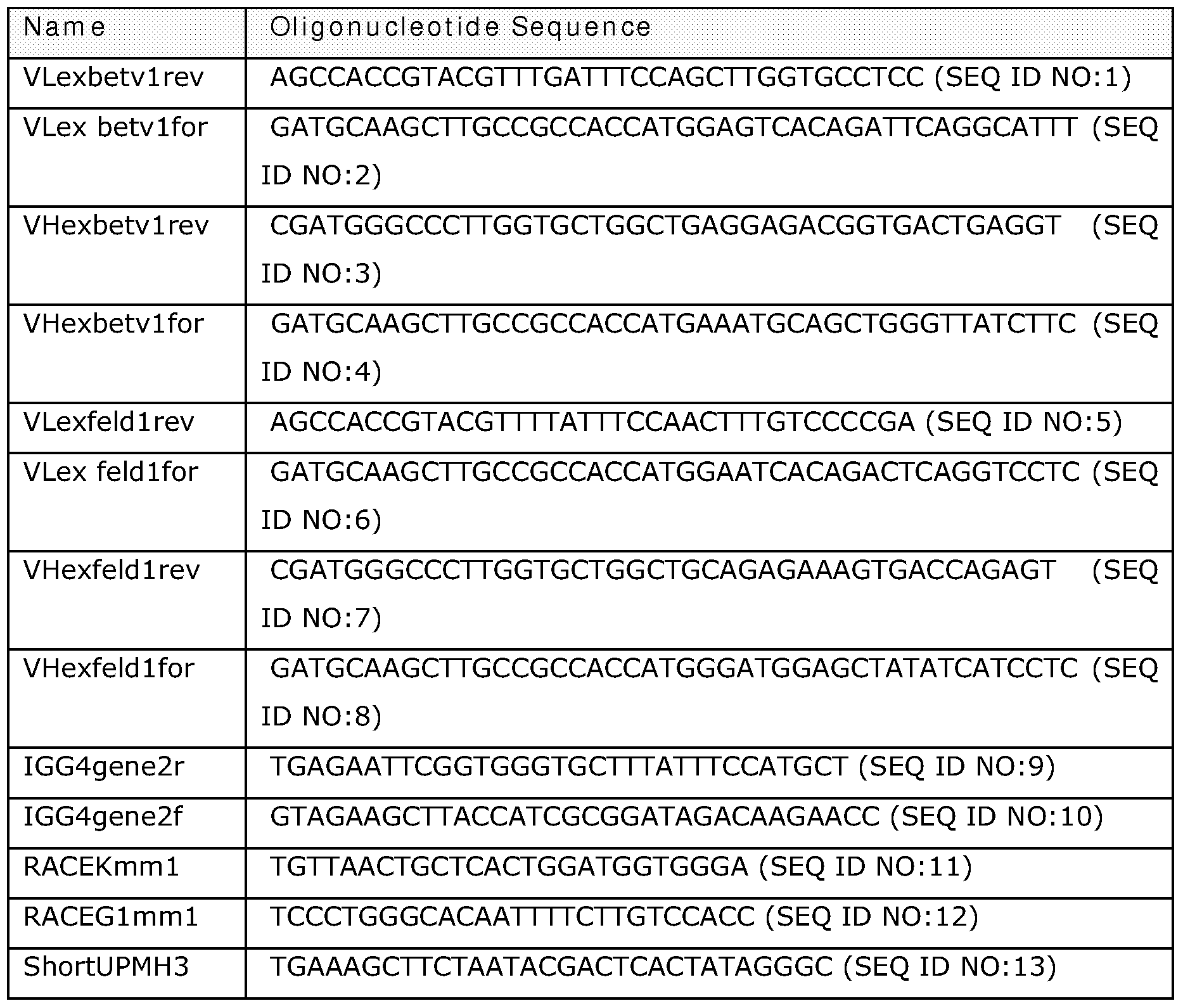


Claims
Priority Applications (10)
| Application Number | Priority Date | Filing Date | Title |
|---|---|---|---|
| CA002688275A CA2688275A1 (en) | 2007-05-31 | 2008-05-30 | Stable igg4 antibodies |
| EP22155736.6A EP4119579A1 (en) | 2007-05-31 | 2008-05-30 | Stable igg4 antibodies |
| EP13181032.7A EP2666787B1 (en) | 2007-05-31 | 2008-05-30 | STABLE IgG4 ANTIBODIES |
| EP08748828.4A EP2164873B2 (en) | 2007-05-31 | 2008-05-30 | Stable igg4 antibodies |
| AU2008255352A AU2008255352B2 (en) | 2007-05-31 | 2008-05-30 | Stable IgG4 antibodies |
| US12/602,439 US20100267934A1 (en) | 2007-05-31 | 2008-05-30 | Stable igg4 antibodies |
| JP2010509682A JP6071165B2 (en) | 2007-05-31 | 2008-05-30 | Stable IgG4 antibody |
| US13/912,581 US20130345406A1 (en) | 2007-05-31 | 2013-06-07 | Stable igg4 antibodies |
| US15/197,496 US10752695B2 (en) | 2007-05-31 | 2016-06-29 | Stable IgG4 antibodies |
| US16/947,021 US20210061919A1 (en) | 2007-05-31 | 2020-07-15 | STABLE IgG4 ANTIBODIES |
Applications Claiming Priority (6)
| Application Number | Priority Date | Filing Date | Title |
|---|---|---|---|
| DKPA200700793 | 2007-05-31 | ||
| DKPA200700792 | 2007-05-31 | ||
| DKPA200700793 | 2007-05-31 | ||
| DKPA200700792 | 2007-05-31 | ||
| DKPA200701002 | 2007-07-06 | ||
| DKPA200701002 | 2007-07-06 |
Related Child Applications (2)
| Application Number | Title | Priority Date | Filing Date |
|---|---|---|---|
| US12/602,439 A-371-Of-International US20100267934A1 (en) | 2007-05-31 | 2008-05-30 | Stable igg4 antibodies |
| US13/912,581 Division US20130345406A1 (en) | 2007-05-31 | 2013-06-07 | Stable igg4 antibodies |
Publications (1)
| Publication Number | Publication Date |
|---|---|
| WO2008145142A1 true WO2008145142A1 (en) | 2008-12-04 |
Family
ID=39831865
Family Applications (1)
| Application Number | Title | Priority Date | Filing Date |
|---|---|---|---|
| PCT/DK2008/050129 WO2008145142A1 (en) | 2007-05-31 | 2008-05-30 | Stable igg4 antibodies |
Country Status (6)
| Country | Link |
|---|---|
| US (4) | US20100267934A1 (en) |
| EP (3) | EP2164873B2 (en) |
| JP (3) | JP6071165B2 (en) |
| AU (1) | AU2008255352B2 (en) |
| CA (1) | CA2688275A1 (en) |
| WO (1) | WO2008145142A1 (en) |
Cited By (125)
| Publication number | Priority date | Publication date | Assignee | Title |
|---|---|---|---|---|
| WO2009103791A1 (en) * | 2008-02-22 | 2009-08-27 | Genmab A/S | Methods for assissing the risk of adverse events upon treatment with igg4 antibodies |
| WO2010063785A2 (en) * | 2008-12-03 | 2010-06-10 | Genmab A/S | Antibody variants having modifications in the constant region |
| WO2010128398A1 (en) | 2009-05-04 | 2010-11-11 | Pangenetics 110 B.V. | Antibodies against nerve growth factor (ngf) with enhanced in vivo stability |
| WO2011108502A1 (en) | 2010-03-02 | 2011-09-09 | 協和発酵キリン株式会社 | Modified antibody composition |
| WO2011110642A2 (en) | 2010-03-10 | 2011-09-15 | Genmab A/S | Monoclonal antibodies against c-met |
| WO2011116090A1 (en) | 2010-03-17 | 2011-09-22 | Abbott Research B.V. | Anti-nerve growth factor (ngf) antibody compositions |
| US8043617B2 (en) | 2006-06-02 | 2011-10-25 | Regeneron Pharmaceuticals, Inc. | Human antibodies to human IL-6 receptor |
| WO2011131746A2 (en) | 2010-04-20 | 2011-10-27 | Genmab A/S | Heterodimeric antibody fc-containing proteins and methods for production thereof |
| EP2389192A2 (en) * | 2009-01-23 | 2011-11-30 | Biogen Idec MA Inc. | Stabilized fc polypeptides with reduced effector function and methods of use |
| WO2011147982A2 (en) | 2010-05-27 | 2011-12-01 | Genmab A/S | Monoclonal antibodies against her2 epitope |
| WO2011147986A1 (en) | 2010-05-27 | 2011-12-01 | Genmab A/S | Monoclonal antibodies against her2 |
| US8080248B2 (en) | 2006-06-02 | 2011-12-20 | Regeneron Pharmaceuticals, Inc. | Method of treating rheumatoid arthritis with an IL-6R antibody |
| WO2011157741A2 (en) | 2010-06-15 | 2011-12-22 | Genmab A/S | Human antibody drug conjugates against tissue factor |
| WO2011138785A3 (en) * | 2010-05-05 | 2011-12-29 | Rappaport Family Institute For Research In The Medical Sciences | Use of ccl1 in therapy |
| WO2012022982A2 (en) | 2010-08-20 | 2012-02-23 | Ucb Pharma S.A. | Improved antibodies of the class igg4 |
| US8246956B2 (en) | 2003-12-24 | 2012-08-21 | Abbott Research B.V. | Humanized anti-nerve growth factor antibodies |
| WO2012143524A2 (en) | 2011-04-20 | 2012-10-26 | Genmab A/S | Bispecific antibodies against her2 and cd3 |
| WO2012143523A1 (en) | 2011-04-20 | 2012-10-26 | Genmab A/S | Bispecifc antibodies against her2 |
| CN103193882A (en) * | 2013-03-29 | 2013-07-10 | 浙江大学 | Embryonic stem cell specific marker GM-CSFR alpha and application thereof |
| WO2013124451A1 (en) | 2012-02-22 | 2013-08-29 | Ucb Pharma S.A. | Sequence symmetric modified igg4 bispecific antibodies |
| WO2013124450A1 (en) | 2012-02-22 | 2013-08-29 | Ucb Pharma S.A. | Sequence symmetric modified igg4 bispecific antibodies |
| WO2013166236A1 (en) * | 2012-05-03 | 2013-11-07 | Regeneron Pharmaceuticals, Inc. | Human antibodies to fel d1 and methods of use thereof |
| WO2015184203A1 (en) | 2014-05-29 | 2015-12-03 | Macrogenics, Inc. | Tri-specific binding molecules and methods of use thereof |
| US9212230B2 (en) | 2007-03-29 | 2015-12-15 | Genmab A/S | Bispecific antibodies and methods for production thereof |
| KR20160044063A (en) * | 2013-09-13 | 2016-04-22 | 베이진 엘티디 | Anti-PD1 Antibodies and their Use as Therapeutics and Diagnostics |
| WO2016091891A1 (en) | 2014-12-09 | 2016-06-16 | INSERM (Institut National de la Santé et de la Recherche Médicale) | Human monoclonal antibodies against axl |
| WO2016135066A1 (en) | 2015-02-26 | 2016-09-01 | INSERM (Institut National de la Santé et de la Recherche Médicale) | Fusion proteins and antibodies comprising thereof for promoting apoptosis |
| US9493578B2 (en) | 2009-09-02 | 2016-11-15 | Xencor, Inc. | Compositions and methods for simultaneous bivalent and monovalent co-engagement of antigens |
| WO2017019846A1 (en) | 2015-07-30 | 2017-02-02 | Macrogenics, Inc. | Pd-1-binding molecules and methods use thereof |
| US9605061B2 (en) | 2010-07-29 | 2017-03-28 | Xencor, Inc. | Antibodies with modified isoelectric points |
| US9605084B2 (en) | 2013-03-15 | 2017-03-28 | Xencor, Inc. | Heterodimeric proteins |
| US9650446B2 (en) | 2013-01-14 | 2017-05-16 | Xencor, Inc. | Heterodimeric proteins |
| CN106687125A (en) * | 2014-03-12 | 2017-05-17 | 耶达研究与开发有限公司 | Reducing systemic regulatory t cell levels or activity for treatment of disease and injury of the cns |
| US9688749B2 (en) | 2005-06-07 | 2017-06-27 | Abbvie Inc. | Molecules that are able to inhibit the binding between NGF and the TrkA receptor as analgesics with prolonged effect |
| US9701759B2 (en) | 2013-01-14 | 2017-07-11 | Xencor, Inc. | Heterodimeric proteins |
| US9717777B2 (en) | 2010-05-05 | 2017-08-01 | Rappaport Family Institute For Research In The Medical Sciences | Use of CCL1 in therapy |
| US9738722B2 (en) | 2013-01-15 | 2017-08-22 | Xencor, Inc. | Rapid clearance of antigen complexes using novel antibodies |
| WO2017144668A1 (en) | 2016-02-26 | 2017-08-31 | INSERM (Institut National de la Santé et de la Recherche Médicale) | Antibodies having specificity for btla and uses thereof |
| JP2017525335A (en) * | 2014-06-12 | 2017-09-07 | エフ.ホフマン−ラ ロシュ アーゲーF. Hoffmann−La Roche Aktiengesellschaft | Methods for selecting antibodies with altered FcRn interactions |
| WO2017162678A1 (en) | 2016-03-22 | 2017-09-28 | INSERM (Institut National de la Santé et de la Recherche Médicale) | Humanized anti-claudin-1 antibodies and uses thereof |
| WO2017191300A1 (en) | 2016-05-06 | 2017-11-09 | INSERM (Institut National de la Santé et de la Recherche Médicale) | Pharmaceutical compositions for the treatment of chemoresistant acute myeloid leukemia (aml) |
| US9822186B2 (en) | 2014-03-28 | 2017-11-21 | Xencor, Inc. | Bispecific antibodies that bind to CD38 and CD3 |
| US9850320B2 (en) | 2014-11-26 | 2017-12-26 | Xencor, Inc. | Heterodimeric antibodies to CD3 X CD20 |
| US9856327B2 (en) | 2014-11-26 | 2018-01-02 | Xencor, Inc. | Heterodimeric antibodies to CD3 X CD123 |
| US9862778B2 (en) | 2011-10-11 | 2018-01-09 | Genentech, Inc. | Assembly of bispecific antibodies |
| US10106624B2 (en) | 2013-03-15 | 2018-10-23 | Xencor, Inc. | Heterodimeric proteins |
| WO2018204871A1 (en) * | 2017-05-05 | 2018-11-08 | Allakos Inc. | Methods and compositions for treating inflammatory gastrointestinal disorders |
| US10131710B2 (en) | 2013-01-14 | 2018-11-20 | Xencor, Inc. | Optimized antibody variable regions |
| EP3310385A4 (en) * | 2015-06-17 | 2018-12-19 | Allakos Inc. | Methods and compositions for treating fibrotic diseases |
| CN109069537A (en) * | 2016-04-04 | 2018-12-21 | 亘喜生物科技(上海)有限公司 | There is the CAR for repeating binding motif in costimulation structural domain |
| US10183996B2 (en) | 2014-02-28 | 2019-01-22 | Allakos Inc. | Methods and compositions for treating Siglec-8 associated diseases |
| US10227411B2 (en) | 2015-03-05 | 2019-03-12 | Xencor, Inc. | Modulation of T cells with bispecific antibodies and FC fusions |
| US10227410B2 (en) | 2015-12-07 | 2019-03-12 | Xencor, Inc. | Heterodimeric antibodies that bind CD3 and PSMA |
| WO2019057933A1 (en) | 2017-09-21 | 2019-03-28 | Imcheck Therapeutics Sas | Antibodies having specificity for btn2 and uses thereof |
| US10316088B2 (en) | 2016-06-28 | 2019-06-11 | Xencor, Inc. | Heterodimeric antibodies that bind somatostatin receptor 2 |
| EP3279216A4 (en) * | 2015-04-01 | 2019-06-19 | Chugai Seiyaku Kabushiki Kaisha | Method for producing polypeptide hetero-oligomer |
| US10344050B2 (en) | 2011-10-27 | 2019-07-09 | Genmab A/S | Production of heterodimeric proteins |
| US10428155B2 (en) | 2014-12-22 | 2019-10-01 | Xencor, Inc. | Trispecific antibodies |
| US10428146B2 (en) | 2014-07-22 | 2019-10-01 | Cb Therapeutics, Inc. | Anti PD-1 antibodies |
| US10435458B2 (en) | 2010-03-04 | 2019-10-08 | Chugai Seiyaku Kabushiki Kaisha | Antibody constant region variants with reduced Fcgammar binding |
| US10435470B2 (en) | 2014-08-05 | 2019-10-08 | Cb Therapeutics, Inc. | Anti-PD-L1 antibodies |
| US10450381B2 (en) | 2010-11-17 | 2019-10-22 | Chugai Seiyaku Kabushiki Kaisha | Methods of treatment that include the administration of bispecific antibodies |
| US10487155B2 (en) | 2013-01-14 | 2019-11-26 | Xencor, Inc. | Heterodimeric proteins |
| US10501543B2 (en) | 2016-10-14 | 2019-12-10 | Xencor, Inc. | IL15/IL15Rα heterodimeric Fc-fusion proteins |
| US10501552B2 (en) | 2015-01-26 | 2019-12-10 | Macrogenics, Inc. | Multivalent molecules comprising DR5-binding domains |
| US10519242B2 (en) | 2013-03-15 | 2019-12-31 | Xencor, Inc. | Targeting regulatory T cells with heterodimeric proteins |
| US10526417B2 (en) | 2014-11-26 | 2020-01-07 | Xencor, Inc. | Heterodimeric antibodies that bind CD3 and CD38 |
| US10544187B2 (en) | 2013-03-15 | 2020-01-28 | Xencor, Inc. | Targeting regulatory T cells with heterodimeric proteins |
| US10544225B2 (en) | 2014-07-03 | 2020-01-28 | Beigene, Ltd. | Anti-PD-L1 antibodies and their use as therapeutics and diagnostics |
| US10604577B2 (en) | 2015-10-22 | 2020-03-31 | Allakos Inc. | Methods and compositions for treating systemic mastocytosis |
| WO2020148207A1 (en) | 2019-01-14 | 2020-07-23 | INSERM (Institut National de la Santé et de la Recherche Médicale) | Human monoclonal antibodies binding to hla-a2 |
| WO2020188086A1 (en) | 2019-03-20 | 2020-09-24 | Imcheck Therapeutics Sas | Antibodies having specificity for btn2 and uses thereof |
| US10787518B2 (en) | 2016-06-14 | 2020-09-29 | Xencor, Inc. | Bispecific checkpoint inhibitor antibodies |
| US10793632B2 (en) | 2016-08-30 | 2020-10-06 | Xencor, Inc. | Bispecific immunomodulatory antibodies that bind costimulatory and checkpoint receptors |
| US10851178B2 (en) | 2011-10-10 | 2020-12-01 | Xencor, Inc. | Heterodimeric human IgG1 polypeptides with isoelectric point modifications |
| US10858417B2 (en) | 2013-03-15 | 2020-12-08 | Xencor, Inc. | Heterodimeric proteins |
| US10865253B2 (en) | 2014-12-19 | 2020-12-15 | Genmab A/S | Rodent bispecific heterodimeric proteins |
| US10864203B2 (en) | 2016-07-05 | 2020-12-15 | Beigene, Ltd. | Combination of a PD-1 antagonist and a RAF inhibitor for treating cancer |
| WO2021009263A1 (en) | 2019-07-16 | 2021-01-21 | INSERM (Institut National de la Santé et de la Recherche Médicale) | Antibodies having specificity for cd38 and uses thereof |
| US10927435B2 (en) | 2011-10-11 | 2021-02-23 | Sanofi Biotechnology | Compositions for the treatment of rheumatoid arthritis and methods of using same |
| US10935554B2 (en) | 2013-08-23 | 2021-03-02 | Regeneron Pharmaceuticals, Inc. | Diagnostic tests and methods for assessing safety, efficacy or outcome of allergen-specific immunotherapy (SIT) |
| US10934344B2 (en) | 2006-03-31 | 2021-03-02 | Chugai Seiyaku Kabushiki Kaisha | Methods of modifying antibodies for purification of bispecific antibodies |
| US10954301B2 (en) | 2015-12-14 | 2021-03-23 | Macrogenics, Inc. | Bispecific molecules having immunoreactivity with PD-1 and CTLA-4, and methods of use thereof |
| WO2021058763A1 (en) | 2019-09-27 | 2021-04-01 | INSERM (Institut National de la Santé et de la Recherche Médicale) | Anti-müllerian inhibiting substance antibodies and uses thereof |
| US10968276B2 (en) | 2013-03-12 | 2021-04-06 | Xencor, Inc. | Optimized anti-CD3 variable regions |
| US10982006B2 (en) | 2018-04-04 | 2021-04-20 | Xencor, Inc. | Heterodimeric antibodies that bind fibroblast activation protein |
| US10981992B2 (en) | 2017-11-08 | 2021-04-20 | Xencor, Inc. | Bispecific immunomodulatory antibodies that bind costimulatory and checkpoint receptors |
| US11001643B2 (en) | 2014-09-26 | 2021-05-11 | Chugai Seiyaku Kabushiki Kaisha | Cytotoxicity-inducing therapeutic agent |
| US11046784B2 (en) | 2006-03-31 | 2021-06-29 | Chugai Seiyaku Kabushiki Kaisha | Methods for controlling blood pharmacokinetics of antibodies |
| US11053316B2 (en) | 2013-01-14 | 2021-07-06 | Xencor, Inc. | Optimized antibody variable regions |
| US11072666B2 (en) | 2016-03-14 | 2021-07-27 | Chugai Seiyaku Kabushiki Kaisha | Cell injury inducing therapeutic drug for use in cancer therapy |
| US11072653B2 (en) | 2015-06-08 | 2021-07-27 | Macrogenics, Inc. | LAG-3-binding molecules and methods of use thereof |
| US11078279B2 (en) | 2015-06-12 | 2021-08-03 | Macrogenics, Inc. | Combination therapy for the treatment of cancer |
| US11084863B2 (en) | 2017-06-30 | 2021-08-10 | Xencor, Inc. | Targeted heterodimeric Fc fusion proteins containing IL-15 IL-15alpha and antigen binding domains |
| US11098127B2 (en) | 2010-01-08 | 2021-08-24 | Regeneron Pharmaceuticals, Inc. | Stabilized formulations containing anti-interleukin-6 receptor (IL-6R) antibodies |
| WO2021175954A1 (en) | 2020-03-04 | 2021-09-10 | Imcheck Therapeutics Sas | Antibodies having specificity for btnl8 and uses thereof |
| US11124576B2 (en) | 2013-09-27 | 2021-09-21 | Chungai Seiyaku Kabushiki Kaisha | Method for producing polypeptide heteromultimer |
| US11168344B2 (en) | 2005-03-31 | 2021-11-09 | Chugai Seiyaku Kabushiki Kaisha | Methods for producing polypeptides by regulating polypeptide association |
| US11174315B2 (en) | 2015-10-08 | 2021-11-16 | Macrogenics, Inc. | Combination therapy for the treatment of cancer |
| WO2021234402A2 (en) | 2020-05-21 | 2021-11-25 | Mabsolve Limited | Modified immunoglobulin fc regions |
| US11198732B2 (en) | 2011-03-16 | 2021-12-14 | Amgen Inc. | Fc variants |
| US11203638B2 (en) | 2017-05-05 | 2021-12-21 | Allakos Inc. | Methods and compositions for treating perennial allergic conjunctivitis and keratoconjunctivitis |
| US11242384B2 (en) | 2017-01-20 | 2022-02-08 | Sanofi | Anti-TGF-beta antibodies and their use |
| US11248053B2 (en) | 2007-09-26 | 2022-02-15 | Chugai Seiyaku Kabushiki Kaisha | Method of modifying isoelectric point of antibody via amino acid substitution in CDR |
| WO2022079199A1 (en) | 2020-10-15 | 2022-04-21 | UCB Biopharma SRL | Binding molecules that multimerise cd45 |
| US11312770B2 (en) | 2017-11-08 | 2022-04-26 | Xencor, Inc. | Bispecific and monospecific antibodies using novel anti-PD-1 sequences |
| US11319355B2 (en) | 2017-12-19 | 2022-05-03 | Xencor, Inc. | Engineered IL-2 Fc fusion proteins |
| US11332533B2 (en) | 2007-09-26 | 2022-05-17 | Chugai Seiyaku Kabushiki Kaisha | Modified antibody constant region |
| US11352417B2 (en) | 2016-12-22 | 2022-06-07 | Regeneron Pharmaceuticals, Inc. | Method of treating an allergy with allergen-specific monoclonal antibodies |
| US11358999B2 (en) | 2018-10-03 | 2022-06-14 | Xencor, Inc. | IL-12 heterodimeric Fc-fusion proteins |
| US11472890B2 (en) | 2019-03-01 | 2022-10-18 | Xencor, Inc. | Heterodimeric antibodies that bind ENPP3 and CD3 |
| US11498969B2 (en) | 2019-01-31 | 2022-11-15 | Sanofi Biotechnology | Compositions and methods for treating juvenile idiopathic arthritis |
| US11505595B2 (en) | 2018-04-18 | 2022-11-22 | Xencor, Inc. | TIM-3 targeted heterodimeric fusion proteins containing IL-15/IL-15RA Fc-fusion proteins and TIM-3 antigen binding domains |
| US11524991B2 (en) | 2018-04-18 | 2022-12-13 | Xencor, Inc. | PD-1 targeted heterodimeric fusion proteins containing IL-15/IL-15Ra Fc-fusion proteins and PD-1 antigen binding domains and uses thereof |
| US11555038B2 (en) | 2017-01-25 | 2023-01-17 | Beigene, Ltd. | Crystalline forms of (S)-7-(1-(but-2-ynoyl)piperidin-4-yl)-2-(4-phenoxyphenyl)-4,5,6,7-tetrahydropyrazolo[1,5-a]pyrimidine-3-carboxamide, preparation, and uses thereof |
| US11591401B2 (en) | 2020-08-19 | 2023-02-28 | Xencor, Inc. | Anti-CD28 compositions |
| US11597768B2 (en) | 2017-06-26 | 2023-03-07 | Beigene, Ltd. | Immunotherapy for hepatocellular carcinoma |
| US11634495B2 (en) | 2017-04-07 | 2023-04-25 | Miltenyi Biotec B.V. & Co. KG | Methods of activating CD32b/c comprising administering an antibody that binds BDCA-2 (CD303) |
| US11649262B2 (en) | 2015-12-28 | 2023-05-16 | Chugai Seiyaku Kabushiki Kaisha | Method for promoting efficiency of purification of Fc region-containing polypeptide |
| US11701357B2 (en) | 2016-08-19 | 2023-07-18 | Beigene Switzerland Gmbh | Treatment of B cell cancers using a combination comprising Btk inhibitors |
| US11739144B2 (en) | 2021-03-09 | 2023-08-29 | Xencor, Inc. | Heterodimeric antibodies that bind CD3 and CLDN6 |
| US11786529B2 (en) | 2017-11-29 | 2023-10-17 | Beigene Switzerland Gmbh | Treatment of indolent or aggressive B-cell lymphomas using a combination comprising BTK inhibitors |
| US11851476B2 (en) | 2011-10-31 | 2023-12-26 | Chugai Seiyaku Kabushiki Kaisha | Antigen-binding molecule having regulated conjugation between heavy-chain and light-chain |
| US11859012B2 (en) | 2021-03-10 | 2024-01-02 | Xencor, Inc. | Heterodimeric antibodies that bind CD3 and GPC3 |
| US11919956B2 (en) | 2020-05-14 | 2024-03-05 | Xencor, Inc. | Heterodimeric antibodies that bind prostate specific membrane antigen (PSMA) and CD3 |
Families Citing this family (38)
| Publication number | Priority date | Publication date | Assignee | Title |
|---|---|---|---|---|
| WO2008145140A2 (en) | 2007-05-31 | 2008-12-04 | Genmab A/S | Transgenic animals producing monovalent human antibodies and antibodies obtainable from these animals |
| SG182823A1 (en) | 2010-02-11 | 2012-09-27 | Alexion Pharma Inc | Therapeutic methods using an ti-cd200 antibodies |
| US11214610B2 (en) | 2010-12-01 | 2022-01-04 | H. Lundbeck A/S | High-purity production of multi-subunit proteins such as antibodies in transformed microbes such as Pichia pastoris |
| US9884909B2 (en) | 2010-12-01 | 2018-02-06 | Alderbio Holdings Llc | Anti-NGF compositions and use thereof |
| US9078878B2 (en) | 2010-12-01 | 2015-07-14 | Alderbio Holdings Llc | Anti-NGF antibodies that selectively inhibit the association of NGF with TrkA, without affecting the association of NGF with p75 |
| KR20190112175A (en) | 2010-12-01 | 2019-10-02 | 앨더바이오 홀딩스 엘엘씨 | Anti-ngf compositions and use thereof |
| US9539324B2 (en) | 2010-12-01 | 2017-01-10 | Alderbio Holdings, Llc | Methods of preventing inflammation and treating pain using anti-NGF compositions |
| US9067988B2 (en) | 2010-12-01 | 2015-06-30 | Alderbio Holdings Llc | Methods of preventing or treating pain using anti-NGF antibodies |
| MX358447B (en) | 2011-06-13 | 2018-08-21 | Abgenomics Cooeperatief U A Star | Anti-psgl-1 antibodies and uses thereof. |
| US10280215B2 (en) | 2013-01-31 | 2019-05-07 | Seoul National University R&Db Foundation | Anti-C5 antibodies and methods of treating complement-related diseases |
| TWI682941B (en) | 2013-02-01 | 2020-01-21 | 美商再生元醫藥公司 | Antibodies comprising chimeric constant domains |
| MY184154A (en) | 2013-12-12 | 2021-03-23 | Shanghai hengrui pharmaceutical co ltd | Pd-1 antibody, antigen-binding fragment thereof, and medical application thereof |
| TWI754319B (en) | 2014-03-19 | 2022-02-01 | 美商再生元醫藥公司 | Methods and antibody compositions for tumor treatment |
| US9718870B2 (en) | 2014-05-29 | 2017-08-01 | Medimmune, Llc | OX40L fusion proteins and uses thereof |
| TWI566780B (en) * | 2014-10-24 | 2017-01-21 | 國立陽明大學 | Use of mip-1β inhibitors for improving angiogenesis to reduce tissue ischemia and rescue diabetic vascular disease in diabetes mellitus |
| PL3221359T3 (en) | 2014-11-17 | 2020-11-16 | Regeneron Pharmaceuticals, Inc. | Methods for tumor treatment using cd3xcd20 bispecific antibody |
| EP3277725B1 (en) | 2015-03-30 | 2020-11-25 | Regeneron Pharmaceuticals, Inc. | Heavy chain constant regions with reduced binding to fc gamma receptors |
| CN107743494B (en) | 2015-06-02 | 2022-04-29 | 诺和诺德股份有限公司 | Insulin with polar recombinant extensions |
| KR101794702B1 (en) | 2015-09-25 | 2017-11-08 | 오상혁 | Hybrid chemicals bottle for color perm |
| AR106184A1 (en) | 2015-09-29 | 2017-12-20 | Celgene Corp | PD-1 BINDING PROTEINS AND THEIR METHODS OF USE |
| MA43348A (en) | 2015-10-01 | 2018-08-08 | Novo Nordisk As | PROTEIN CONJUGATES |
| EP3463451A1 (en) | 2016-05-26 | 2019-04-10 | Qilu Puget Sound Biotherapeutics Corporation | Mixtures of antibodies |
| EP3515943A4 (en) | 2016-09-19 | 2020-05-06 | Celgene Corporation | Methods of treating vitiligo using pd-1 binding proteins |
| SG11201901950TA (en) | 2016-09-19 | 2019-04-29 | Celgene Corp | Methods of treating immune disorders using pd-1 binding proteins |
| GB201701194D0 (en) | 2017-01-24 | 2017-03-08 | Capella Bioscience Ltd | Antigen binding molecules that bind light |
| CN108570109B (en) * | 2017-03-14 | 2022-04-26 | 广东东阳光药业有限公司 | Dual target fusion proteins comprising an immunoglobulin Fc portion |
| JP2020513019A (en) | 2017-04-05 | 2020-04-30 | ノヴォ ノルディスク アー/エス | Oligomer-extended insulin-Fc conjugate |
| RU2646807C1 (en) * | 2017-08-08 | 2018-03-07 | Федеральное государственное бюджетное научное учреждение "Научно-исследовательский институт фундаментальной и клинической иммунологии" | Method for isolating autoantibodies of immunoglobulin g subclasses to immunoregulatory cytokine tumor necrosis factor |
| US11312781B2 (en) | 2018-01-24 | 2022-04-26 | Capella Bioscience Ltd. | Antigen binding molecules that bind LIGHT |
| KR20210054528A (en) | 2018-08-31 | 2021-05-13 | 리제너론 파아마슈티컬스, 인크. | Dosing strategies to alleviate cytokine release syndrome against CD3/C20 bispecific antibodies |
| JP6800191B2 (en) * | 2018-09-21 | 2020-12-16 | ベイジーン スウィッツァーランド ゲーエムベーハー | Anti-PD-1 antibody and its use for treatment and diagnosis |
| JP2022504550A (en) | 2018-10-12 | 2022-01-13 | ゼンコア インコーポレイテッド | Use in PD-1 targeted IL-15 / IL-15R alpha FC fusion proteins and their combination therapies |
| WO2020132646A1 (en) | 2018-12-20 | 2020-06-25 | Xencor, Inc. | Targeted heterodimeric fc fusion proteins containing il-15/il-15ra and nkg2d antigen binding domains |
| CA3147883C (en) | 2019-07-18 | 2024-01-02 | Hanmi Pharm. Co., Ltd. | Novel method for preparing long-acting drug conjugate through preparation of intermediate |
| TW202128757A (en) | 2019-10-11 | 2021-08-01 | 美商建南德克公司 | Pd-1 targeted il-15/il-15ralpha fc fusion proteins with improved properties |
| KR20230148959A (en) * | 2022-04-19 | 2023-10-26 | 전남대학교산학협력단 | Composition for treating colitis comprising fusion protein of flagellin and immunoglobulin Fc region |
| KR20230148956A (en) * | 2022-04-19 | 2023-10-26 | 전남대학교산학협력단 | Composition for treating liver disease or metabolic syndrome comprising fusion protein of flagellin and immunoglobulin Fc region |
| US11926669B2 (en) | 2022-05-30 | 2024-03-12 | Hanall Biopharma Co., Ltd. | Anti-FcRn antibody or antigen binding fragment thereof with improved stability |
Citations (3)
| Publication number | Priority date | Publication date | Assignee | Title |
|---|---|---|---|---|
| WO2005063816A2 (en) * | 2003-12-19 | 2005-07-14 | Genentech, Inc. | Monovalent antibody fragments useful as therapeutics |
| WO2006033386A1 (en) * | 2004-09-22 | 2006-03-30 | Kirin Beer Kabushiki Kaisha | STABILIZED HUMAN IgG4 ANTIBODIES |
| US20070105199A1 (en) * | 2005-10-21 | 2007-05-10 | Wei Yan | Methods for generating monovalent IgG |
Family Cites Families (43)
| Publication number | Priority date | Publication date | Assignee | Title |
|---|---|---|---|---|
| US4816567A (en) | 1983-04-08 | 1989-03-28 | Genentech, Inc. | Recombinant immunoglobin preparations |
| JPS6147500A (en) | 1984-08-15 | 1986-03-07 | Res Dev Corp Of Japan | Chimera monoclonal antibody and its preparation |
| EP0173494A3 (en) | 1984-08-27 | 1987-11-25 | The Board Of Trustees Of The Leland Stanford Junior University | Chimeric receptors by dna splicing and expression |
| GB8422238D0 (en) | 1984-09-03 | 1984-10-10 | Neuberger M S | Chimeric proteins |
| JPS61134325A (en) | 1984-12-04 | 1986-06-21 | Teijin Ltd | Expression of hybrid antibody gene |
| JPS63501765A (en) | 1985-11-01 | 1988-07-21 | インタ−ナショナル、ジェネティック、エンジニアリング インコ−ポレ−テッド | Antibody gene module assembly, antibodies produced thereby, and uses |
| JP3101690B2 (en) * | 1987-03-18 | 2000-10-23 | エス・ビィ・2・インコーポレイテッド | Modifications of or for denatured antibodies |
| US5750172A (en) | 1987-06-23 | 1998-05-12 | Pharming B.V. | Transgenic non human mammal milk |
| GB8823869D0 (en) | 1988-10-12 | 1988-11-16 | Medical Res Council | Production of antibodies |
| US5633076A (en) | 1989-12-01 | 1997-05-27 | Pharming Bv | Method of producing a transgenic bovine or transgenic bovine embryo |
| US6300129B1 (en) | 1990-08-29 | 2001-10-09 | Genpharm International | Transgenic non-human animals for producing heterologous antibodies |
| US5633425A (en) | 1990-08-29 | 1997-05-27 | Genpharm International, Inc. | Transgenic non-human animals capable of producing heterologous antibodies |
| US5877397A (en) | 1990-08-29 | 1999-03-02 | Genpharm International Inc. | Transgenic non-human animals capable of producing heterologous antibodies of various isotypes |
| US5789650A (en) | 1990-08-29 | 1998-08-04 | Genpharm International, Inc. | Transgenic non-human animals for producing heterologous antibodies |
| US5545806A (en) | 1990-08-29 | 1996-08-13 | Genpharm International, Inc. | Ransgenic non-human animals for producing heterologous antibodies |
| US5625126A (en) | 1990-08-29 | 1997-04-29 | Genpharm International, Inc. | Transgenic non-human animals for producing heterologous antibodies |
| US5661016A (en) | 1990-08-29 | 1997-08-26 | Genpharm International Inc. | Transgenic non-human animals capable of producing heterologous antibodies of various isotypes |
| US5814318A (en) | 1990-08-29 | 1998-09-29 | Genpharm International Inc. | Transgenic non-human animals for producing heterologous antibodies |
| US5770429A (en) | 1990-08-29 | 1998-06-23 | Genpharm International, Inc. | Transgenic non-human animals capable of producing heterologous antibodies |
| CA2089661C (en) | 1990-08-29 | 2007-04-03 | Nils Lonberg | Transgenic non-human animals capable of producing heterologous antibodies |
| US5874299A (en) | 1990-08-29 | 1999-02-23 | Genpharm International, Inc. | Transgenic non-human animals capable of producing heterologous antibodies |
| WO1992022645A1 (en) | 1991-06-14 | 1992-12-23 | Genpharm International, Inc. | Transgenic immunodeficient non-human animals |
| EP0593592B1 (en) | 1991-07-08 | 1998-03-25 | The University Of Massachusetts At Amherst | Thermotropic liquid crystal segmented block copolymer |
| US5733743A (en) | 1992-03-24 | 1998-03-31 | Cambridge Antibody Technology Limited | Methods for producing members of specific binding pairs |
| ATE193301T1 (en) | 1993-03-09 | 2000-06-15 | Genzyme Corp | METHOD FOR ISOLATION OF PROTEINS FROM MILK |
| AU6819494A (en) | 1993-04-26 | 1994-11-21 | Genpharm International, Inc. | Transgenic non-human animals capable of producing heterologous antibodies |
| US5827690A (en) | 1993-12-20 | 1998-10-27 | Genzyme Transgenics Corporatiion | Transgenic production of antibodies in milk |
| JP2002512776A (en) | 1998-04-28 | 2002-05-08 | スミスクライン・ビーチャム・コーポレイション | Monoclonal antibodies with reduced immunogenicity |
| US6737056B1 (en) | 1999-01-15 | 2004-05-18 | Genentech, Inc. | Polypeptide variants with altered effector function |
| PL209786B1 (en) * | 1999-01-15 | 2011-10-31 | Genentech Inc | Variant of mother polypeptide containing Fc region, polypeptide containing variant of Fc region with altered affinity of Fc gamma receptor binding (Fc R), polypeptide containing variant of Fc region with altered affinity of Fc gamma neonatal receptor binding (Fc Rn), composition, isolated nucleic acid, vector, host cell, method for obtaining polypeptide variant, the use thereof and method for obtaining region Fc variant |
| EP1210372B1 (en) | 1999-07-29 | 2008-01-23 | Medarex, Inc. | Human monoclonal antibodies to her2/neu |
| CN1371416B (en) | 1999-08-24 | 2012-10-10 | 梅达里克斯公司 | Human CTLA-4 antibodies and their uses |
| DE10015711A1 (en) * | 2000-03-29 | 2001-10-04 | Nbt Gmbh | Alkaline accumulator |
| TWI313299B (en) | 2000-11-30 | 2009-08-11 | Medarex Inc | Transgenic transchromosomal rodents for making human antibodies |
| WO2002086186A1 (en) | 2001-04-24 | 2002-10-31 | Tosoh Smd, Inc. | Target and method of optimizing target profile |
| BRPI0210405B8 (en) | 2001-06-13 | 2021-05-25 | Genmab As | human monoclonal antibody, bispecific molecule, in vitro method to inhibit the growth of a cell expressing egfr, to induce cytolysis of a cell expressing egfr, and to detect the presence of egfr antigen or a cell expressing egfr in a sample, e, vector of expression |
| US7595378B2 (en) | 2001-06-13 | 2009-09-29 | Genmab A/S | Human monoclonal antibodies to epidermal growth factor receptor (EGFR) |
| WO2003074679A2 (en) * | 2002-03-01 | 2003-09-12 | Xencor | Antibody optimization |
| CN101928344B (en) | 2002-10-17 | 2014-08-13 | 根马布股份公司 | Human monoclonal antibodies against cd20 |
| US20060074225A1 (en) * | 2004-09-14 | 2006-04-06 | Xencor, Inc. | Monomeric immunoglobulin Fc domains |
| EP3050963B1 (en) | 2005-03-31 | 2019-09-18 | Chugai Seiyaku Kabushiki Kaisha | Process for production of polypeptide by regulation of assembly |
| JP5525729B2 (en) | 2005-11-28 | 2014-06-18 | ゲンマブ エー/エス | Recombinant monovalent antibody and production method thereof |
| KR102152109B1 (en) * | 2010-04-20 | 2020-09-07 | 젠맵 에이/에스 | Heterodimeric antibody fc-containing proteins and methods for production thereof |
-
2008
- 2008-05-30 EP EP08748828.4A patent/EP2164873B2/en active Active
- 2008-05-30 US US12/602,439 patent/US20100267934A1/en not_active Abandoned
- 2008-05-30 CA CA002688275A patent/CA2688275A1/en active Pending
- 2008-05-30 EP EP13181032.7A patent/EP2666787B1/en active Active
- 2008-05-30 WO PCT/DK2008/050129 patent/WO2008145142A1/en active Application Filing
- 2008-05-30 EP EP22155736.6A patent/EP4119579A1/en active Pending
- 2008-05-30 JP JP2010509682A patent/JP6071165B2/en active Active
- 2008-05-30 AU AU2008255352A patent/AU2008255352B2/en active Active
-
2013
- 2013-06-07 US US13/912,581 patent/US20130345406A1/en not_active Abandoned
-
2014
- 2014-01-31 JP JP2014016803A patent/JP6034315B2/en active Active
-
2016
- 2016-06-29 US US15/197,496 patent/US10752695B2/en active Active
- 2016-10-27 JP JP2016210550A patent/JP6412083B2/en active Active
-
2020
- 2020-07-15 US US16/947,021 patent/US20210061919A1/en active Pending
Patent Citations (4)
| Publication number | Priority date | Publication date | Assignee | Title |
|---|---|---|---|---|
| WO2005063816A2 (en) * | 2003-12-19 | 2005-07-14 | Genentech, Inc. | Monovalent antibody fragments useful as therapeutics |
| WO2006033386A1 (en) * | 2004-09-22 | 2006-03-30 | Kirin Beer Kabushiki Kaisha | STABILIZED HUMAN IgG4 ANTIBODIES |
| EP1810979A1 (en) * | 2004-09-22 | 2007-07-25 | Kirin Beer Kabushiki Kaisha | STABILIZED HUMAN IgG4 ANTIBODIES |
| US20070105199A1 (en) * | 2005-10-21 | 2007-05-10 | Wei Yan | Methods for generating monovalent IgG |
Non-Patent Citations (7)
| Title |
|---|
| ANGAL S ET AL: "A SINGLE AMINO ACID SUBSTITUTION ABOLISHES THE HETEROGENEITY OF CHIMERIC MOUSE/HUMAN (IGG4) ANTIBODY", MOLECULAR IMMUNOLOGY, ELMSFORD, NY, US, vol. 30, no. 1, 1 January 1993 (1993-01-01), pages 105 - 108, XP000770218, ISSN: 0161-5890 * |
| BRUSCO A ET AL: "Molecular characterization of immunoglobulin G4 gene isoallotypes", EUROPEAN JOURNAL OF IMMUNOGENETICS, OXFORD, GB, vol. 25, no. 5, 1 October 1998 (1998-10-01), pages 349 - 355, XP002995211, ISSN: 0960-7420 * |
| DALL'ACQUA WILLIAM F ET AL: "Modulation of the effector functions of a human IgG1 through engineering of its hinge region", JOURNAL OF IMMUNOLOGY (BALTIMORE, MD. : 1950) 15 JUL 2006,, vol. 177, no. 2, 15 July 2006 (2006-07-15), pages 1129 - 1138, XP002497452 * |
| HARINGMAN JASPER J ET AL: "A randomized controlled trial with an anti-CCL2 (anti-monocyte chemotactic protein 1) monoclonal antibody in patients with rheumatoid arthritis.", ARTHRITIS AND RHEUMATISM AUG 2006, vol. 54, no. 8, August 2006 (2006-08-01), pages 2387 - 2392, XP002499902, ISSN: 0004-3591 * |
| HORGAN CAROL ET AL: "Studies on antigen binding by intact and hinge-deleted chimeric antibodies", JOURNAL OF IMMUNOLOGY, AMERICAN ASSOCIATION OF IMMUNOLOGISTS, US, vol. 150, no. 12, 15 June 1993 (1993-06-15), pages 5400 - 5407, XP002423580, ISSN: 0022-1767 * |
| SCHUURMAN J ET AL: "The inter-heavy chain disulfide bonds of IgG4 are in equilibrium with intra-chain disulfide bonds", MOLECULAR IMMUNOLOGY, ELMSFORD, NY, US, vol. 38, no. 1, 1 January 2001 (2001-01-01), pages 1 - 8, XP002329378, ISSN: 0161-5890 * |
| SHERIDAN CORMAC: "Pharma consolidates its grip on post-antibody landscape.", NATURE BIOTECHNOLOGY APR 2007, vol. 25, no. 4, April 2007 (2007-04-01), pages 365 - 366, XP002499901, ISSN: 1087-0156 * |
Cited By (240)
| Publication number | Priority date | Publication date | Assignee | Title |
|---|---|---|---|---|
| US8877491B2 (en) | 2003-12-24 | 2014-11-04 | Abbvie Inc. | Polynucleotides encoding humanized anti-NGF antibodies |
| US8246956B2 (en) | 2003-12-24 | 2012-08-21 | Abbott Research B.V. | Humanized anti-nerve growth factor antibodies |
| US8257710B2 (en) | 2003-12-24 | 2012-09-04 | Abbott Research, B.V. | Method for the treatment of pain with humanized anti-nerve growth factor antibodies |
| US11168344B2 (en) | 2005-03-31 | 2021-11-09 | Chugai Seiyaku Kabushiki Kaisha | Methods for producing polypeptides by regulating polypeptide association |
| US9688749B2 (en) | 2005-06-07 | 2017-06-27 | Abbvie Inc. | Molecules that are able to inhibit the binding between NGF and the TrkA receptor as analgesics with prolonged effect |
| US10934344B2 (en) | 2006-03-31 | 2021-03-02 | Chugai Seiyaku Kabushiki Kaisha | Methods of modifying antibodies for purification of bispecific antibodies |
| US11046784B2 (en) | 2006-03-31 | 2021-06-29 | Chugai Seiyaku Kabushiki Kaisha | Methods for controlling blood pharmacokinetics of antibodies |
| US9308256B2 (en) | 2006-06-02 | 2016-04-12 | Regeneron Pharmaceuticals, Inc. | Method of treating rheumatoid arthritis with an anti-IL-6R antibody |
| US8183014B2 (en) | 2006-06-02 | 2012-05-22 | Regeneron Pharmaceuticals, Inc. | High affinity antibodies to human IL-6 receptor |
| US11370843B2 (en) | 2006-06-02 | 2022-06-28 | Regeneron Pharmaceuticals, Inc. | High affinity antibodies to human IL-6 receptor |
| US8043617B2 (en) | 2006-06-02 | 2011-10-25 | Regeneron Pharmaceuticals, Inc. | Human antibodies to human IL-6 receptor |
| US8568721B2 (en) | 2006-06-02 | 2013-10-29 | Regeneron Pharmaceuticals, Inc. | Method of treating rheumatoid arthritis with an anti-IL-6R antibody |
| US8080248B2 (en) | 2006-06-02 | 2011-12-20 | Regeneron Pharmaceuticals, Inc. | Method of treating rheumatoid arthritis with an IL-6R antibody |
| US9884916B2 (en) | 2006-06-02 | 2018-02-06 | Regeneron Pharmacueuticals, Inc. | High affinity antibodies to human IL-6 receptor |
| US10584173B2 (en) | 2006-06-02 | 2020-03-10 | Regeneron Pharmaceuticals, Inc. | Nucleic acids encoding high affinity antibodies to human IL-6 receptor |
| US8192741B2 (en) | 2006-06-02 | 2012-06-05 | Regeneron Pharmaceuticals, Inc. | Method of treating rheumatoid arthritis with an anti-IL-6R antibody |
| US9212230B2 (en) | 2007-03-29 | 2015-12-15 | Genmab A/S | Bispecific antibodies and methods for production thereof |
| US10906991B2 (en) | 2007-03-29 | 2021-02-02 | Genmab A/S | Bispecific antibodies and methods for production thereof |
| US11248053B2 (en) | 2007-09-26 | 2022-02-15 | Chugai Seiyaku Kabushiki Kaisha | Method of modifying isoelectric point of antibody via amino acid substitution in CDR |
| US11332533B2 (en) | 2007-09-26 | 2022-05-17 | Chugai Seiyaku Kabushiki Kaisha | Modified antibody constant region |
| WO2009103791A1 (en) * | 2008-02-22 | 2009-08-27 | Genmab A/S | Methods for assissing the risk of adverse events upon treatment with igg4 antibodies |
| US9085625B2 (en) | 2008-12-03 | 2015-07-21 | Genmab A/S | Antibody variants having modifications in the constant region |
| WO2010063785A3 (en) * | 2008-12-03 | 2010-07-29 | Genmab A/S | Antibody variants having modifications in the constant region |
| US10000570B2 (en) | 2008-12-03 | 2018-06-19 | Genmab A/S | Antibody variants having modifications in the constant region |
| WO2010063785A2 (en) * | 2008-12-03 | 2010-06-10 | Genmab A/S | Antibody variants having modifications in the constant region |
| EP3153524A1 (en) | 2008-12-03 | 2017-04-12 | Genmab A/S | Antibody variants having modifications in the constant region |
| EP2389192A2 (en) * | 2009-01-23 | 2011-11-30 | Biogen Idec MA Inc. | Stabilized fc polypeptides with reduced effector function and methods of use |
| EP2389192A4 (en) * | 2009-01-23 | 2013-01-16 | Biogen Idec Inc | Stabilized fc polypeptides with reduced effector function and methods of use |
| WO2010128398A1 (en) | 2009-05-04 | 2010-11-11 | Pangenetics 110 B.V. | Antibodies against nerve growth factor (ngf) with enhanced in vivo stability |
| US8435523B2 (en) | 2009-05-04 | 2013-05-07 | Abbott Research B.V. | Antibodies against nerve growth factor (NGF) with enhanced in vivo stability |
| US9447181B2 (en) | 2009-05-04 | 2016-09-20 | Abbvie Research B.V. | Antibodies against nerve growth factor (NGF) with enhanced in vivo stability |
| US9493578B2 (en) | 2009-09-02 | 2016-11-15 | Xencor, Inc. | Compositions and methods for simultaneous bivalent and monovalent co-engagement of antigens |
| US11098127B2 (en) | 2010-01-08 | 2021-08-24 | Regeneron Pharmaceuticals, Inc. | Stabilized formulations containing anti-interleukin-6 receptor (IL-6R) antibodies |
| WO2011108502A1 (en) | 2010-03-02 | 2011-09-09 | 協和発酵キリン株式会社 | Modified antibody composition |
| US10435458B2 (en) | 2010-03-04 | 2019-10-08 | Chugai Seiyaku Kabushiki Kaisha | Antibody constant region variants with reduced Fcgammar binding |
| EP3904391A1 (en) | 2010-03-10 | 2021-11-03 | Genmab A/S | Monoclonal antibodies against c-met |
| EP3511342A1 (en) | 2010-03-10 | 2019-07-17 | Genmab A/S | Monoclonal antibodies against c-met |
| WO2011110642A2 (en) | 2010-03-10 | 2011-09-15 | Genmab A/S | Monoclonal antibodies against c-met |
| WO2011116090A1 (en) | 2010-03-17 | 2011-09-22 | Abbott Research B.V. | Anti-nerve growth factor (ngf) antibody compositions |
| WO2011131746A2 (en) | 2010-04-20 | 2011-10-27 | Genmab A/S | Heterodimeric antibody fc-containing proteins and methods for production thereof |
| JP2013531470A (en) * | 2010-04-20 | 2013-08-08 | ゲンマブ エー/エス | Heterodimeric antibody Fc-containing protein and production method thereof |
| US10597464B2 (en) | 2010-04-20 | 2020-03-24 | Genmab A/S | Heterodimeric antibody Fc-containing proteins and methods for production thereof |
| EP2560993A2 (en) * | 2010-04-20 | 2013-02-27 | Genmab A/S | Heterodimeric antibody fc-containing proteins and methods for production thereof |
| US11866514B2 (en) | 2010-04-20 | 2024-01-09 | Genmab A/S | Heterodimeric antibody Fc-containing proteins and methods for production thereof |
| US9150663B2 (en) | 2010-04-20 | 2015-10-06 | Genmab A/S | Heterodimeric antibody Fc-containing proteins and methods for production thereof |
| JP2017043621A (en) * | 2010-04-20 | 2017-03-02 | ゲンマブ エー/エス | Heterodimeric antibody Fc-containing protein and production method thereof |
| WO2011138785A3 (en) * | 2010-05-05 | 2011-12-29 | Rappaport Family Institute For Research In The Medical Sciences | Use of ccl1 in therapy |
| US9717777B2 (en) | 2010-05-05 | 2017-08-01 | Rappaport Family Institute For Research In The Medical Sciences | Use of CCL1 in therapy |
| US9352000B2 (en) | 2010-05-05 | 2016-05-31 | Rappaport Family Institute For Research In The Medical Sciences | Use of CCL1 in therapy |
| US11091553B2 (en) | 2010-05-27 | 2021-08-17 | Genmab A/S | Monoclonal antibodies against HER2 |
| WO2011147986A1 (en) | 2010-05-27 | 2011-12-01 | Genmab A/S | Monoclonal antibodies against her2 |
| WO2011147982A2 (en) | 2010-05-27 | 2011-12-01 | Genmab A/S | Monoclonal antibodies against her2 epitope |
| EP3539988A2 (en) | 2010-05-27 | 2019-09-18 | Genmab A/S | Monoclonal antibodies against her2 |
| US11046771B2 (en) | 2010-05-27 | 2021-06-29 | Genmab A/S | Monoclonal antibodies against HER2 |
| EP3281956A2 (en) | 2010-06-15 | 2018-02-14 | Genmab A/S | Human antibody drug conjugates against tissue factor |
| WO2011157741A2 (en) | 2010-06-15 | 2011-12-22 | Genmab A/S | Human antibody drug conjugates against tissue factor |
| US9605061B2 (en) | 2010-07-29 | 2017-03-28 | Xencor, Inc. | Antibodies with modified isoelectric points |
| CN103097410A (en) * | 2010-08-20 | 2013-05-08 | Ucb医药有限公司 | Improved antibodies of the class IgG4 |
| WO2012022982A3 (en) * | 2010-08-20 | 2012-06-21 | Ucb Pharma S.A. | Improved antibodies of the class igg4 |
| WO2012022982A2 (en) | 2010-08-20 | 2012-02-23 | Ucb Pharma S.A. | Improved antibodies of the class igg4 |
| CN105859876A (en) * | 2010-08-20 | 2016-08-17 | Ucb医药有限公司 | Improved antibodies of the class IgG4 |
| US10562966B2 (en) | 2010-08-20 | 2020-02-18 | Ucb Biopharma Sprl | Antibodies of the class IGG4 |
| CN105859876B (en) * | 2010-08-20 | 2021-04-02 | Ucb医药有限公司 | Improved IgG4 class antibodies |
| US10450381B2 (en) | 2010-11-17 | 2019-10-22 | Chugai Seiyaku Kabushiki Kaisha | Methods of treatment that include the administration of bispecific antibodies |
| US11198732B2 (en) | 2011-03-16 | 2021-12-14 | Amgen Inc. | Fc variants |
| WO2012143524A2 (en) | 2011-04-20 | 2012-10-26 | Genmab A/S | Bispecific antibodies against her2 and cd3 |
| US11578141B2 (en) | 2011-04-20 | 2023-02-14 | Genmab A/S | Bispecific antibodies against HER2 and CD3 |
| WO2012143523A1 (en) | 2011-04-20 | 2012-10-26 | Genmab A/S | Bispecifc antibodies against her2 |
| US10851178B2 (en) | 2011-10-10 | 2020-12-01 | Xencor, Inc. | Heterodimeric human IgG1 polypeptides with isoelectric point modifications |
| US11725065B2 (en) | 2011-10-11 | 2023-08-15 | Genentech, Inc. | Assembly of bispecific antibodies |
| US9862778B2 (en) | 2011-10-11 | 2018-01-09 | Genentech, Inc. | Assembly of bispecific antibodies |
| US10927435B2 (en) | 2011-10-11 | 2021-02-23 | Sanofi Biotechnology | Compositions for the treatment of rheumatoid arthritis and methods of using same |
| US10626189B2 (en) | 2011-10-11 | 2020-04-21 | Genentech, Inc. | Assembly of bispecific antibodies |
| US10344050B2 (en) | 2011-10-27 | 2019-07-09 | Genmab A/S | Production of heterodimeric proteins |
| US11492371B2 (en) | 2011-10-27 | 2022-11-08 | Genmab A/S | Production of heterodimeric proteins |
| US11851476B2 (en) | 2011-10-31 | 2023-12-26 | Chugai Seiyaku Kabushiki Kaisha | Antigen-binding molecule having regulated conjugation between heavy-chain and light-chain |
| US11059911B2 (en) | 2012-02-22 | 2021-07-13 | UCB Biopharma SRL | Sequence symmetric modified IgG4 bispecific antibodies |
| EP3156416A1 (en) | 2012-02-22 | 2017-04-19 | UCB Biopharma SPRL | Sequence symmetric modified igg4 bispecific antibodies |
| EP3670531A1 (en) | 2012-02-22 | 2020-06-24 | UCB Biopharma SRL | Asymmetric, modified igg4 bispecific antibodies |
| US9902768B2 (en) | 2012-02-22 | 2018-02-27 | Ucb Biopharma Sprl | Sequence asymmetric modified IgG4 bispecific antibodies |
| WO2013124450A1 (en) | 2012-02-22 | 2013-08-29 | Ucb Pharma S.A. | Sequence symmetric modified igg4 bispecific antibodies |
| WO2013124451A1 (en) | 2012-02-22 | 2013-08-29 | Ucb Pharma S.A. | Sequence symmetric modified igg4 bispecific antibodies |
| US10221251B2 (en) | 2012-02-22 | 2019-03-05 | Ucb Biopharma Sprl | Sequence symmetric modified IGG4 bispecific antibodies |
| AU2018203087B2 (en) * | 2012-05-03 | 2020-03-12 | Regeneron Pharmaceuticals, Inc. | Human antibodies to fel d1 and methods of use thereof |
| US10047152B2 (en) | 2012-05-03 | 2018-08-14 | Regeneron Pharmaceuticals, Inc. | Human antibodies to fel D1 and methods of use thereof |
| US11174305B2 (en) | 2012-05-03 | 2021-11-16 | Regeneron Pharmaceuticals, Inc. | Human antibodies to Fel d1 and methods of use thereof |
| US9475869B2 (en) | 2012-05-03 | 2016-10-25 | Regeneron Pharmaceuticals, Inc. | Human antibodies to fel d1 and methods of use thereof |
| EP3978522A3 (en) * | 2012-05-03 | 2022-07-06 | Regeneron Pharmaceuticals, Inc. | Human antibodies to fel d1 and methods of use thereof |
| US9079948B2 (en) | 2012-05-03 | 2015-07-14 | Regeneron Pharmaceuticals, Inc. | Human antibodies to Fel d1 and methods of use thereof |
| CN104411719A (en) * | 2012-05-03 | 2015-03-11 | 瑞泽恩制药公司 | Human antibodies to fel d1 and methods of use thereof |
| KR102185516B1 (en) * | 2012-05-03 | 2020-12-02 | 리제너론 파마슈티칼스 인코포레이티드 | Human antibodies to fel d1 and methods of use thereof |
| EP3660047A1 (en) * | 2012-05-03 | 2020-06-03 | Regeneron Pharmaceuticals, Inc. | Human antibodies to fel d1 and methods of use thereof |
| WO2013166236A1 (en) * | 2012-05-03 | 2013-11-07 | Regeneron Pharmaceuticals, Inc. | Human antibodies to fel d1 and methods of use thereof |
| KR20150005666A (en) * | 2012-05-03 | 2015-01-14 | 리제너론 파마슈티칼스 인코포레이티드 | Human antibodies to fel d1 and methods of use thereof |
| AU2018203087B9 (en) * | 2012-05-03 | 2020-03-26 | Regeneron Pharmaceuticals, Inc. | Human antibodies to fel d1 and methods of use thereof |
| CN104411719B (en) * | 2012-05-03 | 2019-05-14 | 瑞泽恩制药公司 | For the human antibody and its application method of FEL D1 |
| AU2013256251B2 (en) * | 2012-05-03 | 2018-04-12 | Regeneron Pharmaceuticals, Inc. | Human antibodies to Fel d1 and methods of use thereof |
| RU2658491C2 (en) * | 2012-05-03 | 2018-06-21 | Регенерон Фармасьютикалз, Инк. | HUMAN ANTIBODIES TO Fel d1 AND METHODS OF USE THEREOF |
| US10047153B2 (en) | 2012-05-03 | 2018-08-14 | Regeneron Pharmaceuticals, Inc. | Human antibodies to Fel d1 and methods of use thereof |
| AU2013256251C1 (en) * | 2012-05-03 | 2018-08-23 | Regeneron Pharmaceuticals, Inc. | Human antibodies to Fel d1 and methods of use thereof |
| US10738132B2 (en) | 2013-01-14 | 2020-08-11 | Xencor, Inc. | Heterodimeric proteins |
| US10738133B2 (en) | 2013-01-14 | 2020-08-11 | Xencor, Inc. | Heterodimeric proteins |
| US11053316B2 (en) | 2013-01-14 | 2021-07-06 | Xencor, Inc. | Optimized antibody variable regions |
| US11718667B2 (en) | 2013-01-14 | 2023-08-08 | Xencor, Inc. | Optimized antibody variable regions |
| US9701759B2 (en) | 2013-01-14 | 2017-07-11 | Xencor, Inc. | Heterodimeric proteins |
| US11634506B2 (en) | 2013-01-14 | 2023-04-25 | Xencor, Inc. | Heterodimeric proteins |
| US10472427B2 (en) | 2013-01-14 | 2019-11-12 | Xencor, Inc. | Heterodimeric proteins |
| US10487155B2 (en) | 2013-01-14 | 2019-11-26 | Xencor, Inc. | Heterodimeric proteins |
| US10131710B2 (en) | 2013-01-14 | 2018-11-20 | Xencor, Inc. | Optimized antibody variable regions |
| US9650446B2 (en) | 2013-01-14 | 2017-05-16 | Xencor, Inc. | Heterodimeric proteins |
| US9738722B2 (en) | 2013-01-15 | 2017-08-22 | Xencor, Inc. | Rapid clearance of antigen complexes using novel antibodies |
| US10968276B2 (en) | 2013-03-12 | 2021-04-06 | Xencor, Inc. | Optimized anti-CD3 variable regions |
| US10519242B2 (en) | 2013-03-15 | 2019-12-31 | Xencor, Inc. | Targeting regulatory T cells with heterodimeric proteins |
| US10858417B2 (en) | 2013-03-15 | 2020-12-08 | Xencor, Inc. | Heterodimeric proteins |
| US10544187B2 (en) | 2013-03-15 | 2020-01-28 | Xencor, Inc. | Targeting regulatory T cells with heterodimeric proteins |
| US9605084B2 (en) | 2013-03-15 | 2017-03-28 | Xencor, Inc. | Heterodimeric proteins |
| US11814423B2 (en) | 2013-03-15 | 2023-11-14 | Xencor, Inc. | Heterodimeric proteins |
| US10106624B2 (en) | 2013-03-15 | 2018-10-23 | Xencor, Inc. | Heterodimeric proteins |
| US11299554B2 (en) | 2013-03-15 | 2022-04-12 | Xencor, Inc. | Heterodimeric proteins |
| US10287364B2 (en) | 2013-03-15 | 2019-05-14 | Xencor, Inc. | Heterodimeric proteins |
| CN103193882A (en) * | 2013-03-29 | 2013-07-10 | 浙江大学 | Embryonic stem cell specific marker GM-CSFR alpha and application thereof |
| US10935554B2 (en) | 2013-08-23 | 2021-03-02 | Regeneron Pharmaceuticals, Inc. | Diagnostic tests and methods for assessing safety, efficacy or outcome of allergen-specific immunotherapy (SIT) |
| US10519235B2 (en) | 2013-09-13 | 2019-12-31 | Beigene Switzerland Gmbh | Anti-PD1 antibodies and their use as therapeutics and diagnostics |
| KR20160044063A (en) * | 2013-09-13 | 2016-04-22 | 베이진 엘티디 | Anti-PD1 Antibodies and their Use as Therapeutics and Diagnostics |
| US11186637B2 (en) | 2013-09-13 | 2021-11-30 | Beigene Switzerland Gmbh | Anti-PD1 antibodies and their use as therapeutics and diagnostics |
| US9988450B2 (en) | 2013-09-13 | 2018-06-05 | Beigene Switzerland Gmbh | Anti-PD1 antibodies and their use as therapeutics and diagnostics |
| US11673951B2 (en) | 2013-09-13 | 2023-06-13 | Beigene Switzerland Gmbh | Anti-PD1 antibodies and their use as therapeutics and diagnostics |
| US9834606B2 (en) | 2013-09-13 | 2017-12-05 | Beigene, Ltd | Anti-PD1 antibodies and their use as therapeutics and diagnostics |
| US11124576B2 (en) | 2013-09-27 | 2021-09-21 | Chungai Seiyaku Kabushiki Kaisha | Method for producing polypeptide heteromultimer |
| US10183996B2 (en) | 2014-02-28 | 2019-01-22 | Allakos Inc. | Methods and compositions for treating Siglec-8 associated diseases |
| CN106687125A (en) * | 2014-03-12 | 2017-05-17 | 耶达研究与开发有限公司 | Reducing systemic regulatory t cell levels or activity for treatment of disease and injury of the cns |
| US10858451B2 (en) | 2014-03-28 | 2020-12-08 | Xencor, Inc. | Bispecific antibodies that bind to CD38 and CD3 |
| US9822186B2 (en) | 2014-03-28 | 2017-11-21 | Xencor, Inc. | Bispecific antibodies that bind to CD38 and CD3 |
| US11840579B2 (en) | 2014-03-28 | 2023-12-12 | Xencor, Inc. | Bispecific antibodies that bind to CD38 and CD3 |
| WO2015184207A1 (en) | 2014-05-29 | 2015-12-03 | Macrogenics, Inc. | Tri-specific binding molecules that specifically bind to multiple cancer antigens and methods of use thereof |
| US11820818B2 (en) | 2014-05-29 | 2023-11-21 | Macrogenics, Inc. | Multi-chain polypeptide-containing tri-specific binding molecules |
| EP3954703A2 (en) | 2014-05-29 | 2022-02-16 | MacroGenics, Inc. | Tri-specific binding molecules and methods of use thereof |
| WO2015184203A1 (en) | 2014-05-29 | 2015-12-03 | Macrogenics, Inc. | Tri-specific binding molecules and methods of use thereof |
| US11697684B2 (en) | 2014-05-29 | 2023-07-11 | Macrogenics, Inc. | Tri-specific binding molecules that specifically bind to multiple cancer antigens |
| US10647768B2 (en) | 2014-05-29 | 2020-05-12 | Macrogenics, Inc. | Multi-chain polypeptide-containing tri-specific binding molecules |
| US10633440B2 (en) | 2014-05-29 | 2020-04-28 | Macrogenics, Inc. | Multi-chain polypeptide-containing tri-specific binding molecules that specifically bind to multiple cancer antigens |
| JP2017525335A (en) * | 2014-06-12 | 2017-09-07 | エフ.ホフマン−ラ ロシュ アーゲーF. Hoffmann−La Roche Aktiengesellschaft | Methods for selecting antibodies with altered FcRn interactions |
| US10544225B2 (en) | 2014-07-03 | 2020-01-28 | Beigene, Ltd. | Anti-PD-L1 antibodies and their use as therapeutics and diagnostics |
| US11512132B2 (en) | 2014-07-03 | 2022-11-29 | Beigene, Ltd. | Anti-PD-L1 antibodies and their use as therapeutics and diagnostics |
| US10981994B2 (en) | 2014-07-22 | 2021-04-20 | Apollomics Inc. | Anti PD-1 antibodies |
| US10428146B2 (en) | 2014-07-22 | 2019-10-01 | Cb Therapeutics, Inc. | Anti PD-1 antibodies |
| US11560429B2 (en) | 2014-07-22 | 2023-01-24 | Apollomics Inc. | Anti PD-1 antibodies |
| US11111300B2 (en) | 2014-08-05 | 2021-09-07 | Apollomics Inc. | Anti PD-L1 antibodies |
| US10435470B2 (en) | 2014-08-05 | 2019-10-08 | Cb Therapeutics, Inc. | Anti-PD-L1 antibodies |
| US11827707B2 (en) | 2014-08-05 | 2023-11-28 | Apollomics Inc. | Anti PD-L1 antibodies |
| US11001643B2 (en) | 2014-09-26 | 2021-05-11 | Chugai Seiyaku Kabushiki Kaisha | Cytotoxicity-inducing therapeutic agent |
| US10526417B2 (en) | 2014-11-26 | 2020-01-07 | Xencor, Inc. | Heterodimeric antibodies that bind CD3 and CD38 |
| US11111315B2 (en) | 2014-11-26 | 2021-09-07 | Xencor, Inc. | Heterodimeric antibodies that bind CD3 and tumor antigens |
| US11945880B2 (en) | 2014-11-26 | 2024-04-02 | Xencor, Inc. | Heterodimeric antibodies that bind CD3 and tumor antigens |
| US10889653B2 (en) | 2014-11-26 | 2021-01-12 | Xencor, Inc. | Heterodimeric antibodies that bind CD3 and tumor antigens |
| US11859011B2 (en) | 2014-11-26 | 2024-01-02 | Xencor, Inc. | Heterodimeric antibodies that bind CD3 and tumor antigens |
| US10259887B2 (en) | 2014-11-26 | 2019-04-16 | Xencor, Inc. | Heterodimeric antibodies that bind CD3 and tumor antigens |
| US9850320B2 (en) | 2014-11-26 | 2017-12-26 | Xencor, Inc. | Heterodimeric antibodies to CD3 X CD20 |
| US9856327B2 (en) | 2014-11-26 | 2018-01-02 | Xencor, Inc. | Heterodimeric antibodies to CD3 X CD123 |
| US10913803B2 (en) | 2014-11-26 | 2021-02-09 | Xencor, Inc. | Heterodimeric antibodies that bind CD3 and tumor antigens |
| US11673972B2 (en) | 2014-11-26 | 2023-06-13 | Xencor, Inc. | Heterodimeric antibodies that bind CD3 and tumor antigens |
| US11225528B2 (en) | 2014-11-26 | 2022-01-18 | Xencor, Inc. | Heterodimeric antibodies that bind CD3 and tumor antigens |
| US11352442B2 (en) | 2014-11-26 | 2022-06-07 | Xencor, Inc. | Heterodimeric antibodies that bind CD3 and CD38 |
| WO2016091891A1 (en) | 2014-12-09 | 2016-06-16 | INSERM (Institut National de la Santé et de la Recherche Médicale) | Human monoclonal antibodies against axl |
| US10865253B2 (en) | 2014-12-19 | 2020-12-15 | Genmab A/S | Rodent bispecific heterodimeric proteins |
| US10428155B2 (en) | 2014-12-22 | 2019-10-01 | Xencor, Inc. | Trispecific antibodies |
| US10501552B2 (en) | 2015-01-26 | 2019-12-10 | Macrogenics, Inc. | Multivalent molecules comprising DR5-binding domains |
| WO2016135066A1 (en) | 2015-02-26 | 2016-09-01 | INSERM (Institut National de la Santé et de la Recherche Médicale) | Fusion proteins and antibodies comprising thereof for promoting apoptosis |
| US11091548B2 (en) | 2015-03-05 | 2021-08-17 | Xencor, Inc. | Modulation of T cells with bispecific antibodies and Fc fusions |
| US10227411B2 (en) | 2015-03-05 | 2019-03-12 | Xencor, Inc. | Modulation of T cells with bispecific antibodies and FC fusions |
| EP3279216A4 (en) * | 2015-04-01 | 2019-06-19 | Chugai Seiyaku Kabushiki Kaisha | Method for producing polypeptide hetero-oligomer |
| US11142587B2 (en) | 2015-04-01 | 2021-10-12 | Chugai Seiyaku Kabushiki Kaisha | Method for producing polypeptide hetero-oligomer |
| US11072653B2 (en) | 2015-06-08 | 2021-07-27 | Macrogenics, Inc. | LAG-3-binding molecules and methods of use thereof |
| EP4303235A2 (en) | 2015-06-08 | 2024-01-10 | MacroGenics, Inc. | Lag-3-binding moleculkes and methods of use thereof |
| US11858991B2 (en) | 2015-06-08 | 2024-01-02 | Macrogenics, Inc. | LAG-3-binding molecules and methods of use thereof |
| US11078279B2 (en) | 2015-06-12 | 2021-08-03 | Macrogenics, Inc. | Combination therapy for the treatment of cancer |
| AU2016278239B2 (en) * | 2015-06-17 | 2022-07-21 | Allakos Inc. | Methods and compositions for treating fibrotic diseases |
| AU2016278239B9 (en) * | 2015-06-17 | 2022-08-11 | Allakos Inc. | Methods and compositions for treating fibrotic diseases |
| EP3310385A4 (en) * | 2015-06-17 | 2018-12-19 | Allakos Inc. | Methods and compositions for treating fibrotic diseases |
| EP3981792A1 (en) | 2015-07-30 | 2022-04-13 | MacroGenics, Inc. | Pd-1-binding molecules and methods of use thereof |
| WO2017019846A1 (en) | 2015-07-30 | 2017-02-02 | Macrogenics, Inc. | Pd-1-binding molecules and methods use thereof |
| US10577422B2 (en) | 2015-07-30 | 2020-03-03 | Macrogenics, Inc. | PD-1-binding molecules and methods of use thereof |
| US11623959B2 (en) | 2015-07-30 | 2023-04-11 | Macrogenics, Inc. | PD-1-binding molecules and methods of use thereof |
| EP3456346A1 (en) | 2015-07-30 | 2019-03-20 | MacroGenics, Inc. | Pd-1 and lag-3 binding molecules and methods of use thereof |
| US11174315B2 (en) | 2015-10-08 | 2021-11-16 | Macrogenics, Inc. | Combination therapy for the treatment of cancer |
| US10604577B2 (en) | 2015-10-22 | 2020-03-31 | Allakos Inc. | Methods and compositions for treating systemic mastocytosis |
| US10227410B2 (en) | 2015-12-07 | 2019-03-12 | Xencor, Inc. | Heterodimeric antibodies that bind CD3 and PSMA |
| US11623957B2 (en) | 2015-12-07 | 2023-04-11 | Xencor, Inc. | Heterodimeric antibodies that bind CD3 and PSMA |
| US10954301B2 (en) | 2015-12-14 | 2021-03-23 | Macrogenics, Inc. | Bispecific molecules having immunoreactivity with PD-1 and CTLA-4, and methods of use thereof |
| US11840571B2 (en) | 2015-12-14 | 2023-12-12 | Macrogenics, Inc. | Methods of using bispecific molecules having immunoreactivity with PD-1 and CTLA-4 |
| US11649262B2 (en) | 2015-12-28 | 2023-05-16 | Chugai Seiyaku Kabushiki Kaisha | Method for promoting efficiency of purification of Fc region-containing polypeptide |
| WO2017144668A1 (en) | 2016-02-26 | 2017-08-31 | INSERM (Institut National de la Santé et de la Recherche Médicale) | Antibodies having specificity for btla and uses thereof |
| US11072666B2 (en) | 2016-03-14 | 2021-07-27 | Chugai Seiyaku Kabushiki Kaisha | Cell injury inducing therapeutic drug for use in cancer therapy |
| WO2017162678A1 (en) | 2016-03-22 | 2017-09-28 | INSERM (Institut National de la Santé et de la Recherche Médicale) | Humanized anti-claudin-1 antibodies and uses thereof |
| CN109069537A (en) * | 2016-04-04 | 2018-12-21 | 亘喜生物科技(上海)有限公司 | There is the CAR for repeating binding motif in costimulation structural domain |
| CN109069537B (en) * | 2016-04-04 | 2022-04-15 | 亘喜生物科技(上海)有限公司 | Car with repeated binding motions in co-stimulation domain |
| WO2017191300A1 (en) | 2016-05-06 | 2017-11-09 | INSERM (Institut National de la Santé et de la Recherche Médicale) | Pharmaceutical compositions for the treatment of chemoresistant acute myeloid leukemia (aml) |
| EP3744348A1 (en) | 2016-05-06 | 2020-12-02 | INSERM (Institut National de la Santé et de la Recherche Médicale) | Pharmaceutical compositions for the treatment of chemoresistant acute myeloid leukemia (aml) |
| US11492407B2 (en) | 2016-06-14 | 2022-11-08 | Xencor, Inc. | Bispecific checkpoint inhibitor antibodies |
| US11236170B2 (en) | 2016-06-14 | 2022-02-01 | Xencor, Inc. | Bispecific checkpoint inhibitor antibodies |
| US10787518B2 (en) | 2016-06-14 | 2020-09-29 | Xencor, Inc. | Bispecific checkpoint inhibitor antibodies |
| US10316088B2 (en) | 2016-06-28 | 2019-06-11 | Xencor, Inc. | Heterodimeric antibodies that bind somatostatin receptor 2 |
| US11225521B2 (en) | 2016-06-28 | 2022-01-18 | Xencor, Inc. | Heterodimeric antibodies that bind somatostatin receptor 2 |
| US11534431B2 (en) | 2016-07-05 | 2022-12-27 | Beigene Switzerland Gmbh | Combination of a PD-1 antagonist and a RAF inhibitor for treating cancer |
| US10864203B2 (en) | 2016-07-05 | 2020-12-15 | Beigene, Ltd. | Combination of a PD-1 antagonist and a RAF inhibitor for treating cancer |
| US11701357B2 (en) | 2016-08-19 | 2023-07-18 | Beigene Switzerland Gmbh | Treatment of B cell cancers using a combination comprising Btk inhibitors |
| US10793632B2 (en) | 2016-08-30 | 2020-10-06 | Xencor, Inc. | Bispecific immunomodulatory antibodies that bind costimulatory and checkpoint receptors |
| US10550185B2 (en) | 2016-10-14 | 2020-02-04 | Xencor, Inc. | Bispecific heterodimeric fusion proteins containing IL-15-IL-15Rα Fc-fusion proteins and PD-1 antibody fragments |
| US10501543B2 (en) | 2016-10-14 | 2019-12-10 | Xencor, Inc. | IL15/IL15Rα heterodimeric Fc-fusion proteins |
| US11352417B2 (en) | 2016-12-22 | 2022-06-07 | Regeneron Pharmaceuticals, Inc. | Method of treating an allergy with allergen-specific monoclonal antibodies |
| US11242384B2 (en) | 2017-01-20 | 2022-02-08 | Sanofi | Anti-TGF-beta antibodies and their use |
| US11555038B2 (en) | 2017-01-25 | 2023-01-17 | Beigene, Ltd. | Crystalline forms of (S)-7-(1-(but-2-ynoyl)piperidin-4-yl)-2-(4-phenoxyphenyl)-4,5,6,7-tetrahydropyrazolo[1,5-a]pyrimidine-3-carboxamide, preparation, and uses thereof |
| US11634495B2 (en) | 2017-04-07 | 2023-04-25 | Miltenyi Biotec B.V. & Co. KG | Methods of activating CD32b/c comprising administering an antibody that binds BDCA-2 (CD303) |
| US11203638B2 (en) | 2017-05-05 | 2021-12-21 | Allakos Inc. | Methods and compositions for treating perennial allergic conjunctivitis and keratoconjunctivitis |
| WO2018204871A1 (en) * | 2017-05-05 | 2018-11-08 | Allakos Inc. | Methods and compositions for treating inflammatory gastrointestinal disorders |
| US11597768B2 (en) | 2017-06-26 | 2023-03-07 | Beigene, Ltd. | Immunotherapy for hepatocellular carcinoma |
| US11084863B2 (en) | 2017-06-30 | 2021-08-10 | Xencor, Inc. | Targeted heterodimeric Fc fusion proteins containing IL-15 IL-15alpha and antigen binding domains |
| WO2019057933A1 (en) | 2017-09-21 | 2019-03-28 | Imcheck Therapeutics Sas | Antibodies having specificity for btn2 and uses thereof |
| US11312770B2 (en) | 2017-11-08 | 2022-04-26 | Xencor, Inc. | Bispecific and monospecific antibodies using novel anti-PD-1 sequences |
| US10981992B2 (en) | 2017-11-08 | 2021-04-20 | Xencor, Inc. | Bispecific immunomodulatory antibodies that bind costimulatory and checkpoint receptors |
| US11786529B2 (en) | 2017-11-29 | 2023-10-17 | Beigene Switzerland Gmbh | Treatment of indolent or aggressive B-cell lymphomas using a combination comprising BTK inhibitors |
| US11319355B2 (en) | 2017-12-19 | 2022-05-03 | Xencor, Inc. | Engineered IL-2 Fc fusion proteins |
| US10982006B2 (en) | 2018-04-04 | 2021-04-20 | Xencor, Inc. | Heterodimeric antibodies that bind fibroblast activation protein |
| US11505595B2 (en) | 2018-04-18 | 2022-11-22 | Xencor, Inc. | TIM-3 targeted heterodimeric fusion proteins containing IL-15/IL-15RA Fc-fusion proteins and TIM-3 antigen binding domains |
| US11524991B2 (en) | 2018-04-18 | 2022-12-13 | Xencor, Inc. | PD-1 targeted heterodimeric fusion proteins containing IL-15/IL-15Ra Fc-fusion proteins and PD-1 antigen binding domains and uses thereof |
| US11358999B2 (en) | 2018-10-03 | 2022-06-14 | Xencor, Inc. | IL-12 heterodimeric Fc-fusion proteins |
| WO2020148207A1 (en) | 2019-01-14 | 2020-07-23 | INSERM (Institut National de la Santé et de la Recherche Médicale) | Human monoclonal antibodies binding to hla-a2 |
| US11498969B2 (en) | 2019-01-31 | 2022-11-15 | Sanofi Biotechnology | Compositions and methods for treating juvenile idiopathic arthritis |
| US11472890B2 (en) | 2019-03-01 | 2022-10-18 | Xencor, Inc. | Heterodimeric antibodies that bind ENPP3 and CD3 |
| WO2020188086A1 (en) | 2019-03-20 | 2020-09-24 | Imcheck Therapeutics Sas | Antibodies having specificity for btn2 and uses thereof |
| WO2021009263A1 (en) | 2019-07-16 | 2021-01-21 | INSERM (Institut National de la Santé et de la Recherche Médicale) | Antibodies having specificity for cd38 and uses thereof |
| WO2021058763A1 (en) | 2019-09-27 | 2021-04-01 | INSERM (Institut National de la Santé et de la Recherche Médicale) | Anti-müllerian inhibiting substance antibodies and uses thereof |
| WO2021175954A1 (en) | 2020-03-04 | 2021-09-10 | Imcheck Therapeutics Sas | Antibodies having specificity for btnl8 and uses thereof |
| US11919956B2 (en) | 2020-05-14 | 2024-03-05 | Xencor, Inc. | Heterodimeric antibodies that bind prostate specific membrane antigen (PSMA) and CD3 |
| WO2021234402A2 (en) | 2020-05-21 | 2021-11-25 | Mabsolve Limited | Modified immunoglobulin fc regions |
| US11591401B2 (en) | 2020-08-19 | 2023-02-28 | Xencor, Inc. | Anti-CD28 compositions |
| US11919958B2 (en) | 2020-08-19 | 2024-03-05 | Xencor, Inc. | Anti-CD28 compositions |
| WO2022079199A1 (en) | 2020-10-15 | 2022-04-21 | UCB Biopharma SRL | Binding molecules that multimerise cd45 |
| US11739144B2 (en) | 2021-03-09 | 2023-08-29 | Xencor, Inc. | Heterodimeric antibodies that bind CD3 and CLDN6 |
| US11859012B2 (en) | 2021-03-10 | 2024-01-02 | Xencor, Inc. | Heterodimeric antibodies that bind CD3 and GPC3 |
Also Published As
| Publication number | Publication date |
|---|---|
| JP2010528993A (en) | 2010-08-26 |
| EP2164873B1 (en) | 2015-09-30 |
| JP6412083B2 (en) | 2018-10-24 |
| EP2164873B2 (en) | 2018-09-12 |
| US20210061919A1 (en) | 2021-03-04 |
| JP6071165B2 (en) | 2017-02-01 |
| EP2666787B1 (en) | 2022-02-09 |
| CA2688275A1 (en) | 2008-12-04 |
| JP2014122226A (en) | 2014-07-03 |
| EP4119579A1 (en) | 2023-01-18 |
| US20100267934A1 (en) | 2010-10-21 |
| EP2164873A1 (en) | 2010-03-24 |
| JP2017071606A (en) | 2017-04-13 |
| AU2008255352B2 (en) | 2014-05-22 |
| US20170029521A1 (en) | 2017-02-02 |
| US20130345406A1 (en) | 2013-12-26 |
| US10752695B2 (en) | 2020-08-25 |
| JP6034315B2 (en) | 2016-11-30 |
| AU2008255352A1 (en) | 2008-12-04 |
| EP2666787A1 (en) | 2013-11-27 |
Similar Documents
| Publication | Publication Date | Title |
|---|---|---|
| US20210061919A1 (en) | STABLE IgG4 ANTIBODIES | |
| US10906991B2 (en) | Bispecific antibodies and methods for production thereof | |
| EP2373687B1 (en) | Igg4 antibody variants having modifications in the constant region | |
| JP7432650B2 (en) | Design of therapeutic immunoglobulin G4 for improved bioanalytical and bioprocessing properties |
Legal Events
| Date | Code | Title | Description |
|---|---|---|---|
| 121 | Ep: the epo has been informed by wipo that ep was designated in this application |
Ref document number: 08748828 Country of ref document: EP Kind code of ref document: A1 |
|
| WWE | Wipo information: entry into national phase |
Ref document number: 2688275 Country of ref document: CA |
|
| WWE | Wipo information: entry into national phase |
Ref document number: 2010509682 Country of ref document: JP |
|
| NENP | Non-entry into the national phase |
Ref country code: DE |
|
| WWE | Wipo information: entry into national phase |
Ref document number: 2008255352 Country of ref document: AU |
|
| WWE | Wipo information: entry into national phase |
Ref document number: 2008748828 Country of ref document: EP |
|
| WWE | Wipo information: entry into national phase |
Ref document number: 7495/CHENP/2009 Country of ref document: IN |
|
| ENP | Entry into the national phase |
Ref document number: 2008255352 Country of ref document: AU Date of ref document: 20080530 Kind code of ref document: A |
|
| WWE | Wipo information: entry into national phase |
Ref document number: 12602439 Country of ref document: US |