WO2007108003A2 - Methods for cell expansion and uses of cells and conditioned media produced thereby for therapy - Google Patents
Methods for cell expansion and uses of cells and conditioned media produced thereby for therapy Download PDFInfo
- Publication number
- WO2007108003A2 WO2007108003A2 PCT/IL2007/000380 IL2007000380W WO2007108003A2 WO 2007108003 A2 WO2007108003 A2 WO 2007108003A2 IL 2007000380 W IL2007000380 W IL 2007000380W WO 2007108003 A2 WO2007108003 A2 WO 2007108003A2
- Authority
- WO
- WIPO (PCT)
- Prior art keywords
- cells
- adherent cells
- placenta
- cell
- population
- Prior art date
Links
Classifications
-
- A—HUMAN NECESSITIES
- A61—MEDICAL OR VETERINARY SCIENCE; HYGIENE
- A61K—PREPARATIONS FOR MEDICAL, DENTAL OR TOILETRY PURPOSES
- A61K35/00—Medicinal preparations containing materials or reaction products thereof with undetermined constitution
- A61K35/12—Materials from mammals; Compositions comprising non-specified tissues or cells; Compositions comprising non-embryonic stem cells; Genetically modified cells
- A61K35/28—Bone marrow; Haematopoietic stem cells; Mesenchymal stem cells of any origin, e.g. adipose-derived stem cells
-
- A—HUMAN NECESSITIES
- A61—MEDICAL OR VETERINARY SCIENCE; HYGIENE
- A61K—PREPARATIONS FOR MEDICAL, DENTAL OR TOILETRY PURPOSES
- A61K35/00—Medicinal preparations containing materials or reaction products thereof with undetermined constitution
- A61K35/12—Materials from mammals; Compositions comprising non-specified tissues or cells; Compositions comprising non-embryonic stem cells; Genetically modified cells
- A61K35/48—Reproductive organs
- A61K35/50—Placenta; Placental stem cells; Amniotic fluid; Amnion; Amniotic stem cells
-
- A—HUMAN NECESSITIES
- A61—MEDICAL OR VETERINARY SCIENCE; HYGIENE
- A61P—SPECIFIC THERAPEUTIC ACTIVITY OF CHEMICAL COMPOUNDS OR MEDICINAL PREPARATIONS
- A61P1/00—Drugs for disorders of the alimentary tract or the digestive system
-
- A—HUMAN NECESSITIES
- A61—MEDICAL OR VETERINARY SCIENCE; HYGIENE
- A61P—SPECIFIC THERAPEUTIC ACTIVITY OF CHEMICAL COMPOUNDS OR MEDICINAL PREPARATIONS
- A61P1/00—Drugs for disorders of the alimentary tract or the digestive system
- A61P1/04—Drugs for disorders of the alimentary tract or the digestive system for ulcers, gastritis or reflux esophagitis, e.g. antacids, inhibitors of acid secretion, mucosal protectants
-
- A—HUMAN NECESSITIES
- A61—MEDICAL OR VETERINARY SCIENCE; HYGIENE
- A61P—SPECIFIC THERAPEUTIC ACTIVITY OF CHEMICAL COMPOUNDS OR MEDICINAL PREPARATIONS
- A61P17/00—Drugs for dermatological disorders
- A61P17/02—Drugs for dermatological disorders for treating wounds, ulcers, burns, scars, keloids, or the like
-
- A—HUMAN NECESSITIES
- A61—MEDICAL OR VETERINARY SCIENCE; HYGIENE
- A61P—SPECIFIC THERAPEUTIC ACTIVITY OF CHEMICAL COMPOUNDS OR MEDICINAL PREPARATIONS
- A61P19/00—Drugs for skeletal disorders
- A61P19/02—Drugs for skeletal disorders for joint disorders, e.g. arthritis, arthrosis
-
- A—HUMAN NECESSITIES
- A61—MEDICAL OR VETERINARY SCIENCE; HYGIENE
- A61P—SPECIFIC THERAPEUTIC ACTIVITY OF CHEMICAL COMPOUNDS OR MEDICINAL PREPARATIONS
- A61P21/00—Drugs for disorders of the muscular or neuromuscular system
-
- A—HUMAN NECESSITIES
- A61—MEDICAL OR VETERINARY SCIENCE; HYGIENE
- A61P—SPECIFIC THERAPEUTIC ACTIVITY OF CHEMICAL COMPOUNDS OR MEDICINAL PREPARATIONS
- A61P21/00—Drugs for disorders of the muscular or neuromuscular system
- A61P21/04—Drugs for disorders of the muscular or neuromuscular system for myasthenia gravis
-
- A—HUMAN NECESSITIES
- A61—MEDICAL OR VETERINARY SCIENCE; HYGIENE
- A61P—SPECIFIC THERAPEUTIC ACTIVITY OF CHEMICAL COMPOUNDS OR MEDICINAL PREPARATIONS
- A61P25/00—Drugs for disorders of the nervous system
-
- A—HUMAN NECESSITIES
- A61—MEDICAL OR VETERINARY SCIENCE; HYGIENE
- A61P—SPECIFIC THERAPEUTIC ACTIVITY OF CHEMICAL COMPOUNDS OR MEDICINAL PREPARATIONS
- A61P25/00—Drugs for disorders of the nervous system
- A61P25/14—Drugs for disorders of the nervous system for treating abnormal movements, e.g. chorea, dyskinesia
- A61P25/16—Anti-Parkinson drugs
-
- A—HUMAN NECESSITIES
- A61—MEDICAL OR VETERINARY SCIENCE; HYGIENE
- A61P—SPECIFIC THERAPEUTIC ACTIVITY OF CHEMICAL COMPOUNDS OR MEDICINAL PREPARATIONS
- A61P25/00—Drugs for disorders of the nervous system
- A61P25/28—Drugs for disorders of the nervous system for treating neurodegenerative disorders of the central nervous system, e.g. nootropic agents, cognition enhancers, drugs for treating Alzheimer's disease or other forms of dementia
-
- A—HUMAN NECESSITIES
- A61—MEDICAL OR VETERINARY SCIENCE; HYGIENE
- A61P—SPECIFIC THERAPEUTIC ACTIVITY OF CHEMICAL COMPOUNDS OR MEDICINAL PREPARATIONS
- A61P29/00—Non-central analgesic, antipyretic or antiinflammatory agents, e.g. antirheumatic agents; Non-steroidal antiinflammatory drugs [NSAID]
-
- A—HUMAN NECESSITIES
- A61—MEDICAL OR VETERINARY SCIENCE; HYGIENE
- A61P—SPECIFIC THERAPEUTIC ACTIVITY OF CHEMICAL COMPOUNDS OR MEDICINAL PREPARATIONS
- A61P3/00—Drugs for disorders of the metabolism
- A61P3/08—Drugs for disorders of the metabolism for glucose homeostasis
- A61P3/10—Drugs for disorders of the metabolism for glucose homeostasis for hyperglycaemia, e.g. antidiabetics
-
- A—HUMAN NECESSITIES
- A61—MEDICAL OR VETERINARY SCIENCE; HYGIENE
- A61P—SPECIFIC THERAPEUTIC ACTIVITY OF CHEMICAL COMPOUNDS OR MEDICINAL PREPARATIONS
- A61P35/00—Antineoplastic agents
-
- A—HUMAN NECESSITIES
- A61—MEDICAL OR VETERINARY SCIENCE; HYGIENE
- A61P—SPECIFIC THERAPEUTIC ACTIVITY OF CHEMICAL COMPOUNDS OR MEDICINAL PREPARATIONS
- A61P37/00—Drugs for immunological or allergic disorders
- A61P37/02—Immunomodulators
-
- A—HUMAN NECESSITIES
- A61—MEDICAL OR VETERINARY SCIENCE; HYGIENE
- A61P—SPECIFIC THERAPEUTIC ACTIVITY OF CHEMICAL COMPOUNDS OR MEDICINAL PREPARATIONS
- A61P37/00—Drugs for immunological or allergic disorders
- A61P37/02—Immunomodulators
- A61P37/04—Immunostimulants
-
- A—HUMAN NECESSITIES
- A61—MEDICAL OR VETERINARY SCIENCE; HYGIENE
- A61P—SPECIFIC THERAPEUTIC ACTIVITY OF CHEMICAL COMPOUNDS OR MEDICINAL PREPARATIONS
- A61P37/00—Drugs for immunological or allergic disorders
- A61P37/02—Immunomodulators
- A61P37/06—Immunosuppressants, e.g. drugs for graft rejection
-
- A—HUMAN NECESSITIES
- A61—MEDICAL OR VETERINARY SCIENCE; HYGIENE
- A61P—SPECIFIC THERAPEUTIC ACTIVITY OF CHEMICAL COMPOUNDS OR MEDICINAL PREPARATIONS
- A61P43/00—Drugs for specific purposes, not provided for in groups A61P1/00-A61P41/00
-
- A—HUMAN NECESSITIES
- A61—MEDICAL OR VETERINARY SCIENCE; HYGIENE
- A61P—SPECIFIC THERAPEUTIC ACTIVITY OF CHEMICAL COMPOUNDS OR MEDICINAL PREPARATIONS
- A61P5/00—Drugs for disorders of the endocrine system
- A61P5/14—Drugs for disorders of the endocrine system of the thyroid hormones, e.g. T3, T4
-
- A—HUMAN NECESSITIES
- A61—MEDICAL OR VETERINARY SCIENCE; HYGIENE
- A61P—SPECIFIC THERAPEUTIC ACTIVITY OF CHEMICAL COMPOUNDS OR MEDICINAL PREPARATIONS
- A61P7/00—Drugs for disorders of the blood or the extracellular fluid
-
- A—HUMAN NECESSITIES
- A61—MEDICAL OR VETERINARY SCIENCE; HYGIENE
- A61P—SPECIFIC THERAPEUTIC ACTIVITY OF CHEMICAL COMPOUNDS OR MEDICINAL PREPARATIONS
- A61P7/00—Drugs for disorders of the blood or the extracellular fluid
- A61P7/06—Antianaemics
-
- A—HUMAN NECESSITIES
- A61—MEDICAL OR VETERINARY SCIENCE; HYGIENE
- A61P—SPECIFIC THERAPEUTIC ACTIVITY OF CHEMICAL COMPOUNDS OR MEDICINAL PREPARATIONS
- A61P9/00—Drugs for disorders of the cardiovascular system
-
- A—HUMAN NECESSITIES
- A61—MEDICAL OR VETERINARY SCIENCE; HYGIENE
- A61P—SPECIFIC THERAPEUTIC ACTIVITY OF CHEMICAL COMPOUNDS OR MEDICINAL PREPARATIONS
- A61P9/00—Drugs for disorders of the cardiovascular system
- A61P9/10—Drugs for disorders of the cardiovascular system for treating ischaemic or atherosclerotic diseases, e.g. antianginal drugs, coronary vasodilators, drugs for myocardial infarction, retinopathy, cerebrovascula insufficiency, renal arteriosclerosis
-
- C—CHEMISTRY; METALLURGY
- C12—BIOCHEMISTRY; BEER; SPIRITS; WINE; VINEGAR; MICROBIOLOGY; ENZYMOLOGY; MUTATION OR GENETIC ENGINEERING
- C12N—MICROORGANISMS OR ENZYMES; COMPOSITIONS THEREOF; PROPAGATING, PRESERVING, OR MAINTAINING MICROORGANISMS; MUTATION OR GENETIC ENGINEERING; CULTURE MEDIA
- C12N5/00—Undifferentiated human, animal or plant cells, e.g. cell lines; Tissues; Cultivation or maintenance thereof; Culture media therefor
- C12N5/06—Animal cells or tissues; Human cells or tissues
- C12N5/0602—Vertebrate cells
- C12N5/0603—Embryonic cells ; Embryoid bodies
- C12N5/0605—Cells from extra-embryonic tissues, e.g. placenta, amnion, yolk sac, Wharton's jelly
-
- C—CHEMISTRY; METALLURGY
- C12—BIOCHEMISTRY; BEER; SPIRITS; WINE; VINEGAR; MICROBIOLOGY; ENZYMOLOGY; MUTATION OR GENETIC ENGINEERING
- C12N—MICROORGANISMS OR ENZYMES; COMPOSITIONS THEREOF; PROPAGATING, PRESERVING, OR MAINTAINING MICROORGANISMS; MUTATION OR GENETIC ENGINEERING; CULTURE MEDIA
- C12N5/00—Undifferentiated human, animal or plant cells, e.g. cell lines; Tissues; Cultivation or maintenance thereof; Culture media therefor
- C12N5/06—Animal cells or tissues; Human cells or tissues
- C12N5/0602—Vertebrate cells
- C12N5/0652—Cells of skeletal and connective tissues; Mesenchyme
- C12N5/0653—Adipocytes; Adipose tissue
-
- C—CHEMISTRY; METALLURGY
- C12—BIOCHEMISTRY; BEER; SPIRITS; WINE; VINEGAR; MICROBIOLOGY; ENZYMOLOGY; MUTATION OR GENETIC ENGINEERING
- C12N—MICROORGANISMS OR ENZYMES; COMPOSITIONS THEREOF; PROPAGATING, PRESERVING, OR MAINTAINING MICROORGANISMS; MUTATION OR GENETIC ENGINEERING; CULTURE MEDIA
- C12N5/00—Undifferentiated human, animal or plant cells, e.g. cell lines; Tissues; Cultivation or maintenance thereof; Culture media therefor
- C12N5/06—Animal cells or tissues; Human cells or tissues
- C12N5/0602—Vertebrate cells
- C12N5/0652—Cells of skeletal and connective tissues; Mesenchyme
- C12N5/0662—Stem cells
- C12N5/0663—Bone marrow mesenchymal stem cells (BM-MSC)
-
- C—CHEMISTRY; METALLURGY
- C12—BIOCHEMISTRY; BEER; SPIRITS; WINE; VINEGAR; MICROBIOLOGY; ENZYMOLOGY; MUTATION OR GENETIC ENGINEERING
- C12N—MICROORGANISMS OR ENZYMES; COMPOSITIONS THEREOF; PROPAGATING, PRESERVING, OR MAINTAINING MICROORGANISMS; MUTATION OR GENETIC ENGINEERING; CULTURE MEDIA
- C12N5/00—Undifferentiated human, animal or plant cells, e.g. cell lines; Tissues; Cultivation or maintenance thereof; Culture media therefor
- C12N5/06—Animal cells or tissues; Human cells or tissues
- C12N5/0602—Vertebrate cells
- C12N5/0652—Cells of skeletal and connective tissues; Mesenchyme
- C12N5/0662—Stem cells
- C12N5/0667—Adipose-derived stem cells [ADSC]; Adipose stromal stem cells
-
- C—CHEMISTRY; METALLURGY
- C12—BIOCHEMISTRY; BEER; SPIRITS; WINE; VINEGAR; MICROBIOLOGY; ENZYMOLOGY; MUTATION OR GENETIC ENGINEERING
- C12N—MICROORGANISMS OR ENZYMES; COMPOSITIONS THEREOF; PROPAGATING, PRESERVING, OR MAINTAINING MICROORGANISMS; MUTATION OR GENETIC ENGINEERING; CULTURE MEDIA
- C12N5/00—Undifferentiated human, animal or plant cells, e.g. cell lines; Tissues; Cultivation or maintenance thereof; Culture media therefor
- C12N5/06—Animal cells or tissues; Human cells or tissues
- C12N5/0602—Vertebrate cells
- C12N5/0652—Cells of skeletal and connective tissues; Mesenchyme
- C12N5/0662—Stem cells
- C12N5/0668—Mesenchymal stem cells from other natural sources
-
- C—CHEMISTRY; METALLURGY
- C12—BIOCHEMISTRY; BEER; SPIRITS; WINE; VINEGAR; MICROBIOLOGY; ENZYMOLOGY; MUTATION OR GENETIC ENGINEERING
- C12N—MICROORGANISMS OR ENZYMES; COMPOSITIONS THEREOF; PROPAGATING, PRESERVING, OR MAINTAINING MICROORGANISMS; MUTATION OR GENETIC ENGINEERING; CULTURE MEDIA
- C12N5/00—Undifferentiated human, animal or plant cells, e.g. cell lines; Tissues; Cultivation or maintenance thereof; Culture media therefor
- C12N5/06—Animal cells or tissues; Human cells or tissues
- C12N5/0602—Vertebrate cells
- C12N5/0652—Cells of skeletal and connective tissues; Mesenchyme
- C12N5/0669—Bone marrow stromal cells; Whole bone marrow
-
- A—HUMAN NECESSITIES
- A61—MEDICAL OR VETERINARY SCIENCE; HYGIENE
- A61K—PREPARATIONS FOR MEDICAL, DENTAL OR TOILETRY PURPOSES
- A61K35/00—Medicinal preparations containing materials or reaction products thereof with undetermined constitution
- A61K35/12—Materials from mammals; Compositions comprising non-specified tissues or cells; Compositions comprising non-embryonic stem cells; Genetically modified cells
- A61K2035/122—Materials from mammals; Compositions comprising non-specified tissues or cells; Compositions comprising non-embryonic stem cells; Genetically modified cells for inducing tolerance or supression of immune responses
-
- A—HUMAN NECESSITIES
- A61—MEDICAL OR VETERINARY SCIENCE; HYGIENE
- A61K—PREPARATIONS FOR MEDICAL, DENTAL OR TOILETRY PURPOSES
- A61K35/00—Medicinal preparations containing materials or reaction products thereof with undetermined constitution
- A61K35/12—Materials from mammals; Compositions comprising non-specified tissues or cells; Compositions comprising non-embryonic stem cells; Genetically modified cells
- A61K2035/124—Materials from mammals; Compositions comprising non-specified tissues or cells; Compositions comprising non-embryonic stem cells; Genetically modified cells the cells being hematopoietic, bone marrow derived or blood cells
-
- A—HUMAN NECESSITIES
- A61—MEDICAL OR VETERINARY SCIENCE; HYGIENE
- A61K—PREPARATIONS FOR MEDICAL, DENTAL OR TOILETRY PURPOSES
- A61K35/00—Medicinal preparations containing materials or reaction products thereof with undetermined constitution
- A61K35/12—Materials from mammals; Compositions comprising non-specified tissues or cells; Compositions comprising non-embryonic stem cells; Genetically modified cells
-
- C—CHEMISTRY; METALLURGY
- C12—BIOCHEMISTRY; BEER; SPIRITS; WINE; VINEGAR; MICROBIOLOGY; ENZYMOLOGY; MUTATION OR GENETIC ENGINEERING
- C12N—MICROORGANISMS OR ENZYMES; COMPOSITIONS THEREOF; PROPAGATING, PRESERVING, OR MAINTAINING MICROORGANISMS; MUTATION OR GENETIC ENGINEERING; CULTURE MEDIA
- C12N2513/00—3D culture
-
- C—CHEMISTRY; METALLURGY
- C12—BIOCHEMISTRY; BEER; SPIRITS; WINE; VINEGAR; MICROBIOLOGY; ENZYMOLOGY; MUTATION OR GENETIC ENGINEERING
- C12N—MICROORGANISMS OR ENZYMES; COMPOSITIONS THEREOF; PROPAGATING, PRESERVING, OR MAINTAINING MICROORGANISMS; MUTATION OR GENETIC ENGINEERING; CULTURE MEDIA
- C12N2531/00—Microcarriers
-
- C—CHEMISTRY; METALLURGY
- C12—BIOCHEMISTRY; BEER; SPIRITS; WINE; VINEGAR; MICROBIOLOGY; ENZYMOLOGY; MUTATION OR GENETIC ENGINEERING
- C12N—MICROORGANISMS OR ENZYMES; COMPOSITIONS THEREOF; PROPAGATING, PRESERVING, OR MAINTAINING MICROORGANISMS; MUTATION OR GENETIC ENGINEERING; CULTURE MEDIA
- C12N2533/00—Supports or coatings for cell culture, characterised by material
- C12N2533/30—Synthetic polymers
Definitions
- Marrow cells maintain the functional integrity of the HIM by providing extra cellular matrix (ECM) proteins and basement membrane components that facilitate cell-cell contact. They also provide various soluble or resident cytokines needed for controlled hematopoietic cell differentiation and proliferation.
- ECM extra cellular matrix
- the interactions between the HSC and the stroma are required to preserve the viability of the HSCs and prevent their differentiation.
- the transplanted HSCs must home into the bone marrow (BM) microenvironment and lodge in the appropriate niches before they proliferate and differentiate.
- BM bone marrow
- the transplanted HSCs leave the bloodstream and transmigrate by following a gradient of chemokines across the endothelial cell barrier of the BM to reach the dedicated niches.
- the donor HSCs must then home into the hematopoietic niches where they encounter a more favorable microenvironment for HSC division, and where, a continuum, physical and chemical contacts can be established between the HSCs and the mesenchymal cells, the ECM and the secreted growth factors.
- these processes involve a complex array of molecules, such as cytokines, chemokines, hormones, steroids, extra cellular matrix proteins, growth factors, cell-to-cell interaction proteins, adhesion proteins, and matrix proteins.
- Placenta is considered to be one of the most accessible sources of stem cells that does not involve any discomfort or ethical restraints.
- Placenta derived MSCs were found to have similar properties as BM derived MSC. They are plastic-adherent, express CD 105, CD73 and CD90 membrane markers, and lack the expression of CD45, CD34, CD14, CD19 and HLA-DR surface molecules.
- BM derived MSCs placenta derived (PD)-MSCs treated with interferon- ⁇ very minimally upregulated HLA-DR.
- PD-MSCs cells exhibit immunosuppressive properties that are enhanced in the presence of interferon- ⁇ .
- Placenta-derived Multipotent Cells exhibit immunosuppressive properties that are enhanced in the presence of interferon- gamma. Stem Cells. 2006 Nov;24(l l):2466-77.
- PD-MSCs exhibit unique ESC surface markers of
- Placenta-derived multipotent cells exhibit immunosuppressive properties that are enhanced in the presence of interferon-gamma.
- Studies have shown that the expansion potential of PD- MSCs was significantly higher than that of adult BM-derived MSCs (Yen BL, Huang HI, Chien CC, Jui HY, Ko BS, Yao M, Shun CT 5 Yen ML, Lee MC, Chen YC. Isolation of Multipotent cells from Human Term Placenta. Stem Cells. 2005;23(l):3-9; MJ.S. de Groot-Swings, Frans HJ. Claas, Willem E.
- placenta derived adherent cells can differentiate to osteoblasts, adipocytes and chondroblasts.
- placenta derived MSCs were found to suppress umbilical cord blood (UCB) lymphocyte proliferation suggesting that combined transplantation of HSC and placenta derived (PD)-MSCs can reduce the potential graft-versus-host disease (GvHD) in recipients [Li CD, et al., Cell Res. Jul;15(7):539-47 (2005)], and can enhance hematopoietic support [Zhang Yi et al., Chinese Medical Journal 117(6): 882- 887 (2004)].
- GvHD graft-versus-host disease
- the use of the placenta as a source for amniotic epithelial cells is taught for example in WO 00/73421, but obtaining these cells is still labor-intensive and the yield of the MSCs is very low.
- Three dimensional (3D) culturing of cells was found in several studies to be more effective in yield [Ma T, et al., Biotechnology Progress. Biotechnol Prog 15:715-24 (1999); Yubing Xie, Tissue Engineering 7(5): 585-598 (2001)].
- the Use of 3D culturing procedures which mimic the natural environment of the MSCs is based on seeding these cells in a perfusion bioreactor containing Polyactive foams [Wendt, D.
- PLLA poly-L-lactic acid
- a method of cell expansion comprising culturing adherent cells from placenta or adipose tissue under three-dimensional culturing conditions, which support cell expansion.
- a method of producing a conditioned medium comprising: culturing adherent cells from a placenta or adipose tissue in three dimensional culturing conditions which allow cell expansion; and collecting a conditioned medium of the expanded adherent cells, thereby producing the conditioned medium.
- an isolated population of cells comprising adherent cells of placenta or adipose tissue, wherein the adherent cells secrete a higher level of at least one factor selected from the group consisting of SCF, IL-6, and Flt-3 than that secreted by adherent cells of placenta or adipose tissue grown in a 2D culture.
- an isolated population of cells comprising adherent cells of placenta or adipose tissue, wherein the adherent cells express a higher level of at least one protein selected from the group consisting of H2A histone family (H2AF), Aldehyde dehydrogenase X (ALDH X), eukaryotic translation elongation factor 2 (EEEF2), reticulocalbin 3, EF- hand calcium binding domain (R.CN2) and calponin 1 basic smooth muscle (CNNl) than that expressed by adherent cells of placenta or adipose tissue grown in a 2D culture.
- H2A histone family H2AF
- ALDH X Aldehyde dehydrogenase X
- EEEF2 eukaryotic translation elongation factor 2
- R.CN2 reticulocalbin 3
- CNNl calponin 1 basic smooth muscle
- an isolated population of cells comprising adherent cells of placenta or adipose tissue, wherein the adherent cells express a lower level of expression of at least one protein selected from the group consisting of heterogeneous nuclear ribonucleoprotein Hl (Hnrphl), CD44 antigen isoform 2 precursor, 3 phosphoadenosine 5 phosphosulfate synthase 2 isoform a (Papss2) and ribosomal protein L7a (rpL7a) than that expressed by adherent cells of placenta or adipose tissue grown in a 2D culture.
- Hnrphl heterogeneous nuclear ribonucleoprotein Hl
- CD44 antigen isoform 2 precursor CD44 antigen isoform 2 precursor
- 3 phosphoadenosine 5 phosphosulfate synthase 2 isoform a Papss2
- rpL7a ribosomal protein L7a
- an isolated population of cells comprising adherent cells of placenta or adipose tissue, wherein the adherent cells are characterized by a higher immunosuppressive activity than that of adherent cells of placenta or adipose tissue grown in a 2D culture.
- the immunosuppressive activity comprises reduction in T cell proliferation.
- a pharmaceutical composition comprising, as an active ingredient, the isolated population of cells according to above.
- a method of treating a condition which may benefit from stromal cell transplantation in a subject in need thereof comprising administering to the subject a therapeutically effective amount of adherent cells of a tissue selected from the group consisting of placenta and adipose tissue, thereby treating the condition which may benefit from stem cell transplantation in the subject.
- a method of treating a condition which may benefit from stromal cell transplantation in a subject in need thereof comprising administering to the subject a therapeutically effective amount of a conditioned medium of adherent cells derived from a tissue selected from the group consisting of placenta and adipose tissue, thereby treating the condition which may benefit from stem cell transplantation in the subject.
- a method of reducing an immune response in a subject in need thereof comprising administering to the subject a therapeutically effective amount of the isolated population of cells of claims 3, 4, 5, 6 or 7, so as to reduce the immune response in the subject.
- the subject is treated with cell therapy.
- the method further comprises administering stem cells.
- the cells are administered concomitantly with the conditioned medium or adherent cells.
- the adherent cells are obtained from a three dimensional culture.
- the three dimensional culture comprises a 3D bioreactor.
- the bioreactor is selected from the group consisting of a plug flow bioreactor, a continuous stirred tank bioreactor and a stationary-bed bioreactor.
- the culturing of the cells is effected under a continuous flow of a culture medium.
- the three dimensional culture comprises an adherent material selected from the group consisting of a polyester, a polyalkylene, a polyfluorochloroethylene, a polyvinyl chloride, a polystyrene, a polysulfone, a cellulose acetate, a glass fiber, a ceramic particle, a matrigel, an extracellular matrix component, a collagen, a poly L lactic acid and an inert metal fiber.
- adherent material selected from the group consisting of a polyester, a polyalkylene, a polyfluorochloroethylene, a polyvinyl chloride, a polystyrene, a polysulfone, a cellulose acetate, a glass fiber, a ceramic particle, a matrigel, an extracellular matrix component, a collagen, a poly L lactic acid and an inert metal fiber.
- the culturing is effected for at least 3 days. According to still further features in the described preferred embodiments the culturing is effected for at least 3 days.
- the culturing is effected until the adherent cells reach at least 60 % confluence.
- condition may benefit from the facilitation of hematopoietic stem cell engraftment.
- the adherent cells comprise a positive marker expression array selected from the group consisting of CD73, CD90, CD29 and CD105.
- adherent cells secrete a higher level of at least one factor selected from the group consisting of SCF, Flt-3 and IL-6 higher than that secreted by adherent cells from placenta or adipose tissue grown in a 2D culture.
- the adherent cells express a lower level of expression of at least one protein selected from the group consisting of heterogeneous nuclear ribonucleoprotem Hl (Hnrphl), CD44 antigen isoform 2 precursor, 3 phosphoadenosine 5 phosphosulfate synthase 2 isoform a (Papss2) and ribosomal protein L7a (rpL7a) than that secreted by adherent cells from placenta or adipose tissue grown in a 2D culture.
- the adherent cells or medium are characterized by a higher immunosuppressive activity than that of adherent cells of placenta or adipose tissue grown in a 2D culture.
- the immunosuppressive activity comprises reduction in T cell proliferation.
- the cells comprise cells having a stromal stem cell phenotype.
- the stromal stem cell phenotype comprises T cell suppression activity.
- thstromal stem cell phenotype comprises hematopoietic stem cell support activity.
- the present invention successfully addresses the shortcomings of the presently known configurations by providing novel methods of cell expansion and uses of cells and conditioned medium produced thereby for therapy.
- FIGs. la-g depicts the bone-like microenvironment created in the bioreactor system containing 3-D carriers.
- Figures la-b are electron micrographs depicting the comparison of natural bone (Figure Ia) and the structure of the PluriXTM 3D carrier 7 days after seeding Adherent Stromal Cells (3D-ASC), imitating the bone micro- environment ( Figure Ib).
- Figures lc-f are electron micrographs depicting the PluriXTM 3D matrix seeded with 3D-ASC, produced from bone marrow, 20 days ( Figures lc-d, magnified X 150 and 250 respectively) and 40 days ( Figures le-f, magnified X 350 and 500 respectively) after seeding.
- Figure Ig is a diagram of the Plurix 3D plug flow bioreactor with separate parts defined by numbers: Culture medium reservoir (1), gas mixture supply (2), filter (3), injection point (4), column in which the 3D carriers are placed (5) flow monitor (6), flow valve (6a), separating container (7), cell growth analyzers (8); peristaltic pump (9), sampling point (10), dissolved O 2 measurement electrode (11), pH measurement electrode (12), control system (13), fresh growth media (14), used growth media (15).
- FIG. 2 is a graph depicting different production lots of adherent stromal cells (3D-ASC; Lots 5-8 ) originating from_placenta, grown in 3D growth conditions within the bioreactor systems.
- ASCs (2 X 10 6 ) were seeded in the bioreactor at a density of 10000 - 15000 cells / a carrier. Following a 12 day culture 3D-ASCs reached a density of between 150,000-250,000 cells /carrier or 22.5-37.5 X 10 6 in a bioreactor containing 150 carriers.
- FIGs. 3a-b are bar graphs depicting difference in expression levels of expressed membrane markers in placenta derived 3D-ASC (dark purple) as compared to membrane markers in placenta cells cultured in conventional 2D culture conditions (light purple).
- Adherent cells were grown for 4-6 weeks in flasks (2D) or for 2-3 weeks in the bioreactor system, on polystyrene carriers (3D). Following harvesting from either flasks or carriers, cells were incubated and bound to a panel of monoclonal antibodies (MAb), which recognize membrane markers characteristic of MSCs ( Figure 3a), or hematopoietic cells ( Figure 3b).
- MAb monoclonal antibodies
- FIGs. 4a-d are bar graphs depicting a comparison of protein levels in ASCs produced from the placenta cultured under 2D and 3D Conditions or conditioned media of same.
- Figures 4a-c depict levels of Flt-3 ligand (Figure 4a), IL-6 ( Figure 4b) and SCF ( Figure 4c) in pg/ml, normalized for 1 X 10 6 cells/ml, as analyzed by ELISA, in the conditioned media of 2D and 3D cultured ASCs. Results represent one of three independent experiments.
- Figure 4d shows the expression levels of different cellular proteins, as analyzed by mass spectrometry with iTRAQ reagents labeled protein samples compared therebetween. Protein samples were taken from ASCs grown under 2D (white bars) and 3D (grey bars) conditions. The figure represents one of two replica experiments. Note the difference in expression level of some of the proteins in cells and conditioned media of 2D and 3D culture conditions.
- FIGs. 5a-d are micrographs depicting in vitro differentiation capability of placenta derived 3D-ASC to osteoblasts.
- Human placenta derived ASC were cultured in an osteogenic induction medium (DMEM containing 10 % FCS, 100 nM dexamethasone, 0.05 mM ascorbic acid 2-phosphate, 10 mM B-glycerophosphate) for a period of 3 weeks.
- Figures 5a-b show cells expressing calcified matrix, as indicated by Alizzarin Red S staining.
- Figures 5c-d show control cells, which were not treated with osteogenic induction medium and maintained a fibroblast like phenotype and demonstrating no mineralization.
- FIG. 6 is a graph depicting percentage of human CD45+ cells detected in bone marrow (BM) of NOD-SCID mice, treated with chemotherapy (25 mg/kg busulfan intraperitoneal injections for two consecutive weeks) 3.5 weeks following transplantation.
- CD34+ cells 100,000 purified from mononuclear cord blood derived cells, were transplanted alone (5 mice, a) or co-transplanted with 0.5 X 10 6 placenta derived adherent cells cultured in 2D conditions (2D-ASC; 2 mice, b), or placenta derived adherent cells cultured in 3D conditions (3D-ASC), in the pluriXTM bioreactor (5 mice, c).
- BM was then collected from mice femurs and tibias. Human cells in the BM were detected by flow cytometry. The percentage of CD45 expressing human cells was determined by incubating cells with anti-human CD45-FITC. Note the higher percentage of human cells (hCD45+) in the bone marrow of mice co- transplanted with 2D-ASC (b) as well as with 3D-ASC (c) in comparison to the percentage of human cells in the mice treated with HSCs alone (a). The higher engraftment seen in mice treated with 3D-ASC cultured cells in comparison to mice treated with 2D-ASC cultured cells indicates a higher therapeutic advantage unique to 3D cultured ASCs.
- FIGs. 7a-b are FACS analyses of human graft CD45+ cells in mice transplanted with CD34+ cells only (Figure 7a) in comparison to CD34+ cells together with adipose tissue derived ASCs. ( Figure 7b). Note the significantly higher percentage of human hematopoietic population (hCD45+) (7a - 29 %) in a mouse co- transplanted with adipose tissue derived ASC in comparison to a mouse treated with human CD34+ alone (7b -12 %).
- CB human cord blood mononuclear cells
- iCB human peripheral blood derived monocytes
- PBMC peripheral blood derived monocytes
- Size of CB cell population is represented by the 3 H-thymidine uptake (measured in CPM) which was measured during the last 18 hours of culturing. Elevation in stimulated CB cell proliferation indicates an immune response of a higher level. Note the lower level of immune response exhibited by cells incubated with adherent cells, and, in particular, the reduction of CB immune response to PBMCs when co-incubated with adherent cells. Three replicates were made of each reaction.
- MSCs are used for support of HSC transplantation and engraftment and also for curing a growing number of conditions e.g., heart diseases, BM deficiencies, neuronal related diseases, and conditions which require organ or tissue transplantation.
- bone marrow-derived stem cells include adipose tissues and placenta. However, currently there are no methods for efficient expansion of stem cells from such tissues.
- the method comprising culturing adherent cells from placenta or adipose tissue under three-dimensional (3D) culturing conditions which support cell expansion.
- expanding and expansion refer to substantially differentiationless maintenance of the cells and ultimately cell growth, i.e., increase of a cell population (e.g., at least 2 fold) without differentiation accompanying such increase.
- maintaining and “maintenance” refer to substantially differentiationless cell renewal, i.e., substantially stationary cell population without differentiation accompanying such stationarity.
- adherent cells refers to a homogeneous or heterogeneous population of cells which are anchorage dependent, i.e., require attachment to a surface in order to grow in vitro.
- adherent tissue refers to a connective tissue which comprises fat cells (adipocytes).
- placenta tissue refers to any portion of the mammalian female organ which lines the uterine wall and during pregnancy envelopes the fetus, to which it is attached by the umbilical cord. Following birth, the placenta is expelled (and is referred to as a post partum placenta).
- three dimensional culturing conditions refers to disposing the cells to conditions which are compatible with cell growth while allowing the cells to grow in more than one layer. It is well appreciated that the in situ environment of a cell in a living organism (or a tissue) as a three dimensional architecture. Cells are surrounded by other cells. They are held in a complex network of extra cellular matrix nanoscale fibers that allows the establishment of various local microenvironments. Their extra cellular ligands mediate not only the attachment to the basal membrane but also access to a variety of vascular and lymphatic vessels. Oxygen, hormones and nutrients are ferried to cells and waste products are carried away.
- the three dimensional culturing conditions of the present invention are designed to mimic such as environment as is further exemplified below.
- Placental cells may be obtained from a full-term or pre-term placenta. Placenta are preferably collected once it has been ex blooded. The placenta is preferably perfused for a period of time sufficient to remove residual cells.
- the term "perfuse” or “perfusion” used herein refers to the act of pouring or passaging a fluid over or through an organ or tissue.
- the placental tissue may be from any mammal; most preferably the plancental tissue is human.
- a convenient source of plancental tissue is from a post partum placenta (e.g., 1-6 hours), however, the source of plancental tissue or cells or the method of isolation of placental tissue is not critical to the invention.
- Placenta derived adherent cells may be obtained from both fetal (i.e., amnion or inner parts of the placenta, see Example 1) and maternal (i.e., decidua basalis, and decidua parietalis) parts of the placenta.
- Tissue specimens are washed in a physiological buffer [e.g., phosphate-buffered saline (PBS) or Hank's buffer).
- PBS phosphate-buffered saline
- Single- cell suspensions are made by treating the tissue with a digestive enzyme (see below) or/and mincing and flushing the tissue parts through a nylon filter or by gentle pipetting (Falcon, Becton, Dickinson, San Jose, CA) with washing medium.
- Adipose tissue derived adherent cells may be isolated by a variety of methods known to those skilled in the art. For example, such methods are described in U.S. Pat. No. 6,153,432.
- the adipose tissue may be derived from omental/visceral, mammary, gonadal, or other adipose tissue sites.
- a preferred source of adipose tissue is omental adipose. In humans, the adipose is typically isolated by liposuction.
- Isolated adherent cells from adipose tissue may be derived by treating the tissue with a digestive enzyme such as collagenase, trypsin and/or dispase; and/or effective concentrations of hyaluronidase or DNAse; and ethylenediaminetetra-acetic acid (EDTA); at temperatures between 25 - 50 °C, for periods of between 10 minutes to 3 hours.
- the cells may then be passed through a nylon or cheesecloth mesh filter of between 20 microns to 800 microns.
- the cells are then subjected to differential centrifugation directly in media or over a Ficoll or Percoll or other particulate gradient. Cells are centrifuged at speeds of between 100 to 3000 x g for periods of between 1 minutes to 1 hour at temperatures of between 4- 50 °C (see U.S. Pat. No. 7,078,230).
- adherent cells from other cell sources which are characterized by stromal stem cell phenotype- (as will be further described herein below).
- Tissue sources from which adherent cells can be retrieved include, but are not limited to, cord blood, hair follicles [e.g. as described in Us Pat. App. 20060172304], testicles [e.g., as described in Guan K., et al., Nature. 2006 Apr 27;440(7088): 1199-203], human olfactory mucosa [e.g., as described in Marshall, CT., et al., Histol Histopathol.
- cell retrieval is preferably effected under sterile conditions. Once isolated cells are obtained, they are allowed to adhere to an adherent material (e.g., configured as a surface) to thereby isolate adherent cells. This may be effected prior to (see Example 1) or concomitant with culturing in 3D culturing conditions.
- adherent material e.g., configured as a surface
- adherent materials which may be used in accordance with this aspect of the present invention include, but are not limited to, a polyester, a polyalkylene, a polyfluorochloroethylene, a polyvinyl chloride, a polystyrene, a polysulfone, a cellulose acetate, a glass fiber, a ceramic particle, a matrigel, an extra cellular matrix component (e.g., fibronectin, chondronectin, laminin), a collagen, a poly L lactic acid and an inert metal fiber.
- a polyester e.g., a polyester, a polyalkylene, a polyfluorochloroethylene, a polyvinyl chloride, a polystyrene, a polysulfone, a cellulose acetate, a glass fiber, a ceramic particle, a matrigel, an extra cellular matrix component (e.g., fibronectin, chondronectin, laminin), a
- Non-limiting examples of base media useful in culturing according to the present invention include Minimum Essential Medium Eagle, ADC-I, LPM (Bovine Serum Albumin-free), FlO(HAM), F12 (HAM), DCCMl, DCCM2, RPMI 1640, BGJ Medium (with and without Fitton-Jackson Modification), Basal Medium Eagle (BME-with the addition of Earle's salt base), Dulbecco's Modified Eagle Medium (DMEM- without serum), Yamane, IMEM-20, Glasgow Modification Eagle Medium (GMEM), Leibovitz L-15 Medium, McCoy's 5A Medium, Medium M199 (M199E- with Earle's sale base), Medium M 199 (M 199H- with Hank's salt base), Minimum Essential Medium Eagle (MEM-E-with Earle'
- a preferred medium for use in the present invention is DMEM.
- DMEM DMEM
- These and other useful media are available from GIBCO, Grand Island, N. Y., USA and Biological Industries, Bet HaEmek, Israel, among others. A number of these media are summarized in Methods in Enzymology, Volume LVIII, "Cell Culture", pp. 62 72, edited by William B. Jakoby and Ira H. Pastan, published by Academic Press, Inc.
- the medium may be supplemented such as with serum such as fetal serum of bovine or other species, and optionally or alternatively, growth factors, cytokines, and hormones (e.g., growth hormone, erythropoeitin, thrombopoietin, interleukin 3, interleukin 6, interleukin 7, macrophage colony stimulating factor, c-kit ligand/stem cell factor, osteoprotegerin ligand, insulin, insulin like growth factors, epidermal growth factor, fibroblast growth factor, nerve growth factor, cilary neurotrophic factor, platelet derived growth factor, and bone morphogenetic protein at concentrations of between pigogram/ml to milligram/ml levels.
- serum such as fetal serum of bovine or other species
- growth factors cytokines, and hormones
- growth hormones e.g., growth hormone, erythropoeitin, thrombopoietin, interleukin 3, interleukin 6, interleuk
- components may be added to the culture medium.
- Such components may be antibiotics, antimycotics, albumin, amino acids, and other components known to the art for the culture of cells. Additionally, components may be added to enhance the differentiation process when needed (see further below).
- adherent cells Once adherent cells are at hand they may be passaged to three dimensional settings (see Example 1 of the Examples section which follows). It will be appreciated though, that the cells may be transferred to a 3D-configured matrix immediately after isolation (as mentioned hereinabove).
- the adherent material of this aspect of the present invention is configured for 3D culturing thereby providing a growth matrix that substantially increases the available attachment surface for the adherence of the stromal cells so as to mimic the infrastructure of the tissue (e.g., placenta).
- the increase is by a factor of at least from 5 to 30 times, calculated by projection onto a base of the growth matrix.
- Such an increase by a factor of about 5 to 30 times is per unit layer, and if a plurality of such layers, either stacked or separated by spacers or the like, is used, the factor of 5 to 30 times applies per each such structure.
- the matrix is used in sheet form, preferably non-woven fiber sheets, or sheets of open-pore foamed polymers, the preferred thickness of the sheet is about 50 to 1000 ⁇ m or more, there being provided adequate porosity for cell entrance, entrance of nutrients and for removal of waste products from the sheet.
- the pores have an effective diameter of 10 ⁇ m to 100 ⁇ m.
- Such sheets can be prepared from fibers of various thicknesses, the preferred fiber thickness or fiber diameter range being from about 0.5 ⁇ m to 20 ⁇ m, still more preferred fibers are in the range of 10 ⁇ m to 15 ⁇ m in diameter.
- the structures of the invention may be supported by, or even better bonded to, a porous support sheet or screen providing for dimensional stability and physical strength.
- a porous support sheet or screen providing for dimensional stability and physical strength.
- Such matrix sheets may also be cut, punched, or shredded to provide particles with projected area of the order of about 0.2 mm ⁇ to about 10 mm ⁇ , with the same order of thickness (about 50 to 1000 ⁇ m).
- the adherent surface may have a shape selected from the group consisting of squares, rings, discs, and cruciforms.
- culturing is preferably effected in a 3D bioreactor.
- a three dimensional (3D) plug flow bioreactor (as described in US Pat. No. 6911201) is capable of supporting the growth and prolonged maintenance of stromal cells.
- stromal cells are seeded on porrosive carriers made of a non woven fabric matrix of polyester, packed in a glass column, thereby enabling the propagation of large cell numbers in a relatively small volume.
- the matrix used in the plug flow bioreactor can be of sheet form, non-woven fiber sheets, or sheets of open-pore foamed polymers, the preferred thickness of the sheet is about 50 to 1000 ⁇ m or more, there being provided adequate porosity for cell entrance, entrance of nutrients and for removal of waste products from the sheet.
- 3D bioreactors that can be used with the present invention include, but are not limited to, a continuous stirred tank bioreactor, where a culture medium is continuously fed into the bioreactor and a product is continuously drawn out, to maintain a time-constant steady state within the reactor].
- a stirred tank bioreactor with a fibrous bed basket is available for example at New Brunswick Scientific Co., Edison, NJ), A stationary-bed bioreactor, an air-lift bioreactor, where air is typically fed into the bottom of a central draught tube flowing up while forming bubbles, and disengaging exhaust gas at the top of the column], a cell seeding perfusion bioreactor with Polyactive foams [as described in Wendt, D.
- Cell seeding is preferably effected 100,000-1,500,000 cells / mm at seeding. Cells are preferably harvested once reaching at least about 40 % confluence,
- Adherent cells of the present invention preferably comprise at least one "stromal stem cell phenotype".
- a stromal stem cell phenotype refers to a structural or functional phenotype typical of a bone-marrow derived stromal (i.e., mesenchymal) stem cell
- stem cell refers to a cell which is not terminally differentiated.
- the cells may have a spindle shape.
- the cells may express a marker or a collection of markers (e.g. surface marker) typical to stromal stem cells.
- markers e.g. surface marker
- stromal stem cell surface markers positive and negative
- stromal stem cell surface markers include but are not limited to CDl 05+, CD29+, CD44+, CD73+, CD90+, CD34-, CD45-, CD80-, CD19-, CD5-, CD20-, CDI lB-, CD14-, CD 19-, CD79-, HLA-DR-, and FMC7-.
- Other stromal stem cell markers include but are not limited to tyrosine hydroxylase, nestin and H-NF.
- stromal stem cells examples include, but are not limited to, T cell suppression activity (don't stimulate T cells and conversely suppress same), hematopoietic stem cell support activity, as well as adipogenic, hepatogenic, osteogenic and neurogenic differentiation.
- adherent cells of placenta or adipose tissue generated according to the present teachings are capable of expressing and/or secreting high levels of selected factors.
- adherent cells of placenta or adipose tissue generated according to the present teachings are capable of expressing and/or secreting high levels of selected factors.
- such cells express or secrete SCF 3 Flt-3, H2AF or ALDH X at least 2, 3, 4, 5, 6, 7, 8, 9, 10, 11 or preferably 12 fold higher than that expressed or secreted by adherent cells of placenta or adipose tissue grown in a 2D culture.
- population of cells of the present invention secrete or express IL-6, EEEF2, RCN2 or CNNl at a level least 2, 3 or 5 fold higher than that expressed or secreted by adherent cells of placenta or adipose tissue grown in a 2D culture. Additionally or alternatively, population of cells of the present invention are characterized by lower level of expression of various other proteins as compared to 2D cultured cells.
- adherent stromal cells and particularly 3D-ASCs
- adherent stromal cells showed immunosuppressive activity.
- adherent stromal cells and particularly 3D-ASCs
- the cells of the present invention may comprise biological activities which may be preferentially used in the clinic (e.g., T cell suppression activity, hematopoietic stem cell support activity).
- conditioned medium of the cells of the present invention may comprise biological activities which may be preferentially used in the clinic (e.g., T cell suppression activity, hematopoietic stem cell support activity).
- the present invention further envisages collection of conditioned medium and its use as is or following further steps of concentration, enrichment or fractionation using methods which are well known in the art.
- a conditioned medium of the present is obtained from a high viability mid-log culture of cells.
- cells and conditioned media of the present invention are characterized by a stromal stem cell phenotype and as such can be used in any research and clinical application which may benefit from the use of such cells.
- Engraftment and initiation of hematopoiesis by transplanted HSCs depend on complex processes which include homing, following a gradient of chemokines across the endothelial cell barrier, to the bone marrow and lodging in the appropriate niches, while establishing physical contacts between transplanted cells, the ECM and the mesenchymal cells of the niches. All these processes involve a complex array of molecules, such as cytokines, hormones, steroids, extra cellular matrix proteins, growth factors, cell-to-cell interaction and adhesion proteins, and matrix proteins.
- cells or media of the present invention may be used in any clinical application for which stromal stem cell transplantation is used.
- a method of treating a medical condition e.g., pathology, disease, syndrome
- a medical condition e.g., pathology, disease, syndrome
- subject refers to any subject (e.g., mammal), preferably a human subject.
- the method of this aspect of the present invention comprises administering to the subject a therapeutically effective amount of the cells or media of the present invention (described hereinabove), thereby treating the medical condition which may benefit from stromal stem cell transplantation in the subject
- the cells and media may be of autologous or non-autologous source (i.e., allogenic or xenogenic) of fresh or frozen (e.g., cryo-preserved) preparations.
- autologous or non-autologous source i.e., allogenic or xenogenic
- fresh or frozen preparations e.g., cryo-preserved
- the subject may be administered with additional chemical drugs (e.g., immunomodulatory, chemotherapy etc.) or cells.
- additional chemical drugs e.g., immunomodulatory, chemotherapy etc.
- the cells/media of the present invention may be administered prior to, concomitantly with or following HSC transplantation.
- the HSCs and stromal cells share common HLA antigens.
- transplantation refers to the introduction of the cells of the present invention to target tissue.
- the cells can be derived from the recipient or from an allogeneic or xenogeneic donor.
- non-autologous cells are likely to induce an immune reaction when administered to the body
- approaches have been developed to reduce the likelihood of rejection of non-autologous cells. These include either suppressing the recipient immune system or encapsulating the non-autologous cells in immunoisolating, semipermeable membranes before transplantation.
- Encapsulation techniques are generally classified as microencapsulation, involving small spherical vehicles and macroencapsulation, involving larger flat-sheet and hollow-fiber membranes (Uludag, H. et al. Technology of mammalian cell encapsulation. Adv Drug Deliv Rev. 2000; 42: 29-64).
- microcapsules Methods of preparing microcapsules are known in the arts and include for example those disclosed by Lu MZ, et al., Cell encapsulation with alginate and alpha- phenoxycinnamylidene-acetylated poly(allylamine). Biotechnol Bioeng. 2000, 70: 479-83, Chang TM and Prakash S. Procedures for microencapsulation of enzymes, cells and genetically engineered microorganisms. MoI Biotechnol. 2001, 17: 249-60, and Lu MZ, et al., A novel cell encapsulation method using photosensitive poly(allylamine alpha-cyanocinnamylideneacetate). J Microencapsul. 2000, 17: 245-
- MAA methyl methacrylate
- MMA methyl methacrylate
- microcapsules can be further encapsulated with additional 2-5 ⁇ m ter-polymer shells in order to impart a negatively charged smooth surface and to minimize plasma protein absorption (Chia, S.M. et al. Multi-layered microcapsules for cell encapsulation Biomaterials. 2002 23: 849-56).
- microcapsules are based on alginate, a marine polysaccharide
- microcapsules can be prepared by the polyelectrolyte complexation between the polyanions sodium alginate and sodium cellulose sulphate with the polycation poly(methylene-co-guanidine) hydrochloride in the presence of calcium chloride. It will be appreciated that cell encapsulation is improved when smaller capsules are used. Thus, the quality control, mechanical stability, diffusion properties, and in vitro activities of encapsulated cells improved when the capsule size was reduced from 1 mm to 400 ⁇ m (Canaple L. et al, Improving cell encapsulation through size control. J Biomater Sci Polym Ed. 2002;13:783-96).
- nanoporous biocapsules with well-controlled pore size as small as 7 nm, tailored surface chemistries and precise microarchitectures were found to successfully immunoisolate microenvironments for cells (Williams D. Small is beautiful: microparticle and nanoparticle technology in medical devices. Med Device Technol. 1999, 10: 6-9; Desai, T.A. Microfabrication technology for pancreatic cell encapsulation. Expert Opin Biol Ther. 2002, 2: 633-46).
- immunosuppressive agents include, but are not limited to, methotrexate, cyclophosphamide, cyclosporine, cyclosporin A, chloroquine, hydroxychloroquine, sulfasalazine (sulphasalazopyrine), gold salts, D-penicillamine, leflunomide, azathioprine, anakinra, infliximab (REMICADE), etanercept, TNF.alpha. blockers, a biological agent that targets an inflammatory cytokine, and Non-Steroidal Anti-Inflammatory Drug (NSAIDs).
- methotrexate cyclophosphamide
- cyclosporine cyclosporin A
- chloroquine hydroxychloroquine
- sulfasalazine sulphasalazopyrine
- gold salts gold salts
- D-penicillamine leflunomide
- azathioprine anakin
- NSAIDs include, but are not limited to acetyl salicylic acid, choline magnesium salicylate, diflunisal, magnesium salicylate, salsalate, sodium salicylate, diclofenac, etodolac, fenoprofen, flurbiprofen, indomethacin, ketoprofen, ketorolac, meclofenamate, naproxen, nabumetone, phenylbutazone, piroxicam, sulindac, tolmetin, acetaminophen, ibuprofen, Cox-2 inhibitors and tramadol.
- the cells or media can be administered either per se or, preferably as a part of a pharmaceutical composition that further comprises a pharmaceutically acceptable carrier.
- a "pharmaceutical composition” refers to a preparation of one or more of the chemical conjugates described herein, with other chemical components such as pharmaceutically suitable carriers and excipients.
- the purpose of a pharmaceutical composition is to facilitate administration of a compound to a subject.
- pharmaceutically acceptable carrier refers to a carrier or a diluent that does not cause significant irritation to a subject and does not abrogate the biological activity and properties of the administered compound. Examples, without limitations, of carriers are propylene glycol, saline, emulsions and mixtures of organic solvents with water.
- excipient refers to an inert substance added to a pharmaceutical composition to further facilitate administration of a compound.
- excipients include calcium carbonate, calcium phosphate, various sugars and types of starch, cellulose derivatives, gelatin, vegetable oils and polyethylene glycols.
- compositions of the present invention may be manufactured by processes well known in the art, e.g., by means of conventional mixing, dissolving, granulating, dragee-making, levigating, emulsifying, encapsulating, entrapping or lyophilizing processes.
- compositions for use in accordance with the present invention thus may be formulated in conventional manner using one or more physiologically acceptable carriers comprising excipients and auxiliaries, which facilitate processing of the active ingredients into preparations which, can be used pharmaceutically. Proper formulation is dependent upon the route of administration chosen.
- the active ingredients of the pharmaceutical composition may be formulated in aqueous solutions, preferably in physiologically compatible buffers such as Hank's solution, Ringer's solution, or physiological salt buffer.
- physiologically compatible buffers such as Hank's solution, Ringer's solution, or physiological salt buffer.
- penetrants appropriate to the barrier to be permeated are used in the formulation. Such penetrants are generally known in the art.
- the therapeutically effective amount or dose can be estimated initially from in vitro and cell culture assays.
- a dose is formulated in an animal model to achieve a desired concentration or titer. Such information can be used to more accurately determine useful doses in humans.
- Toxicity and therapeutic efficacy of the active ingredients described herein can be determined by standard pharmaceutical procedures in vitro, in cell cultures or experimental animals.
- the data obtained from these in vitro and cell culture assays and animal studies can be used in formulating a range of dosage for use in human.
- the dosage may vary depending upon the dosage form employed and the route of administration utilized.
- the exact formulation, route of administration and dosage can be chosen by the individual physician in view of the patient's condition, (see e.g., Fingl, et ah, 1975, in "The Pharmacological Basis of Therapeutics", Ch. 1 p.l).
- Parkinson's patient can be monitored symptomatically for improved motor functions indicating positive response to treatment.
- the active ingredients of the pharmaceutical composition may be formulated in aqueous solutions, preferably in physiologically compatible buffers such as Hank's solution, Ringer's solution, or physiological salt buffer.
- Dosage amount and interval may be adjusted individually to levels of the active ingredient which are sufficient to effectively regulate the neurotransmitter synthesis by the implanted cells. Dosages necessary to achieve the desired effect will depend on individual characteristics and route of administration. Detection assays can be used to determine plasma concentrations.
- dosing can be of a single or a plurality of administrations, with course of treatment lasting from several days to several weeks or diminution of the disease state is achieved.
- the amount of a composition to be administered will, of course, be dependent on the individual being treated, the severity of the affliction, the manner of administration, the judgment of the prescribing physician, etc.
- the dosage and timing of administration will be responsive to a careful and continuous monitoring of the individual changing condition.
- a treated Parkinson's patient will be administered with an amount of cells which is sufficient to alleviate the symptoms of the disease, based on the monitoring indications.
- the cells of the present invention preferably survive in the diseased area for a period of time (e.g. at least 6 months), such that a therapeutic effect is observed.
- compositions including the preparation of the present invention formulated in a compatible pharmaceutical carrier may also be prepared, placed in an appropriate container, and labeled for treatment of an indicated condition.
- compositions of the present invention may, if desired, be presented in a pack or dispenser device, such as an FDA approved kit, which may contain one or more unit dosage forms containing the active ingredient.
- the pack may, for example, comprise metal or plastic foil, such as a blister pack.
- the pack or dispenser device may be accompanied by instructions for administration.
- the pack or dispenser may also be accommodated by a notice associated with the container in a form prescribed by a governmental agency regulating the manufacture, use or sale of pharmaceuticals, which notice is reflective of approval by the agency of the form of the compositions or human or veterinary administration.
- Such notice for example, may be of labeling approved by the U. S; Food and Drug Administration for prescription drugs or of an approved product insert.
- ASC adherent stromal cells
- Adherent cells were cultured in a bioreactor system containing 3D carriers to produce 3D-ASC cells, characterized by a specific cell marker expression profile. Growth efficiency was tested through cell count. The differentiation capacity of these cells was tested by culturing in a differentiation medium.
- BM stromal cells Bone marrow stromal cells - Bone marrow (BM) stromal cells were obtained from aspirated sterna marrow of hematologically healthy donors undergoing open- heart surgery or BM biopsy. Marrow aspirates were diluted 3 -fold in Hank's Balanced Salts Solution (HBSS; GIBCO BRL/Invitrogen, Gaithersburg MD) and subjected to Ficoll-Hypaque (Robbins Scientific Corp. Sunnyvale, CA) density gradient centrifugation.
- HBSS Hank's Balanced Salts Solution
- Ficoll-Hypaque Robots Scientific Corp. Sunnyvale, CA
- marrow mononuclear cells ( ⁇ 1.077 gm/cm3) were collected, washed 3 times in HBSS and resuspended in growth media [DMEM (Biological Industries, Beit Ha'emek, Israel) supplemented with 10 % FCS (GIBCO BRL), 10 "4 M mercaptoethanol (Merck, White House Station, NJ), Pen-Strep-Nystatin mixture (100 U/ml:100 ug/ml:1.25 un/ml; Beit Ha'Emek), 2 mM L-glutamine (Beit Ha'Emek)].
- DMEM Biological Industries, Beit Ha'emek, Israel
- FCS % FCS
- Pen-Strep-Nystatin mixture 100 U/ml:100 ug/ml:1.25 un/ml
- Beit Ha'Emek 2 mM L-glutamine
- Placenta derived stromal cells Inner parts of a full-term delivery placenta (Bnei Zion medical center, Haifa, Israel) were cut under sterile conditions, washed 3 times with Hank's Buffer and incubated for 3 h at 37 °C with 0.1 % Collagenase (lmg / ml tissue; Sigma- Aldrich, St. Lewis, MO).
- Adipose derived stromal cells Stromal cells were obtained from human adipose tissue of liposuction procedures (Rambam Haifa, Israel). Adipose tissue was washed extensively with equal volumes of PBS and digested at 37 °C for 30 min with collagenase (20 mg/ml).
- Washed cells were then seeded in a sterile tissue culture medium flask at 3-10 X 10 7 cells/flask. At the next day cells were washed with PBS to remove residual RBC and dead cells. The cells were kept at 37 °C in a tissue culture incubator under humidified condition with 5 % CO 2. The medium was changed every 3 to 4 days. At 60-80 % confluence, the cells were detached from the growth flask using 0.25 % trypsin-EDTA and seeded into new flasks. Following 2-40 passages, when cells reached 60-80 % confluece, cells were collected for analysis or for culturing in bioreactors.
- PluriX rM Plug Flow bioreactor The PluriXTM Plug Flow bioreactor (Pluristem, Haifa, Israel; as illustrated in Figure Ig, see also U.S. Pat. No. 6,911,201), was loaded with 1-100 ml packed 3D porrosive carriers (4 mm in diameter) made of a non woven fabric matrix of polyester. These carriers enable the propagation of large cell numbers in a relatively small volume. Glassware was designed and manufactured by Pluristem. The bioreactor was maintained in an incubator of 37 °C, with flow rate regulated and monitored by a valve (6a in Figure Ig), and peristaltic pump (9 in Figure Ig).
- the bioreactor contains a sampling and injection point (4 in Figure Ig), allowing the sequential seeding of cells.
- Culture medium was supplied at pH 6.7-7.4 from a reservoir (1 in Figure Ig).
- the reservoir was supplied by a filtered gas mixture (2,3 in Figure Ig), containing air/CO 2 / ⁇ 2 at differing proportions, depending on cell density in the bioreactor.
- the O 2 proportion was suited to the level of dissolved O 2 at the bioreactor -exit, determined by a monitor (6 in Figure Ig).
- the gas mixture was supplied to the reservoir via silicone tubes or diffuser (Degania Bet, Emek Hayarden, Israel).
- the culture medium was passed through a separating container (7 in Figure Ig) which enables collection of circulating, nonadherent cells. Circulation of the medium was obtained by a peristaltic pump (9 in Figure Ig).
- the bioreactor was further equipped with an additional sampling point (10 in Figure Ig) and containers for continuous medium exchange.
- 3D-adherent stromal cells 3D-adherent stromal cells
- 3D-ASC Non-confluent primary human adherent 2D cell cultures, grown as described above, were trypsinized, washed, resuspended in DMEM supplemented with 10 % FBS, Pen-Strep-Nystatin mixture (100 U/ml:100 ug/ml:1.25 un/ml) and 2 niM L-glutamine, and seeded (10 3 - 10 cells/ml) via an injection point onto the 3D carriers in a sterile Plug Flow bioreactor (see Figure Ig).
- bioreactor Prior to inoculation, bioreactor was filled with PBS-Ca- Mg (Biological Industries, Beit Ha'emek, Israel), autoclaved (120 °C, 30 min) and washed with Dulbecco's growth medium containing 10 % heat-inactivated fetal calf serum and a Pen-Strep-Nystatin mixture (100 U/ml:100 ug/ml:1.25 un/ml). Flow was kept at a rate of 0.1-5 ml/min. Seeding process involved cease of circulation for 2- 48 hrs, thereby allowing the cells to settle on the carriers.
- PBS-Ca- Mg Biological Industries, Beit Ha'emek, Israel
- 3D-ASC cells were then detached from the carriers with Trypsin-EDTA; (Biological Industries, Beit Ha'emek, Israel; 3-15 minutes with gentle agitation, 1-5 times), and were thereafter resuspended in DMEM and cryopreserved.
- Trypsin-EDTA (Biological Industries, Beit Ha'emek, Israel; 3-15 minutes with gentle agitation, 1-5 times)
- 3D-ASC quality biological assays - Cryopreserved 3D-ASC cells were thawed and counted. For cell viability evaluation, 2 X 10 5 cells were seeded in a 150 cm 2 tissue culture flask and their adherence capability and repopulation was evaluated within 7 days following seeding. Thereafter, the 3D-ASC membrane marker phenotype was analyzed using fluorescence monoclonal antibodies flow-cytometer (Beckman Coulter, Fullerton, CA).
- the 3D cultures were produced by seeding 2-10 X 10 6 cells/gram in a bioreactor containing 2000 carriers, and culturing for 18 days. Following harvesting, cells were washed ( X 3) to remove all the serum, pelleted and frozen. Proteins were isolated from pellets [using Tri Reagent kit (Sigma, Saint Louis, USA) and digested with trypsin and labeled with iTRAQ reagent (Applied Biosciences, Foster City, CA)], according to the manufacturers protocol. Breifly, iTRAQ reagents are non-polymeric, isobaric tagging reagents.
- Peptides within each sample are labeled with one of four isobaric, isotope-coded tags via their N-terminal and/or lysine side chains.
- the four labeled samples are mixed and peptides are analyzed with mass spectrometery.
- mass spectrometery Upon peptide fragmentation, each tag releases a distinct mass reporter ion; the ratio of the four reporters therefore gives relative abundances of the given peptide in a sample, (information at: http://docs.appliedbiosystems.com/pebiodocs/00113379.pdf).
- Proteomics analysis of 2D culture versus 3D culture of placenta derived ASCs was performed in the Smoler proteomic center (department of Biology, Technion, Haifa, Israel) using LC-MS/MS on QTOF-Premier (Waters, San Francisco, CA), with identification and analysis done by Pep-Miner software [Beer, I., et al., Proteomics, 4, 950-60 (2004)] against the human part of the nr database.
- the proteins analyzed were: heterogeneous nuclear ribonucleoprotein Hl (Hnrphl GeneBank Accession No. NP_005511), H2A histone family (H2AF, GeneBank Accession No.
- NP_034566.1 eukaryotic translation elongation factor 2
- EEEF2 GeneBank Accession No. NP_031933.1
- RCN2 GeneBank Accession No. NP 065701
- CD44 antigen isoform 2 precursor GeneBank Accession No. NP ⁇ OO 1001389, calponin 1 basic smooth muscle (CNNl, GeneBank Accession No. NP_001290), 3 phosphoadenosine 5 phosphosulfate synthase 2 isoform a (Papss2, GeneBank Accession No. NP 004661), ribosomal protein L7a (rpL7a, GeneBank Accession No.
- Osteoblast differentiating medium- Osteogenic differentiation was assessed by culturing of cells in an osteoblast differentiating medium consisting DMEM supplemented with 10 % FCS, 100 nM dexamethasone, 0.05 mM ascorbic acid 2- phosphate, 10 mM B-glycerophosphate, for a period of 3 weeks. Calcified matrix was indicated by Alizzarin Red S staining and Alkaline phosphatase was detected by Alkaline phosphatase assay kit (all reagents from Sigma- Aldrich, St. Lewis, MO). RESULTS The PluriXTM Bioreactor System creates a physiological -like microenvironment.
- FIG. 1a a physiological -like environment (depicted in Figure Ia) was created artificially, using the PluriX Bioreactor (Pluristem, Haifa, Israel; carrier is illustrated in Figure Ig and shown before seeding in Figure Ib).
- Figures lc-f bone marrow produced 3D-ASC cells were cultured successfully and expanded on the 3D matrix, 20 days ( Figures lb-c, magnified X 150 and 250 respectively) and 40 days ( Figures lc-d, magnified X 350 and 500 respectively) following seeding.
- the 3D culture system was thus proven to provide supporting conditions for the growth and prolonged maintenance of high-density mesenchymal cells cultures, which can be grown efficiently to an amount sufficient for the purpose of supporting engraftment and successful transplantation.
- 3D-ASCs show unique membrane marker characteristics -
- FACs analysis was effected.
- FACS analysis of cell markers depict that 3D-ASCs display a different marker expression pattern than adherent cells grown in 2D conditions.
- 2D cultured cells expressed significantly higher levels of positive membrane markers CD90, CD 105, CD73 and CD29 membrane markers as compared to 3D cultured cells.
- CD105 showed a 56 % expression in 3D cultured cells vs. 87 % in 2D cultured cells.
- ASCs of both 2D and 3D placenta cultures did not express any hematopoietic membrane markers (Figure 3b).
- 3D-ASCs show a unique profile of soluble factors -
- the hematopoietic niche includes supporter cells that produce an abundance of cytokines, chemokines and growth factors.
- the profile of the four main hematopoietic secreted proteins in the conditioned media of 2D and 3D ASC cultures was effected by ELISA.
- Figures 4a-c show that cells grown in 3D conditions produced condition media with higher levels of Flt-3 ligand (Figure 4a), IL-60 ( Figure 4b), and SCF ( Figure 4c), while low levels of IL-6, and close to zero level of Flt-3 ligand and SCF, were detected in the condition media of 2D cultures.
- Production of Trombopoietin (TPO) was very low and equal in both cultures.
- 3D-ASCs show a unique protein profile in mass spectrometry analysis -
- the protein profile of these cells was analyzed by mass spectrometry.
- Figure 4d shows that 2D and 3D cultured ASCs show a remarkably different protein expression profile.
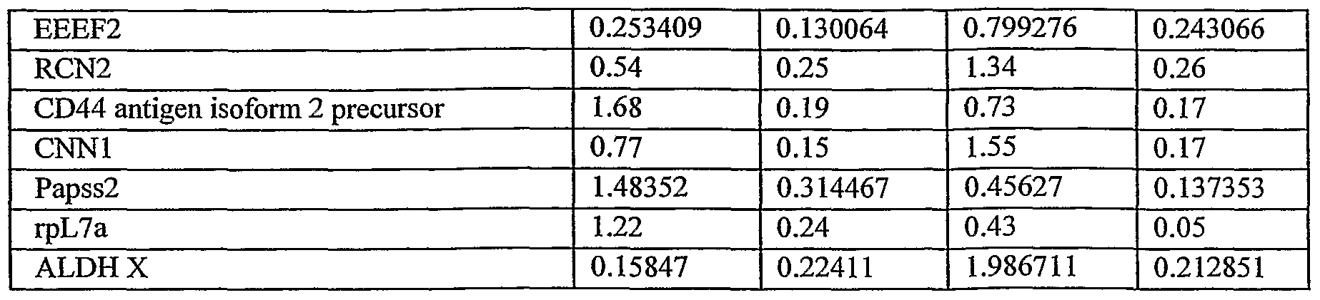
- Table 1 3D cultured cells show a much higher expression level of H2AF and ALDH X (more than 9 and 12 fold higher, respectively) and a higher level of the proteins EEEF2, RCN2 and CNNl (ca. 3, 2.5 and 2 fold, respectively).
- 3D cultured cells show ca. half the expression levels of the proteins Hnrphl and CD44 antigen isoform 2 precursor and ca. a third of the expression levels of Papss2 and rpL7a.
- 3D-ASCs have the capacity to differentiate into osteoblasts -
- cells were cultured in an osteoblast differentiating medium for a period of 3 weeks. Thereafter, calcium precipitation was effected. Differentiated cells were shown to produce calcium (depicted in red in Figures 5a-b) whereas control cells maintained a fibroblast like phenotype and demonstrated no mineralization ( Figures 5c-d). These results show that placenta derived 3D-ASC have the capacity to differentiate in vitro to osteoblasts cells.
- 3D-ASC support of HSC engraftment was evaluated by the level of human hematopoietic cells (hCD45+) detected in sub lethally irradiated or chemotherapy pretreated immune deficient NOD-SCID mice.
- hCD45+ human hematopoietic cells
- CD34+ Cells Umbilical cord blood samples were taken under sterile conditions during delivery (Bnei Zion Medical Center, Haifa, Israel) and mononuclear cells were fractionated using Lymphoprep (Axis-Shield PoC As, Oslo, Norway) density gradient centrifugation and were cryopreserved. Thawed mononuclear cells were washed and incubated with anti-CD34 antibodies and isolated using midi MACS (Miltenyl Biotech, Bergish Gladbach, Germany). Cells from more than one sample were pooled for achieving the desired amount (50,000-100,000 cells).
- NOD-SCID mice Seven week old male and female NOD-SCID mice (NOD-CB 17-Prkdcscid/J; Harlan/ Weizmann Inst., Rehovot Israel) were maintained in sterile open system cages, given sterile diets and autoclaved acidic water.
- mice were sub lethally irradiated (350 cGy), and thereafter (48 hr post irradiation) transplanted with 50,000-100,000 hCD34 + cells, with or without additional ASCs (0.5 X 10 6 - 1 X 10 6 ) derived from placenta or adipose tissue (3-7 mice in each group), by intravenous injection to a lateral tail vein.
- ASCs 0.5 X 10 6 - 1 X 10 6
- BM was collected by flushing both femurs and tibias with FACS buffer (50 ml PBS, 5 ml FBS, 0.5 ml sodium azid 5 %).
- mice BM Human cells in the mice BM were detected by flow cytometry, and the percentage of the human and murine CD45 hematopoietic cell marker expressing cells in the treated NOD-SCID mice was effected by incubating cells with anti-human CD45-FITC (IQ Products, Groningen, The Netherlands). The lowest threshold for unequivocal human engraftment was designated at 0.5 %. Detection of transplanted cells in mice treated with chemotherapy - 6.5 week old male NOD-SCID mice (NOD.CB17/JhkiHsd-scid; Harlan, Rehovot Israel), maintained as described hereinabove for irradiated mice, were injected intraperitoneally with Busulfan (25 mg/kg- for 2 consecutive days).
- Busulfan 25 mg/kg- for 2 consecutive days.
- mice Two days following the second Busulfan injection, mice were injected with CD34+ cells alone, or together with 0.5 X 10 6 ASCs, produced from the placenta. 3.5 weeks following transplantation, mice were sacrificed, and the presence of human hematopoietic cells was determined as described hereinabove for irradiated mice.
- RESULTS 3 D-AS C improved engraftment of HSC in irradiated mice - Human CD34+ hematopoietic cells and 3D-ASC derived from placenta or adipose were co- transplanted in irradiated NOD-SCID mice. Engraftment efficiency was evaluated 4 weeks following co-transplantation, and compared to mice transplanted with HSC alone.
- 3D- ⁇ SC improved engraftment of HSC in mice treated with chemotherapy Human CD34+ hematopoietic cells were co-transplanted with 500,000- 2D-ASC or 3D-ASC derived from placenta, into NOD-SCID mice pretreated with chemotherapy. Engraftment efficiency was evaluated 3.5 weeks following co-transplantation, and compared to mice transplanted with HSC alone. As is shown in Table 3, co- transplantation of ASC and UCB CD34+ cells resulted in higher engraftment levels in the BM of the recipient mice compared to UCB CD34+ cells alone.
- ASCs may serve as supportive cells to improve hematopoietic recovery following HSCs transplantation (autologous or allogenic).
- the ability of the 3D-ASCs to enhance hematopoietic stem and/or progenitor cell engraftment following HSCs transplantation may result from the 3D- ASC ability to secrete HSC supporting cytokines that may improve the homing, self- renewal and proliferation ability of the transplanted cells, or from the ability of those cells to rebuild the damaged hematopoietic microenvironment needed for the homing and proliferation of the transplantable HSCs
- EXAMPLE 3 The suppression of lymphocyte response by 2D and 3D cultured ASCs
- Adherent stromal cells, and particularly 3D-ASCs were. found to suppress the immune reaction of human cord blood mononuclear cells in an MLR assay
- MATERIALS AND EXPERIMENTAL PROCEDURES Mixed lymphocyte reaction (MLR) assay The immunosuppressive and immunoprivileged properties of 2D and 3D derived culturing procedures ASCs produced from the placenta, were effected by the MLR assay, which measures histocompatibility at the HLA locus, as effected by the proliferation rate of incompatible lymphocytes in mixed culturing of responsive (proliferating) and stimulating (unproliferative) cells.
- CB mononuclear cells (2 X 10 5 ) were used as responsive cells and were stimulated by being co-cultured with equal amounts (10 5 ) of irradiated (3000Rad) human peripheral blood derived Monocytes (PBMC), or with 2D or 3D cultured adherent cells, produced from the placenta, or a combination of adherent cells and PBMCs. Each assay was replicated three times. Cells were co-cultured for 4 days in RPMI 1640 medium (containing 20 % FBS under humidified 5 % CO 2 atmosphere at 37 0 C), in a 96-well plate. Plates were pulsed with l ⁇ C 3 H-thymidine during the last 18 hr of culturing. Cells were then harvested over fiberglass filter and thymidine uptake was quantified with a scintillation counter.
- RESULTS RESULTS
- Figure 8 shows the immune response of CB cells as represented by the elevated proliferation of these cells when stimulated with PBMCs, which, without being bound by theory, is probably associated with T cell proliferation in response to HLA incompatibility.
- PBMCs peripheral blood mononuclear cells
- FIG. 8 shows the immune response of CB cells as represented by the elevated proliferation of these cells when stimulated with PBMCs, which, without being bound by theory, is probably associated with T cell proliferation in response to HLA incompatibility.
- PBMCs adherent cells of the present invention.
- the CB immune response to PBMCs was substantially reduced when co-incubated with these adherent cells.
- ASCs were found to have the potential ability to reduce T cell proliferation of donor cells, typical of GvHD.
- both cultures, 2D and 3D reduced the immune response of the lymphocytes, and in line with the other advantages of 3D-ASCs described hereinabove, the 3D ASCs were more immunosuppressive.
Abstract
Description


Claims
Priority Applications (19)
| Application Number | Priority Date | Filing Date | Title |
|---|---|---|---|
| AU2007228341A AU2007228341B2 (en) | 2006-03-23 | 2007-03-22 | Methods for cell expansion and uses of cells and conditioned media produced thereby for therapy |
| CA2646384A CA2646384C (en) | 2006-03-23 | 2007-03-22 | Methods for cell expansion and uses of cells and conditioned media produced thereby for therapy |
| KR1020087025460A KR101490449B1 (en) | 2006-03-23 | 2007-03-22 | Methods For Cell Expansion And Uses Of Cells And Conditioned Media Produced Thereby For Therapy |
| US12/225,478 US20110129447A1 (en) | 2006-03-23 | 2007-03-22 | Methods for Cell Expansion and Uses of Cells and Conditioned Media Produced Thereby for Therapy |
| MX2008012085A MX2008012085A (en) | 2006-03-23 | 2007-03-22 | Methods for cell expansion and uses of cells and conditioned media produced thereby for therapy. |
| EP07713395A EP2010647A4 (en) | 2006-03-23 | 2007-03-22 | Methods for cell expansion and uses of cells and conditioned media produced thereby for therapy |
| BRPI0709349-7A BRPI0709349A2 (en) | 2006-03-23 | 2007-03-22 | methods for cell expansion and uses of cells and conditioned media produced through them for therapy |
| KR1020147029038A KR101557256B1 (en) | 2006-03-23 | 2007-03-22 | Methods For Cell Expansion And Uses Of Cells And Conditioned Media Produced Thereby For Therapy |
| EP16157193.0A EP3091071B1 (en) | 2006-03-23 | 2007-03-22 | Methods for cell expansion and uses of cells and conditioned media produced thereby for therapy |
| JP2009502327A JP5733894B2 (en) | 2006-03-23 | 2007-03-22 | Method for expanding cells and use of cells and conditioned medium produced by the method for the treatment |
| CN200780018851.7A CN101558151B (en) | 2006-03-23 | 2007-03-22 | The purposes that methods for cell expansion and the cell produced by this and conditioned medium are used for the treatment of |
| IL194232A IL194232A (en) | 2006-03-23 | 2008-09-21 | Methods for cell expansion, cells and conditioned media produced thereby and medicaments comprising same |
| HK10103472.9A HK1136846A1 (en) | 2006-03-23 | 2010-04-08 | Methods for cell expansion and uses of cells and conditioned media produced thereby for therapy |
| US13/069,130 US20110171182A1 (en) | 2006-03-23 | 2011-03-22 | Methods for cell expansion and uses of cells and conditioned media produced thereby for therapy |
| US13/161,334 US20110293583A1 (en) | 2006-03-23 | 2011-06-15 | Methods for cell expansion and uses of cells and conditioned media produced thereby for therapy |
| IL236213A IL236213A (en) | 2006-03-23 | 2014-12-11 | Pharmaceutical composition for facilitation of hematopoietic stem cell engraftment |
| US14/873,092 US20160022738A1 (en) | 2006-03-23 | 2015-10-01 | Methods for cell expansion and uses of cells and conditioned media produced thereby for therapy |
| US15/655,537 US20180008649A1 (en) | 2006-03-23 | 2017-07-20 | Adherent stromal cells derived from placentas of multiple donors and uses thereof |
| US17/119,511 US20210113627A1 (en) | 2006-03-23 | 2020-12-11 | Adherent stromal cells derived from placentas of multiple donors and uses thereof |
Applications Claiming Priority (4)
| Application Number | Priority Date | Filing Date | Title |
|---|---|---|---|
| US78476906P | 2006-03-23 | 2006-03-23 | |
| US60/784,769 | 2006-03-23 | ||
| US84708806P | 2006-09-26 | 2006-09-26 | |
| US60/847,088 | 2006-09-26 |
Related Child Applications (6)
| Application Number | Title | Priority Date | Filing Date |
|---|---|---|---|
| US12/225,478 A-371-Of-International US20110129447A1 (en) | 2006-03-23 | 2007-03-22 | Methods for Cell Expansion and Uses of Cells and Conditioned Media Produced Thereby for Therapy |
| US13/069,130 Continuation-In-Part US20110171182A1 (en) | 2006-03-23 | 2011-03-22 | Methods for cell expansion and uses of cells and conditioned media produced thereby for therapy |
| PCT/IB2011/001413 Continuation-In-Part WO2011132087A1 (en) | 2006-03-23 | 2011-04-21 | Adherent stromal cells derived from plancentas of multiple donors and uses thereof |
| US13/642,725 Continuation-In-Part US20130039892A1 (en) | 2010-04-23 | 2011-04-21 | Adherent stromal cells derived from plancentas of multiple donors and uses thereof |
| US14/873,092 Continuation US20160022738A1 (en) | 2006-03-23 | 2015-10-01 | Methods for cell expansion and uses of cells and conditioned media produced thereby for therapy |
| US15/655,537 Continuation-In-Part US20180008649A1 (en) | 2006-03-23 | 2017-07-20 | Adherent stromal cells derived from placentas of multiple donors and uses thereof |
Publications (2)
| Publication Number | Publication Date |
|---|---|
| WO2007108003A2 true WO2007108003A2 (en) | 2007-09-27 |
| WO2007108003A3 WO2007108003A3 (en) | 2009-02-12 |
Family
ID=38522837
Family Applications (1)
| Application Number | Title | Priority Date | Filing Date |
|---|---|---|---|
| PCT/IL2007/000380 WO2007108003A2 (en) | 2006-03-23 | 2007-03-22 | Methods for cell expansion and uses of cells and conditioned media produced thereby for therapy |
Country Status (21)
| Country | Link |
|---|---|
| US (2) | US20110129447A1 (en) |
| EP (5) | EP3091071B1 (en) |
| JP (3) | JP5733894B2 (en) |
| KR (2) | KR101557256B1 (en) |
| CN (1) | CN105296415A (en) |
| AU (1) | AU2007228341B2 (en) |
| BR (1) | BRPI0709349A2 (en) |
| CA (1) | CA2646384C (en) |
| DK (2) | DK2626417T3 (en) |
| ES (3) | ES2572214T3 (en) |
| HK (5) | HK1136846A1 (en) |
| HR (1) | HRP20160538T1 (en) |
| HU (1) | HUE028796T2 (en) |
| IL (1) | IL236213A (en) |
| MX (1) | MX2008012085A (en) |
| PL (2) | PL2366775T3 (en) |
| PT (3) | PT2548951E (en) |
| RU (1) | RU2433177C2 (en) |
| SG (1) | SG170761A1 (en) |
| SI (1) | SI2548951T1 (en) |
| WO (1) | WO2007108003A2 (en) |
Cited By (36)
| Publication number | Priority date | Publication date | Assignee | Title |
|---|---|---|---|---|
| WO2008060541A3 (en) * | 2006-11-13 | 2008-08-28 | Ethicon Inc | In vitro expansion of postpartum-derived cells using microcarriers |
| WO2009008547A1 (en) * | 2007-07-11 | 2009-01-15 | Nitto Denko Corporation | Cell culture substrate |
| WO2009144720A1 (en) * | 2008-05-27 | 2009-12-03 | Pluristem Ltd. | Methods of treating inflammatory colon diseases |
| WO2010026575A2 (en) * | 2008-09-02 | 2010-03-11 | Pluristem Ltd. | Adherent cells from placenta tissue and use thereof in therapy |
| WO2010026573A1 (en) * | 2008-09-02 | 2010-03-11 | Pluristem Ltd. | Methods of selection of cells for transplantation |
| WO2011064669A2 (en) | 2009-11-30 | 2011-06-03 | Pluristem Ltd. | Adherent cells from placenta and use of same in disease treatment |
| US20110256160A1 (en) * | 2008-09-02 | 2011-10-20 | Moran Meiron | Adherent cells from placenta tissue and use thereof in therapy |
| WO2012127320A1 (en) * | 2011-03-22 | 2012-09-27 | Pluristem Ltd. | Methods for treating radiation or chemical injury |
| WO2012140519A2 (en) | 2011-04-15 | 2012-10-18 | Pluristem Ltd. | Methods and systems for harvesting cells |
| US8529888B2 (en) | 2007-09-19 | 2013-09-10 | Pluristem Ltd. | Adherent cells from adipose or placenta tissues and use thereof in therapy |
| EP2687220A2 (en) * | 2007-02-12 | 2014-01-22 | Anthrogenesis Corporation | Treatment of inflammatory diseases using placental stem cells |
| WO2014037863A1 (en) | 2012-09-04 | 2014-03-13 | Pluristem Ltd. | Methods for prevention and treatment of preeclampsia |
| WO2014128634A1 (en) | 2013-02-20 | 2014-08-28 | Pluristem Ltd. | Gene and protein expression properties of adherent stromal cells cultured in 3d |
| WO2014141111A1 (en) | 2013-03-14 | 2014-09-18 | Pluristem Ltd. | Methods for prevention and treatment of graft-versus-host disease |
| WO2015004609A2 (en) | 2013-07-09 | 2015-01-15 | Pluristem Ltd. | Adherent cells from placenta and use thereof in treatment of injured tendons |
| WO2015025059A1 (en) * | 2013-08-23 | 2015-02-26 | Fundacion Pública Andaluza Progreso Y Salud | Cd105-negative multipotent mesenchymal stromal cells |
| US9125906B2 (en) | 2005-12-28 | 2015-09-08 | DePuy Synthes Products, Inc. | Treatment of peripheral vascular disease using umbilical cord tissue-derived cells |
| US9175261B2 (en) | 2005-12-16 | 2015-11-03 | DePuy Synthes Products, Inc. | Human umbilical cord tissue cells for inhibiting adverse immune response in histocompatibility-mismatched transplantation |
| AU2013267050B2 (en) * | 2007-09-19 | 2016-01-07 | Pluristem Ltd. | Adherent cells from adipose or placenta tissues and use thereof in therapy |
| US9234172B2 (en) | 2003-06-27 | 2016-01-12 | DePuy Synthes Products, Inc. | Repair and regeneration of ocular tissue using postpartum-derived cells |
| WO2016098061A1 (en) | 2014-12-18 | 2016-06-23 | Pluristem Ltd. | Methods and compositions for treating and preventing muscle wasting disorders |
| WO2016151476A1 (en) | 2015-03-23 | 2016-09-29 | Pluristem Ltd. | Use of adherent stromal cells for enhancing hematopoiesis in a subject in need thereof |
| US9498501B2 (en) | 2003-06-27 | 2016-11-22 | DePuy Synthes Products, Inc. | Postpartum cells derived from umbilical cord tissue, and methods of making and using the same |
| US9572840B2 (en) | 2003-06-27 | 2017-02-21 | DePuy Synthes Products, Inc. | Regeneration and repair of neural tissue using postpartum-derived cells |
| US9592258B2 (en) | 2003-06-27 | 2017-03-14 | DePuy Synthes Products, Inc. | Treatment of neurological injury by administration of human umbilical cord tissue-derived cells |
| WO2017141181A1 (en) | 2016-02-18 | 2017-08-24 | Pluristem Ltd. | Methods and compositions for treating cancers and neoplasms |
| US9943552B2 (en) | 2009-03-26 | 2018-04-17 | DePuy Synthes Products, Inc. | hUTC as therapy for Alzheimer's disease |
| WO2018150381A1 (en) * | 2017-02-20 | 2018-08-23 | Pluristem Ltd. | Methods and compositions for tumor assessment |
| US10104880B2 (en) | 2008-08-20 | 2018-10-23 | Celularity, Inc. | Cell composition and methods of making the same |
| WO2018198012A1 (en) | 2017-04-24 | 2018-11-01 | Pluristem Ltd. | Methods and compositions for treating neurological disorders |
| DE102018115360A1 (en) | 2017-07-03 | 2019-01-03 | Pluristem Ltd. | METHOD AND COMPOSITIONS FOR REMOVING ADHERENT CELLS |
| US10179900B2 (en) | 2008-12-19 | 2019-01-15 | DePuy Synthes Products, Inc. | Conditioned media and methods of making a conditioned media |
| US10557116B2 (en) | 2008-12-19 | 2020-02-11 | DePuy Synthes Products, Inc. | Treatment of lung and pulmonary diseases and disorders |
| US10851347B2 (en) | 2016-06-06 | 2020-12-01 | Pluristem Ltd. | Altered adherent stromal cells and methods of producing and using same |
| WO2022164788A3 (en) * | 2021-01-26 | 2022-09-22 | The Trustees Of Indiana University | Pharmaceutical compositions and their methods of use |
| AU2017218143B2 (en) * | 2016-02-12 | 2023-08-03 | Cell Care Therapeutics, Inc. | Adipose tissue derived mesenchymal stromal cell conditioned media and methods of making and using the same |
Families Citing this family (34)
| Publication number | Priority date | Publication date | Assignee | Title |
|---|---|---|---|---|
| ES2572214T3 (en) * | 2006-03-23 | 2016-05-30 | Pluristem Ltd | Methods for cell expansion and uses of cells and conditioned media produced in this way for therapy |
| US9352003B1 (en) | 2010-05-14 | 2016-05-31 | Musculoskeletal Transplant Foundation | Tissue-derived tissuegenic implants, and methods of fabricating and using same |
| US8883210B1 (en) | 2010-05-14 | 2014-11-11 | Musculoskeletal Transplant Foundation | Tissue-derived tissuegenic implants, and methods of fabricating and using same |
| US10130736B1 (en) | 2010-05-14 | 2018-11-20 | Musculoskeletal Transplant Foundation | Tissue-derived tissuegenic implants, and methods of fabricating and using same |
| US9757419B2 (en) | 2010-05-27 | 2017-09-12 | Pluristem Ltd. | Skeletal muscle regeneration using mesenchymal system cells |
| AU2011295954B2 (en) * | 2010-08-31 | 2015-08-13 | Gallant Pet, Inc. | Systemic, allogenic stem cell therapies for treatment of diseases in animals |
| WO2012048275A2 (en) | 2010-10-08 | 2012-04-12 | Caridianbct, Inc. | Configurable methods and systems of growing and harvesting cells in a hollow fiber bioreactor system |
| WO2012121695A1 (en) | 2011-03-04 | 2012-09-13 | Al-Qahtani Ahmed H | Skin cream |
| WO2013070734A1 (en) | 2011-11-07 | 2013-05-16 | Chaudhry Hina W | Methods of cardiac repair |
| ITTO20111183A1 (en) * | 2011-12-21 | 2013-06-22 | Univ Degli Studi Torino | CONDITIONAL MEANS OBTAINED FROM PLACENTARY STEM CELLS AND ITS USE IN THE THERAPEUTIC TREATMENT OF PREECLAMPSIA |
| MX357469B (en) | 2012-09-06 | 2018-07-11 | Pluristem Ltd | Devices and methods for culture of cells. |
| RU2525143C1 (en) * | 2013-03-22 | 2014-08-10 | Федеральное государственное бюджетное учреждение науки Государственный научный центр Российской Федерации-Институт медико-биологических проблем Российской академии наук | METHOD FOR CORD BLOOD MONONUCLEAR CELLS (cbMNC) EXPANSION ex vivo IN PRESENCE OF MULTIPOTENT MESENCHYMAL STROMAL CELLS (MMSCS) |
| RU2539750C2 (en) * | 2013-04-09 | 2015-01-27 | Федеральное государственное бюджетное образовательное учреждение высшего профессионального образования "Московский государственный университет имени М.В. Ломоносова" (МГУ) | Method of evaluating immunosuppressive properties of human mesenchymal stromal cells |
| CA2919374C (en) | 2013-07-30 | 2019-12-03 | Musculoskeletal Transplant Foundation | Acellular soft tissue-derived matrices and methods for preparing same |
| JP6633522B2 (en) | 2013-11-16 | 2020-01-22 | テルモ ビーシーティー、インコーポレーテッド | Cell growth in bioreactors |
| US10392593B2 (en) | 2014-03-04 | 2019-08-27 | Pluristem Ltd. | Systems and methods for growing and harvesting cells |
| WO2015148704A1 (en) | 2014-03-25 | 2015-10-01 | Terumo Bct, Inc. | Passive replacement of media |
| JP6830059B2 (en) | 2014-09-26 | 2021-02-17 | テルモ ビーシーティー、インコーポレーテッド | Scheduled cell feeding |
| RU2644650C2 (en) | 2014-12-01 | 2018-02-13 | Общество с ограниченной ответственностью "Т-Хелпер Клеточные Технологии" | Stem cell material and method for its reception |
| WO2016187413A1 (en) | 2015-05-21 | 2016-11-24 | Musculoskeletal Transplant Foundation | Modified demineralized cortical bone fibers |
| WO2017004592A1 (en) | 2015-07-02 | 2017-01-05 | Terumo Bct, Inc. | Cell growth with mechanical stimuli |
| US10912864B2 (en) | 2015-07-24 | 2021-02-09 | Musculoskeletal Transplant Foundation | Acellular soft tissue-derived matrices and methods for preparing same |
| US11052175B2 (en) | 2015-08-19 | 2021-07-06 | Musculoskeletal Transplant Foundation | Cartilage-derived implants and methods of making and using same |
| RU2628092C1 (en) * | 2016-02-10 | 2017-08-14 | Федеральное государственное бюджетное учреждение науки Государственный научный центр Российской Федерации - Институт медико-биологических проблем Российской академии наук (ГНЦ РФ - ИМБП РАН) | Method for obtaining of msc-associated non-differentiated hemopoietic precursor cells with cd34+/cd133+ phenotype |
| WO2017205667A1 (en) | 2016-05-25 | 2017-11-30 | Terumo Bct, Inc. | Cell expansion |
| RU2708329C2 (en) | 2016-05-31 | 2019-12-05 | Общество с ограниченной ответственностью "Т-Хелпер Клеточные Технологии" | Stem cell material, compositions and methods of use |
| US11104874B2 (en) | 2016-06-07 | 2021-08-31 | Terumo Bct, Inc. | Coating a bioreactor |
| US11685883B2 (en) | 2016-06-07 | 2023-06-27 | Terumo Bct, Inc. | Methods and systems for coating a cell growth surface |
| US11629332B2 (en) | 2017-03-31 | 2023-04-18 | Terumo Bct, Inc. | Cell expansion |
| CN110831694A (en) * | 2017-03-31 | 2020-02-21 | 细尔琳生物医学公司 | Biocompatible modified cell culture medium composition and uses thereof |
| US11624046B2 (en) | 2017-03-31 | 2023-04-11 | Terumo Bct, Inc. | Cell expansion |
| WO2018199194A1 (en) * | 2017-04-25 | 2018-11-01 | 北海道公立大学法人札幌医科大学 | Method for producing mesenchymal stem cells, therapeutic effect marker of mesenchymal stem cells, method for determining therapeutic effects of mesenchymal stem cells, and cellular preparation containing mesenchymal stem cells |
| WO2019175773A1 (en) | 2018-03-12 | 2019-09-19 | Universidade Do Porto | Compositions for use in the treatment of musculoskeletal conditions and methods for producing the same leveraging the synergistic activity of two different types of mesenchymal stromal/stem cells |
| CN112813002B (en) * | 2021-02-07 | 2022-11-08 | 天津科技大学 | Heat-resistant lactobacillus brevis and application of metazoan preparation thereof |
Citations (3)
| Publication number | Priority date | Publication date | Assignee | Title |
|---|---|---|---|---|
| US5168085A (en) | 1991-05-20 | 1992-12-01 | Corning Incorporated | Multi-stage twc system |
| US5266476A (en) | 1985-06-18 | 1993-11-30 | Yeda Research & Development Co., Ltd. | Fibrous matrix for in vitro cell cultivation |
| US6911201B1 (en) | 1999-02-04 | 2005-06-28 | Technion Research & Development Foundation Ltd. | Method of producing undifferentiated hemopoietic stem cells using a stationary phase plug-flow bioreactor |
Family Cites Families (61)
| Publication number | Priority date | Publication date | Assignee | Title |
|---|---|---|---|---|
| NL154600B (en) | 1971-02-10 | 1977-09-15 | Organon Nv | METHOD FOR THE DETERMINATION AND DETERMINATION OF SPECIFIC BINDING PROTEINS AND THEIR CORRESPONDING BINDABLE SUBSTANCES. |
| NL154598B (en) | 1970-11-10 | 1977-09-15 | Organon Nv | PROCEDURE FOR DETERMINING AND DETERMINING LOW MOLECULAR COMPOUNDS AND PROTEINS THAT CAN SPECIFICALLY BIND THESE COMPOUNDS AND TEST PACKAGING. |
| NL154599B (en) | 1970-12-28 | 1977-09-15 | Organon Nv | PROCEDURE FOR DETERMINING AND DETERMINING SPECIFIC BINDING PROTEINS AND THEIR CORRESPONDING BINDABLE SUBSTANCES, AND TEST PACKAGING. |
| US3901654A (en) | 1971-06-21 | 1975-08-26 | Biological Developments | Receptor assays of biologically active compounds employing biologically specific receptors |
| US3853987A (en) | 1971-09-01 | 1974-12-10 | W Dreyer | Immunological reagent and radioimmuno assay |
| US3867517A (en) | 1971-12-21 | 1975-02-18 | Abbott Lab | Direct radioimmunoassay for antigens and their antibodies |
| NL171930C (en) | 1972-05-11 | 1983-06-01 | Akzo Nv | METHOD FOR DETERMINING AND DETERMINING BITES AND TEST PACKAGING. |
| US3850578A (en) | 1973-03-12 | 1974-11-26 | H Mcconnell | Process for assaying for biologically active molecules |
| US3935074A (en) | 1973-12-17 | 1976-01-27 | Syva Company | Antibody steric hindrance immunoassay with two antibodies |
| US3996345A (en) | 1974-08-12 | 1976-12-07 | Syva Company | Fluorescence quenching with immunological pairs in immunoassays |
| US4034074A (en) | 1974-09-19 | 1977-07-05 | The Board Of Trustees Of Leland Stanford Junior University | Universal reagent 2-site immunoradiometric assay using labelled anti (IgG) |
| US3984533A (en) | 1975-11-13 | 1976-10-05 | General Electric Company | Electrophoretic method of detecting antigen-antibody reaction |
| US4098876A (en) | 1976-10-26 | 1978-07-04 | Corning Glass Works | Reverse sandwich immunoassay |
| US4879219A (en) | 1980-09-19 | 1989-11-07 | General Hospital Corporation | Immunoassay utilizing monoclonal high affinity IgM antibodies |
| US5011771A (en) | 1984-04-12 | 1991-04-30 | The General Hospital Corporation | Multiepitopic immunometric assay |
| US4666828A (en) | 1984-08-15 | 1987-05-19 | The General Hospital Corporation | Test for Huntington's disease |
| US4683202A (en) | 1985-03-28 | 1987-07-28 | Cetus Corporation | Process for amplifying nucleic acid sequences |
| US4801531A (en) | 1985-04-17 | 1989-01-31 | Biotechnology Research Partners, Ltd. | Apo AI/CIII genomic polymorphisms predictive of atherosclerosis |
| US5902741A (en) | 1986-04-18 | 1999-05-11 | Advanced Tissue Sciences, Inc. | Three-dimensional cartilage cultures |
| US5032508A (en) * | 1988-09-08 | 1991-07-16 | Marrow-Tech, Inc. | Three-dimensional cell and tissue culture system |
| US5863531A (en) * | 1986-04-18 | 1999-01-26 | Advanced Tissue Sciences, Inc. | In vitro preparation of tubular tissue structures by stromal cell culture on a three-dimensional framework |
| US5272057A (en) | 1988-10-14 | 1993-12-21 | Georgetown University | Method of detecting a predisposition to cancer by the use of restriction fragment length polymorphism of the gene for human poly (ADP-ribose) polymerase |
| US5192659A (en) | 1989-08-25 | 1993-03-09 | Genetype Ag | Intron sequence analysis method for detection of adjacent and remote locus alleles as haplotypes |
| US6326198B1 (en) | 1990-06-14 | 2001-12-04 | Regents Of The University Of Michigan | Methods and compositions for the ex vivo replication of stem cells, for the optimization of hematopoietic progenitor cell cultures, and for increasing the metabolism, GM-CSF secretion and/or IL-6 secretion of human stromal cells |
| US6010696A (en) | 1990-11-16 | 2000-01-04 | Osiris Therapeutics, Inc. | Enhancing hematopoietic progenitor cell engraftment using mesenchymal stem cells |
| US5486359A (en) | 1990-11-16 | 1996-01-23 | Osiris Therapeutics, Inc. | Human mesenchymal stem cells |
| US5281521A (en) | 1992-07-20 | 1994-01-25 | The Trustees Of The University Of Pennsylvania | Modified avidin-biotin technique |
| US5629186A (en) | 1994-04-28 | 1997-05-13 | Lockheed Martin Corporation | Porous matrix and method of its production |
| US5736396A (en) | 1995-01-24 | 1998-04-07 | Case Western Reserve University | Lineage-directed induction of human mesenchymal stem cell differentiation |
| US6132463A (en) | 1995-05-19 | 2000-10-17 | Etex Corporation | Cell seeding of ceramic compositions |
| US5925567A (en) | 1995-05-19 | 1999-07-20 | T. Breeders, Inc. | Selective expansion of target cell populations |
| EP0868505B1 (en) | 1995-11-16 | 2005-02-02 | Case Western Reserve University | In vitro chondrogenic induction of human mesenchymal stem cells |
| US6087129A (en) * | 1996-01-19 | 2000-07-11 | Betagene, Inc. | Recombinant expression of proteins from secretory cell lines |
| US6335195B1 (en) | 1997-01-28 | 2002-01-01 | Maret Corporation | Method for promoting hematopoietic and mesenchymal cell proliferation and differentiation |
| US6291240B1 (en) * | 1998-01-29 | 2001-09-18 | Advanced Tissue Sciences, Inc. | Cells or tissues with increased protein factors and methods of making and using same |
| EP1064353B1 (en) | 1998-03-18 | 2002-11-06 | Massachusetts Institute Of Technology | Vascularized perfused microtissue/micro-organ arrays |
| EP1108011A2 (en) | 1998-06-08 | 2001-06-20 | Osiris Therapeutics, Inc. | In vitro maintenance of hematopoietic stem cells |
| US6153432A (en) | 1999-01-29 | 2000-11-28 | Zen-Bio, Inc | Methods for the differentiation of human preadipocytes into adipocytes |
| KR100968164B1 (en) * | 1999-03-10 | 2010-07-06 | 더 리전츠 오브 더 유니버시티 오브 캘리포니아 | Adipose-derived stem cells and lattices |
| AU4860900A (en) | 1999-06-02 | 2000-12-18 | Lifebank Services, L.L.C. | Methods of isolation, cryopreservation, and therapeutic use of human amniotic epithelial cells |
| US6555374B1 (en) | 1999-08-19 | 2003-04-29 | Artecel Sciences, Inc. | Multiple mesodermal lineage differentiation potentials for adipose tissue-derived stromal cells and uses thereof |
| JP2003523767A (en) | 2000-02-26 | 2003-08-12 | アーテイセル・サイエンスイーズ・インコーポレイテツド | Pluripotent stem cells generated from adipose tissue-derived stromal cells and uses thereof |
| ATE360063T1 (en) * | 2000-10-12 | 2007-05-15 | Agency Science Tech & Res | NON-INTERRUPTIONAL, THREE-DIMENSIONAL SYSTEM FOR CULTIVATION AND HARVESTING ANCHORAGE-DEPENDENT CELLS |
| US7311905B2 (en) * | 2002-02-13 | 2007-12-25 | Anthrogenesis Corporation | Embryonic-like stem cells derived from post-partum mammalian placenta, and uses and methods of treatment using said cells |
| EP1349918B1 (en) | 2000-12-06 | 2014-08-06 | Anthrogenesis Corporation | Method of collecting placental stem cells |
| CA2856986C (en) * | 2001-02-14 | 2019-08-13 | Anthrogenesis Corporation | Post-partum mammalian placental stem cells for use in the treatment of neurological or renal diseases and disorders |
| ES2522526T3 (en) * | 2001-02-14 | 2014-11-14 | Anthrogenesis Corporation | Post-partum placenta of mammals, their use and placental stem cells thereof |
| WO2003040346A2 (en) * | 2001-11-09 | 2003-05-15 | Artecel Sciences, Inc. | Methods and compositions for the use of stromal cells to support embryonic and adult stem cells |
| JP2003180334A (en) * | 2001-12-17 | 2003-07-02 | Japan Science & Technology Corp | Three-dimensional and high-density cell culture module |
| US7504099B2 (en) | 2001-12-27 | 2009-03-17 | Yissum Research Development Company Of The Hebrew University Of Jerusalem | Methods of inducing or enhancing connective tissue repair |
| US7498171B2 (en) * | 2002-04-12 | 2009-03-03 | Anthrogenesis Corporation | Modulation of stem and progenitor cell differentiation, assays, and uses thereof |
| CA2483010A1 (en) * | 2002-04-19 | 2003-10-30 | University Of Pittsburgh Of The Commonwealth System Of Higher Education | Placental derived stem cells and uses thereof |
| WO2004076642A2 (en) | 2003-02-27 | 2004-09-10 | The Rockefeller University | Method for modulating epithelial stem cell lineage |
| WO2005001079A2 (en) * | 2003-06-27 | 2005-01-06 | Ethicon, Incorporated | Soft tissue repair and regeneration using postpartum-derived cells |
| US6924458B2 (en) | 2003-07-17 | 2005-08-02 | Great Computer Corp. | Auxiliary focusing tool for laser marker |
| US20050265980A1 (en) * | 2004-05-14 | 2005-12-01 | Becton, Dickinson And Company | Cell culture environments for the serum-free expansion of mesenchymal stem cells |
| DE102004043256B4 (en) * | 2004-09-07 | 2013-09-19 | Rheinische Friedrich-Wilhelms-Universität Bonn | Scalable process for culturing undifferentiated stem cells in suspension |
| CN103356704B (en) | 2005-03-31 | 2015-09-30 | 斯丹姆涅恩有限公司 | Prepare the method for the medicine in order to treat the ulcer caused by diabetic neuropathy |
| US20100233130A1 (en) * | 2006-02-06 | 2010-09-16 | Pluristem Ltd. | Method and Apparatus for Maintenance and Expansion of Hematopoietic Stem Cells From Mononuclear Cells |
| ES2572214T3 (en) * | 2006-03-23 | 2016-05-30 | Pluristem Ltd | Methods for cell expansion and uses of cells and conditioned media produced in this way for therapy |
| JP6030114B2 (en) * | 2011-03-22 | 2016-11-24 | プルリステム リミテッド | Methods for treating radiation or chemical injury |
-
2007
- 2007-03-22 ES ES12189046T patent/ES2572214T3/en active Active
- 2007-03-22 PL PL11170055T patent/PL2366775T3/en unknown
- 2007-03-22 JP JP2009502327A patent/JP5733894B2/en active Active
- 2007-03-22 EP EP16157193.0A patent/EP3091071B1/en active Active
- 2007-03-22 RU RU2008141894/10A patent/RU2433177C2/en active
- 2007-03-22 EP EP07713395A patent/EP2010647A4/en not_active Withdrawn
- 2007-03-22 MX MX2008012085A patent/MX2008012085A/en active IP Right Grant
- 2007-03-22 WO PCT/IL2007/000380 patent/WO2007108003A2/en active Application Filing
- 2007-03-22 DK DK13164303.3T patent/DK2626417T3/en active
- 2007-03-22 SI SI200731782A patent/SI2548951T1/en unknown
- 2007-03-22 PT PT121890461T patent/PT2548951E/en unknown
- 2007-03-22 SG SG201102089-8A patent/SG170761A1/en unknown
- 2007-03-22 CN CN201510738761.1A patent/CN105296415A/en active Pending
- 2007-03-22 HU HUE12189046A patent/HUE028796T2/en unknown
- 2007-03-22 ES ES13164303.3T patent/ES2549528T3/en active Active
- 2007-03-22 AU AU2007228341A patent/AU2007228341B2/en active Active
- 2007-03-22 DK DK11170055.5T patent/DK2366775T3/en active
- 2007-03-22 ES ES11170055.5T patent/ES2537641T3/en active Active
- 2007-03-22 PT PT111700555T patent/PT2366775E/en unknown
- 2007-03-22 BR BRPI0709349-7A patent/BRPI0709349A2/en not_active Application Discontinuation
- 2007-03-22 KR KR1020147029038A patent/KR101557256B1/en active IP Right Grant
- 2007-03-22 CA CA2646384A patent/CA2646384C/en active Active
- 2007-03-22 PL PL12189046T patent/PL2548951T3/en unknown
- 2007-03-22 KR KR1020087025460A patent/KR101490449B1/en active IP Right Grant
- 2007-03-22 PT PT131643033T patent/PT2626417E/en unknown
- 2007-03-22 US US12/225,478 patent/US20110129447A1/en not_active Abandoned
- 2007-03-22 EP EP12189046.1A patent/EP2548951B1/en active Active
- 2007-03-22 EP EP13164303.3A patent/EP2626417B1/en active Active
- 2007-03-22 EP EP20110170055 patent/EP2366775B1/en active Active
-
2010
- 2010-04-08 HK HK10103472.9A patent/HK1136846A1/en not_active IP Right Cessation
-
2011
- 2011-06-16 JP JP2011133774A patent/JP5766041B2/en active Active
-
2012
- 2012-01-17 HK HK12100543.8A patent/HK1160174A1/en not_active IP Right Cessation
- 2012-01-17 HK HK13104666.0A patent/HK1177759A1/en unknown
- 2012-01-17 HK HK14100827.3A patent/HK1187950A1/en unknown
-
2013
- 2013-12-20 JP JP2013263148A patent/JP5941454B2/en active Active
-
2014
- 2014-12-11 IL IL236213A patent/IL236213A/en active IP Right Grant
-
2015
- 2015-10-01 US US14/873,092 patent/US20160022738A1/en not_active Abandoned
-
2016
- 2016-05-12 HK HK16105449.8A patent/HK1217513A1/en unknown
- 2016-05-19 HR HRP20160538TT patent/HRP20160538T1/en unknown
Patent Citations (3)
| Publication number | Priority date | Publication date | Assignee | Title |
|---|---|---|---|---|
| US5266476A (en) | 1985-06-18 | 1993-11-30 | Yeda Research & Development Co., Ltd. | Fibrous matrix for in vitro cell cultivation |
| US5168085A (en) | 1991-05-20 | 1992-12-01 | Corning Incorporated | Multi-stage twc system |
| US6911201B1 (en) | 1999-02-04 | 2005-06-28 | Technion Research & Development Foundation Ltd. | Method of producing undifferentiated hemopoietic stem cells using a stationary phase plug-flow bioreactor |
Non-Patent Citations (2)
| Title |
|---|
| See also references of EP2010647A4 |
| WENDT, D. ET AL., BIOTECHNOL BIOENG, vol. 84, 2003, pages 205 - 214 |
Cited By (100)
| Publication number | Priority date | Publication date | Assignee | Title |
|---|---|---|---|---|
| US10039793B2 (en) | 2003-06-27 | 2018-08-07 | DePuy Synthes Products, Inc. | Soft tissue repair and regeneration using postpartum-derived cells and cell products |
| US10195233B2 (en) | 2003-06-27 | 2019-02-05 | DePuy Synthes Products, Inc. | Postpartum cells derived from placental tissue, and methods of making and using the same |
| US10500234B2 (en) | 2003-06-27 | 2019-12-10 | DePuy Synthes Products, Inc. | Postpartum cells derived from umbilical cord tissue, and methods of making and using the same |
| US10383898B2 (en) | 2003-06-27 | 2019-08-20 | DePuy Synthes Products, Inc. | Postpartum cells derived from placental tissue, and methods of making and using the same |
| US10744164B2 (en) | 2003-06-27 | 2020-08-18 | DePuy Synthes Products, Inc. | Repair and regeneration of ocular tissue using postpartum-derived cells |
| US9498501B2 (en) | 2003-06-27 | 2016-11-22 | DePuy Synthes Products, Inc. | Postpartum cells derived from umbilical cord tissue, and methods of making and using the same |
| US9592258B2 (en) | 2003-06-27 | 2017-03-14 | DePuy Synthes Products, Inc. | Treatment of neurological injury by administration of human umbilical cord tissue-derived cells |
| US10758576B2 (en) | 2003-06-27 | 2020-09-01 | DePuy Synthes Products, Inc. | Soft tissue repair and regeneration using postpartum-derived cells and cell products |
| US9717763B2 (en) | 2003-06-27 | 2017-08-01 | DePuy Synthes Products, Inc. | Postpartum cells derived from umbilical cord tissue, and methods of making and using the same |
| US11191789B2 (en) | 2003-06-27 | 2021-12-07 | DePuy Synthes Products, Inc. | Cartilage and bone repair and regeneration using postpartum-derived cells |
| US9234172B2 (en) | 2003-06-27 | 2016-01-12 | DePuy Synthes Products, Inc. | Repair and regeneration of ocular tissue using postpartum-derived cells |
| US11179422B2 (en) | 2003-06-27 | 2021-11-23 | DePuy Synthes Products, Inc. | Method of differentiating umbilical cord tissue into a chondrogenic phenotype |
| US9572840B2 (en) | 2003-06-27 | 2017-02-21 | DePuy Synthes Products, Inc. | Regeneration and repair of neural tissue using postpartum-derived cells |
| US9504719B2 (en) | 2003-06-27 | 2016-11-29 | DePuy Synthes Products, Inc. | Soft tissue repair and regeneration using postpartum-derived cells and cell products |
| US9175261B2 (en) | 2005-12-16 | 2015-11-03 | DePuy Synthes Products, Inc. | Human umbilical cord tissue cells for inhibiting adverse immune response in histocompatibility-mismatched transplantation |
| US9585918B2 (en) | 2005-12-28 | 2017-03-07 | DePuy Synthes Products, Inc. | Treatment of peripheral vascular disease using umbilical cord tissue-derived cells |
| US9125906B2 (en) | 2005-12-28 | 2015-09-08 | DePuy Synthes Products, Inc. | Treatment of peripheral vascular disease using umbilical cord tissue-derived cells |
| US9102915B2 (en) | 2006-11-13 | 2015-08-11 | DePuy Synthes Products, Inc. | In vitro expansion of postpartum-derived cells using microcarriers |
| WO2008060541A3 (en) * | 2006-11-13 | 2008-08-28 | Ethicon Inc | In vitro expansion of postpartum-derived cells using microcarriers |
| EP3763376A1 (en) * | 2007-02-12 | 2021-01-13 | Celularity, Inc. | Treatment of inflammatory diseases using placental stem cells |
| EP2915537A3 (en) * | 2007-02-12 | 2015-10-28 | Anthrogenesis Corporation | Treatment of inflammatory diseases using placental stem cells |
| EP3103462A1 (en) * | 2007-02-12 | 2016-12-14 | Anthrogenesis Corporation | Treatment of inflammatory diseases using placental stem cells |
| EP2687220A2 (en) * | 2007-02-12 | 2014-01-22 | Anthrogenesis Corporation | Treatment of inflammatory diseases using placental stem cells |
| EP2687220A3 (en) * | 2007-02-12 | 2014-05-14 | Anthrogenesis Corporation | Treatment of inflammatory diseases using placental stem cells |
| WO2009008547A1 (en) * | 2007-07-11 | 2009-01-15 | Nitto Denko Corporation | Cell culture substrate |
| CN107050057A (en) * | 2007-09-19 | 2017-08-18 | 普拉里斯坦有限公司 | Adherent cell from fat or placenta tissue and its purposes in the treatment |
| US8529888B2 (en) | 2007-09-19 | 2013-09-10 | Pluristem Ltd. | Adherent cells from adipose or placenta tissues and use thereof in therapy |
| EP3103463A1 (en) * | 2007-09-19 | 2016-12-14 | Pluristem Ltd. | Adherent cells from adipose or placenta tissues and use thereof in therapy |
| EP2591789A3 (en) * | 2007-09-19 | 2014-01-01 | Pluristem Ltd. | Adherent cells from adipose or placenta tissues and use thereof in therapy |
| US9517248B2 (en) | 2007-09-19 | 2016-12-13 | Pluristem Ltd. | Adherent cells from adipose or placenta tissues and use thereof in therapy |
| EP3103463B1 (en) | 2007-09-19 | 2020-01-01 | Pluristem Ltd. | Adherent cells from adipose or placenta tissues and use thereof in therapy |
| EP2591789B1 (en) | 2007-09-19 | 2016-06-22 | Pluristem Ltd. | Adherent cells from adipose or placenta tissues and use thereof in therapy |
| CN107028981A (en) * | 2007-09-19 | 2017-08-11 | 普拉里斯坦有限公司 | Adherent cell from fat or placenta tissue and its purposes in the treatment |
| AU2013267050B2 (en) * | 2007-09-19 | 2016-01-07 | Pluristem Ltd. | Adherent cells from adipose or placenta tissues and use thereof in therapy |
| EP2200622B2 (en) † | 2007-09-19 | 2015-07-29 | Pluristem Ltd. | Adherent cells from adipose or placenta tissues and use thereof in therapy |
| CN107028981B (en) * | 2007-09-19 | 2021-04-20 | 普拉里斯坦有限公司 | Adherent cells from adipose or placental tissue and their use in therapy |
| CN107050057B (en) * | 2007-09-19 | 2021-04-20 | 普拉里斯坦有限公司 | Adherent cells from adipose or placental tissue and their use in therapy |
| US9393273B2 (en) * | 2008-05-27 | 2016-07-19 | Pluristem Ltd. | Methods of treating inflammatory colon diseases |
| WO2009144720A1 (en) * | 2008-05-27 | 2009-12-03 | Pluristem Ltd. | Methods of treating inflammatory colon diseases |
| EP2641606A1 (en) | 2008-05-27 | 2013-09-25 | Pluristem Ltd. | Methods of treating inflammatory colon diseases |
| US20110129486A1 (en) * | 2008-05-27 | 2011-06-02 | Moran Meiron | Methods of treating inflammatory colon diseases |
| US20160271184A1 (en) * | 2008-05-27 | 2016-09-22 | Pluristem Ltd. | Methods of treating inflammatory colon diseases |
| US10104880B2 (en) | 2008-08-20 | 2018-10-23 | Celularity, Inc. | Cell composition and methods of making the same |
| US20110256160A1 (en) * | 2008-09-02 | 2011-10-20 | Moran Meiron | Adherent cells from placenta tissue and use thereof in therapy |
| WO2010026575A2 (en) * | 2008-09-02 | 2010-03-11 | Pluristem Ltd. | Adherent cells from placenta tissue and use thereof in therapy |
| US20110256159A1 (en) * | 2008-09-02 | 2011-10-20 | Moran Meiron | Adherent cells from placenta tissue and use thereof in therapy |
| US9096827B2 (en) * | 2008-09-02 | 2015-08-04 | Pluristem Ltd. | Adherent cells from placenta tissue and use thereof in therapy |
| AU2009288781B2 (en) * | 2008-09-02 | 2015-01-29 | Pluri Biotech Ltd | Adherent cells from placenta tissue and use thereof in therapy |
| US8524496B2 (en) * | 2008-09-02 | 2013-09-03 | Pluristem Ltd. | Adherent cells from placenta tissue and use thereof in therapy |
| CN105708862A (en) * | 2008-09-02 | 2016-06-29 | 普拉里斯坦有限公司 | Adherent cells from placenta tissue and use thereof in therapy |
| RU2539786C2 (en) * | 2008-09-02 | 2015-01-27 | Плуристем Лтд. | Placental tissue cells grown in substrate-attached culture and using them in treating |
| US9758762B2 (en) | 2008-09-02 | 2017-09-12 | Pluristem Ltd. | Perfusion bioreactor for culturing CD200—placenta adherent cells |
| EP3115451A1 (en) | 2008-09-02 | 2017-01-11 | Pluristem Ltd. | Adherent cells from placenta tissue and use thereof in therapy |
| WO2010026575A3 (en) * | 2008-09-02 | 2010-09-02 | Pluristem Ltd. | Adherent cells from placenta tissue and use thereof in therapy |
| WO2010026573A1 (en) * | 2008-09-02 | 2010-03-11 | Pluristem Ltd. | Methods of selection of cells for transplantation |
| US20130337558A1 (en) * | 2008-09-02 | 2013-12-19 | Pluristem Ltd. | Adherent cells from placenta tissue and use thereof in therapy |
| CN102282249A (en) * | 2008-09-02 | 2011-12-14 | 普拉里斯坦有限公司 | Adherent cells from placenta tissue and use thereof in therapy |
| US10557116B2 (en) | 2008-12-19 | 2020-02-11 | DePuy Synthes Products, Inc. | Treatment of lung and pulmonary diseases and disorders |
| US10179900B2 (en) | 2008-12-19 | 2019-01-15 | DePuy Synthes Products, Inc. | Conditioned media and methods of making a conditioned media |
| US9943552B2 (en) | 2009-03-26 | 2018-04-17 | DePuy Synthes Products, Inc. | hUTC as therapy for Alzheimer's disease |
| AU2010322808B2 (en) * | 2009-11-30 | 2014-07-17 | Pluri Biotech Ltd | Adherent cells from placenta and use of same in disease treatment |
| EP2806023A3 (en) * | 2009-11-30 | 2015-03-11 | Pluristem Ltd. | Adherent cells from placenta and use of same in disease treatment |
| EP2806023A2 (en) | 2009-11-30 | 2014-11-26 | Pluristem Ltd. | Adherent cells from placenta and use of same in disease treatment |
| WO2011064669A2 (en) | 2009-11-30 | 2011-06-03 | Pluristem Ltd. | Adherent cells from placenta and use of same in disease treatment |
| EP2977445A3 (en) * | 2009-11-30 | 2016-03-16 | Pluristem Ltd. | Adherent cells from placenta and use of same in disease treatment |
| EP2977445A2 (en) | 2009-11-30 | 2016-01-27 | Pluristem Ltd. | Adherent cells from placenta and use of same in disease treatment |
| WO2011064669A3 (en) * | 2009-11-30 | 2011-12-15 | Pluristem Ltd. | Adherent cells from placenta and use of same in disease treatment |
| CN105112357A (en) * | 2009-11-30 | 2015-12-02 | 普拉里斯坦有限公司 | Adherent cells from placenta and use of same in disease treatment |
| CN105112357B (en) * | 2009-11-30 | 2020-10-23 | 普拉里斯坦有限公司 | Adherent cells from placenta and their use in the treatment of disease |
| US9175262B2 (en) | 2009-11-30 | 2015-11-03 | Pluristerm Ltd. | Adherent cells from placenta and use of same in disease treatment |
| CN102639692A (en) * | 2009-11-30 | 2012-08-15 | 普拉里斯坦有限公司 | Adherent cells from placenta and use of same in disease treatment |
| US10610545B2 (en) | 2009-11-30 | 2020-04-07 | Pluristem Ltd. | Adherent cells from placenta and use of same in disease treatment |
| WO2012127320A1 (en) * | 2011-03-22 | 2012-09-27 | Pluristem Ltd. | Methods for treating radiation or chemical injury |
| KR101903339B1 (en) | 2011-03-22 | 2018-10-01 | 플루리스템 리미티드 | Methods for treating radiation or chemical injury |
| EP3260533A1 (en) * | 2011-03-22 | 2017-12-27 | Pluristem Ltd. | Methods for treating radiation or chemical injury |
| US10722541B2 (en) | 2011-03-22 | 2020-07-28 | Pluristem Ltd. | Methods for treating radiation or chemical injury |
| WO2012140519A2 (en) | 2011-04-15 | 2012-10-18 | Pluristem Ltd. | Methods and systems for harvesting cells |
| KR101910685B1 (en) * | 2011-04-15 | 2018-10-22 | 플루리스템 리미티드 | Methods and systems for harvesting cells |
| CN108342350A (en) * | 2011-04-15 | 2018-07-31 | 普拉里斯坦有限公司 | Harvest the method and system of cell |
| US10851341B2 (en) | 2011-04-15 | 2020-12-01 | Pluristem Ltd. | Methods and systems for harvesting cells |
| WO2012140519A3 (en) * | 2011-04-15 | 2013-03-07 | Pluristem Ltd. | Methods and systems for harvesting cells |
| CN103476924A (en) * | 2011-04-15 | 2013-12-25 | 普拉里斯坦有限公司 | Methods and systems for harvesting cells |
| US9657266B2 (en) | 2011-04-15 | 2017-05-23 | Pluristem Ltd. | Methods and systems for harvesting cells |
| US9950014B2 (en) | 2012-09-04 | 2018-04-24 | Pluristem Ltd. | Methods for prevention and treatment of preeclampsia |
| WO2014037863A1 (en) | 2012-09-04 | 2014-03-13 | Pluristem Ltd. | Methods for prevention and treatment of preeclampsia |
| US10351910B2 (en) | 2013-02-20 | 2019-07-16 | Pluristem Ltd | Gene and protein expression properties of adherent stromal cells cultured in 3D |
| WO2014128634A1 (en) | 2013-02-20 | 2014-08-28 | Pluristem Ltd. | Gene and protein expression properties of adherent stromal cells cultured in 3d |
| WO2014141111A1 (en) | 2013-03-14 | 2014-09-18 | Pluristem Ltd. | Methods for prevention and treatment of graft-versus-host disease |
| WO2015004609A3 (en) * | 2013-07-09 | 2015-07-30 | Pluristem Ltd. | Adherent cells from placenta and use thereof in treatment of injured tendons |
| WO2015004609A2 (en) | 2013-07-09 | 2015-01-15 | Pluristem Ltd. | Adherent cells from placenta and use thereof in treatment of injured tendons |
| WO2015025059A1 (en) * | 2013-08-23 | 2015-02-26 | Fundacion Pública Andaluza Progreso Y Salud | Cd105-negative multipotent mesenchymal stromal cells |
| WO2016098061A1 (en) | 2014-12-18 | 2016-06-23 | Pluristem Ltd. | Methods and compositions for treating and preventing muscle wasting disorders |
| WO2016151476A1 (en) | 2015-03-23 | 2016-09-29 | Pluristem Ltd. | Use of adherent stromal cells for enhancing hematopoiesis in a subject in need thereof |
| AU2017218143B2 (en) * | 2016-02-12 | 2023-08-03 | Cell Care Therapeutics, Inc. | Adipose tissue derived mesenchymal stromal cell conditioned media and methods of making and using the same |
| WO2017141181A1 (en) | 2016-02-18 | 2017-08-24 | Pluristem Ltd. | Methods and compositions for treating cancers and neoplasms |
| US10851347B2 (en) | 2016-06-06 | 2020-12-01 | Pluristem Ltd. | Altered adherent stromal cells and methods of producing and using same |
| WO2018150381A1 (en) * | 2017-02-20 | 2018-08-23 | Pluristem Ltd. | Methods and compositions for tumor assessment |
| WO2018198012A1 (en) | 2017-04-24 | 2018-11-01 | Pluristem Ltd. | Methods and compositions for treating neurological disorders |
| DE102018115360A1 (en) | 2017-07-03 | 2019-01-03 | Pluristem Ltd. | METHOD AND COMPOSITIONS FOR REMOVING ADHERENT CELLS |
| WO2022164788A3 (en) * | 2021-01-26 | 2022-09-22 | The Trustees Of Indiana University | Pharmaceutical compositions and their methods of use |
Also Published As
Similar Documents
| Publication | Publication Date | Title |
|---|---|---|
| AU2007228341B2 (en) | Methods for cell expansion and uses of cells and conditioned media produced thereby for therapy | |
| US20200306319A1 (en) | Methods for treating radiation or chemical injury | |
| US20110171182A1 (en) | Methods for cell expansion and uses of cells and conditioned media produced thereby for therapy | |
| US20110293583A1 (en) | Methods for cell expansion and uses of cells and conditioned media produced thereby for therapy | |
| US20140017209A1 (en) | Methods for treating radiation or chemical injury |
Legal Events
| Date | Code | Title | Description |
|---|---|---|---|
| WWE | Wipo information: entry into national phase |
Ref document number: 200780018851.7 Country of ref document: CN |
|
| 121 | Ep: the epo has been informed by wipo that ep was designated in this application |
Ref document number: 07713395 Country of ref document: EP Kind code of ref document: A2 |
|
| WWE | Wipo information: entry into national phase |
Ref document number: 2646384 Country of ref document: CA |
|
| WWE | Wipo information: entry into national phase |
Ref document number: 194232 Country of ref document: IL |
|
| WWE | Wipo information: entry into national phase |
Ref document number: MX/A/2008/012085 Country of ref document: MX |
|
| NENP | Non-entry into the national phase |
Ref country code: DE |
|
| WWE | Wipo information: entry into national phase |
Ref document number: 2009502327 Country of ref document: JP |
|
| WWE | Wipo information: entry into national phase |
Ref document number: 1020087025460 Country of ref document: KR |
|
| WWE | Wipo information: entry into national phase |
Ref document number: 2007228341 Country of ref document: AU |
|
| WWE | Wipo information: entry into national phase |
Ref document number: 2256/MUMNP/2008 Country of ref document: IN |
|
| ENP | Entry into the national phase |
Ref document number: 2008141894 Country of ref document: RU Kind code of ref document: A |
|
| WWE | Wipo information: entry into national phase |
Ref document number: 2007713395 Country of ref document: EP |
|
| ENP | Entry into the national phase |
Ref document number: 2007228341 Country of ref document: AU Date of ref document: 20070322 Kind code of ref document: A |
|
| WWE | Wipo information: entry into national phase |
Ref document number: 12225478 Country of ref document: US |
|
| ENP | Entry into the national phase |
Ref document number: PI0709349 Country of ref document: BR Kind code of ref document: A2 Effective date: 20080923 |
|
| WWE | Wipo information: entry into national phase |
Ref document number: 1020147029038 Country of ref document: KR |