WO2007016548A2 - Micro-rna-based methods and compositions for the diagnosis, prognosis and treatment of breast cancer - Google Patents
Micro-rna-based methods and compositions for the diagnosis, prognosis and treatment of breast cancer Download PDFInfo
- Publication number
- WO2007016548A2 WO2007016548A2 PCT/US2006/029889 US2006029889W WO2007016548A2 WO 2007016548 A2 WO2007016548 A2 WO 2007016548A2 US 2006029889 W US2006029889 W US 2006029889W WO 2007016548 A2 WO2007016548 A2 WO 2007016548A2
- Authority
- WO
- WIPO (PCT)
- Prior art keywords
- mir
- gene product
- breast cancer
- subject
- expression
- Prior art date
Links
Classifications
-
- A—HUMAN NECESSITIES
- A61—MEDICAL OR VETERINARY SCIENCE; HYGIENE
- A61K—PREPARATIONS FOR MEDICAL, DENTAL OR TOILETRY PURPOSES
- A61K31/00—Medicinal preparations containing organic active ingredients
- A61K31/70—Carbohydrates; Sugars; Derivatives thereof
- A61K31/7088—Compounds having three or more nucleosides or nucleotides
- A61K31/7105—Natural ribonucleic acids, i.e. containing only riboses attached to adenine, guanine, cytosine or uracil and having 3'-5' phosphodiester links
-
- C—CHEMISTRY; METALLURGY
- C12—BIOCHEMISTRY; BEER; SPIRITS; WINE; VINEGAR; MICROBIOLOGY; ENZYMOLOGY; MUTATION OR GENETIC ENGINEERING
- C12Q—MEASURING OR TESTING PROCESSES INVOLVING ENZYMES, NUCLEIC ACIDS OR MICROORGANISMS; COMPOSITIONS OR TEST PAPERS THEREFOR; PROCESSES OF PREPARING SUCH COMPOSITIONS; CONDITION-RESPONSIVE CONTROL IN MICROBIOLOGICAL OR ENZYMOLOGICAL PROCESSES
- C12Q1/00—Measuring or testing processes involving enzymes, nucleic acids or microorganisms; Compositions therefor; Processes of preparing such compositions
- C12Q1/68—Measuring or testing processes involving enzymes, nucleic acids or microorganisms; Compositions therefor; Processes of preparing such compositions involving nucleic acids
- C12Q1/6876—Nucleic acid products used in the analysis of nucleic acids, e.g. primers or probes
- C12Q1/6883—Nucleic acid products used in the analysis of nucleic acids, e.g. primers or probes for diseases caused by alterations of genetic material
- C12Q1/6886—Nucleic acid products used in the analysis of nucleic acids, e.g. primers or probes for diseases caused by alterations of genetic material for cancer
-
- C—CHEMISTRY; METALLURGY
- C12—BIOCHEMISTRY; BEER; SPIRITS; WINE; VINEGAR; MICROBIOLOGY; ENZYMOLOGY; MUTATION OR GENETIC ENGINEERING
- C12N—MICROORGANISMS OR ENZYMES; COMPOSITIONS THEREOF; PROPAGATING, PRESERVING, OR MAINTAINING MICROORGANISMS; MUTATION OR GENETIC ENGINEERING; CULTURE MEDIA
- C12N15/00—Mutation or genetic engineering; DNA or RNA concerning genetic engineering, vectors, e.g. plasmids, or their isolation, preparation or purification; Use of hosts therefor
- C12N15/09—Recombinant DNA-technology
- C12N15/11—DNA or RNA fragments; Modified forms thereof; Non-coding nucleic acids having a biological activity
- C12N15/113—Non-coding nucleic acids modulating the expression of genes, e.g. antisense oligonucleotides; Antisense DNA or RNA; Triplex- forming oligonucleotides; Catalytic nucleic acids, e.g. ribozymes; Nucleic acids used in co-suppression or gene silencing
-
- A—HUMAN NECESSITIES
- A61—MEDICAL OR VETERINARY SCIENCE; HYGIENE
- A61K—PREPARATIONS FOR MEDICAL, DENTAL OR TOILETRY PURPOSES
- A61K31/00—Medicinal preparations containing organic active ingredients
- A61K31/70—Carbohydrates; Sugars; Derivatives thereof
- A61K31/7088—Compounds having three or more nucleosides or nucleotides
- A61K31/713—Double-stranded nucleic acids or oligonucleotides
-
- A—HUMAN NECESSITIES
- A61—MEDICAL OR VETERINARY SCIENCE; HYGIENE
- A61K—PREPARATIONS FOR MEDICAL, DENTAL OR TOILETRY PURPOSES
- A61K48/00—Medicinal preparations containing genetic material which is inserted into cells of the living body to treat genetic diseases; Gene therapy
- A61K48/005—Medicinal preparations containing genetic material which is inserted into cells of the living body to treat genetic diseases; Gene therapy characterised by an aspect of the 'active' part of the composition delivered, i.e. the nucleic acid delivered
- A61K48/0066—Manipulation of the nucleic acid to modify its expression pattern, e.g. enhance its duration of expression, achieved by the presence of particular introns in the delivered nucleic acid
-
- A—HUMAN NECESSITIES
- A61—MEDICAL OR VETERINARY SCIENCE; HYGIENE
- A61P—SPECIFIC THERAPEUTIC ACTIVITY OF CHEMICAL COMPOUNDS OR MEDICINAL PREPARATIONS
- A61P35/00—Antineoplastic agents
-
- C—CHEMISTRY; METALLURGY
- C12—BIOCHEMISTRY; BEER; SPIRITS; WINE; VINEGAR; MICROBIOLOGY; ENZYMOLOGY; MUTATION OR GENETIC ENGINEERING
- C12N—MICROORGANISMS OR ENZYMES; COMPOSITIONS THEREOF; PROPAGATING, PRESERVING, OR MAINTAINING MICROORGANISMS; MUTATION OR GENETIC ENGINEERING; CULTURE MEDIA
- C12N2310/00—Structure or type of the nucleic acid
- C12N2310/10—Type of nucleic acid
- C12N2310/14—Type of nucleic acid interfering N.A.
- C12N2310/141—MicroRNAs, miRNAs
-
- C—CHEMISTRY; METALLURGY
- C12—BIOCHEMISTRY; BEER; SPIRITS; WINE; VINEGAR; MICROBIOLOGY; ENZYMOLOGY; MUTATION OR GENETIC ENGINEERING
- C12N—MICROORGANISMS OR ENZYMES; COMPOSITIONS THEREOF; PROPAGATING, PRESERVING, OR MAINTAINING MICROORGANISMS; MUTATION OR GENETIC ENGINEERING; CULTURE MEDIA
- C12N2320/00—Applications; Uses
- C12N2320/30—Special therapeutic applications
-
- C—CHEMISTRY; METALLURGY
- C12—BIOCHEMISTRY; BEER; SPIRITS; WINE; VINEGAR; MICROBIOLOGY; ENZYMOLOGY; MUTATION OR GENETIC ENGINEERING
- C12Q—MEASURING OR TESTING PROCESSES INVOLVING ENZYMES, NUCLEIC ACIDS OR MICROORGANISMS; COMPOSITIONS OR TEST PAPERS THEREFOR; PROCESSES OF PREPARING SUCH COMPOSITIONS; CONDITION-RESPONSIVE CONTROL IN MICROBIOLOGICAL OR ENZYMOLOGICAL PROCESSES
- C12Q2600/00—Oligonucleotides characterized by their use
- C12Q2600/112—Disease subtyping, staging or classification
-
- C—CHEMISTRY; METALLURGY
- C12—BIOCHEMISTRY; BEER; SPIRITS; WINE; VINEGAR; MICROBIOLOGY; ENZYMOLOGY; MUTATION OR GENETIC ENGINEERING
- C12Q—MEASURING OR TESTING PROCESSES INVOLVING ENZYMES, NUCLEIC ACIDS OR MICROORGANISMS; COMPOSITIONS OR TEST PAPERS THEREFOR; PROCESSES OF PREPARING SUCH COMPOSITIONS; CONDITION-RESPONSIVE CONTROL IN MICROBIOLOGICAL OR ENZYMOLOGICAL PROCESSES
- C12Q2600/00—Oligonucleotides characterized by their use
- C12Q2600/118—Prognosis of disease development
-
- C—CHEMISTRY; METALLURGY
- C12—BIOCHEMISTRY; BEER; SPIRITS; WINE; VINEGAR; MICROBIOLOGY; ENZYMOLOGY; MUTATION OR GENETIC ENGINEERING
- C12Q—MEASURING OR TESTING PROCESSES INVOLVING ENZYMES, NUCLEIC ACIDS OR MICROORGANISMS; COMPOSITIONS OR TEST PAPERS THEREFOR; PROCESSES OF PREPARING SUCH COMPOSITIONS; CONDITION-RESPONSIVE CONTROL IN MICROBIOLOGICAL OR ENZYMOLOGICAL PROCESSES
- C12Q2600/00—Oligonucleotides characterized by their use
- C12Q2600/136—Screening for pharmacological compounds
-
- C—CHEMISTRY; METALLURGY
- C12—BIOCHEMISTRY; BEER; SPIRITS; WINE; VINEGAR; MICROBIOLOGY; ENZYMOLOGY; MUTATION OR GENETIC ENGINEERING
- C12Q—MEASURING OR TESTING PROCESSES INVOLVING ENZYMES, NUCLEIC ACIDS OR MICROORGANISMS; COMPOSITIONS OR TEST PAPERS THEREFOR; PROCESSES OF PREPARING SUCH COMPOSITIONS; CONDITION-RESPONSIVE CONTROL IN MICROBIOLOGICAL OR ENZYMOLOGICAL PROCESSES
- C12Q2600/00—Oligonucleotides characterized by their use
- C12Q2600/158—Expression markers
-
- C—CHEMISTRY; METALLURGY
- C12—BIOCHEMISTRY; BEER; SPIRITS; WINE; VINEGAR; MICROBIOLOGY; ENZYMOLOGY; MUTATION OR GENETIC ENGINEERING
- C12Q—MEASURING OR TESTING PROCESSES INVOLVING ENZYMES, NUCLEIC ACIDS OR MICROORGANISMS; COMPOSITIONS OR TEST PAPERS THEREFOR; PROCESSES OF PREPARING SUCH COMPOSITIONS; CONDITION-RESPONSIVE CONTROL IN MICROBIOLOGICAL OR ENZYMOLOGICAL PROCESSES
- C12Q2600/00—Oligonucleotides characterized by their use
- C12Q2600/178—Oligonucleotides characterized by their use miRNA, siRNA or ncRNA
-
- Y—GENERAL TAGGING OF NEW TECHNOLOGICAL DEVELOPMENTS; GENERAL TAGGING OF CROSS-SECTIONAL TECHNOLOGIES SPANNING OVER SEVERAL SECTIONS OF THE IPC; TECHNICAL SUBJECTS COVERED BY FORMER USPC CROSS-REFERENCE ART COLLECTIONS [XRACs] AND DIGESTS
- Y02—TECHNOLOGIES OR APPLICATIONS FOR MITIGATION OR ADAPTATION AGAINST CLIMATE CHANGE
- Y02A—TECHNOLOGIES FOR ADAPTATION TO CLIMATE CHANGE
- Y02A90/00—Technologies having an indirect contribution to adaptation to climate change
- Y02A90/10—Information and communication technologies [ICT] supporting adaptation to climate change, e.g. for weather forecasting or climate simulation
Definitions
- breast cancer is a significant health problem for women in the United States and throughout the world. Although advances have been made in the detection and treatment of the disease, breast cancer remains the second leading cause of cancer-related deaths in women, affecting more than 180,000 women in the United States each year. For women in North America, the life-time odds of getting breast cancer are now one in eight.
- MicroRNAs are a class of small, non-coding RNAs that control gene expression by hybridizing to and triggering either translational repression or, less frequently, degradation of a messenger RNA (mRNA) target.
- miRNAs messenger RNA
- the discovery and study of miRNAs has revealed miRNA-mediated gene regulatory mechanisms that play important roles in organismal development and various cellular processes, such as cell differentiation, cell growth and cell death (Cheng, A.M., et al, Nucleic Acids Res. 33: 1290-1297 (2005)).
- Recent studies suggest that aberrant expression of particular miRNAs may be involved in human diseases, such as neurological disorders (Ishizuka, A., et al, Genes Dev. 16:2497-2508 (2002)) and cancer.
- miR-16-1 and/or miR-15a has been found in human chronic lymphocytic leukemias (Calin, G.A., et al, Proc. Natl. Acad. Sci. U.S.A. 99:15524-15529 (2002)).
- the development and use of microarrays containing all known human microRNAs has permitted a simultaneous analysis of the expression of every miRNA in a sample (Liu, C.G., et al, Proc Natl. Acad. Sci U.S.A. 707:9740-9744 (2004)).
- microRNA microarrays have not only been used to confirm that miR-16-1 is deregulated in human CLL cells, but also to generate miRNA expression signatures that are associated with well-defined clinico-pathological features of human CLL (Calin, G.A., et al, Proc. Natl. Acad. Sci. U.S.A. 707:1175-11760 (2004)).
- microRNA microarrays to identify a group of microRNAs, which are differentially-expressed between normal cells and breast cancer cells (i.e., an expression signature or expression profile), may help pinpoint specific miRNAs that are involved in breast cancer. Furthermore, the identification of putative targets of these miRNAs may help to unravel their pathogenic role.
- the present invention provides novel methods and compositions for the diagnosis, prognosis and treatment of breast cancer.
- the present invention is based, in part, on the identification of a breast cancer-specific signature of miRNAs that are differentially-expressed in breast cancer cells, relative to normal control cells.
- the invention encompasses methods of diagnosing whether a subject has, or is at risk for developing, breast cancer, comprising measuring the level of at least one miR gene product in a test sample from the subject and comparing the level of the miR gene product in the test sample to the level of a. corresponding miR gene product in a control sample.
- An alteration e.g., an increase, a decrease
- the at least one miR gene product is selected from the group consisting of miR-125b-l, miR125b-2, miR-145, miR-21, miR-155, miR-lOb and combinations thereof.
- the level of the at least one miR gene product can be measured using a variety of techniques that are well known to those of skill in the art. In one embodiment, the level of the at least one miR gene product is measured using Northern blot analysis.
- the level of the at least one miR gene product is measured by reverse transcribing RNA from a test sample obtained from the subject to provide a set of target oligodeoxynucleotides, hybridizing the target oligodeoxynucleotides to a microarray that comprises miRNA-specific probe oligonucleotides to provide a hybridization profile for the test sample, and comparing the test sample hybridization profile to a hybridization profile generated from a control sample.
- An alteration in the signal of at least one miRNA in the test sample relative to the control sample is indicative of the subject either having, or being at risk for developing, breast cancer.
- the microarray comprises miRNA-specific probe oligonucleotides for a substantial portion of the human miRNome.
- the microarray comprises miRNA-specific probe oligonucleotides for one or more miRNAs selected from the group consisting of miR-145, miR-21, miR-155, miR-lOb, miR-009-1 (miR131-l), miR-34 (miR-170), miR-102 (miR-29b), miR-123 (miR- 126), miR-140-as, miR-125a, miR-125b-l, miR-125b-2, miR-194, miR-204, miR-213, let-7a- 2, let-7a-3, let-7d (let-7d-vl), let-7f-2, let-7i (Iet-7d-v2), miR-101-1, miR-122a, miR-128b 5 miR-136, miR-143, miR
- the invention also provides methods of diagnosing a breast cancer associated with one or more prognostic markers, comprising measuring the level of at least one miR gene product in a breast cancer test sample from a subject and comparing the level of the at least one miR gene product in the breast cancer test sample to the level of a corresponding miR gene product in a control sample.
- the breast cancer can be associated with one or more adverse prognostic markers associated with breast cancer, such as, but not limited to, estrogen receptor expression, progesterone receptor expression, positive lymph node metastasis, high proliferative index, detectable p53 expression, advanced tumor stage, and high vascular invasion.
- the level of the at least one miR gene product is measured by reverse transcribing RNA from a test sample obtained from the subject to provide a set of target oligodeoxynucleotides, hybridizing the target oligodeoxynucleotides to a microarray that comprises miRNA-specific probe oligonucleotides to provide a hybridization profile for the test sample, and comparing the test sample hybridization profile to a hybridization profile generated from a control sample.
- An alteration in the signal of at least one miRNA in the test sample relative to the control sample is indicative of the subject either having, or being at risk for developing, a breast cancer associated with the one or more prognostic markers.
- the microarray comprises at least one miRNA-specific probe oligonucleotide for a miRNA selected from the group consisting of miR-26a, miR-26b, miR- 102 (miR-29b), miR-30a-5p, miR-30b, miR-30c, miR-30d, miR-185, miR-191, miR-206, miR-212, let-7c, miR-9-2, miR-15-a, miR-21, miR-30a-s, miR-133a-l, miR-137, miR-153-2, miR-154, miR-181a, miR-203, miR-213, let-7f-l, let-7a-3, let-7a-2, miR-9-3, miR-lOb, miR- 27a, miR-29a, miR-123, miR-205, let-7d, miR-145, miR-16a, miR-128b and combinations thereof.
- the invention also encompasses methods of treating breast cancer in a subject, wherein at least one miR gene product is de-regulated (e.g., down-regulated, up-regulated) in the cancer cells of the subject.
- the method comprises administering an effective amount of the at least one isolated miR gene product, such that proliferation of cancer cells in the subject is inhibited.
- the method comprises administering an effective amount of the at least one isolated miR gene product, provided that the miR gene is not miR- 15a or miR-16-1, such that proliferation of cancer cells in the subject is inhibited.
- the method comprises administering to the subject an effective amount of at least one compound for inhibiting expression of the at least one miR gene, such that proliferation of breast cancer cells is inhibited.
- the invention provides methods of treating breast cancer in a subject, comprising determining the amount of at least one miR gene product in breast cancer cells from the subject, relative to control cells. If expression of the miR gene product is deregulated in breast cancer cells, the methods further comprise altering the amount of the at least one miR gene product expressed in the breast cancer cells. If the amount of the miR gene product expressed in the cancer cells is less than the amount of the miR gene product expressed in control cells, the method comprises administering an effective amount of at least one isolated miR gene product. In one embodiment, the miR gene product is not miR- 15a or miR-16-1.
- the method comprises administering to the subject an effective amount of at least one compound for inhibiting expression of the at least one miR gene.
- the miR gene product is not miR-15a or miR-16-1.
- the invention further provides pharmaceutical compositions for treating breast cancer.
- the pharmaceutical compositions comprise at least one isolated miR gene product and a pharmaceutically-acceptable carrier.
- the at least one miR gene product corresponds to a miR gene product that has a decreased level of expression in breast cancer cells relative to suitable control cells.
- the isolated miR gene product is selected from the group consisting of miR- 145, miR- 10b, miR-123 (miR-126), miR-140-as, miR-125a, miR-125b-l, miR-125b-2, miR-194, miR-204, let-7a-2, let-7a-3, let-7d (let-7d-vl), let-7f-2, miR-101-1, miR-143 and combinations thereof.
- the pharmaceutical compositions of the invention comprise at least one miR expression inhibition compound.
- the at least one miR expression inhibition compound is specific for a miR gene whose expression is greater in breast cancer cells than control cells.
- the miR expression inhibition compound is specific for one or more miR gene products selected from the group consisting of miR-21, miR-155, miR-009-1 (miR131-l), miR-34 (miR- 170), miR- 102 (miR- 29b), miR-213, let-7i (Iet-7d-v2), miR-122a, miR-128b, miR-136, miR-149, miR-191, miR- 196-1, miR- 196-2, miR-202, miR-203, miR-206, miR-210, miR-213 and combinations thereof.
- miR-21 miR-155, miR-009-1 (miR131-l), miR-34 (miR- 170), miR- 102 (miR- 29b), miR-213, let-7i (Iet-7d-v2), miR-122a, miR-128b, miR-136, miR-149, miR-191, miR- 196-1,
- the invention also encompasses methods of identifying an anti-breast cancer agent, comprising providing a test agent to a cell and measuring the level of at least one miR gene product in the cell.
- the method comprises providing a test agent to a cell and measuring the level of at least one miR gene product associated with decreased expression levels in breast cancer cells. An increase in the level of the miR gene product in the cell, relative to a suitable control cell, is indicative of the test agent being an anti-breast cancer agent.
- the at least one miR gene product associated with decreased expression levels in breast cancer cells is selected from the group consisting of miR-145, miR-10b, miR-123 (miR-126), miR-140-as, miR-125a, miR-125b-l, miR-125b-2, miR-194, miR-204, let-7a-2, let-7a-3, let-7d (let-7d-vl), let-7f-2, miR-101-1, miR-143 and combinations thereof.
- the method comprises providing a test agent to a cell and measuring the level of at least one miR gene product associated with increased expression levels in breast cancer cells. A decrease in the level of the miR gene product in the cell, relative to a suitable control cell, is indicative of the test agent being an anti-breast cancer agent.
- At least one miR gene product associated with increased expression levels in breast cancer cells is selected from the group consisting of miR-21, miR- 155, miR-009-1 (miR131-l), miR-34 (miR-170), miR-102 (miR-29b), miR-213, let-7i (let- 7d-v2), miR-122a, miR-128b, miR-136, miR-149, miR-191, miR-196-1, miR-196-2, miR- 202, miR-203, miR-206, miR-210, miR-213 and combinations thereof.
- miR-21 miR- 155, miR-009-1 (miR131-l), miR-34 (miR-170), miR-102 (miR-29b), miR-213, let-7i (let- 7d-v2), miR-122a, miR-128b, miR-136, miR-149, miR-191, miR-196-1, miR-196-2,
- FIG. 1 depicts a tree generated by cluster analysis showing a separation of breast cancer from normal tissues on the basis of differential microRNA expression (PO.05). The bar at the bottom of the figure indicates the group of cancer (red) or normal breast tissues (yellow).
- FIG. 2 is a graph depicting the probability (0.0 to 1.0) of each sample being a cancerous or normal tissue based on PAM analysis. AU breast cancer and normal tissues were correctly predicted by the miR signature shown in Table 2.
- FIG. 3 A is a Northern blot depicting the expression level of miR-125b, using a miR- 125b complementary probe, in a normal sample, as well as several tumor samples from breast cancer patients (P).
- the U6 probe was used for normalization of expression levels for each sample.
- FIG. 3 B is a Northern blot depicting the expression level of miR- 145, using a miR- 145 complementary probe, in a normal sample, as well as several tumor samples from breast cancer patients (P). The U6 probe was used for normalization of expression levels for each sample.
- FIG. 3C is a Northern blot depicting the expression level of miR-21, using a miR-21 complementary probe, in a normal sample, as well as several tumor samples from breast cancer patients (labeled as numbered patients). The U6 probe was used for normalization of expression levels for each sample.
- FIG. 3D is a Northern blot depicting the expression levels of microRNAs miR- 125b, miR- 145 and miR-21 in various breast cancer cell lines. The expression level of each microRNA was also determined in a sample from normal tissues. The U6 probe was used for normalization of expression levels for each sample.
- FIG. 4A is a table listing miRNAs that are differentially-expressed in breast cancer samples associated with the presence (ER+) or absence (ER-) of estrogen receptor.
- FIG. 4B is a table listing miRNAs that are differentially-expressed in breast cancer samples associated with the presence (PR+) or absence (PR-) of progesterone receptor.
- FIG. 4C is a table listing miRNAs that are differentially-expressed in breast cancer samples associated with stage 1 (pTl) or stage 2 or 3 (pT2-3) tumors.
- FIG. 4D is a table listing miRNAs that are differentially-expressed in breast cancer samples associated with the presence (pNO) or absence (pN10+) of lymph node metastasis.
- FIG. 4E is a table listing miRNAs that are differentially-expressed in breast cancer samples associated with the presence or absence of vascular invasion.
- FIG. 4F is a table listing miRNAs that are differentially-expressed in breast cancer samples associated with a high (MIB-l>30) or low (MIB-l ⁇ 20) proliferative index (PI).
- FIG. 4G is a table listing miRNAs that are differentially-expressed in breast cancer samples associated with positive (p53+) or negative (p53-) immunostaining of p53.
- the present invention is based, in part, on the identification of particular miRNAs whose expression is altered in breast cancer cells relative to normal control cells, and microRNAs whose expression is altered in breast cancer cells associated with particular prognostic features, relative to breast cancer cells lacking such features.
- a "miR gene product,” “microRNA,” “miR,” or “miRNA” refers to the unprocessed or processed RNA transcript from an miR gene. As the miR gene products are not translated into protein, the term “miR gene products” does not include proteins.
- the unprocessed miR gene transcript is also called an "miR precursor,” and typically comprises an RNA transcript of about 70-100 nucleotides in length.
- the miR precursor can be processed by digestion with an RNAse (for example, Dicer, Argonaut, or RNAse III, e.g., E. coli RNAse III)) into an active 19-25 nucleotide RNA molecule. This active 19-25 nucleotide RNA molecule is also called the "processed" miR gene transcript or "mature” miRNA.
- the active 19-25 nucleotide RNA molecule can be obtained from the miR precursor through natural processing routes (e.g., using intact cells or cell lysates) or by synthetic processing routes (e.g. , using isolated processing enzymes, such as isolated Dicer, Argonaut, or RNAase III). It is understood that the active 19-25 nucleotide RKA molecule can also be produced directly by biological or chemical synthesis, without having been processed from the miR precursor.
- the sequences of 187 miR gene products are provided in Table 1. All nucleic acid sequences herein are given in the 5' to 3' direction. In addition, genes are represented by italics, and gene products are represented by normal type; e.g., mir-17 is the gene and miR-17 is the gene product.
- the present invention encompasses methods of diagnosing whether a subject has, or is at risk for developing, breast cancer, comprising measuring the level of at least one miR gene product in a test sample from the subject and comparing the level of the miR gene product in the test sample to the level of a corresponding miR gene product in a control sample.
- a "subject" can be any mammal that has, or is suspected of having, breast cancer.
- the subject is a human who has, or is suspected of having, breast cancer.
- the breast cancer can be any form of breast cancer and may be associated with one or more prognostic markers or features, including, but not limited to, estrogen receptor expression, progesterone receptor expression, lymph node metastasis, high proliferative index, detectable p53 expression, advanced tumor stage, and high vascular invasion.
- the prognostic marker can be associated with an adverse or negative prognosis, or it may be associated with a good or positive prognosis.
- An underlined sequence within a precursor sequence represents a processed miR transcript.
- AU sequences are human.
- the level of at least one miR gene product can be measured in cells of a biological sample obtained from the subject.
- a tissue sample can be removed from a subject suspected of having breast cancer associated with by conventional biopsy techniques.
- a blood sample can be removed from the subject, and white blood cells can be isolated for DNA extraction by standard techniques.
- the blood or tissue sample is preferably obtained from the subject prior to initiation of radiotherapy, chemotherapy or other therapeutic treatment.
- a corresponding control tissue or blood sample can be obtained from unaffected tissues of the subject, from a normal human individual or population of normal individuals, or from cultured cells corresponding to the majority of cells in the subject's sample.
- the control tissue or blood sample is then processed along with the sample from the subject, so that the levels of miR gene product produced from a given miR gene in cells from the subject's sample can be compared to the corresponding miR gene product levels from cells of the control sample.
- an alteration i.e., an increase or decrease
- the level of the at least one miR gene product in the test sample is greater than the level of the corresponding miR gene product in the control sample (i.e., expression of the miR gene product is "up-regulated”).
- expression of an miR gene product is "up-regulated” when the amount of miR gene product in a cell or tissue sample from a subject is greater than the amount the same gene product in a control cell or tissue sample.
- the level of the at least one miR gene product in the test sample is less than the level of the corresponding miR gene product in the control sample (i.e., expression of the miR gene product is "down-regulated”).
- expression of an miR gene is "down-regulated” when the amount of miR gene product produced from that gene in a cell or tissue sample from a subject is less than the amount produced from the same gene in a control cell or tissue sample.
- the relative miR gene expression in the control and normal samples can be determined with respect to one or more RNA expression standards.
- the standards can comprise, for example, a zero miR gene expression level, the miR gene expression level in a standard cell line, or the average level of miR gene expression previously obtained for a population of normal human controls.
- the level of a miR gene product in a sample can be measured using any technique that is suitable for detecting RNA expression levels in a biological sample. Suitable techniques for determining RNA expression levels in cells from a biological sample (e.g., Northern blot analysis, RT-PCR, in situ hybridization) are well known to those of skill in the art.
- the level of at least one miR gene product is detected using Northern blot analysis. For example, total cellular RNA can be purified from cells by homogenization in the presence of nucleic acid extraction buffer, followed by centrifugation. Nucleic acids are precipitated, and DNA is removed by treatment with DNase and precipitation.
- RNA molecules are then separated by gel electrophoresis on agarose gels according to standard techniques, and transferred to nitrocellulose filters.
- the RNA is then immobilized on the filters by heating. Detection and quantification of specific RNA is accomplished using appropriately labeled DNA or RNA probes complementary to the RNA in question. See, for example, Molecular Cloning: A Laboratory Manual, J. Sambrook et ah, eds., 2nd edition, Cold Spring Harbor Laboratory Press, 1989, Chapter 7, the entire disclosure of which is incorporated by reference.
- Suitable probes for Northern blot hybridization of a given miR gene product can be produced from the nucleic acid sequences provided in Table 1. Methods for preparation of labeled DNA and RNA probes, and the conditions for hybridization thereof to target nucleotide sequences, are described in Molecular Cloning: A Laboratory Manual. J. Sambrook et al., eds., 2nd edition, Cold Spring Harbor Laboratory Press, 1989, Chapters 10 and 11, the disclosures of which are incorporated herein by reference.
- the nucleic acid probe can be labeled with, e.g., a radionuclide, such as 3 H, 32 P, 33 P, 14 C, or 35 S; a heavy metal; or a ligand capable of functioning as a specific binding pair member for a labeled ligand ⁇ e.g., biotin, avidin or an antibody), a fluorescent molecule, a chemilurninescent molecule, an enzyme or the like.
- a radionuclide such as 3 H, 32 P, 33 P, 14 C, or 35 S
- a heavy metal e.g., a ligand capable of functioning as a specific binding pair member for a labeled ligand ⁇ e.g., biotin, avidin or an antibody
- a fluorescent molecule e.g., a fluorescent molecule
- chemilurninescent molecule e.g., an enzyme or the like.
- Probes can be labeled to high specific activity by either the nick translation method of Rigby et al. (1977), J. MoI. Biol. 113:237-251 or by the random priming method of Fienberg et al. (1983), Anal. Biochem. 132:6-13, the entire disclosures of which are incorporated herein by reference.
- the latter is the method of choice for synthesizing 32 P -labeled probes of high specific activity from single-stranded DNA or from RNA templates. For example, by replacing preexisting nucleotides with highly radioactive nucleotides according to the nick translation method, it is possible to prepare 32 P-labeled nucleic acid probes with a specific activity well in excess of 10 8 cpm/microgram.
- Autoradiographic detection of hybridization can then be performed by exposing hybridized filters to photographic film. Densitometric scanning of the photographic films exposed by the hybridized filters provides an accurate measurement of miR gene transcript levels. Using another approach, miR gene transcript levels can be quantified by computerized imaging systems, such the Molecular Dynamics 400-B 2D Phosphorimager available from Amersham Biosciences, Piscataway, NJ.
- the random- primer method can be used to incorporate an analogue, for example, the dTTP analogue 5-(N- (N-biotinyl-epsilon-aminocaproyl)-3-aminoallyl)deoxyuridine triphosphate, into the probe molecule.
- analogue for example, the dTTP analogue 5-(N- (N-biotinyl-epsilon-aminocaproyl)-3-aminoallyl)deoxyuridine triphosphate
- the biotinylated probe oligonucleotide can be detected by reaction with biotin- binding proteins, such as avidin, streptavidin, and antibodies (e.g., anti-biotin antibodies) coupled to fluorescent dyes or enzymes that produce color reactions.
- determining the levels of RNA transcripts can be accomplished using the technique of in situ hybridization.
- This technique requires fewer cells than the Northern blotting technique, and involves depositing whole cells onto a microscope cover slip and probing the nucleic acid content of the cell with a solution containing radioactive or otherwise labeled nucleic acid (e.g., cDNA or RNA) probes.
- a solution containing radioactive or otherwise labeled nucleic acid e.g., cDNA or RNA
- This technique is particularly well-suited for analyzing tissue biopsy samples from subjects.
- the practice of the in situ hybridization technique is described in more detail in U.S. Pat. No. 5,427,916, the entire disclosure of which is incorporated herein by reference.
- Suitable probes for in situ hybridization of a given miR gene product can be produced from the nucleic acid sequences provided in Table 1, as described above.
- the relative number of miR gene transcripts in cells can also be determined by reverse transcription of miR gene transcripts, followed by amplification of the reverse- transcribed transcripts by polymerase chain reaction (RT-PCR).
- the levels of miR gene transcripts can be quantified in comparison with an internal standard, for example, the level of mRNA from a "housekeeping" gene present in the same sample.
- a suitable "housekeeping" gene for use as an internal standard includes, e.g., myosin or glyceraldehyde- 3 -phosphate dehydrogenase (G3PDH).
- G3PDH glyceraldehyde- 3 -phosphate dehydrogenase
- an oligolibrary in microchip format (i.e., a microarray), may be constructed containing a set of probe oligodeoxynucleotides that are specific for a set of miR genes.
- a microarray the expression level of multiple microRNAs in a biological sample can be determined by reverse transcribing the RNAs to generate a set of target oligodeoxynucleotides, and hybridizing them to probe oligodeoxynucleotides on the microarray to generate a hybridization, or expression, profile.
- the hybridization profile of the test sample can then be compared to that of a control sample to determine which microRNAs have an altered expression level in breast cancer cells.
- probe oligonucleotide or “probe oligodeoxynucleotide” refers to an oligonucleotide that is capable of hybridizing to a target oligonucleotide.
- Target oligonucleotide or “target oligodeoxynucleotide” refers to a molecule to be detected (e.g., via hybridization).
- miR-specific probe oligonucleotide or “probe oligonucleotide specific for an miR” is meant a probe oligonucleotide that has a sequence selected to hybridize to a specific miR gene product, or to a reverse transcript of the specific miR gene product.
- an "expression profile” or “hybridization profile” of a particular sample is essentially a fingerprint of the state of the sample; while two states may have any particular gene similarly expressed, the evaluation of a number of genes simultaneously allows the generation of a gene expression profile that is unique to the state of the cell. That is, normal tissue may be distinguished from breast cancer tissue, and within breast cancer tissue, different prognosis states (good or poor long term survival prospects, for example) may be determined. By comparing expression profiles of breast cancer tissue in different states, information regarding which genes are important (including both up- and down-regulation of genes) in each of these states is obtained.
- sequences that are differentially expressed in breast cancer tissue or normal breast tissue allows the use of this information in a number of ways. For example, a particular treatment regime may be evaluated (e.g., to determine whether a chemotherapeutic drug act to improve the long-term prognosis in a particular patient). Similarly, diagnosis may be done or confirmed by comparing patient samples with the known expression profiles. Furthermore, these gene expression profiles (or individual genes) allow screening of drug candidates that suppress the breast cancer expression profile or convert a poor prognosis profile to a better prognosis profile.
- the invention provides methods of diagnosing whether a subject has, or is at risk for developing, breast cancer, comprising reverse transcribing RNA from a test sample obtained from the subject to provide a set of target oligo-deoxynucleotides, hybridizing the target oligo-deoxynucleotides to a microarray comprising miRNA-specific probe oligonucleotides to provide a hybridization profile for the test sample, and comparing the test sample hybridization profile to a hybridization profile generated from a control sample, wherein an alteration in the signal of at least one miRNA is indicative of the subject either having, or being at risk for developing, breast cancer.
- the microarray comprises miRNA-specific probe oligonucleotides for a substantial portion of the human miRNome.
- the microarray comprises miRNA-specific probe oligo-nucleotides for one or more miRNAs selected from the group consisting of miR- 125b, miR-145, miR-21, miR-155, miR-lOb, miR-009-1 (miR131-l), miR-34 (miR-170), miR-102 (miR-29b), miR-123 (miR-126), miR-140-as, miR-125a, miR-125b-l, miR-125b-2, miR-194, miR-204, miR-213, let-7a-2, let-7a-3, let-7d (let-7d-vl), let-7f-2, let-7i (Iet-7d-v2) 5 miR-101-1, miR-122a, miR-128b, miR-136, miRNAs, miRNA-specific
- the microarray can be prepared from gene-specific oligonucleotide probes generated from known miRNA sequences.
- the array may contain two different oligonucleotide probes for each miRNA, one containing the active, mature sequence and the other being specific for the precursor of the miRNA.
- the array may also contain controls, such as one or more mouse sequences differing from human orthologs by only a few bases, which can serve as controls for hybridization stringency conditions.
- tRNAs from both species may also be printed on the microchip, providing an internal, relatively stable, positive control for specific hybridization.
- One or more appropriate controls for non-specific hybridization may also be included on the microchip. For this purpose, sequences are selected based upon the absence of any homology with any known miRNAs.
- the microarray may be fabricated using techniques known in the art. For example, probe oligonucleotides of an appropriate length, e.g., 40 nucleotides, are 5'-amine modified at position C6 and printed using commercially available microarray systems, e.g., the GeneMachine OmniGridTM 100 Microarrayer and Amersham CodeLinkTM activated slides. Labeled cDNA oligomer corresponding to the target RNAs is prepared by reverse transcribing the target RNA with labeled primer. Following first strand synthesis, the RNA/DNA hybrids are denatured to degrade the RNA templates.
- probe oligonucleotides of an appropriate length, e.g., 40 nucleotides, are 5'-amine modified at position C6 and printed using commercially available microarray systems, e.g., the GeneMachine OmniGridTM 100 Microarrayer and Amersham CodeLinkTM activated slides.
- the labeled target cDNAs thus prepared are then hybridized to the microarray chip under hybridizing conditions, e.g., 6X SSPE/30% formamide at 25 0 C for 18 hours, followed by washing in 0.75X TNT at 37 0 C for 40 minutes. At positions on the array where the immobilized probe DNA recognizes a complementary target cDNA in the sample, hybridization occurs.
- the labeled target cDNA marks the exact position on the array where binding occurs, allowing automatic detection and quantification.
- the output consists of a list of hybridization events, indicating the relative abundance of specific cDNA sequences, and therefore the relative abundance of the corresponding complementary miRs, in the patient sample.
- the labeled cDNA oligomer is a biotin-labeled cDNA, prepared from a biotin-labeled primer.
- the microarray is then processed by direct detection of the biotin-containing transcripts using, e.g., Streptavidin-Alexa647 conjugate, and scanned utilizing conventional scanning methods. Image intensities of each spot on the array are proportional to the abundance of the corresponding miR in the patient sample.
- the use of the array has several advantages for miRNA expression detection.
- the relatively limited number of miRNAs allows the construction of a common microarray for several species, with distinct oligonucleotide probes for each. Such a tool would allow for analysis of trans-species expression for each known miR under various conditions.
- a microchip containing miRNA-specif ⁇ c probe oligonucleotides corresponding to a substantial portion of the miRNome, preferably the entire miRNome may be employed to carry out miR gene expression profiling, for analysis of miR expression patterns. Distinct miR signatures can be associated with established disease markers, or directly with a disease state. According to the expression profiling methods described herein, total RNA from a sample from a subject suspected of having a cancer (e.g., breast cancer) is quantitatively reverse transcribed to provide a set of labeled target oligodeoxynucleotides complementary to the RNA in the sample.
- a cancer e.g., breast cancer
- the target oligodeoxynucleotides are then hybridized to a microarray comprising miRNA-specific probe oligonucleotides to provide a hybridization profile for the sample.
- the result is a hybridization profile for the sample representing the expression pattern of miRNA in the sample.
- the hybridization profile comprises the signal from the binding of the target oligodeoxynucleotides from the sample to the miRNA-specific probe oligonucleotides in the microarray.
- the profile may be recorded as the presence or absence of binding (signal vs. zero signal). More preferably, the profile recorded includes the intensity of the signal from each hybridization.
- the profile is compared to the hybridization profile generated from a normal, i.e., noncancerous, control sample. An alteration in the signal is indicative of the presence of the cancer in the subject.
- the invention also provides methods of diagnosing a breast cancer associated with one or more prognostic markers, comprising measuring the level of at least one miR gene product in a breast cancer test sample from a subject and comparing the level of the at least one miR gene product in the breast cancer test sample to the level of a corresponding miR gene product in a control sample.
- An alteration e.g., an increase, a decrease
- in the signal of at least one miRNA in the test sample relative to the control sample is indicative of the subject either having, or being at risk for developing, breast cancer associated with the one or more prognostic markers.
- the breast cancer can be associated with one or more prognostic markers or features, including, a marker associated with an adverse (i.e., negative) prognosis, or a marker associated with a good (i.e., positive) prognosis.
- the breast cancer that is diagnosed using the methods described herein is associated with one or more adverse prognostic features selected from the group consisting of estrogen receptor expression, progesterone receptor expression, positive lymph node metastasis, high proliferative index, detectable p53 expression, advanced tumor stage, and high vascular invasion.
- Particular microRNAs whose expression is altered in breast cancer cells associated with each of these prognostic markers are described herein (see, for example, Example 3 and Figure 4).
- the level of the at least one miR gene product is measured by reverse transcribing RNA from a test sample obtained from the subject to provide a set of target oligodeoxynucleotides, hybridizing the target oligodeoxynucleotides to a microarray that comprises miRNA-specific probe oligonucleotides to provide a hybridization profile for the test sample, and comparing the test sample hybridization profile to a hybridization profile generated from a control sample.
- alterations in the level of one or more miR gene products in cells can result in the deregulation of one or more intended targets for these miRs, which can lead to the formation of breast cancer. Therefore, altering the level of the miR gene product (e.g., by decreasing the level of a miR that is up- regulated in breast cancer cells, by increasing the level of a miR that is down-regulated in cancer cells) may successfully treat the breast cancer. Examples of putative gene targets for miRNAs that are deregulated in breast cancer tissues are described herein (see, e.g., Example 2 and Table 4).
- the present invention encompasses methods of treating breast cancer in a subject, wherein at least one miR gene product is de-regulated (e.g., down-regulated, up- regulated) in the cancer cells of the subject.
- the method comprises administering an effective amount of the at least one isolated miR gene product, provided that the miR gene is not miR15 or miRl ⁇ , such that proliferation of cancer cells in the subject is inhibited.
- the method comprises administering to the subject an effective amount of at least one compound for inhibiting expression of the at least one miR gene, referred to herein as miR gene expression inhibition compounds, such that proliferation of breast cancer cells is inhibited.
- treat refers to ameliorating symptoms associated with a disease or condition, for example, breast cancer, including preventing or delaying the onset of the disease symptoms, and/or lessening the severity or frequency of symptoms of the disease or condition.
- subject and “individual” are defined herein to include animals, such as mammals, including but not limited to, primates, cows, sheep, goats, horses, dogs, cats, rabbits, guinea pigs, rats, mice or other bovine, ovine, equine, canine, feline, rodent, or murine species.
- the animal is a human.
- an "effective amount" of an isolated miR gene product is an amount sufficient to inhibit proliferation of a cancer cell in a subject suffering from breast cancer.
- an effective amount of an miR gene product to be administered to a given subject by taking into account factors, such as the size and weight of the subject; the extent of disease penetration; the age, health and sex of the subject; the route of administration; and whether the administration is regional or systemic.
- an effective amount of an isolated miR gene product can be based on the approximate weight of a tumor mass to be treated. The approximate weight of a tumor mass can be determined by calculating the approximate volume of the mass, wherein one cubic centimeter of volume is roughly equivalent to one gram.
- An effective amount of the isolated miR gene product based on the weight of a tumor mass can be in the range of about 10-500 micrograms/gram of tumor mass.
- the tumor mass can be at least about 10 micrograms/gram of tumor mass, at least about 60 micrograms/gram of tumor mass or at least about 100 micrograms/gram of tumor mass.
- an effective amount of an isolated miR gene product can also be based on the approximate or estimated body weight of a subject to be treated. Preferably, such effective amounts are administered parenterally or enterally, as described herein.
- an effective amount of the isolated miR gene product is administered to a subject can range from about 5 - 3000 micrograms/kg of body weight, from about 700 - 1000 micrograms/kg of body weight, or greater than about 1000 micrograms/kg of body weight.
- an appropriate dosage regimen for the administration of an isolated miR gene product to a given subject can be administered to the subject once (e.g., as a single injection or deposition).
- an miR gene product can be administered once or twice daily to a subject for a period of from about three to about twenty-eight days, more particularly from about seven to about ten days.
- an miR gene product is administered once a day for seven days.
- a dosage regimen comprises multiple administrations, it is understood that the effective amount of the miR gene product administered to the subject can comprise the total amount of gene product administered over the entire dosage regimen.
- an "isolated" miR gene product is one which is synthesized, or altered or removed from the natural state through human intervention.
- a synthetic miR gene product, or an miR gene product partially or completely separated from the coexisting materials of its natural state is considered to be “isolated.”
- An isolated miR gene product can exist in substantially-purified form, or can exist in a cell into which the miR gene product has been delivered.
- an miR gene product which is deliberately delivered to, or expressed in, a cell is considered an "isolated” miR gene product.
- An miR gene product produced inside a cell from an miR precursor molecule is also considered to be “isolated” molecule.
- Isolated miR gene products can be obtained using a number of standard techniques.
- the miR gene products can be chemically synthesized or recombinantly produced using methods known in the art.
- miR gene products are chemically synthesized using appropriately protected ribonucleoside phosphoramidites and a conventional DNA/RNA synthesizer.
- RNA molecules or synthesis reagents include, e.g., Proligo (Hamburg, Germany), Dharmacon Research (Lafayette, CO, U.S.A.), Pierce Chemical (part of Perbio Science, Rockford, IL, U.S.A.), Glen Research (Sterling, VA, U.S.A.), ChemGenes (Ashland, MA, U.S.A.) and Cruachem (Glasgow, UK).
- the miR gene products can be expressed from recombinant circular or linear DNA plasmids using any suitable promoter.
- suitable promoters for expressing RNA from a plasmid include, e.g., the U6 or Hl RNA pol III promoter sequences, or the cytomegalovirus promoters. Selection of other suitable promoters is within the skill in the art.
- the recombinant plasmids of the invention can also comprise inducible or regulatable promoters for expression of the miR gene products in cancer cells.
- the miR gene products that are expressed from recombinant plasmids can be isolated from cultured cell expression systems by standard techniques.
- the miR gene products which are expressed from recombinant plasmids can also be delivered to, and expressed directly in, the cancer cells.
- the use of recombinant plasmids to deliver the miR gene products to cancer cells is discussed in more detail below.
- the miR gene products can be expressed from a separate recombinant plasmid, or they can be expressed from the same recombinant plasmid.
- the miR gene products are expressed as RNA precursor molecules from a single plasmid, and the precursor molecules are processed into the functional miR gene product by a suitable processing system, including, but not limited to, processing systems extant within a cancer cell.
- suitable processing systems include, e.g., the in vitro Drosophila cell lysate system (e.g., as described in U.S. Published Patent Application No. 2002/0086356 to Tuschl et al., the entire disclosure of which are incorporated herein by reference) and the E. coli RNAse III system (e.g., as described in U.S. Published Patent Application No. 2004/0014113 to Yang et ah, the entire disclosure of which are incorporated herein by reference).
- plasmids suitable for expressing the miR gene products are within the skill in the art. See, for example, Zeng et al. (2002), Molecular Cell 9:1327-1333; Tuschl (2002), Nat.
- a plasmid expressing the miR gene products comprises a sequence encoding a miR precursor RNA under the control of the CMV intermediate-early promoter.
- a promoter under the control of a promoter means that the nucleic acid sequences encoding the miR gene product are located 3' of the promoter, so that the promoter can initiate transcription of the miR gene product coding sequences.
- the miR gene products can also be expressed from recombinant viral vectors. It is contemplated that the miR gene products can be expressed from two separate recombinant viral vectors, or from the same viral vector.
- the RNA expressed from the recombinant viral vectors can either be isolated from cultured cell expression systems by standard techniques, or can be expressed directly in cancer cells. The use of recombinant viral vectors to deliver the miR gene products to cancer cells is discussed in more detail below.
- the recombinant viral vectors of the invention comprise sequences encoding the miR gene products and any suitable promoter for expressing the RNA sequences.
- suitable promoters include, for example, the U6 or Hl RNA pol III promoter sequences, or the cytomegalovirus promoters. Selection of other suitable promoters is within the skill in the art.
- the recombinant viral vectors of the invention can also comprise inducible or regulatable promoters for expression of the miR gene products in a cancer cell.
- Any viral vector capable of accepting the coding sequences for the miR gene products can be used; for example, vectors derived from adenovirus (AV); adeno-associated virus (AAV); retroviruses (e.g., lentiviruses (LV), Rhabdoviruses, murine leukemia virus); herpes virus, and the like.
- AV adenovirus
- AAV adeno-associated virus
- retroviruses e.g., lentiviruses (LV), Rhabdoviruses, murine leukemia virus
- herpes virus and the like.
- the tropism of the viral vectors can be modified by pseudotyping the vectors with envelope proteins or other surface antigens from other viruses, or by substituting different viral capsid proteins, as appropriate.
- lentiviral vectors of the invention can be pseudotyped with surface proteins from vesicular stomatitis virus (VSV), rabies, Ebola, Mokola, and the like.
- AAV vectors of the invention can be made to target different cells by engineering the vectors to express different capsid protein serotypes.
- an AAV vector expressing a serotype 2 capsid on a serotype 2 genome is called AAV 2/2.
- This serotype 2 capsid gene in the AAV 2/2 vector can be replaced by a serotype 5 capsid gene to produce an AAV 2/5 vector.
- AAV vectors that express different capsid protein serotypes are within the skill in the art; see, e.g., Rabinowitz, J.E., et al. (2002), J. Virol. 76:791-801, the entire disclosure of which is incorporated herein by reference.
- recombinant viral vectors suitable for use in the invention methods for inserting nucleic acid sequences for expressing RNA into the vector, methods of delivering the viral vector to the cells of interest, and recovery of the expressed RNA products are within the skill in the art. See, for example, Dornburg (1995), Gene Therap. 2:301-310; Eglitis (1988), Biotechniques 6:608-614; Miller (1990), Hum. Gene Therap. 1:5-14; and Anderson (1998), Nature 392:25-30, the entire disclosures of which are incorporated herein by reference.
- Particularly suitable viral vectors are those derived from AV and AAV.
- AV vector for expressing the miR gene products a method for constructing the recombinant AV vector, and a method for delivering the vector into target cells, are described in Xia et al. (2002), Nat. Biotech. 20:1006-1010, the entire disclosure of which is incorporated herein by reference.
- Suitable AAV vectors for expressing the miR gene products, methods for constructing the recombinant AAV vector, and methods for delivering the vectors into target cells are described in Samulski etal. (1987), J. Virol. 61:3096-3101; Fisher et al. (1996), J. Virol, 70:520-532; Samulski etal. (1989), J. Virol.
- the miR gene products are expressed from a single recombinant AAV vector comprising the CMV intermediate early promoter.
- a recombinant AAV viral vector of the invention comprises a nucleic acid sequence encoding an miR precursor RNA in operable connection with a polyT termination sequence under the control of a human U6 RNA promoter.
- operable connection with a polyT termination sequence means that the nucleic acid sequences encoding the sense or antisense strands are immediately adjacent to the polyT termination signal in the 5' direction.
- the polyT termination signals act to terminate transcription.
- an effective amount of at least one compound which inhibits miR expression can also be administered to the subject.
- inhibiting miR expression means that the production of the active, mature form of miR gene product after treatment is less than the amount produced prior to treatment.
- One skilled in the art can readily determine whether miR expression has been inhibited in a cancer cell, using for example the techniques for determining miR transcript level discussed above for the diagnostic method. Inhibition can occur at the level of gene expression (i.e., by inhibiting transcription of a miR gene encoding the miR gene product) or at the level of processing (e.g., by inhibiting processing of a miR precursor into a mature, active miR).
- an "effective amount" of a compound that inhibits miR expression is an amount sufficient to inhibit proliferation of a cancer cell in a subject suffering from a cancer associated with a cancer-associated chromosomal feature.
- an effective amount of an miR expression-inhibiting compound to be administered to a given subject by taking into account factors, such as the size and weight of the subject; the extent of disease penetration; the age, health and sex of the subject; the route of administration; and whether the administration is regional or systemic.
- an effective amount of the expression-inhibiting compound can be based on the approximate weight of a tumor mass to be treated.
- the approximate weight of a tumor mass can be determined by calculating the approximate volume of the mass, wherein one cubic centimeter of volume is roughly equivalent to one gram.
- An effective amount based on the weight of a tumor mass can be between about 10-500 micrograms/gram of tumor mass, at least about 10 micrograms/gram of tumor mass, at least about 60 micrograms/gram of tumor mass, and at least about 100 micrograms/gram of tumor mass.
- an effective amount of a compound that inhibits miR expression can also be based on the approximate or estimated body weight of a subject to be treated. Such effective amounts are administered parenterally or enterally, among others, as described herein.
- an effective amount of the expression-inhibiting compound administered to a subject can range from about 5 -3000 micrograms/kg of body weight, from about 700 - 1000 micrograms/kg of body weight, or it can be greater than about 1000 micrograms/kg of body weight.
- an expression-inhibiting compound can be administered to the subject once (e.g., as a single injection or deposition).
- an expression-inhibiting compound can be administered once or twice daily to a subject for a period of from about three to about twenty- eight days, more preferably from about seven to about ten days.
- an expression-inhibiting compound is administered once a day for seven days.
- the effective amount of the expression-inhibiting compound administered to the subject can comprise the total amount of compound administered over the entire dosage regimen.
- Suitable compounds for inhibiting miR gene expression include double-stranded RNA
- RNA molecules such as ribozymes.
- siRNA short- or small-interfering RNA or "siRNA”
- antisense nucleic acids such as ribozymes.
- enzymatic RNA molecules such as ribozymes.
- dsRNA isolated double-stranded RNA
- the dsRNA molecule is a "short or small interfering RNA” or "siRNA.”
- siRNA useful in the present methods comprise short double-stranded RNA from about 17 nucleotides to about 29 nucleotides in length, preferably from about 19 to about 25 nucleotides in length.
- the siRNA comprise a sense RNA strand and a complementary antisense RNA strand annealed together by standard Watson-Crick base-pairing interactions (hereinafter "base-paired").
- the sense strand comprises a nucleic acid sequence which is substantially identical to a nucleic acid sequence contained within the target miR gene product.
- a nucleic acid sequence in an siRNA which is "substantially identical" to a target sequence contained within the target mRNA is a nucleic acid sequence that is identical to the target sequence, or that differs from the target sequence by one or two nucleotides.
- the sense and antisense strands of the siRNA can comprise two complementary, single-stranded RNA molecules, or can comprise a single molecule in which two complementary portions are base-paired and are covalently linked by a single-stranded "hairpin" area.
- the siRNA can also be altered RNA that differs from naturally-occurring RNA by the addition, deletion, substitution and/or alteration of one or more nucleotides.
- Such alterations can include addition of non-nucleotide material, such as to the end(s) of the siRNA or to one or more internal nucleotides of the siRNA, or modifications that make the siRNA resistant to nuclease digestion, or the substitution of one or more nucleotides in the siRNA with deoxyribonucleotides.
- the siRNA can also comprise a 3' overhang.
- a "3' overhang” refers to at least one unpaired nucleotide extending from the 3'-end of a duplexed RNA strand.
- the siRNA comprises at least one 3' overhang of from 1 to about 6 nucleotides (which includes ribonucleotides or deoxyribonucleotides) in length, from 1 to about 5 nucleotides in length, from 1 to about 4 nucleotides in length, or from about 2 to about 4 nucleotides in length.
- the 3 1 overhang is present on both strands of the siRNA, and is 2 nucleotides in length.
- each strand of the siRNA can comprise 3' overhangs of dithymidylic acid ("TT") or diuridylic acid (“uu").
- the siRNA can be produced chemically or biologically, or can be expressed from a recombinant plasmid or viral vector, as described above for the isolated miR gene products.
- Exemplary methods for producing and testing dsRNA or siRNA molecules are described in U.S. Published Patent Application No. 2002/0173478 to Gewirtz and in U.S. Published Patent Application No. 2004/0018176 to Reich et ah, the entire disclosures of which are incorporated herein by reference.
- an antisense nucleic acid refers to a nucleic acid molecule that binds to target RNA by means of RNA-RNA or RNA-DNA or RNA-peptide nucleic acid interactions, which alters the activity of the target RNA.
- Antisense nucleic acids suitable for use in the present methods are single-stranded nucleic acids ⁇ e.g., RNA, DNA, RNA-DNA chimeras, PNA) that generally comprise a nucleic acid sequence complementary to a contiguous nucleic acid sequence in an miR gene product.
- the antisense nucleic acid can comprise a nucleic acid sequence that is 50-100% complementary, 75-100% complementary, or 95-100% complementary to a contiguous nucleic acid sequence in an miR gene product. Nucleic acid sequences for the miR gene products are provided in Table 1. Without wishing to be bound by any theory, it is believed that the antisense nucleic acids activate RNase H or another cellular nuclease that digests the miR gene product/antisense nucleic acid duplex.
- Antisense nucleic acids can also contain modifications to the nucleic acid backbone or to the sugar and base moieties (or their equivalent) to enhance target specificity, nuclease resistance, delivery or other properties related to efficacy of the molecule.
- modifications include cholesterol moieties, duplex intercalators, such as acridine, or one or more nuclease-resistant groups.
- Antisense nucleic acids can be produced chemically or biologically, or can be expressed from a recombinant plasmid or viral vector, as described above for the isolated miR gene products. Exemplary methods for producing and testing are within the skill in the art; see, e.g., Stein and Cheng (1993), Science 261 :1004 and U.S. Pat. No. 5,849,902 to Woolf et ah, the entire disclosures of which are incorporated herein by reference. Expression of a given miR gene can also be inhibited by an enzymatic nucleic acid.
- an "enzymatic nucleic acid” refers to a nucleic acid comprising a substrate binding region that has complementarity to a contiguous nucleic acid sequence of an miR gene product, and which is able to specifically cleave the miR gene product.
- the enzymatic nucleic acid substrate binding region can be, for example, 50-100% complementary, 75- 100% complementary, or 95-100% complementary to a contiguous nucleic acid sequence in an miR gene product.
- the enzymatic nucleic acids can also comprise modifications at the base, sugar, and/or phosphate groups.
- An exemplary enzymatic nucleic acid for use in the present methods is a ribozyme.
- the enzymatic nucleic acids can be produced chemically or biologically, or can be expressed from a recombinant plasmid or viral vector, as described above for the isolated miR gene products.
- exemplary methods for producing and testing dsRNA or siRNA molecules are described in Werner and Uhlenbeck (1995), Nucl Acids Res. 23:2092-96; Hammann et al. (1999), Antisense and Nucleic Acid Drug Dev. 9:25-31; and U.S. Pat. No. 4,987,071 to Cech et al, the entire disclosures of which are incorporated herein by reference.
- Administration of at least one miR gene product, or at least one compound for inhibiting miR expression will inhibit the proliferation of cancer cells in a subject who has a cancer associated with a cancer-associated chromosomal feature.
- to "inhibit the proliferation of a cancer cell” means to kill the cell, or permanently or temporarily arrest or slow the growth of the cell.
- Inhibition of cancer cell proliferation can be inferred if the number of such cells in the subject remains constant or decreases after administration of the miR gene products or miR gene expression-inhibiting compounds.
- An inhibition of cancer cell proliferation can also be inferred if the absolute number of such cells increases, but the rate of tumor growth decreases.
- the number of cancer cells in a subject's body can be determined by direct measurement, or by estimation from the size of primary or metastatic tumor masses.
- the number of cancer cells in a subject can be measured by immunohistological methods, flow cytometry, or other techniques designed to detect characteristic surface markers of cancer cells.
- the size of a tumor mass can be ascertained by direct visual observation, or by diagnostic imaging methods, such as X-ray, magnetic resonance imaging, ultrasound, and scintigraphy. Diagnostic imaging methods used to ascertain size of the tumor mass can be employed with or without contrast agents, as is known in the art.
- the size of a tumor mass can also be ascertained by physical means, such as palpation of the tissue mass or measurement of the tissue mass with a measuring instrument, such as a caliper.
- the miR gene products or miR gene expression-inhibiting compounds can be administered to a subject by any means suitable for delivering these compounds to cancer cells of the subject.
- the miR gene products or miR expression inhibiting compounds can be administered by methods suitable to transfect cells of the subject with these compounds, or with nucleic acids comprising sequences encoding these compounds.
- the cells are transfected with a plasmid or viral vector comprising sequences encoding at least one miR gene product or miR gene expression inhibiting compound.
- Transfection methods for eukaryotic cells include, e.g., direct injection of the nucleic acid into the nucleus or pronucleus of a cell; electroporation; liposome transfer or transfer mediated by lipophilic materials; receptor-mediated nucleic acid delivery, bioballistic or particle acceleration; calcium phosphate precipitation, and transfection mediated by viral vectors.
- cells can be transfected with a liposomal transfer compound, e.g.,
- DOTAP N-[l-(2,3-dioleoyloxy)propyl]-N,N,N-trimethyl-ammonium methylsulfate, Boehringer - Mannheim) or an equivalent, such as LIPOFECTIN.
- the amount of nucleic acid used is not critical to the practice of the invention; acceptable results may be achieved with 0.1-100 micrograms of nucleic acid/ 10 5 cells. For example, a ratio of about 0.5 micrograms of plasmid vector in 3 micrograms of DOTAP per 10 5 cells can be used.
- An miR gene product or miR gene expression inhibiting compound can also be administered to a subject by any suitable enteral or parenteral administration route.
- Suitable enteral administration routes for the present methods include, e.g., oral, rectal, or intranasal delivery.
- Suitable parenteral administration routes include, e.g., intravascular administration (e.g., intravenous bolus injection, intravenous infusion, intra-arterial bolus injection, intra- arterial infusion and catheter instillation into the vasculature); peri- and intra-tissue injection
- peri-tumoral and intra-tumoral injection, intra-retinal injection, or subretinal injection subcutaneous injection or deposition, including subcutaneous infusion (such as by osmotic pumps); direct application to the tissue of interest, for example by a catheter or other placement device (e.g. , a retinal pellet or a suppository or an implant comprising a porous, non-porous, or gelatinous material); and inhalation.
- Particularly suitable administration routes are injection, infusion and direct injection into the tumor.
- an miR gene product or miR gene product expression inhibiting compound can be administered to the subject either as naked RNA, in combination with a delivery reagent, or as a nucleic acid (e.g. , a recombinant plasmid or viral vector) comprising sequences that express the miR gene product or expression inhibiting compound.
- a nucleic acid e.g. , a recombinant plasmid or viral vector
- Suitable delivery reagents include, e.g., the Minis Transit TKO lipophilic reagent; lipofectin; lipofectamine; cellfectin; polycations (e.g., polylysine), and liposomes.
- Recombinant plasmids and viral vectors comprising sequences that express the miR gene products or miR gene expression inhibiting compounds, and techniques for delivering such plasmids and vectors to cancer cells, are discussed herein.
- liposomes are used to deliver an miR gene product or miR gene expression-inhibiting compound (or nucleic acids comprising sequences encoding them) to a subject.
- Liposomes can also increase the blood half-life of the gene products or nucleic acids.
- Suitable liposomes for use in the invention can be formed from standard vesicle-forming lipids, which generally include neutral or negatively charged phospholipids and a sterol, such as cholesterol. The selection of lipids is generally guided by consideration of factors, such as the desired liposome size and half-life of the liposomes in the blood stream. A variety of methods are known for preparing liposomes, for example, as described in Szoka et al. (1980), Ann. Rev. Biophys. Bioeng. 9:467; and U.S. Pat. Nos. 4,235,871,
- the liposomes for use in the present methods can comprise a ligand molecule that targets the liposome to cancer cells.
- Ligands which bind to receptors prevalent in cancer cells such as monoclonal antibodies that bind to tumor cell antigens, are preferred.
- the liposomes for use in the present methods can also be modified so as to avoid clearance by the mononuclear macrophage system ("MMS") and reticuloendothelial system ("RES").
- MMS mononuclear macrophage system
- RES reticuloendothelial system
- Such modified liposomes have opsonization-inhibition moieties on the surface or incorporated into the liposome structure.
- a liposome of the invention can comprise both opsonization-inhibition moieties and a ligand.
- Opsonization-inhibiting moieties for use in preparing the liposomes of the invention are typically large hydrophilic polymers that are bound to the liposome membrane.
- an opsonization inhibiting moiety is "bound" to a liposome membrane when it is chemically or physically attached to the membrane, e.g., by the intercalation of a lipid- soluble anchor into the membrane itself, or by binding directly to active groups of membrane lipids.
- These opsonization-inhibiting hydrophilic polymers form a protective surface layer that significantly decreases the uptake of the liposomes by the MMS and RES; e.g., as described in U.S. Pat. No. 4,920,016, the entire disclosure of which is incorporated herein by reference.
- Opsonization inhibiting moieties suitable for modifying liposomes are preferably water-soluble polymers with a number-average molecular weight from about 500 to about 40,000 daltons, and more preferably from about 2,000 to about 20,000 daltons.
- Such polymers include polyethylene glycol (PEG) or polypropylene glycol (PPG) derivatives; e.g., methoxy PEG or PPG, and PEG or PPG stearate; synthetic polymers, such as polyacrylamide or poly N-vinyl pyrrolidone; linear, branched, or dendrimeric polyamidoamines; polyacrylic acids; polyalcohols, e.g., polyvinylalcohol and polyxylitol to which carboxylic or amino groups are chemically linked, as well as gangliosides, such as ganglioside GMl.
- PEG polyethylene glycol
- PPG polypropylene glycol
- synthetic polymers such as polyacrylamide or poly N-
- Copolymers of PEG, methoxy PEG, or methoxy PPG, or derivatives thereof, are also suitable.
- the opsonization inhibiting polymer can be a block copolymer of PEG and either a polyamino acid, polysaccharide, polyamidoamine, polyethyleneamine, or polynucleotide.
- the opsonization inhibiting polymers can also be natural polysaccharides containing amino acids or carboxylic acids, e.g., galacturonic acid, glucuronic acid, mannuronic acid, hyaluronic acid, pectic acid, neuraminic acid, alginic acid, carrageenan; aminated polysaccharides or oligosaccharides (linear or branched); or carboxylated polysaccharides or oligosaccharides, e.g., reacted with derivatives of carbonic acids with resultant linking of carboxylic groups.
- the opsonization-inhibiting moiety is a PEG, PPG, or derivatives thereof. Liposomes modified with PEG or PEG-derivatives are sometimes called "PEGylated liposomes.”
- the opsonization inhibiting moiety can be bound to the liposome membrane by any one of numerous well-known techniques.
- an N-hydroxysuccinimide ester of PEG can be bound to a phosphatidyl-ethanolamine lipid-soluble anchor, and then bound to a membrane.
- a dextran polymer can be derivatized with a stearylamine lipid-soluble anchor via reductive animation using Na(CN)BH 3 and a solvent mixture, such as tetrahydrofuran and water in a 30:12 ratio at 60 0 C. Liposomes modified with opsonization-inhibition moieties remain in the circulation much longer than unmodified liposomes.
- liposomes are sometimes called “stealth” liposomes.
- Stealth liposomes are known to accumulate in tissues fed by porous or "leaky” microvasculature. Thus, tissue characterized by such microvasculature defects, for example solid tumors, will efficiently accumulate these liposomes; see Gabizon, et al. (1988), Proc. Natl. Acad. Sci., U.S.A., 18:6949-53.
- the reduced uptake by the RES lowers the toxicity of stealth liposomes by preventing significant accumulation of the liposomes in the liver and spleen.
- liposomes that are modified with opsonization- inhibition moieties are particularly suited to deliver the miR gene products or miR gene expression inhibition compounds (or nucleic acids comprising sequences encoding them) to tumor cells.
- the miR gene products or miR gene expression inhibition compounds can be formulated as pharmaceutical compositions, sometimes called “medicaments,” prior to administering them to a subject, according to techniques known in the art. Accordingly, the invention encompasses pharmaceutical compositions for treating breast cancer.
- the pharmaceutical compositions comprise at least one isolated miR gene product and a pharmaceutically-acceptable carrier, hi a particular embodiment, the at least one miR gene product corresponds to a miR gene product that has a decreased level of expression in breast cancer cells relative to suitable control cells.
- the isolated miR gene product is selected from the group consisting of miR- 145, miR- 10b, miR- 123 (miR-126), miR-140-as, miR-125a, miR-125b-l, miR-125b-2, miR-194, miR-204, let- 7a-2, let-7a-3, let-7d (let-7d-vl), let-7f-2, niiR-101-1, miR-143 and combinations thereof.
- the pharmaceutical compositions of the invention comprise at least one miR expression inhibition compound.
- the at least one miR gene expression inhibition compound is specific for a miR gene whose expression is greater in breast cancer cells than control cells.
- the miR gene expression inhibition compound is specific for one or more miR gene products selected from the group consisting of miR-21, miR-155, miR-009-1 (miR131-l), miR-34 (miR-170), miR- 102 (miR-29b), miR-213, let-7i (Iet-7d-v2), miR-122a, miR-128b, miR-136, miR-149, miR- 191, miR-196-1, miR-196-2, miR-202, miR-203, miR-206, miR-210, miR-213 and combinations thereof.
- compositions of the present invention are characterized as being at least sterile and pyrogen-free.
- pharmaceutical formulations include formulations for human and veterinary use. Methods for preparing pharmaceutical compositions of the invention are within the skill in the art, for example as described in Remington's Pharmaceutical Science, 17th ed., Mack Publishing Company, Easton, Pa. (1985), the entire disclosure of which is incorporated herein by reference.
- the present pharmaceutical formulations comprise at least one miR gene product or miR gene expression inhibition compound (or at least one nucleic acid comprising sequences encoding them) (e.g., 0.1 to 90% by weight), or a physiologically acceptable salt thereof, mixed with a pharmaceutically-acceptable carrier.
- the pharmaceutical formulations of the invention can also comprise at least one miR gene product or miR gene expression inhibition compound (or at least one nucleic acid comprising sequences encoding them) which are encapsulated by liposomes and a pharmaceutically-acceptable carrier.
- the pharmaceutical compositions comprise an miR gene or gene product that is not miR- 15, miR- 16, miR-143 and/or miR- 145.
- Especially suitable pharmaceutically-acceptable carriers are water, buffered water, normal saline, 0.4% saline, 0.3% glycine, hyaluronic acid and the like.
- the pharmaceutical compositions of the invention comprise at least one miR gene product or miR gene expression inhibition compound (or at least one nucleic acid comprising sequences encoding them) which is resistant to degradation by nucleases.
- miR gene product or miR gene expression inhibition compound or at least one nucleic acid comprising sequences encoding them
- nucleases One skilled in the art can readily synthesize nucleic acids which are nuclease resistant, for example by incorporating one or more ribonucleotides that are modified at the 2'-position into the miR gene products. Suitable 2'-modified ribonucleotides include those modified at the 2'-position with fluoro, amino, alkyl, alkoxy, and O-allyl.
- compositions of the invention can also comprise conventional pharmaceutical excipients and/or additives.
- suitable pharmaceutical excipients include stabilizers, antioxidants, osmolality adjusting agents, buffers, and pH adjusting agents.
- Suitable additives include, e.g., physiologically biocompatible buffers (e.g., tromethamine hydrochloride), additions of chelants (such as, for example, DTPA or DTPA-bisamide) or calcium chelate complexes (such as, for example, calcium DTPA, CaNaDTP A-bisamide), or, optionally, additions of calcium or sodium salts (for example, calcium chloride, calcium ascorbate, calcium gluconate or calcium lactate).
- physiologically biocompatible buffers e.g., tromethamine hydrochloride
- additions of chelants such as, for example, DTPA or DTPA-bisamide
- calcium chelate complexes such as, for example, calcium DTPA, CaNaDTP A
- compositions of the invention can be packaged for use in liquid form, or can be lyophilized.
- conventional nontoxic solid pharmaceutically-acceptable carriers can be used; for example, pharmaceutical grades of mannitol, lactose, starch, magnesium stearate, sodium saccharin, talcum, cellulose, glucose, sucrose, magnesium carbonate, and the like.
- a solid pharmaceutical composition for oral administration can comprise any of the carriers and excipients listed above and 10-95%, preferably 25%-75%, of the at least one miR gene product or miR gene expression inhibition compound (or at least one nucleic acid comprising sequences encoding them).
- a pharmaceutical composition for aerosol (inhalational) administration can comprise 0.01-20% by weight, preferably l%-10% by weight, of the at least one miR gene product or miR gene expression inhibition compound (or at least one nucleic acid comprising sequences encoding them) encapsulated in a liposome as described above, and a propellant.
- a carrier can also be included as desired; e.g., lecithin for intranasal delivery.
- the invention also encompasses methods of identifying an anti-breast cancer agent, comprising providing a test agent to a cell and measuring the level of at least one miR gene product in the cell.
- the method comprises providing a test agent to a cell and measuring the level of at least one miR gene product associated with decreased expression levels in breast cancer cells. An increase in the level of the miR gene product in the cell, relative to a suitable control cell, is indicative of the test agent being an anti-breast cancer agent.
- At least one miR gene product associated with decreased expression levels in breast cancer cells is selected from the group consisting of miR-145, miR-10b, miR-123 (miR-126), miR-140-as, miR-125a, miR-125b-l, miR-125b-2, miR-194, miR-204, let-7a-2 5 let-7a-3, let-7d (let-7d-vl), let-7f-2, miR-101-1, miR-143 and combinations thereof.
- the method comprises providing a test agent to a cell and measuring the level of at least one miR gene product associated with increased expression levels in breast cancer cells. A decrease in the level of the miR gene product in the cell, relative to a suitable control cell, is indicative of the test agent being an anti-breast cancer agent.
- At least one miR gene product associated with increased expression levels in breast cancer cells is selected from the group consisting of miR-21, miR- 155, miR-009-1 (miR131-l), miR-34 (miR-170), miR-102 (miR-29b), miR-213, let-7i (let- 7d-v2), miR-122a, miR-128b, miR-136, miR-149, miR-191, miR-196-1, miR-196-2, miR- 202, miR-203, miR-206, miR-210, miR-213 and combinations thereof.
- Suitable agents include, but are not limited to drugs (e.g., small molecules, peptides), and biological macromolecules (e.g., proteins, nucleic acids).
- the agent can be produced recombinantly, synthetically, or it may be isolated (i.e., purified) from a natural source.
- Example 1 Identification of a microRNA expression signature that discriminates breast cancer tissues from normal tissues. Materials and Methods
- RNAs from primary tumors were obtained from 76 samples collected at the University of Ferrara (Italy), Istituto Nazionale dei Tumori, Milano (Italy) and Thomas Jefferson University (Philadelphia, PA). Clinico-pathological information was available for 58 tumor samples.
- RNA from normal samples consisted of 6 pools of RNA from 5 normal breast tissues each, as well as RNA from 4 additional single breast tissues.
- RNAs were also obtained from the following cell lines: Hs578- T, MCF7, T47D, BT20, SK-BR-3, HBLlOO, HCC2218, MDA-MB-175, MDA-MB-231, MDA-MB-361, MDA-MB-435, MDA-MB-436, MDA-MB-453 and MDAMB-468.
- RNA labeling and hybridization on microRNA microarray chips was performed as previously described (Liu, C-G., et ah, Proc. Natl. Acad. Set U.S.A. 101:9740-9744 (2004)). Briefly, 5 ⁇ g of RNA from each sample was labeled with biotin during reverse transcription using random hexamers. Hybridization was carried out on a miRNA microarray chip (KCI version 1.0) (Liu, C-G., et al., Proc. Natl. Acad. Set U.S.A. 101:9740-9744 (2004)), which contains 368 probes, including 245 human and mouse miRNA genes, in triplicate. Hybridization signals were detected by binding of biotin to a miRNA microarray chip (KCI version 1.0) (Liu, C-G., et al., Proc. Natl. Acad. Set U.S.A. 101:9740-9744 (2004)), which contains 3
- RNA samples (10 ⁇ g each) were electrophoresed on 15% acrylamide, 7 M urea Criterion pre-casted gels (Bio-Rad) and transferred onto Hybond-N+ membrane (Amersham Pharmacia Biotech). The hybridization was performed at 37°C in 7% sodium dodecyl sulfate (SDS) / 0.2M Na 2 PO 4 (pH 7.0) for 16 hours.
- SDS sodium dodecyl sulfate
- Membranes were washed twice at 42 0 C with 2x standard saline phosphate (0.18 M NaCl/10 mM phosphate, pH 7.4), supplemented with 1 mM EDTA (SSPE) and 0.1% SDS, and twice with 0.5X SSPE /0.1% SDS.
- 2x standard saline phosphate (0.18 M NaCl/10 mM phosphate, pH 7.4)
- SSPE 1 mM EDTA
- SDSPE mM EDTA
- Oligonucleotide probes were complementary to the sequence of the corresponding mature microRNA (see miR Registry at http://www.sanger.ac.uk/Software/Rfam/mirna/): miR-21 5'- TCA ACA TCA GTC TGA TAA GCT A -3' (SEQ ID NO:287); miR-125bl: 5'- TCA CAA GTT AGG GTC TCA GGG A -3' (SEQ ID NO:288); miR-145: 5'- AAG GGA TTC CTG GGA AAA CTG GAC -3' (SEQ ID NO:289).
- oligonucleotide that was complementary to the U6 RNA was used for normalizing expression levels. 200 ng of each probe was end labeled with 100 mCi [gamma- 32 P]-ATP using a polynucleotide kinase (Roche). Northern Blots were stripped in a boiling 0.1% SDS solution for 10 minutes before re-hybridization.
- Prediction strengths are calculated as negative natural log of the probability to predict the observed number of samples, in one of the two classes, by chance. The higher is the score, the best is the prediction strength.
- c - Centroid scores for the two classes of the Prediction Analysis of Microarrays (Tibshirani,
- miRNAs whose expression is significantly (p ⁇ 0.05) deregulated according to the microarray analysis, a set of 15 miRNAs were able to correctly predict the nature of the sample analyzed (i.e., normal vs. tumor) with 100% accuracy.
- miR-lOb, miR-125b, miR145, miR-21 and miR-155 were the most consistently deregulated miRNAs in breast cancer samples.
- miR-10b, miR ⁇ 125b and miR-145 were down-regulated, while the remaining two, miR-21 and miR ⁇ 155, were up-regulated, suggesting that they might act as tumor suppressor genes or oncogenes, respectively.
- Example 2 Determination of putative gene targets of miRNAs that are deregulated in breast cancer tissues.
- oncogenes were identified as targets of miR-10b (e.g., FLTl, the v-crk homolog, the growth factor BDNF and the transducing factor SHCl), miR-125b (e.g., YES, ETSl, TEL, AKT3, the growth factor receptor FGFR2 and members of the mitogen-activated signal transduction pathway VTS58635, MAP3K10, MAP3K11, MAPK14), and miR-145 (e.g., MYCN, FOS, YES and FLIl, integration site of Friend leukemia virus, cell cycle promoters, such as cyclins D2 and Ll, MAPK transduction proteins, such as MAP3K3 and MAP4K4 ).
- the proto-oncogene, YES, and the core-binding transcription factor, CBFB were determined to be potential targets
- TGFB tumor suppressor gene
- potential targets included the tumor suppressor genes, SOCSl and APC, and the kinase, WEEl, which blocks the activity of Cdc2 and prevents entry into mitosis.
- the hypoxia inducible factor, HIFlA was also a predicted target of miR-155.
- the tripartite motif-containing protein TRIM2 the proto-oncogene, SKI, and the RAS homologs, RAB6A and RAB6C, were found as potential targets of both miR-21 and miR-155.
- the Eureka Menarini computerized image analysis system was used. For each tumor section, at least 20 microscopic fields of invasive carcinoma were measured using a 4Ox objective. The following cut-off values were employed: 10% of positive nuclear area for ER, PR, c-erbB2 and p53, 13% of nuclei expressing Mibl was introduced to discriminate cases with high and low proliferative activity.
- let-7 family of miRNAs regulates the expression of members of the RAS oncogene family provides a potential explanation for the role of let-7 miRNAs in human cancer (Johnson, S.M., et al., Cell 120:635-647 (2005)).
- miR-145 and miR-21 two miRNAs whose expression could differentiate cancer or normal tissues, were also differentially-expressed in cancers with a different proliferation index or different tumor stage.
- miR-145 is progressively down-regulated from normal breast to cancers with a high proliferation index.
- miR-21 is progressively up-regulated from normal breast to cancers with high tumor stage.
- miR-9-3 Another miRNA potentially involved in cancer progression is miR-9-3.
- miR- 9-3 was downregulated in breast cancers with either high vascular invasion or lymph node metastasis, suggesting that its down-regulation was acquired during the course of tumor progression and, in particular, during the acquisition of metastatic potential.
Landscapes
- Health & Medical Sciences (AREA)
- Life Sciences & Earth Sciences (AREA)
- Chemical & Material Sciences (AREA)
- Organic Chemistry (AREA)
- Genetics & Genomics (AREA)
- Engineering & Computer Science (AREA)
- Proteomics, Peptides & Aminoacids (AREA)
- Zoology (AREA)
- Wood Science & Technology (AREA)
- Molecular Biology (AREA)
- General Health & Medical Sciences (AREA)
- Biochemistry (AREA)
- Biotechnology (AREA)
- Immunology (AREA)
- Pathology (AREA)
- Analytical Chemistry (AREA)
- Bioinformatics & Cheminformatics (AREA)
- General Engineering & Computer Science (AREA)
- Microbiology (AREA)
- Biophysics (AREA)
- Physics & Mathematics (AREA)
- Animal Behavior & Ethology (AREA)
- Pharmacology & Pharmacy (AREA)
- Medicinal Chemistry (AREA)
- Veterinary Medicine (AREA)
- Public Health (AREA)
- Biomedical Technology (AREA)
- Oncology (AREA)
- Hospice & Palliative Care (AREA)
- Epidemiology (AREA)
- Plant Pathology (AREA)
- Nuclear Medicine, Radiotherapy & Molecular Imaging (AREA)
- General Chemical & Material Sciences (AREA)
- Chemical Kinetics & Catalysis (AREA)
- Measuring Or Testing Involving Enzymes Or Micro-Organisms (AREA)
- Medicines That Contain Protein Lipid Enzymes And Other Medicines (AREA)
- Pharmaceuticals Containing Other Organic And Inorganic Compounds (AREA)
- Investigating Or Analysing Biological Materials (AREA)
Abstract
The present invention provides novel methods and compositions for the diagnosis, prognosis and treatment of breast cancer. The invention also provides methods of identifying anti-breast cancer agents.
Description
TITLE
MicroRNA-BASED METHODS AND COMPOSITIONS FOR THE DIAGNOSIS, PROGNOSIS AND TREATMENT OF BREAST CANCER
Inventors: George A. Calin and Carlo M. Croce
GOVERNMENT SUPPORT
This invention was supported, in whole or in part, by a grant under Program Project Grant P01CA76259, P01CA81534, and P30CA56036 from the National Cancer Institute. The Government has certain rights in this invention.
BACKGROUND OF THE INVENTION
Breast cancer is a significant health problem for women in the United States and throughout the world. Although advances have been made in the detection and treatment of the disease, breast cancer remains the second leading cause of cancer-related deaths in women, affecting more than 180,000 women in the United States each year. For women in North America, the life-time odds of getting breast cancer are now one in eight.
No universally successful method for the treatment or prevention of breast cancer is currently available. Management of breast cancer currently relies on a combination of early diagnosis (e.g., through routine breast screening procedures) and aggressive treatment, which may include one or more of a variety of treatments, such as surgery, radiotherapy, chemotherapy and hormone therapy. The course of treatment for a particular breast cancer is often selected based on a variety of prognostic parameters including an analysis of specific tumor markers. See, e.g., Porter- Jordan and Lippman, Breast Cancer §:73-100 (1994). Although the discovery of BRCAl and BRCA2 were important steps in identifying key genetic factors involved in breast cancer, it has become clear that mutations in BRCAl and BRCA2 account for only a fraction of inherited susceptibility to breast cancer (Nathanson, K.L. et al, Human MoI. Gen. 10(7):7l5-720 (2001); Anglican Breast Cancer Study Group. Br. J. Cancer 83(10):l30l-08 (2000); and Syrjakoski K., etal, J. Natl. Cancer Inst. 92: 1529-31 (2000)). In spite of considerable research into therapies for breast cancer,
breast cancer remains difficult to diagnose and treat effectively, and the high mortality observed in breast cancer patients indicates that improvements are needed in the diagnosis, treatment and prevention of the disease.
MicroRNAs are a class of small, non-coding RNAs that control gene expression by hybridizing to and triggering either translational repression or, less frequently, degradation of a messenger RNA (mRNA) target. The discovery and study of miRNAs has revealed miRNA-mediated gene regulatory mechanisms that play important roles in organismal development and various cellular processes, such as cell differentiation, cell growth and cell death (Cheng, A.M., et al, Nucleic Acids Res. 33: 1290-1297 (2005)). Recent studies suggest that aberrant expression of particular miRNAs may be involved in human diseases, such as neurological disorders (Ishizuka, A., et al, Genes Dev. 16:2497-2508 (2002)) and cancer. In particular, misexpression of miR-16-1 and/or miR-15a has been found in human chronic lymphocytic leukemias (Calin, G.A., et al, Proc. Natl. Acad. Sci. U.S.A. 99:15524-15529 (2002)). The development and use of microarrays containing all known human microRNAs has permitted a simultaneous analysis of the expression of every miRNA in a sample (Liu, C.G., et al, Proc Natl. Acad. Sci U.S.A. 707:9740-9744 (2004)). These microRNA microarrays have not only been used to confirm that miR-16-1 is deregulated in human CLL cells, but also to generate miRNA expression signatures that are associated with well-defined clinico-pathological features of human CLL (Calin, G.A., et al, Proc. Natl. Acad. Sci. U.S.A. 707:1175-11760 (2004)).
The use of microRNA microarrays to identify a group of microRNAs, which are differentially-expressed between normal cells and breast cancer cells (i.e., an expression signature or expression profile), may help pinpoint specific miRNAs that are involved in breast cancer. Furthermore, the identification of putative targets of these miRNAs may help to unravel their pathogenic role. The present invention provides novel methods and compositions for the diagnosis, prognosis and treatment of breast cancer.
SUMMARY OF THE INVENTION
The present invention is based, in part, on the identification of a breast cancer-specific signature of miRNAs that are differentially-expressed in breast cancer cells, relative to normal control cells.
Accordingly, the invention encompasses methods of diagnosing whether a subject has, or is at risk for developing, breast cancer, comprising measuring the level of at least one miR gene product in a test sample from the subject and comparing the level of the miR gene product in the test sample to the level of a. corresponding miR gene product in a control sample. An alteration (e.g., an increase, a decrease) in the level of the miR gene product in the test sample, relative to the level of a corresponding miR gene product in a control sample, is indicative of the subject either having, or being at risk for developing, breast cancer. In certain embodiments, the at least one miR gene product is selected from the group consisting of miR-125b-l, miR125b-2, miR-145, miR-21, miR-155, miR-lOb and combinations thereof. The level of the at least one miR gene product can be measured using a variety of techniques that are well known to those of skill in the art. In one embodiment, the level of the at least one miR gene product is measured using Northern blot analysis. In another embodiment, the level of the at least one miR gene product is measured by reverse transcribing RNA from a test sample obtained from the subject to provide a set of target oligodeoxynucleotides, hybridizing the target oligodeoxynucleotides to a microarray that comprises miRNA-specific probe oligonucleotides to provide a hybridization profile for the test sample, and comparing the test sample hybridization profile to a hybridization profile generated from a control sample. An alteration in the signal of at least one miRNA in the test sample relative to the control sample is indicative of the subject either having, or being at risk for developing, breast cancer. In a particular embodiment, the microarray comprises miRNA-specific probe oligonucleotides for a substantial portion of the human miRNome. In a further embodiment, the microarray comprises miRNA-specific probe oligonucleotides for one or more miRNAs selected from the group consisting of miR-145, miR-21, miR-155, miR-lOb, miR-009-1 (miR131-l), miR-34 (miR-170), miR-102 (miR-29b), miR-123 (miR- 126), miR-140-as, miR-125a, miR-125b-l, miR-125b-2, miR-194, miR-204, miR-213, let-7a-
2, let-7a-3, let-7d (let-7d-vl), let-7f-2, let-7i (Iet-7d-v2), miR-101-1, miR-122a, miR-128b5 miR-136, miR-143, miR-149, miR-191, miR-196-1, miR-196-2, miR-202, miR-203, miR- 205, miR-206, miR-210 and combinations thereof.
The invention also provides methods of diagnosing a breast cancer associated with one or more prognostic markers, comprising measuring the level of at least one miR gene product in a breast cancer test sample from a subject and comparing the level of the at least one miR gene product in the breast cancer test sample to the level of a corresponding miR gene product in a control sample. The breast cancer can be associated with one or more adverse prognostic markers associated with breast cancer, such as, but not limited to, estrogen receptor expression, progesterone receptor expression, positive lymph node metastasis, high proliferative index, detectable p53 expression, advanced tumor stage, and high vascular invasion. In one embodiment, the level of the at least one miR gene product is measured by reverse transcribing RNA from a test sample obtained from the subject to provide a set of target oligodeoxynucleotides, hybridizing the target oligodeoxynucleotides to a microarray that comprises miRNA-specific probe oligonucleotides to provide a hybridization profile for the test sample, and comparing the test sample hybridization profile to a hybridization profile generated from a control sample. An alteration in the signal of at least one miRNA in the test sample relative to the control sample is indicative of the subject either having, or being at risk for developing, a breast cancer associated with the one or more prognostic markers. In a particular embodiment, the microarray comprises at least one miRNA-specific probe oligonucleotide for a miRNA selected from the group consisting of miR-26a, miR-26b, miR- 102 (miR-29b), miR-30a-5p, miR-30b, miR-30c, miR-30d, miR-185, miR-191, miR-206, miR-212, let-7c, miR-9-2, miR-15-a, miR-21, miR-30a-s, miR-133a-l, miR-137, miR-153-2, miR-154, miR-181a, miR-203, miR-213, let-7f-l, let-7a-3, let-7a-2, miR-9-3, miR-lOb, miR- 27a, miR-29a, miR-123, miR-205, let-7d, miR-145, miR-16a, miR-128b and combinations thereof.
The invention also encompasses methods of treating breast cancer in a subject, wherein at least one miR gene product is de-regulated (e.g., down-regulated, up-regulated) in the cancer cells of the subject. When the at least one isolated miR gene product is down- regulated in the breast cancer cells, the method comprises administering an effective amount
of the at least one isolated miR gene product, such that proliferation of cancer cells in the subject is inhibited. In one embodiment, the method comprises administering an effective amount of the at least one isolated miR gene product, provided that the miR gene is not miR- 15a or miR-16-1, such that proliferation of cancer cells in the subject is inhibited. When the at least one isolated miR gene product is up-regulated in the cancer cells, the method comprises administering to the subject an effective amount of at least one compound for inhibiting expression of the at least one miR gene, such that proliferation of breast cancer cells is inhibited.
In z-elated embodiments, the invention provides methods of treating breast cancer in a subject, comprising determining the amount of at least one miR gene product in breast cancer cells from the subject, relative to control cells. If expression of the miR gene product is deregulated in breast cancer cells, the methods further comprise altering the amount of the at least one miR gene product expressed in the breast cancer cells. If the amount of the miR gene product expressed in the cancer cells is less than the amount of the miR gene product expressed in control cells, the method comprises administering an effective amount of at least one isolated miR gene product. In one embodiment, the miR gene product is not miR- 15a or miR-16-1. If the amount of the miR gene product expressed in the cancer cells is greater than the amount of the miR gene product expressed in control cells, the method comprises administering to the subject an effective amount of at least one compound for inhibiting expression of the at least one miR gene. In one embodiment, the miR gene product is not miR-15a or miR-16-1.
The invention further provides pharmaceutical compositions for treating breast cancer. In one embodiment, the pharmaceutical compositions comprise at least one isolated miR gene product and a pharmaceutically-acceptable carrier. In a particular embodiment, the at least one miR gene product corresponds to a miR gene product that has a decreased level of expression in breast cancer cells relative to suitable control cells. In certain embodiments the isolated miR gene product is selected from the group consisting of miR- 145, miR- 10b, miR-123 (miR-126), miR-140-as, miR-125a, miR-125b-l, miR-125b-2, miR-194, miR-204, let-7a-2, let-7a-3, let-7d (let-7d-vl), let-7f-2, miR-101-1, miR-143 and combinations thereof.
In another embodiment, the pharmaceutical compositions of the invention comprise at least one miR expression inhibition compound. In a particular embodiment, the at least one miR expression inhibition compound is specific for a miR gene whose expression is greater in breast cancer cells than control cells. In certain embodiments, the miR expression inhibition compound is specific for one or more miR gene products selected from the group consisting of miR-21, miR-155, miR-009-1 (miR131-l), miR-34 (miR- 170), miR- 102 (miR- 29b), miR-213, let-7i (Iet-7d-v2), miR-122a, miR-128b, miR-136, miR-149, miR-191, miR- 196-1, miR- 196-2, miR-202, miR-203, miR-206, miR-210, miR-213 and combinations thereof. The invention also encompasses methods of identifying an anti-breast cancer agent, comprising providing a test agent to a cell and measuring the level of at least one miR gene product in the cell. In one embodiment, the method comprises providing a test agent to a cell and measuring the level of at least one miR gene product associated with decreased expression levels in breast cancer cells. An increase in the level of the miR gene product in the cell, relative to a suitable control cell, is indicative of the test agent being an anti-breast cancer agent. In a particular embodiment, the at least one miR gene product associated with decreased expression levels in breast cancer cells is selected from the group consisting of miR-145, miR-10b, miR-123 (miR-126), miR-140-as, miR-125a, miR-125b-l, miR-125b-2, miR-194, miR-204, let-7a-2, let-7a-3, let-7d (let-7d-vl), let-7f-2, miR-101-1, miR-143 and combinations thereof.
In other embodiments the method comprises providing a test agent to a cell and measuring the level of at least one miR gene product associated with increased expression levels in breast cancer cells. A decrease in the level of the miR gene product in the cell, relative to a suitable control cell, is indicative of the test agent being an anti-breast cancer agent. In a particular embodiment, at least one miR gene product associated with increased expression levels in breast cancer cells is selected from the group consisting of miR-21, miR- 155, miR-009-1 (miR131-l), miR-34 (miR-170), miR-102 (miR-29b), miR-213, let-7i (let- 7d-v2), miR-122a, miR-128b, miR-136, miR-149, miR-191, miR-196-1, miR-196-2, miR- 202, miR-203, miR-206, miR-210, miR-213 and combinations thereof.
BRIEF DESCRIPTION OF THE DRAWINGS
The patent or application file contains at least one drawing executed in color. Copies of this patent or patent application publication with color drawing(s) will be provided by the Office upon request and payment of the necessary fee. FIG. 1 depicts a tree generated by cluster analysis showing a separation of breast cancer from normal tissues on the basis of differential microRNA expression (PO.05). The bar at the bottom of the figure indicates the group of cancer (red) or normal breast tissues (yellow).
FIG. 2 is a graph depicting the probability (0.0 to 1.0) of each sample being a cancerous or normal tissue based on PAM analysis. AU breast cancer and normal tissues were correctly predicted by the miR signature shown in Table 2.
FIG. 3 A is a Northern blot depicting the expression level of miR-125b, using a miR- 125b complementary probe, in a normal sample, as well as several tumor samples from breast cancer patients (P). The U6 probe was used for normalization of expression levels for each sample.
FIG. 3 B is a Northern blot depicting the expression level of miR- 145, using a miR- 145 complementary probe, in a normal sample, as well as several tumor samples from breast cancer patients (P). The U6 probe was used for normalization of expression levels for each sample. FIG. 3C is a Northern blot depicting the expression level of miR-21, using a miR-21 complementary probe, in a normal sample, as well as several tumor samples from breast cancer patients (labeled as numbered patients). The U6 probe was used for normalization of expression levels for each sample.
FIG. 3D is a Northern blot depicting the expression levels of microRNAs miR- 125b, miR- 145 and miR-21 in various breast cancer cell lines. The expression level of each microRNA was also determined in a sample from normal tissues. The U6 probe was used for normalization of expression levels for each sample.
FIG. 4A is a table listing miRNAs that are differentially-expressed in breast cancer samples associated with the presence (ER+) or absence (ER-) of estrogen receptor.
FIG. 4B is a table listing miRNAs that are differentially-expressed in breast cancer samples associated with the presence (PR+) or absence (PR-) of progesterone receptor.
FIG. 4C is a table listing miRNAs that are differentially-expressed in breast cancer samples associated with stage 1 (pTl) or stage 2 or 3 (pT2-3) tumors. FIG. 4D is a table listing miRNAs that are differentially-expressed in breast cancer samples associated with the presence (pNO) or absence (pN10+) of lymph node metastasis.
FIG. 4E is a table listing miRNAs that are differentially-expressed in breast cancer samples associated with the presence or absence of vascular invasion.
FIG. 4F is a table listing miRNAs that are differentially-expressed in breast cancer samples associated with a high (MIB-l>30) or low (MIB-l<20) proliferative index (PI).
FIG. 4G is a table listing miRNAs that are differentially-expressed in breast cancer samples associated with positive (p53+) or negative (p53-) immunostaining of p53.
DETAILED DESCRIPTION OF THE INVENTION The present invention is based, in part, on the identification of particular miRNAs whose expression is altered in breast cancer cells relative to normal control cells, and microRNAs whose expression is altered in breast cancer cells associated with particular prognostic features, relative to breast cancer cells lacking such features.
As used herein interchangeably, a "miR gene product," "microRNA," "miR," or "miRNA" refers to the unprocessed or processed RNA transcript from an miR gene. As the miR gene products are not translated into protein, the term "miR gene products" does not include proteins. The unprocessed miR gene transcript is also called an "miR precursor," and typically comprises an RNA transcript of about 70-100 nucleotides in length. The miR precursor can be processed by digestion with an RNAse (for example, Dicer, Argonaut, or RNAse III, e.g., E. coli RNAse III)) into an active 19-25 nucleotide RNA molecule. This active 19-25 nucleotide RNA molecule is also called the "processed" miR gene transcript or "mature" miRNA.
The active 19-25 nucleotide RNA molecule can be obtained from the miR precursor through natural processing routes (e.g., using intact cells or cell lysates) or by synthetic processing routes (e.g. , using isolated processing enzymes, such as isolated Dicer, Argonaut,
or RNAase III). It is understood that the active 19-25 nucleotide RKA molecule can also be produced directly by biological or chemical synthesis, without having been processed from the miR precursor.
The sequences of 187 miR gene products are provided in Table 1. All nucleic acid sequences herein are given in the 5' to 3' direction. In addition, genes are represented by italics, and gene products are represented by normal type; e.g., mir-17 is the gene and miR-17 is the gene product.
The present invention encompasses methods of diagnosing whether a subject has, or is at risk for developing, breast cancer, comprising measuring the level of at least one miR gene product in a test sample from the subject and comparing the level of the miR gene product in the test sample to the level of a corresponding miR gene product in a control sample. As used herein, a "subject" can be any mammal that has, or is suspected of having, breast cancer. In a particular embodiment, the subject is a human who has, or is suspected of having, breast cancer. The breast cancer can be any form of breast cancer and may be associated with one or more prognostic markers or features, including, but not limited to, estrogen receptor expression, progesterone receptor expression, lymph node metastasis, high proliferative index, detectable p53 expression, advanced tumor stage, and high vascular invasion. The prognostic marker can be associated with an adverse or negative prognosis, or it may be associated with a good or positive prognosis.
Table 1- Human miR Gene Product Sequences


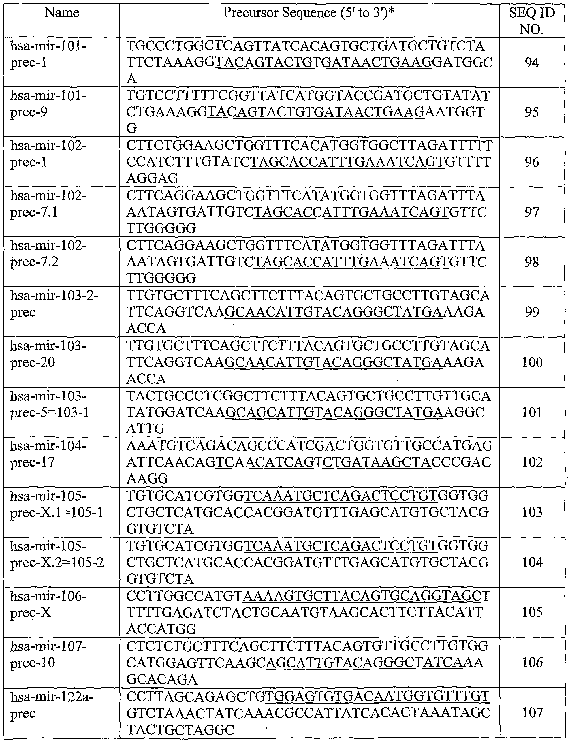


* An underlined sequence within a precursor sequence represents a processed miR transcript. AU sequences are human.
The level of at least one miR gene product can be measured in cells of a biological sample obtained from the subject. For example, a tissue sample can be removed from a subject suspected of having breast cancer associated with by conventional biopsy techniques. In another example, a blood sample can be removed from the subject, and white blood cells can be isolated for DNA extraction by standard techniques. The blood or tissue sample is preferably obtained from the subject prior to initiation of radiotherapy, chemotherapy or other therapeutic treatment. A corresponding control tissue or blood sample can be obtained from unaffected tissues of the subject, from a normal human individual or population of normal individuals, or from cultured cells corresponding to the majority of cells in the subject's sample. The control tissue or blood sample is then processed along with the sample from the subject, so that the levels of miR gene product produced from a given miR gene in cells from the subject's sample can be compared to the corresponding miR gene product levels from cells of the control sample.
An alteration (i.e., an increase or decrease) in the level of a miR gene product in the sample obtained from the subject, relative to the level of a corresponding miR gene product in a control sample, is indicative of the presence of breast cancer in the subject. In one embodiment, the level of the at least one miR gene product in the test sample is greater than the level of the corresponding miR gene product in the control sample (i.e., expression of the miR gene product is "up-regulated"). As used herein, expression of an miR gene product is
"up-regulated" when the amount of miR gene product in a cell or tissue sample from a subject is greater than the amount the same gene product in a control cell or tissue sample. In another embodiment, the level of the at least one miR gene product in the test sample is less than the level of the corresponding miR gene product in the control sample (i.e., expression of the miR gene product is "down-regulated"). As used herein, expression of an miR gene is "down-regulated" when the amount of miR gene product produced from that gene in a cell or tissue sample from a subject is less than the amount produced from the same gene in a control cell or tissue sample. The relative miR gene expression in the control and normal samples can be determined with respect to one or more RNA expression standards. The standards can comprise, for example, a zero miR gene expression level, the miR gene expression level in a standard cell line, or the average level of miR gene expression previously obtained for a population of normal human controls.
The level of a miR gene product in a sample can be measured using any technique that is suitable for detecting RNA expression levels in a biological sample. Suitable techniques for determining RNA expression levels in cells from a biological sample (e.g., Northern blot analysis, RT-PCR, in situ hybridization) are well known to those of skill in the art. In a particular embodiment, the level of at least one miR gene product is detected using Northern blot analysis. For example, total cellular RNA can be purified from cells by homogenization in the presence of nucleic acid extraction buffer, followed by centrifugation. Nucleic acids are precipitated, and DNA is removed by treatment with DNase and precipitation. The RNA molecules are then separated by gel electrophoresis on agarose gels according to standard techniques, and transferred to nitrocellulose filters. The RNA is then immobilized on the filters by heating. Detection and quantification of specific RNA is accomplished using appropriately labeled DNA or RNA probes complementary to the RNA in question. See, for example, Molecular Cloning: A Laboratory Manual, J. Sambrook et ah, eds., 2nd edition, Cold Spring Harbor Laboratory Press, 1989, Chapter 7, the entire disclosure of which is incorporated by reference.
Suitable probes for Northern blot hybridization of a given miR gene product can be produced from the nucleic acid sequences provided in Table 1. Methods for preparation of labeled DNA and RNA probes, and the conditions for hybridization thereof to target
nucleotide sequences, are described in Molecular Cloning: A Laboratory Manual. J. Sambrook et al., eds., 2nd edition, Cold Spring Harbor Laboratory Press, 1989, Chapters 10 and 11, the disclosures of which are incorporated herein by reference.
For example, the nucleic acid probe can be labeled with, e.g., a radionuclide, such as 3H, 32P, 33P, 14C, or 35S; a heavy metal; or a ligand capable of functioning as a specific binding pair member for a labeled ligand {e.g., biotin, avidin or an antibody), a fluorescent molecule, a chemilurninescent molecule, an enzyme or the like.
Probes can be labeled to high specific activity by either the nick translation method of Rigby et al. (1977), J. MoI. Biol. 113:237-251 or by the random priming method of Fienberg et al. (1983), Anal. Biochem. 132:6-13, the entire disclosures of which are incorporated herein by reference. The latter is the method of choice for synthesizing 32P -labeled probes of high specific activity from single-stranded DNA or from RNA templates. For example, by replacing preexisting nucleotides with highly radioactive nucleotides according to the nick translation method, it is possible to prepare 32P-labeled nucleic acid probes with a specific activity well in excess of 108 cpm/microgram. Autoradiographic detection of hybridization can then be performed by exposing hybridized filters to photographic film. Densitometric scanning of the photographic films exposed by the hybridized filters provides an accurate measurement of miR gene transcript levels. Using another approach, miR gene transcript levels can be quantified by computerized imaging systems, such the Molecular Dynamics 400-B 2D Phosphorimager available from Amersham Biosciences, Piscataway, NJ.
Where radionuclide labeling of DNA or RNA probes is not practical, the random- primer method can be used to incorporate an analogue, for example, the dTTP analogue 5-(N- (N-biotinyl-epsilon-aminocaproyl)-3-aminoallyl)deoxyuridine triphosphate, into the probe molecule. The biotinylated probe oligonucleotide can be detected by reaction with biotin- binding proteins, such as avidin, streptavidin, and antibodies (e.g., anti-biotin antibodies) coupled to fluorescent dyes or enzymes that produce color reactions.
In addition to Northern and other RNA hybridization techniques, determining the levels of RNA transcripts can be accomplished using the technique of in situ hybridization. This technique requires fewer cells than the Northern blotting technique, and involves depositing whole cells onto a microscope cover slip and probing the nucleic acid content of
the cell with a solution containing radioactive or otherwise labeled nucleic acid (e.g., cDNA or RNA) probes. This technique is particularly well-suited for analyzing tissue biopsy samples from subjects. The practice of the in situ hybridization technique is described in more detail in U.S. Pat. No. 5,427,916, the entire disclosure of which is incorporated herein by reference. Suitable probes for in situ hybridization of a given miR gene product can be produced from the nucleic acid sequences provided in Table 1, as described above.
The relative number of miR gene transcripts in cells can also be determined by reverse transcription of miR gene transcripts, followed by amplification of the reverse- transcribed transcripts by polymerase chain reaction (RT-PCR). The levels of miR gene transcripts can be quantified in comparison with an internal standard, for example, the level of mRNA from a "housekeeping" gene present in the same sample. A suitable "housekeeping" gene for use as an internal standard includes, e.g., myosin or glyceraldehyde- 3 -phosphate dehydrogenase (G3PDH). The methods for quantitative RT-PCR and variations thereof are within the skill in the art. In some instances, it may be desirable to simultaneously determine the expression level of a plurality of different miR gene products in a sample. In other instances, it may be desirable to determine the expression level of the transcripts of all known miR genes correlated with a cancer. Assessing cancer-specific expression levels for hundreds of miR genes is time consuming and requires a large amount of total RNA (at least 20 μg for each Northern blot) and autoradiographic techniques that require radioactive isotopes.
To overcome these limitations, an oligolibrary, in microchip format (i.e., a microarray), may be constructed containing a set of probe oligodeoxynucleotides that are specific for a set of miR genes. Using such a microarray, the expression level of multiple microRNAs in a biological sample can be determined by reverse transcribing the RNAs to generate a set of target oligodeoxynucleotides, and hybridizing them to probe oligodeoxynucleotides on the microarray to generate a hybridization, or expression, profile. The hybridization profile of the test sample can then be compared to that of a control sample to determine which microRNAs have an altered expression level in breast cancer cells. As used herein, "probe oligonucleotide" or "probe oligodeoxynucleotide" refers to an oligonucleotide that is capable of hybridizing to a target oligonucleotide. "Target
oligonucleotide" or "target oligodeoxynucleotide" refers to a molecule to be detected (e.g., via hybridization). By "miR-specific probe oligonucleotide" or "probe oligonucleotide specific for an miR" is meant a probe oligonucleotide that has a sequence selected to hybridize to a specific miR gene product, or to a reverse transcript of the specific miR gene product.
An "expression profile" or "hybridization profile" of a particular sample is essentially a fingerprint of the state of the sample; while two states may have any particular gene similarly expressed, the evaluation of a number of genes simultaneously allows the generation of a gene expression profile that is unique to the state of the cell. That is, normal tissue may be distinguished from breast cancer tissue, and within breast cancer tissue, different prognosis states (good or poor long term survival prospects, for example) may be determined. By comparing expression profiles of breast cancer tissue in different states, information regarding which genes are important (including both up- and down-regulation of genes) in each of these states is obtained. The identification of sequences that are differentially expressed in breast cancer tissue or normal breast tissue, as well as differential expression resulting in different prognostic outcomes, allows the use of this information in a number of ways. For example, a particular treatment regime may be evaluated (e.g., to determine whether a chemotherapeutic drug act to improve the long-term prognosis in a particular patient). Similarly, diagnosis may be done or confirmed by comparing patient samples with the known expression profiles. Furthermore, these gene expression profiles (or individual genes) allow screening of drug candidates that suppress the breast cancer expression profile or convert a poor prognosis profile to a better prognosis profile.
Accordingly, the invention provides methods of diagnosing whether a subject has, or is at risk for developing, breast cancer, comprising reverse transcribing RNA from a test sample obtained from the subject to provide a set of target oligo-deoxynucleotides, hybridizing the target oligo-deoxynucleotides to a microarray comprising miRNA-specific probe oligonucleotides to provide a hybridization profile for the test sample, and comparing the test sample hybridization profile to a hybridization profile generated from a control sample, wherein an alteration in the signal of at least one miRNA is indicative of the subject either having, or being at risk for developing, breast cancer. In one embodiment, the
microarray comprises miRNA-specific probe oligonucleotides for a substantial portion of the human miRNome. In a particular embodiment, the microarray comprises miRNA-specific probe oligo-nucleotides for one or more miRNAs selected from the group consisting of miR- 125b, miR-145, miR-21, miR-155, miR-lOb, miR-009-1 (miR131-l), miR-34 (miR-170), miR-102 (miR-29b), miR-123 (miR-126), miR-140-as, miR-125a, miR-125b-l, miR-125b-2, miR-194, miR-204, miR-213, let-7a-2, let-7a-3, let-7d (let-7d-vl), let-7f-2, let-7i (Iet-7d-v2)5 miR-101-1, miR-122a, miR-128b, miR-136, miR-143, miR-149, miR-191, miR-196-1, miR- 196-2, miR-202, miR-203, miR-206, miR-210 and combinations thereof. In a further embodiment, the at least one miR gene product is selected from the group consisting of miR- 125b, miR-145, miR-21, miR-155, miR-lOb and combinations thereof.
The microarray can be prepared from gene-specific oligonucleotide probes generated from known miRNA sequences. The array may contain two different oligonucleotide probes for each miRNA, one containing the active, mature sequence and the other being specific for the precursor of the miRNA. The array may also contain controls, such as one or more mouse sequences differing from human orthologs by only a few bases, which can serve as controls for hybridization stringency conditions. tRNAs from both species may also be printed on the microchip, providing an internal, relatively stable, positive control for specific hybridization. One or more appropriate controls for non-specific hybridization may also be included on the microchip. For this purpose, sequences are selected based upon the absence of any homology with any known miRNAs.
The microarray may be fabricated using techniques known in the art. For example, probe oligonucleotides of an appropriate length, e.g., 40 nucleotides, are 5'-amine modified at position C6 and printed using commercially available microarray systems, e.g., the GeneMachine OmniGrid™ 100 Microarrayer and Amersham CodeLink™ activated slides. Labeled cDNA oligomer corresponding to the target RNAs is prepared by reverse transcribing the target RNA with labeled primer. Following first strand synthesis, the RNA/DNA hybrids are denatured to degrade the RNA templates. The labeled target cDNAs thus prepared are then hybridized to the microarray chip under hybridizing conditions, e.g., 6X SSPE/30% formamide at 250C for 18 hours, followed by washing in 0.75X TNT at 370C for 40 minutes. At positions on the array where the immobilized probe DNA recognizes a
complementary target cDNA in the sample, hybridization occurs. The labeled target cDNA marks the exact position on the array where binding occurs, allowing automatic detection and quantification. The output consists of a list of hybridization events, indicating the relative abundance of specific cDNA sequences, and therefore the relative abundance of the corresponding complementary miRs, in the patient sample. According to one embodiment, the labeled cDNA oligomer is a biotin-labeled cDNA, prepared from a biotin-labeled primer. The microarray is then processed by direct detection of the biotin-containing transcripts using, e.g., Streptavidin-Alexa647 conjugate, and scanned utilizing conventional scanning methods. Image intensities of each spot on the array are proportional to the abundance of the corresponding miR in the patient sample.
The use of the array has several advantages for miRNA expression detection. First, the global expression of several hundred genes can be identified in the same sample at one time point. Second, through careful design of the oligonucleotide probes, expression of both mature and precursor molecules can be identified. Third, in comparison with Northern blot analysis, the chip requires a small amount of RNA, and provides reproducible results using 2.5 μg of total RNA. The relatively limited number of miRNAs (a few hundred per species) allows the construction of a common microarray for several species, with distinct oligonucleotide probes for each. Such a tool would allow for analysis of trans-species expression for each known miR under various conditions. In addition to use for quantitative expression level assays of specific miRs, a microchip containing miRNA-specifϊc probe oligonucleotides corresponding to a substantial portion of the miRNome, preferably the entire miRNome, may be employed to carry out miR gene expression profiling, for analysis of miR expression patterns. Distinct miR signatures can be associated with established disease markers, or directly with a disease state. According to the expression profiling methods described herein, total RNA from a sample from a subject suspected of having a cancer (e.g., breast cancer) is quantitatively reverse transcribed to provide a set of labeled target oligodeoxynucleotides complementary to the RNA in the sample. The target oligodeoxynucleotides are then hybridized to a microarray comprising miRNA-specific probe oligonucleotides to provide a hybridization profile for the sample. The result is a hybridization profile for the sample representing the
expression pattern of miRNA in the sample. The hybridization profile comprises the signal from the binding of the target oligodeoxynucleotides from the sample to the miRNA-specific probe oligonucleotides in the microarray. The profile may be recorded as the presence or absence of binding (signal vs. zero signal). More preferably, the profile recorded includes the intensity of the signal from each hybridization. The profile is compared to the hybridization profile generated from a normal, i.e., noncancerous, control sample. An alteration in the signal is indicative of the presence of the cancer in the subject.
Other techniques for measuring miR gene expression are also within the skill in the art, and include various techniques for measuring rates of RNA transcription and degradation. The invention also provides methods of diagnosing a breast cancer associated with one or more prognostic markers, comprising measuring the level of at least one miR gene product in a breast cancer test sample from a subject and comparing the level of the at least one miR gene product in the breast cancer test sample to the level of a corresponding miR gene product in a control sample. An alteration (e.g., an increase, a decrease) in the signal of at least one miRNA in the test sample relative to the control sample is indicative of the subject either having, or being at risk for developing, breast cancer associated with the one or more prognostic markers.
The breast cancer can be associated with one or more prognostic markers or features, including, a marker associated with an adverse (i.e., negative) prognosis, or a marker associated with a good (i.e., positive) prognosis. In certain embodiments, the breast cancer that is diagnosed using the methods described herein is associated with one or more adverse prognostic features selected from the group consisting of estrogen receptor expression, progesterone receptor expression, positive lymph node metastasis, high proliferative index, detectable p53 expression, advanced tumor stage, and high vascular invasion. Particular microRNAs whose expression is altered in breast cancer cells associated with each of these prognostic markers are described herein (see, for example, Example 3 and Figure 4). In one embodiment, the level of the at least one miR gene product is measured by reverse transcribing RNA from a test sample obtained from the subject to provide a set of target oligodeoxynucleotides, hybridizing the target oligodeoxynucleotides to a microarray that comprises miRNA-specific probe oligonucleotides to provide a hybridization profile for the
test sample, and comparing the test sample hybridization profile to a hybridization profile generated from a control sample.
Without wishing to be bound by any one theory, it is believed that alterations in the level of one or more miR gene products in cells can result in the deregulation of one or more intended targets for these miRs, which can lead to the formation of breast cancer. Therefore, altering the level of the miR gene product (e.g., by decreasing the level of a miR that is up- regulated in breast cancer cells, by increasing the level of a miR that is down-regulated in cancer cells) may successfully treat the breast cancer. Examples of putative gene targets for miRNAs that are deregulated in breast cancer tissues are described herein (see, e.g., Example 2 and Table 4).
Accordingly, the present invention encompasses methods of treating breast cancer in a subject, wherein at least one miR gene product is de-regulated (e.g., down-regulated, up- regulated) in the cancer cells of the subject. When the at least one isolated miR gene product is down-regulated in the breast cancer cells, the method comprises administering an effective amount of the at least one isolated miR gene product, provided that the miR gene is not miR15 or miRlό, such that proliferation of cancer cells in the subject is inhibited. When the at least one isolated miR gene product is up-regulated in the cancer cells, the method comprises administering to the subject an effective amount of at least one compound for inhibiting expression of the at least one miR gene, referred to herein as miR gene expression inhibition compounds, such that proliferation of breast cancer cells is inhibited.
The terms "treat", "treating" and "treatment", as used herein, refer to ameliorating symptoms associated with a disease or condition, for example, breast cancer, including preventing or delaying the onset of the disease symptoms, and/or lessening the severity or frequency of symptoms of the disease or condition. The terms "subject" and "individual" are defined herein to include animals, such as mammals, including but not limited to, primates, cows, sheep, goats, horses, dogs, cats, rabbits, guinea pigs, rats, mice or other bovine, ovine, equine, canine, feline, rodent, or murine species. In a preferred embodiment, the animal is a human.
As used herein, an "effective amount" of an isolated miR gene product is an amount sufficient to inhibit proliferation of a cancer cell in a subject suffering from breast cancer.
One skilled in the art can readily determine an effective amount of an miR gene product to be administered to a given subject, by taking into account factors, such as the size and weight of the subject; the extent of disease penetration; the age, health and sex of the subject; the route of administration; and whether the administration is regional or systemic. For example, an effective amount of an isolated miR gene product can be based on the approximate weight of a tumor mass to be treated. The approximate weight of a tumor mass can be determined by calculating the approximate volume of the mass, wherein one cubic centimeter of volume is roughly equivalent to one gram. An effective amount of the isolated miR gene product based on the weight of a tumor mass can be in the range of about 10-500 micrograms/gram of tumor mass. In certain embodiments, the tumor mass can be at least about 10 micrograms/gram of tumor mass, at least about 60 micrograms/gram of tumor mass or at least about 100 micrograms/gram of tumor mass.
An effective amount of an isolated miR gene product can also be based on the approximate or estimated body weight of a subject to be treated. Preferably, such effective amounts are administered parenterally or enterally, as described herein. For example, an effective amount of the isolated miR gene product is administered to a subject can range from about 5 - 3000 micrograms/kg of body weight, from about 700 - 1000 micrograms/kg of body weight, or greater than about 1000 micrograms/kg of body weight.
One skilled in the art can also readily determine an appropriate dosage regimen for the administration of an isolated miR gene product to a given subject. For example, an miR gene product can be administered to the subject once (e.g., as a single injection or deposition). Alternatively, an miR gene product can be administered once or twice daily to a subject for a period of from about three to about twenty-eight days, more particularly from about seven to about ten days. In a particular dosage regimen, an miR gene product is administered once a day for seven days. Where a dosage regimen comprises multiple administrations, it is understood that the effective amount of the miR gene product administered to the subject can comprise the total amount of gene product administered over the entire dosage regimen.
As used herein, an "isolated" miR gene product is one which is synthesized, or altered or removed from the natural state through human intervention. For example, a synthetic miR gene product, or an miR gene product partially or completely separated from the coexisting
materials of its natural state, is considered to be "isolated." An isolated miR gene product can exist in substantially-purified form, or can exist in a cell into which the miR gene product has been delivered. Thus, an miR gene product which is deliberately delivered to, or expressed in, a cell is considered an "isolated" miR gene product. An miR gene product produced inside a cell from an miR precursor molecule is also considered to be "isolated" molecule.
Isolated miR gene products can be obtained using a number of standard techniques. For example, the miR gene products can be chemically synthesized or recombinantly produced using methods known in the art. In one embodiment, miR gene products are chemically synthesized using appropriately protected ribonucleoside phosphoramidites and a conventional DNA/RNA synthesizer. Commercial suppliers of synthetic RNA molecules or synthesis reagents include, e.g., Proligo (Hamburg, Germany), Dharmacon Research (Lafayette, CO, U.S.A.), Pierce Chemical (part of Perbio Science, Rockford, IL, U.S.A.), Glen Research (Sterling, VA, U.S.A.), ChemGenes (Ashland, MA, U.S.A.) and Cruachem (Glasgow, UK).
Alternatively, the miR gene products can be expressed from recombinant circular or linear DNA plasmids using any suitable promoter. Suitable promoters for expressing RNA from a plasmid include, e.g., the U6 or Hl RNA pol III promoter sequences, or the cytomegalovirus promoters. Selection of other suitable promoters is within the skill in the art. The recombinant plasmids of the invention can also comprise inducible or regulatable promoters for expression of the miR gene products in cancer cells.
The miR gene products that are expressed from recombinant plasmids can be isolated from cultured cell expression systems by standard techniques. The miR gene products which are expressed from recombinant plasmids can also be delivered to, and expressed directly in, the cancer cells. The use of recombinant plasmids to deliver the miR gene products to cancer cells is discussed in more detail below.
The miR gene products can be expressed from a separate recombinant plasmid, or they can be expressed from the same recombinant plasmid. In one embodiment, the miR gene products are expressed as RNA precursor molecules from a single plasmid, and the precursor molecules are processed into the functional miR gene product by a suitable
processing system, including, but not limited to, processing systems extant within a cancer cell. Other suitable processing systems include, e.g., the in vitro Drosophila cell lysate system (e.g., as described in U.S. Published Patent Application No. 2002/0086356 to Tuschl et al., the entire disclosure of which are incorporated herein by reference) and the E. coli RNAse III system (e.g., as described in U.S. Published Patent Application No. 2004/0014113 to Yang et ah, the entire disclosure of which are incorporated herein by reference).
Selection of plasmids suitable for expressing the miR gene products, methods for inserting nucleic acid sequences into the plasmid to express the gene products, and methods of delivering the recombinant plasmid to the cells of interest are within the skill in the art. See, for example, Zeng et al. (2002), Molecular Cell 9:1327-1333; Tuschl (2002), Nat.
Biotechnol, 20:446-448; Brummelkamp et al. (2002), Science 296:550-553; Miyagishi et al. (2002), Nat. Biotechnol. 20:497-500; Paddison et al. (2002), Genes Dev. 16:948-958; Lee et al. (2002), Nat. Biotechnol. 20:500-505; and Paul et al. (2002), Nat. Biotechnol. 20:505-508, the entire disclosures of which are incorporated herein by reference. In one embodiment, a plasmid expressing the miR gene products comprises a sequence encoding a miR precursor RNA under the control of the CMV intermediate-early promoter. As used herein, "under the control" of a promoter means that the nucleic acid sequences encoding the miR gene product are located 3' of the promoter, so that the promoter can initiate transcription of the miR gene product coding sequences. The miR gene products can also be expressed from recombinant viral vectors. It is contemplated that the miR gene products can be expressed from two separate recombinant viral vectors, or from the same viral vector. The RNA expressed from the recombinant viral vectors can either be isolated from cultured cell expression systems by standard techniques, or can be expressed directly in cancer cells. The use of recombinant viral vectors to deliver the miR gene products to cancer cells is discussed in more detail below.
The recombinant viral vectors of the invention comprise sequences encoding the miR gene products and any suitable promoter for expressing the RNA sequences. Suitable promoters include, for example, the U6 or Hl RNA pol III promoter sequences, or the cytomegalovirus promoters. Selection of other suitable promoters is within the skill in the
art. The recombinant viral vectors of the invention can also comprise inducible or regulatable promoters for expression of the miR gene products in a cancer cell.
Any viral vector capable of accepting the coding sequences for the miR gene products can be used; for example, vectors derived from adenovirus (AV); adeno-associated virus (AAV); retroviruses (e.g., lentiviruses (LV), Rhabdoviruses, murine leukemia virus); herpes virus, and the like. The tropism of the viral vectors can be modified by pseudotyping the vectors with envelope proteins or other surface antigens from other viruses, or by substituting different viral capsid proteins, as appropriate.
For example, lentiviral vectors of the invention can be pseudotyped with surface proteins from vesicular stomatitis virus (VSV), rabies, Ebola, Mokola, and the like. AAV vectors of the invention can be made to target different cells by engineering the vectors to express different capsid protein serotypes. For example, an AAV vector expressing a serotype 2 capsid on a serotype 2 genome is called AAV 2/2. This serotype 2 capsid gene in the AAV 2/2 vector can be replaced by a serotype 5 capsid gene to produce an AAV 2/5 vector. Techniques for constructing AAV vectors that express different capsid protein serotypes are within the skill in the art; see, e.g., Rabinowitz, J.E., et al. (2002), J. Virol. 76:791-801, the entire disclosure of which is incorporated herein by reference.
Selection of recombinant viral vectors suitable for use in the invention, methods for inserting nucleic acid sequences for expressing RNA into the vector, methods of delivering the viral vector to the cells of interest, and recovery of the expressed RNA products are within the skill in the art. See, for example, Dornburg (1995), Gene Therap. 2:301-310; Eglitis (1988), Biotechniques 6:608-614; Miller (1990), Hum. Gene Therap. 1:5-14; and Anderson (1998), Nature 392:25-30, the entire disclosures of which are incorporated herein by reference. Particularly suitable viral vectors are those derived from AV and AAV. A suitable
AV vector for expressing the miR gene products, a method for constructing the recombinant AV vector, and a method for delivering the vector into target cells, are described in Xia et al. (2002), Nat. Biotech. 20:1006-1010, the entire disclosure of which is incorporated herein by reference. Suitable AAV vectors for expressing the miR gene products, methods for constructing the recombinant AAV vector, and methods for delivering the vectors into target
cells are described in Samulski etal. (1987), J. Virol. 61:3096-3101; Fisher et al. (1996), J. Virol, 70:520-532; Samulski etal. (1989), J. Virol. 63:3822-3826; U.S. Pat. No. 5,252,479; U.S. Pat. No. 5,139,941; International Patent Application No. WO 94/13788; and International Patent Application No. WO 93/24641, the entire disclosures of which are incorporated herein by reference. In one embodiment, the miR gene products are expressed from a single recombinant AAV vector comprising the CMV intermediate early promoter.
In a certain embodiment, a recombinant AAV viral vector of the invention comprises a nucleic acid sequence encoding an miR precursor RNA in operable connection with a polyT termination sequence under the control of a human U6 RNA promoter. As used herein, "in operable connection with a polyT termination sequence" means that the nucleic acid sequences encoding the sense or antisense strands are immediately adjacent to the polyT termination signal in the 5' direction. During transcription of the miR sequences from the vector, the polyT termination signals act to terminate transcription.
In other embodiments of the treatment methods of the invention, an effective amount of at least one compound which inhibits miR expression can also be administered to the subject. As used herein, "inhibiting miR expression" means that the production of the active, mature form of miR gene product after treatment is less than the amount produced prior to treatment. One skilled in the art can readily determine whether miR expression has been inhibited in a cancer cell, using for example the techniques for determining miR transcript level discussed above for the diagnostic method. Inhibition can occur at the level of gene expression (i.e., by inhibiting transcription of a miR gene encoding the miR gene product) or at the level of processing (e.g., by inhibiting processing of a miR precursor into a mature, active miR).
As used herein, an "effective amount" of a compound that inhibits miR expression is an amount sufficient to inhibit proliferation of a cancer cell in a subject suffering from a cancer associated with a cancer-associated chromosomal feature. One skilled in the art can readily determine an effective amount of an miR expression-inhibiting compound to be administered to a given subject, by taking into account factors, such as the size and weight of the subject; the extent of disease penetration; the age, health and sex of the subject; the route of administration; and whether the administration is regional or systemic.
For example, an effective amount of the expression-inhibiting compound can be based on the approximate weight of a tumor mass to be treated. The approximate weight of a tumor mass can be determined by calculating the approximate volume of the mass, wherein one cubic centimeter of volume is roughly equivalent to one gram. An effective amount based on the weight of a tumor mass can be between about 10-500 micrograms/gram of tumor mass, at least about 10 micrograms/gram of tumor mass, at least about 60 micrograms/gram of tumor mass, and at least about 100 micrograms/gram of tumor mass.
An effective amount of a compound that inhibits miR expression can also be based on the approximate or estimated body weight of a subject to be treated. Such effective amounts are administered parenterally or enterally, among others, as described herein. For example, an effective amount of the expression-inhibiting compound administered to a subject can range from about 5 -3000 micrograms/kg of body weight, from about 700 - 1000 micrograms/kg of body weight, or it can be greater than about 1000 micrograms/kg of body weight. One skilled in the art can also readily determine an appropriate dosage regimen for administering a compound that inhibits miR expression to a given subject. For example, an expression-inhibiting compound can be administered to the subject once (e.g., as a single injection or deposition). Alternatively, an expression-inhibiting compound can be administered once or twice daily to a subject for a period of from about three to about twenty- eight days, more preferably from about seven to about ten days. In a particular dosage regimen, an expression-inhibiting compound is administered once a day for seven days. Where a dosage regimen comprises multiple administrations, it is understood that the effective amount of the expression-inhibiting compound administered to the subject can comprise the total amount of compound administered over the entire dosage regimen. Suitable compounds for inhibiting miR gene expression include double-stranded RNA
(such as short- or small-interfering RNA or "siRNA"), antisense nucleic acids, and enzymatic RNA molecules, such as ribozymes. Each of these compounds can be targeted to a given miR gene product and destroy or induce the destruction of the target miR gene product. For example, expression of a given miR gene can be inhibited by inducing RNA interference of the miR gene with an isolated double-stranded RNA ("dsRNA") molecule
which has at least 90%, for example at least 95%, at least 98%, at least 99% or 100%, sequence homology with at least a portion of the miR gene product. In a particular embodiment, the dsRNA molecule is a "short or small interfering RNA" or "siRNA." siRNA useful in the present methods comprise short double-stranded RNA from about 17 nucleotides to about 29 nucleotides in length, preferably from about 19 to about 25 nucleotides in length. The siRNA comprise a sense RNA strand and a complementary antisense RNA strand annealed together by standard Watson-Crick base-pairing interactions (hereinafter "base-paired"). The sense strand comprises a nucleic acid sequence which is substantially identical to a nucleic acid sequence contained within the target miR gene product.
As used herein, a nucleic acid sequence in an siRNA which is "substantially identical" to a target sequence contained within the target mRNA is a nucleic acid sequence that is identical to the target sequence, or that differs from the target sequence by one or two nucleotides. The sense and antisense strands of the siRNA can comprise two complementary, single-stranded RNA molecules, or can comprise a single molecule in which two complementary portions are base-paired and are covalently linked by a single-stranded "hairpin" area.
The siRNA can also be altered RNA that differs from naturally-occurring RNA by the addition, deletion, substitution and/or alteration of one or more nucleotides. Such alterations can include addition of non-nucleotide material, such as to the end(s) of the siRNA or to one or more internal nucleotides of the siRNA, or modifications that make the siRNA resistant to nuclease digestion, or the substitution of one or more nucleotides in the siRNA with deoxyribonucleotides.
One or both strands of the siRNA can also comprise a 3' overhang. As used herein, a "3' overhang" refers to at least one unpaired nucleotide extending from the 3'-end of a duplexed RNA strand. Thus, in certain embodiments, the siRNA comprises at least one 3' overhang of from 1 to about 6 nucleotides (which includes ribonucleotides or deoxyribonucleotides) in length, from 1 to about 5 nucleotides in length, from 1 to about 4 nucleotides in length, or from about 2 to about 4 nucleotides in length. In a particular embodiment, the 31 overhang is present on both strands of the siRNA, and is 2 nucleotides in
length. For example, each strand of the siRNA can comprise 3' overhangs of dithymidylic acid ("TT") or diuridylic acid ("uu").
The siRNA can be produced chemically or biologically, or can be expressed from a recombinant plasmid or viral vector, as described above for the isolated miR gene products. Exemplary methods for producing and testing dsRNA or siRNA molecules are described in U.S. Published Patent Application No. 2002/0173478 to Gewirtz and in U.S. Published Patent Application No. 2004/0018176 to Reich et ah, the entire disclosures of which are incorporated herein by reference.
Expression of a given miR gene can also be inhibited by an antisense nucleic acid. As used herein, an "antisense nucleic acid" refers to a nucleic acid molecule that binds to target RNA by means of RNA-RNA or RNA-DNA or RNA-peptide nucleic acid interactions, which alters the activity of the target RNA. Antisense nucleic acids suitable for use in the present methods are single-stranded nucleic acids {e.g., RNA, DNA, RNA-DNA chimeras, PNA) that generally comprise a nucleic acid sequence complementary to a contiguous nucleic acid sequence in an miR gene product. The antisense nucleic acid can comprise a nucleic acid sequence that is 50-100% complementary, 75-100% complementary, or 95-100% complementary to a contiguous nucleic acid sequence in an miR gene product. Nucleic acid sequences for the miR gene products are provided in Table 1. Without wishing to be bound by any theory, it is believed that the antisense nucleic acids activate RNase H or another cellular nuclease that digests the miR gene product/antisense nucleic acid duplex.
Antisense nucleic acids can also contain modifications to the nucleic acid backbone or to the sugar and base moieties (or their equivalent) to enhance target specificity, nuclease resistance, delivery or other properties related to efficacy of the molecule. Such modifications include cholesterol moieties, duplex intercalators, such as acridine, or one or more nuclease-resistant groups.
Antisense nucleic acids can be produced chemically or biologically, or can be expressed from a recombinant plasmid or viral vector, as described above for the isolated miR gene products. Exemplary methods for producing and testing are within the skill in the art; see, e.g., Stein and Cheng (1993), Science 261 :1004 and U.S. Pat. No. 5,849,902 to Woolf et ah, the entire disclosures of which are incorporated herein by reference.
Expression of a given miR gene can also be inhibited by an enzymatic nucleic acid. As used herein, an "enzymatic nucleic acid" refers to a nucleic acid comprising a substrate binding region that has complementarity to a contiguous nucleic acid sequence of an miR gene product, and which is able to specifically cleave the miR gene product. The enzymatic nucleic acid substrate binding region can be, for example, 50-100% complementary, 75- 100% complementary, or 95-100% complementary to a contiguous nucleic acid sequence in an miR gene product. The enzymatic nucleic acids can also comprise modifications at the base, sugar, and/or phosphate groups. An exemplary enzymatic nucleic acid for use in the present methods is a ribozyme. The enzymatic nucleic acids can be produced chemically or biologically, or can be expressed from a recombinant plasmid or viral vector, as described above for the isolated miR gene products. Exemplary methods for producing and testing dsRNA or siRNA molecules are described in Werner and Uhlenbeck (1995), Nucl Acids Res. 23:2092-96; Hammann et al. (1999), Antisense and Nucleic Acid Drug Dev. 9:25-31; and U.S. Pat. No. 4,987,071 to Cech et al, the entire disclosures of which are incorporated herein by reference.
Administration of at least one miR gene product, or at least one compound for inhibiting miR expression, will inhibit the proliferation of cancer cells in a subject who has a cancer associated with a cancer-associated chromosomal feature. As used herein, to "inhibit the proliferation of a cancer cell" means to kill the cell, or permanently or temporarily arrest or slow the growth of the cell. Inhibition of cancer cell proliferation can be inferred if the number of such cells in the subject remains constant or decreases after administration of the miR gene products or miR gene expression-inhibiting compounds. An inhibition of cancer cell proliferation can also be inferred if the absolute number of such cells increases, but the rate of tumor growth decreases. The number of cancer cells in a subject's body can be determined by direct measurement, or by estimation from the size of primary or metastatic tumor masses. For example, the number of cancer cells in a subject can be measured by immunohistological methods, flow cytometry, or other techniques designed to detect characteristic surface markers of cancer cells.
The size of a tumor mass can be ascertained by direct visual observation, or by diagnostic imaging methods, such as X-ray, magnetic resonance imaging, ultrasound, and scintigraphy. Diagnostic imaging methods used to ascertain size of the tumor mass can be employed with or without contrast agents, as is known in the art. The size of a tumor mass can also be ascertained by physical means, such as palpation of the tissue mass or measurement of the tissue mass with a measuring instrument, such as a caliper.
The miR gene products or miR gene expression-inhibiting compounds can be administered to a subject by any means suitable for delivering these compounds to cancer cells of the subject. For example, the miR gene products or miR expression inhibiting compounds can be administered by methods suitable to transfect cells of the subject with these compounds, or with nucleic acids comprising sequences encoding these compounds. In one embodiment, the cells are transfected with a plasmid or viral vector comprising sequences encoding at least one miR gene product or miR gene expression inhibiting compound. Transfection methods for eukaryotic cells are well known in the art, and include, e.g., direct injection of the nucleic acid into the nucleus or pronucleus of a cell; electroporation; liposome transfer or transfer mediated by lipophilic materials; receptor-mediated nucleic acid delivery, bioballistic or particle acceleration; calcium phosphate precipitation, and transfection mediated by viral vectors. For example, cells can be transfected with a liposomal transfer compound, e.g.,
DOTAP (N-[l-(2,3-dioleoyloxy)propyl]-N,N,N-trimethyl-ammonium methylsulfate, Boehringer - Mannheim) or an equivalent, such as LIPOFECTIN. The amount of nucleic acid used is not critical to the practice of the invention; acceptable results may be achieved with 0.1-100 micrograms of nucleic acid/ 105 cells. For example, a ratio of about 0.5 micrograms of plasmid vector in 3 micrograms of DOTAP per 105 cells can be used.
An miR gene product or miR gene expression inhibiting compound can also be administered to a subject by any suitable enteral or parenteral administration route. Suitable enteral administration routes for the present methods include, e.g., oral, rectal, or intranasal delivery. Suitable parenteral administration routes include, e.g., intravascular administration (e.g., intravenous bolus injection, intravenous infusion, intra-arterial bolus injection, intra-
arterial infusion and catheter instillation into the vasculature); peri- and intra-tissue injection
(e.g., peri-tumoral and intra-tumoral injection, intra-retinal injection, or subretinal injection); subcutaneous injection or deposition, including subcutaneous infusion (such as by osmotic pumps); direct application to the tissue of interest, for example by a catheter or other placement device (e.g. , a retinal pellet or a suppository or an implant comprising a porous, non-porous, or gelatinous material); and inhalation. Particularly suitable administration routes are injection, infusion and direct injection into the tumor.
In the present methods, an miR gene product or miR gene product expression inhibiting compound can be administered to the subject either as naked RNA, in combination with a delivery reagent, or as a nucleic acid (e.g. , a recombinant plasmid or viral vector) comprising sequences that express the miR gene product or expression inhibiting compound.
Suitable delivery reagents include, e.g., the Minis Transit TKO lipophilic reagent; lipofectin; lipofectamine; cellfectin; polycations (e.g., polylysine), and liposomes.
Recombinant plasmids and viral vectors comprising sequences that express the miR gene products or miR gene expression inhibiting compounds, and techniques for delivering such plasmids and vectors to cancer cells, are discussed herein.
In a particular embodiment, liposomes are used to deliver an miR gene product or miR gene expression-inhibiting compound (or nucleic acids comprising sequences encoding them) to a subject. Liposomes can also increase the blood half-life of the gene products or nucleic acids. Suitable liposomes for use in the invention can be formed from standard vesicle-forming lipids, which generally include neutral or negatively charged phospholipids and a sterol, such as cholesterol. The selection of lipids is generally guided by consideration of factors, such as the desired liposome size and half-life of the liposomes in the blood stream. A variety of methods are known for preparing liposomes, for example, as described in Szoka et al. (1980), Ann. Rev. Biophys. Bioeng. 9:467; and U.S. Pat. Nos. 4,235,871,
4,501,728, 4,837,028, and 5,019,369, the entire disclosures of which are incorporated herein by reference.
The liposomes for use in the present methods can comprise a ligand molecule that targets the liposome to cancer cells. Ligands which bind to receptors prevalent in cancer cells, such as monoclonal antibodies that bind to tumor cell antigens, are preferred.
The liposomes for use in the present methods can also be modified so as to avoid clearance by the mononuclear macrophage system ("MMS") and reticuloendothelial system ("RES"). Such modified liposomes have opsonization-inhibition moieties on the surface or incorporated into the liposome structure. In a particularly preferred embodiment, a liposome of the invention can comprise both opsonization-inhibition moieties and a ligand.
Opsonization-inhibiting moieties for use in preparing the liposomes of the invention are typically large hydrophilic polymers that are bound to the liposome membrane. As used herein, an opsonization inhibiting moiety is "bound" to a liposome membrane when it is chemically or physically attached to the membrane, e.g., by the intercalation of a lipid- soluble anchor into the membrane itself, or by binding directly to active groups of membrane lipids. These opsonization-inhibiting hydrophilic polymers form a protective surface layer that significantly decreases the uptake of the liposomes by the MMS and RES; e.g., as described in U.S. Pat. No. 4,920,016, the entire disclosure of which is incorporated herein by reference. Opsonization inhibiting moieties suitable for modifying liposomes are preferably water-soluble polymers with a number-average molecular weight from about 500 to about 40,000 daltons, and more preferably from about 2,000 to about 20,000 daltons. Such polymers include polyethylene glycol (PEG) or polypropylene glycol (PPG) derivatives; e.g., methoxy PEG or PPG, and PEG or PPG stearate; synthetic polymers, such as polyacrylamide or poly N-vinyl pyrrolidone; linear, branched, or dendrimeric polyamidoamines; polyacrylic acids; polyalcohols, e.g., polyvinylalcohol and polyxylitol to which carboxylic or amino groups are chemically linked, as well as gangliosides, such as ganglioside GMl. Copolymers of PEG, methoxy PEG, or methoxy PPG, or derivatives thereof, are also suitable. In addition, the opsonization inhibiting polymer can be a block copolymer of PEG and either a polyamino acid, polysaccharide, polyamidoamine, polyethyleneamine, or polynucleotide. The opsonization inhibiting polymers can also be natural polysaccharides containing amino acids or carboxylic acids, e.g., galacturonic acid, glucuronic acid, mannuronic acid, hyaluronic acid, pectic acid, neuraminic acid, alginic acid, carrageenan; aminated polysaccharides or oligosaccharides (linear or branched); or carboxylated polysaccharides or oligosaccharides, e.g., reacted with derivatives of carbonic acids with resultant linking of
carboxylic groups. Preferably, the opsonization-inhibiting moiety is a PEG, PPG, or derivatives thereof. Liposomes modified with PEG or PEG-derivatives are sometimes called "PEGylated liposomes."
The opsonization inhibiting moiety can be bound to the liposome membrane by any one of numerous well-known techniques. For example, an N-hydroxysuccinimide ester of PEG can be bound to a phosphatidyl-ethanolamine lipid-soluble anchor, and then bound to a membrane. Similarly, a dextran polymer can be derivatized with a stearylamine lipid-soluble anchor via reductive animation using Na(CN)BH3 and a solvent mixture, such as tetrahydrofuran and water in a 30:12 ratio at 60 0C. Liposomes modified with opsonization-inhibition moieties remain in the circulation much longer than unmodified liposomes. For this reason, such liposomes are sometimes called "stealth" liposomes. Stealth liposomes are known to accumulate in tissues fed by porous or "leaky" microvasculature. Thus, tissue characterized by such microvasculature defects, for example solid tumors, will efficiently accumulate these liposomes; see Gabizon, et al. (1988), Proc. Natl. Acad. Sci., U.S.A., 18:6949-53. In addition, the reduced uptake by the RES lowers the toxicity of stealth liposomes by preventing significant accumulation of the liposomes in the liver and spleen. Thus, liposomes that are modified with opsonization- inhibition moieties are particularly suited to deliver the miR gene products or miR gene expression inhibition compounds (or nucleic acids comprising sequences encoding them) to tumor cells.
The miR gene products or miR gene expression inhibition compounds can be formulated as pharmaceutical compositions, sometimes called "medicaments," prior to administering them to a subject, according to techniques known in the art. Accordingly, the invention encompasses pharmaceutical compositions for treating breast cancer. In one embodiment, the pharmaceutical compositions comprise at least one isolated miR gene product and a pharmaceutically-acceptable carrier, hi a particular embodiment, the at least one miR gene product corresponds to a miR gene product that has a decreased level of expression in breast cancer cells relative to suitable control cells. In certain embodiments the isolated miR gene product is selected from the group consisting of miR- 145, miR- 10b, miR-
123 (miR-126), miR-140-as, miR-125a, miR-125b-l, miR-125b-2, miR-194, miR-204, let- 7a-2, let-7a-3, let-7d (let-7d-vl), let-7f-2, niiR-101-1, miR-143 and combinations thereof.
In other embodiments, the pharmaceutical compositions of the invention comprise at least one miR expression inhibition compound. In a particular embodiment, the at least one miR gene expression inhibition compound is specific for a miR gene whose expression is greater in breast cancer cells than control cells. In certain embodiments, the miR gene expression inhibition compound is specific for one or more miR gene products selected from the group consisting of miR-21, miR-155, miR-009-1 (miR131-l), miR-34 (miR-170), miR- 102 (miR-29b), miR-213, let-7i (Iet-7d-v2), miR-122a, miR-128b, miR-136, miR-149, miR- 191, miR-196-1, miR-196-2, miR-202, miR-203, miR-206, miR-210, miR-213 and combinations thereof.
Pharmaceutical compositions of the present invention are characterized as being at least sterile and pyrogen-free. As used herein, "pharmaceutical formulations" include formulations for human and veterinary use. Methods for preparing pharmaceutical compositions of the invention are within the skill in the art, for example as described in Remington's Pharmaceutical Science, 17th ed., Mack Publishing Company, Easton, Pa. (1985), the entire disclosure of which is incorporated herein by reference.
The present pharmaceutical formulations comprise at least one miR gene product or miR gene expression inhibition compound (or at least one nucleic acid comprising sequences encoding them) (e.g., 0.1 to 90% by weight), or a physiologically acceptable salt thereof, mixed with a pharmaceutically-acceptable carrier. The pharmaceutical formulations of the invention can also comprise at least one miR gene product or miR gene expression inhibition compound (or at least one nucleic acid comprising sequences encoding them) which are encapsulated by liposomes and a pharmaceutically-acceptable carrier. In one embodiment, the pharmaceutical compositions comprise an miR gene or gene product that is not miR- 15, miR- 16, miR-143 and/or miR- 145.
Especially suitable pharmaceutically-acceptable carriers are water, buffered water, normal saline, 0.4% saline, 0.3% glycine, hyaluronic acid and the like.
In a particular embodiment, the pharmaceutical compositions of the invention comprise at least one miR gene product or miR gene expression inhibition compound (or at
least one nucleic acid comprising sequences encoding them) which is resistant to degradation by nucleases. One skilled in the art can readily synthesize nucleic acids which are nuclease resistant, for example by incorporating one or more ribonucleotides that are modified at the 2'-position into the miR gene products. Suitable 2'-modified ribonucleotides include those modified at the 2'-position with fluoro, amino, alkyl, alkoxy, and O-allyl.
Pharmaceutical compositions of the invention can also comprise conventional pharmaceutical excipients and/or additives. Suitable pharmaceutical excipients include stabilizers, antioxidants, osmolality adjusting agents, buffers, and pH adjusting agents. Suitable additives include, e.g., physiologically biocompatible buffers (e.g., tromethamine hydrochloride), additions of chelants (such as, for example, DTPA or DTPA-bisamide) or calcium chelate complexes (such as, for example, calcium DTPA, CaNaDTP A-bisamide), or, optionally, additions of calcium or sodium salts (for example, calcium chloride, calcium ascorbate, calcium gluconate or calcium lactate). Pharmaceutical compositions of the invention can be packaged for use in liquid form, or can be lyophilized. For solid pharmaceutical compositions of the invention, conventional nontoxic solid pharmaceutically-acceptable carriers can be used; for example, pharmaceutical grades of mannitol, lactose, starch, magnesium stearate, sodium saccharin, talcum, cellulose, glucose, sucrose, magnesium carbonate, and the like.
For example, a solid pharmaceutical composition for oral administration can comprise any of the carriers and excipients listed above and 10-95%, preferably 25%-75%, of the at least one miR gene product or miR gene expression inhibition compound (or at least one nucleic acid comprising sequences encoding them). A pharmaceutical composition for aerosol (inhalational) administration can comprise 0.01-20% by weight, preferably l%-10% by weight, of the at least one miR gene product or miR gene expression inhibition compound (or at least one nucleic acid comprising sequences encoding them) encapsulated in a liposome as described above, and a propellant. A carrier can also be included as desired; e.g., lecithin for intranasal delivery.
The invention also encompasses methods of identifying an anti-breast cancer agent, comprising providing a test agent to a cell and measuring the level of at least one miR gene product in the cell. In one embodiment, the method comprises providing a test agent to a cell
and measuring the level of at least one miR gene product associated with decreased expression levels in breast cancer cells. An increase in the level of the miR gene product in the cell, relative to a suitable control cell, is indicative of the test agent being an anti-breast cancer agent. In a particular embodiment, at least one miR gene product associated with decreased expression levels in breast cancer cells is selected from the group consisting of miR-145, miR-10b, miR-123 (miR-126), miR-140-as, miR-125a, miR-125b-l, miR-125b-2, miR-194, miR-204, let-7a-25 let-7a-3, let-7d (let-7d-vl), let-7f-2, miR-101-1, miR-143 and combinations thereof.
In other embodiments the method comprises providing a test agent to a cell and measuring the level of at least one miR gene product associated with increased expression levels in breast cancer cells. A decrease in the level of the miR gene product in the cell, relative to a suitable control cell, is indicative of the test agent being an anti-breast cancer agent. In a particular embodiment, at least one miR gene product associated with increased expression levels in breast cancer cells is selected from the group consisting of miR-21, miR- 155, miR-009-1 (miR131-l), miR-34 (miR-170), miR-102 (miR-29b), miR-213, let-7i (let- 7d-v2), miR-122a, miR-128b, miR-136, miR-149, miR-191, miR-196-1, miR-196-2, miR- 202, miR-203, miR-206, miR-210, miR-213 and combinations thereof.
Suitable agents include, but are not limited to drugs (e.g., small molecules, peptides), and biological macromolecules (e.g., proteins, nucleic acids). The agent can be produced recombinantly, synthetically, or it may be isolated (i.e., purified) from a natural source.
Various methods for providing such agents to a cell (e.g., transfection) are well known in the art, and several of such methods are described hereinabove. Methods for detecting the expression of at least one miR gene product (e.g., Northern blotting, in situ hybridization, RT-PCR, expression profiling) are also well known in the art. Several of these methods are also described hereinabove.
The invention will now be illustrated by the following non-limiting examples.
Example 1 : Identification of a microRNA expression signature that discriminates breast cancer tissues from normal tissues.
Materials and Methods
Breast cancer samples and cell lines. RNAs from primary tumors were obtained from 76 samples collected at the University of Ferrara (Italy), Istituto Nazionale dei Tumori, Milano (Italy) and Thomas Jefferson University (Philadelphia, PA). Clinico-pathological information was available for 58 tumor samples. RNA from normal samples consisted of 6 pools of RNA from 5 normal breast tissues each, as well as RNA from 4 additional single breast tissues. Breast cancer RNAs were also obtained from the following cell lines: Hs578- T, MCF7, T47D, BT20, SK-BR-3, HBLlOO, HCC2218, MDA-MB-175, MDA-MB-231, MDA-MB-361, MDA-MB-435, MDA-MB-436, MDA-MB-453 and MDAMB-468.
miRNA microarray. Total RNA isolation was performed with Trizol Reagent (Invitrogen) according to the manufacturer's instructions. RNA labeling and hybridization on microRNA microarray chips was performed as previously described (Liu, C-G., et ah, Proc. Natl. Acad. Set U.S.A. 101:9740-9744 (2004)). Briefly, 5 μg of RNA from each sample was labeled with biotin during reverse transcription using random hexamers. Hybridization was carried out on a miRNA microarray chip (KCI version 1.0) (Liu, C-G., et al., Proc. Natl. Acad. Set U.S.A. 101:9740-9744 (2004)), which contains 368 probes, including 245 human and mouse miRNA genes, in triplicate. Hybridization signals were detected by binding of biotin to a
Streptavidin-Alexa647 conjugate using a Perkin-Elmer ScanArray XL5K. Scanner images were quantified by the Quantarray software (Perkin Elmer).
Statistical and bioinformatic analysis of microarray data. Raw data were normalized and analyzed using the GeneSpring® software, version 7.2 (SiliconGenetics, Redwood City, CA). Expression data were median centered. Statistical comparisons were performed by ANOVA (Analysis of Variance), using the Benjamini and Hochberg correction for reduction of false positives. Prognostic miRNAs for tumor or normal class prediction were determined using both the PAM software (Prediction Analysis of Microarrays, available at http://www.stat.stanford.edu/~tibs/PAM/index.html) (Tibshirani, R., et άl. Proc. Natl. Acad.
Sd. U.S.A. 99:6567-6572 (2002)) and the Support Vector Machine (Furey, T.S., et al. Bioinformatics 16: 906-914 (2000)) software. Both algorithms were used for Cross- validation and Test-set prediction. AU data were submitted using MIAMExpress to the Array Express database (accession numbers to be received upon revision).
Northern Blotting. Northern blot analysis was performed as previously described (Calin, G.A., et al., Proc. Natl. Acad. ScL U.S.A. 99:15524-29 (2002)). RNA samples (10 μg each) were electrophoresed on 15% acrylamide, 7 M urea Criterion pre-casted gels (Bio-Rad) and transferred onto Hybond-N+ membrane (Amersham Pharmacia Biotech). The hybridization was performed at 37°C in 7% sodium dodecyl sulfate (SDS) / 0.2M Na2PO4 (pH 7.0) for 16 hours. Membranes were washed twice at 420C with 2x standard saline phosphate (0.18 M NaCl/10 mM phosphate, pH 7.4), supplemented with 1 mM EDTA (SSPE) and 0.1% SDS, and twice with 0.5X SSPE /0.1% SDS. Oligonucleotide probes were complementary to the sequence of the corresponding mature microRNA (see miR Registry at http://www.sanger.ac.uk/Software/Rfam/mirna/): miR-21 5'- TCA ACA TCA GTC TGA TAA GCT A -3' (SEQ ID NO:287); miR-125bl: 5'- TCA CAA GTT AGG GTC TCA GGG A -3' (SEQ ID NO:288); miR-145: 5'- AAG GGA TTC CTG GGA AAA CTG GAC -3' (SEQ ID NO:289). An oligonucleotide that was complementary to the U6 RNA (5'- GCA GGG GCC ATG CTA ATC TTC TCT GTA TCG -3' (SEQ ID NO:290)) was used for normalizing expression levels. 200 ng of each probe was end labeled with 100 mCi [gamma- 32P]-ATP using a polynucleotide kinase (Roche). Northern Blots were stripped in a boiling 0.1% SDS solution for 10 minutes before re-hybridization.
Results A microRNA microarray (Liu, C-G., et al., Proc. Natl. Acad. Set U.S.A. 101:9740-
9744 (2004)) was used to generate microRNA expression profiles for 10 normal and 76 neoplastic breast tissues. Each tumor sample was derived from a single specimen, while 6 of the 10 normal samples consisted of pools of RNA made from five different normal breast tissues. Hence, 34 normal breast samples were actually examined in the study.
To identify miRNAs that were differentially-expressed between normal and tumor samples, and, therefore, can be used to distinguish normal from cancerous breast tissues, analyses of variance and class prediction statistical tools were utilized. Results of the ANOVA analysis on normalized data generated a profile of differentially-expressed miRNAs (p<0.05) between normal and cancerous breast tissues (Table 2). Cluster analysis, based on differentially-expressed miRNA, generated a tree having a clear distinction between normal and cancer tissues (FIG. IA).
To accurately identify a set of predictive miRNAs capable of differentiating normal from breast cancer tissues, we used Support Vector Machine (GeneSpring software) and PAM (Prediction Analysis of Microarrays) (http://wwwstat.stanford.edu/~tibs/). Results from the two class prediction analyses largely overlapped (Table 3 and FIG. IB). Among the miRNAs listed in Table 3, 11 of 15 have an ANOVA p-value of less than 0.05. To confirm the results obtained by microarray analysis, we performed Northern blot analysis to assess expression levels for a subset of microRNAs, namely, mir-125b, mir-145 and mir-21, that were differentially-expressed in normal and cancerous breast tissues. Northern blot analysis confirmed results obtained by microarray analysis. In many cases, expression differences appeared stronger than those anticipated by the microarray studies (FIG. 1C).

Table 3. Normal and tumor breast tissues class predictor microRNAs
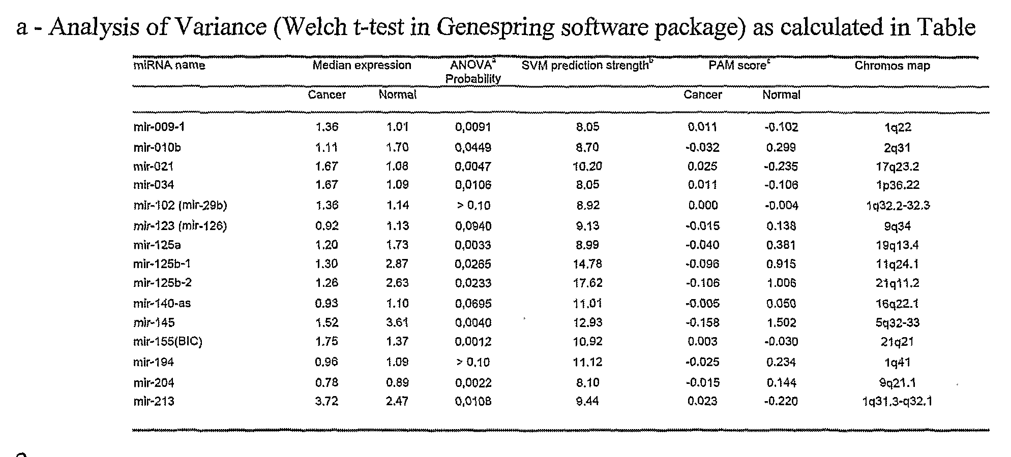
2. b - Support Vector Machine prediction analysis tool (from Genespring 7.2 software package).
Prediction strengths are calculated as negative natural log of the probability to predict the observed number of samples, in one of the two classes, by chance. The higher is the score, the best is the prediction strength. c - Centroid scores for the two classes of the Prediction Analysis of Microarrays (Tibshirani,
R., et al. Proc. Natl. Acad. Set U.S.A. 99:6567-6572 (2002)).
Of the 29 miRNAs whose expression is significantly (p<0.05) deregulated according to the microarray analysis, a set of 15 miRNAs were able to correctly predict the nature of the sample analyzed (i.e., normal vs. tumor) with 100% accuracy. Among the differentially- expressed miRNAs, miR-lOb, miR-125b, miR145, miR-21 and miR-155 were the most consistently deregulated miRNAs in breast cancer samples. Three of these, namely, miR-10b, miR~125b and miR-145, were down-regulated, while the remaining two, miR-21 and miR~ 155, were up-regulated, suggesting that they might act as tumor suppressor genes or oncogenes, respectively.
Example 2: Determination of putative gene targets of miRNAs that are deregulated in breast cancer tissues.
At present, the lack of knowledge about bonaβde miRNA gene targets hampers a full understanding of which biological functions are deregulated in cancers characterized by aberrant miRNA expression. To identify putative targets of the most significantly de-
regulated miRNAs from our study: miR-lOb, miR125b, miR-145, miR-21 and miR-155 (see Example 1), we utilized multiple computational approaches. In particular, the analysis was performed using three algorithms, miRanda, TargetScan and PicTar, which are commonly used to predict human miKNA gene targets (Enright, A.J., et al Genome Biol. J:R1 (2003); Lewis, B.P. et al, Cell 775:787-798 (2003); Krek, A., et al, Nat. Genet. 37:495-500 (2005)). The results obtained using each of the three algorithms were cross-referenced with one another to validate putative targets and only targets that were identified by at least 2 of the 3 algorithms were considered. Results of this analysis are presented in Table 4.
Several genes with potential oncogenic functions were identified as putative targets of miRNAs that are down-regulated in breast cancer samples. Notably, oncogenes were identified as targets of miR-10b (e.g., FLTl, the v-crk homolog, the growth factor BDNF and the transducing factor SHCl), miR-125b (e.g., YES, ETSl, TEL, AKT3, the growth factor receptor FGFR2 and members of the mitogen-activated signal transduction pathway VTS58635, MAP3K10, MAP3K11, MAPK14), and miR-145 (e.g., MYCN, FOS, YES and FLIl, integration site of Friend leukemia virus, cell cycle promoters, such as cyclins D2 and Ll, MAPK transduction proteins, such as MAP3K3 and MAP4K4 ). The proto-oncogene, YES, and the core-binding transcription factor, CBFB, were determined to be potential targets of both miR-125 and miR-145.
Consistent with these findings, multiple tumor suppressor genes were identified as targets of miR-21 and miR-155, miRNAs that are up-regulated in breast cancer cells. For miR-21, the TGFB gene was predicted as target by all three methods. For miR-155, potential targets included the tumor suppressor genes, SOCSl and APC, and the kinase, WEEl, which blocks the activity of Cdc2 and prevents entry into mitosis. The hypoxia inducible factor, HIFlA, was also a predicted target of miR-155. Notably, the tripartite motif-containing protein TRIM2, the proto-oncogene, SKI, and the RAS homologs, RAB6A and RAB6C, were found as potential targets of both miR-21 and miR-155.


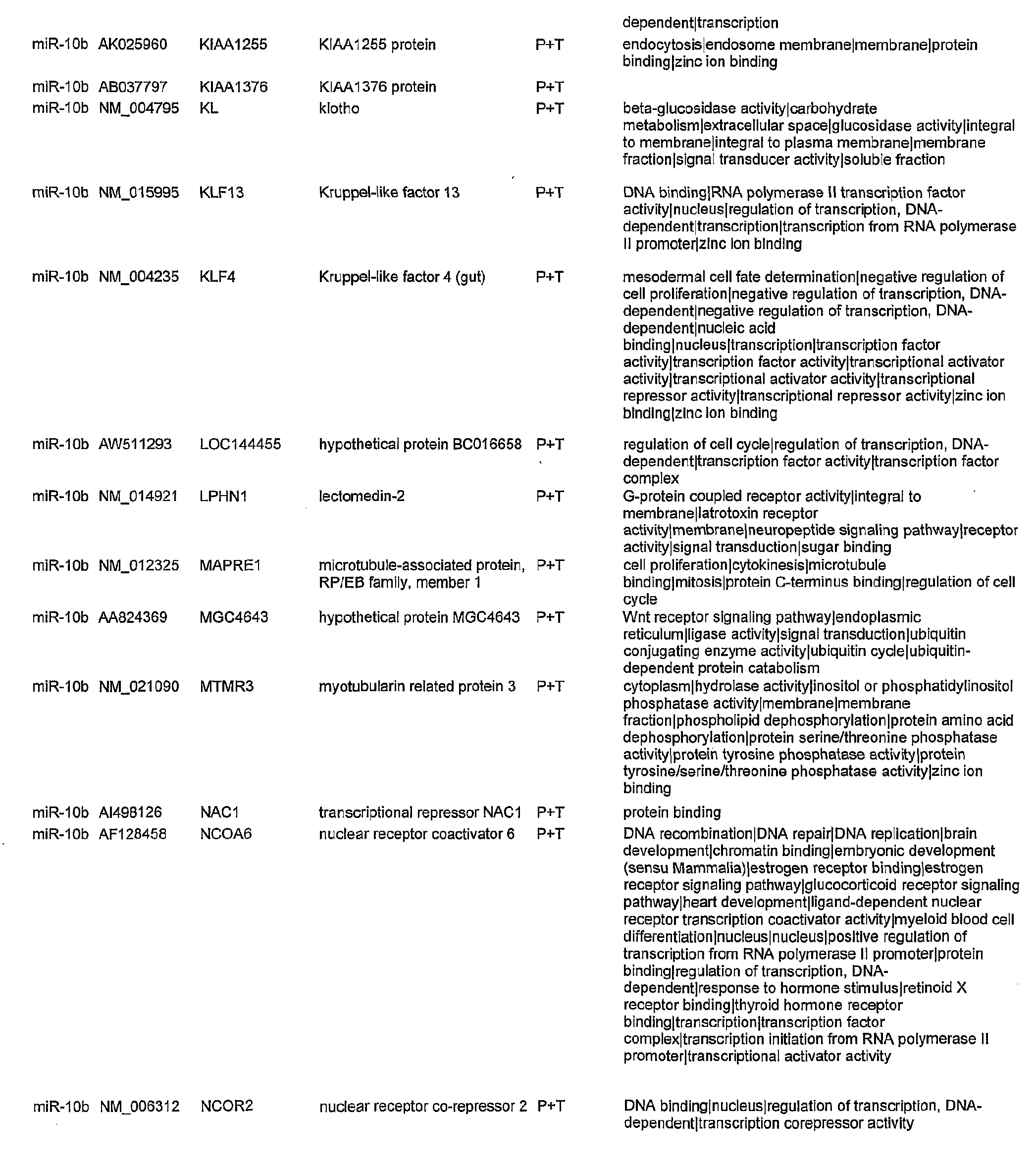


Example 3: Bio-pathological features and microRNA expression
Materials and Methods
5 Immunohistochemical analysis of breast cancer samples.
Staining procedures were performed as described (Querzoli, P., et ah, Anal. Quant. Cytol. Histol. 27:151-160 (1999)). Hormonal receptors were evaluated with
6Fl 1 antibody for estrogen receptor α (ER) and PGR-I A6 antibody for progesterone receptor (PR) (Ventana, Tucson, AZ, U.S.A.). The proliferation index was assessed with MIBl antibody (DAKO, Copenhagen). ERBB2 was detected with CBl 1 antibody (Ventana, Tucson, AZ, U.S.A.) and p53 protein expression was examined with DO7 antibody (Ventana, Tucson, AZ, U.S.A.). Only tumor cells with distinct nuclear immunostaining for ER, PR, Mibl and p53 were recorded as positive. Tumor cells were considered positive for ERBB2 when they showed distinct membrane immunoreactivity.
To perform a quantitative analysis of the expression of these various biological markers, the Eureka Menarini computerized image analysis system was used. For each tumor section, at least 20 microscopic fields of invasive carcinoma were measured using a 4Ox objective. The following cut-off values were employed: 10% of positive nuclear area for ER, PR, c-erbB2 and p53, 13% of nuclei expressing Mibl was introduced to discriminate cases with high and low proliferative activity.
Results
To evaluate whether a correlation exists between various bio-pathological features associated with breast cancer and the expression of particular miRNAs, we generated and compared miRNA expression profiles for various cancer samples associated with the presence or absence of a particular breast cancer feature. In particular, we analyzed breast cancers with lobular or ductal histotypes, breast cancers with differential expression of either estrogen receptor alpha (ER) or progesterone receptor, and breast cancers with differences in lymph node metastasis, vascular invasion, proliferation index, and expression of ERBB2 and p53. Expression profiles of lobular or ductal and +/- ERBB2 expression classes did not reveal any microRNAs that were differentially-expressed, while all other comparisons revealed a small number of differentially-expressed microRNAs (P<0.05). Tumor grade was not analyzed. The results of this analysis are shown in FIG. 4. Differentially-expressed miRNA families were identified for various bio- pathological features that are associated with human breast cancer. For example, all miR-30 miRNAs are down-regulated in both ER- and PR- tumors, suggesting that
expression of miR-30 miRNAs is regulated by these hormones. In addition, the expression of various let- 7 miRNAs was down-regulated in breast cancer samples with either lymph node metastasis or a high proliferation index, suggesting that reduced let-7 expression could be associated with a poor prognosis, a result that is consistent with previous findings (Takamizawa, J., et ah, Cancer Res. 39: 167-169
(2004)). The discovery that the let-7 family of miRNAs regulates the expression of members of the RAS oncogene family provides a potential explanation for the role of let-7 miRNAs in human cancer (Johnson, S.M., et al., Cell 120:635-647 (2005)). miR-145 and miR-21, two miRNAs whose expression could differentiate cancer or normal tissues, were also differentially-expressed in cancers with a different proliferation index or different tumor stage. In particular, miR-145 is progressively down-regulated from normal breast to cancers with a high proliferation index. Similarly, miR-21 is progressively up-regulated from normal breast to cancers with high tumor stage. These findings suggest that deregulation of these two miRNAs may affect critical molecular events involved in tumor progression.
Another miRNA potentially involved in cancer progression is miR-9-3. miR- 9-3 was downregulated in breast cancers with either high vascular invasion or lymph node metastasis, suggesting that its down-regulation was acquired during the course of tumor progression and, in particular, during the acquisition of metastatic potential.
The relevant teachings of all publications cited herein that have not explicitly been incorporated by reference, are incorporated herein by reference in their entirety. While this invention has been particularly shown and described with references to preferred embodiments thereof, it will be understood by those skilled in the art that various changes in form and details may be made therein without departing from the scope of the invention encompassed by the appended claims.
Claims
1. A method of diagnosing whether a subject has, or is at risk for developing, breast cancer, comprising measuring the level of at least one miR gene product in a test sample from said subject, wherein an alteration in the level of the miR gene product in the test sample, relative to the level of a corresponding miR gene product in a control sample, is indicative of the subject either having, or being at risk for developing, breast cancer.
2. The method of Claim 1 , wherein the at least one miR gene product is miR-
125b-l or miR125b-2.
3. The method of Claim 1 , wherein the at least one miR gene product is miR- 145.
4. The method of Claim 1 , wherein the at least one miR gene product is miR-21.
5. The method of Claim 1 , wherein the at least one miR gene product is miR- 155.
6. The method of Claim 1, wherein the at least one miR gene product is miR- 10b.
7. The method of Claim 1 , wherein the at least one miR gene product is selected from the group consisting of miR-125b, miR-145, miR-21, miR-155, miR-lOb, miR-009-1
(miR131-l), miR-34 (miR-170), miR-102 (miR-29b), miR-123 (miR-126), miR-140-as, miR- 125a, miR-125b-l, miR-125b-2, miR-194, miR-204, miR-213, let-7a-2, let-7a-3, let-7d (let- 7d-vl), let-7f-2, let-7i (Iet-7d-v2), miR-101-1, miR-122a, miR-128b, miR-136, miR-143, miR-149, miR-191, miR-196-1, miR-196-2, miR-202, miR-203, miR-206, miR-210 and combinations thereof.
8. The method of Claim 1 , wherein the level of the at least one miR gene product is measured using Northern blot analysis.
9. The method of Claim 1 , wherein the level of the at least one miR gene product in the test sample is less than the level of the corresponding miR gene product in the control sample.
10. The method of Claim 1 , wherein the level of the at least one miR gene product in the test sample is greater than the level of the corresponding miR gene product in the control sample.
11. A method of diagnosing a breast cancer associated with one or more prognostic markers in a subject, comprising measuring the level of at least one miR gene product in a breast cancer sample from said subject, wherein an alteration in the level of the at least one miR gene product in the test sample, relative to the level of a corresponding miR gene product in a control sample, is indicative of the subject having a breast cancer associated with the one or more prognostic markers.
12. The method of Claim 11 , wherein the one or more prognostic markers is selected from the group consisting of estrogen receptor expression, progesterone receptor expression, positive lymph node metastasis, a high proliferative index, detectable p53 expression, advanced tumor stage and high vascular invasion.
13. The method of Claim 11 , wherein the breast cancer associated with one or more prognostic markers and the at least one miR gene product are selected from the group consisting of:
(i) the breast cancer is a breast cancer associated with estrogen receptor expression and the miR gene product is selected from the group consisting of miR- 26a, miR-26b, miR-102 (miR-29b), miR-30a-5ρ, miR-30b, miR-30c, miR-30d, miR- 185, miR-191, miR-206, miR-212, and combinations thereof;
(ii) the breast cancer is a breast cancer associated with progesterone receptor expression and the miR gene product is selected from the group consisting of let-7c, miR-26a, miR-29b, miR-30a-5p, miR-30b, miR-30c, miR-30d, and combinations thereof;
(iii) the breast cancer is a breast cancer associated with positive lymph node metastasis and the miR gene product is selected from the group consisting of let- 7f- 1 , let-7a-3 , let-7a-2, miR-9-3 , and combinations thereof;
(iv) the breast cancer is a breast cancer associated with a high proliferative index and the miR gene product is selected from the group consisting of let-7c, let-7d, miR-26a, miR-26b, miR-30a-5p, miR-102, miR-145, and combinations thereof; (v) the breast cancer is a breast cancer associated with detectable p53 expression and the miR gene product is selected from the group consisting of miR-
16a, miR- 128b and a combination thereof;
(vi) the breast cancer is a breast cancer associated with high vascular invasion and the miR gene product is selected from the group consisting of miR-9-3, miR- 10b, miR-27a, miR-29a, miR- 123, miR-205 and combinations thereof; and (vii) the breast cancer is a breast cancer associated with an advanced tumor stage and the miR gene product is selected from the group consisting of miR-9-2, miR-15-a, miR-21, miR-30a-s, miR-133a-l, miR-137, miR-153-2, miR-154, miR- 181a, miR-203 , miR-213 , and combinations thereof.
14. A method of diagnosing whether a subject has, or is at risk for developing, breast cancer, comprising:
(1) reverse transcribing RNA from a test sample obtained from the subject to provide a set of target oligodeoxynucleotides;
(2) hybridizing the target oligodeoxynucleotides to a microarray comprising miRNA-specific probe oligonucleotides to provide a hybridization profile for the test sample; and
(3) comparing the test sample hybridization profile to a hybridization profile generated from a control sample, wherein an alteration in the signal of at least one miRNA is indicative of the subject either having, or being at risk for developing, breast cancer.
15. The method of Claim 14 wherein the signal of at least one miRNA, relative to the signal generated from the control sample, is down-regulated.
16. The method of Claim 14 wherein the signal of at least one miRNA, relative to the signal generated from the control sample is up-regulated.
17. The method of Claim 14 wherein the microarray comprises miRNA-specific probe oligonucleotides for one or more miRNAs selected from the group consisting of miR- 145, miR-21, miR-155, miR-lOb, miR-009-1 (miR131-l), miR-34 (miR-170), miR-102 (miR-29b), miR-123 (miR-126), miR-140-as, miR-125a, miR-125b-l, miR-125b-2, miR-194, miR-204, miR-213, let-7a-2, let-7a-3, let-7d (let-7d-vl), let-7f-2, let-7i (Iet-7d-v2), miR-101- 1, miR-122a, miR-128b, miR-136, miR-143, miR-149, miR-191, miR-196-1, miR-196-2, miR-202, miR-203, miR-206, miR-210 and combinations thereof.
18. A method of diagnosing whether a subj ect has, or is at risk for developing, a breast cancer associated with one or more adverse prognostic markers in a subject, comprising:
(1) reverse transcribing RNA from a test sample obtained from the subject to provide a set of target oligodeoxynucleotides; (2) hybridizing the target oligodeoxynucleotides to a microarray comprising miRNA-specific probe oligonucleotides to provide a hybridization profile for said test sample; and
(3) comparing the test sample hybridization profile to a hybridization profile generated from a control sample, wherein an alteration in the signal is indicative of the subject either having, or being at risk for developing, the cancer.
19. The method of Claim 18, wherein the one or more adverse prognostic markers is selected from the group consisting of estrogen receptor expression, progesterone receptor expression, positive lymph node metastasis, high proliferative index, detectable p53 expression, advanced tumor stage and high vascular invasion.
20. The method of Claim 18, wherein the microarray comprises at least one miRNA-specific probe oligonucleotide for a miRNA selected from the group consisting of miR-26a, miR-26b, miR-102 (miR-29b), miR-30a-5p, miR-30b, miR-30c, miR-30d, miR- 185, miR-191, miR-206, miR-212, let-7c, miR-9-2, miR-15-a, miR-21, miR-30a-s, miR- 133a-l, miR-137, miR-153-2, miR-154, miR-181a, miR-203, miR-213, let-7f-l, let-7a-3, let- 7a-2, miR-9-3, miR-10b, miR-27a, miR-29a, miR-123, miR-205, let-7d, miR-145, miR-16a, miR-128b and combinations thereof.
21. A method of treating breast cancer in a subject who has a breast cancer in which at least one miR gene product is down-regulated or up-regulated in the cancer cells of the subject relative to control cells, comprising:
(1) when the at least one miR gene product is down-regulated in the cancer cells, administering to the subject an effective amount of at least one isolated miR gene product, provided that the miR gene product is not miR-15a or miR-16-1, such that proliferation of cancer cells in the subject is inhibited; or
(2) when the at least one miR gene product is up-regulated in the cancer cells, administering to the subject an effective amount of at least one compound for inhibiting expression of the at least one miR gene product, such that proliferation of cancer cells in the subject is inhibited.
22. The method of Claim 21 , wherein the at least one isolated miR gene product in step (1) is selected from the group consisting of miR-145, miR-10b, miR-123 (miR-126), miR-140-as, miR-125a, miR-125b-l, miR-125b-2, miR-194, miR-204, let-7a-2, let-7a-3, let- 7d (let-7d-vl), let-7f-2, miR-101-1, miR-143 and combinations thereof.
23. The method of Claim 21 , wherein the at least one miR gene product in step (2) is selected from the group consisting of: miR-21, miR-155, miR-009-1 (miR131-l), miR-34 (miR-170), miR-102 (miR-29b), miR-213, let-7i (Iet-7d-v2), miR-122a, miR-128b, miR-136, miR-149, miR-191, miR-196-1, miR-196-2, miR-202, miR-203, miR-206, miR-210, miR- 213 and combinations thereof.
24. A method of treating breast cancer in a subject, comprising: (1) determining the amount of at least one miR gene product in breast cancer cells, relative to control cells; and
(2) altering the amount of miR gene product expressed in the breast cancer cells by: (i) administering to the subject an effective amount of at least one isolated miR gene product, provided that the miR gene product is not miR- 15a or miR- 16-1, if the amount of the miR gene product expressed in the cancer cells is less than the amount of the miR gene product expressed in control cells; or
(ii) administering to the subject an effective amount of at least one compound for inhibiting expression of the at least one miR gene product, if the amount of the miR gene product expressed in the cancer cells is greater than the amount of the miR gene product expressed in control cells, such that proliferation of cancer cells in the subject is inhibited.
25. The method of Claim 24, wherein the at least one isolated miR gene product in step (i) is selected from the group consisting of miR-145, miR-lOb, miR-123 (miR-126), miR-140-as, miR-125a, miR-125b-l, miR-125b-2, miR-194, miR-204, let-7a-2, let-7a-3, let- 7d (let-7d-vl), let-7f-2, miR-101-1, miR- 143 and combinations thereof.
26. The method of Claim 24, wherein the at least one miR gene product in step (ii) is selected from the group consisting of miR-21, miR-155, miR-009-1 (miR131-l), miR-34 (miR-170), miR- 102 (miR-29b), miR-213, let-7i (Iet-7d-v2), miR-122a, miR-128b, miR-136, miR-149, miR-191, miR-196-1, miR-196-2, miR-202, miR-203, miR-206, miR-210, miR- 213 and combinations thereof.
27. A pharmaceutical composition for treating breast cancer, comprising at least one isolated miR gene product and a pharmaceutically-acceptable carrier.
28. The pharmaceutical composition of Claim 27, wherein the at least one isolated miR gene product corresponds to a miR gene product that is down-regulated in breast cancer cells relative to suitable control cells.
29. The pharmaceutical composition of Claim 27, wherein the isolated miR gene product is selected from the group consisting of miR-145, miR-10b, miR-123 (miR-126), miR-140-as, miR-125a, miR-125b-l, miR-125b-2, miR-194, miR-204, let-7a-2, let-7a-3, let- 7d (let-7d-vl), let-7f-2, miR-101-1, miR-143 and combinations thereof.
30. A pharmaceutical composition for treating breast cancer, comprising at least one miR expression inhibitor compound and a pharmaceutically-acceptable carrier.
31. The pharmaceutical composition of Claim 30, wherein the at least one miR expression inhibitor compound is specific for a miR gene product that is up-regulated in breast cancer cells relative to suitable control cells.
32. The pharmaceutical composition of Claim 30, wherein the at least one miR expression inhibitor compound is specific for a miR gene product selected from the group consisting of miR-21, miR-155, miR-009-1 (miR131-1), miR-34 (miR-170), miR-102 (miR- 29b), miR-213, let-7i (Iet-7d-v2), miR-122a, miR-128b, miR-136, miR-149, miR-191, miR- 196-1, miR- 196-2, miR-202, miR-203, miR-206, miR-210, miR-213 and combinations thereof.
33. A method of identifying an anti-breast cancer agent, comprising providing a test agent to a cell and measuring the level of at least one miR gene product associated with decreased expression levels in breast cancer cells, wherein an increase in the level of the miR gene product in the cell, relative to a suitable control cell, is indicative of the test agent being an anti-breast cancer agent.
34. The method of Claim 33, wherein the miR gene product is selected from the group consisting of miR-145, miR-lOb, miR-123 (miR-126), miR-140-as, miR-125a, miR- 125b-l, miR-125b-2, miR-194, miR-204, let-7a-2, let-7a-3, let-7d (let-7d-vl), let-7f-2, miR- 101-1, miR-143 and combinations thereof.
35. A method of identifying an anti-breast cancer agent, comprising providing a test agent to a cell and measuring the level of at least one miR gene product associated with increased expression levels in breast cancer cells, wherein an decrease in the level of the miR gene product in the cell, relative to a suitable control cell, is indicative of the test agent being an anti-breast cancer agent.
36. The method of Claim 35, wherein the miR gene product is selected from the group consisting of miR-21, miR-155, miR-009-1 (miR131-l), miR-34 (miR-170), miR-102 (miR-29b), miR-213, let-7i (Iet-7d-v2), miR-122a, miR-128b, miR-136, miR-149, miR-191, miR- 196-1, miR- 196-2, miR-202, miR-203, miR-206, miR-210, miR-213 and combinations thereof.
Priority Applications (8)
| Application Number | Priority Date | Filing Date | Title |
|---|---|---|---|
| JP2008525107A JP5489459B2 (en) | 2005-08-01 | 2006-07-31 | MicroRNA-based methods and compositions for diagnosis, prognosis and treatment of breast cancer |
| ES06800599.0T ES2545383T3 (en) | 2005-08-01 | 2006-07-31 | Methods and compositions based on microRNA for the diagnosis, prognosis and treatment of breast cancer |
| CA002617581A CA2617581A1 (en) | 2005-08-01 | 2006-07-31 | Microrna-based methods for the diagnosis of breast cancer |
| CN2006800365983A CN101341259B (en) | 2005-08-01 | 2006-07-31 | Micro-rna-based methods and compositions for the diagnosis, prognosis and treatment of breast cancer |
| EP06800599.0A EP1937845B1 (en) | 2005-08-01 | 2006-07-31 | Micro-rna-based methods and compositions for the diagnosis, prognosis and treatment of breast cancer |
| US12/012,235 US8658370B2 (en) | 2005-08-01 | 2008-01-31 | MicroRNA-based methods and compositions for the diagnosis, prognosis and treatment of breast cancer |
| US14/145,364 US20140147495A1 (en) | 2005-08-01 | 2013-12-31 | MicroRNA-Based Methods and Compositions For the Diagnosis, Prognosis and Treatment of Breast Cancer |
| US15/155,479 US20160251659A1 (en) | 2005-08-01 | 2016-05-16 | MicroRNA-Based Methods and Compositions for the Diagnosis, Prognosis and Treatment of Breast Cancer |
Applications Claiming Priority (2)
| Application Number | Priority Date | Filing Date | Title |
|---|---|---|---|
| US70446405P | 2005-08-01 | 2005-08-01 | |
| US60/704,464 | 2005-08-01 |
Related Child Applications (1)
| Application Number | Title | Priority Date | Filing Date |
|---|---|---|---|
| US12/012,235 Continuation US8658370B2 (en) | 2005-08-01 | 2008-01-31 | MicroRNA-based methods and compositions for the diagnosis, prognosis and treatment of breast cancer |
Publications (2)
| Publication Number | Publication Date |
|---|---|
| WO2007016548A2 true WO2007016548A2 (en) | 2007-02-08 |
| WO2007016548A3 WO2007016548A3 (en) | 2007-08-30 |
Family
ID=37709309
Family Applications (1)
| Application Number | Title | Priority Date | Filing Date |
|---|---|---|---|
| PCT/US2006/029889 WO2007016548A2 (en) | 2005-08-01 | 2006-07-31 | Micro-rna-based methods and compositions for the diagnosis, prognosis and treatment of breast cancer |
Country Status (7)
| Country | Link |
|---|---|
| US (3) | US8658370B2 (en) |
| EP (1) | EP1937845B1 (en) |
| JP (3) | JP5489459B2 (en) |
| CN (7) | CN103820562B (en) |
| CA (1) | CA2617581A1 (en) |
| ES (1) | ES2545383T3 (en) |
| WO (1) | WO2007016548A2 (en) |
Cited By (59)
| Publication number | Priority date | Publication date | Assignee | Title |
|---|---|---|---|---|
| EP1969147A2 (en) * | 2006-01-05 | 2008-09-17 | The Ohio State University Research Foundation | Micrornarna-based methods and compositions for the diagnosis and treatment of solid cancers |
| EP1990414A1 (en) * | 2007-05-10 | 2008-11-12 | Fraunhofer-Gesellschaft zur Förderung der angewandten Forschung e.V. | Means for treating inflammatory diseases, autoimmune diseases and cancer |
| WO2009004632A2 (en) * | 2007-07-05 | 2009-01-08 | Yeda Research And Development Co. Ltd. | Methods of identifying components of a biological pathway and use of said components in regulating diseases associated with altered cell proliferation |
| WO2009021235A2 (en) * | 2007-08-09 | 2009-02-12 | The Regents Of The University Of Colorado | Methods and compositions for treating cancer |
| WO2008104974A3 (en) * | 2007-02-27 | 2009-03-05 | Rosetta Genomics Ltd | Composition and methods for modulating cell proliferation and cell death |
| US20090099034A1 (en) * | 2007-06-07 | 2009-04-16 | Wisconsin Alumni Research Foundation | Reagents and Methods for miRNA Expression Analysis and Identification of Cancer Biomarkers |
| WO2009086156A2 (en) * | 2007-12-21 | 2009-07-09 | Asuragen, Inc. | Mir-10 regulated genes and pathways as targets for therapeutic intervention |
| WO2010008114A1 (en) * | 2008-07-18 | 2010-01-21 | National Cancer Center | Anti-cancer composition comprising microrna molecules |
| WO2010012667A1 (en) * | 2008-08-01 | 2010-02-04 | Santaris Pharma A/S | Micro-rna mediated modulation of colony stimulating factors |
| EP2167521A1 (en) * | 2007-06-15 | 2010-03-31 | The Ohio State University Research Foundation | ONCOGENIC ALL-1 FUSION PROTEINS FOR TARGETING DROSHA-MEDIATED microRNA PROCESSING |
| WO2010043114A1 (en) * | 2008-10-13 | 2010-04-22 | 北京命码生科科技有限公司 | Use of serum/plasma microrna in early diagnosis of hbv infection and liver cancer |
| WO2010100328A1 (en) | 2009-03-06 | 2010-09-10 | Valtion Teknillinen Tutkimuskeskus | NUCLEIC ACIDS REGULATING OESTROGEN RECEPTOR (ER)-α SIGNALING IN BREAST CANCER |
| WO2010107397A1 (en) * | 2009-03-19 | 2010-09-23 | Agency For Science, Technology And Research | Modulators of apoptosis and the uses thereof |
| WO2010064248A3 (en) * | 2008-12-05 | 2010-11-25 | Yeda Research And Development Co. Ltd. | Methods of diagnosing and treating motor neuron diseases |
| CN102021169A (en) * | 2010-10-14 | 2011-04-20 | 南京大学 | Serum/plasma miRNA composition and use thereof |
| EP2326730A2 (en) * | 2008-08-19 | 2011-06-01 | Maine Institute For Human Genetics And Health | Micro rna (mirna) and neurofibromatosis type 1 : a role in diagnosis and therapy |
| WO2011110644A1 (en) * | 2010-03-11 | 2011-09-15 | National University Of Ireland, Galway | Detection and quantification of micrornas in the circulation and the use of circulating micrornas as biomarkers for cancer |
| WO2011128886A1 (en) * | 2010-04-12 | 2011-10-20 | Ramot At Tel Aviv University Ltd. | A micro-rna for cancer diagnosis, prognosis and therapy |
| CN102292456A (en) * | 2008-12-05 | 2011-12-21 | 俄亥俄州立大学研究基金会 | MicroRNA-based methods and compositions for the diagnosis and treatment of ovarian cancer |
| WO2012056033A1 (en) * | 2010-10-29 | 2012-05-03 | Axel Niendorf | Method for the analysis of a biological sample of a patient |
| US8202848B2 (en) | 2008-03-17 | 2012-06-19 | Board Of Regents, The University Of Texas System | Identification of micro-RNAS involved in neuromuscular synapse maintenance and regeneration |
| US8338104B2 (en) | 2006-07-13 | 2012-12-25 | The Ohio State University Research Foundation | Method of diagnosing poor survival prognosis colon cancer using miR-103-2 |
| US8354229B2 (en) | 2007-01-31 | 2013-01-15 | The Ohio State University Research Foundation | MiR-25-based methods for the diagnosis and prognosis of acute myeloid leukemia (AML) |
| US8361710B2 (en) | 2006-01-05 | 2013-01-29 | The Ohio State University Research Foundation | MicroRNA-based methods and compositions for the diagnosis, prognosis and treatment of lung cancer using miR-21 |
| US8367632B2 (en) | 2007-07-31 | 2013-02-05 | Ohio State University Research Foundation | Methods for reverting methylation by targeting methyltransferases |
| US8389210B2 (en) | 2006-01-05 | 2013-03-05 | The Ohio State University Research Foundation | MicroRNA expression abnormalities in pancreatic endocrine and acinar tumors |
| WO2013057567A1 (en) * | 2011-10-19 | 2013-04-25 | Council Of Scientific And Industrial Research (C.S.I.R.) | Biomarkers useful for detection of types, grades and stages of human breast cancer |
| US8440636B2 (en) | 2007-07-31 | 2013-05-14 | The Board Of Regents, The University Of Texas System | Micro-RNA family that modulates fibrosis and uses thereof |
| US8466119B2 (en) | 2007-08-22 | 2013-06-18 | The Ohio State University Research Foundation | Methods and compositions for inducing deregulation of EPHA7 and ERK phosphorylation in human acute leukemias |
| US8486913B2 (en) | 2006-03-20 | 2013-07-16 | The Ohio State University | MicroRNA fingerprints during human megakaryocytopoiesis |
| WO2013110053A1 (en) | 2012-01-20 | 2013-07-25 | The Ohio State University | Breast cancer biomarker signatures for invasiveness and prognosis |
| EP2627333A1 (en) * | 2010-10-15 | 2013-08-21 | Agency For Science, Technology And Research | Combination treatment of cancer |
| US8592384B2 (en) | 2005-04-04 | 2013-11-26 | The Board Of Regents Of The University Of Texas System | Micro-RNA's that regulate muscle cells |
| EP2683387A1 (en) * | 2011-03-07 | 2014-01-15 | The Ohio State University | MUTATOR ACTIVITY INDUCED BY MICRORNA-155 (miR-155) LINKS INFLAMMATION AND CANCER |
| US8658370B2 (en) | 2005-08-01 | 2014-02-25 | The Ohio State University Research Foundation | MicroRNA-based methods and compositions for the diagnosis, prognosis and treatment of breast cancer |
| US8765709B2 (en) | 2004-11-12 | 2014-07-01 | Asuragen, Inc. | Methods and compositions involving miRNA and miRNA inhibitor molecules |
| ES2481819A1 (en) * | 2014-06-12 | 2014-07-31 | Sistemas Genómicos, S.L. | Method of evaluation to evaluate a possibility of breast cancer (Machine-translation by Google Translate, not legally binding) |
| WO2014193309A1 (en) * | 2013-05-30 | 2014-12-04 | Agency For Science, Technology And Research | Method For Determining Cancer Prognosis |
| US8911998B2 (en) | 2007-10-26 | 2014-12-16 | The Ohio State University | Methods for identifying fragile histidine triad (FHIT) interaction and uses thereof |
| US8916533B2 (en) | 2009-11-23 | 2014-12-23 | The Ohio State University | Materials and methods useful for affecting tumor cell growth, migration and invasion |
| WO2014202090A1 (en) * | 2013-06-19 | 2014-12-24 | Syddansk Universitet | Circulating microrna based cancer biomarkers |
| US8946187B2 (en) | 2010-11-12 | 2015-02-03 | The Ohio State University | Materials and methods related to microRNA-21, mismatch repair, and colorectal cancer |
| WO2015035480A1 (en) * | 2013-09-11 | 2015-03-19 | Fundação Pio Xii - Hospital De Câncer De Barretos | Uses of at least one mirna, kit, methods for diagnosing breast cancer and methods for evaluating the risk of metastasis |
| US9006206B2 (en) | 2007-02-27 | 2015-04-14 | Rosetta Genomics Ltd. | Composition and methods for modulating cell proliferation and cell death |
| US9080215B2 (en) | 2007-09-14 | 2015-07-14 | Asuragen, Inc. | MicroRNAs differentially expressed in cervical cancer and uses thereof |
| US9085804B2 (en) | 2007-08-03 | 2015-07-21 | The Ohio State University Research Foundation | Ultraconserved regions encoding ncRNAs |
| CN104884097A (en) * | 2012-11-16 | 2015-09-02 | 得克萨斯州大学系统董事会 | MiRNA as novel therapeutic adjuvants and biomarkers for the prognosis and treatment of drug resistant breast cancers |
| US9125923B2 (en) | 2008-06-11 | 2015-09-08 | The Ohio State University | Use of MiR-26 family as a predictive marker for hepatocellular carcinoma and responsiveness to therapy |
| US9249468B2 (en) | 2011-10-14 | 2016-02-02 | The Ohio State University | Methods and materials related to ovarian cancer |
| US9365852B2 (en) | 2008-05-08 | 2016-06-14 | Mirna Therapeutics, Inc. | Compositions and methods related to miRNA modulation of neovascularization or angiogenesis |
| US9481885B2 (en) | 2011-12-13 | 2016-11-01 | Ohio State Innovation Foundation | Methods and compositions related to miR-21 and miR-29a, exosome inhibition, and cancer metastasis |
| CN106222270A (en) * | 2016-08-02 | 2016-12-14 | 滨州医学院 | A kind of breast carcinoma occurs or the diagnostic kit of transfer |
| US9644241B2 (en) | 2011-09-13 | 2017-05-09 | Interpace Diagnostics, Llc | Methods and compositions involving miR-135B for distinguishing pancreatic cancer from benign pancreatic disease |
| US9642872B2 (en) | 2010-09-30 | 2017-05-09 | University Of Zurich | Treatment of B-cell lymphoma with microRNA |
| EP3068884A4 (en) * | 2013-11-13 | 2017-06-14 | The Texas A&M University System | Micro-rnas that modulate lymphangiogenesis and inflammatory pathways in lymphatic vessel cells |
| US10047388B2 (en) | 2004-05-28 | 2018-08-14 | Asuragen, Inc. | Methods and compositions involving MicroRNA |
| US10758619B2 (en) | 2010-11-15 | 2020-09-01 | The Ohio State University | Controlled release mucoadhesive systems |
| EP3624825A4 (en) * | 2017-05-19 | 2021-03-10 | Georgia State University Research Foundation, Inc. | Recombinant oncolytic virus |
| US11584932B2 (en) | 2016-11-01 | 2023-02-21 | The Research Foundation For The State University Of New York | 5-halouracil-modified microRNAs and their use in the treatment of cancer |
Families Citing this family (74)
| Publication number | Priority date | Publication date | Assignee | Title |
|---|---|---|---|---|
| CA2554818A1 (en) | 2004-02-09 | 2005-08-25 | Thomas Jefferson University | Diagnosis and treatment of cancers with microrna located in or near cancer-associated chromosomal features |
| CN103028120B (en) | 2005-09-12 | 2015-08-12 | 俄亥俄州立大学研究基金会 | For diagnosing or treat compositions and the method for BCL2 associated cancer |
| US8071292B2 (en) | 2006-09-19 | 2011-12-06 | The Ohio State University Research Foundation | Leukemia diagnostic methods |
| EP2076599A2 (en) * | 2006-09-19 | 2009-07-08 | Asuragen, Inc. | Mir-200 regulated genes and pathways as targets for therapeutic intervention |
| JP5501766B2 (en) | 2006-11-01 | 2014-05-28 | ジ・オハイオ・ステイト・ユニバーシティ・リサーチ・ファウンデイション | MicroRNA expression signature to predict survival and metastasis in hepatocellular carcinoma |
| ES2536907T3 (en) | 2007-06-08 | 2015-05-29 | The Government Of The United States Of America As Represented By The Secretary Of The Department Of Health And Human Services | Methods to determine the subtype of a hepatocellular carcinoma |
| WO2009070805A2 (en) | 2007-12-01 | 2009-06-04 | Asuragen, Inc. | Mir-124 regulated genes and pathways as targets for therapeutic intervention |
| CA2742324A1 (en) * | 2008-10-30 | 2010-06-03 | Caris Life Sciences Luxembourg Holdings, S.A.R.L. | Methods for assessing rna patterns |
| CA2743211A1 (en) | 2008-11-12 | 2010-05-20 | Caris Life Sciences Luxembourg Holdings, S.A.R.L. | Methods and systems of using exosomes for determining phenotypes |
| WO2010061396A1 (en) * | 2008-11-30 | 2010-06-03 | Rosetta Genomics Ltd. | Methods for detecting an increased susceptibility to cancer |
| US20120014927A1 (en) | 2008-12-03 | 2012-01-19 | Tel Hashomer Medical Research Infrastructure And Services Ltd. | Methods and kits for determining predisposition to cancer |
| WO2010070637A2 (en) * | 2008-12-15 | 2010-06-24 | Rosetta Genomics Ltd. | Method for distinguishing between adrenal tumors |
| US8492133B2 (en) | 2009-01-20 | 2013-07-23 | Ramot At Tel Aviv University, Ltd. | MIR-21 promoter driven targeted cancer therapy |
| US20120190729A1 (en) * | 2009-07-27 | 2012-07-26 | The Regents Of The University Of Colorado A Body Corporate | Mirna inhibition of six1 expression |
| CN101987197B (en) * | 2009-07-31 | 2012-09-12 | 中国科学院上海生命科学研究院 | Method and reagent for inhibiting invasiveness of cancer cells |
| WO2011024918A1 (en) * | 2009-08-26 | 2011-03-03 | 国立大学法人大阪大学 | Antiangiogenic agent and method for inhibition of angiogenesis |
| CN101993892A (en) * | 2009-08-31 | 2011-03-30 | 长沙赢润生物技术有限公司 | Eukaryotic expression vector for expressing shRNA (short hairpin Ribonucleic Acid) in manner of targeting in cancer cells |
| CN102018963B (en) * | 2009-09-11 | 2013-03-13 | 中国科学院上海生命科学研究院 | MiR-125a for adjusting regulated on activation normal T cell expressed and secreted (RANTES) expressions as well as composition and application thereof |
| CN102078621B (en) * | 2009-11-27 | 2013-04-10 | 中国科学院上海生命科学研究院 | Method and composition of regulating and controlling plasmacytoid dendritic cell I type interferon expression |
| WO2011068865A1 (en) * | 2009-12-01 | 2011-06-09 | Board Of Trustees Of Southern Illinois University | Micro-rna-101 promotes estrogen-independent growth and confers tamoxifen resistance in er-positive cancer cells |
| US20130059906A1 (en) * | 2010-02-17 | 2013-03-07 | The Board Of Regents Of The University Of Texas System | Methods and compositions for influencing tumors using microrna-185 as a tumor suppressor |
| AU2011223789A1 (en) | 2010-03-01 | 2012-09-20 | Caris Life Sciences Switzerland Holdings Gmbh | Biomarkers for theranostics |
| KR20130043104A (en) | 2010-04-06 | 2013-04-29 | 카리스 라이프 사이언스 룩셈부르크 홀딩스 | Circulating biomarkers for disease |
| CA2795430A1 (en) * | 2010-04-08 | 2011-10-13 | Kyoto Prefectural Public University Corporation | Method for detecting rhabdomyosarcoma using sample derived from body fluid |
| CN101921760B (en) * | 2010-09-08 | 2012-02-01 | 南京医科大学 | Serum/plasma miRNA marker associated with breast cancer and application thereof |
| CN102146412B (en) * | 2011-01-11 | 2012-10-03 | 中山大学 | Small molecular non-coding RNA gene hsa-miR-29b and use thereof |
| CN103492566B (en) * | 2011-04-25 | 2017-06-13 | 东丽株式会社 | Therapeutic sensitivity prediction composition and method of the patients with mastocarcinoma to Herceptin |
| WO2012177565A2 (en) * | 2011-06-20 | 2012-12-27 | California Institute Of Technology | Regulation of macrophage activation using mir - 125b |
| KR20140064899A (en) | 2011-09-16 | 2014-05-28 | 엘에스아이피 환도 운에이고우도우가이샤 | Method for detecting bladder cancer cells, primer used in method for detecting bladder cancer cells, and bladder cancer marker |
| US9745578B2 (en) | 2011-11-30 | 2017-08-29 | Cedars-Sinai Medical Center | Targeting microRNA miR-409-3P to treat prostate cancer |
| CN104080461A (en) * | 2011-11-30 | 2014-10-01 | 雪松-西奈医学中心 | Targeting micro RNAS MIR-409-5P, MIR-379 AND MIR-154* to treat prostate cancer bone metastasis and drug resistant lung cancer |
| ITRM20110685A1 (en) | 2011-12-23 | 2013-06-24 | Internat Ct For Genetic En Gineering And | MICRORNA FOR CARDIAC REGENERATION THROUGH THE INDUCTION OF THE PROLIFERATION OF CARDIAC MYCYCLES |
| US9334498B2 (en) | 2012-05-10 | 2016-05-10 | Uab Research Foundation | Methods and compositions for modulating MIR-204 activity |
| CA2880762A1 (en) * | 2012-08-01 | 2014-02-06 | Pacylex Pharmaceuticals Inc. | Methods and compositions for diagnosis and prognosis in breast cancer |
| CN103275979B (en) * | 2012-10-25 | 2014-12-17 | 北京迈誉森生物科技有限公司 | MicroRNA (Ribose Nucleic Acid) marker for detecting breast cancer and application thereof in kit |
| CN103773764A (en) * | 2012-12-27 | 2014-05-07 | 山东省眼科研究所 | miRNA and application of miRNA in preparation of product for diagnosing and treating posterior capsule opacification |
| US10793913B2 (en) * | 2013-03-08 | 2020-10-06 | City Of Hope | Methods and compositions for treatment of breast cancer |
| CN103432595B (en) * | 2013-08-19 | 2015-06-10 | 南京市妇幼保健院 | Application of human miR-26b in preparation of drugs for inhibiting fat cell proliferation |
| CN104164485B (en) * | 2014-06-09 | 2016-06-01 | 济南金域医学检验中心有限公司 | The application of the reagent of detection miR-129-2 methyl in preparation detection breast cancer kit |
| CN105586390B (en) * | 2014-10-23 | 2019-01-22 | 王辉云 | A kind of kit for Prognosis in Breast Cancer judgement |
| ES2838923T3 (en) * | 2014-10-24 | 2021-07-02 | Koninklijke Philips Nv | Medical prognosis and prediction of response to treatment using multiple activities of the cell signaling pathway |
| CN104502601B (en) * | 2014-12-03 | 2016-04-06 | 上海交通大学医学院附属上海儿童医学中心 | SCN3A closes or open mark as diagnosis arterial duct |
| CN104611429B (en) * | 2015-01-21 | 2017-02-22 | 湖州市中心医院 | Application of miR-10b (micro ribonucleic acid-10b) gene to regulation of gastric cancer gene expression |
| CN104630216A (en) * | 2015-01-23 | 2015-05-20 | 北京大学第一医院 | Radionuclide marked microRNA-155 targeting probe and application thereof in tumor imaging |
| CN104630377B (en) * | 2015-02-28 | 2017-11-17 | 上海赛安生物医药科技股份有限公司 | A kind of application of breast cancer miRNA detection kits and its miRNA |
| CN108138180A (en) | 2015-06-05 | 2018-06-08 | 米拉根医疗股份有限公司 | For treating skin T cell lymphoma(CTCL)MIR-155 inhibitor |
| US20160369344A1 (en) * | 2015-06-16 | 2016-12-22 | Emory University | Detecting MicroRNA and Iso-MicroRNA to Evaluate Vascular and Cardiac Health |
| CN105200043B (en) * | 2015-06-26 | 2018-08-03 | 宋尔卫 | A kind of kit for assessing Prognosis in Breast Cancer risk |
| US20170349955A1 (en) * | 2015-11-25 | 2017-12-07 | Kaohsiung Medical University | Mir-125a-5p as a biomarker for breast cancer |
| US20170145421A1 (en) * | 2015-11-25 | 2017-05-25 | Kaohsiung Medical Univeristy | Mir-125a-5p as a biomarker for breast cancer |
| MX2015016721A (en) * | 2015-12-04 | 2017-06-05 | Inst Nac De Medicina Genómica | Breast cancer prognosis method. |
| CN109072481B (en) * | 2015-12-07 | 2022-03-18 | 安大略省癌症研究所 | Genetic characterization of residual risk after endocrine treatment of early breast cancer |
| CN107299129B (en) * | 2016-04-15 | 2021-07-27 | 昆山市工业技术研究院小核酸生物技术研究所有限责任公司 | Application of circulating nucleic acid as breast cancer biomarker |
| US11236337B2 (en) | 2016-11-01 | 2022-02-01 | The Research Foundation For The State University Of New York | 5-halouracil-modified microRNAs and their use in the treatment of cancer |
| CN107064511B (en) * | 2017-06-09 | 2020-07-24 | 南京安吉生物科技有限公司 | Tumor marker TMEM170B protein and application thereof in preparation of tumor inhibition products |
| JP7298913B2 (en) | 2017-12-13 | 2023-06-27 | 国立大学法人広島大学 | Methods to Aid in Breast Cancer Detection |
| WO2019227257A1 (en) * | 2018-05-26 | 2019-12-05 | 深圳市博奥康生物科技有限公司 | Construction and application of mirna-210 overexpression vector |
| WO2019227254A1 (en) * | 2018-05-26 | 2019-12-05 | 深圳市博奥康生物科技有限公司 | Construction of mirna-25 overexpression vector and use thereof |
| CN108588226A (en) * | 2018-06-22 | 2018-09-28 | 南阳师范学院 | Detect the miRNA combination of breast cancer patients with brain transfer and the kit containing the combination |
| CN109136224B (en) * | 2018-08-21 | 2022-03-18 | 上海交通大学医学院附属瑞金医院 | miR-221/222 and inhibitor thereof for preparing medicine for regulating liver fat deposition, liver fibrosis and hepatocellular carcinoma |
| CN109439748A (en) * | 2018-09-25 | 2019-03-08 | 广州医科大学附属第三医院(广州重症孕产妇救治中心、广州柔济医院) | Application of the miR-9-5p as breast cancer DISTANT METASTASES IN monitoring marker |
| CN109321656B (en) * | 2018-10-22 | 2021-10-01 | 上海交通大学医学院附属仁济医院 | Use of protein DEPDC1 as marker for diagnosing triple-negative breast cancer |
| CN110029107B (en) * | 2019-04-22 | 2020-04-21 | 深圳市人民医院 | Oligonucleotides targeting SNHG17 for treating breast cancer |
| CN110423806A (en) * | 2019-08-15 | 2019-11-08 | 宁夏医科大学 | One kind miRNA marker relevant to AS plaque inflammation and its screening technique and application |
| CN110484605B (en) * | 2019-09-23 | 2020-07-28 | 中国人民解放军陆军军医大学第一附属医院 | Gene for in-situ detection of microRNA-34 by living cells and preparation method and application thereof |
| CN111518884B (en) * | 2020-04-08 | 2022-02-18 | 中国医学科学院医药生物技术研究所 | Application of miRNA30 cluster as Alzheimer disease diagnostic marker |
| CN111514460B (en) * | 2020-05-22 | 2021-12-14 | 西安交通大学 | Application of atmospheric pressure cold plasma in inhibiting activity of glutaminase and enzyme inhibitor |
| CN111518914B (en) * | 2020-06-09 | 2021-12-24 | 上海圣梓茂生物科技有限公司 | MiRNA marker combination, kit and method for detecting breast cancer |
| CN111850047B (en) * | 2020-07-28 | 2022-08-12 | 枣庄学院 | miR-16 and miR-30c combined expression vector and construction method and application thereof |
| CN111910006A (en) * | 2020-08-20 | 2020-11-10 | 广州医科大学附属第二医院 | Diagnosis and treatment target point circ PLCL2 of prostatic cancer and bone metastasis |
| CN113481296A (en) * | 2021-05-12 | 2021-10-08 | 广东省人民医院 | Diagnostic kit for early evaluation of breast cancer risk |
| CN114214363B (en) * | 2021-12-03 | 2024-04-19 | 浙江大学 | Anti-mesenchymal stem cell aging modification method and application thereof |
| CN115521899A (en) * | 2022-05-06 | 2022-12-27 | 西南大学 | An efficient Japanese green culture cell CRISPR/Cas9 genome editing method and application thereof |
| CN117604107B (en) * | 2024-01-23 | 2024-04-12 | 杭州华得森生物技术有限公司 | Use of FOXO6 and SNX4 in combination for diagnosing breast cancer |
Family Cites Families (157)
| Publication number | Priority date | Publication date | Assignee | Title |
|---|---|---|---|---|
| US4196265A (en) * | 1977-06-15 | 1980-04-01 | The Wistar Institute | Method of producing antibodies |
| US4172124A (en) | 1978-04-28 | 1979-10-23 | The Wistar Institute | Method of producing tumor antibodies |
| US4608337A (en) | 1980-11-07 | 1986-08-26 | The Wistar Institute | Human hybridomas and the production of human monoclonal antibodies by human hybridomas |
| US4701409A (en) | 1984-11-15 | 1987-10-20 | The Wistar Institute | Detection of B-cell neoplasms |
| US4693975A (en) | 1984-11-20 | 1987-09-15 | The Wistar Institute | Human hybridroma fusion partner for production of human monoclonal antibodies |
| US5015568A (en) | 1986-07-09 | 1991-05-14 | The Wistar Institute | Diagnostic methods for detecting lymphomas in humans |
| US5202429A (en) * | 1986-07-09 | 1993-04-13 | The Wistar Institute | DNA molecules having human BCL-2 gene sequences |
| US5198338A (en) * | 1989-05-31 | 1993-03-30 | Temple University | Molecular probing for human t-cell leukemia and lymphoma |
| US5149628A (en) | 1989-11-15 | 1992-09-22 | Temple University | Methods for detecting bcl-3 gene in human leukemias |
| US5633135A (en) * | 1991-12-11 | 1997-05-27 | Thomas Jefferson University | Chimeric nucleic acids and proteins resulting from ALL-1 region chromosome abnormalities |
| WO1993012136A1 (en) * | 1991-12-11 | 1993-06-24 | Thomas Jefferson University | Detection and treatment of acute leukemias resulting from chromosome abnormalities in the all-1 region |
| US6040140A (en) * | 1991-12-11 | 2000-03-21 | Thomas Jefferson University | Methods for screening and treating leukemias resulting from all-1 region chromosome abnormalities |
| US5674682A (en) | 1992-10-29 | 1997-10-07 | Thomas Jefferson University | Nucleic acid primers for detecting micrometastasis of prostate cancer |
| JPH08502889A (en) * | 1992-10-29 | 1996-04-02 | トーマス・ジェファーソン・ユニバーシティ | Method of detecting micrometastases of prostate cancer |
| US5985598A (en) * | 1994-10-27 | 1999-11-16 | Thomas Jefferson University | TCL-1 gene and protein and related methods and compositions |
| US7175995B1 (en) * | 1994-10-27 | 2007-02-13 | Thomas Jefferson University | TCL-1 protein and related methods |
| US5695944A (en) | 1995-05-05 | 1997-12-09 | Thomas Jefferson University | Modulation of bcl-2 phosphorylation |
| US5567586A (en) | 1995-05-18 | 1996-10-22 | Thomas Jefferson University | Methods of indentifying solid tumors with chromosome abnormalities in the ALL-1 region |
| US5928884A (en) * | 1996-02-09 | 1999-07-27 | Croce; Carlo M. | FHIT proteins and nucleic acids and methods based thereon |
| US6242212B1 (en) | 1996-02-09 | 2001-06-05 | Thomas Jefferson University | Fragile histidine triad (FHIT) nucleic acids and methods of producing FHIT proteins |
| AU6659298A (en) * | 1997-02-18 | 1998-09-08 | Thomas Jefferson University | Compositions that bind to pancreatic cancer cells and methods of using the same |
| US6258541B1 (en) * | 1997-04-04 | 2001-07-10 | Texas A&M University | Noninvasive detection of colonic biomarkers using fecal messenger RNA |
| CA2335315A1 (en) * | 1998-07-20 | 2000-01-27 | Thomas Jefferson University | Nitrilase homologs |
| US6255293B1 (en) * | 1998-07-24 | 2001-07-03 | Yeda Research And Development Co., Ltd. | Prevention of metastasis with 5-aza-2′-deoxycytidine |
| US7141417B1 (en) | 1999-02-25 | 2006-11-28 | Thomas Jefferson University | Compositions, kits, and methods relating to the human FEZ1 gene, a novel tumor suppressor gene |
| JP2003519463A (en) | 1999-03-15 | 2003-06-24 | トーマス・ジェファーソン・ユニバーシティー | TCL-1b gene, protein, methods related thereto and substances related thereto |
| US7163801B2 (en) | 1999-09-01 | 2007-01-16 | The Burnham Institute | Methods for determining the prognosis for cancer patients using tucan |
| US6891031B2 (en) | 2000-02-18 | 2005-05-10 | The Regents Of The University Of California | Coordinate cytokine regulatory sequences |
| US20010026796A1 (en) | 2000-03-14 | 2001-10-04 | Croce Carlo M. | TCL1 enhances Akt kinase activity and mediates its nuclear translocation |
| CA2406366A1 (en) | 2000-04-11 | 2001-10-18 | Thomas Jefferson University | Muir-torre-like syndrome infhit deficient mice |
| US20020086331A1 (en) * | 2000-05-16 | 2002-07-04 | Carlo Croce | Crystal structure of worm NitFhit reveals that a Nit tetramer binds two Fhit dimers |
| US7060811B2 (en) * | 2000-10-13 | 2006-06-13 | Board Of Regents, The University Of Texas System | WWOX: a tumor suppressor gene mutated in multiple cancers |
| US20040033502A1 (en) * | 2001-03-28 | 2004-02-19 | Amanda Williams | Gene expression profiles in esophageal tissue |
| US20050176025A1 (en) | 2001-05-18 | 2005-08-11 | Sirna Therapeutics, Inc. | RNA interference mediated inhibition of B-cell CLL/Lymphoma-2 (BCL-2) gene expression using short interfering nucleic acid (siNA) |
| EP2386637B1 (en) * | 2001-09-28 | 2018-05-16 | Max-Planck-Gesellschaft zur Förderung der Wissenschaften e.V. | Microrna molecules |
| US7371736B2 (en) * | 2001-11-07 | 2008-05-13 | The Board Of Trustees Of The University Of Arkansas | Gene expression profiling based identification of DKK1 as a potential therapeutic targets for controlling bone loss |
| GB0128898D0 (en) * | 2001-12-03 | 2002-01-23 | Biotech Res Ventures Pte Ltd | Materials and methods relating to the stabilization and activation of a tumour suppressor protein |
| JP4575667B2 (en) | 2002-03-08 | 2010-11-04 | エーザイ・アール・アンド・ディー・マネジメント株式会社 | Macrocyclic compounds useful as pharmaceuticals |
| WO2003086445A1 (en) * | 2002-04-08 | 2003-10-23 | Ciphergen Biosystems, Inc. | Serum biomarkers in hepatocellular carcinoma |
| US20040078834A1 (en) * | 2002-04-29 | 2004-04-22 | Croce Carlo M. | Human chronic lymphocytic leukemia modeled in mouse by targeted TCL1 expression |
| JP2005528115A (en) * | 2002-05-31 | 2005-09-22 | ザ ボード オブ トラスティーズ オブ ザ リーランド スタンフォード ジュニア ユニバーシティ | Methods for identifying and isolating stem cells and cancer stem cells |
| US20050260639A1 (en) | 2002-09-30 | 2005-11-24 | Oncotherapy Science, Inc. | Method for diagnosing pancreatic cancer |
| CA2501602A1 (en) * | 2002-10-11 | 2004-04-22 | Thomas Jefferson University | Novel tumor suppressor gene and compositions and methods for making and using the same |
| US20050266443A1 (en) | 2002-10-11 | 2005-12-01 | Thomas Jefferson University | Novel tumor suppressor gene and compositions and methods for making and using the same |
| ATE546528T1 (en) * | 2002-11-13 | 2012-03-15 | Univ Jefferson | COMPOSITIONS AND METHODS FOR DIAGNOSIS AND TREATMENT OF CANCER |
| US7250496B2 (en) | 2002-11-14 | 2007-07-31 | Rosetta Genomics Ltd. | Bioinformatically detectable group of novel regulatory genes and uses thereof |
| WO2004071464A2 (en) * | 2003-02-12 | 2004-08-26 | Johns Hopkins University School Of Medicine | Diagnostic application of differentially-expressed genes in lympho-hematopoietic stem cells |
| WO2004079013A1 (en) | 2003-03-03 | 2004-09-16 | Arizona Board Of Regents On Behalf Of The University Of Arizona | Ecto-5’-nucleotidase (cd73) used in the diagnosis and the treatment of pancreatic cancer |
| US7183384B2 (en) | 2003-03-06 | 2007-02-27 | A & G Pharmaceutical, Inc. | Monoclonal antibody 7H11 reactive with human cancer |
| AU2003286741A1 (en) | 2003-05-02 | 2004-11-26 | Thomas Jefferson University | Methods and compositions for diagnosis and therapy of parkin-associated disorders |
| US20050069918A1 (en) * | 2003-05-29 | 2005-03-31 | Francois Claret | JAB1 as a prognostic marker and a therapeutic target for human cancer |
| JP3934673B1 (en) | 2003-06-18 | 2007-06-20 | ジェネラックス・コーポレイション | Modified recombinant vaccinia virus and other microorganisms, use thereof |
| CA2533701A1 (en) | 2003-07-31 | 2005-02-17 | Isis Pharmaceuticals, Inc. | Oligomeric compounds and compositions for use in modulation of small non-coding rnas |
| WO2005047505A2 (en) * | 2003-08-07 | 2005-05-26 | Whitehead Institute For Biomedical Research | Methods and products for expression of micro rnas |
| US20050037362A1 (en) * | 2003-08-11 | 2005-02-17 | Eppendorf Array Technologies, S.A. | Detection and quantification of siRNA on microarrays |
| US8412541B2 (en) | 2003-08-14 | 2013-04-02 | Edda Technology, Inc. | Method and system for intelligent qualitative and quantitative analysis for medical diagnosis |
| WO2005020795A2 (en) | 2003-08-25 | 2005-03-10 | The Johns Hopkins University | Method of diagnosis and treatment of pancreatic endocrine neoplasms based on differntial gene expression analysis |
| AU2004276823A1 (en) | 2003-09-22 | 2005-04-07 | Merck And Co., Inc | Synthetic lethal screen using RNA interference |
| CN1890382A (en) * | 2003-09-24 | 2007-01-03 | 肿瘤疗法科学股份有限公司 | Method for diagnosing hepatocellular carcinomas |
| US20050186589A1 (en) | 2003-11-07 | 2005-08-25 | University Of Massachusetts | Interspersed repetitive element RNAs as substrates, inhibitors and delivery vehicles for RNAi |
| US20050164252A1 (en) * | 2003-12-04 | 2005-07-28 | Yeung Wah Hin A. | Methods using non-genic sequences for the detection, modification and treatment of any disease or improvement of functions of a cell |
| WO2005060661A2 (en) | 2003-12-19 | 2005-07-07 | The Regents Of The University Of California | Methods and materials for assessing prostate cancer therapies |
| CA2554818A1 (en) | 2004-02-09 | 2005-08-25 | Thomas Jefferson University | Diagnosis and treatment of cancers with microrna located in or near cancer-associated chromosomal features |
| US20050256072A1 (en) | 2004-02-09 | 2005-11-17 | University Of Massachusetts | Dual functional oligonucleotides for use in repressing mutant gene expression |
| WO2005079397A2 (en) | 2004-02-13 | 2005-09-01 | Rockefeller University | Anti-microrna oligonucleotide molecules |
| US20050182005A1 (en) * | 2004-02-13 | 2005-08-18 | Tuschl Thomas H. | Anti-microRNA oligonucleotide molecules |
| WO2005080601A2 (en) | 2004-02-23 | 2005-09-01 | Erasmus Universiteit Rotterdam | Classification, diagnosis and prognosis of acute myeloid leukemia by gene expression profiling |
| US7365058B2 (en) | 2004-04-13 | 2008-04-29 | The Rockefeller University | MicroRNA and methods for inhibiting same |
| CN101056991A (en) * | 2004-04-20 | 2007-10-17 | 基因耐克生物医学制品有限公司 | Method for detecting ncRNA |
| JP5697297B2 (en) * | 2004-05-14 | 2015-04-08 | ロゼッタ ジノミクス リミテッド | Micro NAS and its use |
| EP1771563A2 (en) * | 2004-05-28 | 2007-04-11 | Ambion, Inc. | METHODS AND COMPOSITIONS INVOLVING MicroRNA |
| US7635563B2 (en) * | 2004-06-30 | 2009-12-22 | Massachusetts Institute Of Technology | High throughput methods relating to microRNA expression analysis |
| US20060037088A1 (en) * | 2004-08-13 | 2006-02-16 | Shulin Li | Gene expression levels as predictors of chemoradiation response of cancer |
| EP2338994B1 (en) * | 2004-09-02 | 2014-03-19 | Yale University | Regulation of oncogenes by microRNAs |
| US7592441B2 (en) | 2004-10-04 | 2009-09-22 | Rosetta Genomics Ltd | Liver cancer-related nucleic acids |
| US7642348B2 (en) * | 2004-10-04 | 2010-01-05 | Rosetta Genomics Ltd | Prostate cancer-related nucleic acids |
| FR2877350B1 (en) * | 2004-11-03 | 2010-08-27 | Centre Nat Rech Scient | IDENTIFICATION AND USE OF miRNAs INVOLVED IN THE DIFFERENTIATION OF CELLS FROM MYELOID LEUKEMIA |
| ES2534301T3 (en) * | 2004-11-12 | 2015-04-21 | Asuragen, Inc. | Procedures and compositions involving miRNA and miRNA inhibitor molecules |
| AU2005316384B2 (en) * | 2004-12-14 | 2012-02-09 | Alnylam Pharmaceuticals, Inc. | RNAi modulation of MLL-AF4 and uses thereof |
| US20060185027A1 (en) * | 2004-12-23 | 2006-08-17 | David Bartel | Systems and methods for identifying miRNA targets and for altering miRNA and target expression |
| US20070099196A1 (en) * | 2004-12-29 | 2007-05-03 | Sakari Kauppinen | Novel oligonucleotide compositions and probe sequences useful for detection and analysis of micrornas and their target mRNAs |
| US8071306B2 (en) | 2005-01-25 | 2011-12-06 | Merck Sharp & Dohme Corp. | Methods for quantitating small RNA molecules |
| DE602006016739D1 (en) * | 2005-01-25 | 2010-10-21 | Rosetta Inpharmatics Llc | METHOD FOR QUANTIFYING SMALL RNA MOLECULES |
| US20070065840A1 (en) * | 2005-03-23 | 2007-03-22 | Irena Naguibneva | Novel oligonucleotide compositions and probe sequences useful for detection and analysis of microRNAS and their target mRNAS |
| GB2425311A (en) | 2005-04-15 | 2006-10-25 | Ist Superiore Sanita | Micro RNA against kit protein |
| CA2605701C (en) | 2005-04-29 | 2015-12-08 | Rockefeller University | Human micrornas and methods for inhibiting same |
| US20090209621A1 (en) | 2005-06-03 | 2009-08-20 | The Johns Hopkins University | Compositions and methods for decreasing microrna expression for the treatment of neoplasia |
| US20070065844A1 (en) * | 2005-06-08 | 2007-03-22 | Massachusetts Institute Of Technology | Solution-based methods for RNA expression profiling |
| US20060292616A1 (en) * | 2005-06-23 | 2006-12-28 | U.S. Genomics, Inc. | Single molecule miRNA-based disease diagnostic methods |
| ES2545383T3 (en) | 2005-08-01 | 2015-09-10 | The Ohio State University Research Foundation | Methods and compositions based on microRNA for the diagnosis, prognosis and treatment of breast cancer |
| US20070123482A1 (en) * | 2005-08-10 | 2007-05-31 | Markus Stoffel | Chemically modified oligonucleotides for use in modulating micro RNA and uses thereof |
| US20070213292A1 (en) | 2005-08-10 | 2007-09-13 | The Rockefeller University | Chemically modified oligonucleotides for use in modulating micro RNA and uses thereof |
| CN103028120B (en) * | 2005-09-12 | 2015-08-12 | 俄亥俄州立大学研究基金会 | For diagnosing or treat compositions and the method for BCL2 associated cancer |
| EP1940456A4 (en) | 2005-10-05 | 2009-10-21 | Univ Ohio State Res Found | Wwox gene, vectors containing the same, and uses in treatment of cancer |
| US7390792B2 (en) * | 2005-12-15 | 2008-06-24 | Board Of Regents, The University Of Texas System | MicroRNA1 therapies |
| CN101400361B (en) | 2006-01-05 | 2012-10-17 | 俄亥俄州立大学研究基金会 | Microrna-based methods and compositions for the diagnosis, prognosis and treatment of lung cancer |
| AU2007205257B2 (en) | 2006-01-05 | 2013-07-25 | The Ohio State University Research Foundation | MicroRNA expression abnormalities in pancreatic endocrine and acinar tumors |
| EP2487257B1 (en) | 2006-01-05 | 2015-07-01 | The Ohio State University Research Foundation | MicroRNA-based methods and compositions for the diagnosis and treatment of solid cancers |
| WO2008051260A1 (en) | 2006-01-13 | 2008-05-02 | Battelle Memorial Institute | Methods for assessing copd-retlated diseases |
| US7985584B2 (en) | 2006-03-20 | 2011-07-26 | The Ohio State University Research Foundation | MicroRNA fingerprints during human megakaryocytopoiesis |
| WO2007112097A2 (en) | 2006-03-24 | 2007-10-04 | Children's Medical Center Corporation | Novel signature self renewal gene expression programs |
| AU2007234191B2 (en) | 2006-04-03 | 2012-07-12 | Roche Innovation Center Copenhagen A/S | Pharmaceutical composition comprising anti-miRNA antisense oligonucleotides |
| JP5127821B2 (en) * | 2006-04-24 | 2013-01-23 | ジ・オハイオ・ステイト・ユニバーシティ・リサーチ・ファウンデイション | Pre-B cell proliferation and lymphoblastic leukemia / high-grade lymphoma in miR155 transgenic mice |
| EP2041317A4 (en) * | 2006-07-13 | 2009-10-14 | Univ Ohio State Res Found | Micro-rna-based methods and compositions for the diagnosis and treatment of colon cancer-related diseases |
| ZA200901224B (en) | 2006-08-30 | 2010-05-26 | Univ Michigan | New small molecule inhibitors of MDM2 the uses thereof |
| US20080193943A1 (en) | 2006-09-05 | 2008-08-14 | Abbott Laboratories | Companion diagnostic assays for cancer therapy |
| US8071292B2 (en) * | 2006-09-19 | 2011-12-06 | The Ohio State University Research Foundation | Leukemia diagnostic methods |
| CA2663962A1 (en) * | 2006-09-19 | 2008-03-27 | Asuragen, Inc. | Mir-15, mir-26, mir-31,mir-145, mir-147, mir-188, mir-215, mir-216, mir-331, mmu-mir-292-3p regulated genes and pathways as targets for therapeutic intervention |
| EP2076599A2 (en) * | 2006-09-19 | 2009-07-08 | Asuragen, Inc. | Mir-200 regulated genes and pathways as targets for therapeutic intervention |
| AU2007299828C1 (en) * | 2006-09-19 | 2014-07-17 | Interpace Diagnostics, Llc | MicroRNAs differentially expressed in pancreatic diseases and uses thereof |
| JP5501766B2 (en) | 2006-11-01 | 2014-05-28 | ジ・オハイオ・ステイト・ユニバーシティ・リサーチ・ファウンデイション | MicroRNA expression signature to predict survival and metastasis in hepatocellular carcinoma |
| US8293684B2 (en) * | 2006-11-29 | 2012-10-23 | Exiqon | Locked nucleic acid reagents for labelling nucleic acids |
| WO2008070082A2 (en) | 2006-12-04 | 2008-06-12 | The Johns Hopkins University | Stem-progenitor cell specific micro-ribonucleic acids and uses thereof |
| WO2008073915A2 (en) | 2006-12-08 | 2008-06-19 | Asuragen, Inc. | Micrornas differentially expressed in leukemia and uses thereof |
| AU2007333106A1 (en) * | 2006-12-08 | 2008-06-19 | Asuragen, Inc. | miR-20 regulated genes and pathways as targets for therapeutic intervention |
| EP2104737B1 (en) * | 2006-12-08 | 2013-04-10 | Asuragen, INC. | Functions and targets of let-7 micro rnas |
| WO2008073920A2 (en) | 2006-12-08 | 2008-06-19 | Asuragen, Inc. | Mir-21 regulated genes and pathways as targets for therapeutic intervention |
| US20090175827A1 (en) * | 2006-12-29 | 2009-07-09 | Byrom Mike W | miR-16 REGULATED GENES AND PATHWAYS AS TARGETS FOR THERAPEUTIC INTERVENTION |
| CN101627134B (en) | 2007-01-31 | 2013-11-06 | 俄亥俄州立大学研究基金会 | Microrna-based methods and compositions for the diagnosis, prognosis and treatment of acute myeloid leukemia (aml) |
| US8765702B2 (en) * | 2007-02-27 | 2014-07-01 | Rosetta Genomics Ltd. | Composition and methods for modulating cell proliferation and cell death |
| EP2126584B1 (en) * | 2007-03-16 | 2012-12-19 | CovalX AG | Direct mass spectrometric analysis of drug candidates targeting protein complexes |
| EP2134874A4 (en) | 2007-04-10 | 2010-12-22 | Univ Nat Taiwan | Predicting post-treatment survival in cancer patients with micrornas |
| US20100144850A1 (en) * | 2007-04-30 | 2010-06-10 | The Ohio State University Research Foundation | Methods for Differentiating Pancreatic Cancer from Normal Pancreatic Function and/or Chronic Pancreatitis |
| US20090005336A1 (en) * | 2007-05-08 | 2009-01-01 | Zhiguo Wang | Use of the microRNA miR-1 for the treatment, prevention, and diagnosis of cardiac conditions |
| US20090131354A1 (en) * | 2007-05-22 | 2009-05-21 | Bader Andreas G | miR-126 REGULATED GENES AND PATHWAYS AS TARGETS FOR THERAPEUTIC INTERVENTION |
| US20090232893A1 (en) | 2007-05-22 | 2009-09-17 | Bader Andreas G | miR-143 REGULATED GENES AND PATHWAYS AS TARGETS FOR THERAPEUTIC INTERVENTION |
| US20090099034A1 (en) * | 2007-06-07 | 2009-04-16 | Wisconsin Alumni Research Foundation | Reagents and Methods for miRNA Expression Analysis and Identification of Cancer Biomarkers |
| JP2010529966A (en) | 2007-06-08 | 2010-09-02 | アシュラジェン インコーポレイテッド | Genes and pathways regulated by miR-34 as targets for therapeutic intervention |
| ES2536907T3 (en) | 2007-06-08 | 2015-05-29 | The Government Of The United States Of America As Represented By The Secretary Of The Department Of Health And Human Services | Methods to determine the subtype of a hepatocellular carcinoma |
| EP2167521A4 (en) | 2007-06-15 | 2011-11-23 | Univ Ohio State Res Found | ONCOGENIC ALL-1 FUSION PROTEINS FOR TARGETING DROSHA-MEDIATED microRNA PROCESSING |
| CN101809169B (en) | 2007-07-31 | 2013-07-17 | 俄亥俄州立大学研究基金会 | Methods for reverting methylation by targeting DNMT3A and DNMT3B |
| ES2562077T3 (en) * | 2007-08-03 | 2016-03-02 | The Ohio State University Research Foundation | Ultraconserved regions encoding RNAnc |
| CN101836112A (en) | 2007-08-22 | 2010-09-15 | 俄亥俄州立大学研究基金会 | Methods and compositions for inducing deregulation of EPHA7 and ERK phosphorylation in human acute leukemias |
| US20090061424A1 (en) * | 2007-08-30 | 2009-03-05 | Sigma-Aldrich Company | Universal ligation array for analyzing gene expression or genomic variations |
| WO2009033140A1 (en) | 2007-09-06 | 2009-03-12 | The Ohio State University Research Foundation | Microrna signatures in human ovarian cancer |
| AU2008310704B2 (en) | 2007-10-11 | 2014-03-20 | The Government Of The United States Of America As Represented By The Secretary Of The Department Of Health And Human Services | Methods and compositions for the diagnosis and treatment of esphageal adenocarcinomas |
| WO2009055773A2 (en) | 2007-10-26 | 2009-04-30 | The Ohio State University Research Foundation | Methods for identifying fragile histidine triad (fhit) interaction and uses thereof |
| CA2705488C (en) | 2007-11-12 | 2018-01-16 | Curtis C. Harris | Therapeutic applications of p53 isoforms in regenerative medicine, aging and cancer |
| US20090123933A1 (en) * | 2007-11-12 | 2009-05-14 | Wake Forest University Health Sciences | Microrna biomarkers in lupus |
| US20100323357A1 (en) | 2007-11-30 | 2010-12-23 | The Ohio State University Research Foundation | MicroRNA Expression Profiling and Targeting in Peripheral Blood in Lung Cancer |
| WO2009070805A2 (en) * | 2007-12-01 | 2009-06-04 | Asuragen, Inc. | Mir-124 regulated genes and pathways as targets for therapeutic intervention |
| US20090192114A1 (en) * | 2007-12-21 | 2009-07-30 | Dmitriy Ovcharenko | miR-10 Regulated Genes and Pathways as Targets for Therapeutic Intervention |
| CA2713909C (en) | 2008-02-01 | 2023-12-12 | The General Hospital Corporation | Use of microvesicles in diagnosis, prognosis and treatment of medical diseases and conditions |
| EP2260110B1 (en) | 2008-02-08 | 2014-11-12 | Asuragen, INC. | miRNAs DIFFERENTIALLY EXPRESSED IN LYMPH NODES FROM CANCER PATIENTS |
| JP5662166B2 (en) | 2008-02-28 | 2015-01-28 | ジ・オハイオ・ステイト・ユニバーシティ・リサーチ・ファウンデイションThe Ohio State University Research Foundation | MicroRNA-based methods and compositions for diagnosis, prognosis and treatment of gastric cancer |
| CA2717030A1 (en) | 2008-02-28 | 2009-09-03 | The Ohio State University Research Foundation | Microrna signatures associated with cytogenetics and prognosis in acute myeloid leukemia (aml) and uses thereof |
| JP2011517283A (en) | 2008-02-28 | 2011-06-02 | ジ・オハイオ・ステイト・ユニバーシティ・リサーチ・ファウンデイション | MicroRNA-based methods and compositions for prognosis and treatment of prostate related disorders |
| JP2011517932A (en) | 2008-02-28 | 2011-06-23 | ジ・オハイオ・ステイト・ユニバーシティ・リサーチ・ファウンデイション | MicroRNA signatures associated with human chronic lymphocytic leukemia (CCL) and uses thereof |
| EP2268832A2 (en) | 2008-03-06 | 2011-01-05 | Asuragen, INC. | Microrna markers for recurrence of colorectal cancer |
| EP2271757A2 (en) | 2008-03-26 | 2011-01-12 | Asuragen, INC. | Compositions and methods related to mir-16 and therapy of prostate cancer |
| EP2990487A1 (en) | 2008-05-08 | 2016-03-02 | Asuragen, INC. | Compositions and methods related to mirna modulation of neovascularization or angiogenesis |
| EP2306978A2 (en) | 2008-06-06 | 2011-04-13 | Mirna Therapeutics, Inc. | Compositions for the in vivo delivery of rnai agents |
| ES2433940T3 (en) | 2008-06-11 | 2013-12-13 | The Government Of The United States Of America As Represented By The Secretary Of The Department Of Health And Human Services | Use of the miR-26 family as a predictive marker of hepatocellular carcinoma and sensitivity to therapy |
| AU2009281969A1 (en) | 2008-08-12 | 2010-02-18 | The Ohio State University Research Foundation | Micro-RNA-based compositions and methods for the diagnosis, prognosis and treatment of multiple myeloma |
| CN102271706A (en) | 2008-11-21 | 2011-12-07 | 俄亥俄州立大学研究基金会 | Tcl1 as a transcriptional regulator |
| EP2373815A4 (en) | 2008-12-05 | 2012-09-26 | Univ Ohio State Res Found | Microrna-based methods and compositions for the diagnosis and treatment of ovarian cancer |
| WO2010099161A1 (en) | 2009-02-26 | 2010-09-02 | The Ohio State University Research Foundation | Micrornas in never-smokers and related materials and methods |
-
2006
- 2006-07-31 ES ES06800599.0T patent/ES2545383T3/en active Active
- 2006-07-31 CA CA002617581A patent/CA2617581A1/en active Pending
- 2006-07-31 CN CN201410086649.XA patent/CN103820562B/en not_active Expired - Fee Related
- 2006-07-31 CN CN201410086814.1A patent/CN103882124B/en not_active Expired - Fee Related
- 2006-07-31 EP EP06800599.0A patent/EP1937845B1/en not_active Not-in-force
- 2006-07-31 CN CN201610232789.2A patent/CN105902559A/en active Pending
- 2006-07-31 WO PCT/US2006/029889 patent/WO2007016548A2/en active Application Filing
- 2006-07-31 CN CN2006800365983A patent/CN101341259B/en not_active Expired - Fee Related
- 2006-07-31 JP JP2008525107A patent/JP5489459B2/en not_active Expired - Fee Related
- 2006-07-31 CN CN201410086659.3A patent/CN103866018B/en not_active Expired - Fee Related
- 2006-07-31 CN CN201410086657.4A patent/CN103866017B/en not_active Expired - Fee Related
- 2006-07-31 CN CN201110319534.7A patent/CN102533966B/en not_active Expired - Fee Related
-
2008
- 2008-01-31 US US12/012,235 patent/US8658370B2/en active Active
-
2013
- 2013-01-18 JP JP2013006849A patent/JP2013116114A/en active Pending
- 2013-07-23 JP JP2013152453A patent/JP2013230161A/en active Pending
- 2013-12-31 US US14/145,364 patent/US20140147495A1/en not_active Abandoned
-
2016
- 2016-05-16 US US15/155,479 patent/US20160251659A1/en not_active Abandoned
Non-Patent Citations (8)
| Title |
|---|
| CALIN ET AL., PROC. NATL. ACAD. SCI. U.S.A., vol. 101, 2004, pages 2999 - 3004 |
| CALIN, G.A. ET AL., PROC. NATL. ACAD. SCI. U.S.A., vol. 101, 2004, pages 1175 - 11760 |
| CALIN, G.A. ET AL., PROC. NATL. ACAD. SCI. U.S.A., vol. 99, 2002, pages 15524 - 15529 |
| CHENG, A.M. ET AL., NUCLEIC ACIDS RES., vol. 33, 2005, pages 1290 - 1297 |
| ISHIZUKA, A. ET AL., GENES DEV., vol. 16, 2002, pages 2497 - 2508 |
| LIU, C.G. ET AL., PROC NATL. ACAD. SCI U.S.A., vol. 101, 2004, pages 9740 - 9744 |
| LU ET AL., NATURE, vol. 435, 2004, pages 835 - 838 |
| See also references of EP1937845A4 |
Cited By (130)
| Publication number | Priority date | Publication date | Assignee | Title |
|---|---|---|---|---|
| US10047388B2 (en) | 2004-05-28 | 2018-08-14 | Asuragen, Inc. | Methods and compositions involving MicroRNA |
| US8765709B2 (en) | 2004-11-12 | 2014-07-01 | Asuragen, Inc. | Methods and compositions involving miRNA and miRNA inhibitor molecules |
| US9506061B2 (en) | 2004-11-12 | 2016-11-29 | Asuragen, Inc. | Methods and compositions involving miRNA and miRNA inhibitor molecules |
| US9447414B2 (en) | 2004-11-12 | 2016-09-20 | Asuragen, Inc. | Methods and compositions involving miRNA and miRNA inhibitor molecules |
| US8592384B2 (en) | 2005-04-04 | 2013-11-26 | The Board Of Regents Of The University Of Texas System | Micro-RNA's that regulate muscle cells |
| US9023823B2 (en) | 2005-04-04 | 2015-05-05 | The Board Of Regents Of The University Of Texas System | Micro-RNA's that regulate muscle cells |
| US8658370B2 (en) | 2005-08-01 | 2014-02-25 | The Ohio State University Research Foundation | MicroRNA-based methods and compositions for the diagnosis, prognosis and treatment of breast cancer |
| US9017939B2 (en) | 2006-01-05 | 2015-04-28 | The Ohio State University | Methods for diagnosing breast, colon, lung, pancreatic and prostate cancer using miR-21 and miR-17-5p |
| US8507202B2 (en) | 2006-01-05 | 2013-08-13 | The Ohio State University | Methods for diagnosing pancreatic cancer using MicroRNAs |
| EP2468890A3 (en) * | 2006-01-05 | 2012-10-17 | The Ohio State University Research Foundation | MicroRNA-based methods and compositions for the diagnosis and treatment of solid cancers |
| EP1969147A4 (en) * | 2006-01-05 | 2009-11-25 | Univ Ohio State Res Found | Micrornarna-based methods and compositions for the diagnosis and treatment of solid cancers |
| US8658362B2 (en) | 2006-01-05 | 2014-02-25 | The Ohio State University | Methods for diagnosing colon cancer using MicroRNAs |
| US9023598B2 (en) | 2006-01-05 | 2015-05-05 | The Ohio State University | Methods for diagnosing pancreatic cancer using miR-103-1, miR-103-2, miR-24-2 and miR-107 |
| US8603744B2 (en) | 2006-01-05 | 2013-12-10 | The Ohio State University | Methods for diagnosing breast cancer using MicroRNAs |
| US9051618B2 (en) | 2006-01-05 | 2015-06-09 | The Ohio State University | Methods for diagnosing lung cancer using MicroRNA signatures |
| US8580500B2 (en) | 2006-01-05 | 2013-11-12 | The Ohio State University | Methods for diagnosing lung cancer using microRNAs |
| US8557520B2 (en) | 2006-01-05 | 2013-10-15 | The Ohio State University | Methods for diagnosing prostate cancer using MicroRNAs |
| US8512951B2 (en) | 2006-01-05 | 2013-08-20 | The Ohio State University | Methods for diagnosing stomach cancer using MicroRNAs |
| US9017940B2 (en) | 2006-01-05 | 2015-04-28 | The Ohio State University | Methods for diagnosing colon cancer using MicroRNA signatures |
| EP1969147A2 (en) * | 2006-01-05 | 2008-09-17 | The Ohio State University Research Foundation | Micrornarna-based methods and compositions for the diagnosis and treatment of solid cancers |
| US8148069B2 (en) | 2006-01-05 | 2012-04-03 | The Ohio State University | MicroRNA-based methods and compositions for the diagnosis, prognosis and treatment of solid cancers |
| US8361710B2 (en) | 2006-01-05 | 2013-01-29 | The Ohio State University Research Foundation | MicroRNA-based methods and compositions for the diagnosis, prognosis and treatment of lung cancer using miR-21 |
| US8389210B2 (en) | 2006-01-05 | 2013-03-05 | The Ohio State University Research Foundation | MicroRNA expression abnormalities in pancreatic endocrine and acinar tumors |
| EP2468894A3 (en) * | 2006-01-05 | 2013-02-27 | The Ohio State University Research Foundation | MicroRNA-based Methods and Compositions for the Diagnosis and Treatment of Solid Cancers |
| US8377637B2 (en) | 2006-01-05 | 2013-02-19 | The Ohio State University Research Foundation | MicroRNA-based methods and compositions for the diagnosis, prognosis and treatment of lung cancer using miR-17-3P |
| US8486913B2 (en) | 2006-03-20 | 2013-07-16 | The Ohio State University | MicroRNA fingerprints during human megakaryocytopoiesis |
| US8486912B2 (en) | 2006-03-20 | 2013-07-16 | The Ohio State University Research Foundation | MicroRNA fingerprints during human megakaryocytopoiesis |
| US8492083B2 (en) | 2006-03-20 | 2013-07-23 | The Ohio State University | MicroRNA fingerprints during human megakaryocytopoiesis |
| US8338103B2 (en) | 2006-07-13 | 2012-12-25 | The Ohio State University Research Foundation | Method of diagnosing poor survival prognosis colon cancer using miR-106a |
| US8338104B2 (en) | 2006-07-13 | 2012-12-25 | The Ohio State University Research Foundation | Method of diagnosing poor survival prognosis colon cancer using miR-103-2 |
| US8338105B2 (en) | 2006-07-13 | 2012-12-25 | The Ohio State University Research Foundation | Method of diagnosing poor survival prognosis colon cancer using miR-203 |
| US8338106B2 (en) | 2006-07-13 | 2012-12-25 | The Ohio State University Research Foundation | Method of diagnosing poor survival prognosis colon cancer using miR-29a |
| US8338102B2 (en) | 2006-07-13 | 2012-12-25 | The Ohio State University Research Foundation | Method of diagnosing poor survival prognosis colon cancer using miR-181b |
| US8343725B2 (en) | 2006-07-13 | 2013-01-01 | The Ohio State University Research Foundation | Method of diagnosing poor survival prognosis colon cancer using miR-10a |
| US8518647B2 (en) | 2006-07-13 | 2013-08-27 | The Ohio State University Research Foundation | Method of diagnosing poor survival prognosis colon cancer using miR-16b |
| US8349568B2 (en) | 2006-07-13 | 2013-01-08 | The Ohio State University Research Foundation | Method of diagnosing poor survival prognosis colon cancer using let-7g |
| US8354229B2 (en) | 2007-01-31 | 2013-01-15 | The Ohio State University Research Foundation | MiR-25-based methods for the diagnosis and prognosis of acute myeloid leukemia (AML) |
| US8431342B2 (en) | 2007-01-31 | 2013-04-30 | The Ohio State University Research Foundation | MiR-182-, miR-191, miR-199a-based methods for the diagnosis and prognosis of acute myeloid leukemia (AML) |
| US8530159B2 (en) | 2007-01-31 | 2013-09-10 | The Ohio State University Research Foundation | MiR-29-based methods for the diagnosis and prognosis of acute myeloid leukemia (AML) |
| US9006206B2 (en) | 2007-02-27 | 2015-04-14 | Rosetta Genomics Ltd. | Composition and methods for modulating cell proliferation and cell death |
| US8765702B2 (en) | 2007-02-27 | 2014-07-01 | Rosetta Genomics Ltd. | Composition and methods for modulating cell proliferation and cell death |
| WO2008104974A3 (en) * | 2007-02-27 | 2009-03-05 | Rosetta Genomics Ltd | Composition and methods for modulating cell proliferation and cell death |
| WO2008138577A1 (en) * | 2007-05-10 | 2008-11-20 | Fraunhofer-Gesellschaft zur Förderung der angewandten Forschung e.V. | Means for treating inflammatory diseases, autoimmune diseases and cancer |
| EP1990414A1 (en) * | 2007-05-10 | 2008-11-12 | Fraunhofer-Gesellschaft zur Förderung der angewandten Forschung e.V. | Means for treating inflammatory diseases, autoimmune diseases and cancer |
| US20090099034A1 (en) * | 2007-06-07 | 2009-04-16 | Wisconsin Alumni Research Foundation | Reagents and Methods for miRNA Expression Analysis and Identification of Cancer Biomarkers |
| US8361722B2 (en) | 2007-06-15 | 2013-01-29 | The Ohio State University Research Foundation | Method for diagnosing acute lymphomic leukemia (ALL) using miR-221 |
| EP2167521A4 (en) * | 2007-06-15 | 2011-11-23 | Univ Ohio State Res Found | ONCOGENIC ALL-1 FUSION PROTEINS FOR TARGETING DROSHA-MEDIATED microRNA PROCESSING |
| US8349560B2 (en) | 2007-06-15 | 2013-01-08 | The Ohio State University Research | Method for diagnosing acute lymphomic leukemia (ALL) using miR-222 |
| US8349561B2 (en) | 2007-06-15 | 2013-01-08 | The Ohio State University Research Foundation | Method for diagnosing acute lymphomic leukemia (ALL) using miR-125b |
| US8372586B2 (en) | 2007-06-15 | 2013-02-12 | The Ohio State University Research Foundation | Method for diagnosing acute lymphomic leukemia (ALL) using miR-146a |
| EP2167521A1 (en) * | 2007-06-15 | 2010-03-31 | The Ohio State University Research Foundation | ONCOGENIC ALL-1 FUSION PROTEINS FOR TARGETING DROSHA-MEDIATED microRNA PROCESSING |
| WO2009004632A3 (en) * | 2007-07-05 | 2009-04-30 | Yeda Res & Dev | Methods of identifying components of a biological pathway and use of said components in regulating diseases associated with altered cell proliferation |
| WO2009004632A2 (en) * | 2007-07-05 | 2009-01-08 | Yeda Research And Development Co. Ltd. | Methods of identifying components of a biological pathway and use of said components in regulating diseases associated with altered cell proliferation |
| US8440636B2 (en) | 2007-07-31 | 2013-05-14 | The Board Of Regents, The University Of Texas System | Micro-RNA family that modulates fibrosis and uses thereof |
| US9719088B2 (en) | 2007-07-31 | 2017-08-01 | The Board Of Regents, The University Of Texas System | Micro-RNA family that modulates fibrosis and uses thereof |
| US8367632B2 (en) | 2007-07-31 | 2013-02-05 | Ohio State University Research Foundation | Methods for reverting methylation by targeting methyltransferases |
| US9719087B2 (en) | 2007-07-31 | 2017-08-01 | The Board Of Regents, The University Of Texas System | Micro-RNA family that modulates fibrosis and uses thereof |
| US8940713B2 (en) | 2007-07-31 | 2015-01-27 | The Board Of Regents, The University Of Texas System | Micro-RNA family that modulates fibrosis and uses thereof |
| US8940712B2 (en) | 2007-07-31 | 2015-01-27 | The Board Of Regents, The University Of Texas System | Micro-RNA family that modulates fibrosis and uses thereof |
| US10392618B2 (en) | 2007-07-31 | 2019-08-27 | The Board Of Regents, The University Of Texas System | Micro-RNA family that modulates extracellular matrix genes and uses thereof |
| US9719086B2 (en) | 2007-07-31 | 2017-08-01 | The Board Of Regents, The University Of Texas System | Micro-RNA family that modulates fibrosis and uses thereof |
| US8940711B2 (en) | 2007-07-31 | 2015-01-27 | The Board Of Regents, The University Of Texas System | Micro-RNA family that modulates fibrosis and uses thereof |
| US9085804B2 (en) | 2007-08-03 | 2015-07-21 | The Ohio State University Research Foundation | Ultraconserved regions encoding ncRNAs |
| WO2009021235A2 (en) * | 2007-08-09 | 2009-02-12 | The Regents Of The University Of Colorado | Methods and compositions for treating cancer |
| WO2009021235A3 (en) * | 2007-08-09 | 2009-04-09 | Univ Colorado | Methods and compositions for treating cancer |
| US8466119B2 (en) | 2007-08-22 | 2013-06-18 | The Ohio State University Research Foundation | Methods and compositions for inducing deregulation of EPHA7 and ERK phosphorylation in human acute leukemias |
| US9080215B2 (en) | 2007-09-14 | 2015-07-14 | Asuragen, Inc. | MicroRNAs differentially expressed in cervical cancer and uses thereof |
| US8911998B2 (en) | 2007-10-26 | 2014-12-16 | The Ohio State University | Methods for identifying fragile histidine triad (FHIT) interaction and uses thereof |
| WO2009086156A3 (en) * | 2007-12-21 | 2010-05-06 | Asuragen, Inc. | Mir-10 regulated genes and pathways as targets for therapeutic intervention |
| WO2009086156A2 (en) * | 2007-12-21 | 2009-07-09 | Asuragen, Inc. | Mir-10 regulated genes and pathways as targets for therapeutic intervention |
| US8202848B2 (en) | 2008-03-17 | 2012-06-19 | Board Of Regents, The University Of Texas System | Identification of micro-RNAS involved in neuromuscular synapse maintenance and regeneration |
| US8728724B2 (en) | 2008-03-17 | 2014-05-20 | Board Of Regents, The University Of Texas System | Identification of micro-RNAs involved in neuromuscular synapse maintenance and regeneration |
| US9540645B2 (en) | 2008-05-08 | 2017-01-10 | The John Hopkins University | Compositions and methods related to miRNA modulation of neovascularization or angiogenesis |
| US9365852B2 (en) | 2008-05-08 | 2016-06-14 | Mirna Therapeutics, Inc. | Compositions and methods related to miRNA modulation of neovascularization or angiogenesis |
| US9125923B2 (en) | 2008-06-11 | 2015-09-08 | The Ohio State University | Use of MiR-26 family as a predictive marker for hepatocellular carcinoma and responsiveness to therapy |
| WO2010008114A1 (en) * | 2008-07-18 | 2010-01-21 | National Cancer Center | Anti-cancer composition comprising microrna molecules |
| WO2010012667A1 (en) * | 2008-08-01 | 2010-02-04 | Santaris Pharma A/S | Micro-rna mediated modulation of colony stimulating factors |
| EP2315832B1 (en) | 2008-08-01 | 2015-04-08 | Roche Innovation Center Copenhagen A/S | Micro-rna mediated modulation of colony stimulating factors |
| EP2326730A4 (en) * | 2008-08-19 | 2011-08-03 | Maine Inst For Human Genetics And Health | Micro rna (mirna) and neurofibromatosis type 1 : a role in diagnosis and therapy |
| EP2326730A2 (en) * | 2008-08-19 | 2011-06-01 | Maine Institute For Human Genetics And Health | Micro rna (mirna) and neurofibromatosis type 1 : a role in diagnosis and therapy |
| WO2010043114A1 (en) * | 2008-10-13 | 2010-04-22 | 北京命码生科科技有限公司 | Use of serum/plasma microrna in early diagnosis of hbv infection and liver cancer |
| CN102292456A (en) * | 2008-12-05 | 2011-12-21 | 俄亥俄州立大学研究基金会 | MicroRNA-based methods and compositions for the diagnosis and treatment of ovarian cancer |
| EP2633854A1 (en) * | 2008-12-05 | 2013-09-04 | Yeda Research And Development Co. Ltd. | miRNA-9 or miRNA-9* for use in treating MND |
| US8734759B2 (en) | 2008-12-05 | 2014-05-27 | Yeda Research And Development Co. Ltd. | Methods of diagnosing and treating motor neuron diseases |
| WO2010064248A3 (en) * | 2008-12-05 | 2010-11-25 | Yeda Research And Development Co. Ltd. | Methods of diagnosing and treating motor neuron diseases |
| US9359607B2 (en) | 2008-12-05 | 2016-06-07 | Yeda Research And Development Co. Ltd. | Methods of diagnosing and treating motor neuron diseases |
| WO2010100328A1 (en) | 2009-03-06 | 2010-09-10 | Valtion Teknillinen Tutkimuskeskus | NUCLEIC ACIDS REGULATING OESTROGEN RECEPTOR (ER)-α SIGNALING IN BREAST CANCER |
| WO2010107397A1 (en) * | 2009-03-19 | 2010-09-23 | Agency For Science, Technology And Research | Modulators of apoptosis and the uses thereof |
| US8916533B2 (en) | 2009-11-23 | 2014-12-23 | The Ohio State University | Materials and methods useful for affecting tumor cell growth, migration and invasion |
| WO2011110644A1 (en) * | 2010-03-11 | 2011-09-15 | National University Of Ireland, Galway | Detection and quantification of micrornas in the circulation and the use of circulating micrornas as biomarkers for cancer |
| WO2011128886A1 (en) * | 2010-04-12 | 2011-10-20 | Ramot At Tel Aviv University Ltd. | A micro-rna for cancer diagnosis, prognosis and therapy |
| US9642872B2 (en) | 2010-09-30 | 2017-05-09 | University Of Zurich | Treatment of B-cell lymphoma with microRNA |
| CN102021169A (en) * | 2010-10-14 | 2011-04-20 | 南京大学 | Serum/plasma miRNA composition and use thereof |
| EP2627333A4 (en) * | 2010-10-15 | 2014-03-19 | Agency Science Tech & Res | Combination treatment of cancer |
| EP2627333A1 (en) * | 2010-10-15 | 2013-08-21 | Agency For Science, Technology And Research | Combination treatment of cancer |
| WO2012056033A1 (en) * | 2010-10-29 | 2012-05-03 | Axel Niendorf | Method for the analysis of a biological sample of a patient |
| US8946187B2 (en) | 2010-11-12 | 2015-02-03 | The Ohio State University | Materials and methods related to microRNA-21, mismatch repair, and colorectal cancer |
| US11679157B2 (en) | 2010-11-15 | 2023-06-20 | The Ohio State University | Controlled release mucoadhesive systems |
| US10758619B2 (en) | 2010-11-15 | 2020-09-01 | The Ohio State University | Controlled release mucoadhesive systems |
| EP2683387A1 (en) * | 2011-03-07 | 2014-01-15 | The Ohio State University | MUTATOR ACTIVITY INDUCED BY MICRORNA-155 (miR-155) LINKS INFLAMMATION AND CANCER |
| EP2683387A4 (en) * | 2011-03-07 | 2014-09-03 | Univ Ohio State | MUTATOR ACTIVITY INDUCED BY MICRORNA-155 (miR-155) LINKS INFLAMMATION AND CANCER |
| US8664192B2 (en) | 2011-03-07 | 2014-03-04 | The Ohio State University | Mutator activity induced by microRNA-155 (miR-155) links inflammation and cancer |
| US10655184B2 (en) | 2011-09-13 | 2020-05-19 | Interpace Diagnostics, Llc | Methods and compositions involving miR-135b for distinguishing pancreatic cancer from benign pancreatic disease |
| US9644241B2 (en) | 2011-09-13 | 2017-05-09 | Interpace Diagnostics, Llc | Methods and compositions involving miR-135B for distinguishing pancreatic cancer from benign pancreatic disease |
| US9249468B2 (en) | 2011-10-14 | 2016-02-02 | The Ohio State University | Methods and materials related to ovarian cancer |
| GB2509682A (en) * | 2011-10-19 | 2014-07-09 | Council Scient Ind Res | Biomarkers useful for detection of tyres, grades and stages of human breast cancer |
| GB2536374A (en) * | 2011-10-19 | 2016-09-14 | Council Scient Ind Res | Biomarkers useful for detection of types, grades and stages of human breast cancer |
| US10208353B2 (en) | 2011-10-19 | 2019-02-19 | Council Of Scientific And Industrial Research | Biomarkers useful for detection of types, grades and stages of human breast cancer |
| GB2536374B (en) * | 2011-10-19 | 2016-10-26 | Council Scient Ind Res | Biomarkers useful for detection of types, grades and stages of human breast cancer |
| GB2535914B (en) * | 2011-10-19 | 2016-10-26 | Council Scient Ind Res | Biomarkers useful for detection of types, grades and stages of human breast cancer |
| WO2013057567A1 (en) * | 2011-10-19 | 2013-04-25 | Council Of Scientific And Industrial Research (C.S.I.R.) | Biomarkers useful for detection of types, grades and stages of human breast cancer |
| AU2012324531B2 (en) * | 2011-10-19 | 2018-03-22 | Council Of Scientific And Industrial Research (C.S.I.R.) | Biomarkers useful for detection of types, grades and stages of human breast cancer |
| US11136628B2 (en) | 2011-10-19 | 2021-10-05 | Council Of Scientific And Industrial Research | Biomarkers useful for detection of types, grades and stages of human breast cancer |
| GB2509682B (en) * | 2011-10-19 | 2016-07-27 | Council Scient Ind Res | Biomarkers useful for detection of types, grades and stages of human breast cancer |
| GB2535914A (en) * | 2011-10-19 | 2016-08-31 | Council Scient Ind Res | Biomarkers useful for detection of types, grades and stages of human breast cancer |
| US9481885B2 (en) | 2011-12-13 | 2016-11-01 | Ohio State Innovation Foundation | Methods and compositions related to miR-21 and miR-29a, exosome inhibition, and cancer metastasis |
| US9434995B2 (en) | 2012-01-20 | 2016-09-06 | The Ohio State University | Breast cancer biomarker signatures for invasiveness and prognosis |
| US8859202B2 (en) | 2012-01-20 | 2014-10-14 | The Ohio State University | Breast cancer biomarker signatures for invasiveness and prognosis |
| EP2804960A4 (en) * | 2012-01-20 | 2015-08-19 | Univ Ohio State | Breast cancer biomarker signatures for invasiveness and prognosis |
| WO2013110053A1 (en) | 2012-01-20 | 2013-07-25 | The Ohio State University | Breast cancer biomarker signatures for invasiveness and prognosis |
| CN104884097A (en) * | 2012-11-16 | 2015-09-02 | 得克萨斯州大学系统董事会 | MiRNA as novel therapeutic adjuvants and biomarkers for the prognosis and treatment of drug resistant breast cancers |
| WO2014193309A1 (en) * | 2013-05-30 | 2014-12-04 | Agency For Science, Technology And Research | Method For Determining Cancer Prognosis |
| WO2014202090A1 (en) * | 2013-06-19 | 2014-12-24 | Syddansk Universitet | Circulating microrna based cancer biomarkers |
| WO2015035480A1 (en) * | 2013-09-11 | 2015-03-19 | Fundação Pio Xii - Hospital De Câncer De Barretos | Uses of at least one mirna, kit, methods for diagnosing breast cancer and methods for evaluating the risk of metastasis |
| EP3068884A4 (en) * | 2013-11-13 | 2017-06-14 | The Texas A&M University System | Micro-rnas that modulate lymphangiogenesis and inflammatory pathways in lymphatic vessel cells |
| WO2015189444A1 (en) * | 2014-06-12 | 2015-12-17 | Sistemas Genómicos, S.L. | Evaluation method for evaluating the likelihood of breast cancer |
| ES2481819A1 (en) * | 2014-06-12 | 2014-07-31 | Sistemas Genómicos, S.L. | Method of evaluation to evaluate a possibility of breast cancer (Machine-translation by Google Translate, not legally binding) |
| CN106222270A (en) * | 2016-08-02 | 2016-12-14 | 滨州医学院 | A kind of breast carcinoma occurs or the diagnostic kit of transfer |
| US11584932B2 (en) | 2016-11-01 | 2023-02-21 | The Research Foundation For The State University Of New York | 5-halouracil-modified microRNAs and their use in the treatment of cancer |
| EP3624825A4 (en) * | 2017-05-19 | 2021-03-10 | Georgia State University Research Foundation, Inc. | Recombinant oncolytic virus |
Also Published As
| Publication number | Publication date |
|---|---|
| CN102533966A (en) | 2012-07-04 |
| US20080261908A1 (en) | 2008-10-23 |
| EP1937845B1 (en) | 2015-06-10 |
| CN102533966B (en) | 2014-03-12 |
| CN103820562A (en) | 2014-05-28 |
| CN103820562B (en) | 2015-05-13 |
| CN103866018B (en) | 2016-05-25 |
| CN103866017B (en) | 2016-05-25 |
| US20140147495A1 (en) | 2014-05-29 |
| JP2009505639A (en) | 2009-02-12 |
| CN105902559A (en) | 2016-08-31 |
| CN103866018A (en) | 2014-06-18 |
| US8658370B2 (en) | 2014-02-25 |
| CA2617581A1 (en) | 2007-02-08 |
| JP5489459B2 (en) | 2014-05-14 |
| WO2007016548A3 (en) | 2007-08-30 |
| ES2545383T3 (en) | 2015-09-10 |
| EP1937845A2 (en) | 2008-07-02 |
| CN101341259A (en) | 2009-01-07 |
| US20160251659A1 (en) | 2016-09-01 |
| EP1937845A4 (en) | 2009-11-18 |
| CN101341259B (en) | 2011-12-21 |
| CN103882124A (en) | 2014-06-25 |
| CN103866017A (en) | 2014-06-18 |
| JP2013230161A (en) | 2013-11-14 |
| CN103882124B (en) | 2015-11-18 |
| JP2013116114A (en) | 2013-06-13 |
Similar Documents
| Publication | Publication Date | Title |
|---|---|---|
| JP5489459B2 (en) | MicroRNA-based methods and compositions for diagnosis, prognosis and treatment of breast cancer | |
| US9574239B2 (en) | MicroRNA signatures in human ovarian cancer | |
| EP2061907B1 (en) | Tcl1 expression in chronic lymphocytic leukemia (cll) regulated by mir-29 and mir-181 | |
| US8431342B2 (en) | MiR-182-, miR-191, miR-199a-based methods for the diagnosis and prognosis of acute myeloid leukemia (AML) | |
| JP2010503420A5 (en) | ||
| AU2016203583A1 (en) | MicroRNA-based methods and compositions for the diagnosis and treatment of solid cancers | |
| AU2013245505B2 (en) | MicroRNA-based methods and compositions for the diagnosis and treatment of solid cancers | |
| AU2015202920B2 (en) | MicroRNA signatures in human ovarian cancer | |
| AU2016203848B2 (en) | MicroRNA-based methods and compositions for the diagnosis, prognosis and treatment of lung cancer |
Legal Events
| Date | Code | Title | Description |
|---|---|---|---|
| WWE | Wipo information: entry into national phase |
Ref document number: 200680036598.3 Country of ref document: CN |
|
| 121 | Ep: the epo has been informed by wipo that ep was designated in this application | ||
| ENP | Entry into the national phase |
Ref document number: 2617581 Country of ref document: CA |
|
| WWE | Wipo information: entry into national phase |
Ref document number: 2008525107 Country of ref document: JP |
|
| NENP | Non-entry into the national phase |
Ref country code: DE |
|
| WWE | Wipo information: entry into national phase |
Ref document number: 2006800599 Country of ref document: EP |