WO2004081026A2 - Polypeptides - Google Patents
Polypeptides Download PDFInfo
- Publication number
- WO2004081026A2 WO2004081026A2 PCT/GB2004/002829 GB2004002829W WO2004081026A2 WO 2004081026 A2 WO2004081026 A2 WO 2004081026A2 GB 2004002829 W GB2004002829 W GB 2004002829W WO 2004081026 A2 WO2004081026 A2 WO 2004081026A2
- Authority
- WO
- WIPO (PCT)
- Prior art keywords
- variable domain
- peg
- linked
- single variable
- antibody single
- Prior art date
Links
Classifications
-
- C—CHEMISTRY; METALLURGY
- C07—ORGANIC CHEMISTRY
- C07K—PEPTIDES
- C07K16/00—Immunoglobulins [IGs], e.g. monoclonal or polyclonal antibodies
- C07K16/005—Immunoglobulins [IGs], e.g. monoclonal or polyclonal antibodies constructed by phage libraries
-
- A—HUMAN NECESSITIES
- A61—MEDICAL OR VETERINARY SCIENCE; HYGIENE
- A61K—PREPARATIONS FOR MEDICAL, DENTAL OR TOILETRY PURPOSES
- A61K47/00—Medicinal preparations characterised by the non-active ingredients used, e.g. carriers or inert additives; Targeting or modifying agents chemically bound to the active ingredient
- A61K47/50—Medicinal preparations characterised by the non-active ingredients used, e.g. carriers or inert additives; Targeting or modifying agents chemically bound to the active ingredient the non-active ingredient being chemically bound to the active ingredient, e.g. polymer-drug conjugates
- A61K47/51—Medicinal preparations characterised by the non-active ingredients used, e.g. carriers or inert additives; Targeting or modifying agents chemically bound to the active ingredient the non-active ingredient being chemically bound to the active ingredient, e.g. polymer-drug conjugates the non-active ingredient being a modifying agent
- A61K47/56—Medicinal preparations characterised by the non-active ingredients used, e.g. carriers or inert additives; Targeting or modifying agents chemically bound to the active ingredient the non-active ingredient being chemically bound to the active ingredient, e.g. polymer-drug conjugates the non-active ingredient being a modifying agent the modifying agent being an organic macromolecular compound, e.g. an oligomeric, polymeric or dendrimeric molecule
- A61K47/59—Medicinal preparations characterised by the non-active ingredients used, e.g. carriers or inert additives; Targeting or modifying agents chemically bound to the active ingredient the non-active ingredient being chemically bound to the active ingredient, e.g. polymer-drug conjugates the non-active ingredient being a modifying agent the modifying agent being an organic macromolecular compound, e.g. an oligomeric, polymeric or dendrimeric molecule obtained otherwise than by reactions only involving carbon-to-carbon unsaturated bonds, e.g. polyureas or polyurethanes
- A61K47/60—Medicinal preparations characterised by the non-active ingredients used, e.g. carriers or inert additives; Targeting or modifying agents chemically bound to the active ingredient the non-active ingredient being chemically bound to the active ingredient, e.g. polymer-drug conjugates the non-active ingredient being a modifying agent the modifying agent being an organic macromolecular compound, e.g. an oligomeric, polymeric or dendrimeric molecule obtained otherwise than by reactions only involving carbon-to-carbon unsaturated bonds, e.g. polyureas or polyurethanes the organic macromolecular compound being a polyoxyalkylene oligomer, polymer or dendrimer, e.g. PEG, PPG, PEO or polyglycerol
-
- A—HUMAN NECESSITIES
- A61—MEDICAL OR VETERINARY SCIENCE; HYGIENE
- A61P—SPECIFIC THERAPEUTIC ACTIVITY OF CHEMICAL COMPOUNDS OR MEDICINAL PREPARATIONS
- A61P1/00—Drugs for disorders of the alimentary tract or the digestive system
- A61P1/04—Drugs for disorders of the alimentary tract or the digestive system for ulcers, gastritis or reflux esophagitis, e.g. antacids, inhibitors of acid secretion, mucosal protectants
-
- A—HUMAN NECESSITIES
- A61—MEDICAL OR VETERINARY SCIENCE; HYGIENE
- A61P—SPECIFIC THERAPEUTIC ACTIVITY OF CHEMICAL COMPOUNDS OR MEDICINAL PREPARATIONS
- A61P1/00—Drugs for disorders of the alimentary tract or the digestive system
- A61P1/16—Drugs for disorders of the alimentary tract or the digestive system for liver or gallbladder disorders, e.g. hepatoprotective agents, cholagogues, litholytics
-
- A—HUMAN NECESSITIES
- A61—MEDICAL OR VETERINARY SCIENCE; HYGIENE
- A61P—SPECIFIC THERAPEUTIC ACTIVITY OF CHEMICAL COMPOUNDS OR MEDICINAL PREPARATIONS
- A61P11/00—Drugs for disorders of the respiratory system
-
- A—HUMAN NECESSITIES
- A61—MEDICAL OR VETERINARY SCIENCE; HYGIENE
- A61P—SPECIFIC THERAPEUTIC ACTIVITY OF CHEMICAL COMPOUNDS OR MEDICINAL PREPARATIONS
- A61P13/00—Drugs for disorders of the urinary system
- A61P13/12—Drugs for disorders of the urinary system of the kidneys
-
- A—HUMAN NECESSITIES
- A61—MEDICAL OR VETERINARY SCIENCE; HYGIENE
- A61P—SPECIFIC THERAPEUTIC ACTIVITY OF CHEMICAL COMPOUNDS OR MEDICINAL PREPARATIONS
- A61P17/00—Drugs for dermatological disorders
-
- A—HUMAN NECESSITIES
- A61—MEDICAL OR VETERINARY SCIENCE; HYGIENE
- A61P—SPECIFIC THERAPEUTIC ACTIVITY OF CHEMICAL COMPOUNDS OR MEDICINAL PREPARATIONS
- A61P17/00—Drugs for dermatological disorders
- A61P17/06—Antipsoriatics
-
- A—HUMAN NECESSITIES
- A61—MEDICAL OR VETERINARY SCIENCE; HYGIENE
- A61P—SPECIFIC THERAPEUTIC ACTIVITY OF CHEMICAL COMPOUNDS OR MEDICINAL PREPARATIONS
- A61P17/00—Drugs for dermatological disorders
- A61P17/14—Drugs for dermatological disorders for baldness or alopecia
-
- A—HUMAN NECESSITIES
- A61—MEDICAL OR VETERINARY SCIENCE; HYGIENE
- A61P—SPECIFIC THERAPEUTIC ACTIVITY OF CHEMICAL COMPOUNDS OR MEDICINAL PREPARATIONS
- A61P19/00—Drugs for skeletal disorders
- A61P19/02—Drugs for skeletal disorders for joint disorders, e.g. arthritis, arthrosis
-
- A—HUMAN NECESSITIES
- A61—MEDICAL OR VETERINARY SCIENCE; HYGIENE
- A61P—SPECIFIC THERAPEUTIC ACTIVITY OF CHEMICAL COMPOUNDS OR MEDICINAL PREPARATIONS
- A61P21/00—Drugs for disorders of the muscular or neuromuscular system
- A61P21/04—Drugs for disorders of the muscular or neuromuscular system for myasthenia gravis
-
- A—HUMAN NECESSITIES
- A61—MEDICAL OR VETERINARY SCIENCE; HYGIENE
- A61P—SPECIFIC THERAPEUTIC ACTIVITY OF CHEMICAL COMPOUNDS OR MEDICINAL PREPARATIONS
- A61P25/00—Drugs for disorders of the nervous system
-
- A—HUMAN NECESSITIES
- A61—MEDICAL OR VETERINARY SCIENCE; HYGIENE
- A61P—SPECIFIC THERAPEUTIC ACTIVITY OF CHEMICAL COMPOUNDS OR MEDICINAL PREPARATIONS
- A61P27/00—Drugs for disorders of the senses
- A61P27/02—Ophthalmic agents
-
- A—HUMAN NECESSITIES
- A61—MEDICAL OR VETERINARY SCIENCE; HYGIENE
- A61P—SPECIFIC THERAPEUTIC ACTIVITY OF CHEMICAL COMPOUNDS OR MEDICINAL PREPARATIONS
- A61P29/00—Non-central analgesic, antipyretic or antiinflammatory agents, e.g. antirheumatic agents; Non-steroidal antiinflammatory drugs [NSAID]
- A61P29/02—Non-central analgesic, antipyretic or antiinflammatory agents, e.g. antirheumatic agents; Non-steroidal antiinflammatory drugs [NSAID] without antiinflammatory effect
-
- A—HUMAN NECESSITIES
- A61—MEDICAL OR VETERINARY SCIENCE; HYGIENE
- A61P—SPECIFIC THERAPEUTIC ACTIVITY OF CHEMICAL COMPOUNDS OR MEDICINAL PREPARATIONS
- A61P3/00—Drugs for disorders of the metabolism
- A61P3/08—Drugs for disorders of the metabolism for glucose homeostasis
- A61P3/10—Drugs for disorders of the metabolism for glucose homeostasis for hyperglycaemia, e.g. antidiabetics
-
- A—HUMAN NECESSITIES
- A61—MEDICAL OR VETERINARY SCIENCE; HYGIENE
- A61P—SPECIFIC THERAPEUTIC ACTIVITY OF CHEMICAL COMPOUNDS OR MEDICINAL PREPARATIONS
- A61P31/00—Antiinfectives, i.e. antibiotics, antiseptics, chemotherapeutics
- A61P31/04—Antibacterial agents
-
- A—HUMAN NECESSITIES
- A61—MEDICAL OR VETERINARY SCIENCE; HYGIENE
- A61P—SPECIFIC THERAPEUTIC ACTIVITY OF CHEMICAL COMPOUNDS OR MEDICINAL PREPARATIONS
- A61P31/00—Antiinfectives, i.e. antibiotics, antiseptics, chemotherapeutics
- A61P31/12—Antivirals
-
- A—HUMAN NECESSITIES
- A61—MEDICAL OR VETERINARY SCIENCE; HYGIENE
- A61P—SPECIFIC THERAPEUTIC ACTIVITY OF CHEMICAL COMPOUNDS OR MEDICINAL PREPARATIONS
- A61P31/00—Antiinfectives, i.e. antibiotics, antiseptics, chemotherapeutics
- A61P31/12—Antivirals
- A61P31/14—Antivirals for RNA viruses
- A61P31/18—Antivirals for RNA viruses for HIV
-
- A—HUMAN NECESSITIES
- A61—MEDICAL OR VETERINARY SCIENCE; HYGIENE
- A61P—SPECIFIC THERAPEUTIC ACTIVITY OF CHEMICAL COMPOUNDS OR MEDICINAL PREPARATIONS
- A61P33/00—Antiparasitic agents
- A61P33/02—Antiprotozoals, e.g. for leishmaniasis, trichomoniasis, toxoplasmosis
- A61P33/06—Antimalarials
-
- A—HUMAN NECESSITIES
- A61—MEDICAL OR VETERINARY SCIENCE; HYGIENE
- A61P—SPECIFIC THERAPEUTIC ACTIVITY OF CHEMICAL COMPOUNDS OR MEDICINAL PREPARATIONS
- A61P35/00—Antineoplastic agents
-
- A—HUMAN NECESSITIES
- A61—MEDICAL OR VETERINARY SCIENCE; HYGIENE
- A61P—SPECIFIC THERAPEUTIC ACTIVITY OF CHEMICAL COMPOUNDS OR MEDICINAL PREPARATIONS
- A61P37/00—Drugs for immunological or allergic disorders
-
- A—HUMAN NECESSITIES
- A61—MEDICAL OR VETERINARY SCIENCE; HYGIENE
- A61P—SPECIFIC THERAPEUTIC ACTIVITY OF CHEMICAL COMPOUNDS OR MEDICINAL PREPARATIONS
- A61P37/00—Drugs for immunological or allergic disorders
- A61P37/02—Immunomodulators
-
- A—HUMAN NECESSITIES
- A61—MEDICAL OR VETERINARY SCIENCE; HYGIENE
- A61P—SPECIFIC THERAPEUTIC ACTIVITY OF CHEMICAL COMPOUNDS OR MEDICINAL PREPARATIONS
- A61P37/00—Drugs for immunological or allergic disorders
- A61P37/02—Immunomodulators
- A61P37/06—Immunosuppressants, e.g. drugs for graft rejection
-
- A—HUMAN NECESSITIES
- A61—MEDICAL OR VETERINARY SCIENCE; HYGIENE
- A61P—SPECIFIC THERAPEUTIC ACTIVITY OF CHEMICAL COMPOUNDS OR MEDICINAL PREPARATIONS
- A61P37/00—Drugs for immunological or allergic disorders
- A61P37/08—Antiallergic agents
-
- A—HUMAN NECESSITIES
- A61—MEDICAL OR VETERINARY SCIENCE; HYGIENE
- A61P—SPECIFIC THERAPEUTIC ACTIVITY OF CHEMICAL COMPOUNDS OR MEDICINAL PREPARATIONS
- A61P5/00—Drugs for disorders of the endocrine system
- A61P5/14—Drugs for disorders of the endocrine system of the thyroid hormones, e.g. T3, T4
-
- A—HUMAN NECESSITIES
- A61—MEDICAL OR VETERINARY SCIENCE; HYGIENE
- A61P—SPECIFIC THERAPEUTIC ACTIVITY OF CHEMICAL COMPOUNDS OR MEDICINAL PREPARATIONS
- A61P5/00—Drugs for disorders of the endocrine system
- A61P5/38—Drugs for disorders of the endocrine system of the suprarenal hormones
- A61P5/40—Mineralocorticosteroids, e.g. aldosterone; Drugs increasing or potentiating the activity of mineralocorticosteroids
-
- A—HUMAN NECESSITIES
- A61—MEDICAL OR VETERINARY SCIENCE; HYGIENE
- A61P—SPECIFIC THERAPEUTIC ACTIVITY OF CHEMICAL COMPOUNDS OR MEDICINAL PREPARATIONS
- A61P7/00—Drugs for disorders of the blood or the extracellular fluid
- A61P7/02—Antithrombotic agents; Anticoagulants; Platelet aggregation inhibitors
-
- A—HUMAN NECESSITIES
- A61—MEDICAL OR VETERINARY SCIENCE; HYGIENE
- A61P—SPECIFIC THERAPEUTIC ACTIVITY OF CHEMICAL COMPOUNDS OR MEDICINAL PREPARATIONS
- A61P7/00—Drugs for disorders of the blood or the extracellular fluid
- A61P7/04—Antihaemorrhagics; Procoagulants; Haemostatic agents; Antifibrinolytic agents
-
- A—HUMAN NECESSITIES
- A61—MEDICAL OR VETERINARY SCIENCE; HYGIENE
- A61P—SPECIFIC THERAPEUTIC ACTIVITY OF CHEMICAL COMPOUNDS OR MEDICINAL PREPARATIONS
- A61P7/00—Drugs for disorders of the blood or the extracellular fluid
- A61P7/06—Antianaemics
-
- A—HUMAN NECESSITIES
- A61—MEDICAL OR VETERINARY SCIENCE; HYGIENE
- A61P—SPECIFIC THERAPEUTIC ACTIVITY OF CHEMICAL COMPOUNDS OR MEDICINAL PREPARATIONS
- A61P7/00—Drugs for disorders of the blood or the extracellular fluid
- A61P7/08—Plasma substitutes; Perfusion solutions; Dialytics or haemodialytics; Drugs for electrolytic or acid-base disorders, e.g. hypovolemic shock
-
- A—HUMAN NECESSITIES
- A61—MEDICAL OR VETERINARY SCIENCE; HYGIENE
- A61P—SPECIFIC THERAPEUTIC ACTIVITY OF CHEMICAL COMPOUNDS OR MEDICINAL PREPARATIONS
- A61P9/00—Drugs for disorders of the cardiovascular system
-
- A—HUMAN NECESSITIES
- A61—MEDICAL OR VETERINARY SCIENCE; HYGIENE
- A61P—SPECIFIC THERAPEUTIC ACTIVITY OF CHEMICAL COMPOUNDS OR MEDICINAL PREPARATIONS
- A61P9/00—Drugs for disorders of the cardiovascular system
- A61P9/08—Vasodilators for multiple indications
-
- A—HUMAN NECESSITIES
- A61—MEDICAL OR VETERINARY SCIENCE; HYGIENE
- A61P—SPECIFIC THERAPEUTIC ACTIVITY OF CHEMICAL COMPOUNDS OR MEDICINAL PREPARATIONS
- A61P9/00—Drugs for disorders of the cardiovascular system
- A61P9/10—Drugs for disorders of the cardiovascular system for treating ischaemic or atherosclerotic diseases, e.g. antianginal drugs, coronary vasodilators, drugs for myocardial infarction, retinopathy, cerebrovascula insufficiency, renal arteriosclerosis
-
- C—CHEMISTRY; METALLURGY
- C07—ORGANIC CHEMISTRY
- C07K—PEPTIDES
- C07K16/00—Immunoglobulins [IGs], e.g. monoclonal or polyclonal antibodies
- C07K16/18—Immunoglobulins [IGs], e.g. monoclonal or polyclonal antibodies against material from animals or humans
-
- C—CHEMISTRY; METALLURGY
- C07—ORGANIC CHEMISTRY
- C07K—PEPTIDES
- C07K16/00—Immunoglobulins [IGs], e.g. monoclonal or polyclonal antibodies
- C07K16/18—Immunoglobulins [IGs], e.g. monoclonal or polyclonal antibodies against material from animals or humans
- C07K16/24—Immunoglobulins [IGs], e.g. monoclonal or polyclonal antibodies against material from animals or humans against cytokines, lymphokines or interferons
- C07K16/241—Tumor Necrosis Factors
-
- C—CHEMISTRY; METALLURGY
- C07—ORGANIC CHEMISTRY
- C07K—PEPTIDES
- C07K16/00—Immunoglobulins [IGs], e.g. monoclonal or polyclonal antibodies
- C07K16/18—Immunoglobulins [IGs], e.g. monoclonal or polyclonal antibodies against material from animals or humans
- C07K16/28—Immunoglobulins [IGs], e.g. monoclonal or polyclonal antibodies against material from animals or humans against receptors, cell surface antigens or cell surface determinants
- C07K16/2875—Immunoglobulins [IGs], e.g. monoclonal or polyclonal antibodies against material from animals or humans against receptors, cell surface antigens or cell surface determinants against the NGF/TNF superfamily, e.g. CD70, CD95L, CD153, CD154
-
- C—CHEMISTRY; METALLURGY
- C07—ORGANIC CHEMISTRY
- C07K—PEPTIDES
- C07K16/00—Immunoglobulins [IGs], e.g. monoclonal or polyclonal antibodies
- C07K16/18—Immunoglobulins [IGs], e.g. monoclonal or polyclonal antibodies against material from animals or humans
- C07K16/28—Immunoglobulins [IGs], e.g. monoclonal or polyclonal antibodies against material from animals or humans against receptors, cell surface antigens or cell surface determinants
- C07K16/2878—Immunoglobulins [IGs], e.g. monoclonal or polyclonal antibodies against material from animals or humans against receptors, cell surface antigens or cell surface determinants against the NGF-receptor/TNF-receptor superfamily, e.g. CD27, CD30, CD40, CD95
-
- C—CHEMISTRY; METALLURGY
- C07—ORGANIC CHEMISTRY
- C07K—PEPTIDES
- C07K16/00—Immunoglobulins [IGs], e.g. monoclonal or polyclonal antibodies
- C07K16/40—Immunoglobulins [IGs], e.g. monoclonal or polyclonal antibodies against enzymes
-
- C—CHEMISTRY; METALLURGY
- C07—ORGANIC CHEMISTRY
- C07K—PEPTIDES
- C07K16/00—Immunoglobulins [IGs], e.g. monoclonal or polyclonal antibodies
- C07K16/46—Hybrid immunoglobulins
- C07K16/468—Immunoglobulins having two or more different antigen binding sites, e.g. multifunctional antibodies
-
- A—HUMAN NECESSITIES
- A61—MEDICAL OR VETERINARY SCIENCE; HYGIENE
- A61K—PREPARATIONS FOR MEDICAL, DENTAL OR TOILETRY PURPOSES
- A61K39/00—Medicinal preparations containing antigens or antibodies
- A61K2039/505—Medicinal preparations containing antigens or antibodies comprising antibodies
-
- C—CHEMISTRY; METALLURGY
- C07—ORGANIC CHEMISTRY
- C07K—PEPTIDES
- C07K2317/00—Immunoglobulins specific features
- C07K2317/20—Immunoglobulins specific features characterized by taxonomic origin
- C07K2317/21—Immunoglobulins specific features characterized by taxonomic origin from primates, e.g. man
-
- C—CHEMISTRY; METALLURGY
- C07—ORGANIC CHEMISTRY
- C07K—PEPTIDES
- C07K2317/00—Immunoglobulins specific features
- C07K2317/30—Immunoglobulins specific features characterized by aspects of specificity or valency
- C07K2317/31—Immunoglobulins specific features characterized by aspects of specificity or valency multispecific
-
- C—CHEMISTRY; METALLURGY
- C07—ORGANIC CHEMISTRY
- C07K—PEPTIDES
- C07K2317/00—Immunoglobulins specific features
- C07K2317/30—Immunoglobulins specific features characterized by aspects of specificity or valency
- C07K2317/34—Identification of a linear epitope shorter than 20 amino acid residues or of a conformational epitope defined by amino acid residues
-
- C—CHEMISTRY; METALLURGY
- C07—ORGANIC CHEMISTRY
- C07K—PEPTIDES
- C07K2317/00—Immunoglobulins specific features
- C07K2317/50—Immunoglobulins specific features characterized by immunoglobulin fragments
- C07K2317/56—Immunoglobulins specific features characterized by immunoglobulin fragments variable (Fv) region, i.e. VH and/or VL
- C07K2317/567—Framework region [FR]
-
- C—CHEMISTRY; METALLURGY
- C07—ORGANIC CHEMISTRY
- C07K—PEPTIDES
- C07K2317/00—Immunoglobulins specific features
- C07K2317/50—Immunoglobulins specific features characterized by immunoglobulin fragments
- C07K2317/56—Immunoglobulins specific features characterized by immunoglobulin fragments variable (Fv) region, i.e. VH and/or VL
- C07K2317/569—Single domain, e.g. dAb, sdAb, VHH, VNAR or nanobody®
-
- C—CHEMISTRY; METALLURGY
- C07—ORGANIC CHEMISTRY
- C07K—PEPTIDES
- C07K2317/00—Immunoglobulins specific features
- C07K2317/70—Immunoglobulins specific features characterized by effect upon binding to a cell or to an antigen
-
- C—CHEMISTRY; METALLURGY
- C07—ORGANIC CHEMISTRY
- C07K—PEPTIDES
- C07K2317/00—Immunoglobulins specific features
- C07K2317/70—Immunoglobulins specific features characterized by effect upon binding to a cell or to an antigen
- C07K2317/76—Antagonist effect on antigen, e.g. neutralization or inhibition of binding
-
- C—CHEMISTRY; METALLURGY
- C07—ORGANIC CHEMISTRY
- C07K—PEPTIDES
- C07K2317/00—Immunoglobulins specific features
- C07K2317/90—Immunoglobulins specific features characterized by (pharmaco)kinetic aspects or by stability of the immunoglobulin
- C07K2317/92—Affinity (KD), association rate (Ka), dissociation rate (Kd) or EC50 value
-
- C—CHEMISTRY; METALLURGY
- C07—ORGANIC CHEMISTRY
- C07K—PEPTIDES
- C07K2317/00—Immunoglobulins specific features
- C07K2317/90—Immunoglobulins specific features characterized by (pharmaco)kinetic aspects or by stability of the immunoglobulin
- C07K2317/94—Stability, e.g. half-life, pH, temperature or enzyme-resistance
-
- C—CHEMISTRY; METALLURGY
- C07—ORGANIC CHEMISTRY
- C07K—PEPTIDES
- C07K2319/00—Fusion polypeptide
Definitions
- Conventional antibodies are large multi-subunit protein molecules comprising at least four polypeptide chains.
- human IgG has two heavy chains and two light chains that are disulfide bonded to form the functional antibody.
- the size of a conventional IgG is about 150 kD. Because of their relatively large size, complete antibodies (e.g., IgG, IgA, IgM, etc.) are limited in their therapeutic usefulness due to problems in, for example, tissue penetration. Considerable efforts have focused on identifying and producing smaller antibody fragments that retain antigen binding function and solubility.
- the heavy and light polypeptide chains of antibodies comprise variable (V) regions that directly participate in antigen interactions, and constant (C) regions that provide structural support and function in non-antigen-specific interactions with immune effectors.
- the antigen binding domain of a conventional antibody is comprised of two separate domains: a heavy chain variable domain (V H ) and a light chain variable domain (V L : which can be either V ⁇ or V % ).
- the antigen binding site itself is formed by six polypeptide loops: three from the V H domain (HI, H2 and H3) and three from the V " L domain (LI, L2 and L3).
- C regions include the light chain C regions (referred to as C L regions) and the heavy chain C regions (referred to as C H I, C H and C H 3 regions).
- a number of smaller antigen binding fragments of naturally occurring antibodies have been identified following protease digestion. These include, for example, the "Fab fragment” (VL-CL-C H 1-VH), "Fab' fragment” (a Fab with the heavy chain hinge region) and "F(ab') 2 fragment” (a dimer of Fab' fragments joined by the heavy chain hinge region). Recombinant methods have been used to generate even smaller antigen-binding fragments, referred to as “single chain Fv” (variable fragment) or “scFv,” consisting of V L and V H joined by a synthetic peptide linker.
- the antigen binding unit of a naturally-occurring antibody (e.g., in humans and most other mammals) is generally known to be comprised of a pair of V regions (V I /V H )
- camelid species express a large proportion of fully functional, highly specific antibodies that are devoid of light chain sequences.
- the camelid heavy chain antibodies are found as homodimers of a single heavy chain, dimerized via their constant regions.
- variable domains of these camelid heavy chain antibodies are referred to as V H H domains and retain the ability, when isolated as fragments of the V H chain, to bind antigen with high specificity ((Hamers-Casterman et al., 1993, Nature 363: 446-448; Gahroudi et al., 1997, FEBS Lett. 414: 521-526).
- Antigen binding single V H domains have also been identified from, for example, a library of murine V H genes amplified from genomic DNA from the spleens of immunized mice and expressed in E. coli (Ward et al., 1989, Nature 341: 544-546). Ward et al.
- dAb the isolated single V H domains "dAbs," for “domain antibodies.”
- the term “dAb” will refer herein to an antibody single variable domain (V H or V L ) polypeptide that specifically binds antigen.
- V H or V L antibody single variable domain
- a “dAb” binds antigen independently of other V domains; however, as the term is used herein, a “dAb” can be present in a homo- or heteromultimer with other V H or L domains where the other domains are not required for antigen binding by the dAb, i.e., where the dAb binds antigen independently of the additional V H or V L domains.
- Antibody single variable domains for example, V H H
- V H H are the smallest antigen-binding antibody unit known.
- human antibodies are preferred, primarily because they are not as likely to provoke an immune response when administered to a patient.
- isolated non-camelid V H domains tend to be relatively insoluble and are often poorly expressed.
- Comparisons of camelid V HH with the V H domains of human antibodies reveals several key differences in the framework regions of the camelid V HH domain corresponding to the V H /V L interface of the human V H domains.
- Trp 103 ⁇ Arg mutation improves the solubility of non-camelid V H domains.
- Davies & Riechmann (1995, Biotechnology N.Y. 13: 475-479) also report production of a phage-displayed repertoire of camelized human V H domains and selection of clones that bind hapten with affinities in the range of 100-400 nM, but clones selected for binding to protein antigen had weaker affinities.
- WO 00/29004 (Plaskin et al.) and Reiter et al. (1999, J. Mol. Biol. 290: 685-698) describe isolated V H domains of mouse antibodies expressed in E. coli that are very stable and bind protein antigens with affinity in the nanomolar range.
- WO 90/05144 (Winter et al.) describes a mouse V H domain antibody fragment that binds the experimental antigen lysozyme with a dissociation constant of 19 nM.
- WO 02/051870 (Entwistle et al.) describes human V H single domain antibody fragments that bind experimental antigens, including a V H domain that binds an scFv specific for a Brucella antigen with an affinity of 117 nM, and a V H domain that binds an anti-FLAG IgG.
- Tanha et al. (2001, J. Biol. Chem. 276: 24774-24780) describe the selection of camelized human V H domains that bind two monoclonal antibodies used as experimental antigens and have dissociation constants in the micromolar range.
- the present invention provides single domain variable region polypeptides that are linked to polymers which provide increased stability and half-life.
- polymer molecules e.g., polyethylene glycol; PEG
- PEG polyethylene glycol
- PEG modification of proteins has been shown to alter the in vivo circulating half-life, antigenicity, solubility, and resistance to proteolysis of the protein (Abuchowski et al., J. Biol Chem.
- the present invention is based on the discovery that attachment of polymer moieties such as PEG to antibody single variable domain polypeptides (domain antibodies; dAb) provides a longer in vivo half-life and increased resistance to proteolysis without a loss in dAb activity or target binding affinity.
- dAb antibody single variable domain polypeptides
- the invention also provides dAb molecules in various formats including dimers, trimers, and tetramers, which are linked to one or more polymer molecules such as PEG.
- the present invention encompasses a PEG-linked polypeptide comprising one or two antibody single variable domain polypeptides, wherein the polypeptide has a hydrodynamic size of at least 24 I Da and a half life of at least 1.3 hours, and wherein each variable domain has an antigen binding site, and each variable domain binds antigen as a single antibody variable domain in the polypeptide.
- the invention encompasses a PEG-linked polypeptide comprising one or two antibody single variable domains, wherein the polypeptide has a hydrodynamic size of at least 200 kDa and a total PEG size of from 20 to 60 kDa, and wherein each variable domain has an antigen binding site, and each variable domain binds antigen as a single antibody variable domain in the polypeptide.
- the invention also encompasses a PEG-linked multimer of antibody single variable domains having a hydrodynamic size of at least 24 kDa, and wherein the total PEG size is from 20 to 60 kDa, and wherein each variable domain has an antigen binding site and each variable domain binds antigen as a single antibody variable domain in the polypeptide.
- the PEG-linked polypeptide retains at least 90% activity relative to the same polypeptide not linked to PEG, wherein activity is measured by affinity of the PEG-linked or non-PEG-linked polypeptide to a target ligand.
- each variable domain comprises a universal framework.
- the universal framework comprises a V H framework selected from the group consisting of DP47, DP45 and DP38; and/or the V L framework is DPK9.
- the present invention also encompasses a PEG-linked polypeptide comprising an antigen binding site specific for TNF ⁇ , the polypeptide having one or two antibody variable domains, each variable domain having a TNF ⁇ binding site, wherein the polypeptide has a hydrodynamic size of at least 200 kDa and a total PEG size of from 20 to 60 kDa.
- the polypeptide has specificity for TNF ⁇ .
- the polypeptide dissociates from human TNF ⁇ with a dissociation constant (Ka) of 50nM to 20pM, and a K 0ff rate constant of 5x 10 "7 to lxl 0 "7 s "1 , as dete ⁇ nined by surface plasmon resonance.
- Ka dissociation constant
- K 0ff rate constant 5x 10 "7 to lxl 0 "7 s "1
- the polypeptide neutralizes human TNF ⁇ in a standard cell assay with an ND50 of 500nMto 50pM.
- the PEG-linked polypeptide comprises a universal framework, wherein the universal framework comprises a V H framework selected from the group consisting of DP47, DP45 and DP38; and/or the V L framework is DPK9.
- the invention also encompasses a polymer-linked antibody single variable domain having a half life of at least 1.3 hours, and wherein the polymer is directly or indirectly linked to the antibody single variable domain at a cysteine or lysine residue of the single antibody variable domain.
- the polymer linked antibody single variable domain has a hydrodynamic size of at least 24 kDa.
- the cysteine or lysine residue is at a predetermined location in the antibody single variable domain.
- the cysteine or lysine residue is present at the C-terminus of the antibody single variable domain.
- the polymer is linked to the antibody single variable domain at a cysteine or lysine residue not present at either the C-terminus or N-terminus of said antibody single variable domain.
- the polymer is linked to the antibody single variable domain at a cysteine or lysine residue spaced at least two residues away from the C- and/or N-terminus. In one embodiment, the polymer is linked to a heavy chain variable domain comprising a cysteine or lysine residue substituted at a position selected from the group consisting of Glnl3, Pro41, or Leull5.
- the antibody single variable domain comprises a C-terminal hinge region and wherein the polymer is attached to the hinge region.
- the polymer is selected from the group consisting of straight or branched chain poly(ethylene glycol) (PEG), polypropylene glycol), poly(vinyl alcohol), methoxy(polyethylene glycol), lactose, amylose, dextran, and glycogen
- the polymer is PEG
- one or more predetermined residues of said antibody single variable domain are mutated to a cysteine or lysine residue, and wherein said PEG is linked to said mutated residue
- the antibody single variable domain is a heavy chain variable domain.
- the antibody single variable domain is a light chain variable domain (V L ).
- the half life is between 1.3 and 170 hours.
- the polymer-linked antibody single variable domain has a t V_ alpha of between 0.25 and 6 hours.
- the polymer-linked antibody single variable domain has a t l A beta of between 2 and 40 hours.
- the invention also encompasses a PEG-linked multimer of antibody single variable domains having a half life of at least 1.3 hours, and wherein said PEG is linked to said multimer at a cysteine or lysine residue of said multimer, and wherein each variable domain has an antigen binding site, and each variable domain binds antigen as a single antibody variable domain in the polypeptide.
- the multimer is a dimer of antibody single variable domains.
- the multimer is a trimer of antibody single variable domains.
- the multimer is a tetramer of antibody single variable domains.
- the cysteine or lysine residue is present at the C-terminus or N- terminus of a antibody single variable domain comprised by said multimer.
- one or more predetermined residues of at least one of said antibody single variable domains are mutated to a cysteine or lysine residue, and wherein said PEG is linked to said mutated residue.
- the mutated residue is not at the C-terminus or N-terminus of said antibody single variable domains.
- the antibody single variable domain polypeptide is a heavy chain variable domain, and said mutated residue is selected from the group consisting of Glnl3, Pro41 or Leu 115 . .
- the PEG is linked to said antibody single variable domains at a cysteine or lysine residue spaced at least two residues away from the C- and/or N-terminus.
- the half life is between 1.3 and 170 hours.
- the PEG-linked antibody single variable domain has a t l A alpha of between 0.25 and 5.8 hours.
- the PEG-linked antibody single variable domain has a t l A beta of between 2 and 40 hours.
- the invention also encompasses a PEG-linked multimer antibody single variable domains comprising three or more antibody single variable domains wherein the variable domain has an antigen binding site, and each variable domain binds antigen as a single antibody variable domain.
- the multimer has a hydrodynamic size of at least 24 kDa.
- the multimer has a hydrodynamic size of at least 200 kDa.
- the multimer has 3, 4, 5, 6, 7, or 8 antibody single variable domains.
- the PEG-linked multimer of claim 40 has a half life of at least 1.3 hours.
- the half life is betweenl.3 and 170 hours.
- the PEG-linked antibody single variable domain has a t V% alpha of between 0.55 and 6 hours.
- the PEG-linked antibody single variable domain has a t l A beta of between 2 and 40 hours.
- the PEG is linked to said antibody single variable domain trimer or tetramer at a predetermined cysteine or lysine residue provided by a variable domain of the multimer.
- the cysteine or lysine residue is present at the C-terminus or N- terminus of an antibody single variable domain of said multimer.
- one or more predetermined residues of said antibody single variable domain are mutated to a cysteine or lysine residue, and wherein said PEG is linked to said mutated residue.
- the mutated residue is not at the C-terminus or N-terminus of said antibody single variable domains.
- the antibody single variable domain is a heavy chain variable domain and said mutated residue is selected from the group consisting of Glnl3, Pro41 or Leull5.
- the PEG is linked to said antibody single variable domains at a cysteine or lysine residue which is spaced at least two residues away from the C- or N- terminus.
- the invention still further encompasses a polypeptide comprising an antigen binding site, the polypeptide comprising one or two antibody variable domains, wherein the polypeptide has a hydrodynamic size of at least 24 kDa and a half life of at least 1.3 hours, wherein each variable domain has an antigen binding site, and each variable domain binds antigen as a antibody single variable domain in the polypeptide.
- the invention also encompasses a polypeptide comprising a binding site specific for TNF- ⁇ , said polypeptide comprising one or two antibody variable domains, wherein the polypeptide has a hydrodynamic size of at least 24 kDa and a half life of at least 1.3 hours.
- each variable domain has an antigen binding site and each variable domain binds antigen as an antibody single variable domain in the polypeptide.
- the polypeptide is linked to a PEG polymer having a size of between 20 and 60 kDa.
- the polypeptide has a hydrodynamic size of at least 200 kDa.
- the half life is between 1.3 and 170 hours.
- the polypeptide has a t l A alpha of between 0.25 and 6 hours.
- the polypeptide has a t l A beta of between 2 and 40 hours.
- the polypeptide comprises a variable domain that is linked to a PEG moiety at a cysteine or lysine residue of said variable domain.
- cysteine or lysine residue is present at the C-terminus or N- terminus of said antibody single variable domain.
- one or more predetermined residues of said variable domain are mutated to a cysteine or lysine residue, and wherein said PEG is linked to said mutated residue.
- the mutated residue is not at the C-terminus or N-terminus of said antibody single variable domains.
- variable domain is a heavy chain variable domain and said mutated residue is selected from the group consisting of Glnl3, Pro41 or Leul 15.
- the invention encompasses a homomultimer of antibody single variable domains, wherein said homomultimer has a hydrodynamic size of at least 24 kDa and a half life of at least 1.3 hours.
- each variable domain has an antigen binding site, and each variable domain binds antigen as a single antibody variable domain in the homomultimer.
- the homomultimer is linked to at least one PEG polymer.
- the half life is between 1.3 and 170 hours.
- the homomultimer has a t l A alpha of between 0.25 and 6 hours.
- the homomultimer has a t V. beta of between 1 and 40 hours.
- each antibody single variable domain of said homomultimer comprises either heavy chain variable domain or V L - In one embodiment, each antibody single variable domain of said homomultimer is engineered to contain an additional cysteine residue at the C-terminus of said antibody single variable domain.
- the antibody single variable domains of said homomultimer are linked to each other by a peptide linker.
- the homomultimer comprises only a first and second antibody single variable domain, wherein said first antibody single variable domain of said homodimer comprises an antibody single variable domain and a heavy chain (CHI) constant region, and wherein said second antibody single variable domain of said homodimer comprises an antibody single variable domain and a light chain (CL) constant region.
- first antibody single variable domain of said homodimer comprises an antibody single variable domain and a heavy chain (CHI) constant region
- second antibody single variable domain of said homodimer comprises an antibody single variable domain and a light chain (CL) constant region.
- the homomultimer has specificity for TNF ⁇ .
- the homomultimer dissociates from human TNF ⁇ with a dissociation constant (K d ) of 50nM to 20pM, and a K 0 ff rate constant of 5xl0 _1 to lxlO "7 s "1 , as dete ⁇ nined by surface plasmon resonance.
- K d dissociation constant
- K 0 ff rate constant 5xl0 _1 to lxlO "7 s "1
- the homomultimer neutralizes human TNF ⁇ in a standard cell assay with an ND50 of 500nM to 50pM.
- the antibody single variable domain of said homomultimer binds TNF ⁇ .
- each antibody single variable domain of the homomultimer dissociates from human TNF ⁇ with a dissociation constant (K d ) of 50nM to 20pM, and a K 0ff rate constant of 5x10 "1 to lxl 0 "7 s "1 , as determined by surface plasmon resonance.
- the homomultimer dissociates from human TNF ⁇ with a dissociation constant (K d ) of 50nM to 20pM, and a K 0 f f rate constant of 5x10 "1 to lxl 0 "7 s "1 .
- the antibody single variable domain of said homomultimer neutralizes human TNF ⁇ in a standard cell assay with an ND50 of 500nM to 50pM.
- the invention further encompasses a heteromultimer of antibody single variable domains, and wherein said heteromultimer has a hydrodynamic size of at least 24 kDa and a half life of at least 1.3 hours, and wherein each variable domain has an antigen binding site, and each antibody single variable domain binds antigen as a single antibody variable domain in the heteromultimer.
- the heteromultimer is linked to at least one PEG polymer.
- the half life of the homomultimer is between 1.3 and 170 hours.
- the heteromultimer has a t l A alpha of between 0.25 and 6 hours.
- the heteromultimer has a t l A beta of between 2 and 40 hours.
- each antibody single variable domain of said heteromultimer comprises either heavy chain variable domain or V .
- each antibody single variable domain of said heteromultimer is engineered to contain an additional cysteine residue at the C-terminus or N-terminus of said antibody single variable domain.
- the antibody single variable domains of said heteromultimer are linked to each other by a peptide linker.
- the heteromultimer comprises only a first and second antibody single variable domain, wherein said first antibody single variable domain of said heteromultimer comprises an antibody single variable domain and a heavy chain (CHI) constant region, and wherein said second antibody single variable domain of said heteromultimer comprises an antibody single variable domain and a light chain (CL) constant region.
- first antibody single variable domain of said heteromultimer comprises an antibody single variable domain and a heavy chain (CHI) constant region
- second antibody single variable domain of said heteromultimer comprises an antibody single variable domain and a light chain (CL) constant region.
- the heteromultimer has specificity for TNF ⁇ .
- the heteromultimer dissociates from human TNF ⁇ with a dissociation constant (K d ) of 50nM to 20pM, and a K 0f rate constant of SxlO "1 to lxlO "7 s '1 , as determined by surface plasmon resonance.
- K d dissociation constant
- K 0f rate constant K 0f rate constant
- the heteromultimer neutralizes human TNF ⁇ in a standard cell assay with an ND50 of 500nM to 50pM.
- each antibody single variable domain of said heteromultimer has specificity for TNF ⁇ .
- each antibody single variable domain of said heteromultimer dissociates from human TNF ⁇ with a dissociation constant (K d ) of 50nM to 20pM, and a K off rate constant of 5x10 "1 to lxl 0 "7 s "1 , as determined by surface plasmon resonance.
- K d dissociation constant
- K off rate constant 5x10 "1 to lxl 0 "7 s "1 , as determined by surface plasmon resonance.
- each antibody single variable domain of said heteromultimer neutralizes human TNF ⁇ in a standard cell assay with an ND50 of 500nM to 50pM.
- the invention also encompasses a PEG-linked antibody single variable domain specific for a target ligand which retains activity relative to a non-PEG-linked antibody single variable domain having the same antibody single variable domain as said PEG-linked antibody single variable domain, wherein activity is measured by affinity of said PEG-linked or non-PEG-linked antibody single variable domain to the target ligand.
- the PEG-linked antibody single variable domain retains at least 90% of the activity of the same antibody single variable domain not linked to PEG.
- the activity is measured by surface plasmon resonance as the binding of said PEG-linked antibody single variable domain to TNF ⁇ .
- the PEG-linked antibody single variable domain dissociates from human TNF ⁇ with a dissociation constant (K d ) of 50nM to 20pM, and a K 0 ff rate constant of 5xl0 _1 to IxlO "7 s "1 , as determined by surface plasmon resonance.
- K d dissociation constant
- K 0 ff rate constant 5xl0 _1 to IxlO "7 s "1
- the activity is measured as the ability of said PEG-linked antibody single variable domain to neutralize human TNF ⁇ or TNF receptor 1 in a standard cell assay.
- the PEG-linked antibody single variable domain neutralizes human TNF ⁇ or TNF receptor 1 in a standard cell assay with an ND50 of 500nM to 50pM
- the PEG-linked antibody single variable domain has an IC50 or ND50 which is no more than 10% greater than the IC50 or ND50 respectively of a non-PEG- linked antibody variable domain having the same antibody single variable domain as said PEG-linked antibody single variable domain.
- the invention also includes a PEG-linked antibody single variable domain specific for a target antigen which specifically binds to the target antigen with a K d of 80 nM to 30 pM.
- the invention also includes a PEG-linked antibody single variable domain which specifically binds to a target antigen with a K d of 3 nM to 30 pM.
- the invention also includes a PEG-linked antibody single variable domain which specifically binds to a target antigen with a K of 100 pM to 30 pM.
- the PEG-linked antibody single variable domain of claim 105 wherein said PEG-linked antibody single variable domain binds to TNF ⁇ with a disso'ciation constant (K d ) of 50nM to 20pM, and a K 0ff rate constant of 5x10 "1 to lxlO "7 s "1 , as determined by surface plasmon resonance.
- the binding is measured as the ability of said PEG-linked antibody single variable domain to neutralize human TNF ⁇ or TNF receptor 1 in a standard cell assay.
- the PEG-linked antibody single variable domain neutralizes human TNF ⁇ or TNF receptor 1 in a standard cell assay with an ND50 of 500nM to 50pM
- the present invention still further includes a PEG-linked antibody single variable domain homomultimer which retains activity relative to a non-PEG-linked antibody single variable domain homomultimer having the same antibody single variable domain as said PEG-linked antibody single variable domain, wherein activity is measured by affinity of said PEG-linked or non-PEG-linked antibody single variable domain homomultimer to a target ligand.
- the PEG-linked antibody single variable domain retains 90% of the activity of the same antibody single variable domain homomultimer not linked to PEG.
- the activity is measured as the binding of said PEG-linked antibody single variable domain homomultimer to TNF ⁇ .
- the activity is measured as the ability of said PEG-linked antibody single variable domain homomultimer to inhibit cell cytotoxicity in response to TNF ⁇ .
- the PEG-linked antibody single variable domain has an IC50 which is no more than 10% greater than the IC50 of a non-PEG-linked antibody variable domain homomultimer.
- each member of said homomultimer comprises either heavy chain variable domain or V L .
- the homomultimer comprises an antibody single variable domain that is engineered to contain an additional cysteine residue at the C-terminus or N-terminus of said antibody single variable domain.
- the members of said homomultimer are linked to each other by a peptide linker.
- said first member of said homodimer comprises an antibody single variable, domain and a heavy chain (CHI) constant region
- said second member of said homodimer comprises an antibody single variable domain and a light chain (CL) constant region
- the invention still further encompasses a PEG-linked antibody single variable domain heteromultimer which retains activity relative to the same antibody single variable domain heteromultimer not linked to PEG, wherein activity is measured by affinity of said PEG- linked antibody single variable domain heteromultimer or antibody single variable domain heteromultimer not linked to PEG to a target ligand.
- the PEG-linked antibody single variable domain retains 90% of the activity of the same antibody single variable domain heteromultimer not linked to PEG.
- the activity is measured as the binding of said PEG-linked antibody single variable domain heteromultimer to TNF ⁇ .
- the activity is measured as the ability of said PEG-linked antibody single variable domain heteromultimer to inhibit cell cytotoxicity in response to TNF ⁇ .
- the PEG-linked antibody single variable domain has an IC50 which is no more than 10% greater than the IC50 of a non-PEG-linked antibody variable domain heteromultimer having the same antibody single variable domain as the PEG-linked antibody single variable domain.
- each member of said heteromultimer comprises either heavy chain variable domain or V L -
- each of said antibody single variable domain is engineered to contain an additional cysteine residue at the C-terminus or N-terminus of said antibody single variable domain.
- the members of said heteromultimer are linked to each other by a peptide linker.
- the multimer comprises only a first and second member, said first member of said heteromultimer comprises an antibody single variable domain and a heavy chain (CHI) constant region, and said second member of said homodimer comprises an antibody single variable domain and a light chain (CL) constant region.
- first member of said heteromultimer comprises an antibody single variable domain and a heavy chain (CHI) constant region
- second member of said homodimer comprises an antibody single variable domain and a light chain (CL) constant region.
- the above homo- or heteromultimer is selected from the group consisting of a dimer, trimer, and tetramer.
- the PEG moiety of the above homo- or heteromultimer is a branched PEG.
- the invention also encompasses a PEG-linked homomultimer of antibody single variable domains which specifically binds to a target antigen with a K d of 80 nM to 30 pM.
- the PEG-linked homomultimer binds to TNF ⁇ with a dissociation constant (K d ) of 50nM to 20pM, and a K 0ff rate constant of 5x10 " ' to lxlO "7 s "1 , as determined by surface plasmon resonance.
- K d dissociation constant
- K 0ff rate constant 5x10 " ' to lxlO "7 s "1
- the binding is measured as the ability of said PEG-linked homomultimer to neutralize human TNF ⁇ or TNF receptor 1 in a standard cell assay.
- the PEG-linked homomultimer neutralizes human TNF ⁇ or TNF receptor 1 in a standard cell assay with an ND50 of 500nM to 50pM
- the invention encompasses a PEG-linked homomultimer of antibody single variable domains which specifically binds to a target antigen with a K d of 3 nM to 30 pM.
- the invention also encompasses a PEG-linked homomultimer of antibody single variable domains which specifically binds to a target antigen with a K d of 100 pM to 30 pM.
- the invention further encompasses a PEG-linked heteromultimer of antibody single variable domains which specifically binds to a target antigen with a K d of 80 nM to 30 pM.
- the invention still further encompasses a PEG-linked heteromultimer of antibody single variable domains which specifically binds to a target antigen with a K d of 3 nM to 30 pM.
- the invention also encompasses a PEG-linked heteromultimer of antibody single variable domains which specifically binds to a target antigen with a K d of 100 pM to 30 pM.
- the present invention encompasses an antibody single variable domain comprising at least one solvent-accessible lysine residue at a predetermined location in said antibody single variable domain which is linked to a PEG molecule.
- the PEG is linked to said solvent-accessible lysine in the form of a PEG linked N-hydroxylsuccinimide active ester.
- the N-hydroxylsuccinimide active ester is selected from the group consisting of PEG-0-CH 2 CH CH 2 -C0 2 -NHS; PEG-0-CH 2 -NHS; PEG-0-CH 2 CH 2 - C0 2 -NHS; PEG-S-CH 2 CH 2 -CO-NHS; PEG-0 2 CNH-CH(R)-C0 2 -NHS; PEG-NHCO- CH 2 CH 2 -CO-NHS; and PEG-0-CH 2 -C0 2 -NHS; where R is (CH 2 ) 4 )NHC0 2 (mPEG).
- the PEG is a branched PEG
- the invention encompasses an antibody single variable domain multimer, each member of said multimer comprising at least one solvent accessible lysine residue which is linked to a PEG molecule.
- the solvent accessible lysine residue results from a mutation at one or more residues selected from the group consisting of Glnl3, Pro41 or Leull5.
- the multimer is a homomultimer.
- the multimer is a heteromultimer.
- the multimer is a hetero- or homotrimer.
- the multimer is a hetero- or homotetramer.
- the invention also encompasses an antibody single variable domain homo- or hetero- trimer or tetramer comprising at least one solvent-accessible cysteine residue which is linked to a PEG molecule.
- the PEG is linked to said solvent-accessible cysteine by a sulfhydryl-selective reagent selected from the group consisting of maleimide, vinyl sulfone, and thiol.
- the antibody single variable domain is a heavy chain variable domain and said solvent accessible cysteine residue results from a mutation at one or more residues selected from the group consisting of Glnl3, Pro41 or Leull5.
- the invention also encompasses a PEG-linked antibody variable region polypeptide having a half life which is at least seven times greater than the half life of the same antibody variable region polypeptide not linked to PEG.
- the PEG-linked antibody variable region has a hydrodynamic size of at least 24 kDa. In one embodiment, the PEG-linked antibody variable region has a hydrodynamic size of between 24 kDa and 500 kDa.
- the present invention encompasses a pharmaceutical formulation comprising a PEG- linked antibody single variable domain having a half life of at least 1.3 hours; and a carrier.
- the present invention also encompasses a pharmaceutical formulation comprising a PEG-linked antibody single variable domain dimer having a half life of at least 1.3 hours and having a hydrodynamic size of at least 24 kDa; and a carrier.
- the present invention still further encompasses a pha ⁇ naceutical formulation comprising a PEG-linked antibody single variable domain heterotrimer or homotrimer or heterotetramer or homotetramer, wherein each variable domain has an antigen binding site, and each variable domain binds antigen as a single variable domain.
- the present invention also encompasses a pharmaceutical formulation comprising a PEG-linked antibody single variable domain, wherein said PEG-linked antibody single variable domain is degraded by no more than 10% after administration of said pharmaceutical formulation to the stomach of an animal.
- the present invention includes a pharmaceutical formulation comprising a PEG- linked antibody single variable domain, wherein said PEG-linked antibody single variable domain is degraded by no more than 10% in vitro by exposure to a protease selected from the group consisting of pepsin, trypsin, elastase, chymotrypsin, and carboxypeptidase, wherein if said protease is pepsin, then said PEG-linked antibody single variable domain is degraded by no more than 10% in the presence of pepsin at pH 2.0 for 30 minutes, and wherein if said protease is trypsin, elastase, chymotrypsin, or carboxypeptidase, then said PEG-linked antibody single variable domain is degraded by no more than 10% in the presence of trypsin, elastase, chymotrypsin, and carboxypeptidase at pH 8.0 for 30 minutes.
- a protease selected from the group consisting of
- the pha ⁇ naceutical formulation is suitable for oral administration or is suitable for parenteral administration via a route selected from the group consisting of intravenous, intramuscular or intraperitoneal injection, implantation, rectal and transdermal administration.
- the pharmaceutical formulation is an extended release parenteral or oral dosage formulation.
- the present invention encompasses a method for reducing the degradation of an antibody single variable monomer or multimer domain by a protease selected from the group consisting of pepsin, trypsin, elastase, chymotrypsin, and carboxypeptidase comprising linking said single variable domain to at least one PEG polymer.
- the degradation is reduced in the stomach of an animal.
- the degradation is reduced in vitro by at least 10%, when said antibody single variable domain is exposed to pepsin at pH 2.0 for 30 minutes, and wherein said degradation is reduced in vitro by at least 10% when said antibody variable domain is exposed to trypsin, elastase, chymotrypsin, and carboxypeptidase at pH 8.0 for 30 minutes.
- the polymer is selected from the group consisting of straight or branched chain poly(ethylene glycol) (PEG), poly(propylene glycol), poly(vinyl alcohol), methoxy(polyethylene glycol), lactose, amylose, dextran, and glycogen
- the polymer is PEG.
- one or more predetermined residues of the antibody single variable domain are mutated to a cysteine or lysine residue, and wherein the PEG is linked to the mutated residue
- the antibody single variable domain is a heavy chain variable domain (V H ).
- the antibody single variable domain is a light chain variable domain (V L ).
- the half life is between 0.25 and 170 hours.
- the polymer-linked antibody single variable domain has a t l A alpha of between 0.25 and 6 hours.
- the polymer-linked antibody single variable domain has a t X A beta of between 2 and 40 hours.
- the present invention also encompasses a PEG-linked multimer of antibody single variable domains having a half life of at least 0.25 hours, and wherein the PEG is linked to the multimer at a cysteine or lysine residue of the multimer, and wherein each variable domain has an antigen binding site and each variable domain binds antigen as a single antibody variable domain in the multimer.
- the multimer is a dimer of antibody single variable domains.
- the multimer is a trimer of antibody single variable domains.
- the multimer is a tetramer of antibody single variable domains.
- the cysteine or lysine residue is present at the C-terminus of a antibody single variable domain comprised by the multimer.
- one or more predetermined residues of at least one of the antibody single variable domains are mutated to a cysteine or lysine residue, and wherein the PEG is linked to the mutated residue.
- the half life is between 0.25 and 170 hours.
- the PEG-linked antibody single variable domain has a t ⁇ A alpha of between 0.25 and 5.8 hours.
- the PEG-linked antibody single variable domain has a t l A beta of between 2 and 40 hours.
- the present invention also encompasses a PEG-linked multimer antibody single variable domains comprising three or more antibody single variable domains wherein the variable domain has an antigen binding site, and each variable domain binds antigen as a single antibody variable domain.
- the PEG linked multimer has a hydrodynamic size of at least 24 kDa. In a further embodiment, the PEG-linked multimer has a hydrodynamic size of at least 200 kDa.
- the multimer has 3, 4, 5, 6, 7, or 8 antibody single variable domains.
- the PEG-linked multimer has a half life of at least 0.25 hours.
- the half life is between 0.25 and 170 hours.
- the PEG-linked antibody single variable domain has a t X A alpha of between 0.55 and 6 hours.
- the PEG-linked antibody single variable domain has a t l A beta of between 2 and 40 hours.
- the PEG is linked to the antibody single variable domain trimer or tetramer at a predetermined cysteine or lysine residue provided by a variable domain of the multimer.
- the cysteine or lysine residue is present at the C-terminus of an antibody single variable domain of the multimer.
- one or more predetermined residues of the antibody single variable domain are mutated to a cysteine or lysine residue, and wherein the PEG is linked to the mutated residue.
- the invention further encompasses a polypeptide comprising an antigen binding site, the polypeptide comprising one or two antibody variable domains, wherein the polypeptide has a hydrodynamic size of at least 24 kDa and a half life of at least 0.25 hours, wherein each variable domain has an antigen binding site, and each variable domain binds antigen as a antibody single variable domain in the polypeptide.
- the invention still further encompasses a polypeptide comprising a binding site specific for TNF- ⁇ , the polypeptide comprising one or two antibody variable domains, wherein the polypeptide has a hydrodynamic size of at least 24 kDa and a half life of at least 0.25 hours.
- each variable domain has an antigen binding site and each variable domain binds antigen as an antibody variable domain in the polypeptide.
- the polypeptide is linked to a PEG polymer having a size of between 20 and 60 kDa.
- the polypeptide has a hydrodynamic size of at least 200 kDa.
- the half life is between 0.25 and 170 hours.
- the polypeptide has a t Vz alpha of between 0.25 and 6 hours.
- the polypeptide domain has at l A beta of between 2 and 40 hours.
- the polypeptide comprises a variable domain that is linked to a PEG moiety at a cysteine or lysine residue of the variable domain.
- the cysteine or lysine residue is present at the C-terminus of the antibody single variable domain.
- one or more predetermined residues of the variable domain are mutated to a cysteine or lysine residue, and wherein the PEG is linked to the mutated residue.
- the invention also encompasses a homomultimer of antibody single variable domains, wherein the homomultimer has a hydrodynamic size of at least 24 kDa and a half life of at least 0.25 hours.
- each variable domain has an antigen binding site, and each variable domain binds antigen as a single antibody variable domain in the homomultimer.
- the homomultimer is linked to at least one PEG polymer.
- the half life is between 0.25 and 170 hours.
- the homomultimer has a t l A alpha of between 0.25 and 6 hours.
- the homomultimer has a t l A beta of between 2 and 40 hours.
- each antibody single variable domain of the homomultimer comprises either V H or V L .
- each antibody single variable domain of the homomultimer is engineered to contain an additional cysteine residue at the C-terminus of the antibody single variable domain.
- the antibody single variable domains of the homomultimer are linked to each other by a peptide linker.
- the homomultimer comprises only a first and second antibody single variable domain, wherein the first antibody single variable domain of the homodimer comprises an antibody single variable domain and a heavy chain (CHI) constant region, and wherein the second antibody single variable domain of the homodimer comprises an antibody single variable domain and a light chain (CL) constant region.
- CHI heavy chain
- CL light chain
- the homomultimer has specificity for TNF ⁇ .
- the homomultimer dissociates from human TNF ⁇ with a dissociation constant (K d ) of 50nM to 20pM, and a K 0ff rate constant of 5x10 " ' to lxl 0 "7 s "1 , as determined by surface plasmon resonance.
- the homomultimer neutralizes human TNF ⁇ in a standard cell assay with an ND50 of 500nM to 50pM.
- each antibody single variable domain of the homomultimer binds TNF ⁇ .
- each antibody single variable domain of the homomultimer dissociates from human TNF ⁇ with a dissociation constant (Kd) of 50nM to 20pM, and a K 0ff rate constant of 5xl0 _1 to lxlO "7 s "1 , as determined by surface plasmon resonance.
- the homomultimer dissociates from human TNF ⁇ with a dissociation constant (K d ) of 50nM to 20pM, and aK off rate constant of 5x10 "1 to lxl 0 "7 s "1 .
- each antibody single variable domain of the homomultimer neutralizes human TNF ⁇ in a standard cell assay with an ND50 of 500nM to 50pM.
- the invention further encompasses a heteromultimer of antibody single variable domains, and wherein the heteromultimer has a hydrodynamic size of at least 24 kDa and a half life of at least 0.25 hours, and wherein each variable domain has an antigen binding site, and each antibody single variable domain binds antigen as a single antibody variable domain in the heteromultimer.
- the heteromultimer is linked to at least one PEG polymer.
- the half life is between 0.25 and 170 hours.
- the heteromultimer has a t X A alpha of between 0.25 and 6 hours.
- the heteromultimer has a t Vz beta of between 2 and 40 hours.
- each antibody single variable domain of the heteromultimer comprises either V H or V L -
- the antibody single variable domain of the heteromultimer is engineered to contain an additional cysteine residue at the C-terminus of the antibody single variable domain.
- the antibody single variable domains of the heteromultimer are linked to each other by a peptide linker.
- the heteromultimer comprises only a first and second antibody single variable domain, wherein the first antibody single variable domain of the heteromultimer comprises an antibody single variable domain and a heavy chain (CHI) constant region, and wherein the second antibody single variable domain of the heteromultimer comprises an antibody single variable domain and a light chain (CL) constant region.
- first antibody single variable domain of the heteromultimer comprises an antibody single variable domain and a heavy chain (CHI) constant region
- second antibody single variable domain of the heteromultimer comprises an antibody single variable domain and a light chain (CL) constant region.
- the heteromultimer has specificity for TNF ⁇ .
- the heteromultimer dissociates from human TNF ⁇ with a dissociation constant (K d ) of 50nM to 20pM, and a K 0ff rate constant of 5x10 " ' to lxl0 "7 s " ', as determined by surface plasmon resonance.
- K d dissociation constant
- K 0ff rate constant 5x10 " ' to lxl0 "7 s " '
- the heteromultimer neutralizes human TNF ⁇ in a standard cell assay with an ND50 of 500nM to 50pM.
- each antibody single variable domain of the heteromultimer has specificity for TNF ⁇ .
- each antibody single variable domain of the heteromultimer dissociates from human TNF ⁇ with a dissociation constant (K d ) of 50nM to 20pM, and a K 0ff rate constant of 5X10 "1 to lxlO "7 s "1 , as determined by surface plasmon resonance.
- K d dissociation constant
- K 0ff rate constant 5X10 "1 to lxlO "7 s "1
- each antibody single variable domain of the heteromultimer neutralizes human TNF ⁇ in a standard cell assay with an ND50 of 500nM to 50pM.
- the invention also encompasses a PEG-linked antibody single variable domain specific for a target ligand which retains activity relative to a non-PEG-linked antibody single variable domain having the same antibody single variable domain as the PEG-linked antibody single variable domain, wherein activity is measured by affinity of the PEG-linked or non- PEG-linked antibody single variable domain to the target ligand.
- the PEG-linked antibody single variable domain retains at least 90% of the activity of a non-PEG-linked antibody single variable domain.
- the activity is measured by surface plasmon resonance as the binding of the PEG-linked antibody single variable domain to TNF ⁇ .
- the PEG-linked antibody single variable domain dissociates from human TNF ⁇ with a dissociation constant (K d ) of 50nM to 20pM, and a K 0ff rate constant of 5x10 "1 to lxlO "7 s "1 , as determined by surface plasmon resonance.
- K d dissociation constant
- K 0ff rate constant 5x10 "1 to lxlO "7 s "1
- the activity is measured as the ability of the PEG-linked antibody single variable domain to neutralize human TNF ⁇ or TNF receptor 1 in a standard cell assay.
- the PEG-linked antibody single variable domain neutralizes human TNF ⁇ or TNF receptor 1 in a standard cell assay with an ND50 of 500nM to 50pM.
- the PEG-linked antibody single variable domain has an IC50 or ND50 which is no more than 10%> greater than the IC50 or ND50 respectively of a non-PEG- linked antibody variable domain having the same antibody single variable domain as the PEG-linked antibody single variable domain.
- the invention also encompasses a PEG-linked antibody single variable domain specific for a target antigen which specifically binds to the target antigen with a Ka of 80 nM to 30pM.
- the invention further encompasses a PEG-linked antibody single variable domain which specifically binds to a target antigen with a K of 3 nM to 30 pM.
- the invention still further encompasses a PEG-linked antibody single variable domain which specifically binds to a target antigen with a K d of 100 pM to 30 pM.
- the PEG-linked antibody single variable domain binds to TNF ⁇ with a dissociation constant (K d ) of 50nM to 20pM, and a K 0ff rate constant of 5x10 " ' to 1x10 " 7 s "1 , as determined by surface plasmon resonance.
- K d dissociation constant
- K 0ff rate constant 5x10 " ' to 1x10 " 7 s "1
- the binding is measured as the ability of the PEG-linked antibody single variable domain to neutralize human TNF ⁇ or TNF receptor 1 in a standard cell assay.
- the PEG-linked antibody single variable domain neutralizes human TNF ⁇ or TNF receptor 1 in a standard cell assay with an ND50 of 500nM to 50pM.
- the present invention also encompasses a PEG-linked antibody single variable domain homomultimer which retains activity relative to a non-PEG-linked antibody single variable domain homomultimer having the same antibody single variable domain as the PEG- linked antibody single variable domain, wherein activity is measured by affinity of the PEG- linked or non-PEG-linked antibody single variable domain homomultimer to a target ligand.
- the PEG-linked antibody single variable domain retains 90% of the activity of a non-PEG-linked antibody single variable domain homomultimer.
- the activity is measured as the binding of the PEG-linked antibody single variable domain homomultimer to TNF ⁇ .
- the activity is measured as the ability of the PEG-linked antibody single variable domain homomultimer to inhibit cell cytotoxicity in response to TNF ⁇ .
- the PEG-linked antibody single variable domain has an IC50 which is no more than 10%) greater than the IC50 of a non-PEG-linked antibody variable domain homomultimer.
- each member of the homomultimer comprises either V H or V L .
- the homomultimer comprises an antibody single variable domain that is engineered to contain an additional cysteine residue at the C-terminus of the antibody single variable domain.
- the members of the homomultimer are linked to each other by a peptide linker.
- the multimer comprises only a first and second member, the first member of the homodimer comprises an antibody single variable domain and a heavy chain (CHI) constant region, and the second member of the homodimer comprises an antibody single variable domain and a light chain (CL) constant region.
- first member of the homodimer comprises an antibody single variable domain and a heavy chain (CHI) constant region
- second member of the homodimer comprises an antibody single variable domain and a light chain (CL) constant region.
- CHI heavy chain
- CL light chain
- the invention still further encompasses a PEG-linked antibody single variable domain heteromultimer which retains activity relative to a non-PEG-linked antibody single variable domain heteromultimer having the same antibody single variable domain as the PEG-linked antibody single variable domain, wherein activity is measured by affinity of the PEG-linked or non-PEG-linked antibody single variable domain heteromultimer to a target ligand.
- the PEG-linked antibody single variable domain retains 90% of the activity of a non-PEG-linked antibody single variable domain heteromultimer. In one embodiment, the activity is measured as the binding ofthe PEG-linked antibody single variable domain heteromultimer to TNF ⁇ .
- the activity is measured as the ability ofthe PEG-linked antibody single variable domain heteromultimer to inhibit cell cytotoxicity in response to TNF ⁇ .
- the PEG-linked antibody single variable domain has an IC50 which is no more than 10% greater than the IC50 of a non-PEG-linked antibody variable domain heteromultimer having the same antibody single variable domain as the PEG-linked antibody single variable domain.
- each member ofthe heteromultimer comprises either V H or V .
- each ofthe antibody single variable domain is engineered to contain an additional cysteine residue at the C-terminus ofthe antibody single variable domain.
- the members ofthe heteromultimer are linked to each other by a peptide linker.
- the multimer comprises only a first and second member, the first member ofthe heteromultimer comprises an antibody single variable domain and a heavy chain (CHI) constant region, and the second member ofthe homodimer comprises an antibody single variable domain and a light chain (CL) constant region.
- first member ofthe heteromultimer comprises an antibody single variable domain and a heavy chain (CHI) constant region
- second member ofthe homodimer comprises an antibody single variable domain and a light chain (CL) constant region.
- the invention still further encompasses a PEG-linked homomultimer of antibody single variable domains which specifically binds to a target antigen with a K of 80 nM to 30 pM.
- the PEG-linked homomultimer binds to TNF ⁇ with a dissociation constant (K d ) of 50nM to 20pM, and a K 0ff rate constant of 5x10 "1 to lxlO "7 s "1 , as determined by surface plasmon resonance.
- K d dissociation constant
- K 0ff rate constant 5x10 "1 to lxlO "7 s "1
- the binding is measured as the ability ofthe PEG-linked homomultimer to neutralize human TNF ⁇ or TNF receptor 1 in a standard cell assay.
- the PEG-linked homomultimer neutralizes human TNF ⁇ or TNF receptor 1 in a standard cell assay with an ND50 of 500nM to 50pM.
- the present invention also encompasses a PEG-linked homomultimer of antibody single variable domains which specifically binds to a target antigen with a K d of 3 nM to 30 pM.
- the present invention also encompasses a PEG-linked homomultimer of antibody single variable domains which specifically binds to a target antigen with a K d of 100 pM to 30 pM.
- the present invention also encompasses a PEG-linked heteromultimer of antibody single variable domains which specifically binds to a target antigen with a K d of 80 nM to 30 pM.
- the present invention also encompasses a PEG-linked heteromultimer of antibody single variable domains which specifically binds to a target antigen with a K d of 3 nM to 30 pM.
- the present invention also encompasses a PEG-linked heteromultimer of antibody single variable domains which specifically binds to a target antigen with a K of 100 pM to 30 pM.
- the present invention also encompasses an antibody single variable domain comprising at least one solvent-accessible lysine residue at a predetermined location in the antibody single variable domain which is linked to a PEG molecule.
- the PEG is linked to the solvent-accessible lysine in the form of a
- the N-hydroxylsuccinimide active ester is selected from the group consisting of PEG-0-CH 2 CH 2 CH 2 -C0 2 -NHS; PEG-0-CH 2 -NHS; PEG-0-CH 2 CH 2 - C0 2 -NHS; PEG-S-CH 2 CH 2 -C0-NHS; PEG-0 2 CNH-CH(R)-C0 2 -NHS; PEG-NHCO- CH 2 CH 2 -CO-NHS; and PEG-0-CH 2 -C0 2 -NHS; where R is (CH 2 ) 4 )NHC0 2 (mPEG).
- the PEG is a branched PEG.
- the present invention also encompasses an antibody single variable domain multimer, each member ofthe multimer comprising at least one solvent accessible lysine residue which is linked to a PEG molecule.
- the multimer is a homomultimer.
- the multimer is a heteromultimer. In one embodiment, the multimer is a hetero- or homotrimer.
- the multimer is a hetero- or homotetramer.
- the invention further encompasses an antibody single variable domain homo- or hetero-trimer or tetramer comprising at least one solvent-accessible cysteine residue which is linked to a PEG molecule.
- the PEG is linked to the solvent-accessible cysteine by a sulfhydryl-selective reagent selected from the group consisting of maleimide, vinyl sulfone, and thiol.
- the invention also encompasses a PEG-linked antibody variable region polypeptide having a half life which is at least seven times greater than the half life ofthe same antibody variable region polypeptide not linked to PEG.
- the PEG-linked antibody variable region has a hydrodynamic size of at least 24 kDa.
- the PEG-linked antibody variable region has a hydrodynamic size of between 24 kDa and 500 kDa.
- the invention still further encompasses a pharmaceutical formulation comprising a PEG-linked antibody single variable domain having a half life of at least 0.25 hours; and a carrier.
- the invention also encompasses a pharmaceutical formulation comprising a PEG- linked antibody single variable domain dimer having a half life of at least 0.25 hours and having a hydrodynamic size of at least 24 kDa; and a carrier.
- the invention also encompasses a pharmaceutical formulation comprising a PEG- linked antibody single variable domain heterotrimer or homotrimer or heterotetramer or homotetramer, wherein each variable domain has an antigen binding site, and each variable domain binds antigen as a single variable domain.
- the invention further encompasses a pharmaceutical formulation comprising a PEG- linked antibody single variable domain, wherein the PEG-linked antibody single variable domain is degraded by no more than 10%) after administration ofthe pharmaceutical formulation to the stomach of an animal.
- the invention still further encompasses a pharmaceutical formulation comprising a PEG-linked antibody single variable domain, wherein the PEG-linked antibody single variable domain is degraded by no more than 10% in vitro by exposure to pepsin at pH 2.0 for 30 minutes.
- the pharmaceutical formulation is suitable for oral administration or is suitable for parenteral administration via a route selected from the group consisting of intravenous, intramuscular or intraperitoneal injection, orally, sublingually, topically, by inhalation, implantation, rectal, vaginal, subcutaneous, and transdermal administration.
- a still further aspect ofthe invention is to provide a method and molecules for delivery of therapeutic polypeptides and/or agents across natural barriers such as the blood-brain barrier, lung-blood barrier.
- the invention also encompasses a method for reducing the degradation of an antibody single variable domain monomer or multimer by pepsin comprising linking the single variable domain to at least one PEG polymer.
- degradation is reduced in the stomach of an animal.
- degradation is reduced in vitro by at least 10% when the antibody single variable domain is exposed to pepsin at pH 2.0 for 30 minutes.
- domain refers to a folded protein structure which retains its tertiary structure independently ofthe rest ofthe protein. Generally, domains are responsible for discrete functional properties of proteins, and in many cases may be added, removed or transferred to other proteins without loss of function ofthe remainder ofthe protein and/or of the domain.
- antibody single variable domain is meant a folded polypeptide domain which comprises sequences characteristic of immunoglobulin variable domains and which specifically binds an antigen (i.e., dissociation constant of 1 ⁇ M or less), and which binds antigen as a single variable domain; that is, without any complementary variable domain.
- an antigen i.e., dissociation constant of 1 ⁇ M or less
- a "antibody single variable domain” therefore includes complete antibody variable domains as well as modified variable domains, for example in which one or more loops have been replaced by sequences which are not characteristic of antibody variable domains or antibody variable domains which have been truncated or comprise N- or C-terminal extensions, as well as folded fragments of variable domains which retain a dissociation constant of 500 nM or less (e.g., 450 nM or less, 400 nM or less, 350 nM or less, 300 nM or less, 250 nM or less, 200 nM or less, 150 nM or less, 100 nM or less) and the target antigen specificity ofthe full- length domain.
- 500 nM or less e.g., 450 nM or less, 400 nM or less, 350 nM or less, 300 nM or less, 250 nM or less, 200 nM or less, 150 nM or less, 100 nM or less
- an antibody single variable domain useful in the invention is selected from the group of V H and V , including Vkappa and Vlambda.
- V H and V including Vkappa and Vlambda.
- methods and compositions described herein that utilize V H domains can also utilize camelid V HH domains
- Antibody single variable domains are known in the art, and are described in e.g., Ward et al., Nature. 1989 Oct 12;341(6242):544-6, the entirety of which is incorporated herein by reference.
- antibody single variable domain encompasses not only an isolated antibody single variable domain polypeptide, but also larger polypeptides that comprise one or more monomers of an antibody single variable domain polypeptide sequence.
- a “domain antibody” or “dAb” is equivalent to a "antibody single variable domain” polypeptide as the term is used herein.
- An antibody single variable domain polypeptide, as used herein refers to a mammalian single immunoglobulin variable domain polypeptide, preferably human, but also includes rodent (for example, as disclosed in WO00/29004, the contents of which are inco ⁇ orated herein in their entirety) or camelid V HH dAbs.
- Camelid dAbs are antibody single variable domain polypeptides which are derived from species including camel, llama, alpaca, dromedary, and guanaco, and comprise heavy chain antibodies naturally devoid of light chain: V HH - V HH molecules are about lOx smaller than IgG molecules, and as single polypeptides, they are very stable, resisting extreme pH and temperature conditions. Moreover, camelid antibody single variable domain polypeptides are resistant to the action of proteases. Camelid antibodies are described in, for example, U.S. Pat. Nos.
- Camelid V HH antibody single variable domain polypeptides useful according to the invention include a class of camelid antibody single variable domain polypeptides having human-like sequences, wherein the class is characterized in that the V HH domains carry an amino acid from the group consisting of glycine, alanine, valine, leucine, isoleucine, proline, phenylalanine, tyrosine, tryptophan, methionine, serine, threonine, asparagine, or glutamine at position 45, such as for example L45, and further comprise a tryptophan at position 103 according to the Kabat numbering.
- Humanized camelid V HH polypeptides are taught, for example in WO04/041862, the teachings of which are incorporated herein in their entirety. It will be understood by one of skill in the art that naturally occurring camelid antibody single variable domain polypeptides may be modified according to the teachings of WO04/041862 (e.g., amino acid substitutions at positions 45 and 103) to generate humanized camelid V HH polypeptides.
- antibody single variable domain polypeptide As understood to be equivalent.
- sequence characteristic of immunoglobulin variable domains refers to an amino acid sequence that is homologous, over 20 or more, 25 or more, 30 or more, 35 or more, 40 or more, 45 or more, or even 50 or more contiguous amino acids, to a sequence comprised by an immunoglobulin variable domain sequence.
- linked refers to the attachment of a polymer moiety, such as PEG to an amino acid residue of an antibody single variable domain or polypeptide ofthe invention. Attachment of a PEG polymer to an amino acid residue of a dAb or polypeptide is referred to as "PEGylation” and may be achieved using several PEG attachment moieties including, but not limited to N-hydroxylsuccinimide (NHS) active ester, succinimidyl propionate (SPA), maleimide (MAL), vinyl sulfone (VS), or thiol.
- a PEG polymer, or other polymer can be linked to a dAb polypeptide at either a predetermined position, or may be randomly linked to the dAb molecule.
- the PEG polymer be linked to a dAb or polypeptide at a predetermined position.
- a PEG polymer may be linked to any residue in the dAb or polypeptide, however, it is preferable that the polymer is linked to either a lysine or cysteine, which is either naturally occurring in the dAb or polypeptide, or which has been engineered into the dAb or polypeptide, for example, by mutagenesis of a naturally occurring residue in the dAb to either a cysteine or lysine.
- "linked” can also refer to the association of two or more antibody single variable domain polypeptide monomers to form a dimer, trimer, tetramer, or other multimer.
- dAb monomers can be linked to form a multimer by several methods known in the art including, but not limited to expression ofthe dAb monomers as a fusion protein, linkage of two or more monomers via a peptide linker between monomers, or by chemically joining monomers after translation either to each other directly or through a linker by disulfide bonds, or by linkage to a di-, tri- or multivalent linking moiety (e.g., a multi-arm PEG).
- the phrase "directly linked” with respect to a polymer “directly linked” to an antibody single variable domain polypeptide refers to a situation in which the polymer is attached to a residue (naturally occurring or engineered) which is part ofthe variable domain, e.g., not contained within a constant region, hinge region, or linker peptide.
- the phrase “indirectly linked” to an antibody single variable domain refers to a linkage of a polymer molecule to an antibody single variable domain wherein the polymer is not attached to an amino acid residue which is part ofthe variable region (e.g., can be attached to a hinge region).
- a polymer is "indirectly linked” if it is linked to the single variable domain via a linking peptide, that is the polymer is not attached to an amino acid residue which is a part ofthe antibody single variable domain itself
- a polymer is "indirectly linked” to an antibody single variable domain if it is linked to a C- terminal hinge region ofthe single variable domain, or attached to any residues of a constant region which may be present as part ofthe antibody single variable domain polypeptide.
- homologous sequence according to the invention may be a polypeptide modified by the addition, deletion or substitution of amino acids, said modification not substantially altering the functional characteristics compared with the unmodified polypeptide.
- a homologous sequence according to the invention may be a sequence which exists in other Camelidae species such as camel, dromedary, llama, alpaca, and guanaco.
- sequence “similarity” is a measure ofthe degree to which amino acid sequences share similar amino acid residues at corresponding positions in an alignment ofthe sequences. Amino acids are similar to each other where their side chains are similar. Specifically, “similarity” encompasses amino acids that are conservative substitutes for each other. A “conservative” substitution is any substitution that has a positive score in the blosum62 substitution matrix (Hentikoff and Hentikoff, 1992, Proc. Natl. Acad. Sci. USA 89: 10915-10919). By the statement “sequence A is n% similar to sequence B" is meant that n% ofthe positions of an optimal global alignment between sequences A and B consists of identical amino acids or conservative substitutions. Optimal global alignments can be performed using the following parameters in the Needleman-Wunsch alignment algorithm:
- Substitution matrix blosum62.
- Substitution matrix 10 for matches, 0 for mismatches.
- Typical conservative substitutions are among Met, Val, Leu and Ile; among Ser and Thr; among the residues Asp, Glu and Asn; among the residues Gin, Lys and Arg; or aromatic residues Phe and Tyr.
- two sequences are “homologous” or “similar” to each other where they have at least 85% sequence similarity to each other when aligned using either the Needleman-Wunsch algorithm or the "BLAST 2 sequences” algorithm described by Tatusova & Madden, 1999, FEMS Microbiol Lett. 174:247-250.
- the Blosum 62 matrix is the default matrix.
- the terms "low stringency,” “medium stringency,” “high stringency,” or “very high stringency conditions” describe conditions for nucleic acid hybridization and washing.
- Guidance for performing hybridization reactions can be found in Current Protocols in Molecular Biology, John Wiley & Sons, N.Y. (1989), 6.3.1-6.3.6, which is incorporated herein by reference in its entirety. Aqueous and nonaqueous methods are described in that reference and either can be used.
- Specific hybridization conditions referred to herein are as follows: (1) low stringency hybridization conditions in 6X sodium chloride/sodium citrate (SSC) at about 45°C, followed by two washes in 0.2X SSC, 0.1% SDS at least at 50°C (the temperature ofthe washes can be increased to 55°C for low stringency conditions); (2) medium stringency hybridization conditions in 6X SSC at about 45°C, followed by one or more washes in 0.2X SSC, 0.1% SDS at 60°C; (3) high stringency hybridization conditions in 6X SSC at about 45°C, followed by one or more washes in 0.2X SSC, 0.1% SDS at 65°C; and preferably (4) very high stringency hybridization conditions are 0.5M sodium phosphate, 7% SDS at 65°C, followed by one or more washes at 0.2X SSC, 1% SDS at 65°C.
- SSC sodium chloride/sodium citrate
- the phrase "specifically binds" refers to the binding of an antigen by an antibody single variable domain with a dissociation constant (K d ) of 1 ⁇ M or lower as measured by surface plasmon resonance analysis using, for example, a BIAcoreTM surface plasmon resonance system and BIAcoreTM kinetic evaluation software (e.g., version 2.1).
- K d dissociation constant
- the affinity or K d for a specific binding interaction is preferably about 500 nM or lower, preferably about 300 nM, preferably about 100 nM or lower, more preferably about 80 nM or lower, and preferably as low as 10 pM.
- high affinity binding refers to binding with a K d of less than or equal to 100 nM.
- the phrase "at a concentration of means that a given polypeptide is dissolved in solution (preferably aqueous solution) at the recited mass or molar amount per unit volume and thus includes molar concentration and weight/volume percent.
- a polypeptide that is present "at a concentration of X" or "at a concentration of at least X” is therefore exclusive of both dried and crystallized preparations of a polypeptide.
- the term "repertoire” refers to a collection of diverse variants, for example nucleic acid variants which differ in nucleotide sequence or polypeptide variants which differ in amino acid sequence.
- a library according to the invention will encompass a repertoire of polypeptides or nucleic acids.
- a repertoire of polypeptides is designed to possess a binding site for a generic ligand and a binding site for a target ligand. The binding sites may overlap, or be located in the same region ofthe molecule, but their specificities will differ.
- a library used in the present invention will encompass a repertoire of polypeptides comprising at least 1000 members.
- library refers to a mixture of heterogeneous polypeptides or nucleic acids.
- the library is composed of members, each of which have a single polypeptide or nucleic acid sequence.
- library is synonymous with repertoire. Sequence differences between library members are responsible for the diversity present in the library.
- the library may take the form of a simple mixture of polypeptides or nucleic acids, or may be in the form of organisms or cells, for example bacteria, viruses, animal or plant cells and the like, transformed with a library of nucleic acids.
- each individual organism or cell contains only one or a limited number of library members.
- the nucleic acids are incorporated into expression vectors, in order to allow expression ofthe polypeptides encoded by the nucleic acids.
- a library may take the form of a population of host organisms, each organism containing one or more copies of an expression vector containing a single member ofthe library in nucleic acid form which can be expressed to produce its corresponding polypeptide member.
- the population of host organisms has the potential to encode a large repertoire of genetically diverse polypeptide variants.
- polymer refers to a macromolecule made up of repeating monomeric units, and can refer to a synthetic or naturally occurring polymer such as an optionally substituted straight or branched chain polyalkylene, polyalkenylene, or polyoxyalkylene polymer or a branched or unbranched polysaccharide.
- PEG polyethylene glycol
- PEG polymer refers to polyethylene glycol, and more specifically can refer to a derivatized form of PEG, including, but not limited to N- hydroxylsuccinimide (NHS) active esters of PEG such as succinimidyl propionate, benzotriazole active esters, PEG derivatized with maleimide, vinyl sulfones, or thiol groups.
- NHS N- hydroxylsuccinimide
- Particular PEG formulations can include PEG-0-CH 2 CH 2 CH 2 -C0 2 -NHS; PEG-0-CH 2 -NHS; PEG-0-CH 2 CH 2 -C0 2 -NHS; PEG-S-CH 2 CH 2 -C0-NHS; PEG-0 2 CNH-CH(R)-C0 2 -NHS; PEG-NHCO-CH 2 CH 2 -CO-NHS; and PEG-0-CH 2 -C0 2 -NHS; where R is (CH 2 ) 4 )NHC0 2 (mPEG).
- PEG polymers useful in the invention may be linear molecules, or may be branched wherein multiple PEG moieties are present in a single polymer. Some particularly preferred PEG conformations that are useful in the invention include, but are not limited to the following:
- a "sulfhydryl-selective reagent” is a reagent which is useful for the attachment of a PEG polymer to a thiol-containing amino acid. Thiol groups on the amino acid residue cysteine are particularly useful for interaction with a sulfhydryl-selective reagent. Sulfhydryl-selective reagents which are useful in the invention include, but are not limited to maleimide, vinyl sulfone, and thiol.
- sulfhydryl-selective reagents for coupling to cysteine residues is known in the art and may be adapted as needed according to the present invention (See Eg., Zalipsky, 1995, Bioconjug. Chem. 6:150; Greenwald et al., 2000, Crit. Rev. Ther. Drug Carrier Syst. 17:101; Herman et al., 1994, Macromol Chem. Phys. 195:203).
- an "antigen" is bound by an antibody or a binding region (e.g., a variable domain) of an antibody.
- antigens are capable of raising an antibody response in vivo.
- An antigen can be a peptide, polypeptide, protein, nucleic acid, lipid, carbohydrate, or other molecule, and includes multisubunit molecules.
- an immunoglobulin variable domain is selected for target specificity against a particular antigen.
- epitope refers to a unit of structure conventionally bound by an antibody single variable domain V H V L pair. Epitopes define the minimum binding site for an antibody, and thus represent the target of specificity of an antibody. In the case of a antibody single variable domain, an epitope represents the unit of structure bound by a variable domain in isolation.
- neutralizing when used in reference to an antibody single variable domain polypeptide as described herein, means that the polypeptide interferes (e.g., completely or at least partially suppresses or eradicates) with a measurable activity or function ofthe target antigen.
- a polypeptide is a "neutralizing" polypeptide if it reduces a measurable activity or function ofthe target antigen by at least 50%, and preferably at least 60%, 70%, 80%, 90%, 95% or more, up to and including 100% inhibition (i.e., no detectable effect or function ofthe target antigen). This reduction of a measurable activity or function ofthe target antigen can be assessed by one of skill in the art using standard methods of measuring one or more indicators of such activity or function.
- neutralizing activity can be assessed using a standard L929 cell killing assay or by measuring the ability of an antibody single variable domain polypeptide to inhibit TNF- ⁇ -induced expression of ELAM-1 on HUVEC, which measures TNF- ⁇ -induced cellular activation.
- inhibitor cell cytotoxicity refers to a decrease in cell death as measured, for example, using a standard L929 cell killing assay, wherein cell cytotoxicity is inhibited were cell death is reduced by at least 10% or more.
- a "measurable activity or function of a target antigen” includes, but is not limited to, for example, cell signaling, enzymatic activity, binding activity, ligand- dependent internalization, cell killing, cell activation, promotion of cell survival, and gene expression.
- activity is defined by (1) ND50 in a cell-based assay; (2) affinity for a target ligand, (3) ELISA binding, or (4) a receptor binding assay. Methods for performing these tests are known to those of skill in the art and are described in further detail below.
- dAb activity or “antibody single variable domain activity” refers to the ability ofthe antibody single variable domain or polypeptide to bind antigen.
- "retains activity” refers to a level of activity ofthe PEG-linked antibody single variable domain or polypeptide which is at least 10% ofthe level of activity of a non-PEG- linked antibody single variable domain or polypeptide, preferably at least 20%, 30%, 40%, 50%, 60%, 70%, 80% and up to 90%, preferably up to 95%, 98%, and up to 100% ofthe activity of a non-PEG-linked antibody single variable domain or polypeptide comprising the same variable domain as the PEG-linked antibody single variable domain or polypeptide, wherein activity is determined as described above.
- the activity of a PEG- linked antibody single variable domain or polypeptide compared to a non-PEG linked antibody variable domain or polypeptide should be determined on a single antibody variable domain or polypeptide molar basis; that is equivalent numbers of moles of each ofthe PEG- linked and non-PEG-linked antibody single variable domain should be used in each trial wherein all other conditions are equivalent between trials.
- determining whether a particular PEG-linked antibody single variable domain "retains activity it is preferred that the activity of a PEG-linked antibody single variable domain be compared with the activity of the same antibody single variable domain in the absence of PEG.
- the phrase "specifically binds" refers to the binding of an antigen by an immunoglobulin variable domain or polypeptide with a dissociation constant (Kd) of 1 ⁇ M or lower as measured by surface plasmon resonance analysis using, for example, a BIAcoreTM surface plasmon resonance system and BIAcoreTM kinetic evaluation software (e.g., version 2.1).
- Kd dissociation constant
- the affinity or Kd for a specific binding interaction is preferably about 1 ⁇ M or lower, preferably 500 nM or lower, more preferably 100 nM or lower, more preferably about 80 nM or lower, and preferably as low as 10 pM.
- heterodimer refers to molecules comprising two, three or more (e.g., four, five, six, seven and up to eight or more) monomers of two or more different single immunoglobulin variable domain polypeptide sequence, respectively.
- a heterodimer would include two V H sequences, such as V HI and V HH , or V HHI and V HH2 , or may alternatively include a combination of V H and V L .
- the monomers in a heterodimer, heterotrimer, heterotetramer, or heteromultimer can be linked either by expression as a fusion polypeptide, e.g., with a peptide linker between monomers, or, by chemically joining monomers after translation either to each other directly or through a linker by disulfide bonds, or by linkage to a di-, tri- or multivalent linking moiety.
- the monomers in a heterodimer, trimer, tetramer, or multimer can be linked by a multi-arm PEG polymer, wherein each monomer ofthe dimer, trimer, tetramer, or multimer is linked as described above to a PEG moiety ofthe multi-arm PEG.
- half-life refers to the time taken for the serum concentration of a ligand (e.g., a single immunoglobulin variable domain) to reduce by 50%, in vivo, for example due to degradation ofthe ligand and/or clearance or sequestration ofthe ligand by natural mechanisms.
- the antibody single variable domains ofthe invention are stabilized in vivo and their half-life increased by binding to molecules which are hypothesized to resist degradation and/or clearance or sequestration, such as PEG.
- the half- life of a dAb or polypeptide is increased if its functional activity persists (to a degree), in vivo, for a longer period than a similar dAb which is not linked to a PEG polymer.
- the half life of a PEGylated dAb or polypeptide is increased by 10%, 20%, 30%, 40%, 50% or more relative to a non-PEGylated dAb or polypeptide.
- Increases in the range of 2x, 3x, 4x, 5x, lOx, 20x, 3 Ox, 40x, 5 Ox or more ofthe half life are possible.
- increases in the range of up to 30x, 40x, 50x, 60x, 70x, 80x, 90x, lOOx, 150x ofthe half life are possible.
- a PEG-linked antibody single variable domain or polypeptide has a half-life of between 0.25 and 170 hours, preferably between 1 and 100 hours, more preferably between 30 and 100 hours, and still more preferably between 50 and 100 hours, and up to 170, 180, 190, and 200 hours or more.
- a PEG- or other polymer-linked dAb multimer e.g., hetero- or homodimer, trimer, tetramer, etc
- such a multimer is degraded by less than 5%, and is preferably not degraded at all in the presence of pepsin at pH 2.0 for 30 minutes.
- hydrodynamic size refers to the apparent size of a molecule (e.g., a protein molecule) based on the diffusion ofthe molecule through an aqueous solution.
- the diffusion, or motion of a protein through solution can be processed to derive an apparent size ofthe protein, where the size is given by the "Stokes radius” or “hydrodynamic radius” ofthe protein particle.
- the “hydrodynamic size” of a protein depends on both mass and shape (conformation), such that two proteins having the same molecular mass may have differing hydrodynamic sizes based on the overall conformation ofthe protein.
- the hydrodynamic size of a PEG-linked antibody single variable domain can be in the range of 24 kDa to 500 kDa; 30 to 500 kDa; 40 to 500 kDa; 50 to 500 kDa; 100 to 500 kDa; 150 to 500 kDa; 200 to 500 kDa; 250 to 500 kDa; 300 to 500 kDa; 350 to 500 kDa; 400 to 500 kDa and 450 to 500 kDa.
- the hydrodynamic size of a PEGylated dAb ofthe invention is 30 to 40 kDa; 70 to 80 kDa or 200 to 300 kDa.
- the multimer should have a hydrodynamic size of between 50 and 100 kDa.
- the multimer should have a hydrodynamic size of greater than 200 kDa.
- TAR1 refers to a dAb whose target antigen is TNF ⁇ .
- TAR2 refers to a dAb whose target antigen is the human p55-TNF ⁇ receptor.
- Figure 1 shows the results of a receptor binding assay showing the affinity of a range of PEGylated formats of TAR1-5-19.
- Figure 2 shows the results of a cell cytotoxicity assay showing the affinity of a range of PEGylated formats of TAR1-5-19.
- FIG. 3 shows SDS page gels showing the results of affinity binding of various formats of PEGylated HEL4 dAb to lysozyme. Lane descriptions are provided in the
- Figure 4 shows the results of a receptor binding assay showing the affinity of TAR2- 10-27 and the 40K PEGylated monomer.
- Figure 5 shows the protease stability of TAR1-5-19 and PEGylated variants against the action of pepsin at pH 2.0.
- Figure 6 shows schematics of monomer PEGylation of dAbs.
- Figure 6-1 shows an unmodified V H or V L dAb.
- Figure 6-2 shows a V H or V L with surface PEGylation.
- Figure 6- 3 shows V H or V L dAb with a C-terminal cysteine which is linked to PEG.
- Figure 7 shows schematics of PEGylated V H or V L hetero- or homodimeric dAbs.
- Figure 7-4 shows V H or V L disulfide dimer formed by a C-terminal disulfide bond.
- Figure 7- 5 shows V H or V L disulfide dimer PEGylated on one subunit.
- Figure 7-6 shows V H or VL disulfide dimer PEGylated on both subunits.
- Figure 7-7 shows V H or V L dimer formed by a branched/forked/multi-a ⁇ n PEG via a C-terminal cysteine.
- Figure 7-8 shows a V H or V L disulfide dimer formed by a surface disulfide bond.
- Figure 7-9 shows a V H or V L disulfide dimer PEGylated on one subunit.
- Figure 7-10 shows a V H or V dimer PEGylated on both subunits.
- Figure 7-11 shows a V H or V L dimer formed by a branched/forked/multi-arm PEG via surface cysteine residues.
- Figure 8 shows further schematics of PEGylated V H or V L hetero- or homodimers of the invention.
- Figure 8-13 shows a V H or V L linker dimer PEGylated on one subunit.
- Figure 8- 14 shows a V H or V linker dimer PEGylated on both subunits.
- Figure 8-15 shows a V H or V linker dimer PEGylated via the linker.
- Figure 8-16 shows a V H or V L linker dimer with a C-terminal cysteine residue.
- Figure 8-17 shows two V H or V linker dimers dimerized by disulfide bonds.
- Figure 9 shows schematics of PEGylated linker dAb dimers.
- Figure 9-18 and 9-19 show V H or V L linker dimers PEGylated via a C-terminal cysteine residue on one subunit.
- Figure 9-20 shows a V H or V L linker dimer PEGylated via a cysteine present in the linker.
- Figure 9-21 shows a V H or V L or VL linker dimer PEGylated via a cysteine present on one subunit.
- Figure 9-22 shows a V H or V L linker dimer PEGylated via cysteines present on both subunits.
- Figure 10 shows schematic representations of PEGylation of V H or V L hetero- or homotrimeric dAbs.
- Figures 10-23 and 10-24 show PEGylation and formation of dAb trimers using 3 -arm PEG to covalently trimerize via C-terminal amino acids.
- Figure 10-25 shows surface PEGylation of one ofthe dAb monomers, wherein the dAb trimer is formed via linker peptides.
- Figure 10-26 shows C-terminal PEGylation of one ofthe monomers of the dAb trimer.
- Figure 10-27 shows a dual-specific dAb trimer in which two ofthe dAb monomers have binding affinity for TNF ⁇ and the third monomer has a binding specificity for serum albumin. This format can also be PEGylated as shown in either of Figures 10-25 or 10-26.
- Figure 11 shows a schematic representation of V H or V L hetero- or homotetrameric dAbs.
- Figure 11-28 shows a dAb tetramer formed by linking a 4-arm PEG to C-terminal cysteines of each dAb monomer.
- Figures 11-29 and 11-31 show the formation of a dAb tetramer by linking two dAb linker dimers via a branched/multi-arm PEG where the PEG is linked either to a C-terminal cysteine ( 11 -29) or to the linking peptide (11-31).
- Figure 11-30 shows a dAb tetramer in which each ofthe monomers ofthe tetramer are linked by a single branched PEG to C-terminal cysteine residues of each monomer.
- Figure 11-32 shows a dAb tetramer in which each ofthe monomers is linked to the other by a linking peptide. This configuration may be PEGylated using any ofthe strategies shown in Figures 10-25 or 10-26.
- Figure 12 shows other multimeric PEG-linked dAb formats useful in the invention.
- Figure 12-31 shows a tetramer of dAb linker dimers which are themselves linked to form the tetramer by a multi-arm PEG wherein the PEG is linked to C-terminal cysteine residues present in one ofthe monomers of each dimer.
- Figure 32 shows a tetramer of dAb linker dimers which are themselves linked to form the tetramer by a multi-arm PEG wherein each PEG is linked to cysteine residue present in the linker of each dimer pair.
- Figure 13 shows the sequence ofthe V H framework based on germline sequence DP47 - JH4b (SEQ ID NO: 1, 2) HCDRs 1-3 are indicated by underlining.
- Figure 14 shows the sequence ofthe V ⁇ framework based on germline sequence DP ⁇ 9 - J ⁇ l (SEQ ID NO: 3, 4). LCDRs 1-3 are indicated by underlining.
- Figure 15 shows a plot showing the relationship of native hydrodynamic size ofthe dAb vs the in vivo serum half-life in mouse. Data shown in Table 8 was used to generate the graph.
- Figure 16 shows protease stability profile of monomeric and 40K PEGylated TAR1- 5-19. The relative activity is as a percentage ofthe no protease control.
- the proteases used were pepsin, porcine intestinal mucosa peptidase, elastase, crude bovine pancreatic protease (CBP) and rat intestinal powder (Rat In).
- the present invention provides polymer linked dAbs and dAb homo- and heteromultimers with increased half-life and resistance to proteolytic degradation relative to non-polymer linked dAbs.
- the invention relates, in one embodiment, to PEG-linked dAbs and dAb multimers, and still further to PEG-linked dAb monomers, dimers, trimers, and tetramers having a half-life of at least 0.25 hours, and further having a hydrodynamic size of at least 24 kDa.
- the invention also relates to a PEG-linked antibody single variable domain which retains its activity relative to a non-PEG-linked antibody single variable domain comprising the same antibody variable domain as the PEG-linked antibody variable domain. This provides dAb molecules with increased therapeutic efficacy due to their prolonged circulation time and potent and efficacious activity.
- the invention provides PEG-linked dAb multimers which comprise at least two non-complementary variable domains.
- the dAbs may comprise a pair of V H domains or a pair of V L domains.
- the domains are of non-camelid origin; preferably they are human domains or comprise human framework regions (FWs) and one or more heterologous CDRs.
- CDRs and framework regions are those regions of an immunoglobulin variable domain as defined in the Kabat database of Sequences of Proteins of Immunological Interest.
- the dAb domains are of camelid origin.
- Preferred human framework regions are those encoded by germline gene segments DP47 and DPK9.
- FW1, FW2 and FW3 of a V H or V L domain have the sequence of FW1, FW2 or FW3 from DP47 or DPK9.
- the human frameworks may optionally contain mutations, for example up to about 5 amino acid changes or up to about 10 amino acid changes collectively in the human frameworks used in the dAbs ofthe invention.
- the antibody single variable domains (or dAbs) ofthe invention are a folded polypeptide domain which comprises sequences characteristic of immunoglobulin variable domains and which specifically binds an antigen (i.e., dissociation constant of 500 nM or less), and which binds antigen as a single variable domain; that is, without any complementary variable domain.
- an antigen i.e., dissociation constant of 500 nM or less
- An antibody single variable domain therefore includes complete antibody variable domains as well as modified variable domains, for example in which one or more loops have been replaced by sequences which are not characteristic of antibody variable domains or antibody variable domains which have been truncated or comprise N- or C-terminal extensions, as well as folded fragments of variable domains which retain a dissociation constant of 500 nM or less (e.g., 450 nM or less, 400 nM or less, 350 nM or less, 300 nM or less, 250 nM or less, 200 nM or less, 150 nM or less, 100 nM or less) and the target antigen specificity ofthe full-length domain.
- an antibody single variable domain useful in the invention is selected from the group of V HH , VH and V L , including
- Single immunoglobulin variable domains are prepared in a number of ways. For each of these approaches, well-known methods of preparing (e.g., amplifying, mutating, etc.) and manipulating nucleic acid sequences are applicable.
- V H or V L region of a heavy chain or light chain gene for a cloned antibody known to bind the desired antigen.
- the boundaries of V H and V L domains are set out by Kabat et al. (1991, supra).
- the information regarding the boundaries ofthe V H and V L domains of heavy and light chain genes is used to design PCR primers that amplify the V domain from a cloned heavy or light chain coding sequence encoding an antibody known to bind a given antigen.
- the amplified V domain is inserted into a suitable expression vector, e.g., pHEN-1 (Hoogenboom et al., 1991, Nucleic Acids Res, 19:4133-4137) and expressed, either alone or as a fusion with another polypeptide sequence.
- pHEN-1 Hoogenboom et al., 1991, Nucleic Acids Res, 19:4133-4137
- the expressed V H or V L domain is then screened for high affinity binding to the desired antigen in isolation from the remainder ofthe heavy or light chain polypeptide.
- screening for binding is performed as Icnown in the art or as described herein below.
- a repertoire of V H or V L domains is screened by, for example, phage display, panning against the desired antigen.
- Methods for the construction of bacteriophage display libraries and lambda phage expression libraries are well known in the art, and taught, for example, by: McCafferty et al., 1990, Nature 348: 552; Kang et al., 1991, Proc. Natl. Acad. Sci. U.S.A., 88:4363; Clackson et al., 1991, Nature 352: 624; Lowman et al., 1991, Biochemistry 30: 10832; Burton et al, 1991, Proc. Natl. Acad. Sci U.S.A.
- scFv phage libraries are taught, for example, by Huston et al, 1988, Proc. Natl. Acad. Sci U.S.A. 85:5879-5883; Chaudhary et al, 1990, Proc. Natl. Acad. Sci U.S.A. 87: 1066-1070; McCafferty et al, 1990, supra; Clackson et al, 1991, supra; Marks et al, 1991, supra; Chiswell et al, 1992, Trends Biotech. 10: 80; and Marks et al, 1992, supra.
- Various embodiments of scFv libraries displayed on bacteriophage coat proteins have been described. Refinements of phage display approaches are also known, for example as described in WO96/06213 and WO92/01047 (Medical Research Council et al.) and WO97/08320 (Morphosys, supra).
- the repertoire of V H or V L domains can be a naturally-occurring repertoire of immunoglobulin sequences or a synthetic repertoire.
- a naturally-occurring repertoire is one prepared, for example, from immunoglobulin-expressing cells harvested from one or more animals, including humans.
- Such repertoires can be "naive,” i.e., prepared, for example, from human fetal or newborn immunoglobulin-expressing cells, or rearranged, i.e., prepared from, for example, adult human B cells.
- Natural repertoires are described, for example, by Marks et al, 1991, J. Mol. Biol. 222: 581 and Vaughan et al, 1996, Nature Biotech. 14: 309. If desired, clones identified from a natural repertoire, or any repertoire, for that matter, that bind the target antigen are then subjected to mutagenesis and further screening in order to produce and select variants with improved binding characteristics.
- Synthetic repertoires of single immunoglobulin variable domains are prepared by artificially introducing diversity into a cloned V domain. Synthetic repertoires are described, for example, by Hoogenboom & Winter, 1992, J. Mol. Biol. 227: 381; Barbas et al, 1992, Proc. Natl. Acad. Sci. U.S.A. 89: 4457; Nissim et al, 1994, EMBO J. 13: 692; Griffiths et al, 1994, EMBO J. 13: 3245; DeKriuf et al, 1995, J. Mol. Biol. 248: 97; and WO 99/20749.
- the antigen binding domain of a conventional antibody comprises two separate regions: a heavy chain variable domain (V H ) and a light chain variable domain (V L : which can be either V ⁇ or V ? ,).
- the antigen binding site of such an antibody is formed by six polypeptide loops: three from the V H domain (HI, H2 and H3) and three from the V L domain (LI, L2 and L3). The boundaries of these loops are described, for example, in Kabat et al. (1991, supra).
- a diverse primary repertoire of V genes that encode the V H and V L domains is produced in vivo by the combinatorial rearrangement of gene segments.
- the V H gene is produced by the recombination of three gene segments, V H , D and JH.
- V H segments In humans, there are approximately 51 functional V H segments (Cook and Tomlinson (1995) Immunol Today 16:237), 25 functional D segments (Corbett et al. (1997) J. Mol. Biol. 268: 69) and 6 functional IH segments (Ravetch et al. (1981) Cell 27: 583), depending on the haplotype.
- the V H segment encodes the region ofthe polypeptide chain which forms the first and second antigen binding loops ofthe V H domain (HI and H2), while the V H , D and JH segments combine to form the third antigen binding loop ofthe V H domain (H3).
- V L gene is produced by the recombination of only two gene segments, V L and JL.
- V L and JL there are approximately 40 functional V ⁇ segments (Schable and Zachau (1993) Biol. Chem. Hoppe-Seyler 374: 1001), 31 functional V ⁇ segments (Williams et al. (1996) J. Mol. Biol 264: 220; Kawasaki et al. (1997) Genome Res. 7: 250), 5 functional JK segments (Hieter et al. (1982) J. Biol. Chem. 257: 1516) and 4 functional J ⁇ segments (Vasicek and Leder (1990) J. Exp. Med. 172: 609), depending on the haplotype.
- the V segment encodes the region ofthe polypeptide chain which fo ⁇ ns the first and second antigen binding loops of the V L domain (LI and L2), while the V L and J L segments combine to form the third antigen binding loop ofthe V L domain (L3).
- Antibodies selected from this primary repertoire are believed to be sufficiently diverse to bind almost all antigens with at least moderate affinity.
- High affinity antibodies are produced in vivo by "affinity maturation" ofthe rearranged genes, in which point mutations are generated and selected by the immune system on the basis of improved binding.
- the main-chain conformations are determined by (i) the length ofthe antigen binding loop, and (ii) particular residues, or types of residue, at certain key position in the antigen binding loop and the antibody framework.
- H3 region is much more diverse in terms of sequence, length and structure (due to the use of D segments), it also fo ⁇ ns a limited number of main-chain conformations for short loop lengths which depend on the length and the presence of particular residues, or types of residue, at key positions in the loop and the antibody framework (Martin et al. (1996) J. Mol. Biol. 263: 800; Shirai et al. (1996) FEBS Letters 399: 1.
- variable domain repertoires are prepared in V H or V ⁇ backgrounds, based on artificially diversified germline V H or V ⁇ sequences.
- the V H domain repertoire is based on cloned ge ⁇ nline V ⁇ gene segments V3-23/DP47 (Tomlinson et al, 1992, J. Mol. Biol 227: 7768) and JH4b.
- the V ⁇ domain repertoire is based, for example, on germline V ⁇ gene segments 02/012/DPK9 (Cox et al, 1994, Eur. J. Immunol. 24: 827) and J ⁇ l. Diversity is introduced into these or other gene segments by, for example, PCR mutagenesis. Diversity can be randomly introduced, for example, by error prone PCR (Hawkins, et al, 1992, J. Mol Biol. 226: 889) or chemical mutagenesis. As discussed above, however it is preferred that the introduction of diversity is targeted to particular residues.
- Other codons which achieve similar ends are also of use, including the NNN codon (which leads to the production ofthe additional stop codons TGA and TAA), DVT codon ((A/G/T) (A/G/C)T), DVC codon ((A/G/T)(A/G/C)C), and DVY codon((A/G/T)(A/G/C)(C/T).
- the DVT codon encodes 22% serine and 11% tyrosine, aspargine, glycine, alanine, aspartate, threonine and cysteine, which most closely mimics the distribution of amino acid residues for the antigen binding sites of natural human antibodies.
- Repertoires are made using PCR primers having the selected degenerate codon or codons at each site to be diversified.
- PCR mutagenesis is well Icnown in the art; however, considerations for primer design and PCR mutagenesis useful in the methods ofthe invention are discussed below in the section titled "PCR Mutagenesis.”
- vector refers to a discrete element that is used to introduce heterologous DNA into cells for the expression and/or replication thereof. Methods by which to select or construct and, subsequently, use such vectors are well known to one of skill in the art. Numerous vectors are publicly available, including bacterial plasmids, bacteriophage, artificial chromosomes and episomal vectors. Such vectors may be used for simple cloning and mutagenesis; alternatively, as is typical of vectors in which repertoire (or pre-repertoire) members ofthe invention are carried, a gene expression vector is employed.
- a vector of use according to the invention is selected to accommodate a polypeptide coding sequence of a desired size, typically from 0.25 kilobase (kb) to 40 kb in length.
- a suitable host cell is transformed with the vector after in vitro cloning manipulations.
- Each vector contains various functional components, which generally include a cloning (or "polylinker") site, an origin of replication and at least one selectable marker gene. If a given vector is an expression vector, it additionally possesses one or more ofthe following: enhancer element, promoter, transcription termination and signal sequences, each positioned in the vicinity of the cloning site, such that they are operatively linked to the gene encoding a polypeptide repertoire member according to the invention.
- Both cloning and expression vectors generally contain nucleic acid sequences that enable the vector to replicate in one or more selected host cells.
- this sequence is one that enables the vector to replicate independently ofthe host chromosomal DNA and includes origins of replication or autonomously replicating sequences.
- Such sequences are well Icnown for a variety of bacteria, yeast and viruses.
- the origin of replication from the plasmid pBR322 is suitable for most Gram-negative bacteria, the 2 micron plasmid origin is suitable for yeast, and various viral origins (e.g. SV 40, adenovirus) are useful for cloning vectors in mammalian cells.
- the origin of replication is not needed for mammalian expression vectors unless these are used in mammalian cells able to replicate high levels of DNA, such as COS cells.
- a cloning or expression vector also contains a selection gene also referred to as selectable marker.
- This gene encodes a protein necessary for the survival or growth of transformed host cells grown in a selective culture medium. Host cells not transformed with the vector containing the selection gene will therefore not survive in the culture medium.
- Typical selection genes encode proteins that confer resistance to antibiotics and other toxins, e.g. ampicillin, neomycin, methotrexate or tetracycline, complement auxofrophic deficiencies, or supply critical nutrients not available in the growth media.
- an E. co/z ' -selectable marker for example, the ⁇ -lactamase gene that confers resistance to the antibiotic ampicillin.
- E. coli plasmids such as pBR322 or a pUC plasmid such as pUC18 or pUC19.
- Expression vectors usually contain a promoter that is recognized by the host organism and is operably linked to the coding sequence of interest. Such a promoter may be inducible or constitutive.
- the term "operably linked” refers to a juxtaposition wherein the components described are in a relationship permitting them to function in their intended manner.
- a control sequence "operably linked" to a coding sequence is ligated in such a way that expression ofthe coding sequence is achieved under conditions compatible with the control sequences.
- Promoters suitable for use with prokaryotic hosts include, for example, the ⁇ - lactamase and lactose promoter systems, alkaline phosphatase, the tryptophan (trp) promoter system and hybrid promoters such as the tac promoter. Promoters for use in bacterial systems will also generally contain a Shine-Dalgarno sequence operably linked to the coding sequence.
- the preferred vectors are expression vectors that enable the expression of a nucleotide sequence corresponding to a polypeptide library member. Thus, selection is performed by separate propagation and expression of a single clone expressing the polypeptide library member or by use of any selection display system. As described above, a preferred selection display system uses bacteriophage display. Thus, phage or phagemid vectors can be used, Prefe ⁇ ed vectors are phagemid vectors, which have an E. coli origin of replication (for double stranded replication) and also a phage origin of replication (for production of single-stranded DNA).
- the vector contains a ⁇ -lactamase or other selectable marker gene to confer selectivity on the phagemid, and a lac promoter upstream of a expression cassette that consists (N to C terminal) of a leader sequence (which directs the expressed polypeptide to the periplasmic space), a multiple cloning site (for cloning the nucleotide version ofthe library member), optionally, one or more peptide tags (for detection), optionally, one or more TAG stop codons and the phage protein pill.
- Leader sequences which may be used in bacterial expression and/or phage or phagemid display, include pelB, stll, ompA, phoA, bla, and pelA.
- pelB various suppressor and non-suppressor strains of E. coli and with the addition of glucose, iso-propyl thio- ⁇ -D-galactoside (IPTG) or a helper phage, such as VCS Ml 3
- IPTG iso-propyl thio- ⁇ -D-galactoside
- VCS Ml 3 helper phage, such as VCS Ml 3
- the vector is able to replicate as a plasmid with no expression, produce large quantities ofthe polypeptide library member only, or produce phage, some of which contain at least one copy ofthe polypeptide-pIII fusion on their surface.
- a preferred vector is the pHENl phagemid vector (Hoogenboom et al, 1991, Nucl. Acids Res. 19: 4133-4137; sequence is available, e.g., as SEQ ID NO: 7 in WO 03/031611), in which the production of pill fusion protein is under the control ofthe LacZ promoter, which is inhibited in the presence of glucose and induced with IPTG.
- the gene III fusion protein is produced and packaged into phage, while growth in non-suppressor strains, e.g., HB2151, permits the secretion of soluble fusion protein into the bacterial periplasm and into the culture medium. Because the expression of gene III prevents later infection with helper phage, the bacteria harboring the phagemid vectors are propagated in the presence of glucose before infection with VCSM13 helper phage for phage rescue.
- vectors employs conventional ligation techniques. Isolated vectors or DNA fragments are cleaved, tailored, and re-ligated in the form desired to generate the required vector. If desired, sequence analysis to confirm that the correct sequences are present in the constructed vector is performed using standard methods. Suitable methods for constructing expression vectors, preparing in vitro transcripts, introducing DNA into host cells, and performing analyses for assessing expression and function are Icnown to those skilled in the art.
- telomere sequence The presence of a gene sequence in a sample is detected, or its amplification and/or expression quantified by conventional methods, such as Southern or Northern analysis, Western blotting, dot blotting of DNA, RNA or protein, in situ hybridization, immunocytochemistry or sequence analysis of nucleic acid or protein molecules. Those skilled in the art will readily envisage how these methods may be modified, if desired.
- the members ofthe immunoglobulin superfamily all share a similar fold for their polypeptide chain.
- antibodies are highly diverse in terms of their primary sequence
- comparison of sequences and crystallographic structures has revealed that, contrary to expectation, five ofthe six antigen binding loops of antibodies (HI, H2, LI, L2, L3) adopt a limited number of main-chain conformations, or canonical structures (Chothia and Lesk (1987) J. Mol. Biol, 196: 901; Chothia et al. (1989) Nature, 342: 877).
- Analysis of loop lengths and key residues has therefore enabled prediction ofthe main-chain conformations of HI, H2, LI, L2 and L3 found in the majority of human antibodies (Chothia et al. (1992) !
- H3 region is much more diverse in terms of sequence, length and structure (due to the use of D segments), it also forms a limited number of main-chain conformations for short loop lengths which depend on the length and the presence of particular residues, or types of residue, at key positions in the loop and the antibody framework (Martin et al. (1996) J. Mol. Biol, 263: 800; Shirai et al. (1996) FEBS Letters, 399: 1).
- the PEG-linked antibody single variable domain monomers and multimers ofthe present invention are advantageously assembled from libraries of domains, such as libraries of V H domains and/or libraries of V L domains. Moreover, the PEG-linked dAbs ofthe invention may themselves be provided in the form of libraries.
- libraries of antibody single variable domains are designed in which certain loop lengths and key residues have been chosen to ensure that the main-chain conformation ofthe members is known.
- these are real conformations of immunoglobulin superfamily molecules found in nature, to minimize the chances that they are non-functional, as discussed above.
- Germline V gene segments serve as one suitable basic framework for constructing antibody or T-cell receptor libraries; other sequences are also of use. Variations may occur at a low frequency, such that a small number of functional members may possess an altered main-chain conformation, which does not affect its function.
- Canonical structure theory is also of use to assess the number of different main-chain conformations encoded by ligands, to predict the main-chain conformation based on ligand sequences and to chose residues for diversification which do not affect the canonical structure. It is known that, in the human V ⁇ domain, the LI loop can adopt one of four canonical structures, the L2 loop has a single canonical structure and that 90%> of human V ⁇ domains adopt one of four or five canonical structures for the L3 loop (Tomlinson et al. (1995) supra); thus, in the V ⁇ domain alone, different canonical structures can combine to create a range of different main-chain conformations.
- Vx domain encodes a different range of canonical structures for the LI, L2 and L3 loops and that V ⁇ and V ⁇ domains can pair with any V H domain which can encode several canonical structures for the HI and H2 loops
- the number of canonical structure combinations observed for these five loops is very large. This implies that the generation of diversity in the main-chain conformation may be essential for the production of a wide range of binding specificities.
- an antibody library based on a single Icnown main-chain conformation it has been found, contrary to expectation, that diversity in the main-chain conformation is not required to generate sufficient diversity to target substantially all antigens.
- the single main-chain conformation need not be a consensus structure - a single naturally occu ⁇ ing conformation can be used as the basis for an entire library.
- the polymer-linked antibody single variable domains of the invention possess a single known main-chain conformation.
- the single main-chain conformation that is chosen is preferably commonplace among molecules ofthe immunoglobulin superfamily type in question.
- a conformation is commonplace when a significant number of naturally occurring molecules are observed to adopt it.
- the natural occu ⁇ ence ofthe different main-chain conformations for each binding loop of an immunoglobulin domain are considered separately and then a naturally occu ⁇ ing variable domain is chosen which possesses the desired combination of main-chain conformations for the different loops. If none is available, the nearest equivalent may be chosen.
- the desired combination of main-chain conformations for the different loops is created by selecting germline gene segments which encode the desired main-chain conformations. It is more preferable, that the selected germline gene segments are frequently expressed in nature, and most preferable that they are the most frequently expressed of all natural germline gene segments.
- the incidence ofthe different main-chain conformations for each ofthe six antigen binding loops may be considered separately.
- HI, H2, LI, L2 and L3 a given conformation that is adopted by between 20% and 100% ofthe antigen binding loops of naturally occu ⁇ ing molecules is chosen.
- its observed incidence is above 35% (i.e. between 35% and 100%) and, ideally, above 50% or even above 65%.
- the natural occurrence of combinations of main-chain conformations is used as the basis for choosing the single main-chain conformation.
- the natural occurrence of canonical structure combinations for any two, three, four, five or for all six ofthe antigen binding loops can be detennined.
- the chosen conformation is commonplace in naturally occu ⁇ ing antibodies and most preferable that it observed most frequently in the natural repertoire.
- antibody single variable domains according to the invention or libraries for use in the invention can be constructed by varying the binding site ofthe molecule in order to generate a repertoire with structural and/or functional diversity. This means that variants are generated such that they possess sufficient diversity in their structure and/or in their function so that they are capable of providing a range of activities.
- the desired diversity is typically generated by varying the selected molecule at one or more positions.
- the positions to be changed can be chosen at random or are preferably selected.
- the variation can then be achieved either by randomization, during which the resident amino acid is replaced by any amino acid or analogue thereof, natural or synthetic, producing a very large number of variants or by replacing the resident amino acid with one or more of a defined subset of amino acids, producing a more limited number of variants.
- H3 region of a human tetanus toxoid-binding Fab has been randomised to create a range of new binding specificities (Barbas et al. (1992) Proc. Natl. Acad. Sci. USA, 89: 4457). Random or semi-random H3 and L3 regions have been appended to germline V gene segments to produce large libraries with unmutated framework regions (Hoogenboom & Winter (1992) J. Mol Biol, 227: 381; Barbas et al. (1992) Proc. Natl. Acad. Sci. USA, 89: 4457; Nissim et al.
- loop randomization has the potential to create approximately more than 10 15 structures for H3 alone and a similarly large number of variants for the other five loops, it is not feasible using cu ⁇ ent transformation technology or even by using cell free systems to produce a library representing all possible combinations.
- 6 x 10 10 different antibodies which is only a fraction ofthe potential diversity for a library of this design, were generated (Griffiths et al. (1994) supra).
- the binding site for the target is most often the antigen binding site.
- the invention provides libraries of or for ' the assembly of antibody single variable domains in which only those residues in the antigen binding site are varied. These residues are extremely diverse in the human antibody repertoire and are known to make contacts in high-resolution antibody/antigen complexes. For example, in L2 it is known that positions 50 and 53 are diverse in naturally occu ⁇ ing antibodies and are observed to make contact with the antigen. In contrast, the conventional approach would have been to diversify all the residues in the corresponding Complementarity Determining Region (CDR1) as defined by Kabat et al. (1991, supra), some seven residues compared to the two diversified in the library for use according to the invention. This represents a significant improvement in terms ofthe functional diversity required to create a range of antigen binding specificities.
- CDR1 Complementarity Determining Region
- antibody diversity is the result of two processes: somatic recombination of germline V, D and J gene segments to create a naive primary repertoire (so called germline and junctional diversity) and somatic hypermutation ofthe resulting rearranged V genes.
- somatic hypermutation spreads diversity to regions at the periphery ofthe antigen binding site that are highly conserved in the primary repertoire (see Tomlinson et al. (1996) J. Mol. Biol., 256: 813).
- This complementarity has probably evolved as an efficient strategy for searching sequence space and, although apparently unique to antibodies, it can easily be applied to other polypeptide repertoires.
- the residues which are varied are a subset of those that form the binding site for the target. Different (including overlapping) subsets of residues in the target binding site are diversified at different stages during selection, if desired.
- an initial 'naive' repertoire is created where some, but not all, ofthe residues in the antigen binding site are diversified.
- the term "naive” refers to antibody molecules that have no pre-dete ⁇ nined target. These molecules resemble those which are encoded by the immunoglobulin genes of an individual who has not undergone immune diversification, as is the case with fetal and newborn individuals, whose immune systems have not yet been challenged by a wide variety of antigenic stimuli.
- This repertoire is then selected against a range of antigens or epitopes. If required, further diversity can then be introduced outside the region diversified in the initial repertoire. This matured repertoire can be selected for modified function, specificity or affinity.
- diversification of chosen positions is typically achieved at the nucleic acid level, by altering the coding sequence which specifies the sequence ofthe polypeptide such that a number of possible amino acids (all 20 or a subset thereof) can be incorporated at that position.
- the most versatile codon is NNK, which encodes all amino acids as well as the TAG stop codon.
- the NNK codon is preferably used in order to introduce the required diversity.
- Other codons which achieve the same ends are also of use, including the NNN codon, which leads to the production ofthe additional stop codons TGA and TAA.
- a feature of side-chain diversity in the antigen binding site of human antibodies is a pronounced bias which favors certain amino acid residues. If the amino acid composition of the ten most diverse positions in each ofthe V H , V K and V ⁇ regions are summed, more than 76% ofthe side-chain diversity comes from only seven different residues, these being, serine (24%), tyrosine (14%), asparagine (11%), glycine (9%), alanine (7%), aspartate (6%) and threonine (6%).
- This bias towards hydrophilic residues and small residues which can provide main-chain flexibility probably reflects the evolution of surfaces which are predisposed to binding a wide range of antigens or epitopes and may help to explain the required promiscuity of antibodies in the primary repertoire.
- the distribution of amino acids at the positions to be varied preferably mimics that seen in the antigen binding site of antibodies.
- Such bias in the substitution of amino acids that permits selection of certain polypeptides (not just antibody polypeptides) against a range of target antigens is easily applied to any polypeptide repertoire.
- There are various methods for biasing the amino acid distribution at the position to be varied including the use of tri -nucleotide mutagenesis, see WO97/08320), of which the prefe ⁇ ed method, due to ease of synthesis, is the use of conventional degenerate codons.
- codons (AGT)(AGC)T, (AGT)(AGC)C and (AGT)(AGC)(CT) - that is, DVT, DVC and DVY, respectively using IUPAC nomenclature - are those closest to the desired amino acid profile: they encode 22% serine and 11% tyrosine, asparagine, glycine, alanine, aspartate, threonine and cysteine.
- libraries are constructed using either the DVT, DVC or DVY codon at each ofthe diversified positions.
- the primer is complementary to a portion of a target molecule present in a pool of nucleic acid molecules used in the preparation of sets of nucleic acid repertoire members encoding polypeptide repertoire members. Most often, primers are prepared by synthetic methods, either chemical or enzymatic. Mutagenic oligonucleotide primers are generally 15 to 100 nucleotides in length, ideally from 20 to 40 nucleotides, although oligonucleotides of different length are of use.
- selective hybridization occurs when two nucleic acid sequences are substantially complementary (at least about 65% complementary over a stretch of at least 14 to 25 nucleotides, preferably at least about 75%, more preferably at least about 85% or 90% complementary). See Kanehisa, 1984, Nucleic Acids Res. 12: 203, incorporated herein by reference. As a result, it is expected that a certain degree of mismatch at the priming site is tolerated. Such mismatch may be small, such as a mono-, di- or tri-nucleotide. Alternatively, it may comprise nucleotide loops, which are defined herein as regions in which mismatch encompasses an uninterrupted series of four or more nucleotides.
- Primer sequences with a high G-C content or that comprise palindromic sequences tend to self- hybridize, as do their intended target sites, since unimolecular, rather than bimolecular, hybridization kinetics are generally favored in solution; at the same time, it is important to design a primer containing sufficient numbers of G-C nucleotide pairings to bind the target sequence tightly, since each such pair is bound by three hydrogen bonds, rather than the two that are found when A and T bases pair.
- Hybridization temperature varies inversely with primer annealing efficiency, as does the concentration of organic solvents, e.g. formamide, that might be included in a hybridization mixture, while increases in salt concentration facilitate binding.
- Stringent hybridization conditions for primers typically include salt concentrations of less than about 1M, more usually less than about 500 mM and preferably less than about 200 mM.
- Hybridization temperatures range from as low as 0°C to greater than 22°C, greater than about 30°C, and (most often) in excess of about 37°C. Longer fragments may require higher hybridization temperatures for specific hybridization. As several factors affect the stringency of hybridization, the combination of parameters is more important than the absolute measure of any one alone.
- Primers are designed with these considerations in mind. While estimates ofthe relative merits of numerous sequences may be made mentally by one of skill in the art, computer programs have been designed to assist in the evaluation of these several parameters and the optimization of primer sequences. Examples of such programs are "PrimerSelect" of the DNAStarTM software package (DNAStar, Inc.; Madison, Wl) and OLIGO 4.0 (National Biosciences, Inc.). Once designed, suitable oligonucleotides are prepared by a suitable method, e.g. the phosphoramidite method described by Beaucage and Ca ⁇ uthers, 1981, Tetrahedron Lett. 22: 1859) or the triester method according to Matteucci and Caruthers, 1981, J. Am. Chem. Soc. 103: 3185, both incorporated herein by reference, or by other chemical methods using either a commercial automated oligonucleotide synthesizer or, for example, VLSIPSTM technology.
- a suitable method e.g. the phosphorami
- PCR is performed using template DNA (at least lfg; more usefully, 1-1000 ng) and at least 25 pmol of oligonucleotide primers; it maybe advantageous to use a larger amount of primer when the primer pool is heavily heterogeneous, as each sequence is represented by only a small fraction ofthe molecules ofthe pool, and amounts become limiting in the later amplification cycles.
- a typical reaction mixture includes: 2 ⁇ l of DNA, 25 pmol of oligonucleotide primer, 2.5 ⁇ l of 10X PCR buffer 1 (Perkin-Elmer), 0.4 ⁇ l of 1.25 ⁇ M dNTP, 0.15 ⁇ l (or 2.5 units) of Taq DNA polymerase (Perkin Elmer) and deionized water to a total volume of 25 ⁇ l. Mineral oil is overlaid and the PCR is performed using a programmable thermal cycler.
- the length and temperature of each step of a PCR cycle, as well as the number of cycles, is adjusted in accordance to the stringency requirements in effect.
- Annealing temperature and timing are determined both by the efficiency with which a primer is expected to anneal to a template and the degree of mismatch that is to be tolerated; obviously, when nucleic acid molecules are simultaneously amplified and mutagenized, mismatch is required, at least in the first round of synthesis.
- the loss, under stringent (high-temperature) annealing conditions, of potential mutant products that would only result from low melting temperatures is weighed against the promiscuous annealing of primers to sequences other than the target site.
- An annealing temperature of between 30°C and 72°C is used.
- Initial denaturation ofthe template molecules normally occurs at between 92°C and 99°C for 4 minutes, followed by 20-40 cycles consisting of denaturation (94-99°C for 15 seconds to 1 minute), annealing (temperature determined as discussed above; 1-2 minutes), and extension (72°C for 1-5 minutes, depending on the length ofthe amplified product).
- Final extension is generally for 4 minutes at 72°C, and may be followed by an indefinite (0-24 hour) step at 4°C.
- phage are preselected for the expression of properly folded member variants by panning against an immobilized generic ligand (e.g., protein A or protein L) that is only bound by folded members.
- an immobilized generic ligand e.g., protein A or protein L
- This has the advantage of reducing the proportion of non-functional members, thereby increasing the proportion of members likely to bind a target antigen.
- Pre-selection with generic ligands is taught in WO 99/20749. The screening of phage antibody libraries is generally described, for example, by Ha ⁇ ison et al, 1996, Meth. Enzymol 267: 83-109.
- Screening is commonly performed using purified antigen immobilized on a solid support, for example, plastic tubes or wells, or on a chromatography matrix, for example SepharoseTM (Pharmacia). Screening or selection can also be performed on complex antigens, such as the surface of cells (Marks et al, 1993, BioTechnology 11: 1145; de Kruif et al, 1995, Proc. Natl. Acad. Sci. U.S.A. 92: 3938). Another alternative involves selection by binding biotinylated antigen in solution, followed by capture on streptavidin-coated beads.
- panning is performed by immobilizing antigen (generic or specific) on tubes or wells in a plate, e.g., Nunc MAXISORPTM immunotube 8 well strips.
- Wells are coated with 150 ⁇ l of antigen (100 ⁇ g/ml in PBS) and incubated overnight.
- the wells are then washed 3 times with PBS and blocked with 400 ⁇ l PBS-2% skim milk (2%MPBS) at 37°C for 2 hr.
- the wells are rinsed 3 times with PBS and phage are added in 2%MPBS.
- the mixture is incubated at room temperature for 90 minutes and the liquid, containing unbound phage, is removed.
- Bound phage are eluted by adding 200 ⁇ l of freshly prepared 100 mM triethylamine, mixing well and incubating for 10 mm at room temperature. Eluted phage are transferred to a tube containing 100 ⁇ l of 1M Tris-HCl, pH 7.4 and vortexed to neutralize the triethylamine.
- Exponentially-growing E. coli host cells e.g., TGI
- TGI epidermatitis
- Infected cells are spun down, resuspended in fresh medium and plated in top agarose. Phage plaques are eluted or picked into fresh cultures of host cells to propagate for analysis or for further rounds of selection. One or more rounds of plaque purification are performed if necessary to ensure pure populations of selected phage. Other screening approaches are described by Harrison et al, 1996, supra.
- variable domain fusion protein are easily produced in soluble form by infecting non- suppressor strains of bacteria, e.g., HB2151 that permit the secretion of soluble gene III fusion protein.
- the V domain sequence can be sub-cloned into an appropriate expression vector to produce soluble protein according to methods Icnown in the art.
- Single immunoglobulin variable domain polypeptides secreted into the periplasmic space or into the medium of bacteria are harvested and purified according to known methods (Ha ⁇ ison et al, 1996, supra). Skerra & Pluckthun (1988, Science 240: 1038) and Breitling et al. (1991, Gene 104: 147) describe the harvest of antibody polypeptides from the periplasm, and Better et al. (1988, Science 240: 1041) describes harvest from the culture supernatant. Purification can also be achieved by binding to generic ligands, such as protein A or Protein L.
- generic ligands such as protein A or Protein L.
- variable domains can be expressed with a peptide tag, e.g., the Myc, HA or 6X-His tags, which facilitates purification by affinity chromatography.
- Polypeptides are concentrated by several methods well known in the art, including, for example, ultrafiltration, diafiltration and tangential flow filtration. The process of ultrafiltration uses semi-permeable membranes and pressure to separate molecular species on the basis of size and shape. The pressure is provided by gas pressure or by centrifugation. Commercial ultrafiltration products are widely available, e.g., from Millipore (Bedford, MA; examples include the CentriconTM and MicroconTM concentrators) and Vivascience (Hannover, Gennany; examples include the VivaspinTM concentrators).
- a molecular weight cutoff smaller than the target polypeptide usually 1/3 to 1/6 the molecular weight ofthe target polypeptide, although differences of as little as 10 kD can be used successfully
- the polypeptide is retained when solvent and smaller solutes pass through the membrane.
- a molecular weight cutoff of about 5 kD is useful for concentration of single immunoglobulin variable domain polypeptides described herein.
- Diafiltration which uses ultrafiltration membranes with a "washing" process, is used where it is desired to remove or exchange the salt or buffer in a polypeptide preparation.
- the polypeptide is concentrated by the passage of solvent and small solutes through the membrane, and remaining salts or buffer are removed by dilution ofthe retained polypeptide with a new buffer or salt solution or water, as desired, accompanied by continued ultrafiltration.
- new buffer is added at the same rate that filtrate passes through the membrane.
- a diafiltration volume is the volume of polypeptide solution prior to the start of diafiltration - using continuous diafiltration, greater than 99.5% of a fully permeable solute can be removed by washing through six diafiltration volumes with the new buffer.
- the process can be performed in a discontinuous manner, wherein the sample is repeatedly diluted and then filtered back to its original volume to remove or exchange salt or buffer and ultimately concentrate the polypeptide.
- Equipment for diafiltration and detailed methodologies for its use are available, for example, from Pall Life Sciences (Ann Arbor, MI) and Sartorius AG/Vivascience (Hannover, Germany).
- Tangential flow filtration also known as “cross-flow filtration,” also uses ultrafiltration membrane. Fluid containing the target polypeptide is pumped tangentially along the surface ofthe membrane. The pressure causes a portion ofthe fluid to pass through the membrane while the target polypeptide is retained above the filter. In contrast to standard ultrafiltration, however, the retained molecules do not accumulate on the surface ofthe membrane, but are ca ⁇ ied along by the tangential flow. The solution that does not pass through the filter (containing the target polypeptide) can be repeatedly circulated across the membrane to achieve the desired degree of concentration.
- Protein concentration is measured in a number of ways that are well known in the art. These include, for example, amino acid analysis, absorbance at 280 nm, the "Bradford” and “Lowry” methods, and SDS-PAGE. The most accurate method is total hydrolysis followed by amino acid analysis by HPLC, concentration is then determined then comparison with the Icnown sequence ofthe single immunoglobulin variable domain polypeptide. While this method is the most accurate, it is expensive and time-consuming. Protein determination by measurement of UV absorbance at 280 nm faster and much less expensive, yet relatively accurate and is prefe ⁇ ed as a compromise over amino acid analysis. Absorbance at 280 nm was used to determine protein concentrations reported in the Examples described herein.
- the SDS-PAGE method uses gel elecfrophoresis and Coomassie Blue staining in comparison to Icnown concentration standards, e.g., Icnown amounts of a single immunoglobulin variable domain polypeptide. Quantitation can be done by eye or by densitometry.
- Single human immunoglobulin variable domain antigen-binding polypeptides described herein retain solubility at high concentration (e.g., at least 4.8 mg (-400 ⁇ M) in aqueous solution (e.g., PBS), and preferably at least 5 mg/ml (-417 ⁇ M), 10 mg/ml ( ⁇ 833 ⁇ M), 20 mg/ml (-1.7 mM), 25 mg/ml (-2.1 mM), 30 mg/ml (-2.5 mM), 35 mg/ml (-2.9 mM), 40 mg/ml (-3.3 mM), 45 mg/ml (-3.75 mM), 50 mg/ml (-4.2 mM), 55 mg/ml (-4.6 mM), 60 mg/ml (-5.0 mM), 65 mg/ml (-5.4 mM), 70 mg/ml (-5.8 mM), 75 mg/ml (-6.3 mM), 100 mg/ml (-8.33 mM), 150 mg/ml
- single immunoglobulin variable domain polypeptides One structural feature that promotes high solubility is the relatively small size o the single immunoglobulin variable domain polypeptides.
- a full length conventional four chain antibody, e.g., IgG is about 150 kD in size.
- single immunoglobulin variable domains which all have a general structure comprising 4 framework (FW) regions and 3 CDRs, have a size of approximately 12 kD, or less than 1/10 the size of a conventional antibody.
- single immunoglobulin variable domains are approximately l A the size of an scFv molecule (-26 kD), and approximately 1/5 the size of a Fab molecule (-60 lcD).
- the size of a single immunoglobulin variable domain-containing structure disclosed herein is 100 kD or less, including structures of, for example, about 90 lcD or less, 80 kD or less, 70 kD or less, 60 lcD or less, 50 lcD or less, 40 lcD or less, 30 kD or less, 20 kD or less, down to and including about 12 kD, or a single immunoglobulin variable domain in isolation.
- solubility of a polypeptide is primarily determined by the interactions ofthe amino acid side chains with the surrounding solvent. Hydrophobic side chains tend to be localized internally as a polypeptide folds, away from the solvent- interacting surfaces ofthe polypeptide. Conversely, hydrophilic residues tend to be localized at the solvent-interacting surfaces of a polypeptide. Generally, polypeptides having a primary sequence that permits the molecule to fold to expose more hydrophilic residues to the aqueous environment are more soluble than one that folds to expose fewer hydrophilic residues to the surface. Thus, the a ⁇ angement and number of hydrophobic and hydrophilic residues is an important determinant of solubility.
- polypeptide solubility can be maintained or enhanced by the addition of glycerol (e.g., -10% v/v) to the solution.
- glycerol e.g., -10% v/v
- amino acid residues have been identified in conserved residues of human V H domains that vary in the V H domains of camelid species, which are generally more soluble than human V H domains. These include, for example, Gly 44 (Glu in camelids), Leu 45 (Arg in camelids) and Trp 47 (Gly in camelids). Amino acid residue 103 of V H is also implicated in solubility, with mutation from Trp to Arg tending to confer increased V H solubility.
- single immunoglobulin variable domain polypeptides are based on the DP47 germline V H gene segment or the DPK9 germline V ⁇ gene segment.
- these germline gene segments are capable, particularly when diversified at selected structural locations described herein, of producing specific binding single immunoglobulin variable domain polypeptides that are highly soluble.
- the four framework regions which are preferably not diversified, can contribute to the high solubility ofthe resulting proteins.
- Molecular modeling software can also be used to predict the solubility of a polypeptide sequence relative to that of a polypeptide of Icnown solubility. For example, the substitution or addition of a hydrophobic residue at the solvent-exposed surface, relative to a molecule of Icnown solubility that has a less hydrophobic or even hydrophilic residue exposed in that position is expected to decrease the relative solubility ofthe polypeptide. Similarly, the substitution or addition of a more hydrophilic residue at such a location is expected to increase the relative solubility.
- a change in the net number of hydrophilic or hydrophobic residues located at the surface ofthe molecule (or the overall hydrophobic or hydrophilic nature ofthe surface-exposed residues) relative to a single immunoglobulin variable domain polypeptide structure with known solubility can predict the relative solubility of a single immunoglobulin variable domain polypeptide.
- variable domain polypeptide The antigen-binding affinity of a variable domain polypeptide can be conveniently measured by surface plasmon resonance (SPR) using the BIAcore system (Pharmacia Biosensor, Piscataway, N.J.).
- SPR surface plasmon resonance
- antigen is coupled to the BIAcore chip at Icnown concentrations, and variable domain polypeptides are introduced.
- Specific binding between the variable domain polypeptide and the immobilized antigen results in increased protein concentration on the chip matrix and a change in the SPR signal. Changes in SPR signal are recorded as resonance units (RU) and displayed with respect to time along the Y axis of a sensorgram.
- Baseline signal is taken with solvent alone (e.g., PBS) passing over the chip.
- the net difference between baseline signal and signal after completion of variable domain polypeptide injection represents the binding value of a given sample.
- BIAcore kinetic evaluation software e.g., version 2.1
- High affinity is dependent upon the complementarity between a surface ofthe antigen and the CDRs ofthe antibody or antibody fragment. Complementarity is dete ⁇ nined by the type and strength ofthe molecular interactions possible between portions ofthe target and the CDR, for example, the potential ionic interactions, van der Waals attractions, hydrogen bonding or other interactions that can occur. CDR3 tends to contribute more to antigen binding interactions than CDRs 1 and 2, probably due to its generally larger size, which provides.more opportunity for favorable surface interactions. (See, e.g., Padlan et al, 1994, Mol Immunol. 31: 169-217; Chothia & Lesk, 1987, J. Mol. Biol.
- High affinity indicates single immunoglobulin variable domain/antigen pairings that have a high degree of complementarity, which is directly related to the structures ofthe variable domain and the target.
- the structures conferring high affinity of a single immunoglobulin variable domain polypeptide for a given antigen can be highlighted using molecular modeling software that permits the docking of an antigen with the polypeptide structure.
- a computer model ofthe structure of a single immunoglobulin variable domain of known affinity can be docked with a computer model of a polypeptide or other target antigen of Icnown structure to determine the interaction surfaces. Given the structure ofthe interaction surfaces for such a Icnown interaction, one can then predict the impact, positive or negative, of conservative or less-conservative substitutions in the variable domain sequence on the strength ofthe interaction, thereby permitting the rational design of improved binding molecules.
- a single immunoglobulin variable domain as described herein is multimerized, as for example, hetero- or homodimers, hetero- or homotrimers, hetero- or homotetramers, or higher order hetero- or homomultimers (e.g., hetero- or homo-pentamer and up to octomers). Multimerization can increase the strength of antigen binding through the avidity effect, wherein the strength of binding is related to the sum ofthe binding affinities ofthe multiple binding sites.
- Hetero- and Homomultimers are prepared through expression of single immunoglobulin variable domains fused, for example, through a peptide linlcer, leading to the configuration dAb-linker-dAb or a higher multiple of that a ⁇ angement.
- the multimers can also be linked to additional moieties, e.g., a polypeptide sequence that increases serum half- life or another effector moiety, e.g., a toxin or targeting moiety; e.g., PEG.
- Any linker peptide sequence can be used to generate hetero- or homomultimers, e.g., a linker sequence as would be used in the art to generate an scFv.
- the linlcer can be (Gly 4 Ser) 3 , (Gly 4 Ser)s, (Gly 4 Ser) 7 or another multiple ofthe (Gly Ser) sequence.
- An alternative to the expression of multimers as monomers linked by peptide sequences is linkage ofthe monomeric single immunoglobulin variable domains post- translationally through, for example, disulfide bonding or other chemical linkage.
- a free cysteine is engineered, e.g., at the C-terminus ofthe monomeric polypeptide, permits disulfide bonding between monomers.
- the cysteine is introduced by including a cysteine codon (TGT, TGC) into a PCR primer adjacent to the last codon ofthe dAb sequence (for a C-terminal cysteine, the sequence in the primer will actually be the reverse complement, i.e., ACA or GCA, because it will be incorporated into the downstream PCR primer) and immediately before one or more stop codons.
- a linker peptide sequence e.g., (Gly Ser) n is placed between the dAb sequence and the free cysteine. Expression ofthe monomers having a free cysteine residue results in a mixture of monomeric and dimeric fo ⁇ ns in approximately a 1:1 mixture. Dimers are separated from monomers using gel chromatography, e.g., ion-exchange chromatography with salt gradient elution.
- an engineered free cysteine is used to couple monomers through thiol linkages to a multivalent chemical linlcer, such as a trimeric maleimide molecule (e.g., Tris[2- maleimidoethyl] amine, TMEA) or a bi-maleimide PEG molecule (available from, for example, Nelctar (Shearwater).
- a trimeric maleimide molecule e.g., Tris[2- maleimidoethyl] amine, TMEA
- TMEA tris[2- maleimidoethyl] amine
- a bi-maleimide PEG molecule available from, for example, Nelctar (Shearwater).
- a homodimer or heterodimer ofthe invention includes V H or V L domains which are covalently attached at a C-terminal amino acid to an immunoglobulin C H I domain or C ⁇ domain, respectively.
- the hetero- or homodimer may be a Fab-like molecule wherein the antigen binding domain contains associated V H and/or V L domains covalently linked at their C-termini to a C HI and C ⁇ domain respectively.
- a dAb. multimer ofthe invention may be modeled on the camelid species which express a large proportion of fully functional, highly specific antibodies that are devoid of light chain sequences.
- the camelid heavy chain antibodies are found as homodimers of a single heavy chain, dimerized via their constant regions.
- the variable domains of these camelid heavy chain antibodies are refe ⁇ ed to as V H H domains and retain the ability, when isolated as fragments ofthe V H chain, to bind antigen with high specificity ((Hamers- Casterman et al, 1993, Nature 363: 446-448; Gahroudi et al, 1997, FEBS Lett. 14: 521- 526).
- an antibody single variable domain multimer ofthe invention may be constructed, using methods Icnown in the art, and described above, to possess the V H H conformation ofthe camelid species heavy chain antibodies.
- Target antigens for antibody single variable domain polypeptides as described herein are human antigens related to a disease or disorder. That is, target antigens as described herein are therapeutically relevant targets.
- a "therapeutically relevant target” is one which, when bound by a single immunoglobulin variable domain or other antibody polypeptide that binds target antigen and acts as an antagonist or agonist of that target's activity, has a beneficial effect on the individual (preferably mammalian, preferably human) in which the target is bound.
- a "beneficial effect" is demonstrated by at least a 10% improvement in one or more clinical indicia of a disease or disorder, or, alternatively, where a prophylactic use of the single immunoglobulin variable domain polypeptide is desired, by an increase of at least 10% in the time before symptoms ofthe targeted disease or disorder are observed, relative to an individual not treated with the single immunoglobulin variable domain polypeptide preparation.
- antigens that are suitable targets for single immunoglobulin variable domain polypeptides as described herein include cytokines, cytokine receptors, enzymes, enzyme co-factors, or DNA binding proteins.
- Suitable cytokines and growth factors include but are not limited to: ApoE, Apo-SAA, BDNF, Cardiotrophin-1, EGF, EGF receptor, ENA-78, Eotaxin, Eotaxin-2, Exodus-2, FGF-acidic, FGF-basic, fibroblast growth factor- 10, FLT3 ligand, Fractalkine (CX3C), GDNF, G-CSF, GM-CSF, GF- ⁇ 1, insulin, IFN-g , IGF-I, IGF-II, IL-l ⁇ , IL-l ⁇ , IL-2, IL-3, IL-4, IL-5, IL-6, IL-7, IL-8 (72 a.a.), IL-8 (77 a.a.), IL-9, IL-10, IL-11, IL-12, IL-13, IL-15, IL-16, IL-17, IL- 18 (IGIF), Inhibin ⁇ , Inhibin ⁇ , IP- 10, ker
- Cytokine receptors include receptors for each ofthe foregoing cytokines, e.g., IL-1R, IL-6R, IL-10R, IL-18R, etc. It will be appreciated that this list is by no means exhaustive.
- Preferred targets for antigen single variable domain polypeptides according to the invention are disclosed in WO04/041867 (the contents of which are incorporated herein in their entirety) and include, but are not limited to TNF ⁇ , IgE, IFN ⁇ , MMP-12, EGFR, CEA, H, pylori, TB, influenza, PDK-1, GSK1, Bad, caspase, Forkhead and VonWillebrand Factor (vWF).
- Targets may also be fragments ofthe above targets.
- a target is also a fragment ofthe above targets capable of eliciting an immune response.
- a target is also a fragment of the above targets, capable of binding to an antibody single variable domain polypeptide raised against the full length target.
- a single immunoglobulin variable domain is linked to another single immunoglobulin variable domain to form a homodimer or heterodimer in which each individual domain is capable of binding its cognate antigen.
- Fusing single immunoglobulin variable domains as homodimers can increase the efficiency of target binding, e.g., through the avidity effect.
- Fusing single immunoglobulin variable domains as heterodimers, wherein each monomer binds a different target antigen can produce a dual-specific ligand capable, for example, of bridging the respective target antigens.
- Such dual specific ligands may be used to target cytokines and other molecules which cooperate synergistically in therapeutic situations in the body of an organism.
- the dual specific ligand may be any dual specific ligand, including a ligand composed of complementary and/or non-complementary domains.
- this aspect relates to combinations of V H domains and V L domains, V H domains only and V L domains only.
- the cytokines bound by the dual specific single immunoglobulin variable domain heterodimer of this aspect ofthe invention are selected from the following list:
- amino acid and nucleotide sequences for the target antigens listed above and others are known and available to those of skill in the art. Standard methods of recombinant protein expression are used by one of skill in the art to express and purify these and other antigens where necessary, e.g., to pan for single immunoglobulin variable domains that bind the target antigen.
- antibody single variable domains (and single domain multimers) as described herein have neutralizing activity (e.g., antagonizing activity) or agonizing activity towards their target antigens.
- the activity (whether neutralizing or agonizing) of a single immunoglobulin variable domain polypeptide as described herein is measured relative to the activity ofthe target antigen in the absence ofthe polypeptide in any accepted assay for such activity.
- the target antigen is a ⁇ enzyme
- an in vivo or in vitro functional assay that monitors the activity of that enzyme is used to monitor the activity or effect of an antibody single variable domain polypeptide.
- the target antigen is a receptor, e.g., a cytokine receptor
- activity is measured in terms of reduced or increased ligand binding to the receptor or in terms of reduced or increased signaling activity by the receptor in the presence ofthe single immunoglobulin variable domain polypeptide.
- Receptor signaling activity is measured by monitoring, for example, receptor conformation, co-factor or partner polypeptide binding, GDP for GTP exchange, a kinase, phosphatase or other enzymatic activity possessed by the activated receptor, or by monitoring a downstream result of such activity, such as expression of a gene (including a reporter gene) or other effect, including, for example, cell death, DNA replication, cell adhesion, or secretion of one or more molecules normally occurring as a result of receptor activation.
- a gene including a reporter gene
- the target antigen is, for example, a cytokine or growth factor
- activity is monitored by assaying binding ofthe cytokine to its receptor or by monitoring the activation ofthe receptor, e.g., by monitoring receptor signaling activity as discussed above.
- An example of a functional assay that measures a downstream effect of a cytokine is the L929 cell killing assay for TNF- ⁇ activity, which is well known in the art (see, for example, U.S. 6,090,382).
- the following L929 cytotoxicity assay is refe ⁇ ed to herein as the "standard" L929 cytotoxicity assay.
- Anti-TNF single immunoglobulin variable domains (“anti-TNF dAbs") are tested for the ability to neutralize the cytotoxic activity of TNF on mouse L929 fibroblasts (Evans, T. (2000) Molecular Biotechnology 15, 243-248). Briefly, L929 cells plated in microtiter plates are incubated overnight with anti-TNF dAbs, lOOpg/ml TNF and 1 mg/ml actinomycin D (Sigma, Poole, UK).
- a single immunoglobulin variable domain polypeptide described herein that is specific for TNF- ⁇ or TNF- ⁇ receptor has an IC50 of 500 nM or less in this standard L929 cell assay, preferably 50 nM or less, 5 nM or less, 500 pM or less, 200 pM or less, 100 pM or less or even 50 pM.
- anti-TNF dAbs can be tested for the ability to inhibit the binding of TNF to recombinant TNF receptor 1 (p55). Briefly, Maxisorp plates are incubated overnight with 30mg/ml anti-human Fc mouse monoclonal antibody (Zymed, San Francisco, USA). The wells are washed with phosphate buffered saline (PBS) containing 0.05% Tween- 20 and then blocked with 1% BSA in PBS before being incubated with lOOng/ml TNF receptor 1 Fc fusion protein (R&D Systems, Minneapolis, USA).
- PBS phosphate buffered saline
- Anti-TNF dAb is mixed with TNF which is added to the washed wells at a final concentration of 1 Ong/ml TNF binding is detected with 0.2mg/ml biotinylated anti-TNF antibody (HyCult biotechnology, Uben, Netherlands) followed by 1 in 500 dilution of horse radish peroxidase labeled streptavidin (Amersham Biosciences, UK) and incubation with TMB substrate (KPL, Gaithersburg, MD). The reaction is stopped by the addition of HC1 and the absorbance is read at 450nm. Anti-TNF dAb inhibitory activity beads to a decrease in TNF binding and therefore to a decrease in absorbance compared with the TNF only control.
- Assays for the measurement of receptor binding by a ligand, e.g., a cytokine, are Icnown in the art.
- a ligand e.g., a cytokine
- anti-TNF receptor I dAbs can be tested for the ability to inhibit the binding of TNF to recombinant TNF receptor 1 (p55). Briefly, Maxisorp plates are incubated overnight with 30mg/ml anti-human Fc mouse monoclonal antibody (Zymed, San Francisco, USA).
- the wells are washed with phosphate buffered saline (PBS) containing 0.05% Tween-20 and then blocked with 1%> BSA in PBS before being incubated with lOOng/ml TNF receptor 1 Fc fusion protein (R&D Systems, Minneapolis, USA).
- Anti-TNF receptor I dAb is incubated on the plate for 30 mins prior to the addition of TNF which is added to a final concentration of 3ng/ml and left to incubate for a further 60mins.
- the plate is washed to remove any unbound protein before the detection step.
- TNF binding is detected with 0.2mg/ml biotinylated anti-TNF antibody (HyCult biotechnology, Uben, Netherlands) followed by 1 in 500 dilution of horse radish peroxidase labeled streptavidin (Amersham Biosciences, UK) and incubation with TMB substrate (KPL, Gaithersburg, MD). The reaction is stopped by the addition of HC1 and the absorbance is read at 450nm. Anti-TNF dAb inhibitory activity beads to a decrease in TNF binding and therefore to a decrease in absorbance compared with the TNF only control.
- HeLa cell assay based on the induction of IL-8 secretion by TNF in HeLa cells can be used (method is adapted from that of Alceson, L. et al (1996) Journal of Biological Chemistry 271, 30517-30523, describing the induction of IL-8 by IL-1 in HUVEC; here we look at induction by human TNF- ⁇ and we use HeLa cells instead ofthe HUVEC cell line). Briefly, HeLa cells plated in microtitre plates are incubated overnight with dAb and 300pg/ml TNF.
- the supernatant is aspirated off the cells and the IL-8 concentration is measured via a sandwich ELISA (R&D Systems).
- Anti-TNFRl dAb activity leads to a decrease in IL-8 secretion into the supernatant compared with the TNF only control.
- MRC-5 cell assay based on the induction of IL-8 secretion by TNF in MRC-5 cells can be used (method is adapted from that of Alceson, L. et al (1996) Journal of Biological Chemistry 271, 30517-30523, describing the induction of IL-8 by IL-1 in HUVEC; here we look at induction by human TNF- ⁇ and we use MRC-5 cells instead ofthe HUVEC cell line).
- MRC-5 cells plated in microtitre plates are incubated overnight with dAb and 300pg/ml TNF.
- the supernatant is aspirated off the cells and the IL-8 concentration is measured via a sandwich ELISA (R&D Systems).
- Anti-TNFRl dAb activity leads to a decrease in IL-8 secretion into the supernatant compared with the TNF only control.
- the PEGylated dAb polypeptides retain activity relative to non-PEGylated dAb monomers or multimers, wherein activity is measured as described above; that is, measured by affinity ofthe PEGylated dAb to a target molecule.
- a PEGylated dAb monomer or multimer ofthe invention will retain a level of activity (e.g., target affinity) which is at least 10% ofthe level of activity of a non-PEG-linked antibody single variable domain, preferably at least 20%, 30%, 40%, 50%, 60%, 70%, 80% and up to 90% or more ofthe activity of a non-PEG-linked antibody single variable domain, wherein activity is dete ⁇ nined as described above.
- a level of activity e.g., target affinity
- a PEGylated dAb monomer or multimer retains at least 90% ofthe activity of a non-PEGylated dAb monomer or multimer, and still more preferably, retains all (100%) ofthe activity of a non-PEGylated dAb monomer or multimer.
- the invention encompasses antibody single variable domain clones and clones with substantial sequence similarity or homology to them that also bind target antigen with high affinity and are soluble at high concentration (as well as such antibody single variable domain clones incorporated into multimers).
- substantial sequence similarity or homology is at least 85% similarity or homology.
- sequence identity is calculated as follows.
- the sequences are aligned for optimal comparison purposes (e.g., gaps can be introduced in one or both of a first and a second amino acid or nucleic acid sequence for optimal alignment and non-homologous sequences can be disregarded for comparison purposes).
- the length of a reference sequence aligned for comparison purposes is at least 30%, preferably at least 40%, more preferably at least 50%, even more preferably at least 60%, and even more preferably at least 70%, 80%, 90%>, 100% ofthe length ofthe reference sequence.
- amino acid residues or nucleotides at corresponding amino acid positions or nucleotide positions are then compared.
- a position in the first sequence is occupied by the same amino acid residue or nucleotide as the corresponding position in the second sequence, then the molecules are identical at that position (as used herein, amino acid or nucleic acid "homology” is equivalent to amino acid or nucleic acid “identity”).
- the percent identity between the two sequences is a function ofthe number of identical positions shared by the sequences, taking into account the number of gaps, and the length of each gap, which need to be introduced for optimal alignment ofthe two sequences.
- sequence “similarity” is a measure ofthe degree to which amino acid sequences share similar amino acid residues at corresponding positions in an alignment ofthe sequences. Amino acids are similar to each other where their side chains are similar. Specifically, “similarity” encompasses amino acids that are conservative substitutes for each other. A “conservative” substitution is any substitution that has a positive score in the blosum62 substitution matrix (Hentikoff and Hentikoff, 1992, Proc. Natl. Acad. Sci. USA 89:10915-10919). By the statement “sequence A is n% similar to sequence B" is meant that n% ofthe positions of an optimal global alignment between sequences A and B consists of identical amino acids or conservative substitutions. Optimal global alignments can be performed using the following parameters in the Needleman-Wunsch alignment algorithm:
- Substitution matrix biosum62.
- Substitution matrix 10 for matches, 0 for mismatches.
- Typical conservative substitutions are among Met, Val, Leu and Ile; among Ser and Thr; among the residues Asp, Glu and Asn; among the residues Gin, Lys and Arg; or aromatic residues Phe and Tyr.
- degree most often as a percentage of similarity between two polypeptide sequences, one considers the number of positions at which identity or similarity is observed between co ⁇ esponding amino acid residues in the two polypeptide sequences in relation to the entire lengths ofthe two molecules being compared.
- BLAST Basic Local Alignment Search Tool
- the BLAST algorithm "BLAST 2 Sequences” is available at the world wide web site ("www") ofthe National Center for Biotechnology Information (“.ncbi”), ofthe National Library of Medicine (“.nlm”) ofthe National Institutes of Health (“nih”) ofthe U.S. government (“.gov”), in the "/blast/” directory, sub-directories "bl2seq/bl2.html” This algorithm aligns two sequences for comparison and is described by Tatusova & Madden, 1999, FEMS Microbiol Lett. 174:247-250.
- a first sequence encoding a single immunoglobulin variable domain polypeptide is substantially similar to a second coding sequence if the first sequence hybridizes to the second sequence (or its complement) under highly stringent hybridization conditions (such as those described by Sambrook et al. Molecular Cloning, Laboratory Manual, Cold Spring, Harbor Laboratory Press, New York).
- Highly stringent hybridization conditions refer to hybridization in 6X SSC at about 45°C, followed by one or more washes in 0.2X SSC, 0.1% SDS at 65°C.
- Very highly stringent hybridization conditions refer to hybridization in 0.5M sodium phosphate, 7% SDS at 65°C, followed by one or more washes at 0.2X SSC, 1% SDS at 65°C.
- the present invention provides PEGylated dAb monomers and multimers which provide increased half-life and resistance to degradation without a loss in activity (e.g., binding affinity) relative to non-PEGylated dAbs.
- dAb molecules ofthe invention may be coupled, using methods known in the art, to polymer molecules (preferably PEG) useful for achieving the increased half-life and degradation resistance properties encompassed by the present invention.
- Polymer moieties which may be utilized in the invention may be synthetic or naturally occu ⁇ ing and include, but are not limited to, straight or branched chain polyalkylene, polyalkenylene or polyoxyalkylene polymers, or a branched or unbranched polysaccharide such as a homo- or heteropolysaccharide.
- Prefe ⁇ ed examples of synthetic polymers which may be used in the invention include straight or branched chain poly(ethylene glycol) (PEG), poly(propylene glycol), or poly(vinyl alcohol) and derivatives or substituted forms thereof.
- Particularly preferred substituted polymers useful in the invention include substituted PEG, including methoxy(polyethylene glycol).
- Naturally occurring polymer moieties which may be used according to the invention in addition to or in place of PEG include lactose, amylose, dextran, or glycogen, as well as derivatives thereof which would be recognized by one of skill in the art.
- Derivatized forms of polymer molecules ofthe invention include, for example, derivatives which have additional moieties or reactive groups present therein to permit interaction with amino acid residues ofthe dAb polypeptides described herein.
- Such derivatives include N-hydroxylsuccinimide (NHS) active esters, succinimidyl propionate polymers, and sulfhydryl-selective reactive agents such as maleimide, vinyl sulfone, and thiol.
- NHS N-hydroxylsuccinimide
- succinimidyl propionate polymers succinimidyl propionate polymers
- sulfhydryl-selective reactive agents such as maleimide, vinyl sulfone, and thiol.
- Particularly prefe ⁇ ed derivatized polymers include, but are not limited to, PEG polymers having the formulae: PEG-0-CH 2 CH 2 CH 2 -C0 2 -NHS; PEG-0-CH 2 -NHS; PEG-O- CH 2 CH 2 -C0 2 -NHS; PEG-S-CH 2 CH 2 -CO-NHS; PEG-0 2 CNH-CH(R)-C0 2 -NHS; PEG- NHCO-CH 2 CH 2 -CO-NHS; and PEG-0-CH 2 -C0 2 -NHS; where R is (CH 2 ) 4 )NHC0 2 (mPEG).
- PEG polymers useful in the invention may be linear molecules, or may be branched wherein multiple PEG moieties are present in a single polymer.
- Some particularly preferred PEG derivatives which are useful in the invention include, but are not limited to the following:
- the reactive group (e.g., MAL, NHS, SPA, VS, or Thiol) may be attached directly to the PEG polymer or may be attached to PEG via a linker molecule.
- the size of polymers useful in the invention may be in the range of between 500 Da to 60 kDa, for example, between 1000 Da and 60 IcDa, 10 IcDa and 60 kDa, 20 kDa and 60 IcDa, 30 kDa and 60 IcDa, 40 kDa and 60 kDa, and up to between 50 kDa and 60 kDa.
- the polymers used in the invention particularly PEG, may be straight chain polymers or may possess a branched conformation. Depending on the combination of molecular weight and conformation, the polymer molecules useful in the invention, when attached to a dAb monomer or multimer, will yield a molecule having an average hydrodynamic size of between 24 and 500 kDa.
- the hydrodynamic size of a polymer molecule used herein refers to the apparent size of a molecule (e.g., a protein molecule) based on the diffusion ofthe molecule through an aqueous solution.
- the diffusion, or motion of a protein through solution can be processed to derive an apparent size ofthe protein, where the size is given by the Stokes radius or hydrodynamic radius ofthe protein particle.
- the "hydrodynamic size" of a protein depends on both mass and shape (conformation), such that two proteins having the same molecular mass may have differing hydrodynamic sizes based on the overall conformation ofthe protein.
- the hydrodynamic size of a PEG-linked antibody single variable domain can be in the range of 24 IcDa to 500 IcDa; 30 to 500 kDa; 40 to 500 kDa; 50 to 500 IcDa; 100 to 500 IcDa; 150 to 500 kDa; 200 to 500 IcDa; 250 to 500 kDa; 300 to 500 kDa; 350 to 500 IcDa; 400 to 500 IcDa and 450 to 500 kDa.
- the hydrodynamic size of a PEGylated dAb of the invention is 30 to 40 IcDa; 70 to 80 kDa or 200 to 300 IcDa.
- Hydrodynamic size ofthe PEG-linked dAb monomers and multimers ofthe invention may be dete ⁇ nined using methods which are well Icnown in the art. For example, gel filtration may be used to determine the hydrodynamic size of a PEG- or non-PEG-linked dAb monomer or multimer, wherein the size ofthe dAb band on the gel is compared to a molecular weight standard. Other methods for the determination of hydrodynamic size include, but are not limited to, SDS-PAGE size exclusion chromatography columns such as, for example, Superose 12HR (Amersham Pharmacia, Piscataway, NJ).
- hydrodynamic size of PEG- or other polymer-linked antibody single variable domain polypeptides ofthe invention may be determined using gel filtration matricies.
- the gel filtration matrices used to determine the hydrodynamic sizes of various PEG-linked antibody single variable domain polypeptides may be based upon highly cross-linked agarose. For example, the fractionation range ofthe two columns for globular proteins can be; Superose 12 HR 1000-3x10⁵ Mr and Superose 6 HR 5000-5x10⁶ Mr.
- the globular protein size exclusion limits are -2x10⁶ for the Superose 12 HR and -4x10⁷ Mr for the Superose 6HR.
- the size of a polymer molecule attached to a dAb or dAb multimer ofthe invention can be thus varied depending on the desired application. For example, where the PEGylated dAb is intended to leave the circulation and enter into peripheral tissues, it is desirable to keep the size ofthe attached polymer low to facilitate extravazation from the blood stream. Alternatively, where it is desired to have the PEGylated dAb remain in the circulation for a longer period of time, a higher molecular weight polymer can be used (e.g., a 30 to 60 kDa polymer).
- the polymer (PEG) molecules useful in the invention may be attached to dAb polypeptides (and polypeptide multimers) using methods which are well known in the art.
- the first step in the attachment of PEG or other polymer moieties to a dAb monomer or multimer ofthe invention is the substitution ofthe hydroxyl end-groups ofthe PEG polymer by electrophile-containing functional groups.
- PEG polymers are attached to either cysteine or lysine residues present in the dAb monomers or multimers ofthe invention.
- the cysteine and lysine residues may be naturally occu ⁇ ing, or may be engineered into the dAb molecule.
- cysteine residues may be recombinantly engineered at the C- terminus of dAb polypeptides, or residues at specific solvent accessible locations in the dAb may be substituted with cysteine or lysine.
- a PEG moiety is attached to a cysteine residue which is present in the hinge region at the C-terminus of a dAb monomer or multimer ofthe invention.
- a PEG moiety or other polymer is attached to a cysteine or lysine residue which is either naturally occu ⁇ ing at or engineered into the N-terminus of antibody single variable domain polypeptide ofthe invention.
- a PEG moiety or other polymer is attached to an antibody single variable domain according to the invention at a cysteine or lysine residue (either naturally occurring or engineered) which is at least 2 residues away from (e.g., internal to) the C- and/or N-terminus ofthe antibody single variable domain polypeptide.
- the PEG polymer(s) is attached to one or more cysteine or lysine residues present in a framework region (FWs) and one or more heterologous CDRs of an antibody single variable domain or the invention.
- CDRs and framework regions are those regions of an immunoglobulin variable domain as defined in the Kabat database of Sequences of Proteins of Immunological Interest (Kabat et al. (1991) Sequences of proteins of immunological interest, U.S. Department of Health and Human Services).
- a PEG polymer is linked to a cysteine or lysine residue in the V H framework segment DP47, or the V framework segment DPK9.
- Cysteine and/or lysine residues of DP47 which may be linked to PEG according to the invention include the cysteine at positions 22, or 96 and the lysine at positions 43, 65, 76, or 98 of SEQ ID NO: 2 ( Figure 13).
- Cysteine and/or lysine residues of DPK9 which may be linked to PEG according to the invention include the cysteine residues at positions 23, or 88 and the lysine residues at positions 39, 42, 45, 103, or 107 of SEQ ID NO: 4 ( Figure 14).
- specific cysteine or lysine residues may be linked to PEG in the V H canonical framework region DP38, or DP45.
- solvent accessible sites in the dAb molecule which are not naturally occu ⁇ ing cysteine or lysine residues may be mutated to a cysteine or lysine for attachment of a PEG polymer.
- Solvent accessible residues in any given dAb monomer or multimer may be determined using methods known in the art such as analysis ofthe crystal structure of a given dAb.
- the residues Gin- 13, Pro-14, Gly- 15, Pro-41, Gly-42, Lys-43, Asp- 62, Lys-65, Arg-87, Ala-88, Glu-89, Gin- 112, Leu-115, Thr-117, Ser-119, and Ser-120 have been identified as being solvent accessible, and according to the present invention would be attractive candidates for mutation to cysteine or lysine residues for the attachment of a PEG polymer.
- the residues Val-15, Pro-40, Gly-41, Ser-56, Gly-57, Ser-60, Pro-80, Glu-81, Gln-100, Lys- 107, and Arg-108 have been identified as being solvent accessible, and according to the present invention would be attractive candidates for mutation to cysteine or lysine residues for the attachment of a PEG polymer.
- a PEG moiety or other polymer is attached to a cysteine or lysine residue which is substituted into one or more ofthe positions Glnl3, Pro41 or Leul 15, or residues having similar solvent accessibility in other antibody single variable domain polypeptides according to the invention.
- a PEG polymer is linked to multiple solvent accessible cysteine, or lysine residues, or to solvent accessible residues which have been mutated to a cysteine or lysine residue.
- only one solvent accessible residue is linked to PEG, either where the particular dAb only possesses one solvent accessible cysteine or lysine (or residue modified to a cysteine or lysine) or where a particular solvent accessible residue is selected from among several such residues for PEGylation.
- attachment schemes which are useful in the invention are provided by the company Nektar (San Carlos, CA).
- active esters of PEG polymers which have been derivatized with N-hydroxylsuccinimide, such as succinimidyl propionate may be used.
- PEG polymers which have been derivatized with sulfhydryl- selective reagents such as maleimide, vinyl sulfone, or thiols may be used.
- PEG derivatives which may be used according to the invention to generate PEGylated dAbs may be found in the Nektar Catalog (available on the world wide web at nelctar.com).
- derivatized forms of PEG may be used according to the invention to facilitate attachment ofthe PEG polymer to a dAb monomer or multimer of the invention.
- PEG derivatives useful in the invention include, but are not limited to PEG- succinimidyl succinate, urethane linked PEG, PEG phenylcarbonate, PEG succinimidyl carbonate, PEG-carboxymethyl azide, dimethylmaleic anhydride PEG, PEG dithiocarbonate derivatives, PEG-tresylates (2,2,2-trifluoroethanesolfonates), mPEG imidoesters, and other as described in Zalipsky and Lee, (1992) ("Use of functionalized poly(ethylene glycol)s for modification of peptides" in Poly(Ethylene Glycol) Chemistry: Biotechnical and Biomedical Applications, J. Milton Harris, Ed, Plenum Press, NY).
- Figures 6-11 show several embodiments ofthe PEGylated dAb and dAb multimers of the invention.
- X represents the chemical modification of an amino acid in a dAb with a chemically activated PEG, e.g., PEG-NHS, SPA, MAL, VS, thiol, etc.
- the modified amino acid may be any residue in the dAb, but is preferably a cysteine or lysine, and is more preferably a solvent accessible residue.
- PEG as used in the figures represents a linear, branched, forked, and/or multi-arm PEG.
- S represents the amino acid cysteine.
- Figure 7 shows various dimerized dAb formats wherein the dimer is formed by a disulfide bond between cysteine residues internal to the dAb structure, or which are present at the C-terminus ofthe dAb.
- the C-terminal cysteine residue is present in the hinge region.
- the dAb dimers can be PEGylated at internal sites (see Figure 7-5) on either one or both ofthe dAb monomers.
- the dAb dimers may be formed by the attachment of branched, forked, or multi-arm PEG polymers linked to C-terminal cysteines of each dAb monomer ( Figure 7-7), or linked to amino acid residues internal to each dAb monomer ( Figure 7-11).
- derivatized PEG polymer(s) may then be attached randomly to the dimers, either at cysteine or lysine residues, or may be specifically targeted to cysteine residues at the C-terminus or internal residues of one or both ofthe dAb monomers using any ofthe PEG linking moieties described herein (e.g., MAL, NHS, SPA, VS, or Thiol).
- the PEG polymer may be attached to thiol reactive residues present in the linlcer peptide which is used to link the dAb monomers (Figure 8-15).
- two or more dAb dimers, each formed using linker peptides can be coupled via C-terminal disulfide bonds.
- Figure 9 specifically shows various embodiments of PEG attachment to specific cysteine residues present at internal sites, in the linlcer peptide, or at the C-terminus of one or both of the dAb monomers.
- homo- or heterotrimeric dAbs can be formed by linking three dAb monomers together using either a series of linker peptides (e.g., a Gly Ser linlcer; Figure 10-25, 26, or 27), or using a multi-arm PEG polymer ( Figure 10-23, 24), such that each PEG polymer ofthe multi-arm polymer is linked (via cysteine, lysine, or other solvent accessible residue) to the C-terminus of each ofthe dAb monomers.
- a series of linker peptides e.g., a Gly Ser linlcer; Figure 10-25, 26, or 27
- a multi-arm PEG polymer Figure 10-23, 24
- the PEG polymers can be attached to the trimeric dAb at a C-terminal cysteine, or, as described above, may be attached via MAL, NETS, SPA, VS, or Thiol moieties to any other solvent accessible residue in one, two, or all three ofthe dAb monomers.
- Figure 10-27 shows one embodiment ofthe invention comprising a dAb trimer with dual specificity. In this embodiment, one or more (but not all) ofthe dAb monomers has a binding specificity for a different antigen than the remaining dAb monomers.
- dAb monomers in this type of dual-specific embodiment may be linked via a linking peptide as shown in Figure 10-27, or may alternatively be linked via a branched or multi-arm PEG in which each monomer ofthe trimer is linked to a PEG moiety.
- the trimer may be linked to one or more PEG polymers via any ofthe mechanisms described above. That is, PEG may be linked to a cysteine or lysine residue in one or more of the dAb monomers or present at the C-terminus of one or more ofthe monomers, or further still, engineered into one or more ofthe dAb monomers comprising the trimer.
- a PEG polymer may be attached to an existing cysteine or lysine residue, or may be attached to a cysteine or lysine residue which has been engineered into one or more ofthe dAb monomers, or alternatively, engineered into one or more ofthe linking peptides.
- Figure 11 shows various embodiments for the PEGylation of a dAb hetero- or homotetramer ofthe invention.
- dAb monomers can be linked via one or more linking peptides (Figure 11-29), or alternatively may be linked to form a tetramer using a multi-arm or branched PEG polymer ( Figure 11-28, 30).
- two or more dAb dimers formed either by linking two monomers via a linlcer peptide may be subsequently linked to form a tetramer by a branched or multi-arm PEG polymer ( Figure 11-29, 31), where the PEG polymer is attached to a lysine or cysteine residue present in the dAb monomer, at the C- terminus ofthe dAb monomer ( Figure 11-29), or present in the linlcer peptide ( Figure 11-31).
- PEG polymers can be attached at any desired position in a dAb tetramer ofthe invention.
- a PEG polymer may be attached at a C-terminal cysteine, or other residue, or may be attached to one or more residues in the linking peptides, or still further may be attached to any cysteine or lysine residue present or engineered into the dAb monomers comprising the tetramer.
- Figure 12 shows examples of higher-order PEG-linked dAb multimers which may be generated according to the present invention.
- a plurality of dAb dimers linked either by means of a linlcer peptide (as shown in Figure 12-31) or alternatively linked via disulfide bonds between the monomers of each dimer, are themselves linked to form a tetramer of dimers by a multi-arm PEG polymer wherein each PEG ofthe multi-arm PEG polymer is linked to a C-terminal cysteine residue present in one ofthe monomers of each dimer pair.
- Figure 12-32 shows that, in an alternate embodiment, the PEG polymers ofthe multi-arm PEG may be linked to cysteine residues which are present in the linker peptide of each dimer.
- the means of dimer, trimer and tetramer formation e.g., using linker peptides or disulfide bonding), as well as the location and means for PEG attachment as described above may be varied according to the invention to generate large PEG-linked dAb multimers, such as octamers, decamers, etc.
- frimers or tetramers of dAbs may themselves be linked together by, for example, a multi-arm PEG, a linker peptide, or disulfide bonding, to generate a large PEG-linked dAb multimer.
- a plurality of dAb dimers, frimers, tetramers, etc. are linked together by means of linking peptides, or disulfide bonds (e.g., instead of using a multi-arm PEG as shown in Figure 12)
- PEG polymers can then be linked to the resulting multimer by any ofthe means described herein.
- the PEG polymer may be linked to a cysteine or lysine residue on the surface of one or more ofthe dAb monomers comprising the multimer, the PEG polymer may be linked to a C-terminal cysteine present in one or more ofthe dAb monomers comprising the multimer, or PEG polymer(s) may be linked to cysteine or lysine residues present in any ofthe linking peptides which link together the components ofthe multimer (i.e., the linking peptides which link the individual monomers, and/or the linking peptides which link the dimers, frimers, or tetramers together).
- the PEG polymers can be attached to any amenable residue present in the dAb peptides, or, preferably, one or more residues ofthe dAb may be modified or mutated to a cysteine or lysine residue which may then be used as an attachment point for a PEG polymer.
- a residue to be modified in this manner is a solvent accessible residue; that is, a residue, which when the dAb is in its natural folded configuration is accessible to an aqueous environment and to a derivatized PEG polymer.
- MAL-PEG linear or branched MAL derivatized PEG
- the invention provides an antibody single variable domain composition comprising an antibody single variable domain and PEG polymer wherein the ratio of PEG polymer to antibody single variable domain is a molar ratio of at least 0.25: 1. In a further embodiment, the molar ratio of PEG polymer to antibody single variable domain is 0.33:1 or greater. In a still further embodiment, the molar ratio of PEG polymer to antibody single variable domain is 0.5:1 or greater. Increased Half-life
- the PEGylated dAb monomers and multimers ofthe invention confer a distinct advantage over those dAb molecules taught in the art, in that the PEGylated dAb molecules ofthe invention have a greatly prolonged half-life. Without being bound to one particular theory, it is believed that the increased half-life ofthe dAb molecules ofthe invention is confe ⁇ ed by the increased hydrodynamic size ofthe dAb resulting from the attachment of PEG polymer(s). More specifically, it is believed that two parameters play an important role in determining the serum half-life of PEGylated dAbs and dAb multimers.
- the first criterion is the nature and size ofthe PEG attachment, i.e., if the polymer used is simply a linear chain or a branched/forked chain, wherein the branched/forked chain gives rise to a longer half-life.
- the second is the location ofthe dAb on the final format and how many "free" unmodified PEG arms the molecule has.
- the resulting hydrodynamic size ofthe PEGylated dAb as estimated, for example, by size exclusion chromatography, reflects the serum half-life ofthe molecule. Accordingly, the larger the hydrodynamic size ofthe PEGylated molecule, the greater the serum half-life.
- an antibody single variable domain polypeptide as described herein is stabilized in vivo by fusion with a moiety, such as PEG, that increases the hydrodynamic size ofthe dAb polypeptide.
- a moiety such as PEG
- Methods for pharmacokinetic analysis and determination of dAb half-life will be familiar to those skilled in the art. Details may be found in Kenneth, A et al, Chemical Stability of Pharmaceuticals: A Handbook for Pharmacists and in Peters et al, Pharmacokinetic analysis: A Practical Approach (1996). Reference is also made to "Pharmacokinetics", M Gibaldi & D Perron, published by Marcel Dekker, 2 nd Rev. ex edition (1982), which describes pharmacokinetic parameters such as t alpha and t beta half lives and area under the curve (AUC).
- the half-life of a PEGylated dAb monomer or multimer ofthe invention is increased by 10%, 20%, 30%, 40%, 50% or more relative to a non-PEGylated dAb (wherein the dAb ofthe PEGylated dAb and non-PEGylated dAb are the same).
- Increases in the range of 2x, 3x, 4x, 5x, 7x, lOx, 20x, 30x, 40x, and up to 50x or more ofthe half-life are possible.
- increases in the range of up to 30x, 40x, 50x, 60x, 70x, 80x, 90x, lOOx, 150x ofthe half-life are possible.
- Half lives (tV_ alpha and VA beta) and AUC can be determined from a curve of serum concentration of ligand against time.
- the WinNonlin analysis package (available from Pharsight Corp, Mountain View, CA 94040, USA) can be used, for example, to model the curve.
- a first phase the alpha phase
- a second phase (beta phase) is the terminal phase when the ligand has been distributed and the serum concentration is decreasing as the ligand is cleared from the patient.
- the t ⁇ half-life is the half-life ofthe first phase and the t ⁇ half-life is the half-life ofthe second phase.
- a dAb-containing composition e.g., a dAb-effector group composition, having a t ⁇ half-life in the range of 0.25 hours to 6 hours or more.
- the lower end of the range is 30 minutes, 45 minutes, 1 hour, 1.3 hours, 2 hours, 3 hours, 4 hours, 5 hours, 6 hours, 7 hours, 10 hours, 11 hours or 12 hours.
- a dAb containing composition will have a t ⁇ half-life in the range of up to and including 12 hours.
- the upper end ofthe range is 11, 10, 9, 8, 7, 6, or 5 hours.
- An example of a suitable range is 1.3 to 6 hours, 2 to 5 hours or 3 to 4 hours.
- the present invention provides a dAb containing composition comprising a ligand according to the invention having a t ⁇ half-life in the range of 1-170 hours or more.
- the lower end ofthe range is 2.5 hours, 3 hours, 4 hours, 5 hours, 6 hours, 7 hours, 8 hours, 9 hours, 10 hours, 11 hours or 12 hours.
- a dAb containing composition e.g. a dAb-effector group composition has a t ⁇ half-life in the range of up to and including 21 days.
- the upper end of the range is 12 hours, 24 hours, 2 days, 3 days, 5 days, 10 days, 15 days, or 20 days.
- a dAb containing composition according to the invention will have a t ⁇ half- life in the range 2-100 hours, 4-80 hours, and 10-40 hours. In a further embodiment, it will be in the range 12-48 hours. In a further embodiment still, it will be in the range 12-26 hours.
- the present invention provides a dAb containing composition comprising a ligand according to the invention having a half-life in the range of 1-170 hours or more. In one embodiment, the lower end ofthe range is 1.3 hours, 2.5 hours, 3 hours, 4 hours, 5 hours, 6 hours, 7 hours, 8 hours, 9 hours, 10 hours, 11 hours or 12 hours.
- a dAb containing composition, e.g. a dAb-effector group composition has a half-life in the range of up to and including 21 days. In one embodiment, the upper end ofthe range is 12 hours, 24 hours, 2 days, 3 days, 5 days, 10 days, 15 days, or 20 days.
- the present invention provides a dAb containing composition
- a dAb containing composition comprising a ligand according to the invention having an AUC value (area under the curve) in the range of 1 mg.min ml or more.
- the lower end ofthe range is 5, 10, 15, 20, 30, 100, 200 or 300 mg.min/ml.
- a ligand or composition according to the invention has an AUC in the range of up to 600 mg.min/ml
- the upper end ofthe range is 500, 400, 300, 200, 150, 100, 75 or 50 mg.min/ml.
- a ligand according to the invention will have an AUC in the range selected from the group consisting ofthe following: 15 to 150 mg.min/ml, 15 to 100 mg.min/ml, 15 to 75 mg.min/ml, and 15 to 50 mg.min/ml.
- a further advantage ofthe present invention is that the PEGylated dAbs and dAb 5 multimers described herein possess increased stability to the action of proteases.
- dAbs are generally intrinsically stable to the action of proteases.
- pepsin In the presence of pepsin, however, many dAbs are totally degraded at pH 2 because the protein is unfolded under the acid conditions, thus making the protein more accessible to the protease enzyme.
- the present invention provides PEGylated dAb molecules, including dAb multimers, wherein it is believed that the PEG polymer provides protection ofthe polypeptide backbone due the physical coverage ofthe backbone by the PEG polymer, thereby preventing the protease from gaining access to the polypeptide backbone and cleaving it.
- a PEGylated dAb having a higher hydrodynamic size e.g., 200 to 500 IcDa
- the larger hydrodynamic size will confirm a greater level of protection from protease degradation than a PEGylated dAb having a lower hydrodynamic size.
- a PEG- or other polymer-linked antibody single variable domain monomer or multimer is degraded by no more than 10% when exposed to one or more of pepsin, trypsin, elastase, chymotrypsin, or carboxypeptidase, wherein if the protease is pepsin then exposure is ca ⁇ ied out at pH 2.0 for 30 minutes, and if the protease is one or more of trypsin, elastase, chymotrypsin, or carboxypeptidase, then exposure is ca ⁇ ied out at pH 8.0 for 30 minutes.
- a PEG- or other polymer-linked dAb monomer or multimer is degraded by no more than 10% when exposed to pepsin at pH 2.0 for 30 minutes, preferably no more than 5%, and preferably not degraded at all.
- a PEG- or other polymer-linked dAb multimer e.g., hetero- or homodimer, trimer, tetramer, octamer, etc.
- a PEG- or other polymer-linked dAb multimer e.g., hetero- or homodimer, trimer, tetramer, octamer, etc.
- a PEG- or other polymer-linked dAb monomer or multimer is degraded by no more than 10% when exposed to trypsin, elastase, chymotrypsin, or carboxypeptidase at pH 8.0 for 30 minutes, preferably no more than 5%, and preferably not degraded at all.
- a PEG- or other polymer-linked dAb multimer e.g., hetero- or homodimer, trimer, tetramer, octamer, etc.
- a PEG- or other polymer-linked dAb multimer e.g., hetero- or homodimer, trimer, tetramer, octamer, etc.
- the relative ratios of protease: antibody single variable domain polypeptide may be altered according to the invention to achieve the desired level of degradation as described above.
- the ratio or protease to antibody single variable domain may be from about 1:30, to about 10:40, to about 20:50, to about 30:50, about 40:50, about 50:50, about 50:40, about 50:30, about 50:20, about 50:10, about 50:1, about 40:1, and about 30:1.
- the present invention provides a method for decreasing the degradation of an antibody single variable domain comprising linking an antibody single variable domain monomer or multimer to a PEG polymer according to any ofthe embodiments described herein.
- the antibody single variable domain is degraded by no more than 10% in the presence of pepsin at pH2.0 for 30 minutes.
- a PEG-linked dAb multimer is degraded by no more than 5%, and preferably not degraded at all in the presence of pepsin at pH 2.0 for 30 minutes.
- the antibody single variable domain is degraded by no more than 10% when exposed to trypsin, elastase, chymotrypsin, or carboxypeptidase at pH 8.0 for 30 minutes, preferably no more than 5%, and preferably not degraded at all.
- Degradation of PEG-linked dAb monomers and multimers according to the invention may be measured using methods which are well Icnown to those of skill in the art. For example, following incubation of a PEG-linked dAb with pepsin at pH 2.0 for 30 minutes, or with trypsin, elastase, chymotrypsin, or carboxypeptidase at pH 8.0 for 30 minutes, the dAb samples may be analyzed by gel filtration, wherein degradation ofthe dAb monomer or multimer is evidenced by a gel band of a smaller molecular weight than an un-degraded (i.e., control dAb not treated with pepsin, trypsin, chymotrypsin, elastase, or carboxypeptidase) dAb.
- un-degraded i.e., control dAb not treated with pepsin, trypsin, chymotrypsin, elastase, or carboxy
- Molecular weight ofthe dAb bands on the gel may be determined by comparing the migration ofthe band with the migration of a molecular weight ladder (see Figure 5). Other methods of measuring protein degradation are known in the art and may be adapted to evaluate the PEG-linked dAb monomers and multimers ofthe present invention.
- PEGylated Antibody single variable domain polypeptides as described herein are useful for a variety of in vivo and in vitro diagnostic, and therapeutic and prophylactic applications.
- the polypeptides can be incorporated into immunoassays (e.g., ELISAs, RIA, etc.) for the detection of their target antigens in biological samples.
- immunoassays e.g., ELISAs, RIA, etc.
- Single immunoglobulin variable domain polypeptides can also be of use in, for example, Western blotting applications and in affinity chromatography methods. Such techniques are well Icnown to those of skill in the art.
- a very important field of use for single immunoglobulin variable domain polypeptides is the treatment or prophylaxis of diseases or disorders related to the target antigen.
- any disease or disorder that is a candidate for treatment or prophylaxis with an antibody preparation is a candidate for treatment or prophylaxis with a single immunoglobulin variable domain polypeptide as described herein.
- the high binding affinity, human sequence origin, small size and high solubility ofthe single immunoglobulin variable domain polypeptides described herein render them superior to, for example, full length antibodies or even, for example, scFv for the treatment or prophylaxis of human disease.
- diseases or disorders treatable or preventable using the single immunoglobulin variable domain polypeptides described herein are, for example, inflammation, sepsis (including, for example, septic shock, endotoxic shock, Gram negative sepsis and toxic shock syndrome), allergic hypersensitivity, cancer or other hyperprohferative disorders, autoimmune disorders (including, for example, diabetes, rheumatoid arthritis, multiple sclerosis, lupus erythematosis, myasthenia gravis, scleroderma, Crohn's disease, ulcerative colitis, Hashimoto's disease, Graves' disease, Sj ⁇ gren' s syndrome, polyendocrine failure, vitiligo, peripheral neuropathy, graft-versus-host disease, autoimmune polyglandular syndrome type I, acute glomerulonephritis, Addison's disease, adult-onset idiopathic hypoparathyroidism (AOIH), alopecia totalis, amyotrophic lateral sclerosis, an
- Cancers can be treated, for example, by targeting one or more molecules, e.g., cytokines or growth factors, cell surface receptors or antigens, or enzymes, necessary for the growth and/or metabolic activity ofthe tumor, or, for example, by using a single immunoglobulin variable domain polypeptide specific for a tumor-specific or tumor-enriched antigen to target a liked cytotoxic or apoptosis-inducing agent to the tumor cells.
- Other diseases or disorders e.g., inflammatory or autoimmune disorders, can be treated in a similar manner, by targeting one or more mediators ofthe pathology with a neutralizing single immunoglobulin variable domain polypeptide as described herein. Most commonly, such mediators will be, for example, endogenous cytokines (e.g., TNF- ⁇ ) or their receptors that mediate inflammation or other tissue damage.
- the single immunoglobulin variable domain polypeptides ofthe invention can be incorporated into pharmaceutical compositions suitable for administration to a subject.
- the pharmaceutical composition comprises a single immunoglobulin variable domain polypeptide and a pharmaceutically acceptable carrier.
- pharmaceutically acceptable carrier or “carrier” includes any and all solvents, dispersion media, coatings, antibacterial and antifungal agents, isotonic and absorption delaying agents, and the like that are physiologically compatible.
- pharmaceutically acceptable carrier excludes tissue culture medium comprising bovine or horse serum.
- Examples of pharmaceutically acceptable ca ⁇ iers include one or more of water, saline, phosphate buffered saline, dextrose, glycerol, ethanol and the like, as well as combinations thereof. In many cases, it will be preferable to include isotonic agents, for example, sugars, polyalcohols such as mannitol, sorbitol, or sodium chloride in the composition. Pharmaceutically acceptable substances include minor amounts of auxiliary substances such as wetting or emulsifying agents, preservatives or buffers, which enhance the shelf life or effectiveness ofthe single immunoglobulin variable domain polypeptide.
- compositions as described herein may be in a variety of forms. These include, for example, liquid, semi-solid and solid dosage forms, such as liquid solutions (e.g., injectable and infusible solutions), dispersions or suspensions, tablets, pills, powders, liposomes and suppositories.
- liquid solutions e.g., injectable and infusible solutions
- dispersions or suspensions tablets, pills, powders, liposomes and suppositories.
- the prefe ⁇ ed form depends on the intended mode of administration and therapeutic application.
- Typical preferred compositions are in the form of injectable or infusible solutions, such as compositions similar to those used for passive immunization of humans with other antibodies.
- the preferred mode of administration is parenteral (e.g., intravenous, subcutaneous, intraperitoneal, intramuscular).
- compositions typically must be sterile and stable under the conditions of manufacture and storage.
- the composition can be fo ⁇ nulated as a solution, microemulsion, dispersion, liposome, or other ordered structure suitable to high drug concentration.
- Sterile injectable solutions can be prepared by incorporating the active compound in the required amount in an appropriate solvent with one or a combination of ingredients enumerated above, as required, followed by filter sterilization.
- dispersions are prepared by incorporating the active compound into a sterile vehicle that contains a basic dispersion medium and the required other ingredients from those enumerated above.
- the preferred methods of preparation are vacuum drying and freeze-drying that yields a powder ofthe active ingredient plus any additional desired ingredient from a previously sterile-filtered solution thereof.
- the proper fluidity of a solution can be maintained, for example, by the use of a coating such as lecithin, by the maintenance ofthe required particle size in the case of dispersion and by the use of surfactants.
- the single immunoglobulin variable domain polypeptides described herein can be administered by a variety of methods known in the art, although for many therapeutic applications, the prefe ⁇ ed route/mode of administration is intravenous injection or infusion.
- the polypeptide can also be administered by intramuscular or subcutaneous injection.
- PEGylated immunoglobulin variable region polypeptides ofthe invention may alternatively or in addition to the foregoing, be administered in via a delayed release mechanism.
- the delayed release mechanism may include suitable cellulose based polymers which are known to those of skill in the art, and may further include osmotic pumps which may be implanted in an individual mammal and which will release the PEGylated variable region polypeptide slowly over time.
- Release rates for osmotic pumps according to the invention include from about 0.5 ml over a 6 week period, to 0.1 ml over a 2 week period. Release rates are preferably about 0.2 ml over a 4 week period. It will be understood by one of skill in the art that the specific release rate may be varied depending on the particular outcome desired, or PEGylated variable region polypeptide employed.
- Preparations according to the invention include concentrated solutions ofthe PEGylated antibody single variable domain (or PEGylated multimer), e.g., solutions of at least 5 mg/ml (-417 ⁇ M) in aqueous solution (e.g., PBS), and preferably at least 10 mg/ml (-833 ⁇ M), 20 mg/ml (-1.7 mM), 25 mg/ml (-2.1 mM), 30 mg/ml (-2.5 mM), 35 mg/ml (-2.9 mM), 40 mg/ml (-3.3 mM), 45 mg/ml (-3.75 mM), 50 mg/ml (-4.2 mM), 55 mg/ml (-4.6 mM) 60 mg/ml (-5.0 mM), 65 mg/ml (-5.4 mM), 70 mg/ml (-5.8 mM), 75 mg/ml (-6.3 mM), 100 mg/m (-8.33 mM), 150 mg/ml (-12.5 mM
- preparations can be, for example, 250 mg/ml (-20.8 mM), 300 mg/ml (-25 mM), 350 mg/m (29.2 mM) or even higher, but be diluted down to 200 mg/ml or below prior to use.
- the active compound may be prepared with a carrier that will protect the compound against rapid release, such as a controlled release fo ⁇ nulation, including implants, transdermal patches, and microencapsulated delivery systems.
- a carrier that will protect the compound against rapid release, such as a controlled release fo ⁇ nulation, including implants, transdermal patches, and microencapsulated delivery systems.
- Single immunoglobulin variable domains are well suited for formulation as extended release preparations due, in part, to their small size - the number of moles per dose can be significantly higher than the dosage of, for example, full sized antibodies.
- Biodegradable, biocompatible polymers can be used, such as ethylene vinyl acetate, polyanhydrides, polyglycolic acid, collagen, polyorthoesters, and polylactic acid.
- Prolonged absorption of injectable compositions can be brought about by including in the composition an agent that delays absorption, for example, monostearate salts and gelatin.
- agents that delays absorption for example, monostearate salts and gelatin.
- Many methods for the preparation of such formulations are patented or generally Icnown to those skilled in the art. See, e.g., Sustained and Controlled Release Drug Delivery Systems, J. R. Robinson, ed. Marcel Dekker, Inc., New York, 1978. Additional methods applicable to the controlled or extended release of polypeptide agents such as the single immunoglobulin variable domain polypeptides disclosed herein are described, for example, in U.S. Patent Nos. 6,306,406 and 6,346,274, as well as, for example, in U.S. Patent Application Nos. US20020182254 and US20020051808, all of which are inco ⁇ orated herein by reference.
- a single immunoglobulin variable domain polypeptide may be orally administered, for example, with an inert diligent or an assimilable edible carrier.
- the compound (and other ingredients, if desired) may also be enclosed in a hard or soft shell gelatin capsule, compressed into tablets, or incorporated directly into the individual's diet.
- the compounds may be incorporated with excipients and used in the form of ingestible tablets, buccal tablets, troches, capsules, elixirs, suspensions, syrups, wafers, and the like.
- Another embodiment ofthe present invention is a method for delivering a therapeutic polypeptide, agent or antigen across a natural ba ⁇ ier by covalently or non-covalently attaching thereto a polypeptide construct comprising at least one single domain antibody directed against an internalizing cellular receptor, wherein said construct internalizes upon binding to said receptor.
- a natural barrier includes, but is not limited to, the blood-brain, lung-blood, gut-blood, vaginal-blood, rectal-blood and nasal- blood barriers.
- a peptide construct delivered via the upper respiratory tract and lung can be used for transport of therapeutic polypeptides or agents from the lung lumen to the blood.
- the construct binds specifically to a receptor present on the mucosal surface (bronchial epithelial cells) resulting in transport, via cellular intemalization, ofthe therapeutic polypeptides or agents specific for bloodstream targets from the lung lumen to the blood.
- a therapeutic polypeptide or agent is linked to a polypeptide construct comprising at least one single domain antibody directed against an internalizing cellular receptor present on the intestinal wall into the bloodstream. Said construct induces a transfer through the wall, via cellular intemalization, of said therapeutic polypeptide or agent.
- the present invention also contemplates a method to determine which antibody single variable domain polypeptides (e.g., dAbs; VHH) cross a natural ba ⁇ ier into the bloodstream upon administration using, for example, oral, nasal, lung, skin.
- the method comprises administering a naive, synthetic or immune antibody single variable domain phage library to a small animal such as a mouse.
- a naive, synthetic or immune antibody single variable domain phage library to a small animal such as a mouse.
- blood is retrieved to rescue phages that have been actively transferred to the bloodstream.
- organs can be isolated and bound phages can be stripped off.
- a non-limiting example of a receptor for active transport from the lung lumen to the bloodstream is the Fc receptor N (FcRn).
- the method ofthe invention thus identifies single domain antibodies which are not only actively transported to the blood, but are also able to target specific organs.
- the method may identify which antibody single variable domain polypeptides are transported across the gut and into the blood; across the tongue (or beneath) and into the blood; across the skin and into the blood etc.
- One aspect of the invention is the single domain antibodies obtained by using said method. Methods for determining which antibody single variable domain polypeptides may be best suited for administration in a pharmaceutical formulation according to the invention are taught in WO04/041867, the contents of which are incorporated herein in their entirety.
- a single immunoglobulin variable domain polypeptide is coformulated with and/or coadministered with one or more additional therapeutic agents.
- a single immunoglobulin variable domain polypeptide may be coformulated and/or coadministered with one or more additional antibodies that bind other targets (e.g., antibodies that bind other cytokines or that bind cell surface molecules), or, for example, one or more cytokines.
- Such combination therapies may utilize lower dosages ofthe administered therapeutic agents, thus avoiding possible toxicities or complications associated with the various monotherapies.
- compositions ofthe invention may include a "therapeutically effective amount” or a “prophylactically effective amount” of a single immunoglobulin variable domain polypeptide.
- a “therapeutically effective amount” refers to an amount effective, at dosages and for periods of time necessary, to achieve the desired therapeutic result.
- a therapeutically effective amount ofthe single immunoglobulin variable domain polypeptide may vary according to factors such as the disease state, age, sex, and weight of the individual, and the ability ofthe single immunoglobulin variable domain polypeptide to elicit a desired response in the individual.
- a therapeutically effective amount is also one in which any toxic or detrimental effects ofthe antibody or antibody portion are outweighed by the therapeutically beneficial effects.
- prophylactically effective amount refers to an amount effective, at dosages and for periods of time necessary, to achieve the desired prophylactic result. Typically, because a prophylactic dose is used in subjects prior to or at an earlier stage of disease, the prophylactically effective amount will be less than the therapeutically effective amount.
- Dosage regimens may be adjusted to provide the optimum desired response (e.g., a therapeutic or prophylactic response). For example, a single bolus may be administered, several divided doses may be administered over time or the dose may be proportionally reduced or increased as indicated by the exigencies ofthe therapeutic situation. It is advantageous to fo ⁇ nulate parenteral compositions in dosage unit form for ease of administration and uniformity of dosage.
- Dosage unit fo ⁇ n refers to physically discrete units suited as unitary dosages for the mammalian subjects to be treated; each unit containing a predetermined quantity of active compound calculated to produce the desired therapeutic effect in association with the required pharmaceutical carrier.
- a non-limiting range for a therapeutically or prophylactically effective amount of a single immunoglobulin variable domain polypeptide is 0.1-20 mg/kg, more preferably 1-10 mg/kg. It is to be noted that dosage values may vary with the type and severity ofthe condition to be alleviated. It is to be further understood that for any particular subject, specific dosage regimens should be adjusted over time according to the individual need and the professional judgment ofthe administering clinician.
- the efficacy of treatment with a single immunoglobulin variable domain polypeptide as described herein is judged by the skilled clinician on the basis of improvement in one or more symptoms or indicators ofthe disease state or disorder being treated.
- An improvement of at least 10% (increase or decrease, depending upon the indicator being measured) in one or more clinical indicators is considered "effective treatment," although greater improvements are prefe ⁇ ed, such as 20%, 30%, 40%, 50%, 75%, 90%, or even 100%, or, depending upon the indicator being measured, more than 100% (e.g., two-fold, three-fold, ten-fold, etc, up to and including attainment of a disease-free state).
- Indicators can be physical measurements, e.g., enzyme, cytokine, growth factor or metabolite levels, rate of cell growth or cell death, or the presence or amount of abnormal cells.
- One can also measure, for example, differences in the amount of time between flare-ups of symptoms ofthe disease or disorder (e.g., for remitting/relapsing diseases, such as multiple sclerosis).
- non-physical measurements such as a reported reduction in pain or discomfort or other indicator of disease status can be relied upon to gauge the effectiveness of treatment.
- CDAI Crohn's Disease Activity Index
- prophylaxis performed using a composition as described herein is “effective” if the onset or severity of one or more symptoms is delayed or reduced by at least 10%, or abolished, relative to such symptoms in a similar individual (human or animal model) not treated with the composition.
- Accepted animal models of human disease can be used to assess the efficacy of a single immunoglobulin variable domain polypeptide as described herein for treatment or prophylaxis of a disease or disorder.
- diseases models include, for example: a guinea pig model for allergic asthma as described by Savoie et al, 1995, Am. J. Respir. Cell Biol. 13:133-143; an animal model for multiple sclerosis, experimental autoimmune encephalomyelitis (EAE), which can be induced in a number of species, e.g., guinea pig (Suckling et al, 1984, Lab. Anim. 18:36-39), Lewis rat (Feurer et al, 1985, J.
- EAE experimental autoimmune encephalomyelitis
- mice Zamvil et al, 1985, Nature 317:355-358
- animal models Icnown in the art for diabetes including models for both insulin-dependent diabetes mellitus (IDDM) and non-insulin- dependent diabetes mellitus (NIDDM) - examples include the non-obese diabetic (NOD) mouse (e.g., Li et al, 1994, Proc. Natl. Acad. Sci. U.S. A. 91:11128-11132), the BB/DP rat (Okwueze et al, 1994, Am. J.
- NOD non-obese diabetic
- PhysioL 266:R572-R577) the Wistar fatty rat (Jiao et al, 1991, Int. J. Obesity 15:487-495), and the Zucker diabetic fatty rat (Lee et al, 1994, Proc. Natl. Acad. Sci. U.S.A. 91:10878-10882); animal models for prostate disease (Loweth et al, 1990, Vet. Pathol. 27:347-353), models for atherosclerosis (numerous models, including those described by Chao et al, 1994, J. Lipid Res. 35:71-83; Yoshida et al, 1990, Lab. Anim. Sci.
- the single immunoglobulin variable domain polypeptides described herein must bind a human antigen with high affinity, where one is to evaluate its effect in an animal model system, the polypeptide must cross-react with one or more antigens in the animal model system, preferably at high affinity.
- the efficacy ofthe single immunoglobulin variable domain polypeptide can be examined by administering it to an animal model under conditions which mimic a disease state and monitoring one or more indicators of that disease state for at least a 10% improvement.
- Site specific maleimide-PEGylation of VH and Vk dAbs requires a solvent accessible cysteine to be provided on the surface ofthe protein, in this example at the C-terminus.
- the cysteine residue once reduced to give the free thiol, can then be used to specifically couple the protein to maleimide or thiol-PEG dAb.
- a wide range of chemical modified PEGs of different sizes and branched formats are available from Nektar (formally Icnown as Shearwater Corp). This allows the basic dAb-cys monomer to be formatted in a variety of ways for example as a PEGylated monomer, dimer, trimer, tetramer etc.
- TAR1-5-19 will be used as an example involving the engineering of a C-terminal cys onto a Vk cLAb ( Figure 6-3).
- the site of attachment for the PEG may be placed elsewhere on the surface ofthe dAb as long as the targeted amino acid is solvent accessible and the resultant PEGylated protein still maintains antigen binding.
- the following oligonucleotides were used to specifically PCR TAR1- 5-19 with a SaR and Bam ⁇ l sites for cloning and also to introduce a C-terminal cysteine residue.
- the sequence below is the DNA sequence of TARl-5-19 cys and the PCR primers used to amplify the engineered dAb.
- the PCR reaction (50 ⁇ L volume) was set up as follows: 200 ⁇ M dNTP's, 0.4 ⁇ M of each primer, 5 ⁇ L of lOx P/wTurbo buffer (Stratagene), 100 ng of template plasmid (TAR1-5- 19), 1 ⁇ L of P Turbo enzyme (Stratagene) and the volume adjusted to 50 ⁇ L using sterile water.
- the following PCR conditions were used: initial denaturing step 94 °C for 2 mins, then 25 cycles of 94 °C for 30 sees, 64 °C for 30 sec and 72 °C for 30 sec. A final extension step was also included of 72 °C for 5 mins.
- the PCR product was purified and digested with SaR and Bam ⁇ I and ligated into the vector which had also been cut with the same restriction enzymes. Correct clones were verified by DNA sequencing.
- TAR1-5-19 cys vector was transformed into BL21 (DE3) pLysS chemically competent cells (Novagen) following the manufacturer's protocol.
- Cells carrying the dAb plasmid were selected for using 100 ⁇ g/mL carbenicillin and 37 ⁇ g/mL chloramphenicol. Cultures were set up in 2L baffled flasks containing 500 mL of terrific broth (Sigma- Aldrich), 100 ⁇ g/mL carbenicillin and 37 ⁇ g/mL chloramphenicol.
- the cultures were grown at 30 °C at 200rpm to an O.D.600 of 1-1.5 and then induced with ImM IPTG (isopropyl- beta-D-thiogalactopyranoside, from Melford Laboratories).
- the expression ofthe dAb was allowed to continue for 12-16 hrs at 30 °C. It was found that most ofthe dAb was present in the culture media. Therefore, the cells were separated from the media by centrifugation (8,000xg for 30 mins), and the supernatant used to purify the dAb. Per litre of supernatant, 30 mL of Protein L agarose (Affitech) was added and the dAb allowed to batch bind with stirring for 2 hours.
- the resin was then allowed to settle under gravity for a further hour before the supernatant was siphoned off.
- the agarose was then packed into a XK 50 column (Amersham Pharmacia) and was washed with 10 column volumes of 2xPBS.
- the bound dAb was eluted with 100 mM glycine pH 2.0 and protein containing fractions were then neutralized by the addition of 1/5 volume of 1 M Tris pH 8.0.
- Per litre of culture supernatant 20-30 mg of pure protein was isolated, which contained a 50:50 ratio of monomer to TAR1- 5-19 disulphide dimer [Figure 7-4].
- the cysteine residue which has been engineered onto the surface ofthe VH or Vic dAb may be specifically modified with a single linear or branched PEG-MAL to give monomeric modified protein. Shown below are two mPEG-MAL formats which may be used to PEGylate a monomeric VH or Vk dAb.
- the PEGs may be of MW from 500 to 60,000 (e.g., from 2,000 to 40,000) in size.
- Vk dAb was purified using cation exchange chromatography as the isoelecfric point (pi) ofthe protein is -8.8. If the pi ofthe protein was low, for example 4.0, then anion exchange chromatography would be used. 40 ⁇ L of 40% glacial acetic acid was added per mL ofthe 40K PEG TAR1-5-19 cys reaction to reduce the pH to -4. The sample was then applied to a 1 mL Resource 5 cation exchange column (Amersham Pharmacia), which had been pre-equilibrated with 50 mM sodium acetate pH 4.0.
- the PEGylated material was separated from the unmodified dAb by running a linear sodium chloride gradient from 0 to 500 mM over 20 column volumes. Fractions containing PEGylated dAb only were identified using SDS-PAGE and then pooled and the pH increased to 8 by the addition of 1/5 volume of 1M Tris pH 8.0.
- the multimerisation of dAbs can be achieved by using a wide range of forked/branched PEGs which have been modified with multiple reactive groups to which the dAb may be covalently attached [See, e.g., Figures 7, and 10]. It has been shown that for multi-subunit targets such as TNF, multimerisation ofthe dAb (to dimers, frimers and tetramers) has a significant increase in the avidity for its antigen (see Table 1).
- the above structures show mPEG-MAL formats which may be used to multimerise VH and Vk dAbs.
- the mPEG MW range from 500 to 60,000 (e.g., from 2,000 to 40,000) in size.
- dAb dimers are produced using mPEG(MAL)2 or mPEG2(MAL)2; frimers and tetramers using 3 or 4-arm PEGs respectively.
- the multi-arm PEG shown would require to be modified with MAL to allow the attachment ofthe dAbs.
- the experimental methodology was identical to that used to produce the monomer except that the molar ratio ofthe PEG- MAL:dAb was varied depending upon the format ofthe PEG-MAL being used.
- the PEG-MAL: dAb molar ratio used was 0.5:1.
- cation exchange chromatography was used as described for the PEGylated monomer with the following changes.
- the sodium chloride gradient used was 0 to 250 mM salt over 30 column volumes.
- the sodium chloride gradient used was 0 to 250 mM salt over 30 column volumes.
- the samples were further purified using size exclusion chromatography.
- a Superose 6 HR column (Amersham Pharmacia) was equilibrated with phosphate buffered saline (PBS) prior to loading on the sample. The column was run at 0.5 ml/min and the protein elution monitored by following the absorption at 280 nm. Fractions containing PEGylated dAb only were identified using SDS-PAGE and then pooled.
- TAR2-10-27 will be used as an example the engineering of a C-terminal cys onto a VH dAb.
- Vk dAbs the site of attachment for the PEG may be placed elsewhere on the surface ofthe dAb as long as the targeted amino acid is solvent accessible and the resultant PEGylated protein still maintains antigen binding.
- the following oligonucleotides were used to specifically PCR TARl-5-19 with a SaR and Bam ⁇ I sites for cloning and also to introduce a C-terminal cysteine residue.
- the PCR reaction conditions and cloning was done as outlined in Section 1.1, with the only changes being that the template used was plasmid DNA containing TAR2- 10-27.
- Fig. 2.1 DNA sequence of TAR2-10-27 cys and the PCR primers used to amplify the engineered dAb.
- the monomer PEGylation of TAR2-10-27cys was as outlined in Section 1.3.1. Again VH dAbs may be multimerised using PEG as outlined in Section 1.3.3.
- the purification of PEGylated TAR2-10-27 cys was done using cation exchange chromatography at pH 4.0 (as outlined in Section 1.3.2), as the pi ofthe protein is -8.5.
- any dAb format may be PEGylated without any prior modification, for example TARl-5-19 disulphide dimer [Figure 7-5,6] or ultra affinity dimers [ Figure 8-13, 8-14, 8-15] (see Section 3.3 and 5.0).
- TARl-5-19 disulphide dimer Figure 7-5,6
- ultra affinity dimers Figure 8-13, 8-14, 8-15
- MAL-PEGs a variety of linear/forked and branched SPA and NHS -PEG formats are available as shown below.
- TARl-5-19 will be used as an example of a Vk dAb which has been PEGylated using NHS and SPA-PEGs.
- TARl-5-19 was PCR amplified and cloned using the primers below and as outlined in Section 1.1.
- Trp Ser Ala Ser Thr Asp lie Gin Met Thr Gin Ser Pro Ser Ser Leu Ser Ala Ser Val
- TCA CCC CAG GGTAGT GCAAAG TCA CCG TCA CCTAGA CCCTGT CTA AAGTGA GAG TGG TAG
- TARl-5-19 The expression and purification of TARl-5-19 was as outlined in Section 1.2. The only modification was that once purified the protein was dialysed against PBS or buffer exchanged (PD10) to remove the tris/glycine buffer present.
- PEGylation reactions were ca ⁇ ied out in either PBS buffer or 50 mM sodium phosphate pH 7.0. 400 ⁇ M of TARl-5-19 monomer was mixed with a 5-10 molar excess of activated PEG (either NHS or SPA dissolved in 100% methanol). The reaction was allowed to proceed at room temperature for 2-4 hours and then stopped by the addition of IM glycine pH 3.0 to a final concentration of 20 mM. It was found that the TARl-5-19 disulphide dimer produced during expression (Section 1.2) could also be PEGylated using the above methodology. This allowed the disulphide dimer format to be PEGylated without the need for reduction with DTT followed by modification with the PEG2-(MAL)2. The only modification to the protocol was the addition of 10% glycerol to the dAb dimer to prevent precipitation during any concentration steps.
- the monomer and disulphide dimer PEGylated dAb was purified using cation exchange chromatography as outlined in Section 1.3.3.
- HEL4 will be used as an example of a VH dAb which has been PEGylated using NHS and SPA-PEGs. HEL4 was PCR amplified and cloned using the primers below and under the conditions as outlined in Section 1.1, with the only modification being that the template DNA was a plasmid vector containing the HEL4 DNA sequence.
- GAC GCA GAG AGG ACA CGT CGG AGG CCT AAA TCC TAA TCG CTA CTC CTA TAC CCG ACC GAG
- VH dAb was expressed and purified as outlined in Section 2.2.
- Purification ofthe PEGylated protein was carried out using anion exchange chromatography as the pi ofthe HEL4 is ⁇ 4.
- the column used was a 1ml Resource Q column (Amersham Pharmacia), which had been equilibrated with 50 mM Tris pH 8.0. The pH ofthe sample was shifted to 8 prior to loading onto the column. A linear gradient from 0 to 500 mM sodium chloride in 50 mM Tris pH 8.0 was used to separate the PEGylated protein from unmodified dAb.
- a HEL4 affinity based resin (based on the antigen, hen egg white lysozyme) was produced to test the functionality ofthe PEGylated dAb. Lysozyme was coupled to NETS activated Sepharose 4B resin (Amersham Pharmacia) following the manufacturer's instructions. The PEGylated samples (5 ⁇ g) were then mixed with the affinity resin (100 ⁇ l) and allowed to bind in PBS at room temperature for 30 mins. The resin was then extensively washed with PBS (3x 1ml) to remove unbound protein. The affinity resin was then run on a SDS-PAGE to determine if the PEGylated HEL4 had bound to the matrix (see Section 8.3).
- a dAb trimer with the ability to have an extended serum half-life and engage TNF ⁇ ; one dAb with the ability to bind to serum albumin linked to two TARl-5-19 dAbs that bind TNF as a dimer.
- the ultra affinity dimers have been PEGylated using NHS and SPA-PEGs [ Figure 8- 13, 8-14, and 8-15] as well as being specifically engineered to accept MAL-PEGs at the C- terminus ofthe protein (exa and 6b) [ Figure 8-16].
- the ultra affinity dimer TARl-5-19-Dimer 4 was modified with either 20K-SPA or 40K-NHS.
- the DNA sequence ofthe ultra affinity dimer TARl-5-19 Dimer 4 is shown below.
- the expression ofthe dimer was as described in Section 1.2 with the only modification in the protocol being that the construct was transformed into TOP 10 F' cells (Invitrogen) and that carbenicillin was used at a concentration of lOO ⁇ g/ml.
- the dimer was PEGylated as outlined in Section 3.3 with either 20K SPA PEG or 40K NHS PEG. Both the 20K SPA and the 40K NHS PEGylated dimers were purified by cation exchange chromatography as described in Section 1.3.3 example la.
- ultra affinity dAb dimers can also be modified in a similar fashion [Figure 8-16]. Again the cys-dimer can be used as the basic building block to create larger multi-dAb PEGylated formats [ Figure 8-17, and 11-29, 11-31].
- TARl-5-19 homodimer with a (Gly 4 Ser) 5 linker cys (see Figure 8-16) will be used as an example.
- a monomeric (i.e. dimer; Figure 8-16) and dimeric (i.e. tetramer; Figure 8-17) will be produced in solution during expression.
- the DNA sequence ofthe ultra affinity dimer TARl-5-19 homodimer cys is shown below.
- the expression ofthe dimer was as described in Section 1.2 with the only modification in the protocol being that the construct was transformed into TOP10 F' cells (Invitrogen) and that carbenicillin was used at a concentration of lOO ⁇ g/ml.
- TARl-5-19 homodimer cys was PEGylated with 40K PEG2-(MAL)2 to form the dimer (i.e. 2 x linker dimers with a total of 4 dAbs; Figure 11-29]).
- the reaction and purification conditions were as described in Section 1.3.1 and modified as outlined in Example la.
- Example 6b Tetramerisation and purification of TARl-5-19 homodimer cys with 40K
- TARl-5-19 homodimer cys was PEGylated with 4-arm 20K PEG MAL to form the dimer tetramer (i.e. 4 x linker dimers with a total of 8 dAbs; Figure 11-31).
- the reaction and purification conditions were as described in Section 1.3.1 and modified as outlined in Example lc.
- Example 7 Multimerisation of a low affinity binding dAb (VH or Vk) to create higher affinity formats TARl-5 will be used as an example of a Vk dAb which has a relatively low binding affinity as a monomer for TNF, but the dAb can be formatted to increase its affinity via multimerisation. Again a C-terminal cysteine was used to multimerised the dAb via 4-arm 20K PEG MAL.
- oligonucleotides were used to specifically PCR TARl-5 with SaR and BamH sites for cloning and also to introduce a C-terminal cysteine residue.
- the DNA sequence of TARl-5 cys and the PCR primers used to amplify the engineered dAb are shown below.
- Trp Ser Ala Ser Thr Asp lie Gin Met Thr Gin Ser Pro Ser Ser Leu Ser Ala Ser Val
- the expression and purification ofthe dAb was as outlined in Section 1.2. Again, a 50:50 mix of monomer and TARl-5 disulphide dimer were formed.
- Example 8 In vitro functional binding assay: TNT receptor assay and cell assay
- FIG. 1 shows the results from the RBA assay of a few selected PEGylated Vk dAbs, from the graphs the IC 50 was determined and summarized in Table 1. It can be seen that there is only a slight difference between the PEGylated and unmodified forms ofthe monomer and dimer in the RBA. Even with the larger 40K PEG-MAL the IC 50 is unaffected.
- Table 1 Summary of the results from the receptor and cell cytotoxicity assays of dAb formats based upon TARl-5-19
- FIG. 3 shows the results ofthe binding assay.
- the SDS PAGE gels showing affinity binding to lysozyme, and the lanes are as follows: 1, 4, 7, 10, 13, 16 and 19 show protein remaining in the supernatant after batch binding for 30 mins; lanes 2, 5, 8, 11, 14, 17 and 20 show protein that may have eluted during PBS washing step; and lanes 3, 6, 9, 12, 15, 18 and 21 protein that was bound to the lysozyme affinity resin.
- the binding potency of TAR2- 10-27 can be determined using a TNF receptor binding assay.
- the unmodified monomer had an IC 50 of -3 nM in the assay, and as can be seen from Figure 4.
- the 40K PEGylated monomer has an IC 50 of -20 nM.
- Example 9 Hydrodynamic size correlation to the serum in vivo half life of VH and Vk PEGylated dAbs
- the renal filtration cut-off size is approximately 70 kDa, which means for a protein the size of an unmodified dAb (VH -13-14 kDa and Vk -12 IcDa) the serum half-life will be relatively low (t Yl beta of -10-30 mins).
- Work carried out on the PEGylation of dAbs has shown that the serum half-life of a dAb (either VH or Vk) can be modulated by the size and the branched nature ofthe PEG used. This has important applications for example in drug therapies where a prolonged half-life of tens of hours is desired (e.g. >30 hrs); whereas a significantly shorter residency time of a few hours (3-6 hrs) is required if the dAb were to be labeled and used as an in vivo imaging reagent for diagnostic purposes.
- the first is the nature and size ofthe PEG attachment, i.e. if the polymer used is simply a linear chain or branched/forked in nature.
- the second is the location ofthe dAb on the final format and how many "free" unmodified PEG arms the molecule has.
- the resultant hydrodynamic size ofthe PEGylated format as estimated by size exclusion chromatography, reflects the serum half-life ofthe molecule. Thus the larger the hydrodynamic size ofthe PEGylated molecule the greater the serum half-life, as shown in Table 2.
- the gel filtration matrices used to determine the hydrodynamic sizes ofthe various PEGylated proteins was based upon highly cross-linked agarose.
- the fractionation range of the two columns for globular proteins are; Superose 12 HR 1000-3x10⁵ Mr and Superose 6 HR 5000-5x10⁶ Mr.
- the globular protein size exclusion limits are -2x10⁶ for the Superose 12 HR and -4x10⁷ Mr for the Superose 6HR.
- the advantage in using the 4- arm format is the significant increase in affinity to TNF compared to the dimeric molecules (30 pM compared to 3 nM respectively).
- the molecule could be tailored to have both desired characteristics by varying the number of dAbs present and the size of PEG used.
- Example 10 Protease stability of PEGylated dAbs Another key feature of PEGylated dAbs is their increased stability to the action of proteases. dAbs intrinsically are relatively stable to the action of proteases, depending upon the assay conditions. For example TARl-5-19 is not degraded by trypsin even in the presence of 5 M urea. This is because the dAb does not unfold in the presence ofthe denaturant, whereas it is totally degraded by pepsin at pH 2 as the protein is unfolded under the acidic conditions. PEGylation has the advantage in that the polymer chain most likely covers the surface ofthe dAb, thus preventing the protease from gaining access to the polypeptide backbone and cleaving it. Even when the protein is partially denatured at low pH, the presence ofthe PEG on the dAb significantly increased its resistance to the action of pepsin (Figure 5).
- Figure 5 shows the results of pepsin degradation of TARl-5-19 and other PEGylated variants. Reactions were carried out at pH 2.0, at 37° C for up to 30 minutes in the presence of 250 ⁇ g pepsin.
- the lane designations are as follows: 1 : molecular weight ladder; 2: monomer (0 min); 3: monomer (15 min); 4: monomer (30 min); 5: 40K MAL2 dimer (0 min); 6: 40K MAL2 dimer (15 min); 7: 40K MAL2 dimer (30 min); 8: 20K MAL monomer (0 min); 9: 20K MAL monomer (15 min); 10: 20K MAL monomer.
- the gel shows that the unmodified dAb is degraded very quickly at low pH.
- Figure 5 also shows that the type of PEG present on the surface ofthe dAb also can have a significant effect on its resistance to the action ofthe protease.
- the linear 20K PEG does protect the dAb to some degree, but even after 30 mins some degradation is seen (bands at 3 IcDa, estimated ⁇ 15 20% degradation from the gel).
- the 40K PEG MAL2 dimer very little degradation is seen even after 30 mins (at most ⁇ 5% degradation from the gel). This is most likely due to the 2x20K format of PEG, which offers greater protection from the action of pepsin.
- PEGylated dAb would also be resistant to the action of a wide range of proteases found in the digestive tract of humans and also may maintain functionality through the low pH environment ofthe stomach. Therefore a PEGylated dAb could be administered orally as a therapeutic drug without the need of complex formulations to prevent degradation or loss of functionality.
- a PEGylated dAb offers a clear advantage over other formats such as IgG, scFv or Fabs which may be more susceptible to the action of proteases and denaturation (thus potentially loosing functionality) at the extreme pHs experienced during digestion.
- Example 11 Site specific PEGylation of VH or Vk dAbs using NHS or SPA activated PEGs Due to the presence of several lysine residues on the surface of a dAb (VH or Vk), it is relatively inefficient to try to couple a single NHS/SPA-PEG to the protein as only low ratios of PEGs:dAb are used. Although a single PEGylated species can be produced, usually only between -10-20% ofthe total protein will be PEGylated. Increasing the ratio of activated PEG invariably results in over-PEGylation. This could potentially cause the loss of antigen binding due to the CDRs being obscured by the PEG.
- Vkl has 3 lysine residues in framework 2. These residues could be changed to those found in VlcII (DPKl 8) (arginine and glutamine residues). Substituting the residues with another already found in a human framework would also reduce the potential effects of immunogenicity.
- Vk dAbs also have a C-terminal lysine residue, which would be retained for site specific PEGylation. Specific lysine engineering could also be done for VH dAbs.
- the surface lysines can be substituted to residues found in other corresponding human VH frameworks, but in this case the C-terminal serine would also require substituting to a lysine. This would then allow the site specific engineering of a single lysine into the dAb (in an identical manner already done with cysteine). The only requirement is that the lysine would need to be solvent accessible and placed so that upon PEGylation it would not significantly reduce binding to antigen.
- Table 3 The sites were identified using the HEL4 crystal structure coordinates, and the modeled structure of Vk dummy.
- the amino acid numbering used reflects the primary amino acid sequence of each dAb (shown below)
- Quick-change mutagenesis (Stratagene) was used to substitute a surface residue with a single cysteine for site specific PEGylation. Quick-change mutagenesis was ca ⁇ ied out following the manufacturer's protocol, using wild type TAR2- 10-27 DNA as the template and the appropriate primer pairs (Table 4). Positive clones were identified by DNA sequencing, and each mutant dAb expressed and purified as described in Section 2.2. PEGylation and purification ofthe formatted proteins was carried out as described in Section 2.3
- Each one ofthe TAR2- 10-27 cys mutants was expressed and purified and the final yield of protein shown in Table 5.
- each mutant dAb does vary significantly depending on the position ofthe cysteine on the surface ofthe protein.
- the mutant Leul 15cys has a -5.3 times greater expression level than Serl20cys. Therefore the position ofthe PEGylation site can have a significant effect on the expression levels in vivo.
- each mutant was PEGylated with 30K-MAL PEG and purified by ion exchange chromatography. As a control, each mutant had the surface cysteine blocked with N-ethylmaleimide to produce monomeric protein. All proteins were put through a DOM1 receptor binding assay (RBA) as well as a MRC-5 cell assay and the results are shown in Table 6.
- RBA DOM1 receptor binding assay
- Table 6 Summary ofthe RBA and cell assay data for TAR210-27 cys surface mutants. Blocked proteins were generated using N-ethylmaleimide.
- the location ofthe PEGylation site does indeed have a significant effect on the affinity ofthe dAb for its antigen.
- the best site for PEGylation appears to be at either position Glnl3, Pro41 or Leul 15 (ND 50's of -200-300 nM). All of these constructs seem to maintain a relatively high affinity for the antigen compared to direct C-te ⁇ ninal PEGylation at Serl20cys (ND 50 of 600 nM). Therefore selecting an internal PEGylation site rather than a C-terminal one may be more favourable on the grounds that; (i) protein expression is significantly higher and (ii) antigen binding is less affected.
- Example 13 Hydrodynamic size correlation to the serum in vivo half life of VH and Vk PEGylated dAbs
- the native molecular mass ofthe various PEGylated VH and VL dAbs was determined using gel filtration chromatography.
- a Superose 6HR column (Amersham Biosciences) was equilibrated with 5 column volumes of PBS using an AKTA 100 system (Amersham Biosciences). The column was calibrated using high and low molecular weight protein calibration kits (Amersham Biosciences), following the manufactures instructions.
- 100 ⁇ l of PEGylated protein 1.5 mg/ml was applied to the column at a flow rate of 0.5 ml/min.
- the elution volume ofthe protein was determined by following the absorption at 280 nm.
- the process was repeated for each PEGylated dAb to determine their individual elution volumes.
- a calibration curve for the column was generated by plotting the log ofthe molecular mass vs the K av , where K av is:
- V e elution volume of protein (ml)
- Vo void volume (blue dextran elution volume [7.7 ml])
- V t total column volume (acetone elution volume 22 ml)
- the native molecular mass of each PEGylated dAb was determined by converting the elution volume to a K av .
- the calibration curve plot of K av 's vs log ofthe molecular weight standards was used to extrapolate the mass ofthe PEGylated proteins.
- Table 7 Estimations ofthe hydrodynamic size and in vivo serum half-lives of Vk dAbs. Notes: a estimated size determined by SDS-PAGE. b hydrodynamic sizes as determined by Superose 6HR (Amersham Biosciences) size exclusion chromatography.
- the hydrodynamic size ofthe dAb can be significantly increased by the addition of PEG.
- the structure ofthe polymer can greatly affect the hydrodynamic size.
- the 2x20K and the 4x1 OK have the same PEG content, but the 2x20K is -3 times the hydrodynamic size ofthe 4x1 OK. This can be attributed to the fact that the PEG in the 4x1 OK fo ⁇ nat is more densely packed at the core ofthe molecule compared to the 2x2 OK PEG, thus significantly reducing its hydrodynamic size.
- there is a relationship between the native hydrodynamic mass ofthe protein and the in vivo serum half-life see figure 15).
- hydrodynamic size as determined by gel filtration chromatography can be used to give an estimated serum half-life. I ⁇ espective ofthe format and size ofthe PEG used to modify the dAb (i.e. linear vs branched), it can be seen that the hydrodynamic size is a better indicator of serum half-life than total mass of polymer used to modify the protein.
- hydrodynamic size as determined by gel filtration chromatography can be used to give an estimated serum half-life using figure 15.
- protease stability of unmodified and PEGylated protein was investigated by following antigen binding by ELISA. Due to the lower signal generated by PEGylated protein in ELISA, a significantly higher concentration of dAb had to be used.
- 0.4 ⁇ M (5 ⁇ g/ml) monomer and 25 ⁇ M (300 ⁇ g/ml) 40K PEGylated TARl-5-19 were digested with 250 ⁇ g/ml of protease in a final volume of 100 ⁇ l for all proteases.
Abstract
Description


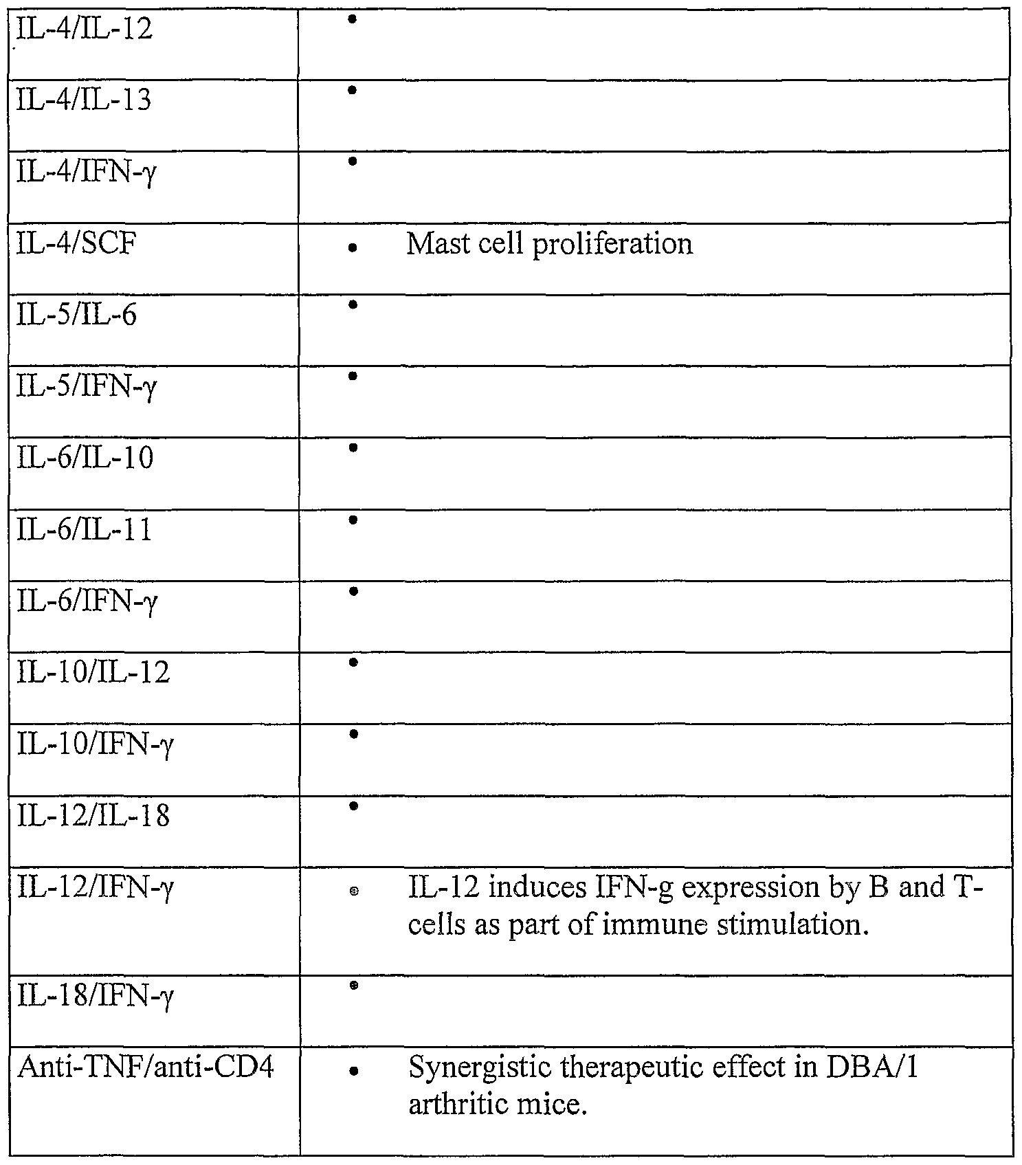


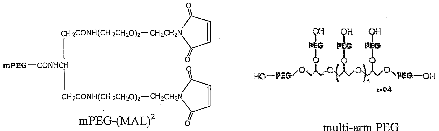


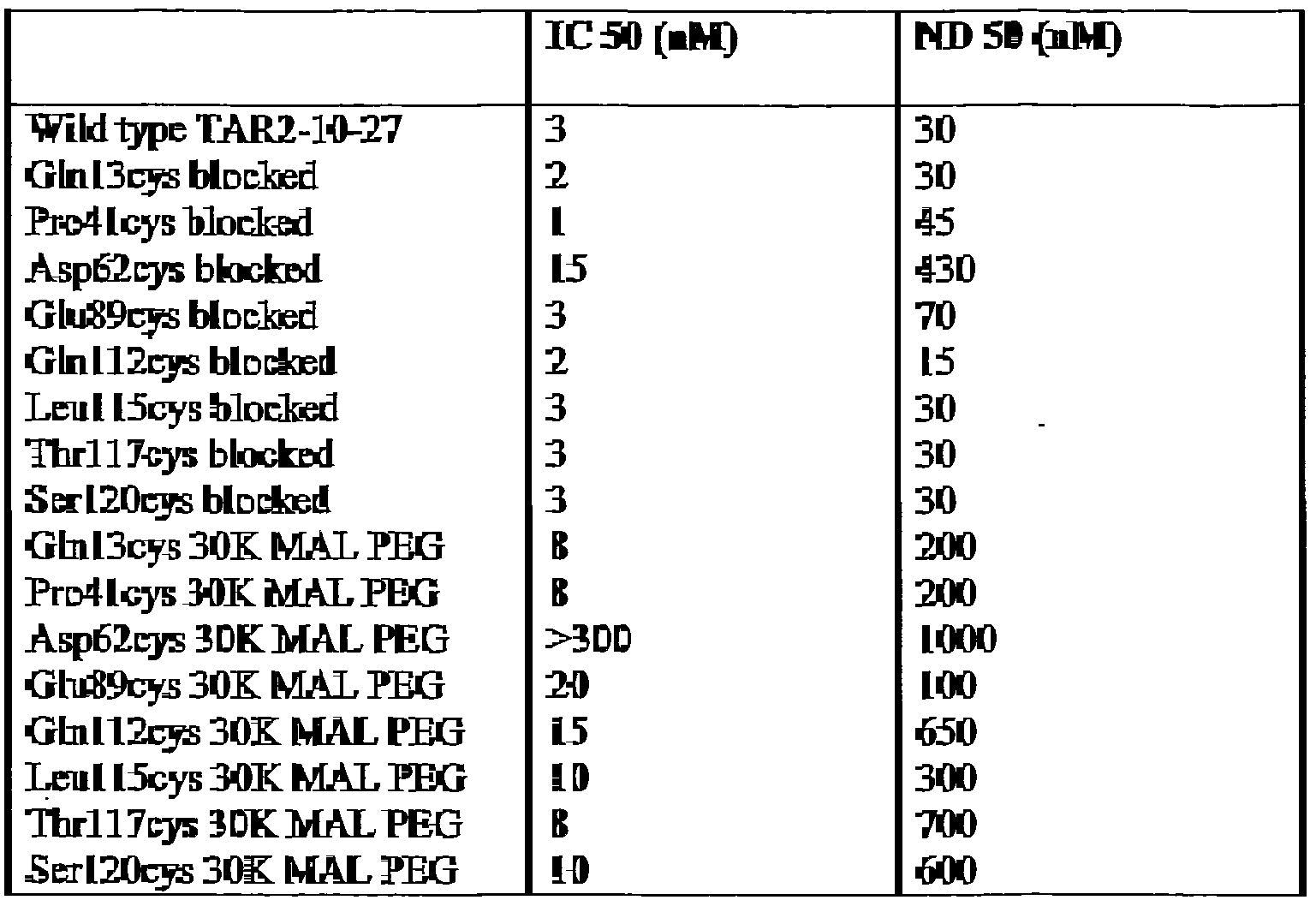


Claims
Priority Applications (41)
| Application Number | Priority Date | Filing Date | Title |
|---|---|---|---|
| JP2006518331A JP5087274B2 (en) | 2003-06-30 | 2004-06-30 | Polypeptide |
| AU2004220325A AU2004220325B2 (en) | 2003-06-30 | 2004-06-30 | Polypeptides |
| SI200431021T SI1639011T1 (en) | 2003-06-30 | 2004-06-30 | Pegylated single domain antibodies (dAb) |
| EP04737302A EP1639011B1 (en) | 2003-06-30 | 2004-06-30 | Pegylated single domain antibodies (dAb) |
| CA002529819A CA2529819A1 (en) | 2003-06-30 | 2004-06-30 | Pegylated single domain antibodies |
| DK04737302T DK1639011T3 (en) | 2003-06-30 | 2004-06-30 | Pegylated Single-Domain Antibodies (dAb) |
| DE602004017726T DE602004017726D1 (en) | 2003-06-30 | 2004-06-30 | Pegylated single-domain antibodies (dAb) |
| CN2004800249396A CN1845938B (en) | 2003-06-30 | 2004-06-30 | Polypeptides |
| PL04737302T PL1639011T3 (en) | 2003-06-30 | 2004-06-30 | Pegylated single domain antibodies (dAb) |
| US10/925,366 US20050271663A1 (en) | 2001-06-28 | 2004-08-24 | Compositions and methods for treating inflammatory disorders |
| PCT/GB2004/004253 WO2005035572A2 (en) | 2003-10-08 | 2004-10-08 | Antibody compositions and methods |
| AU2004280288A AU2004280288B2 (en) | 2003-10-08 | 2004-10-08 | Antibody compositions and methods |
| JP2006530595A JP2008500268A (en) | 2003-10-08 | 2004-10-08 | Antibody composition and method |
| EP04768787A EP1720906A2 (en) | 2003-10-08 | 2004-10-08 | Antibody compositions and methods |
| CA002539999A CA2539999A1 (en) | 2003-10-08 | 2004-10-08 | Antibody compositions and methods |
| US11/098,758 US20060073141A1 (en) | 2001-06-28 | 2005-04-04 | Compositions and methods for treating inflammatory disorders |
| KR1020107009046A KR101205643B1 (en) | 2004-06-30 | 2005-06-29 | Compositions and methods for treating inflammatory disorders |
| CN200580028518.5A CN101039959A (en) | 2004-06-30 | 2005-06-29 | Compositions and methods for treating inflammatory disorders |
| EA200700175A EA200700175A1 (en) | 2004-06-30 | 2005-06-29 | COMPOSITIONS AND METHODS OF TREATING INFLAMMATORY DISEASES |
| JP2007518691A JP2008504356A (en) | 2004-06-30 | 2005-06-29 | Compositions and methods for treating inflammatory diseases |
| AU2005259006A AU2005259006A1 (en) | 2004-06-30 | 2005-06-29 | Compositions and methods for treating inflammatory disorders |
| MXPA06014510A MXPA06014510A (en) | 2004-06-30 | 2005-06-29 | Compositions and methods for treating inflammatory disorders. |
| EP05755389A EP1761565A2 (en) | 2004-06-30 | 2005-06-29 | Compositions and methods for treating inflammatory disorders |
| EA201070623A EA201070623A1 (en) | 2004-06-30 | 2005-06-29 | CONSTRUCTION OF POLYPEPTIDES OF SINGLE-DOMAIN ANTIBODIES, APPLICATION OF THE COMPOSITION CONTAINING IT AND THE METHOD OF TREATING RHEUMATOID ARTHRITIS |
| KR1020077001184A KR20070050912A (en) | 2004-06-30 | 2005-06-29 | Compositions and methods for treating inflammatory disorders |
| CA002571536A CA2571536A1 (en) | 2004-06-30 | 2005-06-29 | Compositions and methods for treating inflammatory disorders |
| EP06021944A EP1777235A1 (en) | 2004-06-30 | 2005-06-29 | Compositions and methods for treating inflammatory disorders |
| PCT/GB2005/002553 WO2006003388A2 (en) | 2004-06-30 | 2005-06-29 | Compositions and methods for treating inflammatory disorders |
| US11/315,848 US20060286066A1 (en) | 2003-06-30 | 2005-12-22 | Pegylated immunoglobulin variable region polypeptides |
| US11/401,745 US20070065440A1 (en) | 2003-10-08 | 2006-04-10 | Antibody compositions and methods |
| ZA200610474A ZA200610474B (en) | 2002-12-27 | 2006-12-13 | Compositions and methods for treating inflammatory disorders |
| IL180187A IL180187A (en) | 2004-06-30 | 2006-12-19 | Composition for treating inflammatory disorders |
| US12/186,281 US20090148434A1 (en) | 2003-10-08 | 2008-08-05 | Antibody Compositions and Methods |
| US12/186,245 US20090082550A1 (en) | 2003-10-08 | 2008-08-05 | Antibody compositions and methods |
| US12/409,617 US20090258012A1 (en) | 2001-06-28 | 2009-03-24 | Compositions and methods for treating inflammatory disorders |
| AU2010201856A AU2010201856B2 (en) | 2004-06-30 | 2010-05-07 | Compositions and methods for treating inflammatory disorders |
| IL208119A IL208119A0 (en) | 2004-06-30 | 2010-09-13 | Use of a composition for treating inflammatory disorders and compositions thereof |
| AU2011200930A AU2011200930B2 (en) | 2003-06-30 | 2011-03-03 | Polypeptides |
| JP2012224608A JP2013018785A (en) | 2004-06-30 | 2012-10-09 | Compositions and methods for treating inflammatory disorders |
| JP2012224609A JP2013056890A (en) | 2004-06-30 | 2012-10-09 | Composition and method for treating inflammatory disorder |
| US13/733,675 US20130216538A1 (en) | 2002-12-27 | 2013-01-03 | Compositions and Methods for Treating Inflammatory Disorders |
Applications Claiming Priority (6)
| Application Number | Priority Date | Filing Date | Title |
|---|---|---|---|
| PCT/GB2003/002804 WO2004003019A2 (en) | 2002-06-28 | 2003-06-30 | Immunoglobin single variant antigen-binding domains and dual-specific constructs |
| GBPCT/GB03/002804 | 2003-06-30 | ||
| US50961303P | 2003-10-08 | 2003-10-08 | |
| US60/509,613 | 2003-10-08 | ||
| US53507604P | 2004-01-08 | 2004-01-08 | |
| US60/535,076 | 2004-01-08 |
Related Parent Applications (1)
| Application Number | Title | Priority Date | Filing Date |
|---|---|---|---|
| PCT/GB2003/005646 Continuation-In-Part WO2004058821A2 (en) | 2001-06-28 | 2003-12-24 | Dual specific single domain antibodies specific for a ligand and for the receptor of the ligand |
Related Child Applications (5)
| Application Number | Title | Priority Date | Filing Date |
|---|---|---|---|
| PCT/GB2002/003014 Continuation-In-Part WO2003002609A2 (en) | 2001-06-28 | 2002-06-28 | Dual-specific ligand and its use |
| PCT/GB2003/002804 Continuation-In-Part WO2004003019A2 (en) | 2001-06-28 | 2003-06-30 | Immunoglobin single variant antigen-binding domains and dual-specific constructs |
| US10/925,366 Continuation-In-Part US20050271663A1 (en) | 2001-06-28 | 2004-08-24 | Compositions and methods for treating inflammatory disorders |
| US11/098,758 Continuation-In-Part US20060073141A1 (en) | 2001-06-28 | 2005-04-04 | Compositions and methods for treating inflammatory disorders |
| US11/315,848 Continuation US20060286066A1 (en) | 2003-06-30 | 2005-12-22 | Pegylated immunoglobulin variable region polypeptides |
Publications (2)
| Publication Number | Publication Date |
|---|---|
| WO2004081026A2 true WO2004081026A2 (en) | 2004-09-23 |
| WO2004081026A3 WO2004081026A3 (en) | 2005-01-27 |
Family
ID=35811376
Family Applications (2)
| Application Number | Title | Priority Date | Filing Date |
|---|---|---|---|
| PCT/GB2004/002829 WO2004081026A2 (en) | 2001-06-28 | 2004-06-30 | Polypeptides |
| PCT/GB2004/004253 WO2005035572A2 (en) | 2002-06-28 | 2004-10-08 | Antibody compositions and methods |
Family Applications After (1)
| Application Number | Title | Priority Date | Filing Date |
|---|---|---|---|
| PCT/GB2004/004253 WO2005035572A2 (en) | 2002-06-28 | 2004-10-08 | Antibody compositions and methods |
Country Status (16)
| Country | Link |
|---|---|
| US (4) | US20060286066A1 (en) |
| EP (3) | EP1639011B1 (en) |
| JP (2) | JP5087274B2 (en) |
| CN (1) | CN1845938B (en) |
| AT (1) | ATE414106T1 (en) |
| AU (3) | AU2004220325B2 (en) |
| CA (2) | CA2529819A1 (en) |
| CY (1) | CY1108691T1 (en) |
| DE (1) | DE602004017726D1 (en) |
| DK (1) | DK1639011T3 (en) |
| ES (1) | ES2315664T3 (en) |
| PL (1) | PL1639011T3 (en) |
| PT (1) | PT1639011E (en) |
| SI (1) | SI1639011T1 (en) |
| WO (2) | WO2004081026A2 (en) |
| ZA (1) | ZA200702879B (en) |
Cited By (151)
| Publication number | Priority date | Publication date | Assignee | Title |
|---|---|---|---|---|
| WO2006038027A2 (en) | 2004-10-08 | 2006-04-13 | Domantis Limited | SINGLE DOMAIN ANTIBODIES AGAINST TNFRl AND METHODS OF USE THEREFOR |
| WO2006003388A3 (en) * | 2004-06-30 | 2006-04-27 | Domantis Ltd | Compositions and methods for treating inflammatory disorders |
| WO2006097689A2 (en) * | 2005-03-18 | 2006-09-21 | Domantis Limited | Antibodies against candida antigens |
| WO2007066106A1 (en) * | 2005-12-06 | 2007-06-14 | Domantis Limited | Ligands that have binding specificity for egfr and/or vegf and methods of use therefor |
| WO2007063308A3 (en) * | 2005-12-01 | 2007-09-20 | Domantis Ltd | Noncompetitive domain antibody formats that bind interleukin 1 receptor type 1 |
| WO2007063311A3 (en) * | 2005-12-01 | 2007-09-20 | Domantis Ltd | Competitive domain antibody formats that bind interleukin 1 receptor type 1 |
| WO2007141280A2 (en) | 2006-06-06 | 2007-12-13 | Oxford Genome Sciences (Uk) Ltd | Proteins |
| WO2008030611A2 (en) | 2006-09-05 | 2008-03-13 | Medarex, Inc. | Antibodies to bone morphogenic proteins and receptors therefor and methods for their use |
| EP1900753A3 (en) * | 2002-11-08 | 2008-04-09 | Ablynx N.V. | Method of administering therapeutic polypeptides, and polypeptides therefor |
| WO2008070569A2 (en) | 2006-12-01 | 2008-06-12 | Medarex, Inc. | Human antibodies that bind cd22 and uses thereof |
| JP2008519813A (en) * | 2004-11-10 | 2008-06-12 | ドマンティス リミテッド | Ligands that enhance endogenous compounds |
| WO2008074004A2 (en) | 2006-12-14 | 2008-06-19 | Medarex, Inc. | Human antibodies that bind cd70 and uses thereof |
| WO2008149143A2 (en) * | 2007-06-06 | 2008-12-11 | Domantis Limited | Methods for selecting protease resistant polypeptides |
| WO2009054863A2 (en) | 2006-12-13 | 2009-04-30 | Medarex, Inc. | Human antibodies that bind cd19 and uses thereof |
| JP2009533347A (en) * | 2006-04-07 | 2009-09-17 | ネクター セラピューティックス エイエル,コーポレイション | Anti-TNFα antibody complex |
| WO2010011944A2 (en) | 2008-07-25 | 2010-01-28 | Wagner Richard W | Protein screeing methods |
| WO2010010119A1 (en) | 2008-07-22 | 2010-01-28 | Ablynx Nv | Amino acid sequences directed against multitarget scavenger receptors and polypeptides |
| US20100092470A1 (en) * | 2008-09-22 | 2010-04-15 | Icb International, Inc. | Antibodies, analogs and uses thereof |
| US20100136584A1 (en) * | 2008-09-22 | 2010-06-03 | Icb International, Inc. | Methods for using antibodies and analogs thereof |
| WO2010081787A1 (en) | 2009-01-14 | 2010-07-22 | Domantis Limited | IMPROVED TNFα ANTAGONISM, PROPHYLAXIS & THERAPY WITH REDUCED ORGAN NECROSIS |
| WO2010084408A2 (en) | 2009-01-21 | 2010-07-29 | Oxford Biotherapeutics Ltd. | Pta089 protein |
| WO2010085590A1 (en) | 2009-01-23 | 2010-07-29 | Biosynexus Incorporated | Opsonic and protective antibodies specific for lipoteichoic acid gram positive bacteria |
| WO2010094720A2 (en) | 2009-02-19 | 2010-08-26 | Glaxo Group Limited | Improved anti-tnfr1 polypeptides, antibody variable domains & antagonists |
| WO2010102175A1 (en) | 2009-03-05 | 2010-09-10 | Medarex, Inc. | Fully human antibodies specific to cadm1 |
| US7846439B2 (en) | 2006-02-01 | 2010-12-07 | Cephalon Australia Pty Ltd | Domain antibody construct |
| WO2011006914A2 (en) | 2009-07-16 | 2011-01-20 | Glaxo Group Limited | Antagonists, uses & methods for partially inhibiting tnfr1 |
| WO2011012609A2 (en) | 2009-07-29 | 2011-02-03 | Glaxo Group Limited | Ligands that bind tgf-beta receptor rii |
| WO2011039368A2 (en) | 2009-10-02 | 2011-04-07 | Boehringer Ingelheim International Gmbh | Dll4-binding molecules |
| WO2011039370A1 (en) | 2009-10-02 | 2011-04-07 | Boehringer Ingelheim International Gmbh | Bispecific binding molecules for anti-angiogenesis therapy |
| WO2011047083A1 (en) | 2009-10-13 | 2011-04-21 | Oxford Biotherapeutics Ltd. | Antibodies against epha10 |
| WO2011054007A1 (en) | 2009-11-02 | 2011-05-05 | Oxford Biotherapeutics Ltd. | Ror1 as therapeutic and diagnostic target |
| WO2011051217A1 (en) | 2009-10-27 | 2011-05-05 | Glaxo Group Limited | Stable anti-tnfr1 polypeptides, antibody variable domains & antagonists |
| WO2011054893A2 (en) | 2009-11-05 | 2011-05-12 | Novartis Ag | Biomarkers predictive of progression of fibrosis |
| EP2322554A1 (en) * | 2004-06-30 | 2011-05-18 | Domantis Limited | Composition comprising an anti-TNF-alpha domain antibody for the treatment of rheumatoid arthritis |
| WO2011064382A1 (en) | 2009-11-30 | 2011-06-03 | Ablynx N.V. | Improved amino acid sequences directed against human respiratory syncytial virus (hrsv) and polypeptides comprising the same for the prevention and/or treatment of respiratory tract infections |
| WO2011073180A1 (en) | 2009-12-14 | 2011-06-23 | Ablynx N.V. | Single variable domain antibodies against ox40l, constructs and therapeutic use |
| WO2011083140A1 (en) | 2010-01-08 | 2011-07-14 | Ablynx Nv | Immunoglobulin single variable domain directed against human cxcr4 |
| EP2267027A3 (en) * | 2002-11-08 | 2011-07-20 | Ablynx N.V. | Method of administering therapeutic polypeptides, and polypeptides therefor |
| WO2011061246A3 (en) * | 2009-11-19 | 2011-07-28 | Ucb Pharma S.A. | Multivalent antibodies |
| WO2011089183A2 (en) | 2010-01-20 | 2011-07-28 | Boehringer Ingelheim International Gmbh | Anticoagulant antidotes |
| WO2011061492A3 (en) * | 2009-11-17 | 2011-07-28 | Ucb Pharma S.A. | Multivalent antibodies |
| WO2011098449A1 (en) | 2010-02-10 | 2011-08-18 | Novartis Ag | Methods and compounds for muscle growth |
| WO2011098520A1 (en) | 2010-02-10 | 2011-08-18 | Novartis Ag | Agonist dr5 binding polypeptides |
| WO2011107507A1 (en) | 2010-03-03 | 2011-09-09 | Boehringer Ingelheim International Gmbh | Biparatopic abeta binding polypeptides |
| EP2365000A2 (en) | 2005-05-18 | 2011-09-14 | Ablynx N.V. | Improved nanobodiesTM against tumor necrosis factor-alpha |
| WO2011138391A1 (en) | 2010-05-06 | 2011-11-10 | Novartis Ag | Compositions and methods of use for therapeutic low density lipoprotein - related protein 6 (lrp6) multivalent antibodies |
| WO2011138392A1 (en) | 2010-05-06 | 2011-11-10 | Novartis Ag | Compositions and methods of use for therapeutic low density lipoprotein -related protein 6 (lrp6) antibodies |
| WO2011144749A1 (en) | 2010-05-20 | 2011-11-24 | Ablynx Nv | Biological materials related to her3 |
| US20110319597A1 (en) * | 2008-08-14 | 2011-12-29 | Caphalon Australia Pty. Ltd. | Variant domain antibodies |
| WO2012020096A1 (en) | 2010-08-13 | 2012-02-16 | Medimmune Limited | Monomeric polypeptides comprising variant fc regions and methods of use |
| WO2012022814A1 (en) | 2010-08-20 | 2012-02-23 | Novartis Ag | Antibodies for epidermal growth factor receptor 3 (her3) |
| WO2012022734A2 (en) | 2010-08-16 | 2012-02-23 | Medimmune Limited | Anti-icam-1 antibodies and methods of use |
| WO2012028716A1 (en) | 2010-09-03 | 2012-03-08 | Boehringer Ingelheim International Gmbh | Vegf-binding molecules |
| WO2012032433A1 (en) | 2010-09-09 | 2012-03-15 | Pfizer Inc. | 4-1bb binding molecules |
| WO2012007880A3 (en) * | 2010-07-16 | 2012-04-12 | Ablynx Nv | Modified single domain antigen binding molecules and uses thereof |
| EP2441775A1 (en) | 2007-02-26 | 2012-04-18 | Oxford Biotherapeutics Ltd. | Protein |
| EP2447719A1 (en) | 2007-02-26 | 2012-05-02 | Oxford Biotherapeutics Ltd. | Proteins |
| WO2012062713A1 (en) | 2010-11-08 | 2012-05-18 | Novartis Ag | Cxcr2 binding polypeptides |
| WO2012093125A1 (en) | 2011-01-06 | 2012-07-12 | Glaxo Group Limited | Ligands that bind tgf-beta receptor ii |
| EP2486941A1 (en) | 2006-10-02 | 2012-08-15 | Medarex, Inc. | Human antibodies that bind CXCR4 and uses thereof |
| WO2012130834A1 (en) | 2011-03-30 | 2012-10-04 | Boehringer Ingelheim International Gmbh | Anticoagulant antidotes |
| WO2012131078A1 (en) | 2011-04-01 | 2012-10-04 | Boehringer Ingelheim International Gmbh | Bispecific binding molecules binding to vegf and ang2 |
| WO2012131076A1 (en) | 2011-04-01 | 2012-10-04 | Boehringer Ingelheim International Gmbh | BISPECIFIC BINDING MOLECULES BINDING TO Dll4 AND Ang2 |
| EP2514767A1 (en) | 2006-12-19 | 2012-10-24 | Ablynx N.V. | Amino acid sequences directed against a metalloproteinase from the ADAM family and polypeptides comprising the same for the treatment of ADAM-related diseases and disorders |
| WO2012156219A1 (en) | 2011-05-05 | 2012-11-22 | Ablynx Nv | Amino acid sequences directed against il-17a, il-17f and/or il17-a/f and polypeptides comprising the same |
| WO2013001369A2 (en) | 2011-06-28 | 2013-01-03 | Oxford Biotherapeutics Ltd. | Therapeutic and diagnostic target |
| WO2013003625A2 (en) | 2011-06-28 | 2013-01-03 | Oxford Biotherapeutics Ltd. | Antibodies |
| EP2557090A2 (en) | 2006-12-19 | 2013-02-13 | Ablynx N.V. | Amino acid sequences directed against GPCRs and polypeptides comprising the same for the treatment of GPCR-related diseases and disorders |
| WO2013067355A1 (en) | 2011-11-04 | 2013-05-10 | Novartis Ag | Low density lipoprotein-related protein 6 (lrp6) - half life extender constructs |
| EP2594590A1 (en) | 2007-12-14 | 2013-05-22 | Bristol-Myers Squibb Company | Binding molecules to the human OX40 receptor |
| WO2013084148A2 (en) | 2011-12-05 | 2013-06-13 | Novartis Ag | Antibodies for epidermal growth factor receptor 3 (her3) directed to domain ii of her3 |
| WO2013084147A2 (en) | 2011-12-05 | 2013-06-13 | Novartis Ag | Antibodies for epidermal growth factor receptor 3 (her3) |
| US20130177568A1 (en) * | 2012-01-09 | 2013-07-11 | Icb International, Inc. | Blood-brain barrier permeable peptide compositions |
| WO2013168108A2 (en) | 2012-05-09 | 2013-11-14 | Novartis Ag | Chemokine receptor binding polypeptides |
| WO2014020331A1 (en) | 2012-08-01 | 2014-02-06 | Oxford Biotherapeutics Ltd. | Therapeutic and diagnostic target |
| WO2014029752A1 (en) | 2012-08-22 | 2014-02-27 | Glaxo Group Limited | Anti lrp6 antibodies |
| WO2014087010A1 (en) | 2012-12-07 | 2014-06-12 | Ablynx N.V. | IMPROVED POLYPEPTIDES DIRECTED AGAINST IgE |
| US20140193834A1 (en) * | 2012-01-09 | 2014-07-10 | Icb International, Inc. | Blood-brain barrier permeable peptide compositions |
| WO2014133855A1 (en) | 2013-02-28 | 2014-09-04 | Caprion Proteomics Inc. | Tuberculosis biomarkers and uses thereof |
| WO2014140180A1 (en) | 2013-03-15 | 2014-09-18 | Glaxosmithkline Intellectual Property Development Limited | Anti-lag-3 binding proteins |
| WO2014159239A2 (en) | 2013-03-14 | 2014-10-02 | Novartis Ag | Antibodies against notch 3 |
| US8921528B2 (en) | 2004-06-01 | 2014-12-30 | Domantis Limited | Bispecific fusion antibodies with enhanced serum half-life |
| WO2015015401A2 (en) | 2013-08-02 | 2015-02-05 | Pfizer Inc. | Anti-cxcr4 antibodies and antibody-drug conjugates |
| WO2015051010A1 (en) | 2013-10-02 | 2015-04-09 | Medimmune, Llc | Neutralizing anti-influenza a antibodies and uses thereof |
| WO2015050959A1 (en) | 2013-10-01 | 2015-04-09 | Yale University | Anti-kit antibodies and methods of use thereof |
| US9028822B2 (en) | 2002-06-28 | 2015-05-12 | Domantis Limited | Antagonists against TNFR1 and methods of use therefor |
| WO2015104322A1 (en) | 2014-01-09 | 2015-07-16 | Glaxosmithkline Intellectual Property Development Limited | Treatment of inflammatory diseases with non-competitive tnfr1 antagonists |
| EP2947097A1 (en) | 2008-04-07 | 2015-11-25 | Ablynx N.V. | Amino acid sequences directed against the Notch pathways and uses thereof |
| WO2015193452A1 (en) | 2014-06-18 | 2015-12-23 | Ablynx Nv | Kv1.3 binding immunoglobulins |
| EP3009454A2 (en) | 2009-04-20 | 2016-04-20 | Oxford Bio Therapeutics Limited | Antibodies specific to cadherin-17 |
| US9320792B2 (en) | 2002-11-08 | 2016-04-26 | Ablynx N.V. | Pulmonary administration of immunoglobulin single variable domains and constructs thereof |
| US9371381B2 (en) | 2002-11-08 | 2016-06-21 | Ablynx, N.V. | Single domain antibodies directed against tumor necrosis factor-alpha and uses therefor |
| US9393304B2 (en) | 2008-10-29 | 2016-07-19 | Ablynx N.V. | Formulations of single domain antigen binding molecules |
| WO2016156570A1 (en) | 2015-04-02 | 2016-10-06 | Ablynx N.V. | Bispecific cxcr4-cd-4 polypeptides with potent anti-hiv activity |
| WO2016180982A1 (en) | 2015-05-13 | 2016-11-17 | Ablynx N.V. | T cell recruiting polypeptides based on cd3 reactivity |
| WO2016180969A1 (en) | 2015-05-13 | 2016-11-17 | Ablynx N.V. | T cell recruiting polypeptides based on tcr alpha/beta reactivity |
| EP3205670A1 (en) | 2009-06-05 | 2017-08-16 | Ablynx N.V. | Improved amino acid sequences directed against human respiratory syncytial virus (hrsv) and polypeptides comprising the same for the prevention and/or treatment of respiratory tract infections |
| WO2017191108A1 (en) | 2016-05-02 | 2017-11-09 | Ablynx Nv | Treatment of rsv infection |
| EP3266798A2 (en) | 2015-11-12 | 2018-01-10 | Ablynx NV | Improved tnf binders |
| WO2018007442A1 (en) | 2016-07-06 | 2018-01-11 | Ablynx N.V. | Treatment of il-6r related diseases |
| WO2018020000A1 (en) | 2016-07-29 | 2018-02-01 | INSERM (Institut National de la Santé et de la Recherche Médicale) | Antibodies targeting tumor associated macrophages and uses thereof |
| US9884921B2 (en) | 2014-07-01 | 2018-02-06 | Pfizer Inc. | Bispecific heterodimeric diabodies and uses thereof |
| WO2018029182A1 (en) | 2016-08-08 | 2018-02-15 | Ablynx N.V. | Il-6r single variable domain antibodies for treatment of il-6r related diseases |
| WO2018050833A1 (en) | 2016-09-15 | 2018-03-22 | Ablynx Nv | Immunoglobulin single variable domains directed against macrophage migration inhibitory factor |
| EP3311837A1 (en) | 2011-09-23 | 2018-04-25 | Ablynx NV | Prolonged inhibition of interleukin-6 mediated signaling |
| WO2018091606A1 (en) | 2016-11-16 | 2018-05-24 | Ablynx Nv | T cell recruiting polypeptides capable of binding cd123 and tcr alpha/beta |
| WO2018099968A1 (en) | 2016-11-29 | 2018-06-07 | Ablynx N.V. | Treatment of infection by respiratory syncytial virus (rsv) |
| WO2018115231A2 (en) | 2016-12-22 | 2018-06-28 | Boehringer Ingelheim International Gmbh | Binding molecules for the treatment of cancer |
| WO2018158398A1 (en) | 2017-03-02 | 2018-09-07 | INSERM (Institut National de la Santé et de la Recherche Médicale) | Antibodies having specificity to nectin-4 and uses thereof |
| US10118962B2 (en) | 2008-10-29 | 2018-11-06 | Ablynx N.V. | Methods for purification of single domain antigen binding molecules |
| WO2018206734A1 (en) | 2017-05-11 | 2018-11-15 | Vib Vzw | Glycosylation of variable immunoglobulin domains |
| WO2018220225A1 (en) | 2017-06-02 | 2018-12-06 | Ablynx Nv | Aggrecan binding immunoglobulins |
| WO2018220235A1 (en) | 2017-06-02 | 2018-12-06 | Merck Patent Gmbh | Mmp13 binding immunoglobulins |
| WO2018220236A1 (en) | 2017-06-02 | 2018-12-06 | Merck Patent Gmbh | Polypeptides binding adamts5, mmp13 and aggrecan |
| WO2018220234A1 (en) | 2017-06-02 | 2018-12-06 | Merck Patent Gmbh | Adamts binding immunoglobulins |
| WO2019016237A1 (en) | 2017-07-19 | 2019-01-24 | Vib Vzw | Serum albumin binding agents |
| WO2019020480A1 (en) | 2017-07-24 | 2019-01-31 | INSERM (Institut National de la Santé et de la Recherche Médicale) | Antibodies and peptides to treat hcmv related diseases |
| GB201901608D0 (en) | 2019-02-06 | 2019-03-27 | Vib Vzw | Vaccine adjuvant conjugates |
| EP3461844A2 (en) | 2009-04-10 | 2019-04-03 | Ablynx N.V. | Improved amino acid sequences directed against il-6r and polypeptides comprising the same for the treatment of il-6r related diseases and disorders |
| US10421816B2 (en) | 2009-09-10 | 2019-09-24 | Ucb Biopharma Sprl | Multivalent antibodies |
| EP3569618A1 (en) | 2018-05-19 | 2019-11-20 | Boehringer Ingelheim International GmbH | Antagonizing cd73 antibody |
| WO2020010250A2 (en) | 2018-07-03 | 2020-01-09 | Elstar Therapeutics, Inc. | Anti-tcr antibody molecules and uses thereof |
| WO2020053122A1 (en) | 2018-09-10 | 2020-03-19 | INSERM (Institut National de la Santé et de la Recherche Médicale) | Combination of her2/neu antibody with heme for treating cancer |
| WO2020099508A1 (en) | 2018-11-13 | 2020-05-22 | Emstopa Limited | Tissue plasminogen activator antibodies and method of use thereof |
| WO2020193520A1 (en) | 2019-03-25 | 2020-10-01 | INSERM (Institut National de la Santé et de la Recherche Médicale) | Treatment of taupathy disorders by targeting new tau species |
| EP3736293A1 (en) | 2013-02-12 | 2020-11-11 | Boehringer Ingelheim International Gmbh | Therapeutic and diagnostic target for cancer comprising dll3 binding reagents |
| US10961313B2 (en) | 2015-10-30 | 2021-03-30 | Genentech, Inc. | Anti-factor D antibody variant conjugates and uses thereof |
| WO2021058729A1 (en) | 2019-09-27 | 2021-04-01 | INSERM (Institut National de la Santé et de la Recherche Médicale) | Anti-müllerian inhibiting substance type i receptor antibodies and uses thereof |
| WO2021058763A1 (en) | 2019-09-27 | 2021-04-01 | INSERM (Institut National de la Santé et de la Recherche Médicale) | Anti-müllerian inhibiting substance antibodies and uses thereof |
| US11028169B2 (en) | 2016-05-18 | 2021-06-08 | Boehringer Ingelheim International Gmbh | Antibody molecules for cancer treatment |
| WO2021116119A1 (en) | 2019-12-09 | 2021-06-17 | INSERM (Institut National de la Santé et de la Recherche Médicale) | Antibodies having specificity to her4 and uses thereof |
| US11161914B2 (en) * | 2018-08-09 | 2021-11-02 | Jiangsu Academy Of Agricultural Sciences | Human-derived insecticidal bt cry toxin mimetic, and design method and application thereof |
| WO2021228956A1 (en) | 2020-05-12 | 2021-11-18 | INSERM (Institut National de la Santé et de la Recherche Médicale) | New method to treat cutaneous t-cell lymphomas and tfh derived lymphomas |
| WO2021236658A1 (en) | 2020-05-19 | 2021-11-25 | Boehringer Ingelheim International Gmbh | Binding molecules for the treatment of cancer |
| US11242395B2 (en) | 2017-08-21 | 2022-02-08 | Adagene Inc. | Anti-CD137 molecules and use thereof |
| WO2022047243A1 (en) | 2020-08-27 | 2022-03-03 | Enosi Life Sciences Corp. | Methods and compositions to treat autoimmune diseases and cancer |
| WO2022044573A1 (en) | 2020-08-26 | 2022-03-03 | 国立大学法人熊本大学 | Human antibody or antigen-binding fragment thereof against coronavirus spike protein |
| WO2022084354A1 (en) | 2020-10-21 | 2022-04-28 | Boehringer Ingelheim International Gmbh | Bispecific anti-vegf and anti-trkb binding molecules for the treatment of eye diseases |
| US11359016B2 (en) | 2018-02-02 | 2022-06-14 | Adagene Inc. | Anti-CTLA4 antibodies and methods of making and using the same |
| WO2022129637A1 (en) | 2020-12-18 | 2022-06-23 | Ablynx Nv | T cell recruiting polypeptides based on tcr alpha/beta reactivity |
| EP4050027A2 (en) | 2012-02-27 | 2022-08-31 | Ablynx N.V. | Cx3cr1-binding polypeptides |
| WO2022216993A2 (en) | 2021-04-08 | 2022-10-13 | Marengo Therapeutics, Inc. | Multifuntional molecules binding to tcr and uses thereof |
| WO2022263507A1 (en) | 2021-06-17 | 2022-12-22 | Boehringer Ingelheim International Gmbh | Novel tri-specific binding molecules |
| US11680104B2 (en) | 2015-09-02 | 2023-06-20 | Immutep S.A.S. | Anti-LAG-3 antibodies |
| WO2023175171A1 (en) | 2022-03-18 | 2023-09-21 | Inserm (Institut National De La Sante Et De La Recherche Medicale) | Bk polyomavirus antibodies and uses thereof |
| WO2024023271A1 (en) | 2022-07-27 | 2024-02-01 | Ablynx Nv | Polypeptides binding to a specific epitope of the neonatal fc receptor |
| WO2024052503A1 (en) | 2022-09-08 | 2024-03-14 | Institut National de la Santé et de la Recherche Médicale | Antibodies having specificity to ltbp2 and uses thereof |
| WO2024056668A1 (en) | 2022-09-12 | 2024-03-21 | Institut National de la Santé et de la Recherche Médicale | New anti-itgb8 antibodies and its uses thereof |
| US11952681B2 (en) | 2018-02-02 | 2024-04-09 | Adagene Inc. | Masked activatable CD137 antibodies |
| WO2024079210A1 (en) * | 2022-10-14 | 2024-04-18 | Bactolife A/S | sdAb-CBM FUSION PROTEIN BOUND TO POLYSACCHARIDE POLYMER |
| WO2024083843A1 (en) | 2022-10-18 | 2024-04-25 | Confo Therapeutics N.V. | Amino acid sequences directed against the melanocortin 4 receptor and polypeptides comprising the same for the treatment of mc4r-related diseases and disorders |
Families Citing this family (135)
| Publication number | Priority date | Publication date | Assignee | Title |
|---|---|---|---|---|
| US20080026068A1 (en) * | 2001-08-16 | 2008-01-31 | Baxter Healthcare S.A. | Pulmonary delivery of spherical insulin microparticles |
| JP2005289809A (en) | 2001-10-24 | 2005-10-20 | Vlaams Interuniversitair Inst Voor Biotechnologie Vzw (Vib Vzw) | Mutant heavy-chain antibody |
| US9321832B2 (en) | 2002-06-28 | 2016-04-26 | Domantis Limited | Ligand |
| AU2003251238A1 (en) * | 2002-08-07 | 2004-02-25 | Umc Utrecht Holding B.V. | Modulation of platelet adhesion based on the surface exposed beta-switch loop of platelet glycoprotein ib-alpha |
| CN101412759A (en) | 2003-01-10 | 2009-04-22 | 埃博灵克斯股份有限公司 | Therapeutic polypeptides, homologues thereof, fragments thereof and for use in modulating platelet-mediated aggregation |
| PL1639011T3 (en) * | 2003-06-30 | 2009-05-29 | Domantis Ltd | Pegylated single domain antibodies (dAb) |
| US8333995B2 (en) * | 2004-05-12 | 2012-12-18 | Baxter International, Inc. | Protein microspheres having injectable properties at high concentrations |
| US8728525B2 (en) * | 2004-05-12 | 2014-05-20 | Baxter International Inc. | Protein microspheres retaining pharmacokinetic and pharmacodynamic properties |
| DK2072040T3 (en) | 2004-05-12 | 2013-07-29 | Baxter Healthcare Sa | Therapeutic use of nucleic acid microspheres |
| CN103432079A (en) * | 2004-05-12 | 2013-12-11 | 巴克斯特国际公司 | Oligonucleotide-containing microspheres, their use for the manufacture of a medicament for treating diabetes type 1 |
| WO2006033702A2 (en) | 2004-07-26 | 2006-03-30 | Biogen Idec Ma Inc. | Anti-cd154 antibodies |
| US7563443B2 (en) | 2004-09-17 | 2009-07-21 | Domantis Limited | Monovalent anti-CD40L antibody polypeptides and compositions thereof |
| JP5185624B2 (en) * | 2004-12-02 | 2013-04-17 | ドマンティス リミテッド | Bispecific antibodies targeting serum albumin and GLP-1 or PYY |
| GB0521621D0 (en) * | 2005-10-24 | 2005-11-30 | Domantis Ltd | Tumor necrosis factor receptor 1 antagonists for treating respiratory diseases |
| CA2593328C (en) * | 2005-01-14 | 2018-02-06 | Ablynx N.V. | Methods and assays for distinguishing between different forms of diseases and disorders characterized by thrombocytopenia and/or by spontaneous interaction between von willebrand factor (vwf) and platelets |
| WO2006110585A2 (en) | 2005-04-07 | 2006-10-19 | Novartis Vaccines And Diagnostics Inc. | Cancer-related genes (prlr) |
| AU2006235276A1 (en) | 2005-04-07 | 2006-10-19 | Novartis Vaccines And Diagnostics Inc. | CACNA1E in cancer diagnosis, detection and treatment |
| PL3415535T3 (en) * | 2005-05-20 | 2021-06-14 | Ablynx N.V. | Improved nanobodies tm for the treatment of aggregation-mediated disorders |
| DE102005023617A1 (en) | 2005-05-21 | 2006-11-23 | Aspre Ag | Method for mixing colors in a display |
| EP1909831A4 (en) | 2005-06-14 | 2013-02-20 | Amgen Inc | Self-buffering protein formulations |
| GB2427194A (en) * | 2005-06-16 | 2006-12-20 | Domantis Ltd | Single domain Helicobacter pylori adhesin antibodies |
| EP1969010A4 (en) | 2005-12-20 | 2009-07-22 | Peptech Ltd | Anti-inflammatory dab |
| US7625564B2 (en) | 2006-01-27 | 2009-12-01 | Novagen Holding Corporation | Recombinant human EPO-Fc fusion proteins with prolonged half-life and enhanced erythropoietic activity in vivo |
| EP2389948A1 (en) | 2006-03-23 | 2011-11-30 | Novartis AG | Anti-tumor cell antigen antibody therapeutics |
| MX2009001226A (en) | 2006-08-04 | 2009-03-20 | Baxter Int | Microsphere-based composition for preventing and/or reversing new-onset autoimmune diabetes. |
| GB0621513D0 (en) | 2006-10-30 | 2006-12-06 | Domantis Ltd | Novel polypeptides and uses thereof |
| LT2842967T (en) * | 2007-01-18 | 2017-02-27 | Eli Lilly And Company | Pegylated amyloid beta fab |
| SI2125894T1 (en) * | 2007-03-22 | 2019-05-31 | Biogen Ma Inc. | Binding proteins, including antibodies, antibody derivatives and antibody fragments, that specifically bind cd154 and uses thereof |
| NZ580379A (en) | 2007-03-29 | 2012-10-26 | Abbott Lab | Crystalline anti-human il-12 antibodies |
| AU2008242842B2 (en) * | 2007-04-17 | 2014-06-05 | Baxter Healthcare Sa | Nucleic acid microparticles for pulmonary delivery |
| MX2009013137A (en) * | 2007-06-06 | 2010-04-30 | Domantis Ltd | Methods for selecting protease resistant polypeptides. |
| US8067548B2 (en) | 2007-07-26 | 2011-11-29 | Novagen Holding Corporation | Fusion proteins having mutated immunoglobulin hinge region |
| EP2535349A1 (en) | 2007-09-26 | 2012-12-19 | UCB Pharma S.A. | Dual specificity antibody fusions |
| BRPI0818928B1 (en) | 2007-11-02 | 2021-12-07 | Novartis Ag | ISOLATED NOGO-A BINDING MOLECULES, THEIR PRODUCTION METHOD AND USE, ISOLATED POLYNUCLEOTIDE, VECTOR AND EXPRESSION SYSTEM, ISOLATED HOST CELL, AND PHARMACEUTICAL COMPOSITION |
| GB0721752D0 (en) * | 2007-11-06 | 2007-12-19 | Univ Southampton | Configurable electronic device and method |
| NZ709704A (en) * | 2007-11-30 | 2017-03-31 | Abbvie Biotechnology Ltd | Protein formulations and methods of making same |
| US8883146B2 (en) | 2007-11-30 | 2014-11-11 | Abbvie Inc. | Protein formulations and methods of making same |
| EP3211010A1 (en) * | 2007-12-21 | 2017-08-30 | Medimmune Limited | Binding members for interleukin-4 receptor alpha (il-4r) - 173 |
| US8580927B2 (en) | 2008-01-31 | 2013-11-12 | The United States Of America, As Represented By The Secretary, Department Of Health And Human Services | Engineered antibody constant domain molecules |
| CA2718081A1 (en) * | 2008-03-21 | 2009-09-24 | Ablynx Nv | Von willebrand factor specific binders and methods of use therefor |
| CN101565465B (en) * | 2008-05-14 | 2011-12-21 | 复旦大学 | Surface antigen 1 of Toxoplasma gondii human antibody Fab fragment and encoded gene thereof |
| DK2334705T3 (en) * | 2008-09-26 | 2017-03-27 | Ucb Biopharma Sprl | BIOLOGICAL PRODUCTS |
| EA201100488A1 (en) * | 2008-10-21 | 2011-12-30 | Домантис Лимитед | LIGANDS THAT HAVE A SPECIFIC BINDING RELATION TO DC-SIGN |
| EA201100485A1 (en) * | 2008-10-21 | 2011-12-30 | Домантис Лимитед | COMPOSITIONS FOR DEFINITIVE CELL POINTING |
| CN102301235B (en) * | 2008-11-28 | 2014-11-19 | Abbvie公司 | Stable Antibody Compositions And Methods For Stabilizing Same |
| EP2370574B1 (en) * | 2008-12-05 | 2013-09-25 | Council of Scientific & Industrial Research | Mutants of staphylokinase carrying amino and carboxy-terminal extensions for polyethylene glycol conjugation |
| JP5501439B2 (en) | 2009-04-02 | 2014-05-21 | ロシュ グリクアート アクチェンゲゼルシャフト | Multispecific antibody comprising a full-length antibody and a single chain Fab fragment |
| PL2437785T3 (en) * | 2009-06-04 | 2015-08-31 | Novartis Ag | METHODS FOR IDENTIFICATION OF SITES FOR IgG CONJUGATION |
| ES2524974T3 (en) * | 2009-06-25 | 2014-12-16 | The University Of North Carolina At Chapel Hill | Chimeric Factor VII molecules |
| WO2011003622A1 (en) | 2009-07-10 | 2011-01-13 | Ablynx N.V. | Method for the production of variable domains |
| CN104945509A (en) | 2009-09-16 | 2015-09-30 | 弗·哈夫曼-拉罗切有限公司 | Coiled coil and/or tether containing protein complexes and uses thereof |
| PL2993231T3 (en) | 2009-09-24 | 2019-01-31 | Ucb Biopharma Sprl | Bacterial strain for recombinant protein expression, having protease deficient degp retaining chaperone activity, and knocked out tsp and ptr genes |
| GB201005063D0 (en) | 2010-03-25 | 2010-05-12 | Ucb Pharma Sa | Biological products |
| WO2011063152A1 (en) * | 2009-11-18 | 2011-05-26 | Affinergy, Inc. | Methods and compositions for soft tissue repair |
| EP2502939B1 (en) * | 2009-11-19 | 2016-01-27 | Zhejiang University | Nonnatural collagen-like protein and use thereof |
| CA2781682A1 (en) | 2009-12-04 | 2011-06-09 | Genentech, Inc. | Multispecific antibodies, antibody analogs, compositions, and methods |
| ES2565208T3 (en) | 2009-12-11 | 2016-04-01 | F. Hoffmann-La Roche Ag | Anti-VEGF-C antibodies and methods of use thereof |
| MX2012007379A (en) | 2009-12-23 | 2012-08-31 | Genentech Inc | Anti-bv8 antibodies and uses thereof. |
| GB201000587D0 (en) | 2010-01-14 | 2010-03-03 | Ucb Pharma Sa | Bacterial hoist strain |
| GB201000590D0 (en) | 2010-01-14 | 2010-03-03 | Ucb Pharma Sa | Bacterial host strain |
| GB201000591D0 (en) | 2010-01-14 | 2010-03-03 | Ucb Pharma Sa | Bacterial hoist strain |
| TW201138821A (en) | 2010-03-26 | 2011-11-16 | Roche Glycart Ag | Bispecific antibodies |
| RU2624027C2 (en) | 2010-04-23 | 2017-06-30 | Дженентек, Инк. | Heteromultimeric proteins production |
| GB201012599D0 (en) | 2010-07-27 | 2010-09-08 | Ucb Pharma Sa | Process for purifying proteins |
| RU2013110844A (en) | 2010-08-13 | 2014-09-20 | Дженентек, Инк. | ANTIBODIES AGAINST IL-1β AND IL-18 USED FOR TREATMENT OF DISEASES |
| CN103068846B9 (en) | 2010-08-24 | 2016-09-28 | 弗·哈夫曼-拉罗切有限公司 | Bispecific antibodies comprising disulfide-stabilized Fv fragments |
| EP2655413B1 (en) | 2010-12-23 | 2019-01-16 | F.Hoffmann-La Roche Ag | Polypeptide-polynucleotide-complex and its use in targeted effector moiety delivery |
| US10689447B2 (en) | 2011-02-04 | 2020-06-23 | Genentech, Inc. | Fc variants and methods for their production |
| CA2825064C (en) | 2011-02-04 | 2022-08-30 | Genentech, Inc. | Fc variants and methods for their production |
| WO2012122512A1 (en) | 2011-03-10 | 2012-09-13 | Hco Antibody, Inc. | Recombinant production of mixtures of single chain antibodies |
| JP6130350B2 (en) | 2011-03-30 | 2017-05-17 | アブリンクス エン.ヴェー. | Methods of treating immune disorders with single domain antibodies against TNFα |
| KR101357117B1 (en) | 2011-06-28 | 2014-02-06 | 비앤엘델리팜 주식회사 | PEGylated Exendin-4 analogues or its derivatives, preparation method thereof and pharmaceutical composition containing the same for preventing and treating a diabetes |
| PT2731973T (en) | 2011-07-13 | 2018-02-22 | Ucb Biopharma Sprl | Bacterial host strain expressing recombinant dsbc |
| MX359384B (en) | 2011-10-11 | 2018-09-25 | Genentech Inc | Improved assembly of bispecific antibodies. |
| JP6163158B2 (en) | 2011-10-13 | 2017-07-12 | ブリストル−マイヤーズ スクイブ カンパニーBristol−Myers Squibb Company | Antibody polypeptides that antagonize CD40L |
| US20140301977A1 (en) | 2011-11-02 | 2014-10-09 | Genentech, Inc. | Overload and elute chromatography |
| BR112014019579A2 (en) | 2012-02-10 | 2019-10-15 | Genentech, Inc | SINGLE CHAIN ANTIBODY, POLYNUCLEOTIDE, VECTOR, HOST CELL, METHOD OF PRODUCTION OF A SINGLE CHAIN ANTIBODY, HETEROMULTYMER AND METHOD OF PRODUCTION |
| GB201208367D0 (en) | 2012-05-14 | 2012-06-27 | Ucb Pharma Sa | Biological product |
| RU2639287C2 (en) | 2012-06-27 | 2017-12-20 | Ф. Хоффманн-Ля Рош Аг | Method for selection and obtaining of highly selective and multispecific targeting groups with specified properties, including at least two different binding groups, and their applications |
| WO2014001325A1 (en) | 2012-06-27 | 2014-01-03 | F. Hoffmann-La Roche Ag | Method for making antibody fc-region conjugates comprising at least one binding entity that specifically binds to a target and uses thereof |
| JOP20200308A1 (en) | 2012-09-07 | 2017-06-16 | Novartis Ag | IL-18 binding molecules |
| KR20150084046A (en) | 2012-11-15 | 2015-07-21 | 제넨테크, 인크. | IONIC STRENGTH-MEDIATED pH GRADIENT ION EXCHANGE CHROMATOGRAPHY |
| CA2918052A1 (en) | 2013-07-12 | 2015-01-15 | Genentech, Inc. | Elucidation of ion exchange chromatography input optimization |
| US20150093800A1 (en) | 2013-09-05 | 2015-04-02 | Genentech, Inc. | Method for chromatography reuse |
| RU2016107435A (en) | 2013-09-13 | 2017-10-18 | Дженентек, Инк. | COMPOSITIONS AND METHODS FOR DETECTING AND QUANTITATIVE DETERMINATION OF THE PROTEIN OF CELLS-OWNERS IN CELL LINES AND RECOMBINANT POLYPEPTIDE PRODUCTS |
| WO2015038888A1 (en) | 2013-09-13 | 2015-03-19 | Genentech, Inc. | Methods and compositions comprising purified recombinant polypeptides |
| MX2016003744A (en) | 2013-10-11 | 2016-08-11 | Us Health | Tem8 antibodies and their use. |
| EP2873680A1 (en) | 2013-11-13 | 2015-05-20 | F.Hoffmann-La Roche Ag | Oligopeptide and methods for producing conjugates thereof |
| WO2015090229A1 (en) | 2013-12-20 | 2015-06-25 | Novartis Ag | Regulatable chimeric antigen receptor |
| WO2015103549A1 (en) | 2014-01-03 | 2015-07-09 | The United States Of America, As Represented By The Secretary Department Of Health And Human Services | Neutralizing antibodies to hiv-1 env and their use |
| WO2015138638A1 (en) | 2014-03-11 | 2015-09-17 | Theraly Pharmaceuticals, Inc. | Long acting trail receptor agonists for treatment of autoimmune diseases |
| WO2015142661A1 (en) | 2014-03-15 | 2015-09-24 | Novartis Ag | Regulatable chimeric antigen receptor |
| BR122021009041B1 (en) | 2014-05-06 | 2022-11-29 | Genentech, Inc | METHODS FOR THE PREPARATION OF A HETEROMULTIMERIC PROTEIN |
| WO2016014553A1 (en) | 2014-07-21 | 2016-01-28 | Novartis Ag | Sortase synthesized chimeric antigen receptors |
| ES2781175T3 (en) | 2014-07-31 | 2020-08-31 | Novartis Ag | Optimized subset of T cells containing a chimeric antigen receptor |
| WO2016077369A1 (en) | 2014-11-10 | 2016-05-19 | Genentech, Inc. | Animal model for nephropathy and agents for treating the same |
| EP3875481A1 (en) | 2014-11-14 | 2021-09-08 | The U.S.A. as represented by the Secretary, Department of Health and Human Services | Neutralizing antibodies to ebola virus glycoprotein and their use |
| ES2764111T3 (en) | 2014-12-03 | 2020-06-02 | Hoffmann La Roche | Multispecific antibodies |
| PT3233910T (en) * | 2014-12-19 | 2020-03-17 | Ablynx Nv | Cysteine linked nanobody dimers |
| GB201500463D0 (en) | 2015-01-12 | 2015-02-25 | Cresendo Biolog Ltd | Therapeutic molecules |
| KR20170140180A (en) | 2015-02-24 | 2017-12-20 | 더 유나이티드 스테이츠 오브 어메리카, 애즈 리프리젠티드 바이 더 세크러테리, 디파트먼트 오브 헬쓰 앤드 휴먼 서비씨즈 | Middle east respiratory syndrome coronavirus immunogens, antibodies, and their use |
| SI3265557T1 (en) | 2015-03-06 | 2020-02-28 | F. Hoffmann-La Roche Ag | Ultrapurified dsba and dsbc and methods of making and using the same |
| SI3271389T1 (en) | 2015-03-20 | 2020-09-30 | The United States Of America, As Represented By The Secretary Department Of Health And Human Services | Neutralizing antibodies to gp120 and their use |
| CN107709364A (en) | 2015-04-07 | 2018-02-16 | 豪夫迈·罗氏有限公司 | Antigen binding complex and application method with agonist activity |
| MX2017015011A (en) | 2015-05-28 | 2018-03-23 | Genentech Inc | Cell-based assay for detecting anti-cd3 homodimers. |
| WO2016196975A1 (en) | 2015-06-03 | 2016-12-08 | The United States Of America, As Represented By The Secretary Department Of Health & Human Services | Neutralizing antibodies to hiv-1 env and their use |
| JP2018534930A (en) * | 2015-10-30 | 2018-11-29 | ジェネンテック, インコーポレイテッド | Anti-factor D antibodies and conjugates |
| EP4011911A1 (en) | 2015-11-03 | 2022-06-15 | The United States of America as represented by The Secretary Department of Health and Human Services | Neutralizing antibodies to hiv-1 gp41 and their use |
| US11123405B2 (en) | 2015-12-23 | 2021-09-21 | The Johns Hopkins University | Long-acting GLP-1R agonist as a therapy of neurological and neurodegenerative conditions |
| US11513127B2 (en) | 2016-01-25 | 2022-11-29 | Genentech, Inc. | Methods for assaying T-cell dependent bispecific antibodies |
| WO2017192589A1 (en) | 2016-05-02 | 2017-11-09 | The United States Of America, As Represented By The Secretary, Department Of Health And Human Services | Neutralizing antibodies to influenza ha and their use and identification |
| WO2017201036A1 (en) | 2016-05-17 | 2017-11-23 | Genentech, Inc. | Stromal gene signatures for diagnosis and use in immunotherapy |
| AU2017283480A1 (en) | 2016-06-13 | 2019-01-24 | Torque Therapeutics, Inc. | Methods and compositions for promoting immune cell function |
| AR108800A1 (en) | 2016-06-17 | 2018-09-26 | Genentech Inc | PURIFICATION OF MULTI-SPECIFIC ANTIBODIES |
| CN109963871A (en) | 2016-08-05 | 2019-07-02 | 豪夫迈·罗氏有限公司 | Multivalence and multi-epitope Antibody and application method with agonist activity |
| AU2017312540B2 (en) | 2016-08-15 | 2023-02-02 | Genentech, Inc. | Chromatography method for quantifying a non-ionic surfactant in a composition comprising the non-ionic surfactant and a polypeptide |
| BR112019007858A2 (en) | 2016-10-21 | 2019-07-02 | Amgen Inc | pharmaceutical formulations and methods for producing the same |
| EP3678701A4 (en) | 2017-09-05 | 2021-12-01 | Torque Therapeutics, Inc. | Therapeutic protein compositions and methods of making and using the same |
| WO2019131964A1 (en) * | 2017-12-27 | 2019-07-04 | 協和発酵キリン株式会社 | Il-2 variant |
| WO2019129054A1 (en) | 2017-12-27 | 2019-07-04 | 信达生物制药(苏州)有限公司 | Triabody, preparation method and use thereof |
| EP3724223A1 (en) | 2018-01-02 | 2020-10-21 | The United States of America, as represented by The Secretary, Department of Health and Human Services | Neutralizing antibodies to ebola virus glycoprotein and their use |
| WO2019184909A1 (en) | 2018-03-27 | 2019-10-03 | 信达生物制药(苏州)有限公司 | Novel antibody molecule, and preparation method and use thereof |
| CN110357966A (en) * | 2018-04-09 | 2019-10-22 | 非同(成都)生物科技有限公司 | The bispecific antibody of antitumous effect with extended half-life period and enhancing |
| AU2019301138A1 (en) | 2018-07-11 | 2021-02-04 | The United States Of America, As Represented By The Secretary, Department Of Health And Human Services | Monoclonal antibody for the detection of the antiretroviral drug emtricitabine (FTC, 2',3'-dideoxy-5-fluoro-3'-thiacytidine) |
| WO2020132214A2 (en) | 2018-12-20 | 2020-06-25 | The United States Of America, As Represented By The Secretary, Department Of Health And Human Services | Ebola virus glycoprotein-specific monoclonal antibodies and uses thereof |
| TWI756621B (en) | 2019-01-25 | 2022-03-01 | 大陸商信達生物製藥(蘇州)有限公司 | Novel bispecific antibody molecules and bispecific antibodies that simultaneously bind pd-l1 and lag-3 |
| KR20220004979A (en) | 2019-03-27 | 2022-01-12 | 유엠씨 우트레크트 홀딩 비.브이. | Engineered IGA Antibodies and Methods of Use |
| JP2022528804A (en) | 2019-04-18 | 2022-06-15 | ジェネンテック, インコーポレイテッド | Antibody titer test |
| AR119264A1 (en) | 2019-06-05 | 2021-12-09 | Genentech Inc | METHOD FOR REUSE OF CHROMATOGRAPHY |
| CA3142833A1 (en) | 2019-07-02 | 2021-01-07 | The United States Of America, As Represented By The Secretary, Department Of Health And Human Services | Monoclonal antibodies that bind egfrviii and their use |
| KR20220152555A (en) * | 2020-03-12 | 2022-11-16 | 비온드 바이오로직스 엘티디 | Shading blocker with increased stability |
| JP2023542079A (en) | 2020-09-21 | 2023-10-05 | ジェネンテック, インコーポレイテッド | Purification of multispecific antibodies |
| CN115611983A (en) | 2021-07-14 | 2023-01-17 | 三优生物医药(上海)有限公司 | CLDN18.2 binding molecules and uses thereof |
| CA3230990A1 (en) * | 2021-09-06 | 2023-03-09 | Anna FRIDMAN-DROR | Cd28 shedding blocking agents |
| WO2023192622A1 (en) | 2022-04-01 | 2023-10-05 | Genentech, Inc. | Hydroxypropyl methyl cellulose derivatives to stabilize polypeptides |
Citations (10)
| Publication number | Priority date | Publication date | Assignee | Title |
|---|---|---|---|---|
| US4839295A (en) | 1984-06-08 | 1989-06-13 | Pierce Chemical Company | Measurement of protein using bicinchoninic acid |
| US5759808A (en) | 1992-08-21 | 1998-06-02 | Vrije Universiteit Brussel | Immunoglobulins devoid of light chains |
| WO1999020749A1 (en) | 1997-10-20 | 1999-04-29 | Medical Research Council | Method to screen phage display libraries with different ligands |
| US6005079A (en) | 1992-08-21 | 1999-12-21 | Vrije Universiteit Brussels | Immunoglobulins devoid of light chains |
| US6090382A (en) | 1996-02-09 | 2000-07-18 | Basf Aktiengesellschaft | Human antibodies that bind human TNFα |
| US6306406B1 (en) | 1990-07-19 | 2001-10-23 | University Of Kentucky Research Foundation | Drug delivery system involving interaction between protein or polypeptide and hydrophobic biodegradable polymer |
| US6346274B1 (en) | 1995-03-10 | 2002-02-12 | Roche Diagnostics Gmbh | Polypeptide-containing pharmaceutical forms of administration in the form of microparticles and method for the preparation thereof |
| US20020182254A1 (en) | 1998-04-20 | 2002-12-05 | Genzyme Corporation | Drug delivery of proteins from polymeric blends |
| WO2004041862A2 (en) | 2002-11-08 | 2004-05-21 | Ablynx N.V. | Single domain antibodies directed against tumour necrosis factor-alpha and uses therefor |
| WO2004041867A2 (en) | 2002-11-08 | 2004-05-21 | Ablynx N.V. | Camelidae antibodies against imminoglobulin e and use thereof for the treatment of allergic disorders |
Family Cites Families (31)
| Publication number | Priority date | Publication date | Assignee | Title |
|---|---|---|---|---|
| US5116964A (en) * | 1989-02-23 | 1992-05-26 | Genentech, Inc. | Hybrid immunoglobulins |
| US6498237B2 (en) * | 1989-08-07 | 2002-12-24 | Peptech Limited | Tumor necrosis factor antibodies |
| US5169627A (en) * | 1991-10-28 | 1992-12-08 | Mount Sinai School Of Medicine Of The City University Of New York | Oral pharmaceutical composition containing a polyethylene glycol-immunoglobulin G conjugate for reconstitution of secretory immunity and method of reconstituting secretory immunity |
| US5443953A (en) * | 1993-12-08 | 1995-08-22 | Immunomedics, Inc. | Preparation and use of immunoconjugates |
| GB9625640D0 (en) * | 1996-12-10 | 1997-01-29 | Celltech Therapeutics Ltd | Biological products |
| US20040009166A1 (en) * | 1997-04-30 | 2004-01-15 | Filpula David R. | Single chain antigen-binding polypeptides for polymer conjugation |
| EP0981548A4 (en) * | 1997-04-30 | 2005-11-23 | Enzon Inc | Single-chain antigen-binding proteins capable of glycosylation, production and uses thereof |
| IL127127A0 (en) * | 1998-11-18 | 1999-09-22 | Peptor Ltd | Small functional units of antibody heavy chain variable regions |
| JP4111368B2 (en) * | 1999-04-23 | 2008-07-02 | 三菱化学株式会社 | Antibodies and polyalkylene glycol-conjugated liposomes |
| US20030027207A1 (en) * | 2000-02-29 | 2003-02-06 | Filpula David Ray | Anti-platelet binding proteins and polymer conjugates containing the same |
| GB0013810D0 (en) * | 2000-06-06 | 2000-07-26 | Celltech Chiroscience Ltd | Biological products |
| AU6976201A (en) * | 2000-06-09 | 2001-12-24 | Us Health | Pegylation of linkers improves antitumor activity and reduces toxicity of immunoconjugates |
| CA2447832C (en) * | 2000-12-22 | 2012-09-25 | Jamshid Tanha | Phage display libraries of human vh fragments |
| US20050271663A1 (en) * | 2001-06-28 | 2005-12-08 | Domantis Limited | Compositions and methods for treating inflammatory disorders |
| EP1399484B1 (en) * | 2001-06-28 | 2010-08-11 | Domantis Limited | Dual-specific ligand and its use |
| WO2004003019A2 (en) * | 2002-06-28 | 2004-01-08 | Domantis Limited | Immunoglobin single variant antigen-binding domains and dual-specific constructs |
| US20060073141A1 (en) * | 2001-06-28 | 2006-04-06 | Domantis Limited | Compositions and methods for treating inflammatory disorders |
| US6908963B2 (en) * | 2001-10-09 | 2005-06-21 | Nektar Therapeutics Al, Corporation | Thioester polymer derivatives and method of modifying the N-terminus of a polypeptide therewith |
| JP2005289809A (en) * | 2001-10-24 | 2005-10-20 | Vlaams Interuniversitair Inst Voor Biotechnologie Vzw (Vib Vzw) | Mutant heavy-chain antibody |
| UA78726C2 (en) * | 2001-11-01 | 2007-04-25 | Sciclone Pharmaceuticals Inc | Pharmaceutical composition of thymosin alpha 1 conjugated with polyethylene glycol, method for production, and method for treatment |
| BR0214621A (en) * | 2001-11-30 | 2004-10-13 | Ca Nat Research Council | Self assembling molecules |
| US7425618B2 (en) * | 2002-06-14 | 2008-09-16 | Medimmune, Inc. | Stabilized anti-respiratory syncytial virus (RSV) antibody formulations |
| US20070298041A1 (en) * | 2002-06-28 | 2007-12-27 | Tomlinson Ian M | Ligands That Enhance Endogenous Compounds |
| US20070104710A1 (en) * | 2002-06-28 | 2007-05-10 | Domants Limited | Ligand that has binding specificity for IL-4 and/or IL-13 |
| US7696320B2 (en) * | 2004-08-24 | 2010-04-13 | Domantis Limited | Ligands that have binding specificity for VEGF and/or EGFR and methods of use therefor |
| US20080008713A1 (en) * | 2002-06-28 | 2008-01-10 | Domantis Limited | Single domain antibodies against tnfr1 and methods of use therefor |
| US20060002935A1 (en) * | 2002-06-28 | 2006-01-05 | Domantis Limited | Tumor Necrosis Factor Receptor 1 antagonists and methods of use therefor |
| US8129330B2 (en) * | 2002-09-30 | 2012-03-06 | Mountain View Pharmaceuticals, Inc. | Polymer conjugates with decreased antigenicity, methods of preparation and uses thereof |
| JP2006523090A (en) * | 2002-12-27 | 2006-10-12 | ドマンティス リミテッド | Bispecific single domain antibody specific for ligand and for ligand receptor |
| PL1639011T3 (en) * | 2003-06-30 | 2009-05-29 | Domantis Ltd | Pegylated single domain antibodies (dAb) |
| US7563443B2 (en) * | 2004-09-17 | 2009-07-21 | Domantis Limited | Monovalent anti-CD40L antibody polypeptides and compositions thereof |
-
2004
- 2004-06-30 PL PL04737302T patent/PL1639011T3/en unknown
- 2004-06-30 SI SI200431021T patent/SI1639011T1/en unknown
- 2004-06-30 EP EP04737302A patent/EP1639011B1/en not_active Revoked
- 2004-06-30 CN CN2004800249396A patent/CN1845938B/en not_active Expired - Fee Related
- 2004-06-30 CA CA002529819A patent/CA2529819A1/en not_active Abandoned
- 2004-06-30 AT AT04737302T patent/ATE414106T1/en active
- 2004-06-30 JP JP2006518331A patent/JP5087274B2/en not_active Expired - Fee Related
- 2004-06-30 AU AU2004220325A patent/AU2004220325B2/en not_active Ceased
- 2004-06-30 EP EP08166692A patent/EP2221317A3/en not_active Withdrawn
- 2004-06-30 PT PT04737302T patent/PT1639011E/en unknown
- 2004-06-30 WO PCT/GB2004/002829 patent/WO2004081026A2/en active Application Filing
- 2004-06-30 ES ES04737302T patent/ES2315664T3/en active Active
- 2004-06-30 DK DK04737302T patent/DK1639011T3/en active
- 2004-06-30 DE DE602004017726T patent/DE602004017726D1/en active Active
- 2004-10-08 AU AU2004280288A patent/AU2004280288B2/en not_active Ceased
- 2004-10-08 JP JP2006530595A patent/JP2008500268A/en not_active Withdrawn
- 2004-10-08 CA CA002539999A patent/CA2539999A1/en not_active Abandoned
- 2004-10-08 WO PCT/GB2004/004253 patent/WO2005035572A2/en active Application Filing
- 2004-10-08 EP EP04768787A patent/EP1720906A2/en not_active Withdrawn
-
2005
- 2005-12-22 US US11/315,848 patent/US20060286066A1/en not_active Abandoned
-
2006
- 2006-04-10 US US11/401,745 patent/US20070065440A1/en not_active Abandoned
-
2007
- 2007-04-05 ZA ZA200702879A patent/ZA200702879B/en unknown
-
2008
- 2008-08-05 US US12/186,245 patent/US20090082550A1/en not_active Abandoned
- 2008-08-05 US US12/186,281 patent/US20090148434A1/en not_active Abandoned
-
2009
- 2009-01-14 CY CY20091100035T patent/CY1108691T1/en unknown
-
2011
- 2011-03-03 AU AU2011200930A patent/AU2011200930B2/en not_active Ceased
Patent Citations (15)
| Publication number | Priority date | Publication date | Assignee | Title |
|---|---|---|---|---|
| US4839295A (en) | 1984-06-08 | 1989-06-13 | Pierce Chemical Company | Measurement of protein using bicinchoninic acid |
| US20020051808A1 (en) | 1990-07-19 | 2002-05-02 | Deluca Patrick P. | Drug delivery system involving interaction between protein or polypeptide and hydrophobic biodegradable polymer |
| US6306406B1 (en) | 1990-07-19 | 2001-10-23 | University Of Kentucky Research Foundation | Drug delivery system involving interaction between protein or polypeptide and hydrophobic biodegradable polymer |
| US5840526A (en) | 1992-08-21 | 1998-11-24 | Vrije Universiteit Brussel | Immunoglobulins devoid of light chains |
| US5874541A (en) | 1992-08-21 | 1999-02-23 | Vrije Universiteit | Immunoglobulins devoid of light chains |
| US6005079A (en) | 1992-08-21 | 1999-12-21 | Vrije Universiteit Brussels | Immunoglobulins devoid of light chains |
| US6015695A (en) | 1992-08-21 | 2000-01-18 | Vrije Universiteit Brussel | Immunoglobulins devoid of light chains |
| US5800988A (en) | 1992-08-21 | 1998-09-01 | Vrije Universiteit Brussel | Immunoglobulins devoid of light chains |
| US5759808A (en) | 1992-08-21 | 1998-06-02 | Vrije Universiteit Brussel | Immunoglobulins devoid of light chains |
| US6346274B1 (en) | 1995-03-10 | 2002-02-12 | Roche Diagnostics Gmbh | Polypeptide-containing pharmaceutical forms of administration in the form of microparticles and method for the preparation thereof |
| US6090382A (en) | 1996-02-09 | 2000-07-18 | Basf Aktiengesellschaft | Human antibodies that bind human TNFα |
| WO1999020749A1 (en) | 1997-10-20 | 1999-04-29 | Medical Research Council | Method to screen phage display libraries with different ligands |
| US20020182254A1 (en) | 1998-04-20 | 2002-12-05 | Genzyme Corporation | Drug delivery of proteins from polymeric blends |
| WO2004041862A2 (en) | 2002-11-08 | 2004-05-21 | Ablynx N.V. | Single domain antibodies directed against tumour necrosis factor-alpha and uses therefor |
| WO2004041867A2 (en) | 2002-11-08 | 2004-05-21 | Ablynx N.V. | Camelidae antibodies against imminoglobulin e and use thereof for the treatment of allergic disorders |
Non-Patent Citations (41)
| Title |
|---|
| "Current Protocols in Molecular Biology", 1989, JOHN WILEY & SONS, pages: 6.3.1 - 6.3.6 |
| "Stability of Protein Pharmaceuticals: in vivo Pathways of Degradation and Strategies for Protein Stabilization", 1991, PLENUM, pages: 235 - 263 |
| "Sustained and Controlled Release Drug Delivery Systems", 1978, MARCEL DEKKER, INC. |
| ABUCHOWSKI ET AL., J. BIOL. CHEM., vol. 252, 1977, pages 3578 |
| BARBAS ET AL., PROC. NATL. ACAD. SCI. U.S.A., vol. 89, 1992, pages 4457 |
| BEAUCAGE; CARRUTHERS, TETRAHEDRON LETT., vol. 22, 1981, pages 1859 |
| BREITLING ET AL., GENE, vol. 104, 1991, pages 147 |
| BURTON ET AL., PROC. NATL. ACAD. SCI U.S.A., vol. 88, 1991, pages 10134 |
| CHANG ET AL., J. IMMUNOL., vol. 147, 1991, pages 3610 |
| CLACKSON ET AL., NATURE, vol. 352, 1991, pages 624 |
| DE KRUIF ET AL., PROC. NATI. ACAD. SCI. U.S.A., vol. 92, 1995, pages 3938 |
| DELGADO ET AL., BR. J. CANCER, vol. 73, 1996, pages 175 |
| FRANCIS ET AL.: "Pharmaceutical Biotechnology", vol. 3 |
| GAHROUDI ET AL., FEBS LETT., vol. 414, 1997, pages 521 - 526 |
| GOODSON; KATRE, BIO/TECHNOLOGY, vol. 8, 1990, pages 343 |
| HAMERS-CASTERMAN ET AL., NATURE, vol. 363, 1993, pages 446 - 448 |
| HARRISON ET AL., METH. ENZYMOL., vol. 267, 1996, pages 83 - 109 |
| HAWKINS; WINTER, J. IMMUNOL., vol. 22, 1992, pages 867 |
| HENTIKOFF; HENTIKOFF, PROC. NATL. ACAD. SCI. USA, vol. 89, 1992, pages 10915 - 10919 |
| HERSHFIELD ET AL., PNAS, vol. 88, 1991, pages 7185 |
| HOOGENBOOM ET AL., NUCLEIC ACIDS RES, vol. 19, 1991, pages 4133 - 4137 |
| HOOGENBOOM ET AL., NUCLEIC ACIDS RES., vol. 19, 1991, pages 4133 |
| KANG ET AL., PROC. NATL. ACAD. SCI. U.S.A., vol. 88, 1991, pages 4363 |
| KENNETH, A ET AL.: "Chemical Stability of Pharmaceuticals: A Handbook for Pharmacists" |
| KITAMURA ET AL., CANCER RES., vol. 51, 1991, pages 4310 |
| LING; MATTIASSON, IMMUNOL. METHODS, vol. 59, 1983, pages 327 |
| LOWMAN ET AL., BIOCHEMISTRY, vol. 30, 1991, pages 10832 |
| M GIBALDI; D PERRON: "Pharmacokinetics", 1982, MARCEL DEKKER |
| MARKS ET AL., BIOTECHNOLOGY, vol. 11, 1993, pages 1145 |
| MARKS ET AL., J. MOL. BIOL., vol. 222, 1991, pages 581 |
| MATTEUCCI; CARUTHERS, J. AM. CHEM. SOC., vol. 103, 1981, pages 3185 |
| MCCAFFERTY ET AL., NATURE, vol. 348, 1990, pages 552 |
| NUCCI ET AL., ADV. DRUG DELIVERY REVIEWS, vol. 6, 1991, pages 133 |
| PEDLEY ET AL., BR. J. CANCER, vol. 70, 1994, pages 1126 |
| PETERS ET AL.: "Pharmacokinetic analysis: A Practical Approach", 1996 |
| SAMBROOK ET AL.: "Molecular Cloning, Laboratory Manual", COLD SPRING, HARBOR LABORATORY PRESS |
| TATUSOVA; MADDEN, FEMS MICROBIOL LETT., vol. 174, 1999, pages 247 - 250 |
| WARD ET AL., NATURE, vol. 341, no. 6242, 12 October 1989 (1989-10-12), pages 544 - 6 |
| WILKINSON ET AL., IMMUNOL. LETTERS, vol. 15, 1987, pages 17 |
| ZALIPSKY; LEE: "Poly (Ethylene Glycol) Chemistry: Biotechnical and Biomedical Applications", 1992, PLENUM PRESS, article "Use of functionalized poly(ethylene glycol)s for modification of peptides" |
| ZALIPSKY; LEE: "Poly(ethylene glycol) Chemistry: Biotechnical and Biomedical Applications", 1992, PLENUM, pages: 347 - 370 |
Cited By (240)
| Publication number | Priority date | Publication date | Assignee | Title |
|---|---|---|---|---|
| US9028822B2 (en) | 2002-06-28 | 2015-05-12 | Domantis Limited | Antagonists against TNFR1 and methods of use therefor |
| US9320792B2 (en) | 2002-11-08 | 2016-04-26 | Ablynx N.V. | Pulmonary administration of immunoglobulin single variable domains and constructs thereof |
| US9725522B2 (en) | 2002-11-08 | 2017-08-08 | Ablynx N.V. | Pulmonary administration of immunoglobulin single variable domains and constructs thereof |
| EP2267027A3 (en) * | 2002-11-08 | 2011-07-20 | Ablynx N.V. | Method of administering therapeutic polypeptides, and polypeptides therefor |
| US9371381B2 (en) | 2002-11-08 | 2016-06-21 | Ablynx, N.V. | Single domain antibodies directed against tumor necrosis factor-alpha and uses therefor |
| EP1900753A3 (en) * | 2002-11-08 | 2008-04-09 | Ablynx N.V. | Method of administering therapeutic polypeptides, and polypeptides therefor |
| US9243065B2 (en) | 2002-11-08 | 2016-01-26 | Ablynx N.V. | Polypeptide constructs including VHH directed against EGFR for intracellular delivery |
| US8921528B2 (en) | 2004-06-01 | 2014-12-30 | Domantis Limited | Bispecific fusion antibodies with enhanced serum half-life |
| EP1777235A1 (en) * | 2004-06-30 | 2007-04-25 | Domantis Limited | Compositions and methods for treating inflammatory disorders |
| WO2006003388A3 (en) * | 2004-06-30 | 2006-04-27 | Domantis Ltd | Compositions and methods for treating inflammatory disorders |
| EP2322554A1 (en) * | 2004-06-30 | 2011-05-18 | Domantis Limited | Composition comprising an anti-TNF-alpha domain antibody for the treatment of rheumatoid arthritis |
| EP2371390A2 (en) | 2004-10-08 | 2011-10-05 | Domantis Limited | Antagonists and methods of use therefor |
| AU2005291017B2 (en) * | 2004-10-08 | 2011-09-15 | Domantis Limited | Single domain antibodies against TNFRl and methods of use therefor |
| WO2006038027A3 (en) * | 2004-10-08 | 2007-01-04 | Domantis Ltd | SINGLE DOMAIN ANTIBODIES AGAINST TNFRl AND METHODS OF USE THEREFOR |
| WO2006038027A2 (en) | 2004-10-08 | 2006-04-13 | Domantis Limited | SINGLE DOMAIN ANTIBODIES AGAINST TNFRl AND METHODS OF USE THEREFOR |
| EP2371390A3 (en) * | 2004-10-08 | 2013-03-13 | Domantis Limited | Antagonists and methods of use therefor |
| EP2420251A2 (en) | 2004-11-10 | 2012-02-22 | Domantis Limited | Ligands that enhance endogenous compounds |
| JP2008519813A (en) * | 2004-11-10 | 2008-06-12 | ドマンティス リミテッド | Ligands that enhance endogenous compounds |
| WO2006097689A3 (en) * | 2005-03-18 | 2007-03-15 | Domantis Ltd | Antibodies against candida antigens |
| WO2006097689A2 (en) * | 2005-03-18 | 2006-09-21 | Domantis Limited | Antibodies against candida antigens |
| US8092800B2 (en) | 2005-03-18 | 2012-01-10 | Istituto Superiore Di Sanita | Antibodies against Candida antigens |
| EP2949668A1 (en) | 2005-05-18 | 2015-12-02 | Ablynx N.V. | Improved nanobodiestm against tumor necrosis factor-alpha |
| EP2479191A2 (en) | 2005-05-18 | 2012-07-25 | Ablynx N.V. | Improved nanobodiesTM against tumor necrosis factor-alpha |
| EP3613767A1 (en) | 2005-05-18 | 2020-02-26 | Ablynx N.V. | Improved nanobodiestm against tumor cecrosis factor-alpha |
| EP2365000A2 (en) | 2005-05-18 | 2011-09-14 | Ablynx N.V. | Improved nanobodiesTM against tumor necrosis factor-alpha |
| AU2006321364B2 (en) * | 2005-12-01 | 2011-11-10 | Domantis Limited | Noncompetitive domain antibody formats that bind Interleukin 1 Receptor type 1 |
| AU2006321367B2 (en) * | 2005-12-01 | 2011-11-03 | Domantis Limited | Competitive domain antibody formats that bind Interleukin 1 Receptor type 1 |
| WO2007063311A3 (en) * | 2005-12-01 | 2007-09-20 | Domantis Ltd | Competitive domain antibody formats that bind interleukin 1 receptor type 1 |
| WO2007063308A3 (en) * | 2005-12-01 | 2007-09-20 | Domantis Ltd | Noncompetitive domain antibody formats that bind interleukin 1 receptor type 1 |
| JP2009517069A (en) * | 2005-12-01 | 2009-04-30 | ドマンティス リミテッド | Competitive domain antibody format that binds to interleukin 1 receptor type 1 |
| WO2007066106A1 (en) * | 2005-12-06 | 2007-06-14 | Domantis Limited | Ligands that have binding specificity for egfr and/or vegf and methods of use therefor |
| JP2009518024A (en) * | 2005-12-06 | 2009-05-07 | ドマンティス リミテッド | Ligands having binding specificity for EGFR and / or VEGF and methods of use thereof |
| US7846439B2 (en) | 2006-02-01 | 2010-12-07 | Cephalon Australia Pty Ltd | Domain antibody construct |
| US9101670B2 (en) | 2006-04-07 | 2015-08-11 | Nektar Therapeutics | Conjugates of an anti-TNF-α antibody |
| JP2009533347A (en) * | 2006-04-07 | 2009-09-17 | ネクター セラピューティックス エイエル,コーポレイション | Anti-TNFα antibody complex |
| US9636412B2 (en) | 2006-04-07 | 2017-05-02 | Nektar Therapeutics | Conjugates of an anti-TNF-alpha antibody |
| US10232042B2 (en) | 2006-04-07 | 2019-03-19 | Nektar Therapeutics | Conjugates of an anti-TNF-alpha antibody |
| EP2375255A1 (en) | 2006-06-06 | 2011-10-12 | Oxford Biotherapeutics Ltd. | Proteins |
| WO2007141280A2 (en) | 2006-06-06 | 2007-12-13 | Oxford Genome Sciences (Uk) Ltd | Proteins |
| WO2008030611A2 (en) | 2006-09-05 | 2008-03-13 | Medarex, Inc. | Antibodies to bone morphogenic proteins and receptors therefor and methods for their use |
| EP2486941A1 (en) | 2006-10-02 | 2012-08-15 | Medarex, Inc. | Human antibodies that bind CXCR4 and uses thereof |
| WO2008070569A2 (en) | 2006-12-01 | 2008-06-12 | Medarex, Inc. | Human antibodies that bind cd22 and uses thereof |
| WO2009054863A2 (en) | 2006-12-13 | 2009-04-30 | Medarex, Inc. | Human antibodies that bind cd19 and uses thereof |
| WO2008074004A2 (en) | 2006-12-14 | 2008-06-19 | Medarex, Inc. | Human antibodies that bind cd70 and uses thereof |
| EP2557090A2 (en) | 2006-12-19 | 2013-02-13 | Ablynx N.V. | Amino acid sequences directed against GPCRs and polypeptides comprising the same for the treatment of GPCR-related diseases and disorders |
| EP2514767A1 (en) | 2006-12-19 | 2012-10-24 | Ablynx N.V. | Amino acid sequences directed against a metalloproteinase from the ADAM family and polypeptides comprising the same for the treatment of ADAM-related diseases and disorders |
| EP2441775A1 (en) | 2007-02-26 | 2012-04-18 | Oxford Biotherapeutics Ltd. | Protein |
| EP2447719A1 (en) | 2007-02-26 | 2012-05-02 | Oxford Biotherapeutics Ltd. | Proteins |
| JP2010528645A (en) * | 2007-06-06 | 2010-08-26 | ドマンティス リミテッド | Methods for selecting protease resistant polypeptides |
| WO2008149143A2 (en) * | 2007-06-06 | 2008-12-11 | Domantis Limited | Methods for selecting protease resistant polypeptides |
| WO2008149143A3 (en) * | 2007-06-06 | 2009-04-02 | Domantis Ltd | Methods for selecting protease resistant polypeptides |
| EP3239178A1 (en) | 2007-12-14 | 2017-11-01 | Bristol-Myers Squibb Company | Binding molecules to the human ox40 receptor |
| EP2851374A1 (en) | 2007-12-14 | 2015-03-25 | Bristol-Myers Squibb Company | Binding molecules to the human OX40 receptor |
| EP2594590A1 (en) | 2007-12-14 | 2013-05-22 | Bristol-Myers Squibb Company | Binding molecules to the human OX40 receptor |
| EP2947097A1 (en) | 2008-04-07 | 2015-11-25 | Ablynx N.V. | Amino acid sequences directed against the Notch pathways and uses thereof |
| WO2010010119A1 (en) | 2008-07-22 | 2010-01-28 | Ablynx Nv | Amino acid sequences directed against multitarget scavenger receptors and polypeptides |
| WO2010011944A2 (en) | 2008-07-25 | 2010-01-28 | Wagner Richard W | Protein screeing methods |
| EP3629022A1 (en) | 2008-07-25 | 2020-04-01 | Richard W. Wagner | Protein screening methods |
| US20110319597A1 (en) * | 2008-08-14 | 2011-12-29 | Caphalon Australia Pty. Ltd. | Variant domain antibodies |
| US20100136584A1 (en) * | 2008-09-22 | 2010-06-03 | Icb International, Inc. | Methods for using antibodies and analogs thereof |
| US20100092470A1 (en) * | 2008-09-22 | 2010-04-15 | Icb International, Inc. | Antibodies, analogs and uses thereof |
| US9393304B2 (en) | 2008-10-29 | 2016-07-19 | Ablynx N.V. | Formulations of single domain antigen binding molecules |
| US10118962B2 (en) | 2008-10-29 | 2018-11-06 | Ablynx N.V. | Methods for purification of single domain antigen binding molecules |
| US11370835B2 (en) | 2008-10-29 | 2022-06-28 | Ablynx N.V. | Methods for purification of single domain antigen binding molecules |
| US9993552B2 (en) | 2008-10-29 | 2018-06-12 | Ablynx N.V. | Formulations of single domain antigen binding molecules |
| WO2010081787A1 (en) | 2009-01-14 | 2010-07-22 | Domantis Limited | IMPROVED TNFα ANTAGONISM, PROPHYLAXIS & THERAPY WITH REDUCED ORGAN NECROSIS |
| WO2010084408A2 (en) | 2009-01-21 | 2010-07-29 | Oxford Biotherapeutics Ltd. | Pta089 protein |
| WO2010085590A1 (en) | 2009-01-23 | 2010-07-29 | Biosynexus Incorporated | Opsonic and protective antibodies specific for lipoteichoic acid gram positive bacteria |
| WO2010094720A2 (en) | 2009-02-19 | 2010-08-26 | Glaxo Group Limited | Improved anti-tnfr1 polypeptides, antibody variable domains & antagonists |
| WO2010102175A1 (en) | 2009-03-05 | 2010-09-10 | Medarex, Inc. | Fully human antibodies specific to cadm1 |
| EP3461844A2 (en) | 2009-04-10 | 2019-04-03 | Ablynx N.V. | Improved amino acid sequences directed against il-6r and polypeptides comprising the same for the treatment of il-6r related diseases and disorders |
| EP3009454A2 (en) | 2009-04-20 | 2016-04-20 | Oxford Bio Therapeutics Limited | Antibodies specific to cadherin-17 |
| EP3205670A1 (en) | 2009-06-05 | 2017-08-16 | Ablynx N.V. | Improved amino acid sequences directed against human respiratory syncytial virus (hrsv) and polypeptides comprising the same for the prevention and/or treatment of respiratory tract infections |
| WO2011006914A2 (en) | 2009-07-16 | 2011-01-20 | Glaxo Group Limited | Antagonists, uses & methods for partially inhibiting tnfr1 |
| WO2011012609A2 (en) | 2009-07-29 | 2011-02-03 | Glaxo Group Limited | Ligands that bind tgf-beta receptor rii |
| US10421816B2 (en) | 2009-09-10 | 2019-09-24 | Ucb Biopharma Sprl | Multivalent antibodies |
| WO2011039368A2 (en) | 2009-10-02 | 2011-04-07 | Boehringer Ingelheim International Gmbh | Dll4-binding molecules |
| WO2011039370A1 (en) | 2009-10-02 | 2011-04-07 | Boehringer Ingelheim International Gmbh | Bispecific binding molecules for anti-angiogenesis therapy |
| WO2011047083A1 (en) | 2009-10-13 | 2011-04-21 | Oxford Biotherapeutics Ltd. | Antibodies against epha10 |
| WO2011051217A1 (en) | 2009-10-27 | 2011-05-05 | Glaxo Group Limited | Stable anti-tnfr1 polypeptides, antibody variable domains & antagonists |
| WO2011054007A1 (en) | 2009-11-02 | 2011-05-05 | Oxford Biotherapeutics Ltd. | Ror1 as therapeutic and diagnostic target |
| WO2011054893A2 (en) | 2009-11-05 | 2011-05-12 | Novartis Ag | Biomarkers predictive of progression of fibrosis |
| WO2011061492A3 (en) * | 2009-11-17 | 2011-07-28 | Ucb Pharma S.A. | Multivalent antibodies |
| EA023053B1 (en) * | 2009-11-17 | 2016-04-29 | Юсб Фарма С.А. | Multivalent antibodies |
| WO2011061246A3 (en) * | 2009-11-19 | 2011-07-28 | Ucb Pharma S.A. | Multivalent antibodies |
| WO2011064382A1 (en) | 2009-11-30 | 2011-06-03 | Ablynx N.V. | Improved amino acid sequences directed against human respiratory syncytial virus (hrsv) and polypeptides comprising the same for the prevention and/or treatment of respiratory tract infections |
| WO2011073180A1 (en) | 2009-12-14 | 2011-06-23 | Ablynx N.V. | Single variable domain antibodies against ox40l, constructs and therapeutic use |
| EP3309176A1 (en) | 2009-12-14 | 2018-04-18 | Ablynx N.V. | Immunoglobulin single variable domain antibodies against ox40l, constructs and therapeutic use |
| WO2011083140A1 (en) | 2010-01-08 | 2011-07-14 | Ablynx Nv | Immunoglobulin single variable domain directed against human cxcr4 |
| EP3195876A1 (en) | 2010-01-20 | 2017-07-26 | Boehringer Ingelheim International Gmbh | Anticoagulant antidotes |
| WO2011089183A2 (en) | 2010-01-20 | 2011-07-28 | Boehringer Ingelheim International Gmbh | Anticoagulant antidotes |
| WO2011098520A1 (en) | 2010-02-10 | 2011-08-18 | Novartis Ag | Agonist dr5 binding polypeptides |
| WO2011098449A1 (en) | 2010-02-10 | 2011-08-18 | Novartis Ag | Methods and compounds for muscle growth |
| WO2011107507A1 (en) | 2010-03-03 | 2011-09-09 | Boehringer Ingelheim International Gmbh | Biparatopic abeta binding polypeptides |
| WO2011138391A1 (en) | 2010-05-06 | 2011-11-10 | Novartis Ag | Compositions and methods of use for therapeutic low density lipoprotein - related protein 6 (lrp6) multivalent antibodies |
| EP3345926A1 (en) | 2010-05-06 | 2018-07-11 | Novartis AG | Compositions and methods of use for therapeutic low density lipoprotein-related protein 6 (lrp6) antibodies |
| EP4234698A2 (en) | 2010-05-06 | 2023-08-30 | Novartis AG | Compositions and methods of use for therapeutic low density lipoprotein-related protein 6 (lrp6) antibodies |
| WO2011138392A1 (en) | 2010-05-06 | 2011-11-10 | Novartis Ag | Compositions and methods of use for therapeutic low density lipoprotein -related protein 6 (lrp6) antibodies |
| EP3546483A1 (en) | 2010-05-20 | 2019-10-02 | Ablynx N.V. | Biological materials related to her3 |
| WO2011144749A1 (en) | 2010-05-20 | 2011-11-24 | Ablynx Nv | Biological materials related to her3 |
| WO2012007880A3 (en) * | 2010-07-16 | 2012-04-12 | Ablynx Nv | Modified single domain antigen binding molecules and uses thereof |
| WO2012020096A1 (en) | 2010-08-13 | 2012-02-16 | Medimmune Limited | Monomeric polypeptides comprising variant fc regions and methods of use |
| WO2012022734A2 (en) | 2010-08-16 | 2012-02-23 | Medimmune Limited | Anti-icam-1 antibodies and methods of use |
| WO2012022814A1 (en) | 2010-08-20 | 2012-02-23 | Novartis Ag | Antibodies for epidermal growth factor receptor 3 (her3) |
| EP2727939A2 (en) | 2010-09-03 | 2014-05-07 | Boehringer Ingelheim International GmbH | VEGF-binding molecules |
| WO2012028716A1 (en) | 2010-09-03 | 2012-03-08 | Boehringer Ingelheim International Gmbh | Vegf-binding molecules |
| US10640568B2 (en) | 2010-09-09 | 2020-05-05 | Pfizer Inc. | 4-1BB binding molecules |
| US8821867B2 (en) | 2010-09-09 | 2014-09-02 | Pfizer Inc | 4-1BB binding molecules |
| US8337850B2 (en) | 2010-09-09 | 2012-12-25 | Pfizer Inc. | 4-1BB binding molecules |
| US9468678B2 (en) | 2010-09-09 | 2016-10-18 | Pfizer Inc. | Method of producing 4-1BB binding molecules and associated nucleic acids |
| EP3441404A1 (en) | 2010-09-09 | 2019-02-13 | Pfizer Inc | 4-1bb binding molecules |
| WO2012032433A1 (en) | 2010-09-09 | 2012-03-15 | Pfizer Inc. | 4-1bb binding molecules |
| EP3578568A2 (en) | 2010-11-08 | 2019-12-11 | Ablynx N.V. | Cxcr2 binding polypeptides |
| TWI619811B (en) * | 2010-11-08 | 2018-04-01 | 諾華公司 | Chemokine receptor binding polypeptides |
| US9127067B2 (en) | 2010-11-08 | 2015-09-08 | Novartis Ag | Methods of treatment using chemokine receptor binding polypeptides |
| WO2012062713A1 (en) | 2010-11-08 | 2012-05-18 | Novartis Ag | Cxcr2 binding polypeptides |
| TWI619730B (en) * | 2010-11-08 | 2018-04-01 | 諾華公司 | Chemokine receptor binding polypeptides |
| US8591896B2 (en) | 2010-11-08 | 2013-11-26 | Novartis Ag | Chemokine receptor binding polypeptides |
| EA037063B1 (en) * | 2010-11-08 | 2021-02-01 | Аблинкс Н.В. | Cxcr2 binding polypeptides |
| US10174118B2 (en) | 2010-11-08 | 2019-01-08 | Novartis Ag | Nucleic acids encoding chemokine receptor binding polypeptides |
| EP3575321A1 (en) | 2010-11-08 | 2019-12-04 | Ablynx N.V. | Cxcr2 binding polypeptides |
| WO2012093125A1 (en) | 2011-01-06 | 2012-07-12 | Glaxo Group Limited | Ligands that bind tgf-beta receptor ii |
| WO2012130834A1 (en) | 2011-03-30 | 2012-10-04 | Boehringer Ingelheim International Gmbh | Anticoagulant antidotes |
| WO2012131076A1 (en) | 2011-04-01 | 2012-10-04 | Boehringer Ingelheim International Gmbh | BISPECIFIC BINDING MOLECULES BINDING TO Dll4 AND Ang2 |
| US9527925B2 (en) | 2011-04-01 | 2016-12-27 | Boehringer Ingelheim International Gmbh | Bispecific binding molecules binding to VEGF and ANG2 |
| EP3144322A2 (en) | 2011-04-01 | 2017-03-22 | Boehringer Ingelheim International GmbH | Bispecific binding molecules binding to vegf and ang2 |
| WO2012131078A1 (en) | 2011-04-01 | 2012-10-04 | Boehringer Ingelheim International Gmbh | Bispecific binding molecules binding to vegf and ang2 |
| US10414828B2 (en) | 2011-04-01 | 2019-09-17 | Boehringer Ingelheim International Gmbh | Bispecific binding molecules binding to VEGF and Ang2 |
| TWI551612B (en) * | 2011-04-01 | 2016-10-01 | 百靈佳殷格翰國際股份有限公司 | Bispecific binding molecules binding to vegf and ang2 |
| US11161916B2 (en) | 2011-04-01 | 2021-11-02 | Boehringer Ingelheim International Gmbh | Bispecific binding molecules binding to VEGF and Ang2 |
| WO2012156219A1 (en) | 2011-05-05 | 2012-11-22 | Ablynx Nv | Amino acid sequences directed against il-17a, il-17f and/or il17-a/f and polypeptides comprising the same |
| EP3363815A1 (en) | 2011-05-05 | 2018-08-22 | Merck Patent GmbH | Amino acid sequences directed against il-17a, il-17f and/or il17-a/f and polypeptides comprising the same |
| EP4105231A1 (en) | 2011-05-05 | 2022-12-21 | Merck Patent GmbH | Amino acid sequences directed against il-17a, il-17f and/or il17-a/f and polypeptides comprising the same |
| WO2013003625A2 (en) | 2011-06-28 | 2013-01-03 | Oxford Biotherapeutics Ltd. | Antibodies |
| WO2013001369A2 (en) | 2011-06-28 | 2013-01-03 | Oxford Biotherapeutics Ltd. | Therapeutic and diagnostic target |
| EP3311837A1 (en) | 2011-09-23 | 2018-04-25 | Ablynx NV | Prolonged inhibition of interleukin-6 mediated signaling |
| EP3252075A1 (en) | 2011-11-04 | 2017-12-06 | Novartis AG | Low density lipoprotein-related protein 6 (lrp6) - half life extender constructs |
| WO2013067355A1 (en) | 2011-11-04 | 2013-05-10 | Novartis Ag | Low density lipoprotein-related protein 6 (lrp6) - half life extender constructs |
| EP3290442A1 (en) | 2011-11-04 | 2018-03-07 | Novartis AG | Low density lipoprotein-related protein 6 (lrp6) half-life extender constructs |
| EP3590538A1 (en) | 2011-12-05 | 2020-01-08 | Novartis AG | Antibodies for epidermal growth factor receptor 3 (her3) |
| WO2013084148A2 (en) | 2011-12-05 | 2013-06-13 | Novartis Ag | Antibodies for epidermal growth factor receptor 3 (her3) directed to domain ii of her3 |
| WO2013084147A2 (en) | 2011-12-05 | 2013-06-13 | Novartis Ag | Antibodies for epidermal growth factor receptor 3 (her3) |
| US10738109B2 (en) | 2012-01-09 | 2020-08-11 | Icb International, Inc. | Blood-brain barrier permeable peptide compositions comprising a VAB domain of an anti-amyloid-beta camelid single-domain heavy-chain only antibody |
| US10112988B2 (en) * | 2012-01-09 | 2018-10-30 | Icb International, Inc. | Methods of assessing amyloid-beta peptides in the central nervous system by blood-brain barrier permeable peptide compositions comprising a vab domain of a camelid single domain heavy chain antibody against an anti-amyloid-beta peptide |
| US10112987B2 (en) * | 2012-01-09 | 2018-10-30 | Icb International, Inc. | Blood-brain barrier permeable peptide compositions comprising a vab domain of a camelid single domain heavy chain antibody against an amyloid-beta peptide |
| US20130177568A1 (en) * | 2012-01-09 | 2013-07-11 | Icb International, Inc. | Blood-brain barrier permeable peptide compositions |
| US20140193834A1 (en) * | 2012-01-09 | 2014-07-10 | Icb International, Inc. | Blood-brain barrier permeable peptide compositions |
| EP4050027A2 (en) | 2012-02-27 | 2022-08-31 | Ablynx N.V. | Cx3cr1-binding polypeptides |
| WO2013168108A2 (en) | 2012-05-09 | 2013-11-14 | Novartis Ag | Chemokine receptor binding polypeptides |
| US9688763B2 (en) | 2012-05-09 | 2017-06-27 | Novartis Ag | Chemokine receptor binding polypeptides |
| US11466089B2 (en) | 2012-05-09 | 2022-10-11 | Ablynx N.V. | Chemokine receptor binding polypeptides |
| US9328174B2 (en) | 2012-05-09 | 2016-05-03 | Novartis Ag | Chemokine receptor binding polypeptides |
| TWI619725B (en) * | 2012-05-09 | 2018-04-01 | 諾華公司 | Chemokine receptor binding polypeptides |
| WO2014020331A1 (en) | 2012-08-01 | 2014-02-06 | Oxford Biotherapeutics Ltd. | Therapeutic and diagnostic target |
| WO2014029752A1 (en) | 2012-08-22 | 2014-02-27 | Glaxo Group Limited | Anti lrp6 antibodies |
| WO2014087010A1 (en) | 2012-12-07 | 2014-06-12 | Ablynx N.V. | IMPROVED POLYPEPTIDES DIRECTED AGAINST IgE |
| EP3736293A1 (en) | 2013-02-12 | 2020-11-11 | Boehringer Ingelheim International Gmbh | Therapeutic and diagnostic target for cancer comprising dll3 binding reagents |
| WO2014133855A1 (en) | 2013-02-28 | 2014-09-04 | Caprion Proteomics Inc. | Tuberculosis biomarkers and uses thereof |
| WO2014159239A2 (en) | 2013-03-14 | 2014-10-02 | Novartis Ag | Antibodies against notch 3 |
| EP3611189A1 (en) | 2013-03-14 | 2020-02-19 | Novartis AG | Antibodies against notch 3 |
| US10344088B2 (en) | 2013-03-15 | 2019-07-09 | Glaxosmithkline Intellectual Property Development Limited | Antigen binding proteins |
| EP3712177A1 (en) | 2013-03-15 | 2020-09-23 | GlaxoSmithKline Intellectual Property Development Limited | Anti-lag-3 binding proteins |
| US10280221B2 (en) | 2013-03-15 | 2019-05-07 | Glaxosmithkline Intellectual Property Development Limited | Anti-LAG-3 binding proteins |
| WO2014140180A1 (en) | 2013-03-15 | 2014-09-18 | Glaxosmithkline Intellectual Property Development Limited | Anti-lag-3 binding proteins |
| US10144781B2 (en) | 2013-08-02 | 2018-12-04 | Pfizer Inc. | Anti-CXCR4 antibodies and antibody-drug conjugates |
| EP4050033A1 (en) | 2013-08-02 | 2022-08-31 | Pfizer Inc. | Anti-cxcr4 antibodies and antibody-drug conjugates |
| US9708405B2 (en) | 2013-08-02 | 2017-07-18 | Pfizer Inc. | Anti-CXCR4 antibodies and antibody-drug conjugates |
| WO2015015401A2 (en) | 2013-08-02 | 2015-02-05 | Pfizer Inc. | Anti-cxcr4 antibodies and antibody-drug conjugates |
| WO2015050959A1 (en) | 2013-10-01 | 2015-04-09 | Yale University | Anti-kit antibodies and methods of use thereof |
| EP3733244A1 (en) | 2013-10-02 | 2020-11-04 | Medlmmune, LLC | Neutralizing anti-influenza a antibodies and uses thereof |
| WO2015051010A1 (en) | 2013-10-02 | 2015-04-09 | Medimmune, Llc | Neutralizing anti-influenza a antibodies and uses thereof |
| WO2015104322A1 (en) | 2014-01-09 | 2015-07-16 | Glaxosmithkline Intellectual Property Development Limited | Treatment of inflammatory diseases with non-competitive tnfr1 antagonists |
| WO2015193452A1 (en) | 2014-06-18 | 2015-12-23 | Ablynx Nv | Kv1.3 binding immunoglobulins |
| US9884921B2 (en) | 2014-07-01 | 2018-02-06 | Pfizer Inc. | Bispecific heterodimeric diabodies and uses thereof |
| WO2016156570A1 (en) | 2015-04-02 | 2016-10-06 | Ablynx N.V. | Bispecific cxcr4-cd-4 polypeptides with potent anti-hiv activity |
| WO2016180969A1 (en) | 2015-05-13 | 2016-11-17 | Ablynx N.V. | T cell recruiting polypeptides based on tcr alpha/beta reactivity |
| EP4345112A2 (en) | 2015-05-13 | 2024-04-03 | Ablynx N.V. | T cell recruiting polypeptides based on cd3 reactivity |
| EP3611192A2 (en) | 2015-05-13 | 2020-02-19 | Ablynx N.V. | T cell recruiting polypeptides based on tcr alpha/beta reactivity |
| WO2016180982A1 (en) | 2015-05-13 | 2016-11-17 | Ablynx N.V. | T cell recruiting polypeptides based on cd3 reactivity |
| US11680104B2 (en) | 2015-09-02 | 2023-06-20 | Immutep S.A.S. | Anti-LAG-3 antibodies |
| US10961313B2 (en) | 2015-10-30 | 2021-03-30 | Genentech, Inc. | Anti-factor D antibody variant conjugates and uses thereof |
| EP3266798A2 (en) | 2015-11-12 | 2018-01-10 | Ablynx NV | Improved tnf binders |
| WO2017191108A1 (en) | 2016-05-02 | 2017-11-09 | Ablynx Nv | Treatment of rsv infection |
| US11795219B2 (en) | 2016-05-18 | 2023-10-24 | Boehringer Ingelheim International Gmbh | Antibody molecules for cancer treatment |
| US11028169B2 (en) | 2016-05-18 | 2021-06-08 | Boehringer Ingelheim International Gmbh | Antibody molecules for cancer treatment |
| EP3848393A1 (en) | 2016-05-18 | 2021-07-14 | Boehringer Ingelheim International GmbH | Antibody molecules for cancer treatment |
| WO2018007442A1 (en) | 2016-07-06 | 2018-01-11 | Ablynx N.V. | Treatment of il-6r related diseases |
| WO2018020000A1 (en) | 2016-07-29 | 2018-02-01 | INSERM (Institut National de la Santé et de la Recherche Médicale) | Antibodies targeting tumor associated macrophages and uses thereof |
| WO2018029182A1 (en) | 2016-08-08 | 2018-02-15 | Ablynx N.V. | Il-6r single variable domain antibodies for treatment of il-6r related diseases |
| WO2018050833A1 (en) | 2016-09-15 | 2018-03-22 | Ablynx Nv | Immunoglobulin single variable domains directed against macrophage migration inhibitory factor |
| WO2018091606A1 (en) | 2016-11-16 | 2018-05-24 | Ablynx Nv | T cell recruiting polypeptides capable of binding cd123 and tcr alpha/beta |
| WO2018099968A1 (en) | 2016-11-29 | 2018-06-07 | Ablynx N.V. | Treatment of infection by respiratory syncytial virus (rsv) |
| WO2018115231A2 (en) | 2016-12-22 | 2018-06-28 | Boehringer Ingelheim International Gmbh | Binding molecules for the treatment of cancer |
| US10858438B2 (en) | 2016-12-22 | 2020-12-08 | Boehringer Ingelheim International Gmbh | TRAILR2 CDH17 binding molecules for the treatment of cancer |
| US11851495B2 (en) | 2016-12-22 | 2023-12-26 | Boehringer Ingelheim International Gmbh | TRAILR2 CDH17 binding molecules for the treatment of cancer |
| EP3360898A1 (en) | 2017-02-14 | 2018-08-15 | Boehringer Ingelheim International GmbH | Bispecific anti-tnf-related apoptosis-inducing ligand receptor 2 and anti-cadherin 17 binding molecules for the treatment of cancer |
| WO2018158398A1 (en) | 2017-03-02 | 2018-09-07 | INSERM (Institut National de la Santé et de la Recherche Médicale) | Antibodies having specificity to nectin-4 and uses thereof |
| WO2018206734A1 (en) | 2017-05-11 | 2018-11-15 | Vib Vzw | Glycosylation of variable immunoglobulin domains |
| WO2018220235A1 (en) | 2017-06-02 | 2018-12-06 | Merck Patent Gmbh | Mmp13 binding immunoglobulins |
| WO2018220234A1 (en) | 2017-06-02 | 2018-12-06 | Merck Patent Gmbh | Adamts binding immunoglobulins |
| WO2018220225A1 (en) | 2017-06-02 | 2018-12-06 | Ablynx Nv | Aggrecan binding immunoglobulins |
| WO2018220236A1 (en) | 2017-06-02 | 2018-12-06 | Merck Patent Gmbh | Polypeptides binding adamts5, mmp13 and aggrecan |
| EP4272822A2 (en) | 2017-06-02 | 2023-11-08 | Merck Patent GmbH | Adamts binding immunoglobulins |
| WO2019016237A1 (en) | 2017-07-19 | 2019-01-24 | Vib Vzw | Serum albumin binding agents |
| WO2019020480A1 (en) | 2017-07-24 | 2019-01-31 | INSERM (Institut National de la Santé et de la Recherche Médicale) | Antibodies and peptides to treat hcmv related diseases |
| US11242395B2 (en) | 2017-08-21 | 2022-02-08 | Adagene Inc. | Anti-CD137 molecules and use thereof |
| US11859003B2 (en) | 2017-08-21 | 2024-01-02 | Adagene Inc. | Method for treating cancer using anti-CD137 antibody |
| US11359016B2 (en) | 2018-02-02 | 2022-06-14 | Adagene Inc. | Anti-CTLA4 antibodies and methods of making and using the same |
| US11692036B2 (en) | 2018-02-02 | 2023-07-04 | Adagene Inc. | Anti-CTLA4 antibodies and methods of making and using the same |
| US11952681B2 (en) | 2018-02-02 | 2024-04-09 | Adagene Inc. | Masked activatable CD137 antibodies |
| WO2019224025A2 (en) | 2018-05-19 | 2019-11-28 | Boehringer Ingelheim International Gmbh | Antagonizing cd73 antibody |
| US11312785B2 (en) | 2018-05-19 | 2022-04-26 | Boehringer Ingelheim International Gmbh | Antagonizing CD73 antibody |
| EP3569618A1 (en) | 2018-05-19 | 2019-11-20 | Boehringer Ingelheim International GmbH | Antagonizing cd73 antibody |
| US11845797B2 (en) | 2018-07-03 | 2023-12-19 | Marengo Therapeutics, Inc. | Anti-TCR antibody molecules and uses thereof |
| WO2020010250A2 (en) | 2018-07-03 | 2020-01-09 | Elstar Therapeutics, Inc. | Anti-tcr antibody molecules and uses thereof |
| US11965025B2 (en) | 2018-07-03 | 2024-04-23 | Marengo Therapeutics, Inc. | Method of treating solid cancers with bispecific interleukin-anti-TCRß molecules |
| DE202019005887U1 (en) | 2018-07-03 | 2023-06-14 | Marengo Therapeutics, Inc. | Anti-TCR antibody molecules and uses thereof |
| US11161914B2 (en) * | 2018-08-09 | 2021-11-02 | Jiangsu Academy Of Agricultural Sciences | Human-derived insecticidal bt cry toxin mimetic, and design method and application thereof |
| WO2020053122A1 (en) | 2018-09-10 | 2020-03-19 | INSERM (Institut National de la Santé et de la Recherche Médicale) | Combination of her2/neu antibody with heme for treating cancer |
| WO2020099508A1 (en) | 2018-11-13 | 2020-05-22 | Emstopa Limited | Tissue plasminogen activator antibodies and method of use thereof |
| GB201901608D0 (en) | 2019-02-06 | 2019-03-27 | Vib Vzw | Vaccine adjuvant conjugates |
| WO2020193520A1 (en) | 2019-03-25 | 2020-10-01 | INSERM (Institut National de la Santé et de la Recherche Médicale) | Treatment of taupathy disorders by targeting new tau species |
| WO2021058763A1 (en) | 2019-09-27 | 2021-04-01 | INSERM (Institut National de la Santé et de la Recherche Médicale) | Anti-müllerian inhibiting substance antibodies and uses thereof |
| WO2021058729A1 (en) | 2019-09-27 | 2021-04-01 | INSERM (Institut National de la Santé et de la Recherche Médicale) | Anti-müllerian inhibiting substance type i receptor antibodies and uses thereof |
| WO2021116119A1 (en) | 2019-12-09 | 2021-06-17 | INSERM (Institut National de la Santé et de la Recherche Médicale) | Antibodies having specificity to her4 and uses thereof |
| WO2021228956A1 (en) | 2020-05-12 | 2021-11-18 | INSERM (Institut National de la Santé et de la Recherche Médicale) | New method to treat cutaneous t-cell lymphomas and tfh derived lymphomas |
| WO2021236658A1 (en) | 2020-05-19 | 2021-11-25 | Boehringer Ingelheim International Gmbh | Binding molecules for the treatment of cancer |
| WO2022044573A1 (en) | 2020-08-26 | 2022-03-03 | 国立大学法人熊本大学 | Human antibody or antigen-binding fragment thereof against coronavirus spike protein |
| WO2022047243A1 (en) | 2020-08-27 | 2022-03-03 | Enosi Life Sciences Corp. | Methods and compositions to treat autoimmune diseases and cancer |
| WO2022084355A2 (en) | 2020-10-21 | 2022-04-28 | Boehringer Ingelheim International Gmbh | Agonistic trkb binding molecules for the treatment of eye diseases |
| WO2022084354A1 (en) | 2020-10-21 | 2022-04-28 | Boehringer Ingelheim International Gmbh | Bispecific anti-vegf and anti-trkb binding molecules for the treatment of eye diseases |
| WO2022129637A1 (en) | 2020-12-18 | 2022-06-23 | Ablynx Nv | T cell recruiting polypeptides based on tcr alpha/beta reactivity |
| WO2022216993A2 (en) | 2021-04-08 | 2022-10-13 | Marengo Therapeutics, Inc. | Multifuntional molecules binding to tcr and uses thereof |
| WO2022263507A1 (en) | 2021-06-17 | 2022-12-22 | Boehringer Ingelheim International Gmbh | Novel tri-specific binding molecules |
| WO2023175171A1 (en) | 2022-03-18 | 2023-09-21 | Inserm (Institut National De La Sante Et De La Recherche Medicale) | Bk polyomavirus antibodies and uses thereof |
| WO2024023271A1 (en) | 2022-07-27 | 2024-02-01 | Ablynx Nv | Polypeptides binding to a specific epitope of the neonatal fc receptor |
| WO2024052503A1 (en) | 2022-09-08 | 2024-03-14 | Institut National de la Santé et de la Recherche Médicale | Antibodies having specificity to ltbp2 and uses thereof |
| WO2024056668A1 (en) | 2022-09-12 | 2024-03-21 | Institut National de la Santé et de la Recherche Médicale | New anti-itgb8 antibodies and its uses thereof |
| WO2024079210A1 (en) * | 2022-10-14 | 2024-04-18 | Bactolife A/S | sdAb-CBM FUSION PROTEIN BOUND TO POLYSACCHARIDE POLYMER |
| WO2024083843A1 (en) | 2022-10-18 | 2024-04-25 | Confo Therapeutics N.V. | Amino acid sequences directed against the melanocortin 4 receptor and polypeptides comprising the same for the treatment of mc4r-related diseases and disorders |
Also Published As
Similar Documents
| Publication | Publication Date | Title |
|---|---|---|
| AU2011200930B2 (en) | Polypeptides | |
| AU2010201856B2 (en) | Compositions and methods for treating inflammatory disorders | |
| RU2401842C2 (en) | Antagonists and method of using said antagonists | |
| US20100081792A1 (en) | Ligand | |
| US20090258012A1 (en) | Compositions and methods for treating inflammatory disorders | |
| US20080008713A1 (en) | Single domain antibodies against tnfr1 and methods of use therefor | |
| KR20130109977A (en) | Improved anti-serum albumin binding variants | |
| US9028822B2 (en) | Antagonists against TNFR1 and methods of use therefor | |
| EP2267028A2 (en) | DUAL SPECIFIC SINGLE DOMAIN ANTIBODIES (dAb) conjugated to PEG | |
| US20130216538A1 (en) | Compositions and Methods for Treating Inflammatory Disorders | |
| CN101443043A (en) | Antibody compositions and methods | |
| JP2008504356A6 (en) | Compositions and methods for treating inflammatory diseases |
Legal Events
| Date | Code | Title | Description |
|---|---|---|---|
| WWE | Wipo information: entry into national phase |
Ref document number: 200480024939.6 Country of ref document: CN |
|
| AK | Designated states |
Kind code of ref document: A2 Designated state(s): AE AG AL AM AT AU AZ BA BB BG BR BW BY BZ CA CH CN CO CR CU CZ DE DK DM DZ EC EE EG ES FI GB GD GE GH GM HR HU ID IL IN IS JP KE KG KP KR KZ LC LK LR LS LT LU LV MA MD MG MK MN MW MX MZ NA NI NO NZ OM PG PH PL PT RO RU SC SD SE SG SK SL SY TJ TM TN TR TT TZ UA UG US UZ VC VN YU ZA ZM ZW |
|
| AL | Designated countries for regional patents |
Kind code of ref document: A2 Designated state(s): GM KE LS MW MZ NA SD SL SZ TZ UG ZM ZW AM AZ BY KG KZ MD RU TJ TM AT BE BG CH CY CZ DE DK EE ES FI FR GB GR HU IE IT LU MC NL PL PT RO SE SI SK TR BF BJ CF CG CI CM GA GN GQ GW ML MR NE SN TD TG |
|
| 121 | Ep: the epo has been informed by wipo that ep was designated in this application | ||
| DPEN | Request for preliminary examination filed prior to expiration of 19th month from priority date (pct application filed from 20040101) | ||
| WWE | Wipo information: entry into national phase |
Ref document number: 11098758 Country of ref document: US |
|
| WWE | Wipo information: entry into national phase |
Ref document number: 2529819 Country of ref document: CA Ref document number: 2004220325 Country of ref document: AU |
|
| WWE | Wipo information: entry into national phase |
Ref document number: 11315848 Country of ref document: US Ref document number: 2004737302 Country of ref document: EP |
|
| WWE | Wipo information: entry into national phase |
Ref document number: 2729/KOLNP/2005 Country of ref document: IN Ref document number: 02729/KOLNP/2005 Country of ref document: IN |
|
| WWE | Wipo information: entry into national phase |
Ref document number: 2006518331 Country of ref document: JP |
|
| ENP | Entry into the national phase |
Ref document number: 2004220325 Country of ref document: AU Date of ref document: 20040630 Kind code of ref document: A |
|
| WWP | Wipo information: published in national office |
Ref document number: 2004220325 Country of ref document: AU |
|
| WWP | Wipo information: published in national office |
Ref document number: 2004737302 Country of ref document: EP |
|
| WWP | Wipo information: published in national office |
Ref document number: 11098758 Country of ref document: US |
|
| WWP | Wipo information: published in national office |
Ref document number: 11315848 Country of ref document: US |