US9790467B2 - Methods and compositions for activation or expansion of T lymphocytes - Google Patents
Methods and compositions for activation or expansion of T lymphocytes Download PDFInfo
- Publication number
- US9790467B2 US9790467B2 US15/228,905 US201615228905A US9790467B2 US 9790467 B2 US9790467 B2 US 9790467B2 US 201615228905 A US201615228905 A US 201615228905A US 9790467 B2 US9790467 B2 US 9790467B2
- Authority
- US
- United States
- Prior art keywords
- polymeric moiety
- cells
- signal
- stimulus
- cell
- Prior art date
- Legal status (The legal status is an assumption and is not a legal conclusion. Google has not performed a legal analysis and makes no representation as to the accuracy of the status listed.)
- Active
Links
- 210000001744 T-lymphocyte Anatomy 0.000 title claims abstract description 170
- 238000000034 method Methods 0.000 title abstract description 50
- 239000000203 mixture Substances 0.000 title description 9
- 230000004913 activation Effects 0.000 title description 6
- 230000027455 binding Effects 0.000 claims abstract description 64
- 238000000926 separation method Methods 0.000 claims abstract description 44
- 239000000017 hydrogel Substances 0.000 claims description 54
- 239000000427 antigen Substances 0.000 claims description 33
- 102000036639 antigens Human genes 0.000 claims description 33
- 108091007433 antigens Proteins 0.000 claims description 33
- 235000010443 alginic acid Nutrition 0.000 claims description 32
- 229920000615 alginic acid Polymers 0.000 claims description 32
- 229920001223 polyethylene glycol Polymers 0.000 claims description 32
- 239000000783 alginic acid Substances 0.000 claims description 27
- 229960001126 alginic acid Drugs 0.000 claims description 27
- 150000004781 alginic acids Chemical class 0.000 claims description 27
- -1 dipyridyl sulfonate Chemical compound 0.000 claims description 25
- 239000012634 fragment Substances 0.000 claims description 18
- 230000010261 cell growth Effects 0.000 claims description 17
- 150000001768 cations Chemical class 0.000 claims description 14
- 230000008859 change Effects 0.000 claims description 13
- KCXVZYZYPLLWCC-UHFFFAOYSA-N EDTA Chemical compound OC(=O)CN(CC(O)=O)CCN(CC(O)=O)CC(O)=O KCXVZYZYPLLWCC-UHFFFAOYSA-N 0.000 claims description 10
- 229920000642 polymer Polymers 0.000 claims description 8
- 230000004044 response Effects 0.000 claims description 8
- 239000011159 matrix material Substances 0.000 claims description 7
- 239000011859 microparticle Substances 0.000 claims description 7
- 239000007787 solid Substances 0.000 claims description 7
- 239000000725 suspension Substances 0.000 claims description 7
- 229920001577 copolymer Polymers 0.000 claims description 6
- 239000000696 magnetic material Substances 0.000 claims description 6
- 239000002105 nanoparticle Substances 0.000 claims description 6
- NLMDJJTUQPXZFG-UHFFFAOYSA-N 1,4,10,13-tetraoxa-7,16-diazacyclooctadecane Chemical compound C1COCCOCCNCCOCCOCCN1 NLMDJJTUQPXZFG-UHFFFAOYSA-N 0.000 claims description 4
- RYHBNJHYFVUHQT-UHFFFAOYSA-N 1,4-Dioxane Chemical compound C1COCCO1 RYHBNJHYFVUHQT-UHFFFAOYSA-N 0.000 claims description 4
- FTEDXVNDVHYDQW-UHFFFAOYSA-N BAPTA Chemical compound OC(=O)CN(CC(O)=O)C1=CC=CC=C1OCCOC1=CC=CC=C1N(CC(O)=O)CC(O)=O FTEDXVNDVHYDQW-UHFFFAOYSA-N 0.000 claims description 4
- XTHFKEDIFFGKHM-UHFFFAOYSA-N Dimethoxyethane Chemical compound COCCOC XTHFKEDIFFGKHM-UHFFFAOYSA-N 0.000 claims description 4
- DGEZNRSVGBDHLK-UHFFFAOYSA-N [1,10]phenanthroline Chemical compound C1=CN=C2C3=NC=CC=C3C=CC2=C1 DGEZNRSVGBDHLK-UHFFFAOYSA-N 0.000 claims description 4
- 150000003983 crown ethers Chemical class 0.000 claims description 4
- 239000002739 cryptand Substances 0.000 claims description 4
- SBZXBUIDTXKZTM-UHFFFAOYSA-N diglyme Chemical compound COCCOCCOC SBZXBUIDTXKZTM-UHFFFAOYSA-N 0.000 claims description 4
- DEFVIWRASFVYLL-UHFFFAOYSA-N ethylene glycol bis(2-aminoethyl)tetraacetic acid Chemical compound OC(=O)CN(CC(O)=O)CCOCCOCCN(CC(O)=O)CC(O)=O DEFVIWRASFVYLL-UHFFFAOYSA-N 0.000 claims description 4
- 239000001509 sodium citrate Substances 0.000 claims description 4
- NLJMYIDDQXHKNR-UHFFFAOYSA-K sodium citrate Chemical compound O.O.[Na+].[Na+].[Na+].[O-]C(=O)CC(O)(CC([O-])=O)C([O-])=O NLJMYIDDQXHKNR-UHFFFAOYSA-K 0.000 claims description 4
- BDHFUVZGWQCTTF-UHFFFAOYSA-M sulfonate Chemical compound [O-]S(=O)=O BDHFUVZGWQCTTF-UHFFFAOYSA-M 0.000 claims description 4
- YFNKIDBQEZZDLK-UHFFFAOYSA-N triglyme Chemical compound COCCOCCOCCOC YFNKIDBQEZZDLK-UHFFFAOYSA-N 0.000 claims description 4
- 125000002091 cationic group Chemical group 0.000 claims description 3
- 102000001706 Immunoglobulin Fab Fragments Human genes 0.000 claims description 2
- 108010054477 Immunoglobulin Fab Fragments Proteins 0.000 claims description 2
- 102000017727 Immunoglobulin Variable Region Human genes 0.000 claims description 2
- 108010067060 Immunoglobulin Variable Region Proteins 0.000 claims description 2
- 239000002202 Polyethylene glycol Substances 0.000 claims description 2
- 238000013459 approach Methods 0.000 abstract description 5
- 238000012545 processing Methods 0.000 abstract description 5
- 238000002659 cell therapy Methods 0.000 abstract description 3
- 210000004027 cell Anatomy 0.000 description 79
- 239000011324 bead Substances 0.000 description 42
- 230000014509 gene expression Effects 0.000 description 36
- 102100034922 T-cell surface glycoprotein CD8 alpha chain Human genes 0.000 description 25
- 102100036011 T-cell surface glycoprotein CD4 Human genes 0.000 description 21
- 230000005291 magnetic effect Effects 0.000 description 21
- 102000017420 CD3 protein, epsilon/gamma/delta subunit Human genes 0.000 description 16
- 239000002738 chelating agent Substances 0.000 description 16
- 108091008874 T cell receptors Proteins 0.000 description 13
- 102000016266 T-Cell Antigen Receptors Human genes 0.000 description 13
- 239000006249 magnetic particle Substances 0.000 description 13
- 229920000233 poly(alkylene oxides) Polymers 0.000 description 13
- 102100036301 C-C chemokine receptor type 7 Human genes 0.000 description 11
- 101000716065 Homo sapiens C-C chemokine receptor type 7 Proteins 0.000 description 11
- 101001018097 Homo sapiens L-selectin Proteins 0.000 description 11
- 102100033467 L-selectin Human genes 0.000 description 11
- 238000000684 flow cytometry Methods 0.000 description 11
- 150000002500 ions Chemical class 0.000 description 11
- 108010019670 Chimeric Antigen Receptors Proteins 0.000 description 10
- 230000006044 T cell activation Effects 0.000 description 10
- 102000004388 Interleukin-4 Human genes 0.000 description 9
- 108090000978 Interleukin-4 Proteins 0.000 description 9
- 229940028885 interleukin-4 Drugs 0.000 description 9
- 108010074328 Interferon-gamma Proteins 0.000 description 8
- 239000012636 effector Substances 0.000 description 8
- 230000000694 effects Effects 0.000 description 8
- 210000003071 memory t lymphocyte Anatomy 0.000 description 8
- 101000914514 Homo sapiens T-cell-specific surface glycoprotein CD28 Proteins 0.000 description 7
- 102100037850 Interferon gamma Human genes 0.000 description 7
- 206010028980 Neoplasm Diseases 0.000 description 7
- 102100027213 T-cell-specific surface glycoprotein CD28 Human genes 0.000 description 7
- 230000021615 conjugation Effects 0.000 description 7
- 239000003446 ligand Substances 0.000 description 7
- 238000011282 treatment Methods 0.000 description 7
- 210000000612 antigen-presenting cell Anatomy 0.000 description 6
- 238000004113 cell culture Methods 0.000 description 6
- 239000011248 coating agent Substances 0.000 description 6
- 238000000576 coating method Methods 0.000 description 6
- 238000004090 dissolution Methods 0.000 description 6
- FHVDTGUDJYJELY-UHFFFAOYSA-N 6-{[2-carboxy-4,5-dihydroxy-6-(phosphanyloxy)oxan-3-yl]oxy}-4,5-dihydroxy-3-phosphanyloxane-2-carboxylic acid Chemical compound O1C(C(O)=O)C(P)C(O)C(O)C1OC1C(C(O)=O)OC(OP)C(O)C1O FHVDTGUDJYJELY-UHFFFAOYSA-N 0.000 description 5
- 102000004127 Cytokines Human genes 0.000 description 5
- 108090000695 Cytokines Proteins 0.000 description 5
- 229940072056 alginate Drugs 0.000 description 5
- 210000004369 blood Anatomy 0.000 description 5
- 239000008280 blood Substances 0.000 description 5
- 239000000872 buffer Substances 0.000 description 5
- 239000006143 cell culture medium Substances 0.000 description 5
- 230000003833 cell viability Effects 0.000 description 5
- 239000003795 chemical substances by application Substances 0.000 description 5
- 238000002955 isolation Methods 0.000 description 5
- 239000003550 marker Substances 0.000 description 5
- XJMOSONTPMZWPB-UHFFFAOYSA-M propidium iodide Chemical compound [I-].[I-].C12=CC(N)=CC=C2C2=CC=C(N)C=C2[N+](CCC[N+](C)(CC)CC)=C1C1=CC=CC=C1 XJMOSONTPMZWPB-UHFFFAOYSA-M 0.000 description 5
- 230000035899 viability Effects 0.000 description 5
- 230000003213 activating effect Effects 0.000 description 4
- 201000011510 cancer Diseases 0.000 description 4
- 238000005516 engineering process Methods 0.000 description 4
- UQSXHKLRYXJYBZ-UHFFFAOYSA-N iron oxide Inorganic materials [Fe]=O UQSXHKLRYXJYBZ-UHFFFAOYSA-N 0.000 description 4
- 230000008569 process Effects 0.000 description 4
- 210000003289 regulatory T cell Anatomy 0.000 description 4
- 230000000638 stimulation Effects 0.000 description 4
- 239000000758 substrate Substances 0.000 description 4
- UXVMQQNJUSDDNG-UHFFFAOYSA-L Calcium chloride Chemical compound [Cl-].[Cl-].[Ca+2] UXVMQQNJUSDDNG-UHFFFAOYSA-L 0.000 description 3
- 108091007741 Chimeric antigen receptor T cells Proteins 0.000 description 3
- XEEYBQQBJWHFJM-UHFFFAOYSA-N Iron Chemical compound [Fe] XEEYBQQBJWHFJM-UHFFFAOYSA-N 0.000 description 3
- 239000000556 agonist Substances 0.000 description 3
- 238000002617 apheresis Methods 0.000 description 3
- 238000003556 assay Methods 0.000 description 3
- 239000001110 calcium chloride Substances 0.000 description 3
- 229910001628 calcium chloride Inorganic materials 0.000 description 3
- 125000003178 carboxy group Chemical group [H]OC(*)=O 0.000 description 3
- 239000006285 cell suspension Substances 0.000 description 3
- 230000000139 costimulatory effect Effects 0.000 description 3
- 238000004132 cross linking Methods 0.000 description 3
- 230000006870 function Effects 0.000 description 3
- 238000011534 incubation Methods 0.000 description 3
- 230000006698 induction Effects 0.000 description 3
- 235000013980 iron oxide Nutrition 0.000 description 3
- SZVJSHCCFOBDDC-UHFFFAOYSA-N iron(II,III) oxide Inorganic materials O=[Fe]O[Fe]O[Fe]=O SZVJSHCCFOBDDC-UHFFFAOYSA-N 0.000 description 3
- 238000004519 manufacturing process Methods 0.000 description 3
- 230000004066 metabolic change Effects 0.000 description 3
- 238000000879 optical micrograph Methods 0.000 description 3
- 238000000746 purification Methods 0.000 description 3
- 150000003839 salts Chemical class 0.000 description 3
- 230000028327 secretion Effects 0.000 description 3
- 230000019491 signal transduction Effects 0.000 description 3
- 239000000243 solution Substances 0.000 description 3
- 238000002560 therapeutic procedure Methods 0.000 description 3
- JKMHFZQWWAIEOD-UHFFFAOYSA-N 2-[4-(2-hydroxyethyl)piperazin-1-yl]ethanesulfonic acid Chemical compound OCC[NH+]1CCN(CCS([O-])(=O)=O)CC1 JKMHFZQWWAIEOD-UHFFFAOYSA-N 0.000 description 2
- OYPRJOBELJOOCE-UHFFFAOYSA-N Calcium Chemical compound [Ca] OYPRJOBELJOOCE-UHFFFAOYSA-N 0.000 description 2
- 108090000790 Enzymes Proteins 0.000 description 2
- 102000004190 Enzymes Human genes 0.000 description 2
- 101000914484 Homo sapiens T-lymphocyte activation antigen CD80 Proteins 0.000 description 2
- 108010061593 Member 14 Tumor Necrosis Factor Receptors Proteins 0.000 description 2
- FAPWRFPIFSIZLT-UHFFFAOYSA-M Sodium chloride Chemical compound [Na+].[Cl-] FAPWRFPIFSIZLT-UHFFFAOYSA-M 0.000 description 2
- 102100025237 T-cell surface antigen CD2 Human genes 0.000 description 2
- 102100027222 T-lymphocyte activation antigen CD80 Human genes 0.000 description 2
- 102100028785 Tumor necrosis factor receptor superfamily member 14 Human genes 0.000 description 2
- 230000001594 aberrant effect Effects 0.000 description 2
- 230000001363 autoimmune Effects 0.000 description 2
- 230000008901 benefit Effects 0.000 description 2
- 239000011575 calcium Substances 0.000 description 2
- 229910052791 calcium Inorganic materials 0.000 description 2
- 210000004970 cd4 cell Anatomy 0.000 description 2
- 230000030833 cell death Effects 0.000 description 2
- 230000032823 cell division Effects 0.000 description 2
- 230000001413 cellular effect Effects 0.000 description 2
- 238000005119 centrifugation Methods 0.000 description 2
- 238000006243 chemical reaction Methods 0.000 description 2
- 238000007796 conventional method Methods 0.000 description 2
- 230000004940 costimulation Effects 0.000 description 2
- 238000007598 dipping method Methods 0.000 description 2
- 230000009977 dual effect Effects 0.000 description 2
- 238000002474 experimental method Methods 0.000 description 2
- 238000001943 fluorescence-activated cell sorting Methods 0.000 description 2
- 238000009472 formulation Methods 0.000 description 2
- 238000001879 gelation Methods 0.000 description 2
- 238000012239 gene modification Methods 0.000 description 2
- 230000002068 genetic effect Effects 0.000 description 2
- 230000005017 genetic modification Effects 0.000 description 2
- 235000013617 genetically modified food Nutrition 0.000 description 2
- 230000028993 immune response Effects 0.000 description 2
- 238000009169 immunotherapy Methods 0.000 description 2
- 238000001727 in vivo Methods 0.000 description 2
- 238000001802 infusion Methods 0.000 description 2
- VBMVTYDPPZVILR-UHFFFAOYSA-N iron(2+);oxygen(2-) Chemical class [O-2].[Fe+2] VBMVTYDPPZVILR-UHFFFAOYSA-N 0.000 description 2
- 210000004698 lymphocyte Anatomy 0.000 description 2
- 239000002122 magnetic nanoparticle Substances 0.000 description 2
- 239000002609 medium Substances 0.000 description 2
- 201000001441 melanoma Diseases 0.000 description 2
- 230000004660 morphological change Effects 0.000 description 2
- 230000004899 motility Effects 0.000 description 2
- 238000010899 nucleation Methods 0.000 description 2
- 238000011275 oncology therapy Methods 0.000 description 2
- 238000010422 painting Methods 0.000 description 2
- 239000000047 product Substances 0.000 description 2
- 230000011664 signaling Effects 0.000 description 2
- 238000001179 sorption measurement Methods 0.000 description 2
- 238000005507 spraying Methods 0.000 description 2
- 239000006228 supernatant Substances 0.000 description 2
- 238000003786 synthesis reaction Methods 0.000 description 2
- 230000002194 synthesizing effect Effects 0.000 description 2
- JPSHPWJJSVEEAX-OWPBQMJCSA-N (2s)-2-amino-4-fluoranylpentanedioic acid Chemical compound OC(=O)[C@@H](N)CC([18F])C(O)=O JPSHPWJJSVEEAX-OWPBQMJCSA-N 0.000 description 1
- WSLDOOZREJYCGB-UHFFFAOYSA-N 1,2-Dichloroethane Chemical compound ClCCCl WSLDOOZREJYCGB-UHFFFAOYSA-N 0.000 description 1
- IXPNQXFRVYWDDI-UHFFFAOYSA-N 1-methyl-2,4-dioxo-1,3-diazinane-5-carboximidamide Chemical compound CN1CC(C(N)=N)C(=O)NC1=O IXPNQXFRVYWDDI-UHFFFAOYSA-N 0.000 description 1
- 108010082808 4-1BB Ligand Proteins 0.000 description 1
- 102100029822 B- and T-lymphocyte attenuator Human genes 0.000 description 1
- 102100024222 B-lymphocyte antigen CD19 Human genes 0.000 description 1
- 108010074708 B7-H1 Antigen Proteins 0.000 description 1
- 108010017384 Blood Proteins Proteins 0.000 description 1
- 102000004506 Blood Proteins Human genes 0.000 description 1
- 108091003079 Bovine Serum Albumin Proteins 0.000 description 1
- 206010006187 Breast cancer Diseases 0.000 description 1
- 208000026310 Breast neoplasm Diseases 0.000 description 1
- 102100027207 CD27 antigen Human genes 0.000 description 1
- 102100038078 CD276 antigen Human genes 0.000 description 1
- 101710185679 CD276 antigen Proteins 0.000 description 1
- 108010029697 CD40 Ligand Proteins 0.000 description 1
- 101150013553 CD40 gene Proteins 0.000 description 1
- 102100032937 CD40 ligand Human genes 0.000 description 1
- 102100025221 CD70 antigen Human genes 0.000 description 1
- 101100314454 Caenorhabditis elegans tra-1 gene Proteins 0.000 description 1
- 206010011968 Decreased immune responsiveness Diseases 0.000 description 1
- 108010087819 Fc receptors Proteins 0.000 description 1
- 102000009109 Fc receptors Human genes 0.000 description 1
- 229920001917 Ficoll Polymers 0.000 description 1
- 102100031351 Galectin-9 Human genes 0.000 description 1
- 101710121810 Galectin-9 Proteins 0.000 description 1
- 239000007995 HEPES buffer Substances 0.000 description 1
- 108010007712 Hepatitis A Virus Cellular Receptor 1 Proteins 0.000 description 1
- 102100034459 Hepatitis A virus cellular receptor 1 Human genes 0.000 description 1
- 102100034458 Hepatitis A virus cellular receptor 2 Human genes 0.000 description 1
- 101000864344 Homo sapiens B- and T-lymphocyte attenuator Proteins 0.000 description 1
- 101000980825 Homo sapiens B-lymphocyte antigen CD19 Proteins 0.000 description 1
- 101000914511 Homo sapiens CD27 antigen Proteins 0.000 description 1
- 101000934356 Homo sapiens CD70 antigen Proteins 0.000 description 1
- 101001068133 Homo sapiens Hepatitis A virus cellular receptor 2 Proteins 0.000 description 1
- 101001019455 Homo sapiens ICOS ligand Proteins 0.000 description 1
- 101000599852 Homo sapiens Intercellular adhesion molecule 1 Proteins 0.000 description 1
- 101001002657 Homo sapiens Interleukin-2 Proteins 0.000 description 1
- 101000984189 Homo sapiens Leukocyte immunoglobulin-like receptor subfamily B member 2 Proteins 0.000 description 1
- 101000984186 Homo sapiens Leukocyte immunoglobulin-like receptor subfamily B member 4 Proteins 0.000 description 1
- 101001063392 Homo sapiens Lymphocyte function-associated antigen 3 Proteins 0.000 description 1
- 101000669511 Homo sapiens T-cell immunoglobulin and mucin domain-containing protein 4 Proteins 0.000 description 1
- 101000934346 Homo sapiens T-cell surface antigen CD2 Proteins 0.000 description 1
- 101000801234 Homo sapiens Tumor necrosis factor receptor superfamily member 18 Proteins 0.000 description 1
- 102100034980 ICOS ligand Human genes 0.000 description 1
- 102100022339 Integrin alpha-L Human genes 0.000 description 1
- 102100037877 Intercellular adhesion molecule 1 Human genes 0.000 description 1
- 102000008070 Interferon-gamma Human genes 0.000 description 1
- 108090000174 Interleukin-10 Proteins 0.000 description 1
- 108010065805 Interleukin-12 Proteins 0.000 description 1
- 108090000172 Interleukin-15 Proteins 0.000 description 1
- 108010002350 Interleukin-2 Proteins 0.000 description 1
- 108010002586 Interleukin-7 Proteins 0.000 description 1
- WHUUTDBJXJRKMK-VKHMYHEASA-N L-glutamic acid Chemical compound OC(=O)[C@@H](N)CCC(O)=O WHUUTDBJXJRKMK-VKHMYHEASA-N 0.000 description 1
- 102100025583 Leukocyte immunoglobulin-like receptor subfamily B member 2 Human genes 0.000 description 1
- 102100025578 Leukocyte immunoglobulin-like receptor subfamily B member 4 Human genes 0.000 description 1
- 108010064548 Lymphocyte Function-Associated Antigen-1 Proteins 0.000 description 1
- 102100030984 Lymphocyte function-associated antigen 3 Human genes 0.000 description 1
- 206010025323 Lymphomas Diseases 0.000 description 1
- 108091054437 MHC class I family Proteins 0.000 description 1
- 102000043129 MHC class I family Human genes 0.000 description 1
- 108091054438 MHC class II family Proteins 0.000 description 1
- 102000043131 MHC class II family Human genes 0.000 description 1
- 101100407308 Mus musculus Pdcd1lg2 gene Proteins 0.000 description 1
- 101000597780 Mus musculus Tumor necrosis factor ligand superfamily member 18 Proteins 0.000 description 1
- 108010042215 OX40 Ligand Proteins 0.000 description 1
- 229910019145 PO4.2H2O Inorganic materials 0.000 description 1
- 208000037581 Persistent Infection Diseases 0.000 description 1
- 239000004372 Polyvinyl alcohol Substances 0.000 description 1
- 108700030875 Programmed Cell Death 1 Ligand 2 Proteins 0.000 description 1
- 102100024216 Programmed cell death 1 ligand 1 Human genes 0.000 description 1
- 102100024213 Programmed cell death 1 ligand 2 Human genes 0.000 description 1
- 239000012979 RPMI medium Substances 0.000 description 1
- 102100039367 T-cell immunoglobulin and mucin domain-containing protein 4 Human genes 0.000 description 1
- 108090001012 Transforming Growth Factor beta Proteins 0.000 description 1
- 102000004887 Transforming Growth Factor beta Human genes 0.000 description 1
- 206010052779 Transplant rejections Diseases 0.000 description 1
- 102100035283 Tumor necrosis factor ligand superfamily member 18 Human genes 0.000 description 1
- 102100026890 Tumor necrosis factor ligand superfamily member 4 Human genes 0.000 description 1
- 102100032101 Tumor necrosis factor ligand superfamily member 9 Human genes 0.000 description 1
- 102100033728 Tumor necrosis factor receptor superfamily member 18 Human genes 0.000 description 1
- 102100022153 Tumor necrosis factor receptor superfamily member 4 Human genes 0.000 description 1
- 101710165473 Tumor necrosis factor receptor superfamily member 4 Proteins 0.000 description 1
- 102100040245 Tumor necrosis factor receptor superfamily member 5 Human genes 0.000 description 1
- 108010079206 V-Set Domain-Containing T-Cell Activation Inhibitor 1 Proteins 0.000 description 1
- 102100038929 V-set domain-containing T-cell activation inhibitor 1 Human genes 0.000 description 1
- 238000013019 agitation Methods 0.000 description 1
- 150000001413 amino acids Chemical class 0.000 description 1
- 125000003277 amino group Chemical group 0.000 description 1
- 239000013011 aqueous formulation Substances 0.000 description 1
- 239000007864 aqueous solution Substances 0.000 description 1
- 210000003719 b-lymphocyte Anatomy 0.000 description 1
- 238000010364 biochemical engineering Methods 0.000 description 1
- 239000012620 biological material Substances 0.000 description 1
- 230000015572 biosynthetic process Effects 0.000 description 1
- 210000001772 blood platelet Anatomy 0.000 description 1
- 210000001185 bone marrow Anatomy 0.000 description 1
- 150000007942 carboxylates Chemical group 0.000 description 1
- 230000011712 cell development Effects 0.000 description 1
- 230000024245 cell differentiation Effects 0.000 description 1
- 230000003915 cell function Effects 0.000 description 1
- 230000004663 cell proliferation Effects 0.000 description 1
- 230000009920 chelation Effects 0.000 description 1
- 238000007906 compression Methods 0.000 description 1
- 230000006835 compression Effects 0.000 description 1
- 239000011258 core-shell material Substances 0.000 description 1
- 238000012258 culturing Methods 0.000 description 1
- 230000016396 cytokine production Effects 0.000 description 1
- 230000001461 cytolytic effect Effects 0.000 description 1
- 210000001151 cytotoxic T lymphocyte Anatomy 0.000 description 1
- 230000003247 decreasing effect Effects 0.000 description 1
- 210000004443 dendritic cell Anatomy 0.000 description 1
- 230000001419 dependent effect Effects 0.000 description 1
- 239000000539 dimer Substances 0.000 description 1
- 230000003292 diminished effect Effects 0.000 description 1
- 229940079593 drug Drugs 0.000 description 1
- 239000003814 drug Substances 0.000 description 1
- 210000003743 erythrocyte Anatomy 0.000 description 1
- 239000012091 fetal bovine serum Substances 0.000 description 1
- 239000012467 final product Substances 0.000 description 1
- 125000000524 functional group Chemical group 0.000 description 1
- 239000000499 gel Substances 0.000 description 1
- 238000011223 gene expression profiling Methods 0.000 description 1
- 238000010353 genetic engineering Methods 0.000 description 1
- 229930195712 glutamate Natural products 0.000 description 1
- 210000003714 granulocyte Anatomy 0.000 description 1
- 230000012010 growth Effects 0.000 description 1
- 239000001963 growth medium Substances 0.000 description 1
- 238000003306 harvesting Methods 0.000 description 1
- 210000002443 helper t lymphocyte Anatomy 0.000 description 1
- 229920001477 hydrophilic polymer Polymers 0.000 description 1
- 230000006058 immune tolerance Effects 0.000 description 1
- 239000012535 impurity Substances 0.000 description 1
- 238000000338 in vitro Methods 0.000 description 1
- 238000010348 incorporation Methods 0.000 description 1
- 230000003993 interaction Effects 0.000 description 1
- 229960003130 interferon gamma Drugs 0.000 description 1
- 108010074108 interleukin-21 Proteins 0.000 description 1
- 229910052742 iron Inorganic materials 0.000 description 1
- JEIPFZHSYJVQDO-UHFFFAOYSA-N iron(III) oxide Inorganic materials O=[Fe]O[Fe]=O JEIPFZHSYJVQDO-UHFFFAOYSA-N 0.000 description 1
- 239000000644 isotonic solution Substances 0.000 description 1
- 208000032839 leukemia Diseases 0.000 description 1
- 210000000265 leukocyte Anatomy 0.000 description 1
- 239000007788 liquid Substances 0.000 description 1
- 230000033001 locomotion Effects 0.000 description 1
- 210000001165 lymph node Anatomy 0.000 description 1
- 210000002540 macrophage Anatomy 0.000 description 1
- 238000007885 magnetic separation Methods 0.000 description 1
- 230000002503 metabolic effect Effects 0.000 description 1
- 239000004530 micro-emulsion Substances 0.000 description 1
- 238000000386 microscopy Methods 0.000 description 1
- 238000010232 migration assay Methods 0.000 description 1
- 230000004048 modification Effects 0.000 description 1
- 238000012986 modification Methods 0.000 description 1
- 238000012544 monitoring process Methods 0.000 description 1
- 210000001616 monocyte Anatomy 0.000 description 1
- 210000000581 natural killer T-cell Anatomy 0.000 description 1
- 210000000822 natural killer cell Anatomy 0.000 description 1
- 235000015097 nutrients Nutrition 0.000 description 1
- 238000005580 one pot reaction Methods 0.000 description 1
- 230000005298 paramagnetic effect Effects 0.000 description 1
- 239000002245 particle Substances 0.000 description 1
- 230000037361 pathway Effects 0.000 description 1
- 239000008188 pellet Substances 0.000 description 1
- 230000010412 perfusion Effects 0.000 description 1
- 210000003819 peripheral blood mononuclear cell Anatomy 0.000 description 1
- 229920001451 polypropylene glycol Polymers 0.000 description 1
- 229920002451 polyvinyl alcohol Polymers 0.000 description 1
- 229920000036 polyvinylpyrrolidone Polymers 0.000 description 1
- 239000001267 polyvinylpyrrolidone Substances 0.000 description 1
- 235000013855 polyvinylpyrrolidone Nutrition 0.000 description 1
- 238000001556 precipitation Methods 0.000 description 1
- 230000037452 priming Effects 0.000 description 1
- 102000004196 processed proteins & peptides Human genes 0.000 description 1
- 108090000765 processed proteins & peptides Proteins 0.000 description 1
- 230000035755 proliferation Effects 0.000 description 1
- 230000009682 proliferation pathway Effects 0.000 description 1
- 230000009696 proliferative response Effects 0.000 description 1
- 102000004169 proteins and genes Human genes 0.000 description 1
- 108090000623 proteins and genes Proteins 0.000 description 1
- 102000005962 receptors Human genes 0.000 description 1
- 108020003175 receptors Proteins 0.000 description 1
- 230000001105 regulatory effect Effects 0.000 description 1
- 238000011160 research Methods 0.000 description 1
- 230000000717 retained effect Effects 0.000 description 1
- 239000011734 sodium Substances 0.000 description 1
- 239000000661 sodium alginate Substances 0.000 description 1
- 235000010413 sodium alginate Nutrition 0.000 description 1
- 229940005550 sodium alginate Drugs 0.000 description 1
- 239000011780 sodium chloride Substances 0.000 description 1
- 230000009870 specific binding Effects 0.000 description 1
- 210000000952 spleen Anatomy 0.000 description 1
- 125000000020 sulfo group Chemical group O=S(=O)([*])O[H] 0.000 description 1
- 230000004083 survival effect Effects 0.000 description 1
- 230000002459 sustained effect Effects 0.000 description 1
- 210000000225 synapse Anatomy 0.000 description 1
- 238000010189 synthetic method Methods 0.000 description 1
- ZRKFYGHZFMAOKI-QMGMOQQFSA-N tgfbeta Chemical compound C([C@H](NC(=O)[C@H](C(C)C)NC(=O)CNC(=O)[C@H](CCC(O)=O)NC(=O)[C@H](CCCNC(N)=N)NC(=O)[C@H](CC(N)=O)NC(=O)[C@H](CC(C)C)NC(=O)[C@H]([C@@H](C)O)NC(=O)[C@H](CCC(O)=O)NC(=O)[C@H]([C@@H](C)O)NC(=O)[C@H](CC(C)C)NC(=O)CNC(=O)[C@H](C)NC(=O)[C@H](CO)NC(=O)[C@H](CCC(N)=O)NC(=O)[C@@H](NC(=O)[C@H](C)NC(=O)[C@H](C)NC(=O)[C@@H](NC(=O)[C@H](CC(C)C)NC(=O)[C@@H](N)CCSC)C(C)C)[C@@H](C)CC)C(=O)N[C@@H]([C@@H](C)O)C(=O)N[C@@H](C(C)C)C(=O)N[C@@H](CC=1C=CC=CC=1)C(=O)N[C@@H](C)C(=O)N1[C@@H](CCC1)C(=O)N[C@@H]([C@@H](C)O)C(=O)N[C@@H](CC(N)=O)C(=O)N[C@@H](CCC(O)=O)C(=O)N[C@@H](C)C(=O)N[C@@H](CC=1C=CC=CC=1)C(=O)N[C@@H](CCCNC(N)=N)C(=O)N[C@@H](C)C(=O)N[C@@H](CC(C)C)C(=O)N1[C@@H](CCC1)C(=O)N1[C@@H](CCC1)C(=O)N[C@@H](CCCNC(N)=N)C(=O)N[C@@H](CCC(O)=O)C(=O)N[C@@H](CCCNC(N)=N)C(=O)N[C@@H](CO)C(=O)N[C@@H](CCCNC(N)=N)C(=O)N[C@@H](CC(C)C)C(=O)N[C@@H](CC(C)C)C(O)=O)C1=CC=C(O)C=C1 ZRKFYGHZFMAOKI-QMGMOQQFSA-N 0.000 description 1
- 230000001225 therapeutic effect Effects 0.000 description 1
- 125000003396 thiol group Chemical class [H]S* 0.000 description 1
- 230000002110 toxicologic effect Effects 0.000 description 1
- 231100000027 toxicology Toxicity 0.000 description 1
- 238000010361 transduction Methods 0.000 description 1
- 230000026683 transduction Effects 0.000 description 1
- 238000013519 translation Methods 0.000 description 1
- 238000002054 transplantation Methods 0.000 description 1
- 238000003260 vortexing Methods 0.000 description 1
- 238000005406 washing Methods 0.000 description 1
- 229910000859 α-Fe Inorganic materials 0.000 description 1
Images


Classifications
-
- C—CHEMISTRY; METALLURGY
- C12—BIOCHEMISTRY; BEER; SPIRITS; WINE; VINEGAR; MICROBIOLOGY; ENZYMOLOGY; MUTATION OR GENETIC ENGINEERING
- C12N—MICROORGANISMS OR ENZYMES; COMPOSITIONS THEREOF; PROPAGATING, PRESERVING, OR MAINTAINING MICROORGANISMS; MUTATION OR GENETIC ENGINEERING; CULTURE MEDIA
- C12N5/00—Undifferentiated human, animal or plant cells, e.g. cell lines; Tissues; Cultivation or maintenance thereof; Culture media therefor
- C12N5/06—Animal cells or tissues; Human cells or tissues
- C12N5/0602—Vertebrate cells
- C12N5/0634—Cells from the blood or the immune system
- C12N5/0636—T lymphocytes
-
- A—HUMAN NECESSITIES
- A61—MEDICAL OR VETERINARY SCIENCE; HYGIENE
- A61K—PREPARATIONS FOR MEDICAL, DENTAL OR TOILETRY PURPOSES
- A61K39/00—Medicinal preparations containing antigens or antibodies
- A61K39/46—Cellular immunotherapy
- A61K39/461—Cellular immunotherapy characterised by the cell type used
- A61K39/4611—T-cells, e.g. tumor infiltrating lymphocytes [TIL], lymphokine-activated killer cells [LAK] or regulatory T cells [Treg]
-
- A—HUMAN NECESSITIES
- A61—MEDICAL OR VETERINARY SCIENCE; HYGIENE
- A61K—PREPARATIONS FOR MEDICAL, DENTAL OR TOILETRY PURPOSES
- A61K39/00—Medicinal preparations containing antigens or antibodies
- A61K39/46—Cellular immunotherapy
- A61K39/463—Cellular immunotherapy characterised by recombinant expression
- A61K39/4631—Chimeric Antigen Receptors [CAR]
-
- A—HUMAN NECESSITIES
- A61—MEDICAL OR VETERINARY SCIENCE; HYGIENE
- A61K—PREPARATIONS FOR MEDICAL, DENTAL OR TOILETRY PURPOSES
- A61K39/00—Medicinal preparations containing antigens or antibodies
- A61K39/46—Cellular immunotherapy
- A61K39/464—Cellular immunotherapy characterised by the antigen targeted or presented
- A61K39/4643—Vertebrate antigens
- A61K39/4644—Cancer antigens
-
- C—CHEMISTRY; METALLURGY
- C07—ORGANIC CHEMISTRY
- C07K—PEPTIDES
- C07K16/00—Immunoglobulins [IGs], e.g. monoclonal or polyclonal antibodies
- C07K16/18—Immunoglobulins [IGs], e.g. monoclonal or polyclonal antibodies against material from animals or humans
- C07K16/28—Immunoglobulins [IGs], e.g. monoclonal or polyclonal antibodies against material from animals or humans against receptors, cell surface antigens or cell surface determinants
- C07K16/2803—Immunoglobulins [IGs], e.g. monoclonal or polyclonal antibodies against material from animals or humans against receptors, cell surface antigens or cell surface determinants against the immunoglobulin superfamily
- C07K16/2809—Immunoglobulins [IGs], e.g. monoclonal or polyclonal antibodies against material from animals or humans against receptors, cell surface antigens or cell surface determinants against the immunoglobulin superfamily against the T-cell receptor (TcR)-CD3 complex
-
- C—CHEMISTRY; METALLURGY
- C07—ORGANIC CHEMISTRY
- C07K—PEPTIDES
- C07K16/00—Immunoglobulins [IGs], e.g. monoclonal or polyclonal antibodies
- C07K16/18—Immunoglobulins [IGs], e.g. monoclonal or polyclonal antibodies against material from animals or humans
- C07K16/28—Immunoglobulins [IGs], e.g. monoclonal or polyclonal antibodies against material from animals or humans against receptors, cell surface antigens or cell surface determinants
- C07K16/2803—Immunoglobulins [IGs], e.g. monoclonal or polyclonal antibodies against material from animals or humans against receptors, cell surface antigens or cell surface determinants against the immunoglobulin superfamily
- C07K16/2818—Immunoglobulins [IGs], e.g. monoclonal or polyclonal antibodies against material from animals or humans against receptors, cell surface antigens or cell surface determinants against the immunoglobulin superfamily against CD28 or CD152
-
- C—CHEMISTRY; METALLURGY
- C07—ORGANIC CHEMISTRY
- C07K—PEPTIDES
- C07K2317/00—Immunoglobulins specific features
- C07K2317/30—Immunoglobulins specific features characterized by aspects of specificity or valency
- C07K2317/31—Immunoglobulins specific features characterized by aspects of specificity or valency multispecific
-
- C—CHEMISTRY; METALLURGY
- C12—BIOCHEMISTRY; BEER; SPIRITS; WINE; VINEGAR; MICROBIOLOGY; ENZYMOLOGY; MUTATION OR GENETIC ENGINEERING
- C12N—MICROORGANISMS OR ENZYMES; COMPOSITIONS THEREOF; PROPAGATING, PRESERVING, OR MAINTAINING MICROORGANISMS; MUTATION OR GENETIC ENGINEERING; CULTURE MEDIA
- C12N2510/00—Genetically modified cells
Definitions
- the invention is generally directed to the fields of T cell activation, cell culture, and bioprocessing. More specifically, the invention is directed toward improved protocols for manufacturing large numbers of viable engineered T lymphocytes for clinical immunotherapy applications.
- T lymphocytes isolated from whole blood are utilized in a wide variety of in vitro, in vivo, and clinical research and therapeutic applications. Examples include studies of immune response, T cell receptor signaling, cytokine release and gene expression profiling. Perhaps most significantly, isolation and subsequent ex vivo engineering of T lymphocytes for subsequent transplantation into clinical patients is showing tremendous promise as a novel cancer therapy. The main approaches to this are engineering of T cells to express either chimeric antigen receptors (CAR) or T cell receptors (TCR). In both approaches, T cells are isolated from whole blood, activated and expanded ex vivo, and subsequently infused into human subjects.
- CAR chimeric antigen receptors
- TCR T cell receptors
- T cell activation is dependent on two signals; engagement of the T cell receptor with antigen (signal 1) and ligation of a costimulatory molecule (signal 2). Both are required for an effective immune response.
- T cell activation is most commonly induced by exposing the T cells to antibodies directed against the T cell surface markers CD3 and CD28 to engage the T cell receptor and deliver a costimulatory signal simultaneously.
- T cell engineering-based cancer therapies Given the significant interest in, and rapid expansion of, T cell engineering-based cancer therapies, there is a significant need for improved T cell activation and harvesting methods that overcome the above limitations of existing approaches, particularly for downstream clinical applications. Specifically, there is a substantial need for technologies to enable ex-vivo cell expansion protocols to meet clinical specifications, to consistently and reproducibly activate T cells, to preserve cell viability and function, and to be applicable to different cell sources and activating agents.
- the invention features a complex including a binding unit, i.e., hydrogel, and a separation unit, e.g., magnetic beads, in which the binding unit includes a polymeric moiety and binds a cell surface component of a lymphocyte, and the separation unit dissociates from the binding unit upon exposure of the polymeric moiety to an ion chelator.
- the separation unit is entrapped within the polymeric moiety.
- the polymeric moiety comprises a polymer that changes from a solid matrix into a solution or suspension in response to decrease in cationic concentration (e.g., Li + , Mg 2+ , Ca 2+ , Sr 2+ , Ba 2+ , Zn 2+ , Cu 2+ , or Al 3+ ) caused by the presence of an ion chelator (e.g., EDTA, EGTA, sodium citrate, BAPTA, crown ether, cryptand, phenanthroline sulfonate, dipyridyl sulfonate, dioxane, DME, diglyme, or triglyme).
- cationic concentration e.g., Li + , Mg 2+ , Ca 2+ , Sr 2+ , Ba 2+ , Zn 2+ , Cu 2+ , or Al 3+
- an ion chelator e.g., EDTA, EGTA, sodium citrate, BAPTA, crown ether
- the elastic modulus of the polymeric moiety is between 100 pascals (Pa) and 100,000,000 Pa (e.g., from 100 Pa to 1,000 Pa, from 1,000 Pa to 10,000 Pa, between 10,000 Pa to 100,000 Pa, between 100,000 Pa and 1,000,000 Pa, 1,000,000 Pa and 10,000,000 Pa, or 10,000,000 Pa and 100,000,000 Pa, e.g., less than 1,000,000 Pa, less than 900,000 Pa, less than 800,000 Pa, less than 700,000 Pa, less than 600,000 Pa, less than 500,000 Pa, less than 400,000 Pa, less than 300,000 Pa, less than 200,000 Pa, less than 100,000 Pa, less than 50,000 Pa, or less than 10,000 Pa).
- Pa pascals
- the polymer is a hydrogel such as alginic acid or an alginic acid-polyethylene glycol (PEG) copolymer, e.g., having a median viscosity of between about 10 and 100 kDa (e.g. about 10, 20, 30, 40, 50, 60, 70, 80, 90, or 100 kDa).
- PEG polyethylene glycol
- the binding unit includes a magnetic material, such as iron.
- the magnetic material is part of a nanoparticle or a microparticle, e.g., which can have a polymeric coating.
- the complex of the invention has at least one dimension of between about 50 nm and about 50 ⁇ m (e.g. 50 nm, 100 nm, 200 nm, 300 nm, 400 nm, 500 nm, 600 nm, 700 nm, 800 nm, 900 nm, 1 ⁇ m, 2 ⁇ m, 3 ⁇ m, 4 ⁇ m, 5 ⁇ m, 10 ⁇ m, 20 ⁇ m, 30 ⁇ m, 40 ⁇ m, or 50 ⁇ m).
- the complex is spherical.
- the diameter is between about 1 and 20 ⁇ m (e.g., 1 ⁇ m, 2 ⁇ m, 3 ⁇ m, 4 ⁇ m, 5 ⁇ m, 10 ⁇ m, 20 ⁇ m).
- the binding moiety is conjugated to the polymeric moiety after the separation unit is entrapped within the polymeric moiety. In other embodiments, the binding moiety is conjugated to the polymeric moiety before the separation unit is entrapped within the polymeric moiety.
- the binding moiety is covalently attached to an alginic acid domain, e.g., of an alginic acid-PEG copolymer (e.g., covalently attached through a carboxylate group of the alginic acid).
- the invention features a complex having a signal 1 stimulus.
- the complex has both a signal 1 stimulus and a signal 2 stimulus.
- the molar ratio of the signal 1 stimulus to the signal 2 stimulus is between about 1:100 and about 100:1 (e.g., about 1:100, about 1:80, about 1:50, about 1:25, about 1:10, about 1:5, about 1:4, about 1:3, about 1:2, about 1:1, about 2:1, about 3:1, about 4:1, about 5:1, about 10:1, about 25:1, about 50:1, about 80:1, or about 100:1).
- the molar ratio of the signal 1 stimulus to the signal 2 stimulus is between about 1:10 and about 10:1.
- the molar ratio of the signal 1 stimulus to the signal 2 stimulus is between about 1:2 and about 2:1. In some embodiments, the molar ratio of the signal 1 stimulus and the signal 2 stimulus is greater than 1:1, and in other embodiments, the molar ratio of the signal 1 stimulus and the signal 2 stimulus is less than 1:1.
- the binding moiety has an antigen-specific signal 1 stimulus. In another embodiment, the signal 1 stimulus is antigen-nonspecific. In some aspects, the binding moiety includes an antibody or antigen-binding fragment thereof. In these aspects, the antibody or antigen binding fragment thereof is a monoclonal antibody or antigen-binding fragment thereof, a Fab, a humanized antibody or antigen-binding fragment thereof, a bispecific antibody or antigen-binding fragment thereof, a monovalent antibody or antigen-binding fragment thereof, a chimeric antibody or antigen-binding fragment thereof, a single-chain Fv molecule, a bispecific single chain Fv ((scFv′) 2) molecule, a domain antibody, a diabody, a triabody, an affibody, a domain antibody, a SMIP, a nanobody, a Fv fragment, a Fab fragment, a F(ab′) 2 molecule, or a tandem scFv (taFv) fragment.
- the binding moiety binds one or more human antigens.
- the invention features a complex with binding moiety made up of one or more antibodies or antigen-binding fragments thereof selected from the group of anti-CD2, anti-CD3, anti-CD27, anti-CD28, anti-CD46, anti-CD137, or antigen binding fragments thereof.
- the signal 1 stimulus is anti-CD3.
- the signal 2 stimulus is anti-CD28.
- the complex includes both a signal 1 stimulus that is anti-CD3 and a signal 2 stimulus is anti-CD28.
- the binding moiety is covalently attached to the polymeric moiety.
- the complex has density of at least 1 binding moiety per square ⁇ m of surface area (e.g., at least 1, 2, 3, 4, 5, 6, 7, 8, 9, 10, 15, 20, 30, 40, 50, 60, 70, 80, 90, 100, or more binding moieties per square ⁇ m of surface area.
- the invention features a method of activating a population of T cells involving; first, contacting a starting population of T cells with complexes each having binding unit and a separation unit, in which the binding unit binds a surface component of a T cell, and in which the contacting induces a metabolic change in the starting population of T cells; second, treating the complex with an ion chelator, which dissociates the separation unit from the T cells; and lastly, isolating a resulting population of T cells from the separation unit.
- the ion chelator chelates a cation (e.g., Li + , Mg 2+ , Ca 2+ , Sr 2+ , Ba 2+ , Zn 2+ , Cu 2+ , Al 3+ ).
- the chelator includes EDTA, EGTA, sodium citrate, BAPTA, crown ether, cryptand, phenanthroline sulfonate, dipyridyl sulfonate, dioxane, DME, diglyme, or triglyme.
- the separation unit is magnetic, and the isolation is performed using a magnet.
- the population of T cells is antigen-specific. In a different embodiment, the population of T cells is antigen-nonspecific. In one embodiment, the population of T cells is tumor-specific. In another embodiment, the population of T cells includes CD4 T cells. In another embodiment, the population of T cells includes CD8 T cells. The resulting population of T cells may have a number or percentage of CD8 T cells that is greater than the number or percentage of CD8 T cells of a starting population (e.g., 10% more, 20% more, 30% more, 40% more, 50% more, 75% more, or greater).
- the resulting population of T cells may have a number or percentage of CD8 T cells that is greater than the number or percentage of CD4 T cells in the resulting population.
- the population of T cells includes NK T cells.
- the population of T cells includes regulatory T cells.
- the resulting population of cells has at least 2% na ⁇ ve T cells (e.g., at least 3%, at least 4%, at least 5%, at least 6%, at least 7%, at least 8%, at least 9%, at least 10%, or more, e.g., about 5%, about 10%, about 15%, or more).
- the resulting population of cells has at greater number or percentage of na ⁇ ve T cells relative to cells expanded by magnetic beads without a hydrogel coating (e.g., Dynabeads, e.g., 10% more, 20% more, 50% more, 100% more, or greater, e.g., 2-fold more, 3-fold more, 4-fold more, 5-fold more, 10-fold more, or greater).
- the resulting population of cells may have a greater number or percentage of central memory T cells than the starting population (e.g., 10% more, 20% more, 30%, more, 40% more, 50% more, 75% more, 100% more, 150% more, 200% more, 300% more, 400% more, 500% more, 1,000% more, 5,000% more, 10,000% more, or greater).
- the na ⁇ ve T cells are CD45RA + cells, CD45RA + CD62L + cells, or CD45RA + CCR7 + cells.
- the na ⁇ ve T cells secrete lower quantities of IL-4 and/or IFN- ⁇ than a reference population of cells (e.g., wherein the reference population is the starting population, a central memory cell population, an effector memory population, or an activated population).
- a reference population of cells e.g., wherein the reference population is the starting population, a central memory cell population, an effector memory population, or an activated population.
- the population of T cells is isolated from a subject. In a different embodiment, the population of T cells is derived from a cell line. In one aspect, the starting population of T cells comprises a genetic modification, such as that resulting from a chimeric antigen receptor (CAR) modification.
- CAR chimeric antigen receptor
- the metabolic change induced in the T cells by contacting them with the complexes includes a biochemical or a morphological change.
- This change may be a greater frequency of cell division, a change in cytokine secretion profile (e.g., of IL-4 and/or IFN- ⁇ ), an increase in median cell diameter, a change surface molecule expression profile, or a change in cellular motility.
- the method includes seeding the starting population of T cells at a concentration of between about 0.2 ⁇ 10 6 and 10 ⁇ 10 6 cells/ml (e.g., 0.2 ⁇ 10 6 , 0.4 ⁇ 10 6 , 0.6 ⁇ 10 6 , 0.8 ⁇ 10 6 , 1.0 ⁇ 10 6 , 2.0 ⁇ 10 6 , 3.0 ⁇ 10 6 , 4.0 ⁇ 10 6 , 5.0 ⁇ 10 6 , or 10 ⁇ 10 6 cells/ml).
- 0.2 ⁇ 10 6 and 10 ⁇ 10 6 cells/ml e.g., 0.2 ⁇ 10 6 , 0.4 ⁇ 10 6 , 0.6 ⁇ 10 6 , 0.8 ⁇ 10 6 , 1.0 ⁇ 10 6 , 2.0 ⁇ 10 6 , 3.0 ⁇ 10 6 , 4.0 ⁇ 10 6 , 5.0 ⁇ 10 6 , or 10 ⁇ 10 6 cells/ml.
- the method includes seeding the starting population of T cells and the complexes at a ratio between about 1:100 and about 100:1 (e.g., about 1:100, about 1:80, about 1:50, about 1:25, about 1:10, about 1:5, about 1:4, about 1:3, about 1:2, about 1:1, about 2:1, about 3:1, about 4:1, about 5:1, about 10:1, about 25:1, about 50:1, about 80:1, or about 100:1).
- a ratio between about 1:100 and about 100:1 e.g., about 1:100, about 1:80, about 1:50, about 1:25, about 1:10, about 1:5, about 1:4, about 1:3, about 1:2, about 1:1, about 2:1, about 3:1, about 4:1, about 5:1, about 10:1, about 25:1, about 50:1, about 80:1, or about 100:1).
- the method includes a step of treating the complex with an ion chelator multiple times over the course of the T cell expansion process. In another embodiment, the method further includes contacting the population of T cells with a plurality of complexes multiple times over the course of T cell expansion process. In one aspect, the isolation of the resulting population of T cells from the separation unit is performed after a predetermined length of time, or alternatively, it is performed after the resulting population of T cells acquires a desired phenotype.
- cell culture media can be supplemented with ions, such as Ca 2+ , through addition of salts, e.g., CaCl 2 .
- Ions can be present at any physiologically suitable concentration (e.g., 1.0 nM-100 mM, e.g., 1.0 ⁇ M to 10 mM, e.g., 0.1 mM, 0.2 mM, 0.5 mM, 1.0 mM, 2 mM, 3 mM, 4 mM, 5 mM, 6 mM, 7 mM, 8 mM, 9 mM, or 10 mM).
- physiologically suitable concentration e.g., 1.0 nM-100 mM, e.g., 1.0 ⁇ M to 10 mM, e.g., 0.1 mM, 0.2 mM, 0.5 mM, 1.0 mM, 2 mM, 3 mM, 4 mM, 5 mM, 6 m
- T cells expanded by any of the compositions or methods of the present invention express CD8 and CD4, e.g., at a ratio of between 1:10 and 10:1, e.g., at a ratio of 1:10, 1:8, 1:5, 1:2, 1:1, 2:1, 5:1, 8:1, or 10:1.
- the number of T cells expressing CD8 is greater than the number of T cells expressing CD4 in the expanded population.
- Complexes and methods of the invention can induce an expanded T cell culture having a greater proportion of CD8 + cells than a population expanded by conventional means (e.g., anti-CD3/CD28 coated beads).
- FIG. 1 Workflow for the manufacture of CAR T cells using anti-CD3/anti-CD28 complexes for T Cell activation and expansion. Post expansion, the complex is dissolved away leaving T Cells magnetic-label free increasing viability and reducing negative host response.
- FIG. 2 Light microscope image showing complexes of the present invention, which include a magnetic bead substrate (i.e., one or more magnetic particles) encapsulated within an alginic acid hydrogel.
- a magnetic bead substrate i.e., one or more magnetic particles
- FIG. 3 A schematic showing complexes of the present invention, which includes, in addition to a magnetic bead substrate, anti-CD3 and anti-CD28 surface moieties.
- FIG. 4 A timeline showing a sequence of procedures involved in an exemplary protocol for setting up an experiment using the complexes of the invention.
- FIG. 5 A graph showing the induction of human T cell expansion by complexes of the invention in comparison with control beads (anti-CD3/CD28-coated beads) and untreated controls.
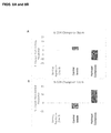
- FIGS. 6A and 6B Flow cytometry plots showing the phenotype of T cells expanded by complexes of the invention.
- FIG. 6A shows the expression of CD3 versus the uptake of propidium iodide (PI) as a marker of cell death after a twelve-day expansion protocol.
- FIG. 6B shows the relative expression of CD8 versus CD4 after a twelve-day expansion protocol.
- PI propidium iodide
- FIGS. 7A and 7B Flow cytometry plots showing the phenotype of T cells expanded by control beads (anti-CD3/CD28-coated beads).
- FIG. 7A shows the expression of CD3 versus the uptake of propidium iodide (PI) as a marker of cell death after a twelve-day expansion protocol.
- FIG. 7B shows the relative expression of CD8 versus CD4 after a twelve-day expansion protocol.
- PI propidium iodide
- FIGS. 8A and 8B Bar graphs showing the percent (%) change in populations of CD4 cells ( FIG. 8A ) and CD8 cells ( FIG. 8B ) over the course of expansion with control beads versus complexes of the invention.
- the data were derived from flow cytometry data analyzed as shown in FIGS. 6 and 7 .
- FIGS. 9A-9E Flow cytometry plots showing the phenotype of T cells at day 0 (a starting population).
- FIG. 9A shows expression of CD8 versus CD4.
- FIG. 9B shows expression of CD45RA versus CD3.
- FIG. 9C shows expression of CD45RO versus CD3.
- FIG. 9D shows expression of CD62L versus CD45RA.
- FIG. 9E shows expression of CCR7 versus CD45RA.
- FIGS. 10A-10E Flow cytometry plots showing the phenotype of control bead-expanded T cells at day 12 of culture.
- FIG. 10A shows expression of CD8 versus CD4.
- FIG. 10B shows expression of CD45RA versus CD3.
- FIG. 10C shows expression of CD45RO versus CD3.
- FIG. 10D shows expression of CD62L versus CD45RA.
- FIG. 10E shows expression of CCR7 versus CD45RA.
- FIGS. 11A-11E Flow cytometry plots showing the phenotype of T cells expanded by the complexes of the invention at day 12 of culture.
- FIG. 11A shows expression of CD8 versus CD4.
- FIG. 11B shows expression of CD45RA versus CD3.
- FIG. 11C shows expression of CD45RO versus CD3.
- FIG. 11D shows expression of CD62L versus CD45RA.
- FIG. 11E shows expression of CCR7 versus CD45RA.
- FIGS. 12A and 12B Bar graphs showing the percent of CD3 + CD45RO + cells at day 0 versus day 6 of expansion with control beads versus complexes of the invention. The data were derived from flow cytometry data analyzed as shown in FIGS. 10 and 11 .
- FIG. 13 Bar graph showing the percent of CD62L + CD45RA + (na ⁇ ve) T cells at day 0 versus day 6 of expansion with control beads versus complexes of the invention. The data were derived from flow cytometry data analyzed as shown in FIGS. 10 and 11 .
- FIG. 14 Bar graph showing the percent of CCR7 + CD45RA + (na ⁇ ve) T cells at day 0 versus day 6 of expansion with control beads versus complexes of the invention. The data were derived from flow cytometry data analyzed as shown in FIGS. 10 and 11 .
- FIG. 15 Bar graph showing the concentration of interferon-gamma (IFN- ⁇ ) secreted at day 6 of expansion with control beads versus complexes of the invention, as measured by enzyme-linked immunosorbant assay (ELISA).
- IFN- ⁇ interferon-gamma
- FIG. 16 Bar graph showing the concentration of interleukin-4 (IL-4) secreted at day 6 of expansion with control beads versus complexes of the invention, as measured by enzyme-linked ELISA.
- IL-4 interleukin-4
- FIG. 17 A series of light microscope images showing T cells expanded by complexes of the invention in comparison with control beads.
- the invention provides a complex that binds to, stimulates, and expands a desired T cell population and facilitates separation of the target population from a sample.
- the complex can be gently dissociated from the binding unit after separating the desired T cell population, representing a safe and efficient approach for processing T cells for clinical use.
- the invention also provides methods of using such complexes as part of adoptive T cell therapy systems.
- the invention features a complex having a binding unit and a separation unit.
- the binding unit includes a binding moiety that binds a component on a T cell surface.
- the binding moiety is linked to a polymeric moiety that includes a cation-sensitive hydrogel. The binding moiety and the polymeric moiety, together, form the binding unit.
- the invention features a binding moiety that can bind to a surface component of a T cell.
- this binding event can lead to signal transduction within the T cell, resulting in activation and/or proliferation of the target cell.
- T cell surface molecules are known to have these downstream effects.
- any ligand that induces clustering of the T cell receptor (signal 1) is able to stimulate that T cell to proliferate and, depending on a secondary signal (signal 2), differentiate towards a particular functional phenotype.
- the signal 1 agent of the present invention is anti-CD3, and the signal 2 agent of the present invention is anti-CD28.
- signal 1 stimuli include MHC-I or MHC-II, and agonists of various other T cell receptor components known in the art.
- signal 2 stimuli include antigen-presenting cell surface molecules that are agonists of co-stimulatory molecules (e.g., CD80, CD86, CD40, ICOSL, CD70, OX40L, 4-1BBL, GITRL, LIGHT, TIM3, TIM4, ICAM1, or LFA3), antibodies toward T cell costimulatory surface molecules other than CD28 (e.g., CD40L, ICOS, CD27, OX40, 4-1 BB, GITR, HVEM, Galectin 9, TIM-1, LFA-1, and CD2), antigen-presenting cell surface molecules that are agonists of co-inhibitory molecules (e.g., CD80, CD86, PD-L1, PD-L2, B7-H3, B7-H4, HVEM, ILT3, or ILT4), and antibodies against co-inhibitory molecules (e.g., CD
- Signal 2 stimulation has been shown to affect multiple aspects of T cell activation. It lowers the concentration of anti-CD3 required to induce a proliferative response in cultures and enhances cytokine production to direct T cell differentiation pathways. Importantly, costimulation helps activate cytolytic potential of CD8 T cells.
- Other molecules including but not limited to CD2 and CD137, can be targeted in activation protocols used to activate and expand various T cell populations, such na ⁇ ve and memory T cells, T helper cells, regulatory T cells, Natural Killer T cells and Cytotoxic T Lymphocytes from mouse and human samples. Additional examples of antibodies, ligands, and other agents useful as signal 1 and signal 2 stimuli for use in the present invention are described in WO2003/024989.
- a binding unit may bind a T cell receptor in an antigen-specific manner analogous to the binding that occurs between the T cell receptor and a peptide-MHC on an antigen presenting cell.
- Synthetic methods to engage a T cell in an antigen specific manner include MHC class I and MHC class II multimers (e.g., dimers, tetramers, and dextramers). Illustrative examples are described in U.S. Pat. No. 7,202,349 and U.S. 2009/0061478.
- Multimerization of MHC-peptide complexes functions to enhance avidity of interaction between peptide-MHC and T cells, which increases the potency of signal 1 transduction.
- Another means to achieve multivalent presentation of antigen-specific T cell receptor ligands is by tethering the ligands to a surface of a complex. Affinity, in contrast to avidity, describes the strength of binding of each individual molecule.
- the affinity of peptide-MHC for a T cell receptor can vary dramatically and dictates the downstream effect of signal 1 stimuli, as discussed below.
- Antigens, and peptides thereof, for use in the present invention include, but are not limited to melanoma antigen recognized by T cells (MART-1), melanoma GP100, the breast cancer antigen, Her-2/Neu, and mucin antigens. Other relevant antigens and sources thereof are described, for example, in U.S. Pat. No. 8,637,307.
- the relative degree of signal 1 and signal 2 binding can influence TCR signal transduction and lead to varying downstream phenotypic effects.
- high affinity with low avidity signal 1 in the absence of signal 2, primes a na ⁇ ve CD4 T cell to turn on Foxp3 expression and differentiate toward a regulatory phenotype (Gottschalk et al., Journal of Experimental Medicine 207 (2010): 1701).
- a na ⁇ ve T cell in response to a high avidity signal 1 stimulus without sufficient signal 2 stimulus, a na ⁇ ve T cell may be more likely to undergo exhaustion, leading to functional anergy (see Ferris et al., J Immunol August 15; 193 (2014): 1525-1530). Either of these effects would be undesireable in the context of cancer adoptive immunotherapy but may be helpful in when priming regulatory T cells for autoimmune treatment.
- the relative contribution of signal 1 and signal 2 can be rationally modulated, depending on the application, by exposing the functionalized complex to a desired ratio of binding moieties.
- the binding moieties may be coupled to the same surface or to separate surfaces.
- a signal 1 stimulus and a signal 2 stimulus are immobilized on the surface at a 1:1 ratio.
- a signal 1 stimulus and a signal 2 stimulus are immobilized on the surface at a ratio of other than 1:1 (e.g., between about 1:100 and about 100:1, between about 1:10 and 10:1, between about 1:2 and 2:1).
- the ratio of signal 1 stimulus to signal 2 stimulus immobilized on the surface is greater than 1:1, or less than 1:1.
- the effect of relative strength of signaling between signal 1 and signal 2 also depends on the activation state of the target T cell. For instance, na ⁇ ve T cells react differently to T cell receptor stimulation and costimulation than antigen-experienced T cells. A skilled artisan would appreciate this effect when selecting a configuration of binding units of the present invention, especially in cases of chronic infection or aberrant immune tolerance, and configure the binding unit accordingly.
- the complex may be formed into any shape compatible with contacting the surface of a T cell. For this reason, however, it is often preferable for a complex to have a high surface area to volume ratio to maximize the available binding surface. Artificial antigen presenting cell platforms having different sizes and shapes have been evaluated for various functional benefits (see Fadel et al., Nano Letters 8 (2008): 2070-2076; Sunshine et al., Biomaterials 35 (2014): 269-277).
- the shape of the structure may be any suitable shape, such as elongated like a wire, tubular, i.e., having a lumen, planar, or spherical.
- An immune synapse can range in size from less than 50 nm to about 20 ⁇ m.
- the complex of the present invention is spherical, and the size is between 50 nm and 20 ⁇ m.
- the complex can be the size of antigen presenting cells, such as dendritic cells or macrophage, which range from about 10-20 ⁇ m and 10-20 ⁇ m, respectively.
- the complex can include a larger matrix, such as a porous scaffold, which can be mechanically exposed, e.g. dipped, into a suspension of cells. Methods for synthesizing such scaffolds, including by using alginate, are known in the art.
- the binding unit of the present invention can be linked to the separation unit by conjugation to a polymeric moiety.
- this conjugation occurs through a covalent reaction.
- Polymeric moieties useful in the present invention include hydrogels that can be dissolved by changing the ionic composition of their environment.
- Hydrogels of the invention are formed from alginic acid conjugated to a polyalkylene oxide, e.g., PEG, as generally described in WO 2012/106658. Conjugation of alginic acid to multifunctional PEG confers greater mechanical strength compared with that achieved solely by ionic crosslinking of alginate. Thus, the mechanical properties (e.g. stiffness) can be modulated by increasing the number of functional groups on each PEG molecule.
- PEG is useful as part of the present invention because of its superior hydrophilic properties, which prevent protein adsorption to the complex of the present invention.
- Adsorption of serum proteins onto the complex can result in aberrant signaling pathways in adjacent cells, such as those caused by Fc receptor engagement.
- the hydrophilic properties of PEG are also functional to maintain a high diffusivity within the complex interior, such that ionic chelators can rapidly access the complex interior to quickly sequester rigidity-maintaining cations.
- incorporation of branched PEG molecules within the hydrogel ensures a rapid dissolution of the hydrogel structure upon exposure to appropriate stimuli.
- any other biocompatible hydrophilic polymer e.g. polyvinyl pyrrolidone, polyvinyl alcohol, and copolymers thereof
- U.S. Pat. No. 7,214,245 can be substituted (see, e.g., U.S. Pat. No. 7,214,245).
- a polymeric moiety of the invention can have one or more mechanical properties (e.g., elastic modulus, Young's modulus, compression modulus, or stiffness) suitable for T cell expansion.
- the mechanical properties of the polymeric moiety can be tailored to be suitable to allow an population of T cells (e.g., a population containing expanded T cells) to retain one or more characteristics of a na ⁇ ve phenotype (e.g., as described in the Methods section) after contacting a complex of the invention.
- the elastic modulus of the polymeric moiety is between 100 pascals (Pa) and 100,000,000 Pa (e.g., from 100 Pa to 1,000 Pa, from 1,000 Pa to 10,000 Pa, between 10,000 Pa to 100,000 Pa, between 100,000 Pa and 1,000,000 Pa, 1,000,000 Pa and 10,000,000 Pa, or 10,000,000 Pa and 100,000,000 Pa, e.g., less than 1,000,000 Pa, less than 900,000 Pa, less than 800,000 Pa, less than 700,000 Pa, less than 600,000 Pa, less than 500,000 Pa, less than 400,000 Pa, less than 300,000 Pa, less than 200,000 Pa, less than 100,000 Pa, less than 50,000 Pa, or less than 10,000 Pa).
- Pa pascals
- 100,000,000 Pa e.g., from 100 Pa to 1,000 Pa, from 1,000 Pa to 10,000 Pa, between 10,000 Pa to 100,000 Pa, between 100,000 Pa and 1,000,000 Pa, 1,000,000 Pa and 10,000,000 Pa, or 10,000,000 Pa and 100,000,000 Pa, e.g., less than 1,000,000 Pa, less than 900,000 Pa, less than 800,000 Pa, less than 700,000
- alginic acid is also a reference to a salt form, e.g., sodium alginate, unless otherwise noted.
- Suitable alginic acid is 20 kDa medium viscosity alginic acid.
- Alginic acid can be present in the polymer moiety in a percentage (e.g., a weight-by-volume percentage) between 0.01 and 10% (e.g., from 0.1% to 0.15%, from 0.15% to 0.2%, from 0.2% to 0.3%, from 0.3% to 0.4%, from 0.4% to 0.5%, from 0.5% to 0.6%, from 0.6% to 0.7%, from 0.7% to 0.8%, from 0.8% to 0.9%, from 0.9% to 1.0%, from 1.0% to 2%, from 2% to 5%, from 5% to 7.5%, or from 7.5% to 10%, e.g., about 0.2%, 0.25%, 0.3%, 0.35%, 0.4%, 0.45%, 0.5%, 0.6%, 0.7%, 0.75%, 0.8%, 0.9%, 1.0%, 1.
- the alginic acid content of the polymeric moiety will influence the stiffness of the polymeric moiety according to known principles (e.g., cation content of the polymeric moiety).
- the stiffness of alginate polymeric moieties can be varied while maintaining a constant or near-constant density according to known methods.
- Polyalkylene oxides e.g., PEG and polypropylene oxide
- Linear or branched, e.g., 4-arm or 8-arm, polyalkylene oxides, e.g., PEG may be employed.
- the polyalkylene oxide, e.g., PEG preferably has a molecular weight between 10 kDa and 20 kDa.
- An exemplary ratio of polyalkylene oxide, e.g., PEG, to alginic acid is 1:2 by weight.
- Alginic acid naturally possesses multiple carboxyl groups that provide convenient groups for conjugation to polyalkylene oxide, e.g., PEG, and/or binding moieties.
- the polyalkylene oxide, e.g., PEG, and binding moiety will naturally possess or be modified to possess an appropriate group to conjugate to a carboxyl group.
- Suitable groups include amine groups, which are often found in binding moieties that include amino acids or can be introduced into binding moieties and polyalkylene oxides, e.g., PEG.
- amine-terminated polyalkylene oxide, e.g., PEG can be employed.
- a linker may be use to conjugate appropriate groups on the polyalkylene oxide, e.g., PEG, or binding moiety to carboxyl groups on the alginic acid.
- a single polyalkylene oxide, e.g., PEG may be conjugated to one or more alginic acid molecules.
- the number of such crosslinks in the composition may or may not be sufficient to form a gel.
- the binding moiety can bind to either the alginic acid directly or to a polyalkylene oxide, e.g., PEG, bound to alginic acid.
- the hydrogel forms by noncovalent crosslinking of the alginic acid with a cation, e.g., Li + , Mg 2+ , Ca 2+ , Sr 2+ , Ba 2+ , Zn 2+ , Cu 2+ , or Al 3+ .
- a preferred cation is Ca 2+ .
- Gelation of hydrogels of the invention may be reserved by contact with a chelator for the cation, e.g., EDTA, EGTA, sodium citrate, BAPTA, crown ether, cryptand, phenanthroline sulfonate, dipyridyl sulfonate, dioxane, DME, diglyme, or triglyme.
- the chelator is a bioinert molecule such as EDTA, which is well-known not to interfere with cell growth and proliferation pathways at concentrations relevant for complex dissolution.
- the complex of the present invention can be formulated by gelation of the hydrogel around a separation unit.
- the hydrogel may be formulated as a coating on the outer surface of the separation unit.
- the separation unit can include multiple smaller structures that are encapsulated within the hydrogel matrix, as shown in FIGS. 2 and 3 .
- the separation unit can be any structure that enables the user to physically manipulate the position of the target cells, once bound with the complex of the invention.
- the separation unit can include one or more magnetic elements to enable physical manipulation, separation, or purification of target cells.
- the magnetic element is encapsulated within a polymeric or hydrogel structure, such that its movement along a magnetic field functions to pull the complex, along with any attached cell, through the field.
- the magnetic element can be one or more microparticles or nanoparticles having magnetic properties. Methods for making magnetic microparticles and nanoparticles and integrating them within polymeric complexes are described in the art (WO 2003/071561, WO 2003/086660, WO 2004/060580, U.S. Pat. No.
- Magnetic particles can be ferrite, oxide, or metallic, and they can be superparamagnetic. Magnetic nanoparticles are often iron oxides, such as magnetite (Fe 3 O 4 ) or maghemite (Fe 3 O 3 ). Magnetic particles can have a shell, such as a polymer shell. The precipitation of iron oxides can occur under non-aqueous conditions (U.S. Pat. No. 4,677,027) and subsequently be converted to aqueous conditions (U.S. Pat. No. 5,160,725) or can occur exclusively in aqueous solutions (U.S. Pat. No.
- Aqueous formulations may preferable in biological applications because of toxicological considerations (U.S. Pat. No. 4,101,435).
- Wet chemical synthesis of the iron oxide crystallites can precede coating with polymer components (core-shell method) or can occur in the presence of the polymer (one-pot method).
- the separation unit can be removed from the target T cell population. This separation can occur in response to exposure of the complex to a cation chelator as discussed above, which results in release of the separation unit from the binding unit.
- compositions of the present invention can be synthesized by methods currently known in the art.
- complexes can be formulated as microparticles by one of several known methods (e.g. microemulsion).
- Techniques useful in encapsulating nanoparticles (i.e. magnetic nanoparticles) within polymeric microparticles are disclosed, for example, in U.S. 2011/0229580.
- Other methods that can be used to entrap a separation unit (i.e. magnetic particles) within a hydrogel include coating by, e.g., dipping, painting, or spraying, with a liquid alginic acid composition and then contacting with cations for crosslinking, e.g., by dipping painting or spraying.
- Alginate PEG co polymer is synthesized using aminated PEG combined in a batch reaction of Alginic acid, EDC, and sulfo NHS.
- Antibody conjugation is performed using standard bio conjugation techniques including malidimide/thiol and EDC/NHS linking. Other useful conjugation methods are described in Hermanson et al., (2013) Bioconjugate Techniques: Academic Press.
- T cell activation or expansion can be performed by isolation of T cells and subsequent stimulation followed and/or expansion.
- a source of T cells can obtained from a subject.
- T cells can be obtained from a number of sources, including peripheral blood mononuclear cells, bone marrow, lymph node tissue, spleen tissue, and tumors.
- a T cell line available in the art may be used.
- T cells can also be obtained from a unit of blood collected from a subject using any number of techniques known to the skilled artisan, such as Ficoll separation or through a PERCOLL® gradient.
- T cells from the circulating blood of an individual can be obtained by apheresis or leukapheresis.
- the apheresis product typically contains lymphocytes including T cells, monocytes, granulocytes, B cells, other nucleated white blood cells, red blood cells, and platelets.
- the cells collected by apheresis may be washed to remove the plasma fraction and to place the cells in an appropriate buffer or media for subsequent processing steps.
- T cells can be enriched prior to treatment with the complex of the present invention through conventional techniques such as magnetic bead negative selection or fluorescence activated cell sorting (FACS).
- T cells can be antigen-specific, antigen-nonspecific, or tumor specific. They can include populations of CD4 cells, CD8 cells, NK T cells, or, for autoimmune or transplant rejection therapies, regulatory T cells.
- T cells that have been obtained from a subject may be further processed prior to or after incubation with the complex of the present invention.
- cells can undergo genetic engineering to equip them with certain functional characteristics, such as in the process of chimeric antigen receptor (CAR) engineering, which equips a patient's cells to recognize cancer antigens.
- CAR chimeric antigen receptor
- the CAR engineering procedure may be performed prior to treatment with the present complexes in order to expand the initially small number of CAR T cells into a substantial activated population.
- CAR chimeric antigen receptor
- bispecific T cell engager (BITE) technology such procedures may be performed after the cells have been expanded using the present invention.
- Other non-genetic processing procedures include but are not limited to treatment with IL-2, IL-4, IL-7, IL-10, IL-12, IL-15, IL-21, or TGF- ⁇ .
- the starting T cell population is incubated with the complexes.
- the ratio of complexes to starting cell number will depend on the size and shape of the complex. For this purpose, it will be understood by a person of skill in the art that the ratio of surface area between target T cells and complexes is a significant factor governing the degree of resulting T cell activation.
- the surface area ratio between target cells and complexes is between about 1:100 and about 100:1 (e.g., about 1:100, about 1:80, about 1:50, about 1:25, about 1:10, about 1:5, about 1:4, about 1:3, about 1:2, about 1:1, about 2:1, about 3:1, about 4:1, about 5:1, about 10:1, about 25:1, about 50:1, about 80:1, or about 100:1).
- Other factors which should be considered when incubating starting T cells with complexes are the density of binding moieties on the complex surfaces, the specific binding moieties chosen (i.e., their binding affinity, EC50, receptor concentration on T cells, etc.). Additionally, it will be appreciated that as T cells proliferate over the course of multiple days, their volumetric requirements will increase, which may limit the total volume that can be allocated to complexes.
- Methods of the present invention include incubating T cells with complexes in a suitable vessel.
- Such cell-culture vessels are known in the art and can include cell culture plates, flasks, or bioreactors of any suitable size.
- Cell culture vessels are preferably sterile, and may be configured for optimal gas exchange or media exchange, such as perfusion capable systems, which are known in the art.
- T cells can be seeded at a concentration of between about 0.2 ⁇ 10 6 and 10 ⁇ 10 6 cells/ml.
- Complexes may require special ionic conditions, e.g., to maintain a solid structure in solution.
- cell culture media can be supplemented with ions, such as Ca 2+ , through addition of salts, e.g., CaCl 2 .
- Ions can be present at any physiologically suitable concentration (e.g., 1.0 nM-100 mM, e.g., 1.0 ⁇ M to 10 mM, e.g., 0.1 mM, 0.2 mM, 0.5 mM, 1.0 mM, 2 mM, 3 mM, 4 mM, 5 mM, 6 mM, 7 mM, 8 mM, 9 mM, or 10 mM).
- physiologically suitable concentration e.g., 1.0 nM-100 mM, e.g., 1.0 ⁇ M to 10 mM, e.g., 0.1 mM, 0.2 mM, 0.5 mM, 1.0 mM, 2 mM, 3 mM, 4 mM, 5 mM, 6 mM, 7 mM, 8 mM, 9 mM, or 10 mM).
- Ex vivo T cell expansion protocols are well-developed, especially for human samples, and can often yield cell number increases in the hundred-fold range, over the course of multiple weeks of culture. Thus, multiple rounds of expansion may be required to overcome constraints on physical space and media nutrient depletion.
- the complexes of the present invention can be dissolved (i.e., the separation unit removed) multiple times (e.g., once, twice, or 3-12 times) over the course of a T cell expansion protocol.
- Such dissolution/re-administration cycles can be achieved by washing out cation chelators from the media by centrifugation or other known methods after each dissolution.
- additional complexes can be introduced into the cultures without removing separation units of the existing complexes, thus bypassing the need for media changes. If necessary, beads encapsulating magnetic particles, or excess magnetic particles alone, can be removed from cultures using well-established magnetic separation procedures.
- the T cell expansion protocol proceeds for a predetermined length of time that is suitable to generate a desired number of T cells, a representative phenotype, or both.
- the phenotypic properties of T-cell populations of the present invention can be monitored by a variety of methods, and the isolation can be performed after the desired phenotype is acquire.
- Relevant phenotypes are metabolic changes such as biochemical or morphological changes (e.g., change in frequency of cell division, change in cytokine expression profile, change in median cell diameter, change in surface molecule expression, or change in cellular motility).
- Assays for monitoring such changes include standard flow cytometry methods, ELISA, microscopy, migration assays, metabolic assays, and other techniques known by those skilled in the art.
- the methods of the present invention allow for expansion of a population of cells containing na ⁇ ve T cells, central memory T cells, and/or effector memory cells.
- the composition of the polymeric moiety can influence the percentage and/or total number of na ⁇ ve T cells, central memory T cells, and/or effector memory cells present in a population (e.g., an expanded population).
- a resulting population of T cells may include a high percentage of na ⁇ ve T cells (e.g., na ⁇ ve CD4 + T cells or na ⁇ ve CD8 + T cells).
- na ⁇ ve T cells are characterized as having one or more properties (surface markers or secreted cytokines) of Table I (see Examples).
- Alginic acid provides a physical anchor to entrap the separation unit.
- a cation e.g., calcium
- alginic acid is crosslinked and solid.
- the hydrogel can be dissolved using a chelator to release the separation unit from the T cell.
- the separation unit includes magnetic particles that are not permissible as part of clinical samples (e.g. the resulting activated T cell suspension). By releasing the separation unit (e.g. magnetic particles) by cation chelation, the resulting T cells are efficiently cleaned of potentially harmful impurities and ready for infusion into a patient in need thereof.
- EDTA is a well-characterized calcium chelator of use in the present invention.
- EDTA can be used at 2 mM in the form of a physiologically insert buffer that preferably also contains 137 mM NaCl, 2.7 mM KCl, 25 mM Hepes and 0.75 mM Na 2 H 2 PO 4 .2H 2 O.
- Cell culture media can be exchanged for the ion chelator solution, e.g., EDTA buffer, potentially by centrifugation and subsequent resuspension of cell pellet, e.g., in EDTA buffer. Other methods may also be used.
- the cell/ion chelator suspension may also be agitated, e.g., by pipetting or vortexing for about 5 seconds.
- Cell suspension can be placed on a magnetic stand for long enough to allow separation of all magnetic particles from cells (5 minutes is typical). During this step, the hydrogel dissolves, cells are released, and the magnetic particles are attracted to the side of the vessel being used (e.g. tube).
- the supernatant can be changed to a clean tube, while magnetic particles can be left behind. This step can be repeated again in the initial tube in order to recover any cells that remain attached to magnetic particles.
- the isolated cells can then be returned to cell culture media or isotonic solution (e.g., for administration into a patient).
- FIG. 1 shows a workflow for the manufacture of chimeric antigen receptor (CAR) T cells using CD3/CD28 complexes for T cell activation and expansion.
- CAR chimeric antigen receptor
- FIG. 2 is a light microscope image showing complexes of the present invention, which include a magnetic bead substrate coated with an alginic acid hydrogel.
- the hydrogel is a novel dissolvable polymer, which has the ability to be conjugated with ligands to facilitate the attachment and subsequent release of targeted cell types.
- FIG. 3 is a schematic of a complex of the present invention, which includes a magnetic bead substrate coated with an alginic acid hydrogel, which has been conjugated with anti-CD3 and anti-CD28 ligands.
- FIG. 4 shows an exemplary timeline of procedures carried out to prepare a T cell population with hydrogel complexes of the present invention.
- cells are seeded in a 24 well plate with hydrogel complexes.
- additional media is added to the cells and the culture is mixed by gently pipetting up and down five times.
- cells are counted and transferred to a six-well plate, where they are cultured in experiment-specific conditions or allowed to continue growth.
- FIGS. 5-17 show characteristics of T cell populations treated using methods of the invention, in comparison to conventional methods involving commercially available anti-CD3/CD28 magnetic beads (control).
- Primary human T lymphocytes were seeded at a density of 1 ⁇ 10 6 cells/mL and cultured in advanced RPMI medium supplemented with fetal bovine serum, glutamate, HEPES, and recombinant human IL-2.
- the media was further supplemented with 2 mM CaCl 2 .
- control beads On day one of culture, cells were stimulated with either control beads at a concentration of one bead per cell or with two different formulations of hydrogel complexes coated with anti-CD3 and anti-CD28 antibodies (Affymetrix/ebioscience).
- T cells expanded potently and in a sustained manner ( FIG. 5 ). Cells proliferated to a similar degree whether they were treated with control beads or hydrogel complexes. In unstimulated cells, no cell expansion occurred.
- FIG. 6A After a twelve-day T cell expansion, cells were stained for viability using propidium iodide. Cell viability was high (greater than 99%) in both hydrogel complex-expanded T cells ( FIG. 6A ) and control bead-expanded T cells ( FIG. 7A ). Live samples of each expanded population were then analyzed for surface expression of T cell markers CD3, CD4, and CD8 ( FIGS. 6-8 ). Under each expansion condition, greater than 97% of cells expressed CD3 ( FIGS. 6A and 7A ). Cells expanded using hydrogel complex formulation #1 were 33.24% CD4-positive and 62.84% CD8-positive ( FIG. 6B ). T cells expanded by control beads preferentially expressed CD4, relative to T cells expanded by hydrogel beads ( FIG.
- FIGS. 8A and 8B show the percent change in CD4 and CD8 expression, respectively, at day 6 of expansion relative to the initial expression levels (e.g., of the starting population at day 0).
- the percentage of CD4 + T cells decreased by more than 10% from day 0 to day 6, whereas the percentage of CD4 + T cells in the control bead-treated groups increased by about 4% ( FIG. 8A ). Furthermore, the percentage of CD8 + T cells in the hydrogel complex-expanded groups increased by more than 30%, whereas the CD8 + population of control-treated cells diminished by more than 10% ( FIG. 8B ).
- CD45RA + cells that simultaneously express CD62L or CCR7 are characterized as na ⁇ ve T cells.
- each expanded population was analyzed via flow cytometry for surface expression of CD3, CD4, CD8, CD45RO, CD45RA, CD62L, and CCR7 and compared with the expression profiles of cells at day 0 ( FIGS. 9A-9E ).
- FIGS. 9A-9E a greater percentage of cells treated with hydrogel complexes were CD45RA + , relative to cells treated with control beads ( FIG. 12B ), suggesting that treatment with hydrogel complexes preferentially yields na ⁇ ve T cells.
- dual expression of CD62L and CD45RA was assessed.
- the secretion of certain effector cytokines can be indicative of a T cell's effector memory phenotype.
- Six days after induction of T cell expansion with control beads or hydrogel complexes samples of cell culture medium were harvested and analyzed for the presence of IL-4 and IFN- ⁇ by ELISA.
- Medium from hydrogel complex-treated cultures contained lower quantities of IFN- ⁇ ( FIG. 15 ) and IL-4 ( FIG. 16 ), relative to control bead-treated cultures, reflecting a smaller population of effector memory T cells within the hydrogel complex expanded population.
- FIG. 17 show hydrogel complex-expanded T cell cultures following hydrogel dissolution and removal. After 7 days of culture in the presence of hydrogel complexes, 5 mM EDTA was added to the culture medium, and cells were briefly agitated. Following agitation, cultures were placed on a cell separation magnetic stand (Quad Technologies) for 5 minutes. The supernatant was then removed and replated. As shown, hydrogel complex-expanded cells were free of magnetic particles.
- the bottom panels of FIG. 17 show control bead-expanded T cells following an analogous separation process.
Landscapes
- Health & Medical Sciences (AREA)
- Immunology (AREA)
- Chemical & Material Sciences (AREA)
- Life Sciences & Earth Sciences (AREA)
- Organic Chemistry (AREA)
- General Health & Medical Sciences (AREA)
- Engineering & Computer Science (AREA)
- Genetics & Genomics (AREA)
- Medicinal Chemistry (AREA)
- Biomedical Technology (AREA)
- Biochemistry (AREA)
- Cell Biology (AREA)
- Microbiology (AREA)
- Molecular Biology (AREA)
- Zoology (AREA)
- Proteomics, Peptides & Aminoacids (AREA)
- Wood Science & Technology (AREA)
- Biotechnology (AREA)
- Biophysics (AREA)
- Bioinformatics & Cheminformatics (AREA)
- Public Health (AREA)
- Epidemiology (AREA)
- Animal Behavior & Ethology (AREA)
- Pharmacology & Pharmacy (AREA)
- Veterinary Medicine (AREA)
- Mycology (AREA)
- General Engineering & Computer Science (AREA)
- Hematology (AREA)
- Micro-Organisms Or Cultivation Processes Thereof (AREA)
- Oncology (AREA)
- Medicines Containing Material From Animals Or Micro-Organisms (AREA)
Abstract
The invention features a complex that binds to, stimulates, and expands a desired T cell population and facilitates separation of the target population from a sample. The complex can be gently dissociated from the binding unit after separating the desired T cell population, representing a safe and efficient approach for processing T cells for clinical use. Invention also provides methods of using such complexes as part of adoptive T cell therapy systems.
Description
The invention is generally directed to the fields of T cell activation, cell culture, and bioprocessing. More specifically, the invention is directed toward improved protocols for manufacturing large numbers of viable engineered T lymphocytes for clinical immunotherapy applications.
T lymphocytes isolated from whole blood are utilized in a wide variety of in vitro, in vivo, and clinical research and therapeutic applications. Examples include studies of immune response, T cell receptor signaling, cytokine release and gene expression profiling. Perhaps most significantly, isolation and subsequent ex vivo engineering of T lymphocytes for subsequent transplantation into clinical patients is showing tremendous promise as a novel cancer therapy. The main approaches to this are engineering of T cells to express either chimeric antigen receptors (CAR) or T cell receptors (TCR). In both approaches, T cells are isolated from whole blood, activated and expanded ex vivo, and subsequently infused into human subjects.
Although both polyclonal and antigen-specific T cells can be readily isolated from whole blood, their numbers are limited. Accordingly, protocols that activate and promote ex vivo expansion of T cells are widely used. Such ex vivo manipulations, however, can potentially reduce T cell viability, proliferation, and survival after infusion. Thus, the choice of methods used for T cell activation has important implications for clinical efficacy.
It is well-established that, in vivo, activation of T cells is dependent on two signals; engagement of the T cell receptor with antigen (signal 1) and ligation of a costimulatory molecule (signal 2). Both are required for an effective immune response. Ex vivo, T cell activation is most commonly induced by exposing the T cells to antibodies directed against the T cell surface markers CD3 and CD28 to engage the T cell receptor and deliver a costimulatory signal simultaneously.
There are significant disadvantages of conventional ex-vivo T cell activation protocols, which use magnetic beads, resulting from the presence of residual magnetic beads attached to the cells. These may negatively affect both function and viability. Pre-clinical clinical applications require cells are that are free from contaminating particles, while retaining high viablilty. For example, June et al. (Pilot study of redirected autologous T cells engineered to contain humanized anti-CD19 in patients with relapsed or refractory CD19+ leukemia and lymphoma previously treated with cell therapy (2015) ClinicalTrials.gov) specified final product release criteria in the IND included the specifications that the number of anti-CD3/CD28-coated paramagnetic beads should not exceed 100 per 3×10⁶ cells and that cell viability should be greater than 70%. However, minimizing the number of beads represents a formidable obstacle in the clinical translation of such therapies, as most antibody-coated magnetic-bead based products lack the ability to readily release bound cells from capture molecules in a manner that does not alter the viability and phenotype of the isolated cells.
Given the significant interest in, and rapid expansion of, T cell engineering-based cancer therapies, there is a significant need for improved T cell activation and harvesting methods that overcome the above limitations of existing approaches, particularly for downstream clinical applications. Specifically, there is a substantial need for technologies to enable ex-vivo cell expansion protocols to meet clinical specifications, to consistently and reproducibly activate T cells, to preserve cell viability and function, and to be applicable to different cell sources and activating agents.
We have developed a unique hydrogel that is biocompatible, functionalized with T cell activating ligands such as antibodies, and can be dissolved rapidly simply by switching to a buffer containing a chelating agent. Together, these properties provide a unique technology that eliminates retained cell tagging agents, such as magnetic beads, from isolated cell populations. Among its many benefits, the dissolution process does not affect cell viability as other coating chemistries do.
In one aspect, the invention features a complex including a binding unit, i.e., hydrogel, and a separation unit, e.g., magnetic beads, in which the binding unit includes a polymeric moiety and binds a cell surface component of a lymphocyte, and the separation unit dissociates from the binding unit upon exposure of the polymeric moiety to an ion chelator. In some embodiments, the separation unit is entrapped within the polymeric moiety. In a preferred embodiment, the polymeric moiety comprises a polymer that changes from a solid matrix into a solution or suspension in response to decrease in cationic concentration (e.g., Li+, Mg2+, Ca2+, Sr2+, Ba2+, Zn2+, Cu2+, or Al3+) caused by the presence of an ion chelator (e.g., EDTA, EGTA, sodium citrate, BAPTA, crown ether, cryptand, phenanthroline sulfonate, dipyridyl sulfonate, dioxane, DME, diglyme, or triglyme). In some embodiments, the elastic modulus of the polymeric moiety is between 100 pascals (Pa) and 100,000,000 Pa (e.g., from 100 Pa to 1,000 Pa, from 1,000 Pa to 10,000 Pa, between 10,000 Pa to 100,000 Pa, between 100,000 Pa and 1,000,000 Pa, 1,000,000 Pa and 10,000,000 Pa, or 10,000,000 Pa and 100,000,000 Pa, e.g., less than 1,000,000 Pa, less than 900,000 Pa, less than 800,000 Pa, less than 700,000 Pa, less than 600,000 Pa, less than 500,000 Pa, less than 400,000 Pa, less than 300,000 Pa, less than 200,000 Pa, less than 100,000 Pa, less than 50,000 Pa, or less than 10,000 Pa). In some embodiments, the polymer is a hydrogel such as alginic acid or an alginic acid-polyethylene glycol (PEG) copolymer, e.g., having a median viscosity of between about 10 and 100 kDa (e.g. about 10, 20, 30, 40, 50, 60, 70, 80, 90, or 100 kDa).
In certain embodiments, the binding unit includes a magnetic material, such as iron. In some embodiments, the magnetic material is part of a nanoparticle or a microparticle, e.g., which can have a polymeric coating.
In one aspect, the complex of the invention has at least one dimension of between about 50 nm and about 50 μm (e.g. 50 nm, 100 nm, 200 nm, 300 nm, 400 nm, 500 nm, 600 nm, 700 nm, 800 nm, 900 nm, 1 μm, 2 μm, 3 μm, 4 μm, 5 μm, 10 μm, 20 μm, 30 μm, 40 μm, or 50 μm). In some aspects, the complex is spherical. In some aspects, the diameter is between about 1 and 20 μm (e.g., 1 μm, 2 μm, 3 μm, 4 μm, 5 μm, 10 μm, 20 μm).
In some embodiments, the binding moiety is conjugated to the polymeric moiety after the separation unit is entrapped within the polymeric moiety. In other embodiments, the binding moiety is conjugated to the polymeric moiety before the separation unit is entrapped within the polymeric moiety.
In some embodiments, the binding moiety is covalently attached to an alginic acid domain, e.g., of an alginic acid-PEG copolymer (e.g., covalently attached through a carboxylate group of the alginic acid).
In one aspect, the invention features a complex having a signal 1 stimulus. In some aspects, the complex has both a signal 1 stimulus and a signal 2 stimulus. In some embodiments, the molar ratio of the signal 1 stimulus to the signal 2 stimulus is between about 1:100 and about 100:1 (e.g., about 1:100, about 1:80, about 1:50, about 1:25, about 1:10, about 1:5, about 1:4, about 1:3, about 1:2, about 1:1, about 2:1, about 3:1, about 4:1, about 5:1, about 10:1, about 25:1, about 50:1, about 80:1, or about 100:1). In some embodiments, the molar ratio of the signal 1 stimulus to the signal 2 stimulus is between about 1:10 and about 10:1. In some embodiments, the molar ratio of the signal 1 stimulus to the signal 2 stimulus is between about 1:2 and about 2:1. In some embodiments, the molar ratio of the signal 1 stimulus and the signal 2 stimulus is greater than 1:1, and in other embodiments, the molar ratio of the signal 1 stimulus and the signal 2 stimulus is less than 1:1.
In one embodiment, the binding moiety has an antigen-specific signal 1 stimulus. In another embodiment, the signal 1 stimulus is antigen-nonspecific. In some aspects, the binding moiety includes an antibody or antigen-binding fragment thereof. In these aspects, the antibody or antigen binding fragment thereof is a monoclonal antibody or antigen-binding fragment thereof, a Fab, a humanized antibody or antigen-binding fragment thereof, a bispecific antibody or antigen-binding fragment thereof, a monovalent antibody or antigen-binding fragment thereof, a chimeric antibody or antigen-binding fragment thereof, a single-chain Fv molecule, a bispecific single chain Fv ((scFv′) 2) molecule, a domain antibody, a diabody, a triabody, an affibody, a domain antibody, a SMIP, a nanobody, a Fv fragment, a Fab fragment, a F(ab′) 2 molecule, or a tandem scFv (taFv) fragment.
In one embodiment, the binding moiety binds one or more human antigens.
In one aspect, the invention features a complex with binding moiety made up of one or more antibodies or antigen-binding fragments thereof selected from the group of anti-CD2, anti-CD3, anti-CD27, anti-CD28, anti-CD46, anti-CD137, or antigen binding fragments thereof. In one aspect, the signal 1 stimulus is anti-CD3. In another aspect, the signal 2 stimulus is anti-CD28. In another aspect, the complex includes both a signal 1 stimulus that is anti-CD3 and a signal 2 stimulus is anti-CD28.
In some aspects, the binding moiety is covalently attached to the polymeric moiety. In some aspects, the complex has density of at least 1 binding moiety per square μm of surface area (e.g., at least 1, 2, 3, 4, 5, 6, 7, 8, 9, 10, 15, 20, 30, 40, 50, 60, 70, 80, 90, 100, or more binding moieties per square μm of surface area.
In another aspect, the invention features a method of activating a population of T cells involving; first, contacting a starting population of T cells with complexes each having binding unit and a separation unit, in which the binding unit binds a surface component of a T cell, and in which the contacting induces a metabolic change in the starting population of T cells; second, treating the complex with an ion chelator, which dissociates the separation unit from the T cells; and lastly, isolating a resulting population of T cells from the separation unit. In one embodiment, the ion chelator chelates a cation (e.g., Li+, Mg2+, Ca2+, Sr2+, Ba2+, Zn2+, Cu2+, Al3+). In one embodiment, the chelator includes EDTA, EGTA, sodium citrate, BAPTA, crown ether, cryptand, phenanthroline sulfonate, dipyridyl sulfonate, dioxane, DME, diglyme, or triglyme.
In one embodiment, the separation unit is magnetic, and the isolation is performed using a magnet.
In one embodiment, the population of T cells is antigen-specific. In a different embodiment, the population of T cells is antigen-nonspecific. In one embodiment, the population of T cells is tumor-specific. In another embodiment, the population of T cells includes CD4 T cells. In another embodiment, the population of T cells includes CD8 T cells. The resulting population of T cells may have a number or percentage of CD8 T cells that is greater than the number or percentage of CD8 T cells of a starting population (e.g., 10% more, 20% more, 30% more, 40% more, 50% more, 75% more, or greater).
Additionally or alternatively, the resulting population of T cells may have a number or percentage of CD8 T cells that is greater than the number or percentage of CD4 T cells in the resulting population. In another embodiment, the population of T cells includes NK T cells. In another embodiment, the population of T cells includes regulatory T cells.
In some embodiments, the resulting population of cells has at least 2% naïve T cells (e.g., at least 3%, at least 4%, at least 5%, at least 6%, at least 7%, at least 8%, at least 9%, at least 10%, or more, e.g., about 5%, about 10%, about 15%, or more). In some embodiments, the resulting population of cells has at greater number or percentage of naïve T cells relative to cells expanded by magnetic beads without a hydrogel coating (e.g., Dynabeads, e.g., 10% more, 20% more, 50% more, 100% more, or greater, e.g., 2-fold more, 3-fold more, 4-fold more, 5-fold more, 10-fold more, or greater). Additionally or alternatively, the resulting population of cells may have a greater number or percentage of central memory T cells than the starting population (e.g., 10% more, 20% more, 30%, more, 40% more, 50% more, 75% more, 100% more, 150% more, 200% more, 300% more, 400% more, 500% more, 1,000% more, 5,000% more, 10,000% more, or greater). In some embodiments, the naïve T cells are CD45RA+ cells, CD45RA+CD62L+ cells, or CD45RA+CCR7+ cells. In other embodiments, the naïve T cells secrete lower quantities of IL-4 and/or IFN-γ than a reference population of cells (e.g., wherein the reference population is the starting population, a central memory cell population, an effector memory population, or an activated population).
In one embodiment, the population of T cells is isolated from a subject. In a different embodiment, the population of T cells is derived from a cell line. In one aspect, the starting population of T cells comprises a genetic modification, such as that resulting from a chimeric antigen receptor (CAR) modification.
In one aspect, the metabolic change induced in the T cells by contacting them with the complexes includes a biochemical or a morphological change. This change may be a greater frequency of cell division, a change in cytokine secretion profile (e.g., of IL-4 and/or IFN-γ), an increase in median cell diameter, a change surface molecule expression profile, or a change in cellular motility.
In another aspect, the method includes seeding the starting population of T cells at a concentration of between about 0.2×10⁶ and 10×106 cells/ml (e.g., 0.2×10⁶, 0.4×10⁶, 0.6×10⁶, 0.8×10⁶, 1.0×10⁶, 2.0×10⁶, 3.0×10⁶, 4.0×10⁶, 5.0×10⁶, or 10×106 cells/ml). In yet another aspect, the method includes seeding the starting population of T cells and the complexes at a ratio between about 1:100 and about 100:1 (e.g., about 1:100, about 1:80, about 1:50, about 1:25, about 1:10, about 1:5, about 1:4, about 1:3, about 1:2, about 1:1, about 2:1, about 3:1, about 4:1, about 5:1, about 10:1, about 25:1, about 50:1, about 80:1, or about 100:1).
In another embodiment, the method includes a step of treating the complex with an ion chelator multiple times over the course of the T cell expansion process. In another embodiment, the method further includes contacting the population of T cells with a plurality of complexes multiple times over the course of T cell expansion process. In one aspect, the isolation of the resulting population of T cells from the separation unit is performed after a predetermined length of time, or alternatively, it is performed after the resulting population of T cells acquires a desired phenotype.
In some embodiments of any of the preceding methods, cell culture media can be supplemented with ions, such as Ca2+, through addition of salts, e.g., CaCl2. Ions can be present at any physiologically suitable concentration (e.g., 1.0 nM-100 mM, e.g., 1.0 μM to 10 mM, e.g., 0.1 mM, 0.2 mM, 0.5 mM, 1.0 mM, 2 mM, 3 mM, 4 mM, 5 mM, 6 mM, 7 mM, 8 mM, 9 mM, or 10 mM).
In some embodiments, T cells expanded by any of the compositions or methods of the present invention express CD8 and CD4, e.g., at a ratio of between 1:10 and 10:1, e.g., at a ratio of 1:10, 1:8, 1:5, 1:2, 1:1, 2:1, 5:1, 8:1, or 10:1. In particular embodiments, the number of T cells expressing CD8 is greater than the number of T cells expressing CD4 in the expanded population. Complexes and methods of the invention can induce an expanded T cell culture having a greater proportion of CD8+ cells than a population expanded by conventional means (e.g., anti-CD3/CD28 coated beads).
The invention provides a complex that binds to, stimulates, and expands a desired T cell population and facilitates separation of the target population from a sample. The complex can be gently dissociated from the binding unit after separating the desired T cell population, representing a safe and efficient approach for processing T cells for clinical use. The invention also provides methods of using such complexes as part of adoptive T cell therapy systems.
Binding Unit
The invention features a complex having a binding unit and a separation unit. The binding unit includes a binding moiety that binds a component on a T cell surface. The binding moiety is linked to a polymeric moiety that includes a cation-sensitive hydrogel. The binding moiety and the polymeric moiety, together, form the binding unit.
T Cell Binding Moieties
In one aspect, the invention features a binding moiety that can bind to a surface component of a T cell. Preferably, this binding event can lead to signal transduction within the T cell, resulting in activation and/or proliferation of the target cell. Several T cell surface molecules are known to have these downstream effects. In general, any ligand that induces clustering of the T cell receptor (signal 1) is able to stimulate that T cell to proliferate and, depending on a secondary signal (signal 2), differentiate towards a particular functional phenotype. Preferably, the signal 1 agent of the present invention is anti-CD3, and the signal 2 agent of the present invention is anti-CD28. Other examples of signal 1 stimuli include MHC-I or MHC-II, and agonists of various other T cell receptor components known in the art. Other examples of signal 2 stimuli include antigen-presenting cell surface molecules that are agonists of co-stimulatory molecules (e.g., CD80, CD86, CD40, ICOSL, CD70, OX40L, 4-1BBL, GITRL, LIGHT, TIM3, TIM4, ICAM1, or LFA3), antibodies toward T cell costimulatory surface molecules other than CD28 (e.g., CD40L, ICOS, CD27, OX40, 4-1 BB, GITR, HVEM, Galectin 9, TIM-1, LFA-1, and CD2), antigen-presenting cell surface molecules that are agonists of co-inhibitory molecules (e.g., CD80, CD86, PD-L1, PD-L2, B7-H3, B7-H4, HVEM, ILT3, or ILT4), and antibodies against co-inhibitory molecules (e.g., CDTL-4, PD-1, BTLA, or CD160). Signal 2 stimulation has been shown to affect multiple aspects of T cell activation. It lowers the concentration of anti-CD3 required to induce a proliferative response in cultures and enhances cytokine production to direct T cell differentiation pathways. Importantly, costimulation helps activate cytolytic potential of CD8 T cells. Other molecules, including but not limited to CD2 and CD137, can be targeted in activation protocols used to activate and expand various T cell populations, such naïve and memory T cells, T helper cells, regulatory T cells, Natural Killer T cells and Cytotoxic T Lymphocytes from mouse and human samples. Additional examples of antibodies, ligands, and other agents useful as signal 1 and signal 2 stimuli for use in the present invention are described in WO2003/024989.
Antigen-Specific T Cell Engagement
In one aspect of the invention, a binding unit may bind a T cell receptor in an antigen-specific manner analogous to the binding that occurs between the T cell receptor and a peptide-MHC on an antigen presenting cell. Synthetic methods to engage a T cell in an antigen specific manner (i.e., in the absence of a natural antigen presenting cell) include MHC class I and MHC class II multimers (e.g., dimers, tetramers, and dextramers). Illustrative examples are described in U.S. Pat. No. 7,202,349 and U.S. 2009/0061478. Multimerization of MHC-peptide complexes functions to enhance avidity of interaction between peptide-MHC and T cells, which increases the potency of signal 1 transduction. Another means to achieve multivalent presentation of antigen-specific T cell receptor ligands is by tethering the ligands to a surface of a complex. Affinity, in contrast to avidity, describes the strength of binding of each individual molecule. The affinity of peptide-MHC for a T cell receptor can vary dramatically and dictates the downstream effect of signal 1 stimuli, as discussed below. Antigens, and peptides thereof, for use in the present invention include, but are not limited to melanoma antigen recognized by T cells (MART-1), melanoma GP100, the breast cancer antigen, Her-2/Neu, and mucin antigens. Other relevant antigens and sources thereof are described, for example, in U.S. Pat. No. 8,637,307.
Effects of the Degree of T Cell Binding
The relative degree of signal 1 and signal 2 binding can influence TCR signal transduction and lead to varying downstream phenotypic effects. For example, high affinity with low avidity signal 1, in the absence of signal 2, primes a naïve CD4 T cell to turn on Foxp3 expression and differentiate toward a regulatory phenotype (Gottschalk et al., Journal of Experimental Medicine 207 (2010): 1701).
Alternatively, in response to a high avidity signal 1 stimulus without sufficient signal 2 stimulus, a naïve T cell may be more likely to undergo exhaustion, leading to functional anergy (see Ferris et al., J Immunol August 15; 193 (2014): 1525-1530). Either of these effects would be undesireable in the context of cancer adoptive immunotherapy but may be helpful in when priming regulatory T cells for autoimmune treatment. Thus, the relative contribution of signal 1 and signal 2 can be rationally modulated, depending on the application, by exposing the functionalized complex to a desired ratio of binding moieties.
The binding moieties may be coupled to the same surface or to separate surfaces. In a preferred embodiment, a signal 1 stimulus and a signal 2 stimulus are immobilized on the surface at a 1:1 ratio. In certain aspects of the present invention, a signal 1 stimulus and a signal 2 stimulus are immobilized on the surface at a ratio of other than 1:1 (e.g., between about 1:100 and about 100:1, between about 1:10 and 10:1, between about 1:2 and 2:1). In other aspects, the ratio of signal 1 stimulus to signal 2 stimulus immobilized on the surface is greater than 1:1, or less than 1:1.
The effect of relative strength of signaling between signal 1 and signal 2 also depends on the activation state of the target T cell. For instance, naïve T cells react differently to T cell receptor stimulation and costimulation than antigen-experienced T cells. A skilled artisan would appreciate this effect when selecting a configuration of binding units of the present invention, especially in cases of chronic infection or aberrant immune tolerance, and configure the binding unit accordingly.
Size and Shape of the Complex
The complex may be formed into any shape compatible with contacting the surface of a T cell. For this reason, however, it is often preferable for a complex to have a high surface area to volume ratio to maximize the available binding surface. Artificial antigen presenting cell platforms having different sizes and shapes have been evaluated for various functional benefits (see Fadel et al., Nano Letters 8 (2008): 2070-2076; Sunshine et al., Biomaterials 35 (2014): 269-277). The shape of the structure may be any suitable shape, such as elongated like a wire, tubular, i.e., having a lumen, planar, or spherical. An immune synapse can range in size from less than 50 nm to about 20 μm. Preferably, the complex of the present invention is spherical, and the size is between 50 nm and 20 μm. For example, the complex can be the size of antigen presenting cells, such as dendritic cells or macrophage, which range from about 10-20 μm and 10-20 μm, respectively. Alternatively, the complex can include a larger matrix, such as a porous scaffold, which can be mechanically exposed, e.g. dipped, into a suspension of cells. Methods for synthesizing such scaffolds, including by using alginate, are known in the art.
Polymeric Moiety
The binding unit of the present invention can be linked to the separation unit by conjugation to a polymeric moiety. Preferably, this conjugation occurs through a covalent reaction.
Polymeric moieties useful in the present invention include hydrogels that can be dissolved by changing the ionic composition of their environment. Hydrogels of the invention are formed from alginic acid conjugated to a polyalkylene oxide, e.g., PEG, as generally described in WO 2012/106658. Conjugation of alginic acid to multifunctional PEG confers greater mechanical strength compared with that achieved solely by ionic crosslinking of alginate. Thus, the mechanical properties (e.g. stiffness) can be modulated by increasing the number of functional groups on each PEG molecule. PEG is useful as part of the present invention because of its superior hydrophilic properties, which prevent protein adsorption to the complex of the present invention. Adsorption of serum proteins onto the complex can result in aberrant signaling pathways in adjacent cells, such as those caused by Fc receptor engagement. The hydrophilic properties of PEG are also functional to maintain a high diffusivity within the complex interior, such that ionic chelators can rapidly access the complex interior to quickly sequester rigidity-maintaining cations. Thus, incorporation of branched PEG molecules within the hydrogel ensures a rapid dissolution of the hydrogel structure upon exposure to appropriate stimuli. Alternatively, any other biocompatible hydrophilic polymer (e.g. polyvinyl pyrrolidone, polyvinyl alcohol, and copolymers thereof) can be substituted (see, e.g., U.S. Pat. No. 7,214,245).
A polymeric moiety of the invention can have one or more mechanical properties (e.g., elastic modulus, Young's modulus, compression modulus, or stiffness) suitable for T cell expansion. For example, the mechanical properties of the polymeric moiety can be tailored to be suitable to allow an population of T cells (e.g., a population containing expanded T cells) to retain one or more characteristics of a naïve phenotype (e.g., as described in the Methods section) after contacting a complex of the invention. In some cases, the elastic modulus of the polymeric moiety is between 100 pascals (Pa) and 100,000,000 Pa (e.g., from 100 Pa to 1,000 Pa, from 1,000 Pa to 10,000 Pa, between 10,000 Pa to 100,000 Pa, between 100,000 Pa and 1,000,000 Pa, 1,000,000 Pa and 10,000,000 Pa, or 10,000,000 Pa and 100,000,000 Pa, e.g., less than 1,000,000 Pa, less than 900,000 Pa, less than 800,000 Pa, less than 700,000 Pa, less than 600,000 Pa, less than 500,000 Pa, less than 400,000 Pa, less than 300,000 Pa, less than 200,000 Pa, less than 100,000 Pa, less than 50,000 Pa, or less than 10,000 Pa).
A reference to alginic acid is also a reference to a salt form, e.g., sodium alginate, unless otherwise noted. Suitable alginic acid is 20 kDa medium viscosity alginic acid. Alginic acid can be present in the polymer moiety in a percentage (e.g., a weight-by-volume percentage) between 0.01 and 10% (e.g., from 0.1% to 0.15%, from 0.15% to 0.2%, from 0.2% to 0.3%, from 0.3% to 0.4%, from 0.4% to 0.5%, from 0.5% to 0.6%, from 0.6% to 0.7%, from 0.7% to 0.8%, from 0.8% to 0.9%, from 0.9% to 1.0%, from 1.0% to 2%, from 2% to 5%, from 5% to 7.5%, or from 7.5% to 10%, e.g., about 0.2%, 0.25%, 0.3%, 0.35%, 0.4%, 0.45%, 0.5%, 0.6%, 0.7%, 0.75%, 0.8%, 0.9%, 1.0%, 1.5%, 2.0%, 2.5%, 3%, 3.5%, 4%, 4.5%, 5%, or 10%). The alginic acid content of the polymeric moiety will influence the stiffness of the polymeric moiety according to known principles (e.g., cation content of the polymeric moiety). The stiffness of alginate polymeric moieties can be varied while maintaining a constant or near-constant density according to known methods.
Polyalkylene oxides, e.g., PEG and polypropylene oxide, are known in the art. Linear or branched, e.g., 4-arm or 8-arm, polyalkylene oxides, e.g., PEG, may be employed. The polyalkylene oxide, e.g., PEG, preferably has a molecular weight between 10 kDa and 20 kDa. An exemplary ratio of polyalkylene oxide, e.g., PEG, to alginic acid is 1:2 by weight.
Alginic acid naturally possesses multiple carboxyl groups that provide convenient groups for conjugation to polyalkylene oxide, e.g., PEG, and/or binding moieties. The polyalkylene oxide, e.g., PEG, and binding moiety will naturally possess or be modified to possess an appropriate group to conjugate to a carboxyl group. Suitable groups include amine groups, which are often found in binding moieties that include amino acids or can be introduced into binding moieties and polyalkylene oxides, e.g., PEG. For example, amine-terminated polyalkylene oxide, e.g., PEG, can be employed. In other embodiments, a linker may be use to conjugate appropriate groups on the polyalkylene oxide, e.g., PEG, or binding moiety to carboxyl groups on the alginic acid. In the hydrogel, a single polyalkylene oxide, e.g., PEG, may be conjugated to one or more alginic acid molecules. When a polyalkylene oxide binds to more than one alginic acid, the number of such crosslinks in the composition may or may not be sufficient to form a gel. The binding moiety can bind to either the alginic acid directly or to a polyalkylene oxide, e.g., PEG, bound to alginic acid.
The hydrogel forms by noncovalent crosslinking of the alginic acid with a cation, e.g., Li+, Mg2+, Ca2+, Sr2+, Ba2+, Zn2+, Cu2+, or Al3+. A preferred cation is Ca2+. Gelation of hydrogels of the invention may be reserved by contact with a chelator for the cation, e.g., EDTA, EGTA, sodium citrate, BAPTA, crown ether, cryptand, phenanthroline sulfonate, dipyridyl sulfonate, dioxane, DME, diglyme, or triglyme. Preferably, the chelator is a bioinert molecule such as EDTA, which is well-known not to interfere with cell growth and proliferation pathways at concentrations relevant for complex dissolution.
Separation Unit
The complex of the present invention can be formulated by gelation of the hydrogel around a separation unit. For example, the hydrogel may be formulated as a coating on the outer surface of the separation unit. Preferably, the separation unit can include multiple smaller structures that are encapsulated within the hydrogel matrix, as shown in FIGS. 2 and 3 .
The separation unit can be any structure that enables the user to physically manipulate the position of the target cells, once bound with the complex of the invention. Preferably, the separation unit can include one or more magnetic elements to enable physical manipulation, separation, or purification of target cells. Preferably, the magnetic element is encapsulated within a polymeric or hydrogel structure, such that its movement along a magnetic field functions to pull the complex, along with any attached cell, through the field. The magnetic element can be one or more microparticles or nanoparticles having magnetic properties. Methods for making magnetic microparticles and nanoparticles and integrating them within polymeric complexes are described in the art (WO 2003/071561, WO 2003/086660, WO 2004/060580, U.S. Pat. No. 7,691,285, U.S. P.G. Pub. 2004/0099954, WO 2001/017662, WO 2002/043708). Magnetic particles can be ferrite, oxide, or metallic, and they can be superparamagnetic. Magnetic nanoparticles are often iron oxides, such as magnetite (Fe3O4) or maghemite (Fe3O3). Magnetic particles can have a shell, such as a polymer shell. The precipitation of iron oxides can occur under non-aqueous conditions (U.S. Pat. No. 4,677,027) and subsequently be converted to aqueous conditions (U.S. Pat. No. 5,160,725) or can occur exclusively in aqueous solutions (U.S. Pat. No. 4,329,241). Aqueous formulations may preferable in biological applications because of toxicological considerations (U.S. Pat. No. 4,101,435). Wet chemical synthesis of the iron oxide crystallites can precede coating with polymer components (core-shell method) or can occur in the presence of the polymer (one-pot method).
In one aspect of the invention, the separation unit can be removed from the target T cell population. This separation can occur in response to exposure of the complex to a cation chelator as discussed above, which results in release of the separation unit from the binding unit.
Synthesis
The compositions of the present invention can be synthesized by methods currently known in the art. Preferably, complexes can be formulated as microparticles by one of several known methods (e.g. microemulsion). Techniques useful in encapsulating nanoparticles (i.e. magnetic nanoparticles) within polymeric microparticles are disclosed, for example, in U.S. 2011/0229580. Other methods that can be used to entrap a separation unit (i.e. magnetic particles) within a hydrogel include coating by, e.g., dipping, painting, or spraying, with a liquid alginic acid composition and then contacting with cations for crosslinking, e.g., by dipping painting or spraying. Methods for synthesizing alginate beads for various applications are known in the art (see, for example, Hatch, et al., Langmuir 27 (2011): 4257-4264 and Sosnik, ISRN Pharmaceutics (2014):1-17). Alginate PEG co polymer is synthesized using aminated PEG combined in a batch reaction of Alginic acid, EDC, and sulfo NHS. Antibody conjugation is performed using standard bio conjugation techniques including malidimide/thiol and EDC/NHS linking. Other useful conjugation methods are described in Hermanson et al., (2013) Bioconjugate Techniques: Academic Press.
Methods of Use
The invention features a method of activating a population of T cells using the complex described above. In one aspect of the invention, T cell activation or expansion can be performed by isolation of T cells and subsequent stimulation followed and/or expansion. Prior to expansion, a source of T cells can obtained from a subject. T cells can be obtained from a number of sources, including peripheral blood mononuclear cells, bone marrow, lymph node tissue, spleen tissue, and tumors. In certain embodiments of the present invention, a T cell line available in the art may be used. T cells can also be obtained from a unit of blood collected from a subject using any number of techniques known to the skilled artisan, such as Ficoll separation or through a PERCOLL® gradient. Cells from the circulating blood of an individual can be obtained by apheresis or leukapheresis. The apheresis product typically contains lymphocytes including T cells, monocytes, granulocytes, B cells, other nucleated white blood cells, red blood cells, and platelets. The cells collected by apheresis may be washed to remove the plasma fraction and to place the cells in an appropriate buffer or media for subsequent processing steps. T cells can be enriched prior to treatment with the complex of the present invention through conventional techniques such as magnetic bead negative selection or fluorescence activated cell sorting (FACS). T cells can be antigen-specific, antigen-nonspecific, or tumor specific. They can include populations of CD4 cells, CD8 cells, NK T cells, or, for autoimmune or transplant rejection therapies, regulatory T cells.
In addition to purification steps, T cells that have been obtained from a subject may be further processed prior to or after incubation with the complex of the present invention. For example, cells can undergo genetic engineering to equip them with certain functional characteristics, such as in the process of chimeric antigen receptor (CAR) engineering, which equips a patient's cells to recognize cancer antigens. In this case, the CAR engineering procedure may be performed prior to treatment with the present complexes in order to expand the initially small number of CAR T cells into a substantial activated population. Other genetic procedures used for adoptive therapy are discussed in Rosenburg et al. (Nat Rev Cancer 8 (2008): 299-308). In other cases, such as those not involving genetic modification, such as bispecific T cell engager (BITE) technology (see WO2011/057124), such procedures may be performed after the cells have been expanded using the present invention. Other non-genetic processing procedures include but are not limited to treatment with IL-2, IL-4, IL-7, IL-10, IL-12, IL-15, IL-21, or TGF-β.
Sample Treatment
In one aspect of the invention, the starting T cell population is incubated with the complexes. The ratio of complexes to starting cell number will depend on the size and shape of the complex. For this purpose, it will be understood by a person of skill in the art that the ratio of surface area between target T cells and complexes is a significant factor governing the degree of resulting T cell activation. Preferably, the surface area ratio between target cells and complexes is between about 1:100 and about 100:1 (e.g., about 1:100, about 1:80, about 1:50, about 1:25, about 1:10, about 1:5, about 1:4, about 1:3, about 1:2, about 1:1, about 2:1, about 3:1, about 4:1, about 5:1, about 10:1, about 25:1, about 50:1, about 80:1, or about 100:1). Other factors which should be considered when incubating starting T cells with complexes are the density of binding moieties on the complex surfaces, the specific binding moieties chosen (i.e., their binding affinity, EC50, receptor concentration on T cells, etc.). Additionally, it will be appreciated that as T cells proliferate over the course of multiple days, their volumetric requirements will increase, which may limit the total volume that can be allocated to complexes.
Incubation Procedures
Methods of the present invention include incubating T cells with complexes in a suitable vessel. Such cell-culture vessels are known in the art and can include cell culture plates, flasks, or bioreactors of any suitable size. Cell culture vessels are preferably sterile, and may be configured for optimal gas exchange or media exchange, such as perfusion capable systems, which are known in the art. T cells can be seeded at a concentration of between about 0.2×10⁶ and 10×106 cells/ml. Complexes may require special ionic conditions, e.g., to maintain a solid structure in solution. For example, cell culture media can be supplemented with ions, such as Ca2+, through addition of salts, e.g., CaCl2. Ions can be present at any physiologically suitable concentration (e.g., 1.0 nM-100 mM, e.g., 1.0 μM to 10 mM, e.g., 0.1 mM, 0.2 mM, 0.5 mM, 1.0 mM, 2 mM, 3 mM, 4 mM, 5 mM, 6 mM, 7 mM, 8 mM, 9 mM, or 10 mM).
Kinetics
Ex vivo T cell expansion protocols are well-developed, especially for human samples, and can often yield cell number increases in the hundred-fold range, over the course of multiple weeks of culture. Thus, multiple rounds of expansion may be required to overcome constraints on physical space and media nutrient depletion. To accommodate these constraints, the complexes of the present invention can be dissolved (i.e., the separation unit removed) multiple times (e.g., once, twice, or 3-12 times) over the course of a T cell expansion protocol. Such dissolution/re-administration cycles can be achieved by washing out cation chelators from the media by centrifugation or other known methods after each dissolution. Alternatively, additional complexes can be introduced into the cultures without removing separation units of the existing complexes, thus bypassing the need for media changes. If necessary, beads encapsulating magnetic particles, or excess magnetic particles alone, can be removed from cultures using well-established magnetic separation procedures.
In one aspect of the present invention, the T cell expansion protocol proceeds for a predetermined length of time that is suitable to generate a desired number of T cells, a representative phenotype, or both. Alternatively, the phenotypic properties of T-cell populations of the present invention can be monitored by a variety of methods, and the isolation can be performed after the desired phenotype is acquire. Relevant phenotypes are metabolic changes such as biochemical or morphological changes (e.g., change in frequency of cell division, change in cytokine expression profile, change in median cell diameter, change in surface molecule expression, or change in cellular motility). Assays for monitoring such changes include standard flow cytometry methods, ELISA, microscopy, migration assays, metabolic assays, and other techniques known by those skilled in the art.
In some cases, the methods of the present invention allow for expansion of a population of cells containing naïve T cells, central memory T cells, and/or effector memory cells. As discussed above, the composition of the polymeric moiety can influence the percentage and/or total number of naïve T cells, central memory T cells, and/or effector memory cells present in a population (e.g., an expanded population). For example, by culturing a starting population of T cells with a complex having a low-elastic modulus polymeric moiety (e.g., an alginic acid-based polymeric moiety having an elastic modulus less than 100,000 Pa, less than 50,000 Pa, less than 10,000 Pa, less than 5,000 Pa, or less than 1,000 Pa, at one or more points in time over the course of incubation with the cells) a resulting population of T cells may include a high percentage of naïve T cells (e.g., naïve CD4+ T cells or naïve CD8+ T cells). In some instances, naïve T cells are characterized as having one or more properties (surface markers or secreted cytokines) of Table I (see Examples).
Purification
Alginic acid provides a physical anchor to entrap the separation unit. In the presence of a cation, e.g., calcium, alginic acid is crosslinked and solid. Upon completion of T cell processing, the hydrogel can be dissolved using a chelator to release the separation unit from the T cell. In some aspects, the separation unit includes magnetic particles that are not permissible as part of clinical samples (e.g. the resulting activated T cell suspension). By releasing the separation unit (e.g. magnetic particles) by cation chelation, the resulting T cells are efficiently cleaned of potentially harmful impurities and ready for infusion into a patient in need thereof.
EDTA is a well-characterized calcium chelator of use in the present invention. EDTA can be used at 2 mM in the form of a physiologically insert buffer that preferably also contains 137 mM NaCl, 2.7 mM KCl, 25 mM Hepes and 0.75 mM Na2H2PO4.2H2O.
Cell culture media can be exchanged for the ion chelator solution, e.g., EDTA buffer, potentially by centrifugation and subsequent resuspension of cell pellet, e.g., in EDTA buffer. Other methods may also be used. The cell/ion chelator suspension may also be agitated, e.g., by pipetting or vortexing for about 5 seconds. Cell suspension can be placed on a magnetic stand for long enough to allow separation of all magnetic particles from cells (5 minutes is typical). During this step, the hydrogel dissolves, cells are released, and the magnetic particles are attracted to the side of the vessel being used (e.g. tube). With the cell suspension still on the magnetic stand, the supernatant can be changed to a clean tube, while magnetic particles can be left behind. This step can be repeated again in the initial tube in order to recover any cells that remain attached to magnetic particles. The isolated cells can then be returned to cell culture media or isotonic solution (e.g., for administration into a patient).
After a twelve-day T cell expansion, cells were stained for viability using propidium iodide. Cell viability was high (greater than 99%) in both hydrogel complex-expanded T cells (FIG. 6A ) and control bead-expanded T cells (FIG. 7A ). Live samples of each expanded population were then analyzed for surface expression of T cell markers CD3, CD4, and CD8 (FIGS. 6-8 ). Under each expansion condition, greater than 97% of cells expressed CD3 (FIGS. 6A and 7A ). Cells expanded using hydrogel complex formulation # 1 were 33.24% CD4-positive and 62.84% CD8-positive (FIG. 6B ). T cells expanded by control beads preferentially expressed CD4, relative to T cells expanded by hydrogel beads (FIG. 7B ). Control bead-expanded T cells were 88.67% CD4-positive and 7.18% CD8-positive (FIG. 7B ). The percentage of cells expressing neither CD4 nor CD8 was less than 5% in all conditions tested. The effect of treatment on CD8 and CD4 expression by T cells is further exemplified by FIGS. 8A and 8B , which show the percent change in CD4 and CD8 expression, respectively, at day 6 of expansion relative to the initial expression levels (e.g., of the starting population at day 0). In hydrogel complex-expanded groups, the percentage of CD4+ T cells decreased by more than 10% from day 0 to day 6, whereas the percentage of CD4+ T cells in the control bead-treated groups increased by about 4% (FIG. 8A ). Furthermore, the percentage of CD8+ T cells in the hydrogel complex-expanded groups increased by more than 30%, whereas the CD8+ population of control-treated cells diminished by more than 10% (FIG. 8B ).
As summarized in Table 1, below, one current model of T cell development suggest that CD45RA is expressed on naïve T cells and that after antigen experience, central memory T cells and effector memory T cells gain expression of CD45RO and lose expression of CD45RA. According to this model, CD45RA+ cells that simultaneously express CD62L or CCR7 are characterized as naïve T cells.
| TABLE 1 |
| Current model of memory T cell subtype marker expression |
| Secretion | ||
| T cell subtype | Surface marker expression profile | profile |
| Naïve T cells | CD45RA+ CD45RO− CCR7+ CD62L+ | IL-4− IFN-γ− |
| Central memory | CD45RA− CD45RO+ CCR7+ CD62L+ | IL-4− IFN-γ− |
| T cells | ||
| Effector memory | CD45RA− CD45RO+ CCR7− CD62L− | IL-4+ IFN-γ+ |
| T cells | ||
Six days after induction of T cell expansion with control beads (FIGS. 10A-10E ) or hydrogel complexes (FIGS. 11A-11E ), each expanded population was analyzed via flow cytometry for surface expression of CD3, CD4, CD8, CD45RO, CD45RA, CD62L, and CCR7 and compared with the expression profiles of cells at day 0 (FIGS. 9A-9E ). In the CD3+ expanded population, a greater percentage of cells treated with hydrogel complexes were CD45RA+, relative to cells treated with control beads (FIG. 12B ), suggesting that treatment with hydrogel complexes preferentially yields naïve T cells. To confirm this observation, dual expression of CD62L and CD45RA was assessed. In the CD3+ expanded population, a greater percentage of cells treated with hydrogel complexes were CD62L+CD45RA+, relative to cells treated with control beads (FIG. 13 ). Dual expression of CCR7 and CD45RA was also measured in the CD3+ expanded population, and a greater percentage of cells treated with hydrogel complexes were CCR7+CD45RA+, relative to cells treated with control beads (FIG. 14 ). Thus, hydrogel complexes induced a higher percentage of T cells having a naïve phenotype than control beads, as determined by surface marker expression profile.
As noted in Table 1, the secretion of certain effector cytokines, such as IL-4 and IFN-γ, can be indicative of a T cell's effector memory phenotype. Six days after induction of T cell expansion with control beads or hydrogel complexes, samples of cell culture medium were harvested and analyzed for the presence of IL-4 and IFN-γ by ELISA. Medium from hydrogel complex-treated cultures contained lower quantities of IFN-γ (FIG. 15 ) and IL-4 (FIG. 16 ), relative to control bead-treated cultures, reflecting a smaller population of effector memory T cells within the hydrogel complex expanded population.
Lastly, it was demonstrated that separation of expanded T cells from hydrogel complexes can be performed simply and efficiently. The top panels of FIG. 17 show hydrogel complex-expanded T cell cultures following hydrogel dissolution and removal. After 7 days of culture in the presence of hydrogel complexes, 5 mM EDTA was added to the culture medium, and cells were briefly agitated. Following agitation, cultures were placed on a cell separation magnetic stand (Quad Technologies) for 5 minutes. The supernatant was then removed and replated. As shown, hydrogel complex-expanded cells were free of magnetic particles. The bottom panels of FIG. 17 show control bead-expanded T cells following an analogous separation process.
Other embodiments are in the claims.
Claims (26)
1. A polymeric moiety comprising: a hydrogel of an alginic acid-polyethylene glycol (PEG) copolymer, a signal 1 stimulus configured to induce T cell expansion, and a cation; wherein the alginic acid-PEG copolymer changes from a solid matrix into a solution or suspension in response to a sufficient decrease of cationic concentration in the environment of the copolymer.
2. The polymeric moiety of claim 1 , wherein the decrease in the cationic concentration in the environment of the polymer is caused by the presence of EDTA, EGTA, sodium citrate, BAPTA, crown ether, cryptand, phenanthroline sulfonate, dipyridyl sulfonate, dioxane, DME, diglyme, or triglyme.
3. The polymeric moiety of claim 1 , wherein the cation is Li+, Mg2+, Ca2+, Sr2+, Ba2+, Zn2+, Cu2+, or Al3+.
4. The polymeric moiety of claim 1 , wherein the hydrogel has an elastic modulus of less than 100,000 pascals (Pa).
5. The polymeric moiety of claim 1 , wherein the hydrogel has at least one cross-sectional dimension of between about 50 nm and about 50 μm.
6. The polymeric moiety of claim 1 , wherein the hydrogel is substantially spherical and has a diameter of between about 1 and 20 μm.
7. The polymeric moiety of claim 1 , wherein the signal 1 stimulus is covalently attached to an alginic acid domain of the alginic acid-PEG copolymer.
8. The polymeric moiety of claim 1 , further comprising a signal 2 stimulus.
9. The polymeric moiety of claim 8 , wherein the molar ratio of the signal 1 stimulus and the signal 2 stimulus is between about 1:100 and about 100:1.
10. The polymeric moiety of claim 9 , wherein the molar ratio of the signal 1 stimulus and the signal 2 stimulus is about 1:1.
11. The polymeric moiety of claim 1 , wherein the signal 1 stimulus is antigen-specific.
12. The polymeric moiety of claim 1 , wherein the signal 1 stimulus is an antibody or antigen-binding fragment thereof.
13. The polymeric moiety of claim 12 , wherein the antibody or antigen binding fragment thereof is a monoclonal antibody or antigen-binding fragment thereof, a Fab, a humanized antibody or antigen-binding fragment thereof, a bispecific antibody or antigen-binding fragment thereof, a monovalent antibody or antigen-binding fragment thereof, a chimeric antibody or antigen-binding fragment thereof, a single-chain Fv molecule, a bispecific single chain Fv ((scFv′) 2) molecule, a domain antibody, a diabody, a triabody, an affibody, a domain antibody, a SMIP, a nanobody, a Fv fragment, a Fab fragment, a F(ab′) 2 molecule, or a tandem scFv (taFv) fragment.
14. The polymeric moiety of claim 12 , wherein the signal 1 stimulus is anti CD3.
15. The polymeric moiety of claim 8 , wherein the signal 1 stimulus is anti-CD3 and/or the signal 2 stimulus is anti-CD28.
16. The polymeric moiety of claim 1 , wherein the signal 1 stimulus is covalently attached to the polymeric moiety.
17. The polymeric moiety of claim 1 , wherein the hydrogel comprises an average of at least one signal 1 stimulus per square μm of surface area.
18. A polymeric moiety comprising: (i) a hydrogel of an alginic acid-PEG copolymer and Ca2+, and (ii) anti-CD3 and anti-CD28; wherein the alginic acid-PEG copolymer changes from a solid matrix into a solution or suspension in response to a sufficient decrease of Ca2+ concentration in the environment of the copolymer.
19. The polymeric moiety of claim 1 , further comprising a separation unit, wherein the separation unit dissociates from the polymeric moiety upon the change of the alginic acid-PEG copolymer from the solid matrix into the solution or suspension.
20. The polymeric moiety of claim 19 , wherein the separation unit is entrapped within the alginic acid-PEG copolymer.
21. The polymeric moiety of claim 19 , wherein the separation unit comprises a magnetic material.
22. The polymeric moiety of claim 21 , wherein the magnetic material is part of a nanoparticle or a microparticle.
23. The polymeric moiety of claim 18 , further comprising a separation unit, wherein the separation unit dissociates from the polymeric moiety upon the change of the alginic acid-PEG copolymer from the solid matrix into the solution or suspension.
24. The polymeric moiety of claim 23 , wherein the separation unit is entrapped within the alginic acid-PEG copolymer.
25. The polymeric moiety of claim 23 , wherein the separation unit comprises a magnetic material.
26. The polymeric moiety of claim 25 , wherein the magnetic material is part of a nanoparticle or a microparticle.
Priority Applications (2)
| Application Number | Priority Date | Filing Date | Title |
|---|---|---|---|
| US15/228,905 US9790467B2 (en) | 2015-09-22 | 2016-08-04 | Methods and compositions for activation or expansion of T lymphocytes |
| PCT/US2016/053103 WO2017053562A1 (en) | 2015-09-22 | 2016-09-22 | Methods and compositions for activation or expansion of t lymphocytes |
Applications Claiming Priority (3)
| Application Number | Priority Date | Filing Date | Title |
|---|---|---|---|
| US201562221743P | 2015-09-22 | 2015-09-22 | |
| US201662354416P | 2016-06-24 | 2016-06-24 | |
| US15/228,905 US9790467B2 (en) | 2015-09-22 | 2016-08-04 | Methods and compositions for activation or expansion of T lymphocytes |
Publications (2)
| Publication Number | Publication Date |
|---|---|
| US20170081636A1 US20170081636A1 (en) | 2017-03-23 |
| US9790467B2 true US9790467B2 (en) | 2017-10-17 |
Family
ID=58276742
Family Applications (1)
| Application Number | Title | Priority Date | Filing Date |
|---|---|---|---|
| US15/228,905 Active US9790467B2 (en) | 2015-09-22 | 2016-08-04 | Methods and compositions for activation or expansion of T lymphocytes |
Country Status (2)
| Country | Link |
|---|---|
| US (1) | US9790467B2 (en) |
| WO (1) | WO2017053562A1 (en) |
Cited By (8)
| Publication number | Priority date | Publication date | Assignee | Title |
|---|---|---|---|---|
| US10513687B2 (en) | 2012-09-25 | 2019-12-24 | Miltenyi Biotec Gmbh | Method for polyclonal stimulation of T cells by flexible nanomatrices |
| US20200048607A1 (en) * | 2017-02-02 | 2020-02-13 | Sartorius Stedim Biotech Gmbh | Separation Of Cell Populations By Marker Identification And Sedimentation Velocity |
| US10739338B2 (en) * | 2014-03-24 | 2020-08-11 | Qt Holdings Corp | Shaped articles including hydrogels and methods of manufacture and use thereof |
| US11311576B2 (en) | 2018-01-22 | 2022-04-26 | Seattle Children's Hospital | Methods of use for CAR T cells |
| US11565237B2 (en) * | 2016-03-18 | 2023-01-31 | Qt Holdings Corp | Compositions, devices, and methods for cell separation |
| WO2023034430A1 (en) | 2021-09-01 | 2023-03-09 | Bio-Techne Corporation | Human vitronectin fragments and uses thereof |
| US11649288B2 (en) | 2017-02-07 | 2023-05-16 | Seattle Children's Hospital | Phospholipid ether (PLE) CAR T cell tumor targeting (CTCT) agents |
| US11759480B2 (en) | 2017-02-28 | 2023-09-19 | Endocyte, Inc. | Compositions and methods for CAR T cell therapy |
Families Citing this family (5)
| Publication number | Priority date | Publication date | Assignee | Title |
|---|---|---|---|---|
| CA3056891A1 (en) * | 2017-03-20 | 2018-09-27 | Qt Holdings Corp | Methods and compositions for modulation of immune cells |
| WO2019018727A1 (en) | 2017-07-21 | 2019-01-24 | Washington University | Methods and compositions for t cell activation |
| CN110218255B (en) * | 2018-03-02 | 2020-12-04 | 广西医科大学 | CD 3-resistant nano antibody CD3/Nb47 and preparation method and application thereof |
| US20220168442A1 (en) * | 2019-04-02 | 2022-06-02 | The Johns Hopkins University | Artificial t-cell stimulating matrix for immunotherapy |
| CN112592894B (en) * | 2020-12-28 | 2022-11-01 | 温州医科大学附属第二医院(温州医科大学附属育英儿童医院) | Preparation method and application of photo-thermal driven drug release hydrogel microspheres |
Citations (33)
| Publication number | Priority date | Publication date | Assignee | Title |
|---|---|---|---|---|
| US20010051374A1 (en) | 1997-08-11 | 2001-12-13 | Mclaughlin-Taylor Elizabeth | High efficiency genetic modification methods |
| US6352694B1 (en) | 1994-06-03 | 2002-03-05 | Genetics Institute, Inc. | Methods for inducing a population of T cells to proliferate using agents which recognize TCR/CD3 and ligands which stimulate an accessory molecule on the surface of the T cells |
| US6534055B1 (en) | 1988-11-23 | 2003-03-18 | Genetics Institute, Inc. | Methods for selectively stimulating proliferation of T cells |
| WO2003040235A1 (en) | 2001-11-07 | 2003-05-15 | Universität Zürich | Synthetic matrix for controlled cell ingrowth and tissue regeneration |
| US6642363B1 (en) | 1996-09-19 | 2003-11-04 | The Regents Of The University Of Michigan | Polymers containing polysaccharides such as alginates or modified alginates |
| US20030235908A1 (en) | 2000-02-24 | 2003-12-25 | Xcyte Therapies, Inc. | Activation and expansion of cells |
| WO2006020580A2 (en) * | 2004-08-09 | 2006-02-23 | Alios Biopharma Inc. | Synthetic hyperglycosylated, protease-resistant polypeptide variants, oral formulations and methods of using the same |
| US20060094060A1 (en) | 2004-10-13 | 2006-05-04 | Biacore Ab | Preparation and use of a reactive solid support surface |
| US20060121012A1 (en) | 2000-03-15 | 2006-06-08 | Orbus Medical Technologies, Inc. | Medical device with coating for capturing genetically-altered cells and methods of using same |
| US20060141045A1 (en) | 2004-12-23 | 2006-06-29 | Biocept, Inc. | Isolation of cells or the like from bodily fluids |
| WO2006096746A1 (en) * | 2005-03-11 | 2006-09-14 | Becton, Dickinson And Company | Entrapped binding protein as biosensors |
| US7204139B2 (en) | 2002-07-12 | 2007-04-17 | Mitsubishi Chemical Corporation | Analytical chip, analytical-chip unit, and analysis apparatus |
| US7214245B1 (en) | 1999-10-29 | 2007-05-08 | Drexel University | Associating hydrogels for nucleus pulposus replacement in intervertebral discs |
| US20090041825A1 (en) | 2006-02-10 | 2009-02-12 | Kotov Nicholas A | Cell culture well-plates having inverted colloidal crystal scaffolds |
| EP2034009A2 (en) | 2002-02-08 | 2009-03-11 | Invitrogen Corporation | Compositions and methods for restoring immune responsiveness in patients with immunological defects basing on cd3/cd28 costimulation |
| US7592431B2 (en) | 2004-02-26 | 2009-09-22 | Immunovative Therapies, Ltd. | Biodegradable T-cell Activation device |
| EP2177236A1 (en) | 2007-07-06 | 2010-04-21 | Bioregen Biomedical (Changzhou) Co., Ltd. | A method for the formation of a rapid-gelling biocompatible hydrogel and the preparation of a spraying agent |
| US20100261270A1 (en) | 2007-12-10 | 2010-10-14 | Koninklijke Philips Electronics N.V. | Patterned cell sheets and a method for production of the same |
| WO2010124227A2 (en) | 2009-04-24 | 2010-10-28 | The Board Of Trustees Of The University Of Illinois | Methods and devices for capturing circulating tumor cells |
| US20110144286A1 (en) | 2009-12-14 | 2011-06-16 | Betty Wu | Method and materials for separating nucleic acid materials |
| WO2011078990A1 (en) | 2009-12-14 | 2011-06-30 | Benaroya Research Institute At Virginia Mason | Hydrogels comprising hyaluronan and at least one t cell induction agent |
| WO2012042467A2 (en) | 2010-09-27 | 2012-04-05 | Istituto Ortopedico Rizzoli | A magnetic hybrid hydrogel |
| WO2012106658A1 (en) | 2011-02-03 | 2012-08-09 | Northeastern University | Methods and compositions for highly specific capture and release of biological materials |
| US20120270209A1 (en) | 2009-05-15 | 2012-10-25 | Massachusetts Institute Of Technology | Systems, devices, and methods for specific capture and release of biological sample components |
| WO2012148684A1 (en) * | 2011-04-27 | 2012-11-01 | President And Fellows Of Harvard College | Cell-friendly inverse opal hydrogels for cell encapsulation, drug and protein delivery, and functional nanoparticle encapsulation |
| US20130017264A1 (en) | 2011-07-15 | 2013-01-17 | Piramal Life Sciences Limited | Alginate tube drug delivery system and method therefor |
| US20130315880A1 (en) | 2006-05-31 | 2013-11-28 | Children's Medical Center Corporation | Abcb5 positive mesenchymal stem cells as immunomodulators |
| WO2013188786A1 (en) * | 2012-06-14 | 2013-12-19 | Benaroya Research Institute At Virginia Mason | Compositions and methods for treating airway inflammatory diseases |
| US20140154703A1 (en) | 2011-01-06 | 2014-06-05 | Alison Skelley | Circulating tumor cell capture on a microfluidic chip incorporating both affinity and size |
| US20150030619A1 (en) | 2011-09-06 | 2015-01-29 | The Trustees Of Columbia University In The City Of New York | Activation and Expansion of T Cell Subsets Using Biocompatible Solid Substrates with Tunable Rigidity |
| US20150118318A1 (en) | 2012-04-12 | 2015-04-30 | Yale University | Methods of Treating Inflammatory and Autoimmune Diseases and Disorders |
| WO2015148512A1 (en) | 2014-03-24 | 2015-10-01 | Qt Holdings Corp | Shaped articles including hydrogels and methods of manufacture and use thereof |
| WO2015168379A2 (en) | 2014-04-30 | 2015-11-05 | President And Fellows Of Harvard College | Combination vaccine devices and methods of killing cancer cells |
-
2016
- 2016-08-04 US US15/228,905 patent/US9790467B2/en active Active
- 2016-09-22 WO PCT/US2016/053103 patent/WO2017053562A1/en active Application Filing
Patent Citations (35)
| Publication number | Priority date | Publication date | Assignee | Title |
|---|---|---|---|---|
| US6534055B1 (en) | 1988-11-23 | 2003-03-18 | Genetics Institute, Inc. | Methods for selectively stimulating proliferation of T cells |
| US6352694B1 (en) | 1994-06-03 | 2002-03-05 | Genetics Institute, Inc. | Methods for inducing a population of T cells to proliferate using agents which recognize TCR/CD3 and ligands which stimulate an accessory molecule on the surface of the T cells |
| US6642363B1 (en) | 1996-09-19 | 2003-11-04 | The Regents Of The University Of Michigan | Polymers containing polysaccharides such as alginates or modified alginates |
| US20010051374A1 (en) | 1997-08-11 | 2001-12-13 | Mclaughlin-Taylor Elizabeth | High efficiency genetic modification methods |
| US7214245B1 (en) | 1999-10-29 | 2007-05-08 | Drexel University | Associating hydrogels for nucleus pulposus replacement in intervertebral discs |
| US20030235908A1 (en) | 2000-02-24 | 2003-12-25 | Xcyte Therapies, Inc. | Activation and expansion of cells |
| US20060121012A1 (en) | 2000-03-15 | 2006-06-08 | Orbus Medical Technologies, Inc. | Medical device with coating for capturing genetically-altered cells and methods of using same |
| WO2003040235A1 (en) | 2001-11-07 | 2003-05-15 | Universität Zürich | Synthetic matrix for controlled cell ingrowth and tissue regeneration |
| EP2034009A2 (en) | 2002-02-08 | 2009-03-11 | Invitrogen Corporation | Compositions and methods for restoring immune responsiveness in patients with immunological defects basing on cd3/cd28 costimulation |
| US7204139B2 (en) | 2002-07-12 | 2007-04-17 | Mitsubishi Chemical Corporation | Analytical chip, analytical-chip unit, and analysis apparatus |
| US7592431B2 (en) | 2004-02-26 | 2009-09-22 | Immunovative Therapies, Ltd. | Biodegradable T-cell Activation device |
| WO2006020580A2 (en) * | 2004-08-09 | 2006-02-23 | Alios Biopharma Inc. | Synthetic hyperglycosylated, protease-resistant polypeptide variants, oral formulations and methods of using the same |
| US20060094060A1 (en) | 2004-10-13 | 2006-05-04 | Biacore Ab | Preparation and use of a reactive solid support surface |
| US20060141045A1 (en) | 2004-12-23 | 2006-06-29 | Biocept, Inc. | Isolation of cells or the like from bodily fluids |
| WO2006096746A1 (en) * | 2005-03-11 | 2006-09-14 | Becton, Dickinson And Company | Entrapped binding protein as biosensors |
| US20090041825A1 (en) | 2006-02-10 | 2009-02-12 | Kotov Nicholas A | Cell culture well-plates having inverted colloidal crystal scaffolds |
| US20130315880A1 (en) | 2006-05-31 | 2013-11-28 | Children's Medical Center Corporation | Abcb5 positive mesenchymal stem cells as immunomodulators |
| EP2177236A1 (en) | 2007-07-06 | 2010-04-21 | Bioregen Biomedical (Changzhou) Co., Ltd. | A method for the formation of a rapid-gelling biocompatible hydrogel and the preparation of a spraying agent |
| US20100144902A1 (en) | 2007-07-06 | 2010-06-10 | Bioregen Biomedical (Changzhou) Co., Ltd. | Biocompatible rapid-gelating hydrogel and associated preparation method of spray |
| US20100261270A1 (en) | 2007-12-10 | 2010-10-14 | Koninklijke Philips Electronics N.V. | Patterned cell sheets and a method for production of the same |
| WO2010124227A2 (en) | 2009-04-24 | 2010-10-28 | The Board Of Trustees Of The University Of Illinois | Methods and devices for capturing circulating tumor cells |
| US20120270209A1 (en) | 2009-05-15 | 2012-10-25 | Massachusetts Institute Of Technology | Systems, devices, and methods for specific capture and release of biological sample components |
| US20110144286A1 (en) | 2009-12-14 | 2011-06-16 | Betty Wu | Method and materials for separating nucleic acid materials |
| WO2011078990A1 (en) | 2009-12-14 | 2011-06-30 | Benaroya Research Institute At Virginia Mason | Hydrogels comprising hyaluronan and at least one t cell induction agent |
| WO2012042467A2 (en) | 2010-09-27 | 2012-04-05 | Istituto Ortopedico Rizzoli | A magnetic hybrid hydrogel |
| US20140154703A1 (en) | 2011-01-06 | 2014-06-05 | Alison Skelley | Circulating tumor cell capture on a microfluidic chip incorporating both affinity and size |
| US20140057280A1 (en) | 2011-02-03 | 2014-02-27 | Northeastern University | Methods and compositions for highly specific capture and release of biological materials |
| WO2012106658A1 (en) | 2011-02-03 | 2012-08-09 | Northeastern University | Methods and compositions for highly specific capture and release of biological materials |
| WO2012148684A1 (en) * | 2011-04-27 | 2012-11-01 | President And Fellows Of Harvard College | Cell-friendly inverse opal hydrogels for cell encapsulation, drug and protein delivery, and functional nanoparticle encapsulation |
| US20130017264A1 (en) | 2011-07-15 | 2013-01-17 | Piramal Life Sciences Limited | Alginate tube drug delivery system and method therefor |
| US20150030619A1 (en) | 2011-09-06 | 2015-01-29 | The Trustees Of Columbia University In The City Of New York | Activation and Expansion of T Cell Subsets Using Biocompatible Solid Substrates with Tunable Rigidity |
| US20150118318A1 (en) | 2012-04-12 | 2015-04-30 | Yale University | Methods of Treating Inflammatory and Autoimmune Diseases and Disorders |
| WO2013188786A1 (en) * | 2012-06-14 | 2013-12-19 | Benaroya Research Institute At Virginia Mason | Compositions and methods for treating airway inflammatory diseases |
| WO2015148512A1 (en) | 2014-03-24 | 2015-10-01 | Qt Holdings Corp | Shaped articles including hydrogels and methods of manufacture and use thereof |
| WO2015168379A2 (en) | 2014-04-30 | 2015-11-05 | President And Fellows Of Harvard College | Combination vaccine devices and methods of killing cancer cells |
Non-Patent Citations (19)
Cited By (11)
| Publication number | Priority date | Publication date | Assignee | Title |
|---|---|---|---|---|
| US10513687B2 (en) | 2012-09-25 | 2019-12-24 | Miltenyi Biotec Gmbh | Method for polyclonal stimulation of T cells by flexible nanomatrices |
| US10739338B2 (en) * | 2014-03-24 | 2020-08-11 | Qt Holdings Corp | Shaped articles including hydrogels and methods of manufacture and use thereof |
| US11565237B2 (en) * | 2016-03-18 | 2023-01-31 | Qt Holdings Corp | Compositions, devices, and methods for cell separation |
| US20200048607A1 (en) * | 2017-02-02 | 2020-02-13 | Sartorius Stedim Biotech Gmbh | Separation Of Cell Populations By Marker Identification And Sedimentation Velocity |
| US11118163B2 (en) * | 2017-02-02 | 2021-09-14 | Sartorius Stedim Biotech Gmbh | Separation of cell populations by marker identification and sedimentation velocity |
| US11649288B2 (en) | 2017-02-07 | 2023-05-16 | Seattle Children's Hospital | Phospholipid ether (PLE) CAR T cell tumor targeting (CTCT) agents |
| US11759480B2 (en) | 2017-02-28 | 2023-09-19 | Endocyte, Inc. | Compositions and methods for CAR T cell therapy |
| US11850262B2 (en) | 2017-02-28 | 2023-12-26 | Purdue Research Foundation | Compositions and methods for CAR T cell therapy |
| US11311576B2 (en) | 2018-01-22 | 2022-04-26 | Seattle Children's Hospital | Methods of use for CAR T cells |
| US11779602B2 (en) | 2018-01-22 | 2023-10-10 | Endocyte, Inc. | Methods of use for CAR T cells |
| WO2023034430A1 (en) | 2021-09-01 | 2023-03-09 | Bio-Techne Corporation | Human vitronectin fragments and uses thereof |
Also Published As
| Publication number | Publication date |
|---|---|
| WO2017053562A1 (en) | 2017-03-30 |
| US20170081636A1 (en) | 2017-03-23 |
Similar Documents
| Publication | Publication Date | Title |
|---|---|---|
| US9790467B2 (en) | Methods and compositions for activation or expansion of T lymphocytes | |
| US20210115401A1 (en) | Methods and Materials for the Generation of Regulatory T Cells | |
| CN110891969B (en) | Methods and compositions for modulating immune cells | |
| JP7084304B2 (en) | Methods of Selective Proliferation of Different T Cell Subpopulations by Altering Cell Surface Signals and Signal Ratios | |
| JP6216793B2 (en) | Polyclonal stimulation of T cells using mobile nanomatrix | |
| CN115305229A (en) | Antigen presenting cell mimetic scaffolds and methods of making and using same | |
| TW202112806A (en) | Methods for manufacturing t cells by direct sorting and compositions therof | |
| JP7218309B2 (en) | Method and use of T cell expansion | |
| WO2020097466A1 (en) | T cell compositions with improved phenotypic properties | |
| WO2004027052A1 (en) | Th1 cell adoptive immunotherapy | |
| AU2018272017B2 (en) | Methods of making and using embryonic mesenchymal progenitor cells | |
| US11753624B2 (en) | Methods for generating functional therapeutic B cells ex-vivo | |
| CN113226359B (en) | T cell compositions with improved phenotypic properties | |
| EP4347794A1 (en) | Person-tailored t cell composition targeting merkel cell carcinoma |
Legal Events
| Date | Code | Title | Description |
|---|---|---|---|
| AS | Assignment |
Owner name: QT HOLDINGS CORP, MASSACHUSETTS Free format text: ASSIGNMENT OF ASSIGNORS INTEREST;ASSIGNORS:KEVLAHAN, SEAN H.;BALL, ANDREW;QIN, GUOKUI;AND OTHERS;REEL/FRAME:039463/0621 Effective date: 20160812 |
|
| STCF | Information on status: patent grant |
Free format text: PATENTED CASE |
|
| MAFP | Maintenance fee payment |
Free format text: PAYMENT OF MAINTENANCE FEE, 4TH YR, SMALL ENTITY (ORIGINAL EVENT CODE: M2551); ENTITY STATUS OF PATENT OWNER: SMALL ENTITY Year of fee payment: 4 |