CROSS-REFERENCE TO RELATED APPLICATIONS
This application claims the benefit of Korean Patent Application No. 10-2014-0011752 on Jan. 29, 2014 with the Korean Intellectual Property Office, the entire disclosure of which is hereby incorporated by reference.
INCORPORATION BY REFERENCE OF ELECTRONICALLY SUBMITTED MATERIALS
Incorporated by reference in its entirety herein is a computer-readable nucleotide/amino acid sequence listing submitted herewith and identified as follows: 136,868 byte ASCII (Text) file named “718241_ST25_revised_20151203.TXT” created Dec. 3, 2015.
BACKGROUND
1. Field
Provided are an anti-Her3 scFV fragment, an anti-c-Met/anti-Her3 bispecific antibody including the same, and a method of preventing and/or treating a cancer using the same.
2. Description of the Related Art
c-Met and EGFR (or HER family) interact with each other and are involved in various mechanisms related to tumor growth. These proteins (targets) are typical receptor tyrosine kinases (RTKs) present at the surface of cells, thereby inducing the proliferation of cancer cells, the penetration of the cancer cells, angiogenesis, etc. Also, these proteins participate in each other's signal transduction systems by interacting with each other, thereby inducing resistance against each other's therapeutic agents.
In particular, among the EGFR family, Her3 (ErbB3) acts as an important regulator for crosstalk between the c-Met and EGFR families. If resistance to anti-EGFR family or anti-c-Met therapy is developed, Her3 may be activated to exert signal transduction, which may yield unsatisfactory therapeutic results.
Meanwhile, bispecific antibodies targeting two or more antigens have been developed in various kinds and forms and are expected as a new drug antibody having excellent therapeutic effects compared to a monoclonal antibody. Most bispecific antibodies have been developed so that their therapeutic effects on cancers can be increased by recognizing an antigen of cytotoxic cells (killer cells) and an antigen of cancer cells at the same time thus leading to the cancer cells being killed by the cytotoxic cells. However, when considering that research indicates cancer cells themselves can be mutated to proliferate and penetrate, by intracellular ligands or various antigens of the same cancer cells as well as the targeted antigen, it is expected that a bispecific antibody capable of recognizing another antigen of the cancer cells as well as an antigen of the killer cells will be also useful in treating cancers.
In addition, various bispecific antibodies have been developed, but their efficiency was not proven in clinical tests, or deleterious side effects were observed. For these reasons, there were many cases which were not approved by FDA and were not marketed as therapeutic antibodies. Some of the biggest problems include the lack of stability and difficulty in producing the antibodies. In the production of early bispecific antibodies having an IgG form, due to random combination between light chains and heavy chains of antibodies, it was very difficult to separate and purify a desired kind of bispecific antibodies, which becomes an obstacle in the mass production. Also, in case of bispecific antibodies with other than IgG forms, their stabilities as a drug were not verified in fields such as protein folding, pharmacokinetics, and the like.
Accordingly, there is a need for the development of a bispecific antibody which is predicted to achieve effective cancer treatment effects by recognizing two or more kinds of antigens in cancer cells at the same time. Furthermore, there is a need for the development of a bispecific antibody which can enhance cancer treatment as well as address the side-effect and resistance problems of the old drugs.
SUMMARY
Provided herein is a polypeptide including one amino acid sequence or a combination of two or more amino acid sequences selected from the group consisting of SEQ ID NO: 109 to SEQ ID NO: 114; as well as a polynucleotide encoding the polypeptide.
Also provided is an anti-Her3 antibody or an antigen-binding fragment thereof including at least one a heavy chain complementarity determining region selected from the group consisting of CDR-H1 including the amino acid sequence of SEQ ID NO: 109, CDR-H2 including the amino acid sequence of SEQ ID NO: 110, and CDR-H3 including the amino acid sequence of SEQ ID NO: 111; at least one a light chain complementarity determining region selected from the group consisting of CDR-L1 including the amino acid sequence of SEQ ID NO: 112, CDR-L2 including the amino acid sequence of SEQ ID NO: 113, and CDR-L3 including the amino acid sequence of SEQ ID NO: 114; or a combination of the at least one a heavy chain complementarity determining region and the at least one a light chain complementarity determining region; as well as a polynucleotide encoding the anti-Her3 antibody or an antigen-binding fragment thereof.
Another embodiment provides an anti-c-Met/anti-HER3 bispecific antibody including an anti-c-Met antibody or an antigen-binding fragment thereof and an anti-HER3 antibody or an antigen-binding fragment thereof. The anti-c-Met antibody or an antigen-binding fragment thereof specifically binds to an epitope including 5 or more contiguous amino acids within SEMA domain (SEQ ID NO: 79) of c-Met protein, and the anti-HER3 antibody or an antigen-binding fragment thereof includes at least one a heavy chain complementarity determining region selected from the group consisting of CDR-H1 including the amino acid sequence of SEQ ID NO: 109, CDR-H2 including the amino acid sequence of SEQ ID NO: 110, and CDR-H3 including the amino acid sequence of SEQ ID NO: 111; at least one a light chain complementarity determining region selected from the group consisting of CDR-L1 including the amino acid sequence of SEQ ID NO: 112, CDR-L2 including the amino acid sequence of SEQ ID NO: 113, and CDR-L3 including the amino acid sequence of SEQ ID NO: 114; or a combination of the at least one a heavy chain complementarity determining region and the at least one a light chain complementarity determining region. Also provided is a polynucleotide encoding the bispecific antibody.
Another embodiment provides a pharmaceutical composition including the anti-Her3 antibody or an antigen-binding fragment thereof, the bispecific antibody, or both as an active ingredient along with a carrier.
Another embodiment provides a method of preventing and/or treating a cancer including administering a pharmaceutically effective amount of the anti-Her3 antibody or an antigen-binding fragment thereof, the bispecific antibody, or both to a subject in need of treating a cancer.
BRIEF DESCRIPTION OF THE DRAWINGS
FIG. 1 is a diagram showing an anti-c-Met/anti-Her3 bispecific antibody.
FIG. 2 is a read-out depicting the result of size exclusion chromatography indicating properties of an anti-cMet/anti-Her3 bispecific antibody.
FIG. 3 is a graph depicting affinity of an anti-cMet/anti-Her3 bispecific antibody to Her3. In the figure, the binding curves indicate antigen-antibody binding at different concentrations, and the fitted curve is the result of fitting with a sensorgram using a mathematical model of BIA evaluation software (Biacore T100 evaluation software) for calculating Kd.
FIG. 4 is a graph depicting the inhibitory effect of an anti-cMet/anti-Her3 bispecific antibody on cancer cell proliferation.
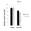
FIG. 5 is a graph depicting the inhibitory effect of an anti-cMet/anti-Her3 bispecific antibody on Her3 phosphorylation.
DETAILED DESCRIPTION
It has been shown herein that a bispecific antibody recognizing c-Met and Her3 at the same time shows an improved effect for inhibiting cancer cell compared to anti-c-Met antibody alone, by inhibiting both c-Met and Her3.
It also has been shown herein that a bispecific antibody in which an anti-c-Met antibody is fused to an antibody recognizing a secondary target, Her3, or an antigen binding fragment thereof (e.g., scFv) could improve the stability issue which was a significant problem of the pre-existing bispecific antibodies.
One embodiment provides a polypeptide including a novel amino acid sequence. The polypeptide may have a function as a complementarity determining region (“CDR”) of an anti-Her3 antibody. The polypeptide may be a recombinant or synthetic polypeptide.
In particular, the polypeptide may include one amino acid sequence or a combination of two or more amino acid sequences selected from the group consisting of SEQ ID NO: 109 to SEQ ID NO: 114. The function of the polypeptide including the amino acid sequence of SEQ ID NO: 109 to SEQ ID NO: 114 as a CDR of an anti-Her3 antibody is summarized in Table 1, as follows:
| TABLE 1 |
| |
| |
|
|
amino acid sequence |
SEQ ID NO. |
| |
| |
Heavy |
CDR-H1 |
SYSMN |
109 |
| |
chain |
CDR-H2 |
SISSSSSYIYYADSVKG |
110 |
| |
CDR |
CDR-H3 |
REDLTPFDY |
111 |
| |
| |
Light |
CDR-L1 |
GGDNIGSKSVH |
112 |
| |
chain |
CDR-L2 |
DDSDRPSGIPE |
113 |
| |
CDR |
CDR-L3 |
QVWDNSVDLL |
114 |
| |
In one particular embodiment, the polypeptide may a polypeptide including a polypeptide of SEQ ID NO: 115, a polypeptide of SEQ ID NO: 116 or a combination thereof. The polypeptide of SEQ ID NO: 115 includes the amino acid sequences of SEQ ID NOS: 109 to 111, and may have a function as a heavy chain variable region of an anti-Her3 antibody. In addition, the polypeptide including the amino acid sequence of SEQ ID NO: 116 includes the amino acid sequences of SEQ ID NOS: 112 to 114, and may have a function as a light chain variable region of an anti-Her3 antibody. The polypeptide is a synthetic or recombinant, non-naturally occurring polypeptide.
The polypeptide may act as a precursor or a component of a Her3 antagonist, such as an anti-Her3 antibody, an antigen-binding fragment thereof, or an anti-Her3 antibody analogue (e.g., a peptibody, nanobody, etc.).
Therefore, another embodiment provides an anti-Her3 antagonist including the polypeptide. The antagonist inhibits the Her3 activity, and may be one or more selected from the group consisting of an anti-Her3 antibody, an antigen-binding fragment thereof, an anti-Her3 antibody analogue (e.g., a peptibody, nanobody, etc.), and the like.
The term “antagonist” may include any molecules capable of completely or partially preventing, inhibiting, or neutralizing one or more biological activities of a target. For instance, an antibody as an antagonist may refer to an antibody capable of inhibiting or lowering biological activities of an antigen (e.g., NRG-1) to which the antibody binds. The antagonist may bind to a receptor for a ligand (target) to decrease receptor phosphorylation, or incapacitating or killing a cell that is activated by the ligand. In addition, the antagonist may substantially decrease an interaction between a receptor and its ligand, by completely blocking the receptor-ligand interaction, binding to the receptor competitively with its ligand, or modifying or down-regulating three-dimensional structure of the receptor.
Term “peptibody (peptide+antibody)” may refer to a fusion protein wherein a peptide is fused with the whole or a part of a constant region of an antibody, such as Fc region, and the peptide acts as an antigen-binding region (e.g., a CDR or variable region of a heavy chain and/or light chain), thereby having a structure and functions similar to an antibody.
Term “nanobody” that is also called as a single-domain antibody, may refer to an antibody fragment including a single variable domain in a monomeric form and selectively binding to a specific antigen, similarly to an antibody in a complete form. The nanobody usually has a molecular weight of about 12 kDa to about 15 kDa, which is much smaller than the average molecular weight (about 150 kDa to about 160 kDa) of an antibody in a complete form (including two heavy chains and two light chains), and in some case, smaller than a molecular weight of a Fab fragment or a scFv fragment.
In a particular embodiment, the polypeptide may act as a precursor or a component of an anti-Her3 antibody.
Another embodiment provides an anti-Her3 antibody or an antigen-binding fragment thereof including the polypeptide. The antigen-binding fragment may be selected from the group consisting of scFv, (scFv)2, scFv-Fc, Fab, Fab′ and F(ab′)2. The anti-Her3 antibody may be a recombinant or synthetic antibody.
In particular, the anti-Her3 antibody or an antigen-binding fragment thereof may include:
at least one heavy chain complementarity determining region selected from the group consisting of CDR-H1 including the amino acid sequence of SEQ ID NO: 109, CDR-H2 including the amino acid sequence of SEQ ID NO: 110, and CDR-H3 including the amino acid sequence of SEQ ID NO: 111, or a heavy chain variable region including the at least one heavy chain complementarity determining region;
at least one light chain complementarity determining region selected from the group consisting of CDR-L1 including the amino acid sequence of SEQ ID NO: 112, CDR-L2 including the amino acid sequence of SEQ ID NO: 113, and CDR-L3 including the amino acid sequence of SEQ ID NO: 114 or a light chain variable region including the at least one light chain complementarity determining region;
a combination of the at least one heavy chain complementarity determining region and the at least one light chain complementarity determining region; or
a combination of the a heavy chain variable region and the a light chain variable region.
For example, the anti-Her3 antibody or an antigen-binding fragment thereof may include a heavy chain variable region including the amino acid sequence of SEQ ID NO: 115, a light chain variable region including the amino acid sequence of SEQ ID NO: 116, or a combination thereof.
In a particular embodiment, the anti-Her3 antibody or an antigen-binding fragment thereof may be an anti-Her3 scFv including a heavy chain variable region including the amino acid sequence of SEQ ID NO: 115, and a light chain variable region including the amino acid sequence of SEQ ID NO: 116.
In the polypeptide or an anti-Her3 scFv including a heavy chain variable region including the amino acid sequence of SEQ ID NO: 115 or SEQ ID NO: 117, and a light chain variable region including the amino acid sequence of SEQ ID NO: 116 or SEQ ID NO: 118, the heavy chain variable region and the light chain variable region may be linked with or without a linker (e.g., a peptide linker). The peptide linker may be those including any amino acids of 1 to 100, particularly 2 to 50, and any kinds of amino acids may be included without any restrictions. The peptide linker may include for example, Gly, Asn and/or Ser residues, and also include neutral amino acids such as Thr and/or Ala. Amino acid sequences suitable for the peptide linker may be those known in the relevant art. Meanwhile, a length of the peptide linker may be variously determined within such a limit that the functions of the fusion protein will not be affected. For instance, the peptide linker may be formed by including a total of 1 to 100, 2 to 50, or 5 to 25 of one or more selected from the group consisting of Gly, Asn, Ser, Thr, and Ala. In one embodiment, the peptide linker may be represented as (G4S)n (n is a repeat number of (G4S), which is an integer of 1 to 10, particularly an integer of 2 to 5).
The term “antibody” may refer to a substance generated by a stimulation of an antigen in immune system, and there is no specific limitation in its kinds. The antibody may include all of an animal antibody, a chimeric antibody, a humanized antibody, and a human antibody. In addition the antibody may include an antigen-binding fragment derived from an antibody having an antigen binding affinity. The complementarity-determining region (CDR) may refer to a region within a variable region, which gives a binding specificity to an antigen. The antigen-binding fragment as described above may be an antibody fragment including at least one complementarity-determining region, for example, one or more selected from the group consisting of scFv, (scFv)2, scFv-Fc, Fab, Fab′, and F(ab′)2.
In the anti-Her3 antibody or an antigen-binding fragment thereof, the rest portion of the light chain and the heavy chain portion excluding the CDRs, the light chain variable region, and the heavy chain variable region as defined above, that is the light chain constant region and the heavy chain constant region, may be those from any subtype of immunoglobulin (e.g., IgA, IgD, IgE, IgG (IgG1, IgG2, IgG3, IgG4), IgM, and the like).
Based on the ability of specifically binding to Her3, the anti-Her3 antibody or an antigen-binding fragment thereof may be used in detecting Her3 or confirming activation and/or overproduction (overexpression) of Her3.
One embodiment provides a composition for detecting the presence of HER3 including the anti-HER3 antibody or an antigen-binding fragment thereof. Another embodiment provides a method of detecting HER3 including treating a biological sample with the anti-HER3 antibody or an antigen-binding fragment thereof; and detecting an antigen-antibody reaction (binding). In the method, when an antigen-antibody reaction is detected, it can be determined that HER3 is present in the biological sample. Another embodiment provides a use of the anti-HER3 antibody or an antigen-binding fragment thereof for detecting HER3. The biological sample may be selected from the group consisting of a cell, a tissue, a body fluid (e.g., blood, serum, etc.), and the like derived from a mammal including primates such as a human, a monkey, and the like, or a rodent such as a mouse, a rat, and the like. The biological sample may be separated from a living body. The detection of HER3 may refer to detection of the presence of HER3, expression of HER3, or the level of HER3.
Another embodiment provides a pharmaceutical composition for diagnosing activation and/or overproduction of HER3 or a disease associated with activation and/or overproduction of HER3 including the anti-HER3 antibody or an antigen-binding fragment thereof. Another embodiment provides a method of diagnosing (or determining) activation and/or overproduction of HER3 or a disease associated with activation and/or overproduction of HER3, including treating a biological sample derived from a patient with the anti-HER3 antibody or an antigen-binding fragment thereof, and measuring a level of an antigen-antibody reaction. In this method, when the level of the antigen-antibody reaction in the biological sample is higher than that of a normal sample, the patient from which the biological sample is derived may be determined as having activation and/or overproduction of HER3 or a disease associated with activation and/or overproduction of HER3. Therefore, the method may further include treating a normal sample with the anti-Her3 antibody or an antigen-binding fragment thereof, and measuring a level of an antigen-antibody reaction. Another embodiment provides a use of the anti-HER3 antibody or an antigen-binding fragment thereof for diagnosing activation and/or overproduction of HER3 or a disease associated with activation and/or overproduction of HER3.
The biological sample may be at least one selected from the group consisting of a cell, a tissue, fluid (e.g., blood, serum, and the like) and the like, derived from a patient to be diagnosed. The biological sample may be separated from a living body. The normal sample may be at least one selected from the group consisting of a cell, a tissue, fluid (e.g., blood, serum, and the like) and the like, derived from a patient having no condition of activation and/or overproduction of Her3 or a disease associated with activation and/or overproduction of Her3. The normal sample may be separated from a living body. The patient may be selected from mammal including primates such as a human, a monkey, and the like, and rodents such as a mouse, a rat, and the like.
Another embodiment provides an anti-c-Met/anti-Her3 bispecific antibody including an anti-c-Met antibody or an antigen-binding fragment thereof and an anti-Her3 antibody or an antigen-binding fragment thereof. The antigen-binding fragment thereof may be selected from the group consisting of scFv, (scFv)2, scFvFc, Fab, Fab′, and F(ab′)2. The anti-c-Met/anti-Her3 bispecific antibody may be a recombinant or synthetic antibody.
The “c-Met protein” refers to a receptor tyrosine kinase binding to hepatocyte growth factor. The c-Met proteins may be derived from any species, for example, those derived from primates such as human c-Met (e.g., NP_000236) and monkey c-Met (e.g., Macaca mulatta, NP_001162100), or those derived from rodents such as mouse c-Met (e.g., NP_032617.2) and rat c-Met (e.g., NP_113705.1). The proteins include, for example, a polypeptide encoded by the nucleotide sequence deposited under GenBank Accession Number NM_000245, or a protein encoded by the polypeptide sequence deposited under GenBank Accession Number NM_000236, or extracellular domains thereof. The receptor tyrosine kinase c-Met is involved in several mechanisms including cancer incidence, cancer metastasis, cancer cell migration, cancer cell penetration, angiogenesis, etc.
The “Her3” is a member of the receptor tyrosine kinases (RTKs) of HER(EGFR) family. The member of the receptor tyrosine kinases of HER(EGFR) family comprises Her1 (also known as EGFR or erbB), Her2 (also known as erbB2), Her3 (also known as erbB3) and Her4 (also known as erbB4), and among them, Her3, as a transmembrane receptor, is composed of an extracellular ligand-binding domain (ECD), a dimerization domain in the ECD, an transmembrane domain (TMD), an extracellular protein Tyrosine-kinase domain (TKD), and a C-terminal phosphorylation domain, in common with original epidermal growth factor receptor. Her3, as well as EGFR and Her2, is related to tumorigenesis. Her3 is sometimes overexpressed in breast cancer, colorectal cancer, ovarian cancer, bladder cancer, prostate cancer, non-small-cell lung cancer, melanoma, pharynx cancer, pancreatic cancer, esophagus cancer, glioma, cholangiocarcinoma, biliary tract cancer, gastric cancer, endometrial cancer, gallbladder cancer, squamous cell carcinoma, or basal cell carcinoma, and the like. For instance, the Her3 may be polypeptides encoded by the nucleotide sequences (mRNA) deposited under GenBank Accession Nos. NM_001982.2, NM_001982.3, NM_001005915.1, NM_010153.1, NM_017218.2 or NM_001103105.1.
In one embodiment, the anti-c-Met/anti-HER3 bispecific antibody may include an anti-c-Met antibody or an antigen binding fragment thereof, and an anti-HER3 antibody or an antigen binding fragment thereof which is linked to the C terminus or N terminus, for example, C terminal, of the anti-c-Met antibody or the antigen binding fragment thereof.
The anti-c-Met/anti-Her3 bispecific antibody may be those including a complete form of an anti-c-Met antibody (e.g., IgG type antibody) and an antigen binding fragment of the anti-Her3 antibody linked to the C terminus of the anti-c-Met antibody.
In the anti-c-Met/anti-Her3 bispecific antibody, the anti-c-Met antibody or the antigen binding fragment thereof, and the anti-Her3 antibody or the antigen binding fragment thereof, may be linked via a peptide linker or without it. Furthermore, a heavy chain portion and a light chain portion within the antigen binding fragment, for example, a heavy chain variable region and a light chain variable region within the scFv fragment, may be linked via a peptide linker or without it. The peptide linker which links the anti-c-Met antibody or the antigen binding fragment thereof and the anti-Her3 antibody or the antigen binding fragment thereof, and the peptide linker which links the heavy chain portion and the light chain portion within the antigen binding fragment, may be identical or different. The peptide linker may be those including any amino acids of 1 to 100, particularly 2 to 50, and any kinds of amino acids may be included without any restrictions. The peptide linker may include for example, Gly, Asn and/or Ser residues, and also include neutral amino acids such as Thr and/or Ala. Amino acid sequences suitable for the peptide linker may be those known in the pertinent art. Meanwhile, a length of the peptide linker may be variously determined within such a limit that the functions of the fusion protein will not be affected. For instance, the peptide linker may be formed by including a total of 1 to 100, 2 to 50, or 5 to 25 of one or more selected from the group consisting of Gly, Asn, Ser, Thr, and Ala. In one embodiment, the peptide linker may be represented as (G4S)n (n is a repeat number of (G4S), which is an integer of 1 to 10, particularly an integer of 2 to 5).
In a particular embodiment, the anti-Her3 antibody or an antigen-biding fragment may include:
at least one heavy chain complementarity determining region selected from the group consisting of CDR-H1 including the amino acid sequence of SEQ ID NO: 109, CDR-H2 including the amino acid sequence of SEQ ID NO: 110, and CDR-H3 including the amino acid sequence of SEQ ID NO: 111 or a heavy chain variable region including the at least one heavy chain complementarity determining region;
at least one light chain complementarity determining region selected from the group consisting of CDR-L1 including the amino acid sequence of SEQ ID NO: 112, CDR-L2 including the amino acid sequence of SEQ ID NO: 113, and CDR-L3 including the amino acid sequence of SEQ ID NO: 114 or a light chain variable region including the at least one light chain complementarity determining region;
a combination of the at least one heavy chain complementarity determining region and the at least one light chain complementarity determining region; or
a combination of the heavy chain variable region and the light chain variable region.
For example, the anti-Her3 antibody or an antigen-binding fragment thereof may include a heavy chain variable region including the amino acid sequence of SEQ ID NO: 115, a light chain variable region including the amino acid sequence of SEQ ID NO: 116, or a combination thereof.
In a particular embodiment, the anti-Her3 antibody or an antigen-binding fragment thereof may be an anti-Her3 scFv including a heavy chain variable region including the amino acid sequence of SEQ ID NO: 115, and a light chain variable region including the amino acid sequence of SEQ ID NO: 116.
The “antigen binding fragment” refers to a fragment of a full immunoglobulin structure including parts of the polypeptide including a portion of antigen-binding regions capable of binding to an antigen. For example, it may be scFv, (scFv)2, Fab, Fab′, or F(ab′)2, but not be limited thereto. In the present disclosure, the antigen binding fragment may be an antibody fragment including at least one complementarity determining region, for example, selected from the group consisting of scFv, (scFv)2, scFv-Fc, Fab, Fab′ and F(ab′)2.
Of the antigen binding fragments, Fab is a structure having variable regions of a light chain and a heavy chain, a constant region of the light chain, and the first constant region (CH1) of the heavy chain, and it has one antigen binding site.
Fab′ is different from Fab in that it has a hinge region including one or more cysteine residues at the C-terminal of heavy chain CH1 domain. An F(ab′)2 antibody is formed through disulfide bond of the cysteine residues at the hinge region of Fab′.
Fv is a minimal antibody piece having only a heavy chain variable region and light chain variable region, and a recombinant technique for producing the Fv fragment is well known in the pertinent art. Two-chain Fv may have a structure in which the heavy chain variable region is linked to the light chain variable region by a non-covalent bond, and single-chain Fv (scFv) may generally have a dimer structure as in the two-chain Fv in which the variable region of a heavy chain and the variable region of a light chain are covalently linked via a peptide linker or they are directly linked to each other at the C-terminal thereof. The peptide linker may be the same as described in the above, for example, those having the amino acid length of 1 to 100, 2 to 50, particularly 5 to 25, and any kinds of amino acids may be included without any restrictions.
The antigen binding fragments may be obtained using proteases (for example, a whole antibody is digested with papain to obtain Fab fragments, and is digested with pepsin to obtain F(ab′)2 fragments), and may be prepared by a genetic recombinant technique.
In a particular embodiment, the anti-c-Met/anti-Her3 bispecific antibody may be those including an anti-c-Met antibody, and scFv, (scFv)2, Fab, Fab′ or F(ab′)2, for example, scFv, of the anti-Her3antibody linked to the C terminus of the anti-c-Met antibody. For instance, scFv, (scFv)2, Fab, Fab′ or F(ab′)2 of the anti-Her3 antibody may be those including a heavy chain variable region including the amino acid sequence of SEQ ID NO: 109 or SEQ ID NO: 113 and a light chain variable region including the amino acid sequence of SEQ ID NO: 111 or SEQ ID NO: 114.
Hence, in a particular embodiment, the anti-c-Met/anti-Her3 bispecific antibody may be those including the anti-c-Met antibody, and scFv, (scFv)2, Fab, Fab′ or F(ab′)2 of the anti-Her3 antibody including a heavy chain variable region including the amino acid sequence of SEQ ID NO: 109 or SEQ ID NO: 113 and a light chain variable region including the amino acid sequence of SEQ ID NO: 111 or SEQ ID NO: 114, linked to the C terminal of the anti-c-Met antibody.
The anti-c-Met antibody may be any one recognizing a specific region of c-Met, e.g., a specific region in the SEMA domain, as an epitope. It may be any antibody or antigen-binding fragment that acts on c-Met to induce intracellular internalization and degradation of c-Met.
c-Met, a receptor for hepatocyte growth factor (HGF), may be divided into three portions: extracellular, transmembrane, and intracellular. The extracellular portion is composed of an α-subunit and a β-subunit which are linked to each other through a disulfide bond, and contains a SEMA domain responsible for binding HGF, a PSI domain (plexin-semaphorins-integrin homology domain) and an IPT domain (immunoglobulin-like fold shared by plexins and transcriptional factors domain). The SEMA domain of c-Met protein may have the amino acid sequence of SEQ ID NO: 79, and is an extracellular domain that functions to bind HGF. A specific region of the SEMA domain, that is, a region including the amino acid sequence of SEQ ID NO: 71, which corresponds to a range from amino acid residues 106 to 124 of the amino acid sequence of the SEMA domain (SEQ ID NO: 79) of c-Met protein, is a loop region between the second and the third propellers within the epitopes of the SEMA domain. The region acts as an epitope for the specific anti-c-Met antibody of the present disclosure.
The term “epitope” as used herein, refers to an antigenic determinant, a part of an antigen recognized by an antibody. In one embodiment, the epitope may be a region including 5 or more contiguous amino acid residues within the SEMA domain (SEQ ID NO: 79) of c-Met protein, for instance, 5 to 19 contiguous amino acid residues within the amino acid sequence of SEQ ID NO: 71. For example, the epitope may be a polypeptide having 5 to 19 contiguous amino acids within the amino acid sequence of SEQ ID NO: 71, wherein the polypeptide essentially includes the amino sequence of SEQ ID NO: 73 (EEPSQ) serving as an essential element for the epitope. For example, the epitope may be a polypeptide including, consisting essentially of, or consisting of the amino acid sequence of SEQ ID NO: 71, SEQ ID NO: 72, or SEQ ID NO: 73.
The epitope including the amino acid sequence of SEQ ID NO: 72 corresponds to the outermost part of the loop between the second and third propellers within the SEMA domain of a c-Met protein. The epitope including the amino acid sequence of SEQ ID NO: 73 is a site to which the antibody or antigen-binding fragment according to one embodiment most specifically binds.
Thus, the anti-c-Met antibody may specifically bind to an epitope which has 5 to 19 contiguous amino acids within the amino acid sequence of SEQ ID NO: 71, including SEQ ID NO: 73 as an essential element. For example, the anti-c-Met antibody may specifically bind to an epitope including the amino acid sequence of SEQ ID NO: 71, SEQ ID NO: 72, or SEQ ID NO: 73.
In one embodiment, the anti-c-Met antibody or an antigen-binding fragment thereof may include:
at least one heavy chain complementarity determining region (CDR) selected from the group consisting of (a) a CDR-H1 including the amino acid sequence of SEQ ID NO: 4; (b) a CDR-H2 including the amino acid sequence of SEQ ID NO: 5, SEQ ID NO: 2, or an amino acid sequence having 8-19 consecutive amino acids within SEQ ID NO: 2 including amino acid residues from the 3rd to 10th positions of SEQ ID NO: 2; and (c) a CDR-H3 including the amino acid sequence of SEQ ID NO: 6, SEQ ID NO: 85, or an amino acid sequence having 6-13 consecutive amino acids within SEQ ID NO: 85 including amino acid residues from the 1st to 6th positions of SEQ ID NO: 85, or a heavy chain variable region including the at least one heavy chain complementarity determining region;
at least one light chain complementarity determining region (CDR) selected from the group consisting of (a) a CDR-L1 including the amino acid sequence of SEQ ID NO: 7, (b) a CDR-L2 including the amino acid sequence of SEQ ID NO: 8, and (c) a CDR-L3 including the amino acid sequence of SEQ ID NO: 9, SEQ ID NO: 15, SEQ ID NO: 86, or an amino acid sequence having 9-17 consecutive amino acids within SEQ ID NO: 89 including amino acid residues from the 1st to 9th positions of SEQ ID NO: 89, or a light chain variable region including the at least one light chain complementarity determining region;
a combination of the at least one heavy chain complementarity determining region and at least one light chain complementarity determining region; or
a combination of the heavy chain variable region and the light chain variable region.
Herein, the amino acid sequences of SEQ ID NOS: 4 to 9 are respectively represented by following Formulas I to VI, below:
Xaa1-Xaa2-Tyr-Tyr-Met-Ser (SEQ ID NO: 4), Formula I
wherein Xaa1 is absent or Pro or Ser, and Xaa2 is Glu or Asp,
Arg-Asn-Xaa3-Xaa4-Asn-Gly-Xaa5-Thr (SEQ ID NO: 5), Formula II
wherein Xaa3 is Asn or Lys, Xaa4 is Ala or Val, and Xaa5 is Asn or Thr,
Asp-Asn-Trp-Leu-Xaa6-Tyr (SEQ ID NO: 6), Formula III
wherein Xaa6 is Ser or Thr,
Lys-Ser-Ser-Xaa7-Ser-Leu-Leu-Ala-Xaa8-Gly-Asn-Xaa9-Xaa10-Asn-Tyr-Leu-Ala (SEQ ID NO: 7) Formula IV
wherein Xaa7 is His, Arg, Gln, or Lys, Xaa8 is Ser or Trp, Xaa9 is His or Gln, and Xaa10 is Lys or Asn,
Trp-Xaa11-Ser-Xaa12-Arg-Val-Xaa13(SEQ ID NO: 8) Formula V
wherein Xaa11 is Ala or Gly, Xaa12 is Thr or Lys, and Xaa13 is Ser or Pro, and
Xaa14-Gln-Ser-Tyr-Ser-Xaa15-Pro-Xaa16-Thr (SEQ ID NO: 9) Formula VI
wherein Xaa14 is Gly, Ala, or Gln, Xaa15 is Arg, His, Ser, Ala, Gly, or Lys, and Xaa16 is Leu, Tyr, Phe, or Met.
In one embodiment, the CDR-H1 may include an amino acid sequence selected from the group consisting of SEQ ID NOS: 1, 22, 23, and 24. The CDR-H2 may include an amino acid sequence selected from the group consisting of SEQ ID NOS: 2, 25, and 26. The CDR-H3 may include an amino acid sequence selected from the group consisting of SEQ ID NOS: 3, 27, 28, and 85.
The CDR-L1 may include an amino acid sequence selected from the group consisting of SEQ ID NOS: 10, 29, 30, 31, 32, 33, and 106. The CDR-L2 may include an amino acid sequence selected from the group consisting of SEQ ID NOS: 11, 34, 35, and 36. The CDR-L3 may include an amino acid sequence selected from the group consisting of SEQ ID NOS: 12, 13, 14, 15, 16, 37, 86, and 89.
In another embodiment, the antibody or antigen-binding fragment may include:
a heavy chain variable region comprising a polypeptide (CDR-H1) including an amino acid sequence selected from the group consisting of SEQ ID NOS: 1, 22, 23, and 24, a polypeptide (CDR-H2) including an amino acid sequence selected from the group consisting of SEQ ID NOS: 2, 25, and 26, and a polypeptide (CDR-H3) including an amino acid sequence selected from the group consisting of SEQ ID NOS: 3, 27, 28, and 85; and
a light chain variable region comprising a polypeptide (CDR-L1) including an amino acid sequence selected from the group consisting of SEQ ID NOS: 10, 29, 30, 31, 32, 33 and 106, a polypeptide (CDR-L2) including an amino acid sequence selected from the group consisting of SEQ ID NOS: 11, 34, 35, and 36, and a polypeptide (CDR-L3) including an amino acid sequence selected from the group consisting of SEQ ID NOS 12, 13, 14, 15, 16, 37, 86, and 89.
Animal-derived antibodies produced by immunizing non-immune animals with a desired antigen generally invoke immunogenicity when injected to humans for the purpose of medical treatment, and thus chimeric antibodies have been developed to inhibit such immunogenicity. Chimeric antibodies are prepared by replacing constant regions of animal-derived antibodies that cause an anti-isotype response with constant regions of human antibodies by genetic engineering. Chimeric antibodies are considerably improved in an anti-isotype response compared to animal-derived antibodies, but animal-derived amino acids still have variable regions, so that chimeric antibodies have side effects with respect to a potential anti-idiotype response. Humanized antibodies have been developed to reduce such side effects. Humanized antibodies are produced by grafting complementarity determining regions (CDR) which serve an important role in antigen binding in variable regions of chimeric antibodies into a human antibody framework.
An important consideration in CDR grafting to produce humanized antibodies is choosing the optimized human antibodies for accepting CDRs of animal-derived antibodies. Antibody databases, analysis of a crystal structure, and technology for molecule modeling are used. However, even when the CDRs of animal-derived antibodies are grafted to the most optimized human antibody framework, amino acids positioned in a framework of the animal-derived CDRs affecting antigen binding are present. Therefore, in many cases, antigen binding affinity is not maintained, and thus application of additional antibody engineering technology for recovering the antigen binding affinity is necessary.
The anti c-Met antibodies may be mouse-derived antibodies, mouse-human chimeric antibodies, humanized antibodies, or human antibodies.
An intact antibody includes two full-length light chains and two full-length heavy chains, in which each light chain is linked to a heavy chain by disulfide bonds. The antibody has a heavy chain constant region and a light chain constant region. The heavy chain constant region is of a gamma (γ), mu (μ), alpha (α), delta (δ), or epsilon (ε) type, which may be further categorized as gamma 1 (γ1), gamma 2 (γ2), gamma 3 (γ3), gamma 4 (γ4), alpha 1 (α1), or alpha 2 (α2). The light chain constant region is of either a kappa (κ) or lambda (λ) type.
As used herein, the term “heavy chain” refers to full-length heavy chain, and fragments thereof, including a variable region VH that includes amino acid sequences sufficient to provide specificity to antigens, and three constant regions, CH1, CH2, and CH3, and a hinge. The term “light chain” refers to a full-length light chain and fragments thereof, including a variable region VL that includes amino acid sequences sufficient to provide specificity to antigens, and a constant region CL.
The term “complementarity determining region (CDR)” refers to an amino acid sequence found in a hyper variable region of a heavy chain or a light chain of immunoglobulin. The heavy and light chains may respectively include three CDRs (CDRH1, CDRH2, and CDRH3; and CDRL1, CDRL2, and CDRL3). The CDR may provide contact residues that play an important role in the binding of antibodies to antigens or epitopes. The terms “specifically binding” and “specifically recognized” are well known to one of ordinary skill in the art, and indicate that an antibody and an antigen specifically interact with each other to lead to an immunological activity.
The term “hinge region,” as used herein, refers to a region between CH1 and CH2 domains within the heavy chain of an antibody which functions to provide flexibility for the antigen-binding site.
When an animal antibody undergoes a chimerization process, the IgG1 hinge of animal origin is replaced with a human IgG1 hinge or IgG2 hinge while the disulfide bridges between two heavy chains are reduced from three to two in number. In addition, an animal-derived IgG1 hinge is shorter than a human IgG1 hinge. Accordingly, the rigidity of the hinge is changed. Thus, a modification of the hinge region may bring about an improvement in the antigen binding efficiency of the humanized antibody. The modification of the hinge region through amino acid deletion, addition, or substitution is well-known to those skilled in the art.
In one embodiment, the anti-c-Met antibody or an antigen-binding fragment thereof may be modified by the deletion, insertion, addition, or substitution of at least one amino acid residue on the amino acid sequence of the hinge region so that it exhibit enhanced antigen-binding efficiency. For example, the antibody may include a hinge region including the amino acid sequence of SEQ ID NO: 100 (U7-HC6), 101 (U6-HC7), 102 (U3-HC9), 103 (U6-HC8), or 104 (U8-HC5), or a hinge region including the amino acid sequence of SEQ ID NO: 105 (non-modified human hinge). In particular, the hinge region has the amino acid sequence of SEQ ID NO: 100 or 101.
In one embodiment, the anti-c-Met antibody or antigen-binding fragment may include a variable region of the heavy chain including the amino acid sequence of SEQ ID NO: 17, 74, 87, 90, 91, 92, 93, or 94, a variable region of the light chain including the amino acid sequence of SEQ ID NO: 18, 19, 20, 21, 75, 88, 95, 96, 97, 98, 99, or 107, 119, or a combination thereof.
In one embodiment, the anti-c-Met antibody may be a monoclonal antibody. The monoclonal antibody may be produced by the hybridoma cell line deposited with Accession No. KCLRF-BP-00220, which binds specifically to the extracellular region of c-Met protein (refer to Korean Patent Publication No. 2011-0047698, the disclosure of which is incorporated in its entirety herein by reference).
The anti-c-Met antibody may include all the antibodies defined in Korean Patent Publication No. 2011-0047698.
In the anti-c-Met antibody, the rest portion of the light chain and the heavy chain portion excluding the CDRs, the light chain variable region, and the heavy chain variable region as defined above, that is the light chain constant region and the heavy chain constant region, may be those from any subtype of immunoglobulin (e.g., IgA, IgD, IgE, IgG (IgG1, IgG2, IgG3, IgG4), IgM, and the like).
By way of further example, the anti-c-Met antibody or the antibody fragment may include:
a heavy chain including the amino acid sequence selected from the group consisting of the amino acid sequence of SEQ ID NO: 62 (wherein the amino acid sequence from amino acid residues from the 1st to 17th positions is a signal peptide), or the amino acid sequence from the 18th to 462nd positions of SEQ ID NO: 62, the amino acid sequence of SEQ ID NO: 64 (wherein the amino acid sequence from the 1st to 17th positions is a signal peptide), the amino acid sequence from the 18th to 461st positions of SEQ ID NO: 64, the amino acid sequence of SEQ ID NO: 66 (wherein the amino acid sequence from the 1st to 17th positions is a signal peptide), and the amino acid sequence from the 18th to 460th positions of SEQ ID NO: 66; and
a light chain including the amino acid sequence selected from the group consisting of the amino acid sequence of SEQ ID NO: 68 (wherein the amino acid sequence from the 1st to 20th positions is a signal peptide), the amino acid sequence from the 21st to 240th positions of SEQ ID NO: 68, the amino acid sequence of SEQ ID NO: 70 (wherein the amino acid sequence from the 1st to 20th positions is a signal peptide), the amino acid sequence from the 21st to 240th positions of SEQ ID NO: 70, and the amino acid sequence of SEQ ID NO: 108.
For example, the anti-c-Met antibody may be selected from the group consisting of:
an antibody including a heavy chain including the amino acid sequence of SEQ ID NO: 62 or the amino acid sequence from the 18th to 462nd positions of SEQ ID NO: 62 and a light chain including the amino acid sequence of SEQ ID NO: 68 or the amino acid sequence from the 21st to 240th positions of SEQ ID NO: 68;
an antibody including a heavy chain including the amino acid sequence of SEQ ID NO: 64 or the amino acid sequence from the 18th to 461st positions of SEQ ID NO: 64 and a light chain including the amino acid sequence of SEQ ID NO: 68 or the amino acid sequence from the 21st to 240th positions of SEQ ID NO: 68;
an antibody including a heavy chain including the amino acid sequence of SEQ ID NO: 66 or the amino acid sequence from the 18th to 460th positions of SEQ ID NO: 66 and a light chain including the amino acid sequence of SEQ ID NO: 68 or the amino acid sequence from the 21st to 240th positions of SEQ ID NO: 68;
an antibody including a heavy chain including the amino acid sequence of SEQ ID NO: 62 or the amino acid sequence from the 18th to 462nd positions of SEQ ID NO: 62 and a light chain including the amino acid sequence of SEQ ID NO: 70 or the amino acid sequence from the 21st to 240th positions of SEQ ID NO: 70;
an antibody including a heavy chain including the amino acid sequence of SEQ ID NO: 64 or the amino acid sequence from the 18th to 461st positions of SEQ ID NO: 64 and a light chain including the amino acid sequence of SEQ ID NO: 70 or the amino acid sequence from the 21st to 240th positions of SEQ ID NO: 70;
an antibody including a heavy chain including the amino acid sequence of SEQ ID NO: 66 or the amino acid sequence from the 18th to 460th positions of SEQ ID NO: 66 and a light chain including the amino acid sequence of SEQ ID NO: 70 or the amino acid sequence from the 21st to 240th positions of SEQ ID NO: 70;
an antibody including a heavy chain including the amino acid sequence of SEQ ID NO: 62 or the amino acid sequence from the 18th to 462nd positions of SEQ ID NO: 62 and a light chain including the amino acid sequence of SEQ ID NO: 108;
an antibody including a heavy chain including the amino acid sequence of SEQ ID NO: 64 or the amino acid sequence from the 18th to 461st positions of SEQ ID NO: 64 and a light chain including the amino acid sequence of SEQ ID NO: 108; and
an antibody including a heavy chain including the amino acid sequence of SEQ ID NO: 66 or the amino acid sequence from the 18th to 460th positions of SEQ ID NO: 66 and a light chain including the amino acid sequence of SEQ ID NO: 108.
According to an embodiment, the anti-c-Met antibody may include a heavy chain including the amino acid sequence from the 18th to 460th positions of SEQ ID NO: 66 and a light chain including the sequence from the 21st to 240th positions of SEQ ID NO: 68, or a heavy chain including the amino acid sequence from the 18th to 460th positions of SEQ ID NO: 66 and a light chain including the sequence of SEQ ID NO: 108.
The polypeptide of SEQ ID NO: 70 is a light chain including human kappa (κ) constant region, and the polypeptide with the amino acid sequence of SEQ ID NO: 68 is a polypeptide obtained by replacing histidine at position 62 (corresponding to position 36 of SEQ ID NO: 68 according to kabat numbering) of the polypeptide with the amino acid sequence of SEQ ID NO: 70 with tyrosine. The production yield of the antibodies may be increased by the replacement. The polypeptide with the amino acid sequence of SEQ ID NO: 108 is a polypeptide obtained by replacing serine at position 32 (position 27e according to kabat numbering in the amino acid sequence from amino acid residues 21 to 240 of SEQ ID NO: 68; positioned within CDR-L1) with tryptophan. By such replacement, antibodies and antibody fragments including such sequences exhibits increased activities, such as c-Met biding affinity, c-Met degradation activity, Akt phosphorylation inhibition, and the like.
In another embodiment, the anti-c-Met antibody may include a light chain complementarity determining region including the amino acid sequence of SEQ ID NO: 106, a light chain variable region including the amino acid sequence of SEQ ID NO: 107, or a light chain including the amino acid sequence of SEQ ID NO: 108.
The anti-Her3 antibody or antibody fragment, and the bispecific antibody, are synthetic or recombinant, non-naturally occurring antibodies or antibody fragments.
The anti-c-Met/anti-Her3 bispecific antibody can not only inhibit the activity of c-Met and Her3 by the internalization and degradation activity of anti-c-Met antibody but also fundamentally block them by reducing the total amounts of c-Met and Her3 by the degradation thereof. Accordingly, the anti-c-Met/anti-Her3 bispecific antibody can obtain efficient effects even when applied to patients who have developed resistance against pre-existing anti-Her3 antibodies.
Another embodiment provides a pharmaceutical composition including the anti-Her3 antibody or an antigen-binding fragment thereof as an active ingredient. Another embodiment provides a pharmaceutical composition including the anti-c-Met/anti-Her3 bispecific antibody as an active ingredient.
In particular, another embodiment provides a pharmaceutical composition for preventing and/or treating a cancer including the anti-Her3 antibody or an antigen-binding fragment thereof as an active ingredient. Another embodiment provides a pharmaceutical composition for preventing and/or treating a cancer including the anti-c-Met/anti-Her3 bispecific antibody as an active ingredient.
Another embodiment provides a method of prevention and/or treatment a cancer, including administering a pharmaceutical effective amount of the anti-Her3 antibody or an antigen-binding fragment thereof to a patient in need of the prevention and/or treatment of the cancer. Another embodiment provides a method of prevention and/or treatment a cancer, including administering a pharmaceutical effective amount of the anti-c-Met/anti-Her3 bispecific antibody to a patient in need of the prevention and/or treatment of the cancer. The method of prevention and/or treatment a cancer may further comprises a step of identifying the patient in need of the prevention and/or treatment of the cancer, prior to the step of administering
Another embodiment provides a use of the anti-c-Met/anti-Her3 bispecific antibody for preventing and/or treating a cancer.
The cancer may be a solid cancer or hematological cancer and for instance, may be, but not limited to, one or more selected from the group consisting of squamous cell carcinoma, small-cell lung cancer, non-small-cell lung cancer, adenocarcinoma of the lung, squamous cell carcinoma of the lung, peritoneal carcinoma, skin cancer, melanoma in the skin or eyeball, rectal cancer, cancer near the anus, esophagus cancer, small intestinal tumor, endocrine gland cancer, parathyroid cancer, adrenal cancer, soft-tissue sarcoma, urethral cancer, chronic or acute leukemia, lymphocytic lymphoma, hepatoma, gastric cancer, pancreatic cancer, glioblastoma, cervical cancer, ovarian cancer, liver cancer, bladder cancer, hepatocellular adenoma, breast cancer, colon cancer, large intestine cancer, endometrial carcinoma or uterine carcinoma, salivary gland tumor, kidney cancer, prostate cancer, vulvar cancer, thyroid cancer, head or neck cancer, brain cancer, and the like. In particular, the cancer may be cancer having resistance against pre-existing anticancer drugs, for example, antagonists against Her3.
In the pharmaceutical composition or method, the pharmaceutically effective amount of the anti-Her3 antibody or an antigen-binding fragment thereof or the anti-c-Met/anti-Her3 bispecific antibody may be administered along with at least one additive selected from the group consisting of a pharmaceutically acceptable carriers, diluents, and excipients.
The pharmaceutically acceptable carrier to be included in the composition may be those commonly used for the formulation of antibodies, which may be one or more selected from the group consisting of lactose, dextrose, sucrose, sorbitol, mannitol, starch, gum acacia, calcium phosphate, alginates, gelatin, calcium silicate, micro-crystalline cellulose, polyvinylpyrrolidone, cellulose, water, syrup, methyl cellulose, methylhydroxy benzoate, propylhydroxy benzoate, talc, magnesium stearate, and mineral oil, but are not limited thereto. The pharmaceutical composition may further include one or more selected from the group consisting of a lubricant, a wetting agent, a sweetener, a flavor enhancer, an emulsifying agent, a suspension agent, and preservative.
The pharmaceutical composition or the anti-Her3 antibody or an antigen-binding fragment thereof or the anti-c-Met/anti-Her3 bispecific antibody may be administered orally or parenterally. The parenteral administration may include intravenous injection, subcutaneous injection, muscular injection, intraperitoneal injection, endothelial administration, local administration, intranasal administration, intrapulmonary administration, and rectal administration. Since oral administration leads to digestion of proteins or peptides, an active ingredient in the compositions for oral administration must be coated or formulated to prevent digestion in stomach. In addition, the compositions may be administered using an optional device that enables an active substance to be delivered to target cells.
A suitable dosage of the pharmaceutical composition, the anti-Her3 antibody or an antigen-binding fragment thereof, or the anti-c-Met/anti-Her3 bispecific antibody may be prescribed in a variety of ways, depending on factors such as formulation methods, administration methods, age of patients, body weight, gender, pathologic conditions, diets, administration time, administration route, excretion speed, and reaction sensitivity. A desirable dosage of the pharmaceutical composition or the anti-c-Met/anti-HER3 bispecific antibody may be in the range of about 0.001 to 100 mg/kg for an adult. For example, the suitable dosage of the pharmaceutical composition, the anti-HER3 antibody or an antigen-binding fragment thereof, or the anti-c-Met/anti-HER3 bispecific antibody may be 0.001 to 1000 mg/kg, 0.01 to 100 mg/kg, or 0.1 to 50 mg/kg, per a day, but not be limited thereto. The term “pharmaceutically effective amount” used herein refers to an amount of the active ingredient (i.e., the anti-HER3 antibody or an antigen-binding fragment thereof, or the anti-c-Met/anti-HER3 bispecific antibody) exhibiting effects in preventing or treating cancer, and may be properly determined in a variety of ways, depending on factors such as formulation methods, administration methods, age of patients, body weight, gender, pathologic conditions, diets, administration time, administration route, excretion speed, and reaction sensitivity.
The pharmaceutical composition or the anti-c-Met/anti-Her3 bispecific antibody may be formulated with a pharmaceutically acceptable carrier and/or excipient into a unit or a multiple dosage form by a method easily carried out by a skilled person in the pertinent art. The dosage form may be a solution in oil or an aqueous medium, a suspension, syrup, an emulsifying solution, an extract, powder, granules, a tablet, or a capsule, and may further include a dispersing or a stabilizing agent.
In addition, the pharmaceutical composition or the anti-c-Met/anti-Her3 bispecific antibody may be administered as an individual drug, or together with other drugs, and may be administered sequentially or simultaneously with pre-existing drugs.
Since the pharmaceutical composition includes an antibody or an antigen binding fragment thereof, it may be formulated as an immunoliposome. The liposome containing an antibody may be prepared using a well-known method in the pertinent art. The immunoliposome is a lipid composition including phosphatidylcholine, cholesterol, and polyethyleneglycol-derivatized phosphatidylethanolamine, and may be prepared by a reverse phase evaporation method. For example, Fab′ fragments of an antibody may be conjugated to the liposome through a disulfide exchange reaction. A chemical drug such as doxorubicin may be additionally included in the liposome.
The subject to which the pharmaceutical composition is administered or the patient to which the prevention and/treatment method is applied may be mammals, for example, primates such as humans and monkeys, or rodents such as rats and mice, but are not be limited thereto. The subject or the patient may be a cancer patient having resistance against pre-existing anticancer drugs, for example, antagonists against the target cell membrane proteins (e.g., EGFR).
Another embodiment provides a polynucleotide encoding a polypeptide including one amino acid sequence or a combination of two or more amino acid sequences selected from the group consisting of SEQ ID NO: 109 to SEQ ID NO: 114. In a particular embodiment, the polynucleotide may encode a polypeptide including the amino acid sequence of SEQ ID NO: 115, a polypeptide including the amino acid sequence of SEQ ID NO: 116, or a combination thereof. Another embodiment provides a recombinant vector including the polynucleotide. Another embodiment provides a recombinant cell transfected with the recombinant vector.
The term “vector” used herein refers to a means for expressing a target gene in a host cell. For example, it includes a plasmid vector, a cosmid vector, and a virus vector such as a bacteriophage vector, an adenovirus vector, a retrovirus vector and an adeno-associated virus vector. Suitable recombinant vectors may be constructed by manipulating plasmids often used in the art (for example, pSC101, pGV1106, pACYC177, ColE1, pKT230, pME290, pBR322, pUC8/9, pUC6, pBD9, pHC79, pIJ61, pLAFR1, pHV14, pGEX series, pET series, pUC19, and the like), a phage (for example, λgt4λB, λ-Charon, λΔz1, M13, and the like), or a virus (for example, SV40, and the like), but not be limited thereto.
In the recombinant vector, the polynucleotides may be operatively linked to a promoter. The term “operatively linked” used herein refers to a functional linkage between a nucleotide expression regulating sequence (for example, a promoter sequence) and other nucleotide sequences. Thus, the regulating sequence may regulate the transcription and/or translation of the other nucleotide sequences by being operatively linked.
The recombinant vector may be constructed typically for either cloning or expression. The expression vector may be any ordinary vectors known in the pertinent art for expressing an exogenous protein in plants, animals, or microorganisms. The recombinant vector may be constructed using various methods known in the art.
The recombinant vector may be constructed using a prokaryotic cell or a eukaryotic cell as a host. For example, when a prokaryotic cell is used as a host cell, the expression vector used generally includes a strong promoter capable of initiating transcription (for example, pLλ promoter, CMV promoter, trp promoter, lac promoter, tac promoter, T7 promoter, and the like), a ribosome binding site for initiating translation, and a transcription/translation termination sequence. When a eukaryotic cell is used as a host cell, the vector used generally includes the origin of replication acting in the eukaryotic cell, for example, a f1 replication origin, a SV40 replication origin, a pMB1 replication origin, an adeno replication origin, an AAV replication origin, or a BBV replication origin, but is not limited thereto. A promoter in an expression vector for a eukaryotic host cell may be a promoter derived from the genomes of mammalian cells (for example, a metallothionein promoter, and the like) or a promoter derived from mammalian viruses (for example, an adenovirus late promoter, a vaccinia virus 7.5K promoter, a SV40 promoter, a cytomegalovirus promoter, a tk promoter of HSV, and the like). A transcription termination sequence in an expression vector for a eukaryotic host cell may be, in general, a polyadenylation sequence.
The recombinant cell may be those obtained by transfecting the recombinant vector into a suitable host cell. Any host cells known in the pertinent art to enable stable and continuous cloning or expression of the recombinant vector may be used as the hose cell. Suitable prokaryotic host cells may be one or more selected from E. coli JM109, E. coli BL21, E. coli RR1, E. coli LE392, E. coli B, E. coli X 1776, E. coli W3110, Bacillus species strains such as Bacillus subtillis, or Bacillus thuringiensis, intestinal bacteria and strains such as Salmonella typhymurum, Serratia marcescens, and various Pseudomonas species. Suitable eukaryotic host cells to be transformed may be one or more selected from yeasts, such as Saccharomyce cerevisiae, insect cells, plant cells, and animal cells, for example, Sp2/0, Chinese hamster ovary (CHO) K1, CHO DG44, PER.C6, W138, BHK, COS-7, 293, HepG2, Huh7, 3T3, RIN, and MDCK cell lines, but not be limited thereto.
The polynucleotide or the recombinant vector including the same may be transferred (transfected) into a host cell by using known transfer methods. Suitable transfer methods for prokaryotic host cells may include a method using CaCl2 and electroporation. Suitable transfer methods for eukaryotic host cells may include microinjection, calcium phosphate precipitation, electroporation, liposome-mediated transfection, and gene bombardment, but are not limited thereto.
A transformed host cell may be selected using a phenotype expressed by a selected marker by any methods known in the art. For example, if the selected marker is a gene that is resistant to a specific antibiotic, a transformant may be easily selected by being cultured in a medium including the antibiotic.
One or more embodiments of the present disclosure will now be described in further detail with reference to the following Examples. However, these examples are for the illustrative purposes only and are not intended to limit the scope of the invention.
EXAMPLES
Reference Example 1
Construction of Anti-c-Met Antibody
1.1. Production of “AbF46”, a Mouse Antibody to c-Met
1.1.1. Immunization of Mouse
To obtain immunized mice necessary for the development of a hybridoma cell line, each of five BALB/c mice (Japan SLC, Inc.), 4 to 6 weeks old, was intraperitoneally injected with a mixture of 100 μg (micrograms) of human c-Met/Fc fusion protein (R&D Systems) and one volume of complete Freund's adjuvant. Two weeks after the injection, a second intraperitoneal injection was conducted on the same mice with a mixture of 50 μg of human c-Met/Fc protein and one volume of incomplete Freund's adjuvant. One week after the second immunization, the immune response was finally boosted. Three days later, blood was taken from the tails of the mice and the sera were 1/1000 diluted in PBS and used to examine a titer of antibody to c-Met by ELISA. Mice found to have a sufficient antibody titer were selected for use in the cell fusion process.
1.1.2. Cell Fusion and Production of Hybridoma
Three days before cell fusion, BALB/c mice (Japan SLC, Inc.) were immunized with an intraperitoneal injection of a mixture of 50 μg of human c-Met/Fc fusion protein and one volume of PBS. The immunized mice were anesthetized before excising the spleen from the left half of the body. The spleen was meshed to separate splenocytes which were then suspended in a culture medium (DMEM, GIBCO, Invitrogen). The cell suspension was centrifuged to recover the cell layer. The splenocytes thus obtained (1×10⁸ cells) were mixed with myeloma cells (Sp2/0) (1×10⁸ cells), followed by spinning to give a cell pellet. The cell pellet was slowly suspended, treated with 45% polyethylene glycol (PEG) (1 mL) in DMEM for 1 min at 37° C., and supplemented with 1 mL of DMEM. To the cells was added 10 mL of DMEM over 10 min, after which incubation was conducted in a water bath at 37° C. for 5 min. Then the cell volume was adjusted to 50 mL before centrifugation. The cell pellet thus formed was resuspended at a density of 1˜2×10⁵ cells/mL in a selection medium (HAT medium) and 0.1 mL of the cell suspension was allocated to each well of 96-well plates which were then incubated at 37° C. in a CO2 incubator to establish a hybridoma cell population.
1.1.3. Selection of Hybridoma Cells Producing Monoclonal Antibodies to c-Met Protein
From the hybridoma cell population established in Reference Example 1.1.2, hybridoma cells which showed a specific response to c-Met protein were screened by ELISA using human c-Met/Fc fusion protein and human Fc protein as antigens.
Human c-Met/Fc fusion protein was seeded in an amount of 50 μL (2 μg/mL)/well to microtiter plates and allowed to adhere to the surface of each well. The antibody that remained unbound was removed by washing. For use in selecting the antibodies that do not bind c-Met but recognize Fc, human Fc protein was attached to the plate surface in the same manner.
The hybridoma cell culture obtained in Reference Example 1.1.2 was added in an amount of 50 μL to each well of the plates and incubated for 1 hour. The cells remaining unreacted were washed out with a sufficient amount of Tris-buffered saline and Tween 20 (TBST). Goat anti-mouse IgG-horseradish peroxidase (HRP) was added to the plates and incubated for 1 hour at room temperature. The plates were washed with a sufficient amount of TBST, followed by reacting the peroxidase with a substrate (OPD). Absorbance at 450 nm was measured on an ELISA reader.
Hybridoma cell lines which secrete antibodies that specifically and strongly bind to human c-Met but not human Fc were selected repeatedly. From the hybridoma cell lines obtained by repeated selection, a single clone producing a monoclonal antibody was finally separated by limiting dilution. The single clone of the hybridoma cell line producing the monoclonal antibody was deposited with the Korean Cell Line Research Foundation, an international depository authority located at Yungun-Dong, Jongno-Gu, Seoul, Korea, on Oct. 9, 2009, with Accession No. KCLRF-BP-00220 according to the Budapest Treaty (refer to Korean Patent Laid-Open Publication No. 2011-0047698).
1.1.4. Production and Purification of Monoclonal Antibody
The hybridoma cell line obtained in Reference Example 1.1.3 was cultured in a serum-free medium, and the monoclonal antibody (AbF46) was produced and purified from the cell culture.
First, the hybridoma cells cultured in 50 mL of a medium (DMEM) supplemented with 10% (v/v) FBS were centrifuged and the cell pellet was washed twice or more with 20 mL of PBS to remove the FBS therefrom. Then, the cells were resuspended in 50 mL of DMEM and incubated for 3 days at 37° C. in a CO2 incubator.
After the cells were removed by centrifugation, the supernatant was stored at 4° C. before use or immediately used for the separation and purification of the antibody. An AKTA system (GE Healthcare) equipped with an affinity column (Protein G agarose column; Pharmacia, USA) was used to purify the antibody from 50 to 300 mL of the supernatant, followed by concentration with an filter (Amicon). The antibody in PBS was stored before use in the following examples.
1.2. Construction of chAbF46, a Chimeric Antibody to c-Met
A mouse antibody is apt to elicit immunogenicity in humans. To address this problem, chAbF46, a chimeric antibody, was constructed from the mouse antibody AbF46 produced in Experimental Example 1.1.4 by replacing the constant region, but not the variable region responsible for antibody specificity, with an amino sequence of the human IgG1 antibody.
In this regard, a gene was designed to include the nucleotide sequence of “EcoRI-signal sequence-VH-NheI-CH-TGA-XhoI” (SEQ ID NO: 38) for a heavy chain and the nucleotide sequence of “EcoRI-signal sequence-VL-BsiWI-CL-TGA-XhoI” (SEQ ID NO: 39) for a light chain and synthesized. Then, a DNA fragment having the heavy chain nucleotide sequence (SEQ ID NO: 38) and a DNA fragment having the light chain nucleotide sequence (SEQ ID NO: 39) were digested with EcoRI (NEB, R0101S) and XhoI (NEB, R0146S) before cloning into a pOptiVEC™-TOPO TA Cloning Kit enclosed in an OptiCHO™ Antibody Express Kit (Cat no. 12762-019, Invitrogen), and a pcDNA™3.3-TOPO TA Cloning Kit (Cat no. 8300-01), respectively.
Each of the constructed vectors was amplified using Qiagen Maxiprep kit (Cat no. 12662), and a transient expression was performed using Freestyle™ MAX 293 Expression System (invitrogen). 293 F cells were used for the expression and cultured in FreeStyle™ 293 Expression Medium in a suspension culture manner. At one day before the transient expression, the cells were provided in the concentration of 5×10⁵ cells/ml, and after 24 hours, when the cell number reached to 1×10⁶ cells/ml, the transient expression was performed. A transfection was performed by a liposomal reagent method using Freestyle™ MAX reagent (invitrogen), wherein in a 15 ml tube, the DNA was provided in the mixture ratio of 1:1 (heavy chain DNA:light chain DNA) and mixed with 2 ml of OptiPro™ SFM (invtrogen) (A), and in another 15 ml tube, 100 ul (microliter) of Freestyle™ MAX reagent and 2 ml of OptiPro™ SFM were mixed (B), followed by mixing (A) and (B) and incubating for 15 minutes. The obtained mixture was slowly mixed with the cells provided one day before the transient expression. After completing the transfection, the cells were incubated in 130 rpm incubator for 5 days under the conditions of 37° C., 80% humidity, and 8% CO2.
Afterwards, the cells were incubated in DMEM supplemented with 10% (v/v) FBS for 5 hours at 37° C. under a 5% CO2 condition and then in FBS-free DMEM for 48 hours at 37° C. under a 5% CO2 condition.
After centrifugation, the supernatant was applied to AKTA prime (GE Healthcare) to purify the antibody. In this regard, 100 mL of the supernatant was loaded at a flow rate of 5 mL/min to AKTA Prime equipped with a Protein A column (GE healthcare, 17-0405-03), followed by elution with an IgG elution buffer (Thermo Scientific, 21004). The buffer was exchanged with PBS to purify a chimeric antibody AbF46 (hereinafter referred to as “chAbF46”).
1.3. Construction of Humanized Antibody huAbF46 from Chimeric Antibody chAbF46
1.3.1. Heavy Chain Humanization
To design two domains H1-heavy and H3-heavy, human germline genes which share the highest identity/homology with the VH gene of the mouse antibody AbF46 purified in Reference Example 1.2 were analyzed. An Ig BLAST (IGBLAST tool available online, maintained by the National Center for Biotechnology Information, Bethesda, Md.) result revealed that VH3-71 has an identity/identity/homology of 83% at the amino acid level. CDR-H1, CDR-H2, and CDR-H3 of the mouse antibody AbF46 were defined according to Kabat numbering. A design was made to introduce the CDR of the mouse antibody AbF46 into the framework of VH3-71. Hereupon, back mutations to the amino acid sequence of the mouse AbF46 were conducted at positions 30 (S→T), 48 (V→L), 73 (D→N), and 78 (T→L). Then, H1 was further mutated at positions 83 (R→K) and 84 (A→T) to finally establish H1-heavy (SEQ ID NO: 40) and H3-heavy (SEQ ID NO: 41).
For use in designing H4-heavy, human antibody frameworks were analyzed by a BLAST search. The result revealed that the VH3 subtype, known to be most stable, is very similar in framework and sequence to the mouse antibody AbF46. CDR-H1, CDR-H2, and CDR-H3 of the mouse antibody AbF46 were defined according to Kabat numbering and introduced into the VH3 subtype to construct H4-heavy (SEQ ID NO: 42).
1.3.2. Light Chain Humanization
To design two domains H1-light (SEQ ID NO: 43) and H2-light (SEQ ID NO: 44), human germline genes which share the highest identity/homology with the VH gene of the mouse antibody AbF46 were analyzed. An Ig BLAST search result revealed that VK4-1 has an identity/homology of 75% at the amino acid level. CDR-L1, CDR-L2, and CDR-L3 of the mouse antibody AbF46 were defined according to Kabat numbering. A design was made to introduce the CDR of the mouse antibody AbF46 into the framework of VK4-1. Hereupon, back mutations to the amino acid sequence of the mouse AbF46 were conducted at positions 36 (Y→H), 46 (L→M), and 49 (Y→I). Only one back mutation was conducted at position 49 (Y→I) on H2-light.
To design H3-light (SEQ ID NO: 45), human germline genes which share the highest identity/homology with the VL gene of the mouse antibody AbF46 were analyzed by a search for BLAST. As a result, VK2-40 was selected. VL and VK2-40 of the mouse antibody AbF46 were found to have a identity/homology of 61% at an amino acid level. CDR-L1, CDR-L2, and CDR-L3 of the mouse antibody were defined according to Kabat numbering and introduced into the framework of VK4-1. Back mutations were conducted at positions 36 (Y→H), 46 (L→M), and 49 (Y→I) on H3-light.
For use in designing H4-light (SEQ ID NO: 46), human antibody frameworks were analyzed. A Blast search revealed that the Vk1 subtype, known to be the most stable, is very similar in framework and sequence to the mouse antibody AbF46. CDR-L1, CDR-L2, and CDR-L3 of the mouse antibody AbF46 were defined according to Kabat numbering and introduced into the Vk1 subtype. Hereupon, back mutations were conducted at positions 36 (Y→H), 46 (L→M), and 49 (Y→I) on H4-light.
Thereafter, DNA fragments having the heavy chain nucleotide sequences (H1-heavy: SEQ ID NO: 47, H3-heavy: SEQ ID NO: 48, H4-heavy: SEQ ID NO: 49) and DNA fragments having the light chain nucleotide sequences (H1-light: SEQ ID NO: 50, H2-light: SEQ ID NO: 51, H3-light: SEQ ID NO: 52, H4-light: SEQ ID NO: 53) were digested with EcoRI (NEB, R0101S) and XhoI (NEB, R0146S) before cloning into a pOptiVEC™-TOPO TA Cloning Kit enclosed in an OptiCHO™ Antibody Express Kit (Cat no. 12762-019, Invitrogen) and a pcDNA™3.3-TOPO TA Cloning Kit (Cat no. 8300-01), respectively, so as to construct recombinant vectors for expressing a humanized antibody.
Each of the constructed vectors was amplified using Qiagen Maxiprep kit (Cat no. 12662), and a transient expression was performed using Freestyle™ MAX 293 Expression System (invitrogen). 293 F cells were used for the expression and cultured in FreeStyle™ 293 Expression Medium in a suspension culture manner. At one day before the transient expression, the cells were provided in the concentration of 5×10⁵ cells/ml, and after 24 hours, when the cell number reached to 1×10⁶ cells/ml, the transient expression was performed. A transfection was performed by a liposomal reagent method using Freestyle™ MAX reagent (invitrogen), wherein in a 15 ml tube, the DNA was provided in the mixture ratio of 1:1 (heavy chain DNA:light chain DNA) and mixed with 2 ml of OptiPro™ SFM (invtrogen) (A), and in another 15 ml tube, 100 ul (microliter) of Freestyle™ MAX reagent and 2 ml of OptiPro™ SFM were mixed (B), followed by mixing (A) and (B) and incubating for 15 minutes. The obtained mixture was slowly mixed with the cells provided one day before the transient expression. After completing the transfection, the cells were incubated in 130 rpm incubator for 5 days under the conditions of 37° C., 80% humidity, and 8% CO2.
After centrifugation, the supernatant was applied to AKTA prime (GE Healthcare) to purify the antibody. In this regard, 100 mL of the supernatant was loaded at a flow rate of 5 mL/min to AKTA Prime equipped with a Protein A column (GE healthcare, 17-0405-03), followed by elution with an IgG elution buffer (Thermo Scientific, 21004). The buffer was exchanged with PBS to purify a humanized antibody AbF46 (hereinafter referred to as “huAbF46”). The humanized antibody huAbF46 used in the following examples included a combination of H4-heavy (SEQ ID NO: 42) and H4-light (SEQ ID NO: 46).
1.4. Construction of scFV Library of huAbF46 Antibody
For use in constructing an scFv of the huAbF46 antibody from the heavy and light chain variable regions of the huAbF46 antibody, a gene was designed to have the structure of “VH-linker-VL” for each of the heavy and the light chain variable region, with the linker including the amino acid sequence “GLGGLGGGGSGGGGSGGSSGVGS” (SEQ ID NO: 54). A polynucleotide sequence (SEQ ID NO: 55) encoding the designed scFv of huAbF46 was synthesized in Bioneer and an expression vector for the polynucleotide had the nucleotide sequence of SEQ ID NO: 56.
After expression, the product was found to exhibit specificity to c-Met.
1.5. Construction of Library Genes for Affinity Maturation
1.5.1. Selection of Target CDRs and Synthesis of Primers
The affinity maturation of huAbF46 was achieved. First, six complementary determining regions (CDRs) were defined according to Kabat numbering. The CDRs are given in Table 2, below.
| TABLE 2 |
| |
| |
CDR |
Amino Acid Sequence |
| |
| |
CDR-H1 |
DYYMS (SEQ ID NO: 1) |
| |
| |
CDR-H2 |
FIRNKANGYTTEYSASVKG(SEQ ID NO: 2) |
| |
| |
CDR-H3 |
DNWFAY (SEQ ID NO: 3) |
| |
| |
CDR-L1 |
KSSQSLLASGNQNNYLA (SEQ ID NO: 10) |
| |
| |
CDR-L2 |
WASTRVS (SEQ ID NO: 11) |
| |
| |
CDR-L3 |
QQSYSAPLT (SEQ ID NO: 12) |
| |
For use in the introduction of random sequences into the CDRs of the antibody, primers were designed as follows. Conventionally, N codons were utilized to introduce bases at the same ratio (25% A, 25% G, 25% C, 25% T) into desired sites of mutation. In this experiment, the introduction of random bases into the CDRs of huAbF46 was conducted in such a manner that, of the three nucleotides per codon in the wild-type polynucleotide encoding each CDR, the first and second nucleotides conserved over 85% of the entire sequence while the other three nucleotides were introduced at the same percentage (each 5%) and that the same possibility was imparted to the third nucleotide (33% G, 33% C, 33% T).
1.5.2. Construction of a Library of huAbF46 Antibodies and Affinity for c-Met
The construction of antibody gene libraries through the introduction of random sequences was carried out using the primers synthesized in the same manner as in Reference Example 1.5.1. Two PCR products were obtained using a polynucleotide covering the scFV of huAbF46 as a template, and were subjected to overlap extension PCR to give scFv library genes for huAbF46 antibodies in which only desired CDRs were mutated. Libraries targeting each of the six CDRs prepared from the scFV library genes were constructed.
The affinity for c-Met of each library was compared to that of the wildtype. Most libraries were lower in affinity for c-Met, compared to the wild-type. The affinity for c-Met was retained in some mutants.
1.6. Selection of Antibody with Improved Affinity from Libraries
After maturation of the affinity of the constructed libraries for c-Met, the nucleotide sequence of scFv from each clone was analyzed. The nucleotide sequences thus obtained are summarized in Table 3 and were converted into IgG forms. Four antibodies which were respectively produced from clones L3-1, L3-2, L3-3, and L3-5 were used in the subsequent experiments.
| TABLE 3 |
| |
| |
Library |
|
| Clone |
constructed |
CDR Sequence |
| |
| H11-4 |
CDR-H1 |
PEYYMS (SEQ ID NO: 22) |
| |
| YC151 |
CDR-H1 |
PDYYMS (SEQ ID NO: 23) |
| |
| YC193 |
CDR-H1 |
SDYYMS (SEQ ID NO: 24) |
| |
| YC244 |
CDR-H2 |
RNNANGNT (SEQ ID NO: 25) |
| |
| YC321 |
CDR-H2 |
RNKVNGYT (SEQ ID NO: 26) |
| |
| YC354 |
CDR-H3 |
DNWLSY (SEQ ID NO: 27) |
| |
| YC374 |
CDR-H3 |
DNWLTY (SEQ ID NO: 28) |
| |
| L1-1 |
CDR-L1 |
KSSHSLLASGNQNNYLA (SEQ ID NO: 29) |
| |
| L1-3 |
CDR-L1 |
KSSRSLLSSGNHKNYLA (SEQ ID NO: 30) |
| |
| L1-4 |
CDR-L1 |
KSSKSLLASGNQNNYLA (SEQ ID NO: 31) |
| |
| L1-12 |
CDR-L1 |
KSSRSLLASGNQNNYLA (SEQ ID NO: 32) |
| |
| L1-22 |
CDR-L1 |
KSSHSLLASGNQNNYLA (SEQ ID NO: 33) |
| |
| L2-9 |
CDR-L2 |
WASKRVS (SEQ ID NO: 34) |
| |
| L2-12 |
CDR-L2 |
WGSTRVS (SEQ ID NO: 35) |
| |
| L2-16 |
CDR-L2 |
WGSTRVP (SEQ ID NO: 36) |
| |
| L3-1 |
CDR-L3 |
QQSYSRPYT (SEQ ID NO: 13) |
| |
| L3-2 |
CDR-L3 |
GQSYSRPLT (SEQ ID NO: 14) |
| |
| L3-3 |
CDR-L3 |
AQSYSHPFS (SEQ ID NO: 15) |
| |
| L3-5 |
CDR-L3 |
QQSYSRPFT (SEQ ID NO: 16) |
| |
| L3-32 |
CDR-L3 |
QQSYSKPFT (SEQ ID NO: 37) |
| |
1.7. Conversion of Selected Antibodies into IgG
Respective polynucleotides encoding heavy chains of the four selected antibodies were designed to have the structure of “EcoRI-signal sequence-VH-NheI-CH-XhoI” (SEQ ID NO: 38). The heavy chains of huAbF46 antibodies were used as they were because their amino acids were not changed during affinity maturation. In the case of the hinge region, however, the U6-HC7 hinge (SEQ ID NO: 57) was employed instead of the hinge of human IgG1. Genes were also designed to have the structure of “EcoRI-signal sequence-VL-BsiWI-CL-XhoI” for the light chain. Polypeptides encoding light chain variable regions of the four antibodies which were selected after the affinity maturation were synthesized in Bioneer. Then, a DNA fragment having the heavy chain nucleotide sequence (SEQ ID NO: 38) and DNA fragments having the light chain nucleotide sequences (DNA fragment including L3-1-derived CDR-L3: SEQ ID NO: 58, DNA fragment including L3-2-derived CDR-L3: SEQ ID NO: 59, DNA fragment including L3-3-derived CDR-L3: SEQ ID NO: 60, and DNA fragment including L3-5-derived CDR-L3: SEQ ID NO: 61) were digested with EcoRI (NEB, R0101S) and XhoI (NEB, R0146S) before cloning into a pOptiVEC™-TOPO TA Cloning Kit enclosed in an OptiCHO™ Antibody Express Kit (Cat no. 12762-019, Invitrogen) and a pcDNA™3.3-TOPO TA Cloning Kit (Cat no. 8300-01), respectively, so as to construct recombinant vectors for expressing affinity-matured antibodies.
Each of the constructed vectors was amplified using Qiagen Maxiprep kit (Cat no. 12662), and a transient expression was performed using Freestyle™ MAX 293 Expression System (invitrogen). 293 F cells were used for the expression and cultured in FreeStyle™ 293 Expression Medium in a suspension culture manner. At one day before the transient expression, the cells were provided in the concentration of 5×10⁵ cells/ml, and after 24 hours, when the cell number reached to 1×10⁶ cells/ml, the transient expression was performed. A transfection was performed by a liposomal reagent method using Freestyle™ MAX reagent (invitrogen), wherein in a 15 ml tube, the DNA was provided in the mixture ratio of 1:1 (heavy chain DNA:light chain DNA) and mixed with 2 ml of OptiPro™ SFM (invtrogen) (A), and in another 15 ml tube, 100 ul (microliter) of Freestyle™ MAX reagent and 2 ml of OptiPro™ SFM were mixed (B), followed by mixing (A) and (B) and incubating for 15 minutes. The obtained mixture was slowly mixed with the cells provided one day before the transient expression. After completing the transfection, the cells were incubated in 130 rpm incubator for 5 days under the conditions of 37° C., 80% humidity, and 8% CO2.
After centrifugation, the supernatant was applied to AKTA prime (GE Healthcare) to purify the antibody. In this regard, 100 mL of the supernatant was loaded at a flow rate of 5 mL/min to AKTA Prime equipped with a Protein A column (GE healthcare, 17-0405-03), followed by elution with an IgG elution buffer (Thermo Scientific, 21004). The buffer was exchanged with PBS to purify four affinity-matured antibodies (hereinafter referred to as “huAbF46-H4-A1 (L3-1 origin), huAbF46-H4-A2 (L3-2 origin), huAbF46-H4-A3 (L3-3 origin), and huAbF46-H4-A5 (L3-5 origin),” respectively).
1.8. Construction of Constant Region- and/or Hinge Region-Substituted huAbF46-H4-A1
Among the four antibodies selected in Reference Example 1.7, huAbF46-H4-A1 was found to be the highest in affinity for c-Met and the lowest in Akt phosphorylation and c-Met degradation degree. In the antibody, the hinge region, or the constant region and the hinge region, were substituted.
The antibody huAbF46-H4-A1 (U6-HC7) was composed of a heavy chain including the heavy chain variable region of huAbF46-H4-A1, U6-HC7 hinge, and the constant region of human IgG1 constant region, and a light chain including the light chain variable region of huAbF46-H4-A1 and human kappa constant region. The antibody huAbF46-H4-A1 (IgG2 hinge) was composed of a heavy chain including a heavy chain variable region, a human IgG2 hinge region, and a human IgG1 constant region, and a light chain including the light chain variable region of huAbF46-H4-A1 and a human kappa constant region. The antibody huAbF46-H4-A1 (IgG2 Fc) was composed of the heavy chain variable region of huAbF46-H4-A1, a human IgG2 hinge region, and a human IgG2 constant region, and a light chain including the light variable region of huAbF46-H4-A1 and a human kappa constant region. Hereupon, the histidine residue at position 36 on the human kappa constant region of the light chain was changed to tyrosine in all of the three antibodies to increase antibody production.
For use in constructing the three antibodies, a polynucleotide (SEQ ID NO: 63) encoding a polypeptide (SEQ ID NO: 62) composed of the heavy chain variable region of huAbF46-H4-A1, a U6-HC7 hinge region, and a human IgG1 constant region, a polynucleotide (SEQ ID NO: 65) encoding a polypeptide (SEQ ID NO: 64) composed of the heavy chain variable region of huAbF46-H4-A1, a human IgG2 hinge region, and a human IgG1 region, a polynucleotide (SEQ ID NO: 67) encoding a polypeptide (SEQ ID NO: 66) composed of the heavy chain variable region of huAbF46-H4-A1, a human IgG2 region, and a human IgG2 constant region, and a polynucleotide (SEQ ID NO: 69) encoding a polypeptide (SEQ ID NO: 68) composed of the light chain variable region of huAbF46-H4-A1, with a tyrosine residue instead of histidine at position 36, and a human kappa constant region were synthesized in Bioneer. Then, the DNA fragments having heavy chain nucleotide sequences were inserted into a pOptiVEC™-TOPO TA Cloning Kit enclosed in an OptiCHO™ Antibody Express Kit (Cat no. 12762-019, Invitrogen) while DNA fragments having light chain nucleotide sequences were inserted into a pcDNA™3.3-TOPO TA Cloning Kit (Cat no. 8300-01) so as to construct vectors for expressing the antibodies.
Each of the constructed vectors was amplified using Qiagen Maxiprep kit (Cat no. 12662), and a transient expression was performed using Freestyle™ MAX 293 Expression System (invitrogen). 293 F cells were used for the expression and cultured in FreeStyle™ 293 Expression Medium in a suspension culture manner. At one day before the transient expression, the cells were provided in the concentration of 5×10⁵ cells/ml, and after 24 hours, when the cell number reached to 1×10⁶ cells/ml, the transient expression was performed. A transfection was performed by a liposomal reagent method using Freestyle™ MAX reagent (invitrogen), wherein in a 15 ml tube, the DNA was provided in the mixture ratio of 1:1 (heavy chain DNA:light chain DNA) and mixed with 2 ml of OptiPro™ SFM (invtrogen) (A), and in another 15 ml tube, 100 ul (microliter) of Freestyle™ MAX reagent and 2 ml of OptiPro™ SFM were mixed (B), followed by mixing (A) and (B) and incubating for 15 minutes. The obtained mixture was slowly mixed with the cells provided one day before the transient expression. After completing the transfection, the cells were incubated in 130 rpm incubator for 5 days under the conditions of 37° C., 80% humidity, and 8% CO2.
After centrifugation, the supernatant was applied to AKTA prime (GE Healthcare) to purify the antibody. In this regard, 100 mL of the supernatant was loaded at a flow rate of 5 mL/min to AKTA Prime equipped with a Protein A column (GE healthcare, 17-0405-03), followed by elution with IgG elution buffer (Thermo Scientific, 21004). The buffer was exchanged with PBS to finally purify three antibodies (huAbF46-H4-A1 (U6-HC7), huAbF46-H4-A1 (IgG2 hinge), and huAbF46-H4-A1 (IgG2 Fc)). Among the three antibodies, huAbF46-H4-A1 (IgG2 Fc) was selected for the following examples, and name as L3-1Y-IgG2 (or SAIT301).
Example 1
Preparation of Anti-Her3 scFv
An anti-Her3 scFv binding to Her3 was prepared by inserting a peptide linker between a heavy chain variable region of SEQ ID NO: 115 and a light chain variable region of SEQ ID NO: 116. In particular, the DNA sequence encoding a linker peptide was added to the DNA sequence (SEQ ID NO: 117) encoding the heavy chain variable region (SEQ ID NO: 115) and the DNA sequence (SEQ ID NO: 118) encoding the light chain variable region (SEQ ID NO: 116) of a humanized anti-Her3 antibody using an automatic gene synthesis (Bioneer Inc.) to synthesize a DNA fragment encoding a scFv of the anti-Her3 antibody.
An anti-Her3 scFv (including SEQ ID NO: 115 and SEQ ID NO: 116) prepared from the synthesized DNA fragment was used for preparing a bispecific antibody.
Example 2
Preparation of Anti-c-Met/Anti-Her3 Bispecific Antibody
The modified anti-Her3 scFv (including SEQ ID NO: 115 and SEQ ID NO: 116) prepared in the above Example 1 was fused at the c-terminal of Fc of the anti-c-Met antibody L3-1Y-IgG2 (or SAIT301) prepared in the above reference example. The fusion procedures are as follows.
A DNA segment having a base sequence (SEQ ID NO: 66) corresponding to the heavy chain of the anti-c-Met antibody L3-1Y-IgG2 prepared in above reference example 1 was inserted into a pcDNA™3.3-TOPO TA Cloning Kit (Cat no. 8300-01) which is included in OptiCHO™ Antibody Express Kit (Cat no. 12762-019) by Invitrogen Inc., and a DNA segment having a base sequence (SEQ ID NO: 68) corresponding to the light chain of the anti-c-Met antibody L3-1Y-IgG2 was inserted into a pOptiVEC™-TOPO TA Cloning Kit. Thereafter, the anti-Her3 scFv coding DNA prepared in Example 1 was fused at the c-terminal of Fc of L3-1Y-IgG2 inserted into pcDNA™3.3, using the coding DNA sequence of a linker peptide having 10 amino acid lengths consisting of (G4S)2, to construct vectors for the expression of bispecific antibodies.
The constructed vectors were each amplified using Qiagen Maxiprep kit (Cat no. 12662) and their transient expressions were performed using Freestyle™ MAX 293 Expression System (invitrogen). A cell line used was 293 F cells, which were cultured in a suspension culture manner using FreeStyle™ 293 Expression Medium as a medium. One day before the transient expression, the cells were prepared at a concentration of 5×10⁵ cells/ml and after 24 hours, their transient expression started when the number of the cells reached 1×10⁶ cells/ml. Transfection was performed by a liposomal reagent method using Freestyle™ MAX reagent (invitrogen). DNA was prepared in a 15-ml tube in a ratio of heavy chain DNA:light chain DNA=3:2 and mixed with 2 ml of OptiPro™ SFM (invtrogen) (A), and 100 ul (microliters) of Freestyle™ MAX reagent and 2 ml of OptiPro™ SFM were mixed in another 15-ml tube (B), and after (A) and (B) were mixed and incubated for 15 min., the mixture solution was then slowly mixed into the cells which were prepared one day before. After the transfection was complete, the cells were cultured in a 37° C., 80% humidity, 8% CO2, 130 rpm incubator for 5 days.
The cultured cells were centrifuged to obtain each 100 ml of supernatants, which were then purified using AKTA Prime (GE healthcare). The supernatant was flowed at a flow rate of 5 ml/min. onto the AKTA Prime installed with Protein A column (GE healthcare, 17-0405-03) to perform elution using an IgG elution buffer (Thermo Scientific, 21004). The buffer was replaced by a PBS buffer to finally obtain purified bispecific anti-c-Met/anti-Her3 antibodies.
The prepared anti-c-Met/anti-Her3 bispecific antibody in which the modified anti-Her3 scFv is fused at the c-terminal of L3-1Y-IgG2 was named MH3-05.
Example 3
Properties of Anti-cMet/Anti-Her3 Bispecific Antibody
To examine the property of the prepared bispecific antibody, Size Exclusion Chromatography is conducted by using HPLC (Agilent 1100 series). The antibody was diluted with 1×PBS (pH 7.4) as the mobile phase and loaded onto the column (TSK-gel G3000 SWXL, Tosoh Biosciences), and wavelength of 280 nm measured at a flow rate of 0.7 mL/min.
The obtained results are shown in FIG. 2. As shown in FIG. 2, the bispecific antibody comprised about 90% of monomer, indicating that it constitutes a stable structure.
Example 4
Affinity of Anti-cMet/Anti-Her3 Bispecific Antibody to Her3
The affinity of the prepared bispecific antibody to Her3 was identified using Biacore T100 (GE). A human Fab binder (GE Healthcare) was immobilized at the surface of a CM5 chip (#BR-1005-30, GE) according to the manufacturer's specifications. About 90 to 120 RU of the bispecific antibody was captured, and the Her3-Fc (R&D Systems) was injected at various concentrations into the captured antibody. 10 mM Glycine-HCl (pH 1.5) solution was injected thereto to regenerate the surface.
The data obtained from this experiment was fitted using BIAevaluation software (GE Healthcare, Biacore T100 evaluation software). The obtained results are shown in FIG. 3, wherein it is confirmed that the bispecific antibody binds to Her3 with high affinity, 0.55 nM of KD. In the FIG. 3, the binding curve indicates antigen-antibody binding curves at different concentrations, and the fitted curve indicates a result of fitting sensorgram using mathematical model of BIAevaluation software (Biacore T100 evaluation software) for calculating Kd.
Example 5
Examination of Cancer Cell Proliferation Inhibition by Anti-c-Met/Anti-Her3 Bispecific Antibody
A stomach cancer cell line MKN45 (ATCC) was incubated in RPMI1640 medium supplemented with 10% (v/v) FBS and 1% (v/v) Penicillin-Streptomycin under the conditions of 5% CO2 and 37° C. For cell proliferation assay, the cell line was sub-cultured at a concentration of 5×10³ cell/well in a 96-well plate, which was treated with the bispecific antibody and cultured for 72 hours.
After incubation, cell proliferation degrees were analyzed using Cell Counting Kit-8 assay (Dojindo Molecular Technologies, Gaithersburg, Md.) according to the manufacturer's instructions. In brief, after the incubation for 72 hours, 10 ul (microliter) of CCK8 solution was added to each well and after the additional incubation for 2.5 hours, absorption degrees were read at 450 nm using a microplate reader.
The obtained results are shown in FIG. 4. As seen in FIG. 4, the anti-c-Met/anti-Her3 bispecific antibody showed remarkable increases in cell proliferation inhibitory effects, compared to the cases treated individually with the anti-c-Met antibody, regardless of the presence of HGF (100 ng/ml).
Example 6
Examination of Her3 Phosphorylation Inhibition by Anti-c-Met/Anti-Her3 Bispecific Antibody
Phospho-HER3 levels were examined in a pancreatic cancer line BxPC3, in order to identify the effect for inhibiting Her3 phosphorylation of the bispecific antibody. 2×10⁴ cells/well of BxPC3 (ATCC) were allocated to 96-well cell culture plate and then incubated for 24 hours under the conditions of 5% CO2 and 37° C. After removing the medium from the cell culture wells, serum-free medium was added and the cells were incubated for 18 hours. 5 and 20 ug/ml of the bispecific antibodies were added to the cells, respectively, and the cells were incubated for 30 minutes. Then, 200 ng/ml of NRG-1 (Sino Biological) were added to the cells, and the cells were incubated for another 30 minutes. After incubating, the cells were dissolved, and phospho-HER3 levels by the bispecific antibody were examined using phospho-Her3 detection kit (Cell signaling).
The obtained results are shown in FIG. 5. As seen in FIG. 5, the level of phospho-Her3 was decreased in a concentration-dependent manner by treatment with the bispecific antibody. These results indicated that the level of activation of Her3 antigen was decreased by the bispecific antibody.
It should be understood that the exemplary embodiments described herein should be considered in a descriptive sense only and not for purposes of limitation. Descriptions of features or aspects within each embodiment should typically be considered as available for other similar features or aspects in other embodiments.
All references, including publications, patent applications, and patents, cited herein are hereby incorporated by reference to the same extent as if each reference were individually and specifically indicated to be incorporated by reference and were set forth in its entirety herein.
The use of the terms “a” and “an” and “the” and “at least one” and similar referents in the context of describing the invention (especially in the context of the following claims) are to be construed to cover both the singular and the plural, unless otherwise indicated herein or clearly contradicted by context. The use of the term “at least one” followed by a list of one or more items (for example, “at least one of A and B”) is to be construed to mean one item selected from the listed items (A or B) or any combination of two or more of the listed items (A and B), unless otherwise indicated herein or clearly contradicted by context. The terms “comprising,” “having,” “including,” and “containing” are to be construed as open-ended terms (i.e., meaning “including, but not limited to,”) unless otherwise noted. Recitation of ranges of values herein are merely intended to serve as a shorthand method of referring individually to each separate value falling within the range, unless otherwise indicated herein, and each separate value is incorporated into the specification as if it were individually recited herein. All methods described herein can be performed in any suitable order unless otherwise indicated herein or otherwise clearly contradicted by context. The use of any and all examples, or exemplary language (e.g., “such as”) provided herein, is intended merely to better illuminate the invention and does not pose a limitation on the scope of the invention unless otherwise claimed. No language in the specification should be construed as indicating any non-claimed element as essential to the practice of the invention.
Preferred embodiments of this invention are described herein, including the best mode known to the inventors for carrying out the invention. Variations of those preferred embodiments may become apparent to those of ordinary skill in the art upon reading the foregoing description. The inventors expect skilled artisans to employ such variations as appropriate, and the inventors intend for the invention to be practiced otherwise than as specifically described herein. Accordingly, this invention includes all modifications and equivalents of the subject matter recited in the claims appended hereto as permitted by applicable law. Moreover, any combination of the above-described elements in all possible variations thereof is encompassed by the invention unless otherwise indicated herein or otherwise clearly contradicted by context.