US8565860B2 - Radioactive emission detector equipped with a position tracking system - Google Patents
Radioactive emission detector equipped with a position tracking system Download PDFInfo
- Publication number
- US8565860B2 US8565860B2 US10/616,307 US61630703A US8565860B2 US 8565860 B2 US8565860 B2 US 8565860B2 US 61630703 A US61630703 A US 61630703A US 8565860 B2 US8565860 B2 US 8565860B2
- Authority
- US
- United States
- Prior art keywords
- probe
- resolution
- radiation
- detector
- image
- Prior art date
- Legal status (The legal status is an assumption and is not a legal conclusion. Google has not performed a legal analysis and makes no representation as to the accuracy of the status listed.)
- Active, expires
Links
- 230000002285 radioactive effect Effects 0.000 title claims abstract description 95
- 239000000523 sample Substances 0.000 claims abstract description 223
- 238000000034 method Methods 0.000 claims abstract description 96
- 238000003384 imaging method Methods 0.000 claims abstract description 77
- 238000004422 calculation algorithm Methods 0.000 claims abstract description 48
- 238000012544 monitoring process Methods 0.000 claims abstract description 21
- 230000000694 effects Effects 0.000 claims abstract description 13
- 230000005855 radiation Effects 0.000 claims description 191
- 238000012545 processing Methods 0.000 claims description 39
- 230000033001 locomotion Effects 0.000 claims description 23
- 238000001514 detection method Methods 0.000 claims description 9
- 238000010276 construction Methods 0.000 claims description 5
- 238000012546 transfer Methods 0.000 claims description 5
- 230000003292 diminished effect Effects 0.000 claims description 3
- 238000002059 diagnostic imaging Methods 0.000 abstract description 18
- 238000004891 communication Methods 0.000 abstract description 11
- 238000004364 calculation method Methods 0.000 abstract description 7
- 210000001519 tissue Anatomy 0.000 description 50
- 206010028980 Neoplasm Diseases 0.000 description 41
- 230000006870 function Effects 0.000 description 31
- 210000004027 cell Anatomy 0.000 description 30
- 239000012217 radiopharmaceutical Substances 0.000 description 29
- 229940121896 radiopharmaceutical Drugs 0.000 description 29
- 230000002799 radiopharmaceutical effect Effects 0.000 description 29
- 238000001574 biopsy Methods 0.000 description 22
- 238000005259 measurement Methods 0.000 description 16
- 238000002604 ultrasonography Methods 0.000 description 15
- 238000012216 screening Methods 0.000 description 14
- 201000011510 cancer Diseases 0.000 description 13
- 238000002595 magnetic resonance imaging Methods 0.000 description 13
- 229910004611 CdZnTe Inorganic materials 0.000 description 12
- 230000008901 benefit Effects 0.000 description 12
- 238000001356 surgical procedure Methods 0.000 description 12
- 239000013078 crystal Substances 0.000 description 11
- 238000009826 distribution Methods 0.000 description 11
- 238000011282 treatment Methods 0.000 description 11
- 238000013459 approach Methods 0.000 description 10
- 230000003902 lesion Effects 0.000 description 10
- 230000035945 sensitivity Effects 0.000 description 10
- 239000000463 material Substances 0.000 description 9
- 210000000056 organ Anatomy 0.000 description 9
- 238000003745 diagnosis Methods 0.000 description 8
- 230000008569 process Effects 0.000 description 8
- 210000004872 soft tissue Anatomy 0.000 description 8
- 239000007787 solid Substances 0.000 description 8
- 206010027476 Metastases Diseases 0.000 description 7
- 238000012937 correction Methods 0.000 description 7
- 238000013461 design Methods 0.000 description 7
- 230000005251 gamma ray Effects 0.000 description 7
- 230000003287 optical effect Effects 0.000 description 7
- 238000012935 Averaging Methods 0.000 description 6
- 238000010521 absorption reaction Methods 0.000 description 6
- 238000001839 endoscopy Methods 0.000 description 6
- 238000005516 engineering process Methods 0.000 description 6
- 230000002601 intratumoral effect Effects 0.000 description 6
- 230000004807 localization Effects 0.000 description 6
- 210000002751 lymph Anatomy 0.000 description 6
- 230000009401 metastasis Effects 0.000 description 6
- 238000012978 minimally invasive surgical procedure Methods 0.000 description 6
- 230000010412 perfusion Effects 0.000 description 6
- 235000021251 pulses Nutrition 0.000 description 6
- 238000012360 testing method Methods 0.000 description 6
- ZCYVEMRRCGMTRW-UHFFFAOYSA-N 7553-56-2 Chemical compound [I] ZCYVEMRRCGMTRW-UHFFFAOYSA-N 0.000 description 5
- 238000002679 ablation Methods 0.000 description 5
- 238000004458 analytical method Methods 0.000 description 5
- 230000015572 biosynthetic process Effects 0.000 description 5
- 210000004204 blood vessel Anatomy 0.000 description 5
- 210000000038 chest Anatomy 0.000 description 5
- 230000006378 damage Effects 0.000 description 5
- 239000000835 fiber Substances 0.000 description 5
- 229910052740 iodine Inorganic materials 0.000 description 5
- 239000011630 iodine Substances 0.000 description 5
- 230000005415 magnetization Effects 0.000 description 5
- 238000001228 spectrum Methods 0.000 description 5
- 230000035899 viability Effects 0.000 description 5
- -1 CdZnSe Chemical compound 0.000 description 4
- 208000007536 Thrombosis Diseases 0.000 description 4
- 210000004369 blood Anatomy 0.000 description 4
- 239000008280 blood Substances 0.000 description 4
- 238000002725 brachytherapy Methods 0.000 description 4
- 208000037265 diseases, disorders, signs and symptoms Diseases 0.000 description 4
- 239000003814 drug Substances 0.000 description 4
- 238000002594 fluoroscopy Methods 0.000 description 4
- 230000002496 gastric effect Effects 0.000 description 4
- 229910052738 indium Inorganic materials 0.000 description 4
- APFVFJFRJDLVQX-UHFFFAOYSA-N indium atom Chemical compound [In] APFVFJFRJDLVQX-UHFFFAOYSA-N 0.000 description 4
- 208000015181 infectious disease Diseases 0.000 description 4
- 208000014674 injury Diseases 0.000 description 4
- 238000009607 mammography Methods 0.000 description 4
- 238000004519 manufacturing process Methods 0.000 description 4
- 210000000664 rectum Anatomy 0.000 description 4
- 230000004044 response Effects 0.000 description 4
- 238000002603 single-photon emission computed tomography Methods 0.000 description 4
- 239000000126 substance Substances 0.000 description 4
- 230000001225 therapeutic effect Effects 0.000 description 4
- 230000008733 trauma Effects 0.000 description 4
- 238000012285 ultrasound imaging Methods 0.000 description 4
- 210000001215 vagina Anatomy 0.000 description 4
- 208000003174 Brain Neoplasms Diseases 0.000 description 3
- 206010006187 Breast cancer Diseases 0.000 description 3
- 208000026310 Breast neoplasm Diseases 0.000 description 3
- 206010008342 Cervix carcinoma Diseases 0.000 description 3
- 206010058467 Lung neoplasm malignant Diseases 0.000 description 3
- 206010060862 Prostate cancer Diseases 0.000 description 3
- 208000000236 Prostatic Neoplasms Diseases 0.000 description 3
- 208000006105 Uterine Cervical Neoplasms Diseases 0.000 description 3
- 230000004075 alteration Effects 0.000 description 3
- 238000003491 array Methods 0.000 description 3
- 201000010881 cervical cancer Diseases 0.000 description 3
- 239000003795 chemical substances by application Substances 0.000 description 3
- 201000010099 disease Diseases 0.000 description 3
- 238000005315 distribution function Methods 0.000 description 3
- 238000007667 floating Methods 0.000 description 3
- 229910052731 fluorine Inorganic materials 0.000 description 3
- 239000011737 fluorine Substances 0.000 description 3
- 230000014509 gene expression Effects 0.000 description 3
- 230000001788 irregular Effects 0.000 description 3
- 201000005202 lung cancer Diseases 0.000 description 3
- 208000020816 lung neoplasm Diseases 0.000 description 3
- 238000002324 minimally invasive surgery Methods 0.000 description 3
- 238000012986 modification Methods 0.000 description 3
- 230000004048 modification Effects 0.000 description 3
- 208000010125 myocardial infarction Diseases 0.000 description 3
- 239000002245 particle Substances 0.000 description 3
- 230000007170 pathology Effects 0.000 description 3
- 239000004033 plastic Substances 0.000 description 3
- 229920003023 plastic Polymers 0.000 description 3
- 210000002307 prostate Anatomy 0.000 description 3
- 229910052710 silicon Inorganic materials 0.000 description 3
- 238000003325 tomography Methods 0.000 description 3
- 230000007704 transition Effects 0.000 description 3
- AOYNUTHNTBLRMT-SLPGGIOYSA-N 2-deoxy-2-fluoro-aldehydo-D-glucose Chemical compound OC[C@@H](O)[C@@H](O)[C@H](O)[C@@H](F)C=O AOYNUTHNTBLRMT-SLPGGIOYSA-N 0.000 description 2
- KKJUPNGICOCCDW-UHFFFAOYSA-N 7-N,N-Dimethylamino-1,2,3,4,5-pentathiocyclooctane Chemical compound CN(C)C1CSSSSSC1 KKJUPNGICOCCDW-UHFFFAOYSA-N 0.000 description 2
- 208000003200 Adenoma Diseases 0.000 description 2
- 206010001233 Adenoma benign Diseases 0.000 description 2
- 206010005003 Bladder cancer Diseases 0.000 description 2
- 206010005949 Bone cancer Diseases 0.000 description 2
- 208000018084 Bone neoplasm Diseases 0.000 description 2
- 229910004613 CdTe Inorganic materials 0.000 description 2
- 206010009944 Colon cancer Diseases 0.000 description 2
- 102000008946 Fibrinogen Human genes 0.000 description 2
- 108010049003 Fibrinogen Proteins 0.000 description 2
- 229910001218 Gallium arsenide Inorganic materials 0.000 description 2
- 241001151024 Giulia Species 0.000 description 2
- 208000008839 Kidney Neoplasms Diseases 0.000 description 2
- 208000000172 Medulloblastoma Diseases 0.000 description 2
- 208000010191 Osteitis Deformans Diseases 0.000 description 2
- 206010031256 Osteomyelitis chronic Diseases 0.000 description 2
- 206010033128 Ovarian cancer Diseases 0.000 description 2
- 206010061535 Ovarian neoplasm Diseases 0.000 description 2
- 208000027868 Paget disease Diseases 0.000 description 2
- 206010038389 Renal cancer Diseases 0.000 description 2
- 208000024770 Thyroid neoplasm Diseases 0.000 description 2
- 208000007097 Urinary Bladder Neoplasms Diseases 0.000 description 2
- 230000005856 abnormality Effects 0.000 description 2
- 230000003321 amplification Effects 0.000 description 2
- 210000003484 anatomy Anatomy 0.000 description 2
- 210000001367 artery Anatomy 0.000 description 2
- 230000002238 attenuated effect Effects 0.000 description 2
- 230000017531 blood circulation Effects 0.000 description 2
- 210000000988 bone and bone Anatomy 0.000 description 2
- 230000000747 cardiac effect Effects 0.000 description 2
- 238000006243 chemical reaction Methods 0.000 description 2
- 208000029742 colonic neoplasm Diseases 0.000 description 2
- 239000012141 concentrate Substances 0.000 description 2
- 238000000315 cryotherapy Methods 0.000 description 2
- 230000005516 deep trap Effects 0.000 description 2
- 238000011161 development Methods 0.000 description 2
- 230000018109 developmental process Effects 0.000 description 2
- 238000002405 diagnostic procedure Methods 0.000 description 2
- 238000010586 diagram Methods 0.000 description 2
- 238000002592 echocardiography Methods 0.000 description 2
- 238000002474 experimental method Methods 0.000 description 2
- 229940012952 fibrinogen Drugs 0.000 description 2
- 238000001914 filtration Methods 0.000 description 2
- 229910052733 gallium Inorganic materials 0.000 description 2
- 229910052732 germanium Inorganic materials 0.000 description 2
- 230000012010 growth Effects 0.000 description 2
- 210000002758 humerus Anatomy 0.000 description 2
- 238000011065 in-situ storage Methods 0.000 description 2
- 230000003993 interaction Effects 0.000 description 2
- 210000000936 intestine Anatomy 0.000 description 2
- 201000010982 kidney cancer Diseases 0.000 description 2
- 201000007270 liver cancer Diseases 0.000 description 2
- 208000014018 liver neoplasm Diseases 0.000 description 2
- 210000004072 lung Anatomy 0.000 description 2
- 208000027202 mammary Paget disease Diseases 0.000 description 2
- 238000013507 mapping Methods 0.000 description 2
- 239000011159 matrix material Substances 0.000 description 2
- 230000007246 mechanism Effects 0.000 description 2
- 229910052751 metal Inorganic materials 0.000 description 2
- 239000002184 metal Substances 0.000 description 2
- 230000001613 neoplastic effect Effects 0.000 description 2
- 238000012633 nuclear imaging Methods 0.000 description 2
- 238000003199 nucleic acid amplification method Methods 0.000 description 2
- 238000002355 open surgical procedure Methods 0.000 description 2
- 230000001151 other effect Effects 0.000 description 2
- 230000009257 reactivity Effects 0.000 description 2
- 238000011084 recovery Methods 0.000 description 2
- 238000002271 resection Methods 0.000 description 2
- 230000029058 respiratory gaseous exchange Effects 0.000 description 2
- 230000033764 rhythmic process Effects 0.000 description 2
- 239000004065 semiconductor Substances 0.000 description 2
- 210000005005 sentinel lymph node Anatomy 0.000 description 2
- 230000003595 spectral effect Effects 0.000 description 2
- 210000002784 stomach Anatomy 0.000 description 2
- 229910052713 technetium Inorganic materials 0.000 description 2
- GKLVYJBZJHMRIY-UHFFFAOYSA-N technetium atom Chemical compound [Tc] GKLVYJBZJHMRIY-UHFFFAOYSA-N 0.000 description 2
- 229910052716 thallium Inorganic materials 0.000 description 2
- 201000002510 thyroid cancer Diseases 0.000 description 2
- 210000001685 thyroid gland Anatomy 0.000 description 2
- WFKWXMTUELFFGS-UHFFFAOYSA-N tungsten Chemical compound [W] WFKWXMTUELFFGS-UHFFFAOYSA-N 0.000 description 2
- 229910052721 tungsten Inorganic materials 0.000 description 2
- 239000010937 tungsten Substances 0.000 description 2
- 201000005112 urinary bladder cancer Diseases 0.000 description 2
- 206010046885 vaginal cancer Diseases 0.000 description 2
- 208000013139 vaginal neoplasm Diseases 0.000 description 2
- 230000000007 visual effect Effects 0.000 description 2
- ZMEWRPBAQVSBBB-GOTSBHOMSA-N (2s)-2-[[(2s)-2-[(2-aminoacetyl)amino]-3-(4-hydroxyphenyl)propanoyl]amino]-6-[[2-[2-[2-[bis(carboxymethyl)amino]ethyl-(carboxymethyl)amino]ethyl-(carboxymethyl)amino]acetyl]amino]hexanoic acid Chemical compound OC(=O)CN(CC(O)=O)CCN(CC(O)=O)CCN(CC(O)=O)CC(=O)NCCCC[C@@H](C(O)=O)NC(=O)[C@@H](NC(=O)CN)CC1=CC=C(O)C=C1 ZMEWRPBAQVSBBB-GOTSBHOMSA-N 0.000 description 1
- QZZYPHBVOQMBAT-LRAGLOQXSA-N (2s)-2-amino-3-[4-(2-fluoranylethoxy)phenyl]propanoic acid Chemical compound OC(=O)[C@@H](N)CC1=CC=C(OCC[18F])C=C1 QZZYPHBVOQMBAT-LRAGLOQXSA-N 0.000 description 1
- AOYNUTHNTBLRMT-MXWOLSILSA-N 2-Deoxy-2(F-18)fluoro-2-D-glucose Chemical compound OC[C@@H](O)[C@@H](O)[C@H](O)[C@@H]([18F])C=O AOYNUTHNTBLRMT-MXWOLSILSA-N 0.000 description 1
- 229910017115 AlSb Inorganic materials 0.000 description 1
- 229910004829 CaWO4 Inorganic materials 0.000 description 1
- 208000017667 Chronic Disease Diseases 0.000 description 1
- 235000008733 Citrus aurantifolia Nutrition 0.000 description 1
- 229910005543 GaSe Inorganic materials 0.000 description 1
- 206010017993 Gastrointestinal neoplasms Diseases 0.000 description 1
- 206010019280 Heart failures Diseases 0.000 description 1
- DGAQECJNVWCQMB-PUAWFVPOSA-M Ilexoside XXIX Chemical compound C[C@@H]1CC[C@@]2(CC[C@@]3(C(=CC[C@H]4[C@]3(CC[C@@H]5[C@@]4(CC[C@@H](C5(C)C)OS(=O)(=O)[O-])C)C)[C@@H]2[C@]1(C)O)C)C(=O)O[C@H]6[C@@H]([C@H]([C@@H]([C@H](O6)CO)O)O)O.[Na+] DGAQECJNVWCQMB-PUAWFVPOSA-M 0.000 description 1
- 206010061216 Infarction Diseases 0.000 description 1
- 206010061218 Inflammation Diseases 0.000 description 1
- 238000012879 PET imaging Methods 0.000 description 1
- 235000010627 Phaseolus vulgaris Nutrition 0.000 description 1
- 244000046052 Phaseolus vulgaris Species 0.000 description 1
- 206010036618 Premenstrual syndrome Diseases 0.000 description 1
- XUIMIQQOPSSXEZ-UHFFFAOYSA-N Silicon Chemical compound [Si] XUIMIQQOPSSXEZ-UHFFFAOYSA-N 0.000 description 1
- 208000005718 Stomach Neoplasms Diseases 0.000 description 1
- 208000002847 Surgical Wound Diseases 0.000 description 1
- 241000656145 Thyrsites atun Species 0.000 description 1
- 235000011941 Tilia x europaea Nutrition 0.000 description 1
- 229910007709 ZnTe Inorganic materials 0.000 description 1
- 210000001015 abdomen Anatomy 0.000 description 1
- 206010000269 abscess Diseases 0.000 description 1
- 230000002411 adverse Effects 0.000 description 1
- 229910052782 aluminium Inorganic materials 0.000 description 1
- XAGFODPZIPBFFR-UHFFFAOYSA-N aluminium Chemical compound [Al] XAGFODPZIPBFFR-UHFFFAOYSA-N 0.000 description 1
- 229910021417 amorphous silicon Inorganic materials 0.000 description 1
- 210000000709 aorta Anatomy 0.000 description 1
- 239000005441 aurora Substances 0.000 description 1
- 230000004888 barrier function Effects 0.000 description 1
- 238000010009 beating Methods 0.000 description 1
- 210000000941 bile Anatomy 0.000 description 1
- 230000005540 biological transmission Effects 0.000 description 1
- 230000000903 blocking effect Effects 0.000 description 1
- 210000004556 brain Anatomy 0.000 description 1
- 210000000481 breast Anatomy 0.000 description 1
- 229910052793 cadmium Inorganic materials 0.000 description 1
- BDOSMKKIYDKNTQ-UHFFFAOYSA-N cadmium atom Chemical group [Cd] BDOSMKKIYDKNTQ-UHFFFAOYSA-N 0.000 description 1
- UHYPYGJEEGLRJD-UHFFFAOYSA-N cadmium(2+);selenium(2-) Chemical compound [Se-2].[Cd+2] UHYPYGJEEGLRJD-UHFFFAOYSA-N 0.000 description 1
- 229940034605 capromab pendetide Drugs 0.000 description 1
- 206010007625 cardiogenic shock Diseases 0.000 description 1
- 230000008859 change Effects 0.000 description 1
- 239000002800 charge carrier Substances 0.000 description 1
- 238000002512 chemotherapy Methods 0.000 description 1
- 239000012829 chemotherapy agent Substances 0.000 description 1
- 239000002131 composite material Substances 0.000 description 1
- 150000001875 compounds Chemical class 0.000 description 1
- 238000002591 computed tomography Methods 0.000 description 1
- 238000013170 computed tomography imaging Methods 0.000 description 1
- 238000005094 computer simulation Methods 0.000 description 1
- 238000001816 cooling Methods 0.000 description 1
- 230000008878 coupling Effects 0.000 description 1
- 238000010168 coupling process Methods 0.000 description 1
- 238000005859 coupling reaction Methods 0.000 description 1
- 238000005520 cutting process Methods 0.000 description 1
- 238000000354 decomposition reaction Methods 0.000 description 1
- 230000001066 destructive effect Effects 0.000 description 1
- 229910003460 diamond Inorganic materials 0.000 description 1
- 239000010432 diamond Substances 0.000 description 1
- 208000035475 disorder Diseases 0.000 description 1
- 239000002019 doping agent Substances 0.000 description 1
- 230000005670 electromagnetic radiation Effects 0.000 description 1
- 210000003743 erythrocyte Anatomy 0.000 description 1
- 210000003238 esophagus Anatomy 0.000 description 1
- 238000011156 evaluation Methods 0.000 description 1
- 206010017758 gastric cancer Diseases 0.000 description 1
- 239000012216 imaging agent Substances 0.000 description 1
- 238000007654 immersion Methods 0.000 description 1
- 230000006872 improvement Effects 0.000 description 1
- 230000007574 infarction Effects 0.000 description 1
- 230000004054 inflammatory process Effects 0.000 description 1
- 238000007689 inspection Methods 0.000 description 1
- 230000010354 integration Effects 0.000 description 1
- 230000002452 interceptive effect Effects 0.000 description 1
- 201000002313 intestinal cancer Diseases 0.000 description 1
- 230000009545 invasion Effects 0.000 description 1
- 238000012977 invasive surgical procedure Methods 0.000 description 1
- 230000002262 irrigation Effects 0.000 description 1
- 238000003973 irrigation Methods 0.000 description 1
- 208000028867 ischemia Diseases 0.000 description 1
- 210000003734 kidney Anatomy 0.000 description 1
- 238000000608 laser ablation Methods 0.000 description 1
- HTUMBQDCCIXGCV-UHFFFAOYSA-N lead oxide Chemical compound [O-2].[Pb+2] HTUMBQDCCIXGCV-UHFFFAOYSA-N 0.000 description 1
- RQQRAHKHDFPBMC-UHFFFAOYSA-L lead(ii) iodide Chemical compound I[Pb]I RQQRAHKHDFPBMC-UHFFFAOYSA-L 0.000 description 1
- 239000004571 lime Substances 0.000 description 1
- 239000007788 liquid Substances 0.000 description 1
- 201000010453 lymph node cancer Diseases 0.000 description 1
- 230000001926 lymphatic effect Effects 0.000 description 1
- 201000001441 melanoma Diseases 0.000 description 1
- YFDLHELOZYVNJE-UHFFFAOYSA-L mercury diiodide Chemical compound I[Hg]I YFDLHELOZYVNJE-UHFFFAOYSA-L 0.000 description 1
- 230000002503 metabolic effect Effects 0.000 description 1
- 150000002739 metals Chemical class 0.000 description 1
- 238000012806 monitoring device Methods 0.000 description 1
- 230000008450 motivation Effects 0.000 description 1
- 230000002107 myocardial effect Effects 0.000 description 1
- 238000013188 needle biopsy Methods 0.000 description 1
- ORQBXQOJMQIAOY-UHFFFAOYSA-N nobelium Chemical compound [No] ORQBXQOJMQIAOY-UHFFFAOYSA-N 0.000 description 1
- 238000009206 nuclear medicine Methods 0.000 description 1
- 229960002700 octreotide Drugs 0.000 description 1
- 239000013307 optical fiber Substances 0.000 description 1
- 239000003973 paint Substances 0.000 description 1
- 229950011098 pendetide Drugs 0.000 description 1
- 230000035515 penetration Effects 0.000 description 1
- 229960003465 pentetreotide Drugs 0.000 description 1
- 230000010363 phase shift Effects 0.000 description 1
- 230000001766 physiological effect Effects 0.000 description 1
- 239000006187 pill Substances 0.000 description 1
- 239000013308 plastic optical fiber Substances 0.000 description 1
- 238000002600 positron emission tomography Methods 0.000 description 1
- 230000005258 radioactive decay Effects 0.000 description 1
- 239000012857 radioactive material Substances 0.000 description 1
- 239000000941 radioactive substance Substances 0.000 description 1
- 239000000700 radioactive tracer Substances 0.000 description 1
- 238000007674 radiofrequency ablation Methods 0.000 description 1
- 210000002254 renal artery Anatomy 0.000 description 1
- 230000000241 respiratory effect Effects 0.000 description 1
- 238000005070 sampling Methods 0.000 description 1
- 229950007308 satumomab Drugs 0.000 description 1
- SBIBMFFZSBJNJF-UHFFFAOYSA-N selenium;zinc Chemical compound [Se]=[Zn] SBIBMFFZSBJNJF-UHFFFAOYSA-N 0.000 description 1
- 238000000926 separation method Methods 0.000 description 1
- 238000007493 shaping process Methods 0.000 description 1
- 239000010703 silicon Substances 0.000 description 1
- 238000004088 simulation Methods 0.000 description 1
- 201000002314 small intestine cancer Diseases 0.000 description 1
- 229910052708 sodium Inorganic materials 0.000 description 1
- 239000011734 sodium Substances 0.000 description 1
- 230000005236 sound signal Effects 0.000 description 1
- 125000006850 spacer group Chemical group 0.000 description 1
- 201000011549 stomach cancer Diseases 0.000 description 1
- 230000001360 synchronised effect Effects 0.000 description 1
- 229910052715 tantalum Inorganic materials 0.000 description 1
- GUVRBAGPIYLISA-UHFFFAOYSA-N tantalum atom Chemical compound [Ta] GUVRBAGPIYLISA-UHFFFAOYSA-N 0.000 description 1
- 230000008685 targeting Effects 0.000 description 1
- 108010023586 technetium Tc 99m P280 Proteins 0.000 description 1
- 238000002560 therapeutic procedure Methods 0.000 description 1
- 201000003957 thoracic cancer Diseases 0.000 description 1
- KOECRLKKXSXCPB-UHFFFAOYSA-K triiodobismuthane Chemical compound I[Bi](I)I KOECRLKKXSXCPB-UHFFFAOYSA-K 0.000 description 1
- 230000002485 urinary effect Effects 0.000 description 1
- 210000004291 uterus Anatomy 0.000 description 1
- 229910052720 vanadium Inorganic materials 0.000 description 1
- 230000002792 vascular Effects 0.000 description 1
- 210000003462 vein Anatomy 0.000 description 1
- 238000012800 visualization Methods 0.000 description 1
Images


Classifications
-
- A—HUMAN NECESSITIES
- A61—MEDICAL OR VETERINARY SCIENCE; HYGIENE
- A61B—DIAGNOSIS; SURGERY; IDENTIFICATION
- A61B5/00—Measuring for diagnostic purposes; Identification of persons
- A61B5/06—Devices, other than using radiation, for detecting or locating foreign bodies ; determining position of probes within or on the body of the patient
-
- A—HUMAN NECESSITIES
- A61—MEDICAL OR VETERINARY SCIENCE; HYGIENE
- A61B—DIAGNOSIS; SURGERY; IDENTIFICATION
- A61B5/00—Measuring for diagnostic purposes; Identification of persons
- A61B5/05—Detecting, measuring or recording for diagnosis by means of electric currents or magnetic fields; Measuring using microwaves or radio waves
- A61B5/055—Detecting, measuring or recording for diagnosis by means of electric currents or magnetic fields; Measuring using microwaves or radio waves involving electronic [EMR] or nuclear [NMR] magnetic resonance, e.g. magnetic resonance imaging
-
- A—HUMAN NECESSITIES
- A61—MEDICAL OR VETERINARY SCIENCE; HYGIENE
- A61B—DIAGNOSIS; SURGERY; IDENTIFICATION
- A61B5/00—Measuring for diagnostic purposes; Identification of persons
- A61B5/06—Devices, other than using radiation, for detecting or locating foreign bodies ; determining position of probes within or on the body of the patient
- A61B5/061—Determining position of a probe within the body employing means separate from the probe, e.g. sensing internal probe position employing impedance electrodes on the surface of the body
- A61B5/064—Determining position of a probe within the body employing means separate from the probe, e.g. sensing internal probe position employing impedance electrodes on the surface of the body using markers
-
- A—HUMAN NECESSITIES
- A61—MEDICAL OR VETERINARY SCIENCE; HYGIENE
- A61B—DIAGNOSIS; SURGERY; IDENTIFICATION
- A61B5/00—Measuring for diagnostic purposes; Identification of persons
- A61B5/07—Endoradiosondes
-
- A—HUMAN NECESSITIES
- A61—MEDICAL OR VETERINARY SCIENCE; HYGIENE
- A61B—DIAGNOSIS; SURGERY; IDENTIFICATION
- A61B5/00—Measuring for diagnostic purposes; Identification of persons
- A61B5/41—Detecting, measuring or recording for evaluating the immune or lymphatic systems
- A61B5/414—Evaluating particular organs or parts of the immune or lymphatic systems
- A61B5/415—Evaluating particular organs or parts of the immune or lymphatic systems the glands, e.g. tonsils, adenoids or thymus
-
- A—HUMAN NECESSITIES
- A61—MEDICAL OR VETERINARY SCIENCE; HYGIENE
- A61B—DIAGNOSIS; SURGERY; IDENTIFICATION
- A61B5/00—Measuring for diagnostic purposes; Identification of persons
- A61B5/41—Detecting, measuring or recording for evaluating the immune or lymphatic systems
- A61B5/414—Evaluating particular organs or parts of the immune or lymphatic systems
- A61B5/418—Evaluating particular organs or parts of the immune or lymphatic systems lymph vessels, ducts or nodes
-
- A—HUMAN NECESSITIES
- A61—MEDICAL OR VETERINARY SCIENCE; HYGIENE
- A61B—DIAGNOSIS; SURGERY; IDENTIFICATION
- A61B5/00—Measuring for diagnostic purposes; Identification of persons
- A61B5/68—Arrangements of detecting, measuring or recording means, e.g. sensors, in relation to patient
- A61B5/6801—Arrangements of detecting, measuring or recording means, e.g. sensors, in relation to patient specially adapted to be attached to or worn on the body surface
- A61B5/683—Means for maintaining contact with the body
- A61B5/6835—Supports or holders, e.g., articulated arms
-
- A—HUMAN NECESSITIES
- A61—MEDICAL OR VETERINARY SCIENCE; HYGIENE
- A61B—DIAGNOSIS; SURGERY; IDENTIFICATION
- A61B6/00—Apparatus for radiation diagnosis, e.g. combined with radiation therapy equipment
- A61B6/02—Devices for diagnosis sequentially in different planes; Stereoscopic radiation diagnosis
- A61B6/03—Computerised tomographs
- A61B6/037—Emission tomography
-
- A—HUMAN NECESSITIES
- A61—MEDICAL OR VETERINARY SCIENCE; HYGIENE
- A61B—DIAGNOSIS; SURGERY; IDENTIFICATION
- A61B6/00—Apparatus for radiation diagnosis, e.g. combined with radiation therapy equipment
- A61B6/40—Apparatus for radiation diagnosis, e.g. combined with radiation therapy equipment with arrangements for generating radiation specially adapted for radiation diagnosis
- A61B6/4057—Apparatus for radiation diagnosis, e.g. combined with radiation therapy equipment with arrangements for generating radiation specially adapted for radiation diagnosis by using radiation sources located in the interior of the body
-
- A—HUMAN NECESSITIES
- A61—MEDICAL OR VETERINARY SCIENCE; HYGIENE
- A61B—DIAGNOSIS; SURGERY; IDENTIFICATION
- A61B6/00—Apparatus for radiation diagnosis, e.g. combined with radiation therapy equipment
- A61B6/42—Apparatus for radiation diagnosis, e.g. combined with radiation therapy equipment with arrangements for detecting radiation specially adapted for radiation diagnosis
- A61B6/4208—Apparatus for radiation diagnosis, e.g. combined with radiation therapy equipment with arrangements for detecting radiation specially adapted for radiation diagnosis characterised by using a particular type of detector
- A61B6/4258—Apparatus for radiation diagnosis, e.g. combined with radiation therapy equipment with arrangements for detecting radiation specially adapted for radiation diagnosis characterised by using a particular type of detector for detecting non x-ray radiation, e.g. gamma radiation
-
- A—HUMAN NECESSITIES
- A61—MEDICAL OR VETERINARY SCIENCE; HYGIENE
- A61B—DIAGNOSIS; SURGERY; IDENTIFICATION
- A61B6/00—Apparatus for radiation diagnosis, e.g. combined with radiation therapy equipment
- A61B6/48—Diagnostic techniques
- A61B6/482—Diagnostic techniques involving multiple energy imaging
-
- A—HUMAN NECESSITIES
- A61—MEDICAL OR VETERINARY SCIENCE; HYGIENE
- A61B—DIAGNOSIS; SURGERY; IDENTIFICATION
- A61B8/00—Diagnosis using ultrasonic, sonic or infrasonic waves
- A61B8/42—Details of probe positioning or probe attachment to the patient
- A61B8/4245—Details of probe positioning or probe attachment to the patient involving determining the position of the probe, e.g. with respect to an external reference frame or to the patient
- A61B8/4254—Details of probe positioning or probe attachment to the patient involving determining the position of the probe, e.g. with respect to an external reference frame or to the patient using sensors mounted on the probe
-
- G—PHYSICS
- G01—MEASURING; TESTING
- G01T—MEASUREMENT OF NUCLEAR OR X-RADIATION
- G01T1/00—Measuring X-radiation, gamma radiation, corpuscular radiation, or cosmic radiation
- G01T1/16—Measuring radiation intensity
- G01T1/161—Applications in the field of nuclear medicine, e.g. in vivo counting
-
- A—HUMAN NECESSITIES
- A61—MEDICAL OR VETERINARY SCIENCE; HYGIENE
- A61B—DIAGNOSIS; SURGERY; IDENTIFICATION
- A61B90/00—Instruments, implements or accessories specially adapted for surgery or diagnosis and not covered by any of the groups A61B1/00 - A61B50/00, e.g. for luxation treatment or for protecting wound edges
- A61B90/39—Markers, e.g. radio-opaque or breast lesions markers
- A61B2090/392—Radioactive markers
-
- A—HUMAN NECESSITIES
- A61—MEDICAL OR VETERINARY SCIENCE; HYGIENE
- A61B—DIAGNOSIS; SURGERY; IDENTIFICATION
- A61B6/00—Apparatus for radiation diagnosis, e.g. combined with radiation therapy equipment
- A61B6/12—Devices for detecting or locating foreign bodies
-
- A—HUMAN NECESSITIES
- A61—MEDICAL OR VETERINARY SCIENCE; HYGIENE
- A61B—DIAGNOSIS; SURGERY; IDENTIFICATION
- A61B6/00—Apparatus for radiation diagnosis, e.g. combined with radiation therapy equipment
- A61B6/44—Constructional features of apparatus for radiation diagnosis
- A61B6/4417—Constructional features of apparatus for radiation diagnosis related to combined acquisition of different diagnostic modalities
-
- A—HUMAN NECESSITIES
- A61—MEDICAL OR VETERINARY SCIENCE; HYGIENE
- A61B—DIAGNOSIS; SURGERY; IDENTIFICATION
- A61B6/00—Apparatus for radiation diagnosis, e.g. combined with radiation therapy equipment
- A61B6/44—Constructional features of apparatus for radiation diagnosis
- A61B6/4423—Constructional features of apparatus for radiation diagnosis related to hygiene or sterilisation
-
- A—HUMAN NECESSITIES
- A61—MEDICAL OR VETERINARY SCIENCE; HYGIENE
- A61B—DIAGNOSIS; SURGERY; IDENTIFICATION
- A61B6/00—Apparatus for radiation diagnosis, e.g. combined with radiation therapy equipment
- A61B6/58—Testing, adjusting or calibrating apparatus or devices for radiation diagnosis
- A61B6/582—Calibration
- A61B6/583—Calibration using calibration phantoms
-
- A—HUMAN NECESSITIES
- A61—MEDICAL OR VETERINARY SCIENCE; HYGIENE
- A61B—DIAGNOSIS; SURGERY; IDENTIFICATION
- A61B8/00—Diagnosis using ultrasonic, sonic or infrasonic waves
- A61B8/08—Detecting organic movements or changes, e.g. tumours, cysts, swellings
- A61B8/0833—Detecting organic movements or changes, e.g. tumours, cysts, swellings involving detecting or locating foreign bodies or organic structures
Definitions
- the present invention relates to a radioactive emission probe equipped with a position tracking system. More particularly, the present invention relates to the functional integration of a radioactive emission probe equipped with a position tracking system as above with medical imaging modalities and (or) with guided minimally-invasive surgical instruments. The present invention is therefore useful for calculating the position of a concentrated radiopharmaceutical in the body in positional context of imaged portions of the body, which information can be used, for example, for performing an efficient minimally invasive surgical procedure.
- the present invention further relates to a surgical instrument equipped with a position tracking system and a radioactive emission probe for fine in situ localization during resection and (or) biopsy procedures, which surgical instrument is operated in concert with other aspects of the invention.
- minimally invasive surgical procedures cause little blunt trauma or blood loss and minimize the risk of infection by maintaining the body's natural barriers to infection substantially intact.
- Minimally invasive surgical procedures result in faster recovery and cause fewer complications than conventional, open, surgical procedures.
- Minimally invasive surgical procedures such as laparoscopic, endoscopic, or cystoscopic surgeries, have replaced more invasive surgical procedures in all areas of surgical medicine. Due to technological advancements in areas such as fiber optics, micro-tool fabrication, imaging and material science, the physician performing the operation has easier-to-operate and more cost-effective tools for use in minimally invasive procedures.
- there still exist a host of technical hurdles that limit the efficacy and increase the difficulty of minimally invasive procedures some of which were overcome by the development of sophisticated imaging techniques.
- the present invention offers further advantages in this respect.
- Radionuclide imaging is one of the most important applications of radioactivity in medicine.
- the purpose of radionuclide imaging is to obtain a distribution image of a radioactively labeled substance, e.g., a radiopharmaceutical, within the body following administration thereof to a patient.
- radiopharmaceuticals include monoclonal antibodies or other agents, e.g., fibrinogen or fluorodeoxyglucose, tagged with a radioactive isotope, e.g., 99M technetium, 67 gallium, 201 thallium, 111 indium, 123 iodine, 125 iodine and 18 fluorine, which may be administered orally or intravenously.
- the radiopharmaceuticals are designed to concentrate in the area of a tumor, and the uptake of such radiopharmaceuticals in the active part of a tumor, or other pathologies such as an inflammation, is higher and more rapid than in the tissue that neighbors the tumor. Thereafter, a radiation emission detector, typically an invasive detector or a gamma camera (see below), is employed for locating the position of the active area.
- a radiation emission detector typically an invasive detector or a gamma camera (see below)
- Another application is the detection of blood clots with radiopharmaceuticals such as ACUTECT from Nycomed Amersham for the detection of newly formed thrombosis in veins, or clots in arteries of the heart or brain, in an emergency or operating room.
- Yet other applications include radioimaging of myocardial infarct using agents such as radioactive anti-myosin antibodies, radioimaging specific cell types using radioactively tagged molecules (also known as molecular imaging), etc.
- the distribution image of the radiopharmaceutical in and around a tumor, or another body structure is obtained by recording the radioactive emission of the radiopharmaceutical with an external radiation detector placed at different locations outside the patient.
- the usual preferred emission for such applications is that of gamma rays, which emission is in the energy range of approximately 20-511 KeV.
- beta radiation and positrons may also be detected.
- the first attempts at radionuclide “imaging” were in the late 1940's.
- An array of radiation detectors was positioned mechanically on a matrix of measuring points around the head of a patient.
- a single detector was positioned mechanically for separate measurements at each point on the matrix.
- the first gamma camera capable of recording all points of the image at one time was described by Hal Anger in 1953.
- Anger used a detector comprising a NaI(Tl) screen and a sheet of X-ray film.
- Anger replaced the film screen with a photomultiplier tube assembly.
- the Anger camera is described in Hal O. Anger, “Radioisotope camera in Hine GJ”, Instrumentation in Nuclear Medicine, New York, Academic Press 1967, chapter 19.
- U.S. Pat. No. 4,959,547 to Carroll et al. describes a probe used to map or provide imaging of radiation within a patient.
- the probe comprises a radiation detector and an adjustment mechanism for adjusting the solid angle through which radiation may pass to the detector, the solid angle being continuously variable.
- the probe is constructed so that the only radiation reaching the detector is that which is within the solid angle. By adjusting the solid angle from a maximum to a minimum while moving the probe adjacent the source of radiation and sensing the detected radiation, one is able to locate the probe at the source of radiation.
- the probe can be used to determine the location of the radioactivity and to provide a point-by-point image of the radiation source or data for mapping the same.
- U.S. Pat. No. 5,246,005 to Carroll et al. describes a radiation detector or probe, which uses statistically valid signals to detect radiation signals from tissue.
- the output of a radiation detector is a series of pulses, which are counted for a predetermined amount of time. At least two count ranges are defined by circuitry in the apparatus and the count range which includes the input count is determined. For each count range, an audible signal is produced which is audibly distrainable from the audible signal produced for every other count range.
- the mean values of each count range are chosen to be statistically different, e.g., 1, 2, or 3 standard deviations, from the mean of adjacent lower or higher count ranges.
- the parameters of the audible signal such as frequency, voice, repetition rate, and (or) intensity are changed for each count range to provide a signal which is discriminable from the signals of any other count range.
- U.S. Pat. No. 5,475,219 to Olson describes a system for detecting photon emissions wherein a detector serves to derive electrical parameter signals having amplitudes corresponding with the detected energy of the photon emissions and other signal generating events.
- Two comparator networks employed within an energy window, which define a function to develop an output, L, when an event-based signal amplitude is equal to or above a threshold value, and to develop an output, H, when such signal amplitude additionally extends above an upper limit.
- Improved reliability and accuracy is achieved with a discriminator circuit which, in response to these outputs L and H, derives an event output upon the occurrence of an output L in the absence of an output H.
- This discriminator circuit is an asynchronous, sequential, fundamental mode discriminator circuit with three stable states.
- U.S. Pat. Nos. 5,694,933 and 6,135,955 to Madden et al. describe a system and method for diagnostic testing of a structure within a patient's body that has been provided with a radioactive imaging agent, e.g., a radiotracer, to cause the structure to produce gamma rays, associated characteristic x rays, and a continuum of Compton-scattered photons.
- the system includes a radiation receiving device, e.g., a hand-held probe or camera, an associated signal processor, and an analyzer.
- the radiation receiving device is arranged to be located adjacent the body and the structure for receiving gamma rays and characteristic X-rays emitted from the structure and for providing a processed electrical signal representative thereof.
- the processed electrical signal includes a first portion representing the characteristic X-rays received and a second portion representing the gamma rays received.
- the signal processor removes the signal corresponding to the Compton-scattered photons from the electrical signal in the region of the full-energy gamma ray and the characteristic X-ray.
- the analyzer is arranged to selectively use the X-ray portion of the processed signal to provide near-field information about the structure, to selectively use both the X-ray and the gamma-ray portions of the processed signal to provide near-field and far-field information about the structure, and to selectively use the gamma-ray portion of the processed signal to provide extended field information about the structure.
- U.S. Pat. No. 5,732,704 to Thurston et al. describes a method for identifying a sentinel lymph node located within a grouping of regional nodes at a lymph drainage basin associated with neoplastic tissue wherein a radiopharmaceutical is injected at the situs of the neoplastic tissue. This radiopharmaceutical migrates along a lymph duct towards the drainage basin containing the sentinel node.
- a hand-held probe with a forwardly disposed radiation detector crystal is maneuvered along the duct while the clinician observes a graphical readout of count rate amplitudes to determine when the probe is aligned with the duct.
- the region containing the sentinel node is identified when the count rate at the probe substantially increases.
- the probe is maneuvered utilizing a sound output in connection with actuation of the probe to establish increasing count rate thresholds followed by incremental movements until the threshold is not reached and no sound cue is given to the surgeon.
- the probe detector will be in adjacency with the sentinel node, which then may be removed.
- U.S. Pat. No. 5,857,463 to Thurston et al. describes further apparatus for tracking a radiopharmaceutical present within the lymph duct and for locating the sentinel node within which the radiopharmaceutical has concentrated.
- a smaller, straight, hand-held probe is employed carrying two hand actuable switches.
- the probe is moved in an undulatory manner, wherein the location of the radiopharmaceutical-containing duct is determined by observing a graphic readout.
- a switch on the probe device is actuated by the surgeon to carry out a sequence of squelching operations until a small node locating region is defined.
- U.S. Pat. No. 5,916,167 to Kramer et al. and U.S. Pat. No. 5,987,350 to Thurston describe surgical probes wherein a heat-sterilizable and reusable detector component is combined with a disposable handle and cable assembly.
- the reusable detector component incorporates a detector crystal and associated mountings along with preamplifier components.
- U.S. Pat. No. 5,928,150 to Call describes a system for detecting emissions from a radiopharmaceutical injected within a lymph duct wherein a hand-held probe is utilized.
- a hand-held probe When employed to locate sentinel lymph nodes, supplementary features are provided including a function for treating validated photon event pulses to determine count rate level signals.
- the system includes a function for count-rate based ranging as well as an adjustable threshold feature.
- a post-threshold amplification circuit develops full-scale aural and visual outputs.
- U.S. Pat. Nos. 5,932,879 and 6,076,009 to Raylman et al. describe an intraoperative system for preferentially detecting beta radiation over gamma radiation emitted from a radiopharmaceutical.
- the system has ion-implanted silicon charged-particle detectors for generating signals in response to received beta particles.
- a preamplifier is located in proximity to the detector filters and amplifies the signal.
- the probe is coupled to a processing unit for amplifying and filtering the signal.
- U.S. Pat. No. 6,144,876 to Bouton describes a system for detecting and locating sources of radiation, with particular applicability to interoperative lymphatic mapping (ILM) procedures.
- the scanning probe employed with the system performs with both an audible as well as a visual perceptive output.
- a desirable stability is achieved in the readouts from the system through a signal processing approach which establishes a floating or dynamic window analysis of validated photon event counts.
- This floating window is defined between an upper edge and a lower edge.
- the values of these window edges vary during the analysis in response to compiled count sum values. In general, the upper and lower edges are spaced apart a value corresponding with about four standard deviations.
- count sums are collected over successive short scan intervals of 50 milliseconds and the count segments resulting therefrom are located in a succession of bins within a circular buffer memory.
- the count sum is generated as the sum of the memory segment count values of a certain number of the bins or segments of memory. Alteration of the floating window occurs when the count sum either exceeds its upper edge or falls below its lower edge.
- a reported mean, computed with respect to the window edge that is crossed, is developed for each scan interval which, in turn, is utilized to derive a mean count rate signal.
- the resulting perceptive output exhibits a desirable stability, particularly under conditions wherein the probe detector is in a direct confrontational geometry with a radiation source.
- U.S. Pat. No. 5,846,513 teaches a system for detecting and destroying living tumor tissue within the body of a living being.
- the system is arranged to be used with a tumor localizing radiopharmaceutical.
- the system includes a percutaneously insertable radiation detecting probe, an associated analyzer, and a percutaneously insertable tumor removing instrument, e.g., a resectoscope.
- the radiation detecting probe includes a needle unit having a radiation sensor component therein and a handle to which the needle unit is releasably mounted.
- the needle is arranged to be inserted through a small percutaneous portal into the patient's body and is movable to various positions within the suspected tumor to detect the presence of radiation indicative of cancerous tissue.
- the probe can then be removed and the tumor removing instrument inserted through the portal to destroy and (or) remove the cancerous tissue.
- the instrument not only destroys the tagged tissue, but also removes it from the body of the being so that it can be assayed for radiation to confirm that the removed tissue is cancerous and not healthy tissue.
- a collimator may be used with the probe to establish the probe's field of view.
- the main limitation of the system is that once the body is penetrated, scanning capabilities are limited to a translational movement along the line of penetration.
- An effective collimator for gamma radiation must be several mm in thickness and therefore an effective collimator for high energy gamma radiation cannot be engaged with a fine surgical instrument such as a surgical needle.
- beta radiation is absorbed mainly due to its chemical reactivity after passage of about 0.2-3 mm through biological tissue.
- the system described in U.S. Pat. No. 5,846,513 cannot efficiently employ high energy gamma detection because directionality will to a great extent be lost and it also cannot efficiently employ beta radiation because too high proximity to the radioactive source is required, whereas body tissue limits the degree of maneuvering the instrument.
- imaging techniques such as computerized tomography (CT), fluoroscopy (X-ray fluoroscopy), magnetic resonance imaging (MRI), optical endoscopy, mammography or ultrasound which distinguish the borders and shapes of soft tissue organs or masses.
- CT computerized tomography
- X-ray fluoroscopy X-ray fluoroscopy
- MRI magnetic resonance imaging
- optical endoscopy mammography or ultrasound which distinguish the borders and shapes of soft tissue organs or masses.
- medical imaging has become a vital part in the early detection, diagnosis and treatment of cancer and other diseases.
- medical imaging is the first step in preventing the spread of cancer through early detection and in many cases medical imaging makes it possible to cure or eliminate the cancer altogether via subsequent treatment.
- radioactivity tagged materials generally known as radiopharmaceuticals, which are administered orally or intravenously and which tend to concentrate in such areas, as the uptake of such radiopharmaceuticals in the active part of a tumor is higher and more rapid than in the neighboring tumor tissue.
- a radiation emission detector typically an invasive detector, is employed for locating the position of the active area.
- Medical imaging is often used to build computer models which allow doctors to, for example, guide exact radiation in the treatment of cancer, and to design minimally-invasive or open surgical procedures.
- imaging modalities are also used to guide surgeons to the target area inside the patient's body, in the operation room during the surgical procedure.
- Such procedures may include, for example, biopsies, inserting a localized radiation source for direct treatment of a cancerous lesion, known as brachytherapy (so as to prevent radiation damage to tissues near the lesion), injecting a chemotherapy agent into the cancerous site or removing a cancerous or other lesions.
- the aim of all such procedures is to pin-point the target area as precisely as possible in order to get the most precise biopsy results, preferably from the most active part of a tumor, or to remove such a tumor in its entirety, with minimal damage to the surrounding, non affected tissues.
- prior art radiation emission detectors and (or) biopsy probes while being suitable for identifying the location of the radiation site, leave something to be desired from the standpoint of facilitating the removal or other destruction of the detected cancerous tissue, with minimal trauma.
- the combination of modalities can reduce the margin of error in locating such tumors.
- the possibility of demonstrating the position of the active part of a tumor superimposed on a scan from an imaging modality that shows the organ or tumor, coupled with the possibility to follow a surgical tool in reference to the afflicted area during a surgical procedure will allow for a more precise and controlled surgical procedures to take place, minimizing the aforementioned problems.
- the present invention addresses these and other issues which are further elaborated hereinbelow, and offers the physicians and patients more reliable targeting, which in turn will result in less invasive and less destructive surgical procedures and fewer cases of mistaken diagnoses.
- the present invention successfully addresses the shortcomings of the presently known configurations by providing a radioactive emission probe in communication with a position tracking system and the use thereof in a variety of systems and methods of medical imaging and procedures.
- wide aperture collimation—deconvolution algorithms are provided, for obtaining a high-efficiency, high resolution image of a radioactivity emitting source, by scanning the radioactivity emitting source with a probe of a wide-aperture collimator, and at the same time, monitoring the position of the radioactive emission probe, at very fine time intervals, to obtain the equivalence of fine-aperture collimation.
- the blurring effect of the wide aperture is then corrected mathematically.
- an imaging method by depth calculations is provided, based on the attenuation of photons of different energies, which are emitted from the same source, coupled with position monitoring.
- the present invention has many other applications in the direction of therapeutics, such as, but not limited to, implanting brachytherapy seeds, ultrasound microwave radio-frequency cryotherapy and localized radiation ablations.
- Implementation of the methods and systems of the present invention involves performing or completing selected tasks or steps manually, automatically, or a combination thereof.
- several selected steps could be implemented by hardware or by software on any operating system of any firmware or a combination thereof.
- selected steps of the invention could be implemented as a chip a circuit.
- selected steps of the invention could be implemented as a plurality of software instructions being executed by a computer using any suitable algorithms.
- selected steps of the method and system of the invention could be described as being performed by a data processor, such as a computing platform for executing a plurality of instructions.
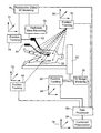
- FIG. 1 is a black box diagram of a system according to the teachings of the present invention.
- FIG. 2 is a perspective view of an articulated arm which serves as a position tracking system shown carrying a radioactive emission probe in accordance with the teachings of the present invention
- FIG. 3 is a schematic depiction of a radioactive emission probe carrying a pair of three coaxially aligned accelerometers which serve as a position tracking system in accordance with the teachings of the present invention
- FIG. 4 is a schematic presentation of a radioactive emission probe communicating with yet another type of a position tracking system in accordance with the teachings of the present invention
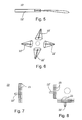
- FIG. 5 is a simplified cross-sectional view of a narrow or wide angle radioactive emission probe used to implement an embodiment of the present invention
- FIG. 6 is a presentation of a scanning protocol which can be effected with the detector of FIG. 5 ;
- FIG. 7 is a simplified cross-sectional view of a spatially sensitive radioactive emission probe, e.g., a gamma camera, used to implement another embodiment of the present invention.
- a spatially sensitive radioactive emission probe e.g., a gamma camera
- FIG. 8 is a presentation of a scanning protocol which can be effected with the detector of FIG. 7 ;
- FIG. 9 demonstrates a system in accordance with the teachings of the present invention which employs four position tracking systems for co-tracking the positions of a patient, a radioactive emission probe, an imaging modality and a surgical instrument;
- FIG. 10 demonstrates the use of a pair of radiation emission detectors connected therebetween via a connector, preferably a flexible connector or a flexible connection to the connector according to the present invention
- FIG. 11 is a schematic diagram of a surgical instrument and accompanying system elements according to the teachings of the present invention.
- FIG. 12 is a simplified pictorial illustration of an imaging system constructed and operative in accordance with a preferred embodiment of the present invention, including a radiation probe and position sensor, position tracking system, medical imaging system and coordinate registration system;
- FIG. 13 is a simplified pictorial illustration of a single dimension image formation with a nuclear radiation probe attached to a position tracking system of the system of FIG. 12 , in accordance with a preferred embodiment of the present invention
- FIG. 14 is a simplified pictorial plot of detecting a radiation point source with the nuclear radiation probe of the system of FIG. 12 , without further processing, in accordance with a preferred embodiment of the present invention
- FIG. 15 is a simplified flow chart of an averaging algorithm used in the imaging system of FIG. 12 , in accordance with a preferred embodiment of the present invention.
- FIG. 16 is a simplified pictorial plot of detecting a radiation point source with the nuclear radiation probe of the system of FIG. 12 , with averaging processing, in accordance with a preferred embodiment of the present invention
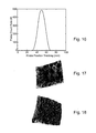
- FIGS. 17 and 18 are simplified pictorial illustrations of hot cross and hot bar phantom images, respectively, of images produced by a gamma radiation probe of the system of FIG. 12 ;
- FIG. 19 is a simplified flow chart of a minimizing algorithm used in the imaging system of FIG. 12 , in accordance with a preferred embodiment of the present invention.
- FIG. 20 is a simplified pictorial plot of detecting a radiation point source with the nuclear radiation probe of the system of FIG. 12 , with minimizing processing, in accordance with a preferred embodiment of the present invention
- FIG. 21 is a simplified pictorial illustration of an image reconstruction system constructed and operative in accordance with a preferred embodiment of the present invention, which produces a combined image made up of medical images, the position of the peak radiation location and the location of a therapeutic instrument;
- FIG. 22 is a simplified flow chart of a radiation map reconstruction algorithm, in accordance with a preferred embodiment of the present invention.
- FIGS. 23A and 23B are illustrations of radiolabeled patterns observed in images produced by the system of the invention and by a conventional gamma camera, respectively, of an autonomous adenoma of a thyroid;
- FIGS. 24A and 24B are illustrations of radiolabeled patterns observed in images produced by the system of the invention and by a conventional gamma camera, respectively, of suspected Paget's disease of a humerus;
- FIGS. 25A and 25B are illustrations of radiolabeled patterns observed in images produced by the system of the invention and by a conventional gamma camera, respectively, of chronic osteomyelitis;
- FIGS. 26A and 26B are illustrations of radiolabeled patterns observed in images produced by the system of the invention and by a conventional gamma camera, respectively, of skeletal metastasis from medulloblastoma;
- FIGS. 27A-27I demonstrate the operation of an algorithm provided by the present invention for estimating the distribution of radiation sources in a control volume
- FIGS. 28A-28F schematically illustrate a handheld probe, in accordance with preferred embodiments of the present invention.
- FIGS. 29A-29B schematically illustrate the manner of calibrating the handheld probe of FIGS. 28A-28F , in accordance with a preferred embodiment of the present invention
- FIG. 30 schematically illustrates the manner of synchronizing event and position readings of the handheld probe of FIGS. 28A-28F , in accordance with a preferred embodiment of the present invention
- FIGS. 31A-31C describe a spatial resolution bar-phantom test of a radioactive emission probe, in accordance with a preferred embodiment of the present invention
- FIGS. 32A-32D describe a spatial resolution bar-phantom test of a prior-art probe
- FIGS. 33A-33B illustrate the energy resolution of a single pixel of a radioactive emission probe, in accordance with the present invention
- FIGS. 34A-34C illustrate endoscopic radioactive emission probes, in accordance with the present invention
- FIG. 35 illustrate a method of calculating the depth of a radiation source, in accordance with the present invention.
- FIG. 36 illustrate a two-dimensional image of the radioactivity emitting source, produced by a free-hand scanning of a cancerous prostate gland, ex vivo, in accordance with the present invention.
- the present invention relates to a radioactive emission probe in communication with a position tracking system and the use thereof in a variety of systems and methods of medical imaging and procedures.
- wide-aperture collimation—deconvolution algorithms are provided, for obtaining a high-efficiency, high resolution image of a radioactivity emitting source, by scanning the radioactivity emitting source with a probe of a wide-aperture collimator, and at the same time, monitoring the position of the radioactive emission probe, at very fine time intervals, to obtain the equivalence of fine-aperture collimation.
- the blurring effect of the wide aperture is then corrected mathematically.
- an imaging method by depth calculations is provided, based on the attenuation of photons of different energies, which are emitted from the same source, coupled with position monitoring.
- Functional imaging or the use of radioactive materials to tag physiologically active tissue within the body of a patient, for determining the tissue's localization and demarcation by radioactive emission probes has been disclosed in the medical literature for at least forty years.
- Functional imaging shows the metabolic activity of body tissue, since dying or damaged body tissue absorbs radiopharmaceuticals at a different rate from a healthy tissue.
- the functional image may be used for example, to study cardiac rhythm, or respiratory rhythm. However, a functional image may not show structural, or anatomic details.
- Such radiopharmaceuticals tend to localize in particular tissue or cell type, whereas uptake or binding of the specific radiopharmaceutical is increased in more “physiologically active” tissue such as the active core of a cancerous tissue, so that the radiation emitted following nuclear disintegrations of the isotope can be detected by a radiation detector to better allocate the active portion of a tumor.
- Such radiation may be, for example, ⁇ , ⁇ ⁇ , ⁇ + and (or) ⁇ radiation.
- radioactive substances are used to determine the level of flow of blood in blood vessels and the level of perfusion thereof into a tissue, e.g., coronary flow and myocardial perfusion.
- FIG. 1 illustrates a system for calculating a position of a radioactivity emitting source in a system-of-coordinates, in accordance with the teachings of the present invention, which system is referred to hereinbelow as system 20 .
- System 20 includes a radioactivity emission detector 22 .
- System 20 according to the present invention further includes a position tracking system 24 .
- System 24 is connected to and (or) communicating with radioactive emission probe 22 so as to monitor the position of detector 22 in a two- or three-dimensional space defined by a system-of-coordinates 28 in two, three or more, say four, five or preferably six degrees-of-freedom (X, Y, Z, ⁇ , ⁇ and ⁇ ).
- System 20 further includes a data processor 26 .
- Data processor 26 is designed and configured for receiving data inputs from position tracking system 24 and from radioactive emission probe 22 and, as is further detailed below, for constructing the image of the radioactivity emitting source in system-of-coordinates 28 . As shown in FIG.
- detectors 22 are preferably connected there between via a connector 29 .
- Connector 29 is preferably flexible. In the alternative, the connections of detectors 22 to connector 29 provide the required flexibility.
- system 20 of radioactivity emission detector 22 and position tracking system 24 is inherently different from known SPECT and PET imaging systems, as well as from other imaging systems such as X-ray, Mammography, CT, and MRI, since the motion of detector 22 of the present invention is not limited to a predetermined track or tracks, with a respect to an immovable gantry. Rather, detector 22 of the present invention is adapted for a variable-course motion, which may be for example, free-hand scanning, variable-course motion on a linkage system, motion within a body lumen, endoscopic motion through a trocar valve, or another form of variable-course motion.
- Position tracking systems per se are well known in the art and may use any one of a plurality of approaches for the determination of position in a two- or three-dimensional space as is defined by a system-of-coordinates in two, three and up to six degrees-of-freedom.
- Some position tracking systems employ movable physical connections and appropriate movement monitoring devices (e.g., potentiometers) to keep track of positional changes.
- movement monitoring devices e.g., potentiometers
- Such systems once zeroed, keep track of position changes to thereby determine actual positions at all times.
- One example for such a position tracking system is an articulated arm.
- FIG. 2 shows an articulated arm 30 which includes six arm members 32 and a base 34 , which can therefore provide positional data in six degrees-of-freedom.
- Monitoring positional changes may be effected in any one of s veral different ways. For example, providing each arm member 32 with, e.g., potentiometers or optical encoders 38 used to monitor the angle between adjacent arm members 32 , to thereby monitor the angular change of each such arm member with respect to adjacent arm members, so as to determine the position in space of radioactive emission probe 22 , which is physically connected to articulated arm 30 .
- FIG. 3 other position tracking systems can be attached directly to radioactive emission probe 22 in order to monitor its position in space.
- An example of such a position tracking system is an assortment of three triaxially (e.g., co-orthogonally) oriented accelerometers 36 which may be used to monitor the positional changes of radioactive emission probe 22 with respect to a space.
- a pair of such assortments as is specifically shown in FIG. 3 , can be used to determine the position of detector 22 in six-degrees of freedom.
- FIGS. 4 and 10 other position tracking systems re-determine a position irrespective of previous positions, to keep track of positional changes.
- Such systems typically employ an array of receivers/transmitters 40 which are spread in known positions in a system-of-coordinates and transmitter(s)/receiver(s) 42 , respectively, which are in physical connection with the object whose position being monitored.
- Time based triangulation and (or) phase shift triangulation are used in such cases to periodically determine the position of the monitored object, radioactive emission probe 22 in this case.
- Radioactive emission probes are well known in the art and may use any one of a number of approaches for the determination of the amount of radioactive emission emanating from an object or portion thereof.
- detectors typically include substances which when interacting with radioactive decay emitted particles emit either electrons or photons in a level which is proportional over a wide linear range of operation to the level of radiation impinging thereon. The emission of electrons or photons is measurable and therefore serves to quantitatively determine radiation levels.
- Solid-state detectors in the form of N-type, P-type, PIN-type pixellated or unpixellated include, for example, Ge, Si, CdTe, CdZnTe, CdSe, CdZnSe, HgI 2 , TlBrI, GaAs, InI, GaSe, Diamond, TlBr, PbI 2 , InP, ZnTe, HgBrI, a-Si, a-Se, BP, GaP, CdS, SiC, AlSb, PbO, BiI 3 and ZnSe detectors.
- Gas (e.g., CO 2 CH 4 ) filled detectors include ionization chamber detectors, proportional chamber detectors and Geiger chamber detectors.
- Scintillation detectors include organic scintillator crystals and liquids, such as C 14 H 10 , C 14 H 12 , C 10 H 8 , etc., Plastics, NE102A, NE104, NE110, Pilot U and inorganic scintillator crystals, such as NaI, CsI, BGO, LSO, YSO, BaF, ZnS, ZnO, CaWO 4 and CdWO 4 . Also known are scintillation fiber detectors.
- Scintillator coupling include photomultiplier tube (PMT) of the following types: side-on type, head-on type, hemispherical type, position sensitive type, icrochannel plate-photomultiplier (MCP-PMTs) and electron multipliers, or photodiodes (and photodiodes arrays), such as Si photodiodes, Si PIN photodiodes, Si APD, GaAs(P) photodiodes, GaP and CCD.
- PMT photomultiplier tube
- MCP-PMTs icrochannel plate-photomultiplier
- electron multipliers or photodiodes (and photodiodes arrays), such as Si photodiodes, Si PIN photodiodes, Si APD, GaAs(P) photodiodes, GaP and CCD.
- FIG. 5 shows a narrow angle or wide angle radioactive emission probe 22 ′.
- Narrow or wide angle radioactive emission probe 22 ′ includes a narrow slit (collimator) so as to allow only radiation arriving from a predetermined angular direction (e.g., 1°-280° wide angle, preferably 1°-80°—narrow angle) to enter the detector.
- Narrow or wide angle radioactive emission probes especially suitable for the configuration shown in FIG. 10 are manufactured, for example, by Neoprobe, Dublin, Ohio (www.neoprobe.com), USA, Nuclear Fields, USA (www.nufi.com) IntraMedical Imaging, Los Angeles, Calif., USA (www.gammaprobe.com).
- such a detector is typically used to measure radioactivity, point by point, by scanning over the surface of a radioactive object from a plurality of directions and distances. In the example shown, scans from four different directions are employed. It will be appreciated that if sufficient radioactivity records are collected from different angles and distances, and the orientation and position in space of detector 22 ′ is simultaneously monitored and recorded during such scans, a three-dimensional model of a radioactive region can be reconstituted and its position in space determined. If two or more detectors are co-employed, as shown in the configuration of FIG. 10 , the results may be collected faster.
- FIG. 7 shows another example of a radioactive emission probe, a spatially sensitive (pixellated) radioactive emission probe 22 ′′ (such as a gamma camera).
- Detector 22 ′′ in effect, includes an array of multitude narrow angle detector units 23 .
- Such an arrangement is used in accordance with the teachings of the present invention to reduce the amount of measurements and angles necessary to acquire sufficient data so as to reconstitute a three-dimensional model of the radioactive object.
- Examples of spatially sensitive radioactive emission probes employed in a variety of contexts are disclosed in, for example, U.S. Pat. Nos. 4,019,057; 4,550,250; 4,831,262; and 5,521,373; which are incorporated by reference as if set forth herein.
- FIG. 8 shows a scan optionally made by spatially sensitive radioactive emission probe 22 ′′ (such as a gamma camera).
- a radioactive emission detector of particular advantages for use in context of the present invention is the Compton gamma probe, since, in the Compton gamma probe, spatial resolution is independent of sensitivity and it appears possible to exceed the noise equivalent sensitivity of collimated imaging systems especially for systems with high spatial resolution.
- the Compton probe is a novel type of gamma-probe that makes use of the kinematics of Compton scattering to construct a source image without the aid of mechanical collimators.
- Compton imaging telescopes were first built in the 1970s for astronomical observations [V. Schoenfelder et al., Astrophysical Journal 217 (1977) 306]. The first medical imaging laboratory instrument was proposed in the early 1980s [M. Singh, Med. Phys. 10 (1983) 421].
- the potential advantages of the Compton gamma probe include higher efficiency, 3-D imaging without detector motion, and more compact and lightweight system.
- high-energy gamma rays are scattered from a first detector layer (or detectors array) into a second detector layer array.
- the deposited energy is measured in both detectors.
- the Compton scattering equation can be solved to determine the cone of possible direction about this axis on which the gamma ray must have entered the first detector. The intersection of cones from many events is then developed to locate gamma ray sources in the probe's field-of-view.
- the probe's electronic system is combining coincidence measurements across many detectors and detectors layers with a very good energy resolution.
- the choice of the geometry and the material of the first layer detector plays a major role in the system imaging capability and depends on (i) material efficiency of single Compton events, in relation to other interactions; (ii) detector energy resolution; and (iii) detector position resolution.
- the overall angular resolution results from the combination of two components, related to the energy resolution and to the pixel volume of the detector.
- connecting a radioactive emission probe to a position tracking system permits simultaneous radioactivity detecting and position tracking at the same time. This enables the accurate calculation of the shape, size and contour of the radiating object and its precise position in a system-of-coordinates.
- the present invention thus provides a method for defining a position of a radioactivity emitting source in a system-of-coordinates.
- the method is effected by (a) providing a radioactive emission probe which is in communication with a position tracking system; and (b) monitoring radioactivity emitted from the radioactivity emitting source, while at the same time, monitoring the position of radioactive emission probe in the system-of-coordinates, thereby defining the image of the radioactivity emitting source in the system-of-coordinates.
- model produced by system 20 is projectable onto any of the other systems-of-coordinates, or alternatively, the system-of-coordinates defined by position tracking system 24 may be shared by other position tracking systems, as is further detailed hereinbelow, such that no such projection is required.
- system 20 of the present invention can be used for calculating a position of a radioactivity emitting source in a first system-of-coordinates 28 and further for projecting the image of the radioactivity emitting source onto a second system-of-coordinates 28 ′.
- the system includes radioactive emission probe 22 , position tracking system 24 which is connected to and (or) communicating with radioactive emission probe 22 , and data processor 26 which is designed and configured for (i) receiving data inputs from position tracking system 24 and from radioactive emission probe 22 ; (ii) constructing the image of the radioactivity emitting source in the first system-of-coordinates; and (iii) projecting the image of the radioactivity emitting source onto the second system-of-coordinates.
- a method for calculating a position of a radioactivity emitting source in a first system-of-coordinates and for projecting the image of the radioactivity emitting source onto a second system-of-coordinates is also offered by the present invention.
- This method is effected by (a) providing a radioactive emission probe being in communication with a position tracking system; and (b) monitoring radioactivity being emitted from the radioactivity emitting source, while at the same time, monitoring the position of the radioactive emission probe in the first system-of-coordinates, thereby defining the image of the radioactivity emitting source in the first system-of-coordinates and projecting the image of the radioactivity emitting source onto the second system-of-coordinates.
- a radioactive emission probe and a position tracking system connected thereto and (or) communicating therewith allows a suitable data processor to generate a two- or three-dimensional image of the radioactivity emitting source.
- An algorithm can be used to calculate image intensity based on, for example, a probability function which averages radiation counts and generates an image in which the shorter the time interval between radioactive counts, the brighter the image and vise versa, while down-compensating when a location is re-scanned.
- a free-hand scanning with a directional detector can be employed for this purpose.
- the detector when scanning a body area with the detector, the detector is made to follow a three-dimensional surface, which defines the body curvature and in effect is used also as a position tracking pointer.
- This information can be used to define the position of the radioactive source with respect to the outer surface of the body, so as to create a three-dimensional map of both the radioactive source and of the body curvature.
- This approach can also be undertaken in open surgeries, such as open chest surgeries so as to provide the surgeon in real time with information concerning the functionality of a tissue.
- the radioactive emission probe which can be used in context of the present invention can be a beta emission detector, a gamma emission detector, a positron emission detector or any combination thereof.
- a detector that is sensitive to both beta and (or) positron and gamma emission can be used to improve localization by sensing for example gamma emission distant from the source and sensing beta or positrons emission closer to the source.
- a beta detector is dedicated for the detection of either electrons from sources such as 131 Iodine, or positrons from sources such as 18 Fluorine.
- a gamma detector can be designed as a single energy detector or as a detector that can distinguish between different types of energies, using the light intensity in the scintillator as a relative measure of the gamma energy. Also, the detector can be designed to utilize coincidence detection by using detectors facing one another (180 degrees) with the examined organ or tissue in-between.
- the radiation detector can have different collimators with different diameters. A large bore will be used for high sensitivity with lower resolution while a small bore collimator will have higher resolution at the expense of lower sensitivity.
- Another possibility is to have a the collimator moving or rotating with the opening eccentric so that a different solid angle is exposed to the incoming photons at any one time, thus gathering the photons from overlapping volumes at different time intervals.
- the rest of the image processing is similar if the probe moves or if the collimator eccentric opening moves.
- System 20 of the present invention can be used in concert with other medical devices, such as, but not limited to, any one of a variety of imaging modalities and (or) surgical instruments.
- Structural imaging modalities which provide anatomic, or structural maps of the body, are well known in the art.
- the main modalities that serve for two-(projectional or cross sectional) or three-(consecutive cross sectional) dimensional imaging are a planer X-ray imager, a fluoroscope, a computerized tomography scanner, a magnetic resonance imager an ultrasound imager, an impedance imager, and an optical camera.
- Medical images taken of the human body are typically acquired or displayed in three main orientations (i) coronal orientation: in a cross section (plane), for example, across the shoulders, dividing the body into front and back halves; (ii) sagittal orientation: in a cross section (plane), for example, down the middle, dividing the body into left and right halves; and (iii) axial orientation: in a cross section (plane), perpendicular to the long axis of the body, dividing the body into upper and lower halves.
- Oblique views can also be acquired and displayed.
- X-ray imaging results in a projection of the integrated density of column-voxels defined by the X-rays as they pass through the body.
- Fluoroscopy is a method based on the principals of film X-ray that is useful for detecting disorders and tumors in the upper gastrointestinal (GI) system (for example, the stomach and intestines). Fluoroscopic imaging yields a moving X-ray picture. The physician can watch the screen and see an image of the patient's body (for example the beating heart). Fluoroscopic technology improved greatly with the addition of television cameras and fluoroscopic “image intensifiers”. Today, many conventional X-ray systems have the ability to switch back and forth between the radiographic and fluoroscopic modes. The latest X-ray systems have the ability to acquire the radiograph or fluoroscopic movie using digital acquisition.
- GI gastrointestinal
- Computed Tomography is based on the X-ray principal, where the film is replaced by a detector that measures the X-ray profile.
- CT Computed Tomography
- Inside the covers of the CT scanner is a rotating frame which has an X-ray tube mounted on one side and the detector mounted on the opposite side.
- a fan beam of X-ray is created as the rotating frame spins the X-ray tube and detector around the patient.
- an image or “slice” has been acquired. This “slice” is collimated to a thickness between 1 mm and 10 mm using lead shutters in front of the X-ray tube and X-ray detector.
- the detector takes numerous profiles of the attenuated X-ray beam. Typically, in one 360° lap, about 1,000 profiles are sampled. Each profile is subdivided spatially by the detectors and fed into about 700 individual channels. Each profile is then backwards reconstructed (or “back projected”) by a dedicated computer into a two-dimensional image of the “slice” that was scanned.
- the CT gantry and table have multiple microprocessors that control the rotation of the gantry, movement of the table (up/down and in/out), tilting of the gantry for angled images, and other functions such as turning the X-ray beam on an off.
- the CT contains a slip ring that allows electric power to be transferred from a stationary power source onto the continuously rotating gantry.
- the innovation of the power slip ring has created a renaissance in CT called spiral or helical scanning.
- spiral CT acquires a volume of data with the patient anatomy all in one position. This volume data set can then be computer-reconstructed to provide three-dimensional models such as of complex blood vessels like the renal arteries or aorta. Spiral CT allows the acquisition of CT data that is perfectly suited for three-dimensional reconstruction.
- MR Imaging is superior to CT in detecting soft tissue lesions such as tumors as it has excellent contrast resolution, meaning it can show subtle soft-tissue changes with exceptional clarity. Thus, MR is often the method of choice for diagnosing tumors and for searching for metastases.
- MR uses magnetic energy and radio waves to create single or consecutive cross-sectional images or “slices” of the human body.
- the main component of most MR systems is a large tube shaped or cylindrical magnet.
- the strength of the MR systems magnetic field is measured in metric units called “Tesla”. Most of the. cylindrical magnets have a strength between 0.5 and 1.5 Tesla and most of the open or C-shaped magnets have a magnetic strength between 0.01 and 0.35 Tesla.
- Each total MR examination typically is comprised of a series of 2 to 6 sequences.
- An “MR sequence” is an acquisition of data that yields a specific image orientation and a specific type of image appearance or “contrast”.
- MR sequence is an acquisition of data that yields a specific image orientation and a specific type of image appearance or “contrast”.
- a radio signal is turned on and off, and subsequently the energy which is absorbed by different atoms in the body is echoed or reflected back out of the body.
- These echoes are continuously measured by “gradient coils” that are switched on and off to measure the MR signal reflecting back.
- the net magnetization vector rotate from a longitudinal position a distance proportional to the time length of the radio frequency pulse.
- Radio frequency coils are the “antenna” of the MRI system that broadcasts the RF signal to the patient and (or) receives the return signal.
- RF coils can be receive-only, in which case the body coil is used as a transmitter; or transmit and receive (transceiver).
- Surface coils are the simplest design of coil. They are simply a loop of wire, either circular or rectangular, that is placed over the region of interest.
- a digital computer reconstructs these echoes into images of the body.
- a benefit of MRI is that it can easily acquire direct views of the body in almost any orientation, while CT scanners typically acquire cross-sectional images perpendicular or nearly perpendicular to the long body axis.
- Ultrasound imaging is a versatile scanning technique that uses sound waves to create images of organs or anatomical structures in order to make a diagnosis.
- the ultrasound process involves placing a small device called a transducer, against the skin of the patient near the region of interest, for example, against the back to image the kidneys.
- the ultrasound transducer combines functions of emitting and receiving sound. This transducer produces a stream of inaudible, high frequency sound waves which penetrate into the body and echo off the organs inside.
- the transducer detects sound waves as they echo back from the internal structures and contours of the organs. Different tissues reflect these sound waves differently, causing a signature which can be measured and transformed into an image.
- These waves are received by the ultrasound machine and turned into live pictures with the use of computers and reconstruction software.
- Ultrasound scanning has many uses, including: diagnosis of disease and structural abnormalities, helping to conduct other diagnostic procedures, such as needle biopsies etc.
- imaging modalities either inherently include (e.g., fluoroscope, CT, MRI) and (or) are integrated with position-tracking-systems, which enable the use of such systems to reconstruct three-dimensional image models and provide their position in a system-of-coordinates.
- fluoroscope e.g., CT, MRI
- position-tracking-systems which enable the use of such systems to reconstruct three-dimensional image models and provide their position in a system-of-coordinates.
- an optical camera can be used to generate three-dimensional imagery date according to the present invention by imaging a body from a plurality (at least two) directions. This type of imaging is especially applicable in open chest surgeries or other open surgeries. Software for calculating a three-dimensional image from a pair of stereoscopic images is well known in the art.
- the phrase “three-dimensional imaging modality” refers to any type of imaging equipment which includes software and hardware for generating a three-dimensional image.
- Such an equipment can generate a three-dimensional image by imaging successive cross-sections of a body, e.g., as if viewed from a single direction.
- such an equipment can generate a three-dimensional image by imaging successive cross-sections of a body, e.g., as if viewed from a single direction.
- such an equipment can generate a three-dimensional image by imaging a body from different angles or directions (typically two angles) and thereafter combining the data into a three-dimensional image.
- a structural imaging probe for example, an ultrasound probe
- a position tracking system in a manner analogous to System 20 of radioactivity emission detector 22 and position tracking system 24 ( FIG. 1 ).
- a miniature MRI probe for example, as taught by U.S. Pat. No. 5,572,132, to Pulyer, et al., entitled, “MRI probe for external imaging,” whose disclosure is incorporated herein by reference, which teaches an MRI catheter for endoscopic imaging of tissue of the artery wall, rectum, urinal tract, intestine, esophagus, nasal passages, vagina and other biomedical applications, may be used.
- a structural imaging probe for example, an ultrasound probe, may be combined with system 20 of radioactivity emission detector 22 and position tracking system 24 ( FIG. 1 ), so as to have:
- the structural imaging modality may have an additional, dedicated position tracking system, or share position tracking system 24 of radioactivity emission detector 22 .
- Position tracking may also be accomplished by using imaging information from the structural imaging system. This may be accomplished by tracking relative changes from one image to another to determine relative motion.
- Surgical instruments are also well known in the art and may use any one of a plurality of configurations in order to perform minimally-invasive surgical procedures. Examples include laser probes, cardiac and angioplastic catheters, endoscopic probes, biopsy needles, aspiration tubes or needles, resecting devices, ultrasonic probes, fiber optic scopes, laparoscopic prob s, thermal probes and suction/irrigation probes. Examples of such surgical instruments employed in a variety of medical cont xts are disclosed in, for example, U.S. Pat. Nos.
- surgical instruments are integrated with position-tracking-systems, which enable to monitor the position of such instruments while placed in and guided through the body of a treated patient.
- the surgical instrument is equipped with an additional radioactive emission probe attached thereto or placed therein.
- This additional detector is used, according to preferred embodiments of the invention, to fine tune the location of radioactive emission from within the body, and in closer proximity to the radioactive source. Since the surgical tool is preferably in communication with a position-tracking system, the position of the additional detector can be monitored and its readouts used to fine tune the position of the radioactive source within the body.
- at least one extracorporeal detector and an intracorporeal detector are used in concert to determine the position of a radioactive source in the body in highest precision.
- the extracorporeal detector provides the general position of the source and is used for directing the surgical instrument thereto, whereas the intracorporeal detector is used for reassuring prior to application of treatment or retrieval of biopsy that indeed the source was correctly targeted at the highest precision.
- intracorporeal and extracorporeal detectors calls for careful choice of the radioactive isotope employed with the radiopharmaceutical. While the extracorporeal detector can be constructed with a suitable collimator for handling strong radiation, such as gamma radiation, the intracorporeal detector is miniature by nature and is limited in design and construction by the construction of the surgical instrument with which it is employed. Since collimators for high energy (80-511 KeV) gamma radiation are robust in nature, they are not readily engageable with miniature detectors. Electron (beta) and positron radiation are characterized by: (i) they highly absorbed by biological tissue as they are of lower energy and higher chemical reactivity; and (ii) they are readily collimated and focused by thin metal collimators.
- the radio pharmaceutical of choice is selected to emit both gamma and beta and (or) positron radiation, whereas the extracorporeal detector is set to detect the high energy gamma radiation, whereas the intracorporeal detector is set to detect the low energy gamma, beta and (or) positron radiation.
- Isotopes that emit both high energy gamma and (or) low energy gamma, beta and (or) positron radiation and which can be used per se or as a part of a compound as radiopharmaceuticals include, without limitation, 18 F, 111 In and 123 I in radiopharmaceuticals, such as, but not limited to, 2-[ 18 F]fluoro-2-deoxy-D-glucose ( 18 FDG), 111 In-Pentetreotide ([ 111 In-DTPA-D-Phe 1 ]-octreotide), L-3- [ 123 I]-Iodo-alpha-methyl-tyrosine (IMT), O-(2-[ 18 F]fluoroethyl)-L-tyrosine (L-[ 18 F]FET), 111 In-Capromab Pendetide (CYT-356, Prostascint) and 111 In-Satumomab Pendetide (Oncoscint).
- FIG. 11 illustrates a system in accordance with this aspect of the present invention.
- a surgical instrument 100 is shown connected to a resection/aspiration control element 102 as well known in the art.
- Surgical instrument 100 includes a radioactive emission probe 104 , which has a collimator 106 for collimating low energy gamma, beta and (or) positron radiation.
- detector 104 may be translated within instrument 100 .
- a position tracking system having one element thereof 110 attached to instrument 100 and another element thereof 112 at a fixed location serves to monitor the position of instrument 100 at all times in two, three and up to six degrees of freedom.
- Radioactive emission probe 104 communicates with a counter 114 for counting low energy gamma, beta and (or) positron radiation. All the data is communicated to, and processed by, a processor 116 .
- the 2D or 3D data may be projected and displayed along with 2D or 3D imaging data derived from an imaging modality using a shared presentation device as described elsewhere herein. A real or virtual image of the surgical instrument itself may also be co-displayed.
- Examples of commercially available radiation emission detectors that can fit inside, for example, a biopsy needle include scintillating plastic optical fibers like S 101 and S 104 , manufactured by PPLASTIFO or an optical fiber communicating with a scintillator (either detector paint or scintillation crystal) at the fiber edge.
- the level of detected radiation can be reported visually or by an audio signal, as is well known in the art.
- a surgical instrument equipped with a radiation emission detector and which is connected to and (or) communicating with a position tracking system forms one embodiment of this aspect of the present invention.
- a design acting in concert with either conventional imaging modalities and (or) extracorporeal radiation emission detectors form other embodiments of this aspect of the invention.
- a surgical instrument equipped with a radiation emission detector and which is connected to and (or) communicating with a position tracking system serves for in situ fine tuning of a radioactive source in the body.
- FIG. 9 An example to this desired outcome is shown in FIG. 9 .
- four independent position tracking systems 50 , 52 , 54 and 56 are used to track the positions of a patient 58 , an imaging modality 60 , a radioactive emission probe 62 and a surgical instrument 64 in four independent systems-of-coordinates 66 , 68 , 70 and 72 , respectively. If the patient is still, no tracking of the patient's position is required.
- any subset or all of the position tracking systems employed may be integrated into one or more common position tracking systems, and (or) that any subset or all of the position tracking systems employed may share one or more systems-of-coordinates, and further that any positional data obtained by any of the position tracking systems described in any of the systems-of coordinates may be projected to any other system of coordinates or to an independent (fifth) system of coordinates 74 .
- the system of coordinates is a dynamic system of coordinates which takes into account the chest breathing movements of the patient during the procedure.
- the raw data collected by detector 62 is recorded and, as indicated at 78 , the position and the radioactive data records are used to generate a three-dimensional model of a radiopharmaceutical uptake portion of a body component of the patient.
- imagery data collected by imaging modality 60 is recorded and the position and the imagery data records are used to generate a three-dimensional model of the imaged body component of the patient.
- All the data collected is then fed into a data processor 82 which processes the data and, as indicated at 84 , generates a combined or superimposed presentation of the radioactive data and the imagery data, which is in positional context with patient 58 and surgical instrument 64 .
- Instrument 64 which by itself can he presented in context of the combined presentation, may then be used to perform the procedure most accurately.
- Processor 82 may be a single entity or may include a plurality of data processing stations which directly communicate with, or even integral to, any one or more of the devices described.
- a structural imaging probe for example, an ultrasound probe, or another structural probe, as known, may be incorporated with the surgical instrument.
- the present invention provides a major advantage over prior art designs because it positionally integrates data pertaining to a body portion as retrieved by two independent imaging techniques, conventional imaging and radioactive imaging, to thereby provide a surgeon with the ability the fine point the portion of the body to be sampled or treated.
- subsets of the devices described in FIG. 9 may be used as stand-alone systems.
- a combination of detector 62 with its position-tracking system and instrument 64 with its position-tracking-system may in some instances be sufficient to perform intrabody procedures.
- a combination of detector 62 position-tracking-system and modality 60 position-tracking-system are sufficient.
- Imaging system 200 preferably includes a radiation probe 202 , such as the narrow angle radioactive emission probe 22 ′ described hereinabove with reference to FIGS. 5 and 10 .
- a position sensor 204 is provided for sensing the position of radiation probe 202 .
- Position sensor 204 may be physically attached to radiation probe 202 , or may be distanced therefrom.
- Position sensor 204 transmits the sensed position data to a position tracking system 206
- Position tracking system 206 may be a system like position tracking system 24 , described hereinabove with reference to FIG. 1 , and position sensor 204 may be any kind of sensor applicable for such position tracking systems.
- Another method which can be used to locate the source of radiation emission is by using a small hand held gamma camera 205 (such as the DigiRad 2020 tc ImagerTM, 9350 Trade Place, San Diego, Calif. 92126-6334, USA), attached to position sensor 204 .
- a small hand held gamma camera 205 such as the DigiRad 2020 tc ImagerTM, 9350 Trade Place, San Diego, Calif. 92126-6334, USA
- Position tracking system 206 enables radiation probe 202 to freely scan back and forth in two- or three-dimensions over the area of interest of the patient, preferably incrementing a short distance between each scan pass. Position tracking system 206 tracks the position of radiation probe 202 with respect to a position tracking coordinate system, such as X p , Y p and Z p , with an origin O p .
- Imaging system 200 also includes a medical imaging system 208 , such as, but not limited to, computed or computerized tomography (CT), magnetic resonance imaging (MRI), ultrasound imaging, positron emission tomography (PET) and single photon emission computed tomography (SPECT), for example.
- Medical imaging system 208 provides images of a patient 209 with respect to a medical imaging coordinate system, such as X m , Y m and Z m , with an origin O m .
- Imaging system 200 also includes a coordinate registration system 210 , such as that described in commonly owned, now abandoned, U.S. patent application No. 09/610,490, entitled, “method for registering coordinate systems,” the disclosure of which is incorporated herein by reference.
- Coordinate registration system 210 is adapted to register the coordinates of the position tracking coordinate system with those of the medical imaging coordinate system.
- Position tracking system 206 , medical imaging system 208 and coordinate registration system 210 are preferably in wired or wireless communication with a processing unit 212 (also referred to as a data processor 212 ).
- a clinician/physician/surgeon may move or scan radiation probe 202 about a target area under examination.
- a physiological activity map of the target area is obtained by measuring the radiation count rate with radiation probe 202 , and by correlating the count rate with the count rate direction with position tracking system 206 , which follows the motion of the moving or scanning radiation probe 202 .
- FIG. 13 illustrates image formation with radiation probe 202 , in accordance with a preferred embodiment of the present invention.
- FIG. 13 illustrates image formation with radiation probe 202 , in accordance with a preferred embodiment of the present invention.
- the example shown in FIG. 13 is for a single dimension image formation, but it is readily understood that the same principles hold true for any other dimensional image formation.
- radiation probe 202 may be a gamma ray detector probe that comprises a collimator 211 and radiation detector 213 .
- Gamma rays are projected through the probe collimator 211 onto radiation detector 213 , which produces electronic signals in accordance with the radiation detected
- Radiation probe 202 sends pulses to a probe counter 215 which may include a pulse height analyzer circuit (not shown).
- the pulse height analyzer circuit analyzes the electronic signals produced by radiation detector 213 . If the electronic signals are within a selected energy window, the level of radiation, i.e., number of radiation counts, is counted by probe counter 215 .
- suitable radiation detectors include a solid state detector (SSD) (CdZnTe, CdTe, HgI, Si, Ge, and the like), a scintillation detector (NaI(Tl), LSO, GSO, CsI, CaF, and the like), a gas detector, or a scintillating fiber detector (S 101 , S 104 , and the like).
- SSD solid state detector
- CaI(Tl) CdZnTe, CdTe, HgI, Si, Ge, and the like
- NaI(Tl) scintillation detector
- LSO LSO
- GSO GSO
- CsI CsI
- CaF gas detector
- S 101 , S 104 scintillating fiber detector
- the position sensor 204 associated with the radiation probe 202 senses the position of radiation probe 202
- position tracking system 206 calculates and monitors the motion of radiation probe 202 with respect to the position tracking coordinate system.
- the motion is calculated and monitored in two, three and up to six dimensions—the linear directions of the X, Y and Z axes as well as rotations about the X, Y and Z axes, i.e., rotational angles ⁇ , ⁇ and ⁇ , respectively.
- Suitable position tracking systems include a measurement mechanical arm (FaroArm, http://www.faro.com/products/faroarm.asp), optical tracking systems (Northern Digital Inc., Ontario, Canada NDI-POLARIS passive or active systems), magnetic tracking systems (NDI-AURORA), infared tracking systems (E-PEN system, http://www.e-pen.com), and ultrasonic tracking systems (E-PEN system), for example.
- FaroArm http://www.faro.com/products/faroarm.asp
- optical tracking systems Northern Digital Inc., Ontario, Canada
- NDI-AURORA magnetic tracking systems
- E-PEN system http://www.e-pen.com
- ultrasonic tracking systems E-PEN system
- Processing unit 212 combines the radiation probe count rate from probe counter 215 together with the positional information from position tracking system 206 , and uses an imaging software algorithm 217 to form a two-dimensional or three-dimensional radiotracer-spread image of the target area inside the patient's body.
- the spatial probe positions together with the spatial count rates may be stored in memory or displayed on a computer monitor 214 as a pattern of marks corresponding to the spatial and count rate position.
- FIG. 14 illustrates a single-dimensional, unprocessed simulation of a radiation point source 218 ( FIG. 13 ), 30 mm deep inside the human body, detected by using a 10 mm nuclear radiation probe 202 coupled to position tracking system 206 .
- the graph of FIG. 14 indicates to a physician that there is a peak count rate of about 500 in the probe position of about 50 mm.
- the imaging software algorithm 217 employs an averaging process to refine the curve of FIG. 14 . This averaging process will now be described with reference to FIG. 15 .
- Probe counter 215 feeds probe count rate information N(Xc, Yc, Zc, ⁇ , ⁇ , ⁇ ) to processing unit 212 (step 301 ).
- Position sensor 204 feeds probe position information (Xc, Yc, Zc, ⁇ , ⁇ , ⁇ ) to processing unit 212 (step 302 ).
- Probe parameters (such as its physical size, dx, dy, dz) are also input into processing unit 212 (step 303 ).
- Processing unit 212 finds all the voxels (i.e., volume pixels) that represent the probe volume in the processing unit memory (step 304 ), i.e., Xc+dx, Yc+dy, Zc+dz. Processing unit 212 calculates the number of times that the calculation process has been done in each voxel from the beginning of the image formation (step 305 ), i.e., M(Xc+dx, Yc+dy, Zc+dz). Processing unit 212 then calculates the new average count rate values in each voxel (step 306 ), in accordance with the formula:
- N ⁇ ( Xc + dx , Yc + dy , Zc + dz ) ⁇ [ N ⁇ ( Xc + dx , Yc + dy , Zc + dz ) + N ⁇ ( Xc , Yc , Zc , ⁇ , ⁇ ) ] / ⁇ [ M ⁇ ( Xc + dx , Yc + dy , Zc + dz ) + 1 ]
- Processing unit 212 then corrects the display image that represents the perceived voxels at N(Xc+dx, Yc+dy, Zc+dz) (step 307 ). The algorithm then repeats itself for the next probe position (step 308 ).
- FIG. 16 The resulting graph of the averaging algorithm of FIG. 15 , as applied to the example of FIG. 14 , is shown in FIG. 16 .
- FIGS. 17 and 18 respectively show examples of a hot cross phantom image and a hot 4.77 mm bar phantom image, produced by a gamma radiation probe coupled with position tracking system 206 and the averaging algorithm of FIG. 15 .
- the position tracking system used was the Ascension miniBIRD, commercially available from Ascension Technology Corporation, P.O. Box 527, Burlington, Vt. 05402 USA (http://vww.ascension-tech.com/graphic.htm).
- the magnetic tracking and location systems of Ascension Technology Corporation use DC magnetic fields to overcome blocking and distortion from nearby conductive metals. Signals pass through the human body without attenuation.
- the imaging software algorithm 217 may employ a minimizing process to refine the curve of FIG. 14 as is now described with reference to FIG. 19 .
- Probe counter 215 feeds probe count rate information N(Xc, Yc, Zc, ⁇ , ⁇ , ⁇ ) to processing unit 212 (step 401 ).
- Position sensor 204 feeds probe position information (Xc, Yc, Zc, ⁇ , ⁇ , ⁇ ) to processing unit 212 (step 402 ).
- Probe parameters (such as its physical size, dx, dy, dz) are also input into processing unit 212 (step 403 ).
- Processing unit 212 finds all the voxels that represent the probe volume in the processing unit memory (step 404 ), i.e., Xc+dx, Yc+dy, Zc+dz. From the voxels that represent the probe volume in the processing unit memory, processing unit 212 finds those that have a higher count rate value N(Xc+dx, Yc+dy, Zc+dz) than the inputted probe count rate N(Xc, Yc, Zc, ⁇ , ⁇ , ⁇ ) (step 405 ).
- Processing unit 212 then changes the higher count rate voxels to that of inputted probe count rate N(Xc, Yc, Zc, ⁇ , ⁇ , ⁇ ) (step 406 ), and corrects the display image at the higher count rate voxels N(Xc+dx, Yc+dy, Zc+dz) (step 407 ). The algorithm then repeats itself for the next probe position (step 408 ).
- FIG. 20 The resulting graph of the minimizing algorithm of FIG. 19 , as applied to the example of FIG. 14 , is shown in FIG. 20 .
- FIG. 21 illustrates an image reconstruction system 450 , constructed and operative in accordance with a preferred embodiment of the present invention.
- Image reconstruction system 450 produces a combined image 451 made up of the images coming from the medical imaging system 208 with the position of the peak radiation location (and its uncertainty area) from processing unit 212 , together with the location of a therapeutic instrument 452 , such as a biopsy needle
- the combined image 451 allows the. physician to better assess the relative position of therapeutic instrument 452 in relation to the anatomical image (from medical imaging system 208 ) and the position of the radioactive area as inferred by the radiation detection algorithm.
- image acquisition and reconstruction algorithms are provided, based on image acquisition with wide-aperture collimation and image reconstruction, which includes deconvolution.
- the overall algorithms are herethereto referred to as wide-aperture collimation—deconvolution algorithms.
- the wide-aperture collimation—deconvolution algorithms enable one to obtain a high-efficiency, high resolution image of a radioactivity emitting source, by scanning the radioactivity emitting source with a probe of a wide-aperture collimator, and at the same time, monitoring the position of the radioactive emission probe, at very fine time intervals, to obtain the equivalence of fine-aperture collimation.
- the blurring effect of the wide aperture is then corrected mathematically.
- FIGS. 27A-27I The wide-aperture collimation—deconvolution algorithms are described hereinbelow, in conjunction with FIGS. 27A-27I , and FIG. 22 .
- Experimental results, showing images produced by the wide-aperture collimation—deconvolution algorithms of the present invention, in comparison to a conventional gamma camera are seen in FIGS. 23A-26B , and FIG. 36 , hereinbelow.
- FIGS. 27A-271 describe wide-aperture collimation—deconvolution algorithms, in accordance with preferred embodiments of the present invention.
- the wide-aperture collimation—deconvolution algorithms are designed for estimating the distribution of radiation sources in a control volume, thus constructing an image of the radiation sources in the control volume.
- the radiation sources comprise dot sources that radiate uniformly in all directions, are localized, and are smoothly distributed in the control volume.
- a single-pixel, wide-bore-collimator probe 707 seen in FIG. 27H , formed for example, as a lead tube collimator 708 , having a length, L collimator of about 20 mm and a radiation detector 706 , for example, a solid state CdZnTl crystal, at its base, with a diameter, D detector of about 10 mm, so that the ratio of L collimator to D detector is about 2.
- An angular opening ⁇ of wide-bore-collimator probe 707 may be about 40 degrees; wide-bore-collimator probe 707 is sensitive to any incident photons within the confining of about a 40-degree angular opening.
- the ratio of L collimator to D detector may be about 1, or about 3.
- conventional gamma cameras have a multi-pixel small-aperture collimation probe, wherein a collimator—pixel arrangement 709 is about 2 mm in diameter and 40 mm in length, so that the ratio of L collimator to D detector is about 20.
- An angular opening ⁇ of the small-aperture collimation probe is very small.
- wide-bore-collimator probe 707 may be about 30 times more sensitive than the small-aperture collimation probe of collimator—pixel arrangement 709 .
- knowing the position and orientation of probe 707 at each time interval and the photon count rate at that position and orientation can be used to reconstruct the radiation density of an unknown object, even in three dimensions.
- the blurring effect of the wide-angle aperture while it is known that the information is blurred, or convolved, by the wide-aperture collimator, a deconvolution process may be used to obtain dependable results.
- the convolving function for a wide aperture depends only on the geometry of the collimator and may be expressed as a set of linear equations that can be readily solved.
- a wide-bore collimator probe having a position tracking system, operating at very short time intervals, and connected to a data processor for wide-aperture collimation—deconvolution algorithms, can be used for the construction of an image of radiation density in one-, two-, or three-dimensions, with high degree of accuracy.
- the wide-aperture probe may have a rectangular collimator of a single cell or of a coarse grid of cells.
- the collimator may be a wide-angle collimator.
- a corresponding deconvolution function may be used by the data processor.
- FIGS. 27A and 27B illustrate a radiation sensor 600 , preferably generally shaped as a tube collimator.
- Radiation quanta 602 are registered by the radiation sensor 600 , as described hereinabove, thereby providing the average number of quanta per unit time.
- the radiation sensor 600 may be moved around a volume of interest 604 , preferably, with six-dimensional freedom of motion, and six dimensional monitoring. However, it will be appreciated that a more limited motion and corresponding monitoring, for example, only in the X and ⁇ directions is possible.
- the position of the sensor 600 and its direction (as well as the position of the investigated volume 604 ) are assumed to be known at any given moment ( FIG. 27A ).
- the tube collimator is preferably provided with a plane circular detector 606 of radiation quanta.
- the quanta detector 606 is preferably disposed on a rear end 608 of the tube and radiation quanta can reach the detector 606 only through an open front end 610 of the tube ( FIG. 27B )
- FIG. 27C illustrates a system of coordinates (x, y, z) with the origin O in the center of the radiation sensor 600 , the (x, y) plane being the plane of the detector, and the z axis being in the center of the collimator tube.
- the radiation sources are distributed in a rectangular region V in a plane.
- Two systems of coordinates are considered. The first one is the sensor coordinate system (x, y, z) corresponding to the sensor 600 .
- the second one is the radiation source coordinate system (u, v, w) corresponding to the radiation sources plane (u, v).
- the radiation sources are considered to be distributed in accordance with the distribution function I(Q) ) in some bounded, given rectangle V on the plane (u, v).
- the function I(Q) will be considered to be given from some finite dimensional space H of functions defined in V. In other words, the function I(Q) itself will not be estimated but rather some finite dimensional approximation of the distribution I(Q).
- the simplest approach to finite dimensional approximation is to subdivide the rectangle V into sets of equal rectangular cells and consider the space H of step-functions corresponding to this subdivision (i.e., the space of functions that are constant in the cells of subdivision), as shown in FIG. 27F .
- the space H is naturally isomorphic to the m-dimensional space of n ⁇ n matrixes (with its natural scalar product ⁇ •,•>).
- ⁇ k are results of measurements of the unknown element I of the space H
- Î M ⁇ 1 ⁇ , (5)
- M ⁇ 1 H ⁇ H the inverse operator to the operator M of the form (4)
- the eigenvector decomposition of the operator M may be used:
- ⁇ 1 , ⁇ 2 , . . . , ⁇ m be eigenvectors of operator M: H ⁇ H corresponding to Eigenvalues ⁇ 1 ⁇ 2 ⁇ . . . ⁇ m 24 0.
- R be some natural number, I ⁇ R ⁇ m (R is the “regularization parameter” ).
- an irregular subdivision of the rectangle V is used. This irregular approach may significantly decrease the dimension of the problem and facilitate computer calculations.
- FIG. 27G illustrates an advantageous irregular cell subdivision of rectangle V, in accordance with a preferred embodiment of the present invention.
- the large cells which have an intensity larger than some threshold, are subdivided into 4 equal subcells (or 8 subcells in the 3D case).
- a suitable threshold may be obtained by taking the average intensity (of all large cells) minus two (or three) sigmas (standard deviation), for example. Measurements and estimations are made in these subdivisions as described hereinabove.
- the 3D problem may be treated in the same way as the 2D case, the only difference being that instead of rectangle V, there is a parallelepiped V ( FIG. 27D ). Accordingly, the cells in each subdivision are also parallelepipeds.
- the wide-aperture collimation—deconvolution algorithms described hereinabove may be used for a variety of imaging systems.
- the algorithms may be used with single radiation detector probe, an array of radiation detector probes, large gamma cameras of various designs, such as multi-head cameras, general purpose cameras, and automatic white balance (AWB) scanners.
- the algorithms are suitable for SPECT and planar imaging, and may be used for all types of isotopes of with any type of photon energy.
- the algorithms described hereinabove may be used to predict the location of the radiation source and the uncertainty region (based on the system measurement errors) in the vicinity of the radiation source.
- the algorithms also guide the user to perform additional measurements to minimize the uncertainty region according to the requirements of the system operator.
- the algorithms thus comprise a feedback system that employs analysis to determine the bounds of an uncertainty region about the radiation source, and which guides medical personnel to conduct additional scans in these uncertainty regions to improve accuracy, reduce error, and hence minimize the bounds of the uncertainty regions.
- Continuous sampling with radiation probe 202 may provide localization of a tumor and a physiological radiation activity map of the tumor region. Higher safety and accuracy are gained by a greater number of scans.
- FIG. 22 illustrates a flow chart of a radiation map reconstruction algorithm, by another system of wide-aperture collimation—deconvolution algorithms, in accordance with another preferred embodiment of the present invention.
- a system transfer-function may be generally obtained directly by taking the Fourier transform of the aperture.
- the blurring effects due to convolution generally exist in two-dimensions only, i.e., the x-y planes.
- a point-spread-function (PSF) is an expression used to describe the convolutional blurring in two-imensions.
- the PSF physically results from imaging a point source.
- the Fourier transform of the PSF is the system transfer-function, which is obtained by convolving the system transfer-function with a Dirac-delta function.
- a point source is the physical equivalent of a Dirac-delta function, and, in the frequency domain, the Dirac-delta function is a unity operator across the spectrum. Therefore, the Fourier transform of the PSF should be the Fourier transform of the aperture. However, the PSF contains noise and blurring due to other effects such as aberrations.
- the PSF contribution to the overall blurriness may be diminished or eliminated by deconvolution.
- the transfer function of the radiation detector may be determined by taking the Fourier transform of the aperture of the detector, and taking into account the noise and blurring due to other effects such as aberrations (step 500 ).
- An example of a transfer function may be a normal distribution.
- the deconvolution of the transfer function may be determined (step 502 ).
- the count readings of each spatial location of the detector constitute the sum of radiation counts from all the voxels (or pixels in the case of two-dimensional maps, the term “voxel” being used herein to include both pixels and voxels) within the detector's field of view. At least one voxel, or preferably each such voxel, may be assigned a count value based on the deconvolution of the unique transfer function of the radiation detector in use (step 504 ). An additional mathematical procedure may treat the various values that each voxel receives due to the multiple readings from viewpoints of different detectors (step 506 ).
- This treatment may constitute for example a simple algebraic average, minimum value or reciprocal of averaged reciprocals in order to produce a single value of readings in each voxel.
- the deconvolution is then used to reconstruct the voxels of the radiation map with diminished or no blurriness (step 508 ).
- the algorithms described herein are applicable not only to the analysis of readings obtained using a directional radioactivity detector, rather they also apply for spatially sensitive (pixellated) radioactivity detectors.
- the readings of each pixel are algorithmically treated as described herein like for a directional radioactivity detector.
- the motivation behind using a spatially sensitive detector is to save on measurement time by receiving readings from a multitude of directions in parallel. This, in essence, creates a number of overlapping low resolution images which can then be processed to form a high resolution image.
- the spatially sensitive detector can be scanned to improve even further the resolution using the algorithms described hereinabove.
- each pixel can be seen as a separate detector with an angle of acceptance dictated by the geometry of a segmented collimator employed thereby.
- Each of the pixels occupies a different position in space and hence can be seen as a new position of a single directional probe by the algorithm described herein. It is also possible, like with the directional detector, to scan the whole set of pixels by scanning the spatially sensitive detector and to acquire a new set of data points from the new position.
- a super resolution algorithm can be employed to generate an image of higher resolution.
- Suitable super resolution algorithms are described in, for example, J. Acoust Soc. Am., Vol. 77, No. 2, February 1985 Pages 567-572; Yokota and Sato, IEEE Trans. Acoust. Speech Signal Process. (April 1984); Yokota and Sato, Acoustical Imaging (Plenum, New York, 1982, Vol. 12; H. Shekarforoush and R. Chellappa, “Data-Driven Multi-channel Super-resolution with Application to Video Sequences”, Journal of Optical Society of America-A, vol. 16, no. 3, pp. 481-492, 1999; H. Shekarforoush, J.
- FIGS. 28A-28F schematically illustrate a radioactive emission probe 700 , preferably, handheld, for free-hand scanning, in accordance with preferred embodiments of the present invention.
- handheld probe 700 is a lightweight nuclear imaging camera, adapted for free-hand movement and having a positioning device 714 incorporated thereto.
- Handheld probe 700 includes a radiation detector 706 , mounted onto a housing 702 , wherein the various electronic components (not shown) are contained.
- Housing 702 is preferably formed of a rigid, lightweight plastic, a composite, or the like, and includes a handle 704 , for easy maneuvering of probe 700 .
- a control unit 710 which may be mounted on housing 702 , may include basic control knobs, such as “stop”, “start”, and “pause.” Additionally or alternatively, control unit 710 may be a computer unit, such as a microcomputer or the like, having processing and memory units, contained within housing unit 702 . Additionally, control unit 710 may include a display screen 718 , mounted on housing unit 702 , for displaying information such as gamma counts, gamma energies, device position, and the like. Display screen 718 may be interactive, for control and display. Control unit 710 may further include a data unit 720 , for receiving a diskette, a minidisk, or the like. Preferably, data unit 720 is a read and write unit. An eject button 722 may be included with data unit 720 .
- a cable 724 may provide signal communication between handheld probe 700 and a main-data-collection-and-analysis-system 726 , such as a PC computer or a server. Additionally or alternatively, signal communication with main-data-collection-and-analysis-system 726 may be wireless, for example, by radio frequency or infrared waves. Alternatively, hand-held probe 700 may be a stand-alone unit, operative with built-in computer unit 710 . Additionally, data may be transferred to and from main-data-collection-and-analysis-system 726 via a diskette or a minidisk, from data unit 720 .
- a preferably rechargeable battery 716 may be attached to housing unit 702 . Additionally or alternatively, a cable 712 may be used to provide power communication between handheld probe 700 and the grid (not shown), or between handheld probe 700 and main-data-collection-and-analysis-system 726 .
- Positioning device 714 is preferably a Navigation sensor, for determination of six coordinates X, Y and Z axes and rotational angles ⁇ , ⁇ and ⁇ , as shown in FIG. 28F .
- Positioning device 714 may be mounted on housing 702 , or located within it.
- positioning device 714 is a magnetic tracking and location system, known as miniBIRD, commercially available from Ascension Technology Corporation, P.O. Box 527, Burlington, Vt. 05402 USA (http://www.ascension.tech.com/graphic.htm).
- miniBIRD magnetic tracking and location system
- Positioning device 714 maintains signal communication with either one or both of control unit 710 and main-data-collection-and-analysis-system 726 .
- nuclear imaging detector 706 may be a disk, forming a single pixel, having a diameter D of about 10 mm, and a length L1 of about 5 mm. It will be appreciated that other dimensions, which may be larger or smaller, are also possible.
- radiation detector 706 is connected to a preamplifier (not shown).
- handheld probe 700 may include a tubular, wide-bore collimator 708 , formed for example, of lead or of tungsten.
- the collimator may have a length L2 of about 10 to 30 mm, and preferably, about 15 mm. It will be appreciated that other dimensions, which may be larger or smaller, are also possible.
- collimator 708 may be a square grid of 4 ⁇ 4 cells 705 , or 16 ⁇ 16 cells 705 .
- Collimator grid 708 may have sides w of about 10 mm, and length L2 of about 20-40 mm. It will be appreciated that other dimensions, which may be larger or smaller, are also possible.
- each cell 705 may include a plurality of radiation detector pixels 703 , formed for example, as square pixels of 5 ⁇ 5 mm and length L1 also of about 5 mm.
- the reason for the division of nuclear detector 708 into pixels 703 is that while the wide aperture of a coarse collimator increases the counting efficiency, radiation detector uniformity is better maintained when the size of the radiation detectors is small. Thus, it is desired to combine a coarse collimator grid with a fine pixel size. Lead spacers are placed between the pixels.
- each cell 705 preferably operates as a single pixel, but each pixel 703 within cell 705 , must be calibrated individually, to correct for material nonuniformity, as will be described hereinbelow, in conjunction with FIG. 29B .
- radiation detector 706 is equipped with a positive contact array, for forming contact with each pixel 703 , and a common negative contact.
- each pixel 703 is connected to a preamplifier.
- radiation detector 706 is a single module array, for example, of 4 ⁇ 4, or of 16 ⁇ 16 pixels, of room temperature CdZnTe, obtained, for example, from IMARAD IMAGING SYSTEMS LTD., of Rehovot, ISRAEL, 76124, www.imarad.com.
- CZT Room temperature solid-state CdZnTe
- the detector may be optimized, in accordance with the teaching of “Electron lifetime determination in semiconductor gamma detector arrays,” bttp://urila.tripod.com/hecht.htm, “GdTe and CdZnTe Crystal Growth and Production of Gamma Radiation Detectors,” http://members.tripod.com/ ⁇ urila/crystal.htm, and “Driving Energy Resolution to the Noise Limit in Semiconductor Gamma Detector Arrays,” Poster presented at NSS2000 Conference, Lyon France, 15-20 Oct.
- radiation detector 706 may be a monolithic CdZnTe crystal, doped with a trivalent donor, such as indium.
- a trivalent donor such as indium
- aluminum may be used as the trivalent donor.
- a trivalent dopant such as indium
- the extra electron falls into a deep trap, leaving behind an ionized shallow donor.
- the addition of more donors shifts the Fermi level from below the trapping band to somewhere within it.
- An optimal donor concentration is achieved when nearly all the deep traps become occupied and the Fermi level shifts to just above the deep trapping band.
- Optimal spectral resolution may be achieved by adjusting the gamma charge collection time (i.e., the shape time) with respect to the electron transition time from contact to contact.
- Gamma photons are absorbed at different depth within the detector where they generate the electrons. As a result, these electrons travel a different distance to the counter electrode and therefore produce a different external signal for each gamma absorption event.
- the shape time By making the shape time shorter than the electron transition time, from contact to contact, these external signals become more or less equal leading to a dramatic improvement in resolution.
- the electrons move from the point of photon absorption towards the positive contact of a specific pixel.
- the holes which arc far slower, move towards the negative contact, and their signal contribution is distributed over a number of pixels.
- the detector circuit collects only the electrons' contribution to the signal, and the spectral response is not deteriorated by the charge of the holes.
- crystal electrical resistively may be, for example, about 5 ⁇ 10 8 ohm cm.
- the bias voltage may be, for example, ⁇ 200 volts.
- the shape time may be, for example, 0.5 ⁇ 10 ⁇ 6 sec. It will be appreciated that other values, which may be larger or smaller are also possible.
- control unit 710 includes data acquisition and control components, which further include a very small, single-module Detector Carrier Board (DCB).
- the DCB preferably includes a temperature control system, having a temperature sensor, a desired temperature setting, and a heat removal system, preferably operative by peltier cooling.
- the DCB further includes a High Voltage (HV) power supply, a HV setting, a HV and dark current output-sensing, and power regulators, preferably having a range of about ⁇ 2V, a XAIM_MBAIS current driver, and noise filtering components.
- HV High Voltage
- data acquisition and control components are based on Application Specific Integrated Circuit (ASIC) architecture for the control, calibration, and readout.
- ASIC data acquisition and control includes calibration of ASIC data, I/V conversion, amplification and shaping.
- Data identification includes threshold discrimination (i.e., noise rejection) and energy windows discrimination, wherein there may be one or several energy windows per radionuclide. Preferably, there are up to four energy windows per radionuclide, but it will be appreciated that another number is also possible.
- Data acquisition relates to counting events per pixel, per energy window, for an acquisition period, which may be very short, for example, about 100 msec, in a continuous mode. It will be appreciated that larger or smaller counting periods arc also possible.
- the events per pixel 703 , per energy window, is synchronized with signals from positioning device 714 , preferably via a DIO card, either within control unit 710 , or main-data collection-and-analysis-system 726 , or both. Additionally, initialization information, including Digital to Analog Convertor (DAC) information, may be stored in software of main-data-collection-and-analysis-system 726 or within control system 710 .
- DAC Digital to Analog Convertor
- an Integrated Drive Electronics (IDE) system provides the electronic interface.
- IDE Integrated Drive Electronics
- FIGS. 29A-29B schematically illustrate the manner of calibrating handheld probe 700 , in accordance with preferred embodiments of the present invention.
- Non-uniformity of the material leads to different sensitivities for each pixel.
- a correction is required to eliminate the sensitivity effect of the different pixels.
- Radiation detector 706 is irradiated by a flood source of a known photon energy, for example, 24 KeV, and for each pixel 703 , such as 703 ( 1 ), 703 ( 2 ), 703 ( 3 ) . . . 703 ( n ), a summation of counts as a function of voltage is obtained, during a finite time interval.
- the location of the energy peak, corresponding to the peak photon energy, for example, 24 KeV is bounded, by an upper level (UL) and a lower level (LL) for that pixel 703 ( n ), forming an energy window for the specific pixel. Only photons which fall within the bounded energy window for each pixel are counted, to exclude Compton scattering and pair production.
- a multi-energy source may be used, and several energy windows may be bounded for each pixel
- the energy windows representing specific energy peaks, are determined for each pixel, they are stored in a memory unit.
- each collimator cell 705 ( n ) is operative as a single pixel.
- the sensitivity correction factor has to be at a collimator-cell level, rather than at a pixel-level.
- an average counts per cell 705 is obtained as:
- the sum of counts within the bounded windows of all pixels in cell 705 ( n ) is multiplied by the correction factor for that cell 705 ( n ) to obtain a sensitivity corrected count rate.
- FIG. 30 schematically illustrates the manner of synchronizing event readings by radiation detector 706 and position readings by positioning device 714 , in accordance with preferred embodiments of the present invention.
- event readings, for each cell 705 is tabulated with the position reading, for each lime interval, such as t 1 , t 2 , t 3 , after correction for sensitivity.
- FIGS. 31A-31C describe a spatial resolution bar-phantom test of probe 700 , in accordance with a preferred embodiment of the present invention.
- the aim of the bar phantom test is to evaluate image quality by measuring image resolution of a bar phantom. Spatial resolution is expressed in terms of the finest bar pattern, visible on nuclear images.
- a bar phantom 750 was constructed of lead strips 752 , encased in a plastic holder (not shown).
- the lead strips were of equal width and spacing.
- a 2-mm bar pattern consisted of 2-mm wide strips 752 , separated by 2-mm spaces 754 , along an x axis.
- the thickness of lead strips 752 was, sufficient to ensure that bars 752 are virtually opaque to the ⁇ rays being imaged.
- Probe 700 of the present embodiment comprised an eV Pen-type CdZnTe solid state detector with collimator diameter D, of 5 mm, and collimator length, L2 of 30 mm.
- the CdZnTe detector was attached to a charge sensitive preamp probe and connected to a Digital and analog V-target alpha system PC cards.
- the probe energy window calibration was performed at the beginning of the test by using EG&G Ortec Maestro 2K Tramp ISA Spectra Processing connected to the analog and the digital V-target alpha system cards.
- the probe position information was provided by MicroScribe-6DLX arm manufactured by Immersion Corporation. Acquisition time was 6 minutes.
- FIG. 31B illustrates a bar phantom image 760 , produced by probe 700 , after 6 minutes of acquisition time, in accordance with the present invention.
- Image 760 shows bar images 756 , and space images 758 , of the 2-mm bar pattern, clearly demonstrating a high degree of spatial resolution.
- FIG. 31C provides a similar observation, based counts as a function of length axis, X.
- the well-defined, narrow sinusoidal peaks of counts as a function of length axis demonstrate a high degree of spatial resolution for the 2-mm bar pattern.
- FIGS. 32A-32D describe a spatial resolution bar-phantom test of a prior-art probe, a High Resolution Gamma Camera of a competitor. Although for the prior art probe, acquisition time was 3 hours, a bar pattern is barely observed for a 3-mm bar pattern of FIG. 32C , and clearly observed only for a 3.5 mm bar pattern of FIG. 32D . Table 1 provides a comparison with a prior art technology.
- CdZnTe detector Good energy resolution of the detector is important for precise identification and separation of ⁇ rays, for radionuclide identification and for scatter rejection.
- a major advantage of CdZnTe detector is better energy resolution, for example than that of the Nai(Tl) detector.
- FIGS. 33A-33B illustrate the energy resolution of a single pixel 703 of probe 700 , in accordance with the present invention.
- FIG. 33A compares a spectrum of a single pixel of a CdZnTe detector with that obtained by a single pixel of an NaI(Tl) detector, at ⁇ 120 KeV, showing the difference in energy resolution between the two detectors.
- the a CdZnTe detector provides a sharper, more well-defined energy peak, which is about 1 ⁇ 3 higher than the Nai(Tl) peak, and having a FWHM that is 2 ⁇ 3 that of the Nai(Tl) peak.
- FIG. 33B provides a spectrum ofaCdZnTe detector, showing good energy resolution at the low energy level of about 24 KeV.
- FIGS. 34A-34C schematically illustrate endoscopic radioactive emission probes 800 , adapted to be inserted into the body, on a shaft or a catheter 713 , via a trocar valve 802 in a tissue 810 .
- endoscopic radioactive emission probe 800 may include, for example, radiation detector 706 , for example, of a single pixel, wide-bore collimator 708 , position tracking system 714 , and an extracorporeal control unit 804 .
- the diameter of radiation detector 706 , D detector is less than 12 mm.
- radioactive emission probe 800 may be used in body lumens, for example, for scanning the stomach or the uterus.
- endoscopic radioactive emission probe 800 may include several radiation detectors 706 , for example, of a single pixel each, and having a dedicated collimator 708 , each.
- the collimators of FIG. 34B are not parallel to each other, and each may point in a different direction.
- each radiation detector 706 may include a plurality of pixels.
- endoscopic radioactive emission probe 800 may include several radiation detectors 706 , which may include wide-angle collimators.
- FIG. 35 schematically illustrates a manner of calculating a distance, d, between radioactive emission probe, such as probe 700 ( FIGS. 28A-28F ) and a radiation source 766 , based on the attenuation of photons of different energies, which are emitted from the same source.
- Probe 700 is on an extracorporeal side 762 and radiation source 766 is on an intracorporeal side 764 of a tissue 768 .
- the transmitted intensity of a monochromatic gamma-ray bean of photon energy F, and intensity I 0 crossing an absorbing layer, such as a tissue layer, of a depth d, is a function of the photons energy E, the layer thickness d, and the total linear attenuation coefficient as a function of energy, ⁇ total , of the material constituting the layer.
- the linear attenuation coefficient is expressed in cm ⁇ 1 and is obtained by multiplying the cross section (in cm 2 /gr) with the absorbing medium density.
- the total linear attenuation coefficient accounts for absorption due to all the relevant interaction mechanisms: photoelectric effect, Compton scattering, and pair production.
- the layer thickness d which is the depth of the radiation source, can be measured using a radionuclide that emits two or more photon energies, since attenuation is a function of the photon energy.
- I ⁇ ( E 1 ) I ⁇ ( E 2 ) I 0 ⁇ ( E 1 ) I 0 ⁇ ( E 2 ) ⁇ e - ⁇ ⁇ ( E 1 ) ⁇ d e - ⁇ ⁇ ( E 2 ) ⁇ d ( Eq ⁇ - ⁇ 2 )
- a B [ I ⁇ ( E 1 ) / I ⁇ ( E 2 ) ] [ I 0 ⁇ ( E 1 ) / I 0 ⁇ ( E 2 ) ] ( Eq ⁇ - ⁇ 4 )
- the linear attenuation coefficients for specific energies are calculated from the photon mass attenuation and energy absorption coefficients table.
- the ratio I 0 (E 1 )/I 0 (E 2 ) for a known isotope or isotopes is calculated from an isotope table and the ratio I(E 1 )/I(E 2 ) is measured at the inspected object edges.
- X-ray information for example, mammography may be used, for more material linear attenuation coefficient, since X-ray imaging provides information about the material density.
- the soft tissue density is 0.8 g/cu 3 .
- the measured ratio between I(E 1 ) to I(E 2 ) on the attenuating soft tissue edge is 0.6153.
- a radiation source depth map in effect, a three-dimensional image of the radiation source.
- This information may be superimposed on the radiation source image, as produced by other methods of the present invention, as an independent check of the other methods.
- a radiation source image produced by the wide-aperture collimation—deconvolution algorithms may be superimposed on a radiation source image produced by depth calculations of based on the attenuation of photons of different energies.
- FIGS. 23A and 23B illustrate radiolabeled patterns observed in images produced by the system of the invention and by a conventional gamma camera, respectively, of an autonomous adenoma of a thyroid of a 58 year-old male.
- FIGS. 24A and 24B illustrate radiolabeled patterns observed in images produced by the system of the invention and by a conventional gamma camera, respectively, of suspected Paget's disease of a humerus in an 89 year-old female.
- FIGS. 25A and 25B illustrate radiolabeled patterns observed in images produced by the system of the invention and by a conventional gamma camera, respectively, of chronic osteomyelitis in a 19 year-old female.
- FIGS. 26A and 26B illustrate radiolabeled patterns observed in images produced by the system of the invention and by a conventional gamma camera, respectively, of skeletal metastasis from medulloblastoma in an 18 year-old male.
- FIG. 36 illustrates a control volume 701 , and a three-dimensional image 703 of the radioactivity emitting source, produced by a free-hand scanning of a cancerous prostate gland, ex vivo, with hand-held probe 700 of FIG. 28A .
- the diameter of the detector was 10 mm, and the length of the collimator was 25 mm.
- the wide-aperture collimation—deconvoluting algorithm was used, for image reconstruction.
- a scale 705 illustrates brightness as a function of count rate density. The brighter the area, the higher the count density.
- the Figure shows the capability of hand-held probe 700 to detect cancerous tissue. The By comparison, the competitor's High-resolution, Gamma-Camera failed to detect any cancerous growth.
- the syste m and method of the present invention can find uses for screening for cancer and (or) directing invasive diagnosis (biopsies) either from outside the body or by way of endoscopic approach.
- biopsies include, but are not limited to, lung cancer biopsy, breast cancer biopsy, prostate cancer biopsy, cervical cancer biopsy, liver cancer biopsy, lymph node cancer biopsy, thyroid cancer biopsy, brain cancer biopsy, bone cancer biopsy, colon cancer biopsy, gastro intestine cancer endoscopy and biopsy, endoscopic screening for vaginal cancer, endoscopic screening for prostate cancer (by way of the rectum), endoscopic screening for ovarian cancer (by way of the vagina), endoscopic screening for cervical cancer (by way of the vagina), endoscopic screening for bladder cancer (by way of the urinary track), endoscopic screening for bile cancer (by way of the gastrointestinal track), screening for lung cancer, screening for breast cancer, screening for melanoma, screening for brain cancer, screening for lymph cancer, screening for kidney cancer, screening for gastro intestinal cancer (from the outside).
- the radiation detector can be combined and packaged together with a small RF coil for the transmission and reception or reception only of the MRI signals in a rectal probe configuration for prostate diagnosis and treatment or any other close confinement position such as the vagina, airways, the uper portion of the gastrointestinal track, etc)
- Procedures known as directing localized treatment of cancer can also benefit from the system and method of the present invention.
- Examples include, but are not limited to, intra tumoral chemotherapy, intra tumoral brachytherapy, intra tumoral cryogenic ablation, intra tumoral radio frequency ablation, intra tumoral ultrasound ablation, and intra tumoral laser ablation, in cases of, for example, lung cancer, breast cancer, prostate cancer, cervical cancer, liver cancer, lymph cancer, thyroid cancer, brain cancer, bone cancer, colon cancer (by way of endoscopy through the rectum), gastric cancer (by way of endoscopy through the thorax), thoracic cancer, small intestine cancer (by way of endoscopy through the rectum or, by way of endoscopy through the thorax), bladder cancer, kidney cancer, vaginal cancer and ovarian cancer.
- the following procedures can take advantage of the present invention wherein the method and system can be used to assess tissue perfusion, tissue viability and blood flow intra operatively during PTCA procedure (balloon alone or in conjunction with the placement of a stent), in cases of cardiogenic shock to assess damage to the heart, following myocardial infarct to asses damage to the heart, in assessing heart failure condition tissue in terms of tissue viability and tissue perfusion, in intra vascular tissue viability and perfusion assessment prior to CABG operation.
- the radioactivity detector can be mounted on a catheter that is entered through the blood vessels to the heart to evaluate ischemia from within the heart in order to guide ablation probes or another type of treatment to the appropriate location within the heart.
- Another application which may benefit from the present invention is the localization of blood clots.
- a radioactivity detector as described herein can be used to assess and differentiate between new clots and old clots.
- the radioactivity detector can be placed on a very small caliber wire such as a guide wire that is used during PTCA in order to image blood-vessel clots. Blood-vessel clots can be searched for in the aortic arc as clots therein are responsible for about 75% of stroke cases.
- tissue viability and blood flow intra operatively can also be employed in the following: during CABG operation to asses tissue viability, to mark infarct areas, during CABG operations to asses the success of the re vascularization.
- the present invention has many other applications in the direction of therapeutics, such as, but not limited to, implanting brachytherapy seeds, ultrasound microwave radio-frequency cryotherapy and localized radiation ablations.
Abstract
Description
-
- structural imaging;
- functional imaging; and
- position tracking, simultaneously. Similarly, other structural imaging modalities, as known, may be combined with
system 20 ofFIG. 1 .
-
- 1. a single-pixel probe does not lend itself to the generation of an image; and
- 2. a wide aperture blurs the information regarding the direction from which the radiation comes.
<I, Φ k>=Σi,j=1 . . . n I ijΦij (k) (2)
where Φk=(Φij (k))i,j=1, . . . ,n, k=1, . . . , K (after approximation of function I(Q), by the corresponding step-function, the integral (1) is transformed to the sum (2)).
ψk =<I,Φ k>+εk , k=1, . . . , K. (3)
M=Σ k=1 . . . KΦk {circle around (X)}Φ k. (4)
Î=M −1Ψ, (5)
where M−1: H→H the inverse operator to the operator M of the form (4), and
Ψ=Σk=1 . . . KψkΦk, (6)
(where ψk are the results of measurements of the form (3)).
H (R) =sp{φ k}k=1 . . . R. (7)
Let Φk (R) −P (R) Φ k, k=1, . . . , K.
Ψ(R)=Σk=1 . . . K ΨkΦk (R), (8)
M (R)=Σk=1 . . . K Φk (R) {circle around (X)}Φ k (R) (9)
then Î (R)=(M (R))−1Ψ(R). (10)
| TABLE 1 | |||
| Detector- | |||
| Imaging | collimator | Acquisition | Maximum Spatial |
| Technology | arrangement | Time | Resolution |
| a | multi-pixel | 180 min. | Worse then |
| High-resolution | detector, | 3 mm | |
| Gamma-Camera | tube collimators | ||
| of a | 1.9 mm in dia. | ||
| competitor's | 40 mm in | ||
| | |||
| Probe | |||
| 700 | single-pixel | 6 min. | 2 mm |
| of FIG. | detector | ||
| 10 mm in dia. | |||
| tube collimator | |||
| 25 mm in length | |||
| Summary | V-target | V-target's | |
| probe is 30 | resolution is 2 | ||
| time faster | time better | ||
I(E)=I 0(E)exp(−μtotal(E)d) (Eq-1)
R=e {μ(E
The source depth, d, can be calculated as:
Claims (29)
Priority Applications (12)
| Application Number | Priority Date | Filing Date | Title |
|---|---|---|---|
| US10/616,307 US8565860B2 (en) | 2000-08-21 | 2003-07-10 | Radioactive emission detector equipped with a position tracking system |
| PCT/IL2003/000917 WO2004042546A1 (en) | 2002-11-04 | 2003-11-04 | Apparatus and methods for imaging and attenuation correction |
| AT03810570T ATE447734T1 (en) | 2002-11-04 | 2003-11-04 | DEVICES AND METHODS FOR IMAGING AND ATTENUATION CORRECTION |
| AU2003276658A AU2003276658A1 (en) | 2002-11-04 | 2003-11-04 | Apparatus and methods for imaging and attenuation correction |
| US10/533,568 US7652259B2 (en) | 2000-08-21 | 2003-11-04 | Apparatus and methods for imaging and attenuation correction |
| DE60329938T DE60329938D1 (en) | 2002-11-04 | 2003-11-04 | DEVICES AND METHOD FOR PICTURE AND DAMPING CORRECTION |
| EP03810570A EP1573495B1 (en) | 2002-11-04 | 2003-11-04 | Apparatus and methods for imaging and attenuation correction |
| AU2004203126A AU2004203126A1 (en) | 2003-07-10 | 2004-07-12 | Three-Dimensional Nuclear-Emission Imaging, Based on Attenuation of Photons of Different Energies |
| US11/607,075 US8094894B2 (en) | 2000-08-21 | 2006-12-01 | Radioactive-emission-measurement optimization to specific body structures |
| US13/345,719 US8620046B2 (en) | 2000-08-21 | 2012-01-08 | Radioactive-emission-measurement optimization to specific body structures |
| US14/058,363 US20140249402A1 (en) | 2000-08-21 | 2013-10-21 | Radioactive emission detector equipped with a position tracking system |
| US14/140,587 US9370333B2 (en) | 2000-08-21 | 2013-12-26 | Radioactive-emission-measurement optimization to specific body structures |
Applications Claiming Priority (7)
| Application Number | Priority Date | Filing Date | Title |
|---|---|---|---|
| US09/641,973 US8489176B1 (en) | 2000-08-21 | 2000-08-21 | Radioactive emission detector equipped with a position tracking system and utilization thereof with medical systems and in medical procedures |
| US71416400A | 2000-11-17 | 2000-11-17 | |
| US09/727,464 US7826889B2 (en) | 2000-08-21 | 2000-12-04 | Radioactive emission detector equipped with a position tracking system and utilization thereof with medical systems and in medical procedures |
| PCT/IL2001/000638 WO2002016965A2 (en) | 2000-08-21 | 2001-07-11 | Radioactive emission detector |
| US39493602P | 2002-07-11 | 2002-07-11 | |
| US34379203A | 2003-02-04 | 2003-02-04 | |
| US10/616,307 US8565860B2 (en) | 2000-08-21 | 2003-07-10 | Radioactive emission detector equipped with a position tracking system |
Related Parent Applications (4)
| Application Number | Title | Priority Date | Filing Date |
|---|---|---|---|
| PCT/IL2001/000638 Continuation-In-Part WO2002016965A2 (en) | 2000-08-21 | 2001-07-11 | Radioactive emission detector |
| US10/343,792 Continuation-In-Part US8909325B2 (en) | 2000-08-21 | 2001-07-11 | Radioactive emission detector equipped with a position tracking system and utilization thereof with medical systems and in medical procedures |
| US34379203A Continuation-In-Part | 2000-08-21 | 2003-02-04 | |
| US11/533,568 Continuation-In-Part US20070063169A1 (en) | 2005-09-22 | 2006-09-20 | Deicing composition |
Related Child Applications (4)
| Application Number | Title | Priority Date | Filing Date |
|---|---|---|---|
| US34379203A Continuation-In-Part | 2000-08-21 | 2003-02-04 | |
| US11/533,568 Continuation-In-Part US20070063169A1 (en) | 2005-09-22 | 2006-09-20 | Deicing composition |
| US11/607,075 Continuation-In-Part US8094894B2 (en) | 2000-08-21 | 2006-12-01 | Radioactive-emission-measurement optimization to specific body structures |
| US14/058,363 Continuation US20140249402A1 (en) | 2000-08-21 | 2013-10-21 | Radioactive emission detector equipped with a position tracking system |
Publications (2)
| Publication Number | Publication Date |
|---|---|
| US20040054248A1 US20040054248A1 (en) | 2004-03-18 |
| US8565860B2 true US8565860B2 (en) | 2013-10-22 |
Family
ID=31999761
Family Applications (2)
| Application Number | Title | Priority Date | Filing Date |
|---|---|---|---|
| US10/616,307 Active 2025-02-17 US8565860B2 (en) | 2000-08-21 | 2003-07-10 | Radioactive emission detector equipped with a position tracking system |
| US14/058,363 Abandoned US20140249402A1 (en) | 2000-08-21 | 2013-10-21 | Radioactive emission detector equipped with a position tracking system |
Family Applications After (1)
| Application Number | Title | Priority Date | Filing Date |
|---|---|---|---|
| US14/058,363 Abandoned US20140249402A1 (en) | 2000-08-21 | 2013-10-21 | Radioactive emission detector equipped with a position tracking system |
Country Status (1)
| Country | Link |
|---|---|
| US (2) | US8565860B2 (en) |
Cited By (53)
| Publication number | Priority date | Publication date | Assignee | Title |
|---|---|---|---|---|
| US20110105880A1 (en) * | 2009-11-02 | 2011-05-05 | Hengyong Yu | Methods for improved single photon emission computed tomography using exact and stable region of interest reconstructions |
| US20110117202A1 (en) * | 2007-08-06 | 2011-05-19 | Immunolight, Llc | Up and down conversion systems for production of emitted light from various energy sources including radio frequency, microwave energy and magnetic induction sources for upconversion |
| US20110282181A1 (en) * | 2009-11-12 | 2011-11-17 | Ge Wang | Extended interior methods and systems for spectral, optical, and photoacoustic imaging |
| US20120278711A1 (en) * | 2003-09-16 | 2012-11-01 | Labtest International, Inc. D/B/A Intertek Consumer Goods North America | Haptic response system and method of use |
| US20130043398A1 (en) * | 2011-08-18 | 2013-02-21 | Savannah River Nuclear Solutions, Llc | System and method for assaying a radionuclide |
| US20130113909A1 (en) * | 2011-11-08 | 2013-05-09 | Mary Maitland DeLAND | Surgical light and video control system and method of use |
| US20130261446A1 (en) * | 2010-11-10 | 2013-10-03 | Siemens Corporation | Robotic Navigated Nuclear Probe Imaging |
| US20130304409A1 (en) * | 2012-05-10 | 2013-11-14 | Board Of Regents, The University Of Texas System | Methods for validating plastic scintillating detectors and applications of same |
| US20130317347A1 (en) * | 2012-05-28 | 2013-11-28 | Doron Kwiat | Real-time traceable interventional tool with ct / mri |
| US20140249402A1 (en) * | 2000-08-21 | 2014-09-04 | Blosensor International Group, Ltd. | Radioactive emission detector equipped with a position tracking system |
| US20150038840A1 (en) * | 2012-03-30 | 2015-02-05 | Koninklijke Philips N.V. | Portable medical imager with gui interface comprising a motion sensor |
| US20150125059A1 (en) * | 2013-11-01 | 2015-05-07 | Lickenbrock Technologies, LLC | Fast iterative algorithm for superresolving computed tomography with missing data |
| WO2015070008A1 (en) * | 2013-11-08 | 2015-05-14 | Schlumberger Canada Limited | Spectral analysis with spectrum deconvolution |
| US20150287237A1 (en) * | 2014-04-04 | 2015-10-08 | Decision Sciences International Corporation | Muon tomography imaging improvement using optimized limited angle data |
| US9561019B2 (en) | 2012-03-07 | 2017-02-07 | Ziteo, Inc. | Methods and systems for tracking and guiding sensors and instruments |
| US9943274B2 (en) | 2004-11-09 | 2018-04-17 | Spectrum Dynamics Medical Limited | Radioimaging using low dose isotope |
| US9943278B2 (en) | 2004-06-01 | 2018-04-17 | Spectrum Dynamics Medical Limited | Radioactive-emission-measurement optimization to specific body structures |
| US20190059830A1 (en) * | 2017-08-22 | 2019-02-28 | Hologic, Inc. | Computed tomography system for imaging multiple anatomical targets |
| US10272268B2 (en) * | 2014-05-22 | 2019-04-30 | Sas D.R.E.A.M. | Method for estimating the dose administered by an external radiotherapy system |
| US10323505B2 (en) | 2016-01-12 | 2019-06-18 | Halliburton Energy Services, Inc. | Radioactive tag detection for downhole positioning |
| US20190290366A1 (en) * | 2016-05-15 | 2019-09-26 | Ortoma Ab | Method and System for Associating Pre-Operative Plan with Position Data of Surgical Instrument |
| US10426429B2 (en) | 2015-10-08 | 2019-10-01 | Decision Sciences Medical Company, LLC | Acoustic orthopedic tracking system and methods |
| US10565745B2 (en) * | 2017-11-17 | 2020-02-18 | National Synchrotron Radiation Research Center | Fast projection matching method for computed tomography images |
| US10617401B2 (en) | 2014-11-14 | 2020-04-14 | Ziteo, Inc. | Systems for localization of targets inside a body |
| US10674973B2 (en) * | 2015-04-24 | 2020-06-09 | Rush University Medical Center | Radiation therapy system and methods of use thereof |
| US10743838B2 (en) | 2015-02-25 | 2020-08-18 | Decision Sciences Medical Company, LLC | Acoustic signal transmission couplants and coupling mediums |
| US20200279412A1 (en) * | 2014-07-30 | 2020-09-03 | Navix International Limited | Probe localization |
| US20200315709A1 (en) * | 2016-11-16 | 2020-10-08 | Navix International Limited | Real-time display of treatment-related tissue changes using virtual material |
| US20210015434A1 (en) * | 2019-04-02 | 2021-01-21 | Lucerno Dynamics, Llc | System and method of using temporal measurements of localized radiation to estimate the magnitude, location, and volume of radioactive material in the body |
| US10905385B2 (en) | 2004-11-26 | 2021-02-02 | Hologic, Inc. | Integrated multi-mode mammography/tomosynthesis x-ray system and method |
| US10959694B2 (en) | 2002-11-27 | 2021-03-30 | Hologic, Inc. | Full field mammography with tissue exposure control, tomosynthesis, and dynamic field of view processing |
| US10964075B2 (en) | 2004-01-13 | 2021-03-30 | Spectrum Dynamics Llc | Gating with anatomically varying durations |
| US10993699B2 (en) | 2011-10-28 | 2021-05-04 | Decision Sciences International Corporation | Spread spectrum coded waveforms in ultrasound diagnostics |
| US11054534B1 (en) | 2020-04-24 | 2021-07-06 | Ronald Nutt | Time-resolved positron emission tomography encoder system for producing real-time, high resolution, three dimensional positron emission tomographic image without the necessity of performing image reconstruction |
| US11076820B2 (en) | 2016-04-22 | 2021-08-03 | Hologic, Inc. | Tomosynthesis with shifting focal spot x-ray system using an addressable array |
| US20210239818A1 (en) * | 2018-10-08 | 2021-08-05 | Norihiro NANGOU | Imaging apparatus not easily affected by radiation, and image display apparatus |
| US11090017B2 (en) | 2018-09-13 | 2021-08-17 | Hologic, Inc. | Generating synthesized projection images for 3D breast tomosynthesis or multi-mode x-ray breast imaging |
| US11096644B2 (en) | 2003-11-26 | 2021-08-24 | Hologic, Inc. | X-ray mammography with tomosynthesis |
| US11096661B2 (en) | 2013-09-13 | 2021-08-24 | Decision Sciences International Corporation | Coherent spread-spectrum coded waveforms in synthetic aperture image formation |
| WO2021188868A1 (en) * | 2020-03-20 | 2021-09-23 | The Regents Of The Univefisity Of Michigan | Semiconductor nanoparticle-based detection |
| US11154274B2 (en) | 2019-04-23 | 2021-10-26 | Decision Sciences Medical Company, LLC | Semi-rigid acoustic coupling articles for ultrasound diagnostic and treatment applications |
| US11226218B2 (en) | 2013-11-08 | 2022-01-18 | Schlumberger Technology Corporation | Flow regime recognition for flow model adaptation |
| US11259768B2 (en) * | 2017-03-01 | 2022-03-01 | Riken | Apparatus and method for beta-emission two-dimensional imaging |
| US11300695B2 (en) | 2020-04-24 | 2022-04-12 | Ronald Nutt | Time-resolved positron emission tomography encoder system for producing event-by-event, real-time, high resolution, three-dimensional positron emission tomographic image without the necessity of performing image reconstruction |
| US11372534B2 (en) | 2002-11-27 | 2022-06-28 | Hologic, Inc. | Image handling and display in x-ray mammography and tomosynthesis |
| US11419569B2 (en) | 2017-08-16 | 2022-08-23 | Hologic, Inc. | Image quality compliance tool |
| US11426238B2 (en) * | 2015-11-02 | 2022-08-30 | Cryotech Nordic As | Automated system for laser-assisted dermatological treatment |
| US11439358B2 (en) | 2019-04-09 | 2022-09-13 | Ziteo, Inc. | Methods and systems for high performance and versatile molecular imaging |
| US11471118B2 (en) | 2020-03-27 | 2022-10-18 | Hologic, Inc. | System and method for tracking x-ray tube focal spot position |
| US11510306B2 (en) | 2019-12-05 | 2022-11-22 | Hologic, Inc. | Systems and methods for improved x-ray tube life |
| US11520043B2 (en) | 2020-11-13 | 2022-12-06 | Decision Sciences Medical Company, LLC | Systems and methods for synthetic aperture ultrasound imaging of an object |
| US11631226B2 (en) | 2016-11-16 | 2023-04-18 | Navix International Limited | Tissue model dynamic visual rendering |
| US11786191B2 (en) | 2021-05-17 | 2023-10-17 | Hologic, Inc. | Contrast-enhanced tomosynthesis with a copper filter |
Families Citing this family (153)
| Publication number | Priority date | Publication date | Assignee | Title |
|---|---|---|---|---|
| US8036731B2 (en) * | 2001-01-22 | 2011-10-11 | Spectrum Dynamics Llc | Ingestible pill for diagnosing a gastrointestinal tract |
| US8489176B1 (en) | 2000-08-21 | 2013-07-16 | Spectrum Dynamics Llc | Radioactive emission detector equipped with a position tracking system and utilization thereof with medical systems and in medical procedures |
| US8909325B2 (en) | 2000-08-21 | 2014-12-09 | Biosensors International Group, Ltd. | Radioactive emission detector equipped with a position tracking system and utilization thereof with medical systems and in medical procedures |
| US7778685B2 (en) * | 2000-10-18 | 2010-08-17 | Paieon Inc. | Method and system for positioning a device in a tubular organ |
| IL157007A0 (en) * | 2001-01-22 | 2004-02-08 | Target Technologies Ltd V | Ingestible device |
| WO2006103644A1 (en) * | 2005-03-31 | 2006-10-05 | Paieon Inc. | Method and apparatus for positioning a device in a tubular organ |
| US7994482B2 (en) * | 2008-03-24 | 2011-08-09 | Innovative American Technology, Inc. | Radiation directional finder and isotope identification system |
| CN1558740A (en) * | 2001-11-14 | 2004-12-29 | Method and apparatus for diagnosing affected parts | |
| US20070255169A1 (en) * | 2001-11-19 | 2007-11-01 | Dune Medical Devices Ltd. | Clean margin assessment tool |
| US7720532B2 (en) | 2004-03-23 | 2010-05-18 | Dune Medical Ltd. | Clean margin assessment tool |
| JP2005535370A (en) | 2002-06-19 | 2005-11-24 | パロマー・メディカル・テクノロジーズ・インコーポレイテッド | Method and apparatus for treating skin and subcutaneous conditions |
| US7317819B2 (en) * | 2002-08-28 | 2008-01-08 | Imaging3, Inc. | Apparatus and method for three-dimensional imaging |
| WO2004028375A1 (en) * | 2002-09-27 | 2004-04-08 | Olympus Corporation | Ultrasonograph |
| US20040116807A1 (en) * | 2002-10-17 | 2004-06-17 | Roni Amrami | Blood vessels wall imaging catheter |
| US20040204646A1 (en) * | 2002-11-04 | 2004-10-14 | V-Target Technologies Ltd. | Intracorporeal-imaging head |
| AU2003276658A1 (en) | 2002-11-04 | 2004-06-07 | V-Target Technologies Ltd. | Apparatus and methods for imaging and attenuation correction |
| US20040152975A1 (en) * | 2003-01-30 | 2004-08-05 | Ira Blevis | Image registration |
| US20040217292A1 (en) * | 2003-05-01 | 2004-11-04 | Cti Pet Systems, Inc. | PET tomograph having continuously rotating panel detectors |
| US20060058615A1 (en) * | 2003-11-14 | 2006-03-16 | Southern Illinois University | Method and system for facilitating surgery |
| US9392961B2 (en) | 2003-12-17 | 2016-07-19 | Check-Cap Ltd. | Intra-lumen polyp detection |
| WO2008096358A2 (en) | 2007-02-06 | 2008-08-14 | Yoav Kimchy | Intra-lumen polyp detection |
| US7787926B2 (en) * | 2003-12-17 | 2010-08-31 | Check-Cap LLC | Intra-lumen polyp detection |
| WO2007010534A2 (en) | 2005-07-19 | 2007-01-25 | Spectrum Dynamics Llc | Imaging protocols |
| US8571881B2 (en) * | 2004-11-09 | 2013-10-29 | Spectrum Dynamics, Llc | Radiopharmaceutical dispensing, administration, and imaging |
| US9470801B2 (en) | 2004-01-13 | 2016-10-18 | Spectrum Dynamics Llc | Gating with anatomically varying durations |
| WO2007054935A2 (en) * | 2005-11-09 | 2007-05-18 | Spectrum Dynamics Llc | Dynamic spect camera |
| WO2006054296A2 (en) * | 2004-11-17 | 2006-05-26 | Spectrum Dynamics Llc | Methods of detecting prostate cancer |
| CN1981210A (en) | 2004-01-13 | 2007-06-13 | 光谱动力学有限责任公司 | Multi-dimensional image reconstruction |
| US9040016B2 (en) | 2004-01-13 | 2015-05-26 | Biosensors International Group, Ltd. | Diagnostic kit and methods for radioimaging myocardial perfusion |
| US8586932B2 (en) | 2004-11-09 | 2013-11-19 | Spectrum Dynamics Llc | System and method for radioactive emission measurement |
| DE102004008164B3 (en) * | 2004-02-11 | 2005-10-13 | Karl Storz Gmbh & Co. Kg | Method and device for creating at least a section of a virtual 3D model of a body interior |
| DE602005027182D1 (en) | 2004-02-20 | 2011-05-12 | Univ Florida | SYSTEM FOR THE ADMINISTRATION OF CONFORMAL RADIATION THERAPY UNDER THE SIMULTANEOUS PICTURE OF SOFT TISSUE |
| US7247855B2 (en) * | 2004-03-09 | 2007-07-24 | United States Of America As Represented By The Secretary Of The Army | Portable nuclear detector |
| US9750425B2 (en) * | 2004-03-23 | 2017-09-05 | Dune Medical Devices Ltd. | Graphical user interfaces (GUI), methods and apparatus for data presentation |
| EP1766550A2 (en) | 2004-06-01 | 2007-03-28 | Spectrum Dynamics LLC | Methods of view selection for radioactive emission measurements |
| DE102004028088B4 (en) * | 2004-06-09 | 2006-07-27 | Aesculap Ag & Co. Kg | Medical X-ray device has markers for linking the X-ray source and planar detector relative to a navigation system which is used to determine the position in space of both source and detector |
| EP1805506A4 (en) * | 2004-08-12 | 2010-06-02 | Navotek Medical Ltd | Localization of a radioactive source within a body of a subject |
| WO2006037087A2 (en) * | 2004-09-27 | 2006-04-06 | Digirad Corporation | Pre-acquisition identification of region for image acquisition time optimization for radiation imaging systems |
| US9316743B2 (en) | 2004-11-09 | 2016-04-19 | Biosensors International Group, Ltd. | System and method for radioactive emission measurement |
| US8615405B2 (en) * | 2004-11-09 | 2013-12-24 | Biosensors International Group, Ltd. | Imaging system customization using data from radiopharmaceutical-associated data carrier |
| US8000773B2 (en) * | 2004-11-09 | 2011-08-16 | Spectrum Dynamics Llc | Radioimaging |
| EP1827505A4 (en) * | 2004-11-09 | 2017-07-12 | Biosensors International Group, Ltd. | Radioimaging |
| WO2008059489A2 (en) * | 2006-11-13 | 2008-05-22 | Spectrum Dynamics Llc | Radioimaging applications of and novel formulations of teboroxime |
| US7872235B2 (en) | 2005-01-13 | 2011-01-18 | Spectrum Dynamics Llc | Multi-dimensional image reconstruction and analysis for expert-system diagnosis |
| US20060239398A1 (en) * | 2005-03-07 | 2006-10-26 | Fused Multimodality Imaging, Ltd. | Breast diagnostic apparatus for fused SPECT, PET, x-ray CT, and optical surface imaging of breast cancer |
| US8295577B2 (en) | 2005-03-31 | 2012-10-23 | Michael Zarkh | Method and apparatus for guiding a device in a totally occluded or partly occluded tubular organ |
| JP4914574B2 (en) * | 2005-04-18 | 2012-04-11 | オリンパスメディカルシステムズ株式会社 | Endoscope shape detection device |
| US7856985B2 (en) | 2005-04-22 | 2010-12-28 | Cynosure, Inc. | Method of treatment body tissue using a non-uniform laser beam |
| US8837793B2 (en) | 2005-07-19 | 2014-09-16 | Biosensors International Group, Ltd. | Reconstruction stabilizer and active vision |
| US8111886B2 (en) * | 2005-07-19 | 2012-02-07 | Spectrum Dynamics Llc | Reconstruction stabilizer and active vision |
| EP1922011B1 (en) * | 2005-08-11 | 2012-05-02 | Navotek Medical Ltd. | Localization of a radioactive source |
| CN101282760A (en) * | 2005-08-11 | 2008-10-08 | 纳沃特克医药有限公司 | Medical treatment system and method using radioactivity based position sensor |
| DE602005007509D1 (en) * | 2005-11-24 | 2008-07-24 | Brainlab Ag | Medical referencing system with gamma camera |
| WO2007069248A2 (en) * | 2005-12-16 | 2007-06-21 | Galil Medical Ltd. | Apparatus and method for thermal ablation of uterine fibroids |
| EP1966984A2 (en) | 2005-12-28 | 2008-09-10 | Starhome GmbH | Optimal voicemail deposit for roaming cellular telephony |
| US7531807B2 (en) * | 2006-01-19 | 2009-05-12 | Ge Medical Systems Israel, Ltd. | Methods and systems for automatic body-contouring imaging |
| US8894974B2 (en) * | 2006-05-11 | 2014-11-25 | Spectrum Dynamics Llc | Radiopharmaceuticals for diagnosis and therapy |
| EP2024761B1 (en) * | 2006-05-16 | 2014-05-07 | SurgicEye GmbH | Method and device for 3d acquisition, 3d visualization and computer guided surgery using nuclear probes |
| US7601966B2 (en) * | 2006-06-28 | 2009-10-13 | Spectrum Dynamics Llc | Imaging techniques for reducing blind spots |
| US7586957B2 (en) | 2006-08-02 | 2009-09-08 | Cynosure, Inc | Picosecond laser apparatus and methods for its operation and use |
| DE102006049123B4 (en) * | 2006-10-18 | 2013-01-24 | Continental Automotive Gmbh | Method for determining the rotational position of the rotor of a mechanically commutated DC servomotor |
| EP2081495A4 (en) * | 2006-10-22 | 2010-01-06 | Paieon Inc | Method and apparatus for positioning a therapeutic device in a tubular organ dilated by an auxiliary device balloon |
| US8923952B2 (en) * | 2006-12-11 | 2014-12-30 | Mayo Foundation For Medical Education And Research | System and method for quantitative molecular breast imaging |
| US9275451B2 (en) * | 2006-12-20 | 2016-03-01 | Biosensors International Group, Ltd. | Method, a system, and an apparatus for using and processing multidimensional data |
| US10795457B2 (en) | 2006-12-28 | 2020-10-06 | D3D Technologies, Inc. | Interactive 3D cursor |
| US11275242B1 (en) | 2006-12-28 | 2022-03-15 | Tipping Point Medical Images, Llc | Method and apparatus for performing stereoscopic rotation of a volume on a head display unit |
| US11315307B1 (en) | 2006-12-28 | 2022-04-26 | Tipping Point Medical Images, Llc | Method and apparatus for performing rotating viewpoints using a head display unit |
| US11228753B1 (en) | 2006-12-28 | 2022-01-18 | Robert Edwin Douglas | Method and apparatus for performing stereoscopic zooming on a head display unit |
| DE102008025151A1 (en) * | 2007-05-24 | 2008-12-18 | Surgiceye Gmbh | Image generation apparatus and method for nuclear imaging |
| KR20100029235A (en) * | 2007-06-08 | 2010-03-16 | 싸이노슈어, 인코포레이티드 | Surgical waveguide |
| WO2009005748A1 (en) * | 2007-06-29 | 2009-01-08 | The Trustees Of Columbia University In The City Ofnew York | Optical imaging or spectroscopy systems and methods |
| US8825134B2 (en) * | 2007-09-14 | 2014-09-02 | Siemens Aktiengesellschaft | Catheter localization system |
| US8521253B2 (en) | 2007-10-29 | 2013-08-27 | Spectrum Dynamics Llc | Prostate imaging |
| US8295910B1 (en) * | 2007-11-16 | 2012-10-23 | Jefferson Science Associates, Llc | Imaging method for monitoring delivery of high dose rate brachytherapy |
| US8489177B2 (en) * | 2008-07-16 | 2013-07-16 | Dilon Technologies, Inc. | Fiducial marker and method for gamma guided stereotactic localization |
| US20100142678A1 (en) * | 2008-12-08 | 2010-06-10 | Electronics And Telecommunications Research Institute | Minimally invasive particle beam cancer therapy apparatus |
| US9472000B2 (en) | 2009-06-19 | 2016-10-18 | Viewray Technologies, Inc. | System and method for performing tomographic image acquisition and reconstruction |
| US8338788B2 (en) | 2009-07-29 | 2012-12-25 | Spectrum Dynamics Llc | Method and system of optimized volumetric imaging |
| US20110190582A1 (en) * | 2009-09-28 | 2011-08-04 | Bennett James D | Intravaginal optics targeting system |
| US8409098B2 (en) * | 2009-10-14 | 2013-04-02 | St. Jude Medical, Atrial Fibrillation Division, Inc. | Method and apparatus for collection of cardiac geometry based on optical or magnetic tracking |
| US8619237B2 (en) | 2009-12-04 | 2013-12-31 | The Trustees Of Columbia University In The City Of New York | Laser-scanning intersecting plane tomography such as for high speed volumetric optical imaging |
| WO2011101754A1 (en) | 2010-02-18 | 2011-08-25 | Koninklijke Philips Electronics N.V. | System and method for tumor motion simulation and motion compensation using tracked bronchoscopy |
| US8982182B2 (en) * | 2010-03-01 | 2015-03-17 | Apple Inc. | Non-uniform spatial resource allocation for depth mapping |
| EP2563409A4 (en) | 2010-04-09 | 2016-10-26 | Bayer Healthcare Llc | Radiopharmaceutical concentration measurement system and method |
| WO2011153457A1 (en) | 2010-06-04 | 2011-12-08 | Medrad, Inc. | System and method for planning and monitoring multi-dose radiopharmaceutical usage on radiopharmaceutical injectors |
| US8702592B2 (en) | 2010-09-30 | 2014-04-22 | David Allan Langlois | System and method for inhibiting injury to a patient during laparoscopic surgery |
| US9139316B2 (en) | 2010-12-29 | 2015-09-22 | Cardinal Health 414, Llc | Closed vial fill system for aseptic dispensing |
| US8797386B2 (en) * | 2011-04-22 | 2014-08-05 | Microsoft Corporation | Augmented auditory perception for the visually impaired |
| US20120278380A1 (en) * | 2011-04-29 | 2012-11-01 | Joseph Curran | Outdoor Tracker System with Visual Communication Devices |
| WO2012167257A1 (en) | 2011-06-03 | 2012-12-06 | Medrad, Inc. | System and method for rapid quantitative dynamic molecular imaging scans |
| US20130023657A1 (en) * | 2011-07-15 | 2013-01-24 | Cardinal Health 414, Llc | System for radiopharmaceutical preparation involving solid and liquid phase interactions |
| US20130020727A1 (en) | 2011-07-15 | 2013-01-24 | Cardinal Health 414, Llc. | Modular cassette synthesis unit |
| US9417332B2 (en) | 2011-07-15 | 2016-08-16 | Cardinal Health 414, Llc | Radiopharmaceutical CZT sensor and apparatus |
| WO2013012822A1 (en) | 2011-07-15 | 2013-01-24 | Cardinal Health 414, Llc | Systems, methods, and devices for producing, manufacturing, and control of radiopharmaceuticals |
| WO2013012441A2 (en) * | 2011-07-21 | 2013-01-24 | Brian Kelleher | Method, system, and apparatus for cranial anatomy evaluation |
| WO2013056231A1 (en) * | 2011-10-14 | 2013-04-18 | Jointvue, Llc | Real-time 3-d ultrasound reconstruction of knee and its complications for patient specific implants and 3-d joint injections |
| DE102011053708A1 (en) * | 2011-09-16 | 2013-03-21 | Surgiceye Gmbh | NUCLEAR IMAGE SYSTEM AND METHOD FOR UPDATING AN ORIGINAL NUCLEAR IMAGE |
| DE102011121708A1 (en) * | 2011-12-20 | 2013-06-20 | Surgiceye Gmbh | Image generation apparatus and method for nuclear imaging |
| RU2628053C2 (en) * | 2011-12-27 | 2017-08-14 | Конинклейке Филипс Н.В. | Deletion of artefacts from the generator of the electromagnetic field from the three-dimensional image |
| JP5400988B1 (en) * | 2012-01-13 | 2014-01-29 | 独立行政法人放射線医学総合研究所 | Radioactive substance detection apparatus, radiation source position visualization system, and radioactive substance detection method |
| US8946648B2 (en) | 2012-01-19 | 2015-02-03 | Princeton Gamma-Tech Instruments Llc | Dual range digital nuclear spectrometer |
| KR102183581B1 (en) | 2012-04-18 | 2020-11-27 | 싸이노슈어, 엘엘씨 | Picosecond laser apparatus and methods for treating target tissues with same |
| WO2013168111A2 (en) | 2012-05-08 | 2013-11-14 | Spectrum Dynamics Llc | Nuclear medicine tomography systems, detectors and methods |
| DE102012216292B4 (en) * | 2012-09-13 | 2021-02-18 | Siemens Healthcare Gmbh | Magnetic resonance assembly, a magnetic resonance device with the magnetic resonance assembly and a method for determining a movement of a patient during a magnetic resonance examination |
| JP2014132980A (en) * | 2013-01-10 | 2014-07-24 | Advanced Healthcare Kk | Trocar and surgery support system |
| US20140218720A1 (en) * | 2013-02-04 | 2014-08-07 | Novadaq Technologies Inc. | Combined radiationless automated three dimensional patient habitus imaging with scintigraphy |
| NL2010267C2 (en) * | 2013-02-07 | 2014-08-11 | Milabs B V | High energy radiation detecting apparatus and method. |
| US9446263B2 (en) | 2013-03-15 | 2016-09-20 | Viewray Technologies, Inc. | Systems and methods for linear accelerator radiotherapy with magnetic resonance imaging |
| WO2014145707A2 (en) | 2013-03-15 | 2014-09-18 | Cynosure, Inc. | Picosecond optical radiation systems and methods of use |
| KR102114417B1 (en) * | 2013-06-11 | 2020-05-22 | 삼성메디슨 주식회사 | Method and apparatus for image registration |
| US9186115B2 (en) * | 2013-07-01 | 2015-11-17 | Siemens Medical Solutions Usa, Inc. | Method and apparatus for compensating for magnetic field during medical imaging |
| AU2014284377B2 (en) | 2013-07-01 | 2019-04-11 | Cokiya, Inc. | Gamma probe with hand-piece control of detection parameters |
| EP2821024A1 (en) * | 2013-07-01 | 2015-01-07 | Advanced Osteotomy Tools - AOT AG | Computer assisted surgery apparatus and method of cutting tissue |
| US20150051480A1 (en) * | 2013-08-07 | 2015-02-19 | Samsung Electronics Co., Ltd. | Method and system for tracing trajectory of lesion in a moving organ using ultrasound |
| US9535175B2 (en) | 2013-09-17 | 2017-01-03 | Koninklijke Philips N.V. | Scatter reject method via energy calibration |
| DE102013219821A1 (en) * | 2013-09-30 | 2015-04-02 | Fraunhofer-Gesellschaft zur Förderung der angewandten Forschung e.V. | X-ray detector |
| TWI512315B (en) * | 2013-10-25 | 2015-12-11 | Iner Aec Executive Yuan | A gamma imaging probe position signal processing method |
| WO2015074869A1 (en) * | 2013-11-25 | 2015-05-28 | Koninklijke Philips N.V. | Medical viewing system with a viewing angle optimization function |
| EP3077991A1 (en) * | 2013-12-03 | 2016-10-12 | ViewRay Technologies, Inc. | Single-and multi-modality alignment of medical images in the presence of non-rigid deformations using phase correlation |
| WO2015082243A1 (en) * | 2013-12-04 | 2015-06-11 | Koninklijke Philips N.V. | Reconstruction apparatus for reconstructing a pet image |
| CN106461460B (en) | 2014-01-31 | 2018-11-27 | 唯亚威通讯技术有限公司 | Optical filter and spectrometer |
| JP2015167789A (en) * | 2014-03-10 | 2015-09-28 | キヤノン株式会社 | Subject information acquiring apparatus and signal processing method |
| CN103976778B (en) * | 2014-05-26 | 2017-08-29 | 王燕青 | A kind of full-automatic venipuncture system |
| US9974500B2 (en) * | 2014-07-11 | 2018-05-22 | Ge Medical Systems Israel, Ltd. | Systems and methods for open imaging |
| US10444136B2 (en) * | 2014-11-12 | 2019-10-15 | Arizona Board Of Regents On Behalf Of The University Of Arizona | Particle emission tomography |
| US11576645B2 (en) | 2015-03-02 | 2023-02-14 | Shanghai United Imaging Healthcare Co., Ltd. | Systems and methods for scanning a patient in an imaging system |
| CN104644205A (en) * | 2015-03-02 | 2015-05-27 | 上海联影医疗科技有限公司 | Method and system for positioning patient during diagnostic imaging |
| US11576578B2 (en) | 2015-03-02 | 2023-02-14 | Shanghai United Imaging Healthcare Co., Ltd. | Systems and methods for scanning a patient in an imaging system |
| US9841387B2 (en) * | 2015-07-22 | 2017-12-12 | Test Research, Inc. | Inspection method and device |
| US10048127B2 (en) | 2015-08-05 | 2018-08-14 | Viavi Solutions Inc. | Optical filter and spectrometer |
| US9945790B2 (en) | 2015-08-05 | 2018-04-17 | Viavi Solutions Inc. | In-situ spectral process monitoring |
| US9696439B2 (en) | 2015-08-10 | 2017-07-04 | Shanghai United Imaging Healthcare Co., Ltd. | Apparatus and method for PET detector |
| US11576641B2 (en) * | 2016-01-12 | 2023-02-14 | CFL Research and Development, L.L.C. | Device and method for performing nuclear imaging |
| JP7066621B2 (en) | 2016-03-02 | 2022-05-13 | ビューレイ・テクノロジーズ・インコーポレイテッド | Particle therapy using magnetic resonance imaging |
| US10166408B2 (en) * | 2016-05-27 | 2019-01-01 | Susan L. Michaud | Cancer therapy system treatment beam progression and method of use thereof |
| CA3028716C (en) | 2016-06-22 | 2024-02-13 | Viewray Technologies, Inc. | Magnetic resonance imaging at low field strength |
| US11284811B2 (en) | 2016-06-22 | 2022-03-29 | Viewray Technologies, Inc. | Magnetic resonance volumetric imaging |
| EP3827883B1 (en) | 2016-12-13 | 2023-11-15 | ViewRay Technologies, Inc. | Radiation therapy systems |
| US10054697B1 (en) * | 2017-04-11 | 2018-08-21 | Consolidated Nuclear Security, LLC | Device and method for locating a radiation emitting source via angular dependence using a single detection crystal |
| EP3639745B1 (en) * | 2017-06-15 | 2023-03-15 | Consejo Superior de Investigaciones Científicas (CSIC) | Dual image system suitable for oncological diagnoses and real time guided biopsies |
| US10282871B2 (en) | 2017-07-10 | 2019-05-07 | Shanghai United Imaging Healthcare Co., Ltd. | Systems and methods for pet image reconstruction |
| EP3679405B1 (en) * | 2017-09-06 | 2023-07-12 | The U.S.A. as represented by the Secretary, Department of Health and Human Services | Micro-dose calibrator |
| EP3691557B1 (en) * | 2017-10-02 | 2023-06-28 | Intuitive Surgical Operations, Inc. | Radiation finder tool |
| WO2019112880A1 (en) | 2017-12-06 | 2019-06-13 | Viewray Technologies, Inc. | Optimization of multimodal radiotherapy |
| CN112042066A (en) | 2018-02-26 | 2020-12-04 | 赛诺秀股份有限公司 | Q-switched cavity-tilting subnanosecond laser |
| CN112040868A (en) | 2018-05-14 | 2020-12-04 | 深圳帧观德芯科技有限公司 | Apparatus for imaging a prostate |
| US11209509B2 (en) | 2018-05-16 | 2021-12-28 | Viewray Technologies, Inc. | Resistive electromagnet systems and methods |
| US11324475B2 (en) | 2018-05-25 | 2022-05-10 | Faxitron Bioptics, Llc | Handheld surgical devices with tactile operable controls |
| JP7157018B2 (en) * | 2018-08-03 | 2022-10-19 | 株式会社Soken | Mobile space information processing system, mobile space information processing method, and communication device |
| CN110301925B (en) * | 2019-08-14 | 2023-07-04 | 晓智未来(成都)科技有限公司 | X-ray imaging system convenient for adjusting physical alignment of all parts |
| IT202000007978A1 (en) * | 2020-04-15 | 2021-10-15 | Consiglio Nazionale Ricerche | DIRECTIONAL RANGE DETECTOR |
| US11191515B1 (en) * | 2020-10-23 | 2021-12-07 | Siemens Medical Solutions Usa, Inc | Internal dose assessment with portable single photon emission computed tomography |
| CN112890805B (en) * | 2021-01-14 | 2023-06-02 | 四川大学华西医院 | Instrument for automatically searching wound and identifying wound state |
Citations (597)
| Publication number | Priority date | Publication date | Assignee | Title |
|---|---|---|---|---|
| US630611A (en) | 1899-03-02 | 1899-08-08 | Charles H Knapp | Warping and beaming machine. |
| US2776377A (en) | 1954-04-22 | 1957-01-01 | Hal O Anger | In vivo radiation scanner |
| US3340866A (en) | 1958-12-31 | 1967-09-12 | Noller Hans Gunter | Ingestible ph responsive radio transmitter |
| US3446965A (en) | 1966-08-10 | 1969-05-27 | Mallinckrodt Chemical Works | Generation and containerization of radioisotopes |
| DE1516429A1 (en) | 1966-02-18 | 1969-12-04 | Wolf Gmbh Richard | Diagnostic device |
| US3535085A (en) | 1967-08-07 | 1970-10-20 | Mallinckrodt Chemical Works | Closed system generation and containerization of radioisotopes |
| US3684887A (en) | 1970-03-26 | 1972-08-15 | Schlumberger Technology Corp | Apparatus for inspecting tubular goods having an automatic shutter |
| US3690309A (en) | 1970-08-05 | 1972-09-12 | Viktor Mikhailovich Pluzhnikov | Radiocapsule for registering ionizing radiation in the cavities of human bodies |
| US3719183A (en) | 1970-03-05 | 1973-03-06 | H Schwartz | Method for detecting blockage or insufficiency of pancreatic exocrine function |
| US3739279A (en) | 1971-06-30 | 1973-06-12 | Corning Glass Works | Radio capsule oscillator circuit |
| US3971362A (en) | 1972-10-27 | 1976-07-27 | The United States Of America As Represented By The Administrator Of The National Aeronautics And Space Administration | Miniature ingestible telemeter devices to measure deep-body temperature |
| US3978337A (en) | 1975-01-29 | 1976-08-31 | Wisconsin Alumni Research Foundation | Three-dimensional time-of-flight gamma camera system |
| US3988585A (en) | 1974-06-11 | 1976-10-26 | Medical Data Systems Corporation | Three-dimensional rectilinear scanner |
| US4000502A (en) | 1973-11-05 | 1976-12-28 | General Dynamics Corporation | Solid state radiation detector and process |
| US4015592A (en) | 1974-12-24 | 1977-04-05 | Bradley Moore Patrick Ralph | Nuclear medicine system for imaging radiation |
| US4055765A (en) | 1976-04-27 | 1977-10-25 | The Ohio State University | Gamma camera system with composite solid state detector |
| US4061919A (en) | 1976-07-06 | 1977-12-06 | The Ohio State University | Gamma camera system |
| US4095107A (en) | 1976-04-15 | 1978-06-13 | Sebastian Genna | Transaxial radionuclide emission camera apparatus and method |
| US4165462A (en) | 1977-05-05 | 1979-08-21 | Albert Macovski | Variable code gamma ray imaging system |
| US4181856A (en) | 1977-03-24 | 1980-01-01 | Emi Limited | Arrangements for detectors of radiation |
| US4278077A (en) | 1978-07-27 | 1981-07-14 | Olympus Optical Co., Ltd. | Medical camera system |
| US4289969A (en) | 1978-07-10 | 1981-09-15 | Butler Greenwich Inc. | Radiation imaging apparatus |
| US4291708A (en) | 1977-11-02 | 1981-09-29 | Yeda Research & Development Co. Ltd. | Apparatus and method for detection of tumors in tissue |
| US4296785A (en) | 1979-07-09 | 1981-10-27 | Mallinckrodt, Inc. | System for generating and containerizing radioisotopes |
| US4302675A (en) | 1980-01-21 | 1981-11-24 | Technicare Corporation | Method of multiplanar emission tomography and apparatus therefor |
| US4364377A (en) | 1979-02-02 | 1982-12-21 | Walker Scientific, Inc. | Magnetic field hemostasis |
| GB2031142B (en) | 1978-09-23 | 1983-01-12 | Shaw R | Apparatus and methodfor examining a blood vessel of interest using radiation detected outside the body |
| US4383327A (en) | 1980-12-01 | 1983-05-10 | University Of Utah | Radiographic systems employing multi-linear arrays of electronic radiation detectors |
| US4476381A (en) | 1982-02-24 | 1984-10-09 | Rubin Martin I | Patient treatment method |
| US4503331A (en) | 1982-04-21 | 1985-03-05 | Technicare Corporation | Non-circular emission computed tomography |
| US4521688A (en) | 1983-01-21 | 1985-06-04 | The United States Of America As Represented By The Administrator Of The National Aeronautics And Space Administration | Three-dimensional and tomographic imaging device for x-ray and gamma-ray emitting objects |
| USH12H (en) | 1983-03-11 | 1986-01-07 | The United States Of America As Represented By The United States Department Of Energy | Nuclear medicine imaging system |
| US4580054A (en) | 1984-03-26 | 1986-04-01 | Elscint, Inc. | Method and apparatus for locating a point in a three-dimensional body using images of the body from a plurality of angular positions |
| US4595014A (en) | 1983-10-18 | 1986-06-17 | University Patents, Inc. | Imaging probe and method |
| US4674107A (en) | 1985-07-31 | 1987-06-16 | Picker International, Inc. | Display for radiation imaging |
| US4679142A (en) | 1984-07-02 | 1987-07-07 | E.I. Du Pont De Nemours And Company | Radioactive material billing system and method |
| US4689621A (en) | 1986-03-31 | 1987-08-25 | The United States Of America As Represented By The Administrator Of The National Aeronautics And Space Administration | Temperature responsive transmitter |
| US4689041A (en) | 1984-01-20 | 1987-08-25 | Eliot Corday | Retrograde delivery of pharmacologic and diagnostic agents via venous circulation |
| US4709382A (en) | 1984-11-21 | 1987-11-24 | Picker International, Inc. | Imaging with focused curved radiation detectors |
| US4710624A (en) | 1984-05-10 | 1987-12-01 | Digirad Corporation | Apparatus and method for measuring light transmittance or reflectance |
| US4731536A (en) | 1985-02-18 | 1988-03-15 | Firma Merfurth GmbH | Apparatus for checking persons for radioactive contamination |
| EP0273257A3 (en) | 1986-12-19 | 1988-08-03 | Siemens Aktiengesellschaft | Scintillation camera and three dimensional multifocal collimator used therewith |
| US4773430A (en) | 1985-01-06 | 1988-09-27 | Yissim Research Development Company | Method and apparatus for the localization of bleeding in the gastrointestinal tract |
| US4782840A (en) | 1984-03-02 | 1988-11-08 | Neoprobe Corporation | Method for locating, differentiating, and removing neoplasms |
| US4791934A (en) | 1986-08-07 | 1988-12-20 | Picker International, Inc. | Computer tomography assisted stereotactic surgery system and method |
| US4801803A (en) | 1987-03-17 | 1989-01-31 | Neoprobe Corporation | Detector and localizer for low energy radiation emissions |
| US4828841A (en) | 1984-07-24 | 1989-05-09 | Colorcon, Inc. | Maltodextrin coating |
| US4834112A (en) | 1987-02-26 | 1989-05-30 | Medrad, Inc. | Device for producing incremental joint movement in a patient |
| US4844067A (en) | 1987-02-19 | 1989-07-04 | Japan Medical Supply Co., Ltd. | Sucrose fatty acid ester coated suture |
| US4844076A (en) | 1988-08-26 | 1989-07-04 | The Johns Hopkins University | Ingestible size continuously transmitting temperature monitoring pill |
| US4853546A (en) | 1986-09-16 | 1989-08-01 | Ube Industries, Ltd. | Automatic radioisotope filling apparatus |
| US4854324A (en) | 1984-01-31 | 1989-08-08 | Medrad, Inc. | Processor-controlled angiographic injector device |
| US4854330A (en) | 1986-07-10 | 1989-08-08 | Medrad, Inc. | Formed core catheter guide wire assembly |
| US4893322A (en) | 1985-11-15 | 1990-01-09 | Medrad, Inc. | Film changer |
| US4893013A (en) | 1987-03-17 | 1990-01-09 | Neoprobe Corporation | Detector and localizer for low energy radiation emissions |
| US4919146A (en) | 1988-10-25 | 1990-04-24 | Medrad, Inc. | Biopsy device |
| US4924486A (en) | 1985-11-23 | 1990-05-08 | Medrad, Inc. | Film-receiving cassette having spiral guide plate |
| US4928250A (en) | 1986-07-02 | 1990-05-22 | Hewlett-Packard Company | System for deriving radiation images |
| US4929832A (en) | 1988-03-11 | 1990-05-29 | Ledley Robert S | Methods and apparatus for determining distributions of radioactive materials |
| US4938230A (en) | 1987-02-26 | 1990-07-03 | Medrad, Inc. | Device for selectively producing incremental joint movement in a patient in opposite directions |
| US4951653A (en) | 1988-03-02 | 1990-08-28 | Laboratory Equipment, Corp. | Ultrasound brain lesioning system |
| US4959547A (en) | 1989-06-08 | 1990-09-25 | Care Wise Medical Products Corporation | Apparatus and methods for detecting, localizing, and imaging of radiation in biological systems |
| US4970391A (en) | 1987-01-27 | 1990-11-13 | Medrad, Inc. | Radiation detector with an ionizable gas atop an integrated circuit |
| US4995396A (en) | 1988-12-08 | 1991-02-26 | Olympus Optical Co., Ltd. | Radioactive ray detecting endoscope |
| US5014708A (en) | 1988-09-14 | 1991-05-14 | Olympus Optical Co. | Radioactive ray detecting therapeutic apparatus |
| US5018182A (en) | 1988-11-15 | 1991-05-21 | Medrad, Inc. | Film changer having film-receiving, nondriven cassette with spiral-shaped guide plate |
| US5032729A (en) | 1989-10-18 | 1991-07-16 | Georges Charpak | Process and device for determining the spatial distribution of electrons emerging from the surface of a radioactive body |
| US5033998A (en) | 1984-01-20 | 1991-07-23 | Eliot Corday | Retrograde delivery of pharmacologic and diagnostic agents via venous circulation |
| US5039863A (en) | 1988-11-15 | 1991-08-13 | Ube Industries, Ltd. | Automatic radioisotope filling apparatus |
| US5042056A (en) | 1985-11-15 | 1991-08-20 | Medrad, Inc. | Film changer |
| US5070878A (en) | 1988-11-14 | 1991-12-10 | Neoprobe Corporation | Detector and localizer for low energy radiation emissions |
| US5088492A (en) * | 1987-09-16 | 1992-02-18 | Olympus Optical Co., Ltd. | Radioactive ray detecting endoscope |
| US5115137A (en) | 1989-02-22 | 1992-05-19 | Spectraphos Ab | Diagnosis by means of fluorescent light emission from tissue |
| US5119818A (en) | 1990-07-25 | 1992-06-09 | Care Wise Medical Products Corporation | Radiation detecting biopsy probe |
| US5132542A (en) | 1991-02-11 | 1992-07-21 | Bernd Bassalleck | Digital gamma ray imaging device |
| US5145163A (en) | 1989-11-24 | 1992-09-08 | Medrad, Inc. | Film sheet load magazine |
| US5151598A (en) | 1987-03-17 | 1992-09-29 | Neoprobe Corporation | Detector and localizer for low energy radiation emissions |
| US5170055A (en) | 1990-07-25 | 1992-12-08 | Care Wise Medical Products Corporation | Radiation detecting biopsy probe |
| US5170439A (en) | 1991-06-11 | 1992-12-08 | Picker International, Inc. | Cone beam reconstruction using combined circle and line orbits |
| US5170789A (en) | 1987-06-17 | 1992-12-15 | Perinchery Narayan | Insertable NMR coil probe |
| US5196796A (en) | 1991-08-06 | 1993-03-23 | Medrad, Inc. | Anatomically conformal quadrature mri surface coil |
| US5210421A (en) | 1991-06-10 | 1993-05-11 | Picker International, Inc. | Simultaneous transmission and emission converging tomography |
| US5243988A (en) | 1991-03-13 | 1993-09-14 | Scimed Life Systems, Inc. | Intravascular imaging apparatus and methods for use and manufacture |
| US5246005A (en) | 1991-07-02 | 1993-09-21 | Care Wise Medical Products Corporation | Apparatus and method for producing statistically valid discriminable signals |
| US5249124A (en) | 1991-04-16 | 1993-09-28 | Siemens Gammasonics, Inc. | Multi-isotope imaging using energy-weighted acquisition for, e.g., myocardial perfusion studies |
| US5252830A (en) | 1992-01-22 | 1993-10-12 | Irving Weinberg | Dedicated apparatus and method for emission mammography |
| US5254101A (en) | 1992-11-27 | 1993-10-19 | Medrad, Inc. | Fluid presence indicator for rotatable syringe |
| US5258717A (en) | 1991-08-09 | 1993-11-02 | Medrad, Inc. | Geometrically isolated multiple port volume MRI receiving coil comprising multiple quadrature coils |
| US5263077A (en) | 1988-11-15 | 1993-11-16 | Medrad, Inc. | Film changer having film-receiving, nondriven cassette with spiral-shaped guide plate |
| US5279607A (en) | 1991-05-30 | 1994-01-18 | The State University Of New York | Telemetry capsule and process |
| US5284147A (en) | 1989-05-22 | 1994-02-08 | Hitachi Medical Corporation | Ultrasonic probe to be installed on fingertip |
| US5299253A (en) | 1992-04-10 | 1994-03-29 | Akzo N.V. | Alignment system to overlay abdominal computer aided tomography and magnetic resonance anatomy with single photon emission tomography |
| US5304165A (en) | 1991-12-09 | 1994-04-19 | Habley Medical Technology Corporation | Syringe-filling medication dispenser |
| US5307808A (en) | 1992-04-01 | 1994-05-03 | General Electric Company | Tracking system and pulse sequences to monitor the position of a device using magnetic resonance |
| US5307814A (en) | 1991-09-17 | 1994-05-03 | Medrad, Inc. | Externally moveable intracavity probe for MRI imaging and spectroscopy |
| US5309959A (en) | 1992-08-19 | 1994-05-10 | British Nuclear Fuels Plc | Dispensing apparatus |
| US5317506A (en) | 1989-01-30 | 1994-05-31 | Abbott Laboratories | Infusion fluid management system |
| US5323006A (en) | 1992-01-22 | 1994-06-21 | Frederick M. Mako | Dedicated apparatus and method for emission mammography |
| US5329976A (en) | 1991-12-09 | 1994-07-19 | Habley Medical Technology Corporation | Syringe-filling and medication mixing dispenser |
| US5334141A (en) | 1992-06-26 | 1994-08-02 | Medrad, Inc. | Extravasation detection system and apparatus |
| US5349190A (en) | 1991-12-02 | 1994-09-20 | Adac Laboratories | Adjustable triple-detector image data acquisition system |
| US5355087A (en) | 1989-02-27 | 1994-10-11 | Medrad, Inc. | Intracavity probe and interface device for MRI imaging and spectroscopy |
| US5365069A (en) | 1993-06-02 | 1994-11-15 | The State Of Israel, Atomic Energy Commission, Soreq Nuclear Research Center | Light weight γ-camera head and γ-camera assemblies containing it |
| US5367552A (en) | 1991-10-03 | 1994-11-22 | In Vision Technologies, Inc. | Automatic concealed object detection system having a pre-scan stage |
| US5365928A (en) | 1992-11-25 | 1994-11-22 | Medrad, Inc. | Endorectal probe with planar moveable MRI coil |
| US5377681A (en) | 1989-11-13 | 1995-01-03 | University Of Florida | Method of diagnosing impaired blood flow |
| US5381791A (en) | 1992-03-10 | 1995-01-17 | Siemens Medical Systems, Inc. | Automatic indentification of anatomical features of interest from data acquired in nuclear medicine studies and automatic positioning of scintillation cameras to carry out such studies at optimal positions |
| US5383456A (en) | 1992-12-18 | 1995-01-24 | The Ohio State University Research Foundation | Radiation-based laparoscopic method for determining treatment modality |
| US5383858A (en) | 1992-08-17 | 1995-01-24 | Medrad, Inc. | Front-loading medical injector and syringe for use therewith |
| US5386446A (en) | 1992-07-06 | 1995-01-31 | Kabushiki Kaisha Toshiba | Positional adjustment of resolution in radiation CT scanner |
| US5387409A (en) | 1990-01-18 | 1995-02-07 | Bracco International B.V. | Boronic acid adducts of rhenium dioxime and technetium-99m dioxime complexes containing a biochemically active group |
| US5391877A (en) | 1994-01-26 | 1995-02-21 | Marks; Michael A. | Combined imaging scanner |
| US5395366A (en) | 1991-05-30 | 1995-03-07 | The State University Of New York | Sampling capsule and process |
| US5399868A (en) | 1992-03-12 | 1995-03-21 | Jones; Barbara L. | Radiation probe |
| US5404293A (en) | 1991-06-11 | 1995-04-04 | The University Of Utah | Cone beam reconstruction using helical data collection paths |
| US5415181A (en) | 1993-12-01 | 1995-05-16 | The Johns Hopkins University | AM/FM multi-channel implantable/ingestible biomedical monitoring telemetry system |
| US5431161A (en) | 1993-04-15 | 1995-07-11 | Adac Laboratories | Method and apparatus for information acquistion, processing, and display within a medical camera system |
| US5436458A (en) | 1993-12-06 | 1995-07-25 | Minnesota Mining And Manufacturing Company | Solid state radiation detection panel having tiled photosensitive detectors arranged to minimize edge effects between tiles |
| US5435302A (en) | 1988-09-09 | 1995-07-25 | Medrad, Inc. | Flexible surface coils for use in nuclear magnetic resonance imaging |
| US5441050A (en) | 1992-12-18 | 1995-08-15 | Neoprobe Corporation | Radiation responsive surgical instrument |
| US5448073A (en) | 1991-03-08 | 1995-09-05 | Jeanguillaume; Christian R. | High sensitivity gamma camera system |
| US5451232A (en) | 1991-10-07 | 1995-09-19 | Medrad, Inc. | Probe for MRI imaging and spectroscopy particularly in the cervical region |
| US5472403A (en) | 1993-05-11 | 1995-12-05 | The Regents Of The University Of California | Device for automatic injection of radionuclide |
| US5475232A (en) | 1992-12-22 | 1995-12-12 | Syncor International Corp. | Method for elution of a radioisotope according to an elution run schedule |
| US5475219A (en) | 1994-10-26 | 1995-12-12 | Neoprobe Corporation | Validation of photon emission based signals using an energy window network in conjunction with a fundamental mode discriminator circuit |
| US5476095A (en) | 1989-02-24 | 1995-12-19 | Medrad, Inc. | Intracavity probe and interface device for MRI imaging and spectroscopy |
| US5481115A (en) | 1991-06-10 | 1996-01-02 | University Of Utah, The | Electronic calibration of single photon emission computed tomography cameras |
| US5479969A (en) | 1992-08-19 | 1996-01-02 | British Nuclear Fuels Plc | Apparatus for dispensing substances which are biologically hazardous |
| US5484384A (en) | 1991-01-29 | 1996-01-16 | Med Institute, Inc. | Minimally invasive medical device for providing a radiation treatment |
| US5489782A (en) | 1994-03-24 | 1996-02-06 | Imaging Laboratory, Inc. | Method and apparatus for quantum-limited data acquisition |
| US5493595A (en) | 1982-02-24 | 1996-02-20 | Schoolman Scientific Corp. | Stereoscopically displayed three dimensional medical imaging |
| EP0697193A2 (en) | 1994-08-18 | 1996-02-21 | Picker International, Inc. | Nuclear camera systems |
| US5493805A (en) | 1993-01-25 | 1996-02-27 | Precision Dynamics Corporation | Memory chip holder and method of using same |
| US5494036A (en) | 1993-11-26 | 1996-02-27 | Medrad, Inc. | Patient infusion system for use with MRI |
| US5501674A (en) | 1994-03-07 | 1996-03-26 | Medrad, Inc. | Intravenous catheter with needle cover and blood collection tube |
| US5517120A (en) | 1993-11-24 | 1996-05-14 | Medrad, Inc. | Quadrature coil for neurovascular imaging and spectroscopy of the human anatomy |
| US5519222A (en) | 1994-02-07 | 1996-05-21 | Picker International, Inc. | 90 degree parallel path collimators for three head spect cameras |
| US5519221A (en) | 1992-01-22 | 1996-05-21 | Ansel M. Schwartz | Dedicated apparatus and method for emission mammography |
| US5520182A (en) | 1991-09-17 | 1996-05-28 | The United States Of America As Represented By The Department Of Health And Human Services | Method and apparatus for producing an image of a substance using a radioactively tagged material |
| US5521506A (en) | 1993-11-19 | 1996-05-28 | Medrad, Inc. | Orthogonal adjustment of magnetic resonance surface coils |
| US5519931A (en) | 1994-03-16 | 1996-05-28 | Syncor International Corporation | Container and method for transporting a syringe containing radioactive material |
| US5520653A (en) | 1995-09-01 | 1996-05-28 | Medrad, Inc. | Syringe adapter for front-loading medical injector |
| EP0543626B1 (en) | 1991-11-20 | 1996-08-28 | General Electric Company | Deconvolution filter for CT system |
| US5559335A (en) | 1994-12-28 | 1996-09-24 | The University Of Utah | Rotating and warping projector/backprojector for converging-beam geometries |
| US5565684A (en) | 1995-06-30 | 1996-10-15 | The University Of Utah | Three-dimensional SPECT reconstruction of combined cone-beam and fan-beam data |
| US5569181A (en) | 1993-10-28 | 1996-10-29 | Medrad, Inc. | Sterility assurance for contrast delivery system |
| US5572132A (en) | 1995-08-15 | 1996-11-05 | Pulyer; Yuly M. | MRI probe for external imaging |
| US5572999A (en) | 1992-05-27 | 1996-11-12 | International Business Machines Corporation | Robotic system for positioning a surgical instrument relative to a patient's body |
| US5579766A (en) | 1992-07-02 | 1996-12-03 | Board Of Regents, The University Of Texas System | Method of predicting carcinomic metastases |
| US5580541A (en) | 1991-05-01 | 1996-12-03 | Mallinkrodt Medical, Inc. | Method of conveying liquid materials and device for the automated elution of a radionuclidic generator |
| US5585637A (en) | 1995-06-09 | 1996-12-17 | Adac Laboratories | Multi-head nuclear medicine camera for dual SPECT and PET imaging |
| US5587585A (en) | 1993-06-02 | 1996-12-24 | Eisen; Yosef | Light weight gamma-camera head and gamma-camera assemblies containing it |
| US5591143A (en) | 1993-04-02 | 1997-01-07 | Medrad Inc. | Luer connector with torque indicator |
| US5600145A (en) | 1995-01-19 | 1997-02-04 | Picker International, Inc. | Emission/transmission device for use with a dual head nuclear medicine gamma camera with the transmission source located behind the emission collimator |
| US5604531A (en) | 1994-01-17 | 1997-02-18 | State Of Israel, Ministry Of Defense, Armament Development Authority | In vivo video camera system |
| US5610520A (en) | 1994-02-24 | 1997-03-11 | Medrad Inc. | Automatic orthogonality adjustment device for a quadrature surface coil for magnetic resonance imaging or spectroscopy |
| US5617858A (en) | 1994-08-30 | 1997-04-08 | Vingmed Sound A/S | Apparatus for endoscopic or gastroscopic examination |
| US5629524A (en) | 1995-02-21 | 1997-05-13 | Advanced Scientific Concepts, Inc. | High speed crystallography detector |
| US5635717A (en) | 1992-07-17 | 1997-06-03 | Dimason | Apparatus for detecting and locating radioactive biological markers |
| US5657759A (en) | 1993-05-13 | 1997-08-19 | Synectics Medical, Incorporated | Measurement of gastric emptying and gastrointestinal output |
| US5672877A (en) | 1996-03-27 | 1997-09-30 | Adac Laboratories | Coregistration of multi-modality data in a medical imaging system |
| US5677539A (en) | 1995-10-13 | 1997-10-14 | Digirad | Semiconductor radiation detector with enhanced charge collection |
| US5682888A (en) | 1996-06-13 | 1997-11-04 | Neoprobe Corporation | Apparatus and system for detecting and locating photon emissions with remote switch control |
| US5687542A (en) | 1995-08-22 | 1997-11-18 | Medrad, Inc. | Isolation module for molding and packaging articles substantially free from contaminants |
| US5690691A (en) | 1996-05-08 | 1997-11-25 | The Center For Innovative Technology | Gastro-intestinal pacemaker having phased multi-point stimulation |
| US5692640A (en) | 1995-12-05 | 1997-12-02 | Caulfield; Patricia E. | Syringe content identification system |
| US5694933A (en) | 1995-04-28 | 1997-12-09 | Care Wise Medical Products Corporation | Apparatus and methods for determining spatial coordinates of radiolabelled tissue using gamma-rays and associated characteristic X-rays |
| US5695500A (en) | 1991-06-13 | 1997-12-09 | International Business Machines Corporation | System for manipulating movement of a surgical instrument with computer controlled brake |
| US5716595A (en) | 1992-05-06 | 1998-02-10 | Immunomedics, Inc. | Intraperative, intravascular and endoscopic tumor and lesion detection and therapy with monovalent antibody fragments |
| US5729129A (en) | 1995-06-07 | 1998-03-17 | Biosense, Inc. | Magnetic location system with feedback adjustment of magnetic field generator |
| US5727554A (en) | 1996-09-19 | 1998-03-17 | University Of Pittsburgh Of The Commonwealth System Of Higher Education | Apparatus responsive to movement of a patient during treatment/diagnosis |
| US5732704A (en) | 1995-10-13 | 1998-03-31 | Neoprobe Corporation | Radiation based method locating and differentiating sentinel nodes |
| US5739508A (en) | 1994-07-12 | 1998-04-14 | Medrad, Inc. | Closed loop information path for medical fluid delivery systems |
| US5742060A (en) | 1994-12-23 | 1998-04-21 | Digirad Corporation | Medical system for obtaining multiple images of a body from different perspectives |
| US5744805A (en) | 1996-05-07 | 1998-04-28 | University Of Michigan | Solid state beta-sensitive surgical probe |
| US5757006A (en) | 1997-01-30 | 1998-05-26 | Siemens Medical Systems, Inc. | Articulating detector array for a gamma camera |
| US5781442A (en) | 1995-05-15 | 1998-07-14 | Alaris Medical Systems, Inc. | System and method for collecting data and managing patient care |
| US5780855A (en) | 1995-09-08 | 1998-07-14 | Smv International | Gamma camera with an improved patient carrier bed |
| US5779675A (en) | 1995-08-25 | 1998-07-14 | Medrad, Inc. | Front load pressure jacket system with syringe holder |
| US5784432A (en) | 1996-01-24 | 1998-07-21 | The Penn State Research Foundation | Large angle solid state position sensitive x-ray detector system |
| US5786597A (en) | 1994-12-23 | 1998-07-28 | Digirad Corporation | Semiconductor gamma-ray camera and medical imaging system |
| US5800355A (en) | 1995-09-27 | 1998-09-01 | Kabushiki Kaisha Toshiba | Image processing apparatus and method for quantitatively detecting and correcting the twist between images |
| US5808203A (en) | 1997-05-12 | 1998-09-15 | Medrad, Inc. | Fluid pressure measurement devices |
| US5806519A (en) | 1993-10-28 | 1998-09-15 | Medrad, Inc. | Total system for contrast delivery |
| US5810742A (en) | 1994-10-24 | 1998-09-22 | Transcan Research & Development Co., Ltd. | Tissue characterization based on impedance images and on impedance measurements |
| US5811814A (en) | 1996-02-12 | 1998-09-22 | Cordis Corporation | Radiation measuring catheter apparatus and method |
| US5813985A (en) | 1995-07-31 | 1998-09-29 | Care Wise Medical Products Corporation | Apparatus and methods for providing attenuation guidance and tumor targeting for external beam radiation therapy administration |
| US5818050A (en) | 1997-04-07 | 1998-10-06 | Brookhaven Science Associates Llc | Collimator-free photon tomography |
| US5821541A (en) | 1996-02-02 | 1998-10-13 | Tuemer; Tuemay O. | Method and apparatus for radiation detection |
| US5825031A (en) | 1996-10-11 | 1998-10-20 | Board Of Regents The University Of Texas System | Tomographic pet camera with adjustable diameter detector ring |
| US5827219A (en) | 1993-10-28 | 1998-10-27 | Medrad, Inc. | Injection system and pumping system for use therein |
| US5828073A (en) | 1997-05-30 | 1998-10-27 | Syncor International Corporation | Dual purpose shielded container for a syringe containing radioactive material |
| US5833603A (en) | 1996-03-13 | 1998-11-10 | Lipomatrix, Inc. | Implantable biosensing transponder |
| US5838009A (en) | 1996-11-27 | 1998-11-17 | Picker International, Inc. | Variable angle multiple detector nuclear medicine gantry |
| US5841141A (en) | 1997-06-03 | 1998-11-24 | The University Of Utah | Image reconstruction from V-projections acquired by Compton camera |
| US5840026A (en) | 1994-09-21 | 1998-11-24 | Medrad, Inc. | Patient specific dosing contrast delivery systems and methods |
| US5843037A (en) | 1993-10-28 | 1998-12-01 | Medrad Inc. | Multipatient fluid dispensing |
| US5842977A (en) | 1995-07-24 | 1998-12-01 | The Johns Hopkins University | Multi-channel pill with integrated optical interface |
| US5846513A (en) | 1997-07-08 | 1998-12-08 | Carewise Medical Products Corporation | Tumor localization and removal system using penetratable detection probe and removal instrument |
| US5857463A (en) | 1995-10-13 | 1999-01-12 | Neoprobe Corporation | Remotely controlled apparatus and system for tracking and locating a source of photoemissions |
| US5871013A (en) | 1995-05-31 | 1999-02-16 | Elscint Ltd. | Registration of nuclear medicine images |
| US5873861A (en) | 1996-11-12 | 1999-02-23 | Medrad, Inc. | Plunger systems |
| US5880475A (en) | 1996-12-27 | 1999-03-09 | Mitsubishi Denki Kabushiki Kaisha | Scintillation fiber type radiation detector |
| US5882338A (en) | 1993-05-04 | 1999-03-16 | Zeneca Limited | Syringes and syringe pumps |
| US5884457A (en) | 1997-02-05 | 1999-03-23 | Smithkline Beecham Corporation | Method and apparatus for automatically producing a plurality of sterile liquid filled delivery devices |
| US5893397A (en) | 1996-01-12 | 1999-04-13 | Bioject Inc. | Medication vial/syringe liquid-transfer apparatus |
| US5900533A (en) | 1995-08-03 | 1999-05-04 | Trw Inc. | System and method for isotope ratio analysis and gas detection by photoacoustics |
| US5903008A (en) | 1997-07-02 | 1999-05-11 | General Electric Company | Scatter correction methods and systems in single photon emission computed tomography |
| US5910112A (en) | 1996-11-08 | 1999-06-08 | Northwestern University | 23 NA and 39 K imaging of the heart |
| US5911252A (en) | 1997-04-29 | 1999-06-15 | Cassel; Douglas | Automated syringe filling system for radiographic contrast agents and other injectable substances |
| US5916167A (en) | 1997-10-10 | 1999-06-29 | Neoprobe Corporation | Surgical probe apparatus and system |
| US5916197A (en) | 1997-02-14 | 1999-06-29 | Medrad, Inc. | Injection system, pump system for use therein and method of use of pumping system |
| US5928150A (en) | 1997-10-04 | 1999-07-27 | Neoprobe Corporation | System for locating and detecting a source of photon emissions |
| US5927351A (en) | 1997-05-30 | 1999-07-27 | Syncor International Corp. | Drawing station system for radioactive material |
| US5932879A (en) | 1996-05-07 | 1999-08-03 | Regents Of The University Of Michigan | Solid state beta-sensitive surgical probe |
| US5939724A (en) | 1993-06-02 | 1999-08-17 | State Of Israel, The, Atomic Energy Commission, Soreo Nuclear Research Center | Light weight-camera head and-camera assemblies containing it |
| US5944694A (en) | 1996-11-12 | 1999-08-31 | Medrad, Inc. | Prefillable syringes and injectors for use therewith |
| US5944190A (en) | 1997-05-30 | 1999-08-31 | Mallinckrodt Inc. | Radiopharmaceutical capsule safe |
| US5947935A (en) | 1996-11-12 | 1999-09-07 | Medrad, Inc. | Syringes, syringe plungers and injector systems |
| US5954668A (en) | 1996-06-14 | 1999-09-21 | Medrad, Inc. | Extravasation detector using microwave radiometry |
| US5961457A (en) | 1996-05-03 | 1999-10-05 | The Regents Of The University Of Michigan | Method and apparatus for radiopharmaceutical-guided biopsy |
| DE19814199A1 (en) | 1998-03-25 | 1999-10-07 | Las Laser Analytical Systems G | Method and device for tunable frequency conversion |
| DE19815362A1 (en) | 1998-03-30 | 1999-10-14 | Las Laser Analytical Systems G | Parasitic charge carrier grating modification in optically nonlinear materials especially during frequency doubling of laser radiation |
| US5967983A (en) | 1995-10-31 | 1999-10-19 | Digirad Corporation | Apparatus for securing a medical imaging device to a body |
| US5973598A (en) | 1997-09-11 | 1999-10-26 | Precision Dynamics Corporation | Radio frequency identification tag on flexible substrate |
| US5974165A (en) | 1993-11-30 | 1999-10-26 | Arch Development Corporation | Automated method and system for the alignment and correlation of images from two different modalities |
| US5984860A (en) | 1998-03-25 | 1999-11-16 | Shan; Yansong | Pass-through duodenal enteroscopic device |
| US5987350A (en) | 1997-10-10 | 1999-11-16 | Neoprobe Corporation | Surgical probe apparatus and system |
| US5993378A (en) | 1980-10-28 | 1999-11-30 | Lemelson; Jerome H. | Electro-optical instruments and methods for treating disease |
| US6002480A (en) | 1997-06-02 | 1999-12-14 | Izatt; Joseph A. | Depth-resolved spectroscopic optical coherence tomography |
| US6002134A (en) | 1995-10-13 | 1999-12-14 | Digirad Corporation | Cross-strip semiconductor detector with cord-wood construction |
| US6019745A (en) | 1993-05-04 | 2000-02-01 | Zeneca Limited | Syringes and syringe pumps |
| US6021341A (en) | 1995-07-13 | 2000-02-01 | Consiglio Nazionale Delle Ricerche | Surgical probe for laparoscopy or intracavitary tumor localization |
| US6037595A (en) | 1995-10-13 | 2000-03-14 | Digirad Corporation | Radiation detector with shielding electrode |
| US6040697A (en) | 1997-11-26 | 2000-03-21 | Medrad, Inc. | Magnetic resonance imaging receiver/transmitter coils |
| US6042565A (en) | 1996-10-18 | 2000-03-28 | Medrad, Inc. | Syringe, injector and injector system |
| US6052618A (en) | 1997-07-11 | 2000-04-18 | Siemens-Elema Ab | Device for mapping electrical activity in the heart |
| US6055450A (en) | 1994-12-23 | 2000-04-25 | Digirad Corporation | Bifurcated gamma camera system |
| US6072177A (en) | 1997-01-08 | 2000-06-06 | Smv America, Inc. | Gamma camera for PET and SPECT studies |
| US6076009A (en) | 1997-05-05 | 2000-06-13 | The University Of Michigan | Solid state beta-sensitive surgical probe |
| USD426892S (en) | 1999-06-29 | 2000-06-20 | Medrad, Inc. | Medical injector |
| USD426891S (en) | 1999-06-29 | 2000-06-20 | Medrad, Inc. | Injector head for a medical injector |
| US6082366A (en) | 1995-09-05 | 2000-07-04 | Aesculap Meditec Gmbh | Method and arrangement for determining the position of a marker in an organic cavity |
| USD428491S (en) | 1999-06-29 | 2000-07-18 | Medrad, Inc. | Combined handle and display for a medical injector |
| US6096011A (en) | 1998-01-29 | 2000-08-01 | Medrad, Inc. | Aseptic connector and fluid delivery system using such an aseptic connector |
| US6107102A (en) | 1995-06-07 | 2000-08-22 | Regents Of The University Of California | Therapeutic microdevices and methods of making and using same |
| US6115635A (en) | 1997-12-15 | 2000-09-05 | Medtronic, Inc. | Method and apparatus for electrical stimulation of the gastrointestinal tract |
| US6129670A (en) | 1997-11-24 | 2000-10-10 | Burdette Medical Systems | Real time brachytherapy spatial registration and visualization system |
| US6137109A (en) | 1997-05-30 | 2000-10-24 | Picker International, Inc. | Autonomous tangential motion control in a multi-detector gamma camera |
| US6135968A (en) | 1997-09-10 | 2000-10-24 | Scantek Medical, Inc. | Differential temperature measuring device and method |
| US6147352A (en) | 1998-02-23 | 2000-11-14 | Digirad Corporation | Low profile open ring single photon emission computed tomographic imager |
| US6147353A (en) | 1997-05-30 | 2000-11-14 | Picker International, Inc. | Image shift for gamma camera |
| US6148229A (en) | 1998-12-07 | 2000-11-14 | Medrad, Inc. | System and method for compensating for motion artifacts in a strong magnetic field |
| US6155485A (en) | 1998-11-09 | 2000-12-05 | Scriptpro Llc | Medicament dispensing station |
| US6160398A (en) | 1999-07-02 | 2000-12-12 | Vista Clara, Inc. | Adaptive reconstruction of phased array NMR imagery |
| US6162198A (en) | 1996-06-11 | 2000-12-19 | Syncor International Corporation | Injection shield and method for discharging a syringe containing radioactive material |
| US6173201B1 (en) | 1999-02-22 | 2001-01-09 | V-Target Ltd. | Stereotactic diagnosis and treatment with reference to a combined image |
| US6184530B1 (en) | 1991-05-23 | 2001-02-06 | Adac Laboratories | Adjustable dual-detector image data acquisition system |
| US6189195B1 (en) | 1995-08-22 | 2001-02-20 | Medrad, Inc. | Manufacture of prefilled syringes |
| US6194725B1 (en) | 1998-07-31 | 2001-02-27 | General Electric Company | Spect system with reduced radius detectors |
| US6194726B1 (en) | 1994-12-23 | 2001-02-27 | Digirad Corporation | Semiconductor radiation detector with downconversion element |
| US6202923B1 (en) | 1999-08-23 | 2001-03-20 | Innovation Associates, Inc. | Automated pharmacy |
| US6205347B1 (en) | 1998-02-27 | 2001-03-20 | Picker International, Inc. | Separate and combined multi-modality diagnostic imaging system |
| US6212423B1 (en) * | 1994-03-02 | 2001-04-03 | Mark Krakovitz | Diagnostic hybrid probes |
| US6223065B1 (en) | 1998-04-15 | 2001-04-24 | Medrad, Inc. | Automatic coil element selection in large MRI coil arrays |
| US6226350B1 (en) | 1998-12-31 | 2001-05-01 | General Electric Company | Methods and apparatus for cardiac scoring with a multi-beam scanner |
| US6224577B1 (en) | 1998-03-02 | 2001-05-01 | Medrad, Inc. | Syringes and plungers for use therein |
| US6229145B1 (en) | 1992-01-22 | 2001-05-08 | Pem Technologies, Inc. | Dedicated apparatus and method emission mammography |
| US6233304B1 (en) | 1998-11-25 | 2001-05-15 | General Electric Company | Methods and apparatus for calcification scoring |
| US6232605B1 (en) | 1997-05-02 | 2001-05-15 | C.N.R. Consiglio Nazionale Delle Ricerche | Flat scintillation gamma camera, with very high spatial resolution, with modular structure |
| US6236050B1 (en) | 1996-02-02 | 2001-05-22 | TüMER TüMAY O. | Method and apparatus for radiation detection |
| US6236878B1 (en) | 1998-05-22 | 2001-05-22 | Charles A. Taylor | Method for predictive modeling for planning medical interventions and simulating physiological conditions |
| US6236880B1 (en) | 1999-05-21 | 2001-05-22 | Raymond R. Raylman | Radiation-sensitive surgical probe with interchangeable tips |
| US6240312B1 (en) | 1997-10-23 | 2001-05-29 | Robert R. Alfano | Remote-controllable, micro-scale device for use in in vivo medical diagnosis and/or treatment |
| US6239438B1 (en) | 1998-11-19 | 2001-05-29 | General Electric Company | Dual acquisition imaging method and apparatus |
| US6242745B1 (en) | 1996-11-24 | 2001-06-05 | Ge Medical Systems Israel Ltd. | Solid state gamma camera |
| US6242744B1 (en) | 1997-04-23 | 2001-06-05 | C.N.R. Consiglio Nazionale Delle Ricerche | Miniaturized gamma camera with very high spatial resolution |
| US6242743B1 (en) | 1998-08-11 | 2001-06-05 | Mosaic Imaging Technology, Inc. | Non-orbiting tomographic imaging system |
| US6246901B1 (en) | 1999-05-05 | 2001-06-12 | David A. Benaron | Detecting, localizing, and targeting internal sites in vivo using optical contrast agents |
| US6252924B1 (en) | 1999-09-30 | 2001-06-26 | General Electric Company | Method and apparatus for motion-free cardiac CT imaging |
| US6259095B1 (en) | 1998-10-23 | 2001-07-10 | Neoprobe Corporation | System and apparatus for detecting and locating sources of radiation |
| US6258576B1 (en) | 1996-06-19 | 2001-07-10 | Board Of Regents, The University Of Texas System | Diagnostic method and apparatus for cervical squamous intraepithelial lesions in vitro and in vivo using fluorescence spectroscopy |
| US6263229B1 (en) | 1998-11-13 | 2001-07-17 | Johns Hopkins University School Of Medicine | Miniature magnetic resonance catheter coils and related methods |
| US6261562B1 (en) | 1997-02-25 | 2001-07-17 | Corixa Corporation | Compounds for immunotherapy of prostate cancer and methods for their use |
| US6269340B1 (en) | 1992-10-15 | 2001-07-31 | The General Hospital | Infusion pump with an electronically loadable drug library and a user interface for loading the library |
| US6271525B1 (en) | 1998-09-23 | 2001-08-07 | Southeastern University Research Assn. | Mini gamma camera, camera system and method of use |
| US6271524B1 (en) | 1998-08-05 | 2001-08-07 | Elgems, Ltd. | Gamma ray collimator |
| US6270463B1 (en) | 1999-11-23 | 2001-08-07 | Medrad, Inc. | System and method for measuring temperature in a strong electromagnetic field |
| US6280704B1 (en) | 1993-07-30 | 2001-08-28 | Alliance Pharmaceutical Corp. | Ultrasonic imaging system utilizing a long-persistence contrast agent |
| US20010020131A1 (en) | 2000-03-06 | 2001-09-06 | Tetsuya Kawagishi | Ultrasonic diagnosis system |
| US6310968B1 (en) | 1998-11-24 | 2001-10-30 | Picker International, Inc. | Source-assisted attenuation correction for emission computed tomography |
| US20010035902A1 (en) | 2000-03-08 | 2001-11-01 | Iddan Gavriel J. | Device and system for in vivo imaging |
| US6317623B1 (en) | 1999-03-12 | 2001-11-13 | Medrad, Inc. | Apparatus and method for controlling contrast enhanced imaging procedures |
| US6315981B1 (en) | 1989-12-22 | 2001-11-13 | Imarx Therapeutics, Inc. | Gas filled microspheres as magnetic resonance imaging contrast agents |
| US6317648B1 (en) | 1996-09-06 | 2001-11-13 | Merck & Co., Inc. | Customer specific packaging line having containers with tag means containing medication order information |
| US6324418B1 (en) | 1997-09-29 | 2001-11-27 | Boston Scientific Corporation | Portable tissue spectroscopy apparatus and method |
| US6323648B1 (en) | 1997-11-26 | 2001-11-27 | Medrad, Inc. | Peripheral vascular array |
| US20010049608A1 (en) | 2000-01-25 | 2001-12-06 | Hochman Mark N. | Injection tracking and management system |
| USD452737S1 (en) | 1999-10-13 | 2002-01-01 | Medrad, Inc. | Medical injector |
| US6336913B1 (en) | 1996-03-29 | 2002-01-08 | Medrad, Inc. | Front-loading syringe adapter for front-loading medical injector |
| US6339718B1 (en) | 1999-07-30 | 2002-01-15 | Medrad, Inc. | Programmable injector control |
| US6344745B1 (en) | 1998-11-25 | 2002-02-05 | Medrad, Inc. | Tapered birdcage resonator for improved homogeneity in MRI |
| US6346886B1 (en) | 1996-12-20 | 2002-02-12 | Carlos De La Huerga | Electronic identification apparatus |
| US6346706B1 (en) | 1999-06-24 | 2002-02-12 | The Regents Of The University Of Michigan | High resolution photon detector |
| US6353227B1 (en) | 1998-12-18 | 2002-03-05 | Izzie Boxen | Dynamic collimators |
| US6356081B1 (en) | 1998-11-25 | 2002-03-12 | Medrad, Inc. | Multimode operation of quadrature phased array MR coil systems |
| EP0887661B1 (en) | 1997-05-30 | 2002-03-27 | Marconi Medical Systems, Inc. | Diagnostic imaging |
| US6368331B1 (en) | 1999-02-22 | 2002-04-09 | Vtarget Ltd. | Method and system for guiding a diagnostic or therapeutic instrument towards a target region inside the patient's body |
| US6377838B1 (en) | 1999-06-04 | 2002-04-23 | Photon Imaging, Inc. | Integral gamma-ray camera and compression member |
| US6381349B1 (en) | 1997-11-12 | 2002-04-30 | The University Of Utah | Projector/backprojector with slice-to-slice blurring for efficient 3D scatter modeling |
| US6388258B1 (en) | 1996-11-24 | 2002-05-14 | Ge. Medical Systems Israel Ltd. | Solid state gamma camera |
| US6388244B1 (en) | 2000-03-20 | 2002-05-14 | Philips Medical Systems (Cleveland), Inc. | Virtual contouring for transmission scanning in spect and pet studies |
| US6392235B1 (en) | 1999-02-22 | 2002-05-21 | The Arizona Board Of Regents On Behalf Of The University Of Arizona | Coded-aperture system for planar imaging of volumetric sources |
| US6397098B1 (en) | 1994-09-21 | 2002-05-28 | Medrad, Inc. | Data communication and control for medical imaging systems |
| US6399951B1 (en) | 2000-02-02 | 2002-06-04 | Ut-Battelle, Llc | Simultaneous CT and SPECT tomography using CZT detectors |
| US6402718B1 (en) | 1992-08-17 | 2002-06-11 | Medrad, Inc. | Front-loading medical injector and syringe for use therewith |
| US20020072784A1 (en) | 2000-10-10 | 2002-06-13 | Sheppard Norman F. | Microchip reservoir devices using wireless transmission of power and data |
| US6407391B1 (en) | 1995-06-20 | 2002-06-18 | Centre National De La Recherche Scientifique (Cnrs) | Device for non-invasive analysis by radio-imaging, in particular for the in vivo examination of small animals, and method of use |
| US6408204B1 (en) | 1999-07-28 | 2002-06-18 | Medrad, Inc. | Apparatuses and methods for extravasation detection |
| US6409987B1 (en) | 1999-04-07 | 2002-06-25 | Intimax Corporation | Targeted agents useful for diagnostic and therapeutic applications |
| US20020087101A1 (en) | 2000-01-04 | 2002-07-04 | Barrick Earl Frederick | System and method for automatic shape registration and instrument tracking |
| US20020085748A1 (en) | 2000-10-27 | 2002-07-04 | Baumberg Adam Michael | Image generation method and apparatus |
| US20020099334A1 (en) | 2000-11-29 | 2002-07-25 | Robert Hanson | Drug delivery device incorporating a tracking code |
| US20020099295A1 (en) | 1999-11-26 | 2002-07-25 | Applied Spectral Imaging Ltd. | System and method for functional brain mapping and an oxygen saturation difference map algorithm for effecting same |
| US20020099310A1 (en) | 2001-01-22 | 2002-07-25 | V-Target Ltd. | Gastrointestinal-tract sensor |
| US6426917B1 (en) | 1997-06-02 | 2002-07-30 | Schlumberger Technology Corporation | Reservoir monitoring through modified casing joint |
| US20020103431A1 (en) | 2001-01-30 | 2002-08-01 | Emre Toker | Medical instrument guidance using stereo radiolocation |
| US20020103429A1 (en) | 2001-01-30 | 2002-08-01 | Decharms R. Christopher | Methods for physiological monitoring, training, exercise and regulation |
| US6429431B1 (en) | 1999-09-24 | 2002-08-06 | Peter J. Wilk | Medical diagnostic method and apparatus utilizing radioactivity detection |
| US6432089B1 (en) | 2000-06-21 | 2002-08-13 | Medrad, Inc. | Medical syringe |
| US6431175B1 (en) | 1997-12-30 | 2002-08-13 | Remon Medical Technologies Ltd. | System and method for directing and monitoring radiation |
| EP0813692B1 (en) | 1994-05-10 | 2002-08-14 | Trustees Of Boston University | Three-dimensional imaging detector employing wavelength-shifting optical fibers |
| US6438401B1 (en) | 2000-04-28 | 2002-08-20 | Alpha Intervention Technology, Inc. | Indentification and quantification of needle displacement departures from treatment plan |
| US6439444B1 (en) | 2000-10-12 | 2002-08-27 | Shields, Ii Levi E. | Cantilever carrying apparatus |
| US6448560B1 (en) | 1996-02-02 | 2002-09-10 | Tumay O. Tumer | Method and apparatus for gamma ray detection |
| US6453199B1 (en) | 1996-04-01 | 2002-09-17 | Valery Ivanovich Kobozev | Electrical gastro-intestinal tract stimulator |
| US6459925B1 (en) | 1998-11-25 | 2002-10-01 | Fischer Imaging Corporation | User interface system for mammographic imager |
| US20020145114A1 (en) | 2001-02-28 | 2002-10-10 | Anzai Medical Kabushiki Kaisha | Gamma camera apparatus |
| US20020148970A1 (en) | 2001-01-16 | 2002-10-17 | Board Of Regents, The University Of Texas System | Pet camera with individually rotatable detector modules and/or individually movable shielding sections |
| US6469306B1 (en) | 1997-08-19 | 2002-10-22 | Van Dulmen Adrianus A | Method of imaging by SPECT |
| US6468261B1 (en) | 1999-07-14 | 2002-10-22 | Mallinckrodt Inc. | Medical fluid delivery system |
| US6471674B1 (en) | 2000-04-21 | 2002-10-29 | Medrad, Inc. | Fluid delivery systems, injector systems and methods of fluid delivery |
| US20020165491A1 (en) | 1999-11-24 | 2002-11-07 | Reilly David M. | Injectors, injector systems, syringes and methods of connecting a syringe to an injector |
| US6480732B1 (en) | 1999-07-01 | 2002-11-12 | Kabushiki Kaisha Toshiba | Medical image processing device for producing a composite image of the three-dimensional images |
| US20020168317A1 (en) | 2000-03-03 | 2002-11-14 | Intramedical Imaging, Llc | Methods and devices to expand applications of intraoperative radiation probes |
| US20020168094A1 (en) | 2000-08-22 | 2002-11-14 | Kaushikkar Shantanu V. | System, method, and computer software product for gain adjustment in biological microarray scanner |
| US6484051B1 (en) | 2001-05-15 | 2002-11-19 | James Daniel | Coincident multiple compton scatter nuclear medical imager |
| US20020172405A1 (en) | 2001-05-18 | 2002-11-21 | Schultz Leonard S. | Methods and apparatus for image recognition and dictation |
| US6490476B1 (en) | 1999-10-14 | 2002-12-03 | Cti Pet Systems, Inc. | Combined PET and X-ray CT tomograph and method for using same |
| US6488661B1 (en) | 1998-07-31 | 2002-12-03 | Medrad, Inc. | Pressure control systems for medical injectors and syringes used therewith |
| US20020183645A1 (en) | 2001-04-04 | 2002-12-05 | Ehud Nachaliel | Breast cancer detection |
| US20020179843A1 (en) | 2001-06-05 | 2002-12-05 | Eiichi Tanaka | Positron emission tomography apparatus |
| US20020188197A1 (en) | 2000-12-01 | 2002-12-12 | Harry Bishop | Cardiovascular imaging and functional analysis system |
| US20020198738A1 (en) | 1995-08-10 | 2002-12-26 | Forhealth Technologies, Inc. | Parenteral products automation system (PPAS) |
| US20030001098A1 (en) | 2001-05-09 | 2003-01-02 | Stoddart Hugh A. | High resolution photon emission computed tomographic imaging tool |
| US20030001837A1 (en) | 2001-05-18 | 2003-01-02 | Baumberg Adam Michael | Method and apparatus for generating confidence data |
| US6504178B2 (en) | 1999-07-02 | 2003-01-07 | Digirad Corporation | Indirect back surface contact to semiconductor devices |
| US6504899B2 (en) | 2000-09-25 | 2003-01-07 | The Board Of Trustees Of The Leland Stanford Junior University | Method for selecting beam orientations in intensity modulated radiation therapy |
| US6504157B2 (en) | 1999-04-14 | 2003-01-07 | Jack E. Juhi | Single photon emission computed tomography system |
| US6506155B2 (en) | 2000-12-15 | 2003-01-14 | Atl Ultrasound, Inc. | Data entry and setup system and method for ultrasound imaging |
| US20030013966A1 (en) | 1996-06-28 | 2003-01-16 | Sonosite, Inc. | Balance body ultrasound system |
| US20030013950A1 (en) | 2001-06-27 | 2003-01-16 | David Rollo | Dual isotope studies in nuclear medicine imaging |
| US6510336B1 (en) * | 2000-03-03 | 2003-01-21 | Intra Medical Imaging, Llc | Methods and devices to expand applications of intraoperative radiation probes |
| US6512374B1 (en) | 1999-04-26 | 2003-01-28 | Medrad, Inc. | MR local imaging coil operable as a receive only or a transmit/receive coil |
| US6516213B1 (en) | 1999-09-03 | 2003-02-04 | Robin Medical, Inc. | Method and apparatus to estimate location and orientation of objects during magnetic resonance imaging |
| US6519569B1 (en) | 1999-12-01 | 2003-02-11 | B. Braun Medical, Inc. | Security infusion pump with bar code reader |
| US6520930B2 (en) | 1999-11-24 | 2003-02-18 | Medrad, Inc. | Injectors, injector systems and injector control |
| US20030055685A1 (en) | 2001-09-19 | 2003-03-20 | Safety Syringes, Inc. | Systems and methods for monitoring administration of medical products |
| US20030063787A1 (en) | 1995-05-31 | 2003-04-03 | Elscint Ltd. | Registration of nuclear medicine images |
| US6549646B1 (en) | 2000-02-15 | 2003-04-15 | Deus Technologies, Llc | Divide-and-conquer method and system for the detection of lung nodule in radiological images |
| US20030071219A1 (en) | 2001-10-15 | 2003-04-17 | Kabushiki Kaisha Toshiba | Nuclear medicine diagnostic apparatus |
| US20030081716A1 (en) | 2001-10-25 | 2003-05-01 | Tumer Tumay O. | Imaging probe |
| US6560354B1 (en) | 1999-02-16 | 2003-05-06 | University Of Rochester | Apparatus and method for registration of images to physical space using a weighted combination of points and surfaces |
| US6563942B2 (en) | 1994-03-07 | 2003-05-13 | Fuji Photo Film Co., Ltd. | Method for adjusting positions of radiation images |
| US6565502B1 (en) | 2002-03-04 | 2003-05-20 | Capintec, Inc. | Needle holder assembly |
| US6576918B1 (en) | 2000-08-09 | 2003-06-10 | Syncor International Corp. | Container and method for transporting a syringe containing radioactive material |
| US6575930B1 (en) | 1999-03-12 | 2003-06-10 | Medrad, Inc. | Agitation devices and dispensing systems incorporating such agitation devices |
| US6584348B2 (en) | 2000-05-31 | 2003-06-24 | Given Imaging Ltd. | Method for measurement of electrical characteristics of tissue |
| US6585700B1 (en) | 2000-10-05 | 2003-07-01 | Medrad, Inc. | Syringe, syringe plunger and attachment mechanism for front loading medical injector |
| US6587710B1 (en) | 1999-06-06 | 2003-07-01 | Elgems Ltd. | Hand-held gamma camera |
| US6591127B1 (en) | 1999-03-15 | 2003-07-08 | General Electric Company | Integrated multi-modality imaging system and method |
| US6589158B2 (en) | 1999-08-06 | 2003-07-08 | Proxima Therapeutics, Inc. | Radiation shield for a syringe |
| US6592520B1 (en) | 2001-07-31 | 2003-07-15 | Koninklijke Philips Electronics N.V. | Intravascular ultrasound imaging apparatus and method |
| US20030135388A1 (en) | 2002-01-11 | 2003-07-17 | James Martucci | Medication delivery system |
| US20030136912A1 (en) | 1999-04-14 | 2003-07-24 | Juni Jack E. | Single photon emission computed tomography system |
| US20030144322A1 (en) | 1997-04-07 | 2003-07-31 | Kozikowski Alan P. | Analogs of cocaine |
| US6602488B1 (en) | 2000-03-03 | 2003-08-05 | Intramedical Imaging, Llc | Use of radiopharmaceuticals and intraoperative radiation probe for delivery of medicinal treatments |
| US6607301B1 (en) | 1999-08-04 | 2003-08-19 | Given Imaging Ltd. | Device and method for dark current noise temperature sensing in an imaging device |
| US20030158481A1 (en) | 2000-10-11 | 2003-08-21 | Rainer Stotzka | Ultrasonic tomograph |
| US6611141B1 (en) | 1998-12-23 | 2003-08-26 | Howmedica Leibinger Inc | Hybrid 3-D probe tracked by multiple sensors |
| US6614453B1 (en) | 2000-05-05 | 2003-09-02 | Koninklijke Philips Electronics, N.V. | Method and apparatus for medical image display for surgical tool planning and navigation in clinical environments |
| US6620134B1 (en) | 1998-11-23 | 2003-09-16 | Medrad, Inc. | Syringes and injector systems with collapsible cartridges |
| US6628983B1 (en) | 2000-10-25 | 2003-09-30 | Koninklijke Philips Electronics N.V. | Nuclear imaging systems and methods with feature-enhanced transmission imaging |
| US6627893B1 (en) | 2001-03-15 | 2003-09-30 | Koninklijke Philips Electronics, N.V. | Focused rotating slat-hole for gamma cameras |
| US6628984B2 (en) | 2000-04-12 | 2003-09-30 | Pem Technologies, Inc. | Hand held camera with tomographic capability |
| US20030183226A1 (en) | 2000-07-15 | 2003-10-02 | Brand Peter John | Medicament dispenser |
| US6630735B1 (en) | 1999-04-09 | 2003-10-07 | Digirad Corporation | Insulator/metal bonding island for active-area silver epoxy bonding |
| US20030191430A1 (en) | 2002-04-08 | 2003-10-09 | D'andrea David T. | Method of using, and determining location of, an ingestible capsule |
| US20030189174A1 (en) | 2000-08-30 | 2003-10-09 | Eiichi Tanaka | Pet device |
| US6632216B2 (en) | 1999-12-21 | 2003-10-14 | Phaeton Research Ltd. | Ingestible device |
| US6638752B2 (en) | 1996-04-19 | 2003-10-28 | Xenogen Corporation | Biodetectors targeted to specific ligands |
| US20030202629A1 (en) | 2002-04-30 | 2003-10-30 | Dunham Bruce Matthew | Computed tomography system with integrated analogic computer |
| US6643538B1 (en) | 2000-10-20 | 2003-11-04 | Southeastern Universities Research Assn. | Directional intraoperative probe |
| US20030208117A1 (en) | 2000-08-10 | 2003-11-06 | Shwartz Shoulamit C. | Spect gamma camera |
| US20030215122A1 (en) | 2002-04-03 | 2003-11-20 | Kabushiki Kaisha Toshiba | Medical image processing apparatus with a function of measurement on a medical image |
| US20030215124A1 (en) | 2002-05-20 | 2003-11-20 | Jianying Li | Method and apparatus of scoring an arterial obstruction |
| US6652489B2 (en) | 2000-02-07 | 2003-11-25 | Medrad, Inc. | Front-loading medical injector and syringes, syringe interfaces, syringe adapters and syringe plungers for use therewith |
| US20030219149A1 (en) | 2002-05-22 | 2003-11-27 | Aditya Vailaya | System and methods for extracting semantics from images |
| US6657200B2 (en) | 2000-07-30 | 2003-12-02 | Imarad Imaging Systems Ltd. | Readout system for solid-state detector arrays |
| US6662036B2 (en) | 1991-01-28 | 2003-12-09 | Sherwood Services Ag | Surgical positioning system |
| US6664542B2 (en) | 2001-12-20 | 2003-12-16 | Koninklijke Philips Electronics N.V. | Gamma camera error correction using multiple point sources |
| US6671563B1 (en) | 1995-05-15 | 2003-12-30 | Alaris Medical Systems, Inc. | System and method for collecting data and managing patient care |
| US6670258B2 (en) | 2000-04-20 | 2003-12-30 | Digirad Corporation | Fabrication of low leakage-current backside illuminated photodiodes |
| US20040003001A1 (en) | 2002-04-03 | 2004-01-01 | Fuji Photo Film Co., Ltd. | Similar image search system |
| US6674834B1 (en) | 2000-03-31 | 2004-01-06 | Ge Medical Systems Global Technology Company, Llc | Phantom and method for evaluating calcium scoring |
| US6673033B1 (en) | 1999-11-24 | 2004-01-06 | Medrad, Inc. | Injectors, injector systems and injector control |
| US6677182B2 (en) | 2000-04-20 | 2004-01-13 | Digirad Corporation | Technique for suppression of edge current in semiconductor devices |
| US20040010397A1 (en) | 2002-04-06 | 2004-01-15 | Barbour Randall L. | Modification of the normalized difference method for real-time optical tomography |
| US6680750B1 (en) * | 1996-10-14 | 2004-01-20 | Commissariat A L'energie Atomique | Device and method for collecting and encoding signals coming from photodetectors |
| US20040015075A1 (en) | 2000-08-21 | 2004-01-22 | Yoav Kimchy | Radioactive emission detector equipped with a position tracking system and utilization thereof with medical systems and in medical procedures |
| US20040021065A1 (en) | 2000-07-22 | 2004-02-05 | Simone Weber | Test body and test body systems for nuclear medicine devices, production and use thereof |
| US6694172B1 (en) | 2001-06-23 | 2004-02-17 | Koninklijke Philips Electronics, N.V. | Fault-tolerant detector for gamma ray imaging |
| US20040044282A1 (en) | 2002-08-28 | 2004-03-04 | Mixon Lonnie Mark | Medical imaging systems and methods |
| US6704592B1 (en) | 2000-06-02 | 2004-03-09 | Medrad, Inc. | Communication systems for use with magnetic resonance imaging systems |
| US20040054278A1 (en) | 2001-01-22 | 2004-03-18 | Yoav Kimchy | Ingestible pill |
| US20040051368A1 (en) | 2002-09-17 | 2004-03-18 | Jimmy Caputo | Systems and methods for programming pumps |
| US6713766B2 (en) | 2001-06-28 | 2004-03-30 | Koninklijke Philips Electronics N.V. | Gamma camera with capability of modifying study during exam |
| US6716195B2 (en) | 1999-07-30 | 2004-04-06 | Medrad, Inc. | Syringe adapters for use with an injector |
| US20040075058A1 (en) | 2002-10-22 | 2004-04-22 | Ira Blevis | Gamma camera |
| US6728583B2 (en) | 2001-06-27 | 2004-04-27 | Koninklijke Philips Electronics N.V. | User interface for a gamma camera which acquires multiple simultaneous data sets |
| US20040081623A1 (en) | 2001-01-12 | 2004-04-29 | Morten Eriksen | Perfusion imaging method |
| US20040082918A1 (en) | 2000-03-22 | 2004-04-29 | Docusys, Inc. | Drug delivery and monitoring system |
| US20040084340A1 (en) | 2001-04-13 | 2004-05-06 | Jean-Luc Morelle | Process and device for preparing radiopharmaceutical products for injection |
| US20040086437A1 (en) | 2002-10-28 | 2004-05-06 | Ge Medical Systems Global Technology Co., Llc | Transportable manufacturing facility for radioactive materials |
| US6734430B2 (en) | 2001-05-23 | 2004-05-11 | C.N.R.-Consiglio Nazionale Delle Ricerche | High spatial resolution scintigraphic device having collimator with integrated crystals |
| US6733477B2 (en) | 1999-12-07 | 2004-05-11 | Medrad, Inc. | Syringes, syringe tubing and fluid transfer systems |
| US6737652B2 (en) | 2000-09-29 | 2004-05-18 | Massachusetts Institute Of Technology | Coded aperture imaging |
| US20040101176A1 (en) | 2002-11-27 | 2004-05-27 | General Electric Company | Method and system for measuring disease relevant tissue changes |
| US20040101177A1 (en) | 2002-11-21 | 2004-05-27 | Siemens Aktiengesellschaft | Method and system for retrieving a medical picture |
| US6743202B2 (en) | 1998-06-15 | 2004-06-01 | Medrad, Inc. | Encoding of syringe information |
| US6748259B1 (en) | 2000-06-15 | 2004-06-08 | Spectros Corporation | Optical imaging of induced signals in vivo under ambient light conditions |
| US20040116807A1 (en) | 2002-10-17 | 2004-06-17 | Roni Amrami | Blood vessels wall imaging catheter |
| US20040120557A1 (en) | 2002-12-18 | 2004-06-24 | Sabol John M. | Data processing and feedback method and system |
| US20040125918A1 (en) | 2002-12-27 | 2004-07-01 | Giridharan Shanmugavel | Proximity detector and radiography system |
| US20040138557A1 (en) | 2002-10-05 | 2004-07-15 | Le Toan Thanh | Systems and methods for improving usability of images for medical applications |
| US6765981B2 (en) | 2002-07-31 | 2004-07-20 | Agilent Technologies, Inc. | Computed tomography |
| US6766048B1 (en) * | 1999-10-26 | 2004-07-20 | Ge Medical Systems Sa | Method of multiresolution reconstruction of a three-dimensional image of an object |
| US20040143449A1 (en) | 2002-12-11 | 2004-07-22 | Mirada Solutions Limited | Processing systems |
| US6771802B1 (en) | 2000-04-13 | 2004-08-03 | Photon Imaging, Inc. | Method and apparatus for imaging and localizing radiation |
| US20040153128A1 (en) | 2003-01-30 | 2004-08-05 | Mitta Suresh | Method and system for image processing and contour assessment |
| US6774358B2 (en) | 2002-03-21 | 2004-08-10 | Cti Pet Systems, Inc. | Normalization apparatus for PET and SPECT scanners and method for using same |
| US6776977B2 (en) | 2001-01-09 | 2004-08-17 | Bristol-Myers Squibb Pharma Company | Polypodal chelants for metallopharmaceuticals |
| US20040162492A1 (en) | 2003-02-18 | 2004-08-19 | Pentax Corporation | Diagnosis supporting device |
| US20040171924A1 (en) | 2003-01-30 | 2004-09-02 | Mire David A. | Method and apparatus for preplanning a surgical procedure |
| US6788758B2 (en) | 2001-01-17 | 2004-09-07 | African Medical Imaging (Proprietary) Limited | Method of reconstructing tomographic images |
| US6787777B1 (en) | 2000-11-09 | 2004-09-07 | Koninklijke Philips Electronics, N.V. | Nuclear imaging system and method using segmented field of view |
| US20040184644A1 (en) | 2002-10-31 | 2004-09-23 | Cadvision Medical Technologies Ltd. | Display for computer-aided evaluation of medical images and for establishing clinical recommendation therefrom |
| US6798206B2 (en) | 1998-11-25 | 2004-09-28 | Medrad, Inc. | Neurovascular coil system and interface and system therefor and method of operating same in a multitude of modes |
| US20040193453A1 (en) | 2003-03-28 | 2004-09-30 | Butterfield Robert D. | Infusion data communication system |
| US20040205343A1 (en) | 2003-04-14 | 2004-10-14 | Forth Gerald E. | Pharmaceutical tracking system |
| US20040204646A1 (en) | 2002-11-04 | 2004-10-14 | V-Target Technologies Ltd. | Intracorporeal-imaging head |
| US20040210126A1 (en) | 2000-11-24 | 2004-10-21 | Benny Hajaj | Multimodality imaging system |
| US6813868B2 (en) | 2000-08-10 | 2004-11-09 | Baxa Corporation | Method, system, and apparatus for handling, labeling, filling and capping syringes |
| US6821013B2 (en) | 2001-12-20 | 2004-11-23 | Medrad, Inc. | Adapters, adapter systems and method for use in connection with powered injectors for agitation of multi-component fluids |
| US20040238743A1 (en) | 2003-05-26 | 2004-12-02 | Commissariat A L'energie Atomique | Photovoltaic infrared radiation detector with independent three-dimensional conducting grid |
| US20040251419A1 (en) | 2003-06-16 | 2004-12-16 | Nelson Robert Sigurd | Device and system for enhanced SPECT, PET, and Compton scatter imaging in nuclear medicine |
| US20040253177A1 (en) | 2003-04-17 | 2004-12-16 | Elmaleh David R. | Method for monitoring blood flow and metabolic uptake in tissue with radiolabeled alkanoic acid |
| US20040263865A1 (en) | 2003-06-27 | 2004-12-30 | Pawlak John Thomas | Non-circular-orbit detection method and apparatus |
| US6838672B2 (en) | 2001-12-17 | 2005-01-04 | Siemens Medical Solutions Usa | High resolution, multiple detector tomographic radionuclide imaging based upon separated radiation detection elements |
| US20050001170A1 (en) | 1999-04-14 | 2005-01-06 | Juni Jack E. | Single photon emission computed tomography system |
| US6841782B1 (en) | 1999-06-06 | 2005-01-11 | Elgems Ltd. | Gamma camera and CT system |
| US20050006589A1 (en) | 2003-06-27 | 2005-01-13 | Siemens Medical Solutions Usa, Inc. | Nuclear imaging system using scintillation bar detectors and method for event position calculation using the same |
| US6843357B2 (en) | 2002-07-26 | 2005-01-18 | Cardinal Health 301, Inc. | Two-axis robot for specimen transfer |
| US20050020915A1 (en) | 2002-07-29 | 2005-01-27 | Cv Therapeutics, Inc. | Myocardial perfusion imaging methods and compositions |
| US20050020898A1 (en) | 2003-07-10 | 2005-01-27 | Vosniak Kenneth J. | System and method for configuring a scanning procedure |
| US20050023474A1 (en) | 1999-06-17 | 2005-02-03 | Siemens Medical Systems,Inc. | Nuclear imaging using three-dimensional gamma particle interaction detection |
| US6851615B2 (en) | 1998-07-20 | 2005-02-08 | Noven Pharmaceuticals, Inc. | Method of individually tracking and identifying a drug delivery device |
| US20050029277A1 (en) | 2001-09-12 | 2005-02-10 | Yasuharu Tachibana | Drug container and drug infusion device comprising the same (as amended) |
| US20050033157A1 (en) | 2003-07-25 | 2005-02-10 | Klein Dean A. | Multi-modality marking material and method |
| US20050049487A1 (en) | 2003-08-26 | 2005-03-03 | Johnson Bruce Fletcher | Compounds and kits for preparing imaging agents and methods of imaging |
| US20050055174A1 (en) | 2000-08-21 | 2005-03-10 | V Target Ltd. | Radioactive emission detector equipped with a position tracking system and utilization thereof with medical systems and in medical procedures |
| US6866654B2 (en) | 2000-10-18 | 2005-03-15 | Medrad, Inc. | Pressure isolation mechanisms and fluid delivery systems including pressure isolation mechanisms |
| US6870175B2 (en) | 2001-06-29 | 2005-03-22 | Capintec, Inc. | Delivery methods, systems and components for use with radiopharmaceutical substances |
| US20050074402A1 (en) | 2000-10-19 | 2005-04-07 | Aldo Cagnolini | Radiopharmaceutical formulations |
| US6881043B2 (en) | 2001-01-28 | 2005-04-19 | Medrad, Inc. | Injection apparatus incorporating clamping and squeezing members for pumping liquid through flexible tubing |
| US20050108044A1 (en) | 2003-11-05 | 2005-05-19 | Koster Karl H. | Systems and methods for detecting counterfeit pharmaceutical drugs at the point of retail sale |
| US20050107698A1 (en) | 2003-09-30 | 2005-05-19 | Powers Donald L. | Containment, shielding, information display, distribution and administration of radioactive pharmaceuticals |
| US20050121505A1 (en) | 2003-12-09 | 2005-06-09 | Metz Stephen W. | Patient-centric data acquisition protocol selection and identification tags therefor |
| US20050131270A1 (en) | 2003-12-12 | 2005-06-16 | Siemens Medical Solutions Usa, Inc. | Radiation treatment system utilizing therapeutic agent and associated identifier |
| US20050149350A1 (en) | 2003-12-24 | 2005-07-07 | Kerr Roger S. | Patient information management system and method |
| US20050148869A1 (en) | 2003-10-29 | 2005-07-07 | Nemoto Kyorindo Co., Ltd. | Liquid injection system having liquid injector capable of optically reading two-dimensional code assigned to liquid syringe |
| US20050145797A1 (en) | 2004-01-07 | 2005-07-07 | Oaknin Jacob H. | Efficient single photon emission imaging |
| US6917828B2 (en) | 2000-10-03 | 2005-07-12 | Nemoto Kyorindo Co., Ltd. | Automatic injection device |
| US6915823B2 (en) | 2002-12-03 | 2005-07-12 | Forhealth Technologies, Inc. | Automated apparatus and process for reconstitution and delivery of medication to an automated syringe preparation apparatus |
| US20050156115A1 (en) | 1997-11-04 | 2005-07-21 | Canon Kabushiki Kaisha | Photoelectric converter, method for driving photoelectric converter and system having photoelectric converter |
| USD507832S1 (en) | 2003-04-28 | 2005-07-26 | Medrad, Inc. | User interface for a medical device |
| US20050187465A1 (en) | 2004-01-05 | 2005-08-25 | Kabushiki Kaisha Toshiba | Nuclear medical diagnostic equipment and data acquisition method for nuclear medical diagnosis |
| US6935560B2 (en) | 2002-02-26 | 2005-08-30 | Safety Syringes, Inc. | Systems and methods for tracking pharmaceuticals within a facility |
| US6936030B1 (en) | 2000-11-08 | 2005-08-30 | Medrad, Inc. | Injector systems incorporating a base unit attached to a surface |
| US20050203389A1 (en) | 2004-02-11 | 2005-09-15 | E-Z-Em, Inc. | Method system and apparatus for operating a medical injector and diagnostic imaging device |
| US20050205792A1 (en) | 2004-01-13 | 2005-09-22 | Benny Rousso | Multi-dimensional image reconstruction |
| US20050205796A1 (en) | 2004-03-19 | 2005-09-22 | Douglas Bryman | Unidimensional array 3-D position sensitive ionization detector |
| US20050211909A1 (en) | 2004-03-26 | 2005-09-29 | Board Of Regents, The University Of Texas System | Method and system for improved image reconstruction and data collection for compton cameras |
| US20050215889A1 (en) | 2004-03-29 | 2005-09-29 | The Board of Supervisory of Louisiana State University | Methods for using pet measured metabolism to determine cognitive impairment |
| US20050234424A1 (en) | 2002-10-02 | 2005-10-20 | Quent Besing | Pharmaceutical pig and method of use |
| US6957522B2 (en) | 2001-08-10 | 2005-10-25 | Baxa Corporation | Method and system for labeling syringe bodies |
| US6963770B2 (en) | 1998-09-30 | 2005-11-08 | North Carolina State University | Methods, systems, and associated implantable devices for dynamic monitoring of physiological and biological properties of tumors |
| US20050247893A1 (en) | 2001-12-05 | 2005-11-10 | Cardinal Health 414, Inc. | Apparatus and method for transporting radiopharmaceuticals |
| US20050253073A1 (en) | 2002-07-17 | 2005-11-17 | Christian Joram | Gamma ray detector for positron emission tomography (pet) and single photon emisson computed tomography (spect) |
| US20050261937A1 (en) | 2004-05-18 | 2005-11-24 | Kia Silverbrook | Pharmaceutical procuct packaging |
| US20050266074A1 (en) | 2004-05-20 | 2005-12-01 | Yoel Zilberstein | Ingestible device platform for the colon |
| US20050277833A1 (en) | 2004-05-27 | 2005-12-15 | E-Z-Em, Inc. | System, method, and computer program product for handling, mixing, dispensing, and injecting radiopharmaceutical agents |
| US20050278066A1 (en) | 2004-06-15 | 2005-12-15 | Kevin Graves | Automated dispensing system and associated method of use |
| US20050277911A1 (en) | 2004-05-27 | 2005-12-15 | Janice Stewart | Apparatus and method for therapeutic delivery of medication |
| US20050288869A1 (en) | 1998-06-04 | 2005-12-29 | Smithkline Beecham Corporation | Patient-specific dosimetry |
| US20060000983A1 (en) | 2004-06-18 | 2006-01-05 | Martin Charron | Fast dynamic imaging protocol using a multi-head single photon emission computed tomography system |
| US6984222B1 (en) | 1996-11-12 | 2006-01-10 | Medrad, Inc. | Plunger systems |
| US6988981B2 (en) | 2000-12-15 | 2006-01-24 | Kawasumi Laboratories, Inc. | Protective tool for therapeutic material delivery device, cartridge for therapeutic material delivery device, and a therapeutic material delivery device |
| US6994249B2 (en) | 2003-05-27 | 2006-02-07 | Cardinal Health Technologies, Llc | System and method for drug management utilizing transferable labels |
| US7009183B2 (en) | 1998-09-24 | 2006-03-07 | Elgems Ltd. | Pixelated photon detector |
| US7011814B2 (en) | 2001-04-23 | 2006-03-14 | Sicel Technologies, Inc. | Systems, methods and devices for in vivo monitoring of a localized response via a radiolabeled analyte in a subject |
| US7019783B2 (en) | 2003-10-02 | 2006-03-28 | Digirad Corporation | Charge pump power supply with noise control |
| US7018363B2 (en) | 2001-01-18 | 2006-03-28 | Medrad, Inc. | Encoding and sensing of syringe information |
| US7017622B2 (en) | 2002-12-03 | 2006-03-28 | Forhealth Technologies, Inc. | Automated means for removing, parking and replacing a syringe tip cap from a syringe |
| US20060072799A1 (en) | 2004-08-26 | 2006-04-06 | Mclain Peter B | Dynamic contrast visualization (DCV) |
| US7025757B2 (en) | 2001-02-08 | 2006-04-11 | Medrad, Inc. | Syringe loading devices for use with syringes and medical injectors |
| US7043063B1 (en) | 1999-08-27 | 2006-05-09 | Mirada Solutions Limited | Non-rigid motion image analysis |
| US20060104519A1 (en) | 2004-11-03 | 2006-05-18 | Jonathan Stoeckel | System and method for a contiguous support vector machine |
| US20060109950A1 (en) | 2004-11-24 | 2006-05-25 | Arenson Jerome S | Method and system of ct data correction |
| US20060122503A1 (en) | 1999-02-02 | 2006-06-08 | Senorx, Inc. | Imageable biopsy site marker |
| US20060145081A1 (en) | 2004-09-27 | 2006-07-06 | Hawman Eric G | Collimator with variable focusing and direction of view for nuclear medicine imaging |
| US20060160157A1 (en) | 2005-01-19 | 2006-07-20 | Zuckerman Mathew M | Method, compositions and classification for tumor diagnostics and treatment |
| US20060188136A1 (en) | 2005-01-20 | 2006-08-24 | Ritt Daniel M | Relative calibration for dosimetric devices |
| US7103204B1 (en) | 1998-11-06 | 2006-09-05 | The University Of British Columbia | Method and apparatus for producing a representation of a measurable property which varies in time and space, for producing an image representing changes in radioactivity in an object and for analyzing tomography scan images |
| US20060214097A1 (en) | 2005-03-28 | 2006-09-28 | Wang Sharon X | Detector head position correction for hybrid SPECT/CT imaging apparatus |
| US7127026B2 (en) | 2002-03-20 | 2006-10-24 | Hitachi, Ltd. | Radiological imaging apparatus and radiological imaging method and radiological imaging support method |
| US20060237652A1 (en) | 2000-08-21 | 2006-10-26 | Yoav Kimchy | Apparatus and methods for imaging and attenuation correction |
| US20060257012A1 (en) | 2001-07-17 | 2006-11-16 | Accuimage Diagnostics Corp | Methods and software for self-gating a set of images |
| US7142634B2 (en) | 2003-01-29 | 2006-11-28 | New England Medical Center Hospitals, Inc. | Radiation field detection |
| US7145986B2 (en) | 2004-05-04 | 2006-12-05 | General Electric Company | Solid state X-ray detector with improved spatial resolution |
| US7147372B2 (en) | 2000-06-07 | 2006-12-12 | Robert Sigurd Nelson | Device and system for improved imaging in nuclear medicine and mammography |
| US7164130B2 (en) | 2003-02-18 | 2007-01-16 | Digirad Corporation | Signal enhancement module |
| US20070133852A1 (en) | 2005-11-23 | 2007-06-14 | Jeffrey Collins | Method and system of computer-aided quantitative and qualitative analysis of medical images |
| US20070156047A1 (en) | 2000-08-21 | 2007-07-05 | Michael Nagler | Radioactive-emission-measurement optimization to specific body structures |
| US20070166227A1 (en) | 2004-02-10 | 2007-07-19 | Shuang Liu | Crowned dithiocarbamate metal complexes and methods for their use |
| US20070166297A1 (en) | 2002-02-22 | 2007-07-19 | Aventis Pharmaceuticals Inc. | Human analogs of murine deubiquitinating protease |
| US20070189436A1 (en) | 2004-06-16 | 2007-08-16 | Hitachi Medical Corporation | Radiotomography apparatus |
| US20070265230A1 (en) | 2006-05-11 | 2007-11-15 | Benny Rousso | Radiopharmaceuticals For Diagnosis And Therapy |
| US7327822B2 (en) | 2005-07-20 | 2008-02-05 | Purdue Research Foundation | Methods, apparatus, and software for reconstructing an image |
| US20080033291A1 (en) | 2005-01-13 | 2008-02-07 | Benny Rousso | Multi-Dimensional Image Reconstruction and Analysis for Expert-System Diagnosis |
| US20080029704A1 (en) | 2006-08-03 | 2008-02-07 | Yaron Hefetz | Method and apparatus for imaging with imaging detectors having small fields of view |
| US20080036882A1 (en) | 2006-08-11 | 2008-02-14 | Olympus Imaging Corp. | Image taking apparatus and control method therefor |
| US20080039721A1 (en) | 2006-08-08 | 2008-02-14 | Eyal Shai | Method and apparatus for imaging using multiple imaging detectors |
| US20080042067A1 (en) | 2004-11-09 | 2008-02-21 | Spectrum Dynamics Llc | Radioimaging |
| US7359535B2 (en) | 2003-06-20 | 2008-04-15 | Ge Medical Systems Global Technology Company, Llc | Systems and methods for retrospective internal gating |
| US20080128626A1 (en) | 2004-11-09 | 2008-06-05 | Spectrum Dynamics Llc | Radioimaging |
| US20080137938A1 (en) | 2006-12-11 | 2008-06-12 | Cytyc Corporation | Method for assessing image focus quality |
| US7394923B2 (en) | 2004-02-10 | 2008-07-01 | The University Of Chicago | Imaging system for generating a substantially exact reconstruction of a region of interest |
| US20080230705A1 (en) | 2004-11-09 | 2008-09-25 | Spectrum Dynamics Llc | Radioimaging |
| US20080237482A1 (en) | 2007-03-28 | 2008-10-02 | Orbotech Medical Solutions Ltd. | Variable collimation in radiation detection |
| US20080260580A1 (en) | 2005-10-31 | 2008-10-23 | Medi-Physics, Inc. | Method and System for Radiopharmaceutical Kit Preparation |
| US20080260228A1 (en) | 2004-06-01 | 2008-10-23 | Eli Dichterman | Methods of View Selection for Radioactive Emission Measurements |
| US20080260637A1 (en) | 2004-11-17 | 2008-10-23 | Dalia Dickman | Methods of Detecting Prostate Cancer |
| US7444010B2 (en) | 2003-12-09 | 2008-10-28 | General Electric Company | Method and apparatus for the reduction of artifacts in computed tomography images |
| US20080277591A1 (en) | 2007-05-08 | 2008-11-13 | Orbotech Medical Solutions Ltd. | Directional radiation detector |
| US20090001273A1 (en) | 2007-06-29 | 2009-01-01 | Siemens Medical Solutions Usa, Inc. | Non-Rotating Transaxial Radionuclide Imaging |
| US20090018412A1 (en) | 2007-07-12 | 2009-01-15 | Siemens Aktiengesellschaft | Medical unit with an apparatus for an examination of a patient and an associated method |
| US7490085B2 (en) | 2002-12-18 | 2009-02-10 | Ge Medical Systems Global Technology Company, Llc | Computer-assisted data processing system and method incorporating automated learning |
| US7495225B2 (en) | 2004-12-08 | 2009-02-24 | General Electric Company | Methods and apparatus for pixilated detector masking |
| US7502499B2 (en) | 2004-11-02 | 2009-03-10 | Siemens Medical Solutions Usa, Inc. | System and method for filtering noise from a medical image |
| US20090112086A1 (en) | 2007-10-29 | 2009-04-30 | Spectrum Dynamics Llc | Prostate imaging |
| US20090152471A1 (en) | 2005-11-09 | 2009-06-18 | Spectrum Dynamics Llc | Dynamic Spect Camera |
| US20090190807A1 (en) | 2005-07-19 | 2009-07-30 | Spectrum Dynamics Llc | Reconstruction Stabilizer and Active Vision |
| US7570732B2 (en) | 2005-11-09 | 2009-08-04 | Dexela Limited | Methods and apparatus for obtaining low-dose imaging |
| US20090201291A1 (en) | 2004-01-13 | 2009-08-13 | Spectrum Dynamics Llc | Gating With Anatomically Varying Durations |
| US20090236532A1 (en) | 2006-07-28 | 2009-09-24 | Koninklijke Philips Electronics N. V. | Time of flight measurements in positron emission tomography |
| US7627084B2 (en) | 2007-03-30 | 2009-12-01 | General Electric Compnay | Image acquisition and processing chain for dual-energy radiography using a portable flat panel detector |
| US20100006770A1 (en) | 2008-05-22 | 2010-01-14 | Dr. Vladimir Balakin | Charged particle beam acceleration and extraction method and apparatus used in conjunction with a charged particle cancer therapy system |
| US20100021378A1 (en) | 2004-01-13 | 2010-01-28 | Spectrum Dynamics Llc | Imaging protocols |
| US7671331B2 (en) | 2006-07-17 | 2010-03-02 | General Electric Company | Apparatus and methods for processing imaging data from multiple detectors |
| US7672491B2 (en) | 2004-03-23 | 2010-03-02 | Siemens Medical Solutions Usa, Inc. | Systems and methods providing automated decision support and medical imaging |
| US7680240B2 (en) | 2007-03-30 | 2010-03-16 | General Electric Company | Iterative reconstruction of tomographic image data method and system |
| US20100102242A1 (en) | 2008-10-29 | 2010-04-29 | General Electric Company | Multi-layer radiation detector assembly |
| US20100121184A1 (en) | 2004-03-02 | 2010-05-13 | General Electric Company | Systems, methods and apparatus for preparation, delivery and monitoring of radioisotopes in positron emission tomography |
| US20100140483A1 (en) | 2006-11-13 | 2010-06-10 | Benny Rousso | Radioimaging applications of and novel formulations of teboroxime |
| US20100202664A1 (en) | 2007-07-26 | 2010-08-12 | Koninklijke Philips Electronics N.V. | Motion correction in nuclear imaging |
| US20100245354A1 (en) | 2004-01-13 | 2010-09-30 | Spectrum Dynamics Llc | Dynamic spect camera |
| US7831024B2 (en) | 2006-03-17 | 2010-11-09 | The Trustees Of The University Of Pennsylvania | Slit-slat collimation |
| US7835927B2 (en) | 2002-12-27 | 2010-11-16 | Carefusion 303, Inc. | Medication management system |
| US7894650B2 (en) | 2005-11-10 | 2011-02-22 | Microsoft Corporation | Discover biological features using composite images |
| US8013308B2 (en) | 2006-10-20 | 2011-09-06 | Commissariat A L'energie Atomique | Gamma-camera utilizing the interaction depth in a detector |
| US8055329B2 (en) | 2001-01-22 | 2011-11-08 | Spectrum Dynamics Llc | Ingestible device for radioimaging of the gastrointestinal tract |
| US8158951B2 (en) | 2009-02-03 | 2012-04-17 | General Electric Company | Apparatus and methods for evaluating operation of pixelated detectors |
| US8204500B2 (en) | 2005-12-28 | 2012-06-19 | Starhome Gmbh | Optimal voicemail deposit for roaming cellular telephony |
| US20120248320A1 (en) | 2011-03-31 | 2012-10-04 | General Electric Company | Nuclear medicine imaging system and method using multiple types of imaging detectors |
| US8338788B2 (en) | 2009-07-29 | 2012-12-25 | Spectrum Dynamics Llc | Method and system of optimized volumetric imaging |
| US20120326034A1 (en) | 2011-06-21 | 2012-12-27 | General Electric Company | Motion correction of spect images |
Family Cites Families (7)
| Publication number | Priority date | Publication date | Assignee | Title |
|---|---|---|---|---|
| US4492119A (en) * | 1982-10-01 | 1985-01-08 | Technicare Corporation | Articulated arm ultrasound imaging systems |
| US5350355A (en) * | 1992-02-14 | 1994-09-27 | Automated Medical Instruments, Inc. | Automated surgical instrument |
| US5873828A (en) * | 1994-02-18 | 1999-02-23 | Olympus Optical Co., Ltd. | Ultrasonic diagnosis and treatment system |
| US5810008A (en) * | 1996-12-03 | 1998-09-22 | Isg Technologies Inc. | Apparatus and method for visualizing ultrasonic images |
| US6697660B1 (en) * | 1998-01-23 | 2004-02-24 | Ctf Systems, Inc. | Method for functional brain imaging from magnetoencephalographic data by estimation of source signal-to-noise ratio |
| US8565860B2 (en) * | 2000-08-21 | 2013-10-22 | Biosensors International Group, Ltd. | Radioactive emission detector equipped with a position tracking system |
| US8489176B1 (en) * | 2000-08-21 | 2013-07-16 | Spectrum Dynamics Llc | Radioactive emission detector equipped with a position tracking system and utilization thereof with medical systems and in medical procedures |
-
2003
- 2003-07-10 US US10/616,307 patent/US8565860B2/en active Active
-
2013
- 2013-10-21 US US14/058,363 patent/US20140249402A1/en not_active Abandoned
Patent Citations (735)
| Publication number | Priority date | Publication date | Assignee | Title |
|---|---|---|---|---|
| US630611A (en) | 1899-03-02 | 1899-08-08 | Charles H Knapp | Warping and beaming machine. |
| US2776377A (en) | 1954-04-22 | 1957-01-01 | Hal O Anger | In vivo radiation scanner |
| US3340866A (en) | 1958-12-31 | 1967-09-12 | Noller Hans Gunter | Ingestible ph responsive radio transmitter |
| DE1516429A1 (en) | 1966-02-18 | 1969-12-04 | Wolf Gmbh Richard | Diagnostic device |
| US3446965A (en) | 1966-08-10 | 1969-05-27 | Mallinckrodt Chemical Works | Generation and containerization of radioisotopes |
| US3535085A (en) | 1967-08-07 | 1970-10-20 | Mallinckrodt Chemical Works | Closed system generation and containerization of radioisotopes |
| US3719183A (en) | 1970-03-05 | 1973-03-06 | H Schwartz | Method for detecting blockage or insufficiency of pancreatic exocrine function |
| US3684887A (en) | 1970-03-26 | 1972-08-15 | Schlumberger Technology Corp | Apparatus for inspecting tubular goods having an automatic shutter |
| US3690309A (en) | 1970-08-05 | 1972-09-12 | Viktor Mikhailovich Pluzhnikov | Radiocapsule for registering ionizing radiation in the cavities of human bodies |
| US3739279A (en) | 1971-06-30 | 1973-06-12 | Corning Glass Works | Radio capsule oscillator circuit |
| US3971362A (en) | 1972-10-27 | 1976-07-27 | The United States Of America As Represented By The Administrator Of The National Aeronautics And Space Administration | Miniature ingestible telemeter devices to measure deep-body temperature |
| US4000502A (en) | 1973-11-05 | 1976-12-28 | General Dynamics Corporation | Solid state radiation detector and process |
| US3988585A (en) | 1974-06-11 | 1976-10-26 | Medical Data Systems Corporation | Three-dimensional rectilinear scanner |
| US4015592A (en) | 1974-12-24 | 1977-04-05 | Bradley Moore Patrick Ralph | Nuclear medicine system for imaging radiation |
| US3978337A (en) | 1975-01-29 | 1976-08-31 | Wisconsin Alumni Research Foundation | Three-dimensional time-of-flight gamma camera system |
| US4095107A (en) | 1976-04-15 | 1978-06-13 | Sebastian Genna | Transaxial radionuclide emission camera apparatus and method |
| US4055765A (en) | 1976-04-27 | 1977-10-25 | The Ohio State University | Gamma camera system with composite solid state detector |
| US4061919A (en) | 1976-07-06 | 1977-12-06 | The Ohio State University | Gamma camera system |
| US4181856A (en) | 1977-03-24 | 1980-01-01 | Emi Limited | Arrangements for detectors of radiation |
| US4165462A (en) | 1977-05-05 | 1979-08-21 | Albert Macovski | Variable code gamma ray imaging system |
| US4291708A (en) | 1977-11-02 | 1981-09-29 | Yeda Research & Development Co. Ltd. | Apparatus and method for detection of tumors in tissue |
| US4289969A (en) | 1978-07-10 | 1981-09-15 | Butler Greenwich Inc. | Radiation imaging apparatus |
| US4278077A (en) | 1978-07-27 | 1981-07-14 | Olympus Optical Co., Ltd. | Medical camera system |
| GB2031142B (en) | 1978-09-23 | 1983-01-12 | Shaw R | Apparatus and methodfor examining a blood vessel of interest using radiation detected outside the body |
| US4364377A (en) | 1979-02-02 | 1982-12-21 | Walker Scientific, Inc. | Magnetic field hemostasis |
| US4296785A (en) | 1979-07-09 | 1981-10-27 | Mallinckrodt, Inc. | System for generating and containerizing radioisotopes |
| US4302675A (en) | 1980-01-21 | 1981-11-24 | Technicare Corporation | Method of multiplanar emission tomography and apparatus therefor |
| US5993378A (en) | 1980-10-28 | 1999-11-30 | Lemelson; Jerome H. | Electro-optical instruments and methods for treating disease |
| US4383327A (en) | 1980-12-01 | 1983-05-10 | University Of Utah | Radiographic systems employing multi-linear arrays of electronic radiation detectors |
| US4476381A (en) | 1982-02-24 | 1984-10-09 | Rubin Martin I | Patient treatment method |
| US5493595A (en) | 1982-02-24 | 1996-02-20 | Schoolman Scientific Corp. | Stereoscopically displayed three dimensional medical imaging |
| US4503331A (en) | 1982-04-21 | 1985-03-05 | Technicare Corporation | Non-circular emission computed tomography |
| US4521688A (en) | 1983-01-21 | 1985-06-04 | The United States Of America As Represented By The Administrator Of The National Aeronautics And Space Administration | Three-dimensional and tomographic imaging device for x-ray and gamma-ray emitting objects |
| USH12H (en) | 1983-03-11 | 1986-01-07 | The United States Of America As Represented By The United States Department Of Energy | Nuclear medicine imaging system |
| US4595014A (en) | 1983-10-18 | 1986-06-17 | University Patents, Inc. | Imaging probe and method |
| US4689041A (en) | 1984-01-20 | 1987-08-25 | Eliot Corday | Retrograde delivery of pharmacologic and diagnostic agents via venous circulation |
| US5033998A (en) | 1984-01-20 | 1991-07-23 | Eliot Corday | Retrograde delivery of pharmacologic and diagnostic agents via venous circulation |
| US4854324A (en) | 1984-01-31 | 1989-08-08 | Medrad, Inc. | Processor-controlled angiographic injector device |
| US4782840A (en) | 1984-03-02 | 1988-11-08 | Neoprobe Corporation | Method for locating, differentiating, and removing neoplasms |
| US4580054A (en) | 1984-03-26 | 1986-04-01 | Elscint, Inc. | Method and apparatus for locating a point in a three-dimensional body using images of the body from a plurality of angular positions |
| US4710624A (en) | 1984-05-10 | 1987-12-01 | Digirad Corporation | Apparatus and method for measuring light transmittance or reflectance |
| US4679142A (en) | 1984-07-02 | 1987-07-07 | E.I. Du Pont De Nemours And Company | Radioactive material billing system and method |
| US4828841A (en) | 1984-07-24 | 1989-05-09 | Colorcon, Inc. | Maltodextrin coating |
| US4709382A (en) | 1984-11-21 | 1987-11-24 | Picker International, Inc. | Imaging with focused curved radiation detectors |
| US4773430A (en) | 1985-01-06 | 1988-09-27 | Yissim Research Development Company | Method and apparatus for the localization of bleeding in the gastrointestinal tract |
| US4731536A (en) | 1985-02-18 | 1988-03-15 | Firma Merfurth GmbH | Apparatus for checking persons for radioactive contamination |
| US4674107A (en) | 1985-07-31 | 1987-06-16 | Picker International, Inc. | Display for radiation imaging |
| US5042056A (en) | 1985-11-15 | 1991-08-20 | Medrad, Inc. | Film changer |
| US4893322A (en) | 1985-11-15 | 1990-01-09 | Medrad, Inc. | Film changer |
| US5317619A (en) | 1985-11-15 | 1994-05-31 | Medrad, Inc. | Film changer |
| US4924486A (en) | 1985-11-23 | 1990-05-08 | Medrad, Inc. | Film-receiving cassette having spiral guide plate |
| US4689621A (en) | 1986-03-31 | 1987-08-25 | The United States Of America As Represented By The Administrator Of The National Aeronautics And Space Administration | Temperature responsive transmitter |
| US4928250A (en) | 1986-07-02 | 1990-05-22 | Hewlett-Packard Company | System for deriving radiation images |
| US4854330A (en) | 1986-07-10 | 1989-08-08 | Medrad, Inc. | Formed core catheter guide wire assembly |
| US4791934A (en) | 1986-08-07 | 1988-12-20 | Picker International, Inc. | Computer tomography assisted stereotactic surgery system and method |
| US4853546A (en) | 1986-09-16 | 1989-08-01 | Ube Industries, Ltd. | Automatic radioisotope filling apparatus |
| EP0273257A3 (en) | 1986-12-19 | 1988-08-03 | Siemens Aktiengesellschaft | Scintillation camera and three dimensional multifocal collimator used therewith |
| US4970391A (en) | 1987-01-27 | 1990-11-13 | Medrad, Inc. | Radiation detector with an ionizable gas atop an integrated circuit |
| US4844067A (en) | 1987-02-19 | 1989-07-04 | Japan Medical Supply Co., Ltd. | Sucrose fatty acid ester coated suture |
| US4834112A (en) | 1987-02-26 | 1989-05-30 | Medrad, Inc. | Device for producing incremental joint movement in a patient |
| US4938230A (en) | 1987-02-26 | 1990-07-03 | Medrad, Inc. | Device for selectively producing incremental joint movement in a patient in opposite directions |
| US4893013A (en) | 1987-03-17 | 1990-01-09 | Neoprobe Corporation | Detector and localizer for low energy radiation emissions |
| US5151598A (en) | 1987-03-17 | 1992-09-29 | Neoprobe Corporation | Detector and localizer for low energy radiation emissions |
| US4801803A (en) | 1987-03-17 | 1989-01-31 | Neoprobe Corporation | Detector and localizer for low energy radiation emissions |
| US5170789A (en) | 1987-06-17 | 1992-12-15 | Perinchery Narayan | Insertable NMR coil probe |
| US5088492A (en) * | 1987-09-16 | 1992-02-18 | Olympus Optical Co., Ltd. | Radioactive ray detecting endoscope |
| US4951653A (en) | 1988-03-02 | 1990-08-28 | Laboratory Equipment, Corp. | Ultrasound brain lesioning system |
| US4929832A (en) | 1988-03-11 | 1990-05-29 | Ledley Robert S | Methods and apparatus for determining distributions of radioactive materials |
| US4844076A (en) | 1988-08-26 | 1989-07-04 | The Johns Hopkins University | Ingestible size continuously transmitting temperature monitoring pill |
| US5435302A (en) | 1988-09-09 | 1995-07-25 | Medrad, Inc. | Flexible surface coils for use in nuclear magnetic resonance imaging |
| US5014708A (en) | 1988-09-14 | 1991-05-14 | Olympus Optical Co. | Radioactive ray detecting therapeutic apparatus |
| US4919146A (en) | 1988-10-25 | 1990-04-24 | Medrad, Inc. | Biopsy device |
| US5070878A (en) | 1988-11-14 | 1991-12-10 | Neoprobe Corporation | Detector and localizer for low energy radiation emissions |
| US5039863A (en) | 1988-11-15 | 1991-08-13 | Ube Industries, Ltd. | Automatic radioisotope filling apparatus |
| US5018182A (en) | 1988-11-15 | 1991-05-21 | Medrad, Inc. | Film changer having film-receiving, nondriven cassette with spiral-shaped guide plate |
| US5263077A (en) | 1988-11-15 | 1993-11-16 | Medrad, Inc. | Film changer having film-receiving, nondriven cassette with spiral-shaped guide plate |
| US4995396A (en) | 1988-12-08 | 1991-02-26 | Olympus Optical Co., Ltd. | Radioactive ray detecting endoscope |
| US5317506A (en) | 1989-01-30 | 1994-05-31 | Abbott Laboratories | Infusion fluid management system |
| US5115137A (en) | 1989-02-22 | 1992-05-19 | Spectraphos Ab | Diagnosis by means of fluorescent light emission from tissue |
| US5476095A (en) | 1989-02-24 | 1995-12-19 | Medrad, Inc. | Intracavity probe and interface device for MRI imaging and spectroscopy |
| US5355087A (en) | 1989-02-27 | 1994-10-11 | Medrad, Inc. | Intracavity probe and interface device for MRI imaging and spectroscopy |
| US5284147A (en) | 1989-05-22 | 1994-02-08 | Hitachi Medical Corporation | Ultrasonic probe to be installed on fingertip |
| US4959547A (en) | 1989-06-08 | 1990-09-25 | Care Wise Medical Products Corporation | Apparatus and methods for detecting, localizing, and imaging of radiation in biological systems |
| US5032729A (en) | 1989-10-18 | 1991-07-16 | Georges Charpak | Process and device for determining the spatial distribution of electrons emerging from the surface of a radioactive body |
| US5377681A (en) | 1989-11-13 | 1995-01-03 | University Of Florida | Method of diagnosing impaired blood flow |
| US5145163A (en) | 1989-11-24 | 1992-09-08 | Medrad, Inc. | Film sheet load magazine |
| US6315981B1 (en) | 1989-12-22 | 2001-11-13 | Imarx Therapeutics, Inc. | Gas filled microspheres as magnetic resonance imaging contrast agents |
| US5387409A (en) | 1990-01-18 | 1995-02-07 | Bracco International B.V. | Boronic acid adducts of rhenium dioxime and technetium-99m dioxime complexes containing a biochemically active group |
| US5119818A (en) | 1990-07-25 | 1992-06-09 | Care Wise Medical Products Corporation | Radiation detecting biopsy probe |
| US5170055A (en) | 1990-07-25 | 1992-12-08 | Care Wise Medical Products Corporation | Radiation detecting biopsy probe |
| US6662036B2 (en) | 1991-01-28 | 2003-12-09 | Sherwood Services Ag | Surgical positioning system |
| US20040122311A1 (en) | 1991-01-28 | 2004-06-24 | Cosman Eric R. | Surgical positioning system |
| US5484384A (en) | 1991-01-29 | 1996-01-16 | Med Institute, Inc. | Minimally invasive medical device for providing a radiation treatment |
| US5132542A (en) | 1991-02-11 | 1992-07-21 | Bernd Bassalleck | Digital gamma ray imaging device |
| US5448073A (en) | 1991-03-08 | 1995-09-05 | Jeanguillaume; Christian R. | High sensitivity gamma camera system |
| US5243988A (en) | 1991-03-13 | 1993-09-14 | Scimed Life Systems, Inc. | Intravascular imaging apparatus and methods for use and manufacture |
| US5249124A (en) | 1991-04-16 | 1993-09-28 | Siemens Gammasonics, Inc. | Multi-isotope imaging using energy-weighted acquisition for, e.g., myocardial perfusion studies |
| US5580541A (en) | 1991-05-01 | 1996-12-03 | Mallinkrodt Medical, Inc. | Method of conveying liquid materials and device for the automated elution of a radionuclidic generator |
| US6281505B1 (en) | 1991-05-23 | 2001-08-28 | Adac Laboratories | Adjustable dual-detector image data acquisition system |
| US6184530B1 (en) | 1991-05-23 | 2001-02-06 | Adac Laboratories | Adjustable dual-detector image data acquisition system |
| US5279607A (en) | 1991-05-30 | 1994-01-18 | The State University Of New York | Telemetry capsule and process |
| US5395366A (en) | 1991-05-30 | 1995-03-07 | The State University Of New York | Sampling capsule and process |
| US5210421A (en) | 1991-06-10 | 1993-05-11 | Picker International, Inc. | Simultaneous transmission and emission converging tomography |
| US5481115A (en) | 1991-06-10 | 1996-01-02 | University Of Utah, The | Electronic calibration of single photon emission computed tomography cameras |
| EP0526970B1 (en) | 1991-06-10 | 2001-08-29 | Marconi Medical Systems, Inc. | Imaging methods and apparatus |
| US5404293A (en) | 1991-06-11 | 1995-04-04 | The University Of Utah | Cone beam reconstruction using helical data collection paths |
| US5170439A (en) | 1991-06-11 | 1992-12-08 | Picker International, Inc. | Cone beam reconstruction using combined circle and line orbits |
| EP0525954B1 (en) | 1991-06-11 | 2001-08-29 | Marconi Medical Systems, Inc. | Imaging methods and apparatus |
| US5695500A (en) | 1991-06-13 | 1997-12-09 | International Business Machines Corporation | System for manipulating movement of a surgical instrument with computer controlled brake |
| US5246005A (en) | 1991-07-02 | 1993-09-21 | Care Wise Medical Products Corporation | Apparatus and method for producing statistically valid discriminable signals |
| US5196796A (en) | 1991-08-06 | 1993-03-23 | Medrad, Inc. | Anatomically conformal quadrature mri surface coil |
| US5258717A (en) | 1991-08-09 | 1993-11-02 | Medrad, Inc. | Geometrically isolated multiple port volume MRI receiving coil comprising multiple quadrature coils |
| US5307814A (en) | 1991-09-17 | 1994-05-03 | Medrad, Inc. | Externally moveable intracavity probe for MRI imaging and spectroscopy |
| US5520182A (en) | 1991-09-17 | 1996-05-28 | The United States Of America As Represented By The Department Of Health And Human Services | Method and apparatus for producing an image of a substance using a radioactively tagged material |
| US5367552A (en) | 1991-10-03 | 1994-11-22 | In Vision Technologies, Inc. | Automatic concealed object detection system having a pre-scan stage |
| US5451232A (en) | 1991-10-07 | 1995-09-19 | Medrad, Inc. | Probe for MRI imaging and spectroscopy particularly in the cervical region |
| EP0543626B1 (en) | 1991-11-20 | 1996-08-28 | General Electric Company | Deconvolution filter for CT system |
| US5349190A (en) | 1991-12-02 | 1994-09-20 | Adac Laboratories | Adjustable triple-detector image data acquisition system |
| US5329976A (en) | 1991-12-09 | 1994-07-19 | Habley Medical Technology Corporation | Syringe-filling and medication mixing dispenser |
| US5304165A (en) | 1991-12-09 | 1994-04-19 | Habley Medical Technology Corporation | Syringe-filling medication dispenser |
| US5252830A (en) | 1992-01-22 | 1993-10-12 | Irving Weinberg | Dedicated apparatus and method for emission mammography |
| US5519221A (en) | 1992-01-22 | 1996-05-21 | Ansel M. Schwartz | Dedicated apparatus and method for emission mammography |
| US5323006A (en) | 1992-01-22 | 1994-06-21 | Frederick M. Mako | Dedicated apparatus and method for emission mammography |
| US20030038240A1 (en) | 1992-01-22 | 2003-02-27 | Irving Weinberg | Dedicated apparatus and method for emission mammography |
| US6545280B2 (en) | 1992-01-22 | 2003-04-08 | Pem Technologies, Inc. | Dedicated apparatus and method for emission mammography |
| US6229145B1 (en) | 1992-01-22 | 2001-05-08 | Pem Technologies, Inc. | Dedicated apparatus and method emission mammography |
| US20040183022A1 (en) | 1992-01-22 | 2004-09-23 | Irving Weinberg | Dedicated apparatus and method for emission mammography |
| US6740882B2 (en) | 1992-01-22 | 2004-05-25 | Naviscan Pet Systems, Inc. | Dedicated apparatus and method for emission mammography |
| US5381791A (en) | 1992-03-10 | 1995-01-17 | Siemens Medical Systems, Inc. | Automatic indentification of anatomical features of interest from data acquired in nuclear medicine studies and automatic positioning of scintillation cameras to carry out such studies at optimal positions |
| US5399868A (en) | 1992-03-12 | 1995-03-21 | Jones; Barbara L. | Radiation probe |
| US5307808A (en) | 1992-04-01 | 1994-05-03 | General Electric Company | Tracking system and pulse sequences to monitor the position of a device using magnetic resonance |
| US5299253A (en) | 1992-04-10 | 1994-03-29 | Akzo N.V. | Alignment system to overlay abdominal computer aided tomography and magnetic resonance anatomy with single photon emission tomography |
| US5716595A (en) | 1992-05-06 | 1998-02-10 | Immunomedics, Inc. | Intraperative, intravascular and endoscopic tumor and lesion detection and therapy with monovalent antibody fragments |
| US5572999A (en) | 1992-05-27 | 1996-11-12 | International Business Machines Corporation | Robotic system for positioning a surgical instrument relative to a patient's body |
| US5334141A (en) | 1992-06-26 | 1994-08-02 | Medrad, Inc. | Extravasation detection system and apparatus |
| US5579766A (en) | 1992-07-02 | 1996-12-03 | Board Of Regents, The University Of Texas System | Method of predicting carcinomic metastases |
| US5386446A (en) | 1992-07-06 | 1995-01-31 | Kabushiki Kaisha Toshiba | Positional adjustment of resolution in radiation CT scanner |
| US5635717A (en) | 1992-07-17 | 1997-06-03 | Dimason | Apparatus for detecting and locating radioactive biological markers |
| US5383858B1 (en) | 1992-08-17 | 1996-10-29 | Medrad Inc | Front-loading medical injector and syringe for use therewith |
| US5741232A (en) | 1992-08-17 | 1998-04-21 | Medrad, Inc. | Front loading medical injector and syringe for use therewith |
| US6733478B2 (en) | 1992-08-17 | 2004-05-11 | Medrad, Inc. | System and method for providing information from a syringe to an injector |
| US5383858A (en) | 1992-08-17 | 1995-01-24 | Medrad, Inc. | Front-loading medical injector and syringe for use therewith |
| US6402717B1 (en) | 1992-08-17 | 2002-06-11 | Medrad, Inc. | Front-loading medical injector and syringe for use therewith |
| US6402718B1 (en) | 1992-08-17 | 2002-06-11 | Medrad, Inc. | Front-loading medical injector and syringe for use therewith |
| US6808513B2 (en) | 1992-08-17 | 2004-10-26 | Medrad, Inc. | Front loading medical injector and syringe for use therewith |
| US6090064A (en) | 1992-08-17 | 2000-07-18 | Medrad, Inc. | Front loading medical injector and syringe for use therewith |
| US5795333A (en) | 1992-08-17 | 1998-08-18 | Medrad, Inc. | Front-loading medical injector and syringe for use therewith |
| US5997502A (en) | 1992-08-17 | 1999-12-07 | Medrad, Inc. | Front loading medical injector and syringe for use therewith |
| US6562008B1 (en) | 1992-08-17 | 2003-05-13 | Medrad, Inc. | Front loading medical injector and syringe for use therewith |
| US5309959A (en) | 1992-08-19 | 1994-05-10 | British Nuclear Fuels Plc | Dispensing apparatus |
| US5479969A (en) | 1992-08-19 | 1996-01-02 | British Nuclear Fuels Plc | Apparatus for dispensing substances which are biologically hazardous |
| EP0592093B1 (en) | 1992-10-05 | 2001-01-10 | The University Of Utah | Imaging methods and apparatus |
| US6269340B1 (en) | 1992-10-15 | 2001-07-31 | The General Hospital | Infusion pump with an electronically loadable drug library and a user interface for loading the library |
| US5365928A (en) | 1992-11-25 | 1994-11-22 | Medrad, Inc. | Endorectal probe with planar moveable MRI coil |
| US5254101A (en) | 1992-11-27 | 1993-10-19 | Medrad, Inc. | Fluid presence indicator for rotatable syringe |
| US5441050A (en) | 1992-12-18 | 1995-08-15 | Neoprobe Corporation | Radiation responsive surgical instrument |
| US5383456A (en) | 1992-12-18 | 1995-01-24 | The Ohio State University Research Foundation | Radiation-based laparoscopic method for determining treatment modality |
| US5475232A (en) | 1992-12-22 | 1995-12-12 | Syncor International Corp. | Method for elution of a radioisotope according to an elution run schedule |
| US5493805A (en) | 1993-01-25 | 1996-02-27 | Precision Dynamics Corporation | Memory chip holder and method of using same |
| US5591143A (en) | 1993-04-02 | 1997-01-07 | Medrad Inc. | Luer connector with torque indicator |
| US5431161A (en) | 1993-04-15 | 1995-07-11 | Adac Laboratories | Method and apparatus for information acquistion, processing, and display within a medical camera system |
| US5803914A (en) | 1993-04-15 | 1998-09-08 | Adac Laboratories | Method and apparatus for displaying data in a medical imaging system |
| US6019745A (en) | 1993-05-04 | 2000-02-01 | Zeneca Limited | Syringes and syringe pumps |
| US5882338A (en) | 1993-05-04 | 1999-03-16 | Zeneca Limited | Syringes and syringe pumps |
| US5472403A (en) | 1993-05-11 | 1995-12-05 | The Regents Of The University Of California | Device for automatic injection of radionuclide |
| US5657759A (en) | 1993-05-13 | 1997-08-19 | Synectics Medical, Incorporated | Measurement of gastric emptying and gastrointestinal output |
| US6132372A (en) | 1993-05-13 | 2000-10-17 | Synectics Medical, Incorporated | Measurement of gastric emptying and gastrointestinal output |
| US5939724A (en) | 1993-06-02 | 1999-08-17 | State Of Israel, The, Atomic Energy Commission, Soreo Nuclear Research Center | Light weight-camera head and-camera assemblies containing it |
| US5587585A (en) | 1993-06-02 | 1996-12-24 | Eisen; Yosef | Light weight gamma-camera head and gamma-camera assemblies containing it |
| US5365069A (en) | 1993-06-02 | 1994-11-15 | The State Of Israel, Atomic Energy Commission, Soreq Nuclear Research Center | Light weight γ-camera head and γ-camera assemblies containing it |
| US6280704B1 (en) | 1993-07-30 | 2001-08-28 | Alliance Pharmaceutical Corp. | Ultrasonic imaging system utilizing a long-persistence contrast agent |
| US5885216A (en) | 1993-10-28 | 1999-03-23 | Medrad, Inc. | Total system for contrast delivery |
| US6442418B1 (en) | 1993-10-28 | 2002-08-27 | Medrad, Inc. | Total system for contrast delivery |
| US5569181A (en) | 1993-10-28 | 1996-10-29 | Medrad, Inc. | Sterility assurance for contrast delivery system |
| US6731971B2 (en) | 1993-10-28 | 2004-05-04 | Medrad, Inc. | Fluid delivery system including a reusable flow path and a per-patient disposable fluid path |
| US5806519A (en) | 1993-10-28 | 1998-09-15 | Medrad, Inc. | Total system for contrast delivery |
| US5843037A (en) | 1993-10-28 | 1998-12-01 | Medrad Inc. | Multipatient fluid dispensing |
| US6063052A (en) | 1993-10-28 | 2000-05-16 | Medrad, Inc. | Injection system and pumping system for use therein |
| US6149627A (en) | 1993-10-28 | 2000-11-21 | Medrad, Inc. | Multi-patient fluid dispensing |
| US5827219A (en) | 1993-10-28 | 1998-10-27 | Medrad, Inc. | Injection system and pumping system for use therein |
| US5521506A (en) | 1993-11-19 | 1996-05-28 | Medrad, Inc. | Orthogonal adjustment of magnetic resonance surface coils |
| US5517120A (en) | 1993-11-24 | 1996-05-14 | Medrad, Inc. | Quadrature coil for neurovascular imaging and spectroscopy of the human anatomy |
| US5494036A (en) | 1993-11-26 | 1996-02-27 | Medrad, Inc. | Patient infusion system for use with MRI |
| USRE37602E1 (en) | 1993-11-26 | 2002-03-26 | Medrad, Inc. | Patient infusion system for use with MRI |
| USRE36648E (en) | 1993-11-26 | 2000-04-11 | Medrad, Inc. | Patient infusion system for use with MRI |
| US5974165A (en) | 1993-11-30 | 1999-10-26 | Arch Development Corporation | Automated method and system for the alignment and correlation of images from two different modalities |
| US5415181A (en) | 1993-12-01 | 1995-05-16 | The Johns Hopkins University | AM/FM multi-channel implantable/ingestible biomedical monitoring telemetry system |
| US5545899A (en) | 1993-12-06 | 1996-08-13 | Minnesota Mining And Manufacturing Company | Solid state radiation detection panel having tiled photosensitive detectors arranged to minimize edge effects between tiles |
| US5436458A (en) | 1993-12-06 | 1995-07-25 | Minnesota Mining And Manufacturing Company | Solid state radiation detection panel having tiled photosensitive detectors arranged to minimize edge effects between tiles |
| US5604531A (en) | 1994-01-17 | 1997-02-18 | State Of Israel, Ministry Of Defense, Armament Development Authority | In vivo video camera system |
| US5391877A (en) | 1994-01-26 | 1995-02-21 | Marks; Michael A. | Combined imaging scanner |
| US5519222A (en) | 1994-02-07 | 1996-05-21 | Picker International, Inc. | 90 degree parallel path collimators for three head spect cameras |
| US5610520A (en) | 1994-02-24 | 1997-03-11 | Medrad Inc. | Automatic orthogonality adjustment device for a quadrature surface coil for magnetic resonance imaging or spectroscopy |
| US6212423B1 (en) * | 1994-03-02 | 2001-04-03 | Mark Krakovitz | Diagnostic hybrid probes |
| US5501674A (en) | 1994-03-07 | 1996-03-26 | Medrad, Inc. | Intravenous catheter with needle cover and blood collection tube |
| US6563942B2 (en) | 1994-03-07 | 2003-05-13 | Fuji Photo Film Co., Ltd. | Method for adjusting positions of radiation images |
| US5519931A (en) | 1994-03-16 | 1996-05-28 | Syncor International Corporation | Container and method for transporting a syringe containing radioactive material |
| US6425174B1 (en) | 1994-03-16 | 2002-07-30 | Syncor International Corp. | Container and method for transporting a syringe containing radioactive material |
| US6722499B2 (en) | 1994-03-16 | 2004-04-20 | Syncor International Corporation | Container transporting a syringe containing radioactive material |
| USRE36693E (en) | 1994-03-16 | 2000-05-16 | Syncor International | Container and method for transporting a syringe containing radioactive material |
| US20050198800A1 (en) | 1994-03-16 | 2005-09-15 | Cardinal Health 414, Inc. | Container and method for transporting a syringe containing radioactive material |
| US5536945A (en) | 1994-03-16 | 1996-07-16 | Syncor International Corporation | Container and method for transporting a syringe containing radioactive material |
| US5489782A (en) | 1994-03-24 | 1996-02-06 | Imaging Laboratory, Inc. | Method and apparatus for quantum-limited data acquisition |
| EP0813692B1 (en) | 1994-05-10 | 2002-08-14 | Trustees Of Boston University | Three-dimensional imaging detector employing wavelength-shifting optical fibers |
| US5920054A (en) | 1994-07-12 | 1999-07-06 | Medrad, Inc. | Closed loop information path for medical fluid delivery systems |
| US5739508A (en) | 1994-07-12 | 1998-04-14 | Medrad, Inc. | Closed loop information path for medical fluid delivery systems |
| EP0697193A2 (en) | 1994-08-18 | 1996-02-21 | Picker International, Inc. | Nuclear camera systems |
| US5617858A (en) | 1994-08-30 | 1997-04-08 | Vingmed Sound A/S | Apparatus for endoscopic or gastroscopic examination |
| US6970735B2 (en) | 1994-09-21 | 2005-11-29 | Medrad, Inc. | Data communication and control for medical imaging systems |
| US6385483B1 (en) | 1994-09-21 | 2002-05-07 | Medrad, Inc. | Patient specific dosing contrast delivery systems and methods |
| US5840026A (en) | 1994-09-21 | 1998-11-24 | Medrad, Inc. | Patient specific dosing contrast delivery systems and methods |
| US6889074B2 (en) | 1994-09-21 | 2005-05-03 | Medrad, Inc. | Patient specific dosing contrast delivery systems and methods |
| US6397098B1 (en) | 1994-09-21 | 2002-05-28 | Medrad, Inc. | Data communication and control for medical imaging systems |
| US6055452A (en) | 1994-10-24 | 2000-04-25 | Transcan Research & Development Co., Ltd. | Tissue characterization based on impedance images and on impedance measurements |
| US6308097B1 (en) | 1994-10-24 | 2001-10-23 | Transscan Medical Ltd. | Tissue characterization based on impedance images and on impedance measurements |
| US5810742A (en) | 1994-10-24 | 1998-09-22 | Transcan Research & Development Co., Ltd. | Tissue characterization based on impedance images and on impedance measurements |
| US5475219A (en) | 1994-10-26 | 1995-12-12 | Neoprobe Corporation | Validation of photon emission based signals using an energy window network in conjunction with a fundamental mode discriminator circuit |
| US6080984A (en) | 1994-12-23 | 2000-06-27 | Digirad Corporation | Semiconductor gamma-ray camera and medical imaging system |
| US5847396A (en) | 1994-12-23 | 1998-12-08 | Digirad Corporation | Semiconductor gamma-ray camera and medical imaging system |
| US6055450A (en) | 1994-12-23 | 2000-04-25 | Digirad Corporation | Bifurcated gamma camera system |
| US6091070A (en) | 1994-12-23 | 2000-07-18 | Digirad Corporation | Semiconductor gamma- ray camera and medical imaging system |
| US5742060A (en) | 1994-12-23 | 1998-04-21 | Digirad Corporation | Medical system for obtaining multiple images of a body from different perspectives |
| US6541763B2 (en) | 1994-12-23 | 2003-04-01 | Digirad Corporation | Semiconductor gamma-ray camera and medical imaging system |
| US5786597A (en) | 1994-12-23 | 1998-07-28 | Digirad Corporation | Semiconductor gamma-ray camera and medical imaging system |
| US6194715B1 (en) | 1994-12-23 | 2001-02-27 | Digirad Corporation | Semiconductor gamma-ray camera and medical imaging system |
| US6194726B1 (en) | 1994-12-23 | 2001-02-27 | Digirad Corporation | Semiconductor radiation detector with downconversion element |
| US6172362B1 (en) | 1994-12-23 | 2001-01-09 | Digirad Corporation | Semiconductor gamma-ray camera and medical imaging system |
| US5559335A (en) | 1994-12-28 | 1996-09-24 | The University Of Utah | Rotating and warping projector/backprojector for converging-beam geometries |
| US5600145A (en) | 1995-01-19 | 1997-02-04 | Picker International, Inc. | Emission/transmission device for use with a dual head nuclear medicine gamma camera with the transmission source located behind the emission collimator |
| US5629524A (en) | 1995-02-21 | 1997-05-13 | Advanced Scientific Concepts, Inc. | High speed crystallography detector |
| US6135955A (en) | 1995-04-28 | 2000-10-24 | Care Wise Medical Products Corporation | Apparatus and methods for determining spatial coordinates of radiolabeled tissue using gamma-rays and associated characteristic x-rays |
| US5694933A (en) | 1995-04-28 | 1997-12-09 | Care Wise Medical Products Corporation | Apparatus and methods for determining spatial coordinates of radiolabelled tissue using gamma-rays and associated characteristic X-rays |
| US6915170B2 (en) | 1995-05-15 | 2005-07-05 | Alaris Medical Systems, Inc. | System and method for collecting data and managing patient care |
| US5781442A (en) | 1995-05-15 | 1998-07-14 | Alaris Medical Systems, Inc. | System and method for collecting data and managing patient care |
| US6671563B1 (en) | 1995-05-15 | 2003-12-30 | Alaris Medical Systems, Inc. | System and method for collecting data and managing patient care |
| US20050107914A1 (en) | 1995-05-15 | 2005-05-19 | Alaris Medical Systems, Inc. | System and method for controlling the delivery of medication to a patient |
| US20050113945A1 (en) | 1995-05-15 | 2005-05-26 | Alaris Medical Systems, Inc. | System and method for programming a clinical device |
| US6731989B2 (en) | 1995-05-15 | 2004-05-04 | Alaris Medical Systems, Inc. | System and method for collecting data and managing patient care |
| US6937750B2 (en) | 1995-05-31 | 2005-08-30 | Ge Medical Systems Israel Ltd. | Registration of nuclear medicine images |
| US20030063787A1 (en) | 1995-05-31 | 2003-04-03 | Elscint Ltd. | Registration of nuclear medicine images |
| US5871013A (en) | 1995-05-31 | 1999-02-16 | Elscint Ltd. | Registration of nuclear medicine images |
| US6107102A (en) | 1995-06-07 | 2000-08-22 | Regents Of The University Of California | Therapeutic microdevices and methods of making and using same |
| US5729129A (en) | 1995-06-07 | 1998-03-17 | Biosense, Inc. | Magnetic location system with feedback adjustment of magnetic field generator |
| US5585637A (en) | 1995-06-09 | 1996-12-17 | Adac Laboratories | Multi-head nuclear medicine camera for dual SPECT and PET imaging |
| US6407391B1 (en) | 1995-06-20 | 2002-06-18 | Centre National De La Recherche Scientifique (Cnrs) | Device for non-invasive analysis by radio-imaging, in particular for the in vivo examination of small animals, and method of use |
| US5565684A (en) | 1995-06-30 | 1996-10-15 | The University Of Utah | Three-dimensional SPECT reconstruction of combined cone-beam and fan-beam data |
| US6021341A (en) | 1995-07-13 | 2000-02-01 | Consiglio Nazionale Delle Ricerche | Surgical probe for laparoscopy or intracavitary tumor localization |
| US5842977A (en) | 1995-07-24 | 1998-12-01 | The Johns Hopkins University | Multi-channel pill with integrated optical interface |
| US5813985A (en) | 1995-07-31 | 1998-09-29 | Care Wise Medical Products Corporation | Apparatus and methods for providing attenuation guidance and tumor targeting for external beam radiation therapy administration |
| US5900533A (en) | 1995-08-03 | 1999-05-04 | Trw Inc. | System and method for isotope ratio analysis and gas detection by photoacoustics |
| US20020198738A1 (en) | 1995-08-10 | 2002-12-26 | Forhealth Technologies, Inc. | Parenteral products automation system (PPAS) |
| US5572132A (en) | 1995-08-15 | 1996-11-05 | Pulyer; Yuly M. | MRI probe for external imaging |
| US6189195B1 (en) | 1995-08-22 | 2001-02-20 | Medrad, Inc. | Manufacture of prefilled syringes |
| US5687542A (en) | 1995-08-22 | 1997-11-18 | Medrad, Inc. | Isolation module for molding and packaging articles substantially free from contaminants |
| US6145277A (en) | 1995-08-22 | 2000-11-14 | Medrad, Inc. | Molding and packaging articles substantially free from contaminants |
| US5953884A (en) | 1995-08-22 | 1999-09-21 | Medrad, Inc. | Molding and packaging articles substantially free from contaminants |
| US5779675A (en) | 1995-08-25 | 1998-07-14 | Medrad, Inc. | Front load pressure jacket system with syringe holder |
| US6974443B2 (en) | 1995-08-25 | 2005-12-13 | Medrad, Inc. | Front loading injector system with pressure jacket assembly and syringe |
| US6241708B1 (en) | 1995-08-25 | 2001-06-05 | Medrad, Inc. | Front load pressure jacket system with syringe holder |
| US6921384B2 (en) | 1995-08-25 | 2005-07-26 | Medrad, Inc. | Front loading injector with pressure jacket assembly |
| US5899885A (en) | 1995-08-25 | 1999-05-04 | Medrad, Inc. | Front load pressure jacket system with syringe holder |
| USRE37487E1 (en) | 1995-08-25 | 2001-12-25 | Medrad, Inc. | Front load pressure jacket system with syringe holder |
| US6371938B1 (en) | 1995-08-25 | 2002-04-16 | Medrad, Inc. | Front load pressure jacket system with syringe holder |
| US5938639A (en) | 1995-08-25 | 1999-08-17 | Medrad, Inc. | Front load pressure jacket system with syringe holder |
| US5520653A (en) | 1995-09-01 | 1996-05-28 | Medrad, Inc. | Syringe adapter for front-loading medical injector |
| US6082366A (en) | 1995-09-05 | 2000-07-04 | Aesculap Meditec Gmbh | Method and arrangement for determining the position of a marker in an organic cavity |
| US5780855A (en) | 1995-09-08 | 1998-07-14 | Smv International | Gamma camera with an improved patient carrier bed |
| US5800355A (en) | 1995-09-27 | 1998-09-01 | Kabushiki Kaisha Toshiba | Image processing apparatus and method for quantitatively detecting and correcting the twist between images |
| US5857463A (en) | 1995-10-13 | 1999-01-12 | Neoprobe Corporation | Remotely controlled apparatus and system for tracking and locating a source of photoemissions |
| US6002134A (en) | 1995-10-13 | 1999-12-14 | Digirad Corporation | Cross-strip semiconductor detector with cord-wood construction |
| US5732704A (en) | 1995-10-13 | 1998-03-31 | Neoprobe Corporation | Radiation based method locating and differentiating sentinel nodes |
| US6037595A (en) | 1995-10-13 | 2000-03-14 | Digirad Corporation | Radiation detector with shielding electrode |
| US5677539A (en) | 1995-10-13 | 1997-10-14 | Digirad | Semiconductor radiation detector with enhanced charge collection |
| US6046454A (en) | 1995-10-13 | 2000-04-04 | Digirad Corporation | Semiconductor radiation detector with enhanced charge collection |
| US5967983A (en) | 1995-10-31 | 1999-10-19 | Digirad Corporation | Apparatus for securing a medical imaging device to a body |
| US5692640A (en) | 1995-12-05 | 1997-12-02 | Caulfield; Patricia E. | Syringe content identification system |
| US5893397A (en) | 1996-01-12 | 1999-04-13 | Bioject Inc. | Medication vial/syringe liquid-transfer apparatus |
| US5784432A (en) | 1996-01-24 | 1998-07-21 | The Penn State Research Foundation | Large angle solid state position sensitive x-ray detector system |
| US5821541A (en) | 1996-02-02 | 1998-10-13 | Tuemer; Tuemay O. | Method and apparatus for radiation detection |
| US20040065838A1 (en) | 1996-02-02 | 2004-04-08 | Tumer Tumay O. | Method and apparatus for radiation detection |
| US6420711B2 (en) | 1996-02-02 | 2002-07-16 | Tuemer Tuemay O. | Method and apparatus for radiation detection |
| US6236050B1 (en) | 1996-02-02 | 2001-05-22 | TüMER TüMAY O. | Method and apparatus for radiation detection |
| US6448560B1 (en) | 1996-02-02 | 2002-09-10 | Tumay O. Tumer | Method and apparatus for gamma ray detection |
| US20050173643A1 (en) | 1996-02-02 | 2005-08-11 | Tumer Tumay O. | Method and apparatus for gamma ray detection |
| US20030006376A1 (en) | 1996-02-02 | 2003-01-09 | Tumer Tumay O. | Method and apparatus for radiation detection |
| US20010016029A1 (en) | 1996-02-02 | 2001-08-23 | Tumer Tumay O. | Method and apparatus for radiation detection |
| US5811814A (en) | 1996-02-12 | 1998-09-22 | Cordis Corporation | Radiation measuring catheter apparatus and method |
| US5833603A (en) | 1996-03-13 | 1998-11-10 | Lipomatrix, Inc. | Implantable biosensing transponder |
| US5672877A (en) | 1996-03-27 | 1997-09-30 | Adac Laboratories | Coregistration of multi-modality data in a medical imaging system |
| US6336913B1 (en) | 1996-03-29 | 2002-01-08 | Medrad, Inc. | Front-loading syringe adapter for front-loading medical injector |
| US6676634B1 (en) | 1996-03-29 | 2004-01-13 | Medrad, Inc. | Front-loading syringe adapter for front-loading medical injector |
| US6453199B1 (en) | 1996-04-01 | 2002-09-17 | Valery Ivanovich Kobozev | Electrical gastro-intestinal tract stimulator |
| US6638752B2 (en) | 1996-04-19 | 2003-10-28 | Xenogen Corporation | Biodetectors targeted to specific ligands |
| US5961457A (en) | 1996-05-03 | 1999-10-05 | The Regents Of The University Of Michigan | Method and apparatus for radiopharmaceutical-guided biopsy |
| US5744805A (en) | 1996-05-07 | 1998-04-28 | University Of Michigan | Solid state beta-sensitive surgical probe |
| US5932879A (en) | 1996-05-07 | 1999-08-03 | Regents Of The University Of Michigan | Solid state beta-sensitive surgical probe |
| US5690691A (en) | 1996-05-08 | 1997-11-25 | The Center For Innovative Technology | Gastro-intestinal pacemaker having phased multi-point stimulation |
| US6162198A (en) | 1996-06-11 | 2000-12-19 | Syncor International Corporation | Injection shield and method for discharging a syringe containing radioactive material |
| US5682888A (en) | 1996-06-13 | 1997-11-04 | Neoprobe Corporation | Apparatus and system for detecting and locating photon emissions with remote switch control |
| US5954668A (en) | 1996-06-14 | 1999-09-21 | Medrad, Inc. | Extravasation detector using microwave radiometry |
| US6375624B1 (en) | 1996-06-14 | 2002-04-23 | Medrad, Inc. | Extravasation detector using microwave radiometry |
| US6258576B1 (en) | 1996-06-19 | 2001-07-10 | Board Of Regents, The University Of Texas System | Diagnostic method and apparatus for cervical squamous intraepithelial lesions in vitro and in vivo using fluorescence spectroscopy |
| US20030013966A1 (en) | 1996-06-28 | 2003-01-16 | Sonosite, Inc. | Balance body ultrasound system |
| US6317648B1 (en) | 1996-09-06 | 2001-11-13 | Merck & Co., Inc. | Customer specific packaging line having containers with tag means containing medication order information |
| US6522945B2 (en) | 1996-09-06 | 2003-02-18 | Merck & Company, Inc. | Customer specific packaging line |
| US5727554A (en) | 1996-09-19 | 1998-03-17 | University Of Pittsburgh Of The Commonwealth System Of Higher Education | Apparatus responsive to movement of a patient during treatment/diagnosis |
| US5825031A (en) | 1996-10-11 | 1998-10-20 | Board Of Regents The University Of Texas System | Tomographic pet camera with adjustable diameter detector ring |
| US6680750B1 (en) * | 1996-10-14 | 2004-01-20 | Commissariat A L'energie Atomique | Device and method for collecting and encoding signals coming from photodetectors |
| US6042565A (en) | 1996-10-18 | 2000-03-28 | Medrad, Inc. | Syringe, injector and injector system |
| US6048334A (en) | 1996-10-18 | 2000-04-11 | Medrad, Inc. | Syringe, injector and injector system |
| US5910112A (en) | 1996-11-08 | 1999-06-08 | Northwestern University | 23 NA and 39 K imaging of the heart |
| US6322535B1 (en) | 1996-11-12 | 2001-11-27 | Medrad, Inc. | Prefillable syringes and injectors for use therewith |
| US5944694A (en) | 1996-11-12 | 1999-08-31 | Medrad, Inc. | Prefillable syringes and injectors for use therewith |
| US6017330A (en) | 1996-11-12 | 2000-01-25 | Medrad, Inc. | Plunger systems |
| US5873861A (en) | 1996-11-12 | 1999-02-23 | Medrad, Inc. | Plunger systems |
| US5947935A (en) | 1996-11-12 | 1999-09-07 | Medrad, Inc. | Syringes, syringe plungers and injector systems |
| US6984222B1 (en) | 1996-11-12 | 2006-01-10 | Medrad, Inc. | Plunger systems |
| US6242745B1 (en) | 1996-11-24 | 2001-06-05 | Ge Medical Systems Israel Ltd. | Solid state gamma camera |
| US6388258B1 (en) | 1996-11-24 | 2002-05-14 | Ge. Medical Systems Israel Ltd. | Solid state gamma camera |
| US5838009A (en) | 1996-11-27 | 1998-11-17 | Picker International, Inc. | Variable angle multiple detector nuclear medicine gantry |
| US6346886B1 (en) | 1996-12-20 | 2002-02-12 | Carlos De La Huerga | Electronic identification apparatus |
| US5880475A (en) | 1996-12-27 | 1999-03-09 | Mitsubishi Denki Kabushiki Kaisha | Scintillation fiber type radiation detector |
| US6072177A (en) | 1997-01-08 | 2000-06-06 | Smv America, Inc. | Gamma camera for PET and SPECT studies |
| US5757006A (en) | 1997-01-30 | 1998-05-26 | Siemens Medical Systems, Inc. | Articulating detector array for a gamma camera |
| US5884457A (en) | 1997-02-05 | 1999-03-23 | Smithkline Beecham Corporation | Method and apparatus for automatically producing a plurality of sterile liquid filled delivery devices |
| US5916197A (en) | 1997-02-14 | 1999-06-29 | Medrad, Inc. | Injection system, pump system for use therein and method of use of pumping system |
| US6197000B1 (en) | 1997-02-14 | 2001-03-06 | Medrad, Inc. | Injection system, pump system for use therein and method of use of pumping system |
| US6261562B1 (en) | 1997-02-25 | 2001-07-17 | Corixa Corporation | Compounds for immunotherapy of prostate cancer and methods for their use |
| US5818050A (en) | 1997-04-07 | 1998-10-06 | Brookhaven Science Associates Llc | Collimator-free photon tomography |
| US20030144322A1 (en) | 1997-04-07 | 2003-07-31 | Kozikowski Alan P. | Analogs of cocaine |
| US6242744B1 (en) | 1997-04-23 | 2001-06-05 | C.N.R. Consiglio Nazionale Delle Ricerche | Miniaturized gamma camera with very high spatial resolution |
| US5911252A (en) | 1997-04-29 | 1999-06-15 | Cassel; Douglas | Automated syringe filling system for radiographic contrast agents and other injectable substances |
| US6232605B1 (en) | 1997-05-02 | 2001-05-15 | C.N.R. Consiglio Nazionale Delle Ricerche | Flat scintillation gamma camera, with very high spatial resolution, with modular structure |
| US6076009A (en) | 1997-05-05 | 2000-06-13 | The University Of Michigan | Solid state beta-sensitive surgical probe |
| US5808203A (en) | 1997-05-12 | 1998-09-15 | Medrad, Inc. | Fluid pressure measurement devices |
| US6147353A (en) | 1997-05-30 | 2000-11-14 | Picker International, Inc. | Image shift for gamma camera |
| EP0887661B1 (en) | 1997-05-30 | 2002-03-27 | Marconi Medical Systems, Inc. | Diagnostic imaging |
| US5927351A (en) | 1997-05-30 | 1999-07-27 | Syncor International Corp. | Drawing station system for radioactive material |
| US5944190A (en) | 1997-05-30 | 1999-08-31 | Mallinckrodt Inc. | Radiopharmaceutical capsule safe |
| US5828073A (en) | 1997-05-30 | 1998-10-27 | Syncor International Corporation | Dual purpose shielded container for a syringe containing radioactive material |
| US6137109A (en) | 1997-05-30 | 2000-10-24 | Picker International, Inc. | Autonomous tangential motion control in a multi-detector gamma camera |
| US6002480A (en) | 1997-06-02 | 1999-12-14 | Izatt; Joseph A. | Depth-resolved spectroscopic optical coherence tomography |
| US6426917B1 (en) | 1997-06-02 | 2002-07-30 | Schlumberger Technology Corporation | Reservoir monitoring through modified casing joint |
| US5841141A (en) | 1997-06-03 | 1998-11-24 | The University Of Utah | Image reconstruction from V-projections acquired by Compton camera |
| US5903008A (en) | 1997-07-02 | 1999-05-11 | General Electric Company | Scatter correction methods and systems in single photon emission computed tomography |
| US5846513A (en) | 1997-07-08 | 1998-12-08 | Carewise Medical Products Corporation | Tumor localization and removal system using penetratable detection probe and removal instrument |
| US5846513B1 (en) | 1997-07-08 | 2000-11-28 | Carewise Medical Products Corp | Tumor localization and removal system using penetratable detection probe and removal instrument |
| US6052618A (en) | 1997-07-11 | 2000-04-18 | Siemens-Elema Ab | Device for mapping electrical activity in the heart |
| US6469306B1 (en) | 1997-08-19 | 2002-10-22 | Van Dulmen Adrianus A | Method of imaging by SPECT |
| US6135968A (en) | 1997-09-10 | 2000-10-24 | Scantek Medical, Inc. | Differential temperature measuring device and method |
| US5973598A (en) | 1997-09-11 | 1999-10-26 | Precision Dynamics Corporation | Radio frequency identification tag on flexible substrate |
| US6324418B1 (en) | 1997-09-29 | 2001-11-27 | Boston Scientific Corporation | Portable tissue spectroscopy apparatus and method |
| US5928150A (en) | 1997-10-04 | 1999-07-27 | Neoprobe Corporation | System for locating and detecting a source of photon emissions |
| US5916167A (en) | 1997-10-10 | 1999-06-29 | Neoprobe Corporation | Surgical probe apparatus and system |
| US5987350A (en) | 1997-10-10 | 1999-11-16 | Neoprobe Corporation | Surgical probe apparatus and system |
| US6240312B1 (en) | 1997-10-23 | 2001-05-29 | Robert R. Alfano | Remote-controllable, micro-scale device for use in in vivo medical diagnosis and/or treatment |
| US20050156115A1 (en) | 1997-11-04 | 2005-07-21 | Canon Kabushiki Kaisha | Photoelectric converter, method for driving photoelectric converter and system having photoelectric converter |
| US6381349B1 (en) | 1997-11-12 | 2002-04-30 | The University Of Utah | Projector/backprojector with slice-to-slice blurring for efficient 3D scatter modeling |
| US6129670A (en) | 1997-11-24 | 2000-10-10 | Burdette Medical Systems | Real time brachytherapy spatial registration and visualization system |
| US6737866B2 (en) | 1997-11-26 | 2004-05-18 | Medrad, Inc. | Method of imaging a vasculature of a patient |
| US6714013B2 (en) | 1997-11-26 | 2004-03-30 | Medrad, Inc. | Magnetic resonance imaging receiver/transmitter coils |
| US6747454B2 (en) | 1997-11-26 | 2004-06-08 | Medrad, Inc. | Array of coils for use in imaging the vasculature of a patient |
| US6396273B2 (en) | 1997-11-26 | 2002-05-28 | Medrad, Inc. | Magnetic resonance imaging receiver/transmitter coils |
| US6714012B2 (en) | 1997-11-26 | 2004-03-30 | Medrad, Inc. | Apparatus and method for positioning a patient to obtain images of the vasculature |
| US6040697A (en) | 1997-11-26 | 2000-03-21 | Medrad, Inc. | Magnetic resonance imaging receiver/transmitter coils |
| US6888351B2 (en) | 1997-11-26 | 2005-05-03 | Medrad, Inc. | Interface for coupling an array of coils to a magnetic resonance imaging system |
| US6677755B2 (en) | 1997-11-26 | 2004-01-13 | Medrad, Inc. | Circuit for selectively enabling and disabling coils of a multi-coil array |
| US7012430B2 (en) | 1997-11-26 | 2006-03-14 | Medrad, Inc. | Transmit/receive phased array coil system |
| US6323648B1 (en) | 1997-11-26 | 2001-11-27 | Medrad, Inc. | Peripheral vascular array |
| US6897658B2 (en) | 1997-11-26 | 2005-05-24 | Medrad, Inc. | Circuit for selectively enabling and disabling coils of a multi-coil array |
| US6115635A (en) | 1997-12-15 | 2000-09-05 | Medtronic, Inc. | Method and apparatus for electrical stimulation of the gastrointestinal tract |
| US6431175B1 (en) | 1997-12-30 | 2002-08-13 | Remon Medical Technologies Ltd. | System and method for directing and monitoring radiation |
| US6096011A (en) | 1998-01-29 | 2000-08-01 | Medrad, Inc. | Aseptic connector and fluid delivery system using such an aseptic connector |
| US6440107B1 (en) | 1998-01-29 | 2002-08-27 | Medrad, Inc. | Fluid delivery system and an aseptic connector for use therewith |
| US6147352A (en) | 1998-02-23 | 2000-11-14 | Digirad Corporation | Low profile open ring single photon emission computed tomographic imager |
| US6205347B1 (en) | 1998-02-27 | 2001-03-20 | Picker International, Inc. | Separate and combined multi-modality diagnostic imaging system |
| US6224577B1 (en) | 1998-03-02 | 2001-05-01 | Medrad, Inc. | Syringes and plungers for use therein |
| US5984860A (en) | 1998-03-25 | 1999-11-16 | Shan; Yansong | Pass-through duodenal enteroscopic device |
| DE19814199A1 (en) | 1998-03-25 | 1999-10-07 | Las Laser Analytical Systems G | Method and device for tunable frequency conversion |
| DE19815362A1 (en) | 1998-03-30 | 1999-10-14 | Las Laser Analytical Systems G | Parasitic charge carrier grating modification in optically nonlinear materials especially during frequency doubling of laser radiation |
| US6223065B1 (en) | 1998-04-15 | 2001-04-24 | Medrad, Inc. | Automatic coil element selection in large MRI coil arrays |
| US6236878B1 (en) | 1998-05-22 | 2001-05-22 | Charles A. Taylor | Method for predictive modeling for planning medical interventions and simulating physiological conditions |
| US20050288869A1 (en) | 1998-06-04 | 2005-12-29 | Smithkline Beecham Corporation | Patient-specific dosimetry |
| US6743202B2 (en) | 1998-06-15 | 2004-06-01 | Medrad, Inc. | Encoding of syringe information |
| US6851615B2 (en) | 1998-07-20 | 2005-02-08 | Noven Pharmaceuticals, Inc. | Method of individually tracking and identifying a drug delivery device |
| US6194725B1 (en) | 1998-07-31 | 2001-02-27 | General Electric Company | Spect system with reduced radius detectors |
| US6488661B1 (en) | 1998-07-31 | 2002-12-03 | Medrad, Inc. | Pressure control systems for medical injectors and syringes used therewith |
| US6271524B1 (en) | 1998-08-05 | 2001-08-07 | Elgems, Ltd. | Gamma ray collimator |
| US6242743B1 (en) | 1998-08-11 | 2001-06-05 | Mosaic Imaging Technology, Inc. | Non-orbiting tomographic imaging system |
| US6271525B1 (en) | 1998-09-23 | 2001-08-07 | Southeastern University Research Assn. | Mini gamma camera, camera system and method of use |
| US7009183B2 (en) | 1998-09-24 | 2006-03-07 | Elgems Ltd. | Pixelated photon detector |
| US6963770B2 (en) | 1998-09-30 | 2005-11-08 | North Carolina State University | Methods, systems, and associated implantable devices for dynamic monitoring of physiological and biological properties of tumors |
| US6259095B1 (en) | 1998-10-23 | 2001-07-10 | Neoprobe Corporation | System and apparatus for detecting and locating sources of radiation |
| US7103204B1 (en) | 1998-11-06 | 2006-09-05 | The University Of British Columbia | Method and apparatus for producing a representation of a measurable property which varies in time and space, for producing an image representing changes in radioactivity in an object and for analyzing tomography scan images |
| US6318630B1 (en) | 1998-11-09 | 2001-11-20 | Scriptpro Llc | Medicament dispensing station |
| US6155485A (en) | 1998-11-09 | 2000-12-05 | Scriptpro Llc | Medicament dispensing station |
| US6263229B1 (en) | 1998-11-13 | 2001-07-17 | Johns Hopkins University School Of Medicine | Miniature magnetic resonance catheter coils and related methods |
| US6239438B1 (en) | 1998-11-19 | 2001-05-29 | General Electric Company | Dual acquisition imaging method and apparatus |
| US6620134B1 (en) | 1998-11-23 | 2003-09-16 | Medrad, Inc. | Syringes and injector systems with collapsible cartridges |
| US6339652B1 (en) | 1998-11-24 | 2002-01-15 | Picker International, Inc. | Source-assisted attenuation correction for emission computed tomography |
| US6310968B1 (en) | 1998-11-24 | 2001-10-30 | Picker International, Inc. | Source-assisted attenuation correction for emission computed tomography |
| US6233304B1 (en) | 1998-11-25 | 2001-05-15 | General Electric Company | Methods and apparatus for calcification scoring |
| US6459925B1 (en) | 1998-11-25 | 2002-10-01 | Fischer Imaging Corporation | User interface system for mammographic imager |
| US6798206B2 (en) | 1998-11-25 | 2004-09-28 | Medrad, Inc. | Neurovascular coil system and interface and system therefor and method of operating same in a multitude of modes |
| US6833705B2 (en) | 1998-11-25 | 2004-12-21 | Medrad, Inc. | Neurovascular coil system and interface and system therefor and method of operating same in a plurality of modes |
| US6356081B1 (en) | 1998-11-25 | 2002-03-12 | Medrad, Inc. | Multimode operation of quadrature phased array MR coil systems |
| US6344745B1 (en) | 1998-11-25 | 2002-02-05 | Medrad, Inc. | Tapered birdcage resonator for improved homogeneity in MRI |
| US6148229A (en) | 1998-12-07 | 2000-11-14 | Medrad, Inc. | System and method for compensating for motion artifacts in a strong magnetic field |
| US6353227B1 (en) | 1998-12-18 | 2002-03-05 | Izzie Boxen | Dynamic collimators |
| US6611141B1 (en) | 1998-12-23 | 2003-08-26 | Howmedica Leibinger Inc | Hybrid 3-D probe tracked by multiple sensors |
| US6226350B1 (en) | 1998-12-31 | 2001-05-01 | General Electric Company | Methods and apparatus for cardiac scoring with a multi-beam scanner |
| US20060122503A1 (en) | 1999-02-02 | 2006-06-08 | Senorx, Inc. | Imageable biopsy site marker |
| US6560354B1 (en) | 1999-02-16 | 2003-05-06 | University Of Rochester | Apparatus and method for registration of images to physical space using a weighted combination of points and surfaces |
| US6173201B1 (en) | 1999-02-22 | 2001-01-09 | V-Target Ltd. | Stereotactic diagnosis and treatment with reference to a combined image |
| US6368331B1 (en) | 1999-02-22 | 2002-04-09 | Vtarget Ltd. | Method and system for guiding a diagnostic or therapeutic instrument towards a target region inside the patient's body |
| US6392235B1 (en) | 1999-02-22 | 2002-05-21 | The Arizona Board Of Regents On Behalf Of The University Of Arizona | Coded-aperture system for planar imaging of volumetric sources |
| US6567687B2 (en) | 1999-02-22 | 2003-05-20 | Yaron Front | Method and system for guiding a diagnostic or therapeutic instrument towards a target region inside the patient's body |
| US6939302B2 (en) | 1999-03-12 | 2005-09-06 | Medrad, Inc. | Apparatus and method for controlling contrast enhanced imaging procedures |
| US6575930B1 (en) | 1999-03-12 | 2003-06-10 | Medrad, Inc. | Agitation devices and dispensing systems incorporating such agitation devices |
| US6317623B1 (en) | 1999-03-12 | 2001-11-13 | Medrad, Inc. | Apparatus and method for controlling contrast enhanced imaging procedures |
| US6591127B1 (en) | 1999-03-15 | 2003-07-08 | General Electric Company | Integrated multi-modality imaging system and method |
| US6409987B1 (en) | 1999-04-07 | 2002-06-25 | Intimax Corporation | Targeted agents useful for diagnostic and therapeutic applications |
| US6630735B1 (en) | 1999-04-09 | 2003-10-07 | Digirad Corporation | Insulator/metal bonding island for active-area silver epoxy bonding |
| US20050001170A1 (en) | 1999-04-14 | 2005-01-06 | Juni Jack E. | Single photon emission computed tomography system |
| US20030136912A1 (en) | 1999-04-14 | 2003-07-24 | Juni Jack E. | Single photon emission computed tomography system |
| US20060033028A1 (en) | 1999-04-14 | 2006-02-16 | Juni Jack E | Single photon emission computed tomography system |
| US6525320B1 (en) | 1999-04-14 | 2003-02-25 | Jack E. Juni | Single photon emission computed tomography system |
| US6525321B2 (en) | 1999-04-14 | 2003-02-25 | Jack E. Juni | Single photon emission computed tomography system |
| US6504157B2 (en) | 1999-04-14 | 2003-01-07 | Jack E. Juhi | Single photon emission computed tomography system |
| US20050056788A1 (en) | 1999-04-14 | 2005-03-17 | Juni Jack E. | Single photon emission computed tomography system |
| US6512374B1 (en) | 1999-04-26 | 2003-01-28 | Medrad, Inc. | MR local imaging coil operable as a receive only or a transmit/receive coil |
| US6246901B1 (en) | 1999-05-05 | 2001-06-12 | David A. Benaron | Detecting, localizing, and targeting internal sites in vivo using optical contrast agents |
| US6236880B1 (en) | 1999-05-21 | 2001-05-22 | Raymond R. Raylman | Radiation-sensitive surgical probe with interchangeable tips |
| US6377838B1 (en) | 1999-06-04 | 2002-04-23 | Photon Imaging, Inc. | Integral gamma-ray camera and compression member |
| US6723988B1 (en) | 1999-06-06 | 2004-04-20 | Elgems Ltd. | Hand-held gamma camera |
| US6841782B1 (en) | 1999-06-06 | 2005-01-11 | Elgems Ltd. | Gamma camera and CT system |
| US6587710B1 (en) | 1999-06-06 | 2003-07-01 | Elgems Ltd. | Hand-held gamma camera |
| US20050023474A1 (en) | 1999-06-17 | 2005-02-03 | Siemens Medical Systems,Inc. | Nuclear imaging using three-dimensional gamma particle interaction detection |
| US6346706B1 (en) | 1999-06-24 | 2002-02-12 | The Regents Of The University Of Michigan | High resolution photon detector |
| USD426891S (en) | 1999-06-29 | 2000-06-20 | Medrad, Inc. | Injector head for a medical injector |
| USD428491S (en) | 1999-06-29 | 2000-07-18 | Medrad, Inc. | Combined handle and display for a medical injector |
| USD426892S (en) | 1999-06-29 | 2000-06-20 | Medrad, Inc. | Medical injector |
| US6480732B1 (en) | 1999-07-01 | 2002-11-12 | Kabushiki Kaisha Toshiba | Medical image processing device for producing a composite image of the three-dimensional images |
| US6160398A (en) | 1999-07-02 | 2000-12-12 | Vista Clara, Inc. | Adaptive reconstruction of phased array NMR imagery |
| US6504178B2 (en) | 1999-07-02 | 2003-01-07 | Digirad Corporation | Indirect back surface contact to semiconductor devices |
| US6468261B1 (en) | 1999-07-14 | 2002-10-22 | Mallinckrodt Inc. | Medical fluid delivery system |
| US6408204B1 (en) | 1999-07-28 | 2002-06-18 | Medrad, Inc. | Apparatuses and methods for extravasation detection |
| US6459931B1 (en) | 1999-07-28 | 2002-10-01 | Medrad, Inc. | Apparatuses and methods for extravasation detection |
| US6751500B2 (en) | 1999-07-28 | 2004-06-15 | Medrad, Inc. | Apparatuses and methods for extravasation detection |
| US6716195B2 (en) | 1999-07-30 | 2004-04-06 | Medrad, Inc. | Syringe adapters for use with an injector |
| US6643537B1 (en) | 1999-07-30 | 2003-11-04 | Medrad, Inc. | Programmable injector control |
| US6743205B2 (en) | 1999-07-30 | 2004-06-01 | Medrad, Inc. | Injector systems and syringe adapters for use therewith |
| US6339718B1 (en) | 1999-07-30 | 2002-01-15 | Medrad, Inc. | Programmable injector control |
| US6726657B1 (en) | 1999-07-30 | 2004-04-27 | Medrad, Inc. | Injector systems and syringe adapters for use therewith |
| US6607301B1 (en) | 1999-08-04 | 2003-08-19 | Given Imaging Ltd. | Device and method for dark current noise temperature sensing in an imaging device |
| US6589158B2 (en) | 1999-08-06 | 2003-07-08 | Proxima Therapeutics, Inc. | Radiation shield for a syringe |
| US6202923B1 (en) | 1999-08-23 | 2001-03-20 | Innovation Associates, Inc. | Automated pharmacy |
| US7043063B1 (en) | 1999-08-27 | 2006-05-09 | Mirada Solutions Limited | Non-rigid motion image analysis |
| US6516213B1 (en) | 1999-09-03 | 2003-02-04 | Robin Medical, Inc. | Method and apparatus to estimate location and orientation of objects during magnetic resonance imaging |
| US6429431B1 (en) | 1999-09-24 | 2002-08-06 | Peter J. Wilk | Medical diagnostic method and apparatus utilizing radioactivity detection |
| US6252924B1 (en) | 1999-09-30 | 2001-06-26 | General Electric Company | Method and apparatus for motion-free cardiac CT imaging |
| USD452737S1 (en) | 1999-10-13 | 2002-01-01 | Medrad, Inc. | Medical injector |
| US6631284B2 (en) | 1999-10-14 | 2003-10-07 | Cti Pet Systems, Inc. | Combined PET and X-ray CT tomograph |
| US6490476B1 (en) | 1999-10-14 | 2002-12-03 | Cti Pet Systems, Inc. | Combined PET and X-ray CT tomograph and method for using same |
| US6766048B1 (en) * | 1999-10-26 | 2004-07-20 | Ge Medical Systems Sa | Method of multiresolution reconstruction of a three-dimensional image of an object |
| US6270463B1 (en) | 1999-11-23 | 2001-08-07 | Medrad, Inc. | System and method for measuring temperature in a strong electromagnetic field |
| US6520930B2 (en) | 1999-11-24 | 2003-02-18 | Medrad, Inc. | Injectors, injector systems and injector control |
| US20020165491A1 (en) | 1999-11-24 | 2002-11-07 | Reilly David M. | Injectors, injector systems, syringes and methods of connecting a syringe to an injector |
| US6958053B1 (en) | 1999-11-24 | 2005-10-25 | Medrad, Inc. | Injector providing drive member advancement and engagement with syringe plunger, and method of connecting a syringe to an injector |
| US6673033B1 (en) | 1999-11-24 | 2004-01-06 | Medrad, Inc. | Injectors, injector systems and injector control |
| US20020099295A1 (en) | 1999-11-26 | 2002-07-25 | Applied Spectral Imaging Ltd. | System and method for functional brain mapping and an oxygen saturation difference map algorithm for effecting same |
| US6519569B1 (en) | 1999-12-01 | 2003-02-11 | B. Braun Medical, Inc. | Security infusion pump with bar code reader |
| US6733477B2 (en) | 1999-12-07 | 2004-05-11 | Medrad, Inc. | Syringes, syringe tubing and fluid transfer systems |
| US6632216B2 (en) | 1999-12-21 | 2003-10-14 | Phaeton Research Ltd. | Ingestible device |
| US20020087101A1 (en) | 2000-01-04 | 2002-07-04 | Barrick Earl Frederick | System and method for automatic shape registration and instrument tracking |
| US20010049608A1 (en) | 2000-01-25 | 2001-12-06 | Hochman Mark N. | Injection tracking and management system |
| US6399951B1 (en) | 2000-02-02 | 2002-06-04 | Ut-Battelle, Llc | Simultaneous CT and SPECT tomography using CZT detectors |
| US6652489B2 (en) | 2000-02-07 | 2003-11-25 | Medrad, Inc. | Front-loading medical injector and syringes, syringe interfaces, syringe adapters and syringe plungers for use therewith |
| US6549646B1 (en) | 2000-02-15 | 2003-04-15 | Deus Technologies, Llc | Divide-and-conquer method and system for the detection of lung nodule in radiological images |
| US7373197B2 (en) | 2000-03-03 | 2008-05-13 | Intramedical Imaging, Llc | Methods and devices to expand applications of intraoperative radiation probes |
| US6602488B1 (en) | 2000-03-03 | 2003-08-05 | Intramedical Imaging, Llc | Use of radiopharmaceuticals and intraoperative radiation probe for delivery of medicinal treatments |
| US20020168317A1 (en) | 2000-03-03 | 2002-11-14 | Intramedical Imaging, Llc | Methods and devices to expand applications of intraoperative radiation probes |
| US6510336B1 (en) * | 2000-03-03 | 2003-01-21 | Intra Medical Imaging, Llc | Methods and devices to expand applications of intraoperative radiation probes |
| US20010020131A1 (en) | 2000-03-06 | 2001-09-06 | Tetsuya Kawagishi | Ultrasonic diagnosis system |
| US20010035902A1 (en) | 2000-03-08 | 2001-11-01 | Iddan Gavriel J. | Device and system for in vivo imaging |
| US6388244B1 (en) | 2000-03-20 | 2002-05-14 | Philips Medical Systems (Cleveland), Inc. | Virtual contouring for transmission scanning in spect and pet studies |
| US20040082918A1 (en) | 2000-03-22 | 2004-04-29 | Docusys, Inc. | Drug delivery and monitoring system |
| US6674834B1 (en) | 2000-03-31 | 2004-01-06 | Ge Medical Systems Global Technology Company, Llc | Phantom and method for evaluating calcium scoring |
| US6628984B2 (en) | 2000-04-12 | 2003-09-30 | Pem Technologies, Inc. | Hand held camera with tomographic capability |
| US6771802B1 (en) | 2000-04-13 | 2004-08-03 | Photon Imaging, Inc. | Method and apparatus for imaging and localizing radiation |
| US7217953B2 (en) | 2000-04-20 | 2007-05-15 | Digirad Corporation | Technique for suppression of edge current in semiconductor devices |
| US6677182B2 (en) | 2000-04-20 | 2004-01-13 | Digirad Corporation | Technique for suppression of edge current in semiconductor devices |
| US6734416B2 (en) | 2000-04-20 | 2004-05-11 | Digirad Corporation | Fabrication of low leakage-current backside illuminated photodiodes |
| US6670258B2 (en) | 2000-04-20 | 2003-12-30 | Digirad Corporation | Fabrication of low leakage-current backside illuminated photodiodes |
| US7256386B2 (en) | 2000-04-20 | 2007-08-14 | Digirad Corporation | Fabrication of low leakage-current backside illuminated photodiodes |
| US6699219B2 (en) | 2000-04-21 | 2004-03-02 | Medrad, Inc. | Fluid delivery systems, injector systems and methods of fluid delivery |
| US6471674B1 (en) | 2000-04-21 | 2002-10-29 | Medrad, Inc. | Fluid delivery systems, injector systems and methods of fluid delivery |
| US6972001B2 (en) | 2000-04-21 | 2005-12-06 | Medrad, Inc. | Fluid delivery system having pump systems, check valves and a removable patient interface |
| US6438401B1 (en) | 2000-04-28 | 2002-08-20 | Alpha Intervention Technology, Inc. | Indentification and quantification of needle displacement departures from treatment plan |
| US6614453B1 (en) | 2000-05-05 | 2003-09-02 | Koninklijke Philips Electronics, N.V. | Method and apparatus for medical image display for surgical tool planning and navigation in clinical environments |
| US6584348B2 (en) | 2000-05-31 | 2003-06-24 | Given Imaging Ltd. | Method for measurement of electrical characteristics of tissue |
| US6704592B1 (en) | 2000-06-02 | 2004-03-09 | Medrad, Inc. | Communication systems for use with magnetic resonance imaging systems |
| US7147372B2 (en) | 2000-06-07 | 2006-12-12 | Robert Sigurd Nelson | Device and system for improved imaging in nuclear medicine and mammography |
| US6748259B1 (en) | 2000-06-15 | 2004-06-08 | Spectros Corporation | Optical imaging of induced signals in vivo under ambient light conditions |
| US6432089B1 (en) | 2000-06-21 | 2002-08-13 | Medrad, Inc. | Medical syringe |
| US20030183226A1 (en) | 2000-07-15 | 2003-10-02 | Brand Peter John | Medicament dispenser |
| US20040021065A1 (en) | 2000-07-22 | 2004-02-05 | Simone Weber | Test body and test body systems for nuclear medicine devices, production and use thereof |
| US6657200B2 (en) | 2000-07-30 | 2003-12-02 | Imarad Imaging Systems Ltd. | Readout system for solid-state detector arrays |
| US6576918B1 (en) | 2000-08-09 | 2003-06-10 | Syncor International Corp. | Container and method for transporting a syringe containing radioactive material |
| US6943355B2 (en) | 2000-08-10 | 2005-09-13 | U.C.G. Technologies | SPECT gamma camera |
| US6976349B2 (en) | 2000-08-10 | 2005-12-20 | Baxa Corporation | Method for filling and capping syringes |
| US6813868B2 (en) | 2000-08-10 | 2004-11-09 | Baxa Corporation | Method, system, and apparatus for handling, labeling, filling and capping syringes |
| US20030208117A1 (en) | 2000-08-10 | 2003-11-06 | Shwartz Shoulamit C. | Spect gamma camera |
| US20060237652A1 (en) | 2000-08-21 | 2006-10-26 | Yoav Kimchy | Apparatus and methods for imaging and attenuation correction |
| US7652259B2 (en) | 2000-08-21 | 2010-01-26 | Spectrum Dynamics Llc | Apparatus and methods for imaging and attenuation correction |
| US20050055174A1 (en) | 2000-08-21 | 2005-03-10 | V Target Ltd. | Radioactive emission detector equipped with a position tracking system and utilization thereof with medical systems and in medical procedures |
| US20070156047A1 (en) | 2000-08-21 | 2007-07-05 | Michael Nagler | Radioactive-emission-measurement optimization to specific body structures |
| US20040015075A1 (en) | 2000-08-21 | 2004-01-22 | Yoav Kimchy | Radioactive emission detector equipped with a position tracking system and utilization thereof with medical systems and in medical procedures |
| US7826889B2 (en) | 2000-08-21 | 2010-11-02 | Spectrum Dynamics Llc | Radioactive emission detector equipped with a position tracking system and utilization thereof with medical systems and in medical procedures |
| US20120172699A1 (en) | 2000-08-21 | 2012-07-05 | Spectrum Dynamics Llc | Radioactive-emission-measurement optimization to specific body structures |
| US20020168094A1 (en) | 2000-08-22 | 2002-11-14 | Kaushikkar Shantanu V. | System, method, and computer software product for gain adjustment in biological microarray scanner |
| US20030189174A1 (en) | 2000-08-30 | 2003-10-09 | Eiichi Tanaka | Pet device |
| US6504899B2 (en) | 2000-09-25 | 2003-01-07 | The Board Of Trustees Of The Leland Stanford Junior University | Method for selecting beam orientations in intensity modulated radiation therapy |
| US6737652B2 (en) | 2000-09-29 | 2004-05-18 | Massachusetts Institute Of Technology | Coded aperture imaging |
| US6917828B2 (en) | 2000-10-03 | 2005-07-12 | Nemoto Kyorindo Co., Ltd. | Automatic injection device |
| US6585700B1 (en) | 2000-10-05 | 2003-07-01 | Medrad, Inc. | Syringe, syringe plunger and attachment mechanism for front loading medical injector |
| US20020072784A1 (en) | 2000-10-10 | 2002-06-13 | Sheppard Norman F. | Microchip reservoir devices using wireless transmission of power and data |
| US20030158481A1 (en) | 2000-10-11 | 2003-08-21 | Rainer Stotzka | Ultrasonic tomograph |
| US6439444B1 (en) | 2000-10-12 | 2002-08-27 | Shields, Ii Levi E. | Cantilever carrying apparatus |
| US6866654B2 (en) | 2000-10-18 | 2005-03-15 | Medrad, Inc. | Pressure isolation mechanisms and fluid delivery systems including pressure isolation mechanisms |
| US20050074402A1 (en) | 2000-10-19 | 2005-04-07 | Aldo Cagnolini | Radiopharmaceutical formulations |
| US6643538B1 (en) | 2000-10-20 | 2003-11-04 | Southeastern Universities Research Assn. | Directional intraoperative probe |
| US6628983B1 (en) | 2000-10-25 | 2003-09-30 | Koninklijke Philips Electronics N.V. | Nuclear imaging systems and methods with feature-enhanced transmission imaging |
| US20020085748A1 (en) | 2000-10-27 | 2002-07-04 | Baumberg Adam Michael | Image generation method and apparatus |
| US6936030B1 (en) | 2000-11-08 | 2005-08-30 | Medrad, Inc. | Injector systems incorporating a base unit attached to a surface |
| US6787777B1 (en) | 2000-11-09 | 2004-09-07 | Koninklijke Philips Electronics, N.V. | Nuclear imaging system and method using segmented field of view |
| US20040210126A1 (en) | 2000-11-24 | 2004-10-21 | Benny Hajaj | Multimodality imaging system |
| US20020099334A1 (en) | 2000-11-29 | 2002-07-25 | Robert Hanson | Drug delivery device incorporating a tracking code |
| US20020188197A1 (en) | 2000-12-01 | 2002-12-12 | Harry Bishop | Cardiovascular imaging and functional analysis system |
| US6988981B2 (en) | 2000-12-15 | 2006-01-24 | Kawasumi Laboratories, Inc. | Protective tool for therapeutic material delivery device, cartridge for therapeutic material delivery device, and a therapeutic material delivery device |
| US6506155B2 (en) | 2000-12-15 | 2003-01-14 | Atl Ultrasound, Inc. | Data entry and setup system and method for ultrasound imaging |
| US6776977B2 (en) | 2001-01-09 | 2004-08-17 | Bristol-Myers Squibb Pharma Company | Polypodal chelants for metallopharmaceuticals |
| US20040081623A1 (en) | 2001-01-12 | 2004-04-29 | Morten Eriksen | Perfusion imaging method |
| US20020148970A1 (en) | 2001-01-16 | 2002-10-17 | Board Of Regents, The University Of Texas System | Pet camera with individually rotatable detector modules and/or individually movable shielding sections |
| US6788758B2 (en) | 2001-01-17 | 2004-09-07 | African Medical Imaging (Proprietary) Limited | Method of reconstructing tomographic images |
| US7018363B2 (en) | 2001-01-18 | 2006-03-28 | Medrad, Inc. | Encoding and sensing of syringe information |
| US20040054278A1 (en) | 2001-01-22 | 2004-03-18 | Yoav Kimchy | Ingestible pill |
| US20020099310A1 (en) | 2001-01-22 | 2002-07-25 | V-Target Ltd. | Gastrointestinal-tract sensor |
| US8055329B2 (en) | 2001-01-22 | 2011-11-08 | Spectrum Dynamics Llc | Ingestible device for radioimaging of the gastrointestinal tract |
| US6881043B2 (en) | 2001-01-28 | 2005-04-19 | Medrad, Inc. | Injection apparatus incorporating clamping and squeezing members for pumping liquid through flexible tubing |
| US20020103429A1 (en) | 2001-01-30 | 2002-08-01 | Decharms R. Christopher | Methods for physiological monitoring, training, exercise and regulation |
| US20020103431A1 (en) | 2001-01-30 | 2002-08-01 | Emre Toker | Medical instrument guidance using stereo radiolocation |
| US7025757B2 (en) | 2001-02-08 | 2006-04-11 | Medrad, Inc. | Syringe loading devices for use with syringes and medical injectors |
| US20020145114A1 (en) | 2001-02-28 | 2002-10-10 | Anzai Medical Kabushiki Kaisha | Gamma camera apparatus |
| US6822237B2 (en) | 2001-02-28 | 2004-11-23 | Anzai Medical Kabushiki Kaisha | Gamma camera apparatus |
| EP1237013A3 (en) | 2001-02-28 | 2006-10-11 | Anzai Medical Kabushiki Kaisha | Gamma camera apparatus |
| US6627893B1 (en) | 2001-03-15 | 2003-09-30 | Koninklijke Philips Electronics, N.V. | Focused rotating slat-hole for gamma cameras |
| US20020183645A1 (en) | 2001-04-04 | 2002-12-05 | Ehud Nachaliel | Breast cancer detection |
| US20040084340A1 (en) | 2001-04-13 | 2004-05-06 | Jean-Luc Morelle | Process and device for preparing radiopharmaceutical products for injection |
| US7011814B2 (en) | 2001-04-23 | 2006-03-14 | Sicel Technologies, Inc. | Systems, methods and devices for in vivo monitoring of a localized response via a radiolabeled analyte in a subject |
| US20030001098A1 (en) | 2001-05-09 | 2003-01-02 | Stoddart Hugh A. | High resolution photon emission computed tomographic imaging tool |
| US6484051B1 (en) | 2001-05-15 | 2002-11-19 | James Daniel | Coincident multiple compton scatter nuclear medical imager |
| US20030001837A1 (en) | 2001-05-18 | 2003-01-02 | Baumberg Adam Michael | Method and apparatus for generating confidence data |
| US20020172405A1 (en) | 2001-05-18 | 2002-11-21 | Schultz Leonard S. | Methods and apparatus for image recognition and dictation |
| US6734430B2 (en) | 2001-05-23 | 2004-05-11 | C.N.R.-Consiglio Nazionale Delle Ricerche | High spatial resolution scintigraphic device having collimator with integrated crystals |
| US20020179843A1 (en) | 2001-06-05 | 2002-12-05 | Eiichi Tanaka | Positron emission tomography apparatus |
| US6694172B1 (en) | 2001-06-23 | 2004-02-17 | Koninklijke Philips Electronics, N.V. | Fault-tolerant detector for gamma ray imaging |
| US6728583B2 (en) | 2001-06-27 | 2004-04-27 | Koninklijke Philips Electronics N.V. | User interface for a gamma camera which acquires multiple simultaneous data sets |
| US20030013950A1 (en) | 2001-06-27 | 2003-01-16 | David Rollo | Dual isotope studies in nuclear medicine imaging |
| US6713766B2 (en) | 2001-06-28 | 2004-03-30 | Koninklijke Philips Electronics N.V. | Gamma camera with capability of modifying study during exam |
| US6870175B2 (en) | 2001-06-29 | 2005-03-22 | Capintec, Inc. | Delivery methods, systems and components for use with radiopharmaceutical substances |
| US20060257012A1 (en) | 2001-07-17 | 2006-11-16 | Accuimage Diagnostics Corp | Methods and software for self-gating a set of images |
| US6592520B1 (en) | 2001-07-31 | 2003-07-15 | Koninklijke Philips Electronics N.V. | Intravascular ultrasound imaging apparatus and method |
| US6957522B2 (en) | 2001-08-10 | 2005-10-25 | Baxa Corporation | Method and system for labeling syringe bodies |
| US20050029277A1 (en) | 2001-09-12 | 2005-02-10 | Yasuharu Tachibana | Drug container and drug infusion device comprising the same (as amended) |
| US20030055685A1 (en) | 2001-09-19 | 2003-03-20 | Safety Syringes, Inc. | Systems and methods for monitoring administration of medical products |
| US20030071219A1 (en) | 2001-10-15 | 2003-04-17 | Kabushiki Kaisha Toshiba | Nuclear medicine diagnostic apparatus |
| US20060036157A1 (en) | 2001-10-25 | 2006-02-16 | Tumer Tumay O | Imaging probe |
| US6940070B2 (en) | 2001-10-25 | 2005-09-06 | Tumay O Tumer | Imaging probe |
| US20030081716A1 (en) | 2001-10-25 | 2003-05-01 | Tumer Tumay O. | Imaging probe |
| US20050247893A1 (en) | 2001-12-05 | 2005-11-10 | Cardinal Health 414, Inc. | Apparatus and method for transporting radiopharmaceuticals |
| US6838672B2 (en) | 2001-12-17 | 2005-01-04 | Siemens Medical Solutions Usa | High resolution, multiple detector tomographic radionuclide imaging based upon separated radiation detection elements |
| US6664542B2 (en) | 2001-12-20 | 2003-12-16 | Koninklijke Philips Electronics N.V. | Gamma camera error correction using multiple point sources |
| US6821013B2 (en) | 2001-12-20 | 2004-11-23 | Medrad, Inc. | Adapters, adapter systems and method for use in connection with powered injectors for agitation of multi-component fluids |
| US20030135388A1 (en) | 2002-01-11 | 2003-07-17 | James Martucci | Medication delivery system |
| US6985870B2 (en) | 2002-01-11 | 2006-01-10 | Baxter International Inc. | Medication delivery system |
| US20070166297A1 (en) | 2002-02-22 | 2007-07-19 | Aventis Pharmaceuticals Inc. | Human analogs of murine deubiquitinating protease |
| US6935560B2 (en) | 2002-02-26 | 2005-08-30 | Safety Syringes, Inc. | Systems and methods for tracking pharmaceuticals within a facility |
| US6565502B1 (en) | 2002-03-04 | 2003-05-20 | Capintec, Inc. | Needle holder assembly |
| US7127026B2 (en) | 2002-03-20 | 2006-10-24 | Hitachi, Ltd. | Radiological imaging apparatus and radiological imaging method and radiological imaging support method |
| US6774358B2 (en) | 2002-03-21 | 2004-08-10 | Cti Pet Systems, Inc. | Normalization apparatus for PET and SPECT scanners and method for using same |
| US20040003001A1 (en) | 2002-04-03 | 2004-01-01 | Fuji Photo Film Co., Ltd. | Similar image search system |
| US20030215122A1 (en) | 2002-04-03 | 2003-11-20 | Kabushiki Kaisha Toshiba | Medical image processing apparatus with a function of measurement on a medical image |
| US20040010397A1 (en) | 2002-04-06 | 2004-01-15 | Barbour Randall L. | Modification of the normalized difference method for real-time optical tomography |
| US20030191430A1 (en) | 2002-04-08 | 2003-10-09 | D'andrea David T. | Method of using, and determining location of, an ingestible capsule |
| US20030202629A1 (en) | 2002-04-30 | 2003-10-30 | Dunham Bruce Matthew | Computed tomography system with integrated analogic computer |
| US20030215124A1 (en) | 2002-05-20 | 2003-11-20 | Jianying Li | Method and apparatus of scoring an arterial obstruction |
| US20030219149A1 (en) | 2002-05-22 | 2003-11-27 | Aditya Vailaya | System and methods for extracting semantics from images |
| US20050253073A1 (en) | 2002-07-17 | 2005-11-17 | Christian Joram | Gamma ray detector for positron emission tomography (pet) and single photon emisson computed tomography (spect) |
| US6843357B2 (en) | 2002-07-26 | 2005-01-18 | Cardinal Health 301, Inc. | Two-axis robot for specimen transfer |
| US20050020915A1 (en) | 2002-07-29 | 2005-01-27 | Cv Therapeutics, Inc. | Myocardial perfusion imaging methods and compositions |
| US6765981B2 (en) | 2002-07-31 | 2004-07-20 | Agilent Technologies, Inc. | Computed tomography |
| US20040044282A1 (en) | 2002-08-28 | 2004-03-04 | Mixon Lonnie Mark | Medical imaging systems and methods |
| US20040051368A1 (en) | 2002-09-17 | 2004-03-18 | Jimmy Caputo | Systems and methods for programming pumps |
| US20050234424A1 (en) | 2002-10-02 | 2005-10-20 | Quent Besing | Pharmaceutical pig and method of use |
| US7620444B2 (en) | 2002-10-05 | 2009-11-17 | General Electric Company | Systems and methods for improving usability of images for medical applications |
| US20040138557A1 (en) | 2002-10-05 | 2004-07-15 | Le Toan Thanh | Systems and methods for improving usability of images for medical applications |
| US20040116807A1 (en) | 2002-10-17 | 2004-06-17 | Roni Amrami | Blood vessels wall imaging catheter |
| US20040075058A1 (en) | 2002-10-22 | 2004-04-22 | Ira Blevis | Gamma camera |
| US7102138B2 (en) | 2002-10-22 | 2006-09-05 | Elgems Ltd. | Gamma camera |
| US6906330B2 (en) | 2002-10-22 | 2005-06-14 | Elgems Ltd. | Gamma camera |
| US20040086437A1 (en) | 2002-10-28 | 2004-05-06 | Ge Medical Systems Global Technology Co., Llc | Transportable manufacturing facility for radioactive materials |
| US20040184644A1 (en) | 2002-10-31 | 2004-09-23 | Cadvision Medical Technologies Ltd. | Display for computer-aided evaluation of medical images and for establishing clinical recommendation therefrom |
| US20040204646A1 (en) | 2002-11-04 | 2004-10-14 | V-Target Technologies Ltd. | Intracorporeal-imaging head |
| US20040101177A1 (en) | 2002-11-21 | 2004-05-27 | Siemens Aktiengesellschaft | Method and system for retrieving a medical picture |
| US20040101176A1 (en) | 2002-11-27 | 2004-05-27 | General Electric Company | Method and system for measuring disease relevant tissue changes |
| US6915823B2 (en) | 2002-12-03 | 2005-07-12 | Forhealth Technologies, Inc. | Automated apparatus and process for reconstitution and delivery of medication to an automated syringe preparation apparatus |
| US7017622B2 (en) | 2002-12-03 | 2006-03-28 | Forhealth Technologies, Inc. | Automated means for removing, parking and replacing a syringe tip cap from a syringe |
| US20040143449A1 (en) | 2002-12-11 | 2004-07-22 | Mirada Solutions Limited | Processing systems |
| US7187790B2 (en) | 2002-12-18 | 2007-03-06 | Ge Medical Systems Global Technology Company, Llc | Data processing and feedback method and system |
| US7490085B2 (en) | 2002-12-18 | 2009-02-10 | Ge Medical Systems Global Technology Company, Llc | Computer-assisted data processing system and method incorporating automated learning |
| US20040120557A1 (en) | 2002-12-18 | 2004-06-24 | Sabol John M. | Data processing and feedback method and system |
| US7835927B2 (en) | 2002-12-27 | 2010-11-16 | Carefusion 303, Inc. | Medication management system |
| US20040125918A1 (en) | 2002-12-27 | 2004-07-01 | Giridharan Shanmugavel | Proximity detector and radiography system |
| US7142634B2 (en) | 2003-01-29 | 2006-11-28 | New England Medical Center Hospitals, Inc. | Radiation field detection |
| US20040153128A1 (en) | 2003-01-30 | 2004-08-05 | Mitta Suresh | Method and system for image processing and contour assessment |
| US20040171924A1 (en) | 2003-01-30 | 2004-09-02 | Mire David A. | Method and apparatus for preplanning a surgical procedure |
| US7164130B2 (en) | 2003-02-18 | 2007-01-16 | Digirad Corporation | Signal enhancement module |
| US20040162492A1 (en) | 2003-02-18 | 2004-08-19 | Pentax Corporation | Diagnosis supporting device |
| US20040193453A1 (en) | 2003-03-28 | 2004-09-30 | Butterfield Robert D. | Infusion data communication system |
| US20040205343A1 (en) | 2003-04-14 | 2004-10-14 | Forth Gerald E. | Pharmaceutical tracking system |
| US20040253177A1 (en) | 2003-04-17 | 2004-12-16 | Elmaleh David R. | Method for monitoring blood flow and metabolic uptake in tissue with radiolabeled alkanoic acid |
| USD507832S1 (en) | 2003-04-28 | 2005-07-26 | Medrad, Inc. | User interface for a medical device |
| US20040238743A1 (en) | 2003-05-26 | 2004-12-02 | Commissariat A L'energie Atomique | Photovoltaic infrared radiation detector with independent three-dimensional conducting grid |
| US6994249B2 (en) | 2003-05-27 | 2006-02-07 | Cardinal Health Technologies, Llc | System and method for drug management utilizing transferable labels |
| US20040251419A1 (en) | 2003-06-16 | 2004-12-16 | Nelson Robert Sigurd | Device and system for enhanced SPECT, PET, and Compton scatter imaging in nuclear medicine |
| US7359535B2 (en) | 2003-06-20 | 2008-04-15 | Ge Medical Systems Global Technology Company, Llc | Systems and methods for retrospective internal gating |
| US20050006589A1 (en) | 2003-06-27 | 2005-01-13 | Siemens Medical Solutions Usa, Inc. | Nuclear imaging system using scintillation bar detectors and method for event position calculation using the same |
| US7470896B2 (en) | 2003-06-27 | 2008-12-30 | Siemens Medical Solutions Usa, Inc. | Non-circular-orbit detection method and apparatus |
| US20040263865A1 (en) | 2003-06-27 | 2004-12-30 | Pawlak John Thomas | Non-circular-orbit detection method and apparatus |
| US20050020898A1 (en) | 2003-07-10 | 2005-01-27 | Vosniak Kenneth J. | System and method for configuring a scanning procedure |
| US20050033157A1 (en) | 2003-07-25 | 2005-02-10 | Klein Dean A. | Multi-modality marking material and method |
| US20050049487A1 (en) | 2003-08-26 | 2005-03-03 | Johnson Bruce Fletcher | Compounds and kits for preparing imaging agents and methods of imaging |
| US20050107698A1 (en) | 2003-09-30 | 2005-05-19 | Powers Donald L. | Containment, shielding, information display, distribution and administration of radioactive pharmaceuticals |
| US7019783B2 (en) | 2003-10-02 | 2006-03-28 | Digirad Corporation | Charge pump power supply with noise control |
| US20050148869A1 (en) | 2003-10-29 | 2005-07-07 | Nemoto Kyorindo Co., Ltd. | Liquid injection system having liquid injector capable of optically reading two-dimensional code assigned to liquid syringe |
| US20050108044A1 (en) | 2003-11-05 | 2005-05-19 | Koster Karl H. | Systems and methods for detecting counterfeit pharmaceutical drugs at the point of retail sale |
| US20050121505A1 (en) | 2003-12-09 | 2005-06-09 | Metz Stephen W. | Patient-centric data acquisition protocol selection and identification tags therefor |
| US7444010B2 (en) | 2003-12-09 | 2008-10-28 | General Electric Company | Method and apparatus for the reduction of artifacts in computed tomography images |
| US20050131270A1 (en) | 2003-12-12 | 2005-06-16 | Siemens Medical Solutions Usa, Inc. | Radiation treatment system utilizing therapeutic agent and associated identifier |
| US20050149350A1 (en) | 2003-12-24 | 2005-07-07 | Kerr Roger S. | Patient information management system and method |
| US20050187465A1 (en) | 2004-01-05 | 2005-08-25 | Kabushiki Kaisha Toshiba | Nuclear medical diagnostic equipment and data acquisition method for nuclear medical diagnosis |
| US20050145797A1 (en) | 2004-01-07 | 2005-07-07 | Oaknin Jacob H. | Efficient single photon emission imaging |
| US7026623B2 (en) | 2004-01-07 | 2006-04-11 | Jacob Oaknin | Efficient single photon emission imaging |
| US20070194241A1 (en) | 2004-01-13 | 2007-08-23 | Spectrum Dynamics Llc | Multi-dimensional image reconstruction |
| US20100021378A1 (en) | 2004-01-13 | 2010-01-28 | Spectrum Dynamics Llc | Imaging protocols |
| US20100245354A1 (en) | 2004-01-13 | 2010-09-30 | Spectrum Dynamics Llc | Dynamic spect camera |
| US20050205792A1 (en) | 2004-01-13 | 2005-09-22 | Benny Rousso | Multi-dimensional image reconstruction |
| US20090201291A1 (en) | 2004-01-13 | 2009-08-13 | Spectrum Dynamics Llc | Gating With Anatomically Varying Durations |
| US7176466B2 (en) | 2004-01-13 | 2007-02-13 | Spectrum Dynamics Llc | Multi-dimensional image reconstruction |
| US7968851B2 (en) | 2004-01-13 | 2011-06-28 | Spectrum Dynamics Llc | Dynamic spect camera |
| US7394923B2 (en) | 2004-02-10 | 2008-07-01 | The University Of Chicago | Imaging system for generating a substantially exact reconstruction of a region of interest |
| US20070166227A1 (en) | 2004-02-10 | 2007-07-19 | Shuang Liu | Crowned dithiocarbamate metal complexes and methods for their use |
| US20050203389A1 (en) | 2004-02-11 | 2005-09-15 | E-Z-Em, Inc. | Method system and apparatus for operating a medical injector and diagnostic imaging device |
| US7734331B2 (en) | 2004-03-02 | 2010-06-08 | General Electric Company | Systems, methods and apparatus for preparation, delivery and monitoring of radioisotopes in positron emission tomography |
| US20100121184A1 (en) | 2004-03-02 | 2010-05-13 | General Electric Company | Systems, methods and apparatus for preparation, delivery and monitoring of radioisotopes in positron emission tomography |
| US20050205796A1 (en) | 2004-03-19 | 2005-09-22 | Douglas Bryman | Unidimensional array 3-D position sensitive ionization detector |
| US7672491B2 (en) | 2004-03-23 | 2010-03-02 | Siemens Medical Solutions Usa, Inc. | Systems and methods providing automated decision support and medical imaging |
| US20050211909A1 (en) | 2004-03-26 | 2005-09-29 | Board Of Regents, The University Of Texas System | Method and system for improved image reconstruction and data collection for compton cameras |
| US20050215889A1 (en) | 2004-03-29 | 2005-09-29 | The Board of Supervisory of Louisiana State University | Methods for using pet measured metabolism to determine cognitive impairment |
| US7145986B2 (en) | 2004-05-04 | 2006-12-05 | General Electric Company | Solid state X-ray detector with improved spatial resolution |
| US20050261937A1 (en) | 2004-05-18 | 2005-11-24 | Kia Silverbrook | Pharmaceutical procuct packaging |
| US20050261936A1 (en) | 2004-05-18 | 2005-11-24 | Kia Silverbrook | Interactive pharmaceutical product packaging |
| US20050261938A1 (en) | 2004-05-18 | 2005-11-24 | Kia Silverbrook | Method for remote authentication of pharmaceutical products |
| US20050266074A1 (en) | 2004-05-20 | 2005-12-01 | Yoel Zilberstein | Ingestible device platform for the colon |
| US20050277911A1 (en) | 2004-05-27 | 2005-12-15 | Janice Stewart | Apparatus and method for therapeutic delivery of medication |
| US20050277833A1 (en) | 2004-05-27 | 2005-12-15 | E-Z-Em, Inc. | System, method, and computer program product for handling, mixing, dispensing, and injecting radiopharmaceutical agents |
| US20080260228A1 (en) | 2004-06-01 | 2008-10-23 | Eli Dichterman | Methods of View Selection for Radioactive Emission Measurements |
| US20050278066A1 (en) | 2004-06-15 | 2005-12-15 | Kevin Graves | Automated dispensing system and associated method of use |
| US20070189436A1 (en) | 2004-06-16 | 2007-08-16 | Hitachi Medical Corporation | Radiotomography apparatus |
| US20060000983A1 (en) | 2004-06-18 | 2006-01-05 | Martin Charron | Fast dynamic imaging protocol using a multi-head single photon emission computed tomography system |
| US7468513B2 (en) | 2004-06-18 | 2008-12-23 | The Children's Hospital Of Philadelphia | Fast dynamic imaging protocol using a multi-head single photon emission computed tomography system |
| US20060072799A1 (en) | 2004-08-26 | 2006-04-06 | Mclain Peter B | Dynamic contrast visualization (DCV) |
| US20060145081A1 (en) | 2004-09-27 | 2006-07-06 | Hawman Eric G | Collimator with variable focusing and direction of view for nuclear medicine imaging |
| US7502499B2 (en) | 2004-11-02 | 2009-03-10 | Siemens Medical Solutions Usa, Inc. | System and method for filtering noise from a medical image |
| US20060104519A1 (en) | 2004-11-03 | 2006-05-18 | Jonathan Stoeckel | System and method for a contiguous support vector machine |
| US20090078875A1 (en) | 2004-11-09 | 2009-03-26 | Spectrum Dynamics Llc | Radioimaging |
| US20080042067A1 (en) | 2004-11-09 | 2008-02-21 | Spectrum Dynamics Llc | Radioimaging |
| US20080128626A1 (en) | 2004-11-09 | 2008-06-05 | Spectrum Dynamics Llc | Radioimaging |
| US20080230705A1 (en) | 2004-11-09 | 2008-09-25 | Spectrum Dynamics Llc | Radioimaging |
| US20080260637A1 (en) | 2004-11-17 | 2008-10-23 | Dalia Dickman | Methods of Detecting Prostate Cancer |
| US20070116170A1 (en) | 2004-11-24 | 2007-05-24 | De Man Bruno K B | Method and system of ct data correction |
| US20060109950A1 (en) | 2004-11-24 | 2006-05-25 | Arenson Jerome S | Method and system of ct data correction |
| US7495225B2 (en) | 2004-12-08 | 2009-02-24 | General Electric Company | Methods and apparatus for pixilated detector masking |
| US7872235B2 (en) | 2005-01-13 | 2011-01-18 | Spectrum Dynamics Llc | Multi-dimensional image reconstruction and analysis for expert-system diagnosis |
| US20080033291A1 (en) | 2005-01-13 | 2008-02-07 | Benny Rousso | Multi-Dimensional Image Reconstruction and Analysis for Expert-System Diagnosis |
| US20060160157A1 (en) | 2005-01-19 | 2006-07-20 | Zuckerman Mathew M | Method, compositions and classification for tumor diagnostics and treatment |
| US20060188136A1 (en) | 2005-01-20 | 2006-08-24 | Ritt Daniel M | Relative calibration for dosimetric devices |
| US20060214097A1 (en) | 2005-03-28 | 2006-09-28 | Wang Sharon X | Detector head position correction for hybrid SPECT/CT imaging apparatus |
| US20090190807A1 (en) | 2005-07-19 | 2009-07-30 | Spectrum Dynamics Llc | Reconstruction Stabilizer and Active Vision |
| US8111886B2 (en) | 2005-07-19 | 2012-02-07 | Spectrum Dynamics Llc | Reconstruction stabilizer and active vision |
| US7327822B2 (en) | 2005-07-20 | 2008-02-05 | Purdue Research Foundation | Methods, apparatus, and software for reconstructing an image |
| US20080260580A1 (en) | 2005-10-31 | 2008-10-23 | Medi-Physics, Inc. | Method and System for Radiopharmaceutical Kit Preparation |
| US20090152471A1 (en) | 2005-11-09 | 2009-06-18 | Spectrum Dynamics Llc | Dynamic Spect Camera |
| US7570732B2 (en) | 2005-11-09 | 2009-08-04 | Dexela Limited | Methods and apparatus for obtaining low-dose imaging |
| US7705316B2 (en) | 2005-11-09 | 2010-04-27 | Spectrum Dynamics Llc | Dynamic SPECT camera |
| US7894650B2 (en) | 2005-11-10 | 2011-02-22 | Microsoft Corporation | Discover biological features using composite images |
| US20070133852A1 (en) | 2005-11-23 | 2007-06-14 | Jeffrey Collins | Method and system of computer-aided quantitative and qualitative analysis of medical images |
| US8204500B2 (en) | 2005-12-28 | 2012-06-19 | Starhome Gmbh | Optimal voicemail deposit for roaming cellular telephony |
| US7831024B2 (en) | 2006-03-17 | 2010-11-09 | The Trustees Of The University Of Pennsylvania | Slit-slat collimation |
| US20070265230A1 (en) | 2006-05-11 | 2007-11-15 | Benny Rousso | Radiopharmaceuticals For Diagnosis And Therapy |
| US7671331B2 (en) | 2006-07-17 | 2010-03-02 | General Electric Company | Apparatus and methods for processing imaging data from multiple detectors |
| US20090236532A1 (en) | 2006-07-28 | 2009-09-24 | Koninklijke Philips Electronics N. V. | Time of flight measurements in positron emission tomography |
| US20080029704A1 (en) | 2006-08-03 | 2008-02-07 | Yaron Hefetz | Method and apparatus for imaging with imaging detectors having small fields of view |
| US7592597B2 (en) | 2006-08-03 | 2009-09-22 | Ge Healthcare Israel | Method and apparatus for imaging with imaging detectors having small fields of view |
| US20080039721A1 (en) | 2006-08-08 | 2008-02-14 | Eyal Shai | Method and apparatus for imaging using multiple imaging detectors |
| US20080036882A1 (en) | 2006-08-11 | 2008-02-14 | Olympus Imaging Corp. | Image taking apparatus and control method therefor |
| US8013308B2 (en) | 2006-10-20 | 2011-09-06 | Commissariat A L'energie Atomique | Gamma-camera utilizing the interaction depth in a detector |
| US20100140483A1 (en) | 2006-11-13 | 2010-06-10 | Benny Rousso | Radioimaging applications of and novel formulations of teboroxime |
| US20080137938A1 (en) | 2006-12-11 | 2008-06-12 | Cytyc Corporation | Method for assessing image focus quality |
| US20080237482A1 (en) | 2007-03-28 | 2008-10-02 | Orbotech Medical Solutions Ltd. | Variable collimation in radiation detection |
| US7627084B2 (en) | 2007-03-30 | 2009-12-01 | General Electric Compnay | Image acquisition and processing chain for dual-energy radiography using a portable flat panel detector |
| US7680240B2 (en) | 2007-03-30 | 2010-03-16 | General Electric Company | Iterative reconstruction of tomographic image data method and system |
| US20080277591A1 (en) | 2007-05-08 | 2008-11-13 | Orbotech Medical Solutions Ltd. | Directional radiation detector |
| US20090001273A1 (en) | 2007-06-29 | 2009-01-01 | Siemens Medical Solutions Usa, Inc. | Non-Rotating Transaxial Radionuclide Imaging |
| US20090018412A1 (en) | 2007-07-12 | 2009-01-15 | Siemens Aktiengesellschaft | Medical unit with an apparatus for an examination of a patient and an associated method |
| US20100202664A1 (en) | 2007-07-26 | 2010-08-12 | Koninklijke Philips Electronics N.V. | Motion correction in nuclear imaging |
| US20090112086A1 (en) | 2007-10-29 | 2009-04-30 | Spectrum Dynamics Llc | Prostate imaging |
| US20100006770A1 (en) | 2008-05-22 | 2010-01-14 | Dr. Vladimir Balakin | Charged particle beam acceleration and extraction method and apparatus used in conjunction with a charged particle cancer therapy system |
| US20100102242A1 (en) | 2008-10-29 | 2010-04-29 | General Electric Company | Multi-layer radiation detector assembly |
| US8158951B2 (en) | 2009-02-03 | 2012-04-17 | General Electric Company | Apparatus and methods for evaluating operation of pixelated detectors |
| US8338788B2 (en) | 2009-07-29 | 2012-12-25 | Spectrum Dynamics Llc | Method and system of optimized volumetric imaging |
| US20130114792A1 (en) | 2009-07-29 | 2013-05-09 | Spectrum Dynamics Llc | Method and system of optimized volumetric imaging |
| US20120248320A1 (en) | 2011-03-31 | 2012-10-04 | General Electric Company | Nuclear medicine imaging system and method using multiple types of imaging detectors |
| US20120326034A1 (en) | 2011-06-21 | 2012-12-27 | General Electric Company | Motion correction of spect images |
Non-Patent Citations (412)
| Title |
|---|
| Advisory Action Before the Filing of an Appeal Brief Dated Feb. 26, 2013 From the US Patent and Trademark Office Re. U.S. Appl. No. 11/989,223. |
| Advisory Action Before the Filing of an Appeal Brief Dated Feb. 26, 2013 From the US Patent and Trademark Office Re. U.S. Appl. No. 12/514,785. |
| Advisory Action Before the filing of an Appeal Brief Dated Jul. 12, 2012 From the US Patent and Trademark Office Re. U.S. Appl. No. 11/667,793. |
| Advisory Action before the Filing of an Appeal Brief Dated May 21, 2013 From the US Patent and Trademark Office Re. U.S. Appl. No. 11/980,653. |
| Amendment After Allowance Under 37 CFR 1.312 Dated Sep. 13, 2010 to Notice of Allowance of Jul. 22, 2010 From the US Patent and Trademark Office Re.: U.S. Appl. No. 11/794,799. |
| Aoi et al. "Absolute Quantitation of Regional Myocardial Blood Flow of Rats Using Dynamic Pinhole SPECT", IEEE Nuclear Science Symposium and Medical Imaging Conference Record, 3: 1780-1783, 2002. Abstract, Figs. |
| Appeal Brief Dated Jan. 19, 2010 to Notice of Appeal of Nov. 16, 2009 From the US Patent and Trademark Office Re.: U.S. Appl. No. 10/616,301. |
| Applicant-Initiated Interview Summary Dated Jan. 28, 2013 From the US Patent and Trademark Office Re.: U.S. Appl. No. 11/798,017. |
| Applicant-Initiated Interview Summary Dated May 1, 2013 From the US Patent and Trademark Office Re. U.S. Appl. No. 10/343,792. |
| Applicant-Initiated Interview Summary Dated May 1, 2013 From the US Patent and Trademark Office Re. U.S. Appl. No. 11/980,653. |
| Applicant-Initiated Interview Summary Dated May 9, 2013 From the US Patent and Trademark Office Re. U.S. Appl. No. 12/448,473. |
| Bacharach et al. "Attenuation Correction in Cardiac Positron Emission Tomography and Single-Photon Emission Computed Tomography", Journal of Nucelar Cardiology, 2(3): 246-255, 1995. |
| Beekman et al. "Efficient Fully 3-D Iterative SPECT Reconstruction With Monte Carlo-Based Scatter Compensation", IEEE Transactions on Medical Imaging, 21(8): 867-877, Aug. 2002. |
| Berman et al. "D-SPECT: A Novel Camera for High Speed Quantitative Molecular Imaging: Initial Clinical Results", The Journal of Nuclear Medicine, 47(Supp1.1): 131P, 2006. |
| Berman et al. "Dual-Isotope Myocardial Perfusion SPECT With Rest Thallium-201 and Stress Tc-99m Sestamibi", Cardiology Clinics, 12(2): 261-270, May 1994. |
| Berman et al. "Dual-Isotope Myocardial Perfusion SPECT With Rest Thallium-201 and Stress Tc-99m Sestamibi", Nuclear Cardiology, 12(2): 261-270, May 1994. |
| Berman et al. "Myocardial Perfusion Imaging With Technetium-99m-Sestamibi: Comparative Analysis of Available Imaging Protocols", The Journal of Nuclear Medicine, 35: 681-688, 1994. |
| Bloch et al. "Application of Computerized Tomography to Radiation Therapy and Surgical Planning", Proceedings of the IEEE, 71(3): 351-355, Mar. 1983. |
| Borges-Neto et al. "Perfusion and Function at Rest and Treadmill Exercise Using Technetium-99m-Sestamibi: Comparison of One- and Two-Day Protocols in Normal Volunteers", The Journal of Nuclear Medicine, 31(7): 1128-1132, Jul. 1990. |
| Bowsher et al. "Treatment of Compton Scattering in Maximum-Likelihood, Expectation-Maximization Reconstructions of SPECT Images", Journal of Nuclear Medicine, 32(6): 1285-1291, 1991. |
| Bromiley et al. "Attenuation Correction in PET Using Consistency Conditions and a Three-Dimensional Template", IEEE Transactions on Nuclear Science, 48(4): 1371-1377, 2001. p. 1376, col. 2, § 2. |
| Bromiley et al. "Attenuation Correction in PET Using Consistency Conditions and a Three-Dimensional Template", IEEE Transactions on Nuclear Science, XP002352920, 48(4): 1371-1377, 2001. p. 1376, co1. 2, § 2. |
| Brown et al. "Method for Segmenting Chest CT Image Data Using an Anatomical Model: Preliminary Results", IEEE Transactions on Medical Imaging, 16(6): 828-839, Dec. 1997. |
| Brzymialkiewicz et al. "Evaluation of Fully 3-D Emission Mammotomography With a Compact Cadmium Zinc Telluride Detector", IEEE Transactions on Medical Imaging, 24(7): 868-877, Jul. 2005. |
| Cancer Medicine "Radiolabeled Monoclonal Antibodies. Historical Perspective", Cancer Medicine, 5th Ed., Sec.16: Principles of Biotherapeutics, Chap.65: Monoclonal Serotherapy, 2000. |
| Charland et al. "The Use of Deconvolution and Total Least Squares in Recovering a Radiation Detector Linc Spread Function", Medical Physics, 25(2): 152-160, Feb. 1998. Abstract Only! |
| Chengazi et al. "Imaging Prostate Cancer With Technetium-99m-7E11-C5.3 (CYT-351)", Journal of Nuclear Medicine, 38: 675-682, 1997. |
| Communication Pursuant to Article 93(3) EPC Dated Mar. 8, 2010 From the European Patent Office Re.: Application No. 06832278.3. |
| Communication Pursuant to Article 94(3) EPC Dated Apr. 16, 2010 From the European Patent Office Re. Application No. 01951883.6. |
| Communication Pursuant to Article 94(3) EPC Dated Jul. 22, 2009 From the European Patent Office Re.: Application No. 06809851.6. |
| Communication Pursuant to Article 94(3) EPC Dated Mar. 2, 2011 From the European Patent Office Re.: Application No. 06756259.5. |
| Communication Pursuant to Article 94(3) EPC Dated Mar. 8, 2010 From the European Patent Office Re.: Application No. 06832278.3. |
| Communication Pursuant to Article 94(3) EPC Dated May 12, 2010 From the European Patent Office Re.: Application No. 06809851.6. |
| Communication Pursuant to Article 94(3) EPC Dated Nov. 12, 2012 From the European Patent Office Re. Application No. 06756258.7. |
| Communication Pursuant to Article 94(3) EPC Dated Nov. 18, 2011 From the European Patent Office Re. Application No. 05803689.8. |
| Communication Pursuant to Article 94(3) EPC Dated Oct. 21, 2009 From the European Patent Office Re.: Application No. 02716285.8. |
| Communication Pursuant to Article 94(3) EPC Dated Oct. 26, 2012 From the European Patent Office Re. Application No. 05803689.8. |
| Communication Pursuant to Article 94(3) EPC Dated Sep. 17, 2012 From the European Patent Office Re. Application No. 06832278.3. |
| Communication Pursuant to Article 94(3) EPC Dated Sep. 22, 2011 From the European Patent Office Re. Application No. 06756258.7. |
| Communication Pursuant to Article 94(3) EPC Dated Sep. 23, 2011 From the European Patent Office Re.: Application No. 06832278.3. |
| Communication pursuant to Article 96(2) EPC Dated Aug. 30, 2007 From the European Patent Office Re.: Application No. 03810570.6. |
| Communication Pursuant to Article 96(2) EPC Dated Jun. 19, 2006 From the European Patent Office Re.: Application No. 03810570.6. |
| Communication Pursuant to Rules 70(2) and 70a(2) EPC Dated Apr. 4, 2011 From the European Patent Office Re. Application No. 05803689.8. |
| Communication Relating to the Results of the Partial International Search Dated Apr. 18, 2007 From the International Searching Authority of the Patent Cooperation Treaty Re.: Application No. PCT/IL2006/001291. |
| Communication Relating to the Results of the Partial International Search Dated May 21, 2008 From the International Searching Authority of the Patent Cooperation Treaty Re.: Application No. PCT/IL2007/001588. |
| Communication Under Rule 71(3) EPC Dated Feb. 26, 2013 From the European Patent Office Re. Application No. 06756259.5. |
| Corstens et al. "Nuclear Medicine's Role in Infection and Inflammation", The Lancet, 354: 765-770, 1999. |
| Day et al. "Localization of Radioiodinated Rat Fibrogen in Transplanted Rat Tumors", J. Nat. Cancer Inst., 23: 799-812, 1959. |
| Day et al. "Localization of Radioiodinated Rat Fibrogen in Transplanted Rat Tumors", Journal of the National Cancer Institute, 23(4): 799-812, 1959. |
| DeGrado et al. "Topics in Integrated Systems Physiology. Tracer Kinetic Modeling in Nuclear Cardiology", Journal of Nuclear Cardiology, 7: 686-700, 2000. |
| Del Guerra et al. "An Integrated PET-SPECT Small Animal Imager: Preliminary Results", Nuclear Science Symposium, IEEE Records, 1: 541-544, 1999. |
| Ellestad "Stress Testing: Principles and Practice", XP008143015, 5th Edition, p. 432, Jan. 1, 2003. |
| Erbil et al. "Use and Limitations of Serum Total and Lipid-Bound Sialic Acid Concentrations as Markers for Colorectal Cancer", Cancer, 55: 404-409, 1985. |
| Examination Report Dated Jun. 22, 2011 From the Government of India, Patent Office, Intellectual Property Building Re. Application No. 2963/CHENP/2006. |
| Final OA dated Jul. 12, 2007. |
| Garcia et al. "Accuracy of Dynamic SPECT Acquisition for Tc-99m Teboroxime Myocardial Perfusion Imaging: Preliminary Results", American College of Cardiology, 51st Annual Scientific Session, Atlanta, Georgia, USA, 8 P., 2002. |
| Gilland et al. "A 3D Model of Non-Uniform Attenuation and Detector Response for Efficient Iterative Reconstruction in SPECT", Physics in Medicine and Biology, XP002558623, 39(3): 547-561, Mar. 1994. p. 549-550, Section 2.3 ‘Active Voxel Reconstruction’, p. 551, Lines 4-8. |
| Gilland et al. "Long Focal Length, Asymmetric Fan Beam Collimation for Transmission Acquisition With a Triple Camera SPECT System", IEEE Transactions on Nuclear Science, XP011087666, 44(3): 1191-1196, Jun. 1, 1997. |
| Gilland et al. "Simultaneous Reconstruction and Motion Estimation for Gated Cardiac ECT", IEEE Transactions on Nuclear Science, XP011077797, 49(5): 2344-2349, Oct. 1, 2002. p. 2344, Section ‘Introduction’, First §. |
| Gugnin et al "Radiocapsule for Recording the Ionizing Radiation in the Gastrointestinal Tract", UDC 615. 417:616.34-005.1-073.916-71 (All-Union Scientific-Research Institute of medical Instrument Design, Moscow. Translated from Meditsinskaya Tekhnika, 1:21-25, Jan.-Feb. 1972). |
| Handrick et al. "Evaluation of Binning Strategies for Tissue Classification in Computed Tomography Images", Medical Imaging 2006: Image Processing, Proceedings of the SPIE, 6144: 1476-1486, 2006. |
| Hassan et al. "A Radiotelemetry Pill for the Measurement of Ionising Radiation Using a Mercuric Iodide Detector", Physics in Medicine and Biology, 23(2): 302-308, 1978. |
| Hayakawa et al. "A PET-MRI Registration Technique for PET Studies of the Rat Brain", Nuclear Medicine & Biology, 27: 121-125, 2000. p. 121, col. 1. |
| Herrmann et al. "Mitochondrial Proteome: Altered Cytochtrome C Oxidase Subunit Levels in Prostate Cancer", Proteomics, XP002625778, 3(9): 1801-1810, Sep. 2003. |
| Hoffman et al. "Intraoperative Probes and Imaging Probes", European Journal of Nuclear Medicine, 26(8): 913-935, 1999. |
| Huesman et al. "Kinetic Parameter Estimation From SPECT Cone-Beam Projection Measurements", Physics in Medicine and Biology, 43(4): 973-982, 1998. |
| International Preliminary Report on Patentability Dated Apr. 16, 2009 From the International Bureau of WIPO Re.: Application No. PCT/IL2007/000918. |
| International Preliminary Report on Patentability Dated Apr. 26, 2007 From the International Bureau of WIPO Re.: Application No. PCT/IL2005/000394. |
| International Preliminary Report on Patentability Dated Jan. 22, 2009 From the International Bureau of WIPO Re.: Application No. PCT/IL2006/000834. |
| International Preliminary Report on Patentability Dated Jan. 22, 2009 From the International Bureau of WIPO Re.: Application No. PCT/IL2006/001511. |
| International Preliminary Report on Patentability Dated Jan. 31, 2008 From the International Bureau of WIPO Re.: Application No. PCT/IL2006/000840. |
| International Preliminary Report on Patentability Dated Jun. 21, 2007 From the International Bureau of WIPO Re.: Application No. PCT/IL2005/000575. |
| International Preliminary Report on Patentability Dated May 22, 2007 From the International Preliminary Examining Authority Re.: Application No. PCT/IL06/00059. |
| International Preliminary Report on Patentability Dated May 22, 2008 From the International Bureau of WIPO Re.: Application No. PCT/IL2006/001291. |
| International Preliminary Report on Patentability Dated May 24, 2007 From the International Bureau of WIPO Re.: Application No. PCT/IL2005/001173. |
| International Search Report Dated Aug. 3, 2006 From the International Searching Authority of the Patent Cooperation Treaty Re.: Application No. PCT/IL05/001173. |
| International Search Report Dated Feb. 1, 2006 From the International Searching Authority of the Patent Cooperation Treaty Re.: Application No. PCT/IL05/00048. |
| International Search Report Dated Jul. 1, 2008 From the International Searching Authority of the Patent Cooperation Treaty Re.: Application No. PCT/IL06/00834. |
| International Search Report Dated Jul. 11, 2008 From the International Searching Authority of the Patent Cooperation Treaty Re.: Application No. PCT/IL06/01511. |
| International Search Report Dated Jul. 2, 2007 From the International Searching Authority of the Patent Cooperation Treaty Re.: Application No. PCT/IL2006/001291. |
| International Search Report Dated Jul. 2, 2007 From the International Searching Authority Re. Application No. PCT/IL2006/001291. |
| International Search Report Dated Jul. 25, 2008 From the International Searching Authority of the Patent Cooperation Treaty Re.: Application No. PCT/IL2007/001588. |
| International Search Report Dated Mar. 18, 2004 From the International Searching Authority of the Patent Cooperation Treaty Re.: Application No. PCT/IL03/00917. |
| International Search Report Dated Mar. 23, 2006 From the International Searching Authority of the Patent Cooperation Treaty Re.: Application No. PCT/IL05/00572. |
| International Search Report Dated Mar. 26, 2007 From the International Searching Authority of the Patent Cooperation Treaty Re.: Application No. PCT/IL05/00394. |
| International Search Report Dated May 11, 2006 From the International Searching Authority of the Patent Cooperation Treaty Re.: Application No. PCT/IL05/001215. |
| International Search Report Dated May 24, 2007 From the International Searching Authority of the Patent Cooperation Treaty Re.: Application No. PCT/IL05/00575. |
| International Search Report Dated Nov. 1, 2007 From the International Searching Authority of the Patent Cooperation Treaty Re.: Application No. PCT/IL06/00840. |
| International Search Report Dated Oct. 10, 2006 From the International Searching Authority of the Patent Cooperation Treaty Re.: Application No. PCT/IL06/00059. |
| International Search Report Dated Oct. 15, 2008 From the International Searching Authority Re.: Application No. PCT/IL07/00918. |
| International Search Report Dated Sep. 11, 2002 From the International Searching Authority of the Patent Cooperation Treaty Re.: Application No. PCT/IL01/00638. |
| International Search Report dated Sep. 12, 2002 from the International Searching Authority of the Patent Cooperation Treaty Re: Application No. PCT/IL02/00057. |
| International Search Report May 24, 2007 From the International Searching Authority of the Patent Cooperation Treaty Re.: Application No. PCT/IL05/00575. |
| Interview Summary Dated Mar. 25, 2011 From the US Patent and Trademark Office Re.: U.S. Appl. No. 10/836,223. |
| Interview Summary Dated May 31, 2011 From the US Patent and Trademark Office Re. U.S. Appl. No. 10/616,301. |
| Invitation to pay additional fees dated Apr. 18, 2007. |
| Invitation to Pay Additional Fees Dated Feb. 15, 2007 From the International Searching Authority Re.: Application No. PCT/IL05/00575. |
| Invitation to Pay Additional Fees Dated Jul. 10, 2008 From the International Searching Authority Re.: Application No. PCT/IL06/01511. |
| Invitation to Pay Additional Fees. |
| Jan et al. "Preliminary Results From the AROPET", IEEE Nuclear Science Symposium Conference Record, Nov. 4-10, 2001, 3: 1607-1610, 2001. |
| Jeanguillaume et al, "From the Whole-Body Counting to Imaging: The Computert Aided Collimation Gamma Camera Project (CACAO)", Radiation Projection Dosimetry, 89(3-4):349-352, 2000. |
| Jeanguillaume et al. "From the Whole-Body Counting to Imaging: The Computer Aided Collimation Gamma Camera Project (CACAO)", Radiation Projection Dosimetry, 89(3-4): 349-352, 2000. & RSNA 2000 Infosystem, 87th Scientific Assembly and Annual Meeting, Chicago, Illinois, 2000. |
| Jessup "Tumor Markers-Prognostic and Therapeutic Implications for Colorectal Carcinoma", Surgical Oncology, 7: 139-151, 1998. |
| Jin et al. "Reconstruction of Cardiac-Gated Dynamic SPECT Images", IEEE International Conference on Image Processing 2005, ICIP 2005, Sep. 11-14, 2005, 3: 1-4, 2005. |
| Kadrmas et al. "Static Versus Dynamic Teboroxime Myocardial Perfusion SPECT in Canines", IEEE Transactions on Nuclear Science, 47(3): 1112-1117, Jun. 2000. |
| Kinahan et al. "Attenuation Correction for a Combined 3D PET/CT Scanner", Medical Physics, 25(10): 2046-2053, Oct. 1998. |
| Kojima et al. "Quantitative Planar Imaging Method for Measurement of Renal Activity by Using a Conjugate-Emission Image and Transmission Data", Medical Physics, 27(3): 608-615, 2000. p. 608. |
| Krieg et al. "Mitochondrial Proteome: Cancer-Altered Metabolism Associated With Cytochrome C Oxidase Subunit Level Variation", Proteomics, XP002625779, 4(9): 2789-2795, Sep. 2004. |
| Kwok et al. "Feasability of Simultaneous Dual-Isotope Myocardial Perfusion Acquisition Using a Lower Dose of Sestamibi", European Journal of Nuclear Medicine, 24(3): 281-285, Mar. 1997. |
| Lange et al. "EM Reconstruction Algorithms for Emission and Transmission Tomography", Journal of Computer Assisted Tomography, 8(2): 306-316, Apr. 1984. |
| Lavall?e et al. "Building a Hybrid Patient's Model for Augmented Reality in Surgery: A Registration Problem", Computing in Biological Medicine, 25(2): 149-164, 1995. |
| Lavallée et al. "Building a Hybrid Patient's Model for Augmented Reality in Surgery: A Registration Problem", Computing in Biological Medicine, 25(2): 149-164, 1995. p. 149-150. |
| Li et al. "A HOTLink/Networked PC Data Acquisition and Image Reconstruction System for a High Resolution Whole-Body PET With Respiratory or ECG-Gated Performance", IEEE Nuclear Sience Symposium and Medical Imaging Conference, Norfolk, VA, USA, Nov. 10-16, 2002, XP010663724, 2: 1135-1139, Nov. 10, 2002. p. 1137, First Col., 2nd §. |
| Lin et al. "Improved Sensor Pills for Physiological Monitoring", NASA Technical Brief, JPL New Technology Report, NPO-20652, 25(2), 2000. |
| Links "Advances in SPECT and PET Imaging", Annals in Nuclear Medical Science, 13(2): 107-120, Jun. 2000. |
| Mao et al. "Human Prostatic Carcinoma: An Electron Microscope Study", Cancer Research, XP002625777, 26(5): 955-973, May 1966. |
| McJilton et al. "Protein Kinase C? Interacts With Bax and Promotes Survival of Human Prostate Cancer Cells", Oncogene, 22; 7958-7968, 2003. |
| Mettler et al. "Legal Requirements and Radiation Safety", Essentials of Nuclear Medicine Imaging, 2nd Ed., Chap.13: 323-331, 1985. |
| Meyers et al. "Age, Perfusion Test Results and Dipyridamole Reaction", Radiologic Technology, XP008142909, 73(5): 409-414, May 1, 2002. |
| Molinolo et al. "Enhanced Tumor Binding Using Immunohistochemical Analyses by Second Generation Anti-Tumor-Associated Glycoprotein 72 Monoclonal Antibodies versus Monoclonal Antibody B72.3 in Human Tissue", Cancer Research, 50: 1291-1298, 1990. |
| Moore et al. "Quantitative Multi-Detector Emission Computerized Tomography Using Iterative Attenuation Compensation", Journal of Nuclear Medicine, XP002549083, 23(8): 706-714, Aug. 1982. Abstract, p. 707, Section ‘The Multi-Detector Scanner’, First §. |
| Mori et al. "Overexpression of Matrix Metalloproteinase-7mRNA in Human Colon Carcinomas", Cancer, 75: 1516-1519, 1995. |
| Notice of Allowance Dated Aug. 25, 2010 From the US Patent and Trademark Office Re. U.S. Appl. No. 11/980,617. |
| Notice of Allowance Dated Dec. 17, 2010 From the US Patent and Trademark Office Re. U.S. Appl. No. 11/980,617. |
| Notice of Allowance Dated Dec. 26, 2012 From the US Patent and Trademark Office Re.: U.S. Appl. No. 11/980,690. |
| Notice of Allowance Dated Feb. 2, 2012 From the US Patent and Trademark Office Re. U.S. Appl. No. 11/628,074. |
| Notice of Allowance Dated Feb. 21, 2013 From the US Patent and Trademark Office Re. U.S. Appl. No. 11/798,017. |
| Notice of Allowance Dated Feb. 23, 2011 From the US Patent and Trademark Office Re.: U.S. Appl. No. 11/980,690. |
| Notice of Allowance Dated Feb. 23, 2011 From the US Patent and Trademark Office Re.: U.S. Appl. No. 12/728,383. |
| Notice of Allowance Dated Jul. 15, 2013 From the US Patent and Trademark Office Re. U.S. Appl. No. 11/932,987. |
| Notice of Allowance Dated Jul. 16, 2009 From the US Patent and Trademark Office Re.: U.S. Appl. No. 12/084,559. |
| Notice of Allowance Dated Jul. 19, 2013 From the US Patent and Trademark Office Re. U.S. Appl. No. 11/989,223. |
| Notice of Allowance Dated Jul. 22, 2010 From the US Patent and Trademark Office Re.: U.S. Appl. No. 11/794,799. |
| Notice of Allowance Dated Jul. 25, 2013 From the US Patent and Trademark Office Re. U.S. Appl. No. 13/345,719. |
| Notice of Allowance Dated Jun. 21, 2013 From the US Patent and Trademark Office Re. U.S. Appl. No. 11/980,653. |
| Notice of Allowance Dated Jun. 23, 2011 From the US Patent and Trademark Office Re.: U.S. Appl. No. 10/616,301. |
| Notice of Allowance Dated Jun. 30, 2010 From the United States Patent and Trademark Office Re.: U.S. Appl. No. 09/727,464. |
| Notice of Allowance Dated Mar. 14, 2013 From the US Patent and Trademark Office Re.: U.S. Appl. No. 11/798,017. |
| Notice of Allowance Dated May 5,2011 From the US Patent and Trademark Office Re.: U.S. Appl. No. 10/240,239. |
| Notice of Allowance Dated May 6, 2011 From the US Patent and Trademark Office Re. U.S. Appl. No. 11/980,617. |
| Notice of Allowance Dated Nov. 15, 2010 From the US Patent and Trademark Office Re. U.S. Appl. No. 10/616,301. |
| Notice of Allowance Dated Nov. 15, 2012 From the US Patent and Trademark Office Re.: U.S. Appl. No. 11/980,683. |
| Notice of Allowance Dated Nov. 23, 2009 From the US Patent and Trademark Office Re.: U.S. Appl. No. 12/084,559. |
| Notice of Allowance Dated Oct. 11, 2011 From the US Patent and Trademark Office Re. U.S. Appl. No. 11/988,926. |
| Notice of Allowance Dated Oct. 26, 2012 From the US Patent and Trademark Office Re. U.S. Appl. No. 12/514,785. |
| Notice of Allowance Dated Sep. 16, 2011 From the US Patent and Trademark Office Re.: U.S. Appl. No. 11/607,075. |
| Notice of Allowance Dated Sep. 17, 2009 From the US Patent and Trademark Office Re.: U.S. Appl. No. 10/533,568. |
| Notice of Appeal and Pre-Appeal Brief Dated Jan. 4, 2010 to Official Action of Sep. 2, 2009 From the US Patent and Trademark Office Re.: U.S. Appl. No. 10/343,792. |
| Notice of Appeal Dated Nov. 16, 2009 to Official Action of Jul. 15, 2009 From the US Patent and Trademark Office Re.: U.S. Appl. No. 10/616,301. |
| Notice of Non-Compliant Amendment Dated Feb. 14, 2011 From the US Patent and Trademark Office Re.: U.S. Appl. No. 10/616,307. |
| Notice of Panel Decision From Pre-Appeal Brief Review Dated Feb. 29, 2012 From the US Patent and Trademark Office Re.: U.S. Appl. No. 10/836,223. |
| OA dated Sep. 4, 2008. |
| OA of Aug. 10, 2007. |
| OA of Dec. 2, 2007. |
| OA of Jan. 17, 2006. |
| OA of Jan. 7, 2009. |
| OA of Jun. 1, 2006. |
| OA of Jun. 19, 2006. |
| Office Action Dated Dec. 2, 2007 From the Israeli Patent Office Re.: Application No. 158442. |
| Office Action Dated Jan. 2, 2006 From the Israeli Patent Office Re.: Application No. 154323. |
| Office Action Dated Jul. 17, 2007 From the Israeli Patent Office Re.: Application No. 154323. |
| Office Action Dated Sep. 4, 2007 From the Israeli Patent Office Re.: Application No. 157007. |
| Official Action Dated Apr. 15, 2008 From the United States Patent and Trademark Office Re.: U.S. Appl. No. 09/641,973. |
| Official Action Dated Apr. 15, 2008 From the US Patent and Trademark Office Re.: U.S. Appl. No. 09/641,973. |
| Official Action Dated Apr. 15, 2008 From the US Patent and Trademark Office Re.: U.S. Appl. No. 09/727,464. |
| Official Action Dated Apr. 16, 2012 From the US Patent and Trademark Office Re.: U.S. Appl. No. 10/836,223. |
| Official Action Dated Apr. 19, 2012 From the US Patent and Trademark Office Rc. U.S. Appl. No. 11/750,057. |
| Official Action Dated Apr. 19, 2012 From the US Patent and Trademark Office Re. U.S. Appl. No. 11/750,057. |
| Official Action Dated Apr. 20, 2006 From the United States Patent and Trademark Office Re.: U.S. Appl. No. 10/240,239. |
| Official Action Dated Apr. 20, 2006 From the US Patent and Trademark Office Re.: U.S. Appl. No. 10/240,239. |
| Official Action Dated Apr. 20, 2011 From the US Patent and Trademark Office Re.: U.S. Appl. No. 11/798,017. |
| Official Action Dated Apr. 23, 2012 From the US Patent and Trademark Office Re. U.S. Appl. No. 11/932,987. |
| Official Action Dated Apr. 27, 2011 From the US Patent and Trademark Office Re.: U.S. Appl. No. 10/836,223. |
| Official Action Dated Apr. 28, 2010 From the US Patent and Trademark Office Re.: U.S. Appl. No. 10/616,301. |
| Official Action Dated Apr. 29, 2009 From the US Patent and Trademark Office Re.: U.S. Appl. No. 11/980,690. |
| Official Action Dated Apr. 8, 2010 From the US Patent and Trademark Office Re.: U.S. Appl. No. 11/980,690. |
| Official Action Dated Apr. 9, 2010 From the US Patent and Trademark Office Re.: U.S. Appl. No. 11/798,017. |
| Official Action Dated Aug. 10, 2007 From the United States Patent and Trademark Office Re.: U.S. Appl. No. 10/836,223. |
| Official Action Dated Aug. 10, 2007 From the US Patent and Trademark Office Re.: U.S. Appl. No. 10/836,223. |
| Official Action Dated Aug. 11, 2009 From the US Patent and Trademark Office Re.: U.S. Appl. No. 09/641,973. |
| Official Action Dated Aug. 14, 2013 From the US Patent and Trademark Office Re. U.S. Appl. No. 12/448,473. |
| Official Action Dated Aug. 2, 2012 From the US Patent and Trademark Office Re. U.S. Appl. No. 12/087,150. |
| Official Action Dated Aug. 28, 2009 From the US Patent and Trademark Office Re.: U.S. Appl. No. 10/240,239. |
| Official Action Dated Aug. 3, 2010 From the US Patent and Trademark Office Re.: U.S. Appl. No. 11/980,690. |
| Official Action Dated Aug. 31, 2012 From the US Patent and Trademark Office Re. U.S. Appl. No. 11/980,653. |
| Official Action Dated Aug. 5, 2013 From the US Patent and Trademark Office Re. U.S. Appl. No. 12/309,479. |
| Official Action Dated Dec. 13, 2007 From the US Patent and Trademark Office Re.: U.S. Appl. No. 10/616,301. |
| Official Action Dated Dec. 15, 2006 From the US Patent and Trademark Office Re.: U.S. Appl. No. 10/616,301. |
| Official Action Dated Dec. 15, 2006 From the US Patent Office Re.: U.S. Appl. No. 10/616,301. |
| Official Action Dated Dec. 15, 2011 From the US Patent and Trademark Office Re.: U.S. Appl. No. 10/343,792. |
| Official Action Dated Dec. 16, 2008 From the US Patent and Trademark Office Re.: U.S. Appl. No. 10/343,792. |
| Official Action Dated Dec. 19, 2012 From the US Patent and Trademark Office Re. U.S. Appl. No. 12/448,473. |
| Official Action Dated Dec. 2, 2007 From the Israeli Patent Office Re.: Application No. 158442. |
| Official Action Dated Dec. 20, 2011 From the US Patent and Trademark Office Re. U.S. Appl. No. 12/309,479. |
| Official Action Dated Dec. 23, 2008 From the US Patent and Trademark Office Re.: U.S. Appl. No. 09/727,464. |
| Official Action Dated Dec. 28, 2010 From the US Patent and Trademark Office Re.: U.S. Appl. No. 11/607,075. |
| Official Action Dated Dec. 28, 2012 From the US Patent and Trademark Office Re.: U.S. Appl. No. 10/343,792. |
| Official Action Dated Dec. 8, 2009 From the US Patent and Trademark Office Re.: U.S. Appl. No. 11/132,320. |
| Official Action Dated Dec. 8, 2010 From the US Patent and Trademark Office Re.: U.S. Appl. No. 09/641,973. |
| Official Action Dated Dec. 8, 2010 From the US Patent and Trademark Office Re.: U.S. Appl. No. 11/980,690. |
| Official Action Dated Feb. 15, 2008 From the United States Patent and Trademark Office Re.: U.S. Appl. No. 10/343,792. |
| Official Action Dated Feb. 15, 2008 From the US Patent and Trademark Office Re.: U.S. Appl. No. 10/343,792. |
| Official Action Dated Feb. 23, 2010 From the US Patent and Trademark Office Re.: U.S. Appl. No. 09/641,973. |
| Official Action Dated Feb. 28, 2012 From the US Patent and Trademark Office Re.: U.S. Appl. No. 09/641,973. |
| Official Action Dated Feb. 7, 2013 From the US Patent and Trademark Office Re. U.S. Appl. No. 11/980,653. |
| Official Action Dated Jan. 17, 2006 From the United States Patent and Trademark Office Re.: U.S. Appl. No. 11/034,007. |
| Official Action Dated Jan. 19, 2012 From the US Patent and Trademark Office Re. U.S. Appl. No. 11/667,793. |
| Official Action Dated Jan. 23, 2012 From the US Patent and Trademark Office Re. U.S. Appl. No. 12/087,150. |
| Official Action Dated Jan. 28, 2011 From the US Patent and Trademark Office Re.: U.S. Appl. No. 09/641,973. |
| Official Action Dated Jan. 31, 2011 From the US Patent and Trademark Office Re. U.S. Appl. No. 11/667,793. |
| Official Action Dated Jan. 7, 2009 From the US Patent and Trademark Office Re.: U.S. Appl. No. 10/616,307. |
| Official Action Dated Jan. 8, 2010 From the US Patent and Trademark Office Re.: U.S. Appl. No. 11/656,548. |
| Official Action Dated Jul. 11, 2011 From the US Patent and Trademark Office Re.: U.S. Appl. No. 11/980,683. |
| Official Action Dated Jul. 12, 2007 From the US Patent and Trademark Office Re.: U.S. Appl. No. 10/616,301. |
| Official Action Dated Jul. 12, 2011 From the US Patent and Trademark Office Re.: U.S. Appl. No. 09/641,973. |
| Official Action Dated Jul. 15, 2008 From the United States Patent and Trademark Office Re.: U.S. Appl. No. 09/641,973. |
| Official Action Dated Jul. 15, 2008 From the US Patent and Trademark Office Re.: U.S. Appl. No. 09/641,973. |
| Official Action Dated Jul. 15, 2009 From the US Patent and Trademark Office Re.: U.S. Appl. No. 10/616,301. |
| Official Action Dated Jul. 17, 2007 From the Israeli Patent Office Re.: Application No. 154323. |
| Official Action Dated Jul. 2, 2004 From the US Patent and Trademark Office Re.: U.S. Appl. No. 09/641,973. |
| Official Action Dated Jul. 20, 2009 From the US Patent and Trademark Office Re.: U.S. Appl. No. 11/980,617. |
| Official Action Dated Jul. 30, 2012 From the US Patent and Trademark Office Re.: U.S. Appl. No. 11/980,683. |
| Official Action Dated Jul. 30, 2013 From the US Patent and Trademark Office Re. U.S. Appl. No. 10/343,792. |
| Official Action Dated Jul. 31, 2013 From the US Patent and Trademark Office Re. U.S. Appl. No. 11/667,793. |
| Official Action Dated Jul. 5, 2013 From the US Patent and Trademark Office Re.: U.S. Appl. No. 11/656,548. |
| Official Action Dated Jul. 7, 2009 From the US Patent and Trademark Office Re.: U.S. Appl. No. 10/533,568. |
| Official Action Dated Jun. 1, 2006 From the United States Patent and Trademark Office Re.: U.S. Appl. No. 10/686,536. |
| Official Action Dated Jun. 1, 2006 From the US Patent and Trademark Office Re.: U.S. Appl. No. 10/686,536. |
| Official Action Dated Jun. 12, 2013 From the US Patent and Trademark Office Re. U.S. Appl. No. 12/087,150. |
| Official Action Dated Jun. 23, 2006 From the United States Patent and Trademark Office Re.: U.S. Appl. No. 09/727,464. |
| Official Action Dated Jun. 23, 2006 From the US Patent and Trademark Office Re.: U.S. Appl. No. 09/727,464. |
| Official Action Dated Jun. 25, 2008 From the US Patent and Trademark Office Re.: U.S. Appl. No. 10/616,301. |
| Official Action Dated Jun. 28, 2011 From the US Patent and Trademark Office Re. U.S. Appl. No. 11/628,074. |
| Official Action Dated Mar. 1, 2010 From the US Patent and Trademark Office Re.: U.S. Appl. No. 11/794,799. |
| Official Action Dated Mar. 11, 2010 From the US Patent and Trademark Office Re.: U.S. Appl. No. 11/607,075. |
| Official Action Dated Mar. 11, 2013 From the US Patent and Trademark Office Re. U.S. Appl. No. 13/345,719. |
| Official Action Dated Mar. 15, 2004 From the United States Patent and Trademark Office Re.: U.S. Appl. No. 09/725,316. |
| Official Action Dated Mar. 15, 2004 From the US Patent and Trademark Office Re.: U.S. Appl. No. 09/765,316. |
| Official Action Dated Mar. 15, 2011 From the US Patent and Trademark Office Re.: U.S. Appl. No. 10/343,792. |
| Official Action Dated Mar. 19, 2010 From the US Patent and Trademark Office Re.: U.S. Appl. No. 10/240,239. |
| Official Action Dated Mar. 2, 2010 From the US Patent and Trademark Office Re.: U.S. Appl. No. 10/836,223. |
| Official Action Dated Mar. 2, 2010 From the US Patent and Trademark Office Re.: U.S. Appl. No. 11/980,617. |
| Official Action Dated Mar. 21, 2008 From the US Patent and Trademark Office Re.: U.S. Appl. No. 10/533,568. |
| Official Action Dated Mar. 6, 2012 From the US Patent and Trademark Office Re. U.S. Appl. No. 12/792,856. |
| Official Action Dated Mar. 9, 2011 From the US Patent and Trademark Office Re.: U.S. Appl. No. 10/616,301. |
| Official Action Dated May 13, 2009 From the US Patent and Trademark Office Re.: U.S. Appl. No. 11/798,017. |
| Official Action Dated May 14, 2009 From the US Patent and Trademark Office Re.: U.S. Appl. No. 11/656,548. |
| Official Action Dated May 18, 2012 From the US Patent and Trademark Office Re. U.S. Appl. No. 12/514,785. |
| Official Action Dated May 23, 2011 From the US Patent and Trademark Office Re. U.S. Appl. No. 11/667,793. |
| Official Action Dated May 23, 2011 From the US Patent and Trademark Office Re.: U.S. Appl. No. 11/980,690. |
| Official Action Dated May 3, 2007 From the United States Patent and Trademark Office Re.: U.S. Appl. No. 10/240,239. |
| Official Action Dated May 3, 2007 From the US Patent and Trademark Office Re.: U.S. Appl. No. 10/240,239. |
| Official Action Dated Nov. 1, 2010 From the US Patent and Trademark Office Re.: U.S. Appl. No. 12/728,383. |
| Official Action Dated Nov. 10, 2010 From the US Patent and Trademark Office Re.: U.S. Appl. No. 10/836,223. |
| Official Action Dated Nov. 23, 2010 From the US Patent and Trademark Office Re.: U.S. Appl. No. 11/656,548. |
| Official Action Dated Nov. 26, 2008 From the US Patent and Trademark Office Re.: U.S. Appl. No. 10/240,239. |
| Official Action Dated Nov. 30, 2012 From the US Patent and Trademark Office Re. U.S. Appl. No. 11/989,223. |
| Official Action Dated Nov. 30, 2012 From the US Patent and Trademark Office Re. U.S. Appl. No. 12/514,785. |
| Official Action Dated Oct. 10, 2012 From the US Patent and Trademark Office Re. U.S. Appl. No. 11/798,017. |
| Official Action Dated Oct. 11, 2012 From the US Patent and Trademark Office Re. U.S. Appl. No. 11/976,852. |
| Official Action Dated Oct. 26, 2011 From the US Patent and Trademark Office Re.: U.S. Appl. No. 10/836,223. |
| Official Action Dated Oct. 27, 2011 From the US Patent and Trademark Office Re.: U.S. Appl. No. 11/656,548. |
| Official Action Dated Oct. 30, 2009 From the US Patent and Trademark Office Re.: U.S. Appl. No. 11/980,690. |
| Official Action Dated Oct. 7, 2008 From the US Patent and Trademark Office Re.: U.S. Appl. No. 10/616,301. |
| Official Action Dated Oct. 7, 2008 From the US Patent Office Re.: U.S. Appl. No. 10/616,301. |
| Official Action Dated Oct. 7, 2011 From the US Patent and Trademark Office Re. U.S. Appl. No. 11/750,057. |
| Official Action Dated Oct. 7, 2011 From the US Patent and Trademark Office Re. U.S. Appl. No. 11/932,872. |
| Official Action Dated Sep. 1, 2009 From the US Patent and Trademark Office Re.: U.S. Appl. No. 11/794,799. |
| Official Action Dated Sep. 12, 2011 From the US Patent and Trademark Office Re. U.S. Appl. No. 11/980,653. |
| Official Action Dated Sep. 12, 2011 From the US Patent and Trademark Office Re. U.S. Appl. No. 12/087,150. |
| Official Action Dated Sep. 13, 2011 From the US Patent and Trademark Office Re. U.S. Appl. No. 11/976,852. |
| Official Action Dated Sep. 15, 2009 From the US Patent and Trademark Office Re.: U.S. Appl. No. 10/616,307. |
| Official Action Dated Sep. 15, 2009 From the US Patent and Trademark Office Re.: U.S. Appl. No. 10/836,223. |
| Official Action Dated Sep. 16, 2009 From the US Patent and Trademark Office Re.: U.S. Appl. No. 09/727,464. |
| Official Action Dated Sep. 2, 2009 From the US Patent and Trademark Office Re.: U.S. Appl. No. 10/343,792. |
| Official Action Dated Sep. 21, 2009 From the US Patent and Trademark Office Re.: U.S. Appl. No. 11/798,017. |
| Official Action Dated Sep. 25, 2006 From the US Patent and Trademark Office Re.: U.S. Appl. No. 10/616,301. |
| Official Action Dated Sep. 30, 2008 From the US Patent and Trademark Office Re.: U.S. Appl. No. 10/616,301. |
| Official Action Dated Sep. 30, 2010 From the US Patent and Trademark Office Re.: U.S. Appl. No. 11/798,017. |
| Official Action Dated Sep. 4, 2008 From the US Patent and Trademark Office Re.: U.S. Appl. No. 10/533,568. |
| Official Action Dated Sep. 5, 2002 From the United States Patent and Trademark Office Re.: U.S. Appl. No. 12/084,559. |
| Official Action Dated Sep. 5, 2002 From the US Patent and Trademark Office Re.: U.S. Appl. No. 12/084,559. |
| Ogawa et al. "Ultra High Resoultion Pinhole SPECT", IEEE Nuclear Science Symposium, 2: 1600-1604, 1998. |
| Ohno et al. "Selection of Optimum Projection Angles in Three Dimensional Myocardial SPECT", IEEE Nuclear Science Symposium Conference Record 2001, 4: 2166-2169, 2001. |
| Ohrvall et al. "Intraoperative Gamma Detection Reveals Abdominal EndocrineTumors More Efficiently Than Somatostatin Receptor Scintigraphy", 6th Conference on Radioimmunodetection and Radioimmunotherapy of Cancer, Cancer, 80: 2490-2494, 1997. |
| Ouyang et al. "Incorporation of Correlated Structural Images in PET Image Reconstruction", IEEE Transactions of Medical Imaging, 13(4): 627-640, Dec. 1994. |
| Pardridge et al. "Tracer Kinetic Model of Blood-Brain Barrier Transport of Plasma Protein-Bound Ligands", Journal of Clinical Investigation, 74: 745-752, 1984. |
| Patton et al. "D-Spect: A New Solid State Camera for High Speed Molecular Imaging", The Journal of Nuclear Medicine, 47(Suppl.1): 189P, 2006. |
| Pellegrini et al. "Design of Compact Pinhole SPECT System Based on Flat Panel PMT", IEEE Nuclear Science Symposium Conference Record, 3: 1828-1832, 2003. |
| Piperno et al. "Breast Cancer Screening by Impedance Measurements", Frontiers Med. Biol. Engng., 2(2): 11-17, 1990. |
| Qi et al. "Resolution and noise Properties of MAP Reconstruction for Fully 3-D PET", IEEE Transactions on Medical Imaging, XP002549082, 19(5): 493-506, May 2000. p. 493, co1. 2, Lines 10-21, p. 495, co1. 1, Last §. |
| Quartuccia et al, "Computer Assisted Collimation Gamma Camera: A New Approach to Imaging Contaminated Tissues", Radiation Projection Dosimetry, 89(3-4):343-348, 2000. |
| Quartuccia et al. "Computer Assisted Collimation Gama Camera: A New Approach to Imaging Contaminated Tissues", Radiation Projection Dosimetry, 89(3-4): 343-348, 2000. |
| Rajshekhar "Continuous Impedence Monitoring During CT-Guided Stereotactic Surgery: Relative Value in Cystic and Solid Lesions", British Journal of Neurosurgery, 6: 439-444, 1992. |
| Rajshekhar, "Continuous Impedence Monitoring During CT-Guided Stereotactic Surgery: Relative Value in Cystic and Solid Lesions", British Journal of Neurosurgery, 6: 439-444, 1992. |
| Response Dated Apr. 5, 2011 to Official Action of Nov. 10, 2010 From the US Patent and Trademark Office Re.: U.S. Appl. No. 10/836,223. |
| Response Dated Apr. 7, 2009 to Official Action of Oct. 7, 2008 From the US Patent and Trademark Office Re.: U.S. Appl. No. 10/616,301. |
| Response Dated Aug. 14, 2008 to Official Action of Apr. 15, 2008 From the US Patent and Trademark Office Re.: U.S. Appl. No. 09/727,464. |
| Response Dated Aug. 16, 2010 to Communication Pursuant to Article 94(3) EPC of Apr. 16, 2010 From the European Patent Office Re. Application No. 01951883.6. |
| Response Dated Aug. 29, 2011 to Official Action of Apr. 27, 2011 From the US Patent and Trademark Office Re.: U.S. Appl. No. 10/836,223. |
| Response Dated Dec. 10, 2009 to Official Action of Aug. 11, 2009 From the US Patent and Trademark Office Re.: U.S. Appl. No. 09/641,973. |
| Response Dated Dec. 15, 2010 to Official Action of Jul. 19, 2010 From the US Patent and Trademark Office Re.: U.S. Appl. No. 11/656,548. |
| Response Dated Dec. 28, 2009 to Official Action of Aug. 28, 2009 From the US Patent and Trademark Office Re.: U.S. Appl. No. 10/240,239. |
| Response Dated Dec. 29, 2011 to Office Action of Jul. 17, 2007 From the Israeli Patent Office Re.: Application No. 154323. |
| Response Dated Dec. 30, 2009 to Official Action of Oct. 30, 2009 From the US Patent and Trademark Office Re.: U.S. Appl. No. 11/980,690. |
| Response Dated Dec. 30, 2009 to Official Action of Sep. 1, 2009 From the US Patent and Trademark Office Re.: U.S. Appl. No. 11/794,799. |
| Response Dated Dec. 8, 2011 to Restriction Official Action of Nov. 8, 2011 From the US Patent and Trademark Office Re. U.S. Appl. No. 12/309,479. |
| Response Dated Feb. 10, 2011 to Notice of Allowance of Nov. 15, 2010 From the US Patent and Trademark Office Re. U.S. Appl. No. 10/616,301. |
| Response Dated Feb. 22, 2010 to Communication Pursuant to Article 94(3) EPC of Oct. 21, 2009 From the European Patent Office Re.: Application No. 02716285.8. |
| Response Dated Jan. 14, 2010 to Official Action of Sep. 15, 2009 From the US Patent and Trademark Office Re.: U.S. Appl. No. 10/836,223. |
| Response Dated Jan. 21, 2010 to Official Action of Sep. 21, 2009 From the US Patent and Trademark Office Re.: U.S. Appl. No. 11/798,017. |
| Response Dated Jan. 27, 2011 to Official Action of Nov. 1, 2010 From the US Patent and Trademark Office Re.: U.S. Appl. No. 11/980,690. |
| Response Dated Jan. 27, 2011 to Official Action of Nov. 1, 2010 From the US Patent and Trademark Office Re.: U.S. Appl. No. 12/728,383. |
| Response Dated Jan. 31, 2011 to Official Action of Sep. 30, 2010 From the US Patent and Trademark Office Re.: U.S. Appl. No. 11/798,017. |
| Response Dated Jul. 1, 2010 to Official Action of Mar. 2, 2010 From the US Patent and Trademark Office Re.: U.S. Appl. No. 11/980,617. |
| Response Dated Jul. 14, 2011 to Official Action of Mar. 15, 2011 From the US Patent and Trademark Office Re.: U.S. Appl. No. 10/343,792. |
| Response Dated Jul. 26, 2010 to Official Action of Apr. 28, 2010 From the US Patent and Trademark Office Re.: U.S. Appl. No. 10/616,301. |
| Response Dated Jul. 8, 2010 to Communication Pursuant to Article 94(3) EPC of Mar. 8, 2010 From the European Patent Office Re.: Application No. 06832278.3. |
| Response Dated Jun. 1, 2010 to Official Action of Mar. 1, 2010 From the US Patent and Trademark Office Re.: U.S. Appl. No. 11/794,799. |
| Response Dated Jun. 28, 2011 to Official Action of Dec. 28, 2010 From the US Patent and Trademark Office Re.: U.S. Appl. No. 11/607,075. |
| Response Dated Jun. 3, 2010 to Notice of Appeal and Pre-Appeal Brief of Jan. 4, 2010 to Official Action of Sep. 2, 2009 From the US Patent and Trademark Office Re.: U.S. Appl. No. 10/343,792. |
| Response Dated Jun. 7, 2011 to Official Action of Mar. 9, 2011 From the US Patent and Trademark Office Re.: U.S. Appl. No. 10/616,301. |
| Response Dated Mar. 13, 2008 to Official Action of Dec. 13, 2007 From the US Patent and Trademark Office Re.: U.S. Appl. No. 10/616,301. |
| Response Dated Mar. 15, 2007 to Official Action of Dec. 15, 2006 From the US Patent and Trademark Office Re.: U.S. Appl. No. 10/616,301. |
| Response Dated Mar. 24, 2011 to Official Action of Dec. 8, 2010 From the US Patent and Trademark Office Re.: U.S. Appl. No. 09/641,973. |
| Response Dated Mar. 31, 2011 to Official Action of Jan. 31, 2011 From the US Patent and Trademark Office Re. U.S. Appl. No. 11/667,793. |
| Response Dated Mar. 8, 2011 to Official Action of Dec. 8, 2010 From the US Patent and Trademark Office Re.: U.S. Appl. No. 11/980,690. |
| Response Dated May 10, 2010 to Official Action of Apr. 8, 2010 From the US Patent and Trademark Office Re.: U.S. Appl. No. 11/980,690. |
| Response Dated May 10, 2010 to Official Action of Jan. 8, 2010 From the US Patent and Trademark Office Re.: U.S. Appl. No. 11/656,548. |
| Response Dated May 11, 2010 to Official Action of Mar. 11, 2010 From the US Patent and Trademark Office Re.: U.S. Appl. No. 11/607,075. |
| Response Dated May 26, 2010 to Official Action of Mar. 19, 2010 From the US Patent and Trademark Office Re.: U.S. Appl. No. 10/240,239. |
| Response Dated Nov. 13, 2011 to Official Action of Sep. 12, 2011 From the US Patent and Trademark Office Re. U.S. Appl. No. 11/980,653. |
| Response Dated Nov. 14, 2011 to Official Action of Jul. 12, 2011 From the US Patent and Trademark Office Re.: U.S. Appl. No. 09/641,973. |
| Response Dated Nov. 14, 2011 to Official Action of Sep. 12, 2011 From the US Patent and Trademark Office Re. U.S. Appl. No. 12/087,150. |
| Response Dated Nov. 18, 2010 to Official Action of Jul. 19, 2010 From the US Patent and Trademark Office Re.: U.S. Appl. No. 11/607,075. |
| Response Dated Nov. 23, 2011 to Official Action of May 23, 2011 From the US Patent and Trademark Office Re.: U.S. Appl. No. 11/980,690. |
| Response Dated Nov. 25, 2005 to Office Action of May 13, 2005 From the Patent Office of the People's Republic of China Re.: Application No. 1817689.5. |
| Response Dated Nov. 28, 2011 to Official Action of Jun. 28, 2011 From the US Patent and Trademark Office Re. U.S. Appl. No. 11/628,074. |
| Response Dated Oct. 12, 2009 to Notice of Allowance of Jul. 16, 2009 From the US Patent and Trademark Office Re.: U.S. Appl. No. 12/084,559. |
| Response Dated Oct. 14, 2009 to Official Action of May 14, 2009 From the US Patent and Trademark Office Re.: U.S. Appl. No. 11/656,548. |
| Response Dated Oct. 14, 2011 to Communication Pursuant to Rules 70(2) and 70a(2) EPC of Apr. 4, 2011 From the European Patent Office Re. Application No. 05803689.8. |
| Response Dated Oct. 14, 2011 to Supplementary European Search Report and the European Search Opinion of Mar. 16, 2011 From the European Patent Office Re. Application No. 05803689.8. |
| Response Dated Oct. 24, 2011 to Official Action of May 23, 2011 From the US Patent and Trademark Office Re. U.S. Appl. No. 11/667,793. |
| Response Dated Oct. 31, 2007 to Official Action of Jul. 12, 2007 From the US Patent and Trademark Office Re.: U.S. Appl. No. 10/616,301. |
| Response Dated Oct. 5, 2010 to Official Action of Jul. 19, 2010 From the US Patent and Trademark Office Re.: U.S. Appl. No. 11/656,548. |
| Response dated Sep. 1, 2010 to Official Action of Aug. 3, 2010 From the US Patent and Trademark Office Re.: U.S. Appl. No. 11/980,690. |
| Response Dated Sep. 1, 2011 to Communication Pursuant to Article 94(3) EPC of Mar. 2, 2011 From the European Patent Office Re.: Application No. 06756259.5. |
| Response Dated Sep. 12, 2011 to Official Action of Jul. 11, 2011 From the US Patent and Trademark Office Re.: U.S. Appl. No. 11/980,683. |
| Response Dated Sep. 20, 2011 to Official Action of Apr. 20, 2011 From the US Patent and Trademark Office Re.: U.S. Appl. No. 11/798,017. |
| Response Dated Sep. 22, 2008 to Official Action of Jun. 25, 2008 From US Patent and Trademark Office Re.: U.S. Appl. No. 10/616,301. |
| Response Dated Sep. 8, 2010 to Communication Pursuant to Article 94(3) EPC Dated May 12, 2010 From the European Patent Office Re.: Application No. 06809851.6. |
| Response to the International Search Report and the Written Opinion of Oct. 10, 2006 From the International Searching Authority Re.: Appliction No. PCT/IL06/00059. |
| Restriction Official Action Dated Apr. 13, 2012 From the US Patent and Trademark Office Re. U.S. Appl. No. 11/989,223. |
| Restriction Official Action Dated Aug. 16, 2012 From the US Patent and Trademark Office Re. U.S. Appl. No. 12/448,473. |
| Restriction Official Action Dated Mar. 9, 2012 From the US Patent and Trademark Office Re. U.S. Appl. No. 11/976,852. |
| Restriction Official Action Dated Nov. 15, 2011 From the US Patent and Trademark Office Re. U.S. Appl. No. 11/980,683. |
| Restriction Official Action Dated Nov. 8, 2011 From the US Patent and Trademark Office Re. U.S. Appl. No. 12/309,479. |
| Reutter et al. "Direct Least Squares Estimation of Spatiotemporal Distributions From Dynamic SPECT Projections Using a Spatial Segmentation and Temporal B-Splines", IEEE Transactions on Medical Imaging, 19(5): 434-450, 2000. |
| Reutter et al. "Kinetic Parameter Estimation From Attenuated SPECT Projection Measurements", IEEE Transactions on Nuclear Science, 45(6): 3007-3013, 1998. |
| Rockmore et al. "A Maximum Likelihood Approach to Emission Image Reconstruction From Projections", IEEE Transactions on Nuclear Science, 23(4): 1428-1432, Aug. 1976. |
| Second International Search Report Dated Jun. 1, 2009 From the International Searching Authority Re.: Application No. PCT/IL07/00918. |
| Second Written Opinion Dated Jun. 1, 2009 From the International Searching Authority Re.: Application No. PCT/IL07/00918. |
| Seret et al. "Intrinsic Uniformity Requirements for Pinhole SPECT", Journal of Nuclear Medicine Technology, 34(1): 43-47, Mar. 2006. |
| Shepp el al. "Maximum Likelihood Reconstruction for Emission Tomography", IEEE Transactions on Medical Imaging, MI-1: 113-122, Oct. 1982. |
| Sitek et al. "Reconstruction of Dynamic Renal Tomographic Data Acquired by Slow Rotation", The Journal of Nuclear Medicine, 42(11): 1704-1712, Nov. 2001. |
| Smither "High Resolution Medical Imaging System for 3-D Imaging of Radioactive Sources With 1 mm FWHM Spatial Resolution", Proceedings of the SPIE, Medical Imaging 2003: Physics of Medical Imaging, 5030: 1052-1060, Jun. 9, 2003. |
| Solanski "The Use of Automation in Radiopharmacy", Hospital Pharmacist, 7(4): 94-98, Apr. 2000. |
| Stoddart et al. "New Multi-Dimensional Reconstructions for the 12-Detector, Scanned Focal Point, Single-Photon Tomograph", Physics in Medicine and Biology, XP020021960, 37(3): 579-586, Mar. 1, 1992. p. 582, § 2-p. 585, § 1. |
| Storey et al. "Tc-99m Sestamibi Uptake in Metastatic Prostate Carcinoma", Clinical Nuclear Medicine, XP009145398, 25(2): 133-134, Feb. 2000. |
| Studen "Compton Camera With Position-Sensitive Silicon Detectors", Doctoral Thesis, University of Ljubljana, Faculty of Mathematics and Physics, 36 P. |
| Summons to Attend Oral Proceedings Pursuant to Rule 115(1) EPC Dated Jan. 16, 2009 From the European Patent Office Re.: Application No. 03810570.6. |
| Summons to Attend Oral Proceedings Pursuant to Rule 115(1) EPC Dated Nov. 29, 2012 From the European Patent Office Re. Application No. 06756259.5. |
| Supplemental Notice of Allowability Dated Oct. 24, 2011 From the US Patent and Trademark Office Re.: U.S. Appl. No. 11/607,075. |
| Supplemental Response After Interview Dated Aug. 4, 2010 to Official Action of Mar. 2, 2010 From the US Patent and Trademark Office Re.: U.S. Appl. No. 11/980,617. |
| Supplemental Response Under 37 C.F.R. § 1.125 Dated Aug. 12, 2010 to Telephonic Interview of Aug. 6, 2010 From the US Patent and Trademark Office Re. U.S. Appl. No. 11/980,617. |
| Supplementary European Search Report and the European Search Opinion Dated Mar. 16, 2011 From the European Patent Office Re. Application No. 05803689.8. |
| Supplementary European Search Report and the European Search Opinion Dated Nov. 13, 2012 From the European Patent Office Re. Application No. 06728347.3. |
| Supplementary European Search Report Dated Dec. 12, 2005 From the European Patent Office Re.: Application No. 03810570.6. |
| Supplementary Partial European Search Report and the European Search Opinion Dated Dec. 15, 2009 From the European Patent Office Re.: Application No. 06832278.3. |
| Supplementary Partial European Search Report and the European Search Opinion Dated Oct. 16, 2009 From the European Patent Office Re.: Application No. 06756259.5. |
| Supplementary Partial European Search Report Dated Nov. 11, 2008 From the European Patent Office Re.: Application No. 01951883.6. |
| Supplementary Partial European Search Report Dated Nov. 20, 2007 From the European Patent Office Re.: Application No. 02716285.8. |
| Supplementary Partial European Search Report Dated Sep. 4, 2007 From the European Patent Office Re.: Application No. 0 2716285.8. |
| Takahashi et al. "Attenuation Correction of Myocardial SPECT Images With X-Ray CT: Effects of Registration Errors Between X-Ray CT and SPECT", Annals of Nuclear Medicine, 16(6): 431-435, Sep. 2002. |
| Thorndyke et al. "Reducing Respiratory Motion Artifacts in Positron Emission Tomography Through Retrospective Stacking", Medical Physics, 33(7): 2632-2641, Jul. 2006. |
| Toennies et al. "Scatter Segmentation in Dynamic SPECT Images Using Principal Component Analysis", Progress in Biomedical Optics and Imaging, 4(23): 507-516, 2003. |
| Tornai et al. "A 3D Gantry Single Photon Emission Tomograph With Hemispherical Coverage for Dedicated Breast Imaging", Nuclear Instruments & Methods in Physics Research, Section A, 497: 157-167, 2003. |
| Translation of Office Action Dated May 13, 2005 From the Patent Office of the People's Republic of China Re.: Application No. 01817689.5. |
| Uni Magdeburg "Attenuation Map", University of Magdeburg, Germany, Retrieved From the Internet, Archived on Jul. 31, 2002. |
| Weldon et al. "Quantification of Inflammatory Bowel Disease Activity Using Technetium-99m HMPAO Labelled Leucocyte Single Photon Emission Computerised Tomography (SPECT)", Gut, 36: 243-250, 1995. |
| Wilson et al. "Non-Stationary Noise Characteristics for SPECT Images", Proceedings of the Nuclear Science Symposium and Medical Imaging Conference, Santa Fe, CA, USA, Nov. 2-9, 1991, XP010058168, p. 1736-1740, Nov. 2, 1991. p. 1736, co1. 2, Lines 4-6. |
| Written Opinion Dated Aug. 3, 2006 From the International Searching Authority of the Patent Cooperation Treaty Re.: Application No. CT/IL05/001173. |
| Written Opinion Dated Aug. 3, 2006 From the International Searching Authority of the Patent Cooperation Treaty Re.: Application No. PCT/IL05/001173. |
| Written Opinion Dated Feb. 1, 2006 From the International Searching Authority of the Patent Cooperation Treaty Re.: Application No. PCT/IL05/00048. |
| Written Opinion Dated Jul. 1, 2008 From the International Searching Authority of the Patent Cooperation Treaty Re.: Application No. PCT/IL06/00834. |
| Written Opinion Dated Jul. 11, 2008 from the International Searching Authority of the Patent Cooperation Treaty Re.: Application No. PCT/IL06/01511. |
| Written Opinion Dated Jul. 2, 2007 From the International Searching Authority of the Patent Cooperation Treaty Re.: Application No. PCT/IL2006/001291. |
| Written Opinion Dated Jul. 25, 2008 From the International Searching Authority of the Patent Cooperation Treaty Re.: Application No. PCT/IL05/001173. |
| Written Opinion Dated Mar. 23, 2006 From the International Searching Authority of the Patent Cooperation Treaty Re.: Application No. PCT/IL05/00572. |
| Written Opinion Dated Mar. 26, 2007 From the International Searching Authority of the Patent Cooperation Treaty Re.: Application No. PCT/IL05/00394. |
| Written Opinion Dated May 24, 2007 From the International Searching Authority of the Patent Cooperation Treaty Re.: Application No. PCT/IL05/00575. |
| Written Opinion Dated Nov. 1, 2007 from the International Searching Authority of the Patent Cooperation Treaty Re.: Application No. PCT/IL06/00840. |
| Written Opinion Dated Oct. 10, 2006 From the International Searching Authority of the Patent Cooperation Treaty Re.: Application No. PCT/IL06/00059. |
| Written Opinion Dated Oct. 15, 2008 From the International Searching Authority Re.: Application No. PCT/IL07/00918. |
| Wu et al. "ECG-Gated Pinhole SPECT in Mice With Millimeter Spatial Resolution", IEEE Transactions on Nuclear Science, 47(3): 1218-1221, Jun. 2000. |
| Xu et al. "Quantitative Expression Profile of Androgen-Regulated Genes in Prostate Cancer Cells and Identification of Prostate-Specific Genes", International Journal of Cancer, 92: 322-328, 2001. |
| Yu et al. "Using Correlated CT Images in Compensation for Attenuation in PET Image Reconstruction", Proceedings of the SPIE, Applications of Optical Engineering: Proceedings of OE/Midwest '90, 1396: 56-58, 1991. |
| Zaidi et al. "Determination of the Attenuation Map in Emission Tomography", Journal of Nuclear Medicine, 44(2): 291-315, 2003. |
| Zaidi et al. "Magenetic Resonance Imaging-Guided Attenuation and Scatter Corrections in Three-Dimensional Brain Positron Emission Tomography", Medical Physics, 30(5): 937-948, May 2003. |
| Zaidi et al. "MRI-Guided Attenuation Correction in 3D Brain PET", Neuroimage Human Brain Mapping 2002 Meeting, 16(2): Abstract 504, Jun. 2002. |
| Zhang et al. "An Innovative High Efficiency and High Resolution Probe for Prostate Imaging", The Journal of Nuclear Medicine, 68: 18, 2000. Abstract. |
| Zhang et al. "Potential of a Compton Camera for High Performance Scintimammography", Physics in Medicine and Biology, XP020024019, 49(4): 617-638, Feb. 21, 2004. |
Cited By (84)
| Publication number | Priority date | Publication date | Assignee | Title |
|---|---|---|---|---|
| US20140249402A1 (en) * | 2000-08-21 | 2014-09-04 | Blosensor International Group, Ltd. | Radioactive emission detector equipped with a position tracking system |
| US10959694B2 (en) | 2002-11-27 | 2021-03-30 | Hologic, Inc. | Full field mammography with tissue exposure control, tomosynthesis, and dynamic field of view processing |
| US11372534B2 (en) | 2002-11-27 | 2022-06-28 | Hologic, Inc. | Image handling and display in x-ray mammography and tomosynthesis |
| US20120278711A1 (en) * | 2003-09-16 | 2012-11-01 | Labtest International, Inc. D/B/A Intertek Consumer Goods North America | Haptic response system and method of use |
| US11096644B2 (en) | 2003-11-26 | 2021-08-24 | Hologic, Inc. | X-ray mammography with tomosynthesis |
| US10964075B2 (en) | 2004-01-13 | 2021-03-30 | Spectrum Dynamics Llc | Gating with anatomically varying durations |
| US9943278B2 (en) | 2004-06-01 | 2018-04-17 | Spectrum Dynamics Medical Limited | Radioactive-emission-measurement optimization to specific body structures |
| US9943274B2 (en) | 2004-11-09 | 2018-04-17 | Spectrum Dynamics Medical Limited | Radioimaging using low dose isotope |
| US10136865B2 (en) | 2004-11-09 | 2018-11-27 | Spectrum Dynamics Medical Limited | Radioimaging using low dose isotope |
| US10905385B2 (en) | 2004-11-26 | 2021-02-02 | Hologic, Inc. | Integrated multi-mode mammography/tomosynthesis x-ray system and method |
| US11617548B2 (en) | 2004-11-26 | 2023-04-04 | Hologic, Inc. | Integrated multi-mode mammography/tomosynthesis x-ray system and method |
| US10080275B2 (en) | 2007-08-06 | 2018-09-18 | Immunolight, Llc | Up and down conversion systems for production of emitted light from various energy sources including radio frequency, microwave energy and magnetic induction sources for upconversion |
| US9232618B2 (en) * | 2007-08-06 | 2016-01-05 | Immunolight, Llc | Up and down conversion systems for production of emitted light from various energy sources including radio frequency, microwave energy and magnetic induction sources for upconversion |
| US20110117202A1 (en) * | 2007-08-06 | 2011-05-19 | Immunolight, Llc | Up and down conversion systems for production of emitted light from various energy sources including radio frequency, microwave energy and magnetic induction sources for upconversion |
| US8952333B2 (en) | 2009-11-02 | 2015-02-10 | Virginia Tech Intellectual Properties, Inc. | Methods for improved single photon emission computed tomography using exact and stable region of interest reconstructions |
| US20110105880A1 (en) * | 2009-11-02 | 2011-05-05 | Hengyong Yu | Methods for improved single photon emission computed tomography using exact and stable region of interest reconstructions |
| US11589432B2 (en) * | 2009-11-10 | 2023-02-21 | Immunolight, Llc. | Up and down conversion systems for production of emitted light from various energy sources including radio frequency, microwave energy and magnetic induction sources for upconversion |
| US8862206B2 (en) * | 2009-11-12 | 2014-10-14 | Virginia Tech Intellectual Properties, Inc. | Extended interior methods and systems for spectral, optical, and photoacoustic imaging |
| US20110282181A1 (en) * | 2009-11-12 | 2011-11-17 | Ge Wang | Extended interior methods and systems for spectral, optical, and photoacoustic imaging |
| US9370332B2 (en) * | 2010-11-10 | 2016-06-21 | Siemens Medical Solutions Usa, Inc. | Robotic navigated nuclear probe imaging |
| US20130261446A1 (en) * | 2010-11-10 | 2013-10-03 | Siemens Corporation | Robotic Navigated Nuclear Probe Imaging |
| US8916829B2 (en) * | 2011-08-18 | 2014-12-23 | Savannah River Nuclear Solutions, Llc | System and method for assaying a radionuclide |
| US20130043398A1 (en) * | 2011-08-18 | 2013-02-21 | Savannah River Nuclear Solutions, Llc | System and method for assaying a radionuclide |
| US10993699B2 (en) | 2011-10-28 | 2021-05-04 | Decision Sciences International Corporation | Spread spectrum coded waveforms in ultrasound diagnostics |
| US11596388B2 (en) | 2011-10-28 | 2023-03-07 | Decision Sciences International Corporation | Spread spectrum coded waveforms in ultrasound diagnostics |
| US11957516B2 (en) | 2011-10-28 | 2024-04-16 | Decision Sciences International Corporation | Spread spectrum coded waveforms in ultrasound diagnostics |
| US8976236B2 (en) * | 2011-11-08 | 2015-03-10 | Mary Maitland DeLAND | Surgical light and video control system and method of use |
| US20130113909A1 (en) * | 2011-11-08 | 2013-05-09 | Mary Maitland DeLAND | Surgical light and video control system and method of use |
| US10426350B2 (en) | 2012-03-07 | 2019-10-01 | Ziteo, Inc. | Methods and systems for tracking and guiding sensors and instruments |
| US9561019B2 (en) | 2012-03-07 | 2017-02-07 | Ziteo, Inc. | Methods and systems for tracking and guiding sensors and instruments |
| US11678804B2 (en) | 2012-03-07 | 2023-06-20 | Ziteo, Inc. | Methods and systems for tracking and guiding sensors and instruments |
| US20150038840A1 (en) * | 2012-03-30 | 2015-02-05 | Koninklijke Philips N.V. | Portable medical imager with gui interface comprising a motion sensor |
| US20130304409A1 (en) * | 2012-05-10 | 2013-11-14 | Board Of Regents, The University Of Texas System | Methods for validating plastic scintillating detectors and applications of same |
| US9375194B2 (en) * | 2012-05-28 | 2016-06-28 | Doron Kwiat | Real-time localization of an interventional tool |
| US20130317347A1 (en) * | 2012-05-28 | 2013-11-28 | Doron Kwiat | Real-time traceable interventional tool with ct / mri |
| US11096661B2 (en) | 2013-09-13 | 2021-08-24 | Decision Sciences International Corporation | Coherent spread-spectrum coded waveforms in synthetic aperture image formation |
| US11607192B2 (en) | 2013-09-13 | 2023-03-21 | Decision Sciences International Corporation | Coherent spread-spectrum coded waveforms in synthetic aperture image formation |
| US9801591B2 (en) * | 2013-11-01 | 2017-10-31 | Lickenbrock Technologies, LLC | Fast iterative algorithm for superresolving computed tomography with missing data |
| US20150125059A1 (en) * | 2013-11-01 | 2015-05-07 | Lickenbrock Technologies, LLC | Fast iterative algorithm for superresolving computed tomography with missing data |
| US20190078916A1 (en) * | 2013-11-08 | 2019-03-14 | Schlumberger Technology Corporation | Spectral analysis with spectrum deconvolution |
| US10126154B2 (en) | 2013-11-08 | 2018-11-13 | Schlumberger Technology Corporation | Spectral analysis with spectrum deconvolution |
| US20200217701A1 (en) * | 2013-11-08 | 2020-07-09 | Schlumberger Technology Corporation | Spectral analysis with spectrum deconvolution |
| US11029264B2 (en) | 2013-11-08 | 2021-06-08 | Schlumberger Technology Corporation | Spectral analysis with spectrum deconvolution |
| WO2015070008A1 (en) * | 2013-11-08 | 2015-05-14 | Schlumberger Canada Limited | Spectral analysis with spectrum deconvolution |
| US11226218B2 (en) | 2013-11-08 | 2022-01-18 | Schlumberger Technology Corporation | Flow regime recognition for flow model adaptation |
| US9639973B2 (en) * | 2014-04-04 | 2017-05-02 | Decision Sciences International Corporation | Muon tomography imaging improvement using optimized limited angle data |
| US20150287237A1 (en) * | 2014-04-04 | 2015-10-08 | Decision Sciences International Corporation | Muon tomography imaging improvement using optimized limited angle data |
| US10272268B2 (en) * | 2014-05-22 | 2019-04-30 | Sas D.R.E.A.M. | Method for estimating the dose administered by an external radiotherapy system |
| US20200279412A1 (en) * | 2014-07-30 | 2020-09-03 | Navix International Limited | Probe localization |
| US11464503B2 (en) | 2014-11-14 | 2022-10-11 | Ziteo, Inc. | Methods and systems for localization of targets inside a body |
| US10617401B2 (en) | 2014-11-14 | 2020-04-14 | Ziteo, Inc. | Systems for localization of targets inside a body |
| US11191521B2 (en) | 2015-02-25 | 2021-12-07 | Decision Sciences Medical Company, LLC | Acoustic signal transmission couplants and coupling mediums |
| US10743838B2 (en) | 2015-02-25 | 2020-08-18 | Decision Sciences Medical Company, LLC | Acoustic signal transmission couplants and coupling mediums |
| US11839512B2 (en) | 2015-02-25 | 2023-12-12 | Decision Sciences Medical Company, LLC | Acoustic signal transmission couplants and coupling mediums |
| US10674973B2 (en) * | 2015-04-24 | 2020-06-09 | Rush University Medical Center | Radiation therapy system and methods of use thereof |
| US10426429B2 (en) | 2015-10-08 | 2019-10-01 | Decision Sciences Medical Company, LLC | Acoustic orthopedic tracking system and methods |
| US11737726B2 (en) | 2015-10-08 | 2023-08-29 | Decision Sciences Medical Company, LLC | Acoustic orthopedic tracking system and methods |
| US11426238B2 (en) * | 2015-11-02 | 2022-08-30 | Cryotech Nordic As | Automated system for laser-assisted dermatological treatment |
| US10323505B2 (en) | 2016-01-12 | 2019-06-18 | Halliburton Energy Services, Inc. | Radioactive tag detection for downhole positioning |
| US11076820B2 (en) | 2016-04-22 | 2021-08-03 | Hologic, Inc. | Tomosynthesis with shifting focal spot x-ray system using an addressable array |
| US20190290366A1 (en) * | 2016-05-15 | 2019-09-26 | Ortoma Ab | Method and System for Associating Pre-Operative Plan with Position Data of Surgical Instrument |
| US11793571B2 (en) * | 2016-11-16 | 2023-10-24 | Navix International Limited | Real-time display of treatment-related tissue changes using virtual material |
| US11631226B2 (en) | 2016-11-16 | 2023-04-18 | Navix International Limited | Tissue model dynamic visual rendering |
| US20200315709A1 (en) * | 2016-11-16 | 2020-10-08 | Navix International Limited | Real-time display of treatment-related tissue changes using virtual material |
| US11259768B2 (en) * | 2017-03-01 | 2022-03-01 | Riken | Apparatus and method for beta-emission two-dimensional imaging |
| US11419569B2 (en) | 2017-08-16 | 2022-08-23 | Hologic, Inc. | Image quality compliance tool |
| US11672500B2 (en) | 2017-08-16 | 2023-06-13 | Hologic, Inc. | Image quality compliance tool |
| US10881359B2 (en) * | 2017-08-22 | 2021-01-05 | Hologic, Inc. | Computed tomography system for imaging multiple anatomical targets |
| US20190059830A1 (en) * | 2017-08-22 | 2019-02-28 | Hologic, Inc. | Computed tomography system for imaging multiple anatomical targets |
| US10565745B2 (en) * | 2017-11-17 | 2020-02-18 | National Synchrotron Radiation Research Center | Fast projection matching method for computed tomography images |
| US11090017B2 (en) | 2018-09-13 | 2021-08-17 | Hologic, Inc. | Generating synthesized projection images for 3D breast tomosynthesis or multi-mode x-ray breast imaging |
| US20210239818A1 (en) * | 2018-10-08 | 2021-08-05 | Norihiro NANGOU | Imaging apparatus not easily affected by radiation, and image display apparatus |
| US11726194B2 (en) * | 2018-10-08 | 2023-08-15 | Norihiro NANGOU | Imaging apparatus not easily affected by radiation, and image display apparatus |
| US20210015434A1 (en) * | 2019-04-02 | 2021-01-21 | Lucerno Dynamics, Llc | System and method of using temporal measurements of localized radiation to estimate the magnitude, location, and volume of radioactive material in the body |
| US11439358B2 (en) | 2019-04-09 | 2022-09-13 | Ziteo, Inc. | Methods and systems for high performance and versatile molecular imaging |
| US11883214B2 (en) | 2019-04-09 | 2024-01-30 | Ziteo, Inc. | Methods and systems for high performance and versatile molecular imaging |
| US11154274B2 (en) | 2019-04-23 | 2021-10-26 | Decision Sciences Medical Company, LLC | Semi-rigid acoustic coupling articles for ultrasound diagnostic and treatment applications |
| US11510306B2 (en) | 2019-12-05 | 2022-11-22 | Hologic, Inc. | Systems and methods for improved x-ray tube life |
| WO2021188868A1 (en) * | 2020-03-20 | 2021-09-23 | The Regents Of The Univefisity Of Michigan | Semiconductor nanoparticle-based detection |
| US11471118B2 (en) | 2020-03-27 | 2022-10-18 | Hologic, Inc. | System and method for tracking x-ray tube focal spot position |
| US11054534B1 (en) | 2020-04-24 | 2021-07-06 | Ronald Nutt | Time-resolved positron emission tomography encoder system for producing real-time, high resolution, three dimensional positron emission tomographic image without the necessity of performing image reconstruction |
| US11300695B2 (en) | 2020-04-24 | 2022-04-12 | Ronald Nutt | Time-resolved positron emission tomography encoder system for producing event-by-event, real-time, high resolution, three-dimensional positron emission tomographic image without the necessity of performing image reconstruction |
| US11520043B2 (en) | 2020-11-13 | 2022-12-06 | Decision Sciences Medical Company, LLC | Systems and methods for synthetic aperture ultrasound imaging of an object |
| US11786191B2 (en) | 2021-05-17 | 2023-10-17 | Hologic, Inc. | Contrast-enhanced tomosynthesis with a copper filter |
Also Published As
| Publication number | Publication date |
|---|---|
| US20040054248A1 (en) | 2004-03-18 |
| US20140249402A1 (en) | 2014-09-04 |
Similar Documents
| Publication | Publication Date | Title |
|---|---|---|
| US8565860B2 (en) | Radioactive emission detector equipped with a position tracking system | |
| US8909325B2 (en) | Radioactive emission detector equipped with a position tracking system and utilization thereof with medical systems and in medical procedures | |
| CN1325933C (en) | Radioactive emission detector with position tracking system and application in therapeutical system and treatment process | |
| US7826889B2 (en) | Radioactive emission detector equipped with a position tracking system and utilization thereof with medical systems and in medical procedures | |
| US7652259B2 (en) | Apparatus and methods for imaging and attenuation correction | |
| US7705316B2 (en) | Dynamic SPECT camera | |
| US7968851B2 (en) | Dynamic spect camera | |
| US5961457A (en) | Method and apparatus for radiopharmaceutical-guided biopsy | |
| US8050743B2 (en) | Positron emission detectors and configurations | |
| US9470801B2 (en) | Gating with anatomically varying durations | |
| US20040204646A1 (en) | Intracorporeal-imaging head | |
| US10178979B2 (en) | Endorectal prostate probe composed of a combined mini gamma camera and ultrasound sensor | |
| US8886293B2 (en) | System and method for tumor analysis and real-time biopsy guidance | |
| US10568560B2 (en) | Endorectal prostate probe with combined PET and US modalities | |
| US8489176B1 (en) | Radioactive emission detector equipped with a position tracking system and utilization thereof with medical systems and in medical procedures | |
| AU2004203126A1 (en) | Three-Dimensional Nuclear-Emission Imaging, Based on Attenuation of Photons of Different Energies | |
| Huber et al. | Initial results of a positron tomograph for prostate imaging | |
| Pani et al. | Integrated ultrasound and gamma imaging probe for medical diagnosis | |
| US20140276019A1 (en) | PET Imaging With Partially Radiation-Transparent Probes-Inserts | |
| JP7226827B2 (en) | System for generating first and second images of a subject and method of operating the system | |
| IL154323A (en) | Radioactive emission detector equipped with a position tracking system and utilization thereof with medical systems and in medical procedures |
Legal Events
| Date | Code | Title | Description |
|---|---|---|---|
| AS | Assignment |
Owner name: V-TARGET TECHNOLOGIES LTD., ISRAEL Free format text: ASSIGNMENT OF ASSIGNORS INTEREST;ASSIGNORS:KIMCHY, YOAV;AMRAMI, RONI;BOUSKILA, YONA;AND OTHERS;REEL/FRAME:014619/0305;SIGNING DATES FROM 20030710 TO 20030715 Owner name: V-TARGET TECHNOLOGIES LTD., ISRAEL Free format text: ASSIGNMENT OF ASSIGNORS INTEREST;ASSIGNORS:KIMCHY, YOAV;AMRAMI, RONI;BOUSKILA, YONA;AND OTHERS;SIGNING DATES FROM 20030710 TO 20030715;REEL/FRAME:014619/0305 |
|
| AS | Assignment |
Owner name: SPECTRUM DYNAMICS (ISRAEL) LTD., ISRAEL Free format text: CHANGE OF NAME;ASSIGNOR:V-TARGET TECHNOLOGIES LTD.;REEL/FRAME:022258/0435 Effective date: 20050606 |
|
| AS | Assignment |
Owner name: SPECTRUM DYNAMICS LLC, NEW YORK Free format text: ASSIGNMENT OF ASSIGNORS INTEREST;ASSIGNOR:SPECTRUM DYNAMICS (ISRAEL) LTD.;REEL/FRAME:022262/0789 Effective date: 20090214 |
|
| AS | Assignment |
Owner name: MEDINVEST CAPITAL S.A R.L., LUXEMBOURG Free format text: SECURITY AGREEMENT;ASSIGNOR:SPECTRUM DYNAMICS LLC;REEL/FRAME:022408/0833 Effective date: 20090309 Owner name: MEDINVEST CAPITAL S.A R.L.,LUXEMBOURG Free format text: SECURITY AGREEMENT;ASSIGNOR:SPECTRUM DYNAMICS LLC;REEL/FRAME:022408/0833 Effective date: 20090309 |
|
| AS | Assignment |
Owner name: BIOSENSORS GROUP INTERNATIONAL, LTD., BERMUDA Free format text: ASSIGNMENT OF ASSIGNORS INTEREST;ASSIGNOR:SPECTRUM DYNAMICS LLC;REEL/FRAME:030869/0299 Effective date: 20130517 |
|
| AS | Assignment |
Owner name: BIOSENSORS INTERNATIONAL GROUP, LTD., BERMUDA Free format text: CORRECTIVE ASSIGNMENT TO CORRECT THE NAME OF ASSIGNEE TO BIOSENSORS INTERNATIONAL GROUP, LTD. PREVIOUSLY RECORDED ON REEL 030869 FRAME 0299. ASSIGNOR(S) HEREBY CONFIRMS THE BIOSENSORS SIGNATURE PAGE, SCHEDULE A HEADER AND EDITED 8/2/2013 CORRECTION.;ASSIGNOR:SPECTRUM DYNAMICS LLC;REEL/FRAME:030954/0949 Effective date: 20130517 |
|
| STCF | Information on status: patent grant |
Free format text: PATENTED CASE |
|
| FEPP | Fee payment procedure |
Free format text: PAYOR NUMBER ASSIGNED (ORIGINAL EVENT CODE: ASPN); ENTITY STATUS OF PATENT OWNER: SMALL ENTITY |
|
| AS | Assignment |
Owner name: SPECTRUM DYNAMICS LLC, NEW YORK Free format text: RELEASE OF INTELLECTUAL PROPERTY SECURITY AGREEMENT;ASSIGNORS:MEDINVEST CAPITAL S.A.R.L.;MORGAN STANLEY BANK INTERNATIONAL LIMITED;REEL/FRAME:035387/0005 Effective date: 20150331 |
|
| FPAY | Fee payment |
Year of fee payment: 4 |
|
| AS | Assignment |
Owner name: SPECTRUM DYNAMICS MEDICAL LIMITED, VIRGIN ISLANDS, BRITISH Free format text: ASSIGNMENT OF ASSIGNORS INTEREST;ASSIGNOR:BIOSENSORS INTERNATIONAL GROUP, LTD.;REEL/FRAME:045186/0004 Effective date: 20170531 Owner name: SPECTRUM DYNAMICS MEDICAL LIMITED, VIRGIN ISLANDS, Free format text: ASSIGNMENT OF ASSIGNORS INTEREST;ASSIGNOR:BIOSENSORS INTERNATIONAL GROUP, LTD.;REEL/FRAME:045186/0004 Effective date: 20170531 |
|
| FEPP | Fee payment procedure |
Free format text: ENTITY STATUS SET TO SMALL (ORIGINAL EVENT CODE: SMAL); ENTITY STATUS OF PATENT OWNER: SMALL ENTITY |
|
| MAFP | Maintenance fee payment |
Free format text: PAYMENT OF MAINTENANCE FEE, 8TH YR, SMALL ENTITY (ORIGINAL EVENT CODE: M2552); ENTITY STATUS OF PATENT OWNER: SMALL ENTITY Year of fee payment: 8 |