US7951532B2 - Method of screening a midkine modulating agent - Google Patents

Method of screening a midkine modulating agent Download PDF

Info

Publication number
US7951532B2
US7951532B2 US10/988,486 US98848604A US7951532B2 US 7951532 B2 US7951532 B2 US 7951532B2 US 98848604 A US98848604 A US 98848604A US 7951532 B2 US7951532 B2 US 7951532B2
Authority
US
United States
Prior art keywords
seq
midkine
nucleotide sequence
promoter
pulmonary
Prior art date
Legal status (The legal status is an assumption and is not a legal conclusion. Google has not performed a legal analysis and makes no representation as to the accuracy of the status listed.)
Expired - Fee Related
Application number
US10/988,486
Other versions
US20050130928A1 (en
Inventor
Jeffrey A. Whitsett
Michael Mucenski
Current Assignee (The listed assignees may be inaccurate. Google has not performed a legal analysis and makes no representation or warranty as to the accuracy of the list.)
Cincinnati Childrens Hospital Medical Center
Original Assignee
Cincinnati Childrens Hospital Medical Center
Priority date (The priority date is an assumption and is not a legal conclusion. Google has not performed a legal analysis and makes no representation as to the accuracy of the date listed.)
Filing date
Publication date
Application filed by Cincinnati Childrens Hospital Medical Center filed Critical Cincinnati Childrens Hospital Medical Center
Priority to US10/988,486 priority Critical patent/US7951532B2/en
Assigned to CHILDREN'S HOSPITAL MEDICAL CENTER reassignment CHILDREN'S HOSPITAL MEDICAL CENTER ASSIGNMENT OF ASSIGNORS INTEREST (SEE DOCUMENT FOR DETAILS). Assignors: MUCENSKI, MICHAEL, WHITSETT, JEFFREY A.
Publication of US20050130928A1 publication Critical patent/US20050130928A1/en
Application granted granted Critical
Publication of US7951532B2 publication Critical patent/US7951532B2/en
Expired - Fee Related legal-status Critical Current
Anticipated expiration legal-status Critical

Links

Images

Classifications

    • CCHEMISTRY; METALLURGY
    • C12BIOCHEMISTRY; BEER; SPIRITS; WINE; VINEGAR; MICROBIOLOGY; ENZYMOLOGY; MUTATION OR GENETIC ENGINEERING
    • C12NMICROORGANISMS OR ENZYMES; COMPOSITIONS THEREOF; PROPAGATING, PRESERVING, OR MAINTAINING MICROORGANISMS; MUTATION OR GENETIC ENGINEERING; CULTURE MEDIA
    • C12N15/00Mutation or genetic engineering; DNA or RNA concerning genetic engineering, vectors, e.g. plasmids, or their isolation, preparation or purification; Use of hosts therefor
    • C12N15/09Recombinant DNA-technology
    • C12N15/63Introduction of foreign genetic material using vectors; Vectors; Use of hosts therefor; Regulation of expression
    • C12N15/79Vectors or expression systems specially adapted for eukaryotic hosts
    • C12N15/85Vectors or expression systems specially adapted for eukaryotic hosts for animal cells
    • C12N15/8509Vectors or expression systems specially adapted for eukaryotic hosts for animal cells for producing genetically modified animals, e.g. transgenic
    • AHUMAN NECESSITIES
    • A01AGRICULTURE; FORESTRY; ANIMAL HUSBANDRY; HUNTING; TRAPPING; FISHING
    • A01KANIMAL HUSBANDRY; AVICULTURE; APICULTURE; PISCICULTURE; FISHING; REARING OR BREEDING ANIMALS, NOT OTHERWISE PROVIDED FOR; NEW BREEDS OF ANIMALS
    • A01K67/00Rearing or breeding animals, not otherwise provided for; New or modified breeds of animals
    • A01K67/027New or modified breeds of vertebrates
    • A01K67/0275Genetically modified vertebrates, e.g. transgenic
    • CCHEMISTRY; METALLURGY
    • C07ORGANIC CHEMISTRY
    • C07KPEPTIDES
    • C07K14/00Peptides having more than 20 amino acids; Gastrins; Somatostatins; Melanotropins; Derivatives thereof
    • C07K14/435Peptides having more than 20 amino acids; Gastrins; Somatostatins; Melanotropins; Derivatives thereof from animals; from humans
    • C07K14/475Growth factors; Growth regulators
    • CCHEMISTRY; METALLURGY
    • C12BIOCHEMISTRY; BEER; SPIRITS; WINE; VINEGAR; MICROBIOLOGY; ENZYMOLOGY; MUTATION OR GENETIC ENGINEERING
    • C12NMICROORGANISMS OR ENZYMES; COMPOSITIONS THEREOF; PROPAGATING, PRESERVING, OR MAINTAINING MICROORGANISMS; MUTATION OR GENETIC ENGINEERING; CULTURE MEDIA
    • C12N15/00Mutation or genetic engineering; DNA or RNA concerning genetic engineering, vectors, e.g. plasmids, or their isolation, preparation or purification; Use of hosts therefor
    • C12N15/09Recombinant DNA-technology
    • C12N15/63Introduction of foreign genetic material using vectors; Vectors; Use of hosts therefor; Regulation of expression
    • C12N15/79Vectors or expression systems specially adapted for eukaryotic hosts
    • C12N15/85Vectors or expression systems specially adapted for eukaryotic hosts for animal cells
    • CCHEMISTRY; METALLURGY
    • C12BIOCHEMISTRY; BEER; SPIRITS; WINE; VINEGAR; MICROBIOLOGY; ENZYMOLOGY; MUTATION OR GENETIC ENGINEERING
    • C12QMEASURING OR TESTING PROCESSES INVOLVING ENZYMES, NUCLEIC ACIDS OR MICROORGANISMS; COMPOSITIONS OR TEST PAPERS THEREFOR; PROCESSES OF PREPARING SUCH COMPOSITIONS; CONDITION-RESPONSIVE CONTROL IN MICROBIOLOGICAL OR ENZYMOLOGICAL PROCESSES
    • C12Q1/00Measuring or testing processes involving enzymes, nucleic acids or microorganisms; Compositions therefor; Processes of preparing such compositions
    • C12Q1/68Measuring or testing processes involving enzymes, nucleic acids or microorganisms; Compositions therefor; Processes of preparing such compositions involving nucleic acids
    • C12Q1/6897Measuring or testing processes involving enzymes, nucleic acids or microorganisms; Compositions therefor; Processes of preparing such compositions involving nucleic acids involving reporter genes operably linked to promoters
    • AHUMAN NECESSITIES
    • A01AGRICULTURE; FORESTRY; ANIMAL HUSBANDRY; HUNTING; TRAPPING; FISHING
    • A01KANIMAL HUSBANDRY; AVICULTURE; APICULTURE; PISCICULTURE; FISHING; REARING OR BREEDING ANIMALS, NOT OTHERWISE PROVIDED FOR; NEW BREEDS OF ANIMALS
    • A01K2217/00Genetically modified animals
    • A01K2217/05Animals comprising random inserted nucleic acids (transgenic)
    • AHUMAN NECESSITIES
    • A01AGRICULTURE; FORESTRY; ANIMAL HUSBANDRY; HUNTING; TRAPPING; FISHING
    • A01KANIMAL HUSBANDRY; AVICULTURE; APICULTURE; PISCICULTURE; FISHING; REARING OR BREEDING ANIMALS, NOT OTHERWISE PROVIDED FOR; NEW BREEDS OF ANIMALS
    • A01K2227/00Animals characterised by species
    • A01K2227/10Mammal
    • A01K2227/105Murine
    • AHUMAN NECESSITIES
    • A01AGRICULTURE; FORESTRY; ANIMAL HUSBANDRY; HUNTING; TRAPPING; FISHING
    • A01KANIMAL HUSBANDRY; AVICULTURE; APICULTURE; PISCICULTURE; FISHING; REARING OR BREEDING ANIMALS, NOT OTHERWISE PROVIDED FOR; NEW BREEDS OF ANIMALS
    • A01K2267/00Animals characterised by purpose
    • A01K2267/03Animal model, e.g. for test or diseases
    • A01K2267/035Animal model for multifactorial diseases
    • A01K2267/0375Animal model for cardiovascular diseases
    • AHUMAN NECESSITIES
    • A61MEDICAL OR VETERINARY SCIENCE; HYGIENE
    • A61KPREPARATIONS FOR MEDICAL, DENTAL OR TOILETRY PURPOSES
    • A61K48/00Medicinal preparations containing genetic material which is inserted into cells of the living body to treat genetic diseases; Gene therapy
    • CCHEMISTRY; METALLURGY
    • C12BIOCHEMISTRY; BEER; SPIRITS; WINE; VINEGAR; MICROBIOLOGY; ENZYMOLOGY; MUTATION OR GENETIC ENGINEERING
    • C12NMICROORGANISMS OR ENZYMES; COMPOSITIONS THEREOF; PROPAGATING, PRESERVING, OR MAINTAINING MICROORGANISMS; MUTATION OR GENETIC ENGINEERING; CULTURE MEDIA
    • C12N2830/00Vector systems having a special element relevant for transcription
    • C12N2830/001Vector systems having a special element relevant for transcription controllable enhancer/promoter combination
    • C12N2830/002Vector systems having a special element relevant for transcription controllable enhancer/promoter combination inducible enhancer/promoter combination, e.g. hypoxia, iron, transcription factor
    • C12N2830/003Vector systems having a special element relevant for transcription controllable enhancer/promoter combination inducible enhancer/promoter combination, e.g. hypoxia, iron, transcription factor tet inducible

Definitions

  • the invention relates to the field of modulation of pulmonary disorders, smooth muscle related disorders, and particularly pulmonary smooth muscle disorders.
  • Smooth muscle cells are a developmentally complex population. Smooth muscle cells arise in multiple regions of the embryo from different precursor populations. For example, studies in chick/quail chimeras have shown that unlike smooth muscle cells in the coronary arteries, smooth muscle cells in the great vessels are derived from a subpopulation of mesenchymal neural crest cells (Hood et al. (1992) Anat. Rec. 234:291-300; Rosenquist et al. (1990) Ann. NY Acad. Sci. 588:106-119; Kirby et al. (1983) Science 220:1059-1061; Le Lievre et al (1975) J. Embryol. Exp. Morphol.
  • smooth muscle cells display remarkable phenotypic plasticity, and retain the capacity to re-enter the cell cycle (Schwartz et al (1986) Circ. Res. 58:427-444).
  • This unusual characteristic of smooth muscle cell phenotypic modulation is often associated with the loss of many smooth muscle cell-specific markers (Glukhova et al (1991) Am. J. Physiol.
  • Smooth muscle proliferation is associated with numerous pulmonary disorders such as asthma, airway hyperactivity, idiopathic pulmonary hypertension, pulmonary hypertension, secondary pulmonary hypertension, COPD, and pulmonary hypotension.
  • asthma affects almost six percent of American children and over six percent of American adults.
  • Pulmonary hypertension resulted in 7,139 deaths and 174,854 hospital visits in 1998.
  • Currently, there is a low survival prognosis and no cure or simple treatment for pulmonary hypertension (Gerberding, J. (2003) Report to Congress Pulmonary Hypertension Centers for Disease Control and Prevention, herein incorporated by reference).
  • compositions and methods for diagnosis and therapy of disorders are provided.
  • the invention encompasses methods of modulating mammalian midkine activity, including modulating midkine expression levels.
  • pulmonary midkine activity including midkine expression levels, is modulated.
  • the compositions and methods of the invention can be targeted toward pulmonary hypertension, chronic obstructive pulmonary disorder, asthma, and hypoxia related disorders.
  • the invention provides a method of preferentially modulating pulmonary tissue midkine expression levels, particularly in a mammal.
  • Nucleotide sequences of interest include, but are not limited to, midkine (SEQ ID NO:1), TTF-1 (SEQ ID NO:3), HIF-1 (SEQ ID NO:5), and fragments and variants thereof having midkine modulating activity or encoding a polypeptide having midkine modulating activity.
  • Fragments and variants of a nucleotide sequence of interest include nucleotide sequences having at least 90% identity to a nucleotide sequence of interest, nucleotide sequences having at least 30 contiguous nucleotides of a nucleotide sequence of interest, nucleotide sequences that hybridize to a nucleotide sequence of interest under stringent conditions, and nucleotide sequences that complement a nucleotide sequence of interest.
  • the method may be used to increase or decrease midkine expression levels.
  • the transformed cell is contained in an organism.
  • the invention provides a method of modulating smooth muscle development in a mammal by administering a midkine-modulating agent to the mammal.
  • An aspect of smooth muscle development is proliferation.
  • Another aspect of smooth muscle development is differentiation.
  • the agent is administered to the pulmonary tissue of a mammal.
  • the cell is contained in a mammal such as, but not limited to, a human, a mouse, a rat, a hamster, a rabbit, a dog, a pig, a goat, a monkey, a chimpanzee, and a cow.
  • the midkine-modulating agent is not limited to a particular mode of action but may bind midkine, bind a midkine receptor, or may alter midkine expression levels.
  • the modulating agent includes, but is not limited to a nucleic acid molecule, a polypeptide, a peptide, a glycoprotein, a transcription factor, an antibody, a small molecule, a midkine binding compound, or a HIF-1 ⁇ modulating agent.
  • An agent that is an isolated nucleic acid molecule comprises an expression cassette further comprising a promoter operably linked to a nucleotide sequence of interest.
  • Promoters useful in the invention include, but are not limited to, inducible promoters, such as HIF-1 binding promoters, constitutive promoters; tissue-preferred promoters; pulmonary tissue preferred promoters; and TTF-1 (thyroid specific transcription factor, also known as T-EBP, Ttf1, and Nkx2.1) binding promoters.
  • inducible promoters such as HIF-1 binding promoters, constitutive promoters; tissue-preferred promoters; pulmonary tissue preferred promoters; and TTF-1 (thyroid specific transcription factor, also known as T-EBP, Ttf1, and Nkx2.1) binding promoters.
  • the invention provides a method of modulating pulmonary smooth muscle development in a mammal by transforming a pulmonary cell with an isolated nucleic acid molecule comprising an expression cassette comprising a pulmonary tissue preferred promoter operably linked to a nucleotide sequence of interest.
  • An aspect of smooth muscle development is proliferation.
  • Another aspect of smooth muscle development is differentiation.
  • the invention provides a method of modulating smooth muscle cell related disorders by administering a midkine-modulating agent to a subject exhibiting a smooth muscle cell related disorder.
  • the method of modulating the disorder decreases a smooth muscle cell related disorder.
  • the method may be used to increase a smooth muscle cell related disorder.
  • the agent is administered to pulmonary tissue.
  • the invention provides a method of modulating a pulmonary disorder.
  • the method comprises the step of transforming a pulmonary cell with an isolated nucleic acid molecule comprising an expression cassette comprising a pulmonary tissue-preferred promoter operably linked to a nucleotide sequence of interest.
  • the invention provides a method of modulating a pulmonary smooth muscle-cell related disorder.
  • the method comprises the step of transforming a pulmonary cell with an isolated nucleic acid molecule comprising an expression cassette comprising a pulmonary tissue-preferred promoter operably linked to a nucleotide sequence of interest described elsewhere herein.
  • Pulmonary smooth muscle cell-related disorders include, but are not limited to asthma, airway hyperactivity, and pulmonary hypertension. Pulmonary smooth muscle cell related disorders are further discussed elsewhere herein.
  • the invention provides a method of detecting a midkine pathway abnormality.
  • the method involves obtaining a sample and assaying the midkine expression levels in the sample.
  • the invention provides a method of detecting a pulmonary disorder.
  • the method involves obtaining a pulmonary tissue sample and assaying the midkine expression levels in the sample.
  • the invention provides a method of screening for a midkine pathway abnormality.
  • the method involves providing an isolated nucleic acid molecule comprising an expression cassette comprising a modified midkine promoter operably linked to a nucleotide sequence encoding a reporter.
  • the promoter is a modified midkine promoter having the nucleotide sequence set forth in SEQ ID NO:7, SEQ ID NO:8, SEQ ID NO:9, SEQ ID NO:10, SEQ ID NO:11, SEQ ID NO:12, SEQ ID NO:13, SEQ ID NO:14, SEQ ID NO:15, or SEQ ID NO:16 or fragments or variants thereof capable of initiating transcription in a mammalian cell.
  • the invention comprises incubating the isolated nucleic acid molecule with a test sample and assaying the reporter.
  • the reporter utilized in the invention can be any reporter known in the art including, but not limited to, luciferases, blue fluorescent proteins, green fluorescent proteins, CAT, GUS, ⁇ -galactosidases, and midkine.
  • the isolated nucleic acid molecule is transformed into a cell.
  • incubating the isolated nucleic acid molecule with a test sample occurs within a cell.
  • incubating the nucleic acid molecule with a test sample occurs in vitro.
  • the test sample is selected from the group including, but not limited to, cellular contents, cell lysates, and cellular fractions.
  • the invention provides a method of screening subjects for a pulmonary disorder.
  • the method involves providing an isolated nucleic acid molecule comprising an expression cassette comprising a promoter operably linked to a nucleotide sequence encoding a reporter.
  • the promoter is a modified midkine promoter having the nucleotide sequence set forth in SEQ ID NO:7, SEQ ID NO:8, SEQ ID NO:9, SEQ ID NO:10, SEQ ID NO:11, SEQ ID NO:12, SEQ ID NO:13, SEQ ID NO:14, SEQ ID NO:15, or SEQ ID NO:16 or fragments or variants thereof capable of initiating transcription in a mammalian cell.
  • the invention comprises incubating the isolated nucleic acid molecule with a pulmonary tissue sample and assaying the reporter.
  • the invention provides a method of treating a pulmonary disorder.
  • the method comprises the step of administering a therapeutically effective amount of a midkine modulating agent to a subject exhibiting the disorder.
  • the pulmonary disorder is a smooth muscle cell related disorder.
  • the invention provides a method of treating a pulmonary disorder comprising the step of transforming a cell with an isolated nucleic acid molecule comprising an expression cassette comprising a pulmonary tissue-preferred promoter operably linked to a nucleotide sequence of interest described elsewhere herein.
  • the invention provides a kit comprising an isolated nucleic acid molecule comprising an expression cassette comprising a modified midkine promoter operably linked to a nucleotide sequence encoding a reporter.
  • the invention provides a transgenic mouse comprising at least one stably incorporated expression cassette in the genome of at least one cell, said expression cassette comprising a promoter operably linked to a midkine nucleotide sequence.
  • the promoter is an inducible, constitutive, or tissue-preferred promoter.
  • the invention further provides transgenic tissue and transgenic cells obtained from a transgenic mouse of the invention, particularly pulmonary tissue and pulmonary cells.
  • the mouse exhibits altered midkine expression levels.
  • the mouse exhibits pulmonary vasculature hypertrophy.
  • the mouse exhibits a pulmonary disorder.
  • the mouse exhibits an altered susceptibility to a pulmonary disorder.
  • the invention provides a method of identifying midkine modulating agents comprising providing a first and second transgenic cell, administering a compound of interest to the first cell, incubating both the first and second cells for a period of time, and monitoring the cells for a modulation of midkine activity.
  • the invention provides a method of preferentially modulating pulmonary tissue midkine expression levels, particularly in a mouse.
  • the method involves providing a transgenic mouse comprising at least one stably incorporated expression cassette in the genome of at least one cell, said expression cassette comprising an inducible promoter operably linked to a midkine nucleotide sequence.
  • the transgenic midkine mouse is mated with a transgenic mouse comprising at least one stably incorporated expression cassette in the genome of at least one cell, said expression cassette comprising a pulmonary-tissue preferred promoter operably linked to a nucleotide sequence encoding an activator molecule.
  • Double transgenic mice are obtained.
  • a regulating compound such as but not limited to, tetracycline or a tetracycline analog, is administered to the double transgenic mice.
  • FIG. 1 presents the results of immunostaining for midkine in mouse lung tissue.
  • the tissue in Panel A is from a TTF-1 null mouse embryo.
  • the tissue in Panel B is from a wild-type littermate embryo.
  • the esophagus is indicated with an arrow and the vertebral column is indicated with an arrowhead.
  • FIG. 2 depicts the results obtained from a midkine promoter activity assay.
  • the expression constructs contained the midkine promoter (SEQ ID NO:7) operably linked to a luciferase reporter gene.
  • the results in Panel A were obtained from JEG cells.
  • JEG is a transformed human placental cell line with significantly reduced TTF-1 expression.
  • the results in Panel B were obtained from H-441 cells.
  • the H-441 cell line is a human epithelial cell line expressing TTF-1. Increasing amounts (0 to 500 ng) of a pCMV-TTF-1 expression plasmid were transfected into the cells.
  • FIG. 3 depicts the wild-type mouse midkine promoter (Panel A), a series of modified midkine promoter-reporter constructs (Panel B), and the results obtained from promoter assays (Panel C).
  • Panel A circles represent potential TTF-1 responsive elements (TREs), and stars represent consensus retinoic acid receptor sites.
  • the modified midkine promoter sequences represented by the diagram in panel B are provided as SEQ ID NO:7, SEQ ID NO:8, SEQ ID NO:9, and SEQ ID NO:10.
  • the promoters were operably linked to a luciferase reporter gene.
  • the promoter assays presented in panel C were performed in JEG cells in the presence (300) or absence (0) of pCMV-TTF-1.
  • the assays were performed using the full length midkine promoter (SEQ ID NO:7), and the deletion constructs (2.0 kb SEQ ID NO:8, 1.7 kb SEQ ID NO:9, and 1.0 kb SEQ ID NO:10).
  • FIG. 4 depicts the wild-type mouse midkine promoter (Panel A), a series of modified midkine promoter constructs (Panel B), and the results obtained from promoter assays (Panel C).
  • Panel A circles represent potential TTF-1 responsive elements (TREs), and stars represent consensus retinoic acid receptor sites.
  • the modified midkine promoter sequences represented by the diagram in panel B are provided as SEQ ID NO:11 (a), SEQ ID NO:12 (b), SEQ ID NO:13 (c), and SEQ ID NO:14 (d).
  • the promoters were operably linked to a luciferase reporter gene.
  • the promoter assays presented in panel C were performed in JEG cells in the presence (300) or absence (0) of pCMV-TTF-1.
  • the assays were performed using the full length midkine promoter (SEQ ID NO:7), and site directed mutation constructs (SEQ ID NO:11, SEQ ID NO:12, SEQ ID NO:13, and SEQ ID NO:14).
  • FIG. 5 panel A depicts a schematic of the pulmonary tissue preferred midkine expression system used in the transgenic mice of the invention.
  • the box labeled hSPC indicates the human surfactant protein C promoter operably linked to the reverse tetracycline transactivator cDNA (rtTA, striped box).
  • rtTA molecules are indicated by rectangles overlayed with triangles.
  • Tetracycline-like molecules are indicated by speckled triangles (Dox).
  • Activated rtTA molecules are indicated by a combination of the rtTA shapes and tetracycline-like molecule speckled triangles.
  • the tetO-MK transgene consists of the murine midkine cDNA (SEQ ID NO:25, white box) operably linked to the minimal cytomegalovirus (CMV) promoter containing seven concatemerized tetracycline operon binding sites (rectangles overlayed with speckled triangles). Midkine molecules are depicted as white ovals labeled MK.
  • CMV minimal cytomegalovirus
  • panel B depicts results obtained from RT-PCR reactions performed on 2 ⁇ g total RNA isolated from double transgenic (SP-C-rtTA +/tg , (tetO) 7 -CMV-MK +/tg or tg/tg ) and control littermates in the presence (+Dox) or absence ( ⁇ Dox) of doxycycline.
  • FIG. 6 presents histology and immunohistochemical results from pulmonary tissue of double transgenic mice and wild type mice for comparison.
  • Tissue from wild-type mice Non-transgenic
  • tissue from SP-C-rtTa +/tg , (tetO) 7 -CMV-MK +/tg or tg/tg mice Double-transgenic
  • the tissue sections in panels A and B were stained with hematoxylin and eosin.
  • the tissue sections in panels C and D were incubated with anti-midkine antibodies (anti-MK).
  • the tissue sections in panels E and F were incubated with anti- ⁇ smooth muscle actin antibodies (anti- ⁇ -SMA).
  • the tissue sections in panels G and H were incubated with anti-Caldesmon antibodies (anti-Caldesmon).
  • the bar indicates 50 ⁇ m.
  • FIG. 7 presents average right ventricle hypertrophy (RVH), distal pulmonary vessel counts (No. distal vessels), and percent distal muscularization of mice in normoxic and hypoxic conditions.
  • RVH right ventricle hypertrophy
  • No. distal vessels distal pulmonary vessel counts
  • percent distal muscularization of mice in normoxic and hypoxic conditions.
  • SP-C-rtTA +/tg , (tetO) 7 -CMV-MK +/tg or tg/tg ) are presented.
  • the standard error values follow the means.
  • FIG. 8 presents immunostaining of bronchioles and bronchial smooth muscle obtained from human subjects exhibiting a COPD (emphysema). The sections were incubated with anti-midkine antibodies. Panel F was not incubated with primary antibodies.
  • FIG. 9 presents immunohistochemical results from pulmonary tissue of FVB/N and CAST/eiJ mice.
  • Panel A and Panel B present midkine staining of FVB/N pulmonary tissue after exposure to normoxia (A) and hypoxia (B) for five weeks.
  • Panel C and Panel D present midkine staining of CAST/eiJ pulmonary tissue after exposure to normoxia (C) and hypoxia (D) for five weeks.
  • Panels E and F present midkine (E) and ⁇ -SMA (F) staining of CAST/eiJ pulmonary tissue after exposure to hypoxia for four weeks.
  • Panels G, H, I, J, K, and L present midkine staining of CAST/eiJ pulmonary tissue after exposure to hypoxia for 1 day (G), 3 days (H), 1 week (I), 2 weeks (J), 3 weeks (K), and 4 weeks (L). Images are at 40 ⁇ (Panels A-D, G-L) and 100 ⁇ (Panels E-F). The bar indicates 100 ⁇ M.
  • FIG. 10 presents the results obtained from PCR performed on cDNA generated by reverse transcription of RNA isolated from JEG-3 (Lane 2), MFLM-4 (Lane 4), and H-441 (Lane 6) cells after exposure to hypoxia.
  • RT-PCR was performed on cDNA generated by reverse transcription of RNA isolated from JEG-3 (Lane 1), MFLM-4 (Lane 3), and H-441 (Lane 5) cells maintained in normoxia.
  • Results of the PCR reactions containing HIF-1 ⁇ primers, midkine primers (MK), and GAPDH primers are indicated.
  • the graph indicates the fold change in midkine expression.
  • FIG. 11 depicts a schematic of the wild-type midkine promoter and the results of multiple promoter assays assessing the effects of HIF-1 ⁇ and TTF-1 on the midkine promoter (SEQ ID NO:7) and modified midkine promoters.
  • the promoters were operably linked to a luciferase reporter gene.
  • Panel A presents the wild-type midkine promoter (SEQ ID NO:7).
  • TTF-1 response elements (TRE) are indicated by empty boxes; HIF-1 ⁇ response elements (HRE) are indicated by solid boxes.
  • TRE TTF-1 response elements
  • HRE HIF-1 ⁇ response elements
  • panel B the wild-type promoter construct and the indicated amount of pCMV-HIF-1 ⁇ or pCMV-TTF1 were transformed into JEG cells, and promoter assays were performed.
  • Results are indicated in luciferase units X 10 5 / ⁇ -gal or 10 4 / ⁇ -gal as indicated.
  • Panel C presents the results of promoter assays performed with the wild-type midkine promoter or a truncated midkine promoter (SEQ ID NO:9). Assays were performed in the presence or absence of 320 ng of pCMV-HIF-1 ⁇ .
  • Panel D presents the results of promoter assays performed with the wild-type midkine promoter or site directed modified midkine promoters (SEQ ID NO:15 and SEQ ID NO:16). Assays were performed in the presence or absence of 320 ng of pCMV-HIF-1 ⁇ .
  • FIG. 12 presents micrographs of tissue sections from double-transgenic and non-transgenic mice stained with ⁇ -SMA specific antibodies. Small blood vessels are indicated with arrows. Large blood vessels are indicated with arrowheads.
  • the tissue in panels A and C were obtained from double-transgenic mice.
  • the tissue in panels B and D were obtained from non-transgenic littermates.
  • Panels A and B present tissue from animals fed doxycycline from conception to post-natal day 21.
  • Panels C and D present tissue from animals fed doxycycline from post-natal day 21 to 9 weeks of age. The bar indicates 50 ⁇ m.
  • FIG. 13 presents results of peripheral pulmonary arterial pattern assessments.
  • Panel A presents pulmonary arteriograms performed on adult non-transgenic (top row) and double transgenic (bottom row) mice.
  • Panel B presents hematoxylin and eosin stained pulmonary tissue sections obtained from non-transgenic and double transgenic mice. The bar indicates 100 ⁇ m.
  • Panel D presents the results of Western blot analysis of Tie-2 and ⁇ -actin expression in whole lung homogenates from non-transgenic (Non-TG) and double transgenic (double-TG) mice.
  • FIG. 14 panel A presents results obtained from RT-PCR analysis of myocardin, calponin, SM-22, ⁇ -SMA, and GAPDH expression in MFLM-4 cells incubated with a placebo ( ⁇ MK) or with midkine (+MK).
  • Panel B presents results of real time RT-PCR analysis of myocardin expression relative to ⁇ -actin in lungs of transgenic (MK-TG) and non-transgenic mice (NTG).
  • Panel C presents the myocardin/ ⁇ -actin expression ratio in the lungs of CAST/eiJ mice exposed to hypoxia for the indicated number of weeks.
  • the present invention provides methods of modulating midkine activity, midkine expression levels, and pulmonary midkine expression levels.
  • the invention further provides methods of modulating smooth muscle cell development, particularly pulmonary smooth muscle cell development, more particularly pulmonary vascular smooth muscle cell development. Additionally the invention provides methods of modulating smooth muscle cell related disorders, pulmonary disorders, pulmonary smooth muscle cell related disorders, and pulmonary vascular smooth muscle cell related disorders.
  • the invention provides methods of detecting midkine pathway abnormalities and pulmonary disorders. Further the invention provides methods of screening for a midkine pathway abnormality and for a pulmonary disorder.
  • the invention provides methods of treating a pulmonary disorder.
  • the invention provides methods of identifying a midkine modulating agent, smooth muscle cell development modulating agents, and pulmonary disorder modulating agents.
  • compositions of the invention include transgenic mice expressing midkine, particularly mice expressing midkine in an inducible, pulmonary tissue-preferred manner. Expression of midkine may alter a transgenic animal's susceptibility to smooth muscle cell related disorders or to pulmonary disorders, particularly pulmonary smooth muscle cell related disorders. Additionally the invention provides modified midkine promoters, and kits comprising a modified midkine promoter.
  • the invention relates to compositions and methods drawn to the murine midkine promoter, modified midkine promoters, and transgenic animals comprising a midkine transgene.
  • the promoter sequences are useful for screening for a midkine pathway abnormality, screening subjects for a pulmonary disorder, evaluating compounds for midkine modulating activity, evaluating compounds for effects on smooth muscle cell development, and evaluating compounds for pulmonary disorder modulating activity.
  • Methods of modulating midkine activity, modulating smooth muscle cell development, pulmonary development, pulmonary smooth muscle development, smooth muscle cell related disorders, pulmonary disorders, and pulmonary smooth muscle cell disorders involve administering a midkine modulating agent to a cell or subject.
  • the invention provides methods of modulating midkine expression that involve administering a midkine modulating agent.
  • a “midkine modulating agent” is a compound that modulates a midkine activity. Modulation may be an increase or decrease in a midkine activity.
  • a midkine modulating compound will modulate a midkine activity by at least 1%, 5%, preferably 10%, 20%, more preferably 30%, 40%, 50%, 60%, yet more preferably 70%, 80%, 90%, or 100% as compared to an untreated or placebo treatment effect.
  • midkine activity is intended any activity exhibited by the wild-type murine midkine described herein.
  • Such activities include, but are not limited to, midkine expression, immunogenicity, midkine receptor binding, heparin binding, modulation of cell differentiation, modulation of cell proliferation, protein tyrosine phosphatase binding, nucleolin binding, retinoic acid responsiveness, immunoreactivity, growth enhancement, neurite outgrowth, chemotactic activity, mitogenesis, angiogenesis modulation, neuron survival modulation, fibrinolysis, myocardin modulation, cell growth, cell migration, and tumorigenesis (Muramatsu et al. (1993) Dev. Biol. 159:392-402; Choundhri et al. (1997) Cancer Res. 57:1814-1819; Owada et al. (1999) J.
  • Modulation of midkine activity includes but is not limited to modulation of a midkine activity such as protein tyrosine phosphatase binding or modulation of midkine expression levels. Thus, modulation of midkine expression is an alteration of a midkine activity. Methods for assaying midkine activity are known in the art and are described elsewhere herein.
  • the invention provides compositions with an altered midkine activity and methods of modulating midkine activity.
  • altered midkine activity is intended a change of at least 1%, 5%, preferably 10%, 20%, more preferably 30%, 40%, 50%, 60%, 70%, 80%, 90%, 99% or more in a midkine activity.
  • the change may be an increase or a decrease in the activity.
  • a compound that increases one midkine activity may decrease a different midkine activity.
  • any midkine activity assay can be used to assay midkine modulating activity including, but not limited to, expression assays, immunogenicity, myocardin interaction assays, myocardin activation assays, receptor binding assays, heparin binding assays, cell differentiation assays, cell adhesion assays, FACS analysis, protein tyrosine phosphatase binding assays, growth assays, chemotactic assay, Western blots, EM, SEM, light microscopy, retinoic acid responsiveness assays, nucleolin binding assays, size exclusion chromatography, and capture ELISAs with multiple midkine antibodies.
  • a “HIF-1 ⁇ modulating agent” is a compound that modulates a HIF-1 ⁇ activity such as altering midkine activity, modulating the HIF-1 ⁇ / ⁇ interaction, oxygen level responsiveness, DNA binding, nuclear translocation, and degradation susceptibility.
  • a HIF-1 ⁇ modulating compound will modulate a HIF-1 ⁇ activity by at least 1%, 5%, preferably 10%, 20%, more preferably 30%, 40%, 50%, 60%, yet more preferably 70%, 80%, 90%, or 100% as compared to an untreated or placebo treatment effect.
  • HIF-1 ⁇ activity is intended any activity exhibited by the wild-type murine HIF-1 ⁇ .
  • Such activities include, but are not limited to, HIF-1 ⁇ expression, interacting with HIF-1 ⁇ , binding DNA, cellular translocation, degradation susceptibility, and midkine stimulation.
  • modulation of HIF-1 ⁇ expression is an alteration of HIF-1 ⁇ activity.
  • Methods for assaying HIF-1 ⁇ activity are known in the art and are described elsewhere herein.
  • the invention provides compositions with an altered HIF-1 ⁇ activity and methods of modulating HIF-1 ⁇ activity.
  • altered HIF-1 ⁇ activity is intended a change of at least 1%, 5%, preferably 10%, 20%, more preferably 30%, 40%, 50%, 60%, 70%, 80%, 90%, 99% or more in a HIF-1 ⁇ activity.
  • the change may be an increase or a decrease in the activity.
  • a compound that increases one HIF-1 ⁇ activity may decrease a different HIF-1 ⁇ activity.
  • HIF-1 ⁇ activity assay can be used to assay HIF-1 ⁇ modulating activity including, but not limited to, expression assays, DNA binding assays, HIF-1 ⁇ / ⁇ binding assays, HIF-1 ⁇ / ⁇ dimerization assays, Western blots, EM, SEM, light microscopy, DAPI staining, and translocation assays.
  • An embodiment of the invention provides methods of modulating myocardin activity.
  • modulating midkine activity modulates myocardin activity.
  • myocardin activity is intended any activity associated with murine myocardin including, but not limited to, smooth muscle gene regulation (Veyssier-Belot & Cacoub (1999) Cardiovascular Research 44:274-282), serum response factor interaction, myogenic activity, modulation of ⁇ -SMA, modulation of calponin, immunogenicity, and modulation of SM-22.
  • Methods of detecting myocardin activity are known in the art; any method of assaying myocardin activity may be used in the methods of the invention.
  • the invention provides a method of altering expression of a native midkine nucleotide sequence in an animal, particularly of modulating midkine expression in a tissue preferred manner.
  • a midkine modulating agent comprising an expression cassette comprising a promoter operably linked to a nucleotide sequence having midkine modulating activity or encoding a polypeptide having midkine modulating activity is administered.
  • the promoter is a tissue preferred promoter such as, but not limited to, a pulmonary tissue preferred promoter or a smooth muscle cell preferred promoter.
  • the midkine modulating agent alters the HIF-1 ⁇ / ⁇ interaction.
  • midkine expression levels is intended that the expression of the midkine nucleotide sequence in a transgenic cell, transgenic tissue, or transgenic cell or tissue of a transgenic animal of the invention differs from expression levels in a non-transgenic cell, non-transgenic tissue, or a non-transgenic animal by at least 1%, 2%, 3%, 4%, 5%, 10%, 15%, 20%, 25%, 30%, 35%, 40%, 45%, 50%, 55%, 60%, 65%, 70%, 75%, 80%, 85%, 90%, 95%, or 100%. The difference may be an increase or decrease in expression levels.
  • Methods of assaying expression levels include, but are not limited to, qualitative Western blot analysis, immunoprecipitation, radiological assays, polypeptide purification, spectrophotometric analysis, Coomassie staining of acrylamide gels, ELISAs, RT-PCR, 2-D gel electrophoresis, microarray analysis, in situ hybridization, chemiluminescence, silver staining, enzymatic assays, ponceau S staining, multiplex RT-PCR, immunohistochemical assays, radioimmunoassay, colorimetric analysis, immunoradiometric assays, immunochemistry, positron emission tomography, Northern blotting, fluorometric assays, fluorescence activated cell sorter staining of permeabilized cells, radioimmunosorbent assays, real-time PCR, hybridization assays, sandwich immunoassays, flow cytometry, SAGE, differential amplification, or electronic analysis.
  • An embodiment of the invention is a method of detecting a midkine pathway abnormality.
  • the method comprises obtaining a sample and assaying the midkine expression level in the sample.
  • An increase or decrease in expression level compared to standard midkine expression levels in a similar sample obtained from a healthy subject, either directly or indirectly (for example, a predetermined standard) indicates a midkine pathway abnormality.
  • midkine pathway is intended any pathway that impacts midkine expression, directly or indirectly.
  • a “midkine pathway abnormality” is an alteration in any stage of a midkine pathway that alters midkine expression.
  • Assays for a midkine pathway abnormality include but are not limited to midkine expression assays and midkine promoter expression assays.
  • pulmonary tissue is intended any tissue obtained from the lungs, including but not limited to, the lungs, bronchi, bronchioles, alveoli, and developmentally related tissues.
  • a tissue is one or more related cells.
  • pulmonary smooth muscle cell is intended any smooth muscle cell associated with pulmonary tissue including, but not limited to, smooth muscle cells associated with bronchial conducting airways and pulmonary arterial cells.
  • pulmonary vascular smooth muscle cell is intended any smooth muscle cell associated with the pulmonary vasculature including, but not limited to, pulmonary vascular cells and their precursors.
  • Midkine modulating agents include, but are not limited to, midkine, TTF-1, HIF1- ⁇ , oxygen, siRNA, anti-sense RNA, midkine binding compounds, midkine receptor binding compounds, and compounds that alter midkine expression such as a HIF 1- ⁇ modulating agent. Additional midkine modulating agents may be identified using the methods of the invention.
  • the midkine modulating agent is an isolated nucleic acid molecule comprising an expression cassette.
  • the expression cassette comprises a promoter operably linked to a nucleotide sequence of interest.
  • Nucleotide sequences of interest exhibit a midkine modulating activity or encode a polypeptide having midkine modulating activity.
  • Nucleotide sequences of interest include, but are not limited to, midkine (SEQ ID NO:1; SEQ ID NO:25), thyroid transcription factor-1 (TTF-1; SEQ ID NO:3), and hypoxia inducing factor-1 (HIF-1 ⁇ ; SEQ ID NO: 5), and fragments and variants thereof.
  • Fragments and variants of the nucleotide sequences of interest and polypeptides encoded thereby are also encompassed by the present invention. Fragments and variants of the modified midkine promoters (discussed elsewhere herein) are also encompassed by the present invention.
  • fragment is intended a portion of the nucleotide sequence or a portion of the amino acid sequence and hence protein encoded thereby.
  • Fragments of a nucleotide sequence may encode protein fragments that retain the biological activity of the native protein and hence exhibit a midkine activity.
  • Fragments of a promoter nucleotide sequence may retain biological activity and drive expression. Alternatively, fragments of a nucleotide sequence that are useful as hybridization probes generally do not retain biological activity.
  • fragments of a nucleotide sequence may range from at least about 20, 25, 30, 35, 40, 45, 50, 55, 60, 65, 70, 75, 80, 85, 90, 95, 100, 150, 200, 250, 300, 350, 400, 450, 500, 550, 600, 650, 700, 750, 800, 850, up to 881 nucleotides for SEQ ID NO:1; from at least about 20, 25, 30, 35, 40, 45, 50, 55, 60, 65, 70, 75, 80, 85, 90, 95, 100, 150, 200, 250, 300, 350, 400, 450, 500, 550, 600, 650, 700, 750, 800, 850, 900, 950, 1000, 1050, 1100, up to about 1119 nucleotides for SEQ ID NO:3; from at least about 20, 25, 30, 35, 40, 45, 50, 55, 60, 65, 70, 75, 80, 85, 90, 95, 100, 150, 200, 250, 300, 350, 400, 450, 500,
  • a fragment of a nucleotide sequence of interest that encodes a biologically active portion of a polypeptide of interest will encode at least 15, 25, 30, 40, 50, 60, 70, 80, 90, 100, 110, 120, 130, or 140 contiguous amino acids, or up to the total number of amino acids present in the full-length protein. Fragments of a nucleotide sequence or interest that are useful as hybridization probes or PCR primers generally need not encode a biologically active portion of a protein.
  • a fragment of a nucleotide sequence of interest may encode a biologically active portion of midkine, a midkine promoter, or a midkine modulating agent or it may be a fragment that can be used as a hybridization probe or PCR primer using methods disclosed below.
  • a biologically active portion of a midkine can be prepared by isolating a portion of one of the midkine nucleotide sequences of the invention, expressing the encoded portion of the midkine protein (e.g., by recombinant expression in vitro), and assessing the activity of the encoded portion of the midkine protein.
  • Nucleic acid molecules that are fragments of a midkine nucleotide sequence comprise at least about 20, 25, 30, 35, 40, 45, 50, 55, 60, 65, 70, 75, 80, 85, 90, 95, 100, 150, 200, 250, 300, 350, 400, 450, 500, 550, 600, 650, 700, 750, 800, 850, up to 881 nucleotides for SEQ ID NO:1 or at least about 20, 25, 30, 35, 40, 45, 50, 55, 60, 65, 70, 75, 80, 85, 90, 95, 100, 150, 200, 250, 300, 350, 400, 450, 500, 550, 600, 650, up to 695 nucleotides for SEQ ID NO:25.
  • a biologically active portion of a midkine promoter can be prepared by isolating a portion of the promoter nucleotide sequence disclosed herein, and assessing the activity of the portion of the promoter.
  • Nucleic acid molecules that are fragments of a midkine promoter comprise from at least about 20, 25, 30, 35, 40, 45, 50, 55, 60, 65, 70, 75, 80, 85, 90, 95, 100, 150, 200, 250, 300, 350, 400, 450, 500, 550, 600, 650, 700, 750, 800, 850, 900, 950, 1000, 1050, 1100, 1150, 1200, 1250, 1300, 1350, 1400, 1450, 1500, 1550, 1600, 1650, 1700, 1750, 1800, 1850, 1900, 1950, 2000, 2050, 2100, 2150, 2200, 2250, 2300, 2350, 2400, 2450, 2500, 2550, up to about 2559 nucleotides for SEQ ID NO:7,
  • variants are intended substantially similar sequences.
  • conservative variants include those sequences that, because of the degeneracy of the genetic code, encode the amino acid sequence of one of the midkine polypeptides of the invention.
  • Naturally occurring allelic variants such as these can be identified with the use of well-known molecular biology techniques, as, for example, with polymerase chain reaction (PCR) and hybridization techniques as outlined below.
  • Variant nucleotide sequences also include synthetically derived nucleotide sequences, such as those generated, for example, by using site-directed mutagenesis [but which still encode midkine].
  • variants of a particular nucleotide sequence of interest will have at least about 90%, 91%, 92%, 93%, 94%, 95%, 96%, 97%, and more preferably at least about 98%, 99% or more sequence identity to that particular nucleotide sequence as determined by sequence alignment programs described elsewhere herein using default parameters.
  • variant protein is intended a protein derived from the native protein by deletion (so-called truncation) or addition of one or more amino acids to the N-termial and/or C-terminal end of the native protein; deletion or addition of one or more amino acids at one or more sites in the native protein; or substitution of one or more amino acids at one or more sites in the native protein.
  • variant proteins encompassed by the present invention are biologically active, that is they continue to possess the desired biological activity of the native protein, that is, a midkine modulating activity as described herein. Such variants may result from, for example, genetic polymorphism or from human manipulation.
  • Biologically active variants of a native midkine modulating protein of the invention will have at least about 90%, 91%, 92%, 93%, 94%, 95%, 96%, 97%, and more preferably at least about 98%, 99% or more sequence identity to the amino acid sequence for the native protein as determined by sequence alignment programs described elsewhere herein using default parameters.
  • a biologically active variant of a protein of the invention may differ from that protein by as few as 1-15 amino acid residues, as few as 1-10, such as 6-10, as few as 5, as few as 4, 3, 2, or even 1 amino acid residue.
  • the proteins of the invention may be altered in various ways including amino acid substitutions, deletions, truncations, and insertions. Methods for such manipulations are generally known in the art. For example, amino acid sequence variants of the midkine modulating polypeptides can be prepared by mutations in the DNA. Methods for mutagenesis and nucleotide sequence alterations are well known in the art. See, for example, Kunkel (1985) Proc. Natl. Acad. Sci. USA 82:488-492; Kunkel et al. (1987) Methods in Enzymol. 154:367-382; U.S. Pat. No. 4,873,192; Walker and Gaastra, eds.
  • genes and nucleotide sequences of the invention include both the naturally occurring sequences as well as mutant forms.
  • proteins of the invention encompass both naturally occurring proteins as well as variations and modified forms thereof. Such variants will continue to possess the midkine modulating activity.
  • mutations that will be made in the DNA encoding the variant must not place the sequence out of reading frame and preferably will not create complementary regions that could produce secondary mRNA structure. See, EP Patent Application Publication No. 75,444.
  • the effect will be evaluated by routine screening assays. That is, the activity can be evaluated by suitable assays such as, but not limited to, promoter activity assays or midkine activity assays such as immunogenic activity.
  • Variant nucleotide sequences also encompass sequences derived from a mutagenic and recombinogenic procedure such as DNA shuffling. With such a procedure, one or more different promoter sequences or nucleotide sequences of interest can be manipulated to create a new sequence possessing the desired properties. In this manner, libraries of recombinant polynucleotides are generated from a population of related sequence polynucleotides comprising sequence regions that have substantial sequence identity and can be homologously recombined in vitro or in vivo.
  • sequence motifs encoding a domain of interest may be shuffled between the modified midkine promoters of the invention and other known promoters to obtain a new promoter with an altered property of interest e.g. altered expression levels or altered midkine activity.
  • Strategies for such DNA shuffling are known in the art. See, for example, Stemmer (1994) Proc. Natl. Acad. Sci. 91:10747-10751; Stemmer (1994) Nature 370:389-391; Crameri et al. (1997) Nature Biotech. 15:436-438; Moore et al. (1997) J. Mol. Biol. 272:336-347; Zhang et al. (1997) Proc. Natl. Acad.
  • sequence relationships between two or more nucleic acids or polynucleotides are used to describe the sequence relationships between two or more nucleic acids or polynucleotides: (a) “reference sequence”, (b) “comparison window”, (c) “sequence identity”, (d) “percentage of sequence identity”, and (e) “substantial identity”.
  • reference sequence is a defined sequence used as a basis for sequence comparison.
  • a reference sequence may be a subset or the entirety of a specified sequence; for example, as a segment of a full-length cDNA or gene sequence or the complete cDNA or gene sequence.
  • comparison window makes reference to a contiguous and specified segment of a polynucleotide sequence, wherein the polynucleotide sequence in the comparison window may comprise additions or deletions (i.e. gaps) compared to the reference sequence (which does not comprise additions or deletions) for optimal alignment of the two sequences.
  • the comparison window is at least 20 contiguous nucleotides in length, and optionally can be 30, 40, 50, 100, or longer.
  • comparison of nucleotide or protein sequences for determination of percent sequence identity to the sequences disclosed herein is preferably made using the GCG program GAP (Version 10.00 or later) with its default parameters or any equivalent program.
  • equivalent program is intended any sequence comparison program that, for any two sequences in question, generates an alignment having identical nucleotide or amino acid residue matches and an identical percent sequence identity when compared to the corresponding alignment generated by the preferred program. Alignment may also be performed manually by inspection.
  • sequence identity or “identity” in the context of two nucleic acid or polypeptide sequences makes reference to the residues in the two sequences that are the same when aligned for maximum correspondence over a specified comparison window.
  • percentage of sequence identity is used in reference to proteins it is recognized that residue positions which are not identical often differ by conservative amino acid substitutions, where amino acid residues are substituted for other amino acid residues with similar chemical properties (e.g., charge or hydrophobicity) and therefore do not change the functional properties of the molecule.
  • sequences differ in conservative substitutions the percent sequence identity may be adjusted upwards to correct for the conservative nature of the substitution.
  • Sequences that differ by such conservative substitutions are said to have “sequence similarity” or “similarity”. Means for making this adjustment are well known to those of skill in the art. Typically this involves scoring a conservative substitution as a partial rather than a full mismatch, thereby increasing the percentage sequence identity. Thus, for example, where an identical amino acid is given a score of 1 and a non-conservative substitution is given a score of zero, a conservative substitution is given a score between zero and 1. The scoring of conservative substitutions is calculated, e.g., as implemented in the program PC/GENE (Intelligenetics, Mountain View, Calif.).
  • percentage of sequence identity means the value determined by comparing two optimally aligned sequences over a comparison window, wherein the portion of the polynucleotide sequence in the comparison window may comprise additions or deletions (i.e., gaps) as compared to the reference sequence (which does not comprise additions or deletions) for optimal alignment of the two sequences. The percentage is calculated by determining the number of positions at which the identical nucleic acid base or amino acid residue occurs in both sequences to yield the number of matched positions, dividing the number of matched positions by the total number of positions in the window of comparison, and multiplying the result by 100 to yield the percentage of sequence identity.
  • polynucleotide sequences means that a polynucleotide comprises a sequence that has at least 70% sequence identity, preferably at least 80%, more preferably at least 90%, and most preferably at least 95%, compared to a reference sequence using one of the alignment programs described using standard parameters.
  • sequence identity preferably at least 80%, more preferably at least 90%, and most preferably at least 95%.
  • nucleotide sequences are substantially identical is if two molecules hybridize to each other under stringent conditions.
  • stringent conditions are selected to be about 5° C. lower than the thermal melting point (T m ) for the specific sequence at a defined ionic strength and pH.
  • T m thermal melting point
  • stringent conditions encompass temperatures in the range of about 1° C. to about 20° C. lower than the T m , depending upon the desired degree of stringency as otherwise qualified herein.
  • Nucleic acids that do not hybridize to each other under stringent conditions are still substantially identical if the polypeptides they encode are substantially identical. This may occur, e.g., when a copy of a nucleic acid is created using the maximum codon degeneracy permitted by the genetic code.
  • One indication that two nucleic acid sequences are substantially identical is when the polypeptide encoded by the first nucleic acid is immunologically cross reactive with the polypeptide encoded by the second nucleic acid.
  • substantially identical in the context of a peptide indicates that a peptide comprises a sequence with at least 70% sequence identity to a reference sequence, preferably 80%, more preferably 85%, most preferably at least 90% or 95% sequence identity to the reference sequence over a specified comparison window.
  • optimal alignment is conducted using the homology alignment algorithm of Needleman and Wunsch (1970) J. Mol. Biol. 48:443-453.
  • An indication that two peptide sequences are substantially identical is that one peptide is immunologically reactive with antibodies raised against the second peptide.
  • a peptide is substantially identical to a second peptide, for example, where the two peptides differ only by a conservative substitution.
  • Peptides that are “substantially similar” share sequences as noted above except that residue positions that are not identical may differ by conservative amino acid changes.
  • an entire promoter sequence or nucleotide sequence of interest disclosed herein, or one or more portions thereof, may be used as a probe capable of specifically hybridizing to corresponding promoter sequences.
  • probes include sequences that are unique among the nucleotide sequences of interest or the promoter sequences and are preferably at least about 10 nucleotides in length, and most preferably at least about 20 nucleotides in length.
  • probes may be used to amplify corresponding sequences from a chosen animal by PCR. This technique may be used to isolate additional sequences from a desired animal or as a diagnostic assay to determine the presence of the sequences in an animal or animal cell.
  • oligonucleotide primers can be designed for use in PCR reactions to amplify corresponding DNA sequences from cDNA or genomic DNA extracted from any animal of interest.
  • Methods for designing PCR primers and PCR cloning are generally known in the art and are disclosed in Sambrook et al. (1989) Molecular Cloning: A Laboratory Manual (2d ed., Cold Spring Harbor Laboratory Press, Plainview, N.Y.). See also Innis et al., eds. (1990) PCR Protocols: A Guide to Methods and Applications (Academic Press, New York); Innis and Gelfand, eds.
  • PCR PCR Strategies
  • nested primers single specific primers
  • degenerate primers gene-specific primers
  • vector-specific primers partially-mismatched primers
  • hybridization techniques all or part of a known nucleotide sequence is used as a probe that selectively hybridizes to other corresponding nucleotide sequences present in a population of cloned genomic DNA fragments or cDNA fragments (i.e., genomic or cDNA libraries) from a chosen organism.
  • the hybridization probes may be genomic DNA fragments, cDNA fragments, RNA fragments, or other oligonucleotides, and may be labeled with a detectable group such as 32 P, or any other detectable marker.
  • probes for hybridization can be made by labeling synthetic oligonucleotides based on the nucleotide sequences of interest.
  • Hybridization techniques include hybridization screening of plated DNA libraries (either plaques or colonies; see, for example, Sambrook et al. (1989) Molecular Cloning: A Laboratory Manual (2d ed., Cold Spring Harbor Laboratory Press, Plainview, N.Y.).
  • Hybridization of such sequences may be carried out under stringent conditions.
  • stringent conditions or “stringent hybridization conditions” is intended conditions under which a probe will hybridize to its target sequence to a detectably greater degree than to other sequences (e.g., at least 2-fold over background).
  • Stringent conditions are sequence-dependent and will be different in different circumstances.
  • target sequences that are 100% complementary to the probe can be identified (homologous probing).
  • stringency conditions can be adjusted to allow some mismatching in sequences so that lower degrees of similarity are detected (heterologous probing).
  • a probe is less than about 1000 nucleotides in length, preferably less than 500 nucleotides in length.
  • stringent conditions will be those in which the salt concentration is less than about 1.5 M Na ion, typically about 0.01 to 1.0 M Na ion concentration (or other salts) at pH 7.0 to 8.3 and the temperature is at least about 30° C. for short probes (e.g., 10 to 50 nucleotides) and at least about 60° C. for long probes (e.g., greater than 50 nucleotides).
  • Stringent conditions may also be achieved with the addition of destabilizing agents such as formamide.
  • Exemplary moderate stringency conditions include hybridization in 40 to 45% formamide, 1.0 M NaCl, 1% SDS at 37° C., and a wash in 0.5 ⁇ to 1 ⁇ SSC at 55 to 60° C.
  • Exemplary high stringency conditions include hybridization in 50% formamide, 1 M NaCl, 1% SDS at 37° C., and a wash in 0.1 ⁇ SSC at 60 to 65° C. Duration of hybridization is generally less than about 24 hours, usually about 4 to about 12 hours.
  • T m 81.5° C.+16.6 (log M)+0.41 (% GC)-0.61 (% form)-500/L; where M is the molarity of monovalent cations, % GC is the percentage of guanosine and cytosine nucleotides in the DNA, % form is the percentage of formamide in the hybridization solution, and L is the length of the hybrid in base pairs.
  • the T m is the temperature (under defined ionic strength and pH) at which 50% of a complementary target sequence hybridizes to a perfectly matched probe. T m is reduced by about 1° C. for each 1% of mismatching; thus, T m , hybridization, and/or wash conditions can be adjusted to hybridize to sequences of the desired identity. For example, if sequences with approximately 90% identity are sought, the T m can be decreased 10° C. Generally, stringent conditions are selected to be about 5° C. lower than the thermal melting point (T m ) for the specific sequence and its complement at a defined ionic strength and pH. However, severely stringent conditions can utilize a hybridization and/or wash at 1, 2, 3, or 4° C.
  • T m thermal melting point
  • moderately stringent conditions can utilize a hybridization and/or wash at 6, 7, 8, 9, or 10° C. lower than the thermal melting point (T m ); low stringency conditions can utilize a hybridization and/or wash at 11, 12, 13, 14, 15, or 20° C. lower than the thermal melting point (T m ).
  • T m thermal melting point
  • sequences that have midkine modulating activity or promoter activity and which hybridize under stringent conditions to the sequences disclosed herein, or to fragments thereof, are encompassed by the present invention.
  • sequences will be at least 85%, 90%, 95% to 98% homologous or more with the disclosed sequences. That is, the sequence identity of sequences may range, sharing at least 85%, 90%, 91%, 92%, 93%, 94%, 95%, 96%, 97%, 98%, 99%, or more sequence identity.
  • Midkine promoters disclosed in the invention may be isolated from any animal, including but not limited to, rat, hamster, human, rabbit, mouse, monkey, chimpanzee, dog, pig, goat, sheep, cat, and cow. It is recognized that any gene of interest can be operably linked to a promoter of the invention.
  • the heterologous nucleotide sequence of interest operably linked to a promoter may be an antisense sequence for a targeted gene (e.g. midkine or a nucleotide sequence encoding a midkine modulating agent).
  • a targeted gene e.g. midkine or a nucleotide sequence encoding a midkine modulating agent.
  • antisense constructions complementary to at least a portion of the messenger RNA (mRNA) for a targeted nucleotide sequence interest can be constructed.
  • Antisense nucleotides are constructed to hybridize with the corresponding mRNA. Modifications of the antisense sequences may be made as long as the sequences hybridize to and interfere with expression of the corresponding mRNA.
  • antisense constructions having 70%, preferably 80%, more preferably 85% sequence identity to the corresponding antisensed sequences may be used.
  • portions of the antisense nucleotides may be used to disrupt the expression of the target gene.
  • sequences of at least 50 nucleotides, 100 nucleotides, 200 nucleotides, or greater may be used.
  • a tissue-preferred promoter sequence may be operably linked to antisense DNA sequences to reduce or inhibit expression of native midkine in a tissue of interest.
  • nucleotide sequence of interest is intended a sequence that is not naturally occurring with the promoter sequence. While this nucleotide sequence is heterologous to the promoter sequence, it may be homologous, or native, or heterologous, or foreign, to the animal host.
  • promoters may be used with their native coding sequences to increase or decrease expression resulting in a change in phenotype in the transformed animal or subject.
  • a midkine modulating agent is an isolated nucleic acid molecule comprising an expression cassette comprising a promoter operably linked to a nucleotide sequence of interest.
  • promoter or “transcriptional initiation region” is intended a regulatory region of DNA usually comprising a TATA box capable of directing RNA polymerase II to initiate RNA synthesis at the appropriate transcription initiation site for a particular coding sequence.
  • a promoter may additionally comprise other recognition sequences generally positioned upstream or 5′ to the TATA box, referred to as upstream promoter elements, which influence the transcription initiation rate. It is recognized that having identified the nucleotide sequences for the promoter regions disclosed herein, it is within the state of the art to isolate and identify further regulatory elements in the 5′ untranslated region.
  • the promoter regions disclosed herein are generally further defined by comprising upstream regulatory elements such as those responsible for tissue and temporal expression of the coding sequence, enhancers and the like. Such elements are typically linked via a 5′ untranslated region, which may further modulate gene expression, to a coding region of interest.
  • upstream regulatory elements such as those responsible for tissue and temporal expression of the coding sequence, enhancers and the like.
  • Such elements are typically linked via a 5′ untranslated region, which may further modulate gene expression, to a coding region of interest.
  • the promoter elements that enable expression in the desired tissue such as pulmonary-tissue or smooth muscle cells can be identified, isolated, and used with other core promoters to confirm tissue-preferred expression.
  • alternative sources of 5′ untranslated leaders may be used in conjunction with these promoter elements.
  • promoters can be used in the practice of the invention.
  • the promoters can be selected based on the desired outcome. Where low level expression is desired, weak promoters will be used.
  • weak promoter is intended a promoter that drives expression of a coding sequence at a low level.
  • low level is intended at levels of about 1/10,000 transcripts to about 1/100,000 transcripts to about 1/500,000 transcripts; conversely, a strong promoter drives expression of a coding sequence at a high level, or at about 1/10 transcripts to about 1/100 transcripts to about 1/1000 transcripts.
  • weak promoters also encompasses promoters that are expressed in only a few cells and not in others to give a total low level of expression. Where a promoter is expressed at unacceptably high levels, portions of the promoter sequence can be deleted or modified to decrease expression levels.
  • enhancers and/or tissue-preference elements may be utilized in combination with the nucleotide sequences of interest.
  • quantitative or tissue specificity upstream elements from other pulmonary tissue-preferred or smooth muscle cell preferred promoters may be combined with the promoter regions of the invention to augment tissue-preferred transcription.
  • the human surfactant protein C promoter is a pulmonary tissue preferred promoter (Perl et al. (2002) Transgenic Res. 11:21-29, herein incorporated by reference in its entirety).
  • the murine smooth muscle 22 ⁇ (SM22 ⁇ ) promoter is a smooth muscle cell preferred promoter (U.S. Pat. Nos. 6,015,711 and 5,837,534, herein incorporated by reference in their entirety).
  • the coding region is operably linked to an inducible regulatory element or elements.
  • inducible promoter systems have been described in the literature and can be used in the present invention. These include, but are not limited to, tetracycline-regulatable systems (WO 94/29442, WO 96/40892, WO 96/01313, U.S. application Ser. No. 10/613,728); hormone responsive systems, interferon-inducible systems, metal-inducible systems, and heat-inducible systems, (WO 93/20218); and ecdysone inducible systems. Some of these systems, including ecdysone inducible and tetracycline inducible systems are commercially available from Invitrogen (Carlsbad, Calif) and Clontech (Palo Alto, Calif), respectively.
  • inducible is intended that a chemical stimulus alters expression of the operably linked nucleotide sequence of interest by at least 1%, 5%, preferably 10%, 20%, more preferably 30%, 40%, 50%, 60%, 70%, 80%, 90%, 99% or more. The difference may be an increase or decrease in expression levels. Methods for assaying expression levels are described elsewhere herein.
  • the chemical stimulus may be administered or withdrawn. Various chemical stimuli are known in the art. In an embodiment, the chemical stimulus is tetracycline, or an analog thereof.
  • promoter elements include those which activate transcription of an operably linked nucleotide sequence of interest in response to hypoxic conditions. These include promoter elements regulated at least in part by hypoxia inducible factor-1. Hypoxia response elements include, but are not limited to, the erythropoietin hypoxia response enhancer element (HREE1), the muscle pyruvate kinase HRE; the ⁇ -enolase HRE; and endothelin-1 HRE element, and chimeric nucleotide sequence comprising these sequences. See Bunn and Poynton (1996) Physiol. Rev. 76:839-885; Dachs and Stratford (1996) Br. J.
  • HREE1 erythropoietin hypoxia response enhancer element
  • the invention provides methods of preferentially modulating midkine activity in a tissue preferred manner (for example, pulmonary tissue or smooth muscle cells).
  • tissue preferred manner for example, pulmonary tissue or smooth muscle cells.
  • the nucleotide sequence of interest of a midkine modulating agent which is an isolated nucleic acid molecule comprising an expression cassette comprising a nucleotide sequence of interest can be operably linked to any tissue preferred promoter.
  • the tissue preferred promoter allows expression of the nucleotide sequence of interest in a tissue preferred manner.
  • Tissues of particular interest include pulmonary tissue, smooth muscle cells, pulmonary smooth muscle cells, and pulmonary vascular smooth muscle cells.
  • pulmonary tissue-preferred is intended that expression of the heterologous sequence is most abundant in pulmonary tissue, while some expression may occur in other tissue types, particularly in tissues developmentally related to pulmonary tissue.
  • Pulmonary-preferred expression of a heterologous nucleotide sequence of interest occurs at levels at least 1%, 5%, 10%, 20%, 30%, 40%, 50%, 60%, 70%, 80%, 90%, or 100% greater than expression of the nucleotide sequence of interest in non-pulmonary tissue.
  • pulmonary vascular tissue-preferred is intended that expression of the heterologous sequence is most abundant in pulmonary vascular tissue, while some expression may occur in other tissue types, particularly in tissues developmentally related to pulmonary vascular tissue.
  • Pulmonary vascular-preferred expression of a heterologous nucleotide sequence of interest occurs at levels at least 1%, 5%, 10%, 20%, 30%, 40%, 50%, 60%, 70%, 80%, 90%, or 100% greater than expression of the nucleotide sequence of interest in non-pulmonary vascular tissue.
  • tissue-preferred expression of a heterologous nucleotide sequence natively expressed in other tissue may be desired. Expression of a heterologous nucleotide sequence from a tissue-preferred promoter may not impact expression of the nucleotide sequence operably linked to its native promoter in other tissues.
  • smooth muscle cell-preferred is intended that expression of the heterologous sequence is most abundant in smooth muscle cells, while some expression may occur in other cell types.
  • Smooth muscle cell-preferred expression of a nucleotide sequence of interest occurs at levels at least 1%, 5%, 10%, 20%, 30%, 40%, 50%, 60%, 70%, 80%, 90%, or 100% greater than expression of the heterologous nucleotide sequence of interest in non-smooth muscle cells.
  • smooth muscle cell-preferred expression of a heterologous nucleotide sequence natively expressed in other tissue may be desired. Expression of a heterologous nucleotide sequence from a smooth muscle cell-preferred promoter may not impact expression of the nucleotide sequence operably linked to its native promoter in other tissues.
  • the invention provides methods of preferentially modulating midkine activity or expression levels in a tissue of interest.
  • a midkine modulating agent is administered to the tissue of interest.
  • the midkine modulating agent is an isolated nucleic acid molecule comprising an expression cassette comprising a nucleotide sequence of interest operably linked to a tissue preferred promoter.
  • pulmonary tissue midkine expression levels By “preferentially modulating pulmonary tissue midkine expression levels” is intended that the expression of midkine in a transgenic cell, pulmonary tissue of a transgenic animal of the invention, or pulmonary tissue of a subject administered a midkine modulating agent differs from expression levels in a non-pulmonary transgenic cell or pulmonary tissue of a non-transgenic animal or untreated subject by at least 1%, 5%, preferably 10%, 20%, more preferably 30%, 40%, 50%, 60%, 70%, 80%, 90%, 99%, or more. The difference may be an increase or decrease in expression levels.
  • expression vectors may also include regions that modulate transcription, such as repressor binding sites and enhancers.
  • regions that modulate transcription include the SV40 enhancer, the cytomegalovirus immediate early enhancer, polyoma enhancer, adenovirus enhancers, and retrovirus LTR enhancers.
  • Such expression cassettes will comprise a transcriptional initiation region comprising a promoter nucleotide sequence operably linked to the heterologous nucleotide sequence whose expression is to be controlled by the promoter.
  • Such an expression cassette is provided with at least one restriction site for insertion of the nucleotide sequence to be under the transcriptional regulation of the regulatory regions.
  • the expression cassette may additionally contain selectable marker genes.
  • the expression cassette will include in the 5′-to-3′ direction of transcription, a transcriptional and translational initiation region, and a heterologous nucleotide sequence of interest.
  • expression cassettes can also contain sequences necessary for transcription termination and, in the transcribed region a ribosome binding site for translation.
  • Other regulatory control elements for expression include initiation and termination codons as well as polyadenylation signals. The person of ordinary skill in the art would be aware of the numerous regulatory sequences that are useful in expression vectors. Such regulatory sequences are described, for example, in Sambrook et al. (1989) Molecular Cloning: A Laboratory Manual 2nd. ed., Cold Spring Harbor Laboratory Press, Cold Spring Harbor, N.Y.).
  • the expression cassette comprising a promoter sequence of the present invention operably linked to a heterologous nucleotide sequence or a promoter operably linked to midkine may also contain at least one additional nucleotide sequence for a gene to be co-transformed into the organism.
  • the additional sequence(s) can be provided on another expression cassette.
  • the regulatory sequences to which the polynucleotides described herein can be operably linked include promoters for directing mRNA transcription. These include, but are not limited to, the left promoter from bacteriophage ⁇ , the lac, TRP, and TAC promoters from E. coli , the early and late promoters from SV40, the CMV immediate early promoter, the adenovirus early and late promoters, and retrovirus long-terminal repeats.
  • the isolated modified midkine promoter sequences of the present invention can be modified to provide assays for a range of expression levels of a compound of interest or a midkine modulating agent. Thus, less than the entire promoter regions may be utilized and the ability to drive expression retained. However, it is recognized that expression levels of mRNA may be altered and usually decreased with deletions of portions of the promoter sequences. Generally, at least about 20 nucleotides of an isolated promoter sequence will be used to drive expression of a nucleotide sequence. Truncated forms of the midkine promoter sequence may also be used to assay compounds for midkine modulating activity, particularly midkine expression modulating activity.
  • the invention encompasses isolated or substantially purified nucleic acid or protein compositions.
  • An “isolated” or substantially “purified” nucleic acid molecule, polypeptide, or biologically active portion thereof, is substantially free of other cellular material, or culture medium when produced by recombinant techniques or substantially free of chemical precursors or other chemicals when chemically synthesized.
  • an “isolated” nucleic acid molecule is free of sequences (preferably polypeptide encoding sequences) that naturally flank the nucleic acid (i.e., sequences located at the 5′ and 3′ ends of the nucleic acid) in the genomic DNA of the organism from which the nucleic acid is derived.
  • the isolated nucleic acid molecule can contain less than about 5 kb, 4kb, 3 kb, 2 kb, 1 kb, 0.5 kb, or 0.1 kb of nucleotide sequences that naturally flank the nucleic acid molecule in genomic DNA of the cell from which the nucleic acid is derived.
  • the promoters for midkine genes from alternative species may generally be isolated from the 5′ untranslated region flanking their respective transcription initiation sites. Methods for isolation of promoter regions are well known in the art. By “isolated” is intended that the promoter sequences have been determined and can be extracted by molecular techniques or synthesized by chemical means. In either instance, the promoter is removed from at least one of its flanking sequences in its native state.
  • the heterologous nucleotide sequence whose expression is to be under the control of a promoter sequence and any additional nucleotide sequence(s) may be optimized for increased expression in the transformed animal. That is, these nucleotide sequences can be synthesized using species preferred codons for improved expression, such as rabbit-preferred codons for improved expression in rabbits or mouse-preferred codons in mice. Methods are available in the art for synthesizing species-preferred nucleotide sequences. See, for example, Wada et al. (1992) Nucleic Acids Res. 20 (Suppl.), 2111-2118; Butkus et al. (1998) Clin Exp Pharmacol Physiol Suppl. 25:S28-33; and Sambrook et al. (1989) Molecular Cloning: A Laboratory Manual 2nd. ed., Cold Spring Harbor Laboratory Press, Cold Spring Harbor, N.Y., herein incorporated by reference.
  • Additional sequence modifications are known to enhance gene expression in a cellular host. These include elimination of sequences encoding spurious polyadenylation signals, exon-intron splice site signals, transposon-like repeats, and other such well-characterized sequences that may be deleterious to gene expression.
  • the G-C content of the heterologous nucleotide sequence may be adjusted to levels average for a given cellular host, as calculated by reference to known genes expressed in the host cell.
  • the sequence is modified to avoid predicted hairpin secondary mRNA structures.
  • the sequence is modified to yield hairpin RNA structures for use in siRNA.
  • the expression cassettes may additionally contain 5′ leader sequences in the expression cassette construct.
  • leader sequences can act to enhance translation.
  • Translation leaders are known in the art and include: picornavirus leaders, for example, EMCV leader (Encephalomyocarditis 5′ noncoding region) (Elroy-Stein et al. (1989) Proc. Nat. Acad. Sci. USA 86:6126-6130); potyvirus leaders, for example, TEV leader (Tobacco Etch Virus) (Allison et al.
  • MDMV leader Mainze Dwarf Mosaic Virus
  • Virology 154:9-20 Virology 154:9-20
  • human immunoglobulin heavy-chain binding protein BiP
  • Other methods known to enhance translation and/or mRNA stability can also be utilized, for example, introns, and the like.
  • the expression cassette may further comprise a coding sequence for a transit peptide.
  • transit peptides are well known in the art and include, but are not limited to, the transit peptide for the acyl carrier protein, the small subunit of RUBISCO, and the like.
  • the various DNA fragments may be manipulated, so as to provide for the DNA sequences in the proper orientation and, as appropriate, in the proper reading frame.
  • adapters or linkers may be employed to join the DNA fragments or other manipulations may be involved to provide for convenient restriction sites, removal of superfluous DNA, removal of restriction sites, or the like.
  • in vitro mutagenesis; primer repair; restriction; annealing; substitutions, for example, transitions and transversions; or any combination thereof may be involved.
  • Reporter genes or selectable marker genes may be included in the expression cassettes. Examples of suitable reporter genes known in the art can be found in, for example, Ausubel et al. (2002) Current Protocols in Molecular Biology . John Wiley & Sons, New York, N.Y., herein incorporated by reference.
  • the reporter utilized in the invention can be any reporter known in the art including, but not limited to, luciferases, blue fluorescent proteins, green fluorescent proteins, CAT, GUS, ⁇ -galactosidases, and midkine.
  • Selectable marker genes for selection of transformed cells or tissues can include genes that confer antibiotic resistance.
  • suitable selectable marker genes include, but are not limited to, genes encoding resistance to neomycin (Schwartz et al (1991) Proc. Natl. Acad. Sci. 88:10416-20); chloramphenicol (Herrera Estrella et al. (1983) EMBO J. 2:987-992); methotrexate (Herrera Estrella et al. (1983) Nature 303:209-213; Meijer et al. (1991) Plant Mol. Biol. 16:807-820); hygromycin (Waldron et al. (1985) Plant Mol. Biol.
  • genes that could serve utility in the recovery of transgenic events but might not be required in the final product would include, but are not limited to, examples such as GUS (b-glucuronidase; Jefferson (1987) Plant Mol. Biol. Rep. 5:387); GFP (green fluorescence protein; Wang et al. (2001) Anim Biotechnol 12:101 -110; Chalfie et al. (1994) Science 263:802), BFP (blue fluorescence protein; Yang et al. (1998) J. Biol. Chem. 273:8212-6), CAT; and luciferase (Riggs et al. (1987) Nucleic Acid Res. 15 (19):8115; Luchrsen et al. (1992) Methods Enzymol. 216: 397-414). These genes are also suitable as reporters when operably linked to a midkine promoter or modified midkine promoter of the invention.
  • Embodiments of the invention provide methods of screening for a midkine pathway abnormality, screening subjects for a pulmonary disorder, and identifying a midkine modulating agent.
  • the invention provides isolated nucleic acid molecules comprising an expression cassette comprising a modified midkine promoter of the invention operably linked to a reporter.
  • the isolated nucleic acid molecule comprising a modified midkine promoter operably linked to a reporter is incubated with a test sample or compound of interest.
  • test sample is intended a biological sample such as, but not limited to, a subject exhibiting a smooth muscle cell related disorder or pulmonary disorder, cells, cell lysates, cellular fractions, tissues, mucosa, and secretions.
  • an isolated nucleic acid molecule comprising a modified midkine promoter operably linked to a reporter is transformed into a cell and incubation of the modified midkine promoter with the test sample occurs within the cell.
  • the isolated nucleic acid molecule is incubated with the test sample in vitro. Methods of assaying reporter expression levels are known in the art and described elsewhere herein. Reporter expression levels are measured by any means known to one skilled in the art.
  • the invention provides methods of screening subjects for a pulmonary disorder, a smooth muscle cell related disorder, a pulmonary smooth muscle cell related disorder, and a pulmonary vascular smooth muscle cell related disorder.
  • a pulmonary disorder a smooth muscle cell related disorder, a pulmonary smooth muscle cell related disorder, and a pulmonary vascular smooth muscle cell related disorder.
  • an isolated nucleic acid molecule comprising a modified midkine promoter operably linked to a reporter is incubated with a test sample obtained from a tissue of interest. Reporter expression levels are measured by any means known to one skilled in the art.
  • Any of the regulatory or other sequences useful in expression vectors can form part of the transgenic sequence. This includes intronic sequences and polyadenylation signals, if not already included.
  • Modulation of a midkine activity, smooth muscle cell development, pulmonary development, smooth muscle cell related disorder, pulmonary disorder, a pulmonary smooth muscle cell related disorder, or a pulmonary vascular smooth muscle cell related disorder can be achieved by providing expression of heterologous products or increased expression of endogenous products in a tissue of interest.
  • the results can be achieved by providing for a reduction of expression of one or more endogenous products, particularly enzymes and cofactors in the tissue of interest.
  • the nucleotide sequence of interest encodes a polypeptide having a midkine modulating activity that alters midkine expression (e.g. TTF-1 and HIF1- ⁇ ).
  • the promoter is operably linked to a heterologous nucleotide sequence of interest from which antisense mRNA complementary to the coding sequence of a polypeptide of interest can be produced.
  • the antisense RNA inhibits production of the protein or polypeptide of interest.
  • the promoter sequences of the invention can be used to produce small interfering RNAs.
  • Products of the heterologous nucleotide sequence include structural proteins, enzymes, cofactors, hormones, signaling proteins, and the like.
  • Nucleotide sequences of interest disclosed in the present invention are useful in the genetic manipulation of any animal when operably linked with a promoter capable of initiating transcription in the animal.
  • the midkine sequences are provided in expression cassettes for expression in a transgenic animal of the invention.
  • the cassette will include 5′ to 3′ regulatory sequences operably linked to a heterologous nucleotide sequence.
  • operably linked is intended the transcription of the nucleotide sequence of interest is under the influence of the promoter sequence.
  • promoter nucleotide sequences may be provided in expression cassettes along with nucleotide sequences of interest for expression in the animal of interest, more particularly for tissue-preferred expression in the animal.
  • heterologous nucleotide sequences of interest suitable for use in the invention include midkine modulating agents including, but not limited to, nucleotide sequences encoding thyroid transcription factor-1 (TTF-1, SEQ ID NO:3), midkine (SEQ ID NO:1, SEQ ID NO:25), hypoxia inducing factor-1 (HIF1- ⁇ , SEQ ID NO:5), transcription factors, midkine promoter binding polypeptides, antisense nucleotide sequences, and small interfering RNA (siRNA) sequences.
  • TTF-1 thyroid transcription factor-1
  • SEQ ID NO:3 midkine
  • SEQ ID NO:1, SEQ ID NO:25 hypoxia inducing factor-1
  • HIF1- ⁇ hypoxia inducing factor-1
  • siRNA small interfering RNA sequences.
  • heterologous nucleotide sequences suitable for use in the methods of the invention include nucleotide sequences encoding reporters or reporter genes. Reporters are described elsewhere herein.
  • transgenic animal of the invention comprises at least one stably transformed cell comprising the nucleotide sequence of interest.
  • the genome of a germ-line cell of a transgenic animal comprises the nucleotide sequence of interest.
  • a transgenic cell e.g. a smooth muscle cell, is a cell comprising at least one expression cassette or disruption cassette isolated from a transgenic animal of the invention.
  • Transgenic tissue e.g. pulmonary tissue and pulmonary smooth muscle cell, is tissue comprising transgenic cells. Transgenic tissue is isolated from a transgenic animal of the invention.
  • Transgenic animals of the invention are useful for studying the function of midkine and identifying and evaluating modulators of midkine activity, smooth muscle cell development, pulmonary development, smooth muscle cell disorders, pulmonary disorders and pulmonary smooth muscle cell disorders.
  • a transgenic animal is preferably a mammal, for example a mouse, in which one or more of the cells of the animal include a transgene.
  • a transgene is exogenous DNA which is integrated into the genome of a cell from which a transgenic animal develops and which remains in the genome of the mature animal in one or more cell types or tissues of the transgenic animal.
  • a genetically engineered host cell can be used to produce non-human transgenic animals.
  • Transgenic animals that exhibit altered midkine expression are useful to conduct assays that identify compounds that affect midkine function such as, but not limited to, vascular remodeling.
  • Assays to determine vascular remodeling are known in the art and include, but are not limited to, immunohistochemical analysis and morphometric analysis.
  • the altered midkine expression may result in altered susceptibility to a smooth muscle cell related disorder, pulmonary disorder, or pulmonary smooth muscle cell related disorder.
  • a transgenic animal can be produced by introducing nucleic acid into the male pronuclei of a fertilized oocyte, e.g., by microinjection, retroviral infection, and allowing the oocyte to develop in a pseudopregnant female foster animal.
  • a transgenic founder animal can be identified based upon the presence of the transgene in its genome and/or expression of transgenic mRNA in tissues or cells of the animals. A transgenic founder animal can then be used to breed additional animals carrying the transgene.
  • transgenic animals carrying a transgene can further be bred to other transgenic animals carrying other transgenes.
  • a transgenic animal also includes animals in which the entire animal or tissues in the animal have been produced using the homologously recombinant host cells described herein. Methods for providing transgenic rabbits are described in Marian et al. (1999) J. Clin. Invest. 104:1683-1692 and James et al. (2000) Circulation 101:1715-1721, herein incorporated by reference in their entirety.
  • transgenic animals include non-human primates, sheep, dogs, pigs, cows, goats, rats, rabbits, and hamsters.
  • the invention provides a method of altering expression of a heterologous midkine nucleotide sequence in an animal, particularly of altering tissue preferred expression of the heterologous nucleotide sequence.
  • tissue preferred expression is pulmonary tissue preferred expression.
  • tissue preferred expression is smooth muscle cell preferred expression.
  • heterologous nucleotide sequence is operably linked to a tissue preferred promoter.
  • An expression cassette comprising the tissue preferred promoter operably linked to the heterologous nucleotide sequence is used to transform an animal.
  • a tissue preferred promoter is operably linked to a nucleotide sequence encoding a polypeptide that enhances expression from a second promoter.
  • the second promoter is operably linked to a nucleotide sequence encoding midkine (SEQ ID NO:25).
  • Animal transformation methods are known in the art and reviewed elsewhere herein. The method yields a stably transformed transgenic animal exhibiting altered expression of a heterologous nucleotide sequence.
  • transgenic non-human animals can be produced which contain selected systems, which allow for regulated expression of the transgene.
  • selected systems include, but are not limited to, the cre/loxP recombinase system, the FLP recombinase system, and the tetracycline based system described elsewhere herein. If a tetracycline based system is used to regulate expression of the transgene, animals containing transgenes encoding both a transactivator and a selected protein are required.
  • Such animals can be provided through the construction of “double” transgenic animals, e.g., by mating two transgenic animals, one containing a transgene encoding a selected protein and the other containing a transgene encoding a transactivator.
  • Clones of the non-human transgenic animals described herein can also be produced according to the methods described in Wilmut et al. (1997) Nature 385:810-813 and PCT International Publication Nos. WO 97/07668 and WO 97/07669.
  • a cell e.g., a somatic cell
  • the quiescent cell can then be fused, e.g., through the use of electrical pulses, to an enucleated oocyte from an animal of the same species from which the quiescent cell is isolated.
  • the reconstructed oocyte is then cultured such that it develops to morula or blastocyst and then transferred to a pseudopregnant female foster animal.
  • the offspring born of this female foster animal will be a clone of the animal from which the cell, e.g., the somatic cell, is isolated.
  • a transgenic animal cell can be a fertilized oocyte or embryonic stem cell that can be used to produce a transgenic animal comprising at least one stably transformed expression cassette comprising the heterologous nucleotide sequence.
  • a transgenic cell can be a stem cell or other early tissue precursor that gives rise to a specific subset of cells and can be used to produce transgenic tissues in an animal. See also Thomas et al., (1987) Cell 51:503 for a description of homologous recombination vectors. The vector is introduced into an embryonic stem cell line (e.g., by electroporation) and cells in which the introduced gene has recombined with the genome are selected (see e.g., Li, E.
  • the selected cells are then injected into a blastocyst of an animal (e.g., a mouse) to form aggregation chimeras (see e.g., Bradley, A. in Teratocarcinomas and Embryonic Stem Cells: A Practical Approach, E. J. Robertson, ed. (IRL, Oxford, 1987) pp. 113-152).
  • a chimeric embryo can then be implanted into a suitable pseudopregnant female foster animal and the embryo brought to term.
  • Progeny harboring the recombined DNA in their germ cells can be used to breed animals in which all cells of the animal contain the recombined DNA by germ line transmission of the transgene.
  • Delivery vehicles suitable for incorporation of a polynucleotide for introduction into a host cell include, but are not limited to, viral vectors and non-viral vectors (Verma and Somia (1997) Nature 389:239-242).
  • a wide variety of non-viral vehicles for delivery of a polynucleotide are known in the art and are encompassed in the present invention.
  • An isolated nucleic acid molecule can be delivered to a cell as naked DNA (WO 97/40163).
  • a polynucleotide can be delivered to a cell associated in a variety of ways with a variety of substances (forms of delivery) including, but not limited to, cationic lipids; biocompatible polymers, including natural and synthetic polymers; lipoproteins; polypeptides; polysaccharides; lipopolysaccharides; artificial viral envelopes; metal particles; and bacteria.
  • a delivery vehicle can be a microparticle. Mixtures or conjugates of these various substances can also be used as delivery vehicles.
  • a polynucleotide can be associated non-covalently or covalently with these forms of delivery. Liposomes can be targeted to a particular cell type, e.g., to a smooth muscle cell or a pulmonary cell.
  • Viral vectors include, but are not limited to, DNA viral vectors such as those based on adenoviruses, herpes simplex virus, poxvirus such as vaccinia virus, and parvoviruses, including adeno-associated virus; and RNA viral vectors, including but not limited to, the retroviral vectors.
  • Retroviral vectors include murine leukemia virus, and lentiviruses such as human immunodeficiency virus. See Naldini et al. (1996) Science 272:263-267.
  • Non-viral delivery vehicles comprising a polynucleotide can be introduced into host cells and/or target cells by any method known in the art, such as transfection by the calcium phosphate co-precipitation technique; electroporation; electropermeablization; liposome-mediated transfection; ballistic transfection; biolistic processes including microparticle bombardment, jet injection, and needle and syringe injection, or by microinjection. Numerous methods of transfection are known to the skilled worker in the field.
  • Viral delivery vectors can be introduced into cells by infection.
  • viral vectors can be incorporated into any of the non-viral delivery vectors described above for delivery into cells.
  • viral vectors can be mixed with cationic lipids (Hodgson and Solaiman (1996) Nature Biotechnol. 14:339-342); or lamellar liposomes (Wilson et al. (1977) Proc. Natl. Acad. Sci. 74:3471-3475; and Faller et al. (1984) J. Virol. 49:269-272).
  • the vector can be introduced into an individual or organism by any method known to the skilled artisan.
  • Transgenic animals that exhibit altered midkine expression of the heterologous nucleotide sequence are useful to conduct assays that identify compounds that modulate midkine activity, smooth muscle cell development, pulmonary development, smooth muscle cell related disorders, pulmonary disorders, and pulmonary smooth muscle cell related disorders.
  • the altered midkine expression may result in altered susceptibility to a pulmonary disorder or a smooth muscle cell related disorder.
  • Disorders of interest include, but are not limited to, pulmonary disorders, smooth muscle cell related disorders, pulmonary smooth muscle cell related disorders, and pulmonary vascular smooth muscle cell related disorders.
  • a “pulmonary disorder” is any disorder or condition involving pulmonary tissue.
  • Pulmonary disorders include but are not limited to, pulmonary hypertension, asthma, primary pulmonary hypertension, secondary pulmonary hypertension, pulmonary arterial hyperplasia, hypoxemia, airway hyperactivity, idiopathic pulmonary hypertension, hypertrophy, hyperplasia, pulmonary hypertension secondary to hypoxemia, bronchopulmonary dysplasia, heart disease associated with pulmonary vascular remodeling, lung inflammation associated with pulmonary vascular remodeling, persistent pulmonary hypertension of the newborn, Eisenmenger's complex, congenital anomalies; atelectasis; diseases of vascular origin, such as pulmonary congestion and edema, including hemodynamic pulmonary edema and edema caused by microvascular injury, adult respiratory distress syndrome (diffuse alveolar damage), pulmonary embolism, hemorrhage, and infarction, and pulmonary hypertension and vascular sclerosis; chronic obstructive pulmonary disease, such as emphysema, chronic
  • Secondary pulmonary hypertension is caused by numerous diseases and conditions including emphysema, bronchitis, scleroderma, CREST syndrome, systemic lupus erythematosus, ventricular septal defects, atrial septal defects, chronic pulmonary thromboembolism, HIV infection, liver disease, fenfluramine use and dexfenfluramine use.
  • Phenotypes associated with pulmonary disorders include, but are not limited to, altered midkine expression levels, dyspnea on exertion, oxygenation levels, tricuspid regurgitation, right ventricular dilation, ventricular hypertrophy, and morphology.
  • Assays to assess pulmonary disorders or phenotypes associated with pulmonary disorders include, but are not limited to, two-dimensional echocardiography, assessment of oxygenation, pulmonary function testing, high resolution computed tomography of the chest, ventilation-perfusion lung scanning, and cardiac catheterization, electrocardiogram, chest radiography, 2-D echocardiography with Doppler flow studies, arterial blood gas analysis, pulmonary angiography, EIAs, light microscopy, multiplex RT-PCR, positron emission tomography, MRI, pulmonary ultrasound, and hematoxylin and eosin staining.
  • a “smooth muscle cell related disorder” is any disorder or condition involving smooth muscle cells including, but not limited to, smooth muscle cell proliferation, smooth muscle cell differentiation, asthma, airway hyperactivity, idiopathic pulmonary hypertension, pulmonary hypertension secondary to hypoxemia, and heart disease or lung inflammation that is associated with pulmonary vascular remodeling and pulmonary hypertension.
  • Phenotypes associated with smooth muscle cell related disorders include, but are not limited to, percent actinization, distal pulmonary vessel counts, angiogenic activity, and midkine expression.
  • Assays to assess smooth muscle cell related disorders include but are not limited to actin staining, morphometric analysis, and midkine expression analysis.
  • a “pulmonary smooth muscle related disorder” is any disorder or condition involving smooth muscle cells in pulmonary tissue including, but not limited to, smooth muscle cell proliferation, smooth muscle cell differentiation, asthma, airway hyperactivity, idiopathic pulmonary hypertension, pulmonary hypertension secondary to hypoxemia, heart disease or lung inflammation that is associated with pulmonary vascular remodeling and pulmonary hypertension.
  • Pulmonary smooth muscle related disorders and phenotypes associated with pulmonary smooth muscle related disorders may be assayed by any means known to one skilled in the art, including but not limited to, the smooth muscle cell related disorder assays and the pulmonary disorder assays.
  • the invention provides methods of modulating a pulmonary disorder, smooth muscle cell related disorder, a pulmonary smooth cell related disorder, or a pulmonary vascular smooth muscle cell related disorder.
  • the methods comprise the steps of identifying a subject exhibiting the disorder of interest and administering a midkine modulating agent to the subject.
  • a clinician identifies a subject of interest based on a physical examination, a potential subject's description of the symptoms, or review of a description of the potential subject's symptoms; or a potential subject correlates his symptom or symptoms with a description of the symptoms associated with a disorder of interest.
  • the midkine modulating agent may increase or decrease the one or more symptoms associated with the disorder.
  • the midkine modulating agent is administered directly to the tissue of interest.
  • a cell in the tissue of interest is transformed with an isolated nucleic acid molecule comprising an expression cassette comprising a promoter operably linked to a nucleotide sequence of interest. Promoters and nucleotide sequences of interest are discussed elsewhere herein.
  • the midkine modulating agent functions by binding midkine, binding a midkine receptor, or altering midkine expression levels; the invention is not limited by the midkine modulating agents′ method of function.
  • a midkine modulating agent may function by altering HIF1- ⁇ expression, HIF-1 ⁇ / ⁇ binding, nuclear localization, or other HIF-1 ⁇ activity.
  • subject is intended a mammal, e.g., a human, or an experimental or animal or disease model or mammalian tissue or mammalian cells.
  • suitable subjects include mammals, particularly humans, exhibiting a pulmonary disorder, a smooth muscle cell related disorder, or a pulmonary smooth muscle cell related disorder; tissue obtained from a mammal exhibiting a exhibiting a pulmonary disorder, a smooth muscle cell related disorder, or a pulmonary smooth muscle cell related disorder; cells obtained from a mammal exhibiting a exhibiting a pulmonary disorder, a smooth muscle cell related disorder, or a pulmonary smooth muscle cell related disorder; and cells cultured from a mammal exhibiting a exhibiting a pulmonary disorder, a smooth muscle cell related disorder, or a pulmonary smooth muscle cell related disorder.
  • the subject can also be a non-human mammal such as, but not limited to, a horse, hamster, guinea pig, mouse, rabbit, dog, pig, goat, cow, rat, monkey, chimpanzee, sheep, or other domestic animal.
  • a non-human mammal such as, but not limited to, a horse, hamster, guinea pig, mouse, rabbit, dog, pig, goat, cow, rat, monkey, chimpanzee, sheep, or other domestic animal.
  • altered susceptibility is intended that a transgenic animal of the invention differs from a non-transgenic animal in the extent to which the transgenic animal of the invention exhibits a pulmonary disorder associated phenotype, smooth muscle cell disorder related phenotype, or a pulmonary smooth muscle cell related disorder phenotype.
  • the disorder phenotype may present during any stage of development including, but not limited to, embryonically, post-natally, in the adult, and as the animal nears end of lifespan.
  • the disorder phenotype may be induced by external stimuli such as, but not limited to, diet, exercise, chemical treatment, or surgical procedure.
  • a transgenic animal or cell of the invention may be used to identify midkine modulating agents.
  • multiple transgenic animals of the invention e.g. at least a first and second transgenic animal.
  • the terms “first,” “experimental,” or “test” transgenic animal refer to a transgenic animal to which a compound of interest is administered.
  • the terms “second” or “control” transgenic animal refer to a transgenic animal to which a placebo is administered.
  • the first and second transgenic animals are clonal, age-matched, gender-matched, and subject to similar environmental conditions.
  • more than one animal may be a first transgenic animal.
  • more than one animal may be a second transgenic animal.
  • the first and second transgenic animals are incubated for a period of time.
  • the period of time will have a predetermined duration appropriate to analysis of the phenotype.
  • durations include, but are not limited to, 30 seconds; 1, 5, 10, 30, or 60 minutes; 8, 12, 24, 36,or 48hours; 3, 4, 5, 6, or 7 days; 2, 3, or 4 weeks; 2, 3, 4, 5, 6, 7, 8, 9, 10, 11, or 12 months; up to 3 years.
  • Monitoring of a midkine activity may occur continuously; at a single interval; or at multiple intervals, such as, but not limited to, hourly, daily, weekly, and monthly. Any method of assaying a midkine activity known in the art may be used to monitor the effects of the compound of interest on a transgenic animal of the invention.
  • the midkine promoter and the modified midkine promoters of the invention are used to identify a midkine modulating agent.
  • An isolated nucleic acid molecule comprising an expression cassette comprising a midkine promoter or a modified midkine promoter operably linked to a reporter is provided.
  • the isolated nucleic acid molecule is incubated with a compound of interest, and the reporter is assayed. Suitable reporters are described elsewhere herein. The period of time will have a predetermined duration appropriate to analysis of the phenotype.
  • Such durations include, but are not limited to, 30 seconds; 1, 5, 10, 30, or 60 minutes; 8, 12, 24, 36, or 48 hours; 3, 4, 5, 6, or 7 days; 2, 3, or 4 weeks; 2, 3, 4, 5, 6, 7, 8, 9, 10, 11, or 12 months; up to 3 years.
  • Monitoring of a midkine activity may occur continuously; at a single interval; or at multiple intervals, such as, but not limited to, hourly, daily, weekly, and monthly. Any method of assaying a reporter activity known in the art may be used to monitor the effects of the compound of interest.
  • a transgenic animal of the invention may be used to identify pulmonary disorder modulating agents, smooth muscle cell related disorder modulating agents, pulmonary smooth muscle cell related disorder modulating agents, pulmonary development modulating agents, smooth muscle cell development modulating agents, and pulmonary smooth muscle cell development modulating agents.
  • a “disorder modulating agent” is a compound that modulates a phenotype associated with a disorder of interest.
  • a “development modulating agent” is a compound that modulates a phenotype associated with development of a tissue of interest. Modulation may be an increase or decrease in the phenotype.
  • a modulating agent will modulate a development or disorder associated phenotype by at least 1%, 5%, preferably 10%, 20%, more preferably 30%, 40%, 50%, 60%, yet more preferably 70%, 80%, 90%, or 100% as compared to an untreated or placebo treatment effect.
  • Methods for assaying development and disorder associated phenotypes are described elsewhere herein. Any method of assaying a development or disorder of interest associated phenotype known in the art may be used to monitor the effects of the compound of interest on a transgenic animal of the invention.
  • transgenic animals of the invention e.g. at least a first and second transgenic animal.
  • the terms “first,” “experimental,” or “test ” transgenic animal refer to a transgenic animal to which a compound of interest is administered.
  • the terms “second” or “control” transgenic animal refer to a transgenic animal to which a placebo is administered.
  • the first and second transgenic animals are clonal, age-matched, gender-matched, and subject to similar environmental conditions.
  • more than one animal may be a first transgenic animal.
  • more than one animal may be a second transgenic animal.
  • the first and second transgenic animals are incubated for a period of time.
  • Compounds of particular interest are midkine modulating agents.
  • the period of time will have a predetermined duration appropriate to analysis of the phenotype. Such durations include, but are not limited to, 30 seconds; 1, 5, 10, 30, or 60 minutes; 8, 12, 24, 36, or 48 hours; 3, 4, 5, 6, or 7 days; 2, 3, or 4 weeks; 2, 3, 4, 5, 6, 7, 8, 9, 10, 11, or 12 months; up to3 years.
  • Monitoring of a development or disorder associated phenotype may occur continuously; at a single interval; or at multiple intervals, such as, but not limited to, hourly, daily, weekly, and monthly. Any method of assaying a development or disorder associated phenotype known in the art may be used to monitor the effects of the compound of interest on a transgenic animal of the invention.
  • development associated phenotype is intended any phenotype associated with the development of a cell or tissue of interest.
  • Development associated phenotypes include, but are not limited to, cell surface marker expression, proliferation, differentiation, cellular morphology, tissue morphology, percent actinization, percent muscularization, pulmonary arterial hyperplasia, ⁇ -smooth muscle actin expression, platelet-endothelial cell adhesion molecule expression, vessel formation, expression of cell or tissue specific polypeptides, and air vessel formation.
  • Development associated phenotype assays include, but are not limited to, scanning electron microscopy, light microscopy, hematoxylin and eosin staining, immunostaining, gross dissection, antibody reactivity, expression pattern analysis, in situ hybridization, FACS analysis, and confocal microscopy.
  • smooth muscle cell development associated phenotype any phenotype associated with the development of smooth muscle cells including, but not limited to, differentiation, proliferation, cellular morphology, percent actinization, percent muscularization, a-smooth muscle actin expression, and expression of smooth muscle cell-specific markers (Glukhova et al (1991) Am. J. Physiol. 261:78-80; Frid et al (1992) Dev. Biol. 153:185-193; herein incorporated by reference).
  • pulmonary development associated phenotype any phenotype associated with pulmonary tissue or pulmonary cells including but not limited to, cell surface marker expression, expression of pulmonary specific markers, tissue morphology, proliferation, differentiation, cellular morphology, percent muscularization, percent vascularization, pulmonary arterial hyperplasia, vessel formation, and pulmonary phenotypes discussed elsewhere herein.
  • An embodiment of the invention provides methods of modulating smooth muscle development, particularly pulmonary smooth muscle development.
  • the method comprises the step of administering a midkine modulating agent to a mammal.
  • Modulation of smooth muscle development can be assayed by monitoring a development associated phenotype.
  • the smooth muscle development associated phenotype that is altered is proliferation.
  • the smooth muscle development associated phenotype that is altered is differentiation.
  • Administration of a midkine modulating agent increases or decreases the development associated phenotype.
  • proliferation is intended a mitotic increase in cell number or an increase in the average size of the cell.
  • differentiation is intended the progression of a pluripotent cell to a cell type from which fewer cell types can emerge.
  • administer is used in its broadest sense and includes any method of introducing a compound into a transgenic animal of the present invention. This includes producing polypeptides or polynucleotides in vivo as by transcription or translation in vivo of polynucleotides that have been exogenously introduced into a subject. Thus, polypeptides or nucleic acids produced in the subject from the exogenous compositions are encompassed in the term “administer.”
  • a “compound” comprises, but is not limited to, nucleic acid molecules, small interfering RNAs, peptides, polypeptides, small molecules, glycoproteins, chemokine receptor inhibitors, antisense nucleotide sequences, peptidomimetics, lipids, antibodies, receptor inhibitors, ligands, sterols, steroids, hormones, kinases, kinase inhibitors, agonists, antagonists, ion-channel modulators, diuretics, enzymes, enzyme inhibitors, carbohydrates, deaminases, deaminase inhibitors, G-proteins, G-protein receptor inhibitors, calcium channel modulators, hormone receptor modulators, alcohols, phosphatases, lactones, neurotransmitter inhibitors, angiotensin converting enzyme inhibitors, vasodilators, anticoagulants, neurotransmitter receptor modulators, negative inotropic agents, ⁇ -blockers, Ca 2+ antagonists, anti-arrhythmia agents, vasodilators
  • pharmaceutically acceptable carrier is intended to include any and all solvents, dispersion media, coatings, antibacterial and antifungal agents, isotonic and absorption delaying agents, and the like, compatible with pharmaceutical administration.
  • the use of such media and agents for pharmaceutically active substances is well known in the art. Except insofar as any conventional media or agent is incompatible with the active compound, such media can be used in the compositions of the invention. Supplementary active compounds can also be incorporated into the compositions.
  • a pharmaceutical composition of the invention is formulated to be compatible with its intended route of administration.
  • routes of administration include parenteral, e.g., intravenous, intradermal, subcutaneous, oral (e.g., inhalation), transdermal (topical), transmucosal, and rectal administration.
  • Solutions or suspensions used for parenteral, intradermal, or subcutaneous application can include the following components: a sterile diluent such as water for injection, saline solution, fixed oils, polyethylene glycols, glycerine, propylene glycol or other synthetic solvents; antibacterial agents such as benzyl alcohol or methyl parabens; antioxidants such as ascorbic acid or sodium bisulfite; chelating agents such as ethylenediaminetetraacetic acid; buffers such as acetates, citrates or phosphates and agents for the adjustment of tonicity such as sodium chloride or dextrose. pH can be adjusted with acids or bases, such as hydrochloric acid or sodium hydroxide.
  • the parenteral preparation can be enclosed in ampoules,
  • compositions suitable for injectable use include sterile aqueous solutions (where water soluble) or dispersions and sterile powders for the extemporaneous preparation of sterile injectable solutions or dispersion.
  • suitable carriers include physiological saline, bacteriostatic water, Cremophor ELTM (BASF, Parsippany, N.J.) or phosphate buffered saline (PBS).
  • the composition must be sterile and should be fluid to the extent that easy syringeability exists. It must be stable under the conditions of manufacture and storage and must be preserved against the contaminating action of microorganisms such as bacteria and fungi.
  • the carrier can be a solvent or dispersion medium containing, for example, water, ethanol, polyol (for example, glycerol, propylene glycol, and liquid polyethylene glycol, and the like), and suitable mixtures thereof.
  • the proper fluidity can be maintained, for example, by the use of a coating such as lecithin, by the maintenance of the required particle size in the case of dispersion and by the use of surfactants.
  • Prevention of the action of microorganisms can be achieved by various antibacterial and antifungal agents, for example, parabens, chlorobutanol, phenol, ascorbic acid, thimerosal, and the like.
  • isotonic agents for example, sugars, polyalcohols such as mannitol, sorbitol, and sodium chloride in the composition.
  • Prolonged absorption of the injectable compositions can be brought about by including in the composition an agent which delays absorption, for example, aluminum monostearate and gelatin.
  • Sterile injectable solutions can be prepared by incorporating the active compound (e.g., a carboxypeptidase protein or anti-carboxypeptidase antibody) in the required amount in an appropriate solvent with one or a combination of ingredients enumerated above, as required, followed by filtered sterilization.
  • the active compound e.g., a carboxypeptidase protein or anti-carboxypeptidase antibody
  • dispersions are prepared by incorporating the active compound into a sterile vehicle which contains a basic dispersion medium and the required other ingredients from those enumerated above.
  • the preferred methods of preparation are vacuum drying and freeze-drying which yields a powder of the active ingredient plus any additional desired ingredient from a previously sterile-filtered solution thereof.
  • Oral compositions generally include an inert diluent or an edible carrier. They can be enclosed in gelatin capsules or compressed into tablets.
  • the agent can be contained in enteric forms to survive the stomach or further coated or mixed to be released in a particular region of the GI tract by known methods.
  • the active compound can be incorporated with excipients and used in the form of tablets, troches, or capsules.
  • Oral compositions can also be prepared using a fluid carrier for use as a mouthwash, wherein the compound in the fluid carrier is applied orally and swished and expectorated or swallowed.
  • Pharmaceutically compatible binding agents, and/or adjuvant materials can be included as part of the composition.
  • the tablets, pills, capsules, troches and the like can contain any of the following ingredients, or compounds of a similar nature: a binder such as microcrystalline cellulose, gum tragacanth or gelatin; an excipient such as starch or lactose, a disintegrating agent such as alginic acid, Primogel, or corn starch; a lubricant such as magnesium stearate or Sterotes; a glidant such as colloidal silicon dioxide; a sweetening agent such as sucrose or saccharin; or a flavoring agent such as peppermint, methyl salicylate, or orange flavoring.
  • a binder such as microcrystalline cellulose, gum tragacanth or gelatin
  • an excipient such as starch or lactose, a disintegrating agent such as alginic acid, Primogel, or corn starch
  • a lubricant such as magnesium stearate or Sterotes
  • a glidant such as colloidal silicon dioxide
  • the compounds are delivered in the form of an aerosol spray from pressured container or dispenser, which contains a suitable propellant, e.g., a gas such as carbon dioxide, or a nebulizer.
  • a suitable propellant e.g., a gas such as carbon dioxide, or a nebulizer.
  • Compounds may also be delivered with supplemental oxygen administered to a subject.
  • Systemic administration can also be by transmucosal or transdermal means.
  • penetrants appropriate to the barrier to be permeated are used in the formulation.
  • penetrants are generally known in the art, and include, for example, for transmucosal administration, detergents, bile salts, and fusidic acid derivatives.
  • Transmucosal administration can be accomplished through the use of nasal sprays or suppositories.
  • the active compounds are formulated into ointments, salves, gels, or creams as generally known in the art.
  • the active compounds are prepared with carriers that will protect the compound against rapid elimination from the body, such as a controlled release formulation, including implants and microencapsulated delivery systems.
  • a controlled release formulation including implants and microencapsulated delivery systems.
  • Biodegradable, biocompatible polymers can be used, such as ethylene vinyl acetate, polyanhydrides, polyglycolic acid, collagen, polyorthoesters, and polylactic acid. Methods for preparation of such formulations will be apparent to those skilled in the art.
  • the materials can also be obtained commercially from Alza Corporation and Nova Pharmaceuticals, Inc.
  • Liposomal suspensions (including liposomes targeted to infected cells with monoclonal antibodies to viral antigens) can also be used as pharmaceutically acceptable carriers. These can be prepared according to methods known to those skilled in the art, for example, as described in U.S. Pat. No. 4,522,811.
  • Dosage unit form refers to physically discrete units suited as unitary dosages for the subject to be treated; each unit containing a predetermined quantity of active compound calculated to produce the desired therapeutic effect in association with the required pharmaceutical carrier.
  • the specification for the dosage unit forms of the invention are dictated by and directly dependent on the unique characteristics of the active compound and the particular therapeutic effect to be achieved, and the limitations inherent in the art of compounding such an active compound for the treatment of individuals.
  • Midkine modulating agents identified by the methods of this invention may be used in the treatment of human individuals.
  • Embodiments of the invention provide methods of treating a pulmonary disorder, a smooth muscle cell related disorder, and a pulmonary smooth muscle cell related disorder.
  • the methods comprise -administering a therapeutically effective amount of a midkine modulating agent to a subject exhibiting a disorder of interest.
  • at least one cell of a tissue of interest is transformed with an isolated nucleic acid molecule comprising an expression cassette comprising a promoter operably linked to a nucleotide sequence of interest.
  • the promoter is a tissue-preferred promoter.
  • Midkine modulating agents useful in the treatment of a disorder discussed herein are provided in therapeutically effective amounts.
  • therapeutically effective amounts is intended an amount sufficient to modulate the desired response. Appropriate therapeutically effective amounts vary depending upon the actual midkine modulating agent utilized, the delivery mode, and the agent's mode of action.
  • mice were sacrificed by an intraperitoneal injection of a ketamine, xylazine, and acepromazine cocktail and exsanguinated by severing the inferior vena cava and descending aorta. Midline sternotomies were performed. Lungs were inflation fixed with 4% paraformaldehyde in PBS and stored in identical fixative at 4° C. Tissues were washed in PBS, dehydrated in a series of alcohols, and embedded in paraffin. Blocks were sectioned into 5 ⁇ m sections using standard techniques. Prior to immunohistochemical staining, the sections were deparaffinized and rehydrated.
  • Rehydrated tissue sections were treated with 3% hydrogen peroxide in methanol for 15 minutes. The sections were incubated with 2.5% horse serum in 0.1% TBS for 2 hours at room temperature. The sections were incubated with midkine, CCSP, or PECAM-1 primary antibody at 4° C. overnight at appropriate dilutions. Control sections were incubated overnight at 4° C. in blocking serum alone.
  • the midkine primary antibody was an affinity-purified goat polyclonal antibody targeted to the carboxy-terminus of midkine (Santa Cruz Biotechnology, Inc. Santa Cruz Calif.). Additional commercially available midkine antibodies include, but are not limited to, AF-258-PB, Lot WE02 (R&D Systems Inc.).
  • the anti-CCSP antibody was a goat polyclonal antibody targeted to recombinant mouse CCSP.
  • the PECAM-1 primary antibody was monoclonal rat anti-mouse platelet-endothelial cell adhesion molecule antibody obtained from Pharmigen, San Diego, Calif.
  • Rehydrated tissue sections were treated with 3% hydrogen peroxide in methanol for 15 minutes. The sections were incubated with 4% goat serum in 0.1% TBS for 2 hours at room temperature. The sections were incubated with ProSP-C and T 1 ⁇ primary antibody at 4° C. overnight at appropriate dilutions. Control sections were incubated overnight at 4° C. in blocking serum alone.
  • the ProSP-C primary antibody was a rabbit polyclonal antibody targeted to the first 35 residues of the amino terminus of the human SPC pro protein (Chemi-Con, Inc. AB 34.28); this antibody detects the proSP-C polypeptide in mouse lung tissue.
  • the T 1 ⁇ primary antibody was monoclonal hamster anti-mouse T 1 ⁇ antibody (The Developmental Studies Hybridoma Bank, University of Iowa; see Farr et al. (1992) J. Histochem Cytochem 40:651-664, herein incorporated by reference in its entirety).
  • Rehydrated tissue sections were treated with 3% hydrogen peroxide in methanol for 15 minutes. Inmmunohistochemistry was performed as outlined in the Mouse on Mouse (M.O.M.) monoclonal kit obtained from the manufacturer (Vector Laboratories, Burlingame Calif.). The sections were incubated with blocking serum for one hour at room temperature. The sections were incubated with ⁇ SMA or caldesmon primary antibody at appropriate dilutions for 30 minutes. Control sections were incubated with blocking serum alone.
  • M.O.M. Mouse on Mouse
  • TTF-1 null transgenic mice were bred and maintained as previously described (Kimura et al. (1996) Genes Dev. 10:60-69, Wert et al. (2002) Developmental Biol. 242:75-87; herein incorporated by reference in their entirety). Sections of the TTF-1 null mice were prepared as described elsewhere herein. Immunohistochemical analysis of midkine expression was performed on the sections as described elsewhere herein. Results from such an experiment are presented in FIG. 1 .
  • ⁇ 12 P labeled DNA probes were generated using the Strip-EZTM DNA kit (Ambion, Austin Tex.).
  • the midkine probe was derived from the 881 base pair mouse cDNA (accession number:AA163237, SEQ ID NO:1) cloned into the pT3T7-Pac vector.
  • the membranes were sequentially hybridized with ⁇ 32 P labeled DNA probes in hybridization buffer (0.5 M phosphate buffer, pH 7.2, 3.5% SDS, 33% formamide, 1 mg/ml BSA, 1 mM EDTA, 20 mg/ml yeast tRNA) at 55° C. overnight.
  • Hybond-N membranes were washed in 2 ⁇ SSC, 0.1 % SDS at room temperature twice for 30 minutes, then in 0.2 ⁇ SSC, 0.1% SDS twice for one hour at 55° C.
  • the blots were exposed to X-AR film with an intensifying screen and stored at ⁇ 80° C. Results from one such experiment are presented in FIG. 5 .
  • Mouse midkine cDNA was incubated with EcoR1 and Not1 to yield an 881 base pair fragment (SEQ ID NO:1).
  • the EcoR1/Not1 fragment was ligated into the pGEM-11f(+) plasmid (Promega, Madison Wis.) using methods known to one skilled in the art.
  • 35 S-UTP labeled single-stranded sense and anti-sense RNA probes were prepared with a T7/SP6 Riboprobe® Combination System and using the manufacturer's recommended protocol (Promega, Madison, Wis.). The labeled probes were precipitated with ethanol and resuspended in 100 nm dithiothreitol.
  • the probes were diluted in hybridization solution (60% deionized formamide, 20 mM Tris-HCl, 5 mM EDTA pH 8.0, 0.3 M NaCl, 1 ⁇ Denhardt's solution, 0.5 mg/ml yeast RNA, and 10% dextran sulfate).
  • the 2.5 kb Midkine promoter pGL3 -basic reporter construct was used as a template.
  • the site directed alterations were made using the QuickChangeTM Site-Directed Mutagenesis kit and following the manufacturer's recommended protocol. Synthetic oligonucleotides comprising the desired mutations were designed to replace six different regions of the promoter.
  • the modified midkine promoters set forth in SEQ ID NOS:11, 12, 13, and 14 each contain a CAAG->CCCC alteration.
  • the modified midkine promoters set forth in SEQ ID NOS:15 and 16 each contain a RCGTG->CTTGC alteration.
  • each modified promoter the appropriate oligonucleotides were used in a PCR reaction with the 2.5 kb midkine promoter pGL3 -basic reporter plasmid.
  • the products of the PCR reaction were digested with Dpnl.
  • the nicked DNA was then transformed into XL1-blue supercompetent cells. Plasmids were isolated, and the modified promoter constructs were confirmed by sequencing.
  • the modified promoters are diagrammed in FIGS. 4 and 11 .
  • the JEG-3 cell line is a transformed human placental cell line with reduced TTF-1 expression.
  • H-441 is a human epithelial cell line from a pulmonary adenocarcinoma that expresses surfactant protein B (SP-B) and TTF-1.
  • SP-B surfactant protein B
  • JEG-3 and H-441 cells were grown to 40-50% confluence in 35 mM tissue culture dishes. The cells were transfected with 3 plasmids at the following concentrations: 500 ng/ ⁇ l pRSV- ⁇ Gal, 100 ng/ ⁇ l MK-pGL3basic, and 0, 100, 200, 300,400, or 500 ng/ ⁇ l pCMV-TTF 1, and pcDNA control vector to bring the total to 1.1 ⁇ g total DNA.
  • the plates were washed with cold PBS. The cells were lysed, and snap-frozen for several hours. The plates were scraped and the material was centrifuged. The cleared supernatant was used for both luciferase assays and ⁇ -galactosidase assays.
  • B-galactosidase assays were performed as previously described (Bohinski et al. (1994) Mol. Cell Biol. 14:5671-5681, herein incorporated by reference in its entirety). Reporter assays were normalized for transfection efficiency based on the ⁇ -galactosidase activity. Luciferase activity was determined on 10 ⁇ l of extract at room temperature in 100 ⁇ l luciferase reagent (Promega) for 10 seconds after a 2 second delay in a Monolight 3010 luminometer. Results from these types of experiments are presented in FIGS. 3 and 4 .
  • the JEG-3 cell line is a transformed human placental cell line with reduced TTF-1 expression.
  • MFLM-4 is a mouse fetal lung mesenchymal cell line. JEG-3 and MFLM-4 cells were grown to 40-50% confluence in 35 mM tissue culture dishes. The cells were transfected with 3 plasmids at the following concentrations: 500 ng/ ⁇ l pRSV- ⁇ Gal, 100 ng/ ⁇ l 2.5 kb MK-pGL3basic or 1.7 kb MK-pGL3 basic, and 0, 80, 160, 320, or 480 ng/ ⁇ l pCMV-HIF1- ⁇ , and pcDNA control vector to bring the total to 1.08 ⁇ g total DNA.
  • the plates were washed with cold PBS. The cells were lysed and snap-frozen for several hours. The plates were scraped and the material was centrifuged. The cleared supernatant was used for both luciferase assays and ⁇ -galactosidase assays.
  • B-galactosidase assays were performed as previously described (Bohinski et al. (1994) Mol. Cell Biol. 14:5671-5681, herein incorporated by reference in its entirety). Reporter assays were normalized for transfection efficiency based on the ⁇ -galactosidase activity. Luciferase activity was determined on 10 ⁇ l of extract at room temperature in 100 ⁇ l luciferase reagent (Promega) for 10 seconds after a 2 second delay in a Monolight 3010 luminometer. Results from these types of experiments are presented in FIG. 11 .
  • the human surfactant protein-C promoter was operably linked to the reverse tetracycline transactivator (rtTA) gene (Perl et al. (2002) Transgenic Res. 11:21-29), herein incorporated by reference in its entirety.
  • rtTA reverse tetracycline transactivator
  • SP-C-rtTA mice were identified by PCR amplification as described in (Tichelaar et al. (2000) J. Biol. Chem. 275:11858-11864), herein incorporated by reference in its entirety.
  • the (tetO) 7 -CMV-MK was generated by inserting a 696 base pair mouse midkine cDNA (SEQ ID NO:25) downstream of a minimal CMV promoter containing seven concatamerized tetracycline-receptor binding sites.
  • Transgenic mice were generated by pronuclear injection using standard techniques. Genotypes of transgenic mice were assayed by PCR amplification of a 559 base pair sequence of the transgene with the primers set forth SEQ ID NO:19 and SEQ ID NO:20.
  • the SP-C-rtTA +/tg mice and (tetO) 7 -CMV-MK +/tg or tg/tg mice were bred to yield double transgenic mice (SP-C-rtTA +/tg , (tetO) 7 -CMV-MK +/tg or tg/tg ).
  • Transgenic animals were fed doxycycline containing food (625 mg/kg, Harlan Teklad, Madison Wis.).
  • Embryonic day (E) 0 was determined to be the day when formation of a vaginal plug was observed.
  • PCR reactions were performed using 2 ⁇ l aliquots of the cDNA and Taq polymerase (Roche). Products were run on a 1.5% agarose gel with the appropriate ladder standards.
  • the nucleotide sequences of the primers used to amplify midkine are set forth in SEQ ID NO:21 and SEQ ID NO:22.
  • the nucleotide sequences of the primers used to amplify HIF-1 ⁇ are set forth in SEQ ID NO:23 and SEQ ID NO:24.
  • the PCR reactions to amplify the midkine fragment underwent 35 cycles of incubation at 94° C. for 30 seconds, 50° C. for 30 seconds, and 72° C. for 45 seconds.
  • the PCR reactions to amplify the HIF-1 ⁇ fragment underwent 30 cycles of incubation at 94° C. for 30 seconds, 60° C. for 30 seconds, and 72° C. for 1 minute. For comparison, each experiment included two control reactions: one lacking mRNA and one lacking reverse transcriptase. Results obtained from such an analysis are presented in FIG. 10 . Details of midkine RT-PCR reactions are described in Reynolds, et al (2004). J Biol Chem. 279: 37124-37132 and Reynolds, et al (2003) Dev Dyn. 227: 227-237, herein incorporated by reference in their entirety.
  • RV hypertrophy was mathematically determined based on a ratio of RV/LV+S and RV/full body weight (FBW). Results obtained from such an analysis are presented in FIG. 7 .
  • Transverse sections of mouse lungs were obtained as described elsewhere herein. The sections were immunostained with PECAM as described elsewhere herein. The analyzer was blinded to the mouse origin of the tissue sections. Three fields in each section of the distal lung were chosen randomly. Vessels (15-50 ⁇ m external diameter) associated with alveoli were counted in the distal lung airspaces. Distal lung vessels are arteries associated with alveolar ducts or walls. An average value was calculated. Results from such an analysis are presented in FIG. 7 .
  • Lung sections were immunohistochemically stained with anti-a-SMA monoclonal antibody (Sigma) as described above. The sections were counter-stained with hematoxylin. Vessels with 15-50 ⁇ m external diameters associated with distal lung alveolar ducts or walls were identified. The extent of actin staining on each vessel was determined. Vessels with actin staining greater than 75% of the circumference were characterized as fully muscularized. Vessels with actin staining 25-50% of the circumference were characterized as partially muscularized. Vessels with actin staining less than 25% of the circumference were characterized as not muscularized. Results from such an analysis are presented in FIG. 7 .
  • Double transgenic and wild-type animals were placed in a hypobaric chamber and exposed to 10% O 2 for five weeks.
  • the O 2 levels (10%) were decreased from normoxic levels (20%) by displacement with N 2 .
  • the chamber was furnished with regular changes of activated carbon (Fisher Scientific), Drierite (Hammond Drierite Co. LTD., Xenia, Ohio), and Baralyme (Allied Health Care Products, Inc. St. Louis. Mo.). Mice were exposed to 12 hour light and 12 hour dark cycles and were allowed free access to doxycycline containing food and water. Hypoxic conditions were relieved briefly each day to weigh mice, clean cages, and replenish the food and water.
  • the JEG-3 cell line is a transformed human placental cell line with reduced TTF-1 expression.
  • JEG-3 cells were grown to 40-50% confluence in 35 mM tissue culture dishes. The cells were transfected with 3 plasmids at the following concentrations: 500 ng/ ⁇ l pRSV- ⁇ Gal, 100 ng/ ⁇ l 2.5 kb MK-pGL3basic, 100 ng/ ⁇ l 1.7 kb MK-pGL3 basic, 100 ng/ ⁇ l p2.5 MK( ⁇ 5′HRE)-luc, or 100 ng/ ⁇ l p2.5 MK( ⁇ 3′HRE)-luc, and 0, 100, or 200 ng/ ⁇ l pCMV-HIF1- ⁇ , and 0, 100, or 200 ng/ ⁇ l pCMV-TTF-1, and pcDNA control vector to bring the total to 1.08 ⁇ g total DNA.
  • the plates were washed with cold PBS. The cells were lysed and snap-frozen for several hours. The plates were scraped and the material was centrifuged. The cleared supernatant was used for both luciferase assays and ⁇ -galactosidase assays.
  • B-galactosidase assays were performed as previously described (Bohinski et al. (1994) Mol. Cell Biol. 14:5671-5681, herein incorporated by reference in its entirety). Reporter assays were normalized for transfection efficiency based on the ⁇ -galactosidase activity. Luciferase activity was determined on 10 ⁇ l of extract at room temperature in 100 ⁇ l luciferase reagent (Promega) for 10 seconds after a 2 second delay in a Monolight 3010 luminometer. Results from these types of experiments are presented in FIG. 11 .
  • JEG-3 and MFLM-4 cells were maintained in Modified Eagle's Medium (MEM) and Dulbecco's modified Eagle's medium (DMEM), respectively. The media were supplemented with 10% Fetal Calf Serum, 2 mM glutamine, and antibiotics. Cells were routinely cultured in 5% CO 2 , 95% air (normoxic conditions) at 37° C. At 80-90% confluence, the cells were divided and plated in 35 mm dishes. The freshly plated cells were grown in normoxic conditions for 18 hours.
  • MEM Modified Eagle's Medium
  • DMEM Dulbecco's modified Eagle's medium
  • doxycycline was provided to double transgenic and non-transgenic littermates from conception to post-natal day 21 or from post-natal day 21 to until sacrifice at week 9.
  • Lung tissue sections were prepared as described elsewhere herein. The tissue sections were immunostained for ⁇ -SMA as described above herein.
  • mice were sacrificed with a 26% sodium pentobarbital euthanasia solution and lungs were infused with a heated solution of gelatin and barium through the pulmonary artery using methods known in the art (LeCras et al. (2003) Am. J. Physiol. 285:L1046-L1054, herein incorporated by reference).
  • Pulmonary arterial architecture was imaged by X-ray radiography. The left lungs were subsequently embedded in paraffin and sectioned as described elsewhere herein. Barium-filled pulmonary arteries were counted by a blinded observer in randomly selected high-powered (60 ⁇ ) fields of distal lung. Fields containing large airways and/or large vessels were excluded.
  • Pulmonary tissue was obtained from human subjects exhibiting COPD/emphysema. The sections were immunostained with anti-midkine antibodies. Photomicrographs of one such experiment are presented in FIG. 8 .

Landscapes

  • Health & Medical Sciences (AREA)
  • Life Sciences & Earth Sciences (AREA)
  • Genetics & Genomics (AREA)
  • Chemical & Material Sciences (AREA)
  • Organic Chemistry (AREA)
  • Engineering & Computer Science (AREA)
  • Zoology (AREA)
  • Wood Science & Technology (AREA)
  • Biotechnology (AREA)
  • General Health & Medical Sciences (AREA)
  • General Engineering & Computer Science (AREA)
  • Bioinformatics & Cheminformatics (AREA)
  • Biochemistry (AREA)
  • Molecular Biology (AREA)
  • Biophysics (AREA)
  • Biomedical Technology (AREA)
  • Physics & Mathematics (AREA)
  • Proteomics, Peptides & Aminoacids (AREA)
  • Microbiology (AREA)
  • Veterinary Medicine (AREA)
  • Plant Pathology (AREA)
  • Environmental Sciences (AREA)
  • Toxicology (AREA)
  • Immunology (AREA)
  • Gastroenterology & Hepatology (AREA)
  • Medicinal Chemistry (AREA)
  • Animal Behavior & Ethology (AREA)
  • Animal Husbandry (AREA)
  • Biodiversity & Conservation Biology (AREA)
  • Analytical Chemistry (AREA)
  • Measuring Or Testing Involving Enzymes Or Micro-Organisms (AREA)
  • Medicines That Contain Protein Lipid Enzymes And Other Medicines (AREA)
  • Pharmaceuticals Containing Other Organic And Inorganic Compounds (AREA)

Abstract

This invention relates to the discovery that midkine modulates pulmonary vasculature development and smooth muscle cell development. Modulation of midkine activity thus alters pulmonary vasculature development and smooth muscle cell development. The invention provides methods of modulating pulmonary disorders, smooth muscle cell related disorders, and pulmonary smooth muscle cell disorders. Disorders of particular interest include, but are not limited to, asthma and pulmonary hyperplasia. Further the invention relates to the discovery that TTF1 and HIF1 -α exhibit midkine modulating activity. The invention pertains to the discovery that midkine modulates myocardin activity.

Description

CROSS-REFERENCE TO RELATED APPLICATIONS
This application claims priority to and benefit of U.S. Provisional Applications: 60/519,473, filed on Nov. 12, 2003; 60/541,580, filed on Feb. 4, 2004; and 60/580,481, filed on Jun. 17, 2004; which are herein incorporated by reference in their entirety.
FIELD OF THE INVENTION
The invention relates to the field of modulation of pulmonary disorders, smooth muscle related disorders, and particularly pulmonary smooth muscle disorders.
BACKGROUND OF THE INVENTION
Smooth muscle cells are a developmentally complex population. Smooth muscle cells arise in multiple regions of the embryo from different precursor populations. For example, studies in chick/quail chimeras have shown that unlike smooth muscle cells in the coronary arteries, smooth muscle cells in the great vessels are derived from a subpopulation of mesenchymal neural crest cells (Hood et al. (1992) Anat. Rec. 234:291-300; Rosenquist et al. (1990) Ann. NY Acad. Sci. 588:106-119; Kirby et al. (1983) Science 220:1059-1061; Le Lievre et al (1975) J. Embryol. Exp. Morphol. 34:125-154; herein incorporated by reference in their entirety.) Furthermore, the smooth muscle cells in most visceral organs, including those of the respiratory system, are thought to originate from local mesenchyme (Cunha et al. (1992) Epith. Cell Biol. 1:76-83, herein incorporated by reference).
Unlike skeletal and cardiac muscle cells, where cell differentiation is accompanied by stable expression of muscle-specific genes (Weintraub et al. (1991) Science 251:761-766 and Olson et al. (1992) Genes & Dev. 4:1454-1461, herein incorporated by reference in their entirety), smooth muscle cells display remarkable phenotypic plasticity, and retain the capacity to re-enter the cell cycle (Schwartz et al (1986) Circ. Res. 58:427-444). This unusual characteristic of smooth muscle cell phenotypic modulation is often associated with the loss of many smooth muscle cell-specific markers (Glukhova et al (1991) Am. J. Physiol. 261:78-80; Frid et al (1992) Dev. Biol. 153:185-193; herein incorporated by reference). Such alterations in smooth muscle cell proliferation and differentiation are associated with a variety of vascular diseases including atherosclerosis, restenosis following angioplasty, and hypertension.
Smooth muscle proliferation is associated with numerous pulmonary disorders such as asthma, airway hyperactivity, idiopathic pulmonary hypertension, pulmonary hypertension, secondary pulmonary hypertension, COPD, and pulmonary hypotension. According to the Centers for Disease Control and Prevention, asthma affects almost six percent of American children and over six percent of American adults. Pulmonary hypertension resulted in 7,139 deaths and 174,854 hospital visits in 1998. Currently, there is a low survival prognosis and no cure or simple treatment for pulmonary hypertension (Gerberding, J. (2003) Report to Congress Pulmonary Hypertension Centers for Disease Control and Prevention, herein incorporated by reference). Numerous disorders including, but not limited to emphysema, bronchitis, scleroderma, CREST, and congenital disorders, result in secondary pulmonary hypertension. Additionally, the N.I.H. reports that chronic obstructive pulmonary disorder is the fourth leading cause of death in the U.S. (N.I.H. Publication No. 03-5229, March 2003, herein incorporated by reference.)
Thus, there is a need in the art for a better understanding of the molecular mechanisms that regulate smooth muscle cell proliferation and differentiation, especially in respect to pulmonary disease. Development of methods of regulating smooth muscle proliferation is desirable. Development of methods of modulating pulmonary disorders is desirable. Development of methods of modulating pulmonary smooth muscle cell related disorders is particularly desirable.
SUMMARY OF THE INVENTION
Compositions and methods for diagnosis and therapy of disorders, particularly smooth muscle related disorders, pulmonary disorders, pulmonary smooth muscle related disorders, and pulmonary vascular smooth muscle related disorders are provided. The invention encompasses methods of modulating mammalian midkine activity, including modulating midkine expression levels. In an aspect of the invention, pulmonary midkine activity, including midkine expression levels, is modulated. The compositions and methods of the invention can be targeted toward pulmonary hypertension, chronic obstructive pulmonary disorder, asthma, and hypoxia related disorders.
In a first embodiment, the invention provides a method of preferentially modulating pulmonary tissue midkine expression levels, particularly in a mammal. Transforming a mammalian cell with an isolated nucleic acid molecule comprising an expression cassette with a pulmonary tissue-preferred promoter operably linked to a nucleotide sequence of interest directly or indirectly alters midkine expression levels in pulmonary tissue. Nucleotide sequences of interest include, but are not limited to, midkine (SEQ ID NO:1), TTF-1 (SEQ ID NO:3), HIF-1 (SEQ ID NO:5), and fragments and variants thereof having midkine modulating activity or encoding a polypeptide having midkine modulating activity. Fragments and variants of a nucleotide sequence of interest include nucleotide sequences having at least 90% identity to a nucleotide sequence of interest, nucleotide sequences having at least 30 contiguous nucleotides of a nucleotide sequence of interest, nucleotide sequences that hybridize to a nucleotide sequence of interest under stringent conditions, and nucleotide sequences that complement a nucleotide sequence of interest. The method may be used to increase or decrease midkine expression levels. In an embodiment, the transformed cell is contained in an organism.
In a second embodiment, the invention provides a method of modulating smooth muscle development in a mammal by administering a midkine-modulating agent to the mammal. An aspect of smooth muscle development is proliferation. Another aspect of smooth muscle development is differentiation. In an aspect of the invention, the agent is administered to the pulmonary tissue of a mammal. In an aspect of the invention the cell is contained in a mammal such as, but not limited to, a human, a mouse, a rat, a hamster, a rabbit, a dog, a pig, a goat, a monkey, a chimpanzee, and a cow. The midkine-modulating agent is not limited to a particular mode of action but may bind midkine, bind a midkine receptor, or may alter midkine expression levels. In another aspect of the invention, the modulating agent includes, but is not limited to a nucleic acid molecule, a polypeptide, a peptide, a glycoprotein, a transcription factor, an antibody, a small molecule, a midkine binding compound, or a HIF-1α modulating agent. An agent that is an isolated nucleic acid molecule comprises an expression cassette further comprising a promoter operably linked to a nucleotide sequence of interest. Promoters useful in the invention include, but are not limited to, inducible promoters, such as HIF-1 binding promoters, constitutive promoters; tissue-preferred promoters; pulmonary tissue preferred promoters; and TTF-1 (thyroid specific transcription factor, also known as T-EBP, Ttf1, and Nkx2.1) binding promoters.
In a third embodiment, the invention provides a method of modulating pulmonary smooth muscle development in a mammal by transforming a pulmonary cell with an isolated nucleic acid molecule comprising an expression cassette comprising a pulmonary tissue preferred promoter operably linked to a nucleotide sequence of interest. An aspect of smooth muscle development is proliferation. Another aspect of smooth muscle development is differentiation.
In a fourth embodiment, the invention provides a method of modulating smooth muscle cell related disorders by administering a midkine-modulating agent to a subject exhibiting a smooth muscle cell related disorder. In an aspect of the invention, the method of modulating the disorder decreases a smooth muscle cell related disorder. In another aspect, the method may be used to increase a smooth muscle cell related disorder. In an aspect of the invention, the agent is administered to pulmonary tissue.
In a fifth embodiment, the invention provides a method of modulating a pulmonary disorder. The method comprises the step of transforming a pulmonary cell with an isolated nucleic acid molecule comprising an expression cassette comprising a pulmonary tissue-preferred promoter operably linked to a nucleotide sequence of interest.
In a sixth embodiment, the invention provides a method of modulating a pulmonary smooth muscle-cell related disorder. The method comprises the step of transforming a pulmonary cell with an isolated nucleic acid molecule comprising an expression cassette comprising a pulmonary tissue-preferred promoter operably linked to a nucleotide sequence of interest described elsewhere herein. Pulmonary smooth muscle cell-related disorders include, but are not limited to asthma, airway hyperactivity, and pulmonary hypertension. Pulmonary smooth muscle cell related disorders are further discussed elsewhere herein.
In a seventh embodiment, the invention provides a method of detecting a midkine pathway abnormality. The method involves obtaining a sample and assaying the midkine expression levels in the sample.
In an eighth embodiment, the invention provides a method of detecting a pulmonary disorder. The method involves obtaining a pulmonary tissue sample and assaying the midkine expression levels in the sample.
In a ninth embodiment, the invention provides a method of screening for a midkine pathway abnormality. The method involves providing an isolated nucleic acid molecule comprising an expression cassette comprising a modified midkine promoter operably linked to a nucleotide sequence encoding a reporter. In the invention, the promoter is a modified midkine promoter having the nucleotide sequence set forth in SEQ ID NO:7, SEQ ID NO:8, SEQ ID NO:9, SEQ ID NO:10, SEQ ID NO:11, SEQ ID NO:12, SEQ ID NO:13, SEQ ID NO:14, SEQ ID NO:15, or SEQ ID NO:16 or fragments or variants thereof capable of initiating transcription in a mammalian cell. The invention comprises incubating the isolated nucleic acid molecule with a test sample and assaying the reporter. The reporter utilized in the invention can be any reporter known in the art including, but not limited to, luciferases, blue fluorescent proteins, green fluorescent proteins, CAT, GUS, β-galactosidases, and midkine. In one aspect, the isolated nucleic acid molecule is transformed into a cell. In an aspect of the invention, incubating the isolated nucleic acid molecule with a test sample occurs within a cell. In another aspect, incubating the nucleic acid molecule with a test sample occurs in vitro. In an aspect of the invention, the test sample is selected from the group including, but not limited to, cellular contents, cell lysates, and cellular fractions.
In a tenth embodiment, the invention provides a method of screening subjects for a pulmonary disorder. The method involves providing an isolated nucleic acid molecule comprising an expression cassette comprising a promoter operably linked to a nucleotide sequence encoding a reporter. In the invention, the promoter is a modified midkine promoter having the nucleotide sequence set forth in SEQ ID NO:7, SEQ ID NO:8, SEQ ID NO:9, SEQ ID NO:10, SEQ ID NO:11, SEQ ID NO:12, SEQ ID NO:13, SEQ ID NO:14, SEQ ID NO:15, or SEQ ID NO:16 or fragments or variants thereof capable of initiating transcription in a mammalian cell. The invention comprises incubating the isolated nucleic acid molecule with a pulmonary tissue sample and assaying the reporter.
In an eleventh embodiment, the invention provides a method of treating a pulmonary disorder. The method comprises the step of administering a therapeutically effective amount of a midkine modulating agent to a subject exhibiting the disorder. In an aspect of the invention, the pulmonary disorder is a smooth muscle cell related disorder.
In a twelfth embodiment, the invention provides a method of treating a pulmonary disorder comprising the step of transforming a cell with an isolated nucleic acid molecule comprising an expression cassette comprising a pulmonary tissue-preferred promoter operably linked to a nucleotide sequence of interest described elsewhere herein.
In a thirteenth embodiment, the invention provides a kit comprising an isolated nucleic acid molecule comprising an expression cassette comprising a modified midkine promoter operably linked to a nucleotide sequence encoding a reporter.
In a fourteenth embodiment, the invention provides a transgenic mouse comprising at least one stably incorporated expression cassette in the genome of at least one cell, said expression cassette comprising a promoter operably linked to a midkine nucleotide sequence. In an aspect of the invention, the promoter is an inducible, constitutive, or tissue-preferred promoter. The invention further provides transgenic tissue and transgenic cells obtained from a transgenic mouse of the invention, particularly pulmonary tissue and pulmonary cells. In an aspect of the invention, the mouse exhibits altered midkine expression levels. In an aspect of the invention, the mouse exhibits pulmonary vasculature hypertrophy. In an aspect of the invention, the mouse exhibits a pulmonary disorder. In an additional aspect of the invention, the mouse exhibits an altered susceptibility to a pulmonary disorder. In another aspect, the invention provides a method of identifying midkine modulating agents comprising providing a first and second transgenic cell, administering a compound of interest to the first cell, incubating both the first and second cells for a period of time, and monitoring the cells for a modulation of midkine activity.
In a fifteenth embodiment, the invention provides a method of preferentially modulating pulmonary tissue midkine expression levels, particularly in a mouse. The method involves providing a transgenic mouse comprising at least one stably incorporated expression cassette in the genome of at least one cell, said expression cassette comprising an inducible promoter operably linked to a midkine nucleotide sequence. In the method, the transgenic midkine mouse is mated with a transgenic mouse comprising at least one stably incorporated expression cassette in the genome of at least one cell, said expression cassette comprising a pulmonary-tissue preferred promoter operably linked to a nucleotide sequence encoding an activator molecule. Double transgenic mice are obtained. In an aspect of the invention, a regulating compound such as but not limited to, tetracycline or a tetracycline analog, is administered to the double transgenic mice.
BRIEF DESCRIPTION OF THE DRAWINGS
FIG. 1 presents the results of immunostaining for midkine in mouse lung tissue. The tissue in Panel A is from a TTF-1 null mouse embryo. For comparison, the tissue in Panel B is from a wild-type littermate embryo. In Panel A, the esophagus is indicated with an arrow and the vertebral column is indicated with an arrowhead.
FIG. 2 depicts the results obtained from a midkine promoter activity assay. The expression constructs contained the midkine promoter (SEQ ID NO:7) operably linked to a luciferase reporter gene. The results in Panel A were obtained from JEG cells. JEG is a transformed human placental cell line with significantly reduced TTF-1 expression. The results in Panel B were obtained from H-441 cells. The H-441 cell line is a human epithelial cell line expressing TTF-1. Increasing amounts (0 to 500 ng) of a pCMV-TTF-1 expression plasmid were transfected into the cells.
FIG. 3 depicts the wild-type mouse midkine promoter (Panel A), a series of modified midkine promoter-reporter constructs (Panel B), and the results obtained from promoter assays (Panel C). In panel A, circles represent potential TTF-1 responsive elements (TREs), and stars represent consensus retinoic acid receptor sites. The modified midkine promoter sequences represented by the diagram in panel B are provided as SEQ ID NO:7, SEQ ID NO:8, SEQ ID NO:9, and SEQ ID NO:10. The promoters were operably linked to a luciferase reporter gene. The promoter assays presented in panel C were performed in JEG cells in the presence (300) or absence (0) of pCMV-TTF-1. The assays were performed using the full length midkine promoter (SEQ ID NO:7), and the deletion constructs (2.0 kb SEQ ID NO:8, 1.7 kb SEQ ID NO:9, and 1.0 kb SEQ ID NO:10).
FIG. 4 depicts the wild-type mouse midkine promoter (Panel A), a series of modified midkine promoter constructs (Panel B), and the results obtained from promoter assays (Panel C). In panel A, circles represent potential TTF-1 responsive elements (TREs), and stars represent consensus retinoic acid receptor sites. The modified midkine promoter sequences represented by the diagram in panel B are provided as SEQ ID NO:11 (a), SEQ ID NO:12 (b), SEQ ID NO:13 (c), and SEQ ID NO:14 (d). The promoters were operably linked to a luciferase reporter gene. The promoter assays presented in panel C were performed in JEG cells in the presence (300) or absence (0) of pCMV-TTF-1. The assays were performed using the full length midkine promoter (SEQ ID NO:7), and site directed mutation constructs (SEQ ID NO:11, SEQ ID NO:12, SEQ ID NO:13, and SEQ ID NO:14).
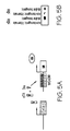
FIG. 5, panel A depicts a schematic of the pulmonary tissue preferred midkine expression system used in the transgenic mice of the invention. The box labeled hSPC indicates the human surfactant protein C promoter operably linked to the reverse tetracycline transactivator cDNA (rtTA, striped box). rtTA molecules are indicated by rectangles overlayed with triangles. Tetracycline-like molecules are indicated by speckled triangles (Dox). Activated rtTA molecules are indicated by a combination of the rtTA shapes and tetracycline-like molecule speckled triangles. The tetO-MK transgene consists of the murine midkine cDNA (SEQ ID NO:25, white box) operably linked to the minimal cytomegalovirus (CMV) promoter containing seven concatemerized tetracycline operon binding sites (rectangles overlayed with speckled triangles). Midkine molecules are depicted as white ovals labeled MK. FIG. 5, panel B depicts results obtained from RT-PCR reactions performed on 2 μg total RNA isolated from double transgenic (SP-C-rtTA+/tg, (tetO)7-CMV-MK+/tg or tg/tg) and control littermates in the presence (+Dox) or absence (−Dox) of doxycycline.
FIG. 6 presents histology and immunohistochemical results from pulmonary tissue of double transgenic mice and wild type mice for comparison. Tissue from wild-type mice (Non-transgenic) is presented in the right column; tissue from SP-C-rtTa+/tg, (tetO)7-CMV-MK+/tg or tg/tg mice (Double-transgenic) is presented in the left column. The tissue sections in panels A and B were stained with hematoxylin and eosin. The tissue sections in panels C and D were incubated with anti-midkine antibodies (anti-MK). The tissue sections in panels E and F were incubated with anti-α smooth muscle actin antibodies (anti-α-SMA). The tissue sections in panels G and H were incubated with anti-Caldesmon antibodies (anti-Caldesmon). The bar indicates 50 μm.
FIG. 7 presents average right ventricle hypertrophy (RVH), distal pulmonary vessel counts (No. distal vessels), and percent distal muscularization of mice in normoxic and hypoxic conditions. For comparison the results obtained from wild-type (control) and double transgenic mice (SP-C-rtTA+/tg, (tetO)7-CMV-MK+/tg or tg/tg) are presented. The standard error values follow the means.
FIG. 8 presents immunostaining of bronchioles and bronchial smooth muscle obtained from human subjects exhibiting a COPD (emphysema). The sections were incubated with anti-midkine antibodies. Panel F was not incubated with primary antibodies.
FIG. 9 presents immunohistochemical results from pulmonary tissue of FVB/N and CAST/eiJ mice. Panel A and Panel B present midkine staining of FVB/N pulmonary tissue after exposure to normoxia (A) and hypoxia (B) for five weeks. Panel C and Panel D present midkine staining of CAST/eiJ pulmonary tissue after exposure to normoxia (C) and hypoxia (D) for five weeks. Panels E and F present midkine (E) and α-SMA (F) staining of CAST/eiJ pulmonary tissue after exposure to hypoxia for four weeks. Panels G, H, I, J, K, and L present midkine staining of CAST/eiJ pulmonary tissue after exposure to hypoxia for 1 day (G), 3 days (H), 1 week (I), 2 weeks (J), 3 weeks (K), and 4 weeks (L). Images are at 40× (Panels A-D, G-L) and 100× (Panels E-F). The bar indicates 100 μM.
FIG. 10 presents the results obtained from PCR performed on cDNA generated by reverse transcription of RNA isolated from JEG-3 (Lane 2), MFLM-4 (Lane 4), and H-441 (Lane 6) cells after exposure to hypoxia. For comparison RT-PCR was performed on cDNA generated by reverse transcription of RNA isolated from JEG-3 (Lane 1), MFLM-4 (Lane 3), and H-441 (Lane 5) cells maintained in normoxia. Results of the PCR reactions containing HIF-1α primers, midkine primers (MK), and GAPDH primers are indicated. The graph indicates the fold change in midkine expression.
FIG. 11 depicts a schematic of the wild-type midkine promoter and the results of multiple promoter assays assessing the effects of HIF-1α and TTF-1 on the midkine promoter (SEQ ID NO:7) and modified midkine promoters. The promoters were operably linked to a luciferase reporter gene. Panel A presents the wild-type midkine promoter (SEQ ID NO:7). TTF-1 response elements (TRE) are indicated by empty boxes; HIF-1α response elements (HRE) are indicated by solid boxes. In panel B, the wild-type promoter construct and the indicated amount of pCMV-HIF-1α or pCMV-TTF1 were transformed into JEG cells, and promoter assays were performed. Results are indicated in luciferase units X 105/β-gal or 104/β-gal as indicated. Panel C presents the results of promoter assays performed with the wild-type midkine promoter or a truncated midkine promoter (SEQ ID NO:9). Assays were performed in the presence or absence of 320 ng of pCMV-HIF-1α. Panel D presents the results of promoter assays performed with the wild-type midkine promoter or site directed modified midkine promoters (SEQ ID NO:15 and SEQ ID NO:16). Assays were performed in the presence or absence of 320 ng of pCMV-HIF-1α.
FIG. 12 presents micrographs of tissue sections from double-transgenic and non-transgenic mice stained with α-SMA specific antibodies. Small blood vessels are indicated with arrows. Large blood vessels are indicated with arrowheads. The tissue in panels A and C were obtained from double-transgenic mice. The tissue in panels B and D were obtained from non-transgenic littermates. Panels A and B present tissue from animals fed doxycycline from conception to post-natal day 21. Panels C and D present tissue from animals fed doxycycline from post-natal day 21 to 9 weeks of age. The bar indicates 50 μm.
FIG. 13 presents results of peripheral pulmonary arterial pattern assessments. Panel A presents pulmonary arteriograms performed on adult non-transgenic (top row) and double transgenic (bottom row) mice. Panel B presents hematoxylin and eosin stained pulmonary tissue sections obtained from non-transgenic and double transgenic mice. The bar indicates 100 μm. Panel C presents a summary of pulmonary arterial density in non-transgenic (NTG) and double transgenic (MK-TG) mice. N=8. Panel D presents the results of Western blot analysis of Tie-2 and β-actin expression in whole lung homogenates from non-transgenic (Non-TG) and double transgenic (double-TG) mice.
FIG. 14, panel A presents results obtained from RT-PCR analysis of myocardin, calponin, SM-22, α-SMA, and GAPDH expression in MFLM-4 cells incubated with a placebo (−MK) or with midkine (+MK). Panel B presents results of real time RT-PCR analysis of myocardin expression relative to β-actin in lungs of transgenic (MK-TG) and non-transgenic mice (NTG). Panel C presents the myocardin/β-actin expression ratio in the lungs of CAST/eiJ mice exposed to hypoxia for the indicated number of weeks.
DETAILED DESCRIPTION OF THE INVENTION
The present invention provides methods of modulating midkine activity, midkine expression levels, and pulmonary midkine expression levels. The invention further provides methods of modulating smooth muscle cell development, particularly pulmonary smooth muscle cell development, more particularly pulmonary vascular smooth muscle cell development. Additionally the invention provides methods of modulating smooth muscle cell related disorders, pulmonary disorders, pulmonary smooth muscle cell related disorders, and pulmonary vascular smooth muscle cell related disorders. The invention provides methods of detecting midkine pathway abnormalities and pulmonary disorders. Further the invention provides methods of screening for a midkine pathway abnormality and for a pulmonary disorder. The invention provides methods of treating a pulmonary disorder. In addition, the invention provides methods of identifying a midkine modulating agent, smooth muscle cell development modulating agents, and pulmonary disorder modulating agents. Compositions of the invention include transgenic mice expressing midkine, particularly mice expressing midkine in an inducible, pulmonary tissue-preferred manner. Expression of midkine may alter a transgenic animal's susceptibility to smooth muscle cell related disorders or to pulmonary disorders, particularly pulmonary smooth muscle cell related disorders. Additionally the invention provides modified midkine promoters, and kits comprising a modified midkine promoter.
The invention relates to compositions and methods drawn to the murine midkine promoter, modified midkine promoters, and transgenic animals comprising a midkine transgene. The promoter sequences are useful for screening for a midkine pathway abnormality, screening subjects for a pulmonary disorder, evaluating compounds for midkine modulating activity, evaluating compounds for effects on smooth muscle cell development, and evaluating compounds for pulmonary disorder modulating activity. Methods of modulating midkine activity, modulating smooth muscle cell development, pulmonary development, pulmonary smooth muscle development, smooth muscle cell related disorders, pulmonary disorders, and pulmonary smooth muscle cell disorders involve administering a midkine modulating agent to a cell or subject. Particularly, the invention provides methods of modulating midkine expression that involve administering a midkine modulating agent.
A “midkine modulating agent” is a compound that modulates a midkine activity. Modulation may be an increase or decrease in a midkine activity. A midkine modulating compound will modulate a midkine activity by at least 1%, 5%, preferably 10%, 20%, more preferably 30%, 40%, 50%, 60%, yet more preferably 70%, 80%, 90%, or 100% as compared to an untreated or placebo treatment effect. By “midkine activity” is intended any activity exhibited by the wild-type murine midkine described herein. Such activities include, but are not limited to, midkine expression, immunogenicity, midkine receptor binding, heparin binding, modulation of cell differentiation, modulation of cell proliferation, protein tyrosine phosphatase binding, nucleolin binding, retinoic acid responsiveness, immunoreactivity, growth enhancement, neurite outgrowth, chemotactic activity, mitogenesis, angiogenesis modulation, neuron survival modulation, fibrinolysis, myocardin modulation, cell growth, cell migration, and tumorigenesis (Muramatsu et al. (1993) Dev. Biol. 159:392-402; Choundhri et al. (1997) Cancer Res. 57:1814-1819; Owada et al. (1999) J. Neurochem 73:2084-2092; Kojima et al. (1995) J. Biol. Chem. 270:9590-9596; Muramatsu & Muramatsu (1991) Biochem. Biophys. Res. Commun. 177:652-658; Kurtz et al. (1995) Crit. Rev. Oncol. 6:151-177; Tsutsui et al. (1993) Cancer Res. 53:1281-1285; Nakagawara et al. (1995) Cancer Res. 55:1792-1797; Garver et al. (1993) Am J. Respir. Cell Biol. 9:463-466; O'Brien et al. (1996) Cancer Res. 56:2515-2518; Kaname et al. (1996) Biochem. Biophys. Res. Commun. 219:256-260; Miyashiro et al. (1996) Cancer Lett 106:287-291; Miyashiro et al. (1997) Breast Cancer Res. Treat. 43:1-6; Takada et al. (1997) J. Biochem. 122:453-458; Maeda et al (1999) J. Biol. Chem. 274:12474-12479; Horiba et al. (2000) J. Clin. Invest. 105:489-495; Aridome et al. (1998) Br. J Cancer 78:472-477; Walker, John, ed. (2002) Protein Protocols on CD-ROM v. 2; and Ausubel et al., eds. (1995) Current Protocols in Molecular Biology, (Greene Publishing and Wiley-Interscience, New York; U.S. patent application Nos. 20020034768 and 20030053989; herein incorporated by reference in their entirety). Modulation of midkine activity includes but is not limited to modulation of a midkine activity such as protein tyrosine phosphatase binding or modulation of midkine expression levels. Thus, modulation of midkine expression is an alteration of a midkine activity. Methods for assaying midkine activity are known in the art and are described elsewhere herein.
In an embodiment, the invention provides compositions with an altered midkine activity and methods of modulating midkine activity. By “altered midkine activity” is intended a change of at least 1%, 5%, preferably 10%, 20%, more preferably 30%, 40%, 50%, 60%, 70%, 80%, 90%, 99% or more in a midkine activity. The change may be an increase or a decrease in the activity. Further, a compound that increases one midkine activity may decrease a different midkine activity.
It is understood that any midkine activity assay can be used to assay midkine modulating activity including, but not limited to, expression assays, immunogenicity, myocardin interaction assays, myocardin activation assays, receptor binding assays, heparin binding assays, cell differentiation assays, cell adhesion assays, FACS analysis, protein tyrosine phosphatase binding assays, growth assays, chemotactic assay, Western blots, EM, SEM, light microscopy, retinoic acid responsiveness assays, nucleolin binding assays, size exclusion chromatography, and capture ELISAs with multiple midkine antibodies. (Reynolds etal. (2004) J. Biol. Chem. 279: 37124-37132; Reynolds et al. (2003) Dev. Dyn. 227:227-237; Muramatsu et al. (1993) Dev. Biol. 159:392-402; Choundhri et al. (1997) Cancer Res. 57:1814-1819; Owada et al. (1999) J. Neurochem 73:2084-2092; Kojima et al. (1995) J. Biol. Chem. 270:9590-9596; Muramatsu & Muramatsu (1991) Biochem. Biophys. Res. Commun. 177:652-658; Kurtz et al. (1995) Crit. Rev. Oncol. 6:151-177; Tsutsui et al. (1993) Cancer Res. 53:1281-1285; Nakagawara et al. (1995) Cancer Res. 55:1792-1797; Garver et al. (1993) Am J. Respir. Cell Biol. 9:463-466; O'Brien et al. (1996) Cancer Res. 56:2515-2518; Kaname et al. (1996) Biochem. Biophys. Res. Commun. 219:256-260; Miyashiro et al. (1996) Cancer Lett 106:287-291; Miyashiro et al. (1997) Breast Cancer Res. Treat. 43:1-6; Takada et al. (1997) J. Biochem. 122:453-458; Maeda et al (1999) J. Biol. Chem. 274:12474-12479; Horiba et al. (2000) J. Clin. Invest. 105:489-495; Aridome et al. (1998) Br. J Cancer 78:472-477; Walker, John, ed. (2002) Protein Protocols on CD-ROM v. 2; and Ausubel et al., eds. (1995) Current Protocols in Molecular Biology, (Greene Publishing and Wiley-Interscience, New York; U.S. patent application Nos. 20020034768 and 20030053989; herein incorporated by reference in their entirety).
A “HIF-1α modulating agent” is a compound that modulates a HIF-1α activity such as altering midkine activity, modulating the HIF-1α/β interaction, oxygen level responsiveness, DNA binding, nuclear translocation, and degradation susceptibility. A HIF-1α modulating compound will modulate a HIF-1α activity by at least 1%, 5%, preferably 10%, 20%, more preferably 30%, 40%, 50%, 60%, yet more preferably 70%, 80%, 90%, or 100% as compared to an untreated or placebo treatment effect. By “HIF-1α activity” is intended any activity exhibited by the wild-type murine HIF-1α. Such activities include, but are not limited to, HIF-1α expression, interacting with HIF-1β, binding DNA, cellular translocation, degradation susceptibility, and midkine stimulation. Thus, modulation of HIF-1α expression is an alteration of HIF-1α activity. Methods for assaying HIF-1α activity are known in the art and are described elsewhere herein.
In an embodiment, the invention provides compositions with an altered HIF-1α activity and methods of modulating HIF-1α activity. By “altered HIF-1α activity” is intended a change of at least 1%, 5%, preferably 10%, 20%, more preferably 30%, 40%, 50%, 60%, 70%, 80%, 90%, 99% or more in a HIF-1α activity. The change may be an increase or a decrease in the activity. Further, a compound that increases one HIF-1α activity may decrease a different HIF-1α activity.
It is understood that any HIF-1α activity assay can be used to assay HIF-1α modulating activity including, but not limited to, expression assays, DNA binding assays, HIF-1α/β binding assays, HIF-1α/β dimerization assays, Western blots, EM, SEM, light microscopy, DAPI staining, and translocation assays. (Walker, John, ed. (2002) Protein Protocols on CD-ROM v. 2; and Ausubel et al., eds. (1995) Current Protocols in Molecular Biology, (Greene Publishing and Wiley-Interscience, New York).
An embodiment of the invention provides methods of modulating myocardin activity. In the methods modulating midkine activity modulates myocardin activity. By “myocardin activity” is intended any activity associated with murine myocardin including, but not limited to, smooth muscle gene regulation (Veyssier-Belot & Cacoub (1999) Cardiovascular Research 44:274-282), serum response factor interaction, myogenic activity, modulation of α-SMA, modulation of calponin, immunogenicity, and modulation of SM-22. Methods of detecting myocardin activity are known in the art; any method of assaying myocardin activity may be used in the methods of the invention.
In an embodiment, the invention provides a method of altering expression of a native midkine nucleotide sequence in an animal, particularly of modulating midkine expression in a tissue preferred manner. In the embodiment a midkine modulating agent comprising an expression cassette comprising a promoter operably linked to a nucleotide sequence having midkine modulating activity or encoding a polypeptide having midkine modulating activity is administered. In an embodiment the promoter is a tissue preferred promoter such as, but not limited to, a pulmonary tissue preferred promoter or a smooth muscle cell preferred promoter. In an embodiment, the midkine modulating agent alters the HIF-1α/β interaction.
By “alters midkine expression levels” is intended that the expression of the midkine nucleotide sequence in a transgenic cell, transgenic tissue, or transgenic cell or tissue of a transgenic animal of the invention differs from expression levels in a non-transgenic cell, non-transgenic tissue, or a non-transgenic animal by at least 1%, 2%, 3%, 4%, 5%, 10%, 15%, 20%, 25%, 30%, 35%, 40%, 45%, 50%, 55%, 60%, 65%, 70%, 75%, 80%, 85%, 90%, 95%, or 100%. The difference may be an increase or decrease in expression levels.
Methods of assaying expression levels are known in the art and include, but are not limited to, qualitative Western blot analysis, immunoprecipitation, radiological assays, polypeptide purification, spectrophotometric analysis, Coomassie staining of acrylamide gels, ELISAs, RT-PCR, 2-D gel electrophoresis, microarray analysis, in situ hybridization, chemiluminescence, silver staining, enzymatic assays, ponceau S staining, multiplex RT-PCR, immunohistochemical assays, radioimmunoassay, colorimetric analysis, immunoradiometric assays, immunochemistry, positron emission tomography, Northern blotting, fluorometric assays, fluorescence activated cell sorter staining of permeabilized cells, radioimmunosorbent assays, real-time PCR, hybridization assays, sandwich immunoassays, flow cytometry, SAGE, differential amplification, or electronic analysis. See, for example, Ausubel et al, eds. (2002) Current Protocols in Molecular Biology, Wiley-Interscience, New York, N.Y.; Coligan et al (2002) Current Protocols in Protein Science, Wiley-Interscience, New York, N.Y.; Sun et al. (2001) Gene Ther. 8:1572-1579; de Jager et al. (2003). Clin. & Diag. Lab. Immun. 10:133-139; U.S. Pat. Nos. 6,489,4555; 6,551,784; 6,607,879; 4,981,783; and 5,569,584; herein incorporated by reference in their entirety.
An embodiment of the invention is a method of detecting a midkine pathway abnormality. The method comprises obtaining a sample and assaying the midkine expression level in the sample. An increase or decrease in expression level compared to standard midkine expression levels in a similar sample obtained from a healthy subject, either directly or indirectly (for example, a predetermined standard) indicates a midkine pathway abnormality. By “midkine pathway” is intended any pathway that impacts midkine expression, directly or indirectly. A “midkine pathway abnormality” is an alteration in any stage of a midkine pathway that alters midkine expression. Assays for a midkine pathway abnormality include but are not limited to midkine expression assays and midkine promoter expression assays.
Another embodiment provides a method of detecting a pulmonary disorder. The method comprises obtaining a pulmonary tissue sample and assaying the midkine expression levels in the pulmonary tissue sample. By “pulmonary tissue” is intended any tissue obtained from the lungs, including but not limited to, the lungs, bronchi, bronchioles, alveoli, and developmentally related tissues. A tissue is one or more related cells. By “pulmonary smooth muscle cell” is intended any smooth muscle cell associated with pulmonary tissue including, but not limited to, smooth muscle cells associated with bronchial conducting airways and pulmonary arterial cells. By “pulmonary vascular smooth muscle cell” is intended any smooth muscle cell associated with the pulmonary vasculature including, but not limited to, pulmonary vascular cells and their precursors.
Midkine modulating agents include, but are not limited to, midkine, TTF-1, HIF1-α, oxygen, siRNA, anti-sense RNA, midkine binding compounds, midkine receptor binding compounds, and compounds that alter midkine expression such as a HIF 1-α modulating agent. Additional midkine modulating agents may be identified using the methods of the invention.
In several embodiments of the invention, the midkine modulating agent is an isolated nucleic acid molecule comprising an expression cassette. The expression cassette comprises a promoter operably linked to a nucleotide sequence of interest. Nucleotide sequences of interest exhibit a midkine modulating activity or encode a polypeptide having midkine modulating activity. Nucleotide sequences of interest include, but are not limited to, midkine (SEQ ID NO:1; SEQ ID NO:25), thyroid transcription factor-1 (TTF-1; SEQ ID NO:3), and hypoxia inducing factor-1 (HIF-1α; SEQ ID NO: 5), and fragments and variants thereof.
Fragments and variants of the nucleotide sequences of interest and polypeptides encoded thereby are also encompassed by the present invention. Fragments and variants of the modified midkine promoters (discussed elsewhere herein) are also encompassed by the present invention. By “fragment” is intended a portion of the nucleotide sequence or a portion of the amino acid sequence and hence protein encoded thereby. Fragments of a nucleotide sequence may encode protein fragments that retain the biological activity of the native protein and hence exhibit a midkine activity. Fragments of a promoter nucleotide sequence may retain biological activity and drive expression. Alternatively, fragments of a nucleotide sequence that are useful as hybridization probes generally do not retain biological activity. Thus, fragments of a nucleotide sequence may range from at least about 20, 25, 30, 35, 40, 45, 50, 55, 60, 65, 70, 75, 80, 85, 90, 95, 100, 150, 200, 250, 300, 350, 400, 450, 500, 550, 600, 650, 700, 750, 800, 850, up to 881 nucleotides for SEQ ID NO:1; from at least about 20, 25, 30, 35, 40, 45, 50, 55, 60, 65, 70, 75, 80, 85, 90, 95, 100, 150, 200, 250, 300, 350, 400, 450, 500, 550, 600, 650, 700, 750, 800, 850, 900, 950, 1000, 1050, 1100, up to about 1119 nucleotides for SEQ ID NO:3; from at least about 20, 25, 30, 35, 40, 45, 50, 55, 60, 65, 70, 75, 80, 85, 90, 95, 100, 150, 200, 250, 300, 350, 400, 450, 500, 550, 600, 650, 700, 750, 800, 850, 900, 950, 1000, 1050, 1100, 1150, 1200, 1250, 1300, 1350, 1400, 1450, 1500, 1550, 1600, 1650, 1700, 1750, 1800, 1850, 1900, 1950, 2000, 2050, 2100, 2150, 2200, 2250, 2300, 2350, 2400, 2450, 2500, 2550, 2600, 2650, 2700, 2750, 2800, 2850, 2900, 2950, 3000, 3050, 3100, 3150, 3200, 3250, 3300, 3350, 3400, 3450, 3500, 3550, 3600, 3650, 3700, 3750, 3800, 3850, 3900, 3950, up to about 3973 nucleotides for SEQ ID NO:5; from at least about 20, 25, 30, 35, 40, 45, 50, 55, 60, 65, 70, 75, 80, 85, 90, 95, 100, 150, 200, 250, 300, 350, 400, 450, 500, 550, 600, 650, 700, 750, 800, 850, 900, 950, 1000, 1050, 1100, 1150, 1200, 1250, 1300,1350, 1400, 1450, 1500, 1550, 1600, 1650, 1700, 1750, 1800, 1850, 1900, 1950, 2000, 2050, 2100, 2150, 2200, 2250, 2300, 2350, 2400, 2450, 2500, 2550, up to about 2559 nucleotides for SEQ ID NO:7, 11, 12, 13, or 14; from at least about 20, 25, 30, 35, 40, 45, 50, 55, 60, 65, 70, 75, 80, 85, 90, 95, 100, 150, 200, 250, 300, 350, 400, 450, 500, 550, 600, 650, 700, 750, 800, 850, 900, 950, 1000, 1050, 1100, 1150, 1200, 1250, 1300, 1350, 1400, 1450, 1500, 1550, 1600, 1650, 1700, 1750, 1800, 1850, 1900, 1950, 2000, 2050, up to about 2074 nucleotides for SEQ ID NO:8, from at least about 20, 25, 30, 35, 40, 45, 50, 55, 60, 65, 70, 75, 80, 85, 90, 95, 100, 150, 200, 250, 300, 350, 400, 450, 500, 550, 600, 650, 700, 750, 800, 850, 900, 950, 1000, 1050, 1100, 1150, 1200, 1250, 1300, 1350, 1400, 1450, 1500, 1550, 1600, 1650,up to about 1677 nucleotides for SEQ ID NO:9; from at least about 20, 25, 30, 35, 40, 45, 50, 55, 60, 65, 70, 75, 80, 85, 90, 95, 100, 150, 200, 250, 300, 350, 400, 450, 500, 550, 600, 650, 700, 750, 800, 850, 900, 950, 1000, up to about 1037 nucleotides for SEQ ID NO:10, from at least about 20, 25, 30, 35, 40, 45, 50, 55, 60, 65, 70, 75, 80, 85, 90, 95, 100, 150, 200, 250, 300, 350, 400, 450, 500, 550, 600, 650, 700, 750, 800, 850, 900, 950, 1000, 1050, 1100, 1150, 1200, 1250, 1300, 1350, 1400, 1450, 1500, 1550, 1600, 1650, 1700, 1750, 1800, 1850, 1900, 1950, 2000, 2050, 2100, 2150, 2200, 2250, 2300, 2350, 2400, 2450, 2500, 2550, up to about 2566 nucleotides for SEQ ID NO:15 or 16; from at least about 20, 25, 30, 35, 40, 45, 50, 55, 60, 65, 70, 75, 80, 85, 90, 95, 100, 150, 200, 250, 300, 350, 400, 450, 500, 550, 600, 650, up to 695 nucleotides for SEQ ID NO:25.
A fragment of a nucleotide sequence of interest that encodes a biologically active portion of a polypeptide of interest will encode at least 15, 25, 30, 40, 50, 60, 70, 80, 90, 100, 110, 120, 130, or 140 contiguous amino acids, or up to the total number of amino acids present in the full-length protein. Fragments of a nucleotide sequence or interest that are useful as hybridization probes or PCR primers generally need not encode a biologically active portion of a protein.
Thus, a fragment of a nucleotide sequence of interest may encode a biologically active portion of midkine, a midkine promoter, or a midkine modulating agent or it may be a fragment that can be used as a hybridization probe or PCR primer using methods disclosed below. A biologically active portion of a midkine can be prepared by isolating a portion of one of the midkine nucleotide sequences of the invention, expressing the encoded portion of the midkine protein (e.g., by recombinant expression in vitro), and assessing the activity of the encoded portion of the midkine protein. Nucleic acid molecules that are fragments of a midkine nucleotide sequence comprise at least about 20, 25, 30, 35, 40, 45, 50, 55, 60, 65, 70, 75, 80, 85, 90, 95, 100, 150, 200, 250, 300, 350, 400, 450, 500, 550, 600, 650, 700, 750, 800, 850, up to 881 nucleotides for SEQ ID NO:1 or at least about 20, 25, 30, 35, 40, 45, 50, 55, 60, 65, 70, 75, 80, 85, 90, 95, 100, 150, 200, 250, 300, 350, 400, 450, 500, 550, 600, 650, up to 695 nucleotides for SEQ ID NO:25. A biologically active portion of a midkine promoter can be prepared by isolating a portion of the promoter nucleotide sequence disclosed herein, and assessing the activity of the portion of the promoter. Nucleic acid molecules that are fragments of a midkine promoter comprise from at least about 20, 25, 30, 35, 40, 45, 50, 55, 60, 65, 70, 75, 80, 85, 90, 95, 100, 150, 200, 250, 300, 350, 400, 450, 500, 550, 600, 650, 700, 750, 800, 850, 900, 950, 1000, 1050, 1100, 1150, 1200, 1250, 1300, 1350, 1400, 1450, 1500, 1550, 1600, 1650, 1700, 1750, 1800, 1850, 1900, 1950, 2000, 2050, 2100, 2150, 2200, 2250, 2300, 2350, 2400, 2450, 2500, 2550, up to about 2559 nucleotides for SEQ ID NO:7, 11, 12, 13, or 14; from at least about 20, 25, 30, 35, 40, 45, 50, 55, 60, 65, 70, 75, 80, 85, 90, 95, 100, 150, 200, 250, 300, 350, 400, 450, 500, 550, 600, 650, 700, 750, 800, 850, 900, 950, 1000, 1050, 1100, 1150, 1200, 1250, 1300, 1350, 1400, 1450, 1500, 1550, 1600, 1650, 1700, 1750, 1800, 1850, 1900, 1950, 2000, 2050, up to about 2074 nucleotides for SEQ ID NO:8, from at least about 20, 25, 30, 35, 40, 45, 50, 55, 60, 65, 70, 75, 80, 85, 90, 95, 100, 150, 200, 250, 300, 350, 400, 450, 500, 550, 600, 650, 700, 750, 800, 850, 900, 950, 1000, 1050, 1100, 1150, 1200, 1250, 1300, 1350, 1400, 1450, 1500, 1550, 1600, 1650, up to about 1677 nucleotides for SEQ ID NO:9; from at least about 20, 25, 30, 35, 40, 45, 50, 55, 60, 65, 70, 75, 80, 85, 90, 95, 100, 150, 200, 250, 300, 350, 400, 450, 500, 550, 600, 650, 700, 750, 800, 850, 900, 950, 1000, up to about 1037 nucleotides for SEQ ID NO:10, from at least about 20, 25, 30, 35, 40, 45, 50, 55, 60, 65, 70, 75, 80, 85, 90, 95, 100, 150, 200, 250, 300, 350, 400, 450, 500, 550, 600, 650, 700, 750, 800, 850, 900, 950, 1000, 1050, 1100, 1150, 1200, 1250, 1300, 1350, 1400, 1450, 1500, 1550, 1600, 1650, 1700, 1750, 1800, 1850, 1900, 1950, 2000, 2050, 2100, 2150, 2200, 2250, 2300, 2350, 2400, 2450, 2500, 2550, up to about 2566 nucleotides for SEQ ID NO:15 or 16.
By “variants” is intended substantially similar sequences. For nucleotide sequences, conservative variants include those sequences that, because of the degeneracy of the genetic code, encode the amino acid sequence of one of the midkine polypeptides of the invention. Naturally occurring allelic variants such as these can be identified with the use of well-known molecular biology techniques, as, for example, with polymerase chain reaction (PCR) and hybridization techniques as outlined below. Variant nucleotide sequences also include synthetically derived nucleotide sequences, such as those generated, for example, by using site-directed mutagenesis [but which still encode midkine]. Generally, variants of a particular nucleotide sequence of interest will have at least about 90%, 91%, 92%, 93%, 94%, 95%, 96%, 97%, and more preferably at least about 98%, 99% or more sequence identity to that particular nucleotide sequence as determined by sequence alignment programs described elsewhere herein using default parameters.
By “variant” protein is intended a protein derived from the native protein by deletion (so-called truncation) or addition of one or more amino acids to the N-termial and/or C-terminal end of the native protein; deletion or addition of one or more amino acids at one or more sites in the native protein; or substitution of one or more amino acids at one or more sites in the native protein. Variant proteins encompassed by the present invention are biologically active, that is they continue to possess the desired biological activity of the native protein, that is, a midkine modulating activity as described herein. Such variants may result from, for example, genetic polymorphism or from human manipulation. Biologically active variants of a native midkine modulating protein of the invention will have at least about 90%, 91%, 92%, 93%, 94%, 95%, 96%, 97%, and more preferably at least about 98%, 99% or more sequence identity to the amino acid sequence for the native protein as determined by sequence alignment programs described elsewhere herein using default parameters. A biologically active variant of a protein of the invention may differ from that protein by as few as 1-15 amino acid residues, as few as 1-10, such as 6-10, as few as 5, as few as 4, 3, 2, or even 1 amino acid residue.
The proteins of the invention may be altered in various ways including amino acid substitutions, deletions, truncations, and insertions. Methods for such manipulations are generally known in the art. For example, amino acid sequence variants of the midkine modulating polypeptides can be prepared by mutations in the DNA. Methods for mutagenesis and nucleotide sequence alterations are well known in the art. See, for example, Kunkel (1985) Proc. Natl. Acad. Sci. USA 82:488-492; Kunkel et al. (1987) Methods in Enzymol. 154:367-382; U.S. Pat. No. 4,873,192; Walker and Gaastra, eds. (1983) Techniques in Molecular Biology (MacMillan Publishing Company, New York) and the references cited therein. Guidance as to appropriate amino acid substitutions that do not affect biological activity of the protein of interest may be found in the model of Dayhoff et al. (1978) Atlas of protein Sequence and Structure (Natl. Biomed. Res. Found., Washington, D.C.), herein incorporated by reference. Conservative substitutions, such as exchanging one amino acid with another having similar properties, may be preferable.
Thus, the genes and nucleotide sequences of the invention include both the naturally occurring sequences as well as mutant forms. Likewise, the proteins of the invention encompass both naturally occurring proteins as well as variations and modified forms thereof. Such variants will continue to possess the midkine modulating activity. Obviously, the mutations that will be made in the DNA encoding the variant must not place the sequence out of reading frame and preferably will not create complementary regions that could produce secondary mRNA structure. See, EP Patent Application Publication No. 75,444.
When it is difficult to predict the exact effect of the substitution, deletion, or insertion in advance of doing so, one skilled in the art will appreciate that the effect will be evaluated by routine screening assays. That is, the activity can be evaluated by suitable assays such as, but not limited to, promoter activity assays or midkine activity assays such as immunogenic activity.
Variant nucleotide sequences also encompass sequences derived from a mutagenic and recombinogenic procedure such as DNA shuffling. With such a procedure, one or more different promoter sequences or nucleotide sequences of interest can be manipulated to create a new sequence possessing the desired properties. In this manner, libraries of recombinant polynucleotides are generated from a population of related sequence polynucleotides comprising sequence regions that have substantial sequence identity and can be homologously recombined in vitro or in vivo. For example, using this approach, sequence motifs encoding a domain of interest may be shuffled between the modified midkine promoters of the invention and other known promoters to obtain a new promoter with an altered property of interest e.g. altered expression levels or altered midkine activity. Strategies for such DNA shuffling are known in the art. See, for example, Stemmer (1994) Proc. Natl. Acad. Sci. 91:10747-10751; Stemmer (1994) Nature 370:389-391; Crameri et al. (1997) Nature Biotech. 15:436-438; Moore et al. (1997) J. Mol. Biol. 272:336-347; Zhang et al. (1997) Proc. Natl. Acad. Sci. 94:4504-4509; Crameri et al. (1998) Nature 391:288-291; Miyazaki (2002) Nucleic Acids Research 30:E139-9; Song et al. (2002) Appl. Environ. Microbiol. 68:6146-51; Hayes et al. (2002) Proc. Natl Acad. Sci. 99:15926-31; Coco et al. (2001) Nature Biotechnol. 19:354-9; Kikuchi et al. (2000) Gene 243:133-7; and U.S. Pat. Nos. 5,606,793 and 5,837,458.
The following terms are used to describe the sequence relationships between two or more nucleic acids or polynucleotides: (a) “reference sequence”, (b) “comparison window”, (c) “sequence identity”, (d) “percentage of sequence identity”, and (e) “substantial identity”.
(a) As used herein, “reference sequence” is a defined sequence used as a basis for sequence comparison. A reference sequence may be a subset or the entirety of a specified sequence; for example, as a segment of a full-length cDNA or gene sequence or the complete cDNA or gene sequence.
(b) As used herein “comparison window” makes reference to a contiguous and specified segment of a polynucleotide sequence, wherein the polynucleotide sequence in the comparison window may comprise additions or deletions (i.e. gaps) compared to the reference sequence (which does not comprise additions or deletions) for optimal alignment of the two sequences. Generally the comparison window is at least 20 contiguous nucleotides in length, and optionally can be 30, 40, 50, 100, or longer. Those of skill in the art understand that to avoid a high similarity to a reference sequence due to inclusion of gaps in the polynucleotide sequence a gap penalty is typically introduced and is subtracted from the number of matches.
Methods of alignment of sequences for comparison are well known in the art. Thus, the determination of percent sequence identity between any two sequences can be accomplished using a mathematical algorithm. Preferred, non-limiting examples of such mathematical algorithms are the algorithm of Myers and Miller (1988) CABIOS 4:11-17; the local homology algorithm of Smith et al. (1981) Adv. Appl. Math. 2:482; the homology alignment algorithm of Needleman and Wunsch (1970) J. Mol. Biol. 48:443-453; the search-for-similarity-method of Pearson and Lipman (1988) Proc. Natl. Acad. Sci. 85:2444-2448; the algorithm of Karlin and Altschul (1990) Proc. Natl. Acad. Sci. USA 87:2264, modified as in Karlin and Altschul (1993) Proc. Natl. Acad. Sci. USA 90:5873-5877.
Computer implementations of these mathematical algorithms can be utilized for comparison of sequences to determine sequence identity. For purposes of the present invention, comparison of nucleotide or protein sequences for determination of percent sequence identity to the sequences disclosed herein is preferably made using the GCG program GAP (Version 10.00 or later) with its default parameters or any equivalent program. By “equivalent program” is intended any sequence comparison program that, for any two sequences in question, generates an alignment having identical nucleotide or amino acid residue matches and an identical percent sequence identity when compared to the corresponding alignment generated by the preferred program. Alignment may also be performed manually by inspection.
(c) As used herein, “sequence identity” or “identity” in the context of two nucleic acid or polypeptide sequences makes reference to the residues in the two sequences that are the same when aligned for maximum correspondence over a specified comparison window. When percentage of sequence identity is used in reference to proteins it is recognized that residue positions which are not identical often differ by conservative amino acid substitutions, where amino acid residues are substituted for other amino acid residues with similar chemical properties (e.g., charge or hydrophobicity) and therefore do not change the functional properties of the molecule. When sequences differ in conservative substitutions, the percent sequence identity may be adjusted upwards to correct for the conservative nature of the substitution. Sequences that differ by such conservative substitutions are said to have “sequence similarity” or “similarity”. Means for making this adjustment are well known to those of skill in the art. Typically this involves scoring a conservative substitution as a partial rather than a full mismatch, thereby increasing the percentage sequence identity. Thus, for example, where an identical amino acid is given a score of 1 and a non-conservative substitution is given a score of zero, a conservative substitution is given a score between zero and 1. The scoring of conservative substitutions is calculated, e.g., as implemented in the program PC/GENE (Intelligenetics, Mountain View, Calif.).
(d) As used herein, “percentage of sequence identity” means the value determined by comparing two optimally aligned sequences over a comparison window, wherein the portion of the polynucleotide sequence in the comparison window may comprise additions or deletions (i.e., gaps) as compared to the reference sequence (which does not comprise additions or deletions) for optimal alignment of the two sequences. The percentage is calculated by determining the number of positions at which the identical nucleic acid base or amino acid residue occurs in both sequences to yield the number of matched positions, dividing the number of matched positions by the total number of positions in the window of comparison, and multiplying the result by 100 to yield the percentage of sequence identity.
(e)(i) The term “substantial identity” of polynucleotide sequences means that a polynucleotide comprises a sequence that has at least 70% sequence identity, preferably at least 80%, more preferably at least 90%, and most preferably at least 95%, compared to a reference sequence using one of the alignment programs described using standard parameters. One of skill in the art will recognize that these values can be appropriately adjusted to determine corresponding identity of proteins encoded by two nucleotide sequences by taking into account codon degeneracy, amino acid similarity, reading frame positioning, and the like. Substantial identity of amino acid sequences for these purposes normally means sequence identity of at least 60%, more preferably at least 70%, 80%, 90%, and most preferably at least 95%.
Another indication that nucleotide sequences are substantially identical is if two molecules hybridize to each other under stringent conditions. Generally, stringent conditions are selected to be about 5° C. lower than the thermal melting point (Tm) for the specific sequence at a defined ionic strength and pH. However, stringent conditions encompass temperatures in the range of about 1° C. to about 20° C. lower than the Tm, depending upon the desired degree of stringency as otherwise qualified herein. Nucleic acids that do not hybridize to each other under stringent conditions are still substantially identical if the polypeptides they encode are substantially identical. This may occur, e.g., when a copy of a nucleic acid is created using the maximum codon degeneracy permitted by the genetic code. One indication that two nucleic acid sequences are substantially identical is when the polypeptide encoded by the first nucleic acid is immunologically cross reactive with the polypeptide encoded by the second nucleic acid.
(e)(ii) The term “substantial identity” in the context of a peptide indicates that a peptide comprises a sequence with at least 70% sequence identity to a reference sequence, preferably 80%, more preferably 85%, most preferably at least 90% or 95% sequence identity to the reference sequence over a specified comparison window. Preferably, optimal alignment is conducted using the homology alignment algorithm of Needleman and Wunsch (1970) J. Mol. Biol. 48:443-453. An indication that two peptide sequences are substantially identical is that one peptide is immunologically reactive with antibodies raised against the second peptide. Thus, a peptide is substantially identical to a second peptide, for example, where the two peptides differ only by a conservative substitution. Peptides that are “substantially similar” share sequences as noted above except that residue positions that are not identical may differ by conservative amino acid changes.
For example, an entire promoter sequence or nucleotide sequence of interest disclosed herein, or one or more portions thereof, may be used as a probe capable of specifically hybridizing to corresponding promoter sequences. To achieve specific hybridization under a variety of conditions, such probes include sequences that are unique among the nucleotide sequences of interest or the promoter sequences and are preferably at least about 10 nucleotides in length, and most preferably at least about 20 nucleotides in length. Such probes may be used to amplify corresponding sequences from a chosen animal by PCR. This technique may be used to isolate additional sequences from a desired animal or as a diagnostic assay to determine the presence of the sequences in an animal or animal cell.
In a PCR approach, oligonucleotide primers can be designed for use in PCR reactions to amplify corresponding DNA sequences from cDNA or genomic DNA extracted from any animal of interest. Methods for designing PCR primers and PCR cloning are generally known in the art and are disclosed in Sambrook et al. (1989) Molecular Cloning: A Laboratory Manual (2d ed., Cold Spring Harbor Laboratory Press, Plainview, N.Y.). See also Innis et al., eds. (1990) PCR Protocols: A Guide to Methods and Applications (Academic Press, New York); Innis and Gelfand, eds. (1995) PCR Strategies (Academic Press, New York); and Innis and Gelfand, eds. (1999) PCR Methods Manual (Academic Press, New York). Known methods of PCR include, but are not limited to, methods using paired primers, nested primers, single specific primers, degenerate primers, gene-specific primers, vector-specific primers, partially-mismatched primers, and the like.
In hybridization techniques, all or part of a known nucleotide sequence is used as a probe that selectively hybridizes to other corresponding nucleotide sequences present in a population of cloned genomic DNA fragments or cDNA fragments (i.e., genomic or cDNA libraries) from a chosen organism. The hybridization probes may be genomic DNA fragments, cDNA fragments, RNA fragments, or other oligonucleotides, and may be labeled with a detectable group such as 32P, or any other detectable marker. Thus, for example, probes for hybridization can be made by labeling synthetic oligonucleotides based on the nucleotide sequences of interest. Methods for preparation of probes for hybridization and for construction of cDNA and genomic libraries are generally known in the art and are disclosed in Sambrook et al. (1989) Molecular Cloning: A Laboratory Manual (2d ed., Cold Spring Harbor Laboratory Press, Plainview, N.Y.).
Hybridization techniques include hybridization screening of plated DNA libraries (either plaques or colonies; see, for example, Sambrook et al. (1989) Molecular Cloning: A Laboratory Manual (2d ed., Cold Spring Harbor Laboratory Press, Plainview, N.Y.).
Hybridization of such sequences may be carried out under stringent conditions. By “stringent conditions” or “stringent hybridization conditions” is intended conditions under which a probe will hybridize to its target sequence to a detectably greater degree than to other sequences (e.g., at least 2-fold over background). Stringent conditions are sequence-dependent and will be different in different circumstances. By controlling the stringency of the hybridization and/or washing conditions, target sequences that are 100% complementary to the probe can be identified (homologous probing). Alternatively, stringency conditions can be adjusted to allow some mismatching in sequences so that lower degrees of similarity are detected (heterologous probing). Generally, a probe is less than about 1000 nucleotides in length, preferably less than 500 nucleotides in length.
Typically, stringent conditions will be those in which the salt concentration is less than about 1.5 M Na ion, typically about 0.01 to 1.0 M Na ion concentration (or other salts) at pH 7.0 to 8.3 and the temperature is at least about 30° C. for short probes (e.g., 10 to 50 nucleotides) and at least about 60° C. for long probes (e.g., greater than 50 nucleotides). Stringent conditions may also be achieved with the addition of destabilizing agents such as formamide. Exemplary low stringency conditions include hybridization with a buffer solution of 30 to 35% formamide, 1 M NaCl, 1% SDS (sodium dodecyl sulphate) at 37° C., and a wash in 1× to 2×SSC (20×SSC=3.0 M NaCl/0.3 M trisodium citrate) at 50 to 55° C. Exemplary moderate stringency conditions include hybridization in 40 to 45% formamide, 1.0 M NaCl, 1% SDS at 37° C., and a wash in 0.5× to 1×SSC at 55 to 60° C. Exemplary high stringency conditions include hybridization in 50% formamide, 1 M NaCl, 1% SDS at 37° C., and a wash in 0.1×SSC at 60 to 65° C. Duration of hybridization is generally less than about 24 hours, usually about 4 to about 12 hours.
Specificity is typically the function of post-hybridization washes, the critical factors being the ionic strength and temperature of the final wash solution. For DNA—DNA hybrids, the Tm can be approximated from the equation of Meinkoth and Wahl (1984) Anal. Biochem. 138:267-284: Tm=81.5° C.+16.6 (log M)+0.41 (% GC)-0.61 (% form)-500/L; where M is the molarity of monovalent cations, % GC is the percentage of guanosine and cytosine nucleotides in the DNA, % form is the percentage of formamide in the hybridization solution, and L is the length of the hybrid in base pairs. The Tm is the temperature (under defined ionic strength and pH) at which 50% of a complementary target sequence hybridizes to a perfectly matched probe. Tm is reduced by about 1° C. for each 1% of mismatching; thus, Tm, hybridization, and/or wash conditions can be adjusted to hybridize to sequences of the desired identity. For example, if sequences with approximately 90% identity are sought, the Tm can be decreased 10° C. Generally, stringent conditions are selected to be about 5° C. lower than the thermal melting point (Tm) for the specific sequence and its complement at a defined ionic strength and pH. However, severely stringent conditions can utilize a hybridization and/or wash at 1, 2, 3, or 4° C. lower than the thermal melting point (Tm); moderately stringent conditions can utilize a hybridization and/or wash at 6, 7, 8, 9, or 10° C. lower than the thermal melting point (Tm); low stringency conditions can utilize a hybridization and/or wash at 11, 12, 13, 14, 15, or 20° C. lower than the thermal melting point (Tm). Using the equation, hybridization and wash compositions, and desired Tm, those of ordinary skill will understand that variations in the stringency of hybridization and/or wash solutions are inherently described. If the desired degree of mismatching results in a Tm of less than 45° C. (aqueous solution) or 32° C. (formamide solution), it is preferred to increase the SSC concentration so that a higher temperature can be used. An extensive guide to the hybridization of nucleic acids is found in Tijssen (1993) Laboratory Techniques in Biochemistry and Molecular Biology—Hybridization with Nucleic Acid Probes, Part I, Chapter 2 (Elsevier, N.Y.); and Ausubel et al., eds. (1995) Current Protocols in Molecular Biology, Chapter 2 (Greene Publishing and Wiley-Interscience, New York). See Sambrook et al. (1989) Molecular Cloning: A Laboratory Manual (2d ed., Cold Spring Harbor Laboratory Press, Plainview, N.Y.). Thus, isolated sequences that have midkine modulating activity or promoter activity and which hybridize under stringent conditions to the sequences disclosed herein, or to fragments thereof, are encompassed by the present invention. Such sequences will be at least 85%, 90%, 95% to 98% homologous or more with the disclosed sequences. That is, the sequence identity of sequences may range, sharing at least 85%, 90%, 91%, 92%, 93%, 94%, 95%, 96%, 97%, 98%, 99%, or more sequence identity.
Midkine promoters disclosed in the invention may be isolated from any animal, including but not limited to, rat, hamster, human, rabbit, mouse, monkey, chimpanzee, dog, pig, goat, sheep, cat, and cow. It is recognized that any gene of interest can be operably linked to a promoter of the invention.
As noted, the heterologous nucleotide sequence of interest operably linked to a promoter may be an antisense sequence for a targeted gene (e.g. midkine or a nucleotide sequence encoding a midkine modulating agent). Thus, with these promoters, antisense constructions complementary to at least a portion of the messenger RNA (mRNA) for a targeted nucleotide sequence interest can be constructed. Antisense nucleotides are constructed to hybridize with the corresponding mRNA. Modifications of the antisense sequences may be made as long as the sequences hybridize to and interfere with expression of the corresponding mRNA. In this manner, antisense constructions having 70%, preferably 80%, more preferably 85% sequence identity to the corresponding antisensed sequences may be used. Furthermore, portions of the antisense nucleotides may be used to disrupt the expression of the target gene. Generally, sequences of at least 50 nucleotides, 100 nucleotides, 200 nucleotides, or greater may be used. Thus, a tissue-preferred promoter sequence may be operably linked to antisense DNA sequences to reduce or inhibit expression of native midkine in a tissue of interest.
By “nucleotide sequence of interest” is intended a sequence that is not naturally occurring with the promoter sequence. While this nucleotide sequence is heterologous to the promoter sequence, it may be homologous, or native, or heterologous, or foreign, to the animal host.
It is recognized that the promoters may be used with their native coding sequences to increase or decrease expression resulting in a change in phenotype in the transformed animal or subject.
A number of promoters can be used in the practice of the invention. The promoters can be selected based on the desired outcome. In various embodiments of the invention, a midkine modulating agent is an isolated nucleic acid molecule comprising an expression cassette comprising a promoter operably linked to a nucleotide sequence of interest.
By “promoter” or “transcriptional initiation region” is intended a regulatory region of DNA usually comprising a TATA box capable of directing RNA polymerase II to initiate RNA synthesis at the appropriate transcription initiation site for a particular coding sequence. A promoter may additionally comprise other recognition sequences generally positioned upstream or 5′ to the TATA box, referred to as upstream promoter elements, which influence the transcription initiation rate. It is recognized that having identified the nucleotide sequences for the promoter regions disclosed herein, it is within the state of the art to isolate and identify further regulatory elements in the 5′ untranslated region. Thus, the promoter regions disclosed herein are generally further defined by comprising upstream regulatory elements such as those responsible for tissue and temporal expression of the coding sequence, enhancers and the like. Such elements are typically linked via a 5′ untranslated region, which may further modulate gene expression, to a coding region of interest. In the same manner, the promoter elements that enable expression in the desired tissue such as pulmonary-tissue or smooth muscle cells can be identified, isolated, and used with other core promoters to confirm tissue-preferred expression. For genes in which the 5′ untranslated region does not affect cell specificity, alternative sources of 5′ untranslated leaders may be used in conjunction with these promoter elements.
A number of promoters can be used in the practice of the invention. The promoters can be selected based on the desired outcome. Where low level expression is desired, weak promoters will be used. Generally, by “weak promoter” is intended a promoter that drives expression of a coding sequence at a low level. By “low level” is intended at levels of about 1/10,000 transcripts to about 1/100,000 transcripts to about 1/500,000 transcripts; conversely, a strong promoter drives expression of a coding sequence at a high level, or at about 1/10 transcripts to about 1/100 transcripts to about 1/1000 transcripts. Alternatively, it is recognized that weak promoters also encompasses promoters that are expressed in only a few cells and not in others to give a total low level of expression. Where a promoter is expressed at unacceptably high levels, portions of the promoter sequence can be deleted or modified to decrease expression levels.
It is recognized that to increase transcription levels or to alter tissue specificity, enhancers and/or tissue-preference elements may be utilized in combination with the nucleotide sequences of interest. For example, quantitative or tissue specificity upstream elements from other pulmonary tissue-preferred or smooth muscle cell preferred promoters may be combined with the promoter regions of the invention to augment tissue-preferred transcription. The human surfactant protein C promoter is a pulmonary tissue preferred promoter (Perl et al. (2002) Transgenic Res. 11:21-29, herein incorporated by reference in its entirety). The murine smooth muscle 22 α (SM22α) promoter is a smooth muscle cell preferred promoter (U.S. Pat. Nos. 6,015,711 and 5,837,534, herein incorporated by reference in their entirety).
In other embodiments, the coding region is operably linked to an inducible regulatory element or elements. A variety of inducible promoter systems have been described in the literature and can be used in the present invention. These include, but are not limited to, tetracycline-regulatable systems (WO 94/29442, WO 96/40892, WO 96/01313, U.S. application Ser. No. 10/613,728); hormone responsive systems, interferon-inducible systems, metal-inducible systems, and heat-inducible systems, (WO 93/20218); and ecdysone inducible systems. Some of these systems, including ecdysone inducible and tetracycline inducible systems are commercially available from Invitrogen (Carlsbad, Calif) and Clontech (Palo Alto, Calif), respectively.
By “inducible” is intended that a chemical stimulus alters expression of the operably linked nucleotide sequence of interest by at least 1%, 5%, preferably 10%, 20%, more preferably 30%, 40%, 50%, 60%, 70%, 80%, 90%, 99% or more. The difference may be an increase or decrease in expression levels. Methods for assaying expression levels are described elsewhere herein. The chemical stimulus may be administered or withdrawn. Various chemical stimuli are known in the art. In an embodiment, the chemical stimulus is tetracycline, or an analog thereof.
One of the most widely used conditional systems is the binary, tetracycline-based system, which has been widely used in both cells and animals to reversibly induce expression by the addition or removal of tetracycline or its analogues (See Bujard (1999). J. Gene Med. 1:372-374; Furth, et al. (1994). Proc. Natl. Acad. Sci. USA 91:9302-9306; and Mansuy & Bujard (2000). Curr. Opin. Neurobiol. 10:593-596, herein incorporated by reference in their entirety.).
Another class of promoter elements includes those which activate transcription of an operably linked nucleotide sequence of interest in response to hypoxic conditions. These include promoter elements regulated at least in part by hypoxia inducible factor-1. Hypoxia response elements include, but are not limited to, the erythropoietin hypoxia response enhancer element (HREE1), the muscle pyruvate kinase HRE; the β-enolase HRE; and endothelin-1 HRE element, and chimeric nucleotide sequence comprising these sequences. See Bunn and Poynton (1996) Physiol. Rev. 76:839-885; Dachs and Stratford (1996) Br. J. Cancer 74:S126-S132; Guillemon and Krasnow (1997) Cell 89:9-12; Firth et al. (1994) Proc. Natl. Acad. Sci. 91:6496-6500; Jiang et al. (1997) Cancer Res. 57:5328-5335; U.S. Pat. No. 5,834,306) herein incorporated by reference in their entirety.
In an embodiment, the invention provides methods of preferentially modulating midkine activity in a tissue preferred manner (for example, pulmonary tissue or smooth muscle cells). It is recognized that the nucleotide sequence of interest of a midkine modulating agent which is an isolated nucleic acid molecule comprising an expression cassette comprising a nucleotide sequence of interest can be operably linked to any tissue preferred promoter. The tissue preferred promoter allows expression of the nucleotide sequence of interest in a tissue preferred manner. Tissues of particular interest include pulmonary tissue, smooth muscle cells, pulmonary smooth muscle cells, and pulmonary vascular smooth muscle cells.
By “pulmonary tissue-preferred” is intended that expression of the heterologous sequence is most abundant in pulmonary tissue, while some expression may occur in other tissue types, particularly in tissues developmentally related to pulmonary tissue. Pulmonary-preferred expression of a heterologous nucleotide sequence of interest occurs at levels at least 1%, 5%, 10%, 20%, 30%, 40%, 50%, 60%, 70%, 80%, 90%, or 100% greater than expression of the nucleotide sequence of interest in non-pulmonary tissue. By “pulmonary vascular tissue-preferred” is intended that expression of the heterologous sequence is most abundant in pulmonary vascular tissue, while some expression may occur in other tissue types, particularly in tissues developmentally related to pulmonary vascular tissue. Pulmonary vascular-preferred expression of a heterologous nucleotide sequence of interest occurs at levels at least 1%, 5%, 10%, 20%, 30%, 40%, 50%, 60%, 70%, 80%, 90%, or 100% greater than expression of the nucleotide sequence of interest in non-pulmonary vascular tissue. In an embodiment, tissue-preferred expression of a heterologous nucleotide sequence natively expressed in other tissue may be desired. Expression of a heterologous nucleotide sequence from a tissue-preferred promoter may not impact expression of the nucleotide sequence operably linked to its native promoter in other tissues.
By “smooth muscle cell-preferred” is intended that expression of the heterologous sequence is most abundant in smooth muscle cells, while some expression may occur in other cell types. Smooth muscle cell-preferred expression of a nucleotide sequence of interest occurs at levels at least 1%, 5%, 10%, 20%, 30%, 40%, 50%, 60%, 70%, 80%, 90%, or 100% greater than expression of the heterologous nucleotide sequence of interest in non-smooth muscle cells. In an embodiment, smooth muscle cell-preferred expression of a heterologous nucleotide sequence natively expressed in other tissue may be desired. Expression of a heterologous nucleotide sequence from a smooth muscle cell-preferred promoter may not impact expression of the nucleotide sequence operably linked to its native promoter in other tissues.
The invention provides methods of preferentially modulating midkine activity or expression levels in a tissue of interest. In an embodiment, a midkine modulating agent is administered to the tissue of interest. In an embodiment the midkine modulating agent is an isolated nucleic acid molecule comprising an expression cassette comprising a nucleotide sequence of interest operably linked to a tissue preferred promoter.
By “preferentially modulating pulmonary tissue midkine expression levels” is intended that the expression of midkine in a transgenic cell, pulmonary tissue of a transgenic animal of the invention, or pulmonary tissue of a subject administered a midkine modulating agent differs from expression levels in a non-pulmonary transgenic cell or pulmonary tissue of a non-transgenic animal or untreated subject by at least 1%, 5%, preferably 10%, 20%, more preferably 30%, 40%, 50%, 60%, 70%, 80%, 90%, 99%, or more. The difference may be an increase or decrease in expression levels.
In addition to control regions that promote transcription, expression vectors may also include regions that modulate transcription, such as repressor binding sites and enhancers. Examples include the SV40 enhancer, the cytomegalovirus immediate early enhancer, polyoma enhancer, adenovirus enhancers, and retrovirus LTR enhancers.
Such expression cassettes will comprise a transcriptional initiation region comprising a promoter nucleotide sequence operably linked to the heterologous nucleotide sequence whose expression is to be controlled by the promoter. Such an expression cassette is provided with at least one restriction site for insertion of the nucleotide sequence to be under the transcriptional regulation of the regulatory regions. The expression cassette may additionally contain selectable marker genes.
The expression cassette will include in the 5′-to-3′ direction of transcription, a transcriptional and translational initiation region, and a heterologous nucleotide sequence of interest. In addition to containing sites for transcription initiation and control, expression cassettes can also contain sequences necessary for transcription termination and, in the transcribed region a ribosome binding site for translation. Other regulatory control elements for expression include initiation and termination codons as well as polyadenylation signals. The person of ordinary skill in the art would be aware of the numerous regulatory sequences that are useful in expression vectors. Such regulatory sequences are described, for example, in Sambrook et al. (1989) Molecular Cloning: A Laboratory Manual 2nd. ed., Cold Spring Harbor Laboratory Press, Cold Spring Harbor, N.Y.).
The expression cassette comprising a promoter sequence of the present invention operably linked to a heterologous nucleotide sequence or a promoter operably linked to midkine may also contain at least one additional nucleotide sequence for a gene to be co-transformed into the organism. Alternatively, the additional sequence(s) can be provided on another expression cassette.
The regulatory sequences to which the polynucleotides described herein can be operably linked include promoters for directing mRNA transcription. These include, but are not limited to, the left promoter from bacteriophage λ, the lac, TRP, and TAC promoters from E. coli, the early and late promoters from SV40, the CMV immediate early promoter, the adenovirus early and late promoters, and retrovirus long-terminal repeats.
The isolated modified midkine promoter sequences of the present invention can be modified to provide assays for a range of expression levels of a compound of interest or a midkine modulating agent. Thus, less than the entire promoter regions may be utilized and the ability to drive expression retained. However, it is recognized that expression levels of mRNA may be altered and usually decreased with deletions of portions of the promoter sequences. Generally, at least about 20 nucleotides of an isolated promoter sequence will be used to drive expression of a nucleotide sequence. Truncated forms of the midkine promoter sequence may also be used to assay compounds for midkine modulating activity, particularly midkine expression modulating activity.
The invention encompasses isolated or substantially purified nucleic acid or protein compositions. An “isolated” or substantially “purified” nucleic acid molecule, polypeptide, or biologically active portion thereof, is substantially free of other cellular material, or culture medium when produced by recombinant techniques or substantially free of chemical precursors or other chemicals when chemically synthesized. Preferably, an “isolated” nucleic acid molecule is free of sequences (preferably polypeptide encoding sequences) that naturally flank the nucleic acid (i.e., sequences located at the 5′ and 3′ ends of the nucleic acid) in the genomic DNA of the organism from which the nucleic acid is derived. For example, in various embodiments, the isolated nucleic acid molecule can contain less than about 5 kb, 4kb, 3 kb, 2 kb, 1 kb, 0.5 kb, or 0.1 kb of nucleotide sequences that naturally flank the nucleic acid molecule in genomic DNA of the cell from which the nucleic acid is derived.
The promoters for midkine genes from alternative species may generally be isolated from the 5′ untranslated region flanking their respective transcription initiation sites. Methods for isolation of promoter regions are well known in the art. By “isolated” is intended that the promoter sequences have been determined and can be extracted by molecular techniques or synthesized by chemical means. In either instance, the promoter is removed from at least one of its flanking sequences in its native state.
Where appropriate, the heterologous nucleotide sequence whose expression is to be under the control of a promoter sequence and any additional nucleotide sequence(s) may be optimized for increased expression in the transformed animal. That is, these nucleotide sequences can be synthesized using species preferred codons for improved expression, such as rabbit-preferred codons for improved expression in rabbits or mouse-preferred codons in mice. Methods are available in the art for synthesizing species-preferred nucleotide sequences. See, for example, Wada et al. (1992) Nucleic Acids Res. 20 (Suppl.), 2111-2118; Butkus et al. (1998) Clin Exp Pharmacol Physiol Suppl. 25:S28-33; and Sambrook et al. (1989) Molecular Cloning: A Laboratory Manual 2nd. ed., Cold Spring Harbor Laboratory Press, Cold Spring Harbor, N.Y., herein incorporated by reference.
Additional sequence modifications are known to enhance gene expression in a cellular host. These include elimination of sequences encoding spurious polyadenylation signals, exon-intron splice site signals, transposon-like repeats, and other such well-characterized sequences that may be deleterious to gene expression. The G-C content of the heterologous nucleotide sequence may be adjusted to levels average for a given cellular host, as calculated by reference to known genes expressed in the host cell. In an embodiment, the sequence is modified to avoid predicted hairpin secondary mRNA structures. In an embodiment the sequence is modified to yield hairpin RNA structures for use in siRNA.
The expression cassettes may additionally contain 5′ leader sequences in the expression cassette construct. Such leader sequences can act to enhance translation. Translation leaders are known in the art and include: picornavirus leaders, for example, EMCV leader (Encephalomyocarditis 5′ noncoding region) (Elroy-Stein et al. (1989) Proc. Nat. Acad. Sci. USA 86:6126-6130); potyvirus leaders, for example, TEV leader (Tobacco Etch Virus) (Allison et al. (1986)); MDMV leader (Maize Dwarf Mosaic Virus) (Virology 154:9-20); and human immunoglobulin heavy-chain binding protein (BiP) (Macejak et al (1991) Nature 353:90-94). Other methods known to enhance translation and/or mRNA stability can also be utilized, for example, introns, and the like.
In those instances where it is desirable to have the expressed product of the heterologous nucleotide sequence directed to a particular organelle, particularly the mitochondria, the nucleus, the endoplasmic reticulum, the Golgi apparatus; or secreted at the cell's surface or extracellularly; the expression cassette may further comprise a coding sequence for a transit peptide. Such transit peptides are well known in the art and include, but are not limited to, the transit peptide for the acyl carrier protein, the small subunit of RUBISCO, and the like.
In preparing the expression cassette, the various DNA fragments may be manipulated, so as to provide for the DNA sequences in the proper orientation and, as appropriate, in the proper reading frame. Toward this end, adapters or linkers may be employed to join the DNA fragments or other manipulations may be involved to provide for convenient restriction sites, removal of superfluous DNA, removal of restriction sites, or the like. For this purpose; in vitro mutagenesis; primer repair; restriction; annealing; substitutions, for example, transitions and transversions; or any combination thereof may be involved.
Reporter genes or selectable marker genes may be included in the expression cassettes. Examples of suitable reporter genes known in the art can be found in, for example, Ausubel et al. (2002) Current Protocols in Molecular Biology. John Wiley & Sons, New York, N.Y., herein incorporated by reference. The reporter utilized in the invention can be any reporter known in the art including, but not limited to, luciferases, blue fluorescent proteins, green fluorescent proteins, CAT, GUS, β-galactosidases, and midkine.
Selectable marker genes for selection of transformed cells or tissues can include genes that confer antibiotic resistance. Examples of suitable selectable marker genes include, but are not limited to, genes encoding resistance to neomycin (Schwartz et al (1991) Proc. Natl. Acad. Sci.88:10416-20); chloramphenicol (Herrera Estrella et al. (1983) EMBO J. 2:987-992); methotrexate (Herrera Estrella et al. (1983) Nature 303:209-213; Meijer et al. (1991) Plant Mol. Biol. 16:807-820); hygromycin (Waldron et al. (1985) Plant Mol. Biol. 5:103-108; Zhijian et al. (1995) Plant Science 108:219-227); streptomycin (Jones et al. (1987) Mol. Gen. Genet. 210:86-91); spectinomycin (Bretagne-Sangard et al. (1996) Transgenic Res. 5:131-137); bleomycin (Hille et al. (1990) Plant Mol. Biol. 7:171-176); sulfonamide (Guerineau et al. (1990) Plant Mol. Biol. 15:127-136); puromycin (Abbate et al (2001) Biotechniques 31:336-40; cytosine arabinoside (Eliopoulos et al. (2002) Gene Ther. 9:452-462); 6-thioguanine (Tucker et al. (1997) Nucleic Acid Research 25:3745-46).
Other genes that could serve utility in the recovery of transgenic events but might not be required in the final product would include, but are not limited to, examples such as GUS (b-glucuronidase; Jefferson (1987) Plant Mol. Biol. Rep. 5:387); GFP (green fluorescence protein; Wang et al. (2001) Anim Biotechnol 12:101 -110; Chalfie et al. (1994) Science 263:802), BFP (blue fluorescence protein; Yang et al. (1998) J. Biol. Chem. 273:8212-6), CAT; and luciferase (Riggs et al. (1987) Nucleic Acid Res. 15 (19):8115; Luchrsen et al. (1992) Methods Enzymol. 216: 397-414). These genes are also suitable as reporters when operably linked to a midkine promoter or modified midkine promoter of the invention.
Embodiments of the invention provide methods of screening for a midkine pathway abnormality, screening subjects for a pulmonary disorder, and identifying a midkine modulating agent. In these embodiments, the invention provides isolated nucleic acid molecules comprising an expression cassette comprising a modified midkine promoter of the invention operably linked to a reporter. In the embodiments, the isolated nucleic acid molecule comprising a modified midkine promoter operably linked to a reporter is incubated with a test sample or compound of interest. By test sample is intended a biological sample such as, but not limited to, a subject exhibiting a smooth muscle cell related disorder or pulmonary disorder, cells, cell lysates, cellular fractions, tissues, mucosa, and secretions. In an embodiment, an isolated nucleic acid molecule comprising a modified midkine promoter operably linked to a reporter is transformed into a cell and incubation of the modified midkine promoter with the test sample occurs within the cell. In an embodiment, the isolated nucleic acid molecule is incubated with the test sample in vitro. Methods of assaying reporter expression levels are known in the art and described elsewhere herein. Reporter expression levels are measured by any means known to one skilled in the art.
The invention provides methods of screening subjects for a pulmonary disorder, a smooth muscle cell related disorder, a pulmonary smooth muscle cell related disorder, and a pulmonary vascular smooth muscle cell related disorder. In the methods an isolated nucleic acid molecule comprising a modified midkine promoter operably linked to a reporter is incubated with a test sample obtained from a tissue of interest. Reporter expression levels are measured by any means known to one skilled in the art.
Any of the regulatory or other sequences useful in expression vectors can form part of the transgenic sequence. This includes intronic sequences and polyadenylation signals, if not already included.
Modulation of a midkine activity, smooth muscle cell development, pulmonary development, smooth muscle cell related disorder, pulmonary disorder, a pulmonary smooth muscle cell related disorder, or a pulmonary vascular smooth muscle cell related disorder can be achieved by providing expression of heterologous products or increased expression of endogenous products in a tissue of interest. Alternatively, the results can be achieved by providing for a reduction of expression of one or more endogenous products, particularly enzymes and cofactors in the tissue of interest. These changes result in a change in phenotype of the transgenic animal. For example, a nucleotide sequence of interest can be used to preferentially express midkine in a tissue of interest and alter the midkine expression pattern. In an embodiment, the nucleotide sequence of interest encodes a polypeptide having a midkine modulating activity that alters midkine expression (e.g. TTF-1 and HIF1-α). Alternatively, the promoter is operably linked to a heterologous nucleotide sequence of interest from which antisense mRNA complementary to the coding sequence of a polypeptide of interest can be produced. The antisense RNA inhibits production of the protein or polypeptide of interest. Alternatively, the promoter sequences of the invention can be used to produce small interfering RNAs.
Products of the heterologous nucleotide sequence include structural proteins, enzymes, cofactors, hormones, signaling proteins, and the like.
Nucleotide sequences of interest disclosed in the present invention, as well as variants and fragments thereof, are useful in the genetic manipulation of any animal when operably linked with a promoter capable of initiating transcription in the animal. The midkine sequences are provided in expression cassettes for expression in a transgenic animal of the invention. The cassette will include 5′ to 3′ regulatory sequences operably linked to a heterologous nucleotide sequence. By “operably linked” is intended the transcription of the nucleotide sequence of interest is under the influence of the promoter sequence. In this manner, promoter nucleotide sequences may be provided in expression cassettes along with nucleotide sequences of interest for expression in the animal of interest, more particularly for tissue-preferred expression in the animal. In an embodiment, heterologous nucleotide sequences of interest suitable for use in the invention include midkine modulating agents including, but not limited to, nucleotide sequences encoding thyroid transcription factor-1 (TTF-1, SEQ ID NO:3), midkine (SEQ ID NO:1, SEQ ID NO:25), hypoxia inducing factor-1 (HIF1-α, SEQ ID NO:5), transcription factors, midkine promoter binding polypeptides, antisense nucleotide sequences, and small interfering RNA (siRNA) sequences. In another embodiment heterologous nucleotide sequences suitable for use in the methods of the invention include nucleotide sequences encoding reporters or reporter genes. Reporters are described elsewhere herein.
By “stably transformed” is intended that the nuclear genome of the animal cell or the nuclear genome of at least one cell of the animal has incorporated at least one copy of the transgene. By “transgene” is intended a nucleic acid molecule having a nucleotide sequence comprising either an expression cassette or a disruption cassette. A transgenic animal of the invention comprises at least one stably transformed cell comprising the nucleotide sequence of interest. In an embodiment, the genome of a germ-line cell of a transgenic animal comprises the nucleotide sequence of interest. A transgenic cell, e.g. a smooth muscle cell, is a cell comprising at least one expression cassette or disruption cassette isolated from a transgenic animal of the invention. Transgenic tissue, e.g. pulmonary tissue and pulmonary smooth muscle cell, is tissue comprising transgenic cells. Transgenic tissue is isolated from a transgenic animal of the invention.
Transgenic animals of the invention are useful for studying the function of midkine and identifying and evaluating modulators of midkine activity, smooth muscle cell development, pulmonary development, smooth muscle cell disorders, pulmonary disorders and pulmonary smooth muscle cell disorders. A transgenic animal is preferably a mammal, for example a mouse, in which one or more of the cells of the animal include a transgene. A transgene is exogenous DNA which is integrated into the genome of a cell from which a transgenic animal develops and which remains in the genome of the mature animal in one or more cell types or tissues of the transgenic animal. A genetically engineered host cell can be used to produce non-human transgenic animals.
Transgenic animals that exhibit altered midkine expression are useful to conduct assays that identify compounds that affect midkine function such as, but not limited to, vascular remodeling. Assays to determine vascular remodeling are known in the art and include, but are not limited to, immunohistochemical analysis and morphometric analysis. The altered midkine expression may result in altered susceptibility to a smooth muscle cell related disorder, pulmonary disorder, or pulmonary smooth muscle cell related disorder.
Methods for generating transgenic animals via embryo manipulation and microinjection, particularly animals such as mice, have become conventional in the art and are described, for example, in U.S. Pat. Nos. 4,736,866; 4,870,009; 4,873,191; 6,201,165 and in Nagy et al Ed. (2003) Manipulating the Mouse Embryo Cold Spring Harbor Press, Cold Spring Harbor, N.Y.), herein incorporated by reference in their entirety.
Similar methods are used for production of other transgenic animals. A transgenic animal can be produced by introducing nucleic acid into the male pronuclei of a fertilized oocyte, e.g., by microinjection, retroviral infection, and allowing the oocyte to develop in a pseudopregnant female foster animal. A transgenic founder animal can be identified based upon the presence of the transgene in its genome and/or expression of transgenic mRNA in tissues or cells of the animals. A transgenic founder animal can then be used to breed additional animals carrying the transgene. Moreover, transgenic animals carrying a transgene can further be bred to other transgenic animals carrying other transgenes. A transgenic animal also includes animals in which the entire animal or tissues in the animal have been produced using the homologously recombinant host cells described herein. Methods for providing transgenic rabbits are described in Marian et al. (1999) J. Clin. Invest. 104:1683-1692 and James et al. (2000) Circulation 101:1715-1721, herein incorporated by reference in their entirety.
Other examples of transgenic animals include non-human primates, sheep, dogs, pigs, cows, goats, rats, rabbits, and hamsters.
In an embodiment, the invention provides a method of altering expression of a heterologous midkine nucleotide sequence in an animal, particularly of altering tissue preferred expression of the heterologous nucleotide sequence. In an embodiment the tissue preferred expression is pulmonary tissue preferred expression. In another embodiment, the tissue preferred expression is smooth muscle cell preferred expression. In an embodiment the heterologous nucleotide sequence is operably linked to a tissue preferred promoter. An expression cassette comprising the tissue preferred promoter operably linked to the heterologous nucleotide sequence is used to transform an animal. In an embodiment a tissue preferred promoter is operably linked to a nucleotide sequence encoding a polypeptide that enhances expression from a second promoter. The second promoter is operably linked to a nucleotide sequence encoding midkine (SEQ ID NO:25). Animal transformation methods are known in the art and reviewed elsewhere herein. The method yields a stably transformed transgenic animal exhibiting altered expression of a heterologous nucleotide sequence.
In another embodiment, transgenic non-human animals can be produced which contain selected systems, which allow for regulated expression of the transgene. Examples of such systems include, but are not limited to, the cre/loxP recombinase system, the FLP recombinase system, and the tetracycline based system described elsewhere herein. If a tetracycline based system is used to regulate expression of the transgene, animals containing transgenes encoding both a transactivator and a selected protein are required. Such animals can be provided through the construction of “double” transgenic animals, e.g., by mating two transgenic animals, one containing a transgene encoding a selected protein and the other containing a transgene encoding a transactivator.
Clones of the non-human transgenic animals described herein can also be produced according to the methods described in Wilmut et al. (1997) Nature 385:810-813 and PCT International Publication Nos. WO 97/07668 and WO 97/07669. In brief, a cell, e.g., a somatic cell, from the transgenic animal can be isolated and induced to exit the growth cycle and enter Go phase. The quiescent cell can then be fused, e.g., through the use of electrical pulses, to an enucleated oocyte from an animal of the same species from which the quiescent cell is isolated. The reconstructed oocyte is then cultured such that it develops to morula or blastocyst and then transferred to a pseudopregnant female foster animal. The offspring born of this female foster animal will be a clone of the animal from which the cell, e.g., the somatic cell, is isolated.
In one embodiment, a transgenic animal cell can be a fertilized oocyte or embryonic stem cell that can be used to produce a transgenic animal comprising at least one stably transformed expression cassette comprising the heterologous nucleotide sequence. Alternatively, a transgenic cell can be a stem cell or other early tissue precursor that gives rise to a specific subset of cells and can be used to produce transgenic tissues in an animal. See also Thomas et al., (1987) Cell 51:503 for a description of homologous recombination vectors. The vector is introduced into an embryonic stem cell line (e.g., by electroporation) and cells in which the introduced gene has recombined with the genome are selected (see e.g., Li, E. et al. (1992) Cell 69:915). The selected cells are then injected into a blastocyst of an animal (e.g., a mouse) to form aggregation chimeras (see e.g., Bradley, A. in Teratocarcinomas and Embryonic Stem Cells: A Practical Approach, E. J. Robertson, ed. (IRL, Oxford, 1987) pp. 113-152). A chimeric embryo can then be implanted into a suitable pseudopregnant female foster animal and the embryo brought to term. Progeny harboring the recombined DNA in their germ cells can be used to breed animals in which all cells of the animal contain the recombined DNA by germ line transmission of the transgene. Methods for constructing homologous recombination vectors and homologous recombinant animals are described further in Bradley, A. (1991) Current Opinion in Biotechnology 2:823-829 and in PCT International Publication Nos. WO 90/11354; WO 91/01140; and WO 93/04169, herein incorporated by reference in their entirety.
Delivery vehicles suitable for incorporation of a polynucleotide for introduction into a host cell include, but are not limited to, viral vectors and non-viral vectors (Verma and Somia (1997) Nature 389:239-242).
A wide variety of non-viral vehicles for delivery of a polynucleotide are known in the art and are encompassed in the present invention. An isolated nucleic acid molecule can be delivered to a cell as naked DNA (WO 97/40163). Alternatively, a polynucleotide can be delivered to a cell associated in a variety of ways with a variety of substances (forms of delivery) including, but not limited to, cationic lipids; biocompatible polymers, including natural and synthetic polymers; lipoproteins; polypeptides; polysaccharides; lipopolysaccharides; artificial viral envelopes; metal particles; and bacteria. A delivery vehicle can be a microparticle. Mixtures or conjugates of these various substances can also be used as delivery vehicles. A polynucleotide can be associated non-covalently or covalently with these forms of delivery. Liposomes can be targeted to a particular cell type, e.g., to a smooth muscle cell or a pulmonary cell.
Viral vectors include, but are not limited to, DNA viral vectors such as those based on adenoviruses, herpes simplex virus, poxvirus such as vaccinia virus, and parvoviruses, including adeno-associated virus; and RNA viral vectors, including but not limited to, the retroviral vectors. Retroviral vectors include murine leukemia virus, and lentiviruses such as human immunodeficiency virus. See Naldini et al. (1996) Science 272:263-267.
Non-viral delivery vehicles comprising a polynucleotide can be introduced into host cells and/or target cells by any method known in the art, such as transfection by the calcium phosphate co-precipitation technique; electroporation; electropermeablization; liposome-mediated transfection; ballistic transfection; biolistic processes including microparticle bombardment, jet injection, and needle and syringe injection, or by microinjection. Numerous methods of transfection are known to the skilled worker in the field.
Viral delivery vectors can be introduced into cells by infection. Alternatively, viral vectors can be incorporated into any of the non-viral delivery vectors described above for delivery into cells. For example, viral vectors can be mixed with cationic lipids (Hodgson and Solaiman (1996) Nature Biotechnol. 14:339-342); or lamellar liposomes (Wilson et al. (1977) Proc. Natl. Acad. Sci. 74:3471-3475; and Faller et al. (1984) J. Virol. 49:269-272).
For in vivo delivery, the vector can be introduced into an individual or organism by any method known to the skilled artisan.
Transgenic animals that exhibit altered midkine expression of the heterologous nucleotide sequence are useful to conduct assays that identify compounds that modulate midkine activity, smooth muscle cell development, pulmonary development, smooth muscle cell related disorders, pulmonary disorders, and pulmonary smooth muscle cell related disorders. The altered midkine expression may result in altered susceptibility to a pulmonary disorder or a smooth muscle cell related disorder.
Disorders of interest include, but are not limited to, pulmonary disorders, smooth muscle cell related disorders, pulmonary smooth muscle cell related disorders, and pulmonary vascular smooth muscle cell related disorders.
A “pulmonary disorder” is any disorder or condition involving pulmonary tissue. Pulmonary disorders include but are not limited to, pulmonary hypertension, asthma, primary pulmonary hypertension, secondary pulmonary hypertension, pulmonary arterial hyperplasia, hypoxemia, airway hyperactivity, idiopathic pulmonary hypertension, hypertrophy, hyperplasia, pulmonary hypertension secondary to hypoxemia, bronchopulmonary dysplasia, heart disease associated with pulmonary vascular remodeling, lung inflammation associated with pulmonary vascular remodeling, persistent pulmonary hypertension of the newborn, Eisenmenger's complex, congenital anomalies; atelectasis; diseases of vascular origin, such as pulmonary congestion and edema, including hemodynamic pulmonary edema and edema caused by microvascular injury, adult respiratory distress syndrome (diffuse alveolar damage), pulmonary embolism, hemorrhage, and infarction, and pulmonary hypertension and vascular sclerosis; chronic obstructive pulmonary disease, such as emphysema, chronic bronchitis, bronchial asthma, and bronchiectasis; cor pulmonale associated severe pulmonary diseases, such as emphysema and cystic fibrosis; diffuse interstitial (infiltrative, restrictive) diseases, such as pneumoconioses, sarcoidosis, idiopathic pulmonary fibrosis, desquamative interstitial pneumonitis, hypersensitivity pneumonitis, pulmonary eosinophilia (pulmonary infiltration with eosinophilia), Bronchiolitis obliterans-organizing pneumonia, diffuse pulmonary hemorrhage syndromes, including Goodpasture syndrome, idiopathic pulmonary hemosiderosis and other hemorrhagic syndromes, pulmonary involvement in collagen vascular disorders, and pulmonary alveolar proteinosis; bronchopulmonary dysplasia; complications of therapies, such as drug-induced lung disease, radiation-induced lung disease, and lung transplantation; tumors, such as bronchogenic carcinoma, including paraneoplastic syndromes, bronchioloalveolar carcinoma, neuroendocrine tumors, such as bronchial carcinoid, miscellaneous tumors, and metastatic tumors; pathologies of the pleura, including inflammatory pleural effusions, noninflammatory pleural effusions, pneumothorax, and pleural tumors, including solitary fibrous tumors (pleural fibroma) and malignant mesothelioma. Secondary pulmonary hypertension is caused by numerous diseases and conditions including emphysema, bronchitis, scleroderma, CREST syndrome, systemic lupus erythematosus, ventricular septal defects, atrial septal defects, chronic pulmonary thromboembolism, HIV infection, liver disease, fenfluramine use and dexfenfluramine use.
Phenotypes associated with pulmonary disorders include, but are not limited to, altered midkine expression levels, dyspnea on exertion, oxygenation levels, tricuspid regurgitation, right ventricular dilation, ventricular hypertrophy, and morphology.
Assays to assess pulmonary disorders or phenotypes associated with pulmonary disorders include, but are not limited to, two-dimensional echocardiography, assessment of oxygenation, pulmonary function testing, high resolution computed tomography of the chest, ventilation-perfusion lung scanning, and cardiac catheterization, electrocardiogram, chest radiography, 2-D echocardiography with Doppler flow studies, arterial blood gas analysis, pulmonary angiography, EIAs, light microscopy, multiplex RT-PCR, positron emission tomography, MRI, pulmonary ultrasound, and hematoxylin and eosin staining.
A “smooth muscle cell related disorder” is any disorder or condition involving smooth muscle cells including, but not limited to, smooth muscle cell proliferation, smooth muscle cell differentiation, asthma, airway hyperactivity, idiopathic pulmonary hypertension, pulmonary hypertension secondary to hypoxemia, and heart disease or lung inflammation that is associated with pulmonary vascular remodeling and pulmonary hypertension.
Phenotypes associated with smooth muscle cell related disorders include, but are not limited to, percent actinization, distal pulmonary vessel counts, angiogenic activity, and midkine expression.
Assays to assess smooth muscle cell related disorders include but are not limited to actin staining, morphometric analysis, and midkine expression analysis.
A “pulmonary smooth muscle related disorder” is any disorder or condition involving smooth muscle cells in pulmonary tissue including, but not limited to, smooth muscle cell proliferation, smooth muscle cell differentiation, asthma, airway hyperactivity, idiopathic pulmonary hypertension, pulmonary hypertension secondary to hypoxemia, heart disease or lung inflammation that is associated with pulmonary vascular remodeling and pulmonary hypertension. Pulmonary smooth muscle related disorders and phenotypes associated with pulmonary smooth muscle related disorders may be assayed by any means known to one skilled in the art, including but not limited to, the smooth muscle cell related disorder assays and the pulmonary disorder assays.
In various embodiments, the invention provides methods of modulating a pulmonary disorder, smooth muscle cell related disorder, a pulmonary smooth cell related disorder, or a pulmonary vascular smooth muscle cell related disorder. The methods comprise the steps of identifying a subject exhibiting the disorder of interest and administering a midkine modulating agent to the subject. In an embodiment a clinician identifies a subject of interest based on a physical examination, a potential subject's description of the symptoms, or review of a description of the potential subject's symptoms; or a potential subject correlates his symptom or symptoms with a description of the symptoms associated with a disorder of interest. The midkine modulating agent may increase or decrease the one or more symptoms associated with the disorder. In an embodiment, the midkine modulating agent is administered directly to the tissue of interest. In an embodiment, a cell in the tissue of interest is transformed with an isolated nucleic acid molecule comprising an expression cassette comprising a promoter operably linked to a nucleotide sequence of interest. Promoters and nucleotide sequences of interest are discussed elsewhere herein. In various aspects, the midkine modulating agent functions by binding midkine, binding a midkine receptor, or altering midkine expression levels; the invention is not limited by the midkine modulating agents′ method of function. A midkine modulating agent may function by altering HIF1-α expression, HIF-1α/β binding, nuclear localization, or other HIF-1α activity.
By “subject” is intended a mammal, e.g., a human, or an experimental or animal or disease model or mammalian tissue or mammalian cells. Suitable subjects include mammals, particularly humans, exhibiting a pulmonary disorder, a smooth muscle cell related disorder, or a pulmonary smooth muscle cell related disorder; tissue obtained from a mammal exhibiting a exhibiting a pulmonary disorder, a smooth muscle cell related disorder, or a pulmonary smooth muscle cell related disorder; cells obtained from a mammal exhibiting a exhibiting a pulmonary disorder, a smooth muscle cell related disorder, or a pulmonary smooth muscle cell related disorder; and cells cultured from a mammal exhibiting a exhibiting a pulmonary disorder, a smooth muscle cell related disorder, or a pulmonary smooth muscle cell related disorder. The subject can also be a non-human mammal such as, but not limited to, a horse, hamster, guinea pig, mouse, rabbit, dog, pig, goat, cow, rat, monkey, chimpanzee, sheep, or other domestic animal.
By “altered susceptibility” is intended that a transgenic animal of the invention differs from a non-transgenic animal in the extent to which the transgenic animal of the invention exhibits a pulmonary disorder associated phenotype, smooth muscle cell disorder related phenotype, or a pulmonary smooth muscle cell related disorder phenotype. The disorder phenotype may present during any stage of development including, but not limited to, embryonically, post-natally, in the adult, and as the animal nears end of lifespan. In an embodiment, the disorder phenotype may be induced by external stimuli such as, but not limited to, diet, exercise, chemical treatment, or surgical procedure.
In an embodiment, a transgenic animal or cell of the invention may be used to identify midkine modulating agents. To identify midkine modulating agent, multiple transgenic animals of the invention, e.g. at least a first and second transgenic animal, are provided. The terms “first,” “experimental,” or “test” transgenic animal refer to a transgenic animal to which a compound of interest is administered. The terms “second” or “control” transgenic animal refer to a transgenic animal to which a placebo is administered. In an embodiment, the first and second transgenic animals are clonal, age-matched, gender-matched, and subject to similar environmental conditions. In an embodiment, more than one animal may be a first transgenic animal. In an embodiment more than one animal may be a second transgenic animal.
After administration of either the compound of interest or the placebo, the first and second transgenic animals are incubated for a period of time. The period of time will have a predetermined duration appropriate to analysis of the phenotype. Such durations include, but are not limited to, 30 seconds; 1, 5, 10, 30, or 60 minutes; 8, 12, 24, 36,or 48hours; 3, 4, 5, 6, or 7 days; 2, 3, or 4 weeks; 2, 3, 4, 5, 6, 7, 8, 9, 10, 11, or 12 months; up to 3 years. Monitoring of a midkine activity may occur continuously; at a single interval; or at multiple intervals, such as, but not limited to, hourly, daily, weekly, and monthly. Any method of assaying a midkine activity known in the art may be used to monitor the effects of the compound of interest on a transgenic animal of the invention.
In an embodiment, the midkine promoter and the modified midkine promoters of the invention are used to identify a midkine modulating agent. An isolated nucleic acid molecule comprising an expression cassette comprising a midkine promoter or a modified midkine promoter operably linked to a reporter is provided. The isolated nucleic acid molecule is incubated with a compound of interest, and the reporter is assayed. Suitable reporters are described elsewhere herein. The period of time will have a predetermined duration appropriate to analysis of the phenotype. Such durations include, but are not limited to, 30 seconds; 1, 5, 10, 30, or 60 minutes; 8, 12, 24, 36, or 48 hours; 3, 4, 5, 6, or 7 days; 2, 3, or 4 weeks; 2, 3, 4, 5, 6, 7, 8, 9, 10, 11, or 12 months; up to 3 years. Monitoring of a midkine activity may occur continuously; at a single interval; or at multiple intervals, such as, but not limited to, hourly, daily, weekly, and monthly. Any method of assaying a reporter activity known in the art may be used to monitor the effects of the compound of interest.
In an embodiment, a transgenic animal of the invention may be used to identify pulmonary disorder modulating agents, smooth muscle cell related disorder modulating agents, pulmonary smooth muscle cell related disorder modulating agents, pulmonary development modulating agents, smooth muscle cell development modulating agents, and pulmonary smooth muscle cell development modulating agents. A “disorder modulating agent” is a compound that modulates a phenotype associated with a disorder of interest. A “development modulating agent” is a compound that modulates a phenotype associated with development of a tissue of interest. Modulation may be an increase or decrease in the phenotype. A modulating agent will modulate a development or disorder associated phenotype by at least 1%, 5%, preferably 10%, 20%, more preferably 30%, 40%, 50%, 60%, yet more preferably 70%, 80%, 90%, or 100% as compared to an untreated or placebo treatment effect. Methods for assaying development and disorder associated phenotypes are described elsewhere herein. Any method of assaying a development or disorder of interest associated phenotype known in the art may be used to monitor the effects of the compound of interest on a transgenic animal of the invention.
To identify modulating agents, multiple transgenic animals of the invention, e.g. at least a first and second transgenic animal, are provided. The terms “first,” “experimental,” or “test ” transgenic animal refer to a transgenic animal to which a compound of interest is administered. The terms “second” or “control” transgenic animal refer to a transgenic animal to which a placebo is administered. In an embodiment, the first and second transgenic animals are clonal, age-matched, gender-matched, and subject to similar environmental conditions. In an embodiment, more than one animal may be a first transgenic animal. In an embodiment more than one animal may be a second transgenic animal.
After administration of either the compound of interest or the placebo, the first and second transgenic animals are incubated for a period of time. Compounds of particular interest are midkine modulating agents. The period of time will have a predetermined duration appropriate to analysis of the phenotype. Such durations include, but are not limited to, 30 seconds; 1, 5, 10, 30, or 60 minutes; 8, 12, 24, 36, or 48 hours; 3, 4, 5, 6, or 7 days; 2, 3, or 4 weeks; 2, 3, 4, 5, 6, 7, 8, 9, 10, 11, or 12 months; up to3 years. Monitoring of a development or disorder associated phenotype may occur continuously; at a single interval; or at multiple intervals, such as, but not limited to, hourly, daily, weekly, and monthly. Any method of assaying a development or disorder associated phenotype known in the art may be used to monitor the effects of the compound of interest on a transgenic animal of the invention.
By “development associated phenotype” is intended any phenotype associated with the development of a cell or tissue of interest. Development associated phenotypes include, but are not limited to, cell surface marker expression, proliferation, differentiation, cellular morphology, tissue morphology, percent actinization, percent muscularization, pulmonary arterial hyperplasia, α-smooth muscle actin expression, platelet-endothelial cell adhesion molecule expression, vessel formation, expression of cell or tissue specific polypeptides, and air vessel formation.
Development associated phenotype assays include, but are not limited to, scanning electron microscopy, light microscopy, hematoxylin and eosin staining, immunostaining, gross dissection, antibody reactivity, expression pattern analysis, in situ hybridization, FACS analysis, and confocal microscopy.
By “smooth muscle cell development associated phenotype” is intended any phenotype associated with the development of smooth muscle cells including, but not limited to, differentiation, proliferation, cellular morphology, percent actinization, percent muscularization, a-smooth muscle actin expression, and expression of smooth muscle cell-specific markers (Glukhova et al (1991) Am. J. Physiol. 261:78-80; Frid et al (1992) Dev. Biol. 153:185-193; herein incorporated by reference).
By “pulmonary development associated phenotype” is intended any phenotype associated with pulmonary tissue or pulmonary cells including but not limited to, cell surface marker expression, expression of pulmonary specific markers, tissue morphology, proliferation, differentiation, cellular morphology, percent muscularization, percent vascularization, pulmonary arterial hyperplasia, vessel formation, and pulmonary phenotypes discussed elsewhere herein.
An embodiment of the invention provides methods of modulating smooth muscle development, particularly pulmonary smooth muscle development. The method comprises the step of administering a midkine modulating agent to a mammal. Modulation of smooth muscle development can be assayed by monitoring a development associated phenotype. In an embodiment, the smooth muscle development associated phenotype that is altered is proliferation. In an embodiment, the smooth muscle development associated phenotype that is altered is differentiation. Administration of a midkine modulating agent increases or decreases the development associated phenotype. By “proliferation” is intended a mitotic increase in cell number or an increase in the average size of the cell. By “differentiation” is intended the progression of a pluripotent cell to a cell type from which fewer cell types can emerge.
The term “administer” is used in its broadest sense and includes any method of introducing a compound into a transgenic animal of the present invention. This includes producing polypeptides or polynucleotides in vivo as by transcription or translation in vivo of polynucleotides that have been exogenously introduced into a subject. Thus, polypeptides or nucleic acids produced in the subject from the exogenous compositions are encompassed in the term “administer.”
A “compound” comprises, but is not limited to, nucleic acid molecules, small interfering RNAs, peptides, polypeptides, small molecules, glycoproteins, chemokine receptor inhibitors, antisense nucleotide sequences, peptidomimetics, lipids, antibodies, receptor inhibitors, ligands, sterols, steroids, hormones, kinases, kinase inhibitors, agonists, antagonists, ion-channel modulators, diuretics, enzymes, enzyme inhibitors, carbohydrates, deaminases, deaminase inhibitors, G-proteins, G-protein receptor inhibitors, calcium channel modulators, hormone receptor modulators, alcohols, phosphatases, lactones, neurotransmitter inhibitors, angiotensin converting enzyme inhibitors, vasodilators, anticoagulants, neurotransmitter receptor modulators, negative inotropic agents, β-blockers, Ca2+ antagonists, anti-arrhythmia agents, vasodilators, midkine promoter binding agents, transcription factors, TTF-1, and HIF-1α. A compound may additionally comprise a pharmaceutically acceptable carrier.
As used herein the language “pharmaceutically acceptable carrier” is intended to include any and all solvents, dispersion media, coatings, antibacterial and antifungal agents, isotonic and absorption delaying agents, and the like, compatible with pharmaceutical administration. The use of such media and agents for pharmaceutically active substances is well known in the art. Except insofar as any conventional media or agent is incompatible with the active compound, such media can be used in the compositions of the invention. Supplementary active compounds can also be incorporated into the compositions. A pharmaceutical composition of the invention is formulated to be compatible with its intended route of administration. Examples of routes of administration include parenteral, e.g., intravenous, intradermal, subcutaneous, oral (e.g., inhalation), transdermal (topical), transmucosal, and rectal administration. Solutions or suspensions used for parenteral, intradermal, or subcutaneous application can include the following components: a sterile diluent such as water for injection, saline solution, fixed oils, polyethylene glycols, glycerine, propylene glycol or other synthetic solvents; antibacterial agents such as benzyl alcohol or methyl parabens; antioxidants such as ascorbic acid or sodium bisulfite; chelating agents such as ethylenediaminetetraacetic acid; buffers such as acetates, citrates or phosphates and agents for the adjustment of tonicity such as sodium chloride or dextrose. pH can be adjusted with acids or bases, such as hydrochloric acid or sodium hydroxide. The parenteral preparation can be enclosed in ampoules, disposable syringes or multiple dose vials made of glass or plastic.
Pharmaceutical compositions suitable for injectable use include sterile aqueous solutions (where water soluble) or dispersions and sterile powders for the extemporaneous preparation of sterile injectable solutions or dispersion. For intravenous administration, suitable carriers include physiological saline, bacteriostatic water, Cremophor ELTM (BASF, Parsippany, N.J.) or phosphate buffered saline (PBS). In all cases, the composition must be sterile and should be fluid to the extent that easy syringeability exists. It must be stable under the conditions of manufacture and storage and must be preserved against the contaminating action of microorganisms such as bacteria and fungi. The carrier can be a solvent or dispersion medium containing, for example, water, ethanol, polyol (for example, glycerol, propylene glycol, and liquid polyethylene glycol, and the like), and suitable mixtures thereof. The proper fluidity can be maintained, for example, by the use of a coating such as lecithin, by the maintenance of the required particle size in the case of dispersion and by the use of surfactants. Prevention of the action of microorganisms can be achieved by various antibacterial and antifungal agents, for example, parabens, chlorobutanol, phenol, ascorbic acid, thimerosal, and the like. In many cases, it will be preferable to include isotonic agents, for example, sugars, polyalcohols such as mannitol, sorbitol, and sodium chloride in the composition. Prolonged absorption of the injectable compositions can be brought about by including in the composition an agent which delays absorption, for example, aluminum monostearate and gelatin.
Sterile injectable solutions can be prepared by incorporating the active compound (e.g., a carboxypeptidase protein or anti-carboxypeptidase antibody) in the required amount in an appropriate solvent with one or a combination of ingredients enumerated above, as required, followed by filtered sterilization. Generally, dispersions are prepared by incorporating the active compound into a sterile vehicle which contains a basic dispersion medium and the required other ingredients from those enumerated above. In the case of sterile powders for the preparation of sterile injectable solutions, the preferred methods of preparation are vacuum drying and freeze-drying which yields a powder of the active ingredient plus any additional desired ingredient from a previously sterile-filtered solution thereof.
Oral compositions generally include an inert diluent or an edible carrier. They can be enclosed in gelatin capsules or compressed into tablets. For oral administration, the agent can be contained in enteric forms to survive the stomach or further coated or mixed to be released in a particular region of the GI tract by known methods. For the purpose of oral therapeutic administration, the active compound can be incorporated with excipients and used in the form of tablets, troches, or capsules. Oral compositions can also be prepared using a fluid carrier for use as a mouthwash, wherein the compound in the fluid carrier is applied orally and swished and expectorated or swallowed. Pharmaceutically compatible binding agents, and/or adjuvant materials can be included as part of the composition. The tablets, pills, capsules, troches and the like can contain any of the following ingredients, or compounds of a similar nature: a binder such as microcrystalline cellulose, gum tragacanth or gelatin; an excipient such as starch or lactose, a disintegrating agent such as alginic acid, Primogel, or corn starch; a lubricant such as magnesium stearate or Sterotes; a glidant such as colloidal silicon dioxide; a sweetening agent such as sucrose or saccharin; or a flavoring agent such as peppermint, methyl salicylate, or orange flavoring.
For administration by inhalation, the compounds are delivered in the form of an aerosol spray from pressured container or dispenser, which contains a suitable propellant, e.g., a gas such as carbon dioxide, or a nebulizer. Compounds may also be delivered with supplemental oxygen administered to a subject.
Systemic administration can also be by transmucosal or transdermal means. For transmucosal or transdermal administration, penetrants appropriate to the barrier to be permeated are used in the formulation. Such penetrants are generally known in the art, and include, for example, for transmucosal administration, detergents, bile salts, and fusidic acid derivatives. Transmucosal administration can be accomplished through the use of nasal sprays or suppositories. For transdermal administration, the active compounds are formulated into ointments, salves, gels, or creams as generally known in the art.
In one embodiment, the active compounds are prepared with carriers that will protect the compound against rapid elimination from the body, such as a controlled release formulation, including implants and microencapsulated delivery systems. Biodegradable, biocompatible polymers can be used, such as ethylene vinyl acetate, polyanhydrides, polyglycolic acid, collagen, polyorthoesters, and polylactic acid. Methods for preparation of such formulations will be apparent to those skilled in the art. The materials can also be obtained commercially from Alza Corporation and Nova Pharmaceuticals, Inc. Liposomal suspensions (including liposomes targeted to infected cells with monoclonal antibodies to viral antigens) can also be used as pharmaceutically acceptable carriers. These can be prepared according to methods known to those skilled in the art, for example, as described in U.S. Pat. No. 4,522,811.
It is especially advantageous to formulate oral or parenteral compositions in dosage unit form for ease of administration and uniformity of dosage. “Dosage unit form” as used herein refers to physically discrete units suited as unitary dosages for the subject to be treated; each unit containing a predetermined quantity of active compound calculated to produce the desired therapeutic effect in association with the required pharmaceutical carrier. The specification for the dosage unit forms of the invention are dictated by and directly dependent on the unique characteristics of the active compound and the particular therapeutic effect to be achieved, and the limitations inherent in the art of compounding such an active compound for the treatment of individuals.
Midkine modulating agents identified by the methods of this invention may be used in the treatment of human individuals. Embodiments of the invention provide methods of treating a pulmonary disorder, a smooth muscle cell related disorder, and a pulmonary smooth muscle cell related disorder. The methods comprise -administering a therapeutically effective amount of a midkine modulating agent to a subject exhibiting a disorder of interest. In an embodiment at least one cell of a tissue of interest is transformed with an isolated nucleic acid molecule comprising an expression cassette comprising a promoter operably linked to a nucleotide sequence of interest. In an aspect, the promoter is a tissue-preferred promoter. Midkine modulating agents useful in the treatment of a disorder discussed herein are provided in therapeutically effective amounts. By “therapeutically effective amounts” is intended an amount sufficient to modulate the desired response. Appropriate therapeutically effective amounts vary depending upon the actual midkine modulating agent utilized, the delivery mode, and the agent's mode of action.
The following examples are offered by way of illustration and not limitation.
EXPERIMENTAL EXAMPLE 1 Tissue Preparation
Mice were sacrificed by an intraperitoneal injection of a ketamine, xylazine, and acepromazine cocktail and exsanguinated by severing the inferior vena cava and descending aorta. Midline sternotomies were performed. Lungs were inflation fixed with 4% paraformaldehyde in PBS and stored in identical fixative at 4° C. Tissues were washed in PBS, dehydrated in a series of alcohols, and embedded in paraffin. Blocks were sectioned into 5 μm sections using standard techniques. Prior to immunohistochemical staining, the sections were deparaffinized and rehydrated.
EXAMPLE 2 Immunohistochemical Analysis of Midkine, Clara Cell Secretory Protein (CCSP), and Platelet-Endothelial Cell Adhesion Molecule (PECAM)-1
Rehydrated tissue sections were treated with 3% hydrogen peroxide in methanol for 15 minutes. The sections were incubated with 2.5% horse serum in 0.1% TBS for 2 hours at room temperature. The sections were incubated with midkine, CCSP, or PECAM-1 primary antibody at 4° C. overnight at appropriate dilutions. Control sections were incubated overnight at 4° C. in blocking serum alone. The midkine primary antibody was an affinity-purified goat polyclonal antibody targeted to the carboxy-terminus of midkine (Santa Cruz Biotechnology, Inc. Santa Cruz Calif.). Additional commercially available midkine antibodies include, but are not limited to, AF-258-PB, Lot WE02 (R&D Systems Inc.). The anti-CCSP antibody was a goat polyclonal antibody targeted to recombinant mouse CCSP. The PECAM-1 primary antibody was monoclonal rat anti-mouse platelet-endothelial cell adhesion molecule antibody obtained from Pharmigen, San Diego, Calif.
After application of primary antibody, sections were developed with a biotinylated horse anti-goat secondary antibody and a Vector Elite ABC kit (Vector Laboratories; Burlingame Calif.). Development in NiDAB was followed by incubation with Tris-cobalt and counterstaining with nuclear fast red.
The sections were then dehydrated in an increasing series of ethanols, washed in three changes of xylene, and mounted under coverslips with Permount®. Colocalization was determined by analyzing serial sections to compare immunolabeling of the various polypeptides.
EXAMPLE 3 Immunohistochemical Analysis of Pro-surfactant Protein (ProSP-C) and T1α
Rehydrated tissue sections were treated with 3% hydrogen peroxide in methanol for 15 minutes. The sections were incubated with 4% goat serum in 0.1% TBS for 2 hours at room temperature. The sections were incubated with ProSP-C and T1α primary antibody at 4° C. overnight at appropriate dilutions. Control sections were incubated overnight at 4° C. in blocking serum alone. The ProSP-C primary antibody was a rabbit polyclonal antibody targeted to the first 35 residues of the amino terminus of the human SPC pro protein (Chemi-Con, Inc. AB 34.28); this antibody detects the proSP-C polypeptide in mouse lung tissue. The T1α primary antibody was monoclonal hamster anti-mouse T1α antibody (The Developmental Studies Hybridoma Bank, University of Iowa; see Farr et al. (1992) J. Histochem Cytochem 40:651-664, herein incorporated by reference in its entirety).
After application of primary antibody, sections were developed with a biotinylated goat anti-rabbit secondary antibody and a Vector Elite ABC kit (Vector Laboratories; Burlingame Calif.). Development in NiDAB was followed by incubation with Tris-cobalt and counterstaining with nuclear fast red.
The sections were then dehydrated in an increasing series of ethanols, washed in three changes of xylene, and mounted under coverslips with Permount®. Colocalization was determined by analyzing serial sections to compare immunolabeling of the various polypeptides. Results from such experiments are presented in FIGS. 6 and 9.
EXAMPLE 4 Immunohistochemical Analysis of α-smooth Muscle Actin (αSMA) and Caldesmon
Rehydrated tissue sections were treated with 3% hydrogen peroxide in methanol for 15 minutes. Inmmunohistochemistry was performed as outlined in the Mouse on Mouse (M.O.M.) monoclonal kit obtained from the manufacturer (Vector Laboratories, Burlingame Calif.). The sections were incubated with blocking serum for one hour at room temperature. The sections were incubated with αSMA or caldesmon primary antibody at appropriate dilutions for 30 minutes. Control sections were incubated with blocking serum alone.
After application of primary antibody, sections were developed with a biotinylated mouse secondary antibody and a Vector Elite ABC kit (Vector Laboratories; Burlingame Calif.). Development in NiDAB was followed by incubation with Tris-cobalt and counterstaining with nuclear fast red.
The sections were then dehydrated in an increasing series of ethanols, washed in three changes of xylene, and mounted under coverslips with Permount®. Colocalization was determined by analyzing serial sections to compare immunolabeling of the various polypeptides. Results from such experiments are presented in FIGS. 6 and 9.
EXAMPLE 5 Immunohistochemical Analysis of Midkine Expression in TTF-1 Null Mice
TTF-1 null transgenic mice were bred and maintained as previously described (Kimura et al. (1996) Genes Dev. 10:60-69, Wert et al. (2002) Developmental Biol. 242:75-87; herein incorporated by reference in their entirety). Sections of the TTF-1 null mice were prepared as described elsewhere herein. Immunohistochemical analysis of midkine expression was performed on the sections as described elsewhere herein. Results from such an experiment are presented in FIG. 1.
EXAMPLE 6 Northern Blot Analysis of Midkine Expression
Total RNA was isolated from lung tissue using Trizol® Reagent and according to the manufacturer's recommended protocol (Invitrogen, Carlsbad Calif.). RNA was stored at −80° C. until further analysis. Northern blot analyses were performed as described in Wikenheiser et al. (1993) Proc. Natl. Acad. Sci 90:11029-11033, herein incorporated by reference, with the following modifications. Ten micrograms of total RNA was electrophoresed through 1% agarose/18% formaldehyde gels and transferred to Hybond-N membranes (Amersham, Arlington Heights Ill). The membranes were exposed to UV light.
α12P labeled DNA probes were generated using the Strip-EZ™ DNA kit (Ambion, Austin Tex.). The midkine probe was derived from the 881 base pair mouse cDNA (accession number:AA163237, SEQ ID NO:1) cloned into the pT3T7-Pac vector. The membranes were sequentially hybridized with α32P labeled DNA probes in hybridization buffer (0.5 M phosphate buffer, pH 7.2, 3.5% SDS, 33% formamide, 1 mg/ml BSA, 1 mM EDTA, 20 mg/ml yeast tRNA) at 55° C. overnight. The Hybond-N membranes were washed in 2×SSC, 0.1 % SDS at room temperature twice for 30 minutes, then in 0.2×SSC, 0.1% SDS twice for one hour at 55° C. The blots were exposed to X-AR film with an intensifying screen and stored at −80° C. Results from one such experiment are presented in FIG. 5.
EXAMPLE 7 In Situ Hybridization of Midkine
Mouse midkine cDNA was incubated with EcoR1 and Not1 to yield an 881 base pair fragment (SEQ ID NO:1). The EcoR1/Not1 fragment was ligated into the pGEM-11f(+) plasmid (Promega, Madison Wis.) using methods known to one skilled in the art. 35S-UTP labeled single-stranded sense and anti-sense RNA probes were prepared with a T7/SP6 Riboprobe® Combination System and using the manufacturer's recommended protocol (Promega, Madison, Wis.). The labeled probes were precipitated with ethanol and resuspended in 100 nm dithiothreitol. The probes were diluted in hybridization solution (60% deionized formamide, 20 mM Tris-HCl, 5 mM EDTA pH 8.0, 0.3 M NaCl, 1× Denhardt's solution, 0.5 mg/ml yeast RNA, and 10% dextran sulfate).
In situ hybridization was performed according to the method of Wilkinson and Green (1990) Postimplantation Mammalian Embryos (ed. A. J. Copp and D. L. Cockroft) London: Oxford University Press, pp. 155-171, herein incorporated by reference in its entirety). Slides were immersed in 50% Ilford K5 nuclear track emulsion in 0.3 M ammonium acetate, dried, and exposed to autoradiography film for 7-21 days at 4° C. Hybridization was visualized after development of the slides with Kodak D19 developer at 16° C. After development, sections were examined and photographed under phase optic and dark field illumination.
EXAMPLE 8 Identification and Isolation of the Murine Midkine Promoter and Truncated Modified Midkine Promoters
High fidelity polymerase chain reactions were performed with the Expand Long Template PCR System (Roche, Indianapolis, Ind.). Oligonucleotide primer sequences with Mlu1 restriction sites were designed. The sequence of the upstream primer is set forth in SEQ ID NO:17. The sequence of the downstream primer is set forth in SEQ ID NO:18. After an initial incubation at 94° C. for 1 minute, the reaction mixture underwent 35 cycles of 20 seconds at 94° C., 30 seconds at 57° C., and 2 minutes at 70°C. The amplified fragment was incubated with Mlul to yield a 2.5 kb fragment (SEQ ID NO:7). Subsequently the 2.5 kb fragment was ligated into the pGL3-basic luciferase reporter plasmid (Promega). The construct was sequenced. Three truncated midkine promoter constructs (SEQ ID NO:8, SEQ ID NO:9, and SEQ ID NO:10) were generated with the appropriate upstream primers. The deletion promoters were sequenced also. The modified promoters are diagrammed in FIG. 3.
EXAMPLE 9 Generation of Site Directed Modified Midkine Promoters
For generation of site directed mutations of the midkine promoter, the 2.5 kb Midkine promoter pGL3 -basic reporter construct was used as a template. The site directed alterations were made using the QuickChange™ Site-Directed Mutagenesis kit and following the manufacturer's recommended protocol. Synthetic oligonucleotides comprising the desired mutations were designed to replace six different regions of the promoter. The modified midkine promoters set forth in SEQ ID NOS:11, 12, 13, and 14 each contain a CAAG->CCCC alteration. The modified midkine promoters set forth in SEQ ID NOS:15 and 16 each contain a RCGTG->CTTGC alteration.
To generate each modified promoter, the appropriate oligonucleotides were used in a PCR reaction with the 2.5 kb midkine promoter pGL3 -basic reporter plasmid. The products of the PCR reaction were digested with Dpnl. The nicked DNA was then transformed into XL1-blue supercompetent cells. Plasmids were isolated, and the modified promoter constructs were confirmed by sequencing. The modified promoters are diagrammed in FIGS. 4 and 11.
EXAMPLE 10 Evaluation of TTF-1 and Midkine and Modified Midkine Promoter Activity
The JEG-3 cell line is a transformed human placental cell line with reduced TTF-1 expression. H-441 is a human epithelial cell line from a pulmonary adenocarcinoma that expresses surfactant protein B (SP-B) and TTF-1. JEG-3 and H-441 cells were grown to 40-50% confluence in 35 mM tissue culture dishes. The cells were transfected with 3 plasmids at the following concentrations: 500 ng/μl pRSV-βGal, 100 ng/μl MK-pGL3basic, and 0, 100, 200, 300,400, or 500 ng/μl pCMV-TTF 1, and pcDNA control vector to bring the total to 1.1 μg total DNA.
After the cells reached confluence, the plates were washed with cold PBS. The cells were lysed, and snap-frozen for several hours. The plates were scraped and the material was centrifuged. The cleared supernatant was used for both luciferase assays and β-galactosidase assays.
B-galactosidase assays were performed as previously described (Bohinski et al. (1994) Mol. Cell Biol. 14:5671-5681, herein incorporated by reference in its entirety). Reporter assays were normalized for transfection efficiency based on the β-galactosidase activity. Luciferase activity was determined on 10 μl of extract at room temperature in 100 μl luciferase reagent (Promega) for 10 seconds after a 2 second delay in a Monolight 3010 luminometer. Results from these types of experiments are presented in FIGS. 3 and 4.
EXAMPLE 11 Evaluation of HIF-1α on Midkine and Modified Midkine Promoter Activity
The JEG-3 cell line is a transformed human placental cell line with reduced TTF-1 expression. MFLM-4 is a mouse fetal lung mesenchymal cell line. JEG-3 and MFLM-4 cells were grown to 40-50% confluence in 35 mM tissue culture dishes. The cells were transfected with 3 plasmids at the following concentrations: 500 ng/μl pRSV-βGal, 100 ng/μl 2.5 kb MK-pGL3basic or 1.7 kb MK-pGL3 basic, and 0, 80, 160, 320, or 480 ng/μl pCMV-HIF1-α, and pcDNA control vector to bring the total to 1.08 μg total DNA.
After the cells reached confluence, the plates were washed with cold PBS. The cells were lysed and snap-frozen for several hours. The plates were scraped and the material was centrifuged. The cleared supernatant was used for both luciferase assays and β-galactosidase assays.
B-galactosidase assays were performed as previously described (Bohinski et al. (1994) Mol. Cell Biol. 14:5671-5681, herein incorporated by reference in its entirety). Reporter assays were normalized for transfection efficiency based on the β-galactosidase activity. Luciferase activity was determined on 10 μl of extract at room temperature in 100 μl luciferase reagent (Promega) for 10 seconds after a 2 second delay in a Monolight 3010 luminometer. Results from these types of experiments are presented in FIG. 11.
EXAMPLE 12 Generation of Transgenic Mice with an Inducible, Pulmonary Preferred Promoter Onerably Linked to Midkine
A pulmonary tissue preferred promoter, the human surfactant protein-C promoter was operably linked to the reverse tetracycline transactivator (rtTA) gene (Perl et al. (2002) Transgenic Res. 11:21-29), herein incorporated by reference in its entirety. SP-C-rtTA mice were identified by PCR amplification as described in (Tichelaar et al. (2000) J. Biol. Chem. 275:11858-11864), herein incorporated by reference in its entirety.
The (tetO)7-CMV-MK was generated by inserting a 696 base pair mouse midkine cDNA (SEQ ID NO:25) downstream of a minimal CMV promoter containing seven concatamerized tetracycline-receptor binding sites. Transgenic mice were generated by pronuclear injection using standard techniques. Genotypes of transgenic mice were assayed by PCR amplification of a 559 base pair sequence of the transgene with the primers set forth SEQ ID NO:19 and SEQ ID NO:20.
The SP-C-rtTA+/tg mice and (tetO)7-CMV-MK+/tg or tg/tg mice were bred to yield double transgenic mice (SP-C-rtTA+/tg, (tetO)7-CMV-MK+/tg or tg/tg). Transgenic animals were fed doxycycline containing food (625 mg/kg, Harlan Teklad, Madison Wis.).
Transgenic and control mice were housed and maintained under pathogen free conditions in accordance with institutional guidelines. Embryonic day (E) 0 was determined to be the day when formation of a vaginal plug was observed.
EXAMPLE 13 RT-PCR Analysis of Midkine Expression in Transgenic Mice
Lungs were collected from wild-type and double transgenic mice. Total RNA was extracted from the lungs with Trizol® Reagent (Invitrogen) and according to the manufacturer's recommended protocol. Lung RNA was resuspended and quantified. Aliquots of 2 μg total RNA were incubated with 1 μl RNAsin (Promega) and DnaseI (Invitrogen) at room temperature for 15 minuts. The reactions were incubated at 90° C. for 10 minutes. Reverse transcription reactions were performed with the SuperScrip™ II-RT (Invitrogen) according to the manufacturer's recommended protocol.
PCR reactions were performed using 2 μl aliquots of the cDNA and Taq polymerase (Roche). Products were run on a 1.5% agarose gel with the appropriate ladder standards. The nucleotide sequences of the primers used to amplify midkine are set forth in SEQ ID NO:21 and SEQ ID NO:22. The nucleotide sequences of the primers used to amplify HIF-1α are set forth in SEQ ID NO:23 and SEQ ID NO:24. The PCR reactions to amplify the midkine fragment underwent 35 cycles of incubation at 94° C. for 30 seconds, 50° C. for 30 seconds, and 72° C. for 45 seconds. The PCR reactions to amplify the HIF-1α fragment underwent 30 cycles of incubation at 94° C. for 30 seconds, 60° C. for 30 seconds, and 72° C. for 1 minute. For comparison, each experiment included two control reactions: one lacking mRNA and one lacking reverse transcriptase. Results obtained from such an analysis are presented in FIG. 10. Details of midkine RT-PCR reactions are described in Reynolds, et al (2004). J Biol Chem. 279: 37124-37132 and Reynolds, et al (2003) Dev Dyn. 227: 227-237, herein incorporated by reference in their entirety.
EXAMPLE 14 Morphometric Analysis of Right Ventricle Hypertrophy
Mice were sacrificed as described elsewhere herein. Hearts were excised and stored in PBS at 4° C. The total heart mass was determined. The right ventricle (RV) free wall was separated from the left ventricle (LV) and septum (S). The masses of the right ventricle, left ventricle, and septum were determined on an analytical balance. RV hypertrophy (RVH) was mathematically determined based on a ratio of RV/LV+S and RV/full body weight (FBW). Results obtained from such an analysis are presented in FIG. 7.
EXAMPLE 15 Assessment of Distal Pulmonary Vessel Count
Transverse sections of mouse lungs were obtained as described elsewhere herein. The sections were immunostained with PECAM as described elsewhere herein. The analyzer was blinded to the mouse origin of the tissue sections. Three fields in each section of the distal lung were chosen randomly. Vessels (15-50 μm external diameter) associated with alveoli were counted in the distal lung airspaces. Distal lung vessels are arteries associated with alveolar ducts or walls. An average value was calculated. Results from such an analysis are presented in FIG. 7.
EXAMPLE 16 Assessment of Smooth Muscle Cell Induction
Lung sections were immunohistochemically stained with anti-a-SMA monoclonal antibody (Sigma) as described above. The sections were counter-stained with hematoxylin. Vessels with 15-50 μm external diameters associated with distal lung alveolar ducts or walls were identified. The extent of actin staining on each vessel was determined. Vessels with actin staining greater than 75% of the circumference were characterized as fully muscularized. Vessels with actin staining 25-50% of the circumference were characterized as partially muscularized. Vessels with actin staining less than 25% of the circumference were characterized as not muscularized. Results from such an analysis are presented in FIG. 7.
EXAMPLE 17 Assessment of Hypoxia Exposure Effects in Vivo
Double transgenic and wild-type animals were placed in a hypobaric chamber and exposed to 10% O2 for five weeks. The O2 levels (10%) were decreased from normoxic levels (20%) by displacement with N2. The chamber was furnished with regular changes of activated carbon (Fisher Scientific), Drierite (Hammond Drierite Co. LTD., Xenia, Ohio), and Baralyme (Allied Health Care Products, Inc. St. Louis. Mo.). Mice were exposed to 12 hour light and 12 hour dark cycles and were allowed free access to doxycycline containing food and water. Hypoxic conditions were relieved briefly each day to weigh mice, clean cages, and replenish the food and water.
EXAMPLE 18 Evaluation of HIF-1α on Midkine and Modified Midkine Promoter Activity
The JEG-3 cell line is a transformed human placental cell line with reduced TTF-1 expression. JEG-3 cells were grown to 40-50% confluence in 35 mM tissue culture dishes. The cells were transfected with 3 plasmids at the following concentrations: 500 ng/μl pRSV-βGal, 100 ng/μl 2.5 kb MK-pGL3basic, 100 ng/μl 1.7 kb MK-pGL3 basic, 100 ng/μl p2.5 MK(Δ5′HRE)-luc, or 100 ng/μl p2.5 MK(Δ3′HRE)-luc, and 0, 100, or 200 ng/μl pCMV-HIF1-α, and 0, 100, or 200 ng/μl pCMV-TTF-1, and pcDNA control vector to bring the total to 1.08 μg total DNA.
After the cells reached confluence, the plates were washed with cold PBS. The cells were lysed and snap-frozen for several hours. The plates were scraped and the material was centrifuged. The cleared supernatant was used for both luciferase assays and β-galactosidase assays.
B-galactosidase assays were performed as previously described (Bohinski et al. (1994) Mol. Cell Biol. 14:5671-5681, herein incorporated by reference in its entirety). Reporter assays were normalized for transfection efficiency based on the β-galactosidase activity. Luciferase activity was determined on 10 μl of extract at room temperature in 100 μl luciferase reagent (Promega) for 10 seconds after a 2 second delay in a Monolight 3010 luminometer. Results from these types of experiments are presented in FIG. 11.
EXAMPLE 19 Assessment of Hypoxia Exposure Effects in Vitro
JEG-3 and MFLM-4 cells were maintained in Modified Eagle's Medium (MEM) and Dulbecco's modified Eagle's medium (DMEM), respectively. The media were supplemented with 10% Fetal Calf Serum, 2 mM glutamine, and antibiotics. Cells were routinely cultured in 5% CO2, 95% air (normoxic conditions) at 37° C. At 80-90% confluence, the cells were divided and plated in 35 mm dishes. The freshly plated cells were grown in normoxic conditions for 18 hours. Half the cultures were then placed into an air-tight chamber (Thermo Forma Series II Water Jacketed CO2 Incubator, Marietta Ohio) infused with a mixture of 5% CO2, 5% O2, and 90% N2 for 4 hours. Cells were then harvested and lysed. Total RNA was collected using the Absolutely RNA® RT-PCR Miniprep Kit (Stratagene) according to the manufacturer's recommended protocol.
EXAMPLE 20 Pulmonary Vascular Remodeling Assessment
In order to assess the effect of midkine on pulmonary vasculature, doxycycline was provided to double transgenic and non-transgenic littermates from conception to post-natal day 21 or from post-natal day 21 to until sacrifice at week 9. Lung tissue sections were prepared as described elsewhere herein. The tissue sections were immunostained for α-SMA as described above herein.
EXAMPLE 21 Pulmonary Arteriograms, Histology, and Arterial Density Quantification
Adult mice were sacrificed with a 26% sodium pentobarbital euthanasia solution and lungs were infused with a heated solution of gelatin and barium through the pulmonary artery using methods known in the art (LeCras et al. (2003) Am. J. Physiol. 285:L1046-L1054, herein incorporated by reference). Pulmonary arterial architecture was imaged by X-ray radiography. The left lungs were subsequently embedded in paraffin and sectioned as described elsewhere herein. Barium-filled pulmonary arteries were counted by a blinded observer in randomly selected high-powered (60×) fields of distal lung. Fields containing large airways and/or large vessels were excluded.
EXAMPLE 22 Real-Time RT-PCR
Total RNA was isolated from mouse lungs and reverse transcribed to cDNA. Oligonucleotide SYBR green primer pairs for myocardin and β-actin were generated. Quantitative fluorogenic amplification of cDNA was performed in the Smart Cycler Processing Block, Model #SC1000-1 and using the LightCycler-DNA Master SYBR Green I Kit (available from Roche). The relative abundance of mRNA was determined from standard curves generated from the amplification from serially diluted standard pools of cDNA and normalized to β-actin mRNA.
EXAMPLE 23 Midkine Expression in Human Subjects Exhibiting COPD
Pulmonary tissue was obtained from human subjects exhibiting COPD/emphysema. The sections were immunostained with anti-midkine antibodies. Photomicrographs of one such experiment are presented in FIG. 8.
All publications, patents, and patent applications mentioned in the specification are indicative of the level of those skilled in the art to which this invention pertains. All publications, patents, and patent applications are herein incorporated by reference to the same extent as if each individual publication or patent application were specifically and individually incorporated by reference.
Although the foregoing invention has been described in some detail by way of illustration and example for purposes of clarity of understanding, it will be obvious that certain changes and modifications may be practiced within the scope of the appended claims.

Claims (39)

1. A method of screening for a midkine transcription abnormality, said method comprising:
(a) providing an isolated nucleic acid molecule comprising an expression cassette comprising a midkine promoter operably linked to a nucleotide sequence encoding a reporter, wherein said promoter comprises a nucleotide sequence selected from the group consisting of: the nucleotide sequence as set forth in SEQ ID NO:7, SEQ ID NO:8, SEQ ID NO:9, SEQ ID NO:10, SEQ ID NO: 11, SEQ ID NO:12, SEQ ID NO:13, SEQ ID NO:14, SEQ ID NO:15, and SEQ ID NO:16;
(b) incubating said isolated nucleic acid molecule with a test sample in vitro;
(c) assaying the reporter by measuring the expression level or activity of the reporter;
(d) comparing the expression level of the reporter operably linked to said promoter in the presence of the test sample to the expression level of said reporter operably linked to said promoter in the presence of a control sample; and
(e) identifying an altered expression level as a midkine transcription abnormality.
2. The method of claim 1, wherein said reporter is selected from the group consisting of: luciferases, blue fluorescent proteins, green fluorescent proteins, CAT, GUS, β-galactosidases, and midkine.
3. The method of claim 1, wherein said isolated nucleic acid molecule is transformed into a cultured cell.
4. The method of claim 1, wherein incubating said isolated nucleic acid molecule with a test sample occurs within a cultured cell.
5. The method of claim 1, wherein said test sample is selected from the group consisting of cell lysates and cellular fractions.
6. A method of identifying a midkine modulating agent, said method comprising:
(a) providing an isolated nucleic acid molecule comprising an expression cassette comprising a midkine promoter operably linked to a nucleotide sequence encoding a reporter, wherein said promoter comprises a nucleotide sequence selected from the group consisting of: the nucleotide sequence as set forth in SEQ ID NO:7, SEQ ID NO:8, SEQ ID NO:9, SEQ ID NO:10, SEQ ID NO:11, SEQ ID NO:12, SEQ ID NO:13, SEQ ID NO:14, SEQ ID NO:15, and SEQ ID NO:16;
(b) incubating said isolated nucleic acid molecule with a compound of interest in vitro;
(c) expressing the isolated nucleic acid molecule in a sample selected from the group consisting of a cell, a cell lysate, a cellular fraction, and a tissue under in vitro conditions in the presence of the compound of interest;
(d) assaying the reporter by measuring the expression level or activity of the reporter;
(e) comparing the expression level of the reporter operably linked to said promoter in the presence of said compound of interest to the expression level of said reporter operably linked to said promoter in the absence of said compound of interest; and
(f) identifying a compound of interest that modulates the expression level of the reporter as a midkine modulating agent.
7. The method of claim 1, wherein the control sample provides a baseline normal expression level of the reporter.
8. The method of claim 1, wherein said promoter comprises the nucleotide sequence as set forth in SEQ ID NO:7.
9. The method of claim 1, wherein said promoter comprises the nucleotide sequence as set forth in SEQ ID NO:8.
10. The method of claim 1, wherein said promoter comprises the nucleotide sequence as set forth in SEQ ID NO:9.
11. The method of claim 1, wherein said promoter comprises the nucleotide sequence as set forth in SEQ ID NO:10.
12. The method of claim 1, wherein said promoter comprises the nucleotide sequence as set forth in SEQ ID NO:11.
13. The method of claim 1, wherein said promoter comprises the nucleotide sequence as set forth in SEQ ID NO:12.
14. The method of claim 1, wherein said promoter comprises the nucleotide sequence as set forth in SEQ ID NO:13.
15. The method of claim 1, wherein said promoter comprises the nucleotide sequence as set forth in SEQ ID NO:14.
16. The method of claim 1, wherein said promoter comprises the nucleotide sequence as set forth in SEQ ID NO:15.
17. The method of claim 1, wherein said promoter comprises the nucleotide sequence as set forth in SEQ ID NO:16.
18. The method of claim 1, wherein the expression cassette further comprises a selectable marker gene.
19. The method of claim 1, wherein the expression level or activity of the reporter is measured with a luminometer.
20. The method of claim 1, wherein the test sample comprises mucosa or a secretion.
21. The method of claim 1, wherein the test sample is obtained from a subject exhibiting a smooth muscle cell disorder.
22. The method of claim 6, wherein said promoter comprises the nucleotide sequence as set forth in SEQ ID NO:7.
23. The method of claim 6, wherein said promoter comprises the nucleotide sequence as set forth in SEQ ID NO:8.
24. The method of claim 6, wherein said promoter comprises the nucleotide sequence as set forth in SEQ ID NO:9.
25. The method of claim 6, wherein said promoter comprises the nucleotide sequence as set forth in SEQ ID NO:10.
26. The method of claim 6, wherein said promoter comprises the nucleotide sequence as set forth in SEQ ID NO:11.
27. The method of claim 6, wherein said promoter comprises the nucleotide sequence as set forth in SEQ ID NO:12.
28. The method of claim 6, wherein said promoter comprises the nucleotide sequence as set forth in SEQ ID NO:13.
29. The method of claim 6, wherein said promoter comprises the nucleotide sequence as set forth in SEQ ID NO:14.
30. The method of claim 6, wherein said promoter comprises the nucleotide sequence as set forth in SEQ ID NO:15.
31. The method of claim 6, wherein said promoter comprises the nucleotide sequence as set forth in SEQ ID NO:16.
32. The method of claim 6, wherein the expression cassette further comprises a selectable marker gene.
33. The method of claim 6, wherein the compound comprises a nucleic acid molecule.
34. The method of claim 6, wherein the compound comprises a peptide or peptidomimetic.
35. The method of claim 6, wherein the compound comprises a small molecule.
36. The method of claim 6, wherein the sample comprises a cultured cell.
37. The method of claim 6, wherein the sample comprises a cell lysate.
38. The method of claim 36, wherein the expression occurs within the cultured cell.
39. The method of claim 6, wherein the expression level or activity of the reporter is measured with a luminometer.
US10/988,486 2003-11-12 2004-11-12 Method of screening a midkine modulating agent Expired - Fee Related US7951532B2 (en)

Priority Applications (1)

Application Number Priority Date Filing Date Title
US10/988,486 US7951532B2 (en) 2003-11-12 2004-11-12 Method of screening a midkine modulating agent

Applications Claiming Priority (4)

Application Number Priority Date Filing Date Title
US51947303P 2003-11-12 2003-11-12
US54158004P 2004-02-04 2004-02-04
US58048104P 2004-06-17 2004-06-17
US10/988,486 US7951532B2 (en) 2003-11-12 2004-11-12 Method of screening a midkine modulating agent

Publications (2)

Publication Number Publication Date
US20050130928A1 US20050130928A1 (en) 2005-06-16
US7951532B2 true US7951532B2 (en) 2011-05-31

Family

ID=34595943

Family Applications (1)

Application Number Title Priority Date Filing Date
US10/988,486 Expired - Fee Related US7951532B2 (en) 2003-11-12 2004-11-12 Method of screening a midkine modulating agent

Country Status (3)

Country Link
US (1) US7951532B2 (en)
EP (1) EP1681988B1 (en)
WO (1) WO2005046450A2 (en)

Families Citing this family (6)

* Cited by examiner, † Cited by third party
Publication number Priority date Publication date Assignee Title
EP1681988B1 (en) 2003-11-12 2015-04-08 The Children's Hospital Medical Center Method for diagnosis and treatment of pulmonary disorders
US20100056637A1 (en) * 2005-12-20 2010-03-04 Icagen, Inc. Treatment methods using triaryl methane compounds
CN101437403A (en) * 2005-12-20 2009-05-20 Icagen公司 Treatment methods using triaryl methane compounds
DE102007062411A1 (en) * 2007-12-20 2009-06-25 Justus-Liebig-Universität Giessen Medicines for the treatment of fibrosing lung diseases
WO2013120082A1 (en) * 2012-02-10 2013-08-15 Kassab Ghassan S Methods and uses of biological tissues for various stent and other medical applications
CN109652525A (en) * 2018-11-30 2019-04-19 深圳市人民医院 Pulmonary thromboembolism gene panel kit and its application

Citations (17)

* Cited by examiner, † Cited by third party
Publication number Priority date Publication date Assignee Title
US5013720A (en) 1986-05-06 1991-05-07 Abbott Laboratories SAP-6-Val proteins and methods
US5834306A (en) 1994-12-23 1998-11-10 Sri International Tissue specific hypoxia regulated therapeutic constructs
US5837534A (en) 1995-11-14 1998-11-17 The Board Of Regents Of The University Of Texas System Smooth muscle 22α promoter, gene transfer vectors containing the same, and method of use of the same to target gene expression in arterial smooth muscle cells
US5976873A (en) 1994-05-18 1999-11-02 Children's Hospital Medical Center Nucleic acid sequences controlling lung cell-specific gene expression
US6201165B1 (en) 1997-10-16 2001-03-13 Board Of Regents, University Of Texas System Transgenic animal models for cardiac hypertrophy and methods of use thereof
US20020034768A1 (en) 2000-06-14 2002-03-21 Anton Wellstein Pleiotrophin growth factor receptor for the treatment of proliferative, vascular and neurological disorders
US6364912B1 (en) 1999-09-17 2002-04-02 Depuy Orthopeaedics, Inc. Pleiotrophin-based compositions for enhancing connective tissue repair
US20020110539A1 (en) 2001-02-15 2002-08-15 Yaping Zhu Modulated release particles for lung delivery
US20020197646A1 (en) 2001-02-14 2002-12-26 Nogee Lawrence M. Single nucleotide polymorphisms associated with interstitial lung disease
EP1283255A1 (en) 2000-04-27 2003-02-12 Kyowa Hakko Kogyo Co., Ltd. Myocardial cell proliferation-associated genes
US20030053989A1 (en) 2000-09-15 2003-03-20 Genvec, Inc. Method of modulating neovascularization
US20030065157A1 (en) 2001-04-04 2003-04-03 Lasek Amy W. Genes expressed in lung cancer
EP1302539A1 (en) 2000-07-21 2003-04-16 Chiba Prefecture Tumor-specific promoters
US20030104499A1 (en) 2001-03-12 2003-06-05 Monogen, Inc. Cell-based detection and differentiation of lung cancer
US20030118567A1 (en) 1999-03-26 2003-06-26 Stewart Duncan John Cell-based therapy for the pulmonary system
US20030124580A1 (en) 2001-09-07 2003-07-03 Incyte Genomics, Inc. Lung surfactant molecules
WO2005046450A2 (en) 2003-11-12 2005-05-26 Children's Hospital Medical Center Method for diagnosis and treatment of pulmonary disorders

Family Cites Families (19)

* Cited by examiner, † Cited by third party
Publication number Priority date Publication date Assignee Title
US4873191A (en) 1981-06-12 1989-10-10 Ohio University Genetic transformation of zygotes
US4522811A (en) 1982-07-08 1985-06-11 Syntex (U.S.A.) Inc. Serial injection of muramyldipeptides and liposomes enhances the anti-infective activity of muramyldipeptides
US4870009A (en) 1982-11-22 1989-09-26 The Salk Institute For Biological Studies Method of obtaining gene product through the generation of transgenic animals
US4736866A (en) 1984-06-22 1988-04-12 President And Fellows Of Harvard College Transgenic non-human mammals
US4981783A (en) 1986-04-16 1991-01-01 Montefiore Medical Center Method for detecting pathological conditions
US4873192A (en) 1987-02-17 1989-10-10 The United States Of America As Represented By The Department Of Health And Human Services Process for site specific mutagenesis without phenotypic selection
AU631788B2 (en) 1988-04-15 1992-12-10 Montefiore Medical Center Method for determining state of malignant and pre-malignant progression
FR2646438B1 (en) 1989-03-20 2007-11-02 Pasteur Institut A METHOD FOR SPECIFIC REPLACEMENT OF A COPY OF A GENE PRESENT IN THE RECEIVER GENOME BY INTEGRATION OF A GENE DIFFERENT FROM THAT OR INTEGRATION
US6551784B2 (en) 1989-06-07 2003-04-22 Affymetrix Inc Method of comparing nucleic acid sequences
ATE423195T1 (en) 1989-07-25 2009-03-15 Cell Genesys Inc HOMOLOGOUS RECOMBINATION FOR UNIVERSAL DONOR CELLS AND CHIMERIC MAMMAL CELLS
AU2515992A (en) 1991-08-20 1993-03-16 Genpharm International, Inc. Gene targeting in animal cells using isogenic dna constructs
GB9206874D0 (en) 1992-03-30 1992-05-13 Connaught Lab Generation of improved inducible mammalian expression vectors
US5866755A (en) 1993-06-14 1999-02-02 Basf Aktiengellschaft Animals transgenic for a tetracycline-regulated transcriptional inhibitor
ES2140359T1 (en) 1993-06-14 2000-03-01 Basf Ag PRECISE CONTROL OF GENETIC EXPRESSION IN EUCARIOTIC CELLS THROUGH PROMOTERS RESPONDING TO TETACICLINE.
US5589362A (en) 1993-06-14 1996-12-31 Basf Aktiengesellschaft Tetracycline regulated transcriptional modulators with altered DNA binding specificities
GB9517780D0 (en) 1995-08-31 1995-11-01 Roslin Inst Edinburgh Biological manipulation
GB9517779D0 (en) 1995-08-31 1995-11-01 Roslin Inst Edinburgh Biological manipulation
US6489455B2 (en) 1997-05-21 2002-12-03 Clontech Laboratories, Inc. Methods of assaying differential expression
US6607879B1 (en) 1998-02-09 2003-08-19 Incyte Corporation Compositions for the detection of blood cell and immunological response gene expression

Patent Citations (22)

* Cited by examiner, † Cited by third party
Publication number Priority date Publication date Assignee Title
US5013720A (en) 1986-05-06 1991-05-07 Abbott Laboratories SAP-6-Val proteins and methods
US20010016352A1 (en) 1994-05-18 2001-08-23 Robert J. Bohinski Nucleic acid sequences controlling lung cell-specific gene expression
US5976873A (en) 1994-05-18 1999-11-02 Children's Hospital Medical Center Nucleic acid sequences controlling lung cell-specific gene expression
US5834306A (en) 1994-12-23 1998-11-10 Sri International Tissue specific hypoxia regulated therapeutic constructs
US5837534A (en) 1995-11-14 1998-11-17 The Board Of Regents Of The University Of Texas System Smooth muscle 22α promoter, gene transfer vectors containing the same, and method of use of the same to target gene expression in arterial smooth muscle cells
US6015711A (en) 1995-11-14 2000-01-18 The Board Of Regents Of The University Of Texas System Smooth muscle 22α promoter, gene transfer vectors containing the same, and method of use of the same to target gene expression in arterial smooth muscle cells
US6201165B1 (en) 1997-10-16 2001-03-13 Board Of Regents, University Of Texas System Transgenic animal models for cardiac hypertrophy and methods of use thereof
US20030118567A1 (en) 1999-03-26 2003-06-26 Stewart Duncan John Cell-based therapy for the pulmonary system
US6364912B1 (en) 1999-09-17 2002-04-02 Depuy Orthopeaedics, Inc. Pleiotrophin-based compositions for enhancing connective tissue repair
EP1283255A1 (en) 2000-04-27 2003-02-12 Kyowa Hakko Kogyo Co., Ltd. Myocardial cell proliferation-associated genes
US20020034768A1 (en) 2000-06-14 2002-03-21 Anton Wellstein Pleiotrophin growth factor receptor for the treatment of proliferative, vascular and neurological disorders
US20060099188A1 (en) 2000-07-21 2006-05-11 Masatoshi Tagawa Tumor-specific promoter
US20030157065A1 (en) * 2000-07-21 2003-08-21 Masatoshi Tagawa Tumor-specific promoters
EP1302539A1 (en) 2000-07-21 2003-04-16 Chiba Prefecture Tumor-specific promoters
US20030053989A1 (en) 2000-09-15 2003-03-20 Genvec, Inc. Method of modulating neovascularization
US20020197646A1 (en) 2001-02-14 2002-12-26 Nogee Lawrence M. Single nucleotide polymorphisms associated with interstitial lung disease
US20020110539A1 (en) 2001-02-15 2002-08-15 Yaping Zhu Modulated release particles for lung delivery
US20030104499A1 (en) 2001-03-12 2003-06-05 Monogen, Inc. Cell-based detection and differentiation of lung cancer
US20030065157A1 (en) 2001-04-04 2003-04-03 Lasek Amy W. Genes expressed in lung cancer
US20030124580A1 (en) 2001-09-07 2003-07-03 Incyte Genomics, Inc. Lung surfactant molecules
WO2005046450A2 (en) 2003-11-12 2005-05-26 Children's Hospital Medical Center Method for diagnosis and treatment of pulmonary disorders
EP1681988A2 (en) 2003-11-12 2006-07-26 The Children's Hospital Medical Center Method for diagnosis and treatment of pulmonary disorders

Non-Patent Citations (28)

* Cited by examiner, † Cited by third party
Title
"Chronic Obstructive Pulmonary Disease" Data Fact Sheet U.S. Department of Health and Human Services, National Institutes of Health, National Heart, Lung, and Blood Institute. NIH publication No. 03-5229. Mar. 2003.
Adachi et al, "Midkine Promoter-based Adenoviral Vector Gene Delivery for Pediatric Solid Tumors", Cancer Research, Aug. 15, 2000, 4305-4310, 60-16.
Adachi Y., et al Midkine as a novel target gene for the Wilms' tumor suppressor gene (WT1). Oncogene (1996) pp. 2197-2203 (Abstract) v.13, #10.
Boshoff et al. Differential Basal Synthesis of Hsp70/Hsc70 contributes to interindividual variation in Hsp70/Hsc70 inducibility, Cell Mol Life Sci, 2000 57:8 1317-25.
Cheung et al Nature Genetics, 2003, 33, 422-425. *
Choudhuri R., et al. "An angiogenic role for the neurokines midkine and pleiotrophin in tumorigenesis." Cancer Res. (1997) pp. 1814-1819. (Abstract) v.57, #9.
Durmowicz, "Mechanisms of Structural Remodeling in Chronic Pulmonary Hypertension", Pediatrics in Review, 1999, e91-e102, v.20.
Garver et al Am J Respir Cell Mol Biol. 1993; 9(5):463-6. *
Gervois et al., A Truncated Human Peroxisome Proliferator Activated Receptor alpha Splice Variant with Dominant Negative Activity, Molecular Endocrinology 1999 13-9 1535-49.
Goodman & Gilman's The Pharmacological basis of Therapeutics, McGraw-Hill, New York, NY. pp. 77-101. *
Gutierrez-Rios et al, Regulatory Network of Escherichia coli: Consistency between Literature Knowledge and Microarray Profiles, Genome Research 2003 13:2435-2443.
Hoshikawa et al Physiol Genomics 12: 209-219, 2003. *
Ishimoto J Clin Endocrinol Metab. Oct. 2006;91(10):4050-6. *
Kaplan et al "The Growth Factor Midkine is Modulated by Both Glucocorticoid and Retinoids in Fetal Lung Development." American Journal Respiratory Cell and Molecular Biology 2003 pp. 33-41, vol. 28.
Kato et al J Neuropathol Exp Neurol. 1999; 58(5):430-41. *
Matsubara et al, Structure of a Retinoic Acid-responsive Gene, MKn which is Transiently Activated During the Differentiation of Embryonal Carcinoma Cells and the Mid-Gestation Period of Mouse Embryogenesis, Journal of Biological Chemistry, Jun. 5, 1990, 9441-9443, 265-14.
Matsuura et al Cell Struct Funct. 2002; 27(2):109-15. *
Nauser, T. et al. "Diagnosis and Treatment of Pulmonary Hypertension" American Family Physician (2001) pp. 1789-1798 v.63 #9.
Paul, S. et al.. "Molecular cloning, expression, and purification of truncated midkine and its growth stimulatory activity on Wilms' tumor (G401) cells." Cancer Lett (2001) pp. 239-244, v. 163.
Perl, et al. "Molecular mechanisms controlling lung morphogenesis." Clinical Genetics (1999) p14 v.56 #1.
Reynolds et al, Thyroid Transcription Factor (TTF) . . . , (Dev Dyn. 2003; 227(2): 227-237), @ 2003 Wiley-Liss, Inc. USA.
Reynolds et al. "Thyroid Transcription Factor (TTF)-1 Regulates the Expression of Midkine (MK) During Lung Morphogenesis" Developmental Dynamics (2003) pp. 227-237 v. 227.
Reynolds et al. J. B.C. Midkine is Regulated by Hypoxia and Causes Pulmonary Vascular Remodeling (2004) 279:37124-37132.
Tomizawa, et al "Regulatory Regions of growth-related genes can activate an exogenous gene of the alpha-fetoprotein to a comparable degree in human hepatocellular carcinoma cells" AntiCancer Research. 2003 pp. 3273-3277 v. 23, No. 4.
Wert, et al. "Increased expression of thyroid transcription factor-1 (TTF-1) in respiratory epithelial cells inhibits alveolization and causes pulmonary inflammation " Developmental Biology 2002 pp. 75-87 v. 242.
Yoshida Y., et al "A promoter region of midkine gene can activate transcription of exogenous suicide gene in pancreatic cancer." Anticancer Research. (2002) pp. 117-120 v.22.
You et al BMC Med Genomics. Feb. 14, 2008;1:6. *
Yu, et al. "Cell growth- and P53-dependent transcriptional activity of the midkine promoter confers suicide gene expression in tumor cell." Oncology Report (2003) pp. 1301-1305 v. 10.

Also Published As

Publication number Publication date
US20050130928A1 (en) 2005-06-16
WO2005046450A3 (en) 2007-01-04
WO2005046450A2 (en) 2005-05-26
EP1681988A2 (en) 2006-07-26
EP1681988A4 (en) 2008-05-07
EP1681988B1 (en) 2015-04-08

Similar Documents

Publication Publication Date Title
Arthur et al. Endoglin, an ancillary TGFβ receptor, is required for extraembryonic angiogenesis and plays a key role in heart development
US20120010138A1 (en) Novel uses of vegfxxxb
JP4669968B2 (en) Methods for regulating hematopoiesis and angiogenesis
Huang et al. Gene targeting and expression analysis of mouse Tem1/endosialin using a lacZ reporter
US7196190B2 (en) Methods and compositions for screening for angiogenesis modulating compounds
US7807864B2 (en) Transgenic animal as a model for fibrotic diseases
US7011973B1 (en) Regulatory sequences capable of conferring expression of a heterologous DNA sequence in endothelial cells in vivo and uses thereof
Ishitobi et al. Flk1-GFP BAC Tg mice: an animal model for the study of blood vessel development
Delaloy et al. Cardiovascular expression of the mouse WNK1 gene during development and adulthood revealed by a BAC reporter assay
US7951532B2 (en) Method of screening a midkine modulating agent
Redolfi et al. A new transgenic mouse line for imaging mitochondrial calcium signals
US20030017549A1 (en) Methods and compositions for expressing polynucleotides specifically in smooth muscle cells in vivo
US20040216178A1 (en) Regulation of mdm2 function
US7078510B2 (en) Robust, inducible cardiac preferred expression system for transgenesis
JP2004500879A (en) Renal regulatory elements and methods of their use
US20040229360A1 (en) Cardiac-preferred genetic alteration of transgenic rabbits
Cazzin et al. Adenoviral-mediated Cre expression effectively suppresses GlyT1 binding in the thalamic area of GlyT1 conditional knock-out mice
US20030157076A1 (en) Disruption of the Akt2 gene
WO2002059270A2 (en) Methods and compositions for expressing polynucleotides specifically in smooth muscle cells in vivo
Shishima Characterization of the promoter of flk2, a gene encoding a receptor tyrosine kinase involved in hematopoiesis
CA2680401A1 (en) Somatotransgenic bioimaging
Teichert et al. Endothelial nitric oxide gene expression (Nos. 3) during murine embryogenesis: commencement of expression occurs after the
Daniel et al. A Mutant Receptor Tyrosine Phosphatase

Legal Events

Date Code Title Description
AS Assignment

Owner name: CHILDREN'S HOSPITAL MEDICAL CENTER, OHIO

Free format text: ASSIGNMENT OF ASSIGNORS INTEREST;ASSIGNORS:WHITSETT, JEFFREY A.;MUCENSKI, MICHAEL;REEL/FRAME:015911/0843;SIGNING DATES FROM 20050119 TO 20050127

Owner name: CHILDREN'S HOSPITAL MEDICAL CENTER, OHIO

Free format text: ASSIGNMENT OF ASSIGNORS INTEREST;ASSIGNORS:WHITSETT, JEFFREY A.;MUCENSKI, MICHAEL;SIGNING DATES FROM 20050119 TO 20050127;REEL/FRAME:015911/0843

STCF Information on status: patent grant

Free format text: PATENTED CASE

CC Certificate of correction
FPAY Fee payment

Year of fee payment: 4

MAFP Maintenance fee payment

Free format text: PAYMENT OF MAINTENANCE FEE, 8TH YR, SMALL ENTITY (ORIGINAL EVENT CODE: M2552); ENTITY STATUS OF PATENT OWNER: SMALL ENTITY

Year of fee payment: 8

FEPP Fee payment procedure

Free format text: MAINTENANCE FEE REMINDER MAILED (ORIGINAL EVENT CODE: REM.); ENTITY STATUS OF PATENT OWNER: SMALL ENTITY

LAPS Lapse for failure to pay maintenance fees

Free format text: PATENT EXPIRED FOR FAILURE TO PAY MAINTENANCE FEES (ORIGINAL EVENT CODE: EXP.); ENTITY STATUS OF PATENT OWNER: SMALL ENTITY

STCH Information on status: patent discontinuation

Free format text: PATENT EXPIRED DUE TO NONPAYMENT OF MAINTENANCE FEES UNDER 37 CFR 1.362

FP Lapsed due to failure to pay maintenance fee

Effective date: 20230531