US6066464A - Immunoassay of IGF family of peptides, their binding proteins and related molecules in dried whole blood filter paper spots - Google Patents

Immunoassay of IGF family of peptides, their binding proteins and related molecules in dried whole blood filter paper spots Download PDF

Info

Publication number
US6066464A
US6066464A US08/763,244 US76324496A US6066464A US 6066464 A US6066464 A US 6066464A US 76324496 A US76324496 A US 76324496A US 6066464 A US6066464 A US 6066464A
Authority
US
United States
Prior art keywords
blood
igf
igfbp
buffer
whole blood
Prior art date
Legal status (The legal status is an assumption and is not a legal conclusion. Google has not performed a legal analysis and makes no representation as to the accuracy of the status listed.)
Expired - Fee Related
Application number
US08/763,244
Inventor
M. Javad Khosravi
Jehangir Mistry
Anastasia Diamandi
Current Assignee (The listed assignees may be inaccurate. Google has not performed a legal analysis and makes no representation or warranty as to the accuracy of the list.)
Beckman Coulter Inc
Original Assignee
Diagnostic Systems Laboratories Inc
Priority date (The priority date is an assumption and is not a legal conclusion. Google has not performed a legal analysis and makes no representation as to the accuracy of the date listed.)
Filing date
Publication date
Application filed by Diagnostic Systems Laboratories Inc filed Critical Diagnostic Systems Laboratories Inc
Priority to US08/763,244 priority Critical patent/US6066464A/en
Assigned to DIAGNOSTIC SYSTEMS LABORATORIES, INC. reassignment DIAGNOSTIC SYSTEMS LABORATORIES, INC. ASSIGNMENT OF ASSIGNORS INTEREST (SEE DOCUMENT FOR DETAILS). Assignors: MISTRY, JEHANGIR, DIAMANDI, ANASTASIA, KHOSRAVI, M. JAVAD
Priority to US09/405,348 priority patent/US6348323B1/en
Application granted granted Critical
Publication of US6066464A publication Critical patent/US6066464A/en
Assigned to BECKMAN COULTER, INC. reassignment BECKMAN COULTER, INC. ASSIGNMENT OF ASSIGNORS INTEREST (SEE DOCUMENT FOR DETAILS). Assignors: DIAGNOSTIC SYSTEMS LABORATORIES, INC.
Anticipated expiration legal-status Critical
Expired - Fee Related legal-status Critical Current

Links

Images

Classifications

    • GPHYSICS
    • G01MEASURING; TESTING
    • G01NINVESTIGATING OR ANALYSING MATERIALS BY DETERMINING THEIR CHEMICAL OR PHYSICAL PROPERTIES
    • G01N33/00Investigating or analysing materials by specific methods not covered by groups G01N1/00 - G01N31/00
    • G01N33/48Biological material, e.g. blood, urine; Haemocytometers
    • G01N33/50Chemical analysis of biological material, e.g. blood, urine; Testing involving biospecific ligand binding methods; Immunological testing
    • G01N33/74Chemical analysis of biological material, e.g. blood, urine; Testing involving biospecific ligand binding methods; Immunological testing involving hormones or other non-cytokine intercellular protein regulatory factors such as growth factors, including receptors to hormones and growth factors
    • GPHYSICS
    • G01MEASURING; TESTING
    • G01NINVESTIGATING OR ANALYSING MATERIALS BY DETERMINING THEIR CHEMICAL OR PHYSICAL PROPERTIES
    • G01N2333/00Assays involving biological materials from specific organisms or of a specific nature
    • G01N2333/435Assays involving biological materials from specific organisms or of a specific nature from animals; from humans
    • G01N2333/46Assays involving biological materials from specific organisms or of a specific nature from animals; from humans from vertebrates
    • G01N2333/47Assays involving proteins of known structure or function as defined in the subgroups
    • G01N2333/4701Details
    • G01N2333/4745Insulin-like growth factor binding protein
    • GPHYSICS
    • G01MEASURING; TESTING
    • G01NINVESTIGATING OR ANALYSING MATERIALS BY DETERMINING THEIR CHEMICAL OR PHYSICAL PROPERTIES
    • G01N2333/00Assays involving biological materials from specific organisms or of a specific nature
    • G01N2333/435Assays involving biological materials from specific organisms or of a specific nature from animals; from humans
    • G01N2333/575Hormones
    • G01N2333/65Insulin-like growth factors (Somatomedins), e.g. IGF-1, IGF-2
    • YGENERAL TAGGING OF NEW TECHNOLOGICAL DEVELOPMENTS; GENERAL TAGGING OF CROSS-SECTIONAL TECHNOLOGIES SPANNING OVER SEVERAL SECTIONS OF THE IPC; TECHNICAL SUBJECTS COVERED BY FORMER USPC CROSS-REFERENCE ART COLLECTIONS [XRACs] AND DIGESTS
    • Y10TECHNICAL SUBJECTS COVERED BY FORMER USPC
    • Y10TTECHNICAL SUBJECTS COVERED BY FORMER US CLASSIFICATION
    • Y10T428/00Stock material or miscellaneous articles
    • Y10T428/24Structurally defined web or sheet [e.g., overall dimension, etc.]
    • Y10T428/24802Discontinuous or differential coating, impregnation or bond [e.g., artwork, printing, retouched photograph, etc.]

Definitions

  • the present invention relates to screening or testing for insulin like growth factors and their binding proteins and related molecules ("IGF/IGFBPs"). More specifically the present invention relates to a method for screening IGF/IGFBPs from whole blood spotted on a solid support. Additionally, the present invention relates to a test kit for screening or testing IGF/IGFBPs on the solid support.
  • IGF/IGFBPs insulin like growth factors and their binding proteins and related molecules
  • IGFs Insulin-like growth factors
  • IGF-I and IGF-II have important mitogenic and anabolic actions that are mediated by their binding to specific high affinity cell surface receptors (Jones J I, et al. Endocrin Rev 1995; 16:3-34).
  • IGF-I and IGF-II are produced in multiple tissues and are present in blood and other biological fluids in association with specific high affinity IGF binding proteins (IGFBPs).
  • IGFBPs IGF binding proteins
  • IGFBPs refers to the IGFs, to IGFBPs and to acid labile subunit (ALS).
  • IGF-I is the major mediator of the in-vivo growth-promoting actions of growth hormone (GH) (Daughaday W H, et al. Endocrin Rev 1989;10:68-91).
  • the IGFs circulate mostly (>85%) bound to an approximately 150 kD ternary protein complex consisting of IGFBP-3, the major serum IGFBP, and a unique, leucine-rich, acid-labile subunit (ALS).
  • ALS acid-labile subunit
  • Smaller proportions of circulating IGFs are associated with other IGFBPs and less than 1% of serum IGF-I has been estimated to exist in an unbound or "free" form (Rosenfeld R G, et al. Recent Prog Horm Res 1990; 46:99-163).
  • IGF-I and IGFBP-3 are highly dependent on GH secretion, determination of their serum levels, individually or in combination, have been recently recognized as the most effective means in the evaluation of childhood GH deficiency (Rosenfeld R G, et al. J Clin Endocrinol Metab 1995;80:1532-40).
  • the circulating levels of IGF-I and IGFBP-3 in the ternary molecular complex with ALS remain relatively constant, thus, allowing reliable determination of their concentrations in a single random specimen.
  • measurement of IGFBP-2/IGF-I ratios have been reported to further enhance the diagnostic utility of IGF-I and IGFBP-3 measurements (Smith W J, et al. J Clin Endocrinol Metab 1993;77:1294-99).
  • the immunoassays developed for human applications may be also useful for the analyte determination in other species. This may be of particular interest in veterinary medicine and livestock management where determination and monitoring of the IGFs and their binding proteins could have significant diagnostic or predictive values.
  • the present invention provides a method of determining concentrations of an insulin-like growth factor or acid labile subunit in an individual comprising the steps of collecting blood having an unknown concentration of insulin-like growth factor or acid labile subunit from a subject; applying said blood onto a solid support; extracting blood from said solid support with a pre-extraction buffer to form a blood extract; contacting said blood extract with an acidification buffer, followed by a neutralization buffer to form a neutralized extract; and testing said neutralized extract for the concentration of insulin-like growth factor or acid labile subunit.
  • the insulin-like growth factor may be IGF-I or IGF-II.
  • the blood may be collected by capillary puncture.
  • the capillary puncture may be finger prick, thumb prick or ear lobe prick.
  • Also provided in the present invention is a method of determining concentrations of an insulin-like growth factor binding protein or acid labile subunit comprising the steps of collecting blood having an unknown concentration of insulin-like growth factor binding protein from a subject; applying said blood onto a solid support; extracting blood from said solid support with a buffer extract to form a blood extract; and testing said blood extract for the concentration of insulin-like growth factor binding protein or acid labile subunit.
  • the insulin-like growth factor binding protein may be IGFBP-1, -2, -3, -4, -5 or -6.
  • kits for measuring insulin-like growth factor, acid labile subunit, and/or insulin-like growth factor binding protein are also provided herein.
  • FIGS. 1A & B Interassay assay correlation of IGFBP-3
  • FIGS. 2A & B & C Stability of IGF-I in dried blood filter paper spots
  • Replicate dried blood spots were stored at various temperature and analyzed as indicated. Percent changes from the day 0 values are shown for three different runs.
  • FIGS. 3A, B & C Stability of IGFBP-3 in dried blood filter paper spots
  • Replicate dried blood spots were stored at various temperature and analyzed as indicated. Percent changes from the day 0 values are shown for three different runs.
  • FIGS. 4A & B Comparison of IGF-I in plasma, whole blood and dried blood extracts
  • FIGS. 5A & B Comparison of IGFBP-3 in plasma whole blood and dried blood extracts
  • FIG. 6 Comparison of IGF-II in plasma and dried blood extracts
  • FIG. 7 Comparison of IGFBP-2 in plasma and dried blood extracts
  • whole blood collected from an individual is spotted onto a suitable solid support.
  • the whole blood is collected via techniques known to those skilled in the art including, but not limited to, finger prick, thumb prick, heel prick, ear lobe prick or any other form of capillary puncture by which a blood specimen may be obtained.
  • whole blood may be obtained by venipuncture and spotted onto the solid support.
  • the blood specimen is collected via finger prick or thumb prick.
  • Suitable devices for conducting capillary puncture include, but are not limited to, lancets and needles. Suitable lancets include, but are not limited to MONOLET® (SHERWOODTM MEDICALTM, St.
  • the amount of whole blood required to be spotted onto the solid support is about 50 ⁇ L to about 200 ⁇ L, and preferably about 80 ⁇ L to about 100 ⁇ L. Accordingly, about 2 drops to about 4 drops may be spotted onto the solid support in accordance with the teachings of the present invention.
  • a suitable solid support for collection is one on which blood can be spotted and dried and from which IGF/IGFBPs can later be extracted.
  • the solid support is a paper medium, and more preferably it is filter paper such as used in newborn screening programs and which are known to those skilled in the art. These include, but are not limited to SCHLEICHER and SCUELLTM ("S&S", Keene, N.H., U.S.A.) No. 903 filter papers.
  • S& S grade 903 filter paper is relatively thick and very absorbent, and applied drops of blood readily diffuse radially outward to form overlapping circles.
  • the solid support comprises a first area onto which blood is applied, and a second area for recordation of information about the blood donor.
  • the second area need not have the absorbency of the first area, as blood will not be spotted onto the second area.
  • the solid support comprises solely the first area described above. In such cases, information recordal can be conducted on a separate instrument.
  • the blood After spotting, the blood is allowed to dry. Depending on ambient conditions, drying takes about one hour to about 3 hours, preferably about 2 hours. It is anticipated that the individual donor may be responsible for collecting the whole blood specimen, recording information about himself or herself or the test subject, and shipping or delivering the impregnated solid support to the diagnostic testing laboratory.
  • the blood may be obtained from human or animal species.
  • the animal species may include but are not limited to pig, cow, calf, goats, lamb, sheep, horse, chicken or marine species.
  • the site of the blood drawn from animals could be those sites readily accessible and known to those skilled in the art.
  • Analysis of IGF/IGFBPs from the blood-impregnated solid support is conducted by first extracting the IGF/IGFBPs from the dried blood on the solid support. This may be accomplished by removing one or more sections from the blood-impregnated solid support. In an alternative embodiment, the entire blood-impregnated solid support may be subjected to extraction without removing one or more sections from said solid support. Those skilled in the art will know a suitable means of collecting such sections. Preferably, sections of about one (1) to about ten (10) millimeters diameter, and more preferably about six (6) to about seven (7) millimeters in diameter are removed from the blood-impregnated solid support. This may, for example, be conducted by using a single hole paper punch. But any other means for removing sections, such as knives or scissors, may be used.
  • the sections so removed, or, alternatively, the entire blood-impregnated solid support are placed in a suitable container with an extraction buffer, and are incubated for a suitable time period in which the dried blood may be leached from the solid support and dissolved in the liquid phase.
  • the extraction from the solid support of IGFs and ALS is conducted differently than the extraction of IGFBPs.
  • a common blood extraction method may be developed for the IGFs/ALS and IGFBPs by those skilled in the art as briefly exemplified in this application (Example 4).
  • the extraction protocol developed may or may not include an acidification-step, as dissociation of IGFs or ALS from IGFBPs may be accomplished by other means known to those skilled in the art and may include dissociating agents such as a variety of ionic or non-ionic detergents, organic or inorganic reagents, chelators, various salts such as NaCl, KCl, MgCl 2 , CaCl 2 and other agents such as ATP and the like which may be capable of dissociating protein-protein complexes.
  • the process may even include enzymatic degradation of one member of the complex for the purpose of releasing the binding partner of interest. Examples could include various specific or non-specific proteases known to those skilled in the art.
  • proteases such as trypsin, chymotrypsin, plasminogen, plasminogen activators and related molecules
  • serine proteases such as prostate-specific antigen (PSA) used for the purpose of protein degradation.
  • PSA prostate-specific antigen
  • a suitable preextraction buffer For the IGFs and ALS, treatment with a suitable preextraction buffer is most preferable. Such treatment may, for example, be conducted by contacting the blood-impregnated solid support with a suitable preextraction buffer.
  • Suitable pre-extraction buffers include any reagent or buffer solution as represented in Example 4.
  • reagents as simple as dH 2 O or solutions containing various concentrations of a number of buffering species may be used. These include but are not limited to Tris, borates, acetates, carbonates, phosphates or other buffering species known to those skilled in the art.
  • the solid support is preferably contacted with the preextraction buffer for a period of about 30 minutes to about 2 hours, and most preferably about 1 hour, at which time the solid support may be removed from the extract.
  • the extract is contacted with a suitable acidification buffer.
  • suitable acidification buffers include any acidifying agents capable of reducing the sample pH to a level needed to efficiently dissociate the IGF/IGFBPs/ALS complexes. These include but are not limited to the commonly used buffering species such as hydrocholoric acid, acetic acid, citric acid, or glycine-HCl. These also include other agents, buffers or compounds known to those skilled in the art.
  • the extract is preferably contacted with the acidification buffer for a period of about 15 minutes to about 1 hour, and most preferably about 30 minutes.
  • a suitable neutralization buffer include any neutralising agent known to those skilled in the art. These include but are not limited to buffers based on Tris, borates, acetates, carbonates and phosphates. These also include any other buffers or agent known to those skilled in the art.
  • the extract is preferably contacted with the neutralization buffer for a period of about 15 minutes to about 1 hour, and most preferably about 30 minutes. Those skilled in the art will be able to ascertain a suitable volume of preextraction, acidification and neutralization buffers. Preferably they are about 0.1 ml to about 1.0 ml, and most preferably about 0.2 ml.
  • Suitable extraction buffers may include any reagent or buffer solution as represented in Example 4.
  • simple reagents such as dH 2 O or solution containing various concentrations of a number of buffering species may be used. These include, but are not limited to Tris, borates, acetates, carbonates, and phosphates. These also include other species known to those skilled in the art.
  • a buffer comprising 0.05 mol/L. sodium borate, pH 8.5, 9 g/L NaCl, 10 g/L bovine serum albumin, 0.1 g/L thimerosal is particularly effective.
  • the extraction is conducted by contacting the blood-impregnated solid support with the extraction buffer for a period of about 15 minutes to about 1 hour, and most preferably about 30 minutes.
  • IGF/IGFBPs can be analyzed by any suitable sensitive assay technique. These include but are not limited to techniques such as immunoradiometric assays, chemiluminescence immunoassays, electroluminescence techniques, fluoroimmunoassays, enzyme immunoassays or bioluminescence immunoassays.
  • the methods and kits of the present invention can be used to measure IGF-1, IGF-II, IGFBP-1, IGFBP-2, IGFBP-3, IGFBP-4, IGFBP-5, IGFBP-6, ALS and other related molecules in both human and animal species.
  • the present invention is of particular interest in veterinary medicine or in the livestock management industry, where determination and/or monitoring of the IGFs/IGFBPs/ALS may have significant diagnostic or predictive value for the selection and/or monitoring of the well being or the quality of the breeding stock.
  • Samples may be collected from sites known to those skilled in the art. For example, blood samples from cows, pigs, goats and sheep may be collected via venous draw from sites such as, but not limited to, the jugular vein.
  • Whole blood samples from rabbits may be collected from the ear vein.
  • Blood samples from rats may be collected from heart puncture or tail clip.
  • Whole blood samples from chickens may be collected from heart puncture or wing vein.
  • orbital whole blood samples may be collected.
  • Whole blood samples from fish may be collected from tail fins or gills. Other sites for collection of blood from various animal species may be used.
  • the present invention may be particularly useful when collection and transportation of liquid human or animal blood samples to distant laboratories, specifically to reference laboratories in other countries, may be prohibitive because of safety issues, cost or both. Furthermore, the present invention would significantly simplify the specimen storage requirements, especially when a large number of samples may be involved such as in screening programs or in cases of continuous livestock breeding monitoring and testing.
  • the method can be specifically used to diagnose disorders such as growth hormone deficiency in children and adults, acromegaly, as well as other abnormalities related to changes in production or utilization of IGF-I, IGF-II, IGFBP-1, IGFBP-2, IGFBP-3, IGFBP-4, IGFBP-5, IGFBP-6, ALS, related molecules or their concentration ratios.
  • Blood spots were prepared by applying one drop of blood with a disposable pipet onto the designated circular area on the filter paper and allowed to air dry at room temperature overnight. The dried blood spots were stored in a plastic bag at room temperature unless otherwise indicated.
  • Plasma samples used for spotting were allowed to stand at room temperature for 2-4 h and samples of plasma were carefully transferred to corresponding sample tubes.
  • the plasma samples not assayed immediately were stored at -20° C. and used for these studies within 1-2 weeks after collection.
  • All analysis were performed using a single 1/4 inch diameter dried blood filter paper spot disc with an impregnation whole blood volume of approximately 13 ⁇ L.
  • the blood spot disc was punched out by a manual paper puncher from an area that was completely impregnated with blood.
  • the disc was placed into a well of a 48 well cell culture plate (total well volume 1.6 mL), and the blood was extracted into a liquid-phase for the measurement of IGF/IGFBPs.
  • IGF-I, IGF-II, IGFBP-2, IGFBP-3, and IGFBP-1 in plasma, filter paper extracts or whole blood were measured by immunoassay kits manufactured and marketed by the Diagnostic Systems Laboratories (DSLTM, Webster, Tex.). All the assays except for IGFBP-2 were based on non-competitive ELISA principles involving a solid-phase capture antibody and a soluble horseradish peroxidase (HRP)-labeled detection antibody. All assays were performed according to the manufacturer's instructions. The procedures for analyte determination in whole blood or plasma was exactly as described for serum using the same sample volume. A brief description of the assays is as follows:
  • the DSLTM Direct IGF-I and IGF-II ELISA kits are performed in a two-step format, involving anti-IGF-I or anti-IGF-II antibody coated microwells and a pairing anti-IGF-I or anti-IGF-II detection antibody labeled with HRP.
  • the IGFs' direct ELISAs incorporates a sample pre-treatment step to dissociate the IGFs from their binding proteins. This involves incubation of 20 ⁇ L of serum samples with 1.0 mL of the IGF Acidification Buffer, followed by 30 min room temperature incubation and addition of 1.0 mL of the Neutralization Buffer which raises the pH to slightly alkaline and blocks reassociation of IGFs with IGFBPs.
  • the final sample preparation dilution factor is 101-fold and 20 ⁇ L is used for IGF-I or IGF-II analysis.
  • the IGF-I kit has a total incubation time of ⁇ 3 h, a standard range of 0.1 to 6 ug/L (10-606 ug/L after correction for the sample pre-treatment dilution factor) and an overall precision of ⁇ 10%.
  • the IGF-II kit has a total incubation time of ⁇ 3 h, a standard range of 0.5 to 15 ug/L (51-1515 after correction for the sample pre-treatment dilution factor) and an overall precision of ⁇ 10%.
  • the DSLTM IGFBP-3 ELISA kit is a dual-epitope, two-step immunoassay, performed in anti-IGFBP-3 coated microwells.
  • the assay requires a 100-fold serum sample pre-dilution with the zero standard to bring the IGFBP-3 levels within the measuring range of the assay.
  • the IGFBP-3 ELISA uses a sample volume of 25 ⁇ L, a total incubation time of ⁇ 3 h, a standard range of 2 to 100 ug/L, and an overall precision of ⁇ 10%.
  • the DSLTM IGFBP-2 is based on the radioimmunoassay (RIA) principle, involving 125 I-labeled IGFBP-2 tracer and limited amount of a highly specific anti-IGFBP-2 antibody.
  • the assay requires a 50-fold serum sample pre-dilution with the zero standard to bring the IGFBP-2 levels within the measuring range of the assay.
  • the IGFBP-2 RIA uses a sample volume of 200 ⁇ L, a total incubation time of ⁇ 24 h, a standard range of 2.5 to 100 ug/L, and an overall precision of ⁇ 10%. Radioactivity was measured by the LKB 1275 MINIGAMMA COUNTER,TM PHARMACIA LKB BIOTECHNOLOGY ABeTM, Uppsala, Sweden.
  • the DSLTM Total IGFBP-1 ELISA kit is a dual-epitope, two-step immunoassay, performed in anti-IGFBP-1 coated microwells.
  • the assay measures total IGFBP-1 levels, in a 25 ⁇ L sample volume in ⁇ 2 h total incubation time.
  • the assay has a dynamic range of up to 160 ⁇ g of IGFBP-1/L and an overall precision of ⁇ 10%.
  • IGFs require their dissociation from IGFBPs.
  • Procedures for direct measurement of IGFs by sample acidification to a pH of about 2.0 to dissociate the complexes followed by neutralization to slightly alkaline pH prior to analysis have been developed by DSLTM.
  • the optimized IGF-I extraction protocol combined a pre-extraction step with sample acidification and neutralization steps using the corresponding reagents developed for direct analysis of the IGFs by DSLTM.
  • Two hundred ⁇ L of the IGF pre-extraction buffer was added to each well containing a single unknown or control dried blood disc and incubate shaken at room temperature for 1 h to allow complete elution of IGF from the filter paper.
  • IGFBP-3 For the optimized IGFBP-3 extraction, 0.5 mL of the IGFBP-3 extraction buffer (see below) was added to each well containing a single unknown or control dried blood disc. IGFBP-3 was eluted by incubating the wells shaken for 1 h at room temperature. The extract was used for IGFBP-3 determination as well as determination of IGFBP-2 which is similarly released into the surrounding media. The same extraction procedure was used for elution of IGFBP-1 from dried blood filter paper spots, except that discs were extracted with 0.25 mL of the IGFBP-3 extraction buffer. EXAMPLE 4
  • Extraction efficiency of IGF-I and IGFBP-3 was investigated using a number of extraction media and extraction time (30-120 min).
  • the extraction media used were as follows: (1), deionized water (dH 2 O); (2), IGF-I pre-extraction buffer (0.005 mol/L Tris, pH 7.0, 0.5 mL/L Tween-20); (3), IGFBP-3 extraction buffer (0.05 mol/l, sodium borate, pH 8.5, 9 g/L NaCl, 10 g/l, bovine serum albumin (BSA), 0.1 g/L thimerosal); (4) 0.05 mol/L sodium phosphate, pH 7.4, 0.9 g/L NaCl, 10 g/L BSA, 1 mL/L Tween-20, 0.1 g/L thimerosal.
  • the IGF acidification buffer could be used for the development of a common extraction method, the procedure appeared less promising.
  • the direct acid-extraction approach showed significant variation in extraction kinetics, as evaluated for IGF-I (Table 3), and in between-run reproducibility of IGFBP-3 dried blood extraction performed on two different occasions (FIG. 1).
  • the efficiency of the optimized extraction methods was evaluated by comparing whole blood concentrations of IGF-I and IGFBP-3 with those obtained from extracts of the corresponding dried blood spots. For these analysis, 10 ⁇ L aliquot of 6 different freshly drawn blood samples were spotted, dried overnight, and the extracts of the entire 1 ⁇ L blood spots were analyzed along with 10 ⁇ L of the appropriately processed original whole blood samples. As shown in Table 4, the recovery of IGF-I and IGFBP-3 ranged from 87%-107%, and 91 %-109%, with mean values of 97% and 101%, respectively.
  • IGF-I and IGFBP-3 The combined reproducibility of dried blood sample extraction and analysis for IGF-I and IGFBP-3 from three different samples are shown in Table 7.
  • the IGFBP-3 inter-assay CVs were derived from duplicate analysis of 5 separate blood spot extracts in 5 separate assays.
  • IGF-I and IGFBP-3 demonstrated high stability in dry form.
  • Replicate dried blood filter paper spots from three different specimen with wide concentration range were prepared and stored at four different temperatures The blood spots' IGF-I and IGFBP-3 were measured at 0, 8, 27 and 40 days of storage.
  • the recovery at room temperature, 4° C. and -20° C. were >80% of the zero day value at 40 days of storage.
  • FIGS. 2 and 3 depict three replicate experiments each. In both Figures, the open squares represent storage at -20° C.; the triangles represent storage at 4° C.; the closed squares storage at room temperature; and the open circles storage at 37° C.
  • the measurement of the plasma levels of IGFs, IGFBP-2, and IGFBP-3 require a 50- to 100-fold sample dilution to bring them within the measuring range of the assays.
  • IGF-I and IGFBP-3 ELISAs plasma samples are diluted by about 100-fold while, assuming a hematocrit of 50% and a disc blood volume of 13 ⁇ L, the fractional plasma volume of the spotted blood is diluted by approximately 123- and 77-fold for IGF-I and IGFBP-3, respectively. Therefore with use of the same liquid standards, the IGF-I levels measured in the dried blood extracts should be ideally less than those measured in plasma (by ⁇ 10-20%), while the IGFBP-3 levels should be higher by about 10-20%. It is, however, possible to adjust the extraction buffer volume (or the standards) to obtain closely comparable values.
  • a freshly collected whole blood EDTA sample was aliquoted into several fractions of equal volumes and centrifuged, except for one fraction representing the originally drawn whole blood sample.
  • the relative volume of the plasma portion of the centrifuged fractions was then changed to create a set of whole blood samples with varying degree of hematocrit.
  • the IGF-I and IGFBP-3 levels in the plasma and the resulting whole blood samples were measured and compared to their corresponding hematocrit values (hematocrit ranged from 0.20 to 0.70).
  • the measured IGF-I or IGFBP-3 levels in each blood fraction was compared to the plasma IGF-I or IGFBP-3 concentration and a whole-blood to plasma IGF-I and IGFBP-3 concentration ratios were established. These ratios were then compared to the fractional plasma volume of the whole blood samples calculated as 1 minus the hematocrit value (1-hematocrit). Comparable results were taken to indicate presence of IGF-I and IGFBP-3 within the plasma portion.
  • the whole blood fractions prepared as above with hematocrits ranging from 0 (plasma) to 0.70 were spotted onto filter paper, dried and extracted for IGF-I and IGFBP-3 analysis.
  • concentrations of IGF-I and IGFBP-3 in the whole blood fractions and the spot extracts were measured.
  • the percent change in the whole blood values from that obtained for the original whole blood sample was calculated as recently described (Hoffman, B R, et al. Clin Chem 1996;42:536-44).
  • the percent change in the extracted blood spots values from the corresponding value of the original whole blood spot extract was calculated.
  • the differences in the effect of hematocrit on the analyte concentrations measured in whole blood or extracts of whole blood were compared.
  • Circulating molecules may be differentially distributed among whole blood subfractions (plasma and red blood cell fractions) whose relative fractional volumes are subject to change, both within- and between-individuals. As these variables could significantly alter the validity of the whole blood measurements, we investigated distribution of the representative components of the IGF/IGFBPs. IGF-I and IGFBP-3 were measured in plasma and in a series of corresponding whole blood fractions with hematocrit ranging from 0.20 to 0.70. As shown in Table 8, the whole blood levels of IGF-I and IGFBP-3 varied by ⁇ 3-4 fold as a function of variations in hematocrit.
  • IGF-I and IGFBP-3 levels were measured in the whole blood fractions described above and in their corresponding dried blood extracts. In both series of samples, the measured values were calculated as percent change from the corresponding value obtained for the originally drawn 0.33 hematocrit specimen.
  • variation in hematocrit had a significant effect on IGF-I and IGFBP-3 levels measured in whole blood (Table 10).
  • the whole blood concentrations of these analytes changed by ⁇ 8-10% for every 0.05 unit change in hematocrit.
  • the effect of variation in hematocrit on the analyte concentrations measured in the dried blood filter paper extracts were comparatively less pronounced.
  • the IGF-I and IGFBP-3 levels changed by ⁇ 2-4% per 0.05 unit change in hematocrit. Only at abnormally high hematocrit (>0.6), the change in blood extract concentrations relative to those measured in the corresponding 0.33 hematocrit specimen were >10%.
  • the corresponding whole blood and /or plasma fractions were also processed for IGF-I, IGF-II, IGFBP-2 and IGFBP-3 determination as described above and in their respective DSL ELISA or RIA protocols.
  • IGF-I and IGFBP-3 levels measured in plasma fractions and in the corresponding whole blood and dried blood filter paper extracts of freshly drawn samples (n 46) were compared.
  • Regression analysis of data showed acceptable linear relationship between plasma values and those measured in whole blood and dried blood extracts (FIGS. 4-5). For these analytes, the blood spot values correlated well with the plasma levels (FIGS. 6-8).
  • Whole blood samples from cows, pigs, goats and sheep is collected via venous draw from the jugular vein.
  • Whole blood samples from rabbits are collected from the ear vein.
  • Whole blood samples from rats are collected from heart puncture or tail clip.
  • Whole blood samples from chicken are collected from heart puncture or wing vein.
  • orbital whole blood samples are collected.
  • Whole blood samples from fish are collected from tail fins or gills.
  • the whole blood samples from the above species are either directly spotted on blood collection filter paper cards obtained from Schleicher and Schuell or are first collected in tubes containing anticoagulants, such as EDTA, and then applied to the blood collection filter paper cards. A portion of the whole blood from each of the above samples is allowed to stand at room temperature for 2 hours, centrifuged and supernatant plasma collected in separate tubes.
  • the dried blood spots are extracted as described in Examples 1, 2, and 3.
  • concentrations of IGF-I and IGFBP-3 are determined on the paired "blood spot” and "plasma” samples using the "analytical Procedure” described earlier. An excellent correlation is obtained between the "blood spot” and "plasma” samples, indicating the validity of the blood spot measurement of these analytes.

Landscapes

  • Life Sciences & Earth Sciences (AREA)
  • Health & Medical Sciences (AREA)
  • Molecular Biology (AREA)
  • Engineering & Computer Science (AREA)
  • Immunology (AREA)
  • Chemical & Material Sciences (AREA)
  • Biomedical Technology (AREA)
  • Urology & Nephrology (AREA)
  • Hematology (AREA)
  • Microbiology (AREA)
  • Cell Biology (AREA)
  • Biotechnology (AREA)
  • Endocrinology (AREA)
  • Food Science & Technology (AREA)
  • Medicinal Chemistry (AREA)
  • Physics & Mathematics (AREA)
  • Analytical Chemistry (AREA)
  • Biochemistry (AREA)
  • General Health & Medical Sciences (AREA)
  • General Physics & Mathematics (AREA)
  • Pathology (AREA)
  • Investigating Or Analysing Biological Materials (AREA)

Abstract

The present invention relates to screening or testing for insulin like growth factors, insulin like growth factor binding proteins and/or acid labile subunit by the use of a solid support. Blood is collected onto a solid support, such as paper, and subsequently the analytes of interest are extracted for testing.

Description

BACKGROUND OF THE INVENTION
1. Field of the Invention
The present invention relates to screening or testing for insulin like growth factors and their binding proteins and related molecules ("IGF/IGFBPs"). More specifically the present invention relates to a method for screening IGF/IGFBPs from whole blood spotted on a solid support. Additionally, the present invention relates to a test kit for screening or testing IGF/IGFBPs on the solid support.
2. Description of the Prior Art
Insulin-like growth factors (IGFs) belong to a family of peptides that share a high degree of structural homology with insulin. IGFs (IGF-I and IGF-II) have important mitogenic and anabolic actions that are mediated by their binding to specific high affinity cell surface receptors (Jones J I, et al. Endocrin Rev 1995; 16:3-34). IGF-I and IGF-II are produced in multiple tissues and are present in blood and other biological fluids in association with specific high affinity IGF binding proteins (IGFBPs). Six structurally homologous IGFBPs with distinct molecular size, hormonal control, and tissue expression have been identified (Holly J M P, et al. Growth Regul 1994;4(suppl 1):20-30). Although the precise biological significant of the IGFBPs have not been clearly defined, they appear to play important roles in a complex system which modulates the bioavalability and functions of the IGFs. In addition, the ability of the IGFBPs to directly bind to cell surface receptors and trigger IGF-independent cellular functions have been recently reported (Cohen P, et al. Curr Opin Pediatr 1994;6:462-7). As used herein, the term "IGF/IGFBPs" refers to the IGFs, to IGFBPs and to acid labile subunit (ALS).
It is now well established that IGF-I is the major mediator of the in-vivo growth-promoting actions of growth hormone (GH) (Daughaday W H, et al. Endocrin Rev 1989;10:68-91). The IGFs circulate mostly (>85%) bound to an approximately 150 kD ternary protein complex consisting of IGFBP-3, the major serum IGFBP, and a unique, leucine-rich, acid-labile subunit (ALS). Smaller proportions of circulating IGFs are associated with other IGFBPs and less than 1% of serum IGF-I has been estimated to exist in an unbound or "free" form (Rosenfeld R G, et al. Recent Prog Horm Res 1990; 46:99-163).
The clinical assessment of growth hormone status has been controversial, primarily due to the episodic nature of GH secretion, its relatively short circulating half-life and considerable variability in GH measurement by different assay methods (Lee P D K, et al. Pediatr Res 1990; 27:45-51; Rosenfeld R G, et al. J Clin Endocrinol Metab 1995;80:1532-40). Currently, clinical evaluation of GH sufficiency invariably involves multiple venous blood sampling for the determination of pituitary GH secretion in response to a number of physiological or pharmacological stimuli. Because of the reported limitations of provocative GH testing which include arbitrary definition of diagnostic cut-off levels, and potential health risk and cost, alternative screening procedures have been sought. As blood IGF-I and IGFBP-3 (as well as ALS) levels are highly dependent on GH secretion, determination of their serum levels, individually or in combination, have been recently recognized as the most effective means in the evaluation of childhood GH deficiency (Rosenfeld R G, et al. J Clin Endocrinol Metab 1995;80:1532-40). Unlike growth hormone, the circulating levels of IGF-I and IGFBP-3 in the ternary molecular complex with ALS remain relatively constant, thus, allowing reliable determination of their concentrations in a single random specimen. More recently, measurement of IGFBP-2/IGF-I ratios have been reported to further enhance the diagnostic utility of IGF-I and IGFBP-3 measurements (Smith W J, et al. J Clin Endocrinol Metab 1993;77:1294-99).
In children, blood sampling by way of venipuncture has been problematic. Alternative procedure involves collection of blood by the less invasive and more convenient capillary puncture from the heel, finger or earlobe. Capillary blood dried on filter paper is a well established approach and has been successfully used in a number of large scale infant and population screening programs (Dussault Jh, et al. J Pediatr 1974;86:670-4; Augier D, et al. J Genet Hum 1985;33:325-36; Chanteau S, et al. Trans R Soc Trop Med Hyg 1989;83:414-6). The procedure allows collection of a relatively small volume of blood and has been regulated (Blood collection on filter paper for neonatal programs-2d ed.; Approved Standard. NCCLS publications LA4-A2. Villanova, Pa., 1992) to confer all the advantages of a reliable and safe specimen transportation system at a significantly low cost. The latter considerations may assume greater importance for applications requiring sample transportation to distant central laboratories and when transportation of liquid specimens may not be feasible and/or practical. The use of blood spots to enhance the stability of somatomedin is disclosed in U.S. Pat. No. 4,277,249 (Broughton).
We have recently described development of highly specific and simple non-competitive ELISA for reliable determination of IGF-I (Khosravi M J, et al. Clin Chem 1996; 42:1147-54), IGF-II (Diamandi A, et al. The Endocrine Soc 77th Ann Meeting 1995: P2-328), IGFBP-3 (Khosravi J, et al. Clin Chem 1996;S6:234), IGFBP-1 (Khosravi, J, et al. Clin Chem 1996;S6 171) and ALS in serum and other physiological fluids. The high affinity antibodies incorporated in these immunoassays have been selected for lack of cross-reactivity or interference by the closely related peptides or binding protein. However, because of the conserved nature of the IGF/IGFBPs, the immunoassays developed for human applications may be also useful for the analyte determination in other species. This may be of particular interest in veterinary medicine and livestock management where determination and monitoring of the IGFs and their binding proteins could have significant diagnostic or predictive values.
We here describe the first report on application of whole blood collected on filter paper in the analysis of the IGF/IGFBPs of human and veterinary interest. We demonstrate by example that blood dried on filter paper is ideal for the analysis of the IGF/IGFBPs as these analytes express high degree of stability in dried format and can be readily released from the solid-support by treatment with an appropriate elution reagent. Due to a naturally high concentrations of these analytes, their analysis in serum typically require a 50- to 100-fold sample pre-dilution. This requirement is advantageous as similar dilution factors could be incorporated in dried blood spot extraction procedures, thus, allowing employment of the current serum assays for the analysis of the extracted blood samples.
These and other advantages of the present invention will become apparent from the following detailed description.
SUMMARY OF THE INVENTION
The present invention provides a method of determining concentrations of an insulin-like growth factor or acid labile subunit in an individual comprising the steps of collecting blood having an unknown concentration of insulin-like growth factor or acid labile subunit from a subject; applying said blood onto a solid support; extracting blood from said solid support with a pre-extraction buffer to form a blood extract; contacting said blood extract with an acidification buffer, followed by a neutralization buffer to form a neutralized extract; and testing said neutralized extract for the concentration of insulin-like growth factor or acid labile subunit. The insulin-like growth factor may be IGF-I or IGF-II. The blood may be collected by capillary puncture. The capillary puncture may be finger prick, thumb prick or ear lobe prick.
Also provided in the present invention is a method of determining concentrations of an insulin-like growth factor binding protein or acid labile subunit comprising the steps of collecting blood having an unknown concentration of insulin-like growth factor binding protein from a subject; applying said blood onto a solid support; extracting blood from said solid support with a buffer extract to form a blood extract; and testing said blood extract for the concentration of insulin-like growth factor binding protein or acid labile subunit. The insulin-like growth factor binding protein may be IGFBP-1, -2, -3, -4, -5 or -6.
Also provided herein is a kit for measuring insulin-like growth factor, acid labile subunit, and/or insulin-like growth factor binding protein.
These and other advantages of the present invention will become apparent through the following detailed description.
BRIEF DESCRIPTION OF THE DRAWINGS
FIGS. 1A & B. Interassay assay correlation of IGFBP-3
Paired dried blood spots (n=40) were extracted in two separate runs (Run 1 and run 2) by the direct acidification protocol FIG. (A) and the optimized IGFBP-3 extraction protocol FIG. (B) and analyzed. For each protocol, values obtained in Run 1 and Run 2 were compared. Run 1: y=0.19+0.89x (r=0.925); Run 2: y=0.33+0.79x (r=0.75).
FIGS. 2A & B & C. Stability of IGF-I in dried blood filter paper spots
Replicate dried blood spots were stored at various temperature and analyzed as indicated. Percent changes from the day 0 values are shown for three different runs.
FIGS. 3A, B & C. Stability of IGFBP-3 in dried blood filter paper spots
Replicate dried blood spots were stored at various temperature and analyzed as indicated. Percent changes from the day 0 values are shown for three different runs.
FIGS. 4A & B. Comparison of IGF-I in plasma, whole blood and dried blood extracts
Corresponding whole blood, plasma and dried blood extracts from fresh blood samples (n=46) were analyzed for IGF-I. Correlation of values in plasma fraction vs those measured in whole blood FIG. (A) and dried blood extract FIG. (B) are shown. A: y=-20.77+0.75x (r=0.98); B: y=-7.7+0.81x (r=0.98).
FIGS. 5A & B. Comparison of IGFBP-3 in plasma whole blood and dried blood extracts
Corresponding whole blood, plasma and dried blood extracts from fresh blood samples (n=46) were analyzed for IGFBP-3. Correlation of values in plasma fraction vs those measured in whole blood FIG. (A) and dried blood extract FIG. (B) are shown. A: y=0.26 0.51x (r=0.93); B: y=0.25+1.03x (r=0.944).
FIG. 6. Comparison of IGF-II in plasma and dried blood extracts
Paired plasma and dried blood extracts from fresh blood samples (n=46) were analyzed for IGF-II. Correlation of values in plasma fraction vs those measured in dried blood extract are shown. y=118.79+0.69x (r=0.90).
FIG. 7. Comparison of IGFBP-2 in plasma and dried blood extracts
Paired plasma and dried blood extracts from fresh blood samples (n=40) were analyzed for IGFBP-3. Correlation of values in plasma fraction vs those measured in dried blood extract are shown. y=3.4+0.95x (r=0.936).
FIG. 8. Comparison of IGFBP-1 in plasma and dried blood extracts Paired plasma and dried blood extracts from fresh blood samples (n=40) were analyzed for IGFBP-1. Correlation of values in plasma fraction vs those measured in dried blood extract are shown. y=3.84+0.73x (r=0.948).
DETAILED DESCRIPTION OF THE INVENTION
According to the teachings of the present invention, whole blood collected from an individual is spotted onto a suitable solid support. The whole blood is collected via techniques known to those skilled in the art including, but not limited to, finger prick, thumb prick, heel prick, ear lobe prick or any other form of capillary puncture by which a blood specimen may be obtained. In an alternative embodiment, whole blood may be obtained by venipuncture and spotted onto the solid support. Preferably, the blood specimen is collected via finger prick or thumb prick. Suitable devices for conducting capillary puncture include, but are not limited to, lancets and needles. Suitable lancets include, but are not limited to MONOLET® (SHERWOOD™ MEDICAL™, St. Louis, Mo., U.S.A.), TENDERFOOT® (BAXTER™, Miami, Fla., U.S.A.), MINILANCE®, AUTOLET®, UNILET®), and MICROLANCE® (BECTON DICKINSON™, Franklin Lakes, N.J., U.S.A.). The amount of whole blood required to be spotted onto the solid support is about 50 μL to about 200 μL, and preferably about 80 μL to about 100 μL. Accordingly, about 2 drops to about 4 drops may be spotted onto the solid support in accordance with the teachings of the present invention.
Any suitable form of solid support for collection may be used. A suitable solid support is one on which blood can be spotted and dried and from which IGF/IGFBPs can later be extracted. Preferably the solid support is a paper medium, and more preferably it is filter paper such as used in newborn screening programs and which are known to those skilled in the art. These include, but are not limited to SCHLEICHER and SCUELL™ ("S&S", Keene, N.H., U.S.A.) No. 903 filter papers. The S & S grade 903 filter paper is relatively thick and very absorbent, and applied drops of blood readily diffuse radially outward to form overlapping circles. It is the recognized standard for collecting dried blood, and the absorbency of each manufactured batch is kept within tight limits as certified by quality control testing carried out by the Centers for Disease Control and Prevention in Atlanta, Ga. (U.S.A.). This type of paper has been widely used in neonatal thyroid/phenylketonuria dried blood screening programs. In one embodiment, the solid support comprises a first area onto which blood is applied, and a second area for recordation of information about the blood donor. The second area need not have the absorbency of the first area, as blood will not be spotted onto the second area. Alternatively, the solid support comprises solely the first area described above. In such cases, information recordal can be conducted on a separate instrument.
After spotting, the blood is allowed to dry. Depending on ambient conditions, drying takes about one hour to about 3 hours, preferably about 2 hours. It is anticipated that the individual donor may be responsible for collecting the whole blood specimen, recording information about himself or herself or the test subject, and shipping or delivering the impregnated solid support to the diagnostic testing laboratory. The blood may be obtained from human or animal species. The animal species may include but are not limited to pig, cow, calf, goats, lamb, sheep, horse, chicken or marine species. The site of the blood drawn from animals could be those sites readily accessible and known to those skilled in the art.
Analysis of IGF/IGFBPs from the blood-impregnated solid support is conducted by first extracting the IGF/IGFBPs from the dried blood on the solid support. This may be accomplished by removing one or more sections from the blood-impregnated solid support. In an alternative embodiment, the entire blood-impregnated solid support may be subjected to extraction without removing one or more sections from said solid support. Those skilled in the art will know a suitable means of collecting such sections. Preferably, sections of about one (1) to about ten (10) millimeters diameter, and more preferably about six (6) to about seven (7) millimeters in diameter are removed from the blood-impregnated solid support. This may, for example, be conducted by using a single hole paper punch. But any other means for removing sections, such as knives or scissors, may be used.
The sections so removed, or, alternatively, the entire blood-impregnated solid support, are placed in a suitable container with an extraction buffer, and are incubated for a suitable time period in which the dried blood may be leached from the solid support and dissolved in the liquid phase. In the preferred embodiment, the extraction from the solid support of IGFs and ALS is conducted differently than the extraction of IGFBPs. However, a common blood extraction method may be developed for the IGFs/ALS and IGFBPs by those skilled in the art as briefly exemplified in this application (Example 4). The extraction protocol developed may or may not include an acidification-step, as dissociation of IGFs or ALS from IGFBPs may be accomplished by other means known to those skilled in the art and may include dissociating agents such as a variety of ionic or non-ionic detergents, organic or inorganic reagents, chelators, various salts such as NaCl, KCl, MgCl2, CaCl2 and other agents such as ATP and the like which may be capable of dissociating protein-protein complexes. The process may even include enzymatic degradation of one member of the complex for the purpose of releasing the binding partner of interest. Examples could include various specific or non-specific proteases known to those skilled in the art. These include, but are not limited to proteases such as trypsin, chymotrypsin, plasminogen, plasminogen activators and related molecules, and serine proteases such as prostate-specific antigen (PSA) used for the purpose of protein degradation.
For the IGFs and ALS, treatment with a suitable preextraction buffer is most preferable. Such treatment may, for example, be conducted by contacting the blood-impregnated solid support with a suitable preextraction buffer. Suitable pre-extraction buffers include any reagent or buffer solution as represented in Example 4. For example, reagents as simple as dH2 O or solutions containing various concentrations of a number of buffering species may be used. These include but are not limited to Tris, borates, acetates, carbonates, phosphates or other buffering species known to those skilled in the art. The solid support is preferably contacted with the preextraction buffer for a period of about 30 minutes to about 2 hours, and most preferably about 1 hour, at which time the solid support may be removed from the extract. Following this, the extract is contacted with a suitable acidification buffer. Suitable acidification buffers include any acidifying agents capable of reducing the sample pH to a level needed to efficiently dissociate the IGF/IGFBPs/ALS complexes. These include but are not limited to the commonly used buffering species such as hydrocholoric acid, acetic acid, citric acid, or glycine-HCl. These also include other agents, buffers or compounds known to those skilled in the art. The extract is preferably contacted with the acidification buffer for a period of about 15 minutes to about 1 hour, and most preferably about 30 minutes. Following this, the extract is contacted with a suitable neutralization buffer. Suitable neutralization buffers include any neutralising agent known to those skilled in the art. These include but are not limited to buffers based on Tris, borates, acetates, carbonates and phosphates. These also include any other buffers or agent known to those skilled in the art. The extract is preferably contacted with the neutralization buffer for a period of about 15 minutes to about 1 hour, and most preferably about 30 minutes. Those skilled in the art will be able to ascertain a suitable volume of preextraction, acidification and neutralization buffers. Preferably they are about 0.1 ml to about 1.0 ml, and most preferably about 0.2 ml.
For the IGFBPs, preextraction is not needed. Instead, extraction is preferably conducted with a suitable extraction buffer. Suitable extraction buffers may include any reagent or buffer solution as represented in Example 4. For example, simple reagents such as dH2 O or solution containing various concentrations of a number of buffering species may be used. These include, but are not limited to Tris, borates, acetates, carbonates, and phosphates. These also include other species known to those skilled in the art. For example, a buffer comprising 0.05 mol/L. sodium borate, pH 8.5, 9 g/L NaCl, 10 g/L bovine serum albumin, 0.1 g/L thimerosal is particularly effective. The extraction is conducted by contacting the blood-impregnated solid support with the extraction buffer for a period of about 15 minutes to about 1 hour, and most preferably about 30 minutes.
IGF/IGFBPs can be analyzed by any suitable sensitive assay technique. These include but are not limited to techniques such as immunoradiometric assays, chemiluminescence immunoassays, electroluminescence techniques, fluoroimmunoassays, enzyme immunoassays or bioluminescence immunoassays.
The methods and kits of the present invention can be used to measure IGF-1, IGF-II, IGFBP-1, IGFBP-2, IGFBP-3, IGFBP-4, IGFBP-5, IGFBP-6, ALS and other related molecules in both human and animal species. The present invention is of particular interest in veterinary medicine or in the livestock management industry, where determination and/or monitoring of the IGFs/IGFBPs/ALS may have significant diagnostic or predictive value for the selection and/or monitoring of the well being or the quality of the breeding stock. Samples may be collected from sites known to those skilled in the art. For example, blood samples from cows, pigs, goats and sheep may be collected via venous draw from sites such as, but not limited to, the jugular vein. Whole blood samples from rabbits may be collected from the ear vein. Blood samples from rats may be collected from heart puncture or tail clip. Whole blood samples from chickens may be collected from heart puncture or wing vein. For guinea pigs, orbital whole blood samples may be collected. Whole blood samples from fish may be collected from tail fins or gills. Other sites for collection of blood from various animal species may be used.
The present invention may be particularly useful when collection and transportation of liquid human or animal blood samples to distant laboratories, specifically to reference laboratories in other countries, may be prohibitive because of safety issues, cost or both. Furthermore, the present invention would significantly simplify the specimen storage requirements, especially when a large number of samples may be involved such as in screening programs or in cases of continuous livestock breeding monitoring and testing. In human applications, the method can be specifically used to diagnose disorders such as growth hormone deficiency in children and adults, acromegaly, as well as other abnormalities related to changes in production or utilization of IGF-I, IGF-II, IGFBP-1, IGFBP-2, IGFBP-3, IGFBP-4, IGFBP-5, IGFBP-6, ALS, related molecules or their concentration ratios. For example, there are currently examples of changes in the levels of IGFBP-1 in association with birthweight as well as changes in the circulating IGFBP-1 profile in conditions such as pregnancy, in patients with Laron's syndrome and insulin-dependent diabetes mellitus. Thus, measurement of IGFBP-1 for example may be useful in evaluating and monitoring fetal growth and development, pregnancy and in abnormal conditions associated with pregnancy.
The following examples serve to illustrate specific embodiments of the invention, but should not be considered as a limitation on the scope of the invention.
EXAMPLE 1
Whole blood collected by venipuncture in the presence of anticoagulants, primarily EDTA, were obtained from clinical laboratories in Canada. The samples were residuals from routine clinical test samples and were from an adult population. Blood collection filter paper cards (#903) were obtained from SCHLEICHER and SCHUELL (S & S), Keen, N.H. The paper has been manufactured according to the specifications set by the National Committee for Clinical Laboratory Standards (NCCLS) for blood collection. All other chemical reagents were of highest quality and were obtained from SIGMA CHEMICAL CO.™, St. Louis, Mo., or Amresco, Inc., Solon, Ohio. Forty eight well cell culture plates were products of COSTAR™, Cambridge, Mass.
Precollected blood samples were thoroughly mixed before application to filter paper or use in liquid-phase whole blood sample analysis. Blood spots were prepared by applying one drop of blood with a disposable pipet onto the designated circular area on the filter paper and allowed to air dry at room temperature overnight. The dried blood spots were stored in a plastic bag at room temperature unless otherwise indicated.
For collection of plasma, blood samples used for spotting were allowed to stand at room temperature for 2-4 h and samples of plasma were carefully transferred to corresponding sample tubes. The plasma samples not assayed immediately were stored at -20° C. and used for these studies within 1-2 weeks after collection.
All analysis were performed using a single 1/4 inch diameter dried blood filter paper spot disc with an impregnation whole blood volume of approximately 13 μL. The blood spot disc was punched out by a manual paper puncher from an area that was completely impregnated with blood. The disc was placed into a well of a 48 well cell culture plate (total well volume 1.6 mL), and the blood was extracted into a liquid-phase for the measurement of IGF/IGFBPs.
The concentration of IGF-I, IGF-II, IGFBP-2, IGFBP-3, and IGFBP-1 in plasma, filter paper extracts or whole blood were measured by immunoassay kits manufactured and marketed by the Diagnostic Systems Laboratories (DSL™, Webster, Tex.). All the assays except for IGFBP-2 were based on non-competitive ELISA principles involving a solid-phase capture antibody and a soluble horseradish peroxidase (HRP)-labeled detection antibody. All assays were performed according to the manufacturer's instructions. The procedures for analyte determination in whole blood or plasma was exactly as described for serum using the same sample volume. A brief description of the assays is as follows:
The DSL™ Direct IGF-I and IGF-II ELISA kits are performed in a two-step format, involving anti-IGF-I or anti-IGF-II antibody coated microwells and a pairing anti-IGF-I or anti-IGF-II detection antibody labeled with HRP. The IGFs' direct ELISAs incorporates a sample pre-treatment step to dissociate the IGFs from their binding proteins. This involves incubation of 20 μL of serum samples with 1.0 mL of the IGF Acidification Buffer, followed by 30 min room temperature incubation and addition of 1.0 mL of the Neutralization Buffer which raises the pH to slightly alkaline and blocks reassociation of IGFs with IGFBPs. The final sample preparation dilution factor is 101-fold and 20 μL is used for IGF-I or IGF-II analysis. The IGF-I kit has a total incubation time of <3 h, a standard range of 0.1 to 6 ug/L (10-606 ug/L after correction for the sample pre-treatment dilution factor) and an overall precision of <10%. The IGF-II kit has a total incubation time of <3 h, a standard range of 0.5 to 15 ug/L (51-1515 after correction for the sample pre-treatment dilution factor) and an overall precision of <10%.
The DSL™ IGFBP-3 ELISA kit is a dual-epitope, two-step immunoassay, performed in anti-IGFBP-3 coated microwells. The assay requires a 100-fold serum sample pre-dilution with the zero standard to bring the IGFBP-3 levels within the measuring range of the assay. The IGFBP-3 ELISA uses a sample volume of 25 μL, a total incubation time of ˜3 h, a standard range of 2 to 100 ug/L, and an overall precision of <10%.
The DSL™ IGFBP-2 is based on the radioimmunoassay (RIA) principle, involving 125 I-labeled IGFBP-2 tracer and limited amount of a highly specific anti-IGFBP-2 antibody. The assay requires a 50-fold serum sample pre-dilution with the zero standard to bring the IGFBP-2 levels within the measuring range of the assay. The IGFBP-2 RIA uses a sample volume of 200 μL, a total incubation time of μ24 h, a standard range of 2.5 to 100 ug/L, and an overall precision of <10%. Radioactivity was measured by the LKB 1275 MINIGAMMA COUNTER,™ PHARMACIA LKB BIOTECHNOLOGY ABe™, Uppsala, Sweden.
The DSL™ Total IGFBP-1 ELISA kit is a dual-epitope, two-step immunoassay, performed in anti-IGFBP-1 coated microwells. The assay measures total IGFBP-1 levels, in a 25 μL sample volume in <2 h total incubation time. The assay has a dynamic range of up to 160 μg of IGFBP-1/L and an overall precision of <10%.
Optical density measurements for all ELISAs were performed with the Labsystems Multisken Multisoft microplate reader (Labsystems, Helsinki, Finland).
EXAMPLE 2 Extraction of IGF-I
Determination of IGFs require their dissociation from IGFBPs. Procedures for direct measurement of IGFs by sample acidification to a pH of about 2.0 to dissociate the complexes followed by neutralization to slightly alkaline pH prior to analysis have been developed by DSL™. Thus, the optimized IGF-I extraction protocol combined a pre-extraction step with sample acidification and neutralization steps using the corresponding reagents developed for direct analysis of the IGFs by DSL™. Two hundred μL of the IGF pre-extraction buffer (see below) was added to each well containing a single unknown or control dried blood disc and incubate shaken at room temperature for 1 h to allow complete elution of IGF from the filter paper. To the extract was then added 200 μL of an acidification buffer, containing 0.4M glycine-HCl, pH 2.0 incubated as above for 30 min, and neutralized by adding 400 μL of a neutralization buffer, containing 0.85M Tris and 0.1% sodium dodecylsulfate. The extract was mixed for 2 min and used for IGF-I analysis. The extracts could be used for the measurement of IGF-II and ALS as these analytes also require dissociation from IGFBPs prior to analysis. EXAMPLE 3
Extraction of IGFBP-3
For the optimized IGFBP-3 extraction, 0.5 mL of the IGFBP-3 extraction buffer (see below) was added to each well containing a single unknown or control dried blood disc. IGFBP-3 was eluted by incubating the wells shaken for 1 h at room temperature. The extract was used for IGFBP-3 determination as well as determination of IGFBP-2 which is similarly released into the surrounding media. The same extraction procedure was used for elution of IGFBP-1 from dried blood filter paper spots, except that discs were extracted with 0.25 mL of the IGFBP-3 extraction buffer. EXAMPLE 4
Optimization of the Extraction Procedures
Extraction efficiency of IGF-I and IGFBP-3 was investigated using a number of extraction media and extraction time (30-120 min). The extraction media used were as follows: (1), deionized water (dH2 O); (2), IGF-I pre-extraction buffer (0.005 mol/L Tris, pH 7.0, 0.5 mL/L Tween-20); (3), IGFBP-3 extraction buffer (0.05 mol/l, sodium borate, pH 8.5, 9 g/L NaCl, 10 g/l, bovine serum albumin (BSA), 0.1 g/L thimerosal); (4) 0.05 mol/L sodium phosphate, pH 7.4, 0.9 g/L NaCl, 10 g/L BSA, 1 mL/L Tween-20, 0.1 g/L thimerosal.
To explore the possibility of using a common extraction procedure and reagents for IGF-I and IGFBP-3, dried blood spot discs were also directly extracted by the addition of 0.5 ml of the IGF acidification buffer/well followed by 1 h incubation as above and addition of 0.5 mL of the IGF neutralization buffer and mixing. The performance of the direct acidification protocol for IGF-I and IGFBP-3 analysis was then evaluated.
Both IGF-I and IGFBP-3 were almost completely released within 30 min of incubation (Table 1), and all extraction buffers including dH2 O appeared equally effective (Table 2).
              TABLE 1                                                     
______________________________________                                    
Kinetics of IGF-I (ug/L) and IGFBP-3 (mg/L) Extraction from Dried Filter  
 Paper Blood Spot                                                         
      IGF-I Extraction Time (min)                                         
                       IGFBP-3 Extraction Time (min)                      
Sample                                                                    
      30       60      120   30     60     120                            
______________________________________                                    
1      42       66      71   4.9    5.2    4.7                            
  2 167 173 172 2.6 2.7 2.7                                               
  3 103 101  88 5.0 5.2 5.0                                               
  4 200 197 216 3.2 3.3 3.5                                               
  5 133 137 140 9.3 9.4 9.7                                               
  6  57  67  69 1.9 2.4 2.3                                               
  7  88  80  78 2.7 2.5 2.9                                               
  8 167 201 205 4.3 4.5 4.8                                               
______________________________________                                    
              TABLE 2                                                     
______________________________________                                    
Effect of Various Reagents on IGF-I (ug/L) and IGFBP-3 (mg/L)             
  Extraction Efficiency                                                   
      IGF-I Extraction Media.sup.a                                        
                       IGFBP-3 Extraction Media                           
Sample                                                                    
      1      2      3     4    1     2    3     4                         
______________________________________                                    
1     179    178    185   138  2.00  2.34 1.94  2.20                      
  2  97  67  72  85 1.94 2.20 2.23 2.12                                   
  3 138 123 129 136 0.81 1.05 0.83 0.83                                   
  4  96  88  75  79 0.92 1.02 0.97 1.02                                   
  5 131 133 104 118 1.80 1.84 1.92 1.65                                   
______________________________________                                    
 .sup.a 1-4 indicate extraction media: (1), deionized water (dH.sub.2 O); 
 (2), IGFI preextraction buffer (0.005 mol/L Tris, pH 7.0, 0.5 mL/L       
 Tween20); (3), IGFBP3 extraction buffer (0.05 mol/L sodium borate, pH 8.5
 9 g/L NaCl, 10 g/L bovine serum albumin (BSA), 0.1 g/L thimerosal); (4)  
 0.05 mol/L sodium phosphate, pH 7.4, 0.9 g/L NaCL, 10 g/L BSA, 1 mL/L    
 Tween20, 0.1 g/L thimerosal.                                             
Although, the IGF acidification buffer could be used for the development of a common extraction method, the procedure appeared less promising. Compared to the optimized IGF-I and IGFBP-3 extraction methods, the direct acid-extraction approach showed significant variation in extraction kinetics, as evaluated for IGF-I (Table 3), and in between-run reproducibility of IGFBP-3 dried blood extraction performed on two different occasions (FIG. 1).
              TABLE 3                                                     
______________________________________                                    
Effect of Direct Acidification on IGF-I (μg/L) Dried Blood Spot        
 Extraction Kinetics                                                      
         IGF-I Extraction Time (min)                                      
Sample   30            60     120                                         
______________________________________                                    
1        267           343    316                                         
  2  87 103  53                                                           
  3 240 263 185                                                           
  4 184 186 121                                                           
  5 553 499 526                                                           
  6 150 100 107                                                           
  7 157 125 150                                                           
  8 194 114 129                                                           
______________________________________                                    
The efficiency of the optimized extraction methods was evaluated by comparing whole blood concentrations of IGF-I and IGFBP-3 with those obtained from extracts of the corresponding dried blood spots. For these analysis, 10 μL aliquot of 6 different freshly drawn blood samples were spotted, dried overnight, and the extracts of the entire 1 μL blood spots were analyzed along with 10 μL of the appropriately processed original whole blood samples. As shown in Table 4, the recovery of IGF-I and IGFBP-3 ranged from 87%-107%, and 91 %-109%, with mean values of 97% and 101%, respectively.
              TABLE 4                                                     
______________________________________                                    
Recovery of IGF-I and IGFBP-3 from Dried Filter Paper Blood Spot          
                    IGFBP-3 (mg/L)                                        
IGF-I (ug/L)                    %                                         
Sample                                                                    
      Blood   Extract % Recovery                                          
                              Blood Extract                               
                                          Recovery                        
______________________________________                                    
1      96      90      94     2.2   2.0    91                             
  2 242 241 100 4.3 3.9  91                                               
  3  33  33 100 2.0 2.2 110                                               
  4 123 134  92 3.4 3.4 100                                               
  5 301 280 107 4.4 4.8 109                                               
  6 405 465  87 4.5 4.8 107                                               
______________________________________                                    
The extraction efficiency of IGF-I and IGFBP-3 was unaffected by changes in the volume of the extraction buffers. Replicate dried blood spots of two representative samples extracted with 0.15 to 0.6 mL of the IGF-I pre-extraction buffer followed by addition of proportional volumes of the acidification and neutralization solutions, as described IGF-I extraction, gave a mean recovery of 97±8.2% in comparison to the value obtained by the usual IGF-I extraction protocol (Table 5). Similarly, the mean recovery of IGFBP-3 from dried blood extracted with 0.25 to 2.0 mL of the extraction buffer was 107±6.1% in comparison to its concentration measured by the optimized IGFBP-3 extraction protocol (Table 6).
              TABLE 5                                                     
______________________________________                                    
Effect of Extraction Buffer Volume on IGF-I (ug/L) Dried Blood Filter     
 Paper Assay                                                              
  Extraction volume                                                       
               Sample 1       Sample 2                                    
(mL)       Recovered                                                      
                    % Recovery.sup.a                                      
                              Recovered                                   
                                     % Recovery.sup.a                     
______________________________________                                    
0.6        241      91        315    84                                   
   0.8.sup.b 265 100   375.sup.b 100                                      
  1.2 269 101  369 99                                                     
  1.6 264 99 368 99                                                       
  2.4 240 90 411 111                                                      
______________________________________                                    
 .sup.a Recovery calculated as % of value measured with optimized         
 extraction protocol                                                      
 .sup.b Values by the optimized extraction protocols                      
              TABLE 6                                                     
______________________________________                                    
Effect of Extraction Buffer Volume on IGFBP-3 (mg/L) Dried Blood          
  Filter Paper Assay                                                      
  Extraction volume                                                       
               Sample 1       Sample 2                                    
(mL)       Recovered                                                      
                    % Recovery.sup.a                                      
                              Recovered                                   
                                     % Recovery.sup.a                     
______________________________________                                    
 0.25      4.5      105       3.5    100                                  
   0.5.sup.b 4.3 100  3.5.sup.b 100                                       
   0.75 4.7 109 4.0 114                                                   
  1.0 4.7 109 3.6 103                                                     
  1.5 4.3 100 4.1 117                                                     
  2.0 4.8 112 3.9 111                                                     
______________________________________                                    
 .sup.a Recovery calculated as % of value measured with optimized         
 extraction protocol                                                      
The combined reproducibility of dried blood sample extraction and analysis for IGF-I and IGFBP-3 from three different samples are shown in Table 7. Intra-assay precision for IGF-I, which also include between-spot extraction variations, was evaluated by replicate analysis (n=12) of extracts obtained from 3-6 separate blood spots. Inter-assay precision was established by duplicate analysis of 7 separate dried blood spot extracts assayed in 7 separate runs. For determination of IGFBP-3 intra-assay precision, 12 separate spots were extracted and the extracts analyzed in duplicate (n=24). The IGFBP-3 inter-assay CVs were derived from duplicate analysis of 5 separate blood spot extracts in 5 separate assays.
              TABLE 7                                                     
______________________________________                                    
Precision of Dried Filter Paper Blood IGF-I (ug/L) and                    
  IGFBP-3 (mg/L) Assay                                                    
         IGF-I,              IGFBP-3                                      
       Intra-assay                                                        
               n         Intra-assay                                      
                                   n                                      
Sample   Mean ± % CV      Mean ± % CV                               
______________________________________                                    
1        126 ± 8.5                                                     
                   12        1.65 ± 8.7                                
                                     24                                   
  2 320 ± 8.5 12 2.00 ± 8.0 24                                      
  3 526 ± 5.4 12 8.10 ± 8.0 24                                      
  1  99 ± 16.7  7  0.92 ± 11.7  5                                   
  2 229 ± 5.3  7 4.87 ± 4.7  5                                      
  3  420 ± 12.1  7 3.08 ± 6.2  5                                    
______________________________________                                    
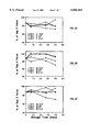
IGF-I and IGFBP-3 demonstrated high stability in dry form. Replicate dried blood filter paper spots from three different specimen with wide concentration range were prepared and stored at four different temperatures The blood spots' IGF-I and IGFBP-3 were measured at 0, 8, 27 and 40 days of storage. For both analytes, the recovery at room temperature, 4° C. and -20° C. were >80% of the zero day value at 40 days of storage. The recovery of IGF-I and IGFBP-3 after 27 days of storage at 30° C. exceeded 80% and 67%, respectively (FIGS. 2 and 3). FIGS. 2 and 3 depict three replicate experiments each. In both Figures, the open squares represent storage at -20° C.; the triangles represent storage at 4° C.; the closed squares storage at room temperature; and the open circles storage at 37° C.
As outlined above, the measurement of the plasma levels of IGFs, IGFBP-2, and IGFBP-3 require a 50- to 100-fold sample dilution to bring them within the measuring range of the assays. To simplify the calibration requirement of measuring these analytes in dried blood filter paper extract, we incorporated a similar dilution factor in the extraction procedures to permit employment of the current kits' calibrators. With this approach, the levels of the analyte measured in plasma and in the corresponding blood extract would be expected to correlate with a slope value representing their respective dilution factors. For example, in both IGF-I and IGFBP-3 ELISAs, plasma samples are diluted by about 100-fold while, assuming a hematocrit of 50% and a disc blood volume of 13 μL, the fractional plasma volume of the spotted blood is diluted by approximately 123- and 77-fold for IGF-I and IGFBP-3, respectively. Therefore with use of the same liquid standards, the IGF-I levels measured in the dried blood extracts should be ideally less than those measured in plasma (by ˜10-20%), while the IGFBP-3 levels should be higher by about 10-20%. It is, however, possible to adjust the extraction buffer volume (or the standards) to obtain closely comparable values.
EXAMPLE 5 Distribution of IGF-I and IGFBP-3 in whole blood
A freshly collected whole blood EDTA sample was aliquoted into several fractions of equal volumes and centrifuged, except for one fraction representing the originally drawn whole blood sample. The relative volume of the plasma portion of the centrifuged fractions was then changed to create a set of whole blood samples with varying degree of hematocrit. After mixing, the IGF-I and IGFBP-3 levels in the plasma and the resulting whole blood samples were measured and compared to their corresponding hematocrit values (hematocrit ranged from 0.20 to 0.70). Briefly, the measured IGF-I or IGFBP-3 levels in each blood fraction was compared to the plasma IGF-I or IGFBP-3 concentration and a whole-blood to plasma IGF-I and IGFBP-3 concentration ratios were established. These ratios were then compared to the fractional plasma volume of the whole blood samples calculated as 1 minus the hematocrit value (1-hematocrit). Comparable results were taken to indicate presence of IGF-I and IGFBP-3 within the plasma portion.
The whole blood fractions prepared as above with hematocrits ranging from 0 (plasma) to 0.70 were spotted onto filter paper, dried and extracted for IGF-I and IGFBP-3 analysis. The concentrations of IGF-I and IGFBP-3 in the whole blood fractions and the spot extracts were measured. The percent change in the whole blood values from that obtained for the original whole blood sample was calculated as recently described (Hoffman, B R, et al. Clin Chem 1996;42:536-44). Similarly, the percent change in the extracted blood spots values from the corresponding value of the original whole blood spot extract was calculated. The differences in the effect of hematocrit on the analyte concentrations measured in whole blood or extracts of whole blood were compared.
Circulating molecules may be differentially distributed among whole blood subfractions (plasma and red blood cell fractions) whose relative fractional volumes are subject to change, both within- and between-individuals. As these variables could significantly alter the validity of the whole blood measurements, we investigated distribution of the representative components of the IGF/IGFBPs. IGF-I and IGFBP-3 were measured in plasma and in a series of corresponding whole blood fractions with hematocrit ranging from 0.20 to 0.70. As shown in Table 8, the whole blood levels of IGF-I and IGFBP-3 varied by ˜3-4 fold as a function of variations in hematocrit. However, the magnitude of changes in whole blood levels were inversely related to hematocrit, suggesting distribution of IGF-I and IGFBP-3 in the plasma fraction. Furthermore, comparison of whole blood/plasma concentration ratios for IGF-I and IGFBP-3 with the corresponding fractional plasma volume of each blood sample (calculated as 1-Hematocrit) showed closely comparable relationship (Table 9).
              TABLE 8                                                     
______________________________________                                    
Hematocrit Effect on Measured IGF-I, and IGFBP-3 Whole Blood and          
  Extracts Levels                                                         
           IGF-I (ug/L)   IGFBP-3 (mg/L)                                  
Hematocrit WB.sup.a                                                       
                  Extract     WB   Extract                                
______________________________________                                    
0.70        92    155         0.8  2.7                                    
  0.62 134 153 1.4 3.6                                                    
   0.434 200 223 2.3 4.5                                                  
  0.33 234 210 2.8 4.4                                                    
  0.27 240 216 3.1 4.9                                                    
  0.20 287 220 3.5 4.6                                                    
  0 (plasma) 312 217 4.5 4.6                                              
______________________________________                                    
 .sup.a WB = Whole blood                                                  
              TABLE 9                                                     
______________________________________                                    
Comparison of Whole Blood/Plasma Concentration Ratio with Fractional      
 Plasma Volume.sup.a                                                      
 Whole Blood/Plasma Concentration Ratio                                   
  1-Hematocrit      IGF-I  IGFBP-3                                        
______________________________________                                    
0.3             0.29   0.18                                               
  0.38 0.43 0.31                                                          
  0.57 0.64 0.51                                                          
  0.67 0.75 0.62                                                          
  0.73 0.77 0.69                                                          
  0.80 0.91 0.78                                                          
______________________________________                                    
 .sup.a Defined as 1hematocrit                                            
To quantify the effect of hematocrit, IGF-I and IGFBP-3 levels were measured in the whole blood fractions described above and in their corresponding dried blood extracts. In both series of samples, the measured values were calculated as percent change from the corresponding value obtained for the originally drawn 0.33 hematocrit specimen. As expected for analytes present primarily in the plasm fraction, variation in hematocrit had a significant effect on IGF-I and IGFBP-3 levels measured in whole blood (Table 10). The whole blood concentrations of these analytes changed by ˜8-10% for every 0.05 unit change in hematocrit. In contrast, the effect of variation in hematocrit on the analyte concentrations measured in the dried blood filter paper extracts were comparatively less pronounced. The IGF-I and IGFBP-3 levels changed by ˜2-4% per 0.05 unit change in hematocrit. Only at abnormally high hematocrit (>0.6), the change in blood extract concentrations relative to those measured in the corresponding 0.33 hematocrit specimen were >10%.
EXAMPLE 6 Analyte Determination in Plasma and Corresponding Whole Blood and Dried Blood Extract
Fresh EDTA-whole blood samples (n=46) were spotted on filter paper, dried overnight, and extracted according to the protocols described for filter paper extraction of IGF-I and IGFBP-3. The corresponding whole blood and /or plasma fractions were also processed for IGF-I, IGF-II, IGFBP-2 and IGFBP-3 determination as described above and in their respective DSL ELISA or RIA protocols.
IGF-I and IGFBP-3 levels measured in plasma fractions and in the corresponding whole blood and dried blood filter paper extracts of freshly drawn samples (n=46) were compared. The plasma IGF-I and IGFBP-3 levels ranged from 36-575 ug/L, 0.87-5.9 mg/L and 6.4-52 arbitrary units/L, respectively. Regression analysis of data showed acceptable linear relationship between plasma values and those measured in whole blood and dried blood extracts (FIGS. 4-5). For these analytes, the blood spot values correlated well with the plasma levels (FIGS. 6-8).
              TABLE 10                                                    
______________________________________                                    
Hematocrit Effect on Measured IGF-I, and IGFBP-3 Concentrations           
  in Whole Blood and Dried Blood Extract                                  
           Percent Change from Corresponding Original                     
  Sample Value.sup.a                                                      
         IGF-I (ug/L)     IGFBP-3 (mg/L)                                  
Hematocrit WB.sup.b                                                       
                  Extract     WB   Extract                                
______________________________________                                    
0.70       -60    -26         -72  -38                                    
  0.62 -43 -27 -49 -17                                                    
   0.434 -15 +6.1 -18    +2.2                                             
  0.33 0 0    0  0                                                        
  0.27 +2.5 +2.8 +11 +14                                                  
  0.20 +23 +4.8 +24  +6                                                   
  0 (plasma) +33 -7.3 +58  +4                                             
______________________________________                                    
 .sup.a Value of the 0.33 hematocrit specimen                             
 .sup.b WB = Whole blood                                                  
EXAMPLE 7 Comparison of IGF-1 and IGFBP-3 in Plasma and Corresponding Dried Blood in Various Species
This is a prophetic example. Whole blood samples from cows, pigs, goats and sheep is collected via venous draw from the jugular vein. Whole blood samples from rabbits are collected from the ear vein. Whole blood samples from rats are collected from heart puncture or tail clip. Whole blood samples from chicken are collected from heart puncture or wing vein. For guinea pigs, orbital whole blood samples are collected. Whole blood samples from fish are collected from tail fins or gills. The whole blood samples from the above species are either directly spotted on blood collection filter paper cards obtained from Schleicher and Schuell or are first collected in tubes containing anticoagulants, such as EDTA, and then applied to the blood collection filter paper cards. A portion of the whole blood from each of the above samples is allowed to stand at room temperature for 2 hours, centrifuged and supernatant plasma collected in separate tubes.
The dried blood spots are extracted as described in Examples 1, 2, and 3. The concentrations of IGF-I and IGFBP-3 are determined on the paired "blood spot" and "plasma" samples using the "analytical Procedure" described earlier. An excellent correlation is obtained between the "blood spot" and "plasma" samples, indicating the validity of the blood spot measurement of these analytes.
Many other variations and modifications may be made in the methods herein described, by those having experience in this art, without departing from the concept of the present invention. Accordingly, it should be clearly understood that the methods described in the foregoing description are illustrative only, and not intended as a limitation on the scope of the invention.

Claims (12)

What is claimed is:
1. A method of determining concentrations of an insulin-like growth factor binding protein or acid labile subunit comprising the steps of:
(a) collecting blood from a subject;
(b) applying said blood to a solid support and drying said blood;
(c) extracting said dried blood from said solid support with a buffer of about pH 7.0-8.5 to form a blood extract of about pH 7.0-8.5; and
(d) testing said blood extract of about pH 7.0-8.5 for the concentration of insulin-like growth factor binding protein or acid labile subunit with an antibody directed against insulin-like growth factor binding protein or acid labile subunit.
2. The method according to claim 1 wherein said blood collecting step is conducted by capillary puncture.
3. The method according to claim 2 wherein said capillary puncture is selected from the group consisting of finger prick, thumb prick, or ear lobe prick.
4. The method according to claim 1 wherein said solid support is a paper medium.
5. The method according to claim 4 wherein said paper medium is filter paper.
6. The method according to claim 1 wherein said extracting step is conducted for a period of about thirty minutes to about two hours.
7. The method according to claim 1 wherein said extracting step is conducted for a period of about one hour.
8. The method according to claim 1 wherein said buffer extract comprises a buffer and protein.
9. The method according to claim 8 wherein said buffer comprises a buffer selected from the group consisting of Tris, borates, carbonates and phosphates.
10. The method according to claim 1 wherein said insulin-like growth factor binding protein is selected from the group consisting of insulin-like growth factor binding protein -1, -2, -3, -4, -5, and -6.
11. The method of claim 1, wherein said buffer extract consists essentially of an aqueous buffer, a detergent, optionally bovine serum albumin and optionally a salt.
12. The method of claim 11,
wherein the aqueous buffer is selected from the group consisting of dH2 O, Tris, borates, acetates, and phosphates,
wherein the detergent is selected from the group consisting of Tween 20 and sodium dodecyl sulfate, and
wherein the salt is selected from the group consisting of NaCl, KCl, MgCl2, and CaCl2.
US08/763,244 1996-12-10 1996-12-10 Immunoassay of IGF family of peptides, their binding proteins and related molecules in dried whole blood filter paper spots Expired - Fee Related US6066464A (en)

Priority Applications (2)

Application Number Priority Date Filing Date Title
US08/763,244 US6066464A (en) 1996-12-10 1996-12-10 Immunoassay of IGF family of peptides, their binding proteins and related molecules in dried whole blood filter paper spots
US09/405,348 US6348323B1 (en) 1996-12-10 1999-09-24 Immunoassay of IGF family of peptides, their binding proteins and related molecules in dried whole blood filter paper spots

Applications Claiming Priority (1)

Application Number Priority Date Filing Date Title
US08/763,244 US6066464A (en) 1996-12-10 1996-12-10 Immunoassay of IGF family of peptides, their binding proteins and related molecules in dried whole blood filter paper spots

Related Child Applications (1)

Application Number Title Priority Date Filing Date
US09/405,348 Continuation US6348323B1 (en) 1996-12-10 1999-09-24 Immunoassay of IGF family of peptides, their binding proteins and related molecules in dried whole blood filter paper spots

Publications (1)

Publication Number Publication Date
US6066464A true US6066464A (en) 2000-05-23

Family

ID=25067278

Family Applications (2)

Application Number Title Priority Date Filing Date
US08/763,244 Expired - Fee Related US6066464A (en) 1996-12-10 1996-12-10 Immunoassay of IGF family of peptides, their binding proteins and related molecules in dried whole blood filter paper spots
US09/405,348 Expired - Fee Related US6348323B1 (en) 1996-12-10 1999-09-24 Immunoassay of IGF family of peptides, their binding proteins and related molecules in dried whole blood filter paper spots

Family Applications After (1)

Application Number Title Priority Date Filing Date
US09/405,348 Expired - Fee Related US6348323B1 (en) 1996-12-10 1999-09-24 Immunoassay of IGF family of peptides, their binding proteins and related molecules in dried whole blood filter paper spots

Country Status (1)

Country Link
US (2) US6066464A (en)

Cited By (13)

* Cited by examiner, † Cited by third party
Publication number Priority date Publication date Assignee Title
US20040018484A1 (en) * 2002-04-23 2004-01-29 Home Access Health Corporation Quantitative analysis of a biological sample of unknown quantity
US20050130310A1 (en) * 2002-04-23 2005-06-16 Home Access Health Corporation Quantitative analysis of a biological sample of unknown quantity
US20050209532A1 (en) * 2002-04-23 2005-09-22 Home Access Health Corporation Quantitative analysis of a biological sample of unknown quantity
EP1830192A1 (en) * 2006-02-28 2007-09-05 Mediagnost Gesellschaft für Forschung und Herstellung von Diagnostika GmbH Method for storing the human growth hormone somatotropine and method for quantitative detection of the human growth hormone somatotropine
CN101962646A (en) * 2010-12-09 2011-02-02 华南农业大学 Chicken meat quality-related gene IGFBR-1 and application thereof
CN104697829A (en) * 2015-02-10 2015-06-10 深圳市新产业生物医学工程股份有限公司 Acid treatment agent for IGF-I chemiluminesent immunoassay, sample pretreatment method, kit and detection method
JP2016520843A (en) * 2013-06-06 2016-07-14 アンスティチュ ナショナル ドゥ ラ サンテ エ ドゥ ラ ルシェルシュ メディカル Biomarkers for readmission after heart failure
WO2016193497A1 (en) 2015-06-04 2016-12-08 Ospedale San Raffaele Srl Inhibitor of igfbp3/tmem219 axis and diabetes
WO2016193496A1 (en) 2015-06-04 2016-12-08 Ospedale San Raffaele Srl Igfbp3 and uses thereof
CN108699127A (en) * 2015-12-31 2018-10-23 西门子医疗保健诊断公司 Non-recombinant actrapid monotard-like growth factor binding protein concentrate
CN110234974A (en) * 2016-12-19 2019-09-13 生物梅里埃公司 Blood sample is deposited on waterleaf paper and the method for subsequent mechanical extraction blood culture
CN114112599A (en) * 2021-12-10 2022-03-01 泰州泽成生物技术有限公司 Insulin-like growth factor-I dissociation liquid
CN116500256A (en) * 2023-02-20 2023-07-28 郑州大学第五附属医院 Method for rapidly detecting content of serum retinol

Families Citing this family (8)

* Cited by examiner, † Cited by third party
Publication number Priority date Publication date Assignee Title
EP2204654B1 (en) * 2002-08-13 2012-12-12 N-Dia, Inc. Use of devices and methods for detecting amniotic fluid in vaginal secretions
GB2451620A (en) * 2007-07-26 2009-02-11 Keltie Therapeutic drug monitoring
BR112015016027A2 (en) 2013-01-02 2017-07-11 N Dia Inc method of predicting the time of delivery (ttd), method of determining the risk of preterm delivery, method of determining the risk of a pregnant woman spontaneously rupturing the chorioaminiotic membrane and kit
CN105794767B (en) * 2016-03-17 2018-10-09 四川新健康成生物股份有限公司 A kind of Precerving liquid and its application process for preserving pig whole blood
CN106501528A (en) * 2016-10-27 2017-03-15 杭州量康科技有限公司 Based on the method that Dried blood spots detect ToRCH10 item antibody
US10656164B2 (en) 2016-12-22 2020-05-19 Qiagen Sciences, Llc Screening asymptomatic pregnant woman for preterm birth
US10935555B2 (en) 2016-12-22 2021-03-02 Qiagen Sciences, Llc Determining candidate for induction of labor
CN109444411A (en) * 2018-10-26 2019-03-08 成都普利泰生物科技有限公司 A kind of canine parvovirus antibody chemical luminescence immue quantitative detection reagent box

Citations (16)

* Cited by examiner, † Cited by third party
Publication number Priority date Publication date Assignee Title
US4224031A (en) * 1977-11-15 1980-09-23 Mee John M L CI Mass spectrometric analysis of physiologically active compounds
US4230684A (en) * 1978-03-16 1980-10-28 Cornell Research Foundation, Inc. Method for determining steroids in human body liquids
US4277249A (en) * 1979-10-15 1981-07-07 Nichols Institute Mammalian Somatomedin measurement
US4381291A (en) * 1979-02-23 1983-04-26 Ab Fortia Measurement of free ligands
US4431743A (en) * 1978-03-16 1984-02-14 Cornell Research Foundation, Inc. Method for determining steroids in human body liquids
US4455381A (en) * 1980-11-07 1984-06-19 International Institute Of Cellular And Molecular Pathology Immunoassay of proteins
US5096812A (en) * 1989-06-08 1992-03-17 Osborn Laboratories, Inc. Assay method for gamma glutamyltransferase (GGT) in liquid blood and dried blood
US5187151A (en) * 1991-02-12 1993-02-16 Genentech, Inc. Use of binding protein with igf-i as an anabolic growth promoting agent
US5198340A (en) * 1991-01-17 1993-03-30 Genentech, Inc. Assay for free igf-i, igf-ii, and gh levels in body fluids
US5204267A (en) * 1991-12-17 1993-04-20 Osborn Laboratories, Inc. Method of glucose stabilization and analysis in dried blood spot samples
US5252489A (en) * 1989-01-17 1993-10-12 Macri James N Down syndrome screening method utilizing dried blood samples
US5324820A (en) * 1988-07-15 1994-06-28 Central Sydney Area Health Service Acid-labile subunit (ALS) of insulin-like growth factor binding protein complex
US5432097A (en) * 1993-11-09 1995-07-11 Yourno; Joseph Method for recovery of blood cells from dried blood spots on filter paper
US5516487A (en) * 1994-06-22 1996-05-14 Isolab, Inc. Absorbent paper for liquid sampling and impregnated paper calibrators and controls
US5532166A (en) * 1994-04-18 1996-07-02 Ma; Yinfa Quantitative retinol assay for serum and dried blood spots
US5641682A (en) * 1995-08-14 1997-06-24 Ortho Pharmaceutical Corporation Apparatus and method for automatically processing dried blood spots in HIV-1 testing

Family Cites Families (9)

* Cited by examiner, † Cited by third party
Publication number Priority date Publication date Assignee Title
US4594327A (en) * 1983-11-02 1986-06-10 Syntex (U.S.A.) Inc. Assay method for whole blood samples
US5200509A (en) * 1987-04-06 1993-04-06 Celtrix Pharmaceuticals, Inc. Human somatomedin carrier protein subunits and process for producing them; recombinant DNA molecules, hosts, processes and human somatomedin carrier protein-like polypeptides
AU1232188A (en) * 1987-05-29 1988-12-01 Mallinckrodt, Inc. Novel pair of monoclonal antibodies to insulin-like growth factor i permits immunometric assay for igf-i
US5593844A (en) * 1990-11-19 1997-01-14 Genentech, Inc. Ligand-mediated immunofunctional hormone binding protein assay method
EP0700994A1 (en) * 1994-03-23 1996-03-13 Japan Clinical Laboratories Anti-igf-i monoclonal antibody
US5824642A (en) * 1994-04-07 1998-10-20 Genentech, Inc. Treatment of partial growth hormone insensitivity syndrome
US5597700A (en) * 1994-04-28 1997-01-28 California Research, Llc Method for detecting free insulin-like growth-factor-binding protein 1 and a test device for detecting the ruptures of fetal membranes using the above method
JP2789306B2 (en) * 1994-11-15 1998-08-20 株式会社第一ラジオアイソトープ研究所 Immunological measurement method of insulin-like growth factor and kit for measuring insulin-like growth factor
JP2000514177A (en) * 1995-11-27 2000-10-24 ロシュ ダイアグノスティックス ゲーエムベーハー Articles for collecting and transporting test samples and methods for measuring test substances

Patent Citations (17)

* Cited by examiner, † Cited by third party
Publication number Priority date Publication date Assignee Title
US4224031A (en) * 1977-11-15 1980-09-23 Mee John M L CI Mass spectrometric analysis of physiologically active compounds
US4230684A (en) * 1978-03-16 1980-10-28 Cornell Research Foundation, Inc. Method for determining steroids in human body liquids
US4431743A (en) * 1978-03-16 1984-02-14 Cornell Research Foundation, Inc. Method for determining steroids in human body liquids
US4381291A (en) * 1979-02-23 1983-04-26 Ab Fortia Measurement of free ligands
US4277249A (en) * 1979-10-15 1981-07-07 Nichols Institute Mammalian Somatomedin measurement
US4455381A (en) * 1980-11-07 1984-06-19 International Institute Of Cellular And Molecular Pathology Immunoassay of proteins
US5561046A (en) * 1988-07-15 1996-10-01 Genentech, Inc. Methods for the detection of acid-labile subunit (ALS) of insulin-like growth factor binding protein complex
US5324820A (en) * 1988-07-15 1994-06-28 Central Sydney Area Health Service Acid-labile subunit (ALS) of insulin-like growth factor binding protein complex
US5252489A (en) * 1989-01-17 1993-10-12 Macri James N Down syndrome screening method utilizing dried blood samples
US5096812A (en) * 1989-06-08 1992-03-17 Osborn Laboratories, Inc. Assay method for gamma glutamyltransferase (GGT) in liquid blood and dried blood
US5198340A (en) * 1991-01-17 1993-03-30 Genentech, Inc. Assay for free igf-i, igf-ii, and gh levels in body fluids
US5187151A (en) * 1991-02-12 1993-02-16 Genentech, Inc. Use of binding protein with igf-i as an anabolic growth promoting agent
US5204267A (en) * 1991-12-17 1993-04-20 Osborn Laboratories, Inc. Method of glucose stabilization and analysis in dried blood spot samples
US5432097A (en) * 1993-11-09 1995-07-11 Yourno; Joseph Method for recovery of blood cells from dried blood spots on filter paper
US5532166A (en) * 1994-04-18 1996-07-02 Ma; Yinfa Quantitative retinol assay for serum and dried blood spots
US5516487A (en) * 1994-06-22 1996-05-14 Isolab, Inc. Absorbent paper for liquid sampling and impregnated paper calibrators and controls
US5641682A (en) * 1995-08-14 1997-06-24 Ortho Pharmaceutical Corporation Apparatus and method for automatically processing dried blood spots in HIV-1 testing

Non-Patent Citations (43)

* Cited by examiner, † Cited by third party
Title
Barreca, A et al, Progress in Growth Factor Res., vol.6 (2 4), pp. 231 239, Feb. 1995. *
Barreca, A et al, Progress in Growth Factor Res., vol.6 (2-4), pp. 231-239, Feb. 1995.
Bereket, A. et al, Clinical Endocrin., May 1996, vol. 44, pp. 525 532. *
Bereket, A. et al, Clinical Endocrin., May 1996, vol. 44, pp. 525-532.
Cassio, A, et al, Eur.J. Pediatrics, vol.145, pp. 187 189, 1986. *
Cassio, A, et al, Eur.J. Pediatrics, vol.145, pp. 187-189, 1986.
Chin, E et al, Endocrinol, vol. 134(6), Jun. 1994, pp. 2498 2504. *
Chin, E et al, Endocrinol, vol. 134(6), Jun. 1994, pp. 2498-2504.
Cotterill, A.M. et al, Jan., Clinical Endocrinology, vol. 44(1), pp. 91 101, 1996. *
Cotterill, A.M. et al, Jan., Clinical Endocrinology, vol. 44(1), pp. 91-101, 1996.
Daughaday, WH. et al, Gen. Comp. Endocrinol., Aug. 1985, vol. 59(2), pp. 316 325. *
Daughaday, WH. et al, Gen. Comp. Endocrinol., Aug. 1985, vol. 59(2), pp. 316-325.
Furlanetto, R.W., J. Clins. Endocrinology and Metabolism, vol. 54(5), pp. 1084 1086, 1982. *
Furlanetto, R.W., J. Clins. Endocrinology and Metabolism, vol. 54(5), pp. 1084-1086, 1982.
Hannon, WH et al, WI NC997, vol.12(13) 26 pages, 1992. *
He Yu, et al. "Ultrasensitive Time-Resolved Immunofluorometric Assay of Prostate-Specific Antigen in Serum and Preliminary Clinical Studies." Clinical Chemistry 39(10):2108-2114 (1993).
He Yu, et al. Ultrasensitive Time Resolved Immunofluorometric Assay of Prostate Specific Antigen in Serum and Preliminary Clinical Studies. Clinical Chemistry 39(10):2108 2114 (1993). *
Lee, C.Y. et al, Endocrinol., vol. 136(11) Nov. 1995, pp. 4982 4989. *
Lee, C.Y. et al, Endocrinol., vol. 136(11) Nov. 1995, pp. 4982-4989.
Lewitt, MS et al, Jun. 1994, Endocrinol., vol. 134(6), pp. 2404 2409. *
Lewitt, MS et al, Jun. 1994, Endocrinol., vol. 134(6), pp. 2404-2409.
Liu, F et al, Dec. 1994, J. of Clinical Endocrin. & Met. vol. 79(6), pp. 1883 1886. *
Liu, F et al, Dec. 1994, J. of Clinical Endocrin. & Met. vol. 79(6), pp. 1883-1886.
Liu, F et al, J. Clin. Endocrin. & Metabol. vol. 79(6), pp. 1883 1886, 1994. *
Liu, F et al, J. Clin. Endocrin. & Metabol. vol. 79(6), pp. 1883-1886, 1994.
Lord, A. P. D. et al, J. of Endocrinol., 1991, vol. 129, pp. 59 68. *
Lord, A. P. D. et al, J. of Endocrinol., 1991, vol. 129, pp. 59-68.
Mauceri, H.J. et al, Nov. Dec., Alcoholism: Clinical and Experimental Res., vol. 17(6), pp. 1201 1206, 1993. *
Mauceri, H.J. et al, Nov.-Dec., Alcoholism: Clinical and Experimental Res., vol. 17(6), pp. 1201-1206, 1993.
Mitchell, M.L et al, Pediatric Research, vol. 25(4), pp. 336 338, 1989. *
Mitchell, M.L et al, Pediatric Research, vol. 25(4), pp. 336-338, 1989.
Mitchell, M.L. et al, 1987, Clin. Chem., vol.33(4), pp. 536 538. *
Mitchell, M.L. et al, 1987, Clin. Chem., vol.33(4), pp. 536-538.
Mitchell, M.L. et al, Clinical Endocrinology vol. 27, pp. 625 628, 1987. *
Mitchell, M.L. et al, Clinical Endocrinology vol. 27, pp. 625-628, 1987.
Mitchell, ML et al, Clin. Chem., 1987, vol. 33(4), pp. 536 538. *
Mitchell, ML et al, Clin. Chem., 1987, vol. 33(4), pp. 536-538.
Scharf, J et al, Oct. 1995, J. of Hepatol., vol.23, pp. 424 430. *
Scharf, J et al, Oct. 1995, J. of Hepatol., vol.23, pp. 424-430.
Scharf, J et al, Progress in Growth Factor Res., vol. 6, (2 4), pp. 175 180, 1995, Oct. *
Scharf, J et al, Progress in Growth Factor Res., vol. 6, (2-4), pp. 175-180, 1995, Oct.
Westwood, M et al, Dec. 1994, J. Clin. Endocrin. & Metabol., vol. 79(6), pp. 1735 1741. *
Westwood, M et al, Dec. 1994, J. Clin. Endocrin. & Metabol., vol. 79(6), pp. 1735-1741.

Cited By (28)

* Cited by examiner, † Cited by third party
Publication number Priority date Publication date Assignee Title
US8383058B2 (en) 2002-04-23 2013-02-26 Home Access Health Corporation Quantitative analysis of a biological sample of unknown quantity
US8211703B2 (en) 2002-04-23 2012-07-03 Home Access Health Corporation Quantitative analysis of a biological sample of unknown quantity
US20050209532A1 (en) * 2002-04-23 2005-09-22 Home Access Health Corporation Quantitative analysis of a biological sample of unknown quantity
US20040018484A1 (en) * 2002-04-23 2004-01-29 Home Access Health Corporation Quantitative analysis of a biological sample of unknown quantity
US7479392B2 (en) 2002-04-23 2009-01-20 Home Access Health Corporation Quantitative analysis of a biological sample of unknown quantity
US20090104636A1 (en) * 2002-04-23 2009-04-23 Home Access Health Corporation Quantitative analysis of a biological sample of unknown quantity
US7569184B2 (en) 2002-04-23 2009-08-04 Home Access Health Corporation Quantitative analysis of a biological sample of unknown quantity
US7611670B2 (en) 2002-04-23 2009-11-03 Home Access Health Corporation Quantitative analysis of a biological sample of unknown quantity
US20100010373A1 (en) * 2002-04-23 2010-01-14 Home Access Health Corporation Quantitative analysis of a biological sample of unknown quantity
US20050130310A1 (en) * 2002-04-23 2005-06-16 Home Access Health Corporation Quantitative analysis of a biological sample of unknown quantity
US8080425B2 (en) 2002-04-23 2011-12-20 Home Access Health Corporation Quantitative analysis of a biological sample of unknown quantity
WO2006052764A1 (en) * 2004-11-05 2006-05-18 Home Access Health Corporation Quantitative analysis of a biological sample of unknown quantity
EP1830192A1 (en) * 2006-02-28 2007-09-05 Mediagnost Gesellschaft für Forschung und Herstellung von Diagnostika GmbH Method for storing the human growth hormone somatotropine and method for quantitative detection of the human growth hormone somatotropine
CN101962646B (en) * 2010-12-09 2012-05-30 华南农业大学 Chicken meat quality-related gene IGFBR-1 and application thereof
CN101962646A (en) * 2010-12-09 2011-02-02 华南农业大学 Chicken meat quality-related gene IGFBR-1 and application thereof
JP2016520843A (en) * 2013-06-06 2016-07-14 アンスティチュ ナショナル ドゥ ラ サンテ エ ドゥ ラ ルシェルシュ メディカル Biomarkers for readmission after heart failure
CN104697829B (en) * 2015-02-10 2018-05-04 深圳市新产业生物医学工程股份有限公司 Acidic treatment agent, sample preprocessing method, kit and detection method for the detection of I chemiluminescence immunoassays of IGF-
CN104697829A (en) * 2015-02-10 2015-06-10 深圳市新产业生物医学工程股份有限公司 Acid treatment agent for IGF-I chemiluminesent immunoassay, sample pretreatment method, kit and detection method
WO2016193497A1 (en) 2015-06-04 2016-12-08 Ospedale San Raffaele Srl Inhibitor of igfbp3/tmem219 axis and diabetes
WO2016193496A1 (en) 2015-06-04 2016-12-08 Ospedale San Raffaele Srl Igfbp3 and uses thereof
US10682391B2 (en) 2015-06-04 2020-06-16 Ospedale San Raffaele Srl Inhibitors of IGFBP3 binding to TMEM219 for treatment of intestinal diseases
US11020453B2 (en) 2015-06-04 2021-06-01 Ospedale San Raffaele Srl Inhibitor of IGFBP3/TMEM219 axis and diabetes
CN108699127A (en) * 2015-12-31 2018-10-23 西门子医疗保健诊断公司 Non-recombinant actrapid monotard-like growth factor binding protein concentrate
CN110234974A (en) * 2016-12-19 2019-09-13 生物梅里埃公司 Blood sample is deposited on waterleaf paper and the method for subsequent mechanical extraction blood culture
CN110234974B (en) * 2016-12-19 2021-08-17 生物梅里埃公司 Method for depositing blood samples on absorbent paper and subsequent mechanical extraction of blood cultures
CN114112599A (en) * 2021-12-10 2022-03-01 泰州泽成生物技术有限公司 Insulin-like growth factor-I dissociation liquid
CN114112599B (en) * 2021-12-10 2024-02-13 泰州泽成生物技术有限公司 Insulin-like growth factor-I dissociation liquid
CN116500256A (en) * 2023-02-20 2023-07-28 郑州大学第五附属医院 Method for rapidly detecting content of serum retinol

Also Published As

Publication number Publication date
US6348323B1 (en) 2002-02-19

Similar Documents

Publication Publication Date Title
US6066464A (en) Immunoassay of IGF family of peptides, their binding proteins and related molecules in dried whole blood filter paper spots
US6248546B1 (en) Assay of IGFBP complex
JP5225097B2 (en) How to detect preeclampsia
US7410807B2 (en) Pregnancy and sex identification test based on saliva or other bodily fluids
EP0495625B1 (en) Assay for free IGF-I, IGF-II, and GH levels in body fluids
US9128099B2 (en) Determination of sFlt-1:angiogenic factor complex
JPS58117456A (en) Reagent set detecting and determining one component of reaction between protein having specified binding activity and substance binding in response to said protein
JP2001514388A (en) Method and apparatus for rapidly analyzing an analyte in a biological sample
Proye et al. Usefulness and limits of quick intraoperative measurements of intact (1–84) parathyroid hormone in the surgical management of hyperparathyroidism: sequential measurements in patients with multiglandular disease
Worthman et al. Measurement of gonadotropins in dried blood spots
CS257757B2 (en) Method of reaction determination between antigen and antibody and reaction agent for realization of this process
CN103048457A (en) Myocardial infarction early-warning colloidal gold kit and preparation method thereof
JP4734306B2 (en) Method for distinguishing cardiac dysfunction associated with heart disease and placental associated cardiac dysfunction in pregnant women using natriuretic peptide and placental growth factor / soluble VEGF receptor
Heyderman et al. Human chorionic gonadotropin and human placental lactogen in extragonadal tumors an immunoperoxidase study of ten non‐germ cell neoplasms
CN104714031A (en) Rapid detection kit for myocardial infarction
JPS63501819A (en) Immunoassay methods and kits
US20070190580A1 (en) Immunoassay of Fragments of Insulin-Like Growth Factor Binding Proteins
JP2008544285A (en) Phosphorylated protein immunoassay
Hopwood et al. Thyroid antibodies in children and adolescents with thyroid disorders
US20020013002A1 (en) Pregnancy test based on saliva or other bodily fluids
EP4086633A2 (en) Prognostic method
Obese et al. Measuring concentrations of insulin-like growth factor-I with an enzyme-linked immunosorbent antibody assay in plasma samples from Holstein cows
Prakash et al. Development of a simple, direct, microtitre plate enzymeimmunoassay (EIA) for progesterone determination in whole milk of buffaloes
Giclas Classical pathway evaluation
Van Kamp et al. A rapid and sensitive immunosorbent assay for urinary albumin

Legal Events

Date Code Title Description
AS Assignment

Owner name: DIAGNOSTIC SYSTEMS LABORATORIES, INC., TEXAS

Free format text: ASSIGNMENT OF ASSIGNORS INTEREST;ASSIGNORS:KHOSRAVI, M. JAVAD;MISTRY, JEHANGIR;DIAMANDI, ANASTASIA;REEL/FRAME:008410/0688;SIGNING DATES FROM 19961126 TO 19961209

FEPP Fee payment procedure

Free format text: PAYOR NUMBER ASSIGNED (ORIGINAL EVENT CODE: ASPN); ENTITY STATUS OF PATENT OWNER: SMALL ENTITY

CC Certificate of correction
LAPS Lapse for failure to pay maintenance fees
FP Lapsed due to failure to pay maintenance fee

Effective date: 20040523

FEPP Fee payment procedure

Free format text: PETITION RELATED TO MAINTENANCE FEES FILED (ORIGINAL EVENT CODE: PMFP); ENTITY STATUS OF PATENT OWNER: SMALL ENTITY

AS Assignment

Owner name: INDEPENDENCE MANZANAR LLC, NEVADA

Free format text: ASSIGNMENT OF ASSIGNORS INTEREST;ASSIGNOR:NORTEL NETWORKS LIMITED;REEL/FRAME:017286/0465

Effective date: 20050406

FEPP Fee payment procedure

Free format text: PAYOR NUMBER ASSIGNED (ORIGINAL EVENT CODE: ASPN); ENTITY STATUS OF PATENT OWNER: SMALL ENTITY

Free format text: PAYER NUMBER DE-ASSIGNED (ORIGINAL EVENT CODE: RMPN); ENTITY STATUS OF PATENT OWNER: SMALL ENTITY

FEPP Fee payment procedure

Free format text: PETITION RELATED TO MAINTENANCE FEES FILED (ORIGINAL EVENT CODE: PMFP); ENTITY STATUS OF PATENT OWNER: SMALL ENTITY

FEPP Fee payment procedure

Free format text: PETITION RELATED TO MAINTENANCE FEES DENIED/DISMISSED (ORIGINAL EVENT CODE: PMFD); ENTITY STATUS OF PATENT OWNER: SMALL ENTITY

STCH Information on status: patent discontinuation

Free format text: PATENT EXPIRED DUE TO NONPAYMENT OF MAINTENANCE FEES UNDER 37 CFR 1.362

AS Assignment

Owner name: BECKMAN COULTER, INC., CALIFORNIA

Free format text: ASSIGNMENT OF ASSIGNORS INTEREST;ASSIGNOR:DIAGNOSTIC SYSTEMS LABORATORIES, INC.;REEL/FRAME:023828/0704

Effective date: 20091231