US5808028A - Molecular clone of a P58 receptor protein and uses thereof - Google Patents
Molecular clone of a P58 receptor protein and uses thereof Download PDFInfo
- Publication number
- US5808028A US5808028A US08/248,628 US24862894A US5808028A US 5808028 A US5808028 A US 5808028A US 24862894 A US24862894 A US 24862894A US 5808028 A US5808028 A US 5808028A
- Authority
- US
- United States
- Prior art keywords
- receptor protein
- nucleic acid
- receptor
- present
- cells
- Prior art date
- Legal status (The legal status is an assumption and is not a legal conclusion. Google has not performed a legal analysis and makes no representation as to the accuracy of the status listed.)
- Expired - Lifetime
Links
- 101710157220 DNA primase large subunit Proteins 0.000 claims abstract description 100
- 101710173608 Protein ERGIC-53 Proteins 0.000 claims abstract description 100
- 101710106224 Protein disulfide-isomerase A3 Proteins 0.000 claims abstract description 100
- 101710204645 UV excision repair protein RAD23 homolog B Proteins 0.000 claims abstract description 100
- 150000007523 nucleic acids Chemical class 0.000 claims abstract description 45
- 108020004707 nucleic acids Proteins 0.000 claims abstract description 37
- 102000039446 nucleic acids Human genes 0.000 claims abstract description 37
- 239000013598 vector Substances 0.000 claims abstract description 17
- 108091028043 Nucleic acid sequence Proteins 0.000 claims description 6
- 125000003275 alpha amino acid group Chemical group 0.000 claims description 5
- 101000611553 Homo sapiens DNA primase large subunit Proteins 0.000 claims description 3
- 101000976215 Homo sapiens Probable ribonuclease ZC3H12D Proteins 0.000 claims description 3
- 101001038300 Homo sapiens Protein ERGIC-53 Proteins 0.000 claims description 3
- 101001098802 Homo sapiens Protein disulfide-isomerase A3 Proteins 0.000 claims description 3
- 101000717424 Homo sapiens UV excision repair protein RAD23 homolog B Proteins 0.000 claims description 3
- 102000047985 human ZC3H12D Human genes 0.000 claims description 3
- 238000000034 method Methods 0.000 abstract description 55
- 239000003446 ligand Substances 0.000 abstract description 29
- 102000005962 receptors Human genes 0.000 abstract description 26
- 108020003175 receptors Proteins 0.000 abstract description 26
- 239000012472 biological sample Substances 0.000 abstract description 19
- 239000005557 antagonist Substances 0.000 abstract description 11
- 239000000556 agonist Substances 0.000 abstract description 10
- 229940079593 drug Drugs 0.000 abstract description 8
- 239000003814 drug Substances 0.000 abstract description 8
- 108020001507 fusion proteins Proteins 0.000 abstract description 7
- 102000037865 fusion proteins Human genes 0.000 abstract description 7
- 238000012216 screening Methods 0.000 abstract description 7
- 230000001086 cytosolic effect Effects 0.000 abstract description 3
- 102100033634 Killer cell immunoglobulin-like receptor 2DL3 Human genes 0.000 description 91
- 210000004027 cell Anatomy 0.000 description 57
- 210000000822 natural killer cell Anatomy 0.000 description 30
- 108090000623 proteins and genes Proteins 0.000 description 28
- 102000004169 proteins and genes Human genes 0.000 description 20
- 239000002299 complementary DNA Substances 0.000 description 16
- 239000002773 nucleotide Substances 0.000 description 15
- 125000003729 nucleotide group Chemical group 0.000 description 15
- 239000000523 sample Substances 0.000 description 15
- 230000015572 biosynthetic process Effects 0.000 description 10
- 108020004414 DNA Proteins 0.000 description 8
- 108010087819 Fc receptors Proteins 0.000 description 8
- 102000009109 Fc receptors Human genes 0.000 description 8
- 230000000692 anti-sense effect Effects 0.000 description 8
- 238000003556 assay Methods 0.000 description 8
- 238000009396 hybridization Methods 0.000 description 8
- 239000013615 primer Substances 0.000 description 8
- 239000000427 antigen Substances 0.000 description 7
- 108091007433 antigens Proteins 0.000 description 7
- 102000036639 antigens Human genes 0.000 description 7
- 239000011324 bead Substances 0.000 description 7
- 230000001404 mediated effect Effects 0.000 description 7
- 238000000746 purification Methods 0.000 description 7
- 238000012163 sequencing technique Methods 0.000 description 7
- 108060003951 Immunoglobulin Proteins 0.000 description 6
- 206010028980 Neoplasm Diseases 0.000 description 6
- 238000012408 PCR amplification Methods 0.000 description 6
- FAPWRFPIFSIZLT-UHFFFAOYSA-M Sodium chloride Chemical compound [Na+].[Cl-] FAPWRFPIFSIZLT-UHFFFAOYSA-M 0.000 description 6
- 239000000356 contaminant Substances 0.000 description 6
- 102000018358 immunoglobulin Human genes 0.000 description 6
- 230000001506 immunosuppresive effect Effects 0.000 description 6
- 239000006166 lysate Substances 0.000 description 6
- 230000009826 neoplastic cell growth Effects 0.000 description 6
- 238000003199 nucleic acid amplification method Methods 0.000 description 6
- 108091032973 (ribonucleotides)n+m Proteins 0.000 description 5
- 241000699666 Mus <mouse, genus> Species 0.000 description 5
- 108091034117 Oligonucleotide Proteins 0.000 description 5
- 230000003321 amplification Effects 0.000 description 5
- 239000003153 chemical reaction reagent Substances 0.000 description 5
- 239000000499 gel Substances 0.000 description 5
- 238000003018 immunoassay Methods 0.000 description 5
- 230000003834 intracellular effect Effects 0.000 description 5
- 238000002955 isolation Methods 0.000 description 5
- 239000000463 material Substances 0.000 description 5
- 238000011282 treatment Methods 0.000 description 5
- QKNYBSVHEMOAJP-UHFFFAOYSA-N 2-amino-2-(hydroxymethyl)propane-1,3-diol;hydron;chloride Chemical compound Cl.OCC(N)(CO)CO QKNYBSVHEMOAJP-UHFFFAOYSA-N 0.000 description 4
- QTBSBXVTEAMEQO-UHFFFAOYSA-N Acetic acid Chemical compound CC(O)=O QTBSBXVTEAMEQO-UHFFFAOYSA-N 0.000 description 4
- 229930010555 Inosine Natural products 0.000 description 4
- UGQMRVRMYYASKQ-KQYNXXCUSA-N Inosine Chemical group O[C@@H]1[C@H](O)[C@@H](CO)O[C@H]1N1C2=NC=NC(O)=C2N=C1 UGQMRVRMYYASKQ-KQYNXXCUSA-N 0.000 description 4
- 229960003786 inosine Drugs 0.000 description 4
- 239000000047 product Substances 0.000 description 4
- 238000002415 sodium dodecyl sulfate polyacrylamide gel electrophoresis Methods 0.000 description 4
- 102000001301 EGF receptor Human genes 0.000 description 3
- 108060006698 EGF receptor Proteins 0.000 description 3
- 102000004190 Enzymes Human genes 0.000 description 3
- 108090000790 Enzymes Proteins 0.000 description 3
- 238000012413 Fluorescence activated cell sorting analysis Methods 0.000 description 3
- PEDCQBHIVMGVHV-UHFFFAOYSA-N Glycerine Chemical compound OCC(O)CO PEDCQBHIVMGVHV-UHFFFAOYSA-N 0.000 description 3
- 101000917858 Homo sapiens Low affinity immunoglobulin gamma Fc region receptor III-A Proteins 0.000 description 3
- 101000917839 Homo sapiens Low affinity immunoglobulin gamma Fc region receptor III-B Proteins 0.000 description 3
- 102000003746 Insulin Receptor Human genes 0.000 description 3
- 108010001127 Insulin Receptor Proteins 0.000 description 3
- 102100029185 Low affinity immunoglobulin gamma Fc region receptor III-B Human genes 0.000 description 3
- 108700018351 Major Histocompatibility Complex Proteins 0.000 description 3
- 229920002684 Sepharose Polymers 0.000 description 3
- DBMJMQXJHONAFJ-UHFFFAOYSA-M Sodium laurylsulphate Chemical compound [Na+].CCCCCCCCCCCCOS([O-])(=O)=O DBMJMQXJHONAFJ-UHFFFAOYSA-M 0.000 description 3
- 150000001413 amino acids Chemical class 0.000 description 3
- 238000004458 analytical method Methods 0.000 description 3
- 238000000376 autoradiography Methods 0.000 description 3
- 230000004071 biological effect Effects 0.000 description 3
- 210000001185 bone marrow Anatomy 0.000 description 3
- 230000036755 cellular response Effects 0.000 description 3
- 238000012512 characterization method Methods 0.000 description 3
- 230000009089 cytolysis Effects 0.000 description 3
- 239000003599 detergent Substances 0.000 description 3
- 239000013604 expression vector Substances 0.000 description 3
- 239000012133 immunoprecipitate Substances 0.000 description 3
- 230000007774 longterm Effects 0.000 description 3
- 239000012528 membrane Substances 0.000 description 3
- 238000010369 molecular cloning Methods 0.000 description 3
- 239000013612 plasmid Substances 0.000 description 3
- 102000004196 processed proteins & peptides Human genes 0.000 description 3
- 108090000765 processed proteins & peptides Proteins 0.000 description 3
- 238000010839 reverse transcription Methods 0.000 description 3
- 238000003757 reverse transcription PCR Methods 0.000 description 3
- 239000011780 sodium chloride Substances 0.000 description 3
- 230000020382 suppression by virus of host antigen processing and presentation of peptide antigen via MHC class I Effects 0.000 description 3
- 238000012360 testing method Methods 0.000 description 3
- YBJHBAHKTGYVGT-ZKWXMUAHSA-N (+)-Biotin Chemical compound N1C(=O)N[C@@H]2[C@H](CCCCC(=O)O)SC[C@@H]21 YBJHBAHKTGYVGT-ZKWXMUAHSA-N 0.000 description 2
- QFVHZQCOUORWEI-UHFFFAOYSA-N 4-[(4-anilino-5-sulfonaphthalen-1-yl)diazenyl]-5-hydroxynaphthalene-2,7-disulfonic acid Chemical compound C=12C(O)=CC(S(O)(=O)=O)=CC2=CC(S(O)(=O)=O)=CC=1N=NC(C1=CC=CC(=C11)S(O)(=O)=O)=CC=C1NC1=CC=CC=C1 QFVHZQCOUORWEI-UHFFFAOYSA-N 0.000 description 2
- LRFVTYWOQMYALW-UHFFFAOYSA-N 9H-xanthine Chemical compound O=C1NC(=O)NC2=C1NC=N2 LRFVTYWOQMYALW-UHFFFAOYSA-N 0.000 description 2
- 229920000936 Agarose Polymers 0.000 description 2
- HEDRZPFGACZZDS-UHFFFAOYSA-N Chloroform Chemical compound ClC(Cl)Cl HEDRZPFGACZZDS-UHFFFAOYSA-N 0.000 description 2
- 239000003155 DNA primer Substances 0.000 description 2
- 241000588724 Escherichia coli Species 0.000 description 2
- 102100023593 Fibroblast growth factor receptor 1 Human genes 0.000 description 2
- 101710182386 Fibroblast growth factor receptor 1 Proteins 0.000 description 2
- 102000006354 HLA-DR Antigens Human genes 0.000 description 2
- 108010058597 HLA-DR Antigens Proteins 0.000 description 2
- TWRXJAOTZQYOKJ-UHFFFAOYSA-L Magnesium chloride Chemical compound [Mg+2].[Cl-].[Cl-] TWRXJAOTZQYOKJ-UHFFFAOYSA-L 0.000 description 2
- 241001465754 Metazoa Species 0.000 description 2
- 241000699670 Mus sp. Species 0.000 description 2
- -1 NP40 Substances 0.000 description 2
- 102100026547 Platelet-derived growth factor receptor beta Human genes 0.000 description 2
- 101710164680 Platelet-derived growth factor receptor beta Proteins 0.000 description 2
- 229920001213 Polysorbate 20 Polymers 0.000 description 2
- WCUXLLCKKVVCTQ-UHFFFAOYSA-M Potassium chloride Chemical compound [Cl-].[K+] WCUXLLCKKVVCTQ-UHFFFAOYSA-M 0.000 description 2
- 102000007056 Recombinant Fusion Proteins Human genes 0.000 description 2
- 108010008281 Recombinant Fusion Proteins Proteins 0.000 description 2
- PXIPVTKHYLBLMZ-UHFFFAOYSA-N Sodium azide Chemical compound [Na+].[N-]=[N+]=[N-] PXIPVTKHYLBLMZ-UHFFFAOYSA-N 0.000 description 2
- 210000001744 T-lymphocyte Anatomy 0.000 description 2
- 241000700605 Viruses Species 0.000 description 2
- 239000002253 acid Substances 0.000 description 2
- 150000007513 acids Chemical class 0.000 description 2
- 201000011510 cancer Diseases 0.000 description 2
- 230000015556 catabolic process Effects 0.000 description 2
- 108700010039 chimeric receptor Proteins 0.000 description 2
- 238000004587 chromatography analysis Methods 0.000 description 2
- 238000006731 degradation reaction Methods 0.000 description 2
- 238000001514 detection method Methods 0.000 description 2
- 230000029087 digestion Effects 0.000 description 2
- 230000000694 effects Effects 0.000 description 2
- 239000012149 elution buffer Substances 0.000 description 2
- 238000001114 immunoprecipitation Methods 0.000 description 2
- 230000003993 interaction Effects 0.000 description 2
- 238000004519 manufacturing process Methods 0.000 description 2
- 239000003550 marker Substances 0.000 description 2
- 108020004999 messenger RNA Proteins 0.000 description 2
- 244000052769 pathogen Species 0.000 description 2
- 238000003752 polymerase chain reaction Methods 0.000 description 2
- 239000000256 polyoxyethylene sorbitan monolaurate Substances 0.000 description 2
- 235000010486 polyoxyethylene sorbitan monolaurate Nutrition 0.000 description 2
- 230000002285 radioactive effect Effects 0.000 description 2
- 238000004007 reversed phase HPLC Methods 0.000 description 2
- 238000000926 separation method Methods 0.000 description 2
- 238000010186 staining Methods 0.000 description 2
- 210000001519 tissue Anatomy 0.000 description 2
- 239000011534 wash buffer Substances 0.000 description 2
- 238000001262 western blot Methods 0.000 description 2
- 206010003445 Ascites Diseases 0.000 description 1
- 241000894006 Bacteria Species 0.000 description 1
- 102100026189 Beta-galactosidase Human genes 0.000 description 1
- 108091003079 Bovine Serum Albumin Proteins 0.000 description 1
- 238000011537 Coomassie blue staining Methods 0.000 description 1
- 101150074155 DHFR gene Proteins 0.000 description 1
- 108010061982 DNA Ligases Proteins 0.000 description 1
- 102000012410 DNA Ligases Human genes 0.000 description 1
- 238000007399 DNA isolation Methods 0.000 description 1
- 102000016928 DNA-directed DNA polymerase Human genes 0.000 description 1
- 108010014303 DNA-directed DNA polymerase Proteins 0.000 description 1
- 241000196324 Embryophyta Species 0.000 description 1
- 108010067770 Endopeptidase K Proteins 0.000 description 1
- 241000701533 Escherichia virus T4 Species 0.000 description 1
- 102000009465 Growth Factor Receptors Human genes 0.000 description 1
- 108010009202 Growth Factor Receptors Proteins 0.000 description 1
- 102100028976 HLA class I histocompatibility antigen, B alpha chain Human genes 0.000 description 1
- 241000238631 Hexapoda Species 0.000 description 1
- 108010088652 Histocompatibility Antigens Class I Proteins 0.000 description 1
- 108091006905 Human Serum Albumin Proteins 0.000 description 1
- 102000008100 Human Serum Albumin Human genes 0.000 description 1
- FBOZXECLQNJBKD-ZDUSSCGKSA-N L-methotrexate Chemical compound C=1N=C2N=C(N)N=C(N)C2=NC=1CN(C)C1=CC=C(C(=O)N[C@@H](CCC(O)=O)C(O)=O)C=C1 FBOZXECLQNJBKD-ZDUSSCGKSA-N 0.000 description 1
- 108060001084 Luciferase Proteins 0.000 description 1
- 239000005089 Luciferase Substances 0.000 description 1
- 108700005089 MHC Class I Genes Proteins 0.000 description 1
- 108700005092 MHC Class II Genes Proteins 0.000 description 1
- 241001529936 Murinae Species 0.000 description 1
- 125000001429 N-terminal alpha-amino-acid group Chemical group 0.000 description 1
- 238000000636 Northern blotting Methods 0.000 description 1
- 101710163270 Nuclease Proteins 0.000 description 1
- 102000016978 Orphan receptors Human genes 0.000 description 1
- 108070000031 Orphan receptors Proteins 0.000 description 1
- 241000283973 Oryctolagus cuniculus Species 0.000 description 1
- 238000009004 PCR Kit Methods 0.000 description 1
- 239000002033 PVDF binder Substances 0.000 description 1
- ISWSIDIOOBJBQZ-UHFFFAOYSA-N Phenol Chemical compound OC1=CC=CC=C1 ISWSIDIOOBJBQZ-UHFFFAOYSA-N 0.000 description 1
- 241000276498 Pollachius virens Species 0.000 description 1
- 108010021757 Polynucleotide 5'-Hydroxyl-Kinase Proteins 0.000 description 1
- 102000008422 Polynucleotide 5'-hydroxyl-kinase Human genes 0.000 description 1
- 108091034057 RNA (poly(A)) Proteins 0.000 description 1
- 108700008625 Reporter Genes Proteins 0.000 description 1
- 102000006382 Ribonucleases Human genes 0.000 description 1
- 108010083644 Ribonucleases Proteins 0.000 description 1
- 241000283984 Rodentia Species 0.000 description 1
- 241000710960 Sindbis virus Species 0.000 description 1
- 238000002105 Southern blotting Methods 0.000 description 1
- 108091008874 T cell receptors Proteins 0.000 description 1
- 102000016266 T-Cell Antigen Receptors Human genes 0.000 description 1
- 239000013504 Triton X-100 Substances 0.000 description 1
- 229920004890 Triton X-100 Polymers 0.000 description 1
- 108090000631 Trypsin Proteins 0.000 description 1
- 102000004142 Trypsin Human genes 0.000 description 1
- JLCPHMBAVCMARE-UHFFFAOYSA-N [3-[[3-[[3-[[3-[[3-[[3-[[3-[[3-[[3-[[3-[[3-[[5-(2-amino-6-oxo-1H-purin-9-yl)-3-[[3-[[3-[[3-[[3-[[3-[[5-(2-amino-6-oxo-1H-purin-9-yl)-3-[[5-(2-amino-6-oxo-1H-purin-9-yl)-3-hydroxyoxolan-2-yl]methoxy-hydroxyphosphoryl]oxyoxolan-2-yl]methoxy-hydroxyphosphoryl]oxy-5-(5-methyl-2,4-dioxopyrimidin-1-yl)oxolan-2-yl]methoxy-hydroxyphosphoryl]oxy-5-(6-aminopurin-9-yl)oxolan-2-yl]methoxy-hydroxyphosphoryl]oxy-5-(6-aminopurin-9-yl)oxolan-2-yl]methoxy-hydroxyphosphoryl]oxy-5-(6-aminopurin-9-yl)oxolan-2-yl]methoxy-hydroxyphosphoryl]oxy-5-(6-aminopurin-9-yl)oxolan-2-yl]methoxy-hydroxyphosphoryl]oxyoxolan-2-yl]methoxy-hydroxyphosphoryl]oxy-5-(5-methyl-2,4-dioxopyrimidin-1-yl)oxolan-2-yl]methoxy-hydroxyphosphoryl]oxy-5-(4-amino-2-oxopyrimidin-1-yl)oxolan-2-yl]methoxy-hydroxyphosphoryl]oxy-5-(5-methyl-2,4-dioxopyrimidin-1-yl)oxolan-2-yl]methoxy-hydroxyphosphoryl]oxy-5-(5-methyl-2,4-dioxopyrimidin-1-yl)oxolan-2-yl]methoxy-hydroxyphosphoryl]oxy-5-(6-aminopurin-9-yl)oxolan-2-yl]methoxy-hydroxyphosphoryl]oxy-5-(6-aminopurin-9-yl)oxolan-2-yl]methoxy-hydroxyphosphoryl]oxy-5-(4-amino-2-oxopyrimidin-1-yl)oxolan-2-yl]methoxy-hydroxyphosphoryl]oxy-5-(4-amino-2-oxopyrimidin-1-yl)oxolan-2-yl]methoxy-hydroxyphosphoryl]oxy-5-(4-amino-2-oxopyrimidin-1-yl)oxolan-2-yl]methoxy-hydroxyphosphoryl]oxy-5-(6-aminopurin-9-yl)oxolan-2-yl]methoxy-hydroxyphosphoryl]oxy-5-(4-amino-2-oxopyrimidin-1-yl)oxolan-2-yl]methyl [5-(6-aminopurin-9-yl)-2-(hydroxymethyl)oxolan-3-yl] hydrogen phosphate Polymers Cc1cn(C2CC(OP(O)(=O)OCC3OC(CC3OP(O)(=O)OCC3OC(CC3O)n3cnc4c3nc(N)[nH]c4=O)n3cnc4c3nc(N)[nH]c4=O)C(COP(O)(=O)OC3CC(OC3COP(O)(=O)OC3CC(OC3COP(O)(=O)OC3CC(OC3COP(O)(=O)OC3CC(OC3COP(O)(=O)OC3CC(OC3COP(O)(=O)OC3CC(OC3COP(O)(=O)OC3CC(OC3COP(O)(=O)OC3CC(OC3COP(O)(=O)OC3CC(OC3COP(O)(=O)OC3CC(OC3COP(O)(=O)OC3CC(OC3COP(O)(=O)OC3CC(OC3COP(O)(=O)OC3CC(OC3COP(O)(=O)OC3CC(OC3COP(O)(=O)OC3CC(OC3COP(O)(=O)OC3CC(OC3COP(O)(=O)OC3CC(OC3CO)n3cnc4c(N)ncnc34)n3ccc(N)nc3=O)n3cnc4c(N)ncnc34)n3ccc(N)nc3=O)n3ccc(N)nc3=O)n3ccc(N)nc3=O)n3cnc4c(N)ncnc34)n3cnc4c(N)ncnc34)n3cc(C)c(=O)[nH]c3=O)n3cc(C)c(=O)[nH]c3=O)n3ccc(N)nc3=O)n3cc(C)c(=O)[nH]c3=O)n3cnc4c3nc(N)[nH]c4=O)n3cnc4c(N)ncnc34)n3cnc4c(N)ncnc34)n3cnc4c(N)ncnc34)n3cnc4c(N)ncnc34)O2)c(=O)[nH]c1=O JLCPHMBAVCMARE-UHFFFAOYSA-N 0.000 description 1
- 238000001042 affinity chromatography Methods 0.000 description 1
- 238000001261 affinity purification Methods 0.000 description 1
- 239000011543 agarose gel Substances 0.000 description 1
- 238000000246 agarose gel electrophoresis Methods 0.000 description 1
- 238000012442 analytical experiment Methods 0.000 description 1
- 238000005571 anion exchange chromatography Methods 0.000 description 1
- 230000000118 anti-neoplastic effect Effects 0.000 description 1
- 230000001363 autoimmune Effects 0.000 description 1
- 230000001580 bacterial effect Effects 0.000 description 1
- 108010005774 beta-Galactosidase Proteins 0.000 description 1
- 230000008827 biological function Effects 0.000 description 1
- 229960002685 biotin Drugs 0.000 description 1
- 235000020958 biotin Nutrition 0.000 description 1
- 239000011616 biotin Substances 0.000 description 1
- 238000010322 bone marrow transplantation Methods 0.000 description 1
- 210000004556 brain Anatomy 0.000 description 1
- 210000000481 breast Anatomy 0.000 description 1
- 239000000872 buffer Substances 0.000 description 1
- AIYUHDOJVYHVIT-UHFFFAOYSA-M caesium chloride Chemical compound [Cl-].[Cs+] AIYUHDOJVYHVIT-UHFFFAOYSA-M 0.000 description 1
- 238000007816 calorimetric assay Methods 0.000 description 1
- 210000000170 cell membrane Anatomy 0.000 description 1
- 238000005119 centrifugation Methods 0.000 description 1
- 238000006243 chemical reaction Methods 0.000 description 1
- 239000003795 chemical substances by application Substances 0.000 description 1
- 238000010367 cloning Methods 0.000 description 1
- 230000001143 conditioned effect Effects 0.000 description 1
- 238000010276 construction Methods 0.000 description 1
- 238000007796 conventional method Methods 0.000 description 1
- SUYVUBYJARFZHO-RRKCRQDMSA-N dATP Chemical compound C1=NC=2C(N)=NC=NC=2N1[C@H]1C[C@H](O)[C@@H](COP(O)(=O)OP(O)(=O)OP(O)(O)=O)O1 SUYVUBYJARFZHO-RRKCRQDMSA-N 0.000 description 1
- SUYVUBYJARFZHO-UHFFFAOYSA-N dATP Natural products C1=NC=2C(N)=NC=NC=2N1C1CC(O)C(COP(O)(=O)OP(O)(=O)OP(O)(O)=O)O1 SUYVUBYJARFZHO-UHFFFAOYSA-N 0.000 description 1
- RGWHQCVHVJXOKC-SHYZEUOFSA-J dCTP(4-) Chemical compound O=C1N=C(N)C=CN1[C@@H]1O[C@H](COP([O-])(=O)OP([O-])(=O)OP([O-])([O-])=O)[C@@H](O)C1 RGWHQCVHVJXOKC-SHYZEUOFSA-J 0.000 description 1
- HAAZLUGHYHWQIW-KVQBGUIXSA-N dGTP Chemical compound C1=NC=2C(=O)NC(N)=NC=2N1[C@H]1C[C@H](O)[C@@H](COP(O)(=O)OP(O)(=O)OP(O)(O)=O)O1 HAAZLUGHYHWQIW-KVQBGUIXSA-N 0.000 description 1
- NHVNXKFIZYSCEB-XLPZGREQSA-N dTTP Chemical compound O=C1NC(=O)C(C)=CN1[C@@H]1O[C@H](COP(O)(=O)OP(O)(=O)OP(O)(O)=O)[C@@H](O)C1 NHVNXKFIZYSCEB-XLPZGREQSA-N 0.000 description 1
- 230000006378 damage Effects 0.000 description 1
- 230000002498 deadly effect Effects 0.000 description 1
- 230000007423 decrease Effects 0.000 description 1
- 230000007547 defect Effects 0.000 description 1
- 239000005547 deoxyribonucleotide Substances 0.000 description 1
- 229940042399 direct acting antivirals protease inhibitors Drugs 0.000 description 1
- 239000002552 dosage form Substances 0.000 description 1
- 238000004520 electroporation Methods 0.000 description 1
- 238000005516 engineering process Methods 0.000 description 1
- 238000002474 experimental method Methods 0.000 description 1
- 238000000605 extraction Methods 0.000 description 1
- 238000000684 flow cytometry Methods 0.000 description 1
- MHMNJMPURVTYEJ-UHFFFAOYSA-N fluorescein-5-isothiocyanate Chemical compound O1C(=O)C2=CC(N=C=S)=CC=C2C21C1=CC=C(O)C=C1OC1=CC(O)=CC=C21 MHMNJMPURVTYEJ-UHFFFAOYSA-N 0.000 description 1
- 238000005194 fractionation Methods 0.000 description 1
- 239000012634 fragment Substances 0.000 description 1
- 238000001502 gel electrophoresis Methods 0.000 description 1
- 238000010353 genetic engineering Methods 0.000 description 1
- 210000004013 groin Anatomy 0.000 description 1
- 239000003102 growth factor Substances 0.000 description 1
- ZJYYHGLJYGJLLN-UHFFFAOYSA-N guanidinium thiocyanate Chemical compound SC#N.NC(N)=N ZJYYHGLJYGJLLN-UHFFFAOYSA-N 0.000 description 1
- 210000004408 hybridoma Anatomy 0.000 description 1
- 230000007124 immune defense Effects 0.000 description 1
- 230000036737 immune function Effects 0.000 description 1
- 230000028993 immune response Effects 0.000 description 1
- 230000002163 immunogen Effects 0.000 description 1
- 230000002055 immunohistochemical effect Effects 0.000 description 1
- 238000011534 incubation Methods 0.000 description 1
- 230000005764 inhibitory process Effects 0.000 description 1
- 244000000056 intracellular parasite Species 0.000 description 1
- 238000007918 intramuscular administration Methods 0.000 description 1
- 238000007912 intraperitoneal administration Methods 0.000 description 1
- 238000001990 intravenous administration Methods 0.000 description 1
- 238000004255 ion exchange chromatography Methods 0.000 description 1
- 238000001155 isoelectric focusing Methods 0.000 description 1
- 108010045069 keyhole-limpet hemocyanin Proteins 0.000 description 1
- 238000011813 knockout mouse model Methods 0.000 description 1
- 238000011005 laboratory method Methods 0.000 description 1
- 238000004989 laser desorption mass spectroscopy Methods 0.000 description 1
- 239000004816 latex Substances 0.000 description 1
- 229920000126 latex Polymers 0.000 description 1
- 210000004072 lung Anatomy 0.000 description 1
- 239000012139 lysis buffer Substances 0.000 description 1
- 230000002101 lytic effect Effects 0.000 description 1
- 229910001629 magnesium chloride Inorganic materials 0.000 description 1
- 235000011147 magnesium chloride Nutrition 0.000 description 1
- 230000007246 mechanism Effects 0.000 description 1
- 229960000485 methotrexate Drugs 0.000 description 1
- HPNSFSBZBAHARI-UHFFFAOYSA-N micophenolic acid Natural products OC1=C(CC=C(C)CCC(O)=O)C(OC)=C(C)C2=C1C(=O)OC2 HPNSFSBZBAHARI-UHFFFAOYSA-N 0.000 description 1
- 239000002808 molecular sieve Substances 0.000 description 1
- HPNSFSBZBAHARI-RUDMXATFSA-N mycophenolic acid Chemical compound OC1=C(C\C=C(/C)CCC(O)=O)C(OC)=C(C)C2=C1C(=O)OC2 HPNSFSBZBAHARI-RUDMXATFSA-N 0.000 description 1
- 229960000951 mycophenolic acid Drugs 0.000 description 1
- 210000005170 neoplastic cell Anatomy 0.000 description 1
- QYSGYZVSCZSLHT-UHFFFAOYSA-N octafluoropropane Chemical compound FC(F)(F)C(F)(F)C(F)(F)F QYSGYZVSCZSLHT-UHFFFAOYSA-N 0.000 description 1
- 210000000056 organ Anatomy 0.000 description 1
- 239000000137 peptide hydrolase inhibitor Substances 0.000 description 1
- 230000026731 phosphorylation Effects 0.000 description 1
- 238000006366 phosphorylation reaction Methods 0.000 description 1
- 229920002981 polyvinylidene fluoride Polymers 0.000 description 1
- 239000013641 positive control Substances 0.000 description 1
- 239000001103 potassium chloride Substances 0.000 description 1
- 235000011164 potassium chloride Nutrition 0.000 description 1
- 239000002244 precipitate Substances 0.000 description 1
- 238000001556 precipitation Methods 0.000 description 1
- 230000001681 protective effect Effects 0.000 description 1
- 238000001742 protein purification Methods 0.000 description 1
- 239000013014 purified material Substances 0.000 description 1
- 239000000700 radioactive tracer Substances 0.000 description 1
- 238000003127 radioimmunoassay Methods 0.000 description 1
- 230000004044 response Effects 0.000 description 1
- 239000012723 sample buffer Substances 0.000 description 1
- 230000019491 signal transduction Effects 0.000 description 1
- URGAHOPLAPQHLN-UHFFFAOYSA-N sodium aluminosilicate Chemical compound [Na+].[Al+3].[O-][Si]([O-])=O.[O-][Si]([O-])=O URGAHOPLAPQHLN-UHFFFAOYSA-N 0.000 description 1
- 238000003153 stable transfection Methods 0.000 description 1
- 238000007920 subcutaneous administration Methods 0.000 description 1
- 238000006467 substitution reaction Methods 0.000 description 1
- 230000000699 topical effect Effects 0.000 description 1
- 238000001890 transfection Methods 0.000 description 1
- 238000012546 transfer Methods 0.000 description 1
- 239000001226 triphosphate Substances 0.000 description 1
- 235000011178 triphosphate Nutrition 0.000 description 1
- GPRLSGONYQIRFK-MNYXATJNSA-N triton Chemical compound [3H+] GPRLSGONYQIRFK-MNYXATJNSA-N 0.000 description 1
- 239000012588 trypsin Substances 0.000 description 1
- 241000701447 unidentified baculovirus Species 0.000 description 1
- 238000005406 washing Methods 0.000 description 1
- 229940075420 xanthine Drugs 0.000 description 1
- 210000005253 yeast cell Anatomy 0.000 description 1
Images
Classifications
-
- C—CHEMISTRY; METALLURGY
- C07—ORGANIC CHEMISTRY
- C07K—PEPTIDES
- C07K14/00—Peptides having more than 20 amino acids; Gastrins; Somatostatins; Melanotropins; Derivatives thereof
- C07K14/435—Peptides having more than 20 amino acids; Gastrins; Somatostatins; Melanotropins; Derivatives thereof from animals; from humans
- C07K14/705—Receptors; Cell surface antigens; Cell surface determinants
- C07K14/70503—Immunoglobulin superfamily
-
- A—HUMAN NECESSITIES
- A61—MEDICAL OR VETERINARY SCIENCE; HYGIENE
- A61K—PREPARATIONS FOR MEDICAL, DENTAL OR TOILETRY PURPOSES
- A61K38/00—Medicinal preparations containing peptides
Definitions
- NK cells Natural killer cells are important in the immune defense against tumors, viruses and intracellular parasites. NK cells also may be involved in the rejection of bone marrow transplants, and other transplants. The ability to intervene with the immune function of the NK cells, by either boosting NK cell responses to pathogens, or by lowering or stopping unwanted NK cell responses, is a long recognized need.
- the p58 receptor of NK cells is a family of cell surface molecules with a molecular mass of about 58,000 which is involved in the recognition of target cells by NK cells.
- the p58 receptor contributes to the specificity of NK cells in their recognition of major histocompatibility complex (MHC) class I molecules on target cells.
- MHC major histocompatibility complex
- the specific recognition of MHC molecules mediated by the p58 receptor protects the target cell from lysis by the NK cell. Conversely, absence of the appropriate MHC class I molecule on target cells leads to NK-mediated lysis.
- Moretta, et al. (1990) described the use of a monoclonal antibody designated GL183 to immunoprecipitate either a 58 Kd band or a broad 55-58 Kd band from NK clones which had been labeled with 125 I (Moretta, et al. J. Exp. Med. 171: 695-714 (1990); Moretta, et al. Advances in Immunology 55: 341-380 (1994)).
- Moretta, et al. did not sufficiently purify the alleged p58 receptor protein to permit its isolation and characterization, and could not have done so utilizing the particular NK cells described therein, or known NK cells.
- NK cells described in Moretta, et al. as most NK cells, cannot be grown to large enough populations to permit isolation of sufficient amounts of p58 receptor protein for sequencing. Specifically, about 1 ⁇ 10 10 NK cells would have to be obtained in order to purify an amount of pure p58 protein sufficient for sequencing. However, NK cells cannot be expanded much beyond 1 ⁇ 10 7 cells since they do not grow well in culture.
- the NK cell lines described in Moretta, et al. express high levels of Fc receptor which could interfere with immunoaffinity purifications utilizing the GL183 monoclonal antibody, since this antibody also binds to the Fc receptor.
- the inventors of the present invention found that the p58 and Fc receptors (CD16) have very similar isoelectric points and relative masses, which could make their separation difficult.
- the inventors of the present invention overcame the potential problems in the art by employing a long term NK cell line designated NK 3.3 which, unlike previously identified NK clones, grows well under long-term culture conditions, can be readily expanded to sufficient numbers, and expresses very low levels of the Fc receptor. Accordingly, the inventors of the present invention were the first to purify a member of the p58 receptor family, characterize it, and subsequently clone its DNA.
- the present invention provides a purified and isolated nucleic acid molecule encoding the p58 receptor protein.
- the present invention also provides vectors encoding this nucleic acid molecule, a host cell stably transformed or transfected with the vector, as well as the p58 receptor protein produced by this host cell.
- the present invention also provides a method for detecting nucleic acid encoding the p58 receptor in a biological sample which comprises the steps of: (a) contacting nucleic acid from the biological sample with the probe made from nucleic acid encoding the p58 receptor protein under conditions permitting a complex to be formed between the probe and nucleic acid presence in the sample; and (b) detecting the formation of the complex.
- the present invention further provides a method for detecting nucleic acid encoding the p58 receptor in a biological sample which comprises the steps of: (a) contacting nucleic acid from the biological sample with a sense and an antisense primer prepared from nucleic acid encoding the p58 receptor protein under conditions permitting PCR amplification to occur; and (b) detecting amplification of the nucleic acid from the biological sample.
- the present invention provides a method for detecting a p58 receptor protein in a biological sample which comprises the steps of: (a) contacting the biological sample with a reagent which specifically reacts with a p58 receptor protein; and (b) detecting the formation of a complex between the protein and the reagent.
- the present invention also provides a method for identifying a ligand capable of binding to the p58 receptor protein which comprises the steps of: (a) contacting a p58 receptor protein or cells which express the p58 receptor protein with a candidate ligand; and (b) detecting the formation of a complex between the p58 receptor protein and the ligand.
- the present invention provides a method for identifying a ligand capable of binding to the p58 receptor protein which comprises the steps of: (a) contacting a p58 receptor protein with cells transfected with cDNA encoding a candidate ligand; and (b) detecting the formation of a complex between the p58 receptor protein and the ligand expressed by the cells.
- Also provided by the present invention is a method for screening for drugs capable of acting as agonists or antagonists to the p58 receptor protein which comprises the steps of: (a) contacting cells which express the p58 receptor protein with candidate drugs; and (b) evaluating the biological activity mediated by said contact.
- the present invention also provides the agonists and antagonists identified by the method above as well as uses of these agonists and antagonists.
- the present invention provides a method for preventing immunosuppressive rejection of a transplant in a subject receiving the transplant which comprises administering to the subject an amount of an agonist to a p58 receptor protein effective to prevent immunosuppressive rejection of the transplant.
- the present invention also provides a method for treating a neoplastic cell growth in a subject in need of such treatment which comprises administering an amount of an antagonist to a p58 receptor protein effective to treat the neoplastic cell growth.
- the present invention provides a chimeric protein comprising (a) the cytoplasmic region of a p58 receptor protein which transmits a dominant negative signal to a cell expressing the p58 receptor protein; and (b) a transmembrane domain and an extracellular domain of a known receptor.
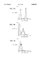
- FIGS. 1A-1C Transfection of the 183c16 cDNA leads to cell surface expression of the monoclonal antibody GL183.
- 183 refers to the molecule recognized by the GL183 mab. FACS analysis of 183 expression at the surface with the 183 cDNA is shown.
- FIG. 1A shows the cell surface expression of 183 (open area) on NK3.3 relative to control staining without primary Ab (Antibody) (filled area).
- the human T cell line Jurkat does not express 183 (FIG. 1B, filled area), nor does a human B-LCL (FIG. 1 C, filled are).
- Stable transfection of these two cell lines with the 183c16 cDNA in the eukaryotic expression vector RSV.5(gpt) results in surface expression of 183 (FIGS. 1B and 1C, open areas).
- the present invention provides a purified and isolated nucleic acid molecule encoding the p58 receptor protein.
- the nucleic acid molecule includes all nucleic acid sequences which encode for the p58 receptor including all degenerate forms.
- the term "nucleic acid” includes but is not limited to DNA, RNA or cDNA.
- the p58 receptor protein has the nucleotide sequence contained in SEQ ID NO:1, or a sequence which is substantially homologous thereto.
- substantially homologous refers to substantial correspondence between the nucleic acid sequences of various members of the p58 receptor protein family.
- “Substantially homologous” means at least 50% identical, preferably about 65% identical, and most preferably, about 70-90% identical.
- the nucleic acid is mammalian, and is preferably human or murine.
- the nucleic acid sequences for the additional members of the p58 receptor protein family may be isolated by screening libraries of cDNA clones derived from NK cell RNA with a probe corresponding to the sequence contained in SEQ ID NO:2 using standard DNA hybridization techniques. It is expected that the DNA sequences encoding the different members of the p58 receptor protein family will exhibit a level of similarity that clearly exceeds the 55% identity required for cross-hybridization. This is because analogous families of related genes encoding other receptors usually display similarities with about 70 to 90% identity among different members (e.g. HLA class I genes, HLA class II genes, genes encoding different T cell receptors, genes encoding different immunoglobulin molecules).
- genomic clones encoding the p58 receptor family also may be isolated by screening human genomic libraries with a probe corresponding to the sequence contained in SEQ ID NO:2 using standard DNA hybridization techniques.
- mice may be isolated by cross-hybridization techniques as long as some portion of the probe above has at least 55% nucleotide sequence identity with the mouse genes. If cross-hybridization is not successful, oligonucleotide primers based on the same sequence may be used to isolate the mouse genes using PCR.
- the mouse genes may be used to generate "knock-out" mice with a defect in one or several of the genes encoding the mouse counterpart of the human p58 receptor protein. Such knock-out mice would be extremely valuable tools to study the role of p58 receptors in immune responses.
- the present invention also provides a vector comprising the nucleic acid molecule encoding the p58 receptor protein, as described above.
- the vector is an expression vector which comprises at least one expression control element operationally linked to the nucleic acid sequence encoding the p58 receptor.
- the expression control elements are inserted in the vector to control and regulate the expression of the nucleic acid sequence, and are known in the art.
- the vectors of the present invention may be constructed by known techniques (see U.S. Pat. Nos. 4,704,362, 4,366,246, 4,425,437, 4,356,270, and 4,571,421), using commercially available plasmids.
- the present invention also provides a host cell stably transformed or transfected with the vector above.
- the host cells may be prokaryotic such as E. coli, or eukaryotic such as animal, plant, insect and yeast cells.
- the host cells are stably transformed or transfected by known procedures (see U.S. Pat. Nos. 4,704,362, 4,366,246, 4,425,437, 4,356,270, and 4,571,421).
- amplification protocols e.g. methotrexate selection with dhfr vectors
- Other expression systems based on virus vectors may be used for extremely high level expression, but only transiently (e.g. baculovirus, Sindbis virus).
- the host cells may be screened for clones which produce the recombinant p58 receptor protein by known procedures such as Coomassie blue staining and Western blotting.
- modified forms of the p58 receptor protein may be engineered and produced. Truncated or mutated forms can be produced to define functional domains of the p58 receptor protein. Secreted forms can be engineered for the production of soluble receptors.
- the present invention also provides the p58 receptor protein produced by the host cell above.
- the recombinant protein expressed by the host cells may be obtained as a crude lysate or can be purified by standard protein purification procedures known in the art such as differential precipitation, molecular sieve chromatography, ion-exchange chromatography, isoelectric focusing, gel electrophoresis, affinity chromatography, immunoaffinity chromatography, and the like.
- the p58 receptor protein has the amino acid sequence contained in SEQ ID NO:2, or is a biologically active analogue thereof.
- biologically active analogue thereof refers to proteins having substantially homologous sequences to the sequence contained in SEQ ID NO:2, and possess similar activity as that protein.
- the isolated and purified p58 receptor protein of the present invention includes all members of the p58 receptor protein family.
- the present invention also provides a method for detecting nucleic acid encoding the p58 receptor protein in a biological sample utilizing as probes the nucleic acid above, or portions thereof, labeled with detectable markers.
- the method comprises the steps of: (a) contacting the probe with a biological sample under conditions permitting a complex to be formed between the probe and nucleic acid present in the sample; and (b) detecting the formation of the complex.
- the biological samples include mammalian tissue or cell samples.
- the nucleic acid may be extracted from the biological samples by known procedures. Specifically, the cells may be lysed using an enzyme such as proteinase K, in the presence of detergents such as sodium dodecyl sulfate (SDS), NP40, or Tween 20. If the nucleic acid is genomic DNA, it may be extracted using known techniques such as phenol/chloroform extraction, or other procedures (see U.S. Pat. Nos. 4,900,677 and 5,047,345). Alternatively, the DNA may be isolated using one of the commercially available kits such as the Oncor Genomic DNA isolation kit.
- RNA may be extracted using various known procedures such as guanidinium thiocyanate followed by centrifugation in cesium chloride (Sambrook, J., Fritsch, E. F., and Maniatis, T. "Molecular Cloning, A Laboratory Manual,” second edition, Cold Spring Harbor Laboratory Press, pp. 7.0-7.25 (1989)).
- the formation of the complex may be detected using various conventional techniques known in the art including but not limited to Northern blotting, Southern blotting, dot and slot hybridization, S1 nuclease assay, ribonuclease protection assay, and filter hybridization (Southern, E. M. J. Mol. Biol. 98:503 (1975); Chirgwin, J. M., et al. Biochemistry 18: 5294-5299 (1979); Kafatos, et al. Nuc. Acids. Res. 7: 1541 (1979); Thomas, P. S. Proc. Natl. Acad. Sci. 77: 5201 (1980); White, B. A. and F. C. Bancroft J. Bio. Chem.
- the probe is labeled with a detectable marker which includes but is not limited to fluorescence, enzyme or radiolabeled markers such as 32 P and biotin.
- the marker is 32 P.
- the probe is labeled by known procedures such as phosphorylation with bacteriophage T4 polynucleotide kinase (Sambrook, J., Fritsch, E. F., and Maniatis, T. "Molecular Cloning, A Laboratory Manual," second edition, Cold Spring Harbor Laboratory Press, pp. 11.31-11.32 (1989)).
- probes may be designed to detect all members of the p58 family, or may be unique in detecting only one or more specific members of the p58 family.
- the present invention also provides a method for detecting nucleic acid encoding the p58 receptor in a biological sample using standard PCR procedures.
- the method comprises the steps of: (a) contacting nucleic acid from the biological sample with a sense and an antisense primer prepared from the sequence of the present invention under conditions permitting PCR amplification to occur; and (b) detecting amplification of the nucleic acid from the biological sample.
- Polymerase chain reaction is performed by methods and conditions disclosed in U.S. Pat. Nos. 4,683,202 and 4,683,195 and in Perkin Elmer Cetus PCR kit protocols.
- the DNA polymerase, deoxyribonucleotide triphosphates (dNTPS) (e.g. dATP, dCTP, dTTP, and dGTP), and amplification buffer (e.g. glycerol, tris-hydrochloric acid, potassium chloride, Tween 20, and magnesium chloride) are readily commercially available (Perkin Elmer Cetus).
- the polymerase chain reaction may be performed as many cycles as desired.
- Reverse transcription (RT) of mRNA and RT-PCR are performed by methods described in commercially available kits such as the RT and RT-PCR kits (Perkin Elmer Cetus).
- the sense and antisense primers for use in PCR and RT-PCR may be prepared from the nucleic acid sequence above using automated instruments sold by a variety of manufacturers or may be commercially synthesized.
- primers may be designed to detect all members of the p58 family, or may be unique in detecting only one or more specific members of the p58 family.
- the present invention also provides a method for detecting a p58 receptor protein in a biological sample using particular reagents which specifically react with the p58 receptor protein.
- the term "reagent” includes monoclonal or polyclonal antibodies.
- Exemplary antibody molecules for use in the detection methods of the present invention are intact immunoglobulin molecules, substantially intact immunoglobulin molecules, or those portions of an immunoglobulin molecule that contain the antigen binding site, including those portions known in the art as F(ab), F(ab'), F(ab') 2 , and F(v).
- antibodies specific to each different member of the p58 receptor protein family, or portions thereof may be used to detect expression of their corresponding p58 receptor protein family member.
- the complete protein or unique portions thereof of the particular members of the p58 family may be utilized as an immunogen, with or without a carrier molecule, to produce these antibodies.
- carrier molecules includes human albumin, bovine albumin, keyhole limpet hemo-cyanin, and the like.
- Polyclonal and monoclonal antibodies, or their fragments may be produced by methods known in the art, or by genetic engineering (Kohler and Milstein (1975) Nature 256: 495-497; Campbell “Monoclonal Antibody Technology,” the “Production and Characterization of Rodent and Human Hybridomas” in Burdon, et al. (eds.) (1985) "Laboratory Techniques in Biochemistry and Molecular Biology,” Volume 13, Elsevier Science Publishers, Amsterdam).
- the antibodies are used in immunoassays to detect the p58 receptor protein in biological samples.
- the method comprises the steps of: (a) contacting the biological sample with an antibody; and (b) detecting the formation of a complex between the protein and the antibody.
- the immunoassays are used to detect different members of the p58 receptor protein family in the biological sample using their respective antibodies.
- Suitable immunoassays include but are not limited to radioimmunoassay, Western blot assay, immunofluorescent assay, enzyme immunoassay, chemiluminescent assay, and immunohistochemical assay. These assays may be performed using procedures well known in the art. (In “Principles and Practice of Immunoassay” (1991) Christopher P. Price and David J. Neoman (eds), Stockton Press, New York, N.Y.; Ausubel, et al. (eds) (1991) in "Current Protocols in Molecular Biology” John Wiley and Sons, New York, N.Y.).
- Target cells that do not express a cell surface ligand able to interact with the p58 receptor expressed by a particular NK cell are likely to be lysed by that NK cell.
- This deadly fate results from the fact that the p58 receptor on NK cells serves to provide a negative signal to the NK cell and thus stop the lytic machinery of the NK cell.
- Rejection of bone marrow transplants, and possibly autoimmune reactions, are mediated by NK cells when the negative signal fails to be delivered to the NK cells. It is therefore very important, and potentially very useful clinically, to determine exactly which molecules on target cells serve as ligands for the p58 receptor proteins and as protective elements on target cells that would otherwise be killed.
- the present invention also provides a method for identifying a ligand capable of binding to the p58 receptor protein.
- the method comprises the steps of: (a) contacting a p58 receptor protein or cells which express the p58 receptor protein with a candidate ligand; and (b) detecting the formation of a complex between the p58 receptor protein and the ligand.
- the method comprises the steps of: (a) contacting a p58 receptor protein with cells transfected with cDNA encoding a candidate ligand; and (b) detecting the formation of a complex between the p58 receptor protein and the ligand expressed by the cells.
- ligand refers to any protein or proteins that may interact with the p58 receptor protein. Said ligand may be soluble or membrane bound. The ligand may be a naturally occurring protein, or synthetically or recombinantly produced. The ligand may also be a nonprotein molecule that acts as a ligand when it interacts with the p58 receptor. In the preferred embodiment, the ligands are HLA class I molecules and their isotypes. Interactions between the ligand and receptor include but are not limited to covalent or non-covalent interactions. The receptor binding domain is any region of the receptor that interacts directly or indirectly with the ligand.
- the p58 receptor protein is preferably recombinant p58 receptor protein, and most preferably is soluble. It is within the confines of the present invention that the soluble recombinant molecules may be produced as fusion proteins with a secreted form of immunoglobulin. This leads to efficient expression of a secreted soluble form and also to detection using well-established antibodies specific for the immunoglobulin molecules.
- the soluble p58-immunoglobulin fusion proteins may be used as reagents for analysis by flow cytometry to test for the presence of ligands on different cells.
- Cells stably transfected with a complete cDNA encoding a p58 receptor protein may be used to test binding of soluble ligands or binding of immobilized ligands.
- ligands may be attached to a support such as a latex bead. These assays are known in the art as rosette formation assays.
- the present invention also provides a method for screening for drugs capable of acting as agonists or antagonists to the p58 receptor protein which comprises the steps of: (a) contacting cells which express the p58 receptor protein with candidate drugs; and (b) evaluating the biological activity mediated by said contact.
- drug includes but is not limited to proteins, peptides, agents purified from conditioned cell medium, organic molecules, inorganic molecules, antibodies oligonucleotides, or analogs of the ligand described above.
- the drug may be naturally occurring or synthetically or recombinantly produced.
- Specific agonists of the p58 receptor protein would be extremely useful in preventing unwanted NK-mediated lysis of target cells (e.g. in bone marrow transplantation).
- Specific antagonists would be useful for boosting NK cell responses to unwanted pathogens such as cancer cells.
- biological activity means the triggering of the p58 receptor protein to transmit a negative signal to the cell which expresses it. This is evaluated by tranfecting the cDNA encoding the p58 receptor protein with known reporter gene constructs, and measuring the triggering effect of specific drugs on the p58 receptor protein using known calorimetric assays (e.g. beta-galactosidase) or luminometry (e.g. luciferase).
- calorimetric assays e.g. beta-galactosidase
- luminometry e.g. luciferase
- the present invention also provides an agonist or antagonist to a p58 receptor protein identified using the methods above, as well as uses thereof.
- the present invention provides a method for preventing immunosuppressive rejection of a transplant in a subject receiving the transplant which comprises administering to the subject an amount of an agonist to a p58 receptor protein effective to prevent immunosuppressive rejection of the transplant.
- transplant refers to any cell, tissue or organ which may be suspectable to immunosuppressive rejection when transplanted into a patient.
- the transplant is bone marrow.
- the present invention also provides a method for treating a neoplastic cell growth in a subject in need of such treatment which comprises administering an amount of an antagonist to a p58 receptor protein effective to treat the neoplastic cell growth.
- neoplastic cell growth includes any of the known cancers such as cancers of the breast, lung, brain, groin, and the like.
- treatment includes the partial or total inhibition of neoplastic cell growth, as well as the partial or total destruction of the neoplastic cells.
- subject includes a human or animal subject diagnosed as having cancer. It is within the confines of the present invention that the antagonist may be administered in conjunction with an antibody which specifically recognizes the tumor.
- administration for both methods above may be affected by means known to those skilled in the art such as oral, rectal, topical intravenous, subcutaneous, intramuscular, or intraperitoneal routes of administration.
- the dosage form and amount for each method can be readily established by reference to known immunosupressive treatments and antineoplastic treatments, respectively.
- the actual dose will depend upon the route of administration, the pharmacokinetic properties of the individual treated, as well as the results desired.
- chimeric receptor proteins consisting of extracellular domain of one receptor and the intracellular domain of a different receptor indicate that a common mechanism of signal transduction through the cell membrane is shared by different growth factor receptors (Riedel, et al. Nature 324: 68-70 (1986); Riedel, et al. EMBO J. 8: 2943-2954 (1989)).
- the specificity of the signal transmitted by the receptors resides in the intracellular domain.
- Chimeras between the extracellular domain of a known ligand and intracellular domain of an orphan receptor can be activated by the known ligand, yet the biological function elicited is similar to the receptor with the intracellular domain.
- chimeric receptors including insulin receptor (IR)/epidermal growth factor receptor (EGFR) (Riedel, et al. Nature 324: 68-70 (1986), EGFR/IR (Riedel, et al. EMBO J. 8: 2943-2954 (1989)), platelet-derived growth factor receptor beta (PDGFR-beta)/fibroblast growth factor receptor-1 (FGFR-1) (Mares, et al. Growth Factors 6:93-101 (1992)) have been constructed and elicited proper responses.
- IR insulin receptor
- EGFR epidermatitis
- the present invention also provides a chimeric protein comprising (a) the cytoplasmic region of a p58 receptor protein which transmits a dominant negative signal to a cell expressing the p58 receptor protein; and (b) a transmembrane domain and an extracellular domain of a known receptor.
- This chimeric protein may be constructed as described in the publications above. The chimeric protein is useful for studying the recognition of target molecules mediated by the p58 receptor protein family.
- the mAb GL183 recognizes a surface antigen (183 antigen, 183 Ag) expressed on a subset of freshly isolated human NK cells and on some NK clones (Moretta, et al. J. Exp. Med. 171: 695-714 (1990); Moretta, et al. Advances in Immunology 55: 341-380 (1994)).
- the difficulty of generating and growing human NK clones has precluded the isolation of large amounts of 183 antigen from these cells necessary for sequencing the 183 antigen. Purification also has been hindered because most known NK cells express the Fc receptor (CD16), which also has a similar isoelectric point and molecular mass as the 183 antigen.
- NK3.3 human NK cell line NK3.3 obtained from Jackie Kornbluth (Kornbluth, J., et al. J. Immunol. 129:2831 (1982)) expresses the 183 antigen at the cell surface, and also express low levels of the Fc receptor (CD16). Unlike previously identified clones, the NK3.3 cell line also grows well under long term conditions, and can be readily expanded.
- NK3.3 and NK clones were surface iodinated and lysed in lysis buffer containing Triton and protease inhibitors. Immunoprecipitation using GL183 and control antibodies, and analysis of the precipitates on SDS-PAGE indicated that the 183 Ag derived from the two sources were very similar.
- GL183 was purified from ascites and coupled to Sepharose CL-2B. These GL183-Sepharose beads were used to immunoprecipitate 183 Ag from detergent lysates of surface iodinated NK3.3. The immuno-precipitates were then incubated in an elution buffer at pH 11.2 to test whether the 183 Ag could be eluted from the beads under those conditions. The eluted material was analyzed on SDS gels followed by autoradiography.
- the lysate was therefore passed over four sequential columns containing the following beads: Unconjugated Sepharose CL-2B, Mouse IgG-agarose, L243-Sepharose-CL-2B and finally GL183-Sepharose-CL-2B.
- the first two columns were strictly preclearing columns.
- small amounts of radio-iodinated NK3.3 was used as a tracer.
- a lysate prepared from 2 ⁇ 10 9 NK3.3 cells and spiked with a second lysate prepared from 6 ⁇ 10 7 surface-iodinated NK3.3 was passed over the 4 columns described above connected to each other in series. The columns were disconnected and the L243 and the GL183 columns were washed with wash buffer in parallel. The bound proteins were eluted with the elution buffer at pH 11.2 and 50 drops/fraction (about 500 ⁇ l/fraction) were collected. Each fraction was immediately neutralized by addition of dilute acetic acid. 2 ⁇ l of each fraction was counted in a gamma counter to identify fractions containing peak radioactivity. The material in those fractions containing the most radioactivity was further analyzed.
- wash buffers were evaluated during the affinity-purification step to see if conditions could be found where these contaminants would be removed during the column washes without removing the 183 Ag.
- the following wash procedure was adopted: After washing overnight with 0,1% Triton X-100, 10 mM Tris-HCL, pH 7.5, 150 mM NaCl, 0,02% NaN 3 the columns were washed with 0.1% C12E9, 10 mM Tris-HCL pH 9.0, 500 mM NaCl followed by a final wash with 0.1% C 12 E 9 , 10 mM Tris-HCL, pH 7.5, 150 mM NaCl. The wash at pH 9.0 was found to remove significant amounts of contaminants with only small decreases in yield of 183 Ag.
- Affinity-purified material from about 4 ⁇ 10 9 cells was subjected to SDS-gel separation and transfer to Problot as described above. After cutting out the region of the membrane containing 183 Ag, it was subjected to digestion with trypsin. The digest was separated by reverse-phase HPLC using a vydac C-18 column. Aliquots of material from the prominent peaks were analyzed by Matrix-Assisted Laser Desorption Mass Spectrometry and fractions containing one main peak were subjected to Edman degradation. The amino acid sequences of eight tryptic peptides were obtained. Computer searches revealed that these sequences were previously unknown.
- 183N-FOR32 5' GGI CCI (C/T)TI GTN AA(A/G) TCN GAA GAG AC 3' (SEQ ID NO:3)
- 183P20-BACK32 5' IC(G/T) (A/G)CT IA(A/G) (A/G)TG (A/G)TA CAT (A/G)TC ATA 3' (SEQ ID NO:4)
- 183P20-BACK32B 5' NC(G/T) (A/G)CT NA(A/G) (A/G)TG (A/G)TA CAT (A/G)TC ATA 3' (SEQ ID NO:5)
- 183N-FOR32-B 5' GGN CCN (C/T)TN GTN AA(A/G) TCN GAA GAG AC 3' (SEQ ID NO:6)
- the cDNA library was constructed using a kit from Gibco BRL and was size selected as follows.
- the cDNA library was amplified, and purified double-stranded plasmid was linearized by digestion with NotI and separated by agarose gel electrophoresis. The gel was cut in 21 sections, each containing DNA of a different size, i.e. with inserts of different length.
- the DNA was purified from the agarose and an aliquot of each fraction was run on an agarose gel. It was transferred to Genescreen, and hybridization was done using the 32 P-labelled 183 PCR amplification product as probe. This identified the fraction containing inserts corresponding to the PCR amplification product.
- This fraction was re-circularized with DNA ligase and transformed into E. coli.
- the bacteria were plated on LB plates and after lifting onto Genescreen discs they were screened with the probed described above.
- Several positive clones were isolated, and 6 were further characterized by sequencing. The 6 clones turned out to be identical in their sequence.
- the insert from one of the clones was subcloned into the eukaryotic expression vector RSV.5(gpt).
- This construct was transfected into the human T cell line Jurkat and into a human B-LCL by electroporation.
- Stable transfectants were selected by culture in medium containing Mycophenolic acid and Xanthine and were screened for surface expression of 183 Ag by FACS analysis using the GL183 as primary antibody followed by FITC-conjugated rabbit anti-mouse IgG (see FIGS. 1B-1C). The results show that the p58 cDNA directs the surface expression of the 183 Ag in transfected cells.
Landscapes
- Health & Medical Sciences (AREA)
- Chemical & Material Sciences (AREA)
- Life Sciences & Earth Sciences (AREA)
- Immunology (AREA)
- Organic Chemistry (AREA)
- Biochemistry (AREA)
- Gastroenterology & Hepatology (AREA)
- Zoology (AREA)
- Toxicology (AREA)
- Biophysics (AREA)
- General Health & Medical Sciences (AREA)
- Genetics & Genomics (AREA)
- Medicinal Chemistry (AREA)
- Molecular Biology (AREA)
- Proteomics, Peptides & Aminoacids (AREA)
- Cell Biology (AREA)
- Peptides Or Proteins (AREA)
Abstract
The present invention provides a purified and isolated nucleic acid molecule encoding the p58 receptor protein. The present invention also provides vectors encoding this nucleic acid molecule, a host cell stably transformed or transfected with the vector, as well as the p58 receptor protein produced by this host cell. The present invention further provides methods for detecting nucleic acid encoding the p58 receptor as well as the p58 receptor protein in biological samples, methods for identifying a ligand capable of binding to the p58 receptor protein, and methods for screening for drugs capable of acting as agonists or antagonists to the p58 receptor protein. The present invention also provides the agonists and antagonists identified by the screening methods, as well as their use. Lastly, the present invention provides chimeric protein comprising the cytoplasmic region of a p58 receptor protein.
Description
Natural killer (NK) cells are important in the immune defense against tumors, viruses and intracellular parasites. NK cells also may be involved in the rejection of bone marrow transplants, and other transplants. The ability to intervene with the immune function of the NK cells, by either boosting NK cell responses to pathogens, or by lowering or stopping unwanted NK cell responses, is a long recognized need.
The p58 receptor of NK cells is a family of cell surface molecules with a molecular mass of about 58,000 which is involved in the recognition of target cells by NK cells. The p58 receptor contributes to the specificity of NK cells in their recognition of major histocompatibility complex (MHC) class I molecules on target cells. The specific recognition of MHC molecules mediated by the p58 receptor protects the target cell from lysis by the NK cell. Conversely, absence of the appropriate MHC class I molecule on target cells leads to NK-mediated lysis.
Prior to the present invention, however, no members of the p58 family have been purified sufficiently to permit their isolation and characterization. This is because it is impossible to grow large enough populations of the NK cells to permit isolation of sufficient amounts of p58 receptor protein for sequencing. Furthermore, the known NK cell lines express high levels of Fc receptor which could interfere with immunoaffinity purifications utilizing monoclonal antibodies directed to the p58 receptor protein, since these antibodies also bind to the Fc receptor.
In particular, Moretta, et al. (1990) described the use of a monoclonal antibody designated GL183 to immunoprecipitate either a 58 Kd band or a broad 55-58 Kd band from NK clones which had been labeled with 125 I (Moretta, et al. J. Exp. Med. 171: 695-714 (1990); Moretta, et al. Advances in Immunology 55: 341-380 (1994)). However, Moretta, et al. did not sufficiently purify the alleged p58 receptor protein to permit its isolation and characterization, and could not have done so utilizing the particular NK cells described therein, or known NK cells.
This is because NK cells described in Moretta, et al., as most NK cells, cannot be grown to large enough populations to permit isolation of sufficient amounts of p58 receptor protein for sequencing. Specifically, about 1×10¹⁰ NK cells would have to be obtained in order to purify an amount of pure p58 protein sufficient for sequencing. However, NK cells cannot be expanded much beyond 1×10⁷ cells since they do not grow well in culture.
Moreover, the NK cell lines described in Moretta, et al. express high levels of Fc receptor which could interfere with immunoaffinity purifications utilizing the GL183 monoclonal antibody, since this antibody also binds to the Fc receptor. Furthermore, the inventors of the present invention found that the p58 and Fc receptors (CD16) have very similar isoelectric points and relative masses, which could make their separation difficult.
The inventors of the present invention overcame the potential problems in the art by employing a long term NK cell line designated NK 3.3 which, unlike previously identified NK clones, grows well under long-term culture conditions, can be readily expanded to sufficient numbers, and expresses very low levels of the Fc receptor. Accordingly, the inventors of the present invention were the first to purify a member of the p58 receptor family, characterize it, and subsequently clone its DNA.
The present invention provides a purified and isolated nucleic acid molecule encoding the p58 receptor protein. The present invention also provides vectors encoding this nucleic acid molecule, a host cell stably transformed or transfected with the vector, as well as the p58 receptor protein produced by this host cell.
The present invention also provides a method for detecting nucleic acid encoding the p58 receptor in a biological sample which comprises the steps of: (a) contacting nucleic acid from the biological sample with the probe made from nucleic acid encoding the p58 receptor protein under conditions permitting a complex to be formed between the probe and nucleic acid presence in the sample; and (b) detecting the formation of the complex.
The present invention further provides a method for detecting nucleic acid encoding the p58 receptor in a biological sample which comprises the steps of: (a) contacting nucleic acid from the biological sample with a sense and an antisense primer prepared from nucleic acid encoding the p58 receptor protein under conditions permitting PCR amplification to occur; and (b) detecting amplification of the nucleic acid from the biological sample.
Moreover, the present invention provides a method for detecting a p58 receptor protein in a biological sample which comprises the steps of: (a) contacting the biological sample with a reagent which specifically reacts with a p58 receptor protein; and (b) detecting the formation of a complex between the protein and the reagent.
The present invention also provides a method for identifying a ligand capable of binding to the p58 receptor protein which comprises the steps of: (a) contacting a p58 receptor protein or cells which express the p58 receptor protein with a candidate ligand; and (b) detecting the formation of a complex between the p58 receptor protein and the ligand.
In addition, the present invention provides a method for identifying a ligand capable of binding to the p58 receptor protein which comprises the steps of: (a) contacting a p58 receptor protein with cells transfected with cDNA encoding a candidate ligand; and (b) detecting the formation of a complex between the p58 receptor protein and the ligand expressed by the cells.
Also provided by the present invention is a method for screening for drugs capable of acting as agonists or antagonists to the p58 receptor protein which comprises the steps of: (a) contacting cells which express the p58 receptor protein with candidate drugs; and (b) evaluating the biological activity mediated by said contact.
The present invention also provides the agonists and antagonists identified by the method above as well as uses of these agonists and antagonists.
In particular, the present invention provides a method for preventing immunosuppressive rejection of a transplant in a subject receiving the transplant which comprises administering to the subject an amount of an agonist to a p58 receptor protein effective to prevent immunosuppressive rejection of the transplant.
The present invention also provides a method for treating a neoplastic cell growth in a subject in need of such treatment which comprises administering an amount of an antagonist to a p58 receptor protein effective to treat the neoplastic cell growth.
Lastly, the present invention provides a chimeric protein comprising (a) the cytoplasmic region of a p58 receptor protein which transmits a dominant negative signal to a cell expressing the p58 receptor protein; and (b) a transmembrane domain and an extracellular domain of a known receptor.
FIGS. 1A-1C. Transfection of the 183c16 cDNA leads to cell surface expression of the monoclonal antibody GL183. 183 refers to the molecule recognized by the GL183 mab. FACS analysis of 183 expression at the surface with the 183 cDNA is shown. FIG. 1A shows the cell surface expression of 183 (open area) on NK3.3 relative to control staining without primary Ab (Antibody) (filled area). The human T cell line Jurkat does not express 183 (FIG. 1B, filled area), nor does a human B-LCL (FIG. 1 C, filled are). Stable transfection of these two cell lines with the 183c16 cDNA in the eukaryotic expression vector RSV.5(gpt) results in surface expression of 183 (FIGS. 1B and 1C, open areas).
The present invention provides a purified and isolated nucleic acid molecule encoding the p58 receptor protein. The nucleic acid molecule includes all nucleic acid sequences which encode for the p58 receptor including all degenerate forms. The term "nucleic acid" includes but is not limited to DNA, RNA or cDNA.
In the preferred embodiment, the p58 receptor protein has the nucleotide sequence contained in SEQ ID NO:1, or a sequence which is substantially homologous thereto. "Substantially homologous" as used herein refers to substantial correspondence between the nucleic acid sequences of various members of the p58 receptor protein family. "Substantially homologous" means at least 50% identical, preferably about 65% identical, and most preferably, about 70-90% identical. The nucleic acid is mammalian, and is preferably human or murine.
The nucleic acid sequences for the additional members of the p58 receptor protein family may be isolated by screening libraries of cDNA clones derived from NK cell RNA with a probe corresponding to the sequence contained in SEQ ID NO:2 using standard DNA hybridization techniques. It is expected that the DNA sequences encoding the different members of the p58 receptor protein family will exhibit a level of similarity that clearly exceeds the 55% identity required for cross-hybridization. This is because analogous families of related genes encoding other receptors usually display similarities with about 70 to 90% identity among different members (e.g. HLA class I genes, HLA class II genes, genes encoding different T cell receptors, genes encoding different immunoglobulin molecules).
Furthermore, genomic clones encoding the p58 receptor family also may be isolated by screening human genomic libraries with a probe corresponding to the sequence contained in SEQ ID NO:2 using standard DNA hybridization techniques.
In addition, the corresponding gene in mice may be isolated by cross-hybridization techniques as long as some portion of the probe above has at least 55% nucleotide sequence identity with the mouse genes. If cross-hybridization is not successful, oligonucleotide primers based on the same sequence may be used to isolate the mouse genes using PCR. The mouse genes may be used to generate "knock-out" mice with a defect in one or several of the genes encoding the mouse counterpart of the human p58 receptor protein. Such knock-out mice would be extremely valuable tools to study the role of p58 receptors in immune responses.
The present invention also provides a vector comprising the nucleic acid molecule encoding the p58 receptor protein, as described above. In the preferred embodiment, the vector is an expression vector which comprises at least one expression control element operationally linked to the nucleic acid sequence encoding the p58 receptor. The expression control elements are inserted in the vector to control and regulate the expression of the nucleic acid sequence, and are known in the art. The vectors of the present invention may be constructed by known techniques (see U.S. Pat. Nos. 4,704,362, 4,366,246, 4,425,437, 4,356,270, and 4,571,421), using commercially available plasmids.
The present invention also provides a host cell stably transformed or transfected with the vector above. The host cells may be prokaryotic such as E. coli, or eukaryotic such as animal, plant, insect and yeast cells. The host cells are stably transformed or transfected by known procedures (see U.S. Pat. Nos. 4,704,362, 4,366,246, 4,425,437, 4,356,270, and 4,571,421). In the particularly preferred embodiment, amplification protocols (e.g. methotrexate selection with dhfr vectors) may be used to obtain cells that produce high levels of recombinant proteins. Other expression systems based on virus vectors may be used for extremely high level expression, but only transiently (e.g. baculovirus, Sindbis virus). The host cells may be screened for clones which produce the recombinant p58 receptor protein by known procedures such as Coomassie blue staining and Western blotting.
It is also within the confines of the present invention that modified forms of the p58 receptor protein may be engineered and produced. Truncated or mutated forms can be produced to define functional domains of the p58 receptor protein. Secreted forms can be engineered for the production of soluble receptors.
The present invention also provides the p58 receptor protein produced by the host cell above. The recombinant protein expressed by the host cells may be obtained as a crude lysate or can be purified by standard protein purification procedures known in the art such as differential precipitation, molecular sieve chromatography, ion-exchange chromatography, isoelectric focusing, gel electrophoresis, affinity chromatography, immunoaffinity chromatography, and the like.
Preferably, the p58 receptor protein has the amino acid sequence contained in SEQ ID NO:2, or is a biologically active analogue thereof. The phrase "biologically active analogue thereof" refers to proteins having substantially homologous sequences to the sequence contained in SEQ ID NO:2, and possess similar activity as that protein. The isolated and purified p58 receptor protein of the present invention includes all members of the p58 receptor protein family.
The present invention also provides a method for detecting nucleic acid encoding the p58 receptor protein in a biological sample utilizing as probes the nucleic acid above, or portions thereof, labeled with detectable markers. In particular, the method comprises the steps of: (a) contacting the probe with a biological sample under conditions permitting a complex to be formed between the probe and nucleic acid present in the sample; and (b) detecting the formation of the complex.
The biological samples include mammalian tissue or cell samples. The nucleic acid may be extracted from the biological samples by known procedures. Specifically, the cells may be lysed using an enzyme such as proteinase K, in the presence of detergents such as sodium dodecyl sulfate (SDS), NP40, or Tween 20. If the nucleic acid is genomic DNA, it may be extracted using known techniques such as phenol/chloroform extraction, or other procedures (see U.S. Pat. Nos. 4,900,677 and 5,047,345). Alternatively, the DNA may be isolated using one of the commercially available kits such as the Oncor Genomic DNA isolation kit. RNA may be extracted using various known procedures such as guanidinium thiocyanate followed by centrifugation in cesium chloride (Sambrook, J., Fritsch, E. F., and Maniatis, T. "Molecular Cloning, A Laboratory Manual," second edition, Cold Spring Harbor Laboratory Press, pp. 7.0-7.25 (1989)).
The formation of the complex may be detected using various conventional techniques known in the art including but not limited to Northern blotting, Southern blotting, dot and slot hybridization, S1 nuclease assay, ribonuclease protection assay, and filter hybridization (Southern, E. M. J. Mol. Biol. 98:503 (1975); Chirgwin, J. M., et al. Biochemistry 18: 5294-5299 (1979); Kafatos, et al. Nuc. Acids. Res. 7: 1541 (1979); Thomas, P. S. Proc. Natl. Acad. Sci. 77: 5201 (1980); White, B. A. and F. C. Bancroft J. Bio. Chem. 257: 8569 (1982); Berk, A. J. and P. A. Sharp Cell 12: 721 (1977); Casey, J. and N. Davidson Nuc. Acids Res. 4:1539 (1977); Sambrook, J., Fritsch, E. F., and Maniatis, T. "Molecular Cloning, A Laboratory Manual," second edition, Cold Spring Harbor Laboratory Press, pp. 7.30-7.87 and 9.31-9.62 (1989)).
In the method above, the probe is labeled with a detectable marker which includes but is not limited to fluorescence, enzyme or radiolabeled markers such as 32 P and biotin. In the preferred embodiment, the marker is 32 P. The probe is labeled by known procedures such as phosphorylation with bacteriophage T4 polynucleotide kinase (Sambrook, J., Fritsch, E. F., and Maniatis, T. "Molecular Cloning, A Laboratory Manual," second edition, Cold Spring Harbor Laboratory Press, pp. 11.31-11.32 (1989)).
It is within the confines of the present invention that probes may be designed to detect all members of the p58 family, or may be unique in detecting only one or more specific members of the p58 family.
The present invention also provides a method for detecting nucleic acid encoding the p58 receptor in a biological sample using standard PCR procedures. In this regard, the method comprises the steps of: (a) contacting nucleic acid from the biological sample with a sense and an antisense primer prepared from the sequence of the present invention under conditions permitting PCR amplification to occur; and (b) detecting amplification of the nucleic acid from the biological sample.
Polymerase chain reaction is performed by methods and conditions disclosed in U.S. Pat. Nos. 4,683,202 and 4,683,195 and in Perkin Elmer Cetus PCR kit protocols. The DNA polymerase, deoxyribonucleotide triphosphates (dNTPS) (e.g. dATP, dCTP, dTTP, and dGTP), and amplification buffer (e.g. glycerol, tris-hydrochloric acid, potassium chloride, Tween 20, and magnesium chloride) are readily commercially available (Perkin Elmer Cetus). The polymerase chain reaction may be performed as many cycles as desired. Reverse transcription (RT) of mRNA and RT-PCR are performed by methods described in commercially available kits such as the RT and RT-PCR kits (Perkin Elmer Cetus).
The sense and antisense primers for use in PCR and RT-PCR may be prepared from the nucleic acid sequence above using automated instruments sold by a variety of manufacturers or may be commercially synthesized.
It is within the confines of the present invention that primers may be designed to detect all members of the p58 family, or may be unique in detecting only one or more specific members of the p58 family.
The present invention also provides a method for detecting a p58 receptor protein in a biological sample using particular reagents which specifically react with the p58 receptor protein.
The term "reagent" includes monoclonal or polyclonal antibodies. Exemplary antibody molecules for use in the detection methods of the present invention are intact immunoglobulin molecules, substantially intact immunoglobulin molecules, or those portions of an immunoglobulin molecule that contain the antigen binding site, including those portions known in the art as F(ab), F(ab'), F(ab')2, and F(v). In the preferred embodiment, antibodies specific to each different member of the p58 receptor protein family, or portions thereof, may be used to detect expression of their corresponding p58 receptor protein family member. The complete protein or unique portions thereof of the particular members of the p58 family may be utilized as an immunogen, with or without a carrier molecule, to produce these antibodies. Examples of carrier molecules includes human albumin, bovine albumin, keyhole limpet hemo-cyanin, and the like.
Polyclonal and monoclonal antibodies, or their fragments, may be produced by methods known in the art, or by genetic engineering (Kohler and Milstein (1975) Nature 256: 495-497; Campbell "Monoclonal Antibody Technology," the "Production and Characterization of Rodent and Human Hybridomas" in Burdon, et al. (eds.) (1985) "Laboratory Techniques in Biochemistry and Molecular Biology," Volume 13, Elsevier Science Publishers, Amsterdam).
The antibodies are used in immunoassays to detect the p58 receptor protein in biological samples. In particular, the method comprises the steps of: (a) contacting the biological sample with an antibody; and (b) detecting the formation of a complex between the protein and the antibody. In the particularly preferred embodiment, the immunoassays are used to detect different members of the p58 receptor protein family in the biological sample using their respective antibodies.
Suitable immunoassays include but are not limited to radioimmunoassay, Western blot assay, immunofluorescent assay, enzyme immunoassay, chemiluminescent assay, and immunohistochemical assay. These assays may be performed using procedures well known in the art. (In "Principles and Practice of Immunoassay" (1991) Christopher P. Price and David J. Neoman (eds), Stockton Press, New York, N.Y.; Ausubel, et al. (eds) (1991) in "Current Protocols in Molecular Biology" John Wiley and Sons, New York, N.Y.).
Target cells that do not express a cell surface ligand able to interact with the p58 receptor expressed by a particular NK cell are likely to be lysed by that NK cell. This deadly fate results from the fact that the p58 receptor on NK cells serves to provide a negative signal to the NK cell and thus stop the lytic machinery of the NK cell. Rejection of bone marrow transplants, and possibly autoimmune reactions, are mediated by NK cells when the negative signal fails to be delivered to the NK cells. It is therefore very important, and potentially very useful clinically, to determine exactly which molecules on target cells serve as ligands for the p58 receptor proteins and as protective elements on target cells that would otherwise be killed.
Therefore, the present invention also provides a method for identifying a ligand capable of binding to the p58 receptor protein. In one embodiment, the method comprises the steps of: (a) contacting a p58 receptor protein or cells which express the p58 receptor protein with a candidate ligand; and (b) detecting the formation of a complex between the p58 receptor protein and the ligand. In another embodiment, the method comprises the steps of: (a) contacting a p58 receptor protein with cells transfected with cDNA encoding a candidate ligand; and (b) detecting the formation of a complex between the p58 receptor protein and the ligand expressed by the cells.
The term "ligand" refers to any protein or proteins that may interact with the p58 receptor protein. Said ligand may be soluble or membrane bound. The ligand may be a naturally occurring protein, or synthetically or recombinantly produced. The ligand may also be a nonprotein molecule that acts as a ligand when it interacts with the p58 receptor. In the preferred embodiment, the ligands are HLA class I molecules and their isotypes. Interactions between the ligand and receptor include but are not limited to covalent or non-covalent interactions. The receptor binding domain is any region of the receptor that interacts directly or indirectly with the ligand.
The p58 receptor protein is preferably recombinant p58 receptor protein, and most preferably is soluble. It is within the confines of the present invention that the soluble recombinant molecules may be produced as fusion proteins with a secreted form of immunoglobulin. This leads to efficient expression of a secreted soluble form and also to detection using well-established antibodies specific for the immunoglobulin molecules. The soluble p58-immunoglobulin fusion proteins may be used as reagents for analysis by flow cytometry to test for the presence of ligands on different cells.
Cells stably transfected with a complete cDNA encoding a p58 receptor protein may be used to test binding of soluble ligands or binding of immobilized ligands. To increase valency, ligands may be attached to a support such as a latex bead. These assays are known in the art as rosette formation assays.
The present invention also provides a method for screening for drugs capable of acting as agonists or antagonists to the p58 receptor protein which comprises the steps of: (a) contacting cells which express the p58 receptor protein with candidate drugs; and (b) evaluating the biological activity mediated by said contact.
The term "drugs" includes but is not limited to proteins, peptides, agents purified from conditioned cell medium, organic molecules, inorganic molecules, antibodies oligonucleotides, or analogs of the ligand described above. The drug may be naturally occurring or synthetically or recombinantly produced. Specific agonists of the p58 receptor protein would be extremely useful in preventing unwanted NK-mediated lysis of target cells (e.g. in bone marrow transplantation). Specific antagonists would be useful for boosting NK cell responses to unwanted pathogens such as cancer cells.
The term "biological activity" as used herein means the triggering of the p58 receptor protein to transmit a negative signal to the cell which expresses it. This is evaluated by tranfecting the cDNA encoding the p58 receptor protein with known reporter gene constructs, and measuring the triggering effect of specific drugs on the p58 receptor protein using known calorimetric assays (e.g. beta-galactosidase) or luminometry (e.g. luciferase).
The present invention also provides an agonist or antagonist to a p58 receptor protein identified using the methods above, as well as uses thereof.
In particular, the present invention provides a method for preventing immunosuppressive rejection of a transplant in a subject receiving the transplant which comprises administering to the subject an amount of an agonist to a p58 receptor protein effective to prevent immunosuppressive rejection of the transplant.
As used herein, the term "transplant" refers to any cell, tissue or organ which may be suspectable to immunosuppressive rejection when transplanted into a patient. In the preferred embodiment, the transplant is bone marrow.
The present invention also provides a method for treating a neoplastic cell growth in a subject in need of such treatment which comprises administering an amount of an antagonist to a p58 receptor protein effective to treat the neoplastic cell growth.
The term "neoplastic cell growth" includes any of the known cancers such as cancers of the breast, lung, brain, groin, and the like. The term "treatment" includes the partial or total inhibition of neoplastic cell growth, as well as the partial or total destruction of the neoplastic cells. The term "subject" includes a human or animal subject diagnosed as having cancer. It is within the confines of the present invention that the antagonist may be administered in conjunction with an antibody which specifically recognizes the tumor.
The administration for both methods above may be affected by means known to those skilled in the art such as oral, rectal, topical intravenous, subcutaneous, intramuscular, or intraperitoneal routes of administration.
The dosage form and amount for each method can be readily established by reference to known immunosupressive treatments and antineoplastic treatments, respectively. The actual dose will depend upon the route of administration, the pharmacokinetic properties of the individual treated, as well as the results desired.
Studies of chimeric receptor proteins consisting of extracellular domain of one receptor and the intracellular domain of a different receptor indicate that a common mechanism of signal transduction through the cell membrane is shared by different growth factor receptors (Riedel, et al. Nature 324: 68-70 (1986); Riedel, et al. EMBO J. 8: 2943-2954 (1989)). The specificity of the signal transmitted by the receptors resides in the intracellular domain. Chimeras between the extracellular domain of a known ligand and intracellular domain of an orphan receptor can be activated by the known ligand, yet the biological function elicited is similar to the receptor with the intracellular domain. Numerous chimeric receptors including insulin receptor (IR)/epidermal growth factor receptor (EGFR) (Riedel, et al. Nature 324: 68-70 (1986), EGFR/IR (Riedel, et al. EMBO J. 8: 2943-2954 (1989)), platelet-derived growth factor receptor beta (PDGFR-beta)/fibroblast growth factor receptor-1 (FGFR-1) (Mares, et al. Growth Factors 6:93-101 (1992)) have been constructed and elicited proper responses.
Accordingly, the present invention also provides a chimeric protein comprising (a) the cytoplasmic region of a p58 receptor protein which transmits a dominant negative signal to a cell expressing the p58 receptor protein; and (b) a transmembrane domain and an extracellular domain of a known receptor. This chimeric protein may be constructed as described in the publications above. The chimeric protein is useful for studying the recognition of target molecules mediated by the p58 receptor protein family.
The present invention is described in the following Experimental Details section, which sets forth specific examples to aid in an understanding of the invention, and should not be construed to limit in any way the invention as defined in the claims which follow thereafter.
1. NK3.3 Cells Express 183 Ag
The mAb GL183 recognizes a surface antigen (183 antigen, 183 Ag) expressed on a subset of freshly isolated human NK cells and on some NK clones (Moretta, et al. J. Exp. Med. 171: 695-714 (1990); Moretta, et al. Advances in Immunology 55: 341-380 (1994)). The difficulty of generating and growing human NK clones has precluded the isolation of large amounts of 183 antigen from these cells necessary for sequencing the 183 antigen. Purification also has been hindered because most known NK cells express the Fc receptor (CD16), which also has a similar isoelectric point and molecular mass as the 183 antigen.
The inventors of the present invention have found by FACS analysis using GL183 that a human NK cell line NK3.3 obtained from Jackie Kornbluth (Kornbluth, J., et al. J. Immunol. 129:2831 (1982)) expresses the 183 antigen at the cell surface, and also express low levels of the Fc receptor (CD16). Unlike previously identified clones, the NK3.3 cell line also grows well under long term conditions, and can be readily expanded.
Analytical experiments were performed to biochemically characterize the 183 Ag expressed on NK3.3, in order to compare it to the Ag expressed on NK clones. NK3.3 and NK clones were surface iodinated and lysed in lysis buffer containing Triton and protease inhibitors. Immunoprecipitation using GL183 and control antibodies, and analysis of the precipitates on SDS-PAGE indicated that the 183 Ag derived from the two sources were very similar.
2. Immunoprecipitation
GL183 was purified from ascites and coupled to Sepharose CL-2B. These GL183-Sepharose beads were used to immunoprecipitate 183 Ag from detergent lysates of surface iodinated NK3.3. The immuno-precipitates were then incubated in an elution buffer at pH 11.2 to test whether the 183 Ag could be eluted from the beads under those conditions. The eluted material was analyzed on SDS gels followed by autoradiography.
It was found that incubation at pH 11.2 released a I-125 labelled protein of the expected size (MW). However, these experiments also showed that control beads, either unconjugated or conjugated to control Abs, immunoprecipitated large amounts of I-125 labelled proteins, which were also eluted from the beads under these conditions, indicating that extensive preclearing of the detergent lysates would be necessary.
The lysate was therefore passed over four sequential columns containing the following beads: Unconjugated Sepharose CL-2B, Mouse IgG-agarose, L243-Sepharose-CL-2B and finally GL183-Sepharose-CL-2B. The first two columns were strictly preclearing columns. The third, containing beads conjugated to L243, a mAb specific for HLA-DR, served as both a preclearing column and a positive control, since the NK3.3 cells also express HLA-DR. To monitor the purification, small amounts of radio-iodinated NK3.3 was used as a tracer.
A lysate prepared from 2×10⁹ NK3.3 cells and spiked with a second lysate prepared from 6×10⁷ surface-iodinated NK3.3 was passed over the 4 columns described above connected to each other in series. The columns were disconnected and the L243 and the GL183 columns were washed with wash buffer in parallel. The bound proteins were eluted with the elution buffer at pH 11.2 and 50 drops/fraction (about 500 μl/fraction) were collected. Each fraction was immediately neutralized by addition of dilute acetic acid. 2 μl of each fraction was counted in a gamma counter to identify fractions containing peak radioactivity. The material in those fractions containing the most radioactivity was further analyzed.
3. SDS-PAGE and Other Purification
60 μl of each fraction was run in a SDS-PAGE gel which was stained with Coomassie Blue to visualize total protein and then exposed to autoradiography film to detect radiolabeled proteins. A single radiolabeled protein migrating at 58 kD was eluted from the GL183 column and not from the L243 column. Although the autoradiography indicated that the GL183 had been purified to apparent homogeneity, Coomassie-staining revealed the presence of several additional non-radioactive proteins, mostly of lower molecular weight. These non-radioactive contaminants made it necessary to further purify the 183 Ag before subjecting it to sequencing.
Several fractions of affinity-column eluate were pooled, EtOH-precipitated and resuspended in sample buffer. The material was run in a preparative SDS-PAGE gel. The proteins were electrophoretically transferred to a ProBlot PVDF membrane which was stained with Coomassie blue. The band at about 58 kD was cut out and subjected to automated Edman degradation. This yielded an N-terminal sequence of 19 amino acids.
Computer searches of the translated Genbank and Swissprot databases showed that the sequence was unique. However, there was no evidence that the sequence was derived from the 183 Ag, rather than from a contaminant protein with the same electrophoretic mobility.
Other methods were evaluated for further purification. Reverse-phase HPLC was found to resolve the 183 Ag too well, in that it separated the material into several fractions. This fractionation procedure revealed that a contaminant with the same electrophoretic mobility was present in the immunoaffinity-purified 183 Ag. This molecule was not detected in the cell surface-iodinated sample and probably represents an intracellular protein.
Anion-exchange HPLC was also attempted but did not separate the 183 Ag from the contaminants. The 183 Ag also became refractive to N-terminal amino acid sequencing during these procedures.
Accordingly, different wash buffers were evaluated during the affinity-purification step to see if conditions could be found where these contaminants would be removed during the column washes without removing the 183 Ag. The following wash procedure was adopted: After washing overnight with 0,1% Triton X-100, 10 mM Tris-HCL, pH 7.5, 150 mM NaCl, 0,02% NaN3 the columns were washed with 0.1% C12E9, 10 mM Tris-HCL pH 9.0, 500 mM NaCl followed by a final wash with 0.1% C12 E9, 10 mM Tris-HCL, pH 7.5, 150 mM NaCl. The wash at pH 9.0 was found to remove significant amounts of contaminants with only small decreases in yield of 183 Ag.
Affinity-purified material from about 4×10⁹ cells was subjected to SDS-gel separation and transfer to Problot as described above. After cutting out the region of the membrane containing 183 Ag, it was subjected to digestion with trypsin. The digest was separated by reverse-phase HPLC using a vydac C-18 column. Aliquots of material from the prominent peaks were analyzed by Matrix-Assisted Laser Desorption Mass Spectrometry and fractions containing one main peak were subjected to Edman degradation. The amino acid sequences of eight tryptic peptides were obtained. Computer searches revealed that these sequences were previously unknown.
1. PCR Amplification
Based on the amino acid sequences of the N-terminus and internal tryptic peptides of 183, various oligonucleotide primers for PCR amplification were designed. The use of degenerate primers produced too much non-specific amplifications to be practical. Primers were therefore made with inosine substitutions at positions of maximum degeneracy (183N-FOR32; 183P20-BACK32). However, these inosine-containing products could not be cloned into bacterial plasmids for further analysis. Primers without inosines were therefore used in the re-amplification of the initial PCR product (183P20-BACK32B; 183N-FOR32-B). The best primers used are as follows:
183N-FOR32: 5' GGI CCI (C/T)TI GTN AA(A/G) TCN GAA GAG AC 3' (SEQ ID NO:3)
183P20-BACK32: 5' IC(G/T) (A/G)CT IA(A/G) (A/G)TG (A/G)TA CAT (A/G)TC ATA 3' (SEQ ID NO:4)
183P20-BACK32B: 5' NC(G/T) (A/G)CT NA(A/G) (A/G)TG (A/G)TA CAT (A/G)TC ATA 3' (SEQ ID NO:5)
183N-FOR32-B: 5' GGN CCN (C/T)TN GTN AA(A/G) TCN GAA GAG AC 3' (SEQ ID NO:6)
2. Construction and Screening of NK3.3 cDNA Library
Total RNA was isolated from NK3.3 by a scaled-up version of the method of Chomczynksi and Sacchi (Analytical Biochemistry 162:156-159 (1987)). Poly(A)+ selection was carried out by two passages of the total RNA over an Oligo(dT) column.
The cDNA library was constructed using a kit from Gibco BRL and was size selected as follows. The cDNA library was amplified, and purified double-stranded plasmid was linearized by digestion with NotI and separated by agarose gel electrophoresis. The gel was cut in 21 sections, each containing DNA of a different size, i.e. with inserts of different length. The DNA was purified from the agarose and an aliquot of each fraction was run on an agarose gel. It was transferred to Genescreen, and hybridization was done using the 32 P-labelled 183 PCR amplification product as probe. This identified the fraction containing inserts corresponding to the PCR amplification product.
This fraction was re-circularized with DNA ligase and transformed into E. coli. The bacteria were plated on LB plates and after lifting onto Genescreen discs they were screened with the probed described above. Several positive clones were isolated, and 6 were further characterized by sequencing. The 6 clones turned out to be identical in their sequence.
The insert from one of the clones was subcloned into the eukaryotic expression vector RSV.5(gpt). This construct was transfected into the human T cell line Jurkat and into a human B-LCL by electroporation. Stable transfectants were selected by culture in medium containing Mycophenolic acid and Xanthine and were screened for surface expression of 183 Ag by FACS analysis using the GL183 as primary antibody followed by FITC-conjugated rabbit anti-mouse IgG (see FIGS. 1B-1C). The results show that the p58 cDNA directs the surface expression of the 183 Ag in transfected cells.
All publications mentioned hereinabove are hereby incorporated by reference in their entirety.
While the foregoing invention has been described in some detail for purposes of clarity and understanding, it will be appreciated by one skilled in the art from a reading of the disclosure that various changes in form and detail can be made without departing from the true scope of the invention in the appended claims.
__________________________________________________________________________
SEQUENCE LISTING
(1) GENERAL INFORMATION:
(iii) NUMBER OF SEQUENCES: 6
(2) INFORMATION FOR SEQ ID NO:1:
(i) SEQUENCE CHARACTERISTICS:
(A) LENGTH: 1594 BASE PAIRS
(B) TYPE: NUCLEIC ACID
(C) STRANDEDNESS: SINGLE
(D) TOPOLOGY: UNKNOWN
(ii) MOLECULE TYPE:
(A) DESCRIPTION: cDNA to mRNA
(iii) HYPOTHETICAL: NO
(iv) ANTI-SENSE: NO
(vi) ORIGINAL SOURCE:
(A) ORGANISM: HUMAN
(C) INDIVIDUAL ISOLATE: NK 3.3 CELL
(xi) SEQUENCE DESCRIPTION:SEQ ID NO:1:
CCTGTCTGCACAGACAGCACCATGTCGCTCATGGTCGTCAGCATG45
MetSerLeuMetValValSerMet
15
GTGTGTGTTGGGTTCTTCTTGCTGCAGGGGGCCTGGCCACAT87
ValCysValGlyPhePheLeuLeuGlnGlyAlaTrpProHis
101520
GAGGGAGTCCACAGAAAACCTTCCCTCCTGGCCCACCCAGGT129
GluGlyValHisArgLysProSerLeuLeuAlaHisProGly
253035
CCCCTGGTGAAATCAGAAGAGACAGTCATCCTGCAATGTTGG171
ProLeuValLysSerGluGluThrValIleLeuGlnCysTrp
404550
TCAGATGTCAGGTTTCAGCACTTCCTTCTGCACAGAGAAGGG213
SerAspValArgPheGlnHisPheLeuLeuHisArgGluGly
5560
AAGTTTAAGGACACTTTGCACCTCATTGGAGAGCACCATGAT255
LysPheLysAspThrLeuHisLeuIleGlyGluHisHisAsp
657075
GGGGTCTCCAAGGCCAACTTCTCCATCGGTCCCATGATGCAA297
GlyValSerLysAlaAsnPheSerIleGlyProMetMetGln
808590
GACCTTGCAGGGACCTACAGATGCTACGGTTCTGTTACTCAC339
AspLeuAlaGlyThrTyrArgCysTyrGlySerValThrHis
95100105
TCCCCCTATCAGTTGTCAGCTCCCAGTGACCCTCTGGACATC381
SerProTyrGlnLeuSerAlaProSerAspProLeuAspIle
110115120
GTCATCACAGGTCTATATGAGAAACCTTCTCTCTCACGCCAG423
ValIleThrGlyLeuTyrGluLysProSerLeuSerArgGln
125130
CCGGGCCCCACGGTTCTGGCAGGAGAGAGCGTGACCTTGTCC465
ProGlyProThrValLeuAlaGlyGluSerValThrLeuSer
135140145
TGCAGCTCCCGGAGCTCCTATGACATGTACCATCTATCCAGG507
CysSerSerArgSerSerTyrAspMetTyrHisLeuSerArg
150155160
GAGGGGGAGGCCCATGAACGTAGGTTCTCTGCAGGGCCCAAG549
GluGlyGluAlaHisGluArgArgPheSerAlaGlyProLys
165170175
GTCAACGGAACATTCCAGGCCGACTTTCCTCTGGGCCCTGCC591
ValAsnGlyThrPheGlnAlaAspPheProLeuGlyProAla
180185190
ACCCACGGAGGAACCTACAGATGCTTCGGCTCTTTCCGTGAC633
ThrHisGlyGlyThrTyrArgCysPheGlySerPheArgAsp
195200
TCTCCATACGAGTGGTCAAACTCGAGTGACCCACTGCTTGTT675
SerProTyrGluTrpSerAsnSerSerAspProLeuLeuVal
205210215
TCTGTCACAGGAAACCCTTCAAATAGTTGGCCTTCACCCACT717
SerValThrGlyAsnProSerAsnSerTrpProSerProThr
220225230
GAACCAAGCTCCGAAACCGGTAACCCCAGACACCTGCATGTT759
GluProSerSerGluThrGlyAsnProArgHisLeuHisVal
235240245
CTGATTGGGACCTCAGTGGTCATCATCCTCTTCATCCTCCTC801
LeuIleGlyThrSerValValIleIleLeuPheIleLeuLeu
250255260
CTCTTCTTTCTCCTTCATCGCTGGTGCTGCAACAAAAAAAAT843
LeuPhePheLeuLeuHisArgTrpCysCysAsnLysLysAsn
265270
GCTGTTGTAATGGACCAAGAGCCTGCAGGGAACAGAACAGTG885
AlaValValMetAspGlnGluProAlaGlyAsnArgThrVal
275280285
AACAGGGAGGACTCTGATGAACAAGACCCTCAGGAGGTGACA927
AsnArgGluAspSerAspGluGlnAspProGlnGluValThr
290295300
TATGCACAGTTGAATCACTGCGTTTTCACACAGAGAAAAATC969
TyrAlaGlnLeuAsnHisCysValPheThrGlnArgLysIle
305310315
ACTCGCCCTTCTCAGAGGCCCAAGACACCCCCAACAGATATC1011
ThrArgProSerGlnArgProLysThrProProThrAspIle
320325330
ATCGTGTACACGGAACTTCCAAATGCTGAGCCCTGATCC1050
IleValTyrThrGluLeuProAsnAlaGluPro
335340
AAAGTTGTCTCCTGCCCATGAGCACCACAGTCAGGCCTTGAGGGGATCTT1100
CTAGGGAGACAACAGCCCTGTCTCAAAACTGGGTTGCCAGCTCCAATGTA1150
CCAGCAGCTGGAATCTGAAGGCGTGAGTCTGCATCTTAGGGCATCGCTCT1200
TCCTCACACCACAAATCTGAACGTGCCTCTCCCTTGCTTACAAATGTCTA1250
AGGTCCCCACTGCCTGCTGGAGAGAAAACACACTCCTTTGCTTAGCCCAC1300
AATTCTCCATTTCACTTGACCCCTGCCCACCTCTCCAACCTAACTGGCTT1350
ACTTCCTAGTCTACTTGAGGCTGCAATCACACTGAGGAACTCACAATTCC1400
AAACATACAAGAGGCTCCCTCTTAACACGGCACTTAGACACGTGCTGTTC1450
CACCTTCCCTCATGCTGTTCCACCTCCCCTCAGACTAGCTTTCAGCCTTC1500
TGTCAGCAGTAAAACTTATATATTTTTTAAAATAATTTCAATGTAGTTTT1550
CCCTCCTTCAAATAAACATGTCTGCCCTCAAAAAAAAAAAAAAA1594
(2) INFORMATION FOR SEQ ID NO:2:
(i) SEQUENCE CHARACTERISTICS:
(A) LENGTH: 341 AMINO ACIDS
(B) TYPE: AMINO ACID
(C) STRANDEDNESS: UNKNOWN
(D) TOPOLOGY: UNKNOWN
(ii) MOLECULE TYPE:
(A) DESCRIPTION: PROTEIN
(iii) HYPOTHETICAL: NO
(vi) ORIGINAL SOURCE:
(A) ORGANISM: HUMAN
(C) INDIVIDUAL ISOLATE: NK 3.3 CELL
(xi) SEQUENCE DESCRIPTION:SEQ ID NO:2:
MetSerLeuMetValValSerMetValCysValGlyPhePhe
1510
LeuLeuGlnGlyAlaTrpProHisGluGlyValHisArgLys
152025
ProSerLeuLeuAlaHisProGlyProLeuValLysSerGlu
303540
GluThrValIleLeuGlnCysTrpSerAspValArgPheGln
455055
HisPheLeuLeuHisArgGluGlyLysPheLysAspThrLeu
606570
HisLeuIleGlyGluHisHisAspGlyValSerLysAlaAsn
7580
PheSerIleGlyProMetMetGlnAspLeuAlaGlyThrTyr
859095
ArgCysTyrGlySerValThrHisSerProTyrGlnLeuSer
100105110
AlaProSerAspProLeuAspIleValIleThrGlyLeuTyr
115120125
GluLysProSerLeuSerArgGlnProGlyProThrValLeu
130135140
AlaGlyGluSerValThrLeuSerCysSerSerArgSerSer
145150
TyrAspMetTyrHisLeuSerArgGluGlyGluAlaHisGlu
155160165
ArgArgPheSerAlaGlyProLysValAsnGlyThrPheGln
170175180
AlaAspPheProLeuGlyProAlaThrHisGlyGlyThrTyr
185190195
ArgCysPheGlySerPheArgAspSerProTyrGluTrpSer
200205210
AsnSerSerAspProLeuLeuValSerValThrGlyAsnPro
215220
SerAsnSerTrpProSerProThrGluProSerSerGluThr
225230235
GlyAsnProArgHisLeuHisValLeuIleGlyThrSerVal
240245250
ValIleIleLeuPheIleLeuLeuLeuPhePheLeuLeuHis
255260265
ArgTrpCysCysAsnLysLysAsnAlaValValMetAspGln
270275280
GluProAlaGlyAsnArgThrValAsnArgGluAspSerAsp
285290
GluGlnAspProGlnGluValThrTyrAlaGlnLeuAsnHis
295300305
CysValPheThrGlnArgLysIleThrArgProSerGlnArg
310315320
ProLysThrProProThrAspIleIleValTyrThrGluLeu
325330335
ProAsnAlaGluPro
340
(2) INFORMATION FOR SEQ ID NO:3:
(i) SEQUENCE CHARACTERISTICS:
(A) LENGTH: 26 BASE PAIRS
(B) TYPE: NUCLEIC ACID
(C) STRANDEDNESS: SINGLE
(D) TOPOLOGY: UNKNOWN
(ii) MOLECULE TYPE:
(A) DESCRIPTION: OLIGONUCLEOTIDE
(iii) HYPOTHETICAL: NO
(iv) ANTI-SENSE: NO
(vi) ORIGINAL SOURCE:
(A) ORGANISM: HUMAN
(C) INDIVIDUAL ISOLATE: NK 3.3 CELL
(ix) FEATURE:
(A) NAME/KEY:
(B) LOCATION:
(C) IDENTIFICATION METHOD:
(D) OTHER INFORMATION: The N at nucleotides 3,
6, and 9 is inosine (I); the N at nucleotide
7 is C or T; the N at nucleotides 12 and 18
is C, T, A or G; and the N at nucleotide 15
is A or G.
(xi) SEQUENCE DESCRIPTION:SEQ ID NO:3:
GGNCCNNTNGTNAANTCNGAAGAGAC26
(2) INFORMATION FOR SEQ ID NO:4:
(i) SEQUENCE CHARACTERISTICS:
(A) LENGTH: 24 BASE PAIRS
(B) TYPE: NUCLEIC ACID
(C) STRANDEDNESS: SINGLE
(D) TOPOLOGY: UNKNOWN
(ii) MOLECULE TYPE:
(A) DESCRIPTION: OLIGONUCLEOTIDE
(iii) HYPOTHETICAL: NO
(iv) ANTI-SENSE: NO
(vi) ORIGINAL SOURCE:
(A) ORGANISM: HUMAN
(C) INDIVIDUAL ISOLATE: NK 3.3 CELL
(ix) FEATURE:
(A) NAME/KEY:
(B) LOCATION:
(C) IDENTIFICATION METHOD:
(D) OTHER INFORMATION: The N at nucleotides
1 and 7 is inosine (I); the N at
nucleotide 3 is G or T; and the N at
nucleotides 4, 9, 10, 13, and 19 is A or
G.
(xi) SEQUENCE DESCRIPTION:SEQ ID NO:4:
NCNNCTNANNTGNTACATNTCATA24
(2) INFORMATION FOR SEQ ID NO:5:
(i) SEQUENCE CHARACTERISTICS:
(A) LENGTH: 24 BASE PAIRS
(B) TYPE: NUCLEIC ACID
(C) STRANDEDNESS: SINGLE
(D) TOPOLOGY: UNKNOWN
(ii) MOLECULE TYPE:
(A) DESCRIPTION: OLIGONUCLEOTIDE
(iii) HYPOTHETICAL: NO
(iv) ANTI-SENSE: NO
(vi) ORIGINAL SOURCE:
(A) ORGANISM: HUMAN
(C) INDIVIDUAL ISOLATE: NK 3.3 CELL
(ix) FEATURE:
(A) NAME/KEY:
(B) LOCATION:
(C) IDENTIFICATION METHOD:
(D) OTHER INFORMATION: The N at nucleotides
1 and 7 is C, T, A, or G; the N at
nucleotide 3 is G or T; and the N at
nucleotides 4, 9, 10, 13, and 19 is A or
G.
(xi) SEQUENCE DESCRIPTION:SEQ ID NO:5:
NCNNCTNANNTGNTACATNTCATA24
(2) INFORMATION FOR SEQ ID NO:6:
(i) SEQUENCE CHARACTERISTICS:
(A) LENGTH: 26 BASE PAIRS
(B) TYPE: NUCLEIC ACID
(C) STRANDEDNESS: SINGLE
(D) TOPOLOGY: UNKNOWN
(ii) MOLECULE TYPE:
(A) DESCRIPTION: OLIGONUCLEOTIDE
(iii) HYPOTHETICAL: NO
(iv) ANTI-SENSE: NO
(vi) ORIGINAL SOURCE:
(A) ORGANISM: HUMAN
(C) INDIVIDUAL ISOLATE: NK 3.3 CELL
(ix) FEATURE:
(A) NAME/KEY:
(B) LOCATION:
(C) IDENTIFICATION METHOD:
(D) OTHER INFORMATION: The N at nucleotides
3, 6, 9, 11, and 18 is C, T, A, or G;
the N at nucleotide 7 is C or T; and the
N at nucleotide 15 is A or G.
(xi) SEQUENCE DESCRIPTION:SEQ ID NO:6:
GGNCCNNTNGTNAANTCNGAAGAGAC26
__________________________________________________________________________
Claims (9)
1. A purified and isolated nucleic acid molecule encoding a human p58 receptor protein.
2. The nucleic acid molecule of claim 1, said molecule encoding a p58 receptor protein having an amino acid sequence shown in SEQ ID NO:2.
3. The nucleic acid molecule of claim 2, said molecule having a nucleic acid sequence shown in SEQ ID NO:1.
4. A vector comprising a nucleic acid molecule encoding a human p58 receptor protein.
5. The vector of claim 4, wherein the nucleic acid molecule encodes a p58 receptor protein having an amino acid sequence shown in SEQ ID NO:2.
6. The vector of claim 5, wherein the nucleic acid molecule has a nucleic acid sequence shown in SEQ ID NO:1.
7. A host cell stably transformed or transfected with the vector of claim 5.
8. A host cell stably transformed or transfected with the vector of claim 6.
9. A host cell stably transformed or transfected with the vector of claim 4.
Priority Applications (1)
| Application Number | Priority Date | Filing Date | Title |
|---|---|---|---|
| US08/248,628 US5808028A (en) | 1994-05-24 | 1994-05-24 | Molecular clone of a P58 receptor protein and uses thereof |
Applications Claiming Priority (1)
| Application Number | Priority Date | Filing Date | Title |
|---|---|---|---|
| US08/248,628 US5808028A (en) | 1994-05-24 | 1994-05-24 | Molecular clone of a P58 receptor protein and uses thereof |
Publications (1)
| Publication Number | Publication Date |
|---|---|
| US5808028A true US5808028A (en) | 1998-09-15 |
Family
ID=22939951
Family Applications (1)
| Application Number | Title | Priority Date | Filing Date |
|---|---|---|---|
| US08/248,628 Expired - Lifetime US5808028A (en) | 1994-05-24 | 1994-05-24 | Molecular clone of a P58 receptor protein and uses thereof |
Country Status (1)
| Country | Link |
|---|---|
| US (1) | US5808028A (en) |
Cited By (5)
| Publication number | Priority date | Publication date | Assignee | Title |
|---|---|---|---|---|
| US20060263361A1 (en) * | 2003-07-02 | 2006-11-23 | Innate Pharma S.A. | Pan-kir2dl nk -receptor antibodies and their use in diagnostik and therapy |
| US20060280740A1 (en) * | 2003-07-02 | 2006-12-14 | Novo Nordisk A/S | Compositions and methods for regulating NK cell activity |
| US20080305117A1 (en) * | 2005-01-06 | 2008-12-11 | Novo Nordisk A/S/ | Kir-Binding Agents and Methods of Use Thereof |
| US20090081240A1 (en) * | 2004-07-01 | 2009-03-26 | Novo Nordisk A/S | Human Anti-Kir Antibodies |
| US9333255B2 (en) | 2005-01-06 | 2016-05-10 | Innate Pharma S.A.S. | Methods of enhancing antibody-based antiviral therapies by administering KIR2DL-binding antibodies |
-
1994
- 1994-05-24 US US08/248,628 patent/US5808028A/en not_active Expired - Lifetime
Non-Patent Citations (6)
| Title |
|---|
| Lewin, Science, vol. 237, 1570, Sep. 25, 1987. * |
| Moretta, A. et al. "A Novel Surface Antigen Expressed By a Subset of Human CD3- CD16+ Natural Killer Cells" J. Exp. Med., 171:695-714 (1990). |
| Moretta, A. et al. A Novel Surface Antigen Expressed By a Subset of Human CD3 CD16 Natural Killer Cells J. Exp. Med., 171:695 714 (1990). * |
| Moretta, L. et al. "Human Natural Killer Cells; Origin, Clonality, Specificity, and Receptors" Advances in Immunology, 55:341-380 (1994). |
| Moretta, L. et al. Human Natural Killer Cells; Origin, Clonality, Specificity, and Receptors Advances in Immunology, 55:341 380 (1994). * |
| Reeck, Cell, vol. 50, 667, 1987. * |
Cited By (17)
| Publication number | Priority date | Publication date | Assignee | Title |
|---|---|---|---|---|
| US8637258B2 (en) | 2003-07-02 | 2014-01-28 | Novo Nordisk A/S | Compositions and methods for regulating NK cell activity |
| US20060280740A1 (en) * | 2003-07-02 | 2006-12-14 | Novo Nordisk A/S | Compositions and methods for regulating NK cell activity |
| US11365393B2 (en) | 2003-07-02 | 2022-06-21 | Innate Pharma | Compositions and methods for regulating NK cell activity |
| US9902936B2 (en) | 2003-07-02 | 2018-02-27 | Innate Pharma S.A. | PAN-KIR2DL NK-receptor antibodies and their use in diagnostics and therapy |
| US20090191213A9 (en) * | 2003-07-02 | 2009-07-30 | Novo Nordisk A/S | Compositions and methods for regulating NK cell activity |
| US20100291110A1 (en) * | 2003-07-02 | 2010-11-18 | Novo Nordisk A/S | Compositions and Methods for Regulating NK Cell Activity |
| US20060263361A1 (en) * | 2003-07-02 | 2006-11-23 | Innate Pharma S.A. | Pan-kir2dl nk -receptor antibodies and their use in diagnostik and therapy |
| US20090081240A1 (en) * | 2004-07-01 | 2009-03-26 | Novo Nordisk A/S | Human Anti-Kir Antibodies |
| US8119775B2 (en) | 2004-07-01 | 2012-02-21 | University Of Genoa | Human anti-KIR antibodies |
| US8981065B2 (en) | 2004-07-01 | 2015-03-17 | Novo Nordisk A/S—Novo Alle | Human anti-KIR antibodies |
| US8222376B2 (en) * | 2005-01-06 | 2012-07-17 | Novo Nordisk A/S | KIR-binding agents and methods of use thereof |
| US8388970B2 (en) | 2005-01-06 | 2013-03-05 | Novo Nordisk A/S | KIR-binding agents and methods of use thereof |
| US9018366B2 (en) | 2005-01-06 | 2015-04-28 | Innate Pharma S.A.S | KIR-binding agents and methods of use thereof |
| US9333255B2 (en) | 2005-01-06 | 2016-05-10 | Innate Pharma S.A.S. | Methods of enhancing antibody-based antiviral therapies by administering KIR2DL-binding antibodies |
| US9708403B2 (en) | 2005-01-06 | 2017-07-18 | Novo Nordisk A/S | KIR-binding agents and methods of use thereof |
| US20090075340A1 (en) * | 2005-01-06 | 2009-03-19 | Novo Nordisk A/S | Kir-Binding Agents and Methods of Use Thereof |
| US20080305117A1 (en) * | 2005-01-06 | 2008-12-11 | Novo Nordisk A/S/ | Kir-Binding Agents and Methods of Use Thereof |
Similar Documents
| Publication | Publication Date | Title |
|---|---|---|
| US6194152B1 (en) | Prostate tumor polynucleotide compositions and methods of detection thereof | |
| JP4054025B2 (en) | Gene sequence and diagnostic method for spinocerebellar ataxia type 1 | |
| Nilson et al. | Exon/intron structure of the human AF‐4 gene, a member of the AF‐4/LAF‐4/FMR‐2 gene family coding for a nuclear protein with structural alterations in acute leukaemia | |
| Westendorf et al. | Identification and characterization of a protein containing formin homology (FH1/FH2) domains | |
| US5821067A (en) | Mammalian methadone-specific opioid receptor and uses in binding assays | |
| US5627039A (en) | Mortalin and methods for determining complementation group assignment of cancer cells | |
| JPH06501381A (en) | human macrophage inflammatory protein 2α | |
| US5808028A (en) | Molecular clone of a P58 receptor protein and uses thereof | |
| US20110124081A1 (en) | Nuclear factor of activated t cells receptor | |
| US5298407A (en) | DNA encoding a protein active in lymphocyte-mediated cytotoxicity | |
| US5504192A (en) | Human insulin receptor endocytic code binding protein | |
| WO1993001314A1 (en) | DNAs ENCODING PROTEINS ACTIVE IN LYMPHOCYTE-MEDIATED CYTOTOXICITY | |
| JPH0956380A (en) | Asialoglycoprotein receptor derivative and its use | |
| JPH11501817A (en) | Hyaluronan receptor expressed in human umbilical vein endothelial cells | |
| WO1999035159A1 (en) | Lymphoma/leukemia oncogene, oncoprotein and methods of use | |
| WO1998007737A9 (en) | Method for determining expression of aberrant forms of apc protein | |
| EP0948516A1 (en) | Method for determining expression of aberrant forms of apc protein | |
| JP2001513755A (en) | Histidine decarboxylase assay to detect cancer | |
| US5998599A (en) | Growth arrest gene compositions and methods | |
| US20030040029A1 (en) | Detection of tumor marker transcript and protein recognized by naive natural killer cells | |
| AU3653599A (en) | Gene encoding syntaxin interacting protein | |
| CA2472111A1 (en) | Human mast cell-expressed membrane proteins | |
| KR20020047289A (en) | HUMAN PROTEINS BINDING TO HUMAN TYROSINE KINASE Hck AND GENES ENCODING THE SAME | |
| US6365722B1 (en) | Y2H14 an IKK binding protein | |
| JP2005287316A (en) | LIVER X RECEPTOR beta SPLICING MUTANT PROTEIN WHICH IS ISOFORM OF LIVER X RECEPTOR beta, ITS GENE AND THEIR UTILIZATION |
Legal Events
| Date | Code | Title | Description |
|---|---|---|---|
| AS | Assignment |
Owner name: UNITED STATES OF AMERICA, THE, AS REPRESENTED BY T Free format text: ASSIGNMENT OF ASSIGNORS INTEREST;ASSIGNORS:LONG, ERIC O.;WATMANN, NICOLAI;REEL/FRAME:007072/0004 Effective date: 19940720 |
|
| STCF | Information on status: patent grant |
Free format text: PATENTED CASE |
|
| FPAY | Fee payment |
Year of fee payment: 4 |
|
| REMI | Maintenance fee reminder mailed | ||
| FPAY | Fee payment |
Year of fee payment: 8 |
|
| FPAY | Fee payment |
Year of fee payment: 12 |