US5800814A - Method for inhibition of breast tumor growth - Google Patents
Method for inhibition of breast tumor growth Download PDFInfo
- Publication number
- US5800814A US5800814A US08/232,997 US23299794A US5800814A US 5800814 A US5800814 A US 5800814A US 23299794 A US23299794 A US 23299794A US 5800814 A US5800814 A US 5800814A
- Authority
- US
- United States
- Prior art keywords
- pcd
- procathepsin
- breast cancer
- cells
- antibodies
- Prior art date
- Legal status (The legal status is an assumption and is not a legal conclusion. Google has not performed a legal analysis and makes no representation as to the accuracy of the status listed.)
- Expired - Lifetime
Links
Images


Classifications
-
- C—CHEMISTRY; METALLURGY
- C07—ORGANIC CHEMISTRY
- C07K—PEPTIDES
- C07K16/00—Immunoglobulins [IGs], e.g. monoclonal or polyclonal antibodies
- C07K16/40—Immunoglobulins [IGs], e.g. monoclonal or polyclonal antibodies against enzymes
-
- A—HUMAN NECESSITIES
- A61—MEDICAL OR VETERINARY SCIENCE; HYGIENE
- A61K—PREPARATIONS FOR MEDICAL, DENTAL OR TOILETRY PURPOSES
- A61K38/00—Medicinal preparations containing peptides
- A61K38/16—Peptides having more than 20 amino acids; Gastrins; Somatostatins; Melanotropins; Derivatives thereof
- A61K38/43—Enzymes; Proenzymes; Derivatives thereof
- A61K38/46—Hydrolases (3)
- A61K38/48—Hydrolases (3) acting on peptide bonds (3.4)
- A61K38/488—Aspartic endopeptidases (3.4.23), e.g. pepsin, chymosin, renin, cathepsin E
Definitions
- the present invention is generally in the area of inhibition of breast cancer through manipulation of procathepsin D levels.
- Cathepsin D is an aspartic protease (EG 3.4.23.5) found in lysosomes of all mammalian cells, and is considered to be one of the main catabolic proteinases (Barret, (1970) Biochem. J. 117, 601-607). In the form of a zymogen, Cathepsin D is targeted via the mannose 6-phosphate (M6P) pathway, reviewed by Kornfeld and Mellman, (1989) Annu. Rev. Cell Biol. 5, 483-525. The two M6P receptors involved in the lysosomal targeting of pCD are localized both intracellularly and on the outer cell membrane.
- M6P mannose 6-phosphate
- the receptor recognizes molecules which contain the M6P tag and recaptures them (Stein et al (1987) EMBO J. 6, 2677-2681, and binds and mediates the effect of the insulin-like growth factor II (IGF II) (Morgan et al, (1987) Nature 329, 301-307).
- IGF II insulin-like growth factor II
- Procathepsin D is secreted from cultured human cell lines at a low level.
- pCD becomes the major secreted protein in several human breast cancer cell lines, as reported by Vignon et al, (1986) "Autocrine growth stimulation of the MCF7 breast cancer cells by the estrogen-regulated 52 K protein" Endocrinology 118, 1537-1545; Westley and May, (1987) Nucleic Acids Res. 15, 3773-86; and Rochefort, (1992) "Biological and clinical significance of cathepsin D in breast cancer” Acta Oncologica 31, 125-130).
- the activation of the pCD is accomplished by the removal of the 44 amino acid activation peptide at the N-terminus of the proenzyme, and takes place at the low pH of lysosomes, reviewed by Hasilik, (1992) Experentia 48, 130-151.
- the activation is achieved by a combination of limited autoproteolysis and cleavage by other lysosomal proteinases.
- Cathepsin D is a proteinase with pH optimum close to 3, and its activity rapidly falls at pH above 5 (Bond and Butler, (1987) Annu. Rev. Biochem. 56, 333-364).
- Proteolytic activity of pCD in the extracellular space has not yet been reported. Nevertheless, tissues with high consumption of energy, as in the case of tumor tissues, may locally produce low pH (Vaupel et al, Cancer Res. 49, 6449-6465), and consequently allow the activation of a secreted pCD.
- Human procathepsin D isolated from the supernatant of human breast cancer cell line ZR-75-1, was tested for mitogenic activity for a broad spectrum of human-derived cell lines, as well as for the ability to regulate the expression of CD11a, CD11b, and CD62L receptors on human peripheral neutrophils and lymphocytes.
- breast cancer cell lines ZR-75-1, MDA-MB-436, MBA-MD-483 and MDA-MB-231 B lymphoblastoid cell line Raji, the monocytoid cell line U937, T lymphoblastoid cell line 8402, epithelioid carcinoma cell line HELA, hepatocellular carcinoma cell line Hep G2, breast milk epithelial cell line HBL-100 and angiosarcoma cell line HAEND-1.
- procathepsin D to the cell lines significantly enhanced proliferation of breast cancer cell lines only.
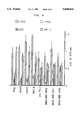
- FIG. 1 is a graph of the effect of various concentrations (0, 0.5, 1, 5, 10, 20, and 50 ng/ml) of either procathepsin D (pCD) (closed squares) or IGF II (open squares) on the growth of human breast cancer cell line MDA-MB-231 in serum-free medium, measured as percent of growth in medium supplemented with FCS.
- pCD procathepsin D
- IGF II open squares
- FIG. 2 is graph of the growth of human cell lines in serum-free medium containing procathepsin D (pCD), IGF II, or pCD and anti-pCD (10 ng/ml of either procathepsin D or IGF II).
- pCD procathepsin D
- IGF II IGF II
- pCD and anti-pCD 10 ng/ml of either procathepsin D or IGF II.
- FCS FCS
- FIG. 3 is a graph of the inhibition of growth of human cell lines by anti-procathepsin D IgG (anti-pCD IgG).
- anti-pCD IgG anti-procathepsin D IgG
- the results from growth in the control media with control MOPC-21 IgG (IgG) are given for comparison.
- the results represent the mean ⁇ SD of three experiments.
- FIG. 4 is a bar graph of the growth of several human cell lines in serum-free medium containing either pCD or IGF II. The results from growth in control media supplemented with FCS (+FCS) are given for comparison.
- the concentration of pCD was 0.4 nM (20 ng/ml).
- the concentration of IGF II was 2.7 nM (20 ng/ml). The results in this experiment and in following proliferation experiments represent the mean ⁇ SD of five experiments.
- FIG. 5 is a bar graph showing the influence of additives or modification of pCD on the proliferative activity of pCD and IGF II in serum free medium for cell lines MDA-MB-231 and U937.
- concentrations that were used were: pCD, 0.4 nM; M6P, 10 mM; pepstatin A, 1 ⁇ M; deglycosylated pCD (Deglyc.pCD), 0.4 nM.
- FIG. 6 is a bar graph showing the effect of two types of antibodies on the pCD or IGF II mediated proliferation of two human cell lines in serum free medium.
- the antibodies used were anti-procathepsin D (anti-pCD) and anti-cathepsin D (anti-CD) and their concentrations were 5 micrograms/ml of the IgG fraction.
- FIG. 7 is a bar graph comparing the influence of pCD (0.4 nM), IGF II (2.7 nM), bovine cathepsin D (BCD, 2 nM), human cathepsin D (HCD, 2 nM), the activation peptide of pCD (20 nM) and pig pepsinogen (PPGN, 20 nM) on the growth of four human cell lines in serum free medium.
- FIG. 8 is a graph of the dose response curves of the proliferative effect of the activation peptide for human breast cancer cell lines MDA-MB-231, MDA-MB-436 and ZR-75-1, measured as percent of growth in FCS versus concentration of activation peptide ng/ml!.
- FIG. 9 is a graph of the regulation of three surface receptors (CD11b and CD62L) of human neutrophils (part A) and lymphocytes (part B) by pCD and IGF II.
- the number 1 stands for pCD (2 nM), number 2 for pCD (2 nM) with anti-pCD IgG (5 ⁇ g/ml), respectively.
- the data are compared to a control experiment where any additives were omitted and was taken as 100%. Results represents the mean ⁇ SD of five experiments.
- Partial lower activity of the activation peptide of pCD in comparison to intact pCD may be explained by its higher conformational flexibility compared to the structure of the same region of pCD, or may be a result of its only forming a part of a three-dimensional signal present on the entire pCD structure.
- pepstatin A strong inhibitor of aspartic proteinases
- the data supports the mechanism that the mitogenic function of pCD relies on a specific structure of the activation peptide of pCD, and its interaction with a cell surface localized receptors.
- the findings identify the activation peptide of procathepsin D as a target for suppression of growth of certain breast tumors.
- the antibodies are made and characterized below in the examples. These can be administered as fragments or humanized as follows to decrease immunogenicity.
- a humanized antibody is one in which only the antigen-recognized sites, or complementarity-determining hypervariable regions (CDRs) are of non-human origin, whereas all framework regions (FR) of variable domains are products of human genes.
- CDRs complementarity-determining hypervariable regions
- FR framework regions
- variable region DNA of a selected animal recombinant anti-idiotypic ScFv is sequenced by the method of Clackson, T., et al., (1991) Nature, 352:624-688, incorporated herein by reference.
- animal CDRs are distinguished from animal framework regions (FR) based on locations of the CDRs in known sequences of animal variable genes. Kabat, H.
- the CDRs are grafted onto human heavy chain variable region framework by the use of synthetic oligonucleotides and polymerase chain reaction (PCR) recombination. Codons for the animal heavy chain CDRs, as well as the available human heavy chain variable region framework, are built in four (each 100 bases long) oligonucleotides. Using PCR, a grafted DNA sequence of 400 bases is formed that encodes for the recombinant animal CDR/human heavy chain FR protection.
- PCR polymerase chain reaction
- the immunogenic stimulus presented by the monoclonal antibodies so produced may be further decreased by the use of Pharmacia's (Pharmacia LKB Biotechnology, Sweden) "Recombinant Phage Antibody System” (RPAS), which generates a single-chain Fv fragment (ScFv) which incorporates the complete antigen-binding domain of the antibody.
- RPAS Recombinant Phage Antibody System
- ScFv single-chain Fv fragment
- antibody variable heavy and light chain genes are separately amplified from the hybridoma mRNA and cloned into an expression vector.
- the heavy and light chain domains are co-expressed on the same polypeptide chain after joining with a short linker DNA which codes for a flexible peptide.
- ScFv single-chain Fv fragment
- the antibodies can be formulated in standard pharmaceutical carriers for administration to patients in need thereof. These include saline, phosphate buffered saline, and other aqueous carriers, and liposomes, polymeric microspheres and other controlled release delivery devices, as are well known in the art.
- the antibodies can also be administered with adjuvant, such as muramyl dipeptide or other materials approved for use in humans (Freund's adjuvant can be used for administration of antibody to animals).
- the patient can also be immunized against the activation peptide of procathepsin D to achieve a similar inhibition of enhancement of proliferation that is obtained by administration to the patient of antibody.
- the activation peptide is not typically perceived as foreign by the patient. Accordingly, it is preferably conjugated with or administered in combination with a carrier protein (such as bovine serum albumin) or a polysaccharide that increases its immunogenicity and prepared for injection by suitable methods known to those skilled in the art. For example, see E. A. Kabat, Experimental Immunochemistry, 2nd edition (Thomas 1971), incorporated herein by reference.
- soluble antigens are generally sterilized by passage through a Chamberland, Berkefeld or Seitz filter having a pore size of 2 microns or less (Appropriate filters are also available from Costar, Cambridge, Mass. 02140).
- Suspensions of insoluble antigens may be sterilized by treatment with 0.2% formalin for several days in the cold or with 0.3 to 0.5% phenol. The antigen is then centrifuged off, washed once with sterile saline and suspended in saline. Phenol need not be removed.
- As a preservative for soluble as well as insoluble antigens 1% by volume of a 1% solution of methiolate is added.
- saline solutions of the desired concentration are prepared in 0.25% phenol; dextrans and the blood group substances may be autoclaved. Solutions of 1 mg per ml for the dextrans and blood group substances, and 0.05 mg per ml of the pneumococcal polysaccharides have been used. Sterility tests are carried out by streaking blood plates with 0.1 ml of solution and by inoculating samples into tubes containing 20 ml Difco thioglycollate broth. Plates and tubes are observed for ten days; no growth should occur.
- Soluble proteins may be precipitated with alum to obtain an enhanced antibody response.
- 0.5 mg to 1.5 mg of antigen is injected into a human patient in a sufficient frequency to provoke the formation of anti-activation peptide antibodies.
- antisense molecules are preferably administered in a polymeric formulation or liposome formulation which has been demonstrated to greatly enhance uptake and half-life of the antisense molecules.
- peptides derived from the activation peptide which bind to the receptor for the activation peptide of procathepsin D but which do not induce cell proliferation are used to block proliferation of cells induced by procathepsin D or activation peptide.
- the peptides are obtained by routine synthesis of peptides having the same sequence as activation peptide, making point mutations, screening for binding activity to breast cancer cells but not normal cells and then screening those that bind to determine efficacy in enhancing proliferation of the cells.
- the preferred peptide is that which then is determined to block inducement of proliferation of cells when the peptide is administered to breast cancer cells in combination with either procathepsin D or activation peptide.
- pCD Procathepsin D
- PPGN bovine cathepsinogen
- BCD bovine cathepsin D
- HCD human cathepsin D
- CD cathepsin D
- IGF II insulin like growth factor II
- mAb monoclonal antibody
- FCS fetal calf serum
- FITC Fluorescein isothiocyanate
- MTT 3-(4,5-Dimethylthiazol-2-4)-2,5-diphenyltetrazolium bromide
- PBS 25 mM sodium phosphate, 150 mM NaCl, pH 7.2
- RPMI 1640 medium, ⁇ -estradiol, HEPES, MTT, human transferrin, MOPC-21 IgG and recombinant human insulin-like growth factor II were obtained from Sigma Chemicals (St. Louis, Mo.), Fetal Clone from Hyclone Laboratories (Logan, Utah), fetal bovine serum from Intergen (Purchase, N.Y.); Iscove's modified Dulbecco's medium from Whittaker (Walkersville, Md.).
- the B lymphoblastoid cell line Raji, the monocytoid cell line U937, epithelioid carcinoma cell line HELA, hepatocellular carcinoma cell line Hep G2 and breast milk epithelial cell line HBL-100 were obtained from the American Tissue Culture Collection (ATCC, Rockville, Md.).
- the human angiosarcoma cell line HAEND-1 was described by Hoover, et al., (1993) "Isolation of a human endothelial cell line from a hepatic angiocarcinoma" In Vitro 29A, 199-202.
- the T lymphoblastoid cell line 8402 was obtained from The Tissue Culture Facility of the Lineberger Cancer Research Center of the University of North Carolina at Chapel Hill, Chapel Hill, N.C.
- Human breast cancer cell lines ZR-75-1, MDA-MB-436, MBA-MD-483 and MDA-MB-231 were obtained from Dr. R. Ceriani of the John Muir Cancer and Aging Research Institute, Walnut Creek, Calif.
- the cancer cell lines were grown in RPMI 1640 medium with HEPES buffer supplemented with 10% (v/v) heat-inactivated Fetal Clone, 2 mM L-glutamine, 100 U/ml penicillin, and 100 ⁇ g/ml streptomycin in plastic disposable tissue culture flasks at 37° C.
- Human procathepsin D was isolated from culture supernatants of human breast cancer cell line ZR-75. Briefly, a two step procedure was used. In the first step an immunoaffinity chromatography was used with anti-human cathepsin D activation peptide antibodies (Grinstein, S. W. (1992) "Chemoattractant-induced tyrosine phosphorylation and activation of microtubule-associated protein kinase in human neutrophils" J. Biol. Chem. 267(25), 18122-18125) attached to Protein A Sepharose. In the second step FPLC chromatography using Mono Q column and 20 mM Tris pH 7.2 were used.
- the viability of cell lines was determined after 7 days of incubation using a Cyto-Tox 96TM cytotoxicity assay (Promega Corp., Madison, Wis.) according to the manufacturer instructions.
- FIG. 1 The results showing the dose dependence of procathepsin D- or IGF II-mediated proliferation of breast cancer cells are summarized in FIG. 1.
- the results show the growth of MDA-MB-231 cell line, but identical results were found with other breast cancer cell lines.
- FIG. 2 shows the effect of 10 ng/ml of either procathepsin D or IGF II on proliferation of 11 different cell lines.
- Procathepsin D was shown to stimulate growth of all tested breast cancer cell lines (ZR-75-1, MDA-MB-483, MDA-MB-231 and MDA-MB-436).
- procathepsin D has very strong proliferative activity restricted for cell lines derived from primary mammary tumors.
- Procathepsin D has this proliferative effect not only for cell lines which secrete this proenzyme under the influence of estrogens, such as ZR-75-1 or MCF7 cell lines, but also for those cell lines which do not possess the ability to secrete procathepsin D.
- the antibodies against procathepsin D block the proliferative effects of procathepsin D but not IGF II. This clearly shows the results are a specific function of procathepsin D and not a function of unidentified impurities.
- procathepsin D The antibodies against procathepsin D were used in the experiments without the addition of procathepsin D. The suppression of proliferation was detected for several cell lines including those which had no response for procathepsin D addition. As the cytotoxicity experiments revealed, these antibodies did not have any cytotoxic effects on tested cell lines.
- procathepsin D has not been correlated with the estrogen receptors, as reviewed by Rochefort, H. (1992) "Biological and clinical significance of cathepsin D in breast cancer” Acta Oncologica 31, 125-130, particularly as to those tumor cells which do not have high estrogen receptor levels, but are responsive to procathepsin D.
- the mechanism of the mitogenic function of pCD was studied by incubation of cells with pCD isolated from secretions of ZR-75-1 cell line.
- the cellular response was measured by monitoring of the cellular proliferation of different human cell lines, and by measuring the expression of several surface receptors on human peripheral neutrophils and lymphocytes.
- procathepsin D exhibited a mitogenic effect on cell lines derived from breast cancer tissues, and that its addition regulated the expression of several surface receptors on human leukocytes.
- the data further demonstrate that these mitogenic effects are mediated by the activation peptide of pCD, and do not involve pCD interaction via the mannose-6-phosphate group nor cathepsin D proteolytic activity.
- the data strongly suggest that there is a cell surface receptor that remains to be identified which is able to recognize the activation peptide of pCD.
- N-glycanase was obtained from Genzyme (Cambridge, Mass.), pig pepsinogen was purchased from Worthington (Freeholf, N.J.), human cathepsin D was obtained from Biodesign (Kennebunk, Me.), human insulin-like growth factor II, bovine cathepsin D, bovine hemoglobin, RPMI 1640 and Iscove's modified Dulbecco's medium, ⁇ -estradiol, HEPES, propidium iodide, MTT and FITC were purchased from Sigma (St. Louis, Mo.). Ficoll-Hypaque and Fetal Clone were obtained from Hyclone Laboratories (Logan, Utah), pepstatin A was obtained from Serva (Heidelberg, Germany).
- MN-41 IgG 1 anti-CD11b monoclonal antibody (mAb) (Eddy et al, (1984) Clin. Immunol. Imunopathol. 31, 371-389) and TS1/22 anti-CD11a were purified from ascites fluid as described by Myones et al, (1988) J. Clin. Invest. 82, 640-651.
- MOPC-21 myeloma IgG 1 was purchased from Sigma.
- CD62L mAb from Pharmingen (San Diego, Calif.) and affinity purified goat anti-mouse IgG-FITC from Southern Biotechnology Associates, Inc. (Birmingham, Ala.).
- Anti-pCD IgG antibodies were raised against a synthetic activation peptide of pCD, and the specificity for pCD ascertained.
- the anti-bovine cathepsin D IgG (anti-CD) antibodies were kindly provided by Dr. J. Tang, Oklahoma Medical Research Foundation, Oklahoma City, Okla.
- pCD was isolated from pooled media of cell line ZR-75-1 treated with 10 nM ⁇ -estrogen. Briefly, a two step procedure was employed using immunoaffinity chromatography based on anti-pCD antibodies followed by two anion exchange Mono Q FPLC separations at pH 7.2. The isolated pCD was free of contaminants as judged by SDS electrophoresis and other methods. The identity of the protein was confirmed by immunostaining on western-blots, and by blocking of the activity by pepstatin A. pCD before and after deglycosylation were analyzed by SDS electrophoresis, blotted, and visualized using pig anti-pCD IgG antibodies and rabbit anti-pig IgG antibodies conjugated with peroxidase. As an independent test of purity of the procathepsin D preparation, solutions of pCD were purified by reacting with the anti-CD antibodies bound to Protein A SepharoseTM (Pharmacia, Uppsala, Sweden).
- peptide synthesis was accomplished by solid-phase multiple peptide synthesis under low-pressure continuous-flow conditions using a manually operated synthesizer. The synthesis was carried out in a flow reactor with adjustable volumen using F moc ⁇ tBu protection strategy on standard methylbenzhydrylamine polystyrene-based resin. The purity of the peptide was confirmed by amino acid analysis using a Durrum 500 amino acid analyzer and by sequencing using an Applied Biosystems model 470A sequencer. The sequence is provided as Sequence ID No. 1.
- Human cell lines Hela, U937 (monocytoid) and Raji (B lymphoblastoid) were obtained from American Tissue Culture Collection (ATCC, Rockville, Md.).
- the 8402 cell line (T lymphoblastoid) was obtained from The Tissue Culture Facility of the Lineberger Cancer Research Center of the University of North Carolina at Chapel Hill, Chapel Hill, N.C.
- Human breast cancer cell lines ZR-75-1, MDA-MB-436 and MDA-MB-231 were obtained from Dr. R. Ceriani of the John Muir Cancer and Aging Research Institute, Walnut Creek, Calif.
- the cancer cell lines were grown in RPMI 1640 medium with HEPES buffer supplemented with 10% (v/v) heat-inactivated Fetal Clone, 2 mM L-Glutamine, 100 U/ml penicillin, and 100 gm/ml streptomycin at 37° C. in a 5% CO 2 /95% air incubator. All other cell lines were incubated in the same way, except that the fetal clone was supplemented with fetal bovine serum (FCS).
- FCS fetal bovine serum
- the mononuclear cell fraction and neutrophil cell fraction were washed five times in RPMI-1640 medium and maintained in an ice bath until used. Cells were preincubated with appropriate amounts of tested substances for 30 minutes at 40° C. The cells were then washed once by centrifugation through a 3 ml cushion of 12% BSA in PBS with 10 mM sodium azide (12% BSA/PBS/azide).
- the cells were incubated with 1 ⁇ g of goat-anti mouse IgG-FITC and 10 ⁇ l of propidium iodide (1 mg/ml in PBS) for another 30 minutes on ice. After washing once through a 3 ml cushion of 12% BSA/PBS/azide as described above, the cells were resuspended in PBS containing 1% BSA and 10 mM sodium azide.
- Flow cytometry was performed with an EPICS Profile IITM (Coulter Electronics Inc., Miami Lakes, Fla.), and the data obtained from greater than or equal to 10 4 cells in each sample were stored in list mode. The binding of antibodies to polymorphonuclear cells and lymphocytes, gated by light scatter, was assessed by analyzing the stored list mode data with the Epics Elite Flow Cytometry Workstation software (Coulter).
- 5 ⁇ 10 5 neutrophils were incubated with 50 ng of FITC-pCD for 30 minutes at 4° C.
- the neutrophils were first incubated with 10 ng of the activation peptide.
- the neutrophils was first incubated for 30 minutes with pCD and then incubated with the FITC labeled anti-pCD antibodies.
- FIG. 4 shows the effect of pCD and IGF II on the proliferation of cell lines ZR-75-1, MDA-MB-231, MDA-MB-436, Hela, Raji, U937 and 8402.
- the proliferation was measured by the incorporation of 3-(4,5-Dimethylthiazol-2-4)-2,5-diphenyltetrazolium bromide (MTT) by cells.
- the fetal calf serum (FCS) or IGF II supplemented media were used as positive controls.
- FCS fetal calf serum
- IGF II supplemented media were used as positive controls.
- the proliferation of breast cancer cell lines was increased in the same way for both pCD and IGF II, while the cell lines which are not derived from breast cancer tissues responded to pCD much less than to IGF II.
- the proliferative activity of pCD and IGF II for breast cancer cell lines was dose responsive, as shown by FIG. 1.
- concentrations of pCD and IGF II were 20 ng/ml (0.4 nM for pCD and 2.7 nM for the IGF II). This experimental approach was used to study the mechanism of the mitogenic effect exhibited by pCD.
- Pepstatin A is a strong inhibitor of cathepsin D, with K i in the picomolar level, as reported by Baldwin et al, Proc. Natl. Acad. Sci. USA 90, 6796-6800, and was used to test the involvement of the proteolytic activity of cathepsin D on the observed mitogenic function.
- the hypothetical role of the M6P residues of PCD was investigated by either the addition of M6P or by the deglycosylation of pCD. The same set of cell lines was tested as in the studies shown in FIG. 4a.
- a synthetic peptide corresponding to the activation peptide of pCD was used to investigate further the role of the activation peptide in the mitogenic function.
- a dose responsive curve is shown in FIG. 7. Similar proliferative activity to that of pCD was observed, as shown by FIG. 8.
- the breast cancer derived cell lines responded in the same way to pCD and the synthetic activation peptide, while cell lines which did not react with pCD did not show any response to the peptide alone.
- No proliferative activity was observed for control molecules: pig pepsinogen (PPGN) or mature cathepsins D (FIG. 8).
- the PPGN control experiment was included for comparison with a protein of similar overall structure.
- Mature pepsin has a similar three-dimensional structure to cathepsin D (Metcalf and Fusek, (1993) EMBO J. 12, 1293-1302), but the sequences and the three-dimensional structures of the activation peptides of both enzymes differ substantially in the segment 43P--5 of pCD.
- FITC fluorescent marker
Abstract
Description
              TABLE 1
______________________________________
Effect of anti-procathepsin D IgG on
proliferation and death of cells.
          PERCENTAGE OF DEAD CELLS
            + anti-procathepsin
                         - anti-procathepsin
CELL LINE   D IgG        D IgG
______________________________________
Raji        3.7          5.2
8402        5.2          4.4
U937        6.0          5.1
HELA        3.4          5.8
Hep G2      6.7          6.0
HAEND-1     2.3          2.8
HBL-100     6.6          7.6
ZR-75-1     5.0          5.3
MDA-MB-483  9.6          8.1
MBA-MB-231  5.9          3.4
MDA-MB-436  8.3          6.4
______________________________________
 Results represent mean data from 5 wells of one characteristic experiment
              TABLE 2
______________________________________
Summary of flow cytometry experiments on
activation of receptors on human
leukocytes by pCD.
Additive/
Receptor     CD11a       CD11b   CD62L
______________________________________
pCD          ++          ++      --
IGF II       ++          ++      --
pCD + pepstatin A
             ++          ++      --
pCD + M6P    ++          ++      --
deglycosylated pCD
             +           +       --
pCD + anti-pCD
             0           0       0
pCD + anti-CD
             ++          ++      --
IGF II + anti-pCD
             ++          ++      --
anti-pCD     0           0       0
Activation peptide
             ++          ++      --
Activation   0           0       0
peptide + anti-pCD
______________________________________
 The sign "++" represents upregulation in the expression of a particular
 receptor, "--" means downregulation of a receptor, and "0" is no change i
 the concentration of a receptor in comparison to the control experiment.
 The concentrations of pCD was 2 nM, IGF II, 89 nM; M6P, 10 mM; pepstatin
 A, 1 μM; deglycosylated pCD, 2 nM; antibodies, 5 μg/ml; and
 activation peptide, 20 nM.
__________________________________________________________________________
SEQUENCE LISTING
(1) GENERAL INFORMATION:
(iii) NUMBER OF SEQUENCES: 1
(2) INFORMATION FOR SEQ ID NO:1:
(i) SEQUENCE CHARACTERISTICS:
(A) LENGTH: 44 amino acids
(B) TYPE: amino acid
(C) STRANDEDNESS: single
(D) TOPOLOGY: linear
(ii) MOLECULE TYPE: peptide
(iii) HYPOTHETICAL: NO
(iv) ANTI-SENSE: YES
(v) FRAGMENT TYPE: internal
(xi) SEQUENCE DESCRIPTION: SEQ ID NO:1:
LeuValArgIleProLeuHisLysPheThrSerIleArgArgThrMet
151015
SerGluValGlyGlySerValGluAspLeuIleAlaLysGlyProVal
202530
SerLysTyrSerGlnAlaProAlaValThrGluGly
3540
__________________________________________________________________________
Claims (5)
Priority Applications (1)
| Application Number | Priority Date | Filing Date | Title |
|---|---|---|---|
| US08/232,997 US5800814A (en) | 1994-04-22 | 1994-04-22 | Method for inhibition of breast tumor growth |
Applications Claiming Priority (1)
| Application Number | Priority Date | Filing Date | Title |
|---|---|---|---|
| US08/232,997 US5800814A (en) | 1994-04-22 | 1994-04-22 | Method for inhibition of breast tumor growth |
Publications (1)
| Publication Number | Publication Date |
|---|---|
| US5800814A true US5800814A (en) | 1998-09-01 |
Family
ID=22875450
Family Applications (1)
| Application Number | Title | Priority Date | Filing Date |
|---|---|---|---|
| US08/232,997 Expired - Lifetime US5800814A (en) | 1994-04-22 | 1994-04-22 | Method for inhibition of breast tumor growth |
Country Status (1)
| Country | Link |
|---|---|
| US (1) | US5800814A (en) |
Cited By (6)
| Publication number | Priority date | Publication date | Assignee | Title |
|---|---|---|---|---|
| US6224865B1 (en) | 1993-07-16 | 2001-05-01 | Cancerforskningsfonden Af 1989 | Suppression of inhibitors |
| US20040199750A1 (en) * | 1995-08-16 | 2004-10-07 | Micro Unity Systems Engineering, Inc. | Programmable processor with group floating-point operations |
| US20070117180A1 (en) * | 1998-10-02 | 2007-05-24 | Wataru Morikawa | Enzyme producing plasma protein fragment having inhibitory activity to metastasis and growth of cancer and plasma protein fragment produced by fragmentation by said enzyme |
| US20110081669A1 (en) * | 2008-06-16 | 2011-04-07 | University Of Louisville Research Foundation, Inc. | Systems and methods for diagnosis and prognosis of cancer |
| EP2330132A1 (en) | 2003-04-04 | 2011-06-08 | Yeda Research and Development Co. Ltd. | Antibodies and pharmaceutical compositions containing same useful for inhibiting activity of metalloproteins |
| WO2011092700A1 (en) | 2010-01-27 | 2011-08-04 | Yeda Research And Development Co. Ltd. | Antibodies that inhibit metalloproteins |
-
1994
- 1994-04-22 US US08/232,997 patent/US5800814A/en not_active Expired - Lifetime
Non-Patent Citations (104)
| Title |
|---|
| Baldwin, E.T., "Crystal Structures of Native and Inhibited Forms of Human Cathepsin D: Implications for lysosomal Targeting and Drug Design," Proc. Natl. Acad. Sci. USA, vol. 90, pp. 6796-6800 (1993). |
| Baldwin, E.T., Crystal Structures of Native and Inhibited Forms of Human Cathepsin D: Implications for lysosomal Targeting and Drug Design, Proc. Natl. Acad. Sci. USA, vol. 90, pp. 6796 6800 (1993). * |
| BarRett, A.J. "Purification of Isoenzymes from Human and Chicken Liver," Biochem J., vol. 117, pp. 601-607 (1970). |
| BarRett, A.J. Purification of Isoenzymes from Human and Chicken Liver, Biochem J., vol. 117, pp. 601 607 (1970). * |
| Bond, J.S., et al., "Intracellular Proteases," Ann. Rev. Biochem., vol. 56, pp. 333-364 (1987). |
| Bond, J.S., et al., Intracellular Proteases, Ann. Rev. Biochem., vol. 56, pp. 333 364 (1987). * |
| Cavailles, V., et al., "Cathepsin D Gene is Controlled by a Mixed Promoter, and Estrogens Stimulate only TATA-Dependent Transcription in Breast Cancer Cells," Proc. Natl. Acad. Sci. USA, vol. 90, pp. 203-207 (1993). |
| Cavailles, V., et al., Cathepsin D Gene is Controlled by a Mixed Promoter, and Estrogens Stimulate only TATA Dependent Transcription in Breast Cancer Cells, Proc. Natl. Acad. Sci. USA, vol. 90, pp. 203 207 (1993). * |
| Clackson, T., et al., "Making Antibody Fragments Using Phage Display Libraries," Nature, vol. 352, pp. 624-628 (1991). |
| Clackson, T., et al., Making Antibody Fragments Using Phage Display Libraries, Nature, vol. 352, pp. 624 628 (1991). * |
| Creek, K.E., et al., "The Role of the Phosphomannosyl Receptor in the Transport of Acid Hydrolases to Lysosomes," Lysosomes in Biology and Pathology, pp. 63-82 (1984). |
| Creek, K.E., et al., The Role of the Phosphomannosyl Receptor in the Transport of Acid Hydrolases to Lysosomes, Lysosomes in Biology and Pathology, pp. 63 82 (1984). * |
| Daugherty, B.L., et al., "Polymerase Chain Reaction Facilitates the Cloning, CDR-grating, and Rapid Expression of a Murine Monoclonal Antibody Directed Against the CD18 Component of Leukocyte Integrins," Nucleic Acids Res., vol. 19, No. 9, pp. 2471-2476 (1991). |
| Daugherty, B.L., et al., Polymerase Chain Reaction Facilitates the Cloning, CDR grating, and Rapid Expression of a Murine Monoclonal Antibody Directed Against the CD18 Component of Leukocyte Integrins, Nucleic Acids Res., vol. 19, No. 9, pp. 2471 2476 (1991). * |
| Dillman, J. Clinical Oncology 12:1497 1515, 1994. * |
| Dillman, J. Clinical Oncology 12:1497-1515, 1994. |
| Diment, S., et al., "Cathespin D is Membrane-Associated in Macrophase Endosomes," J. Biological Chemistry, vol. 263, No. 14, pp. 6901-6907 (1988). |
| Diment, S., et al., Cathespin D is Membrane Associated in Macrophase Endosomes, J. Biological Chemistry, vol. 263, No. 14, pp. 6901 6907 (1988). * |
| Eddy, A., et al., "The Distribution of the CR3 Receptor on Human Cells and Tissue as Revealed by a mOnoclonal Antibody," Clinical Immunology and Immunopathology, vol. 31, pp. 371-389 (1984). |
| Eddy, A., et al., The Distribution of the CR3 Receptor on Human Cells and Tissue as Revealed by a mOnoclonal Antibody, Clinical Immunology and Immunopathology, vol. 31, pp. 371 389 (1984). * |
| Elangovan, S., et al., "Progesterone and Estrogen Control of Rates of Synthesis of Uterine Cathespin D," J. Biological Chemistry, vol. 255, No. 15, pp. 7474-7479 (1980). |
| Elangovan, S., et al., Progesterone and Estrogen Control of Rates of Synthesis of Uterine Cathespin D, J. Biological Chemistry, vol. 255, No. 15, pp. 7474 7479 (1980). * |
| Ferrandina, G., et al., "Cathespin D in Primary Squamous Laryngeal Tumors: Correlation with Clinico-Pathological Parameters and Receptor Status," Cancer Letters, vol. 67, 133-138 (1992). |
| Ferrandina, G., et al., Cathespin D in Primary Squamous Laryngeal Tumors: Correlation with Clinico Pathological Parameters and Receptor Status, Cancer Letters, vol. 67, 133 138 (1992). * |
| Fusek, Martin, et al., "Mitogenic function of human procathepsin D: the role of the propeptide", Biochem., J., vol. 303:775-780 (1994). |
| Fusek, Martin, et al., Mitogenic function of human procathepsin D: the role of the propeptide , Biochem., J., vol. 303:775 780 (1994). * |
| Glickman, J.N., et al., "Manose 6-Phosphate-Independent Targeting of Lysosomal Enzymemes in I-Cell Disease B Lymophoblasts," J. Cell Biology, vol. 123, No. 1, pp. 99-108 (1993). |
| Glickman, J.N., et al., Manose 6 Phosphate Independent Targeting of Lysosomal Enzymemes in I Cell Disease B Lymophoblasts, J. Cell Biology, vol. 123, No. 1, pp. 99 108 (1993). * |
| Grassel, S. et al., "Human Cathepsin D Precursor is Associated with a 60 kDa Glycosylated Polypeptide," Biochemical and Biophysical Research Communications, vol. 182, No. 1, pp. 276-282 (1992). |
| Grassel, S. et al., Human Cathepsin D Precursor is Associated with a 60 kDa Glycosylated Polypeptide, Biochemical and Biophysical Research Communications, vol. 182, No. 1, pp. 276 282 (1992). * |
| Grinstein S., et al., "Chemoattractant-Induced Tyrosine Phosphorylation and Activation of Microtuble-Associated Protein Kinase in Human Neutrophils." J. Biol. Chem., vol. 267, No. 25, pp. 18122-18125 (1992). |
| Grinstein S., et al., Chemoattractant Induced Tyrosine Phosphorylation and Activation of Microtuble Associated Protein Kinase in Human Neutrophils. J. Biol. Chem., vol. 267, No. 25, pp. 18122 18125 (1992). * |
| Gura, Science 270:575 7, 1995. * |
| Gura, Science 270:575-7, 1995. |
| Harlow et al., Antibodies A Laboratory Manual Cold Spring Harbor Laboratory, 1988, pp. 285, 287. * |
| Harris et al, Tibtech, 1993, 111:42 44. * |
| Harris et al, Tibtech, 1993, 111:42-44. |
| Hasilik, A., "The Early and Late Processing of Lysosomal Enzymes: Proteolysis and Compartmentation," Experientia 48 Birkhauser Veerlag, CH-=4010 Basel/Swizerland (1992). |
| Hasilik, A., The Early and Late Processing of Lysosomal Enzymes: Proteolysis and Compartmentation, Experientia 48 Birkhauser Veerlag, CH 4010 Basel/Swizerland (1992). * |
| Helminen, H.J., et al., "Quantitation of Lysosomal Enzyme Changes During Enforced Mdammary Gland Involution," Exptl. Cell Res. vol. 60, pp. 419-426 (1970). |
| Helminen, H.J., et al., Quantitation of Lysosomal Enzyme Changes During Enforced Mdammary Gland Involution, Exptl. Cell Res. vol. 60, pp. 419 426 (1970). * |
| Hird et al, Genes & Cancer, Carney & Sikora, Eds 1990 John Wiley & Sons Ltd, pp. 183 189. * |
| Hird et al, Genes & Cancer, Carney & Sikora, Eds 1990 John Wiley & Sons Ltd, pp. 183-189. |
| Hoover, et al., Isolation of a Human Endothelial Cell Line from a Hepatic Angiocarcinoma, In Vitro 29A, 199 202 (1993). * |
| Hoover, et al., Isolation of a Human Endothelial Cell Line from a Hepatic Angiocarcinoma, In Vitro 29A, 199-202 (1993). |
| Johnson, M.D., et al., "The Role of Cathespin D in the Invasiveness of Human Breach Cancer Cells," Cancer Research, vol. 53, pp. 873-877 (1993). |
| Johnson, M.D., et al., The Role of Cathespin D in the Invasiveness of Human Breach Cancer Cells, Cancer Research, vol. 53, pp. 873 877 (1993). * |
| Kabat, H.A., et al., Sequences of Proteins of Immunological Interest, 4th ed. (U.S. Dept. Health & Human Services, Bethesda, MD, 1987)*. * |
| Kasai, M., et al., "Proenzyme Form of Cathepsin L Produced by Thymic Epithelial Cells Promotes Proliferation of Immature Thymocytes in the Presence of IL-1, IL-7, and Anti-CD3 Antibody," Cellular Immunology, vol. 150, pp. 124-136 (1993). |
| Kasai, M., et al., Proenzyme Form of Cathepsin L Produced by Thymic Epithelial Cells Promotes Proliferation of Immature Thymocytes in the Presence of IL 1, IL 7, and Anti CD3 Antibody, Cellular Immunology, vol. 150, pp. 124 136 (1993). * |
| Klionsky, D.J. et al., "Intracellular Sorting and Processing of a Yeast Vacuolar Hydrolase: Proteinase A Propeptide Contains Vacuolar Targeting Information," Molecular and Cellular Biology, Vo. 8, No. 5, pp. 2105-2116 (1988). |
| Klionsky, D.J. et al., Intracellular Sorting and Processing of a Yeast Vacuolar Hydrolase: Proteinase A Propeptide Contains Vacuolar Targeting Information, Molecular and Cellular Biology, Vo. 8, No. 5, pp. 2105 2116 (1988). * |
| Kornfeld, S., "The Biogenesis of Lysosomes," Ann. Rev. Cell Biol., vol. 5, pp. 483-525 (1989). |
| Kornfeld, S., "Trafficking of Lysosomal Enzymes," FASEB J., vol. 1, pp. 462-468 (1987). |
| Kornfeld, S., The Biogenesis of Lysosomes, Ann. Rev. Cell Biol., vol. 5, pp. 483 525 (1989). * |
| Kornfeld, S., Trafficking of Lysosomal Enzymes, FASEB J., vol. 1, pp. 462 468 (1987). * |
| Larsen, L.B., et al., "Procathepsin D Cannot Autoactivate to Cathepsin D at Acid pH," FEBS Letters, vol. 319, Nos. 1, 2, pp. 54-58 (1993). |
| Larsen, L.B., et al., Procathepsin D Cannot Autoactivate to Cathepsin D at Acid pH, FEBS Letters, vol. 319, Nos. 1, 2, pp. 54 58 (1993). * |
| Mathieu, M., et al., "Interactions of Cathepsin-D and Insulin-Like Growth Factor II (IGF-II) on the IGF-II/Mannose-6-Phosphate Receptor in Human Breast Cancer Cells and Possible Consequences on Mitogenic Activity of IGF-II)," Mol. Endo., vol. 4, No. 9, pp. 1327-1335 (1990). |
| Mathieu, M., et al., Interactions of Cathepsin D and Insulin Like Growth Factor II (IGF II) on the IGF II/Mannose 6 Phosphate Receptor in Human Breast Cancer Cells and Possible Consequences on Mitogenic Activity of IGF II), Mol. Endo., vol. 4, No. 9, pp. 1327 1335 (1990). * |
| McIntyre, G.F., "Procathepsins L and D are Membrane-Bound in Acidic Microsomal Vesicles," J. Biological Chemistry, vol. 266, No. 23, pp. 15438-15445 (1991). |
| McIntyre, G.F., et al., "The Lysosomal Proenzyme Receptor that Binds Procathepsin L to Microsomal Membranes at pH 5 is a 43-kDa Integral Membrane Protein," Proc. Natl. Acad. Sci. USA, vol. 90, pp. 10588-10592 (1993). |
| McIntyre, G.F., et al., "The pH-Dependent Membrane Association of Procathepsin L is Mediated by a 9-Residue Sequence within the Propeptide," J. Biological Chemistry, vol. 269, No. 1, pp. 567-572 (1994). |
| McIntyre, G.F., et al., The Lysosomal Proenzyme Receptor that Binds Procathepsin L to Microsomal Membranes at pH 5 is a 43 kDa Integral Membrane Protein, Proc. Natl. Acad. Sci. USA, vol. 90, pp. 10588 10592 (1993). * |
| McIntyre, G.F., et al., The pH Dependent Membrane Association of Procathepsin L is Mediated by a 9 Residue Sequence within the Propeptide, J. Biological Chemistry, vol. 269, No. 1, pp. 567 572 (1994). * |
| McIntyre, G.F., Procathepsins L and D are Membrane Bound in Acidic Microsomal Vesicles, J. Biological Chemistry, vol. 266, No. 23, pp. 15438 15445 (1991). * |
| Metcalf, P., et al., Two Crystal Structures for Cathepsin D: the Isysosomal Targeting Signal and Active Site, The EMBO J., vol. 12, No. 4, pp. 1293 1302 (1993). * |
| Metcalf, P., et al., Two Crystal Structures for Cathepsin D: the Isysosomal Targeting Signal and Active Site, The EMBO J., vol. 12, No. 4, pp. 1293-1302 (1993). |
| Morgan, D.O., et al., "Insulin-Like Growth Factor II Receptor as Multifunctional Binding Protein," Nature, vol. 329, pp. 301-307 (1987). |
| Morgan, D.O., et al., Insulin Like Growth Factor II Receptor as Multifunctional Binding Protein, Nature, vol. 329, pp. 301 307 (1987). * |
| Myones, B.L., "Neutrophil and Monocyte Cell Surface p150.95 Has iC3b-Receptor (CR4), Activity Resembling CR3," J. Clin. Invest., vol. 82, pp. 640-651 (1988). |
| Myones, B.L., Neutrophil and Monocyte Cell Surface p150.95 Has iC3b Receptor (CR 4 ), Activity Resembling CR 3 , J. Clin. Invest., vol. 82, pp. 640 651 (1988). * |
| Osband et al, Immunology Today, 1990, 11:193 195. * |
| Osband et al, Immunology Today, 1990, 11:193-195. |
| Rijnboutt, S., et al., "Mannose 6-Phosphate-Independent Membrane Association of Cathepsin D., Glucocerebrosidase, and Spingolipid-Activating Protein in HepG2 Cells," J. Biological Chemistry, vol. 266, No. 8, pp. 4862-4868 (1991). |
| Rijnboutt, S., et al., Mannose 6 Phosphate Independent Membrane Association of Cathepsin D., Glucocerebrosidase, and Spingolipid Activating Protein in HepG2 Cells, J. Biological Chemistry, vol. 266, No. 8, pp. 4862 4868 (1991). * |
| Rochefort, H., "Biological and Clinical Significance of Cathepsin D in Breast Center," Acta Oncologica, vol. 31, No. 2, pp. 125-130 (1992). |
| Rochefort, H., Biological and Clinical Significance of Cathepsin D in Breast Center, Acta Oncologica, vol. 31, No. 2, pp. 125 130 (1992). * |
| Ross, G.S., et al., "Identification of a C3bi-Specific Membrane Complement Receptor that is Expressed on Lymphocytes, Monocytes, Neutrophils, and Erythrocytes," J. Exp. Med., vol. 155, pp. 96-110 (1982). |
| Ross, G.S., et al., Identification of a C3bi Specific Membrane Complement Receptor that is Expressed on Lymphocytes, Monocytes, Neutrophils, and Erythrocytes, J. Exp. Med., vol. 155, pp. 96 110 (1982). * |
| Sacks, N.P.M., et al., "Cathepsin D Levels in Primary Breast Cancers: Relationships with Epidermal Growth Factor Receptor, Oestrogen Receptor and Axillary Nodal Status," European J. of Cancer, 29A(3):426-428 (1993) Note: European J. of Cancer is the correct cite not Int. J. Cancer. |
| Sacks, N.P.M., et al., Cathepsin D Levels in Primary Breast Cancers: Relationships with Epidermal Growth Factor Receptor, Oestrogen Receptor and Axillary Nodal Status, European J. of Cancer, 29A(3):426 428 (1993) Note: European J. of Cancer is the correct cite not Int. J. Cancer. * |
| Sanchez, L.M., "Cathepsin D in Breast Secretions from Women with Breast Cancer," Br. J. Cancer vol. 67, pp. 1076-1081 (1993). |
| Sanchez, L.M., Cathepsin D in Breast Secretions from Women with Breast Cancer, Br. J. Cancer vol. 67, pp. 1076 1081 (1993). * |
| Stein, M., et al., "M, 46 000, Mannose 60-Phosphate Specific Receptor, Its Role in Targeting of Lysosomal Enzymes," The EMBO J. vol. 6, No. 9, pp. 2677-2681 (1987). |
| Stein, M., et al., M, 46 000, Mannose 60 Phosphate Specific Receptor, Its Role in Targeting of Lysosomal Enzymes, The EMBO J. vol. 6, No. 9, pp. 2677 2681 (1987). * |
| Vagner, J., et al., "Colour-Monitored Solid-Phase Multiple Peptide Synthesis under Low-Pressure Continuous-Flow Conditions, Synthesis of Medium-Size Peptides: The Propart of Human Procathepsin D and the Growth-Hormone Releasing Factor," Coll. Czech. Chem. Commun., vol. 58, pp. 435-444 (1993). |
| Vagner, J., et al., Colour Monitored Solid Phase Multiple Peptide Synthesis under Low Pressure Continuous Flow Conditions, Synthesis of Medium Size Peptides: The Propart of Human Procathepsin D and the Growth Hormone Releasing Factor, Coll. Czech. Chem. Commun., vol. 58, pp. 435 444 (1993). * |
| Vaupel, P., et al., "Blood Flow, Oxygen and Nutrient Supply, and Metabolic Microenvironment of Human Tumors: A Review," Cancer Research, vol. 49, pp. 6440-6465 (1989). |
| Vaupel, P., et al., Blood Flow, Oxygen and Nutrient Supply, and Metabolic Microenvironment of Human Tumors: A Review, Cancer Research, vol. 49, pp. 6440 6465 (1989). * |
| Vetvicka, et al., "Effect of human procathepsin D on proliferation of human cell lines," Cancer Letters, 79,:131-135 (1994). |
| Vetvicka, et al., Effect of human procathepsin D on proliferation of human cell lines, Cancer Letters, 79,:131 135 (1994). * |
| Vetvicka, V., et al., "Complement Factors H and I Synthesize by B Cell Lines Function to Generate a Growth Factor Activity from C31," J. Immunology, vol. 150, No. 9 (1993). |
| Vetvicka, V., et al., "Human Breast Milk Containing Procathepsin D-Detection by Specific Antibodies," Biochemistry and Molecular Biology International, vol. 30, No. 5, pp. 921-928 (1993). |
| Vetvicka, V., et al., Complement Factors H and I Synthesize by B Cell Lines Function to Generate a Growth Factor Activity from C3 1 , J. Immunology, vol. 150, No. 9 (1993). * |
| Vetvicka, V., et al., Human Breast Milk Containing Procathepsin D Detection by Specific Antibodies, Biochemistry and Molecular Biology International, vol. 30, No. 5, pp. 921 928 (1993). * |
| Vignon, F., et al., "Autocrine Growth Stimulation of the MCF 7 Breast Cancer Cells by the Estrogen-Regulated 53 K Protein," Endocrinology, vol. 118, No. 4, pp. 1537-1544 (1986). |
| Vignon, F., et al., Autocrine Growth Stimulation of the MCF 7 Breast Cancer Cells by the Estrogen Regulated 53 K Protein, Endocrinology , vol. 118, No. 4, pp. 1537 1544 (1986). * |
| von Figura, K., "Molecular Recognition and Targeting of Lysosomal Proteins," Current Opinion in Cell Biology, vol. 3, pp. 642-646 (1991). |
| von Figura, K., Molecular Recognition and Targeting of Lysosomal Proteins, Current Opinion in Cell Biology, vol. 3, pp. 642 646 (1991). * |
| Westley, B.R., et al., "Oestrogen Regulates Cathepsin D mRNA levels in Oestrogen Responsive Human Breast Cancer Cells," Nucleic Acids Research, vol. 15, No. 9, pp. 3773-3786 (1987). |
| Westley, B.R., et al., Oestrogen Regulates Cathepsin D mRNA levels in Oestrogen Responsive Human Breast Cancer Cells, Nucleic Acids Research, vol. 15, No. 9, pp. 3773 3786 (1987). * |
| Williams, K.P., et al., "Isolation of a Membrane-Associated Cathespin D-Like Enzyme from the Model Antigen Presenting Cell, A20, and its Ability to Generate Antigenic Fragments from a Protein Antigen in a Cell-Free system," Archives of Biochemistry and Biophysics, vol. 305, No. 2, pp. 298-306 (1993). |
| Williams, K.P., et al., Isolation of a Membrane Associated Cathespin D Like Enzyme from the Model Antigen Presenting Cell, A20, and its Ability to Generate Antigenic Fragments from a Protein Antigen in a Cell Free system, Archives of Biochemistry and Biophysics, vol. 305, No. 2, pp. 298 306 (1993). * |
Cited By (10)
| Publication number | Priority date | Publication date | Assignee | Title |
|---|---|---|---|---|
| US6224865B1 (en) | 1993-07-16 | 2001-05-01 | Cancerforskningsfonden Af 1989 | Suppression of inhibitors |
| US20030096755A1 (en) * | 1993-07-16 | 2003-05-22 | Niels Brunner | Suppression of inhibitors |
| US20080166354A1 (en) * | 1993-07-16 | 2008-07-10 | Niels Brunner | Suppression of inhibitors |
| US20040199750A1 (en) * | 1995-08-16 | 2004-10-07 | Micro Unity Systems Engineering, Inc. | Programmable processor with group floating-point operations |
| US20070117180A1 (en) * | 1998-10-02 | 2007-05-24 | Wataru Morikawa | Enzyme producing plasma protein fragment having inhibitory activity to metastasis and growth of cancer and plasma protein fragment produced by fragmentation by said enzyme |
| EP2330132A1 (en) | 2003-04-04 | 2011-06-08 | Yeda Research and Development Co. Ltd. | Antibodies and pharmaceutical compositions containing same useful for inhibiting activity of metalloproteins |
| US20110081669A1 (en) * | 2008-06-16 | 2011-04-07 | University Of Louisville Research Foundation, Inc. | Systems and methods for diagnosis and prognosis of cancer |
| US8852870B2 (en) | 2008-06-16 | 2014-10-07 | University Of Louisville Research Foundation, Inc. | Systems and methods for diagnosis and prognosis of cancer |
| WO2011092700A1 (en) | 2010-01-27 | 2011-08-04 | Yeda Research And Development Co. Ltd. | Antibodies that inhibit metalloproteins |
| EP3216805A1 (en) | 2010-01-27 | 2017-09-13 | Yeda Research and Development Co. Ltd | Antibodies that inhibit metalloproteins |
Similar Documents
| Publication | Publication Date | Title |
|---|---|---|
| Fusek et al. | Mitogenic function of human procathepsin D: the role of the propeptide | |
| Amagai et al. | Absorption of pathogenic autoantibodies by the extracellular domain of pemphigus vulgaris antigen (Dsg3) produced by baculovirus. | |
| US8178104B2 (en) | Methods and compositions for targeting gC1qR/p32 | |
| CN103747807B (en) | P97 antibody conjugates and using method | |
| US7939490B2 (en) | TWEAK as a therapeutic target for treating central nervous system diseases associated with cerebral edema and cell death | |
| US20040023870A1 (en) | Methods of therapy and diagnosis using targeting of cells that express toll-like receptor proteins | |
| HU230768B1 (en) | Humanized antibodies that sequester amyloid beta peptide | |
| JP2002062295A (en) | Urokinase type plasminogen activating factor receptor | |
| Vetvicka et al. | Analysis of the interaction of procathepsin D activation peptide with breast cancer cells | |
| US20040170631A1 (en) | Heparanase activity neutralizing anti-heparanase monoclonal antibody and other anti-heparanase antibodies | |
| US20150010495A1 (en) | Method for the production of an immunostimulating mucin (muc1) | |
| US7378253B2 (en) | Methods of therapy and diagnosis using immunotargeting of CD84Hy1-expressing cells | |
| KR20100023869A (en) | Cancer remedy containing antibody against peptide encoded by exon-17 of periostin | |
| JP2002505882A (en) | Chitin-binding fragments of chitinase | |
| WO2006052926A2 (en) | Tweak as a therapeutic target for treating central nervous system diseases associated with cerebral edema and and cell death | |
| EP1064005B1 (en) | Urokinase plasminogen activator receptor as a target for diagnosis of micrometastases | |
| CA2245956A1 (en) | Monoclonal antibodies specific to endothelial cell cadherins and uses thereof | |
| US5800814A (en) | Method for inhibition of breast tumor growth | |
| US20030215453A1 (en) | Methods of therapy and diagnosis using immunotargeting of cells expressing VpreB1 protein | |
| US20060269552A1 (en) | Heparanase activity neutralizing anti-heparanase monclonal antibody and other anti-heparanase antibodies | |
| EP0642354A1 (en) | METHODS FOR DETECTING AND ISOLATING uPA-R AND INHIBITING THE BINDING OF uPA TO uPA-R | |
| US20040109863A1 (en) | Methods of therapy and diagnosis using targeting of cells that express Ly-9 | |
| CA2476555A1 (en) | Methods of therapy and diagnosis | |
| EP3102940B1 (en) | Anti-metalloprotease antibody for diagnosis and treatment of cancers | |
| US20050158324A1 (en) | Methods of therapy and diagnosis using targeting of cells that express BCLP polypeptides |
Legal Events
| Date | Code | Title | Description |
|---|---|---|---|
| AS | Assignment |
Owner name: UNIVERSITY OF LOUISVILLE RESEARCH FOUNDATION, INC. Free format text: ASSIGNMENT OF ASSIGNORS INTEREST;ASSIGNOR:VETVICKA, VACLAV;REEL/FRAME:007038/0075 Effective date: 19940620 Owner name: OKLAHOMA MEDICAL RESEARCH FOUNDATION, OKLAHOMA Free format text: ASSIGNMENT OF ASSIGNORS INTEREST;ASSIGNOR:FUSEK, MARTIN;REEL/FRAME:007038/0078 Effective date: 19940527 |
|
| STCF | Information on status: patent grant |
Free format text: PATENTED CASE |
|
| FPAY | Fee payment |
Year of fee payment: 4 |
|
| FPAY | Fee payment |
Year of fee payment: 8 |
|
| FEPP | Fee payment procedure |
Free format text: PAYER NUMBER DE-ASSIGNED (ORIGINAL EVENT CODE: RMPN); ENTITY STATUS OF PATENT OWNER: SMALL ENTITY Free format text: PAYOR NUMBER ASSIGNED (ORIGINAL EVENT CODE: ASPN); ENTITY STATUS OF PATENT OWNER: SMALL ENTITY |
|
| AS | Assignment |
Owner name: NATIONAL INSTITUTES OF HEALTH (NIH), U.S. DEPT. OF Free format text: EXECUTIVE ORDER 9424, CONFIRMATORY LICENSE;ASSIGNOR:OKLAHOMA MEDICAL RESEARCH FOUNDATION;REEL/FRAME:021291/0447 Effective date: 20010806 |
|
| FPAY | Fee payment |
Year of fee payment: 12 |