US11776146B2 - Edge handling methods for associated depth sensing camera devices, systems, and methods - Google Patents
Edge handling methods for associated depth sensing camera devices, systems, and methods Download PDFInfo
- Publication number
- US11776146B2 US11776146B2 US17/655,514 US202217655514A US11776146B2 US 11776146 B2 US11776146 B2 US 11776146B2 US 202217655514 A US202217655514 A US 202217655514A US 11776146 B2 US11776146 B2 US 11776146B2
- Authority
- US
- United States
- Prior art keywords
- depths
- changes
- patient
- roi
- steep
- Prior art date
- Legal status (The legal status is an assumption and is not a legal conclusion. Google has not performed a legal analysis and makes no representation as to the accuracy of the status listed.)
- Active, expires
Links
Images


Classifications
-
- G—PHYSICS
- G06—COMPUTING; CALCULATING OR COUNTING
- G06T—IMAGE DATA PROCESSING OR GENERATION, IN GENERAL
- G06T7/00—Image analysis
- G06T7/50—Depth or shape recovery
- G06T7/55—Depth or shape recovery from multiple images
- G06T7/579—Depth or shape recovery from multiple images from motion
-
- A—HUMAN NECESSITIES
- A61—MEDICAL OR VETERINARY SCIENCE; HYGIENE
- A61B—DIAGNOSIS; SURGERY; IDENTIFICATION
- A61B5/00—Measuring for diagnostic purposes; Identification of persons
- A61B5/08—Detecting, measuring or recording devices for evaluating the respiratory organs
- A61B5/0816—Measuring devices for examining respiratory frequency
-
- A—HUMAN NECESSITIES
- A61—MEDICAL OR VETERINARY SCIENCE; HYGIENE
- A61B—DIAGNOSIS; SURGERY; IDENTIFICATION
- A61B5/00—Measuring for diagnostic purposes; Identification of persons
- A61B5/103—Detecting, measuring or recording devices for testing the shape, pattern, colour, size or movement of the body or parts thereof, for diagnostic purposes
- A61B5/11—Measuring movement of the entire body or parts thereof, e.g. head or hand tremor, mobility of a limb
- A61B5/1126—Measuring movement of the entire body or parts thereof, e.g. head or hand tremor, mobility of a limb using a particular sensing technique
- A61B5/1128—Measuring movement of the entire body or parts thereof, e.g. head or hand tremor, mobility of a limb using a particular sensing technique using image analysis
-
- A—HUMAN NECESSITIES
- A61—MEDICAL OR VETERINARY SCIENCE; HYGIENE
- A61B—DIAGNOSIS; SURGERY; IDENTIFICATION
- A61B5/00—Measuring for diagnostic purposes; Identification of persons
- A61B5/103—Detecting, measuring or recording devices for testing the shape, pattern, colour, size or movement of the body or parts thereof, for diagnostic purposes
- A61B5/11—Measuring movement of the entire body or parts thereof, e.g. head or hand tremor, mobility of a limb
- A61B5/113—Measuring movement of the entire body or parts thereof, e.g. head or hand tremor, mobility of a limb occurring during breathing
- A61B5/1135—Measuring movement of the entire body or parts thereof, e.g. head or hand tremor, mobility of a limb occurring during breathing by monitoring thoracic expansion
-
- A—HUMAN NECESSITIES
- A61—MEDICAL OR VETERINARY SCIENCE; HYGIENE
- A61B—DIAGNOSIS; SURGERY; IDENTIFICATION
- A61B5/00—Measuring for diagnostic purposes; Identification of persons
- A61B5/74—Details of notification to user or communication with user or patient ; user input means
- A61B5/7475—User input or interface means, e.g. keyboard, pointing device, joystick
- A61B5/748—Selection of a region of interest, e.g. using a graphics tablet
-
- A—HUMAN NECESSITIES
- A61—MEDICAL OR VETERINARY SCIENCE; HYGIENE
- A61M—DEVICES FOR INTRODUCING MEDIA INTO, OR ONTO, THE BODY; DEVICES FOR TRANSDUCING BODY MEDIA OR FOR TAKING MEDIA FROM THE BODY; DEVICES FOR PRODUCING OR ENDING SLEEP OR STUPOR
- A61M16/00—Devices for influencing the respiratory system of patients by gas treatment, e.g. mouth-to-mouth respiration; Tracheal tubes
- A61M16/0057—Pumps therefor
- A61M16/0072—Tidal volume piston pumps
-
- G—PHYSICS
- G06—COMPUTING; CALCULATING OR COUNTING
- G06T—IMAGE DATA PROCESSING OR GENERATION, IN GENERAL
- G06T7/00—Image analysis
- G06T7/10—Segmentation; Edge detection
- G06T7/11—Region-based segmentation
-
- G—PHYSICS
- G06—COMPUTING; CALCULATING OR COUNTING
- G06T—IMAGE DATA PROCESSING OR GENERATION, IN GENERAL
- G06T7/00—Image analysis
- G06T7/10—Segmentation; Edge detection
- G06T7/12—Edge-based segmentation
-
- G—PHYSICS
- G06—COMPUTING; CALCULATING OR COUNTING
- G06T—IMAGE DATA PROCESSING OR GENERATION, IN GENERAL
- G06T7/00—Image analysis
- G06T7/20—Analysis of motion
- G06T7/254—Analysis of motion involving subtraction of images
-
- G—PHYSICS
- G16—INFORMATION AND COMMUNICATION TECHNOLOGY [ICT] SPECIALLY ADAPTED FOR SPECIFIC APPLICATION FIELDS
- G16H—HEALTHCARE INFORMATICS, i.e. INFORMATION AND COMMUNICATION TECHNOLOGY [ICT] SPECIALLY ADAPTED FOR THE HANDLING OR PROCESSING OF MEDICAL OR HEALTHCARE DATA
- G16H30/00—ICT specially adapted for the handling or processing of medical images
- G16H30/40—ICT specially adapted for the handling or processing of medical images for processing medical images, e.g. editing
-
- A—HUMAN NECESSITIES
- A61—MEDICAL OR VETERINARY SCIENCE; HYGIENE
- A61B—DIAGNOSIS; SURGERY; IDENTIFICATION
- A61B5/00—Measuring for diagnostic purposes; Identification of persons
- A61B5/0059—Measuring for diagnostic purposes; Identification of persons using light, e.g. diagnosis by transillumination, diascopy, fluorescence
- A61B5/0062—Arrangements for scanning
- A61B5/0064—Body surface scanning
-
- A—HUMAN NECESSITIES
- A61—MEDICAL OR VETERINARY SCIENCE; HYGIENE
- A61B—DIAGNOSIS; SURGERY; IDENTIFICATION
- A61B5/00—Measuring for diagnostic purposes; Identification of persons
- A61B5/74—Details of notification to user or communication with user or patient ; user input means
- A61B5/7475—User input or interface means, e.g. keyboard, pointing device, joystick
- A61B5/748—Selection of a region of interest, e.g. using a graphics tablet
- A61B5/7485—Automatic selection of region of interest
-
- A—HUMAN NECESSITIES
- A61—MEDICAL OR VETERINARY SCIENCE; HYGIENE
- A61M—DEVICES FOR INTRODUCING MEDIA INTO, OR ONTO, THE BODY; DEVICES FOR TRANSDUCING BODY MEDIA OR FOR TAKING MEDIA FROM THE BODY; DEVICES FOR PRODUCING OR ENDING SLEEP OR STUPOR
- A61M2230/00—Measuring parameters of the user
- A61M2230/40—Respiratory characteristics
- A61M2230/42—Rate
-
- G—PHYSICS
- G06—COMPUTING; CALCULATING OR COUNTING
- G06T—IMAGE DATA PROCESSING OR GENERATION, IN GENERAL
- G06T2207/00—Indexing scheme for image analysis or image enhancement
- G06T2207/10—Image acquisition modality
- G06T2207/10016—Video; Image sequence
-
- G—PHYSICS
- G06—COMPUTING; CALCULATING OR COUNTING
- G06T—IMAGE DATA PROCESSING OR GENERATION, IN GENERAL
- G06T2207/00—Indexing scheme for image analysis or image enhancement
- G06T2207/10—Image acquisition modality
- G06T2207/10028—Range image; Depth image; 3D point clouds
-
- G—PHYSICS
- G06—COMPUTING; CALCULATING OR COUNTING
- G06T—IMAGE DATA PROCESSING OR GENERATION, IN GENERAL
- G06T2207/00—Indexing scheme for image analysis or image enhancement
- G06T2207/20—Special algorithmic details
- G06T2207/20092—Interactive image processing based on input by user
- G06T2207/20104—Interactive definition of region of interest [ROI]
-
- G—PHYSICS
- G06—COMPUTING; CALCULATING OR COUNTING
- G06T—IMAGE DATA PROCESSING OR GENERATION, IN GENERAL
- G06T2207/00—Indexing scheme for image analysis or image enhancement
- G06T2207/30—Subject of image; Context of image processing
- G06T2207/30196—Human being; Person
Definitions
- the present technology is generally related to patient monitoring using an image capture device and edge handling methods therefor.
- a pulse oximeter is a finger sensor that can include two light emitters and a photodetector. The sensor emits light into the patient's finger and transmits the detected light signal to a monitor.
- the monitor includes a processor that processes the signal, determines vital signs (e.g., pulse rate, respiration rate, arterial oxygen saturation), and displays the vital signs on a display.
- monitoring systems include other types of monitors and sensors, such as electroencephalogram (EEG) sensors, blood pressure cuffs, temperature probes, air flow measurement devices (e.g., spirometer), and others.
- EEG electroencephalogram
- Some wireless, wearable sensors have been developed, such as wireless EEG patches and wireless pulse oximetry sensors.
- Video-based monitoring is a field of patient monitoring that uses one or more remote video cameras to detect physical attributes of the patient. This type of monitoring can also be called “non-contact” monitoring in reference to the remote video sensor(s), which does/do not contact the patient. The remainder of this disclosure offers solutions and improvements in this field.
- the techniques of this disclosure generally relate to systems and methods for patient monitoring using an image capture device, including defining a region of interest (ROI) on a patient; capturing two or more images of the ROI using an image capture device; calculating an overall change in depth of the ROI within the two or more images, wherein calculating the overall change in depth of the ROI includes: measuring changes in depths of portions of the ROI; recognizing steep changes in depths in the measured changes in depths; and adjusting the recognized steep changes in depths.
- ROI region of interest
- adjusting the recognized steep changes in depths includes excluding the recognized steep changes in depths from the calculation of the overall change in depth of the ROI.
- adjusting the recognized steep changes in depths includes (i) excluding measured changes in depths corresponding to an outer percentage of the ROI and/or to an edge region of the patient and/or (ii) excluding a percentage of the measured changes in depths surrounding a recognized steep change in depth.
- adjusting the recognized steep changes in depths comprises including only measured changes in depths up to and/or between one or more recognized steep changes in depths in the calculation of the overall change in depth of the ROI.
- adjusting the recognized steep changes in depths includes interpolating and/or extrapolating over the recognized steep changes in depths using one or more other measured changes in depths.
- adjusting the recognized steep changes in depths includes using a template to approximate changes in depths at portions of the ROI corresponding to the recognized steep changes in depths.
- Other aspects include determining one or more patient respiratory parameters using all or a subset of the measured changes in depths and/or all or a subset of the adjusted changes in depths.
- An exemplary patient respiratory parameter includes a tidal volume of the patient, and wherein the tidal volume of the patient is determined using a subset of the measured changes in depths excluding the recognized steep changes in depths and/or all or a subset of the adjusted changes in depths.
- Another exemplary patient respiratory parameters includes a respiratory rate of the patient, wherein the respiratory rate of the patient is determined using all of the measured changes in depths and none of the adjusted changes in depths.
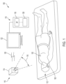
- FIG. 1 is a schematic view of a video-based patient monitoring system configured in accordance with various embodiments of the present technology.
- FIG. 2 is a block diagram illustrating a video-based patient monitoring system having a computing device, a server, and one or more image capture devices, and configured in accordance with various embodiments of the present technology.
- FIG. 3 is a schematic view of a patient showing various regions of interest that can be defined by video-based patient monitoring systems configured in accordance with various embodiments of the present technology.
- FIG. 4 A is a false color depth sensing image of a region of interest corresponding to a patient's torso, captured using a video-based patient monitoring system configured in accordance with various embodiments of the present technology.
- FIG. 4 B is a line plot of depths measured across a portion of the depth sensing image of FIG. 4 A .
- FIG. 4 C is a line plot of a change in depth measured across a portion of the depth sensing image of FIG. 4 A .
- FIG. 5 A is a schematic diagram of lateral movement of a patient's torso.
- FIG. 5 B is a line plot of measured depths of a patient's torso over time and generated using a non-contact patient monitoring system configured in accordance with various embodiments of the present technology.
- FIG. 5 C is a schematic diagram of lateral movement of a patient's torso.
- FIGS. 6 - 8 are line plots illustrating various methods for mitigating errors resulting from changes in depths perceived at edges of a patient's torso in accordance with various embodiments of the present technology.
- FIG. 9 is a flow diagram illustrating a method for mitigating errors in changes in depths measured at edge regions of a patient in accordance with various embodiments of the present technology.
- devices, systems, and/or methods configured in accordance with embodiments of the present technology are configured to capture one or more images (e.g., a video sequence) of a patient or portion(s) of a patient (e.g., a patient's torso) within a field of view of an image capture device.
- the devices, systems, and/or methods can measure changes in depths of regions (e.g., one or more pixels or groups of pixels) in the captured images over time.
- the devices, systems, and/or methods can determine various respiratory parameters of a patient, including tidal volume, minute volume, respiratory rate, among others.
- the device, systems, and/or methods can analyze the respiratory parameters and can trigger alerts and/or alarms when the devices, systems, and/or methods detect one or more breathing abnormalities.
- Errors in measured depths and/or changes in depths can occur at edges of a patient within the field of view of the image capture device. For example, lateral movement of a patient's torso at the edges of the patient's torso can be perceived as large changes in depths of the patient's torso in these regions. Additionally, or alternatively, as a patient inhales and exhales, edge portions of the patient's torso can move within and outside a line of sight of an image capture device.
- an edge portion of the patient's torso can move within and/or remain within the line of sight of the image capture device.
- the edge portion of the patient's torso can move outside or and/or remain outside of the line of sight of the image capture device.
- the image capture device can perceive large, inaccurate changes in depths at edge regions of the patient at various points throughout the patient's respiratory cycle. These large, inaccurate changes in depths can contribute to errors in the respiratory parameters of the patient determined by the video-based patient monitoring devices, system, and/or methods.
- the video-based patient monitoring devices, systems, and associated methods of the present technology are configured to mitigate the errors in changes in depths measured at edge regions of a patient (e.g., of a patient's torso).
- the devices, systems, and associated methods exclude the edge portions while integrating over a region within the extent of the patient.
- the devices, systems, and associated methods interpolate changes in depths at the edge regions using changes in depths perceived at other regions of the patient.
- the device, systems, and associated methods use a template (e.g., a template generated from a previous scan of the patient) to estimate changes in depths that occur at the edge regions of the patient.
- the video-based patient monitoring devices, systems, and associated methods configured in accordance with various embodiments of the present technology can more accurately measure changes in depths that occur at edge regions of a patient within the field of view of the image capture device(s).
- the devices, systems, and associated methods can more accurately determine a patient's respiratory parameters.
- FIGS. 1 - 9 Specific details of several embodiments of the present technology are described herein with reference to FIGS. 1 - 9 .
- many of the embodiments are described with respect to devices, systems, and methods for video-based detection and/or monitoring of breathing in a human patient, other applications and other embodiments in addition to those described herein are within the scope of the present technology.
- at least some embodiments of the present technology can be useful for video-based detection and/or monitoring of breathing in other animals and/or in non-patients (e.g., elderly or neonatal individuals within their homes).
- non-patients e.g., elderly or neonatal individuals within their homes.
- embodiments in addition to those disclosed herein are within the scope of the present technology.
- embodiments of the present technology can have different configurations, components, and/or procedures than those shown or described herein.
- embodiments of the present technology can have configurations, components, and/or procedures in addition to those shown or described herein and that these and other embodiments can be without several of the configurations, components, and/or procedures shown or described herein without deviating from the present technology.
- the term “steep” shall be understood to include any change in depth or rate of change above a threshold value or percentage.
- the threshold value or percentage can be a predetermined and/or predefined threshold value (e.g., 0.5 mm, 1 mm, 2 mm, 5 mm, 10 mm, 20 mm, 50 mm, 75 mm, 100 mm, etc.) or percentage (e.g., 1%, 2%, 5%, 10%, 20%, 30%, 40%, 50%, 60%, 70%, 80%, 90%, 95%, 97%, etc.).
- the term “steep” shall be understood to encompass any change in depth or rate of change above a threshold value or percentage vis-à-vis the same pixel and/or region of an ROI across two or more images. In these and still other embodiments, the term “steep” shall be understood to encompass any change in depth or rate of change above a threshold value or percentage vis-à-vis neighboring and/or adjacent pixels and/or regions of an ROI across one or more images.
- FIG. 1 is a schematic view of a patient 112 and a video-based patient monitoring system 100 configured in accordance with various embodiments of the present technology.
- the system 100 includes a non-contact detector 110 and a computing device 115 .
- the detector 110 can include one or more image capture devices, such as one or more video cameras.
- the non-contact detector 110 includes a video camera 114 .
- the non-contact detector 110 of the system 100 is placed remote from the patient 112 . More specifically, the video camera 114 of the non-contact detector 110 is positioned remote from the patient 112 in that it is spaced apart from and does not contact the patient 112 .
- the camera 114 includes a detector exposed to a field of view (FOV) 116 that encompasses at least a portion of the patient 112 .
- FOV field of view
- the camera 114 can capture a sequence of images over time.
- the camera 114 can be a depth sensing camera, such as a Kinect camera from Microsoft Corp. (Redmond, Wash.).
- a depth sensing camera can detect a distance between the camera and objects within its field of view.
- Such information can be used, as disclosed herein, to determine that a patient 112 is within the FOV 116 of the camera 114 and/or to determine one or more ROI's to monitor on the patient 112 .
- the ROI can be monitored over time, and the changes in depths of regions (e.g., pixels) within the ROI 102 can represent movements of the patient 112 associated with breathing.
- regions e.g., pixels
- Provisional Patent Application Ser. No. 62/779,964 those movements, or changes of regions within the ROI 102 , can be used to determine various breathing parameters, such as tidal volume, minute volume, respiratory rate, etc. Those movements, or changes of regions within the ROI 102 , can also be used to detect various breathing abnormalities, as discussed in greater detail in U.S. Provisional Patent Application Ser. Nos. 62/716,724 and 62/779,964.
- the various breathing abnormalities can include, for example, apnea, rapid breathing (tachypnea), slow breathing, intermittent or irregular breathing, shallow breathing, obstructed and/or impaired breathing, and others.
- the entire disclosures of U.S. patent application Ser. No. 16/219,360 and U.S. Provisional Patent Application Ser. Nos. 62/716,724 and 62/779,964 are incorporated herein by reference.
- the system 100 determines a skeleton-like outline of the patient 112 to identify a point or points from which to extrapolate a ROI.
- a skeleton-like outline can be used to find a center point of a chest, shoulder points, waist points, and/or any other points on a body of the patient 112 . These points can be used to determine one or more ROI's.
- a ROI 102 can be defined by filling in area around a center point 103 of the chest, as shown in FIG. 1 . Certain determined points can define an outer edge of the ROI 102 , such as shoulder points.
- other points are used to establish a ROI.
- a face can be recognized, and a chest area inferred in proportion and spatial relation to the face.
- a reference point of a patient's chest can be obtained (e.g., through a previous 3-D scan of the patient), and the reference point can be registered with a current 3-D scan of the patient.
- the system 100 can define a ROI around a point using parts of the patient 112 that are within a range of depths from the camera 114 .
- the system 100 can utilize depth information from the depth sensing camera 114 to fill out the ROI. For example, if the point 103 on the chest is selected, parts of the patient 112 around the point 103 that are a similar depth from the camera 114 as the point 103 are used to determine the ROI 102 .
- the patient 112 can wear specially configured clothing (not shown) that includes one or more features to indicate points on the body of the patient 112 , such as the patient's shoulders and/or the center of the patient's chest.
- the one or more features can include visually encoded message (e.g., bar code, QR code, etc.), and/or brightly colored shapes that contrast with the rest of the patient's clothing.
- the one or more features can include one or more sensors that are configured to indicate their positions by transmitting light or other information to the camera 114 .
- the one or more features can include a grid or another identifiable pattern to aid the system 100 in recognizing the patient 112 and/or the patient's movement.
- the one or more features can be stuck on the clothing using a fastening mechanism such as adhesive, a pin, etc.
- a fastening mechanism such as adhesive, a pin, etc.
- a small sticker can be placed on a patient's shoulders and/or on the center of the patient's chest that can be easily identified within an image captured by the camera 114 .
- the system 100 can recognize the one or more features on the patient's clothing to identify specific points on the body of the patient 112 . In turn, the system 100 can use these points to recognize the patient 112 and/or to define a ROI.
- the system 100 can receive user input to identify a starting point for defining a ROI. For example, an image can be reproduced on a display 122 of the system 100 , allowing a user of the system 100 to select a patient 112 for monitoring (which can be helpful where multiple objects are within the FOV 116 of the camera 114 ) and/or allowing the user to select a point on the patient 112 from which a ROI can be determined (such as the point 103 on the chest of the patient 112 ). In other embodiments, other methods for identifying a patient 112 , identifying points on the patient 112 , and/or defining one or more ROI's can be used.
- the images detected by the camera 114 can be sent to the computing device 115 through a wired or wireless connection 120 .
- the computing device 115 can include a processor 118 (e.g., a microprocessor), the display 122 , and/or hardware memory 126 for storing software and computer instructions. Sequential image frames of the patient 112 are recorded by the video camera 114 and sent to the processor 118 for analysis.
- the display 122 can be remote from the camera 114 , such as a video screen positioned separately from the processor 118 and the memory 126 .
- Other embodiments of the computing device 115 can have different, fewer, or additional components than shown in FIG. 1 .
- the computing device 115 can be a server. In other embodiments, the computing device 115 of FIG.
- the captured images/video can be processed or analyzed at the computing device 115 and/or a server to determine a variety of parameters (e.g., tidal volume, minute volume, respiratory rate, etc.) of a patient's breathing.
- a server e.g., as shown in FIG. 2 and discussed in greater detail below.
- the captured images/video can be processed or analyzed at the computing device 115 and/or a server to determine a variety of parameters (e.g., tidal volume, minute volume, respiratory rate, etc.) of a patient's breathing.
- FIG. 2 is a block diagram illustrating a video-based patient monitoring system 200 (e.g., the video-based patient monitoring system 100 shown in FIG. 1 ) having a computing device 210 , a server 225 , and one or more image capture devices 285 , and configured in accordance with various embodiments of the present technology. In various embodiments, fewer, additional, and/or different components can be used in the system 200 .
- the computing device 210 includes a processor 215 that is coupled to a memory 205 .
- the processor 215 can store and recall data and applications in the memory 205 , including applications that process information and send commands/signals according to any of the methods disclosed herein.
- the processor 215 can also (i) display objects, applications, data, etc. on an interface/display 207 and/or (ii) receive inputs through the interface/display 207 . As shown, the processor 215 is also coupled to a transceiver 220 .
- the computing device 210 can communicate with other devices, such as the server 225 and/or the image capture device(s) 285 via (e.g., wired or wireless) connections 270 and/or 280 , respectively.
- the computing device 210 can send to the server 225 information determined about a patient from images captured by the image capture device(s) 285 .
- the computing device 210 can be the computing device 115 of FIG. 1 . Accordingly, the computing device 210 can be located remotely from the image capture device(s) 285 , or it can be local and close to the image capture device(s) 285 (e.g., in the same room).
- the processor 215 of the computing device 210 can perform the steps disclosed herein.
- the steps can be performed on a processor 235 of the server 225 .
- the various steps and methods disclosed herein can be performed by both of the processors 215 and 235 .
- certain steps can be performed by the processor 215 while others are performed by the processor 235 .
- information determined by the processor 215 can be sent to the server 225 for storage and/or further processing.
- the image capture device(s) 285 are remote sensing device(s), such as depth sensing video camera(s), as described above with respect to FIG. 1 .
- the image capture device(s) 285 can be or include some other type(s) of device(s), such as proximity sensors or proximity sensor arrays, heat or infrared sensors/cameras, sound/acoustic or radio wave emitters/detectors, or other devices that include a field of view and can be used to monitor the location and/or characteristics of a patient or a region of interest (ROI) on the patient.
- Body imaging technology can also be utilized according to the methods disclosed herein.
- backscatter x-ray or millimeter wave scanning technology can be utilized to scan a patient, which can be used to define and/or monitor a ROI.
- such technologies can be able to “see” through clothing, bedding, or other materials while giving an accurate representation of the patient's skin. This can allow for more accurate measurements, particularly if the patient is wearing baggy clothing or is under bedding.
- the image capture device(s) 285 can be described as local because they are relatively close in proximity to a patient such that at least a part of a patient is within the field of view of the image capture device(s) 285 .
- the image capture device(s) 285 can be adjustable to ensure that the patient is captured in the field of view.
- the image capture device(s) 285 can be physically movable, can have a changeable orientation (such as by rotating or panning), and/or can be capable of changing a focus, zoom, or other characteristic to allow the image capture device(s) 285 to adequately capture images of a patient and/or a ROI of the patient.
- the image capture device(s) 285 can focus on a ROI, zoom in on the ROI, center the ROI within a field of view by moving the image capture device(s) 285 , or otherwise adjust the field of view to allow for better and/or more accurate tracking/measurement of the ROI.
- the server 225 includes a processor 235 that is coupled to a memory 230 .
- the processor 235 can store and recall data and applications in the memory 230 .
- the processor 235 is also coupled to a transceiver 240 .
- the processor 235 and subsequently the server 225 , can communicate with other devices, such as the computing device 210 through the connection 270 .
- connections 270 and 280 can be varied.
- Either of the connections 270 and 280 can be a hard-wired connection.
- a hard-wired connection can involve connecting the devices through a USB (universal serial bus) port, serial port, parallel port, or other type of wired connection that can facilitate the transfer of data and information between a processor of a device and a second processor of a second device.
- either of the connections 270 and 280 can be a dock where one device can plug into another device.
- either of the connections 270 and 280 can be a wireless connection.
- connections can take the form of any sort of wireless connection, including, but not limited to, Bluetooth connectivity, Wi-Fi connectivity, infrared, visible light, radio frequency (RF) signals, or other wireless protocols/methods.
- RF radio frequency
- other possible modes of wireless communication can include near-field communications, such as passive radio-frequency identification (RFID) and active RFID technologies. RFID and similar near-field communications can allow the various devices to communicate in short range when they are placed proximate to one another.
- RFID and similar near-field communications can allow the various devices to communicate in short range when they are placed proximate to one another.
- the various devices can connect through an internet (or other network) connection. That is, either of the connections 270 and 280 can represent several different computing devices and network components that allow the various devices to communicate through the internet, either through a hard-wired or wireless connection. Either of the connections 270 and 280 can also be a combination of several modes of connection.
- the configuration of the devices in FIG. 2 is merely one physical system 200 on which the disclosed embodiments can be executed. Other configurations of the devices shown can exist to practice the disclosed embodiments. Further, configurations of additional or fewer devices than the devices shown in FIG. 2 can exist to practice the disclosed embodiments. Additionally, the devices shown in FIG. 2 can be combined to allow for fewer devices than shown or can be separated such that more than the three devices exist in a system. It will be appreciated that many various combinations of computing devices can execute the methods and systems disclosed herein.
- Examples of such computing devices can include other types of medical devices and sensors, infrared cameras/detectors, night vision cameras/detectors, other types of cameras, augmented reality goggles, virtual reality goggles, mixed reality goggle, radio frequency transmitters/receivers, smart phones, personal computers, servers, laptop computers, tablets, blackberries, RFID enabled devices, smart watch or wearables, or any combinations of such devices.
- FIG. 3 is a schematic view of a patient 112 showing various regions of interest (ROI's) that can be defined by video-based patient monitoring systems configured in accordance with various embodiments of the present technology.
- ROI regions of interest
- a video-based patient monitoring system can define a ROI using a variety of methods (e.g., using extrapolation from a point on the patient 112 , using inferred positioning from proportional and/or spatial relationships with the patient's face, using parts of the patient 112 having similar depths from the camera 114 as a point, using one or more features on the patient's clothing, using user input, etc.).
- the video-based patient monitoring system can define an aggregate ROI 102 that includes both sides of the patient's chest as well as both sides of the patient's abdomen.
- the aggregate ROI 102 can be useful in determining a patient's aggregate tidal volume, minute volume, and/or respiratory rate, among other aggregate breathing parameters.
- the system 100 can define one or more smaller regions of interest within the patient's torso. For example, the system 100 can define ROI's 351 - 354 .
- ROI 351 corresponds to the left half of the patient's chest
- ROI 352 corresponds to the left half of the patient's abdomen
- ROI 353 corresponds to the right half of the patient's abdomen
- ROI 354 corresponds to the right half of the patient's chest.
- the system 100 can define other regions of interest in addition to or in lieu of the ROI's 102 , 351 , 352 , 353 , and/or 354 .
- the system 100 can define a ROI 356 corresponding to the patient's chest (e.g., the ROI 351 plus the ROI 354 ) and/or a ROI 357 corresponding to the patient's abdomen (e.g., the ROI 352 plus the ROI 353 ).
- the system 100 can define a ROI 358 corresponding to the right side of the patient's chest or torso (e.g., the ROI 353 and/or the ROI 354 ) and/or a ROI 359 corresponding to the left side of the patient's chest or torso (e.g., the ROI 351 and/or the ROI 352 ).
- the system 100 can define one or more other regions of interest than shown in FIG. 3 .
- the system 100 can define a region of interest that includes other parts of the patient's body, such as at least a portion of the patient's neck (e.g., to detect when the patient 112 is straining to breathe).
- FIG. 4 A is a false color depth sensing image 460 of a region of interest (ROI) 102 corresponding to a patient's torso 412 .
- the image 460 can be captured using a video-based patient monitoring system configured in accordance with various embodiments of the present technology.
- the image 460 can be captured using an image capture device of the video-based patient monitoring system.
- the image capture device can include a field of view that is orthogonal to the patient's torso 412 .
- the colors assigned to regions of the ROI 102 within the image 460 can correspond to exhibited changes in depths of the regions over time (e.g., across a previously captured image (not shown) of the ROI 102 and the image 460 ).
- regions illustrated in lighter shades or colors in the image 460 can correspond to regions of the patient's torso 412 that the video-based patient monitoring system perceives exhibited larger magnitudes of changes in depths (e.g., in a particular direction) than other regions of the ROI 102 illustrated in darker shades or color.
- the regions illustrated in lighter shades or colors include regions 461 and 462 of the ROI 102 that correspond to edges of the patient's torso 412 .
- FIG. 4 B is a line plot 480 of depths measured across a horizontal portion 470 ( FIG. 4 A ) of the depth sensing image 460 of FIG. 4 A .
- the line plot 480 includes a depth curve 483 corresponding to depths measured across the portion 470 in an image (not shown) captured prior to the image 460 of FIG. 4 A .
- the line plot 480 also includes a depth curve 485 that corresponds to depths measured across the portion 470 in the image 460 . Differences between the curves 483 and 485 represent changes in depths of the corresponding pixels across the previously captured image and the image 460 .
- regions within the line plot 480 where gaps are noticeably visible between the depth curve 483 and the depth curve 485 correspond to regions of the ROI 102 ( FIG. 4 A ) where an image capture device detected large changes in depths across the previously captured image and the image 460 .
- these regions of the line plot 480 include portions of the depth curves 483 and 485 that correspond to the edge regions 461 and 462 of the ROI 102 illustrated in FIG. 4 B .
- FIG. 4 C is a line plot 490 illustrating measured changes in depths across the portion 470 of the depth sensing image 460 of FIG. 4 A .
- the line plot 490 includes a change in depth curve 497 corresponding to the difference between the depth curve 485 and the depth curve 483 illustrated in FIG. 4 B .
- the change in depth curve 497 includes large peaks at portions of the change in depth curve 497 that correspond to the edge regions 461 and 462 of the ROI 102 illustrated in FIG. 4 A . These large peaks are caused by large gradients that exist at the edges of the ROI 102 and represent large changes in depths measured by the system at the edge regions of the patient's torso 412 .
- FIG. 5 A is a schematic diagram of lateral movement of a patient's torso 512 .
- FIG. 5 A illustrates three curves (curves A, B, and C) of the patient's torso 512 on a patient bed 520 , where each of the curves A, B, and C represents the patient's torso 512 at a respective stage within the patient's breathing cycle.
- the patient's torso 512 includes an edge region 561 within a field of view of an image capture device configured in accordance with various embodiments of the present technology. As shown, a line of sight within the field of view of the image capture device is oriented substantially orthogonal to the edge region 561 of the patient's torso 512 and/or to the patient bed 520 .
- FIG. 5 B is a line plot 580 of measured depths of the patient's torso 512 ( FIG. 5 A ) over time and generated using a non-contact patient monitoring system configured in accordance with various embodiments of the present technology.
- the line plot 580 includes a depth curve 585 that illustrates perceived depths of the patient's torso 512 (from the perspective of the image capture device and along the line of sight illustrated in FIG. 5 A ) relative to the patient bed 520 ( FIG. 5 A ).
- a point 570 on the patient bed 520 is visible within the line of sight of the image capture device while the patient's torso 512 is represented by the curve A.
- a first portion of the depth curve 585 illustrated in FIG. 5 B indicates that there is no distance between the patient's torso 512 and the patient bed 520 .
- the patient's torso 512 illustrated in FIG. 5 A moves laterally outward from the curve A to the curve B (e.g., as the patient inhales)
- the patient's torso 512 eventually blocks the point 570 on the patient bed 520 from being visible to the image capture device along the line of sight.
- the image capture device views a point 550 along the curve B of the patient's torso 512 within the line of sight.
- the patient's torso 512 appears to have suddenly jumped toward the image capture device and away from the patient bed 520 a distance ⁇ D representative of the distance between the patient bed 520 and the point 550 along the line of sight.
- the image capture device perceives a sudden change in depth of the patient's torso equal to the distance ⁇ D. Accordingly, this perceived change in depth is illustrated as a sudden jump on the depth curve 585 illustrated in FIG. 5 B (e.g., by the portion of the curve 585 between the points 570 and 550 ).
- This perceived change in depth of the patient's torso 512 does not accurately represent the actual change in depth exhibited by the patient's torso 512 because not all of the perceived motion toward the image capture device can be attributed to the patient's torso 512 .
- FIG. 5 C is a schematic diagram illustrating a zoomed in view of the edge region 561 illustrated in FIG. 5 A .
- FIG. 5 C illustrates a portion of the edge region 561 that includes only the curves B and C of the patient's torso 512 .
- the point 550 along the curve B is within the line of sight of the image capture device.
- ⁇ X the distance ⁇ X in the direction illustrated by the arrows in FIG.
- the point 550 of the patient's torso 512 moves outside of the line of sight of the image capture device while the point 551 along the curve C of the patient's torso 512 moves within the light of sight of the image capture device. From the perspective of the image capture device, the patient's torso 512 has moved toward the image capture device a distance ⁇ Z along the line of sight of the image capture device.
- the image capture device perceives a large change in depth of the patient's torso 512 up the gradient ⁇ Z/ ⁇ X as a result of a slight, lateral translation of the patient's torso 512 from the curve B to the curve C.
- This change in depth is illustrated as the first half of the concave parabola of the depth curve 585 illustrated in FIG. 5 B (e.g., by the portion of the curve 585 between the point 550 and the point 551 ).
- the large, perceived change in depth of the patient's torso 512 similarly does not accurately represent the actual change in depth exhibited by the patient's torso 512 because not all of the perceived motion toward the image capture device can be attributed to the same portion of the patient's torso 512 .
- video-based patient monitoring devices, systems, and methods configured in accordance with various embodiments of the present technology are configured to account for the inaccuracy of changes in depths perceived at edge regions of a patient, thereby increasing accuracy of the subsequently determined patient respiratory parameters.
- FIGS. 6 - 8 are line plots 690 , 790 , and 890 , respectively, illustrating various methods for mitigating errors resulting from changes in depths perceived at edges of a patient's torso in accordance with various embodiments of the present technology.
- the line plots 690 , 790 , and 890 are similar to the line plot 490 illustrated in FIG. 4 C .
- the line plots 690 , 790 , and 890 each include a change in depth curve 697 , 797 , and 897 , respectively, similar to the change in depth curve 497 ( FIG. 4 B ).
- each of the change in depth curves 697 , 797 , and 897 illustrate changes in depths perceived by an image capture device across the horizontal portion 470 of the depth image 460 illustrated in FIG. 4 A .
- each of the change in depth curves 696 , 797 , and 897 includes large peaks (representing large changes in depths) at portions of the change in depth curves 697 , 797 , and 897 that correspond to edge regions 461 and 462 of the ROI 102 and the patient's torso 512 illustrated in FIG. 4 A .
- these large peaks are caused by large gradients that exist at the edges of the ROI 102 and represent large changes in depths perceived by the system at the edge regions of the patient's torso 412 .
- video-based patient monitoring devices, systems, and methods configured in accordance with the present technology can integrate over a region of the change in depth curve 696 within the extent of the patient's torso 412 .
- the devices, systems, and methods can integrate over only a portion 645 of the change in depth curve 697 .
- the portion 645 of the change in depth curve 697 can be defined as a subset of the curve 697 (e.g., the inner 80 to 90 percent of the curve 697 such that the outer five to ten percent of the curve at either end is left out of the integration).
- the devices, systems, and methods can recognize steep gradients and rapid changes in depths (e.g., the devices, systems, and methods can recognize the large peaks along the change in depth curve 697 ).
- the devices, systems, and methods can define the portion 645 of the curve 697 for use in the integration.
- the devices, systems, and methods can define the portion 645 as the portion of the curve 697 between the points A and B in FIG. 6 after recognizing the sharp increase in depths or changes in depths beginning at these points A and B.
- the devices, system, and methods of the present technology can prevent introducing the large, perceived changes in depths in the edge regions 461 and/or 462 in the determination of a patient's respiratory parameters.
- the video-based patient monitoring devices, systems, and methods can interpolate points between (i) a location on a change in depth curve corresponding to a location near to the edge of a patient's torso and (ii) a location on the change in depth curve corresponding to a location of the edge of the patient's torso.
- the devices, systems, and methods can determine where sharp changes in depths occur (e.g., the devices, systems, and methods can determine where the points A and/or B are located along the change in depth curve 797 ).
- the devices, systems, and methods can interpolate points to the end(s) of the curve 797 , to zero, or to another value (e.g., using a curve fitted to one or more inner portions 770 of the curve 797 ).
- the devices, systems, and methods can fit a curve to all or a subset of the points along the portion 770 of the curve 797 and interpolate points to the left end of the curve 797 to generate an end portion 771 of the curve 797 .
- the devices, systems, and methods can fit a curve to all or a subset of the points along the portion 770 of the curve 797 and interpolate points to the right end of the curve 797 to generate an end portion 772 of the curve 797 .
- the interpolated portions 771 and/or 772 of the curve 797 can be included in the integration to calculate the total change in depth of the patient's torso 412 .
- an approximation of the changes in depths that are exhibited by the patient within the edge regions 461 and/or 462 can be included in the integration without inserting the large, inaccurate changes in depths perceived by an image capture device in the determination of a patient's respiratory parameters.
- the video-based patient monitoring devices, systems, and methods can fit a template to one or more points along a change in depth curve.
- the devices, systems, and methods can fit a template 880 to one or more points along an inner portion 870 of the change in depth curve 897 .
- the template 880 can be a default template (e.g., used for all patients).
- the template 880 can be generated as an aggregate shape from a population-based analysis of body shapes.
- the template 880 can be generated from a prior body scan of the patient.
- the template 880 can correspond to a current position along the patient's respiratory cycle.
- the template 880 can perform a similar function as the interpolation illustrated in FIG. 7 .
- the template 880 can exclude the large, perceived changes in depths along the change in depth curve 897 from an integration of the total change in depth of the patient's torso 412 , and can instead insert an approximation of the changes in depths exhibited by the patient's torso 412 within the edge regions 461 and/or 462 .
- the accuracy of patient respiratory parameters determined at least in part using measured changes in depths can be increased.
- the video-based patient monitoring devices, systems, and methods can include multiple image capture devices.
- an image capture device can remain substantially orthogonal to a region of interest on a patient (e.g., to the patient's torso), and one or more other image capture devices can be positioned at other angles offset from 90 degrees to the region of interest.
- the other image capture device(s) can view around the edge regions of the region of interest and/or can be positioned such that lateral movement of the patient is directed toward or away from the other image capture device(s).
- data captured by the other images capture device(s) can be used to factor or filter out and/or account for the large, inaccurate changes in depths perceived at edge portions of the patient by the substantially orthogonal image capture device.
- a horizontal portion 470 ( FIG. 4 A ) of the depth sensing image 460 ( FIG. 4 A )
- other horizontal portions and/or other portions at other angles across the depth sensing image 460 in addition to or in lieu of the portion 470 can be used in these and other embodiments.
- a two-dimensional surface corresponding to all or a subset of the image 460 can be used.
- the two-dimensional surface may be interrogated to locate rapid changes in depths (e.g., steep gradients). Once located, the rapid changes in depths can be accounted for in accordance with the discussion of any of the foregoing embodiments.
- the routine 900 can begin at block 901 by recognizing a patient within a field of view (FOV) of the image capture device and/or by defining one or more regions of interest (ROI's) on the patient.
- the routine 900 can recognize the patient by identifying the patient using facial recognition hardware and/or software of the image capture device.
- the routine 900 can display the name of the patient on a display screen once the routine 900 has identified the patient.
- the routine 900 can recognize a patient within the FOV of the image capture device by determining a skeleton outline of the patient and/or by recognizing one or more characteristic features (e.g., a torso of a patient).
- the routine 900 can define one or more ROI's on the patient in accordance with the discussion above with respect to FIGS. 1 and/or 3 .
- the routine 900 can define one or more ROI's on the patient using extrapolation from a point on the patient, using inferred positioning from proportional and/or spatial relationships with the patient's face, using parts of the patient having similar depths from the camera 114 as a point, using one or more features on the patient's clothing, using user input, etc.
- the routine 900 can measure changes in depths of one or more regions in one or more ROI's over time.
- the routine 900 can measure changes in depths of regions in the one or more ROI's by computing a difference between a depth of a region of a ROI in a first captured image of the ROI and a depth of the same region in a second captured image of the ROI.
- the routine 900 can recognize steep changes (increases and/or decreases) in depths measured at block 903 .
- the routine 900 can interrogate all or a subset of the changes in depths measured at block 903 to locate steep changes in depths. For example, the routine 900 can interrogate all or a subset of a two-dimensional surface corresponding to all or a subset of the changes in depths measured at block 903 .
- the routine 900 can adjust the steep changes in depths measured at block 903 and/or recognized at block 904 .
- the routine 900 can adjust the steep changes in depths by excluding them from subsequent calculations (e.g., excluding them from calculations used to determine a patient's respiratory parameters at block 906 ).
- the routine 900 can exclude the steep changes in depths from a subsequent integration to determine an overall change in depth exhibited across all or a portion of the changes in depths measured at block 903 (e.g., by integrating within the extent of a patient's torso).
- the routine 900 can include only changes in depths within an inner percentage of all or a subset of the measured changes in depths (e.g., within an inner percentage of regions corresponding to a patient's torso) in the integration such that an outer percentage near edge regions of the patient are excluded from the integration.
- the routine 900 can include all measured changes in depths up to and/or between recognized steep changes in depths.
- the routine 900 can use one or more measured changes in depths to interpolate or extrapolate one or more changes in depths over the recognized steep changes in depths (e.g., by using one or more curves and/or one or more changes in depths measured at block 903 ).
- the routine 900 can use a template to approximate changes in depths exhibited by regions that correspond to recognized steep changes in depths.
- the template can be a default template or a template based on a prior body scan of the patient.
- the template can be an aggregate shape determined from a population-based analysis of body shapes.
- the template can correspond to a current position along the patient's respiratory cycle and/or can correspond to one or more changes in depths measured at block 903 .
- the routine 900 can use data captured by one or more other, non-orthogonal image capture devices to filter or factor out and/or account for the recognized steep changes in depths perceived by an orthogonal image capture device.
- the routine 900 can determine one or more patient respiratory parameters using all or a subset of the changes in depths measured at block 903 and/or all or a subset of the adjusted changes in depths generated at block 905 .
- the routine 900 can determine a patient's tidal volume, minute volume, and/or respiratory rate, among others.
- the routine 900 can calculate the patient's tidal volume using a subset of the changes in depths measured at block 903 and/or all or a subset of the adjusted changes in depths generated at block 905 .
- routine 900 in FIG. 9 is not so limited. In other embodiments, the routine 900 can be performed in a different order. In these and other embodiments, any of the steps of the routine 900 can be performed before, during, and/or after any of the other steps of the routine 900 . Moreover, a person of ordinary skill in the relevant art will readily recognize that the illustrated method can be altered and still remain within these and other embodiments of the present technology. For example, one or more steps of the routine 900 illustrated in FIG. 9 can be omitted and/or repeated in some embodiments.
- the systems and methods described herein can be provided in the form of tangible and non-transitory machine-readable medium or media (such as a hard disk drive, hardware memory, etc.) having instructions recorded thereon for execution by a processor or computer.
- the set of instructions can include various commands that instruct the computer or processor to perform specific operations such as the methods and processes of the various embodiments described here.
- the set of instructions can be in the form of a software program or application.
- the computer storage media can include volatile and non-volatile media, and removable and non-removable media, for storage of information such as computer-readable instructions, data structures, program modules or other data.
- the computer storage media can include, but are not limited to, RAM, ROM, EPROM, EEPROM, flash memory or other solid-state memory technology, CD-ROM, DVD, or other optical storage, magnetic disk storage, or any other hardware medium which can be used to store desired information and that can be accessed by components of the system.
- Components of the system can communicate with each other via wired or wireless communication.
- the components can be separate from each other, or various combinations of components can be integrated together into a monitor or processor or contained within a workstation with standard computer hardware (for example, processors, circuitry, logic circuits, memory, and the like).
- the system can include processing devices such as microprocessors, microcontrollers, integrated circuits, control units, storage media, and other hardware.
- the term “substantially” refers to the complete or nearly complete extent or degree of an action, characteristic, property, state, structure, item, or result.
- an object that is “substantially” enclosed would mean that the object is either completely enclosed or nearly completely enclosed.
- the exact allowable degree of deviation from absolute completeness may in some cases depend on the specific context. However, generally speaking, the nearness of completion will be so as to have the same overall result as if absolute and total completion were obtained.
- the use of “substantially” is equally applicable when used in a negative connotation to refer to the complete or near complete lack of an action, characteristic, property, state, structure, item, or result.
Landscapes
- Health & Medical Sciences (AREA)
- Engineering & Computer Science (AREA)
- Life Sciences & Earth Sciences (AREA)
- Physics & Mathematics (AREA)
- Public Health (AREA)
- General Health & Medical Sciences (AREA)
- Animal Behavior & Ethology (AREA)
- Biomedical Technology (AREA)
- Veterinary Medicine (AREA)
- Heart & Thoracic Surgery (AREA)
- Medical Informatics (AREA)
- Pathology (AREA)
- Biophysics (AREA)
- Molecular Biology (AREA)
- Surgery (AREA)
- Computer Vision & Pattern Recognition (AREA)
- Pulmonology (AREA)
- Physiology (AREA)
- Theoretical Computer Science (AREA)
- General Physics & Mathematics (AREA)
- Oral & Maxillofacial Surgery (AREA)
- Dentistry (AREA)
- Emergency Medicine (AREA)
- Anesthesiology (AREA)
- Hematology (AREA)
- Nuclear Medicine, Radiotherapy & Molecular Imaging (AREA)
- Radiology & Medical Imaging (AREA)
- Epidemiology (AREA)
- Primary Health Care (AREA)
- Multimedia (AREA)
- Measurement Of The Respiration, Hearing Ability, Form, And Blood Characteristics Of Living Organisms (AREA)
- Measuring And Recording Apparatus For Diagnosis (AREA)
Abstract
Description
Claims (16)
Priority Applications (2)
| Application Number | Priority Date | Filing Date | Title |
|---|---|---|---|
| US17/655,514 US11776146B2 (en) | 2019-01-28 | 2022-03-18 | Edge handling methods for associated depth sensing camera devices, systems, and methods |
| US18/455,545 US20230410343A1 (en) | 2019-01-28 | 2023-08-24 | Edge handling methods for associated depth sensing camera devices, systems, and methods |
Applications Claiming Priority (3)
| Application Number | Priority Date | Filing Date | Title |
|---|---|---|---|
| US201962797538P | 2019-01-28 | 2019-01-28 | |
| US16/750,502 US11315275B2 (en) | 2019-01-28 | 2020-01-23 | Edge handling methods for associated depth sensing camera devices, systems, and methods |
| US17/655,514 US11776146B2 (en) | 2019-01-28 | 2022-03-18 | Edge handling methods for associated depth sensing camera devices, systems, and methods |
Related Parent Applications (1)
| Application Number | Title | Priority Date | Filing Date |
|---|---|---|---|
| US16/750,502 Continuation US11315275B2 (en) | 2019-01-28 | 2020-01-23 | Edge handling methods for associated depth sensing camera devices, systems, and methods |
Related Child Applications (1)
| Application Number | Title | Priority Date | Filing Date |
|---|---|---|---|
| US18/455,545 Continuation US20230410343A1 (en) | 2019-01-28 | 2023-08-24 | Edge handling methods for associated depth sensing camera devices, systems, and methods |
Publications (2)
| Publication Number | Publication Date |
|---|---|
| US20220207763A1 US20220207763A1 (en) | 2022-06-30 |
| US11776146B2 true US11776146B2 (en) | 2023-10-03 |
Family
ID=71732693
Family Applications (3)
| Application Number | Title | Priority Date | Filing Date |
|---|---|---|---|
| US16/750,502 Active 2040-10-22 US11315275B2 (en) | 2019-01-28 | 2020-01-23 | Edge handling methods for associated depth sensing camera devices, systems, and methods |
| US17/655,514 Active 2040-03-12 US11776146B2 (en) | 2019-01-28 | 2022-03-18 | Edge handling methods for associated depth sensing camera devices, systems, and methods |
| US18/455,545 Pending US20230410343A1 (en) | 2019-01-28 | 2023-08-24 | Edge handling methods for associated depth sensing camera devices, systems, and methods |
Family Applications Before (1)
| Application Number | Title | Priority Date | Filing Date |
|---|---|---|---|
| US16/750,502 Active 2040-10-22 US11315275B2 (en) | 2019-01-28 | 2020-01-23 | Edge handling methods for associated depth sensing camera devices, systems, and methods |
Family Applications After (1)
| Application Number | Title | Priority Date | Filing Date |
|---|---|---|---|
| US18/455,545 Pending US20230410343A1 (en) | 2019-01-28 | 2023-08-24 | Edge handling methods for associated depth sensing camera devices, systems, and methods |
Country Status (1)
| Country | Link |
|---|---|
| US (3) | US11315275B2 (en) |
Families Citing this family (10)
| Publication number | Priority date | Publication date | Assignee | Title |
|---|---|---|---|---|
| US10667723B2 (en) | 2016-02-19 | 2020-06-02 | Covidien Lp | Systems and methods for video-based monitoring of vital signs |
| WO2019094893A1 (en) | 2017-11-13 | 2019-05-16 | Covidien Lp | Systems and methods for video-based monitoring of a patient |
| US11712176B2 (en) | 2018-01-08 | 2023-08-01 | Covidien, LP | Systems and methods for video-based non-contact tidal volume monitoring |
| WO2019240991A1 (en) | 2018-06-15 | 2019-12-19 | Covidien Lp | Systems and methods for video-based patient monitoring during surgery |
| US11311252B2 (en) * | 2018-08-09 | 2022-04-26 | Covidien Lp | Video-based patient monitoring systems and associated methods for detecting and monitoring breathing |
| US11617520B2 (en) | 2018-12-14 | 2023-04-04 | Covidien Lp | Depth sensing visualization modes for non-contact monitoring |
| US11315275B2 (en) | 2019-01-28 | 2022-04-26 | Covidien Lp | Edge handling methods for associated depth sensing camera devices, systems, and methods |
| US11484208B2 (en) | 2020-01-31 | 2022-11-01 | Covidien Lp | Attached sensor activation of additionally-streamed physiological parameters from non-contact monitoring systems and associated devices, systems, and methods |
| TWI806006B (en) * | 2021-02-20 | 2023-06-21 | 緯創資通股份有限公司 | Thermal image positioning method and system thereof |
| CN113628205B (en) * | 2021-08-25 | 2022-05-20 | 四川大学 | Non-contact respiratory frequency detection method based on depth image |
Citations (177)
| Publication number | Priority date | Publication date | Assignee | Title |
|---|---|---|---|---|
| US5107845A (en) | 1987-11-23 | 1992-04-28 | Bertin & Cie | Method and device for monitoring human respiration |
| US5408998A (en) | 1994-03-10 | 1995-04-25 | Ethicon Endo-Surgery | Video based tissue oximetry |
| US5704367A (en) | 1995-03-28 | 1998-01-06 | Nihon Kohden Corporation | Respiration monitor for monitoring respiration based upon an image signal of a facial region |
| US5800360A (en) | 1992-02-11 | 1998-09-01 | Spectrum Medical Technologies, Inc. | Apparatus and method for respiratory monitoring |
| CA2234191A1 (en) | 1997-04-08 | 1998-10-08 | David Bleam | A method and apparatus for creating a color blood flow image based upon ultrasonic echo signals received by an intravascular ultrasound imaging probe |
| DE19741982A1 (en) | 1996-09-23 | 1998-10-08 | Blazek Vladimir Prof Dr Ing | System for local noninvasive functional indicating of dermal blood perfusion |
| US5995856A (en) | 1995-11-22 | 1999-11-30 | Nellcor, Incorporated | Non-contact optical monitoring of physiological parameters |
| US20020137464A1 (en) | 2001-01-22 | 2002-09-26 | Alex Dolgonos | OFDM multiple upstream receiver network |
| US6668071B1 (en) | 1997-04-04 | 2003-12-23 | Viktor Albertovich Minkin | Method and apparatus for user identification using pulsating light source |
| US20040001633A1 (en) | 2002-06-26 | 2004-01-01 | Koninklijke Philips Electronics N.V. | Objective method and system for estimating perceived image and video sharpness |
| JP2004173010A (en) | 2002-11-20 | 2004-06-17 | Konica Minolta Holdings Inc | Image pickup device, image processor, image recorder, image processing method, program and recording medium |
| JP2004283373A (en) | 2003-03-20 | 2004-10-14 | Toshiba Corp | Analyzer of luminal structure |
| WO2004100067A2 (en) | 2003-04-30 | 2004-11-18 | D3D, L.P. | Intra-oral imaging system |
| US20040258285A1 (en) | 2001-10-03 | 2004-12-23 | Hansen Johan Dore | Assessment of lesions in an image |
| US6920236B2 (en) | 2001-03-26 | 2005-07-19 | Mikos, Ltd. | Dual band biometric identification system |
| WO2005079658A2 (en) | 2004-02-20 | 2005-09-01 | Imedos Gmbh | Device and method for recording and representing images of a test object |
| US20050203348A1 (en) | 2004-03-01 | 2005-09-15 | Musa Shihadeh | Remote cardiac arrest monitor |
| JP3744778B2 (en) | 2000-08-28 | 2006-02-15 | セイコーエプソン株式会社 | Image projection apparatus, image processing method, and information storage medium |
| US20070116328A1 (en) | 2005-11-23 | 2007-05-24 | Sezai Sablak | Nudity mask for use in displaying video camera images |
| US20080001735A1 (en) | 2006-06-30 | 2008-01-03 | Bao Tran | Mesh network personal emergency response appliance |
| US20080108880A1 (en) | 2006-11-03 | 2008-05-08 | National Yang-Ming University | Wireless transmission apparatus for transmitting physiological parameters and method thereof |
| US7431700B2 (en) | 2000-12-07 | 2008-10-07 | Keio University | Body movement and respiration monitor |
| US20080279420A1 (en) | 2004-01-22 | 2008-11-13 | Masticola Stephen P | Video and audio monitoring for syndromic surveillance for infectious diseases |
| US20080295837A1 (en) | 2007-05-29 | 2008-12-04 | Mccormick Timothy P | Method to limit leak compensation based on a breathing circuit leak alarm |
| US20090024012A1 (en) | 2007-07-19 | 2009-01-22 | Shenzhen Mindray Bio-Medical Electronics Co., Ltd. | Method and apparatus for measuring blood oxygen saturation |
| US7558618B1 (en) | 2005-01-18 | 2009-07-07 | Darin S Williams | Method for extracting images of vascular structure and blood flow from image sequences |
| JP2009544080A (en) | 2006-07-12 | 2009-12-10 | アービトロン インコーポレイテッド | Compliance confirmation and encouragement methods and systems |
| US20090304280A1 (en) | 2006-07-25 | 2009-12-10 | Humaneyes Technologies Ltd. | Interactive Segmentation of Images With Single Scribbles |
| WO2010036653A1 (en) | 2008-09-25 | 2010-04-01 | Nellcor Puritan Bennett Llc | Model-predictive online identification of patient respiratory effort dynamics in medical ventilators |
| WO2010034107A1 (en) | 2008-09-24 | 2010-04-01 | Dentsply International Inc. | Imaging device for dental instruments and methods for intra-oral viewing |
| US20100210924A1 (en) | 2009-02-18 | 2010-08-19 | Nonin Medical, Inc. | Disposable oximeter device |
| US20100236553A1 (en) | 2009-03-20 | 2010-09-23 | Nellcor Puritan Bennelt LLC | Leak-compensated proportional assist ventilation |
| US20100249630A1 (en) * | 2008-04-03 | 2010-09-30 | Kai Medical, Inc. | Systems and methods for respiratory rate measurement |
| US20100324437A1 (en) | 2007-09-12 | 2010-12-23 | Freeman Jenny E | Device and method for assessing physiological parameters |
| US20110144517A1 (en) | 2009-01-26 | 2011-06-16 | Miguel Angel Cervantes | Video Based Automated Detection of Respiratory Events |
| US20110150274A1 (en) | 2009-12-23 | 2011-06-23 | General Electric Company | Methods for automatic segmentation and temporal tracking |
| JP2011130996A (en) | 2009-12-25 | 2011-07-07 | Denso Corp | Biological activity measuring apparatus |
| US20120065533A1 (en) | 2010-05-28 | 2012-03-15 | Carrillo Jr Oscar | Positive Airway Pressure System and Method |
| US20120075464A1 (en) | 2010-09-23 | 2012-03-29 | Stryker Corporation | Video monitoring system |
| US8149273B2 (en) | 2007-11-30 | 2012-04-03 | Fuji Xerox Co., Ltd. | System and methods for vital sign estimation from passive thermal video |
| US20120243797A1 (en) | 2005-09-05 | 2012-09-27 | Alpvision, S.A. | Means for using microstructure of materials surface as a unique identifier |
| US20130267873A1 (en) | 2012-04-10 | 2013-10-10 | Mindray Ds Usa, Inc. | Systems and methods for monitoring patients with real-time video |
| US20130272393A1 (en) | 2011-01-05 | 2013-10-17 | Koninklijke Philips N.V. | Video coding and decoding devices and methods preserving ppg relevant information |
| US20130271591A1 (en) | 2011-01-05 | 2013-10-17 | Koninklijke Philips Electronics N.V. | Device and method for extracting information from characteristic signals |
| US20130275873A1 (en) | 2012-04-13 | 2013-10-17 | Qualcomm Incorporated | Systems and methods for displaying a user interface |
| US20130324830A1 (en) | 2012-06-01 | 2013-12-05 | Xerox Corporation | Minute ventilation estimation based on depth maps |
| US20130324876A1 (en) | 2012-06-01 | 2013-12-05 | Xerox Corporation | Processing a video for tidal chest volume estimation |
| US20140023235A1 (en) | 2009-03-06 | 2014-01-23 | Koninklijke Philips N.V. | Method of controlling a function of a device and system for detecting the presence of a living being |
| US20140052006A1 (en) | 2012-08-15 | 2014-02-20 | Nanyang Technological University | Systems and methods for pedal revascularization assessment |
| US20140053840A1 (en) | 2011-12-30 | 2014-02-27 | Beijing Aeonmed Co., Ltd. | Human-Machine Synchronization Method And Device Of Invasive Ventilator Operating In Noninvasive Ventilation Mode |
| US20140073860A1 (en) | 2012-09-10 | 2014-03-13 | General Electric Company | Sensor, monitoring system and method for physiological measurement |
| RS20120373A1 (en) | 2012-08-30 | 2014-04-30 | Diasens Doo | Apparatus and method for monitoring respiration volumes and synchronization the triggering in mechanical ventilation by measuring the local curvature of the torso surface |
| US20140139405A1 (en) | 2012-11-14 | 2014-05-22 | Hill-Rom Services, Inc. | Augmented reality system in the patient care environment |
| US20140140592A1 (en) | 2008-12-11 | 2014-05-22 | Pneumacare Ltd | Method and apparatus for monitoring an object |
| US8754772B2 (en) | 2010-09-20 | 2014-06-17 | Industrial Technology Research Institute | Non-contact vital sign sensing system and sensing method using the same |
| US8792969B2 (en) | 2012-11-19 | 2014-07-29 | Xerox Corporation | Respiratory function estimation from a 2D monocular video |
| US20140235976A1 (en) | 2013-02-15 | 2014-08-21 | Koninklijke Philips N. V. | System and method for determining a vital sign of a subject |
| US20140272860A1 (en) | 2012-07-02 | 2014-09-18 | Physio-Control, Inc. | Decision support tool for use with a medical monitor-defibrillator |
| US20140267718A1 (en) | 2013-03-14 | 2014-09-18 | All Systems Designed Solutions, Inc. | System And Method For Monitoring A Patient |
| US20140276104A1 (en) | 2013-03-14 | 2014-09-18 | Nongjian Tao | System and method for non-contact monitoring of physiological parameters |
| US20140275832A1 (en) | 2013-03-14 | 2014-09-18 | Koninklijke Philips N.V. | Device and method for obtaining vital sign information of a subject |
| US20140330336A1 (en) | 2013-01-15 | 2014-11-06 | ElectroCore, LLC | Mobile phone for stimulating the trigeminal nerve to treat disorders |
| US20140334697A1 (en) | 2013-05-08 | 2014-11-13 | Koninklijke Philips N.V. | Device for obtaining a vital sign of a subject |
| US20140358017A1 (en) | 2011-12-20 | 2014-12-04 | Koninklijke Philips N.V. | Method and apparatus for monitoring the baroreceptor reflex of a user |
| US20140378810A1 (en) | 2013-04-18 | 2014-12-25 | Digimarc Corporation | Physiologic data acquisition and analysis |
| US20140379369A1 (en) | 2012-01-04 | 2014-12-25 | Draeger Medical Systems, Inc. | Patient Identification and Monitoring System |
| US20150003723A1 (en) | 2013-06-27 | 2015-01-01 | Chevron U.S.A. Inc. | System and method of detecting objects in scene point cloud |
| US20150094597A1 (en) | 2013-10-02 | 2015-04-02 | Xerox Corporation | Breathing pattern identification for respiratory function assessment |
| WO2015059700A1 (en) | 2013-10-24 | 2015-04-30 | Breathevision Ltd. | Motion monitor |
| US20150131880A1 (en) | 2013-11-11 | 2015-05-14 | Toshiba Medical Systems Corporation | Method of, and apparatus for, registration of medical images |
| WO2015078735A1 (en) | 2013-11-27 | 2015-06-04 | Koninklijke Philips N.V. | Device and method for obtaining pulse transit time and/or pulse wave velocity information of a subject |
| US20150157269A1 (en) | 2013-12-06 | 2015-06-11 | Covidien Lp | Capacitance enhanced physiological measurements |
| WO2015110859A1 (en) | 2014-01-21 | 2015-07-30 | Trophy | Method for implant surgery using augmented visualization |
| US20150223731A1 (en) | 2013-10-09 | 2015-08-13 | Nedim T. SAHIN | Systems, environment and methods for identification and analysis of recurring transitory physiological states and events using a wearable data collection device |
| US20150238150A1 (en) | 2014-02-25 | 2015-08-27 | Gestln Time Inc. | Smartwatch with a multi-purpose sensor for remote monitoring of a patent |
| US20150265187A1 (en) | 2013-09-11 | 2015-09-24 | Xerox Corporation | Non-contact monitoring of spatio-temporal respiratory mechanics via depth sensing |
| US20150282724A1 (en) | 2014-04-02 | 2015-10-08 | Massachusetts Institute Of Technology | Methods and Apparatus for Physiological Measurement Using Color Band Photoplethysmographic Sensor |
| US20150301590A1 (en) | 2014-04-21 | 2015-10-22 | Xerox Corporation | System and method for producing computer control signals from breath attributes |
| US20150317814A1 (en) | 2014-04-29 | 2015-11-05 | Canon Kabushiki Kaisha | Print-ready document editing using intermediate format |
| US20160000335A1 (en) | 2014-07-04 | 2016-01-07 | Arc Devices (Ni) Limited | Non-touch optical detection of vital signs |
| US20160049094A1 (en) | 2014-08-13 | 2016-02-18 | Pitchvantage Llc | Public Speaking Trainer With 3-D Simulation and Real-Time Feedback |
| US9282725B2 (en) | 2013-03-11 | 2016-03-15 | Biomedical International R + D Gmbh | Non-invasive temperature and physical activity measurement of animals |
| US20160082222A1 (en) | 2013-04-17 | 2016-03-24 | Koninklijke Philips N.V. | Adjustment of sensory stimulation intensity to enhance sleep slow wave activity |
| US9301710B2 (en) | 2012-06-01 | 2016-04-05 | Xerox Corporation | Processing a video for respiration rate estimation |
| WO2016065411A1 (en) | 2014-10-27 | 2016-05-06 | Resmed Limited | Method and apparatus for treating hyperarousal disorders |
| US20160140828A1 (en) | 2014-11-14 | 2016-05-19 | Eric DeForest | Personal Safety and Security Mobile Application Responsive to Changes in Heart Rate |
| US20160143598A1 (en) | 2014-11-21 | 2016-05-26 | Medical Informatics Corporation | Patient Alarm Data Application |
| US20160151022A1 (en) | 2014-12-01 | 2016-06-02 | Covidien Lp | Automated identification of physiological data |
| US20160156835A1 (en) | 2013-10-04 | 2016-06-02 | Olympus Corporation | Image pickup apparatus and method for operating image pickup apparatus |
| US20160174887A1 (en) | 2014-12-18 | 2016-06-23 | Koninklijke Philips N.V. | System and method for extracting physiological information and medical instrument for use in the system |
| US20160210747A1 (en) | 2014-12-11 | 2016-07-21 | Jeffrey R. Hay | Method of analyzing, displaying, organizing and responding to vital signals |
| US9402601B1 (en) | 1999-06-22 | 2016-08-02 | Teratech Corporation | Methods for controlling an ultrasound imaging procedure and providing ultrasound images to an external non-ultrasound application via a network |
| KR101644843B1 (en) | 2015-01-12 | 2016-08-02 | 손서경 | Analyses of condition based on the peculiarity of face temperature distribution |
| US9436984B2 (en) | 2013-06-21 | 2016-09-06 | Xerox Corporation | Compensating for motion induced artifacts in a physiological signal extracted from a single video |
| US9443289B2 (en) | 2013-06-21 | 2016-09-13 | Xerox Corporation | Compensating for motion induced artifacts in a physiological signal extracted from multiple videos |
| US20160310084A1 (en) | 2015-04-27 | 2016-10-27 | Tata Consultancy Services Limited | Method and system for noise cleaning of photoplethysmogram signals |
| US20160317041A1 (en) | 2013-12-19 | 2016-11-03 | The Board Of Trustees Of The University Of Illinois | System and methods for measuring physiological parameters |
| WO2016178141A1 (en) | 2015-05-04 | 2016-11-10 | Cortelazzi Lorenzo | Apparatus for non-invesive pulmonary ventilation and calculation method of at least one breathing signal |
| US9504426B2 (en) | 2013-12-06 | 2016-11-29 | Xerox Corporation | Using an adaptive band-pass filter to compensate for motion induced artifacts in a physiological signal extracted from video |
| US20160345931A1 (en) | 2014-01-30 | 2016-12-01 | Koninklijke Philips N.V. | Method and apparatus for detecting a gas pocket using ultrasound |
| US20160367186A1 (en) | 2010-08-13 | 2016-12-22 | Respiratory Motion, Inc. | Devices and methods for non-invasive ventilation therapy |
| WO2016209491A1 (en) | 2015-06-25 | 2016-12-29 | Whoop, Inc. | Heart rate variability with sleep detection |
| US20170007342A1 (en) | 2014-02-28 | 2017-01-12 | Sony Corporation | Robot arm apparatus, robot arm control method, and program |
| US20170007795A1 (en) | 2015-06-11 | 2017-01-12 | Revolutionary Medical Devices, Inc. | Ventilation mask |
| US20170055877A1 (en) | 2015-08-27 | 2017-03-02 | Intel Corporation | 3d camera system for infant monitoring |
| US20170065484A1 (en) | 2015-09-08 | 2017-03-09 | Covidien Lp | System and method for cardiopulmonary resuscitation |
| US20170071516A1 (en) | 2015-09-15 | 2017-03-16 | Samsung Electronics Co., Ltd. | Mobile optical device and methods for monitoring microvascular hemodynamics |
| US9607138B1 (en) | 2013-12-18 | 2017-03-28 | Amazon Technologies, Inc. | User authentication and verification through video analysis |
| US20170095217A1 (en) | 2014-06-06 | 2017-04-06 | Koninklijke Philips N.V. | Unobtrusive advisors for patient monitor |
| US20170095215A1 (en) | 2015-10-02 | 2017-04-06 | Covidien Lp | Medical device with adaptive power consumption |
| WO2017060463A1 (en) | 2015-10-08 | 2017-04-13 | Koninklijke Philips N.V. | Device, system and method for obtaining vital sign information of a subject |
| US20170119340A1 (en) | 2015-11-02 | 2017-05-04 | Toshiba Medical Systems Corporation | Photon-counting x-ray ct apparatus and image processing apparatus |
| US20170147772A1 (en) | 2015-11-23 | 2017-05-25 | Covidien Lp | Wireless patient verification |
| US9662022B2 (en) | 2013-04-26 | 2017-05-30 | Xerox Corporation | Continuous cardiac signal generation from a video of a subject being monitored for cardiac function |
| CN106725410A (en) | 2016-12-12 | 2017-05-31 | 努比亚技术有限公司 | A kind of heart rate detection method and terminal |
| WO2017089139A1 (en) | 2015-11-24 | 2017-06-01 | Koninklijke Philips N.V. | Tracking usage of a pulse oximeter via a network system |
| WO2017100188A2 (en) | 2015-12-07 | 2017-06-15 | Medici Technologies, LLC | Methods and apparatuses for assessment and management of hemodynamic status |
| US20170172434A1 (en) | 2015-12-21 | 2017-06-22 | Robert Andre AMELARD | System and method for spatial cardiovascular monitoring |
| US20170173262A1 (en) | 2017-03-01 | 2017-06-22 | François Paul VELTZ | Medical systems, devices and methods |
| US9697599B2 (en) | 2015-06-17 | 2017-07-04 | Xerox Corporation | Determining a respiratory pattern from a video of a subject |
| US9693710B2 (en) | 2014-10-21 | 2017-07-04 | Xerox Corporation | System and method for determining respiration rate from a video |
| US9693693B2 (en) | 2006-06-30 | 2017-07-04 | University Of Louisville Research Foundation | Non-contact and passive measurement of arterial pulse through thermal IR imaging, and analysis of thermal IR imagery |
| EP2428162B1 (en) | 2010-09-10 | 2017-08-23 | Dimensional Photonics International, Inc. | Method of data acquisition for three-dimensional imaging of the intra-oral cavity |
| EP3207863A1 (en) | 2016-02-19 | 2017-08-23 | Covidien LP | Methods for video-based monitoring of vital signs |
| WO2017144934A1 (en) | 2016-02-26 | 2017-08-31 | Trophy | Guided surgery apparatus and method |
| US9750461B1 (en) | 2013-01-02 | 2017-09-05 | Masimo Corporation | Acoustic respiratory monitoring sensor with probe-off detection |
| US20170311887A1 (en) | 2014-11-14 | 2017-11-02 | Koninklijke Philips N.V. | Patient headphones with integrated sensor system |
| US20170319114A1 (en) | 2013-07-10 | 2017-11-09 | Koninklijke Philips N.V. | System for screening of the state of oxygenation of a subject |
| US9839756B2 (en) | 2012-11-27 | 2017-12-12 | Resmed Limited | Methods and apparatus for ionization therapy |
| US20180042486A1 (en) | 2015-03-30 | 2018-02-15 | Tohoku University | Biological information measuring apparatus and biological information measuring method |
| US20180042500A1 (en) | 2016-01-22 | 2018-02-15 | Edan Instruments, Inc. | Method and terminal for obtaining fetal heart |
| US20180049669A1 (en) | 2016-08-17 | 2018-02-22 | The Regents Of The University Of Colorado, A Body Corporate | Apparatus and methods for continuous and fine-grained breathing volume monitoring |
| US20180053392A1 (en) | 2016-08-19 | 2018-02-22 | EGW Technologies LLC | Baby monitor |
| WO2018042376A1 (en) | 2016-09-02 | 2018-03-08 | Papoff Paola | Method and system for the detection of the respiratory profile of a patient undergoing non invasive respiratory assistance by nasal cannulas or other interface |
| US9943371B2 (en) | 2012-06-12 | 2018-04-17 | Koninklijke Philips N.V. | System for camera-based vital sign measurement |
| US20180106897A1 (en) | 2015-04-20 | 2018-04-19 | Resmed Sensor Technologies Limited | Detection and identification of a human from characteristic signals |
| US20180104426A1 (en) | 2015-03-31 | 2018-04-19 | Fisher & Paykel Healthcare Limited | Methods and apparatus for oxygenation and/or co2 removal |
| US20180169361A1 (en) | 2015-03-13 | 2018-06-21 | Redmed Limited | Respiratory therapy apparatus and method |
| US20180217660A1 (en) | 2017-02-02 | 2018-08-02 | Stmicroelectronics, Inc. | System and method of determining whether an electronic device is in contact with a human body |
| US20180228381A1 (en) | 2006-12-19 | 2018-08-16 | Valencell, Inc. | Earpiece monitor |
| EP3384827A1 (en) | 2017-04-03 | 2018-10-10 | Koninklijke Philips N.V. | System for determining a physiological parameter of a subject |
| US20180310844A1 (en) | 2015-06-15 | 2018-11-01 | Panasonic Intellectual Property Management Co., Lt d. | Pulse estimation device, pulse estimation system, and pulse estimation method |
| US20180325420A1 (en) | 2015-11-20 | 2018-11-15 | Koninklijke Philips N.V. | Method and apparatus for determining a respiration rate of a subject |
| US20180333102A1 (en) | 2015-12-01 | 2018-11-22 | Koninklijke Philips N.V. | Device, system and method for determining vital sign information of a subject |
| US20180333050A1 (en) | 2015-03-27 | 2018-11-22 | Koninklijke Philips N.V. | Multiple independent audio spheres for patient monitor |
| EP2772828B1 (en) | 2011-10-26 | 2019-01-09 | Sony Interactive Entertainment Inc. | Individual body discrimination device and individual body discrimination method |
| US20190050985A1 (en) | 2016-02-08 | 2019-02-14 | Koninklijke Philips N.V. | Device, system and method for pulsatility detection |
| US10278585B2 (en) | 2012-06-21 | 2019-05-07 | Novadaq Technologies ULC | Quantification and analysis of angiography and perfusion |
| US20190133499A1 (en) | 2013-10-24 | 2019-05-09 | Breathevision Ltd. | Body motion monitor |
| WO2019094893A1 (en) | 2017-11-13 | 2019-05-16 | Covidien Lp | Systems and methods for video-based monitoring of a patient |
| US20190199970A1 (en) | 2016-08-23 | 2019-06-27 | Koninklijke Philips N.V. | Hospital video surveillance system |
| US20190209083A1 (en) | 2018-01-09 | 2019-07-11 | National Chiao Tung University | Monitoring system and monitoring method for infant |
| US20190209046A1 (en) | 2018-01-08 | 2019-07-11 | Covidien Lp | Systems and methods for video-based non-contact tidal volume monitoring |
| US10376147B2 (en) | 2012-12-05 | 2019-08-13 | AccuVeiw, Inc. | System and method for multi-color laser imaging and ablation of cancer cells using fluorescence |
| US20190311101A1 (en) | 2018-04-09 | 2019-10-10 | Robert F. Nienhouse 1997 Declaration Of Trust | System and method for locating and determining substance use |
| US20190343480A1 (en) | 2018-05-08 | 2019-11-14 | Cardiac Pacemakers, Inc. | Heart sound sensing headgear |
| US20190380807A1 (en) | 2018-06-15 | 2019-12-19 | Covidien Lp | Systems and methods for video-based patient monitoring during surgery |
| US10523852B2 (en) | 2015-06-14 | 2019-12-31 | Facense Ltd. | Wearable inward-facing camera utilizing the Scheimpflug principle |
| WO2020033613A1 (en) | 2018-08-09 | 2020-02-13 | Covidien Lp | Video-based patient monitoring systems and associated methods for detecting and monitoring breathing |
| US10588779B2 (en) | 2007-11-16 | 2020-03-17 | Medivance Incorporated | Patient temperature response control system and method |
| US10589916B2 (en) | 2015-05-29 | 2020-03-17 | Revolution Resource Recovery Inc. | Water box apparatus and method |
| US10650585B2 (en) | 2018-06-08 | 2020-05-12 | Data Integrity Advisors, Llc | System and method for geometrically-resolved radiographic X-ray imaging |
| US20200187827A1 (en) | 2018-12-14 | 2020-06-18 | Covidien Lp | Depth sensing visualization modes for non-contact monitoring |
| US20200202154A1 (en) | 2017-09-13 | 2020-06-25 | Koninklijke Philips N.V. | Subject identification systems and methods |
| US20200205734A1 (en) | 2018-12-26 | 2020-07-02 | Flashback Technologies, Inc. | Ear-Based Physiological State Monitoring |
| US20200237225A1 (en) | 2019-01-28 | 2020-07-30 | Covidien Lp | Wearable patient monitoring systems and associated devices, systems, and methods |
| US20200242790A1 (en) | 2019-01-28 | 2020-07-30 | Covidien Lp | Edge handling methods for associated depth sensing camera devices, systems, and methods |
| US20200250406A1 (en) | 2017-10-27 | 2020-08-06 | Koninklijke Philips N.V. | Camera and image calibration for subject identification |
| US20200253560A1 (en) | 2017-11-01 | 2020-08-13 | Koninklijke Philips N.V. | Device, system and method for determining at least one vital sign of a subject |
| CN111728602A (en) | 2020-08-21 | 2020-10-02 | 之江实验室 | Non-contact blood pressure measuring device based on PPG |
| US20200329976A1 (en) | 2017-10-30 | 2020-10-22 | Tsinghua University | Non-contact neck-based respiratory and pulse signal detection method and apparatus, and imaging device |
| US10874331B2 (en) | 2014-12-02 | 2020-12-29 | Brainlab Ag | Medical camera assembly comprising range camera and thermographic camera |
| CN112233813A (en) | 2020-09-28 | 2021-01-15 | 兰州大学 | Non-contact non-invasive heart rate and respiration measurement method and system based on PPG |
| US10939834B2 (en) | 2017-05-01 | 2021-03-09 | Samsung Electronics Company, Ltd. | Determining cardiovascular features using camera-based sensing |
| US20210068670A1 (en) | 2018-03-20 | 2021-03-11 | Heiko Redtel | Device and method for recording and analysing images of the skin |
| WO2021044240A1 (en) | 2019-09-06 | 2021-03-11 | Aarca Research Inc. | System and method for evaluating vascular health condition of a person using thermal imaging |
| US20210235992A1 (en) | 2020-01-31 | 2021-08-05 | Covidien Lp | Attached sensor activation of additionally-streamed physiological parameters from non-contact monitoring systems and associated devices, systems, and methods |
Family Cites Families (1)
| Publication number | Priority date | Publication date | Assignee | Title |
|---|---|---|---|---|
| US20080029583A1 (en) | 2006-08-04 | 2008-02-07 | Norio Kojiro | Small-sized cash-storing envelope |
-
2020
- 2020-01-23 US US16/750,502 patent/US11315275B2/en active Active
-
2022
- 2022-03-18 US US17/655,514 patent/US11776146B2/en active Active
-
2023
- 2023-08-24 US US18/455,545 patent/US20230410343A1/en active Pending
Patent Citations (198)
| Publication number | Priority date | Publication date | Assignee | Title |
|---|---|---|---|---|
| US5107845A (en) | 1987-11-23 | 1992-04-28 | Bertin & Cie | Method and device for monitoring human respiration |
| US5800360A (en) | 1992-02-11 | 1998-09-01 | Spectrum Medical Technologies, Inc. | Apparatus and method for respiratory monitoring |
| US5408998A (en) | 1994-03-10 | 1995-04-25 | Ethicon Endo-Surgery | Video based tissue oximetry |
| US5704367A (en) | 1995-03-28 | 1998-01-06 | Nihon Kohden Corporation | Respiration monitor for monitoring respiration based upon an image signal of a facial region |
| US5995856A (en) | 1995-11-22 | 1999-11-30 | Nellcor, Incorporated | Non-contact optical monitoring of physiological parameters |
| DE19741982A1 (en) | 1996-09-23 | 1998-10-08 | Blazek Vladimir Prof Dr Ing | System for local noninvasive functional indicating of dermal blood perfusion |
| US6668071B1 (en) | 1997-04-04 | 2003-12-23 | Viktor Albertovich Minkin | Method and apparatus for user identification using pulsating light source |
| CA2234191A1 (en) | 1997-04-08 | 1998-10-08 | David Bleam | A method and apparatus for creating a color blood flow image based upon ultrasonic echo signals received by an intravascular ultrasound imaging probe |
| US9402601B1 (en) | 1999-06-22 | 2016-08-02 | Teratech Corporation | Methods for controlling an ultrasound imaging procedure and providing ultrasound images to an external non-ultrasound application via a network |
| JP3744778B2 (en) | 2000-08-28 | 2006-02-15 | セイコーエプソン株式会社 | Image projection apparatus, image processing method, and information storage medium |
| US7431700B2 (en) | 2000-12-07 | 2008-10-07 | Keio University | Body movement and respiration monitor |
| US20020137464A1 (en) | 2001-01-22 | 2002-09-26 | Alex Dolgonos | OFDM multiple upstream receiver network |
| US6920236B2 (en) | 2001-03-26 | 2005-07-19 | Mikos, Ltd. | Dual band biometric identification system |
| US20040258285A1 (en) | 2001-10-03 | 2004-12-23 | Hansen Johan Dore | Assessment of lesions in an image |
| EP2793189B1 (en) | 2001-10-03 | 2016-11-02 | Retinalyze A/S | Assessment of lesions in an image |
| US20040001633A1 (en) | 2002-06-26 | 2004-01-01 | Koninklijke Philips Electronics N.V. | Objective method and system for estimating perceived image and video sharpness |
| JP2004173010A (en) | 2002-11-20 | 2004-06-17 | Konica Minolta Holdings Inc | Image pickup device, image processor, image recorder, image processing method, program and recording medium |
| JP2004283373A (en) | 2003-03-20 | 2004-10-14 | Toshiba Corp | Analyzer of luminal structure |
| WO2004100067A2 (en) | 2003-04-30 | 2004-11-18 | D3D, L.P. | Intra-oral imaging system |
| US20080279420A1 (en) | 2004-01-22 | 2008-11-13 | Masticola Stephen P | Video and audio monitoring for syndromic surveillance for infectious diseases |
| WO2005079658A2 (en) | 2004-02-20 | 2005-09-01 | Imedos Gmbh | Device and method for recording and representing images of a test object |
| US20050203348A1 (en) | 2004-03-01 | 2005-09-15 | Musa Shihadeh | Remote cardiac arrest monitor |
| US7558618B1 (en) | 2005-01-18 | 2009-07-07 | Darin S Williams | Method for extracting images of vascular structure and blood flow from image sequences |
| US20120243797A1 (en) | 2005-09-05 | 2012-09-27 | Alpvision, S.A. | Means for using microstructure of materials surface as a unique identifier |
| US20070116328A1 (en) | 2005-11-23 | 2007-05-24 | Sezai Sablak | Nudity mask for use in displaying video camera images |
| US9693693B2 (en) | 2006-06-30 | 2017-07-04 | University Of Louisville Research Foundation | Non-contact and passive measurement of arterial pulse through thermal IR imaging, and analysis of thermal IR imagery |
| US20080001735A1 (en) | 2006-06-30 | 2008-01-03 | Bao Tran | Mesh network personal emergency response appliance |
| JP2009544080A (en) | 2006-07-12 | 2009-12-10 | アービトロン インコーポレイテッド | Compliance confirmation and encouragement methods and systems |
| US20090304280A1 (en) | 2006-07-25 | 2009-12-10 | Humaneyes Technologies Ltd. | Interactive Segmentation of Images With Single Scribbles |
| US20080108880A1 (en) | 2006-11-03 | 2008-05-08 | National Yang-Ming University | Wireless transmission apparatus for transmitting physiological parameters and method thereof |
| US20180228381A1 (en) | 2006-12-19 | 2018-08-16 | Valencell, Inc. | Earpiece monitor |
| US20080295837A1 (en) | 2007-05-29 | 2008-12-04 | Mccormick Timothy P | Method to limit leak compensation based on a breathing circuit leak alarm |
| US20090024012A1 (en) | 2007-07-19 | 2009-01-22 | Shenzhen Mindray Bio-Medical Electronics Co., Ltd. | Method and apparatus for measuring blood oxygen saturation |
| US20100324437A1 (en) | 2007-09-12 | 2010-12-23 | Freeman Jenny E | Device and method for assessing physiological parameters |
| US10588779B2 (en) | 2007-11-16 | 2020-03-17 | Medivance Incorporated | Patient temperature response control system and method |
| US8149273B2 (en) | 2007-11-30 | 2012-04-03 | Fuji Xerox Co., Ltd. | System and methods for vital sign estimation from passive thermal video |
| US20100249630A1 (en) * | 2008-04-03 | 2010-09-30 | Kai Medical, Inc. | Systems and methods for respiratory rate measurement |
| WO2010034107A1 (en) | 2008-09-24 | 2010-04-01 | Dentsply International Inc. | Imaging device for dental instruments and methods for intra-oral viewing |
| WO2010036653A1 (en) | 2008-09-25 | 2010-04-01 | Nellcor Puritan Bennett Llc | Model-predictive online identification of patient respiratory effort dynamics in medical ventilators |
| US20140140592A1 (en) | 2008-12-11 | 2014-05-22 | Pneumacare Ltd | Method and apparatus for monitoring an object |
| US20110144517A1 (en) | 2009-01-26 | 2011-06-16 | Miguel Angel Cervantes | Video Based Automated Detection of Respiratory Events |
| US20100210924A1 (en) | 2009-02-18 | 2010-08-19 | Nonin Medical, Inc. | Disposable oximeter device |
| US20140023235A1 (en) | 2009-03-06 | 2014-01-23 | Koninklijke Philips N.V. | Method of controlling a function of a device and system for detecting the presence of a living being |
| US20100236553A1 (en) | 2009-03-20 | 2010-09-23 | Nellcor Puritan Bennelt LLC | Leak-compensated proportional assist ventilation |
| US20110150274A1 (en) | 2009-12-23 | 2011-06-23 | General Electric Company | Methods for automatic segmentation and temporal tracking |
| JP2011130996A (en) | 2009-12-25 | 2011-07-07 | Denso Corp | Biological activity measuring apparatus |
| US20120065533A1 (en) | 2010-05-28 | 2012-03-15 | Carrillo Jr Oscar | Positive Airway Pressure System and Method |
| US20160367186A1 (en) | 2010-08-13 | 2016-12-22 | Respiratory Motion, Inc. | Devices and methods for non-invasive ventilation therapy |
| EP2428162B1 (en) | 2010-09-10 | 2017-08-23 | Dimensional Photonics International, Inc. | Method of data acquisition for three-dimensional imaging of the intra-oral cavity |
| US8754772B2 (en) | 2010-09-20 | 2014-06-17 | Industrial Technology Research Institute | Non-contact vital sign sensing system and sensing method using the same |
| US20120075464A1 (en) | 2010-09-23 | 2012-03-29 | Stryker Corporation | Video monitoring system |
| US20130271591A1 (en) | 2011-01-05 | 2013-10-17 | Koninklijke Philips Electronics N.V. | Device and method for extracting information from characteristic signals |
| US20130272393A1 (en) | 2011-01-05 | 2013-10-17 | Koninklijke Philips N.V. | Video coding and decoding devices and methods preserving ppg relevant information |
| EP2772828B1 (en) | 2011-10-26 | 2019-01-09 | Sony Interactive Entertainment Inc. | Individual body discrimination device and individual body discrimination method |
| US20140358017A1 (en) | 2011-12-20 | 2014-12-04 | Koninklijke Philips N.V. | Method and apparatus for monitoring the baroreceptor reflex of a user |
| US20140053840A1 (en) | 2011-12-30 | 2014-02-27 | Beijing Aeonmed Co., Ltd. | Human-Machine Synchronization Method And Device Of Invasive Ventilator Operating In Noninvasive Ventilation Mode |
| US20140379369A1 (en) | 2012-01-04 | 2014-12-25 | Draeger Medical Systems, Inc. | Patient Identification and Monitoring System |
| US20130267873A1 (en) | 2012-04-10 | 2013-10-10 | Mindray Ds Usa, Inc. | Systems and methods for monitoring patients with real-time video |
| US20130275873A1 (en) | 2012-04-13 | 2013-10-17 | Qualcomm Incorporated | Systems and methods for displaying a user interface |
| US8971985B2 (en) | 2012-06-01 | 2015-03-03 | Xerox Corporation | Minute ventilation estimation based on depth maps |
| US9226691B2 (en) | 2012-06-01 | 2016-01-05 | Xerox Corporation | Processing a video for tidal chest volume estimation |
| US20130324830A1 (en) | 2012-06-01 | 2013-12-05 | Xerox Corporation | Minute ventilation estimation based on depth maps |
| US9301710B2 (en) | 2012-06-01 | 2016-04-05 | Xerox Corporation | Processing a video for respiration rate estimation |
| US20130324876A1 (en) | 2012-06-01 | 2013-12-05 | Xerox Corporation | Processing a video for tidal chest volume estimation |
| US9943371B2 (en) | 2012-06-12 | 2018-04-17 | Koninklijke Philips N.V. | System for camera-based vital sign measurement |
| US10278585B2 (en) | 2012-06-21 | 2019-05-07 | Novadaq Technologies ULC | Quantification and analysis of angiography and perfusion |
| US20140272860A1 (en) | 2012-07-02 | 2014-09-18 | Physio-Control, Inc. | Decision support tool for use with a medical monitor-defibrillator |
| US20140052006A1 (en) | 2012-08-15 | 2014-02-20 | Nanyang Technological University | Systems and methods for pedal revascularization assessment |
| RS20120373A1 (en) | 2012-08-30 | 2014-04-30 | Diasens Doo | Apparatus and method for monitoring respiration volumes and synchronization the triggering in mechanical ventilation by measuring the local curvature of the torso surface |
| US20140073860A1 (en) | 2012-09-10 | 2014-03-13 | General Electric Company | Sensor, monitoring system and method for physiological measurement |
| US20140139405A1 (en) | 2012-11-14 | 2014-05-22 | Hill-Rom Services, Inc. | Augmented reality system in the patient care environment |
| US8792969B2 (en) | 2012-11-19 | 2014-07-29 | Xerox Corporation | Respiratory function estimation from a 2D monocular video |
| US9839756B2 (en) | 2012-11-27 | 2017-12-12 | Resmed Limited | Methods and apparatus for ionization therapy |
| US10376147B2 (en) | 2012-12-05 | 2019-08-13 | AccuVeiw, Inc. | System and method for multi-color laser imaging and ablation of cancer cells using fluorescence |
| US9750461B1 (en) | 2013-01-02 | 2017-09-05 | Masimo Corporation | Acoustic respiratory monitoring sensor with probe-off detection |
| US20140330336A1 (en) | 2013-01-15 | 2014-11-06 | ElectroCore, LLC | Mobile phone for stimulating the trigeminal nerve to treat disorders |
| US20140235976A1 (en) | 2013-02-15 | 2014-08-21 | Koninklijke Philips N. V. | System and method for determining a vital sign of a subject |
| US9282725B2 (en) | 2013-03-11 | 2016-03-15 | Biomedical International R + D Gmbh | Non-invasive temperature and physical activity measurement of animals |
| US20140275832A1 (en) | 2013-03-14 | 2014-09-18 | Koninklijke Philips N.V. | Device and method for obtaining vital sign information of a subject |
| US20140276104A1 (en) | 2013-03-14 | 2014-09-18 | Nongjian Tao | System and method for non-contact monitoring of physiological parameters |
| US20140267718A1 (en) | 2013-03-14 | 2014-09-18 | All Systems Designed Solutions, Inc. | System And Method For Monitoring A Patient |
| US20160082222A1 (en) | 2013-04-17 | 2016-03-24 | Koninklijke Philips N.V. | Adjustment of sensory stimulation intensity to enhance sleep slow wave activity |
| US20140378810A1 (en) | 2013-04-18 | 2014-12-25 | Digimarc Corporation | Physiologic data acquisition and analysis |
| US9662022B2 (en) | 2013-04-26 | 2017-05-30 | Xerox Corporation | Continuous cardiac signal generation from a video of a subject being monitored for cardiac function |
| US20140334697A1 (en) | 2013-05-08 | 2014-11-13 | Koninklijke Philips N.V. | Device for obtaining a vital sign of a subject |
| US9443289B2 (en) | 2013-06-21 | 2016-09-13 | Xerox Corporation | Compensating for motion induced artifacts in a physiological signal extracted from multiple videos |
| US9436984B2 (en) | 2013-06-21 | 2016-09-06 | Xerox Corporation | Compensating for motion induced artifacts in a physiological signal extracted from a single video |
| US20150003723A1 (en) | 2013-06-27 | 2015-01-01 | Chevron U.S.A. Inc. | System and method of detecting objects in scene point cloud |
| US20170319114A1 (en) | 2013-07-10 | 2017-11-09 | Koninklijke Philips N.V. | System for screening of the state of oxygenation of a subject |
| US20150265187A1 (en) | 2013-09-11 | 2015-09-24 | Xerox Corporation | Non-contact monitoring of spatio-temporal respiratory mechanics via depth sensing |
| US20150094597A1 (en) | 2013-10-02 | 2015-04-02 | Xerox Corporation | Breathing pattern identification for respiratory function assessment |
| US20160156835A1 (en) | 2013-10-04 | 2016-06-02 | Olympus Corporation | Image pickup apparatus and method for operating image pickup apparatus |
| US20150223731A1 (en) | 2013-10-09 | 2015-08-13 | Nedim T. SAHIN | Systems, environment and methods for identification and analysis of recurring transitory physiological states and events using a wearable data collection device |
| US20190133499A1 (en) | 2013-10-24 | 2019-05-09 | Breathevision Ltd. | Body motion monitor |
| WO2015059700A1 (en) | 2013-10-24 | 2015-04-30 | Breathevision Ltd. | Motion monitor |
| US20160235344A1 (en) | 2013-10-24 | 2016-08-18 | Breathevision Ltd. | Motion monitor |
| US20150131880A1 (en) | 2013-11-11 | 2015-05-14 | Toshiba Medical Systems Corporation | Method of, and apparatus for, registration of medical images |
| US20170164904A1 (en) | 2013-11-27 | 2017-06-15 | Koninklijke Philips N.V. | Device and method for obtaining pulse transit time and/or pulse wave velocity information of a subject |
| WO2015078735A1 (en) | 2013-11-27 | 2015-06-04 | Koninklijke Philips N.V. | Device and method for obtaining pulse transit time and/or pulse wave velocity information of a subject |
| US20150157269A1 (en) | 2013-12-06 | 2015-06-11 | Covidien Lp | Capacitance enhanced physiological measurements |
| US9504426B2 (en) | 2013-12-06 | 2016-11-29 | Xerox Corporation | Using an adaptive band-pass filter to compensate for motion induced artifacts in a physiological signal extracted from video |
| US9607138B1 (en) | 2013-12-18 | 2017-03-28 | Amazon Technologies, Inc. | User authentication and verification through video analysis |
| US20160317041A1 (en) | 2013-12-19 | 2016-11-03 | The Board Of Trustees Of The University Of Illinois | System and methods for measuring physiological parameters |
| WO2015110859A1 (en) | 2014-01-21 | 2015-07-30 | Trophy | Method for implant surgery using augmented visualization |
| US20160345931A1 (en) | 2014-01-30 | 2016-12-01 | Koninklijke Philips N.V. | Method and apparatus for detecting a gas pocket using ultrasound |
| US20150238150A1 (en) | 2014-02-25 | 2015-08-27 | Gestln Time Inc. | Smartwatch with a multi-purpose sensor for remote monitoring of a patent |
| US20170007342A1 (en) | 2014-02-28 | 2017-01-12 | Sony Corporation | Robot arm apparatus, robot arm control method, and program |
| US20150282724A1 (en) | 2014-04-02 | 2015-10-08 | Massachusetts Institute Of Technology | Methods and Apparatus for Physiological Measurement Using Color Band Photoplethysmographic Sensor |
| US20150301590A1 (en) | 2014-04-21 | 2015-10-22 | Xerox Corporation | System and method for producing computer control signals from breath attributes |
| US20150317814A1 (en) | 2014-04-29 | 2015-11-05 | Canon Kabushiki Kaisha | Print-ready document editing using intermediate format |
| US20170095217A1 (en) | 2014-06-06 | 2017-04-06 | Koninklijke Philips N.V. | Unobtrusive advisors for patient monitor |
| US20160000335A1 (en) | 2014-07-04 | 2016-01-07 | Arc Devices (Ni) Limited | Non-touch optical detection of vital signs |
| US20160049094A1 (en) | 2014-08-13 | 2016-02-18 | Pitchvantage Llc | Public Speaking Trainer With 3-D Simulation and Real-Time Feedback |
| US9693710B2 (en) | 2014-10-21 | 2017-07-04 | Xerox Corporation | System and method for determining respiration rate from a video |
| WO2016065411A1 (en) | 2014-10-27 | 2016-05-06 | Resmed Limited | Method and apparatus for treating hyperarousal disorders |
| US20160140828A1 (en) | 2014-11-14 | 2016-05-19 | Eric DeForest | Personal Safety and Security Mobile Application Responsive to Changes in Heart Rate |
| US20170311887A1 (en) | 2014-11-14 | 2017-11-02 | Koninklijke Philips N.V. | Patient headphones with integrated sensor system |
| US20160143598A1 (en) | 2014-11-21 | 2016-05-26 | Medical Informatics Corporation | Patient Alarm Data Application |
| US20160151022A1 (en) | 2014-12-01 | 2016-06-02 | Covidien Lp | Automated identification of physiological data |
| US10874331B2 (en) | 2014-12-02 | 2020-12-29 | Brainlab Ag | Medical camera assembly comprising range camera and thermographic camera |
| US20160210747A1 (en) | 2014-12-11 | 2016-07-21 | Jeffrey R. Hay | Method of analyzing, displaying, organizing and responding to vital signals |
| US20160174887A1 (en) | 2014-12-18 | 2016-06-23 | Koninklijke Philips N.V. | System and method for extracting physiological information and medical instrument for use in the system |
| KR101644843B1 (en) | 2015-01-12 | 2016-08-02 | 손서경 | Analyses of condition based on the peculiarity of face temperature distribution |
| US20180169361A1 (en) | 2015-03-13 | 2018-06-21 | Redmed Limited | Respiratory therapy apparatus and method |
| US20180333050A1 (en) | 2015-03-27 | 2018-11-22 | Koninklijke Philips N.V. | Multiple independent audio spheres for patient monitor |
| US20180042486A1 (en) | 2015-03-30 | 2018-02-15 | Tohoku University | Biological information measuring apparatus and biological information measuring method |
| US20180104426A1 (en) | 2015-03-31 | 2018-04-19 | Fisher & Paykel Healthcare Limited | Methods and apparatus for oxygenation and/or co2 removal |
| US20180106897A1 (en) | 2015-04-20 | 2018-04-19 | Resmed Sensor Technologies Limited | Detection and identification of a human from characteristic signals |
| US20160310084A1 (en) | 2015-04-27 | 2016-10-27 | Tata Consultancy Services Limited | Method and system for noise cleaning of photoplethysmogram signals |
| WO2016178141A1 (en) | 2015-05-04 | 2016-11-10 | Cortelazzi Lorenzo | Apparatus for non-invesive pulmonary ventilation and calculation method of at least one breathing signal |
| US10589916B2 (en) | 2015-05-29 | 2020-03-17 | Revolution Resource Recovery Inc. | Water box apparatus and method |
| US20170007795A1 (en) | 2015-06-11 | 2017-01-12 | Revolutionary Medical Devices, Inc. | Ventilation mask |
| US10523852B2 (en) | 2015-06-14 | 2019-12-31 | Facense Ltd. | Wearable inward-facing camera utilizing the Scheimpflug principle |
| US20180310844A1 (en) | 2015-06-15 | 2018-11-01 | Panasonic Intellectual Property Management Co., Lt d. | Pulse estimation device, pulse estimation system, and pulse estimation method |
| US9697599B2 (en) | 2015-06-17 | 2017-07-04 | Xerox Corporation | Determining a respiratory pattern from a video of a subject |
| WO2016209491A1 (en) | 2015-06-25 | 2016-12-29 | Whoop, Inc. | Heart rate variability with sleep detection |
| US20170055877A1 (en) | 2015-08-27 | 2017-03-02 | Intel Corporation | 3d camera system for infant monitoring |
| US20170065484A1 (en) | 2015-09-08 | 2017-03-09 | Covidien Lp | System and method for cardiopulmonary resuscitation |
| US20170071516A1 (en) | 2015-09-15 | 2017-03-16 | Samsung Electronics Co., Ltd. | Mobile optical device and methods for monitoring microvascular hemodynamics |
| US20170095215A1 (en) | 2015-10-02 | 2017-04-06 | Covidien Lp | Medical device with adaptive power consumption |
| WO2017060463A1 (en) | 2015-10-08 | 2017-04-13 | Koninklijke Philips N.V. | Device, system and method for obtaining vital sign information of a subject |
| US20170119340A1 (en) | 2015-11-02 | 2017-05-04 | Toshiba Medical Systems Corporation | Photon-counting x-ray ct apparatus and image processing apparatus |
| US20180325420A1 (en) | 2015-11-20 | 2018-11-15 | Koninklijke Philips N.V. | Method and apparatus for determining a respiration rate of a subject |
| US20170147772A1 (en) | 2015-11-23 | 2017-05-25 | Covidien Lp | Wireless patient verification |
| WO2017089139A1 (en) | 2015-11-24 | 2017-06-01 | Koninklijke Philips N.V. | Tracking usage of a pulse oximeter via a network system |
| US20180333102A1 (en) | 2015-12-01 | 2018-11-22 | Koninklijke Philips N.V. | Device, system and method for determining vital sign information of a subject |
| WO2017100188A2 (en) | 2015-12-07 | 2017-06-15 | Medici Technologies, LLC | Methods and apparatuses for assessment and management of hemodynamic status |
| US20170172434A1 (en) | 2015-12-21 | 2017-06-22 | Robert Andre AMELARD | System and method for spatial cardiovascular monitoring |
| US20180042500A1 (en) | 2016-01-22 | 2018-02-15 | Edan Instruments, Inc. | Method and terminal for obtaining fetal heart |
| US20190050985A1 (en) | 2016-02-08 | 2019-02-14 | Koninklijke Philips N.V. | Device, system and method for pulsatility detection |
| EP3207862A1 (en) | 2016-02-19 | 2017-08-23 | Covidien LP | Methods for video-based monitoring of vital signs |
| US10398353B2 (en) | 2016-02-19 | 2019-09-03 | Covidien Lp | Systems and methods for video-based monitoring of vital signs |
| US20200289024A1 (en) | 2016-02-19 | 2020-09-17 | Covidien Lp | System and methods for video-based monitoring of vital signs |
| EP3207863A1 (en) | 2016-02-19 | 2017-08-23 | Covidien LP | Methods for video-based monitoring of vital signs |
| US10702188B2 (en) | 2016-02-19 | 2020-07-07 | Covidien Lp | System and methods for video-based monitoring of vital signs |
| US20170238842A1 (en) | 2016-02-19 | 2017-08-24 | Covidien Lp | Systems and methods for video-based monitoring of vital signs |
| US10667723B2 (en) | 2016-02-19 | 2020-06-02 | Covidien Lp | Systems and methods for video-based monitoring of vital signs |
| US20170238805A1 (en) | 2016-02-19 | 2017-08-24 | Covidien Lp | Systems and methods for video-based monitoring of vital signs |
| US20220211296A1 (en) | 2016-02-19 | 2022-07-07 | Covidien Lp | System and methods for video-based monitoring of vital signs |
| US20190307365A1 (en) | 2016-02-19 | 2019-10-10 | Covidien Lp | System and methods for video-based monitoring of vital signs |
| WO2017144934A1 (en) | 2016-02-26 | 2017-08-31 | Trophy | Guided surgery apparatus and method |
| US20180049669A1 (en) | 2016-08-17 | 2018-02-22 | The Regents Of The University Of Colorado, A Body Corporate | Apparatus and methods for continuous and fine-grained breathing volume monitoring |
| US20180053392A1 (en) | 2016-08-19 | 2018-02-22 | EGW Technologies LLC | Baby monitor |
| US20190199970A1 (en) | 2016-08-23 | 2019-06-27 | Koninklijke Philips N.V. | Hospital video surveillance system |
| WO2018042376A1 (en) | 2016-09-02 | 2018-03-08 | Papoff Paola | Method and system for the detection of the respiratory profile of a patient undergoing non invasive respiratory assistance by nasal cannulas or other interface |
| CN106725410A (en) | 2016-12-12 | 2017-05-31 | 努比亚技术有限公司 | A kind of heart rate detection method and terminal |
| US20180217660A1 (en) | 2017-02-02 | 2018-08-02 | Stmicroelectronics, Inc. | System and method of determining whether an electronic device is in contact with a human body |
| US20170173262A1 (en) | 2017-03-01 | 2017-06-22 | François Paul VELTZ | Medical systems, devices and methods |
| EP3384827A1 (en) | 2017-04-03 | 2018-10-10 | Koninklijke Philips N.V. | System for determining a physiological parameter of a subject |
| US10939834B2 (en) | 2017-05-01 | 2021-03-09 | Samsung Electronics Company, Ltd. | Determining cardiovascular features using camera-based sensing |
| US20200202154A1 (en) | 2017-09-13 | 2020-06-25 | Koninklijke Philips N.V. | Subject identification systems and methods |
| US20200250406A1 (en) | 2017-10-27 | 2020-08-06 | Koninklijke Philips N.V. | Camera and image calibration for subject identification |
| US20200329976A1 (en) | 2017-10-30 | 2020-10-22 | Tsinghua University | Non-contact neck-based respiratory and pulse signal detection method and apparatus, and imaging device |
| US20200253560A1 (en) | 2017-11-01 | 2020-08-13 | Koninklijke Philips N.V. | Device, system and method for determining at least one vital sign of a subject |
| US20190142274A1 (en) | 2017-11-13 | 2019-05-16 | Covidien Lp | Systems and methods for video-based monitoring of a patient |
| WO2019094893A1 (en) | 2017-11-13 | 2019-05-16 | Covidien Lp | Systems and methods for video-based monitoring of a patient |
| US20210153746A1 (en) | 2017-11-13 | 2021-05-27 | Covidien Lp | Systems and methods for video-based monitoring of a patient |
| US10939824B2 (en) | 2017-11-13 | 2021-03-09 | Covidien Lp | Systems and methods for video-based monitoring of a patient |
| WO2019135877A1 (en) | 2018-01-08 | 2019-07-11 | Covidien Lp | Systems and methods for video-based non-contact tidal volume monitoring |
| US20190209046A1 (en) | 2018-01-08 | 2019-07-11 | Covidien Lp | Systems and methods for video-based non-contact tidal volume monitoring |
| US20190209083A1 (en) | 2018-01-09 | 2019-07-11 | National Chiao Tung University | Monitoring system and monitoring method for infant |
| US20210068670A1 (en) | 2018-03-20 | 2021-03-11 | Heiko Redtel | Device and method for recording and analysing images of the skin |
| US20190311101A1 (en) | 2018-04-09 | 2019-10-10 | Robert F. Nienhouse 1997 Declaration Of Trust | System and method for locating and determining substance use |
| US20190343480A1 (en) | 2018-05-08 | 2019-11-14 | Cardiac Pacemakers, Inc. | Heart sound sensing headgear |
| US10650585B2 (en) | 2018-06-08 | 2020-05-12 | Data Integrity Advisors, Llc | System and method for geometrically-resolved radiographic X-ray imaging |
| WO2019240991A1 (en) | 2018-06-15 | 2019-12-19 | Covidien Lp | Systems and methods for video-based patient monitoring during surgery |
| US20190380599A1 (en) | 2018-06-15 | 2019-12-19 | Covidien Lp | Systems and methods for video-based patient monitoring during surgery |
| US20190380807A1 (en) | 2018-06-15 | 2019-12-19 | Covidien Lp | Systems and methods for video-based patient monitoring during surgery |
| US20200046302A1 (en) | 2018-08-09 | 2020-02-13 | Covidien Lp | Video-based patient monitoring systems and associated methods for detecting and monitoring breathing |
| WO2020033613A1 (en) | 2018-08-09 | 2020-02-13 | Covidien Lp | Video-based patient monitoring systems and associated methods for detecting and monitoring breathing |
| US20200187827A1 (en) | 2018-12-14 | 2020-06-18 | Covidien Lp | Depth sensing visualization modes for non-contact monitoring |
| US20200205734A1 (en) | 2018-12-26 | 2020-07-02 | Flashback Technologies, Inc. | Ear-Based Physiological State Monitoring |
| US20200242790A1 (en) | 2019-01-28 | 2020-07-30 | Covidien Lp | Edge handling methods for associated depth sensing camera devices, systems, and methods |
| US20200237225A1 (en) | 2019-01-28 | 2020-07-30 | Covidien Lp | Wearable patient monitoring systems and associated devices, systems, and methods |
| WO2021044240A1 (en) | 2019-09-06 | 2021-03-11 | Aarca Research Inc. | System and method for evaluating vascular health condition of a person using thermal imaging |
| US20210235992A1 (en) | 2020-01-31 | 2021-08-05 | Covidien Lp | Attached sensor activation of additionally-streamed physiological parameters from non-contact monitoring systems and associated devices, systems, and methods |
| CN111728602A (en) | 2020-08-21 | 2020-10-02 | 之江实验室 | Non-contact blood pressure measuring device based on PPG |
| CN112233813A (en) | 2020-09-28 | 2021-01-15 | 兰州大学 | Non-contact non-invasive heart rate and respiration measurement method and system based on PPG |
Non-Patent Citations (98)
| Title |
|---|
| "European Search Report", European Application No. 17156334.9, Applicant: Covidien LP, dated Aug. 23, 2017, 10 pages. |
| "European Search Report", European Patent Application No. 17156337.2, Applicant: Covidien LP, dated Aug. 23, 2017, 10 pages. |
| "International Search Report and Written Opinion", International Application No. PCT/US19/035433, dated Nov. 11, 2019, 17 pages. |
| "International Search Report and Written Opinion", International Application No. PCT/US2018/060648, dated Jan. 28, 2019, 17 pages. |
| "International Search Report and Written Opinion", International Application No. PCT/US2018/065492, dated Mar. 8, 2019, 12 pages. |
| "International Search Report and Written Opinion", International Application No. PCT/US2019/045600, dated Oct. 23, 2019, 19 pages. |
| "International Search Report and Written Opinion", International Application No. PCT/US2021/015669, dated Apr. 12, 2021, 15 pages. |
| "Invitation to Pay Additional Fees and Partial International Search Report", International Application No. PCT/US2019/035433, dated Sep. 13, 2019, 16 pages. |
| "Medical Electrical Equipment, Part 2-61: Particular requirements for basic safety and essential performance of pulse oximeter equipment", BSI Standards Publication, BS EN ISO 80601-2-61, 2011, 98 pages. |
| Aarts, Lonneke A.M., et al., "Non-contact heart rate monitoring utilizing camera photoplethysmography in neonatal Intensive care unit—A Pilot Study", Early Human Development 89, 2013, pp. 943-948, 6 pages. |
| Abbas, A.K., et al., "Neonatal non-contact respiratory monitoring based on real-time infrared thermography", Biomed. Eng. Online, vol. 10, No. 93, 2011, 17 pages. |
| Addison, P.S., et al., "Video-based Heart Rate Monitoring across a Range of Skin Pigmentations during an Acute Hypoxic Challenge", J Clin Monit Comput, vol. 9, Nov. 9, 2017, 15 pages. |
| Addison, Paul S., "A Review of Signal Processing Used in the Implementation of the Pulse Oximetry Photoplethysmographic Fluid Responsiveness Parameter", International Anesthesia Research Society, vol. 119, No. 6, Dec. 2014, pp. 1293-1306, 14 pages. |
| Addison, Paul S., et al., "Developing an algorithm for pulse oximetry derived respirator rate (RRoxi): a healthy volunteer study", J Clin comput, No. 26, 2012, pp. 45-51, 7 pages. |
| Addison, Paul S., et al., "Pulse oximetry-derived respiratory rate in general care floor patients", J. Clin Monit Comput, No. 29, 2015, pp. 113-120, 8 pages. |
| Al-Naji, Ali , et al., "Real Time Apnoea Monitoring of Children Using the Microsoft Kinect Sensor: A Pilot Study", Sensors, 17(286), Feb. 3, 2017, 15 pages. |
| Amazon, "Dockem Koala Tablet Wall Mount Dock for iPad Air/Mini/Pro, Samsung Galaxy Tab/Note, Nexus 7/10, and More (Black Brackets, Screw-in Version)", https://www.amazon.com/Tablet-Dockem-Samsung-Brackets-Version-dp/B00JV75FC6?th=1, First available Apr. 22, 2014, viewed on Nov. 16, 2021, Apr. 22, 2014, 4 pages. |
| Amelard, et al., "Non-contact transmittance photoplethysmographic imaging (PPGI) for long-distance cardiovascular monitoring", ResearchGate, XP055542534 [Retrieved online Jan. 15, 2019], Mar. 23, 2015, pp. 1-13, 14 pages. |
| Armanian, A. M., "Caffeine administration to prevent apnea in very premature infants", Pediatrics & Neonatology, 57(5), 2016, pp. 408-412, 5 pages. |
| Barone, S., et al., "Computer-aided modelling of three-dimensional maxillofacial tissues through multi-modal Imaging", Proceedings of the Institution of Mechanical Engineers, Journal of Engineering in Medicine, Part H vol. 227, No. 2, Feb. 1, 2013, 1 page. |
| Barone, S., et al., "Creation of 3D Multi-body Orthodontic Models by Using Independent Imaging Sensors", Senros Mdpi Ag Switzerland, vol. 13, No. 2, Jan. 1, 2013, pp. 2033-2050, 18 pages. |
| Bartula, M., et al., "Camera-based System for Sontactless Monitoring of Respiration", 2013 35th Annual International Conference of the IEEE Engineering in Medicine and Biology Society (EMBC), Jul. 3, 2013, pp. 2672-2675, 4 pages. |
| Bhattacharya, S., et al., "A Novel Classification Method for Predicting Acute Hypotensive Episodes in Critical Care", 5th ACM Conference on Bioinformatics, Computational Bilogy and Health Informatics (ACM-BCB 2014), Newport Beach, USA, 2014, 10 pages. |
| Bhattacharya, S., et al., "Unsupervised learning using Gaussian Mixture Copula models", 21st International Conference on Computational Statistics (COMPSTAT 2014), Geneva, Switzerland, 2014, pp. 523-530, 8 pages. |
| Bickler, Philip E., et al., "Factors Affecting the Performance of 5 Cerebral Oximeters During Hypoxia in Healthy Volunteers", Society for Technology in Anesthesia, vol. 117, No. 4, Oct. 2013, pp. 813-823, 11 pages. |
| Bousefsaf, Frederic, et al., "Continuous wavelet filtering on webcam photoplethysmographic signals to remotely assess the instantaneous heart rate", Biomedical Signal Processing and Control 8, 2013, pp. 568-574, 7 pages. |
| Bruser, C., et al., "Adaptive Beat-to-Beat Heart Rate Estimation in Ballistocardiograms", IEEE Transactions Information Technology in Biomedicine, vol. 15, No. 5, Sep. 2011, pp. 778-786, 9 pages. |
| Cennini, Giovanni, et al., "Heart rate monitoring via remote photoplethysmography with motion artifacts reduction", Optics Express, vol. 18, No. 5, Mar. 1, 2010, pp. 4867-4875, 9 pages. |
| Colantonio, S., et al., "A smart mirror to promote a healthy lifestyle", Biosystems Engineering. vol. 138, Innovations in Medicine and Healthcare, Oct. 2015, pp. 33-43, 11 pages. |
| Cooley, et al., "An Alorithm for the Machine Calculation of Complex Fourier Series", Aug. 17, 1964, pp. 297-301, 5 pages. |
| Di Fiore, J.M., et al., "Intermittent hypoxemia and oxidative stress in preterm infants", Respiratory Physiology & Neurobiology, No. 266, 2019, pp. 121-129, 25 pages. |
| Fei, J., et al., "Thermistor at a distance: unobtrusive measurement of breathing", IEEE Transactions on Biomedical Engineering, vol. 57, No. 4, 2010, pp. 968-998, 11 pages. |
| Feng, Litong, et al., "Dynamic ROI based on K-means for remote photoplethysmography", IEE International Conference on Accoustics, Speech and Signal Processing (ICASSP), Apr. 2015, pp. 1310-1314, 5 pages. |
| Fischer, et al., "ReMoteCare: Health Monitoring with Streaming Video", OCMB '08, 7th International Conference on Mobile Business, IEEE, Piscataway, NJ,, Jul. 7, 2008, pp. 280-286. |
| George, et al., "Respiratory Rate Measurement From PPG Signal Using Smart Fusion Technique", International Conference on Engineering Trends and Science & Humanities (ICETSH-2015), 2015, 5 pages. |
| Goldman, L.J., "Nasal airflow and thoracoabdominal motion in children using infrared thermographic video processing", Pediatric Pulmonology, vol. 47, No. 5, 2012, pp. 476-486, 11 pages. |
| Grimm, T., et al., "Sleep position classification from a depth camera using bed aligned maps", 23rd International Conference on Pattern Recognition (ICPR), Dec. 2016, pp. 319-324, 6 pages. |
| Gsmarena, "Apple iPad Pro 11 (2018)", https://www.gsmarena.com/apple_ipad_pro_11_(2018)-9386.pjp, viewed on Nov. 16, 2021, 1 page. |
| Guazzi, Alessandro R., et al., "Non-contact measurement of oxygen saturation with an RGB camera", Biomedical Optics Express, vol. 6, No. 9, Sep. 1, 2015, pp. 3320-3338, 19 pages. |
| Han, J., et al., "Visible and infrared image registration in man-made environments employing hybrid visuals features", Pattern Recognition Letters, vol. 34, No. 1, 2013, pp. 42-51, 10 pages. |
| Harte, James M., et al., "Chest wall motion analysis in healthy volunteers and adults with cystic fibrosis using a novel Kinect-based motion tracking system", Medical & Biological Engineering & Computing, 54(11), Feb. 13, 2016, pp. 1631-1640, 11 pages. |
| Huddar, V., et al., "Predicting Postoperative Acute Respiratory Failure in Critical Care using Nursing Notes and Physiological Signals", 36th Annual International Conference of IEEE Engineering in Medicine and Biology Society (IEEE EMBC 2014), Chicago, USA, 2014, pp. 2702-2705, 4 pages. |
| Hyvarinen, A., et al., "Independent Component Analysis: Algorithms and Applications", Neural Networks, vol. 13, No. 4, 2000, pp. 411-430, 31 pages. |
| Javadi, M., et al., "Diagnosing Pneumonia in Rural Thailand: Digital Cameras versus Film Digitizers for Chest Radiograph Teleradiology", International Journal of Infectious Disease, 10(2), Mar. 2006, pp. 129-135, 7 pages. |
| Jopling, M. W., et al., "Issues in the Laboratory Evaluation of Pulse Oximeter Performance", Anesth. Analg., No. 94, 2002, pp. S62-S68, 7 pages. |
| Kastle, Siegfried W., et al., "Determining the Artifact Sensitivity of Recent Pulse Oximeters During Laboratory Benchmarking", Journal of Clinical Monitoring and Computing, vol. 16, No. 7, 2000, pp. 509-552, 14 pages. |
| Klaessens, J.H.G.M., et al., "Non-invasive skin oxygenation imaging using a multi-spectral camera system: Effectiveness of various concentration algorithms applied on human skin", Proc. of SPIE, vol. 7174 717408-1, 2009, 14 pages. |
| Kong, Lingqin, et al., "Non-contact detection of oxygen saturation based on visible light imaging device using ambient light", Optics Express, vol. 21, No. 15, Jul. 29, 2013, pp. 17646-17471, 8 pages. |
| Kortelainen, J.M., et al., "Sleep staging based on signals acquired through bed sensor", IEEE Transactions on Informational Technology in Biomedicine, vol. 14, No. 3, May 2010, pp. 776-785, 10 pages. |
| Kumar, M., et al., "Distance PPG: Robust non-contact vital signs monitoring using a camera", Biomedical Optics Express, vol. 6, No. 5, May 1, 2015, 24 pages. |
| Kwon, Sungjun, et al., "Validation of heart rate extraction using video imaging on a built-in camera system of a smartphone", 34th Annual International Conference of the IEEE EMBS, San Diego, CA, USA, Aug. 28-Sep. 1, 2012, pp. 2174-2177, 4 pages. |
| Lai, C.J., et al., "Heated humidified high-flow nasal oxygen prevents intraoperative body temperature decrease in non-Intubated thoracoscopy", Journal of Anesthesia, Oct. 15, 2018, 8 pages. |
| Lawrence, E., et al., "Data Collection, Correlation and Dissemination of Medical Sensor information in a WSN", IEEE 2009 Fifth International Conference on Networking and Services, 978-0-7695-3586-9/09, Apr. 20, 2009, pp. 102-408, 7 pages. |
| Li, et al., "A Non-Contact Vision-Based System for Respiratory Rate Estimation", IEEE 978-1-4244-7929-0/14, 2014, pp. 2119-2122, 4 pages. |
| Liu, C., et al., "Motion Magnification", ACM Transactions on Graphics (TOG), vol. 24, No. 3, 2005, pp. 519-526, 8 pages. |
| Liu, H., et al., "A Novel Method Based on Two Cameras for Accurate Estimation of Arterial Oxygen Saturation", BioMedical Engineering Online, vol. 14, No. 52, 2015, 18 pages. |
| Liu, S., et al., "In-bed pose estimation: Deep learning with shallow dataset. IEEE journal of translational engineering in health and medicine", IEEE Journal of Translational Engineering in Health and Medicine, No. 7, 2019, pp. 1-12, 12 pages. |
| Lv, et al., "Class Energy Image Analysis for Video Sensor-Based Gait Recognition: A Review", Sensors, No. 15, 2015, pp. 932-964, 33 pages. |
| McDuff, Daniel J., et al., "A Survey of Remote Optical Photoplethysmographic Imaging Methods", IEEE 987-1-4244-0270-1/15, 2015, pp. 6398-6404, 7 pages. |
| Mestha, L.K., et al., "Towards Continuous Monitoring of Pulse Rate in Neonatal Intensive Care Unit with a Webcam", Proc. of 36th Annual Int. Conf. of the IEEE Engineering in Medicine and Biology Society, Chicago, IL, 2014, pp. 3817-3820, 4 pages. |
| Mukherjee, S., et al., "Patient health management system using e-health monitoring architecture", IEEE, International Advance Computing Conference (IACC), 978-1-4799-2572-8/14, Feb. 21, 2014, pp. 400-405, 6 pages. |
| Nguyen, et al., "3D shape, deformation and vibration measurements using infrared Kinect sensors and digital image correlation", Applied Optics, vol. 56, No. 32, Nov. 10, 2017, 8 pages. |
| Ni, et al., "RGBD-Camera Based Get-Up Event Detection for Hospital Fall Prevention", Acoustics, Speech and Signal Processing (ICASSP) 2012 IEEE International Conf., Mar. 2012, pp. 1405-1408, 6 pages. |
| Nisar, et al., "Contactless heart rate monitor for multiple persons in a video", IEEE International Conference on Consumer Electronics—Taiwan (ICCE-TW), XP03291229 [Retreived on Jul. 25, 2016], May 27, 2016, 2 pages. |
| Pereira, C., et al., "Noncontact Monitoring of Respiratory Rate in Newborn Infants Using Thermal Imaging", IEEE Transactions on Biomedical Engineering, Aug. 23, 2018, 10 pages. |
| Poh, et al., "Advancements in Noncontact, Multiparameter Physiological Measurements Using a Webcam", IEEE Transactions on Biomedical Engineering, vol. 58, No. 1, Jan. 2011, pp. 7-11, 5 pages. |
| Poh, et al., "Non-contact, automated cardiac pulse measurements using video imaging and blind source separation", OPT. Express 18, 2010, pp. 10762-10774, 14 pages. |
| Povsic, Klemen, et al., "Real-Time 3D visualization of the thoraco-abdominal surface during breathing with body movement and deformation extraction", Physiological Measurement, vol. 36, No. 7, May 28, 2015, pp. 1497-1516, 22 pages. |
| Prochazka, et al., "Microsoft Kinect Visual and Depth Sensors for Breathing and Heart Rate Analysis", Senors, vol. 16, No. 7, Jun. 28, 2016, 11 pages. |
| Rajan, V., et al., "Clinical Decision Support for Stroke using Multiview Learning based Models for NIHSS Scores", PAKDD 2016 Workshop: Predictive Analytics in Critical Care (PACC), Auckland, New Zealand, 2016, pp. 190-199, 10 pages. |
| Rajan, V., et al., "Dependency Clustering of Mixed Data with Gaussian Mixture Copulas", 25th International Joint Conference on Artificial Intelligence IJCAI, New York, USA, 2016, pp. 1967-1973, 7 pages. |
| Reisner, A., et al., "Utility of the Photoplethysmogram in Circulatory Monitoring", American Society of Anesthesiologist, May 2008, pp. 950-958, 9 pages. |
| Reyes, B.A., et al., "Tidal vol. and Instantaneous Respiration Rate Estimation using a Volumetric Surrogate Signal Acquired via a Smartphone Camera", IEEE Journal of Biomedical and Health Informatics, vol. 21(3), Feb. 25, 2016, pp. 764-777, 15 pages. |
| Rougier, Caroline, et al., "Robust Video Surveillance for Fall Detection Based on Human Shape Deformation", IEEE Transactions on Circuits and Systems for Video Technology, vol. 21, No. 5, May 2011, pp. 611-622, 12 pages. |
| Rubinstein, M., "Analysis and Visualization of Temporal Variations in Video", Department of Electrical Engineering and Computer Science, Massachusetts Institute of Technology, Feb. 2014, 118 pages. |
| Scalise, Lorenzo, et al., "Heart rate measurement in neonatal patients using a webcamera", Department of Industrial Engineering and Mathematical Science, Italy, 978-1-4673-0882-3/12, EEE, 2012, 4 pages. |
| Schaerer, J., et al., "Multi-dimensional respiratory motion tracking from markerless optical surface imaging based on deformable mesh registration", Physics in Medicine and Biology, vol. 57, No. 2, Dec. 14, 2011, pp. 357-373, 18 pages. |
| Sengupta, A., et al., "A Statistical Model for Stroke Outcome Prediction and Treatment Planning", 38th Annual International Conference of the IEE Engineering in Medicine and Biology (Society IEEE EMBC2016), Orlando, USA, 2016, pp. 2516-2519, 4 pages. |
| Shah, Nitin, et al., "Performance of three new-generation pulse oximeters during motion and low perfursion in volunteers", Journal of Clinical Anesthesia, No. 24, 2012, pp. 385-391, 7 pages. |
| Shao, Dangdang, et al., "Noncontact Monitoring Breathing Pattern, Exhalation Flow Rate and Pulse Transit Time", IEEE Transactions on Biomedical Engineering, vol. 61, No. 11, Nov. 2014, pp. 2760-2767, 8 pages. |
| Shrivastava, H., et al., "Classification with Imbalance: A Similarity-based Method for Predicting Respiratory Failure", IEEE International Conference on Bioinformatics and Biomedicine (IEEE BIBM2015), Washington, DC,USA, 2015, pp. 707-714, 8 pages. |
| Srinivas, J., et al., "A Mutual Authentication Framework for Wireless Medical Sensor Networks", Journal of Medical Systems, 41:80, 2017, pp. 1-19, 19 pages. |
| Sun, Yu, et al., "Motion-compensated noncontact imaging photoplethysmography to monitor cardiorespiratory status during exercise", Journal of Biomedical Optics, vol. 16, No. 7, Jul. 1, 2011, 10 pages. |
| Sun, Yu, et al., "Noncontact imaging photoplethysmography to effectively access pulse rate variability", Journal of Biomedical Optics, vol. 18(6), Jun. 2013, 10 pages. |
| Tamura, et al., "Wearable Photoplethysmographic Sensors—Past & Present", Electronics, vol. 3, 2014, pp. 282-302, 21 pages. |
| Tarassenko, L., et al., "Non-contact video-based vital sign monitoring using ambient light and auto-regressive models", Institute of Physics and Engineering in Medicine, vol. 35, 2014, pp. 807-831, 26 pages. |
| Teichmann, D., et al., "Non-Contact monitoring techniques-Principles and applications", In Proc. of IEEE International Conference of the Engineering in Medicine and Biology Society (EMBC), San Diego, CA, 2012, pp. 1302-1305, 4 pages. |
| Transue, S., et al., "Real-time Tidal Volume Estimation using Iso-surface Reconstruction", 2016 IEEE First International Conference on Connected Health: Applications, Systems and Engineering Technologies (CHASE), Jun. 27, 2016, pp. 209-218, 10 pages. |
| Verkruysee, Wim, et al., "Calibration of Contactless Pulse Oximetry", Anesthesia & Analgesia, vol. 124, No. 1, Jan. 2017, pp. 136-145, 10 pages. |
| Villarroel, Mauricio, et al., "Continuous non-contact vital sign monitoring in neonatal intensive care unit", Healthcare Technology Letters, vol. 1, Issue 3, 2014, pp. 87-91, 5 pages. |
| Wadhwa, N., et al., "Phase-Based Video Motion Processing", MIT Computer Science and Artificial Intelligence Lab, Jul. 2013, 9 pages. |
| Wadhwa, N., et al., "Riesz pyramids for fast phase-based video magnification", In Proc. of IEEE International Conference on Computational Photography (ICCP), Santa Clara, CA, 2014, 10 pages. |
| Wang, W., et al., "Exploiting spatial redundancy of image sensor for motion robust rPPG", IEEE Transactions on Biomedical Engineering, vol. 62, No. 2, 2015, pp. 415-425, 11 pages. |
| Wu, H.Y. , et al., "Eulerian video magnifcation for revealing subtle changes in the world", ACM Transactions on Graphics (TOG), vol. 31, No. 4, 2012, pp. 651-658, 8 pages. |
| Wulbrand, H., et al., "Submental and diaphragmatic muscle activity during and at resolution of mixed and obstructive apneas and cardiorespiratory arousal in preterm infants", Pediatric Research, No. 38(3), 1995, pp. 298-305, 9 pages. |
| Yu, M.C., et al., "Noncontact Respiratory Measurement of Volume Change Using Depth Camera", 2012 Annual International Conference of the IEEE Engeineering in Medicine and Biology Society, Aug. 28, 2012, pp. 2371-2374, 4 pages. |
| Zaunseder, et al., "Spatio-temporal analysis of blood perfusion by imaging photoplethysmography", Progress in Biomedical Optics and Imaging, SPIE—International Society for Optical Engineering, vol. 10501, Feb. 20, 2018, 15 pages. |
| Zhou, J., et al., "Maximum parsimony analysis of gene copy number changes in tumor phylogenetics", 15th International Workshop on Algorithms in Bioinformatics WABI 2015, Atlanta, USA, 2015, pp. 108-120, 13 pages. |
Also Published As
| Publication number | Publication date |
|---|---|
| US11315275B2 (en) | 2022-04-26 |
| US20200242790A1 (en) | 2020-07-30 |
| US20230410343A1 (en) | 2023-12-21 |
| US20220207763A1 (en) | 2022-06-30 |
Similar Documents
| Publication | Publication Date | Title |
|---|---|---|
| US11776146B2 (en) | Edge handling methods for associated depth sensing camera devices, systems, and methods | |
| US11311252B2 (en) | Video-based patient monitoring systems and associated methods for detecting and monitoring breathing | |
| US11617520B2 (en) | Depth sensing visualization modes for non-contact monitoring | |
| US11712176B2 (en) | Systems and methods for video-based non-contact tidal volume monitoring | |
| US20230000358A1 (en) | Attached sensor activation of additionally-streamed physiological parameters from non-contact monitoring systems and associated devices, systems, and methods | |
| US20200237225A1 (en) | Wearable patient monitoring systems and associated devices, systems, and methods | |
| US9364157B2 (en) | Apparatus based on image for detecting heart rate activity and method thereof | |
| KR101426750B1 (en) | System for mearsuring heart rate using thermal image | |
| US20200085370A1 (en) | Systems and Methods for Monitoring Breathing and Apnea | |
| US20220233096A1 (en) | Systems and methods for non-contact respiratory monitoring | |
| Kempfle et al. | Breathing In-Depth: A Parametrization Study on RGB-D Respiration Extraction Methods | |
| US20220167880A1 (en) | Patient position monitoring methods and systems | |
| US20230329590A1 (en) | Non-Contact Monitoring System and Method | |
| US20220225893A1 (en) | Methods for automatic patient tidal volume determination using non-contact patient monitoring systems | |
| US20230397843A1 (en) | Informative display for non-contact patient monitoring | |
| US20230000584A1 (en) | Systems and methods for aiding non-contact detector placement in non-contact patient monitoring systems | |
| WO2023199193A1 (en) | Non-contact monitoring system and method | |
| US20230082016A1 (en) | Mask for non-contact respiratory monitoring | |
| EP3967215A1 (en) | Measurement device and method for assessing vital data of a human or animal subject | |
| WO2024003714A1 (en) | Sensor alignment for a non-contact patient monitoring system | |
| US20220240790A1 (en) | Systems and methods for non-contact heart rate monitoring | |
| EP4176809A1 (en) | Device, system and method for monitoring a subject | |
| KR20220033303A (en) | Apparatus and method for non-contact bio-signal measurement | |
| CN116115214A (en) | Respiration rate detection method and device, electronic equipment and storage medium |
Legal Events
| Date | Code | Title | Description |
|---|---|---|---|
| AS | Assignment |
Owner name: COVIDIEN LP, MASSACHUSETTS Free format text: ASSIGNMENT OF ASSIGNORS INTEREST;ASSIGNORS:ADDISON, PAUL S.;JACQUEL, DOMINIQUE;SMIT, PHILIP;SIGNING DATES FROM 20200113 TO 20200116;REEL/FRAME:059314/0052 |
|
| FEPP | Fee payment procedure |
Free format text: ENTITY STATUS SET TO UNDISCOUNTED (ORIGINAL EVENT CODE: BIG.); ENTITY STATUS OF PATENT OWNER: LARGE ENTITY |
|
| STPP | Information on status: patent application and granting procedure in general |
Free format text: DOCKETED NEW CASE - READY FOR EXAMINATION |
|
| STPP | Information on status: patent application and granting procedure in general |
Free format text: NOTICE OF ALLOWANCE MAILED -- APPLICATION RECEIVED IN OFFICE OF PUBLICATIONS |
|
| STPP | Information on status: patent application and granting procedure in general |
Free format text: NOTICE OF ALLOWANCE MAILED -- APPLICATION RECEIVED IN OFFICE OF PUBLICATIONS |
|
| STPP | Information on status: patent application and granting procedure in general |
Free format text: PUBLICATIONS -- ISSUE FEE PAYMENT RECEIVED |
|
| STPP | Information on status: patent application and granting procedure in general |
Free format text: PUBLICATIONS -- ISSUE FEE PAYMENT VERIFIED |
|
| STCF | Information on status: patent grant |
Free format text: PATENTED CASE |