US10822606B2 - Optimized small guide RNAs and methods of use - Google Patents

Optimized small guide RNAs and methods of use Download PDF

Info

Publication number
US10822606B2
US10822606B2 US15/025,217 US201415025217A US10822606B2 US 10822606 B2 US10822606 B2 US 10822606B2 US 201415025217 A US201415025217 A US 201415025217A US 10822606 B2 US10822606 B2 US 10822606B2
Authority
US
United States
Prior art keywords
sgrna
nucleic acid
labeled
sequence
nuclease
Prior art date
Legal status (The legal status is an assumption and is not a legal conclusion. Google has not performed a legal analysis and makes no representation as to the accuracy of the status listed.)
Active
Application number
US15/025,217
Other versions
US20160289673A1 (en
Inventor
Bo Huang
Baohui CHEN
Lei Qi
Luke Gilbert
Current Assignee (The listed assignees may be inaccurate. Google has not performed a legal analysis and makes no representation or warranty as to the accuracy of the list.)
University of California
Original Assignee
University of California
Priority date (The priority date is an assumption and is not a legal conclusion. Google has not performed a legal analysis and makes no representation as to the accuracy of the date listed.)
Filing date
Publication date
Application filed by University of California filed Critical University of California
Priority to US15/025,217 priority Critical patent/US10822606B2/en
Assigned to NATIONAL INSTITUTES OF HEALTH (NIH), U.S. DEPT. OF HEALTH AND HUMAN SERVICES (DHHS), U.S. GOVERNMENT reassignment NATIONAL INSTITUTES OF HEALTH (NIH), U.S. DEPT. OF HEALTH AND HUMAN SERVICES (DHHS), U.S. GOVERNMENT CONFIRMATORY LICENSE (SEE DOCUMENT FOR DETAILS). Assignors: UNIVERSITY OF CALIFORNIA, SAN FRANCISCO
Publication of US20160289673A1 publication Critical patent/US20160289673A1/en
Assigned to THE REGENTS OF THE UNIVERSITY OF CALIFORNIA reassignment THE REGENTS OF THE UNIVERSITY OF CALIFORNIA ASSIGNMENT OF ASSIGNORS INTEREST (SEE DOCUMENT FOR DETAILS). Assignors: CHEN, Baohui, HUANG, BO, QI, LEI
Assigned to THE REGENTS OF THE UNIVERSITY OF CALIFORNIA reassignment THE REGENTS OF THE UNIVERSITY OF CALIFORNIA ASSIGNMENT OF ASSIGNORS INTEREST (SEE DOCUMENT FOR DETAILS). Assignors: GILBERT, LUKE
Application granted granted Critical
Publication of US10822606B2 publication Critical patent/US10822606B2/en
Active legal-status Critical Current
Anticipated expiration legal-status Critical

Links

Images

Classifications

    • CCHEMISTRY; METALLURGY
    • C12BIOCHEMISTRY; BEER; SPIRITS; WINE; VINEGAR; MICROBIOLOGY; ENZYMOLOGY; MUTATION OR GENETIC ENGINEERING
    • C12NMICROORGANISMS OR ENZYMES; COMPOSITIONS THEREOF; PROPAGATING, PRESERVING, OR MAINTAINING MICROORGANISMS; MUTATION OR GENETIC ENGINEERING; CULTURE MEDIA
    • C12N15/00Mutation or genetic engineering; DNA or RNA concerning genetic engineering, vectors, e.g. plasmids, or their isolation, preparation or purification; Use of hosts therefor
    • C12N15/09Recombinant DNA-technology
    • C12N15/11DNA or RNA fragments; Modified forms thereof; Non-coding nucleic acids having a biological activity
    • CCHEMISTRY; METALLURGY
    • C12BIOCHEMISTRY; BEER; SPIRITS; WINE; VINEGAR; MICROBIOLOGY; ENZYMOLOGY; MUTATION OR GENETIC ENGINEERING
    • C12NMICROORGANISMS OR ENZYMES; COMPOSITIONS THEREOF; PROPAGATING, PRESERVING, OR MAINTAINING MICROORGANISMS; MUTATION OR GENETIC ENGINEERING; CULTURE MEDIA
    • C12N9/00Enzymes; Proenzymes; Compositions thereof; Processes for preparing, activating, inhibiting, separating or purifying enzymes
    • C12N9/14Hydrolases (3)
    • C12N9/16Hydrolases (3) acting on ester bonds (3.1)
    • C12N9/22Ribonucleases RNAses, DNAses
    • CCHEMISTRY; METALLURGY
    • C12BIOCHEMISTRY; BEER; SPIRITS; WINE; VINEGAR; MICROBIOLOGY; ENZYMOLOGY; MUTATION OR GENETIC ENGINEERING
    • C12NMICROORGANISMS OR ENZYMES; COMPOSITIONS THEREOF; PROPAGATING, PRESERVING, OR MAINTAINING MICROORGANISMS; MUTATION OR GENETIC ENGINEERING; CULTURE MEDIA
    • C12N15/00Mutation or genetic engineering; DNA or RNA concerning genetic engineering, vectors, e.g. plasmids, or their isolation, preparation or purification; Use of hosts therefor
    • C12N15/09Recombinant DNA-technology
    • C12N15/11DNA or RNA fragments; Modified forms thereof; Non-coding nucleic acids having a biological activity
    • C12N15/111General methods applicable to biologically active non-coding nucleic acids
    • CCHEMISTRY; METALLURGY
    • C12BIOCHEMISTRY; BEER; SPIRITS; WINE; VINEGAR; MICROBIOLOGY; ENZYMOLOGY; MUTATION OR GENETIC ENGINEERING
    • C12NMICROORGANISMS OR ENZYMES; COMPOSITIONS THEREOF; PROPAGATING, PRESERVING, OR MAINTAINING MICROORGANISMS; MUTATION OR GENETIC ENGINEERING; CULTURE MEDIA
    • C12N2310/00Structure or type of the nucleic acid
    • C12N2310/10Type of nucleic acid
    • C12N2310/20Type of nucleic acid involving clustered regularly interspaced short palindromic repeats [CRISPRs]
    • CCHEMISTRY; METALLURGY
    • C12BIOCHEMISTRY; BEER; SPIRITS; WINE; VINEGAR; MICROBIOLOGY; ENZYMOLOGY; MUTATION OR GENETIC ENGINEERING
    • C12NMICROORGANISMS OR ENZYMES; COMPOSITIONS THEREOF; PROPAGATING, PRESERVING, OR MAINTAINING MICROORGANISMS; MUTATION OR GENETIC ENGINEERING; CULTURE MEDIA
    • C12N2310/00Structure or type of the nucleic acid
    • C12N2310/50Physical structure
    • C12N2310/53Physical structure partially self-complementary or closed
    • C12N2310/531Stem-loop; Hairpin
    • CCHEMISTRY; METALLURGY
    • C12BIOCHEMISTRY; BEER; SPIRITS; WINE; VINEGAR; MICROBIOLOGY; ENZYMOLOGY; MUTATION OR GENETIC ENGINEERING
    • C12NMICROORGANISMS OR ENZYMES; COMPOSITIONS THEREOF; PROPAGATING, PRESERVING, OR MAINTAINING MICROORGANISMS; MUTATION OR GENETIC ENGINEERING; CULTURE MEDIA
    • C12N2800/00Nucleic acids vectors
    • C12N2800/80Vectors containing sites for inducing double-stranded breaks, e.g. meganuclease restriction sites
    • CCHEMISTRY; METALLURGY
    • C12BIOCHEMISTRY; BEER; SPIRITS; WINE; VINEGAR; MICROBIOLOGY; ENZYMOLOGY; MUTATION OR GENETIC ENGINEERING
    • C12YENZYMES
    • C12Y301/00Hydrolases acting on ester bonds (3.1)

Definitions

  • CRISPR Clustered, regularly interspaced short palindromic repeat
  • Cas CRISPR-associated genes that encode a nuclease this is important for CRISPR maintenance and function.
  • CRISPR/Cas loci have been demonstrated to confer immunity against bacteriophage infection by an interference mechanism that relies on the strict identity between CRISPR spacers and phage target sequences.
  • the mechanism underlying this immunity is based on sequence specific cleavage of foreign nucleic acids by a CRISPR:Cas complex that contains the Cas nuclease and a guide RNA derived from the CRISPR sequences that provides target sequence specificity through a single stranded binding region. Binding of the CRISPR:Cas complex to the target sequence results in double stranded cleavage of the target sequence.
  • the CRISPR/Cas system has been modified for use in prokaryotic and eukaryotic systems for genome editing and transcriptional regulation.
  • methods and compositions known the in art often fail to provide the activity and specificity necessary for routine use.
  • this invention provides a small guide RNA molecule comprising from 5′ to 3′: a binding region, comprising between about 5 and about 50 nucleotides; a 5′ hairpin region, comprising: fewer than four consecutive uracil nucleotides; or a length of at least 31 nucleotides; and a 3′ hairpin region; and a transcription termination sequence, wherein the small guide RNA is configured to form a complex with a small guide RNA-mediated nuclease, the complex having increased stability or activity relative to a complex containing a small guide RNA-mediated nuclease and a small guide RNA comprising at least 95% identity to SEQ ID NO:1 or a complement thereof.
  • the 5′ hairpin region of the small guide RNA molecule comprises fewer than four consecutive uracil nucleotides and a length of at least 31 nucleotides, a length of at least 35 nucleotides, or a length of at least 36, 37, 38, 39, 40, 41, 42, 43, 44, 45, 46, 47, 48, 49, or 50 nucleotides.
  • the small guide RNA molecule further comprises an additional hairpin region designed to interact with a protein or small-molecule to conditionally stabilize the secondary and/or tertiary structure of the small guide RNA molecule.
  • the small guide RNA molecule comprises at least 95% identity to SEQ ID NOs:2, 3, or 4, or a complement thereof.
  • the invention provides a composition for nucleic acid modification or detection comprising any of the foregoing small guide RNA molecules.
  • the composition further comprises a small guide RNA-mediated nuclease, wherein the small guide RNA and the small guide RNA-mediated nuclease form a complex having increased stability or activity relative to a complex containing a small guide RNA comprising at least 95% identity to SEQ ID NO:1 or a complement thereof.
  • the composition is nuclease defective, thereby forming a complex configured to bind to, but not cleave or nick, a target nucleic acid substantially complementary to the binding region of the small guide RNA.
  • the nuclease defective composition comprises a Cas9 protein containing a mutation at one or more of the following residues: D10, G12, G17, E762, H840, N854, N863, H982, H983, A984, D986, or A987.
  • the nuclease defective composition comprises a Cas9 protein containing a mutation at two or more of the following residues: D10, G12, G17, E762, H840, N854, N863, H982, H983, A984, D986, and A987.
  • the nuclease defective composition comprises a Cas9 protein containing a D10A and a H840A mutation.
  • the nuclease defective composition comprises a labeled Cas9 protein.
  • the labeled Cas9 protein comprises a fluorophore.
  • the fluorophore is a fluorescent protein.
  • the nuclease defective composition comprises a Cas9 protein that comprises a polypeptide that modulates (e.g., activates or represses) transcription.
  • the Cas9 protein can be a Cas9 fused to a transcriptional repressor, including but not limited to, a chromoshadow domain (CSD).
  • the Cas9 protein can be a Cas9 fused to a transcriptional repressor, including but not limited to, a Krüppel associated box (KRAB) domain.
  • the Cas9 protein can be a Cas9 fused to a transcriptional activator, including but not limited to, VP8, VP16, or VP64.
  • the composition has nuclease activity, thereby forming a complex configured to bind and cleave a target nucleic acid sequence substantially complementary to the binding region of the small guide RNA.
  • the small guide RNA-mediated nuclease has nicking activity, but is substantially defective at catalyzing double stranded breaks in the target sequence.
  • the small guide RNA-mediated nuclease comprises a Cas9 protein containing a mutation at one or more of the following residues D10, G12, G17, E762, H840, N854, N863, H982, H983, A984, D986, or A987.
  • the small guide RNA-mediated nuclease comprises a Cas9 protein with nicking activity containing a mutation at one or more of the following residues D10, G12, G17, E762, H840, N854, N863, H982, H983, A984, D986, or A987.
  • the invention provides an expression cassette comprising a promoter operably linked to a nucleic acid encoding any of the small guide RNAs of claims 1 - 4 .
  • the promoter of the expression cassette is an RNA polymerase III promoter.
  • the RNA polymerase promoter is a U6 or H1 promoter, preferably a U6 promoter.
  • the promoter of the expression cassette is an SFFV promoter.
  • the invention provides an expression cassette comprising a promoter operably linked to a nucleic acid encoding a small guide RNA-mediated nuclease.
  • the promoter is a weak mammalian promoter as compared to the human elongation factor 1 promoter (EF1A).
  • the weak mammalian promoter is a ubiquitin C promoter or a phosphoglycerate kinase 1 promoter (PGK).
  • the weak mammalian promoter is a TetOn promoter in the absence of an inducer.
  • the nucleic acid encoding a small guide RNA-mediated nuclease of the expression cassette further encodes a one or two nuclear localization sequences.
  • the present invention provides a host cell comprising any one of the foregoing the small guide RNAs.
  • the cell further comprises a small guide RNA-mediated nuclease.
  • the small guide RNA-mediated nuclease is labeled, such as with a fluorophore, such as a fluorescent protein.
  • the present invention provides a method of detecting a target nucleic acid sequence in a cell, the method comprising: (i) introducing into the cell: (a) one or more small guide RNAs, each small guide RNA specific for the target nucleic acid sequence; and (b) a labeled nuclease-deficient small guide RNA-mediated nuclease, thereby forming a labeled RNA:nuclease complex; and (ii) incubating the cell to allow the labeled RNA:nuclease complex to localize to the target nucleic acid sequence; and (iii) detecting the presence, absence, or quantity of labeled complex in the nucleus of the cell, thereby detecting the target nucleic acid.
  • the method further comprises introducing at least 2, 3, 4, 5, 6, 7, 8, 10, 15, 20 (e.g., 3-20, 4-10, etc.) or more different small guide RNAs, each specific for a different portion of the target nucleic acid sequence.
  • the one or more small guide RNAs are specific for a repeated target sequence.
  • the repeated target sequence comprises at least 5, 10, 15, 20 or more contiguous repeats, each repeat of at least 5, 10, 15, 20, or more nucleotides in length.
  • the small guide RNA-mediated nuclease contains a mutation at one or more of the following residues D10, G12, G17, E762, H840, N854, N863, H982, H983, A984, D986, or A987. In some cases, the small guide RNA-mediated nuclease contains a mutation at two or more of the following residues D10, G12, G17, E762, H840, N854, N863, H982, H983, A984, D986, or A987.
  • the introducing further comprises: ⁇ forming a complex between: —a first labeled nuclease-deficient small guide RNA-mediated nuclease and one or more small guide RNAs, to form a first labeled complex; and—a second labeled nuclease deficient small guide RNA-mediated nuclease and one or more small guide RNAs, to form a second labeled complex; and ⁇ contacting the cell with the first and second complexes.
  • the method further comprises forming a third and fourth labeled complex and contacting the cell with the four labeled complexes.
  • each labeled complex is specific for a different target nucleic acid sequence or specific for a different region of a chromosome. In some cases, each labeled complex is labeled with a different label, and the method comprises detecting the presence, absence, or quantity of each labeled complex in the nucleus of the cell.
  • the present invention provides a method of modifying a target nucleic acid sequence in a cell, the method comprising: (i) introducing into the cell: (a) any of the foregoing small guide RNAs, the small guide RNA specific for the target nucleic acid sequence; and (b) a small guide RNA-mediated nuclease, thereby forming a small guide RNA:nuclease complex; and (ii) incubating the cell to allow the small guide RNA:nuclease complex to bind to and cleave or nick the target nucleic acid, thereby modifying the target nucleic acid sequence in the cell.
  • the small guide RNA-mediated nuclease is capable of catalyzing double stranded breaks in the target nucleic acid, and the method further comprises cleaving the target nucleic acid. In some cases, the small guide RNA-mediated nuclease is capable of nicking the target nucleic acid but not catalyzing double stranded breaks in the target nucleic acid, and the method further comprises nicking the target nucleic acid.
  • the method further comprises (i) introducing into the cell: (a) a pair of any of the foregoing small guide RNAs, each small guide RNA specific for a target nucleic acid, wherein the pair of small guide RNAs bind to target nucleic acids on a chromosome and flank a nucleic acid region of interest; and (b) a small guide RNA-mediated nuclease, thereby forming a pair of small guide RNA:nuclease complexes; and (ii) incubating the cell to allow the small guide RNA:nuclease complexes to localize to the target nucleic acid sequence, thereby creating nicks or double stranded breaks that flank the nucleic acid region of interest; and (iii) incubating the cell to allow non-homologous end joining (NHEJ) or homologous DNA repair (HDR) to occur, thereby reducing heterozygosity in the cell or deleting at least a portion of the nucle
  • the method further comprises introducing into the cell a heterologous nucleic acid that contains regions of substantial homology to the nucleic acid region of interest, thereby incorporating at least a portion of the heterologous nucleic acid into the nucleic acid region of interest.
  • the present invention provides a kit comprising an sgRNA and a labeled nuclease defective sgRNA-mediated nuclease.
  • the kit further comprises a second sgRNA or a second labeled nuclease defective sgRNA-mediated nuclease.
  • the kit further comprises a cell transfection reagent.
  • FIG. 1 depicts an optimized CRISPR system for visualizing sequence-specific genomic elements in living human cells.
  • a dCas9-EGFP fusion protein and sgRNAs allow local enrichment of fluorescence signals at specific genomic sites in living cells.
  • B The CRISPR imaging system consists of a doxycycline-inducible expression system with low dCas9-EGFP expression level, a Tet-on 3G trans-activator, and custom designed sgRNAs expressed from a murine U6 promoter.
  • NLS nuclear localization signals
  • C Original and optimized sgRNA designs.
  • the optimized sgRNA contains an A-U pair flip (underlined) and 5-bp extension (underlined) at the top of the hairpin (boxed).
  • D CRISPR imaging of the human telomeric DNA sequence in RPE cells. The sgRNA target site is indicated by a gray line and the adjacent PAM is shown.
  • the optimized sgRNA shows a much higher labeling efficiency with lower background fluorescence signals. SgGAL4 is the negative control without cognate binding sites in the genome. Scale bar, 5 ⁇ m.
  • E Histograms of telomere counts and distribution of telomere fluorescence intensity. The optimized sgRNA shows greatly enhanced labeling efficiency of telomeres.
  • FIG. 2 different dCas9 and nuclear localization signal (NLS) fusion proteins show varied efficiencies for nucleus localization.
  • Two copies of NLSs were fused to dCas9-EGFP at different positions.
  • Version 1 containing two tandem NLSs between dCas9 and EGFP shows only 20% nuclear localization, which can be enhanced by insertion of a HA tag as shown in Version 2.
  • Version 3 and 4 with an N-terminal NLS, shows almost 100% nuclear localization of dCas9-EGFP.
  • the nucleus is labeled with DAPI. Scale bar, 5 ⁇ m.
  • FIGS. 3A-3D depicts a comparison of different sgRNA designs for CRISPR-mediated gene repression.
  • Different sgRNAs were transiently transfected into a GFP+ HEK293 reporter cell line harboring a genomic integrated dCas9-KRAB gene. The fluorescence was assayed using flow cytometry. Modifications (underlined) of sgRNA sequence with a polymerase III SINE element polyadenylation signal sequence (#2), A-U flip (#3, 4, 5 and 6), hairpin extension (#7 and 8) and combined changes were hypothesized to increase sgRNA expression level, stability or its association with dCas9 protein.
  • Repression fold is calculated by dividing the fluorescence of +sgGAL4 and fluorescence+different designs.
  • the fold increase in repression is calculated by dividing the fluorescence of Design #1 and fluorescence of other designs.
  • the unmodified hairpin nucleotides are boxed.
  • FIGS. 4A-4C Optimized sgRNA designs enhance the labeling efficiency of telomeres.
  • A Both designs of A-U flip (#6) and hairpin extension (#8) increased the telomere labeling efficiency compared to the original sgRNA design (#1), as more dCas9-EGFP puncta were observed. Combining both modifications of A-U flip and hairpin extension (#10) further enhanced the labeling efficiency and reduced background fluorescence signal.
  • This design was used for later imaging and annotated as (F+E).
  • Each conventional fluorescence image shows one projected three dimensional (3-D) stack of 3 ⁇ m deep (an entire RPE nucleus) with 0.4 ⁇ m focal spacing. Scale bar, 5 ⁇ m.
  • (B) A plot of overall telomere intensity distribution comparing the telomere labeling efficiencies of different sgRNA designs. Each spot stands for one cell. The percentage of whole-cell GFP is calculated as the ratio of overall telomere intensity and overall GFP signal in the whole cell.
  • FIG. 5 Labeling efficiency of telomeres is dosage-dependent of the sgRNA lentivirus.
  • F+E telomere design
  • telomeres were labeled when infected with 50 ⁇ L lentivirus, but there were some nucleolus-like structures highlighted in the background.
  • Infection with 200 ⁇ L lentivirus increased telomere labeling efficiency without nucleolus-like structures, possibly due to enhanced assembly between sgRNAs and the dCas9-EGFP protein.
  • a good balance between labeling efficiency and cytotoxicity was achieved by infecting the cells with 100 ⁇ L sgRNA lentivirus.
  • FIG. 6 depicts the co-localization of CRISPR labeling with telomere markers.
  • CRISPR can effectively detect telomere length changes in living cells.
  • A Co-localization of dCas9-EGFP and telomeres as labeled by Oligo FISH (top) or antibody to TRF2 (bottom).
  • B-C Telomere elongation was induced in UMUC3 cancer cells (starting telomere length 2-5 kb) by overexpression of human telomerase RNA (hTR).
  • B Visualization of telomeres in UMUC3 cells by CRISPR labeling.
  • C Telomere fluorescence intensity in CRISPR-labeled cells increases after elongation of telomeres by hTR. Scale bar, 5 ⁇ m.
  • FIG. 7 illustrates that no DNA damage response was detected at the telomeres labeled by CRISPR.
  • A Co-localization of PNA FISH and 53BP1 staining in RPE cells, dCas9-EGFP-labeled RPE cells, and TIN2 shRNA-treated RPE cells. TIN2 shRNA infecting the RPE cells was used to induce telomeric DNA damage response revealed by 53BP1 antibody staining 53BP1 signal was enriched at the telomeres indicated by PNA probe in most cells when infected with TIN2 shRNA. There was no obvious enrichment of 53BP1 at the telomeres labeled by PNA probes or dCas9-EGFP.
  • B Histogram showing the quantification of 53BP1 signal enriched at the telomeres.
  • FIG. 8 depicts the results of imaging of endogenous MUC4 gene by targeting the repetitive or non-repetitive regions via dCas9-EGFP.
  • A Schematic of the human MUC4 locus containing two repeated regions in exon 2 and intron 3 as indicated. The sgRNA target sites are indicated by gray lines and the adjacent PAMs are shown.
  • B Conventional fluorescence images of MUC4 loci (arrows) in RPE cells by targeting the exon 2 repeats with three different sgRNAs.
  • C Two protospacers with different lengths, 13 bp and 23 bp, were chosen as targets in the intron repeats.
  • MUC4 intron (arrows) can only be labeled by using optimized sgRNA design when targeting the 23 bp protospacer.
  • D Histograms of MUC4 loci counts by labeling exon 2 via CRISPR.
  • E Histograms of MUC4 loci counts by labeling intron 3 via CRISPR.
  • F Co-localization of dCas9-EGFP and Oligo FISH probes for both exon and intron labeling.
  • G 73 protospacers in the first exon were selected for labeling the non-repetitive region of MUC4 gene. 1-3 spots (arrows) can be detected with 36 and 73 sgRNAs.
  • Co-labeling of MUC4 using 73 sgRNAs and sgMUC4-E3 shows two proximal spots (arrowhead). The inset shows the magnification of the white box region.
  • H Histograms of MUC4 loci counts. 26 sgRNAs or more are required to detect the non-repetitive genome loci. Scale bar, 5 ⁇ m.
  • FIG. 9 depicts the results of a karyotype analysis of the RPE cell line.
  • Cytogenetic analysis was performed on ten G-banded metaphase cells of human RPE cell line.
  • the chromosomes were stained with dyes that show a pattern of light and dark bands (called the banding pattern).
  • the banding pattern for each chromosome was specific and consistent allowing identification of each of the 24 chromosomes.
  • 10 cells were analyzed. This cell line demonstrated a hypertriploid karyotype (73 chromosomes in total) with female origin. There were extra copies of chromosome 5, 7, 11, 12, 16, 19, and 20 that were present in eight or nine cells except for chromosome 16 (six cells) and 19 (four cells). Chromosome 10 and 22 were also lost in nine and eight cells respectively. All ten cells had two copies of an abnormal X chromosome with addition of unidentifiable genetic material translocated to the long-arm at Xq28.
  • FIG. 10 depicts CRISPR imaging of MUC4 and telomeres in the monoclones of HeLa cell line.
  • A In addition to RPE and UMUC3 cell lines, CRISPR can also detect MUC4 loci and telomeres in living HeLa cells. Three MUC4 loci (arrows) were detected by CRISPR via targeting the repetitive exon region.
  • B Histograms of MUC4 loci counts by CRISPR labeling.
  • C MUC4 locus is localized at 3q29 of chromosome 3. Two hundred interphase nuclei of HeLa cells were examined by FISH using two probes hybridized to 3q26.1 and 3q28-29, respectively. All two hundred cells demonstrated a FISH signal pattern of three copies of 3q28-29, suggesting trisomy chromosome 3 in HeLa cells.
  • FIG. 11 depicts CRISPR imaging of MUC1 loci in living RPE cells.
  • A Schematic depicting the structure of MUC1 gene, which contains a 60 bp unit repeated 20 to 140 copies in both exon 3 and intron 3. Four sgRNAs were designed to target the repeat region, and their target sites are indicated with gray lines and the adjacent PAM are also indicated.
  • B MUC1 loci (arrows) visualized in RPE cells by targeting four different protospacers.
  • C The labeling specificity of CRISPR was confirmed by oligo FISH labeling. The co-localization of dCas9-EGFP and oligo FISH is indicated by an arrow.
  • the sgRNA design (sgMUC1-E4 (F+E)) increased the labeling efficiency compared to sgMUC1-E4 from 20% to 55%.
  • FIG. 12 depicts a comparison of labeling efficiency between CRISPR and Oligo FISH.
  • A Oligo FISH labeling of MUC4 exon, MUC4 intron, and MUC1 exon as indicated by arrows.
  • B Histograms showing the statistics of observed spots for labeling MUC4 exon, MUC4 intron, and MUC1 exon by Oligo FISH or CRISPR.
  • CRISPR shows a higher labeling efficiency in all cases.
  • FIG. 13 depicts imaging of multiple elements via CRISPR.
  • A The loci of MUC1 and MUC4 were co-labeled by infecting RPE cells with both sgMUC1-E1 and sgMUC4-E3. More spots (arrows) were observed compared to individual sgMUC1-E1 or sgMUC4-E3 labeling, while similar number of spots was detected if infecting the cells with both sgMUC4-E3 and sgMUC4-I2 (F+E) that target the same MUC4
  • B Histograms showing the counts of Mucin genes loci. 45% of cells targeting both MUC1 and MUC4 contain more than 6 spots, while only 10% of cells have 6 spots with co-infection of two sgMUC4s.
  • FIG. 14 depicts single particle tracking and diffusion dynamics of MUC4 labeled by CRISPR.
  • A Conventional fluorescence image of MUC4 (left) and three example traces of marked foci (right). All traces show confined diffusion, but focus 2 is actively transported in addition.
  • B Averaged Mean Square Displacement (MSD) of 119 and 50 traces from 26 cells, categorized as confined diffusive (darker line) and actively transported (lighter line). The shaded areas represent the standard error of the mean.
  • C Histogram of confinement characteristic length and
  • D histogram of microscopic diffusion coefficient derived from MSDs.
  • E Data illustrating individual MUC4 loci movement.
  • nucleic acid refers to deoxyribonucleic acids (DNA) or ribonucleic acids (RNA) and polymers thereof in either single- or double-stranded form. Unless specifically limited, the term encompasses nucleic acids containing known analogues of natural nucleotides that have similar binding properties as the reference nucleic acid and are metabolized in a manner similar to naturally occurring nucleotides. Unless otherwise indicated, a particular nucleic acid sequence also implicitly encompasses conservatively modified variants thereof (e.g., degenerate codon substitutions), alleles, orthologs, SNPs, and complementary sequences as well as the sequence explicitly indicated.
  • DNA deoxyribonucleic acids
  • RNA ribonucleic acids
  • degenerate codon substitutions may be achieved by generating sequences in which the third position of one or more selected (or all) codons is substituted with mixed-base and/or deoxyinosine residues (Batzer et al., Nucleic Acid Res. 19:5081 (1991); Ohtsuka et al., J. Biol. Chem. 260:2605-2608 (1985); and Rossolini et al., Mol. Cell. Probes 8:91-98 (1994)).
  • the term nucleic acid is used interchangeably with gene, cDNA, and mRNA encoded by a gene.
  • gene means the segment of DNA involved in producing a polypeptide chain. It may include regions preceding and following the coding region (leader and trailer) as well as intervening sequences (introns) between individual coding segments (exons).
  • a “promoter” is defined as an array of nucleic acid control sequences that direct transcription of a nucleic acid.
  • a promoter includes necessary nucleic acid sequences near the start site of transcription, such as, in the case of a polymerase II type promoter, a TATA element.
  • a promoter also optionally includes distal enhancer or repressor elements, which can be located as much as several thousand base pairs from the start site of transcription.
  • An “expression cassette” is a nucleic acid construct, generated recombinantly or synthetically, with a series of specified nucleic acid elements that permit transcription of a particular polynucleotide sequence in a host cell.
  • An expression cassette may be part of a plasmid, viral genome, or nucleic acid fragment.
  • an expression cassette includes a polynucleotide to be transcribed, operably linked to a promoter.
  • a “reporter gene” encodes proteins that are readily detectable due to their biochemical characteristics, such as enzymatic activity or chemifluorescent features.
  • One specific example of such a reporter is green fluorescent protein. Fluorescence generated from this protein can be detected with various commercially-available fluorescent detection systems. Other reporters can be detected by staining.
  • the reporter can also be an enzyme that generates a detectable signal when contacted with an appropriate substrate.
  • the reporter can be an enzyme that catalyzes the formation of a detectable product. Suitable enzymes include, but are not limited to, proteases, nucleases, lipases, phosphatases and hydrolases.
  • the reporter can encode an enzyme whose substrates are substantially impermeable to eukaryotic plasma membranes, thus making it possible to tightly control signal formation.
  • suitable reporter genes that encode enzymes include, but are not limited to, CAT (chloramphenicol acetyl transferase; Alton and Vapnek (1979) Nature 282: 864-869); luciferase (lux); ⁇ -galactosidase; LacZ; ⁇ -glucuronidase; and alkaline phosphatase (Toh, et al. (1980) Eur. J. Biochem. 182: 231-238; and Hall et al. (1983) J. Mol. Appl. Gen. 2: 101), each of which is incorporated by reference herein in the entirety.
  • Other suitable reporters include those that encode for a particular epitope that can be detected with a labeled antibody that specifically recognizes the epitope.
  • amino acid refers to naturally occurring and synthetic amino acids, as well as amino acid analogs and amino acid mimetics that function in a manner similar to the naturally occurring amino acids.
  • Naturally occurring amino acids are those encoded by the genetic code, as well as those amino acids that are later modified, e.g., hydroxyproline, ⁇ -carboxyglutamate, and O-phosphoserine
  • Amino acid analogs refers to compounds that have the same basic chemical structure as a naturally occurring amino acid, i.e., an a carbon that is bound to a hydrogen, a carboxyl group, an amino group, and an R group, e.g., homoserine, norleucine, methionine sulfoxide, methionine methyl sulfonium.
  • amino acid mimetics refers to chemical compounds having a structure that is different from the general chemical structure of an amino acid, but that functions in a manner similar to a naturally occurring amino acid.
  • Amino acids may be referred to herein by either the commonly known three letter symbols or by the one-letter symbols recommended by the IUPAC-IUB Biochemical Nomenclature Commission. Nucleotides, likewise, may be referred to by their commonly accepted single-letter codes.
  • Polypeptide “peptide,” and “protein” are used interchangeably herein to refer to a polymer of amino acid residues. All three terms apply to amino acid polymers in which one or more amino acid residue is an artificial chemical mimetic of a corresponding naturally occurring amino acid, as well as to naturally occurring amino acid polymers and non-naturally occurring amino acid polymers. As used herein, the terms encompass amino acid chains of any length, including full-length proteins, wherein the amino acid residues are linked by covalent peptide bonds.
  • “Conservatively modified variants” applies to both amino acid and nucleic acid sequences. With respect to particular nucleic acid sequences, “conservatively modified variants” refers to those nucleic acids that encode identical or essentially identical amino acid sequences, or where the nucleic acid does not encode an amino acid sequence, to essentially identical sequences. Because of the degeneracy of the genetic code, a large number of functionally identical nucleic acids encode any given protein. For instance, the codons GCA, GCC, GCG and GCU all encode the amino acid alanine. Thus, at every position where an alanine is specified by a codon, the codon can be altered to any of the corresponding codons described without altering the encoded polypeptide.
  • nucleic acid variations are “silent variations,” which are one species of conservatively modified variations. Every nucleic acid sequence herein that encodes a polypeptide also describes every possible silent variation of the nucleic acid.
  • each codon in a nucleic acid except AUG, which is ordinarily the only codon for methionine, and TGG, which is ordinarily the only codon for tryptophan
  • TGG which is ordinarily the only codon for tryptophan
  • amino acid sequences one of skill will recognize that individual substitutions, deletions or additions to a nucleic acid, peptide, polypeptide, or protein sequence which alters, adds or deletes a single amino acid or a small percentage of amino acids in the encoded sequence is a “conservatively modified variant” where the alteration results in the substitution of an amino acid with a chemically similar amino acid. Conservative substitution tables providing functionally similar amino acids are well known in the art. Such conservatively modified variants are in addition to and do not exclude polymorphic variants, interspecies homologs, and alleles of the invention. In some cases, conservatively modified variants of Cas9 or sgRNA can have an increased stability, assembly, or activity as described herein.
  • Amino acids may be referred to herein by either their commonly known three letter symbols or by the one-letter symbols recommended by the IUPAC-IUB Biochemical Nomenclature Commission. Nucleotides, likewise, may be referred to by their commonly accepted single-letter codes.
  • amino acid residues are numbered according to their relative positions from the left most residue, which is numbered 1, in an unmodified wild-type polypeptide sequence.
  • the terms “identical” or percent “identity,” in the context of describing two or more polynucleotide or amino acid sequences, refer to two or more sequences or subsequences that are the same or have a specified percentage of amino acid residues or nucleotides that are the same.
  • a core small guide RNA (sgRNA) sequence responsible for assembly and activity of a sgRNA:nuclease complex has at least 80% identity, preferably 85%, 90%, 91%, 92%, 93, 94%, 95%, 96%, 97%, 98%, 99%, or 100% identity, to a reference sequence, e.g., one of SEQ ID NOs:1-4), when compared and aligned for maximum correspondence over a comparison window, or designated region as measured using one of the following sequence comparison algorithms or by manual alignment and visual inspection.
  • a reference sequence e.g., one of SEQ ID NOs:1-4
  • a Cas9 sequence responsible for assembly and activity of a sgRNA:nuclease complex has at least 80% identity, preferably 85%, 90%, 91%, 92%, 93, 94%, 95%, 96%, 97%, 98%, 99%, or 100% identity, to a reference sequence, e.g., one of SEQ ID NOs:5-6), when compared and aligned for maximum correspondence over a comparison window, or designated region as measured using one of the following sequence comparison algorithms or by manual alignment and visual inspection. Such sequences are then said to be “substantially identical.” With regard to polynucleotide sequences, this definition also refers to the complement of a test sequence. With regard to amino acid sequences, preferably, the identity exists over a region that is at least about 50 amino acids or nucleotides in length, or more preferably over a region that is 75-100 amino acids or nucleotides in length.
  • sequence comparison typically one sequence acts as a reference sequence, to which test sequences are compared.
  • test and reference sequences are entered into a computer, subsequence coordinates are designated, if necessary, and sequence algorithm program parameters are designated. Default program parameters can be used, or alternative parameters can be designated.
  • sequence comparison algorithm then calculates the percent sequence identities for the test sequences relative to the reference sequence, based on the program parameters. For sequence comparison of nucleic acids and proteins, the BLAST and BLAST 2.0 algorithms and the default parameters discussed below are used.
  • a “comparison window”, as used herein, includes reference to a segment of any one of the number of contiguous positions selected from the group consisting of from 20 to 600, usually about 50 to about 200, more usually about 100 to about 150 in which a sequence may be compared to a reference sequence of the same number of contiguous positions after the two sequences are optimally aligned.
  • Methods of alignment of sequences for comparison are well-known in the art. Optimal alignment of sequences for comparison can be conducted, e.g., by the local homology algorithm of Smith & Waterman, Adv. Appl. Math. 2:482 (1981), by the homology alignment algorithm of Needleman & Wunsch, J. Mol. Biol.
  • HSPs high scoring sequence pairs
  • T is referred to as the neighborhood word score threshold (Altschul et al, supra).
  • These initial neighborhood word hits acts as seeds for initiating searches to find longer HSPs containing them.
  • the word hits are then extended in both directions along each sequence for as far as the cumulative alignment score can be increased.
  • Cumulative scores are calculated using, for nucleotide sequences, the parameters M (reward score for a pair of matching residues; always >0) and N (penalty score for mismatching residues; always ⁇ 0). For amino acid sequences, a scoring matrix is used to calculate the cumulative score.
  • Extension of the word hits in each direction are halted when: the cumulative alignment score falls off by the quantity X from its maximum achieved value; the cumulative score goes to zero or below, due to the accumulation of one or more negative-scoring residue alignments; or the end of either sequence is reached.
  • the BLAST algorithm parameters W, T, and X determine the sensitivity and speed of the alignment.
  • the BLASTP program uses as defaults a word size (W) of 3, an expectation (E) of 10, and the BLOSUM62 scoring matrix (see Henikoff & Henikoff, Proc. Natl. Acad. Sci. USA 89:10915 (1989)).
  • the BLAST algorithm also performs a statistical analysis of the similarity between two sequences (see, e.g., Karlin & Altschul, Proc. Nat'l. Acad. Sci. USA 90:5873-5787 (1993)).
  • One measure of similarity provided by the BLAST algorithm is the smallest sum probability (P(N)), which provides an indication of the probability by which a match between two nucleotide or amino acid sequences would occur by chance.
  • P(N) the smallest sum probability
  • a nucleic acid is considered similar to a reference sequence if the smallest sum probability in a comparison of the test nucleic acid to the reference nucleic acid is less than about 0.2, more preferably less than about 0.01, and most preferably less than about 0.001.
  • nucleic acid sequences or polypeptides are substantially identical is that the polypeptide encoded by the first nucleic acid is immunologically cross reactive with the antibodies raised against the polypeptide encoded by the second nucleic acid, as described below.
  • a polypeptide is typically substantially identical to a second polypeptide, for example, where the two peptides differ only by conservative substitutions.
  • Another indication that two nucleic acid sequences are substantially identical is that the two molecules or their complements hybridize to each other under stringent conditions, as described below.
  • Yet another indication that two nucleic acid sequences are substantially identical is that the same primers can be used to amplify the sequence.
  • Yet another indication that two polypeptides are substantially identical is that the two polypeptides retain identical or substantially similar activity.
  • a “translocation sequence” or “transduction sequence” refers to a peptide or protein (or active fragment or domain thereof) sequence that directs the movement of a protein from one cellular compartment to another, or from the extracellular space through the cell or plasma membrane into the cell.
  • Translocation sequences that direct the movement of a protein from the extracellular space through the cell or plasma membrane into the cell are “cell penetration peptides.”
  • Translocation sequences that localize to the nucleus of a cell are termed “nuclear localization” sequences, signals, domains, peptides, or the like. Examples of translocation sequences include, without limitation, the TAT transduction domain (see, e.g., S. Schwarze et al., Science 285 (Sep.
  • Translocation peptides can be fused (e.g. at the amino or carboxy terminus), conjugated, or coupled to a compound of the present invention, to, among other things, produce a conjugate compound that may easily pass into target cells, or through the blood brain barrier and into target cells.
  • CRISPR/Cas refers to a widespread class of bacterial systems for defense against foreign nucleic acid.
  • CRISPR/Cas systems are found in a wide range of eubacterial and archaeal organisms.
  • CRISPR/Cas systems include type I, II, and III sub-types. Wild-type type II CRISPR/Cas systems utilize the RNA-mediated nuclease, Cas9 in complex with guide and activating RNA to recognize and cleave foreign nucleic acid.
  • Cas9 homologs are found in a wide variety of eubacteria, including, but not limited to bacteria of the following taxonomic groups: Actinobacteria, Aquificae, Bacteroidetes-Chlorobi, Chlamydiae-Verrucomicrobia, Chlroflexi, Cyanobacteria, Firmicutes, Proteobacteria, Spirochaetes, and Thermotogae.
  • An exemplary Cas9 protein is the Streptococcus pyogenes Cas9 protein. Additional Cas9 proteins and homologs thereof are described in, e.g., Chylinksi, et al., RNA Biol. 2013 May 1; 10(5): 726-737; Nat.
  • activity in the context of CRISPR/Cas activity, Cas9 activity, sgRNA activity, sgRNA:nuclease activity and the like refers to the ability to bind to a target sequence and/or label or cleave the target sequence.
  • activity can be measured in a variety of ways as known in the art. For example, expression, activity, or level of a reporter gene can be measured, and sgRNA:nucleases targeting the reporter gene sequence can be assayed for their ability to reduce the expression, activity, or level of the reporter gene.
  • a cell can be transfected with an expression cassette encoding a green fluorescent protein under the control of a constituitive promoter. The fluorescence intensity can be measured and compared to the intensity of the cell after transfection with Cas9 and candidate sgRNAs to identify optimized sgRNAs
  • Described herein are methods and compositions for analyzing or modifying target nucleic acids.
  • the methods and compositions are based on an optimized CRISPR/Cas system that employs a small guide RNA (sgRNA) and an sgRNA-mediated nuclease.
  • sgRNA small guide RNA
  • the sgRNA contains a binding region that provides specific binding to a nucleic acid target sequence that is substantially complementary to the sequence of the binding region.
  • the sgRNA and the nuclease can form a complex that specifically binds to the nucleic acid target sequence.
  • an small guide RNA (sgRNA) molecule is provided.
  • sgRNAs contain a binding region that determines the sequence specificity of the sgRNA and the sgRNA:nuclease complex, a 5′ stem-loop region that, at least in part, participates in assembly and interaction with a sgRNA-mediated nuclease; an intervening sequence, a 3′ stem-loop region, and a termination sequence.
  • the binding region can be between about 5 and 100 nucleotides long, or longer (e.g., 5, 6, 7, 8, 9, 10, 11, 12, 13, 14, 15, 16, 17, 18, 19, 20, 21, 22, 23, 24, 25, 26, 27, 28, 29, 30, 31, 32, 33, 34, 35, 36, 37, 38, 39, 40, 41, 42, 43, 44, 45, 46, 47, 48, 49, 50, 51, 52, 53, 54, 55, 56, 57, 58, 59 60, 61, 62, 63, 63, 64, 65, 66, 67, 68, 69, 70, 71, 72, 73, 74, 75, 76, 77, 78, 79, 80, 81, 82, 83, 84, 85, 86, 87, 88, 89, 90, 91 92, 93, 94, 95, 96, 97, 98, 99, or 100 nucleotides in length, or longer).
  • the binding region is between about 15 and about 30 nucleotides in length (e.g., about 15-29, 15-26, 15-25; 16-30, 16-29, 16-26, 16-25; or about 18-30, 18-29, 18-26, or 18-25 nucleotides in length).
  • the binding region is designed to complement or substantially complement the target nucleic acid sequence or sequences.
  • the binding region can incorporate wobble or degenerate bases to bind multiple sequences.
  • the binding region can be altered to increase stability. For example, non-natural nucleotides, can be incorporated to increase RNA resistance to degradation.
  • the binding region can be altered or designed to avoid or reduce secondary structure formation in the binding region.
  • the binding region can be designed to optimize G-C content.
  • G-C content is preferably between about 40% and about 60% (e.g., 40%, 45%, 50%, 55%, 60%).
  • the binding region can contain modified nucleotides such as, without limitation, methylated or phosphorylated nucleotides.
  • the 5′ stem-loop region can be between about 15 and about 50 nucleotides in length (e.g., about 15, 16, 17, 18, 19, 20, 21, 22, 23, 24, 25, 26, 27, 28, 29, 30, 31, 32, 33, 34, 35, 36, 37, 38, 39, 40, 41, 42, 43, 44, 45, 46, 47, 48, 49, or about 50 nucleotides in length). In some cases, the 5′ stem-loop region is between about 30-45 nucleotides in length (e.g., about 31, 32, 33, 34, 35, 36, 37, 38, 39, 40, 41, 42, 43, 44, or 45 nucleotides in length).
  • the 5′ stem-loop region is at least about 31 nucleotides in length (e.g., at least about 31, 32, 33, 34, 35, 36, 37, 38, 39, 40, 41, 42, 43, 44, or 45 nucleotides in length).
  • the 5′ stem-loop structure contains one or more loops or bulges, each loop or bulge of about 1, 2, 3, 4, 5, 6, 7, 8, 9, or 10 nucleotides.
  • the 5′ stem-loop structure contains a stem of between about 10 and 30 complementary base pairs (e.g., 11, 12, 13, 14, 15, 16, 17, 18, 19, 20, 21, 22, 23, 24, 25, 26, 27, 28, 29, or 30 complementary base pairs).
  • the 5′ stem-loop structure can contain protein-binding, or small molecule-binding structures.
  • the 5′ stem-loop function e.g., interacting or assembling with a sgRNA-mediated nuclease
  • the 5′ stem-loop structure can contain non-natural nucleotides.
  • non-natural nucleotides can be incorporated to enhance protein-RNA interaction, or to increase the thermal stability or resistance to degradation of the sgRNA.
  • the intervening sequence between the 5′ and 3′ stem-loop structures can be between about 10 and about 50 nucleotides in length (e.g., about 10, 11, 12, 13, 14, 15, 16, 17, 18, 19, 20, 21, 22, 23, 24, 25, 26, 27, 28, 29, 30, 31, 32, 33, 34, 35, 36, 37, 38, 39, 40, 41, 42, 43, 44, 45, 46, 47, 48, 49, or about 50 nucleotides in length).
  • the intervening sequence is designed to be linear, unstructured, substantially linear, or substantially unstructured.
  • the intervening sequence can contain non-natural nucleotides.
  • non-natural nucleotides can be incorporated to enhance protein-RNA interaction or to increase the activity of the sgRNA:nuclease complex.
  • natural nucleotides can be incorporated to enhance the thermal stability or resistance to degradation of the sgRNA.
  • the 3′ stem-loop structure can contain an about 3, 4, 5, 6, 7, or 8 nucleotide loop and an about 4, 5, 6, 7, 8, 9, 10, 11, 12, 13, 14, 15, 16, 17, 18, 19, 20, 21, 22, 23, 24, or 25 nucleotide or longer stem.
  • the 3′ stem-loop can contain a protein-binding, small molecule-binding, hormone-binding, or metabolite-binding structure that can conditionally stabilize the secondary and/or tertiary structure of the sgRNA.
  • the 3′ stem-loop can contain non-natural nucleotides.
  • non-natural nucleotides can be incorporated to enhance protein-RNA interaction or to increase the activity of the sgRNA:nuclease complex.
  • natural nucleotides can be incorporated to enhance the thermal stability or resistance to degradation of the sgRNA.
  • the sgRNA includes a termination structure at its 3′ end.
  • the sgRNA includes an additional 3′ hairpin structure, e.g., before the termination structure, that can interact with proteins, small-molecules, hormones, etc., for stabilization or additional functionality, such as conditional stabilization or conditional regulation of sgRNA:nuclease assembly or activity.
  • the sgRNA is optimized to enhance stability, assembly, and/or expression. In some case, the sgRNA is optimized to enhance the activity of a sgRNA:nuclease complex as compared to previously known sgRNAs, such as an sgRNA encoded by:
  • SEQ ID NO: 1 [N] 5-100 GUUUUAGAGCUAGAAAUAGCAAGUUAAAAUAAGGCUAGUCC GUUAUCAACUUGAAAAAGUGGCACCGAGUCGGUGCUUUUUU, where [N] represents a target specific binding region of between about 5-100 nucleotides (e.g., about 5, 10, 15, 20, 15, 30, 35, 40, 45, 50, 55, 60, 70, 80, or 90 nucleotides) that is complementary or substantially complementary to the target nucleic acid.
  • the optimized sgRNA provides enhanced activity as compared to a previously known sgRNA or an sgRNA substantially identical to a previously known sgRNA.
  • identity of an sgRNA to another sgRNA is determined with reference to the identity to the nucleotide sequences outside of the binding region. For example, two sgRNAs with 0% identity inside the binding region and 100% identity outside the binding region are 100% identical to each other.
  • number of substitutions, additions, or deletions of an sgRNA as compared to another is determined with reference to the nucleotide sequences outside of the binding region. For example, two sgRNAs with multiple additions, substitutions, and/or deletions inside the binding region and 100% identity outside the binding region are considered to contain 0 nucleotide substitutions, additions, or deletions.
  • the optimized sgRNAs described herein form an sgRNA:nuclease complex with enhanced activity as compared to SEQ ID NO:1, or an sgRNA 90, 95, 96, 97, 98, or 99% or more identical to SEQ ID NO:1.
  • the optimized sgRNAs described herein form an sgRNA:nuclease complex with enhanced activity as compared to SEQ ID NO:1, or an sgRNA with fewer than 5, 4, 3, or 2 nucleotide substitutions, additions, or deletions of SEQ ID NO:1.
  • the sgRNA can be optimized for expression by substituting, deleting, or adding one or more nucleotides.
  • a nucleotide sequence that provides inefficient transcription from an encoding template nucleic acid can be deleted or substituted.
  • the sgRNA is transcribed from a nucleic acid operably linked to an RNA polymerase III promoter.
  • sgRNA sequences that result in inefficient transcription by RNA polymerase III such as those described in Nielsen et al., Science. 2013 Jun. 28; 340(6140):1577-80, can be deleted or substituted.
  • one or more consecutive uracils can be deleted or substituted from the sgRNA sequence.
  • the consecutive uracils are present in the stem portion of a stem-loop structure.
  • one or more of the consecutive uracils can be substituted by exchanging the uracil and its complementary base.
  • the sgRNA sequence can be altered to exchange the adenine and uracil.
  • This “A-U flip” can retain the overall structure and function of the sgRNA molecule while improving expression by reducing the number of consecutive uracil nucleotides.
  • the sgRNA containing an A-U flip is encoded by:
  • the optimized sgRNA is at least 90, 91, 92, 93, 94, 95, 96, 97, 98, or 99% identical or more to SEQ ID NO:2, or contains fewer than 10, 9, 8, 7, 6, 5, 4, 3, or 2 nucleotide additions, deletions, or substitutions compared to SEQ ID NO:2.
  • the A-U pair can be replaced by a G-C, C-G, A-C, G-U pair.
  • the sgRNA can be optimized for stability. Stability can be enhanced by optimizing the stability of the sgRNA:nuclease interaction, optimizing assembly of the sgRNA:nuclease complex, removing or altering RNA destabilizing sequence elements, or adding RNA stabilizing sequence elements.
  • the sgRNA contains a 5′ stem-loop structure proximal to, or adjacent to, the binding region that interacts with the sgRNA-mediated nuclease. Optimization of the 5′ stem-loop structure can provide enhanced stability or assembly of the sgRNA:nuclease complex. In some cases, the 5′ stem-loop structure is optimized by increasing the length of the stem portion of the stem-loop structure.
  • An exemplary sgRNA containing an optimized 5′ stem-loop structure is encoded by:
  • the optimized sgRNA is at least 90, 91, 92, 93, 94, 95, 96, 97, 98, or 99% identical or more to SEQ ID NO:3, or contains fewer than 10, 9, 8, 7, 6, 5, 4, 3, or 2 nucleotide additions, deletions, or substitutions compared to SEQ ID NO:3.
  • the 5′ stem-loop optimization is combined with mutations for increased transcription to provide an optimized sgRNA.
  • an A-U flip and an elongated stem loop can be combined to provide an optimized sgRNA.
  • An exemplary sgRNA containing an A-U flip and an elongated 5′ stem-loop is encoded by:
  • the optimized sgRNA is at least 90, 91, 92, 93, 94, 95, 96, 97, 98, or 99% identical or more to SEQ ID NO:4, or contains fewer than 10, 9, 8, 7, 6, 5, 4, 3, or 2 nucleotide additions, deletions, or substitutions compared to SEQ ID NO:4.
  • sgRNAs can be modified by methods known in the art.
  • the modifications can include, but are not limited to, the addition of one or more of the following sequence elements: a 5′ cap (e.g., a 7-methylguanylate cap); a 3′ polyadenylated tail; a riboswitch sequence; a stability control sequence; a hairpin; a subcellular localization sequence; a detection sequence or label; or a binding site for one or more proteins.
  • Modifications can also include the introduction of non-natural nucleotides including, but not limited to, one or more of the following: fluorescent nucleotides and methylated nucleotides.
  • the sgRNA can contain from 5′ to 3′: (i) a binding region of between about 10 and about 50 nucleotides; (ii) a 5′ hairpin region containing fewer than four consecutive uracil nucleotides, or a length of at least 31 nucleotides (e.g., from about 31 to about 41 nucleotides); (iii) a 3′ hairpin region; and (iv) a transcription termination sequence, wherein the small guide RNA is configured to form a complex with a small guide RNA-mediated nuclease, the complex having increased stability or activity relative to a complex containing a small guide RNA-mediated nuclease and a small guide RNA comprising at least 95% identity to SEQ ID NO:1 or a complement thereof.
  • an sgRNA-mediated nuclease is provided.
  • the sgRNA-mediated nuclease is a Cas9 protein.
  • the sgRNA-mediated nuclease can be a type I, II, or III Cas9 protein.
  • the sgRNA-mediated nuclease can be a modified Cas9 protein.
  • Cas9 proteins can be modified by any method known in the art.
  • the Cas9 protein can be codon optimized for expression in host cell or an in vitro expression system. Additionally, or alternatively, the Cas9 protein can be engineered for stability, enhanced target binding, or reduced aggregation.
  • the Cas9 protein can be engineered to be nuclease deficient.
  • the Cas9 protein generally catalyzes double-stranded cleavage of the target nucleic acid; however, certain Cas9 mutations can provide a nuclease that is able to nick the target nucleic acid but unable to catalyze double-stranded cleavage, or unable to substantially catalyze double-stranded cleavage.
  • Exemplary mutations that reduce or eliminate double stranded cleavage but provide nicking activity include one or more mutations in the following locations: D10, G12, G17, E762, H840, N854, N863, H982, H983, A984, D986, or A987, or a mutation in a corresponding location in a Cas9 homologue or ortholog.
  • the mutation(s) can include substitution with any natural (e.g., alanine) or non-natural amino acid, or deletion.
  • Cas9 proteins that cleave or nick the target sequence can be utilized in combination with an sgRNA, such as one or more of the sgRNAs described herein, to form a complex that is useful for various nucleic acid modification methods, such as genome editing as further explained below.
  • An exemplary nicking Cas9 protein is Cas9D10A or Cas9H840A (Jinek, et al., Science. 2012 Aug. 17; 337(6096):816-21).
  • the Cas9D10A amino acid sequence is (D10A mutation underlined):
  • the Cas9H840A amino acid sequence is (H840A mutation underlined):
  • certain Cas9 mutations can provide a nuclease that is nuclease defective.
  • certain Cas9 mutations can provide a nuclease that does not cleave or nick, or does not substantially cleave or nick the target sequence.
  • Exemplary mutations that reduce or eliminate nuclease activity include one or more mutations in the following locations: D10, G12, G17, E762, H840, N854, N863, H982, H983, A984, D986, or A987, or a mutation in a corresponding location in a Cas9 homologue or ortholog.
  • the mutation(s) can include substitution with any natural (e.g., alanine) or non-natural amino acid, or deletion.
  • Cas9 proteins that do not cleave or nick the target sequence can be utilized in combination with an sgRNA, such as one or more of the sgRNAs described herein, to form a complex that is useful for detection of target nucleic acids as further explained below.
  • An exemplary nuclease defective Cas9 protein is Cas9D10A&H840A (Jinek, et al., Science. 2012 Aug. 17; 337(6096):816-21; Qi, et al., Cell. 2013 Feb. 28; 152(5):1173-83).
  • the Cas9 protein can be conjugated or fused to a detectable label.
  • Cas9 is fused to a fluorescent protein.
  • a fluorescent protein can be fused at the N and/or C-terminus of the Cas9 protein.
  • the Cas9 protein is nuclease deficient and labeled.
  • the labeled Cas9 protein can be combined with an sgRNA, such as an sgRNA provided herein, to form a complex useful as a detection reagent in a cell.
  • the Cas9 protein can be conjugated or fused to a protein localization signal or a cell penetration peptide.
  • the Cas9 protein can be fused to one or more nuclear localization signals, one or more mitochondrial localization signals, or one or more chloroplast localization signals.
  • expression cassettes are provided for expression of sgRNAs and/or Cas9.
  • the expression cassettes are configured to express sgRNAs and/or Cas9 in an in vivo system.
  • the expression cassettes can be configured to express sgRNAs and/or Cas9 in an in vitro system.
  • the expression cassette is configured to express one or more sgRNAs and/or Cas9 in the same host cell in which nucleic acid modification or detection is to be performed.
  • Expression cassettes as described herein can include a promoter operably linked to a nucleotide encoding sgRNA or Cas9.
  • the expression cassettes also include one or more nuclear localization signals, one or more mitochondrial localization signals, or one or more chloroplast localization signals.
  • the nuclear localization signal can be fused to the N and/or C-terminus of a Cas9 coding sequence.
  • additional sequences are included in the expression cassette, such as cell penetration peptides, purification tags, detection labels, or termination sequences. Termination sequences include stop codons, transcriptional termination signals, or polyadenylation signals.
  • Detection labels include fluorescent proteins, such as green fluorescent protein, yellow fluorescent protein, blue fluorescent protein, mCherry, and the like.
  • sgRNA expression cassettes can utilize a wide range of suitable promoters as known in the art.
  • sgRNA expression cassettes operably link a strong promoter to the sgRNA encoding nucleic acid to provide a high level of expression in a specified expression system or host cell.
  • an RNA polymerase III promoter such as H1 or U6
  • H1 or U6 can be operably linked to an sgRNA encoding nucleic acid for in vivo expression in a mammalian cell.
  • Other suitable mammalian promoters can also be utilized.
  • Exemplary suitable promoters for sgRNA expression are provided in e.g., www.addgene.org/CRISPR/.
  • Cas9 expression cassettes can utilize a wide range of suitable promoters as known in the art.
  • the Cas9, or modified Cas9, utilized in the host cell is prone to aggregation or mislocalization.
  • Cas9 expression cassettes operably link a weak promoter to the Cas9 encoding nucleic acid to provide a low level of expression in the host cell.
  • the weak expression level mitigates, ameliorates, or eliminates the aggregation and/or mislocalization of Cas9 in the host cell.
  • the Cas9 is operably linked to a strong promoter to provide reagent quantities of Cas9 from an in vitro or in vivo expression system.
  • Suitable strong promoters include promoters described in e.g., www.addgene.org/CRISPR/.
  • Suitable weak mammalian promoters include the ubiquitin C promoter, the phosphoglycerate kinase 1 promoter, and the un-induced TetOn promoter.
  • the weak or strong promoters are respectively weaker or stronger than the elongation factor 1a (EF1a) promoter, which is known to provide a moderate level of expression in a broad range of mammalian cell types.
  • EF1a elongation factor 1a
  • the expression systems include an expression cassette containing a nucleic acid encoding an sgRNA. In some cases, the expression systems include an expression cassette containing a nucleic acid encoding a Cas9 protein. In some cases, the expression systems contain one or more of the foregoing expression cassettes and a host cell or cell-lysate for generating reagent quantities of sgRNA and/or Cas9. The expression cassettes can be introduced into the host cell or cell-lysate and incubated for production of sgRNA or Cas9. The sgRNA and/or Cas9 product(s) can be purified and used in the methods of the present invention.
  • host cells containing nucleic acid detection or modification reagents.
  • the host cells contain one or more sgRNAs, one or more sgRNA-mediated nucleases, or one or more sgRNA:nuclease complexes.
  • the host cells are transfected with a suitable vector containing one or more expression cassettes encoding an sgRNA and/or Cas9 protein.
  • suitable vectors include any vectors that are known in the art and capable of transferring nucleic acid to a host cell.
  • Suitable vectors include, but are not limited to one or more of the following viral vectors: an oncoviral vector, a foamy viral vector, lentiviral vector, a feline immunodeficiency viral vector, a Moloney murine leukemia virus (MoMLV) vector, a vaccinia viral vector, a polioviral vector, an adenoviral vector, an adeno-associated viral vector, an SV40 viral vector, a herpes simplex viral vector, an HIV viral vector, a spleen necrosis viral vector, a Rous Sarcoma viral vector, a Harvey Sarcoma viral vector, an avian leucosis viral vector, a myeloproliferative sarcoma viral vector, or a mammary tumor viral vector.
  • viral vectors include, but are not limited to one or more of the following viral vectors: an oncoviral vector, a foamy viral vector, lentiviral vector, a feline immunodeficiency viral vector,
  • the sgRNA, Cas9, or sgRNA:nuclease complexes are introduced into a host cell. Suitable methods for introduction of the RNA, protein, or complex are known in the art and include, for example, electroporation; calcium phosphate precipitation; or PEI, PEG, DEAE, nanoparticle, or liposome mediated transformation. Other suitable transfection methods include direct micro-injection.
  • the sgRNA and Cas9 are introduced separately and the sgRNA:nuclease complexes are formed in the cell. In other cases, the sgRNA:nuclease complexes are formed and then introduced into the cell. In some cases, multiple, differentially labeled, sgRNA:nuclease complexes, each directed to a different nucleic acid target or nucleic acid target region are formed and then introduced into the cell.
  • the methods utilize a nuclease defective Cas9 protein.
  • a complex between an sgRNA and a Cas9 protein e.g., a nuclease defective Cas9
  • a nucleic acid containing the target sequence e.g., a nucleic acid containing the target sequence.
  • the nucleic acid resides in a host cell, e.g., a living host cell.
  • sgRNA and Cas9 can be expressed in, or otherwise introduced into, a host cell, and the sgRNA and Cas9 can form an sgRNA:nuclease complex and localize to the target nucleic acid sequence.
  • sgRNA can be expressed in the host cell and Cas9 introduced using a method known in the art for protein transfection.
  • Cas9 fused to a cell penetration peptide can be contacted with the sgRNA containing cell.
  • Cas9 can be expressed in the cell and sgRNA introduced into the cell using any method known in the art for RNA transfection.
  • sgRNA:nuclease complexes can be formed and introduced into the cell.
  • sgRNA:nuclease complexes can localize to a target nucleic acid sequence that is complementary, or substantially complementary to the binding region of the sgRNA. Such localized sgRNA:nuclease complexes can be detected to detect the target nucleic acid sequence, or a region containing the target nucleic acid. For example, the nuclease and/or the sgRNA can be labeled and detection of the localized label thereby detects the target nucleic acid sequence. In some cases, multiple different sgRNAs, each corresponding to a different target sequence in a nucleic acid region can be used to amplify the signal.
  • sgRNAs can be designed that each bind to a different sequence corresponding to a specified gene or chromosomal region.
  • These sgRNAs and a labeled Cas9 can be introduced into a host cell and their localization detected. The presence of a strong and localized nuclear signal indicates the presence of the target gene or chromosomal region.
  • a small number of sgRNAs e.g., 5, 4, 3, 2, or 1
  • sgRNAs can be designed to bind to a repetitive sequence located in a target gene or chromosomal region and, e.g., introduced into a cell to form an sgRNA:nuclease complex.
  • the sgRNA:nuclease complex can localize to the repeat region and provide a strong signal.
  • an sgRNA:nuclease complex can be used to enhance other chromosomal detection methods.
  • an sgRNA targeted to a nucleic acid sequence or region of interest and an Cas9 protein can be introduced into a cell.
  • the cell can be incubated to allow formation and localization of the sgRNA:nuclease complex.
  • Helicase activity of the sgRNA:nuclease complex can then relax the target nucleic acid and surrounding regions.
  • the cells can then be fixed and stained using standard fluorescence in situ hybridization (FISH) protocols. The relaxed regions can then be more readily and routinely detected by FISH.
  • FISH fluorescence in situ hybridization
  • the sgRNA:nuclease complexes used to enhance other detection methods are labeled. In other cases, the sgRNA:nuclease complexes used to enhance other detection methods are not labeled. Labeled sgRNA:nuclease complexes can provide for co-localization detection or detection of multiple sequences or regions in addition to, or in combination with the other detection method (e.g., FISH).
  • an sgRNA:nuclease provided herein can be used to enhance any methods known in the art that rely on access to chromosomal DNA.
  • genome editing with TALENS or Zinc Finger nucleases can be enhanced by targeting sgRNA:nucleases (e.g., nuclease active, nuclease deficient, or nuclease defective) to a region at or near the TALEN or Zinc Finger nuclease target site.
  • sgRNA:nucleases e.g., nuclease active, nuclease deficient, or nuclease defective
  • activators or repressors can targeted to regions at or near sgRNA:nuclease target sites.
  • the helicase activity of the sgRNA:nuclease unwinds, or relaxes the target nucleic acid region, or creates a more open chromosomal structure providing access to other tools known in the art for genome editing, labeling, or transcriptional regulation.
  • the methods described herein provide diagnostics for genetic diseases.
  • the methods can be used to detect chromosomal number, chromosomal rearrangements, mutations, insertions, deletions, and the presence or absence of a gene or region of a gene.
  • the methods utilize a nuclease deficient Cas9 protein.
  • the methods utilize a Cas9 protein that catalyzes double stranded cleavage of the target nucleic acid.
  • a complex between an sgRNA and a Cas9 protein e.g., a nicking Cas9 protein or a Cas9 protein capable of double stranded cleavage
  • a nucleic acid resides in a host cell, e.g., a living host cell.
  • sgRNA and Cas9 can be expressed in a host cell, the sgRNA and Cas9 can form an sgRNA:nuclease complex and localize to the target nucleic acid sequence.
  • sgRNA can be expressed in the host cell and Cas9 introduced using a method known in the art for protein transfection.
  • Cas9 fused to a cell penetration peptide can be contacted with the sgRNA containing cell.
  • Cas9 can be expressed in the cell and sgRNA introduced into the cell using any method known in the art for RNA transfection.
  • sgRNA:nuclease complexes can localize to a target nucleic acid sequence that is complementary, or substantially complementary to the binding region of the sgRNA.
  • the localized sgRNA:nuclease complexes can then nick or cleave the target sequence.
  • a pair of sgRNA:nuclease complexes are utilized that flank a nucleic acid region of interest.
  • the nicking or cleaving of target nucleic acid sequences that flank the region of interest can then activate repair processes in the host cell.
  • NHEJ non homologous end joining
  • HDR homologous DNA repair
  • a heterologous nucleic acid can be introduced into the cell with regions of homology corresponding to at least a portion of the nucleic acid region of interest and/or one or more target nucleic acid sequences.
  • HDR can incorporate at least a portion of the heterologous nucleic acid into the flanked region of interest.
  • the heterologous nucleic acid can contain one or more selectable markers or detectable labels to assay for successful incorporation or select for cells that have incorporated the nucleic acid.
  • the heterologous nucleic acid can encode a detectable or selectable protein, e.g. an enzyme, transcription factor, or binding protein.
  • kits are described herein for modifying or detecting nucleic acids.
  • the kits can be used for detecting nucleic acids in a living cell.
  • the kits contain a labeled nuclease defective sgRNA and an sgRNA-mediated nuclease.
  • the kits contain a nuclease deficient, or a nuclease competent sgRNA-mediated nuclease and an sgRNA.
  • the kits contain multiple sgRNAs directed to different target nucleic acid sequences.
  • the kits contain multiple labeled sgRNA-mediated nucleases.
  • the kits contain one or more expression cassettes containing nucleic acid encoding one or more of the sgRNAs and/or sgRNA-mediated nucleases described herein.
  • compositions, or kits described herein include one or more of the following: genome editing, transcriptional or epigenetic regulation, genome imaging, copy number analysis, analysis of living cells, detection of highly repetitive genome sequence or structure, detection of complex genome sequences or structures, detection of gene duplication or rearrangement, enhanced FISH labeling, unwinding of target nucleic acid, large scale diagnostics of diseases and genetic disorders related to genome deletion, duplication, and rearrangement, use of an RNA oligo chip with multiple unique sgRNAs for high-throughput imaging and/or diagnostics, multicolor differential detection of target sequences, identification or diagnosis of diseases of unknown cause or origin, and 4-dimensional (e.g., time-lapse) or 5-dimensional (e.g., multicolor time-lapse) imaging of cells (e.g., live cells), tissues, or organisms.
  • genome editing transcriptional or epigenetic regulation
  • genome imaging copy number analysis, analysis of living cells
  • detection of highly repetitive genome sequence or structure detection of complex genome sequences or structures
  • detection of gene duplication or rearrangement detection of
  • DNA-binding proteins fused with fluorescent proteins have enabled robust dynamic imaging of chromosomes in living cells (Robinett, et al., 1996; Wang, et al., 2008). Due to either fixed DNA sequence binding requirements or limited native DNA-binding proteins, however, it remains challenging to use DNA-binding protein for imaging arbitrary genes and genetic elements. We argue that more powerful dynamic genome imaging approaches require methods that could leverage the ease of Watson-Crick base-pairing as seen in nucleic acid probes and the robustness of DNA-binding proteins.
  • the bacterial genetic immune system CRISPR (clustered regularly interspaced short palindromic repeats) provides a natural example for DNA targeting using both a DNA-binding protein and a small base-pairing RNA (Barrangou, et al., 2007; Wiedenheft, et al., 2012).
  • the minimal type II CRISPR system derived from Streptococcus pyogenes recognizes specific DNA sequences via a small guide (sg) RNA-mediated nuclease, Cas9 (Deltcheva, et al., 2011; Jinek, et al., 2012).
  • Cas9-sgRNA complex Upon DNA binding, the Cas9-sgRNA complex causes DNA double-stranded breaks (Jinek, et al., 2012).
  • dCas9 protein to Enhanced Green Fluorescent Protein (EGFP).
  • EGFP Green Fluorescent Protein
  • complementary sgRNAs will direct dCas9-EGFP to the targeted genomic loci ( FIG. 1A ).
  • EGFP Enhanced Green Fluorescent Protein
  • FIG. 1B To reduce the levels of free dCas9-EGFP that contribute to background, we expressed dCas9-EGFP from the Tet-On 3G promoter without doxycycline induction ( FIG. 1B ).
  • telomeres specialized chromatin structures composed of TTAGGG repeats 5 to 15 kilobase pairs (kb) in length (Moyzis, et al., 1988).
  • sgRNA sgTelomere
  • the sgRNA also contained a redesigned stem-loop hairpin structure, and a S.
  • FIG. 1C pyogenes terminator sequence following previous studies (Jinek, et al., 2012; Qi, et al., 2013) ( FIG. 1C ).
  • This cell line was subsequently infected by sgTelomere-containing lentivirus, and imaged for telomere detection 48 h post-infection.
  • Most cells (80%) showed 10 to 40 puncta with bright nuclear areas resembling nucleoli ( FIG. 1D ). The observed number of puncta was substantially lower than numbers expected for telomeres in human cells, suggesting that the system was sub-optimal.
  • telomeres To further check the integrity of telomeres with dCas9 binding, we used an antibody to image the localization of 53BP1, a protein recruited to damaged DNA sites (d'Adda di Fagagna, et al., 2003). We saw a very mild increase in DNA damage at telomeres, which was orders of magnitude less than major telomere disruptions such as disassembly of the shelterin complex by shRNA-mediated depletion of TIN2 ( FIG. 7 ) (Kim, et al., 1999).
  • telomere length can be conditionally elongated by transfection with a human telomerase RNA (hTR) gene (Vaziri, et al., 1998).
  • hTR human telomerase RNA
  • MUC4 encodes a glycoprotein important for protecting mucus in diverse epithelial tissues and tumor formation
  • the MUC4 gene contains a region in the coding sequence with a variable number (>100) of 48-bp tandem repeats in the second exon ( FIG. 8A ) (Nollet, et al., 1998).
  • sgMUC4-E1, E2, E3 targeting this repeat sequence
  • the labeling was highly efficient, and the original sgRNA design without modifications was sufficient for imaging.
  • the MUC4 gene also contains an array of 15-bp tandem repeat region in the third intron.
  • we designed two sgRNAs with different lengths of base pairing (23 bp and 12 bp) to the repeat sequence (Table 1).
  • the F+E modifications of the sgRNA designs greatly enhanced the imaging efficiency ( FIG. 8C ).
  • Oligo FISH probe SEQ ID NO Telomere Cy3-ttagggttagggttagggttagggttaggg 31
  • Telomere Cy5-ttagggttagggttagggttagggttaggg 32 MUC1 Cy3-agcccacggtgtcacctcgg 33
  • MUC1 Cy5-acctcggccccggaca 34 MUC4_exon Cy3-tcttcctgtcaccgacactt 35 MUC4_exon Cy5-cttcctgtcaccgac 36
  • CRISPR imaging offers a unique non-invasive platform for tracking the dynamics of genetic elements in living cells.
  • CRISPR-imaging analysis revealed similar movement speed and two dimensional displacement of telomeres ( FIG. 14B-D ).
  • long telomeres, as defined by higher fluorescent surface area, showed slower movement speeds.
  • We also tracked individual MUC4 loci movement FIG. 14E ).
  • FIG. 14F Our analysis discerned two distinct movement modes: confined diffusion and active transport of confined diffusion ( FIG. 14F ). 80% of detected MUC4 loci followed confined diffusion, suggesting the movement of local chromatin is modulated by nuclear factors ( FIG. 14G ). The median speed was 0.011 um 2 /s as defined by diffusion coefficient ( FIG. 14H ). This demonstrates the power of using CRISPR to directly visualize the local movement and the segregation process of chromosomes during cell division.
  • CRISPR sgRNAs for sequence-specifically visualizing genetic elements in living cells defines a new class of genome imaging tools.
  • a nuclease-deactivated S. pyogenes Cas9 fused with fluorescent proteins can be used to flexibly image both repetitive and non-repetitive genetic elements, which can be potentially applied to image any genomic sequence of interest.
  • Our technology relies on RNA-directed local enrichment of fluorescence signals, which robustly filters the off-target effects of the CRISPR system (Hsu, et al., 2013).
  • orthogonal Cas9 proteins should further allow multiplexed detection of multiple genetic events.
  • the method is capable of tracking the dynamic movement of an arbitrary genomic region in living cells, opening doors to a fuller understanding of how genomes are organized in vivo and how they are dynamically regulated in the cell nucleus.
  • the CRISPR imaging technology offers a universal platform for using RNAs to modify, modulate and label human genomes.
  • the DNA sequence encoding the Cas9 nuclease harboring inactivating D10A and H840A substitutions was fused with EGFP and two SV40 nuclear localization sequences (NLS) at different positions.
  • dCas9 Cas9 nuclease harboring inactivating D10A and H840A substitutions
  • NLS nuclear localization sequences
  • sgRNAs were cloned into a lentiviral U6-based expression vector derived from pSico, which coexpressm Cherry from a CMV promoter.
  • the sgRNA (old design) expression plasmids were cloned by inserting annealed primers into the vector digested by BstXI and XhoI.
  • the optimized sgRNAs expression constructs were directly ordered as gBlocks (IDT) and clone into the lentiviral U6-based expression vector digested by XbaI/BamHI.
  • the 73 sgRNAs targeting non-repetitive sequence of MUC4 were cloned into the same sgRNA expression vector by amplifying the insertions using a same reverse primer but different forward primers containing the unique spacer sequence right after the BstXI site and the optimized sgRNA as the template.
  • the PCR fragments were cloned into the vector by BstXI and XhoI.
  • Human embryonic kidney (HEK) cell line 293T, human renal cancer cell line UMUC3 and Hela cells were maintained in Dulbecco's modified Eagle medium (DMEM) with high glucose in 10% FBS.
  • DMEM Dulbecco's modified Eagle medium
  • RPE retinal pigment epithelium
  • 293T cells were seeded into T75 flask one day prior to transfection.
  • 8 ⁇ g of pCMV-dR8.91 (virus packaging plasmid) and 9 ⁇ g of the lentivector (Tet-on 3G, dCas9-EGFP, GFP-TRF1, sgRNA or TIN-2 shRNA) were cotransfected into 293 T cells using FuGENE (Promega) following the manufacture's recommended protocol.
  • Virus was harvested 48 hours post-transfection.
  • culture cells were incubated with culture medium-diluted virus supernatant supplemented with 5 ⁇ g/ml polybrene for 12 hours.
  • RPE, UMUC3 and HeLa cell lines stably expressing dCas9-EGFP were generated by infecting the cells with lentivirus that co-packaged the two expression vectors of dCas9-EGFP and transactivator protein.
  • Clonal cells expressing dCas9-EGFP in the inducible system for each cell line were generated by picking a single cell colony.
  • the clones with low basal level expression of dCas9-EGFP were selected for CRISPR imaging.
  • telomeres and the repetitive regions of MUC1 or MUC4 the selected clonal cell lines were infected with 100 ⁇ l lentivirus with individual sgRNAs in each 8-well of chambered cover glass.
  • sgRNA plasmids were co-packaged in the same lentivirus.
  • the dCas9 expression cells were infected with a mixture of lentivirus including 16, 26, 36 or 73 sgRNAs.
  • Cells were fixed in 4% paraformaldehyde, permeabilized with 0.5% NP-40 in phosphate buffered saline (PBS) for 10 minutes, washed with PBS for 5 minutes, blocked in 0.2% cold water fish gelatin and 0.5% bovine serum albumin (BSA) for 20 minutes, incubated with the primary antibody in blocking buffer at 4° C. overnight, washed three times and then incubated with Alexa647-conjugated secondary antibody at room temperature for 1 hour, washed again and stained with DAPI.
  • Primary and secondary antibodies used in this study were anti-TRFP2 (E-20, sc-32106, Santa Cruz Biotechnology) and anti-53BP1.
  • FISH Fluorescence In situ Hybridization
  • Oligo FISH was performed according to standard protocols. Briefly, cells were fixed with 4% paraformaldehyde, incubated with 0.7% Triton X-100, 0.1% Saponin in 2 ⁇ SSC for 30 min at RT for permeabilization of the nuclear membrane, washed with 2 ⁇ SSC twice, treated with RNase A at 37° C. for 1 h, washed again with 2 ⁇ SSC and equilibrated with PBS for 5 min before dehydration by consecutive 5 min incubations in 70%, 85% and 100% ethanol. After air drying, cells were heated at 80° C. for 5 min in 70% formamide/2 ⁇ SSC, washed using an ethanol series (ice cold; 70, 80, 95%).
  • Cy3- or Cy5-labeled Oligo FISH probe (around 20 bp, each oligo has one molecule of Cy3 or Cy5 conjugated to its 5 prime end) in hybridizing solution (10% dextran sulfate, 50% formamide, 500 ng/ml Salmon sperm DNA in 2 ⁇ SSC buffer), at a final concentration of 2 ng/ ⁇ l, was added to the sample and incubated overnight at 37° C. After hybridization, cells were washed three times with 2 ⁇ SSC for three times and finally stained with DAPI.
  • telomere PNA-FISH was performed as described in Diolaiti et al (beginning after the pepsin treatment).
  • telomeres and Mucin genes were seeded onto 8-Well Lab-Tek II chambered cover glass 24 hours before lentivirus infection.
  • the imaging of telomeres and Mucin genes was performed 48 hours post-infection of sgRNAs.
  • the samples with fixation were imaged at a frame rate of 5 Hz with 50 frames.
  • the final image was generated by averaging the 50 frames.
  • 3 ⁇ m Z-stack at 0.4 ⁇ m steps were acquired.
  • the culture medium was replaced with medium without phenol red. Images were recorded at a frame rate of 5 Hz. 600 frames were acquired for the dynamic analysis.
  • the temperature was maintained at 37° C. with an environmental chamber.
  • telomeres and 53BP1 foci were taken on a DeltaVision deconvolution microscope (Applied Precision/GE) with a 100 ⁇ 1.40 NA Plan Apo objective (Olympus). Identification, measurement of areas and intensity, and colocalization analysis of telomeres and 53BP1 foci were performed in CellProfiler (Carpenter 2006, specific pipelines available upon request); quantification and statistical comparison were carried out using custom Python software.

Landscapes

  • Health & Medical Sciences (AREA)
  • Life Sciences & Earth Sciences (AREA)
  • Engineering & Computer Science (AREA)
  • Genetics & Genomics (AREA)
  • Biomedical Technology (AREA)
  • Chemical & Material Sciences (AREA)
  • Bioinformatics & Cheminformatics (AREA)
  • Organic Chemistry (AREA)
  • Molecular Biology (AREA)
  • Wood Science & Technology (AREA)
  • Zoology (AREA)
  • Biotechnology (AREA)
  • General Engineering & Computer Science (AREA)
  • Microbiology (AREA)
  • Biochemistry (AREA)
  • General Health & Medical Sciences (AREA)
  • Physics & Mathematics (AREA)
  • Biophysics (AREA)
  • Plant Pathology (AREA)
  • Medicinal Chemistry (AREA)
  • Measuring Or Testing Involving Enzymes Or Micro-Organisms (AREA)

Abstract

Methods, compositions, and kits are provided herein for CRISPER/Cas-mediated nucleic acid detection or modification.

Description

CROSS-REFERENCE TO RELATED APPLICATION
This application is a US National Stage entry of International Application No. PCT/US2014/058133, filed Sep. 29, 2014, which claims benefit of priority to U.S. Provisional Application No. 61/883,929, filed on Sep. 27, 2013, the contents of which are hereby incorporated in its entirety for all purposes.
STATEMENT AS TO RIGHTS TO INVENTIONS MADE UNDER FEDERALLY SPONSORED RESEARCH AND DEVELOPMENT
This invention was made with government support under Grant No. GM081879 awarded by the National Institutes of Health. The government has certain rights in the invention.
BACKGROUND OF THE INVENTION
Clustered, regularly interspaced short palindromic repeat (CRISPR) sequences are present in approximately 40% of eubacterial genomes and nearly all archaeal genomes sequenced to date, and consist of short (˜24-48 nucleotide) direct repeats separated by similarly sized, unique spacers. They are generally flanked by a set of CRISPR-associated (Cas) genes that encode a nuclease this is important for CRISPR maintenance and function. In Streptococcus thermophilus and Escherichia coli, CRISPR/Cas loci have been demonstrated to confer immunity against bacteriophage infection by an interference mechanism that relies on the strict identity between CRISPR spacers and phage target sequences. The mechanism underlying this immunity is based on sequence specific cleavage of foreign nucleic acids by a CRISPR:Cas complex that contains the Cas nuclease and a guide RNA derived from the CRISPR sequences that provides target sequence specificity through a single stranded binding region. Binding of the CRISPR:Cas complex to the target sequence results in double stranded cleavage of the target sequence.
The CRISPR/Cas system has been modified for use in prokaryotic and eukaryotic systems for genome editing and transcriptional regulation. However, methods and compositions known the in art often fail to provide the activity and specificity necessary for routine use. For example, Cradick, et al., Nucleic Acids Res. Aug. 11, 2013; Pattanayak, et al., Nat Biotechnol. 2013 September; 31(9):839-43; Mali, et al., Nat Biotechnol. 2013 September; 31(9):833-8; and Hsu, et al., Nat Biotechnol. 2013 September; 31(9):827-32, all report significant off-target genome editing and varied editing efficiency across different gene targets. Similar issues also exist when using known CRISPR/Cas systems for regulation of transcription.
BRIEF SUMMARY OF THE INVENTION
In one embodiment, this invention provides a small guide RNA molecule comprising from 5′ to 3′: a binding region, comprising between about 5 and about 50 nucleotides; a 5′ hairpin region, comprising: fewer than four consecutive uracil nucleotides; or a length of at least 31 nucleotides; and a 3′ hairpin region; and a transcription termination sequence, wherein the small guide RNA is configured to form a complex with a small guide RNA-mediated nuclease, the complex having increased stability or activity relative to a complex containing a small guide RNA-mediated nuclease and a small guide RNA comprising at least 95% identity to SEQ ID NO:1 or a complement thereof.
In some cases, the 5′ hairpin region of the small guide RNA molecule comprises fewer than four consecutive uracil nucleotides and a length of at least 31 nucleotides, a length of at least 35 nucleotides, or a length of at least 36, 37, 38, 39, 40, 41, 42, 43, 44, 45, 46, 47, 48, 49, or 50 nucleotides. In some cases, the small guide RNA molecule further comprises an additional hairpin region designed to interact with a protein or small-molecule to conditionally stabilize the secondary and/or tertiary structure of the small guide RNA molecule. In some cases, the small guide RNA molecule comprises at least 95% identity to SEQ ID NOs:2, 3, or 4, or a complement thereof.
In some embodiments, the invention provides a composition for nucleic acid modification or detection comprising any of the foregoing small guide RNA molecules. In some cases, the composition further comprises a small guide RNA-mediated nuclease, wherein the small guide RNA and the small guide RNA-mediated nuclease form a complex having increased stability or activity relative to a complex containing a small guide RNA comprising at least 95% identity to SEQ ID NO:1 or a complement thereof. In some cases, the composition is nuclease defective, thereby forming a complex configured to bind to, but not cleave or nick, a target nucleic acid substantially complementary to the binding region of the small guide RNA. In some cases, the nuclease defective composition comprises a Cas9 protein containing a mutation at one or more of the following residues: D10, G12, G17, E762, H840, N854, N863, H982, H983, A984, D986, or A987. In some cases, the nuclease defective composition comprises a Cas9 protein containing a mutation at two or more of the following residues: D10, G12, G17, E762, H840, N854, N863, H982, H983, A984, D986, and A987. In some cases, the nuclease defective composition comprises a Cas9 protein containing a D10A and a H840A mutation.
In some cases, the nuclease defective composition comprises a labeled Cas9 protein. For example, in some cases, the labeled Cas9 protein comprises a fluorophore. In some cases, the fluorophore is a fluorescent protein.
In some cases, the nuclease defective composition comprises a Cas9 protein that comprises a polypeptide that modulates (e.g., activates or represses) transcription. For example, the Cas9 protein can be a Cas9 fused to a transcriptional repressor, including but not limited to, a chromoshadow domain (CSD). As another example, the Cas9 protein can be a Cas9 fused to a transcriptional repressor, including but not limited to, a Krüppel associated box (KRAB) domain. As yet another example, the Cas9 protein can be a Cas9 fused to a transcriptional activator, including but not limited to, VP8, VP16, or VP64.
In some embodiments, the composition has nuclease activity, thereby forming a complex configured to bind and cleave a target nucleic acid sequence substantially complementary to the binding region of the small guide RNA. In some cases, the small guide RNA-mediated nuclease has nicking activity, but is substantially defective at catalyzing double stranded breaks in the target sequence. In some cases, the small guide RNA-mediated nuclease comprises a Cas9 protein containing a mutation at one or more of the following residues D10, G12, G17, E762, H840, N854, N863, H982, H983, A984, D986, or A987. In some cases, the small guide RNA-mediated nuclease comprises a Cas9 protein with nicking activity containing a mutation at one or more of the following residues D10, G12, G17, E762, H840, N854, N863, H982, H983, A984, D986, or A987.
In some embodiments, the invention provides an expression cassette comprising a promoter operably linked to a nucleic acid encoding any of the small guide RNAs of claims 1-4. In some cases, the promoter of the expression cassette is an RNA polymerase III promoter. In some cases, the RNA polymerase promoter is a U6 or H1 promoter, preferably a U6 promoter. In some cases, the promoter of the expression cassette is an SFFV promoter.
In some embodiments, the invention provides an expression cassette comprising a promoter operably linked to a nucleic acid encoding a small guide RNA-mediated nuclease. In some cases, the promoter is a weak mammalian promoter as compared to the human elongation factor 1 promoter (EF1A). In some cases, the weak mammalian promoter is a ubiquitin C promoter or a phosphoglycerate kinase 1 promoter (PGK). In some cases, the weak mammalian promoter is a TetOn promoter in the absence of an inducer. In some cases, the nucleic acid encoding a small guide RNA-mediated nuclease of the expression cassette further encodes a one or two nuclear localization sequences.
In some embodiments, the present invention provides a host cell comprising any one of the foregoing the small guide RNAs. In some cases, the cell further comprises a small guide RNA-mediated nuclease. In some cases, the small guide RNA-mediated nuclease is labeled, such as with a fluorophore, such as a fluorescent protein.
In some embodiments, the present invention provides a method of detecting a target nucleic acid sequence in a cell, the method comprising: (i) introducing into the cell: (a) one or more small guide RNAs, each small guide RNA specific for the target nucleic acid sequence; and (b) a labeled nuclease-deficient small guide RNA-mediated nuclease, thereby forming a labeled RNA:nuclease complex; and (ii) incubating the cell to allow the labeled RNA:nuclease complex to localize to the target nucleic acid sequence; and (iii) detecting the presence, absence, or quantity of labeled complex in the nucleus of the cell, thereby detecting the target nucleic acid.
In some cases, the method further comprises introducing at least 2, 3, 4, 5, 6, 7, 8, 10, 15, 20 (e.g., 3-20, 4-10, etc.) or more different small guide RNAs, each specific for a different portion of the target nucleic acid sequence. In some cases, the one or more small guide RNAs are specific for a repeated target sequence. In some cases, the repeated target sequence comprises at least 5, 10, 15, 20 or more contiguous repeats, each repeat of at least 5, 10, 15, 20, or more nucleotides in length. In some cases, the small guide RNA-mediated nuclease contains a mutation at one or more of the following residues D10, G12, G17, E762, H840, N854, N863, H982, H983, A984, D986, or A987. In some cases, the small guide RNA-mediated nuclease contains a mutation at two or more of the following residues D10, G12, G17, E762, H840, N854, N863, H982, H983, A984, D986, or A987.
In some embodiments, the introducing further comprises: ⋅forming a complex between: —a first labeled nuclease-deficient small guide RNA-mediated nuclease and one or more small guide RNAs, to form a first labeled complex; and—a second labeled nuclease deficient small guide RNA-mediated nuclease and one or more small guide RNAs, to form a second labeled complex; and ⋅contacting the cell with the first and second complexes. In some cases, the method further comprises forming a third and fourth labeled complex and contacting the cell with the four labeled complexes. In some cases, each labeled complex is specific for a different target nucleic acid sequence or specific for a different region of a chromosome. In some cases, each labeled complex is labeled with a different label, and the method comprises detecting the presence, absence, or quantity of each labeled complex in the nucleus of the cell.
In some embodiments, the present invention provides a method of modifying a target nucleic acid sequence in a cell, the method comprising: (i) introducing into the cell: (a) any of the foregoing small guide RNAs, the small guide RNA specific for the target nucleic acid sequence; and (b) a small guide RNA-mediated nuclease, thereby forming a small guide RNA:nuclease complex; and (ii) incubating the cell to allow the small guide RNA:nuclease complex to bind to and cleave or nick the target nucleic acid, thereby modifying the target nucleic acid sequence in the cell.
In some cases, the small guide RNA-mediated nuclease is capable of catalyzing double stranded breaks in the target nucleic acid, and the method further comprises cleaving the target nucleic acid. In some cases, the small guide RNA-mediated nuclease is capable of nicking the target nucleic acid but not catalyzing double stranded breaks in the target nucleic acid, and the method further comprises nicking the target nucleic acid. In some cases, the method further comprises (i) introducing into the cell: (a) a pair of any of the foregoing small guide RNAs, each small guide RNA specific for a target nucleic acid, wherein the pair of small guide RNAs bind to target nucleic acids on a chromosome and flank a nucleic acid region of interest; and (b) a small guide RNA-mediated nuclease, thereby forming a pair of small guide RNA:nuclease complexes; and (ii) incubating the cell to allow the small guide RNA:nuclease complexes to localize to the target nucleic acid sequence, thereby creating nicks or double stranded breaks that flank the nucleic acid region of interest; and (iii) incubating the cell to allow non-homologous end joining (NHEJ) or homologous DNA repair (HDR) to occur, thereby reducing heterozygosity in the cell or deleting at least a portion of the nucleic acid region of interest. In some cases, the method the method further comprises introducing into the cell a heterologous nucleic acid that contains regions of substantial homology to the nucleic acid region of interest, thereby incorporating at least a portion of the heterologous nucleic acid into the nucleic acid region of interest.
In some embodiments, the present invention provides a kit comprising an sgRNA and a labeled nuclease defective sgRNA-mediated nuclease. In some cases, the kit further comprises a second sgRNA or a second labeled nuclease defective sgRNA-mediated nuclease. In some cases, the kit further comprises a cell transfection reagent.
BRIEF DESCRIPTION OF THE DRAWINGS
FIG. 1 depicts an optimized CRISPR system for visualizing sequence-specific genomic elements in living human cells. (A) Overview of using CRISPR for genome imaging. A dCas9-EGFP fusion protein and sgRNAs allow local enrichment of fluorescence signals at specific genomic sites in living cells. (B) The CRISPR imaging system consists of a doxycycline-inducible expression system with low dCas9-EGFP expression level, a Tet-on 3G trans-activator, and custom designed sgRNAs expressed from a murine U6 promoter. We also optimized the nuclear localization of the dCas9-EGFP fusion protein by modifying the fusion strategy with nuclear localization signals (NLS). (C) Original and optimized sgRNA designs. The optimized sgRNA contains an A-U pair flip (underlined) and 5-bp extension (underlined) at the top of the hairpin (boxed). (D) CRISPR imaging of the human telomeric DNA sequence in RPE cells. The sgRNA target site is indicated by a gray line and the adjacent PAM is shown. The optimized sgRNA (F+E) shows a much higher labeling efficiency with lower background fluorescence signals. SgGAL4 is the negative control without cognate binding sites in the genome. Scale bar, 5 μm. (E) Histograms of telomere counts and distribution of telomere fluorescence intensity. The optimized sgRNA shows greatly enhanced labeling efficiency of telomeres.
FIG. 2 different dCas9 and nuclear localization signal (NLS) fusion proteins show varied efficiencies for nucleus localization. Two copies of NLSs were fused to dCas9-EGFP at different positions. Version 1 containing two tandem NLSs between dCas9 and EGFP shows only 20% nuclear localization, which can be enhanced by insertion of a HA tag as shown in Version 2. Version 3 and 4, with an N-terminal NLS, shows almost 100% nuclear localization of dCas9-EGFP. The nucleus is labeled with DAPI. Scale bar, 5 μm.
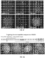
FIGS. 3A-3D depicts a comparison of different sgRNA designs for CRISPR-mediated gene repression. Different sgRNAs were transiently transfected into a GFP+ HEK293 reporter cell line harboring a genomic integrated dCas9-KRAB gene. The fluorescence was assayed using flow cytometry. Modifications (underlined) of sgRNA sequence with a polymerase III SINE element polyadenylation signal sequence (#2), A-U flip (#3, 4, 5 and 6), hairpin extension (#7 and 8) and combined changes were hypothesized to increase sgRNA expression level, stability or its association with dCas9 protein. Repression fold is calculated by dividing the fluorescence of +sgGAL4 and fluorescence+different designs. The fold increase in repression is calculated by dividing the fluorescence of Design #1 and fluorescence of other designs. The unmodified hairpin nucleotides are boxed.
FIGS. 4A-4C Optimized sgRNA designs enhance the labeling efficiency of telomeres. (A) Both designs of A-U flip (#6) and hairpin extension (#8) increased the telomere labeling efficiency compared to the original sgRNA design (#1), as more dCas9-EGFP puncta were observed. Combining both modifications of A-U flip and hairpin extension (#10) further enhanced the labeling efficiency and reduced background fluorescence signal. This design was used for later imaging and annotated as (F+E). Each conventional fluorescence image shows one projected three dimensional (3-D) stack of 3 μm deep (an entire RPE nucleus) with 0.4 μm focal spacing. Scale bar, 5 μm. (B) A plot of overall telomere intensity distribution comparing the telomere labeling efficiencies of different sgRNA designs. Each spot stands for one cell. The percentage of whole-cell GFP is calculated as the ratio of overall telomere intensity and overall GFP signal in the whole cell.
FIG. 5 Labeling efficiency of telomeres is dosage-dependent of the sgRNA lentivirus. With the best sgRNA design (F+E), telomeres were labeled when infected with 50 μL lentivirus, but there were some nucleolus-like structures highlighted in the background. Infection with 200 μL lentivirus increased telomere labeling efficiency without nucleolus-like structures, possibly due to enhanced assembly between sgRNAs and the dCas9-EGFP protein. A good balance between labeling efficiency and cytotoxicity was achieved by infecting the cells with 100 μL sgRNA lentivirus.
FIG. 6 depicts the co-localization of CRISPR labeling with telomere markers. CRISPR can effectively detect telomere length changes in living cells. (A) Co-localization of dCas9-EGFP and telomeres as labeled by Oligo FISH (top) or antibody to TRF2 (bottom). (B-C) Telomere elongation was induced in UMUC3 cancer cells (starting telomere length 2-5 kb) by overexpression of human telomerase RNA (hTR). (B) Visualization of telomeres in UMUC3 cells by CRISPR labeling. (C) Telomere fluorescence intensity in CRISPR-labeled cells increases after elongation of telomeres by hTR. Scale bar, 5 μm.
FIG. 7 illustrates that no DNA damage response was detected at the telomeres labeled by CRISPR. (A) Co-localization of PNA FISH and 53BP1 staining in RPE cells, dCas9-EGFP-labeled RPE cells, and TIN2 shRNA-treated RPE cells. TIN2 shRNA infecting the RPE cells was used to induce telomeric DNA damage response revealed by 53BP1 antibody staining 53BP1 signal was enriched at the telomeres indicated by PNA probe in most cells when infected with TIN2 shRNA. There was no obvious enrichment of 53BP1 at the telomeres labeled by PNA probes or dCas9-EGFP. (B) Histogram showing the quantification of 53BP1 signal enriched at the telomeres.
FIG. 8 depicts the results of imaging of endogenous MUC4 gene by targeting the repetitive or non-repetitive regions via dCas9-EGFP. (A) Schematic of the human MUC4 locus containing two repeated regions in exon 2 and intron 3 as indicated. The sgRNA target sites are indicated by gray lines and the adjacent PAMs are shown. (B) Conventional fluorescence images of MUC4 loci (arrows) in RPE cells by targeting the exon 2 repeats with three different sgRNAs. (C) Two protospacers with different lengths, 13 bp and 23 bp, were chosen as targets in the intron repeats. MUC4 intron (arrows) can only be labeled by using optimized sgRNA design when targeting the 23 bp protospacer. (D) Histograms of MUC4 loci counts by labeling exon 2 via CRISPR. (E) Histograms of MUC4 loci counts by labeling intron 3 via CRISPR. (F) Co-localization of dCas9-EGFP and Oligo FISH probes for both exon and intron labeling. (G) 73 protospacers in the first exon were selected for labeling the non-repetitive region of MUC4 gene. 1-3 spots (arrows) can be detected with 36 and 73 sgRNAs. Co-labeling of MUC4 using 73 sgRNAs and sgMUC4-E3 shows two proximal spots (arrowhead). The inset shows the magnification of the white box region. (H) Histograms of MUC4 loci counts. 26 sgRNAs or more are required to detect the non-repetitive genome loci. Scale bar, 5 μm.
FIG. 9 depicts the results of a karyotype analysis of the RPE cell line. Cytogenetic analysis was performed on ten G-banded metaphase cells of human RPE cell line. The chromosomes were stained with dyes that show a pattern of light and dark bands (called the banding pattern). The banding pattern for each chromosome was specific and consistent allowing identification of each of the 24 chromosomes. 10 cells were analyzed. This cell line demonstrated a hypertriploid karyotype (73 chromosomes in total) with female origin. There were extra copies of chromosome 5, 7, 11, 12, 16, 19, and 20 that were present in eight or nine cells except for chromosome 16 (six cells) and 19 (four cells). Chromosome 10 and 22 were also lost in nine and eight cells respectively. All ten cells had two copies of an abnormal X chromosome with addition of unidentifiable genetic material translocated to the long-arm at Xq28.
FIG. 10: depicts CRISPR imaging of MUC4 and telomeres in the monoclones of HeLa cell line. (A) In addition to RPE and UMUC3 cell lines, CRISPR can also detect MUC4 loci and telomeres in living HeLa cells. Three MUC4 loci (arrows) were detected by CRISPR via targeting the repetitive exon region. (B) Histograms of MUC4 loci counts by CRISPR labeling. (C) MUC4 locus is localized at 3q29 of chromosome 3. Two hundred interphase nuclei of HeLa cells were examined by FISH using two probes hybridized to 3q26.1 and 3q28-29, respectively. All two hundred cells demonstrated a FISH signal pattern of three copies of 3q28-29, suggesting trisomy chromosome 3 in HeLa cells.
FIG. 11 depicts CRISPR imaging of MUC1 loci in living RPE cells. (A) Schematic depicting the structure of MUC1 gene, which contains a 60 bp unit repeated 20 to 140 copies in both exon 3 and intron 3. Four sgRNAs were designed to target the repeat region, and their target sites are indicated with gray lines and the adjacent PAM are also indicated. (B) MUC1 loci (arrows) visualized in RPE cells by targeting four different protospacers. (C) The labeling specificity of CRISPR was confirmed by oligo FISH labeling. The co-localization of dCas9-EGFP and oligo FISH is indicated by an arrow. (D) Histograms of the labeling efficiency of MUC1 loci by targeting different protospacers. 1-5 spots can be observed in 90% of the cells with sgMUC1-E1 and 95% of cells with sgMUC1-E3, but only in 45% and 20% of cells with sgMUC1-E2 and sgMUC1-E4, respectively. The sgRNA design (sgMUC1-E4 (F+E)) increased the labeling efficiency compared to sgMUC1-E4 from 20% to 55%.
FIG. 12 depicts a comparison of labeling efficiency between CRISPR and Oligo FISH. (A) Oligo FISH labeling of MUC4 exon, MUC4 intron, and MUC1 exon as indicated by arrows. (B) Histograms showing the statistics of observed spots for labeling MUC4 exon, MUC4 intron, and MUC1 exon by Oligo FISH or CRISPR. CRISPR shows a higher labeling efficiency in all cases.
FIG. 13 depicts imaging of multiple elements via CRISPR. (A) The loci of MUC1 and MUC4 were co-labeled by infecting RPE cells with both sgMUC1-E1 and sgMUC4-E3. More spots (arrows) were observed compared to individual sgMUC1-E1 or sgMUC4-E3 labeling, while similar number of spots was detected if infecting the cells with both sgMUC4-E3 and sgMUC4-I2 (F+E) that target the same MUC4 (B) Histograms showing the counts of Mucin genes loci. 45% of cells targeting both MUC1 and MUC4 contain more than 6 spots, while only 10% of cells have 6 spots with co-infection of two sgMUC4s.
FIG. 14 depicts single particle tracking and diffusion dynamics of MUC4 labeled by CRISPR. (A) Conventional fluorescence image of MUC4 (left) and three example traces of marked foci (right). All traces show confined diffusion, but focus 2 is actively transported in addition. (B) Averaged Mean Square Displacement (MSD) of 119 and 50 traces from 26 cells, categorized as confined diffusive (darker line) and actively transported (lighter line). The shaded areas represent the standard error of the mean. (C) Histogram of confinement characteristic length and (D) histogram of microscopic diffusion coefficient derived from MSDs. (E) Data illustrating individual MUC4 loci movement. (F) Analysis of MUC4 loci movement depicts two distinct movement modes: confined diffusion and active transport of confined diffusion. (G) 80% of detected MUC4 loci followed confined diffusion, suggesting the movement of local chromatin is modulated by nuclear factors. (H) The median speed was 0.011 um2/s as defined by diffusion coefficient.
DETAILED DESCRIPTION OF THE INVENTION I. Definitions
As used in this specification and the appended claims, the singular forms “a,” “an,” and “the” include plural reference unless the context clearly dictates otherwise.
The term “nucleic acid” or “polynucleotide” refers to deoxyribonucleic acids (DNA) or ribonucleic acids (RNA) and polymers thereof in either single- or double-stranded form. Unless specifically limited, the term encompasses nucleic acids containing known analogues of natural nucleotides that have similar binding properties as the reference nucleic acid and are metabolized in a manner similar to naturally occurring nucleotides. Unless otherwise indicated, a particular nucleic acid sequence also implicitly encompasses conservatively modified variants thereof (e.g., degenerate codon substitutions), alleles, orthologs, SNPs, and complementary sequences as well as the sequence explicitly indicated. Specifically, degenerate codon substitutions may be achieved by generating sequences in which the third position of one or more selected (or all) codons is substituted with mixed-base and/or deoxyinosine residues (Batzer et al., Nucleic Acid Res. 19:5081 (1991); Ohtsuka et al., J. Biol. Chem. 260:2605-2608 (1985); and Rossolini et al., Mol. Cell. Probes 8:91-98 (1994)). The term nucleic acid is used interchangeably with gene, cDNA, and mRNA encoded by a gene.
The term “gene” means the segment of DNA involved in producing a polypeptide chain. It may include regions preceding and following the coding region (leader and trailer) as well as intervening sequences (introns) between individual coding segments (exons).
A “promoter” is defined as an array of nucleic acid control sequences that direct transcription of a nucleic acid. As used herein, a promoter includes necessary nucleic acid sequences near the start site of transcription, such as, in the case of a polymerase II type promoter, a TATA element. A promoter also optionally includes distal enhancer or repressor elements, which can be located as much as several thousand base pairs from the start site of transcription.
An “expression cassette” is a nucleic acid construct, generated recombinantly or synthetically, with a series of specified nucleic acid elements that permit transcription of a particular polynucleotide sequence in a host cell. An expression cassette may be part of a plasmid, viral genome, or nucleic acid fragment. Typically, an expression cassette includes a polynucleotide to be transcribed, operably linked to a promoter.
A “reporter gene” encodes proteins that are readily detectable due to their biochemical characteristics, such as enzymatic activity or chemifluorescent features. One specific example of such a reporter is green fluorescent protein. Fluorescence generated from this protein can be detected with various commercially-available fluorescent detection systems. Other reporters can be detected by staining. The reporter can also be an enzyme that generates a detectable signal when contacted with an appropriate substrate. The reporter can be an enzyme that catalyzes the formation of a detectable product. Suitable enzymes include, but are not limited to, proteases, nucleases, lipases, phosphatases and hydrolases. The reporter can encode an enzyme whose substrates are substantially impermeable to eukaryotic plasma membranes, thus making it possible to tightly control signal formation. Specific examples of suitable reporter genes that encode enzymes include, but are not limited to, CAT (chloramphenicol acetyl transferase; Alton and Vapnek (1979) Nature 282: 864-869); luciferase (lux); β-galactosidase; LacZ; β-glucuronidase; and alkaline phosphatase (Toh, et al. (1980) Eur. J. Biochem. 182: 231-238; and Hall et al. (1983) J. Mol. Appl. Gen. 2: 101), each of which is incorporated by reference herein in the entirety. Other suitable reporters include those that encode for a particular epitope that can be detected with a labeled antibody that specifically recognizes the epitope.
The term “amino acid” refers to naturally occurring and synthetic amino acids, as well as amino acid analogs and amino acid mimetics that function in a manner similar to the naturally occurring amino acids. Naturally occurring amino acids are those encoded by the genetic code, as well as those amino acids that are later modified, e.g., hydroxyproline, γ-carboxyglutamate, and O-phosphoserine Amino acid analogs refers to compounds that have the same basic chemical structure as a naturally occurring amino acid, i.e., an a carbon that is bound to a hydrogen, a carboxyl group, an amino group, and an R group, e.g., homoserine, norleucine, methionine sulfoxide, methionine methyl sulfonium. Such analogs have modified R groups (e.g., norleucine) or modified peptide backbones, but retain the same basic chemical structure as a naturally occurring amino acid. “Amino acid mimetics” refers to chemical compounds having a structure that is different from the general chemical structure of an amino acid, but that functions in a manner similar to a naturally occurring amino acid.
There are various known methods in the art that permit the incorporation of an unnatural amino acid derivative or analog into a polypeptide chain in a site-specific manner, see, e.g., WO 02/086075.
Amino acids may be referred to herein by either the commonly known three letter symbols or by the one-letter symbols recommended by the IUPAC-IUB Biochemical Nomenclature Commission. Nucleotides, likewise, may be referred to by their commonly accepted single-letter codes.
“Polypeptide,” “peptide,” and “protein” are used interchangeably herein to refer to a polymer of amino acid residues. All three terms apply to amino acid polymers in which one or more amino acid residue is an artificial chemical mimetic of a corresponding naturally occurring amino acid, as well as to naturally occurring amino acid polymers and non-naturally occurring amino acid polymers. As used herein, the terms encompass amino acid chains of any length, including full-length proteins, wherein the amino acid residues are linked by covalent peptide bonds.
“Conservatively modified variants” applies to both amino acid and nucleic acid sequences. With respect to particular nucleic acid sequences, “conservatively modified variants” refers to those nucleic acids that encode identical or essentially identical amino acid sequences, or where the nucleic acid does not encode an amino acid sequence, to essentially identical sequences. Because of the degeneracy of the genetic code, a large number of functionally identical nucleic acids encode any given protein. For instance, the codons GCA, GCC, GCG and GCU all encode the amino acid alanine. Thus, at every position where an alanine is specified by a codon, the codon can be altered to any of the corresponding codons described without altering the encoded polypeptide. Such nucleic acid variations are “silent variations,” which are one species of conservatively modified variations. Every nucleic acid sequence herein that encodes a polypeptide also describes every possible silent variation of the nucleic acid. One of skill will recognize that each codon in a nucleic acid (except AUG, which is ordinarily the only codon for methionine, and TGG, which is ordinarily the only codon for tryptophan) can be modified to yield a functionally identical molecule. Accordingly, each silent variation of a nucleic acid that encodes a polypeptide is implicit in each described sequence.
As to amino acid sequences, one of skill will recognize that individual substitutions, deletions or additions to a nucleic acid, peptide, polypeptide, or protein sequence which alters, adds or deletes a single amino acid or a small percentage of amino acids in the encoded sequence is a “conservatively modified variant” where the alteration results in the substitution of an amino acid with a chemically similar amino acid. Conservative substitution tables providing functionally similar amino acids are well known in the art. Such conservatively modified variants are in addition to and do not exclude polymorphic variants, interspecies homologs, and alleles of the invention. In some cases, conservatively modified variants of Cas9 or sgRNA can have an increased stability, assembly, or activity as described herein.
The following eight groups each contain amino acids that are conservative substitutions for one another:
1) Alanine (A), Glycine (G);
2) Aspartic acid (D), Glutamic acid (E);
3) Asparagine (N), Glutamine (Q);
4) Arginine (R), Lysine (K);
5) Isoleucine (I), Leucine (L), Methionine (M), Valine (V);
6) Phenylalanine (F), Tyrosine (Y), Tryptophan (W);
7) Serine (S), Threonine (T); and
8) Cysteine (C), Methionine (M)
(see, e.g., Creighton, Proteins, W. H. Freeman and Co., N. Y. (1984)).
Amino acids may be referred to herein by either their commonly known three letter symbols or by the one-letter symbols recommended by the IUPAC-IUB Biochemical Nomenclature Commission. Nucleotides, likewise, may be referred to by their commonly accepted single-letter codes.
In the present application, amino acid residues are numbered according to their relative positions from the left most residue, which is numbered 1, in an unmodified wild-type polypeptide sequence.
As used in herein, the terms “identical” or percent “identity,” in the context of describing two or more polynucleotide or amino acid sequences, refer to two or more sequences or subsequences that are the same or have a specified percentage of amino acid residues or nucleotides that are the same. For example, a core small guide RNA (sgRNA) sequence responsible for assembly and activity of a sgRNA:nuclease complex has at least 80% identity, preferably 85%, 90%, 91%, 92%, 93, 94%, 95%, 96%, 97%, 98%, 99%, or 100% identity, to a reference sequence, e.g., one of SEQ ID NOs:1-4), when compared and aligned for maximum correspondence over a comparison window, or designated region as measured using one of the following sequence comparison algorithms or by manual alignment and visual inspection. As another example, a Cas9 sequence responsible for assembly and activity of a sgRNA:nuclease complex has at least 80% identity, preferably 85%, 90%, 91%, 92%, 93, 94%, 95%, 96%, 97%, 98%, 99%, or 100% identity, to a reference sequence, e.g., one of SEQ ID NOs:5-6), when compared and aligned for maximum correspondence over a comparison window, or designated region as measured using one of the following sequence comparison algorithms or by manual alignment and visual inspection. Such sequences are then said to be “substantially identical.” With regard to polynucleotide sequences, this definition also refers to the complement of a test sequence. With regard to amino acid sequences, preferably, the identity exists over a region that is at least about 50 amino acids or nucleotides in length, or more preferably over a region that is 75-100 amino acids or nucleotides in length.
For sequence comparison, typically one sequence acts as a reference sequence, to which test sequences are compared. When using a sequence comparison algorithm, test and reference sequences are entered into a computer, subsequence coordinates are designated, if necessary, and sequence algorithm program parameters are designated. Default program parameters can be used, or alternative parameters can be designated. The sequence comparison algorithm then calculates the percent sequence identities for the test sequences relative to the reference sequence, based on the program parameters. For sequence comparison of nucleic acids and proteins, the BLAST and BLAST 2.0 algorithms and the default parameters discussed below are used.
A “comparison window”, as used herein, includes reference to a segment of any one of the number of contiguous positions selected from the group consisting of from 20 to 600, usually about 50 to about 200, more usually about 100 to about 150 in which a sequence may be compared to a reference sequence of the same number of contiguous positions after the two sequences are optimally aligned. Methods of alignment of sequences for comparison are well-known in the art. Optimal alignment of sequences for comparison can be conducted, e.g., by the local homology algorithm of Smith & Waterman, Adv. Appl. Math. 2:482 (1981), by the homology alignment algorithm of Needleman & Wunsch, J. Mol. Biol. 48:443 (1970), by the search for similarity method of Pearson & Lipman, Proc. Nat'l. Acad. Sci. USA 85:2444 (1988), by computerized implementations of these algorithms (GAP, BESTFIT, FASTA, and TFASTA in the Wisconsin Genetics Software Package, Genetics Computer Group, 575 Science Dr., Madison, Wis.), or by manual alignment and visual inspection (see, e.g., Current Protocols in Molecular Biology (Ausubel et al., eds. 1995 supplement)).
Examples of algorithms that are suitable for determining percent sequence identity and sequence similarity are the BLAST and BLAST 2.0 algorithms, which are described in Altschul et al., (1990) J. Mol. Biol. 215: 403-410 and Altschul et al. (1977) Nucleic Acids Res. 25: 3389-3402, respectively. Software for performing BLAST analyses is publicly available at the National Center for Biotechnology Information website, ncbi.nlm.nih.gov. The algorithm involves first identifying high scoring sequence pairs (HSPs) by identifying short words of length W in the query sequence, which either match or satisfy some positive-valued threshold score T when aligned with a word of the same length in a database sequence. T is referred to as the neighborhood word score threshold (Altschul et al, supra). These initial neighborhood word hits acts as seeds for initiating searches to find longer HSPs containing them. The word hits are then extended in both directions along each sequence for as far as the cumulative alignment score can be increased. Cumulative scores are calculated using, for nucleotide sequences, the parameters M (reward score for a pair of matching residues; always >0) and N (penalty score for mismatching residues; always <0). For amino acid sequences, a scoring matrix is used to calculate the cumulative score. Extension of the word hits in each direction are halted when: the cumulative alignment score falls off by the quantity X from its maximum achieved value; the cumulative score goes to zero or below, due to the accumulation of one or more negative-scoring residue alignments; or the end of either sequence is reached. The BLAST algorithm parameters W, T, and X determine the sensitivity and speed of the alignment. The BLASTN program (for nucleotide sequences) uses as defaults a word size (W) of 28, an expectation (E) of 10, M=1, N=−2, and a comparison of both strands. For amino acid sequences, the BLASTP program uses as defaults a word size (W) of 3, an expectation (E) of 10, and the BLOSUM62 scoring matrix (see Henikoff & Henikoff, Proc. Natl. Acad. Sci. USA 89:10915 (1989)).
The BLAST algorithm also performs a statistical analysis of the similarity between two sequences (see, e.g., Karlin & Altschul, Proc. Nat'l. Acad. Sci. USA 90:5873-5787 (1993)). One measure of similarity provided by the BLAST algorithm is the smallest sum probability (P(N)), which provides an indication of the probability by which a match between two nucleotide or amino acid sequences would occur by chance. For example, a nucleic acid is considered similar to a reference sequence if the smallest sum probability in a comparison of the test nucleic acid to the reference nucleic acid is less than about 0.2, more preferably less than about 0.01, and most preferably less than about 0.001.
An indication that two nucleic acid sequences or polypeptides are substantially identical is that the polypeptide encoded by the first nucleic acid is immunologically cross reactive with the antibodies raised against the polypeptide encoded by the second nucleic acid, as described below. Thus, a polypeptide is typically substantially identical to a second polypeptide, for example, where the two peptides differ only by conservative substitutions. Another indication that two nucleic acid sequences are substantially identical is that the two molecules or their complements hybridize to each other under stringent conditions, as described below. Yet another indication that two nucleic acid sequences are substantially identical is that the same primers can be used to amplify the sequence. Yet another indication that two polypeptides are substantially identical is that the two polypeptides retain identical or substantially similar activity.
A “translocation sequence” or “transduction sequence” refers to a peptide or protein (or active fragment or domain thereof) sequence that directs the movement of a protein from one cellular compartment to another, or from the extracellular space through the cell or plasma membrane into the cell. Translocation sequences that direct the movement of a protein from the extracellular space through the cell or plasma membrane into the cell are “cell penetration peptides.” Translocation sequences that localize to the nucleus of a cell are termed “nuclear localization” sequences, signals, domains, peptides, or the like. Examples of translocation sequences include, without limitation, the TAT transduction domain (see, e.g., S. Schwarze et al., Science 285 (Sep. 3, 1999); penetratins or penetratin peptides (D. Derossi et al., Trends in Cell Biol. 8, 84-87); Herpes simplex virus type 1 VP22 (A. Phelan et al., Nature Biotech. 16, 440-443 (1998), and polycationic peptides (Cell Mol. Life Sci. 62 (2005) 1839-1849). Further translocation sequences are known in the art. Translocation peptides can be fused (e.g. at the amino or carboxy terminus), conjugated, or coupled to a compound of the present invention, to, among other things, produce a conjugate compound that may easily pass into target cells, or through the blood brain barrier and into target cells.
The “CRISPR/Cas” system refers to a widespread class of bacterial systems for defense against foreign nucleic acid. CRISPR/Cas systems are found in a wide range of eubacterial and archaeal organisms. CRISPR/Cas systems include type I, II, and III sub-types. Wild-type type II CRISPR/Cas systems utilize the RNA-mediated nuclease, Cas9 in complex with guide and activating RNA to recognize and cleave foreign nucleic acid.
Cas9 homologs are found in a wide variety of eubacteria, including, but not limited to bacteria of the following taxonomic groups: Actinobacteria, Aquificae, Bacteroidetes-Chlorobi, Chlamydiae-Verrucomicrobia, Chlroflexi, Cyanobacteria, Firmicutes, Proteobacteria, Spirochaetes, and Thermotogae. An exemplary Cas9 protein is the Streptococcus pyogenes Cas9 protein. Additional Cas9 proteins and homologs thereof are described in, e.g., Chylinksi, et al., RNA Biol. 2013 May 1; 10(5): 726-737; Nat. Rev. Microbiol. 2011 June; 9(6): 467-477; Hou, et al., Proc Natl Acad Sci USA. 2013 Sep. 24; 110(39):15644-9; Sampson et al., Nature. 2013 May 9; 497(7448):254-7; and Jinek, et al., Science. 2012 Aug. 17; 337(6096):816-21.
As used herein, “activity” in the context of CRISPR/Cas activity, Cas9 activity, sgRNA activity, sgRNA:nuclease activity and the like refers to the ability to bind to a target sequence and/or label or cleave the target sequence. Such activity can be measured in a variety of ways as known in the art. For example, expression, activity, or level of a reporter gene can be measured, and sgRNA:nucleases targeting the reporter gene sequence can be assayed for their ability to reduce the expression, activity, or level of the reporter gene. For example, a cell can be transfected with an expression cassette encoding a green fluorescent protein under the control of a constituitive promoter. The fluorescence intensity can be measured and compared to the intensity of the cell after transfection with Cas9 and candidate sgRNAs to identify optimized sgRNAs
II. Introduction
Described herein are methods and compositions for analyzing or modifying target nucleic acids. The methods and compositions are based on an optimized CRISPR/Cas system that employs a small guide RNA (sgRNA) and an sgRNA-mediated nuclease. The sgRNA contains a binding region that provides specific binding to a nucleic acid target sequence that is substantially complementary to the sequence of the binding region. The sgRNA and the nuclease can form a complex that specifically binds to the nucleic acid target sequence.
III. Compositions
In some embodiments, an small guide RNA (sgRNA) molecule is provided. sgRNAs contain a binding region that determines the sequence specificity of the sgRNA and the sgRNA:nuclease complex, a 5′ stem-loop region that, at least in part, participates in assembly and interaction with a sgRNA-mediated nuclease; an intervening sequence, a 3′ stem-loop region, and a termination sequence.
The binding region can be between about 5 and 100 nucleotides long, or longer (e.g., 5, 6, 7, 8, 9, 10, 11, 12, 13, 14, 15, 16, 17, 18, 19, 20, 21, 22, 23, 24, 25, 26, 27, 28, 29, 30, 31, 32, 33, 34, 35, 36, 37, 38, 39, 40, 41, 42, 43, 44, 45, 46, 47, 48, 49, 50, 51, 52, 53, 54, 55, 56, 57, 58, 59 60, 61, 62, 63, 63, 64, 65, 66, 67, 68, 69, 70, 71, 72, 73, 74, 75, 76, 77, 78, 79, 80, 81, 82, 83, 84, 85, 86, 87, 88, 89, 90, 91 92, 93, 94, 95, 96, 97, 98, 99, or 100 nucleotides in length, or longer). In some cases, the binding region is between about 15 and about 30 nucleotides in length (e.g., about 15-29, 15-26, 15-25; 16-30, 16-29, 16-26, 16-25; or about 18-30, 18-29, 18-26, or 18-25 nucleotides in length). Generally, the binding region is designed to complement or substantially complement the target nucleic acid sequence or sequences. In some cases, the binding region can incorporate wobble or degenerate bases to bind multiple sequences. In some cases, the binding region can be altered to increase stability. For example, non-natural nucleotides, can be incorporated to increase RNA resistance to degradation. In some cases, the binding region can be altered or designed to avoid or reduce secondary structure formation in the binding region. In some cases, the binding region can be designed to optimize G-C content. In some cases, G-C content is preferably between about 40% and about 60% (e.g., 40%, 45%, 50%, 55%, 60%). In some cases, the binding region can contain modified nucleotides such as, without limitation, methylated or phosphorylated nucleotides.
The 5′ stem-loop region can be between about 15 and about 50 nucleotides in length (e.g., about 15, 16, 17, 18, 19, 20, 21, 22, 23, 24, 25, 26, 27, 28, 29, 30, 31, 32, 33, 34, 35, 36, 37, 38, 39, 40, 41, 42, 43, 44, 45, 46, 47, 48, 49, or about 50 nucleotides in length). In some cases, the 5′ stem-loop region is between about 30-45 nucleotides in length (e.g., about 31, 32, 33, 34, 35, 36, 37, 38, 39, 40, 41, 42, 43, 44, or 45 nucleotides in length). In some cases, the 5′ stem-loop region is at least about 31 nucleotides in length (e.g., at least about 31, 32, 33, 34, 35, 36, 37, 38, 39, 40, 41, 42, 43, 44, or 45 nucleotides in length). In some cases, the 5′ stem-loop structure contains one or more loops or bulges, each loop or bulge of about 1, 2, 3, 4, 5, 6, 7, 8, 9, or 10 nucleotides. In some cases, the 5′ stem-loop structure contains a stem of between about 10 and 30 complementary base pairs (e.g., 11, 12, 13, 14, 15, 16, 17, 18, 19, 20, 21, 22, 23, 24, 25, 26, 27, 28, 29, or 30 complementary base pairs).
In some embodiments, the 5′ stem-loop structure can contain protein-binding, or small molecule-binding structures. In some cases, the 5′ stem-loop function (e.g., interacting or assembling with a sgRNA-mediated nuclease) can be conditionally activated by drugs, growth factors, small molecule ligands, or a protein that binds to the protein-binding structure of the 5′ stem-loop. In some embodiments, the 5′ stem-loop structure can contain non-natural nucleotides. For example, non-natural nucleotides can be incorporated to enhance protein-RNA interaction, or to increase the thermal stability or resistance to degradation of the sgRNA.
The intervening sequence between the 5′ and 3′ stem-loop structures can be between about 10 and about 50 nucleotides in length (e.g., about 10, 11, 12, 13, 14, 15, 16, 17, 18, 19, 20, 21, 22, 23, 24, 25, 26, 27, 28, 29, 30, 31, 32, 33, 34, 35, 36, 37, 38, 39, 40, 41, 42, 43, 44, 45, 46, 47, 48, 49, or about 50 nucleotides in length). In some cases, the intervening sequence is designed to be linear, unstructured, substantially linear, or substantially unstructured. In some embodiments, the intervening sequence can contain non-natural nucleotides. For example, non-natural nucleotides can be incorporated to enhance protein-RNA interaction or to increase the activity of the sgRNA:nuclease complex. As another example, natural nucleotides can be incorporated to enhance the thermal stability or resistance to degradation of the sgRNA.
The 3′ stem-loop structure can contain an about 3, 4, 5, 6, 7, or 8 nucleotide loop and an about 4, 5, 6, 7, 8, 9, 10, 11, 12, 13, 14, 15, 16, 17, 18, 19, 20, 21, 22, 23, 24, or 25 nucleotide or longer stem. In some cases, the 3′ stem-loop can contain a protein-binding, small molecule-binding, hormone-binding, or metabolite-binding structure that can conditionally stabilize the secondary and/or tertiary structure of the sgRNA. In some embodiments, the 3′ stem-loop can contain non-natural nucleotides. For example, non-natural nucleotides can be incorporated to enhance protein-RNA interaction or to increase the activity of the sgRNA:nuclease complex. As another example, natural nucleotides can be incorporated to enhance the thermal stability or resistance to degradation of the sgRNA.
In some embodiments, the sgRNA includes a termination structure at its 3′ end. In some cases, the sgRNA includes an additional 3′ hairpin structure, e.g., before the termination structure, that can interact with proteins, small-molecules, hormones, etc., for stabilization or additional functionality, such as conditional stabilization or conditional regulation of sgRNA:nuclease assembly or activity.
In some cases, the sgRNA is optimized to enhance stability, assembly, and/or expression. In some case, the sgRNA is optimized to enhance the activity of a sgRNA:nuclease complex as compared to previously known sgRNAs, such as an sgRNA encoded by:
SEQ ID NO: 1 
[N]5-100GUUUUAGAGCUAGAAAUAGCAAGUUAAAAUAAGGCUAGUCC
GUUAUCAACUUGAAAAAGUGGCACCGAGUCGGUGCUUUUUUU,

where [N] represents a target specific binding region of between about 5-100 nucleotides (e.g., about 5, 10, 15, 20, 15, 30, 35, 40, 45, 50, 55, 60, 70, 80, or 90 nucleotides) that is complementary or substantially complementary to the target nucleic acid. In some cases, the optimized sgRNA provides enhanced activity as compared to a previously known sgRNA or an sgRNA substantially identical to a previously known sgRNA. As used herein, identity of an sgRNA to another sgRNA, such as an sgRNA to SEQ ID NO:1 is determined with reference to the identity to the nucleotide sequences outside of the binding region. For example, two sgRNAs with 0% identity inside the binding region and 100% identity outside the binding region are 100% identical to each other. Similarly, as used herein, number of substitutions, additions, or deletions of an sgRNA as compared to another, such as an sgRNA compared to SEQ ID NO:1 is determined with reference to the nucleotide sequences outside of the binding region. For example, two sgRNAs with multiple additions, substitutions, and/or deletions inside the binding region and 100% identity outside the binding region are considered to contain 0 nucleotide substitutions, additions, or deletions.
In some cases, the optimized sgRNAs described herein form an sgRNA:nuclease complex with enhanced activity as compared to SEQ ID NO:1, or an sgRNA 90, 95, 96, 97, 98, or 99% or more identical to SEQ ID NO:1. In some cases, the optimized sgRNAs described herein form an sgRNA:nuclease complex with enhanced activity as compared to SEQ ID NO:1, or an sgRNA with fewer than 5, 4, 3, or 2 nucleotide substitutions, additions, or deletions of SEQ ID NO:1.
In some embodiments, the sgRNA can be optimized for expression by substituting, deleting, or adding one or more nucleotides. In some cases, a nucleotide sequence that provides inefficient transcription from an encoding template nucleic acid can be deleted or substituted. For example, in some cases, the sgRNA is transcribed from a nucleic acid operably linked to an RNA polymerase III promoter. In such cases, sgRNA sequences that result in inefficient transcription by RNA polymerase III, such as those described in Nielsen et al., Science. 2013 Jun. 28; 340(6140):1577-80, can be deleted or substituted. For example, one or more consecutive uracils can be deleted or substituted from the sgRNA sequence. In some cases, the consecutive uracils are present in the stem portion of a stem-loop structure. In such cases, one or more of the consecutive uracils can be substituted by exchanging the uracil and its complementary base. For example, if the uracil is hydrogen bonded to a corresponding adenine, the sgRNA sequence can be altered to exchange the adenine and uracil. This “A-U flip” can retain the overall structure and function of the sgRNA molecule while improving expression by reducing the number of consecutive uracil nucleotides. In some cases, the sgRNA containing an A-U flip is encoded by:
SEQ ID NO: 2
[N]5-100GUUUAAGAGCUAGAAAUAGCAAGUUUAAAUAAGGCUAGUCC
GUUAUCAACUUGAAAAAGUGGCACCGAGUCGGUGCU,

where the A-U flipped nucleotides are underlined. In some cases, the optimized sgRNA is at least 90, 91, 92, 93, 94, 95, 96, 97, 98, or 99% identical or more to SEQ ID NO:2, or contains fewer than 10, 9, 8, 7, 6, 5, 4, 3, or 2 nucleotide additions, deletions, or substitutions compared to SEQ ID NO:2. Alternatively, the A-U pair can be replaced by a G-C, C-G, A-C, G-U pair.
In some embodiments, the sgRNA can be optimized for stability. Stability can be enhanced by optimizing the stability of the sgRNA:nuclease interaction, optimizing assembly of the sgRNA:nuclease complex, removing or altering RNA destabilizing sequence elements, or adding RNA stabilizing sequence elements. In some embodiments, the sgRNA contains a 5′ stem-loop structure proximal to, or adjacent to, the binding region that interacts with the sgRNA-mediated nuclease. Optimization of the 5′ stem-loop structure can provide enhanced stability or assembly of the sgRNA:nuclease complex. In some cases, the 5′ stem-loop structure is optimized by increasing the length of the stem portion of the stem-loop structure. An exemplary sgRNA containing an optimized 5′ stem-loop structure is encoded by:
SEQ ID NO: 3
[N]5-100GUUUUAGAGCUAUGCUGGAAACAGCAUAGCAAGUUAAAAU
AAGGCUAGUCCGUUAUCAACUUGAAAAAGUGGCACCGAGUCGGUGC
UUUUUUU,

where the nucleotides contributing to the elongated stem portion of the 5′ stem-loop structure are underlined. In some cases, the optimized sgRNA is at least 90, 91, 92, 93, 94, 95, 96, 97, 98, or 99% identical or more to SEQ ID NO:3, or contains fewer than 10, 9, 8, 7, 6, 5, 4, 3, or 2 nucleotide additions, deletions, or substitutions compared to SEQ ID NO:3.
In some embodiments, the 5′ stem-loop optimization is combined with mutations for increased transcription to provide an optimized sgRNA. For example, an A-U flip and an elongated stem loop can be combined to provide an optimized sgRNA. An exemplary sgRNA containing an A-U flip and an elongated 5′ stem-loop is encoded by:
SEQ ID NO: 4
[N]5-100GUUUAAGAGCUAUGCUGGAAACAGCAUAGCAAGUUUAAAUAA
GGCUAGUCCGUUAUCAACUUGAAAAAGUGGCACCGAGUCGGUGCUUUU
UUU,

where the A-U flipped nucleotides and the nucleotides contributing to the elongated stem portion of the 5′ stem-loop structure are underlined. In some cases, the optimized sgRNA is at least 90, 91, 92, 93, 94, 95, 96, 97, 98, or 99% identical or more to SEQ ID NO:4, or contains fewer than 10, 9, 8, 7, 6, 5, 4, 3, or 2 nucleotide additions, deletions, or substitutions compared to SEQ ID NO:4.
sgRNAs can be modified by methods known in the art. In some cases, the modifications can include, but are not limited to, the addition of one or more of the following sequence elements: a 5′ cap (e.g., a 7-methylguanylate cap); a 3′ polyadenylated tail; a riboswitch sequence; a stability control sequence; a hairpin; a subcellular localization sequence; a detection sequence or label; or a binding site for one or more proteins. Modifications can also include the introduction of non-natural nucleotides including, but not limited to, one or more of the following: fluorescent nucleotides and methylated nucleotides.
In some embodiments, the sgRNA can contain from 5′ to 3′: (i) a binding region of between about 10 and about 50 nucleotides; (ii) a 5′ hairpin region containing fewer than four consecutive uracil nucleotides, or a length of at least 31 nucleotides (e.g., from about 31 to about 41 nucleotides); (iii) a 3′ hairpin region; and (iv) a transcription termination sequence, wherein the small guide RNA is configured to form a complex with a small guide RNA-mediated nuclease, the complex having increased stability or activity relative to a complex containing a small guide RNA-mediated nuclease and a small guide RNA comprising at least 95% identity to SEQ ID NO:1 or a complement thereof.
In some embodiments, an sgRNA-mediated nuclease is provided. In some cases, the sgRNA-mediated nuclease is a Cas9 protein. For example, the sgRNA-mediated nuclease can be a type I, II, or III Cas9 protein. In some cases, the sgRNA-mediated nuclease can be a modified Cas9 protein. Cas9 proteins can be modified by any method known in the art. For example, the Cas9 protein can be codon optimized for expression in host cell or an in vitro expression system. Additionally, or alternatively, the Cas9 protein can be engineered for stability, enhanced target binding, or reduced aggregation.
In some cases, the Cas9 protein can be engineered to be nuclease deficient. For example, the Cas9 protein generally catalyzes double-stranded cleavage of the target nucleic acid; however, certain Cas9 mutations can provide a nuclease that is able to nick the target nucleic acid but unable to catalyze double-stranded cleavage, or unable to substantially catalyze double-stranded cleavage. Exemplary mutations that reduce or eliminate double stranded cleavage but provide nicking activity include one or more mutations in the following locations: D10, G12, G17, E762, H840, N854, N863, H982, H983, A984, D986, or A987, or a mutation in a corresponding location in a Cas9 homologue or ortholog. The mutation(s) can include substitution with any natural (e.g., alanine) or non-natural amino acid, or deletion. Cas9 proteins that cleave or nick the target sequence can be utilized in combination with an sgRNA, such as one or more of the sgRNAs described herein, to form a complex that is useful for various nucleic acid modification methods, such as genome editing as further explained below. An exemplary nicking Cas9 protein is Cas9D10A or Cas9H840A (Jinek, et al., Science. 2012 Aug. 17; 337(6096):816-21).
The Cas9D10A amino acid sequence is (D10A mutation underlined):
SEQ ID NO: 5
MDKKYSIGLAIGTNSVGWAVITDEYKVPSKKFKVLGNTDRHSIKKNLIGA
LLFDSGETAEATRLKRTARRRYTRRKNRICYLQEIFSNEMAKVDDSFFHR
LEESFLVEEDKKHERHPIFGNIVDEVAYHEKYPTIYHLRKKLVDSTDKAD
LRLIYLALAHMIKFRGHFLIEGDLNPDNSDVDKLFIQLVQTYNQLFEENP
INASGVDAKAILSARLSKSRRLENLIAQLPGEKKNGLFGNLIALSLGLTP
NFKSNFDLAEDAKLQLSKDTYDDDLDNLLAQIGDQYADLFLAAKNLSDAI
LLSDILRVNTEITKAPLSASMIKRYDEHHQDLTLLKALVRQQLPEKYKEI
FFDQSKNGYAGYIDGGASQEEFYKFIKPILEKMDGTEELLVKLNREDLLR
KQRTFDNGSIPHQIHLGELHAILRRQEDFYPFLKDNREKIEKILTFRIPY
YVGPLARGNSRFAWMTRKSEETITPWNFEEVVDKGASAQSFIERMTNFDK
NLPNEKVLPKHSLLYEYFTVYNELTKVKYVTEGMRKPAFLSGEQKKAIVD
LLFKTNRKVTVKQLKEDYFKKIECFDSVEISGVEDRFNASLGTYHDLLKI
IKDKDFLDNEENEDILEDIVLTLTLFEDREMIEERLKTYAHLFDDKVMKQ
LKRRRYTGWGRLSRKLINGIRDKQSGKTILDFLKSDGFANRNFMQLIHDD
SLTFKEDIQKAQVSGQGDSLHEHIANLAGSPAIKKGILQTVKVVDELVKV
MGRHKPENIVIEMARENQTTQKGQKNSRERMKRIEEGIKELGSQILKEHP
VENTQLQNEKLYLYYLQNGRDMYVDQELDINRLSDYDVDHIVPQSFLKDD
SIDNKVLTRSDKNRGKSDNVPSEEVVKKMKNYWRQLLNAKLITQRKFDNL
TKAERGGLSELDKAGFIKRQLVETRQITKHVAQILDSRMNTKYDENDKLI
REVKVITLKSKLVSDFRKDFQFYKVREINNYHHAHDAYLNAVVGTALIKK
YPKLESEFVYGDYKVYDVRKMIAKSEQEIGKATAKYFFYSNIMNFFKTEI
TLANGEIRKRPLIETNGETGEIVWDKGRDFATVRKVLSMPQVNIVKKTEV
QTGGFSKESILPKRNSDKLIARKKDWDPKKYGGFDSPTVAYSVLVVAKVE
KGKSKKLKSVKELLGITIMERSSFEKNPIDFLEAKGYKEVKKDLIIKLPK
YSLFELENGRKRMLASAGELQKGNELALPSKYVNFLYLASHYEKLKGSPE
DNEQKQLFVEQHKHYLDEIIEQISEFSKRVILADANLDKVLSAYNKHRDK
PIREQAENIIHLFTLTNLGAPAAFKYFDTTIDRKRYTSTKEVLDATLIHQ
SITGLYETRIDLSQLGGD.
The Cas9H840A amino acid sequence is (H840A mutation underlined):
SEQ ID NO: 6
MDKKYSIGLDIGTNSVGWAVITDEYKVPSKKFKVLGNTDRHSIKKNLIGA
LLFDSGETAEATRLKRTARRRYTRRKNRICYLQEIFSNEMAKVDDSFFHR
LEESFLVEEDKKHERHPIFGNIVDEVAYHEKYPTIYHLRKKLVDSTDKAD
LRLIYLALAHMIKFRGHFLIEGDLNPDNSDVDKLFIQLVQTYNQLFEENP
INASGVDAKAILSARLSKSRRLENLIAQLPGEKKNGLFGNLIALSLGLTP
NFKSNFDLAEDAKLQLSKDTYDDDLDNLLAQIGDQYADLFLAAKNLSDAI
LLSDILRVNTEITKAPLSASMIKRYDEHHQDLTLLKALVRQQLPEKYKEI
FFDQSKNGYAGYIDGGASQEEFYKFIKPILEKMDGTEELLVKLNREDLLR
KQRTFDNGSIPHQIHLGELHAILRRQEDFYPFLKDNREKIEKILTFRIPY
YVGPLARGNSRFAWMTRKSEETITPWNFEEVVDKGASAQSFIERMTNFDK
NLPNEKVLPKHSLLYEYFTVYNELTKVKYVTEGMRKPAFLSGEQKKAIVD
LLFKTNRKVTVKQLKEDYFKKIECFDSVEISGVEDRFNASLGTYHDLLKI
IKDKDFLDNEENEDILEDIVLTLTLFEDREMIEERLKTYAHLFDDKVMKQ
LKRRRYTGWGRLSRKLINGIRDKQSGKTILDFLKSDGFANRNFMQLIHDD
SLTFKEDIQKAQVSGQGDSLHEHIANLAGSPAIKKGILQTVKVVDELVKV
MGRHKPENIVIEMARENQTTQKGQKNSRERMKRIEEGIKELGSQILKEHP
VENTQLQNEKLYLYYLQNGRDMYVDQELDINRLSDYDVDAIVPQSFLKDD
SIDNKVLTRSDKNRGKSDNVPSEEVVKKMKNYWRQLLNAKLITQRKFDNL
TKAERGGLSELDKAGFIKRQLVETRQITKHVAQILDSRMNTKYDENDKLI
REVKVITLKSKLVSDFRKDFQFYKVREINNYHHAHDAYLNAVVGTALIKK
YPKLESEFVYGDYKVYDVRKMIAKSEQEIGKATAKYFFYSNIMNFFKTEI
TLANGEIRKRPLIETNGETGEIVWDKGRDFATVRKVLSMPQVNIVKKTEV
QTGGFSKESILPKRNSDKLIARKKDWDPKKYGGFDSPTVAYSVLVVAKVE
KGKSKKLKSVKELLGITIMERSSFEKNPIDFLEAKGYKEVKKDLIIKLPK
YSLFELENGRKRMLASAGELQKGNELALPSKYVNFLYLASHYEKLKGSPE
DNEQKQLFVEQHKHYLDEIIEQISEFSKRVILADANLDKVLSAYNKHRDK
PIREQAENIIHLFTLTNLGAPAAFKYFDTTIDRKRYTSTKEVLDATLIHQ
SITGLYETRIDLSQLGGD.
As another example, certain Cas9 mutations can provide a nuclease that is nuclease defective. For example, certain Cas9 mutations can provide a nuclease that does not cleave or nick, or does not substantially cleave or nick the target sequence. Exemplary mutations that reduce or eliminate nuclease activity include one or more mutations in the following locations: D10, G12, G17, E762, H840, N854, N863, H982, H983, A984, D986, or A987, or a mutation in a corresponding location in a Cas9 homologue or ortholog. The mutation(s) can include substitution with any natural (e.g., alanine) or non-natural amino acid, or deletion. Cas9 proteins that do not cleave or nick the target sequence can be utilized in combination with an sgRNA, such as one or more of the sgRNAs described herein, to form a complex that is useful for detection of target nucleic acids as further explained below. An exemplary nuclease defective Cas9 protein is Cas9D10A&H840A (Jinek, et al., Science. 2012 Aug. 17; 337(6096):816-21; Qi, et al., Cell. 2013 Feb. 28; 152(5):1173-83).
In some embodiments, the Cas9 protein can be conjugated or fused to a detectable label. In some cases Cas9 is fused to a fluorescent protein. For example, a fluorescent protein can be fused at the N and/or C-terminus of the Cas9 protein. In some cases, the Cas9 protein is nuclease deficient and labeled. In such cases, the labeled Cas9 protein can be combined with an sgRNA, such as an sgRNA provided herein, to form a complex useful as a detection reagent in a cell.
In some embodiments, the Cas9 protein can be conjugated or fused to a protein localization signal or a cell penetration peptide. For example, the Cas9 protein can be fused to one or more nuclear localization signals, one or more mitochondrial localization signals, or one or more chloroplast localization signals.
In some embodiments, expression cassettes are provided for expression of sgRNAs and/or Cas9. In some cases, the expression cassettes are configured to express sgRNAs and/or Cas9 in an in vivo system. Alternatively, the expression cassettes can be configured to express sgRNAs and/or Cas9 in an in vitro system. In some cases, the expression cassette is configured to express one or more sgRNAs and/or Cas9 in the same host cell in which nucleic acid modification or detection is to be performed.
Expression cassettes as described herein can include a promoter operably linked to a nucleotide encoding sgRNA or Cas9. In some embodiments the expression cassettes also include one or more nuclear localization signals, one or more mitochondrial localization signals, or one or more chloroplast localization signals. The nuclear localization signal can be fused to the N and/or C-terminus of a Cas9 coding sequence. In some cases, additional sequences are included in the expression cassette, such as cell penetration peptides, purification tags, detection labels, or termination sequences. Termination sequences include stop codons, transcriptional termination signals, or polyadenylation signals. Detection labels include fluorescent proteins, such as green fluorescent protein, yellow fluorescent protein, blue fluorescent protein, mCherry, and the like.
sgRNA expression cassettes can utilize a wide range of suitable promoters as known in the art. In some cases, sgRNA expression cassettes operably link a strong promoter to the sgRNA encoding nucleic acid to provide a high level of expression in a specified expression system or host cell. For example, an RNA polymerase III promoter, such as H1 or U6, can be operably linked to an sgRNA encoding nucleic acid for in vivo expression in a mammalian cell. Other suitable mammalian promoters can also be utilized. Exemplary suitable promoters for sgRNA expression are provided in e.g., www.addgene.org/CRISPR/.
Cas9 expression cassettes can utilize a wide range of suitable promoters as known in the art. In some cases, the Cas9, or modified Cas9, utilized in the host cell is prone to aggregation or mislocalization. In some cases Cas9 expression cassettes operably link a weak promoter to the Cas9 encoding nucleic acid to provide a low level of expression in the host cell. In some cases, the weak expression level mitigates, ameliorates, or eliminates the aggregation and/or mislocalization of Cas9 in the host cell. Alternatively, the Cas9 is operably linked to a strong promoter to provide reagent quantities of Cas9 from an in vitro or in vivo expression system. Reagent quantities of Cas9 can then be purified from the expression system for subsequent use in a cell as described below. Suitable strong promoters include promoters described in e.g., www.addgene.org/CRISPR/. Suitable weak mammalian promoters include the ubiquitin C promoter, the phosphoglycerate kinase 1 promoter, and the un-induced TetOn promoter. In some cases, the weak or strong promoters are respectively weaker or stronger than the elongation factor 1a (EF1a) promoter, which is known to provide a moderate level of expression in a broad range of mammalian cell types.
Also provided are expression systems for expression of reagent quantities of sgRNA and/or Cas9. In some cases, the expression systems include an expression cassette containing a nucleic acid encoding an sgRNA. In some cases, the expression systems include an expression cassette containing a nucleic acid encoding a Cas9 protein. In some cases, the expression systems contain one or more of the foregoing expression cassettes and a host cell or cell-lysate for generating reagent quantities of sgRNA and/or Cas9. The expression cassettes can be introduced into the host cell or cell-lysate and incubated for production of sgRNA or Cas9. The sgRNA and/or Cas9 product(s) can be purified and used in the methods of the present invention.
Also provided are host cells containing nucleic acid detection or modification reagents. In some cases, the host cells contain one or more sgRNAs, one or more sgRNA-mediated nucleases, or one or more sgRNA:nuclease complexes. In some cases, the host cells are transfected with a suitable vector containing one or more expression cassettes encoding an sgRNA and/or Cas9 protein. Suitable vectors include any vectors that are known in the art and capable of transferring nucleic acid to a host cell. Suitable vectors include, but are not limited to one or more of the following viral vectors: an oncoviral vector, a foamy viral vector, lentiviral vector, a feline immunodeficiency viral vector, a Moloney murine leukemia virus (MoMLV) vector, a vaccinia viral vector, a polioviral vector, an adenoviral vector, an adeno-associated viral vector, an SV40 viral vector, a herpes simplex viral vector, an HIV viral vector, a spleen necrosis viral vector, a Rous Sarcoma viral vector, a Harvey Sarcoma viral vector, an avian leucosis viral vector, a myeloproliferative sarcoma viral vector, or a mammary tumor viral vector.
In some cases, the sgRNA, Cas9, or sgRNA:nuclease complexes are introduced into a host cell. Suitable methods for introduction of the RNA, protein, or complex are known in the art and include, for example, electroporation; calcium phosphate precipitation; or PEI, PEG, DEAE, nanoparticle, or liposome mediated transformation. Other suitable transfection methods include direct micro-injection. In some cases, the sgRNA and Cas9 are introduced separately and the sgRNA:nuclease complexes are formed in the cell. In other cases, the sgRNA:nuclease complexes are formed and then introduced into the cell. In some cases, multiple, differentially labeled, sgRNA:nuclease complexes, each directed to a different nucleic acid target or nucleic acid target region are formed and then introduced into the cell.
IV. Methods
Methods for detecting a target nucleic acid sequence are described herein. In some embodiments, the methods utilize a nuclease defective Cas9 protein. For example, a complex between an sgRNA and a Cas9 protein (e.g., a nuclease defective Cas9) can be formed and contacted with a nucleic acid containing the target sequence. In some cases, the nucleic acid resides in a host cell, e.g., a living host cell. For example, sgRNA and Cas9 can be expressed in, or otherwise introduced into, a host cell, and the sgRNA and Cas9 can form an sgRNA:nuclease complex and localize to the target nucleic acid sequence. Alternatively, sgRNA can be expressed in the host cell and Cas9 introduced using a method known in the art for protein transfection. For example, Cas9 fused to a cell penetration peptide can be contacted with the sgRNA containing cell. As yet another alternative, Cas9 can be expressed in the cell and sgRNA introduced into the cell using any method known in the art for RNA transfection. As yet another alternative sgRNA:nuclease complexes can be formed and introduced into the cell.
sgRNA:nuclease complexes can localize to a target nucleic acid sequence that is complementary, or substantially complementary to the binding region of the sgRNA. Such localized sgRNA:nuclease complexes can be detected to detect the target nucleic acid sequence, or a region containing the target nucleic acid. For example, the nuclease and/or the sgRNA can be labeled and detection of the localized label thereby detects the target nucleic acid sequence. In some cases, multiple different sgRNAs, each corresponding to a different target sequence in a nucleic acid region can be used to amplify the signal. For example, 5, 10, 15, 20, 25, 30, or more sgRNAs can be designed that each bind to a different sequence corresponding to a specified gene or chromosomal region. These sgRNAs and a labeled Cas9 can be introduced into a host cell and their localization detected. The presence of a strong and localized nuclear signal indicates the presence of the target gene or chromosomal region. Alternatively, a small number of sgRNAs (e.g., 5, 4, 3, 2, or 1) can be designed to bind to a repetitive sequence located in a target gene or chromosomal region and, e.g., introduced into a cell to form an sgRNA:nuclease complex. The sgRNA:nuclease complex can localize to the repeat region and provide a strong signal.
In some embodiments, an sgRNA:nuclease complex can be used to enhance other chromosomal detection methods. For example an sgRNA targeted to a nucleic acid sequence or region of interest and an Cas9 protein can be introduced into a cell. The cell can be incubated to allow formation and localization of the sgRNA:nuclease complex. Helicase activity of the sgRNA:nuclease complex can then relax the target nucleic acid and surrounding regions. The cells can then be fixed and stained using standard fluorescence in situ hybridization (FISH) protocols. The relaxed regions can then be more readily and routinely detected by FISH. In some cases, the sgRNA:nuclease complexes used to enhance other detection methods such as FISH are labeled. In other cases, the sgRNA:nuclease complexes used to enhance other detection methods are not labeled. Labeled sgRNA:nuclease complexes can provide for co-localization detection or detection of multiple sequences or regions in addition to, or in combination with the other detection method (e.g., FISH).
Similarly, an sgRNA:nuclease provided herein can be used to enhance any methods known in the art that rely on access to chromosomal DNA. For example, genome editing with TALENS or Zinc Finger nucleases can be enhanced by targeting sgRNA:nucleases (e.g., nuclease active, nuclease deficient, or nuclease defective) to a region at or near the TALEN or Zinc Finger nuclease target site. Similarly, activators or repressors can targeted to regions at or near sgRNA:nuclease target sites. In some cases, the helicase activity of the sgRNA:nuclease unwinds, or relaxes the target nucleic acid region, or creates a more open chromosomal structure providing access to other tools known in the art for genome editing, labeling, or transcriptional regulation.
In some embodiments, the methods described herein provide diagnostics for genetic diseases. For example, the methods can be used to detect chromosomal number, chromosomal rearrangements, mutations, insertions, deletions, and the presence or absence of a gene or region of a gene.
Methods for modifying a target nucleic acid sequence are described herein. In some embodiments, the methods utilize a nuclease deficient Cas9 protein. In other embodiments, the methods utilize a Cas9 protein that catalyzes double stranded cleavage of the target nucleic acid. For example, a complex between an sgRNA and a Cas9 protein (e.g., a nicking Cas9 protein or a Cas9 protein capable of double stranded cleavage) can be formed and contacted with a nucleic acid containing the target sequence. In some cases, the nucleic acid resides in a host cell, e.g., a living host cell. For example, sgRNA and Cas9 can be expressed in a host cell, the sgRNA and Cas9 can form an sgRNA:nuclease complex and localize to the target nucleic acid sequence. Alternatively, sgRNA can be expressed in the host cell and Cas9 introduced using a method known in the art for protein transfection. For example, Cas9 fused to a cell penetration peptide can be contacted with the sgRNA containing cell. As yet another alternative, Cas9 can be expressed in the cell and sgRNA introduced into the cell using any method known in the art for RNA transfection.
sgRNA:nuclease complexes can localize to a target nucleic acid sequence that is complementary, or substantially complementary to the binding region of the sgRNA. The localized sgRNA:nuclease complexes can then nick or cleave the target sequence. In some cases, a pair of sgRNA:nuclease complexes are utilized that flank a nucleic acid region of interest. The nicking or cleaving of target nucleic acid sequences that flank the region of interest can then activate repair processes in the host cell. For example, non homologous end joining (NHEJ) of flanking double stranded breaks can cause deletion of at least a portion of the flanked region of interest. Alternatively, homologous DNA repair (HDR) can cause incorporation of a homologous region and lead to loss of heterozygosity in the host cell.
As yet another alternative, a heterologous nucleic acid can be introduced into the cell with regions of homology corresponding to at least a portion of the nucleic acid region of interest and/or one or more target nucleic acid sequences. Thus, HDR can incorporate at least a portion of the heterologous nucleic acid into the flanked region of interest. In some cases, the heterologous nucleic acid can contain one or more selectable markers or detectable labels to assay for successful incorporation or select for cells that have incorporated the nucleic acid. For example, the heterologous nucleic acid can encode a detectable or selectable protein, e.g. an enzyme, transcription factor, or binding protein.
V. Kits
Kits are described herein for modifying or detecting nucleic acids. For example, the kits can be used for detecting nucleic acids in a living cell. In some embodiments, the kits contain a labeled nuclease defective sgRNA and an sgRNA-mediated nuclease. In other embodiments, the kits contain a nuclease deficient, or a nuclease competent sgRNA-mediated nuclease and an sgRNA. In some cases, the kits contain multiple sgRNAs directed to different target nucleic acid sequences. In some cases, the kits contain multiple labeled sgRNA-mediated nucleases. In some embodiments, the kits contain one or more expression cassettes containing nucleic acid encoding one or more of the sgRNAs and/or sgRNA-mediated nucleases described herein.
Further uses of the methods, compositions, or kits described herein include one or more of the following: genome editing, transcriptional or epigenetic regulation, genome imaging, copy number analysis, analysis of living cells, detection of highly repetitive genome sequence or structure, detection of complex genome sequences or structures, detection of gene duplication or rearrangement, enhanced FISH labeling, unwinding of target nucleic acid, large scale diagnostics of diseases and genetic disorders related to genome deletion, duplication, and rearrangement, use of an RNA oligo chip with multiple unique sgRNAs for high-throughput imaging and/or diagnostics, multicolor differential detection of target sequences, identification or diagnosis of diseases of unknown cause or origin, and 4-dimensional (e.g., time-lapse) or 5-dimensional (e.g., multicolor time-lapse) imaging of cells (e.g., live cells), tissues, or organisms.
EXAMPLES
The following examples are provided by way of illustration only and not by way of limitation. Those of skill in the art will readily recognize a variety of non-critical parameters that could be changed or modified to yield essentially the same or similar results.
Example 1 Dynamic Imaging of the Genome in Living Human Cells Via CRISPR Introduction
Understanding the dynamic organization and function of genomes requires methods for sequence-specific imaging of genetic elements in living cells. In human cells, the functional output of a genome is strongly determined by its dynamic spatial conformation and interaction with other RNA and protein factors (Misteli, 2007; Misteli, 2013), yet methods for robustly detecting such dynamic information are missing. Artificially synthesized nucleic acid probes and engineered DNA-binding proteins have allowed in situ detection of genetic elements such as telomeres. While fluorescence in situ hybridization (FISH) using fluorescent probes is a rapid method for detecting specific DNA sequences on chromosomes based on Watson-Crick complementarity (Pardue, et al., 1969). FISH requires chemical fixation of cells, precluding its use for live cell imaging. Unlike nucleic acid probes, DNA-binding proteins fused with fluorescent proteins have enabled robust dynamic imaging of chromosomes in living cells (Robinett, et al., 1996; Wang, et al., 2008). Due to either fixed DNA sequence binding requirements or limited native DNA-binding proteins, however, it remains challenging to use DNA-binding protein for imaging arbitrary genes and genetic elements. We argue that more powerful dynamic genome imaging approaches require methods that could leverage the ease of Watson-Crick base-pairing as seen in nucleic acid probes and the robustness of DNA-binding proteins.
The bacterial genetic immune system CRISPR (clustered regularly interspaced short palindromic repeats) provides a natural example for DNA targeting using both a DNA-binding protein and a small base-pairing RNA (Barrangou, et al., 2007; Wiedenheft, et al., 2012). The minimal type II CRISPR system derived from Streptococcus pyogenes recognizes specific DNA sequences via a small guide (sg) RNA-mediated nuclease, Cas9 (Deltcheva, et al., 2011; Jinek, et al., 2012). Upon DNA binding, the Cas9-sgRNA complex causes DNA double-stranded breaks (Jinek, et al., 2012). Harnessing this unique RNA-guided nuclease activity, recent work has demonstrated the use of CRISPR for genome editing in a broad range of organisms (Cong, et al., 2013; Mali, et al., 2013; Wang, et al., 2013). Furthermore, a repurposed nuclease-deactivated Cas9 (dCas9) protein has been used to regulate endogenous gene expression by controlling the RNA polymerase activity or by modulating promoter accessibility when fused with transcription factors (Qi, et al., 2013; Gilbert, et al., 2013). Beyond using CRISPR for gene editing or regulation, we hypothesized that the system could offer a promising platform for in situ dynamic imaging of genetic elements in living cells.
Results and Discussion
To engineer the CRISPR system for imaging endogenous genetic elements, we fused the dCas9 protein to Enhanced Green Fluorescent Protein (EGFP). In this way, complementary sgRNAs will direct dCas9-EGFP to the targeted genomic loci (FIG. 1A). To enrich the signal over the background of unbound dCas9-EGFP, we targeted multiple dCas9-EGFP proteins to a given locus. To reduce the levels of free dCas9-EGFP that contribute to background, we expressed dCas9-EGFP from the Tet-On 3G promoter without doxycycline induction (FIG. 1B). To better localize the dCas9-EGFP protein to the nucleus for genome targeting, we tested different fusions carrying two copies of a nuclear localization signal (NLS) sequence (FIG. 2), and used a resulting fully nuclear localized version (#4) for subsequent imaging experiments.
To test if the CRISPR system can detect non-coding genetic elements, we imaged human telomeres, specialized chromatin structures composed of TTAGGG repeats 5 to 15 kilobase pairs (kb) in length (Moyzis, et al., 1988). We designed an sgRNA (sgTelomere) that contains a 22-nt region complementary to the telomere sequence, expressed from a murine polymerase III U6 promoter (FIG. 1B & Table 1). The sgRNA also contained a redesigned stem-loop hairpin structure, and a S. pyogenes terminator sequence following previous studies (Jinek, et al., 2012; Qi, et al., 2013) (FIG. 1C). We created a clonal RPE cell line for stable dCas9-EGFP expression using lentiviruses containing the dCas9-EGFP expression cassette. This cell line was subsequently infected by sgTelomere-containing lentivirus, and imaged for telomere detection 48 h post-infection. Most cells (80%) showed 10 to 40 puncta with bright nuclear areas resembling nucleoli (FIG. 1D). The observed number of puncta was substantially lower than numbers expected for telomeres in human cells, suggesting that the system was sub-optimal.
TABLE 1
Protopacer sequences of telomeres,
MUC1 and MUC4, shaded, PAM:
SEQ
ID
Genomic target Protospacer sequences NO
Telomere
Figure US10822606-20201103-C00001
21
MUC1
Figure US10822606-20201103-C00002
22
MUC1
Figure US10822606-20201103-C00003
23
MUC1
Figure US10822606-20201103-C00004
24
MUC1
Figure US10822606-20201103-C00005
25
MUC4_exon
Figure US10822606-20201103-C00006
26
MUC4_exon
Figure US10822606-20201103-C00007
27
MUC4 exon
Figure US10822606-20201103-C00008
28
MUC4_intron
Figure US10822606-20201103-C00009
29
MUC4_intron
Figure US10822606-20201103-C00010
30
Previous work has suggested expression levels of the chimeric guide RNA limit CRISPR/Cas9 function in human cells. To improve the system for more effective genome imaging, we modified the sgRNA design to increase its expression level and assembly with the dCas9 protein. It has been shown that 4 or more consecutive uridine (U) residues could pause or terminate Pol-III transcription (Nielsen, et al., 2013). To improve the sgRNA expression levels, we removed the consecutive 4 U's by A-U base pair flipping or added a polymerase III SINE element polyadenylation signal sequence at the 3′ end (FIGS. 3A-3D). We first tested these new sgRNAs using the CRISPR interference (CRISPRi) method (Qi, et al., 2013). We transfected each modified sgRNA targeted to an EGFP reporter into cells expressing stably EGFP and dCas9-KRAB as previously described. One A-U flip (#6) enhanced repressive activity. To improve the assembly of sgRNA and dCas9, we extended the stem-loop hairpin structure that binds to dCas9. A 5-bp extension at the top of the hairpin (#8) increased repressive activity. Interestingly, a design combining both modifications (#10) further increased the repression (FIGS. 3A-3D).
We tested three improved sgRNA designs for telomere imaging. Consistently, these sgRNA designs increased puncta numbers and decreased background and nucleolar signals (FIGS. 4A-4C). The best design (F+E) combined both A-U flip and hairpin extension (FIG. 1C) and doubled the observable telomere numbers and increased the signal-to-background ratio by 5-fold (FIGS. 1D & 1E). This sgRNA design also enabled robust imaging at lower lentiviral titers (FIG. 5).
To verify that the observed puncta were indeed telomeres, we performed FISH using telomere-specific Cys5-labeled telomeric repeat oligo probes in CRISPR-labeled cells and observed spatial co-localization (FIG. 6A). We also used an antibody to stain endogenous TRF2 in CRISPR-labeled cells, which is a protein in the shelterin complex that binds to the telomeric DNA repeats (Griffith, et al., 1999). In all cells, TRF2 punctaco-localized with CRISPR labeling. The co-localization of TRF2 and CRISPR puncta also suggests that dCas9 binding does not disrupt the telomere structure. To further check the integrity of telomeres with dCas9 binding, we used an antibody to image the localization of 53BP1, a protein recruited to damaged DNA sites (d'Adda di Fagagna, et al., 2003). We saw a very mild increase in DNA damage at telomeres, which was orders of magnitude less than major telomere disruptions such as disassembly of the shelterin complex by shRNA-mediated depletion of TIN2 (FIG. 7) (Kim, et al., 1999).
To examine whether the puncta intensity in CRISPR imaging correlates with telomere length, we imaged telomeres in the UMUC3 human bladder cancer cell line, wherein telomere length can be conditionally elongated by transfection with a human telomerase RNA (hTR) gene (Vaziri, et al., 1998). Six days after hTR lentiviral infection, we compared UMUC3 cells with and without transduction. CRISPR imaging showed brighter signals with hTR, consistent with the expected increase of the telomere length (FIGS. 6B & 6C). Our results suggest that CRISPR imaging can detect telomere length changes by measuring fluorescence intensity.
Next, we used CRISPR to image endogenous protein-coding genes. Specifically, we chose the MUC4 gene which encodes a glycoprotein important for protecting mucus in diverse epithelial tissues and tumor formation (Hollingsworth, et al., 2004). The MUC4 gene contains a region in the coding sequence with a variable number (>100) of 48-bp tandem repeats in the second exon (FIG. 8A) (Nollet, et al., 1998). To image the MUC4 gene, we designed three sgRNAs (Table 1) targeting this repeat sequence (sgMUC4-E1, E2, E3). We observed that the labeling efficiency depended on the target site. The best one, sgMUC4-E3, showed labeling of 2 or more puncta in 100% of cells (FIG. 8B). The labeling was highly efficient, and the original sgRNA design without modifications was sufficient for imaging. The MUC4 gene also contains an array of 15-bp tandem repeat region in the third intron. As previous studies have shown that sgRNA binding requires a minimal ˜12-bp DNA complementary region, we designed two sgRNAs with different lengths of base pairing (23 bp and 12 bp) to the repeat sequence (Table 1). In this case, the F+E modifications of the sgRNA designs greatly enhanced the imaging efficiency (FIG. 8C). Surprisingly, although the binding affinity of the shorter 12-bp sgMUC4-I2 is predicted to be lower than that of the longer 23-bp sgRNA (Qi, et al., 2013), sgMUC4-I2 showed a higher imaging efficiency. This implies that the copy number of bound sgRNA-dCas9-EGFP is more important than the binding affinity of individual sgRNAs.
Interestingly, we saw 3 labeled MUC4 loci in the majority of RPE cells using both methods. As the MUC4 gene is located on chromosome 3, we measured the ploidy of our cells using FISH targeting two different regions on chromosome 3 as well as whole-cell karyotype analysis (FIG. 9). Our results confirmed that the RPE cell line that we used is trisomic for chromosome 3, suggesting that using the CRISPR for imaging is capable of detecting gene copy numbers in living cells. While we saw 1 to 2 puncta using oligo FISH, CRISPR imaging using sgRNAs targeting the MUC4 exon showed statistically more puncta. The best one, sgMUC4-E3, showed 3 or 6 labeled spots in 90% of the cells (FIG. 8D), implying all possible sites were labeled. The observation of 6 spots is likely due to chromosome replication. CRISPR labeling with two sgRNAs targeting the MUC4 intron gave similar results (FIG. 8E). To confirm the observed puncta labeled with sgMUC4-E3 were indeed MUC4 loci on different chromosomes, we performed Cys5-labeled Oligo FISH labeling and observed all three spots were co-labeled (FIG. 8F & Table 2).
TABLE 2 
Oligo FISH probes
Targets Oligo FISH probe SEQ ID NO
Telomere Cy3-ttagggttagggttagggttaggg 31
Telomere Cy5-ttagggttagggttagggttaggg 32
MUC1 Cy3-agcccacggtgtcacctcgg 33
MUC1 Cy5-acctcggccccggaca 34
MUC4_exon Cy3-tcttcctgtcaccgacactt 35
MUC4_exon Cy5-cttcctgtcaccgac 36
MUC4_intron Cy3-aggtatgggtgtgga 37
MUC4_intron Cy5-aggtatgggtgtgga 38
To verify the generality of CRISPR imaging, we used sgTelomere and sgMUC4-E3 to image MUC4 loci and telomeres in HeLa cells. In both cases, we observed strong labeling of the target genomic loci (FIG. 10). We similarly observed three copies of MUC4 in our HeLa cells, and FISH experiments further confirmed the cells contain a trisomy of chromosome 3. We also designed sgRNAs to target the repetitive elements in the MUC1 gene (FIG. 11 & Table 1) (Gendler, et al., 1990). We observed distinct multiple MUC1 loci in the RPE cells, which was further confirmed by co-labeling with FISH. It is worth noting that CRISPR generally exhibits a higher labeling efficiency than using Oligo FISH (FIG. 12).
Most genes and genetic elements in the human genome contain non-repetitive sequences. To test if CRISPR labeling could detect non-repetitive genetic elements, we designed 73 sgRNAs that target both DNA strands spanning a 5-kb region in the first intron of MUC4 gene (Table 3). We produced lentiviral cocktails, each containing 5 to 6 sgRNAs, and infected different numbers of sgRNAs (16, 26, 36 or 73) into RPE cells while maintaining the same total virus dosage. We observed 1 to 3 MUC4 loci using 36 or 73 sgRNAs, but no detectable puncta when using 16 sgRNAs (FIGS. 8G & 8H). This suggests that to detect a non-repetitive genome sequence locus using CRISPR, currently 30 or more sgRNAs are required.
TABLE 3
Protospacer sequences in the MUC4 non-repetitive region for
CRISPR imaging, shaded background, PAM
Seq Id
sgRNA Protospacer sequences No:
 1
Figure US10822606-20201103-C00011
39
 2
Figure US10822606-20201103-C00012
40
 3
Figure US10822606-20201103-C00013
41
 4
Figure US10822606-20201103-C00014
42
 5
Figure US10822606-20201103-C00015
43
 6
Figure US10822606-20201103-C00016
44
 7
Figure US10822606-20201103-C00017
45
 8
Figure US10822606-20201103-C00018
46
 9
Figure US10822606-20201103-C00019
47
10
Figure US10822606-20201103-C00020
48
11
Figure US10822606-20201103-C00021
49
12
Figure US10822606-20201103-C00022
50
13
Figure US10822606-20201103-C00023
51
14
Figure US10822606-20201103-C00024
52
15
Figure US10822606-20201103-C00025
53
16
Figure US10822606-20201103-C00026
54
17
Figure US10822606-20201103-C00027
55
18
Figure US10822606-20201103-C00028
56
19
Figure US10822606-20201103-C00029
57
20
Figure US10822606-20201103-C00030
58
21
Figure US10822606-20201103-C00031
59
22
Figure US10822606-20201103-C00032
60
23
Figure US10822606-20201103-C00033
61
24
Figure US10822606-20201103-C00034
62
25
Figure US10822606-20201103-C00035
63
26
Figure US10822606-20201103-C00036
64
27
Figure US10822606-20201103-C00037
65
28
Figure US10822606-20201103-C00038
66
29
Figure US10822606-20201103-C00039
67
30
Figure US10822606-20201103-C00040
68
31
Figure US10822606-20201103-C00041
69
32
Figure US10822606-20201103-C00042
70
33
Figure US10822606-20201103-C00043
71
34
Figure US10822606-20201103-C00044
72
35
Figure US10822606-20201103-C00045
73
36
Figure US10822606-20201103-C00046
74
37
Figure US10822606-20201103-C00047
75
38
Figure US10822606-20201103-C00048
76
39
Figure US10822606-20201103-C00049
77
40
Figure US10822606-20201103-C00050
78
41
Figure US10822606-20201103-C00051
79
42
Figure US10822606-20201103-C00052
80
43
Figure US10822606-20201103-C00053
81
44
Figure US10822606-20201103-C00054
82
45
Figure US10822606-20201103-C00055
83
46
Figure US10822606-20201103-C00056
84
47
Figure US10822606-20201103-C00057
85
48
Figure US10822606-20201103-C00058
86
49
Figure US10822606-20201103-C00059
87
50
Figure US10822606-20201103-C00060
88
51
Figure US10822606-20201103-C00061
89
52
Figure US10822606-20201103-C00062
90
53
Figure US10822606-20201103-C00063
91
54
Figure US10822606-20201103-C00064
92
55
Figure US10822606-20201103-C00065
93
56
Figure US10822606-20201103-C00066
94
57
Figure US10822606-20201103-C00067
95
58
Figure US10822606-20201103-C00068
96
59
Figure US10822606-20201103-C00069
97
60
Figure US10822606-20201103-C00070
98
61
Figure US10822606-20201103-C00071
99
62
Figure US10822606-20201103-C00072
100
63
Figure US10822606-20201103-C00073
101
64
Figure US10822606-20201103-C00074
102
65
Figure US10822606-20201103-C00075
103
66
Figure US10822606-20201103-C00076
104
67
Figure US10822606-20201103-C00077
105
68
Figure US10822606-20201103-C00078
106
69
Figure US10822606-20201103-C00079
107
70
Figure US10822606-20201103-C00080
108
71
Figure US10822606-20201103-C00081
109
72
Figure US10822606-20201103-C00082
110
73
Figure US10822606-20201103-C00083
111
We also co-labeled the MUC4 locus using 73 intron-targeting sgRNAs and the exon-targeting sgMUC4-E3. The distance between the two labeling sites is 15-kb. In 20% of cells, we observed two proximal spots co-localized together (FIG. 8G), whose distance likely reflects the local organization of the chromatin structure. Furthermore, we co-labeled both MUC1 and MUC4 using two sgRNAs, and observed 45% of cells showing more than 6puncta compared to 20% cells showing 6 puncta using only sgMUC4 (FIG. 13). These results suggest that it should be possible to perform multicolor imaging of multiple genomic loci using orthogonal Cas9 proteins fused to distinct fluorescent proteins.
CRISPR imaging offers a unique non-invasive platform for tracking the dynamics of genetic elements in living cells. We performed a high-frequency (0.2 s per frame) time-lapse microscopy to track telomere dynamics in living cells over 40s (FIG. 14A). Consistent with previous results using GFP-fused TRF1 (Wang, et al., 2008), a native telomere binding protein, CRISPR-imaging analysis revealed similar movement speed and two dimensional displacement of telomeres (FIG. 14B-D). Furthermore, long telomeres, as defined by higher fluorescent surface area, showed slower movement speeds. We also tracked individual MUC4 loci movement (FIG. 14E). Our analysis discerned two distinct movement modes: confined diffusion and active transport of confined diffusion (FIG. 14F). 80% of detected MUC4 loci followed confined diffusion, suggesting the movement of local chromatin is modulated by nuclear factors (FIG. 14G). The median speed was 0.011 um2/s as defined by diffusion coefficient (FIG. 14H). This demonstrates the power of using CRISPR to directly visualize the local movement and the segregation process of chromosomes during cell division.
The ability to use CRISPR sgRNAs for sequence-specifically visualizing genetic elements in living cells defines a new class of genome imaging tools. In this study, we have shown that a nuclease-deactivated S. pyogenes Cas9 fused with fluorescent proteins can be used to flexibly image both repetitive and non-repetitive genetic elements, which can be potentially applied to image any genomic sequence of interest. Our technology relies on RNA-directed local enrichment of fluorescence signals, which robustly filters the off-target effects of the CRISPR system (Hsu, et al., 2013). The use of orthogonal Cas9 proteins should further allow multiplexed detection of multiple genetic events. The method is capable of tracking the dynamic movement of an arbitrary genomic region in living cells, opening doors to a fuller understanding of how genomes are organized in vivo and how they are dynamically regulated in the cell nucleus. Combined with technologies using CRISPR for gene editing and regulation, the CRISPR imaging technology here offers a universal platform for using RNAs to modify, modulate and label human genomes.
Supplemental Methods
Plasmid Construction
The DNA sequence encoding the Cas9 nuclease harboring inactivating D10A and H840A substitutions (dCas9), derived from Streptococcus pyogenes, was fused with EGFP and two SV40 nuclear localization sequences (NLS) at different positions. Using standard ligation-independent cloning, we cloned these three fusion proteins into the TRE response vector with a PTRE3G promoter (Tet-on 3G inducible expression system, Clontech). sgRNAs were cloned into a lentiviral U6-based expression vector derived from pSico, which coexpressm Cherry from a CMV promoter. The sgRNA (old design) expression plasmids were cloned by inserting annealed primers into the vector digested by BstXI and XhoI. The optimized sgRNAs expression constructs were directly ordered as gBlocks (IDT) and clone into the lentiviral U6-based expression vector digested by XbaI/BamHI.
The 73 sgRNAs targeting non-repetitive sequence of MUC4 were cloned into the same sgRNA expression vector by amplifying the insertions using a same reverse primer but different forward primers containing the unique spacer sequence right after the BstXI site and the optimized sgRNA as the template. The PCR fragments were cloned into the vector by BstXI and XhoI.
Cell Culture
Human embryonic kidney (HEK) cell line 293T, human renal cancer cell line UMUC3 and Hela cells were maintained in Dulbecco's modified Eagle medium (DMEM) with high glucose in 10% FBS. Human retinal pigment epithelium (RPE) cells were maintained in DMEM with GlutaMAX1 in 10% FBS. All cells were maintained at 37° C. and 5% CO2 in a humidified incubator.
Viral Production and Stable Expression of dCas9 and sgRNA
For viral production, 293T cells were seeded into T75 flask one day prior to transfection. 1 μg of pMD2.G plasmid (virus envelope plasmid), 8 μg of pCMV-dR8.91 (virus packaging plasmid) and 9 μg of the lentivector (Tet-on 3G, dCas9-EGFP, GFP-TRF1, sgRNA or TIN-2 shRNA) were cotransfected into 293 T cells using FuGENE (Promega) following the manufacture's recommended protocol. Virus was harvested 48 hours post-transfection. For virus infection, culture cells were incubated with culture medium-diluted virus supernatant supplemented with 5 μg/ml polybrene for 12 hours.
RPE, UMUC3 and HeLa cell lines stably expressing dCas9-EGFP were generated by infecting the cells with lentivirus that co-packaged the two expression vectors of dCas9-EGFP and transactivator protein. Clonal cells expressing dCas9-EGFP in the inducible system for each cell line were generated by picking a single cell colony. The clones with low basal level expression of dCas9-EGFP were selected for CRISPR imaging. To label telomeres and the repetitive regions of MUC1 or MUC4, the selected clonal cell lines were infected with 100 μl lentivirus with individual sgRNAs in each 8-well of chambered cover glass. To label the non-repetitive region of MUC4, five or six of sgRNA plasmids were co-packaged in the same lentivirus. The dCas9 expression cells were infected with a mixture of lentivirus including 16, 26, 36 or 73 sgRNAs.
Immunostaining
Cells were fixed in 4% paraformaldehyde, permeabilized with 0.5% NP-40 in phosphate buffered saline (PBS) for 10 minutes, washed with PBS for 5 minutes, blocked in 0.2% cold water fish gelatin and 0.5% bovine serum albumin (BSA) for 20 minutes, incubated with the primary antibody in blocking buffer at 4° C. overnight, washed three times and then incubated with Alexa647-conjugated secondary antibody at room temperature for 1 hour, washed again and stained with DAPI. Primary and secondary antibodies used in this study were anti-TRFP2 (E-20, sc-32106, Santa Cruz Biotechnology) and anti-53BP1.
Fluorescence In Situ Hybridization (FISH) and IF with PNA FISH
Oligo FISH was performed according to standard protocols. Briefly, cells were fixed with 4% paraformaldehyde, incubated with 0.7% Triton X-100, 0.1% Saponin in 2×SSC for 30 min at RT for permeabilization of the nuclear membrane, washed with 2×SSC twice, treated with RNase A at 37° C. for 1 h, washed again with 2×SSC and equilibrated with PBS for 5 min before dehydration by consecutive 5 min incubations in 70%, 85% and 100% ethanol. After air drying, cells were heated at 80° C. for 5 min in 70% formamide/2×SSC, washed using an ethanol series (ice cold; 70, 80, 95%). After air drying, Cy3- or Cy5-labeled Oligo FISH probe (around 20 bp, each oligo has one molecule of Cy3 or Cy5 conjugated to its 5 prime end) in hybridizing solution (10% dextran sulfate, 50% formamide, 500 ng/ml Salmon sperm DNA in 2×SSC buffer), at a final concentration of 2 ng/μl, was added to the sample and incubated overnight at 37° C. After hybridization, cells were washed three times with 2×SSC for three times and finally stained with DAPI.
For immunofluorescence (IF) with peptide nucleic acid (PNA) FISH, after incubation with primary and secondary antibodies, telomere PNA-FISH was performed as described in Diolaiti et al (beginning after the pepsin treatment).
Optical Setup and Image Acquisition
Cells were seeded onto 8-Well Lab-Tek II chambered cover glass 24 hours before lentivirus infection. The imaging of telomeres and Mucin genes was performed 48 hours post-infection of sgRNAs. The samples with fixation were imaged at a frame rate of 5 Hz with 50 frames. The final image was generated by averaging the 50 frames. To make a projection image, 3 μm Z-stack at 0.4 μm steps were acquired. Right before live cell imaging, the culture medium was replaced with medium without phenol red. Images were recorded at a frame rate of 5 Hz. 600 frames were acquired for the dynamic analysis. During the imaging session, the temperature was maintained at 37° C. with an environmental chamber.
Imaging Analysis
To quantify the colocalization of 53BP1 and PNA-FISH at the telomeres, images were taken on a DeltaVision deconvolution microscope (Applied Precision/GE) with a 100×1.40 NA Plan Apo objective (Olympus). Identification, measurement of areas and intensity, and colocalization analysis of telomeres and 53BP1 foci were performed in CellProfiler (Carpenter 2006, specific pipelines available upon request); quantification and statistical comparison were carried out using custom Python software.
REFERENCES
  • 1. T. Misteli, Cell 128, 787 (2007).
  • 2. T. Misteli, Cell 152, 1209 (2013).
  • 3. M. L. Pardue, J. G. Gall, Proceedings of the National Academy of Sciences of the United States of America 64, 600 (1969).
  • 4. C. C. Robinett et al., The Journal of cell biology 135, 1685 (1996).
  • 5. X. Wang et al., Epigenetics & chromatin 1, 4 (2008).
  • 6. R. Barrangou et al., Science 315, 1709 (2007).
  • 7. B. Wiedenheft, S. H. Sternberg, J. A. Doudna, Nature 482, 331 (2012).
  • 8. E. Deltcheva et al., Nature 471, 602 (2011).
  • 9. M. Jinek et al., Science 337, 816 (2012).
  • 10. L. Cong et al., Science 339, 819 (2013).
  • 11. P. Mali et al., Science 339, 823 (2013).
  • 12. H. Wang et al., Cell 153, 910 (2013).
  • 13. L. S. Qi et al., Cell 152, 1173 (2013).
  • 14. L. A. Gilbert et al., Cell 154, 442 (2013).
  • 15. R. K. Moyzis et al., Proceedings of the National Academy of Sciences of the United States of America 85, 6622 (1988).
  • 16. S. Nielsen, Y. Yuzenkova, N. Zenkin, Science 340, 1577 (2013).
  • 17. J. D. Griffith et al., Cell 97, 503 (1999).
  • 18. F. d'Adda di Fagagna et al., Nature 426, 194 (2003).
  • 19. S. H. Kim, P. Kaminker, J. Campisi, Nature genetics 23, 405 (1999).
  • 20. H. Vaziri, S. Benchimol, Current biology: CB 8, 279 (1998).
  • 21. M. A. Hollingsworth, B. J. Swanson, Nature reviews. Cancer 4, 45 (2004).
  • 22. S. Nollet et al., The Biochemical journal 332 (Pt 3), 739 (1998).
  • 23. S. J. Gendler et al., The Journal of biological chemistry 265, 15286 (1990).
  • 24. P. D. Hsu et al., Nature biotechnology 31, 827 (2013).
All patents, patent applications, and other publications, including GenBank Accession Numbers, cited in this application are incorporated by reference in the entirety for all purposes.

Claims (9)

What is claimed is:
1. A method of genome imaging a target nucleic acid sequence in a living cell nucleus, the method comprising:
(i) introducing into the living cell:
(a) one or more small guide RNAs (sgRNAs) comprising sequences of SEQ ID NO:4, SEQ ID NO:8, and/or SEQ ID NO:17, wherein each of the sgRNAs is specific for the target nucleic acid sequence in the living cell nucleus; and
(b) a first labeled nuclease-deficient Cas9 (dCas9), thereby forming a first labeled complex comprising at least one of the sgRNAs and the first labeled dCas9; and
(ii) incubating the living cell to allow the first labeled complex to localize to the target nucleic acid sequence in the living cell nucleus; and
(iii) imaging the presence or quantity of the first labeled complex in the nucleus of the living cell, thereby imaging the target nucleic acid.
2. The method of claim 1, wherein the one or more sgRNAs comprise at least 2 different sgRNAs, each specific for a different portion of the target nucleic acid sequence.
3. The method of claim 1, wherein the one or more sgRNAs are specific for a repeated target sequence.
4. The method of claim 1, wherein the introducing further comprises forming a second labeled complex comprising the one or more sgRNAs and a second labeled dCas9; and
contacting the living cell with the second labeled complex.
5. The method of claim 4, wherein the first and second labeled complexes are specific for a different target nucleic acid sequence in the living cell nucleus or specific for a different region of a chromosome in the living cell nucleus.
6. The method of claim 4, wherein the first and second labeled complexes are labeled with a different label, and the method comprises imaging the presence or quantity of the first and second labeled complexes in the nucleus of the living cell, thereby imaging the target nucleic acid.
7. A method of genome imaging a target nucleic acid sequence in a living cell nucleus, the method comprising:
(i) introducing into the living cell:
(a) one or more small guide RNAs (sgRNAs) comprising a sequence of SEQ ID NO:4 and/or SEQ ID NO:8, wherein each of the sgRNAs is specific for the target nucleic acid sequence in the living cell nucleus; and
(b) a first labeled nuclease-deficient Cas9 (dCas9), thereby forming a first labeled complex comprising at least one of the sgRNAs and the first labeled dCas9; and
(ii) incubating the living cell to allow the first labeled complex to localize to the target nucleic acid sequence in the living cell nucleus; and
(iii) imaging the presence or quantity of the first labeled complex in the nucleus of the living cell, thereby imaging the target nucleic acid.
8. The method of claim 1, wherein the one or more sgRNAs comprise SEQ ID NO:4.
9. The method of claim 1, wherein the first labeled dCas9 comprises a Cas9 protein containing a mutation at one or more of the following residues:
D10, G12, G17, E762, H840, N854, N863, H982, H983, A984, D986, or A987.
US15/025,217 2013-09-27 2014-09-29 Optimized small guide RNAs and methods of use Active US10822606B2 (en)

Priority Applications (1)

Application Number Priority Date Filing Date Title
US15/025,217 US10822606B2 (en) 2013-09-27 2014-09-29 Optimized small guide RNAs and methods of use

Applications Claiming Priority (3)

Application Number Priority Date Filing Date Title
US201361883929P 2013-09-27 2013-09-27
PCT/US2014/058133 WO2015048690A1 (en) 2013-09-27 2014-09-29 Optimized small guide rnas and methods of use
US15/025,217 US10822606B2 (en) 2013-09-27 2014-09-29 Optimized small guide RNAs and methods of use

Related Parent Applications (1)

Application Number Title Priority Date Filing Date
PCT/US2014/058133 A-371-Of-International WO2015048690A1 (en) 2013-09-27 2014-09-29 Optimized small guide rnas and methods of use

Related Child Applications (1)

Application Number Title Priority Date Filing Date
US17/033,255 Division US20210123046A1 (en) 2013-09-27 2020-09-25 Optimized small guide rnas and methods of use

Publications (2)

Publication Number Publication Date
US20160289673A1 US20160289673A1 (en) 2016-10-06
US10822606B2 true US10822606B2 (en) 2020-11-03

Family

ID=52744581

Family Applications (2)

Application Number Title Priority Date Filing Date
US15/025,217 Active US10822606B2 (en) 2013-09-27 2014-09-29 Optimized small guide RNAs and methods of use
US17/033,255 Pending US20210123046A1 (en) 2013-09-27 2020-09-25 Optimized small guide rnas and methods of use

Family Applications After (1)

Application Number Title Priority Date Filing Date
US17/033,255 Pending US20210123046A1 (en) 2013-09-27 2020-09-25 Optimized small guide rnas and methods of use

Country Status (2)

Country Link
US (2) US10822606B2 (en)
WO (1) WO2015048690A1 (en)

Families Citing this family (110)

* Cited by examiner, † Cited by third party
Publication number Priority date Publication date Assignee Title
WO2013066438A2 (en) 2011-07-22 2013-05-10 President And Fellows Of Harvard College Evaluation and improvement of nuclease cleavage specificity
EP2841572B1 (en) 2012-04-27 2019-06-19 Duke University Genetic correction of mutated genes
BR112015026197B1 (en) 2013-04-16 2022-12-06 Regeneron Pharmaceuticals, Inc METHOD FOR MARKED MODIFICATION OF A GENOMIC LOCUS OF INTEREST IN A PLURIPOTENT RAT CELL
WO2014204725A1 (en) 2013-06-17 2014-12-24 The Broad Institute Inc. Optimized crispr-cas double nickase systems, methods and compositions for sequence manipulation
US9163284B2 (en) 2013-08-09 2015-10-20 President And Fellows Of Harvard College Methods for identifying a target site of a Cas9 nuclease
US9359599B2 (en) 2013-08-22 2016-06-07 President And Fellows Of Harvard College Engineered transcription activator-like effector (TALE) domains and uses thereof
US9228207B2 (en) 2013-09-06 2016-01-05 President And Fellows Of Harvard College Switchable gRNAs comprising aptamers
US9526784B2 (en) 2013-09-06 2016-12-27 President And Fellows Of Harvard College Delivery system for functional nucleases
US9322037B2 (en) 2013-09-06 2016-04-26 President And Fellows Of Harvard College Cas9-FokI fusion proteins and uses thereof
LT3066201T (en) 2013-11-07 2018-08-10 Editas Medicine, Inc. Crispr-related methods and compositions with governing grnas
RU2685914C1 (en) 2013-12-11 2019-04-23 Регенерон Фармасьютикалс, Инк. Methods and compositions for genome targeted modification
US20150165054A1 (en) 2013-12-12 2015-06-18 President And Fellows Of Harvard College Methods for correcting caspase-9 point mutations
KR20160089527A (en) 2013-12-12 2016-07-27 더 브로드 인스티튜트, 인코퍼레이티드 Delivery, use and therapeutic applications of the crispr-cas systems and compositions for genome editing
WO2015089486A2 (en) 2013-12-12 2015-06-18 The Broad Institute Inc. Systems, methods and compositions for sequence manipulation with optimized functional crispr-cas systems
WO2015089354A1 (en) 2013-12-12 2015-06-18 The Broad Institute Inc. Compositions and methods of use of crispr-cas systems in nucleotide repeat disorders
US20150191744A1 (en) * 2013-12-17 2015-07-09 University Of Massachusetts Cas9 effector-mediated regulation of transcription, differentiation and gene editing/labeling
CN106164271B (en) 2014-02-11 2020-06-02 科罗拉多州立大学董事会(法人团体) CRISPR-supported multiplexed genome engineering
US11141493B2 (en) 2014-03-10 2021-10-12 Editas Medicine, Inc. Compositions and methods for treating CEP290-associated disease
EP3553176A1 (en) 2014-03-10 2019-10-16 Editas Medicine, Inc. Crispr/cas-related methods and compositions for treating leber's congenital amaurosis 10 (lca10)
WO2016011080A2 (en) 2014-07-14 2016-01-21 The Regents Of The University Of California Crispr/cas transcriptional modulation
US10077453B2 (en) 2014-07-30 2018-09-18 President And Fellows Of Harvard College CAS9 proteins including ligand-dependent inteins
KR20170036801A (en) * 2014-08-19 2017-04-03 프레지던트 앤드 펠로우즈 오브 하바드 칼리지 Rna-guided systems for probing and mapping of nucleic acids
CA2963820A1 (en) 2014-11-07 2016-05-12 Editas Medicine, Inc. Methods for improving crispr/cas-mediated genome-editing
LT3221457T (en) 2014-11-21 2019-06-10 Regeneron Pharmaceuticals, Inc. Methods and compositions for targeted genetic modification using paired guide rnas
EP3985115A1 (en) 2014-12-12 2022-04-20 The Broad Institute, Inc. Protected guide rnas (pgrnas)
JP2018522249A (en) * 2015-04-24 2018-08-09 エディタス・メディシン、インコーポレイテッド Evaluation of CAS 9 molecule / guide RNA molecule complex
WO2016205759A1 (en) 2015-06-18 2016-12-22 The Broad Institute Inc. Engineering and optimization of systems, methods, enzymes and guide scaffolds of cas9 orthologs and variants for sequence manipulation
AU2016280893B2 (en) * 2015-06-18 2021-12-02 Massachusetts Institute Of Technology CRISPR enzyme mutations reducing off-target effects
IL295616A (en) 2015-07-31 2022-10-01 Us Health Modified cells and methods of therapy
ES2929110T3 (en) * 2015-08-25 2022-11-24 Univ Duke Compositions and methods to improve the specificity in genetic engineering using RNA-guided endonucleases
US11390908B2 (en) 2015-09-02 2022-07-19 University Of Massachusetts Detection of gene loci with CRISPR arrayed repeats and/or polychromatic single guide ribonucleic acids
US11268144B2 (en) 2015-09-24 2022-03-08 Sigma-Aldrich Co. Llc Methods and reagents for molecular proximity detection using RNA-guided nucleic acid binding proteins
US11970710B2 (en) 2015-10-13 2024-04-30 Duke University Genome engineering with Type I CRISPR systems in eukaryotic cells
EP3365357B1 (en) 2015-10-23 2024-02-14 President and Fellows of Harvard College Evolved cas9 proteins for gene editing
JP2019500899A (en) * 2015-11-23 2019-01-17 ザ リージェンツ オブ ザ ユニバーシティ オブ カリフォルニア Cellular RNA tracking and manipulation through nuclear delivery of CRISPR / Cas9
WO2017100343A1 (en) * 2015-12-07 2017-06-15 Arc Bio, Llc Methods and compositions for the making and using of guide nucleic acids
MY196175A (en) 2016-01-11 2023-03-20 Univ Leland Stanford Junior Chimeric Proteins And Methods Of Regulating Gene Expression
BR112018013663A2 (en) 2016-01-11 2019-01-22 Univ Leland Stanford Junior chimeric proteins and immunotherapy methods
CN105647885B (en) * 2016-01-20 2017-08-18 广州元曦生物科技有限公司 Cas9 fusion proteins and its coded sequence
EP3219799A1 (en) * 2016-03-17 2017-09-20 IMBA-Institut für Molekulare Biotechnologie GmbH Conditional crispr sgrna expression
US11293021B1 (en) 2016-06-23 2022-04-05 Inscripta, Inc. Automated cell processing methods, modules, instruments, and systems
AU2017280353B2 (en) 2016-06-24 2021-11-11 Inscripta, Inc. Methods for generating barcoded combinatorial libraries
CA3032822A1 (en) 2016-08-02 2018-02-08 Editas Medicine, Inc. Compositions and methods for treating cep290 associated disease
AU2017306676B2 (en) 2016-08-03 2024-02-22 President And Fellows Of Harvard College Adenosine nucleobase editors and uses thereof
AU2017308889B2 (en) 2016-08-09 2023-11-09 President And Fellows Of Harvard College Programmable Cas9-recombinase fusion proteins and uses thereof
US11542509B2 (en) 2016-08-24 2023-01-03 President And Fellows Of Harvard College Incorporation of unnatural amino acids into proteins using base editing
SG11201903089RA (en) 2016-10-14 2019-05-30 Harvard College Aav delivery of nucleobase editors
WO2018075664A1 (en) 2016-10-18 2018-04-26 Regents Of The University Of Minnesota Tumor infiltrating lymphocytes and methods of therapy
WO2018119359A1 (en) 2016-12-23 2018-06-28 President And Fellows Of Harvard College Editing of ccr5 receptor gene to protect against hiv infection
TW201839136A (en) 2017-02-06 2018-11-01 瑞士商諾華公司 Compositions and methods for the treatment of hemoglobinopathies
WO2018165504A1 (en) 2017-03-09 2018-09-13 President And Fellows Of Harvard College Suppression of pain by gene editing
US11542496B2 (en) 2017-03-10 2023-01-03 President And Fellows Of Harvard College Cytosine to guanine base editor
EP3596217A1 (en) 2017-03-14 2020-01-22 Editas Medicine, Inc. Systems and methods for the treatment of hemoglobinopathies
IL306092A (en) 2017-03-23 2023-11-01 Harvard College Nucleobase editors comprising nucleic acid programmable dna binding proteins
EP3622070A2 (en) 2017-05-10 2020-03-18 Editas Medicine, Inc. Crispr/rna-guided nuclease systems and methods
CA3062595A1 (en) 2017-05-10 2018-11-15 The Regents Of The University Of California Directed editing of cellular rna via nuclear delivery of crispr/cas9
WO2018209320A1 (en) 2017-05-12 2018-11-15 President And Fellows Of Harvard College Aptazyme-embedded guide rnas for use with crispr-cas9 in genome editing and transcriptional activation
WO2018232356A1 (en) 2017-06-15 2018-12-20 The Regents Of The University Of California Targeted non-viral dna insertions
US10011849B1 (en) 2017-06-23 2018-07-03 Inscripta, Inc. Nucleic acid-guided nucleases
US9982279B1 (en) 2017-06-23 2018-05-29 Inscripta, Inc. Nucleic acid-guided nucleases
EP3848459A1 (en) 2017-06-30 2021-07-14 Inscripta, Inc. Automated cell processing methods, modules, instruments and systems
WO2019006418A2 (en) 2017-06-30 2019-01-03 Intima Bioscience, Inc. Adeno-associated viral vectors for gene therapy
WO2019014564A1 (en) 2017-07-14 2019-01-17 Editas Medicine, Inc. Systems and methods for targeted integration and genome editing and detection thereof using integrated priming sites
JP2020534795A (en) 2017-07-28 2020-12-03 プレジデント アンド フェローズ オブ ハーバード カレッジ Methods and Compositions for Evolving Base Editing Factors Using Phage-Supported Continuous Evolution (PACE)
US10738327B2 (en) 2017-08-28 2020-08-11 Inscripta, Inc. Electroporation cuvettes for automation
WO2019139645A2 (en) 2017-08-30 2019-07-18 President And Fellows Of Harvard College High efficiency base editors comprising gam
CN111372650A (en) 2017-09-30 2020-07-03 因思科瑞普特公司 Flow-through electroporation apparatus
US11795443B2 (en) 2017-10-16 2023-10-24 The Broad Institute, Inc. Uses of adenosine base editors
WO2019084552A1 (en) 2017-10-27 2019-05-02 The Regents Of The University Of California Targeted replacement of endogenous t cell receptors
CN108103090B (en) * 2017-12-12 2021-06-15 中山大学附属第一医院 RNA Cas9-m6A modified vector system for targeting RNA methylation, and construction method and application thereof
WO2019147743A1 (en) 2018-01-26 2019-08-01 Massachusetts Institute Of Technology Structure-guided chemical modification of guide rna and its applications
WO2019190874A1 (en) 2018-03-29 2019-10-03 Inscripta, Inc. Automated control of cell growth rates for induction and transformation
WO2019200004A1 (en) 2018-04-13 2019-10-17 Inscripta, Inc. Automated cell processing instruments comprising reagent cartridges
EP3781695A4 (en) * 2018-04-16 2022-01-26 The Children's Hospital of Philadelphia Mitochondrial rna import for treating mitochondrial disease
US10526598B2 (en) 2018-04-24 2020-01-07 Inscripta, Inc. Methods for identifying T-cell receptor antigens
US10501738B2 (en) 2018-04-24 2019-12-10 Inscripta, Inc. Automated instrumentation for production of peptide libraries
US10858761B2 (en) 2018-04-24 2020-12-08 Inscripta, Inc. Nucleic acid-guided editing of exogenous polynucleotides in heterologous cells
AU2019292919A1 (en) 2018-06-30 2021-03-11 Inscripta, Inc. Instruments, modules, and methods for improved detection of edited sequences in live cells
US10532324B1 (en) 2018-08-14 2020-01-14 Inscripta, Inc. Instruments, modules, and methods for improved detection of edited sequences in live cells
US10752874B2 (en) 2018-08-14 2020-08-25 Inscripta, Inc. Instruments, modules, and methods for improved detection of edited sequences in live cells
US11142740B2 (en) 2018-08-14 2021-10-12 Inscripta, Inc. Detection of nuclease edited sequences in automated modules and instruments
US11965154B2 (en) 2018-08-30 2024-04-23 Inscripta, Inc. Detection of nuclease edited sequences in automated modules and instruments
US11214781B2 (en) 2018-10-22 2022-01-04 Inscripta, Inc. Engineered enzyme
CA3115534A1 (en) 2018-10-22 2020-04-30 Inscripta, Inc. Engineered nucleic acid-guided nucleases
JP2022526908A (en) 2019-03-19 2022-05-27 ザ ブロード インスティテュート,インコーポレーテッド Editing Methods and Compositions for Editing Nucleotide Sequences
WO2020198174A1 (en) 2019-03-25 2020-10-01 Inscripta, Inc. Simultaneous multiplex genome editing in yeast
US11001831B2 (en) 2019-03-25 2021-05-11 Inscripta, Inc. Simultaneous multiplex genome editing in yeast
EP3953477A4 (en) 2019-06-06 2022-06-22 Inscripta, Inc. Curing for recursive nucleic acid-guided cell editing
US10907125B2 (en) 2019-06-20 2021-02-02 Inscripta, Inc. Flow through electroporation modules and instrumentation
EP3986909A4 (en) 2019-06-21 2023-08-02 Inscripta, Inc. Genome-wide rationally-designed mutations leading to enhanced lysine production in e. coli
US10927385B2 (en) 2019-06-25 2021-02-23 Inscripta, Inc. Increased nucleic-acid guided cell editing in yeast
WO2021102059A1 (en) 2019-11-19 2021-05-27 Inscripta, Inc. Methods for increasing observed editing in bacteria
KR20220110778A (en) 2019-12-10 2022-08-09 인스크립타 인코포레이티드 Novel MAD nuclease
US10704033B1 (en) 2019-12-13 2020-07-07 Inscripta, Inc. Nucleic acid-guided nucleases
US11008557B1 (en) 2019-12-18 2021-05-18 Inscripta, Inc. Cascade/dCas3 complementation assays for in vivo detection of nucleic acid-guided nuclease edited cells
US10689669B1 (en) 2020-01-11 2020-06-23 Inscripta, Inc. Automated multi-module cell processing methods, instruments, and systems
WO2021154706A1 (en) 2020-01-27 2021-08-05 Inscripta, Inc. Electroporation modules and instrumentation
US20210332388A1 (en) 2020-04-24 2021-10-28 Inscripta, Inc. Compositions, methods, modules and instruments for automated nucleic acid-guided nuclease editing in mammalian cells
AU2021267940A1 (en) 2020-05-08 2022-12-08 President And Fellows Of Harvard College Methods and compositions for simultaneous editing of both strands of a target double-stranded nucleotide sequence
US11787841B2 (en) 2020-05-19 2023-10-17 Inscripta, Inc. Rationally-designed mutations to the thrA gene for enhanced lysine production in E. coli
EP4162044A1 (en) * 2020-06-05 2023-04-12 Flagship Pioneering Innovations VI, LLC Template guide rna molecules
WO2022060749A1 (en) 2020-09-15 2022-03-24 Inscripta, Inc. Crispr editing to embed nucleic acid landing pads into genomes of live cells
US11512297B2 (en) 2020-11-09 2022-11-29 Inscripta, Inc. Affinity tag for recombination protein recruitment
CA3204158A1 (en) 2021-01-04 2022-07-07 Juhan Kim Mad nucleases
US11332742B1 (en) 2021-01-07 2022-05-17 Inscripta, Inc. Mad nucleases
US11884924B2 (en) 2021-02-16 2024-01-30 Inscripta, Inc. Dual strand nucleic acid-guided nickase editing
EP4341404A1 (en) * 2021-05-21 2024-03-27 Aarhus Universitet Lentivirus-derived nanoparticles comprising crispr/cas9 ribonucleoprotein complexes
WO2022251644A1 (en) 2021-05-28 2022-12-01 Lyell Immunopharma, Inc. Nr4a3-deficient immune cells and uses thereof
CA3177712A1 (en) 2021-06-02 2022-12-02 Lyell Immunopharma, Inc. Nr4a-deficient cells expressing c-jun and uses thereof
WO2023225665A1 (en) 2022-05-19 2023-11-23 Lyell Immunopharma, Inc. Polynucleotides targeting nr4a3 and uses thereof

Citations (4)

* Cited by examiner, † Cited by third party
Publication number Priority date Publication date Assignee Title
US20070025970A1 (en) 2000-10-06 2007-02-01 Oxford Biomedica (Uk) Limited Vector system
US20090298909A1 (en) * 2004-08-23 2009-12-03 Pachuk Catherine J Multiple RNA Polymerase III Promoter Expression Constructs
US8697359B1 (en) * 2012-12-12 2014-04-15 The Broad Institute, Inc. CRISPR-Cas systems and methods for altering expression of gene products
US20140364333A1 (en) * 2013-03-15 2014-12-11 President And Fellows Of Harvard College Methods for Live Imaging of Cells

Patent Citations (4)

* Cited by examiner, † Cited by third party
Publication number Priority date Publication date Assignee Title
US20070025970A1 (en) 2000-10-06 2007-02-01 Oxford Biomedica (Uk) Limited Vector system
US20090298909A1 (en) * 2004-08-23 2009-12-03 Pachuk Catherine J Multiple RNA Polymerase III Promoter Expression Constructs
US8697359B1 (en) * 2012-12-12 2014-04-15 The Broad Institute, Inc. CRISPR-Cas systems and methods for altering expression of gene products
US20140364333A1 (en) * 2013-03-15 2014-12-11 President And Fellows Of Harvard College Methods for Live Imaging of Cells

Non-Patent Citations (13)

* Cited by examiner, † Cited by third party
Title
Borodulina et al. (2008) Transcripts synthesized by RNA polymerase III can be polyadenylated in an AAUAAA-dependent manner. RNA, 14:1865-1873 (Year: 2008). *
Briner et al. (2014) Guide RNA Functional Modules Direct Cas9 Activity and Orthogonality. Molecular Cell, 56:333-339 (Year: 2014). *
Chen et al., "Dynamic Imaging of Genomic Loci in Living Human Cells by an Optimized CRISPR/Cas System," Cell 155, Dec. 19, 2013, 1479-1491.
Hsu et al. (2013) DNA targeting specificity of RNA-guided Cas9 nucleases. Nature Biotechnology, 31(9):827-832 (Year: 2013). *
International Search Report issued for PCT/US2014/058133, dated Jan. 8, 2015.
Jinek et al. (2012) A Programmable Dual-RNA-Guided DNA Endonuclease in Adaptive Bacterial Immunity. Science, 337:816-823 (Year: 2012). *
Jinek et al. (2012) A Programmable Dual-RNA—Guided DNA Endonuclease in Adaptive Bacterial Immunity. Science, 337:816-823 (Year: 2012). *
Mali et al., "CAS9 Transcriptional Activators for Target Specificity Screening and Paired Nickases for Cooperative Genome Engineering," Nature Biotechnology, vol. 31, No. 9, Sep. 2013, 833-840.
Matveeva et al. (2010) Optimization of Duplex Stability and Terminal Asymmetry for shRNA Design. PLOS One, 5(4):e10180, pp. 1-9 (Year: 2010). *
Nielsen et al., "Mechanism of Eukaryotic RNA polymersase III transcription termination", Science, Jun. 28, 2013, 340(6140):1577-1580.
PCT/US2014/058133, "International Search Report and Written Opinion", dated Jan. 8, 2015, 13 pages.
Qi et al. (2013) Repurposing CRISPR as an RNA-Guided Platform for Sequence-Specific Control of Gene Expression. Cell, 152:1173-1183. *
Qi, et al., "Repurposing CRISPR as an RNA-Guided Platform for Sequence Specific Control of Gene Expression,"Cell 152, Feb. 28, 2013, 1173-1183.

Also Published As

Publication number Publication date
US20210123046A1 (en) 2021-04-29
WO2015048690A1 (en) 2015-04-02
US20160289673A1 (en) 2016-10-06

Similar Documents

Publication Publication Date Title
US20210123046A1 (en) Optimized small guide rnas and methods of use
US20170219596A1 (en) A protein tagging system for in vivo single molecule imaging and control of gene transcription
US20220220508A1 (en) Engineered casx systems
US11118194B2 (en) Modified site-directed modifying polypeptides and methods of use thereof
Staahl et al. Efficient genome editing in the mouse brain by local delivery of engineered Cas9 ribonucleoprotein complexes
US11666666B2 (en) Methods and compositions for modifying a mutant dystrophin gene in a cell&#39;s genome
US20190119701A1 (en) Methods for improved homologous recombination and compositions thereof
ES2741387T3 (en) Methods and compositions for generating or maintaining pluripotent cells
US20180245057A1 (en) Systems and methods for selection of grna targeting strands for cas9 localization
WO2018208755A1 (en) Compositions and methods for tagging target proteins in proximity to a nucleotide sequence of interest
WO2017107898A2 (en) Compositions and methods for gene editing
WO2017136335A1 (en) Self-inactivating endonuclease-encoding nucleic acids and methods of using the same
JP2016523084A (en) Target integration
EP2539445A1 (en) Use of endonucleases for inserting transgenes into safe harbor loci
EP2690177B1 (en) Protein with recombinase activity for site-specific DNA-recombination
US20190032053A1 (en) Synthetic guide rna for crispr/cas activator systems
WO2019041344A1 (en) Methods and compositions for single-stranded dna transfection
KR20210141536A (en) CRISPR/Cas Dropout Screening Platform to Identify Genetic Vulnerabilities Associated with Tau Aggregation
WO2020069029A1 (en) Novel crispr nucleases
US20220228133A1 (en) Single base substitution protein, and composition comprising same
JP6956995B2 (en) Genome editing method
CA3163741A1 (en) Compositions comprising a nuclease and uses thereof
CN114380918B (en) System and method for single base editing of target RNA
WO2022187278A1 (en) Nucleic acid detection and analysis systems
US20240156989A1 (en) Methods and Compositions for Modifying a Mutant Dystrophin Gene in a Cell&#39;s Genome

Legal Events

Date Code Title Description
AS Assignment

Owner name: NATIONAL INSTITUTES OF HEALTH (NIH), U.S. DEPT. OF HEALTH AND HUMAN SERVICES (DHHS), U.S. GOVERNMENT, MARYLAND

Free format text: CONFIRMATORY LICENSE;ASSIGNOR:UNIVERSITY OF CALIFORNIA, SAN FRANCISCO;REEL/FRAME:039153/0615

Effective date: 20160401

Owner name: NATIONAL INSTITUTES OF HEALTH (NIH), U.S. DEPT. OF

Free format text: CONFIRMATORY LICENSE;ASSIGNOR:UNIVERSITY OF CALIFORNIA, SAN FRANCISCO;REEL/FRAME:039153/0615

Effective date: 20160401

AS Assignment

Owner name: THE REGENTS OF THE UNIVERSITY OF CALIFORNIA, CALIFORNIA

Free format text: ASSIGNMENT OF ASSIGNORS INTEREST;ASSIGNORS:HUANG, BO;CHEN, BAOHUI;QI, LEI;SIGNING DATES FROM 20170525 TO 20170809;REEL/FRAME:043262/0604

Owner name: THE REGENTS OF THE UNIVERSITY OF CALIFORNIA, CALIF

Free format text: ASSIGNMENT OF ASSIGNORS INTEREST;ASSIGNORS:HUANG, BO;CHEN, BAOHUI;QI, LEI;SIGNING DATES FROM 20170525 TO 20170809;REEL/FRAME:043262/0604

STPP Information on status: patent application and granting procedure in general

Free format text: FINAL REJECTION MAILED

STPP Information on status: patent application and granting procedure in general

Free format text: DOCKETED NEW CASE - READY FOR EXAMINATION

STPP Information on status: patent application and granting procedure in general

Free format text: NON FINAL ACTION MAILED

STPP Information on status: patent application and granting procedure in general

Free format text: RESPONSE TO NON-FINAL OFFICE ACTION ENTERED AND FORWARDED TO EXAMINER

STPP Information on status: patent application and granting procedure in general

Free format text: NOTICE OF ALLOWANCE MAILED -- APPLICATION RECEIVED IN OFFICE OF PUBLICATIONS

AS Assignment

Owner name: THE REGENTS OF THE UNIVERSITY OF CALIFORNIA, CALIFORNIA

Free format text: ASSIGNMENT OF ASSIGNORS INTEREST;ASSIGNOR:GILBERT, LUKE;REEL/FRAME:053549/0847

Effective date: 20200819

FEPP Fee payment procedure

Free format text: ENTITY STATUS SET TO UNDISCOUNTED (ORIGINAL EVENT CODE: BIG.); ENTITY STATUS OF PATENT OWNER: LARGE ENTITY

STPP Information on status: patent application and granting procedure in general

Free format text: PUBLICATIONS -- ISSUE FEE PAYMENT VERIFIED

STCF Information on status: patent grant

Free format text: PATENTED CASE

MAFP Maintenance fee payment

Free format text: PAYMENT OF MAINTENANCE FEE, 4TH YEAR, LARGE ENTITY (ORIGINAL EVENT CODE: M1551); ENTITY STATUS OF PATENT OWNER: LARGE ENTITY

Year of fee payment: 4