US10799555B2 - PD-1 peptide inhibitors - Google Patents

PD-1 peptide inhibitors Download PDF

Info

Publication number
US10799555B2
US10799555B2 US15/906,481 US201815906481A US10799555B2 US 10799555 B2 US10799555 B2 US 10799555B2 US 201815906481 A US201815906481 A US 201815906481A US 10799555 B2 US10799555 B2 US 10799555B2
Authority
US
United States
Prior art keywords
peptide
peptides
amino acid
acid sequence
sequence seq
Prior art date
Legal status (The legal status is an assumption and is not a legal conclusion. Google has not performed a legal analysis and makes no representation as to the accuracy of the status listed.)
Active, expires
Application number
US15/906,481
Other versions
US20180200328A1 (en
Inventor
Gabriel M. Gutierrez
Vinayaka Kotraiah
James Pannucci
Ramses Ayala
Current Assignee (The listed assignees may be inaccurate. Google has not performed a legal analysis and makes no representation or warranty as to the accuracy of the list.)
Leidos Inc
Original Assignee
Leidos Inc
Priority date (The priority date is an assumption and is not a legal conclusion. Google has not performed a legal analysis and makes no representation as to the accuracy of the date listed.)
Filing date
Publication date
Priority claimed from US15/705,333 external-priority patent/US10098950B2/en
Priority to US15/906,481 priority Critical patent/US10799555B2/en
Application filed by Leidos Inc filed Critical Leidos Inc
Priority to JP2020544737A priority patent/JP7170737B2/en
Priority to CA3091414A priority patent/CA3091414A1/en
Priority to EP18712047.2A priority patent/EP3758732A1/en
Priority to PCT/US2018/020209 priority patent/WO2019168524A1/en
Priority to AU2018410849A priority patent/AU2018410849A1/en
Assigned to LEIDOS, INC. reassignment LEIDOS, INC. ASSIGNMENT OF ASSIGNORS INTEREST (SEE DOCUMENT FOR DETAILS). Assignors: PANNUCCI, James, AYALA, RAMSES, GUTIERREZ, GABRIEL M, KOTRAIAH, VINAYAKA
Assigned to LEIDOS, INC. reassignment LEIDOS, INC. ASSIGNMENT OF ASSIGNORS INTEREST (SEE DOCUMENT FOR DETAILS). Assignors: GUTIERREZ, GABRIEL M, KOTRAIAH, VINAYAKA, AYALA, RAMSES, PANNUCCI, James
Publication of US20180200328A1 publication Critical patent/US20180200328A1/en
Priority to US17/015,658 priority patent/US11793856B2/en
Publication of US10799555B2 publication Critical patent/US10799555B2/en
Application granted granted Critical
Priority to JP2022175551A priority patent/JP2022190026A/en
Priority to US18/204,545 priority patent/US20230414703A1/en
Active legal-status Critical Current
Adjusted expiration legal-status Critical

Links

Images

Classifications

    • AHUMAN NECESSITIES
    • A61MEDICAL OR VETERINARY SCIENCE; HYGIENE
    • A61KPREPARATIONS FOR MEDICAL, DENTAL OR TOILETRY PURPOSES
    • A61K38/00Medicinal preparations containing peptides
    • A61K38/04Peptides having up to 20 amino acids in a fully defined sequence; Derivatives thereof
    • A61K38/10Peptides having 12 to 20 amino acids
    • AHUMAN NECESSITIES
    • A61MEDICAL OR VETERINARY SCIENCE; HYGIENE
    • A61KPREPARATIONS FOR MEDICAL, DENTAL OR TOILETRY PURPOSES
    • A61K35/00Medicinal preparations containing materials or reaction products thereof with undetermined constitution
    • A61K35/12Materials from mammals; Compositions comprising non-specified tissues or cells; Compositions comprising non-embryonic stem cells; Genetically modified cells
    • A61K35/14Blood; Artificial blood
    • A61K35/17Lymphocytes; B-cells; T-cells; Natural killer cells; Interferon-activated or cytokine-activated lymphocytes
    • AHUMAN NECESSITIES
    • A61MEDICAL OR VETERINARY SCIENCE; HYGIENE
    • A61KPREPARATIONS FOR MEDICAL, DENTAL OR TOILETRY PURPOSES
    • A61K38/00Medicinal preparations containing peptides
    • A61K38/16Peptides having more than 20 amino acids; Gastrins; Somatostatins; Melanotropins; Derivatives thereof
    • A61K38/17Peptides having more than 20 amino acids; Gastrins; Somatostatins; Melanotropins; Derivatives thereof from animals; from humans
    • A61K38/19Cytokines; Lymphokines; Interferons
    • AHUMAN NECESSITIES
    • A61MEDICAL OR VETERINARY SCIENCE; HYGIENE
    • A61KPREPARATIONS FOR MEDICAL, DENTAL OR TOILETRY PURPOSES
    • A61K39/00Medicinal preparations containing antigens or antibodies
    • A61K39/0005Vertebrate antigens
    • A61K39/0011Cancer antigens
    • AHUMAN NECESSITIES
    • A61MEDICAL OR VETERINARY SCIENCE; HYGIENE
    • A61KPREPARATIONS FOR MEDICAL, DENTAL OR TOILETRY PURPOSES
    • A61K39/00Medicinal preparations containing antigens or antibodies
    • A61K39/002Protozoa antigens
    • A61K39/015Hemosporidia antigens, e.g. Plasmodium antigens
    • AHUMAN NECESSITIES
    • A61MEDICAL OR VETERINARY SCIENCE; HYGIENE
    • A61KPREPARATIONS FOR MEDICAL, DENTAL OR TOILETRY PURPOSES
    • A61K39/00Medicinal preparations containing antigens or antibodies
    • A61K39/39Medicinal preparations containing antigens or antibodies characterised by the immunostimulating additives, e.g. chemical adjuvants
    • AHUMAN NECESSITIES
    • A61MEDICAL OR VETERINARY SCIENCE; HYGIENE
    • A61KPREPARATIONS FOR MEDICAL, DENTAL OR TOILETRY PURPOSES
    • A61K45/00Medicinal preparations containing active ingredients not provided for in groups A61K31/00 - A61K41/00
    • A61K45/06Mixtures of active ingredients without chemical characterisation, e.g. antiphlogistics and cardiaca
    • AHUMAN NECESSITIES
    • A61MEDICAL OR VETERINARY SCIENCE; HYGIENE
    • A61PSPECIFIC THERAPEUTIC ACTIVITY OF CHEMICAL COMPOUNDS OR MEDICINAL PREPARATIONS
    • A61P31/00Antiinfectives, i.e. antibiotics, antiseptics, chemotherapeutics
    • A61P31/12Antivirals
    • A61P31/20Antivirals for DNA viruses
    • AHUMAN NECESSITIES
    • A61MEDICAL OR VETERINARY SCIENCE; HYGIENE
    • A61PSPECIFIC THERAPEUTIC ACTIVITY OF CHEMICAL COMPOUNDS OR MEDICINAL PREPARATIONS
    • A61P33/00Antiparasitic agents
    • A61P33/02Antiprotozoals, e.g. for leishmaniasis, trichomoniasis, toxoplasmosis
    • A61P33/06Antimalarials
    • AHUMAN NECESSITIES
    • A61MEDICAL OR VETERINARY SCIENCE; HYGIENE
    • A61PSPECIFIC THERAPEUTIC ACTIVITY OF CHEMICAL COMPOUNDS OR MEDICINAL PREPARATIONS
    • A61P35/00Antineoplastic agents
    • A61P35/04Antineoplastic agents specific for metastasis
    • AHUMAN NECESSITIES
    • A61MEDICAL OR VETERINARY SCIENCE; HYGIENE
    • A61PSPECIFIC THERAPEUTIC ACTIVITY OF CHEMICAL COMPOUNDS OR MEDICINAL PREPARATIONS
    • A61P37/00Drugs for immunological or allergic disorders
    • A61P37/02Immunomodulators
    • A61P37/04Immunostimulants
    • CCHEMISTRY; METALLURGY
    • C07ORGANIC CHEMISTRY
    • C07KPEPTIDES
    • C07K14/00Peptides having more than 20 amino acids; Gastrins; Somatostatins; Melanotropins; Derivatives thereof
    • CCHEMISTRY; METALLURGY
    • C07ORGANIC CHEMISTRY
    • C07KPEPTIDES
    • C07K7/00Peptides having 5 to 20 amino acids in a fully defined sequence; Derivatives thereof
    • C07K7/04Linear peptides containing only normal peptide links
    • C07K7/08Linear peptides containing only normal peptide links having 12 to 20 amino acids
    • AHUMAN NECESSITIES
    • A01AGRICULTURE; FORESTRY; ANIMAL HUSBANDRY; HUNTING; TRAPPING; FISHING
    • A01KANIMAL HUSBANDRY; AVICULTURE; APICULTURE; PISCICULTURE; FISHING; REARING OR BREEDING ANIMALS, NOT OTHERWISE PROVIDED FOR; NEW BREEDS OF ANIMALS
    • A01K2227/00Animals characterised by species
    • A01K2227/10Mammal
    • A01K2227/105Murine
    • AHUMAN NECESSITIES
    • A01AGRICULTURE; FORESTRY; ANIMAL HUSBANDRY; HUNTING; TRAPPING; FISHING
    • A01KANIMAL HUSBANDRY; AVICULTURE; APICULTURE; PISCICULTURE; FISHING; REARING OR BREEDING ANIMALS, NOT OTHERWISE PROVIDED FOR; NEW BREEDS OF ANIMALS
    • A01K2267/00Animals characterised by purpose
    • A01K2267/03Animal model, e.g. for test or diseases
    • A01K2267/0331Animal model for proliferative diseases
    • AHUMAN NECESSITIES
    • A01AGRICULTURE; FORESTRY; ANIMAL HUSBANDRY; HUNTING; TRAPPING; FISHING
    • A01KANIMAL HUSBANDRY; AVICULTURE; APICULTURE; PISCICULTURE; FISHING; REARING OR BREEDING ANIMALS, NOT OTHERWISE PROVIDED FOR; NEW BREEDS OF ANIMALS
    • A01K2267/00Animals characterised by purpose
    • A01K2267/03Animal model, e.g. for test or diseases
    • A01K2267/0337Animal models for infectious diseases
    • AHUMAN NECESSITIES
    • A61MEDICAL OR VETERINARY SCIENCE; HYGIENE
    • A61KPREPARATIONS FOR MEDICAL, DENTAL OR TOILETRY PURPOSES
    • A61K48/00Medicinal preparations containing genetic material which is inserted into cells of the living body to treat genetic diseases; Gene therapy
    • YGENERAL TAGGING OF NEW TECHNOLOGICAL DEVELOPMENTS; GENERAL TAGGING OF CROSS-SECTIONAL TECHNOLOGIES SPANNING OVER SEVERAL SECTIONS OF THE IPC; TECHNICAL SUBJECTS COVERED BY FORMER USPC CROSS-REFERENCE ART COLLECTIONS [XRACs] AND DIGESTS
    • Y02TECHNOLOGIES OR APPLICATIONS FOR MITIGATION OR ADAPTATION AGAINST CLIMATE CHANGE
    • Y02ATECHNOLOGIES FOR ADAPTATION TO CLIMATE CHANGE
    • Y02A50/00TECHNOLOGIES FOR ADAPTATION TO CLIMATE CHANGE in human health protection, e.g. against extreme weather
    • Y02A50/30Against vector-borne diseases, e.g. mosquito-borne, fly-borne, tick-borne or waterborne diseases whose impact is exacerbated by climate change

Definitions

  • This disclosure relates generally to immunomodulatory peptides.
  • FIG. 1 Graph showing saturatable binding of anti-human PD-1 antibody to Jurkat cells.
  • FIG. 2 Graph showing saturatable binding of PD-L1 Fc to Jurkat cells.
  • FIGS. 3A-B Graphs showing effect of peptide QP20 on binding of PD-L1 to PD-1.
  • FIG. 3A MFI
  • FIG. 3B normalized mean fluorescence intensity (MFI).
  • FIGS. 4A-B Graphs showing effect of peptide HD20 on binding of PD-L1 to PD-1.
  • FIG. 4A MFI
  • FIG. 4B normalized MFI.
  • FIGS. 5A-B Graphs showing effect of peptide WQ20 on binding of PD-L1 to PD-1.
  • FIG. 5A MFI;
  • FIG. 5B normalized MFI.
  • FIGS. 6A-B Graphs showing effect of peptide SQ20 on binding of PD-L1 to PD-1.
  • FIG. 6A MFI;
  • FIG. 6B normalized MFI.
  • FIG. 7A Graph showing the effect of an anti-human PD-1 antibody on the interaction between PD-1-expressing Jurkat T cells and PD-L1-expressing CHO cells that results in inhibition of a PD-1 mediated suppression of luciferase reporter that is under the control of promoter containing IL-2, NFAT, and NF-kB response elements.
  • FIG. 7B Graph showing the effect of an anti-human PD-1 antibody on the interaction between PD-1-expressing Jurkat T cells and PD-L1-expressing CHO cells (data in 7A expressed as fold inhibition).
  • FIG. 8A Graph showing that PD-1 peptide inhibitors inhibit, in a dose-dependent manner, the interaction between PD-1-expressing Jurkat T cells and PD-L1-expressing CHO cells, which results in increased luciferase reporter expression.
  • FIG. 8B Graph showing the effect of an anti-human PD-1 antibody on the interaction between PD-1-expressing Jurkat T cells and PD-L1-expressing CHO cells (data in 8B expressed as fold inhibition).
  • FIG. 9 Graph showing IL-2 production by peripheral blood mononuclear cells (PBMCs) in a tetanus toxoid recall assay after culture with peptides QP20, HD20, WQ20, SQ20, or CQ-22.
  • PBMCs peripheral blood mononuclear cells
  • FIG. 10 Graph showing IL-4 production by PBMCs in a tetanus toxoid recall assay after culture with peptides QP20, HD20, WQ20, SQ20, or CQ-22.
  • FIG. 11 Graph showing IL-6 production by PBMCs in a tetanus toxoid recall assay after culture with peptides QP20, HD20, WQ20, SQ20, or CQ-22.
  • FIG. 12 Graph showing IL-10 production by PBMCs in a tetanus toxoid recall assay after culture with peptides QP20, HD20, WQ20, SQ20, or CQ-22.
  • FIG. 13 Graph showing IL-17a production by PBMCs in a tetanus toxoid recall assay, after culture with peptides QP20, HD20, WQ20, SQ20, or CQ-22.
  • FIG. 14 Graph showing IFN ⁇ production by PBMCs in a tetanus toxoid recall assay, after culture with peptides QP20, HD20, WQ20, SQ20, or CQ-22.
  • FIG. 15 Graph showing TNF ⁇ production by PBMCs in a tetanus toxoid recall assay, after culture with peptides QP20, HD20, WQ20, SQ20, or CQ-22.
  • FIG. 16 Graph showing IL-2 production by PBMCs in a tetanus toxoid recall assay, after culture with various combinations of peptides QP20, HD20, WQ20, and SQ20.
  • FIG. 17 Graph showing IL-4 production by PBMCs in a tetanus toxoid recall assay, after culture with various combinations of peptides QP20, HD20, WQ20, and SQ20.
  • FIG. 18 Graph showing IL-6 production by PBMCs in a tetanus toxoid recall assay, after culture with various combinations of peptides QP20, HD20, WQ20, and SQ20.
  • FIG. 19 Graph showing IL-10 production by PBMCs in a tetanus toxoid recall assay, after stimulation with various combinations of peptides QP20, HD20, WQ20, and SQ20.
  • FIG. 20 Graph showing IL-17a production by PBMCs after stimulation with various combinations of peptides QP20, HD20, WQ20, and SQ20.
  • FIG. 21 Graph showing IFN ⁇ production by PBMCs after culture with various combinations of peptides QP20, HD20, WQ20, and SQ20.
  • FIG. 22 Graph showing TNF ⁇ production by PBMCs after culture with various combinations of peptides QP20, HD20, WQ20, and SQ20.
  • FIG. 23A Graph showing IL-2 production by PBMCs from donor A after culture with peptides QP20, HD20, WQ20, and SQ20, or CQ-22.
  • FIG. 23B Graph showing IL-2 production by PBMCs from donor B after culture with peptides QP20, HD20, WQ20, or SQ20 and combinations of these peptides.
  • FIG. 24A Graph showing IL-17a production by PBMCs from donor A after culture with peptides QP20, HD20, WQ20, and SQ20, or CQ-22.
  • FIG. 24B Graph showing IL-17a production by PBMCs from donor B after culture with peptides QP20, HD20, WQ20, or SQ20 and combinations of these peptides.
  • FIG. 25 Graph showing number of surface metastases in mice bearing B16-F10-LacZ tumor cells and treated with combinations of peptides.
  • FIG. 26 Graph showing the average number ⁇ standard deviation of Plasmodium yoelii circumsporozoite protein (PyCS)-specific, IFN ⁇ -secreting CD8 T cells per 0.5 ⁇ 10 6 splenocytes for each cohort tested in Example 8.
  • PyCS Plasmodium yoelii circumsporozoite protein
  • FIG. 27 Graph showing the effect of the combination of QP20, HD20, WQ20, and SQ20 peptides on the mean level of serum HBsAg (hepatitis B surface antigen) at weeks 2 and 3 post infection.
  • HH_ checkpoint receptor
  • PD-1 checkpoint receptor
  • HBV hepatitis B virus
  • HCV hepatitis C virus
  • Plasmodium falciparum a hyperproliferative disorder
  • any of the disclosed peptides can be modified using chemical or recombinant methods to enhance stability or other pharmacokinetic properties. See, e.g., US 2017/0020956. Modifications include, but are not limited to, replacement of one or more L-amino acid with its corresponding D-form, acetylation on a C- and/or N-terminal residue, amidation on a C- and/or N-terminal residue, cyclization, esterification, glycosylation, acylation, attachment of myristic or palmitic acid, addition of an N-terminal glycine, addition of lipophilic moieties such as long fatty acid chains, and PEGylation.
  • one or more of the disclosed peptides can be conjugated to various moieties, such as albumin and transthyretin, to enhance the plasma half-life of the peptide(s).
  • moieties such as albumin and transthyretin
  • Methods of preparing such conjugates are well known in the art (e.g., Penchala et al., 2015; Kontermann, 2016; Zorzi et al., 2017).
  • any of the disclosed peptides can be conjugated to a partner molecule, such as a peptide or protein such as an antibody intended to increase the half-life in vivo and/or to provide specific delivery to a target tissue or cell. Conjugation may be direct or can be via a linker.
  • the peptide can be modified to substitute one or more amino acids with amino acids used to attach partner molecules, such as lysine, or by N-terminal extension of the peptide with, e.g., 1, 2, 3, or 4 glycine spacer molecules.
  • Peptides, or modified versions of the peptides as described above can be made by any method known in the art, including synthetic methods, recombinant methods, or both. Synthetic methods include solid-phase and solution methods, and may include the use of protective groups. See, e.g., Bodanszky et al. (1976), McOmie (1973), Merrifield (1963), Neurath et al. (1976), Stuart & Young (1984).
  • Recombinant production of peptides can be carried out using any nucleotide sequence(s) encoding the peptides in any suitable expression system.
  • Nucleic acid molecules encoding one or more of the disclosed peptides can be incorporated into an expression cassette that includes control elements operably linked to the coding sequences. Control elements include, but are not limited to, initiators, promoters (including inducible, repressible, and constitutive promoters), enhancers, and polyadenylation signals. Signal sequences can be included.
  • the expression cassette can be provided in a vector that can be introduced into an appropriate host cell for production of the peptide(s). Methods of constructing expression cassettes and expression vectors are well known. Expression vectors can include one or more of the expression cassettes described in the paragraphs below.
  • an expression cassette encodes a peptide comprising the amino acid sequence SEQ ID NO:1. In some embodiments, an expression cassette encodes a peptide consisting essentially of the amino acid sequence SEQ ID NO:1. In some embodiments, an expression cassette encodes a peptide consisting of the amino acid sequence SEQ ID NO:1.
  • an expression cassette encodes a peptide comprising the amino acid sequence SEQ ID NO:2. In some embodiments, an expression cassette encodes a peptide consisting essentially of the amino acid sequence SEQ ID NO:2. In some embodiments, an expression cassette encodes a peptide consisting of the amino acid sequence SEQ ID NO:2.
  • an expression cassette encodes a peptide comprising the amino acid sequence SEQ ID NO:3. In some embodiments, an expression cassette encodes a peptide consisting essentially of the amino acid sequence SEQ ID NO:3. In some embodiments, an expression cassette encodes a peptide consisting of the amino acid sequence SEQ ID NO:3.
  • an expression cassette encodes a peptide comprising the amino acid sequence SEQ ID NO:4. In some embodiments, an expression cassette encodes a peptide consisting essentially of the amino acid sequence SEQ ID NO:4. In some embodiments, an expression cassette encodes a peptide consisting of the amino acid sequence SEQ ID NO:4.
  • an expression cassette encodes only two of the disclosed peptides; e.g., a peptide (a) a peptide comprising, consisting essentially of, or consisting of the amino acid sequence SEQ ID NO:1 and a peptide comprising, consisting essentially of, or consisting of the amino acid sequence SEQ ID NO:2; (b) a peptide comprising, consisting essentially of, or consisting of the amino acid sequence SEQ ID NO:1 and a peptide comprising, consisting essentially of, or consisting of the amino acid sequence SEQ ID NO:3; (c) a peptide comprising, consisting essentially of, or consisting of the amino acid sequence SEQ ID NO:1 and a peptide comprising, consisting essentially of, or consisting of the amino acid sequence SEQ ID NO:4; (d) a peptide comprising, consisting essentially of, or consisting of the amino acid sequence SEQ ID NO:2 and a peptide comprising, consist consisting essentially
  • an expression cassette encodes only three of the disclosed peptides; e.g., (a) a peptide comprising, consisting essentially of, or consisting of the amino acid sequence SEQ ID NO:1, a peptide comprising, consisting essentially of, or consisting of the amino acid sequence SEQ ID NO:2, and a peptide comprising, consisting essentially of, or consisting of the amino acid sequence SEQ ID NO:3; (b) a peptide comprising, consisting essentially of, or consisting of the amino acid sequence SEQ ID NO:1, a peptide comprising, consisting essentially of, or consisting of the amino acid sequence SEQ ID NO:2, and a peptide comprising, consisting essentially of, or consisting of the amino acid sequence SEQ ID NO:4; (c) a peptide comprising, consisting essentially of, or consisting of the amino acid sequence SEQ ID NO:2, a peptide comprising, consisting essentially of, or consisting of the amino acid sequence SEQ
  • an expression cassette encodes all four of the disclosed peptides; i.e., a peptide comprising, consisting essentially of, or consisting of the amino acid sequence SEQ ID NO:1, a peptide comprising, consisting essentially of, or consisting of the amino acid sequence SEQ ID NO:2, a peptide comprising, consisting essentially of, or consisting of the amino acid sequence SEQ ID NO:3, and a peptide comprising, consisting essentially of, or consisting of the amino acid sequence SEQ ID NO:4.
  • administering includes direct administration of the disclosed peptides or modified versions thereof as well as indirect administration, e.g., using a nucleic acid molecule encoding the peptides or modified versions of the peptides, as described below. In some embodiments, administration is carried out in conjunction with one or more other therapeutic moieties. “In conjunction with” includes administration together with, before, or after administration of the one or more other therapeutic moieties.
  • one or more of the disclosed peptides or modified versions thereof can be administered to inhibit the progression of a hyperproliferative disorder, such as cancer.
  • a hyperproliferative disorder such as cancer.
  • Such inhibition may include, for example, reducing proliferation of neoplastic or pre-neoplastic cells; destroying neoplastic or pre-neoplastic cells; and inhibiting metastasis or decreasing the size of a tumor.
  • cancers that can be treated using one or more of the disclosed peptides or modified versions thereof include, but are not limited to, melanomas, lymphomas, sarcomas, and cancers of the colon, kidney, stomach, bladder, brain (e.g., gliomas, glioblastomas, astrocytomas, medulloblastomas), prostate, bladder, rectum, esophagus, pancreas, liver, lung, breast, uterus, cervix, ovary, blood (e.g., acute myeloid leukemia, acute lymphoid leukemia, chronic myeloid leukemia, chronic lymphocytic leukemia, Burkitt's lymphoma, EBV-induced B-cell lymphoma).
  • melanomas e.g., lymphomas, glioblastomas, astrocytomas, medulloblastomas
  • prostate e.g., gliomas, glioblastomas, astro
  • one or more of the disclosed peptides or modified versions thereof can be administered in conjunction with one or more therapies or immunotherapies, such as those described below.
  • the second therapy comprises a second agent that reduces or blocks the activity of PD-1 (e.g., nivolumab, pembrolizumab, durvalumab) or
  • the second therapy comprises an agent that reduces or blocks the activity of PD-L1 (e.g., atezolizumab).
  • the second therapy comprises an agent that reduces or blocks the activity of other inhibitory checkpoint molecules and/or molecules that suppress the immune system.
  • inhibitory checkpoint molecules include, but are not limited to:
  • Agents that reduce or block the activity of LAG-3 include, but are not limited to, BMS-986016, IMP321, and GSK2831781 (He et al., 2016).
  • CTLA-4 agents that reduce or block the activity of CTLA-4 include, but are not limited to, ipilimumab and tremelimumab.
  • Agents that reduce or block the activity of VISTA include, but are not limited to, small molecules, such as CA-170, and antibodies (e.g., Le Mercier et al., 2014).
  • Agents that reduce or block the activity of TIM-3 include, but are not limited to, antibodies such as MBG453 and TSR-022; see Dempke et al., 2017.
  • KIRs include, but are not limited to, monoclonal antibodies such as IPH2101 and Lirilumab (BMS-986015, formerly IPH2102); see Benson & Caligiuri, 2014.
  • Agents that reduce or block the activity of IDO include, but are not limited to, epacadostat and agents disclosed in US 2017/0037125.
  • Agents that reduce or block the activity of BTLA include, but are not limited to, peptides (e.g., Spodzieja et al., 2017).
  • Agents that reduce or block the activity of A2AR include, but are not limited to, small molecules such as CPI-444 and vipadenant.
  • the second therapy comprises a cytokine (e.g., interleukin 7).
  • cytokine e.g., interleukin 7
  • the second therapy comprises an agonist of a stimulatory checkpoint molecule.
  • a stimulatory checkpoint molecule include, but are not limited to:
  • GITR glucocorticoid-induced tumor necrosis factor-related protein
  • CD40 agonist monoclonal antibodies such as cp-870,893, ChiLob7/4, dacetuzumab, and lucatumumab. See, e.g., Vonderheide et al., 2007; Khubchandani et al., 2009; Johnson et al., 2010; Bensinger et al., 2012; Vonderheide and Glennie, 2013; Johnson et al., 2015.
  • OX40 agonist antibodies such as MOXR0916, MED16469, MED10562, PF-045618600, GSK3174998, and INCCAGN01949
  • OX40L-Fc fusion proteins such as MEDI6383. See, e.g., Huseni et al., 2014; Linch et al., 2015; Messenheimer et al., 2017. See also Shrimali et al., 2017.
  • Agonists of GITR include, but are not limited to, MEDI1873. See, e.g., Schaer et al., 2012; Tigue et al., 2017.
  • Agonists of ICOS include, but are not limited to, ICOS agonist antibodies JTX-2011 and GSK3359609. See, e.g., Harvey et al., 2015; Michaelson et al., 2016.
  • the second therapy comprises a 4-1BB agonist (Shindo et al., 2015), such as urelumab; a 4-1BB antagonist (see US 2017/0174773); an inhibitor of anaplastic lymphoma kinase (ALK; Wang et al., 2014; US 2017/0274074), such as crizotinib, ceritinib, alectinib, PF-06463922, NVP-TAE684, AP26113, TSR-011, X-396, CEP-37440, RXDX-101; an inhibitor of histone deacetylase (HDAC; see US 2017/0327582); a VEGFR inhibitor, such as axitinib, sunitinib, sorafenib, tivozanib, bevacizumab; and/or an anti-CD27 antibody, such as varlilumab.
  • a 4-1BB agonist such as urelumab
  • the second therapy comprises a cancer vaccine (e.g., Duraiswamy et al., 2013).
  • a “cancer vaccine” is an immunogenic composition intended to elicit an immune response against a particular antigen in the individual to which the cancer vaccine is administered.
  • a cancer vaccine typically contains a tumor antigen which is able to induce or stimulate an immune response against the tumor antigen.
  • a “tumor antigen” is an antigen that is present on the surface of a target tumor.
  • a tumor antigen may be a molecule which is not expressed by a non-tumor cell or may be, for example, an altered version of a molecule expressed by a non-tumor cell (e.g., a protein that is misfolded, truncated, or otherwise mutated).
  • the second therapy comprises a chimeric antigen receptor (CAR) T cell therapy.
  • CAR chimeric antigen receptor
  • one or more of the disclosed peptides or modified versions thereof can be administered to treat infectious diseases, including chronic infections, caused, e.g., by viruses, fungi, bacteria, and protozoa, and helminths.
  • viral agents include human immunodeficiency virus (HIV), Epstein Barr Virus (EBV), Herpes simplex (HSV, including HSV1 and HSV2), Human Papillomavirus (HPV), Varicella zoster (VSV) Cytomegalovirus (CMV), and hepatitis A, B, and C viruses.
  • HSV human immunodeficiency virus
  • EBV Epstein Barr Virus
  • HSV Herpes simplex
  • HPV Human Papillomavirus
  • VSV Varicella zoster
  • CMV Cytomegalovirus
  • hepatitis A, B, and C viruses hepatitis A, B, and C viruses.
  • fungal agents examples include Aspergillus, Candida, Coccidioides, Cryptococcus , and Histoplasma capsulatum.
  • bacterial agents include Streptococcal bacteria (e.g., pyogenes, agalactiae, pneumoniae ), Chlamydia pneumoniae, Listeria monocytogenes , and Mycobacterium tuberculosis.
  • protozoa examples include Sarcodina (e.g., Entamoeba ), Mastigophora (e.g., Giardia ), Ciliophora (e.g., Balantidium ), and Sporozoa (e.g., Plasmodium falciparum, Cryptosporidium ).
  • Sarcodina e.g., Entamoeba
  • Mastigophora e.g., Giardia
  • Ciliophora e.g., Balantidium
  • Sporozoa e.g., Plasmodium falciparum, Cryptosporidium
  • helminths examples include Platyhelminths (e.g., trematodes, cestodes), Acanthocephalins , and Nematodes.
  • one or more of the disclosed peptides or modified versions thereof can be administered as a vaccine adjuvant in conjunction with a vaccine to enhance a response to vaccination (e.g., by increasing effector T cells and/or reducing T cell exhaustion).
  • the vaccine can be, for example, an RNA vaccine (e.g., US 2016/0130345, US 2017/0182150), a DNA vaccine, a recombinant vector, a protein vaccine, or a peptide vaccine.
  • RNA vaccine e.g., US 2016/0130345, US 2017/0182150
  • DNA vaccine e.g., a recombinant vector, a protein vaccine, or a peptide vaccine.
  • Such vaccines can be delivered, for example, using virus-like particles, as is well known in the art.
  • one or more of the disclosed peptides or modified versions thereof can be administered to treat sepsis.
  • one or more of the disclosed peptides or modified versions thereof can be administered to promote hair color re-pigmentation. In some embodiments one or more of the disclosed peptides or modified versions thereof can be administered to promote lightening of pigmented skin lesions.
  • one or more of the disclosed peptides themselves, or modified versions thereof, are administered.
  • a peptide carrier system is used.
  • a number of peptide carrier systems are known in the art, including microparticles, polymeric nanoparticles, liposomes, solid lipid nanoparticles, hydrophilic mucoadhesive polymers, thiolated polymers, polymer matrices, nanoemulsions, and hydrogels. See Patel et al. (2014), Bruno et al. (2013), Feridooni et al. (2016). Any suitable system can be used.
  • engineered T cell-based therapies that express and secrete a peptide or protein can be used to deliver PD-1 inhibition at the site of engagement of the T cell receptor with an antigen.
  • the T cell-based therapy could be, for example, a CAR-T cell that expresses one or more of the disclosed peptides or modified versions thereof. Either inducible or constitutive expression can be used.
  • one or more of the disclosed peptides or modified versions thereof are delivered using one or more nucleic acids encoding the peptide(s) (e.g., DNA, cDNA, PNA, RNA or a combination thereof); See, e.g., US 2017/0165335.
  • Nucleic acids encoding one or more peptides can be delivered using a variety of delivery systems known in the art. Nucleic acid delivery systems include, but are not limited to, gene-gun; cationic lipids and cationic polymers; encapsulation in liposomes, microparticles, or microcapsules; electroporation; virus-based, and bacterial-based delivery systems.
  • Virus-based systems include, but are not limited to, modified viruses such as adenovirus, adeno-associated virus, herpes virus, retroviruses, vaccinia virus, or hybrid viruses containing elements of one or more viruses.
  • US 2002/0111323 describes use of “naked DNA,” i.e., a “non-infectious, non-immunogenic, non-integrating DNA sequence,” free from “transfection-facilitating proteins, viral particles, liposomal formulations, charged lipids and calcium phosphate precipitating agents,” to administer a peptide.
  • Bacterial-based delivery systems are disclosed, e.g., in Van Dessel et al. (2015) and Yang et al. (2007).
  • a peptide is administered via an RNA molecule encoding the peptide.
  • the RNA molecule is encapsulated in a nanoparticle.
  • the nanoparticle comprises a cationic polymer (e.g., poly-L-lysine, polyamidoamine, polyethyleneimine, chitosan, poly( ⁇ -amino esters).
  • the nanoparticle comprises a cationic lipid or an ionizable lipid.
  • the RNA molecule is conjugated to a bioactive ligand (e.g., N-acetylgalactosamine (GalNAc), cholesterol, vitamin E, antibodies, cell-penetrating peptides).
  • a bioactive ligand e.g., N-acetylgalactosamine (GalNAc)
  • GalNAc N-acetylgalactosamine
  • an RNA molecule can be modified to reduce its chances of degradation or recognition by the immune system.
  • the ribose sugar, the phosphate linkage, and/or individual bases can be modified. See, e.g., Behlke (2008), Bramsen (2009), Chiu (2003), Judge & MacLachlan (2008), Kauffman (2016), Li (2016), Morrissey (2005), Prakash (2005), Pratt & MacRae (2009), Sahin (2014), Soutschek (2004), Wittrup & Lieberman (2015).
  • the modification is one or more of a ribo-difluorotoluyl nucleotide, a 4′-thio modified RNA, a boranophosphate linkage, a phosphorothioate linkage, a 2′-O-methyl (2′-OMe) sugar substitution, a 2′-fluoro (2′-F), a 2′-O-methoxyethyl (2′-MOE) sugar substitution, a locked nucleic acid (LNA), and an L-RNA.
  • a ribo-difluorotoluyl nucleotide a 4′-thio modified RNA
  • a boranophosphate linkage a phosphorothioate linkage
  • a 2′-O-methyl (2′-OMe) sugar substitution a 2′-fluoro (2′-F)
  • a 2′-O-methoxyethyl (2′-MOE) sugar substitution a locked nucleic acid (LNA), and
  • compositions comprising an effective amount of any of the active agents described in the paragraphs above include a pharmaceutically acceptable vehicle.
  • the “pharmaceutically acceptable vehicle” may comprise one or more substances which do not affect the biological activity of the active agent(s) and, when administered to a patient, does not cause an adverse reaction.
  • Pharmaceutical compositions may be liquid or may be lyophilized. Lyophilized compositions may be provided in a kit with a suitable liquid, typically water for injection (WFI) for use in reconstituting the composition.
  • WFI water for injection
  • Other suitable forms of pharmaceutical compositions include suspensions, emulsions, and tablets.
  • Routes of administration include injection or infusion (e.g., epidural, intradermal, intramuscular, intraperitoneal, intravenous, sub-cutaneous), transdermal (e.g., US 2017/0281672), mucosal (e.g., intranasal or oral), pulmonary, and topical (e.g., US 2017/0274010) administration. See, e.g., US 2017/0101474.
  • injection or infusion e.g., epidural, intradermal, intramuscular, intraperitoneal, intravenous, sub-cutaneous
  • transdermal e.g., US 2017/0281672
  • mucosal e.g., intranasal or oral
  • pulmonary e.g., intranasal or oral
  • topical e.g., US 2017/0274010
  • Administration can be systemic or local.
  • implants can be used to achieve a local administration.
  • suitable materials include, but are not limited to, sialastic membranes, polymers, fibrous matrices, and collagen matrices.
  • Topical administration can be by way of a cream, ointment, lotion, transdermal patch (such as a microneedle patch), or other suitable forms well known in the art.
  • Administration can also be by controlled release, for example, using a microneedle patch, pump and/or suitable polymeric materials.
  • suitable materials include, but are not limited to, poly(2-hydroxy ethyl methacrylate), poly(methyl methacrylate), poly(acrylic acid), poly(ethylene-co-vinyl acetate), poly(methacrylic acid), polyglycolides (PLG), polyanhydrides, poly(N-vinyl pyrrolidone), poly(vinyl alcohol), polyacrylamide, poly(ethylene glycol), polylactides (PLA), poly(lactide-co-glycolides) (PLGA), and polyorthoesters.
  • Devices comprising any of the active agents described above include, but are not limited to, syringes, pumps, transdermal patches, spray devices, vaginal rings, and pessaries.
  • TriCo-20TM TriCo-20TM
  • TriCo-16TM TriCo-16TM Phage Display Peptide Libraries (Creative Biolabs, 45-1 Ramsey Road, Shirley, N.Y. 11967) were screened to identify binders of soluble recombinant human PD-1 receptor. After the fourth round of panning, obvious enrichment for specific binders was observed, and individual peptides were confirmed as weakly specific binders in a clonal phage ELISA. A fifth round of panning led to greater enrichment. Table 1 lists four peptides which showed strong specific binding in the clonal phage ELISA.
  • FIGS. 3A-B The results are shown in FIGS. 3A-B , 4 A-B, 5 A-B, and 6 A-B.
  • a cell-based reporter assay was used to assess whether binding of the four peptides identified above was sufficient to block the interaction with PD-1 and its ligand PD-L1.
  • the components of the assay include a Jurkat T cell line that stably expresses human PD-1 and a luciferase reporter, a CHO cell line that stably expressed human PD-L1, and a positive control anti-PD-1 antibody that blocks the interaction of PD-1 and PD-L1, resulting in a measurable effect in the assay.
  • the luciferase reporter in the Jurkat T cell line is triggered by IL-1, NFAT, or NF- ⁇ B response elements in the promoter region.
  • the Jurkat T cells are pre-treated with CD3 and immediately cryopreserved for use in the assay. Interaction of the Jurkat T cells with the PD-L1 expressing cell line inhibits the intracellular mechanism by which the luciferase construct is activated, thereby preventing luciferase expression.
  • a molecule that binds to either PD-1 on the Jurkat T cells or to PD-L1 on the CHO cells sufficiently to prevent their interaction permits the Jurkat T cells to produce luciferase.
  • CellTiter-Glo® (CELLTITER-GLO®, Promega) was used to measure luciferase expression.
  • FIGS. 7A-B The results of positive control assays using the anti-PD-1 control antibody are shown in FIGS. 7A-B . These results demonstrate that the control antibody restores luciferase expression in a dose-dependent manner, with peak-fold inhibition of approximately 8 at an antibody concentration of 20 ⁇ M.
  • FIGS. 8A-B The results of assays of the peptides identified above are shown in FIGS. 8A-B . These results demonstrate that each of the four peptides restores luciferase expression in a dose-dependent manner, with peak-fold inhibition of approximately 1.5 at a concentration of approximately 25 ⁇ M.
  • Peptides 1-4 were tested in a human PBMC-based tetanus antigen recall assay. “Peptide CQ-22” was used as a negative control.
  • PBMCs were obtained from plasma of human donors and tested in vitro for recall of tetanus toxoid. Suitable PBMCs were cryopreserved until needed, then thawed and cultured in a 96-wellplate. Tetanus toxoid was added to the cultures in the presence or absence of peptides 1-4, and the production of cytokines and cell surface T cell activation markers were examined.
  • BIACORE® assays were carried out using a BIACORE® T-200 at 25° C.
  • the assay and regeneration buffers contained 10 mM HEPES (pH 7.4), 150 mM NaCl, 3 mM EDTA, and 0.05% P20.
  • the immobilization buffer was 10 mM sodium acetate, pH 5.0.
  • the flow rate used for immobilizing the ligand was 5 ⁇ l/min.
  • the flow rate for kinetics analysis was 30 ⁇ l/min.
  • B16-F10-LacZ cells transfected to express the LacZ gene that encodes ⁇ -galactoside, an intracellular enzyme, are injected into the tail vein of syngeneic mice. The cells travel through the circulation, settle in the lungs, and form tumors. Mice are terminated 2 weeks after implant. When the enzyme cleaves its substrate, X-gal, the products dimerize and change color and can be detected ex vivo. The number of metastatic tumors on the surface of the lung is then quantified by manual counting of tumors under a dissecting microscope.
  • Mice received a treatment of the peptide combination (200 ⁇ g, 20 ⁇ g, or 2 ⁇ g, each peptide per dose) intravenously by tail vein injection on study days 2, 5, 7, 9 and 12.
  • a treatment of the peptide combination 200 ⁇ g, 20 ⁇ g, or 2 ⁇ g, each peptide per dose
  • mice were terminated on study day 14, and their lungs were removed and stained. The number of tumor metastases were counted.
  • Treatment groups are described in Table 4.
  • Mice treated with the highest dose of peptide mixture had the fewest tumors (average 97), and mice treated with the lowest dose of peptide mixture (2 ⁇ g) had the most tumors (average 205).
  • the untreated group had significantly more tumors. This indicates that the 4 peptides in combination showed a dose-dependent efficacy on B16-F10-LacZ tumor growth in vivo.
  • the peptide combination was well tolerated by the mice and did not have any acute adverse effects on animal health.
  • Immunogenicity of the peptide combination as a prophylactic vaccine adjuvant was assessed in a mouse model of malaria.
  • Balb/c mice immunized with an adenovirus-based malaria vaccine expressing the Plasmodium yoelli circumsporozoite protein (AdPyCS) were given 200 ⁇ g of the peptide combination, anti-PD-1 mAb, anti-PDL1 mAb, or the negative control peptide ovalbumin (OVA) on days 1, 3, 5, and 7 after immunization with AdPyCS (Table 5). Note that no additional adjuvant was added to the AdPyCS antigen.
  • AdPyCS Plasmodium yoelli circumsporozoite protein
  • Spleens were collected 12 days after immunization, and the number of splenic PyCS-specific, IFN ⁇ -secreting CD8 + T cells was determined via ELISpot assay. Note that for the ELISpot assay, splenocytes were stimulated with the SYVPSAEQI peptide (SEQ ID NO:5), an H-2Kd-restricted CD8 + T cell epitope of PyCS.
  • FIG. 26 shows the average number ⁇ standard deviation of CSP-specific, IFN ⁇ -secreting CD8 + T cells per 0.5 ⁇ 10 6 splenocytes for each cohort.
  • Significant differences in the average number ⁇ standard deviation of CSP-specific, IFN ⁇ -secreting CD8 + T cells per 0.5 ⁇ 10 6 splenocytes between the AdPyCS alone (Cohort 1) and the peptide combination (Cohort 3), anti-PD-1 antibody (Cohort 4) or anti-PD-L1 antibody (Cohort 5) were detected using the one-way ANOVA test (***p ⁇ 0.001, and *p ⁇ 0.05).
  • HBV hepatitis B virus
  • the combination of QP20, HD20, WQ20, and SQ20 peptides was assessed in a hepatitis B virus (HBV) mouse model where the role of PD-1 in T cell exhaustion and immunotolerance is documented (Tzeng et al., 2012; Ye et al., 2015).
  • PD-1 is elevated in the hepatic T cells of mice with persistent HBV infection but not in animals that have cleared the infection.
  • inhibition of the PD-1/PD-L1 interaction with an anti-PD-1 mAb both increases antigen-specific IFN ⁇ production by hepatic T cells and reverses HBV persistence (Tzeng et al., 2012).
  • This mouse model of persistent HBV presented an opportunity to test whether the combination of QP20, HD20, WQ20, and SQ20 peptides can reverse T cell exhaustion in vivo and aid the immune system in controlling viral infection.
  • mice infected with HBV were treated with saline (negative control), 200 ⁇ g of QP20, HD20, WQ20, and SQ20 peptides combined, or 200 ⁇ g anti-PD-1 mAb at 9 time points, 2 days prior to infection and days 1, 3, 6, 9, 12, 14, 17 and 20 post infection.
  • the level of serum HB surface antigen (HBsAg) was monitored by ELISA on days 7, 14, and 21 to follow the infection (higher levels of serum HBsAg are reflective of higher viral titer) and detect the immune enhancement activity of the combination of QP20, HD20, WQ20, and SQ20 peptides.
  • the group treated with the combination of QP20, HD20, WQ20, and SQ20 peptides showed significantly lower mean level of serum HBsAg at weeks 2 and 3 post infection (p ⁇ 0.05, 1-way ANOVA, Tukey's Multiple Comparison Test) compared to the saline negative control ( FIG. 27 ).
  • AUNP-12 A Novel Peptide Therapeutic Targeting PD-1 Immune Checkpoint Pathway for Cancer Immunotherapy—Structure Activity Relationships & Peptide/Peptidomimetic Analogs,” available at differding.com/data/AUNP_12_A_novel_peptide_therapeutic_targeting_PD_1_immune_checkpoint_pathway_for_cancer_immunotherapy.pdf, Feb. 26, 2014
  • VISTA is an inhibitory immune checkpoint that is increased after ipilimumab therapy in patients with prostate cancer.”
  • Penchala et al. “A biomimetic approach for enhancing the in vivo half-life of peptides,” Nat. Chem. Biol. 11, 793-98, 2015.
  • RNA-induced silencing complex a versatile gene-silencing machine
  • LAG-3 a novel lymphocyte activation gene closely related to CD4

Landscapes

  • Health & Medical Sciences (AREA)
  • Life Sciences & Earth Sciences (AREA)
  • Chemical & Material Sciences (AREA)
  • Medicinal Chemistry (AREA)
  • General Health & Medical Sciences (AREA)
  • Veterinary Medicine (AREA)
  • Pharmacology & Pharmacy (AREA)
  • Animal Behavior & Ethology (AREA)
  • Public Health (AREA)
  • Immunology (AREA)
  • Epidemiology (AREA)
  • Organic Chemistry (AREA)
  • Engineering & Computer Science (AREA)
  • Chemical Kinetics & Catalysis (AREA)
  • General Chemical & Material Sciences (AREA)
  • Proteomics, Peptides & Aminoacids (AREA)
  • Nuclear Medicine, Radiotherapy & Molecular Imaging (AREA)
  • Bioinformatics & Cheminformatics (AREA)
  • Gastroenterology & Hepatology (AREA)
  • Tropical Medicine & Parasitology (AREA)
  • Virology (AREA)
  • Oncology (AREA)
  • Molecular Biology (AREA)
  • Microbiology (AREA)
  • Mycology (AREA)
  • Zoology (AREA)
  • Cell Biology (AREA)
  • Biotechnology (AREA)
  • Biochemistry (AREA)
  • Genetics & Genomics (AREA)
  • Biophysics (AREA)
  • Communicable Diseases (AREA)
  • Biomedical Technology (AREA)
  • Developmental Biology & Embryology (AREA)
  • Hematology (AREA)
  • Peptides Or Proteins (AREA)
  • Medicines That Contain Protein Lipid Enzymes And Other Medicines (AREA)
  • Pharmaceuticals Containing Other Organic And Inorganic Compounds (AREA)
  • Medicines Containing Antibodies Or Antigens For Use As Internal Diagnostic Agents (AREA)

Abstract

This disclosure provides peptides which have a strong affinity for the checkpoint receptor “programmed death 1” (PD-1). These peptides block the interaction of PD-1 with its ligand PD-L1 and can therefore be used for various therapeutic purposes, such as inhibiting the progression of a hyperproliferative disorder, including cancer; treating infectious diseases; enhancing a response to vaccination; treating sepsis; and promoting hair re-pigmentation or lightening of pigmented skin lesions.

Description

This application is a continuation-in-part of Ser. No. 15/705,333 filed on Sep. 15, 2017, which claims priority to and incorporates by reference in its entirety U.S. Ser. No. 62/395,195 filed on Sep. 15, 2016. Ser. No. 15/705,333, Ser. No. 62/395,195, and each reference cited in this disclosure are incorporated herein in their entireties.
This application incorporates by reference the contents of a 1.43 kb text file created on Feb. 27, 2018 and named “00047900252sequencelisting.txt,” which is the sequence listing for this application.
TECHNICAL FIELD
This disclosure relates generally to immunomodulatory peptides.
BRIEF DESCRIPTION OF THE DRAWINGS
FIG. 1. Graph showing saturatable binding of anti-human PD-1 antibody to Jurkat cells.
FIG. 2. Graph showing saturatable binding of PD-L1 Fc to Jurkat cells.
FIGS. 3A-B. Graphs showing effect of peptide QP20 on binding of PD-L1 to PD-1. FIG. 3A, MFI; FIG. 3B, normalized mean fluorescence intensity (MFI).
FIGS. 4A-B. Graphs showing effect of peptide HD20 on binding of PD-L1 to PD-1. FIG. 4A, MFI; FIG. 4B, normalized MFI.
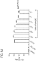
FIGS. 5A-B. Graphs showing effect of peptide WQ20 on binding of PD-L1 to PD-1. FIG. 5A, MFI; FIG. 5B, normalized MFI.
FIGS. 6A-B. Graphs showing effect of peptide SQ20 on binding of PD-L1 to PD-1. FIG. 6A, MFI; FIG. 6B, normalized MFI.
FIG. 7A. Graph showing the effect of an anti-human PD-1 antibody on the interaction between PD-1-expressing Jurkat T cells and PD-L1-expressing CHO cells that results in inhibition of a PD-1 mediated suppression of luciferase reporter that is under the control of promoter containing IL-2, NFAT, and NF-kB response elements.
FIG. 7B. Graph showing the effect of an anti-human PD-1 antibody on the interaction between PD-1-expressing Jurkat T cells and PD-L1-expressing CHO cells (data in 7A expressed as fold inhibition).
FIG. 8A. Graph showing that PD-1 peptide inhibitors inhibit, in a dose-dependent manner, the interaction between PD-1-expressing Jurkat T cells and PD-L1-expressing CHO cells, which results in increased luciferase reporter expression.
FIG. 8B. Graph showing the effect of an anti-human PD-1 antibody on the interaction between PD-1-expressing Jurkat T cells and PD-L1-expressing CHO cells (data in 8B expressed as fold inhibition).
FIG. 9. Graph showing IL-2 production by peripheral blood mononuclear cells (PBMCs) in a tetanus toxoid recall assay after culture with peptides QP20, HD20, WQ20, SQ20, or CQ-22.
FIG. 10. Graph showing IL-4 production by PBMCs in a tetanus toxoid recall assay after culture with peptides QP20, HD20, WQ20, SQ20, or CQ-22.
FIG. 11. Graph showing IL-6 production by PBMCs in a tetanus toxoid recall assay after culture with peptides QP20, HD20, WQ20, SQ20, or CQ-22.
FIG. 12. Graph showing IL-10 production by PBMCs in a tetanus toxoid recall assay after culture with peptides QP20, HD20, WQ20, SQ20, or CQ-22.
FIG. 13. Graph showing IL-17a production by PBMCs in a tetanus toxoid recall assay, after culture with peptides QP20, HD20, WQ20, SQ20, or CQ-22.
FIG. 14. Graph showing IFNγ production by PBMCs in a tetanus toxoid recall assay, after culture with peptides QP20, HD20, WQ20, SQ20, or CQ-22.
FIG. 15. Graph showing TNFα production by PBMCs in a tetanus toxoid recall assay, after culture with peptides QP20, HD20, WQ20, SQ20, or CQ-22.
FIG. 16. Graph showing IL-2 production by PBMCs in a tetanus toxoid recall assay, after culture with various combinations of peptides QP20, HD20, WQ20, and SQ20.
FIG. 17. Graph showing IL-4 production by PBMCs in a tetanus toxoid recall assay, after culture with various combinations of peptides QP20, HD20, WQ20, and SQ20.
FIG. 18. Graph showing IL-6 production by PBMCs in a tetanus toxoid recall assay, after culture with various combinations of peptides QP20, HD20, WQ20, and SQ20.
FIG. 19. Graph showing IL-10 production by PBMCs in a tetanus toxoid recall assay, after stimulation with various combinations of peptides QP20, HD20, WQ20, and SQ20.
FIG. 20. Graph showing IL-17a production by PBMCs after stimulation with various combinations of peptides QP20, HD20, WQ20, and SQ20.
FIG. 21. Graph showing IFNγ production by PBMCs after culture with various combinations of peptides QP20, HD20, WQ20, and SQ20.
FIG. 22. Graph showing TNFα production by PBMCs after culture with various combinations of peptides QP20, HD20, WQ20, and SQ20.
FIG. 23A. Graph showing IL-2 production by PBMCs from donor A after culture with peptides QP20, HD20, WQ20, and SQ20, or CQ-22.
FIG. 23B. Graph showing IL-2 production by PBMCs from donor B after culture with peptides QP20, HD20, WQ20, or SQ20 and combinations of these peptides.
FIG. 24A. Graph showing IL-17a production by PBMCs from donor A after culture with peptides QP20, HD20, WQ20, and SQ20, or CQ-22.
FIG. 24B. Graph showing IL-17a production by PBMCs from donor B after culture with peptides QP20, HD20, WQ20, or SQ20 and combinations of these peptides.
FIG. 25. Graph showing number of surface metastases in mice bearing B16-F10-LacZ tumor cells and treated with combinations of peptides.
FIG. 26. Graph showing the average number±standard deviation of Plasmodium yoelii circumsporozoite protein (PyCS)-specific, IFNγ-secreting CD8 T cells per 0.5×10⁶ splenocytes for each cohort tested in Example 8.
FIG. 27. Graph showing the effect of the combination of QP20, HD20, WQ20, and SQ20 peptides on the mean level of serum HBsAg (hepatitis B surface antigen) at weeks 2 and 3 post infection.
DETAILED DESCRIPTION
This disclosure provides four peptides:
peptide amino acid sequence SEQ ID NO:
QP20 QTRTVPMPKIHHPPWQNVVP 1
HD20 HHHQVYQVRSHWTGMHSGHD 2
WQ20 WNLPASFHNHHIRPHEHEWIQ 3
SQ20 SSYHHFKMPELHFGKNTFHQ 4
These peptides share a core sequence of HH_, which is shown above in bold, and have a strong affinity for the checkpoint receptor “programmed death 1” (PD-1). These peptides block the interaction of PD-1 with its ligand PD-L1 and can therefore be used to inhibit the progression of a hyperproliferative disorder, including cancer, or to treat infectious diseases, including persistent infections by agents such as HIV, hepatitis B virus (HBV), hepatitis C virus (HCV), and Plasmodium falciparum, by enhancing, stimulating, and/or increasing an individual's immune response.
In some embodiments, any of the disclosed peptides can be modified using chemical or recombinant methods to enhance stability or other pharmacokinetic properties. See, e.g., US 2017/0020956. Modifications include, but are not limited to, replacement of one or more L-amino acid with its corresponding D-form, acetylation on a C- and/or N-terminal residue, amidation on a C- and/or N-terminal residue, cyclization, esterification, glycosylation, acylation, attachment of myristic or palmitic acid, addition of an N-terminal glycine, addition of lipophilic moieties such as long fatty acid chains, and PEGylation.
In some embodiments, one or more of the disclosed peptides can be conjugated to various moieties, such as albumin and transthyretin, to enhance the plasma half-life of the peptide(s). Methods of preparing such conjugates are well known in the art (e.g., Penchala et al., 2015; Kontermann, 2016; Zorzi et al., 2017).
In some embodiments, any of the disclosed peptides can be conjugated to a partner molecule, such as a peptide or protein such as an antibody intended to increase the half-life in vivo and/or to provide specific delivery to a target tissue or cell. Conjugation may be direct or can be via a linker. In some of these embodiments, the peptide can be modified to substitute one or more amino acids with amino acids used to attach partner molecules, such as lysine, or by N-terminal extension of the peptide with, e.g., 1, 2, 3, or 4 glycine spacer molecules.
Peptides, or modified versions of the peptides as described above, can be made by any method known in the art, including synthetic methods, recombinant methods, or both. Synthetic methods include solid-phase and solution methods, and may include the use of protective groups. See, e.g., Bodanszky et al. (1976), McOmie (1973), Merrifield (1963), Neurath et al. (1976), Stuart & Young (1984).
Recombinant production of peptides can be carried out using any nucleotide sequence(s) encoding the peptides in any suitable expression system. Nucleic acid molecules encoding one or more of the disclosed peptides can be incorporated into an expression cassette that includes control elements operably linked to the coding sequences. Control elements include, but are not limited to, initiators, promoters (including inducible, repressible, and constitutive promoters), enhancers, and polyadenylation signals. Signal sequences can be included. The expression cassette can be provided in a vector that can be introduced into an appropriate host cell for production of the peptide(s). Methods of constructing expression cassettes and expression vectors are well known. Expression vectors can include one or more of the expression cassettes described in the paragraphs below.
In some embodiments, an expression cassette encodes a peptide comprising the amino acid sequence SEQ ID NO:1. In some embodiments, an expression cassette encodes a peptide consisting essentially of the amino acid sequence SEQ ID NO:1. In some embodiments, an expression cassette encodes a peptide consisting of the amino acid sequence SEQ ID NO:1.
In some embodiments, an expression cassette encodes a peptide comprising the amino acid sequence SEQ ID NO:2. In some embodiments, an expression cassette encodes a peptide consisting essentially of the amino acid sequence SEQ ID NO:2. In some embodiments, an expression cassette encodes a peptide consisting of the amino acid sequence SEQ ID NO:2.
In some embodiments, an expression cassette encodes a peptide comprising the amino acid sequence SEQ ID NO:3. In some embodiments, an expression cassette encodes a peptide consisting essentially of the amino acid sequence SEQ ID NO:3. In some embodiments, an expression cassette encodes a peptide consisting of the amino acid sequence SEQ ID NO:3.
In some embodiments, an expression cassette encodes a peptide comprising the amino acid sequence SEQ ID NO:4. In some embodiments, an expression cassette encodes a peptide consisting essentially of the amino acid sequence SEQ ID NO:4. In some embodiments, an expression cassette encodes a peptide consisting of the amino acid sequence SEQ ID NO:4.
In some embodiments, an expression cassette encodes only two of the disclosed peptides; e.g., a peptide (a) a peptide comprising, consisting essentially of, or consisting of the amino acid sequence SEQ ID NO:1 and a peptide comprising, consisting essentially of, or consisting of the amino acid sequence SEQ ID NO:2; (b) a peptide comprising, consisting essentially of, or consisting of the amino acid sequence SEQ ID NO:1 and a peptide comprising, consisting essentially of, or consisting of the amino acid sequence SEQ ID NO:3; (c) a peptide comprising, consisting essentially of, or consisting of the amino acid sequence SEQ ID NO:1 and a peptide comprising, consisting essentially of, or consisting of the amino acid sequence SEQ ID NO:4; (d) a peptide comprising, consisting essentially of, or consisting of the amino acid sequence SEQ ID NO:2 and a peptide comprising, consisting essentially of, or consisting of the amino acid sequence SEQ ID NO:3; (e) a peptide comprising, consisting essentially of, or consisting of the amino acid sequence SEQ ID NO:2 and a peptide comprising, consisting essentially of, or consisting of the amino acid sequence SEQ ID NO:4; or (f) a peptide comprising, consisting essentially of, or consisting of the amino acid sequence SEQ ID NO:3 and a peptide comprising, consisting essentially of, or consisting of the amino acid sequence SEQ ID NO:4.
In some embodiments, an expression cassette encodes only three of the disclosed peptides; e.g., (a) a peptide comprising, consisting essentially of, or consisting of the amino acid sequence SEQ ID NO:1, a peptide comprising, consisting essentially of, or consisting of the amino acid sequence SEQ ID NO:2, and a peptide comprising, consisting essentially of, or consisting of the amino acid sequence SEQ ID NO:3; (b) a peptide comprising, consisting essentially of, or consisting of the amino acid sequence SEQ ID NO:1, a peptide comprising, consisting essentially of, or consisting of the amino acid sequence SEQ ID NO:2, and a peptide comprising, consisting essentially of, or consisting of the amino acid sequence SEQ ID NO:4; (c) a peptide comprising, consisting essentially of, or consisting of the amino acid sequence SEQ ID NO:2, a peptide comprising, consisting essentially of, or consisting of the amino acid sequence SEQ ID NO:3, and a peptide comprising, consisting essentially of, or consisting of the amino acid sequence SEQ ID NO:4; or (d) a peptide comprising, consisting essentially of, or consisting of the amino acid sequence SEQ ID NO:1, a peptide comprising, consisting essentially of, or consisting of the amino acid sequence SEQ ID NO:3, and a peptide comprising, consisting essentially of, or consisting of the amino acid sequence SEQ ID NO:4.
In some embodiments, an expression cassette encodes all four of the disclosed peptides; i.e., a peptide comprising, consisting essentially of, or consisting of the amino acid sequence SEQ ID NO:1, a peptide comprising, consisting essentially of, or consisting of the amino acid sequence SEQ ID NO:2, a peptide comprising, consisting essentially of, or consisting of the amino acid sequence SEQ ID NO:3, and a peptide comprising, consisting essentially of, or consisting of the amino acid sequence SEQ ID NO:4.
Therapeutic Uses
The disclosed peptides have a number of therapeutic applications and can be administered for a variety of purposes to both human and veterinary subjects. “Administer” as used herein includes direct administration of the disclosed peptides or modified versions thereof as well as indirect administration, e.g., using a nucleic acid molecule encoding the peptides or modified versions of the peptides, as described below. In some embodiments, administration is carried out in conjunction with one or more other therapeutic moieties. “In conjunction with” includes administration together with, before, or after administration of the one or more other therapeutic moieties.
Treatment of Hyperproliferative Disorders, Including Cancer
In some embodiments, one or more of the disclosed peptides or modified versions thereof can be administered to inhibit the progression of a hyperproliferative disorder, such as cancer. Such inhibition may include, for example, reducing proliferation of neoplastic or pre-neoplastic cells; destroying neoplastic or pre-neoplastic cells; and inhibiting metastasis or decreasing the size of a tumor.
Examples of cancers that can be treated using one or more of the disclosed peptides or modified versions thereof include, but are not limited to, melanomas, lymphomas, sarcomas, and cancers of the colon, kidney, stomach, bladder, brain (e.g., gliomas, glioblastomas, astrocytomas, medulloblastomas), prostate, bladder, rectum, esophagus, pancreas, liver, lung, breast, uterus, cervix, ovary, blood (e.g., acute myeloid leukemia, acute lymphoid leukemia, chronic myeloid leukemia, chronic lymphocytic leukemia, Burkitt's lymphoma, EBV-induced B-cell lymphoma).
Combination Cancer Therapies
In some embodiments, one or more of the disclosed peptides or modified versions thereof can be administered in conjunction with one or more therapies or immunotherapies, such as those described below.
In some embodiments, the second therapy comprises a second agent that reduces or blocks the activity of PD-1 (e.g., nivolumab, pembrolizumab, durvalumab) or
In some embodiments, the second therapy comprises an agent that reduces or blocks the activity of PD-L1 (e.g., atezolizumab).
In some embodiments, the second therapy comprises an agent that reduces or blocks the activity of other inhibitory checkpoint molecules and/or molecules that suppress the immune system. These molecules include, but are not limited to:
    • 1. Lymphocyte-activation gene-3 (LAG-3; see He et al., 2016; Triebel et al., 1990);
    • 2. cytotoxic T-lymphocyte—associated antigen 4 (CTLA-4);
    • 3. V-domain Immunoglobulin Suppressor of T cell Activation (VISTA, also known as c10orf54, PD-1H, DD1α, Gi24, Dies1, and SISP1; see US 2017/0334990, US 2017/0112929, Gao et al., 2017, Wang et al., 2011; Liu et al., 2015);
    • 4. T-cell Immunoglobulin domain and Mucin domain 3 (TIM-3; see US 2017/0198041, US 2017/0029485, US 2014/0348842, Sakuishi et al., 2010);
    • 5. killer immunoglobulin-like receptors (KIRs; see US 2015/0290316);
    • 6. agents that inhibit indoleamine (2,3)-dioxygenase (IDO; see Mellemgaard et al., 2017);
    • 7. B and T Lymphocyte Attenuator (BTLA; see US 2016/09222114); and
    • 8. A2A adenosine receptor (A2AR; see Beavis et al., 2015; US 2013/0267515; US 2017/0166878; Leone et al., 2015; Mediavilla-Varela et al., 2017; Young et al., 2016).
Agents that reduce or block the activity of LAG-3 include, but are not limited to, BMS-986016, IMP321, and GSK2831781 (He et al., 2016).
Agents that reduce or block the activity of CTLA-4 include, but are not limited to, ipilimumab and tremelimumab.
Agents that reduce or block the activity of VISTA include, but are not limited to, small molecules, such as CA-170, and antibodies (e.g., Le Mercier et al., 2014).
Agents that reduce or block the activity of TIM-3 include, but are not limited to, antibodies such as MBG453 and TSR-022; see Dempke et al., 2017.
Agents that reduce or block the activity of KIRs include, but are not limited to, monoclonal antibodies such as IPH2101 and Lirilumab (BMS-986015, formerly IPH2102); see Benson & Caligiuri, 2014.
Agents that reduce or block the activity of IDO include, but are not limited to, epacadostat and agents disclosed in US 2017/0037125.
Agents that reduce or block the activity of BTLA include, but are not limited to, peptides (e.g., Spodzieja et al., 2017).
Agents that reduce or block the activity of A2AR include, but are not limited to, small molecules such as CPI-444 and vipadenant.
In some embodiments, the second therapy comprises a cytokine (e.g., interleukin 7).
In some embodiments, the second therapy comprises an agonist of a stimulatory checkpoint molecule. These molecules include, but are not limited to:
1. CD40;
2. OX40;
3. glucocorticoid-induced tumor necrosis factor-related protein (GITR); and
4. Inducible T-cell COStimulator (ICOS).
Agonists of CD40 include, but are not limited to, CD40 agonist monoclonal antibodies such as cp-870,893, ChiLob7/4, dacetuzumab, and lucatumumab. See, e.g., Vonderheide et al., 2007; Khubchandani et al., 2009; Johnson et al., 2010; Bensinger et al., 2012; Vonderheide and Glennie, 2013; Johnson et al., 2015.
Agonists of OX40 include, but are not limited to, OX40 agonist antibodies such as MOXR0916, MED16469, MED10562, PF-045618600, GSK3174998, and INCCAGN01949, and OX40L-Fc fusion proteins, such as MEDI6383. See, e.g., Huseni et al., 2014; Linch et al., 2015; Messenheimer et al., 2017. See also Shrimali et al., 2017.
Agonists of GITR include, but are not limited to, MEDI1873. See, e.g., Schaer et al., 2012; Tigue et al., 2017.
Agonists of ICOS include, but are not limited to, ICOS agonist antibodies JTX-2011 and GSK3359609. See, e.g., Harvey et al., 2015; Michaelson et al., 2016.
In other embodiments, the second therapy comprises a 4-1BB agonist (Shindo et al., 2015), such as urelumab; a 4-1BB antagonist (see US 2017/0174773); an inhibitor of anaplastic lymphoma kinase (ALK; Wang et al., 2014; US 2017/0274074), such as crizotinib, ceritinib, alectinib, PF-06463922, NVP-TAE684, AP26113, TSR-011, X-396, CEP-37440, RXDX-101; an inhibitor of histone deacetylase (HDAC; see US 2017/0327582); a VEGFR inhibitor, such as axitinib, sunitinib, sorafenib, tivozanib, bevacizumab; and/or an anti-CD27 antibody, such as varlilumab.
In some embodiments, the second therapy comprises a cancer vaccine (e.g., Duraiswamy et al., 2013). A “cancer vaccine” is an immunogenic composition intended to elicit an immune response against a particular antigen in the individual to which the cancer vaccine is administered. A cancer vaccine typically contains a tumor antigen which is able to induce or stimulate an immune response against the tumor antigen. A “tumor antigen” is an antigen that is present on the surface of a target tumor. A tumor antigen may be a molecule which is not expressed by a non-tumor cell or may be, for example, an altered version of a molecule expressed by a non-tumor cell (e.g., a protein that is misfolded, truncated, or otherwise mutated).
In some embodiments, the second therapy comprises a chimeric antigen receptor (CAR) T cell therapy. See, e.g., John et al., 2013; Chong et al., 2016.
Additional Therapeutic Uses
In some embodiments, one or more of the disclosed peptides or modified versions thereof can be administered to treat infectious diseases, including chronic infections, caused, e.g., by viruses, fungi, bacteria, and protozoa, and helminths.
Examples of viral agents include human immunodeficiency virus (HIV), Epstein Barr Virus (EBV), Herpes simplex (HSV, including HSV1 and HSV2), Human Papillomavirus (HPV), Varicella zoster (VSV) Cytomegalovirus (CMV), and hepatitis A, B, and C viruses.
Examples of fungal agents include Aspergillus, Candida, Coccidioides, Cryptococcus, and Histoplasma capsulatum.
Examples of bacterial agents include Streptococcal bacteria (e.g., pyogenes, agalactiae, pneumoniae), Chlamydia pneumoniae, Listeria monocytogenes, and Mycobacterium tuberculosis.
Examples of protozoa include Sarcodina (e.g., Entamoeba), Mastigophora (e.g., Giardia), Ciliophora (e.g., Balantidium), and Sporozoa (e.g., Plasmodium falciparum, Cryptosporidium).
Examples of helminths include Platyhelminths (e.g., trematodes, cestodes), Acanthocephalins, and Nematodes.
In some embodiments one or more of the disclosed peptides or modified versions thereof can be administered as a vaccine adjuvant in conjunction with a vaccine to enhance a response to vaccination (e.g., by increasing effector T cells and/or reducing T cell exhaustion). The vaccine can be, for example, an RNA vaccine (e.g., US 2016/0130345, US 2017/0182150), a DNA vaccine, a recombinant vector, a protein vaccine, or a peptide vaccine. Such vaccines can be delivered, for example, using virus-like particles, as is well known in the art.
In some embodiments one or more of the disclosed peptides or modified versions thereof can be administered to treat sepsis.
In some embodiments one or more of the disclosed peptides or modified versions thereof can be administered to promote hair color re-pigmentation. In some embodiments one or more of the disclosed peptides or modified versions thereof can be administered to promote lightening of pigmented skin lesions.
Administration of Peptides
In some embodiments, one or more of the disclosed peptides themselves, or modified versions thereof, are administered. In some of these embodiments, a peptide carrier system is used. A number of peptide carrier systems are known in the art, including microparticles, polymeric nanoparticles, liposomes, solid lipid nanoparticles, hydrophilic mucoadhesive polymers, thiolated polymers, polymer matrices, nanoemulsions, and hydrogels. See Patel et al. (2014), Bruno et al. (2013), Feridooni et al. (2016). Any suitable system can be used.
In some embodiments, engineered T cell-based therapies that express and secrete a peptide or protein can be used to deliver PD-1 inhibition at the site of engagement of the T cell receptor with an antigen. The T cell-based therapy could be, for example, a CAR-T cell that expresses one or more of the disclosed peptides or modified versions thereof. Either inducible or constitutive expression can be used.
In other embodiments one or more of the disclosed peptides or modified versions thereof are delivered using one or more nucleic acids encoding the peptide(s) (e.g., DNA, cDNA, PNA, RNA or a combination thereof); See, e.g., US 2017/0165335. Nucleic acids encoding one or more peptides can be delivered using a variety of delivery systems known in the art. Nucleic acid delivery systems include, but are not limited to, gene-gun; cationic lipids and cationic polymers; encapsulation in liposomes, microparticles, or microcapsules; electroporation; virus-based, and bacterial-based delivery systems. Virus-based systems include, but are not limited to, modified viruses such as adenovirus, adeno-associated virus, herpes virus, retroviruses, vaccinia virus, or hybrid viruses containing elements of one or more viruses. US 2002/0111323 describes use of “naked DNA,” i.e., a “non-infectious, non-immunogenic, non-integrating DNA sequence,” free from “transfection-facilitating proteins, viral particles, liposomal formulations, charged lipids and calcium phosphate precipitating agents,” to administer a peptide. Bacterial-based delivery systems are disclosed, e.g., in Van Dessel et al. (2015) and Yang et al. (2007).
In some embodiments, a peptide is administered via an RNA molecule encoding the peptide. In some embodiments, the RNA molecule is encapsulated in a nanoparticle. In some embodiments, the nanoparticle comprises a cationic polymer (e.g., poly-L-lysine, polyamidoamine, polyethyleneimine, chitosan, poly(β-amino esters). In some embodiments, the nanoparticle comprises a cationic lipid or an ionizable lipid. In some embodiments, the RNA molecule is conjugated to a bioactive ligand (e.g., N-acetylgalactosamine (GalNAc), cholesterol, vitamin E, antibodies, cell-penetrating peptides). See, e.g., Akinc et al. (2008), Akinc et al. (2009), Anderson et al. (2003), Behr (1997), Boussif et al. (1995), Chen et al. (2012), Dahlman et al. (2014), Desigaux et al. (2007), Dong et al. (2014), Dosta et al. (2015), Fenton et al. (2016), Guo et al. (2012), Howard et al. (2006), Kaczmarek et al. (2016), Kanasty et al. (2013), Kauffman et al. (2015), Kozielski et al. (2013), Leus et al. (2014), Lorenz et al. (2004), Love et al. (2010), Lynn & Langer (2000), Moschos et al. (2007), Nair et al. (2014), Nishina et al. (2008), Pack et al. (2005), Rehman et al. (2013), Schroeder et al. (2010), Tsutsumi et al. (2007), Tzeng et al. (2012), Won et al. (2009), Xia et al. (2009), Yu et al. (2016).
In some embodiments, an RNA molecule can be modified to reduce its chances of degradation or recognition by the immune system. The ribose sugar, the phosphate linkage, and/or individual bases can be modified. See, e.g., Behlke (2008), Bramsen (2009), Chiu (2003), Judge & MacLachlan (2008), Kauffman (2016), Li (2016), Morrissey (2005), Prakash (2005), Pratt & MacRae (2009), Sahin (2014), Soutschek (2004), Wittrup & Lieberman (2015). In some embodiments, the modification is one or more of a ribo-difluorotoluyl nucleotide, a 4′-thio modified RNA, a boranophosphate linkage, a phosphorothioate linkage, a 2′-O-methyl (2′-OMe) sugar substitution, a 2′-fluoro (2′-F), a 2′-O-methoxyethyl (2′-MOE) sugar substitution, a locked nucleic acid (LNA), and an L-RNA.
Routes of Administration, Pharmaceutical Compositions, and Devices
Pharmaceutical compositions comprising an effective amount of any of the active agents described in the paragraphs above include a pharmaceutically acceptable vehicle. The “pharmaceutically acceptable vehicle” may comprise one or more substances which do not affect the biological activity of the active agent(s) and, when administered to a patient, does not cause an adverse reaction. Pharmaceutical compositions may be liquid or may be lyophilized. Lyophilized compositions may be provided in a kit with a suitable liquid, typically water for injection (WFI) for use in reconstituting the composition. Other suitable forms of pharmaceutical compositions include suspensions, emulsions, and tablets.
Routes of administration include injection or infusion (e.g., epidural, intradermal, intramuscular, intraperitoneal, intravenous, sub-cutaneous), transdermal (e.g., US 2017/0281672), mucosal (e.g., intranasal or oral), pulmonary, and topical (e.g., US 2017/0274010) administration. See, e.g., US 2017/0101474.
Administration can be systemic or local. In addition to local infusions and injections, implants can be used to achieve a local administration. Examples of suitable materials include, but are not limited to, sialastic membranes, polymers, fibrous matrices, and collagen matrices.
Topical administration can be by way of a cream, ointment, lotion, transdermal patch (such as a microneedle patch), or other suitable forms well known in the art.
Administration can also be by controlled release, for example, using a microneedle patch, pump and/or suitable polymeric materials. Examples of suitable materials include, but are not limited to, poly(2-hydroxy ethyl methacrylate), poly(methyl methacrylate), poly(acrylic acid), poly(ethylene-co-vinyl acetate), poly(methacrylic acid), polyglycolides (PLG), polyanhydrides, poly(N-vinyl pyrrolidone), poly(vinyl alcohol), polyacrylamide, poly(ethylene glycol), polylactides (PLA), poly(lactide-co-glycolides) (PLGA), and polyorthoesters.
Devices comprising any of the active agents described above include, but are not limited to, syringes, pumps, transdermal patches, spray devices, vaginal rings, and pessaries.
Example 1. Peptide Library Screening
The TriCo-20™ (TRICO-20™) and TriCo-16™ (TRICO-16™) Phage Display Peptide Libraries (Creative Biolabs, 45-1 Ramsey Road, Shirley, N.Y. 11967) were screened to identify binders of soluble recombinant human PD-1 receptor. After the fourth round of panning, obvious enrichment for specific binders was observed, and individual peptides were confirmed as weakly specific binders in a clonal phage ELISA. A fifth round of panning led to greater enrichment. Table 1 lists four peptides which showed strong specific binding in the clonal phage ELISA.
TABLE 1
Clonal
Phase ELISA
coated uncoated SEQ
Clone signal signal peptide sequence ID NO:
QP20 0.851 0.446 QTRTVPMPKIHHPPWQNVVP 1
HD20 0.281 0.109 HHHQVYQVRSHWTGMHSGHD 2
WQ20 0.275 0.115 WNLPASFHNHHIRPHEHEWIQ 3
SQ20 0.284 0.159 SSYHHFKMPELHFGKNTFHQ 4
Example 2. Competitive PD-1:PD-L1 Binding Inhibition Assay
Briefly, detection of cell surface PD-1 on Jurkat cells was accomplished by incubating cells with the human PD-L1-Fc fusion protein, followed by detection of the recombinant molecule with a fluorescently labeled anti-human Fc antibody. Flow cytometry was performed to detect binding between PD-1 and the PD-L1 recombinant protein. Quantitative binding measurement was then determined by mean fluorescence intensity (MFI).
Jurkat Cell-surface expression of PD1 and binding of PD-L1 to these cells were verified as shown in FIGS. 1 and 2. The results are shown in FIGS. 3A-B, 4A-B, 5A-B, and 6A-B.
Example 3. Cell-Based Reporter Assay
A cell-based reporter assay was used to assess whether binding of the four peptides identified above was sufficient to block the interaction with PD-1 and its ligand PD-L1. The components of the assay include a Jurkat T cell line that stably expresses human PD-1 and a luciferase reporter, a CHO cell line that stably expressed human PD-L1, and a positive control anti-PD-1 antibody that blocks the interaction of PD-1 and PD-L1, resulting in a measurable effect in the assay. The luciferase reporter in the Jurkat T cell line is triggered by IL-1, NFAT, or NF-κB response elements in the promoter region. The Jurkat T cells are pre-treated with CD3 and immediately cryopreserved for use in the assay. Interaction of the Jurkat T cells with the PD-L1 expressing cell line inhibits the intracellular mechanism by which the luciferase construct is activated, thereby preventing luciferase expression. A molecule that binds to either PD-1 on the Jurkat T cells or to PD-L1 on the CHO cells sufficiently to prevent their interaction permits the Jurkat T cells to produce luciferase. CellTiter-Glo® (CELLTITER-GLO®, Promega) was used to measure luciferase expression.
The results of positive control assays using the anti-PD-1 control antibody are shown in FIGS. 7A-B. These results demonstrate that the control antibody restores luciferase expression in a dose-dependent manner, with peak-fold inhibition of approximately 8 at an antibody concentration of 20 μM.
The results of assays of the peptides identified above are shown in FIGS. 8A-B. These results demonstrate that each of the four peptides restores luciferase expression in a dose-dependent manner, with peak-fold inhibition of approximately 1.5 at a concentration of approximately 25 μM.
Example 4. Tetanus Toxoid Recall Assay Using Individual Peptides
Peptides 1-4 were tested in a human PBMC-based tetanus antigen recall assay. “Peptide CQ-22” was used as a negative control.
PBMCs were obtained from plasma of human donors and tested in vitro for recall of tetanus toxoid. Suitable PBMCs were cryopreserved until needed, then thawed and cultured in a 96-wellplate. Tetanus toxoid was added to the cultures in the presence or absence of peptides 1-4, and the production of cytokines and cell surface T cell activation markers were examined.
The results of these assays are shown in FIGS. 9-15 and summarized qualitatively in Table 2. In the table, “x” indicates no effect, “−” indicates a possible low effect, “+” indicates some effect, and “++” indicates a definite effect.
TABLE 2
peptide IL-2 IL-4 IL-6 IL-10 IL-17a IFNγ TNFα
QP20 x x x x x x
HD20 x ++ x ++ ++ ++
WQ20 ++ ++ x ++ ++ ++
SQ20 + ++ + ++ ++ +
The results demonstrated a trend towards modest enhancement of IL-6, IL-17α, IFNγ, and TNFα production at the highest concentrations of peptides. No significant enhancement of IL-2 production was detected.
Example 5. Tetanus Toxoid Recall Assay Using Combinations of Peptides
Combinations of peptides were tested in the antigen recall assay described above, using a different PBMC donor and a different lot number of tetanus toxoid. The results are shown in FIGS. 16, 17, 18, 19, 20, 21, and 22. These results demonstrated that the combination of the four peptides combination of the four peptides QP20, HD20, WQ20, and SQ20 result in increased IL-2 production and reduced IL-17a production.
The effect of peptides QP20, HD20, WQ20, and QP20 on the production of IL-2 and IL-17a appears to be donor-specific, as shown in FIGS. 23A-B and 24A-B.
Example 6. Biacore® Assays
BIACORE® assays were carried out using a BIACORE® T-200 at 25° C. The assay and regeneration buffers contained 10 mM HEPES (pH 7.4), 150 mM NaCl, 3 mM EDTA, and 0.05% P20. The immobilization buffer was 10 mM sodium acetate, pH 5.0. The flow rate used for immobilizing the ligand was 5 μl/min. The flow rate for kinetics analysis was 30 μl/min.
Scouting.
12,000 response units (RU) of human and 6000 RU of mouse PD-1 receptors were directly immobilized on flow cell 2 and flow cell 4 of the CMS chip by amine coupling method (EDC/NHS). The un-occupied sites were blocked with 1M ethanol amine. Scouting was performed at a single analyte concentration of 25 μM to confirm yes/no binding. Flow cell 1 was kept blank and used for reference subtraction. Binding of analyte to the ligand was monitored in real time.
Full Kinetics.
Based on the scouting results, full kinetics were performed by immobilizing higher RU of the ligand to a new chip and analyte concentration at 25 μM, followed by serial dilution to 12.5, 6.25, 3.125, 1.562, 0.78 and 0 μM concentration or as indicated. Due to fast on rate and off rate, KD was determined by steady state equilibrium kinetics.
Chi square (χ2) analysis was carried out between the actual sensorgram and a sensorgram generated from the BIANALYSIS® software (black line) to determine the accuracy of the analysis. A χ2 value within 1-2 is considered significant (accurate) and below 1 is highly significant (highly accurate). The results are summarized in Table 3.
TABLE 3
Ligand Rmax KA KD Conc.
10,000 RU Analyte (RU) (1/M) (M) (μM) χ2
mouse PD-1 WQ-21 270 1.31 × 10³ 7.61 × 10⁻⁴ 0-25 0.0203
mouse PD-1 QP-20 13.4 1.80 × 10⁴ 5.54 × 10⁻⁵ 0-25 0.0446
mouse PD-1 HD-20 76 4.25 × 10³ 2.35 × 10⁻⁴ 0-25 0.11
mouse PD-1 SQ-20 12.8 2.14 × 10⁴ 4.68 × 10⁻⁵ 0-25 0.039
human PD-1 WQ-21 84.7 3.28 × 10³ 3.05 × 10⁻⁴ 0-25 0.0309
human PD-1 QP-20 3.83 9.36 × 10⁴ 1.07 × 10⁻⁵ 0-25 0.0569
human PD-1 HD-20 3.35 3.18 × 10⁵ 3.41 × 10⁻⁶ 0-12.5 0.0733
human PD-1 SQ-20 4.05 1.94 × 10⁵ 5.16 × 10⁻⁶ 0-25 0.111
mouse PD-1 Mouse PD-L1 259 2.75 × 10⁶ 3.64 × 10⁻⁷ 0-50 0.105
human PD-1 Human PD-L1 213 6.92 × 10⁶ 1.44 × 10⁻⁷ 0-50 2.44
These results indicate that each of the four peptides bind both human and mouse PD-1. QP20 and SQ20 showed the highest affinity towards mouse PD-1. HD20 and SQ20 showed the highest affinity towards human PD-1.
Example 7. Experimental Metastasis Model
Efficacy of the peptides was evaluated in a B16-F10-LacZ experimental metastasis model. In this model, B16-F10-LacZ cells, transfected to express the LacZ gene that encodes β-galactoside, an intracellular enzyme, are injected into the tail vein of syngeneic mice. The cells travel through the circulation, settle in the lungs, and form tumors. Mice are terminated 2 weeks after implant. When the enzyme cleaves its substrate, X-gal, the products dimerize and change color and can be detected ex vivo. The number of metastatic tumors on the surface of the lung is then quantified by manual counting of tumors under a dissecting microscope.
Briefly, mice (N=7) were implanted on study day 0 with B16-F10-LacZ tumor cells (5×10⁵ or 1×10⁶ cells per mouse) by intravenous injection in the tail vein. Mice received a treatment of the peptide combination (200 μg, 20 μg, or 2 μg, each peptide per dose) intravenously by tail vein injection on study days 2, 5, 7, 9 and 12. Detailed clinical examinations and body weights were recorded regularly during treatment. Mice were terminated on study day 14, and their lungs were removed and stained. The number of tumor metastases were counted. Treatment groups are described in Table 4.
TABLE 4
Treatment
Group N Implant Treatment Dose Route Days
1 7 5 × 10⁵ QP-20, SQ-20, 200 μg IV SD 2, 5, 7,
HD-20, WQ-20 9, 12
2 7 5 × 10⁵ QP-20, SQ-20, 20 μg IV SD 2, 5, 7,
HD-20, WQ-20 9, 12
3 7 5 × 10⁵ QP-20, SQ-20, 2 μg IV SD 2, 5, 7,
HD-20, WQ-20 9, 12
4 7 5 × 10⁵ Untreated
5 7 1 × 10⁶ QP-20, SQ-20, 200 μg IV SD 2, 5, 7,
HD-20, WQ-20 9, 12
6 7 1 × 10⁶ Untreated

The results are shown in FIG. 25. A good dose response was observed when mice were implanted at both cell concentrations. Mice treated with the highest dose of peptide mixture (200 μg) had the fewest tumors (average 97), and mice treated with the lowest dose of peptide mixture (2 μg) had the most tumors (average 205). Similarly, in the two groups that were implanted with high tumor numbers, the untreated group had significantly more tumors. This indicates that the 4 peptides in combination showed a dose-dependent efficacy on B16-F10-LacZ tumor growth in vivo. Moreover, the peptide combination was well tolerated by the mice and did not have any acute adverse effects on animal health.
Example 8. Effect of Peptide Combination on the Immunogenicity of a Malaria Vaccine
Immunogenicity of the peptide combination as a prophylactic vaccine adjuvant was assessed in a mouse model of malaria. Balb/c mice immunized with an adenovirus-based malaria vaccine expressing the Plasmodium yoelli circumsporozoite protein (AdPyCS) were given 200 μg of the peptide combination, anti-PD-1 mAb, anti-PDL1 mAb, or the negative control peptide ovalbumin (OVA) on days 1, 3, 5, and 7 after immunization with AdPyCS (Table 5). Note that no additional adjuvant was added to the AdPyCS antigen. Spleens were collected 12 days after immunization, and the number of splenic PyCS-specific, IFNγ-secreting CD8+ T cells was determined via ELISpot assay. Note that for the ELISpot assay, splenocytes were stimulated with the SYVPSAEQI peptide (SEQ ID NO:5), an H-2Kd-restricted CD8+ T cell epitope of PyCS.
TABLE 5
Cohort Test Sample # Mice Route Treatment days
1 AdPyCS only 5
2 AdPyCS + control OVA 5 i.p. 0, 1, 3, 5, 7
peptide (200 μg)
3 AdPyCS + peptide combo 5 i.p. 0, 1, 3, 5, 7
(200 μg)
4 AdPyCS + anti-PD-1 antibody 5 i.p. 0, 1, 3, 5, 7
(200 μg)
5 AdPyCS + anti-PDL1 5 i.p. 0, 1, 3, 5, 7
antibody (200 μg)
FIG. 26 shows the average number±standard deviation of CSP-specific, IFNγ-secreting CD8+ T cells per 0.5×10⁶ splenocytes for each cohort. Significant differences in the average number±standard deviation of CSP-specific, IFNγ-secreting CD8+ T cells per 0.5×10⁶ splenocytes between the AdPyCS alone (Cohort 1) and the peptide combination (Cohort 3), anti-PD-1 antibody (Cohort 4) or anti-PD-L1 antibody (Cohort 5) were detected using the one-way ANOVA test (***p<0.001, and *p<0.05). These results demonstrate that the peptide combination (Cohort 3) is functionally active in vivo, increasing the number of CSP-specific, IFNγ-secreting CD8+ T cells ˜1.6-fold relative to AdPyCS alone (Cohort 1), which was similar to changes with anti-PD-1 or -PD-L1 antibody (Cohort 4 and 5).
Example 9. Effect of Peptide Combination on Survival in a Model of Sepsis
Sepsis can negatively alter T cell function and survival, however this can be reversed when the PD-1:PDL1 interaction is blocked, which results in improved survival. Thus the efficacy of the peptide combination was assessed in a representative, clinically relevant model of sepsis where CD1 mice are subjected to cecal ligation and puncture (CLP) to induce intra-abdominal peritonitis. For this study, 200 μg of either the peptide combination or anti-PD-1 antibody were administered i.v. at 2, 24, 48, 72 and 96 hours after surgery. A vehicle control group was also included. Six mice were in each group. All mice were checked twice daily for signs of morbidity and mortality. Administration of the peptide combination conferred an enhanced survival advantage over the vehicle control group where the peptide combination showed a 2-fold higher survival rate (Table 6). Moreover, survival in the peptide combination group was slightly above treatment with anti-PD-1 antibody.
TABLE 6
Group % Survival
Vehicle Control
50%
Anti-PD-1 antibody 83%
PD-1 Peptide Combo 100%
Example 10. Effect of Peptide Combination on Serum HBsAg Levels in HBV-Infected Mice
The combination of QP20, HD20, WQ20, and SQ20 peptides was assessed in a hepatitis B virus (HBV) mouse model where the role of PD-1 in T cell exhaustion and immunotolerance is documented (Tzeng et al., 2012; Ye et al., 2015). PD-1 is elevated in the hepatic T cells of mice with persistent HBV infection but not in animals that have cleared the infection. In this model, it has been shown that inhibition of the PD-1/PD-L1 interaction with an anti-PD-1 mAb both increases antigen-specific IFNγ production by hepatic T cells and reverses HBV persistence (Tzeng et al., 2012). This mouse model of persistent HBV presented an opportunity to test whether the combination of QP20, HD20, WQ20, and SQ20 peptides can reverse T cell exhaustion in vivo and aid the immune system in controlling viral infection.
Mice infected with HBV were treated with saline (negative control), 200 μg of QP20, HD20, WQ20, and SQ20 peptides combined, or 200 μg anti-PD-1 mAb at 9 time points, 2 days prior to infection and days 1, 3, 6, 9, 12, 14, 17 and 20 post infection. The level of serum HB surface antigen (HBsAg) was monitored by ELISA on days 7, 14, and 21 to follow the infection (higher levels of serum HBsAg are reflective of higher viral titer) and detect the immune enhancement activity of the combination of QP20, HD20, WQ20, and SQ20 peptides. The group treated with the combination of QP20, HD20, WQ20, and SQ20 peptides showed significantly lower mean level of serum HBsAg at weeks 2 and 3 post infection (p<0.05, 1-way ANOVA, Tukey's Multiple Comparison Test) compared to the saline negative control (FIG. 27).
REFERENCES
Adams et al., “Big opportunities for small molecules in immuno-oncology,” Nature Reviews Drug Discovery Advance Online Publication, Jul. 31, 2016, 20 pages
Alsaab et al., “PD-1 and PD-L1 Checkpoint Signaling Inhibition for Cancer Immunotherapy: Mechanism, Combinations, and Clinical Outcome,” Front. Pharmacol. 8, 561, 2017
Beavis et al., “Adenosine Receptor 2A Blockade Increases the Efficacy of Anti-PD-1 through Enhanced Antitumor T-cell Responses,” Cancer Immunol. Res. 3, 506-17, 2015
Behlke, “Chemical modification of siRNAs for in vivo use,” Oligonucleotides. 2008; 18:305-19.
Bensinger et al., “A phase 1 study of lucatumumab, a fully human anti-CD40 antagonist monoclonal antibody administered intravenously to patients with relapsed or refractory multiple myeloma,” Br J Haematol. 159, 58-66, 2012.
Benson & Caligiuri, “Killer Immunoglobulin-like Receptors and Tumor Immunity,” Cancer Immunol Res 2014; 2:99-104
Bodanszky et al., Peptide Synthesis, John Wiley and Sons, 2d ed. (1976)
Bramsen et al., “A large-scale chemical modification screen identifies design rules to generate siRNAs with high activity, high stability and low toxicity,” Nucleic Acids Res. 2009; 37:2867-81.
Bruno et al., “Basics and recent advances in peptide and protein drug delivery,” Ther. Deliv. 4, 1443-67, 2013
Bu et al., “Learning from PD-1 Resistance: New Combination Strategies,” Trends Mol. Med. 22, 448-51, 2016
Burnett & Rossi, “RNA-based Therapeutics—Current Progress and Future Prospects,” Chem Biol. 19, 60-71, 2012
Cao, “Advances in Delivering Protein and Peptide Therapeutics,” Pharmaceutical Technology 40, 22-24, Nov. 2, 2016
Cherkassky et al., “Human CAR T cells with cell-intrinsic PD-1 checkpoint blockade resist tumor-mediated inhibition,” J. Clin. Invest. 126, 3130-44, 2016
Chiu et al., “siRNA function in RNAi: a chemical modification analysis,” RNA 2003; 9:1034-48.
Chong et al., “PD-1 blockade modulates chimeric antigen receptor (CAR)-modified T cells: refueling the CAR,” Blood. 129(8), 1039-41, 2017, published on-line Dec. 28, 2016
Chowdhury et al., “Combination therapy strategies for improving PD-1 blockade efficacy: a new era in cancer immunotherapy,” J. Int. Med. doi: 10.1111/joim.12708, Epub ahead of print, Oct. 26, 2017
Creative Biolabs User Manual, “TriCo-20™ Phage Display 20-mer Random Peptide Library,” 14 pages, Aug. 4, 2009
Dempke et al., “Second- and third-generation drugs for immuno-oncology treatment—The more the better?” Eur. J. Cancer 74, 55-72, March 2017
Differding, “AUNP-12—A Novel Peptide Therapeutic Targeting PD-1 Immune Checkpoint Pathway for Cancer Immunotherapy—Structure Activity Relationships & Peptide/Peptidomimetic Analogs,” available at differding.com/data/AUNP_12_A_novel_peptide_therapeutic_targeting_PD_1_immune_checkpoint_pathway_for_cancer_immunotherapy.pdf, Feb. 26, 2014
Duraiswamy et al., “Dual Blockade of PD-1 and CTLA-4 Combined with Tumor Vaccine Effectively Restores T-Cell Rejection Function in Tumors,” Cancer Res 73, 3591-603, 2013
Feridooni et al., “Noninvasive Strategies for Systemic Delivery of Therapeutic Proteins—Prospects and Challenges,” Chapter 8 of Sezer, ed., Smart Drug Delivery System, available at http://www.intechopen.com/books/smart-drug-delivery-system, Feb. 10, 2016
Gao et al., “VISTA is an inhibitory immune checkpoint that is increased after ipilimumab therapy in patients with prostate cancer,” Nature Med. 23, 551-55, 2017
Harvey et al., “Efficacy of anti-ICOS agonist monoclonal antibodies in preclinical tumor models provides a rationale for clinical development as cancer immunotherapeutics,” Journal for ImmunoTherapy of Cancer 3(Suppl 2), O9, 2015
He et al., “Lymphocyte-activation gene-3, an important immune checkpoint in cancer,” Cancer Sci. 107, 1193-97, 2016
Huseni et al., “Anti-tumor efficacy and biomarker evaluation of agonistic anti-OX40 antibodies in preclinical models,” Journal for ImmunoTherapy of Cancer 2(Suppl 3), P105, 2014
Infante et al., “A phase Ib dose escalation study of the OX40 agonist MOXR0916 and the PD-L1 inhibitor atezolizumab in patients with advanced solid tumors,” J Clin Oncol. 34 (suppl; abstr 101), 2016
John et al., “Blockade of PD-1 immunosuppression boosts CAR T-cell therapy,” Oncolmmunology 2, e26286, 3 pages, 2013
Johnson et al., “Clinical and Biological Effects of an Agonist Anti-CD40 Antibody: A Cancer Research UK Phase I Study,” Clin Cancer Res 21, 1321-28, 2015
Johnson et al., “A Cancer Research UK phase I study evaluating safety, tolerability, and biological effects of chimeric anti-CD40 monoclonal antibody (MAb), Chi Lob 7/4,” J Clin Oncol. 28, 2507, 2010.
Judge & MacLachlan, “Overcoming the innate immune response to small interfering RNA,” Hum Gene Ther. 2008; 19:111-24.
Kaczmarek et al., “Advances in the delivery of RNA therapeutics: from concept to clinical reality,” Genome Medicine 2017; 9:60, 16 pages
Kauffman et al., “Efficacy and immunogenicity of unmodified and pseudouridine-modified mRNA delivered systemically with lipid nanoparticles in vivo,” Biomaterials. 2016; 109:78-87.
Kavikansky & Pavlick, “Beyond Checkpoint Inhibitors: The Next Generation of Immunotherapy in Oncology,” Amer. J. Hematol. Oncol. 13, 9-20, 2017
Khubchandani et al., “Dacetuzumab, a humanized mAb against CD40 for the treatment of hematological malignancies,” Curr Opin Investig Drugs 10, 579-87, 2009.
Kontermann, “Half-life extended biotherapeutics,” Expert Opin. Biol. Ther. 16, 903-15, 2016.
Le Mercier et al., “VISTA Regulates the Development of Protective Antitumor Immunity,” Cancer Res 2014; 74:1933-1944
Leone et al., “A2aR antagonists: Next generation checkpoint blockade for cancer immunotherapy,” Computational and Structural Biotechnology Journal 13, 265-72, 2015
Li et al., “Discovery of peptide inhibitors targeting human programmed death 1 (PD-1) receptor,” Oncotarget 7, 64967-76, Aug. 12, 2016
Li et al., “Effects of chemically modified messenger RNA on protein expression,” Bioconjug Chem. 2016; 27:849-53.
Linch et al., “OX40 agonists and combination immunotherapy: putting the pedal to the metal,” Frontiers in Oncology 5, 14 pages, 2015
Liu et al., “Immune-checkpoint proteins VISTA and PD-1 nonredundantly regulate murine T-cell responses,” Proc. Nat'l. Acad. Sci. USA 112, 6682-87, 2015
Magiera-Mularz et al., “Bioactive macrocyclic inhibitors of the PD-1/PD-L1 immune checkpoint,” Angewandte Chemie Int. Ed. 10.1002/anie.201707707, e-published Sep. 26, 2017
Maute et al., “Engineering high-affinity PD-1 variants for optimized immunotherapy and immuno-PET imaging,” Proc. Natl. Acad. Sci. USA, E6506-E6514, published online Nov. 10, 2015
McOmie, Protective Groups in Organic Chemistry, Plenum Press, New York, N.Y., 1973
Mediavilla-Varela et al., “A Novel Antagonist of the Immune Checkpoint Protein Adenosine A2a Receptor Restores Tumor-Infiltrating Lymphocyte Activity in the Context of the Tumor Microenvironment,” Neoplasia 19, 530-36, 2017
Mellemgaard et al., “Combination immunotherapy with IDO vaccine and PD-1 inhibitors in advances HSCLC,” DOI: 10.1200/JCO.2017.35.15_suppl.TPS2610 Journal of Clinical Oncology 35, no. 15_suppl—published online before print, 2017
Merrifield, J. Am. Chem. Soc. 85:2149-54, 1963
Messenheimer et al., “Timing of PD-1 Blockade Is Critical to Effective Combination Immunotherapy with Anti-OX40,” Clin. Cancer Res. 23, DOI: 10.1158/1078-0432.CCR-16-2677 Published October 2017
Michaelson et al., “Preclinical evaluation of JTX-2011, an anti-ICOS agonist antibody,”, Abstract 573, Proceedings: AACR 107th Annual Meeting 2016; Apr. 16-20, 2016; New Orleans, La.
Morrissey et al., “Immunotherapy and Novel Combinations in Oncology: Current Landscape, Challenges, and Opportunities,” Clinical and Translational Science 9, 89-104, 2016
Morrissey et al., “Potent and persistent in vivo anti-HBV activity of chemically modified siRNAs,” Nat Biotechnol. 2005; 23:1002-7.
Neurath et al., eds., The Proteins, Vol. II, 3d ed., pp. 105-237, Academic Press, New York, N.Y. (1976)
Ott et al., “Combination immunotherapy: a road map,” J. ImmunoTherapy of Cancer 5, 16, 2017
Patel et al., “Recent Advances in Protein and Peptide Drug Delivery: A Special Emphasis on Polymeric Nanoparticles,” Protein. Pept. Lett. 21, 1102-20, 2014
Penchala et al., “A biomimetic approach for enhancing the in vivo half-life of peptides,” Nat. Chem. Biol. 11, 793-98, 2015.
Prakash et al., “Positional effect of chemical modifications on short interference RNA activity in mammalian cells,” J Med Chem. 2005; 48:4247-53.
Pratt & MacRae, “The RNA-induced silencing complex: a versatile gene-silencing machine,” J Biol Chem. 2009; 284:17897-901.
Rivera et al., “Hair Repigmentation During Immunotherapy Treatment With an Anti-Programmed Cell Death 1 and Anti-Programmed Cell Death Ligand 1 Agent for Lung Cancer,” JAMA Dermatol. 2017 Jul. 12. doi: 10.1001/jamadermatol.2017.2106, Jul. 12, 2017
Sahin et al., “mRNA-based therapeutics—developing a new class of drugs,” Nat Rev Drug Discov. 2014; 13:759-80.
Sakuishi et al., “Targeting Tim-3 and PD-1 pathways to reverse T cell exhaustion and restore anti-tumor immunity,” J. Exp. Med. 20, 2187-94, 2010
Schaer et al., “Modulation of GITR for cancer immunotherapy,” Curr Opin Immunol. 24, 217-24, 2012
Sharma & Allison, “Immune Checkpoint Targeting in Cancer Therapy: Toward Combination Strategies with Curative Potential,” Cell 161, 205-14, 2015
Shindo et al., “Combination Immunotherapy with 4-1BB Activation and PD-1 Blockade Enhances Antitumor Efficacy in a Mouse Model of Subcutaneous Tumor,” Anticancer Res. 35, 129-36, 2015
Shrimali et al., “Concurrent PD-1 Blockade Negates the Effects of OX40 Agonist Antibody in Combination Immunotherapy through Inducing T-cell Apoptosis,” Cancer Immunol Res 5(9), pages OF1-12, Aug. 28, 2017
Skalniak et al., “Small-molecule inhibitors of PD-1/PD-L1 immune checkpoint alleviate the PD-L1-induced exhaustion of T-cells,” Oncotarget, Advance Publications, Aug. 7, 2017, 15 pages
Smith, “Pigmented skin lesions lightened during melanoma immunotherapy,” http://www.mdedge. com/edermatologynews/article/132598/melanoma/pigmented-skin-lesions-lightened-during-melanoma, Mar. 2, 2017
Soutschek et al., “Therapeutic silencing of an endogenous gene by systemic administration of modified siRNAs,” Nature. 2004; 432:173-78.
Spodzieja et al., “Design of short peptides to block BTLA/HVEM interactions for promoting anticancer T-cell responses,” PLoS ONE 12(6): e0179201, 17 pages, 2017
Stuart & Young, Solid Phase Peptide Synthesis, Pierce Chemical Company, Rockford, Ill., 1984
Tigue et al., “MEDI1873, a potent, stabilized hexameric agonist of human GITR with regulatory T-cell targeting potential,” ONCOIMMUNOLOGY 6(3), e1280645 (14 pages), Feb. 3, 2017
Triebel et al., “LAG-3, a novel lymphocyte activation gene closely related to CD4,” J. Exp. Med. 171, 1393-405, 1990
Tuck, “Development of Small Molecule Checkpoint Inhibitors,” Immune Checkpoint Inhibitors Symposium, 28 pages, Mar. 14-16, 2017
Tzeng et al., “PD-1 blockage reverses immune dysfunction and hepatitis B viral persistence in a mouse animal model,” PLoS One 7(6):e39179, 2012.
Van Dessel et al., “Potent and tumor specific: arming bacteria with therapeutic proteins,” Ther. Deliv. 6, 385-99, 2015.
Vonderheide and Glennie, “Agonistic CD40 antibodies and cancer therapy,” Clin. Cancer Res. 19, 1035-43, 2013
Vonderheide et al., “Clinical activity and immune modulation in cancer patients treated with CP-870,893, a novel CD40 agonist monoclonal antibody,” J Clin Oncol. 25, 876-83, 2007.
Wang et al., “Anaplastic lymphoma kinase (ALK) inhibitors: a review of design and discovery,” Med. Chem. Commun. 5, 1266-79, 2014
Wang et al., “VISTA, a novel mouse Ig superfamily ligand that negatively regulates T cell responses,” J. Exp. Med. 208, 577-92, 2011
Wittrup & Lieberman, “Knocking down disease: a progress report on siRNA therapeutics,” Nat Rev Genet. 2015; 16:543-52.
Yang et al., “Oral vaccination with salmonella simultaneously expressing Yersinia pestis F1 and V antigens protects against bubonic and pneumonic plague,” J Immunol. 178, 1059-67, 2007
Ye et al., “T-cell exhaustion in chronic hepatitis B infection: current knowledge and clinical significance,” Cell Death Dis. Mar. 19; 6:e1694, 2015.
Young et al., “Co-inhibition of CD73 and A2AR Adenosine Signaling Improves Anti-tumor Immune Responses,” Cancer Cell 30, 391-403, 2016
Zarganes-Tzitzikas et al., “Inhibitors of programmed cell death 1 (PD-1): a patent review,” Expert Opinion on Therapeutic Patents 26, 973-77, published on-line Jul. 6, 2016
Zhan et al., “From monoclonal antibodies to small molecules: the development of inhibitors targeting the PD-1/PD-L1 pathway,” Drug Discovery Today 21, 1027-36, June 2016
Zorzi et al., “Acylated heptapeptide binds albumin with high affinity and application as tag furnishes long-acting peptides,” Nature Communications 8, 16092, 2017.

Claims (2)

The invention claimed is:
1. An expression construct encoding up to four peptides selected from the group consisting of (i) a peptide consisting of the amino acid sequence SEQ ID NO:1; (ii) a peptide consisting of the amino acid sequence SEQ ID NO:2; (iii) a peptide consisting of the amino acid sequence SEQ ID NO:3; and (iv) a peptide consisting of the amino acid sequence SEQ ID NO:4.
2. The expression construct of claim 1, which encodes the peptide consisting of the amino acid sequence SEQ ID NO:2.
US15/906,481 2016-09-15 2018-02-27 PD-1 peptide inhibitors Active 2038-08-16 US10799555B2 (en)

Priority Applications (9)

Application Number Priority Date Filing Date Title
US15/906,481 US10799555B2 (en) 2016-09-15 2018-02-27 PD-1 peptide inhibitors
JP2020544737A JP7170737B2 (en) 2018-02-27 2018-02-28 PD-1 peptide inhibitor
CA3091414A CA3091414A1 (en) 2018-02-27 2018-02-28 Pd-1 peptide inhibitors
EP18712047.2A EP3758732A1 (en) 2018-02-27 2018-02-28 Pd-1 peptide inhibitors
PCT/US2018/020209 WO2019168524A1 (en) 2018-02-27 2018-02-28 Pd-1 peptide inhibitors
AU2018410849A AU2018410849A1 (en) 2018-02-27 2018-02-28 PD-1 peptide inhibitors
US17/015,658 US11793856B2 (en) 2016-09-15 2020-09-09 PD-1 peptide inhibitors
JP2022175551A JP2022190026A (en) 2018-02-27 2022-11-01 Pd-1 peptide inhibitors
US18/204,545 US20230414703A1 (en) 2016-09-15 2023-06-01 PD-1 Peptide Inhibitors

Applications Claiming Priority (3)

Application Number Priority Date Filing Date Title
US201662395195P 2016-09-15 2016-09-15
US15/705,333 US10098950B2 (en) 2016-09-15 2017-09-15 PD-1 Peptide Inhibitors
US15/906,481 US10799555B2 (en) 2016-09-15 2018-02-27 PD-1 peptide inhibitors

Related Parent Applications (1)

Application Number Title Priority Date Filing Date
US15/705,333 Continuation-In-Part US10098950B2 (en) 2016-09-15 2017-09-15 PD-1 Peptide Inhibitors

Related Child Applications (1)

Application Number Title Priority Date Filing Date
US17/015,658 Division US11793856B2 (en) 2016-09-15 2020-09-09 PD-1 peptide inhibitors

Publications (2)

Publication Number Publication Date
US20180200328A1 US20180200328A1 (en) 2018-07-19
US10799555B2 true US10799555B2 (en) 2020-10-13

Family

ID=62838894

Family Applications (3)

Application Number Title Priority Date Filing Date
US15/906,481 Active 2038-08-16 US10799555B2 (en) 2016-09-15 2018-02-27 PD-1 peptide inhibitors
US17/015,658 Active 2038-09-27 US11793856B2 (en) 2016-09-15 2020-09-09 PD-1 peptide inhibitors
US18/204,545 Pending US20230414703A1 (en) 2016-09-15 2023-06-01 PD-1 Peptide Inhibitors

Family Applications After (2)

Application Number Title Priority Date Filing Date
US17/015,658 Active 2038-09-27 US11793856B2 (en) 2016-09-15 2020-09-09 PD-1 peptide inhibitors
US18/204,545 Pending US20230414703A1 (en) 2016-09-15 2023-06-01 PD-1 Peptide Inhibitors

Country Status (1)

Country Link
US (3) US10799555B2 (en)

Cited By (3)

* Cited by examiner, † Cited by third party
Publication number Priority date Publication date Assignee Title
US11338040B2 (en) 2020-06-04 2022-05-24 Leidos, Inc. Immunomodulatory compounds
US11969341B2 (en) * 2018-05-23 2024-04-30 Corcym S.R.L. Cardiac valve prosthesis
US11987646B2 (en) 2020-10-12 2024-05-21 Leidos, Inc. Immunomodulatory peptides

Citations (10)

* Cited by examiner, † Cited by third party
Publication number Priority date Publication date Assignee Title
US20020111323A1 (en) 1998-05-06 2002-08-15 Stephen Martin Introduction of naked DNA or RNA encoding non-human vertebrate peptide hormones or cytokines into a non-human vertebrate
US20110318373A1 (en) 2010-06-25 2011-12-29 Aurigene Discovery Technologies Limited Immunosuppression modulating compounds
WO2012168944A1 (en) 2011-06-08 2012-12-13 Aurigene Discovery Technologies Limited Therapeutic compounds for immunomodulation
US20130237580A1 (en) 2012-03-07 2013-09-12 Aurigene Discovery Technologies Limited Peptidomimetic compounds as immunomodulators
WO2013144704A1 (en) 2012-03-29 2013-10-03 Aurigene Discovery Technologies Limited Immunomodulating cyclic compounds from the bc loop of human pd1
WO2014127917A1 (en) 2013-02-22 2014-08-28 Curevac Gmbh Combination of vaccination and inhibition of the pd-1 pathway
US20150125955A1 (en) 2010-02-16 2015-05-07 Oregon Health & Science University Pd-1 modulation and uses thereof for modulating hiv replication
US20170274074A1 (en) 2014-08-25 2017-09-28 Pfizer Inc. Combination of a pd-1 antagonist and an alk inhibitor for treating cancer
CN107383174A (en) 2017-08-21 2017-11-24 苏州立豪生物科技有限公司 A kind of tumor suppression peptide and application thereof that can be specifically bound with PD 1
WO2018053218A1 (en) 2016-09-15 2018-03-22 Leidos, Inc. Pd-1 peptide inhibitors

Family Cites Families (9)

* Cited by examiner, † Cited by third party
Publication number Priority date Publication date Assignee Title
JP2001523958A (en) 1997-03-21 2001-11-27 ブライハム アンド ウィミンズ ホスピタル,インコーポレイテッド CTLA-4 binding peptides for immunotherapy
CA3049609C (en) 2010-02-18 2024-02-13 Athenix Corp. Axmi221z, axmi222z, axmi223z, axmi224z, and axmi225z delta-endotoxin genes and methods for their use
CN102085735B (en) 2010-11-09 2013-03-27 浙江大东南包装股份有限公司 Multifunctional BOPP (Biaxially-oriented Polypropylene) cigarette packaging film and manufacture method thereof
WO2012116714A1 (en) 2011-03-02 2012-09-07 Curevac Gmbh Vaccination in elderly patients
WO2013043569A1 (en) 2011-09-20 2013-03-28 Vical Incorporated Synergistic anti-tumor efficacy using alloantigen combination immunotherapy
US20160145355A1 (en) 2013-06-24 2016-05-26 Biomed Valley Discoveries, Inc. Bispecific antibodies
AU2015333728B2 (en) 2014-10-14 2020-07-30 Riptide Bioscience, Inc. Peptide-based methods for treating pancreatic cancer
EP3328896A4 (en) 2015-07-31 2019-08-07 University of Florida Research Foundation, Inc. Hematopoietic stem cells in combinatorial therapy with immune checkpoint inhibitors against cancer
JP7187486B2 (en) 2017-05-25 2022-12-12 レイドス, インコーポレイテッド PD-1 and CTLA-4 Dual Inhibitor Peptides

Patent Citations (11)

* Cited by examiner, † Cited by third party
Publication number Priority date Publication date Assignee Title
US20020111323A1 (en) 1998-05-06 2002-08-15 Stephen Martin Introduction of naked DNA or RNA encoding non-human vertebrate peptide hormones or cytokines into a non-human vertebrate
US20150125955A1 (en) 2010-02-16 2015-05-07 Oregon Health & Science University Pd-1 modulation and uses thereof for modulating hiv replication
US20110318373A1 (en) 2010-06-25 2011-12-29 Aurigene Discovery Technologies Limited Immunosuppression modulating compounds
WO2012168944A1 (en) 2011-06-08 2012-12-13 Aurigene Discovery Technologies Limited Therapeutic compounds for immunomodulation
US20130237580A1 (en) 2012-03-07 2013-09-12 Aurigene Discovery Technologies Limited Peptidomimetic compounds as immunomodulators
WO2013144704A1 (en) 2012-03-29 2013-10-03 Aurigene Discovery Technologies Limited Immunomodulating cyclic compounds from the bc loop of human pd1
WO2014127917A1 (en) 2013-02-22 2014-08-28 Curevac Gmbh Combination of vaccination and inhibition of the pd-1 pathway
US20170274074A1 (en) 2014-08-25 2017-09-28 Pfizer Inc. Combination of a pd-1 antagonist and an alk inhibitor for treating cancer
WO2018053218A1 (en) 2016-09-15 2018-03-22 Leidos, Inc. Pd-1 peptide inhibitors
US10098950B2 (en) 2016-09-15 2018-10-16 Leidos, Inc. PD-1 Peptide Inhibitors
CN107383174A (en) 2017-08-21 2017-11-24 苏州立豪生物科技有限公司 A kind of tumor suppression peptide and application thereof that can be specifically bound with PD 1

Non-Patent Citations (45)

* Cited by examiner, † Cited by third party
Title
Adams et al., "Big opportunities for small molecules in immuno-oncology," Nature Reviews Drug Discovery Advance Online Publication, Jul. 31, 2016, 20 pages.
Bruno et al., "Basics and recent advances in peptide and protein drug delivery," Ther. Deliv. 4, 1443-67, 2013.
Bu et al., "Learning from PD-1 Resistance: New Combination Strategies," Trends Mol. Med. 22, 448-51, 2016.
Burnett & Rossi, "RNA-based Therapeutics-Current Progress and Future Prospects," Chem Biol. 19, 60-71, 2012.
Burnett & Rossi, "RNA-based Therapeutics—Current Progress and Future Prospects," Chem Biol. 19, 60-71, 2012.
Cao, "Advances in Delivering Protein and Peptide Therapeutics," Pharmaceutical Technology 40, 22-24, Nov. 2, 2016.
Chang, Hao-Nan, et al., "Blocking of the PD-1/PD-L1 Interaction by a D-Peptide Antagonist for Cancer Immunitherapy," Angewandte Chemie International Edition, vol. 54, No. 40, 11760-11764, Aug. 10, 2015.
Cherkassky et al., "Human CAR T cells with cell-intrinsic PD-1 checkpoint blockade resist tumor-mediated inhibition," J. Clin. Invest. 126, 3130-44, 2016.
Chong et al., "PD-1 blockade modulates chimeric antigen receptor (CAR)-modified T cells: refueling the CAR," Blood. 129(8), 1039-41, 2017, published on-line Dec. 28, 2016.
Creative Biolabs User Manual, "TriCo-20TM Phage Display 20-mer Random Peptide Library," 14 pages, Aug. 4, 2009.
Differding, "AUNP-12-A Novel Peptide Therapeutic Targeting PD-1 Immune Checkpoint Pathway for Cancer Immunotherapy-Structure Activity Relationships & Peptide/Peptidomimetic Analogs," available at http://www.differding.com/data/AUNP_12_A_novel_peptide_therapeutic_targeting_PD_1_immune_checkpoint_pathwayfor_cancer_immunotherapy.pdf, Feb. 26, 2014.
Differding, "AUNP-12—A Novel Peptide Therapeutic Targeting PD-1 Immune Checkpoint Pathway for Cancer Immunotherapy—Structure Activity Relationships & Peptide/Peptidomimetic Analogs," available at http://www.differding.com/data/AUNP_12_A_novel_peptide_therapeutic_targeting_PD_1_immune_checkpoint_pathwayfor_cancer_immunotherapy.pdf, Feb. 26, 2014.
Duraiswamy et al., "Dual Blockade of PD-1 and CTLA-4 Combined with Tumor Vaccine Effectively Restores T-Cell Rejection Function in Tumors," Cancer Res 73, 3591-603, 2013.
Feridooni et al., "Noninvasive Strategies for Systemic Delivery of Therapeutic Proteins-Prospects and Challenges," Chapter 8 of Sezer, ed., Smart Drug Delivery System, available at http://www.intechopen.com/books/smart-drug-delivery-system, Feb. 10, 2016.
Feridooni et al., "Noninvasive Strategies for Systemic Delivery of Therapeutic Proteins—Prospects and Challenges," Chapter 8 of Sezer, ed., Smart Drug Delivery System, available at http://www.intechopen.com/books/smart-drug-delivery-system, Feb. 10, 2016.
Gutierrez et al., International Search Report and Written Opinion for PCT/US2015/051697, 5 pages, searched Nov. 13, 2017, dated Dec. 1, 2017.
Gutierrez et al., PCT/US2015/051697 filed Sep. 15, 2017, 49 pages.
Gutierrez et al., U.S. Appl. No. 15/705,333 Interview Summary, Notice of Allowance, and Notice of Allowability dated Dec. 6, 2017, 18 pages.
Gutierrez et al., U.S. Appl. No. 15/705,333, amendment filed Feb. 28, 2018, 6 pages.
Gutierrez et al., U.S. Appl. No. 15/705,333, filed Sep. 15, 2017, 49 pages.
Gutierrez et al., U.S. Appl. No. 15/908,861 divisional application of U.S. Appl. No. 15/705,333, filed Mar. 1, 2018, 49 pages.
International Search Report and Written Opinion for PCT/US2018/020209, dated Oct. 22, 2018, 17 pages.
John et al., "Blockade of PD-1 immunosuppression boosts CAR T-cell therapy," Oncolmmunology 2, e26286, 3 pages, 2013.
Kaczmarek et al., "Advances in the delivery of RNA therapeutics: from concept to clinical reality," Genome Medicine 2017; 9:60, 16 pages.
Kavikansky & Pavlick, "Beyond Checkpoint Inhibitors: The Next Generation of Immunotherapy in Oncology," Amer. J. Hematol. Oncol. 13, 9-20, 2017.
Kontermann, "Half-life extended biotherapeutics," Expert Opin. Biol. Ther. 16, 903-15, 2016.
Li et al., "Discovery of peptide inhibitors targeting human programmed death 1 (PD-1) receptor," Oncotarget 7, 64967-76, Aug. 12, 2016.
Magiera-Mularz et al., "Bioactive macrocyclic inhibitors of the PD-1/PD-L1 immune checkpoint," Angewandte Chemie Int. Ed. 10.1002/anie.201707707, e-published Sep. 26, 2017.
Maute et al., "Engineering high-affinity PD-1 variants for optimized immunotherapy and immuno-PET imaging," Proc. Natl. Acad. Sci. USA, E6506-E6514, published online Nov. 10, 2015.
Morrissey et al., "Immunotherapy and Novel Combinations in Oncology: Current Landscape, Challenges, and Opportunities," Clinical and Translational Science 9, 89-104, 2016.
Patel et al., "Recent Advances in Protein and Peptide Drug Delivery: A Special Emphasis on Polymeric Nanoparticles," Protein. Pept. Lett. 21, 1102-20, 2014.
Penchala et al., "A biomimetic approach for enhancing the in vivo half-life of peptides," Nat. Chem. Biol. 11, 793-98, 2015.
Phares et al., "Enhancement of the immune response to plasmodium yoelii circumsporozoite protein by PD-1 inhibitors," Amer. J. Tropical Med. and Hygiene 97, Abstract No. 1062, Nov. 1, 2017, 1 page.
Rivera et al., "Hair Repigmentation During Immunotherapy Treatment With an Anti-Programmed Cell Death 1 and Anti-Programmed Cell Death Ligand 1 Agent for Lung Cancer," JAMA Dermatol. Jul. 12, 2017. doi: 10.1001/jamadermatol.2017.2106, Jul. 12, 2017.
Sharma & Allison, "Immune Checkpoint Targeting in Cancer Therapy: Toward Combination Strategies with Curative Potential," Cell 161, 205-14, 2015.
Shindo et al., "Combination Immunotherapy with 4-1BB Activation and PD-1 Blockade Enhances Antitumor Efficacy in a Mouse Model of Subcutaneous Tumor," Anticancer Res. 35, 129-36, 2015.
Skalniak et al., "Small-molecule inhibitors of PD-1/PD-L1 immune checkpoint alleviate the PD-L1-induced exhaustion of T-cells," Oncotarget, Advance Publications, Aug. 7, 2017, 15 pages.
Smith, "Pigmented skin lesions lightened during melanoma immunotherapy," http://www.mdedge.com/edermatologynews/article/132598/melanoma/pigmented-skin-lesions-lightened-during-melanoma, Mar. 2, 2017.
Tzeng et al., "PD-1 blockage reverses immune dysfunction and hepatitis B viral persistence in a mouse animal model," PLoS One 7(6):e39179, 2012.
Van Dessel et al., "Potent and tumor specific: arming bacteria with therapeutic proteins," Ther. Deliv. 6, 385-99, 2015.
Yang et al., "Oral vaccination with salmonella simultaneously expressing Yersinia pestis F1 and V antigens protects against bubonic and pneumonic plague," J Immunol. 178, 1059-67, 2007.
Ye et al., "T-cell exhaustion in chronic hepatitis B infection: current knowledge and clinical significance," Cell Death Dis. Mar. 19;6:e1694, 2015.
Young et al., "Co-inhibition of CD73 and A2AR Adenosine Signaling Improves Anti-tumor Immune Responses," Cancer Cell 30, 391-403, 2016.
Zarganes-Tzitzikas et al., "Inhibitors of programmed cell death 1 (PD-1): a patent review," Expert Opinion on Therapeutic Patents 26, 973-77, published on-line Jul. 6, 2016.
Zhan et al., "From monoclonal antibodies to small molecules: the development of inhibitors targeting the PD-1/PD-L1 pathway," Drug Discovery Today 21, 1027-36, Jun. 2016.

Cited By (3)

* Cited by examiner, † Cited by third party
Publication number Priority date Publication date Assignee Title
US11969341B2 (en) * 2018-05-23 2024-04-30 Corcym S.R.L. Cardiac valve prosthesis
US11338040B2 (en) 2020-06-04 2022-05-24 Leidos, Inc. Immunomodulatory compounds
US11987646B2 (en) 2020-10-12 2024-05-21 Leidos, Inc. Immunomodulatory peptides

Also Published As

Publication number Publication date
US20200405803A1 (en) 2020-12-31
US20180200328A1 (en) 2018-07-19
US20230414703A1 (en) 2023-12-28
US11793856B2 (en) 2023-10-24

Similar Documents

Publication Publication Date Title
US20210275669A1 (en) PD-1 and CTLA-4 Dual Inhibitor Peptides
US20230414703A1 (en) PD-1 Peptide Inhibitors
US20210000948A1 (en) PD-1 Peptide Inhibitors
WO2019168524A1 (en) Pd-1 peptide inhibitors
US11338040B2 (en) Immunomodulatory compounds
US20220395554A1 (en) LAG3 Binding Peptides
US11987646B2 (en) Immunomodulatory peptides
US11578102B2 (en) LAG3 binding peptides

Legal Events

Date Code Title Description
FEPP Fee payment procedure

Free format text: ENTITY STATUS SET TO UNDISCOUNTED (ORIGINAL EVENT CODE: BIG.); ENTITY STATUS OF PATENT OWNER: LARGE ENTITY

AS Assignment

Owner name: LEIDOS, INC., VIRGINIA

Free format text: ASSIGNMENT OF ASSIGNORS INTEREST;ASSIGNORS:GUTIERREZ, GABRIEL M;KOTRAIAH, VINAYAKA;AYALA, RAMSES;AND OTHERS;SIGNING DATES FROM 20160916 TO 20160919;REEL/FRAME:045348/0262

STPP Information on status: patent application and granting procedure in general

Free format text: DOCKETED NEW CASE - READY FOR EXAMINATION

AS Assignment

Owner name: LEIDOS, INC., VIRGINIA

Free format text: ASSIGNMENT OF ASSIGNORS INTEREST;ASSIGNORS:GUTIERREZ, GABRIEL M;KOTRAIAH, VINAYAKA;PANNUCCI, JAMES;AND OTHERS;SIGNING DATES FROM 20180410 TO 20180430;REEL/FRAME:045670/0681

STPP Information on status: patent application and granting procedure in general

Free format text: NON FINAL ACTION MAILED

STPP Information on status: patent application and granting procedure in general

Free format text: NOTICE OF ALLOWANCE MAILED -- APPLICATION RECEIVED IN OFFICE OF PUBLICATIONS

STPP Information on status: patent application and granting procedure in general

Free format text: PUBLICATIONS -- ISSUE FEE PAYMENT VERIFIED

STCF Information on status: patent grant

Free format text: PATENTED CASE

MAFP Maintenance fee payment

Free format text: PAYMENT OF MAINTENANCE FEE, 4TH YEAR, LARGE ENTITY (ORIGINAL EVENT CODE: M1551); ENTITY STATUS OF PATENT OWNER: LARGE ENTITY

Year of fee payment: 4