US10706537B2 - Method for checking a medical image and method for adapting an examination protocol during a medical imaging examination - Google Patents
Method for checking a medical image and method for adapting an examination protocol during a medical imaging examination Download PDFInfo
- Publication number
- US10706537B2 US10706537B2 US15/905,919 US201815905919A US10706537B2 US 10706537 B2 US10706537 B2 US 10706537B2 US 201815905919 A US201815905919 A US 201815905919A US 10706537 B2 US10706537 B2 US 10706537B2
- Authority
- US
- United States
- Prior art keywords
- examination
- training
- job
- medical
- image
- Prior art date
- Legal status (The legal status is an assumption and is not a legal conclusion. Google has not performed a legal analysis and makes no representation as to the accuracy of the status listed.)
- Active, expires
Links
- 238000000034 method Methods 0.000 title claims abstract description 90
- 238000002059 diagnostic imaging Methods 0.000 title claims description 73
- 238000012545 processing Methods 0.000 claims abstract description 119
- 238000012549 training Methods 0.000 claims description 82
- 238000004422 calculation algorithm Methods 0.000 claims description 32
- 238000010801 machine learning Methods 0.000 claims description 18
- 238000003384 imaging method Methods 0.000 claims description 7
- 238000002591 computed tomography Methods 0.000 claims description 6
- 238000013170 computed tomography imaging Methods 0.000 claims description 5
- 230000002028 premature Effects 0.000 claims description 3
- 230000006870 function Effects 0.000 description 29
- 230000005855 radiation Effects 0.000 description 27
- 238000004590 computer program Methods 0.000 description 26
- 238000012805 post-processing Methods 0.000 description 26
- 230000015654 memory Effects 0.000 description 14
- 238000010586 diagram Methods 0.000 description 10
- 230000001419 dependent effect Effects 0.000 description 9
- 238000012546 transfer Methods 0.000 description 9
- 230000008569 process Effects 0.000 description 8
- 239000002872 contrast media Substances 0.000 description 6
- 230000003993 interaction Effects 0.000 description 5
- 230000002123 temporal effect Effects 0.000 description 5
- 206010028980 Neoplasm Diseases 0.000 description 4
- 230000006399 behavior Effects 0.000 description 4
- 239000013256 coordination polymer Substances 0.000 description 4
- 239000000463 material Substances 0.000 description 4
- 230000004048 modification Effects 0.000 description 4
- 238000012986 modification Methods 0.000 description 4
- 230000008878 coupling Effects 0.000 description 3
- 238000010168 coupling process Methods 0.000 description 3
- 238000005859 coupling reaction Methods 0.000 description 3
- 230000005670 electromagnetic radiation Effects 0.000 description 3
- 230000003287 optical effect Effects 0.000 description 3
- 230000002441 reversible effect Effects 0.000 description 3
- OYPRJOBELJOOCE-UHFFFAOYSA-N Calcium Chemical compound [Ca] OYPRJOBELJOOCE-UHFFFAOYSA-N 0.000 description 2
- 206010061216 Infarction Diseases 0.000 description 2
- 238000007792 addition Methods 0.000 description 2
- 238000004458 analytical method Methods 0.000 description 2
- 229910052791 calcium Inorganic materials 0.000 description 2
- 239000011575 calcium Substances 0.000 description 2
- 238000013500 data storage Methods 0.000 description 2
- 238000003066 decision tree Methods 0.000 description 2
- 230000003111 delayed effect Effects 0.000 description 2
- 238000003745 diagnosis Methods 0.000 description 2
- 238000011156 evaluation Methods 0.000 description 2
- 238000001914 filtration Methods 0.000 description 2
- 238000009472 formulation Methods 0.000 description 2
- 230000007574 infarction Effects 0.000 description 2
- 230000007246 mechanism Effects 0.000 description 2
- 239000000203 mixture Substances 0.000 description 2
- 230000001902 propagating effect Effects 0.000 description 2
- 238000002603 single-photon emission computed tomography Methods 0.000 description 2
- 210000004872 soft tissue Anatomy 0.000 description 2
- 230000003068 static effect Effects 0.000 description 2
- 238000003325 tomography Methods 0.000 description 2
- 230000001131 transforming effect Effects 0.000 description 2
- 208000031481 Pathologic Constriction Diseases 0.000 description 1
- 230000009471 action Effects 0.000 description 1
- 238000013473 artificial intelligence Methods 0.000 description 1
- 238000013528 artificial neural network Methods 0.000 description 1
- 230000008901 benefit Effects 0.000 description 1
- 230000005540 biological transmission Effects 0.000 description 1
- 210000004556 brain Anatomy 0.000 description 1
- 238000004364 calculation method Methods 0.000 description 1
- 239000000470 constituent Substances 0.000 description 1
- 208000029078 coronary artery disease Diseases 0.000 description 1
- 238000013135 deep learning Methods 0.000 description 1
- 238000009795 derivation Methods 0.000 description 1
- 230000003203 everyday effect Effects 0.000 description 1
- 230000007717 exclusion Effects 0.000 description 1
- 230000014509 gene expression Effects 0.000 description 1
- 230000006872 improvement Effects 0.000 description 1
- 230000005865 ionizing radiation Effects 0.000 description 1
- 210000004072 lung Anatomy 0.000 description 1
- 238000012423 maintenance Methods 0.000 description 1
- 238000007726 management method Methods 0.000 description 1
- 238000007620 mathematical function Methods 0.000 description 1
- 230000035790 physiological processes and functions Effects 0.000 description 1
- 238000002600 positron emission tomography Methods 0.000 description 1
- 230000002685 pulmonary effect Effects 0.000 description 1
- ZLIBICFPKPWGIZ-UHFFFAOYSA-N pyrimethanil Chemical compound CC1=CC(C)=NC(NC=2C=CC=CC=2)=N1 ZLIBICFPKPWGIZ-UHFFFAOYSA-N 0.000 description 1
- 238000011158 quantitative evaluation Methods 0.000 description 1
- 238000007637 random forest analysis Methods 0.000 description 1
- 230000009467 reduction Effects 0.000 description 1
- 230000002787 reinforcement Effects 0.000 description 1
- 230000004044 response Effects 0.000 description 1
- 239000010979 ruby Substances 0.000 description 1
- 239000007787 solid Substances 0.000 description 1
- 230000036262 stenosis Effects 0.000 description 1
- 208000037804 stenosis Diseases 0.000 description 1
- 239000000126 substance Substances 0.000 description 1
- 238000006467 substitution reaction Methods 0.000 description 1
- 238000012706 support-vector machine Methods 0.000 description 1
- 230000001225 therapeutic effect Effects 0.000 description 1
- 210000001519 tissue Anatomy 0.000 description 1
- 230000007704 transition Effects 0.000 description 1
- 230000000007 visual effect Effects 0.000 description 1
Images


Classifications
-
- G—PHYSICS
- G06—COMPUTING; CALCULATING OR COUNTING
- G06N—COMPUTING ARRANGEMENTS BASED ON SPECIFIC COMPUTATIONAL MODELS
- G06N20/00—Machine learning
-
- G—PHYSICS
- G06—COMPUTING; CALCULATING OR COUNTING
- G06T—IMAGE DATA PROCESSING OR GENERATION, IN GENERAL
- G06T11/00—2D [Two Dimensional] image generation
- G06T11/003—Reconstruction from projections, e.g. tomography
- G06T11/005—Specific pre-processing for tomographic reconstruction, e.g. calibration, source positioning, rebinning, scatter correction, retrospective gating
-
- G—PHYSICS
- G06—COMPUTING; CALCULATING OR COUNTING
- G06T—IMAGE DATA PROCESSING OR GENERATION, IN GENERAL
- G06T7/00—Image analysis
- G06T7/0002—Inspection of images, e.g. flaw detection
-
- G—PHYSICS
- G06—COMPUTING; CALCULATING OR COUNTING
- G06T—IMAGE DATA PROCESSING OR GENERATION, IN GENERAL
- G06T7/00—Image analysis
- G06T7/0002—Inspection of images, e.g. flaw detection
- G06T7/0012—Biomedical image inspection
-
- G—PHYSICS
- G06—COMPUTING; CALCULATING OR COUNTING
- G06V—IMAGE OR VIDEO RECOGNITION OR UNDERSTANDING
- G06V10/00—Arrangements for image or video recognition or understanding
- G06V10/70—Arrangements for image or video recognition or understanding using pattern recognition or machine learning
- G06V10/87—Arrangements for image or video recognition or understanding using pattern recognition or machine learning using selection of the recognition techniques, e.g. of a classifier in a multiple classifier system
-
- G—PHYSICS
- G16—INFORMATION AND COMMUNICATION TECHNOLOGY [ICT] SPECIALLY ADAPTED FOR SPECIFIC APPLICATION FIELDS
- G16H—HEALTHCARE INFORMATICS, i.e. INFORMATION AND COMMUNICATION TECHNOLOGY [ICT] SPECIALLY ADAPTED FOR THE HANDLING OR PROCESSING OF MEDICAL OR HEALTHCARE DATA
- G16H30/00—ICT specially adapted for the handling or processing of medical images
- G16H30/20—ICT specially adapted for the handling or processing of medical images for handling medical images, e.g. DICOM, HL7 or PACS
-
- G—PHYSICS
- G16—INFORMATION AND COMMUNICATION TECHNOLOGY [ICT] SPECIALLY ADAPTED FOR SPECIFIC APPLICATION FIELDS
- G16H—HEALTHCARE INFORMATICS, i.e. INFORMATION AND COMMUNICATION TECHNOLOGY [ICT] SPECIALLY ADAPTED FOR THE HANDLING OR PROCESSING OF MEDICAL OR HEALTHCARE DATA
- G16H30/00—ICT specially adapted for the handling or processing of medical images
- G16H30/40—ICT specially adapted for the handling or processing of medical images for processing medical images, e.g. editing
-
- G—PHYSICS
- G16—INFORMATION AND COMMUNICATION TECHNOLOGY [ICT] SPECIALLY ADAPTED FOR SPECIFIC APPLICATION FIELDS
- G16H—HEALTHCARE INFORMATICS, i.e. INFORMATION AND COMMUNICATION TECHNOLOGY [ICT] SPECIALLY ADAPTED FOR THE HANDLING OR PROCESSING OF MEDICAL OR HEALTHCARE DATA
- G16H40/00—ICT specially adapted for the management or administration of healthcare resources or facilities; ICT specially adapted for the management or operation of medical equipment or devices
- G16H40/60—ICT specially adapted for the management or administration of healthcare resources or facilities; ICT specially adapted for the management or operation of medical equipment or devices for the operation of medical equipment or devices
- G16H40/63—ICT specially adapted for the management or administration of healthcare resources or facilities; ICT specially adapted for the management or operation of medical equipment or devices for the operation of medical equipment or devices for local operation
-
- G—PHYSICS
- G16—INFORMATION AND COMMUNICATION TECHNOLOGY [ICT] SPECIALLY ADAPTED FOR SPECIFIC APPLICATION FIELDS
- G16H—HEALTHCARE INFORMATICS, i.e. INFORMATION AND COMMUNICATION TECHNOLOGY [ICT] SPECIALLY ADAPTED FOR THE HANDLING OR PROCESSING OF MEDICAL OR HEALTHCARE DATA
- G16H50/00—ICT specially adapted for medical diagnosis, medical simulation or medical data mining; ICT specially adapted for detecting, monitoring or modelling epidemics or pandemics
- G16H50/20—ICT specially adapted for medical diagnosis, medical simulation or medical data mining; ICT specially adapted for detecting, monitoring or modelling epidemics or pandemics for computer-aided diagnosis, e.g. based on medical expert systems
-
- G—PHYSICS
- G16—INFORMATION AND COMMUNICATION TECHNOLOGY [ICT] SPECIALLY ADAPTED FOR SPECIFIC APPLICATION FIELDS
- G16H—HEALTHCARE INFORMATICS, i.e. INFORMATION AND COMMUNICATION TECHNOLOGY [ICT] SPECIALLY ADAPTED FOR THE HANDLING OR PROCESSING OF MEDICAL OR HEALTHCARE DATA
- G16H50/00—ICT specially adapted for medical diagnosis, medical simulation or medical data mining; ICT specially adapted for detecting, monitoring or modelling epidemics or pandemics
- G16H50/70—ICT specially adapted for medical diagnosis, medical simulation or medical data mining; ICT specially adapted for detecting, monitoring or modelling epidemics or pandemics for mining of medical data, e.g. analysing previous cases of other patients
-
- G—PHYSICS
- G06—COMPUTING; CALCULATING OR COUNTING
- G06F—ELECTRIC DIGITAL DATA PROCESSING
- G06F18/00—Pattern recognition
- G06F18/20—Analysing
- G06F18/285—Selection of pattern recognition techniques, e.g. of classifiers in a multi-classifier system
-
- G06K9/6227—
-
- G—PHYSICS
- G06—COMPUTING; CALCULATING OR COUNTING
- G06T—IMAGE DATA PROCESSING OR GENERATION, IN GENERAL
- G06T2207/00—Indexing scheme for image analysis or image enhancement
- G06T2207/10—Image acquisition modality
- G06T2207/10072—Tomographic images
-
- G—PHYSICS
- G06—COMPUTING; CALCULATING OR COUNTING
- G06T—IMAGE DATA PROCESSING OR GENERATION, IN GENERAL
- G06T2207/00—Indexing scheme for image analysis or image enhancement
- G06T2207/10—Image acquisition modality
- G06T2207/10072—Tomographic images
- G06T2207/10081—Computed x-ray tomography [CT]
-
- G—PHYSICS
- G06—COMPUTING; CALCULATING OR COUNTING
- G06T—IMAGE DATA PROCESSING OR GENERATION, IN GENERAL
- G06T2207/00—Indexing scheme for image analysis or image enhancement
- G06T2207/10—Image acquisition modality
- G06T2207/10116—X-ray image
-
- G—PHYSICS
- G06—COMPUTING; CALCULATING OR COUNTING
- G06T—IMAGE DATA PROCESSING OR GENERATION, IN GENERAL
- G06T2207/00—Indexing scheme for image analysis or image enhancement
- G06T2207/20—Special algorithmic details
- G06T2207/20076—Probabilistic image processing
-
- G—PHYSICS
- G06—COMPUTING; CALCULATING OR COUNTING
- G06T—IMAGE DATA PROCESSING OR GENERATION, IN GENERAL
- G06T2207/00—Indexing scheme for image analysis or image enhancement
- G06T2207/20—Special algorithmic details
- G06T2207/20081—Training; Learning
-
- G—PHYSICS
- G06—COMPUTING; CALCULATING OR COUNTING
- G06T—IMAGE DATA PROCESSING OR GENERATION, IN GENERAL
- G06T2207/00—Indexing scheme for image analysis or image enhancement
- G06T2207/20—Special algorithmic details
- G06T2207/20084—Artificial neural networks [ANN]
-
- G—PHYSICS
- G06—COMPUTING; CALCULATING OR COUNTING
- G06T—IMAGE DATA PROCESSING OR GENERATION, IN GENERAL
- G06T2207/00—Indexing scheme for image analysis or image enhancement
- G06T2207/30—Subject of image; Context of image processing
- G06T2207/30004—Biomedical image processing
-
- G—PHYSICS
- G06—COMPUTING; CALCULATING OR COUNTING
- G06T—IMAGE DATA PROCESSING OR GENERATION, IN GENERAL
- G06T2207/00—Indexing scheme for image analysis or image enhancement
- G06T2207/30—Subject of image; Context of image processing
- G06T2207/30168—Image quality inspection
-
- G—PHYSICS
- G06—COMPUTING; CALCULATING OR COUNTING
- G06T—IMAGE DATA PROCESSING OR GENERATION, IN GENERAL
- G06T2211/00—Image generation
- G06T2211/40—Computed tomography
- G06T2211/416—Exact reconstruction
-
- G—PHYSICS
- G06—COMPUTING; CALCULATING OR COUNTING
- G06V—IMAGE OR VIDEO RECOGNITION OR UNDERSTANDING
- G06V2201/00—Indexing scheme relating to image or video recognition or understanding
- G06V2201/03—Recognition of patterns in medical or anatomical images
Definitions
- At least one embodiment of the invention generally relates to a method for checking a medical image with regard to a processing of the medical image via an image processing module and generally to a method for adapting an examination protocol during a medical imaging examination.
- At least one embodiment of the invention further generally relates to a method for generating a list of examination steps as well as to a method for training a decision support system for an examination via a medical imaging device.
- At least one embodiment of the invention further generally relates to a data processing unit, a computer program, a computer-readable medium, a decision support system, a medical imaging device, and/or a use of a decision support system.
- Reproducibility plays a substantial role in quantitative diagnostic findings based on radiological data, in particular image data.
- Quantitative findings may relate for example to a tumor volume, a tissue density, a calcium score or similar. Comparing the same with corresponding values of a preliminary examination is an everyday task in radiology.
- Post-processing applications which are able to derive quantitative information of the aforethe kind from the initially reconstructed image data, generally respond sensitively to changes to relevant image properties, for example to the spatial and/or temporal resolution, the contrasts or similar.
- the data that is to be used as a source for the quantitative evaluation must generally satisfy certain requirements so that the results will be relevant and suitable with regard to reproducibility.
- the use of the right combination of image data and post-processing application for deriving quantitative results is based on knowledge that is described in the documentation, for example in relation to the post-processing application. It is typically left to the user whether data that does not satisfy the requirements with regard to relevance and/or reproducibility is used in a specific post-processing application.
- At least one embodiment of the invention enables a medical imaging examination to be performed in an improved manner.
- At least one embodiment of the invention relates to a method for checking a medical image with regard to a processing of the medical image via an image processing module, the method comprising:
- At least one embodiment of the invention further relates to a method for adapting an examination protocol during a medical imaging examination
- each examination step of the list of examination steps is assigned a respective instruction
- the examination protocol comprises a start examination step from the list of examination steps, which start examination step is performed at the commencement of the medical imaging examination
- the examination protocol is adapted by way of the instruction assigned to the performed examination step by performing one of the following steps based on the instruction:
- At least one embodiment of the invention further relates to a method for generating a list of examination steps, wherein each examination step is assigned a respective instruction for adapting an examination protocol during a medical imaging examination, the method comprising:
- each training dataset comprises a respective training instruction and an examination parameter set, wherein the examination parameter set relates to an examination step assigned to the training instruction and/or an examination result assigned to the training instruction, and
- each examination step of the list of examination steps is assigned a respective instruction for adapting an examination protocol during a medical imaging examination.
- At least one embodiment of the invention further relates to a method for training a decision support system for an examination via a medical imaging device, the method comprising:
- each training dataset comprises a respective training examination job and examination information, wherein the examination information relates to a medical imaging examination that has been performed based on the training examination job, and
- At least one embodiment of the invention further relates to a data processing unit which is embodied to perform a method according to one of the embodiments disclosed in this application.
- At least one embodiment of the invention further relates to a computer program which can be loaded into a memory device of a data processing system and has program sections for performing all steps of a method according to one of the embodiments disclosed in this application when the computer program is executed by the data processing system.
- At least one embodiment of the invention further relates to a computer-readable medium on which are stored program sections that can be read in and executed by a data processing system in order to perform all steps of a method according to one of the embodiments disclosed in this application when the program sections are executed by the data processing system.
- At least one embodiment of the invention further relates to a decision support system which has been trained based on a method according to one of the embodiments disclosed in this application.
- At least one embodiment of the invention further relates to a medical imaging device comprising a data processing unit according to one of the embodiments disclosed in this application and/or a decision support system according to one of the embodiments disclosed in this application.
- At least one embodiment of the invention further relates to a use of a decision support system which has been trained based on a method according to one of the embodiments disclosed in this application in a medical imaging device and/or for generating an examination recommendation for an examination via a medical imaging device based on an examination job.
- FIG. 1 shows a flowchart for a method for checking a medical image with regard to a processing of the medical image via an image processing module according to an embodiment variant of the invention
- FIG. 2 shows a schematic representation of a data processing unit for checking a medical image with regard to a processing of the medical image via an image processing module according to a further embodiment variant of the invention
- FIG. 3 shows a flowchart for a method for checking a medical image with regard to a processing of the medical image via an image processing module according to a further embodiment variant of the invention
- FIG. 4 shows a flowchart for a method for checking a medical image with regard to a processing of the medical image via an image processing module according to a further embodiment variant of the invention
- FIG. 5 shows a flowchart for a method for checking a medical image with regard to a processing of the medical image via an image processing module according to a further embodiment variant of the invention
- FIG. 6 shows a flowchart for a method for checking a medical image with regard to a processing of the medical image via an image processing module according to a further embodiment variant of the invention
- FIG. 7 shows a flowchart for a method for adapting an examination protocol during a medical imaging examination
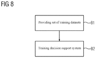
- FIG. 8 shows a flowchart for a method for training a decision support system for an examination via a medical imaging device
- FIG. 9 shows a schematic representation of a medical imaging device according to an embodiment variant of the invention.
- first, second, etc. may be used herein to describe various elements, components, regions, layers, and/or sections, these elements, components, regions, layers, and/or sections, should not be limited by these terms. These terms are only used to distinguish one element from another. For example, a first element could be termed a second element, and, similarly, a second element could be termed a first element, without departing from the scope of example embodiments of the present invention.
- the term “and/or,” includes any and all combinations of one or more of the associated listed items. The phrase “at least one of” has the same meaning as “and/or”.
- spatially relative terms such as “beneath,” “below,” “lower,” “under,” “above,” “upper,” and the like, may be used herein for ease of description to describe one element or feature's relationship to another element(s) or feature(s) as illustrated in the figures. It will be understood that the spatially relative terms are intended to encompass different orientations of the device in use or operation in addition to the orientation depicted in the figures. For example, if the device in the figures is turned over, elements described as “below,” “beneath,” or “under,” other elements or features would then be oriented “above” the other elements or features. Thus, the example terms “below” and “under” may encompass both an orientation of above and below.
- the device may be otherwise oriented (rotated 90 degrees or at other orientations) and the spatially relative descriptors used herein interpreted accordingly.
- the element when an element is referred to as being “between” two elements, the element may be the only element between the two elements, or one or more other intervening elements may be present.
- Spatial and functional relationships between elements are described using various terms, including “connected,” “engaged,” “interfaced,” and “coupled.” Unless explicitly described as being “direct,” when a relationship between first and second elements is described in the above disclosure, that relationship encompasses a direct relationship where no other intervening elements are present between the first and second elements, and also an indirect relationship where one or more intervening elements are present (either spatially or functionally) between the first and second elements. In contrast, when an element is referred to as being “directly” connected, engaged, interfaced, or coupled to another element, there are no intervening elements present. Other words used to describe the relationship between elements should be interpreted in a like fashion (e.g., “between,” versus “directly between,” “adjacent,” versus “directly adjacent,” etc.).
- the term “and/or” includes any and all combinations of one or more of the associated listed items. Expressions such as “at least one of,” when preceding a list of elements, modify the entire list of elements and do not modify the individual elements of the list. Also, the term “exemplary” is intended to refer to an example or illustration.
- Units and/or devices may be implemented using hardware, software, and/or a combination thereof.
- hardware devices may be implemented using processing circuity such as, but not limited to, a processor, Central Processing Unit (CPU), a controller, an arithmetic logic unit (ALU), a digital signal processor, a microcomputer, a field programmable gate array (FPGA), a System-on-Chip (SoC), a programmable logic unit, a microprocessor, or any other device capable of responding to and executing instructions in a defined manner.
- processing circuity such as, but not limited to, a processor, Central Processing Unit (CPU), a controller, an arithmetic logic unit (ALU), a digital signal processor, a microcomputer, a field programmable gate array (FPGA), a System-on-Chip (SoC), a programmable logic unit, a microprocessor, or any other device capable of responding to and executing instructions in a defined manner.
- module or the term ‘controller’ may be replaced with the term ‘circuit.’
- module may refer to, be part of, or include processor hardware (shared, dedicated, or group) that executes code and memory hardware (shared, dedicated, or group) that stores code executed by the processor hardware.
- the module may include one or more interface circuits.
- the interface circuits may include wired or wireless interfaces that are connected to a local area network (LAN), the Internet, a wide area network (WAN), or combinations thereof.
- LAN local area network
- WAN wide area network
- the functionality of any given module of the present disclosure may be distributed among multiple modules that are connected via interface circuits. For example, multiple modules may allow load balancing.
- a server (also known as remote, or cloud) module may accomplish some functionality on behalf of a client module.
- Software may include a computer program, program code, instructions, or some combination thereof, for independently or collectively instructing or configuring a hardware device to operate as desired.
- the computer program and/or program code may include program or computer-readable instructions, software components, software modules, data files, data structures, and/or the like, capable of being implemented by one or more hardware devices, such as one or more of the hardware devices mentioned above.
- Examples of program code include both machine code produced by a compiler and higher level program code that is executed using an interpreter.
- a hardware device is a computer processing device (e.g., a processor, Central Processing Unit (CPU), a controller, an arithmetic logic unit (ALU), a digital signal processor, a microcomputer, a microprocessor, etc.)
- the computer processing device may be configured to carry out program code by performing arithmetical, logical, and input/output operations, according to the program code.
- the computer processing device may be programmed to perform the program code, thereby transforming the computer processing device into a special purpose computer processing device.
- the processor becomes programmed to perform the program code and operations corresponding thereto, thereby transforming the processor into a special purpose processor.
- Software and/or data may be embodied permanently or temporarily in any type of machine, component, physical or virtual equipment, or computer storage medium or device, capable of providing instructions or data to, or being interpreted by, a hardware device.
- the software also may be distributed over network coupled computer systems so that the software is stored and executed in a distributed fashion.
- software and data may be stored by one or more computer readable recording mediums, including the tangible or non-transitory computer-readable storage media discussed herein.
- any of the disclosed methods may be embodied in the form of a program or software.
- the program or software may be stored on a non-transitory computer readable medium and is adapted to perform any one of the aforementioned methods when run on a computer device (a device including a processor).
- a computer device a device including a processor
- the non-transitory, tangible computer readable medium is adapted to store information and is adapted to interact with a data processing facility or computer device to execute the program of any of the above mentioned embodiments and/or to perform the method of any of the above mentioned embodiments.
- Example embodiments may be described with reference to acts and symbolic representations of operations (e.g., in the form of flow charts, flow diagrams, data flow diagrams, structure diagrams, block diagrams, etc.) that may be implemented in conjunction with units and/or devices discussed in more detail below.
- a function or operation specified in a specific block may be performed differently from the flow specified in a flowchart, flow diagram, etc.
- functions or operations illustrated as being performed serially in two consecutive blocks may actually be performed simultaneously, or in some cases be performed in reverse order.
- computer processing devices may be described as including various functional units that perform various operations and/or functions to increase the clarity of the description.
- computer processing devices are not intended to be limited to these functional units.
- the various operations and/or functions of the functional units may be performed by other ones of the functional units.
- the computer processing devices may perform the operations and/or functions of the various functional units without sub-dividing the operations and/or functions of the computer processing units into these various functional units.
- Units and/or devices may also include one or more storage devices.
- the one or more storage devices may be tangible or non-transitory computer-readable storage media, such as random access memory (RAM), read only memory (ROM), a permanent mass storage device (such as a disk drive), solid state (e.g., NAND flash) device, and/or any other like data storage mechanism capable of storing and recording data.
- the one or more storage devices may be configured to store computer programs, program code, instructions, or some combination thereof, for one or more operating systems and/or for implementing the example embodiments described herein.
- the computer programs, program code, instructions, or some combination thereof may also be loaded from a separate computer readable storage medium into the one or more storage devices and/or one or more computer processing devices using a drive mechanism.
- a separate computer readable storage medium may include a Universal Serial Bus (USB) flash drive, a memory stick, a Blu-ray/DVD/CD-ROM drive, a memory card, and/or other like computer readable storage media.
- the computer programs, program code, instructions, or some combination thereof may be loaded into the one or more storage devices and/or the one or more computer processing devices from a remote data storage device via a network interface, rather than via a local computer readable storage medium.
- the computer programs, program code, instructions, or some combination thereof may be loaded into the one or more storage devices and/or the one or more processors from a remote computing system that is configured to transfer and/or distribute the computer programs, program code, instructions, or some combination thereof, over a network.
- the remote computing system may transfer and/or distribute the computer programs, program code, instructions, or some combination thereof, via a wired interface, an air interface, and/or any other like medium.
- the one or more hardware devices, the one or more storage devices, and/or the computer programs, program code, instructions, or some combination thereof, may be specially designed and constructed for the purposes of the example embodiments, or they may be known devices that are altered and/or modified for the purposes of example embodiments.
- a hardware device such as a computer processing device, may run an operating system (OS) and one or more software applications that run on the OS.
- the computer processing device also may access, store, manipulate, process, and create data in response to execution of the software.
- OS operating system
- a hardware device may include multiple processing elements or processors and multiple types of processing elements or processors.
- a hardware device may include multiple processors or a processor and a controller.
- other processing configurations are possible, such as parallel processors.
- the computer programs include processor-executable instructions that are stored on at least one non-transitory computer-readable medium (memory).
- the computer programs may also include or rely on stored data.
- the computer programs may encompass a basic input/output system (BIOS) that interacts with hardware of the special purpose computer, device drivers that interact with particular devices of the special purpose computer, one or more operating systems, user applications, background services, background applications, etc.
- BIOS basic input/output system
- the one or more processors may be configured to execute the processor executable instructions.
- the computer programs may include: (i) descriptive text to be parsed, such as HTML (hypertext markup language) or XML (extensible markup language), (ii) assembly code, (iii) object code generated from source code by a compiler, (iv) source code for execution by an interpreter, (v) source code for compilation and execution by a just-in-time compiler, etc.
- source code may be written using syntax from languages including C, C++, C#, Objective-C, Haskell, Go, SQL, R, Lisp, Java®, Fortran, Perl, Pascal, Curl, OCaml, Javascript®, HTML5, Ada, ASP (active server pages), PHP, Scala, Eiffel, Smalltalk, Erlang, Ruby, Flash®, Visual Basic®, Lua, and Python®.
- At least one embodiment of the invention relates to the non-transitory computer-readable storage medium including electronically readable control information (processor executable instructions) stored thereon, configured in such that when the storage medium is used in a controller of a device, at least one embodiment of the method may be carried out.
- electronically readable control information processor executable instructions
- the computer readable medium or storage medium may be a built-in medium installed inside a computer device main body or a removable medium arranged so that it can be separated from the computer device main body.
- the term computer-readable medium, as used herein, does not encompass transitory electrical or electromagnetic signals propagating through a medium (such as on a carrier wave); the term computer-readable medium is therefore considered tangible and non-transitory.
- Non-limiting examples of the non-transitory computer-readable medium include, but are not limited to, rewriteable non-volatile memory devices (including, for example flash memory devices, erasable programmable read-only memory devices, or a mask read-only memory devices); volatile memory devices (including, for example static random access memory devices or a dynamic random access memory devices); magnetic storage media (including, for example an analog or digital magnetic tape or a hard disk drive); and optical storage media (including, for example a CD, a DVD, or a Blu-ray Disc).
- Examples of the media with a built-in rewriteable non-volatile memory include but are not limited to memory cards; and media with a built-in ROM, including but not limited to ROM cassettes; etc.
- various information regarding stored images for example, property information, may be stored in any other form, or it may be provided in other ways.
- code may include software, firmware, and/or microcode, and may refer to programs, routines, functions, classes, data structures, and/or objects.
- Shared processor hardware encompasses a single microprocessor that executes some or all code from multiple modules.
- Group processor hardware encompasses a microprocessor that, in combination with additional microprocessors, executes some or all code from one or more modules.
- References to multiple microprocessors encompass multiple microprocessors on discrete dies, multiple microprocessors on a single die, multiple cores of a single microprocessor, multiple threads of a single microprocessor, or a combination of the above.
- Shared memory hardware encompasses a single memory device that stores some or all code from multiple modules.
- Group memory hardware encompasses a memory device that, in combination with other memory devices, stores some or all code from one or more modules.
- memory hardware is a subset of the term computer-readable medium.
- the term computer-readable medium does not encompass transitory electrical or electromagnetic signals propagating through a medium (such as on a carrier wave); the term computer-readable medium is therefore considered tangible and non-transitory.
- Non-limiting examples of the non-transitory computer-readable medium include, but are not limited to, rewriteable non-volatile memory devices (including, for example flash memory devices, erasable programmable read-only memory devices, or a mask read-only memory devices); volatile memory devices (including, for example static random access memory devices or a dynamic random access memory devices); magnetic storage media (including, for example an analog or digital magnetic tape or a hard disk drive); and optical storage media (including, for example a CD, a DVD, or a Blu-ray Disc).
- Examples of the media with a built-in rewriteable non-volatile memory include but are not limited to memory cards; and media with a built-in ROM, including but not limited to ROM cassettes; etc.
- various information regarding stored images for example, property information, may be stored in any other form, or it may be provided in other ways.
- the apparatuses and methods described in this application may be partially or fully implemented by a special purpose computer created by configuring a general purpose computer to execute one or more particular functions embodied in computer programs.
- the functional blocks and flowchart elements described above serve as software specifications, which can be translated into the computer programs by the routine work of a skilled technician or programmer.
- At least one embodiment of the invention relates to a method for checking a medical image with regard to a processing of the medical image via an image processing module, the method comprising:
- the medical image may be a medical computed tomography image.
- the medical imaging examination may be a medical computed tomography imaging examination.
- the following step may be performed as a function of a result of the check:
- one of the following steps may be performed as a function of a result of the check:
- the following step may be performed as a function of a result of the check:
- the method may additionally comprise the following steps:
- the method may additionally comprise the following steps:
- the at least one input requirement may be defined independently of specific characteristics of a medical imaging device by which the medical image and/or the further medical image are/is generated.
- the at least one image parameter may be chosen from the image parameter group consisting of a spatial resolution, a temporal resolution, an edge behavior, a contrast, a convolution kernel, a slice thickness, a rotation time, a pitch, a tube voltage, a filter property, a modulation transfer function, and combinations thereof.
- At least one embodiment of the invention further relates to a method for adapting an examination protocol during a medical imaging examination
- each examination step of the list of examination steps is assigned a respective instruction
- the examination protocol comprises a start examination step from the list of examination steps, which start examination step is performed at the commencement of the medical imaging examination
- the examination protocol is adapted by way of the instruction assigned to the performed examination step by performing one of the following steps based on the instruction:
- the following step may be performed as a function of a result of the performed examination step:
- the method may additionally comprise the following step:
- determining the result of the performed examination step based on a user input and/or based on an automatic evaluation of data that was acquired during the performed examination step.
- At least one embodiment of the invention further relates to a method for generating a list of examination steps, wherein each examination step is assigned a respective instruction for adapting an examination protocol during a medical imaging examination, the method comprising:
- each training dataset comprises a respective training instruction and an examination parameter set, wherein the examination parameter set relates to an examination step assigned to the training instruction and/or an examination result assigned to the training instruction, and
- each examination step of the list of examination steps is assigned a respective instruction for adapting an examination protocol during a medical imaging examination.
- At least one training dataset of the set of training datasets may be provided by performing the following steps during the course of an examination:
- At least one embodiment of the invention further relates to a method for training a decision support system for an examination via a medical imaging device, the method comprising:
- each training dataset comprises a respective training examination job and examination information, wherein the examination information relates to a medical imaging examination that has been performed based on the training examination job, and
- the training examination job comprises a training instruction
- the examination information comprises an examination parameter set
- the examination parameter set relates to an examination step that is assigned to the training instruction and/or to an examination result that is assigned to the training instruction
- the examination recommendation comprises a list of examination steps that is generated based on the set of training datasets and the machine learning algorithm, wherein each examination step of the list of examination steps is assigned a respective instruction for adapting an examination protocol during a medical imaging examination.
- training examination job comprises an image generation job
- the examination information comprises acquisition parameters and/or reconstruction parameters by which the image generation job was performed during the medical imaging examination.
- training examination job comprises an image processing job
- the examination information comprises image processing parameters by which the image processing job was performed during the medical imaging examination.
- At least one embodiment of the invention further relates to a data processing unit which is embodied to perform a method according to one of the embodiments disclosed in this application.
- At least one embodiment of the invention further relates to a computer program which can be loaded into a memory device of a data processing system and has program sections for performing all steps of a method according to one of the embodiments disclosed in this application when the computer program is executed by the data processing system.
- At least one embodiment of the invention further relates to a computer-readable medium on which are stored program sections that can be read in and executed by a data processing system in order to perform all steps of a method according to one of the embodiments disclosed in this application when the program sections are executed by the data processing system.
- At least one embodiment of the invention further relates to a decision support system which has been trained based on a method according to one of the embodiments disclosed in this application.
- At least one embodiment of the invention further relates to a medical imaging device comprising a data processing unit according to one of the embodiments disclosed in this application and/or a decision support system according to one of the embodiments disclosed in this application.
- At least one embodiment of the invention further relates to a use of a decision support system which has been trained based on a method according to one of the embodiments disclosed in this application in a medical imaging device and/or for generating an examination recommendation for an examination via a medical imaging device based on an examination job.
- post-processing module Given knowledge of the mode of operation of a post-processing algorithm, it is possible to specify necessary and/or sufficient properties of input data, in particular of the medical image, which characterize a suitability of the input data for an evaluation by way of the post-processing algorithm.
- the properties can be specified, in particular with knowledge of the properties of the training data and under the assumption that the result aimed at by way of the trained algorithm functions in an optimal manner with the training data.
- post-processing module and image processing module are used synonymously.
- the properties may relate for example to a spatial resolution in one or more spatial directions, an edge behavior, in particular an overshoot, a temporal resolution, in particular a minimum temporal resolution, a material contrast or similar.
- the properties may correlate in particular with acquisition parameters and/or reconstruction parameters.
- acquisition parameters and/or reconstruction parameters are in particular a convolution kernel, a slice thickness, a rotation time, a pitch, a reconstructed data segment, a tube voltage or a filtering property.
- the spatial resolution correlates with the convolution kernel and/or with the slice thickness, the edge behavior with the convolution kernel, the temporal resolution with the rotation time, with the pitch and/or with the reconstructed data segment, the material contrast with the tube voltage and/or with the filtering property.
- a selection of the aforementioned parameters may be used to characterize the medical image.
- further parameters may be added without leaving the scope of the invention insofar as it is set out by the claims.
- intervals or a list of values may also be defined in particular for the at least one image parameter by way of the at least one input requirement.
- the medical image may in particular comprise a header and/or a footer.
- the image processing module may in particular be an image processing algorithm and/or include an image processing algorithm.
- the image processing module may for example be embodied for segmenting a structure and/or for extracting a centerline in the medical image.
- the image processing module may in particular be integrated into a post-processing application.
- the post-processing application may for example be embodied for detecting pulmonary nodules and/or for detecting a stenosis.
- the input requirements are stored in a standardized form, in particular irrespective of specialized imaging devices and/or manufacturers.
- modulation transfer functions may be specified for one or more or all spatial directions instead of the convolution kernel and the slice thickness.
- the modulation transfer functions can be realized in particular when a specific convolution kernel is used and a specific slice thickness is set. In this way it is possible for example to describe the spatial resolution and the edge behavior in a device-neutral manner.
- the medical imaging device may for example be chosen from the imaging modalities group consisting of an x-ray device, a C-arm x-ray device, a computed tomography device (CT device), a molecular imaging device (MI device), a single-photon emission computed tomography device (SPECT device), a positron emission tomography device (PET device), a magnetic resonance tomography device (MR device), and combinations thereof, in particular a PET/CT device and a PET/MR device.
- the medical imaging device may furthermore comprise a combination of an imaging modality, chosen for example from the imaging modalities group, and an irradiation modality.
- the irradiation modality may for example comprise an irradiation unit for therapeutic irradiation treatment.
- a computed tomography device is cited in relation to some of the embodiment variants by way of example for a medical imaging device.
- the medical imaging device comprises an acquisition unit which is embodied for acquiring the acquisition data.
- the acquisition unit may comprise a radiation source and a radiation detector.
- An embodiment variant of the invention provides that the radiation source is embodied for emitting and/or exciting radiation, in particular electromagnetic radiation, and/or that the radiation detector is embodied for detecting the radiation, in particular the electromagnetic radiation.
- the radiation may for example travel from the radiation source to a region to be imaged and/or arrive at the radiation detector following an interaction with the region to be imaged.
- the radiation is modified and thus becomes a carrier of information relating to the region to be imaged.
- the information is captured in the form of acquisition data.
- the acquisition data may be projection data
- the acquisition unit a projection data acquisition unit
- the radiation source an x-ray source
- the radiation detector an x-ray detector.
- the x-ray detector may in particular be a quantum-counting and/or energy-resolving x-ray detector.
- the acquisition data may be a magnetic resonance dataset
- the acquisition unit a magnetic resonance data acquisition unit
- the radiation source a first radiofrequency antenna unit
- the radiation detector the first radiofrequency antenna unit and/or a second radiofrequency antenna unit.
- the data processing unit and/or one or more components of the data processing unit may be formed by a data processing system.
- the decision support system and/or one or more components of the decision support system may be formed by a data processing system.
- the data processing system may for example comprise one or more components in the form of hardware and/or one or more components in the form of software.
- the data processing system may for example be formed at least in part by a cloud computing system.
- the data processing system may for example be and/or comprise a cloud computing system, a computer network, a computer, a tablet computer, a smartphone or similar, or combinations thereof.
- the hardware may for example cooperate with software and/or be configurable via software.
- the software may for example be executed via the hardware.
- the hardware may for example be a storage system, an FPGA (Field-Programmable Gate Array) system, an ASIC (Application-Specific Integrated Circuit) system, a microcontroller system, a processor system, and combinations thereof.
- the processor system may for example comprise a microprocessor and/or a plurality of cooperating microprocessors.
- a component of the data processing unit according to one of the aspects disclosed in this application which component is embodied to perform a given step of a method according to one of the embodiments disclosed in this application, may be implemented in the form of hardware which is configured to perform the given step and/or which is configured to execute a computer-readable instruction in such a way that the hardware may be configured via the computer-readable instruction to perform the given step.
- the system may comprise a storage area, for example in the form of a computer-readable medium, in which computer-readable instructions, for example in the form of a computer program, are stored.
- a data transfer between components of the data processing system may for example be performed in each case via a suitable data transfer interface.
- the data transfer interface for transferring data to and/or from a component of the data processing system may be realized at least in part in the form of software and/or at least in part in the form of hardware.
- the data transfer interface may for example be embodied for storing data in and/or for loading data from an area of the storage system, one or more components of the data processing system being able to access the area of the storage system.
- the computer program is loadable into the storage system of the data processing system and can be executed by the processor system of the data processing system.
- the data processing system may for example be embodied via the computer program in such a way that the data processing system can perform the steps of a method according to one of the embodiment variants disclosed in this application when the computer program is executed by the data processing system.
- the computer program product according to one of the embodiment variants disclosed in this application and/or the computer program according to one of the embodiment variants disclosed in this application may for example be stored on the computer-readable medium.
- the computer-readable medium may for example be a memory stick, a hard disk or another data medium which in particular may be releasably connected to the data processing system or permanently integrated into the data processing system.
- the computer-readable medium may for example form a domain of the storage system of the data processing system.
- protocol and examination protocol are used synonymously for one another.
- the medical imaging device comprises the medical imaging device.
- the use of the term “unit” does not rule out the possibility that the object to which the term “unit” refers may comprise a plurality of components that are separated from one another in space.
- a formulation which accordingly produces (alternatively: determines, defines, etc.) a first feature based on a second feature does not preclude the possibility that the first feature may be produced (alternatively: determined, defined, etc.) based on a third feature.
- FIG. 1 shows a flowchart for a method for checking a medical image with regard to a processing of the medical image via an image processing module according to an embodiment variant of the invention, wherein the method comprises the following steps:
- FIG. 2 shows a schematic representation of a data processing unit 35 for checking a medical image with regard to a processing of the medical image via an image processing module according to a further embodiment variant of the invention, comprising:
- an input requirement provider unit PQ-M for providing PQ at least one input requirement of the image processing module relating to at least one image parameter of the medical image
- an image parameter provider unit PX-M for providing PM the at least one image parameter of the medical image
- a checking unit CP-M for checking CP whether the at least one image parameter fulfills the at least one input requirement.
- FIG. 3 shows a flowchart for a method for checking a medical image with regard to a processing of the medical image via an image processing module according to an embodiment variant of the invention.
- the start of the execution sequence of the method is marked by step S.
- a parameter set P algo is assigned to a post-processing module L which describes the input requirements and/or is stored in the post-processing module.
- the image parameters, P real realized in the medical image are stored in a data structure ID in which the medical image is transmitted.
- a header or footer may be used for this purpose, for example.
- P algo is represented by reference sign Q.
- P real is represented by reference sign X.
- P real may comprise acquisition parameters XA for an acquisition DA and/or reconstruction parameters XR for a reconstruction IR.
- a check is carried out to verify whether P real may be combined with P algo .
- This can be realized in particular by way of a parameter-by-parameter check to determine whether the realized value lies in the value range specified via P algo .
- the value range may for example be specified in the form of a value, an interval and/or a list.
- P real and P algo may be regarded in principle as sets of values for specific parameters. The check to verify whether the at least one image parameter fulfills the at least one input requirement would therefore correspond to the check to determine whether P real is a subset of P algo , i.e. whether P real ⁇ P algo applies.
- the medical image is processed via the image processing module L.
- This path is marked by Y. Otherwise, as shown in FIG. 3 , the post-processing job is rejected J 1 and/or, as shown in FIG. 4 , an alert J 2 is output to the user indicating that the results generated via the post-processing algorithm may possibly be limited in terms of their quality.
- This path is marked by N.
- An end of the execution sequence of the method is marked by steps E 1 and E 2 , respectively.
- an image generation job is generated relating to a generation of a further medical image taking into consideration the at least one input requirement P algo .
- a further medical image which fulfills the requirement P real ⁇ P algo can be generated via a medical imaging device.
- image parameters in particular acquisition parameters and/or reconstruction parameters for generating the further medical image, are generated based on P real and/or P algo and/or on a result of the check CP. These parameters are different in particular from the initial parameters that were provided for generating the medical image at step INI.
- the generation of the further medical image may in particular comprise a reconstruction of the further medical image based on already acquired raw data. For example, the same raw data may be used in this case as was used already for a reconstruction of the medical image that does not fulfill the at least one input requirement. In this case use may be made in particular of reconstruction parameters that are adapted to the at least one input requirement.
- the generation of the further medical image may include an acquisition of raw data via a medical imaging device, in particular when the requirement P real ⁇ P algo cannot be fulfilled by way of the reconstruction alone.
- acquisition parameters may be used in this case which are adapted to the at least one input requirement.
- the further medical image may be reconstructed based on the thus acquired raw data.
- the solution according to an embodiment of the invention therefore enables an automatic control of an acquisition and/or a reconstruction which is adapted with regard to a processing of a medical image generated in the process via a post-processing application.
- Including a new acquisition of data makes sense in particular for modalities in which this does not cause the patient to be subjected to any additional exposure, for example due to ionizing radiation.
- the post-processing job can be rejected and/or an alert can be output to the user indicating that the results generated via the post-processing algorithm could be limited in terms of their quality.
- the medical image on which the first checking step is based is already generated based on an image generation job which was generated based on the at least one input requirement.
- an image generation job may be generated based on a planned post-processing step with specification of the input requirements P algo for generating a medical image and output to a medical imaging device.
- a medical image may be generated based on the image generation job via a medical imaging device in such a way that the at least one image parameter of the further medical image fulfills the at least one input requirement.
- the generation of the medical image may in particular include a reconstruction and/or an acquisition.
- a reconstruction of already acquired raw data may be carried out based on the image generation job for the medical image using reconstruction parameters that are adapted to the at least one input requirement. This is aimed at fulfilling or at least approximately fulfilling the requirement P real ⁇ P algo for the medical image.
- an acquisition can be performed based on the image generation job for the medical image using acquisition parameters that are adapted to the at least one input requirement.
- the thus acquired raw data may be reconstructed in particular using reconstruction parameters that are adapted to the at least one input requirement.
- a check based on a reconstruction using reconstruction parameters that are adapted to the at least one input requirement may be carried out initially in order to determine whether the at least one input requirement for the medical image is fulfilled. If the at least one input requirement for the medical image is not fulfilled, an image generation job for a further medical image can be generated which relates both to an acquisition and to a reconstruction. If the requirement P real ⁇ P algo is fulfilled, the post-processing step can be performed. Otherwise, the post-processing job is rejected and/or an alert is output to the user indicating that the results generated via the post-processing algorithm may possibly be limited in terms of their quality.
- the solution according to an embodiment of the invention enables in particular a check to be carried out in order to determine whether a medical image is actually suitable as a basis for a post-processing operation. In this way it is possible in particular to realize an improvement in quality with regard to the accuracy and/or the reproducibility of the results derived on the basis of the post-processing operation. It is furthermore possible to advise caution be exercised in the interpretation of the results.
- the best possible quality in the chain of acquisition, reconstruction and derivation of results via post-processing algorithms can be achieved retrospectively or in advance with the aid of the image generation job.
- the method steps can be performed in particular in an automated manner. This is associated with a reduction in the amount of effort required, or at least no increase in the amount of effort required, on the part of the user.
- the examination protocol is typically configured in such a way that the maximum chain of examination steps that might be necessary in order to rule out a ground for suspicion can be processed.
- a linear sequence of examination steps may be used that corresponds to a superset of the necessary examination steps.
- the examination steps may for example be scans in the case of different physiological states, in particular native scans, scans using contrast agent, scans at different phases or similar.
- each examination step it is decided as a function of the respective findings whether the protocol may be terminated prematurely or a step skipped or a transition made to the next examination step in the sequence. This typically happens manually on the basis of indication-based rules of the respective institution and/or of the general evidence base of the user.
- the solution according to an embodiment of the invention in particular enables these dynamic adjustments to be simplified and/or automated.
- a basic protocol can be defined which contains the list of examination steps.
- Such a basic protocol may also be understood for example as a superset of a maximum examination scope that can be planned using the examination steps.
- the examination steps may for example be borrowed from a conventional examination protocol.
- the assignment of the instructions to the examination steps may be carried out manually within the scope of an implementation.
- the list of examination steps may be machine-learned in conjunction with the instructions from medical imaging examinations in which conventional examination protocols are used. In the process, the user may then for example be asked for the reason if there is a deviation from the linear sequence of examination steps that is provided in the conventional protocol during the medical imaging examination.
- Such a deviation may involve in particular a skipping of an examination step in the linear sequence, a premature termination of the medical imaging examination or similar.
- the examination parameter set of the training dataset may include the reason for the deviation.
- the order of the examination steps in the list of examination steps is of no importance per se. Except for the start examination step, whether the examination step is performed in this case is not dependent on the relative position of an examination step in the list. Rather, what matters is whether a previously performed examination step is assigned an instruction which points to the examination step that is to be performed thereafter.
- At least one examination step is assigned an instruction which points to the next examination step that is to be performed, without any further condition with regard to the result of the examination step, or which terminates the entire examination.
- at least one examination step is assigned an instruction which points to one of a plurality of examination steps as a function of a result of the examination step relating to the problem that the examination is intended to answer, or which terminates the entire examination sequence.
- At least one examination step is assigned an instruction which defines a further indication for a subsequent examination as a function of an in particular unexpected finding, and/or which selects a further examination protocol in respect of a clinical problem relating to the finding.
- the instruction may in particular comprise a generation of an examination job (Requested Procedure/Modality Worklist) in an HIS/RIS (Hospital Information System/Radiology Information System).
- HIS/RIS Hospital Information System/Radiology Information System
- the result of the performed examination step is determined based on an interaction with the user, for example a user of a medical imaging device.
- the interaction may in particular comprise a question that is to be answered by a user and/or a list of options from which the user can select the answer.
- the result of the performed examination step is determined based on an automatic analysis of data generated in the course of the performed examination step.
- the data may for example consist of slice images, post-processing results or similar.
- FIG. 7 shows a schematic representation of an examination protocol for the exclusion of hemorrhaging, infarction and/or tumor in the brain.
- a list of examination steps T 1 , T 2 , T 3 is provided at step 71 .
- each examination step in the list of examination steps is assigned a respective instruction.
- start examination step 7 S is defined, specifying, for example, that the examination protocol is to commence with examination step T 1 .
- Examination step T 1 may in particular relate to an examination of soft tissue without contrast agent.
- Examination step T 2 may in particular relate to an examination of soft tissue with contrast agent, for example with delayed administration of contrast agent.
- Examination step T 3 may in particular relate to an examination of a tumor volume with contrast agent, for example with delayed administration of contrast agent.
- step DT 1 it can be determined in particular whether hemorrhaging or an infarction is present. If yes Y, the examination is continued with examination step T 2 according to instruction FT 1 . If no N, the examination is terminated according to instruction ET 1 .
- step DT 2 it can be determined in particular whether a tumor is present. If yes Y, the examination is continued with examination step T 3 according to instruction FT 2 . If no N, the examination is terminated according to instruction ET 2 .
- FIG. 8 shows a flowchart for a method for training a decision support system for an examination via a medical imaging device, wherein the method comprises the following steps:
- each training dataset comprises a respective training examination job and examination information, wherein the examination information relates to a medical imaging examination that has been carried out based on the training examination job,
- training 82 the decision support system based on the set of training datasets and a machine learning algorithm in such a way that via the trained decision support system it is possible to generate an in particular optimal examination recommendation for the examination via the medical imaging device based on an examination job.
- FIG. 9 shows a schematic representation of a medical imaging device 1 according to an embodiment variant of the invention.
- a computed tomography device is shown by way of example for the medical imaging device 1 .
- the medical imaging device 1 comprises the gantry 20 , the tunnel-shaped opening or bore 9 , the patient support device 10 and the control device 30 .
- the gantry 20 comprises the stationary carrier frame 21 and the rotor 24 .
- the patient 13 can be introduced into the tunnel-shaped bore 9 .
- the acquisition zone 4 is located in the tunnel-shaped bore 9 .
- a region to be imaged of the patient 13 can be positioned in the acquisition zone 4 in such a way that the radiation 27 can travel from the radiation source 26 to the region to be imaged and, after interacting with the region to be imaged, can arrive at the radiation detector 28 .
- the patient support device 10 comprises the support plinth 11 and the support tabletop 12 for supporting the patient 13 .
- the support tabletop 12 is arranged on the support plinth 11 so as to be movable relative to the support plinth 11 in such a way that the support tabletop 12 can be introduced into the acquisition zone 4 in a longitudinal direction of the support tabletop 12 , in particular along the system axis AR.
- the medical imaging device 1 is embodied for acquiring acquisition data based on electromagnetic radiation 27 .
- the medical imaging device 1 comprises an acquisition unit.
- the acquisition unit is a projection data acquisition unit comprising the radiation source 26 , e.g. an x-ray source, and the detector 28 , e.g. an x-ray detector, in particular an energy-resolving x-ray detector.
- the radiation source 26 is arranged on the rotor 24 and is embodied for emitting radiation 27 , e.g. x-ray radiation, comprising radiation quanta 27 .
- the detector 28 is arranged on the rotor 24 and is embodied for detecting the radiation quanta 27 .
- the radiation quanta 27 can travel from the radiation source 26 to the region to be imaged of the patient 13 and, after interacting with the region to be imaged, impinge on the detector 28 . In this way, acquisition data of the region to be imaged can be captured in the form of projection data via the acquisition unit.
- the control device 30 is embodied for receiving the acquisition data acquired by the acquisition unit.
- the control device 30 is embodied for controlling the medical imaging device 1 .
- the control device 30 comprises the data processing unit 35 , the decision support system 37 , the computer-readable medium 32 and the processor system 36 .
- the control device 30 in particular the data processing unit 35 , is formed by a data processing system which comprises a computer.
- the control device 30 comprises the image reconstruction device 34 .
- a medical image dataset can be reconstructed via the image reconstruction device 34 on the basis of the acquisition data.
- the medical imaging device 1 comprises an input device 38 and an output device 39 , which are each connected to the control device 30 .
- the input device 38 is embodied for inputting control information, e.g. image reconstruction parameters, examination parameters or similar.
- the output device 39 is embodied in particular for outputting control information, images and/or acoustic signals.
- the solution according to an embodiment of the invention enables in particular a manual application of the findings-dependent rules, which would be necessary each time the protocol were used, to be dispensed with and an automated workflow to be implemented instead.
- An examination step may in particular comprise an acquisition and/or a reconstruction and/or an image processing operation.
- a machine learning algorithm in the context of the present application, is understood in particular an algorithm that is embodied for machine learning.
- a machine learning algorithm may be realized for example with the aid of decision trees, mathematical functions and/or general programming languages.
- the machine learning algorithm may be embodied for example for supervised learning and/or for unsupervised learning.
- the machine learning algorithm may be embodied for example for deep learning and/or for reinforcement learning and/or for marginal space learning.
- a functions class may be used which is based for example on decision trees, a random forest, a logistical regression, a support vector machine, an artificial neural network, a kernel method, Bayes classifiers or similar, or combinations thereof.
- Possible implementations of the machine learning algorithm may use artificial intelligence, for example.
- One or more rule-based algorithms may be used alternatively or in addition to the first machine learning algorithm and/or to the second machine learning algorithm. Calculations, in particular when determining the classification system based on the set of training datasets and a machine learning algorithm, may be performed via a processor system, for example.
- the processor system may for example comprise one or more graphics processors.
- data relating for example to a medical image, a protocol or a training dataset may be provided such that the data is loaded, e.g. from an area of a storage system, and/or generated, e.g. via a medical imaging device.
- one step or more steps or all steps of the method according to the invention may be performed automatically and/or via a component of a data processing unit, the component being formed by a processor system, for example.
- the medical imaging examination may be an examination via a medical imaging device and/or be performed via a medical imaging device.
- the set of training datasets may in particular be provided by collecting a plurality of examination jobs together with the actual examinations.
- a decision support system can learn from this information, in particular recognize patterns. For example, it can be recognized in this way that most clinics also include a coronary reconstruction when a lung scan is performed. In this way it can quickly be identified that in the medical imaging examinations performed by a user, the user deviates compared to the majority of users performing similar medical imaging examinations.
- an examination recommendation with regard to an acquisition of imaging data can be generated via the decision support system, for example whether a spiral or a sequence is to be preferred or whether additional scans, in particular an additional late phase, might facilitate a better diagnosis.
- an examination recommendation with regard to a reconstruction of a medical image can be generated via the decision support system, for example whether a reconstruction using a different kernel would be better suited.
- an examination recommendation with regard to an image processing operation can be generated via the decision support system, in particular which algorithms were to be preferred.
Landscapes
- Engineering & Computer Science (AREA)
- Health & Medical Sciences (AREA)
- Medical Informatics (AREA)
- Theoretical Computer Science (AREA)
- General Health & Medical Sciences (AREA)
- Public Health (AREA)
- Epidemiology (AREA)
- Primary Health Care (AREA)
- General Physics & Mathematics (AREA)
- Data Mining & Analysis (AREA)
- Computer Vision & Pattern Recognition (AREA)
- Physics & Mathematics (AREA)
- Biomedical Technology (AREA)
- Software Systems (AREA)
- Nuclear Medicine, Radiotherapy & Molecular Imaging (AREA)
- Radiology & Medical Imaging (AREA)
- Databases & Information Systems (AREA)
- Artificial Intelligence (AREA)
- Evolutionary Computation (AREA)
- Computing Systems (AREA)
- Quality & Reliability (AREA)
- Pathology (AREA)
- General Engineering & Computer Science (AREA)
- Mathematical Physics (AREA)
- Business, Economics & Management (AREA)
- General Business, Economics & Management (AREA)
- Multimedia (AREA)
- Bioinformatics & Cheminformatics (AREA)
- Bioinformatics & Computational Biology (AREA)
- Evolutionary Biology (AREA)
- Life Sciences & Earth Sciences (AREA)
- Apparatus For Radiation Diagnosis (AREA)
- Measuring And Recording Apparatus For Diagnosis (AREA)
Abstract
Description
Claims (10)
Applications Claiming Priority (3)
| Application Number | Priority Date | Filing Date | Title |
|---|---|---|---|
| DE102017203333.9 | 2017-03-01 | ||
| DE102017203333 | 2017-03-01 | ||
| DE102017203333.9A DE102017203333A1 (en) | 2017-03-01 | 2017-03-01 | A medical image review method and method for adjusting an examination protocol during a medical imaging examination |
Publications (2)
| Publication Number | Publication Date |
|---|---|
| US20180253842A1 US20180253842A1 (en) | 2018-09-06 |
| US10706537B2 true US10706537B2 (en) | 2020-07-07 |
Family
ID=63171034
Family Applications (1)
| Application Number | Title | Priority Date | Filing Date |
|---|---|---|---|
| US15/905,919 Active 2038-06-03 US10706537B2 (en) | 2017-03-01 | 2018-02-27 | Method for checking a medical image and method for adapting an examination protocol during a medical imaging examination |
Country Status (3)
| Country | Link |
|---|---|
| US (1) | US10706537B2 (en) |
| CN (1) | CN108537774B (en) |
| DE (1) | DE102017203333A1 (en) |
Families Citing this family (5)
| Publication number | Priority date | Publication date | Assignee | Title |
|---|---|---|---|---|
| EP3637362A1 (en) * | 2018-10-11 | 2020-04-15 | Siemens Healthcare GmbH | Method for adjusting an image impression |
| EP3654251A1 (en) * | 2018-11-13 | 2020-05-20 | Siemens Healthcare GmbH | Determining a processing sequence for processing an image |
| CN111178297A (en) * | 2019-12-31 | 2020-05-19 | 上海联影医疗科技有限公司 | Image processing method and device, electronic equipment and medium |
| CN111276219A (en) * | 2020-01-16 | 2020-06-12 | 上海联影医疗科技有限公司 | Medical imaging processing method, device, equipment and medium |
| EP4250307A1 (en) * | 2022-08-29 | 2023-09-27 | Siemens Healthcare GmbH | Method and data processing system for providing a study description for a radiological imaging examination |
Citations (6)
| Publication number | Priority date | Publication date | Assignee | Title |
|---|---|---|---|---|
| US20080044069A1 (en) * | 2006-08-15 | 2008-02-21 | Dugal Tiffany Ann | Processes and apparatus for imaging protocols and analysis |
| DE102014107445A1 (en) | 2014-05-27 | 2015-12-03 | Carl Zeiss Meditec Ag | An optical observation apparatus, a method for setting a parameter value for an optical observation apparatus, and a method for recording image or video data from an observation object by means of an observation optical apparatus |
| US20170186157A1 (en) * | 2015-12-23 | 2017-06-29 | Siemens Healthcare Gmbh | Method and system for outputting augmented reality information |
| US20180137244A1 (en) * | 2016-11-17 | 2018-05-17 | Terarecon, Inc. | Medical image identification and interpretation |
| US20180144465A1 (en) * | 2016-11-23 | 2018-05-24 | General Electric Company | Deep learning medical systems and methods for medical procedures |
| US20180235563A1 (en) * | 2015-08-17 | 2018-08-23 | Samsung Electronics Co., Ltd. | Medical image display device and medical image processing method |
Family Cites Families (10)
| Publication number | Priority date | Publication date | Assignee | Title |
|---|---|---|---|---|
| CA2529929A1 (en) * | 2003-06-25 | 2005-01-06 | Siemens Medical Solutions Usa, Inc. | Systems and methods for automated diagnosis and decision support for breast imaging |
| US7599534B2 (en) * | 2003-08-13 | 2009-10-06 | Siemens Medical Solutions Usa, Inc. | CAD (computer-aided decision) support systems and methods |
| CN101903887B (en) * | 2007-12-21 | 2014-06-11 | 皇家飞利浦电子股份有限公司 | Detection of errors in the inference engine of a clinical decision support system |
| DE102011002928A1 (en) * | 2011-01-20 | 2012-07-26 | Siemens Aktiengesellschaft | Method for the computer-aided configuration of a medical imaging device |
| CA2827523C (en) * | 2011-02-17 | 2019-09-10 | Eon Medical Ltd. | System and method for performing an automatic and self-guided medical examination |
| CA2831330A1 (en) * | 2011-04-20 | 2012-10-26 | The Hospital For Sick Children | System, method and computer program for training for medical examinations involving body parts with concealed anatomy |
| CN105474219B (en) * | 2013-08-28 | 2019-10-18 | 西门子公司 | System and method for estimating physiology cardiac measurement according to medical image and clinical data |
| DE102015202771A1 (en) * | 2015-02-16 | 2016-08-18 | Siemens Healthcare Gmbh | A method of operating a medical imaging system and operating device for such a system |
| CN105912846A (en) * | 2016-04-07 | 2016-08-31 | 南京小网科技有限责任公司 | Intelligent medical aid decision making system on basis of cloud computing technique and medical knowledge base technique |
| CN106372719B (en) * | 2016-08-31 | 2018-10-23 | 浙江莱达信息技术有限公司 | A kind of intelligent preferred method of medical X-ray images processing controling parameter |
-
2017
- 2017-03-01 DE DE102017203333.9A patent/DE102017203333A1/en active Pending
-
2018
- 2018-02-27 US US15/905,919 patent/US10706537B2/en active Active
- 2018-02-28 CN CN201810169088.8A patent/CN108537774B/en active Active
Patent Citations (6)
| Publication number | Priority date | Publication date | Assignee | Title |
|---|---|---|---|---|
| US20080044069A1 (en) * | 2006-08-15 | 2008-02-21 | Dugal Tiffany Ann | Processes and apparatus for imaging protocols and analysis |
| DE102014107445A1 (en) | 2014-05-27 | 2015-12-03 | Carl Zeiss Meditec Ag | An optical observation apparatus, a method for setting a parameter value for an optical observation apparatus, and a method for recording image or video data from an observation object by means of an observation optical apparatus |
| US20180235563A1 (en) * | 2015-08-17 | 2018-08-23 | Samsung Electronics Co., Ltd. | Medical image display device and medical image processing method |
| US20170186157A1 (en) * | 2015-12-23 | 2017-06-29 | Siemens Healthcare Gmbh | Method and system for outputting augmented reality information |
| US20180137244A1 (en) * | 2016-11-17 | 2018-05-17 | Terarecon, Inc. | Medical image identification and interpretation |
| US20180144465A1 (en) * | 2016-11-23 | 2018-05-24 | General Electric Company | Deep learning medical systems and methods for medical procedures |
Non-Patent Citations (1)
| Title |
|---|
| German Office Action No. 102017203333.9 dated Nov. 16, 2017. |
Also Published As
| Publication number | Publication date |
|---|---|
| DE102017203333A1 (en) | 2018-09-06 |
| CN108537774B (en) | 2022-10-14 |
| CN108537774A (en) | 2018-09-14 |
| US20180253842A1 (en) | 2018-09-06 |
Similar Documents
| Publication | Publication Date | Title |
|---|---|---|
| US10706537B2 (en) | Method for checking a medical image and method for adapting an examination protocol during a medical imaging examination | |
| US10614597B2 (en) | Method and data processing unit for optimizing an image reconstruction algorithm | |
| US11402453B2 (en) | Method and system for determining sufficiency of measurement data for post-processing process | |
| US10959685B2 (en) | Ascertaining a function parameter relating to a local tissue function for plurality of tissue regions | |
| US20170147782A1 (en) | Method for automatically determining a contrast agent injection protocol | |
| US11170499B2 (en) | Method and device for the automated evaluation of at least one image data record recorded with a medical image recording device, computer program and electronically readable data carrier | |
| US10754665B2 (en) | Method for configuring a medical imaging device, system for configuring a medical imaging device, computer program product and computer readable medium | |
| US11328423B2 (en) | Characterization of plaque | |
| US20190074083A1 (en) | Method and data processing unit for determining classification data for adaption of an examination protocol | |
| US11109823B2 (en) | Method for controlling a x-ray imaging device, x-ray imaging device, computer program and electronically readable storage medium | |
| US20220198655A1 (en) | Computer-implemented method for operating a medical imaging device, imaging device, computer program and electronically readable data medium | |
| US11514623B2 (en) | Providing a medical image | |
| US20180254098A1 (en) | Method and data processing unit for selecting a protocol for a medical imaging examination | |
| US20170076447A1 (en) | Transformation determination device and method for determining a transformation for image registration | |
| US20220293250A1 (en) | Method for determining an imaging parameter value for the control of a medical technology device during a capture of a first image dataset | |
| US10292674B2 (en) | Method for computing a CT scanner protocol | |
| US20220101526A1 (en) | Method for generating a trained machine learning algorithm | |
| US10699392B2 (en) | Contrast-enhanced reproduction of spectral CT image data | |
| US20180146938A1 (en) | Generating high-resolution ct images with spectral information | |
| US11925501B2 (en) | Topogram-based fat quantification for a computed tomography examination | |
| US11918398B2 (en) | Analysis method and analysis unit for determining radiological result data | |
| US20220351832A1 (en) | Methods for operating an evaluation system for medical image data sets, evaluation systems, computer programs and electronically readable storage mediums | |
| US11139082B2 (en) | Method for classifying a risk for thrombus formation in an organ, system for classifying a risk for thrombus formation in an organ, a computer program product and a computer readable medium | |
| US11684262B2 (en) | Providing a medical image | |
| US10733772B2 (en) | Method and providing unit for providing an optimized energy bin parameter set for photon-counting spectral computed tomography |
Legal Events
| Date | Code | Title | Description |
|---|---|---|---|
| FEPP | Fee payment procedure |
Free format text: ENTITY STATUS SET TO UNDISCOUNTED (ORIGINAL EVENT CODE: BIG.); ENTITY STATUS OF PATENT OWNER: LARGE ENTITY |
|
| STPP | Information on status: patent application and granting procedure in general |
Free format text: DOCKETED NEW CASE - READY FOR EXAMINATION |
|
| AS | Assignment |
Owner name: SIEMENS HEALTHCARE GMBH, GERMANY Free format text: ASSIGNMENT OF ASSIGNORS INTEREST;ASSIGNORS:ALLMENDINGER, THOMAS;BAER, MATTHIAS;FEUERLEIN, UTE;AND OTHERS;SIGNING DATES FROM 20180329 TO 20180412;REEL/FRAME:045665/0001 |
|
| STPP | Information on status: patent application and granting procedure in general |
Free format text: NON FINAL ACTION MAILED |
|
| STPP | Information on status: patent application and granting procedure in general |
Free format text: RESPONSE TO NON-FINAL OFFICE ACTION ENTERED AND FORWARDED TO EXAMINER |
|
| STPP | Information on status: patent application and granting procedure in general |
Free format text: AWAITING TC RESP., ISSUE FEE NOT PAID |
|
| STPP | Information on status: patent application and granting procedure in general |
Free format text: PUBLICATIONS -- ISSUE FEE PAYMENT VERIFIED |
|
| STCF | Information on status: patent grant |
Free format text: PATENTED CASE |
|
| AS | Assignment |
Owner name: SIEMENS HEALTHINEERS AG, GERMANY Free format text: ASSIGNMENT OF ASSIGNORS INTEREST;ASSIGNOR:SIEMENS HEALTHCARE GMBH;REEL/FRAME:066267/0346 Effective date: 20231219 |
|
| FEPP | Fee payment procedure |
Free format text: MAINTENANCE FEE REMINDER MAILED (ORIGINAL EVENT CODE: REM.); ENTITY STATUS OF PATENT OWNER: LARGE ENTITY |