US10597697B2 - Method of determining a nucleotide sequence of a nucleic acid segment from a region of interest in a biological sample - Google Patents
Method of determining a nucleotide sequence of a nucleic acid segment from a region of interest in a biological sample Download PDFInfo
- Publication number
- US10597697B2 US10597697B2 US15/003,450 US201615003450A US10597697B2 US 10597697 B2 US10597697 B2 US 10597697B2 US 201615003450 A US201615003450 A US 201615003450A US 10597697 B2 US10597697 B2 US 10597697B2
- Authority
- US
- United States
- Prior art keywords
- sample
- nucleic acid
- fluorescent
- target
- images
- Prior art date
- Legal status (The legal status is an assumption and is not a legal conclusion. Google has not performed a legal analysis and makes no representation as to the accuracy of the status listed.)
- Active, expires
Links
- 0 [1*][N+]1=C(/C=C/C=C/C=C2/CC3=C(C=CC=C3)N2[2*])CC2=C1C=CC=C2.[1*][N+]1=C(/C=C/C=C2/CC3=C(C=CC=C3)N2[2*])CC2=C1C=CC=C2.[I-].[I-] Chemical compound [1*][N+]1=C(/C=C/C=C/C=C2/CC3=C(C=CC=C3)N2[2*])CC2=C1C=CC=C2.[1*][N+]1=C(/C=C/C=C2/CC3=C(C=CC=C3)N2[2*])CC2=C1C=CC=C2.[I-].[I-] 0.000 description 1
Images


Classifications
-
- C—CHEMISTRY; METALLURGY
- C12—BIOCHEMISTRY; BEER; SPIRITS; WINE; VINEGAR; MICROBIOLOGY; ENZYMOLOGY; MUTATION OR GENETIC ENGINEERING
- C12Q—MEASURING OR TESTING PROCESSES INVOLVING ENZYMES, NUCLEIC ACIDS OR MICROORGANISMS; COMPOSITIONS OR TEST PAPERS THEREFOR; PROCESSES OF PREPARING SUCH COMPOSITIONS; CONDITION-RESPONSIVE CONTROL IN MICROBIOLOGICAL OR ENZYMOLOGICAL PROCESSES
- C12Q1/00—Measuring or testing processes involving enzymes, nucleic acids or microorganisms; Compositions therefor; Processes of preparing such compositions
- C12Q1/68—Measuring or testing processes involving enzymes, nucleic acids or microorganisms; Compositions therefor; Processes of preparing such compositions involving nucleic acids
- C12Q1/6806—Preparing nucleic acids for analysis, e.g. for polymerase chain reaction [PCR] assay
-
- G—PHYSICS
- G01—MEASURING; TESTING
- G01N—INVESTIGATING OR ANALYSING MATERIALS BY DETERMINING THEIR CHEMICAL OR PHYSICAL PROPERTIES
- G01N33/00—Investigating or analysing materials by specific methods not covered by groups G01N1/00 - G01N31/00
- G01N33/48—Biological material, e.g. blood, urine; Haemocytometers
- G01N33/50—Chemical analysis of biological material, e.g. blood, urine; Testing involving biospecific ligand binding methods; Immunological testing
- G01N33/58—Chemical analysis of biological material, e.g. blood, urine; Testing involving biospecific ligand binding methods; Immunological testing involving labelled substances
- G01N33/582—Chemical analysis of biological material, e.g. blood, urine; Testing involving biospecific ligand binding methods; Immunological testing involving labelled substances with fluorescent label
Definitions
- the present invention relates to the field of nucleic acid analysis, and more specifically to a method for identification of a region of interest in a biological sample followed by analysis of a nucleic acid present in the region of interest.
- nucleic acids The analysis of nucleic acids is widely used in clinical and applied fields, as well as in academic research.
- Biological samples e.g. tissue samples used for cancer research and diagnosis, frequently contain heterogeneous mixtures of cells.
- tissue samples used for cancer research and diagnosis, frequently contain heterogeneous mixtures of cells.
- One aspect of the invention is to provide a method for analysis of a nucleic acid present in a region of interest in a biological sample. This is achieved with a method as defined in claim 1 .
- the novel method described herein can provide protein expression information for many proteins and add to that DNA sequence information, all from one single more homogeneous subsection of a heterogeneous sample.
- One advantage is that specific subpopulations of cells within mixed populations can be accurately identified from predetermined selection criteria and analysed.
- a further advantage is that mutations can be identified, which may be useful for diagnosis and/or prognosis or for further investigation of drug targets.
- solid support refers to an article on which the biological sample may be immobilized and the protein and target nucleic acid sequence may be subsequently detected by the methods disclosed herein.
- the biological sample may be immobilized on the solid support by physical adsorption, by covalent bond formation, or by combinations thereof.
- a solid support may include a polymeric, a glass, or a metallic material. Examples of solid supports include a membrane, a microtiter plate, a bead, a filter, a test strip, a slide, a cover slip, and a test tube.
- fluorescent marker refers to a fluorophore that selectively stains particular subcellular compartments.
- suitable fluorescent marker may include, but are not limited to: 4′,6-diamidino-2-phenylindole (DAPI) (nucleic acids), Eosin (alkaline cellular components, cytoplasm), Hoechst 33258 and Hoechst 33342 (two bisbenzimides) (nucleic acids), Propidium Iodide (nucleic acids), Quinacrine (nucleic acids), Fluorescein-phalloidin (actin fibers), Chromomycin A 3 (nucleic acids), Acriflavine-Feulgen reaction (nucleic acid), Auramine 0-Feulgen reaction (nucleic acids), Ethidium Bromide (nucleic acids).
- DAPI 4′,6-diamidino-2-phenylindole
- Eosin alkaline cellular components, cytoplasm
- Nissl stains neutralized DNA fluorophores
- high affinity DNA fluorophores such as POPO, BOBO, YOYO and TOTO and others
- Green Fluorescent Protein fused to DNA binding protein e.g., histones
- ACMA ACMA
- Acridine Orange e.g., Acridine Orange
- fluorophore refers to a chemical compound, which when excited by exposure to a particular wavelength of light, emits light (at a different wavelength).
- fluorescence refers to the emission of light by an excited fluorophore.
- Fluorophores may be described in terms of their emission profile, or “color.” Green fluorophores (for example Cy3, FITC, and Oregon Green) may be characterized by their emission at wavelengths generally in the range of 515-540 nanometers. Red fluorophores (for example Texas Red, Cy5, and tetramethylrhodamine) may be characterized by their emission at wavelengths generally in the range of 590-690 nanometers.
- fluorophores examples include, but are not limited to, 4-acetamido-4′-isothiocyanatostilbene-2,2′-disulfonic acid, acridine, derivatives of acridine and acridine isothiocyanate, 5-(2′-aminoethyl)aminonaphthalene-1-sulfonic acid (EDANS), 4-amino-N-[3-vinylsulfonyl)phenyl]naphthalimide-3,5 disulfonate (Lucifer Yellow VS), N-(4-anilino-1-naphthyl)maleimide, anthranilamide, Brilliant Yellow, coumarin, coumarin derivatives, 7-amino-4-methylcoumarin (AMC, Coumarin 120), 7-amino-trifluoromethylcouluarin (Coumaran 151), cyanosine; 4′,6-diaminidino-2-phenylindole
- a fluorophore may essentially include a fluorophore that may be attached to an antibody, for example, in an immunofluorescence analysis.
- Suitable fluorophores that may be conjugated to a antibody include, but are not limited to, Fluorescein, Rhodamine, Texas Red, Cy2, Cy3, Cy5, VECTOR Red, ELFTM (Enzyme-Labeled Fluorescence), Cy2, Cy3, Cy3.5, Cy5, Cy7, FluorX, Calcein, Calcein-AM, CRYPTOFLUORTM'S, Orange (42 kDa), Tangerine (35 kDa), Gold (31 kDa), Red (42 kDa), Crimson (40 kDa), BHMP, BHDMAP, Br-Oregon, Lucifer Yellow, Alexa dye family, N-[6-(7-nitrobenz-2-oxa-1, 3-diazol-4-yl)amino]caproyl] (NBD), BODIPY, boron
- a fluorophore may essentially include a cyanine dye. In some embodiments, a fluorophore may essentially include one or more cyanine dye (e.g., Cy3 dye, a Cy5 dye, or a Cy7 dye).
- cyanine dye e.g., Cy3 dye, a Cy5 dye, or a Cy7 dye.
- the term “antibody” refers to an immunoglobulin that specifically binds to and is thereby defined as complementary with a particular spatial and polar organization of another molecule.
- the antibody may be monoclonal or polyclonal and may be prepared by techniques that are well known in the art such as immunization of a host and collection of sera (polyclonal), or by preparing continuous hybrid cell lines and collecting the secreted protein (monoclonal), or by cloning and expressing nucleotide sequences or mutagenized versions thereof, coding at least for the amino acid sequences required for specific binding of natural antibodies.
- Antibodies may include a complete immunoglobulin or fragment thereof, which immunoglobulins include the various classes and isotypes, such as IgA, IgD, IgE, IgG1, IgG2a, IgG2b and IgG3, IgM.
- Functional antibody fragments may include portions of an antibody capable of retaining binding at similar affinity to full-length antibody (for example, Fab, Fv and F(ab′)2, or Fab′).
- aggregates, polymers, and conjugates of immunoglobulins or their fragments may be used where appropriate so long as binding affinity for a particular molecule is substantially maintained.
- Target generally refers to the component of a biological sample that may be detected when present in the biological sample.
- the target may be any substance for which there exists a naturally occurring specific binder (e.g., an antibody), or for which a specific binder may be prepared (e.g., a small molecule binder).
- the binder may bind to target through one or more discrete chemical moieties of the target or a three-dimensional structural component of the target (e.g., 3D structures resulting from peptide folding).
- the target may include one or more of peptides, proteins (e.g., antibodies, affibodies, or aptamers), nucleic acids (e.g., polynucleotides, DNA, RNA, or aptamers); polysaccharides (e.g., lectins or sugars), lipids, enzymes, enzyme substrates, ligands, receptors, antigens, or haptens.
- proteins e.g., antibodies, affibodies, or aptamers
- nucleic acids e.g., polynucleotides, DNA, RNA, or aptamers
- polysaccharides e.g., lectins or sugars
- targets may include proteins or nucleic acids.
- binder refers to a biological molecule that may bind to one or more targets in the biological sample.
- a binder may specifically bind to a target.
- Suitable binders may include one or more of natural or modified peptides, proteins (e.g., antibodies, affibodies, or aptamers), nucleic acids (e.g., polynucleotides, DNA, RNA, or aptamers); polysaccharides (e.g., lectins, sugars), lipids, enzymes, enzyme substrates or inhibitors, ligands, receptors, antigens, haptens, and the like.
- a suitable binder may be selected depending on the sample to be analyzed and the targets available for detection.
- a target in the sample may include a ligand and the binder may include a receptor or a target may include a receptor and the probe may include a ligand.
- a target may include an antigen and the binder may include an antibody or antibody fragment or vice versa.
- a target may include a nucleic acid and the binder may include a complementary nucleic acid.
- both the target and the binder may include proteins capable of binding to each other.
- a binder molecule may have an intrinsic equilibrium association constant (KA) for the target no lower than about 105 M ⁇ 1 under ambient conditions (i.e., a pH of about 6 to about 8 and temperature ranging from about 0° C. to about 37° C.).
- KA intrinsic equilibrium association constant
- in situ generally refers to an event occurring in the original location, for example, in intact organ or tissue or in a representative segment of an organ or tissue.
- in situ analysis of targets may be performed on cells derived from a variety of sources, including an organism, an organ, tissue sample, or a cell culture. In situ analysis provides contextual information that may be lost when the target is removed from its site of origin. Accordingly, in situ analysis of targets describes analysis of target-bound probe located within a whole cell or a tissue sample, whether the cell membrane is fully intact or partially intact where target-bound probe remains within the cell. Furthermore, the methods disclosed herein may be employed to analyze targets in situ in cell or tissue samples that are fixed or unfixed.
- a “chemical agent” may include one or more chemicals capable of modifying the fluorophore or the cleavable linker (if present) between the fluorophore and the binder.
- a chemical agent may be contacted with the fluorophore in the form of a solid, a solution, a gel, or a suspension.
- Suitable chemical agents useful to modify the signal include agents that modify pH (for example, acids or bases), electron donors (e.g., nucleophiles), electron acceptors (e.g., electrophiles), oxidizing agents, reducing agents, or combinations thereof.
- a chemical agent may include a base, for example, sodium hydroxide, ammonium hydroxide, potassium carbonate, or sodium acetate.
- a chemical agent may include an acid, for example, hydrochloric acid, sulfuric acid, acetic acid, formic acid, trifluoroacetic acid, or dichloroacetic acid.
- a chemical agent may include nucleophiles, for example, cyanides, phosphines, or thiols.
- a chemical agent may include reducing agents, for example, phosphines, thiols, sodium dithionite, or hydrides that can be used in the presence of water such as borohydride or cyanoborohydrides.
- a chemical agent may include oxidizing agents, for example, active oxygen species, hydroxyl radicals, singlet oxygen, hydrogen peroxide, or ozone.
- a chemical agent may include a fluoride, for example tetrabutylammonium fluoride, pyridine-HF, or SiF4.
- One or more of the aforementioned chemical agents may be used in the methods disclosed herein depending upon the susceptibility of the fluorophore, of the binder, of the target, or of the biological sample to the chemical agent.
- a chemical agent that essentially does not affect the integrity of the binder, the target, and the biological sample may be employed.
- a chemical agent that does not affect the specificity of binding between the binder and the target may be employed.
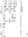
- FIG. 1 shows an exemplary workflow of the method of the invention.
- FIG. 2 shows an overview of a FFPE tissue section with three regions of interest marked.
- FIG. 3 shows, for each of the three regions of interest, the individual immunofluorescence signals for fluorescent antibodies towards the target proteins EGFR, HER2, HER3, c-MET, PI3K and phosphorylated AKT.
- FIG. 4 shows overlaid images with c-MYC and CEP8 FISH fluorescence signals from the three regions of interest ROI 1-3.
- FIG. 5 shows an overview of the FFPE tissue section after removal of subsamples from the three regions of interest.
- FIG. 6 shows the average depth of coverage and the uniformity of coverage for subsamples from four regions of interest, denoted ROI1, ROI2, ROI3 and ROI4.
- WS 1 and WS2 are whole slide extractions, used as controls.
- FIG. 7 shows the intersection and intrasection variation of the retrieval yield for subsamples retrieved with a laser capture microdissector.
- FIG. 8 shows sequencing data for DNA from FFPE samples subjected to up to 10 bleaching rounds.
- the present invention discloses a method for determining a nucleotide sequence of a nucleic acid segment present in a biological sample, such as a microscopy sample.
- the sample can e.g. be a tissue section, such as a formalin-fixed paraffin-embedded (FFRPE) section, but it can also be e.g. a smear or a preparation derived from a fluid sample.
- FFRPE formalin-fixed paraffin-embedded
- the sample may be affixed to or otherwise mounted on a solid support, such as a microscope slide, e.g. a glass or quartz slide.
- the solid support or microscope slide can suitably have a positively charged surface, e.g.
- an aminosilane or polylysine coating which improves the adherence of the sample to the support/slide during subsequent staining and bleaching steps.
- An example of a positively charged support/slide is the Superfrost Plus slides, available from Thermo Scientific.
- the solid support or microscope slide can also suitably have a low autofluorescence, e.g. with an autofluorescence intensity which is less than 1% or less than 0.1% of the average fluorescence intensity over a region of interest in the sample. Standard low-fluorescence glass or quartz qualities used for fluorescence microscopy slides are suitable from this point of view.
- the method comprises the steps of a)-d) as outlined below:
- the immunofluorescence detection can suitably involve a plurality of incubations of the sample with fluorophore-conjugated antibodies against the target proteins.
- This predetermined criterion can e.g. be a pattern of expression levels for the different proteins.
- the pattern may be qualitative (e.g. combinations of high/low expression of different proteins), semiquantitative (e.g. combinations of expressions above or below certain threshold levels) or quantitative (e.g. involving multivariate correlations).
- c) removing a subsample from the region of interest. This can suitably be performed while the sample is still affixed to the same solid support/microscope slide as in steps a) and b).
- the nucleic acid segment may be a nucleic acid or part of a nucleic acid.
- the nucleic acid may be DNA or it may be RNA.
- step a) comprises generating a plurality of fluorescent images of the sample, each image being generated by a protocol comprising immunofluorescence detection of at least one target protein in the sample and fluorescence detection of at least one morphological feature in said sample.
- the detection of one or more morphological features allows the localization of the same regions of interest throughout the different images of the sample, either by direct alignment of the different images or by the provision of navigational information such as a common coordinate system for the different images.
- the detection of morphological features can be accomplished by staining with a fluorescent marker, as described below, or alternatively from autofluorescence of the sample or by the introduction of e.g. fluorescent particles in the sample.
- the plurality of fluorescent images can e.g.
- the immunofluorescence staining can comprise contacting the sample with at least one antibody against at least one target protein, which antibody/antibodies is/are conjugated with a fluorophore.
- the staining can e.g. comprise contacting the sample with two antibodies against two different target proteins, where each antibody is conjugated with a different fluorophore, producing fluorescence detectable at two different wavelengths. It is also possible to use a non-conjugated antibody in combination with a fluorophore-labeled antibody against the non-conjugated antibody.
- Suitable fluorophores are cyanine dyes, such as Cy3 (I) and/or Cy5 (II), where the R1 and R2 groups can independently of each other be e.g. methyl, ethyl, propyl, butyl, carboxyl, acetylmethoxy, sulfo or N.hydroxysuccinimide groups.
- the Cy3 and Cy5 fluorophores can be conveniently bleached by the procedures discussed below.
- the bleaching of the sample allows the extinguishment of the fluorescence signals from the previous immunofluorescence staining, such that the sample can be stained again with another fluorophore-conjugated antibody without cross-talk from the previous staining.
- the bleaching can e.g. be a chemical inactivation procedure, involving incubation with one or more chemical agents, e.g. oxidants such as hydrogen peroxide as disclosed in U.S. Pat. No. 7,741,046, it can be a photobleaching procedure or it can be a photoassisted chemical inactivation, e.g. as disclosed in U.S. Pat. No.
- the bleaching may comprise contacting the sample with a peroxide, such as hydrogen peroxide, or with an organic borate, such as an aromatic borate like triphenyl monobenzyl borate.
- a peroxide such as hydrogen peroxide
- an organic borate such as an aromatic borate like triphenyl monobenzyl borate.
- the biological sample is subjected to an antigen retrieval step before step a).
- the purpose of the antigen retrieval step is to break crosslinks in the sample, particularly crosslinks introduced by previous formaldehyde fixation processes. This improves the binding of the antibodies introduced in step a) with the target proteins in the sample.
- the antigen retrieval step may comprise heating in a non-neutral buffer (i.e. a buffer with pH 6.5 or lower or pH 7.5 or higher), e.g. a buffer having a pH of 5-6.5 or 8-9.5. Suitable such buffers include Bond Epitope Retrieval Solution (Leica Biosystems).
- the fluorescent image generated in step a) is a composite image obtained by overlaying a plurality of immunofluorescent images of the sample obtained as described above.
- the overlaying can be accomplished by using fluorescent signals from morphological features in the sample as a navigational aid for the alignment of the individual images.
- the sample is further stained with a fluorescent marker that provides the morphological information as discussed above.
- This fluorescent marker can suitably be a nucleus stain, such as 4′,6-diamidino-2-phenylindole (DAPI) or other nucleic acid markers as listed under Definitions.
- DAPI 4′,6-diamidino-2-phenylindole
- the at least five different target proteins comprise at least one of EGFR (epidermal growth factor receptor), HER2 (human epidermal growth factor receptor 2), HER3 (human epidermal growth factor receptor 3), the receptor tyrosine kinase c-MET, phosphoinositide 3-kinase (PI3K), phosphorylated protein kinase B (AKT), ribosomal protein S6 (S6), phosphorylated extracellular signal regulating kinases 1 and 2 (phospho ERK1/2), cytokeratin PCK-26 (PCK26), anion transport protein AE1 (AE1) and sodium-potassium adenosinetriphosphatase (NaKATPase).
- the at least five different target proteins may also, or alternatively, comprise any one of the target proteins listed below under the separate heading Target proteins.
- step a) further comprises fluorescence in situ hybridization (FISH) staining of the sample followed by capture of a fluorescent hybridization image and overlaying of the fluorescent hybridization image with the immunofluorescent images.
- FISH staining may comprise contacting the sample with an oligonucleotide (probe) capable of hybridizing with a target nucleic acid region/sequence in the sample, conjugated with a fluorophore.
- target nucleic acid regions/sequences can be the c-MYC gene or the centromeric region of chromosome 8 (CEP8). Further examples are discussed below under Target nucleic acid sequences.
- step c) of the method involves removing the subsample by laser microdissection.
- Laser microdissection is a technique where a laser beam is used to either cut out a subsample (typically a UV laser) or to melt a polymer film that adheres to the subsample (typically an IR laser).
- Commercially available laser microdissection instruments are e.g. Life Technologies Arcturus and Leica LMD.
- the subsample may be removed using micromilling.
- the subsample is removed using a rotating milling tool controlled by a precision x-y table.
- Commercially available micromilling instruments include MilliSect and MillMan from AvanSci Bio/Roche.
- the circle-equivalent diameter of the subsample removed can suitably be less than 2 mm, such as about 5 to about 1500 micrometers.
- the sequence determination can be performed directly on the removed sample, using methods such as ion semiconductor sequencing (also called ion torrent sequencing) or e.g. Illumina dye sequencing. It is however also possible to perform an amplification step before sequencing. Such amplification may be performed by methods known in the art, e.g. PCR, multiple displacement amplification (MDA) or rolling circle amplification (RCA).
- Ion semiconductor sequencing involves detection of hydrogens released upon polymerase-induced incorporation of a nucleotide in a strand of DNA by a semiconductor chip. Examples of instrumentation for this technique includes Life Technologies Ion Torrent.
- Illumina dye sequencing involves fluorescence detection of the incorporation of labeled nucleotides in a strand of DNA and is exemplified by the Illumina instruments available from Illumina.
- a biological sample in accordance with one embodiment of the invention may be solid or fluid.
- Biological sample refers to a sample obtained from a biological subject, including sample of biological tissue or fluid origin obtained in vivo or in vitro. Suitable examples of biological samples may include, but are not limited to, blood, saliva, cerebral spinal fluid, pleural fluid, milk, lymph, sputum, semen, urine, stool, tears, needle aspirates, external sections of the skin, respiratory, intestinal, and genitourinary tracts, tumors, organs, cell cultures, or solid tissue sections.
- the biological sample may be analyzed as is, that is, without harvest and/or isolation of the target of interest. In an alternate embodiment, harvest of the sample may be performed prior to analysis.
- the methods disclosed herein may be particularly suitable for in-vitro analysis of biological samples.
- Biological samples may be immobilized on a solid support, such as in glass slides, microtiter, or ELISA plates.
- a biological sample may include any of the aforementioned samples regardless of their physical condition, such as, but not limited to, being frozen or stained or otherwise treated.
- a biological sample may include compounds which are not naturally intermixed with the sample in nature such as preservatives, anticoagulants, buffers, fixatives, nutrients, antibiotics, or the like.
- a biological sample may be of prokaryotic origin or eukaryotic origin (e.g., insects, protozoa, birds, fish, reptiles).
- the biological sample is mammalian (e.g., rat, mouse, cow, dog, donkey, guinea pig, or rabbit).
- the biological sample is of primate origin (e.g., example, chimpanzee or human).
- a biological sample may include a tissue sample, a whole cell, a cell constituent, a cytospin, or a cell smear.
- a biological sample essentially includes a tissue sample.
- a tissue sample may include a collection of similar cells obtained from a tissue of a biological subject that may have a similar function.
- a tissue sample may include a collection of similar cells obtained from a tissue of a human. Suitable examples of human tissues include, but are not limited to, (1) epithelium; (2) the connective tissues, including blood vessels, bone and cartilage; (3) muscle tissue; and (4) nerve tissue.
- the source of the tissue sample may be solid tissue obtained from a fresh, frozen and/or preserved organ or tissue sample or biopsy or aspirate; blood or any blood constituents; bodily fluids such as cerebral spinal fluid, amniotic fluid, peritoneal fluid, or interstitial fluid; or cells from any time in gestation or development of the subject.
- the tissue sample may include primary or cultured cells or cell lines.
- the tissue sample may be obtained by a variety of procedures including, but not limited to surgical excision, aspiration or biopsy.
- the tissue sample may be fixed and embedded in paraffin.
- the tissue sample may be fixed or otherwise preserved by conventional methodology; the choice of a fixative may be determined by the purpose for which the tissue is to be histologically stained or otherwise analyzed.
- the length of fixation may depend upon the size of the tissue sample and the fixative used. For example, neutral buffered formalin, Bouin's or paraformaldehyde, may be used to fix or preserve a tissue sample.
- a biological sample includes tissue sections of normal or cancerous origin, such as tissue sections form colon, breast, prostate, lung, liver, and stomach.
- a tissue section may include a single part or piece of a tissue sample, for example, a thin slice of tissue or cells cut from a tissue sample.
- multiple sections of tissue samples may be taken and subjected to analysis, provided the methods disclosed herein may be used for analysis of the same section of the tissue sample with respect to at least two different targets (i.e., one of these being of a protein origin and another one being a nucleic acid origin).
- a tissue section, if employed as a biological sample may have a thickness in a range that is less than about 100 micrometers, in a range that is less than about 50 micrometers, in a range that is less than about 25 micrometers, or in range that is less than about 10 micrometers.
- a target protein according to an embodiment of the invention may be present on the surface of a biological sample (for example, an antigen on a surface of a tissue section) or present in the bulk of the sample (for example, an antibody in a buffer solution).
- a target protein may not be inherently present on the surface of a biological sample and the biological sample may have to be processed to make the target protein available on the surface.
- the target protein may be in a tissue, either on a cell surface, or within a cell.
- a target may provide information about the presence or absence of an analyte in the biological sample.
- a target protein may provide information on a state of a biological sample. For example, if the biological sample includes a tissue sample, the methods disclosed herein may be used to detect target protein that may help in comparing different types of cells or tissues, comparing different developmental stages, detecting the presence of a disease or abnormality, or determining the type of disease or abnormality.
- Suitable target proteins may include one or more of peptides, proteins (e.g., antibodies, affibodies, or aptamers), enzymes, ligands, receptors, antigens, or haptens.
- proteins e.g., antibodies, affibodies, or aptamers
- enzymes ligands, receptors, antigens, or haptens.
- One or more of the aforementioned target proteins may be characteristic of particular cells, while other target proteins may be associated with a particular disease or condition.
- target proteins in a tissue sample that may be detected and analyzed using the methods disclosed herein may include, but are not limited to, prognostic markers, predictive markers, hormone or hormone receptors, lymphoids, tumor markers, cell cycle associated markers, neural tissue and tumor markers, or cluster differentiation markers.
- prognostic markers may include enzymatic targets such as galactosyl transferase II, neuron specific enolase, proton ATPase-2, or acid phosphatase.
- Other examples of prognostic protein or gene markers include Ki67, cyclin E, p53, cMet.
- Suitable examples of predictive markers may include protein or gene targets such as EGFR, Her2, ALK.
- hormone or hormone receptors may include human chorionic gonadotropin (HCG), adrenocorticotropic hormone, carcinoembryonic antigen (CEA), prostate-specific antigen (PSA), estrogen receptor, progesterone receptor, androgen receptor, gClq-R/p33 complement receptor, IL-2 receptor, p75 neurotrophin receptor, PTH receptor, thyroid hormone receptor, or insulin receptor.
- HCG human chorionic gonadotropin
- CEA carcinoembryonic antigen
- PSA prostate-specific antigen
- estrogen receptor progesterone receptor
- androgen receptor gClq-R/p33 complement receptor
- IL-2 receptor p75 neurotrophin receptor
- PTH receptor thyroid hormone receptor
- thyroid hormone receptor or insulin receptor.
- lymphoids may include alpha-1-antichymotrypsin, alpha-1-antitrypsin, B cell target, bcl-2, bcl-6, B lymphocyte antigen 36 kD, BM1 (myeloid target), BM2 (myeloid target), galectin-3, granzyme B, HLA class I Antigen, HLA class II (DP) antigen, HLA class II (DQ) antigen, HLA class II (DR) antigen, human neutrophil defensins, immunoglobulin A, immunoglobulin D, immunoglobulin G, immunoglobulin M, kappa light chain, kappa light chain, lambda light chain, lymphocyte/histocyte antigen, macrophage target, muramidase (lysozyme), p80 anaplastic lymphoma kinase, plasma cell target, secretory leukocyte protease inhibitor, T cell antigen receptor (JOVI 1), T cell antigen receptor (JOVI 3), terminal
- tumor markers may include alpha fetoprotein, apolipoprotein D, BAG-1 (RAP46 protein), CA19-9 (sialyl lewisa), CA50 (carcinoma associated mucin antigen), CAl25 (ovarian cancer antigen), CA242 (tumour associated mucin antigen), chromogranin A, clusterin (apolipoprotein J), epithelial membrane antigen, epithelial-related antigen, epithelial specific antigen, gross cystic disease fluid protein-15, hepatocyte specific antigen, heregulin, human gastric mucin, human milk fat globule, MAGE-1, matrix metalloproteinases, melan A, melanoma target (HMB45), mesothelin, metallothionein, microphthalmia transcription factor (MITF), Muc-1 core glycoprotein.
- Suitable examples of cell cycle associated markers may include apoptosis protease activating factor-1, bcl-w, bcl-x, bromodeoxyuridine, CAK (cdk-activating kinase), cellular apoptosis susceptibility protein (CAS), caspase 2, caspase 8, CPP32 (caspase-3), CPP32 (caspase-3), cyclin dependent kinases, cyclin A, cyclin B1, cyclin D1, cyclin D2, cyclin D3, cyclin E, cyclin G, DNA fragmentation factor (N-terminus), Fas (CD95), Fas-associated death domain protein, Fas ligand, Fen-1, IPO-38, Mc1-1, minichromosome maintenance proteins, mismatch repair protein (MSH2), poly (ADP-Ribose) polymerase, proliferating cell nuclear antigen, p16 protein, p27 protein, p34cdc2, p57 protein (Kip
- Suitable examples of neural tissue and tumor markers may include alpha B crystallin, alpha-internexin, alpha synuclein, amyloid precursor protein, beta amyloid, calbindin, choline acetyltransferase, excitatory amino acid transporter 1, GAP43, glial fibrillary acidic protein, glutamate receptor 2, myelin basic protein, nerve growth factor receptor (gp75), neuroblastoma target, neurofilament 68 kD, neurofilament 160 kD, neurofilament 200 kD, neuron specific enolase, nicotinic acetylcholine receptor alpha4, nicotinic acetylcholine receptor beta2, peripherin, protein gene product 9, S-100 protein, serotonin, SNAP-25, synapsin I, synaptophysin, tau, tryptophan hydroxylase, tyrosine hydroxylase, or ubiquitin.
- cluster differentiation markers may include CD1a, CD1b, CD1c, CD1d, CD1e, CD2, CD3delta, CD3epsilon, CD3gamma, CD4, CD5, CD6, CD7, CD8alpha, CD8beta, CD9, CD10, CD11a, CD11b, CD11c, CDw12, CD13, CD14, CD15, CD15s, CD16a, CD16b, CDw17, CD18, CD19, CD20, CD21, CD22, CD23, CD24, CD25, CD26, CD27, CD28, CD29, CD30, CD31, CD32, CD33, CD34, CD35, CD36, CD37, CD38, CD39, CD40, CD41, CD42a, CD42b, CD42c, CD42d, CD43, CD44, CD44R, CD45, CD46, CD47, CD48, CD49a, CD49b, CD49c, CD49d, CD49e, CD49f, LD50,
- target proteins include centromere protein-F (CENP-F), giantin, involucrin, lamin A&C (XB 10), LAP-70, mucin, nuclear pore complex proteins, p180 lamellar body protein, ran, cathepsin D, Ps2 protein, Her2-neu, P53, S100, epithelial target antigen (EMA), TdT, MB2, MB3, PCNA, Ki67, cytokeratin, PI3K, cMyc or MAPK.
- CENP-F centromere protein-F
- CENP-F giantin
- involucrin lamin A&C
- LAP-70 LAP-70
- mucin mucin
- nuclear pore complex proteins p180 lamellar body protein, ran, cathepsin D, Ps2 protein, Her2-neu, P53, S100, epithelial target antigen (EMA), TdT, MB2, MB3, PCNA, Ki67, cytokeratin, PI3K,
- Still other suitable target proteins include Her2/neu (epidermal growth factor over expressed in breast and stomach cancer, therapy by a monoclonal antibody slows tumor growth); EGF-R/erbB (epidermal growth factor receptor); ER (estrogen receptor required for growth of some breast cancer tumors, located in the nucleus and detected with ISH for deciding on therapy limiting estrogen in positive patients); PR (progesterone receptor is a hormone that binds to DNA); AR (androgen receptor is involved in androgen dependent tumor growth); ⁇ -catenin (oncogene in cancer translocates from the cell membrane to the nucleus, which functions in both cell adhesion and as a latent gene regulatory protein); Phospho- ⁇ -Catenin: phosphorylated (form of ⁇ -catenin degrades in the cytosol and does not translocate to the nucleus); GSK3 ⁇ (glycogen synthase kinase-3 ⁇ protein in the Wnt pathway phosphorylates ⁇ -catenin marking
- a target nucleic acid sequence refers to a sequence of interest which is contained in a nucleic acid molecule in the biological sample.
- the nucleic acid molecule may be present in the nuclei of the cells of the biological sample (for example, chromosomal DNA) or present in the cytoplasm (for example, mRNA).
- a nucleic acid molecule may not be inherently present on the surface of a biological sample and the biological sample may have to be processed to make the nucleic acid molecule accessible by a probe. For example, protease treatment of the sample could readily bring the target nucleic acid sequences.
- Suitability of a nucleic acid molecule to be analyzed may be determined by the type and nature of analysis required for the biological sample.
- the analysis may provide information about the gene expression level of the target nucleic acid sequence in the biological sample.
- the analysis may provide information on the presence or absence or amplification level of a chromosomal DNA. For example, if the biological sample includes a tissue sample, the methods disclosed herein may be used to detect a target nucleic acid sequence that may identify cells which has an increased copy number of a particular chromosomal segment harboring the target nucleic acid sequence.
- the target nucleic acid sequence in a tissue sample that may be detected and analyzed using the methods disclosed herein may include, but are not limited to, nucleic acid sequences for prognostic markers, hormone or hormone receptors, lymphoids, tumor markers, cell cycle associated markers, neural tissue and tumor markers, or cluster differentiation markers. Examples of these markers are described in the section entitled “Target proteins”.
- the target nucleic acid sequence target is a sequence for the EGFR, TOP2A, cMyc, ALK, FGFR1 or HER2 gene.
- the target nucleic acid sequence includes a sequence that is part of the gene sequence which encodes the target protein. In other embodiments, the target nucleic acid sequence does not include a sequence that is part of the gene sequence which encodes the target protein. Thus, the target nucleic acid sequence may include a sequence that is part of the gene sequence which encodes a different protein than the target protein.
- a probe is used to detect the target nucleic acid sequences. It is desirable that the probe binds specifically to the region of nucleic acid molecule that contains the sequence of interest.
- the probe is sequence-specific.
- a sequence-specific probe may include a nucleic acid and the probe may be capable of recognizing a particular linear arrangement of nucleotides or derivatives thereof.
- the linear arrangement may include contiguous nucleotides or derivatives thereof that may each bind to a corresponding complementary nucleotide in the probe.
- the sequence may not be contiguous as there may be one, two, or more nucleotides that may not have corresponding complementary residues on the probe.
- Suitable examples of probes may include, but are not limited to DNA or RNA oligonucleotides or polynucleotides, peptide nucleic acid (PNA) sequences, locked nucleic acid (LNA) sequences, or aptamers.
- PNA peptide nucleic acid
- LNA locked nucleic acid
- suitable probes may include nucleic acid analogs, such as dioxygenin dCTP, biotin dcTP 7-azaguanosine, azidothymidine, inosine, or uridine.
- a probe may form a Watson-Crick bond with the target nucleic acid sequence.
- the probe may form a Hoogsteen bond with the target nucleic acid sequence, thereby forming a triplex.
- a probe that binds by Hoogsteen binding may enter the major groove of a nucleic acid sequence and hybridizes with the bases located there.
- the probes may form both Watson-Crick and Hoogsteen bonds with the target nucleic acid sequence (for example, bis PNA probes are capable of both Watson-Crick and Hoogsteen binding to a nucleic acid molecule).
- the probe may comprise a nucleic acid probe, a peptide nucleic acid probe, a locked nucleic acid probe, mRNA probe, miRNA probe or siRNA probe.
- the length of the probe may also determine the specificity of binding.
- the energetic cost of a single mismatch between the probe and the target nucleic acid sequence may be relatively high for shorter sequences than for longer ones.
- hybridization of smaller probes may be more specific than the hybridization of longer probes, as the longer probes may be more amenable to mismatches and may continue to bind to the nucleic acid depending on the conditions.
- shorter probes may exhibit lower binding stability at a given temperature and salt concentration. Probes that may exhibit greater stability to bind short sequences may be employed in this case (for examples, bis PNA).
- the probe may have a length in range of from about 4 nucleotides to about 12 nucleotides, from about 12 nucleotides to about 25 nucleotides, from about 25 nucleotides to about 50 nucleotides, from about 50 nucleotides to about 100 nucleotides, from about 100 nucleotides to about 250 nucleotides, from about 250 nucleotides to about 500 nucleotides, or from about 500 nucleotides to about 1000 nucleotides. In some embodiments, the probe may have a length in a range that is greater than about 1000 nucleotides.
- the probe may not hybridize to complementary nucleotides in the target nucleic acid sequence.
- the probe may include 50 nucleotide residues in length, and only 25 of those residues may hybridize to the target nucleic acid sequence.
- the nucleotide residues that may hybridize may be contiguous with each other.
- the probe may be single stranded or may include a secondary structure.
- a biological sample may include a cell or a tissue sample and the biological sample may be subjected to in situ hybridization (ISH) using a probe.
- a tissue sample may be subjected to in situ hybridization in addition to immunofluorescence (IF) to obtain desired information regarding the tissue sample.
- the specificity of binding between the probe and the nucleic acid sequence may also be affected depending on the binding conditions (for example, hybridization conditions in case of complementary nucleic acids. Suitable binding conditions may be realized by modulating one or more of pH, temperature, or salt concentration.
- a probe may be intrinsically labeled (fluorophore attached during synthesis of probe) with a fluorophore or extrinsically labeled (fluorophore attached during a later step).
- an intrinsically labeled nucleic acid may be synthesized using methods that incorporate fluorophore-labeled nucleotides directly into the growing nucleic acid chain.
- a probe may be synthesized in a manner such that fluorophores may be incorporated at a later stage. For example, this latter labeling may be accomplished by chemical means by the introduction of active amino or thiol groups into nucleic acids chains.
- a probe such a nucleic acid for example, a DNA
- appropriate chemistries for the same for the same.
- FFPE formalin-fixed paraffin-embedded
- the slide was then stained with S6 antibody conjugated with Cy5 combined with phospho ERK1/2 rabbit antibody and anti-rabbit antibody conjugated with Cy3, followed by counterstaining with DAPI.
- the slide was coverslipped and the entire slide was imaged using a fluorescence microscope equipped with a 1.25 ⁇ magnification objective and a DAPI filterset. The images were captured using a digital monochrome camera, and then combined to form one stitched image of the entire slide. From this stitched full slide image the location of the tissue section was determined and coordinates for imaging the tissue section were recorded.
- the slide was then subjected to a 10 min treatment with 0.05% pepsin that partially removed protein structures to allow access to nuclear DNA.
- the slide was then fixed using aqueous 4% formaldehyde solution for 10 min, washed and subjected to hybridization using FISH probes for the c-MYC gene labeled with Orange (captured on Cy3) and for the CEP8 centromer labeled with Green (captured on FITC).
- Sections of all image rounds were then registered using DAPI images of each of the imaging round sets to align the images and they were combined in single filed images to allow simultaneous visualization of the expression of the target proteins EGFR, HER2, HER3, c-MET, PI3K and phosphor-AKT, as well as the presence of c-MYC and CRP8 in the cells.
- ROI 1-4 (only ROI 1-3 shown in the Figs) were identified.
- ROI1 had a high expression of EGFR and a low expression of phospho AKT
- ROI2 had high expression of both EGFR and phospho AKT
- ROI3 had low expression of EGFR and high expression of phospho AKT.
- the slide was then moved to a Life Technologies Arcturus XT laser capture microdissector (LCM) and a 1.5 mm diameter subsample was retrieved for each region of interest. The subsamples were efficiently retrieved despite the tissue-retaining surface on the slide.
- Each subsample, containing approx. 10 ng genomic DNA was then subjected to DNA sequencing in a Life Technologies Ion Torrent ion semiconductor sequencer using an AmpliSeq cancer panel consisting of 740 mutational hotspots in 46 cancer-related genes. As shown by FIG. 6 , the depth of coverage was well above 2000 ⁇ for every ROI and the uniformity of coverage was close to 100%. Table 1 shows more detailed information about the sequencing results for ROI 1-3. Surprisingly, the DNA was found not to be degraded by the dye inactivation procedure (bleaching).
- Example 1 In order to check the reproducibility of the subsample retrieval step, five serial sections (S1-S5) of the same FFPE tissue block as in Example 1 were stained as in Example 1. From each section two or four 1 mm circular subsamples were retrieved with the LCM and the fluorescence intensity was measured on the slide both in the location from which the subsample had been removed and in the surrounding area. The ratio between the intensities was calculated and used as a measure of the retrieval yield. As shown in FIG. 7 , the yield was consistently within the 50-75% interval both for the intersection and intrasection variation.
- This example was carried out in order to check the effect of repeated bleaching cycles on the quality of DNA in subsamples retrieved.
- Five sections of a SKVO3 ovarian carcinoma cell xenograft were mounted on microscope slides and treated as in Example 1. The immunofluorescence staining was performed with Cy3-labeled anti-PCK26 and Cy5-labeled anti-NaKATPase antibodies. Slide 1 was not stained at all, slide 2 stained one round without bleaching, slide 3 stained and bleached one round, slide 4 stained and bleached 5 rounds and slide 5 stained and bleached 10 rounds. 1.5 mm subsamples were retrieved from each slide and the DNA in the subsamples was amplified by rolling circle amplification and sequenced with ion semiconductor sequencing. As shown in FIG. 8 , no major effect of the number of bleaching rounds was observed.
- the sample was incubated in an aqueous solution of 1 mM triphenyl monobenzyl borate and 0.1 mM 1,4-diazabicyclo(2,2,2)octane (DABCO) under irradiation with >500 nm light for 7 min.
- DABCO 1,4-diazabicyclo(2,2,2)octane
- the sample was incubated in a freshly made aqueous solution of 3% H2O2 and 0.1 M NaHCO3.
- the DNA was precipitated with SureClean.
Landscapes
- Chemical & Material Sciences (AREA)
- Life Sciences & Earth Sciences (AREA)
- Health & Medical Sciences (AREA)
- Engineering & Computer Science (AREA)
- Organic Chemistry (AREA)
- Molecular Biology (AREA)
- Analytical Chemistry (AREA)
- Immunology (AREA)
- Zoology (AREA)
- Wood Science & Technology (AREA)
- Proteomics, Peptides & Aminoacids (AREA)
- Biochemistry (AREA)
- Biotechnology (AREA)
- Physics & Mathematics (AREA)
- General Health & Medical Sciences (AREA)
- Microbiology (AREA)
- Hematology (AREA)
- Urology & Nephrology (AREA)
- Biomedical Technology (AREA)
- Biophysics (AREA)
- Bioinformatics & Cheminformatics (AREA)
- General Engineering & Computer Science (AREA)
- Genetics & Genomics (AREA)
- Cell Biology (AREA)
- Chemical Kinetics & Catalysis (AREA)
- Food Science & Technology (AREA)
- Medicinal Chemistry (AREA)
- General Physics & Mathematics (AREA)
- Pathology (AREA)
- Measuring Or Testing Involving Enzymes Or Micro-Organisms (AREA)
- Investigating Or Analysing Biological Materials (AREA)
- Investigating, Analyzing Materials By Fluorescence Or Luminescence (AREA)
Abstract
Description

| TABLE 1 | ||||||
| ROI | Mapped | AQ20 | Mean | On | ||
| # | reads | reads | | target | Uniformity | |
| 1 | 616 339 | 464 865 | 2 755 | 98.83% | 98.53% | |
| 2 | 721 657 | 549 639 | 3 203 | 98.60% | 99.96% | |
| 3 | 589 877 | 435 620 | 2 612 | 98.57% | 98.54% | |
| Sample | CT | ||
| gDNA control | 25 | ||
| gDNA triphenyl monobenzyl borate | 25 | ||
| gDNA hydrogen peroxide | 25 | ||
| FFPE control | 28 | ||
| FFPE triphenyl monobenzyl borate | 28 | ||
| FFPE hydrogen peroxide | 28 | ||
Claims (21)
Priority Applications (1)
| Application Number | Priority Date | Filing Date | Title |
|---|---|---|---|
| US15/003,450 US10597697B2 (en) | 2015-02-05 | 2016-01-21 | Method of determining a nucleotide sequence of a nucleic acid segment from a region of interest in a biological sample |
Applications Claiming Priority (2)
| Application Number | Priority Date | Filing Date | Title |
|---|---|---|---|
| US201562112199P | 2015-02-05 | 2015-02-05 | |
| US15/003,450 US10597697B2 (en) | 2015-02-05 | 2016-01-21 | Method of determining a nucleotide sequence of a nucleic acid segment from a region of interest in a biological sample |
Publications (2)
| Publication Number | Publication Date |
|---|---|
| US20160230219A1 US20160230219A1 (en) | 2016-08-11 |
| US10597697B2 true US10597697B2 (en) | 2020-03-24 |
Family
ID=56566594
Family Applications (1)
| Application Number | Title | Priority Date | Filing Date |
|---|---|---|---|
| US15/003,450 Active 2038-01-08 US10597697B2 (en) | 2015-02-05 | 2016-01-21 | Method of determining a nucleotide sequence of a nucleic acid segment from a region of interest in a biological sample |
Country Status (1)
| Country | Link |
|---|---|
| US (1) | US10597697B2 (en) |
Cited By (2)
| Publication number | Priority date | Publication date | Assignee | Title |
|---|---|---|---|---|
| US10932710B2 (en) | 2017-01-10 | 2021-03-02 | Drawbridge Health, Inc. | Carriers for storage and transport of biological samples |
| US11266337B2 (en) | 2015-09-09 | 2022-03-08 | Drawbridge Health, Inc. | Systems, methods, and devices for sample collection, stabilization and preservation |
Families Citing this family (1)
| Publication number | Priority date | Publication date | Assignee | Title |
|---|---|---|---|---|
| CN110498858B (en) * | 2019-07-26 | 2024-01-23 | 深圳市达科为生物工程有限公司 | Method for dynamically detecting secretion condition of single-cell exoprotein |
Citations (8)
| Publication number | Priority date | Publication date | Assignee | Title |
|---|---|---|---|---|
| US20050176068A1 (en) * | 2002-04-26 | 2005-08-11 | Emmert-Buck Michael R. | Direct cell target analysis |
| US20060046311A1 (en) * | 2004-08-26 | 2006-03-02 | Intel Corporation | Biomolecule analysis using Raman surface scanning |
| US20060134692A1 (en) * | 2003-01-24 | 2006-06-22 | Emmert-Buck Michael R | Target activated microtransfer |
| US20080315119A1 (en) * | 2004-09-01 | 2008-12-25 | Perkinelmer Singapore Pte Ltd | Method of Analysing a Sample and Apparatus Therefor |
| US20140323349A1 (en) * | 2011-12-05 | 2014-10-30 | Phigenix, Inc. | Compositions and methods for monitoring and detecting cancerous conditions |
| US20150316482A1 (en) * | 2012-12-11 | 2015-11-05 | Clarient Diagnostic Services, Inc. | Photoactivated chemical bleaching of dyes |
| US20160146823A1 (en) * | 2013-07-05 | 2016-05-26 | University Of Washington Through Its Center For Commercialization | Methods, compositions and systems for microfluidic assays |
| US20160314583A1 (en) * | 2013-12-19 | 2016-10-27 | Axon Dx, Llc | Cell detection, capture and isolation methods and apparatus |
-
2016
- 2016-01-21 US US15/003,450 patent/US10597697B2/en active Active
Patent Citations (8)
| Publication number | Priority date | Publication date | Assignee | Title |
|---|---|---|---|---|
| US20050176068A1 (en) * | 2002-04-26 | 2005-08-11 | Emmert-Buck Michael R. | Direct cell target analysis |
| US20060134692A1 (en) * | 2003-01-24 | 2006-06-22 | Emmert-Buck Michael R | Target activated microtransfer |
| US20060046311A1 (en) * | 2004-08-26 | 2006-03-02 | Intel Corporation | Biomolecule analysis using Raman surface scanning |
| US20080315119A1 (en) * | 2004-09-01 | 2008-12-25 | Perkinelmer Singapore Pte Ltd | Method of Analysing a Sample and Apparatus Therefor |
| US20140323349A1 (en) * | 2011-12-05 | 2014-10-30 | Phigenix, Inc. | Compositions and methods for monitoring and detecting cancerous conditions |
| US20150316482A1 (en) * | 2012-12-11 | 2015-11-05 | Clarient Diagnostic Services, Inc. | Photoactivated chemical bleaching of dyes |
| US20160146823A1 (en) * | 2013-07-05 | 2016-05-26 | University Of Washington Through Its Center For Commercialization | Methods, compositions and systems for microfluidic assays |
| US20160314583A1 (en) * | 2013-12-19 | 2016-10-27 | Axon Dx, Llc | Cell detection, capture and isolation methods and apparatus |
Non-Patent Citations (2)
| Title |
|---|
| Gibbs, "DNA Annplificatioin by the Polymerase Chain Reaction", Anal. Chem. 1990, 62, 1202-1214. * |
| Murphy et al., "Mate Pair Sequencing of Whole-Genome-Amplified DNA Following Laser Capture Microdissection of Prostate Cancer", DNA Research (Oct. 2012) 19(5):395-406 (Year: 2012). * |
Cited By (3)
| Publication number | Priority date | Publication date | Assignee | Title |
|---|---|---|---|---|
| US11266337B2 (en) | 2015-09-09 | 2022-03-08 | Drawbridge Health, Inc. | Systems, methods, and devices for sample collection, stabilization and preservation |
| US10932710B2 (en) | 2017-01-10 | 2021-03-02 | Drawbridge Health, Inc. | Carriers for storage and transport of biological samples |
| US11298060B2 (en) | 2017-01-10 | 2022-04-12 | Drawbridge Health, Inc. | Devices for collecting biological samples |
Also Published As
| Publication number | Publication date |
|---|---|
| US20160230219A1 (en) | 2016-08-11 |
Similar Documents
| Publication | Publication Date | Title |
|---|---|---|
| EP2831591B1 (en) | Immunoflu0rescence and fluorescent-based nucleic acid analysis on a single sample | |
| US11573235B2 (en) | Methods for generating an image of a biological sample | |
| EP2875353B1 (en) | Methods of detecting dna, rna and protein in biological samples | |
| US9518982B2 (en) | Sequential analysis of biological samples | |
| US20150141278A1 (en) | Multiplexed assay method for lung cancer classification | |
| US20150226743A1 (en) | Multiplexed method for diagnosing classical hodgkin lymphoma | |
| WO2014088744A1 (en) | Systems and methods for using an immunostaining mask to selectively refine ish analysis results | |
| US10597697B2 (en) | Method of determining a nucleotide sequence of a nucleic acid segment from a region of interest in a biological sample |
Legal Events
| Date | Code | Title | Description |
|---|---|---|---|
| AS | Assignment |
Owner name: GE HEALTHCARE BIO-SCIENCES CORP., MASSACHUSETTS Free format text: ASSIGNMENT OF ASSIGNORS INTEREST;ASSIGNORS:NELSON, JOHN RICHARD;AU, QINGYAN;GAO, WEI;AND OTHERS;REEL/FRAME:045908/0768 Effective date: 20150205 |
|
| STPP | Information on status: patent application and granting procedure in general |
Free format text: RESPONSE TO NON-FINAL OFFICE ACTION ENTERED AND FORWARDED TO EXAMINER |
|
| STPP | Information on status: patent application and granting procedure in general |
Free format text: NON FINAL ACTION MAILED |
|
| STPP | Information on status: patent application and granting procedure in general |
Free format text: RESPONSE TO NON-FINAL OFFICE ACTION ENTERED AND FORWARDED TO EXAMINER |
|
| STPP | Information on status: patent application and granting procedure in general |
Free format text: FINAL REJECTION MAILED |
|
| STPP | Information on status: patent application and granting procedure in general |
Free format text: RESPONSE AFTER FINAL ACTION FORWARDED TO EXAMINER |
|
| STPP | Information on status: patent application and granting procedure in general |
Free format text: NOTICE OF ALLOWANCE MAILED -- APPLICATION RECEIVED IN OFFICE OF PUBLICATIONS |
|
| STPP | Information on status: patent application and granting procedure in general |
Free format text: PUBLICATIONS -- ISSUE FEE PAYMENT VERIFIED |
|
| STCF | Information on status: patent grant |
Free format text: PATENTED CASE |
|
| AS | Assignment |
Owner name: GLOBAL LIFE SCIENCES SOLUTIONS USA LLC, MASSACHUSETTS Free format text: CHANGE OF NAME;ASSIGNOR:GE HEALTHCARE BIO-SCIENCES CORP.;REEL/FRAME:053648/0854 Effective date: 20190930 |
|
| AS | Assignment |
Owner name: LEICA MICROSYSTEMS CMS GMBH, GERMANY Free format text: ASSIGNMENT OF ASSIGNORS INTEREST;ASSIGNOR:GLOBAL LIFE SCIENCES SOLUTIONS USA LLC;REEL/FRAME:057261/0128 Effective date: 20210430 |
|
| MAFP | Maintenance fee payment |
Free format text: PAYMENT OF MAINTENANCE FEE, 4TH YEAR, LARGE ENTITY (ORIGINAL EVENT CODE: M1551); ENTITY STATUS OF PATENT OWNER: LARGE ENTITY Year of fee payment: 4 |