US10524775B2 - Methods of repairing cartilage defects - Google Patents
Methods of repairing cartilage defects Download PDFInfo
- Publication number
- US10524775B2 US10524775B2 US14/790,570 US201514790570A US10524775B2 US 10524775 B2 US10524775 B2 US 10524775B2 US 201514790570 A US201514790570 A US 201514790570A US 10524775 B2 US10524775 B2 US 10524775B2
- Authority
- US
- United States
- Prior art keywords
- cartilage
- bone
- flexible strand
- transosseus
- tunnel
- Prior art date
- Legal status (The legal status is an assumption and is not a legal conclusion. Google has not performed a legal analysis and makes no representation as to the accuracy of the status listed.)
- Active, expires
Links
Images


Classifications
-
- A—HUMAN NECESSITIES
- A61—MEDICAL OR VETERINARY SCIENCE; HYGIENE
- A61B—DIAGNOSIS; SURGERY; IDENTIFICATION
- A61B17/00—Surgical instruments, devices or methods, e.g. tourniquets
- A61B17/04—Surgical instruments, devices or methods, e.g. tourniquets for suturing wounds; Holders or packages for needles or suture materials
- A61B17/0401—Suture anchors, buttons or pledgets, i.e. means for attaching sutures to bone, cartilage or soft tissue; Instruments for applying or removing suture anchors
-
- A—HUMAN NECESSITIES
- A61—MEDICAL OR VETERINARY SCIENCE; HYGIENE
- A61F—FILTERS IMPLANTABLE INTO BLOOD VESSELS; PROSTHESES; DEVICES PROVIDING PATENCY TO, OR PREVENTING COLLAPSING OF, TUBULAR STRUCTURES OF THE BODY, e.g. STENTS; ORTHOPAEDIC, NURSING OR CONTRACEPTIVE DEVICES; FOMENTATION; TREATMENT OR PROTECTION OF EYES OR EARS; BANDAGES, DRESSINGS OR ABSORBENT PADS; FIRST-AID KITS
- A61F2/00—Filters implantable into blood vessels; Prostheses, i.e. artificial substitutes or replacements for parts of the body; Appliances for connecting them with the body; Devices providing patency to, or preventing collapsing of, tubular structures of the body, e.g. stents
- A61F2/02—Prostheses implantable into the body
- A61F2/30—Joints
- A61F2/30756—Cartilage endoprostheses
-
- A—HUMAN NECESSITIES
- A61—MEDICAL OR VETERINARY SCIENCE; HYGIENE
- A61B—DIAGNOSIS; SURGERY; IDENTIFICATION
- A61B17/00—Surgical instruments, devices or methods, e.g. tourniquets
- A61B17/04—Surgical instruments, devices or methods, e.g. tourniquets for suturing wounds; Holders or packages for needles or suture materials
- A61B17/0401—Suture anchors, buttons or pledgets, i.e. means for attaching sutures to bone, cartilage or soft tissue; Instruments for applying or removing suture anchors
- A61B2017/0403—Dowels
-
- A—HUMAN NECESSITIES
- A61—MEDICAL OR VETERINARY SCIENCE; HYGIENE
- A61B—DIAGNOSIS; SURGERY; IDENTIFICATION
- A61B17/00—Surgical instruments, devices or methods, e.g. tourniquets
- A61B17/04—Surgical instruments, devices or methods, e.g. tourniquets for suturing wounds; Holders or packages for needles or suture materials
- A61B17/0401—Suture anchors, buttons or pledgets, i.e. means for attaching sutures to bone, cartilage or soft tissue; Instruments for applying or removing suture anchors
- A61B2017/0404—Buttons
-
- A—HUMAN NECESSITIES
- A61—MEDICAL OR VETERINARY SCIENCE; HYGIENE
- A61B—DIAGNOSIS; SURGERY; IDENTIFICATION
- A61B17/00—Surgical instruments, devices or methods, e.g. tourniquets
- A61B17/04—Surgical instruments, devices or methods, e.g. tourniquets for suturing wounds; Holders or packages for needles or suture materials
- A61B17/0401—Suture anchors, buttons or pledgets, i.e. means for attaching sutures to bone, cartilage or soft tissue; Instruments for applying or removing suture anchors
- A61B2017/0409—Instruments for applying suture anchors
-
- A—HUMAN NECESSITIES
- A61—MEDICAL OR VETERINARY SCIENCE; HYGIENE
- A61B—DIAGNOSIS; SURGERY; IDENTIFICATION
- A61B17/00—Surgical instruments, devices or methods, e.g. tourniquets
- A61B17/04—Surgical instruments, devices or methods, e.g. tourniquets for suturing wounds; Holders or packages for needles or suture materials
- A61B17/0401—Suture anchors, buttons or pledgets, i.e. means for attaching sutures to bone, cartilage or soft tissue; Instruments for applying or removing suture anchors
- A61B2017/0412—Suture anchors, buttons or pledgets, i.e. means for attaching sutures to bone, cartilage or soft tissue; Instruments for applying or removing suture anchors having anchoring barbs or pins extending outwardly from suture anchor body
-
- A—HUMAN NECESSITIES
- A61—MEDICAL OR VETERINARY SCIENCE; HYGIENE
- A61B—DIAGNOSIS; SURGERY; IDENTIFICATION
- A61B17/00—Surgical instruments, devices or methods, e.g. tourniquets
- A61B17/04—Surgical instruments, devices or methods, e.g. tourniquets for suturing wounds; Holders or packages for needles or suture materials
- A61B17/0401—Suture anchors, buttons or pledgets, i.e. means for attaching sutures to bone, cartilage or soft tissue; Instruments for applying or removing suture anchors
- A61B2017/0414—Suture anchors, buttons or pledgets, i.e. means for attaching sutures to bone, cartilage or soft tissue; Instruments for applying or removing suture anchors having a suture-receiving opening, e.g. lateral opening
-
- A—HUMAN NECESSITIES
- A61—MEDICAL OR VETERINARY SCIENCE; HYGIENE
- A61B—DIAGNOSIS; SURGERY; IDENTIFICATION
- A61B17/00—Surgical instruments, devices or methods, e.g. tourniquets
- A61B17/04—Surgical instruments, devices or methods, e.g. tourniquets for suturing wounds; Holders or packages for needles or suture materials
- A61B17/0401—Suture anchors, buttons or pledgets, i.e. means for attaching sutures to bone, cartilage or soft tissue; Instruments for applying or removing suture anchors
- A61B2017/0427—Suture anchors, buttons or pledgets, i.e. means for attaching sutures to bone, cartilage or soft tissue; Instruments for applying or removing suture anchors having anchoring barbs or pins extending outwardly from the anchor body
-
- A—HUMAN NECESSITIES
- A61—MEDICAL OR VETERINARY SCIENCE; HYGIENE
- A61B—DIAGNOSIS; SURGERY; IDENTIFICATION
- A61B17/00—Surgical instruments, devices or methods, e.g. tourniquets
- A61B17/04—Surgical instruments, devices or methods, e.g. tourniquets for suturing wounds; Holders or packages for needles or suture materials
- A61B17/0401—Suture anchors, buttons or pledgets, i.e. means for attaching sutures to bone, cartilage or soft tissue; Instruments for applying or removing suture anchors
- A61B2017/044—Suture anchors, buttons or pledgets, i.e. means for attaching sutures to bone, cartilage or soft tissue; Instruments for applying or removing suture anchors with a threaded shaft, e.g. screws
-
- A—HUMAN NECESSITIES
- A61—MEDICAL OR VETERINARY SCIENCE; HYGIENE
- A61B—DIAGNOSIS; SURGERY; IDENTIFICATION
- A61B17/00—Surgical instruments, devices or methods, e.g. tourniquets
- A61B17/04—Surgical instruments, devices or methods, e.g. tourniquets for suturing wounds; Holders or packages for needles or suture materials
- A61B17/0401—Suture anchors, buttons or pledgets, i.e. means for attaching sutures to bone, cartilage or soft tissue; Instruments for applying or removing suture anchors
- A61B2017/0445—Suture anchors, buttons or pledgets, i.e. means for attaching sutures to bone, cartilage or soft tissue; Instruments for applying or removing suture anchors cannulated, e.g. with a longitudinal through-hole for passage of an instrument
-
- A—HUMAN NECESSITIES
- A61—MEDICAL OR VETERINARY SCIENCE; HYGIENE
- A61B—DIAGNOSIS; SURGERY; IDENTIFICATION
- A61B17/00—Surgical instruments, devices or methods, e.g. tourniquets
- A61B17/04—Surgical instruments, devices or methods, e.g. tourniquets for suturing wounds; Holders or packages for needles or suture materials
- A61B17/0401—Suture anchors, buttons or pledgets, i.e. means for attaching sutures to bone, cartilage or soft tissue; Instruments for applying or removing suture anchors
- A61B2017/0446—Means for attaching and blocking the suture in the suture anchor
- A61B2017/0456—Surface features on the anchor, e.g. ribs increasing friction between the suture and the anchor
-
- A—HUMAN NECESSITIES
- A61—MEDICAL OR VETERINARY SCIENCE; HYGIENE
- A61F—FILTERS IMPLANTABLE INTO BLOOD VESSELS; PROSTHESES; DEVICES PROVIDING PATENCY TO, OR PREVENTING COLLAPSING OF, TUBULAR STRUCTURES OF THE BODY, e.g. STENTS; ORTHOPAEDIC, NURSING OR CONTRACEPTIVE DEVICES; FOMENTATION; TREATMENT OR PROTECTION OF EYES OR EARS; BANDAGES, DRESSINGS OR ABSORBENT PADS; FIRST-AID KITS
- A61F2/00—Filters implantable into blood vessels; Prostheses, i.e. artificial substitutes or replacements for parts of the body; Appliances for connecting them with the body; Devices providing patency to, or preventing collapsing of, tubular structures of the body, e.g. stents
- A61F2/02—Prostheses implantable into the body
- A61F2/30—Joints
- A61F2002/30001—Additional features of subject-matter classified in A61F2/28, A61F2/30 and subgroups thereof
- A61F2002/30316—The prosthesis having different structural features at different locations within the same prosthesis; Connections between prosthetic parts; Special structural features of bone or joint prostheses not otherwise provided for
- A61F2002/30329—Connections or couplings between prosthetic parts, e.g. between modular parts; Connecting elements
- A61F2002/30461—Connections or couplings between prosthetic parts, e.g. between modular parts; Connecting elements sutured, ligatured or stitched
-
- A—HUMAN NECESSITIES
- A61—MEDICAL OR VETERINARY SCIENCE; HYGIENE
- A61F—FILTERS IMPLANTABLE INTO BLOOD VESSELS; PROSTHESES; DEVICES PROVIDING PATENCY TO, OR PREVENTING COLLAPSING OF, TUBULAR STRUCTURES OF THE BODY, e.g. STENTS; ORTHOPAEDIC, NURSING OR CONTRACEPTIVE DEVICES; FOMENTATION; TREATMENT OR PROTECTION OF EYES OR EARS; BANDAGES, DRESSINGS OR ABSORBENT PADS; FIRST-AID KITS
- A61F2/00—Filters implantable into blood vessels; Prostheses, i.e. artificial substitutes or replacements for parts of the body; Appliances for connecting them with the body; Devices providing patency to, or preventing collapsing of, tubular structures of the body, e.g. stents
- A61F2/02—Prostheses implantable into the body
- A61F2/30—Joints
- A61F2002/30001—Additional features of subject-matter classified in A61F2/28, A61F2/30 and subgroups thereof
- A61F2002/30316—The prosthesis having different structural features at different locations within the same prosthesis; Connections between prosthetic parts; Special structural features of bone or joint prostheses not otherwise provided for
- A61F2002/30329—Connections or couplings between prosthetic parts, e.g. between modular parts; Connecting elements
- A61F2002/30462—Connections or couplings between prosthetic parts, e.g. between modular parts; Connecting elements retained or tied with a rope, string, thread, wire or cable
-
- A—HUMAN NECESSITIES
- A61—MEDICAL OR VETERINARY SCIENCE; HYGIENE
- A61F—FILTERS IMPLANTABLE INTO BLOOD VESSELS; PROSTHESES; DEVICES PROVIDING PATENCY TO, OR PREVENTING COLLAPSING OF, TUBULAR STRUCTURES OF THE BODY, e.g. STENTS; ORTHOPAEDIC, NURSING OR CONTRACEPTIVE DEVICES; FOMENTATION; TREATMENT OR PROTECTION OF EYES OR EARS; BANDAGES, DRESSINGS OR ABSORBENT PADS; FIRST-AID KITS
- A61F2/00—Filters implantable into blood vessels; Prostheses, i.e. artificial substitutes or replacements for parts of the body; Appliances for connecting them with the body; Devices providing patency to, or preventing collapsing of, tubular structures of the body, e.g. stents
- A61F2/02—Prostheses implantable into the body
- A61F2/30—Joints
- A61F2/30721—Accessories
- A61F2/30749—Fixation appliances for connecting prostheses to the body
- A61F2002/30751—Fixation appliances for connecting prostheses to the body for attaching cartilage scaffolds to underlying bone
-
- A—HUMAN NECESSITIES
- A61—MEDICAL OR VETERINARY SCIENCE; HYGIENE
- A61F—FILTERS IMPLANTABLE INTO BLOOD VESSELS; PROSTHESES; DEVICES PROVIDING PATENCY TO, OR PREVENTING COLLAPSING OF, TUBULAR STRUCTURES OF THE BODY, e.g. STENTS; ORTHOPAEDIC, NURSING OR CONTRACEPTIVE DEVICES; FOMENTATION; TREATMENT OR PROTECTION OF EYES OR EARS; BANDAGES, DRESSINGS OR ABSORBENT PADS; FIRST-AID KITS
- A61F2/00—Filters implantable into blood vessels; Prostheses, i.e. artificial substitutes or replacements for parts of the body; Appliances for connecting them with the body; Devices providing patency to, or preventing collapsing of, tubular structures of the body, e.g. stents
- A61F2/02—Prostheses implantable into the body
- A61F2/30—Joints
- A61F2/30756—Cartilage endoprostheses
- A61F2002/30766—Scaffolds for cartilage ingrowth and regeneration
-
- A—HUMAN NECESSITIES
- A61—MEDICAL OR VETERINARY SCIENCE; HYGIENE
- A61F—FILTERS IMPLANTABLE INTO BLOOD VESSELS; PROSTHESES; DEVICES PROVIDING PATENCY TO, OR PREVENTING COLLAPSING OF, TUBULAR STRUCTURES OF THE BODY, e.g. STENTS; ORTHOPAEDIC, NURSING OR CONTRACEPTIVE DEVICES; FOMENTATION; TREATMENT OR PROTECTION OF EYES OR EARS; BANDAGES, DRESSINGS OR ABSORBENT PADS; FIRST-AID KITS
- A61F2/00—Filters implantable into blood vessels; Prostheses, i.e. artificial substitutes or replacements for parts of the body; Appliances for connecting them with the body; Devices providing patency to, or preventing collapsing of, tubular structures of the body, e.g. stents
- A61F2/02—Prostheses implantable into the body
- A61F2/30—Joints
- A61F2/30767—Special external or bone-contacting surface, e.g. coating for improving bone ingrowth
- A61F2/30771—Special external or bone-contacting surface, e.g. coating for improving bone ingrowth applied in original prostheses, e.g. holes or grooves
- A61F2002/30772—Apertures or holes, e.g. of circular cross section
- A61F2002/30784—Plurality of holes
-
- A—HUMAN NECESSITIES
- A61—MEDICAL OR VETERINARY SCIENCE; HYGIENE
- A61F—FILTERS IMPLANTABLE INTO BLOOD VESSELS; PROSTHESES; DEVICES PROVIDING PATENCY TO, OR PREVENTING COLLAPSING OF, TUBULAR STRUCTURES OF THE BODY, e.g. STENTS; ORTHOPAEDIC, NURSING OR CONTRACEPTIVE DEVICES; FOMENTATION; TREATMENT OR PROTECTION OF EYES OR EARS; BANDAGES, DRESSINGS OR ABSORBENT PADS; FIRST-AID KITS
- A61F2/00—Filters implantable into blood vessels; Prostheses, i.e. artificial substitutes or replacements for parts of the body; Appliances for connecting them with the body; Devices providing patency to, or preventing collapsing of, tubular structures of the body, e.g. stents
- A61F2/02—Prostheses implantable into the body
- A61F2/30—Joints
- A61F2/30767—Special external or bone-contacting surface, e.g. coating for improving bone ingrowth
- A61F2/30771—Special external or bone-contacting surface, e.g. coating for improving bone ingrowth applied in original prostheses, e.g. holes or grooves
- A61F2002/30878—Special external or bone-contacting surface, e.g. coating for improving bone ingrowth applied in original prostheses, e.g. holes or grooves with non-sharp protrusions, for instance contacting the bone for anchoring, e.g. keels, pegs, pins, posts, shanks, stems, struts
Definitions
- This disclosure relates to a surgical method for fixating a cartilage graft to bone to repair a cartilage defect.
- method(s) as described herein include, inter alia, preparing a cartilage defect for implantation of a cartilage graft and attaching the cartilage graft to bone.
- the cartilage graft can be attached to a bone via a fixation device(s).
- a fixation device can include, but is not limited to, a button, an anchor, a screw, a suture construct (e.g., a bone bridge).
- a suture constructed can be either knotted or knotless.
- Methods can further include creating one or more transosseus tunnels.
- a method of repairing a cartilage defect includes creating at least one transosseus tunnel; attaching a flexible strand to a cartilage graft; transiting the cartilage graft through the at least one transosseus tunnel; and fixating the cartilage graft to bone.
- the preparing step includes creating vertical margins around a periphery of the cartilage defect.
- the preparing step includes removing at least a portion of the cartilage defect.
- a portion of the cartilage can be removed with a curette.
- the preparing step includes performing bone marrow stimulation to the cartilage defect.
- the step of performing the bone marrow stimulation includes performing a microfracture procedure.
- the preparing step includes drying the cartilage defect.
- the attaching step includes passing a flexible strand through the cartilage graft, loading a free end of the flexible strand through a portion of the at least one fixation device, tensioning the flexible strand to approximate the cartilage graft to the bone and inserting the fixation device into the bone to fixate the cartilage graft to the bone.
- the inserting step includes moving fixation device toward the portion inside the bone to trap the flexible strand between the bone and the fixation device.
- the attaching step includes implanting at least one fixation device into the bone, passing a flexible strand of the at least one fixation device through the cartilage graft and tensioning the flexible strand to approximate the cartilage graft to the bone.
- the tensioning step includes shuttling a free end of the flexible strand through the flexible strand to create a spliced loop around the cartilage graft.
- the at least one fixation device includes a first knotless suture anchor and a second knotless suture anchor.
- the attaching step includes implanting the first knotless suture anchor into the bone, passing a flexible strand connected to the first knotless suture anchor through the cartilage graft and tensioning the flexible strand to approximate the cartilage graft to the bone.
- the attaching step includes passing a second flexible strand through the cartilage graft, loading the second flexible strand through a portion of the fixation device, tensioning the second flexible strand and inserting the fixation device into bone.
- At least one fixation device is a soft knotless anchor assembly.
- At least one knotless suture anchor includes an eyelet.
- At least one knotless suture anchor and the second knotless suture anchor includes a shuttle device configured to shuttle the flexible strand.
- a method for repairing a cartilage defect includes, among other things, passing a flexible strand through a cartilage graft, tensioning the flexible strand to approximate the cartilage graft relative to bone associated with the cartilage defect and inserting a knotless suture anchor into the bone to knotlessly fixate the cartilage graft to the bone.
- the method includes creating a pilot hole in the bone prior to the step of inserting the knotless suture anchor.
- the tensioning step occurs before the inserting step.
- the tensioning step occurs after the inserting step.
- the method includes loading a free end of the flexible strand through a portion of the knotless suture anchor.
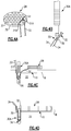
- FIGS. 1 and 2 schematically illustrate preparing a cartilage defect for implantation of a cartilage graft.
- FIG. 3 illustrates an exemplary cartilage graft.
- FIGS. 4A, 4B, 4C and 4D schematically illustrate knotlessly fixating a cartilage graft to bone.
- FIG. 5 schematically illustrates repairing a cartilage defect.
- FIGS. 6A, 6B, 6C and 6D illustrate exemplary fixation patterns for fixating a cartilage graft to a bone.
- the surgical methods include attaching a cartilage graft to bone using at least one fixation device.
- the fixation device is implanted into bone before tensioning a flexible strand, such as a suture, to approximate the cartilage graft to the bone.
- the fixation device is implanted into bone after tensioning the flexible strand to approximate the cartilage graft to the bone.
- Cartilage defects include localized areas of damaged articular cartilage and, potentially, adjacent subchondral bone. Cartilage defects typically do not heal without treatment. If not treated, a defect can deteriorate articulate cartilage and/or underlying bone of the joint, thereby causing relatively significant arthritic pain.
- FIGS. 1-6 schematically illustrate methods of repairing a cartilage defect 10 located within a joint 12 .
- the methods are illustrated and described as an arthroscopic method; however, methods could alternatively be performed as an open procedure.
- the cartilage defect 10 can include osteochondral and/or chondral defects.
- a cartilage defect 10 may include localized areas of damaged articular cartridge and/or damaged subchondral bone of the joint 12 .
- the joint 12 is a knee joint.
- methods of this disclosure may be used to repair cartilage defects located anywhere within the human body, including but not limited to, knee, talus, elbow, shoulder, hip, and temporomandibular joints.
- An exemplary repair method begins by prepping the cartilage defect 10 for receiving a cartilage graft 28 (shown in FIG. 3 ).
- the cartilage defect 10 may be debrided to a stable border having perpendicular margins.
- Tools, such as a curette 14 and an elevator 16 can be used to create vertical margins around a periphery of the cartilage defect 10 .
- a cartilage defect 10 may be prepped with our without bone marrow stimulation.
- a cartilage defect 10 is further prepared by performing bone marrow stimulation.
- a microfracture procedure or some other technique may optionally be performed to obtain a bleeding bone bed 18 .
- multiple perforations 20 are created in subchondral bone 22 that extends beneath articular cartilage 24 associated with a cartilage defect 10 .
- a bleeding bone bed 18 may be created using a tool 26 , such as Arthrex's PowerpickTM, to create perforations 20 .
- Formation of perforations 20 creates a bleeding bone bed 18 , which stimulates bone marrow seepage at the repair site.
- Other techniques can also be used to create a bleeding bone bed 18 , including but not limited to, drilling, hammering, curetting, scraping, etc.
- a cartilage defect 10 may also be dried to complete surgical preparation of the cartilage defect 10 .
- a cartilage defect 10 may be dried to remove excess moisture that could interfere with implantation of a cartilage graft 28 .
- the cartilage defect 10 may be dried using any known technique.
- At least one transosseus tunnel (e.g., 1, 2, 3, 4, or 5) can be created in one or more bones adjacent a cartilage defect.
- a transosseus tunnel can be created by drilling (e.g., an Arthrex Flipcutter®).
- a transosseus tunnel can be created in the tibia, femur, calcaneus, humerus, acetabulum, mandible, temporal bone, or phalanges.
- one or more transosseus tunnels can exit in a cartilage defect.
- a cartilage graft 28 may be implanted after adequately prepping a cartilage defect 10 .
- a cartilage graft 28 serves as a scaffold over a cartilage defect 10 , thereby providing a tissue network that can potentially signal autologous cellular interactions.
- the size and shape of a cartilage graft 28 may be selected using a template that is placed over the cartilage defect 10 and marked to indicate its general size. A template may then be used to trim a cartilage graft 28 down to the desired size and shape.
- a cartilage graft 28 includes a cartilage disk 29 having a plurality of pores 31 formed through the cartilage disk 29 .
- the cartilage graft 28 may be made of human tissue (e.g., allograft cartilage), synthetic materials, xeno materials, etc.
- a cartilage graft 28 is made of a micronized cartilage matrix. Although shown as being porous, a cartilage graft 28 is not limited to such an embodiment.
- a flexible strand 32 such as a suture, can be passed through a cartilage graft 28 .
- a mattress stitch 33 may be formed to connect the flexible strand 32 to the cartilage graft 28 .
- a mattress stitch 33 is formed by inserting a flexible strand 32 through a pore 31 A in a direction from the bottom 35 toward a top 37 of the disk 29 of the cartilage graft 28 and then inserting the flexible strand 32 through an adjacent pore 31 B in a direction from the top 37 toward the bottom 35 of the disk 29 .
- Other suturing techniques and configurations are also contemplated within the scope of this disclosure.
- the flexible strand 32 may be simply threaded through the cartilage graft 28 .
- one or more free ends 34 of a flexible strand 32 are loaded through a portion 55 of a knotless suture anchor 30 A.
- the portion 55 includes an eyelet 36 of the knotless suture anchor 30 A.
- Knotless techniques are considered “knotless” because there is no need to tie knots in a flexible strand 32 in order to secure a cartilage graft 28 to bone.
- the cartilage graft 28 can be fixated by positioning flexible strands 32 at each of its four quadrants (see FIG. 6A ), through its center and about its periphery (see FIG. 6B ), through its top and bottom halves (see FIG. 6C ), or at each third of the cartilage graft 28 (see FIG. 6D ).
- Other fixation patterns could also be used.
- a layer of fibrin may be applied over a cartilage graft 28 after it has been fixated to bone. Fibrin may be applied using an applicator. After the fibrin and the cartilage graft sit for a predefined amount of time, such as approximately five minutes, the joint 12 may be gently ranged before closure to assure adherence of the fibrin 50 and the cartilage graft 28 to the bone B.
- a flexible strand can be passed through a cartilage graft and pulling the graft through the transosseus tunnel. Once through the transosseus tunnel, the flexible strand can be tensioned to approximate the cartilage graft to the bone. Once approximated, the cartilage graft can be fixated to the bone via one or more fixation devices.
- a fixation device can be an anchor, a screw, a button, or a suture construct.
- a method for repairing a cartilage defect includes drilling at least one transosseus tunnel 60 ; tensioning a flexible strand 32 that is passed through a cartilage graft 28 through at least one transosseus tunnel 60 ; and fixating the cartilage graft 28 to bone B.
- a flexible strand 32 is tensioned (schematically shown by arrows 62 ) through more than one transosseus tunnels 60 (three shown in FIG. 5 ).
- a cartilage graft 28 can be fixated to bone B via a fixation device 64 , which can be a button, a screw, an anchor, or a suture construct.
- An embodiment includes a method comprising more than one fixation device 64 , which can be a button, a screw, an anchor, a suture construct, or combinations thereof.
- a suture construct can be knotted or knotless.
- a suture construct can be a pattern of suture that secures the graft to a bone (e.g., SutureBridge®, SpeedBridgeTM, etc.).
Landscapes
- Health & Medical Sciences (AREA)
- Life Sciences & Earth Sciences (AREA)
- Surgery (AREA)
- Animal Behavior & Ethology (AREA)
- Rheumatology (AREA)
- Veterinary Medicine (AREA)
- Public Health (AREA)
- Engineering & Computer Science (AREA)
- Biomedical Technology (AREA)
- Heart & Thoracic Surgery (AREA)
- General Health & Medical Sciences (AREA)
- Nuclear Medicine, Radiotherapy & Molecular Imaging (AREA)
- Medical Informatics (AREA)
- Molecular Biology (AREA)
- Vascular Medicine (AREA)
- Transplantation (AREA)
- Oral & Maxillofacial Surgery (AREA)
- Cardiology (AREA)
- Orthopedic Medicine & Surgery (AREA)
- Prostheses (AREA)
Abstract
Description
Claims (20)
Priority Applications (1)
| Application Number | Priority Date | Filing Date | Title |
|---|---|---|---|
| US14/790,570 US10524775B2 (en) | 2015-07-02 | 2015-07-02 | Methods of repairing cartilage defects |
Applications Claiming Priority (1)
| Application Number | Priority Date | Filing Date | Title |
|---|---|---|---|
| US14/790,570 US10524775B2 (en) | 2015-07-02 | 2015-07-02 | Methods of repairing cartilage defects |
Publications (2)
| Publication Number | Publication Date |
|---|---|
| US20170000473A1 US20170000473A1 (en) | 2017-01-05 |
| US10524775B2 true US10524775B2 (en) | 2020-01-07 |
Family
ID=57683480
Family Applications (1)
| Application Number | Title | Priority Date | Filing Date |
|---|---|---|---|
| US14/790,570 Active 2036-06-02 US10524775B2 (en) | 2015-07-02 | 2015-07-02 | Methods of repairing cartilage defects |
Country Status (1)
| Country | Link |
|---|---|
| US (1) | US10524775B2 (en) |
Cited By (5)
| Publication number | Priority date | Publication date | Assignee | Title |
|---|---|---|---|---|
| US11523834B1 (en) | 2022-06-20 | 2022-12-13 | University Of Utah Research Foundation | Cartilage and bone harvest and delivery system and methods |
| US11617605B2 (en) | 2015-11-13 | 2023-04-04 | Leith Medical LLC | Bone fixation system with fasteners and a removal tool for decoupling of the fasteners |
| US11660194B1 (en) | 2022-06-20 | 2023-05-30 | University Of Utah Research Foundation | Cartilage and bone harvest and delivery system and methods |
| US11744626B2 (en) | 2019-10-14 | 2023-09-05 | Leith Medical, LLC | Bone fixation system with fasteners and a removal tool for decoupling of the fasteners |
| US11980402B2 (en) | 2019-10-14 | 2024-05-14 | Leith Medical, Inc. | Apparatus for stabilization of a bone fracture site |
Families Citing this family (1)
| Publication number | Priority date | Publication date | Assignee | Title |
|---|---|---|---|---|
| US10881762B2 (en) * | 2017-11-15 | 2021-01-05 | De Novo Orthopedics Inc. | Method for manufacturing bioinductive patch |
Citations (59)
| Publication number | Priority date | Publication date | Assignee | Title |
|---|---|---|---|---|
| US3318774A (en) * | 1961-03-15 | 1967-05-09 | Squibb & Sons Inc | Treatment of osseous and other tissue |
| US4772286A (en) * | 1987-02-17 | 1988-09-20 | E. Marlowe Goble | Ligament attachment method and apparatus |
| US4773910A (en) * | 1987-08-17 | 1988-09-27 | Johnson & Johnson Consumer Products, Inc. | Permanent ligament prosthesis |
| US5139520A (en) * | 1990-01-31 | 1992-08-18 | American Cyanamid Company | Method for acl reconstruction |
| US5713374A (en) | 1995-02-10 | 1998-02-03 | The Hospital For Joint Diseases Orthopaedic Institute | Fixation method for the attachment of wound repair materials to cartilage defects |
| US5964764A (en) * | 1998-03-24 | 1999-10-12 | Hugh S. West, Jr. | Apparatus and methods for mounting a ligament graft to a bone |
| US6440141B1 (en) | 2000-07-24 | 2002-08-27 | Oratec Interventions, Inc. | Method and apparatus for treating osteochondral pathologies |
| US6488033B1 (en) | 2000-05-15 | 2002-12-03 | Cryolife, Inc. | Osteochondral transplant techniques |
| US20050149118A1 (en) | 2003-12-18 | 2005-07-07 | Ilya Koyfman | High strength suture with absorbable core and suture anchor combination |
| US20060293710A1 (en) | 2003-10-21 | 2006-12-28 | Arthrocare Corporation | Knotless suture lock and bone anchor implant method |
| US20070135843A1 (en) | 2005-03-30 | 2007-06-14 | Arthrex, Inc. | Knotless fixation of tissue to bone with suture chain |
| US20070288023A1 (en) | 2006-06-12 | 2007-12-13 | Greg Pellegrino | Soft tissue repair using tissue augments and bone anchors |
| US7326222B2 (en) * | 2002-05-01 | 2008-02-05 | Arthrex, Inc. | Suture tensioning device |
| US20080039954A1 (en) * | 2006-08-08 | 2008-02-14 | Howmedica Osteonics Corp. | Expandable cartilage implant |
| US7361195B2 (en) | 2001-07-16 | 2008-04-22 | Depuy Products, Inc. | Cartilage repair apparatus and method |
| US7371260B2 (en) | 2005-10-26 | 2008-05-13 | Biomet Sports Medicine, Inc. | Method and instrumentation for the preparation and transplantation of osteochondral allografts |
| US20080125863A1 (en) * | 2006-11-28 | 2008-05-29 | Mckay William F | Implant designs and methods of improving cartilage repair |
| US20080195205A1 (en) * | 2004-03-03 | 2008-08-14 | Schwartz Bomedical Llc | Articular Cartilage Fixation Device and Method |
| US20080255613A1 (en) | 2007-04-10 | 2008-10-16 | Biomet Sports Medicine, Inc. | Adjustable knotless loops |
| US20080269674A1 (en) * | 2007-04-25 | 2008-10-30 | Biomet Sports Medicine, Inc. | Localized Cartilage Defect Therapy |
| US20090024229A1 (en) | 2007-07-16 | 2009-01-22 | Chen Silvia S | Devitalization and recellularization of cartilage |
| US7488347B1 (en) * | 2005-01-06 | 2009-02-10 | Medicine Lodge, Inc. | Transosseous graft retention system and method |
| US20090312842A1 (en) | 2008-06-16 | 2009-12-17 | Predrag Bursac | Assembled Cartilage Repair Graft |
| US7641694B1 (en) | 2005-01-06 | 2010-01-05 | IMDS, Inc. | Line lock graft retention system and method |
| US20100016892A1 (en) | 2008-07-21 | 2010-01-21 | William Kaiser | Method and apparatus for securing soft tissue to bone |
| US20100040662A1 (en) | 2008-06-13 | 2010-02-18 | Cotton Nicholas J | Fixation Devices For Tissue Repair |
| US7666230B2 (en) | 2003-12-08 | 2010-02-23 | Depuy Products, Inc. | Implant device for cartilage regeneration in load bearing articulation regions |
| US20100168869A1 (en) | 2008-12-31 | 2010-07-01 | Howmedica Osteonics Corp. | Tissue integration implant |
| US7749250B2 (en) * | 2006-02-03 | 2010-07-06 | Biomet Sports Medicine, Llc | Soft tissue repair assembly and associated method |
| US7901461B2 (en) | 2003-12-05 | 2011-03-08 | Ethicon, Inc. | Viable tissue repair implants and methods of use |
| US20110091517A1 (en) | 2002-10-18 | 2011-04-21 | Depuy Mitek, Inc. | Biocompatible scaffolds with tissue fragments |
| US7931695B2 (en) | 2003-07-15 | 2011-04-26 | Kensey Nash Corporation | Compliant osteosynthesis fixation plate |
| US8012205B2 (en) | 2001-07-16 | 2011-09-06 | Depuy Products, Inc. | Cartilage repair and regeneration device |
| US8016867B2 (en) | 1999-07-23 | 2011-09-13 | Depuy Mitek, Inc. | Graft fixation device and method |
| US20110245929A1 (en) | 2010-03-05 | 2011-10-06 | Advanced BioHealing Inc. | Methods and compositions for joint healing and repair |
| US8062654B2 (en) * | 1999-08-06 | 2011-11-22 | The Board Of Regents Of The University Of Texas System | Drug releasing biodegradable fiber for delivery of therapeutics |
| US8142502B2 (en) | 2007-02-26 | 2012-03-27 | Biomet Sports Medicine, Llc | Stable cartilage defect repair plug |
| US20120207718A1 (en) | 2011-02-16 | 2012-08-16 | Stone Kevin R | Thin shell graft for cartilage resurfacing |
| US8444968B2 (en) | 2005-12-07 | 2013-05-21 | Isto Technologies, Inc. | Cartilage repair methods |
| US8449561B2 (en) | 1999-07-23 | 2013-05-28 | Depuy Mitek, Llc | Graft fixation device combination |
| US20130138123A1 (en) * | 2006-02-03 | 2013-05-30 | Biomet Sports Medicine, Llc | Method And Apparatus For Coupling Soft Tissue To A Bone |
| US8518433B2 (en) | 2003-12-11 | 2013-08-27 | Zimmer, Inc. | Method of treating an osteochondral defect |
| US8535703B2 (en) | 2007-08-27 | 2013-09-17 | Arthrex, Inc. | Methods of arthroscopic osteochondral resurfacing |
| US8545535B2 (en) * | 2009-05-12 | 2013-10-01 | Foundry Newco Xi, Inc. | Suture anchors with one-way cinching mechanisms |
| US20130338792A1 (en) | 2012-06-15 | 2013-12-19 | Arthrex, Inc. | Implantation of micronized allograft tissue over a microfractured defect |
| US20140017283A1 (en) | 2012-07-11 | 2014-01-16 | Osiris Therapeutics, Inc. | Porated cartilage products |
| US8637066B2 (en) | 2002-10-18 | 2014-01-28 | Depuy Mitek, Llc | Biocompatible scaffold for ligament or tendon repair |
| US20140031863A1 (en) * | 2009-05-12 | 2014-01-30 | Foundry Newco Xi, Inc. | Methods and devices to treat diseased or injured musculoskeletal tissue |
| US8657881B2 (en) | 2004-04-20 | 2014-02-25 | Depuy Mitek, Llc | Meniscal repair scaffold |
| US20140142718A1 (en) | 2012-11-16 | 2014-05-22 | Isto Technologies, Inc. | Flexible tissue matrix and methods for joint repair |
| US8734828B2 (en) * | 2006-10-06 | 2014-05-27 | Biotissue Ag | Matrix-gel graft without cells |
| US20140222162A1 (en) | 2013-02-01 | 2014-08-07 | Xiros Limited | Connective tissue repair technology |
| US8834568B2 (en) | 2010-02-04 | 2014-09-16 | Paul S. Shapiro | Surgical technique using a contoured allograft cartilage as a spacer of the carpo-metacarpal joint of the thumb or tarso-metatarsal joint of the toe |
| US8895045B2 (en) | 2003-03-07 | 2014-11-25 | Depuy Mitek, Llc | Method of preparation of bioabsorbable porous reinforced tissue implants and implants thereof |
| US20150057750A1 (en) * | 2013-08-21 | 2015-02-26 | Arthrex, Inc. | Bone tendon constructs and methods of tissue fixation |
| US9066716B2 (en) * | 2011-03-30 | 2015-06-30 | Arthrosurface Incorporated | Suture coil and suture sheath for tissue repair |
| US20150182233A1 (en) | 2013-12-26 | 2015-07-02 | Tenjin LLC | Percussive surgical devices, systems, and methods of use thereof |
| US20160287243A1 (en) * | 2015-04-02 | 2016-10-06 | Arthrex, Inc. | Method of repairing cartilage defects |
| US9855146B2 (en) * | 2015-08-24 | 2018-01-02 | Arthrex, Inc. | Arthroscopic resurfacing techniques |
Family Cites Families (1)
| Publication number | Priority date | Publication date | Assignee | Title |
|---|---|---|---|---|
| US7536504B2 (en) * | 2006-07-28 | 2009-05-19 | Diskeeper Corporation | Online storage medium transfer rate characteristics determination |
-
2015
- 2015-07-02 US US14/790,570 patent/US10524775B2/en active Active
Patent Citations (65)
| Publication number | Priority date | Publication date | Assignee | Title |
|---|---|---|---|---|
| US3318774A (en) * | 1961-03-15 | 1967-05-09 | Squibb & Sons Inc | Treatment of osseous and other tissue |
| US4772286A (en) * | 1987-02-17 | 1988-09-20 | E. Marlowe Goble | Ligament attachment method and apparatus |
| US4773910A (en) * | 1987-08-17 | 1988-09-27 | Johnson & Johnson Consumer Products, Inc. | Permanent ligament prosthesis |
| US5139520A (en) * | 1990-01-31 | 1992-08-18 | American Cyanamid Company | Method for acl reconstruction |
| US5713374A (en) | 1995-02-10 | 1998-02-03 | The Hospital For Joint Diseases Orthopaedic Institute | Fixation method for the attachment of wound repair materials to cartilage defects |
| US5964764A (en) * | 1998-03-24 | 1999-10-12 | Hugh S. West, Jr. | Apparatus and methods for mounting a ligament graft to a bone |
| US8016867B2 (en) | 1999-07-23 | 2011-09-13 | Depuy Mitek, Inc. | Graft fixation device and method |
| US8449561B2 (en) | 1999-07-23 | 2013-05-28 | Depuy Mitek, Llc | Graft fixation device combination |
| US8062654B2 (en) * | 1999-08-06 | 2011-11-22 | The Board Of Regents Of The University Of Texas System | Drug releasing biodegradable fiber for delivery of therapeutics |
| US6488033B1 (en) | 2000-05-15 | 2002-12-03 | Cryolife, Inc. | Osteochondral transplant techniques |
| US6440141B1 (en) | 2000-07-24 | 2002-08-27 | Oratec Interventions, Inc. | Method and apparatus for treating osteochondral pathologies |
| US7361195B2 (en) | 2001-07-16 | 2008-04-22 | Depuy Products, Inc. | Cartilage repair apparatus and method |
| US8012205B2 (en) | 2001-07-16 | 2011-09-06 | Depuy Products, Inc. | Cartilage repair and regeneration device |
| US7326222B2 (en) * | 2002-05-01 | 2008-02-05 | Arthrex, Inc. | Suture tensioning device |
| US20110091517A1 (en) | 2002-10-18 | 2011-04-21 | Depuy Mitek, Inc. | Biocompatible scaffolds with tissue fragments |
| US8637066B2 (en) | 2002-10-18 | 2014-01-28 | Depuy Mitek, Llc | Biocompatible scaffold for ligament or tendon repair |
| US8895045B2 (en) | 2003-03-07 | 2014-11-25 | Depuy Mitek, Llc | Method of preparation of bioabsorbable porous reinforced tissue implants and implants thereof |
| US7931695B2 (en) | 2003-07-15 | 2011-04-26 | Kensey Nash Corporation | Compliant osteosynthesis fixation plate |
| US20060293710A1 (en) | 2003-10-21 | 2006-12-28 | Arthrocare Corporation | Knotless suture lock and bone anchor implant method |
| US7901461B2 (en) | 2003-12-05 | 2011-03-08 | Ethicon, Inc. | Viable tissue repair implants and methods of use |
| US8641775B2 (en) | 2003-12-05 | 2014-02-04 | Depuy Mitek, Llc | Viable tissue repair implants and methods of use |
| US7666230B2 (en) | 2003-12-08 | 2010-02-23 | Depuy Products, Inc. | Implant device for cartilage regeneration in load bearing articulation regions |
| US8834914B2 (en) | 2003-12-11 | 2014-09-16 | Zimmer, Inc. | Treatment methods using a particulate cadaveric allogenic juvenile cartilage particles |
| US8524268B2 (en) | 2003-12-11 | 2013-09-03 | Zimmer, Inc. | Cadaveric allogenic human juvenile cartilage implant |
| US8518433B2 (en) | 2003-12-11 | 2013-08-27 | Zimmer, Inc. | Method of treating an osteochondral defect |
| US20050149118A1 (en) | 2003-12-18 | 2005-07-07 | Ilya Koyfman | High strength suture with absorbable core and suture anchor combination |
| US8597352B2 (en) | 2004-03-03 | 2013-12-03 | Schwartz Biomedical, Llc | Articular cartilage fixation device and method |
| US20080195205A1 (en) * | 2004-03-03 | 2008-08-14 | Schwartz Bomedical Llc | Articular Cartilage Fixation Device and Method |
| US8657881B2 (en) | 2004-04-20 | 2014-02-25 | Depuy Mitek, Llc | Meniscal repair scaffold |
| US7641694B1 (en) | 2005-01-06 | 2010-01-05 | IMDS, Inc. | Line lock graft retention system and method |
| US7488347B1 (en) * | 2005-01-06 | 2009-02-10 | Medicine Lodge, Inc. | Transosseous graft retention system and method |
| US20070135843A1 (en) | 2005-03-30 | 2007-06-14 | Arthrex, Inc. | Knotless fixation of tissue to bone with suture chain |
| US7371260B2 (en) | 2005-10-26 | 2008-05-13 | Biomet Sports Medicine, Inc. | Method and instrumentation for the preparation and transplantation of osteochondral allografts |
| US8882774B2 (en) | 2005-10-26 | 2014-11-11 | Biomet Sports Medicine, Llc | Instrumentation for the preparation and transplantation of osteochondral allografts |
| US8444968B2 (en) | 2005-12-07 | 2013-05-21 | Isto Technologies, Inc. | Cartilage repair methods |
| US7749250B2 (en) * | 2006-02-03 | 2010-07-06 | Biomet Sports Medicine, Llc | Soft tissue repair assembly and associated method |
| US20130138123A1 (en) * | 2006-02-03 | 2013-05-30 | Biomet Sports Medicine, Llc | Method And Apparatus For Coupling Soft Tissue To A Bone |
| US20130158601A1 (en) * | 2006-02-03 | 2013-06-20 | Biomet Sports Medicine, Llc | Soft Tissue Repair Assembly And Associated Method |
| US20070288023A1 (en) | 2006-06-12 | 2007-12-13 | Greg Pellegrino | Soft tissue repair using tissue augments and bone anchors |
| US20080039954A1 (en) * | 2006-08-08 | 2008-02-14 | Howmedica Osteonics Corp. | Expandable cartilage implant |
| US8734828B2 (en) * | 2006-10-06 | 2014-05-27 | Biotissue Ag | Matrix-gel graft without cells |
| US20080125863A1 (en) * | 2006-11-28 | 2008-05-29 | Mckay William F | Implant designs and methods of improving cartilage repair |
| US8142502B2 (en) | 2007-02-26 | 2012-03-27 | Biomet Sports Medicine, Llc | Stable cartilage defect repair plug |
| US20080255613A1 (en) | 2007-04-10 | 2008-10-16 | Biomet Sports Medicine, Inc. | Adjustable knotless loops |
| US20080269674A1 (en) * | 2007-04-25 | 2008-10-30 | Biomet Sports Medicine, Inc. | Localized Cartilage Defect Therapy |
| US20090024229A1 (en) | 2007-07-16 | 2009-01-22 | Chen Silvia S | Devitalization and recellularization of cartilage |
| US8535703B2 (en) | 2007-08-27 | 2013-09-17 | Arthrex, Inc. | Methods of arthroscopic osteochondral resurfacing |
| US20100040662A1 (en) | 2008-06-13 | 2010-02-18 | Cotton Nicholas J | Fixation Devices For Tissue Repair |
| US20090312842A1 (en) | 2008-06-16 | 2009-12-17 | Predrag Bursac | Assembled Cartilage Repair Graft |
| US20100016892A1 (en) | 2008-07-21 | 2010-01-21 | William Kaiser | Method and apparatus for securing soft tissue to bone |
| US20100168869A1 (en) | 2008-12-31 | 2010-07-01 | Howmedica Osteonics Corp. | Tissue integration implant |
| US20140031863A1 (en) * | 2009-05-12 | 2014-01-30 | Foundry Newco Xi, Inc. | Methods and devices to treat diseased or injured musculoskeletal tissue |
| US8545535B2 (en) * | 2009-05-12 | 2013-10-01 | Foundry Newco Xi, Inc. | Suture anchors with one-way cinching mechanisms |
| US8834568B2 (en) | 2010-02-04 | 2014-09-16 | Paul S. Shapiro | Surgical technique using a contoured allograft cartilage as a spacer of the carpo-metacarpal joint of the thumb or tarso-metatarsal joint of the toe |
| US20110245929A1 (en) | 2010-03-05 | 2011-10-06 | Advanced BioHealing Inc. | Methods and compositions for joint healing and repair |
| US20120207718A1 (en) | 2011-02-16 | 2012-08-16 | Stone Kevin R | Thin shell graft for cartilage resurfacing |
| US9066716B2 (en) * | 2011-03-30 | 2015-06-30 | Arthrosurface Incorporated | Suture coil and suture sheath for tissue repair |
| US20130338792A1 (en) | 2012-06-15 | 2013-12-19 | Arthrex, Inc. | Implantation of micronized allograft tissue over a microfractured defect |
| US20140017283A1 (en) | 2012-07-11 | 2014-01-16 | Osiris Therapeutics, Inc. | Porated cartilage products |
| US20140142718A1 (en) | 2012-11-16 | 2014-05-22 | Isto Technologies, Inc. | Flexible tissue matrix and methods for joint repair |
| US20140222162A1 (en) | 2013-02-01 | 2014-08-07 | Xiros Limited | Connective tissue repair technology |
| US20150057750A1 (en) * | 2013-08-21 | 2015-02-26 | Arthrex, Inc. | Bone tendon constructs and methods of tissue fixation |
| US20150182233A1 (en) | 2013-12-26 | 2015-07-02 | Tenjin LLC | Percussive surgical devices, systems, and methods of use thereof |
| US20160287243A1 (en) * | 2015-04-02 | 2016-10-06 | Arthrex, Inc. | Method of repairing cartilage defects |
| US9855146B2 (en) * | 2015-08-24 | 2018-01-02 | Arthrex, Inc. | Arthroscopic resurfacing techniques |
Non-Patent Citations (3)
| Title |
|---|
| International Preliminary Report on Patentability for International application No. PCT/US2015/059192 dated Oct. 12, 2017. |
| International Search Report and Written Opinion of the International Searching Authority for International application No. PCT/US2015/059192 dated Feb. 3, 2016. |
| Streit, Jomnathan J., et al., Fresh osteochondral allograft for shoulder resurfacing relieves pain, Orthopedics Today, Oct. 2013, https://www.healio.com/orthopedics/shoulder-elbow/news/print/orthopedics-today/%7bccb . . . , printed Mar. 22, 2018. |
Cited By (5)
| Publication number | Priority date | Publication date | Assignee | Title |
|---|---|---|---|---|
| US11617605B2 (en) | 2015-11-13 | 2023-04-04 | Leith Medical LLC | Bone fixation system with fasteners and a removal tool for decoupling of the fasteners |
| US11744626B2 (en) | 2019-10-14 | 2023-09-05 | Leith Medical, LLC | Bone fixation system with fasteners and a removal tool for decoupling of the fasteners |
| US11980402B2 (en) | 2019-10-14 | 2024-05-14 | Leith Medical, Inc. | Apparatus for stabilization of a bone fracture site |
| US11523834B1 (en) | 2022-06-20 | 2022-12-13 | University Of Utah Research Foundation | Cartilage and bone harvest and delivery system and methods |
| US11660194B1 (en) | 2022-06-20 | 2023-05-30 | University Of Utah Research Foundation | Cartilage and bone harvest and delivery system and methods |
Also Published As
| Publication number | Publication date |
|---|---|
| US20170000473A1 (en) | 2017-01-05 |
Similar Documents
| Publication | Publication Date | Title |
|---|---|---|
| US10524774B2 (en) | Method of repairing cartilage defects | |
| US10524775B2 (en) | Methods of repairing cartilage defects | |
| US10932897B2 (en) | Joint kinematic reconstruction techniques | |
| US9855146B2 (en) | Arthroscopic resurfacing techniques | |
| US7594922B1 (en) | System and method for meniscal repair through a meniscal capsular tunnel | |
| US11446135B2 (en) | Joint kinematic reconstruction techniques | |
| US20160199054A1 (en) | Medical device for repair of tissue and method for implantation and fixation | |
| US7833269B2 (en) | Osteochondral implant procedure | |
| US20050256582A1 (en) | Spinal implants, including devices that reduce pressure on the annulus fibrosis | |
| US9913710B2 (en) | Implantable biologic holder | |
| US20080281422A1 (en) | Arthroscopic knotless technique for collagen patch fixation | |
| US10568733B2 (en) | Anterior cable region superior capsule reconstructions | |
| US11357496B2 (en) | Scapho-lunate and other ligament and bone repair/reconstruction | |
| US20230147972A1 (en) | Knotless tensionable fixation systems and surgical methods for repairing tissue defects | |
| RU2721936C1 (en) | Method for treating complex fractures of proximal humerus | |
| US11510790B2 (en) | Triangular fibrocartilage complex reconstruction techniques | |
| Edwards et al. | Management of extensor mechanism during revision total knee arthroplasty | |
| WO2014138408A1 (en) | Implant and method for glenoid and labrum repair |
Legal Events
| Date | Code | Title | Description |
|---|---|---|---|
| AS | Assignment |
Owner name: ARTHREX, INC., FLORIDA Free format text: ASSIGNMENT OF ASSIGNORS INTEREST;ASSIGNORS:BENEDICT, ROBERT;STOLL, MARC;COOK, JAMES L.;SIGNING DATES FROM 20150709 TO 20150714;REEL/FRAME:036160/0008 |
|
| STPP | Information on status: patent application and granting procedure in general |
Free format text: ADVISORY ACTION MAILED |
|
| STPP | Information on status: patent application and granting procedure in general |
Free format text: DOCKETED NEW CASE - READY FOR EXAMINATION |
|
| STPP | Information on status: patent application and granting procedure in general |
Free format text: NOTICE OF ALLOWANCE MAILED -- APPLICATION RECEIVED IN OFFICE OF PUBLICATIONS |
|
| STPP | Information on status: patent application and granting procedure in general |
Free format text: AWAITING TC RESP., ISSUE FEE NOT PAID |
|
| STPP | Information on status: patent application and granting procedure in general |
Free format text: PUBLICATIONS -- ISSUE FEE PAYMENT VERIFIED |
|
| STCF | Information on status: patent grant |
Free format text: PATENTED CASE |
|
| MAFP | Maintenance fee payment |
Free format text: PAYMENT OF MAINTENANCE FEE, 4TH YEAR, LARGE ENTITY (ORIGINAL EVENT CODE: M1551); ENTITY STATUS OF PATENT OWNER: LARGE ENTITY Year of fee payment: 4 |