US10515450B2 - Systems and methods for using a single-cell to create chromosomal spreads - Google Patents
Systems and methods for using a single-cell to create chromosomal spreads Download PDFInfo
- Publication number
- US10515450B2 US10515450B2 US15/789,414 US201715789414A US10515450B2 US 10515450 B2 US10515450 B2 US 10515450B2 US 201715789414 A US201715789414 A US 201715789414A US 10515450 B2 US10515450 B2 US 10515450B2
- Authority
- US
- United States
- Prior art keywords
- cell
- chromosomal
- nuclear
- dna
- compression
- Prior art date
- Legal status (The legal status is an assumption and is not a legal conclusion. Google has not performed a legal analysis and makes no representation as to the accuracy of the status listed.)
- Expired - Fee Related, expires
Links
- 238000000034 method Methods 0.000 title claims abstract description 34
- 230000002759 chromosomal effect Effects 0.000 title claims abstract description 30
- 210000004027 cell Anatomy 0.000 claims description 82
- 230000006835 compression Effects 0.000 claims description 32
- 238000007906 compression Methods 0.000 claims description 32
- 238000003384 imaging method Methods 0.000 claims description 13
- 206010028980 Neoplasm Diseases 0.000 claims description 11
- 201000011510 cancer Diseases 0.000 claims description 6
- 230000008961 swelling Effects 0.000 claims description 3
- 210000004292 cytoskeleton Anatomy 0.000 claims description 2
- 230000002438 mitochondrial effect Effects 0.000 claims description 2
- 230000008520 organization Effects 0.000 claims description 2
- 230000001225 therapeutic effect Effects 0.000 claims description 2
- 230000025545 Golgi localization Effects 0.000 claims 1
- 230000030583 endoplasmic reticulum localization Effects 0.000 claims 1
- 230000002596 correlated effect Effects 0.000 abstract description 4
- 230000005945 translocation Effects 0.000 abstract description 3
- 108020004414 DNA Proteins 0.000 description 17
- 208000037265 diseases, disorders, signs and symptoms Diseases 0.000 description 14
- 238000011282 treatment Methods 0.000 description 14
- 210000004940 nucleus Anatomy 0.000 description 11
- 201000010099 disease Diseases 0.000 description 8
- 108090000623 proteins and genes Proteins 0.000 description 8
- 230000001575 pathological effect Effects 0.000 description 7
- 102000004169 proteins and genes Human genes 0.000 description 7
- 239000000758 substrate Substances 0.000 description 7
- 210000000349 chromosome Anatomy 0.000 description 6
- 208000035475 disorder Diseases 0.000 description 6
- 230000004807 localization Effects 0.000 description 5
- 206010008805 Chromosomal abnormalities Diseases 0.000 description 4
- 208000031404 Chromosome Aberrations Diseases 0.000 description 4
- 230000002159 abnormal effect Effects 0.000 description 4
- 230000001413 cellular effect Effects 0.000 description 4
- 239000012530 fluid Substances 0.000 description 4
- 239000011521 glass Substances 0.000 description 4
- 230000000394 mitotic effect Effects 0.000 description 4
- 230000005856 abnormality Effects 0.000 description 3
- 230000032683 aging Effects 0.000 description 3
- 238000004458 analytical method Methods 0.000 description 3
- 230000008901 benefit Effects 0.000 description 3
- 239000000463 material Substances 0.000 description 3
- 239000002609 medium Substances 0.000 description 3
- 230000008569 process Effects 0.000 description 3
- 230000007480 spreading Effects 0.000 description 3
- 206010061764 Chromosomal deletion Diseases 0.000 description 2
- 208000036086 Chromosome Duplication Diseases 0.000 description 2
- 208000034951 Genetic Translocation Diseases 0.000 description 2
- 208000025500 Hutchinson-Gilford progeria syndrome Diseases 0.000 description 2
- 208000007932 Progeria Diseases 0.000 description 2
- IQFYYKKMVGJFEH-XLPZGREQSA-N Thymidine Chemical compound O=C1NC(=O)C(C)=CN1[C@@H]1O[C@H](CO)[C@@H](O)C1 IQFYYKKMVGJFEH-XLPZGREQSA-N 0.000 description 2
- 230000001464 adherent effect Effects 0.000 description 2
- 238000012217 deletion Methods 0.000 description 2
- 230000037430 deletion Effects 0.000 description 2
- 230000002706 hydrostatic effect Effects 0.000 description 2
- 230000006872 improvement Effects 0.000 description 2
- 238000002372 labelling Methods 0.000 description 2
- 208000026585 laminopathy Diseases 0.000 description 2
- 208000006132 lipodystrophy Diseases 0.000 description 2
- 230000007246 mechanism Effects 0.000 description 2
- 230000031864 metaphase Effects 0.000 description 2
- 210000003470 mitochondria Anatomy 0.000 description 2
- 230000011278 mitosis Effects 0.000 description 2
- 201000006938 muscular dystrophy Diseases 0.000 description 2
- 210000003463 organelle Anatomy 0.000 description 2
- 238000012545 processing Methods 0.000 description 2
- 230000006916 protein interaction Effects 0.000 description 2
- 230000008707 rearrangement Effects 0.000 description 2
- 238000002560 therapeutic procedure Methods 0.000 description 2
- 210000001519 tissue Anatomy 0.000 description 2
- 238000012546 transfer Methods 0.000 description 2
- DWRXFEITVBNRMK-UHFFFAOYSA-N Beta-D-1-Arabinofuranosylthymine Natural products O=C1NC(=O)C(C)=CN1C1C(O)C(O)C(CO)O1 DWRXFEITVBNRMK-UHFFFAOYSA-N 0.000 description 1
- 208000031229 Cardiomyopathies Diseases 0.000 description 1
- 108010043121 Green Fluorescent Proteins Proteins 0.000 description 1
- 102000004144 Green Fluorescent Proteins Human genes 0.000 description 1
- 241000282412 Homo Species 0.000 description 1
- 206010049287 Lipodystrophy acquired Diseases 0.000 description 1
- 241000124008 Mammalia Species 0.000 description 1
- KYRVNWMVYQXFEU-UHFFFAOYSA-N Nocodazole Chemical compound C1=C2NC(NC(=O)OC)=NC2=CC=C1C(=O)C1=CC=CS1 KYRVNWMVYQXFEU-UHFFFAOYSA-N 0.000 description 1
- 108091023040 Transcription factor Proteins 0.000 description 1
- 102000040945 Transcription factor Human genes 0.000 description 1
- 241000251539 Vertebrata <Metazoa> Species 0.000 description 1
- IQFYYKKMVGJFEH-UHFFFAOYSA-N beta-L-thymidine Natural products O=C1NC(=O)C(C)=CN1C1OC(CO)C(O)C1 IQFYYKKMVGJFEH-UHFFFAOYSA-N 0.000 description 1
- 230000001364 causal effect Effects 0.000 description 1
- 238000004891 communication Methods 0.000 description 1
- 238000004590 computer program Methods 0.000 description 1
- 210000004748 cultured cell Anatomy 0.000 description 1
- 238000011161 development Methods 0.000 description 1
- 230000018109 developmental process Effects 0.000 description 1
- 230000002526 effect on cardiovascular system Effects 0.000 description 1
- 210000003038 endothelium Anatomy 0.000 description 1
- 238000002795 fluorescence method Methods 0.000 description 1
- 238000000799 fluorescence microscopy Methods 0.000 description 1
- 238000002866 fluorescence resonance energy transfer Methods 0.000 description 1
- 239000007850 fluorescent dye Substances 0.000 description 1
- 239000005090 green fluorescent protein Substances 0.000 description 1
- 239000001963 growth medium Substances 0.000 description 1
- 239000000815 hypotonic solution Substances 0.000 description 1
- 230000002401 inhibitory effect Effects 0.000 description 1
- 230000003834 intracellular effect Effects 0.000 description 1
- 239000003550 marker Substances 0.000 description 1
- 238000005259 measurement Methods 0.000 description 1
- 239000012528 membrane Substances 0.000 description 1
- 230000007625 mitochondrial abnormality Effects 0.000 description 1
- 238000012986 modification Methods 0.000 description 1
- 230000004048 modification Effects 0.000 description 1
- 229950006344 nocodazole Drugs 0.000 description 1
- 210000000633 nuclear envelope Anatomy 0.000 description 1
- 230000003204 osmotic effect Effects 0.000 description 1
- 238000002638 palliative care Methods 0.000 description 1
- 239000011148 porous material Substances 0.000 description 1
- 239000002096 quantum dot Substances 0.000 description 1
- 239000000523 sample Substances 0.000 description 1
- 238000010008 shearing Methods 0.000 description 1
- 239000000243 solution Substances 0.000 description 1
- 210000000130 stem cell Anatomy 0.000 description 1
- 238000010869 super-resolution microscopy Methods 0.000 description 1
- 239000013589 supplement Substances 0.000 description 1
- 230000003319 supportive effect Effects 0.000 description 1
- 239000000725 suspension Substances 0.000 description 1
- 208000024891 symptom Diseases 0.000 description 1
- 229940104230 thymidine Drugs 0.000 description 1
- 238000011277 treatment modality Methods 0.000 description 1
- XLYOFNOQVPJJNP-UHFFFAOYSA-N water Substances O XLYOFNOQVPJJNP-UHFFFAOYSA-N 0.000 description 1
Images


Classifications
-
- G—PHYSICS
- G06—COMPUTING; CALCULATING OR COUNTING
- G06T—IMAGE DATA PROCESSING OR GENERATION, IN GENERAL
- G06T7/00—Image analysis
- G06T7/0002—Inspection of images, e.g. flaw detection
- G06T7/0012—Biomedical image inspection
-
- B—PERFORMING OPERATIONS; TRANSPORTING
- B01—PHYSICAL OR CHEMICAL PROCESSES OR APPARATUS IN GENERAL
- B01L—CHEMICAL OR PHYSICAL LABORATORY APPARATUS FOR GENERAL USE
- B01L3/00—Containers or dishes for laboratory use, e.g. laboratory glassware; Droppers
- B01L3/50—Containers for the purpose of retaining a material to be analysed, e.g. test tubes
- B01L3/502—Containers for the purpose of retaining a material to be analysed, e.g. test tubes with fluid transport, e.g. in multi-compartment structures
- B01L3/5027—Containers for the purpose of retaining a material to be analysed, e.g. test tubes with fluid transport, e.g. in multi-compartment structures by integrated microfluidic structures, i.e. dimensions of channels and chambers are such that surface tension forces are important, e.g. lab-on-a-chip
- B01L3/50273—Containers for the purpose of retaining a material to be analysed, e.g. test tubes with fluid transport, e.g. in multi-compartment structures by integrated microfluidic structures, i.e. dimensions of channels and chambers are such that surface tension forces are important, e.g. lab-on-a-chip characterised by the means or forces applied to move the fluids
-
- C—CHEMISTRY; METALLURGY
- C12—BIOCHEMISTRY; BEER; SPIRITS; WINE; VINEGAR; MICROBIOLOGY; ENZYMOLOGY; MUTATION OR GENETIC ENGINEERING
- C12N—MICROORGANISMS OR ENZYMES; COMPOSITIONS THEREOF; PROPAGATING, PRESERVING, OR MAINTAINING MICROORGANISMS; MUTATION OR GENETIC ENGINEERING; CULTURE MEDIA
- C12N15/00—Mutation or genetic engineering; DNA or RNA concerning genetic engineering, vectors, e.g. plasmids, or their isolation, preparation or purification; Use of hosts therefor
- C12N15/09—Recombinant DNA-technology
- C12N15/10—Processes for the isolation, preparation or purification of DNA or RNA
- C12N15/1003—Extracting or separating nucleic acids from biological samples, e.g. pure separation or isolation methods; Conditions, buffers or apparatuses therefor
-
- C—CHEMISTRY; METALLURGY
- C12—BIOCHEMISTRY; BEER; SPIRITS; WINE; VINEGAR; MICROBIOLOGY; ENZYMOLOGY; MUTATION OR GENETIC ENGINEERING
- C12Q—MEASURING OR TESTING PROCESSES INVOLVING ENZYMES, NUCLEIC ACIDS OR MICROORGANISMS; COMPOSITIONS OR TEST PAPERS THEREFOR; PROCESSES OF PREPARING SUCH COMPOSITIONS; CONDITION-RESPONSIVE CONTROL IN MICROBIOLOGICAL OR ENZYMOLOGICAL PROCESSES
- C12Q1/00—Measuring or testing processes involving enzymes, nucleic acids or microorganisms; Compositions therefor; Processes of preparing such compositions
- C12Q1/68—Measuring or testing processes involving enzymes, nucleic acids or microorganisms; Compositions therefor; Processes of preparing such compositions involving nucleic acids
- C12Q1/6813—Hybridisation assays
- C12Q1/6841—In situ hybridisation
-
- C—CHEMISTRY; METALLURGY
- C12—BIOCHEMISTRY; BEER; SPIRITS; WINE; VINEGAR; MICROBIOLOGY; ENZYMOLOGY; MUTATION OR GENETIC ENGINEERING
- C12Q—MEASURING OR TESTING PROCESSES INVOLVING ENZYMES, NUCLEIC ACIDS OR MICROORGANISMS; COMPOSITIONS OR TEST PAPERS THEREFOR; PROCESSES OF PREPARING SUCH COMPOSITIONS; CONDITION-RESPONSIVE CONTROL IN MICROBIOLOGICAL OR ENZYMOLOGICAL PROCESSES
- C12Q1/00—Measuring or testing processes involving enzymes, nucleic acids or microorganisms; Compositions therefor; Processes of preparing such compositions
- C12Q1/68—Measuring or testing processes involving enzymes, nucleic acids or microorganisms; Compositions therefor; Processes of preparing such compositions involving nucleic acids
- C12Q1/6844—Nucleic acid amplification reactions
- C12Q1/6851—Quantitative amplification
-
- C—CHEMISTRY; METALLURGY
- C40—COMBINATORIAL TECHNOLOGY
- C40B—COMBINATORIAL CHEMISTRY; LIBRARIES, e.g. CHEMICAL LIBRARIES
- C40B40/00—Libraries per se, e.g. arrays, mixtures
- C40B40/04—Libraries containing only organic compounds
- C40B40/06—Libraries containing nucleotides or polynucleotides, or derivatives thereof
-
- G—PHYSICS
- G01—MEASURING; TESTING
- G01N—INVESTIGATING OR ANALYSING MATERIALS BY DETERMINING THEIR CHEMICAL OR PHYSICAL PROPERTIES
- G01N33/00—Investigating or analysing materials by specific methods not covered by groups G01N1/00 - G01N31/00
- G01N33/48—Biological material, e.g. blood, urine; Haemocytometers
- G01N33/50—Chemical analysis of biological material, e.g. blood, urine; Testing involving biospecific ligand binding methods; Immunological testing
-
- B—PERFORMING OPERATIONS; TRANSPORTING
- B01—PHYSICAL OR CHEMICAL PROCESSES OR APPARATUS IN GENERAL
- B01L—CHEMICAL OR PHYSICAL LABORATORY APPARATUS FOR GENERAL USE
- B01L2400/00—Moving or stopping fluids
- B01L2400/04—Moving fluids with specific forces or mechanical means
- B01L2400/0475—Moving fluids with specific forces or mechanical means specific mechanical means and fluid pressure
- B01L2400/0481—Moving fluids with specific forces or mechanical means specific mechanical means and fluid pressure squeezing of channels or chambers
-
- G—PHYSICS
- G06—COMPUTING; CALCULATING OR COUNTING
- G06T—IMAGE DATA PROCESSING OR GENERATION, IN GENERAL
- G06T2207/00—Indexing scheme for image analysis or image enhancement
- G06T2207/30—Subject of image; Context of image processing
- G06T2207/30004—Biomedical image processing
- G06T2207/30024—Cell structures in vitro; Tissue sections in vitro
Definitions
- the shape and size of the nucleus is an important prognostic marker for diseases.
- the shape and size of the nucleus in a cell can be used to identify a number of different types of cancers.
- the mechanisms by which the cancer nucleus becomes abnormal in shape are poorly understood.
- One potential mechanism is that altered number of chromosomes and/or chromosomal translocations contribute to abnormal cancer nuclear shapes.
- chromosomal spreads rely on colliding a drop containing many cells (e.g., millions of mitotic cells (cells lacking a nucleus)) against a surface to spread out the DNA.
- the disadvantage of this method is that it is not possible to map a given chromosomal spread to an image of the nucleus that housed it, nor the cell. This makes it difficult to correlate chromosomal spreads and/or translocations with cell and nuclear phenotype.
- the cells are adherent and mitotic, and could include any of the cell types in the human body.
- cells could be in suspension, such as cancer stem cells or part of tissue such as myotubes, endothelium or cardiovascular tissue.
- the cell is a cancer cell.
- it could be a cell from patients with progeria, cell from muscular dystrophy, laminopathy or lipodystrophy patients, or it could be a cell from aging humans.
- Vertical downward compression can be applied using any means suitable to compress and rupture the cell in a controlled manner.
- the compression can be applied using a glass slide or slide of other material, cantilever, sphere, or cylinder (rod) made of glass or other material.
- the cell is compressed with a rod using downward/vertical pressure.
- the rod is also actuated in a horizontal plane to apply shear in addition to compression. Compression and/or shear can also be applied using fluid flow, such as jet flow or hydrostatic pressure.
- cells may be extruded under pressure through narrow pores to burst the nucleus and remove chromosomal contents.
- the compression involves a single downward force at a speed and force sufficient to eject the DNA. In other embodiments, the compression involves oscillating vertical and tangential forces at controlled frequencies, with or without fluid flow.
- compression is preferably accompanied by a horizontal force.
- this horizontal force is achieved by the force of ejection.
- This force can be increased by, for example, increasing intracellular/intranuclear pressure prior to compression.
- the method involves osmotically swelling the cell prior to compression to increase DNA ejection.
- the cells are arrested (e.g. the combined treatment of thymidine and nocodazole) and fixed at the pro-metaphase/metaphase.
- the culture media can then be replaced, for example, with a hypotonic solution (e.g. 0.56% KCl solution) to induce osmotic imbalance for cell/nucleus swelling.
- a hypotonic solution e.g. 0.56% KCl solution
- the method involves applying horizontal flow during compression to increase spread of the ejected DNA. This flow can also be used to transfer the ejected DNA after imaging for further analysis.
- the disclosed method can further involve quantifying chromosomal content from the ejected DNA.
- images of the DNA can be used to identify chromosomal duplications, deletions, or rearrangements.
- DNA can be collected, and labeled with chromosome specific probes that allow identification of chromosomes. Specific genes could be labeled to determine their position on chromosomes, and this information correlated with nuclear and cell phenotype before compression.
- the disclosed method involves imaging the cell prior to compression.
- the nucleus becomes abnormally shaped in a large number of cancers, and its appearance can be a diagnostic metric.
- Imaging can be done after fluorescently labeling specific targets in the cell using immuno-labeling, expressing proteins conjugated with fluorescent dyes or green fluorescent protein, or using quantum dots to recognize proteins, confocal fluorescence or other fluorescence imaging methods (epifluorescence, super-resolution microscopy) can be used to image cells. Images collected can be used, for example, to measure cell volume, nuclear volume, cell shape, nuclear shape, nuclear invaginations, or any combination thereof.
- Images can also be used to detect organization of the cytoskeleton and/or to detect localization of the mitochondria, ER and golgi body and similar such cell biological organelles. Images can be used to measure mitochondrial activity in the cell prior to compression. Images can be used to quantify localization of chosen proteins like transcription factors in specific locations of the cell and or fluorescence methods like photobleaching, FRET or other methods can be used to quantify protein interactions or protein dynamics.
- One advantage of the disclosed methods is the ability to correlate observations of these types with chromosomal content.
- Quantifying chromosomal abnormalities simultaneously with cellular parameters like nuclear volume and shape, protein dynamics, protein interactions, localization of proteins, size and localization of other organelles like ER, golgi or mitochondria, and cellular geometry may improve diagnostic outcomes.
- the nucleus can be abnormally shaped in a certain cancer, without changes to chromosomal content. While chromosomal duplications, deletions or rearrangements might occur in other cancers and cause abnormal cellular parameters. Collecting such information about cancers can improve diagnose and treatment of cancers.
- Nuclear abnormalities also occur in human aging, and therefore combining chromosomal spreads with nuclear shape measurements in aging populations can help understand how to ‘normalize’ aged cell populations. Such abnormalities also occur in a host of other diseases like progeria, laminopathies, lipodystrophies, muscular dystrophies and cardiomyopathies.
- the disclosed method can be used to select a suitable therapeutic, e.g. based on the chromosomal content and/or its relationship to other observations of the cell prior to compression. For example, if chromosomal abnormalities are determined to be not responsible for nuclear volume and shape changes in a pathological state, this may suggest a different target for nuclear abnormalities and a different treatment modality as compared to if chromosomal abnormalities are observed. Similarly, if chromosomal abnormalities are found to correlate with mitochondrial abnormalities or abnormal localization of proteins to the ER or golgi, or with changes in cell volume, this information can help develop better target for therapies.
- a suitable therapeutic e.g. based on the chromosomal content and/or its relationship to other observations of the cell prior to compression. For example, if chromosomal abnormalities are determined to be not responsible for nuclear volume and shape changes in a pathological state, this may suggest a different target for nuclear abnormalities and a different treatment modality as compared to if
- a device for processing cells that includes a stage configured to hold a container or slide comprising a cell, an imaging apparatus configured to acquire an image at a plurality of locations in a scan area of the container or slide, and a compression apparatus configured to apply vertical pressure on the cell in a manner sufficient to eject DNA from the cell.
- the compression apparatus involves a rod (e.g. glass rod) positioned over the scan area.
- a rod e.g. glass rod
- the compression apparatus involves a fluidic apparatus configured to apply compression flow on the cell, configured to apply horizontal shear flow on the cell in a manner sufficient to increase spread of the ejected DNA, or a combination thereof.
- the device also includes a computer program on computer readable medium with instructions to cause the device to carry out a method that involves imaging a first cell at a first location in the scan area, applying downward compression using the compression apparatus in an manner sufficient to eject DNA out of the cell, and imaging the ejected DNA.
- the instructions further cause the device to repeat these steps on a second cell at a second location in the scan area.
- the imaging apparatus can also contain an image processor operable to process the image to identify and select a cell in a scan area for processing by the method.
- the computer readable medium can also include instructions to process the image of the cell to measure one or more of cell volume, nuclear volume, cell shape, nuclear shape, and nuclear invaginations.
- the computer readable medium can also comprises instructions to process the image of the ejected DNA to detect chromosomal content.
- FIGS. 1 to 3 are images of non-swelled cells before and after being compressed to cause chromosomal spreading.
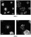
- FIGS. 4 to 6 are images of swelled cells before and after being compressed to cause chromosomal spreading
- Embodiments of the present disclosure provide for methods and systems for preparing chromosomal spread for a selected cell so that chromosomal spreads and/or translocations can be correlated with the selected cell.
- systems and methods can be used to prepare chromosomal spreads that can be correlated with the shape of the nucleus before mitosis.
- a cell can be selected and imaged using an imaging technique (e.g., a microscope) and then a shear and/or a compression force can be applied to the selected cell and the chromosomal spread can be imaged.
- an imaging technique e.g., a microscope
- a fluidic system can be used to transfer the chromosomal spread for additional analysis.
- shear and compression forces can be applied to mitotic, adherent cells, to obtain the chromosomal spread of the selected cell.
- embodiments of the present disclosure can be used to map the chromosomal content and chromosomal translocations onto nuclear and cell shapes and cell content (e.g., pre-mitotic nuclear and cell shapes and nuclear and cell content) for a single selected cell.
- the compression can be performed by moving a blunt vertical structure (e.g., a glass slide or a cylinder) into a substrate (e.g., a dish) with cultured cells arrested in mitosis between the structure and the substrate.
- the substrate can be imaged from the bottom with a 60 ⁇ objective on a Nikon epifluorescence microscope, for example, before and after application of the compression and/or shear forces.
- the cylinder is first centered in the field of view. By compressing down with the cylinder on top of cell while observing cell simultaneously on the microscope, it is possible to image chromosomes before and after compression of the optionally osmotically swelled cell.
- the substrate can include a fluidic or microfluidic channel that can include the selected cell or the substrate can be in fluidic communication with the chromosomal spread, so that the chromosomal spread can be flowed from the substrate and further analyzed.
- the fluidic system can be used to flow a fluid before and/or during compression, which may further enhance chromosomal spreading by applying another shearing force.
- the substrate can be part of a fluidic system that can be interfaced with an analysis system.
- subject refers to any individual who is the target of administration or treatment.
- the subject can be a vertebrate, for example, a mammal.
- the subject can be a human or veterinary patient.
- patient refers to a subject under the treatment of a clinician, e.g., physician.
- treatment refers to the medical management of a patient with the intent to cure, ameliorate, stabilize, or prevent a disease, pathological condition, or disorder.
- This term includes active treatment, that is, treatment directed specifically toward the improvement of a disease, pathological condition, or disorder, and also includes causal treatment, that is, treatment directed toward removal of the cause of the associated disease, pathological condition, or disorder.
- this term includes palliative treatment, that is, treatment designed for the relief of symptoms rather than the curing of the disease, pathological condition, or disorder; preventative treatment, that is, treatment directed to minimizing or partially or completely inhibiting the development of the associated disease, pathological condition, or disorder; and supportive treatment, that is, treatment employed to supplement another specific therapy directed toward the improvement of the associated disease, pathological condition, or disorder.
- FIGS. 1 to 6 illustrate the spread chromosomes in unswelled and osmotically swollen (e.g., with water or other fluid that osmotically swells) cells.
- the method and system include both the use of hydrodynamic shear and compression to generate the chromosomal spread of a selected cell.
Landscapes
- Chemical & Material Sciences (AREA)
- Engineering & Computer Science (AREA)
- Health & Medical Sciences (AREA)
- Life Sciences & Earth Sciences (AREA)
- Organic Chemistry (AREA)
- Zoology (AREA)
- Wood Science & Technology (AREA)
- General Health & Medical Sciences (AREA)
- Molecular Biology (AREA)
- Genetics & Genomics (AREA)
- Physics & Mathematics (AREA)
- Biotechnology (AREA)
- Biomedical Technology (AREA)
- Biochemistry (AREA)
- Analytical Chemistry (AREA)
- General Engineering & Computer Science (AREA)
- Bioinformatics & Cheminformatics (AREA)
- Microbiology (AREA)
- Proteomics, Peptides & Aminoacids (AREA)
- Immunology (AREA)
- Biophysics (AREA)
- Chemical Kinetics & Catalysis (AREA)
- Hematology (AREA)
- General Physics & Mathematics (AREA)
- Urology & Nephrology (AREA)
- Medicinal Chemistry (AREA)
- Computer Vision & Pattern Recognition (AREA)
- Nuclear Medicine, Radiotherapy & Molecular Imaging (AREA)
- Theoretical Computer Science (AREA)
- Radiology & Medical Imaging (AREA)
- Medical Informatics (AREA)
- Quality & Reliability (AREA)
- Crystallography & Structural Chemistry (AREA)
- Plant Pathology (AREA)
- Clinical Laboratory Science (AREA)
- Cell Biology (AREA)
- Pathology (AREA)
- Dispersion Chemistry (AREA)
- Food Science & Technology (AREA)
- General Chemical & Material Sciences (AREA)
Abstract
Description
Claims (12)
Priority Applications (2)
| Application Number | Priority Date | Filing Date | Title |
|---|---|---|---|
| US15/789,414 US10515450B2 (en) | 2016-10-20 | 2017-10-20 | Systems and methods for using a single-cell to create chromosomal spreads |
| US16/679,689 US10957040B2 (en) | 2016-10-20 | 2019-11-11 | Systems and methods for using a single-cell to create chromosomal spreads |
Applications Claiming Priority (2)
| Application Number | Priority Date | Filing Date | Title |
|---|---|---|---|
| US201662410553P | 2016-10-20 | 2016-10-20 | |
| US15/789,414 US10515450B2 (en) | 2016-10-20 | 2017-10-20 | Systems and methods for using a single-cell to create chromosomal spreads |
Related Child Applications (1)
| Application Number | Title | Priority Date | Filing Date |
|---|---|---|---|
| US16/679,689 Continuation US10957040B2 (en) | 2016-10-20 | 2019-11-11 | Systems and methods for using a single-cell to create chromosomal spreads |
Publications (3)
| Publication Number | Publication Date |
|---|---|
| US20180114316A1 US20180114316A1 (en) | 2018-04-26 |
| US20190073766A2 US20190073766A2 (en) | 2019-03-07 |
| US10515450B2 true US10515450B2 (en) | 2019-12-24 |
Family
ID=61970256
Family Applications (2)
| Application Number | Title | Priority Date | Filing Date |
|---|---|---|---|
| US15/789,414 Expired - Fee Related US10515450B2 (en) | 2016-10-20 | 2017-10-20 | Systems and methods for using a single-cell to create chromosomal spreads |
| US16/679,689 Active US10957040B2 (en) | 2016-10-20 | 2019-11-11 | Systems and methods for using a single-cell to create chromosomal spreads |
Family Applications After (1)
| Application Number | Title | Priority Date | Filing Date |
|---|---|---|---|
| US16/679,689 Active US10957040B2 (en) | 2016-10-20 | 2019-11-11 | Systems and methods for using a single-cell to create chromosomal spreads |
Country Status (1)
| Country | Link |
|---|---|
| US (2) | US10515450B2 (en) |
Cited By (1)
| Publication number | Priority date | Publication date | Assignee | Title |
|---|---|---|---|---|
| US10957040B2 (en) * | 2016-10-20 | 2021-03-23 | University Of Florida Research Foundation, Inc. | Systems and methods for using a single-cell to create chromosomal spreads |
Families Citing this family (7)
| Publication number | Priority date | Publication date | Assignee | Title |
|---|---|---|---|---|
| SG11201707515SA (en) | 2015-04-10 | 2017-10-30 | Spatial Transcriptomics Ab | Spatially distinguished, multiplex nucleic acid analysis of biological specimens |
| EP3864173A4 (en) * | 2018-10-10 | 2022-07-20 | Readcoor, LLC | Surface capture of targets |
| US11702693B2 (en) | 2020-01-21 | 2023-07-18 | 10X Genomics, Inc. | Methods for printing cells and generating arrays of barcoded cells |
| US11732299B2 (en) | 2020-01-21 | 2023-08-22 | 10X Genomics, Inc. | Spatial assays with perturbed cells |
| US11835462B2 (en) | 2020-02-11 | 2023-12-05 | 10X Genomics, Inc. | Methods and compositions for partitioning a biological sample |
| US11926863B1 (en) | 2020-02-27 | 2024-03-12 | 10X Genomics, Inc. | Solid state single cell method for analyzing fixed biological cells |
| EP4153775A1 (en) | 2020-05-22 | 2023-03-29 | 10X Genomics, Inc. | Simultaneous spatio-temporal measurement of gene expression and cellular activity |
Citations (3)
| Publication number | Priority date | Publication date | Assignee | Title |
|---|---|---|---|---|
| US20090048785A1 (en) * | 2006-01-10 | 2009-02-19 | Applied Spectral Imaging Ltd. | Methods And Systems For Analyzing Biological Samples |
| US20150010617A1 (en) * | 2012-02-01 | 2015-01-08 | Protalix Ltd. | Inhalable liquid formulations of dnase i |
| US20180114316A1 (en) * | 2016-10-20 | 2018-04-26 | University Of Florida Research Foundation, Inc. | Systems and methods for using a single-cell to create chromosomal spreads |
Family Cites Families (2)
| Publication number | Priority date | Publication date | Assignee | Title |
|---|---|---|---|---|
| US6716578B1 (en) * | 1999-03-08 | 2004-04-06 | Bioforce Nanosciences, Inc. | Method for solid state genome analysis |
| WO2018129226A1 (en) * | 2017-01-05 | 2018-07-12 | Virginia Commonwealth University | System, method, computer-accessible medium and apparatus for dna mapping |
-
2017
- 2017-10-20 US US15/789,414 patent/US10515450B2/en not_active Expired - Fee Related
-
2019
- 2019-11-11 US US16/679,689 patent/US10957040B2/en active Active
Patent Citations (4)
| Publication number | Priority date | Publication date | Assignee | Title |
|---|---|---|---|---|
| US20090048785A1 (en) * | 2006-01-10 | 2009-02-19 | Applied Spectral Imaging Ltd. | Methods And Systems For Analyzing Biological Samples |
| US9133506B2 (en) * | 2006-01-10 | 2015-09-15 | Applied Spectral Imaging Ltd. | Methods and systems for analyzing biological samples |
| US20150010617A1 (en) * | 2012-02-01 | 2015-01-08 | Protalix Ltd. | Inhalable liquid formulations of dnase i |
| US20180114316A1 (en) * | 2016-10-20 | 2018-04-26 | University Of Florida Research Foundation, Inc. | Systems and methods for using a single-cell to create chromosomal spreads |
Non-Patent Citations (1)
| Title |
|---|
| Paul Grayson, Lin Han, Tabita Winther, and Rob Phillips, "Real-time observations of single bacteriophage λ DNA ejections in vitro", PNAS Sep. 11, 2007 104 (37) 14652-14657; https://doi.org/10.1073/pnas.0703274104. (Year: 2007). * |
Cited By (1)
| Publication number | Priority date | Publication date | Assignee | Title |
|---|---|---|---|---|
| US10957040B2 (en) * | 2016-10-20 | 2021-03-23 | University Of Florida Research Foundation, Inc. | Systems and methods for using a single-cell to create chromosomal spreads |
Also Published As
| Publication number | Publication date |
|---|---|
| US20200082528A1 (en) | 2020-03-12 |
| US20190073766A2 (en) | 2019-03-07 |
| US10957040B2 (en) | 2021-03-23 |
| US20180114316A1 (en) | 2018-04-26 |
Similar Documents
| Publication | Publication Date | Title |
|---|---|---|
| US10957040B2 (en) | Systems and methods for using a single-cell to create chromosomal spreads | |
| Rappa et al. | Sperm processing for advanced reproductive technologies: Where are we today? | |
| Krause et al. | Probing the compressibility of tumor cell nuclei by combined atomic force–confocal microscopy | |
| JP6675355B2 (en) | Advanced methods and devices for cell analysis | |
| Yu et al. | Emerging technologies for home‐based semen analysis | |
| EP2948776B1 (en) | Methods, compositions, kits, and systems for selective enrichment of target cells | |
| Li et al. | Yield strength of human erythrocyte membranes to impulsive stretching | |
| US20230002737A1 (en) | Methods and Apparatuses for Patient-Derived Micro-Organospheres | |
| Susienka et al. | Quantifying the kinetics and morphological changes of the fusion of spheroid building blocks | |
| Cutrona et al. | A High‐Throughput Automated Confocal Microscopy Platform for Quantitative Phenotyping of Nanoparticle Uptake and Transport in Spheroids | |
| US20150140655A1 (en) | Apparatus and methods for sperm separation | |
| Lagalla et al. | A quantitative approach to blastocyst quality evaluation: morphometric analysis and related IVF outcomes | |
| US10732174B2 (en) | Three dimensional tissues for high-throughput assays | |
| Yildiz et al. | Use of microfluidic sperm extraction chips as an alternative method in patients with recurrent in vitro fertilisation failure | |
| US20210130882A1 (en) | Membrane Probes for Expansion Microscopy | |
| US20210285054A1 (en) | Precision drug screening for personalized cancer therapy | |
| Tarozzi et al. | Effect on sperm DNA quality following sperm selection for ART: new insights | |
| US20210011003A1 (en) | Component analysis device, drug component analysis device, component analysis method, and drug component analysis method | |
| WO2022119966A1 (en) | Precision drug screening for personalized cancer therapy | |
| Soares et al. | A cost-effective method for preparing, maintaining, and transfecting neurons in organotypic slices | |
| CN110959110B (en) | Method for immobilizing biological samples for analytical purposes | |
| Fichtman et al. | High-resolution imaging and analysis of individual nuclear pore complexes | |
| Simon et al. | Sperm selection techniques and their relevance to ART | |
| CA2834007A1 (en) | Apparatus and methods for sperm separation | |
| Agarwal et al. | Advanced sperm processing/selection techniques |
Legal Events
| Date | Code | Title | Description |
|---|---|---|---|
| FEPP | Fee payment procedure |
Free format text: ENTITY STATUS SET TO UNDISCOUNTED (ORIGINAL EVENT CODE: BIG.); ENTITY STATUS OF PATENT OWNER: SMALL ENTITY |
|
| FEPP | Fee payment procedure |
Free format text: ENTITY STATUS SET TO SMALL (ORIGINAL EVENT CODE: SMAL); ENTITY STATUS OF PATENT OWNER: SMALL ENTITY |
|
| AS | Assignment |
Owner name: UNIVERSITY OF FLORIDA RESEARCH FOUNDATION, INC., F Free format text: ASSIGNMENT OF ASSIGNORS INTEREST;ASSIGNORS:LELE, TANMAY P.;SAWYER, WALLACE GREGORY;SIGNING DATES FROM 20171113 TO 20171115;REEL/FRAME:044162/0168 |
|
| STPP | Information on status: patent application and granting procedure in general |
Free format text: NON FINAL ACTION MAILED |
|
| STPP | Information on status: patent application and granting procedure in general |
Free format text: RESPONSE TO NON-FINAL OFFICE ACTION ENTERED AND FORWARDED TO EXAMINER |
|
| STPP | Information on status: patent application and granting procedure in general |
Free format text: FINAL REJECTION MAILED |
|
| STPP | Information on status: patent application and granting procedure in general |
Free format text: NOTICE OF ALLOWANCE MAILED -- APPLICATION RECEIVED IN OFFICE OF PUBLICATIONS |
|
| STPP | Information on status: patent application and granting procedure in general |
Free format text: PUBLICATIONS -- ISSUE FEE PAYMENT VERIFIED |
|
| STCF | Information on status: patent grant |
Free format text: PATENTED CASE |
|
| FEPP | Fee payment procedure |
Free format text: MAINTENANCE FEE REMINDER MAILED (ORIGINAL EVENT CODE: REM.); ENTITY STATUS OF PATENT OWNER: SMALL ENTITY |
|
| LAPS | Lapse for failure to pay maintenance fees |
Free format text: PATENT EXPIRED FOR FAILURE TO PAY MAINTENANCE FEES (ORIGINAL EVENT CODE: EXP.); ENTITY STATUS OF PATENT OWNER: SMALL ENTITY |
|
| STCH | Information on status: patent discontinuation |
Free format text: PATENT EXPIRED DUE TO NONPAYMENT OF MAINTENANCE FEES UNDER 37 CFR 1.362 |
|
| FP | Lapsed due to failure to pay maintenance fee |
Effective date: 20231224 |