US10398820B2 - Use and monitoring of inhaled nitric oxide with left ventricular assist devices - Google Patents
Use and monitoring of inhaled nitric oxide with left ventricular assist devices Download PDFInfo
- Publication number
- US10398820B2 US10398820B2 US15/418,837 US201715418837A US10398820B2 US 10398820 B2 US10398820 B2 US 10398820B2 US 201715418837 A US201715418837 A US 201715418837A US 10398820 B2 US10398820 B2 US 10398820B2
- Authority
- US
- United States
- Prior art keywords
- lvad
- ppm
- patient
- inhaled
- pulmonary
- Prior art date
- Legal status (The legal status is an assumption and is not a legal conclusion. Google has not performed a legal analysis and makes no representation as to the accuracy of the status listed.)
- Active, expires
Links
- MWUXSHHQAYIFBG-UHFFFAOYSA-N Nitric oxide Chemical compound O=[N] MWUXSHHQAYIFBG-UHFFFAOYSA-N 0.000 title claims abstract description 823
- 230000002861 ventricular Effects 0.000 title claims abstract description 81
- 238000012544 monitoring process Methods 0.000 title claims abstract description 21
- 238000000034 method Methods 0.000 claims abstract description 124
- 230000000004 hemodynamic effect Effects 0.000 claims description 127
- 230000002685 pulmonary effect Effects 0.000 claims description 103
- 210000005240 left ventricle Anatomy 0.000 claims description 62
- 230000000747 cardiac effect Effects 0.000 claims description 19
- 238000011282 treatment Methods 0.000 claims description 15
- 210000001765 aortic valve Anatomy 0.000 claims description 14
- 230000001746 atrial effect Effects 0.000 claims description 6
- 238000012384 transportation and delivery Methods 0.000 abstract description 130
- 208000002815 pulmonary hypertension Diseases 0.000 description 50
- 239000007789 gas Substances 0.000 description 48
- 206010039163 Right ventricular failure Diseases 0.000 description 41
- 238000002513 implantation Methods 0.000 description 30
- 230000006854 communication Effects 0.000 description 29
- 238000004891 communication Methods 0.000 description 29
- 206010019280 Heart failures Diseases 0.000 description 28
- 206010007559 Cardiac failure congestive Diseases 0.000 description 26
- 230000006870 function Effects 0.000 description 25
- 230000036593 pulmonary vascular resistance Effects 0.000 description 21
- 210000005241 right ventricle Anatomy 0.000 description 21
- 230000008753 endothelial function Effects 0.000 description 20
- 230000001404 mediated effect Effects 0.000 description 18
- JCXJVPUVTGWSNB-UHFFFAOYSA-N nitrogen dioxide Inorganic materials O=[N]=O JCXJVPUVTGWSNB-UHFFFAOYSA-N 0.000 description 18
- 230000036316 preload Effects 0.000 description 16
- 230000002411 adverse Effects 0.000 description 15
- 230000007423 decrease Effects 0.000 description 14
- 230000001154 acute effect Effects 0.000 description 13
- 230000002093 peripheral effect Effects 0.000 description 13
- 210000001147 pulmonary artery Anatomy 0.000 description 13
- 230000010412 perfusion Effects 0.000 description 12
- 230000002829 reductive effect Effects 0.000 description 12
- 230000029058 respiratory gaseous exchange Effects 0.000 description 12
- 230000001225 therapeutic effect Effects 0.000 description 12
- 229940124549 vasodilator Drugs 0.000 description 12
- 239000003071 vasodilator agent Substances 0.000 description 12
- 230000000694 effects Effects 0.000 description 11
- 239000012530 fluid Substances 0.000 description 11
- 230000009885 systemic effect Effects 0.000 description 11
- 208000008166 Right Ventricular Dysfunction Diseases 0.000 description 10
- 230000004872 arterial blood pressure Effects 0.000 description 10
- 239000000203 mixture Substances 0.000 description 10
- 238000011084 recovery Methods 0.000 description 10
- 230000006814 right ventricular dysfunction Effects 0.000 description 10
- 230000003993 interaction Effects 0.000 description 9
- 238000002560 therapeutic procedure Methods 0.000 description 9
- MGWGWNFMUOTEHG-UHFFFAOYSA-N 4-(3,5-dimethylphenyl)-1,3-thiazol-2-amine Chemical compound CC1=CC(C)=CC(C=2N=C(N)SC=2)=C1 MGWGWNFMUOTEHG-UHFFFAOYSA-N 0.000 description 8
- IJGRMHOSHXDMSA-UHFFFAOYSA-N Atomic nitrogen Chemical compound N#N IJGRMHOSHXDMSA-UHFFFAOYSA-N 0.000 description 8
- 230000008859 change Effects 0.000 description 8
- 230000001771 impaired effect Effects 0.000 description 8
- 229940042110 inomax Drugs 0.000 description 8
- 230000002980 postoperative effect Effects 0.000 description 8
- 230000000541 pulsatile effect Effects 0.000 description 8
- 238000010967 transthoracic echocardiography Methods 0.000 description 8
- 230000002792 vascular Effects 0.000 description 8
- 230000002612 cardiopulmonary effect Effects 0.000 description 7
- 229940079593 drug Drugs 0.000 description 7
- 239000003814 drug Substances 0.000 description 7
- 230000000297 inotrophic effect Effects 0.000 description 7
- 238000012545 processing Methods 0.000 description 7
- 230000004044 response Effects 0.000 description 7
- 230000002457 bidirectional effect Effects 0.000 description 6
- 210000004369 blood Anatomy 0.000 description 6
- 239000008280 blood Substances 0.000 description 6
- 230000017531 blood circulation Effects 0.000 description 6
- 230000037396 body weight Effects 0.000 description 6
- 230000001684 chronic effect Effects 0.000 description 6
- 238000013461 design Methods 0.000 description 6
- 230000006872 improvement Effects 0.000 description 6
- 238000007726 management method Methods 0.000 description 6
- 238000005457 optimization Methods 0.000 description 6
- 230000000144 pharmacologic effect Effects 0.000 description 6
- 230000009467 reduction Effects 0.000 description 6
- 238000012360 testing method Methods 0.000 description 6
- 210000005166 vasculature Anatomy 0.000 description 6
- 206010049694 Left Ventricular Dysfunction Diseases 0.000 description 5
- IOVCWXUNBOPUCH-UHFFFAOYSA-M Nitrite anion Chemical compound [O-]N=O IOVCWXUNBOPUCH-UHFFFAOYSA-M 0.000 description 5
- 208000007536 Thrombosis Diseases 0.000 description 5
- QVGXLLKOCUKJST-UHFFFAOYSA-N atomic oxygen Chemical compound [O] QVGXLLKOCUKJST-UHFFFAOYSA-N 0.000 description 5
- 230000010455 autoregulation Effects 0.000 description 5
- 238000011161 development Methods 0.000 description 5
- 230000010339 dilation Effects 0.000 description 5
- XEYBHCRIKKKOSS-UHFFFAOYSA-N disodium;azanylidyneoxidanium;iron(2+);pentacyanide Chemical compound [Na+].[Na+].[Fe+2].N#[C-].N#[C-].N#[C-].N#[C-].N#[C-].[O+]#N XEYBHCRIKKKOSS-UHFFFAOYSA-N 0.000 description 5
- 230000003434 inspiratory effect Effects 0.000 description 5
- 238000001990 intravenous administration Methods 0.000 description 5
- 230000000302 ischemic effect Effects 0.000 description 5
- 238000002595 magnetic resonance imaging Methods 0.000 description 5
- 239000001301 oxygen Substances 0.000 description 5
- 229910052760 oxygen Inorganic materials 0.000 description 5
- 230000009726 pulmonary vascular congestion Effects 0.000 description 5
- 230000002441 reversible effect Effects 0.000 description 5
- 229940083618 sodium nitroprusside Drugs 0.000 description 5
- 230000004218 vascular function Effects 0.000 description 5
- CIWBSHSKHKDKBQ-JLAZNSOCSA-N Ascorbic acid Chemical compound OC[C@H](O)[C@H]1OC(=O)C(O)=C1O CIWBSHSKHKDKBQ-JLAZNSOCSA-N 0.000 description 4
- NHNBFGGVMKEFGY-UHFFFAOYSA-N Nitrate Chemical compound [O-][N+]([O-])=O NHNBFGGVMKEFGY-UHFFFAOYSA-N 0.000 description 4
- 230000001042 autoregulative effect Effects 0.000 description 4
- 230000033228 biological regulation Effects 0.000 description 4
- 239000000090 biomarker Substances 0.000 description 4
- 239000003795 chemical substances by application Substances 0.000 description 4
- 230000004087 circulation Effects 0.000 description 4
- 230000003205 diastolic effect Effects 0.000 description 4
- 229960001123 epoprostenol Drugs 0.000 description 4
- KAQKFAOMNZTLHT-VVUHWYTRSA-N epoprostenol Chemical compound O1C(=CCCCC(O)=O)C[C@@H]2[C@@H](/C=C/[C@@H](O)CCCCC)[C@H](O)C[C@@H]21 KAQKFAOMNZTLHT-VVUHWYTRSA-N 0.000 description 4
- 230000007774 longterm Effects 0.000 description 4
- 230000007246 mechanism Effects 0.000 description 4
- 230000004048 modification Effects 0.000 description 4
- 238000012986 modification Methods 0.000 description 4
- 229910052757 nitrogen Inorganic materials 0.000 description 4
- 230000004088 pulmonary circulation Effects 0.000 description 4
- BNRNXUUZRGQAQC-UHFFFAOYSA-N sildenafil Chemical compound CCCC1=NN(C)C(C(N2)=O)=C1N=C2C(C(=CC=1)OCC)=CC=1S(=O)(=O)N1CCN(C)CC1 BNRNXUUZRGQAQC-UHFFFAOYSA-N 0.000 description 4
- 230000003068 static effect Effects 0.000 description 4
- 230000003319 supportive effect Effects 0.000 description 4
- 210000001519 tissue Anatomy 0.000 description 4
- 208000012671 Gastrointestinal haemorrhages Diseases 0.000 description 3
- SNIOPGDIGTZGOP-UHFFFAOYSA-N Nitroglycerin Chemical compound [O-][N+](=O)OCC(O[N+]([O-])=O)CO[N+]([O-])=O SNIOPGDIGTZGOP-UHFFFAOYSA-N 0.000 description 3
- 239000000006 Nitroglycerin Substances 0.000 description 3
- 238000011360 adjunctive therapy Methods 0.000 description 3
- 238000013459 approach Methods 0.000 description 3
- 230000007175 bidirectional communication Effects 0.000 description 3
- 230000006837 decompression Effects 0.000 description 3
- 230000001419 dependent effect Effects 0.000 description 3
- 201000010099 disease Diseases 0.000 description 3
- 208000037265 diseases, disorders, signs and symptoms Diseases 0.000 description 3
- 239000002934 diuretic Substances 0.000 description 3
- 229940030606 diuretics Drugs 0.000 description 3
- 238000002592 echocardiography Methods 0.000 description 3
- 208000030304 gastrointestinal bleeding Diseases 0.000 description 3
- 229960003711 glyceryl trinitrate Drugs 0.000 description 3
- 230000036541 health Effects 0.000 description 3
- 230000000544 hyperemic effect Effects 0.000 description 3
- 238000003384 imaging method Methods 0.000 description 3
- 230000001976 improved effect Effects 0.000 description 3
- 239000004041 inotropic agent Substances 0.000 description 3
- 230000036581 peripheral resistance Effects 0.000 description 3
- 238000011422 pharmacological therapy Methods 0.000 description 3
- 230000036387 respiratory rate Effects 0.000 description 3
- 230000035882 stress Effects 0.000 description 3
- 230000035488 systolic blood pressure Effects 0.000 description 3
- 239000003826 tablet Substances 0.000 description 3
- 238000012546 transfer Methods 0.000 description 3
- 238000002054 transplantation Methods 0.000 description 3
- 208000022211 Arteriovenous Malformations Diseases 0.000 description 2
- JRWZLRBJNMZMFE-UHFFFAOYSA-N Dobutamine Chemical compound C=1C=C(O)C(O)=CC=1CCNC(C)CCC1=CC=C(O)C=C1 JRWZLRBJNMZMFE-UHFFFAOYSA-N 0.000 description 2
- 206010048554 Endothelial dysfunction Diseases 0.000 description 2
- 206010016803 Fluid overload Diseases 0.000 description 2
- 206010018910 Haemolysis Diseases 0.000 description 2
- 108010054147 Hemoglobins Proteins 0.000 description 2
- 102000001554 Hemoglobins Human genes 0.000 description 2
- 208000001953 Hypotension Diseases 0.000 description 2
- 102000008299 Nitric Oxide Synthase Human genes 0.000 description 2
- 108010021487 Nitric Oxide Synthase Proteins 0.000 description 2
- 206010064911 Pulmonary arterial hypertension Diseases 0.000 description 2
- 108010068048 S-nitrosohemoglobin Proteins 0.000 description 2
- 208000032594 Vascular Remodeling Diseases 0.000 description 2
- 239000008186 active pharmaceutical agent Substances 0.000 description 2
- OIRDTQYFTABQOQ-KQYNXXCUSA-N adenosine Chemical compound C1=NC=2C(N)=NC=NC=2N1[C@@H]1O[C@H](CO)[C@@H](O)[C@H]1O OIRDTQYFTABQOQ-KQYNXXCUSA-N 0.000 description 2
- 210000003484 anatomy Anatomy 0.000 description 2
- 201000002064 aortic valve insufficiency Diseases 0.000 description 2
- 230000005744 arteriovenous malformation Effects 0.000 description 2
- 229960005070 ascorbic acid Drugs 0.000 description 2
- 235000010323 ascorbic acid Nutrition 0.000 description 2
- 239000011668 ascorbic acid Substances 0.000 description 2
- 230000003190 augmentative effect Effects 0.000 description 2
- 230000009286 beneficial effect Effects 0.000 description 2
- 210000002302 brachial artery Anatomy 0.000 description 2
- 238000006243 chemical reaction Methods 0.000 description 2
- 239000003638 chemical reducing agent Substances 0.000 description 2
- 230000006378 damage Effects 0.000 description 2
- 230000006735 deficit Effects 0.000 description 2
- 238000009111 destination therapy Methods 0.000 description 2
- 238000010586 diagram Methods 0.000 description 2
- 238000007865 diluting Methods 0.000 description 2
- 230000003292 diminished effect Effects 0.000 description 2
- 229960001089 dobutamine Drugs 0.000 description 2
- VYFYYTLLBUKUHU-UHFFFAOYSA-N dopamine Chemical compound NCCC1=CC=C(O)C(O)=C1 VYFYYTLLBUKUHU-UHFFFAOYSA-N 0.000 description 2
- 230000008694 endothelial dysfunction Effects 0.000 description 2
- 230000003090 exacerbative effect Effects 0.000 description 2
- 230000002349 favourable effect Effects 0.000 description 2
- 208000019622 heart disease Diseases 0.000 description 2
- 230000008588 hemolysis Effects 0.000 description 2
- 230000036543 hypotension Effects 0.000 description 2
- 208000028867 ischemia Diseases 0.000 description 2
- 230000000670 limiting effect Effects 0.000 description 2
- 210000004072 lung Anatomy 0.000 description 2
- 230000007257 malfunction Effects 0.000 description 2
- 238000004519 manufacturing process Methods 0.000 description 2
- 239000000463 material Substances 0.000 description 2
- 238000002483 medication Methods 0.000 description 2
- 238000002156 mixing Methods 0.000 description 2
- 230000003287 optical effect Effects 0.000 description 2
- 210000000056 organ Anatomy 0.000 description 2
- 230000008520 organization Effects 0.000 description 2
- 238000006213 oxygenation reaction Methods 0.000 description 2
- 230000000737 periodic effect Effects 0.000 description 2
- 230000008569 process Effects 0.000 description 2
- 238000004393 prognosis Methods 0.000 description 2
- 230000000750 progressive effect Effects 0.000 description 2
- GMVPRGQOIOIIMI-DWKJAMRDSA-N prostaglandin E1 Chemical compound CCCCC[C@H](O)\C=C\[C@H]1[C@H](O)CC(=O)[C@@H]1CCCCCCC(O)=O GMVPRGQOIOIIMI-DWKJAMRDSA-N 0.000 description 2
- 230000008695 pulmonary vasoconstriction Effects 0.000 description 2
- 230000035485 pulse pressure Effects 0.000 description 2
- 238000010926 purge Methods 0.000 description 2
- 229960003310 sildenafil Drugs 0.000 description 2
- 230000003238 somatosensory effect Effects 0.000 description 2
- 230000004083 survival effect Effects 0.000 description 2
- 230000001839 systemic circulation Effects 0.000 description 2
- 238000013175 transesophageal echocardiography Methods 0.000 description 2
- 230000006438 vascular health Effects 0.000 description 2
- 230000035899 viability Effects 0.000 description 2
- 230000000007 visual effect Effects 0.000 description 2
- 230000003442 weekly effect Effects 0.000 description 2
- GMVPRGQOIOIIMI-UHFFFAOYSA-N (8R,11R,12R,13E,15S)-11,15-Dihydroxy-9-oxo-13-prostenoic acid Natural products CCCCCC(O)C=CC1C(O)CC(=O)C1CCCCCCC(O)=O GMVPRGQOIOIIMI-UHFFFAOYSA-N 0.000 description 1
- UCTWMZQNUQWSLP-VIFPVBQESA-N (R)-adrenaline Chemical compound CNC[C@H](O)C1=CC=C(O)C(O)=C1 UCTWMZQNUQWSLP-VIFPVBQESA-N 0.000 description 1
- 229930182837 (R)-adrenaline Natural products 0.000 description 1
- 206010001029 Acute pulmonary oedema Diseases 0.000 description 1
- 206010067484 Adverse reaction Diseases 0.000 description 1
- 238000012935 Averaging Methods 0.000 description 1
- 108010017384 Blood Proteins Proteins 0.000 description 1
- 102000004506 Blood Proteins Human genes 0.000 description 1
- 101800000407 Brain natriuretic peptide 32 Proteins 0.000 description 1
- 239000002126 C01EB10 - Adenosine Substances 0.000 description 1
- 208000024172 Cardiovascular disease Diseases 0.000 description 1
- CWYNVVGOOAEACU-UHFFFAOYSA-N Fe2+ Chemical compound [Fe+2] CWYNVVGOOAEACU-UHFFFAOYSA-N 0.000 description 1
- 229910002547 FeII Inorganic materials 0.000 description 1
- 208000036119 Frailty Diseases 0.000 description 1
- 206010021143 Hypoxia Diseases 0.000 description 1
- 206010024119 Left ventricular failure Diseases 0.000 description 1
- 244000062730 Melissa officinalis Species 0.000 description 1
- 235000010654 Melissa officinalis Nutrition 0.000 description 1
- -1 NO2 − Chemical class 0.000 description 1
- 229910002651 NO3 Inorganic materials 0.000 description 1
- 206010071229 Procedural haemorrhage Diseases 0.000 description 1
- 206010063837 Reperfusion injury Diseases 0.000 description 1
- 208000004756 Respiratory Insufficiency Diseases 0.000 description 1
- 241001661807 Systole Species 0.000 description 1
- 208000027418 Wounds and injury Diseases 0.000 description 1
- 230000002378 acidificating effect Effects 0.000 description 1
- 230000006978 adaptation Effects 0.000 description 1
- 229960005305 adenosine Drugs 0.000 description 1
- 230000006838 adverse reaction Effects 0.000 description 1
- 229960000711 alprostadil Drugs 0.000 description 1
- 230000004075 alteration Effects 0.000 description 1
- 230000003466 anti-cipated effect Effects 0.000 description 1
- 239000003963 antioxidant agent Substances 0.000 description 1
- 230000003078 antioxidant effect Effects 0.000 description 1
- 235000006708 antioxidants Nutrition 0.000 description 1
- 210000000709 aorta Anatomy 0.000 description 1
- 230000006793 arrhythmia Effects 0.000 description 1
- 206010003119 arrhythmia Diseases 0.000 description 1
- 206010003549 asthenia Diseases 0.000 description 1
- 230000008901 benefit Effects 0.000 description 1
- 239000010836 blood and blood product Substances 0.000 description 1
- 230000036772 blood pressure Effects 0.000 description 1
- 229940125691 blood product Drugs 0.000 description 1
- 238000002554 cardiac rehabilitation Methods 0.000 description 1
- 230000009084 cardiovascular function Effects 0.000 description 1
- 210000000748 cardiovascular system Anatomy 0.000 description 1
- 230000015556 catabolic process Effects 0.000 description 1
- 210000004027 cell Anatomy 0.000 description 1
- 230000037326 chronic stress Effects 0.000 description 1
- 239000000084 colloidal system Substances 0.000 description 1
- 230000001447 compensatory effect Effects 0.000 description 1
- 230000000295 complement effect Effects 0.000 description 1
- 150000001875 compounds Chemical class 0.000 description 1
- 239000000356 contaminant Substances 0.000 description 1
- 230000008602 contraction Effects 0.000 description 1
- 230000001276 controlling effect Effects 0.000 description 1
- 208000029078 coronary artery disease Diseases 0.000 description 1
- 210000004351 coronary vessel Anatomy 0.000 description 1
- 230000002596 correlated effect Effects 0.000 description 1
- 230000000875 corresponding effect Effects 0.000 description 1
- 230000001351 cycling effect Effects 0.000 description 1
- XUJNEKJLAYXESH-UHFFFAOYSA-N cysteine Natural products SCC(N)C(O)=O XUJNEKJLAYXESH-UHFFFAOYSA-N 0.000 description 1
- 235000018417 cysteine Nutrition 0.000 description 1
- 230000003247 decreasing effect Effects 0.000 description 1
- 230000007812 deficiency Effects 0.000 description 1
- 230000035487 diastolic blood pressure Effects 0.000 description 1
- 230000004069 differentiation Effects 0.000 description 1
- 230000000916 dilatatory effect Effects 0.000 description 1
- 239000012895 dilution Substances 0.000 description 1
- 238000010790 dilution Methods 0.000 description 1
- 238000006073 displacement reaction Methods 0.000 description 1
- 229960003638 dopamine Drugs 0.000 description 1
- 231100000673 dose–response relationship Toxicity 0.000 description 1
- 229940126534 drug product Drugs 0.000 description 1
- 230000008846 dynamic interplay Effects 0.000 description 1
- 230000004064 dysfunction Effects 0.000 description 1
- 230000002526 effect on cardiovascular system Effects 0.000 description 1
- 230000003511 endothelial effect Effects 0.000 description 1
- 238000005516 engineering process Methods 0.000 description 1
- 229960005139 epinephrine Drugs 0.000 description 1
- 210000003743 erythrocyte Anatomy 0.000 description 1
- 230000001747 exhibiting effect Effects 0.000 description 1
- 238000002618 extracorporeal membrane oxygenation Methods 0.000 description 1
- 238000002637 fluid replacement therapy Methods 0.000 description 1
- 102000034356 gene-regulatory proteins Human genes 0.000 description 1
- 108091006104 gene-regulatory proteins Proteins 0.000 description 1
- 229910000078 germane Inorganic materials 0.000 description 1
- 239000001307 helium Substances 0.000 description 1
- 229910052734 helium Inorganic materials 0.000 description 1
- SWQJXJOGLNCZEY-UHFFFAOYSA-N helium atom Chemical compound [He] SWQJXJOGLNCZEY-UHFFFAOYSA-N 0.000 description 1
- 230000002440 hepatic effect Effects 0.000 description 1
- 230000001146 hypoxic effect Effects 0.000 description 1
- 239000007943 implant Substances 0.000 description 1
- 238000010348 incorporation Methods 0.000 description 1
- 239000011261 inert gas Substances 0.000 description 1
- 238000001802 infusion Methods 0.000 description 1
- 230000000977 initiatory effect Effects 0.000 description 1
- 208000014674 injury Diseases 0.000 description 1
- 230000010354 integration Effects 0.000 description 1
- 208000012947 ischemia reperfusion injury Diseases 0.000 description 1
- 239000000865 liniment Substances 0.000 description 1
- 239000007788 liquid Substances 0.000 description 1
- 238000005259 measurement Methods 0.000 description 1
- 208000005135 methemoglobinemia Diseases 0.000 description 1
- 230000004089 microcirculation Effects 0.000 description 1
- 210000004925 microvascular endothelial cell Anatomy 0.000 description 1
- PZRHRDRVRGEVNW-UHFFFAOYSA-N milrinone Chemical compound N1C(=O)C(C#N)=CC(C=2C=CN=CC=2)=C1C PZRHRDRVRGEVNW-UHFFFAOYSA-N 0.000 description 1
- 229960003574 milrinone Drugs 0.000 description 1
- 230000002107 myocardial effect Effects 0.000 description 1
- 210000004165 myocardium Anatomy 0.000 description 1
- HPNRHPKXQZSDFX-OAQDCNSJSA-N nesiritide Chemical compound C([C@H]1C(=O)NCC(=O)N[C@@H](CCCNC(N)=N)C(=O)N[C@@H](CCCCN)C(=O)N[C@@H](CCSC)C(=O)N[C@@H](CC(O)=O)C(=O)N[C@@H](CCCNC(N)=N)C(=O)N[C@H](C(N[C@@H](CO)C(=O)N[C@@H](CO)C(=O)N[C@@H](CO)C(=O)N[C@@H](CO)C(=O)NCC(=O)N[C@@H](CC(C)C)C(=O)NCC(=O)N[C@@H](CSSC[C@@H](C(=O)N1)NC(=O)CNC(=O)[C@H](CO)NC(=O)CNC(=O)[C@H](CCC(N)=O)NC(=O)[C@@H](NC(=O)[C@H](CCSC)NC(=O)[C@H](CCCCN)NC(=O)[C@H]1N(CCC1)C(=O)[C@@H](N)CO)C(C)C)C(=O)N[C@@H](CCCCN)C(=O)N[C@@H](C(C)C)C(=O)N[C@@H](CC(C)C)C(=O)N[C@@H](CCCNC(N)=N)C(=O)N[C@@H](CCCNC(N)=N)C(=O)N[C@@H](CC=1N=CNC=1)C(O)=O)=O)[C@@H](C)CC)C1=CC=CC=C1 HPNRHPKXQZSDFX-OAQDCNSJSA-N 0.000 description 1
- 229960001267 nesiritide Drugs 0.000 description 1
- 230000009635 nitrosylation Effects 0.000 description 1
- 230000003647 oxidation Effects 0.000 description 1
- 238000007254 oxidation reaction Methods 0.000 description 1
- 230000036542 oxidative stress Effects 0.000 description 1
- 230000007310 pathophysiology Effects 0.000 description 1
- 238000013146 percutaneous coronary intervention Methods 0.000 description 1
- 208000030613 peripheral artery disease Diseases 0.000 description 1
- 230000003836 peripheral circulation Effects 0.000 description 1
- 230000002688 persistence Effects 0.000 description 1
- 230000002085 persistent effect Effects 0.000 description 1
- 239000000825 pharmaceutical preparation Substances 0.000 description 1
- 238000001050 pharmacotherapy Methods 0.000 description 1
- 230000010118 platelet activation Effects 0.000 description 1
- 238000010837 poor prognosis Methods 0.000 description 1
- 239000002244 precipitate Substances 0.000 description 1
- 230000001376 precipitating effect Effects 0.000 description 1
- 239000002243 precursor Substances 0.000 description 1
- 230000035935 pregnancy Effects 0.000 description 1
- 238000004321 preservation Methods 0.000 description 1
- 230000002035 prolonged effect Effects 0.000 description 1
- 230000001737 promoting effect Effects 0.000 description 1
- 150000003815 prostacyclins Chemical class 0.000 description 1
- XEYBRNLFEZDVAW-UHFFFAOYSA-N prostaglandin E2 Natural products CCCCCC(O)C=CC1C(O)CC(=O)C1CC=CCCCC(O)=O XEYBRNLFEZDVAW-UHFFFAOYSA-N 0.000 description 1
- 229940127293 prostanoid Drugs 0.000 description 1
- 150000003814 prostanoids Chemical class 0.000 description 1
- 230000010349 pulsation Effects 0.000 description 1
- 238000009877 rendering Methods 0.000 description 1
- 230000000241 respiratory effect Effects 0.000 description 1
- 201000004193 respiratory failure Diseases 0.000 description 1
- 238000005070 sampling Methods 0.000 description 1
- 239000000243 solution Substances 0.000 description 1
- 239000000126 substance Substances 0.000 description 1
- 230000008961 swelling Effects 0.000 description 1
- 208000024891 symptom Diseases 0.000 description 1
- 230000001360 synchronised effect Effects 0.000 description 1
- 230000002195 synergetic effect Effects 0.000 description 1
- 230000004873 systolic arterial blood pressure Effects 0.000 description 1
- 230000008685 targeting Effects 0.000 description 1
- 238000010998 test method Methods 0.000 description 1
- 230000001256 tonic effect Effects 0.000 description 1
- 238000011269 treatment regimen Methods 0.000 description 1
- 238000013024 troubleshooting Methods 0.000 description 1
- 238000002604 ultrasonography Methods 0.000 description 1
- 238000011144 upstream manufacturing Methods 0.000 description 1
- 230000006492 vascular dysfunction Effects 0.000 description 1
- 230000006442 vascular tone Effects 0.000 description 1
- 230000002227 vasoactive effect Effects 0.000 description 1
- 230000003519 ventilatory effect Effects 0.000 description 1
- XLYOFNOQVPJJNP-UHFFFAOYSA-N water Substances O XLYOFNOQVPJJNP-UHFFFAOYSA-N 0.000 description 1
Images


Classifications
-
- A—HUMAN NECESSITIES
- A61—MEDICAL OR VETERINARY SCIENCE; HYGIENE
- A61B—DIAGNOSIS; SURGERY; IDENTIFICATION
- A61B5/00—Measuring for diagnostic purposes; Identification of persons
- A61B5/48—Other medical applications
- A61B5/4842—Monitoring progression or stage of a disease
-
- A61M1/122—
-
- A—HUMAN NECESSITIES
- A61—MEDICAL OR VETERINARY SCIENCE; HYGIENE
- A61B—DIAGNOSIS; SURGERY; IDENTIFICATION
- A61B5/00—Measuring for diagnostic purposes; Identification of persons
- A61B5/02—Detecting, measuring or recording pulse, heart rate, blood pressure or blood flow; Combined pulse/heart-rate/blood pressure determination; Evaluating a cardiovascular condition not otherwise provided for, e.g. using combinations of techniques provided for in this group with electrocardiography or electroauscultation; Heart catheters for measuring blood pressure
- A61B5/02007—Evaluating blood vessel condition, e.g. elasticity, compliance
-
- A—HUMAN NECESSITIES
- A61—MEDICAL OR VETERINARY SCIENCE; HYGIENE
- A61B—DIAGNOSIS; SURGERY; IDENTIFICATION
- A61B5/00—Measuring for diagnostic purposes; Identification of persons
- A61B5/02—Detecting, measuring or recording pulse, heart rate, blood pressure or blood flow; Combined pulse/heart-rate/blood pressure determination; Evaluating a cardiovascular condition not otherwise provided for, e.g. using combinations of techniques provided for in this group with electrocardiography or electroauscultation; Heart catheters for measuring blood pressure
- A61B5/021—Measuring pressure in heart or blood vessels
- A61B5/0215—Measuring pressure in heart or blood vessels by means inserted into the body
-
- A—HUMAN NECESSITIES
- A61—MEDICAL OR VETERINARY SCIENCE; HYGIENE
- A61B—DIAGNOSIS; SURGERY; IDENTIFICATION
- A61B5/00—Measuring for diagnostic purposes; Identification of persons
- A61B5/02—Detecting, measuring or recording pulse, heart rate, blood pressure or blood flow; Combined pulse/heart-rate/blood pressure determination; Evaluating a cardiovascular condition not otherwise provided for, e.g. using combinations of techniques provided for in this group with electrocardiography or electroauscultation; Heart catheters for measuring blood pressure
- A61B5/026—Measuring blood flow
- A61B5/029—Measuring or recording blood output from the heart, e.g. minute volume
-
- A—HUMAN NECESSITIES
- A61—MEDICAL OR VETERINARY SCIENCE; HYGIENE
- A61B—DIAGNOSIS; SURGERY; IDENTIFICATION
- A61B5/00—Measuring for diagnostic purposes; Identification of persons
- A61B5/48—Other medical applications
- A61B5/4848—Monitoring or testing the effects of treatment, e.g. of medication
-
- A—HUMAN NECESSITIES
- A61—MEDICAL OR VETERINARY SCIENCE; HYGIENE
- A61B—DIAGNOSIS; SURGERY; IDENTIFICATION
- A61B5/00—Measuring for diagnostic purposes; Identification of persons
- A61B5/68—Arrangements of detecting, measuring or recording means, e.g. sensors, in relation to patient
- A61B5/6846—Arrangements of detecting, measuring or recording means, e.g. sensors, in relation to patient specially adapted to be brought in contact with an internal body part, i.e. invasive
- A61B5/6847—Arrangements of detecting, measuring or recording means, e.g. sensors, in relation to patient specially adapted to be brought in contact with an internal body part, i.e. invasive mounted on an invasive device
- A61B5/686—Permanently implanted devices, e.g. pacemakers, other stimulators, biochips
-
- A—HUMAN NECESSITIES
- A61—MEDICAL OR VETERINARY SCIENCE; HYGIENE
- A61B—DIAGNOSIS; SURGERY; IDENTIFICATION
- A61B5/00—Measuring for diagnostic purposes; Identification of persons
- A61B5/72—Signal processing specially adapted for physiological signals or for diagnostic purposes
- A61B5/7271—Specific aspects of physiological measurement analysis
- A61B5/7275—Determining trends in physiological measurement data; Predicting development of a medical condition based on physiological measurements, e.g. determining a risk factor
-
- A—HUMAN NECESSITIES
- A61—MEDICAL OR VETERINARY SCIENCE; HYGIENE
- A61B—DIAGNOSIS; SURGERY; IDENTIFICATION
- A61B8/00—Diagnosis using ultrasonic, sonic or infrasonic waves
- A61B8/08—Detecting organic movements or changes, e.g. tumours, cysts, swellings
- A61B8/0883—Detecting organic movements or changes, e.g. tumours, cysts, swellings for diagnosis of the heart
-
- A61M1/1086—
-
- A—HUMAN NECESSITIES
- A61—MEDICAL OR VETERINARY SCIENCE; HYGIENE
- A61M—DEVICES FOR INTRODUCING MEDIA INTO, OR ONTO, THE BODY; DEVICES FOR TRANSDUCING BODY MEDIA OR FOR TAKING MEDIA FROM THE BODY; DEVICES FOR PRODUCING OR ENDING SLEEP OR STUPOR
- A61M60/00—Blood pumps; Devices for mechanical circulatory actuation; Balloon pumps for circulatory assistance
- A61M60/10—Location thereof with respect to the patient's body
- A61M60/122—Implantable pumps or pumping devices, i.e. the blood being pumped inside the patient's body
- A61M60/126—Implantable pumps or pumping devices, i.e. the blood being pumped inside the patient's body implantable via, into, inside, in line, branching on, or around a blood vessel
- A61M60/148—Implantable pumps or pumping devices, i.e. the blood being pumped inside the patient's body implantable via, into, inside, in line, branching on, or around a blood vessel in line with a blood vessel using resection or like techniques, e.g. permanent endovascular heart assist devices
-
- A—HUMAN NECESSITIES
- A61—MEDICAL OR VETERINARY SCIENCE; HYGIENE
- A61M—DEVICES FOR INTRODUCING MEDIA INTO, OR ONTO, THE BODY; DEVICES FOR TRANSDUCING BODY MEDIA OR FOR TAKING MEDIA FROM THE BODY; DEVICES FOR PRODUCING OR ENDING SLEEP OR STUPOR
- A61M60/00—Blood pumps; Devices for mechanical circulatory actuation; Balloon pumps for circulatory assistance
- A61M60/10—Location thereof with respect to the patient's body
- A61M60/122—Implantable pumps or pumping devices, i.e. the blood being pumped inside the patient's body
- A61M60/165—Implantable pumps or pumping devices, i.e. the blood being pumped inside the patient's body implantable in, on, or around the heart
- A61M60/178—Implantable pumps or pumping devices, i.e. the blood being pumped inside the patient's body implantable in, on, or around the heart drawing blood from a ventricle and returning the blood to the arterial system via a cannula external to the ventricle, e.g. left or right ventricular assist devices
-
- A—HUMAN NECESSITIES
- A61—MEDICAL OR VETERINARY SCIENCE; HYGIENE
- A61M—DEVICES FOR INTRODUCING MEDIA INTO, OR ONTO, THE BODY; DEVICES FOR TRANSDUCING BODY MEDIA OR FOR TAKING MEDIA FROM THE BODY; DEVICES FOR PRODUCING OR ENDING SLEEP OR STUPOR
- A61M60/00—Blood pumps; Devices for mechanical circulatory actuation; Balloon pumps for circulatory assistance
- A61M60/20—Type thereof
-
- A—HUMAN NECESSITIES
- A61—MEDICAL OR VETERINARY SCIENCE; HYGIENE
- A61M—DEVICES FOR INTRODUCING MEDIA INTO, OR ONTO, THE BODY; DEVICES FOR TRANSDUCING BODY MEDIA OR FOR TAKING MEDIA FROM THE BODY; DEVICES FOR PRODUCING OR ENDING SLEEP OR STUPOR
- A61M60/00—Blood pumps; Devices for mechanical circulatory actuation; Balloon pumps for circulatory assistance
- A61M60/50—Details relating to control
- A61M60/508—Electronic control means, e.g. for feedback regulation
- A61M60/515—Regulation using real-time patient data
-
- A—HUMAN NECESSITIES
- A61—MEDICAL OR VETERINARY SCIENCE; HYGIENE
- A61M—DEVICES FOR INTRODUCING MEDIA INTO, OR ONTO, THE BODY; DEVICES FOR TRANSDUCING BODY MEDIA OR FOR TAKING MEDIA FROM THE BODY; DEVICES FOR PRODUCING OR ENDING SLEEP OR STUPOR
- A61M60/00—Blood pumps; Devices for mechanical circulatory actuation; Balloon pumps for circulatory assistance
- A61M60/50—Details relating to control
- A61M60/508—Electronic control means, e.g. for feedback regulation
- A61M60/538—Regulation using real-time blood pump operational parameter data, e.g. motor current
-
- A—HUMAN NECESSITIES
- A61—MEDICAL OR VETERINARY SCIENCE; HYGIENE
- A61P—SPECIFIC THERAPEUTIC ACTIVITY OF CHEMICAL COMPOUNDS OR MEDICINAL PREPARATIONS
- A61P11/00—Drugs for disorders of the respiratory system
-
- A—HUMAN NECESSITIES
- A61—MEDICAL OR VETERINARY SCIENCE; HYGIENE
- A61P—SPECIFIC THERAPEUTIC ACTIVITY OF CHEMICAL COMPOUNDS OR MEDICINAL PREPARATIONS
- A61P9/00—Drugs for disorders of the cardiovascular system
- A61P9/04—Inotropic agents, i.e. stimulants of cardiac contraction; Drugs for heart failure
-
- A—HUMAN NECESSITIES
- A61—MEDICAL OR VETERINARY SCIENCE; HYGIENE
- A61P—SPECIFIC THERAPEUTIC ACTIVITY OF CHEMICAL COMPOUNDS OR MEDICINAL PREPARATIONS
- A61P9/00—Drugs for disorders of the cardiovascular system
- A61P9/12—Antihypertensives
-
- A—HUMAN NECESSITIES
- A61—MEDICAL OR VETERINARY SCIENCE; HYGIENE
- A61B—DIAGNOSIS; SURGERY; IDENTIFICATION
- A61B5/00—Measuring for diagnostic purposes; Identification of persons
- A61B5/08—Detecting, measuring or recording devices for evaluating the respiratory organs
- A61B5/0816—Measuring devices for examining respiratory frequency
-
- A—HUMAN NECESSITIES
- A61—MEDICAL OR VETERINARY SCIENCE; HYGIENE
- A61M—DEVICES FOR INTRODUCING MEDIA INTO, OR ONTO, THE BODY; DEVICES FOR TRANSDUCING BODY MEDIA OR FOR TAKING MEDIA FROM THE BODY; DEVICES FOR PRODUCING OR ENDING SLEEP OR STUPOR
- A61M2202/00—Special media to be introduced, removed or treated
- A61M2202/02—Gases
- A61M2202/0266—Nitrogen (N)
- A61M2202/0275—Nitric oxide [NO]
-
- A—HUMAN NECESSITIES
- A61—MEDICAL OR VETERINARY SCIENCE; HYGIENE
- A61M—DEVICES FOR INTRODUCING MEDIA INTO, OR ONTO, THE BODY; DEVICES FOR TRANSDUCING BODY MEDIA OR FOR TAKING MEDIA FROM THE BODY; DEVICES FOR PRODUCING OR ENDING SLEEP OR STUPOR
- A61M2205/00—General characteristics of the apparatus
- A61M2205/05—General characteristics of the apparatus combined with other kinds of therapy
-
- A—HUMAN NECESSITIES
- A61—MEDICAL OR VETERINARY SCIENCE; HYGIENE
- A61M—DEVICES FOR INTRODUCING MEDIA INTO, OR ONTO, THE BODY; DEVICES FOR TRANSDUCING BODY MEDIA OR FOR TAKING MEDIA FROM THE BODY; DEVICES FOR PRODUCING OR ENDING SLEEP OR STUPOR
- A61M2205/00—General characteristics of the apparatus
- A61M2205/33—Controlling, regulating or measuring
- A61M2205/3303—Using a biosensor
-
- A—HUMAN NECESSITIES
- A61—MEDICAL OR VETERINARY SCIENCE; HYGIENE
- A61M—DEVICES FOR INTRODUCING MEDIA INTO, OR ONTO, THE BODY; DEVICES FOR TRANSDUCING BODY MEDIA OR FOR TAKING MEDIA FROM THE BODY; DEVICES FOR PRODUCING OR ENDING SLEEP OR STUPOR
- A61M2205/00—General characteristics of the apparatus
- A61M2205/35—Communication
- A61M2205/3546—Range
- A61M2205/3553—Range remote, e.g. between patient's home and doctor's office
-
- A—HUMAN NECESSITIES
- A61—MEDICAL OR VETERINARY SCIENCE; HYGIENE
- A61M—DEVICES FOR INTRODUCING MEDIA INTO, OR ONTO, THE BODY; DEVICES FOR TRANSDUCING BODY MEDIA OR FOR TAKING MEDIA FROM THE BODY; DEVICES FOR PRODUCING OR ENDING SLEEP OR STUPOR
- A61M2230/00—Measuring parameters of the user
- A61M2230/04—Heartbeat characteristics, e.g. ECG, blood pressure modulation
-
- G—PHYSICS
- G16—INFORMATION AND COMMUNICATION TECHNOLOGY [ICT] SPECIALLY ADAPTED FOR SPECIFIC APPLICATION FIELDS
- G16H—HEALTHCARE INFORMATICS, i.e. INFORMATION AND COMMUNICATION TECHNOLOGY [ICT] SPECIALLY ADAPTED FOR THE HANDLING OR PROCESSING OF MEDICAL OR HEALTHCARE DATA
- G16H20/00—ICT specially adapted for therapies or health-improving plans, e.g. for handling prescriptions, for steering therapy or for monitoring patient compliance
- G16H20/40—ICT specially adapted for therapies or health-improving plans, e.g. for handling prescriptions, for steering therapy or for monitoring patient compliance relating to mechanical, radiation or invasive therapies, e.g. surgery, laser therapy, dialysis or acupuncture
-
- G—PHYSICS
- G16—INFORMATION AND COMMUNICATION TECHNOLOGY [ICT] SPECIALLY ADAPTED FOR SPECIFIC APPLICATION FIELDS
- G16H—HEALTHCARE INFORMATICS, i.e. INFORMATION AND COMMUNICATION TECHNOLOGY [ICT] SPECIALLY ADAPTED FOR THE HANDLING OR PROCESSING OF MEDICAL OR HEALTHCARE DATA
- G16H50/00—ICT specially adapted for medical diagnosis, medical simulation or medical data mining; ICT specially adapted for detecting, monitoring or modelling epidemics or pandemics
- G16H50/30—ICT specially adapted for medical diagnosis, medical simulation or medical data mining; ICT specially adapted for detecting, monitoring or modelling epidemics or pandemics for calculating health indices; for individual health risk assessment
Definitions
- Embodiments of the present invention generally relate to the field of methods and devices for delivering and monitoring inhaled nitric oxide (NO).
- NO inhaled nitric oxide
- LVADs left ventricular assist devices
- LVADs are cost-effective and durable surgically-implanted mechanical devices which augment or substitute for a poorly functioning or nonfunctioning diseased left ventricle of the heart to maintain blood circulation to the body.
- LVADs are now considered to be a reasonable alternative to orthotopic heart transplantation, especially given the severe shortage of suitable donor organs.
- Continuous-flow LVADs have replaced earlier pulsatile models because they are more durable, less cumbersome, and have been shown to increase survival, exercise capacity and quality of life.
- LVADs are used to sustain patients with advanced congestive heart failure (CHF) who cannot be managed medically either as a bridge-to-heart transplantation, as destination therapy or, in those patients whose CHF is deemed at least partially reversible, as a bridge-to-recovery.
- CHF congestive heart failure
- the frequency of sufficient recovery to permit LVAD explantation is estimated to be 10-20% in CHF of non-ischemic etiology and in ⁇ 1% in ischemic CHF.
- LVAD implantation is generally indicated in CHF when cardiac index (CI) is ⁇ 2 L/min/m 2 , systemic systolic arterial pressure is ⁇ 90 mm Hg, left atrial pressure is >20 mm Hg, or the systemic vascular resistance is >1.57 mm Hg/mL. Advances in the durability and miniaturization of LVADs, afforded by continuous-flow rather than pulsatile design, have enabled more extensive and longer-duration utilization.
- continuous-flow LVADs generate reduced pulsatility of peripheral perfusion compared to pulsatile-flow LVAD devices and/or the normal circulation derived from a well-functioning human heart as measured by pulsatility index, pulse pressure and/or the frequency of opening of the aortic valve, and this reduced pulsatility has been implicated in a number of adverse events including reduced peripheral vascular compliance, gastrointestinal bleeding, arteriovenous malformations, hemolysis, pump thrombosis, aortic insufficiency and lower rate of recovery of left ventricular function.
- One or more aspects of the present invention provide new adjunctive therapies that enhance the effectiveness of LVADs and/or reduce the risk of right ventricular failure associated with LVADs.
- One aspect of the present invention relates to a method of determining whether a patient with pulmonary hypertension will resolve the pulmonary hypertension with continued use of an LVAD.
- the method comprises measuring one or more pulmonary hemodynamic parameters of a patient with an LVAD to obtain a first pulmonary hemodynamic value; after obtaining the first pulmonary hemodynamic value, administering inhaled NO to the patient with the LVAD; and measuring one or more pulmonary hemodynamic parameters of the patient during or after the inhaled NO administration to obtain a second pulmonary hemodynamic value.
- a significant change in the pulmonary hemodynamic parameter from the first pulmonary hemodynamic value to the second pulmonary hemodynamic value can indicate that the patient is likely to resolve the pulmonary hypertension after continued use of the LVAD.
- a significant change in the pulmonary hemodynamic parameter can be at least 10 mm Hg mPAP and/or at least 20% PVR, or equivalent changes as shown by echocardiography, MRI or other imaging technology.
- the inhaled NO is administered at a concentration of 5 to 80 ppm for at least 10 minutes.
- Exemplary inhaled NO concentrations include about 5 ppm, about 10 ppm, about 15 ppm, about 20 ppm, about 25 ppm, about 30 ppm, about 35 ppm, about 40 ppm, about 45 ppm, about 50 ppm, about 55 ppm, about 60 ppm, about 65 ppm, about 70 ppm, and about 80 ppm.
- Exemplary NO administration times include about 10 minutes, about 15 minutes, about 20 minutes, about 25 minutes, about 30 minutes, about 35 minutes, about 40 minutes, about 45 minutes, about 50 minutes, about 55 minutes, and about 60 minutes.
- Exemplary pulmonary hemodynamic parameters include mean pulmonary artery pressure (mPAP), diastolic pulmonary artery pressure (dPAP), pulmonary capillary wedge pressure (PCWP), transpulmonary gradient (TPG) and pulmonary vascular resistance (PVR).
- Other pulmonary hemodynamic parameters include combinations of and/or interrelations between these parameters, such as the difference between dPAP and PCWP.
- the one or more pulmonary hemodynamic parameters may be measured or estimated by any appropriate procedures, such as by performing a right heart catheterization, MRI or echocardiography.
- the method further comprises placing the patient on a heart transplant list if there is a significant change in the pulmonary hemodynamic parameter from the first pulmonary hemodynamic value to the second pulmonary hemodynamic value, such as a decrease in mPAP of at least 10 mm Hg and/or a decrease in PVR at least 20%.
- the method further comprises explanting the LVAD and implanting a donor heart in the patient.
- pulmonary hemodynamic parameter may be a decrease of 5 mm Hg (for pressure-related parameters such as mPAP or TPG) or a change in the parameter of at least 5% (for all parameters).
- Exemplary significant changes in the pulmonary hemodynamic parameter include a change of at least 5 mm Hg, at least 6 mm Hg, at least 7 mm Hg, at least 8 mm Hg, at least 9 mm Hg, at least 10 mm Hg, at least 15 mm Hg, at least 20 mm Hg, or at least 25 mm Hg, and/or at least 5%, at least 10%, at least 15%, at least 20%, at least 25%, at least 30%, at least 35%, at least 40% or at least 50%.
- Another aspect of the present invention relates to a method of optimizing the settings of an LVAD.
- the method comprises administering inhaled NO to a patient having an LVAD; performing an echocardiogram on the patient during the administration of inhaled NO; and adjusting or setting one or more parameters of the LVAD during the echocardiogram and during the administration of inhaled NO.
- other appropriate techniques may be used to set the LVAD parameters.
- adjusting or setting the LVAD parameters during administration of NO helps to optimize cardiac output.
- adjusting or setting one or more parameters of the LVAD comprises one or more of (i) determining a low pump speed setting for the LVAD based on the minimal pump speed necessary for the patient's aortic valve to open with each heart beat or (ii) determining a high speed setting for the LVAD based on the pump speed at which the septum of the patient's heart flattens.
- augmenting aortic valve opening and closing without flattening the septum could include setting a constant speed, or setting a range over which the speed could be modulated to accomplish this, such as in pulse modulation continuous flow.
- the inhaled NO is administered at a concentration of 5 to 80 ppm for at least 10 minutes.
- Exemplary inhaled NO concentrations include about 5 ppm, about 10 ppm, about 15 ppm, about 20 ppm, about 25 ppm, about 30 ppm, about 35 ppm, about 40 ppm, about 45 ppm, about 50 ppm, about 55 ppm, about 60 ppm, about 65 ppm, about 70 ppm, and about 80 ppm.
- Exemplary NO administration times include about 10 minutes, about 15 minutes, about 20 minutes, about 25 minutes, about 30 minutes, about 35 minutes, about 40 minutes, about 45 minutes, about 50 minutes, about 55 minutes, about 60 minutes, about 1.5 hours, about 2 hours, about 2.5 hours and about 3 hours.
- the LVAD settings are changed over a series of incremental adjustments.
- the LVAD pump speed may be adjusted upwards in two or more steps. One or more or all of these steps may be performed during the administration of inhaled NO as described herein.
- Another aspect of the present invention relates to a method of reducing the risk of right ventricular failure during LVAD use.
- the method comprises administering inhaled NO to a patient with an LVAD for at least 12 hours a day for at least 20 days.
- the method further comprises confirming that the LVAD is functioning properly before administering inhaled NO.
- the inhaled NO is administered after a patient has been weaned from cardiopulmonary bypass (CPB).
- CPB cardiopulmonary bypass
- the inhaled NO may be administered for several days to many months or even longer.
- Exemplary treatment times include 10 days, 15 days, 20 days, 25 days, 30 days, 35 days, 40 days, 45 days, 2 months, 3 months, 4 months, 5 months, 6 months, 7 months, 8 months, 9 months, 10 months, 11 months, 1 year, 1.5 years, or 2 years.
- the patient is administered inhaled NO indefinitely.
- the inhaled NO is administered at a concentration of 5 to 80 ppm for at least 12 hours a day.
- Exemplary inhaled NO concentrations include about 5 ppm, about 10 ppm, about 15 ppm, about 20 ppm, about 25 ppm, about 30 ppm, about 35 ppm, about 40 ppm, about 45 ppm, about 50 ppm, about 55 ppm, about 60 ppm, about 65 ppm, about 70 ppm, and about 80 ppm.
- Exemplary NO administration times include about 12 hours a day, about 14 hours a day, about 16 hours a day, about 18 hours a day, about 20 hours a day, about 22 hours a day, or up to 24 hours a day.
- the dose of NO may be prescribed based on the patient's ideal body weight (IBW).
- Exemplary NO doses may be in the range of about 25 to about 150 ⁇ g/kg IBW/hr, such as about 25, about 30, about 35, about 40, about 45, about 50, about 60, about 70, about 80, about 90, about 100, about 110, about 120, about 130, about 140 or about 150 ⁇ g/kg IBW/hr.
- the method further comprises monitoring one or more output parameters of the LVAD and/or one or more hemodynamic parameters of the patient, comparing the one or more output parameters and/or the one or more hemodynamic parameters to a predetermined range, and adjusting the dose of inhaled NO if the one or more outputs parameters and/or the one or more hemodynamic parameters are outside of the predetermined range.
- the method further comprises providing an alert if the one or more output parameters and/or the one or more hemodynamic parameters are outside of the predetermined range.
- Such alerts can include an audible alert, a visual alert, a somatosensory alert (e.g. vibration) and/or a text alert.
- the inhaled NO dose may be adjusted automatically (e.g. by the NO delivery device or a control system in communication with the NO delivery device), or may be manually adjusted by a physician or other user.
- LVAD parameters examples include, but are not limited to, pump speed (e.g. rpm), pump flow (e.g. L/min), pulsatility index, battery level, and LVAD status (e.g. operational, presence or absence of warnings).
- Another aspect of the present invention relates to a method of monitoring the left ventricle of a patient with an LVAD to determine whether the left ventricle of the patient is improving.
- the method comprises reducing the pump speed of the LVAD or turning off the LVAD; measuring one or more pulmonary hemodynamic parameters of the patient to obtain a first pulmonary hemodynamic value; preloading the left ventricle by administering inhaled NO to the patient; and measuring one or more pulmonary hemodynamic parameters after or during administration of inhaled NO to obtain a second pulmonary hemodynamic value.
- the pulmonary hemodynamic parameter is selected from LAP, PCWP and CO, or may be any assessment of the left ventricular reserve to compensate for increased left ventricular preload that can be measured through right heart catheterization, echocardiographic, MRI or other techniques.
- the inhaled NO is administered at a concentration of 5 to 80 ppm for at least 10 minutes.
- Exemplary inhaled NO concentrations include about 5 ppm, about 10 ppm, about 15 ppm, about 20 ppm, about 25 ppm, about 30 ppm, about 35 ppm, about 40 ppm, about 45 ppm, about 50 ppm, about 55 ppm, about 60 ppm, about 65 ppm, about 70 ppm, and about 80 ppm.
- Exemplary NO administration times include about 10 minutes, about 15 minutes, about 20 minutes, about 25 minutes, about 30 minutes, about 35 minutes, about 40 minutes, about 45 minutes, about 50 minutes, about 55 minutes, about 60 minutes, about 1.5 hours, about 2 hours, about 2.5 hours, about 3 hours, about 3.5 hours, about 4 hours, about 5 hours, about 6 hours, about 7 hours or about 8 hours.
- an increase in LAP and/or PCWP from the first pulmonary hemodynamic value to the second pulmonary hemodynamic value of less than 5 mm Hg indicates that the left ventricle is improving.
- Other exemplary values that indicate an improvement in the left ventricle include an LAP and/or PCWP increase of less than 1 mm Hg, 2 mm Hg, 3 mm Hg, 4 mm Hg, 6 mm Hg, 7 mm Hg, 8 mm Hg, 9 mm Hg, 10 mm Hg, 11 mm Hg, 12 mm Hg, 13 mm Hg, 14 mm Hg or 15 mm Hg.
- the method further comprises modifying treatment if the left ventricle is improving, such as explanting the LVAD from the patient.
- Other modifications in treatment can include changing the supportive medication (e.g. diuretics and/or inotropic medications) that the patient is given, such as reducing the supportive medication.
- Yet another aspect of the present invention relates to a method of exercising a heart of a patient having an LVAD.
- the method comprises reducing and/or modulating the pump speed of the LVAD or turning off the LVAD; preloading the left ventricle by administering inhaled NO to the patient for at least 5 minutes; discontinuing the inhaled NO administration; and repeating the preloading and discontinuation to exercise the left ventricle of the patient's heart.
- the inhaled NO is administered at a concentration of 5 to 80 ppm for at least 5 minutes.
- Exemplary inhaled NO concentrations include about 5 ppm, about 10 ppm, about 15 ppm, about 20 ppm, about 25 ppm, about 30 ppm, about 35 ppm, about 40 ppm, about 45 ppm, about 50 ppm, about 55 ppm, about 60 ppm, about 65 ppm, about 70 ppm, and about 80 ppm.
- Exemplary NO administration times include about 5 minutes, about 10 minutes, about 15 minutes, about 20 minutes, about 25 minutes, about 30 minutes, about 35 minutes, about 40 minutes, about 45 minutes, about 50 minutes, about 55 minutes, about 60 minutes, about 1.5 hours, about 2 hours, about 2.5 hours, about 3 hours, about 3.5 hours, about 4 hours, about 5 hours, about 6 hours, about 7 hours or about 8 hours.
- the dose of NO may be prescribed based on the patient's ideal body weight (IBW).
- Exemplary NO doses may be in the range of about 25 to about 150 ⁇ g/kg IBW/hr, such as about 25, about 30, about 35, about 40, about 45, about 50, about 60, about 70, about 80, about 90, about 100, about 110, about 120, about 130, about 140 or about 150 ⁇ g/kg IBW/hr.
- the preloading of the left ventricle may be performed multiple times per day, such as twice a day, three times a day, four times a day, five times a day, six times a day, seven times a day, eight times a day, nine times a day or ten times a day. Alternatively, the preloading may be performed once a day. If the preloading is performed multiple times per day, the preloading procedures may be clustered together (e.g. spaced apart by several minutes or a couple hours) or may be spread out throughout the day.
- the preloading of the left ventricle may also be performed once a week, two days a week, three days a week, four days a week, five days a week, six days a week, or seven days a week.
- the left ventricle is preloaded several times a day for several days a week, such as two to five times a day for two to four days a week or other combinations of the above daily and weekly preloading schedules.
- Yet another aspect of the present invention relates to a method of reducing the risk of adverse events during LVAD use.
- the adverse events are associated with reduced pulsatility caused by LVAD use and/or associated with impaired NO-mediated vascular function.
- the method comprises administering inhaled NO to a patient with a continuous-flow or semi-pulsatile LVAD for at least 12 hours a day for at least 20 days.
- the method further comprises confirming that the LVAD is functioning properly before administering inhaled NO.
- the inhaled NO is administered after a patient has been weaned from cardiopulmonary bypass (CPB).
- CPB cardiopulmonary bypass
- the inhaled NO may be administered for several days to many months or even longer.
- Exemplary treatment times include 10 days, 15 days, 20 days, 25 days, 30 days, 35 days, 40 days, 45 days, 2 months, 3 months, 4 months, 5 months, 6 months, 7 months, 8 months, 9 months, 10 months, 11 months, 1 year, 1.5 years, or 2 years.
- the patient is administered inhaled NO indefinitely.
- the inhaled NO is administered at a concentration of 5 to 80 ppm for at least 12 hours a day.
- Exemplary inhaled NO concentrations include about 5 ppm, about 10 ppm, about 15 ppm, about 20 ppm, about 25 ppm, about 30 ppm, about 35 ppm, about 40 ppm, about 45 ppm, about 50 ppm, about 55 ppm, about 60 ppm, about 65 ppm, about 70 ppm, and about 80 ppm.
- Exemplary NO administration times include about 12 hours a day, about 14 hours a day, about 16 hours a day, about 18 hours a day, about 20 hours a day, about 22 hours a day, or up to 24 hours a day.
- the dose of NO may be prescribed based on the patient's ideal body weight (IBW).
- Exemplary NO doses may be in the range of about 25 to about 150 ⁇ g/kg IBW/hr, such as about 25, about 30, about 35, about 40, about 45, about 50, about 60, about 70, about 80, about 90, about 100, about 110, about 120, about 130, about 140 or about 150 ⁇ g/kg IBW/hr.
- the method further comprises monitoring one or more output parameters of the LVAD and/or one or more hemodynamic parameters of the patient, comparing the one or more output parameters and/or the one or more hemodynamic parameters to a predetermined range, and adjusting the dose of inhaled NO if the one or more outputs parameters and/or the one or more hemodynamic parameters are outside of the predetermined range.
- the method further comprises providing an alert if the one or more output parameters and/or the one or more hemodynamic parameters are outside of the predetermined range.
- Such alerts can include an audible alert, a visual alert, a somatosensory alert (e.g. vibration) and/or a text alert.
- the inhaled NO dose may be adjusted automatically (e.g. by the NO delivery device or a control system in communication with the NO delivery device), or may be manually adjusted by a physician or other user.
- LVAD parameters examples include, but are not limited to, pump speed (e.g. rpm), pump flow (e.g. L/min), pulsatility index, battery level, and LVAD status (e.g. operational, presence or absence of warnings).
- Yet another aspect relates to a method of optimizing the inhaled NO dose to be used in conjunction with an LVAD, such as a continuous-flow or semi-pulsatile LVAD.
- the method comprises measuring endothelial function of a patient having a continuous-flow or semi-pulsatile LVAD, administering inhaled NO to the patient at a first dose, measuring the endothelial function of the patient during the administration of inhaled NO, and adjusting the inhaled NO dose to optimize endothelial function.
- Any appropriate techniques may be used to measure the endothelial function, including, but not limited to, flow-mediated dilation (FMD) and/or reactive hyperemic index (RHI).
- FMD flow-mediated dilation
- RHI reactive hyperemic index
- adjusting the NO dose helps to optimize the endothelial function and reduce the risk of adverse events associated with impaired NO-mediated vascular function.
- biomarkers of endothelial function include, but are not limited to, pulse amplitude tonometry (measuring post ischemic swelling of the fingertip) and peripheral arterial tonometry (using ultrasound to measure the size of the brachial artery after a blood pressure cuff is released).
- Yet another aspect of the present invention relates to a system for coordinating operation of the LVAD and the NO delivery device.
- a system for coordinating operation of the LVAD and the NO delivery device.
- the system comprises a control system in communication with the NO delivery device and/or the LVAD, wherein the control system monitors one or more parameters of the NO delivery device and/or one or more parameters of the LVAD and provides an alert if one or more parameters of the NO delivery device and/or LVAD are outside of a predetermined range.
- the system may also comprise the NO delivery device and/or the LVAD itself.
- control system reduces a pump speed of the LVAD if there is a failure of the NO delivery device.
- the control system may also initiate a weaning procedure for the NO delivery device if there is a failure of the LVAD.
- the control system may be integral to the NO delivery device, integral to the LVAD or a component of a stand-alone control module.
- FIG. 1 illustrates an exemplary NO delivery device that can be used in accordance with one or more embodiments of the invention
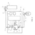
- FIG. 2 illustrates an exemplary NO delivery device that can be used in accordance with one or more embodiments of the invention
- FIG. 4 illustrates an exemplary hardware configuration that can be used in accordance with one or more embodiments of the invention
- FIG. 5 illustrates exemplary input and output parameters that can be used in accordance with one or more embodiments of the invention
- FIG. 6 illustrates an exemplary main menu with mode selection that can be used in accordance with one or more embodiments of the invention
- FIG. 7 illustrates an exemplary main menu with alarm settings that can be used in accordance with one or more embodiments of the invention
- FIG. 8 illustrates an exemplary main menu with configuration settings that can be used in accordance with one or more embodiments of the invention
- FIG. 9 illustrates an exemplary submenu for assessment of the likelihood of pulmonary hypertension resolution that can be used in accordance with one or more embodiments of the invention.
- FIG. 10 illustrates an exemplary submenu for optimization of LVAD settings that can be used in accordance with one or more embodiments of the invention
- FIG. 11 illustrates an exemplary submenu for reducing the risk of right ventricular failure that can be used in accordance with one or more embodiments of the invention
- FIG. 13 illustrates an exemplary submenu for heart exercise that can be used in accordance with one or more embodiments of the invention.
- FIG. 14 illustrates an exemplary submenu for clinician setting of the control system that can be used in accordance with one or more embodiments of the invention.
- INOmax® (nitric oxide) for inhalation is an approved drug product.
- the FDA-approved prescribing information for INOmax® dated 2013 is attached as Appendix 1, and so forms part of the present disclosure, and also is incorporated by reference herein in its entirety.
- INOmax® is a selective pulmonary vasodilator, which, in conjunction with ventilatory support or other appropriate agents, is indicated for the treatment of tem′ and near-term (>34 weeks gestation) neonates with hypoxic respiratory failure associated with clinical or echocardiographic evidence of pulmonary hypertension, where it improves oxygenation and reduces the need for extracorporeal membrane oxygenation.
- the recommended dose of INOmax® for the approved indication is 20 ppm, maintained for up to 14 days or until the underlying oxygen desaturation has resolved. Weaning should occur gradually.
- Adverse reactions per the label include methemoglobinemia and nitrogen dioxide levels, both which can be dose dependent.
- Inhaled NO may be administered via a NO delivery device such as the INOmax DSIR®, INOmax® DS or INOvent® delivery devices, each of which delivers operator-determined concentrations of NO in conjunction with a ventilator or breathing gas administration system after dilution with oxygen or an oxygen/air mixture.
- NO delivery devices such as the INOmax DSIR®, INOmax® DS or INOvent® delivery devices, each of which delivers operator-determined concentrations of NO in conjunction with a ventilator or breathing gas administration system after dilution with oxygen or an oxygen/air mixture.
- NO delivery devices and features of NO delivery devices are described below, including NO delivery devices having novel features not present in currently available NO delivery devices.
- the source of NO used in any of the presently disclosed methods and devices can be a cylinder of compressed gas containing NO, typically as a mixture with an inert gas such as nitrogen or helium.
- the NO-containing gas can be generated by manufacturing the gases separately, mixing them in an appropriate ratio, and introducing them into an appropriate cylinder under pressure. The mixing can occur in two steps: first diluting bulk NO with nitrogen to a concentration of, e.g., 5,000 ppm or 28,600 ppm in interim cylinders, and then diluting that mixture further by introducing the mixture into the final cylinders and filling them with more nitrogen to produce a concentration of, e.g., 100 ppm or 800 ppm in the final cylinders. Care is taken not to introduce any water or oxygen into the cylinders.
- the cylinders can be equipped with an appropriate valve, shipped to the point of use, and attached to a NO delivery device to facilitate inhalation of the gas by the patient.
- the source of NO can instead be a NO-generating device that generates NO from a suitable nitrogen source, such as air (see for reference U.S. Pat. No. 5,396,882, incorporated herein by reference) or nitrogen dioxide (see for reference U.S. Pat. No. 7,560,076, incorporated herein by reference).
- a suitable nitrogen source such as air (see for reference U.S. Pat. No. 5,396,882, incorporated herein by reference) or nitrogen dioxide (see for reference U.S. Pat. No. 7,560,076, incorporated herein by reference).
- the source of nitrogen dioxide can be, for example, a canister of compressed nitrogen dioxide gas or a container of N 2 O 4 (which, when treated under appropriate conditions, will give off nitrogen dioxide).
- Manufacturing a source of nitrogen dioxide can include the steps of compressing nitrogen dioxide gas into a suitable container or introducing N 2 O 4 in liquid form into a suitable container.
- pulmonary hemodynamic parameter refers to any parameter used to describe or evaluate the blood flow through the heart and pulmonary vasculature.
- pulmonary hemodynamic parameters include, but are not limited to, mean pulmonary artery pressure (mPAP), diastolic pulmonary artery pressure (dPAP) [also known as pulmonary artery diastolic pressure (PADP)], systolic pulmonary artery pressure (sPAP) [also known as pulmonary artery systolic pressure (PASP)], pulmonary capillary wedge pressure (PCWP) [also known as pulmonary artery wedge pressure (PAWP)], left atrial pressure (LAP), transpulmonary gradient (TPG), pulmonary vascular resistance (PVR) and cardiac output (CO).
- mPAP mean pulmonary artery pressure
- dPAP diastolic pulmonary artery pressure
- sPAP systolic pulmonary artery pressure
- PCWP pulmonary capillary wedge pressure
- PAWP pulmonary artery wedge pressure
- PCWP is often used as a more convenient, less invasive approximation of LAP.
- PVR is related to mPAP, PCWP and CO according to the following equation: PVR ⁇ (mPAP ⁇ PCWP)/CO
- mPAP (2 ⁇ 3)dPAP+(1 ⁇ 3)sPAP
- the pulmonary hemodynamic parameters are measured directly, such as during a right heart catheterization.
- the pulmonary hemodynamic parameters are estimated and/or evaluated through other techniques such as magnetic resonance imaging (MRI) or echocardiography.
- MRI magnetic resonance imaging
- PH pulmonary hypertension
- Post-LVAD right ventricular failure may be defined pathophysiologically as inability of the right ventricle to maintain adequate loading of the LVAD-assisted left ventricle despite adequate right ventricle preload, or to do so only at the expense of significantly elevated central venous pressure.
- Post-LVAD right heart failure is generally defined operationally as the need for implantation of a right ventricular assist device (RVAD), or the need for reinstitution of inhaled NO for greater than 48 hours, or the need for inotropic pharmacological therapy for greater than 14 days.
- RVAD right ventricular assist device
- the pathophysiology of right ventricular failure after LVAD implantation appears to be multi-factorial, and includes pre-operative right ventricular dysfunction and pulmonary hypertension (PH), right ventricular ischemia, peri-operative fluctuations in pulmonary vascular resistance (PVR) in the setting of cardiopulmonary bypass (CPB), excessive right ventricular preload, and altered interventricular balance, although the relative importance of each of these factors is strongly debated.
- peri-operative procedures and/or complications thereof such as intra-operative mechanical and/or ischemic damage to the right ventricle, and intra-operative hemorrhage requiring extensive fluid, colloid or blood product resuscitation, have also been invoked as acute predisposing or exacerbating factors for peri-operative right heart failure associated with LVAD implantation.
- the direct and indirect effects of the LVAD itself on the anatomy and function of the left ventricle are also implicated as causing or contributing to post-implantation right ventricular dysfunction and right heart failure, despite optimization of LVAD adjustments and pharmacological support.
- the left ventricle In the healthy heart, the left ventricle is estimated to contribute 80% of the contractile flow and up to two-thirds of the contractile pressure generated by the right ventricle through a process termed mechanical systolic ventricular interaction (SVI).
- SVI mechanical systolic ventricular interaction
- the (patho)physiological interactions between the left ventricle and the right ventricle in CHF are quite complex.
- the right ventricle may be directly damaged by the underlying disease process affecting the left ventricle in CHF. Most commonly, right ventricular failure in CHF results from increased right ventricular afterload due to chronic pulmonary vascular congestion and PH consequent to the primary left ventricular dysfunction.
- dilation of the left ventricle in CHF realigns the anatomy of interventricular septal musculature to a less efficient transverse orientation, further impairing SVI and therefore overall right ventricular contractility.
- dysfunctional SVI unleashes a vicious cycle of progressive right ventricular dysfunction and right ventricular failure Reciprocally, as the failing right ventricle dilates, it intrudes and interferes with the relaxation (diastolic) filling of the left ventricle (termed diastolic ventricular interaction [DVI]), further exacerbating pulmonary vascular congestion and PH, creating a superimposed additional vicious cycle of progressive left ventricular failure and right ventricular failure.
- DVI diastolic ventricular interaction
- An additional form of remote interventricular interaction occurs at the level of the peripheral circulation, wherein the increased central systemic blood volume and central venous pressure in CHF increases right ventricular preload and right ventricular filling further increasing interventricular septal intrusion into the left ventricle and thereby further worsening DVI.
- a further level of interventricular interaction occurs at the level of the pulmonary circulation, where chronic elevation of pulmonary venous and capillary pressures causes pulmonary vascular remodeling which, over time creates a relatively fixed, structurally-mediated increase in PVR, further worsening PH and right ventricular afterload (defined as World Health Organization [WHO] Group 2 Pulmonary Hypertension Secondary to Left-Sided Heart Disease).
- WWHO World Health Organization
- the chronically failing left ventricle increases left atrial and pulmonary venous pressure which increases pulmonary artery pressure (PAP) and right ventricular afterload.
- PAP pulmonary artery pressure
- TPG trans-pulmonary pressure gradient
- the extent to which this increased PVR and TPG is mediated by tonic (and hence acutely reversible) compensatory pulmonary vasoconstriction versus relatively fixed structural vascular remodeling, and the degree to which each of these components is ultimately reversible in advanced CHF is often difficult to predict based on previously known methods.
- Current post-operative hemodynamic LVAD management primarily targets restoration of normal systemic peripheral end-organ (e.g. renal and hepatic) perfusion (as measured by CI) in order to penult gradual weaning of inotropic agents and diuretics.
- Fluid therapy is generally targeted to maintain initial pump speed >2 liters/min with a right atrial filling pressure ⁇ 20 mm Hg).
- attributes of pulmonary vascular congestion and PH and the need for inotropic pharmacological support generally decline as measures of peripheral end-organ perfusion progressively improve gradually over a period of days to weeks.
- right ventricular dysfunction as assessed by transthoracic echocardiogram may remain impaired for up to 3 months following successful LVAD implantation (although more rapid improvement has been reported in stable LVAD patients not requiring inotropic support).
- the still-weakened right ventricle may be unable to accommodate the increased forward flow generated by the LVAD-assisted left ventricular output, posing a continuing risk of right heart failure.
- the associated elevation of right arterial pressure one month post-LVAD implantation is linked with impaired exercise tolerance as reliably assessed by the distance walked in six minutes (6 MWD) and is predictive of increased mortality risk.
- Persistent post-LVAD right ventricular dysfunction may reflect diminished intrinsic ability of the right ventricle to undergo self-repair and/or the fact that therapeutic hemodynamic adjustments prioritize the systemic circulation leaving the still-weakened right ventricle exposed to non-optimized hemodynamic stresses.
- Both the intrinsic right ventricle and the implanted LVAD are preload-dependent and afterload-sensitive, and adequate but not excessive preload of the right ventricle is important to maintain adequate left ventricle/LVAD filling without excessive right ventricular volume overload in the immediate post-operative period.
- LVAD flows must be kept low enough to avoid right ventricular volume overload but high enough to sustain adequate end-organ perfusion.
- Inotropes e.g. milrinone, dobutamine and epinephrine, used to wean from CPB are often continued for days after implantation.
- Nitroglycerin, SNP, nesiritide and sildenafil have been used to lower Inhaled NO and prostacyclin have also been used to reduce PVR in order to do so without compromising systemic perfusion.
- right ventricular outflow through the pulmonary circulation may be inadequate to reliably fill the left ventricle, resulting in the development of negative pressure in the left ventricle.
- This negative left ventricular pressure not only compromises LVAD function, but also draws the interventricular septum leftward, disrupting SVI and essentially eliminating any septal contribution to right heart contractility, further reducing right ventricular outflow.
- trans-esophageal echocardiography continuously monitors the position of the interventricular septum during LVAD adjustment and weaning from CPB; this is particularly important to monitor and manage the acute effects of CPB-withdrawal on the dynamic status of the pulmonary vasculature, which can produce severe acute intra-operative or peri-operative PH.
- TTE trans-thoracic echocardiography
- a “ramped speed study” under TTE monitoring may be used to adjust the optimal pump speed taking into account changes in ventricular dimensions, displacement of the interatrial and interventricular septa, and the frequency of aortic valve opening as well as evidence of inadequate left ventricular preloading and right ventricular dysfunction.
- This TTE-directed optimization may be especially important in patients with poor 6 MWD.
- Reduction of LVAD speed to optimize right ventricular function and/or manage post-LVAD right ventricular failure may require temporary reintroduction of inotropic pharmacological support and/or intravenous vasodilators to maintain adequate systemic end-organ perfusion.
- right ventricular failure remains a leading cause of early mortality after implantation of even the most modern continuous-flow LVADs.
- Acute pulmonary vasoreactivity testing (AVT) by right heart catheterization with selective pulmonary vasodilators such as inhaled NO is routinely performed in other forms of PH such as pulmonary arterial hypertension (PAH, or WHO Group 1 Pulmonary Hypertension).
- AVT with inhaled NO is only rarely and cautiously performed in non-LVAD-supported patients with CHF prior to heart transplantation, because acute highly-selective reduction in PVR and right ventricular afterload may overload the failing left ventricle thereby increasing right arterial and pulmonary venous pressure, potentially precipitating acute pulmonary edema.
- “fixed” versus “reversible” PH in advanced CHF is usually interrogated by the hemodynamic response or lack thereof to combinations of systemically-administered non-pulmonary-specific vasodilators such as SNP, nitroglycerin or adenosine and inotropic agents administered acutely, or in some studies, for over 72 hours.
- systemically-administered non-pulmonary-specific vasodilators such as SNP, nitroglycerin or adenosine and inotropic agents administered acutely, or in some studies, for over 72 hours.
- acute or longer infusion of highest tolerated doses i.e. free of systemic hypotension or other systemic side effects
- prostacyclin and prostaglandin E 1 in conjunction with inotropes or non-specific vasodilators have been used for PH-reversibility in this setting.
- Inhaled or intravenous prostanoids may be currently considered preferable to inhaled NO in non-LVAD-supported CHF patients in some but not all geographies despite significant decreases in systemic vascular resistance (SVR) and the clear demonstration that chronic intravenous prostacyclin therapy increases mortality in patients with end-stage CHF.
- SVR systemic vascular resistance
- TTE becomes a primary tool to aid in the regulation of LVAD settings and hemodynamic fluid and pharmacological therapy in the post-acute post-implant setting.
- Management is complicated by the fact that right ventricular and both the pulmonary and systemic circulations are simultaneously undergoing complex dynamic interactions and adaptations to the newly functioning LVAD and the fact that most vasoactive drugs affect both circulatory systems simultaneously, e.g. SNP dilates both systemic and pulmonary resistance vessels.
- the high pulmonary selectivity and very short half-life of inhaled NO are ideal attributes to classify, manage and optimize the pulmonary vascular status in CHF patients on LVAD support, and to optimize LVAD performance in this setting.
- AVT with inhaled NO under echocardiographic and/or right heart catheterization guidance could be used to determine if inhaled NO should be continued in order to adjust left ventricular preload to optimize SVI and LVAD performance and reduce the risk of perioperative and postoperative acute right ventricular dysfunction and right ventricular failure.
- doses of inhaled NO would be titrated against the right ventricular performance including measures of SVI and LVAD rotational speed, power and flow, until the correct combination is achieved to normalize/optimize right ventricular function, SVI, cardiac output, pulsatility and other hemodynamic parameters.
- inhaled NO would be continued during the taper of inotrope and/or intravenous systemic vasodilators. These parameters would then be re-monitored with echocardiographic assessment to maintain optimal settings during the post-acute recovery period. It would be anticipated that the provision of critical right ventricular afterload reduction and left ventricular preload enhancement during this period would permit increased CI with less LVAD power, improve pulse index, improve peripheral perfusion and exercise tolerance, and hasten and improve early cardiac rehabilitation, as well as reduce the risk of right heart failure.
- inhaled NO could be highly predictive of the likelihood of subsequent maximum resolution of residual PH over 6-months resulting from chronic CHF, particularly in those patients whose PH may have been characterized preoperatively as “fixed” by lack of response to the combination of inotropic agents and systemic vasodilators, as the effectiveness of these agents is often tolerability-limited.
- inhaled NO could represent a new paradigm in the optimal management of patients with functioning LVADs over the days, weeks and months following implantation.
- aspects of the present invention provide for the utilization of inhaled NO as adjunctive therapy post-LVAD implantation.
- the inhaled NO therapy may be commenced pre-operatively pre-implantation or intra-operatively before, during or directly after implantation; such inhaled NO therapy may be continued beyond the time period in which it is clinically required in order successfully counteract the acute and temporary PH that occurs as a direct consequence of the CPB procedure itself.
- inhaled NO could be instituted (or re-instituted) after successful weaning from CPB and the direct consequence thereof.
- inhaled NO would thusly be utilized for one or more novel applications to predict and prevent the development of right ventricular failure post successful weaning from CPB in LVAD recipients.
- right ventricular failure after institution of LVAD support may be consequent to the development or persistence of PH, further impairment of right ventricular contractility secondary to alterations in SVI and DVI or other ventricular interactions between the RV and the “unloaded” left ventricle, or further intrinsic impairment of right ventricular contractility given the known susceptibility of the right ventricle to myocardial preservation injury during CPB. Differentiation among these contributing causes is important since they would be managed differently.
- One aspect of the present invention provides a method to predict which post-implantation LVAD patients with PH are likely to resolve their PH with continued LV unloading by a functional LVAD.
- Those patients exhibiting a significant acute reduction in TPG and/or mPAP and/or PVR and/or other measures such as the difference between dPAP and PCWP with inhaled NO would be those more likely to resolve their increased TPG and/or mPAP and/or PVR following LVAD treatment, e.g. after several months of LVAD treatment.
- the PAP is a combination of increased pulmonary vascular resistance and elevated post-capillary pressure measured as either PCWP or right arterial pressure (RAP).
- PCWP and RAP effect would be removed once the LVAD is in place, making the AVT results more clearly related to pulmonary vascular resistance, not confounded by increased PCWP and RAP. Accordingly, this aspect of the present invention provides an enhanced predictive tool by performing the AVT after LVAD implantation.
- the method comprises measuring one or more pulmonary hemodynamic parameters of a patient with an LVAD to obtain a first pulmonary hemodynamic value; after obtaining the first pulmonary hemodynamic value, administering inhaled NO to the patient with the LVAD; and measuring one or more pulmonary hemodynamic parameters of the patient during or after the inhaled NO administration to obtain a second pulmonary hemodynamic value.
- a significant improvement in the pulmonary hemodynamic parameter from the first pulmonary hemodynamic value to the second pulmonary hemodynamic value for example a decrease of at least 10 mm Hg and/or at least 20% can indicate that the patient is likely to resolve the pulmonary hypertension after continued use of the LVAD.
- the inhaled NO is administered at a concentration of 5 to 80 ppm for at least 10 minutes.
- Exemplary inhaled NO concentrations include about 5 ppm, about 10 ppm, about 15 ppm, about 20 ppm, about 25 ppm, about 30 ppm, about 35 ppm, about 40 ppm, about 45 ppm, about 50 ppm, about 55 ppm, about 60 ppm, about 65 ppm, about 70 ppm, and about 80 ppm.
- Exemplary NO administration times include about 10 minutes, about 15 minutes, about 20 minutes, about 25 minutes, about 30 minutes, about 35 minutes, about 40 minutes, about 45 minutes, about 50 minutes, about 55 minutes, and about 60 minutes.
- Exemplary pulmonary hemodynamic parameters include mean pulmonary artery pressure (mPAP), transpulmonary gradient (TPG) and pulmonary vascular resistance (PVR).
- mPAP mean pulmonary artery pressure
- TPG transpulmonary gradient
- PVR pulmonary vascular resistance
- the one or more pulmonary hemodynamic parameters may be measured by any appropriate procedures, such as by performing a right heart catheterization.
- AVT acute vasoreactivity testing
- the method further comprises placing the patient on a heart transplant list if the decrease in the pulmonary hemodynamic parameter from the first pulmonary hemodynamic value to the second pulmonary hemodynamic value is at least 10 mm Hg and/or at least 20%. In some embodiments, the method further comprises explanting the LVAD and implanting a donor heart in the patient.
- other significant decreases in the pulmonary hemodynamic parameter may be at least 5 mm Hg, at least 6 mm Hg, at least 7 mm Hg, at least 8 mm Hg, at least 9 mm Hg, at least 15 mm Hg, at least 20 mm Hg, or at least 25 mm Hg, and/or at least 5%, at least 10%, at least 15%, at least 25%, at least 30%, at least 35%, at least 40% or at least 50%.
- Another aspect of the present invention provides a method to use administration of inhaled NO during the adjustment and setting of the LVAD parameters and hemodynamic pharmacotherapy and fluid replacement therapy once the acute effects of prior CPB on pulmonary hemodynamics have elapsed.
- One current methodology of setting an LVAD involves setting the revolutions/min (rpm) rate of an LVAD (such as a HeartMate II LVAD (HM II)) to provide adequate cardiac output and achieve optimal left ventricular decompression, while maintaining a pulsatility index (defined as the maximum LVAD flow rate minus the minimum LVAD flow rate divided by the average LVAD flow rate) of 3.5 to 4.
- rpm revolutions/min
- HM II HeartMate II LVAD
- the flow rate through these devices at any device setting varies depending upon the pressure gradient between the left ventricle and the aorta such that the increased systolic pressure with each contraction of the left ventricle transiently increases flow through the LVAD creating some pulsatile variation in blood flow.
- the fixed-rate speed of a continuous-flow LVAD is usually adjusted to maximize left ventricular decompression and to improve cardiac output, while simultaneously allowing for a minimum aortic valve opening ratio of 1:3 (i.e. the left ventricular systolic pressure achieves a sufficiently high pressure relative to aortic pressure to permit opening of the aortic valve once out of every three systoles despite the continuing efflux of blood through the LVAD).
- Another current methodology of setting an LVAD involves optimizing the rpm speed, both hemodynamically and echocardiographically, at the time of LVAD placement, before the patient is discharged from the hospital (i.e., after admission for LVAD placement) and if clinical events such as new symptoms or suction events warranted further adjustment.
- these hemodynamic and echocardiographic assessments used to adjust LVAD settings are static in that they are performed at the left ventricular preload exhibited by the patient at the time and condition under which the test is performed (usually at rest).
- inhaled NO during part of the test procedure to maximally relax the pulmonary vessels and lower PVR would provide information on the maximal left ventricular preload that the right ventricle is able to generate unfettered by acutely-reversible pulmonary vasoconstriction.
- Such dynamic (rather than static) assessment would provide additional information to optimize any particular group of settings of the LVAD to produce the desired cardiac out and pulsatility parameters while avoiding the generation of left ventricular suction as determined by simultaneous TTE.
- the novel use of inhaled NO as an adjunct to adjusting LVAD parameters should improve the efficiency and safety of functioning LVADs, which should result in improved cardiac output, pulsatility indices and exercise tolerance, and reduce the risk of right heart failure.
- this aspect of the present invention relates to a method of optimizing the settings of an LVAD by utilizing inhaled NO.
- the method comprises administering inhaled NO to a patient having an LVAD; performing an echocardiogram or similar functional hemodynamic or cardiac imaging assessment on the patient during the administration of inhaled NO; and adjusting or setting one or more parameters of the LVAD during the echocardiogram and during the administration of inhaled NO.
- adjusting or setting the LVAD parameters during administration of NO helps to optimize cardiac output, end-organ perfusion, LVAD efficiency and/or exercise tolerance.
- adjusting or setting one or more parameters of the LVAD comprises one or more of (i) determining a low pump speed setting for the LVAD based on the minimal pump speed necessary for the patient's aortic valve to open with each heart beat or (ii) determining a high speed setting for the LVAD based for example on the pump speed at which the septum of the patient's heart flattens.
- the inhaled NO is administered at a concentration of 5 to 80 ppm for at least 10 minutes.
- Exemplary inhaled NO concentrations include about 5 ppm, about 10 ppm, about 15 ppm, about 20 ppm, about 25 ppm, about 30 ppm, about 35 ppm, about 40 ppm, about 45 ppm, about 50 ppm, about 55 ppm, about 60 ppm, about 65 ppm, about 70 ppm, and about 80 ppm.
- Exemplary NO administration times include about 10 minutes, about 15 minutes, about 20 minutes, about 25 minutes, about 30 minutes, about 35 minutes, about 40 minutes, about 45 minutes, about 50 minutes, about 55 minutes, about 60 minutes, about 1.5 hours, about 2 hours, about 2.5 hours and about 3 hours.
- the LVAD settings are changed over a series of incremental adjustments.
- the LVAD pump speed may be adjusted upwards in two or more steps. One or more or all of these steps may be performed during the administration of inhaled NO as described herein.
- Another aspect of the present invention provides for long-term use of inhaled NO after LVAD implantation.
- AVT favorably predicts that PH will resolve after continued use of the LVAD
- the settings and read-out and/or the TTE indicate more efficacious LVAD function and hemodynamic status under inhaled NO challenge
- the treating physician may wish to continue administering inhaled NO to the patient continuously for all or part of the convalescent period.
- inhaled NO would be administered to the patient during all or part of the day over a period of days, weeks or months to maintain the favorable LVAD function and/or hemodynamic status.
- Periodic testing may be performed as described above both on and off inhaled NO for a short period of time, such that when sufficient recovery had occurred so that inhaled NO was no longer producing and hemodynamic or TTE change, the patient may be carefully weaned from inhaled NO. This treatment would be expected to result in improved cardiac output and exercise tolerance more quickly, and reduce the risk of right heart failure.
- the method comprises administering inhaled NO to a patient with an LVAD for at least 12 hours a day for at least 10 days.
- the inhaled NO may be administered for several days to many months or even longer. Exemplary treatment times include 10 days, 15 days, 20 days, 25 days, 30 days, 35 days, 40 days, 45 days, 2 months, 3 months, 4 months, 5 months, 6 months, 7 months, 8 months, 9 months, 10 months, 11 months, 1 year, 1.5 years, or 2 years.
- the patient is administered inhaled NO indefinitely.
- the inhaled NO is administered at a concentration of 5 to 80 ppm for at least 12 hours a day.
- Exemplary inhaled NO concentrations include about 5 ppm, about 10 ppm, about 15 ppm, about 20 ppm, about 25 ppm, about 30 ppm, about 35 ppm, about 40 ppm, about 45 ppm, about 50 ppm, about 55 ppm, about 60 ppm, about 65 ppm, about 70 ppm, and about 80 ppm.
- Exemplary NO administration times include about 12 hours a day, about 14 hours a day, about 16 hours a day, about 18 hours a day, about 20 hours a day, about 22 hours a day, or up to 24 hours a day.
- the method further comprises confirming that the LVAD is functioning properly before administering inhaled NO.
- the inhaled NO is administered after a patient has been weaned from cardiopulmonary bypass (CPB).
- CPB cardiopulmonary bypass
- the dose of NO may be prescribed based on the patient's ideal body weight (IBW).
- IBW ideal body weight
- Exemplary NO doses may be in the range of about 25 to about 150 ⁇ g/kg IBW/hr, such as about 25, about 30, about 35, about 40, about 45, about 50, about 60, about 70, about 80, about 90, about 100, about 110, about 120, about 130, about 140 or about 150 ⁇ g/kg IBW/hr.
- the method further comprises monitoring one or more output parameters of the LVAD and/or one or more hemodynamic parameters of the patient, comparing the one or more output parameters and/or the one or more hemodynamic parameters to a predetermined range, and adjusting the dose of inhaled NO if the one or more outputs parameters and/or the one or more hemodynamic parameters are outside of the predetermined range.
- the method further comprises providing an alert if the one or more output parameters and/or the one or more hemodynamic parameters are outside of the predetermined range.
- the inhaled NO dose may be adjusted automatically (e.g. by the NO delivery device or a control system in communication with the NO delivery device), or may be manually adjusted by a physician or other user, such as in response to an alert.
- LVAD parameters examples include, but are not limited to, pump speed (e.g. rpm), pump flow (e.g. L/min), pump power, pulsatility index, battery level, and LVAD status (e.g. operational, presence or absence of warnings).
- the LVAD has a minimum and/or maximum pump speed that is set by the physician, and can be specific for the individual patient. Alternatively or additionally, the LVAD may also have a minimum and/or maximum pump speed set by the manufacturer of the LVAD.
- exemplary minimum pump speeds include 100 rpm, 200 rpm, 300 rpm, 400 rpm, 500 rpm, 6000 rpm, 700 rpm, 800 rpm, 900 rpm, 1,000 rpm, 1,500 rpm, 2,000 rpm, 2,500 rpm, 3,000 rpm, 4,000 rpm, 5,000 rpm, 6,000 rpm, 7,000 rpm, 8,000 rpm, 9,000 rpm, 10,000 rpm, 11,000 rpm, 12,000 rpm, 13,000 rpm, 14,000 rpm and 15,000 rpm, and exemplary maximum pump speeds include 1,000 rpm, 1,500 rpm, 2,000 rpm, 2,500 rpm, 3,000 rpm, 4,000 rpm, 5,000 rpm, 6,000 rpm, 7,000 rpm, 8,000 rpm, 9,000 rpm, 10,000 rpm, 11,000 rpm, 12,000 rpm, 13,000 rpm, 14,000 rpm and
- the LVAD has a minimum and/or maximum pump flow that is set by the physician, and can be specific for the individual patient. Alternatively or additionally, the LVAD may also have a minimum and/or maximum pump flow set by the manufacturer of the LVAD.
- exemplary minimum pump speeds include 1 L/min, 1.5 L/min, 2 L/min, 2.5 L/min, 3 L/min, 3.5 L/min, 4 L/min, 4.5 L/min, 5 L/min, 6 L/min, 7 L/min, 8 L/min, 9 L/min and 10 L/min
- exemplary maximum pump flows include 3 L/min, 3.5 L/min, 4 L/min, 4.5 L/min, 5 L/min, 6 L/min, 7 L/min, 8 L/min, 9 L/min, 10 L/min, 11 L/min, 12 L/min, 13 L/min, 14 L/min and 15 L/min.
- the minimum and maximum pump flows may depend on the design of the LVAD.
- the pulsatility index of the LVAD has a minimum and/or maximum threshold.
- the pulsatility index is the maximum pump flow minus the minimum pump flow, divided by the average pump flow.
- the pulsatility index is an indication of how much support the LVAD is providing to the heart (a higher pulsatility index indicates that the LVAD is providing more support)
- high pulsatility indices can be a cause of concern.
- exemplary maximum pulsatility indices include values of 5, 6, 7, 8, 9, 10, 11, 12, 13, 14, 15 or 20.
- the battery level of the LVAD is monitored. As a low battery can indicate a future or imminent shutdown of the LVAD, when the battery level of the LVAD drops below a certain threshold, the inhaled NO dose may be lowered, a weaning protocol may be initiated, and/or an alert is provided. Examples of minimum battery levels include 30%, 25%, 20%, 15%, 10%, 9%, 8%, 7%, 6%, 5%, 4%, 3%, 2% or 1% battery remaining or 6 hours, 5 hours, 4 hours, 3 hours. 2 hours, 1 hour, 30 minutes, 20 minutes, 15 minutes, 10 minutes, 5 minutes, 4 minutes, 3 minutes, 2 minutes or 1 minute of battery life remaining.
- the inhaled NO dose may be adjusted, a weaning protocol may be initiated and/or an alert may be provided.
- the inhaled NO dose may be adjusted automatically (e.g. by the NO delivery device or a control system in communication with the NO delivery device), or may be manually adjusted by a physician or other user.
- Another aspect of the present invention relates to a method of monitoring the left ventricle of a patient with an LVAD.
- the method comprises reducing the pump speed of the LVAD or turning off the LVAD; measuring one or more pulmonary hemodynamic parameters of the patient to obtain a first pulmonary hemodynamic value; preloading the left ventricle by administering inhaled NO to the patient; and measuring one or more pulmonary hemodynamic parameters of the patient after or during administration of inhaled NO to obtain a second pulmonary hemodynamic value.
- the pulmonary hemodynamic parameter is selected from LAP, PCWP and CO, or may be any assessment of the left ventricular reserve to compensate for increased left ventricular preload that can be measured through right heart catheterization, echocardiographic, MRI or other techniques.
- the inhaled NO is administered at a concentration of 5 to 80 ppm for at least 10 minutes.
- Exemplary inhaled NO concentrations include about 5 ppm, about 10 ppm, about 15 ppm, about 20 ppm, about 25 ppm, about 30 ppm, about 35 ppm, about 40 ppm, about 45 ppm, about 50 ppm, about 55 ppm, about 60 ppm, about 65 ppm, about 70 ppm, and about 80 ppm.
- Exemplary NO administration times include about 10 minutes, about 15 minutes, about 20 minutes, about 25 minutes, about 30 minutes, about 35 minutes, about 40 minutes, about 45 minutes, about 50 minutes, about 55 minutes, about 60 minutes, about 1.5 hours, about 2 hours, about 2.5 hours, about 3 hours, about 3.5 hours, about 4 hours, about 5 hours, about 6 hours, about 7 hours or about 8 hours.
- the method further comprises modifying treatment if the left ventricle is improving, such as explanting the LVAD from the patient.
- Other modifications in treatment can include changing the supportive medication (e.g. diuretics and/or inotropic medications) that the patient is given, such as reducing the supportive medication.
- Another aspect of the present invention relates to a method of exercising a heart of a patient having an LVAD.
- the method comprises reducing and/or modulating the pump speed of the LVAD or turning off the LVAD; preloading the left ventricle by administering inhaled NO to the patient for at least 5 minutes; discontinuing the inhaled NO administration; and repeating the preloading and discontinuation to exercise the left ventricle of the patient's heart.
- the inhaled NO is administered at a concentration of 5 to 80 ppm for at least 5 minutes.
- Exemplary inhaled NO concentrations include about 5 ppm, about 10 ppm, about 15 ppm, about 20 ppm, about 25 ppm, about 30 ppm, about 35 ppm, about 40 ppm, about 45 ppm, about 50 ppm, about 55 ppm, about 60 ppm, about 65 ppm, about 70 ppm, and about 80 ppm.
- Exemplary NO administration times include about 5 minutes, about 10 minutes, about 15 minutes, about 20 minutes, about 25 minutes, about 30 minutes, about 35 minutes, about 40 minutes, about 45 minutes, about 50 minutes, about 55 minutes, about 60 minutes, about 1.5 hours, about 2 hours, about 2.5 hours, about 3 hours, about 3.5 hours, about 4 hours, about 5 hours, about 6 hours, about 7 hours or about 8 hours.
- the preloading of the left ventricle may be performed multiple times per day, such as twice a day, three times a day, four times a day, five times a day, six times a day, seven times a day, eight times a day, nine times a day or ten times a day. Alternatively, the preloading may be performed once a day. If the preloading is performed multiple times per day, the preloading procedures may be clustered together (e.g. spaced apart by several minutes or a couple hours) or may be spread out throughout the day.
- the preloading of the left ventricle may also be performed once a week, two days a week, three days a week, four days a week, five days a week, six days a week, or seven days a week.
- the left ventricle is preloaded several times a day for several days a week, such as two to five times a day for two to four days a week or other combinations of the above daily and weekly preloading schedules.
- the first instance or first few instances of the exercising of the patient's left ventricle is performed under the direct supervision of a physician, but once an appropriate exercise protocol has been determined for a patient, then the exercise protocol may be performed under patient control or may be automated by the NO delivery device and/or control system.
- Another aspect of the present invention relates to a method of reducing the risk of adverse events during LVAD use.
- inhaled NO can improve cardiovascular function of LVAD recipients with functioning LVADs, reduce the risk of adverse events associated with continuous-flow or semi-pulsatile LVADs (e.g. thrombosis, gastrointestinal bleeding) and improve the function of transplanted hearts by making the cardiovascular system more compliant.
- a specific deficiency in continuous-flow LVADs is that the reduced pulsatility of peripheral perfusion generated by these devices compared to pulsatile-flow LVAD devices and/or the normal circulation derived from a well-functioning human heart as measured by pulsatility index, pulse pressure and/or the frequency of opening of the aortic valve has been implicated in a number of adverse events including reduced peripheral vascular compliance, gastrointestinal bleeding, arteriovenous malformations, hemolysis, pump thrombosis, aortic insufficiency and lower rate of recovery of left ventricular function. These adverse events can also be associated with semi-pulsatile LVADs.
- FMD flow-mediated dilation
- RHI reactive hyperemic index
- Deranged NO-mediated autoregulatory vascular function is reflected not only in peripheral vessels but also in coronary vessels as well, including an association with a higher rate of in-stent thrombosis following percutaneous coronary intervention. It is believed that impaired NO-mediated coronary vascular autoregulation could provide a potential mechanism that would contribute to reduced LV recovery during continuous-LVAD support. NO not only mediates autoregulation of vascular tone, but also modulates platelet activation and adhesion, such that deranged NO regulation could contribute to the increased thrombosis associated with continuous-flow LVAD support.
- Attributes of NO-mediated vascular dysfunction are considered to be significant overall risk factors for adverse cardiovascular outcomes in the general population. Indeed, some authors have suggested that impaired endothelial dilation is the ultimate cause of cardiovascular diseases such as coronary artery disease and peripheral artery disease. These widespread effects of NO on vascular health are thought to be mediated in large part by modulating oxidative stress through the control of nitrosylation of a wide range of regulatory proteins.
- Inhalational NO could therefore improve endothelial function and vascular health, and thereby important clinical outcomes, in patients on long-term continuous-flow LVAD support, through two related and potentially complementary or synergistic mechanisms.
- inhaled NO by dilating pulmonary vessels, would increase left ventricular preload and improve left ventricular filling, thereby increasing the frequency of aortic valve opening and closing, and hence pulsatility for any given LVAD speed setting. This effect might be more dominant during periods of rest or sleep, or during periods of increased cardiac output demand such as exercise, depending upon the characteristics of the LVAD device and setting.
- inhaled NO also has the potential to improve NO-mediated vascular autoregulatory function and health independent of its effect of circulatory pulsatility, by improving NO bioavailability throughout the peripheral and coronary circulation.
- NO can be generated by NO synthase (NOS) or by the breakdown of nitrite or other compounds to NO.
- NO NO synthase
- Inhalation of NO for even short periods of time (e.g. two hours) increases the circulating levels of a variety of molecules that have the potential to regenerate NO within the microcirculation including nitrite (NO 2 ⁇ ), nitrate (NO 3 ⁇ ) and S-nitrosohemoglobin.
- NO 2 ⁇ nitrite
- NO 3 ⁇ nitrate
- S-nitrosohemoglobin S-nitrosohemoglobin.
- embodiments of the present invention provide for the administration by inhalation of NO at doses and for durations that are expected to (1) improve pulsatility through the NO's direct hemodynamic action on the pulmonary vasculature thereby promoting left ventricular filling, aortic valve opening and closure, and thereby maximize pulsatile flow and/or (2) increase peripheral NO bioavailability through provision NO precursors in the form of NO metabolites such as NO 2 ⁇ , NO 3 ⁇ , nitrosohemoglobin and other that are formed in the lung as a result of NO inhalation and transported by the circulation to peripheral tissues where NO can be reformed further reducing endothelial dysfunction.
- NO precursors in the form of NO metabolites such as NO 2 ⁇ , NO 3 ⁇ , nitrosohemoglobin and other that are formed in the lung as a result of NO inhalation and transported by the circulation to peripheral tissues where NO can be reformed further reducing endothelial dysfunction.
- the method comprises administering inhaled NO to a patient with an LVAD for at least 12 hours a day for at least 10 days.
- the inhaled NO may be administered for several days to many months or even longer. Exemplary treatment times include 10 days, 15 days, 20 days, 25 days, 30 days, 35 days, 40 days, 45 days, 2 months, 3 months, 4 months, 5 months, 6 months, 7 months, 8 months, 9 months, 10 months, 11 months, 1 year, 1.5 years, or 2 years.
- the patient is administered inhaled NO indefinitely.
- the inhaled NO is administered at a concentration of 5 to 80 ppm for at least 12 hours a day.
- Exemplary inhaled NO concentrations include about 5 ppm, about 10 ppm, about 15 ppm, about 20 ppm, about 25 ppm, about 30 ppm, about 35 ppm, about 40 ppm, about 45 ppm, about 50 ppm, about 55 ppm, about 60 ppm, about 65 ppm, about 70 ppm, and about 80 ppm.
- Exemplary NO administration times include about 12 hours a day, about 14 hours a day, about 16 hours a day, about 18 hours a day, about 20 hours a day, about 22 hours a day, or up to 24 hours a day.
- the method further comprises confirming that the LVAD is functioning properly before administering inhaled NO.
- the inhaled NO is administered after a patient has been weaned from cardiopulmonary bypass (CPB).
- CPB cardiopulmonary bypass
- the method further comprises monitoring one or more output parameters of the LVAD and/or one or more hemodynamic parameters of the patient, comparing the one or more output parameters and/or the one or more hemodynamic parameters to a predetermined range, and adjusting the dose of inhaled NO if the one or more outputs parameters and/or the one or more hemodynamic parameters are outside of the predetermined range.
- the method further comprises providing an alert if the one or more output parameters and/or the one or more hemodynamic parameters are outside of the predetermined range.
- the inhaled NO dose may be adjusted automatically (e.g. by the NO delivery device or a control system in communication with the NO delivery device), or may be manually adjusted by a physician or other user, such as in response to an alert.
- LVAD parameters examples include, but are not limited to, pump speed (e.g. rpm), pump flow (e.g. L/min), pump power, pulsatility index, battery level, and LVAD status (e.g. operational, presence or absence of warnings).
- the LVAD has a minimum and/or maximum pump speed that is set by the physician, and can be specific for the individual patient. Alternatively or additionally, the LVAD may also have a minimum and/or maximum pump speed set by the manufacturer of the LVAD.
- exemplary minimum pump speeds include 100 rpm, 200 rpm, 300 rpm, 400 rpm, 500 rpm, 6000 rpm, 700 rpm, 800 rpm, 900 rpm, 1,000 rpm, 1,500 rpm, 2,000 rpm, 2,500 rpm, 3,000 rpm, 4,000 rpm, 5,000 rpm, 6,000 rpm, 7,000 rpm, 8,000 rpm, 9,000 rpm, 10,000 rpm, 11,000 rpm, 12,000 rpm, 13,000 rpm, 14,000 rpm and 15,000 rpm, and exemplary maximum pump speeds include 1,000 rpm, 1,500 rpm, 2,000 rpm, 2,500 rpm, 3,000 rpm, 4,000 rpm, 5,000 rpm, 6,000 rpm, 7,000 rpm, 8,000 rpm, 9,000 rpm, 10,000 rpm, 11,000 rpm, 12,000 rpm, 13,000 rpm, 14,000 rpm and
- the LVAD has a minimum and/or maximum pump flow that is set by the physician, and can be specific for the individual patient. Alternatively or additionally, the LVAD may also have a minimum and/or maximum pump flow set by the manufacturer of the LVAD.
- exemplary minimum pump speeds include 1 L/min, 1.5 L/min, 2 L/min, 2.5 L/min, 3 L/min, 3.5 L/min, 4 L/min, 4.5 L/min, 5 L/min, 6 L/min, 7 L/min, 8 L/min, 9 L/min and 10 L/min
- exemplary maximum pump flows include 3 L/min, 3.5 L/min, 4 L/min, 4.5 L/min, 5 L/min, 6 L/min, 7 L/min, 8 L/min, 9 L/min, 10 L/min, 11 L/min, 12 L/min, 13 L/min, 14 L/min and 15 L/min.
- the minimum and maximum pump flows may depend on the design of the LVAD.
- the pulsatility index of the LVAD has a minimum and/or maximum threshold.
- the pulsatility index is the maximum pump flow minus the minimum pump flow, divided by the average pump flow.
- the pulsatility index is an indication of how much support the LVAD is providing to the heart (a higher pulsatility index indicates that the LVAD is providing more support)
- high pulsatility indices can be a cause of concern.
- exemplary maximum pulsatility indices include values of 5, 6, 7, 8, 9, 10, 11, 12, 13, 14, 15 or 20.
- the battery level of the LVAD is monitored. As a low battery can indicate a future or imminent shutdown of the LVAD, when the battery level of the LVAD drops below a certain threshold, the inhaled NO dose may be lowered, a weaning protocol may be initiated, and/or an alert is provided. Examples of minimum battery levels include 30%, 25%, 20%, 15%, 10%, 9%, 8%, 7%, 6%, 5%, 4%, 3%, 2% or 1% battery remaining or 6 hours, 5 hours, 4 hours, 3 hours. 2 hours, 1 hour, 30 minutes, 20 minutes, 15 minutes, 10 minutes, 5 minutes, 4 minutes, 3 minutes, 2 minutes or 1 minute of battery life remaining.
- the inhaled NO dose may be adjusted, a weaning protocol may be initiated and/or an alert may be provided.
- the inhaled NO dose may be adjusted automatically (e.g. by the NO delivery device or a control system in communication with the NO delivery device), or may be manually adjusted by a physician or other user.
- Another aspect of the present invention provides a method for optimizing the inhaled NO dose for a continuous-flow or semi-pulsatile LVAD to reduce the risk of adverse events during LVAD use and/or optimize endothelial function.
- dosing would be optimized by first optimizing the continuous-flow LVAD settings by conventional means and/or by the additional means described above.
- endothelial function may be measured, such as by FMD and/or RHI.
- This adjustment could include the introduction of pulse modulation into the regulation of continuous-flow LVAD use and/or the use of Enhanced External Counter Pulsation Therapy (EECP Therapy) to which could be used in concert with inhaled NO.
- EECP Therapy Enhanced External Counter Pulsation Therapy
- Inhaled NO would be commenced at one or more doses, for various periods of time, following which endothelial function would be re-measured using the same technique.
- Inhaled NO dosing would be modified in various stepwise fashions to identify an optimal dosing paradigm to maximize the improvement in endothelial function.
- various NO-related molecules and other biomarkers of endothelial function would be measured in blood or plasma and correlated with the RHI and/or FMD measurement, to provide optimal monitoring of endothelial function to facilitate ongoing dose adjustments.
- Such correlation may detect and/or determine the optimal parameter to measure to ensure most ideal management of dosing of inhaled NO for the purpose of optimizing and/or individualizing inhaled NO dosing to maximize endothelial function in patients on long-term continuous-flow LVAD support.
- the inhaled NO is administered at an initial concentration of 5 to 80 ppm.
- Exemplary initial inhaled NO concentrations include about 5 ppm, about 10 ppm, about 15 ppm, about 20 ppm, about 25 ppm, about 30 ppm, about 35 ppm, about 40 ppm, about 45 ppm, about 50 ppm, about 55 ppm, about 60 ppm, about 65 ppm, about 70 ppm, and about 80 ppm.
- the dose of NO may be prescribed based on the patient's ideal body weight (IBW).
- IBW ideal body weight
- Exemplary initial inhaled NO doses may be in the range of about 25 to about 150 ⁇ g/kg IBW/hr, such as about 25, about 30, about 35, about 40, about 45, about 50, about 60, about 70, about 80, about 90, about 100, about 110, about 120, about 130, about 140 or about 150 ⁇ g/kg IBW/hr.
- Exemplary increments for optimizing the inhaled NO dose include adjusting the NO concentration by 0.1 ppm, 0.2 ppm, 0.5 ppm, 1 ppm, 2 ppm, 3 ppm, 4 ppm, 5 ppm, 6 ppm, 7 ppm, 8 ppm, 9 ppm, 10 ppm, 15 ppm and 20 ppm, or 0.1, 0.2, 0.5, 1, 2, 3, 4, 5, 6, 7, 8, 9, 10, 15, 20, 25, 30, 35, 40 or 50 ⁇ g NO/kg IBW/hr.
- the NO may be continued to be administered during long-term LVAD use, such as described above.
- control system as described herein may be a standalone or discrete hardware and software based device which communicates with the LVAD and the NO delivery device, or the control system may be a predominantly software-only implementation resident on the existing LVAD hardware or, the control system may be a predominantly software-only implementation resident on the existing NO delivery device hardware.
- the control system includes appropriate components for transmitting and/or communicating information and/or data between the NO delivery device, the LVAD and the control system.
- Communication to and from the control system may be over a communication path, where the communication path may be wired or wireless, and wherein suitable hardware, firmware, and/or software may be configured to interconnect components and/or provide electrical communications over the communication path(s).
- the communication path may be a wireless optical line-of-sight signal (e.g. infrared signal) from one transceiver to another transceiver or from a transmitter to a receiver.
- the control system may comprise certain hardware elements as are shown in FIG. 4 .
- Configuration 1 of FIG. 4 includes an exemplary set of hardware elements for the control system. These elements can include a processing core which can be implemented using a microprocessor, a microcontroller, a field-programmable gate array (FPGA) or other digital logic processing device.
- the control system can also comprise volatile and/or non-volatile memory with the volatile memory allowing for efficient data processing, and the latter being resident to allow for programming of clinical scenarios or algorithms (code) into the invention. Also resident are the processor support functions of clocking (oscillator) as well as a safety watchdog.
- Two way data communications is provided with both the LVAD and NO delivery devices as noted earlier or simply with the other system if the invention is implemented resident on the LVAD or NO device hardware.
- a battery or wall power supply is provided to power the electronics and this battery or wall power may be shared with the LVAD or nitric oxide delivery devices.
- Configuration 2 of FIG. 4 also provides an exemplary set of hardware elements for the control system, which is a more expansive configuration than Configuration 1 .
- data is also exchanged with the internet or cloud in order that a physician can remotely view patient data and remotely make adjustments to the LVAD or NO delivery device.
- analytics specific to embodiments of the invention and/or optimization of treatment protocols may also be provided to the physician remotely (e.g. in the form of a cloud or internet based application, or as a program resident on the physicians laptop, tablet, smart phone or other device) for patient status tracking or adjustment of treatment regimens.
- Configuration 2 may also include a user interface for two way information exchange with the patient (or caregiver).
- control system is implemented integral to the NO delivery device or the LVAD, then many if not all of the hardware elements described above may already be resident on the NO delivery device or on the LVAD.
- control system Regardless of whether the control system is standalone or resident on the LVAD or NO delivery device, many of the processing tasks are unaffected and therefore may be equivalent. However, certain tasks such as input/output routines and hardware-level code-interaction-dependent tasks (e.g. addressing of memory) may vary depending on whether the control system is standalone or resident on the LVAD or NO delivery device.
- the I/O hardware can include at least one bidirectional data port in the case of the LVAD and NO device resident designs and at least two bidirectional data ports in the case of the standalone implementation. It is understood that this bidirectional data port is preferably an opto-isolated serial port; however, other bidirectional data communications schemes including parallel ports, LANs, USB, etc. are also possible. Such bidirectional communications schemes are abundantly described in the art. Further, it is possible that bidirectional communications can be implemented via wireless techniques such as by optical means such as infrared (IR) or by radio frequency (RF) means such as by Bluetooth. Regardless of the communication technique, the bidirectional data transfer preferably occurs in a timely fashion.
- IR infrared
- RF radio frequency
- the user interface can be integrated into the existing, yet extended, user interfaces of those devices. For instance, if the control system is implemented on a NO delivery device, the control system may require additional settings, alarm and modes not currently resident on such a NO delivery device, and therefore the user interface of the NO delivery device can be extended to incorporate such interaction. Further, in the case of a control system that is resident on an LVAD system, the LVAD system user interface can be modified to extend its functionality consistent with new modes, alarms and settings. In addition, many LVAD systems provide for data display on a device worn on the patient and also provide for clinician mediated settings on an ancillary system monitor. It is understood that the functionality of such ancillary system monitor user interfaces can be extended to accommodate the control system.
- the user interface can have one of at least two configurations.
- the first configuration can include an ancillary system monitor which is used by the clinician to set up the system and then removed from the system after setup and initiation of the system.
- ancillary system monitor which is used by the clinician to set up the system and then removed from the system after setup and initiation of the system.
- a user interface can be integrated into the control system and worn by the user in the Balm of a module, preferably with a graphical display.
- Such a strategy is consistent with the more elaborate architecture of Configuration 2 of FIG. 4 .
- Hardware for implementing such a strategy may consist of a wired or wireless link to either a computer with an internet link or to a transceiver capable of remote information exchange such as GSM or other remote wireless techniques which can be tied into an internet or cloud infrastructure. It is further understood that this remote information exchange can be implemented using secure/encrypted data links.
- Other similar techniques are known to those skilled in the art and include, for instance, current bidirectional internet/cloud based secure data exchange schemes such as those based on e.g, Intel's Internet of Things (IoT) architecture and similar emerging architectures.
- IoT Internet of Things
- Remote connectivity capability can include a remote application programmed on a computer, a cell phone, smart phone, tablet or other device which receives information from either the internet or from a cloud and which displays this information and allows for bi-directional communication with the control system for many purposes, e.g. settings adjustments.
- the control system can include a software component which asserts itself as the master over the functionality of the resident NO delivery device and/or LVAD controllers.
- This software component can be responsible for controlling the NO delivery device and LVAD in an integrated fashion consistent with the clinical scenarios outlined above and/or consistent with the user-specified controller functionality specified below.
- the control system can be capable of operating with the NO delivery device and LVAD in a master/slave configuration.
- the control system can assert a master role when modes of operation are engaged in which the control of the LVAD and NO delivery device are controlled by the control system. This may include removing the option to make settings adjustments on the LVAD or NO delivery device without overriding the master/slave configuration.
- Such a master/slave configuration can obviate potential control ambiguity resulting from adjustments to two or even three user interfaces.
- Such a master/slave configuration can be made possible by integrating additional code into the LVAD or NO delivery device allowing for a master/slave configuration, or the master/slave configuration can be implemented by allowing the control system to immediately override any settings changes made to the LVAD or NO delivery device.
- the master/slave configuration can be set and overridden by a clinician through the user interface screen of the control system or remotely via a cloud/internet based application or via a resident application on a tablet, smart phone, laptop or other device.
- the control system can utilize certain input parameters from the LVAD and/or the NO delivery device, and the control system can include software functionality which is implemented in such a fashion as to command control of aspects of the LVAD and NO delivery device functionality. Further, a set of output parameters from the control system can be used so that the LVAD and NO delivery device can be commanded to perform certain tasks.
- the input parameters from the NO delivery device can include, but are not limited to, the patient's respiratory rate, the current NO delivery rate (e.g. ppm, mg/kg IBW/hr, etc.), and/or the adverse event status of the NO delivery device.
- Exemplary input parameters from the LVAD include, but are not limited to, pump power, patient heart rate, blood flow through the pump, pulsatility index, LVAD pump speed, and/or the adverse event status of the LVAD.
- Exemplary output parameters from the control system to the NO delivery device include, but are not limited to, resetting/adjusting the NO delivery rate.
- Exemplary output parameters from the control system to the LVAD include, but are not limited to, resetting/adjusting the LVAD pump speed. Other relevant input/output parameters may also be used.
- a control system coordinating operation of the LVAD and the NO delivery device may be used in any of the indications or methods described herein.
- the system comprises a control system in communication with the NO delivery device and/or the LVAD, wherein the control system monitors one or more parameters of the NO delivery device and/or one or more parameters of the LVAD. If one or more parameters of the NO delivery device and/or LVAD and/or of the patient are outside of a predetermined range, the control system may adjust the LVAD pump settings, the inhaled NO dose, initiate a weaning protocol and/or provide an alert.
- the system may also comprise the NO delivery device and/or the LVAD itself.
- the control system reduces a pump speed of the LVAD if there is a failure of the NO delivery device that discontinues NO administration. Failure of the NO delivery device can either be relatively minor or may be major.
- the control system may also initiate a weaning procedure for the NO delivery device if there is a failure of the LVAD and/or if the NO delivery device is going to be shutoff or if there is a failure of the NO delivery device.
- the control system may also adjust the inhaled NO dose and/or provide an alert if any of the monitored parameters are outside of the relevant range and/or if there is a failure of the LVAD and/or NO delivery device.
- the control system may provide for autonomous control of LVAD and/or NO delivery device functionality once the control systems' “rules set” has been input by the clinician and once the devices have been initiated by the clinician.
- the rules set may consist of initial LVAD and NO system settings, input vs. output parameter relationships, alarm settings, communications configuration etc. It is understood that the rules set can be entered interactively (e.g. pump speed can be adjusted during patient assessment) or the rules set can be entered preemptively prior to patient use of the device(s).
- the rules set can be accessible through a menu-driven software program which allows the clinician to input all aspects of required system performance Some of the rules the clinician enters may be statically set, i.e. the setting does not change once set.
- a high pump speed alarm is an example of a static rule.
- the rules may define a variable input vs. output relationship over time; for instance, the pump speed might be commanded to be reduced over time or further the rules set may allow the controller to control parameter variation within predefined limits or control parameter variation through linear or non-linear control techniques in the which input/output parameter relationship can be controlled dynamically.
- the control system user interface allows for selecting a number of menu-driven control schemes in which the operation of the NO delivery device and/or LVAD is controlled consistent with the clinical needs of the patient or clinician.
- FIGS. 6-8 An example of such a main user interface menu is shown in FIGS. 6-8 .
- FIG. 6 shows a menu that allows for the user to select a “mode” or control scheme, which can be any of the indications or methods described above.
- mode when one particular mode is enabled, the other modes are disabled, i.e. the modes are user accessible with exclusivity.
- the menu can also include other user settable parameters such as alarm limits, NO delivery device and LVAD system configuration settings for therapy startup, weaning parameters, or for the case when an active mode is disabled (e.g.
- FIG. 7 shows an exemplary alarm settings menu
- FIG. 8 shows an exemplary menu for setting the NO delivery device and/or LVAD system configuration settings.
- These menus can include submenus for setting the alarms and/or configuration settings.
- the alarms, NO delivery device settings and/or LVAD settings may have some options disabled based on the mode selection. Further, the settings in these configuration submenus can allow for default settings to be locked which are returned to when none of the mode selection options are active.
- Exemplary modes are described in further detail below. These exemplary modes are not intended to be limiting, and fewer than all of these modes or additional modes can be provided in a menu-driven user interface of the control system.
- Mode 2 can include a method for determining the upper and lower limits of LVAD pump speed operation concurrent with inhaled NO delivery.
- FIG. 10 shows one embodiment of the program flow for the submenu relating to this mode.
- the pump speed (or potentially other LVAD parameters) are adjusted during administration of NO in order that cardiac output can be optimized.
- an echocardiogram is utilized in this mode to determine the low pump speed setting corresponding to the minimal pump speed necessary for the aortic valve to open with each heart beat and also to determine the high pump speed at which the septum of the heart flattens.
- the user is prompted to make an initial nitric oxide setting and subsequent pump speed adjustments in the software.
- the software ultimately records the target pump speeds for utilization.
- Mode 3 can provide a method for reducing the risk of right ventricular failure during LVAD use based on concurrent delivery of nitric oxide with LVAD use by making settings adjustments to both the NO dose and LVAD settings.
- FIG. 11 shows one embodiment of the program flow for the submenu relating to this mode. These settings can be adjusted manually (as shown in the first portion of the flow diagram in FIG. 11 ) or can be adjusted by a user defined control system and user defined transfer function and dynamic performance characteristics (see latter portion of flow diagram in FIG. 11 ). Monitoring of the patient can be performed by any number of clinical techniques including hemodynamic assessment. Finally, alarm limits for measured LVAD and NO delivery device parameters can be set.
- Mode 4 can be used to assess the status of left ventricular function following either a reduction in setting or complete cessation of the LVAD pump.
- FIG. 12 shows one embodiment of the program flow for the submenu relating to this mode.
- Ventricular function can be assessed by means of measured hemodynamic parameters such as LAP, PCWP or CO.
- pre- and post-pump speed adjustment hemodynamic parameter changes may be optionally assessed by a smart algorithm (see second to last box in FIG. 12 ) which provides clinical feedback based on these hemodynamic changes about the status of the left ventricle.
- Mode 5 can be used to exercise the heart by first reducing the speed of or stopping the LVAD pump, then preloading the left ventricle by short term administration of NO and then subsequently cycling off and on the dose of NO to exercise the (left ventricle) heart.
- An exemplary submenu for implementing this mode is shown in FIG. 13 .
- Mode 6 can be used to set the control system, which can oversee the functionality of the NO delivery device and/or the LVAD.
- This control system setting can be set to be the master control system used at all times during the operation of the invention or alternately it may be disabled in particular modes previously described.
- the control system settings can provide methods for adjustment of nitric oxide dose or LVAD pump speed based on the input parameters specified in FIG. 5 or alternately based on measured hemodynamic parameters input at timely intervals by the clinician.
- the clinician can be allowed to specify the input parameters and also the transfer functions between single or multiple input parameters and the output parameters (e.g. pump speed and NO dose).
- the dynamic performance of this relationship can also be user specified.
- timed, respiratory cycle synchronized pulsed delivery of NO can also be specified.
- This may include single or multiple pulses of NO of fixed or time-varying concentration profiles.
- the ability of the clinician to turn on the controller (enabling output parameter variation in time) or to disable the controller in favor of making static (output) settings adjustments can also be specified. These settings can be displayed along with measured parameters and alarm settings (as specified in another main menu item) in the main menu screen in order that rapid assessment of patient status can be obtained by the clinician at a glance.
- the LVAD may be any appropriate LVAD prescribed by a physician, including, but not limited to, pulsatile, semi-pulsatile (such as those that perform pulsatile pump speed modulation), or continuous-flow LVADs.
- the LVAD can have any appropriate mechanism of providing blood flow, including, but not limited to, valves, centrifugal pumps, turbines, etc.
- the LVAD can be internal or external to the patient. In some embodiments, the LVAD is an internal LVAD that is implanted in the patient.
- the NO delivery device may include any appropriate components for administering inhaled NO to the patient, including flow sensors, valves, flow controllers, processors, safety shut-off valves, purge valves, tubing etc.
- the NO delivery device may administer a constant concentration of inhaled NO, such as described by U.S. Pat. No. 5,558,083, which is hereby incorporated by reference in its entirety. An example of such a NO delivery device is shown in FIG. 1 .
- the NO delivery device may administer a plurality of pulses of inhaled NO to provide a dose of inhaled NO that is independent of a patient's breathing pattern, such as described by U.S. Pat. No. 7,523,752, which is hereby incorporated by reference in its entirety.
- FIG. 2 An example of such a NO delivery device is shown in FIG. 2 .
- the NO delivery device may also have the features described in any of the following U.S. patents and published U.S. patent applications, which are incorporated by reference in their entireties: U.S. Pat. Nos. 8,573,209; 8,573,210; 8,770,199; and U.S. Patent App. Pub. No. 2014/0283828.
- Other appropriate NO delivery devices are known in the art, such as the INOmax DSIR®, INOmax® DS and/or INOvent®.
- a therapeutic injector module 103 is in fluid communication with a first inlet 101 and a second inlet 102 .
- First inlet 101 is in fluid communication with therapeutic gas injector tube 110 , which is in fluid communication with a therapeutic gas supply comprising NO.
- Second inlet 102 is in fluid communication with breathing gas delivery system 111 , which is illustrated as a ventilator.
- the arrows in FIG. 1 indicate the direction of flow for the breathing gas and the combined gas mixture of therapeutic gas and breathing gas.
- Flow sensor 106 is in fluid communication and downstream of second inlet 102 , and monitors the flow of breathing gas through therapeutic injector module 103 .
- the top view of therapeutic injector module 103 is shown.
- Injector module cable 105 connects therapeutic injector module 103 with control module 109 .
- Control module 109 comprises display 108 , which can display information about NO delivery and/or any of the parameters described herein, and can provide any alerts as described herein.
- Inspiratory breathing hose 112 is in fluid communication with outlet 104 and nasal cannula 114 or other patient interface.
- the inspiratory breathing hose provides the gas mixture of breathing gas and therapeutic gas to nasal cannula 114 , which delivers the gas mixture to the patient.
- Patient gas sample line 113 diverts some of the flow of the gas mixture from inspiratory breathing hose 112 and brings it to sample block 119 for measuring NO, O 2 and/or NO 2 concentrations in the gas delivered to the patient.
- Downstream from shut off valve 226 can be a flow control system that controls the flow of the pharmaceutical gas to the patient through the gas conduit 219 to the patient device 218 to the patient 241 .
- the flow control system can comprise a first flow control valve that can be a high flow control valve 228 and a second flow control valve that can be a low control valve 230 and there can be a first flow orifice that can be a high flow orifice 232 and a second flow orifice that can be a low flow orifice 234 .
- a gas flow sensor 236 can also be located in the flow of pharmaceutical gas to patient device 218 and, as shown, can be downstream from the flow control system, however, gas flow sensor 236 may alternatively be located upstream of the flow control system.
- a patient trigger sensor 238 When the patient breathes in during inspiration it can create a small sub atmospheric pressure in the nose or other area where patient device 218 is located, and hence in patient device 218 itself. Patient trigger sensor 238 can detect this pressure drop and can provide a signal indicative of the start of inspiration of the patient. Similarly, when the patient breathes out there can be a positive pressure in patient device 218 and patient trigger sensor 238 can detect that positive pressure and can provide a signal indicative of the beginning of expiration. This can allow patient trigger sensor 238 to determine not only the respiratory rate of the patient but also the inspiratory times and/or expiratory times. It will be understood that other techniques can be used to determine the respiratory rate of the patient, inspiratory times, and expiratory times, and/or other aspects of patient breathing.
- CPU 240 can communicate with patient trigger sensor 238 , flow valves 228 , 230 , gas shut off valve 226 , and other components in order to carry out various exemplary embodiments of the present disclosure.
- CPU 240 can include a processing component such as a microprocessor to carry out all of the solutions to the equations that can be used by the gas delivery device 222 to deliver the predetermined quantity of the pharmaceutical gas to a patient.
- the CPU 420 can be connected to the front panel 210 where the user can enter settings and monitor therapy.
- the NO delivery device may be in communication with the LVAD.
- the control system is integral to the NO delivery device.
- the control system can also be integral to the LVAD or can be a separate, stand-alone control module.
- the NO delivery device may also include components for monitoring the gas that is administered to the patient, such as gas concentration sensors (e.g. 02 , NO and/or NO 2 sensors), sampling pumps, etc.
- the NO delivery device may also include redundant sensors and/or valves and have an automatic backup delivery system in case of failure of the primary NO delivery system.
- the NO delivery device may also include one or more sensors for feedback control of the NO delivery and/or for independent safety monitoring of NO delivery.
- the NO delivery device can also provide alarms if any of the monitored parameters meet or exceed a certain level or if other safety issues are present.
- the NO delivery device may be portable and light (e.g., less than 10 lbs) so that it does not hinder the patient's mobility.
- the NO delivery device may run on a battery and have a battery life that meets a certain minimum criteria, such as having a battery life of at least 16 hours.
- the NO delivery device may also include a backup battery or other power source.
- the NO source may include two or more gas cylinders such that continuous NO administration is not interrupted when one of the gas cylinders is replaced.
- the NO may be generated bedside, such as by an appropriate chemical reaction, e.g. the reaction of a NO-releasing agent and a reductant such as ascorbic acid.
- an appropriate chemical reaction e.g. the reaction of a NO-releasing agent and a reductant such as ascorbic acid.
- Other methods for generating nitric oxide bedside are also known in the art.
- the NO delivery device may also include an automated pre-use checkout procedure with automatic purge to clear NO 2 , and on-screen setup instructions.
- the system may also have on-screen alarm help, and wireless connectivity to communicate with an electronic medical record (EMR) system or a tech support desk for remote troubleshooting.
- EMR electronic medical record
- Another safety feature may be the incorporation of sensors and mechanisms to automatically detect fluid or gas leaks.
- the NO delivery device may have additional safety features such as a weaning protocol that slowly reduced the inhaled NO dose to avoid a sudden increase in PAP.
- a weaning protocol may be initiated by a “wean button” on the NO delivery device, or may initiated automatically by the control system in communication with the LVAD and/or the NO delivery device.
- the weaning protocol may include small changes in the NO dose, such as reducing the dose by 5 ppm for several minutes, followed by reducing the dose by a further 5 ppm for several minutes, until the patient is completely weaned off of the inhaled NO.
- a weaning protocol includes 0.1 ppm, 0.2 ppm, 0.5 ppm, 1 ppm, 2 ppm, 3 ppm, 4 ppm, 5 ppm, 6 ppm, 7 ppm, 8 ppm, 9 ppm, 10 ppm, 15 ppm and 20 ppm, or 0.1, 0.2, 0.5, 1, 2, 3, 4, 5, 6, 7, 8, 9, 10, 15, 20, 25, 30, 35, 40 or 50 ⁇ g/kg IBW/hr.
- Multiple weaning protocols may be used based on the failure detected and whether device shutoff is eminent.
- the weaning protocol may also be individualized for a particular patient, such as having the weaning protocol be dependent on the degree of the patient's current and/or prior PH.
- the inhaled NO dose is adjusted in response to the monitoring of various parameters of the LVAD and/or the patient.
- Such adjustments can include an increase or a decrease in the inhaled NO dose (either in ppm or ⁇ g/kg IBW/hr).
- Increments for an adjustment include 0.1 ppm, 0.2 ppm, 0.5 ppm, 1 ppm, 2 ppm, 3 ppm, 4 ppm, 5 ppm, 6 ppm, 7 ppm, 8 ppm, 9 ppm, 10 ppm, 15 ppm and 20 ppm, or 0.1, 0.2, 0.5, 1, 2, 3, 4, 5, 6, 7, 8, 9, 10, 15, 20, 25, 30, 35, 40 or 50 ⁇ g/kg IBW/hr.
- a method of determining whether a patient with pulmonary hypertension will resolve the pulmonary hypertension with continued use of a left ventricular assist device may include measuring one or more pulmonary hemodynamic parameters of a patient with an LVAD to obtain a first pulmonary hemodynamic value; after obtaining the first pulmonary hemodynamic value, administering inhaled nitric oxide to the patient with the LVAD; and measuring one or more pulmonary hemodynamic parameters of the patient during or after the inhaled nitric oxide administration to obtain a second pulmonary hemodynamic value.
- the decrease in the pulmonary hemodynamic parameter from the first pulmonary hemodynamic value to the second pulmonary hemodynamic value of at least 10 mm Hg and/or at least 20% indicates that the patient is likely to resolve the pulmonary hypertension after continued use of the LVAD.
- the method may further include selecting the pulmonary hemodynamic parameter from mean pulmonary artery pressure (mPAP), transpulmonary gradient (TPG) and pulmonary vascular resistance (PVR).
- the method may further include administering the nitric oxide at a concentration of 5 to 80 ppm for at least 10 minutes.
- the method may include measuring one or more pulmonary hemodynamic parameters by performing a right heart catheterization.
- the patient may be placed on a heart transplant list if the decrease in the pulmonary hemodynamic parameter from the first pulmonary hemodynamic value to the second pulmonary hemodynamic value is at least 10 mm Hg and/or at least 20%.
- the LVAD may be explanted and a donor heart is implanted in the patient.
- a method of optimizing the settings of a left ventricular assist device may include administering inhaled nitric oxide to a patient having an LVAD; performing an echocardiogram on the patient during the administration of inhaled nitric oxide; and adjusting or setting one or more parameters of the LVAD during the echocardiogram and during the administration of inhaled nitric oxide to optimize cardiac output.
- LVAD left ventricular assist device
- the adjusting or setting of one or more parameters of the LVAD may include one or more of (i) determining a low pump speed setting for the LVAD based on the minimal pump speed necessary for the patient's aortic valve to open with each heart beat or (ii) determining a high speed setting for the LVAD based on the pump speed at which the septum of the patient's heart flattens.
- the inhaled nitric oxide may be administered at a concentration of 5 to 80 ppm for at least 10 minutes.
- a method of reducing the risk of right ventricular failure during left ventricular assist device (LVAD) use may include administering inhaled nitric oxide to a patient with an LVAD for at least 12 hours a day for at least 20 days to reduce the risk of right ventricular failure, and confirming that the LVAD is functioning before administering inhaled nitric oxide.
- the inhaled nitric oxide may be administered after a patient has been weaned from cardiopulmonary bypass (CPB), and may be administered for at least 30 days, or at least 3 months.
- the inhaled nitric oxide may be administered at a concentration of 5 to 80 ppm or at a dose of 25 to 150 ⁇ g/kg IBW/hr.
- the method may further include monitoring one or more output parameters of the LVAD and/or one or more hemodynamic parameters of the patient, comparing the one or more output parameters and/or the one or more hemodynamic parameters to a predetermined range, adjusting the dose of inhaled nitric oxide if the one or more outputs parameters and/or the one or more hemodynamic parameters are outside of the predetermined range, and providing an alert if the one or more output parameters and/or the one or more hemodynamic parameters are outside of the predetermined range.
- a method of monitoring the left ventricle of a patient with a left ventricular assist device may include reducing the pump speed of the LVAD or turning off the LVAD; measuring one or more pulmonary hemodynamic parameters of the patient to obtain a first pulmonary hemodynamic value; preloading the left ventricle by administering inhaled nitric oxide to the patient; and measuring one or more pulmonary hemodynamic parameters of the patient after or during administration of inhaled nitric oxide to obtain a second pulmonary hemodynamic value.
- the pulmonary hemodynamic parameter may be selected from left atrial pressure (LAP), pulmonary capillary wedge pressure (PCWP) and cardiac output (CO).
- the inhaled nitric oxide may be administered at a concentration of 5 to 80 ppm for at least 10 minutes. Treatment may be modified by explanting the LVAD from the patient if the left ventricle is improving. An increase in LAP and/or PCWP from the first pulmonary hemodynamic value to the second pulmonary hemodynamic value of less than 5 mm Hg may indicate that the left ventricle is improving.
- a method of exercising a heart of a patient having a left ventricular assist device may include reducing the pump speed of the LVAD or turning off the LVAD; preloading the left ventricle by administering inhaled nitric oxide to the patient for at least 5 minutes; discontinuing the inhaled nitric oxide administration; and repeating the preloading and discontinuation to exercise the left ventricle of the patient's heart.
- the preloading of the left ventricle may include administering inhaled nitric oxide at a concentration of 5 to 80 ppm for a time period in the range from 5 to 30 minutes, and may be performed one to five times a day.
- a method of reducing the risk adverse events during use of a continuous-flow or semi-pulsatile left ventricular assist device may include administering inhaled nitric oxide to a patient with a continuous-flow or semi-pulsatile LVAD for at least 12 hours a day for at least 20 days.
- the adverse events may be associated with reduced pulsatility and/or associated with impaired NO-mediated vascular function.
- function of the LVAD may be confirmed.
- the inhaled nitric oxide may be administered after a patient has been weaned from cardiopulmonary bypass (CPB), and may be administered for at least 30 days or 30 months.
- the inhaled nitric oxide may be administered at a concentration of 5 to 80 ppm or at a dose of 25 to 150 ⁇ g/kg IBW/hr.
- the method may further include monitoring one or more output parameters of the LVAD and/or one or more hemodynamic parameters of the patient, comparing the one or more output parameters and/or the one or more hemodynamic parameters to a predetermined range, and adjusting the dose of inhaled nitric oxide if the one or more outputs parameters and/or the one or more hemodynamic parameters are outside of the predetermined range.
- An alert may be provided if the one or more output parameters and/or the one or more hemodynamic parameters are outside of the predetermined range.
- a method of optimizing the dose of inhaled nitric oxide for use with a continuous-flow or semi-pulsatile left ventricular assist device may include measuring endothelial function of a patient having a continuous-flow or semi-pulsatile LVAD; administering inhaled nitric oxide to the patient at a first dose; measuring the endothelial function of the patient during the administration of inhaled nitric oxide; and adjusting the dose of inhaled nitric oxide to optimize endothelial function.
- the measuring the endothelial function of the patient comprises measuring one or more of (i) flow-mediated dilation (FMD) or (ii) reactive hyperemic index (RHI).
- the method may include measuring one or NO-related molecules and/or other biomarkers of endothelial function in the patient's blood and/or plasma.
- the NO-related molecules may be selected from the group consisting of nitrite (NO 2 ⁇ ), nitrate (NO 3 ⁇ ) and nitrosohemoglobin.
- a system may include a control system in communication with a nitric oxide delivery device and/or a left ventricular assist device (LVAD), wherein the control system monitors one or more parameters of the nitric oxide delivery device and/or one or more parameters of the LVAD and provides an alert if one or more parameters of the nitric oxide delivery device and/or the LVAD are outside of a predetermined range.
- the control system may reduce a pump speed of the LVAD if there is a failure of the nitric oxide delivery device.
- the control system may initiate a weaning procedure for the nitric oxide delivery device if there is a failure of the LVAD.
- the control system may be integral to the nitric oxide delivery device, integral to the LVAD or a component of a stand-alone control module.
- the system may include the nitric oxide delivery device and the LVAD.
Landscapes
- Health & Medical Sciences (AREA)
- Life Sciences & Earth Sciences (AREA)
- Engineering & Computer Science (AREA)
- Heart & Thoracic Surgery (AREA)
- Veterinary Medicine (AREA)
- Public Health (AREA)
- General Health & Medical Sciences (AREA)
- Animal Behavior & Ethology (AREA)
- Cardiology (AREA)
- Biomedical Technology (AREA)
- Medical Informatics (AREA)
- Molecular Biology (AREA)
- Surgery (AREA)
- Pathology (AREA)
- Physics & Mathematics (AREA)
- Biophysics (AREA)
- Hematology (AREA)
- Mechanical Engineering (AREA)
- Anesthesiology (AREA)
- Physiology (AREA)
- Vascular Medicine (AREA)
- Nuclear Medicine, Radiotherapy & Molecular Imaging (AREA)
- Bioinformatics & Cheminformatics (AREA)
- Computer Vision & Pattern Recognition (AREA)
- Signal Processing (AREA)
- Psychiatry (AREA)
- Artificial Intelligence (AREA)
- Radiology & Medical Imaging (AREA)
- Chemical Kinetics & Catalysis (AREA)
- Pharmacology & Pharmacy (AREA)
- Medicinal Chemistry (AREA)
- Organic Chemistry (AREA)
- General Chemical & Material Sciences (AREA)
- Chemical & Material Sciences (AREA)
- Pulmonology (AREA)
- Hospice & Palliative Care (AREA)
- Pharmaceuticals Containing Other Organic And Inorganic Compounds (AREA)
- External Artificial Organs (AREA)
- Acyclic And Carbocyclic Compounds In Medicinal Compositions (AREA)
- Medicinal Preparation (AREA)
Abstract
Description
PVR∝(mPAP−PCWP)/CO
TPG=mPAP−PCWP
mPAP=(⅔)dPAP+(⅓)sPAP
In some embodiments, the pulmonary hemodynamic parameters are measured directly, such as during a right heart catheterization. In other embodiments, the pulmonary hemodynamic parameters are estimated and/or evaluated through other techniques such as magnetic resonance imaging (MRI) or echocardiography.
Claims (8)
Priority Applications (1)
| Application Number | Priority Date | Filing Date | Title |
|---|---|---|---|
| US15/418,837 US10398820B2 (en) | 2016-02-12 | 2017-01-30 | Use and monitoring of inhaled nitric oxide with left ventricular assist devices |
Applications Claiming Priority (2)
| Application Number | Priority Date | Filing Date | Title |
|---|---|---|---|
| US201662294711P | 2016-02-12 | 2016-02-12 | |
| US15/418,837 US10398820B2 (en) | 2016-02-12 | 2017-01-30 | Use and monitoring of inhaled nitric oxide with left ventricular assist devices |
Publications (2)
| Publication Number | Publication Date |
|---|---|
| US20170232166A1 US20170232166A1 (en) | 2017-08-17 |
| US10398820B2 true US10398820B2 (en) | 2019-09-03 |
Family
ID=59559979
Family Applications (3)
| Application Number | Title | Priority Date | Filing Date |
|---|---|---|---|
| US16/077,276 Active 2037-06-19 US11202899B2 (en) | 2016-02-12 | 2017-01-30 | Use and monitoring of inhaled nitric oxide with left ventricular assist devices |
| US15/418,837 Active 2037-04-29 US10398820B2 (en) | 2016-02-12 | 2017-01-30 | Use and monitoring of inhaled nitric oxide with left ventricular assist devices |
| US17/358,738 Active 2037-05-27 US11759620B2 (en) | 2016-02-12 | 2021-06-25 | Use and monitoring of inhaled nitric oxide with left ventricular assist devices |
Family Applications Before (1)
| Application Number | Title | Priority Date | Filing Date |
|---|---|---|---|
| US16/077,276 Active 2037-06-19 US11202899B2 (en) | 2016-02-12 | 2017-01-30 | Use and monitoring of inhaled nitric oxide with left ventricular assist devices |
Family Applications After (1)
| Application Number | Title | Priority Date | Filing Date |
|---|---|---|---|
| US17/358,738 Active 2037-05-27 US11759620B2 (en) | 2016-02-12 | 2021-06-25 | Use and monitoring of inhaled nitric oxide with left ventricular assist devices |
Country Status (10)
| Country | Link |
|---|---|
| US (3) | US11202899B2 (en) |
| EP (2) | EP3970730B1 (en) |
| JP (2) | JP2019513691A (en) |
| KR (1) | KR20190014493A (en) |
| CN (1) | CN108883126A (en) |
| AU (2) | AU2017218391B2 (en) |
| CA (1) | CA3012180A1 (en) |
| DK (1) | DK3970730T3 (en) |
| FI (1) | FI3970730T3 (en) |
| WO (1) | WO2017139113A1 (en) |
Cited By (8)
| Publication number | Priority date | Publication date | Assignee | Title |
|---|---|---|---|---|
| US10946163B2 (en) | 2017-02-27 | 2021-03-16 | Third Pole, Inc. | Systems and methods for generating nitric oxide |
| US11033705B2 (en) * | 2017-02-27 | 2021-06-15 | Third Pole, Inc. | Systems and methods for ambulatory generation of nitric oxide |
| US11045620B2 (en) | 2019-05-15 | 2021-06-29 | Third Pole, Inc. | Electrodes for nitric oxide generation |
| US11479464B2 (en) | 2019-05-15 | 2022-10-25 | Third Pole, Inc. | Systems and methods for generating nitric oxide |
| US11691879B2 (en) | 2020-01-11 | 2023-07-04 | Third Pole, Inc. | Systems and methods for nitric oxide generation with humidity control |
| US11827989B2 (en) | 2020-06-18 | 2023-11-28 | Third Pole, Inc. | Systems and methods for preventing and treating infections with nitric oxide |
| US11833309B2 (en) | 2017-02-27 | 2023-12-05 | Third Pole, Inc. | Systems and methods for generating nitric oxide |
| US11975139B2 (en) | 2021-09-23 | 2024-05-07 | Third Pole, Inc. | Systems and methods for delivering nitric oxide |
Families Citing this family (23)
| Publication number | Priority date | Publication date | Assignee | Title |
|---|---|---|---|---|
| EP3379999B1 (en) | 2015-11-24 | 2023-09-20 | The Regents of the University of California | Mapping and quantifying blood stasis and thrombus risk in the heart |
| EP3509684A4 (en) * | 2016-09-10 | 2020-10-21 | Vero Biotech LLC | System and method for portable nitric oxide delivery |
| EP3600477B1 (en) | 2017-03-29 | 2022-10-26 | Tc1 Llc | Communication architecture for heart treatment systems |
| US10780209B2 (en) | 2017-03-29 | 2020-09-22 | Tc1 Llc | Adjusting pump protocol based on irregular heart rhythm |
| US10835654B2 (en) | 2017-03-29 | 2020-11-17 | Tc1 Llc | Pressure sensing ventricular assist devices and methods of use |
| JP7414529B2 (en) | 2017-06-07 | 2024-01-16 | シファメド・ホールディングス・エルエルシー | Intravascular fluid transfer devices, systems, and methods of use |
| US20200188319A1 (en) * | 2017-08-30 | 2020-06-18 | Bellerophon Pulse Technologies Llc | Use Of Inhaled Nitric Oxide For The Treatment Of Pulmonary Hypertension Associated With Lung Disease |
| EP3703562A4 (en) * | 2017-11-02 | 2022-04-06 | Beyond Air, Inc. | Inhalation of nitric oxide |
| JP7319266B2 (en) | 2017-11-13 | 2023-08-01 | シファメド・ホールディングス・エルエルシー | Intravascular fluid transfer devices, systems and methods of use |
| AU2018394928A1 (en) * | 2017-12-28 | 2020-07-02 | Bellerophon Pulse Technologies Llc | Use of inhaled nitric oxide and oxygen for the treatment of pulmonary hypertension |
| CN117959583A (en) | 2018-02-01 | 2024-05-03 | 施菲姆德控股有限责任公司 | Intravascular blood pump and methods of use and manufacture |
| EP3765113A1 (en) | 2018-03-15 | 2021-01-20 | Tc1 Llc | Methods and systems for preventing right heart failure |
| WO2019182761A1 (en) | 2018-03-19 | 2019-09-26 | Tc1 Llc | Coordinated ventricular assist and cardiac rhythm management devices and methods |
| WO2019195783A1 (en) * | 2018-04-05 | 2019-10-10 | The Regents Of The University Of California | Mapping and quantifying shear stress and hemolysis in patients having lvads |
| US11241570B2 (en) | 2018-07-17 | 2022-02-08 | Tc1 Llc | Systems and methods for inertial sensing for VAD diagnostics and closed loop control |
| TW202021637A (en) * | 2018-08-10 | 2020-06-16 | 美商貝勒羅豐治療公司 | Use of inhaled nitric oxide (ino) for the improvement of severe hypoxemia |
| EP3996797A4 (en) | 2019-07-12 | 2023-08-02 | Shifamed Holdings, LLC | Intravascular blood pumps and methods of manufacture and use |
| WO2021016372A1 (en) | 2019-07-22 | 2021-01-28 | Shifamed Holdings, Llc | Intravascular blood pumps with struts and methods of use and manufacture |
| US11724089B2 (en) | 2019-09-25 | 2023-08-15 | Shifamed Holdings, Llc | Intravascular blood pump systems and methods of use and control thereof |
| CN111135406B (en) * | 2020-01-10 | 2023-08-08 | 宁波戴维医疗器械股份有限公司 | High-frequency breathing machine |
| WO2021222106A1 (en) * | 2020-04-30 | 2021-11-04 | The Board Of Trustees Of The Leland Stanford Junior University | Systems and methods for predicting post-operative right ventricular failure using echocardiograms |
| CN117279685A (en) * | 2021-03-11 | 2023-12-22 | 第三极股份有限公司 | System and method for generating and delivering nitric oxide |
| CN114098692B (en) * | 2021-10-28 | 2022-09-30 | 北京心世纪医疗科技有限公司 | Left ventricle auxiliary device implantation method based on blood flow distribution optimization |
Citations (10)
| Publication number | Priority date | Publication date | Assignee | Title |
|---|---|---|---|---|
| US5396882A (en) | 1992-03-11 | 1995-03-14 | The General Hospital Corporation | Generation of nitric oxide from air for medical uses |
| US5558083A (en) | 1993-11-22 | 1996-09-24 | Ohmeda Inc. | Nitric oxide delivery system |
| US7523752B2 (en) | 2005-09-21 | 2009-04-28 | Ino Therapeutics, Llc | System and method of administering a pharmaceutical gas to a patient |
| US7560076B2 (en) | 2004-08-18 | 2009-07-14 | Geno Llc | Conversion of nitrogen dioxide (NO2) to nitric oxide (NO) |
| US20120093948A1 (en) * | 2009-11-20 | 2012-04-19 | Fine David H | Nitric Oxide Treatments |
| US8573209B2 (en) | 2011-01-06 | 2013-11-05 | Ino Therapeutics Llc | Gas delivery device and system |
| US20130309328A1 (en) | 2011-05-27 | 2013-11-21 | Geno Llc | Method of determining vasoreactivity using inhaled nitric oxide |
| US20140163397A1 (en) | 2012-12-07 | 2014-06-12 | Shape Medical Systems, LLC | Gas Exchange Testing and Auxiliary Gas Delivery Apparatus |
| US8770199B2 (en) | 2012-12-04 | 2014-07-08 | Ino Therapeutics Llc | Cannula for minimizing dilution of dosing during nitric oxide delivery |
| US20140283828A1 (en) | 2013-03-15 | 2014-09-25 | Ino Therapeutics Llc | Therapeutic Gas Delivery Device With Pulsed And Continuous Flow Control |
Family Cites Families (8)
| Publication number | Priority date | Publication date | Assignee | Title |
|---|---|---|---|---|
| DE122007000103I1 (en) * | 1990-12-05 | 2008-04-03 | Gen Hospital Corp | Use of NO for the treatment of persistent pulmonary hypertension of the newborn |
| EP1796655A2 (en) * | 2004-09-17 | 2007-06-20 | Myogen, Inc. | Use of modulators of a novel form of muscle selective calcineurin interacting protein (mcip-1-38) as a treatment for cardiovascular diseases |
| EP1885185A4 (en) | 2005-05-09 | 2010-04-07 | Mytogen Inc | Cellular cardiomyoplasty as supportive therapy in patients with heart disease |
| EP1888049A2 (en) * | 2005-05-25 | 2008-02-20 | The Massachusetts Institute Of Technology | Localized delivery of cardiac inotropic agents |
| US8165819B2 (en) * | 2006-10-10 | 2012-04-24 | The University of Pittsburgh—Of the Commonwealth System of Higher Education | Modeling wound healing |
| CA3130908A1 (en) * | 2009-06-30 | 2010-12-30 | Mallinckrodt Hospital Products IP Limited | Methods of treating term and near-term neonates having hypoxic respiratory failure associated with clinical or echocardiographic evidence of pulmonary hypertension |
| EP3360557A3 (en) * | 2010-12-03 | 2018-11-07 | Geno LLC | Nitric oxide treatments |
| US9629358B2 (en) * | 2013-03-15 | 2017-04-25 | Mallinckrodt Hospital Products IP Limited | Administration and monitoring of nitric oxide in ex vivo fluids |
-
2017
- 2017-01-30 JP JP2018542275A patent/JP2019513691A/en active Pending
- 2017-01-30 FI FIEP21206569.2T patent/FI3970730T3/en active
- 2017-01-30 US US16/077,276 patent/US11202899B2/en active Active
- 2017-01-30 KR KR1020187026301A patent/KR20190014493A/en unknown
- 2017-01-30 EP EP21206569.2A patent/EP3970730B1/en active Active
- 2017-01-30 CN CN201780018861.4A patent/CN108883126A/en active Pending
- 2017-01-30 WO PCT/US2017/015552 patent/WO2017139113A1/en active Application Filing
- 2017-01-30 DK DK21206569.2T patent/DK3970730T3/en active
- 2017-01-30 US US15/418,837 patent/US10398820B2/en active Active
- 2017-01-30 AU AU2017218391A patent/AU2017218391B2/en active Active
- 2017-01-30 CA CA3012180A patent/CA3012180A1/en active Pending
- 2017-01-30 EP EP17750567.4A patent/EP3413899A4/en not_active Withdrawn
-
2021
- 2021-06-25 US US17/358,738 patent/US11759620B2/en active Active
- 2021-10-04 JP JP2021163373A patent/JP7220269B2/en active Active
-
2023
- 2023-02-07 AU AU2023200638A patent/AU2023200638A1/en active Pending
Patent Citations (11)
| Publication number | Priority date | Publication date | Assignee | Title |
|---|---|---|---|---|
| US5396882A (en) | 1992-03-11 | 1995-03-14 | The General Hospital Corporation | Generation of nitric oxide from air for medical uses |
| US5558083A (en) | 1993-11-22 | 1996-09-24 | Ohmeda Inc. | Nitric oxide delivery system |
| US7560076B2 (en) | 2004-08-18 | 2009-07-14 | Geno Llc | Conversion of nitrogen dioxide (NO2) to nitric oxide (NO) |
| US7523752B2 (en) | 2005-09-21 | 2009-04-28 | Ino Therapeutics, Llc | System and method of administering a pharmaceutical gas to a patient |
| US20120093948A1 (en) * | 2009-11-20 | 2012-04-19 | Fine David H | Nitric Oxide Treatments |
| US8573209B2 (en) | 2011-01-06 | 2013-11-05 | Ino Therapeutics Llc | Gas delivery device and system |
| US8573210B2 (en) | 2011-01-06 | 2013-11-05 | Ino Therapeutics Llc | Nitric oxide delivery device |
| US20130309328A1 (en) | 2011-05-27 | 2013-11-21 | Geno Llc | Method of determining vasoreactivity using inhaled nitric oxide |
| US8770199B2 (en) | 2012-12-04 | 2014-07-08 | Ino Therapeutics Llc | Cannula for minimizing dilution of dosing during nitric oxide delivery |
| US20140163397A1 (en) | 2012-12-07 | 2014-06-12 | Shape Medical Systems, LLC | Gas Exchange Testing and Auxiliary Gas Delivery Apparatus |
| US20140283828A1 (en) | 2013-03-15 | 2014-09-25 | Ino Therapeutics Llc | Therapeutic Gas Delivery Device With Pulsed And Continuous Flow Control |
Non-Patent Citations (4)
| Title |
|---|
| Argenziano et al, Randomized, Double-Bind Trial of Inhaled Nitric Oxide in LVAD Recipients With Pulmonary Hypertension, Ann. Thorac. Surg., 1998, pp. 340-345, vol. 65. No. 2. |
| Ichinose et al, Inhaled Nitric Oxide, A Selective Pulmonary Vasodilator: Current Uses and Therapeutic Potential, Circulation, 2004, pp. 106-3111, vol. 109. |
| International Search Report for PCT/US2017/15552 dated Jun. 5, 2017, 4 pages. |
| Kutty et al, Use of centrifugal left ventricular assist device as a bridge to candidacy in severe heart failure with secondary pulmonary hypertension, Eur. J. Cardiothorac. Surg., 2013, pp. 1237-1242, vol. 43, No. 6. |
Cited By (13)
| Publication number | Priority date | Publication date | Assignee | Title |
|---|---|---|---|---|
| US11524134B2 (en) | 2017-02-27 | 2022-12-13 | Third Pole, Inc. | Systems and methods for ambulatory generation of nitric oxide |
| US11033705B2 (en) * | 2017-02-27 | 2021-06-15 | Third Pole, Inc. | Systems and methods for ambulatory generation of nitric oxide |
| US11376390B2 (en) | 2017-02-27 | 2022-07-05 | Third Pole, Inc. | Systems and methods for generating nitric oxide |
| US10946163B2 (en) | 2017-02-27 | 2021-03-16 | Third Pole, Inc. | Systems and methods for generating nitric oxide |
| US11554240B2 (en) | 2017-02-27 | 2023-01-17 | Third Pole, Inc. | Systems and methods for ambulatory generation of nitric oxide |
| US11833309B2 (en) | 2017-02-27 | 2023-12-05 | Third Pole, Inc. | Systems and methods for generating nitric oxide |
| US11911566B2 (en) | 2017-02-27 | 2024-02-27 | Third Pole, Inc. | Systems and methods for ambulatory generation of nitric oxide |
| US11045620B2 (en) | 2019-05-15 | 2021-06-29 | Third Pole, Inc. | Electrodes for nitric oxide generation |
| US11478601B2 (en) | 2019-05-15 | 2022-10-25 | Third Pole, Inc. | Electrodes for nitric oxide generation |
| US11479464B2 (en) | 2019-05-15 | 2022-10-25 | Third Pole, Inc. | Systems and methods for generating nitric oxide |
| US11691879B2 (en) | 2020-01-11 | 2023-07-04 | Third Pole, Inc. | Systems and methods for nitric oxide generation with humidity control |
| US11827989B2 (en) | 2020-06-18 | 2023-11-28 | Third Pole, Inc. | Systems and methods for preventing and treating infections with nitric oxide |
| US11975139B2 (en) | 2021-09-23 | 2024-05-07 | Third Pole, Inc. | Systems and methods for delivering nitric oxide |
Also Published As
| Publication number | Publication date |
|---|---|
| FI3970730T3 (en) | 2024-05-28 |
| KR20190014493A (en) | 2019-02-12 |
| CN108883126A (en) | 2018-11-23 |
| JP2022008827A (en) | 2022-01-14 |
| US11759620B2 (en) | 2023-09-19 |
| CA3012180A1 (en) | 2017-08-17 |
| US20210330957A1 (en) | 2021-10-28 |
| AU2017218391A1 (en) | 2018-07-26 |
| EP3413899A4 (en) | 2019-10-02 |
| JP2019513691A (en) | 2019-05-30 |
| DK3970730T3 (en) | 2024-05-27 |
| WO2017139113A1 (en) | 2017-08-17 |
| EP3970730B1 (en) | 2024-05-01 |
| EP3413899A1 (en) | 2018-12-19 |
| US20170232166A1 (en) | 2017-08-17 |
| US20190046705A1 (en) | 2019-02-14 |
| US11202899B2 (en) | 2021-12-21 |
| EP3970730A1 (en) | 2022-03-23 |
| AU2023200638A1 (en) | 2023-03-09 |
| WO2017139113A9 (en) | 2017-11-30 |
| JP7220269B2 (en) | 2023-02-09 |
| AU2017218391B2 (en) | 2022-11-24 |
Similar Documents
| Publication | Publication Date | Title |
|---|---|---|
| US11759620B2 (en) | Use and monitoring of inhaled nitric oxide with left ventricular assist devices | |
| US20200222628A1 (en) | Medical Drug Delivery Systems | |
| US20210244381A1 (en) | Patient Fluid Management Systems and Methods Employing Integrated Fluid Status Sensing | |
| EP2131897B1 (en) | Device for infusion of at least two medicaments | |
| US11986302B2 (en) | Urine collection systems and associated methods and devices | |
| US10926053B2 (en) | Closed-circuit breathing device | |
| US10803983B2 (en) | System and method for auto-regulating drug administration for achieving artificial homeostasis | |
| US20220143316A1 (en) | System with a device and a process for controlling an administration of a drug to a patient | |
| Pavel et al. | Hemodynamic adaptation of heart failure to percutaneous venoarterial extracorporeal circulatory supports | |
| Wagner et al. | Is it possible to predict outcome in cardiac ECMO? Analysis of preoperative risk factors | |
| McLean | Hemodynamics of acute right heart failure in mechanically ventilated patients with acute respiratory distress syndrome | |
| Abe et al. | Over 500 days’ survival of a goat with a total artificial heart with 1/R control | |
| Kar | Pediatric cardiogenic shock: Current perspectives | |
| Soltesz et al. | A Pragmatic Approach to Weaning Temporary Mechanical Circulatory Support | |
| Rossano et al. | Perioperative management of patients with poorly functioning ventricles in the setting of the functionally univentricular heart | |
| JP2024009734A (en) | Blood pressure and transfusion control assisting program, blood pressure and transfusion control assisting device, blood pressure and transfusion control assisting system, and charging method | |
| Klauwer et al. | Cardiovascular Monitoring and Cardiovascular Drug Therapy | |
| Jiang et al. | Afterload pressure and left ventricular contractility synergistically affect left atrial pressure during veno-arterial ECMO | |
| Rosser et al. | Postoperative care of the adult cardiac surgical patient | |
| Press et al. | Postoperative care of the adult cardiac surgical patient | |
| Udani | Sepsis and Septic Shock | |
| Hockings et al. | Critical care following cardiac surgery | |
| Parnell et al. | Postoperative care of the adult cardiac surgical patient | |
| Baiocchi et al. | Intraoperative Anesthesiological Monitoring and Management | |
| Hopgood et al. | Postoperative care |
Legal Events
| Date | Code | Title | Description |
|---|---|---|---|
| AS | Assignment |
Owner name: DEUTSCHE BANK AG NEW YORK BRANCH, AS COLLATERAL AGENT, NEW YORK Free format text: NOTICE OF GRANT OF SECURITY INTEREST IN PATENTS;ASSIGNOR:MALLINCKRODT HOSPITAL PRODUCTS IP LIMITED;REEL/FRAME:043601/0025 Effective date: 20170817 Owner name: DEUTSCHE BANK AG NEW YORK BRANCH, AS COLLATERAL AG Free format text: NOTICE OF GRANT OF SECURITY INTEREST IN PATENTS;ASSIGNOR:MALLINCKRODT HOSPITAL PRODUCTS IP LIMITED;REEL/FRAME:043601/0025 Effective date: 20170817 |
|
| STPP | Information on status: patent application and granting procedure in general |
Free format text: RESPONSE AFTER FINAL ACTION FORWARDED TO EXAMINER |
|
| STPP | Information on status: patent application and granting procedure in general |
Free format text: NOTICE OF ALLOWANCE MAILED -- APPLICATION RECEIVED IN OFFICE OF PUBLICATIONS |
|
| AS | Assignment |
Owner name: MALLINCKRODT HOSPITAL PRODUCTS IP LIMITED, IRELAND Free format text: ASSIGNMENT OF ASSIGNORS INTEREST;ASSIGNORS:POTENZIANO, JIM;GREENE, DOUGLAS ALAN;FLANAGAN, CRAIG;SIGNING DATES FROM 20180615 TO 20190701;REEL/FRAME:049742/0315 |
|
| STPP | Information on status: patent application and granting procedure in general |
Free format text: PUBLICATIONS -- ISSUE FEE PAYMENT VERIFIED |
|
| STCF | Information on status: patent grant |
Free format text: PATENTED CASE |
|
| AS | Assignment |
Owner name: WILMINGTON SAVINGS FUND SOCIETY, FSB, AS SECOND LI Free format text: SECURITY INTEREST;ASSIGNORS:MALLINCKRODT ARD IP LIMITED;MALLINCKRODT HOSPITAL PRODUCTS IP LIMITED;SPECGX LLC;AND OTHERS;REEL/FRAME:051256/0829 Effective date: 20191209 Owner name: WILMINGTON SAVINGS FUND SOCIETY, FSB, AS SECOND LIEN COLLATERAL AGENT, DELAWARE Free format text: SECURITY INTEREST;ASSIGNORS:MALLINCKRODT ARD IP LIMITED;MALLINCKRODT HOSPITAL PRODUCTS IP LIMITED;SPECGX LLC;AND OTHERS;REEL/FRAME:051256/0829 Effective date: 20191209 |
|
| AS | Assignment |
Owner name: VTESSE LLC (F/K/A VTESSE INC.), MISSOURI Free format text: RELEASE BY SECURED PARTY;ASSIGNOR:WILMINGTON SAVINGS FUND SOCIETY, FSB, AS SECOND LIEN COLLATERAL AGENT;REEL/FRAME:060389/0839 Effective date: 20220616 Owner name: STRATATECH CORPORATION, WISCONSIN Free format text: RELEASE BY SECURED PARTY;ASSIGNOR:WILMINGTON SAVINGS FUND SOCIETY, FSB, AS SECOND LIEN COLLATERAL AGENT;REEL/FRAME:060389/0839 Effective date: 20220616 Owner name: SPECGX LLC, MISSOURI Free format text: RELEASE BY SECURED PARTY;ASSIGNOR:WILMINGTON SAVINGS FUND SOCIETY, FSB, AS SECOND LIEN COLLATERAL AGENT;REEL/FRAME:060389/0839 Effective date: 20220616 Owner name: MALLINCKRODT PHARMA IP TRADING UNLIMITED COMPANY (F/K/A MALLINCKRODT PHARMA IP TRADING DESIGNATED ACTIVITY COMPANY), IRELAND Free format text: RELEASE BY SECURED PARTY;ASSIGNOR:WILMINGTON SAVINGS FUND SOCIETY, FSB, AS SECOND LIEN COLLATERAL AGENT;REEL/FRAME:060389/0839 Effective date: 20220616 Owner name: MALLINCKRODT HOSPITAL PRODUCTS IP UNLIMITED COMPANY (F/K/A MALLINCKRODT HOSPITAL PRODUCTS IP LIMITED), IRELAND Free format text: RELEASE BY SECURED PARTY;ASSIGNOR:WILMINGTON SAVINGS FUND SOCIETY, FSB, AS SECOND LIEN COLLATERAL AGENT;REEL/FRAME:060389/0839 Effective date: 20220616 Owner name: MALLINCKRODT ARD IP UNLIMITED COMPANY (F/K/A MALLINCKRODT ARD IP LIMITED), IRELAND Free format text: RELEASE BY SECURED PARTY;ASSIGNOR:WILMINGTON SAVINGS FUND SOCIETY, FSB, AS SECOND LIEN COLLATERAL AGENT;REEL/FRAME:060389/0839 Effective date: 20220616 Owner name: MALLINCKRODT LLC, MISSOURI Free format text: RELEASE BY SECURED PARTY;ASSIGNOR:WILMINGTON SAVINGS FUND SOCIETY, FSB, AS SECOND LIEN COLLATERAL AGENT;REEL/FRAME:060389/0839 Effective date: 20220616 Owner name: OCERA THERAPEUTICS, INC., MISSOURI Free format text: RELEASE BY SECURED PARTY;ASSIGNOR:WILMINGTON SAVINGS FUND SOCIETY, FSB, AS SECOND LIEN COLLATERAL AGENT;REEL/FRAME:060389/0839 Effective date: 20220616 Owner name: WILMINGTON SAVINGS FUND SOCIETY, FSB, AS SECOND LIEN COLLATERAL AGENT, DELAWARE Free format text: SECURITY INTEREST;ASSIGNORS:MALLINCKRODT ENTERPRISES LLC;MALLINCKRODT LLC;SPECGX LLC;AND OTHERS;REEL/FRAME:060389/0913 Effective date: 20220616 |
|
| AS | Assignment |
Owner name: DEUTSCHE BANK AG NEW YORK BRANCH, NEW YORK Free format text: NOTICE OF GRANT OF SECURITY INTEREST IN INTELLECTUAL PROPERTY;ASSIGNORS:MALLINCKRODT ENTERPRISES LLC;MALLINCKRODT LLC;SPECGX LLC;AND OTHERS;REEL/FRAME:060434/0536 Effective date: 20220616 |
|
| MAFP | Maintenance fee payment |
Free format text: PAYMENT OF MAINTENANCE FEE, 4TH YEAR, LARGE ENTITY (ORIGINAL EVENT CODE: M1551); ENTITY STATUS OF PATENT OWNER: LARGE ENTITY Year of fee payment: 4 |
|
| AS | Assignment |
Owner name: ACQUIOM AGENCY SERVICES LLC, COLORADO Free format text: SECURITY INTEREST;ASSIGNORS:MALLINCKRODT ENTERPRISES LLC;MALLINCKRODT LLC;MALLINCKRODT PHARMA IP TRADING UNLIMITED COMPANY;AND OTHERS;REEL/FRAME:065595/0376 Effective date: 20231114 Owner name: MALLINCKRODT PHARMACEUTICALS IRELAND LIMITED, IRELAND Free format text: RELEASE OF SECURITY INTERESTS IN PATENTS AT REEL 060389/FRAME 0913;ASSIGNOR:WILMINGTON SAVINGS FUND SOCIETY, FSB, AS SECOND LIEN COLLATERAL AGENT;REEL/FRAME:065583/0465 Effective date: 20231114 Owner name: VTESSE LLC, MISSOURI Free format text: RELEASE OF SECURITY INTERESTS IN PATENTS AT REEL 060389/FRAME 0913;ASSIGNOR:WILMINGTON SAVINGS FUND SOCIETY, FSB, AS SECOND LIEN COLLATERAL AGENT;REEL/FRAME:065583/0465 Effective date: 20231114 Owner name: SUCAMPO PHARMA AMERICAS LLC, MISSOURI Free format text: RELEASE OF SECURITY INTERESTS IN PATENTS AT REEL 060389/FRAME 0913;ASSIGNOR:WILMINGTON SAVINGS FUND SOCIETY, FSB, AS SECOND LIEN COLLATERAL AGENT;REEL/FRAME:065583/0465 Effective date: 20231114 Owner name: STRATATECH CORPORATION, WISCONSIN Free format text: RELEASE OF SECURITY INTERESTS IN PATENTS AT REEL 060389/FRAME 0913;ASSIGNOR:WILMINGTON SAVINGS FUND SOCIETY, FSB, AS SECOND LIEN COLLATERAL AGENT;REEL/FRAME:065583/0465 Effective date: 20231114 Owner name: SPECGX LLC, MISSOURI Free format text: RELEASE OF SECURITY INTERESTS IN PATENTS AT REEL 060389/FRAME 0913;ASSIGNOR:WILMINGTON SAVINGS FUND SOCIETY, FSB, AS SECOND LIEN COLLATERAL AGENT;REEL/FRAME:065583/0465 Effective date: 20231114 Owner name: MALLINCKRODT PHARMA IP TRADING UNLIMITED COMPANY, IRELAND Free format text: RELEASE OF SECURITY INTERESTS IN PATENTS AT REEL 060389/FRAME 0913;ASSIGNOR:WILMINGTON SAVINGS FUND SOCIETY, FSB, AS SECOND LIEN COLLATERAL AGENT;REEL/FRAME:065583/0465 Effective date: 20231114 Owner name: MALLINCKRODT ENTERPRISES LLC, MISSOURI Free format text: RELEASE OF SECURITY INTERESTS IN PATENTS AT REEL 060389/FRAME 0913;ASSIGNOR:WILMINGTON SAVINGS FUND SOCIETY, FSB, AS SECOND LIEN COLLATERAL AGENT;REEL/FRAME:065583/0465 Effective date: 20231114 Owner name: MALLINCKRODT LLC, MISSOURI Free format text: RELEASE OF SECURITY INTERESTS IN PATENTS AT REEL 060389/FRAME 0913;ASSIGNOR:WILMINGTON SAVINGS FUND SOCIETY, FSB, AS SECOND LIEN COLLATERAL AGENT;REEL/FRAME:065583/0465 Effective date: 20231114 Owner name: OCERA THERAPEUTICS LLC (F/K/A OCERA THERAPEUTICS, INC.), MISSOURI Free format text: RELEASE OF SECURITY INTERESTS IN PATENTS AT REEL 060389/FRAME 0913;ASSIGNOR:WILMINGTON SAVINGS FUND SOCIETY, FSB, AS SECOND LIEN COLLATERAL AGENT;REEL/FRAME:065583/0465 Effective date: 20231114 |
|
| AS | Assignment |
Owner name: SUCAMPO PHARMA AMERICAS LLC, MISSOURI Free format text: RELEASE OF PATENT SECURITY INTERESTS RECORDED AT REEL 060434, FRAME 0536;ASSIGNOR:DEUTSCHE BANK AG NEW YORK BRANCH, AS COLLATERAL AGENT;REEL/FRAME:065601/0347 Effective date: 20231114 Owner name: MALLINCKRODT PHARMACEUTICALS IRELAND LIMITED, IRELAND Free format text: RELEASE OF PATENT SECURITY INTERESTS RECORDED AT REEL 060434, FRAME 0536;ASSIGNOR:DEUTSCHE BANK AG NEW YORK BRANCH, AS COLLATERAL AGENT;REEL/FRAME:065601/0347 Effective date: 20231114 Owner name: MALLINCKRODT PHARMA IP TRADING UNLIMITED COMPANY, IRELAND Free format text: RELEASE OF PATENT SECURITY INTERESTS RECORDED AT REEL 060434, FRAME 0536;ASSIGNOR:DEUTSCHE BANK AG NEW YORK BRANCH, AS COLLATERAL AGENT;REEL/FRAME:065601/0347 Effective date: 20231114 Owner name: VTESSE LLC, MISSOURI Free format text: RELEASE OF PATENT SECURITY INTERESTS RECORDED AT REEL 060434, FRAME 0536;ASSIGNOR:DEUTSCHE BANK AG NEW YORK BRANCH, AS COLLATERAL AGENT;REEL/FRAME:065601/0347 Effective date: 20231114 Owner name: STRATATECH CORPORATION, WISCONSIN Free format text: RELEASE OF PATENT SECURITY INTERESTS RECORDED AT REEL 060434, FRAME 0536;ASSIGNOR:DEUTSCHE BANK AG NEW YORK BRANCH, AS COLLATERAL AGENT;REEL/FRAME:065601/0347 Effective date: 20231114 Owner name: OCERA THERAPEUTICS, INC., MISSOURI Free format text: RELEASE OF PATENT SECURITY INTERESTS RECORDED AT REEL 060434, FRAME 0536;ASSIGNOR:DEUTSCHE BANK AG NEW YORK BRANCH, AS COLLATERAL AGENT;REEL/FRAME:065601/0347 Effective date: 20231114 Owner name: SPECGX LLC, MISSOURI Free format text: RELEASE OF PATENT SECURITY INTERESTS RECORDED AT REEL 060434, FRAME 0536;ASSIGNOR:DEUTSCHE BANK AG NEW YORK BRANCH, AS COLLATERAL AGENT;REEL/FRAME:065601/0347 Effective date: 20231114 Owner name: MALLINCKRODT LLC, MISSOURI Free format text: RELEASE OF PATENT SECURITY INTERESTS RECORDED AT REEL 060434, FRAME 0536;ASSIGNOR:DEUTSCHE BANK AG NEW YORK BRANCH, AS COLLATERAL AGENT;REEL/FRAME:065601/0347 Effective date: 20231114 Owner name: MALLINCKRODT ENTERPRISES LLC, MISSOURI Free format text: RELEASE OF PATENT SECURITY INTERESTS RECORDED AT REEL 060434, FRAME 0536;ASSIGNOR:DEUTSCHE BANK AG NEW YORK BRANCH, AS COLLATERAL AGENT;REEL/FRAME:065601/0347 Effective date: 20231114 Owner name: INO THERAPEUTICS LLC, MISSOURI Free format text: RELEASE OF PATENT SECURITY INTERESTS RECORDED AT REEL 043601, FRAME 0025;ASSIGNOR:DEUTSCHE BANK AG NEW YORK BRANCH, AS COLLATERAL AGENT;REEL/FRAME:065610/0233 Effective date: 20231114 Owner name: IKARIA THERAPEUTICS LLC, NEW JERSEY Free format text: RELEASE OF PATENT SECURITY INTERESTS RECORDED AT REEL 043601, FRAME 0025;ASSIGNOR:DEUTSCHE BANK AG NEW YORK BRANCH, AS COLLATERAL AGENT;REEL/FRAME:065610/0233 Effective date: 20231114 Owner name: THERAKOS, INC., MISSOURI Free format text: RELEASE OF PATENT SECURITY INTERESTS RECORDED AT REEL 043601, FRAME 0025;ASSIGNOR:DEUTSCHE BANK AG NEW YORK BRANCH, AS COLLATERAL AGENT;REEL/FRAME:065610/0233 Effective date: 20231114 Owner name: ST SHARED SERVICES LLC, MISSOURI Free format text: RELEASE OF PATENT SECURITY INTERESTS RECORDED AT REEL 043601, FRAME 0025;ASSIGNOR:DEUTSCHE BANK AG NEW YORK BRANCH, AS COLLATERAL AGENT;REEL/FRAME:065610/0233 Effective date: 20231114 Owner name: INFACARE PHARMACEUTICAL CORPORATION, MISSOURI Free format text: RELEASE OF PATENT SECURITY INTERESTS RECORDED AT REEL 043601, FRAME 0025;ASSIGNOR:DEUTSCHE BANK AG NEW YORK BRANCH, AS COLLATERAL AGENT;REEL/FRAME:065610/0233 Effective date: 20231114 Owner name: MALLINCKRODT PHARMA IP TRADING UNLIMITED COMPANY (F/K/A MALLINCKRODT PHARMA IP TRADING D.A.C.), IRELAND Free format text: RELEASE OF PATENT SECURITY INTERESTS RECORDED AT REEL 043601, FRAME 0025;ASSIGNOR:DEUTSCHE BANK AG NEW YORK BRANCH, AS COLLATERAL AGENT;REEL/FRAME:065610/0233 Effective date: 20231114 Owner name: MALLINCKRODT PHARMACEUTICALS IRELAND LIMITED, IRELAND Free format text: RELEASE OF PATENT SECURITY INTERESTS RECORDED AT REEL 043601, FRAME 0025;ASSIGNOR:DEUTSCHE BANK AG NEW YORK BRANCH, AS COLLATERAL AGENT;REEL/FRAME:065610/0233 Effective date: 20231114 Owner name: VTESSE LLC (F/K/A VTESSE INC.), MISSOURI Free format text: RELEASE OF PATENT SECURITY INTERESTS RECORDED AT REEL 043601, FRAME 0025;ASSIGNOR:DEUTSCHE BANK AG NEW YORK BRANCH, AS COLLATERAL AGENT;REEL/FRAME:065610/0233 Effective date: 20231114 Owner name: SUCAMPO PHARMA AMERICAS LLC, MISSOURI Free format text: RELEASE OF PATENT SECURITY INTERESTS RECORDED AT REEL 043601, FRAME 0025;ASSIGNOR:DEUTSCHE BANK AG NEW YORK BRANCH, AS COLLATERAL AGENT;REEL/FRAME:065610/0233 Effective date: 20231114 Owner name: STRATATECH CORPORATION, WISCONSIN Free format text: RELEASE OF PATENT SECURITY INTERESTS RECORDED AT REEL 043601, FRAME 0025;ASSIGNOR:DEUTSCHE BANK AG NEW YORK BRANCH, AS COLLATERAL AGENT;REEL/FRAME:065610/0233 Effective date: 20231114 Owner name: SPECGX LLC, MISSOURI Free format text: RELEASE OF PATENT SECURITY INTERESTS RECORDED AT REEL 043601, FRAME 0025;ASSIGNOR:DEUTSCHE BANK AG NEW YORK BRANCH, AS COLLATERAL AGENT;REEL/FRAME:065610/0233 Effective date: 20231114 Owner name: OCERA THERAPEUTICS LLC (F/K/A OCERA THERAPEUTICS, INC.), MISSOURI Free format text: RELEASE OF PATENT SECURITY INTERESTS RECORDED AT REEL 043601, FRAME 0025;ASSIGNOR:DEUTSCHE BANK AG NEW YORK BRANCH, AS COLLATERAL AGENT;REEL/FRAME:065610/0233 Effective date: 20231114 Owner name: MALLINCKRODT ARD IP UNLIMITED COMPANY (F/K/A MALLINCKRODT ARD IP LIMITED), IRELAND Free format text: RELEASE OF PATENT SECURITY INTERESTS RECORDED AT REEL 043601, FRAME 0025;ASSIGNOR:DEUTSCHE BANK AG NEW YORK BRANCH, AS COLLATERAL AGENT;REEL/FRAME:065610/0233 Effective date: 20231114 Owner name: MALLINCKRODT HOSPITAL PRODUCTS IP UNLIMITED COMPANY (F/K/A MALLINCKRODT HOSPITAL PRODUCTS IP LIMITED), IRELAND Free format text: RELEASE OF PATENT SECURITY INTERESTS RECORDED AT REEL 043601, FRAME 0025;ASSIGNOR:DEUTSCHE BANK AG NEW YORK BRANCH, AS COLLATERAL AGENT;REEL/FRAME:065610/0233 Effective date: 20231114 Owner name: MEH, INC., MISSOURI Free format text: RELEASE OF PATENT SECURITY INTERESTS RECORDED AT REEL 043601, FRAME 0025;ASSIGNOR:DEUTSCHE BANK AG NEW YORK BRANCH, AS COLLATERAL AGENT;REEL/FRAME:065610/0233 Effective date: 20231114 Owner name: IMC EXPLORATION COMPANY, MISSOURI Free format text: RELEASE OF PATENT SECURITY INTERESTS RECORDED AT REEL 043601, FRAME 0025;ASSIGNOR:DEUTSCHE BANK AG NEW YORK BRANCH, AS COLLATERAL AGENT;REEL/FRAME:065610/0233 Effective date: 20231114 Owner name: MALLINCKRODT US HOLDINGS LLC, MISSOURI Free format text: RELEASE OF PATENT SECURITY INTERESTS RECORDED AT REEL 043601, FRAME 0025;ASSIGNOR:DEUTSCHE BANK AG NEW YORK BRANCH, AS COLLATERAL AGENT;REEL/FRAME:065610/0233 Effective date: 20231114 Owner name: MALLINCKRODT VETERINARY, INC., MISSOURI Free format text: RELEASE OF PATENT SECURITY INTERESTS RECORDED AT REEL 043601, FRAME 0025;ASSIGNOR:DEUTSCHE BANK AG NEW YORK BRANCH, AS COLLATERAL AGENT;REEL/FRAME:065610/0233 Effective date: 20231114 Owner name: MALLINCKRODT BRAND PHARMACEUTICALS LLC (F/K/A MALLINCKRODT BRAND PHARMACEUTICALS, INC.), MISSOURI Free format text: RELEASE OF PATENT SECURITY INTERESTS RECORDED AT REEL 043601, FRAME 0025;ASSIGNOR:DEUTSCHE BANK AG NEW YORK BRANCH, AS COLLATERAL AGENT;REEL/FRAME:065610/0233 Effective date: 20231114 Owner name: LIEBEL-FLARSHEIM COMPANY LLC, MISSOURI Free format text: RELEASE OF PATENT SECURITY INTERESTS RECORDED AT REEL 043601, FRAME 0025;ASSIGNOR:DEUTSCHE BANK AG NEW YORK BRANCH, AS COLLATERAL AGENT;REEL/FRAME:065610/0233 Effective date: 20231114 Owner name: LAFAYETTE PHARMACEUTICALS LLC, MISSOURI Free format text: RELEASE OF PATENT SECURITY INTERESTS RECORDED AT REEL 043601, FRAME 0025;ASSIGNOR:DEUTSCHE BANK AG NEW YORK BRANCH, AS COLLATERAL AGENT;REEL/FRAME:065610/0233 Effective date: 20231114 Owner name: MALLINCKRODT LLC, MISSOURI Free format text: RELEASE OF PATENT SECURITY INTERESTS RECORDED AT REEL 043601, FRAME 0025;ASSIGNOR:DEUTSCHE BANK AG NEW YORK BRANCH, AS COLLATERAL AGENT;REEL/FRAME:065610/0233 Effective date: 20231114 Owner name: MALLINCKRODT ENTERPRISES LLC, MISSOURI Free format text: RELEASE OF PATENT SECURITY INTERESTS RECORDED AT REEL 043601, FRAME 0025;ASSIGNOR:DEUTSCHE BANK AG NEW YORK BRANCH, AS COLLATERAL AGENT;REEL/FRAME:065610/0233 Effective date: 20231114 Owner name: MALLINCKRODT ENTERPRISES HOLDINGS LLC (F/K/A MALLINCKRODT ENTERPRISES HOLDINGS, INC.), MISSOURI Free format text: RELEASE OF PATENT SECURITY INTERESTS RECORDED AT REEL 043601, FRAME 0025;ASSIGNOR:DEUTSCHE BANK AG NEW YORK BRANCH, AS COLLATERAL AGENT;REEL/FRAME:065610/0233 Effective date: 20231114 Owner name: CNS THERAPEUTICS, INC., MISSOURI Free format text: RELEASE OF PATENT SECURITY INTERESTS RECORDED AT REEL 043601, FRAME 0025;ASSIGNOR:DEUTSCHE BANK AG NEW YORK BRANCH, AS COLLATERAL AGENT;REEL/FRAME:065610/0233 Effective date: 20231114 Owner name: LUDLOW LLC (F/K/A LUDLOW CORPORATION), MISSOURI Free format text: RELEASE OF PATENT SECURITY INTERESTS RECORDED AT REEL 043601, FRAME 0025;ASSIGNOR:DEUTSCHE BANK AG NEW YORK BRANCH, AS COLLATERAL AGENT;REEL/FRAME:065610/0233 Effective date: 20231114 Owner name: MNK 2011 LLC (F/K/A MALLINCKRODT INC.), MISSOURI Free format text: RELEASE OF PATENT SECURITY INTERESTS RECORDED AT REEL 043601, FRAME 0025;ASSIGNOR:DEUTSCHE BANK AG NEW YORK BRANCH, AS COLLATERAL AGENT;REEL/FRAME:065610/0233 Effective date: 20231114 Owner name: MALLINCKRODT US POOL LLC, MISSOURI Free format text: RELEASE OF PATENT SECURITY INTERESTS RECORDED AT REEL 043601, FRAME 0025;ASSIGNOR:DEUTSCHE BANK AG NEW YORK BRANCH, AS COLLATERAL AGENT;REEL/FRAME:065610/0233 Effective date: 20231114 Owner name: MALLINCKRODT CARRIBEAN, INC., MISSOURI Free format text: RELEASE OF PATENT SECURITY INTERESTS RECORDED AT REEL 043601, FRAME 0025;ASSIGNOR:DEUTSCHE BANK AG NEW YORK BRANCH, AS COLLATERAL AGENT;REEL/FRAME:065610/0233 Effective date: 20231114 Owner name: MALLINCKRODT US HOLDINGS LLC (F/K/A MALLINCKRODT US HOLDINGS INC.), MISSOURI Free format text: RELEASE OF PATENT SECURITY INTERESTS RECORDED AT REEL 043601, FRAME 0025;ASSIGNOR:DEUTSCHE BANK AG NEW YORK BRANCH, AS COLLATERAL AGENT;REEL/FRAME:065610/0233 Effective date: 20231114 Owner name: MALLINCKRODT FINANCE GMBH, SWITZERLAND Free format text: RELEASE OF PATENT SECURITY INTERESTS RECORDED AT REEL 043601, FRAME 0025;ASSIGNOR:DEUTSCHE BANK AG NEW YORK BRANCH, AS COLLATERAL AGENT;REEL/FRAME:065610/0233 Effective date: 20231114 Owner name: MALLINCKRODT CB LLC, MISSOURI Free format text: RELEASE OF PATENT SECURITY INTERESTS RECORDED AT REEL 043601, FRAME 0025;ASSIGNOR:DEUTSCHE BANK AG NEW YORK BRANCH, AS COLLATERAL AGENT;REEL/FRAME:065610/0233 Effective date: 20231114 Owner name: MALLINCKRODT INTERNATIONAL FINANCE S.A., LUXEMBOURG Free format text: RELEASE OF PATENT SECURITY INTERESTS RECORDED AT REEL 043601, FRAME 0025;ASSIGNOR:DEUTSCHE BANK AG NEW YORK BRANCH, AS COLLATERAL AGENT;REEL/FRAME:065610/0233 Effective date: 20231114 |