RU2694311C2 - Use of small molecules to increase expression of mafa in pancreatic endocrine cells - Google Patents
Use of small molecules to increase expression of mafa in pancreatic endocrine cells Download PDFInfo
- Publication number
- RU2694311C2 RU2694311C2 RU2016149126A RU2016149126A RU2694311C2 RU 2694311 C2 RU2694311 C2 RU 2694311C2 RU 2016149126 A RU2016149126 A RU 2016149126A RU 2016149126 A RU2016149126 A RU 2016149126A RU 2694311 C2 RU2694311 C2 RU 2694311C2
- Authority
- RU
- Russia
- Prior art keywords
- cells
- inhibitor
- stage
- medium
- enriched
- Prior art date
Links
- 230000014509 gene expression Effects 0.000 title claims abstract description 86
- 210000003890 endocrine cell Anatomy 0.000 title claims abstract description 64
- 230000009996 pancreatic endocrine effect Effects 0.000 title claims abstract description 63
- 150000003384 small molecules Chemical class 0.000 title description 26
- 101100129232 Danio rerio mafaa gene Proteins 0.000 title 1
- 101150051019 Klrg1 gene Proteins 0.000 title 1
- 101150084866 MAFA gene Proteins 0.000 title 1
- 210000004027 cell Anatomy 0.000 claims abstract description 817
- 239000003112 inhibitor Substances 0.000 claims abstract description 229
- 238000000034 method Methods 0.000 claims abstract description 115
- 101000963360 Homo sapiens Histone-lysine N-methyltransferase, H3 lysine-79 specific Proteins 0.000 claims abstract description 26
- 239000003719 aurora kinase inhibitor Substances 0.000 claims abstract description 23
- 238000012258 culturing Methods 0.000 claims abstract description 21
- AUYYCJSJGJYCDS-LBPRGKRZSA-N Thyrolar Chemical compound IC1=CC(C[C@H](N)C(O)=O)=CC(I)=C1OC1=CC=C(O)C(I)=C1 AUYYCJSJGJYCDS-LBPRGKRZSA-N 0.000 claims abstract description 17
- 239000003697 methyltransferase inhibitor Substances 0.000 claims abstract description 16
- 229940122531 Anaplastic lymphoma kinase inhibitor Drugs 0.000 claims abstract description 4
- 229940035722 triiodothyronine Drugs 0.000 claims abstract 4
- XUIIKFGFIJCVMT-GFCCVEGCSA-N D-thyroxine Chemical compound IC1=CC(C[C@@H](N)C(O)=O)=CC(I)=C1OC1=CC(I)=C(O)C(I)=C1 XUIIKFGFIJCVMT-GFCCVEGCSA-N 0.000 claims abstract 3
- 229940034208 thyroxine Drugs 0.000 claims abstract 3
- XUIIKFGFIJCVMT-UHFFFAOYSA-N thyroxine-binding globulin Natural products IC1=CC(CC([NH3+])C([O-])=O)=CC(I)=C1OC1=CC(I)=C(O)C(I)=C1 XUIIKFGFIJCVMT-UHFFFAOYSA-N 0.000 claims abstract 3
- OGQSCIYDJSNCMY-UHFFFAOYSA-H iron(3+);methyl-dioxido-oxo-$l^{5}-arsane Chemical compound [Fe+3].[Fe+3].C[As]([O-])([O-])=O.C[As]([O-])([O-])=O.C[As]([O-])([O-])=O OGQSCIYDJSNCMY-UHFFFAOYSA-H 0.000 claims abstract 2
- 102000004887 Transforming Growth Factor beta Human genes 0.000 claims description 71
- 108090001012 Transforming Growth Factor beta Proteins 0.000 claims description 71
- ZRKFYGHZFMAOKI-QMGMOQQFSA-N tgfbeta Chemical compound C([C@H](NC(=O)[C@H](C(C)C)NC(=O)CNC(=O)[C@H](CCC(O)=O)NC(=O)[C@H](CCCNC(N)=N)NC(=O)[C@H](CC(N)=O)NC(=O)[C@H](CC(C)C)NC(=O)[C@H]([C@@H](C)O)NC(=O)[C@H](CCC(O)=O)NC(=O)[C@H]([C@@H](C)O)NC(=O)[C@H](CC(C)C)NC(=O)CNC(=O)[C@H](C)NC(=O)[C@H](CO)NC(=O)[C@H](CCC(N)=O)NC(=O)[C@@H](NC(=O)[C@H](C)NC(=O)[C@H](C)NC(=O)[C@@H](NC(=O)[C@H](CC(C)C)NC(=O)[C@@H](N)CCSC)C(C)C)[C@@H](C)CC)C(=O)N[C@@H]([C@@H](C)O)C(=O)N[C@@H](C(C)C)C(=O)N[C@@H](CC=1C=CC=CC=1)C(=O)N[C@@H](C)C(=O)N1[C@@H](CCC1)C(=O)N[C@@H]([C@@H](C)O)C(=O)N[C@@H](CC(N)=O)C(=O)N[C@@H](CCC(O)=O)C(=O)N[C@@H](C)C(=O)N[C@@H](CC=1C=CC=CC=1)C(=O)N[C@@H](CCCNC(N)=N)C(=O)N[C@@H](C)C(=O)N[C@@H](CC(C)C)C(=O)N1[C@@H](CCC1)C(=O)N1[C@@H](CCC1)C(=O)N[C@@H](CCCNC(N)=N)C(=O)N[C@@H](CCC(O)=O)C(=O)N[C@@H](CCCNC(N)=N)C(=O)N[C@@H](CO)C(=O)N[C@@H](CCCNC(N)=N)C(=O)N[C@@H](CC(C)C)C(=O)N[C@@H](CC(C)C)C(O)=O)C1=CC=C(O)C=C1 ZRKFYGHZFMAOKI-QMGMOQQFSA-N 0.000 claims description 71
- 210000000130 stem cell Anatomy 0.000 claims description 66
- 108091006082 receptor inhibitors Proteins 0.000 claims description 62
- 210000004039 endoderm cell Anatomy 0.000 claims description 33
- 229940123877 Aurora kinase inhibitor Drugs 0.000 claims description 21
- 229940043355 kinase inhibitor Drugs 0.000 claims description 20
- 239000003757 phosphotransferase inhibitor Substances 0.000 claims description 19
- 102100039489 Histone-lysine N-methyltransferase, H3 lysine-79 specific Human genes 0.000 claims description 18
- 239000003963 antioxidant agent Substances 0.000 claims description 18
- 230000002124 endocrine Effects 0.000 claims description 16
- 229940123379 Methyltransferase inhibitor Drugs 0.000 claims description 15
- 230000003078 antioxidant effect Effects 0.000 claims description 15
- 210000003577 pancreatic endocrine progenitor Anatomy 0.000 claims description 11
- LXFOLMYKSYSZQS-LURJZOHASA-N CC(C)N(C[C@H]1O[C@H]([C@H](O)[C@@H]1O)n1cnc2c(N)ncnc12)[C@@H]1C[C@H](CCc2nc3cc(ccc3[nH]2)C(C)(C)C)C1 Chemical compound CC(C)N(C[C@H]1O[C@H]([C@H](O)[C@@H]1O)n1cnc2c(N)ncnc12)[C@@H]1C[C@H](CCc2nc3cc(ccc3[nH]2)C(C)(C)C)C1 LXFOLMYKSYSZQS-LURJZOHASA-N 0.000 claims description 5
- QTBWCSQGBMPECM-UHFFFAOYSA-N 3-[4-[4-[2-[3-[(dimethylamino)methyl]phenyl]-1H-pyrrolo[2,3-b]pyridin-4-yl]-1-ethyl-3-pyrazolyl]phenyl]-1,1-dimethylurea Chemical compound N=1N(CC)C=C(C=2C=3C=C(NC=3N=CC=2)C=2C=C(CN(C)C)C=CC=2)C=1C1=CC=C(NC(=O)N(C)C)C=C1 QTBWCSQGBMPECM-UHFFFAOYSA-N 0.000 claims description 4
- WKDACQVEJIVHMZ-UHFFFAOYSA-N 5-(3-ethylsulfonylphenyl)-3,8-dimethyl-n-(1-methylpiperidin-4-yl)-9h-pyrido[2,3-b]indole-7-carboxamide Chemical compound CCS(=O)(=O)C1=CC=CC(C=2C=3C4=CC(C)=CN=C4NC=3C(C)=C(C(=O)NC3CCN(C)CC3)C=2)=C1 WKDACQVEJIVHMZ-UHFFFAOYSA-N 0.000 claims description 4
- 239000002253 acid Substances 0.000 claims description 3
- FAYAUAZLLLJJGH-UHFFFAOYSA-N 1-(3-chlorophenyl)-3-[5-[2-(4-thieno[3,2-d]pyrimidinylamino)ethyl]-2-thiazolyl]urea Chemical compound ClC1=CC=CC(NC(=O)NC=2SC(CCNC=3C=4SC=CC=4N=CN=3)=CN=2)=C1 FAYAUAZLLLJJGH-UHFFFAOYSA-N 0.000 claims description 2
- FYCODPVDEFFWSR-UHFFFAOYSA-N 1-(3-chlorophenyl)-3-[5-[2-(thieno[3,2-d]pyrimidin-4-ylamino)ethyl]-1,3-thiazol-2-yl]urea;methanesulfonic acid Chemical compound CS(O)(=O)=O.N=1C=C(CCN=C2C=3SC=CC=3N=CN2)SC=1NC(O)=NC1=CC=CC(Cl)=C1 FYCODPVDEFFWSR-UHFFFAOYSA-N 0.000 claims description 2
- AFVFQIVMOAPDHO-UHFFFAOYSA-M Methanesulfonate Chemical compound CS([O-])(=O)=O AFVFQIVMOAPDHO-UHFFFAOYSA-M 0.000 claims description 2
- 108700040121 Protein Methyltransferases Proteins 0.000 claims description 2
- 102000055027 Protein Methyltransferases Human genes 0.000 claims description 2
- 229950002475 mesilate Drugs 0.000 claims description 2
- 230000001939 inductive effect Effects 0.000 claims 1
- 239000000126 substance Substances 0.000 abstract description 23
- 230000000694 effects Effects 0.000 abstract description 9
- 230000006698 induction Effects 0.000 abstract description 5
- 239000002243 precursor Substances 0.000 abstract description 5
- 101000971533 Homo sapiens Killer cell lectin-like receptor subfamily G member 1 Proteins 0.000 abstract 1
- 101000979205 Homo sapiens Transcription factor MafA Proteins 0.000 abstract 1
- 102100021457 Killer cell lectin-like receptor subfamily G member 1 Human genes 0.000 abstract 1
- 239000002609 medium Substances 0.000 description 174
- NOESYZHRGYRDHS-UHFFFAOYSA-N insulin Chemical compound N1C(=O)C(NC(=O)C(CCC(N)=O)NC(=O)C(CCC(O)=O)NC(=O)C(C(C)C)NC(=O)C(NC(=O)CN)C(C)CC)CSSCC(C(NC(CO)C(=O)NC(CC(C)C)C(=O)NC(CC=2C=CC(O)=CC=2)C(=O)NC(CCC(N)=O)C(=O)NC(CC(C)C)C(=O)NC(CCC(O)=O)C(=O)NC(CC(N)=O)C(=O)NC(CC=2C=CC(O)=CC=2)C(=O)NC(CSSCC(NC(=O)C(C(C)C)NC(=O)C(CC(C)C)NC(=O)C(CC=2C=CC(O)=CC=2)NC(=O)C(CC(C)C)NC(=O)C(C)NC(=O)C(CCC(O)=O)NC(=O)C(C(C)C)NC(=O)C(CC(C)C)NC(=O)C(CC=2NC=NC=2)NC(=O)C(CO)NC(=O)CNC2=O)C(=O)NCC(=O)NC(CCC(O)=O)C(=O)NC(CCCNC(N)=N)C(=O)NCC(=O)NC(CC=3C=CC=CC=3)C(=O)NC(CC=3C=CC=CC=3)C(=O)NC(CC=3C=CC(O)=CC=3)C(=O)NC(C(C)O)C(=O)N3C(CCC3)C(=O)NC(CCCCN)C(=O)NC(C)C(O)=O)C(=O)NC(CC(N)=O)C(O)=O)=O)NC(=O)C(C(C)CC)NC(=O)C(CO)NC(=O)C(C(C)O)NC(=O)C1CSSCC2NC(=O)C(CC(C)C)NC(=O)C(NC(=O)C(CCC(N)=O)NC(=O)C(CC(N)=O)NC(=O)C(NC(=O)C(N)CC=1C=CC=CC=1)C(C)C)CC1=CN=CN1 NOESYZHRGYRDHS-UHFFFAOYSA-N 0.000 description 129
- CIWBSHSKHKDKBQ-JLAZNSOCSA-N Ascorbic acid Chemical compound OC[C@H](O)[C@H]1OC(=O)C(O)=C1O CIWBSHSKHKDKBQ-JLAZNSOCSA-N 0.000 description 112
- 230000004069 differentiation Effects 0.000 description 92
- UIIMBOGNXHQVGW-UHFFFAOYSA-M Sodium bicarbonate Chemical compound [Na+].OC([O-])=O UIIMBOGNXHQVGW-UHFFFAOYSA-M 0.000 description 88
- 210000001778 pluripotent stem cell Anatomy 0.000 description 78
- 239000007788 liquid Substances 0.000 description 73
- 102000004877 Insulin Human genes 0.000 description 66
- 108090001061 Insulin Proteins 0.000 description 66
- 229940125396 insulin Drugs 0.000 description 66
- WQZGKKKJIJFFOK-GASJEMHNSA-N Glucose Natural products OC[C@H]1OC(O)[C@H](O)[C@@H](O)[C@@H]1O WQZGKKKJIJFFOK-GASJEMHNSA-N 0.000 description 63
- 102100028096 Homeobox protein Nkx-6.2 Human genes 0.000 description 63
- 101000578254 Homo sapiens Homeobox protein Nkx-6.1 Proteins 0.000 description 63
- 101000578258 Homo sapiens Homeobox protein Nkx-6.2 Proteins 0.000 description 63
- 102100041030 Pancreas/duodenum homeobox protein 1 Human genes 0.000 description 63
- 101710183548 Pyridoxal 5'-phosphate synthase subunit PdxS Proteins 0.000 description 60
- 238000010790 dilution Methods 0.000 description 57
- 239000012895 dilution Substances 0.000 description 57
- 235000010323 ascorbic acid Nutrition 0.000 description 52
- 229960005070 ascorbic acid Drugs 0.000 description 52
- 239000011668 ascorbic acid Substances 0.000 description 52
- 239000000203 mixture Substances 0.000 description 49
- CDOVNWNANFFLFJ-UHFFFAOYSA-N 4-[6-[4-(1-piperazinyl)phenyl]-3-pyrazolo[1,5-a]pyrimidinyl]quinoline Chemical compound C1CNCCN1C1=CC=C(C2=CN3N=CC(=C3N=C2)C=2C3=CC=CC=C3N=CC=2)C=C1 CDOVNWNANFFLFJ-UHFFFAOYSA-N 0.000 description 47
- SHGAZHPCJJPHSC-YCNIQYBTSA-N all-trans-retinoic acid Chemical compound OC(=O)\C=C(/C)\C=C\C=C(/C)\C=C\C1=C(C)CCCC1(C)C SHGAZHPCJJPHSC-YCNIQYBTSA-N 0.000 description 45
- 239000008103 glucose Substances 0.000 description 45
- 229930002330 retinoic acid Natural products 0.000 description 45
- 229960001727 tretinoin Drugs 0.000 description 45
- 235000017557 sodium bicarbonate Nutrition 0.000 description 44
- 229910000030 sodium bicarbonate Inorganic materials 0.000 description 44
- 210000001671 embryonic stem cell Anatomy 0.000 description 40
- 210000001900 endoderm Anatomy 0.000 description 40
- HJCMDXDYPOUFDY-WHFBIAKZSA-N Ala-Gln Chemical compound C[C@H](N)C(=O)N[C@H](C(O)=O)CCC(N)=O HJCMDXDYPOUFDY-WHFBIAKZSA-N 0.000 description 39
- 102000003693 Hedgehog Proteins Human genes 0.000 description 37
- 108090000031 Hedgehog Proteins Proteins 0.000 description 37
- 239000000758 substrate Substances 0.000 description 36
- 102100028071 Fibroblast growth factor 7 Human genes 0.000 description 35
- HTTJABKRGRZYRN-UHFFFAOYSA-N Heparin Chemical compound OC1C(NC(=O)C)C(O)OC(COS(O)(=O)=O)C1OC1C(OS(O)(=O)=O)C(O)C(OC2C(C(OS(O)(=O)=O)C(OC3C(C(O)C(O)C(O3)C(O)=O)OS(O)(=O)=O)C(CO)O2)NS(O)(=O)=O)C(C(O)=O)O1 HTTJABKRGRZYRN-UHFFFAOYSA-N 0.000 description 33
- 229960002897 heparin Drugs 0.000 description 33
- 229920000669 heparin Polymers 0.000 description 33
- 101001060261 Homo sapiens Fibroblast growth factor 7 Proteins 0.000 description 32
- KXMZDGSRSGHMMK-VWLOTQADSA-N 1-(6,7-dihydro-5h-benzo[2,3]cyclohepta[2,4-d]pyridazin-3-yl)-3-n-[(7s)-7-pyrrolidin-1-yl-6,7,8,9-tetrahydro-5h-benzo[7]annulen-3-yl]-1,2,4-triazole-3,5-diamine Chemical compound N1([C@H]2CCC3=CC=C(C=C3CC2)NC=2N=C(N(N=2)C=2N=NC=3C4=CC=CC=C4CCCC=3C=2)N)CCCC1 KXMZDGSRSGHMMK-VWLOTQADSA-N 0.000 description 30
- 210000000227 basophil cell of anterior lobe of hypophysis Anatomy 0.000 description 30
- 102100039939 Growth/differentiation factor 8 Human genes 0.000 description 29
- -1 MAFA Proteins 0.000 description 29
- 108010056852 Myostatin Proteins 0.000 description 29
- 229950009568 bemcentinib Drugs 0.000 description 28
- 239000001963 growth medium Substances 0.000 description 28
- NWONKYPBYAMBJT-UHFFFAOYSA-L zinc sulfate Chemical compound [Zn+2].[O-]S([O-])(=O)=O NWONKYPBYAMBJT-UHFFFAOYSA-L 0.000 description 28
- 229910000368 zinc sulfate Inorganic materials 0.000 description 28
- 230000024245 cell differentiation Effects 0.000 description 25
- 239000005557 antagonist Substances 0.000 description 24
- 210000000936 intestine Anatomy 0.000 description 23
- 239000011686 zinc sulphate Substances 0.000 description 22
- 235000009529 zinc sulphate Nutrition 0.000 description 22
- 102000004190 Enzymes Human genes 0.000 description 21
- 108090000790 Enzymes Proteins 0.000 description 21
- 230000000968 intestinal effect Effects 0.000 description 20
- 102000018233 Fibroblast Growth Factor Human genes 0.000 description 19
- 108050007372 Fibroblast Growth Factor Proteins 0.000 description 19
- 102000013380 Smoothened Receptor Human genes 0.000 description 19
- 238000005516 engineering process Methods 0.000 description 19
- 229940088598 enzyme Drugs 0.000 description 19
- 229940088597 hormone Drugs 0.000 description 19
- 239000005556 hormone Substances 0.000 description 19
- 102100037236 Tyrosine-protein kinase receptor UFO Human genes 0.000 description 18
- 238000001943 fluorescence-activated cell sorting Methods 0.000 description 18
- 235000006708 antioxidants Nutrition 0.000 description 17
- 230000015572 biosynthetic process Effects 0.000 description 17
- 108010082117 matrigel Proteins 0.000 description 16
- 159000000000 sodium salts Chemical class 0.000 description 16
- 210000001519 tissue Anatomy 0.000 description 16
- 210000002966 serum Anatomy 0.000 description 15
- 239000000243 solution Substances 0.000 description 15
- 238000004113 cell culture Methods 0.000 description 14
- 229940126864 fibroblast growth factor Drugs 0.000 description 14
- 230000008569 process Effects 0.000 description 14
- 230000019491 signal transduction Effects 0.000 description 14
- 108010043324 Amyloid Precursor Protein Secretases Proteins 0.000 description 13
- 102000002659 Amyloid Precursor Protein Secretases Human genes 0.000 description 13
- 241000699666 Mus <mouse, genus> Species 0.000 description 13
- 108091000080 Phosphotransferase Proteins 0.000 description 13
- IYOZTVGMEWJPKR-IJLUTSLNSA-N Y-27632 Chemical compound C1C[C@@H]([C@H](N)C)CC[C@@H]1C(=O)NC1=CC=NC=C1 IYOZTVGMEWJPKR-IJLUTSLNSA-N 0.000 description 13
- 239000010410 layer Substances 0.000 description 13
- 102000020233 phosphotransferase Human genes 0.000 description 13
- OZFAFGSSMRRTDW-UHFFFAOYSA-N (2,4-dichlorophenyl) benzenesulfonate Chemical compound ClC1=CC(Cl)=CC=C1OS(=O)(=O)C1=CC=CC=C1 OZFAFGSSMRRTDW-UHFFFAOYSA-N 0.000 description 12
- GVJHHUAWPYXKBD-UHFFFAOYSA-N (±)-α-Tocopherol Chemical compound OC1=C(C)C(C)=C2OC(CCCC(C)CCCC(C)CCCC(C)C)(C)CCC2=C1C GVJHHUAWPYXKBD-UHFFFAOYSA-N 0.000 description 12
- 239000012591 Dulbecco’s Phosphate Buffered Saline Substances 0.000 description 12
- 102000051325 Glucagon Human genes 0.000 description 12
- 108060003199 Glucagon Proteins 0.000 description 12
- PWKSKIMOESPYIA-BYPYZUCNSA-N L-N-acetyl-Cysteine Chemical compound CC(=O)N[C@@H](CS)C(O)=O PWKSKIMOESPYIA-BYPYZUCNSA-N 0.000 description 12
- UIIMBOGNXHQVGW-DEQYMQKBSA-M Sodium bicarbonate-14C Chemical compound [Na+].O[14C]([O-])=O UIIMBOGNXHQVGW-DEQYMQKBSA-M 0.000 description 12
- 229960004308 acetylcysteine Drugs 0.000 description 12
- 108010023082 activin A Proteins 0.000 description 12
- MASNOZXLGMXCHN-ZLPAWPGGSA-N glucagon Chemical compound C([C@@H](C(=O)N[C@H](C(=O)N[C@@H](CCC(N)=O)C(=O)N[C@@H](CC=1C2=CC=CC=C2NC=1)C(=O)N[C@@H](CC(C)C)C(=O)N[C@@H](CCSC)C(=O)N[C@@H](CC(N)=O)C(=O)N[C@@H]([C@@H](C)O)C(O)=O)C(C)C)NC(=O)[C@H](CC(O)=O)NC(=O)[C@H](CCC(N)=O)NC(=O)[C@H](C)NC(=O)[C@H](CCCNC(N)=N)NC(=O)[C@H](CCCNC(N)=N)NC(=O)[C@H](CO)NC(=O)[C@H](CC(O)=O)NC(=O)[C@H](CC(C)C)NC(=O)[C@H](CC=1C=CC(O)=CC=1)NC(=O)[C@H](CCCCN)NC(=O)[C@H](CO)NC(=O)[C@H](CC=1C=CC(O)=CC=1)NC(=O)[C@H](CC(O)=O)NC(=O)[C@H](CO)NC(=O)[C@@H](NC(=O)[C@H](CC=1C=CC=CC=1)NC(=O)[C@@H](NC(=O)CNC(=O)[C@H](CCC(N)=O)NC(=O)[C@H](CO)NC(=O)[C@@H](N)CC=1NC=NC=1)[C@@H](C)O)[C@@H](C)O)C1=CC=CC=C1 MASNOZXLGMXCHN-ZLPAWPGGSA-N 0.000 description 12
- 229960004666 glucagon Drugs 0.000 description 12
- 230000012010 growth Effects 0.000 description 12
- 229940121372 histone deacetylase inhibitor Drugs 0.000 description 12
- 239000003276 histone deacetylase inhibitor Substances 0.000 description 12
- 102000007350 Bone Morphogenetic Proteins Human genes 0.000 description 11
- 108010007726 Bone Morphogenetic Proteins Proteins 0.000 description 11
- 101100518002 Danio rerio nkx2.2a gene Proteins 0.000 description 11
- 102100024785 Fibroblast growth factor 2 Human genes 0.000 description 11
- 108090000379 Fibroblast growth factor 2 Proteins 0.000 description 11
- 108700014808 Homeobox Protein Nkx-2.2 Proteins 0.000 description 11
- 102100027886 Homeobox protein Nkx-2.2 Human genes 0.000 description 11
- 101100460496 Homo sapiens NKX2-2 gene Proteins 0.000 description 11
- 101000687905 Homo sapiens Transcription factor SOX-2 Proteins 0.000 description 11
- 108010032788 PAX6 Transcription Factor Proteins 0.000 description 11
- 102100037506 Paired box protein Pax-6 Human genes 0.000 description 11
- 229940112869 bone morphogenetic protein Drugs 0.000 description 11
- 239000006285 cell suspension Substances 0.000 description 11
- 239000003102 growth factor Substances 0.000 description 11
- 238000004519 manufacturing process Methods 0.000 description 11
- 102000045246 noggin Human genes 0.000 description 11
- 108700007229 noggin Proteins 0.000 description 11
- 238000010899 nucleation Methods 0.000 description 11
- 108010040422 Bone Morphogenetic Protein Receptors Proteins 0.000 description 10
- 102000001893 Bone Morphogenetic Protein Receptors Human genes 0.000 description 10
- 108010007718 Chromogranins Proteins 0.000 description 10
- 102000007345 Chromogranins Human genes 0.000 description 10
- 102100029284 Hepatocyte nuclear factor 3-beta Human genes 0.000 description 10
- 101001062347 Homo sapiens Hepatocyte nuclear factor 3-beta Proteins 0.000 description 10
- 102100024392 Insulin gene enhancer protein ISL-1 Human genes 0.000 description 10
- 102100024270 Transcription factor SOX-2 Human genes 0.000 description 10
- 238000011161 development Methods 0.000 description 10
- 230000018109 developmental process Effects 0.000 description 10
- 210000000496 pancreas Anatomy 0.000 description 10
- 239000000725 suspension Substances 0.000 description 10
- 108010083123 CDX2 Transcription Factor Proteins 0.000 description 9
- 239000006144 Dulbecco’s modified Eagle's medium Substances 0.000 description 9
- 102100028412 Fibroblast growth factor 10 Human genes 0.000 description 9
- 102100031671 Homeobox protein CDX-2 Human genes 0.000 description 9
- 238000004458 analytical method Methods 0.000 description 9
- 230000003203 everyday effect Effects 0.000 description 9
- 230000005764 inhibitory process Effects 0.000 description 9
- 239000003550 marker Substances 0.000 description 9
- 239000008188 pellet Substances 0.000 description 9
- 239000011148 porous material Substances 0.000 description 9
- 239000012064 sodium phosphate buffer Substances 0.000 description 9
- 210000002438 upper gastrointestinal tract Anatomy 0.000 description 9
- 108010024124 Histone Deacetylase 1 Proteins 0.000 description 8
- 102100039996 Histone deacetylase 1 Human genes 0.000 description 8
- 101001053263 Homo sapiens Insulin gene enhancer protein ISL-1 Proteins 0.000 description 8
- 102100032063 Neurogenic differentiation factor 1 Human genes 0.000 description 8
- 108050000588 Neurogenic differentiation factor 1 Proteins 0.000 description 8
- 241000288906 Primates Species 0.000 description 8
- 108090000315 Protein Kinase C Proteins 0.000 description 8
- 102000003923 Protein Kinase C Human genes 0.000 description 8
- 108091005735 TGF-beta receptors Proteins 0.000 description 8
- 102000016715 Transforming Growth Factor beta Receptors Human genes 0.000 description 8
- 238000000338 in vitro Methods 0.000 description 8
- 238000011534 incubation Methods 0.000 description 8
- 238000003753 real-time PCR Methods 0.000 description 8
- 239000005495 thyroid hormone Substances 0.000 description 8
- 229940036555 thyroid hormone Drugs 0.000 description 8
- 102100036255 Glucose-6-phosphatase 2 Human genes 0.000 description 7
- 101000930907 Homo sapiens Glucose-6-phosphatase 2 Proteins 0.000 description 7
- 101000599951 Homo sapiens Insulin-like growth factor I Proteins 0.000 description 7
- 102100037852 Insulin-like growth factor I Human genes 0.000 description 7
- 230000004913 activation Effects 0.000 description 7
- 239000012190 activator Substances 0.000 description 7
- 238000004163 cytometry Methods 0.000 description 7
- 206010012601 diabetes mellitus Diseases 0.000 description 7
- 210000002950 fibroblast Anatomy 0.000 description 7
- 210000001654 germ layer Anatomy 0.000 description 7
- 230000002401 inhibitory effect Effects 0.000 description 7
- 108020004999 messenger RNA Proteins 0.000 description 7
- 108090000623 proteins and genes Proteins 0.000 description 7
- LXFOLMYKSYSZQS-XKHGBIBOSA-N (2R,3R,4S,5R)-2-(6-aminopurin-9-yl)-5-[[[3-[2-(6-tert-butyl-1H-benzimidazol-2-yl)ethyl]cyclobutyl]-propan-2-ylamino]methyl]oxolane-3,4-diol Chemical compound CC(C)(C)C1=CC=C2NC(CCC3CC(C3)N(C[C@@H]3[C@H]([C@@H](O)[C@@H](O3)N3C4=NC=NC(N)=C4N=C3)O)C(C)C)=NC2=C1 LXFOLMYKSYSZQS-XKHGBIBOSA-N 0.000 description 6
- 102000003989 Aurora kinases Human genes 0.000 description 6
- 108090000433 Aurora kinases Proteins 0.000 description 6
- ZEOWTGPWHLSLOG-UHFFFAOYSA-N Cc1ccc(cc1-c1ccc2c(n[nH]c2c1)-c1cnn(c1)C1CC1)C(=O)Nc1cccc(c1)C(F)(F)F Chemical compound Cc1ccc(cc1-c1ccc2c(n[nH]c2c1)-c1cnn(c1)C1CC1)C(=O)Nc1cccc(c1)C(F)(F)F ZEOWTGPWHLSLOG-UHFFFAOYSA-N 0.000 description 6
- 101150068639 Hnf4a gene Proteins 0.000 description 6
- 101000917237 Homo sapiens Fibroblast growth factor 10 Proteins 0.000 description 6
- 102000011011 Sphingosine 1-phosphate receptors Human genes 0.000 description 6
- 108050001083 Sphingosine 1-phosphate receptors Proteins 0.000 description 6
- 229930003427 Vitamin E Natural products 0.000 description 6
- WIGCFUFOHFEKBI-UHFFFAOYSA-N gamma-tocopherol Natural products CC(C)CCCC(C)CCCC(C)CCCC1CCC2C(C)C(O)C(C)C(C)C2O1 WIGCFUFOHFEKBI-UHFFFAOYSA-N 0.000 description 6
- 108091006086 inhibitor proteins Proteins 0.000 description 6
- 230000036961 partial effect Effects 0.000 description 6
- 230000035755 proliferation Effects 0.000 description 6
- 239000011709 vitamin E Substances 0.000 description 6
- 235000019165 vitamin E Nutrition 0.000 description 6
- 229940046009 vitamin E Drugs 0.000 description 6
- 229960001763 zinc sulfate Drugs 0.000 description 6
- 229940125373 Gamma-Secretase Inhibitor Drugs 0.000 description 5
- 101000603702 Homo sapiens Neurogenin-3 Proteins 0.000 description 5
- 102100038553 Neurogenin-3 Human genes 0.000 description 5
- DFPAKSUCGFBDDF-UHFFFAOYSA-N Nicotinamide Chemical compound NC(=O)C1=CC=CN=C1 DFPAKSUCGFBDDF-UHFFFAOYSA-N 0.000 description 5
- 102100035423 POU domain, class 5, transcription factor 1 Human genes 0.000 description 5
- 102000013814 Wnt Human genes 0.000 description 5
- 108050003627 Wnt Proteins 0.000 description 5
- 235000001014 amino acid Nutrition 0.000 description 5
- 229940024606 amino acid Drugs 0.000 description 5
- 150000001413 amino acids Chemical class 0.000 description 5
- 239000011575 calcium Substances 0.000 description 5
- 229910052791 calcium Inorganic materials 0.000 description 5
- 230000003366 colagenolytic effect Effects 0.000 description 5
- 230000007423 decrease Effects 0.000 description 5
- ULFUJLFTRWWLPO-UHFFFAOYSA-N ethyl 2,7,7-trimethyl-5-oxo-4-(4-phenylphenyl)-1,4,6,8-tetrahydroquinoline-3-carboxylate Chemical compound CCOC(=O)C1=C(C)NC(CC(C)(C)CC2=O)=C2C1C(C=C1)=CC=C1C1=CC=CC=C1 ULFUJLFTRWWLPO-UHFFFAOYSA-N 0.000 description 5
- 239000003540 gamma secretase inhibitor Substances 0.000 description 5
- 238000002513 implantation Methods 0.000 description 5
- 210000004185 liver Anatomy 0.000 description 5
- 230000035800 maturation Effects 0.000 description 5
- 102000004169 proteins and genes Human genes 0.000 description 5
- 238000000926 separation method Methods 0.000 description 5
- 230000011664 signaling Effects 0.000 description 5
- 238000010186 staining Methods 0.000 description 5
- LUZOFMGZMUZSSK-LRDDRELGSA-N (-)-indolactam V Chemical compound C1[C@@H](CO)NC(=O)[C@H](C(C)C)N(C)C2=CC=CC3=C2C1=CN3 LUZOFMGZMUZSSK-LRDDRELGSA-N 0.000 description 4
- YHHFKWKMXWRVTJ-OQKWZONESA-N 5-chloro-n-[(e)-[phenyl(pyridin-2-yl)methylidene]amino]pyridin-2-amine Chemical compound N1=CC(Cl)=CC=C1N\N=C(C=1N=CC=CC=1)/C1=CC=CC=C1 YHHFKWKMXWRVTJ-OQKWZONESA-N 0.000 description 4
- HFDKKNHCYWNNNQ-YOGANYHLSA-N 75976-10-2 Chemical compound C([C@@H](C(=O)N[C@@H]([C@@H](C)CC)C(=O)N[C@@H](CC(N)=O)C(=O)N[C@@H](CCSC)C(=O)N[C@@H](CC(C)C)C(=O)N[C@@H]([C@@H](C)O)C(=O)N[C@@H](CCCNC(N)=N)C(=O)N1[C@@H](CCC1)C(=O)N[C@@H](CCCNC(N)=N)C(=O)N[C@@H](CC=1C=CC(O)=CC=1)C(N)=O)NC(=O)[C@H](CCCNC(N)=N)NC(=O)[C@H](CCCNC(N)=N)NC(=O)[C@H](CC(C)C)NC(=O)[C@H](CC(O)=O)NC(=O)[C@H](C)NC(=O)[C@H](C)NC(=O)[C@H](CC=1C=CC(O)=CC=1)NC(=O)[C@H](CCC(N)=O)NC(=O)[C@H](C)NC(=O)[C@H](CCSC)NC(=O)[C@H](CCC(N)=O)NC(=O)[C@H](CCC(O)=O)NC(=O)[C@H]1N(CCC1)C(=O)[C@@H](NC(=O)[C@H](C)NC(=O)[C@H](CC(N)=O)NC(=O)[C@H](CC(O)=O)NC(=O)CNC(=O)[C@H]1N(CCC1)C(=O)[C@H](CC=1C=CC(O)=CC=1)NC(=O)[C@@H](NC(=O)[C@H]1N(CCC1)C(=O)[C@H](CCC(O)=O)NC(=O)[C@H](CC(C)C)NC(=O)[C@H]1N(CCC1)C(=O)[C@H](C)N)C(C)C)[C@@H](C)O)C1=CC=C(O)C=C1 HFDKKNHCYWNNNQ-YOGANYHLSA-N 0.000 description 4
- ZZZCUOFIHGPKAK-UHFFFAOYSA-N D-erythro-ascorbic acid Natural products OCC1OC(=O)C(O)=C1O ZZZCUOFIHGPKAK-UHFFFAOYSA-N 0.000 description 4
- 102000012004 Ghrelin Human genes 0.000 description 4
- 101800001586 Ghrelin Proteins 0.000 description 4
- 102000003964 Histone deacetylase Human genes 0.000 description 4
- 108090000353 Histone deacetylase Proteins 0.000 description 4
- 101000613495 Homo sapiens Paired box protein Pax-4 Proteins 0.000 description 4
- 101000939387 Homo sapiens Urocortin-3 Proteins 0.000 description 4
- LUZOFMGZMUZSSK-UHFFFAOYSA-N Indolactam-V Natural products C1C(CO)NC(=O)C(C(C)C)N(C)C2=CC=CC3=C2C1=CN3 LUZOFMGZMUZSSK-UHFFFAOYSA-N 0.000 description 4
- 241000699670 Mus sp. Species 0.000 description 4
- BHUZLJOUHMBZQY-YXQOSMAKSA-N N-[4-[(2R,4R,6S)-4-[[(4,5-diphenyl-2-oxazolyl)thio]methyl]-6-[4-(hydroxymethyl)phenyl]-1,3-dioxan-2-yl]phenyl]-N'-hydroxyoctanediamide Chemical compound C1=CC(CO)=CC=C1[C@H]1O[C@@H](C=2C=CC(NC(=O)CCCCCCC(=O)NO)=CC=2)O[C@@H](CSC=2OC(=C(N=2)C=2C=CC=CC=2)C=2C=CC=CC=2)C1 BHUZLJOUHMBZQY-YXQOSMAKSA-N 0.000 description 4
- 101710126211 POU domain, class 5, transcription factor 1 Proteins 0.000 description 4
- 102100040909 Paired box protein Pax-4 Human genes 0.000 description 4
- 102000018886 Pancreatic Polypeptide Human genes 0.000 description 4
- 239000004793 Polystyrene Substances 0.000 description 4
- FAPWRFPIFSIZLT-UHFFFAOYSA-M Sodium chloride Chemical compound [Na+].[Cl-] FAPWRFPIFSIZLT-UHFFFAOYSA-M 0.000 description 4
- 102000005157 Somatostatin Human genes 0.000 description 4
- 108010056088 Somatostatin Proteins 0.000 description 4
- 101000983124 Sus scrofa Pancreatic prohormone precursor Proteins 0.000 description 4
- 102100029794 Urocortin-3 Human genes 0.000 description 4
- 229930003268 Vitamin C Natural products 0.000 description 4
- 102000044880 Wnt3A Human genes 0.000 description 4
- 108700013515 Wnt3A Proteins 0.000 description 4
- 239000000556 agonist Substances 0.000 description 4
- 239000000872 buffer Substances 0.000 description 4
- 230000008859 change Effects 0.000 description 4
- 239000003636 conditioned culture medium Substances 0.000 description 4
- 238000011156 evaluation Methods 0.000 description 4
- 230000001605 fetal effect Effects 0.000 description 4
- GNKDKYIHGQKHHM-RJKLHVOGSA-N ghrelin Chemical compound C([C@H](NC(=O)[C@@H](NC(=O)[C@H](CO)NC(=O)CN)COC(=O)CCCCCCC)C(=O)N[C@@H](CC(C)C)C(=O)N[C@@H](CO)C(=O)N1[C@@H](CCC1)C(=O)N[C@@H](CCC(O)=O)C(=O)N[C@@H](CC=1N=CNC=1)C(=O)N[C@@H](CCC(N)=O)C(=O)N[C@@H](CCCNC(N)=N)C(=O)N[C@@H](C(C)C)C(=O)N[C@@H](CCC(N)=O)C(=O)N[C@@H](CCC(N)=O)C(=O)N[C@@H](CCCNC(N)=N)C(=O)N[C@@H](CCCCN)C(=O)N[C@@H](CCC(O)=O)C(=O)N[C@@H](CO)C(=O)N[C@@H](CCCCN)C(=O)N[C@@H](CCCCN)C(=O)N1[C@@H](CCC1)C(=O)N1[C@@H](CCC1)C(=O)N[C@@H](C)C(=O)N[C@@H](CCCCN)C(=O)N[C@@H](CC(C)C)C(=O)N[C@@H](CCC(N)=O)C(=O)N1[C@@H](CCC1)C(=O)N[C@@H](CCCNC(N)=N)C(O)=O)C1=CC=CC=C1 GNKDKYIHGQKHHM-RJKLHVOGSA-N 0.000 description 4
- RWSXRVCMGQZWBV-WDSKDSINSA-N glutathione Chemical compound OC(=O)[C@@H](N)CCC(=O)N[C@@H](CS)C(=O)NCC(O)=O RWSXRVCMGQZWBV-WDSKDSINSA-N 0.000 description 4
- 239000003446 ligand Substances 0.000 description 4
- 125000002496 methyl group Chemical group [H]C([H])([H])* 0.000 description 4
- 235000015097 nutrients Nutrition 0.000 description 4
- BQJRUJTZSGYBEZ-YVQNUNKESA-N phorbol 12,13-dibutanoate Chemical compound C([C@]1(O)C(=O)C(C)=C[C@H]1[C@@]1(O)[C@H](C)[C@H]2OC(=O)CCC)C(CO)=C[C@H]1[C@H]1[C@]2(OC(=O)CCC)C1(C)C BQJRUJTZSGYBEZ-YVQNUNKESA-N 0.000 description 4
- 229920002223 polystyrene Polymers 0.000 description 4
- 229940121649 protein inhibitor Drugs 0.000 description 4
- 239000012268 protein inhibitor Substances 0.000 description 4
- 230000002797 proteolythic effect Effects 0.000 description 4
- 108010041788 rho-Associated Kinases Proteins 0.000 description 4
- 102000000568 rho-Associated Kinases Human genes 0.000 description 4
- NHXLMOGPVYXJNR-ATOGVRKGSA-N somatostatin Chemical compound C([C@H]1C(=O)N[C@H](C(N[C@@H](CO)C(=O)N[C@@H](CSSC[C@@H](C(=O)N[C@@H](CCCCN)C(=O)N[C@@H](CC(N)=O)C(=O)N[C@@H](CC=2C=CC=CC=2)C(=O)N[C@@H](CC=2C=CC=CC=2)C(=O)N[C@@H](CC=2C3=CC=CC=C3NC=2)C(=O)N[C@@H](CCCCN)C(=O)N[C@H](C(=O)N1)[C@@H](C)O)NC(=O)CNC(=O)[C@H](C)N)C(O)=O)=O)[C@H](O)C)C1=CC=CC=C1 NHXLMOGPVYXJNR-ATOGVRKGSA-N 0.000 description 4
- 229960000553 somatostatin Drugs 0.000 description 4
- 210000002784 stomach Anatomy 0.000 description 4
- 238000004114 suspension culture Methods 0.000 description 4
- 238000002054 transplantation Methods 0.000 description 4
- RTKIYFITIVXBLE-QEQCGCAPSA-N trichostatin A Chemical compound ONC(=O)/C=C/C(/C)=C/[C@@H](C)C(=O)C1=CC=C(N(C)C)C=C1 RTKIYFITIVXBLE-QEQCGCAPSA-N 0.000 description 4
- 235000019154 vitamin C Nutrition 0.000 description 4
- 239000011718 vitamin C Substances 0.000 description 4
- 101150068520 wnt3a gene Proteins 0.000 description 4
- SHHUPGSHGSNPDB-UHFFFAOYSA-N 2-(4-benzoylphenoxy)-n-(1-benzylpiperidin-4-yl)acetamide Chemical compound C1CN(CC=2C=CC=CC=2)CCC1NC(=O)COC(C=C1)=CC=C1C(=O)C1=CC=CC=C1 SHHUPGSHGSNPDB-UHFFFAOYSA-N 0.000 description 3
- XKZDWYDHEBCGCG-UHFFFAOYSA-N 2-(4-bromo-2-chlorophenoxy)-n-[[4-[(4,6-dimethylpyrimidin-2-yl)sulfamoyl]phenyl]carbamothioyl]acetamide Chemical compound CC1=CC(C)=NC(NS(=O)(=O)C=2C=CC(NC(=S)NC(=O)COC=3C(=CC(Br)=CC=3)Cl)=CC=2)=N1 XKZDWYDHEBCGCG-UHFFFAOYSA-N 0.000 description 3
- LQUNNCQSFFKSSK-UHFFFAOYSA-N 2-(4-tert-butylphenyl)-1h-benzimidazole Chemical compound C1=CC(C(C)(C)C)=CC=C1C1=NC2=CC=CC=C2N1 LQUNNCQSFFKSSK-UHFFFAOYSA-N 0.000 description 3
- AAAQFGUYHFJNHI-SFHVURJKSA-N 2-[(4S)-6-(4-chlorophenyl)-8-methoxy-1-methyl-4H-[1,2,4]triazolo[4,3-a][1,4]benzodiazepin-4-yl]-N-ethylacetamide Chemical compound N([C@H](C1=NN=C(C)N1C1=CC=C(OC)C=C11)CC(=O)NCC)=C1C1=CC=C(Cl)C=C1 AAAQFGUYHFJNHI-SFHVURJKSA-N 0.000 description 3
- FHPOTBQOUBMMCI-UHFFFAOYSA-N 2-chloro-3-[2-(2,4-dichlorophenoxy)ethoxy]-6-(fluoromethyl)pyridine Chemical compound ClC1=NC(CF)=CC=C1OCCOC1=CC=C(Cl)C=C1Cl FHPOTBQOUBMMCI-UHFFFAOYSA-N 0.000 description 3
- ZXSWZQSYZYMZKS-UHFFFAOYSA-N 2-methoxyethyl 4-(3-hydroxyphenyl)-7-(2-methoxyphenyl)-2-methyl-5-oxo-4,6,7,8-tetrahydro-1h-quinoline-3-carboxylate Chemical compound COCCOC(=O)C1=C(C)NC(CC(CC2=O)C=3C(=CC=CC=3)OC)=C2C1C1=CC=CC(O)=C1 ZXSWZQSYZYMZKS-UHFFFAOYSA-N 0.000 description 3
- IDDDVXIUIXWAGJ-DDSAHXNVSA-N 4-[(1r)-1-aminoethyl]-n-pyridin-4-ylcyclohexane-1-carboxamide;dihydrochloride Chemical compound Cl.Cl.C1CC([C@H](N)C)CCC1C(=O)NC1=CC=NC=C1 IDDDVXIUIXWAGJ-DDSAHXNVSA-N 0.000 description 3
- QBNZBMVRFYREHK-UHFFFAOYSA-N 4-[3-(4-methoxyphenyl)-5-phenyl-3,4-dihydropyrazol-2-yl]benzenesulfonamide Chemical compound C1=CC(OC)=CC=C1C1N(C=2C=CC(=CC=2)S(N)(=O)=O)N=C(C=2C=CC=CC=2)C1 QBNZBMVRFYREHK-UHFFFAOYSA-N 0.000 description 3
- KDKUVYLMPJIGKA-UHFFFAOYSA-N 4-[[5-amino-1-[(2,6-difluorophenyl)-oxomethyl]-1,2,4-triazol-3-yl]amino]benzenesulfonamide Chemical compound N=1N(C(=O)C=2C(=CC=CC=2F)F)C(N)=NC=1NC1=CC=C(S(N)(=O)=O)C=C1 KDKUVYLMPJIGKA-UHFFFAOYSA-N 0.000 description 3
- OYMNPJXKQVTQTR-UHFFFAOYSA-N 5-[4-phenyl-5-(trifluoromethyl)-2-thiophenyl]-3-[3-(trifluoromethyl)phenyl]-1,2,4-oxadiazole Chemical compound FC(F)(F)C=1SC(C=2ON=C(N=2)C=2C=C(C=CC=2)C(F)(F)F)=CC=1C1=CC=CC=C1 OYMNPJXKQVTQTR-UHFFFAOYSA-N 0.000 description 3
- GFFGJBXGBJISGV-UHFFFAOYSA-N Adenine Chemical compound NC1=NC=NC2=C1N=CN2 GFFGJBXGBJISGV-UHFFFAOYSA-N 0.000 description 3
- 229930024421 Adenine Natural products 0.000 description 3
- XEAOPVUAMONVLA-QGZVFWFLSA-N Avagacestat Chemical compound C=1C=C(Cl)C=CC=1S(=O)(=O)N([C@H](CCC(F)(F)F)C(=O)N)CC(C(=C1)F)=CC=C1C=1N=CON=1 XEAOPVUAMONVLA-QGZVFWFLSA-N 0.000 description 3
- 210000002237 B-cell of pancreatic islet Anatomy 0.000 description 3
- 102100031650 C-X-C chemokine receptor type 4 Human genes 0.000 description 3
- 108010038447 Chromogranin A Proteins 0.000 description 3
- 102000010792 Chromogranin A Human genes 0.000 description 3
- 108010037362 Extracellular Matrix Proteins Proteins 0.000 description 3
- 102000010834 Extracellular Matrix Proteins Human genes 0.000 description 3
- 108090001047 Fibroblast growth factor 10 Proteins 0.000 description 3
- 108090000385 Fibroblast growth factor 7 Proteins 0.000 description 3
- 102000019058 Glycogen Synthase Kinase 3 beta Human genes 0.000 description 3
- 108010051975 Glycogen Synthase Kinase 3 beta Proteins 0.000 description 3
- 102100031487 Growth arrest-specific protein 6 Human genes 0.000 description 3
- 101000922348 Homo sapiens C-X-C chemokine receptor type 4 Proteins 0.000 description 3
- 101000923005 Homo sapiens Growth arrest-specific protein 6 Proteins 0.000 description 3
- 101000652324 Homo sapiens Transcription factor SOX-17 Proteins 0.000 description 3
- 101000711846 Homo sapiens Transcription factor SOX-9 Proteins 0.000 description 3
- 101000976622 Homo sapiens Zinc finger protein 42 homolog Proteins 0.000 description 3
- VEXZGXHMUGYJMC-UHFFFAOYSA-N Hydrochloric acid Chemical compound Cl VEXZGXHMUGYJMC-UHFFFAOYSA-N 0.000 description 3
- IDQPVOFTURLJPT-UHFFFAOYSA-N N,N'-dihydroxyoctanediamide Chemical compound ONC(=O)CCCCCCC(=O)NO IDQPVOFTURLJPT-UHFFFAOYSA-N 0.000 description 3
- VNBRGSXVFBYQNN-UHFFFAOYSA-N N-[4-[(2-amino-3-chloro-4-pyridinyl)oxy]-3-fluorophenyl]-4-ethoxy-1-(4-fluorophenyl)-2-oxo-3-pyridinecarboxamide Chemical compound O=C1C(C(=O)NC=2C=C(F)C(OC=3C(=C(N)N=CC=3)Cl)=CC=2)=C(OCC)C=CN1C1=CC=C(F)C=C1 VNBRGSXVFBYQNN-UHFFFAOYSA-N 0.000 description 3
- PAWIYAYFNXQGAP-UHFFFAOYSA-N N-hydroxy-2-[4-[[(1-methyl-3-indolyl)methylamino]methyl]-1-piperidinyl]-5-pyrimidinecarboxamide Chemical compound C12=CC=CC=C2N(C)C=C1CNCC(CC1)CCN1C1=NC=C(C(=O)NO)C=N1 PAWIYAYFNXQGAP-UHFFFAOYSA-N 0.000 description 3
- 101710144033 Pancreas/duodenum homeobox protein 1 Proteins 0.000 description 3
- 102000052651 Pancreatic hormone Human genes 0.000 description 3
- WCUXLLCKKVVCTQ-UHFFFAOYSA-M Potassium chloride Chemical compound [Cl-].[K+] WCUXLLCKKVVCTQ-UHFFFAOYSA-M 0.000 description 3
- 102000040945 Transcription factor Human genes 0.000 description 3
- 108091023040 Transcription factor Proteins 0.000 description 3
- 102100030243 Transcription factor SOX-17 Human genes 0.000 description 3
- 102100034204 Transcription factor SOX-9 Human genes 0.000 description 3
- 102000004338 Transferrin Human genes 0.000 description 3
- 108090000901 Transferrin Proteins 0.000 description 3
- GLEVLJDDWXEYCO-UHFFFAOYSA-N Trolox Chemical compound O1C(C)(C(O)=O)CCC2=C1C(C)=C(C)C(O)=C2C GLEVLJDDWXEYCO-UHFFFAOYSA-N 0.000 description 3
- 102100023550 Zinc finger protein 42 homolog Human genes 0.000 description 3
- 229960000643 adenine Drugs 0.000 description 3
- 239000000853 adhesive Substances 0.000 description 3
- 230000001070 adhesive effect Effects 0.000 description 3
- 239000005441 aurora Substances 0.000 description 3
- 210000002469 basement membrane Anatomy 0.000 description 3
- 210000002459 blastocyst Anatomy 0.000 description 3
- 230000022131 cell cycle Effects 0.000 description 3
- 238000006243 chemical reaction Methods 0.000 description 3
- 230000004186 co-expression Effects 0.000 description 3
- SWWVFYHSSOWZMF-UHFFFAOYSA-N cyclohexyl 2,7,7-trimethyl-4-(4-nitrophenyl)-5-oxo-1,4,6,8-tetrahydroquinoline-3-carboxylate Chemical compound C1C(C)(C)CC(=O)C2=C1NC(C)=C(C(=O)OC1CCCCC1)C2C1=CC=C([N+]([O-])=O)C=C1 SWWVFYHSSOWZMF-UHFFFAOYSA-N 0.000 description 3
- INVTYAOGFAGBOE-UHFFFAOYSA-N entinostat Chemical compound NC1=CC=CC=C1NC(=O)C(C=C1)=CC=C1CNC(=O)OCC1=CC=CN=C1 INVTYAOGFAGBOE-UHFFFAOYSA-N 0.000 description 3
- 210000002744 extracellular matrix Anatomy 0.000 description 3
- 210000001035 gastrointestinal tract Anatomy 0.000 description 3
- 230000007045 gastrulation Effects 0.000 description 3
- 210000003494 hepatocyte Anatomy 0.000 description 3
- 229910052500 inorganic mineral Inorganic materials 0.000 description 3
- 210000004153 islets of langerhan Anatomy 0.000 description 3
- 210000004072 lung Anatomy 0.000 description 3
- 239000011777 magnesium Substances 0.000 description 3
- 229910052749 magnesium Inorganic materials 0.000 description 3
- 235000010755 mineral Nutrition 0.000 description 3
- 239000011707 mineral Substances 0.000 description 3
- NDKGACIWVAOUQH-UHFFFAOYSA-N n,n-dicyclohexyl-5-cyclopropyl-1,2-oxazole-3-carboxamide Chemical compound C1=C(C2CC2)ON=C1C(=O)N(C1CCCCC1)C1CCCCC1 NDKGACIWVAOUQH-UHFFFAOYSA-N 0.000 description 3
- IVUGFMLRJOCGAS-UHFFFAOYSA-N n-[4-[3-(2-aminopyrimidin-4-yl)pyridin-2-yl]oxyphenyl]-4-(4-methylthiophen-2-yl)phthalazin-1-amine Chemical compound CC1=CSC(C=2C3=CC=CC=C3C(NC=3C=CC(OC=4C(=CC=CN=4)C=4N=C(N)N=CC=4)=CC=3)=NN=2)=C1 IVUGFMLRJOCGAS-UHFFFAOYSA-N 0.000 description 3
- FTODTDQFHDJWIQ-UHFFFAOYSA-N n-[6-[4-[2-[4-[(4-methylpiperazin-1-yl)methyl]-3-(trifluoromethyl)anilino]-2-oxoethyl]phenoxy]pyrimidin-4-yl]cyclopropanecarboxamide Chemical compound C1CN(C)CCN1CC(C(=C1)C(F)(F)F)=CC=C1NC(=O)CC(C=C1)=CC=C1OC1=CC(NC(=O)C2CC2)=NC=N1 FTODTDQFHDJWIQ-UHFFFAOYSA-N 0.000 description 3
- 229960003966 nicotinamide Drugs 0.000 description 3
- 235000005152 nicotinamide Nutrition 0.000 description 3
- 239000011570 nicotinamide Substances 0.000 description 3
- 102000039446 nucleic acids Human genes 0.000 description 3
- 108020004707 nucleic acids Proteins 0.000 description 3
- 150000007523 nucleic acids Chemical class 0.000 description 3
- FZHHRERIIVOATI-UHFFFAOYSA-N p7c3 Chemical compound C12=CC=C(Br)C=C2C2=CC(Br)=CC=C2N1CC(O)CNC1=CC=CC=C1 FZHHRERIIVOATI-UHFFFAOYSA-N 0.000 description 3
- 239000004025 pancreas hormone Substances 0.000 description 3
- 229940032957 pancreatic hormone Drugs 0.000 description 3
- 229920000642 polymer Polymers 0.000 description 3
- 238000002360 preparation method Methods 0.000 description 3
- 238000012545 processing Methods 0.000 description 3
- 230000001902 propagating effect Effects 0.000 description 3
- 102000005962 receptors Human genes 0.000 description 3
- 108020003175 receptors Proteins 0.000 description 3
- 230000002829 reductive effect Effects 0.000 description 3
- 238000011160 research Methods 0.000 description 3
- 238000003757 reverse transcription PCR Methods 0.000 description 3
- 235000002639 sodium chloride Nutrition 0.000 description 3
- 238000009331 sowing Methods 0.000 description 3
- HKSZLNNOFSGOKW-FYTWVXJKSA-N staurosporine Chemical compound C12=C3N4C5=CC=CC=C5C3=C3CNC(=O)C3=C2C2=CC=CC=C2N1[C@H]1C[C@@H](NC)[C@@H](OC)[C@]4(C)O1 HKSZLNNOFSGOKW-FYTWVXJKSA-N 0.000 description 3
- 230000004083 survival effect Effects 0.000 description 3
- VAZAPHZUAVEOMC-UHFFFAOYSA-N tacedinaline Chemical compound C1=CC(NC(=O)C)=CC=C1C(=O)NC1=CC=CC=C1N VAZAPHZUAVEOMC-UHFFFAOYSA-N 0.000 description 3
- 239000012581 transferrin Substances 0.000 description 3
- 229940088594 vitamin Drugs 0.000 description 3
- 229930003231 vitamin Natural products 0.000 description 3
- 235000013343 vitamin Nutrition 0.000 description 3
- 239000011782 vitamin Substances 0.000 description 3
- WAEXFXRVDQXREF-UHFFFAOYSA-N vorinostat Chemical compound ONC(=O)CCCCCCC(=O)NC1=CC=CC=C1 WAEXFXRVDQXREF-UHFFFAOYSA-N 0.000 description 3
- 239000011534 wash buffer Substances 0.000 description 3
- 238000005406 washing Methods 0.000 description 3
- YBJHBAHKTGYVGT-ZKWXMUAHSA-N (+)-Biotin Chemical compound N1C(=O)N[C@@H]2[C@H](CCCCC(=O)O)SC[C@@H]21 YBJHBAHKTGYVGT-ZKWXMUAHSA-N 0.000 description 2
- QSHGISMANBKLQL-OWJWWREXSA-N (2s)-2-[[2-(3,5-difluorophenyl)acetyl]amino]-n-[(7s)-5-methyl-6-oxo-7h-benzo[d][1]benzazepin-7-yl]propanamide Chemical compound N([C@@H](C)C(=O)N[C@@H]1C(N(C)C2=CC=CC=C2C2=CC=CC=C21)=O)C(=O)CC1=CC(F)=CC(F)=C1 QSHGISMANBKLQL-OWJWWREXSA-N 0.000 description 2
- AWDORCFLUJZUQS-ZDUSSCGKSA-N (S)-2-methyl-1-(4-methylisoquinoline-5-sulfonyl)-1,4-diazepane Chemical compound C[C@H]1CNCCCN1S(=O)(=O)C1=CC=CC2=CN=CC(C)=C12 AWDORCFLUJZUQS-ZDUSSCGKSA-N 0.000 description 2
- KFAKESMKRPNZTM-UHFFFAOYSA-N 1,4-dimethoxy-10H-acridine-9-thione Chemical compound N1C2=CC=CC=C2C(=S)C2=C1C(OC)=CC=C2OC KFAKESMKRPNZTM-UHFFFAOYSA-N 0.000 description 2
- QHWMIGPLLWYRIR-UHFFFAOYSA-N 2-chloro-4-fluoro-3-methyl-n-[2-(4-methylpiperazin-1-yl)-5-nitrophenyl]benzamide Chemical compound C1CN(C)CCN1C1=CC=C([N+]([O-])=O)C=C1NC(=O)C1=CC=C(F)C(C)=C1Cl QHWMIGPLLWYRIR-UHFFFAOYSA-N 0.000 description 2
- MXWDSZWTBOCWBK-UHFFFAOYSA-N 4-(dimethylamino)-N-[7-(hydroxyamino)-7-oxoheptyl]benzamide Chemical compound CN(C)C1=CC=C(C(=O)NCCCCCCC(=O)NO)C=C1 MXWDSZWTBOCWBK-UHFFFAOYSA-N 0.000 description 2
- NSPMIYGKQJPBQR-UHFFFAOYSA-N 4H-1,2,4-triazole Chemical compound C=1N=CNN=1 NSPMIYGKQJPBQR-UHFFFAOYSA-N 0.000 description 2
- AYJRTVVIBJSSKN-UHFFFAOYSA-N 5-[4-[[6-(4-methylsulfonylphenyl)pyridin-3-yl]oxymethyl]piperidin-1-yl]-3-propan-2-yl-1,2,4-oxadiazole Chemical compound CC(C)C1=NOC(N2CCC(COC=3C=NC(=CC=3)C=3C=CC(=CC=3)S(C)(=O)=O)CC2)=N1 AYJRTVVIBJSSKN-UHFFFAOYSA-N 0.000 description 2
- JTDYUFSDZATMKU-UHFFFAOYSA-N 6-(1,3-dioxo-2-benzo[de]isoquinolinyl)-N-hydroxyhexanamide Chemical compound C1=CC(C(N(CCCCCC(=O)NO)C2=O)=O)=C3C2=CC=CC3=C1 JTDYUFSDZATMKU-UHFFFAOYSA-N 0.000 description 2
- DLFVBJFMPXGRIB-UHFFFAOYSA-N Acetamide Chemical compound CC(N)=O DLFVBJFMPXGRIB-UHFFFAOYSA-N 0.000 description 2
- 108010059616 Activins Proteins 0.000 description 2
- 102000005606 Activins Human genes 0.000 description 2
- 229940124232 Adiponectin receptor agonist Drugs 0.000 description 2
- 102000002260 Alkaline Phosphatase Human genes 0.000 description 2
- 108020004774 Alkaline Phosphatase Proteins 0.000 description 2
- KXDAEFPNCMNJSK-UHFFFAOYSA-N Benzamide Chemical compound NC(=O)C1=CC=CC=C1 KXDAEFPNCMNJSK-UHFFFAOYSA-N 0.000 description 2
- 102100022595 Broad substrate specificity ATP-binding cassette transporter ABCG2 Human genes 0.000 description 2
- OYPRJOBELJOOCE-UHFFFAOYSA-N Calcium Chemical compound [Ca] OYPRJOBELJOOCE-UHFFFAOYSA-N 0.000 description 2
- 241000283707 Capra Species 0.000 description 2
- 102100035882 Catalase Human genes 0.000 description 2
- 108010053835 Catalase Proteins 0.000 description 2
- 102000011068 Cdc42 Human genes 0.000 description 2
- 108050001278 Cdc42 Proteins 0.000 description 2
- 108010069241 Connexin 43 Proteins 0.000 description 2
- 102100032857 Cyclin-dependent kinase 1 Human genes 0.000 description 2
- 101710106279 Cyclin-dependent kinase 1 Proteins 0.000 description 2
- QASFUMOKHFSJGL-LAFRSMQTSA-N Cyclopamine Chemical compound C1C=C2C[C@@H](O)CC[C@]2(C)[C@@H](CC2=C3C)[C@@H]1[C@@H]2CC[C@@]13O[C@@H]2C[C@H](C)CN[C@H]2[C@H]1C QASFUMOKHFSJGL-LAFRSMQTSA-N 0.000 description 2
- DWJXYEABWRJFSP-XOBRGWDASA-N DAPT Chemical compound N([C@@H](C)C(=O)N[C@H](C(=O)OC(C)(C)C)C=1C=CC=CC=1)C(=O)CC1=CC(F)=CC(F)=C1 DWJXYEABWRJFSP-XOBRGWDASA-N 0.000 description 2
- 101000923091 Danio rerio Aristaless-related homeobox protein Proteins 0.000 description 2
- KCXVZYZYPLLWCC-UHFFFAOYSA-N EDTA Chemical compound OC(=O)CN(CC(O)=O)CCN(CC(O)=O)CC(O)=O KCXVZYZYPLLWCC-UHFFFAOYSA-N 0.000 description 2
- 102100037060 Forkhead box protein D3 Human genes 0.000 description 2
- 101100180045 Gallus gallus ISL1 gene Proteins 0.000 description 2
- 102100021337 Gap junction alpha-1 protein Human genes 0.000 description 2
- 102100039290 Gap junction gamma-1 protein Human genes 0.000 description 2
- 102400000921 Gastrin Human genes 0.000 description 2
- 108010052343 Gastrins Proteins 0.000 description 2
- 108010024636 Glutathione Proteins 0.000 description 2
- DHMQDGOQFOQNFH-UHFFFAOYSA-N Glycine Chemical compound NCC(O)=O DHMQDGOQFOQNFH-UHFFFAOYSA-N 0.000 description 2
- 108060003393 Granulin Proteins 0.000 description 2
- 102100035961 Hematopoietically-expressed homeobox protein HHEX Human genes 0.000 description 2
- 102100029087 Hepatocyte nuclear factor 6 Human genes 0.000 description 2
- 102100032742 Histone-lysine N-methyltransferase SETD2 Human genes 0.000 description 2
- 102100031470 Homeobox protein ARX Human genes 0.000 description 2
- 101001029308 Homo sapiens Forkhead box protein D3 Proteins 0.000 description 2
- 101001021503 Homo sapiens Hematopoietically-expressed homeobox protein HHEX Proteins 0.000 description 2
- 101000988619 Homo sapiens Hepatocyte nuclear factor 6 Proteins 0.000 description 2
- 101000923090 Homo sapiens Homeobox protein ARX Proteins 0.000 description 2
- 101001069749 Homo sapiens Prospero homeobox protein 1 Proteins 0.000 description 2
- 101000944921 Homo sapiens Ribosomal protein S6 kinase alpha-2 Proteins 0.000 description 2
- 101000777245 Homo sapiens Undifferentiated embryonic cell transcription factor 1 Proteins 0.000 description 2
- 108050003304 Huntingtin-interacting protein 1 Proteins 0.000 description 2
- 102000036770 Islet Amyloid Polypeptide Human genes 0.000 description 2
- 108010041872 Islet Amyloid Polypeptide Proteins 0.000 description 2
- 239000002176 L01XE26 - Cabozantinib Substances 0.000 description 2
- CSNNHWWHGAXBCP-UHFFFAOYSA-L Magnesium sulfate Chemical compound [Mg+2].[O-][S+2]([O-])([O-])[O-] CSNNHWWHGAXBCP-UHFFFAOYSA-L 0.000 description 2
- 241000124008 Mammalia Species 0.000 description 2
- 108010090306 Member 2 Subfamily G ATP Binding Cassette Transporter Proteins 0.000 description 2
- BAVYZALUXZFZLV-UHFFFAOYSA-N Methylamine Chemical compound NC BAVYZALUXZFZLV-UHFFFAOYSA-N 0.000 description 2
- 101100369076 Mus musculus Tdgf1 gene Proteins 0.000 description 2
- MOSKATHMXWSZTQ-UHFFFAOYSA-N N-(2,6-difluorophenyl)-5-[3-[2-[5-ethyl-2-methoxy-4-[4-(4-methylsulfonyl-1-piperazinyl)-1-piperidinyl]anilino]-4-pyrimidinyl]-2-imidazo[1,2-a]pyridinyl]-2-methoxybenzamide Chemical compound COC=1C=C(N2CCC(CC2)N2CCN(CC2)S(C)(=O)=O)C(CC)=CC=1NC(N=1)=NC=CC=1C(N1C=CC=CC1=N1)=C1C(C=1)=CC=C(OC)C=1C(=O)NC1=C(F)C=CC=C1F MOSKATHMXWSZTQ-UHFFFAOYSA-N 0.000 description 2
- 102000007354 PAX6 Transcription Factor Human genes 0.000 description 2
- 101150081664 PAX6 gene Proteins 0.000 description 2
- 108700020479 Pancreatic hormone Proteins 0.000 description 2
- 229930040373 Paraformaldehyde Natural products 0.000 description 2
- 108010004729 Phycoerythrin Proteins 0.000 description 2
- 102100033880 Prospero homeobox protein 1 Human genes 0.000 description 2
- KAESVJOAVNADME-UHFFFAOYSA-N Pyrrole Chemical compound C=1C=CNC=1 KAESVJOAVNADME-UHFFFAOYSA-N 0.000 description 2
- SMWDFEZZVXVKRB-UHFFFAOYSA-N Quinoline Chemical compound N1=CC=CC2=CC=CC=C21 SMWDFEZZVXVKRB-UHFFFAOYSA-N 0.000 description 2
- AUNGANRZJHBGPY-SCRDCRAPSA-N Riboflavin Chemical compound OC[C@@H](O)[C@@H](O)[C@@H](O)CN1C=2C=C(C)C(C)=CC=2N=C2C1=NC(=O)NC2=O AUNGANRZJHBGPY-SCRDCRAPSA-N 0.000 description 2
- 102100033534 Ribosomal protein S6 kinase alpha-2 Human genes 0.000 description 2
- 239000006146 Roswell Park Memorial Institute medium Substances 0.000 description 2
- 101150086694 SLC22A3 gene Proteins 0.000 description 2
- JKIXLEKBXHMXTN-UHFFFAOYSA-N SZL-P1-41 Chemical compound OC=1C(CC)=CC(C(C(C=2SC3=CC=CC=C3N=2)=CO2)=O)=C2C=1CN1CCCCC1 JKIXLEKBXHMXTN-UHFFFAOYSA-N 0.000 description 2
- 108010090739 Smoothened Receptor Proteins 0.000 description 2
- 102000019197 Superoxide Dismutase Human genes 0.000 description 2
- 108010012715 Superoxide dismutase Proteins 0.000 description 2
- 102100033456 TGF-beta receptor type-1 Human genes 0.000 description 2
- IQFYYKKMVGJFEH-XLPZGREQSA-N Thymidine Chemical compound O=C1NC(=O)C(C)=CN1[C@@H]1O[C@H](CO)[C@@H](O)C1 IQFYYKKMVGJFEH-XLPZGREQSA-N 0.000 description 2
- 108010011702 Transforming Growth Factor-beta Type I Receptor Proteins 0.000 description 2
- 206010067584 Type 1 diabetes mellitus Diseases 0.000 description 2
- 102100031278 Undifferentiated embryonic cell transcription factor 1 Human genes 0.000 description 2
- XSQUKJJJFZCRTK-UHFFFAOYSA-N Urea Chemical compound NC(N)=O XSQUKJJJFZCRTK-UHFFFAOYSA-N 0.000 description 2
- 108010076089 accutase Proteins 0.000 description 2
- 239000000488 activin Substances 0.000 description 2
- PLOPBXQQPZYQFA-AXPWDRQUSA-N amlintide Chemical compound C([C@@H](C(=O)NCC(=O)N[C@@H](C)C(=O)N[C@@H]([C@@H](C)CC)C(=O)N[C@@H](CC(C)C)C(=O)N[C@@H](CO)C(=O)N[C@@H](CO)C(=O)N[C@@H]([C@@H](C)O)C(=O)N[C@@H](CC(N)=O)C(=O)N[C@@H](C(C)C)C(=O)NCC(=O)N[C@@H](CO)C(=O)N[C@@H](CC(N)=O)C(=O)N[C@@H]([C@@H](C)O)C(=O)N[C@@H](CC=1C=CC(O)=CC=1)C(N)=O)NC(=O)[C@H](CC(N)=O)NC(=O)[C@H](CC(N)=O)NC(=O)[C@H](CO)NC(=O)[C@H](CO)NC(=O)[C@H](CC=1NC=NC=1)NC(=O)[C@@H](NC(=O)[C@H](CC(C)C)NC(=O)[C@H](CC=1C=CC=CC=1)NC(=O)[C@H](CC(N)=O)NC(=O)[C@H](C)NC(=O)[C@H](CC(C)C)NC(=O)[C@H](CCCNC(N)=N)NC(=O)[C@H](CCC(N)=O)NC(=O)[C@@H](NC(=O)[C@H](C)NC(=O)[C@H]1NC(=O)[C@H]([C@@H](C)O)NC(=O)[C@H](C)NC(=O)[C@H]([C@@H](C)O)NC(=O)[C@H](CC(N)=O)NC(=O)[C@@H](NC(=O)[C@@H](N)CCCCN)CSSC1)[C@@H](C)O)C(C)C)C1=CC=CC=C1 PLOPBXQQPZYQFA-AXPWDRQUSA-N 0.000 description 2
- 210000004381 amniotic fluid Anatomy 0.000 description 2
- 238000013459 approach Methods 0.000 description 2
- 208000032740 autosomal recessive 10 intellectual disability Diseases 0.000 description 2
- 230000000903 blocking effect Effects 0.000 description 2
- 239000007853 buffer solution Substances 0.000 description 2
- ONIQOQHATWINJY-UHFFFAOYSA-N cabozantinib Chemical compound C=12C=C(OC)C(OC)=CC2=NC=CC=1OC(C=C1)=CC=C1NC(=O)C1(C(=O)NC=2C=CC(F)=CC=2)CC1 ONIQOQHATWINJY-UHFFFAOYSA-N 0.000 description 2
- 229960001292 cabozantinib Drugs 0.000 description 2
- HFCFMRYTXDINDK-WNQIDUERSA-N cabozantinib malate Chemical compound OC(=O)[C@@H](O)CC(O)=O.C=12C=C(OC)C(OC)=CC2=NC=CC=1OC(C=C1)=CC=C1NC(=O)C1(C(=O)NC=2C=CC(F)=CC=2)CC1 HFCFMRYTXDINDK-WNQIDUERSA-N 0.000 description 2
- 230000010261 cell growth Effects 0.000 description 2
- AOXOCDRNSPFDPE-UKEONUMOSA-N chembl413654 Chemical compound C([C@H](C(=O)NCC(=O)N[C@H](CC=1C2=CC=CC=C2NC=1)C(=O)N[C@H](CCSC)C(=O)N[C@H](CC(O)=O)C(=O)N[C@H](CC=1C=CC=CC=1)C(N)=O)NC(=O)[C@@H](C)NC(=O)[C@@H](CCC(O)=O)NC(=O)[C@@H](CCC(O)=O)NC(=O)[C@@H](CCC(O)=O)NC(=O)[C@H](CCC(O)=O)NC(=O)[C@H](CCC(O)=O)NC(=O)[C@H](CC(C)C)NC(=O)[C@H](CC=1C2=CC=CC=C2NC=1)NC(=O)[C@H]1N(CCC1)C(=O)CNC(=O)[C@@H](N)CCC(O)=O)C1=CC=C(O)C=C1 AOXOCDRNSPFDPE-UKEONUMOSA-N 0.000 description 2
- 239000003795 chemical substances by application Substances 0.000 description 2
- 230000001143 conditioned effect Effects 0.000 description 2
- 108010015426 connexin 45 Proteins 0.000 description 2
- QECMENZMDBOLDR-AWEZNQCLSA-N cpi 203 Chemical compound N([C@@H](CC(N)=O)C1=NN=C(N1C=1SC(C)=C(C)C=11)C)=C1C1=CC=C(Cl)C=C1 QECMENZMDBOLDR-AWEZNQCLSA-N 0.000 description 2
- 210000004748 cultured cell Anatomy 0.000 description 2
- QASFUMOKHFSJGL-UHFFFAOYSA-N cyclopamine Natural products C1C=C2CC(O)CCC2(C)C(CC2=C3C)C1C2CCC13OC2CC(C)CNC2C1C QASFUMOKHFSJGL-UHFFFAOYSA-N 0.000 description 2
- 230000002354 daily effect Effects 0.000 description 2
- 108010007093 dispase Proteins 0.000 description 2
- 238000009826 distribution Methods 0.000 description 2
- 238000011977 dual antiplatelet therapy Methods 0.000 description 2
- 210000003981 ectoderm Anatomy 0.000 description 2
- 210000002242 embryoid body Anatomy 0.000 description 2
- 210000000981 epithelium Anatomy 0.000 description 2
- 210000003238 esophagus Anatomy 0.000 description 2
- 239000007789 gas Substances 0.000 description 2
- 229960003180 glutathione Drugs 0.000 description 2
- 210000004408 hybridoma Anatomy 0.000 description 2
- 230000001976 improved effect Effects 0.000 description 2
- 238000001727 in vivo Methods 0.000 description 2
- 230000003993 interaction Effects 0.000 description 2
- 238000002955 isolation Methods 0.000 description 2
- 210000003734 kidney Anatomy 0.000 description 2
- 230000007774 longterm Effects 0.000 description 2
- 238000012423 maintenance Methods 0.000 description 2
- 239000000463 material Substances 0.000 description 2
- 230000010534 mechanism of action Effects 0.000 description 2
- 210000003716 mesoderm Anatomy 0.000 description 2
- 238000004264 monolayer culture Methods 0.000 description 2
- NSQSAUGJQHDYNO-UHFFFAOYSA-N n-[(4,6-dimethyl-2-oxo-1h-pyridin-3-yl)methyl]-3-[ethyl(oxan-4-yl)amino]-2-methyl-5-[4-(morpholin-4-ylmethyl)phenyl]benzamide Chemical compound C=1C(C=2C=CC(CN3CCOCC3)=CC=2)=CC(C(=O)NCC=2C(NC(C)=CC=2C)=O)=C(C)C=1N(CC)C1CCOCC1 NSQSAUGJQHDYNO-UHFFFAOYSA-N 0.000 description 2
- 125000003854 p-chlorophenyl group Chemical group [H]C1=C([H])C(*)=C([H])C([H])=C1Cl 0.000 description 2
- FPOHNWQLNRZRFC-ZHACJKMWSA-N panobinostat Chemical compound CC=1NC2=CC=CC=C2C=1CCNCC1=CC=C(\C=C\C(=O)NO)C=C1 FPOHNWQLNRZRFC-ZHACJKMWSA-N 0.000 description 2
- 229920002866 paraformaldehyde Polymers 0.000 description 2
- 125000001997 phenyl group Chemical group [H]C1=C([H])C([H])=C(*)C([H])=C1[H] 0.000 description 2
- 210000002826 placenta Anatomy 0.000 description 2
- 108010056274 polo-like kinase 1 Proteins 0.000 description 2
- 229920000139 polyethylene terephthalate Polymers 0.000 description 2
- 239000005020 polyethylene terephthalate Substances 0.000 description 2
- 229920001184 polypeptide Polymers 0.000 description 2
- 230000035935 pregnancy Effects 0.000 description 2
- 125000002924 primary amino group Chemical group [H]N([H])* 0.000 description 2
- 102000004196 processed proteins & peptides Human genes 0.000 description 2
- 108090000765 processed proteins & peptides Proteins 0.000 description 2
- 230000002062 proliferating effect Effects 0.000 description 2
- 239000012474 protein marker Substances 0.000 description 2
- 235000018102 proteins Nutrition 0.000 description 2
- 230000004044 response Effects 0.000 description 2
- 238000007790 scraping Methods 0.000 description 2
- 238000012216 screening Methods 0.000 description 2
- 230000003248 secreting effect Effects 0.000 description 2
- 230000028327 secretion Effects 0.000 description 2
- 230000008410 smoothened signaling pathway Effects 0.000 description 2
- 239000011780 sodium chloride Substances 0.000 description 2
- DAEPDZWVDSPTHF-UHFFFAOYSA-M sodium pyruvate Chemical compound [Na+].CC(=O)C([O-])=O DAEPDZWVDSPTHF-UHFFFAOYSA-M 0.000 description 2
- 239000007787 solid Substances 0.000 description 2
- 210000001082 somatic cell Anatomy 0.000 description 2
- 230000003068 static effect Effects 0.000 description 2
- 239000013589 supplement Substances 0.000 description 2
- 230000008093 supporting effect Effects 0.000 description 2
- 210000001685 thyroid gland Anatomy 0.000 description 2
- 230000001131 transforming effect Effects 0.000 description 2
- 210000003954 umbilical cord Anatomy 0.000 description 2
- XDIYNQZUNSSENW-UUBOPVPUSA-N (2R,3S,4R,5R)-2,3,4,5,6-pentahydroxyhexanal Chemical compound OC[C@@H](O)[C@@H](O)[C@H](O)[C@@H](O)C=O.OC[C@@H](O)[C@@H](O)[C@H](O)[C@@H](O)C=O XDIYNQZUNSSENW-UUBOPVPUSA-N 0.000 description 1
- WOLVEMPZUIFSII-IHHOKICGSA-N (2e,4e)-n-[(2s,5s)-5-(hydroxymethyl)-1-methyl-3-oxo-2-propan-2-yl-2,4,5,6-tetrahydro-1,4-benzodiazocin-8-yl]-5-[4-(trifluoromethyl)phenyl]penta-2,4-dienamide Chemical compound CN([C@H](C(N[C@H](CO)CC1=C2)=O)C(C)C)C1=CC=C2NC(=O)\C=C\C=C\C1=CC=C(C(F)(F)F)C=C1 WOLVEMPZUIFSII-IHHOKICGSA-N 0.000 description 1
- FOORCIAZMIWALX-ULJHMMPZSA-N (z)-n-(4-benzylpiperazin-1-yl)-1-(3,5-dimethyl-1-phenylpyrazol-4-yl)methanimine Chemical compound CC1=NN(C=2C=CC=CC=2)C(C)=C1\C=N/N(CC1)CCN1CC1=CC=CC=C1 FOORCIAZMIWALX-ULJHMMPZSA-N 0.000 description 1
- LBPKYPYHDKKRFS-UHFFFAOYSA-N 1,5-naphthyridine, 2-[3-(6-methyl-2-pyridinyl)-1h-pyrazol-4-yl]- Chemical compound CC1=CC=CC(C2=C(C=NN2)C=2N=C3C=CC=NC3=CC=2)=N1 LBPKYPYHDKKRFS-UHFFFAOYSA-N 0.000 description 1
- IQCKJUKAQJINMK-HUBRGWSESA-N 1-[3-[[(2r,3s,4r,5r)-5-(4-amino-5-bromopyrrolo[2,3-d]pyrimidin-7-yl)-3,4-dihydroxyoxolan-2-yl]methyl-propan-2-ylamino]propyl]-3-(4-tert-butylphenyl)urea Chemical compound C([C@@H]1[C@H]([C@@H](O)[C@@H](O1)N1C2=NC=NC(N)=C2C(Br)=C1)O)N(C(C)C)CCCNC(=O)NC1=CC=C(C(C)(C)C)C=C1 IQCKJUKAQJINMK-HUBRGWSESA-N 0.000 description 1
- UXJDYCVBZLEFIC-UHFFFAOYSA-N 1-n'-[4-(6,7-dimethoxyquinolin-4-yl)oxyphenyl]-1-n'-(4-fluorophenyl)cyclopropane-1,1-dicarboxamide Chemical compound C=12C=C(OC)C(OC)=CC2=NC=CC=1OC(C=C1)=CC=C1N(C=1C=CC(F)=CC=1)C(=O)C1(C(N)=O)CC1 UXJDYCVBZLEFIC-UHFFFAOYSA-N 0.000 description 1
- FGRBYDKOBBBPOI-UHFFFAOYSA-N 10,10-dioxo-2-[4-(N-phenylanilino)phenyl]thioxanthen-9-one Chemical compound O=C1c2ccccc2S(=O)(=O)c2ccc(cc12)-c1ccc(cc1)N(c1ccccc1)c1ccccc1 FGRBYDKOBBBPOI-UHFFFAOYSA-N 0.000 description 1
- HBNRHSRUYLJXFO-UHFFFAOYSA-N 2-(1,4-benzodiazepin-4-yl)acetamide Chemical compound NC(=O)CN1C=CN=C2C=CC=CC2=C1 HBNRHSRUYLJXFO-UHFFFAOYSA-N 0.000 description 1
- VUNPBOGZBAOGIF-UHFFFAOYSA-M 2-(4-bromophenyl)-1-[(4-methoxyphenyl)methyl]imidazo[1,2-a]pyridin-4-ium;bromide Chemical compound [Br-].C1=CC(OC)=CC=C1CN1C2=CC=CC=[N+]2C=C1C1=CC=C(Br)C=C1 VUNPBOGZBAOGIF-UHFFFAOYSA-M 0.000 description 1
- 239000001763 2-hydroxyethyl(trimethyl)azanium Substances 0.000 description 1
- 125000004105 2-pyridyl group Chemical group N1=C([*])C([H])=C([H])C([H])=C1[H] 0.000 description 1
- WEVWMDXKENHVCZ-UHFFFAOYSA-N 3,4-dihydro-1h-pteridin-2-one Chemical compound C1=CN=C2NC(=O)NCC2=N1 WEVWMDXKENHVCZ-UHFFFAOYSA-N 0.000 description 1
- 125000004211 3,5-difluorophenyl group Chemical group [H]C1=C(F)C([H])=C(*)C([H])=C1F 0.000 description 1
- IYZIARNSXPGYSC-UHFFFAOYSA-N 3-phenylbenzamide Chemical compound NC(=O)C1=CC=CC(C=2C=CC=CC=2)=C1 IYZIARNSXPGYSC-UHFFFAOYSA-N 0.000 description 1
- 101710171221 30S ribosomal protein S11 Proteins 0.000 description 1
- IYOZTVGMEWJPKR-VOMCLLRMSA-N 4-[(1R)-1-aminoethyl]-N-pyridin-4-yl-1-cyclohexanecarboxamide Chemical compound C1CC([C@H](N)C)CCC1C(=O)NC1=CC=NC=C1 IYOZTVGMEWJPKR-VOMCLLRMSA-N 0.000 description 1
- RFEOSYDHBGPXCU-UHFFFAOYSA-N 4-amino-2,6-difluorophenol Chemical compound NC1=CC(F)=C(O)C(F)=C1 RFEOSYDHBGPXCU-UHFFFAOYSA-N 0.000 description 1
- AJJISMLYIMQAKP-OAHLLOKOSA-N 5-[4-[(2r)-4-(3-fluoro-4-methylsulfonylphenoxy)butan-2-yl]piperidin-1-yl]-3-propan-2-yl-1,2,4-oxadiazole Chemical compound CC(C)C1=NOC(N2CCC(CC2)[C@H](C)CCOC=2C=C(F)C(=CC=2)S(C)(=O)=O)=N1 AJJISMLYIMQAKP-OAHLLOKOSA-N 0.000 description 1
- ZILJZINSEYHWLP-UHFFFAOYSA-N 6-(1,3-dioxobenzo[de]isoquinolin-2-yl)hexanoic acid Chemical compound C1=CC(C(N(CCCCCC(=O)O)C2=O)=O)=C3C2=CC=CC3=C1 ZILJZINSEYHWLP-UHFFFAOYSA-N 0.000 description 1
- JRWCBEOAFGHNNU-UHFFFAOYSA-N 6-[difluoro-[6-(1-methyl-4-pyrazolyl)-[1,2,4]triazolo[4,3-b]pyridazin-3-yl]methyl]quinoline Chemical compound C1=NN(C)C=C1C1=NN2C(C(F)(F)C=3C=C4C=CC=NC4=CC=3)=NN=C2C=C1 JRWCBEOAFGHNNU-UHFFFAOYSA-N 0.000 description 1
- HJCUTNIGJHJGCF-UHFFFAOYSA-N 9,10-dihydroacridine Chemical compound C1=CC=C2CC3=CC=CC=C3NC2=C1 HJCUTNIGJHJGCF-UHFFFAOYSA-N 0.000 description 1
- 102100024645 ATP-binding cassette sub-family C member 8 Human genes 0.000 description 1
- 102100034134 Activin receptor type-1B Human genes 0.000 description 1
- 102100027211 Albumin Human genes 0.000 description 1
- 108010088751 Albumins Proteins 0.000 description 1
- 239000012099 Alexa Fluor family Substances 0.000 description 1
- 239000004475 Arginine Substances 0.000 description 1
- DCXYFEDJOCDNAF-UHFFFAOYSA-N Asparagine Chemical compound OC(=O)C(N)CC(N)=O DCXYFEDJOCDNAF-UHFFFAOYSA-N 0.000 description 1
- 102100032306 Aurora kinase B Human genes 0.000 description 1
- 241000271566 Aves Species 0.000 description 1
- 229940088617 BET protein inhibitor Drugs 0.000 description 1
- 108091052244 BRD4 family Proteins 0.000 description 1
- 108010027344 Basic Helix-Loop-Helix Transcription Factors Proteins 0.000 description 1
- 102000018720 Basic Helix-Loop-Helix Transcription Factors Human genes 0.000 description 1
- DWRXFEITVBNRMK-UHFFFAOYSA-N Beta-D-1-Arabinofuranosylthymine Natural products O=C1NC(=O)C(C)=CN1C1C(O)C(O)C(CO)O1 DWRXFEITVBNRMK-UHFFFAOYSA-N 0.000 description 1
- 108091003079 Bovine Serum Albumin Proteins 0.000 description 1
- 102000001805 Bromodomains Human genes 0.000 description 1
- 108050009021 Bromodomains Proteins 0.000 description 1
- 102100037904 CD9 antigen Human genes 0.000 description 1
- 108060001253 CD99 Proteins 0.000 description 1
- 102000024905 CD99 Human genes 0.000 description 1
- UXVMQQNJUSDDNG-UHFFFAOYSA-L Calcium chloride Chemical compound [Cl-].[Cl-].[Ca+2] UXVMQQNJUSDDNG-UHFFFAOYSA-L 0.000 description 1
- 102100025232 Calcium/calmodulin-dependent protein kinase type II subunit beta Human genes 0.000 description 1
- 108010078791 Carrier Proteins Proteins 0.000 description 1
- 102000014914 Carrier Proteins Human genes 0.000 description 1
- 102100025745 Cerberus Human genes 0.000 description 1
- 101710010675 Cerberus Proteins 0.000 description 1
- 241000202252 Cerberus Species 0.000 description 1
- 235000019743 Choline chloride Nutrition 0.000 description 1
- 206010010099 Combined immunodeficiency Diseases 0.000 description 1
- 108010025454 Cyclin-Dependent Kinase 5 Proteins 0.000 description 1
- 108090000266 Cyclin-dependent kinases Proteins 0.000 description 1
- 102000003903 Cyclin-dependent kinases Human genes 0.000 description 1
- 102100026805 Cyclin-dependent-like kinase 5 Human genes 0.000 description 1
- AUNGANRZJHBGPY-UHFFFAOYSA-N D-Lyxoflavin Natural products OCC(O)C(O)C(O)CN1C=2C=C(C)C(C)=CC=2N=C2C1=NC(=O)NC2=O AUNGANRZJHBGPY-UHFFFAOYSA-N 0.000 description 1
- 241000252212 Danio rerio Species 0.000 description 1
- 102100038606 Death-associated protein kinase 3 Human genes 0.000 description 1
- 101100508533 Drosophila melanogaster IKKbeta gene Proteins 0.000 description 1
- 102100040844 Dual specificity protein kinase CLK2 Human genes 0.000 description 1
- 238000002965 ELISA Methods 0.000 description 1
- 108010055325 EphB3 Receptor Proteins 0.000 description 1
- 102100031982 Ephrin type-B receptor 3 Human genes 0.000 description 1
- 241000283074 Equus asinus Species 0.000 description 1
- 102000001690 Factor VIII Human genes 0.000 description 1
- 108010054218 Factor VIII Proteins 0.000 description 1
- 229910005390 FeSO4-7H2O Inorganic materials 0.000 description 1
- 229910005444 FeSO4—7H2O Inorganic materials 0.000 description 1
- 102100028072 Fibroblast growth factor 4 Human genes 0.000 description 1
- 102100023593 Fibroblast growth factor receptor 1 Human genes 0.000 description 1
- 101710182386 Fibroblast growth factor receptor 1 Proteins 0.000 description 1
- 102100037362 Fibronectin Human genes 0.000 description 1
- 108010067306 Fibronectins Proteins 0.000 description 1
- 201000008808 Fibrosarcoma Diseases 0.000 description 1
- 238000012413 Fluorescence activated cell sorting analysis Methods 0.000 description 1
- 229940100607 GPR119 agonist Drugs 0.000 description 1
- 229940122242 GTPase inhibitor Drugs 0.000 description 1
- 102000042092 Glucose transporter family Human genes 0.000 description 1
- 108091052347 Glucose transporter family Proteins 0.000 description 1
- WHUUTDBJXJRKMK-UHFFFAOYSA-N Glutamic acid Natural products OC(=O)C(N)CCC(O)=O WHUUTDBJXJRKMK-UHFFFAOYSA-N 0.000 description 1
- 239000004471 Glycine Substances 0.000 description 1
- 229920002527 Glycogen Polymers 0.000 description 1
- 108700031316 Goosecoid Proteins 0.000 description 1
- 102000050057 Goosecoid Human genes 0.000 description 1
- 108010043121 Green Fluorescent Proteins Proteins 0.000 description 1
- 101150113453 Gsk3a gene Proteins 0.000 description 1
- 229920002971 Heparan sulfate Polymers 0.000 description 1
- 102100022123 Hepatocyte nuclear factor 1-beta Human genes 0.000 description 1
- 102100022537 Histone deacetylase 6 Human genes 0.000 description 1
- 102100038970 Histone-lysine N-methyltransferase EZH2 Human genes 0.000 description 1
- 108700005087 Homeobox Genes Proteins 0.000 description 1
- 102100031672 Homeobox protein CDX-1 Human genes 0.000 description 1
- 102100024208 Homeobox protein MIXL1 Human genes 0.000 description 1
- 102100030634 Homeobox protein OTX2 Human genes 0.000 description 1
- 241000282412 Homo Species 0.000 description 1
- 101000760570 Homo sapiens ATP-binding cassette sub-family C member 8 Proteins 0.000 description 1
- 101000799189 Homo sapiens Activin receptor type-1B Proteins 0.000 description 1
- 101000798306 Homo sapiens Aurora kinase B Proteins 0.000 description 1
- 101000738354 Homo sapiens CD9 antigen Proteins 0.000 description 1
- 101001077352 Homo sapiens Calcium/calmodulin-dependent protein kinase type II subunit beta Proteins 0.000 description 1
- 101000956149 Homo sapiens Death-associated protein kinase 3 Proteins 0.000 description 1
- 101000749291 Homo sapiens Dual specificity protein kinase CLK2 Proteins 0.000 description 1
- 101001064458 Homo sapiens Ephrin type-B receptor 3 Proteins 0.000 description 1
- 101001060274 Homo sapiens Fibroblast growth factor 4 Proteins 0.000 description 1
- 101001045758 Homo sapiens Hepatocyte nuclear factor 1-beta Proteins 0.000 description 1
- 101000899330 Homo sapiens Histone deacetylase 6 Proteins 0.000 description 1
- 101000882127 Homo sapiens Histone-lysine N-methyltransferase EZH2 Proteins 0.000 description 1
- 101000777808 Homo sapiens Homeobox protein CDX-1 Proteins 0.000 description 1
- 101001052462 Homo sapiens Homeobox protein MIXL1 Proteins 0.000 description 1
- 101000584400 Homo sapiens Homeobox protein OTX2 Proteins 0.000 description 1
- 101000977771 Homo sapiens Interleukin-1 receptor-associated kinase 4 Proteins 0.000 description 1
- 101001139134 Homo sapiens Krueppel-like factor 4 Proteins 0.000 description 1
- 101001052493 Homo sapiens Mitogen-activated protein kinase 1 Proteins 0.000 description 1
- 101000576323 Homo sapiens Motor neuron and pancreas homeobox protein 1 Proteins 0.000 description 1
- 101001094700 Homo sapiens POU domain, class 5, transcription factor 1 Proteins 0.000 description 1
- 101001123331 Homo sapiens Peroxisome proliferator-activated receptor gamma coactivator 1-alpha Proteins 0.000 description 1
- 101001051777 Homo sapiens Protein kinase C alpha type Proteins 0.000 description 1
- 101000777293 Homo sapiens Serine/threonine-protein kinase Chk1 Proteins 0.000 description 1
- 101000777277 Homo sapiens Serine/threonine-protein kinase Chk2 Proteins 0.000 description 1
- 101000601441 Homo sapiens Serine/threonine-protein kinase Nek2 Proteins 0.000 description 1
- 101000987297 Homo sapiens Serine/threonine-protein kinase PAK 4 Proteins 0.000 description 1
- 101000713275 Homo sapiens Solute carrier family 22 member 3 Proteins 0.000 description 1
- 101000655352 Homo sapiens Telomerase reverse transcriptase Proteins 0.000 description 1
- 101000819074 Homo sapiens Transcription factor GATA-4 Proteins 0.000 description 1
- 101000997832 Homo sapiens Tyrosine-protein kinase JAK2 Proteins 0.000 description 1
- 101000934996 Homo sapiens Tyrosine-protein kinase JAK3 Proteins 0.000 description 1
- 102100023533 Interleukin-1 receptor-associated kinase 4 Human genes 0.000 description 1
- 102100020677 Krueppel-like factor 4 Human genes 0.000 description 1
- ONIBWKKTOPOVIA-BYPYZUCNSA-N L-Proline Chemical compound OC(=O)[C@@H]1CCCN1 ONIBWKKTOPOVIA-BYPYZUCNSA-N 0.000 description 1
- QNAYBMKLOCPYGJ-REOHCLBHSA-N L-alanine Chemical compound C[C@H](N)C(O)=O QNAYBMKLOCPYGJ-REOHCLBHSA-N 0.000 description 1
- CKLJMWTZIZZHCS-REOHCLBHSA-N L-aspartic acid Chemical compound OC(=O)[C@@H](N)CC(O)=O CKLJMWTZIZZHCS-REOHCLBHSA-N 0.000 description 1
- ZDXPYRJPNDTMRX-VKHMYHEASA-N L-glutamine Chemical compound OC(=O)[C@@H](N)CCC(N)=O ZDXPYRJPNDTMRX-VKHMYHEASA-N 0.000 description 1
- 229930182816 L-glutamine Natural products 0.000 description 1
- AGPKZVBTJJNPAG-WHFBIAKZSA-N L-isoleucine Chemical compound CC[C@H](C)[C@H](N)C(O)=O AGPKZVBTJJNPAG-WHFBIAKZSA-N 0.000 description 1
- ROHFNLRQFUQHCH-YFKPBYRVSA-N L-leucine Chemical compound CC(C)C[C@H](N)C(O)=O ROHFNLRQFUQHCH-YFKPBYRVSA-N 0.000 description 1
- BVHLGVCQOALMSV-JEDNCBNOSA-N L-lysine hydrochloride Chemical compound Cl.NCCCC[C@H](N)C(O)=O BVHLGVCQOALMSV-JEDNCBNOSA-N 0.000 description 1
- COLNVLDHVKWLRT-QMMMGPOBSA-N L-phenylalanine Chemical compound OC(=O)[C@@H](N)CC1=CC=CC=C1 COLNVLDHVKWLRT-QMMMGPOBSA-N 0.000 description 1
- QIVBCDIJIAJPQS-VIFPVBQESA-N L-tryptophane Chemical compound C1=CC=C2C(C[C@H](N)C(O)=O)=CNC2=C1 QIVBCDIJIAJPQS-VIFPVBQESA-N 0.000 description 1
- 108010085895 Laminin Proteins 0.000 description 1
- 102000007547 Laminin Human genes 0.000 description 1
- ROHFNLRQFUQHCH-UHFFFAOYSA-N Leucine Natural products CC(C)CC(N)C(O)=O ROHFNLRQFUQHCH-UHFFFAOYSA-N 0.000 description 1
- FYYHWMGAXLPEAU-UHFFFAOYSA-N Magnesium Chemical compound [Mg] FYYHWMGAXLPEAU-UHFFFAOYSA-N 0.000 description 1
- 101150003567 Mapk12 gene Proteins 0.000 description 1
- 101150060694 Mapk13 gene Proteins 0.000 description 1
- 102100027754 Mast/stem cell growth factor receptor Kit Human genes 0.000 description 1
- 206010027336 Menstruation delayed Diseases 0.000 description 1
- 241001465754 Metazoa Species 0.000 description 1
- 108060004795 Methyltransferase Proteins 0.000 description 1
- 102000016397 Methyltransferase Human genes 0.000 description 1
- 229910017621 MgSO4-7H2O Inorganic materials 0.000 description 1
- 102100024193 Mitogen-activated protein kinase 1 Human genes 0.000 description 1
- 102000056243 Mitogen-activated protein kinase 12 Human genes 0.000 description 1
- 108700015929 Mitogen-activated protein kinase 12 Proteins 0.000 description 1
- 108700015928 Mitogen-activated protein kinase 13 Proteins 0.000 description 1
- 102000056248 Mitogen-activated protein kinase 13 Human genes 0.000 description 1
- 102100025170 Motor neuron and pancreas homeobox protein 1 Human genes 0.000 description 1
- 101100335081 Mus musculus Flt3 gene Proteins 0.000 description 1
- 101100297651 Mus musculus Pim2 gene Proteins 0.000 description 1
- WWGBHDIHIVGYLZ-UHFFFAOYSA-N N-[4-[3-[[[7-(hydroxyamino)-7-oxoheptyl]amino]-oxomethyl]-5-isoxazolyl]phenyl]carbamic acid tert-butyl ester Chemical compound C1=CC(NC(=O)OC(C)(C)C)=CC=C1C1=CC(C(=O)NCCCCCCC(=O)NO)=NO1 WWGBHDIHIVGYLZ-UHFFFAOYSA-N 0.000 description 1
- 101150117329 NTRK3 gene Proteins 0.000 description 1
- 102100037369 Nidogen-1 Human genes 0.000 description 1
- 238000000636 Northern blotting Methods 0.000 description 1
- 230000005913 Notch signaling pathway Effects 0.000 description 1
- 108010084438 Oncogene Protein v-maf Proteins 0.000 description 1
- 241000283973 Oryctolagus cuniculus Species 0.000 description 1
- 101100202399 Oryza sativa subsp. japonica SAPK4 gene Proteins 0.000 description 1
- 229910019142 PO4 Inorganic materials 0.000 description 1
- 101800001268 Pancreatic hormone Proteins 0.000 description 1
- 102000035195 Peptidases Human genes 0.000 description 1
- 108091005804 Peptidases Proteins 0.000 description 1
- 102100028960 Peroxisome proliferator-activated receptor gamma coactivator 1-alpha Human genes 0.000 description 1
- BELBBZDIHDAJOR-UHFFFAOYSA-N Phenolsulfonephthalein Chemical compound C1=CC(O)=CC=C1C1(C=2C=CC(O)=CC=2)C2=CC=CC=C2S(=O)(=O)O1 BELBBZDIHDAJOR-UHFFFAOYSA-N 0.000 description 1
- ONIBWKKTOPOVIA-UHFFFAOYSA-N Proline Natural products OC(=O)C1CCCN1 ONIBWKKTOPOVIA-UHFFFAOYSA-N 0.000 description 1
- 102000001253 Protein Kinase Human genes 0.000 description 1
- 102100024924 Protein kinase C alpha type Human genes 0.000 description 1
- 108010067787 Proteoglycans Proteins 0.000 description 1
- 102000016611 Proteoglycans Human genes 0.000 description 1
- LCTONWCANYUPML-UHFFFAOYSA-N Pyruvic acid Chemical class CC(=O)C(O)=O LCTONWCANYUPML-UHFFFAOYSA-N 0.000 description 1
- 102000003861 Ribosomal protein S6 Human genes 0.000 description 1
- 108090000221 Ribosomal protein S6 Proteins 0.000 description 1
- 241000283984 Rodentia Species 0.000 description 1
- 102000000341 S-Phase Kinase-Associated Proteins Human genes 0.000 description 1
- 108010055623 S-Phase Kinase-Associated Proteins Proteins 0.000 description 1
- 101150105578 SAPK3 gene Proteins 0.000 description 1
- 108091006299 SLC2A2 Proteins 0.000 description 1
- MTCFGRXMJLQNBG-UHFFFAOYSA-N Serine Natural products OCC(N)C(O)=O MTCFGRXMJLQNBG-UHFFFAOYSA-N 0.000 description 1
- 102100031081 Serine/threonine-protein kinase Chk1 Human genes 0.000 description 1
- 102100031075 Serine/threonine-protein kinase Chk2 Human genes 0.000 description 1
- 102100037703 Serine/threonine-protein kinase Nek2 Human genes 0.000 description 1
- 102100027940 Serine/threonine-protein kinase PAK 4 Human genes 0.000 description 1
- 102100023537 Solute carrier family 2, facilitated glucose transporter member 2 Human genes 0.000 description 1
- 206010043276 Teratoma Diseases 0.000 description 1
- 102100021380 Transcription factor GATA-4 Human genes 0.000 description 1
- 101710195626 Transcriptional activator protein Proteins 0.000 description 1
- RTKIYFITIVXBLE-UHFFFAOYSA-N Trichostatin A Natural products ONC(=O)C=CC(C)=CC(C)C(=O)C1=CC=C(N(C)C)C=C1 RTKIYFITIVXBLE-UHFFFAOYSA-N 0.000 description 1
- QIVBCDIJIAJPQS-UHFFFAOYSA-N Tryptophan Natural products C1=CC=C2C(CC(N)C(O)=O)=CNC2=C1 QIVBCDIJIAJPQS-UHFFFAOYSA-N 0.000 description 1
- 101150098329 Tyro3 gene Proteins 0.000 description 1
- 102100033444 Tyrosine-protein kinase JAK2 Human genes 0.000 description 1
- 102100025387 Tyrosine-protein kinase JAK3 Human genes 0.000 description 1
- 102100021125 Tyrosine-protein kinase ZAP-70 Human genes 0.000 description 1
- 229940091171 VEGFR-2 tyrosine kinase inhibitor Drugs 0.000 description 1
- KZSNJWFQEVHDMF-UHFFFAOYSA-N Valine Chemical compound CC(C)C(N)C(O)=O KZSNJWFQEVHDMF-UHFFFAOYSA-N 0.000 description 1
- 241000251539 Vertebrata <Metazoa> Species 0.000 description 1
- 229930003779 Vitamin B12 Natural products 0.000 description 1
- 108010046882 ZAP-70 Protein-Tyrosine Kinase Proteins 0.000 description 1
- 210000000683 abdominal cavity Anatomy 0.000 description 1
- 125000002777 acetyl group Chemical group [H]C([H])([H])C(*)=O 0.000 description 1
- 230000003213 activating effect Effects 0.000 description 1
- 230000002411 adverse Effects 0.000 description 1
- 239000010441 alabaster Substances 0.000 description 1
- 235000004279 alanine Nutrition 0.000 description 1
- 229940050528 albumin Drugs 0.000 description 1
- 238000011166 aliquoting Methods 0.000 description 1
- 230000003281 allosteric effect Effects 0.000 description 1
- 108010064397 amyloid beta-protein (1-40) Proteins 0.000 description 1
- 108010064539 amyloid beta-protein (1-42) Proteins 0.000 description 1
- SCVDQROBPYCNAO-UHFFFAOYSA-N aniline;pyrido[3,2-d]triazine Chemical compound NC1=CC=CC=C1.C1=NN=NC2=CC=CN=C21 SCVDQROBPYCNAO-UHFFFAOYSA-N 0.000 description 1
- 239000002260 anti-inflammatory agent Substances 0.000 description 1
- 229940121363 anti-inflammatory agent Drugs 0.000 description 1
- 230000001028 anti-proliverative effect Effects 0.000 description 1
- 239000000427 antigen Substances 0.000 description 1
- 108091007433 antigens Proteins 0.000 description 1
- 102000036639 antigens Human genes 0.000 description 1
- ODKSFYDXXFIFQN-UHFFFAOYSA-N arginine Natural products OC(=O)C(N)CCCNC(N)=N ODKSFYDXXFIFQN-UHFFFAOYSA-N 0.000 description 1
- 235000003704 aspartic acid Nutrition 0.000 description 1
- 238000003556 assay Methods 0.000 description 1
- QVGXLLKOCUKJST-UHFFFAOYSA-N atomic oxygen Chemical compound [O] QVGXLLKOCUKJST-UHFFFAOYSA-N 0.000 description 1
- 230000008901 benefit Effects 0.000 description 1
- 125000001797 benzyl group Chemical group [H]C1=C([H])C([H])=C(C([H])=C1[H])C([H])([H])* 0.000 description 1
- WQZGKKKJIJFFOK-VFUOTHLCSA-N beta-D-glucose Chemical compound OC[C@H]1O[C@@H](O)[C@H](O)[C@@H](O)[C@@H]1O WQZGKKKJIJFFOK-VFUOTHLCSA-N 0.000 description 1
- IQFYYKKMVGJFEH-UHFFFAOYSA-N beta-L-thymidine Natural products O=C1NC(=O)C(C)=CN1C1OC(CO)C(O)C1 IQFYYKKMVGJFEH-UHFFFAOYSA-N 0.000 description 1
- OQFSQFPPLPISGP-UHFFFAOYSA-N beta-carboxyaspartic acid Natural products OC(=O)C(N)C(C(O)=O)C(O)=O OQFSQFPPLPISGP-UHFFFAOYSA-N 0.000 description 1
- 210000000013 bile duct Anatomy 0.000 description 1
- 229960002685 biotin Drugs 0.000 description 1
- 235000020958 biotin Nutrition 0.000 description 1
- 239000011616 biotin Substances 0.000 description 1
- 229940098773 bovine serum albumin Drugs 0.000 description 1
- 238000009395 breeding Methods 0.000 description 1
- 230000001488 breeding effect Effects 0.000 description 1
- FAPWYRCQGJNNSJ-UBKPKTQASA-L calcium D-pantothenic acid Chemical compound [Ca+2].OCC(C)(C)[C@@H](O)C(=O)NCCC([O-])=O.OCC(C)(C)[C@@H](O)C(=O)NCCC([O-])=O FAPWYRCQGJNNSJ-UBKPKTQASA-L 0.000 description 1
- 239000001110 calcium chloride Substances 0.000 description 1
- 229910001628 calcium chloride Inorganic materials 0.000 description 1
- OSGAYBCDTDRGGQ-UHFFFAOYSA-L calcium sulfate Chemical compound [Ca+2].[O-]S([O-])(=O)=O OSGAYBCDTDRGGQ-UHFFFAOYSA-L 0.000 description 1
- 239000002775 capsule Substances 0.000 description 1
- 239000004202 carbamide Substances 0.000 description 1
- 239000006143 cell culture medium Substances 0.000 description 1
- 230000011748 cell maturation Effects 0.000 description 1
- 230000003833 cell viability Effects 0.000 description 1
- 230000005754 cellular signaling Effects 0.000 description 1
- 238000012512 characterization method Methods 0.000 description 1
- 239000002738 chelating agent Substances 0.000 description 1
- 239000003153 chemical reaction reagent Substances 0.000 description 1
- SGMZJAMFUVOLNK-UHFFFAOYSA-M choline chloride Chemical compound [Cl-].C[N+](C)(C)CCO SGMZJAMFUVOLNK-UHFFFAOYSA-M 0.000 description 1
- 229960003178 choline chloride Drugs 0.000 description 1
- 239000011248 coating agent Substances 0.000 description 1
- 238000000576 coating method Methods 0.000 description 1
- AGVAZMGAQJOSFJ-WZHZPDAFSA-M cobalt(2+);[(2r,3s,4r,5s)-5-(5,6-dimethylbenzimidazol-1-yl)-4-hydroxy-2-(hydroxymethyl)oxolan-3-yl] [(2r)-1-[3-[(1r,2r,3r,4z,7s,9z,12s,13s,14z,17s,18s,19r)-2,13,18-tris(2-amino-2-oxoethyl)-7,12,17-tris(3-amino-3-oxopropyl)-3,5,8,8,13,15,18,19-octamethyl-2 Chemical compound [Co+2].N#[C-].[N-]([C@@H]1[C@H](CC(N)=O)[C@@]2(C)CCC(=O)NC[C@@H](C)OP(O)(=O)O[C@H]3[C@H]([C@H](O[C@@H]3CO)N3C4=CC(C)=C(C)C=C4N=C3)O)\C2=C(C)/C([C@H](C\2(C)C)CCC(N)=O)=N/C/2=C\C([C@H]([C@@]/2(CC(N)=O)C)CCC(N)=O)=N\C\2=C(C)/C2=N[C@]1(C)[C@@](C)(CC(N)=O)[C@@H]2CCC(N)=O AGVAZMGAQJOSFJ-WZHZPDAFSA-M 0.000 description 1
- 150000001875 compounds Chemical class 0.000 description 1
- 229910000365 copper sulfate Inorganic materials 0.000 description 1
- ARUVKPQLZAKDPS-UHFFFAOYSA-L copper(II) sulfate Chemical compound [Cu+2].[O-][S+2]([O-])([O-])[O-] ARUVKPQLZAKDPS-UHFFFAOYSA-L 0.000 description 1
- 229910000366 copper(II) sulfate Inorganic materials 0.000 description 1
- 125000004122 cyclic group Chemical group 0.000 description 1
- 239000002875 cyclin dependent kinase inhibitor Substances 0.000 description 1
- 229940043378 cyclin-dependent kinase inhibitor Drugs 0.000 description 1
- 235000018417 cysteine Nutrition 0.000 description 1
- XUJNEKJLAYXESH-UHFFFAOYSA-N cysteine Natural products SCC(N)C(O)=O XUJNEKJLAYXESH-UHFFFAOYSA-N 0.000 description 1
- 230000032459 dedifferentiation Effects 0.000 description 1
- 235000014113 dietary fatty acids Nutrition 0.000 description 1
- ASIYFCYUCMQNGK-JZGIKJSDSA-L disodium L-tyrosinate Chemical compound [Na+].[Na+].[O-]C(=O)[C@@H](N)CC1=CC=C([O-])C=C1 ASIYFCYUCMQNGK-JZGIKJSDSA-L 0.000 description 1
- BNIILDVGGAEEIG-UHFFFAOYSA-L disodium hydrogen phosphate Chemical compound [Na+].[Na+].OP([O-])([O-])=O BNIILDVGGAEEIG-UHFFFAOYSA-L 0.000 description 1
- 229910000397 disodium phosphate Inorganic materials 0.000 description 1
- 235000019800 disodium phosphate Nutrition 0.000 description 1
- XHBVYDAKJHETMP-UHFFFAOYSA-N dorsomorphin Chemical compound C=1C=C(C2=CN3N=CC(=C3N=C2)C=2C=CN=CC=2)C=CC=1OCCN1CCCCC1 XHBVYDAKJHETMP-UHFFFAOYSA-N 0.000 description 1
- 210000001198 duodenum Anatomy 0.000 description 1
- 239000000975 dye Substances 0.000 description 1
- 229940121647 egfr inhibitor Drugs 0.000 description 1
- 230000013020 embryo development Effects 0.000 description 1
- 210000002308 embryonic cell Anatomy 0.000 description 1
- 239000002158 endotoxin Substances 0.000 description 1
- HKSZLNNOFSGOKW-UHFFFAOYSA-N ent-staurosporine Natural products C12=C3N4C5=CC=CC=C5C3=C3CNC(=O)C3=C2C2=CC=CC=C2N1C1CC(NC)C(OC)C4(C)O1 HKSZLNNOFSGOKW-UHFFFAOYSA-N 0.000 description 1
- 230000006862 enzymatic digestion Effects 0.000 description 1
- 108060002566 ephrin Proteins 0.000 description 1
- 102000012803 ephrin Human genes 0.000 description 1
- 102000052116 epidermal growth factor receptor activity proteins Human genes 0.000 description 1
- 108700015053 epidermal growth factor receptor activity proteins Proteins 0.000 description 1
- 125000001495 ethyl group Chemical group [H]C([H])([H])C([H])([H])* 0.000 description 1
- 229930195729 fatty acid Natural products 0.000 description 1
- 239000000194 fatty acid Substances 0.000 description 1
- 150000004665 fatty acids Chemical class 0.000 description 1
- 210000003754 fetus Anatomy 0.000 description 1
- 238000000684 flow cytometry Methods 0.000 description 1
- 210000003953 foreskin Anatomy 0.000 description 1
- 210000000232 gallbladder Anatomy 0.000 description 1
- 108010074605 gamma-Globulins Proteins 0.000 description 1
- 239000008246 gaseous mixture Substances 0.000 description 1
- 210000004602 germ cell Anatomy 0.000 description 1
- 235000001727 glucose Nutrition 0.000 description 1
- 235000013922 glutamic acid Nutrition 0.000 description 1
- 239000004220 glutamic acid Substances 0.000 description 1
- 229940096919 glycogen Drugs 0.000 description 1
- PKWIYNIDEDLDCJ-UHFFFAOYSA-N guanazole Chemical compound NC1=NNC(N)=N1 PKWIYNIDEDLDCJ-UHFFFAOYSA-N 0.000 description 1
- XLYOFNOQVPJJNP-ZSJDYOACSA-N heavy water Substances [2H]O[2H] XLYOFNOQVPJJNP-ZSJDYOACSA-N 0.000 description 1
- HNDVDQJCIGZPNO-UHFFFAOYSA-N histidine Natural products OC(=O)C(N)CC1=CN=CN1 HNDVDQJCIGZPNO-UHFFFAOYSA-N 0.000 description 1
- 238000010562 histological examination Methods 0.000 description 1
- 210000003917 human chromosome Anatomy 0.000 description 1
- RAXXELZNTBOGNW-UHFFFAOYSA-N imidazole Substances C1=CNC=N1 RAXXELZNTBOGNW-UHFFFAOYSA-N 0.000 description 1
- 230000001900 immune effect Effects 0.000 description 1
- 210000000987 immune system Anatomy 0.000 description 1
- 238000012151 immunohistochemical method Methods 0.000 description 1
- 238000013388 immunohistochemistry analysis Methods 0.000 description 1
- 238000012744 immunostaining Methods 0.000 description 1
- 239000007943 implant Substances 0.000 description 1
- 238000007901 in situ hybridization Methods 0.000 description 1
- 238000012606 in vitro cell culture Methods 0.000 description 1
- 238000011065 in-situ storage Methods 0.000 description 1
- 210000004263 induced pluripotent stem cell Anatomy 0.000 description 1
- 239000000411 inducer Substances 0.000 description 1
- 238000001802 infusion Methods 0.000 description 1
- 238000002347 injection Methods 0.000 description 1
- 239000007924 injection Substances 0.000 description 1
- CDAISMWEOUEBRE-GPIVLXJGSA-N inositol Chemical compound O[C@H]1[C@H](O)[C@@H](O)[C@H](O)[C@H](O)[C@@H]1O CDAISMWEOUEBRE-GPIVLXJGSA-N 0.000 description 1
- HHMSPIAOYISBOU-IYQBICMXSA-N insulin rabbit Chemical compound C([C@@H](C(=O)N[C@@H](CC(C)C)C(=O)N[C@H]1CSSC[C@H]2C(=O)N[C@H](C(=O)N[C@@H](CO)C(=O)N[C@H](C(=O)N[C@H](C(N[C@@H](CO)C(=O)N[C@@H](CC(C)C)C(=O)N[C@@H](CC=3C=CC(O)=CC=3)C(=O)N[C@@H](CCC(N)=O)C(=O)N[C@@H](CC(C)C)C(=O)N[C@@H](CCC(O)=O)C(=O)N[C@@H](CC(N)=O)C(=O)N[C@@H](CC=3C=CC(O)=CC=3)C(=O)N[C@@H](CSSC[C@H](NC(=O)[C@H](C(C)C)NC(=O)[C@H](CC(C)C)NC(=O)[C@H](CC=3C=CC(O)=CC=3)NC(=O)[C@H](CC(C)C)NC(=O)[C@H](C)NC(=O)[C@H](CCC(O)=O)NC(=O)[C@H](C(C)C)NC(=O)[C@H](CC(C)C)NC(=O)[C@H](CC=3NC=NC=3)NC(=O)[C@H](CO)NC(=O)CNC1=O)C(=O)NCC(=O)N[C@@H](CCC(O)=O)C(=O)N[C@@H](CCCNC(N)=N)C(=O)NCC(=O)N[C@@H](CC=1C=CC=CC=1)C(=O)N[C@@H](CC=1C=CC=CC=1)C(=O)N[C@@H](CC=1C=CC(O)=CC=1)C(=O)N[C@@H]([C@@H](C)O)C(=O)N1[C@@H](CCC1)C(=O)N[C@@H](CCCCN)C(=O)N[C@@H](CO)C(O)=O)C(=O)N[C@@H](CC(N)=O)C(O)=O)=O)CSSC[C@@H](C(N2)=O)NC(=O)[C@H](CCC(N)=O)NC(=O)[C@H](CCC(O)=O)NC(=O)[C@H](C(C)C)NC(=O)[C@@H](NC(=O)CN)[C@@H](C)CC)[C@@H](C)CC)[C@@H](C)O)NC(=O)[C@H](CCC(N)=O)NC(=O)[C@H](CC(N)=O)NC(=O)[C@@H](NC(=O)[C@@H](N)CC=1C=CC=CC=1)C(C)C)C1=CN=CN1 HHMSPIAOYISBOU-IYQBICMXSA-N 0.000 description 1
- 230000003914 insulin secretion Effects 0.000 description 1
- 238000010212 intracellular staining Methods 0.000 description 1
- 229910000358 iron sulfate Inorganic materials 0.000 description 1
- BAUYGSIQEAFULO-UHFFFAOYSA-L iron(2+) sulfate (anhydrous) Chemical compound [Fe+2].[O-]S([O-])(=O)=O BAUYGSIQEAFULO-UHFFFAOYSA-L 0.000 description 1
- AGPKZVBTJJNPAG-UHFFFAOYSA-N isoleucine Natural products CCC(C)C(N)C(O)=O AGPKZVBTJJNPAG-UHFFFAOYSA-N 0.000 description 1
- 229960000310 isoleucine Drugs 0.000 description 1
- 125000001449 isopropyl group Chemical group [H]C([H])([H])C([H])(*)C([H])([H])[H] 0.000 description 1
- 230000000670 limiting effect Effects 0.000 description 1
- 150000002632 lipids Chemical class 0.000 description 1
- AGBQKNBQESQNJD-UHFFFAOYSA-M lipoate Chemical compound [O-]C(=O)CCCCC1CCSS1 AGBQKNBQESQNJD-UHFFFAOYSA-M 0.000 description 1
- 235000019136 lipoic acid Nutrition 0.000 description 1
- 210000005228 liver tissue Anatomy 0.000 description 1
- 210000003750 lower gastrointestinal tract Anatomy 0.000 description 1
- 229960005337 lysine hydrochloride Drugs 0.000 description 1
- 229910052943 magnesium sulfate Inorganic materials 0.000 description 1
- 235000019341 magnesium sulphate Nutrition 0.000 description 1
- 210000004962 mammalian cell Anatomy 0.000 description 1
- 125000000325 methylidene group Chemical group [H]C([H])=* 0.000 description 1
- PQIOSYKVBBWRRI-UHFFFAOYSA-N methylphosphonyl difluoride Chemical group CP(F)(F)=O PQIOSYKVBBWRRI-UHFFFAOYSA-N 0.000 description 1
- 230000005012 migration Effects 0.000 description 1
- 238000013508 migration Methods 0.000 description 1
- 210000000663 muscle cell Anatomy 0.000 description 1
- 230000035772 mutation Effects 0.000 description 1
- ODBJGLKPAQMCJA-UHFFFAOYSA-N n-(2-chlorophenyl)-6-piperidin-4-ylimidazo[1,2-a]pyridine-3-carboxamide;oxalic acid Chemical compound OC(=O)C(O)=O.ClC1=CC=CC=C1NC(=O)C1=CN=C2N1C=C(C1CCNCC1)C=C2 ODBJGLKPAQMCJA-UHFFFAOYSA-N 0.000 description 1
- GTTFJYUWPUKXJH-UHFFFAOYSA-N n-(4-aminophenyl)benzamide Chemical compound C1=CC(N)=CC=C1NC(=O)C1=CC=CC=C1 GTTFJYUWPUKXJH-UHFFFAOYSA-N 0.000 description 1
- KVQVEZQDNHMQJV-UHFFFAOYSA-N n-[(3-benzamidophenyl)carbamothioyl]-3,4,5-trimethoxybenzamide Chemical compound COC1=C(OC)C(OC)=CC(C(=O)NC(=S)NC=2C=C(NC(=O)C=3C=CC=CC=3)C=CC=2)=C1 KVQVEZQDNHMQJV-UHFFFAOYSA-N 0.000 description 1
- INVVXEKRUDMEBE-UHFFFAOYSA-N n-[2-[2-(dimethylamino)ethoxy]-4-(1h-pyrazol-4-yl)phenyl]-2,3-dihydro-1,4-benzodioxine-3-carboxamide;dihydrochloride Chemical compound Cl.Cl.C=1C=C(NC(=O)C2OC3=CC=CC=C3OC2)C(OCCN(C)C)=CC=1C=1C=NNC=1 INVVXEKRUDMEBE-UHFFFAOYSA-N 0.000 description 1
- YOVNFNXUCOWYSG-UHFFFAOYSA-N n-[3-[2-(4-amino-1,2,5-oxadiazol-3-yl)-1-ethylimidazo[4,5-c]pyridin-6-yl]oxyphenyl]-4-(2-morpholin-4-ylethoxy)benzamide Chemical compound C1=C2N(CC)C(C=3C(=NON=3)N)=NC2=CN=C1OC(C=1)=CC=CC=1NC(=O)C(C=C1)=CC=C1OCCN1CCOCC1 YOVNFNXUCOWYSG-UHFFFAOYSA-N 0.000 description 1
- YOHYSYJDKVYCJI-UHFFFAOYSA-N n-[3-[[6-[3-(trifluoromethyl)anilino]pyrimidin-4-yl]amino]phenyl]cyclopropanecarboxamide Chemical compound FC(F)(F)C1=CC=CC(NC=2N=CN=C(NC=3C=C(NC(=O)C4CC4)C=CC=3)C=2)=C1 YOHYSYJDKVYCJI-UHFFFAOYSA-N 0.000 description 1
- 210000002569 neuron Anatomy 0.000 description 1
- 230000000324 neuroprotective effect Effects 0.000 description 1
- 108010008217 nidogen Proteins 0.000 description 1
- 239000002777 nucleoside Substances 0.000 description 1
- 125000003835 nucleoside group Chemical group 0.000 description 1
- 210000002747 omentum Anatomy 0.000 description 1
- 210000000056 organ Anatomy 0.000 description 1
- 230000005305 organ development Effects 0.000 description 1
- 230000003204 osmotic effect Effects 0.000 description 1
- 230000036542 oxidative stress Effects 0.000 description 1
- 229910052760 oxygen Inorganic materials 0.000 description 1
- 239000001301 oxygen Substances 0.000 description 1
- 230000015031 pancreas development Effects 0.000 description 1
- 210000004923 pancreatic tissue Anatomy 0.000 description 1
- 239000008177 pharmaceutical agent Substances 0.000 description 1
- 229960003531 phenolsulfonphthalein Drugs 0.000 description 1
- COLNVLDHVKWLRT-UHFFFAOYSA-N phenylalanine Natural products OC(=O)C(N)CC1=CC=CC=C1 COLNVLDHVKWLRT-UHFFFAOYSA-N 0.000 description 1
- 239000010452 phosphate Substances 0.000 description 1
- IYPZRUYMFDWKSS-UHFFFAOYSA-N piperazin-1-amine Chemical compound NN1CCNCC1 IYPZRUYMFDWKSS-UHFFFAOYSA-N 0.000 description 1
- 210000005059 placental tissue Anatomy 0.000 description 1
- 101150067958 plk-3 gene Proteins 0.000 description 1
- 229920000515 polycarbonate Polymers 0.000 description 1
- 239000004417 polycarbonate Substances 0.000 description 1
- 229920000728 polyester Polymers 0.000 description 1
- 210000003240 portal vein Anatomy 0.000 description 1
- 239000013641 positive control Substances 0.000 description 1
- 239000001103 potassium chloride Substances 0.000 description 1
- 235000011164 potassium chloride Nutrition 0.000 description 1
- 230000003389 potentiating effect Effects 0.000 description 1
- 238000004321 preservation Methods 0.000 description 1
- 230000000750 progressive effect Effects 0.000 description 1
- 230000000644 propagated effect Effects 0.000 description 1
- QLNJFJADRCOGBJ-UHFFFAOYSA-N propionamide Chemical compound CCC(N)=O QLNJFJADRCOGBJ-UHFFFAOYSA-N 0.000 description 1
- 125000002572 propoxy group Chemical group [*]OC([H])([H])C(C([H])([H])[H])([H])[H] 0.000 description 1
- 125000001436 propyl group Chemical group [H]C([*])([H])C([H])([H])C([H])([H])[H] 0.000 description 1
- 108060006633 protein kinase Proteins 0.000 description 1
- 239000003909 protein kinase inhibitor Substances 0.000 description 1
- 229940024999 proteolytic enzymes for treatment of wounds and ulcers Drugs 0.000 description 1
- 125000002206 pyridazin-3-yl group Chemical group [H]C1=C([H])C([H])=C(*)N=N1 0.000 description 1
- ZUFQODAHGAHPFQ-UHFFFAOYSA-N pyridoxine hydrochloride Chemical compound Cl.CC1=NC=C(CO)C(CO)=C1O ZUFQODAHGAHPFQ-UHFFFAOYSA-N 0.000 description 1
- 229960004172 pyridoxine hydrochloride Drugs 0.000 description 1
- 235000019171 pyridoxine hydrochloride Nutrition 0.000 description 1
- 239000011764 pyridoxine hydrochloride Substances 0.000 description 1
- JWVCLYRUEFBMGU-UHFFFAOYSA-N quinazoline Chemical compound N1=CN=CC2=CC=CC=C21 JWVCLYRUEFBMGU-UHFFFAOYSA-N 0.000 description 1
- 230000035802 rapid maturation Effects 0.000 description 1
- 229940044601 receptor agonist Drugs 0.000 description 1
- 239000000018 receptor agonist Substances 0.000 description 1
- 229940124617 receptor tyrosine kinase inhibitor Drugs 0.000 description 1
- 230000007261 regionalization Effects 0.000 description 1
- 238000009256 replacement therapy Methods 0.000 description 1
- 238000010839 reverse transcription Methods 0.000 description 1
- 229960002477 riboflavin Drugs 0.000 description 1
- 235000019192 riboflavin Nutrition 0.000 description 1
- 239000002151 riboflavin Substances 0.000 description 1
- 210000003705 ribosome Anatomy 0.000 description 1
- 150000003839 salts Chemical class 0.000 description 1
- 239000012679 serum free medium Substances 0.000 description 1
- 239000002356 single layer Substances 0.000 description 1
- 210000001626 skin fibroblast Anatomy 0.000 description 1
- MFBOGIVSZKQAPD-UHFFFAOYSA-M sodium butyrate Chemical compound [Na+].CCCC([O-])=O MFBOGIVSZKQAPD-UHFFFAOYSA-M 0.000 description 1
- 239000001488 sodium phosphate Substances 0.000 description 1
- 229910000162 sodium phosphate Inorganic materials 0.000 description 1
- 229960003339 sodium phosphate Drugs 0.000 description 1
- 235000011008 sodium phosphates Nutrition 0.000 description 1
- 229940054269 sodium pyruvate Drugs 0.000 description 1
- 230000002269 spontaneous effect Effects 0.000 description 1
- CGPUWJWCVCFERF-UHFFFAOYSA-N staurosporine Natural products C12=C3N4C5=CC=CC=C5C3=C3CNC(=O)C3=C2C2=CC=CC=C2N1C1CC(NC)C(OC)C4(OC)O1 CGPUWJWCVCFERF-UHFFFAOYSA-N 0.000 description 1
- 150000003431 steroids Chemical class 0.000 description 1
- 230000004936 stimulating effect Effects 0.000 description 1
- 238000007920 subcutaneous administration Methods 0.000 description 1
- RLNWRDKVJSXXPP-UHFFFAOYSA-N tert-butyl 2-[(2-bromoanilino)methyl]piperidine-1-carboxylate Chemical compound CC(C)(C)OC(=O)N1CCCCC1CNC1=CC=CC=C1Br RLNWRDKVJSXXPP-UHFFFAOYSA-N 0.000 description 1
- 238000012360 testing method Methods 0.000 description 1
- 238000002560 therapeutic procedure Methods 0.000 description 1
- 229960002663 thioctic acid Drugs 0.000 description 1
- 229940104230 thymidine Drugs 0.000 description 1
- 210000001541 thymus gland Anatomy 0.000 description 1
- 102000004217 thyroid hormone receptors Human genes 0.000 description 1
- 108090000721 thyroid hormone receptors Proteins 0.000 description 1
- 231100000331 toxic Toxicity 0.000 description 1
- 230000002588 toxic effect Effects 0.000 description 1
- 210000003437 trachea Anatomy 0.000 description 1
- 108091006106 transcriptional activators Proteins 0.000 description 1
- 230000009261 transgenic effect Effects 0.000 description 1
- 230000007704 transition Effects 0.000 description 1
- RYFMWSXOAZQYPI-UHFFFAOYSA-K trisodium phosphate Chemical compound [Na+].[Na+].[Na+].[O-]P([O-])([O-])=O RYFMWSXOAZQYPI-UHFFFAOYSA-K 0.000 description 1
- 210000004881 tumor cell Anatomy 0.000 description 1
- 208000001072 type 2 diabetes mellitus Diseases 0.000 description 1
- 230000003827 upregulation Effects 0.000 description 1
- 230000002792 vascular Effects 0.000 description 1
- 235000019163 vitamin B12 Nutrition 0.000 description 1
- 239000011715 vitamin B12 Substances 0.000 description 1
- 238000001262 western blot Methods 0.000 description 1
- DGVVWUTYPXICAM-UHFFFAOYSA-N β‐Mercaptoethanol Chemical compound OCCS DGVVWUTYPXICAM-UHFFFAOYSA-N 0.000 description 1
Images
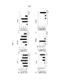
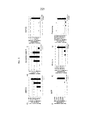
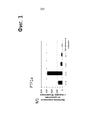
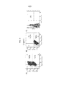
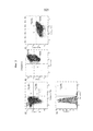
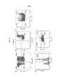
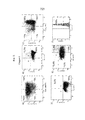
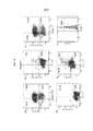
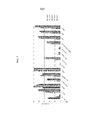
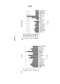
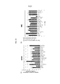
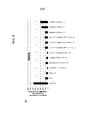
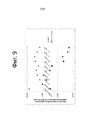
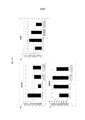
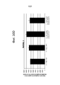
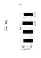
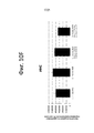
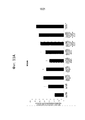
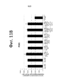
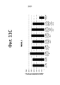
Classifications
-
- C—CHEMISTRY; METALLURGY
- C12—BIOCHEMISTRY; BEER; SPIRITS; WINE; VINEGAR; MICROBIOLOGY; ENZYMOLOGY; MUTATION OR GENETIC ENGINEERING
- C12N—MICROORGANISMS OR ENZYMES; COMPOSITIONS THEREOF; PROPAGATING, PRESERVING, OR MAINTAINING MICROORGANISMS; MUTATION OR GENETIC ENGINEERING; CULTURE MEDIA
- C12N5/00—Undifferentiated human, animal or plant cells, e.g. cell lines; Tissues; Cultivation or maintenance thereof; Culture media therefor
- C12N5/06—Animal cells or tissues; Human cells or tissues
- C12N5/0602—Vertebrate cells
- C12N5/0676—Pancreatic cells
-
- A—HUMAN NECESSITIES
- A61—MEDICAL OR VETERINARY SCIENCE; HYGIENE
- A61P—SPECIFIC THERAPEUTIC ACTIVITY OF CHEMICAL COMPOUNDS OR MEDICINAL PREPARATIONS
- A61P1/00—Drugs for disorders of the alimentary tract or the digestive system
- A61P1/18—Drugs for disorders of the alimentary tract or the digestive system for pancreatic disorders, e.g. pancreatic enzymes
-
- C—CHEMISTRY; METALLURGY
- C12—BIOCHEMISTRY; BEER; SPIRITS; WINE; VINEGAR; MICROBIOLOGY; ENZYMOLOGY; MUTATION OR GENETIC ENGINEERING
- C12N—MICROORGANISMS OR ENZYMES; COMPOSITIONS THEREOF; PROPAGATING, PRESERVING, OR MAINTAINING MICROORGANISMS; MUTATION OR GENETIC ENGINEERING; CULTURE MEDIA
- C12N2500/00—Specific components of cell culture medium
- C12N2500/30—Organic components
- C12N2500/38—Vitamins
-
- C—CHEMISTRY; METALLURGY
- C12—BIOCHEMISTRY; BEER; SPIRITS; WINE; VINEGAR; MICROBIOLOGY; ENZYMOLOGY; MUTATION OR GENETIC ENGINEERING
- C12N—MICROORGANISMS OR ENZYMES; COMPOSITIONS THEREOF; PROPAGATING, PRESERVING, OR MAINTAINING MICROORGANISMS; MUTATION OR GENETIC ENGINEERING; CULTURE MEDIA
- C12N2501/00—Active agents used in cell culture processes, e.g. differentation
- C12N2501/10—Growth factors
- C12N2501/115—Basic fibroblast growth factor (bFGF, FGF-2)
-
- C—CHEMISTRY; METALLURGY
- C12—BIOCHEMISTRY; BEER; SPIRITS; WINE; VINEGAR; MICROBIOLOGY; ENZYMOLOGY; MUTATION OR GENETIC ENGINEERING
- C12N—MICROORGANISMS OR ENZYMES; COMPOSITIONS THEREOF; PROPAGATING, PRESERVING, OR MAINTAINING MICROORGANISMS; MUTATION OR GENETIC ENGINEERING; CULTURE MEDIA
- C12N2501/00—Active agents used in cell culture processes, e.g. differentation
- C12N2501/10—Growth factors
- C12N2501/119—Other fibroblast growth factors, e.g. FGF-4, FGF-8, FGF-10
-
- C—CHEMISTRY; METALLURGY
- C12—BIOCHEMISTRY; BEER; SPIRITS; WINE; VINEGAR; MICROBIOLOGY; ENZYMOLOGY; MUTATION OR GENETIC ENGINEERING
- C12N—MICROORGANISMS OR ENZYMES; COMPOSITIONS THEREOF; PROPAGATING, PRESERVING, OR MAINTAINING MICROORGANISMS; MUTATION OR GENETIC ENGINEERING; CULTURE MEDIA
- C12N2501/00—Active agents used in cell culture processes, e.g. differentation
- C12N2501/10—Growth factors
- C12N2501/16—Activin; Inhibin; Mullerian inhibiting substance
-
- C—CHEMISTRY; METALLURGY
- C12—BIOCHEMISTRY; BEER; SPIRITS; WINE; VINEGAR; MICROBIOLOGY; ENZYMOLOGY; MUTATION OR GENETIC ENGINEERING
- C12N—MICROORGANISMS OR ENZYMES; COMPOSITIONS THEREOF; PROPAGATING, PRESERVING, OR MAINTAINING MICROORGANISMS; MUTATION OR GENETIC ENGINEERING; CULTURE MEDIA
- C12N2501/00—Active agents used in cell culture processes, e.g. differentation
- C12N2501/30—Hormones
- C12N2501/375—Thyroid stimulating hormone [TSH]
-
- C—CHEMISTRY; METALLURGY
- C12—BIOCHEMISTRY; BEER; SPIRITS; WINE; VINEGAR; MICROBIOLOGY; ENZYMOLOGY; MUTATION OR GENETIC ENGINEERING
- C12N—MICROORGANISMS OR ENZYMES; COMPOSITIONS THEREOF; PROPAGATING, PRESERVING, OR MAINTAINING MICROORGANISMS; MUTATION OR GENETIC ENGINEERING; CULTURE MEDIA
- C12N2501/00—Active agents used in cell culture processes, e.g. differentation
- C12N2501/40—Regulators of development
- C12N2501/415—Wnt; Frizzeled
-
- C—CHEMISTRY; METALLURGY
- C12—BIOCHEMISTRY; BEER; SPIRITS; WINE; VINEGAR; MICROBIOLOGY; ENZYMOLOGY; MUTATION OR GENETIC ENGINEERING
- C12N—MICROORGANISMS OR ENZYMES; COMPOSITIONS THEREOF; PROPAGATING, PRESERVING, OR MAINTAINING MICROORGANISMS; MUTATION OR GENETIC ENGINEERING; CULTURE MEDIA
- C12N2501/00—Active agents used in cell culture processes, e.g. differentation
- C12N2501/70—Enzymes
- C12N2501/72—Transferases (EC 2.)
- C12N2501/727—Kinases (EC 2.7.)
-
- C—CHEMISTRY; METALLURGY
- C12—BIOCHEMISTRY; BEER; SPIRITS; WINE; VINEGAR; MICROBIOLOGY; ENZYMOLOGY; MUTATION OR GENETIC ENGINEERING
- C12N—MICROORGANISMS OR ENZYMES; COMPOSITIONS THEREOF; PROPAGATING, PRESERVING, OR MAINTAINING MICROORGANISMS; MUTATION OR GENETIC ENGINEERING; CULTURE MEDIA
- C12N2501/00—Active agents used in cell culture processes, e.g. differentation
- C12N2501/999—Small molecules not provided for elsewhere
-
- C—CHEMISTRY; METALLURGY
- C12—BIOCHEMISTRY; BEER; SPIRITS; WINE; VINEGAR; MICROBIOLOGY; ENZYMOLOGY; MUTATION OR GENETIC ENGINEERING
- C12N—MICROORGANISMS OR ENZYMES; COMPOSITIONS THEREOF; PROPAGATING, PRESERVING, OR MAINTAINING MICROORGANISMS; MUTATION OR GENETIC ENGINEERING; CULTURE MEDIA
- C12N2506/00—Differentiation of animal cells from one lineage to another; Differentiation of pluripotent cells
- C12N2506/02—Differentiation of animal cells from one lineage to another; Differentiation of pluripotent cells from embryonic cells
-
- C—CHEMISTRY; METALLURGY
- C12—BIOCHEMISTRY; BEER; SPIRITS; WINE; VINEGAR; MICROBIOLOGY; ENZYMOLOGY; MUTATION OR GENETIC ENGINEERING
- C12N—MICROORGANISMS OR ENZYMES; COMPOSITIONS THEREOF; PROPAGATING, PRESERVING, OR MAINTAINING MICROORGANISMS; MUTATION OR GENETIC ENGINEERING; CULTURE MEDIA
- C12N2506/00—Differentiation of animal cells from one lineage to another; Differentiation of pluripotent cells
- C12N2506/22—Differentiation of animal cells from one lineage to another; Differentiation of pluripotent cells from pancreatic cells
Landscapes
- Health & Medical Sciences (AREA)
- Engineering & Computer Science (AREA)
- Life Sciences & Earth Sciences (AREA)
- Biomedical Technology (AREA)
- Organic Chemistry (AREA)
- Bioinformatics & Cheminformatics (AREA)
- Chemical & Material Sciences (AREA)
- Biotechnology (AREA)
- Genetics & Genomics (AREA)
- Wood Science & Technology (AREA)
- Zoology (AREA)
- General Health & Medical Sciences (AREA)
- Biochemistry (AREA)
- General Engineering & Computer Science (AREA)
- Microbiology (AREA)
- Cell Biology (AREA)
- Nuclear Medicine, Radiotherapy & Molecular Imaging (AREA)
- General Chemical & Material Sciences (AREA)
- Medicinal Chemistry (AREA)
- Chemical Kinetics & Catalysis (AREA)
- Pharmacology & Pharmacy (AREA)
- Animal Behavior & Ethology (AREA)
- Public Health (AREA)
- Veterinary Medicine (AREA)
- Micro-Organisms Or Cultivation Processes Thereof (AREA)
- Medicines Containing Material From Animals Or Micro-Organisms (AREA)
- Measuring Or Testing Involving Enzymes Or Micro-Organisms (AREA)
Abstract
Description
Перекрестные ссылки на смежные заявкиCross references to related applications
Настоящая заявка испрашивает преимущество предварительной заявки на патент США № 61/994,259, поданной 16 мая 2014 г., которая полностью включена в настоящий документ путем ссылки.This application claims the benefit of provisional US patent application No. 61 / 994,259, filed May 16, 2014, which is incorporated herein by reference in its entirety.
Область применения изобретенияScope of the invention
Настоящее изобретение относится к способам дифференцировки плюрипотентных стволовых клеток и клеткам и популяциям, получаемым в результате такой дифференцировки. В частности, изобретение относится к применению конкретных малых молекул для создания панкреатических эндокринных клеток и популяций таких клеток, которые показывают увеличенную экспрессию MAFA.The present invention relates to methods for differentiating pluripotent stem cells and cells and populations resulting from such differentiation. In particular, the invention relates to the use of specific small molecules to create pancreatic endocrine cells and populations of such cells, which show an increased expression of MAFA.
Предпосылки создания изобретенияBackground of the invention
Последние достижения в области заместительной клеточной терапии для лечения сахарного диабета 1-го типа и нехватка островков Лангерганса, выполненных с возможностью трансплантации, заставили обратить внимание на разработку источников инсулин-продуцирующих клеток, или бета-клеток, подходящих для приживления трансплантата. Один подход представляет собой получение функционализированных бета-клеток из плюрипотентных стволовых клеток, таких как эмбриональные стволовые клетки.Recent advances in cell replacement therapy for the treatment of
При эмбриональном развитии позвоночных плюрипотентная клетка дает начало группе клеток, содержащих три зародышевых листка (эктодерму, мезодерму и энтодерму), в ходе процесса, известного как «гаструляция». Ткани, такие как ткань щитовидной железы, вилочковой железы, поджелудочной железы, кишечника и печени, будут развиваться из энтодермы через промежуточную стадию. Промежуточная стадия данного процесса представляет собой образование дефинитивной энтодермы.During vertebrate embryonic development, the pluripotent cell gives rise to a group of cells containing three germ layers (ectoderm, mesoderm, and endoderm) in a process known as “gastrulation”. Tissues such as thyroid, thymus, pancreas, intestines and liver tissue will develop from the endoderm through an intermediate stage. The intermediate stage of this process is the formation of definitive endoderm.
К концу гаструляции энтодерма разделяется на передний и задний домены, которые можно распознать по экспрессии ряда факторов, однозначно выделяющих переднюю, среднюю и заднюю области энтодермы. Например, HHEX и SOX2 идентифицируют переднюю область, в то время как CDX1, 2 и 4 идентифицируют заднюю область энтодермы.By the end of gastrulation, the endoderm is divided into anterior and posterior domains, which can be recognized by the expression of a number of factors that uniquely distinguish the anterior, middle, and posterior regions of the endoderm. For example, HHEX and SOX2 identify the anterior region, while CDX1, 2 and 4 identify the posterior endoderm region.
Миграция ткани энтодермы приближает энтодерму к различным мезодермальным тканям, которые способствуют регионализации кишечной трубки. Это осуществляется с помощью большого числа секретируемых факторов, таких как факторы роста фибробластов (FGF), белки WNT, трансформирующие факторы роста бета (TGF-β), ретиноевая кислота и лиганды костного морфогенетического белка (BMP) и их антагонисты. Например, FGF4 и BMP способствуют экспрессии CDX2 в предполагаемой энтодерме задней кишки и подавляют экспрессию передних генов HHEX и SOX2 (2000 Development, 127:1563-1567). Было продемонстрировано, что сигнализация WNT также действует параллельно с сигнализацией FGF, способствуя развитию задней кишки и препятствуя зачаткам передней кишки (2007 Development, 134:2207-2217). Наконец, ретиноевая кислота, секретируемая мезенхимой, регулирует границу между передней и задней кишкой (2002 Curr. Biol., 12:1215-1220).Migration of the endoderm tissue brings the endoderm closer to various mesodermal tissues that contribute to the regionalization of the intestinal tube. This is accomplished with a large number of secreted factors, such as fibroblast growth factors (FGF), WNT proteins, transforming beta growth factors (TGF-β), retinoic acid and bone morphogenetic protein ligands (BMP) and their antagonists. For example, FGF4 and BMP promote the expression of CDX2 in the putative endoderm of the hindgut and inhibit the expression of the anterior HHEX and SOX2 genes (2000 Development, 127: 1563-1567). It has been demonstrated that WNT signaling also acts in parallel with FGF signaling, contributing to the development of the posterior intestine and inhibiting the buds of the anterior intestine (2007 Development, 134: 2207-2217). Finally, retinoic acid secreted by the mesenchyme regulates the border between the anterior and posterior intestine (2002 Curr. Biol., 12: 1215-1220).
Уровень экспрессии специфических факторов транскрипции можно применять для определения типа ткани. Во время преобразования дефинитивной энтодермы в примитивную кишечную трубку кишечная трубка становится разделенной на широкие домены, которые можно наблюдать на молекулярном уровне с помощью ограниченных паттернов экспрессии генов. Например, регионализованный домен поджелудочной железы в кишечной трубке демонстрирует очень высокую экспрессию PDX1 и очень низкую экспрессию CDX2 и SOX2. PDX1, NKX6.1/PTFlA и NKX2.2 высоко экспрессируются в ткани поджелудочной железы, а экспрессия CDX2 высока в кишечной ткани.The expression level of specific transcription factors can be used to determine the type of tissue. During the conversion of the definitive endoderm to the primitive intestinal tube, the intestinal tube becomes divided into broad domains that can be observed at the molecular level with the help of limited gene expression patterns. For example, a regionalized pancreatic domain in the intestinal tube shows very high expression of PDX1 and very low expression of CDX2 and SOX2. PDX1, NKX6.1 / PTFlA and NKX2.2 are highly expressed in pancreatic tissue, and CDX2 expression is high in intestinal tissue.
Образование поджелудочной железы происходит при дифференцировке дефинитивной энтодермы в панкреатическую энтодерму. Дорсальный и вентральный домены поджелудочной железы формируются из эпителия передней кишки. Передняя кишка также дает начало формированию пищевода, трахеи, легких, щитовидной железы, желудка, печени и системы желчных протоков.The formation of the pancreas occurs when the differentiation of the definitive endoderm into the pancreatic endoderm. The dorsal and ventral domains of the pancreas are formed from the epithelium of the anterior intestine. The anterior gut also gives rise to the formation of the esophagus, trachea, lungs, thyroid, stomach, liver, and bile duct system.
Клетки панкреатической энтодермы экспрессируют панкрео-дуоденальный, содержащий гомеобокс ген PDX1. В отсутствие PDX1 поджелудочная железа не развивается дальше образования вентрального и дорсального зачатков. Следовательно, экспрессия PDX1 является важным этапом органогенеза поджелудочной железы. Зрелая поджелудочная железа содержит как экзокринную, так и эндокринную ткани, которые образуются при дифференцировке панкреатической энтодермы.Pancreatic endoderm cells express pancreato-duodenal, containing the homeobox gene PDX1. In the absence of PDX1, the pancreas does not develop further than the formation of ventral and dorsal buds. Therefore, PDX1 expression is an important step in pancreatic organogenesis. The mature pancreas contains both exocrine and endocrine tissues, which are formed during the differentiation of pancreatic endoderm.
DʹAmour et al. описывают производство обогащенных культур дефинитивной энтодермы, полученной из человеческих эмбриональных стволовых клеток, в присутствии высокой концентрации активина и низкой концентрации сыворотки (Nature Biotechnology 2005, 23:1534-1541; патент США № 7,704,738). Трансплантация этих клеток под капсулу почки у мышей приводила к дифференцировке в более зрелые клетки с характеристиками ткани энтодермы (патент США № 7,704,738). Клетки дефинитивной энтодермы, полученные из человеческих эмбриональных стволовых клеток, могут быть дополнительно дифференцированы в PDX1-положительные клетки после добавления FGF-10 и ретиноевой кислоты (заявка на патент США № 2005/0266554). Последующая трансплантация этих клеток-предшественников панкреатических клеток в жировое тело иммунодефицитных мышей привела к образованию функционализированных панкреатических эндокринных клеток с последующей 3-4-месячной стадией созревания (патенты США № 7,534,608 и № 7,993,920).DʹAmour et al. describe the production of enriched cultures of definitive endoderm derived from human embryonic stem cells in the presence of a high concentration of activin and a low serum concentration (Nature Biotechnology 2005, 23: 1534-1541; US Patent No. 7,704,738). Transplantation of these cells under the kidney capsule in mice resulted in differentiation into more mature cells with endoderm tissue characteristics (US Patent No. 7,704,738). Definitive endoderm cells derived from human embryonic stem cells can be further differentiated into PDX1-positive cells after the addition of FGF-10 and retinoic acid (US Patent Application No. 2005/0266554). The subsequent transplantation of these progenitor cells of pancreatic cells into the fatty body of immunodeficient mice led to the formation of functionalized pancreatic endocrine cells, followed by a 3-4-month stage of maturation (US Patents No. 7,534,608 and No. 7,993,920).
Fisk et al. сообщают о системе получения клеток панкреатических островков из человеческих эмбриональных стволовых клеток (патент США № 7,033,831). В данном случае процесс дифференцировки был разделен на три стадии. Человеческие эмбриональные стволовые клетки сначала были дифференцированы в энтодерму при помощи комбинации бутирата натрия и активина A (патент США № 7,326,572). Впоследствии клетки культивировали с антагонистами BMP, такими как Ноггин, в комбинации с EGF или бета-целлюлином для генерирования PDX1-положительных клеток. Конечную дифференцировку индуцировали никотинамидом.Fisk et al. report a system for obtaining pancreatic islet cells from human embryonic stem cells (US Patent No. 7,033,831). In this case, the process of differentiation was divided into three stages. Human embryonic stem cells were first differentiated into the endoderm using a combination of sodium butyrate and activin A (US Patent No. 7,326,572). Subsequently, cells were cultured with BMP antagonists, such as Noggin, in combination with EGF or beta-celluline to generate PDX1-positive cells. The final differentiation was induced by nicotinamide.
Низкомолекулярные ингибиторы также применяют для индуцирования панкреатических эндокринных клеток-предшественников. Например, низкомолекулярные ингибиторы рецептора TGF-ß и рецепторов BMP (Development 2011, 138:861-871; Diabetes 2011, 60:239-247) применяют для значительного увеличения числа панкреатических эндокринных клеток. Дополнительно для генерирования клеток дефинитивной энтодермы или клеток-предшественников панкреатических клеток также применяют низкомолекулярные активаторы (Curr. Opin. Cell Biol. 2009, 21:727-732; Nature Chem. Biol. 2009, 5:258-265).Low molecular weight inhibitors are also used to induce pancreatic endocrine progenitor cells. For example, low molecular weight inhibitors of the TGF-ß receptor and BMP receptors (Development 2011, 138: 861-871; Diabetes 2011, 60: 239-247) are used to significantly increase the number of pancreatic endocrine cells. Additionally, low molecular weight activators (Curr. Opin. Cell Biol. 2009, 21: 727-732; Nature Chem. Biol. 2009, 5: 258-265) are also used to generate definitive endoderm cells or progenitor cells of pancreatic cells.
НВ9 (также известный как HIXB9 и MNX1) является транскрипционным активаторным белком bHLH, экпрессируемым на ранней стадии развития поджелудочной железы, начинающейся приблизительно на восьмой эмбриональный день. Экспрессия HB9 неравномерна и достигает пика примерно в день 10,5 в эпителии поджелудочной железы, будучи экспрессированной в клетках, экспрессирующих PDX1 и NKX6.1. Примерно в день 12,5 экспрессия HB9 снижается и на поздних стадиях ограничивается только β-клетками. У мышей, гомозиготных по нулевой мутации HB9, дорсальная доля поджелудочной железы не развивается (Nat. Genet. 23:67-70, 1999; Nat. Genet. 23:71-75, 1999). HB9-/β-клетки экспрессируют низкие уровни глюкозного транспортера, GLUT2 и NKX6.1. Кроме того, HB9 -/- поджелудочной железы демонстрирует значительное снижение числа инсулин-положительных клеток, в то же время не оказывая значительного влияния на экспрессию прочих гормонов поджелудочной железы. Таким образом, временной контроль HB9 чрезвычайно важен для нормального развития и функционирования β-клетки. В то время как о факторах, регулирующих экспрессию HB9 в β-клетках, известно немного, недавнее исследование на рыбе данио предполагает, что ретиноевая кислота может положительно регулировать экспрессию HB9 (Development, 138, 4597-4608, 2011).HB9 (also known as HIXB9 and MNX1) is a transcriptional activator protein bHLH, expressed at an early stage of pancreatic development, starting around the eighth embryonic day. Expression of HB9 is uneven and peaks at about 10.5 in the pancreatic epithelium, being expressed in cells expressing PDX1 and NKX6.1. On about day 12.5, HB9 expression is reduced and in the later stages is limited to β-cells only. In mice homozygous for the zero mutation of HB9, the dorsal lobe of the pancreas does not develop (Nat. Genet. 23: 67-70, 1999; Nat. Genet. 23: 71-75, 1999). HB9 / β cells express low levels of the glucose transporter, GLUT2 and NKX6.1. In addition, HB9 - / - pancreas demonstrates a significant decrease in the number of insulin-positive cells, while not significantly affecting the expression of other pancreatic hormones. Thus, the time control of HB9 is extremely important for the normal development and functioning of the β-cell. While little is known about the factors that regulate HB9 expression in β-cells, a recent study in zebrafish suggests that retinoic acid can positively regulate HB9 expression (Development, 138, 4597-4608, 2011).
В заявке на патент США № 13/998,883, полностью включенной в настоящий документ путем ссылки, было продемонстрировано, что трийодтиронин (Т3) может выступать в качестве индуктора экспрессии белка HB9 при дифференцировке клеток в β-клетки. Также в ней описаны способы получения панкреатических энтодермальных клеток, которые оказались успешными для NKX6.1, PDX1 и HB9, с помощью Т3 и/или Т4. Дополнительно и как описано в заявке на патент США № 13/998,884, полностью включенной в настоящий документ путем ссылки, было продемонстрировано, что экспрессия панкреатических эндокринных маркеров может быть значительно усилена посредством культивирования на поверхности раздела воздух-жидкость и с помощью Т3 и ингибиторов активин-рецептороподобной киназы (ALK) 5.In the application for US patent No. 13/998,883, fully incorporated herein by reference, it was demonstrated that triiodothyronine (T3) can act as an inducer of HB9 protein expression in differentiating cells into β cells. It also describes methods for producing pancreatic endodermal cells that have been successful for NKX6.1, PDX1 and HB9, using T3 and / or T4. Additionally, and as described in US Patent Application No. 13 / 998,884, fully incorporated herein by reference, it has been demonstrated that the expression of pancreatic endocrine markers can be significantly enhanced by cultivation at the air-liquid interface and by using T3 and activin inhibitors receptor-like kinase (ALK) 5.
Множество факторов транскрипции регулируют дифференцировку панкреатических эндокринных клеток в секретирующие инсулин β-клетки. Одним из этих факторов является гомолог А онкогена v-maf птичьей мышечно-апоневротической фибросаркомы (MAFA). Фактически считается, что MAFA может представлять собой основной регулятор стимулированной глюкозой секреции инсулина в β-клетках.Many transcription factors regulate the differentiation of pancreatic endocrine cells into insulin secreting β-cells. One of these factors is the homologue A of the oncogene v-maf avian musculoaponeurotic fibrosarcoma (MAFA). In fact, it is believed that MAFA may be the primary regulator of glucose-stimulated insulin secretion in β-cells.
В целом, процесс дифференцировки прогениторных клеток в функционализированные β-клетки проходит через различные стадии, и протоколы получения панкреатических клеток из прогениторных клеток, таких как человеческие плюрипотентные стволовые клетки, были в значительной степени улучшены. Несмотря на эти продвижения в исследованиях, каждый этап в процессе дифференцировки прогениторных клеток является уникальной задачей. Таким образом, сохраняется необходимость в дополнительной разработке протокола дифференцировки с целью получения функционализированных эндокринных клеток и в частности функционализированных β-клеток. В частности, желательно разработать процессы, в которых увеличена экспрессия MAFA в панкреатических эндокринных клетках.In general, the process of differentiation of progenitor cells into functionalized β-cells goes through various stages, and protocols for obtaining pancreatic cells from progenitor cells, such as human pluripotent stem cells, have been greatly improved. Despite these advances in research, each stage in the process of progenitor cell differentiation is a unique task. Thus, there remains a need for additional development of a differentiation protocol in order to obtain functionalized endocrine cells and, in particular, functionalized β-cells. In particular, it is desirable to develop processes in which the expression of MAFA is increased in pancreatic endocrine cells.
Краткое описание чертежейBrief Description of the Drawings
ФИГ. 1А-1М представляют собой графики, отображающие данные анализов ПЦР в реальном времени кратного изменения по сравнению с недифференцированными ЭС-клетками экспрессии гена PDX1, NKX6.1, PAX4, PAX6, NGN3, MAFA, ABCC8, хромогранина A, G6PC2, IAPP, инсулина, глюкагона и PTF1a в линии стволовых клеток H1, дифференцированных в соответствии с примером 1.FIG. 1A-1M are graphs showing real-time PCR analysis data of a fold change compared with undifferentiated ES cells expressing the PDX1, NKX6.1, PAX4, PAX6, NGN3, MAFA, ABFA8 gene, chromogranin A, G6PC2, IAPP, insulin, glucagon and PTF1a in the H1 stem cell line differentiated according to Example 1.
На ФИГ. 2A-2C показаны результаты цитометрии посредством сортировки клеток с активацией флуоресценции (FACS) клеток стадии 3, дифференцированных в соответствии с примером 1 и окрашенных следующим образом: PDX1 (ось X) окрашена вместе с Ki67 (ось Y) на ФИГ. 2A; PDX1 (ось X) окрашена вместе с CDX2 (ось Y) на ФИГ. 2B; и NKX6.1 на ФИГ. 2C.In FIG. 2A-2C show the results of cytometry by sorting cells with fluorescence activation (FACS) of
На ФИГ. 3A-3D показаны результаты цитометрии посредством сортировки клеток с активацией флуоресценции (FACS) клеток стадии 4, дифференцированных в соответствии с примером 1 и окрашенных следующим образом: хромогранин (ось X) окрашен вместе с NKX6.1 (ось Y) на ФИГ. 3A; PDX1 (ось X) окрашена вместе с Ki67 (ось Y) на ФИГ. 3B; NKX6.1 (ось X) окрашена вместе с инсулином (ось Y) на ФИГ. 3C; и NeuroD1 на ФИГ. 3D.In FIG. 3A-3D shows the results of cytometry by sorting cells with fluorescence activation (FACS) of
На ФИГ. 4A-4E показаны результаты цитометрии посредством сортировки клеток с активацией флуоресценции (FACS) клеток стадии 5, дифференцированных в соответствии с примером 1 и окрашенных следующим образом: хромогранин (ось X) окрашен вместе с NKX6.1 (ось Y) на ФИГ. 4A; PDX1 (ось X) окрашена вместе с Ki67 (ось Y) на ФИГ. 4B; и NKX6.1 (ось X) окрашена вместе с инсулином (ось Y) на ФИГ. 4C; NeuroD1 на ФИГ. 4D; и инсулин (ось X) окрашен вместе с глюкагоном (ось Y).In FIG. 4A-4E shows the results of cytometry by sorting cells with fluorescence activation (FACS) of
На ФИГ. 5A-5F показаны результаты цитометрии посредством сортировки клеток с активацией флуоресценции (FACS) клеток стадии 6, дифференцированных в соответствии с примером 1 и окрашенных следующим образом: хромогранин (ось X) окрашен вместе с NKX6.1 (ось Y) на ФИГ. 5A; PDX1 (ось X) окрашена вместе с Ki67 (ось Y) на ФИГ. 5B; и NKX6.1 (ось X) окрашена вместе с инсулином (ось Y) на ФИГ. 5C; PAX6 (ось X) окрашена вместе с Oct 3/4 (ось Y) на ФИГ. 5D; инсулин (ось X) окрашен вместе с глюкагоном (ось Y) на ФИГ. 5E; и FOXA2 на ФИГ. 5F.In FIG. 5A-5F shows the results of cytometry by sorting cells with fluorescence activation (FACS) of
На ФИГ. 6A-6F показаны результаты цитометрии посредством сортировки клеток с активацией флуоресценции (FACS) клеток стадии 7, дифференцированных в соответствии с примером 1 и окрашенных следующим образом: хромогранин (ось X) окрашен вместе с NKX6.1 (ось Y) на ФИГ. 6A; PDX1 (ось X) окрашена вместе с Ki67 (ось Y) на ФИГ. 6B; и NKX6.1 (ось X) окрашена вместе с инсулином (ось Y) на ФИГ. 6C; PAX6 (ось X) окрашена вместе с Oct 3/4 (ось Y) на ФИГ. 6D; инсулин (ось X) окрашен вместе с глюкагоном (ось Y) на ФИГ. 6E; и FOXA2 на ФИГ. 6F.In FIG. 6A-6F show the results of cytometry by sorting cells with fluorescence activation (FACS) of
ФИГ. 7 представляет собой график выраженной в процентах экспрессии множества панкреатических энтодермальных маркеров (FOXA2, PDX1, NKX6.1), маркера недифференцированных ЭС-клеток (Oct3/4), эндокринных маркеров (PAX6, ISl-1, NKX2.2, хромогранин) и гормона (инсулин, глюкагон) в клетках стадии 3-7, дифференцированных в соответствии с примером 1.FIG. 7 is a graph of the percentage expression of a plurality of pancreatic endodermal markers (FOXA2, PDX1, NKX6.1), a marker of undifferentiated ES cells (Oct3 / 4), endocrine markers (PAX6, ISl-1, NKX2.2, chromogranin) and the hormone (insulin, glucagon) in cells of stage 3-7, differentiated in accordance with example 1.
ФИГ. 8A-8E представляют собой графики, отображающие данные анализов ПЦР в реальном времени кратного изменения экспрессии инсулина и MAFA в дифференцированных клетках по сравнению с недифференцированными клетками после обработки малыми молекулами на стадии 6-7.FIG. 8A-8E are graphs showing real-time PCR analysis data of a fold change in insulin and MAFA expression in differentiated cells compared to undifferentiated cells after treatment with small molecules in step 6-7.
ФИГ. 9 представляет собой график, отображающий данные анализов ПЦР в реальном времени кратного изменения экспрессии AXL и GAS6 в дифференцированных клетках из примера 4 по сравнению с недифференцированными клетками.FIG. 9 is a graph showing the real-time PCR analysis data of a fold change in the expression of AXL and GAS6 in the differentiated cells of Example 4 as compared to undifferentiated cells.
ФИГ. 10A-F представляют собой графики данных анализов ПЦР в реальном времени кратного изменения экспрессии MAFA, UCN3, G6PC2, NKX6.1, PDX1 и инсулина в дифференцированных клетках по сравнению с недифференцированными клетками после обработки малыми молекулами на стадии 7 в соответствии с примером 6.FIG. 10A-F are graphs of real-time PCR data from fold changes in the expression of MAFA, UCN3, G6PC2, NKX6.1, PDX1 and insulin in differentiated cells compared to undifferentiated cells after treatment with small molecules in
ФИГ. 11A-D представляют собой графики, отображающие данные анализов ПЦР в реальном времени кратного изменения экспрессии MAFA, PDX1, NKX6.1 и инсулина в дифференцированных клетках по сравнению с недифференцированными клетками после обработки малыми молекулами на стадии 7 в соответствии с примером 7.FIG. 11A-D are graphs showing real-time PCR analysis of the fold change in expression of MAFA, PDX1, NKX6.1 and insulin in differentiated cells compared to undifferentiated cells after treatment with small molecules at
Подробное описание изобретенияDetailed Description of the Invention
Следующее подробное описание изобретения будет более понятно при изучении вместе с приложенными фигурами. В целях иллюстрирования изобретения на фигурах показаны варианты осуществления настоящего изобретения. Однако изобретение не ограничивается приведенными точными конструкциями, примерами и устройствами. Для ясности описания, а не в целях ограничения, подробное описание изобретения разделено на подразделы, описывающие или иллюстрирующие конкретные элементы, варианты осуществления или области применения настоящего изобретения.The following detailed description of the invention will be more clearly understood when studying together with the attached figures. In order to illustrate the invention, the figures show embodiments of the present invention. However, the invention is not limited to the exact structures, examples and devices. For clarity of description, and not for purposes of limitation, the detailed description of the invention is divided into subsections describing or illustrating specific elements, embodiments or uses of the present invention.
Настоящее изобретение относится к получению панкреатических эндокринных клеток более зрелого фенотипа посредством обработки менее зрелых панкреатических эндокринных клеток конкретными малыми молекулами. В определенных вариантах осуществления изобретения панкреатические эндокринные клетки культивируют, за одну или более стадий, в присутствии малых молекул, которые представляют собой один или более из ингибитора протеин метилтрансферазы, ингибитора киназы Aurora и ингибитора киназы S6 рибосомального белка p90 (RSK). Таким образом, в настоящем изобретении предложены клеточные культуры для дифференцировки плюрипотентных стволовых клеток в клетки, показывающие характеристики панкреатических эндокринных клеток, а также дифференцировочная среда, которая инициирует и облегчает такую дифференцировку, и дифференцированные клетки и клеточные популяции, получаемые в результате дифференцировки. Способы изобретения обеспечивают образование популяции панкреатических эндокринных клеток, причем, по меньшей мере, 10%, предпочтительно, по меньшей мере, 20%, более предпочтительно, по меньшей мере, 30% и наиболее предпочтительно, по меньшей мере, 50% клеток экспрессируют единственный гормон инсулин и являются PDX1-, NKX6.1- и MAFA-положительными.The present invention relates to the production of pancreatic endocrine cells of a more mature phenotype by treating less mature pancreatic endocrine cells with specific small molecules. In certain embodiments of the invention, pancreatic endocrine cells are cultured, in one or more stages, in the presence of small molecules, which are one or more of the methyltransferase protein inhibitor, the Aurora kinase inhibitor, and the p6 ribosomal kinase inhibitor RSK. Thus, the present invention proposes cell cultures for differentiating pluripotent stem cells into cells showing the characteristics of pancreatic endocrine cells, as well as a differentiating environment that initiates and facilitates such differentiation, and differentiated cells and cell populations resulting from differentiation. The methods of the invention provide a population of pancreatic endocrine cells, with at least 10%, preferably at least 20%, more preferably at least 30%, and most preferably at least 50% of the cells express a single hormone insulin and are PDX1-, NKX6.1- and MAFA-positive.
Преимущественно и предпочтительно клеточные культуры и дифференцировочную среду изобретения можно применять в сочетании с дифференцировкой на поверхности раздела воздух-жидкость. Культивирование на поверхности раздела воздух-жидкость может происходить на всех стадиях процесса дифференцировки от плюрипотентных стволовых клеток до панкреатических эндокринных клеток зрелого фенотипа или может включать в себя культивирование планарной культуры, погруженной в среду на ранних стадиях дифференцировки, с последующим культивированием на поверхности раздела воздух-жидкость во время одной или нескольких более поздних стадий дифференцировки. Более предпочтительно процесс изобретения включает в себя комбинацию культивирования плюрипотентных стволовых клеток на поддерживающей поверхности, погруженной в среду, на ранних стадиях и впоследствии культивирование на поверхности раздела воздух-жидкость на более поздних стадиях дифференцировки. В таких вариантах осуществления клетки могут быть первоначально посеяны на твердую поверхность для погруженного культивирования и впоследствии удалены с твердой поверхности и пересеяны на пористую поверхность для культивирования на поверхности раздела воздух-жидкость. Альтернативно клетки могут быть изначально посеяны на пористую поддерживающую поверхность, которая впоследствии погружается в среду, на ранних стадиях дифференцировки и впоследствии расположены на поверхности раздела воздух-жидкость на более поздних стадиях дифференцировки.Advantageously and preferably, cell cultures and the differentiation medium of the invention can be used in combination with differentiation at the air-liquid interface. Cultivation at the air-liquid interface can occur at all stages of the differentiation process, from pluripotent stem cells to pancreatic endocrine cells of the mature phenotype, or it may include the cultivation of a planar culture immersed in the medium at the early stages of differentiation, followed by cultivation at the air-liquid interface. during one or more later stages of differentiation. More preferably, the process of the invention includes a combination of cultivating pluripotent stem cells on a supporting surface immersed in the medium at early stages and subsequently cultivating at the air-liquid interface at later stages of differentiation. In such scenarios, cells can be initially sown on a solid surface for submerged cultivation and subsequently removed from the solid surface and transplanted onto a porous surface for cultivation at the air-liquid interface. Alternatively, cells can be initially seeded on a porous supporting surface, which is subsequently immersed in the medium, in the early stages of differentiation, and subsequently located on the air-liquid interface in the later stages of differentiation.
В еще одном варианте осуществления дифференцировку на одной или более стадиях также выполняют в присутствии одного или более из Т3, Т4, их аналогов и необязательно, но предпочтительно с ингибитором активин-рецептороподобной киназы 5 (ALK 5). В предпочтительном варианте осуществления популяцию панкреатических энтодермальных/эндокринных клеток-предшественников культивируют в среде, содержащей один или более из Т3, Т4, их аналогов и ингибитор ALK5, в панкреатические эндокринные клетки. В более предпочтительном варианте осуществления полученные панкреатические эндокринные клетки дополнительно дифференцируются в присутствии среды, содержащей один или более из Т3, Т4, их аналогов и ингибитор ALK5.In yet another embodiment, the differentiation in one or more stages is also performed in the presence of one or more of T3, T4, their analogues, and optionally, but preferably with an inhibitor of activin receptor-like kinase 5 (ALK 5). In a preferred embodiment, the population of pancreatic endodermal / endocrine progenitor cells is cultured in medium containing one or more of T3, T4, their analogues and the ALK5 inhibitor, into pancreatic endocrine cells. In a more preferred embodiment, the resulting pancreatic endocrine cells are further differentiated in the presence of a medium containing one or more of T3, T4, their analogues and the ALK5 inhibitor.
Особым открытием изобретения является то, что обработка панкреатических энтодермальных клеток комбинацией одного или более из ингибитора ALK5 и агониста рецепторов щитовидной железы с последующим культивированием полученных панкреатических эндокринных клеток в комбинации с одним или более из ингибитора протеин метилтрансферазы DOT1L, ингибитора киназы Aurora и ингибитора RSK значительно увеличивает число клеток в популяции, экспрессирующих единственный гормон инсулин, MAFA, PDX1 и NKX6.1, а также увеличивает уровень экспрессии MAFA в клетках. Изобретение является особенно полезным при применении в сочетании с дифференцировкой на поверхности раздела воздух-жидкость.A special discovery of the invention is that the treatment of pancreatic endodermal cells with a combination of one or more of the ALK5 inhibitor and a thyroid receptor agonist, followed by cultivation of the resulting pancreatic endocrine cells in combination with one or more of the inhibitor DOT1L methyltransferase inhibitor, Aurora kinase inhibitor and RSK inhibitor. the number of cells in the population expressing a single hormone insulin, MAFA, PDX1 and NKX6.1, also increases the level of expression of MAFA in the cells. The invention is particularly useful when used in combination with differentiation at the air-liquid interface.
ОпределенияDefinitions
Стволовые клетки представляют собой недифференцированные клетки, образованные со способностью, на одноклеточном уровне, к самообновлению и дифференцировке. Стволовые клетки могут продуцировать клетки-потомки, включая самообновляющиеся прогениторные клетки, необновляющиеся прогениторные клетки и окончательно дифференцированные клетки. Стволовые клетки также характеризуются своей способностью дифференцироваться в условиях in vitro в функционализированные клетки различных линий дифференцировки из множества зародышевых листков (энтодермы, мезодермы и эктодермы). Стволовые клетки также дают начало тканям множества зародышевых листков после трансплантации и вносят существенный вклад в образование большинства, если не всех, тканей после инъекции в бластоцисты.Stem cells are undifferentiated cells formed with the ability, on a single-cell level, to self-renew and differentiate. Stem cells can produce progeny cells, including self-renewing progenitor cells, non-renewing progenitor cells, and finally differentiated cells. Stem cells are also characterized by their ability to differentiate in vitro into functionalized cells of different differentiation lines from a variety of germ layers (endoderm, mesoderm, and ectoderm). Stem cells also give rise to the tissues of many germ layers after transplantation and make a significant contribution to the formation of most, if not all, of the tissues after injection into the blastocyst.
Стволовые клетки классифицируются по потенциалу развития. Плюрипотентные стволовые клетки способны преобразовываться во все виды эмбриональных клеток.Stem cells are classified by developmental potential. Pluripotent stem cells are able to transform into all types of embryonic cells.
Дифференцировка представляет собой процесс, посредством которого неспециализированная («некоммитированная») или менее специализированная клетка приобретает свойства специализированной клетки, например нервной клетки или мышечной клетки. Дифференцированная клетка представляет собой клетку, занявшую более специализированное («коммитированное») положение в клеточной линии дифференцировки. Термин «коммитированная» применительно к процессу дифференцировки относится к клетке, дошедшей в процессе дифференцировки до стадии, с которой в нормальных условиях она продолжит дифференцироваться до конкретного типа клеток или подмножества типов клеток и не сможет в нормальных условиях дифференцироваться в иной тип клеток или вернуться к менее дифференцированному типу клеток. «Дедифференцировка» обозначает процесс, в ходе которого клетка возвращается к менее специализированному (или коммитированному) положению в пределах клеточной линии дифференцировки. В настоящем документе «клеточная линия дифференцировки» обозначает наследственность клетки, т. е. из каких клеток произошла данная клетка и каким клеткам она может дать начало. Клеточная линия дифференцировки размещает клетку в пределах наследственной схемы развития и дифференцировки. Маркер, специфичный для линии дифференцировки, относится к характеристике, специфически ассоциированной с фенотипом клеток интересующей линии дифференцировки, и его можно применять для оценки дифференцировки некоммитированной клетки в клетки интересующей линии дифференцировки.Differentiation is the process by which a unspecialized (“uncommitted”) or less specialized cell acquires the properties of a specialized cell, such as a nerve cell or muscle cell. A differentiated cell is a cell that occupies a more specialized (“committed”) position in the cell lineage line. The term "committed" in relation to the process of differentiation refers to a cell that has reached the stage of differentiation to a stage from which under normal conditions it will continue to differentiate to a particular cell type or a subset of cell types and will not be able to differentiate to a different cell type or return to less differentiated cell type. “Dedifferentiation” refers to the process by which a cell returns to a less specialized (or committed) position within the cell line of differentiation. In this document, the "cell line of differentiation" refers to the heredity of the cell, i.e. from which cells the given cell originated and which cells it can give rise to. The cell lineage differentiates the cell within the hereditary pattern of development and differentiation. A differentiation line-specific marker refers to a characteristic specifically associated with the phenotype of cells of a differentiation line of interest, and can be used to assess the differentiation of a non-committed cell into cells of a differentiation line of interest.
В настоящем документе «маркеры» представляют собой молекулы нуклеиновых кислот или полипептидов, которые дифференциально экспрессируются в интересующей клетке. В данном контексте под дифференциальной экспрессией понимают повышенный уровень положительного маркера и пониженный уровень отрицательного маркера по сравнению с недифференцированной клеткой или клеткой на другой стадии дифференцировки. Обнаруживаемый уровень маркерной нуклеиновой кислоты или полипептида в интересующих клетках оказывается значительно выше или ниже по сравнению с другими клетками, что позволяет идентифицировать интересующую клетку и отличать ее от других клеток с помощью любого из множества способов, известных в данной области.In this document, "markers" are nucleic acid molecules or polypeptides that are differentially expressed in the cell of interest. In this context, differential expression is understood to mean an increased level of a positive marker and a reduced level of a negative marker compared with an undifferentiated cell or cell at a different stage of differentiation. The detectable level of the marker nucleic acid or polypeptide in the cells of interest is significantly higher or lower than other cells, which allows the cell of interest to be identified and distinguished from other cells using any of a variety of methods known in the art.
В настоящем документе клетка является «положительной по» установленному маркеру, «положительной» или обозначается со знаком «+», если установленный маркер обнаруживают в клетке в достаточном количестве. Аналогично, клетка является «отрицательной по» установленному маркеру, «отрицательной» или обозначается со знаком «-», если установленный маркер не обнаруживают в клетке в достаточном количестве. В частности, положительность при цитометрии посредством сортировки клеток с активацией флуоресценции (FACS), как правило, означает уровень более чем около 2%, в то время как отрицательный предел для FACS, как правило, составляет менее чем около 1%. Положительность при цитометрии с помощью полимерной цепной реакции (ПЦР), как правило, означает около или менее 30 циклов (Cts); при этом отрицательность для ПЦР, как правило, составляет более чем около 31 цикла.In this document, a cell is “positive for” an established marker, “positive”, or is indicated with a “+” sign if the established marker is found in a cell in sufficient quantity. Similarly, a cell is “negative by” set marker, “negative” or is indicated with a “-” sign if the set marker is not found in the cell in sufficient quantity. In particular, cytometric positivity by sorting cells with fluorescence activation (FACS) typically means a level of more than about 2%, while a negative limit for FACS is usually less than about 1%. Positivity in cytometry using a polymer chain reaction (PCR) usually means about or less than 30 cycles (Cts); however, the negativity for PCR is usually more than about 31 cycles.
При попытках воспроизвести дифференцировку плюрипотентных стволовых клеток в функционализированные панкреатические эндокринные клетки в статичных клеточных культурах в условиях in vitro процесс дифференцировки часто рассматривают как прохождение через несколько последовательных стадий. В частности, процесс дифференцировки обычно рассматривают как прохождение через множество стадий. В этой поэтапной дифференцировке «стадия 1» обозначает первый этап в процессе дифференцировки, то есть дифференцировку плюрипотентных стволовых клеток в клетки, экспрессирующие маркеры, характерные для клеток дефинитивной энтодермы («клетки стадии 1»). «Стадия 2» обозначает второй этап, то есть дифференцировку клеток, экспрессирующих маркеры, характерные для клеток дефинитивной энтодермы, в клетки, экспрессирующие маркеры, характерные для клеток кишечной трубки («клетки стадии 2»). «Стадия 3» обозначает третий этап, то есть дифференцировку клеток, экспрессирующих маркеры, характерные для клеток кишечной трубки, в клетки, экспрессирующие маркеры, характерные для энтодермальных клеток передней кишки («клетки стадии 3»). «Стадия 4» обозначает четвертый этап, то есть дифференцировку клеток, экспрессирующих маркеры, характерные для энтодермальных клеток передней кишки, в клетки, экспрессирующие маркеры, характерные для панкреатических клеток-предшественников передней кишки («клетки стадии 4»). «Стадия 5» обозначает пятый этап, то есть дифференцировку клеток, экспрессирующих маркеры, характерные для панкреатических клеток-предшественников передней кишки, в клетки, экспрессирующие маркеры, характерные для панкреатических энтодермальных клеток и/или панкреатических эндокринных клеток-предшественников (совместно обозначаемых как «клетки стадии 5» или альтернативно «панкреатические энтодермальные/эндокринные клетки-предшественники»). «Стадия 6» обозначает шестой этап, то есть дифференцировку клеток, экспрессирующих маркеры, характерные для панкреатических энтодермальных/эндокринных клеток-предшественников, в клетки, экспрессирующие маркеры, характерные для панкреатических эндокринных клеток, которые представляют собой незрелые бета-клетки («клетки стадии 6»). Клетки стадии 6 экспрессируют единственный гормон инсулин и являются PDX1-, NKX6.1- и хромогранин-положительными. В процессе и с целью продуцирования популяций клеток изобретения применяется седьмой этап (стадия 7), который обозначает дифференцировку клеток, экспрессирующих маркеры, характерные для панкреатических эндокринных клеток, которые представляют собой незрелые бета-клетки, в клетки, экспрессирующие маркеры, характерные для панкреатических эндокринных клеток, которые представляют собой созревающие бета-клетки и которые обладают более зрелым фенотипом по сравнению с клетками стадии 6. Под выражением «клетки стадии 7» подразумевается панкреатическая эндокринная клетка, которая является инсулин(единственный гормон)-, MAFA-, NKX6.1- и PDX1-положительной, но также экспрессирует MAFA в более высокой концентрации, чем незрелая бета-клетка. Дополнительно популяция клеток, полученная при выполнении стадии 7, имеет больший процент MAFA-положительных и экспрессирующих единственный гормон инсулин клеток по сравнению с популяциями клеток стадии 6.When trying to reproduce the differentiation of pluripotent stem cells into functionalized pancreatic endocrine cells in static cell cultures in vitro, the process of differentiation is often considered as passing through several successive stages. In particular, the process of differentiation is usually considered as passing through many stages. In this phased differentiation, “
Необходимо отметить, что не все клетки в отдельно взятой популяции проходят через эти стадии с одинаковой скоростью. Вследствие этого в клеточных культурах в условиях in vitro нередко обнаруживается наличие клеток, менее или более дифференцированных по сравнению с большинством клеток, присутствующих в популяции, особенно на более поздних стадиях дифференцировки. Например, нередко наблюдается появление маркеров, характерных для панкреатических эндокринных клеток, во время стадии 5 культивирования клеток. Для иллюстративных нужд настоящего изобретения в настоящем документе описаны характеристики различных типов клеток, связанных с определенными выше стадиями.It should be noted that not all cells in a single population pass through these stages at the same rate. As a consequence, cell cultures in vitro often reveal the presence of cells that are less or more differentiated compared to most of the cells present in the population, especially in the later stages of differentiation. For example, markers characteristic of pancreatic endocrine cells are often observed during
В настоящем документе «дефинитивная энтодерма» обозначает клетки, обладающие характеристиками клеток, происходящих от эпибласта при гаструляции, и формирующие желудочно-кишечный тракт и его производные. Клетки дефинитивной энтодермы экспрессируют, по меньшей мере, один из следующих маркеров: FOXA2 (также известный как ядерный фактор гепатоцитов 3-β (HNF3-β)), GATA4, SOX17, CXCR4, Brachyury, Cerberus, OTX2, goosecoid, C-Kit, CD99 и MIXL1. Маркеры, характерные для клеток дефинитивной энтодермы, представляют собой CXCR4, FOXA2 и SOX17. Таким образом, клетки дефинитивной энтодермы могут характеризоваться своей экспрессией CXCR4, FOXA2 и SOX17. Дополнительно, в зависимости от длительности времени, на протяжении которого клеткам позволяется оставаться на стадии 1, можно наблюдать рост в HNF4α.In this document, "definitive endoderm" refers to cells that have characteristics of cells derived from epiblast during gastrulation, and that form the gastrointestinal tract and its derivatives. Definitive endoderm cells express at least one of the following markers: FOXA2 (also known as hepatocyte 3-β nuclear factor (HNF3-β)), GATA4, SOX17, CXCR4, Brachyury, Cerberus, OTX2, goosecoid, C-Kit, CD99 and MIXL1. Markers characteristic of definitive endoderm cells are CXCR4, FOXA2 and SOX17. Thus, definitive endoderm cells can be characterized by their expression of CXCR4, FOXA2 and SOX17. Additionally, depending on the length of time during which cells are allowed to remain in
В настоящем документе «клетки кишечной трубки» обозначает клетки, полученные из дефинитивной энтодермы, из которых могут образоваться все энтодермальные органы, такие как легкие, печень, поджелудочная железа, желудок и кишечник. Клетки кишечной трубки могут характеризоваться своей постоянно растущей экспрессией HNF4α, выше, чем экспрессия клеток дефинитивной энтодермы. Например, рост экспрессии HNF4α в мРНК в десять-сорок раз можно наблюдать на стадии 2.In this document, “intestinal tube cells” refers to cells derived from the definitive endoderm from which all endodermal organs can be formed, such as the lungs, liver, pancreas, stomach and intestines. Cells of the intestinal tube can be characterized by their ever-growing expression of HNF4α, higher than the expression of definitive endoderm cells. For example, the growth of HNF4α expression in mRNA ten to forty times can be observed in
В настоящем документе «энтодерма передней кишки» обозначает клетки, из которых образуются пищевод, легкие, желудок, печень, поджелудочная железа, желчный пузырь и часть двенадцатиперстной кишки. Клетки энтодермы передней кишки экспрессируют, по меньшей мере, один из следующих маркеров: PDX1, FOXA2, CDX2, SOX2 и HNF4α. Клетки энтодермы передней кишки могут характеризоваться ростом экспрессии PDX1 по сравнению с клетками кишечной трубки. Например, более пятидесяти процентов клеток в культурах стадии 3 типично экспрессируют PDX1.In this document, the "foregut endoderm" refers to the cells from which the esophagus, lungs, stomach, liver, pancreas, gallbladder and part of the duodenum are formed. Cells of the foregut endoderm express at least one of the following markers: PDX1, FOXA2, CDX2, SOX2 and HNF4α. Cells of the foregut endoderm can be characterized by an increase in PDX1 expression compared with intestinal tube cells. For example, more than fifty percent of cells in
В настоящем документе «панкреатические клетки-предшественники передней кишки» обозначают клетки, экспрессирующие, по меньшей мере, один из следующих маркеров: PDX1, NKX6.1, HNF6, NGN3, SOX9, PAX4, PAX6, ISL1, гастрин, FOXA2, PTF1a, PROX1 и HNF4α. Панкреатические клетки-предшественники передней кишки могут характеризоваться как положительные по отношению к экспрессии, по меньшей мере, одного из PDX1, NKX6.1 и SOX9.In this document, "pancreatic progenitor cells of the foregut" designate cells expressing at least one of the following markers: PDX1, NKX6.1, HNF6, NGN3, SOX9, PAX4, PAX6, ISL1, gastrin, FOXA2, PTF1a, PROX1 and HNF4α. Pancreatic progenitor cells of the anterior gut may be characterized as positive for the expression of at least one of PDX1, NKX6.1 and SOX9.
В настоящем документе «панкреатические энтодермальные клетки» обозначают клетки, экспрессирующие, по меньшей мере, один из следующих маркеров: PDX1, NKX6.1, HNF1β, PTF1α, HNF6, HNF4α, SOX9, NGN3, гастрин; HB9 или PROX1. Панкреатические энтодермальные клетки могут характеризоваться отсутствием у них существенной экспрессии CDX2 или SOX2.In this document, "pancreatic endodermal cells" means cells expressing at least one of the following markers: PDX1, NKX6.1, HNF1β, PTF1α, HNF6, HNF4α, SOX9, NGN3, gastrin; HB9 or PROX1. Pancreatic endodermal cells can be characterized by their lack of significant expression of CDX2 or SOX2.
В настоящем документе «панкреатические эндокринные клетки-предшественники» обозначают панкреатические энтодермальные клетки, способные стать панкреатической клеткой, экспрессирующей гормоны. Панкреатические эндокринные клетки-предшественники экспрессируют, по меньшей мере, один из следующих маркеров: NGN3, NKX2.2, NeuroD11, ISL1, PAX4, PAX6 или ARX. Панкреатические эндокринные клетки-предшественники могут характеризоваться своей экспрессией NKX2.2 и NeuroD11.In this document, “pancreatic endocrine progenitor cells” designate pancreatic endodermal cells capable of becoming a pancreatic hormone-expressing cell. Pancreatic endocrine progenitor cells express at least one of the following markers: NGN3, NKX2.2, NeuroD11, ISL1, PAX4, PAX6 or ARX. Pancreatic endocrine progenitor cells can be characterized by their expression of NKX2.2 and NeuroD11.
В настоящем документе «панкреатические эндокринные клетки» обозначают клетки, способные экспрессировать, по меньшей мере, один из следующих гормонов: инсулин, глюкагон, соматостатин, грелин и панкреатический полипептид. В дополнение к этим гормонам маркеры, характерные для панкреатических эндокринных клеток, включают в себя один или более из NeuroD1, ISL1, PDX1, NKX6.1, ARX, NKX2.2, HB9 и PAX6. Одно подмножество панкреатических эндокринных клеток представляет собой «незрелые бета-клетки», которые представляют собой клетки, способные экспрессировать инсулин (но не глюкагон), соматостатин, грелин и панкреатический полипептид. Дополнительно маркеры, характерные для незрелых бета-клеток, включают в себя один или более из NeuroD1, ISL1, PDX1, NKX6.1, NKX2.2, HB9 и PAX6. Второе подмножество панкреатических эндокринных клеток представляет собой «созревающие бета-клетки», которые представляют собой клетки, способные экспрессировать инсулин (но не глюкагон), соматостатин, грелин и панкреатический полипептид. Дополнительно маркеры, характерные для созревающих бета-клеток, включают в себя один или более из NeuroD1, ISL1, PDX1, NKX6.1, NKX2.2, HB9, PAX6 и MAFA. Еще одно подмножество панкреатических эндокринных клеток представляет собой клетки, экспрессирующие маркеры, характерные для зрелых бета-клеток, которые могут характеризоваться своей экспрессией PDX1, NKX2.2, NKX6.1, NeuroD1, ISL1, HNF3β, HB9, MAFA и PAX6, а также выбросом инсулина в ответ на глюкозную нагрузку, которые являются устойчивыми и увеличенными по сравнению с менее зрелыми бета-клетками..In this document, "pancreatic endocrine cells" refers to cells capable of expressing at least one of the following hormones: insulin, glucagon, somatostatin, ghrelin, and pancreatic polypeptide. In addition to these hormones, markers characteristic of pancreatic endocrine cells include one or more of NeuroD1, ISL1, PDX1, NKX6.1, ARX, NKX2.2, HB9 and PAX6. One subset of pancreatic endocrine cells is “immature beta cells,” which are cells capable of expressing insulin (but not glucagon), somatostatin, ghrelin, and pancreatic polypeptide. Additionally, markers characteristic of immature beta cells include one or more of NeuroD1, ISL1, PDX1, NKX6.1, NKX2.2, HB9 and PAX6. The second subset of pancreatic endocrine cells is “maturing beta cells”, which are cells capable of expressing insulin (but not glucagon), somatostatin, ghrelin, and pancreatic polypeptide. Additionally, markers characteristic of maturing beta cells include one or more of NeuroD1, ISL1, PDX1, NKX6.1, NKX2.2, HB9, PAX6 and MAFA. Another subset of pancreatic endocrine cells is cells expressing markers characteristic of mature beta cells, which can be characterized by their expression of PDX1, NKX2.2, NKX6.1, NeuroD1, ISL1, HNF3β, HB9, MAFA and PAX6, as well as release insulin in response to glucose load, which are stable and increased compared to less mature beta cells ..
В настоящем документе «поверхность раздела воздух-жидкость» или «ALI» обозначает поверхность раздела воздух-жидкость, которая присутствует в открытом сосуде для культивирования или сосуде для культивирования, частично наполненном средой. Хотя в настоящем документе для удобства применяется термин «воздух», изобретение не ограничивается смесью газов и композиций, находящихся в окружающей среде. Изобретение детально рассматривает и включает в себя газообразные смеси, имеющие композиции, отличные от окружающей среды, включая, например, смеси, обогащенные определенным компонентом или в которых содержание определенного компонента понижено или исключено.In this document, an “air-liquid interface” or “ALI” refers to an air-liquid interface that is present in an open culture vessel or culture vessel partially filled with medium. Although the term “air” is used for convenience in this document, the invention is not limited to a mixture of gases and compositions in the environment. The invention considers in detail and includes gaseous mixtures having compositions different from the environment, including, for example, mixtures enriched in a specific component or in which the content of a specific component is reduced or eliminated.
В настоящем документе взаимозаменяемо применяются выражения «d1», «1d» и «день 1»; «d2», «2d» и «день 2» и так далее. Эти комбинации цифр и букв обозначают конкретный день инкубации на различных стадиях в процессе постадийного протокола дифференцировки настоящей заявки.In this document, the expressions “d1”, “1d” and “
Термин «LDN-193189» обозначает ((6-(4-(2-(пиперидин-1-ил)этокси)фенил)-3-(пиридин-4-ил)пиразоло[1,5-a]пиримидин, гидрохлорид)), ингибитор рецептора BMP, который можно приобрести в компании Shanghai ChemPartner, Co., LTD.The term "LDN-193189" means (((6- (4- (2- (piperidin-1-yl) ethoxy) phenyl) -3- (pyridin-4-yl) pyrazolo [1,5-a] pyrimidine, hydrochloride) ), a BMP receptor inhibitor available from Shanghai ChemPartner, Co., LTD.
Характеристики, источник, размножение и культивирование плюрипотентных стволовых клетокCharacteristics, source, reproduction and cultivation of pluripotent stem cells
А. Характеристики плюрипотентных стволовых клетокA. Characteristics of pluripotent stem cells
Плюрипотентные стволовые клетки могут экспрессировать одно или более из указанных TRA-1-60 и TRA-1-81 антител (Thomson et al. 1998, Science 282:1145-1147). Дифференцировка плюрипотентных стволовых клеток в условиях in vitro приводит к потере экспрессии TRA-1-60 и TRA-1-81. Недифференцированные плюрипотентные стволовые клетки, как правило, имеют щелочнофосфатазную активность, которую можно обнаружить путем фиксации клеток 4%-ным раствором параформальдегида и впоследствии путем выращивания с использованием набора щелочнофосфатазных субстратов, который продается под товарным знаком VECTOR® Red, в соответствии с описанием производителя (Vector Laboratories, Inc., г. Берлингейм, штат Калифорния). Недифференцированные плюрипотентные стволовые клетки также, как правило, экспрессируют OCT4 и TERT, определяемые с помощью полимерной цепной реакции с обратной транскрипцией (ОТ-ПЦР).Pluripotent stem cells can express one or more of the indicated TRA-1-60 and TRA-1-81 antibodies (Thomson et al. 1998, Science 282: 1145-1147). In vitro differentiation of pluripotent stem cells in vitro results in loss of TRA-1-60 and TRA-1-81 expression. Undifferentiated pluripotent stem cells, as a rule, have alkaline phosphatase activity, which can be detected by fixing the cells with 4% paraformaldehyde solution and subsequently by growing using a set of alkaline phosphatase substrates, which is sold under the VECTOR® Red trademark, in accordance with the manufacturer's description (Vector Laboratories, Inc., Burlingame, California). Undifferentiated pluripotent stem cells also tend to express OCT4 and TERT, as determined by a polymer chain reaction with reverse transcription (RT-PCR).
Другое желательное фенотипическое свойство выращенных плюрипотентных стволовых клеток представляет собой потенциал дифференцировки в клетки всех трех зародышевых листков: в энтодермальные, мезодермальные и эктодермальные ткани. Плюрипотентность стволовых клеток можно подтвердить, например, путем инъекции клеток мышам с тяжелым комбинированным иммунодефицитом (SCID), фиксации образующихся тератом с помощью 4% параформальдегида и впоследствии их гистологического исследования на наличие клеточных типов, происходящих от этих трех зародышевых листков. Альтернативно плюрипотентность можно определить по созданию эмбриоидных телец и анализу эмбриоидных телец на наличие маркеров, ассоциирующихся с тремя зародышевыми листками.Another desirable phenotypic property of grown pluripotent stem cells is the potential for differentiation into cells of all three germ layers: endodermal, mesodermal, and ectodermal tissues. The pluripotency of stem cells can be confirmed, for example, by injecting cells into mice with severe combined immunodeficiency (SCID), fixing the resulting teratomas with 4% paraformaldehyde and subsequently their histological examination for the presence of cell types derived from these three germ layers. Alternatively, pluripotency can be determined from the creation of embryoid bodies and the analysis of embryoid bodies for the presence of markers associated with the three germ layers.
Выращенные линии плюрипотентных стволовых клеток можно кариотипировать с помощью стандартного метода G-бэндинга и сравнивать с опубликованными кариотипами соответствующих видов приматов. Желательно получать клетки, имеющие «нормальный кариотип», т. е. эуплоидные клетки, в которых все хромосомы человека присутствуют и не имеют видимых изменений.Grown lines of pluripotent stem cells can be karyotyped using the standard G-banding method and compared with the published karyotypes of the respective primate species. It is desirable to obtain cells that have a “normal karyotype,” that is, euploid cells in which all human chromosomes are present and have no visible changes.
В. Источники плюрипотентных стволовых клетокB. Sources of pluripotent stem cells
Любые плюрипотентные стволовые клетки можно применять в способах изобретения. Примерные типы плюрипотентных стволовых клеток, которые можно применять, включают в себя стабильные линии плюрипотентных клеток, в том числе преэмбриональной ткани (такой как бластоцист), эмбриональной или фетальной ткани, взятой в любой момент во время беременности, как правило, но не обязательно, до срока приблизительно от 10 до 12 недель беременности. Неограничивающие примеры представляют собой стабильные линии человеческих эмбриональных стволовых клеток (hESC) или человеческих эмбриональных зародышевых клеток, такие как линии человеческих эмбриональных стволовых клеток H1 (код NIH: WA01), H7 (код NIH: WA07), H9 (код NIH: WA09) (WiCell Research Institute, г. Мэдисон, штат Висконсин, США) и SA002 (Cellartis AB Corporation, г. Гетеборг, Швеция).Any pluripotent stem cells can be used in the methods of the invention. Exemplary types of pluripotent stem cells that can be used include stable lines of pluripotent cells, including pre-embryonic tissue (such as the blastocyst), fetal or fetal tissue, taken at any time during pregnancy, usually, but not necessarily, before approximately 10 to 12 weeks gestation. Non-limiting examples are stable lines of human embryonic stem cells (hESC) or human embryonic germ cells, such as lines of human embryonic stem cells H1 (code NIH: WA01), H7 (code NIH: WA07), H9 (code NIH: WA09) ( WiCell Research Institute, Madison, Wisconsin, USA) and SA002 (Cellartis AB Corporation, Gothenburg, Sweden).
Клетки, взятые из популяции плюрипотентных стволовых клеток, уже культивированных в отсутствие питающих клеток, также являются приемлемыми. Индуцированные плюрипотентные клетки (IPS) или перепрограммированные плюрипотентные клетки, полученные из зрелых соматических клеток с помощью принудительной экспрессии ряда факторов транскрипции, относящихся к плюрипотенции, такие как OCT4, NANOG, SOX2, KLF4 и ZFP42 (Annu Rev Genomics Hum Genet 2011, 12:165-185; см. также IPS, Cell, 126(4): 663-676), также могут применяться. Человеческие эмбриональные стволовые клетки, применяемые в способах изобретения, также могут быть получены, как описано Thomson et al. (патент США № 5,843,780; Science, 1998, 282:1145-1147; Curr Top Dev Biol 1998, 38:133-165; Proc Natl Acad Sci U.S.A. 1995, 92:7844-7848). Мутантные линии человеческих эмбриональных стволовых клеток, такие как BG01v (BresaGen, г. Атенс, штат Джорджия), или клетки, полученные из зрелых человеческих соматических клеток, такие как клетки, описанные Takahashi et al., Cell 131: 1-12 (2007), также могут применяться. В определенных вариантах осуществления плюрипотентные стволовые клетки, приемлемые для применения в настоящем изобретении, могут быть получены в соответствии со способами, описанными в: Li et al. (Cell Stem Cell 4: 16-19, 2009); Maherali et al. (Cell Stem Cell 1: 55-70, 2007); Stadtfeld et al. (Cell Stem Cell 2: 230-240); Nakagawa et al. (Nature Biotechnol 26: 101-106, 2008); Takahashi et al. (Cell 131: 861-872, 2007); и в заявке на патент США № 2011/0104805. В определенных вариантах осуществления плюрипотентные стволовые клетки, приемлемые для применения в настоящем изобретении, могут считаться «интактными» и могут быть получены в соответствии со способами, описанными в: Gafni et al. (Nature, 504:282, 2013); и Ware et al. (PNAS, 111: 4484-4489, 2014). Все эти ссылки, патенты и заявки на патенты полностью включены в настоящий документ путем ссылки, в частности, поскольку они относятся к выделению, культивированию, размножению и дифференцировке плюрипотентных клеток.Cells taken from a population of pluripotent stem cells already cultured in the absence of feeder cells are also acceptable. Induced pluripotent cells (IPS) or reprogrammed pluripotent cells derived from mature somatic cells by forcibly expressing a number of pluripotency transcription factors, such as OCT4, NANOG, SOX2, KLF4 and ZFP42 (Annu Rev Genomics Hum Genet 2011, 12: 165 -185; see also IPS, Cell, 126 (4): 663-676), may also apply. Human embryonic stem cells used in the methods of the invention can also be obtained as described by Thomson et al. (US Patent No. 5,843,780; Science, 1998, 282: 1145-1147; Curr Top Dev Biol 1998, 38: 133-165; Proc Natl Acad Sci U.S.A. 1995, 92: 7844-7848). Mutant lines of human embryonic stem cells, such as BG01v (BresaGen, Athens, GA), or cells derived from mature human somatic cells, such as those described by Takahashi et al., Cell 131: 1-12 (2007) may also apply. In certain embodiments of the implementation of pluripotent stem cells suitable for use in the present invention, can be obtained in accordance with the methods described in: Li et al. (Cell Stem Cell 4: 16-19, 2009); Maherali et al. (Cell Stem Cell 1: 55-70, 2007); Stadtfeld et al. (Cell Stem Cell 2: 230-240); Nakagawa et al. (Nature Biotechnol 26: 101-106, 2008); Takahashi et al. (Cell 131: 861-872, 2007); and in the application for US patent No. 2011/0104805. In certain embodiments of the implementation of pluripotent stem cells suitable for use in the present invention, can be considered "intact" and can be obtained in accordance with the methods described in: Gafni et al. (Nature, 504: 282, 2013); and Ware et al. (PNAS, 111: 4484-4489, 2014). All of these references, patents and patent applications are fully incorporated herein by reference, in particular, since they relate to the isolation, cultivation, reproduction and differentiation of pluripotent cells.
Другие источники плюрипотентных стволовых клеток включают в себя индуцированные плюрипотентные стволовые клетки (IPS, Cell, 126(4): 663-676). Еще одни источники приемлемых клеток включают в себя клетки, полученные из ткани пуповины человека, клетки, полученные из амниотической жидкости человека, клетки, полученные из плаценты человека, и человеческие партеноты. В одном варианте осуществления клетки, полученные из ткани пуповины человека, могут быть получены способом, описанным в патенте США № 7,510,873. В другом варианте осуществления клетки, полученные из ткани плаценты, могут быть получены с помощью способов, описанных в заявке на патент США № 2005/0058631. В другом варианте осуществления клетки, полученные из амниотической жидкости, могут быть получены с помощью способов, описанных в заявке на патент США № 2007/0122903. Описания каждой из этих заявок на патент полностью включены в настоящий документ путем ссылки, поскольку они относятся к выделению и характеризации клеток. В определенных вариантах осуществления плюрипотентные стволовые клетки могут иметь неэмбриональное происхождение.Other sources of pluripotent stem cells include induced pluripotent stem cells (IPS, Cell, 126 (4): 663-676). Other sources of acceptable cells include cells derived from human umbilical cord tissue, cells derived from human amniotic fluid, cells derived from human placenta, and human parthenotes. In one embodiment, cells obtained from human umbilical cord tissue can be obtained by the method described in US Pat. No. 7,510,873. In another embodiment, cells derived from placental tissue can be obtained using the methods described in US Patent Application No. 2005/0058631. In another embodiment, cells derived from amniotic fluid can be obtained using the methods described in US Patent Application No. 2007/0122903. The descriptions of each of these patent applications are fully incorporated herein by reference as they relate to the isolation and characterization of cells. In certain embodiments, pluripotent stem cells may be of non-embryonic origin.
С. Размножение и культивирование плюрипотентных стволовых клетокC. Reproduction and cultivation of pluripotent stem cells
Плюрипотентные стволовые клетки, как правило, культивируются на слое питающих клеток, которые оказывают разностороннюю поддержку плюрипотентным стволовым клеткам. Альтернативно плюрипотентные стволовые клетки можно культивировать в культуральной системе, которая по существу не содержит питающих клеток, но, тем не менее, поддерживает пролиферацию плюрипотентных стволовых клеток, не допуская существенной дифференцировки. Рост плюрипотентных стволовых клеток в не содержащей питающих клеток культуре без дифференцировки часто поддерживают с помощью среды, кондиционированной путем предварительного культивирования клеток иного типа. Альтернативно рост плюрипотентных стволовых клеток в не содержащей питающих клеток культуре без дифференцировки поддерживают с помощью среды с определенным химическим составом.Pluripotent stem cells are typically cultured on a layer of feeder cells that provide versatile support for pluripotent stem cells. Alternatively, pluripotent stem cells can be cultured in a culture system that essentially does not contain feeder cells, but nevertheless supports the proliferation of pluripotent stem cells, avoiding significant differentiation. The growth of pluripotent stem cells in a culture that does not contain feeder cells without differentiation is often supported by the medium conditioned by the preliminary culture of another type of cells. Alternatively, the growth of pluripotent stem cells in a culture that does not contain feeder cells without differentiation is maintained using a medium with a specific chemical composition.
Плюрипотентные клетки можно легко размножить в культуре с помощью различных питательных слоев или с помощью сосудов, покрытых матриксными белками. Альтернативно поверхности с определенным химическим составом в комбинации с определенными средами, такими как среды, которые продаются под товарным знаком mTESR®1 (StemCell Technologies, Inc., г. Ванкувер, провинция Британская Колумбия, Канада), можно применять для обычного размножения клеток. Плюрипотентные клетки можно легко удалять из культуральных планшетов с помощью ферментативного расщепления, механического разделения или с помощью различных кальциевых хелаторов, таких как этилендиаминтетрауксусная кислота (EDTA). Альтернативно плюрипотентные клетки можно размножать в суспензии в отсутствие каких-либо матриксных белков или питательного слоя.Pluripotent cells can be easily propagated in culture using different nutrient layers or using vessels coated with matrix proteins. Alternatively, surfaces with a specific chemical composition in combination with certain media, such as media sold under the
В заявленном изобретении можно применять множество различных известных способов размножения и культивирования плюрипотентных стволовых клеток. Например, в способах изобретения можно применять способы Reubinoff et al., Thompson et al., Richards et al. и заявки на патент США № 2002/0072117. Reubinoff et al. (Nature Biotechnology 18: 399-404 (2000)) и Thompson et al. (Science 282: 1145-1147 (1998) описывают культивирование линий плюрипотентных стволовых клеток из человеческих бластоцист с помощью слоя питающих клеток из мышиных эмбриональных фибробластов. Richards et al. (Stem Cells 21: 546-556, 2003) оценили группу из одиннадцати слоев различных человеческих зрелых, фетальных и неонатальных питающих клеток по их способности поддерживать культуру человеческих плюрипотентных стволовых клеток и отметили, что «линии человеческих эмбриональных стволовых клеток, культивированных на зрелых питающих фибробластах кожи, сохраняют морфологию человеческих эмбриональных стволовых клеток и остаются плюрипотентными». В заявке на патент США № 2002/0072117 описаны линии клеток, продуцирующие среду, которая поддерживает рост плюрипотентных стволовых клеток приматов в не содержащей питающих клеток культуре. Использованные линии клеток представляют собой мезенхимальные и фибробластоподобные линии клеток, полученные из эмбриональной ткани или дифференцированные из эмбриональных стволовых клеток. В заявке на патент США № 2002/072117 также описано применение линий клеток в качестве первичного слоя питающих клеток.In the claimed invention, many different known methods of propagating and cultivating pluripotent stem cells can be used. For example, in the methods of the invention, the methods of Reubinoff et al., Thompson et al., Richards et al. and patent applications US No. 2002/0072117. Reubinoff et al. (Nature Biotechnology 18: 399-404 (2000)) and Thompson et al. (Science 282: 1145-1147 (1998) describe the cultivation of lines of pluripotent stem cells from human blastocysts using a layer of feeder cells from mouse embryonic fibroblasts. Richards et al. (Stem Cells 21: 546-556, 2003) evaluated a group of eleven different layers human mature, fetal and neonatal feeder cells according to their ability to support the culture of human pluripotent stem cells and noted that "lines of human embryonic stem cells cultured on mature feeder skin fibroblasts retain morph The structure of human embryonic stem cells remains pluripotent. ”US patent application number 2002/0072117 describes cell lines that produce an environment that supports primate growth of pluripotent stem cells in a culture that does not contain feeder cells. The cell lines used are mesenchymal and fibroblast-like cell lines derived from embryonic tissue or differentiated from embryonic stem cells. US Patent Application No. 2002/072117 also describes the use of cell lines as a primary layer of feeder cells.
Прочие известные приемлемые способы размножения и культивирования плюрипотентных стволовых клеток описаны, например, в Wang et al., Stojkovic et al., Miyamoto et al. и Amit et al. Wang et al. (Stem Cells 23: 1221-1227, 2005), описывают способы длительного выращивания человеческих плюрипотентных стволовых клеток на слоях питающих клеток, полученных из человеческих эмбриональных стволовых клеток. Stojkovic et al. (Stem Cells 2005 23: 306-314, 2005) описывают систему питающих клеток, полученную в результате спонтанной дифференцировки человеческих эмбриональных стволовых клеток. Miyamoto et al. (Stem Cells 22: 433-440, 2004) описывают источник питающих клеток, полученных из плаценты человека. Amit et al. (Biol. Reprod 68: 2150-2156, 2003) описывают слой питающих клеток, полученных из крайней плоти человека.Other known acceptable methods of propagation and cultivation of pluripotent stem cells are described, for example, in Wang et al., Stojkovic et al., Miyamoto et al. and Amit et al. Wang et al. (Stem Cells 23: 1221-1227, 2005), describe methods for the long-term growth of human pluripotent stem cells on layers of feeder cells derived from human embryonic stem cells. Stojkovic et al. (Stem Cells 2005 23: 306-314, 2005) describe a system of feeder cells, resulting from the spontaneous differentiation of human embryonic stem cells. Miyamoto et al. (Stem Cells 22: 433-440, 2004) describe the source of feeder cells derived from human placenta. Amit et al. (Biol. Reprod 68: 2150-2156, 2003) describe a layer of feeder cells derived from human foreskin.
Другие приемлемые способы размножения и культивирования плюрипотентных стволовых клеток описаны, например, в Inzunza et al., патенте США № 6,642,048, WO 2005/014799, Xu et al. и в заявке на патент США № 2007/0010011. Inzunza et al. (Stem Cells 23: 544-549, 2005) описывают слой питающих клеток из фибробластов постнатальной крайней плоти человека. В патенте США № 6,642,048 описана среда, поддерживающая рост плюрипотентных стволовых клеток приматов в не содержащей питающих клеток культуре, а также линии клеток, используемые для производства такой среды. В патенте США № 6,642,048 сообщается о мезенхимальных и фибробластоподобных линиях клеток, полученных из эмбриональной ткани или дифференцированных из эмбриональных стволовых клеток, а также о способах получения таких линий клеток, обработки среды и выращивания стволовых клеток с помощью такой среды. В международной заявке WO 2005/014799 описана кондиционированная среда для поддержания, пролиферации и дифференцировки клеток млекопитающих. В международной заявке WO 2005/014799 сообщается, что культуральная среда, формируемая в соответствии с описанием, кондиционируется с помощью секреторной активности мышиных клеток, в частности дифференцированных и иммортализованных трансгенных гепатоцитов под названием MMH (Met-гепатоциты мыши). Xu et al. (Stem Cells 22: 972-980, 2004) описывают кондиционированную среду, полученную из производных человеческих эмбриональных стволовых клеток, генетически модифицированных для сверхэкспрессии обратной транскриптазы теломеразы человека. В заявке на патент США № 2007/0010011 описана культуральная среда с определенным химическим составом для поддержания плюрипотентных стволовых клеток.Other acceptable methods of propagating and cultivating pluripotent stem cells are described, for example, in Inzunza et al., US Pat. No. 6,642,048, WO 2005/014799, Xu et al. and in the application for US patent No. 2007/0010011. Inzunza et al. (Stem Cells 23: 544-549, 2005) describe a layer of feeder cells from human postnatal fibroblasts. In US patent No. 6,642,048 described environment that supports the growth of pluripotent stem cells of primates in not containing feeder cells culture, as well as cell lines used to produce such an environment. US Patent No. 6,642,048 reports on mesenchymal and fibroblast-like cell lines derived from embryonic tissue or differentiated from embryonic stem cells, as well as methods for producing such cell lines, treating the medium, and growing stem cells using such a medium. In the international application WO 2005/014799 described conditioned medium for the maintenance, proliferation and differentiation of mammalian cells. In the international application WO 2005/014799 it is reported that the culture medium formed in accordance with the description is conditioned by the secretory activity of mouse cells, in particular differentiated and immortalized transgenic hepatocytes called MMH (Met-mouse hepatocytes). Xu et al. (Stem Cells 22: 972-980, 2004) describe a conditioned medium derived from human embryonic stem cell derivatives genetically modified to overexpress human telomerase reverse transcriptase. In the application for US patent No. 2007/0010011 described culture medium with a specific chemical composition to maintain pluripotent stem cells.
В известной альтернативной культуральной системе используют бессывороточную среду, обогащенную факторами роста, способными стимулировать пролиферацию эмбриональных стволовых клеток. Примеры таких культуральных систем включают в себя, без ограничений, системы, описанные в Cheon et al., Levenstein et al. и в заявке на патент США № 2005/0148070. Cheon et al. (BioReprod DOI:10.1095/biolreprod.105.046870, October 19, 2005) описывают не содержащую питающих клеток бессывороточную культуральную систему, в которой эмбриональные стволовые клетки поддерживаются в некондиционированной, замещающей сыворотку среде, обогащенной различными факторами роста, способными запускать самообновление эмбриональных стволовых клеток. Levenstein et al. (Stem Cells 24: 568-574, 2006) описывают способы длительного культивирования человеческих эмбриональных стволовых клеток в отсутствие фибробластов или кондиционированной среды с помощью среды, обогащенной основным фактором роста фибробластов (bFGF). В заявке на патент США № 2005/0148070 описан способ культивирования человеческих эмбриональных стволовых клеток в среде с определенным составом без сыворотки и без питающих фибробластных клеток, причем данный способ включает культивирование стволовых клеток в культуральной среде, содержащей альбумин, аминокислоты, витамины, минеральные вещества, по меньшей мере, один трансферрин или заменитель трансферрина, по меньшей мере, один инсулин или заместитель инсулина, при этом культуральная среда по существу не содержит фетальной сыворотки млекопитающих и содержит, по меньшей мере, около 100 нг/мл фактора роста фибробластов, способного активировать сигнальный рецептор фактора роста фибробластов, при этом фактор роста взят из источника, который не является просто слоем питающих фибробластов, при этом среда поддерживает пролиферацию стволовых клеток в недифференцированном состоянии без питающих клеток или кондиционированной среды.In a known alternative culture system, a serum-free medium enriched with growth factors capable of stimulating the proliferation of embryonic stem cells is used. Examples of such culture systems include, without limitation, the systems described in Cheon et al., Levenstein et al. and in the application for US patent No. 2005/0148070. Cheon et al. (BioReprod DOI: 10.1095 / biolreprod.105.046870, October 19, 2005) describe a cell-free, serum-free culture system in which embryonic stem cells are maintained in an unconditioned, serum-replacing medium enriched with various growth factors capable of triggering self-renewal of embryonic stem cells. Levenstein et al. (Stem Cells 24: 568-574, 2006) describe methods for the long-term cultivation of human embryonic stem cells in the absence of fibroblasts or conditioned medium using medium enriched with the main fibroblast growth factor (bFGF). In the application for US patent No. 2005/0148070 described the method of cultivation of human embryonic stem cells in an environment with a specific composition without serum and without feeding fibroblast cells, and this method includes the cultivation of stem cells in a culture medium containing albumin, amino acids, vitamins, minerals, at least one transferrin or transferrin substitute, at least one insulin or insulin substituent, while the culture medium is essentially free of fetal mammal serum feeding and contains at least about 100 ng / ml fibroblast growth factor capable of activating the signal receptor of fibroblast growth factor, while the growth factor is taken from a source that is not just a layer of feeding fibroblasts, while the medium supports the proliferation of stem cells in undifferentiated condition without feeder cells or conditioned medium.
Другие известные приемлемые способы культивирования и размножения плюрипотентных стволовых клеток описаны в заявке на патент США № 2005/0233446, патенте США № 6,800,480, заявке на патент США № 2005/0244962 и WO 2005/065354. В заявке на патент США № 2005/0233446 описана среда с определенным составом, используемая при культивировании стволовых клеток, включая недифференцированные зародышевые стволовые клетки приматов. Среда в растворе является по существу изотонической по сравнению с культивируемыми стволовыми клетками. В данной культуре конкретная среда представляет собой основную среду и количество каждого из bFGF, инсулина и аскорбиновой кислоты, необходимое для поддержания по существу недифференцированного роста зародышевых стволовых клеток. В патенте США № 6,800,480 отмечено, что культуральная среда для выращивания зародышевых стволовых клеток приматов предоставляется в по существу недифференцированном состоянии и включает в себя основную среду с низким содержанием эндотоксина и низким осмотическим давлением, которая эффективно поддерживает рост зародышевых стволовых клеток приматов. В описании патента 6,800,480 дополнительно сообщается, что основную среду объединяют с питательной сывороткой, которая эффективно поддерживает рост зародышевых стволовых клеток приматов, и субстратом, выбранным из питающих клеток и компонента внеклеточного матрикса, полученного из питающих клеток. Дополнительно отмечено, что эта среда включает в себя заменимые аминокислоты, антиоксидант и первый фактор роста, выбранный из нуклеозидов и соли пировиноградной кислоты. В заявке на патент США № 2005/0244962 сообщается, что в одном аспекте описания предложен способ культивирования эмбриональных стволовых клеток приматов и что стволовые клетки культивируют в культуре, по существу не содержащей фетальную сыворотку млекопитающих (предпочтительно также по существу не содержащей сыворотку любого животного), и при наличии фактора роста фибробластов, полученного из источника, отличного от просто слоя питающих фибробластов.Other known acceptable methods of cultivating and propagating pluripotent stem cells are described in US Patent Application No. 2005/0233446, US Patent No. 6,800,480, US Patent Application No. 2005/0244962 and WO 2005/065354. In the application for US patent No. 2005/0233446 described the environment with a specific composition used in the cultivation of stem cells, including undifferentiated embryonic stem cells of primates. The medium in solution is essentially isotonic as compared to cultured stem cells. In this culture, the particular medium is the base medium and the amount of each of bFGF, insulin, and ascorbic acid, necessary to maintain substantially undifferentiated growth of embryonic stem cells. In US patent No. 6,800,480 it is noted that the culture medium for growing primate embryonic stem cells is provided in a substantially undifferentiated state and includes a basic medium with low endotoxin content and low osmotic pressure, which effectively supports the growth of primate embryonic stem cells. In the description of the patent 6,800,480 it is additionally reported that the basic medium is combined with nutrient serum, which effectively supports the growth of primate embryonic stem cells, and a substrate selected from the feeder cells and the extracellular matrix component obtained from the feeder cells. Additionally, it is noted that this medium includes interchangeable amino acids, an antioxidant, and a first growth factor selected from nucleosides and a pyruvic acid salt. US Patent Application No. 2005/0244962 reports that, in one aspect of the description, a method for culturing primate embryonic stem cells is proposed and that stem cells are cultured in a culture that does not substantially contain mammalian fetal serum (preferably also essentially not containing any animal’s serum), and in the presence of fibroblast growth factor derived from a source other than just a layer of feeding fibroblasts.
В международной заявке WO 2005/065354 описана изотоническая культуральная среда с определенным составом, которая по существу не содержит питающих клеток и сыворотки и содержит основную среду, bFGF, инсулин и аскорбиновую кислоту в количествах, достаточных для поддержания роста по существу недифференцированных стволовых клеток млекопитающих. Кроме того, в международной заявке WO 2005/086845 описан способ поддержания недифференцированной стволовой клетки, причем указанный способ включает воздействие на стволовую клетку представителя семейства белков трансформирующего фактора роста бета (TGF-β), представителя семейства белков фактора роста фибробластов (FGF) или никотинамида в количестве, достаточном для поддержания клетки в недифференцированном состоянии в течение периода времени, достаточного для получения необходимого результата.International application WO 2005/065354 describes an isotonic culture medium with a specific composition that essentially does not contain feeding cells and serum and contains basic medium, bFGF, insulin and ascorbic acid in quantities sufficient to support the growth of essentially undifferentiated mammalian stem cells. In addition, international application WO 2005/086845 describes a method for maintaining an undifferentiated stem cell, which method involves exposing the stem cell to a member of the family of transforming beta growth factor (TGF-β) proteins, a member of the family of fibroblast growth factor (FGF) proteins or nicotinamide an amount sufficient to maintain the cell in an undifferentiated state for a period of time sufficient to obtain the desired result.
Плюрипотентные стволовые клетки можно наносить на приемлемый культуральный субстрат. В одном варианте осуществления приемлемый культуральный субстрат представляет собой компонент внеклеточного матрикса, такой как компонент, полученный из базальной мембраны, или компонент, который может участвовать в лиганд-рецепторном взаимодействии с адгезивными молекулами. Приемлемый культуральный субстрат представляет собой восстановленную базальную мембрану, продаваемую под товарным знаком MATRIGEL™ (Corning Incorporated, Corning, г. Нью-Йорк). MATRIGEL™ представляет собой растворимый препарат из клеток опухоли Энгельбрета-Холма-Суорма, который при комнатной температуре превращается в гель с образованием восстановленной базальной мембраны.Pluripotent stem cells can be applied to an acceptable culture substrate. In one embodiment, a suitable culture substrate is a component of the extracellular matrix, such as a component derived from the basement membrane, or a component that can participate in ligand-receptor interaction with adhesive molecules. A suitable culture substrate is a reconstituted basement membrane sold under the trademark MATRIGEL ™ (Corning Incorporated, Corning, New York). MATRIGEL ™ is a soluble preparation from Engelbret-Holm-Suorma tumor cells, which at room temperature turns into a gel with the formation of a reconstituted basement membrane.
В качестве альтернативы приемлемыми являются и другие компоненты внеклеточного матрикса и смеси компонентов, известные в данной области. В зависимости от типа пролиферирующих клеток данные компоненты могут включать в себя, по отдельности или в различных комбинациях, ламинин, фибронектин, протеогликан, энтактин, гепарансульфат и т. п.Alternatively, other extracellular matrix components and mixtures of components known in the art are acceptable. Depending on the type of proliferating cells, these components may include, individually or in various combinations, laminin, fibronectin, proteoglycan, entactin, heparan sulfate, etc.
Плюрипотентные стволовые клетки можно наносить на субстрат с приемлемым распределением и при наличии среды, поддерживающей выживаемость, размножение и сохранение желательных характеристик клеток. Все эти характеристики улучшаются при тщательном подходе к распределению при посеве и могут быть без труда определены специалистом в данной области. Приемлемые культуральные среды могут быть получены из следующих компонентов: модифицированная по способу Дульбекко среда Игла (DMEM), продаваемая под товарным знаком GIBCO® (№ по каталогу 11965-092) компанией Life Technologies Corporation, г. Гранд Айленд, штат Нью-Йорк; нокаутирующая модифицированная по способу Дульбекко среда Игла (KO DMEM), продаваемая под товарным знаком GIBCO® (№ по каталогу 10829-018) компанией Life Technologies Corporation; основная среда Хэма F12/50% DMEM, 200 мМ L-глутамин, продаваемый под товарным знаком GIBCO® (№ по каталогу 25030-081) компанией Life Technologies; раствор заменимых аминокислот, продаваемый под товарным знаком GIBCO® (№ по каталогу 11140-050) компанией Life Technologies; β-меркаптоэтанол, Sigma-Aldrich Company, LLC, г. Сент-Луис, штат Миссури (№ по каталогу M7522); человеческий рекомбинантный основной фактор роста фибробластов (bFGF), продаваемый под товарным знаком GIBCO® (№ по каталогу 13256-029) компанией Life Technologies.Pluripotent stem cells can be applied to a substrate with an acceptable distribution and with a medium that supports the survival, reproduction and preservation of the desired characteristics of the cells. All these characteristics are improved with a careful approach to the distribution during sowing and can be easily determined by a specialist in this field. Acceptable culture media can be obtained from the following components: Dulbecco's Modified Eagle's Medium (DMEM), sold under the trademark GIBCO® (Catalog No. 11965-092) by Life Technologies Corporation, Grand Island, New York; Dulbecco's Medium Modified Knockout Medium (KO DMEM), marketed under the trademark GIBCO® (Catalog No. 10829-018) by Life Technologies Corporation; Ham's Basic Environment F12 / 50% DMEM, 200 mM L-glutamine, sold under the trademark GIBCO® (Catalog No. 25030-081) by Life Technologies; a solution of replaceable amino acids sold under the trademark GIBCO® (catalog number 11140-050) by Life Technologies; β-mercaptoethanol, Sigma-Aldrich Company, LLC, St. Louis, Missouri (catalog number M7522); human recombinant fibroblast basic growth factor (bFGF), marketed under the trademark GIBCO® (Catalog No. 13256-029) by Life Technologies.
Размножение в больших объемах и управляемые процессы дифференцировки человеческих эмбриональных стволовых клеток также могут быть получены с помощью суспендирующих биореакторов. Такие системы могут производить достаточные для клинического применения количества клеток с большей эффективностью в управляемой культуральной системе. Применение широко известных биореакторных культуральных систем, которые позволяют размножать плюрипотентные мышиные клетки и человеческие ЭС-клетки, описано, например, в Journal of Biotechnology, May 2014, Vol. 178: 54-64, Stem Cell Reports, Apr 2014, Vol. 3, № 6:1132 и Tissue Engineering Part C: Methods, Feb 2013, Vol. 19, № 2: 166-180.Large-scale reproduction and controlled differentiation of human embryonic stem cells can also be obtained using suspending bioreactors. Such systems can produce sufficient for clinical use the number of cells with greater efficiency in a managed culture system. The use of well-known bioreactor culture systems that allow propagation of pluripotent mouse cells and human ES cells is described, for example, in the Journal of Biotechnology, May 2014, Vol. 178: 54-64, Stem Cell Reports, Apr 2014, Vol. 3, No. 6: 1132 and Tissue Engineering Part C: Methods, Feb 2013, Vol. 19, No. 2: 166-180.
Дифференцировка плюрипотентных стволовых клетокDifferentiation of pluripotent stem cells
По мере того как плюрипотентные клетки дифференцируются в β-клетки, они дифференцируются через различные стадии, каждая из которых может характеризоваться наличием или отсутствием определенных маркеров. Дифференцировка клеток на этих стадиях достигается путем создания специфических условий культивирования, включая наличие или отсутствие определенных факторов, добавленных в культуральную среду. В целом, эта дифференцировка может включать в себя дифференцировку плюрипотентных стволовых клеток по линии дефинитивной энтодермы и в клетки дефинитивной энтодермы. Эти клетки могут впоследствии дифференцироваться в клетки кишечной трубки, которые в свою очередь могут впоследствии дифференцироваться в клетки энтодермы передней кишки. Клетки энтодермы передней кишки могут дифференцироваться в панкреатические клетки-предшественники передней кишки, которые могут впоследствии дополнительно дифференцироваться в панкреатические клетки энтодермы, панкреатические эндокринные клетки-предшественники или и в оба типа клеток. Эти клетки могут дифференцироваться в панкреатические клетки, продуцирующие или секретирующие гормоны. В настоящей заявке предложена стадийная дифференцировка плюрипотентных стволовых клеток в панкреатические эндокринные клетки, предпочтительно путем культивирования клеток на поверхности раздела воздух-жидкость, которая присутствует в пределах сосуда для культивирования, частично наполненного средой, в особенности путем культивирования клеток на поверхности раздела воздух-жидкость на одной или более из стадий 5-7.As pluripotent cells differentiate into β-cells, they differentiate through different stages, each of which can be characterized by the presence or absence of certain markers. Differentiation of cells at these stages is achieved by creating specific culture conditions, including the presence or absence of certain factors added to the culture medium. In general, this differentiation may include differentiation of pluripotent stem cells along the definitive endoderm line and into the definitive endoderm cells. These cells can subsequently differentiate into cells of the intestinal tube, which in turn can subsequently differentiate into endoderm cells of the anterior intestine. Cells of the foregut endoderm can differentiate into pancreatic progenitor cells of the anterior intestine, which can subsequently further differentiate into pancreatic endoderm cells, pancreatic endocrine progenitor cells or into both types of cells. These cells can differentiate into pancreatic cells that produce or secrete hormones. This application provides for stepwise differentiation of pluripotent stem cells into pancreatic endocrine cells, preferably by culturing cells at the air-liquid interface, which is present within the culture vessel, partially filled with medium, especially by culturing cells at the air-liquid interface surface or more of stages 5-7.
Один или более из гормонов щитовидной железы трийодтиронина (Т3) и тироксина (Т4) и их аналогов, отдельно или дополнительно в комбинации с ингибитором ALK5, можно применять для культивирования клеток на одной или более из стадий 1-7 дифференцировки и предпочтительно на каждой из стадий 5-7. Альтернативно ингибитор ALK5 можно применять отдельно на одной или более стадиях дифференцировки, но предпочтительно на каждой из стадий 5-7. Более предпочтительно один или более из гормонов щитовидной железы или их аналогов и ингибитор ALK5 применяют на одной или более стадиях дифференцировки и предпочтительно на каждой из стадий 5-7. Приемлемые аналоги гормонов щитовидной железы могут включать в себя, без ограничений: GC-1 (Sobertirome) (продаваемый компанией R&D Systems, Inc., г. Миннеаполис, штат Миннесота); 3,5-дийодтриопропионовую кислоту (DIPTA); KB-141, обсуждаемый в J. Steroid Biochem. Mol. Biol., 2008(111): 262-267 и Proc. Natl. Acad. Sci. US 2003,100 10067-10072; MB07344, обсуждаемый в Proc. Natl. Acad. Sci. US 2007,104 15490-15495; T0681, обсуждаемый в J. Lipid Res., May 2009, 50:938 и Endocr. Pract. 2012; 18(6): 954-964, описания которых полностью включены в настоящий документ путем ссылки. Используемые ингибиторы ALK5 включают в себя: ингибитор ALK5 II (Enzo Life Sciences, Inc., г. Фармингдейл, штат Нью-Йорк), который также представляет собой предпочтительный ингибитор ALK5; ALK5 I (Axxora, Inc., г. Сан-Диего, штат Калифорния), SD208 (R&D Systems); ингибитор TGF-β SB431542 (Xcess Biosciences, Inc., г. Сан-Диего, штат Калифорния); ITD-1 (Xcess Biosciences); LY2109761 (Xcess Biosciences); A83-01 (Xcess Biosciences); LY2157299 (Xcess Biosciences); ингибитор рецептора TGF-β V (EMD Millipore Chemical, г. Гибстаун, штат Нью-Джерси); ингибитор рецептора TGF-β I (EMD Millipore); ингибитор рецептора TGF-β IV (EMD Millipore); ингибитор рецептора TGF-β VII (EMD Millipore); ингибитор рецептора TGF-β VIII (EMD Millipore); ингибитор рецептора TGF-β II (EMD Millipore); ингибитор рецептора TGF-β VI (EMD Millipore); и ингибитор рецептора TGF-β VI (EMD Millipore).One or more of the thyroid hormones triiodothyronine (T3) and thyroxin (T4) and their analogues, alone or in addition in combination with an ALK5 inhibitor, can be used to cultivate cells in one or more of the stages 1-7 of differentiation and preferably at each of the stages 5-7. Alternatively, the ALK5 inhibitor can be used separately in one or more stages of differentiation, but preferably in each of stages 5-7. More preferably, one or more of the thyroid hormones or their analogues and the ALK5 inhibitor are used in one or more stages of differentiation, and preferably in each of stages 5-7. Acceptable thyroid hormone analogues may include, without limitation: GC-1 (Sobertirome) (sold by R & D Systems, Inc., Minneapolis, Minnesota); 3,5-diiodotriopropionic acid (DIPTA); KB-141, discussed in J. Steroid Biochem. Mol. Biol., 2008 (111): 262-267 and Proc. Natl.
Кроме того, в предпочтительных вариантах осуществления изобретения способы включают в себя обработку клеток на одной или более стадиях, но предпочтительно обработку клеток во время стадии 7 дифференцировочной средой, которая включает в себя один или оба из антиоксиданта, такого как витамин Е, ацетилцистеин, витамин С, антиоксидантная добавка (№ по каталогу A1345, компания Sigma-Aldrich, г. Сент-Луис, штат Миссури), глутатион, супероксиддисмутаза, каталаза и т. п. и их комбинации. В других предпочтительных вариантах осуществления при выполнении стадии 6 применяется ингибитор гамма-секретазы, который может представлять собой ингибитор гамма-секретазы XX (EMD Millipore), ингибитор гамма-секретазы XXI (EMD Millipore), ингибитор гамма-секретазы XVI (EMD Millipore), N-[(3,5-дифторфенил)ацетил]-L-аланил-2-фенил]глицин-1,1-диметиловый эфир (DAPT) (№ по каталогу 2634, Tocris Bioscience, г. Бристоль, Великобритания) и т. п. и их комбинации. Используемые количества ингибитора гамма-секретазы могут составлять около 50-5000 нМ, предпочтительно около 50-500 нМ. Количество антиоксиданта может составлять около 0,1-100 мкМ, альтернативно около 0,1-20 мкМ и предпочтительно около 1-10 мкМ. Альтернативно используемые количества антиоксиданта могут составлять приблизительно от 100 нМ до 5 мМ, приблизительно от 1000 нМ до 2 мМ и предпочтительно приблизительно от 0,1 до 1 мМ.In addition, in preferred embodiments of the invention, the methods include treating the cells in one or more stages, but preferably treating the cells during
В наиболее предпочтительных вариантах осуществления изобретения конкретные малые молекулы применяются в среде на одной или более стадиях дифференцировки, предпочтительно на стадии 6 и/или 7. Интересующие малые молекулы представляют собой молекулы, способные ингибировать киназу Aurora, киназу p90 рибосомального белка S6 или метилтрансферазу DOT1L, и предпочтительно применяются вместе с антиоксидантами, которые уменьшают окислительный стресс культивируемых клеток. Такие используемые ингибиторы включают в себя ингибитор киназы Aurora II (4-(4ʹ-бензамиданилин)-6,7-диметоксихиназолин), SNS 314 мезилат (N-(3-хлорфенил)-N'-[5-[2-(тиено[3,2-d]пиримидин-4-иламино)этил]-2-тиазолил]мочевины метансульфонат), GSK1070916 (3-(4-(4-(2-(3-((диметиламино)метил)фенил)-1H-пирроло[2,3-b]пиридин-4-ил)-1-этил-1H-пиразол-3-ил)фенил)-1,1-диметилмочевина), TAK-901 (5-(3-(этилсульфонил)фенил)-3,8-диметил-N-(1-метилпиперидин-4-ил)-9H-пиридо[2,3-b]индол-7-карбоксамид), ингибитор RSK II (рацемическую смесь дигидро-птеридинон 2-(3,5-дифтор-4-гидрокси-анилин)-8-изопентил-5,7-диметил-7H-птеридин-6-он) и EPZ-5676 (9H-пурин-6-амин, 9-[5-дезокси-5-[[цис-3-[2-[6-(1,1-диметилэтил)-1H-бензимидазол-2-ил]этил]циклобутил](1-метилэтил)амино]-β-D-рибофуранозил]-) и их комбинации. Особый интерес представляют ингибитор киназы Aurora II, ингибитор RSK II и ингибитор DOT1L, в частности EPZ-5676. В предпочтительном варианте осуществления изобретения малая молекула применяется в среде на одной или более из стадий 6 и 7 и более предпочтительно на стадии 7. Используемое количество малых молекул может быть определено путем выбора количества, демонстрирующего наибольшую экспрессию маркеров созревания, но не производящего токсических эффектов. Как правило, используемые количества будут составлять приблизительно от 500 нМ до 10 мкМ, альтернативно приблизительно от 500 нМ до 5 мкМ и предпочтительно приблизительно от 500 нМ до 2 мкМ.In the most preferred embodiments of the invention, the specific small molecules are used in the medium in one or more stages of differentiation, preferably in
Дифференцировка плюрипотентных клеток в клетки, экспрессирующие маркеры, характерные для панкреатических эндокринных клеток со зрелым фенотипомDifferentiation of pluripotent cells into cells expressing markers characteristic of pancreatic endocrine cells with a mature phenotype
Характеристики плюрипотентных стволовых клеток хорошо известны специалистам в данной области, и продолжается выявление дополнительных характеристик плюрипотентных стволовых клеток. Маркеры плюрипотентных стволовых клеток включают в себя, например, экспрессию одного или более из следующих маркеров: ABCG2, cripto, FOXD3, CONNEXIN43, CONNEXIN45, OCT4, SOX2, NANOG, hTERT, UTF1, ZFP42, SSEA-3, SSEA-4, TRA-1-60, TRA-1-81. Они могут быть обнаружены посредством ОТ-ПЦР.The characteristics of pluripotent stem cells are well known to those skilled in the art, and the identification of additional characteristics of pluripotent stem cells continues. Pluripotent stem cell markers include, for example, the expression of one or more of the following markers: ABCG2, cripto, FOXD3, CONNEXIN43, CONNEXIN45, OCT4, SOX2, NANOG, hTERT, UTF1, ZFP42, SSEA-3, SSEA-4, TRA- 1-60, TRA-1-81. They can be detected by RT-PCR.
Примеры плюрипотентных стволовых клеток включают в себя человеческие эмбриональные стволовые клетки линии H9 (код NIH: WA09), человеческие эмбриональные стволовые клетки линии H1 (код NIH: WA01), человеческие эмбриональные стволовые клетки линии H7 (код NIH: WA07) и человеческие эмбриональные стволовые клетки линии SA002. Также приемлемыми являются клетки, экспрессирующие, по меньшей мере, один из следующих маркеров, характерных для плюрипотентных клеток: ABCG2, cripto, CD9, FOXD3, CONNEXIN43, CONNEXIN45, OCT4, SOX2, NANOG, hTERT, UTF1, ZFP42, SSEA-3, SSEA-4, TRA-1-60 и TRA-1-81.Examples of pluripotent stem cells include H9 human embryonic stem cells (NIH code: WA09), H1 human embryonic stem cells (NIH code: WA01), human H7 embryonic stem cells (NIH code: WA07), and human embryonic stem cells lines SA002. Also acceptable are cells expressing at least one of the following markers characteristic of pluripotent cells: ABCG2, cripto, CD9, FOXD3, CONNEXIN43, CONNEXIN45, OCT4, SOX2, NANOG, hTERT, UTF1, ZFP42, SSEA-3, SSEA -4, TRA-1-60 and TRA-1-81.
Для применения в настоящем изобретении приемлемой является клетка, экспрессирующая, по меньшей мере, один из маркеров, характерных для линии дефинитивной энтодермы. В одном аспекте настоящего изобретения клетка, экспрессирующая маркеры, характерные для линии дефинитивной энтодермы, представляет собой клетку-предшественник первичной полоски. В альтернативном аспекте клетка, экспрессирующая маркеры, характерные для линии дефинитивной энтодермы, представляет собой клетку мезэнтодермы. В альтернативном аспекте клетка, экспрессирующая маркеры, характерные для линии дефинитивной энтодермы, представляет собой клетку дефинитивной энтодермы.For use in the present invention, a cell expressing at least one of the markers characteristic of the definitive endoderm line is acceptable. In one aspect of the present invention, a cell expressing markers characteristic of the definitive endoderm lineage is a precursor cell of a primary strip. In an alternative aspect, a cell expressing markers characteristic of the definitive endoderm lineage is a mesentoderm cell. In an alternative aspect, a cell expressing markers characteristic of the definitive endoderm lineage is a definitive endoderm cell.
Также для применения в настоящем изобретении приемлемой является клетка, экспрессирующая, по меньшей мере, один из маркеров, характерных для линии панкреатической энтодермы. В одном аспекте настоящего изобретения клетка, экспрессирующая маркеры, характерные для линии панкреатической энтодермы, представляет собой клетку панкреатической энтодермы, в которой экспрессия PDX1 и NKX6.1 по существу превышает экспрессию CDX2 и SOX2. В определенных вариантах осуществления более 30% клеток экспрессируют PDX1 и NKX6.1 и менее 30% клеток экспрессируют CDX2 или SOX2 при измерении с помощью FACS. В частности, используемыми являются клетки, в которых экспрессия PDX1 и NKX6.1, по меньшей мере, в два раза превышает экспрессию CDX2 или SOX2.Also for use in the present invention, a cell expressing at least one of the markers characteristic of the pancreatic endoderm line is acceptable. In one aspect of the present invention, a cell expressing markers characteristic of the pancreatic endoderm lineage is a pancreatic endoderm cell in which the expression of PDX1 and NKX6.1 essentially exceeds the expression of CDX2 and SOX2. In certain embodiments, more than 30% of the cells express PDX1 and NKX6.1 and less than 30% of the cells express CDX2 or SOX2 as measured by FACS. In particular, cells are used in which the expression of PDX1 and NKX6.1 is at least twice as high as the expression of CDX2 or SOX2.
Также приемлемой для применения в настоящем изобретении является клетка, экспрессирующая, по меньшей мере, один из маркеров, характерных для линии панкреатических эндокринных клеток. В одном аспекте изобретения клетка, экспрессирующая маркеры, характерные для линии панкреатических эндокринных клеток, представляет собой панкреатическую эндокринную клетку. Панкреатической эндокринной клеткой может быть панкреатическая клетка, экспрессирующая гормоны, а именно клетка, способная к экспрессии, по меньшей мере, одного из следующих гормонов: инсулина, глюкагона, соматостатина, грелина или панкреатического полипептида. В предпочтительном варианте осуществления панкреатическая эндокринная клетка представляет собой инсулин-продуцирующую β-клетку.Also acceptable for use in the present invention is a cell expressing at least one of the markers characteristic of the pancreatic endocrine cell line. In one aspect of the invention, the cell expressing markers characteristic of the pancreatic endocrine cell line is a pancreatic endocrine cell. A pancreatic endocrine cell can be a pancreatic cell expressing hormones, namely, a cell capable of expressing at least one of the following hormones: insulin, glucagon, somatostatin, ghrelin, or pancreatic polypeptide. In a preferred embodiment, the pancreatic endocrine cell is an insulin-producing β-cell.
В определенных вариантах осуществления изобретения для получения клеток, экспрессирующих маркеры, характерные для панкреатических эндокринных бета-клеток зрелого фенотипа, используется протокол, начинающийся с плюрипотентных стволовых клеток. Данный протокол включает в себя следующее.In certain embodiments of the invention, to obtain cells expressing markers characteristic of the pancreatic endocrine beta cells of a mature phenotype, a protocol starting from pluripotent stem cells is used. This protocol includes the following.
Стадия 1: Плюрипотентные стволовые клетки, такие как эмбриональные стволовые клетки, полученные из культуры клеточных линий, обрабатываются соответствующими факторами для стимулирования образования клеток дефинитивной энтодермы.Stage 1: Pluripotent stem cells, such as embryonic stem cells derived from cell line culture, are treated with appropriate factors to stimulate the formation of definitive endoderm cells.
Стадия 2: Клетки, полученные на стадии 1, обрабатываются соответствующими факторами для стимулирования образования клеток в клетки, экспрессирующие маркеры, характерные для клеток кишечной трубки.Stage 2: The cells obtained in
Стадия 3: Клетки, полученные из клеток стадии 2, обрабатываются соответствующими факторами для стимулирования дополнительной дифференцировки в клетки, экспрессирующие маркеры, характерные для клеток энтодермы передней кишки.Stage 3: Cells obtained from cells of
Стадия 4: Клетки, полученные на стадии 3, обрабатываются соответствующими факторами для стимулирования дополнительной дифференцировки в клетки, экспрессирующие маркеры, характерные для панкреатических клеток-предшественников передней кишки. В конце стадии 4 клетки необязательно культивируются на поверхности раздела воздух-жидкость.Stage 4: The cells obtained in
Стадия 5: Клетки, полученные на стадии 4, обрабатываются соответствующими факторами, включая в определенных вариантах осуществления: (i) один или более из Т3, Т4 или их аналога; (ii) ингибитор ALK5; или (iii) культивируются (i) и (ii) совместно, необязательно и предпочтительно на поверхности раздела воздух-жидкость, для стимулирования дифференцировки в клетки, экспрессирующие маркеры, характерные для панкреатических энтодермальных/эндокринных клеток-предшественников.Stage 5: Cells obtained in
Стадия 6: Клетки, полученные из клеток стадии 5, обрабатываются соответствующими факторами, включая в определенных вариантах осуществления: (i) один или более из Т3, Т4 или их аналога; (ii) ингибитор ALK5; (iii) один или более из ингибитора киназы Aurora, ингибитора RSK и ингибитора протеин метилтрансферазы DOT1L; (iv) вместе (i) и (ii); (v) (i), (ii) и (iii); (vi) (i) и (iii); или (vii) культивируются (ii) и (iii), необязательно и предпочтительно на поверхности раздела воздух-жидкость, для стимулирования дифференцировки в клетки, экспрессирующие маркеры, характерные для панкреатических эндокринных клеток.Stage 6: Cells obtained from cells of
Стадия 7: Клетки, полученные из клеток стадии 6, обрабатываются соответствующими факторами, включая в определенных вариантах осуществления: (i) один или более из Т3, Т4 или их; (ii) ингибитор ALK5; (iii) антиоксидант; (iv) один или более из ингибитора киназы Aurora, ингибитора RSK и ингибитора протеин метилтрансферазы DOT1L; (v) (i) и (ii); (vi) (i) и (iii); (vii) (i) и (iv); (viii) (ii) и (iii); (ix) (ii) и (iv); (x) (i), (ii) и (iii); (xi) (i), (iii) и (iv); (xii) (ii), (iii) и (iv); (xiii) (i), (ii) и (iv); (xiv) (iii) и (iv); или (xv) культивируются (i), (ii), (iii) и (iv), необязательно и предпочтительно на поверхности раздела воздух-жидкость, для стимулирования образования панкреатических эндокринных клеток, которые экспрессируют единственный гормон инсулин и являются PDX1, NKX6.1 и MAFA-положительными и которые имеют более высокий уровень экспрессии MAFA, чем клетки стадии 6, и полученная популяция клеток имеет более высокий процент MAFA-положительных и экспрессирующих единственный гормон инсулин клеток, чем клетки стадии 6.Stage 7: Cells obtained from cells of stage 6 are treated with relevant factors, including in certain embodiments: (i) one or more of T3, T4, or them; (ii) an ALK5 inhibitor; (iii) antioxidant; (iv) one or more of an Aurora kinase inhibitor, an RSK inhibitor, and a DOT1L methyltransferase inhibitor protein; (v) (i) and (ii); (vi) (i) and (iii); (vii) (i) and (iv); (viii) (ii) and (iii); (ix) (ii) and (iv); (x) (i), (ii) and (iii); (xi) (i), (iii) and (iv); (xii) (ii), (iii) and (iv); (xiii) (i), (ii) and (iv); (xiv) (iii) and (iv); or (xv) are cultivated (i), (ii), (iii) and (iv), optionally and preferably at the air-liquid interface, to stimulate the formation of pancreatic endocrine cells that express a single hormone insulin and are PDX1, NKX6.1 and MAFA-positive and that have a higher level of expression of MAFA than stage 6 cells, and the resulting cell population has a higher percentage of MAFA-positive and single-hormone-expressing insulin cells than stage 6 cells.
Наряду с тем, что изобретение в определенных вариантах осуществления включает дифференцировку плюрипотентных стволовых клеток (например, клеток, предшествующих стадии 1) в клетки стадии 7, изобретение также включает дифференцировку клеток других стадий в клетки стадии 7. В частности, изобретение включает дифференцировку клеток стадии 4-7. Более того, хотя способ описан в отдельных стадиях, обработка, так же как и прохождение клеток через процесс дифференцировки, может быть последовательной или непрерывной. Более того, дифференцировка плюрипотентных стволовых клеток в клетки стадии 6 или стадии 7 может быть выполнена в суспензионных культурах.Along with the fact that the invention in certain embodiments includes the differentiation of pluripotent stem cells (for example, cells preceding stage 1) into cells of
Эффективность дифференцировки можно определить путем воздействия на обрабатываемую популяцию клеток агентом, таким как антитело, который специфически распознает белковый маркер, экспрессированный интересующими дифференцированными клетками. Способы оценки экспрессии белковых маркеров и маркеров нуклеиновых кислот в культивированных или выделенных клетках являются стандартными для данной области. Эти способы включают в себя ОТ-ПЦР, Нозерн-блоттинг, гибридизацию in situ (см., например, Current Protocols in Molecular Biology (Ausubel et al., eds. 2001 supplement)), а также иммунологические анализы, такие как иммуногистохимический анализ среза материала, Вестерн-блоттинг, а для маркеров, экспрессированных на интактных клетках, - способ проточной цитометрии (FACS) (см., например, Harlow and Lane, Using Antibodies: A Laboratory Manual, New York: Cold Spring Harbor Laboratory Press (1998)).The efficiency of differentiation can be determined by exposing the treated population of cells to an agent, such as an antibody, that specifically recognizes a protein marker expressed by the differentiated cells of interest. Methods for assessing the expression of protein markers and nucleic acid markers in cultured or isolated cells are standard for this area. These methods include RT-PCR, Northern blot, in situ hybridization (see, for example, Current Protocols in Molecular Biology (Ausubel et al., Eds. 2001 supplement)), as well as immunological analyzes, such as immunohistochemistry analysis of the slice Western blot material, and for markers expressed on intact cells, flow cytometry method (FACS) (see, for example, Harlow and Lane, Alabaster Manual, New York: Cold Spring Harbor Laboratory Press (1998) ).
Дифференцированные клетки также могут быть дополнительно очищены. Например, после обработки плюрипотентных стволовых клеток с применением способов настоящего изобретения дифференцированные клетки можно очистить путем воздействия на обрабатываемую популяцию клеток агентом, таким как антитело, который специфически распознает белковый маркер, характерно экспрессируемый очищенными дифференцированными клетками.Differentiated cells can also be further purified. For example, after processing pluripotent stem cells using the methods of the present invention, the differentiated cells can be purified by exposing the cell population being treated with an agent, such as an antibody, that specifically recognizes a protein marker, typically expressed by purified differentiated cells.
Любую приемлемую ростовую среду, содержащую достаточные количества витаминов, минералов, солей, глюкозы, аминокислот и белков-носителей, подходящих для дифференцировки клеток, можно применять для различных стадий от 1 до 7. Однако предпочтительно применять следующие среды: стадия 1 - MCDB-131 (продаваемая компанией Life Technologies Corporation, г. Гранд Айленд, штат Нью-Йорк) или RPMI (продаваемая компанией Sigma-Aldrich); стадия 2 - MCDB-131 или модифицированная по способу Дульбекко среда Игла F12 (DMEM-F12); стадия 3-5 - MCDB-131, BLAR (таблица 1) или DMEM; и стадии 6 и 7 - BLAR или CMRL (Life Technologies). Предпочтительно концентрация глюкозы в среде поддерживается на уровне равном или, более предпочтительно, меньшем, чем около 10 мМ, для стадий 1-4 и большем, чем около 10 мМ, для стадий 5-7.Any suitable growth medium containing sufficient amounts of vitamins, minerals, salts, glucose, amino acids and carrier proteins suitable for cell differentiation can be used for different stages from 1 to 7. However, it is preferable to use the following media: stage 1 - MCDB-131 ( sold by Life Technologies Corporation, Grand Island, New York) or RPMI (sold by Sigma-Aldrich);
Стадия 1: Дифференцировка плюрипотентных клеток в клетки, экспрессирующие маркеры, характерные для клеток дефинитивной энтодермы.Stage 1: Differentiation of iPS cells into cells expressing markers characteristic of definitive endoderm cells.
Плюрипотентные стволовые клетки можно дифференцировать в клетки, экспрессирующие маркеры, характерные для клеток дефинитивной энтодермы, любым способом, известным специалистам в данной области, или любым способом, предложенным в изобретении. Способы, используемые для дифференцировки плюрипотентных стволовых клеток в клетки, экспрессирующие маркеры, характерные для линии дефинитивной энтодермы, описаны в: DʹAmour et al., Nature Biotechnology 23, 1534-1541 (2005); Shinozaki et al., Development 131, 1651-1662 (2004); McLean et al., Stem Cells 25, 29-38 (2007); DʹAmour et al., Nature Biotechnology 24, 1392-1401 (2006). Дополнительные приемлемые способы дифференцировки описаны в: заявке на патент США № 2007/0254359; заявке на патент США № 2009/0170198; заявке на патент США № 2011/0091971; заявке на патент США № 2010/0015711; заявке на патент США № 2012/0190111; заявке на патент США № 2012/0190112; и заявке на патент США № 2012/0196365. Эти описания полностью включены в настоящий документ путем ссылки, поскольку они относятся к дифференцировке плюрипотентных стволовых клеток в клетки дефинитивной энтодермы.Pluripotent stem cells can be differentiated into cells expressing markers characteristic of the definitive endoderm cells, by any method known to those skilled in the art, or by any method proposed by the invention. The methods used to differentiate pluripotent stem cells into cells expressing markers characteristic of the definitive endoderm lineage are described in: DʹAmour et al., Nature Biotechnology 23, 1534-1541 (2005); Shinozaki et al., Development 131, 1651-1662 (2004); McLean et al., Stem Cells 25, 29-38 (2007); DʹAmour et al., Nature Biotechnology 24, 1392-1401 (2006). Additional acceptable methods of differentiation are described in: US Patent Application No. 2007/0254359; application for US patent No. 2009/0170198; application for US patent No. 2011/0091971; application for US patent No. 2010/0015711; application for US patent No. 2012/0190111; application for US patent No. 2012/0190112; and application for US patent No. 2012/0196365. These descriptions are fully incorporated herein by reference, as they relate to the differentiation of pluripotent stem cells into definitive endoderm cells.
В одном варианте осуществления плюрипотентные клетки обрабатывают приемлемой ростовой средой, предпочтительно MCDB-131 или RPMI. Среду предпочтительно обогащают ростовым фактором дифференцировки, таким как ростовой фактор дифференцировки 8 (GDF8), и ингибитором киназы гликогенсинтазы-3 β (GSK3β), таким как циклические анилин-пиридинотриазиновые композиции, описанные в заявке на патент США № 2010/0015711 (полностью включенной в настоящий документ путем ссылки), для стимулирования дифференцировки в клетки, экспрессирующие маркеры, характерные для клеток дефинитивной энтодермы. Предпочтительным ингибитором GSK3β является 14-проп-2-ен-1-ил-3,5,7,14,17,23,27-гептаазатетрацикло[19.3.1.1~2,6~.1~8,12~]гептакоза-1(25),2(27),3,5,8(26),9,11,21,23-нонаен-16-он (композиция MCX). Обработка может включать в себя контакт плюрипотентных стволовых клеток со средой, обогащенной от около 50 нг/мл до около 150 нг/мл, альтернативно от около 75 нг/мл до около 125 нг/мл, предпочтительно около 100 нг/мл GDF8. Обработка может также включать в себя контакт клеток с от около 0,1 до около 5 мкМ, альтернативно от около 0,5 до около 2,5 мкМ, предпочтительно около 1 мкМ композиции МСХ. Плюрипотентные клетки можно культивировать на протяжении около двух-пяти дней, предпочтительно около двух-трех дней, для облегчения дифференцировки в клетки, экспрессирующие маркеры, характерные для клеток дефинитивной энтодермы.In one embodiment, the pluripotent cells are treated with an acceptable growth medium, preferably MCDB-131 or RPMI. The medium is preferably enriched with growth differentiation factor, such as growth differentiation factor 8 (GDF8), and glycogen synthase-3 β kinase inhibitor (GSK3β), such as cyclic aniline-pyridinotriazine compositions described in US Patent Application No. 2010/0015711 (fully incorporated in this document by reference), to stimulate differentiation into cells expressing markers characteristic of definitive endoderm cells. The preferred GSK3β inhibitor is 14-prop-2-en-1-yl-3,5,7,14,17,23,27-heptaazatetracyclo [19.3.1.1 ~ 2.6 ~ .1 ~ 8.12 ~] heptaose- 1 (25), 2 (27), 3,5,8 (26), 9,11,21,23-nona-16-one (composition MCX). Treatment may include contacting pluripotent stem cells with media enriched from about 50 ng / ml to about 150 ng / ml, alternatively from about 75 ng / ml to about 125 ng / ml, preferably about 100 ng / ml GDF8. The treatment may also include contacting cells with from about 0.1 to about 5 μM, alternatively from about 0.5 to about 2.5 μM, preferably about 1 μM of the MAX composition. Pluripotent cells can be cultured for about two to five days, preferably about two to three days, to facilitate differentiation into cells expressing markers characteristic of definitive endoderm cells.
В предпочтительном варианте осуществления клетки культивируют в присутствии GDF8 и композиции МСХ на протяжении одного дня, с последующим культивированием в присутствии GDF8 и более низкой концентрации композиции МСХ на протяжении одного дня, с последующим культивированием в присутствии GDF8 на протяжении одного дня без добавления композиции МСХ. В частности, клетки культивируют в присутствии GDF8 и около 1 мкМ композиции МСХ на протяжении одного дня, с последующим культивированием в присутствии GDF8 и около 0,1 мкМ композиции МСХ на протяжении одного дня, с последующим культивированием в присутствии GDF8 на протяжении одного дня без добавления композиции МСХ. Альтернативно клетки можно культивировать в присутствии GDF8 и около 1 мкМ композиции МСХ на протяжении одного дня, с последующим культивированием в присутствии GDF8 и около 0,1 мкМ композиции МСХ на протяжении одного дня.In a preferred embodiment, the cells are cultured in the presence of GDF8 and the MAX composition for one day, followed by cultivation in the presence of GDF8 and a lower concentration of the MAX composition for one day, followed by cultivation in the presence of GDF8 for one day without adding the MAX composition. In particular, cells are cultured in the presence of GDF8 and about 1 μM of the MAX composition for one day, followed by cultivation in the presence of GDF8 and about 0.1 μM of the MAX composition for one day, followed by cultivation in the presence of GDF8 for one day without adding compositions of the Ministry of Agriculture. Alternatively, cells can be cultured in the presence of GDF8 and about 1 μM of the MAX composition for one day, followed by cultivation in the presence of GDF8 and about 0.1 μM of the MAX composition for one day.
Альтернативно плюрипотентные стволовые клетки можно культивировать в среде, содержащей активин А, но не содержащей сыворотку, впоследствии культивировать клетки с активином А и сывороткой, с последующим культивированием клеток с активином А и сывороткой в другой концентрации, как описано в DʹAmour et al., Nature Biotechnology 23, 1534-1541 (2005). В качестве еще одной альтернативы плюрипотентные стволовые клетки можно дифференцировать в клетки, экспрессирующие маркеры, характерные для клеток дефинитивной энтодермы, путем культивирования плюрипотентных стволовых клеток в среде, содержащей активин А, но не содержащей сыворотку, с последующим культивированием клеток с активином A и сывороткой, как описано в DʹAmour et al., Nature Biotechnology, 2005. Дополнительно плюрипотентные стволовые клетки можно дифференцировать в клетки, экспрессирующие маркеры, характерные для линии дефинитивной энтодермы, путем культивирования плюрипотентных стволовых клеток в среде, содержащей активин А и WNT-лиганд, но не содержащей сыворотку, с последующим удалением WNT-лиганда и культивированием клеток с активином A и сывороткой, как описано в DʹAmour et al., Nature Biotechnology 24, 1392-1401 (2006).Alternatively, pluripotent stem cells can be cultured in a medium containing activin A, but not serum, subsequently cultured cells with activin A and serum, followed by cultivation of cells with activin A and serum in another concentration, as described in DʹAmour et al., Nature Biotechnology 23, 1534-1541 (2005). As another alternative, pluripotent stem cells can be differentiated into cells expressing markers characteristic of definitive endoderm cells by culturing pluripotent stem cells in a medium containing activin A, but not serum, followed by cultivation of cells with activin A and serum, as described in DʹAmour et al., Nature Biotechnology, 2005. Additionally, pluripotent stem cells can be differentiated into cells expressing markers characteristic of the definitive endoderm lineage. by cultivating pluripotent stem cells in a medium containing activin A and a WNT ligand, but not serum, followed by removal of the WNT ligand and cultivating cells with activin A and serum, as described in DʹAmour et al., Nature Biotechnology 24, 1392 -1401 (2006).
В одном варианте осуществления изобретения плюрипотентные стволовые клетки обрабатывают активином А и WNT3A для получения клеток, экспрессирующих маркеры, характерные для клеток дефинитивной энтодермы. Обработка может включать в себя контакт плюрипотентных стволовых клеток с активином А в концентрации от около 50 нг/мл до около 150 нг/мл, альтернативно от около 75 нг/мл до около 125 нг/мл, альтернативно около 100 нг/мл. Обработка также может включать в себя контакт клеток с WNT3A в концентрации от около 10 нг/мл до около 50 нг/мл, альтернативно от около 15 нг/мл до около 30 нг/мл, альтернативно около 20 нг/мл. Плюрипотентные клетки можно культивировать в течение приблизительно трех дней для получения клеток дефинитивной энтодермы. В одном варианте осуществления плюрипотентные клетки культивируются в присутствии активина А и WNT3A на протяжении одного дня с последующим культивированием в присутствии активина А (без присутствия WNT3A).In one embodiment of the invention, pluripotent stem cells are treated with activin A and WNT3A to obtain cells expressing markers characteristic of definitive endoderm cells. The treatment may include the contact of pluripotent stem cells with activin A at a concentration of from about 50 ng / ml to about 150 ng / ml, alternatively from about 75 ng / ml to about 125 ng / ml, alternatively about 100 ng / ml. The treatment may also include contacting the cells with WNT3A at a concentration of from about 10 ng / ml to about 50 ng / ml, alternatively from about 15 ng / ml to about 30 ng / ml, alternatively about 20 ng / ml. Pluripotent cells can be cultured for about three days to obtain definitive endoderm cells. In one embodiment, pluripotent cells are cultured in the presence of activin A and WNT3A for one day, followed by cultivation in the presence of activin A (without the presence of WNT3A).
Образование клеток, экспрессирующих маркеры, характерные для клеток дефинитивной энтодермы, можно определить путем проверки на наличие маркеров до и после выполнения конкретного протокола. Плюрипотентные стволовые клетки, как правило, не экспрессируют таких маркеров. Таким образом, дифференцировка плюрипотентных клеток может быть обнаружена, когда клетки начинают экспрессировать маркеры, характерные для дефинитивной энтодермы.The formation of cells expressing markers characteristic of definitive endoderm cells can be determined by checking for the presence of markers before and after the implementation of a specific protocol. Pluripotent stem cells generally do not express such markers. Thus, differentiation of pluripotent cells can be detected when cells begin to express markers characteristic of definitive endoderm.
Стадия 2: Дифференцировка клеток, экспрессирующих маркеры, характерные для клеток дефинитивной энтодермы, в клетки, экспрессирующие маркеры, характерные для клеток кишечной трубки.Stage 2: Differentiation of cells expressing markers characteristic of definitive endoderm cells into cells expressing markers characteristic of cells of the intestinal tube.
Клетки, экспрессирующие маркеры, характерные для клеток дефинитивной энтодермы, можно дополнительно дифференцировать в клетки, экспрессирующие маркеры, характерные для клеток кишечной трубки в ростовой среде, такой как MCDB-131 или DMEM F12. В одном варианте осуществления формирование клеток, экспрессирующих маркеры, характерные для клеток кишечной трубки, включает в себя культивирование клеток, экспрессирующих маркеры, характерные для клеток дефинитивной энтодермы, со средой, содержащей фактор роста фибробластов (FGF), предпочтительно FGF7 или FGF10, для дифференцировки клеток. Например, клеточная культура может включать от около 10 нг/мл до около 75 нг/мл, альтернативно от около 25 нг/мл до около 75 нг/мл, альтернативно от около 30 нг/мл до около 60 нг/мл, альтернативно около 50 нг/мл фактора роста фибробластов, предпочтительно FGF7 или FGF10, более предпочтительно FGF7 и наиболее предпочтительно около 25 нг/мл FGF7. Клетки можно культивировать в этих условиях на протяжении двух-трех дней, предпочтительно около двух дней.Cells expressing markers characteristic of definitive endoderm cells can be further differentiated into cells expressing markers characteristic of cells of the intestinal tube in growth medium, such as MCDB-131 or DMEM F12. In one embodiment, the formation of cells expressing markers characteristic of cells of the intestinal tube, includes the cultivation of cells expressing markers characteristic of definitive endoderm cells with medium containing fibroblast growth factor (FGF), preferably FGF7 or FGF10, to differentiate cells . For example, a cell culture may include from about 10 ng / ml to about 75 ng / ml, alternatively from about 25 ng / ml to about 75 ng / ml, alternatively from about 30 ng / ml to about 60 ng / ml, alternatively about 50 ng / ml fibroblast growth factor, preferably FGF7 or FGF10, more preferably FGF7, and most preferably about 25 ng / ml FGF7. Cells can be cultured under these conditions for two to three days, preferably about two days.
В другом варианте осуществления формирование клеток, экспрессирующих маркеры, характерные для клеток кишечной трубки, включает в себя культивирование клеток, экспрессирующих маркеры, характерные для линии дефинитивной энтодермы, с фактором роста фибробластов, предпочтительно FGF7 или FGF10, и аскорбиновой кислотой (витамином С). Культуральная среда может включать от около 0,1 мМ до около 0,5 мМ аскорбиновой кислоты, альтернативно от около 0,2 мМ до около 0,4 мМ аскорбиновой кислоты, альтернативно около 0,25 мМ аскорбиновой кислоты. Клеточная культура также может включать от около 10 нг/мл до около 35 нг/мл, альтернативно от около 15 нг/мл до около 30 нг/мл, альтернативно около 25 нг/мл фактора роста фибробластов, предпочтительно FGF7 или FGF10, более предпочтительно FGF7. Например, культуральная среда может включать около 0,25 мМ аскорбиновой кислоты и около 25 нг/мл FGF7. В одном варианте осуществления клетки стадии 1 обрабатываются FGF7 и аскорбиновой кислотой на протяжении 2 дней.In another embodiment, the formation of cells expressing markers characteristic of intestinal tube cells includes the cultivation of cells expressing markers characteristic of the definitive endoderm line, with fibroblast growth factor, preferably FGF7 or FGF10, and ascorbic acid (vitamin C). The culture medium may include from about 0.1 mM to about 0.5 mM ascorbic acid, alternatively from about 0.2 mM to about 0.4 mM ascorbic acid, alternatively about 0.25 mM ascorbic acid. The cell culture may also include from about 10 ng / ml to about 35 ng / ml, alternatively from about 15 ng / ml to about 30 ng / ml, alternatively about 25 ng / ml fibroblast growth factor, preferably FGF7 or FGF10, more preferably FGF7 . For example, the culture medium may include about 0.25 mM ascorbic acid and about 25 ng / ml FGF7. In one embodiment, the cells of
Стадия 3: Дифференцировка клеток, экспрессирующих маркеры, характерные для клеток кишечной трубки, в клетки, экспрессирующие маркеры, характерные для клеток энтодермы передней кишки.Stage 3: Differentiation of cells expressing markers characteristic of cells of the intestinal tube into cells expressing markers characteristic of endoderm cells of the anterior intestine.
Клетки кишечной трубки, полученные при выполнении стадии 2, можно дополнительно дифференцировать в клетки стадии 3 или клетки, экспрессирующие маркеры, характерные для энтодермы передней кишки, путем культивирования этих клеток в ростовой среде, такой как MCDB-131, DMEM или специализированной среде, такой как BLAR (таблица I). Среда может быть обогащена следующими соединениями: (i) фактор роста фибробластов, предпочтительно FGF7 или FGF10, более предпочтительно FGF7; (ii) ретиноевая кислота (RA); (iii) антагонист сигнального пути Sonic Hedgehog (SHH) (такой как антагонист Smoothened 1 (SANT-1), который представляет собой 1-пиперазинамин, N-[(3,5-диметил-1-фенил-1H-пиразол-4-ил)метилен]-4-(фенилметил)- или ((E)-4-бензил-N-((3,5-диметил-1-фенил-1H-пиразол-4-ил),этилен-пиперазин-1-амин), HPI-1, который представляет собой 2-метоксиэтил 1,4,5,6,7,8-гексагидро-4-(3гидроксифенил)-7-(2-метоксифенил)-2-метил-5-оксо-3-хинолинкарбоксилат, и предпочтительно SANT-1; (iv) активатор протеинкиназы С (PKC), такой как ((2S,5S)-(E,E)-8-(5-(4-(трифторметил)фенил)-2,4-пентадиеноиламино)бензолактам) (TPB), форбол-12,13-дибутират(PDBu), форбол-12-миристат-13-ацетат (PMA) или индолактам V (ILV) и предпочтительно TPB; (v) ингибитор костного морфогенетического белка (BMP), такой как LDN-193189, Ноггин или Хордин и предпочтительно LDN-193189; и (vi) аскорбиновая кислота. Альтернативно также может применяться ингибитор рецептора Smoothened (SMO) (такой как MRT10 (N[[[3-бензоиламино)фенил]амино]тиоксометил]-3,4,5-триметоксибензамид)) или циклопамин. Например, клеточная культура может включать от около 100 нМ до около 500 нМ, альтернативно от около 100 нМ до около 400 нМ, альтернативно около 200 нМ активатора PKC. Клетки можно культивировать в присутствии этих факторов роста, низкомолекулярных агонистов и антагонистов на протяжении около двух-четырех дней, предпочтительно около двух-трех дней, более предпочтительно около двух дней.Cells of the intestinal tube, obtained by performing
Альтернативно клетки стадии 2 можно дифференцировать в клетки стадии 3 путем культивирования этих клеток в культуральной среде, обогащенной ингибитором рецептора SMO, SANT-1, ретиноевой кислотой и Ноггин. Клетки можно культивировать на протяжении приблизительно двух-четырех дней, предпочтительно около двух дней.Alternatively,
В одном варианте осуществления среда обогащена: от около 10 нг/мл до около 35 нг/мл, альтернативно от около 15 нг/мл до около 30 нг/мл, альтернативно около 25 нг/мл фактора роста фибробластов, предпочтительно FGF7 или FGF10, более предпочтительно FGF7; от около 0,1 мМ до около 0,5 мМ аскорбиновой кислоты, альтернативно от около 0,2 мМ до около 0,4 мМ, альтернативно около 0,25 мМ аскорбиновой кислоты; от около 0,1 мкМ до около 0,4 мкМ SANT-1; от около 100 до около 300 нМ TPB; и от около 50 нМ до около 200 нМ и около 100 нМ LDN-193189. В другом варианте осуществления среда обогащена около 25 нг/мл FGF-7, около 1 мкМ ретиноевой кислоты, около 0,25 мкМ SANT-1, около 200 нМ ТРВ, около 100 нМ LDN-193189 и около 0,25 мкМ аскорбиновой кислоты.In one embodiment, the medium is enriched: from about 10 ng / ml to about 35 ng / ml, alternatively from about 15 ng / ml to about 30 ng / ml, alternatively about 25 ng / ml fibroblast growth factor, preferably FGF7 or FGF10, more preferably FGF7; from about 0.1 mM to about 0.5 mM ascorbic acid, alternatively from about 0.2 mM to about 0.4 mM, alternatively about 0.25 mM ascorbic acid; from about 0.1 μM to about 0.4 μM SANT-1; about 100 to about 300 nM TPB; and from about 50 nM to about 200 nM and about 100 nM LDN-193189. In another embodiment, the medium is enriched with about 25 ng / ml FGF-7, about 1 μM retinoic acid, about 0.25 μM SANT-1, about 200 nM TPB, about 100 nM LDN-193189, and about 0.25 μM ascorbic acid.
В одном варианте осуществления среда обогащена от около 0,1 мкМ до около 0,3 мкМ SANT-1, от около 0,5 мкМ до около 3 мкМ ретиноевой кислоты и от около 75 нг/мл до около 125 нг/мл Ноггин.In one embodiment, the medium is enriched from about 0.1 μM to about 0.3 μM SANT-1, from about 0.5 μM to about 3 μM retinoic acid, and from about 75 ng / ml to about 125 ng / ml Noggin.
Стадии 4-7: Дифференцировка клеток, экспрессирующих маркеры, характерные для клеток энтодермы передней кишки, в клетки, экспрессирующие маркеры, характерные для панкреатических эндокринных клеток зрелого фенотипа, путем обработки культуральной средой, обогащенной гормоном щитовидной железы и/или ингибитором ALK, а также одним или более из ингибитора киназы Aurora, ингибитора RSK и ингибитора протеин метилтрансферазы DOT1L, предпочтительно путем культивирования на поверхности раздела воздух-жидкость.Stages 4-7: Differentiation of cells expressing markers characteristic of the frontal intestinal endoderm cells into cells expressing markers characteristic of pancreatic endocrine cells of the mature phenotype, by treatment with culture medium enriched with thyroid hormone and / or ALK inhibitor, as well as alone or more of an Aurora kinase inhibitor, an RSK inhibitor and a DOT1L methyltransferase inhibitor protein, preferably by cultivation at the air-liquid interface.
Хотя в одном варианте осуществления настоящее изобретение предусматривает культивирование на поверхности раздела воздух-жидкость на всех стадиях пути для плюрипотентной клетки до панкреатической эндокринной клетки, в изобретении предпочтительно предложено формирование клеток стадии 1-4 в планарной или погруженной культуре и клеток стадии 5, 6 и 7 путем культивирования клеток на поверхности раздела воздух-жидкость. В других вариантах осуществления настоящее изобретение относится к способу поэтапной дифференцировки плюрипотентных клеток, включающему культивирование клеток на стадиях 4, 5 и 6 на поверхности раздела воздух-жидкость. В определенных вариантах осуществления клетки, культивируемые в течение стадий 4-7, могут культивироваться на поверхности раздела воздух-жидкость. В других вариантах осуществления на поверхности раздела воздух-жидкость культивируются только клетки от поздней стадии 4 до стадии 6 или клетки стадии 5 и стадии 6. В еще одном альтернативном варианте осуществления стадии 1-4 выполняются путем культивирования клеток в погруженных планарных культурах, а стадии 5-7 выполняются путем культивирования в погруженных суспензионных культурах.Although in one embodiment, the present invention provides for cultivation at the air-liquid interface at all stages of the path for a pluripotent cell to a pancreatic endocrine cell, the invention preferably suggests the formation of stage 1-4 cells in a planar or submerged culture and
Дополнительно культивирование в течение одной или более и предпочтительно всех из стадий 5, 6 и 7 выполняется в присутствии одного или более из Т3, Т4 и их аналогов, или ингибитора ALK5, или одного или более из Т3, Т4 и их аналогов и ингибитора ALK5. В предпочтительных вариантах осуществления культивирование в течение одной или более и предпочтительно всех из стадий 5, 6 и 7 предпочтительно выполняется в присутствии Т3 и ингибитора ALK5 и более предпочтительно в присутствии Т3 и ингибитора ALK5 II. Приемлемые количества гормонов щитовидной железы или их аналогов составляют от около 0 до около 1000 нМ, альтернативно от около 10 до около 900 нМ, альтернативно от около 100 до около 800 нМ, альтернативно от около 200 до около 700 нМ, альтернативно от около 300 до около 600 нМ, альтернативно от около 400 до около 500 нМ, альтернативно от около 1 до около 500 нМ, альтернативно от около 1 до около 100 нМ, альтернативно от около 100 до около 1000 нМ, альтернативно от около 500 до около 1000 нМ, альтернативно от около 100 до около 500 нМ, альтернативно около 1 мкМ, и предпочтительно от около 0,1 до 1 мкМ. Количества ингибитора ALK5 составляют от около 250 нМ до 2 мкМ, альтернативно от около 300 до около 2000 нМ, альтернативно от около 400 до около 2000 нМ, альтернативно от около 500 до около 2000 нМ, альтернативно от около 600 до около 2000 нМ, альтернативно от около 700 до около 2000 нМ, альтернативно от около 800 до около 2000 нМ, альтернативно от около 1000 до около 2000 нМ, альтернативно от около 1500 до около 2000 нМ, альтернативно от около 250 до около 1000 нМ, альтернативно от около 250 до около 500 нМ, альтернативно от около 300 до около 1000 нМ, альтернативно от около 400 до около 1000 нМ, альтернативно от около 500 до около 1000 нМ, альтернативно от около 600 до около 1000 нМ, альтернативно от около 700 до около 1000 нМ, альтернативно от около 800 до около 1000 нМ, альтернативно около 500 нМ, альтернативно около 10 мкМ и предпочтительно около 10 мкМ.Additionally, cultivation for one or more and preferably all of
При культивировании клеток на поверхности раздела воздух-жидкость (ALI) клетки можно культивировать на пористом субстрате таким образом, что клетки находятся в контакте с воздухом на верхней стороне и с клеточной культуральной средой на нижней стороне. Например, достаточный объем среды можно добавить на дно сосуда для культивирования, содержащего пористый субстрат (например, фильтрующую вставку), таким образом, что среда будет контактировать с нижней поверхностью клеток, расположенных на субстрате, но не будет герметизировать или погружать их. Приемлемые пористые субстраты могут быть сформированы из любого материала, который не окажет отрицательного влияния на рост и дифференцировку клеток. Примеры пористых субстратов изготовлены из полимеров, таких как полиэтилентерефталат (РЕТ), полиэфир или поликарбонат. Приемлемые пористые субстраты могут быть с покрытием или без покрытия. В одном варианте осуществления покрытие может представлять собой MATRIGEL™. В одном варианте осуществления изобретения пористый субстрат представляет собой пористую фильтрующую вставку, которая может быть покрыта MATRIGELтм. В одном варианте осуществления изобретения пористый субстрат представляет собой фильтрующую вставку без покрытия. Пористость субстрата должна быть достаточной для поддержания жизнеспособности клетки и стимуляции дифференцировки клеток. Приемлемые субстраты включают в себя фильтрующие вставки, имеющие размер пор от около 0,3 до около 3,0 мкм, от около 0,3 до около 2,0 мкм, от около 0,3 до около 1,0 мкм, от около 0,3 до около 0,8 мкм, от около 0,3 до около 0,6 мкм, от около 0,3 до около 0,5 мкм, от около 0,3 до около 3,0 мкм, от около 0,6 до около 3,0 мкм, от около 0,8 до около 3,0 мкм, от около 1,0 до около 3,0 мкм, от около 2,0 до около 3,0 мкм, предпочтительно около 0,4 мкм, и плотность пор от около 50 до около 120 миллионов пор/см2, от около 60 до около 110 миллионов пор/см2, от около 70 до около 100 миллионов пор/см2, предпочтительно от около 80 до около 100 миллионов пор/см2, от около 90 до около 100 миллионов пор/см2 и более предпочтительно около 100 миллионов пор/см2.By cultivating cells at the air-liquid interface (ALI), cells can be cultured on a porous substrate so that the cells are in contact with air on the upper side and with the cell culture medium on the lower side. For example, a sufficient volume of medium can be added to the bottom of a culture vessel containing a porous substrate (for example, a filter insert), so that the medium will contact the bottom surface of the cells located on the substrate, but will not seal or immerse them. Acceptable porous substrates can be formed from any material that does not adversely affect cell growth and differentiation. Examples of porous substrates are made of polymers such as polyethylene terephthalate (PET), polyester or polycarbonate. Acceptable porous substrates can be coated or uncoated. In one embodiment, the coating may be MATRIGEL ™. In one embodiment of the invention, the porous substrate is a porous filter insert that can be coated with MATRIGEL ™. In one embodiment of the invention, the porous substrate is an uncoated filter insert. The porosity of the substrate should be sufficient to maintain cell viability and stimulate cell differentiation. Acceptable substrates include filter inserts having a pore size of from about 0.3 to about 3.0 microns, from about 0.3 to about 2.0 microns, from about 0.3 to about 1.0 microns, from about 0 , 3 to about 0.8 microns, from about 0.3 to about 0.6 microns, from about 0.3 to about 0.5 microns, from about 0.3 to about 3.0 microns, from about 0.6 to about 3.0 microns, from about 0.8 to about 3.0 microns, from about 1.0 to about 3.0 microns, from about 2.0 to about 3.0 microns, preferably about 0.4 microns, and a pore density of from about 50 to about 120 million pores / cm 2 , from about 60 to about 110 million pores / cm 2 , from about 70 to about 100 million pores / cm 2 , preferred It is from about 80 to about 100 million pores / cm 2 , from about 90 to about 100 million pores / cm 2 and more preferably about 100 million pores / cm 2 .
Среду можно менять или обновлять через день или предпочтительно каждый день. Клетки, выросшие на верхней стороне пористого субстрата, по существу не являются одиночными клетками, но чаще они формируют пласт или существуют в виде агрегированного скопления клеток. Клетки, культивируемые на поверхности раздела воздух-жидкость, могут испытывать большее давление кислорода по сравнению с клетками, погруженными в среду.The environment can be changed or updated every other day or preferably every day. Cells grown on the upper side of a porous substrate are not essentially single cells, but more often they form a layer or exist in the form of aggregated clumps of cells. Cells cultured at the air-liquid interface can experience greater oxygen pressure than cells immersed in the medium.
Настоящее изобретение включает формирование клеток стадий 4-7, предпочтительно клеток стадий 5-7, на поверхности раздела воздух-жидкость. Клетки можно получить путем дифференцировки плюрипотентных столовых клеток или путем дополнительной дифференцировки клеток стадий 3, 4, 5 или 6. Клетки стадии 4 можно культивировать полностью на поверхности раздела воздух-жидкость, или клетки можно культивировать в погруженной планарной культуре во время раннего периода стадии 4, то есть около одного-двух дней, и впоследствии культивировать на поверхности раздела воздух-жидкость во время позднего периода стадии 4, то есть приблизительно от второго до третьего дня. Предпочтительно стадия 4 выполняется не на поверхности раздела воздух-жидкость, а в погруженной культуре.The present invention involves the formation of cells in stages 4-7, preferably cells in stages 5-7, at the air-liquid interface. Cells can be obtained by differentiating pluripotent table cells or by additional differentiation of cells of
В одном варианте осуществления в настоящем изобретении предложен способ получения клеток, экспрессирующих маркеры, характерные для панкреатических эндокринных клеток, из плюрипотентных стволовых клеток, включающий культивирование плюрипотентных стволовых клеток; дифференцировку плюрипотентных стволовых клеток в клетки, экспрессирующие маркеры, характерные для энтодермы передней кишки; дифференцировку клеток, экспрессирующих маркеры, характерные для энтодермы передней кишки, в клетки, экспрессирующие маркеры, характерные для панкреатических эндокринных клеток, путем культивирования, необязательно на поверхности раздела воздух-жидкость. Способ может включать обработку средой, обогащенной одним или обоими из (i) Т3, Т4 или их аналогов; (ii) ингибитором ALK5; или как (i), так и (ii). Способ может включать дифференцировку клеток, экспрессирующих маркеры, характерные для клеток энтодермы передней кишки (клетки стадии 3), в клетки, экспрессирующие маркеры, характерные для панкреатических клеток-предшественников передней кишки (клетки стадии 4), путем обработки средой, обогащенной (i) одним или обоими из Т3, Т4 или их аналогов; (ii) ингибитором ALK5; или как (i), так и (ii), и культивирования в планарной культуре. Способ может также включать дифференцировку клеток, экспрессирующих маркеры, характерные для панкреатических клеток-предшественников передней кишки (клетки стадии 4), в клетки, экспрессирующие маркеры, характерные для панкреатических эндокринных клеток (клетки стадии 6), путем обработки средой, обогащенной (i) одним или обоими из Т3, Т4 или их аналогов; (ii) ингибитором ALK5; или как (i), так и (ii), и культивирования в планарной культуре или предпочтительно культивирования на поверхности раздела воздух-жидкость. Способ дополнительно включает дифференцировку клеток стадии 6 в клетки, экспрессирующие маркеры, характерные для панкреатических эндокринных клеток, и которые имеют более зрелый фенотип по сравнению с клетками стадии 6 (клетки стадии 7), путем обработки средой, обогащенной (i) одним или обоими из Т3, Т4 или их аналогов; (ii) ингибитором ALK5; или как (i), так и (ii), а также одним или более из ингибитора киназы Aurora, ингибитора RSK и ингибитора протеин метилтрансферазы DOT1L и необязательно, но предпочтительно антиоксидантом, таким как витамин Е или предпочтительно ацетилцистеин. Используемое количество ацетилцистеина составляет от около 0,1 до около 2 мМ. Количество витамина Е составляет от около 0,1 до около 10 мкМ. В еще одном варианте осуществления способ дополнительно включает выполнение стадии 6 путем обработки клеток стадии 5 средой, обогащенной (i) одним или обоими из Т3, Т4 или их аналогов; (ii) ингибитором ALK5; или как (i), так и (ii), а также одним или более из ингибитора киназы Aurora, ингибитора RSK и ингибитора протеин метилтрансферазы DOT1L. В еще одном варианте осуществления стадия 6 выполняется путем обработки клеток стадии 5 средой, обогащенной (i) одним или обоими из Т3, Т4 или их аналогов; (ii) ингибитором ALK5; или как (i), так и (ii), а также одним или более из ингибитора киназы Aurora, ингибитора RSK и ингибитора протеин метилтрансферазы DOT1L с последующим выполнением стадии 7 путем обработки средой, обогащенной (i) одним или обоими из Т3, Т4 или их аналогов; (ii) ингибитором ALK5; или как (i), так и (ii), а также одним или более из ингибитора киназы Aurora, ингибитора RSK и ингибитора протеин метилтрансферазы DOT1L и необязательно, но предпочтительно антиоксидантом, таким как витамин Е или предпочтительно ацетилцистеин.In one embodiment, the present invention provides a method for obtaining cells expressing markers characteristic of pancreatic endocrine cells from pluripotent stem cells, including cultivating pluripotent stem cells; differentiation of pluripotent stem cells into cells expressing markers characteristic of the foregut endoderm; differentiation of cells expressing markers characteristic of the foregut endoderm into cells expressing markers characteristic of pancreatic endocrine cells by culturing, not necessarily at the air-liquid interface. The method may include treatment with a medium enriched in one or both of (i) T3, T4 or their analogues; (ii) an ALK5 inhibitor; or both (i) and (ii). The method may include the differentiation of cells expressing markers characteristic of the foregut endoderm cells (cells of stage 3) into cells expressing markers characteristic of pancreatic progenitor cells of the front intestine (cells of stage 4) by treatment with medium enriched with (i) one or both of T3, T4 or their analogues; (ii) an ALK5 inhibitor; or both (i) and (ii), and cultivation in a planar culture. The method may also include differentiation of cells expressing markers characteristic of pancreatic progenitor cells of the anterior intestine (cells of stage 4) into cells expressing markers characteristic of pancreatic endocrine cells (cells of stage 6) by treatment with medium enriched with (i) one or both of T3, T4 or their analogues; (ii) an ALK5 inhibitor; or both (i) and (ii), and cultivation in a planar culture, or preferably cultivation at the air-liquid interface. The method further includes differentiating
Один вариант осуществления изобретения представляет собой способ формирования панкреатических эндокринных клеток, экспрессирующих маркеры, характерные для созревающих бета-клеток (клетки стадии 7), включающий дифференцировку клеток, экспрессирующих маркеры, характерные для панкреатических клеток-предшественников передней кишки (клетки стадии 4), в клетки, экспрессирующие маркеры, характерные для клеток стадии 7, путем культивирования, предпочтительно на поверхности раздела воздух-жидкость. В другом варианте осуществления способы изобретения приводят к образованию клеток стадии 6 или клеток, которые представляют собой незрелые бета-клетки, причем способ предпочтительно, по меньшей мере, в течение стадий 5-7 включает обработку средой, обогащенной Т3, Т4 или их аналогом и/или ингибитором ALK5. One embodiment of the invention is a method for forming pancreatic endocrine cells expressing markers characteristic of mature beta cells (cells of stage 7), including differentiation of cells expressing markers characteristic of pancreatic progenitor cells of the anterior intestine (cells of stage 4) into cells expressing markers characteristic of
Культивирование клеток на поверхности раздела воздух-жидкость включает в себя посев клеток на пористый субстрат, такой как пористая фильтрующая вставка. В определенных вариантах осуществления размер пор в субстрате может находиться в диапазоне от около 0,3 до около 3 мкм. Посев может быть выполнен путем высвобождения одиночных клеток из однослойных культур или скоплений клеток из однослойных культур в суспензию и последующего аликвотирования суспензии из одиночных клеток или взвешенной клеточной культуры на пористый субстрат на поверхности раздела воздух-жидкость. Клетки можно посеять на пористый субстрат из суспензии, имеющей от около 1000 клеток/мкл до около 100 000 клеток/мкл, от около 1000 клеток/мкл до около 90 000 клеток/мкл, от около 1000 клеток/мкл до около 80 000 клеток/мкл, от около 1000 клеток/мкл до около 70 000 клеток/мкл, от около 1000 клеток/мкл до около 60 000 клеток/мкл, от около 1000 клеток/мкл до около 50 000 клеток/мкл, от около 1000 клеток/мкл до около 40 000 клеток/мкл, от около 1000 клеток/мкл до около 30 000 клеток/мкл, от около 1000 клеток/мкл до около 20 000 клеток/мкл, от около 1000 клеток/мкл до около 10 000 клеток/мкл, от около 1000 клеток/мкл до около 5000 клеток/мкл, от около 5000 клеток/мкл до около 100 000 клеток/мкл, от около 10 000 клеток/мкл до около 100 000 клеток/мкл, от около 20 000 клеток/мкл до около 100 000 клеток/мкл, от около 30 000 клеток/мкл до около 100 000 клеток/мкл, от около 40 000 клеток/мкл до около 100 000 клеток/мкл, от около 50 000 клеток/мкл до около 100 000 клеток/мкл, от около 60 000 клеток/мкл до около 100 000 клеток/мкл, от около 20 000 клеток/мкл до около 80 000 клеток/мкл, от около 30 000 клеток/мкл до около 70 000 клеток/мкл, от около 40 000 клеток/мкл до около 60 000 клеток/мкл и предпочтительно около 50 000 клеток/мкл. Клетки могут быть посеяны в виде капель клеточной суспензии, содержащей отдельные клетки или агрегаты или скопления клеток. Итоговый пласт клеток может содержать от около 5×10⁶ до около 5×10⁷ клеток/см2, от около 6×10⁶ до около 5×10⁷ клеток/см2, от около 7×10⁶ до около 5×10⁷ клеток/см2, от около 8×10⁶ до около 5×10⁷ клеток/см2, от около 9×10⁶ до около 5×10⁷ клеток/см2, от около 1×10⁷ до около 5×10⁷ клеток/см2, от около 2×10⁷ до около 5×10⁷ клеток/см2, от около 3×10⁷ до около 5×10⁷ клеток/см2, от около 4×10⁷ до около 5×10⁷ клеток/см2, от около 5×10⁶ до около 4×10⁷ клеток/см2, от около 5×10⁶ до около 3×10⁷ клеток/см2, от около 5×10⁶ до около 2×10⁷ клеток/см2, от около 5×10⁶ до около 1×10⁷ клеток/см2, от около 5×10⁶ до около 9×10⁶ клеток/см2, от около 5×10⁶ до около 8×10⁶ клеток/см2, от около 5×10⁶ до около 7×10⁶ клеток/см2, от около 5×10⁶ до около 6×10⁶ клеток/см2, от около 7×10⁶ до около 4×10⁷ клеток/см2, от около 8×10⁶ до около 3×10⁷ клеток/см2, от около 9×10⁶ до около 2×10⁷ клеток/см2 и предпочтительно около 1×10⁷ клеток/см2.Cultivation of cells at the air-liquid interface involves sowing cells onto a porous substrate, such as a porous filter insert. In certain embodiments of the implementation of the pore size in the substrate may be in the range from about 0.3 to about 3 microns. Sowing can be performed by releasing single cells from monolayer cultures or clusters of cells from monolayer cultures into a suspension and then aliquoting the suspension from single cells or suspended cell culture onto a porous substrate at the air-liquid interface. Cells can be seeded on a porous substrate from a suspension having from about 1000 cells / μl to about 100,000 cells / μl, from about 1000 cells / μl to about 90,000 cells / μl, from about 1000 cells / μl to about 80,000 cells / µl, from about 1000 cells / µl to about 70,000 cells / µl, from about 1000 cells / µl to about 60,000 cells / µl, from about 1000 cells / µl to about 50,000 cells / µl, from about 1000 cells / µl to about 40,000 cells / µl, from about 1000 cells / µl to about 30,000 cells / µl, from about 1000 cells / µl to about 20,000 cells / µl, from about 1000 cells / µl to about 10,000 cells / µl, from about 1000 cells c / µl to about 5,000 cells / µl, from about 5,000 cells / µl to about 100,000 cells / µl, from about 10,000 cells / µl to about 100,000 cells / µl, from about 20,000 cells / µl to about 100,000 cells / µl, from about 30,000 cells / µl to about 100,000 cells / µl, from about 40,000 cells / µl to about 100,000 cells / µl, from about 50,000 cells / µl to about 100,000 cells / µl, from about 60,000 cells / µl to about 100,000 cells / µl, from about 20,000 cells / µl to about 80,000 cells / µl, from about 30,000 cells / µl to about 70,000 cells / µl, from about 40,000 cells / µl to about 60,000 cells / µl and preferably about 50,000 cells / µl. Cells can be seeded in the form of droplets of a cell suspension containing individual cells or aggregates or clusters of cells. The final cell layer may contain from about 5 × 10⁶ to about 5 × 10⁷ cells / cm 2 , from about 6 × 10⁶ to about 5 × 10⁷ cells / cm 2 , from about 7 × 10⁶ to about 5 × 10⁷ cells / cm 2 , from about 8 × 10⁶ to about 5 × 10⁷ cells / cm 2 , from about 9 × 10⁶ to about 5 × 10⁷ cells / cm 2 , from about 1 × 10⁷ to about 5 × 10⁷ cells / cm 2 , from about 2 × 10⁷ up to about 5 × 10⁷ cells / cm 2 , from about 3 × 10⁷ to about 5 × 10⁷ cells / cm 2 , from about 4 × 10⁷ to about 5 × 10⁷ cells / cm 2 , from about 5 × 10⁶ to about 4 × 10⁷ cells / cm 2 , from about 5 × 10⁶ to about 3 × 10⁷ cells / cm 2 , from about 5 × 10⁶ to about 2 × 10⁷ cells / cm 2 , from about 5 × 10⁶ to about 1 × 10⁷ cells / cm 2 , from about 5 × 10⁶ to about 9 × 10⁶ cells / cm 2 , from about 5 × 10 6 to about 8 × 10 6 cells / cm 2 , from about 5 × 10 6 to about 7 × 10 6 cells / cm 2 , from about 5 × 10 6 to about 6 × 10 6 cells / cm 2 , from about 7 × 10⁶ to about 4 × 10⁷ cells / cm 2 , from about 8 × 10⁶ to about 3 × 10⁷ cells / cm 2 , from about 9 × 10⁶ to about 2 × 10⁷ cells / cm 2 and preferably about 1 × 10⁷ cells / cm 2 .
В другом варианте осуществления изобретение относится к способу увеличения числа клеток, положительных в отношении единственного гормона (например, клетки, которые одновременно экспрессируют NKX6.1 и инсулин, или клетки, которые одновременно экспрессируют NKX6.1 и хромогранин, путем культивирования и дифференцировки популяции клеток, одновременно экспрессирующих PDX1 и NKX6.1, предпочтительно на поверхности раздела воздух-жидкость. В другом варианте осуществления панкреатические энтодермальные клетки, культивируемые на поверхности раздела воздух-жидкость, дополнительно дифференцируются в панкреатические эндокринные клетки путем обработки композицией, выбранной из следующих: ингибитор ALK5, ингибитор ВМР, ингибитор гамма-секретазы, Эфрин лиганды, ингибитор EphB, ингибитор РКС, ингибитор EGFr, ретиноевая кислота, витамин С, Т3/Т4, глюкоза, регуляторы клеточного цикла, регуляторы WNT, ингибитор SHH, ингибитор Aurora, антиоксиданты, витамин Е, ацетилцистеин или их комбинации.In another embodiment, the invention relates to a method of increasing the number of cells positive for a single hormone (for example, cells that simultaneously express NKX6.1 and insulin, or cells that simultaneously express NKX6.1 and chromogranin, by culturing and differentiating a population of cells, simultaneously expressing PDX1 and NKX6.1, preferably at the air-liquid interface, in another embodiment, pancreatic endodermal cells cultured at the interface in Healthy liquid, additionally differentiated into pancreatic endocrine cells by treatment with a composition selected from the following: ALK5 inhibitor, BMP inhibitor, gamma secretase inhibitor, Ephrin ligands, EphB inhibitor, PKC inhibitor, EGFr inhibitor, retinoic acid, vitamin C, T3 / T4 , glucose, cell cycle regulators, WNT regulators, SHH inhibitor, Aurora inhibitor, antioxidants, vitamin E, acetylcysteine, or combinations thereof.
В дополнительных вариантах осуществления настоящее изобретение относится к поэтапному способу дифференцировки плюрипотентных клеток, который включает культивирование клеток стадий 4-6 в среде, содержащей достаточные количества (i) одного или более из Т3, Т4 и их аналогов; (ii) ингибитора ALK5; или как (i), так и (ii), и дополнительное культивирование клеток стадии 6 в среде, которая необязательно и предпочтительно содержит один или более из ингибитора киназы Aurora, ингибитора RSK и ингибитора протеин метилтрансферазы DOT1L и антиоксидант для получения панкреатических эндокринных клеток и популяций панкреатических эндокринных клеток, которые экспрессируют инсулин, PDX1, NKX6.1 и MAFA.In additional embodiments, the implementation of the present invention relates to a phased method of differentiation of pluripotent cells, which includes the cultivation of the cells of stages 4-6 in a medium containing sufficient quantities (i) of one or more of T3, T4 and their analogues; (ii) an ALK5 inhibitor; or both (i) and (ii), and the additional cultivation of
В некоторых вариантах осуществления, по меньшей мере, 10% клеток итоговой клеточной популяции экспрессируют инсулин, PDX1, NKX6.1 и MAFA. В других вариантах осуществления, по меньшей мере, 20% клеток популяции экспрессируют инсулин, PDX1, NKX6.1 и MAFA. В других вариантах осуществления, по меньшей мере, 30% клеток популяции экспрессируют инсулин, PDX1, NKX6.1 и MAFA. В еще одних вариантах осуществления, по меньшей мере, 40% клеток популяции экспрессируют инсулин, PDX1, NKX6.1 и MAFA. В еще одних вариантах осуществления, по меньшей мере, 50% клеток популяции экспрессируют инсулин, PDX1, NKX6.1 и MAFA. В альтернативных вариантах осуществления, по меньшей мере, 60% клеток экспрессируют инсулин, PDX1, NKX6.1 и MAFA. В еще одних альтернативных вариантах осуществления, по меньшей мере, 70% клеток популяции экспрессируют инсулин, PDX1, NKX6.1 и MAFA. В еще одних вариантах осуществления, по меньшей мере, 80% клеток популяции экспрессируют инсулин, PDX1, NKX6.1 и MAFA. В других вариантах осуществления, по меньшей мере, 90% клеток популяции экспрессируют инсулин, PDX1, NKX6.1 и MAFA. В альтернативных вариантах осуществления, по меньшей мере, 91, 92, 93, 94, 95, 96, 97, 98 или 99% клеток популяции экспрессируют инсулин, PDX1, NKX6.1 и MAFA.In some embodiments, at least 10% of the cells of the final cell population express insulin, PDX1, NKX6.1, and MAFA. In other embodiments, at least 20% of the cells of the population express insulin, PDX1, NKX6.1 and MAFA. In other embodiments, at least 30% of the cells of the population express insulin, PDX1, NKX6.1 and MAFA. In still other embodiments, at least 40% of the cells of the population express insulin, PDX1, NKX6.1, and MAFA. In still other embodiments, at least 50% of the cells of the population express insulin, PDX1, NKX6.1, and MAFA. In alternative embodiments, at least 60% of the cells express insulin, PDX1, NKX6.1 and MAFA. In yet another alternative embodiments, at least 70% of the cells of the population express insulin, PDX1, NKX6.1 and MAFA. In still other embodiments, at least 80% of the cells of the population express insulin, PDX1, NKX6.1, and MAFA. In other embodiments, at least 90% of the cells of the population express insulin, PDX1, NKX6.1 and MAFA. In alternative embodiments, at least 91, 92, 93, 94, 95, 96, 97, 98, or 99% of the cells of the population express insulin, PDX1, NKX6.1, and MAFA.
Стадия 4: Дифференцировка клеток, экспрессирующих маркеры, характерные для клеток энтодермы передней кишки, в клетки, экспрессирующие маркеры, характерные для панкреатических клеток-предшественников передней кишки.Stage 4: Differentiation of cells expressing markers characteristic of the cells of the endoderm of the anterior intestine into cells expressing markers characteristic of the pancreatic progenitor cells of the anterior intestine.
В одном варианте осуществления способы изобретения включают обработку клеток стадии 3 дифференцировочной средой, которая может представлять собой любую приемлемую ростовую среду и предпочтительно представляет собой MCDB-131, DMEM или специализированную среду, такую как BLAR (таблица I). Среда может быть обогащена одним или более из следующих компонентов: (а) ингибитор ALK5, выбранный из группы, состоящей из: ингибитора рецептора TGF-β V, ингибитора рецептора TGF-β I, ингибитора рецептора TGF-β IV, ингибитора рецептора TGF-β VII, ингибитора рецептора TGF-β VIII, ингибитора рецептора TGF-β II, ингибитора рецептора TGF-β VI, ингибитора рецептора TGF-β III, ингибитора рецептора TGF-β SB431542, SD-208, ITD-1, LY2109761, A83-01, LY2157299, ALK5 I и ингибитора ALK5 II; (b) гормон щитовидной железы, выбранный из группы, состоящей из Т3, Т4, аналогов Т3, аналогов Т4 и их смесей (c) антагонист сигнального пути Sonic Hedgehog (SHH), выбранный из SANT-1 или HIP-1; (d) ингибитор рецептора ВМР, выбранный из LDN-193189, Ноггин или Хордин; (e) активатор PKC, выбранный из TPB, PDBu, PMA и ILV; (f) фактор роста фибробластов, выбранный из FGF-7 или FGF-10; (g) ретиноевая кислота; и (h) аскорбиновая кислота;. Например, ростовая среда, такая как MCDB131 или, и предпочтительно, BLAR, может быть обогащена антагонистом сигнального пути Sonic Hedgehog (SHH) (таким как SANT-1 или HPI-1), ингибитором ВМР (таким как LDN-193189, Ноггин или Хордин), аскорбиновой кислотой и активатором PKC (таким как TPB, PDBu, PMA или ILV) для обеспечения приемлемой дифференцировочной среды. Культивирование клеток стадии 3 в такой среде на протяжении около двух-четырех дней, предпочтительно около двух-трех дней, более предпочтительно около трех дней, как правило, является достаточным для дифференцировки клеток стадии 3 в клетки стадии 4. В другом варианте осуществления среда может быть обогащена ингибитором SMO и антагонистом сигнального пути Sonic Hedgehog (SHH). В предпочтительном варианте осуществления клетки стадии 3 могут быть обработаны средой, обогащенной около 0,25 мкМ SANT-1; около 100 нМ RA; около 2 нг/мл FGF7; около 100 нM LDN-193189; и около 0,25 мМ аскорбиновой кислоты; и около 200 нМ в течение трех дней.In one embodiment, the methods of the invention comprise treating the cells of
На стадии 4 клетки можно культивировать на поверхности раздела воздух-жидкость либо в течение всей стадии, либо, и предпочтительно, через 2-3 дня после планарного культивирования. В частности, в настоящем изобретении предложена клеточная культура в условиях in vitro для дифференцировки клеток, полученных из плюрипотентных стволовых клеток на поверхности раздела воздух-жидкость, содержащая: (а) сосуд для культивирования; (b) объем ростовой среды в пределах указанного сосуда, достаточный для заполнения только части объема сосуда; (с) воздух в пределах сосуда, который заполняет часть сосуда, свободную от среды; (d) пористый субстрат, размещенный на поверхности раздела между средой и воздухом; и (е) клетки, полученные из плюрипотентных стволовых клеток, расположенные на поверхности субстрата таким образом, что среда контактирует только с частью поверхности клеток. Альтернативно стадия 4 может выполняться полностью в планарной культуре. In
В дополнительном варианте осуществления клетки по завершении стадии 4 могут быть обработаны ингибитором Rho-ассоциированной киназы (ROCK), таким как Y27632 ((1R,4r)-4-((R)-1-аминоэтил)-N-(пиридин-4-ил)циклогексанкарбоксамид), GSK269962 (N-[3-[[2-(4-амино-1,2,5-оксадиазол-3?-ил)-1-этил-1H-имидазо[4,5-c]пиридин-6-ил]окси]фенил]-4-[2-(4-морфолинил)этокси]бензамид), H1152 ((S)-(+)-2-метил-1-[(4-метил-5-изохинолинил)сульфонил]гомопиперазин, 2 HCl,) и SR3677 (N-[2-[2-(диметиламино)этокси]-4-(1H-пиразол-4-ил)фенил-2,3-дигидро-1,4-бензодиоксин-2-карбоксамиддигидрохлорид). В определенных вариантах осуществления можно применять около 1-20 мкМ, альтернативно около 1-15 мкМ, альтернативно около 1-10 мкМ, предпочтительно около 10 мкМ ингибитора ROCK.In an additional embodiment, the cells at the end of
В определенных вариантах осуществления только клетки поздней стадии 4, то есть клетки, которые были культивированы в течение 1-2 дней в планарных культурах, можно затем культивировать на поверхности раздела воздух-жидкость для завершения стадии 4. В одном варианте осуществления только клетки поздней стадии 4, которые были обработаны ингибитором ROCK, культивируются на поверхности раздела воздух-жидкость. В других вариантах осуществления выполняют посев от 0,5 до около 0,75×10⁵ клеток/микролитр для культивирования на поверхности раздела воздух-жидкость; альтернативно выполняют посев приблизительно от 2 до 6×10⁶ клеток для культивирования на поверхности раздела воздух-жидкость. В определенных вариантах осуществления клетки можно обрабатывать раствором для разъединения клеток, таким как раствор, содержащий протеолитические и коллагенолитические ферменты, такой как TrypLE™, Accutase™ или Dispase™, перед культивированием на поверхности раздела воздух-жидкость.In certain embodiments, only
В альтернативном варианте осуществления клетки стадии 3 могут быть обработаны дифференцировочной средой, содержащей ростовую среду, обогащенную ингибитором ALK5, Ноггин и активатором РКС, таким как ТРВ. В определенных вариантах осуществления среда может быть обогащена около 0,1 мкМ ингибитора ALK5, около 100 нг/мл Ноггин и около 500 нМ ТРВ. Клеточная культура может быть в однослойном формате. Обработка может длиться в целом около трех дней. В определенных вариантах осуществления клетки могут обрабатываться на протяжении двух дней, и впоследствии, в последний день, клетки могут обрабатываться протеолитическими ферментами, коллагенолитическими ферментами или и теми, и другими, такими как Dispase™, и разбиваться на клеточные кластеры, имеющие диаметр менее чем приблизительно 100 микрон, с последующим культивированием в присутствии ингибитора ALK5 и LDN-193189. В определенных вариантах осуществления клеточные кластеры, имеющие диаметр менее чем приблизительно 100 микрон, могут культивироваться в среде, обогащенной около 200 нМ ингибитора ALK5 и около 100 нМ LDN-193189. В альтернативном варианте осуществления культивирование клеток стадии 4 на поверхности раздела воздух-жидкость может значительно увеличить экспрессию маркеров панкреатической энтодермы и маркеров, относящихся к эндокринным клеткам. Соответственно, в изобретении предложены способы увеличения экспрессии маркеров панкреатической энтодермы и маркеров, относящихся к эндокринным клеткам, путем культивирования клеток стадии 4 на поверхности раздела воздух-жидкость.In an alternative embodiment, the cells of
Стадия 5: Дифференцировка клеток, экспрессирующих маркеры, характерные для панкреатических клеток-предшественников передней кишки, в клетки, экспрессирующие маркеры, характерные для панкреатических энтодермальных/эндокринных клеток-предшественников.Stage 5: Differentiation of cells expressing markers characteristic of pancreatic progenitor cells of the anterior intestine into cells expressing markers characteristic of pancreatic endodermal / endocrine progenitor cells.
В одном варианте осуществления способы изобретения включают обработку клеток стадии 4 дифференцировочной средой, которая может представлять собой любую приемлемую ростовую среду и предпочтительно представляет собой MCDB-131, DMEM или, и предпочтительно, специализированную среду, такую как BLAR (таблица I). Среда может быть обогащена одним или более из следующих компонентов: (а) ингибитор ALK5, выбранный из группы, состоящей из: ингибитора рецептора TGF-β V, ингибитора рецептора TGF-β I, ингибитора рецептора TGF-β IV, ингибитора рецептора TGF-β VII, ингибитора рецептора TGF-β VIII, ингибитора рецептора TGF-β II, ингибитора рецептора TGF-β VI, ингибитора рецептора TGF-β III, ингибитора рецептора TGF-β SB431542, SD-208, ITD-1, LY2109761, A83-01, LY2157299, ALK5 I и ингибитора ALK5 II; (b) гормон щитовидной железы, выбранный из группы, состоящей из Т3, Т4, аналогов Т3, аналогов Т4 и их смесей; (с) антагонист сигнального пути Sonic Hedgehog (SHH), выбранный из SANT-1 или HIP-1; (d) ингибитор рецептора ВМР, выбранный из LDN-193189, Ноггин или Хордин; (e) ретиноевая кислота; (f) аскорбиновая кислота; (g) гепарин; и (h) сульфат цинка, и культивирование клеток, предпочтительно на поверхности раздела воздух-жидкость, в течение около двух-четырех дней, предпочтительно около трех дней, для дифференцировки клеток в клетки стадии 5. В другом варианте осуществления ростовая среда также обогащена одним или обоими из ингибитора SMO (такого как MRT10 или циклопамин) и фактора роста фибробластов, предпочтительно выбранного из FGF-7 или FGF-10. Обработка клеток стадии 4 выполняется в течение около двух-четырех дней, предпочтительно около 3 дней, для дифференцировки клеток в клетки стадии 5.In one embodiment, the methods of the invention comprise treating the cells of
В предпочтительном варианте осуществления клетки стадии 4 дифференцируются в клетки стадии 5 путем обработки клеток средой, обогащенной от около 0,1 мкМ до около 0,4 мкМ SANT-1 и предпочтительно около 0,25 мкМ SANT-1, около 50 нМ RA, от около 0,1 мМ до около 0,5 мМ аскорбиновой кислоты, альтернативно от около 0,2 мМ до около 0,4 мМ и предпочтительно около 0,25 мМ аскорбиновой кислоты, от около 50 нМ до около 200 нМ и предпочтительно около 100 нМ LDN-193189, около 1 мкМ Т3 и около 10 000 нМ ингибитора ALK5, более предпочтительно ингибитора ALK5 II. В еще одном варианте осуществления клетки необязательно и предпочтительно также обрабатывают около 1-15 мкМ, альтернативно около 1-10 мкМ ZnSO4, альтернативно около 5-10 мкМ, предпочтительно около 10 мкМ около 10 мкМ сульфата цинка и около 1-100 мкг/мл, предпочтительно около 10 мкг/мл гепарина. Обработка клеток стадии 4 выполняется в течение около двух-четырех дней, предпочтительно около 3 дней, для дифференцировки клеток в клетки стадии 5.In a preferred embodiment,
В еще одном варианте осуществления способы изобретения включают обработку клеток стадии 4 средой, обогащенной гепарином, ингибитором SMO или антагонистом сигнального пути Sonic Hedgehog (SHH), RA, ингибитором рецептора BMP и ингибитором ALK5, и культивирование клеток на поверхности раздела воздух-жидкость на протяжении около 3 дней для дифференцировки клеток в клетки стадии 5. В альтернативном варианте осуществления среда может быть обогащена как ингибитором SMO, так и антагонистом сигнального пути Sonic Hedgehog (SHH), а также RA, ингибитором рецептора ВМР и ингибитором ALK5. Таким образом, в одном варианте осуществления клетки стадии 4 можно дифференцировать в клетки стадии 5 путем обработки клеток стадии 4 средой, обогащенной гепарином, ZnSO4, ингибитором SMO или антагонистом сигнального пути Sonic Hedgehog (SHH), RA, LDN-193189 и ингибитором ALK5 II. В альтернативном варианте осуществления среда может быть обогащена как ингибитором SMO, так и антагонистом сигнального пути Sonic Hedgehog (SHH). В одном варианте осуществления клетки стадии 4 дифференцируются в клетки стадии 5 путем обработки клеток средой, обогащенной около 10 мкг/мл гепарина, около 0,25 мкМ SANT-1, около 50 нМ RA, около 50 нМ LDN-193189, около 10 нМ Т3 и около 1000 нМ ингибитора ALK5. Приемлемые ингибиторы ALK5 включают, без ограничений, SD-208, ингибитор ALK5 II, ингибитор рецептора TGF-β V, ингибитор рецептора TGF-β I, ингибитор рецептора TGF-β IV, ингибитор рецептора TGF-β VII, ингибитор рецептора TGF-β VIII, ингибитор рецептора TGF-β II, ингибитор рецептора TGF-β VI, ингибитор рецептора TGF-β III и их комбинации. Обработка клеток стадии 4 выполняется в течение около двух-четырех дней, предпочтительно около 3 дней, для дифференцировки клеток в клетки стадии 5.In yet another embodiment, the methods of the invention include treating the cells of
В предпочтительном варианте осуществления ингибитор ALK5 представляет собой ингибитор ALK5 II. В другом предпочтительном варианте осуществления применяется около 10 000 нМ ингибитора ALK5 II. В альтернативном предпочтительном варианте осуществления клетки стадии 4 обрабатывают средой, обогащенной около 10 мкг/мл гепарина, около 0,25 мкМ SANT-1, около 50 нМ RA, около 100 нМ LDN-193189 и около 10 000 нМ ингибитора ALK5 II. В еще одном альтернативном варианте осуществления способы изобретения включают обработку клеток стадии 4 средой, обогащенной ингибитором SMO или антагонистом сигнального пути Sonic Hedgehog (SHH), RA и ингибитором ALK5, и культивирование клеток предпочтительно на поверхности раздела воздух-жидкость на протяжении около двух-четырех дней, предпочтительно около 3 дней, для дифференцировки клеток в клетки стадии 5. В альтернативном варианте осуществления среда может быть обогащена как ингибитором SMO, так и антагонистом сигнального пути Sonic Hedgehog (SHH). В одном варианте осуществления клетки стадии 4 дифференцируются в клетки стадии 5 путем обработки клеток средой, обогащенной около 0,25 мкМ SANT-1, около 50 нМ RA, около 50 нМ LDN-193189, около 1 мкМ T3 и около 1000 нМ ингибитора ALK5.In a preferred embodiment, the ALK5 inhibitor is an ALK5 II inhibitor. In another preferred embodiment, about 10,000 nM ALK5 II inhibitor is used. In an alternative preferred embodiment, the cells of
Количество клеток, посеянных для культивирования на поверхности раздела воздух-жидкость, может варьировать. Например, для культивирования клеток на поверхности раздела воздух-жидкость капли клеточной суспензии, содержащей приблизительно от 0,5 до 6×10⁵ клеток/мкл, могут быть посеяны на пористый субстрат (например, фильтр). Суспензия может содержать от около 2×10⁵ клеток/мкл до около 6×10⁵ клеток/мкл; от около 4×10⁵ клеток/мкл до около 6×10⁵ клеток/мкл; от около 5×10⁵ клеток/мкл до около 6×10⁵ клеток/мкл; от около 5×10⁵ клеток/мкл до около 6×10⁵ клеток/мкл; от около 2×10⁵ клеток/мкл до около 5×10⁵ клеток/мкл; от около 2×10⁵ клеток/мкл до около 4×10⁵ клеток/мкл; или около 3×10⁵ клеток/мкл, которые могут быть посеяны на пористый субстрат, такой как фильтр, размещенный на поверхности раздела воздух-жидкость. В некоторых вариантах осуществления капли клеточной суспензии, содержащие от около 0,5×10⁵ клеток/мкл до около 0,75×10⁵ клеток/мкл; от около 0,6×10⁵ клеток/мкл до около 0,75×10⁵ клеток/мкл; или от около 0,5×10⁵ клеток/мкл до около 0,6×10⁵ клеток/мкл, сеют на пористую подложку для культивирования на поверхности раздела воздух-жидкость.The number of cells seeded for cultivation at the air-liquid interface can vary. For example, to culture cells at the air-liquid interface, droplets of a cell suspension containing approximately 0.5 to 6 × 10 5 cells / μl can be sown on a porous substrate (for example, a filter). A suspension may contain from about 2 × 10 5 cells / μl to about 6 × 10 5 cells / μl; from about 4 × 10⁵ cells / μl to about 6 × 10⁵ cells / μl; from about 5 × 10⁵ cells / μl to about 6 × 10⁵ cells / μl; from about 5 × 10⁵ cells / μl to about 6 × 10⁵ cells / μl; from about 2 × 10⁵ cells / μl to about 5 × 10⁵ cells / μl; from about 2 × 10⁵ cells / μl to about 4 × 10⁵ cells / μl; or about 3 × 10 5 cells / μl, which can be seeded on a porous substrate, such as a filter placed on the air-liquid interface. In some embodiments, the implementation of the drop cell suspension containing from about 0.5 × 10⁵ cells / μl to about 0.75 × 10⁵ cells / μl; from about 0.6 × 10⁵ cells / μl to about 0.75 × 10⁵ cells / μl; or from about 0.5 × 10⁵ cells / μl to about 0.6 × 10⁵ cells / μl, sown on a porous substrate for cultivation at the air-liquid interface.
В другом варианте осуществления способы изобретения включают обработку клеток стадии 4 средой, обогащенной ингибитором рецептора ВМР (например, LDN-193189, Ноггин или Хордин) и ингибитором ALK5, на протяжении около 1 дня для дифференцировки клеток стадии 4 в клетки стадии 5. Например, среда может быть обогащена около 100 нМ LDN-193189, и около 100 нМ ингибитора ALK5, и около 1 мкМ Т3. Клетки могут быть сформированы в кластеры. В определенных вариантах осуществления клетки можно обрабатывать раствором для разъединения клеток, таким как раствор, содержащий протеолитические и коллагенолитические ферменты, перед культивированием на поверхности раздела воздух-жидкость.In another embodiment, the methods of the invention include treating the cells of
В соответствии с вышеупомянутым способом в изобретении дополнительно предложена клеточная культура для дифференцировки клеток, экспрессирующих маркеры, характерные для панкреатических клеток-предшественников передней кишки, в клетки, экспрессирующие маркеры, характерные для панкреатических энтодермальных/панкреатических эндокринных клеток-предшественников, содержащая: (а) сосуд для культивирования; (b) объем ростовой среды в пределах указанного сосуда, достаточный для заполнения только части объема указанного сосуда; (с) воздух в пределах указанного сосуда, который заполняет часть указанного сосуда, свободную от указанной среды; (d) пористый субстрат, размещенный на поверхности раздела между указанной средой и указанным воздухом; и (е) клетки, экспрессирующие маркеры, характерные для панкреатических клеток-предшественников передней кишки, полученные из плюрипотентных стволовых клеток, расположенных на поверхности указанного субстрата таким образом, что указанная среда контактирует только с частью поверхности указанных клеток.In accordance with the above method, the invention further proposes a cell culture for differentiating cells expressing markers characteristic of pancreatic progenitor cells of the anterior intestine into cells expressing markers characteristic of pancreatic endodermal / pancreatic endocrine progenitor cells, comprising: (a) a vessel for cultivation; (b) the volume of the growth medium within the specified vessel, sufficient to fill only part of the volume of the specified vessel; (c) air within the specified vessel, which fills a part of the specified vessel that is free of the specified medium; (d) a porous substrate placed at the interface between said medium and said air; and (e) cells expressing markers characteristic of pancreatic progenitor cells of the anterior intestine, obtained from pluripotent stem cells located on the surface of said substrate in such a way that said medium contacts only part of the surface of said cells.
Стадия 6: Дифференцировка клеток, экспрессирующих маркеры, характерные для панкреатических энтодермальных/эндокринных клеток-предшественников, в клетки, экспрессирующие маркеры, характерные для панкреатических эндокринных клеток.Stage 6: Differentiation of cells expressing markers characteristic of pancreatic endodermal / endocrine progenitor cells into cells expressing markers characteristic of pancreatic endocrine cells.
В одном варианте осуществления способы изобретения включают обработку клеток стадии 5 дифференцировочной средой, которая может представлять собой любую приемлемую ростовую среду, предпочтительно такую как MCDB-131 или CMRL, и более предпочтительно специализированную среду, такую как BLAR (таблица I). Среда может быть обогащена одним или более из следующих компонентов: (а) ингибитор ALK5, выбранный из группы, состоящей из: ингибитора рецептора TGF-β V, ингибитора рецептора TGF-β I, ингибитора рецептора TGF-β IV, ингибитора рецептора TGF-β VII, ингибитора рецептора TGF-β VIII, ингибитора рецептора TGF-β II, ингибитора рецептора TGF-β VI, ингибитора рецептора TGF-β III, ингибитора рецептора TGF-β SB431542, SD-208, ITD-1, LY2109761, A83-01, LY2157299, ALK5 I и ингибитора ALK5 II; (b) гормон щитовидной железы, выбранный из группы, состоящей из Т3, Т4, их аналогов и их смесей; (c) ингибитор рецептора ВМР, предпочтительно выбранный из LDN-193189, Ноггин или Хордин; (d) ингибитор гамма-секретазы, такой как ингибитор гамма-секретазы XX, ингибитор гамма-секретазы XXI, ингибитор гамма-секретазы XVI или DAPT; (e) аскорбиновая кислота; (f) гепарин; и (g) сульфат цинка, и культивированием, предпочтительно на поверхности раздела воздух-жидкость, в течение около двух-четырех, предпочтительно около трех дней, для дифференцировки клеток стадии 5 в клетки стадии 6. Необязательно среда может быть дополнительно обогащена одним или более из антагониста сигнального пути Sonic Hedgehog (SHH), ингибитора рецептора Smoothened, фактора роста фибробластов и ретиноевой кислоты.In one embodiment, the methods of the invention comprise treating the cells of
В предпочтительном варианте осуществления клетки стадии 5 можно дифференцировать в клетки стадии 6 путем обработки средой, обогащенной около 50 нМ RA, около 0,25 мМ аскорбиновой кислоты, около 100 нМ LDN-193189, около 10 000 нМ ингибитора ALK5 и предпочтительно ингибитора ALK5 II, 1 мкМ Т3, около 100 нМ ингибитора гамма-секретазы в течение около семи дней. Альтернативно клетки стадии 5 можно дифференцировать в клетки стадии 6 путем обработки клеток средой, обогащенной около 0,25 мкМ SANT-1, около 50 нМ RA, около 0,25 мМ аскорбиновой кислоты, около 1000 нМ ингибитора ALK5 и 1 мкМ T3 в течение около трех дней. Клетки можно культивировать в такой среде на протяжении дополнительных двух дней или более, если это желательно.In a preferred embodiment, the cells of
Альтернативно клетки стадии 5 можно дифференцировать в клетки стадии 6 путем обработки средой, обогащенной гепарином, ингибитором SMO или антагонистом сигнального пути Sonic Hedgehog (SHH), ингибитором ВМР, Т3, Т4, их аналогами и их смесями и ингибитором ALK5, и культивирования, предпочтительно на поверхности раздела воздух-жидкость, в течение около одного-семи дней, альтернативно около шести дней, альтернативно около семи дней. В альтернативном варианте осуществления среда может быть обогащена как ингибитором SMO, так и антагонистом сигнального пути Sonic Hedgehog (SHH). Например, клетки можно культивировать в среде, обогащенной около 10 мкг/мл гепарина, около 0,25 мкМ SANT-1, около 100 нМ LDN-193189, около 1000 нМ Т3 и от около 500 до около 10 000 нМ, альтернативно около 500 нМ, альтернативно около 1000 мМ и альтернативно около 10 000 нМ ингибитора ALK5. Приемлемые ингибиторы ALK5 включают, без ограничений, SD-208, ингибитор ALK5 II, ингибитор рецептора TGF-β V, ингибитор рецептора TGF-β I, ингибитор рецептора TGF-β IV, ингибитор рецептора TGF-β VII, ингибитор рецептора TGF-β VIII, ингибитор рецептора TGF-β II, ингибитор рецептора TGF-β VI, ингибитор рецептора TGF-β III и их комбинации.Alternatively,
В предпочтительном варианте осуществления ингибитор ALK5 представляет собой ингибитор ALK5 II. В более предпочтительном варианте осуществления применяется около 10 000 нМ ингибитора ALK5 II. Соответственно, в одном варианте осуществления клетки стадии 5 можно дифференцировать в клетки стадии 6 путем обработки средой, обогащенной гепарином, ингибитором SMO или антагонистом сигнального пути Sonic Hedgehog (SHH), ингибитором ВМР, Т3, Т4, их аналогами и их смесями и ингибитором ALK5, и культивирования, предпочтительно на поверхности раздела воздух-жидкость, предпочтительно в течение около семи дней. В альтернативном варианте осуществления среда может быть обогащена как ингибитором SMO, так и антагонистом сигнального пути Sonic Hedgehog (SHH). В определенных вариантах осуществления клетки можно обрабатывать раствором для разъединения клеток, таким как раствор, содержащий протеолитические и коллагенолитические ферменты, перед культивированием на поверхности раздела воздух-жидкость.In a preferred embodiment, the ALK5 inhibitor is an ALK5 II inhibitor. In a more preferred embodiment, about 10,000 nM ALK5 II inhibitor is used. Accordingly, in one embodiment, the cells of
В другом варианте осуществления клетки стадии 5 можно дифференцировать в клетки стадии 6 путем обработки средой, обогащенной гепарином, ингибитором SMO или антагонистом сигнального пути Sonic Hedgehog (SHH), ингибитором ВРМ, Т3 и ингибитором ALK5 II, и культивирования на поверхности раздела воздух-жидкость на протяжении от около 5 дней до около 7 дней, альтернативно около 5 дней, альтернативно около 6 дней, альтернативно около 7 дней. В этих вариантах осуществления среда может быть обогащена около 10 мкг/мл гепарина, около 0,25 мкМ SANT-1, около 100 нМ LDN-193189, около 1000 нМ Т3 и около 10 000 нМ ингибитора ALK5 II. В определенных вариантах осуществления среда может быть дополнительно обогащена сульфатом цинка (ZnSO4). Например, среда может быть дополнительно обогащена около 10 мМ ZnSO4. В альтернативном варианте осуществления среда может быть обогащена как ингибитором SMO, так и антагонистом сигнального пути Sonic Hedgehog (SHH).In another embodiment,
В особенно предпочтительном варианте осуществления изобретения к среде добавляют один или более из ингибитора киназы Aurora, предпочтительно ингибитора киназы Aurora II, ингибитора RSK, предпочтительно ингибитора RSK II, и ингибитора протеин метилтрансферазы DOT1L, предпочтительно EPZ 5676. Добавленное количество может составлять около 100-5000 нМ, альтернативно около 1000-5000 нМ, альтернативно около 2000-5000 нМ, альтернативно около 3000-5000 нМ и предпочтительно около 1000-2000 нМ для киназы Aurora и ингибиторов RSK и около 100-1000 нМ для ингибитора DOT1L и более предпочтительно от около 1 мкМ до около 10 нМ.In a particularly preferred embodiment of the invention, one or more of Aurora kinase inhibitor, preferably Aurora II kinase inhibitor, RSK inhibitor, preferably RSK II inhibitor, and DOT1L methyltransferase protein inhibitor, preferably
В соответствии с вышеупомянутым способом в изобретении дополнительно предложена клеточная культура для дифференцировки клеток, экспрессирующих маркеры, характерные для панкреатических энтодермальных/эндокринных клеток-предшественников, в клетки, экспрессирующие маркеры, характерные для панкреатических эндокринных клеток, содержащая: (а) сосуд для культивирования; (b) объем ростовой среды в пределах указанного сосуда, достаточный для заполнения только части объема указанного сосуда; (с) воздух в пределах указанного сосуда, который заполняет часть указанного сосуда, свободную от указанной среды; (d) пористый субстрат, размещенный на поверхности раздела между указанной средой и указанным воздухом; и (d) клетки, экспрессирующие маркеры, характерные для панкреатических энтодермальных/эндокринных клеток-предшественников, полученные из плюрипотентных стволовых клеток, расположенных на поверхности указанного субстрата таким образом, что указанная среда контактирует только с частью поверхности указанных клеток.In accordance with the aforementioned method, the invention further proposes a cell culture to differentiate cells expressing markers characteristic of pancreatic endodermal / endocrine progenitor cells into cells expressing markers characteristic of pancreatic endocrine cells, comprising: (a) a culture vessel; (b) the volume of the growth medium within the specified vessel, sufficient to fill only part of the volume of the specified vessel; (c) air within the specified vessel, which fills a part of the specified vessel that is free of the specified medium; (d) a porous substrate placed at the interface between said medium and said air; and (d) cells expressing markers characteristic of pancreatic endodermal / endocrine progenitor cells, derived from pluripotent stem cells located on the surface of said substrate in such a way that said medium contacts only part of the surface of said cells.
В одном варианте осуществления клетки стадии 5, полученные в соответствии с вариантами осуществления изобретения, используются и дифференцируются в клетки стадии 6, в то время как в других вариантах осуществления могут использоваться клетки стадии 5, полученные в соответствии с другими протоколами.In one embodiment, the cells of
В другом варианте осуществления способы изобретения приводят к получению клеток стадии 6, положительных в отношении единственного гормона. Таким образом, в одном варианте осуществления способы изобретения приводят к получению клеток стадии 6, которые совместно экспрессируют NKX6.1, инсулин, хромогранин и PDX1. В другом варианте осуществления способы изобретения приводят к получению клеток стадии 6, которые совместно экспрессируют NKX6.1 и инсулин. В определенных вариантах осуществления способ включает применение специализированной среды BLAR (см. таблицу I) на стадиях 4-6, или начиная от поздней стадии 4 до стадии 6, или на стадиях 5 и 6. Среда может меняться, и предпочтительно меняется, каждый день или альтернативно через день.In another embodiment, the methods of the invention result in
В другом варианте осуществления изобретение относится к способу получения клеток стадии 6, совместно экспрессирующих NKX6.1 и хромогранин, включающему культивирование клеток стадии 4, предпочтительно клеток поздней стадии 4, в клетки стадии 6 на поверхности раздела воздух-жидкость. В еще одном варианте осуществления изобретение относится к способу получения клеток стадии 6, положительных в отношении единственного гормона инсулина и экспрессирующих NKX6.1, путем культивирования клеток стадии 4, предпочтительно клеток поздней стадии 4, в клетки стадии 6 на поверхности раздела воздух-жидкость.In another embodiment, the invention relates to a method for producing cells of
Стадия 7: Дифференцировка клеток, экспрессирующих маркеры, характерные для панкреатических эндокринных клеток, в клетки, экспрессирующие маркеры, характерные для панкреатических эндокринных клеток, имеющих более зрелый фенотип.Stage 7: Differentiation of cells expressing markers characteristic of pancreatic endocrine cells into cells expressing markers characteristic of pancreatic endocrine cells having a more mature phenotype.
В одном варианте осуществления способы изобретения включают обработку клеток стадии 6 дифференцировочной средой, которая может представлять собой любую приемлемую ростовую среду, предпочтительно такую как MCDB-131 или CMRL, или, и более предпочтительно, специализированную среду, такую как BLAR (таблица I). Среда обогащена одним или более из следующих компонентов: (а) ингибитор ALK5, выбранный из группы, состоящей из: ингибитора рецептора TGF-β V, ингибитора рецептора TGF-β I, ингибитора рецептора TGF-β IV, ингибитора рецептора TGF-β VII, ингибитора рецептора TGF-β VIII, ингибитора рецептора TGF-β II, ингибитора рецептора TGF-β VI, ингибитора рецептора TGF-β III, ингибитора рецептора TGF-β SB431542, SD-208, ITD-1, LY2109761, A83-01, LY2157299, ALK5 I и ингибитора ALK5 II; (b) гормон щитовидной железы, выбранный из группы, состоящей из Т3, Т4, их аналогов и их смесей; (c) гепарин; (d) сульфат цинка; (e) антиоксидант, выбранный из группы, состоящей из витамина Е, витамина C, ацетилцистеина, антиоксидантной добавки А1345, глутатиона, супероксиддисмутазы, каталазы и их комбинаций; и (f) один или более из ингибитора киназы Aurora, который предпочтительно представляет собой ингибитор киназы Aurora II, ингибитора RSK, который предпочтительно представляет собой ингибитор RSK II, и ингибитора протеин метилтрансферазы DOT1L, который предпочтительно представляет собой EPZ 5676, и культивированием, предпочтительно на поверхности раздела воздух-жидкость, в течение приблизительно от семи до двадцати одного дня, предпочтительно в течение приблизительно от семи до десяти дней, более предпочтительно приблизительно семи дней, для дифференцировки клеток стадии 6 в клетки стадии 7. В одном варианте осуществления ростовая среда обогащена Т3, Т4, их аналогами и их смесями, ингибитором ALK5, антиоксидантом и ингибитором киназы Aurora, предпочтительно ингибитором киназы Aurora II, или ингибитором RSK, предпочтительно ингибитором RSK II. Клетки стадии 6 можно дифференцировать в клетки стадии 7 путем обработки средой, обогащенной около 10 000 нМ ингибитора ALK5 II, около 1000 нМ Т3, около 10 мкМ 6-гидрокси-2,5,7,8-тетраметилхроман-2-карбоновой кислоты (Trolox) и от около 1 мкМ до около 1 мкМ ингибитора киназы Aurora II или ингибитора RSK II в течение около семи дней.In one embodiment, the methods of the invention comprise treating the cells of
В одном варианте осуществления клетки стадии 6 можно дифференцировать в клетки стадии 7 путем обработки средой, обогащенной около 10 000 нМ ингибитора ALK5, около 1 мкМ Т3, около 2 мкМ одного или более из ингибитора киназы Aurora II, ингибитора RSK II и EPZ 5676 и около 1 мМ N-ацетилцистеина. Альтернативно также можно добавить одно или более из около 0,25 мкМ SANT-1, около 50 нМ RA, около 0,25 мМ аскорбиновой кислоты и около 100 нМ LDN-193189.In one embodiment, the cells of
Альтернативно клетки стадии 6 можно дифференцировать в клетки стадии 7 путем обработки средой, обогащенной гепарином, Т3, Т4, их аналогами или их смесями, ингибитором ALK5, антиоксидантом и ингибитором киназы Aurora, ингибитором RSK, ингибитором протеин метилтрансферазы DOT1L или их смесями, и культивирования, предпочтительно на поверхности раздела воздух-жидкость, в течение приблизительно от семи до двадцати одного дня, альтернативно приблизительно от семи до десяти дней, предпочтительно приблизительно семи дней. В альтернативном варианте осуществления среда может быть обогащена одним или более из ретиноевой кислоты, ингибитора SMO, антагониста сигнального пути Sonic Hedgehog (SHH), ингибитора рецептора BMP и N-ацетилцистеина.Alternatively,
В предпочтительном варианте осуществления ингибитор ALK5 представляет собой ингибитор ALK5 II. В более предпочтительном варианте осуществления применяется около 10 000 нМ ингибитора ALK5 II. Соответственно, в одном варианте осуществления клетки стадии 6 можно дифференцировать в клетки стадии 7 путем обработки средой, обогащенной гепарином, Т3, Т4, их аналогами и их смесями и ингибитором ALK5, антиоксидантом и ингибитором киназы Aurora, ингибитором RSK или ингибитором протеин метилтрансферазы DOT1L, и культивирования, предпочтительно на поверхности раздела воздух-жидкость, в течение приблизительно семи дней. В определенных вариантах осуществления клетки можно обрабатывать раствором для разъединения клеток, таким как раствор, содержащий протеолитические и коллагенолитические ферменты, перед культивированием на поверхности раздела воздух-жидкость. В особенно предпочтительном варианте осуществления изобретения к среде добавляют один или более из ингибитора киназы Aurora, предпочтительно ингибитора киназы Aurora II, ингибитора RSK, предпочтительно ингибитора RSK II, и ингибитора протеин метилтрансферазы DOT1L, предпочтительно EPZ 5676. Добавленное количество может составлять около 100-5000 нМ, альтернативно около 1000-5000 нМ, альтернативно около 2000-5000 нМ, альтернативно около 3000-5000 нМ и предпочтительно около 1000-2000 нМ ингибитора киназы Aurora, более предпочтительно около 2000 нМ ингибитора киназы Aurora или ингибитора RSK, или около 100-500 нМ ингибитора DOT1L и более предпочтительно от около 1 мкМ до около 10 нМ ингибитора DOT1L.In a preferred embodiment, the ALK5 inhibitor is an ALK5 II inhibitor. In a more preferred embodiment, about 10,000 nM ALK5 II inhibitor is used. Accordingly, in one embodiment, the cells of
В соответствии с вышеупомянутым способом в изобретении дополнительно предложена клеточная культура для дифференцировки клеток, экспрессирующих маркеры, характерные для панкреатических энтодермальных/эндокринных клеток-предшественников, в клетки, экспрессирующие маркеры, характерные для зрелых панкреатических эндокринных клеток, содержащая: (а) сосуд для культивирования; (b) объем ростовой среды в пределах указанного сосуда, достаточный для заполнения только части объема указанного сосуда; (с) воздух в пределах указанного сосуда, который заполняет часть указанного сосуда, свободную от указанной среды; (d) пористый субстрат, размещенный на поверхности раздела между указанной средой и указанным воздухом; и (d) клетки, экспрессирующие маркеры, характерные для панкреатических энтодермальных/эндокринных клеток-предшественников, полученные из плюрипотентных стволовых клеток, расположенных на поверхности указанного субстрата таким образом, что указанная среда контактирует только с частью поверхности указанных клеток.In accordance with the aforementioned method, the invention further proposes a cell culture to differentiate cells expressing markers characteristic of pancreatic endodermal / endocrine progenitor cells into cells expressing markers characteristic of mature pancreatic endocrine cells, comprising: (a) a culture vessel; (b) the volume of the growth medium within the specified vessel, sufficient to fill only part of the volume of the specified vessel; (c) air within the specified vessel, which fills a part of the specified vessel that is free of the specified medium; (d) a porous substrate placed at the interface between said medium and said air; and (d) cells expressing markers characteristic of pancreatic endodermal / endocrine progenitor cells, derived from pluripotent stem cells located on the surface of said substrate in such a way that said medium contacts only part of the surface of said cells.
В одном варианте осуществления клетки стадии 6, полученные в соответствии с вариантами осуществления изобретения, используются и дифференцируются в клетки стадии 7, в то время как в других вариантах осуществления могут использоваться клетки стадии 6, полученные в соответствии с другими протоколами.In one embodiment, the cells of
В другом варианте осуществления способы изобретения приводят к получению клеток стадии 7, положительных в отношении единственного гормона. Таким образом, в одном варианте осуществления способы изобретения приводят к получению клеток стадии 7, которые совместно экспрессируют NKX6.1, инсулин, PDX1 и MAFA. В другом варианте осуществления способы изобретения приводят к получению клеток стадии 7, которые совместно экспрессируют NKX6.1, PDX1, инсулин и MAFA. В еще одном варианте осуществления образуется популяция клеток, в которой каждая клетка из, по меньшей мере, около 10%, альтернативно, по меньшей мере, около 20%, альтернативно, по меньшей мере, около 30%, альтернативно, по меньшей мере, около 40%, альтернативно, по меньшей мере, около 50%, альтернативно, по меньшей мере, около 60%, альтернативно, по меньшей мере, около 70%, альтернативно, по меньшей мере, около 80% или альтернативно, по меньшей мере, около 90% популяции клеток экспрессирует инсулин, PDX1, NKX6.1 и MAFA.In another embodiment, the methods of the invention result in
В определенных и предпочтительных вариантах осуществления изобретения способ включает применение специализированной среды BLAR (таблица I) на стадиях 4-7, или начиная от поздней стадии 4 до стадии 7, или на стадиях 5, 6 и 7. Среда предпочтительно может заменяться каждый день или альтернативно через день.In certain and preferred embodiments of the invention, the method includes the use of a specialized BLAR environment (Table I) in stages 4-7, or from
В другом варианте осуществления изобретение относится к способу получения клеток стадии 7, совместно экспрессирующих NKX6.1, PDX1, MAFA и инсулин, включающему культивирование клеток стадии 4, предпочтительно клеток поздней стадии 4, в клетки стадии 7 на поверхности раздела воздух-жидкость. В еще одном варианте осуществления изобретение относится к способу получения клеток стадии 7, положительных в отношении единственного гормона инсулина и экспрессирующих NKX6.1, PDX1 и MAFA, путем культивирования клеток стадии 4, предпочтительно клеток поздней стадии 4, в клетки стадии 7 на поверхности раздела воздух-жидкость.In another embodiment, the invention relates to a method for producing cells of
Клетки стадии 6 и 7, полученные в соответствии со способами, описанными в настоящем документе, также хорошо подходят для применения в композициях, используемых для скрининга, из-за их влияния на секрецию панкреатических гормонов и эндокринных маркеров. В частности, клетки стадий 4-7, культивированные на поверхности раздела воздух-жидкость, можно тестировать в различных формах для культивирования, содержащих от 384 до 6 лунок. Такие формы позволяют оценить множество малых молекул или биологических препаратов в различных дозах и временных интервалах в отношении последующей экспрессии маркеров панкреатических энтодермальных клеток, панкреатических эндокринных клеток-предшественников, панкреатических эндокринных клеток и панкреатических бета-клеток. Такая оценка может быть выполнена путем измерения экспрессии генов с помощью ПЦР, экспрессии белков с помощью FACS, или иммуноокрашивания, или с помощью ИФА в отношении секреции факторов клетками, подвергнутыми воздействию малых молекул/биологических препаратов.
Клетки, получаемые с помощью способов изобретенияCells obtained using the methods of the invention
В изобретении предложена клетка или популяция клеток, которую можно получить способом изобретения. В изобретении также предложена клетка или популяция клеток, предпочтительно экспрессирующая маркеры, характерные для панкреатических эндокринных клеток зрелого фенотипа. В изобретении также предложена инсулин-положительная клетка или популяция инсулин-положительных клеток, предпочтительно экспрессирующая маркеры, характерные для панкреатических эндокринных клеток зрелого фенотипа, которая характеризуется экспрессией NKX6.1 (предпочтительно более чем около 30%), экспрессией PDX1 (предпочтительно более чем около 30%) и экспрессией MAFA (предпочтительно более чем около 10%).The invention proposed a cell or population of cells, which can be obtained by the method of the invention. The invention also proposed a cell or population of cells, preferably expressing markers characteristic of the pancreatic endocrine cells of the mature phenotype. The invention also provides an insulin-positive cell or a population of insulin-positive cells, preferably expressing markers characteristic of the pancreatic endocrine cells of the mature phenotype, which is characterized by the expression of NKX6.1 (preferably more than about 30%), the expression of PDX1 (preferably more than about 30 %) and expression of MAFA (preferably more than about 10%).
Способы леченияTreatment methods
В настоящем изобретении предложены способы лечения и, в частности, для лечения пациентов, страдающих диабетом или находящихся в группе риска развития диабета. В изобретении также предложена популяция клеток, получаемых или полученных способом изобретения, для применения в способе лечения. В частности, в изобретении предложена клетка или популяция клеток, получаемых или полученных способом изобретения, для применения в способе лечения пациента, страдающего диабетом или находящегося в группе риска развития диабета. Диабет может быть диабетом 1-го типа или 2-го типа.In the present invention proposed methods of treatment and, in particular, for the treatment of patients with diabetes or at risk of developing diabetes. The invention also proposed a population of cells obtained or obtained by the method of the invention, for use in the method of treatment. In particular, the invention provides a cell or population of cells obtained or obtained by the method of the invention for use in a method of treating a patient suffering from diabetes or at risk of developing diabetes. Diabetes can be
В одном варианте осуществления способ лечения включает имплантацию клеток, полученных или получаемых способом изобретения, пациенту. В одном варианте осуществления способ лечения включает дифференцировку плюрипотентных клеток в условиях in vitro в клетки стадии 1, стадии 2, стадии 3, стадии 4, стадии 5, стадии 6 или стадии 7, например как описано в настоящем документе, и имплантацию дифференцированных клеток пациенту. В одном варианте осуществления способ дополнительно включает этап культивирования плюрипотентных стволовых клеток, например как описано в настоящем документе, до этапа дифференцировки плюрипотентных стволовых клеток. В дополнительном варианте осуществления способ дополнительно включает этап дифференцировки клеток в условиях in vivo после этапа имплантации. В одном варианте осуществления пациент, получающий лечение любым из способов, представляет собой млекопитающее и, предпочтительно, человека.In one embodiment, the method of treatment includes the implantation of cells obtained or obtained by the method of the invention, the patient. In one embodiment, the method of treatment comprises differentiating the pluripotent cells in vitro into
В одном варианте осуществления клетки можно имплантировать в виде диспергированных клеток или скоплений клеток, которые можно вводить путем инфузии в сосудистую систему, например в воротную вену печени. Альтернативно, клетки могут быть обеспечены в биосовместимых, разлагаемых, полимерных подложках, пористых, неразлагаемых устройствах или в герметизированном виде для защиты от иммунной системы организма-хозяина. В одном варианте осуществления способ лечения дополнительно включает внедрение клеток в трехмерную подложку перед имплантацией. Клетки можно поддерживать в условиях in vitro на этой подложке перед имплантацией пациенту. Альтернативно, подложку, содержащую клетки, можно имплантировать непосредственно в организм пациента без дополнительного культивирования в условиях in vitro. В подложку может быть необязательно внедрен, по меньшей мере, один фармацевтический агент, способствующий выживаемости и функционированию трансплантированных клеток. Клетки могут быть имплантированы в подходящий участок в реципиенте, включая, например, печень, естественную поджелудочную железу, субкапсулярное пространство почек, сальник, брюшную полость, субсерозное пространство, кишечник, желудок или подкожный карман.In one embodiment, the cells can be implanted in the form of dispersed cells or clusters of cells that can be introduced by infusion into the vascular system, for example, the portal vein of the liver. Alternatively, cells can be provided in biocompatible, degradable, polymeric substrates, porous, non-degradable devices or in sealed form to protect against the immune system of the host organism. In one embodiment, the treatment method further comprises introducing the cells into a three-dimensional substrate before implantation. Cells can be maintained in vitro on this substrate before implantation into a patient. Alternatively, a substrate containing cells can be implanted directly into the patient's body without further cultivation in vitro. Optionally, at least one pharmaceutical agent may be incorporated into the support, contributing to the survival and function of the transplanted cells. Cells can be implanted into a suitable site in the recipient, including, for example, the liver, the natural pancreas, the subcapsular space of the kidneys, the omentum, the abdominal cavity, the subserous space, the intestines, the stomach, or the subcutaneous pocket.
Для повышения дополнительной дифференцировки, выживаемости или активности имплантированных клеток в условиях in vivo можно вводить дополнительные факторы, такие как факторы роста, антиоксиданты или противовоспалительные агенты, до, одновременно или после введения клеток. Данные факторы могут секретироваться эндогенными клетками и подвергаться воздействию введенных клеток in situ. Дифференцировку имплантированных клеток можно стимулировать любой комбинацией эндогенных и введенных экзогенно факторов роста, известных в данной области.To enhance additional differentiation, survival or activity of implanted cells in in vivo conditions, additional factors, such as growth factors, antioxidants or anti-inflammatory agents, can be administered before, simultaneously or after the introduction of the cells. These factors can be secreted by endogenous cells and exposed to the injected cells in situ. Differentiation of implanted cells can be stimulated by any combination of endogenous and exogenously introduced growth factors known in the art.
Количество клеток, применяемых при имплантации, зависит от ряда факторов, включая состояние реципиента и его реакцию на имплантационную терапию, и может быть определено специалистом в данной области.The number of cells used during implantation depends on a number of factors, including the condition of the recipient and its response to implant therapy, and can be determined by a person skilled in the art.
Для дополнительного разъяснения сути изобретения приведены следующие примеры, не имеющие ограничительного характера.The following non-limiting examples are provided to further clarify the essence of the invention.
ПРИМЕРЫEXAMPLES
Пример 1Example 1
Панкреатические энтодермальные клетки, культивируемые на поверхности раздела воздух-жидкость - прогрессирующее увеличение экспрессии MAFA от стадии 5 до стадии 7Pancreatic endodermal cells cultured at the air-liquid interface - a progressive increase in MAFA expression from
Этот пример демонстрирует кинетику экспрессии MAFA в панкреатических энтодермальных клетках, культивируемых на поверхности раздела воздух-жидкость (ALI) в течение стадий 5-7 и обрабатываемых от стадии 5 до стадии 7 Т3 и ингибитором ALK5 II. Человеческие эмбриональные стволовые клетки линии H1 (клетки WA01, исследовательский институт WiCell, г. Мэдисон, штат Висконсин) при пассаже 42 были посеяны в виде одиночных клеток в концентрации 1×10⁵ клеток/см2 на чашки, покрытые MATRIGEL™ в разведении 1: 30 (Corning Incorporated, г. Корнинг, штат Нью-Йорк, № по каталогу 354230), в модифицированную по способу Дульбекко среду Игла; смесь питательных веществ F-12 (DMEM-F12) (Life Technologies Corporation, г. Карлсбад, штат Калифорния, № по каталогу 11330-032), GlutaMAX™ (Life Technologies, № по каталогу 35050-079) в разведении 1: 100 (концентрация 1X), 0,25 мМ аскорбиновой кислоты (Sigma Aldrich Co. LLC, г. Сент-Луис, штат Миссури, № по каталогу A4544), 100 нг/мл фактора роста фибробластов 2 (FGF2) (R & D Systems, г. Миннеаполис, штат Миннесота, № по каталогу 233-FB-025), 1 нг/мл трансформирующего фактора роста бета (TGF-β) (R & D Systems Inc., № по каталогу 240-B-002), инсулин-трансферрин-селен-этаноламин (ITS-X) (Life Technologies, № по каталогу 51500056) при разведении 1: 100, 2% не содержащий жирных кислот бычий сывороточный альбумин (FAF-BSA) (Proliant, Inc., г. Бун, штат Айдахо, № по каталогу 68700) и 20 нг/мл инсулиноподобного фактора роста-1 (IGF-1) (R & D Systems, № по каталогу 291-G1-200), обогащенная 10 мкМ ингибитора Rock Y-27632 (№ по каталогу Y0503, Sigma-Aldrich). Через сорок восемь часов после посева культуры промыли в неполном PBS (натрий-фосфатный буфер без магния или кальция) с последующей инкубацией с TrypLE™ Express Enzyme (Life Science; № по каталогу 14190) в концентрации 1x в течение 3-5 минут при 37 °C. Высвобожденные клетки промыли DMEM-F12 и центрифугировали при 1000 об/мин в течение 5 минут. Полученный клеточный осадок повторно суспендировали в DMEM-F12, обогащенной 10 мкМ Y-27632, GlutaMAX™ в разведении 1: 100 (концентрация 1X), 0,25 мМ аскорбиновой кислоты, 100 нг/мл FGF2, 1 нг/мл TGF-β, ITS-X в разведении 1: 100, 2% FAF-BSA и 20 нг/мл IGF-1, и суспензию одиночных клеток посеяли с плотностью приблизительно от 1,3 до 1,5×10⁵ клеток/см2. Культуры питали каждый день средой, а дифференцировку, в соответствии с нижеследующим протоколом, начали через 48 часов после посева, что обеспечило исходную конфлюентность около 90%. В течение стадий 1-4 применяемого протокола дифференцировки культуры поддерживали в виде планарных адгезивных культур, а в течение стадий 5-7 на поверхности раздела воздух-жидкость.This example demonstrates the kinetics of MAFA expression in pancreatic endodermal cells cultured at the air-liquid interface (ALI) during stages 5-7 and processed from
Стадия 1 (3 дня):Stage 1 (3 days):
Клетки нанесли на чашки, покрытые MATRIGEL™ (разведение 1: 30), которые сначала промыли неполным натрий-фосфатным буфером Дульбекко (DPBS) в концентрации 1X, а впоследствии культивировали в течение одного дня в следующих средах стадии 1: среда MCDB-131 (Life Technologies, № по каталогу 10372-019), обогащенная 0,5% FAF-BSA, 1,2 г/1000 мл бикарбоната натрия (Sigma-Aldrich, № по каталогу S3187); GlutaMAX™ в концентрации 1X; 4,5 мМ D-глюкозы (Sigma-Aldrich, № по каталогу G8769), где использовались на этой стадии и последующих стадиях для получения концентрации 10 мМ глюкозы (Sigma-Aldrich, № по каталогу G8769); 100 нг/мл фактора роста/дифференцировки 8 (GDF8) (Peprotech, г. Роки-Хилл, штат Нью-Джерси, № по каталогу 120-00); и 1 мкМ 14-проп-2-ен-1-ил-3,5,7,14,17,23,27-гептаазатетрацикло[19.3.1.1~2,6~.1~8,12~]гептакоза-1(25),2(27),3,5,8(26),9,11,21,23-нонаен-16-он (композиция MCX). Впоследствии клетки культивировали в течение одного дополнительного дня в среде MCDB-131, обогащенной 0,5% FAF-BSA, 0,0012 г/мл бикарбоната натрия, GlutaMAX™ в концентрации 1X, 4,5 мM D-глюкозы, 100 нг/мл GDF8 и 0,1 мкМ композиции MCX. Впоследствии клетки культивировали в течение одного дополнительного дня в среде MCDB-131, обогащенной 0,5% не содержащим жирных кислот BSA, 1,2 г/1000 мл бикарбоната натрия, GlutaMAX™ в концентрации 1X, 4,5 мM D-глюкозы и 100 нг/мл GDF8.Cells were coated on MATRIGEL ™ coated plates (dilution 1: 30), which were first washed with Dulbecco's partial sodium phosphate buffer (DPBS) at a concentration of 1X, and subsequently cultured for one day in the following media of stage 1: MCDB-131 medium (Life Technologies, catalog number 10372-019), enriched with 0.5% FAF-BSA, 1.2 g / 1000 ml sodium bicarbonate (Sigma-Aldrich, catalog number S3187); GlutaMAX ™ at a concentration of 1X; 4.5 mM D-glucose (Sigma-Aldrich, catalog number G8769), where it was used at this stage and subsequent stages to obtain a concentration of 10 mM glucose (Sigma-Aldrich, catalog number G8769); 100 ng / ml growth factor / differentiation factor 8 (GDF8) (Peprotech, Rocky Hill, New Jersey, catalog number 120-00); and 1 μM 14-prop-2-en-1-yl-3,5,7,14,17,23,27-heptaazatetracyclo [19.3.1.1 ~ 2.6 ~ .1 ~ 8.12 ~] heptacosis-1 (25), 2 (27), 3,5,8 (26), 9,11,21,23-nona-16-one (composition MCX). Subsequently, the cells were cultured for one additional day in MCDB-131 medium enriched with 0.5% FAF-BSA, 0.0012 g / ml sodium bicarbonate, GlutaMAX ™ at a concentration of 1X, 4.5 mM D-glucose, 100 ng / ml GDF8 and 0.1 μM MCX composition. Subsequently, cells were cultured for one additional day in MCDB-131 medium, enriched with 0.5% BSA fatty acid-free, 1.2 g / 1000 ml sodium bicarbonate, GlutaMAX ™ at 1X, 4.5 mM D-glucose and 100 ng / ml GDF8.
Стадия 2 (2 дня):Stage 2 (2 days):
Сначала клетки промыли неполным DPBS в концентрации 1X, а впоследствии обрабатывали в течение двух дней средой MCDB-131, обогащенной 0,5% FAF-BSA; 1,2 г/1000 мл бикарбоната натрия; GlutaMAX™ в концентрации 1X; 4,5 мМ D-глюкозы; 0,25 мМ аскорбиновой кислоты и 25 нг/мл FGF7 (R & D Systems, Inc., № по каталогу 251-KG).First, the cells were washed with incomplete DPBS at a concentration of 1X, and subsequently treated for two days with MCDB-131 enriched with 0.5% FAF-BSA; 1.2 g / 1000 ml sodium bicarbonate; GlutaMAX ™ at a concentration of 1X; 4.5 mM D-glucose; 0.25 mM ascorbic acid and 25 ng / ml FGF7 (R & D Systems, Inc., catalog number 251-KG).
Стадия 3 (2 дня):Stage 3 (2 days):
Клетки обрабатывали специализированной средой BLAR (произведенной компанией Life Technologies, компоненты перечислены в таблице I), обогащенной ITS-X в разведении 1: 200; 4,5 мМ глюкозы; GlutaMAX™ в концентрации 1X; 2,5 г/1000 мл бикарбоната натрия; 2% FAF-BSA; 0,25 мкМ SANT-1 (N-[(3,5-диметил-1-фенил-1H-пиразол-4-ил)метилен]-4-(фенилметил)-1-пиперазинамин) (Sigma Aldrich, № по каталогу S4572); 1 мкМ ретиноевой кислоты (RA) (Sigma Aldrich, № по каталогу R2625); 25 нг/мл FGF7; 0,25 мМ аскорбиновой кислоты; 200 нМ активатора PKC ((2S, 5S-(E,E)-8-(5-(4-трифторметил)фенил-2,4,-дентадиеноиламино)бензолактам (TPB) (EMD Millipore Corporation, г. Гибстаун, штат Нью-Джерси; № по каталогу 565740); и 100 нМ ингибитора рецептора костного морфогенетического белка (BMP) LDN-193189 (Shanghai ChemPartners Co Ltd., г. Шанхай, Китай) в течение двух дней.Cells were treated with a specialized BLAR medium (manufactured by Life Technologies, the components are listed in Table I), enriched with ITS-X at a dilution of 1: 200; 4.5 mM glucose; GlutaMAX ™ at a concentration of 1X; 2.5 g / 1000 ml sodium bicarbonate; 2% FAF-BSA; 0.25 μM SANT-1 (N - [(3,5-dimethyl-1-phenyl-1H-pyrazol-4-yl) methylene] -4- (phenylmethyl) -1-piperazinamine) (Sigma Aldrich, catalog number S4572); 1 μM retinoic acid (RA) (Sigma Aldrich, catalog number R2625); 25 ng / ml FGF7; 0.25 mM ascorbic acid; 200 nM PKC activator ((2S, 5S- (E, E) -8- (5- (4-trifluoromethyl) phenyl-2.4, -dadienoyl-amino) benzolactam (TPB) (EMD Millipore Corporation, Gibstown, New Jersey; catalog number 565740); and 100 nM bone morphogenetic protein receptor inhibitor (BMP) LDN-193189 (Shanghai ChemPartners Co Ltd., Shanghai, China) for two days.
Стадия 4 (3 дня):Stage 4 (3 days):
Клетки обрабатывали средой BLAR, обогащенной ITS-X в разведении 1: 200; 4,5 мМ глюкозы; GlutaMAX™ в концентрации 1X; 2,5 г/1000 мл бикарбоната натрия; 2% FAF-BSA; 0,25 мкМ SANT-1; 100 нM RA; 2 нг/мл FGF7; 100 нM LDN-193189; 0,25 мМ аскорбиновой кислоты; и 200 нM TPB в течение трех дней. После завершения стадии 4 (3 дня) клетки, культивированные на планарных чашках, обрабатывали на протяжении 4 часов 10 мкМ Y27632, промывали PBS и обрабатывали на протяжении 5 минут при комнатной температуре ферментом TrypLE™ Express Enzyme (LifeTechnologies Corporation, № по каталогу 12604-013) в концентрации 1X с последующим удалением фермента, промывкой средой BLAR и соскабливанием клеток клеточным скребком. Полученная суспензия клеток была засеяна с плотностью 0,5-0,75×10⁶ клеток (в аликвотах объемом 5-10 мкл) на пористых фильтрующих вставках для культивирования клеток с размером ячейки 0,4 мкм (Corning, № по каталогу 353493) на 6-луночных планшетах. 1,5 мл среды добавили на дно каждой вставки, а на верхушку или верхнюю часть фильтра дополнительную среду не добавляли. Среду меняли ежедневно в течение стадий 5, 6 и 7.Cells were treated with BLAR enriched with ITS-X at a dilution of 1: 200; 4.5 mM glucose; GlutaMAX ™ at a concentration of 1X; 2.5 g / 1000 ml sodium bicarbonate; 2% FAF-BSA; 0.25 μM SANT-1; 100 nM RA; 2 ng / ml FGF7; 100 nM LDN-193189; 0.25 mM ascorbic acid; and 200 nM TPB for three days. After stage 4 (3 days), cells cultured on planar plates were treated for 4 hours with 10 μM Y27632, washed with PBS and treated for 5 minutes at room temperature with TrypLE ™ Express Enzyme (LifeTechnologies Corporation, catalog number 12604-013 ) at a concentration of 1X, followed by removal of the enzyme, washing with BLAR medium and scraping cells with a cell scraper. The resulting cell suspension was seeded with a density of 0.5-0.75 × 10 6 cells (in 5-10 μl aliquots) on porous filter inserts for culturing cells with a cell size of 0.4 μm (Corning, catalog No. 353493) at 6 -well plates. 1.5 ml of medium was added to the bottom of each insert, and no additional medium was added to the top or top of the filter. The medium was changed daily during
Стадия 5 (3 дня):Stage 5 (3 days):
Клетки культивировали на поверхности раздела воздух-жидкость в среде BLAR, обогащенной ITS-X в разведении 1: 200; глюкозой для достижения конечной концентрации 20 мМ глюкозы; GlutaMAX™ в концентрации 1X; 1,5 г/1000 мл бикарбоната натрия; 2% FAF-BSA; 0,25 мМ аскорбиновой кислоты; 10 мкг/мл гепарина (Sigma Aldrich, № по каталогу H3149), 10 мкМ ZnSO4 (Sigma Aldrich, № по каталогу Z0251), 0,25 мкМ SANT-1; 50 нM ретиноевой кислоты; 100 нM LDN-193189; 1 мкМ Т3 в форме натриевой соли 3,3´,5-трийод-L-тиронина (Sigma Aldrich, № по каталогу T6397), 10 000 нМ 2-(3-(6-метилпиридин-2-ил)-1H-пиразол-4-ил)-1,5-нафтиридин (ингибитор ALK5 II) (Enzo Life Sciences, Inc., Фармингдейл, штат Нью-Йорк, № по каталогу ALX-270-445) в течение трех дней.Cells were cultured at the air-liquid interface in BLAR enriched with ITS-X at a dilution of 1: 200; glucose to achieve a final concentration of 20 mM glucose; GlutaMAX ™ at a concentration of 1X; 1.5 g / 1000 ml sodium bicarbonate; 2% FAF-BSA; 0.25 mM ascorbic acid; 10 μg / ml heparin (Sigma Aldrich, catalog number H3149), 10 μM ZnSO4 (Sigma Aldrich, catalog number Z0251), 0.25 μM SANT-1; 50 nM retinoic acid; 100 nM LDN-193189; 1 µM T3 in the form of
Стадия 6 (7 дней):Stage 6 (7 days):
Клетки стадии 5 обрабатывали средой BLAR, обогащенной ITS-X в разведении 1: 200; глюкозой для достижения конечной концентрации 20 мМ глюкозы; GlutaMAX™ в концентрации 1X; 1,5 г/мл бикарбоната натрия; 2% FAF-BSA; 0,25 мМ аскорбиновой кислоты; 10 мкг/мл гепарина, 10 мкМ ZnSO4, 100 нМ LDN-193189, 1 мкМ Т3, 100 нМ (S,S)-2-[2-(3,5-дифторфенил)ацетиламино]-N-(5-метил-6-оксо-6,7-дигидро-5H-дибензо[b,d]азепин-7-ил)пропионамид (ингибитор гамма-секретазы XX) (EMD Millipore, № по каталогу 565789) и 10 000 нМ ингибитора ALK5 II в течение 7 дней.
Стадия 7 (7 дней):Stage 7 (7 days):
Клетки стадии 6 обрабатывали средой BLAR, обогащенной ITS-X в разведении 1: 200; глюкозой для достижения конечной концентрации 20 мМ глюкозы; GlutaMAX™ в концентрации 1X; 1,5 г/1000 мл бикарбоната натрия; 2% FAF-BSA; 10 мкг/мл гепарина, 10 мкM ZnSO4, 1 мкМ Т3, 10 000 ингибитора ALK5 II, 10 мкМ аналога витамина Е Trolox (EMD Millipore, № по каталогу 648471) в течение 7-15 дней.
На Фиг. 1А-M представлены данные анализов ПЦР в реальном времени следующих генов в человеческих эмбриональных стволовых клетках линии H1, дифференцированных, как описано в примере 1: PDX1(ФИГ. 1A); NKX6.1 (ФИГ. 1B); PAX4 (ФИГ. 1C); PAX6 (ФИГ. 1D); NGN3 (ФИГ. 1E); MAFA (ФИГ. 1F); ABCC8 (ФИГ. 1G); хромогранин A (ФИГ. 1H); G6PC2 (ФИГ. 1I); IAPP (ФИГ. 1J); инсулин (ФИГ. 1K); глюкагон (ФИГ. 1L); и PTF1a (ФИГ. 1M). Как показано на ФИГ. 1F, наблюдалось явное увеличение, или положительная регуляция, MAFA при сравнении стадий 4 и 5 со стадиями 6 и 7, что указывает на увеличение созревания клеток по бета-клеточной линии дифференцировки. Однако на стадиях 6 и 7 экспрессия мРНК, кодирующей MAFA, была ниже, чем в островках взрослого человека.FIG. 1A-M presents real-time PCR analysis data of the following genes in human H1 embryonic stem cells differentiated as described in Example 1: PDX1 (FIG. 1A); NKX6.1 (FIG. 1B); PAX4 (FIG. 1C); PAX6 (FIG. 1D); NGN3 (FIG. 1E); MAFA (FIG. 1F); ABCC8 (FIG. 1G); chromogranin A (FIG. 1H); G6PC2 (FIG. 1I); IAPP (FIG. 1J); insulin (FIG. 1K); glucagon (FIG. 1L); and PTF1a (FIG. 1M). As shown in FIG. 1F, there was a clear increase, or positive regulation, of MAFA when comparing
Для дополнительного определения характеристик различных стадий клетки собрали на стадиях 3, 4, 5, 6 и 7 и проанализировали с помощью цитометрии посредством сортировки клеток с активацией флуоресценции (FACS). Окрашивание в анализе FACS выполнили, как описано в Diabetes, 61, 2016, 2012, с применением антител, перечисленных в таблице III. Вкратце, клетки инкубировали в TrypLE™ Express (Life Technologies, № по каталогу 12604) в течение 3-5 минут при 37 °C и высвободили в суспензию одиночных клеток, после чего их дважды промыли буферным натрий-фосфатным раствором для окрашивания, содержащим 0,2% BSA (BD Sciences, № по каталогу 554657). Клетки (от 1×10⁵ до 1×10⁶) повторно суспендировали в 100 мкл блокирующего буферного раствора 0,5% человеческого гамма-глобулина, разведенного 1: 4 в буферном растворе для окрашивания для создания поверхности. К клеткам добавили напрямую конъюгированные первичные антитела в конечном разведении 1: 20 с последующей инкубацией при 4 °C в течение 30 минут. Окрашенные клетки дважды промыли в буферном растворе для окрашивания с последующим повторным суспендированием в 200 мкл буферного раствора для окрашивания, а впоследствии инкубировали в 15 мкл 7AAD для распознавания живых/погибших клеток перед анализом FACS на BD Canto II. Внутриклеточное окрашивание с помощью антител выполнили путем инкубирования с клеточной краской Green Fluorescent LIVE/DEAD (Life Technologies, № по каталогу L23101) при 4 °C в течение 20 минут с последующей однократной промывкой в холодном PBS. Фиксацию клеток выполнили в 250 мкл буферного раствора Cytofix/Cytoperm (BD, № по каталогу 554723) с последующим повторным суспендированием клеток в 100 мкл промывочного буферного окрашивающего/блокирующего раствора Perm с 2% нормальной козлиной сывороткой. Клетки инкубировали при 4 °C в течение 30 минут с первичными антителами в предварительно эмпирически определенных разведениях с последующими двумя промывками в буферном растворе Perm/Wash. Впоследствии клетки инкубировали с подходящими антителами при 4 °C в течение 30 минут, а впоследствии дважды промыли перед анализом на BD FACS Canto II. Примененные концентрации антител показаны в таблице III. Антитела для маркеров поджелудочной железы протестировали на специфичность с помощью человеческих островков или недифференцированных клеток H1 в качестве положительного контроля. В качестве вторичных антител добавили и инкубировали при 4 °C в течение 30 минут следующие компоненты: антимышиные антитела Alexa Fluor® 647 в разведении 1: 500 (Life Technologies), козлиные антикроличьи антитела, меченные фикоэритрином (PE), в разведении 1: 200 (об.) или ослиные антикозлиные антитела Alexa 647 в разведении 1: 800 (Life Technologies) с последующей окончательной промывкой в буферном растворе Perm/Wash и анализом на BD FACS Canto II с помощью программного обеспечения BD FACS Diva Software и регистрацией, по меньшей мере, 30 000 событий.To further characterize the different stages, cells were collected in
На Фиг. 2-6 показаны результаты анализа FACS для клеток, собранных на стадиях 3, 4, 5, 6 и 7, соответственно. Как показано на Фиг. 4, на стадии 5 приблизительно 15% клеток совместно экспрессировали инсулин и NKX6.1 и приблизительно 21% PDX1-положительных клеток находились в активной фазе клеточного цикла при измерении на основании совместной экспрессии PDX1 и KI-67 (~23%; KI-67 является характерным для клеток, находящихся в активной фазе клеточного цикла). Однако на стадии 6 и стадии 7 (Фиг. 5 и 6) произошел значительный спад в пролиферации PDX1+ клеток (1-2%), в то время как наблюдался значительный рост числа NKX6.1+ клеток, совместно экспрессирующих инсулин (~39% на стадии 6 и ~50% на стадии 7). Более того, отмечен значительный рост клеток, экспрессирующих эндокринные прекурсорные маркеры ISL-1, NeuroD1 и NKX2.2. Эти результаты указывают на то, что культуры стадий 6 и 7 обеспечивают быстрое созревание клеток от пролиферирующей клетки-предшественника к созревающим эндокринным клеткам в раннем периоде созревания. Кроме того, при дальнейшем переходе от стадии 6 к стадии 7 (Фиг. 5 по сравнению с Фиг. 6) наблюдалось увеличение процентной доли клеток, совместно экспрессирующих инсулин и NKX6.1 (33%). На Фиг. 7 показана экспрессия в процентах множества панкреатических энтодермальных маркеров (FOXA2, PDX-1, NKX6.1), маркеров недифференцированных ЭС-клеток (Oct3/4), эндокринных маркеров (Pax6, ISl-1, NKX2.2, хромогранин) и гормона (инсулин, глюкагон) от стадии 3 до стадии 7.FIG. Figures 2-6 show the results of the FACS assay for cells harvested in
Пример 2Example 2
Скрининг для выявления малых молекул, которые способны значительно повысить экспрессию MAFA и инсулинаScreening for small molecules that can significantly increase the expression of MAFA and insulin
Этот пример относится к выявлению малых молекул, которые способы значительно увеличить созревание клеток в панкреатические бета-клетки. Человеческие эмбриональные стволовые клетки линии Н1 (WA01) при пассаже 42 были посеяны в виде одиночных клеток с плотностью 1×10⁵ клеток/см2 на чашки, покрытые MATRIGEL™ (разведение 1: 30), в среду, содержащую DMEM-F12, GlutaMAX™ (разведение 1: 100), 0,25 мМ аскорбиновой кислоты, 100 нг/мл FGF2 (R & D systems, MN), 1 нг/мл TGF-β, ITS-X (разведение 1: 100), 2% FAF-BSA и 20 нг/мл IGF-1, обогащенного 10 мкМ Y-27632. Через сорок восемь часов после посева культуры промыли в неполном PBS (натрий-фосфатный буфер без Mg или Ca) с последующей инкубацией с TrypLE™ Express Enzyme (Life Science; № по каталогу 14190) в концентрации 1x в течение 3-5 минут при 37 °C. Высвобожденные клетки промыли DMEM-F12 и центрифугировали при 1000 об/мин в течение 5 минут. Полученный клеточный осадок повторно суспендировали в DMEM-F12, обогащенной 10 мкМ Y-27632, GlutaMAX™ в концентрации 1X, 0,25 мМ аскорбиновой кислоты, 100 нг/мл FGF2, 1 нг/мл TGF-β, ITS-X в разведении 1: 100, 2% FAF-BSA и 20 нг/мл IGF-1, и суспензию одиночных клеток посеяли с плотностью приблизительно от 1,3 до 1,5×10⁵ клеток/см2. Культуры питали каждый день средой, а дифференцировку, в соответствии с нижеследующим протоколом, начали через 48 часов после посева, что обеспечило исходную конфлюентность около 90%. Если не указано иное, источники сред, реагентов, молекул и т. п. в примерах аналогичны таковым, приведенным в примере 1.This example relates to the identification of small molecules, which are ways to significantly increase the maturation of cells in pancreatic beta cells. Human embryonic stem cells of the H1 line (WA01) at passage 42 were seeded as single cells with a density of 1 × 10⁵ cells / cm 2 on plates coated with MATRIGEL ™ (1: 30 dilution) on Wednesday containing DMEM-F12, GlutaMAX ™ (dilution 1: 100), 0.25 mM ascorbic acid, 100 ng / ml FGF2 (R & D systems, MN), 1 ng / ml TGF-β, ITS-X (dilution 1: 100), 2% FAF- BSA and 20 ng / ml IGF-1 enriched with 10 μM Y-27632. Forty-eight hours after seeding, the cultures were washed in incomplete PBS (sodium-phosphate buffer without Mg or Ca), followed by incubation with TrypLE ™ Express Enzyme (Life Science; Catalog No. 14190) at a concentration of 1x for 3-5 minutes at 37 ° C. The released cells were washed with DMEM-F12 and centrifuged at 1000 rpm for 5 minutes. The resulting cell pellet was resuspended in DMEM-F12 enriched with 10 μM Y-27632, GlutaMAX ™ at a concentration of 1X, 0.25 mM ascorbic acid, 100 ng / ml FGF2, 1 ng / ml TGF-β, ITS-X at a dilution of 1 : 100, 2% FAF-BSA and 20 ng / ml IGF-1, and a single cell suspension was seeded with a density of approximately 1.3 to 1.5 × 10 5 cells / cm 2 . Cultures were fed every day with medium, and differentiation, in accordance with the following protocol, began 48 hours after seeding, which provided an initial confluence of about 90%. Unless otherwise noted, the sources of media, reagents, molecules, etc. in the examples are similar to those shown in example 1.
Стадия 1 (3 дня):Stage 1 (3 days):
Клетки нанесли на чашки, покрытые MATRIGEL™ (разведение 1: 30), которые сначала промыли неполным натрий-фосфатным буфером Дульбекко (DPBS) в концентрации 1X, а впоследствии культивировали в течение одного дня в средах стадии 1: среда MCDB-131, обогащенная 0,5% FAF-BSA, 0,0012 г/мл бикарбоната натрия; GlutaMAX™ в концентрации 1X; 4,5 мМ D-глюкозы; 100 нг/мл GDF8; и 1 мМ композиции MCX. Впоследствии клетки культивировали в течение одного дополнительного дня в среде MCDB-131, обогащенной 0,5% FAF-BSA, 0,0012 г/мл бикарбоната натрия, GlutaMAX™ в концентрации 1X, 4,5 мM D-глюкозы, 100 нг/мл GDF8 и 0,1 мкМ композиции MCX. Впоследствии клетки культивировали в течение одного дополнительного дня в среде MCDB-131, обогащенной 0,5% FAF-BSA, 0,0012 г/мл бикарбоната натрия, GlutaMAX™ в концентрации 1X, 4,5 мM D-глюкозы и 100 нг/мл GDF8.Cells were coated on MATRIGEL ™ coated plates (dilution 1: 30), which were first washed with Dulbecco’s partial sodium phosphate buffer (DPBS) at a concentration of 1X, and subsequently cultured for one day in
Стадия 2 (2 дня):Stage 2 (2 days):
Сначала клетки промыли неполным DPBS в концентрации 1X, а впоследствии обрабатывали в течение двух дней средой MCDB-131, обогащенной 0,5% FAF-BSA; 0,0012 г/мл бикарбоната натрия; GlutaMAX™ в концентрации 1X; 4,5 мМ D-глюкозы; 0,25 мМ аскорбиновой кислоты и 25 нг/мл FGF7.First, the cells were washed with incomplete DPBS at a concentration of 1X, and subsequently treated for two days with MCDB-131 enriched with 0.5% FAF-BSA; 0.0012 g / ml sodium bicarbonate; GlutaMAX ™ at a concentration of 1X; 4.5 mM D-glucose; 0.25 mM ascorbic acid and 25 ng / ml FGF7.
Стадия 3 (2 дня):Stage 3 (2 days):
Клетки обрабатывали специализированной средой BLAR, обогащенной ITS-X в разведении 1: 200; 4,5 мМ глюкозы; GlutaMAX™ в концентрации 1X; 0,0025 г/мл бикарбоната натрия; 2% FAF-BSA; 0,25 мкМ SANT-1; 1 мкM ретиноевой кислоты; 25 нг/мл FGF7; 0,25 мМ аскорбиновой кислоты; 200 нM TPB; и 100 нM LDN-193189 в течение двух дней.Cells were treated with specialized BLAR enriched with ITS-X at a dilution of 1: 200; 4.5 mM glucose; GlutaMAX ™ at a concentration of 1X; 0.0025 g / ml sodium bicarbonate; 2% FAF-BSA; 0.25 μM SANT-1; 1 μM retinoic acid; 25 ng / ml FGF7; 0.25 mM ascorbic acid; 200 nM TPB; and 100 nM LDN-193189 for two days.
Стадия 4 (3 дня):Stage 4 (3 days):
Клетки обрабатывали средой BLAR, обогащенной ITS-X в разведении 1: 200; 4,5 мМ глюкозы; GlutaMAX™ в концентрации 1X; 0,0025 г/мл бикарбоната натрия; 2% FAF-BSA; 0,25 мкМ SANT-1; 100 нM RA; 2 нг/мл FGF7; 100 нM LDN-193189; 0,25 мМ аскорбиновой кислоты; и 200 нМ TPB в течение трех дней, впоследствии, по завершении стадии 4, клетки, культивированные на планарных чашках, обрабатывали на протяжении 4 часов 10 мкМ Y-27632, промывали PBS и обрабатывали на протяжении 5 минут при комнатной температуре ферментом TrypLE™ в концентрации 1X с последующим удалением фермента, промывкой базовой средой BLAR и соскабливанием клеток клеточным скребком. Полученную клеточную суспензию засеяли с плотностью 0,5-0,75×10⁶ клеток (аликвотами объемом 5-10 мкл) на пористые фильтрующие вставки для культивирования клеток с размером ячеек 0,4 мкм в 6-луночные планшеты. 1,5 мл среды добавили на дно каждой вставки, а на верхнюю часть фильтра дополнительную среду не добавляли. Среду меняли ежедневно в течение стадий 5, 6 и 7.Cells were treated with BLAR enriched with ITS-X at a dilution of 1: 200; 4.5 mM glucose; GlutaMAX ™ at a concentration of 1X; 0.0025 g / ml sodium bicarbonate; 2% FAF-BSA; 0.25 μM SANT-1; 100 nM RA; 2 ng / ml FGF7; 100 nM LDN-193189; 0.25 mM ascorbic acid; and 200 nM TPB for three days, subsequently, at the end of
Стадия 5 (3 дня):Stage 5 (3 days):
Клетки культивировали на поверхности раздела воздух-жидкость в среде BLAR, обогащенной ITS-X в разведении 1: 200; 20 мМ глюкозы; GlutaMAX™ в концентрации 1X; 0,0015 г/мл бикарбоната натрия; 2% FAF-BSA; 0,25 мМ аскорбиновой кислоты; 10 мкг/мл гепарина, 10 мкM ZnSO4, 0,25 мкМ SANT-1; 50 нM ретиноевой кислоты; 100 нM LDN-193189; 1 мкM Т3 в виде натриевой соли 3,3´, 5-трийод-L-тиронина и 10 000 нМ ингибитора ALK5 II в течение трех дней.Cells were cultured at the air-liquid interface in BLAR enriched with ITS-X at a dilution of 1: 200; 20 mM glucose; GlutaMAX ™ at a concentration of 1X; 0.0015 g / ml sodium bicarbonate; 2% FAF-BSA; 0.25 mM ascorbic acid; 10 μg / ml heparin, 10 μM ZnSO4, 0.25 μM SANT-1; 50 nM retinoic acid; 100 nM LDN-193189; 1 μM T3 in the form of
Стадия 6 (7 дней):Stage 6 (7 days):
Клетки стадии 5 культивировали на поверхности раздела воздух-жидкость и обрабатывали средой BLAR, обогащенной ITS-X в разведении 1: 200; 20 мМ глюкозы; GlutaMAX™ в концентрации 1X; 0,0015 г/мл бикарбоната натрия; 2% FAF-BSA; 0,25 мМ аскорбиновой кислоты; 10 мкг/мл гепарина, 10 мкM ZnSO4, 100 нМ LDN-193189, 1 мкМ T3 в виде натриевой соли 3,3´, 5-трийод-L-тиронина, 100 нМ ингибитора гамма-секретазы XX и 10 000 нМ ингибитора ALK5 II в течение 7 дней.
Стадия 7 (7 дней):Stage 7 (7 days):
Клетки стадии 6 культивировали на поверхности раздела воздух-жидкость и обрабатывали средой BLAR, обогащенной ITS-X в разведении 1: 200; 20 мМ глюкозы; GlutaMAX™ в концентрации 1X; 0,0015 г/мл бикарбоната натрия; 2% FAF-BSA; 10 мкг/мл гепарина, 10 мкM ZnSO4, 1000 нМ T3 в виде натриевой соли 3,3´, 5-трийод-L-тиронина, 10 000 нМ ингибитора ALK5 II, 10 мкM Trolox в течение 7 дней.
На стадиях 6 или 7 добавляли различные малые молекулы и оценивали их влияние с помощью ПЦР в реальном времени. В таблице III перечислены оцененные малые молекулы и указано, когда они были добавлены.At
Как показано на Фиг. 8A-8E, которые представляют собой графики, отображающие данные анализов ПЦР в реальном времени экспрессии инсулина и MAFA после обработки малыми молекулами, добавление EPZ-5676 (ингибитор протеин метилтрансферазы DOT1L) и ингибитора AXL (R428) значительно увеличило экспрессию MAFA по сравнению с необработанными культурами на стадии 6 и стадии 7 соответственно.As shown in FIG. 8A-8E, which are graphs showing real-time PCR analysis of insulin and MAFA after treatment with small molecules, the addition of EPZ-5676 (an inhibitor of DOT1L methyltransferase protein) and an AXL inhibitor (R428) significantly increased the expression of MAFA compared to untreated cultures in
Пример 3 (возможного использования)Example 3 (possible use)
Получение эндокринных клеток, совместно экспрессирующих инсулин, PDX-1, NKX6.1 и MAFA в суспензионных культурахProduction of endocrine cells expressing insulin, PDX-1, NKX6.1, and MAFA in suspension cultures
Человеческие эмбриональные стволовые клетки линии Н1 (WA01) сеют в виде одиночных клеток с плотностью 1×10⁵ клеток/см2 на чашки, покрытые MATRIGEL™ (разведение 1: 30), в среду, содержащую DMEM-F12, GlutaMAX™ (разведение 1: 100), 0,25 мМ аскорбиновой кислоты, 100 нг/мл FGF2 (R & D systems, MN), 1 нг/мл TGF-β, ITS-X (разведение 1: 100), 2% FAF-BSA и 20 нг/мл IGF-1, обогащенного 10 мкМ Y-27632. Через сорок восемь часов после посева культуры промывают в неполном PBS (натрий-фосфатный буфер без Mg или Ca) с последующей инкубацией с 1x TrypLE™ Express Enzyme в течение 3-5 минут при 37 °C. Высвобожденные клетки промывают DMEM-F12 и центрифугируют при 1000 об/мин в течение 5 минут. Полученный клеточный осадок повторно суспендируют в DMEM-F12, обогащенной 10 мкМ Y-27632, GlutaMAX™ в концентрации 1X, 0,25 мМ аскорбиновой кислоты, 100 нг/мл FGF2, 1 нг/мл TGF-β, ITS-X в разведении:100, 2% FAF-BSA и 20 нг/мл IGF-1, и суспензию одиночных клеток сеют с плотностью приблизительно от 1,3 до 1,5×10⁵ клеток/см2. Культуры питают каждый день средой, а дифференцировку, в соответствии с нижеследующим протоколом, начинают через 48 часов после посева, что обеспечивает исходную конфлюентность около 90%. Стадии 1-4 протекают на планарных адгезионных культурах, а стадии 5-7 протекают в суспензионных культурах.Human embryonic stem cells of the H1 line (WA01) are sown as single cells with a density of 1 × 10⁵ cells / cm 2 per cups coated with MATRIGEL ™ (1: 30 dilution) in medium containing DMEM-F12, GlutaMAX ™ (dilution 1: 100), 0.25 mM ascorbic acid, 100 ng / ml FGF2 (R & D systems, MN), 1 ng / ml TGF-β, ITS-X (1: 100 dilution), 2% FAF-BSA and 20 ng / ml IGF-1 enriched with 10 μM Y-27632. Forty-eight hours after seeding, the cultures are washed in incomplete PBS (sodium phosphate buffer without Mg or Ca), followed by incubation with 1x TrypLE ™ Express Enzyme for 3-5 minutes at 37 ° C. The released cells are washed with DMEM-F12 and centrifuged at 1000 rpm for 5 minutes. The resulting cell pellet is re-suspended in DMEM-F12 enriched with 10 μM Y-27632, GlutaMAX ™ at a concentration of 1X, 0.25 mM ascorbic acid, 100 ng / ml FGF2, 1 ng / ml TGF-β, ITS-X in dilution: 100, 2% FAF-BSA and 20 ng / ml IGF-1, and a single cell suspension is sown with a density of approximately 1.3 to 1.5 × 10⁵ cells / cm 2 . Cultures are fed every day with medium, and differentiation, in accordance with the following protocol, begins 48 hours after seeding, which ensures an initial confluence of about 90%. Stages 1-4 proceed on planar adhesive cultures, and stages 5-7 proceed in suspension cultures.
Стадия 1 (3 дня):Stage 1 (3 days):
Клетки наносят на чашки, покрытые MATRIGEL™ (разведение 1: 30), которые сначала промывают неполным натрий-фосфатным буфером Дульбекко (DPBS) в концентрации 1X, а впоследствии культивируют в течение одного дня в средах стадии 1: среда MCDB-131, обогащенная 0,5% FAF-BSA, 1,2 г/1000 мл бикарбоната натрия; GlutaMAX™ в концентрации 1X; 4,5 мМ D-глюкозы; 100 нг/мл GDF8; и 1 мМ композиции MCX. Впоследствии клетки культивируют в течение одного дополнительного дня в среде MCDB-131, обогащенной 0,5% FAF-BSA, 1,2 г/1000 мл бикарбоната натрия, GlutaMAX™ в концентрации 1X, 4,5 мM D-глюкозы, 100 нг/мл GDF8 и 0,1 мкМ композиции MCX. Впоследствии клетки культивируют в течение одного дополнительного дня в среде MCDB-131, обогащенной 0,5% FAF-BSA, 1,2 г/1000 мл бикарбоната натрия, GlutaMAX™ в концентрации 1X, 4,5 мM D-глюкозы и 100 нг/мл GDF8.Cells are plated on MATRIGEL ™ coated plates (dilution 1:30), which are first washed with Dulbecco’s partial sodium phosphate buffer (DPBS) at a concentration of 1X, and subsequently cultured for one day in
Стадия 2 (2 дня):Stage 2 (2 days):
Сначала клетки промывают неполным DPBS в концентрации 1X, а впоследствии обрабатывают в течение двух дней средой MCDB-131, обогащенной 0,5% FAF-BSA; 1,2 г/1000 мл бикарбоната натрия; GlutaMAX™ в концентрации 1X; 4,5 мМ D-глюкозы; 0,25 мМ аскорбиновой кислоты и 25 нг/мл FGF7.First, the cells are washed with incomplete DPBS at a concentration of 1X, and subsequently treated for two days with MCDB-131 enriched with 0.5% FAF-BSA; 1.2 g / 1000 ml sodium bicarbonate; GlutaMAX ™ at a concentration of 1X; 4.5 mM D-glucose; 0.25 mM ascorbic acid and 25 ng / ml FGF7.
Стадия 3 (2 дня):Stage 3 (2 days):
Клетки обрабатывают специализированной средой BLAR, обогащенной ITS-X в разведении 1: 200; 4,5 мМ глюкозы; GlutaMAX™ в концентрации 1X; 2,5 г/1000 мл бикарбоната натрия; 2% FAF-BSA; 0,25 мкМ SANT-1; 1 мкM ретиноевой кислоты; 25 нг/мл FGF7; 0,25 мМ аскорбиновой кислоты; 200 нM TPB; и 100 нM LDN-193189 в течение двух дней.Cells are treated with a specialized BLAR medium enriched with ITS-X at a dilution of 1: 200; 4.5 mM glucose; GlutaMAX ™ at a concentration of 1X; 2.5 g / 1000 ml sodium bicarbonate; 2% FAF-BSA; 0.25 μM SANT-1; 1 μM retinoic acid; 25 ng / ml FGF7; 0.25 mM ascorbic acid; 200 nM TPB; and 100 nM LDN-193189 for two days.
Стадия 4 (3 дня):Stage 4 (3 days):
Клетки обрабатывают средой BLAR, обогащенной ITS-X в разведении 1: 200; 4,5 мМ глюкозы; GlutaMAX™ в концентрации 1X; 2,5 г/1000 мл бикарбоната натрия; 2% FAF-BSA; 0,25 мкМ SANT-1; 100 нM RA; 2 нг/мл FGF7; 100 нM LDN-193189; 0,25 мМ аскорбиновой кислоты; и 200 нМ TPB в течение трех дней, впоследствии, по завершении стадии 4, клетки, культивированные на планарных чашках, обрабатывают на протяжении 4 часов 10 мкМ Y-27632, промывают PBS и обрабатывают на протяжении 5 минут при комнатной температуре ферментом StemPro® Accutase® (Life Technologies, № A11105-01) в концентрации 1X, фермент удаляют и выполняют промывку базовой средой BLAR, а клетки соскабливают клеточным скребком и разделяют на клеточные кластеры (< 100 мкм). Клеточные кластеры переносят в одноразовую полистирольную пробирку Spinner Flask (Corning) объемом 125 мл и центрифугируют при 80-100 об/мин в суспензии с описанной ниже средой стадии 5.Cells are treated with BLAR enriched with ITS-X at a dilution of 1: 200; 4.5 mM glucose; GlutaMAX ™ at a concentration of 1X; 2.5 g / 1000 ml sodium bicarbonate; 2% FAF-BSA; 0.25 μM SANT-1; 100 nM RA; 2 ng / ml FGF7; 100 nM LDN-193189; 0.25 mM ascorbic acid; and 200 nM TPB for three days, subsequently, at the end of
Стадия 5 (3 дня):Stage 5 (3 days):
Клетки стадии 4 получают в виде кластеров и культивируют в суспензии в среде BLAR, обогащенной ITS-X в разведении 1: 200; 20 мМ глюкозы (конечный объем); GlutaMAX™ в концентрации 1X; 1,5 г/1000 мл бикарбоната натрия; 2% FAF-BSA; 0,25 мМ аскорбиновой кислоты; 10 мкг/мл гепарина, 10 мкM ZnSO4, 0,25 мкМ SANT-1; 50 нM ретиноевой кислоты; 100 нM LDN-193189; 1 мкM Т3 в виде натриевой соли 3,3', 5-трийод-L-тиронина и 10 000 нМ ингибитора ALK5 II в течение трех дней.
Стадия 6 (7 дней):Stage 6 (7 days):
Клетки стадии 5 культивируют в суспензии и обрабатывают средой BLAR, обогащенной ITS-X в разведении 1: 200; 20 мМ глюкозы (конечный объем); GlutaMAX™ в концентрации 1X; 1,5 г/1000 мл бикарбоната натрия; 2% FAF-BSA; 0,25 мМ аскорбиновой кислоты; 10 мкг/мл гепарина, 10 мкM ZnSO4, 100 нМ LDN-193189, 1 мкМ T3 в виде натриевой соли 3,3´, 5-трийод-L-тиронина, 100 нМ ингибитора гамма-секретазы XX и 10 000 нМ ингибитора ALK5 II в течение 7 дней.
Стадия 7 (15 дней):Stage 7 (15 days):
Клетки стадии 6 культивируют в суспензии и обрабатывают средой BLAR, обогащенной ITS-X в разведении 1: 200; 20 мМ глюкозы (конечный объем); GlutaMAX™ в концентрации 1X; 0,0015 г/мл бикарбоната натрия; 2% FAF-BSA; 10 мкг/мл гепарина, 10 мкM ZnSO4, 1 мкМ T3 в виде натриевой соли 3,3´, 5-трийод-L-тиронина, 10 000 нМ ингибитора ALK5 II, 10 мкM Trolox, 1 мМ N-ацетилцистеина и 2 мкМ ингибитора AXL (R428) в течение 15 дней.
На стадиях 5-7 аликвоты клеточных кластеров удаляют и оценивают с помощью ПЦР, FACS и иммуногистохимических методов в отношении совместной экспрессии инсулина, NKX6.1, PDX-1 и MAFA. Предполагается, что результаты такого тестирования продемонстрируют наличие совместной экспрессии инсулина, PDXI, NKX6.1 и MAFA в пределах той же клетки и клеточной популяции, в которой, по меньшей мере, у около 10% клеточной популяции наблюдалась такая экспрессия.At stages 5-7, aliquots of cell clusters are removed and evaluated by PCR, FACS and immunohistochemical methods for co-expression of insulin, NKX6.1, PDX-1 and MAFA. It is assumed that the results of such testing will demonstrate the presence of co-expression of insulin, PDXI, NKX6.1 and MAFA within the same cell and cell population in which at least 10% of the cell population had such expression.
Пример 4Example 4
Экспрессия мРНК, кодирующих AXL и солиганд GAS6, является очень низкой для стадии 7 или клеток человеческих островков.The expression of mRNA encoding AXL and the GAS6 soligand is very low for
Человеческие эмбриональные стволовые клетки линии H1 (WA01) были посеяны в виде одиночных клеток с плотностью 1×10⁵ клеток/см2 на чашки, покрытые MATRIGEL™ (разведение 1: 30), в среду, содержащую Essential 8™ (E8) (BD Biosciences; № по каталогу 356231). Через 48 часов после посева культуры промыли в неполном PBS в концентрации 1x с последующей инкубацией с ферментом TrypLE™ Express Enzyme (Life Science; № по каталогу 14190) в концентрации 1x в течение 3-5 минут при 37 °C.. Высвобожденные клетки промыли E8 и центрифугировали при 1000 об/мин в течение 5 минут. Полученный клеточный осадок повторно суспендировали в E8, обогащенной 10 мкМ Y-27632, и суспензию одиночных клеток посеяли с плотностью приблизительно 1,3-1,5×10⁵ клеток/см2. Культуры питали каждый день средой E8, а дифференцировку, в соответствии с нижеследующим протоколом, начали через 48 часов после посева, что обеспечило исходную конфлюентность около 90%.Human H1 embryonic stem cells (WA01) were seeded as single cells with a density of 1 × 10 5 cells / cm 2 on MATRIGEL ™ coated plates (1:30 dilution) in medium containing Essential 8 ™ (E8) (BD Biosciences ; Catalog number 356231). 48 hours after seeding, the cultures were washed in incomplete PBS at a concentration of 1x, followed by incubation with TrypLE ™ Enzyme Express Enzyme (Life Science; catalog No. 14190) at a concentration of 1x for 3-5 minutes at 37 ° C .. The released cells were washed with E8 and centrifuged at 1000 rpm for 5 minutes. The resulting cell pellet was resuspended in E8 enriched with 10 μM Y-27632, and the single cell suspension was seeded with a density of approximately 1.3-1.5 × 10⁵ cells / cm 2 . Cultures were fed every day with medium E8, and differentiation, in accordance with the following protocol, started 48 hours after seeding, which provided an initial confluence of about 90%.
Стадия 1 (3 дня):Stage 1 (3 days):
Клетки нанесли на чашки, покрытые MATRIGEL™ (разведение 1: 30), которые сначала промыли неполным натрий-фосфатным буфером Дульбекко (DPBS) в концентрации 1X, а впоследствии культивировали в течение одного дня в средах стадии 1: среда MCDB-131, обогащенная 0,5% FAF-BSA, 1,5 г/1000 мл бикарбоната натрия; Glutamax™ в концентрации 1X, 4,5 мМ D-глюкозы; 100 нг/мл GDF8; и 1,5 мМ композиции MCX. Впоследствии клетки культивировали в течение одного дополнительного дня в среде MCDB-131, обогащенной 0,5% FAF-BSA, 1,5 г/1000 мл бикарбоната натрия, Glutamax™ в концентрации 1X, 4,5 мM D-глюкозы, 100 нг/мл GDF8 и 0,1 мкМ композиции MCX. Впоследствии клетки культивировали в течение одного дополнительного дня в среде MCDB-131, обогащенной 0,5% FAF-BSA, 1,5 г/1000 мл бикарбоната натрия, Glutamax™ в концентрации 1X, 4,5 мM D-глюкозы и 100 нг/мл GDF8.Cells were coated on MATRIGEL ™ coated plates (dilution 1: 30), which were first washed with Dulbecco’s partial sodium phosphate buffer (DPBS) at a concentration of 1X, and subsequently cultured for one day in
Стадия 2 (2 дня):Stage 2 (2 days):
Сначала клетки промыли неполным DPBS в концентрации 1X, а впоследствии культивировали в течение двух дней в среде MCDB-131, обогащенной 0,5% FAF-BSA; 1,5 г/1000 мл бикарбоната натрия; Glutamax™ в концентрации 1X, 4,5 мМ D-глюкозы; 0,25 мМ аскорбиновой кислоты и 50 нг/мл FGF7.First, the cells were washed with incomplete DPBS at a concentration of 1X, and subsequently cultured for two days in MCDB-131 medium enriched with 0.5% FAF-BSA; 1.5 g / 1000 ml sodium bicarbonate; Glutamax ™ at 1X, 4.5 mM D-glucose; 0.25 mM ascorbic acid and 50 ng / ml FGF7.
Стадия 3 (2 дня):Stage 3 (2 days):
Клетки культивировали в специализированной среде BLAR, обогащенной ITS-X в разведении 1: 100; Glutamax™ в концентрации 1X, 4,5 мМ D-глюкозы; 2,5 г/1000 мл бикарбоната натрия; 2% FAF-BSA; 0,25 мкМ SANT-1; 1 мкM ретиноевой кислоты; 25 нг/мл FGF7; 0,25 мМ аскорбиновой кислоты; 300 нM TPB; и 100 нM LDN-193189 в течение двух дней.Cells were cultured in a specialized BLAR medium enriched with ITS-X at a dilution of 1: 100; Glutamax ™ at 1X, 4.5 mM D-glucose; 2.5 g / 1000 ml sodium bicarbonate; 2% FAF-BSA; 0.25 μM SANT-1; 1 μM retinoic acid; 25 ng / ml FGF7; 0.25 mM ascorbic acid; 300 nM TPB; and 100 nM LDN-193189 for two days.
Стадия 4 (3 дня):Stage 4 (3 days):
Клетки культивировали в среде BLAR, обогащенной ITS-X в разведении 1: 100; Glutamax™ в концентрации 1X, 4,5 мМ D-глюкозы; 2,5 г/1000 мл бикарбоната натрия; 2% FAF-BSA; 0,25 мкМ SANT-1; 0,1 мкM ретиноевой кислоты; 2 нг/мл FGF7; 100 нM LDN-193189; 0,25 мМ аскорбиновой кислоты; и 200 нМ TPB в течение трех дней, впоследствии, по завершении стадии 4, клетки, культивированные на планарных чашках, обработали на протяжении 4 часов 10 мкМ Y-27632, промыли неполным PBS в концентрации 1X и обработали в течение 3-5 минут при комнатной температуре TrypLE™ в концентрации 1X. Фермент удалили, клетки высвободили и промыли средой BLAR и перенесли в одноразовую полистирольную пробирку Spinner Flask объемом 125 мл и центрифугировали при 1000 об/мин в течение 3 мин. Полученный клеточный осадок повторно суспендировали в виде одиночных клеток с плотностью приблизительно 0,5×10⁵ клеток/см2 на фильтрующих вставках (BD Biosciences; № по каталогу 3420) (5-10 мкл на одну зону, общее число 0,25-0,5 миллиона клеток/зона). Диаметр каждой зоны составил приблизительно 1-2 мм, в зависимости от объема добавленных клеток. В случае 6-луночных вставок на дно каждой вставки добавили 1,5 мл/лунка, при этом в случае фильтрующих вставок размером 10 см добавили 8 мл. Как правило, применяли 20-15 зон на одну лунку для 6-луночной вставки и 80-90 зон для вставок размером 10 см.Cells were cultured in BLAR medium enriched with ITS-X at a dilution of 1: 100; Glutamax ™ at 1X, 4.5 mM D-glucose; 2.5 g / 1000 ml sodium bicarbonate; 2% FAF-BSA; 0.25 μM SANT-1; 0.1 μM retinoic acid; 2 ng / ml FGF7; 100 nM LDN-193189; 0.25 mM ascorbic acid; and 200 nM TPB for three days, subsequently, at the end of
Стадия 5 (3 дня):Stage 5 (3 days):
Клетки стадии 4 культивировали в среде BLAR, обогащенной ITS-X в разведении 1: 100; 20 мМ глюкозы (конечный объем); 1,5 г/1000 мл бикарбоната натрия; 2% FAF-BSA; 10 мкг/мл гепарина, 10 мкM ZnSO4, 0,25 мкМ SANT-1; 0,05 мкM RA; 100 нМ LDN-193189, 1 мкM Т3 в виде натриевой соли 3,3´, 5-трийод-L-тиронина и 10 мкМ ингибитора ALK5 II в течение трех дней.
Стадия 6 (7 дней):Stage 6 (7 days):
Клетки стадии 5 культивировали в среде BLAR, обогащенной ITS-X в разведении 1: 100; 20 мМ глюкозы (конечный объем); 1,5 г/1000 мл бикарбоната натрия; 2% FAF-BSA; 10 мкг/мл гепарина, 10 мкM ZnSO4, 100 нМ LDN-193189, 1 мкМ T3 в виде натриевой соли 3,3´, 5-трийод-L-тиронина, 100 нМ ингибитора гамма-секретазы XX и 10 мкМ ингибитора ALK5 II в течение 7 дней.
Стадия 7 (7 дней):Stage 7 (7 days):
Клетки стадии 6 культивировали в среде BLAR, обогащенной ITS-X в разведении 1: 100; 20 мМ глюкозы (конечный объем); 1,5 г/л бикарбоната натрия; 2% FAF-BSA; 10 мкг/мл гепарина, 10 мкM ZnSO4, 1 мкМ T3 в виде натриевой соли 3,3´, 5-трийод-L-тиронина, 10 мкМ ингибитора ALK5 II, 1 мМ N-ацетилцистеина и 2 мкМ ингибитора AXL (R428) в течение 7 дней.
На стадии 4 (день 3), стадии 5 (день 3), стадии 6 (день 3) и стадии 7 (день 7) собрали мРНК и оценили экспрессию AXL и GAS 6 по сравнению с недифференцированными человеческими стволовыми клетками и трупными человеческими островками (Prodo Labs, штат Калифорния). Как показано на Фиг. 9, в недифференцированных стволовых клетках наблюдался высокий уровень экспрессии AXL. Однако дифференцировка стволовых клеток в панкреатические энтодермальные, панкреатические эндокринные и незрелые бета-клетки привела к резкому падению экспрессии AXL. Более того, на стадиях 4-7 сохранялся низкий уровень экспрессии GAS6. Экспрессия AXL также была значительно ниже в человеческих островках по сравнению с недифференцированными стволовыми клетками. Результаты показывают, что клетки стадии 6-7 имеют очень низкую экспрессию AXL.At stage 4 (day 3), stage 5 (day 3), stage 6 (day 3) and stage 7 (day 7), mRNA was harvested and the expression of AXL and
Пример 5Example 5
R428 ингибирует AXL и множество дополнительных киназR428 inhibits AXL and many additional kinases.
Эффективность ингибитора AXL R428 в отношении воздействия на различные киназы оценили с помощью Kinase Profiling Services с применением АТФ в концентрации 100 мкМ (EMD Millipore). R428 протестировали в концентрации 1 и 10 мкМ. В таблице IV перечислены киназы, оцененные в отношении эффективности воздействия на них, причем меньшее число указывает на более мощное ингибирование конкретной киназы.The effectiveness of the AXL inhibitor R428 in relation to the effects on various kinases was evaluated using Kinase Profiling Services using ATP at a concentration of 100 μM (EMD Millipore). R428 was tested at a concentration of 1 and 10 μM. Table IV lists the kinases assessed for their efficacy, with a smaller number indicating a more potent inhibition of a particular kinase.
Таблица IV. Оценка воздействия R428 на киназыTable IV. Evaluation of the effects of R428 on kinases
Результаты оценки киназ указывают на то, что R428 ингибирует AXL, как предполагалось. Однако, кроме того, R428 мощно ингибирует киназы RSK3, Ret, Flt, FGFr1, AuroraA и AuroraB в концентрациях 1 и 10 мкМ. Это значит, что механизм действия R428 в отношении индукции MAFA может быть не связан с ингибированием рецептора AXL. Фактически, приведенные в настоящем документе примеры показывают, что экспрессия мРНК, кодирующей AXL, на стадии 7 является очень низкой, и это означает, что механизм действия R428 в отношении индукции MAFA связан не с ингибированием AXL, а с ингибированием других киназ, таких как киназы RSK3 и Aurora.Kinase evaluation results indicate that R428 inhibits AXL as suggested. However, in addition, R428 strongly inhibits RSK3, Ret, Flt, FGFr1, AuroraA and AuroraB kinases at concentrations of 1 and 10 μM. This means that the mechanism of action of R428 in relation to the induction of MAFA may not be associated with inhibition of the AXL receptor. In fact, the examples presented in this document show that the expression of mRNA encoding AXL in
Пример 6Example 6
Ингибирование экспрессии киназы Aurora увеличило экспрессию MAFA на стадии 7 в отсутствие R428Inhibition of expression of Aurora kinase increased MAFA expression at
Человеческие эмбриональные стволовые клетки линии H1 (WA01) были посеяны в виде одиночных клеток с плотностью 1×10⁵ клеток/см2 на чашки, покрытые MATRIGEL™ (разведение 1: 30) в среду E8. При конфлюентности около 70-80% культуры промыли в неполном PBS в концентрации 1x с последующей инкубацией с ферментом TrypLE™ Express Enzyme в концентрации 1x в течение 3-5 минут при 37°C. Высвобожденные клетки промыли E8 и центрифугировали при 1000 об/мин в течение 5 минут. Полученный клеточный осадок повторно суспендировали в E8, обогащенной 10 мкМ Y-27632, и суспензию одиночных клеток посеяли с плотностью приблизительно 1,3-1,5×10⁵ клеток/см2. Культуры питали каждый день средой E8, а дифференцировку, в соответствии с нижеследующим протоколом, начали через 48 часов после посева, что обеспечило исходную конфлюентность около 90%.Human H1 embryonic stem cells (WA01) were seeded as single cells with a density of 1 × 10 5 cells / cm 2 in MATRIGEL ™ coated plates (1: 30 dilution) on E8 medium. With confluence, about 70-80% of the culture was washed in incomplete PBS at a concentration of 1x, followed by incubation with TrypLE ™ Express Enzyme enzyme at a concentration of 1x for 3-5 minutes at 37 ° C. The released cells were washed with E8 and centrifuged at 1000 rpm for 5 minutes. The resulting cell pellet was resuspended in E8 enriched with 10 μM Y-27632, and the single cell suspension was seeded with a density of approximately 1.3-1.5 × 10⁵ cells / cm 2 . Cultures were fed every day with medium E8, and differentiation, in accordance with the following protocol, started 48 hours after seeding, which provided an initial confluence of about 90%.
Стадия 1 (3 дня):Stage 1 (3 days):
Клетки нанесли на чашки, покрытые MATRIGEL™ (разведение 1: 30), которые сначала промыли неполным натрий-фосфатным буфером Дульбекко (DPBS) в концентрации 1X, а впоследствии культивировали в течение одного дня в средах стадии 1: среда MCDB-131, обогащенная 0,5% FAF-BSA, 1,5 г/1000 мл бикарбоната натрия; 10 мМ глюкозы в конечной концентрации; 100 нг/мл GDF8; и 1,5 мМ композиции MCX. Впоследствии клетки культивировали в течение одного дополнительного дня в среде MCDB-131, обогащенной 0,5% FAF-BSA, 1,5 г/1000 мл бикарбоната натрия, 10 мМ глюкозы в конечной концентрации, 100 нг/мл GDF8 и 0,1 мкМ композиции MCX. Впоследствии клетки культивировали в течение одного дополнительного дня в среде MCDB-131, обогащенной 0,5% FAF-BSA, 1,5 г/1000 мл бикарбоната натрия, 10 мМ глюкозы в конечной концентрации и 100 нг/мл GDF8.Cells were coated on MATRIGEL ™ coated plates (dilution 1: 30), which were first washed with Dulbecco’s partial sodium phosphate buffer (DPBS) at a concentration of 1X, and subsequently cultured for one day in
Стадия 2 (2 дня):Stage 2 (2 days):
Клетки промыли неполным DPBS в концентрации 1X, а впоследствии культивировали в течение двух дней в среде MCDB-131, обогащенной 0,5% FAF-BSA; 1,5 г/1000 мл бикарбоната натрия; 10 мМ глюкозы в конечной концентрации; 0,25 мМ аскорбиновой кислоты и 50 нг/мл FGF7.Cells were washed with incomplete DPBS at a concentration of 1X, and subsequently cultured for two days in MCDB-131 enriched with 0.5% FAF-BSA; 1.5 g / 1000 ml sodium bicarbonate; 10 mM glucose at final concentration; 0.25 mM ascorbic acid and 50 ng / ml FGF7.
Стадия 3 (2 дня):Stage 3 (2 days):
Клетки культивировали в специализированной среде BLAR, обогащенной ITS-X в разведении 1: 100; 10 мМ глюкозы в конечной концентрации; 2,5 г/1000 мл бикарбоната натрия; 2% FAF-BSA; 0,25 мкМ SANT-1; 1 мкM ретиноевой кислоты; 25 нг/мл FGF7; 0,25 мМ аскорбиновой кислоты; 300 нM TPB; и 100 нM LDN-193189 в течение двух дней.Cells were cultured in a specialized BLAR medium enriched with ITS-X at a dilution of 1: 100; 10 mM glucose at final concentration; 2.5 g / 1000 ml sodium bicarbonate; 2% FAF-BSA; 0.25 μM SANT-1; 1 μM retinoic acid; 25 ng / ml FGF7; 0.25 mM ascorbic acid; 300 nM TPB; and 100 nM LDN-193189 for two days.
Стадия 4 (3 дня):Stage 4 (3 days):
Клетки культивировали в среде BLAR, обогащенной ITS-X в разведении 1: 100; 10 мМ глюкозы в конечной концентрации; 2,5 г/1000 мл бикарбоната натрия; 2% FAF-BSA; 0,25 мкМ SANT-1; 0,1 мкM ретиноевой кислоты; 2 нг/мл FGF7; 100 нM LDN-193189; 0,25 мМ аскорбиновой кислоты; и 200 нМ TPB в течение трех дней, впоследствии, по завершении стадии 4, клетки, культивированные на планарных чашках, обработали на протяжении 4 часов 10 мкМ Y-27632, промыли неполным PBS в концентрации 1X и обработали в течение 3-5 минут при комнатной температуре TrypLE™ в концентрации 1X. Фермент удалили, клетки высвободили и промыли средой BLAR и перенесли в одноразовую полистирольную пробирку Spinner Flask объемом 125 мл и центрифугировали при 1000 об/мин в течение 3 мин. Полученный клеточный осадок повторно суспендировали в виде одиночных клеток с плотностью приблизительно 0,5×10⁵ клеток/см2 на фильтрующих вставках (5-10 мкл на одну зону, общее число 0,25-0,5 миллиона клеток/зона). Диаметр каждой зоны составил приблизительно 1-2 мм, в зависимости от объема добавленных клеток. В случае 6-луночных вставок на дно каждой вставки добавили 1,5 мл/лунка, при этом в случае фильтрующих вставок размером 10 см добавили 8 мл. Как правило, использовали 20-15 зон на одну лунку для 6-луночной вставки и 80-90 зон для вставок размером 10 см.Cells were cultured in BLAR medium enriched with ITS-X at a dilution of 1: 100; 10 mM glucose at final concentration; 2.5 g / 1000 ml sodium bicarbonate; 2% FAF-BSA; 0.25 μM SANT-1; 0.1 μM retinoic acid; 2 ng / ml FGF7; 100 nM LDN-193189; 0.25 mM ascorbic acid; and 200 nM TPB for three days, subsequently, at the end of
Стадия 5 (3 дня):Stage 5 (3 days):
Клетки стадии 4 культивировали в среде BLAR, обогащенной ITS-X в разведении 1: 100; 20 мМ глюкозы (конечный объем); 1,5 г/1000 мл бикарбоната натрия; 2% FAF-BSA; 10 мкг/мл гепарина, 10 мкM ZnSO4, 0,25 мкМ SANT-1; 0,05 мкM RA; 100 нМ LDN-193189, 1 мкM Т3 в виде натриевой соли 3,3´, 5-трийод-L-тиронина и 10 мкМ ингибитора ALK5 II в течение трех дней.
Стадия 6 (7 дней):Stage 6 (7 days):
Клетки стадии 5 культивировали в среде BLAR, обогащенной ITS-X в разведении 1: 100; 20 мМ глюкозы (конечный объем); 1,5 г/1000 мл бикарбоната натрия; 2% FAF-BSA; 10 мкг/мл гепарина, 10 мкM ZnSO4, 100 нМ LDN-193189, 1 мкМ T3 в виде натриевой соли 3,3´, 5-трийод-L-тиронина, 100 нМ ингибитора гамма-секретазы XX и 10 мкМ ингибитора ALK5 II в течение 7 дней.
Стадия 7 (7 дней):Stage 7 (7 days):
Клетки стадии 6 культивировали в течение семи дней в среде BLAR, обогащенной ITS-X в разведении 1: 100; 20 мМ глюкозы (конечный объем); 1,5 г/л бикарбоната натрия; 2% FAF-BSA; 10 мкг/мл гепарина, 10 мкM ZnSO4, 1 мкМ T3 в виде натриевой соли 3,3´, 5-трийод-L-тиронина, 10 мкМ ингибитора ALK5 II, 1 мМ N-ацетилцистеина. Некоторые культуры также включали в себя 2 мкМ R428 или 2 мкМ ингибитора киназы Aurora VI (4-(4-(N-бензоиламино)анилин)-6-метокси-7-(3-(1-морфолин)пропокси)хиназолин) (EMD Millipore; № по каталогу 18941), или 2 мкМ ингибитора киназы Aurora II (4-(4ʹ-бензамиданилин)-6,7-диметоксихиназолин) (EMD Millipore; № по каталогу 189404).
На стадии 7 (день 7) собрали мРНК и оценили экспрессию MAFA, UCN3, PDX1, NKX6.1, инсулина и G6PC2 по сравнению с недифференцированными человеческими стволовыми клетками. Как показано на Фиг. 10, удаление R428 привело к значительному уменьшению экспрессии MAFA. Значительный рост экспрессии UCN3 и G6PC2, оба из которых являются маркерами зрелых бета-клеток, был отмечен в культурах, не обработанных R428, и это показывает, что, хотя R428 увеличивает экспрессию MAFA, данная композиция уменьшает другие маркеры созревания бета-клеток. Замена ингибиторами киназы Aurora ингибитора R428 восстановила экспрессию MAFA, но при этом не уменьшила концентрации G6PC2. Таким образом, индукция экспрессии MAFA с помощью R428 на стадии 7 скорее всего была связана не с ингибированием AXL, а с ингибированием киназ Aurora. Применение ингибитора киназы Aurora II привело к увеличению экспрессии MAFA и сохранению экспрессии UCN3 и G6PC2.At stage 7 (day 7), mRNA was harvested and the expression of MAFA, UCN3, PDX1, NKX6.1, insulin and G6PC2 was evaluated in comparison with undifferentiated human stem cells. As shown in FIG. 10, the removal of R428 resulted in a significant decrease in the expression of MAFA. A significant increase in the expression of UCN3 and G6PC2, both of which are markers of mature beta cells, has been noted in cultures not treated with R428, and this shows that, although R428 increases MAFA expression, this composition decreases other markers of beta-cell maturation. Replacing the Aurora kinase inhibitors with the R428 inhibitor restored MAFA expression, but did not reduce the concentration of G6PC2. Thus, the induction of MAFA expression with R428 in
Пример 7Example 7
Ингибирование киназы Aurora или RSK увеличило экспрессию MAFA на стадии 7 в отсутствие R428Inhibition of Aurora kinase or RSK increased MAFA expression at
Человеческие эмбриональные стволовые клетки линии H1 (WA01) были посеяны в виде одиночных клеток с плотностью 1×10⁵ клеток/см2 на чашки, покрытые MATRIGEL™ (разведение 1: 30) в среду E8. При конфлюентности около 70-80% культуры промыли в неполном PBS в концентрации 1x с последующей инкубацией с ферментом TrypLE™ Express Enzyme в концентрации 1x в течение 3-5 минут при 37 °C. Высвобожденные клетки промыли E8 и центрифугировали при 1000 об/мин в течение 5 минут. Полученный клеточный осадок повторно суспендировали в E8, обогащенной 10 мкМ Y-27632, и суспензию одиночных клеток посеяли с плотностью приблизительно 1,3-1,5×10⁵ клеток/см2. Культуры питали каждый день средой E8, а дифференцировку, в соответствии с нижеследующим протоколом, начали через 48 часов после посева, что обеспечило исходную конфлюентность около 90%.Human H1 embryonic stem cells (WA01) were seeded as single cells with a density of 1 × 10 5 cells / cm 2 in MATRIGEL ™ coated plates (1: 30 dilution) on E8 medium. With confluence, about 70-80% of the culture was washed in incomplete PBS at a concentration of 1x, followed by incubation with TrypLE ™ Express Enzyme enzyme at a concentration of 1x for 3-5 minutes at 37 ° C. The released cells were washed with E8 and centrifuged at 1000 rpm for 5 minutes. The resulting cell pellet was resuspended in E8 enriched with 10 μM Y-27632, and the single cell suspension was seeded with a density of approximately 1.3-1.5 × 10⁵ cells / cm 2 . Cultures were fed every day with medium E8, and differentiation, in accordance with the following protocol, started 48 hours after seeding, which provided an initial confluence of about 90%.
Стадия 1 (3 дня):Stage 1 (3 days):
Клетки нанесли на чашки, покрытые MATRIGEL™ (разведение 1: 30), которые сначала промыли неполным натрий-фосфатным буфером Дульбекко (DPBS) в концентрации 1X, а впоследствии культивировали в течение одного дня в средах стадии 1: среда MCDB-131, обогащенная 0,5% FAF-BSA, 1,5 г/1000 мл бикарбоната натрия; 10 мМ глюкозы в конечной концентрации; 100 нг/мл GDF8; и 1,5 мМ композиции MCX. Впоследствии клетки культивировали в течение одного дополнительного дня в среде MCDB-131, обогащенной 0,5% FAF-BSA, 1,5 г/1000 мл бикарбоната натрия, 10 мМ глюкозы в конечной концентрации, 100 нг/мл GDF8 и 0,1 мкМ композиции MCX. Впоследствии клетки культивировали в течение одного дополнительного дня в среде MCDB-131, обогащенной 0,5% FAF-BSA, 1,5 г/1000 мл бикарбоната натрия, 10 мМ глюкозы в конечной концентрации и 100 нг/мл GDF8.Cells were coated on MATRIGEL ™ coated plates (dilution 1: 30), which were first washed with Dulbecco’s partial sodium phosphate buffer (DPBS) at a concentration of 1X, and subsequently cultured for one day in
Стадия 2 (2 дня):Stage 2 (2 days):
Клетки промыли неполным DPBS в концентрации 1X, а впоследствии культивировали в течение двух дней в среде MCDB-131, обогащенной 0,5% FAF-BSA; 1,5 г/1000 мл бикарбоната натрия; 10 мМ глюкозы в конечной концентрации; 0,25 мМ аскорбиновой кислоты и 50 нг/мл FGF7.Cells were washed with incomplete DPBS at a concentration of 1X, and subsequently cultured for two days in MCDB-131 enriched with 0.5% FAF-BSA; 1.5 g / 1000 ml sodium bicarbonate; 10 mM glucose at final concentration; 0.25 mM ascorbic acid and 50 ng / ml FGF7.
Стадия 3 (2 дня):Stage 3 (2 days):
Клетки культивировали в специализированной среде BLAR, обогащенной ITS-X в разведении 1: 100; 10 мМ глюкозы в конечной концентрации; 2,5 г/1000 мл бикарбоната натрия; 2% FAF-BSA; 0,25 мкМ SANT-1; 1 мкM ретиноевой кислоты; 25 нг/мл FGF7; 0,25 мМ аскорбиновой кислоты; 300 нM TPB; и 100 нM LDN-193189 в течение двух дней.Cells were cultured in a specialized BLAR medium enriched with ITS-X at a dilution of 1: 100; 10 mM glucose at final concentration; 2.5 g / 1000 ml sodium bicarbonate; 2% FAF-BSA; 0.25 μM SANT-1; 1 μM retinoic acid; 25 ng / ml FGF7; 0.25 mM ascorbic acid; 300 nM TPB; and 100 nM LDN-193189 for two days.
Стадия 4 (3 дня):Stage 4 (3 days):
Клетки культивировали в среде BLAR, обогащенной ITS-X в разведении 1: 100; 10 мМ глюкозы в конечной концентрации; 2,5 г/1000 мл бикарбоната натрия; 2% FAF-BSA; 0,25 мкМ SANT-1; 0,1 мкM ретиноевой кислоты; 2 нг/мл FGF7; 100 нM LDN-193189; 0,25 мМ аскорбиновой кислоты; и 200 нМ TPB в течение трех дней, впоследствии, по завершении стадии 4, клетки, культивированные на планарных чашках, обработали на протяжении 4 часов 10 мкМ Y-27632, промыли неполным PBS в концентрации 1X и обработали в течение 3-5 минут при комнатной температуре TrypLE™ в концентрации 1X. Фермент удалили, клетки высвободили и промыли средой BLAR и перенесли в одноразовую полистирольную пробирку Spinner Flask объемом 125 мл и центрифугировали при 1000 об/мин в течение 3 мин. Полученный клеточный осадок повторно суспендировали в виде одиночных клеток с плотностью приблизительно 0,5×10⁵ клеток/см2 на фильтрующих вставках (5-10 мкл на одну зону, общее число 0,25-0,5 миллиона клеток/зона). Диаметр каждой зоны составил приблизительно 1-2 мм, в зависимости от объема добавленных клеток. В случае 6-луночных вставок на дно каждой вставки добавили 1,5 мл/лунка, при этом в случае фильтрующих вставок размером 10 см добавили 8 мл. Как правило, использовали 20-15 зон на одну лунку для 6-луночной вставки и 80-90 зон для вставок размером 10 см.Cells were cultured in BLAR medium enriched with ITS-X at a dilution of 1: 100; 10 mM glucose at final concentration; 2.5 g / 1000 ml sodium bicarbonate; 2% FAF-BSA; 0.25 μM SANT-1; 0.1 μM retinoic acid; 2 ng / ml FGF7; 100 nM LDN-193189; 0.25 mM ascorbic acid; and 200 nM TPB for three days, subsequently, at the end of
Стадия 5 (3 дня):Stage 5 (3 days):
Клетки стадии 4 культивировали в среде BLAR, обогащенной ITS-X в разведении 1: 100; 20 мМ глюкозы (конечный объем); 1,5 г/1000 мл бикарбоната натрия; 2% FAF-BSA; 10 мкг/мл гепарина, 10 мкM ZnSO4, 0,25 мкМ SANT-1; 0,05 мкM RA; 100 нМ LDN-193189, 1 мкM Т3 в виде натриевой соли 3,3´, 5-трийод-L-тиронина и 10 мкМ ингибитора ALK5 II в течение трех дней.
Стадия 6 (7 дней):Stage 6 (7 days):
Клетки стадии 5 культивировали в среде BLAR, обогащенной ITS-X в разведении 1: 100; 20 мМ глюкозы (конечный объем); 1,5 г/1000 мл бикарбоната натрия; 2% FAF-BSA; 10 мкг/мл гепарина, 10 мкM ZnSO4, 100 нМ LDN-193189, 1 мкМ T3 в виде натриевой соли 3,3´, 5-трийод-L-тиронина, 100 нМ ингибитора гамма-секретазы XX и 10 мкМ ингибитора ALK5 II в течение 7 дней.
Стадия 7 (7 дней):Stage 7 (7 days):
Клетки стадии 6 культивировали в течение четырнадцати дней в среде BLAR, обогащенной ITS-X в разведении 1: 100; 20 мМ глюкозы (конечный объем); 1,5 г/л бикарбоната натрия; 2% FAF-BSA; 10 мкг/мл гепарина, 10 М ZnSO4, 1 мкМ T3 в виде натриевой соли 3,3´, 5-трийод-L-тиронина, 10 мкМ ингибитора ALK5 II, 1 мМ N-ацетилцистеина. Некоторые культуры также включали в себя 2 мкМ R428 или 2-5 мкМ ингибитора RSK II (2-(3,5-дифтор-4-гидрокси-анилин)-8-изопентил-5,7-диметил-7H-птеридин-6-он) (EMD Millipore; № по каталогу 559286-5MG), или 2-5 мкМ ингибитора киназы Aurora II (EMD Millipore), или комбинацию 2-5 мкМ ингибитора RSK II и 2-5 мкМ ингибитора киназы Aurora II.
На стадии 7 (день 14) мРНК собрали и сравнили с недифференцированными человеческими стволовыми клетками. Как показано на Фиг. 11, удаление R428 привело к значительному уменьшению экспрессии MAFA. Замена ингибитора R428 ингибитором киназы Aurora II восстановила экспрессию MAFA. Аналогично, замена ингибитором RSK ингибитора R428 восстановила экспрессию MAFA. Замена ингибитора R428 ингибитором киназы Aurora II и ингибитором RSK дополнительно увеличила экспрессию MAFA. Эти данные указывают на то, что индукция экспрессии MAFA ингибитором R428 скорее всего была связана не с ингибированием AXL, а с ингибированием киназы Aurora, RSK или их комбинации.At stage 7 (day 14), mRNA was assembled and compared with undifferentiated human stem cells. As shown in FIG. 11, the removal of R428 resulted in a significant decrease in the expression of MAFA. Replacing the R428 inhibitor with the Aurora II kinase inhibitor restored the expression of MAFA. Similarly, the replacement of the RSK inhibitor with the R428 inhibitor restored the expression of MAFA. Replacing the R428 inhibitor with the Aurora II kinase inhibitor and the RSK inhibitor further increased MAFA expression. These data indicate that the induction of MAFA expression by the R428 inhibitor most likely was associated not with AXL inhibition, but with Aurora kinase, RSK or their combination inhibition.
Claims (19)
Applications Claiming Priority (3)
| Application Number | Priority Date | Filing Date | Title |
|---|---|---|---|
| US201461994259P | 2014-05-16 | 2014-05-16 | |
| US61/994,259 | 2014-05-16 | ||
| PCT/US2015/029636 WO2015175307A1 (en) | 2014-05-16 | 2015-05-07 | Use of small molecules to enhance mafa expression in pancreatic endocrine cells |
Publications (3)
| Publication Number | Publication Date |
|---|---|
| RU2016149126A RU2016149126A (en) | 2018-06-19 |
| RU2016149126A3 RU2016149126A3 (en) | 2018-12-19 |
| RU2694311C2 true RU2694311C2 (en) | 2019-07-11 |
Family
ID=53264778
Family Applications (1)
| Application Number | Title | Priority Date | Filing Date |
|---|---|---|---|
| RU2016149126A RU2694311C2 (en) | 2014-05-16 | 2015-05-07 | Use of small molecules to increase expression of mafa in pancreatic endocrine cells |
Country Status (17)
| Country | Link |
|---|---|
| US (2) | US10006006B2 (en) |
| EP (2) | EP3143127B1 (en) |
| JP (1) | JP6588969B2 (en) |
| KR (1) | KR102162138B1 (en) |
| CN (2) | CN117821369A (en) |
| AR (1) | AR100446A1 (en) |
| AU (2) | AU2015259573B2 (en) |
| BR (1) | BR112016026626A2 (en) |
| CA (1) | CA2949056A1 (en) |
| DK (1) | DK3143127T3 (en) |
| MX (1) | MX2016015004A (en) |
| PH (1) | PH12016502274A1 (en) |
| RU (1) | RU2694311C2 (en) |
| SG (2) | SG11201609473XA (en) |
| SV (1) | SV2016005319A (en) |
| WO (1) | WO2015175307A1 (en) |
| ZA (1) | ZA201608681B (en) |
Families Citing this family (36)
| Publication number | Priority date | Publication date | Assignee | Title |
|---|---|---|---|---|
| RU2016100219A (en) | 2013-06-11 | 2017-07-17 | Президент Энд Феллоус Оф Гарвард Колледж | SC-β CELLS AND COMPOSITIONS AND METHODS FOR THEIR CREATION |
| US10253298B2 (en) | 2014-12-18 | 2019-04-09 | President And Fellows Of Harvard College | Methods for generating stem cell-derived beta cells and methods of use thereof |
| WO2016100925A1 (en) * | 2014-12-18 | 2016-06-23 | President And Fellows Of Harvard College | METHODS FOR GENERATING AUGMENTED STEM CELL-DERIVED β CELLS AND USES THEREOF |
| US10443042B2 (en) | 2014-12-18 | 2019-10-15 | President And Fellows Of Harvard College | Serum-free in vitro directed differentiation protocol for generating stem cell-derived beta cells and uses thereof |
| US20160175363A1 (en) * | 2014-12-18 | 2016-06-23 | President And Fellows Of Harvard College | Methods for generating stem cell-derived beta cells and uses thereof |
| EP3234110B1 (en) | 2014-12-18 | 2024-02-28 | President and Fellows of Harvard College | METHODS FOR GENERATING STEM CELL-DERIVED ß CELLS AND USES THEREOF |
| WO2017136479A1 (en) | 2016-02-01 | 2017-08-10 | Cedars-Sinai Medical Center | Systems and methods for growth of intestinal cells in microfluidic devices |
| US11274280B2 (en) | 2016-02-24 | 2022-03-15 | Novo Nordisk A/S | Generation of functional beta cells from human pluripotent stem cell-derived endocrine progenitors |
| MA45329A (en) * | 2016-04-08 | 2019-02-13 | Univ California | PRODUCTION OF FULLY FUNCTIONAL MATURE BETA CELLS FROM HUMAN PANCREATIC PROGENITORS |
| MA45479A (en) * | 2016-04-14 | 2019-02-20 | Janssen Biotech Inc | DIFFERENTIATION OF PLURIPOTENT STEM CELLS IN ENDODERMAL CELLS OF MIDDLE INTESTINE |
| MA45502A (en) * | 2016-06-21 | 2019-04-24 | Janssen Biotech Inc | GENERATION OF FUNCTIONAL BETA CELLS DERIVED FROM HUMAN PLURIPOTENT STEM CELLS WITH GLUCOSE-DEPENDENT MITOCHONDRIAL RESPIRATION AND TWO-PHASE INSULIN SECRETION RESPONSE |
| WO2018002290A1 (en) * | 2016-07-01 | 2018-01-04 | Centre National De La Recherche Scientifique (Cnrs) | Amplifying beta cell differentiation with small molecules bet (bromodomain and extraterminal family of bromodomain-containing proteins) inhibitors |
| WO2018140647A1 (en) | 2017-01-25 | 2018-08-02 | Cedars-Sinai Medical Center | In vitro induction of mammary-like differentiation from human pluripotent stem cells |
| US11767513B2 (en) | 2017-03-14 | 2023-09-26 | Cedars-Sinai Medical Center | Neuromuscular junction |
| WO2018176001A2 (en) | 2017-03-24 | 2018-09-27 | Cedars-Sinai Medical Center | Methods and compositions for production of fallopian tube epithelium |
| US10391156B2 (en) | 2017-07-12 | 2019-08-27 | Viacyte, Inc. | University donor cells and related methods |
| WO2019053727A1 (en) * | 2017-09-18 | 2019-03-21 | Ramot At Tel-Aviv University Ltd. | Redifferentiation of cells expanded in vitro from adult human pancreatic islet beta cells, by small-molecule inhibitors of signaling pathway |
| IL305391A (en) | 2017-11-15 | 2023-10-01 | Vertex Pharma | Islet cell manufacturing compositions and methods of use |
| CN111630052B (en) | 2018-01-18 | 2023-04-18 | 第一三共株式会社 | Dihydroindazinone derivatives |
| CN110218696A (en) * | 2018-03-01 | 2019-09-10 | 中国科学院广州生物医药与健康研究院 | A kind of cultivating system generated for chemical induction multipotent stem cells and the chemical reprogramming method using the cultivating system |
| IL277025B2 (en) * | 2018-03-02 | 2024-04-01 | Vertex Pharma | Methods of enhancing stem cell differentiation into beta cells |
| US20210000880A1 (en) | 2018-03-23 | 2021-01-07 | Cedars-Sinai Medical Center | Methods of use of islet cells |
| AU2019320072A1 (en) | 2018-08-10 | 2021-02-25 | Vertex Pharmaceuticals Incorporated | Stem cell derived islet differentiation |
| US20200080107A1 (en) | 2018-09-07 | 2020-03-12 | Crispr Therapeutics Ag | Universal donor cells |
| CN115029299B (en) * | 2018-11-07 | 2023-11-14 | 杭州瑞普晨创科技有限公司 | Application of JAK2 inhibitor in islet beta cell induced differentiation |
| CN109468277A (en) * | 2018-12-28 | 2019-03-15 | 诺未科技(北京)有限公司 | The cultivating system of amplifying candidate stem cell, method and application thereof |
| EP3975926A1 (en) | 2019-05-31 | 2022-04-06 | W.L. Gore & Associates, Inc. | A biocompatible membrane composite |
| CA3139591C (en) | 2019-05-31 | 2024-01-16 | Timothy M. BRUHN | A biocompatible membrane composite |
| CN114173837A (en) | 2019-05-31 | 2022-03-11 | W.L.戈尔及同仁股份有限公司 | Biocompatible film composite |
| CN114401752B (en) | 2019-05-31 | 2023-04-04 | W.L.戈尔及同仁股份有限公司 | Cell encapsulation device with controlled oxygen diffusion distance |
| JP2022547505A (en) | 2019-09-05 | 2022-11-14 | クリスパー セラピューティクス アクチェンゲゼルシャフト | universal donor cells |
| CA3150233A1 (en) | 2019-09-05 | 2021-03-11 | Alireza Rezania | Universal donor cells |
| EP4271399A1 (en) * | 2020-12-29 | 2023-11-08 | Vertex Pharmaceuticals Incorporated | Enhanced differentiation of beta cells |
| WO2022144855A1 (en) | 2020-12-31 | 2022-07-07 | Crispr Therapeutics Ag | Universal donor cells |
| CA3236375A1 (en) * | 2021-10-28 | 2023-05-04 | Lorenz Studer | Compositions and methods for promoting in vitro maturation of cells |
| CN114317401B (en) * | 2021-12-07 | 2022-10-04 | 浙江大学 | Method for promoting long-term stable passage of islet precursor cells |
Citations (1)
| Publication number | Priority date | Publication date | Assignee | Title |
|---|---|---|---|---|
| WO2010051223A1 (en) * | 2008-10-31 | 2010-05-06 | Centocor Ortho Biotech Inc. | Differentiation of human embryonic stem cells to the pancreatic endocrine lineage |
Family Cites Families (247)
| Publication number | Priority date | Publication date | Assignee | Title |
|---|---|---|---|---|
| US3209652A (en) | 1961-03-30 | 1965-10-05 | Burgsmueller Karl | Thread whirling method |
| AT326803B (en) | 1968-08-26 | 1975-12-29 | Binder Fa G | MESHWARE AND METHOD OF MANUFACTURING THE SAME |
| US3935067A (en) | 1974-11-22 | 1976-01-27 | Wyo-Ben Products, Inc. | Inorganic support for culture media |
| CA1201400A (en) | 1982-04-16 | 1986-03-04 | Joel L. Williams | Chemically specific surfaces for influencing cell activity during culture |
| US4499802A (en) | 1982-09-29 | 1985-02-19 | Container Graphics Corporation | Rotary cutting die with scrap ejection |
| US4537773A (en) | 1983-12-05 | 1985-08-27 | E. I. Du Pont De Nemours And Company | α-Aminoboronic acid derivatives |
| US4557264A (en) | 1984-04-09 | 1985-12-10 | Ethicon Inc. | Surgical filament from polypropylene blended with polyethylene |
| US5089396A (en) | 1985-10-03 | 1992-02-18 | Genentech, Inc. | Nucleic acid encoding β chain prodomains of inhibin and method for synthesizing polypeptides using such nucleic acid |
| US5215893A (en) | 1985-10-03 | 1993-06-01 | Genentech, Inc. | Nucleic acid encoding the ba chain prodomains of inhibin and method for synthesizing polypeptides using such nucleic acid |
| US4737578A (en) | 1986-02-10 | 1988-04-12 | The Salk Institute For Biological Studies | Human inhibin |
| US5863531A (en) | 1986-04-18 | 1999-01-26 | Advanced Tissue Sciences, Inc. | In vitro preparation of tubular tissue structures by stromal cell culture on a three-dimensional framework |
| CA1340581C (en) | 1986-11-20 | 1999-06-08 | Joseph P. Vacanti | Chimeric neomorphogenesis of organs by controlled cellular implantation using artificial matrices |
| US5567612A (en) | 1986-11-20 | 1996-10-22 | Massachusetts Institute Of Technology | Genitourinary cell-matrix structure for implantation into a human and a method of making |
| US5804178A (en) | 1986-11-20 | 1998-09-08 | Massachusetts Institute Of Technology | Implantation of cell-matrix structure adjacent mesentery, omentum or peritoneum tissue |
| NZ229354A (en) | 1988-07-01 | 1990-09-26 | Becton Dickinson Co | Treating polymer surfaces with a gas plasma and then applying a layer of endothelial cells to the surface |
| EP0363125A3 (en) | 1988-10-03 | 1990-08-16 | Hana Biologics Inc. | Proliferated pancreatic endocrine cell product and process |
| US5837539A (en) | 1990-11-16 | 1998-11-17 | Osiris Therapeutics, Inc. | Monoclonal antibodies for human mesenchymal stem cells |
| WO1992019759A1 (en) | 1991-04-25 | 1992-11-12 | Chugai Seiyaku Kabushiki Kaisha | Reconstituted human antibody against human interleukin 6 receptor |
| US5449383A (en) | 1992-03-18 | 1995-09-12 | Chatelier; Ronald C. | Cell growth substrates |
| GB9206861D0 (en) | 1992-03-28 | 1992-05-13 | Univ Manchester | Wound healing and treatment of fibrotic disorders |
| CA2114282A1 (en) | 1993-01-28 | 1994-07-29 | Lothar Schilder | Multi-layered implant |
| JP3525221B2 (en) | 1993-02-17 | 2004-05-10 | 味の素株式会社 | Immunosuppressants |
| AU687386B2 (en) | 1993-04-08 | 1998-02-26 | Human Cell Cultures, Inc. | Cell culturing method and medium |
| US5523226A (en) | 1993-05-14 | 1996-06-04 | Biotechnology Research And Development Corp. | Transgenic swine compositions and methods |
| GB9310557D0 (en) | 1993-05-21 | 1993-07-07 | Smithkline Beecham Plc | Novel process and apparatus |
| TW257671B (en) | 1993-11-19 | 1995-09-21 | Ciba Geigy | |
| US5834308A (en) | 1994-04-28 | 1998-11-10 | University Of Florida Research Foundation, Inc. | In vitro growth of functional islets of Langerhans |
| US6703017B1 (en) | 1994-04-28 | 2004-03-09 | Ixion Biotechnology, Inc. | Reversal of insulin-dependent diabetes by islet-producing stem cells, islet progenitor cells and islet-like structures |
| US6001647A (en) | 1994-04-28 | 1999-12-14 | Ixion Biotechnology, Inc. | In vitro growth of functional islets of Langerhans and in vivo uses thereof |
| US6083903A (en) | 1994-10-28 | 2000-07-04 | Leukosite, Inc. | Boronic ester and acid compounds, synthesis and uses |
| DE69525971T3 (en) | 1994-12-29 | 2013-01-10 | Chugai Seiyaku K.K. | USE OF A PM-1 ANTIBODY OR AN MH 166 ANTIBODY FOR REINFORCING THE ANTI-TUMOR EFFECT OF CISPLATINE OR CARBOPLATIN |
| US5843780A (en) | 1995-01-20 | 1998-12-01 | Wisconsin Alumni Research Foundation | Primate embryonic stem cells |
| US5718922A (en) | 1995-05-31 | 1998-02-17 | Schepens Eye Research Institute, Inc. | Intravitreal microsphere drug delivery and method of preparation |
| US5908782A (en) | 1995-06-05 | 1999-06-01 | Osiris Therapeutics, Inc. | Chemically defined medium for human mesenchymal stem cells |
| UA65572C2 (en) | 1997-04-24 | 2004-04-15 | Ortho Mcneil Pharm Inc | Substituted imidazoles, intermediate compounds for the preparation thereof, a method for the preparation of substituted imidazoles and a method for the treatment of inflammatory diseases |
| DE69837491T2 (en) | 1997-07-03 | 2008-01-17 | Osiris Therapeutics, Inc. | HUMAN MESENCHYMAL STEM CELLS OF PERIPHERAL BLOOD |
| US6670127B2 (en) | 1997-09-16 | 2003-12-30 | Egea Biosciences, Inc. | Method for assembly of a polynucleotide encoding a target polypeptide |
| ATE462004T1 (en) | 1997-09-16 | 2010-04-15 | Centocor Inc | METHODS FOR COMPLETE CHEMICAL SYNTHESIS AND ASSEMBLY OF GENES AND GENOMES |
| AU729377B2 (en) | 1997-10-23 | 2001-02-01 | Asterias Biotherapeutics, Inc. | Methods and materials for the growth of primate-derived primordial stem cells in feeder-free culture |
| ZA9811898B (en) | 1997-12-29 | 2000-06-28 | Ortho Mcneil Pharm Inc | Anti-Inflammatory Compounds. |
| AU755888B2 (en) | 1998-03-18 | 2003-01-02 | Mesoblast International Sarl | Mesenchymal stem cells for prevention and treatment of immune responses in transplantation |
| MY132496A (en) | 1998-05-11 | 2007-10-31 | Vertex Pharma | Inhibitors of p38 |
| US6413773B1 (en) | 1998-06-01 | 2002-07-02 | The Regents Of The University Of California | Phosphatidylinositol 3-kinase inhibitors as stimulators of endocrine differentiation |
| US7410798B2 (en) | 2001-01-10 | 2008-08-12 | Geron Corporation | Culture system for rapid expansion of human embryonic stem cells |
| US6667176B1 (en) | 2000-01-11 | 2003-12-23 | Geron Corporation | cDNA libraries reflecting gene expression during growth and differentiation of human pluripotent stem cells |
| US6610540B1 (en) | 1998-11-18 | 2003-08-26 | California Institute Of Technology | Low oxygen culturing of central nervous system progenitor cells |
| US6413556B1 (en) | 1999-01-08 | 2002-07-02 | Sky High, Llc | Aqueous anti-apoptotic compositions |
| US6458593B1 (en) | 1999-01-21 | 2002-10-01 | Vitro Diagnostics, Inc. | Immortalized cell lines and methods of making the same |
| US6815203B1 (en) | 1999-06-23 | 2004-11-09 | Joslin Diabetes Center, Inc. | Methods of making pancreatic islet cells |
| US6306424B1 (en) | 1999-06-30 | 2001-10-23 | Ethicon, Inc. | Foam composite for the repair or regeneration of tissue |
| US6333029B1 (en) | 1999-06-30 | 2001-12-25 | Ethicon, Inc. | Porous tissue scaffoldings for the repair of regeneration of tissue |
| AU7719300A (en) | 1999-09-27 | 2001-04-30 | Ixion Biotechnology, Inc. | Reversal of insulin-dependent diabetes by islet-producing stem cells, islet progenitor cells and islet-like structures |
| US6685936B2 (en) | 1999-10-12 | 2004-02-03 | Osiris Therapeutics, Inc. | Suppressor cells induced by culture with mesenchymal stem cells for treatment of immune responses in transplantation |
| US20030082155A1 (en) | 1999-12-06 | 2003-05-01 | Habener Joel F. | Stem cells of the islets of langerhans and their use in treating diabetes mellitus |
| EP1240518A4 (en) | 1999-12-13 | 2006-05-17 | Scripps Research Inst | Markers for identification and isolation of pancreatic islet alpha and beta cell progenitors |
| US7439064B2 (en) | 2000-03-09 | 2008-10-21 | Wicell Research Institute, Inc. | Cultivation of human embryonic stem cells in the absence of feeder cells or without conditioned medium |
| US7005252B1 (en) | 2000-03-09 | 2006-02-28 | Wisconsin Alumni Research Foundation | Serum free cultivation of primate embryonic stem cells |
| US6436704B1 (en) | 2000-04-10 | 2002-08-20 | Raven Biotechnologies, Inc. | Human pancreatic epithelial progenitor cells and methods of isolation and use thereof |
| US6458589B1 (en) | 2000-04-27 | 2002-10-01 | Geron Corporation | Hepatocyte lineage cells derived from pluripotent stem cells |
| EP1302534A4 (en) | 2000-06-26 | 2004-06-16 | Renomedix Inst Inc | Cell fraction containing cells capable of differentiating into neural cells |
| CZ20031125A3 (en) | 2000-10-23 | 2003-10-15 | Smithkline Beecham Corporation | Novel compounds |
| ATE326464T1 (en) | 2000-12-08 | 2006-06-15 | Ortho Mcneil Pharm Inc | INDAZOLYL-SUBSTITUTED PYRROLIDINE COMPOUNDS AS KINASE INHIBITORS |
| ATE301661T1 (en) | 2000-12-08 | 2005-08-15 | Ortho Mcneil Pharm Inc | MACROHETEROCYCLIC COMPOUNDS AS KINASE INHIBITORS |
| US6599323B2 (en) | 2000-12-21 | 2003-07-29 | Ethicon, Inc. | Reinforced tissue implants and methods of manufacture and use |
| EP1366148A2 (en) | 2001-01-24 | 2003-12-03 | THE GOVERNMENT OF THE UNITED STATES OF AMERICA, represented by THE DEPARTMENT OF HEALTH & HUMAN SERVICES | Differentiation of stem cells to pancreatic endocrine cells |
| AU2002243646B2 (en) | 2001-01-25 | 2006-06-22 | The Government Of The United States Of America, As Represented By The Secretary, Department Of Health And Human Services | Formulation of boronic acid compounds |
| US6656488B2 (en) | 2001-04-11 | 2003-12-02 | Ethicon Endo-Surgery, Inc. | Bioabsorbable bag containing bioabsorbable materials of different bioabsorption rates for tissue engineering |
| JP2004527249A (en) | 2001-04-19 | 2004-09-09 | デヴェロゲン アクチエンゲゼルシャフト フュア エントヴィックルングスビオローギッシェ フォルシュング | Method of differentiating stem cells into insulin producing cells |
| JP4296781B2 (en) | 2001-04-24 | 2009-07-15 | 味の素株式会社 | Stem cells and methods for separating them |
| WO2002092756A2 (en) | 2001-05-15 | 2002-11-21 | Rappaport Family Institute For Research In The Medical Sciences | Insulin producing cells derived from human embryonic stem cells |
| US6626950B2 (en) | 2001-06-28 | 2003-09-30 | Ethicon, Inc. | Composite scaffold with post anchor for the repair and regeneration of tissue |
| KR100418195B1 (en) | 2001-07-05 | 2004-02-11 | 주식회사 우리기술 | Apparatus and method for multi-testing insulation of power cables |
| GB0117583D0 (en) | 2001-07-19 | 2001-09-12 | Astrazeneca Ab | Novel compounds |
| CA2456981C (en) | 2001-08-06 | 2012-02-28 | Bresagen, Inc. | Alternative compositions and methods for the culture of stem cells |
| US6617152B2 (en) | 2001-09-04 | 2003-09-09 | Corning Inc | Method for creating a cell growth surface on a polymeric substrate |
| EP1298201A1 (en) | 2001-09-27 | 2003-04-02 | Cardion AG | Process for the production of cells exhibiting an islet-beta-cell-like state |
| WO2003033697A1 (en) | 2001-10-18 | 2003-04-24 | Ixion Biotechnology, Inc. | Conversion of liver stem and progenitor cells to pancreatic functional cells |
| EP1442115B9 (en) | 2001-11-15 | 2009-12-16 | Children's Medical Center Corporation | Methods of isolation, expansion and differentiation of fetal stem cells from chorionic villus, amniotic fluid, and placenta and therapeutic uses thereof |
| EP1572071B1 (en) | 2001-12-07 | 2018-10-03 | Cytori Therapeutics, Inc. | Methods for preparing fresh adipose tissue-derived cells and uses in treating patients |
| KR101089591B1 (en) | 2001-12-07 | 2011-12-05 | 제론 코포레이션 | Islet cells from human embryonic stem cells |
| AU2002218893A1 (en) | 2001-12-21 | 2003-07-09 | Thromb-X Nv | Compositions for the in vitro derivation and culture of embryonic stem (es) cell lines with germline transmission capability |
| US20050095703A1 (en) | 2001-12-28 | 2005-05-05 | Henrik Semb | Method for the establishment of a pluripotent human blastocyst - derived stem cell line |
| US20030162290A1 (en) | 2002-01-25 | 2003-08-28 | Kazutomo Inoue | Method for inducing differentiation of embryonic stem cells into functioning cells |
| US20050208029A1 (en) | 2002-04-17 | 2005-09-22 | Akihiro Umezawa | Method of forming pancreatic beta cells from mesenchymal cells |
| US20040161419A1 (en) | 2002-04-19 | 2004-08-19 | Strom Stephen C. | Placental stem cells and uses thereof |
| ATE387444T1 (en) | 2002-05-08 | 2008-03-15 | Janssen Pharmaceutica Nv | SUBSTITUTED PYRROLINES AS KINASE INHIBITORS |
| US20060003446A1 (en) | 2002-05-17 | 2006-01-05 | Gordon Keller | Mesoderm and definitive endoderm cell populations |
| JP2005527241A (en) | 2002-05-28 | 2005-09-15 | ベクトン・ディキンソン・アンド・カンパニー | Methods for proliferation of human pancreatic acinar cells and transdifferentiation into insulin producing cells in vitro |
| WO2003104222A1 (en) | 2002-06-05 | 2003-12-18 | Janssen Pharmaceutica N.V. | Bisindolyl-maleimid derivatives as kinase inhibitors |
| GB0212976D0 (en) | 2002-06-06 | 2002-07-17 | Tonejet Corp Pty Ltd | Ejection method and apparatus |
| CN1171991C (en) | 2002-07-08 | 2004-10-20 | 徐如祥 | Culture process of human nerve stem cell |
| US6877147B2 (en) | 2002-07-22 | 2005-04-05 | Broadcom Corporation | Technique to assess timing delay by use of layout quality analyzer comparison |
| US7838290B2 (en) | 2002-07-25 | 2010-11-23 | The Scripps Research Institute | Hematopoietic stem cells and methods of treatment of neovascular eye diseases therewith |
| US20040110287A1 (en) | 2002-07-29 | 2004-06-10 | Es Cell International Pte Ltd. | Multi-step method for the differentiation of insulin positive, glucose responsive cells |
| US20040063204A1 (en) | 2002-08-14 | 2004-04-01 | Lijun Yang | Bone marrow cell differentiation |
| AU2003268534A1 (en) | 2002-09-06 | 2004-03-29 | Amcyte Inc. | Cd56 positive human adult pancreatic endocrine progenitor cells |
| US9969977B2 (en) | 2002-09-20 | 2018-05-15 | Garnet Biotherapeutics | Cell populations which co-express CD49c and CD90 |
| US20040062753A1 (en) | 2002-09-27 | 2004-04-01 | Alireza Rezania | Composite scaffolds seeded with mammalian cells |
| AU2003285172A1 (en) | 2002-11-08 | 2004-06-03 | The Johns Hopkins University | Human embryonic stem cell cultures, and compositions and methods for growing same |
| US7144999B2 (en) | 2002-11-23 | 2006-12-05 | Isis Pharmaceuticals, Inc. | Modulation of hypoxia-inducible factor 1 alpha expression |
| AU2003302702B2 (en) | 2002-12-05 | 2008-08-07 | Technion Research & Development Foundation Ltd. | Cultured human pancreatic islets, and uses thereof |
| ES2571355T3 (en) | 2002-12-16 | 2016-05-24 | Technion Res & Dev Foundation | Culture system without feeder cells or xenocontaminants for human embryonic stem cells |
| AU2004208606B2 (en) | 2003-01-29 | 2009-09-24 | Takeda Pharmaceutical Company Limited | Process for producing coated preparation |
| RU2359671C2 (en) | 2003-01-29 | 2009-06-27 | Такеда Фармасьютикал Компани Лимитед | Method of obtaining of preparation with covering |
| WO2004073633A2 (en) | 2003-02-14 | 2004-09-02 | The Board Of Trustees Of The Leland Stanford Junior University | Methods and compositions for modulating the development of stem cells |
| US20070154981A1 (en) | 2003-02-14 | 2007-07-05 | The Board Of Trustees Of The Leland Stanford Junior University | Insulin-producing cells derived from stem cells |
| CA2520861A1 (en) | 2003-03-27 | 2004-10-14 | Ixion Biotechnology, Inc. | Method for transdifferentiation of non-pancreatic stem cells to the pancreatic pathway |
| WO2004090110A2 (en) | 2003-03-31 | 2004-10-21 | Bresagen Inc. | Compositions and methods for the control, differentiation and/or manipulation of pluripotent cells through a gamma-secretase signaling pathway |
| US20090203141A1 (en) | 2003-05-15 | 2009-08-13 | Shi-Lung Lin | Generation of tumor-free embryonic stem-like pluripotent cells using inducible recombinant RNA agents |
| WO2005001079A2 (en) | 2003-06-27 | 2005-01-06 | Ethicon, Incorporated | Soft tissue repair and regeneration using postpartum-derived cells |
| IL161903A0 (en) | 2003-07-17 | 2005-11-20 | Gamida Cell Ltd | Ex vivo progenitor and stem cell expansion for usein the treatment of disease of endodermally- deri ved organs |
| ITRM20030395A1 (en) | 2003-08-12 | 2005-02-13 | Istituto Naz Per Le Malattie Infettive Lazz | CULTURE GROUND FOR MAINTENANCE, PROLIFERATION AND DIFFERENTIATION OF MAMMALIAN CELLS. |
| US20050042595A1 (en) | 2003-08-14 | 2005-02-24 | Martin Haas | Banking of multipotent amniotic fetal stem cells |
| US7157275B2 (en) | 2003-08-15 | 2007-01-02 | Becton, Dickinson And Company | Peptides for enhanced cell attachment and growth |
| WO2005021728A2 (en) | 2003-08-27 | 2005-03-10 | Stemcells California, Inc. | Enriched pancreatic stem cell and progenitor cell populations, and methods for identifying, isolating and enriching for these populations |
| EP1696899A1 (en) | 2003-12-17 | 2006-09-06 | Allergan, Inc. | Methods for treating retinoid responsive disorders using selective inhibitors of cyp26a and cyp26b |
| US20060030042A1 (en) | 2003-12-19 | 2006-02-09 | Ali Brivanlou | Maintenance of embryonic stem cells by the GSK-3 inhibitor 6-bromoindirubin-3'-oxime |
| US20050266554A1 (en) | 2004-04-27 | 2005-12-01 | D Amour Kevin A | PDX1 expressing endoderm |
| EA200601231A1 (en) | 2003-12-23 | 2007-06-29 | Китера, Инк. | DEFINITIVE ENTODERMA |
| CN109628371B (en) | 2003-12-23 | 2021-02-19 | 维亚希特公司 | Definitive endoderm |
| US7625753B2 (en) | 2003-12-23 | 2009-12-01 | Cythera, Inc. | Expansion of definitive endoderm cells |
| TWI334443B (en) | 2003-12-31 | 2010-12-11 | Ind Tech Res Inst | Method of single cell culture of undifferentiated human embryonic stem cells |
| US20050233446A1 (en) | 2003-12-31 | 2005-10-20 | Parsons Xuejun H | Defined media for stem cell culture |
| US7794704B2 (en) | 2004-01-23 | 2010-09-14 | Advanced Cell Technology, Inc. | Methods for producing enriched populations of human retinal pigment epithelium cells for treatment of retinal degeneration |
| US20080241107A1 (en) | 2004-01-23 | 2008-10-02 | Copland Iii John A | Methods and Compositions For Preparing Pancreatic Insulin Secreting Cells |
| WO2005080551A2 (en) | 2004-02-12 | 2005-09-01 | University Of Newcastle Upon Tyne | Stem cells |
| US7964401B2 (en) | 2004-02-19 | 2011-06-21 | Kyoto University | Screening method for somatic cell nuclear reprogramming substance affecting ECAT2 and ECAT3 |
| WO2005086860A2 (en) | 2004-03-09 | 2005-09-22 | Gang Xu | Methods for generating insulin-producing cells |
| WO2005086845A2 (en) | 2004-03-10 | 2005-09-22 | Regents Of The University Of California | Compositions and methods for growth of embryonic stem cells |
| EP1734112B1 (en) | 2004-03-23 | 2017-08-23 | Toshihiro Akaike | Method of proliferating pluripotent stem cell |
| WO2005097980A2 (en) | 2004-03-26 | 2005-10-20 | Geron Corporation | New protocols for making hepatocytes from embryonic stem cells |
| EP1730268A2 (en) | 2004-04-01 | 2006-12-13 | Wisconsin Alumni Research Foundation | Differentiation of stem cells to endoderm and pancreatic lineage |
| CN103103158B (en) | 2004-04-27 | 2016-08-03 | 韦尔赛特公司 | Cell culture medium |
| CA2966883A1 (en) | 2004-07-09 | 2006-02-16 | Cythera, Inc. | Methods for identifying factors for differentiating definitive endoderm |
| WO2006020919A2 (en) | 2004-08-13 | 2006-02-23 | University Of Georgia Research Foundation, Inc. | Compositions and methods for self-renewal and differentiation in human embryonic stem cells |
| US20080268533A1 (en) | 2004-08-25 | 2008-10-30 | University Of Georgia Research Foundation, Inc. | Methods and Compositions Utilizing Myc and Gsk3Beta to Manipulate the Pluripotency of Embryonic Stem Cells |
| DE102004043256B4 (en) | 2004-09-07 | 2013-09-19 | Rheinische Friedrich-Wilhelms-Universität Bonn | Scalable process for culturing undifferentiated stem cells in suspension |
| MX2007002390A (en) | 2004-09-08 | 2007-04-23 | Wisconsin Alumni Res Found | Medium and culture of embryonic stem cells. |
| NZ553235A (en) | 2004-09-08 | 2009-11-27 | Wisconsin Alumni Res Found | Culturing human pluripotent stem cells |
| AU2006208944A1 (en) | 2005-01-28 | 2006-08-03 | Imperial College Innovations Limited | Methods for embryonic stem cell culture |
| WO2006083782A2 (en) | 2005-01-31 | 2006-08-10 | Es Cell International Pte Ltd. | Directed differentiation of embryonic stem cells and uses thereof |
| CN101188942B (en) | 2005-03-04 | 2011-11-30 | 生命扫描有限公司 | Adult pancreatic derived stromal cells |
| GB0505970D0 (en) | 2005-03-23 | 2005-04-27 | Univ Edinburgh | Culture medium containing kinase inhibitor, and uses thereof |
| CN100425694C (en) | 2005-04-15 | 2008-10-15 | 北京大学 | Method of inducing embryo stem cell to differentiate toward pancreatic cell |
| ATE553198T1 (en) | 2005-04-15 | 2012-04-15 | Geron Corp | TREATMENT OF CANCER THROUGH THE COMBINED INHIBITION OF PROTEASOME AND TELOMERASE ACTIVITIES |
| EP1874367B1 (en) | 2005-04-26 | 2011-07-06 | Arhus Universitet | Biocompatible material for surgical implants and cell guiding tissue culture surfaces |
| AU2006202209B2 (en) | 2005-05-27 | 2011-04-14 | Lifescan, Inc. | Amniotic fluid derived cells |
| AU2006257859B2 (en) | 2005-06-10 | 2009-12-10 | Irm Llc | Compounds that maintain pluripotency of embryonic stem cells |
| WO2006138433A2 (en) | 2005-06-14 | 2006-12-28 | The Regents Of The University Of California | Induction of cell differentiation by class i bhlh polypeptides |
| US20080199959A1 (en) | 2005-06-21 | 2008-08-21 | Ge Healthcare Bio-Sciences Ab | Method For Cell Culture |
| JP5560391B2 (en) | 2005-06-22 | 2014-07-23 | アステリアス バイオセラピューティクス インコーポレイテッド | Suspension culture method of human embryonic stem cells |
| CN101341138B (en) | 2005-06-30 | 2012-11-14 | 詹森药业有限公司 | Cyclic anilino-pyridinotriazines as GSK-3 inhibitors |
| WO2007016485A2 (en) | 2005-07-29 | 2007-02-08 | Athersys, Inc. | Use of a gsk-3 inhibitor to maintain potency of cultured cells |
| AU2006274438A1 (en) | 2005-07-29 | 2007-02-01 | Australian Stem Cell Centre Limited | Compositions and methods for growth of pluripotent cells |
| WO2007025234A2 (en) | 2005-08-26 | 2007-03-01 | The Trustees Of Columbia University In The City Of New York | Generation of pancreatic endocrine cells from primary duct cell cultures and methods of use for treatment of diabetes |
| EP3354723B1 (en) | 2005-08-29 | 2023-12-13 | Technion Research & Development Foundation Ltd. | Media for culturing stem cells |
| BRPI0617085A2 (en) | 2005-09-02 | 2016-11-08 | Agency Science Tech & Res | method for obtaining a mesenchymal stem cell (msc), cell or cell line, method for deriving a cell culture from an embryonic stem cell, method for treating a disease, use of a mesenchymal stem cell, differentiated lineage, pharmaceutical composition, method for conditioning a cell culture medium, conditioned medium and use thereof, method for obtaining a polypeptide, method for obtaining a cell culture, cell culture, mesenchymal stem cell, mesenchymal stem cell line or a differentiated mesenchymal cell |
| WO2007030870A1 (en) | 2005-09-12 | 2007-03-22 | Es Cell International Pte Ltd | Cardiomyocyte production |
| JP2009511061A (en) | 2005-10-14 | 2009-03-19 | リージェンツ オブ ザ ユニバーシティ オブ ミネソタ | Differentiation of non-embryonic stem cells into cells with pancreatic phenotype |
| DK2674485T3 (en) | 2005-10-27 | 2019-08-26 | Viacyte Inc | PDX-1 EXPRESSING DORSAL AND VENTRAL FORTARM ENDODERM |
| EP4223769A3 (en) | 2005-12-13 | 2023-11-01 | Kyoto University | Nuclear reprogramming factor |
| WO2007082963A1 (en) | 2006-01-18 | 2007-07-26 | Fundación Instituto Valenciano De Infertilidad | Human embryo stem-cell lines and methods for using same |
| CN105802904B (en) | 2006-02-23 | 2021-04-20 | 维亚赛特公司 | Compositions and methods for culturing differentiable cells |
| AU2007224116B2 (en) | 2006-03-02 | 2013-11-07 | Viacyte, Inc. | Endocrine precursor cells, pancreatic hormone-expressing cells and methods of production |
| US7695965B2 (en) | 2006-03-02 | 2010-04-13 | Cythera, Inc. | Methods of producing pancreatic hormones |
| US8741643B2 (en) | 2006-04-28 | 2014-06-03 | Lifescan, Inc. | Differentiation of pluripotent stem cells to definitive endoderm lineage |
| CA2984541C (en) | 2006-04-28 | 2022-04-12 | Lifescan, Inc. | Differentiation of human embryonic stem cells |
| GB2452186B (en) | 2006-05-02 | 2011-01-26 | Wisconsin Alumni Res Found | Method of differentiating stem cells into cells of the endoderm and pancreatic lineage |
| US8685730B2 (en) | 2006-05-02 | 2014-04-01 | Wisconsin Alumni Research Foundation | Methods and devices for differentiating pluripotent stem cells into cells of the pancreatic lineage |
| US7964402B2 (en) | 2006-05-25 | 2011-06-21 | Sanford-Burnham Medical Research Institute | Methods for culture and production of single cell populations of human embryonic stem cells |
| US20090298169A1 (en) | 2006-06-02 | 2009-12-03 | The University Of Georgia Research Foundation | Pancreatic and Liver Endoderm Cells and Tissue by Differentiation of Definitive Endoderm Cells Obtained from Human Embryonic Stems |
| CN101541953A (en) | 2006-06-02 | 2009-09-23 | 佐治亚大学研究基金会 | Pancreatic and liver endoderm cells and tissue by differentiation of definitive endoderm cells obtained from human embryonic stems |
| WO2007149182A2 (en) | 2006-06-19 | 2007-12-27 | Geron Corporation | Differentiation and enrichment of islet-like cells from human pluripotent stem cells |
| CN100494359C (en) | 2006-06-23 | 2009-06-03 | 中日友好医院 | Method for in vitro amplifying and in 3D solid culturing for nerve stem cell |
| US20080003676A1 (en) | 2006-06-26 | 2008-01-03 | Millipore Corporation | Growth of embryonic stem cells |
| CA2893679C (en) | 2006-06-26 | 2018-10-30 | Lifescan, Inc. | Conditioned medium for pluripotent stem cell culture |
| WO2008004990A2 (en) | 2006-07-06 | 2008-01-10 | Es Cell International Pte Ltd | Method for stem cell culture and cells derived therefrom |
| WO2008013664A2 (en) | 2006-07-26 | 2008-01-31 | Cythera, Inc. | Methods of producing pancreatic hormones |
| DK3441459T3 (en) | 2006-08-02 | 2021-06-07 | Technion Res & Dev Foundation | PROCEDURES FOR EXPANSION OF EMBRYONAL STEM CELLS IN A SUSPENSION CULTURE |
| KR101331510B1 (en) | 2006-08-30 | 2013-11-20 | 재단법인서울대학교산학협력재단 | Media compostions containing low concentrations of glucose useful for human embryonic stem cells, differentiation method of human embryonic stem cells into insulin-producing cells or cell clusters using thereof, and insulin-producing cells or cell clusters differentiated thereby |
| JP2008099662A (en) | 2006-09-22 | 2008-05-01 | Institute Of Physical & Chemical Research | Method for culturing stem cell |
| WO2008039521A2 (en) | 2006-09-26 | 2008-04-03 | Nmt Medical, Inc. | Method for modifying a medical implant surface for promoting tissue growth |
| US20100323442A1 (en) | 2006-10-17 | 2010-12-23 | Emmanuel Edward Baetge | Modulation of the phosphatidylinositol-3-kinase pathway in the differentiation of human embryonic stem cells |
| CA2667053C (en) | 2006-10-17 | 2015-04-28 | Stiefel Laboratories, Inc. | Talarazole metabolites |
| CA2666789C (en) | 2006-10-18 | 2016-11-22 | Yong Zhao | Embryonic-like stem cells derived from adult human peripheral blood and methods of use |
| EP2088190A4 (en) | 2006-11-09 | 2011-01-05 | Japan Government | Method for culture and passage of primate embryonic stem cell, and method for induction of differentiation of the embryonic stem cell |
| TW200836749A (en) | 2007-01-09 | 2008-09-16 | Vioquest Pharmaceuticals Inc | Compositions including triciribine and bortezomib and derivatives thereof and methods of use thereof |
| US9175260B2 (en) | 2007-01-30 | 2015-11-03 | TheUniversity of Georgia Research Foundation, Inc. | Early mesoderm cells, a stable population of mesendoderm cells that has utility for generation of endoderm and mesoderm lineages and multipotent migratory cells (MMC) |
| GB0703188D0 (en) | 2007-02-19 | 2007-03-28 | Roger Land Building | Large scale production of stem cells |
| WO2008148105A1 (en) | 2007-05-25 | 2008-12-04 | Medistem Laboratories, Inc. | Endometrial stem cells and methods of making and using same |
| DK3293256T3 (en) | 2007-06-29 | 2019-08-12 | Fujifilm Cellular Dynamics Inc | Automated method and apparatus for culturing embryonic stem cells |
| CN105176919A (en) | 2007-07-18 | 2015-12-23 | 生命扫描有限公司 | Differentiation Of Human Embryonic Stem Cells |
| ES2648128T3 (en) | 2007-07-31 | 2017-12-28 | Lifescan, Inc. | Differentiation of pluripotent stem cells using human feeder cells |
| CN101952415B (en) | 2007-07-31 | 2017-06-27 | 生命扫描有限公司 | The differentiation of human embryo stem cell |
| CA2696622C (en) | 2007-08-24 | 2016-07-19 | Stichting Het Nederlands Kanker Instituut | Compositions for the treatment of neoplastic diseases |
| WO2009061442A1 (en) | 2007-11-06 | 2009-05-14 | Children's Medical Center Corporation | Method to produce induced pluripotent stem (ips) cells form non-embryonic human cells |
| CN107574142B (en) | 2007-11-27 | 2021-07-06 | 生命扫描有限公司 | Differentiation of human embryonic stem cells |
| SG154367A1 (en) | 2008-01-31 | 2009-08-28 | Es Cell Int Pte Ltd | Method of differentiating stem cells |
| WO2009096049A1 (en) | 2008-02-01 | 2009-08-06 | Kyoto University | Differentiated cells originating in artificial pluripotent stem cells |
| US20100330677A1 (en) | 2008-02-11 | 2010-12-30 | Cambridge Enterprise Limited | Improved Reprogramming of Mammalian Cells, and Cells Obtained |
| WO2009105570A2 (en) | 2008-02-21 | 2009-08-27 | Centocor Ortho Biotech Inc. | Methods, surface modified plates and compositions for cell attachment, cultivation and detachment |
| JPWO2009110215A1 (en) | 2008-03-03 | 2011-07-14 | 独立行政法人科学技術振興機構 | Ciliary cell differentiation induction method |
| SG188918A1 (en) | 2008-03-17 | 2013-04-30 | Agency Science Tech & Res | |
| US8338170B2 (en) | 2008-04-21 | 2012-12-25 | Viacyte, Inc. | Methods for purifying endoderm and pancreatic endoderm cells derived from human embryonic stem cells |
| AU2008355123B2 (en) | 2008-04-21 | 2014-12-04 | Viacyte, Inc. | Methods for purifying endoderm and pancreatic endoderm cells derived from human embryonic stem cells |
| US8728812B2 (en) | 2008-04-22 | 2014-05-20 | President And Fellows Of Harvard College | Compositions and methods for promoting the generation of PDX1+ pancreatic cells |
| US8623648B2 (en) | 2008-04-24 | 2014-01-07 | Janssen Biotech, Inc. | Treatment of pluripotent cells |
| US7939322B2 (en) | 2008-04-24 | 2011-05-10 | Centocor Ortho Biotech Inc. | Cells expressing pluripotency markers and expressing markers characteristic of the definitive endoderm |
| US20090298178A1 (en) | 2008-06-03 | 2009-12-03 | D Amour Kevin Allen | Growth factors for production of definitive endoderm |
| EP2993226B1 (en) | 2008-06-03 | 2020-12-16 | Viacyte, Inc. | Growth factors for production of definitive endoderm |
| BRPI0913925A2 (en) | 2008-06-30 | 2015-08-04 | Centocor Ortho Biotech Inc | Differentiation of pluripotent stem cells |
| DE102008032236A1 (en) | 2008-06-30 | 2010-04-01 | Eberhard-Karls-Universität Tübingen | Isolation and / or identification of stem cells with adipocytic, chondrocytic and pancreatic differentiation potential |
| JP5734183B2 (en) | 2008-06-30 | 2015-06-17 | ヤンセン バイオテツク,インコーポレーテツド | Differentiation of pluripotent stem cells |
| US20100028307A1 (en) | 2008-07-31 | 2010-02-04 | O'neil John J | Pluripotent stem cell differentiation |
| CA2742267C (en) | 2008-10-31 | 2019-06-04 | Centocor Ortho Biotech Inc. | Differentiation of human embryonic stem cells to the pancreatic endocrine lineage |
| US8008075B2 (en) | 2008-11-04 | 2011-08-30 | Viacyte, Inc. | Stem cell aggregate suspension compositions and methods of differentiation thereof |
| DK2356213T3 (en) | 2008-11-04 | 2019-09-09 | Viacyte Inc | STEM CELL UNIT SUSPENSION COMPOSITIONS AND PROCEDURES FOR DIFFERENTIATING THEREOF |
| CN105349517B (en) | 2008-11-14 | 2021-05-04 | 维赛特公司 | Encapsulation of pancreatic cells derived from human pluripotent stem cells |
| RU2555538C2 (en) | 2008-11-20 | 2015-07-10 | Сентокор Орто Байотек Инк. | Culture of pluripotent stem cells on microcarriers |
| DK2356218T3 (en) | 2008-12-05 | 2017-08-21 | Inserm (Institut Nat De La Santé Et De La Rech Médicale) | METHOD AND MEDIUM FOR NEURAL DIFFERENTIZATION OF PLURIPOTENT CELLS |
| GB2485113B (en) | 2009-07-20 | 2016-12-28 | Janssen Biotech Inc | Differentiation of human embryonic stem cells into cells of the pancreatic endoderm lineage |
| EP2456862A4 (en) | 2009-07-20 | 2013-02-27 | Janssen Biotech Inc | Differentiation of human embryonic stem cells |
| PL2494035T3 (en) | 2009-10-29 | 2018-07-31 | Janssen Biotech, Inc. | Pluripotent stem cells |
| FI20096288A0 (en) | 2009-12-04 | 2009-12-04 | Kristiina Rajala | Formulations and Methods for Culturing Stem Cells |
| RU2586506C2 (en) * | 2009-12-23 | 2016-06-10 | Янссен Байотек, Инк. | Differentiation of human embryonic stem cells |
| CN102741395B (en) | 2009-12-23 | 2016-03-16 | 詹森生物科技公司 | The differentiation of human embryo stem cell |
| WO2011081222A1 (en) | 2009-12-29 | 2011-07-07 | 武田薬品工業株式会社 | Method for manufacturing pancreatic-hormone-producing cells |
| WO2011108993A1 (en) | 2010-03-02 | 2011-09-09 | National University Of Singapore | Culture additives to boost stem cell proliferation and differentiation response |
| EP3936608A1 (en) | 2010-03-31 | 2022-01-12 | The Scripps Research Institute | Reprogramming cells |
| EP2563908B1 (en) | 2010-04-25 | 2019-01-09 | Icahn School of Medicine at Mount Sinai | Generation of anterior foregut endoderm from pluripotent cells |
| MX351515B (en) | 2010-05-12 | 2017-10-17 | Janssen Biotech Inc | Differentiation of human embryonic stem cells. |
| WO2011160066A1 (en) | 2010-06-17 | 2011-12-22 | Regents Of The University Of Minnesota | Production of insulin producing cells |
| JP6043999B2 (en) | 2010-08-05 | 2016-12-14 | ウィスコンシン アラムニ リサーチ ファンデーション | Simple basic medium for human pluripotent cell culture |
| JP5875517B2 (en) | 2010-08-09 | 2016-03-02 | 武田薬品工業株式会社 | Method for producing pancreatic hormone-producing cells |
| CA2809305C (en) | 2010-08-31 | 2019-06-11 | Janssen Biotech, Inc. | Differentiation of pluripotent stem cells |
| MY177150A (en) | 2011-02-28 | 2020-09-08 | Stempeutics Res Malaysia Sdn Bhd | Isolation and expansion of adult stem cells, their therapeutic composition and uses thereof |
| US20130274184A1 (en) | 2011-10-11 | 2013-10-17 | The Trustees Of Columbia University In The City Of New York | Er stress relievers in beta cell protection |
| EP2766474B1 (en) * | 2011-10-14 | 2020-10-07 | Children's Medical Center Corporation | Inhibition and enhancement of reprogramming by chromatin modifying enzymes |
| AU2012355698B2 (en) | 2011-12-22 | 2018-11-29 | Janssen Biotech, Inc. | Differentiation of human embryonic stem cells into single hormonal insulin positive cells |
| US10519422B2 (en) | 2012-02-29 | 2019-12-31 | Riken | Method of producing human retinal pigment epithelial cells |
| CN108103006A (en) | 2012-06-08 | 2018-06-01 | 詹森生物科技公司 | Differentiation of the human embryo stem cell to pancreatic endocrine cell |
| US20150247123A1 (en) * | 2012-09-03 | 2015-09-03 | Novo Nordisk A/S | Generation of pancreatic endoderm from Pluripotent Stem cells using small molecules |
| KR102036780B1 (en) * | 2012-12-31 | 2019-10-25 | 얀센 바이오테크 인코포레이티드 | Differentiation of human embryonic stem cells into pancreatic endocrine cells using hb9 regulators |
| KR102084561B1 (en) | 2012-12-31 | 2020-03-04 | 얀센 바이오테크 인코포레이티드 | Culturing of human embryonic stem cells at the air-liquid interface for differentiation into pancreatic endocrine cells |
| US8859286B2 (en) | 2013-03-14 | 2014-10-14 | Viacyte, Inc. | In vitro differentiation of pluripotent stem cells to pancreatic endoderm cells (PEC) and endocrine cells |
| JP6602745B2 (en) | 2013-03-15 | 2019-11-06 | ザ ジャクソン ラボラトリー | Isolation and use of non-embryonic stem cells |
| RU2016100219A (en) | 2013-06-11 | 2017-07-17 | Президент Энд Феллоус Оф Гарвард Колледж | SC-β CELLS AND COMPOSITIONS AND METHODS FOR THEIR CREATION |
-
2015
- 2015-05-07 CA CA2949056A patent/CA2949056A1/en active Pending
- 2015-05-07 BR BR112016026626A patent/BR112016026626A2/en not_active IP Right Cessation
- 2015-05-07 US US14/706,310 patent/US10006006B2/en active Active
- 2015-05-07 JP JP2017512643A patent/JP6588969B2/en active Active
- 2015-05-07 RU RU2016149126A patent/RU2694311C2/en active
- 2015-05-07 DK DK15724442.7T patent/DK3143127T3/en active
- 2015-05-07 KR KR1020167034809A patent/KR102162138B1/en active IP Right Grant
- 2015-05-07 SG SG11201609473XA patent/SG11201609473XA/en unknown
- 2015-05-07 CN CN202311485003.4A patent/CN117821369A/en active Pending
- 2015-05-07 EP EP15724442.7A patent/EP3143127B1/en active Active
- 2015-05-07 WO PCT/US2015/029636 patent/WO2015175307A1/en active Application Filing
- 2015-05-07 EP EP21184754.6A patent/EP3954759A1/en active Pending
- 2015-05-07 SG SG10201810739VA patent/SG10201810739VA/en unknown
- 2015-05-07 CN CN201580026042.5A patent/CN106414720A/en active Pending
- 2015-05-07 AU AU2015259573A patent/AU2015259573B2/en active Active
- 2015-05-07 MX MX2016015004A patent/MX2016015004A/en unknown
- 2015-05-14 AR ARP150101504A patent/AR100446A1/en unknown
-
2016
- 2016-11-15 PH PH12016502274A patent/PH12016502274A1/en unknown
- 2016-11-16 SV SV2016005319A patent/SV2016005319A/en unknown
- 2016-12-15 ZA ZA2016/08681A patent/ZA201608681B/en unknown
-
2018
- 2018-06-13 US US16/007,600 patent/US10870832B2/en active Active
-
2021
- 2021-06-16 AU AU2021203995A patent/AU2021203995B2/en active Active
Patent Citations (1)
| Publication number | Priority date | Publication date | Assignee | Title |
|---|---|---|---|---|
| WO2010051223A1 (en) * | 2008-10-31 | 2010-05-06 | Centocor Ortho Biotech Inc. | Differentiation of human embryonic stem cells to the pancreatic endocrine lineage |
Non-Patent Citations (5)
| Title |
|---|
| ВЕЧКАНОВ Е.М. и др. Основы клеточной инженерии: Учебное пособие. - Ростов-на-Дону, 2012. - 136 с. * |
| ВЕЧКАНОВ Е.М. и др. Основы клеточной инженерии: Учебное пособие. - Ростов-на-Дону, 2012. - 136 с. НОВОХАТСКИЙ А.С. и др. "Проблема контаминации клетками и новые подходы к контролю перевиваемых линий", Вопросы вирусологии. 1977; 4: 396-408. МЕНЗОРОВ А.Г., СЕРОВ О.Л. "Зачем нужны коллекции линий клеток", Вавиловский журнал генетики и селекции. 2016; 20(6): 945-948. ЖДАНОВ В.М. и др. Тайна третьего царства. М.: Знание, 1975.- 176 с. * |
| ЖДАНОВ В.М. и др. Тайна третьего царства. М.: Знание, 1975.- 176 с. * |
| МЕНЗОРОВ А.Г., СЕРОВ О.Л. "Зачем нужны коллекции линий клеток", Вавиловский журнал генетики и селекции. 2016; 20(6): 945-948. * |
| НОВОХАТСКИЙ А.С. и др. "Проблема контаминации клетками и новые подходы к контролю перевиваемых линий", Вопросы вирусологии. 1977; 4: 396-408. * |
Also Published As
| Publication number | Publication date |
|---|---|
| CN117821369A (en) | 2024-04-05 |
| BR112016026626A2 (en) | 2017-08-15 |
| WO2015175307A1 (en) | 2015-11-19 |
| CN106414720A (en) | 2017-02-15 |
| PH12016502274A1 (en) | 2017-02-06 |
| ZA201608681B (en) | 2019-06-26 |
| AU2021203995B2 (en) | 2023-11-16 |
| AR100446A1 (en) | 2016-10-05 |
| US20180291348A1 (en) | 2018-10-11 |
| SG10201810739VA (en) | 2019-01-30 |
| US20210079353A1 (en) | 2021-03-18 |
| EP3143127B1 (en) | 2021-07-14 |
| JP2017515507A (en) | 2017-06-15 |
| JP6588969B2 (en) | 2019-10-09 |
| SV2016005319A (en) | 2017-01-18 |
| US10870832B2 (en) | 2020-12-22 |
| AU2015259573A1 (en) | 2016-11-10 |
| KR102162138B1 (en) | 2020-10-06 |
| EP3143127A1 (en) | 2017-03-22 |
| AU2015259573B2 (en) | 2021-03-18 |
| MX2016015004A (en) | 2017-06-27 |
| US10006006B2 (en) | 2018-06-26 |
| RU2016149126A (en) | 2018-06-19 |
| KR20170002630A (en) | 2017-01-06 |
| AU2021203995A1 (en) | 2021-07-08 |
| CA2949056A1 (en) | 2015-11-19 |
| US20150329828A1 (en) | 2015-11-19 |
| DK3143127T3 (en) | 2021-09-13 |
| SG11201609473XA (en) | 2016-12-29 |
| EP3954759A1 (en) | 2022-02-16 |
| RU2016149126A3 (en) | 2018-12-19 |
Similar Documents
| Publication | Publication Date | Title |
|---|---|---|
| RU2694311C2 (en) | Use of small molecules to increase expression of mafa in pancreatic endocrine cells | |
| JP7278990B2 (en) | Differentiation of Human Embryonic Stem Cells into Pancreatic Endocrine Cells Using HB9 Modulators | |
| AU2016202260B2 (en) | Differentiation of human embryonic stem cells | |
| US10633635B2 (en) | Methods for production of functional beta cells | |
| ES2837763T3 (en) | Culture of human embryonic stem cells in the air-liquid interface for differentiation into pancreatic endocrine cells | |
| MX2011004565A (en) | Differentiation of human embryonic stem cells to the pancreatic endocrine lineage. | |
| US11981929B2 (en) | Use of small molecules to enhance MAFA expression in pancreatic endocrine cells | |
| US20230151332A1 (en) | Methods for making insulin in vivo |