RU2593895C1 - Method for assessing therapeutic effect of human endometrial stem cells on damaged endometrium in experiment - Google Patents
Method for assessing therapeutic effect of human endometrial stem cells on damaged endometrium in experiment Download PDFInfo
- Publication number
- RU2593895C1 RU2593895C1 RU2015107794/14A RU2015107794A RU2593895C1 RU 2593895 C1 RU2593895 C1 RU 2593895C1 RU 2015107794/14 A RU2015107794/14 A RU 2015107794/14A RU 2015107794 A RU2015107794 A RU 2015107794A RU 2593895 C1 RU2593895 C1 RU 2593895C1
- Authority
- RU
- Russia
- Prior art keywords
- esc
- endometrium
- day
- stem cells
- therapeutic effect
- Prior art date
Links
Abstract
Description
Изобретение относится к области медицины, а именно к экспериментальной медицине, и может быть использовано для оценки терапевтического воздействия ЭСК человека на поврежденный эндометрий в эксперименте.The invention relates to medicine, namely to experimental medicine, and can be used to evaluate the therapeutic effect of human ESCs on damaged endometrium in an experiment.
В последние годы появились публикации о новом источнике стволовых клеток человека - ЭСК, полученных из менструальной крови. Считается, что ежемесячная регенерация эндометрия в организме женщины обусловлена именно этими клетками.In recent years, publications have appeared about a new source of human stem cells - ESCs obtained from menstrual blood. It is believed that the monthly regeneration of the endometrium in a woman’s body is caused by these cells.
Показано, что ЭСК менструальной крови обладают всеми свойствами стволовых клеток: могут дифференцироваться в разные ткани, то есть плюрипотентны; экспрессируют специфические маркеры, имеют высокую пролиферативную активность - выше, чем у мезенхимальных клеток пуповинной крови, рассматриваемых как наиболее перспективный источник взрослых стволовых клеток; способны к длительному поддержанию в культуре. При этом получение образца ЭСК технически просто, неинвазивно, существует возможность использовать аутологичные клетки; этические препятствия отсутствуют. Такие клетки уникальны, поскольку в отличие от других типов стволовых клеток не имеют аналогов у животных. Риск развития у реципиентов патологии генетической природы, онкологических осложнений трансплантации, аутоиммунных заболеваний отсутствует.It has been shown that ESCs of menstrual blood possess all the properties of stem cells: they can differentiate into different tissues, that is, they are pluripotent; Express specific markers, have a high proliferative activity - higher than that of cord blood mesenchymal cells, considered as the most promising source of adult stem cells; capable of long-term maintenance in culture. Moreover, obtaining an ESC sample is technically simple, non-invasive, there is the possibility of using autologous cells; ethical barriers are absent. Such cells are unique because, unlike other types of stem cells, they have no analogues in animals. There is no risk of the development of pathologies of a genetic nature in recipients, oncological complications of transplantation, and autoimmune diseases.
В настоящее время ЭСК очень востребованы как в работах по изучению биологических свойств мезенхимальных стволовых клеток, так и в качестве терапевтического агента на экспериментальных моделях таких заболеваний, как инфаркт миокарда, болезнь Паркинсона, сахарный диабет, инсульт. Их результаты подтверждают, что ЭСК, выделенные из менструальной крови, являются ценным материалом для клеточной терапии.Currently, ESCs are very popular both in studies of the biological properties of mesenchymal stem cells, and as a therapeutic agent in experimental models of diseases such as myocardial infarction, Parkinson's disease, diabetes mellitus, and stroke. Their results confirm that ESCs isolated from menstrual blood are valuable material for cell therapy.
Несмотря на активную исследовательскую работу, направленную на изучение терапевтического потенциала клеточных технологий во многих смежных областях, имеются лишь единичные сообщения об использовании стволовых клеток в репродуктивной сфере. Так, доказано, что ЭСК могут использоваться для профилактики невынашивания беременности, связанного с несостоятельностью эндометрия, при этом в эксперименте ЭСК трансплантировались в один из рогов матки псевдобеременной крысы и при одновременной стимуляции прогестероном формировалась децидуальная ткань (патент RU 2515475).Despite active research aimed at studying the therapeutic potential of cell technologies in many related fields, there are only a few reports on the use of stem cells in the reproductive sphere. Thus, it has been proven that ESCs can be used to prevent miscarriage associated with endometrial failure, while in the experiment, ESCs were transplanted into one of the uterine horns of a pseudopregnant rat and decidual tissue was formed with progesterone stimulation (patent RU 2515475).
Однако данный способ не позволяет оценить терапевтическое воздействие ЭСК и на сегодняшний день для терапии патологических состояний органов репродуктивной системы ЭСК не использовались ни в клинической практике, ни на экспериментальных животных моделях.However, this method does not allow to evaluate the therapeutic effect of ESCs and to date, for the treatment of pathological conditions of the organs of the reproductive system, ESCs have not been used either in clinical practice or in experimental animal models.
Техническим результатом изобретения является создание способа оценки терапевтического воздействия ЭСК человека на поврежденный эндометрий в эксперименте, позволяющем оценить эффективность введения ЭСК в динамике при различном пути их введения в организм модельных животных, повысить точность способа за счет создания адекватных условий для проведения сравнительной оценки.The technical result of the invention is the creation of a method for assessing the therapeutic effect of human ESCs on the damaged endometrium in an experiment, which allows to evaluate the effectiveness of introducing ESCs in dynamics for different ways of their introduction into the body of model animals, to increase the accuracy of the method by creating adequate conditions for a comparative assessment.
Указанный технический результат достигается тем, что в способе оценки терапевтического воздействия эндометриальных стволовых клеток человека на поврежденный эндометрий в эксперименте, заключающемся в том, что модельным животным, полученным путем эстрогенизации самок кроликов с последующей имплантацией на париетальную брюшину передней брюшной стенки фрагментов аутологичного эндометрия, поврежденного согласно заданным параметрам, и послеоперационной профилактической антибактериальной терапией путем внутримышечного введения цефтриаксона в дозе 50 мг/кг/сут в течение 5 суток, на 7 день после имплантации вводят суспензию клеточного продукта на основе ЭСК, внутривенно - в ушную вену или локально - непосредственно в толщу импланта, и на 10 день после введения суспензии клеточного продукта на основе ЭСК удаляют все образцы имплантов для гистологического и иммуногистохимического исследований.The specified technical result is achieved by the fact that in the method for evaluating the therapeutic effect of human endometrial stem cells on the damaged endometrium in the experiment, the model animals obtained by estrogenization of female rabbits with subsequent implantation of fragments of an autologous endometrium damaged according to the parietal peritoneum of the anterior abdominal wall according to preset parameters, and postoperative prophylactic antibacterial therapy by intramuscular injection of cef Riaxon at a dose of 50 mg / kg / day for 5 days, on the 7th day after implantation, a suspension of the cellular product based on ESC is injected, intravenously - in the ear vein or locally - directly into the thickness of the implant, and 10 days after injection of the suspension of the cellular product on Based on ESCs, all implant samples are removed for histological and immunohistochemical studies.
Модель поврежденного эндометрия (патент RU 2 533 739) в данном исследовании формируется на 7 день после аутоимплантации эндометрия и может использоваться как мишень для терапевтического воздействия КП на основе ЭСК при наличии следующих признаков: наличие воспалительных инфильтратов, мононуклеарной инфильтрации, склеротических изменений в сосудах, очагового фиброза стромы, повышение экспрессии факторов CD16 и CD56 в строме и железах эндометрия, признаки деструкции железистого и стромального компонентов эндометрия.The model of damaged endometrium (patent RU 2 533 739) in this study is formed on the 7th day after autoimplantation of the endometrium and can be used as a target for the therapeutic effect of KP based on ESC in the presence of the following signs: the presence of inflammatory infiltrates, mononuclear infiltration, sclerotic changes in the vessels, focal stromal fibrosis, increased expression of factors CD16 and CD56 in the stroma and endometrial glands, signs of destruction of the glandular and stromal components of the endometrium.
Терапевтическое действие КП на основе ЭСК эндометрия изучалось на экспериментальной модели хронического эндометрита по приведенному ниже протоколу.The therapeutic effect of KP based on endometrial ESC was studied on an experimental model of chronic endometritis using the protocol below.
Самки кроликов породы Шиншилла (п=36) после предварительной эстрогенизации, имплантации аутологичного эндометрия на брюшину передней брюшной стенки и подтверждения состоятельности модели на 7 день после имплантации были разделены на две группы. Первая группа (группа исследования) (n=20) состояла из модельных животных, которым осуществляли трансплантацию КП на основе ЭСК (подгруппа 1а; n=10) или плацебо (фосфатно-солевой буфер, ФСБ) (подгруппа 1б; n=10) локально в имплант. Во второй группе (группа сравнения) (n=16) трансплантация КП на основе ЭСК (подгруппа 2а; n=8) или плацебо (ФСБ) (подгруппа 2б; n=8) производилось внутривенно.Female chinchilla rabbits (n = 36) after preliminary estrogenization, implantation of an autologous endometrium on the peritoneum of the anterior abdominal wall and confirmation of model viability on day 7 after implantation were divided into two groups. The first group (study group) (n = 20) consisted of model animals that underwent transplantation of KP based on ESC (subgroup 1a; n = 10) or placebo (phosphate-buffered saline, FSB) (subgroup 1b; n = 10) locally in the implant. In the second group (comparison group) (n = 16), transplantation of KP on the basis of ESC (subgroup 2a; n = 8) or placebo (FSB) (subgroup 2b; n = 8) was performed intravenously.
Трансплантация КП на основе ЭСК или ФСБ производилась животным на 7-й день после имплантации эндометрия сразу после макроскопической оценки состояния имплантов, а также после эксцизионной биопсии одного из фрагментов имплантированного эндометрия (с целью морфологической и иммуногистохимической верификации адекватности модели заболевания эндометрия). Животным 1а подгруппы КП на основе ЭСК (20-40 мкл клеточной суспензии) вводили локально в толщу импланта с использованием гамильтоновского шприца объемом 50 мкл. Животным 1б подгруппы в толщу импланта вводили равный объем ФСБ.KP transplantation based on ESC or FSB was performed on the animals on the 7th day after endometrial implantation immediately after a macroscopic assessment of the implant condition, as well as after an excision biopsy of one of the fragments of the implanted endometrium (with the aim of morphological and immunohistochemical verification of the adequacy of the endometrial disease model). Animals 1a of the subgroup of CP based on ESCs (20-40 μl of cell suspension) were injected locally into the thickness of the implant using a 50 μl Hamiltonian syringe. For animals of group 1b, an equal volume of FSB was introduced into the implant thickness.
В группе сравнения соответствующие объемы КП на основе ЭСК (подгруппа 2а) и ФСБ (подгруппа 2б) вводились системно - путем инъекции в ушную вену. В послеоперационном периоде всем животным с целью профилактики послеоперационных осложнений назначали антибиотики (цефтриаксон в дозе 50 мг/кг/сут) в течение 5 суток.In the comparison group, the corresponding volumes of CP based on ESC (subgroup 2a) and FSB (subgroup 2b) were systemically administered by injection into the ear vein. In the postoperative period, all animals were prescribed antibiotics (ceftriaxone at a dose of 50 mg / kg / day) for 5 days to prevent postoperative complications.
Оценка состояния имплантов, забор ткани имплантов для микроскопического и иммуногистохимического исследований производились на 17 день моделирования (10 день после введения КП на основе ЭСК или ФСБ) путем релапаротомии с последующим выведением животного.Assessment of the condition of the implants, tissue sampling of the implants for microscopic and immunohistochemical studies were carried out on the 17th day of modeling (10 days after the introduction of CP based on ESC or FSB) by relaparotomy followed by excretion of the animal.
С целью оценки терапевтических возможностей КП на основе ЭСК было изучено 152 импланта эндометрия от 36 модельных животных: 82 импланта от 20 модельных животных группы 1 (основной) - которым выполнялось локальное введение КП на основе ЭСК или ФСБ и 70 имплантов от 16 модельных животных группы 2 (группа сравнения), которым введение КП на основе ЭСК или ФСБ производилось внутривенно.In order to assess the therapeutic possibilities of CPs based on ESCs, 152 endometrial implants from 36 model animals were studied: 82 implants from 20 model 1 group animals (the main) - which performed local administration of CPs based on ESC or FSB and 70 implants from 16 model group 2 animals (comparison group), by which the administration of KP based on ESC or FSB was carried out intravenously.
Подготовку препаратов ткани эндометрия для морфологического исследования проводили на основании фиксации в 10% нейтральном формалине не менее 10 суток с последующей проводкой по стандартной схеме с повышением концентрации спиртов. Окраску препаратов осуществляли гематоксилин-эозином и по Ван-Гизон по стандартной методике (Меркулов Г.А. Основы патогистологической техники / М.: Медицина. - 1961. - 187 с.).Preparation of endometrial tissue preparations for morphological studies was carried out on the basis of fixation in 10% neutral formalin for at least 10 days, followed by posting according to the standard scheme with an increase in alcohol concentration. The preparations were stained with hematoxylin-eosin and according to Van Gieson according to the standard method (G. Merkulov. Fundamentals of the histopathological technique / M .: Medicine. - 1961. - 187 p.).
Подготовку препаратов ткани эндометрия для иммуногистохимического исследования с окраской на маркеры CD90, CD105, CD16, CD56 проводили с использованием стандартного одноэтапного протокола с демаскировкой антигена высокотемпературной обработкой ткани в 0,01 М цитратном буфере (рН 6,0). Визуализация результатов исследования проводилась при помощи флюоресцентной микроскопии. Для количественной оценки результатов иммуногистохимической реакции получали микрофотографии образцов ткани с помощью системы фиксации микроскопических изображений, состоящей из микроскопа NikonEclipse Е400, цифровой камеры Nikon DXM1200, персонального компьютера на базе IntelPentium 4, программного обеспечения «АСТ-1», версия 2.12. Получили по 5 микрофотографий соответственно 5 случайно выбранным полям зрения на увеличении ×400 для каждого образца. В дальнейшем с помощью программы компьютерного анализа изображений «Морфология 5.0» (ВидеоТест, Россия) оценивали площадь, занятую иммунопозитивными структурами, относили ее к общей площади кадра и рассчитывали показатель относительной площади (%), в котором и выразили экспрессию исследуемых маркеров.Preparation of endometrial tissue preparations for immunohistochemical studies stained with CD90, CD105, CD16, and CD56 markers was carried out using a standard one-step protocol with antigen unmasking by high-temperature tissue treatment in 0.01 M citrate buffer (pH 6.0). Visualization of the results of the study was carried out using fluorescence microscopy. To quantify the results of the immunohistochemical reaction, micrographs of tissue samples were obtained using a microscope imaging system consisting of a NikonEclipse E400 microscope, a Nikon DXM1200 digital camera, an IntelPentium 4-based personal computer, AST-1 software, version 2.12. Received 5 microphotographs respectively 5 randomly selected fields of view at a magnification of × 400 for each sample. Subsequently, using the computer image analysis program “Morphology 5.0” (VideoTest, Russia), the area occupied by immunopositive structures was estimated, related to the total frame area, and the relative area indicator (%) was calculated, in which expression of the studied markers was expressed.
Комплексная характеристика имплантов проводилась на основании визуальной оценки состояния имплантов, данных световой микроскопии и иммуногистохимического исследования.A comprehensive description of the implants was carried out on the basis of a visual assessment of the condition of the implants, data from light microscopy and immunohistochemical studies.
При интраоперационной макроскопической оценке состояния имплантов модельных животных обеих групп использовались следующие характеристики: форма имплантированного аутологичного эндометрия и его отношение к поверхности париетальной брюшины, наличие, степень выраженности и распространенность гиперемии в зоне имплантации, наличие, характер и локализация спаечного процесса между органами брюшной полости.The following characteristics were used during the intraoperative macroscopic assessment of the condition of the implants of model animals of both groups: the shape of the implanted autologous endometrium and its relation to the surface of the parietal peritoneum, the presence, severity and prevalence of hyperemia in the implantation zone, the presence, nature and localization of the adhesive process between the organs of the abdominal cavity.
У всех модельных животных до введения клеточного продукта на основе ЭСК или ФСБ импланты эндометрия характеризовались умеренно выраженной степенью гиперемии, равномерно распространенной по всей зоне имплантации.In all model animals, prior to the introduction of a cell product based on ESC or FSB, endometrial implants were characterized by a moderately expressed degree of hyperemia, uniformly distributed throughout the implantation zone.
В качестве морфологических признаков, характеризующих наличие и динамику структурных изменений в ткани эндометрия, использовались следующие: наличие эндометриальных желез с частично слущенным в просвет эпителием; неравномерный щелевидный промежуток между имплантом и подлежащей брюшиной; наличие воспалительных инфильтратов; инфильтрация стромы плазматическими клетками; отек стромы эндометрия; наличие очагового фиброза; стаз эритроцитов; склеротические изменения стенок спиральных артерий.As morphological characters characterizing the presence and dynamics of structural changes in endometrial tissue, the following were used: the presence of endometrial glands with partially desquamated epithelium in the lumen; uneven slit-like gap between the implant and the underlying peritoneum; the presence of inflammatory infiltrates; stroma infiltration by plasma cells; edema of the stroma of the endometrium; the presence of focal fibrosis; erythrocyte stasis; sclerotic changes in the walls of the spiral arteries.
Сравнительная оценка макроскопических критериев состояния имплантов эндометрия у модельных животных основной группы показала, что на фоне локального введения КП на основе ЭСК по сравнению с аналогичными показателями, полученными у модельных животных-реципиентов плацебо, в зоне имплантированного эндометрия местная воспалительная реакция проявляется слабо и не имеет тенденции к распространению, снижается способность к образованию спаечного процесса в зоне имплантации эндометрия и в брюшной полости, исход воспалительной реакции в зоне имплантации эндометрия в рубцово-спаечный процесс ограничен.A comparative assessment of macroscopic criteria for the state of endometrial implants in model animals of the main group showed that, against the background of local administration of KPs based on ESCs as compared with similar parameters obtained in model animal recipients of placebo, the local inflammatory reaction is weak in the area of the implanted endometrium and has no tendency to spread, the ability to form adhesions in the area of endometrial implantation and in the abdominal cavity decreases, the outcome of the inflammatory reaction in the e implantation endometrium in scar-adhesive process is limited.
Сравнительная оценка микроскопических признаков активности воспалительных процессов в области имплантов при локальном введении КП на основе ЭСК и ФСБ показала, что среди образцов имплантов эндометрия, полученных после введения КП основе ЭСК выявлялись признаки активации процессов восстановления гистоархитектоники ткани имплантированного эндометрия (Табл. 1). Проводимое морфологическое исследование позволило констатировать, что в образцах имплантов модельных животных после локального введения КП прослеживается отчетливая тенденция к сохранению железистой структуры - в отличие от образцов модельных животных после локального введения плацебо. При этом большая частота выявления воспалительных инфильтратов в тканях имплантов после введения КП может рассматриваться как проявление активации компенсаторно-восстановительных процессов.A comparative assessment of the microscopic signs of inflammatory activity in the implant region with local administration of CPs based on ESCs and FSBs showed that among the samples of endometrial implants obtained after administration of CPs based on ESCs, signs of activation of the histoarchitectonics of tissue restoration of the implanted endometrium were revealed (Table 1). The morphological study made it possible to state that in the samples of implants of model animals after local administration of CP, there is a clear tendency to maintain the glandular structure - in contrast to samples of model animals after local administration of placebo. Moreover, a high frequency of detection of inflammatory infiltrates in the tissues of the implants after the introduction of CP can be considered as a manifestation of the activation of compensatory-recovery processes.

Сравнительная оценка макроскопических критериев состояния имплантов эндометрия у модельных животных группы сравнения показала, что на фоне системного введения КП на основе ЭСК (подгруппа 2а) по сравнению с аналогичными показателями у модельных животных - реципиентов плацебо (подгруппа 2б), реакция воспаления протекает менее агрессивно, не имеет тенденции к развитию инфильтративных и грубых рубцово-спаечных процессов.A comparative assessment of macroscopic criteria for the condition of endometrial implants in model animals of the comparison group showed that, against the background of systemic administration of CPs based on ESCs (subgroup 2a), compared with similar parameters in model animals - placebo recipients (subgroup 2b), the inflammation reaction is less aggressive, not has a tendency to the development of infiltrative and rough scar adhesions.
Сравнительная оценка морфологических показателей признаков воспаления в имплантах эндометрия при системном введении КП на основе ЭСК и ФСБ показала, что введение клеточного продукта на основе ЭСК (подгруппа 2а) коррелирует со снижением активности процессов организации соединительной ткани в имплантах эндометрия (Табл. 2): в частности, с меньшей частотой выявлены воспалительные инфильтраты в сочетании с высоким содержанием плазмоцитов в их составе, тогда как при оценке образцов имплантов от модельных животных после системного введения ФСБ (подгруппа 2б) выявлены с повышенной частотой признаки очагового фиброза, стаза эритроцитов и склеротических изменений в стенках спиральных артерий.A comparative assessment of the morphological parameters of signs of inflammation in endometrial implants with systemic administration of CPs based on ESCs and FSBs showed that the administration of a cellular product based on ESCs (subgroup 2a) correlates with a decrease in the activity of the organization of connective tissue in endometrial implants (Table 2): in particular , with a lower frequency, inflammatory infiltrates in combination with a high content of plasmocytes in their composition were detected, while when evaluating implant samples from model animals after systemic administration Sa (subgroup 2b) revealed an increased frequency signs of focal fibrosis, stasis erythrocytes and sclerotic changes in the walls of spiral arteries.


В последующем проведена сравнительная оценка морфологическихх показателей состояния имплантированного эндометрия при локальном и системном введении КП на основе ЭСК.Subsequently, a comparative assessment of the morphological indicators of the state of the implanted endometrium was carried out with local and systemic administration of CP based on ESC.
Сравнительная оценка макроскопических критериев состояния имплантов эндометрия показала, что при сравнении показателей модельных животных, получивших КП на основе ЭСК локально и системно, отмечено, что при локальном введении КП на основе ЭСК воспалительная реакция имеет стабильные проявления, ограничена преимущественно зоной имплантации эндометрия и не сопровождается развитием грубого рубцово-спаечного процесса в брюшной полости.A comparative assessment of macroscopic criteria for the state of endometrial implants showed that when comparing the parameters of model animals that received CPs based on ESCs locally and systemically, it was noted that with local administration of CPs based on ESCs, the inflammatory reaction has stable manifestations, it is limited mainly by the endometrial implantation zone and is not accompanied by development a rough scar-adhesive process in the abdominal cavity.
Сравнительная оценка морфологических характеристик процессов воспаления в имплантах эндометрия после локального и системного введения КП на основе ЭСК показала, что в образцах имплантов эндометрия, полученных после локального ведения КП на основе ЭСК (подгруппа 1а), с большой частотой выявляются воспалительные инфильтраты, а такие проявления воспалительной реакции, как отек стромы, очаговый фиброз, стаз эритроцитов и склеротические изменения стенок спиральных артерий встречаются гораздо реже, чем при системном введении КП на основе ЭСК (подгруппа 2а) (Табл. 3).A comparative assessment of the morphological characteristics of inflammation processes in endometrial implants after local and systemic administration of CPs based on ESCs showed that in samples of endometrial implants obtained after local administration of CPs based on ESCs (subgroup 1a), inflammatory infiltrates are detected with a high frequency, and such manifestations are inflammatory reactions such as stromal edema, focal fibrosis, erythrocyte stasis and sclerotic changes in the walls of the spiral arteries are much less common than with systemic administration of E-based CP K (subgroup 2a) (Tab. 3).

В качестве иммуногистохимических признаков, характеризующих наличие деструктивного процесса в ткани эндометрия, использовались маркеры цитотоксичных NK-клеток CD16 и CD56. Иммуногистохимическое подтверждение экспрессии факторов CD90 и CD105 (маркеров мезенхимальных стволовых клеток) использовалось для верификации присутствия КП на основе ЭСК в ткани имплантов эндометрия, в то же время, было показано, что в исследуемых образцах не экспрессировались маркеры кроветворных клеток CD34 и CD45.As immunohistochemical signs characterizing the presence of a destructive process in the endometrial tissue, the markers of cytotoxic NK cells CD16 and CD56 were used. Immunohistochemical confirmation of the expression of factors CD90 and CD105 (markers of mesenchymal stem cells) was used to verify the presence of ESC based CP in endometrial implant tissue, while it was shown that hematopoietic cell markers CD34 and CD45 were not expressed in the studied samples.
Образцы имплантов эндометрия модельных животных были использованы для проведения сравнительного иммуногистохимического анализа. В результате показано, что в образцах имплантов эндометрия модельных животных после локального введения КП на основе ЭСК (подгруппа 1а) и ФСБ (подгруппа 1б) имеются достоверные отличия экспрессии маркера CD16 - более высокая площадь его экспрессии в образцах, полученных после локального введения КП на основе ЭСК по сравнению с аналогичным показателем от модельных животных-реципиентов плацебо свидетельствует о тенденции к активации местной воспалительной реакции под воздействием КП на основе ЭСК. Отсутствие экспрессии факторов CD90 и CD105 в подгруппе 1б связано с введением плацебо (табл. 4).Endometrial implant samples of model animals were used for comparative immunohistochemical analysis. As a result, it was shown that in the samples of endometrial implants of model animals after local administration of KP based on ESC (subgroup 1a) and FSB (subgroup 1b) there are significant differences in the expression of the CD16 marker - a higher area of its expression in samples obtained after local administration of KP based on ESC, compared with the same indicator from placebo animal model recipients, indicates a tendency to activate a local inflammatory response under the influence of CPs based on ESCs. The lack of expression of factors CD90 and CD105 in subgroup 1b is associated with the introduction of placebo (table. 4).

При оценке результатов системного введения КП на основе ЭСК и ФСБ выявлено, что в группе модельных животных после системного введения КП на основе ЭСК (подгруппа 2а) в имплантах определяется достоверно более высокая экспрессия CD16 по сравнению с образцами от модельных животных после системного введения ФСБ (подгруппа 2б). Более высокая площадь его экспрессии в образцах, полученных после системного введения КП на основе ЭСК по сравнению с ФСБ, свидетельствует о тенденции к активации местной воспалительной реакции под воздействием КП на основе ЭСК. Стимуляция факторов местного иммунитета в совокупности с изменениями, выявленными при морфологическом анализе ткани имплантов, свидетельствует о стимуляции механизмов, запускающих процессы восстановления гистоархитектоники ткани. Отсутствие экспрессии факторов CD90 и CD105 в подгруппе 2б связано с введением плацебо (Табл. 5).When evaluating the results of systemic administration of KP based on ESC and FSB, it was revealed that in the group of model animals after systemic administration of KP based on ESC (subgroup 2a), significantly higher CD16 expression is determined in implants compared to samples from model animals after systemic administration of FSB (subgroup 2b). A higher area of its expression in samples obtained after systemic administration of KP based on ESC as compared to FSB indicates a tendency to activate a local inflammatory response under the influence of KP based on ESC. Stimulation of local immunity factors, together with the changes identified during morphological analysis of implant tissue, indicates the stimulation of the mechanisms that trigger the restoration of tissue histoarchitectonics. The lack of expression of factors CD90 and CD105 in subgroup 2b is associated with the introduction of placebo (Table 5).

Проведена сравнительная оценка иммуногистохимических показателей состояния имплантированного эндометрия при локальном и системном введении КП на основе ЭСК (Табл. 6).A comparative assessment of the immunohistochemical parameters of the state of the implanted endometrium was performed with local and systemic administration of CP based on ESCs (Table 6).
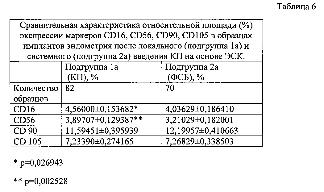
При локальном введении КП на основе ЭСК (подгруппа 1а) показатели относительной плотности CD16 и CD56 в имплантах достоверно превышали эти показатели в группе с системным введением КП на основе ЭСК (подгруппа 2а) (при использовании непараметрического метода Манна-Уитни) (Таблица 6). Уровни экспрессии CD90 и CD105 не имели достоверных отличий между образцами, полученными от животных групп 1а и 2а. Таким образом, показано, что в условиях локального введения КП на основе ЭСК по сравнению с системным введением реактивность тканей имплантоваутологичного эндометрия более выражена.With local administration of KP based on ESC (subgroup 1a), the relative density indices of CD16 and CD56 in implants significantly exceeded those in the group with systemic administration of KP based on ESC (subgroup 2a) (using the nonparametric Mann-Whitney method) (Table 6). The expression levels of CD90 and CD105 did not have significant differences between samples obtained from animals of groups 1a and 2a. Thus, it has been shown that under the conditions of local administration of KP based on ESC, the reactivity of tissues of the implant-autologous endometrium is more pronounced compared with systemic administration.
Метод иммуногистохимического исследования образцов имплантированного эндометрия, в условиях предложенной экспериментальной модели заболевания эндометрия является информативным для оценки терапевтического действия КП на основе ЭСК. Определение маркеров КП на основе ЭСК иммуногистохимическим методом позволяет верифицировать экспансию КП на основе ЭСК в условиях экспериментальной модели заболевания эндометрия как при локальном, так и при системном введении. Иммуногистохимическим методом выявляется тенденция к усилению активности NK-клеток, активизации репаративных процессов в имплантах эндометрия после введения КП продукта на основе ЭСК, что свидетельствует о стимуляции факторов, запускающих процессы восстановления гистоархитектоники ткани.The method of immunohistochemical study of samples of implanted endometrium, under the conditions of the proposed experimental model of endometrial disease, is informative for assessing the therapeutic effect of KP based on ESC. The determination of KP markers based on ESCs by the immunohistochemical method allows one to verify the expansion of KPs based on ESCs under the conditions of an experimental model of endometrial disease with both local and systemic administration. An immunohistochemical method reveals a tendency to increase the activity of NK cells, activation of reparative processes in endometrial implants after administration of a CP product based on ESC, which indicates the stimulation of factors that trigger the restoration of tissue histoarchitectonics.
Таким образом, представленный способ позволяет оценить эффективность терапевтического воздействия ЭСК на поврежденный эндометрий в условиях эксперимента и оценить эффективность введения стволовых клеток в динамике.Thus, the presented method allows to evaluate the effectiveness of the therapeutic effects of ESCs on the damaged endometrium under experimental conditions and to evaluate the effectiveness of stem cell administration in dynamics.
Claims (1)
Priority Applications (1)
| Application Number | Priority Date | Filing Date | Title |
|---|---|---|---|
| RU2015107794/14A RU2593895C1 (en) | 2015-03-05 | 2015-03-05 | Method for assessing therapeutic effect of human endometrial stem cells on damaged endometrium in experiment |
Applications Claiming Priority (1)
| Application Number | Priority Date | Filing Date | Title |
|---|---|---|---|
| RU2015107794/14A RU2593895C1 (en) | 2015-03-05 | 2015-03-05 | Method for assessing therapeutic effect of human endometrial stem cells on damaged endometrium in experiment |
Publications (1)
| Publication Number | Publication Date |
|---|---|
| RU2593895C1 true RU2593895C1 (en) | 2016-08-10 |
Family
ID=56612923
Family Applications (1)
| Application Number | Title | Priority Date | Filing Date |
|---|---|---|---|
| RU2015107794/14A RU2593895C1 (en) | 2015-03-05 | 2015-03-05 | Method for assessing therapeutic effect of human endometrial stem cells on damaged endometrium in experiment |
Country Status (1)
| Country | Link |
|---|---|
| RU (1) | RU2593895C1 (en) |
Citations (3)
| Publication number | Priority date | Publication date | Assignee | Title |
|---|---|---|---|---|
| US20070041948A1 (en) * | 2005-07-20 | 2007-02-22 | Seoul National University Industry Foundation | Method for culturing and proliferating hematopoietic stem cells and progenitor cells using human endometrial cells |
| WO2008148105A1 (en) * | 2007-05-25 | 2008-12-04 | Medistem Laboratories, Inc. | Endometrial stem cells and methods of making and using same |
| RU2515475C1 (en) * | 2012-11-15 | 2014-05-10 | Федеральное государственное бюджетное учреждение науки ИНСТИТУТ ЦИТОЛОГИИ РОССИЙСКОЙ АКАДЕМИИ НАУК | Method for stimulation of endometrial decidua formation experimentally |
-
2015
- 2015-03-05 RU RU2015107794/14A patent/RU2593895C1/en not_active IP Right Cessation
Patent Citations (3)
| Publication number | Priority date | Publication date | Assignee | Title |
|---|---|---|---|---|
| US20070041948A1 (en) * | 2005-07-20 | 2007-02-22 | Seoul National University Industry Foundation | Method for culturing and proliferating hematopoietic stem cells and progenitor cells using human endometrial cells |
| WO2008148105A1 (en) * | 2007-05-25 | 2008-12-04 | Medistem Laboratories, Inc. | Endometrial stem cells and methods of making and using same |
| RU2515475C1 (en) * | 2012-11-15 | 2014-05-10 | Федеральное государственное бюджетное учреждение науки ИНСТИТУТ ЦИТОЛОГИИ РОССИЙСКОЙ АКАДЕМИИ НАУК | Method for stimulation of endometrial decidua formation experimentally |
Non-Patent Citations (2)
| Title |
|---|
| KONDO W. et al. Effect ofanti-TNF-a on peritoneal endometrial implants of rats. Rev. Col. Bras. Cir., 2011, V.38 (4), P.266-273. * |
| ДОМНИНА А. П. Эндометриальные стволовые клетки: получение, характеристика и применение для стимуляции развития эндометрия крыс. автореферат диссертации на соискание ученой степени кандидата биологических наук, 2014. PATEL A.N., et al. Multipotent menstrual blood stromal stem cells, isolation characterization and differentiation. Cell Transplant. 2008, 17, 303-311. * |
Similar Documents
| Publication | Publication Date | Title |
|---|---|---|
| Ko et al. | Integrated bioactive scaffold with polydeoxyribonucleotide and stem-cell-derived extracellular vesicles for kidney regeneration | |
| US11781114B2 (en) | Materials and methods for expansion of stem cells | |
| Mukherjee et al. | Mesenchymal stem cell-based bioengineered constructs: foreign body response, cross-talk with macrophages and impact of biomaterial design strategies for pelvic floor disorders | |
| Ansari et al. | Alginate/hyaluronic acid hydrogel delivery system characteristics regulate the differentiation of periodontal ligament stem cells toward chondrogenic lineage | |
| Silva et al. | Neovascularization induced by the hyaluronic acid-based spongy-like hydrogels degradation products | |
| Lee et al. | The use of injectable spherically symmetric cell aggregates self-assembled in a thermo-responsive hydrogel for enhanced cell transplantation | |
| Tucker et al. | The use of progenitor cell/biodegradable MMP2–PLGA polymer constructs to enhance cellular integration and retinal repopulation | |
| US9492484B2 (en) | Cardiosphere derived cell population and methods of use | |
| Yap et al. | Enhanced liver progenitor cell survival and differentiation in vivo by spheroid implantation in a vascularized tissue engineering chamber | |
| Nosenko et al. | Novel biodegradable polymeric microparticles facilitate scarless wound healing by promoting re-epithelialization and inhibiting fibrosis | |
| Ye et al. | Enhanced proliferation of porcine bone marrow mesenchymal stem cells induced by extracellular calcium is associated with the activation of the calcium-sensing receptor and ERK signaling pathway | |
| Pokrywczynska et al. | Do mesenchymal stem cells modulate the milieu of reconstructed bladder wall? | |
| Xin et al. | MSCs-extracellular vesicles attenuated neuroinflammation, synapse damage and microglial phagocytosis after hypoxia-ischemia injury by preventing osteopontin expression | |
| Lopez-Martinez et al. | Bioengineered endometrial hydrogels with growth factors promote tissue regeneration and restore fertility in murine models | |
| Lin et al. | Cytokine loaded layer-by-layer ultrathin matrices to deliver single dermal papilla cells for spot-by-spot hair follicle regeneration | |
| Zheng et al. | In vivo bioengineered ovarian tumors based on collagen, matrigel, alginate and agarose hydrogels: a comparative study | |
| Yao et al. | Stem cell extracellular matrix-modified decellularized tendon slices facilitate the migration of bone marrow mesenchymal stem cells | |
| Yun et al. | Inhibitory effect of topical cartilage acellular matrix suspension treatment on neovascularization in a rabbit corneal model | |
| Lai et al. | A roadmap from research to clinical testing of mesenchymal stromal cell exosomes in the treatment of psoriasis | |
| Später et al. | Engineering microparticles based on solidified stem cell secretome with an augmented pro-angiogenic factor portfolio for therapeutic angiogenesis | |
| Li et al. | 3D-printed hydrogel particles containing PRP laden with TDSCs promote tendon repair in a rat model of tendinopathy | |
| Sideris et al. | Hyaluronic acid particle hydrogels decrease cerebral atrophy and promote pro-reparative astrocyte/axonal infiltration in the core after ischemic stroke | |
| Dai et al. | A construct of adipose-derived mesenchymal stem cells—laden collagen scaffold for fertility restoration by inhibiting fibrosis in a rat model of endometrial injury | |
| RU2593895C1 (en) | Method for assessing therapeutic effect of human endometrial stem cells on damaged endometrium in experiment | |
| Adhikari et al. | Photoinduced gelatin-methacrylate scaffolds to examine the impact of extracellular environment on trabecular meshwork cells |
Legal Events
| Date | Code | Title | Description |
|---|---|---|---|
| MM4A | The patent is invalid due to non-payment of fees |
Effective date: 20180306 |