RU2563107C2 - Method of treating arthroses, osteonecroses and other types of arthropathies and device for realisation thereof - Google Patents
Method of treating arthroses, osteonecroses and other types of arthropathies and device for realisation thereof Download PDFInfo
- Publication number
- RU2563107C2 RU2563107C2 RU2013151988/14A RU2013151988A RU2563107C2 RU 2563107 C2 RU2563107 C2 RU 2563107C2 RU 2013151988/14 A RU2013151988/14 A RU 2013151988/14A RU 2013151988 A RU2013151988 A RU 2013151988A RU 2563107 C2 RU2563107 C2 RU 2563107C2
- Authority
- RU
- Russia
- Prior art keywords
- bone
- holder
- rod
- hole
- holder according
- Prior art date
Links
Images
Classifications
-
- A—HUMAN NECESSITIES
- A61—MEDICAL OR VETERINARY SCIENCE; HYGIENE
- A61N—ELECTROTHERAPY; MAGNETOTHERAPY; RADIATION THERAPY; ULTRASOUND THERAPY
- A61N1/00—Electrotherapy; Circuits therefor
- A61N1/10—Applying static electricity
-
- A—HUMAN NECESSITIES
- A61—MEDICAL OR VETERINARY SCIENCE; HYGIENE
- A61L—METHODS OR APPARATUS FOR STERILISING MATERIALS OR OBJECTS IN GENERAL; DISINFECTION, STERILISATION OR DEODORISATION OF AIR; CHEMICAL ASPECTS OF BANDAGES, DRESSINGS, ABSORBENT PADS OR SURGICAL ARTICLES; MATERIALS FOR BANDAGES, DRESSINGS, ABSORBENT PADS OR SURGICAL ARTICLES
- A61L27/00—Materials for grafts or prostheses or for coating grafts or prostheses
- A61L27/28—Materials for coating prostheses
- A61L27/30—Inorganic materials
- A61L27/306—Other specific inorganic materials not covered by A61L27/303 - A61L27/32
-
- A—HUMAN NECESSITIES
- A61—MEDICAL OR VETERINARY SCIENCE; HYGIENE
- A61L—METHODS OR APPARATUS FOR STERILISING MATERIALS OR OBJECTS IN GENERAL; DISINFECTION, STERILISATION OR DEODORISATION OF AIR; CHEMICAL ASPECTS OF BANDAGES, DRESSINGS, ABSORBENT PADS OR SURGICAL ARTICLES; MATERIALS FOR BANDAGES, DRESSINGS, ABSORBENT PADS OR SURGICAL ARTICLES
- A61L27/00—Materials for grafts or prostheses or for coating grafts or prostheses
- A61L27/28—Materials for coating prostheses
- A61L27/34—Macromolecular materials
-
- A—HUMAN NECESSITIES
- A61—MEDICAL OR VETERINARY SCIENCE; HYGIENE
- A61L—METHODS OR APPARATUS FOR STERILISING MATERIALS OR OBJECTS IN GENERAL; DISINFECTION, STERILISATION OR DEODORISATION OF AIR; CHEMICAL ASPECTS OF BANDAGES, DRESSINGS, ABSORBENT PADS OR SURGICAL ARTICLES; MATERIALS FOR BANDAGES, DRESSINGS, ABSORBENT PADS OR SURGICAL ARTICLES
- A61L27/00—Materials for grafts or prostheses or for coating grafts or prostheses
- A61L27/50—Materials characterised by their function or physical properties, e.g. injectable or lubricating compositions, shape-memory materials, surface modified materials
-
- A—HUMAN NECESSITIES
- A61—MEDICAL OR VETERINARY SCIENCE; HYGIENE
- A61L—METHODS OR APPARATUS FOR STERILISING MATERIALS OR OBJECTS IN GENERAL; DISINFECTION, STERILISATION OR DEODORISATION OF AIR; CHEMICAL ASPECTS OF BANDAGES, DRESSINGS, ABSORBENT PADS OR SURGICAL ARTICLES; MATERIALS FOR BANDAGES, DRESSINGS, ABSORBENT PADS OR SURGICAL ARTICLES
- A61L31/00—Materials for other surgical articles, e.g. stents, stent-grafts, shunts, surgical drapes, guide wires, materials for adhesion prevention, occluding devices, surgical gloves, tissue fixation devices
- A61L31/08—Materials for coatings
- A61L31/082—Inorganic materials
- A61L31/088—Other specific inorganic materials not covered by A61L31/084 or A61L31/086
-
- A—HUMAN NECESSITIES
- A61—MEDICAL OR VETERINARY SCIENCE; HYGIENE
- A61L—METHODS OR APPARATUS FOR STERILISING MATERIALS OR OBJECTS IN GENERAL; DISINFECTION, STERILISATION OR DEODORISATION OF AIR; CHEMICAL ASPECTS OF BANDAGES, DRESSINGS, ABSORBENT PADS OR SURGICAL ARTICLES; MATERIALS FOR BANDAGES, DRESSINGS, ABSORBENT PADS OR SURGICAL ARTICLES
- A61L31/00—Materials for other surgical articles, e.g. stents, stent-grafts, shunts, surgical drapes, guide wires, materials for adhesion prevention, occluding devices, surgical gloves, tissue fixation devices
- A61L31/08—Materials for coatings
- A61L31/10—Macromolecular materials
-
- A—HUMAN NECESSITIES
- A61—MEDICAL OR VETERINARY SCIENCE; HYGIENE
- A61L—METHODS OR APPARATUS FOR STERILISING MATERIALS OR OBJECTS IN GENERAL; DISINFECTION, STERILISATION OR DEODORISATION OF AIR; CHEMICAL ASPECTS OF BANDAGES, DRESSINGS, ABSORBENT PADS OR SURGICAL ARTICLES; MATERIALS FOR BANDAGES, DRESSINGS, ABSORBENT PADS OR SURGICAL ARTICLES
- A61L31/00—Materials for other surgical articles, e.g. stents, stent-grafts, shunts, surgical drapes, guide wires, materials for adhesion prevention, occluding devices, surgical gloves, tissue fixation devices
- A61L31/14—Materials characterised by their function or physical properties, e.g. injectable or lubricating compositions, shape-memory materials, surface modified materials
-
- A—HUMAN NECESSITIES
- A61—MEDICAL OR VETERINARY SCIENCE; HYGIENE
- A61N—ELECTROTHERAPY; MAGNETOTHERAPY; RADIATION THERAPY; ULTRASOUND THERAPY
- A61N1/00—Electrotherapy; Circuits therefor
- A61N1/18—Applying electric currents by contact electrodes
- A61N1/20—Applying electric currents by contact electrodes continuous direct currents
- A61N1/205—Applying electric currents by contact electrodes continuous direct currents for promoting a biological process
-
- A—HUMAN NECESSITIES
- A61—MEDICAL OR VETERINARY SCIENCE; HYGIENE
- A61N—ELECTROTHERAPY; MAGNETOTHERAPY; RADIATION THERAPY; ULTRASOUND THERAPY
- A61N1/00—Electrotherapy; Circuits therefor
- A61N1/18—Applying electric currents by contact electrodes
- A61N1/32—Applying electric currents by contact electrodes alternating or intermittent currents
- A61N1/326—Applying electric currents by contact electrodes alternating or intermittent currents for promoting growth of cells, e.g. bone cells
-
- C—CHEMISTRY; METALLURGY
- C08—ORGANIC MACROMOLECULAR COMPOUNDS; THEIR PREPARATION OR CHEMICAL WORKING-UP; COMPOSITIONS BASED THEREON
- C08L—COMPOSITIONS OF MACROMOLECULAR COMPOUNDS
- C08L27/00—Compositions of homopolymers or copolymers of compounds having one or more unsaturated aliphatic radicals, each having only one carbon-to-carbon double bond, and at least one being terminated by a halogen; Compositions of derivatives of such polymers
- C08L27/02—Compositions of homopolymers or copolymers of compounds having one or more unsaturated aliphatic radicals, each having only one carbon-to-carbon double bond, and at least one being terminated by a halogen; Compositions of derivatives of such polymers not modified by chemical after-treatment
- C08L27/12—Compositions of homopolymers or copolymers of compounds having one or more unsaturated aliphatic radicals, each having only one carbon-to-carbon double bond, and at least one being terminated by a halogen; Compositions of derivatives of such polymers not modified by chemical after-treatment containing fluorine atoms
- C08L27/18—Homopolymers or copolymers or tetrafluoroethene
-
- A—HUMAN NECESSITIES
- A61—MEDICAL OR VETERINARY SCIENCE; HYGIENE
- A61L—METHODS OR APPARATUS FOR STERILISING MATERIALS OR OBJECTS IN GENERAL; DISINFECTION, STERILISATION OR DEODORISATION OF AIR; CHEMICAL ASPECTS OF BANDAGES, DRESSINGS, ABSORBENT PADS OR SURGICAL ARTICLES; MATERIALS FOR BANDAGES, DRESSINGS, ABSORBENT PADS OR SURGICAL ARTICLES
- A61L2400/00—Materials characterised by their function or physical properties
-
- A—HUMAN NECESSITIES
- A61—MEDICAL OR VETERINARY SCIENCE; HYGIENE
- A61L—METHODS OR APPARATUS FOR STERILISING MATERIALS OR OBJECTS IN GENERAL; DISINFECTION, STERILISATION OR DEODORISATION OF AIR; CHEMICAL ASPECTS OF BANDAGES, DRESSINGS, ABSORBENT PADS OR SURGICAL ARTICLES; MATERIALS FOR BANDAGES, DRESSINGS, ABSORBENT PADS OR SURGICAL ARTICLES
- A61L2430/00—Materials or treatment for tissue regeneration
- A61L2430/02—Materials or treatment for tissue regeneration for reconstruction of bones; weight-bearing implants
Landscapes
- Health & Medical Sciences (AREA)
- Life Sciences & Earth Sciences (AREA)
- General Health & Medical Sciences (AREA)
- Veterinary Medicine (AREA)
- Public Health (AREA)
- Animal Behavior & Ethology (AREA)
- Chemical & Material Sciences (AREA)
- Epidemiology (AREA)
- Medicinal Chemistry (AREA)
- Biomedical Technology (AREA)
- Engineering & Computer Science (AREA)
- Radiology & Medical Imaging (AREA)
- Nuclear Medicine, Radiotherapy & Molecular Imaging (AREA)
- Oral & Maxillofacial Surgery (AREA)
- Dermatology (AREA)
- Transplantation (AREA)
- Surgery (AREA)
- Heart & Thoracic Surgery (AREA)
- Inorganic Chemistry (AREA)
- Vascular Medicine (AREA)
- Polymers & Plastics (AREA)
- Organic Chemistry (AREA)
- Molecular Biology (AREA)
- Chemical Kinetics & Catalysis (AREA)
- Cell Biology (AREA)
- Orthopedic Medicine & Surgery (AREA)
- Prostheses (AREA)
- Electrotherapy Devices (AREA)
- Surgical Instruments (AREA)
Abstract
Description
Изобретение относится к медицине, а более конкретно к электротерапии с использованием статического электричества электретных покрытий и оболочек для профилактики и лечения опорно-двигательного аппарата, например для лечения артрозов суставов: коленного, тазобедренного и др.The invention relates to medicine, and more particularly to electrotherapy using static electricity of electret coatings and membranes for the prevention and treatment of the musculoskeletal system, for example, for the treatment of arthrosis of the joints: knee, hip, etc.
С 80-х годов формируется и интенсивно развивается новая отрасль медицины, основанная на использовании близкодействующих статических электрических полей для стимулирования позитивных биологических процессов в организме человека. Главной отличительной особенностью практических методов, основанных на этой концепции, является то, что электрические поля создаются не традиционными электротехническими источниками энергии с сетевым или аккумуляторным электропитанием, а функционирующими автономно электретными пленками, нанесенными на имплантаты различного назначения, широко применяемые в медицине.Since the 80s, a new branch of medicine has been formed and is intensively developing, based on the use of short-range static electric fields to stimulate positive biological processes in the human body. The main distinguishing feature of practical methods based on this concept is that electric fields are created not by traditional electrotechnical energy sources with mains or battery power, but by functioning autonomous electret films deposited on various implants that are widely used in medicine.
Электрет - это диэлектрик, на поверхности или в объеме которого продолжительное время сохраняются не скомпенсированные электрические заряды, создающие в окружающем электрет пространстве квазистатическое (медленно меняющееся во времени) электрическое поле. Попадая вместе с имплантатом в организм человека, электретная пленка своим полем оказывает дозированное локальное воздействие на поврежденный орган, способствуя его лечению в оптимальных биофизических условиях. В основе этого процесса лежит природный эффект, состоящий в том, что внешнее близкодействующее электрическое поле определенной величины и знака, действуя на клеточном уровне, является катализатором репаративных процессов в живых тканях.An electret is a dielectric on the surface or in the volume of which uncompensated electric charges are stored for a long time, creating a quasistatic (slowly varying in time) electric field in the space surrounding the electret. Getting together with the implant into the human body, the electret film with its field has a dosed local effect on the damaged organ, contributing to its treatment in optimal biophysical conditions. The basis of this process is a natural effect, consisting in the fact that an external short-range electric field of a certain size and sign, acting at the cellular level, is a catalyst for reparative processes in living tissues.
Известен способ лечения суставов, включающий периодические наложения (два раза в день по 1 часу) на кожную поверхность пораженного артрозом сустава бумажной пластины, на поверхности которой сформирована диэлектрическая оболочка в электретном состоянии (ДОЭС) см. http://www.uralargo.ru/article/489, а также http://precession.m/?id_page=859373, а также http://elis-deta.ru/elplast.html.A known method of treating joints, including periodic overlay (twice a day for 1 hour) on the skin surface of a joint affected by arthrosis of a paper plate, on the surface of which a dielectric sheath is formed in an electret state (DOES), see http://www.uralargo.ru/ article / 489, as well as http: //precession.m/? id_page = 859373, as well as http://elis-deta.ru/elplast.html.
Недостатком известного способа лечения артрозов является удаленность электростатического поля ДОЭС от очага поражения сустава, что снижает эффективность способа-аналога, удлиняя сроки лечения (срок лечения 0,5-1,5 года).A disadvantage of the known method for the treatment of arthrosis is the remoteness of the electrostatic field of DOES from the lesion of the joint, which reduces the effectiveness of the analogue method, lengthening the duration of treatment (treatment period 0.5-1.5 years).
Кроме того, периодичность воздействия на очаг поражения уменьшает длительность терапевтического действия электростатического поля электретного покрытия, что также удлиняет сроки лечения.In addition, the frequency of exposure to the lesion decreases the duration of the therapeutic effect of the electrostatic field of the electret coating, which also extends the duration of treatment.
Кроме того, не решен вопрос установки пластины с ДОЭС на поверхность сустава, что существенно осложняет применение способа-аналога на практике.In addition, the issue of installing a plate with DOES on the joint surface has not been resolved, which significantly complicates the application of the analogue method in practice.
Кроме того, бумажная пластина, на поверхности которой сформирована ДОЭС недолговечна в использовании, быстро теряет электретный заряд при любом сминании бумажной пластины.In addition, a paper plate, on the surface of which a DOES is formed, is short-lived in use, quickly loses its electret charge during any wrinkling of the paper plate.
Указанные недостатки аналога частично устраняются в ближайшем аналоге.The indicated disadvantages of the analogue are partially eliminated in the nearest analogue.
Ближайшим аналогом авторы выбрали способ предупреждения патологии тазобедренного сустава (см. АС СССР №1251915), включающий установку внутрь больного сустава диэлектрической оболочки в электретном состоянии (ДОЭС) с помощью держателя, который выполнен в виде пластины, целиком покрытой ДОЭС, с клиновидным отогнутым концом.The closest analogue, the authors chose a way to prevent the pathology of the hip joint (see AS USSR No. 1251915), which includes installing a dielectric sheath in the electret state (DOES) inside the diseased joint using a holder that is made in the form of a plate entirely covered with DOES with a wedge-shaped bent end.
Причем пластина с клиновидным отогнутым концом, одним своим концом шурупами прикрепляется к бедренной кости, а клиновидный отогнутый конец забивается в головку кости тазобедренного сустава.Moreover, a plate with a wedge-shaped bent end, with one of its ends with screws, is attached to the femur, and the wedge-shaped bent end is hammered into the head of the bone of the hip joint.
Благодаря тому, что клиновидный отогнутый конец установлен внутрь патологичной головки и максимально приближен к зоне патологии обеспечивается предупреждение развития некроза и деформации суставных поверхностей, в короткое время снимается болевой синдром, ускоряется восстановление (остеорепарация) всех внутрикостных тканей сустава.Due to the fact that the wedge-shaped bent end is installed inside the pathological head and is as close as possible to the pathology zone, the development of necrosis and deformation of the articular surfaces is prevented, pain is relieved in a short time, and restoration (osteoreparation) of all intraosseous tissues of the joint is accelerated.
Недостатком ближайшего способа-аналога является нарушение целостности ДОЭС в процессе забивания клиновидного конца в головку сустава, что приводит к быстрой разрядке ДОЭС и прекращения терапевтического действия электростатического поля на патологический сустав.The disadvantage of the closest analogue method is the violation of the integrity of the DOES during the clogging of the wedge-shaped end into the head of the joint, which leads to the rapid discharge of the DOES and termination of the therapeutic effect of the electrostatic field on the pathological joint.
Основное назначение имплантируемой пластины - остеосинтез, т.е. жесткая фиксация головки бедренной кости относительно отсеченной части бедренной кости при лечении врожденного заболевания, связанного с неправильным положением головки тазобедренной кости относительно кости таза. Главное, на что здесь работает ДОЭС - остеосинтез, т.е. стимулирование срастания в правильном взаимном положении друг относительно друга частей рассеченной тазобедренной кости. Клиновидный отросток здесь служит как крепежный элемент временной конструкции до момента, когда части тазобедренной кости срастутся и смогут воспринимать все нагрузки опорного аппарата самостоятельно. После чего пластина с кости удаляется за ненадобностью.The main purpose of the implantable plate is osteosynthesis, i.e. rigid fixation of the femoral head relative to the cut off part of the femur in the treatment of congenital disease associated with improper position of the hip head relative to the pelvic bone. The main thing that DOES works for here is osteosynthesis, i.e. stimulation of fusion in the correct mutual position relative to each other parts of the dissected hip bone. The wedge-shaped process here serves as a fastening element of a temporary structure until the moment when the parts of the hip bone grow together and can absorb all the loads of the supporting apparatus independently. After which the plate from the bone is removed as unnecessary.
Таким образом, основным назначением ДОЭС на поверхности пластины и отогнутом конце было обеспечить оптимизацию остеосинтеза. Терапевтическое действие ДОЭС на поверхности клиновидного отростка давало дополнительный эффект - оздоровление сустава - имел место пока части бедренной кости срастались. После удаления пластины терапевтическое действие ДОЭС на патологию сустава прекращалось, а сами патологические процессы могли продолжаться, разрушая костные ткани головки сустава, делая человека обездвиженным инвалидом.Thus, the main purpose of DOES on the surface of the plate and the bent end was to optimize osteosynthesis. The therapeutic effect of DOES on the surface of the sphenoid process gave an additional effect - joint improvement - took place while parts of the femur fused. After removal of the plate, the therapeutic effect of DOES on the joint pathology ceased, and the pathological processes themselves could continue, destroying the bone tissue of the joint head, making the person immobilized with a disability.
Кроме того, расположение ДОЭС по отношению к очагу патологического разрушения кости не всегда было оптимальным, т.к. основным, приоритетным в выборе направления забивания в кость клиновидного отростка были соображения точности взаимного расположения соединяемых частей кости (остеосинтеза), вопрос оптимизации остеорепарации стоял на втором месте, что приводило к снижению эффективности терапевтического действия ДОЭС на патологический очаг.In addition, the location of DOES in relation to the site of pathological destruction of the bone was not always optimal, because The main priority in choosing the direction of clogging of the sphenoid bone into the bone was considerations of the accuracy of the relative position of the connected parts of the bone (osteosynthesis), the optimization of osteoreparation was in second place, which led to a decrease in the effectiveness of the therapeutic effect of DOES on the pathological focus.
Кроме того, забивание клиновидного отростка приводило к повышению внутрикостного давления и требовало просверливания в кости рядом с отростком дополнительных отверстий в кости для снятия этого избыточного давления в кости, что дополнительно травмировало кость, удлиняя сроки лечения.In addition, clogging of the sphenoid process led to an increase in intraosseous pressure and required additional holes in the bone to be drilled in the bone near the process to relieve this excess pressure in the bone, which additionally injured the bone, lengthening the treatment time.
Задачей, вытекающей из уровня техники, является создание способа лечения, обеспечивающего длительное действие электростатического поля ДОЭС за счет предотвращения нарушения целостности ДОЭС в процессе установки ее внутрь кости.The task arising from the prior art is to create a treatment method that ensures the long-term effect of the electrostatic field of DOES by preventing a violation of the integrity of DOES during the installation of it inside the bone.
Поставленная задача в заявляемом способе решается за счет того, что заявляемый способ лечения, включающий установку внутрь кости диэлектрической оболочки в электретном состоянии (ДОЭС) с помощью держателя. Отличается тем, что в кости выполняют отверстие, через которое вводят с зазором держатель ДОЭС и оставляют ДОЭС внутри кости до момента разрядки ДОЭС.The problem in the inventive method is solved due to the fact that the claimed method of treatment, including the installation inside the bone of the dielectric sheath in an electret state (DOES) using the holder. It differs in that a hole is made in the bone through which the DOES holder is inserted with a gap and the DOES is left inside the bone until the DOES is discharged.
Благодаря тому, что держатель с ДОЭС вводят в отверстие в кости с зазором, поверхность ДОЭС не повреждается, что способствует предотвращению нарушения целостности ДОЭС и, следовательно, сохранению заряда в ДОЭС, что позволяет удлинить время благотворного действия электростатического поля на поврежденные патологическим процессом ткани организма в сравнении с ближайшим аналогом, где держатель ДОЭС забивался в кость, что приводило к нарушению целостности ДОЭС, быстрой разрядке ДОЭС и, следовательно, сокращению длительности лечебного действия электростатического поля ДОЭС на ткани организма.Due to the fact that the holder with the DOES is inserted into the hole in the bone with a gap, the surface of the DOES is not damaged, which helps to prevent violation of the integrity of the DOES and, therefore, preserves the charge in the DOES, which allows you to extend the beneficial effects of the electrostatic field on the tissues of the body damaged by the pathological process compared with the closest analogue, where the holder of the DOES was hammered into the bone, which led to a violation of the integrity of the DOES, the rapid discharge of the DOES and, therefore, a reduction in the duration of the treatment DOES tviya electrostatic field on body tissues.
Кроме того, в заявляемом способе мы имеем возможность точного ориентирования ДОЭС относительно пораженных патологией тканей организма, оптимизируя процесс лечения, повышая его эффективность в сравнении с ближайшим аналогом, в котором выбор направления забивания держателя в кость определялся прежде всего исходя из потребности точного взаимного ориентирования соединяемых имплантатом частей кости (остеосинтеза).In addition, in the claimed method, we have the ability to accurately orientate the DOES relative to the diseased tissues of the body, optimizing the treatment process, increasing its efficiency in comparison with the closest analogue, in which the choice of the direction of clogging of the holder into the bone was determined primarily on the basis of the need for precise mutual orientation of the implant connected parts of the bone (osteosynthesis).
Кроме того, благодаря тому, что держатель с ДОЭС вводят в отверстие кости с зазором, это позволяет снять избыточное напряжение внутри кости, избавиться от дополнительных декомпрессионных отверстий (как это имеет место в ближайшем способе-аналоге, где держатель ДОЭС забивался в кость), что упрощает заявляемый способ, уменьшает травматичность его воздействия на кость.In addition, due to the fact that the holder with DOES is inserted into the bone hole with a gap, this allows you to remove excess tension inside the bone, get rid of additional decompression holes (as is the case in the closest analogous method, where the DOES holder was hammered into the bone), which simplifies the inventive method, reduces the morbidity of its effects on the bone.
При разрядке ДОЭС ее извлекают из кости, и на ее место устанавливается другая - заряженная ДОЭС.When the DOES is discharged, it is removed from the bone, and another, charged DOES, is installed in its place.
В способе лечения ограничивают возможность выхода ДОЭС из полости отверстия в кости, чтобы ДОЭС самопроизвольно не вышла из кости.In the treatment method, the possibility of DOES exit from the cavity of the hole in the bone is limited so that DOES does not spontaneously leave the bone.
В способе лечения держатель можно оставить в полости отверстия в кости вместе с ДОЭС.In the method of treatment, the holder can be left in the cavity of the hole in the bone with DOES.
В способе лечения после установки ДОЭС на свое место в отверстии, держатель можно извлечь из полости отверстия.In the treatment method, after installing the DOES in its place in the hole, the holder can be removed from the cavity of the hole.
В способе лечения соосно отверстию в кость можно имплантировать шлюзовое устройство, через которое можно устанавливать и извлекать ДОЭС, а также его можно использовать для ограничения выхода из отверстия ДОЭС или держателя с ДОЭС.In the method of treatment, a sluice device can be implanted coaxially with the hole in the bone, through which DOES can be installed and removed, and it can also be used to restrict the exit of the DOES hole or holder with DOES.
В способе лечения шлюзовое устройство может быть имплантировано в кость заподлицо с наружной поверхностью кости.In a method of treatment, a sluice device can be implanted into the bone flush with the outer surface of the bone.
Шлюзовое устройство для установки-извлечения ДОЭС может быть выполнено в виде втулки, внутренняя полость которой с одной стороны сообщается с полостью отверстия в кости, а с другой стороны сообщается с внешней средой.The lock device for installing-extracting DOES can be made in the form of a sleeve, the internal cavity of which on the one hand communicates with the cavity of the hole in the bone, and on the other hand communicates with the external environment.
На наружной поверхности втулки может быть выполнена резьба для установки ее в кости.A thread can be made on the outer surface of the sleeve to install it in the bone.
Внутри втулки может быть выполнена резьба для запирания внутренней полости втулки.A thread may be made inside the sleeve to lock the internal cavity of the sleeve.
На торцевой поверхности втулки может быть выполнен, по крайней мере, один шлиц под отвертку для вкручивания-выкручивания ее из кости.At least one slot for a screwdriver can be made on the end surface of the sleeve for screwing it in and out of the bone.
Втулка может быть снабжена крышкой для запирания внутренней полости втулки.The sleeve may be provided with a cover for locking the internal cavity of the sleeve.
Держатель ДОЭС может быть выполнен в виде тела, форма и размеры которого соответствует форме и размерам отверстия в кости, причем, поперечные размеры тела должны быть меньше поперечных размеров отверстия в кости.The DOES holder can be made in the form of a body, the shape and dimensions of which correspond to the shape and dimensions of the hole in the bone, moreover, the transverse dimensions of the body must be less than the transverse dimensions of the hole in the bone.
Тело держателя может быть выполнено в виде твердого стержня.The body of the holder can be made in the form of a solid rod.
Стержень может быть выполнен из металла, например, из титана, тантала, циркония, и других вентильных металлов и их сплавов, легированной стали.The rod may be made of metal, for example, titanium, tantalum, zirconium, and other valve metals and their alloys, alloy steel.
Стержень может быть выполнен также из неметалла (например, из пластмассы, или керамики), на поверхности которого выполнен электропроводящий слой, над которым размещен ДОЭС.The rod may also be made of non-metal (for example, plastic, or ceramic), on the surface of which an electrically conductive layer is made, over which a DOES is placed.
ДОЭС может быть выполнен из политетрафторэтилена и (или) его сополимеров.DOES can be made of polytetrafluoroethylene and (or) its copolymers.
ДОЭС может быть также выполнен из пятиокиси тантала (а также из окислов других вентильных металлов).DOES can also be made of tantalum pentoxide (as well as from oxides of other valve metals).
Держатель может быть выполнен в виде цилиндрической оправки с возможностью изменения ее диаметральных размеров.The holder can be made in the form of a cylindrical mandrel with the possibility of changing its diametrical dimensions.
Стержень может иметь скругленное окончание.The rod may have a rounded end.
Одно из окончаний стержня может быть уплощено и загнуто под углом к оси стержня. В загнутом уплощении выполнено сквозное отверстие под шуруп для фиксации стержня внутри кости.One of the ends of the rod can be flattened and bent at an angle to the axis of the rod. In the bent flattening, a through hole is made under the screw for fixing the rod inside the bone.
В одном из окончаний стержня может быть выполнено сквозное отверстие для извлечения стержня из сустава.A through hole may be provided at one of the ends of the shaft for removing the shaft from the joint.
На конце стержня может быть выполнено утолщение, на боковой поверхности которого выполнена резьба, а на торцевой поверхности выполнен, по крайней мере, один шлиц под отвертку.A thickening may be performed at the end of the shaft, on which a thread is made on the side surface, and at least one slot for a screwdriver is made on the end surface.
На конце стержня может быть выполнено утолщение с конусной наружной поверхностью для скрепления с костью.At the end of the shaft, a thickening may be made with a conical outer surface for bonding to the bone.
Стержень также может быть выполнен Г-образной формы, а одна из его оконечностей выполнена заостренной.The rod can also be made L-shaped, and one of its extremities is made pointed.
Стержень может иметь в поперечнике круглое сечение.The rod may have a circular cross section.
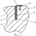
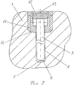
На фиг.1 изображена установленная (имплантируемая) в кость ДОЭС с держателем, зафиксированным в кости шурупом (вид сбоку).Figure 1 shows the installed (implantable) in the bone DOES with a holder fixed in the bone with a screw (side view).
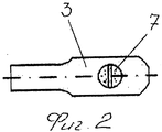
На фиг.2 - установленная в кость ДОЭС с держателем, зафиксированным в кости шурупом (вид сверху).Figure 2 - installed in the bone DOES with a holder fixed in the bone with a screw (top view).
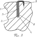
На фиг.3 - установленная в кость ДОЭС с держателем Г-образной формы, зафиксированным в кости с помощью заостренной оконечности.Figure 3 - installed in the bone DOES with the holder of the L-shaped, fixed in the bone using a pointed tip.
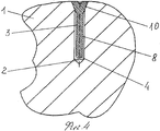
На фиг.4 - установленная в кость ДОЭС с держателем, зафиксированным в кости с помощью клиновидного утолщения.Figure 4 - installed in the bone DOES with a holder fixed in the bone using a wedge-shaped thickening.
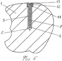
На фиг.5 - установленная в кость ДОЭС с держателем, зафиксированным в кости с помощью резьбовой поверхности, выполненной на головке держателя.Figure 5 - installed in the bone DOES with a holder fixed in the bone using a threaded surface made on the head of the holder.
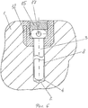
На фиг.6 - установленная в кость ДОЭС с держателем, установленным в кости через шлюз и зафиксированным в кости с помощью шлюза (втулки) и крышки (вид сбоку).Figure 6 - installed in the bone DOES with a holder installed in the bone through the lock and fixed in the bone using the lock (sleeve) and cover (side view).
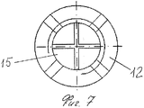
На фиг.7 - установленная в кость ДОЭС с держателем, установленным в кости через шлюз и зафиксированным в кости с помощью шлюза (втулки) и крышки (вид сверху).In Fig.7 - installed in the bone DOES with a holder installed in the bone through the lock and fixed in the bone using the lock (sleeve) and cover (top view).
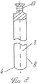
На фиг.8 - держатель (см. фиг.7) - развернуто на 90 град, с отверстием для извлечения из кости.On Fig - holder (see Fig.7) - deployed at 90 degrees, with a hole for extraction from the bone.
На фиг.9 - установленная в кость ДОЭС с держателем, установленным в кости через шлюз и зафиксированным в кости с помощью шлюза (втулки) и резьбовой поверхности на головке держателя, завернутого внутрь полости втулки (вид сбоку).In Fig.9 - installed in the bone DOES with the holder installed in the bone through the lock and fixed in the bone using the lock (sleeve) and a threaded surface on the head of the holder, wrapped inside the cavity of the sleeve (side view).
Рассмотрим вариант реализации заявляемого способа на примере лечения диспластического коксартроза головки тазобедренного сустава.Consider the implementation of the proposed method on the example of the treatment of dysplastic coxarthrosis of the head of the hip joint.
Больная К. 1954 г.р. поступила в клинику военной травматологии и ортопедии ВМедА по поводу указанного выше диагноза 08.04.1996 г. (История болезни №8276). Пациентке было проведено лечение согласно заявленному способу.Patient K., born in 1954 was admitted to the clinic of military traumatology and orthopedics VMedA about the above diagnosis on 04/08/1996 (medical history No. 8276). The patient was treated according to the claimed method.
В процессе хирургического вмешательства под местным наркозом в кости 1 пациентки (фиг.1, фиг.2) было выполнено цилиндрическое отверстие 2 диаметром 4.1 мм, в которое был введен держатель 3 в виде стержня цилиндрической формы диаметром 4 мм. С одной стороны стержень 3 имел скругленное окончание 4. С другой стороны окончание 5 стержня 3 уплощено и загнуто под углом к оси стержня 3. В загнутом уплощении 5 выполнено сквозное отверстие 6, через которое в кость 1 был вкручен шуруп 7 для фиксации держателя 3 относительно кости 1. Держатель 3 выполнен из тантала. На его поверхности методом анодного окисления сформирована диэлектрическая оболочка 8 пятиокиси тантала толщиной 0,3 мкм, имеющая распределенный вдоль оси стержня 3 электретный потенциал, полученный с помощью известного способа (см. положительное решение от 21.10.13 г. по заявке на патент РФ №2011102303). Оболочка ДОЭС 8, находясь внутри кости 1 длительное время (3-5 лет и более), оказывает терапевтическое (репаративное) действие на костные ткани 1, сосуды, нервные окончания, связки и др., восстанавливая нормальное функционирование сустава.In the process of surgical intervention under local anesthesia in the
Через два дня после введения в кость 1 головки тазобедренного сустава танталового держателя 3, покрытого ДОЭС 8 боли прекратились. Через семь дней больная была выписана из клиники. Через 5 лет пациентке была проведена повторная операция по замене держателя 3 с разряженной ДОЭС 8 на держатель 3 с заряженным ДОЭС 8. Для этого он был извлечен из отверстия 2 наружу. На его место в отверстие 2 установлен другой держатель 3 с заряженной ДОЭС 8, был прикручен к кости 1 фиксирующим шурупом 7.Two days after the introduction of 1 head of the hip joint of the
Благодаря тому, что держатель 3 с ДОЭС 8 вводят в отверстие 2 в кости 1 с зазором, поверхность ДОЭС 8 не повреждается, что способствует предотвращению нарушения целостности ДОЭС 8 и, следовательно, сохранению заряда в ДОЭС 8, что позволяет удлинить (более 5 лет) время действие электростатического поля на поврежденные патологическим процессом ткани организма в сравнении с ближайшим аналогом, где держатель ДОЭС забивался в кость, что приводило к нарушению целостности ДОЭС, быстрой разрядке ДОЭС и, следовательно, укорочению длительности лечебного действия (до 6 месяцев) электростатического поля ДОЭС на ткани организма.Due to the fact that the
Описанный выше в заявленном способе лечения вариант выполнения держателя 3 является одним из возможных вариантов выполнения держателя 3 и одним из возможных способов фиксации ДОЭС 8 в отверстии 2.The embodiment of the
ДОЭС 8 может быть установлена в кость 1 с помощью цилиндрического держателя 3 Г-образной формы (фиг.3), зафиксированного в кости 1 (забитого в кость 1) с помощью заостренной оконечности 9. При этом рабочая заряженная поверхность ДОЭС 8 не контактирует в процессе установки с костью 1 и не повреждается ее целостность.DOES 8 can be installed in
ДОЭС 8 может быть также установлена в кость 1 с помощью цилиндрического держателя 3 (фиг.4), зафиксированным в кости 1 с помощью клиновидного утолщения 10 (забитого в кость 1). При этом рабочая заряженная поверхность ДОЭС 8 не контактирует в процессе установки с костью 1 и не повреждается ее целостность.DOES 8 can also be installed in the
ДОЭС 8 может быть установлена в кость 1 с помощью цилиндрического держателя 3 (фиг.5), зафиксированного в кости 1 (вкрученным в кость 1) с помощью резьбовой поверхности 11, выполненной в винтовой головке 12 держателя 3. Держатель 3 вкручивают в кость 1 с помощью отвертки, вставленной в шлиц 13. Извлекают держатель 3 из кости 1, выкручивая его из кости 1.DOES 8 can be installed in
ДОЭС 8 может быть установлена в кость 1 с помощью цилиндрического держателя 3 (фиг.6, фиг.7. фиг.8), установленного в кости 1 с помощью шлюза 14, выполненного в виде втулки 14, вкрученной в кость 1 соосно отверстию 2. Держатель 3 с ДОЭС 8 через шлюз 14 вводят в отверстие 2 и фиксируют в нем с помощью крышки 15. Для извлечения отработанной (разряженной) ДОЭС 8 выкручивают крышку 15 подцепляют держатель 3 за отверстие 17 и извлекают его вместе с ДОЭС 8 из отверстия 2. На освободившееся место через шлюз 14 в отверстие 2 устанавливают другой держатель 3 с заряженной ДОЭС 8.DOES 8 can be installed in the
ДОЭС 8 может быть установлена в кость 1 с помощью цилиндрического держателя 3 (фиг.9), установленного в кости 1 с помощью шлюза 14. Держатель 3 с ДОЭС 8 через шлюз 14 вводят в отверстие 2 и фиксируют в нем с помощью резьбовой поверхности 11, выполненной в винтовой головке 12 держателя 3, которой он вкручивается в шлюз 14.DOES 8 can be installed in the
Пример 2. Больная 58 лет, поступила в травматологическое отделение Елизаветинской больницы с диагнозом: застарелое повреждение внутреннего мениска, передней крестообразной связки, деформирующий артроз правого коленного сустава. 27.11.09 выполнена лечебно-диагностическая артроскопия, менискэктомия, имплантация держателя 3 ДОЭС 8 в эпифиз большеберцовой кости 1. Контрольная R-графия в послеоперационном периоде через 6, 12, 24 мес. показала, что прогрессирования дегенеративных процессов в мыщелках большеберцовой и бедренной костей 1 не выявлено. Болевой синдром купирован, больная отказалась от приема противоболевых препаратов.Example 2. Patient 58 years old, was admitted to the trauma unit of the Elizabethan hospital with a diagnosis of chronic damage to the internal meniscus, anterior cruciate ligament, deforming arthrosis of the right knee joint. 11.27.09 performed diagnostic and treatment arthroscopy, meniscectomy, implantation of
Пример 3. В городскую больницу Святой преподобной мученицы Елизаветы г. Ленинграда поступила пациентка Н., передвигавшаяся только на коляске, 18 лет с диагнозом полиартрит неизвестной этиологии. В апреле 1990 г. ведущим травматологом Хомутовым В.П. была проведена операция по установке танталовых держателей 3 с ДОЭС 8 в тазобедренных и коленных суставах. Результат: через несколько дней после операции боли в суставах прекратились. Через 7 дней пациентка уже самостоятельно передвигалась на ногах. Через 12 дней она покинула клинику и вернулась к полноценной жизни.Example 3. Patient N., who was only traveling in a wheelchair, 18 years old with a diagnosis of polyarthritis of unknown etiology, was admitted to the city hospital of the Holy Martyr Elizabeth of Leningrad. In April 1990, the leading traumatologist Khomutov V.P. An operation was performed to install
Пример 4. В мае 1991 г. в городскую больницу Святой преподобной мученицы Елизаветы г. Ленинград поступил 60-летний пациент М. Пациент подвергся воздействию вредных и ядовитых химических веществ на производстве. В результате у него стали рассасываться головки бедренных костей 1 на фоне поражения крови и других структурно-функциональных нарушений в организме, что не позволяло использовать эндопротезирование. Пациент был прикован к постели. Ведущий травматолог больницы Хомутов В.П. провел операцию по введению в пораженные кости 1 суставов танталовых держателей 3 с ДОЭС 8. Через неделю после операции было зафиксировано восстановление функции суставов. Через две недели функция суставов почти полностью восстановилась, пациент стал ходить.Example 4. In May 1991, the 60-year-old patient M. was admitted to the city hospital of the Holy Martyr Elizabeth of Leningrad. The patient was exposed to harmful and toxic chemicals in the workplace. As a result, he began to dissolve the heads of his
Заявляемый способ, прошедший клиническое опробование в Военно-Медицинской Академии, а также в 3-й городской больнице г. Санкт-Петербург и детском ортопедическом санатории МО СССР (город Евпатория), показал высокую эффективность.The inventive method, which has undergone clinical testing at the Military Medical Academy, as well as at the 3rd city hospital in St. Petersburg and the children's orthopedic sanatorium of the USSR Ministry of Defense (Yevpatoriya city), has shown high efficiency.
Было прооперировано более 100 пациентов и в 98% случаев развитие заболевания было остановлено.More than 100 patients were operated on and in 98% of cases the development of the disease was stopped.
Результативность метода лечения - происходит не просто снятие болевых ощущений, а предотвращение дальнейшего развития заболевания.The effectiveness of the treatment method - it is not just the removal of pain, but the prevention of further development of the disease.
Сокращение общей стоимости лечения заболевания до 10 раз в сравнении с известными методами лечения.Reducing the total cost of treating a disease up to 10 times in comparison with known methods of treatment.
Сокращение сроков реабилитации по сравнению с заменой сустава на эндопротез.Reduced rehabilitation time compared to replacing a joint with an endoprosthesis.
Готовится зарубежное патентование с целью реализации разработанной технологии на зарубежных рынках медтехники.Foreign patenting is being prepared in order to implement the developed technology in the foreign markets of medical equipment.
Claims (18)
Priority Applications (14)
| Application Number | Priority Date | Filing Date | Title |
|---|---|---|---|
| RU2013151988/14A RU2563107C2 (en) | 2013-11-21 | 2013-11-21 | Method of treating arthroses, osteonecroses and other types of arthropathies and device for realisation thereof |
| PCT/RU2014/000511 WO2015076698A1 (en) | 2013-11-21 | 2014-07-11 | Method for treatment of arthrosis, electret implant, bushing for its placing and removal from bone |
| CA2928449A CA2928449C (en) | 2013-11-21 | 2014-07-11 | Method for treatment of arthrosis, electret implant, bushing for its placing and removal from bone |
| AU2014353618A AU2014353618B2 (en) | 2013-11-21 | 2014-07-11 | Method for treatment of arthrosis, electret implant, bushing for its placing and removal from bone |
| KR1020167011245A KR102221145B1 (en) | 2013-11-21 | 2014-07-11 | Electret implant |
| CN201480063792.5A CN105764565B (en) | 2013-11-21 | 2014-07-11 | Treat method, electret implantation material and the casing for electret implantation material to be arranged and removes it from bone of arthropathy |
| JP2016555441A JP6370396B2 (en) | 2013-11-21 | 2014-07-11 | Electret implant for the treatment of arthropathy |
| US15/034,220 US9757559B2 (en) | 2013-11-21 | 2014-07-11 | Electret implant for treatment of arthrosis |
| EP14783914.6A EP3030312B8 (en) | 2013-11-21 | 2014-07-11 | Electret implant for treatment of arthrosis, bushing for its placing and removal from bone |
| PL14783914T PL3030312T3 (en) | 2013-11-21 | 2014-07-11 | Electret implant for treatment of arthrosis, bushing for its placing and removal from bone |
| BR112016011458-2A BR112016011458B1 (en) | 2013-11-21 | 2014-07-11 | ELECTRET IMPLANT FOR THE TREATMENT OF ARTHROSIS |
| ES14783914.6T ES2651850T3 (en) | 2013-11-21 | 2014-07-11 | Elephant implant for osteoarthritis treatment, socket for bone placement and removal |
| EA201400852A EA025289B1 (en) | 2013-11-21 | 2014-08-29 | Method for treatment of arthrosis, electret implant and bushing for its placing and removal from bone |
| IL245750A IL245750B (en) | 2013-11-21 | 2016-05-19 | Method for treatment of arthrosis, electret implant, bushing for its placing and removal from bone |
Applications Claiming Priority (1)
| Application Number | Priority Date | Filing Date | Title |
|---|---|---|---|
| RU2013151988/14A RU2563107C2 (en) | 2013-11-21 | 2013-11-21 | Method of treating arthroses, osteonecroses and other types of arthropathies and device for realisation thereof |
Publications (2)
| Publication Number | Publication Date |
|---|---|
| RU2013151988A RU2013151988A (en) | 2015-05-27 |
| RU2563107C2 true RU2563107C2 (en) | 2015-09-20 |
Family
ID=51691123
Family Applications (1)
| Application Number | Title | Priority Date | Filing Date |
|---|---|---|---|
| RU2013151988/14A RU2563107C2 (en) | 2013-11-21 | 2013-11-21 | Method of treating arthroses, osteonecroses and other types of arthropathies and device for realisation thereof |
Country Status (14)
| Country | Link |
|---|---|
| US (1) | US9757559B2 (en) |
| EP (1) | EP3030312B8 (en) |
| JP (1) | JP6370396B2 (en) |
| KR (1) | KR102221145B1 (en) |
| CN (1) | CN105764565B (en) |
| AU (1) | AU2014353618B2 (en) |
| BR (1) | BR112016011458B1 (en) |
| CA (1) | CA2928449C (en) |
| EA (1) | EA025289B1 (en) |
| ES (1) | ES2651850T3 (en) |
| IL (1) | IL245750B (en) |
| PL (1) | PL3030312T3 (en) |
| RU (1) | RU2563107C2 (en) |
| WO (1) | WO2015076698A1 (en) |
Cited By (1)
| Publication number | Priority date | Publication date | Assignee | Title |
|---|---|---|---|---|
| RU216301U1 (en) * | 2022-06-29 | 2023-01-27 | Общество с ограниченной ответственностью "ЭЛЕКТРЕТНЫЕ НАНОТЕХНОЛОГИИ" | Implant with electret bioactive coating for the treatment of injuries and diseases of bones and joints |
Families Citing this family (3)
| Publication number | Priority date | Publication date | Assignee | Title |
|---|---|---|---|---|
| ES2939164T3 (en) * | 2016-08-22 | 2023-04-19 | Link Waldemar Gmbh Co | coating for an implant |
| AU2019251437A1 (en) * | 2018-04-10 | 2020-10-08 | DePuy Synthes Products, Inc. | Bipolar bone anchor with connection for electrostimulation |
| US11305112B2 (en) | 2018-05-16 | 2022-04-19 | DePuy Synthes Products, Inc. | Electrical stimulation implants |
Citations (3)
| Publication number | Priority date | Publication date | Assignee | Title |
|---|---|---|---|---|
| SU1657171A1 (en) * | 1988-04-06 | 1991-06-23 | Makishev Otan M | Device for osteosynthesis of thighbone neck fractures |
| US5384337A (en) * | 1993-02-05 | 1995-01-24 | Budinger; William D. | Poromeric material having uniformly distributed electrets for maintaining an electrostatic charge |
| RU2422113C1 (en) * | 2009-11-19 | 2011-06-27 | Государственное образовательное учреждение высшего профессионального образования "Российский государственный медицинский университет Федерального агентства по здравоохранению и социальному развитию" (ГОУ ВПО РГМУ Росздрава) | Device for pin passing and method of its application in osteosynthesis of fractures of distal radial bone metaepiphysis |
Family Cites Families (15)
| Publication number | Priority date | Publication date | Assignee | Title |
|---|---|---|---|---|
| US3968790A (en) | 1975-02-26 | 1976-07-13 | Rikagaku Kenkyusho | Electret method of promoting callus formation in regeneration of bones |
| JPS60188167A (en) * | 1984-03-07 | 1985-09-25 | 理化学研究所 | Temporary bone forming material comprising high molecular electret film |
| US5759205A (en) * | 1994-01-21 | 1998-06-02 | Brown University Research Foundation | Negatively charged polymeric electret implant |
| RU2225181C2 (en) * | 2002-03-21 | 2004-03-10 | Шагивалеев Наиль Анварович | Fixing member for performing femur neck osteosynthesis |
| DE10236691B4 (en) * | 2002-08-09 | 2005-12-01 | Biedermann Motech Gmbh | Dynamic stabilization device for bones, in particular for vertebrae |
| US8070785B2 (en) | 2003-09-16 | 2011-12-06 | Spineco, Inc. | Bone anchor prosthesis and system |
| US7431734B2 (en) * | 2005-02-04 | 2008-10-07 | Intellistem Orthopaedic Innovations, Inc. | Implanted prosthetic device |
| US8145319B1 (en) | 2005-10-11 | 2012-03-27 | Ebi, Llc | Methods and devices for treatment of osteonecrosis of the femoral head with core decompression |
| US20090048675A1 (en) * | 2007-04-25 | 2009-02-19 | Bhatnagar Mohit K | Spinal Fusion Implants with Selectively Applied Bone Growth Promoting Agent |
| US8257395B2 (en) * | 2007-09-21 | 2012-09-04 | Jmea Corporation | Spinal fixation with selectively applied bone growth promoting agent |
| JP2008100112A (en) * | 2008-01-17 | 2008-05-01 | Senko Medical Instr Mfg Co Ltd | Medial/distal chip for artificial joint |
| EP2249914A4 (en) * | 2008-02-21 | 2011-03-30 | Integrity Intellect Inc | Implant equipped for nerve location and method of use |
| US20100318140A1 (en) * | 2009-06-16 | 2010-12-16 | Medtronic, Inc. | Volumetric energy density electrodes |
| WO2011146930A2 (en) * | 2010-05-21 | 2011-11-24 | Revent Medical, Inc. | Systems and methods for treatment of sleep apnea |
| US8882806B2 (en) * | 2011-04-25 | 2014-11-11 | Said ELSHIHABI | Spine stabilization system with self-cutting rod |
-
2013
- 2013-11-21 RU RU2013151988/14A patent/RU2563107C2/en active
-
2014
- 2014-07-11 KR KR1020167011245A patent/KR102221145B1/en active IP Right Grant
- 2014-07-11 AU AU2014353618A patent/AU2014353618B2/en active Active
- 2014-07-11 CN CN201480063792.5A patent/CN105764565B/en active Active
- 2014-07-11 WO PCT/RU2014/000511 patent/WO2015076698A1/en active Application Filing
- 2014-07-11 US US15/034,220 patent/US9757559B2/en active Active
- 2014-07-11 JP JP2016555441A patent/JP6370396B2/en active Active
- 2014-07-11 CA CA2928449A patent/CA2928449C/en active Active
- 2014-07-11 BR BR112016011458-2A patent/BR112016011458B1/en active IP Right Grant
- 2014-07-11 PL PL14783914T patent/PL3030312T3/en unknown
- 2014-07-11 ES ES14783914.6T patent/ES2651850T3/en active Active
- 2014-07-11 EP EP14783914.6A patent/EP3030312B8/en active Active
- 2014-08-29 EA EA201400852A patent/EA025289B1/en unknown
-
2016
- 2016-05-19 IL IL245750A patent/IL245750B/en active IP Right Grant
Patent Citations (3)
| Publication number | Priority date | Publication date | Assignee | Title |
|---|---|---|---|---|
| SU1657171A1 (en) * | 1988-04-06 | 1991-06-23 | Makishev Otan M | Device for osteosynthesis of thighbone neck fractures |
| US5384337A (en) * | 1993-02-05 | 1995-01-24 | Budinger; William D. | Poromeric material having uniformly distributed electrets for maintaining an electrostatic charge |
| RU2422113C1 (en) * | 2009-11-19 | 2011-06-27 | Государственное образовательное учреждение высшего профессионального образования "Российский государственный медицинский университет Федерального агентства по здравоохранению и социальному развитию" (ГОУ ВПО РГМУ Росздрава) | Device for pin passing and method of its application in osteosynthesis of fractures of distal radial bone metaepiphysis |
Non-Patent Citations (2)
| Title |
|---|
| NAKAMURA M. et al. Endothelial cell migration and morphogenesis on silk fibroin scaffolds containing hydroxyapatite electret. J Biomed Mater Res A. 2012 Apr;100(4):969-77. doi: 10.1002/jbm.a.34046. Epub 2012 Jan 24 (Abstract) * |
| АРТЕМЬЕВ А.А. и др. Влияние электретов на остеорепарацию при интрамедуллярном остеосинтезе. Ортопедия, травматология и протезирование. 1990, N 7, с.26-30. * |
Cited By (3)
| Publication number | Priority date | Publication date | Assignee | Title |
|---|---|---|---|---|
| RU2810013C2 (en) * | 2021-05-04 | 2023-12-21 | Общество с ограниченной ответственностью "Медэл" (ООО "Медэл") | Method of replacing bone defects and electret implant for its implementation |
| RU216301U1 (en) * | 2022-06-29 | 2023-01-27 | Общество с ограниченной ответственностью "ЭЛЕКТРЕТНЫЕ НАНОТЕХНОЛОГИИ" | Implant with electret bioactive coating for the treatment of injuries and diseases of bones and joints |
| RU2802152C1 (en) * | 2022-11-21 | 2023-08-22 | федеральное государственное бюджетное учреждение "Санкт-Петербургский научно-исследовательский институт фтизиопульмонологии" Министерства здравоохранения Российской Федерации | Method of surgical treatment of osteoarthritis of the knee joint |
Also Published As
| Publication number | Publication date |
|---|---|
| EP3030312B1 (en) | 2017-11-22 |
| EP3030312B8 (en) | 2018-01-03 |
| IL245750A0 (en) | 2016-07-31 |
| BR112016011458B1 (en) | 2021-10-19 |
| CA2928449A1 (en) | 2015-05-28 |
| KR20160088296A (en) | 2016-07-25 |
| US9757559B2 (en) | 2017-09-12 |
| RU2013151988A (en) | 2015-05-27 |
| EP3030312A1 (en) | 2016-06-15 |
| EA025289B1 (en) | 2016-12-30 |
| BR112016011458A2 (en) | 2017-08-08 |
| PL3030312T3 (en) | 2018-04-30 |
| CN105764565B (en) | 2018-09-04 |
| JP2016538103A (en) | 2016-12-08 |
| CA2928449C (en) | 2022-07-19 |
| WO2015076698A1 (en) | 2015-05-28 |
| ES2651850T3 (en) | 2018-01-30 |
| KR102221145B1 (en) | 2021-02-25 |
| US20160367798A1 (en) | 2016-12-22 |
| EA201400852A1 (en) | 2015-05-29 |
| AU2014353618B2 (en) | 2019-07-04 |
| IL245750B (en) | 2019-02-28 |
| CN105764565A (en) | 2016-07-13 |
| JP6370396B2 (en) | 2018-08-08 |
Similar Documents
| Publication | Publication Date | Title |
|---|---|---|
| US10531897B2 (en) | Implantable device for relieving ankle pain | |
| US9440069B2 (en) | Percutaneous continual electro-acupuncture stimulation for in vivo and in situ tissue engineering | |
| MONTICELLI et al. | Distraction Epiphysiolysis as a Method of Limb Lengthening: III. Clinical Applications. | |
| Scheiner et al. | Design and clinical application of a double helix electrode for functional electrical stimulation | |
| RU2563107C2 (en) | Method of treating arthroses, osteonecroses and other types of arthropathies and device for realisation thereof | |
| WO2010036440A1 (en) | A device for facilitating the healing of bone including olecranan | |
| AU2014353618A1 (en) | Method for treatment of arthrosis, electret implant, bushing for its placing and removal from bone | |
| Carraro | Modulation of trophism and fiber type expression of denervated muscle by different patterns of electrical stimulation | |
| RU2284785C1 (en) | Method for treating children for juvenile femur head epiphyseolysis | |
| Nandurkar et al. | Percutaneous implantation of iliopsoas for functional neuromuscular stimulation | |
| Kim et al. | The effect of Micro-current electrical stimulation on muscle atrophy caused by sciatic nerve compression | |
| RU2802152C1 (en) | Method of surgical treatment of osteoarthritis of the knee joint | |
| RU2336046C1 (en) | Implant of biomedical application and method of regenerative process stimulation within bone injury area | |
| RU2361531C1 (en) | Method of treating intra-articular fractures of distal part of shoulder | |
| RU2635441C2 (en) | Method for treatment of severe congenital varus deformity of the femoral bone neck | |
| Krishnan | Bioengineers are close to finding a cure for arthritis | |
| Al Muderis | Osseointegration for Amputees: Past, Present and Future: Basic Science, Innovations in Surgical Technique, Implant Design and Rehabilitation Strategies | |
| RU56166U1 (en) | DEVICE FOR COMBINED OSTEOSYNTHESIS | |
| Hall et al. | GAIT BIOMECHANICS OF RABBITS WITH EITHER ACHILLES OR TIBIALIS CRANIALIS ARTIFICIAL TENDONS | |
| Katz | A proposal to study the effect of electrical stimulation on osteogenesis in non-united fractures with a thorough review of the related literature and information of commercial units available | |
| RU2572004C1 (en) | Method of treating delayed union and unfused fractures of long bones | |
| RU2310413C1 (en) | Method and device for combined ostheosynthesis | |
| Huang et al. | Influence of Functional Electrical Stimulation on Muscle and Nerve Rehabilitation in Post Targeted Muscle Reinnervation Surgery | |
| RU2526956C1 (en) | Method of treating patients with paraprosthetic hip joint infection | |
| RU2131227C1 (en) | Method for treating dystrophic processes in the femur neck and head |
Legal Events
| Date | Code | Title | Description |
|---|---|---|---|
| QB4A | Licence on use of patent |
Free format text: PLEDGE FORMERLY AGREED ON 20191024 Effective date: 20191024 |
|
| QB4A | Licence on use of patent |
Free format text: SUBSEQUENT PLEDGE FORMERLY AGREED ON 20200121 Effective date: 20200121 |
|
| QC41 | Official registration of the termination of the licence agreement or other agreements on the disposal of an exclusive right |
Free format text: SUBSEQUENT PLEDGE FORMERLY AGREED ON 20200121 Effective date: 20200729 |
|
| QC41 | Official registration of the termination of the licence agreement or other agreements on the disposal of an exclusive right |
Free format text: PLEDGE FORMERLY AGREED ON 20191024 Effective date: 20200826 |