RU2474433C2 - Mmp-9 regulators and use thereof - Google Patents
Mmp-9 regulators and use thereof Download PDFInfo
- Publication number
- RU2474433C2 RU2474433C2 RU2010108429/15A RU2010108429A RU2474433C2 RU 2474433 C2 RU2474433 C2 RU 2474433C2 RU 2010108429/15 A RU2010108429/15 A RU 2010108429/15A RU 2010108429 A RU2010108429 A RU 2010108429A RU 2474433 C2 RU2474433 C2 RU 2474433C2
- Authority
- RU
- Russia
- Prior art keywords
- mmp
- domain
- pro
- agent
- protein
- Prior art date
Links
Images


Classifications
-
- C—CHEMISTRY; METALLURGY
- C07—ORGANIC CHEMISTRY
- C07K—PEPTIDES
- C07K16/00—Immunoglobulins [IGs], e.g. monoclonal or polyclonal antibodies
- C07K16/40—Immunoglobulins [IGs], e.g. monoclonal or polyclonal antibodies against enzymes
-
- A—HUMAN NECESSITIES
- A61—MEDICAL OR VETERINARY SCIENCE; HYGIENE
- A61K—PREPARATIONS FOR MEDICAL, DENTAL OR TOILETRY PURPOSES
- A61K38/00—Medicinal preparations containing peptides
- A61K38/005—Enzyme inhibitors
-
- A—HUMAN NECESSITIES
- A61—MEDICAL OR VETERINARY SCIENCE; HYGIENE
- A61K—PREPARATIONS FOR MEDICAL, DENTAL OR TOILETRY PURPOSES
- A61K39/00—Medicinal preparations containing antigens or antibodies
- A61K39/395—Antibodies; Immunoglobulins; Immune serum, e.g. antilymphocytic serum
- A61K39/39533—Antibodies; Immunoglobulins; Immune serum, e.g. antilymphocytic serum against materials from animals
- A61K39/3955—Antibodies; Immunoglobulins; Immune serum, e.g. antilymphocytic serum against materials from animals against proteinaceous materials, e.g. enzymes, hormones, lymphokines
-
- A—HUMAN NECESSITIES
- A61—MEDICAL OR VETERINARY SCIENCE; HYGIENE
- A61P—SPECIFIC THERAPEUTIC ACTIVITY OF CHEMICAL COMPOUNDS OR MEDICINAL PREPARATIONS
- A61P11/00—Drugs for disorders of the respiratory system
-
- A—HUMAN NECESSITIES
- A61—MEDICAL OR VETERINARY SCIENCE; HYGIENE
- A61P—SPECIFIC THERAPEUTIC ACTIVITY OF CHEMICAL COMPOUNDS OR MEDICINAL PREPARATIONS
- A61P11/00—Drugs for disorders of the respiratory system
- A61P11/08—Bronchodilators
-
- A—HUMAN NECESSITIES
- A61—MEDICAL OR VETERINARY SCIENCE; HYGIENE
- A61P—SPECIFIC THERAPEUTIC ACTIVITY OF CHEMICAL COMPOUNDS OR MEDICINAL PREPARATIONS
- A61P13/00—Drugs for disorders of the urinary system
- A61P13/12—Drugs for disorders of the urinary system of the kidneys
-
- A—HUMAN NECESSITIES
- A61—MEDICAL OR VETERINARY SCIENCE; HYGIENE
- A61P—SPECIFIC THERAPEUTIC ACTIVITY OF CHEMICAL COMPOUNDS OR MEDICINAL PREPARATIONS
- A61P17/00—Drugs for dermatological disorders
-
- A—HUMAN NECESSITIES
- A61—MEDICAL OR VETERINARY SCIENCE; HYGIENE
- A61P—SPECIFIC THERAPEUTIC ACTIVITY OF CHEMICAL COMPOUNDS OR MEDICINAL PREPARATIONS
- A61P19/00—Drugs for skeletal disorders
- A61P19/02—Drugs for skeletal disorders for joint disorders, e.g. arthritis, arthrosis
-
- A—HUMAN NECESSITIES
- A61—MEDICAL OR VETERINARY SCIENCE; HYGIENE
- A61P—SPECIFIC THERAPEUTIC ACTIVITY OF CHEMICAL COMPOUNDS OR MEDICINAL PREPARATIONS
- A61P21/00—Drugs for disorders of the muscular or neuromuscular system
-
- A—HUMAN NECESSITIES
- A61—MEDICAL OR VETERINARY SCIENCE; HYGIENE
- A61P—SPECIFIC THERAPEUTIC ACTIVITY OF CHEMICAL COMPOUNDS OR MEDICINAL PREPARATIONS
- A61P25/00—Drugs for disorders of the nervous system
-
- A—HUMAN NECESSITIES
- A61—MEDICAL OR VETERINARY SCIENCE; HYGIENE
- A61P—SPECIFIC THERAPEUTIC ACTIVITY OF CHEMICAL COMPOUNDS OR MEDICINAL PREPARATIONS
- A61P25/00—Drugs for disorders of the nervous system
- A61P25/28—Drugs for disorders of the nervous system for treating neurodegenerative disorders of the central nervous system, e.g. nootropic agents, cognition enhancers, drugs for treating Alzheimer's disease or other forms of dementia
-
- A—HUMAN NECESSITIES
- A61—MEDICAL OR VETERINARY SCIENCE; HYGIENE
- A61P—SPECIFIC THERAPEUTIC ACTIVITY OF CHEMICAL COMPOUNDS OR MEDICINAL PREPARATIONS
- A61P29/00—Non-central analgesic, antipyretic or antiinflammatory agents, e.g. antirheumatic agents; Non-steroidal antiinflammatory drugs [NSAID]
-
- A—HUMAN NECESSITIES
- A61—MEDICAL OR VETERINARY SCIENCE; HYGIENE
- A61P—SPECIFIC THERAPEUTIC ACTIVITY OF CHEMICAL COMPOUNDS OR MEDICINAL PREPARATIONS
- A61P35/00—Antineoplastic agents
-
- A—HUMAN NECESSITIES
- A61—MEDICAL OR VETERINARY SCIENCE; HYGIENE
- A61P—SPECIFIC THERAPEUTIC ACTIVITY OF CHEMICAL COMPOUNDS OR MEDICINAL PREPARATIONS
- A61P35/00—Antineoplastic agents
- A61P35/04—Antineoplastic agents specific for metastasis
-
- A—HUMAN NECESSITIES
- A61—MEDICAL OR VETERINARY SCIENCE; HYGIENE
- A61P—SPECIFIC THERAPEUTIC ACTIVITY OF CHEMICAL COMPOUNDS OR MEDICINAL PREPARATIONS
- A61P43/00—Drugs for specific purposes, not provided for in groups A61P1/00-A61P41/00
-
- C—CHEMISTRY; METALLURGY
- C12—BIOCHEMISTRY; BEER; SPIRITS; WINE; VINEGAR; MICROBIOLOGY; ENZYMOLOGY; MUTATION OR GENETIC ENGINEERING
- C12N—MICROORGANISMS OR ENZYMES; COMPOSITIONS THEREOF; PROPAGATING, PRESERVING, OR MAINTAINING MICROORGANISMS; MUTATION OR GENETIC ENGINEERING; CULTURE MEDIA
- C12N9/00—Enzymes; Proenzymes; Compositions thereof; Processes for preparing, activating, inhibiting, separating or purifying enzymes
- C12N9/14—Hydrolases (3)
- C12N9/48—Hydrolases (3) acting on peptide bonds (3.4)
- C12N9/50—Proteinases, e.g. Endopeptidases (3.4.21-3.4.25)
- C12N9/64—Proteinases, e.g. Endopeptidases (3.4.21-3.4.25) derived from animal tissue
- C12N9/6421—Proteinases, e.g. Endopeptidases (3.4.21-3.4.25) derived from animal tissue from mammals
- C12N9/6489—Metalloendopeptidases (3.4.24)
- C12N9/6491—Matrix metalloproteases [MMP's], e.g. interstitial collagenase (3.4.24.7); Stromelysins (3.4.24.17; 3.2.1.22); Matrilysin (3.4.24.23)
-
- C—CHEMISTRY; METALLURGY
- C12—BIOCHEMISTRY; BEER; SPIRITS; WINE; VINEGAR; MICROBIOLOGY; ENZYMOLOGY; MUTATION OR GENETIC ENGINEERING
- C12Q—MEASURING OR TESTING PROCESSES INVOLVING ENZYMES, NUCLEIC ACIDS OR MICROORGANISMS; COMPOSITIONS OR TEST PAPERS THEREFOR; PROCESSES OF PREPARING SUCH COMPOSITIONS; CONDITION-RESPONSIVE CONTROL IN MICROBIOLOGICAL OR ENZYMOLOGICAL PROCESSES
- C12Q1/00—Measuring or testing processes involving enzymes, nucleic acids or microorganisms; Compositions therefor; Processes of preparing such compositions
- C12Q1/34—Measuring or testing processes involving enzymes, nucleic acids or microorganisms; Compositions therefor; Processes of preparing such compositions involving hydrolase
- C12Q1/37—Measuring or testing processes involving enzymes, nucleic acids or microorganisms; Compositions therefor; Processes of preparing such compositions involving hydrolase involving peptidase or proteinase
-
- C—CHEMISTRY; METALLURGY
- C12—BIOCHEMISTRY; BEER; SPIRITS; WINE; VINEGAR; MICROBIOLOGY; ENZYMOLOGY; MUTATION OR GENETIC ENGINEERING
- C12Y—ENZYMES
- C12Y304/00—Hydrolases acting on peptide bonds, i.e. peptidases (3.4)
- C12Y304/24—Metalloendopeptidases (3.4.24)
- C12Y304/24035—Gelatinase B (3.4.24.35), i.e. matrix metalloprotease 9 or MMP9
-
- G—PHYSICS
- G01—MEASURING; TESTING
- G01N—INVESTIGATING OR ANALYSING MATERIALS BY DETERMINING THEIR CHEMICAL OR PHYSICAL PROPERTIES
- G01N33/00—Investigating or analysing materials by specific methods not covered by groups G01N1/00 - G01N31/00
- G01N33/48—Biological material, e.g. blood, urine; Haemocytometers
- G01N33/50—Chemical analysis of biological material, e.g. blood, urine; Testing involving biospecific ligand binding methods; Immunological testing
- G01N33/53—Immunoassay; Biospecific binding assay; Materials therefor
- G01N33/573—Immunoassay; Biospecific binding assay; Materials therefor for enzymes or isoenzymes
-
- C—CHEMISTRY; METALLURGY
- C07—ORGANIC CHEMISTRY
- C07K—PEPTIDES
- C07K2299/00—Coordinates from 3D structures of peptides, e.g. proteins or enzymes
-
- G—PHYSICS
- G01—MEASURING; TESTING
- G01N—INVESTIGATING OR ANALYSING MATERIALS BY DETERMINING THEIR CHEMICAL OR PHYSICAL PROPERTIES
- G01N2333/00—Assays involving biological materials from specific organisms or of a specific nature
- G01N2333/90—Enzymes; Proenzymes
- G01N2333/914—Hydrolases (3)
- G01N2333/948—Hydrolases (3) acting on peptide bonds (3.4)
- G01N2333/95—Proteinases, i.e. endopeptidases (3.4.21-3.4.99)
- G01N2333/964—Proteinases, i.e. endopeptidases (3.4.21-3.4.99) derived from animal tissue
- G01N2333/96425—Proteinases, i.e. endopeptidases (3.4.21-3.4.99) derived from animal tissue from mammals
- G01N2333/96427—Proteinases, i.e. endopeptidases (3.4.21-3.4.99) derived from animal tissue from mammals in general
- G01N2333/9643—Proteinases, i.e. endopeptidases (3.4.21-3.4.99) derived from animal tissue from mammals in general with EC number
- G01N2333/96486—Metalloendopeptidases (3.4.24)
- G01N2333/96491—Metalloendopeptidases (3.4.24) with definite EC number
- G01N2333/96494—Matrix metalloproteases, e. g. 3.4.24.7
-
- G—PHYSICS
- G01—MEASURING; TESTING
- G01N—INVESTIGATING OR ANALYSING MATERIALS BY DETERMINING THEIR CHEMICAL OR PHYSICAL PROPERTIES
- G01N2500/00—Screening for compounds of potential therapeutic value
- G01N2500/04—Screening involving studying the effect of compounds C directly on molecule A (e.g. C are potential ligands for a receptor A, or potential substrates for an enzyme A)
-
- G—PHYSICS
- G01—MEASURING; TESTING
- G01N—INVESTIGATING OR ANALYSING MATERIALS BY DETERMINING THEIR CHEMICAL OR PHYSICAL PROPERTIES
- G01N2500/00—Screening for compounds of potential therapeutic value
- G01N2500/20—Screening for compounds of potential therapeutic value cell-free systems
Landscapes
- Health & Medical Sciences (AREA)
- Chemical & Material Sciences (AREA)
- Life Sciences & Earth Sciences (AREA)
- Engineering & Computer Science (AREA)
- Organic Chemistry (AREA)
- General Health & Medical Sciences (AREA)
- Medicinal Chemistry (AREA)
- Bioinformatics & Cheminformatics (AREA)
- Public Health (AREA)
- Pharmacology & Pharmacy (AREA)
- Animal Behavior & Ethology (AREA)
- Veterinary Medicine (AREA)
- Chemical Kinetics & Catalysis (AREA)
- General Chemical & Material Sciences (AREA)
- Nuclear Medicine, Radiotherapy & Molecular Imaging (AREA)
- Immunology (AREA)
- Biomedical Technology (AREA)
- Wood Science & Technology (AREA)
- Genetics & Genomics (AREA)
- Zoology (AREA)
- Biochemistry (AREA)
- Molecular Biology (AREA)
- Proteomics, Peptides & Aminoacids (AREA)
- Microbiology (AREA)
- General Engineering & Computer Science (AREA)
- Biotechnology (AREA)
- Neurology (AREA)
- Epidemiology (AREA)
- Urology & Nephrology (AREA)
- Neurosurgery (AREA)
- Hematology (AREA)
- Physics & Mathematics (AREA)
- Biophysics (AREA)
- Analytical Chemistry (AREA)
- Rheumatology (AREA)
- Pulmonology (AREA)
- Physical Education & Sports Medicine (AREA)
- Orthopedic Medicine & Surgery (AREA)
- Mycology (AREA)
- Endocrinology (AREA)
Abstract
Description
ОБЛАСТЬ И УРОВЕНЬ ТЕХНИКИFIELD AND BACKGROUND
Настоящее изобретение относится к регуляторам ММР-9, а именно к регуляторам, нацеленным на OG-домен ММР-9.The present invention relates to regulators of MMP-9, and in particular to regulators aimed at the OG domain of MMP-9.
Матриксные металлопротеиназы (ММР) играют многообразную роль в физиологии и патологии. Установлено, что представители семейства ММР участвуют в многочисленных аспектах миграции клеток воспаления и раковых клеток в соединительной ткани, осуществляя данное участие не только посредством катаболизации компонентов внеклеточного матрикса (ВКМ), но и посредством обработки различных растворимых медиаторов, способствующих развитию многих заболеваний. Хотя все ММР обладают одинаковыми каталитическими участками, наблюдаются выраженные различия их субстратной специфичности, обусловленные, по крайней мере частично, присутствием дополнительных участков связывания субстратов в некаталитических доменах белка. Вследствие этого ММР различаются по выполняемым биологическим функциям. ММР-9, называемая также желатиназой В, является стандартной мишенью при воспалительных заболеваниях, так как оказывает повреждающее воздействие на ткани, а также осуществляет обработку растворимых белков, включая ингибиторы протеазы, хемокины и цитокины, способствующую развитию воспаления.Matrix metalloproteinases (MMPs) play a diverse role in physiology and pathology. It was found that representatives of the MMP family participate in numerous aspects of the migration of inflammatory cells and cancer cells in the connective tissue, carrying out this participation not only through the catabolization of extracellular matrix components (ECM), but also through the treatment of various soluble mediators that contribute to the development of many diseases. Although all MMPs have the same catalytic sites, pronounced differences in their substrate specificity are observed, due, at least in part, to the presence of additional substrate binding sites in non-catalytic protein domains. As a result, MMPs differ in their biological functions. MMP-9, also called gelatinase B, is a standard target for inflammatory diseases, as it has a damaging effect on tissues and also processes soluble proteins, including protease inhibitors, chemokines and cytokines, which contribute to the development of inflammation.
ММР-2 (или желатиназа А) в отличие от ММР-9 обладает в основном противовоспалительными и гомеостатическими функциями, осуществляемыми, предположительно, посредством инактивации воспалительных хемокинов и регуляции процессов обновления соединительной ткани. Таким образом, для проведения эффективной противовоспалительной терапии, не сопровождающейся возникновением побочных эффектов, требуется применение селективных ингибиторов, различающих эти ферменты, характеризующиеся высокой степенью сходства. С этой целью возможно использование селективных ингибиторов, различающих ММР-2 и ММР-9, нацеленных на другие, некаталитические участки фермента.MMP-2 (or gelatinase A), unlike MMP-9, has mainly anti-inflammatory and homeostatic functions, carried out, presumably, through the inactivation of inflammatory chemokines and the regulation of connective tissue renewal processes. Thus, in order to conduct effective anti-inflammatory therapy that is not accompanied by the occurrence of side effects, the use of selective inhibitors that distinguish between these enzymes, which are characterized by a high degree of similarity, is required. For this purpose, it is possible to use selective inhibitors that distinguish between MMP-2 and MMP-9, aimed at other non-catalytic sites of the enzyme.
Представляет интерес тот факт, что основное структурное различие между ММР-9 и ММР-2 заключается в присутствии в ММР-9 домена, характеризующегося высокой степенью О-гликозилирования (OG) [Opdenakker, G.С соавт. (2001), Trends Immunol. 22, 571-579; Van den Steen, P.E. с соавт. (2006) J Biol Chem. 281, 18626-18637]. Другие домены, присутствующие в ММР-9, обнаруживаются и в ММР-2; они включают в себя пропептидный домен, ответственный за поддержание латентности; каталитический домен, содержащий три фибронектиновых повтора; и С-концевой домен, также называемый гемопексин-подобным доменом, который образует экзосайт для связывания с эндогенным ингибитором ММР-9 и ММР-2 - тканевым ингибитором металлопротеиназы-1 (TIMP-I). Несмотря на важную роль, которую ММР-9 играет в патогенезе многих заболеваний, имеющаяся информация о структуре ММР-9 (в отличие от аналогичной информации об ММР-2) ограничивается ее двумя концевыми доменами, а не распространяется на всю длину фермента. Рентгеноструктурное исследование N-концевой части, содержащей прокаталитический домен [Elkins с соавт. 2002 Acta Crystallogr D Biol Crystallogr 58, 1182-1192], продемонстрировало наличие матриксинового складывания. С-концевой гемопексин-подобный домен имеет структуру четырехлопастного (?) пропеллера с ложной четырехкратной симметрией [Cha с соавт., 2002, J Mol Biol 320, 1065-1079]. На Фиг.IA представлена кристаллическая структура прокаталитического и гемопексин-подобного доменов про-ММР-9. Эти домены соединены пунктирной линией, обозначающей линкерное звено, состоящее из 64 аминокислотных остатков (содержащее 22 остатка пролина, 6 остатков глицина и приблизительно 12-14 О-связанных гликанов [Van den Steen с соавт., 2001, Biochim Biophys Acta 1528, 61-73]. Важное значение имеет тот факт, что данный линкерный домен про-ММР-9 в 2-3 раза длиннее линкерных областей коллагеназ, стромелизинов и желатиназы А семейства ММР, у которых типичная длина линкера составляет всего лишь от 21 до 27 аминокислотных остатков.Of interest is the fact that the main structural difference between MMP-9 and MMP-2 is the presence in MMP-9 of a domain characterized by a high degree of O-glycosylation (OG) [Opdenakker, G. C et al. (2001), Trends Immunol. 22, 571-579; Van den Steen, P.E. et al. (2006) J Biol Chem. 281, 18626-18637]. Other domains present in MMP-9 are also found in MMP-2; they include a propeptide domain responsible for maintaining latency; a catalytic domain containing three fibronectin repeats; and a C-terminal domain, also called a hemopexin-like domain, which forms an exosite for binding to an endogenous inhibitor of MMP-9 and MMP-2, a tissue inhibitor of metalloproteinase-1 (TIMP-I). Despite the important role that MMP-9 plays in the pathogenesis of many diseases, the available information about the structure of MMP-9 (unlike similar information about MMP-2) is limited to its two terminal domains, and does not extend to the entire length of the enzyme. X-ray diffraction study of the N-terminal portion containing the procatalytic domain [Elkins et al. 2002 Acta Crystallogr D Biol Crystallogr 58, 1182-1192], demonstrated the presence of matrix folding. The C-terminal hemopexin-like domain has the structure of a four-blade (?) Propeller with false four-fold symmetry [Cha et al., 2002, J Mol Biol 320, 1065-1079]. On Fig. IA presents the crystal structure of the procatalytic and hemopexin-like domains of pro-MMP-9. These domains are connected by a dashed line denoting a linker link consisting of 64 amino acid residues (containing 22 proline residues, 6 glycine residues and approximately 12-14 O-linked glycans [Van den Steen et al., 2001, Biochim Biophys Acta 1528, 61- 73]. Of great importance is the fact that this pro-MMP-9 linker domain is 2–3 times longer than the linker regions of collagenases, stromelysins, and gelatinase A of the MMP family, in which the typical linker length is only 21 to 27 amino acid residues.
Кристаллизация линкерного домена про-ММР-9 - как в отдельности, так и вместе с другими доменами белка - затруднена. Отсутствие большой боковой цепи у глицина и наличие собственного изгиба молекулы пролина препятствуют формированию вторичной структуры и часто приводят к образованию петель или неструктурированных областей. Кроме того, присутствие кластеров остатков серина и треонина, являющихся точками прикрепления О-гликанов, может приводить к стерическим эффектам, способным препятствовать кристаллографической упаковке. Этот домен, в связи со сходством его аминокислотной последовательности с последовательностью коллагена V, был также назван "коллаген V-подобным доменом"; недавно данный домен получил новое название "О-гликозилированный (OG) домен". OG-домен принимает активное участие в ориентировании гемопексиновых доменов, обеспечивающем возможность взаимодействий экзосайта. Вместе с тем, какая-либо информация о влиянии OG-домена на общую трехмерную структуру ММР-9 и ее биофизическую природу отсутствует.Crystallization of the pro-MMP-9 linker domain — both individually and together with other protein domains — is difficult. The absence of a large side chain in glycine and the presence of a proper bend of the proline molecule prevent the formation of a secondary structure and often lead to the formation of loops or unstructured regions. In addition, the presence of clusters of serine and threonine residues, which are points of attachment of O-glycans, can lead to steric effects that can interfere with crystallographic packing. This domain, due to the similarity of its amino acid sequence with the collagen V sequence, has also been called the “collagen V-like domain”; Recently, this domain has received the new name "O-glycosylated (OG) domain." The OG domain is actively involved in the orientation of hemopexin domains, providing the possibility of exosite interactions. At the same time, there is no information on the influence of the OG domain on the general three-dimensional structure of MMP-9 and its biophysical nature.
В патенте США №20040175817 предлагается идентификация модуляторов ММР-9 на основании оценки кристаллической структуры ее каталитической субъединицы. Вместе с тем, учитывая наличие, в целом, высокой степени гомологии аминокислотных последовательностей каталитических участков матриксных металлопротеиназ, модуляторы, рассчитанные на нацеливание на каталитический участок, не могут быть селективными в отношении ММР-9.U.S. Patent No. 20040175817 proposes the identification of MMP-9 modulators based on an assessment of the crystal structure of its catalytic subunit. However, given the generally high degree of homology of the amino acid sequences of the catalytic sites of matrix metalloproteinases, modulators designed to target the catalytic site cannot be selective for MMP-9.
КРАТКОЕ ОПИСАНИЕ ИЗОБРЕТЕНИЯSUMMARY OF THE INVENTION
Существует потребность в специфических регуляторах ММР-9.There is a need for specific regulators of MMP-9.
Один из признаков настоящего изобретения связан со способом регулирования активности металлопротеиназы 9 (ММР-9); данный способ заключается в осуществлении контакта ММР-9 с агентом, специфически взаимодействующим с OG-доменом ММР-9 и, таким образом, регулирующим активность ММР-9.One of the features of the present invention is associated with a method for regulating the activity of metalloproteinase 9 (MMP-9); this method consists in contacting MMP-9 with an agent that specifically interacts with the OG domain of MMP-9 and, thus, regulates the activity of MMP-9.
Другой признак настоящего изобретения связан со способом идентификации агента, способного специфически регулировать ММР-9; данный способ заключается в определении способности агента, являющегося предполагаемым специфическим регулятором ММР-9, взаимодействовать с OG-доменом ММР-9.Another feature of the present invention relates to a method for identifying an agent capable of specifically regulating MMP-9; this method consists in determining the ability of the agent, which is the alleged specific regulator of MMP-9, to interact with the OG domain of MMP-9.
Другой признак настоящего изобретения связан со способом лечения заболеваний, в патогенезе которых участвует ММР-9; данный способ заключается во введении пациенту, нуждающемуся в данном лечении, терапевтически эффективного количества агента, специфически взаимодействующего с OG-доменом ММР-9 и, таким образом, осуществляющего лечение заболевания или патологического состояния, в патогенезе которого участвует ММР-9.Another feature of the present invention relates to a method for treating diseases in the pathogenesis of which MMP-9 is involved; this method consists in administering to a patient in need of this treatment a therapeutically effective amount of an agent that specifically interacts with the OG domain of MMP-9 and, thus, treating a disease or pathological condition in which MMP-9 is involved in the pathogenesis.
Другой признак настоящего изобретения связан с молекулой, способной специфически регулировать активность ММР-9, отличающейся тем, что она взаимодействует с OG-доменом ММР-9, при условии, что эта молекула не является антителом, не относящимся к гуманизированным антителам.Another feature of the present invention is associated with a molecule capable of specifically regulating the activity of MMP-9, characterized in that it interacts with the OG domain of MMP-9, provided that this molecule is not an antibody that is not related to humanized antibodies.
Другой признак настоящего изобретения связан с гуманизированным антителом, содержащим антиген-распознающий домен, специфически взаимодействующий с OG-доменом ММР-9.Another feature of the present invention is associated with a humanized antibody containing an antigen-recognizing domain that specifically interacts with the OG domain of MMP-9.
Другой признак настоящего изобретения связан с фармацевтическим составом, содержащим, в качестве активного ингредиента, молекулу, способную специфически регулировать активность ММР-9, отличающимся тем, что данная молекула взаимодействует с OG-доменом ММР-9, при условии, что эта молекула не является антителом, не относящимся к гуманизированным антителам, и используется фармацевтически приемлемый носитель.Another feature of the present invention is associated with a pharmaceutical composition containing, as an active ingredient, a molecule capable of specifically regulating the activity of MMP-9, characterized in that the molecule interacts with the OG domain of MMP-9, provided that this molecule is not an antibody not related to humanized antibodies, and a pharmaceutically acceptable carrier is used.
Вариант осуществления: данный ММР-9 является нативным ММР-9.An implementation option: this MMP-9 is a native MMP-9.
Другой вариант осуществления: данная активность является коллагенолитической активностью.Another embodiment: this activity is collagenolytic activity.
Другой вариант осуществления: данная активность является желатинолитической активностью.Another embodiment: this activity is a gelatinolytic activity.
Другой вариант осуществления: данная регуляция является повышающей регуляцией.Another embodiment: this regulation is an up-regulation.
Другой вариант осуществления: данная регуляция является понижающей регуляцией.Another embodiment: this regulation is a down regulation.
Другой вариант осуществления: данный агент включает в себя полипептидный агент.Another embodiment: the agent includes a polypeptide agent.
Другой вариант осуществления: данный полипептидный агент включает в себя антитело.Another embodiment: the polypeptide agent comprises an antibody.
Другой вариант осуществления: данный агент включает в себя небольшую молекулу.Another implementation option: this agent includes a small molecule.
Другой вариант осуществления: данное определение осуществляется путем сравнения структуры данного агента со структурой OG-домена ММР-9.Another embodiment: this determination is made by comparing the structure of this agent with the structure of the OG domain of MMP-9.
Другой вариант осуществления: данное определение осуществляется путем обеспечения контакта указанного агента с выделенным OG-доменом ММР-9.Another embodiment: this determination is made by providing contact of the specified agent with the selected OG domain of MMP-9.
Другой вариант осуществления: данный агент включает в себя полипептид.Another embodiment: the agent includes a polypeptide.
Другой вариант осуществления: данный полипептид включает в себя антитело.Another embodiment: the polypeptide comprises an antibody.
Другой вариант осуществления: данный агент включает в себя небольшую молекулу.Another implementation option: this agent includes a small molecule.
Другой вариант осуществления: данный агент идентифицируют в соответствии с описанием, представленным в настоящем изобретении.Another embodiment: the agent is identified as described in the present invention.
Другой вариант осуществления: данный агент включает в себя небольшую молекулу или полипептидный агент.Another embodiment: the agent includes a small molecule or a polypeptide agent.
Другой вариант осуществления: данный полипептидный агент включает в себя антитело.Another embodiment: the polypeptide agent comprises an antibody.
Другой вариант осуществления: данная молекула включает в себя гуманизированное антитело, включающее в себя антиген-распознающий домен, специфически взаимодействующий с OG-доменом ММР-9.Another implementation option: this molecule includes a humanized antibody, including an antigen-recognizing domain that specifically interacts with the OG domain of MMP-9.
КРАТКОЕ ОПИСАНИЕ ЧЕРТЕЖЕЙBRIEF DESCRIPTION OF THE DRAWINGS
Данное изобретение описано ниже с помощью примеров, иллюстрируемых чертежами. В отношении этих детальных чертежей следует подчеркнуть, что представленные на них подробности являются всего лишь примерами, предназначенными для иллюстрации обсуждения предпочтительных вариантов осуществления настоящего изобретения; эти чертежи представлены с целью обеспечения предполагаемого наиболее удобного и понятного описания принципов и концептуальных признаков данного изобретения. В связи с этим не было предпринято каких-либо попыток представить структурные детали данного изобретения более подробно, чем это необходимо для понимания основ изобретения; для специалистов в данной области это описание в сочетании с данными чертежами дает четкое представление о том, каким образом различные формы этого изобретения могут быть осуществлены на практике.The invention is described below using examples illustrated by the drawings. With respect to these detailed drawings, it should be emphasized that the details presented therein are merely examples intended to illustrate a discussion of preferred embodiments of the present invention; these drawings are presented in order to provide the intended most convenient and understandable description of the principles and conceptual features of the present invention. In this regard, there have been no attempts to present the structural details of this invention in more detail than is necessary to understand the basics of the invention; for those skilled in the art, this description, in conjunction with these drawings, gives a clear idea of how the various forms of this invention can be practiced.
На данных чертежах представлено следующее.The following is presented in these drawings.
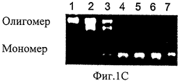
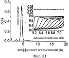
На Фиг.1A-D представлены компьютерно сгенерированные модели и графики, характеризующие про-ММР-9. На Фиг.1А представлена иллюстрация кристаллических структур концевых доменов. N-концевой домен про-ММР-9 (код PDB: 1L6J) состоит из пропептида (зеленый), трех фибронектиновых (тип II) повторов (синие) и каталитического домена (красный) с активным участком, содержащим цинк (серый шар). OG-домен (пунктирная линия) содержит фрагмент неизвестной структуры, состоящий из 64 остатков аминокислот, и соединяет N-концевой домен с С-концевым гемопексин-подобным доменом (код PDB: HTV), состоящим из четырех пропеллерных лопастей (циан). На Фиг.1В представлен график, иллюстрирующий эксклюзионную хроматографию, показывающую профиль элюирования олигомерных (пик 1, 15,8 минут и пик 2, 22,7 минут) и мономерных (пик 3, 25,1 минут) форм про-ММР-9. Вставка: График Пората [57] для стандартов белков с известными радиусами Стокса; эти стандарты были использованы с целью калибрования колонки Superdex 200 (слева направо: тиреоглобулин 85 Å, ферритин 61 Å, каталаза 52,2 Å, альдолаза 48,1 Å, альбумин 35,5 Å). Представлена зависимость кубического корня Kd от радиуса Стокса каждого белка, а также показана подгонка методом наименьших квадратов. На Фиг.1С представлена фотография желатиновой зимограммы. С целью отделения мономеров от более высокомолекулярных олигомерных структур в препаративных количествах была применена глицериновая седиментация. Был проведен анализ аликвотных количеств каждой фракции на желатиновых зимограммах. Высокомолекулярные олигомерные структуры присутствовали во фракциях 1-3. Фракция 3 содержала смесь всех олигомерных форм. Фракции 4-7 содержали преимущественно мономерную форму. На Фиг.1D представлен анализ скорости седиментации при аналитическом ультрацентрифугировании, осуществленный с целью расчета распределения коэффициента седиментации. Вставка: моделирование профилей седиментации (линий) на основании экспериментальных данных (точек) как функции времени и расстояния от оси вращения. Вверху представлен график остатков. В целях упрощения восприятия данных показан только каждый десятый профиль, использованный в данном анализе.On figa-D presents a computer-generated models and graphs characterizing pro-MMP-9. On figa presents an illustration of the crystal structures of the terminal domains. The pro-MMP-9 N-terminal domain (PDB code: 1L6J) consists of a propeptide (green), three fibronectin (type II) repeats (blue) and a catalytic domain (red) with an active site containing zinc (gray ball). The OG domain (dashed line) contains a fragment of an unknown structure consisting of 64 amino acid residues, and connects the N-terminal domain to the C-terminal hemopexin-like domain (PDB code: HTV) consisting of four propeller blades (cyan). 1B is a graph illustrating size exclusion chromatography showing the elution profile of oligomeric (
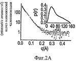
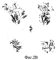
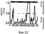
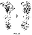
На Фиг.2А-Е представлены компьютерно сгенерированные модели и графики, иллюстрирующие структурный анализ про-ММР-9. На Фиг.2А представлен график, иллюстрирующий данные малоуглового рентгеновского рассеяния (МУРР) про-ММР-9 в растворе. Экспериментальные данные интенсивности рентгеновского излучения (черные точки) были сопоставлены с наиболее вероятной моделью (серая линия) с помощью CHADD. Вставка: парная функция распределения экспериментальных данных МУРР. На Фиг.2В представлены модели про-ММР-9, реконструированные с помощью CHADD. Модели, полученные на основании данных МУРР, представлены в виде белых шаров, имеющих радиус 5 Å. Каждая модель была повернута на 0° и 90° вокруг вертикальной оси. Пристыкованные кристаллические структуры N-концевого и С-концевого доменов [22, 24] представлены соответственно в виде синей и красной лент. На Фиг.2С представлена область долгого разупорядочения, спрогнозированная с помощью PONDR (толстая черная линия) [37] в последовательности про-ММР-9, и соответствующая организация домена (верхняя полоса: PRO-пропептид, CAT+FN - каталитический домен и три фибронектиновых (тип II) повтора, OG - О-гликозилированный домен, РЕХ - гемопексин-подобный домен). На Фиг.2D продемонстрировано соответствие экспериментальным данным расчетной кривой рассеяния про-ММР-9 полной длины с реконструированным OG-доменом. Эти расчетные кривые трех лучших моделей представлены в виде зеленой, зеленовато-голубой и желтой линий. Экспериментальные данные представлены в виде черных точек. Три лучшие модели OG-домена были рассчитаны с помощью RAPPER [38, 39] в рамках модели CHADD. На Фиг.2Е представлена реконструкция структуры OG-домена. Три лучшие модели представлены в виде зеленой, зеленовато-голубой и желтой лент.2A-E are computer generated models and graphs illustrating structural analysis of pro-MMP-9. 2A is a graph illustrating small angle X-ray scattering (SAXM) data of pro-MMP-9 in solution. The experimental X-ray intensity data (black dots) were compared with the most likely model (gray line) using CHADD. Insert: pair distribution function of experimental SAXS data. 2B shows pro-MMP-9 models reconstructed using CHADD. The models obtained on the basis of the SAXS data are presented in the form of white balls with a radius of 5 Å. Each model was rotated 0 ° and 90 ° around a vertical axis. The docked crystal structures of the N-terminal and C-terminal domains [22, 24] are presented in the form of blue and red ribbons, respectively. 2C shows a region of long disorder predicted by PONDR (thick black line) [37] in the pro-MMP-9 sequence and the corresponding domain organization (upper band: PRO-propeptide, CAT + FN — catalytic domain and three fibronectin (type II) repeat, OG is an O-glycosylated domain, PEX is a hemopexin-like domain). On Fig 2D shows the correspondence to experimental data of the calculated scattering curve of pro-MMP-9 full length with the reconstructed OG domain. These calculated curves of the three best models are presented in the form of green, greenish-blue and yellow lines. The experimental data are presented as black dots. The three best OG domain models were calculated using RAPPER [38, 39] in the framework of the CHADD model. Figure 2E shows a reconstruction of the structure of the OG domain. The three best models are presented in the form of green, greenish-blue and yellow ribbons.
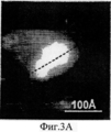
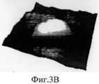
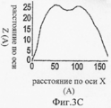
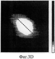
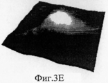
На Фиг.3A-F представлены графики и изображения про-ММР-9 дикого типа и мутировавшего про-ММР-9, полученные с помощью атомно-силовой микроскопии (АСМ). С целью ковалентного соединения аминных групп на поверхности белка был использован глютаральдегид. Все скенограммы были получены с использованием зонда с острым наконечником. На Фиг.3А-С представлены скенограммы про-ММР-9 дикого типа, полученные в полусухом режиме. На Фиг.3D-F представлены скенограммы мутанта npo-MMP-9ΔOG, полученные в полусухом режиме. На Фиг.3А и 3D представлены двухмерные изображения. На Фиг.3В и 3Е представлены трехмерные изображения. На Фиг.3С и 3F представлены поперечные сечения XZ вдоль пунктирной линии, показанной на Фиг.3А и Фиг.3D. Подготовка пробы и условия получения изображений описаны в тексте. Шкала высоты обозначена столбиком, расположенным с правой стороны: диапазон оси Z составляет от 0 до 50 Å (от темного к светлому).3A-F are graphs and images of wild-type pro-MMP-9 and mutated pro-MMP-9 obtained by atomic force microscopy (AFM). Glutaraldehyde was used to covalently bond amine groups on the surface of the protein. All scenograms were obtained using a probe with a sharp tip. On figa-C presents the scans of pro-MMP-9 wild-type obtained in semi-dry mode. 3D-F presents scans of the npo-MMP-9ΔOG mutant obtained in dry mode. On figa and 3D presents two-dimensional images. 3B and 3E show three-dimensional images. FIGS. 3C and 3F show cross-sections XZ along the dashed line shown in FIGS. 3A and 3D. Sample preparation and image acquisition conditions are described in the text. The height scale is indicated by a column located on the right side: the range of the Z axis is from 0 to 50 Å (from dark to light).
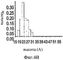
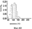
На Фиг.4A-F представлены гистограммы распределения размеров про-ММР-9 дикого типа (слева) и про-MMP-9ΔOG (справа) согласно результатам измерений с помощью АСМ. На оси "у" всех гистограмм отложена нормализованная частота, полученная с помощью деления подсчитанного количества импульсов на общую популяцию. На Фиг.4А и 4В представлено распределение по высоте. На Фиг.4С и 4D представлено распределение по ширине. Величины ширины были скорректированы в соответствии с описанием, представленным в разделе, посвященном методикам проведения экспериментов. На Фиг.4Е представлено распределение межлопастных расстояний про-ММР-9 дикого типа. Разделение лопастей про-MMP-9ΔOG отсутствовало. На Фиг.4F показано моделирование конформационных состояний про-ММР-9. Стандартное отклонение, равное 9,5 Å, рассчитанное на основании данных АСМ в отношении межлопастных расстояний, было вычтено (слева) из междоменного разделения усредненной структуры (посередине), полученной с помощью структурной реконструкции на основе МУРР, или добавлено (справа) к данному междоменному разделению. N- и С-концевой домены [22, 24] обозначены соответственно синим и красным цветом. OG-домен был реконструирован с помощью RAPPER [38, 39]; он обозначен зеленой С?-линией.4A-F are histograms of the size distribution of wild-type pro-MMP-9 (left) and pro-MMP-9ΔOG (right) according to AFM measurements. The normalized frequency obtained by dividing the calculated number of pulses by the total population is plotted on the y axis of all histograms. 4A and 4B show a height distribution. 4C and 4D show the width distribution. The widths were adjusted in accordance with the description presented in the section on the experimental methods. Figure 4E shows the wild-type pro-MMP-9 inter-blade spacing distribution. The separation of the pro-MMP-9ΔOG lobes was absent. FIG. 4F shows a simulation of conformational states of pro-MMP-9. The standard deviation of 9.5 Å, calculated on the basis of AFM data regarding the inter-blade distances, was subtracted (left) from the interdomain separation of the averaged structure (in the middle) obtained by structural reconstruction based on the SAXS, or added (to the right) to this interdomain separation. The N- and C-terminal domains [22, 24] are indicated by blue and red, respectively. The OG domain was reconstructed using RAPPER [38, 39]; it is indicated by a green C? line.
На Фиг.5А-В представлены реконструированные модели про-ММР-9, полученные с помощью МУРР. На Фиг.5А представлена модель GASBOR. На Фиг.5В представлена модель CHADD. Белые шары радиусом 5 Å представляют полученные модели. Пристыкованные кристаллические структуры N- и С-концевых доменов обозначены соответственно синим и красным цветом. Каждая модель была повернута на 0° и 90° вокруг вертикальной оси.On figa-b presents the reconstructed model of pro-MMP-9, obtained using SAXS. 5A shows a GASBOR model. 5B shows a CHADD model. White balls with a radius of 5 Å represent the resulting model. The docked crystal structures of the N- and C-terminal domains are indicated by blue and red, respectively. Each model was rotated 0 ° and 90 ° around a vertical axis.
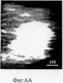
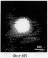
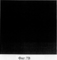
На Фиг.6А-С представлены АСМ-изображения про-ММР-9. С целью ковалентного соединения аминных групп на поверхности белка был использован глютаральдегид. Все скенограммы были получены с использованием зонда с острым наконечником, за исключением скенограммы, представленной на Фиг.6А, которая была получена с использованием зонда с кремниево-нитридным наконечником, заостренным оксидом. На Фиг.6А представлен про-ММР-9 дикого типа под буферным раствором. На Фиг.6В представлен обезвоженный образец фермента дикого типа, просканированный в условиях окружающей среды. На Фиг.6С представлена пустая проба, подвергнутая той же процедуре иммобилизации, но без нанесения фермента. Стрелкой показана одиночная частица, наблюдаемая на скенограмме 1×1? м2. Шкала высоты обозначена столбиком, расположенным с правой стороны: диапазон оси Z составляет от 0 до 50 Å (от темного к светлому).6A-C show AFM images of pro-MMP-9. Glutaraldehyde was used to covalently bond amine groups on the surface of the protein. All scenograms were obtained using a sharp tip probe, with the exception of that shown in FIG. 6A, which was obtained using a silicon nitride tip, pointed oxide probe. 6A shows wild-type pro-MMP-9 under a buffer solution. 6B shows a dehydrated wild-type enzyme sample scanned under ambient conditions. On Figs presents an empty sample subjected to the same procedure of immobilization, but without applying the enzyme. The arrow shows a single particle observed on the 1 × 1? m 2 . The height scale is indicated by a column located on the right side: the range of the Z axis is from 0 to 50 Å (from dark to light).
Фиг.7А-В: На Фиг.7А представлена зимография in situ клеток линии НТ1080, экспрессирующих секретируемую ММР-9. Наличие зеленой флюоресценции свидетельствует о протеолитической активности в отношении коллагена IV типа. Синим цветом окрашивается ядро (Hoehst). На Фиг.7В проиллюстрировано инкубирование клеток линии НТ1080 с антителами против ММР (9 часов) с последующим нанесением слоя коллагена IV типа, конъюгированного с Орегонским зеленым. Отсутствие выраженной зеленой флюоресценции вокруг клеток свидетельствует об ингибировании перицеллюлярного протеолиза, обеспечиваемого ММР-9.7A-B: FIG. 7A shows in situ zymography of HT1080 cell lines expressing secreted MMP-9. The presence of green fluorescence indicates proteolytic activity against type IV collagen. The core is painted in blue (Hoehst). Fig. 7B illustrates the incubation of HT1080 cells with anti-MMP antibodies (9 hours), followed by the application of a layer of type IV collagen conjugated with Oregon green. The absence of pronounced green fluorescence around the cells indicates the inhibition of pericellular proteolysis provided by MMP-9.
ОПИСАНИЕ ПРЕДПОЧТИТЕЛЬНЫХ ВАРИАНТОВ ОСУЩЕСТВЛЕНИЯDESCRIPTION OF PREFERRED EMBODIMENTS
Настоящее изобретение относится к регуляторам ММР-9, а более конкретно к регуляторам, нацеленным на OG-домен ММР-9.The present invention relates to regulators of MMP-9, and more specifically to regulators aimed at the OG domain of MMP-9.
Чертежи и сопровождающие их описания способствуют лучшему пониманию принципов и осуществления настоящего изобретения.The drawings and accompanying descriptions contribute to a better understanding of the principles and implementation of the present invention.
Перед началом подробного рассмотрения вариантов осуществления настоящего изобретения необходимо уточнить, что применение данного изобретения не ограничивается деталями, представленными ниже в описании или в разделе "Примеры". Возможны и другие варианты осуществления данного изобретения, а также различные пути его реализации или выполнения. Кроме того, следует иметь в виду, что используемые в данном документе фразы и термины используются лишь в целях описания и не должны рассматриваться как ограничители объема данного изобретения.Before starting a detailed discussion of embodiments of the present invention, it is necessary to clarify that the application of the present invention is not limited to the details presented in the description or in the "Examples" section below. There are other possible embodiments of the present invention, as well as various ways of its implementation or implementation. In addition, it should be borne in mind that the phrases and terms used herein are used for description purposes only and should not be construed as limiting the scope of this invention.
Установлено, что представители семейства металлопротеиназ (ММР) участвуют в многочисленных аспектах миграции клеток воспаления и раковых клеток в соединительной ткани, способствуя развитию многих заболеваний. Хотя все ММР имеют одинаковые каталитические участки, их участки связывания субстрата различны. Вследствие этого ММР различаются по своим биологическим функциям. Так, например, ММР-9 способствует повреждению ткани и развитию воспаления, тогда как ММР-2 выполняет в основном противовоспалительные функции и функцию поддержания гомеостаза. Таким образом, селективные ингибиторы, различающие эти ферменты, обладающие высокой степенью сходства, имеют очень важное значение как средство эффективной противовоспалительной терапии, не вызывающее побочных эффектов.It was found that representatives of the family of metalloproteinases (MMPs) are involved in numerous aspects of the migration of inflammatory cells and cancer cells in the connective tissue, contributing to the development of many diseases. Although all MMPs have the same catalytic sites, their substrate binding sites are different. As a result, MMPs differ in their biological functions. So, for example, MMP-9 contributes to tissue damage and the development of inflammation, while MMP-2 performs mainly anti-inflammatory functions and the function of maintaining homeostasis. Thus, selective inhibitors that distinguish between these enzymes, which have a high degree of similarity, are very important as a means of effective anti-inflammatory therapy that does not cause side effects.
На предварительном этапе разработки данного изобретения его авторы пришли к пониманию того, что основное структурное различие между ММР-9 и ММР-2 заключается в присутствии в ММР-9 домена, характеризующегося высокой степенью О-гликозилирования (OG). Вместе с тем, имеющаяся информация о структуре ММР-9 ограничивается ее двумя концевыми доменами и не захватывает этот OG-домен. Таким образом, какая-либо информация, имеющая отношение к влиянию OG-домена на общую трехмерную структуру ММР-9 и ее биофизическую природу, отсутствует.At the preliminary stage of the development of this invention, its authors came to the understanding that the main structural difference between MMP-9 and MMP-2 is the presence in MMP-9 of a domain characterized by a high degree of O-glycosylation (OG). At the same time, the available information on the structure of MMP-9 is limited to its two terminal domains and does not capture this OG domain. Thus, any information related to the influence of the OG domain on the general three-dimensional structure of MMP-9 and its biophysical nature is absent.
В процессе практического осуществления настоящего изобретения данные изобретатели выполнили новый структурный анализ, применив сочетание малоуглового рентгеновского рассеяния (МУРР) и мономолекулярной атомно-силовой микроскопии (АСМ) для того, чтобы впервые выяснить характеристики структуры полной длины про-ММР-9 и молекулярные характеристики ее О-гликозилированного линкерного домена. В результате МУРР с последующим анализом изображений и структурной реконструкции была установлена молекулярная форма полной длины про-ММР-9, представляющая ее усредненную конформацию в растворе (Фиг.2А-Е). Согласно полученным структурным данным, подтвержденным АСМ высокого разрешения (Фиг.3A-F и 4А-Е) и биофизическими измерениями, про-ММР-9 представляет собой белок вытянутой формы, в котором OG-домен действует как гибкое соединительное звено (линкер) длиной 30 Å, расположенное между двумя концевыми доменами (Фиг.5А-В). Степень гибкости OG-домена была оценена статистически на основании данных о разных конформациях белка, выявленных с помощью мономолекулярных изображений (Фиг.4F). Структурно-динамическая модель полной длины про-ММР-9 дает возможность получить новые представления о роли гибкости данного домена белка в регулировании распознавания, связывания и обработки субстратов, лигандов и рецепторов, необходимом для обеспечения эффектов ММР-9.In the practice of the present invention, these inventors performed a new structural analysis using a combination of small angle X-ray scattering (SAXS) and monomolecular atomic force microscopy (AFM) in order to first determine the characteristics of the pro-MMP-9 full-length structure and its molecular characteristics O -glycosylated linker domain. As a result of SAXS, followed by image analysis and structural reconstruction, a pro-MMP-9 full-length molecular form was established representing its averaged conformation in solution (Fig. 2A-E). According to the obtained structural data, confirmed by high-resolution AFM (Figs. 3A-F and 4A-E) and biophysical measurements, pro-MMP-9 is an elongated protein in which the OG domain acts as a flexible connecting link (linker) of
В процессе дальнейшего практического осуществления настоящего изобретения данными изобретателями с помощью зимографии in situ было показано, что антитело, способное специфически взаимодействовать с OG-доменом ММР-9, блокирует коллагенолитическую активность ММР-9, но не блокирует ее желатинолитическую активность (Фиг.7А-В). Таким образом, данные изобретатели предлагают использование агентов, регулирующих гибкость OG-домена, с целью препятствования эффектам этого фермента, способствующим возникновению патологических изменений.In the process of further practical implementation of the present invention by these inventors using in situ zymography, it was shown that an antibody capable of specifically interacting with the OG domain of MMP-9 blocks the collagenolytic activity of MMP-9, but does not block its gelatinolytic activity (Fig. 7A-B ) Thus, these inventors propose the use of agents that regulate the flexibility of the OG domain, in order to inhibit the effects of this enzyme, contributing to the occurrence of pathological changes.
Таким образом, один из признаков данного изобретения связан со способом регулирования активности металлопротеиназы 9 (ММР-9), заключающимся в осуществлении контакта ММР-9 с агентом, специфически взаимодействующим с OG-доменом ММР-9 и, таким образом, регулирующим активность ММР-9.Thus, one of the features of this invention is associated with a method for regulating the activity of metalloproteinase 9 (MMP-9), which involves contacting MMP-9 with an agent that specifically interacts with the OG domain of MMP-9 and, thus, regulating the activity of MMP-9 .
В рамках данного документа термин "ММР-9" (мультидоменная цинковая эндопептидаза матриксная металлопротеиназа-9, также называемая желатиназой В) относится к предшественнику и активным формам полипептида ММР-9 млекопитающих (например, человека) (ЕС 3.4.24.35; Swiss Prot №Р 14780), включая их гомологи, ортологи и изоформы. Как правило, ММР-9 включает в себя три домена -каталитический домен; домен, связывающий субстрат; и расположенный между ними линкерный домен. Этот линкерный домен, который в этом документе также называется "коллаген V-подобным доменом" или "О-гликозилированным (OG) доменом", включает в себя 64 аминокислоты, 22 из которых представляют собой остаток пролина, 6 являются остатком глицина и приблизительно 12-14 - О-связанными гликанами.For the purposes of this document, the term “MMP-9” (multidomain zinc endopeptidase matrix metalloproteinase-9, also called gelatinase B) refers to the precursor and active forms of the MMP-9 polypeptide of mammals (eg, humans) (EC 3.4.24.35; Swiss Prot No. P 14780), including their homologues, orthologs and isoforms. As a rule, MMP-9 includes three domains — a catalytic domain; substrate binding domain; and a linker domain located between them. This linker domain, also referred to herein as a “collagen V-like domain” or “O-glycosylated (OG) domain,” includes 64 amino acids, 22 of which are proline residues, 6 are glycine residues, and approximately 12- 14 - O-linked glycans.
Вариант осуществления этого признака изобретения: ММР-9 является нативным (то есть не денатурированным) ферментом. Другой вариант осуществления этого признака изобретения: ММР-9 является активным (предпочтительно полностью активным) ферментом.An embodiment of this feature of the invention: MMP-9 is a native (i.e. not denatured) enzyme. Another embodiment of this feature of the invention: MMP-9 is an active (preferably fully active) enzyme.
Виды активности ММР-9 включают в себя, помимо прочего, желатинолитическую активность, разложение нативных коллагенов I, III и XI типов (коллагенолитическую активность), а также разложение эластина, агрекана, цепи А ламинина и основного белка миелина.Types of MMP-9 activity include, but are not limited to, gelatinolytic activity, decomposition of native type I, III, and XI collagen (collagenolytic activity), as well as decomposition of elastin, agrecan, laminin chain A, and myelin basic protein.
В рамках данного документа термин "регулирующий" означает понижающую регуляцию или повышающую регуляцию. Следует иметь в виду, что агенты, подавляющие гибкость OG-домена, снижают функцию ММР-9, для осуществления которой требуется гибкость OG-домена (например, коллагенолитическую активность ММР-9). В отличие от этого активность, требующая наличия особой трехмерной структуры ММР-9 и не требующая наличия гибкости OG-домена, может повышаться под действием агентов, взаимодействующих с OG-доменом. Примером такой активности является желатинолитическая активность ММР-9 или ее способность взаимодействовать с рецепторами и/или факторами роста.As used herein, the term “regulatory” means down regulation or up regulation. It should be borne in mind that agents that suppress the flexibility of the OG domain reduce the function of MMP-9, which requires the flexibility of the OG domain (for example, the collagenolytic activity of MMP-9). In contrast, activity that requires a special three-dimensional structure of MMP-9 and does not require the flexibility of the OG domain can be increased by agents interacting with the OG domain. An example of such activity is the gelatinolytic activity of MMP-9 or its ability to interact with receptors and / or growth factors.
Как было отмечено выше, способ данного изобретения осуществляется путем обеспечения контакта ММР-9 с агентом, способным специфически взаимодействовать с OG-доменом ММР-9.As noted above, the method of the invention is carried out by contacting MMP-9 with an agent capable of specifically interacting with the OG domain of MMP-9.
В рамках данного документа термин "обеспечение контакта" означает обеспечение возможности ММР-9 вступать в контакт с соответствующим агентом в условиях (то есть в определенное время, при определенной температуре и при наличии определенного буфера), позволяющих агенту взаимодействовать с OG-доменом ММР-9 (например, связываться с OG-доменом) и воздействовать на жесткость OG-домена. Следует иметь в виду, что контакт может обеспечиваться в условиях in vivo, ex vivo или in vitro.For the purposes of this document, the term “providing contact” means enabling MMP-9 to come into contact with the appropriate agent under conditions (that is, at a specific time, at a certain temperature and in the presence of a specific buffer) that allow the agent to interact with the OG domain of MMP-9 (for example, bind to the OG domain) and affect the stiffness of the OG domain. It should be borne in mind that contact can be provided in vivo, ex vivo or in vitro.
В рамках данного документа фраза "специфически взаимодействующий" означает как повышенное сродство с OG-доменом ММР-9 по сравнению с другим доменом ММР-9 (например, каталитическим доменом или доменом, связывающим субстрат), так и повышенное сродство с OG-доменом ММР-9 по сравнению с OG-доменом другой металлопротеиназы (например, ММР-2). Примером минимального сродства, вероятно, является величина сродства, равная 10-5 М. Предпочтительно агент взаимодействует с OG-доменом ММР-9 с уровнем сродства, не менее чем в 3 раза превышающим сродство, указанное выше; более предпочтительно, если это взаимодействие осуществляется с уровнем сродства, не менее чем в 5 раз превышающим вышеуказанное сродство, а еще более предпочтительно не менее чем в 10 раз превышающим это сродство. Следует иметь в виду, что агенты, способные специфически взаимодействовать с OG-доменом ММР-9, способны и специфически регулировать ММР-9, так как аминокислотная последовательность OG-домена ММР-9 специфична для ММР-9 (в отличие от аминокислотной последовательности каталитического домена, который характеризуется высокой степенью гомологии между ММР-9 и ММР-2).For the purposes of this document, the phrase “specifically interacting” means both increased affinity for the MMP-9 OG domain compared to another MMP-9 domain (for example, a catalytic domain or substrate binding domain), and increased affinity for the MMP-
Агенты (то есть молекулы), рассматриваемые в настоящем изобретении как способные к взаимодействию с OG-доменом ММР-9, включают в себя, помимо прочего, полипептидные агенты (например, антитела, содержащие антиген-распознающий домен, специфически взаимодействующий с OG-доменом ММР-9), пептиды и небольшие молекулы. Следует иметь в виду, что данные агенты могут взаимодействовать с OG-доменом на основе распознавания специфической последовательности аминокислот и/или на основе конформационного распознавания. Агенты-антитела, распознающие OG-домен ММР-9, имеются в продаже (например, антитела, выпускаемые компаниями Сигма, Кемикон и Абкам).Agents (i.e., molecules) considered in the present invention as capable of interacting with the MG-9 OG domain include, but are not limited to, polypeptide agents (e.g., antibodies containing an antigen-recognizing domain that specifically interacts with the MMP OG domain -9), peptides and small molecules. It should be borne in mind that these agents can interact with the OG domain based on recognition of a specific amino acid sequence and / or on the basis of conformational recognition. Antibody agents that recognize the MG-9 OG domain are commercially available (for example, antibodies manufactured by Sigma, Chemicon and Abkam).
В данном изобретении термин "антитело" охватывает интактные молекулы, а также их функциональные фрагменты (например, Fab, F(ab')2 и Fv), способные связываться со специфическими митохондриальными белками. Фрагменты антител, имеющие меньшие размеры, могут иметь преимущество над целыми антителами, поскольку они способны более активно проникать в ткани и быстрее выводятся из организма. Особенно важное значение эти преимущества имеют при использовании ММР-9-специфичных антител in vivo. Кроме того, дополнительным преимуществом фрагментов антител является то, что они могут быть продуцированы бактериями или дрожжами.As used herein, the term “antibody” encompasses intact molecules as well as functional fragments thereof (eg, Fab, F (ab ′) 2 and Fv) capable of binding to specific mitochondrial proteins. Fragments of antibodies having smaller sizes may have an advantage over whole antibodies, since they are able to penetrate tissue more actively and are more rapidly excreted from the body. These advantages are especially important when using MMP-9 specific antibodies in vivo. In addition, an additional advantage of antibody fragments is that they can be produced by bacteria or yeast.
Выработка антител против OG-домена ММР-9 может быть индуцирована пептидом, включающим в себя OG-домен. Селекция данных антител может быть осуществлена с использованием других доменов ММР-9 в качестве отрицательных контролей.The production of antibodies against the OG domain of MMP-9 can be induced by a peptide comprising the OG domain. Selection of these antibodies can be carried out using other MMP-9 domains as negative controls.
К фрагментам антител, подходящим для практического применения настоящего изобретения, относятся область, определяющая комплементарность (ООК; CDR), легкая цепь иммуноглобулина (далее называемая "легкой цепью"); область, определяющая комплементарность, тяжелой цепи иммуноглобулина (далее называемая "тяжелой цепью"); вариабильная область легкой цепи; вариабильная область тяжелой цепи; легкая цепь, тяжелая цепь; фрагмент Fd; и фрагменты антител, содержащие практически целые вариабильные области легкой и тяжелой цепей - такие как Fv, одноцепочечный Fv, Fab, Fab' и F(ab')2.Antibody fragments suitable for the practice of the present invention include the complementarity determining region (OLC; CDR), the immunoglobulin light chain (hereinafter referred to as the “light chain”); the complementarity determining region of an immunoglobulin heavy chain (hereinafter referred to as the "heavy chain"); variable region of the light chain; variable region of the heavy chain; light chain, heavy chain; Fd fragment; and antibody fragments containing substantially entire variable regions of the light and heavy chains — such as Fv, single chain Fv, Fab, Fab ′ and F (ab ′) 2.
Функциональными фрагментами антител, содержащими целые или практически целые вариабильные области легкой и тяжелой цепей, являются:Functional antibody fragments containing whole or almost whole variable regions of the light and heavy chains are:
(i) фрагмент Fv, созданный с помощью методов генной инженерии, состоящий из вариабильной области легкой цепи и вариабильной области тяжелой цепи и экспрессируемый в виде двух цепей;(i) an Fv fragment generated by genetic engineering methods consisting of a variable region of a light chain and a variable region of a heavy chain and expressed as two chains;
(ii) одноцепочечный фрагмент Fv ("scFv"), представляющий собой одноцепочечную молекулу, созданную с помощью методов генной инженерии; эта молекула включает в себя вариабильную область легкой цепи и вариабильную область тяжелой цепи, соединенные с помощью подходящего полипептидного линкера;(ii) a single chain Fv fragment (“scFv”), which is a single chain molecule created using genetic engineering methods; this molecule includes the variable region of the light chain and the variable region of the heavy chain connected using a suitable polypeptide linker;
(iii) фрагмент Fab, представляющий собой фрагмент молекулы антитела, содержащий моновалентную антиген-связывающую часть молекулы антитела, которая может быть получена путем обработки целого антитела ферментом папаином, в результате которой получают интактную легкую цепь и фрагмент Fd тяжелой цепи, который состоит из ее вариабильного домента и СНl-домена;(iii) a Fab fragment, which is a fragment of an antibody molecule containing the monovalent antigen-binding portion of an antibody molecule, which can be obtained by treating the whole antibody with papain, resulting in an intact light chain and a heavy chain Fd fragment, which consists of its variable domain and CHl domain;
(iv) фрагмент Fab', представляющий собой фрагмент молекулы антитела, содержащий моновалентную антиген-связывающую часть молекулы антитела, которая может быть получена путем обработки целого антитела ферментом пепсином с последующим восстановлением (на одну молекулу антитела приходятся два получаемых фрагмента Fab'); и(iv) a Fab ′ fragment, which is a fragment of an antibody molecule containing the monovalent antigen-binding portion of an antibody molecule, which can be obtained by treating the whole antibody with pepsin enzyme followed by reduction (two Fab fragments are obtained per antibody molecule); and
(v) фрагмент F(ab')2, представляющий собой фрагмент молекулы антитела, содержащий моновалентную антиген-связывающую часть молекулы антитела, которая может быть получена путем обработки целого антитела ферментом пепсином (то есть димер фрагментов Fab', удерживаемых вместе двумя дисульфидными связями).(v) a fragment F (ab ') 2, which is a fragment of an antibody molecule containing the monovalent antigen-binding portion of an antibody molecule, which can be obtained by treating the whole antibody with pepsin enzyme (i.e. a dimer of Fab' fragments held together by two disulfide bonds) .
Способы выработки антител (моноклональных и поликлональных) хорошо известны в данной отрасли знаний. Выработка антител может осуществляться любым из нескольких известных способов; эти способы могут включать в себя индукцию выработки молекул антител in vivo, скрининг библиотек иммуноглобулинов (Orlandi D.R. с соавт., 1989. Proc. Natl. Acad. Sci. U.S.A. 86:3833-3837; Winter G. с соавт., 1991. Nature 349:293-299) или выработку молекул моноклональных антител непрерывными клеточными линиями в культуре. Эти способы включают в себя, помимо прочего, гибридомную методику; методику гибридомных В-клеток человека и методику вируса Эпштейна-Барр (ЕВV)-гибридомы (Kohler G. с соавт., 1975. Nature 256:495-497; Kozbor D. с соавт., 1985. J. Immunol. Methods 81:31-42; Cote RJ. с соавт., 1983. Proc. Natl. Acad. Sci. U.S.A. 80:2026-2030; Cole SP. с соавт., 1984. Mol. Cell. Biol. 62:109-120).Methods for generating antibodies (monoclonal and polyclonal) are well known in the art. The development of antibodies can be carried out by any of several known methods; these methods may include inducing in vivo production of antibody molecules, screening immunoglobulin libraries (Orlandi DR et al., 1989. Proc. Natl. Acad. Sci. USA 86: 3833-3837; Winter G. et al., 1991. Nature 349: 293-299) or the production of monoclonal antibody molecules by continuous cell lines in culture. These methods include, but are not limited to, a hybridoma technique; the method of hybridoma human B cells and the method of the Epstein-Barr virus (EBV) -hybridoma (Kohler G. et al., 1975. Nature 256: 495-497; Kozbor D. et al., 1985. J. Immunol. Methods 81: 31-42; Cote RJ et al., 1983. Proc. Natl. Acad. Sci. USA 80: 2026-2030; Cole SP. Et al. 1984. Mol. Cell. Biol. 62: 109-120).
В тех случаях, когда целевые антигены слишком малы для того, чтобы вызвать адекватный иммунный ответ при выработке антител in vivo, данные антигены (гаптены) могут быть спарены с антигенно нейтральными носителями, такими как гемоцианин моллюска рода фиссурелла (KLH) или сывороточный альбумин [например, бычий сывороточный альбумин (BSA)] (см., например, патенты США №5,189,178 и №5,239,078]. Спаривание гаптена с носителем может быть осуществлено с помощью способов, хорошо известных в данной отрасли знаний. Так, например, может быть осуществлено прямое присоединение к аминогруппам и, по выбору, последующее восстановление образовавшейся иминосвязи. Как альтернативный вариант, носитель может быть присоединен с помощью конденсирующих агентов, например дициклогексилкарбодиимида или других карбодиимидных дегидратирующих агентов. Кроме того, с целью обеспечения данного спаривания могут быть использованы соединяющие вещества (линкеры); как гомобифункциональные, так и гетеробифункциональные линкеры могут быть приобретены у Пирс Кемикал Компани, Рокфорд, 111. Иммуногенный комплекс, полученный в результате спаривания, можно вводить подходящим млекопитающим, например мышам, кроликам и т.д. Подходят протоколы, предусматривающие повторные инъекции иммуногена в присутствии адъювантов в соответствии со схемой, ускоряющей продукцию антител и, соответственно, повышение их уровня в сыворотке. Титры антител в иммунной сыворотке легко определяются с помощью методик иммунологического анализа, хорошо известных в данной отрасли знаний.In cases where the target antigens are too small to elicit an adequate immune response when producing antibodies in vivo, these antigens (haptens) can be paired with antigenically neutral carriers such as fissurella mollusk hemocyanin (KLH) or serum albumin [eg bovine serum albumin (BSA)] (see, for example, US Pat. Nos. 5,189,178 and 5,239,078]. Mating the hapten with a carrier can be accomplished using methods well known in the art. For example, direct admixture can be carried out. amino acids and, optionally, subsequent restoration of the formed imino bond. Alternatively, the carrier may be attached using condensing agents, for example dicyclohexylcarbodiimide or other carbodiimide dehydrating agents. In addition, connecting substances (linkers) can be used to ensure this pairing. ; both homobifunctional and heterobifunctional linkers can be purchased from Pierce Chemical Company, Rockford, 111. Immunogenic complex obtained in result of mating can be administered by a suitable mammal, e.g., mice, rabbits, etc. Suitable protocols are those involving repeated injections of the immunogen in the presence of adjuvants in accordance with a scheme accelerating the production of antibodies and, accordingly, increasing their level in serum. Antibody titers in immune serum are readily determined using immunoassay techniques that are well known in the art.
Возможно непосредственное использование полученных антисывороток, либо могут быть получены моноклональные антитела, как описано выше.It is possible to directly use the obtained antisera, or monoclonal antibodies can be obtained, as described above.
С помощью способов, хорошо известных в данной отрасли знаний, могут быть получены фрагменты антител [(см., например, работу Harlow и Lane, "Antibodies: А Laboratory Manual", Cold Spring Harbor Laboratory, New York, (1988)]. Так, например, фрагменты антител по настоящему изобретению могут быть получены с помощью протеолитического гидролиза антитела или с помощью экспрессии E. coli или клетками млекопитающих (например, культурой клеток яичника китайского хомячка или другими системами экспрессии белка) ДНК, кодирующей данный фрагмент.Using methods well known in the art, antibody fragments can be obtained [(see, for example, Harlow and Lane, "Antibodies: A Laboratory Manual", Cold Spring Harbor Laboratory, New York, (1988)]. So for example, antibody fragments of the present invention can be obtained by proteolytic hydrolysis of an antibody or by expression of E. coli or mammalian cells (e.g., Chinese hamster ovary cell culture or other protein expression systems) of DNA encoding the fragment.
Как альтернативный вариант, фрагменты антител могут быть получены с помощью переваривания целых антител пепсином или папаином в соответствии с общепринятыми способами. Как описано выше, фрагмент антитела (Fab')2 может быть получен путем ферментного расщепления антитела пепсином с получением 5S фрагмента. Этот фрагмент может быть дополнительно расщеплен с помощью тиолового восстанавливающего агента и, необязательно, блокирующей группы для сульфгидрильных групп, образующихся в результате разрыва дисульфидных связей, с получением моновалентных 3,5S Fab' фрагментов. Как альтернативный вариант, применяется ферментное расщепление с помощью пепсина, в результате которого непосредственно получают два моновалентных Fab' фрагмента и один Fc фрагмент. В научной литературе присутствуют полные руководства по применению данных способов (например, Goldenberg, патенты США №4,036,945 и 4,331,647; Porter, RR., 1959. Biochem. J. 73:119-126). Кроме того, могут быть использованы и другие способы расщепления антител, например отделение тяжелых цепей с образованием моновалентных фрагментов легкой-тяжелой цепи, дополнительное расщепление фрагментов или другие ферментные, химические или генетические методики, при условии, что получаемые фрагменты связываются с антигеном, распознаваемым интактным антителом.Alternatively, antibody fragments can be obtained by digesting whole antibodies with pepsin or papain in accordance with conventional methods. As described above, an antibody fragment (Fab ') 2 can be obtained by enzymatic cleavage of the antibody with pepsin to give a 5S fragment. This moiety can be further cleaved with a thiol reducing agent and optionally blocking groups for sulfhydryl groups resulting from cleavage of disulfide bonds to give monovalent 3,5S Fab ′ fragments. Alternatively, enzymatic cleavage with pepsin is used, as a result of which two monovalent Fab 'fragments and one Fc fragment are directly obtained. The scientific literature contains complete guidelines for the use of these methods (for example, Goldenberg, US patents No. 4,036,945 and 4,331,647; Porter, RR., 1959. Biochem. J. 73: 119-126). In addition, other antibody cleavage methods can be used, for example, heavy chain separation to form monovalent light-heavy chain fragments, additional fragment cleavage, or other enzymatic, chemical, or genetic techniques, provided that the resulting fragments bind to an antigen recognized by the intact antibody .
Как было описано выше, Fv состоит из спаренных вариабильного домена тяжелой цепи и вариабильного домена легкой цепи. Эта связь может быть нековалентной (см., например, работу Inbar с соавт., 1972. Proc. Natl. Acad. Sci. USA. 69:2659-62). Как альтернативный вариант (см. выше), данные вариабильные домены могут быть соединены с помощью межмолекулярной дисульфидной связи с образованием одноцепочечного Fv; в качестве другого альтернативного варианта, эти цепи могут быть соединены поперечными связями с помощью химических веществ, например глютаральдегида.As described above, Fv consists of a paired heavy chain variable domain and a light chain variable domain. This relationship may be non-covalent (see, for example, Inbar et al., 1972. Proc. Natl. Acad. Sci. USA. 69: 2659-62). As an alternative (see above), these variable domains can be joined via an intermolecular disulfide bond to form a single-chain Fv; as another alternative, these chains can be connected by cross-linking with chemicals, such as glutaraldehyde.
Предпочтительно Fv является одноцепочечным Fv.Preferably, the Fv is a single chain Fv.
Одноцепочечные Fv получают путем конструирования структурного гена, содержащего последовательности ДНК, кодирующие вариабильный домен тяжелой цепи и вариабильный домен легкой цепи, соединенные олигонуклеотидом, кодирующим пептидный линкер. Этот структурный ген внедряют в экспрессионный вектор, который затем вводят в клетку-хозяина (например, клетку Е. coli). Рекомбинантные клетки-хозяева синтезируют одиночную полипептидную цепь с линкерным пептидом, связывающим два вариабильных домена. В научной литературе присутствуют полные руководства по получению одноцепочечных Fv (см., например, Whitlow и Filpula, 1991. Methods 2:97-105; Bird с соавт., 1988. Science 242:423-426; Pack с соавт., 1993. Bio/Technology 11:1271-77; и Ladner с соавт., патент США №4,946,778).Single chain Fvs are obtained by constructing a structural gene containing DNA sequences encoding the variable domain of the heavy chain and the variable domain of the light chain connected by an oligonucleotide encoding a peptide linker. This structural gene is introduced into an expression vector, which is then introduced into a host cell (e.g., an E. coli cell). Recombinant host cells synthesize a single polypeptide chain with a linker peptide linking two variable domains. The scientific literature provides comprehensive guidelines for the production of single chain Fvs (see, for example, Whitlow and Filpula, 1991. Methods 2: 97-105; Bird et al., 1988. Science 242: 423-426; Pack et al., 1993. Bio / Technology 11: 1271-77; and Ladner et al. US Pat. No. 4,946,778).
Изолированные пептиды области, определяющей комплементарность, могут быть получены путем конструирования генов, кодирующих область, определяющую комплементарность, интересующего антитела. Такие гены могут быть получены, например, с помощью полимеразной цепной реакции с обратной транскрипцией (ОТ-ПЦР) мРНК антителопродуцирующей клетки. В научной литературе присутствуют полные руководства по применению данных способов (см., например, Larrick и Fry, 1991. Methods 2:106-10).Isolated peptides of the complementarity determining region can be obtained by constructing genes encoding the complementarity determining region of the antibody of interest. Such genes can be obtained, for example, by reverse transcription polymerase chain reaction (RT-PCR) of an antibody producing cell mRNA. The scientific literature provides complete guidance on the use of these methods (see, for example, Larrick and Fry, 1991. Methods 2: 106-10).
Следует иметь в виду, что при лечении людей предпочтение отдается гуманизированным антителам. Гуманизированные формы антител, вырабатываемых животными (например, мышами), представляют собой химерные антитела или фрагменты антител, полученные методами генной инженерии и содержащие участки (предпочтительно очень небольшие), происходящие от антитела животного. К гуманизированным антителам относятся антитела, в которых области, определяющие комплементарность, человеческого антитела (антитела-реципиента) замещены остатками, обладающими желаемыми функциональными свойствами, взятыми из области, определяющей комплементарность, антитела животного (антитела-донора) (например, мыши, крысы или кролика). В некоторых случаях остатки человеческого антитела, присутствующие в каркасе Fv замещают соответствующими остатками антитела животного. Кроме того, гуманизированные антитела могут содержать остатки, не обнаруживаемые ни в антителе-реципиенте, ни в импортируемой области, определяющей комплементарность, ни в каркасных последовательностях. В целом, гуманизированное антитело содержит, практически полностью, как минимум один (а как правило, два) вариабильных домена, в которых все (или практически все) области, определяющие комплементарность, соответствуют областям, определяющим комплементарность, антитела животного, и все (или практически все) каркасные области соответствуют каркасным областям соответствующей человеческой консенсусной последовательности. В оптимальном варианте гуманизированные антитела также включают в себя по крайней мере часть константной области антитела (например, область Fc), происходящую, как правило, от антитела человека (см., например, работы Jones с соавт., 1986. Nature 321:522-525; Riechmann с соавт., 1988. Nature 332:323-329; и Presta, 1992. Curr. Op. Struct. Biol. 2:593-596). Способы гуманизации антител животных хорошо известны в данной отрасли знаний. Обычно гуманизированное антитело содержит не менее чем один аминокислотный остаток, внедренный в него из животного источника. Эти животные аминокислотные остатки часто называют импортированными остатками; как правило, они принадлежат к импортируемому вариабильному домену. В целом, гуманизация может быть осуществлена в соответствии с описаниями, имеющимися в литературе (см., например, работы Jones с соавт., 1986. Nature 321:522-525; Riechmann с соавт., 1988. Nature 332:323-327; Verhoeyen с соавт., 1988. Science 239:1534-1536; патент США №4,816,567), путем замещения человеческих областей, определяющих комплементарность, соответствующими областями, определяющими комплементарность, грызуна. Таким образом, данные гуманизированные антитела являются химерными антителами, отличающимися тем, что соответствующей последовательностью животного происхождения заменен человеческий вариабильный домен, причем не полностью, а существенно меньше, чем на всю величину интактного человеческого вариабильного домена. На практике гуманизированные антитела могут представлять собой типичные человеческие антитела, в которых некоторые остатки области, определяющей комплементарность, и, возможно, некоторые остатки каркаса заменены остатками, относящимися к аналогичным участкам антитела грызуна. Кроме того, человеческие антитела могут быть получены с помощью различных методик, известных в данной отрасли знаний, включая фаговые дисплейные библиотеки [см., например, работы Hoogenboom и Winter, 1991. J. Mol. Biol. 227:381; Marks с соавт., 1991. J. MoL Biol. 222:581; Cole с соавт. "Monoclonal Antibodies and Cancer Therapy", Alan R. Liss, pp.77 (1985); Boerner с соавт., 1991. J. Immunol. 147:86-95). Гуманизированные антитела могут быть также получены путем введения последовательностей локусов, кодирующих иммуноглобулин человека, трансгенным животным (например, мышам), у которых гены, кодирующие эндогенный иммуноглобулин, частично или полностью инактивированы. После антигенной стимуляции у этих животных наблюдается выработка человеческих антител, во всех отношениях сходных с антителами, обнаруживаемыми у человека, включая реаранжировку генов, сборку цепей и репертуар антител. В научной литературе присутствуют полные руководства по применению данного подхода (см., например: патенты США №№5,545,807, 5,545,806, 5,569,825, 5,625,126, 5,633,425 и 5,661,016; Marks с соавт., 1992. Bio/Technology 10:779-783; Lonberg с соавт., 1994. Nature 368:856-859; Morrison, 1994. Nature 368:812-13; Fishwild с соавт., 1996. Nature Biotechnology 14:845-51; Neuberger, 1996. Nature Biotechnology 14:826; Lonberg и Huszar, 1995. Intern. Rev. Immunol. 13:65-93).It should be borne in mind that in the treatment of people, preference is given to humanized antibodies. Humanized forms of antibodies produced by animals (eg, mice) are chimeric antibodies or antibody fragments obtained by genetic engineering and containing regions (preferably very small) derived from an animal’s antibody. Humanized antibodies include antibodies in which the complementarity determining regions of a human antibody (recipient antibody) are replaced by residues having the desired functional properties, taken from the complementarity determining region, animal antibodies (donor antibodies) (e.g., mouse, rat or rabbit ) In some cases, human antibody residues present in the Fv framework are replaced with corresponding animal antibody residues. In addition, humanized antibodies may contain residues that are not detected either in the recipient antibody, or in the imported complementarity determining region, or in the frame sequences. In general, a humanized antibody contains, almost completely, at least one (and usually two) variable domains in which all (or almost all) regions that determine complementarity correspond to regions that determine complementarity, animal antibodies, and all (or practically all) the framework regions correspond to the framework regions of the corresponding human consensus sequence. Optionally, humanized antibodies also include at least a portion of the constant region of the antibody (eg, the Fc region), derived generally from a human antibody (see, for example, Jones et al., 1986. Nature 321: 522- 525; Riechmann et al., 1988. Nature 332: 323-329; and Presta, 1992. Curr. Op. Struct. Biol. 2: 593-596). Methods of humanizing animal antibodies are well known in the art. Typically, a humanized antibody contains at least one amino acid residue introduced into it from an animal source. These animal amino acid residues are often referred to as imported residues; as a rule, they belong to the imported variable domain. In general, humanization can be carried out in accordance with descriptions available in the literature (see, for example, Jones et al., 1986. Nature 321: 522-525; Riechmann et al., 1988. Nature 332: 323-327; Verhoeyen et al., 1988. Science 239: 1534-1536; US Pat. No. 4,816,567), by replacing human regions determining complementarity with corresponding regions determining complementarity of a rodent. Thus, these humanized antibodies are chimeric antibodies, characterized in that the corresponding variable sequence of animal origin replaced the human variable domain, and not completely, but significantly less than the entire size of the intact human variable domain. In practice, humanized antibodies can be typical human antibodies in which some residues of the complementarity determining region, and possibly some carcass residues, are replaced by residues related to similar regions of the rodent antibody. In addition, human antibodies can be obtained using various techniques known in the art, including phage display libraries [see, for example, Hoogenboom and Winter, 1991. J. Mol. Biol. 227: 381; Marks et al., 1991. J. MoL Biol. 222: 581; Cole et al. "Monoclonal Antibodies and Cancer Therapy", Alan R. Liss, pp. 77 (1985); Boerner et al., 1991. J. Immunol. 147: 86-95). Humanized antibodies can also be obtained by introducing sequences of loci encoding human immunoglobulin to transgenic animals (eg, mice) in which genes encoding endogenous immunoglobulin are partially or completely inactivated. After antigenic stimulation, the production of human antibodies is observed in these animals, in all respects similar to the antibodies found in humans, including gene rearrangement, chain assembly, and antibody repertoire. The scientific literature contains complete guidelines for the application of this approach (see, for example: US patents Nos. 5,545,807, 5,545,806, 5,569,825, 5,625,126, 5,633,425 and 5,661,016; Marks et al., 1992. Bio / Technology 10: 779-783; Lonberg c. et al., 1994. Nature 368: 856-859; Morrison, 1994. Nature 368: 812-13; Fishwild et al., 1996. Nature Biotechnology 14: 845-51; Neuberger, 1996. Nature Biotechnology 14: 826; Lonberg and Huszar, 1995. Intern. Rev. Immunol. 13: 65-93).
С целью идентификации возможных агентов, способных специфически регулировать ММР-9, может осуществляться оценка агента в отношении его способности взаимодействовать с OG-доменом ММР-9.In order to identify potential agents capable of specifically regulating MMP-9, an agent may be evaluated for its ability to interact with the OG domain of MMP-9.
Соответственно, один из признаков настоящего изобретения связан со способом определения принадлежности агента к специфическим регуляторам ММР-9, включающим в себя определение способности агента взаимодействовать с OG-доменом ММР-9, когда агент представляет собой возможный специфический регулятор ММР-9. Проведя тщательные экспериментальные исследования, данные изобретатели установили трехмерную структуру полной длины молекулы про-ММР-9. Описанная в данном документе трехмерная структура полной молекулы ММР-9 может быть использована при рациональной разработке лекарственных препаратов, модулирующих (предпочтительно ингибирующих) действие ММР9. Эти модуляторы ММР9 могут быть использованы с целью предотвращения или терапевтической коррекции нежелательных физических и фармакологических эффектов активности ММР9. Соответственно, один из вариантов осуществления данного признака настоящего изобретения заключается в том, что агент может быть оценен в отношении его способности специфически регулировать ММР-9 путем сравнения его структуры со структурой OG-домена ММР-9. Эта задача может быть выполнена с использованием компьютерных моделей полной длины ММР-9, например моделей, созданных данными изобретателями с помощью таких программ, как GASBOR и CHADD. Этот способ может быть особенно пригоден для идентификации пептидных агентов и небольших молекул.Accordingly, one of the features of the present invention relates to a method for determining the affiliation of an agent to specific MMP-9 regulators, which includes determining the ability of an agent to interact with the MG-9 OG domain when the agent is a possible specific MMP-9 regulator. After conducting thorough experimental studies, these inventors established a three-dimensional structure of the full length of the pro-MMP-9 molecule. The three-dimensional structure of the complete MMP-9 molecule described herein can be used in the rational development of drugs that modulate (preferably inhibit) the action of MMP-9. These MMP9 modulators can be used to prevent or therapeutic correction of undesirable physical and pharmacological effects of MMP9 activity. Accordingly, one embodiment of this feature of the present invention is that the agent can be evaluated for its ability to specifically regulate MMP-9 by comparing its structure with the structure of the OG domain of MMP-9. This task can be accomplished using full-length computer models of the MMP-9, for example, models created by these inventors using programs such as GASBOR and CHADD. This method may be particularly suitable for identifying peptide agents and small molecules.
После выяснения структуры агента появляется возможность сконструировать пептиды, встраивающиеся в трехмерную структуру OG-домена и обеспечивающие его желаемую стабилизацию или разрушение. Можно осуществлять скрининг таких пептидов/небольших молекул с целью выявления агентов, специфически связывающихся с OG-доменом.After clarifying the structure of the agent, it becomes possible to construct peptides that integrate into the three-dimensional structure of the OG domain and provide its desired stabilization or destruction. Such peptides / small molecules can be screened to identify agents that specifically bind to the OG domain.
Как описано выше, пептидные миметики можно создавать, используя разные подходы, в том числе, например, дисплейные методики.As described above, peptide mimetics can be created using various approaches, including, for example, display techniques.
Способы конструирования таких дисплейных библиотек хорошо известны в данной отрасли знаний. Эти способы описаны, например, в работах Young АС с соавт. "The three-dimensional structures of a polysaccharide binding antibody to Cryptococcus neoformans and its complex with a peptide from a phage display library: implications for the identification of peptide mimotopes" J Mol Biol 1997 Dec 12; 274(4):622-34; Giebel LB с соавт. "Screening of cyclic peptide phage libraries identifies ligands that bind streptavidin with high affinities" Biochemistry 1995 Nov 28; 34(47): 15430-5; Davies EL с соавт. "Selection of specific phage-display antobodies using libraries derived from chicken immunoglobulin genes" J Immunol Cnoco6s 1995 Oct 12; 186(1): 125-35; Jones С RT al. "Current trends in molecular recognition and bioseparation" J Chromatogr A 1995 Jul 14; 707(1):3-22; Deng SJ с соавт. "Basis for selection of improved carbohydrate-binding single-chain antibodies from synthetic gene libraries" Proc Natl Acad Sci USA 1995 May 23; 92(11):4992-6; and Deng SJ с соавт. "Selection of antibody single-chain variable fragments with improved carbohydrate binding by phage display" J Biol Chem 1994 Apr 1; 269(13):9533-8; эти работы включены в данный документ посредством ссылки на них точно так же, как было бы в случае их полного изложения в нем.Methods for constructing such display libraries are well known in the art. These methods are described, for example, in the work of Young AC et al. "The three-dimensional structures of a polysaccharide binding antibody to Cryptococcus neoformans and its complex with a peptide from a phage display library: implications for the identification of peptide mimotopes" J Mol Biol 1997 Dec 12; 274 (4): 622-34; Giebel LB et al. "Screening of cyclic peptide phage libraries identifies ligands that bind streptavidin with high affinities" Biochemistry 1995 Nov 28; 34 (47): 15430-5; Davies EL et al. "Selection of specific phage-display antobodies using libraries derived from chicken immunoglobulin genes" J Immunol Cnoco6s 1995 Oct 12; 186 (1): 125-35; Jones C RT al. "Current trends in molecular recognition and bioseparation" J Chromatogr A 1995 Jul 14; 707 (1): 3-22; Deng SJ et al. "Basis for selection of improved carbohydrate-binding single-chain antibodies from synthetic gene libraries" Proc Natl Acad Sci USA 1995 May 23; 92 (11): 4992-6; and Deng SJ et al. "Selection of antibody single-chain variable fragments with improved carbohydrate binding by phage display" J Biol Chem 1994 Apr 1; 269 (13): 9533-8; these works are included in this document by reference to them in the same way as they would if they were fully stated in it.
Кроме того, возможна идентификация пептидных миметиков с помощью методов вычислительной биологии. Так, например, способность различных соединений связываться с OG-доменом может быть оценена вычислительными методами с использованием различных трехмерных вычислительных средств, подобных тем, которые описаны в разделе "Примеры" ниже. С целью моделирования взаимодействий между OG-доменом и предполагаемыми пептидными миметиками и, таким образом, идентификации пептидов, демонстрирующих наиболее высокую вероятность связывания с OG-областью, могут быть использованы компьютерные программы, пригодные для демонстрации трехмерных структурных моделей, такие как RIBBONS (Carson, М., 1997. Methods in Enzymology 277, 25), О (Jones, ТА. et al, 1991. Acta Crystallogr. A47, 110), DINO (DINO: Visualizing Structural Biology (2001) www.dino3d.org); и QUANTA, INSIGHT, SYBYL, MACROMODE, ICM, MOLMOL, RASMOL и GRASP (см. обзор Kraulis, J., 1991. Appl Crystallogr. 24, 946). Вычислительное моделирование взаимодействий "белок-пептид" успешно применяется с целью рациональной разработки лекарственных препаратов; дополнительные подробности по данной проблеме представлены в работах Lam с соавт., 1994. Science 263, 380; Wlodawer с соавт., 1993. Ann Rev Biochem. 62, 543; Appelt, 1993. Perspectives in Drug Discovery and Design 1, 23; Erickson, 1993. Perspectives in Drug Discovery and Design 1, 109 и Mauro MJ. с соавт., 2002. J Clin Oncol. 20, 325-34.In addition, peptide mimetics can be identified using methods of computational biology. For example, the ability of various compounds to bind to the OG domain can be evaluated by computational methods using various three-dimensional computational tools, such as those described in the Examples section below. In order to simulate interactions between the OG domain and putative peptide mimetics and, thus, identify peptides that show the highest probability of binding to the OG region, computer programs suitable for demonstrating three-dimensional structural models such as RIBBONS (Carson, M ., 1997. Methods in Enzymology 277, 25), O (Jones, TA. Et al, 1991. Acta Crystallogr. A47, 110), DINO (DINO: Visualizing Structural Biology (2001) www.dino3d.org); and QUANTA, INSIGHT, SYBYL, MACROMODE, ICM, MOLMOL, RASMOL and GRASP (see review by Kraulis, J., 1991. Appl Crystallogr. 24, 946). Computational modeling of protein-peptide interactions has been successfully applied for the rational development of drugs; additional details on this issue are presented in the works of Lam et al., 1994. Science 263, 380; Wlodawer et al., 1993. Ann Rev Biochem. 62, 543; Appelt, 1993. Perspectives in Drug Discovery and
Согласно другому варианту осуществления данного признака настоящего изобретения способность агента специфически регулировать ММР-9 может быть оценена путем инкубирования данного агента с выделенной ММР-9. Поскольку последовательность аминокислот ММР-9 известна, изолированная ММР-9 или ее фрагмент, включающий в себя OG-домен, могут быть получены с помощью стандартной технологии рекомбинантной ДНК или химического синтеза. С целью анализа связывания агента с мишенью могут быть использованы стандартные методики мечения белка. Мечение может быть прямым (например, мечение ММР-9 с помощью S35) или непрямым (например, с помощью вторичных антител). С целью оценки связывания агента могут быть использованы стандартные иммунологические (твердофазный иммуноферментный анализ (ИФА; ELISA), иммунопреципитация) и биохимические (например, гель-фильтрация) способы.According to another embodiment of this feature of the present invention, the ability of an agent to specifically regulate MMP-9 can be assessed by incubating the agent with isolated MMP-9. Since the amino acid sequence of MMP-9 is known, isolated MMP-9 or a fragment thereof including the OG domain can be obtained using standard recombinant DNA technology or chemical synthesis. To analyze the binding of an agent to a target, standard protein labeling techniques can be used. Labeling can be direct (e.g., labeling of MMP-9 with S35) or indirect (e.g., using secondary antibodies). To assess the binding of the agent, standard immunological (enzyme-linked immunosorbent assay (ELISA), immunoprecipitation) and biochemical (e.g. gel filtration) methods can be used.
Способность идентифицированных предполагаемых агентов регулировать функции ММР-9, а также селективность данных агентов в отношении ММР-9, может быть проверена с помощью соответствующего анализа. Примером такого анализа является зимографический анализ коллагенолитической активности in-situ, описанный ниже в Примере 7.The ability of the identified putative agents to regulate the functions of MMP-9, as well as the selectivity of these agents for MMP-9, can be verified by appropriate analysis. An example of such an analysis is a zymographic analysis of in-situ collagenolytic activity described below in Example 7.
Как было отмечено выше, ММР-9 является известной стандартной мишенью при воспалительных заболеваниях, поскольку она участвует в механизмах повреждения ткани и обработки растворимых белков, способствующих развитию воспаления, включая ингибиторы протеазы, хемокины и цитокины. В связи с этим агенты, способные снижать активность ММР-9, могут применяться с целью лечения заболеваний, в патогенезе которых участвует ММР-9.As noted above, MMP-9 is a well-known standard target for inflammatory diseases, as it is involved in the mechanisms of tissue damage and the processing of soluble proteins that contribute to the development of inflammation, including protease inhibitors, chemokines and cytokines. In this regard, agents that can reduce the activity of MMP-9 can be used to treat diseases in the pathogenesis of which MMP-9 is involved.
Таким образом, один из признаков настоящего изобретения связан со способом лечения заболеваний, в патогенезе которых участвует ММР-9; этот способ заключается во введении пациенту, нуждающемуся в данном лечении, терапевтически эффективного количества агента, специфически взаимодействующего с OG-доменом ММР-9 и благодаря этому взаимодействию обеспечивающего лечение заболеваний, в развитии которых участвует ММР-9.Thus, one of the features of the present invention is associated with a method of treating diseases in the pathogenesis of which MMP-9 is involved; this method consists in administering to a patient in need of this treatment a therapeutically effective amount of an agent that specifically interacts with the OG domain of MMP-9 and, thanks to this interaction, provides a treatment for diseases in the development of which MMP-9 is involved.
В рамках данного документа термин "пациент, нуждающийся в данном лечении" означает представителя млекопитающих, предпочтительно человека.As used herein, the term “patient in need of this treatment” means a representative of a mammal, preferably a human.
В рамках данного документа термин "лечение" означает предотвращение, излечение, обеспечение обратного развития, уменьшение тяжести, облегчение, минимизацию, подавление или остановку вредных воздействий ММР-9, участвующей в патогенезе заболевания или патологического состояния.For the purposes of this document, the term “treatment” means preventing, treating, reversing, reducing the severity, alleviating, minimizing, suppressing or stopping the harmful effects of MMP-9 involved in the pathogenesis of a disease or pathological condition.
Фраза "патологическое состояние, в патогенезе которого участвует ММР-9" обозначает заболевание или расстройство, к началу или прогрессированию которого может иметь отношение ММР-9. Примером патологического состояния, в патогенезе которого участвует ММР-9, является рак (например, метастатический рак молочной железы, яичника, кости, легкого, поджелудочной железы или простаты).The phrase "a pathological condition in the pathogenesis of which MMP-9 is involved" means a disease or disorder, the onset or progression of which MMP-9 may relate to. An example of a pathological condition in which MMP-9 is involved is cancer (for example, metastatic cancer of the breast, ovary, bone, lung, pancreas, or prostate).
Помимо участия в патогенезе рака, ММР-9 может играть роль и при других заболеваниях, например при артрите или нейродегенеративных заболеваниях, в частности рассеянном склерозе (Firestein, Curr. Opin. Rheumatol. 4:348-354 (1992); Gijbels с соавт., J. Neuroimmumol. 41:29-34 (1992)). Так, например, повышенный уровень ММР-9 был выявлен в сыворотке и синовиальной жидкости пациентов с воспалительным артритом (such as rheumatoid arthritis compared to healthy patients or patients with osteoarthritis (Ahrens с соавт., Arthritis & Rheumatism 39:1576-87 (1996); Gruber с соавт., Clin. Immunol. & Immunopathol., 78:161-171 (1996)). Кроме того, была выявлена корреляция между баллами активности воспаления в суставе при артрите и количеством ММР-9, присутствующей в аспирированной синовиальной жидкости (Koolwijk с соавт. J. Reumatology, 22:385-393 (1995)).In addition to participating in the pathogenesis of cancer, MMP-9 can play a role in other diseases, such as arthritis or neurodegenerative diseases, in particular multiple sclerosis (Firestein, Curr. Opin. Rheumatol. 4: 348-354 (1992); Gijbels et al. J. Neuroimmumol. 41: 29-34 (1992)). For example, elevated MMP-9 levels have been detected in the serum and synovial fluid of patients with inflammatory arthritis (such as rheumatoid arthritis compared to healthy patients or patients with osteoarthritis (Ahrens et al., Arthritis & Rheumatism 39: 1576-87 (1996) ; Gruber et al., Clin. Immunol. & Immunopathol., 78: 161-171 (1996)). In addition, a correlation was found between the points of joint inflammation activity in arthritis and the amount of MMP-9 present in aspirated synovial fluid ( Koolwijk et al. J. Reumatology, 22: 385-393 (1995)).
Экспрессия ММР-9 обнаруживается и при заболеваниях нервной системы. Так, например, выраженную экспрессию ММР-9 демонстрируют реактивные астроциты и макрофаги, присутствующие в демиелинизирующих поражениях, по сравнению с нормальной тканью головного мозга (Cuzner с соавт., J. Neuropathol. Exp. Neurol, 55:1194-1204 (1996)). Уровень ММР-9 повышен при энцефаломиелите (Gijbels с соавт., J. Neuro. Res. 36:432-440 (1993); Proost, с соавт., Biochem, Biophys, Res. Comm. 192:1175-1181 (1993)), в цереброспинальной жидкости пациентов с рассеянным склерозом (Leppert, с соавт., Brain 121:2327-2334 (1998); Rosenberg с соавт., Brain Res., 703:151-155(1995)) и у пациентов с деменцией, вызванной СПИДом (Conant с соавт., Annals of Neurology 46: 391-398 (1999)). Кроме того, экспрессию ММР-9 демонстрируют пирамидные клетки двигательной зоны коры и двигательные нейроны спинного мозга пациентов с амиотрофическим латеральным склерозом (Lim с соавт., J. Neurochem., 67:251-259 (1996)).MMP-9 expression is also found in diseases of the nervous system. For example, reactive astrocytes and macrophages present in demyelinating lesions exhibit pronounced MMP-9 expression compared to normal brain tissue (Cuzner et al., J. Neuropathol. Exp. Neurol, 55: 1194-1204 (1996)) . MMP-9 levels are elevated in encephalomyelitis (Gijbels et al., J. Neuro. Res. 36: 432-440 (1993); Proost et al. Biochem, Biophys, Res. Comm. 192: 1175-1181 (1993) ), in the cerebrospinal fluid of patients with multiple sclerosis (Leppert, et al., Brain 121: 2327-2334 (1998); Rosenberg et al., Brain Res., 703: 151-155 (1995)) and in patients with dementia, caused by AIDS (Conant et al., Annals of Neurology 46: 391-398 (1999)). In addition, the expression of MMP-9 is demonstrated by pyramidal cells of the motor zone of the cortex and motor neurons of the spinal cord of patients with amyotrophic lateral sclerosis (Lim et al., J. Neurochem., 67: 251-259 (1996)).
Имеются данные о связи ММР-9 с различными прочими воспалительными заболеваниями. Так, например, высокий уровень активности ММР-9 обнаруживается в стенке аорты в области аневризмы (Freestone с соавт. Arteriosclerosis, Thrombosis & Vascular Biology, 15:1145-1151 (1995); Newman с соавт. Connective Tissue Research, 30:265-276 (1994); Sakalihasan с соавт., J. Vascular Surgery, 24:127-33 (1996)). Кроме того, у пациентов с гигантоклеточным артериитом присутствуют повышенные уровни ММР-9 и мРНК ММР-9 в гладкомышечных клетках и фибробластах в зонах фрагментации эластических волокон lamina media воспаленных сосудов (Sorbi с соавт., Arthitis & Rheumatism, 35:1747-1753 (1996)). Повышенный уровень ММР-9 был также выявлен в мокроте пациентов с муковисцидозом, а также в бронхоальвеолярной промывной жидкости пациентов с бронхоэктазами (Delacourt с соавт., Amer. J. Respiratory & Critical Care Med., 152:765-764 (1995); Sepper с соавт. Chest, 106:1129-1133 (1994)). Кроме того, высокий уровень ММР-9 был выявлен в жидкости пузырей кожных поражений у пациентов с буллезным пемфигоидом (Stahle-Backdahl с соавт., J. Clinical Invest, 93:2022-2030 (1994)).There is evidence of a relationship between MMP-9 and various other inflammatory diseases. For example, a high level of MMP-9 activity is found in the aortic wall in the region of the aneurysm (Freestone et al. Arteriosclerosis, Thrombosis & Vascular Biology, 15: 1145-1151 (1995); Newman et al. Connective Tissue Research, 30: 265- 276 (1994); Sakalihasan et al., J. Vascular Surgery, 24: 127-33 (1996)). In addition, patients with giant cell arteritis have elevated levels of MMP-9 and MMP-9 mRNA in smooth muscle cells and fibroblasts in zones of fragmentation of elastic fibers of lamina media of inflamed vessels (Sorbi et al., Arthitis & Rheumatism, 35: 1747-1753 (1996 )). Elevated levels of MMP-9 have also been detected in the sputum of patients with cystic fibrosis, as well as in the bronchoalveolar lavage fluid of patients with bronchiectasis (Delacourt et al., Amer. J. Respiratory & Critical Care Med. 152: 765-764 (1995); Sepper et al. Chest, 106: 1129-1133 (1994)). In addition, a high level of MMP-9 was detected in fluid bladder skin lesions in patients with bullous pemphigoid (Stahle-Backdahl et al., J. Clinical Invest, 93: 2022-2030 (1994)).
Помимо этого, экспрессию ММР-9 связывают с патогенезом и рядом других заболеваний. Так, например, считается, что ММР-9 участвует в патогенезе поликистоза почек (Murray с соавт., Conn. Tissue Res., 33:249-256 (1996)), мембранозной нефропатии (McMillin с соавт., J. Clin. Invest., 97:1094-1101 (1996)) и болезни Альцгеймера (Lim с соавт., J. Neurochem., 68:1606-1611 (1997)).In addition, the expression of MMP-9 is associated with pathogenesis and a number of other diseases. For example, it is believed that MMP-9 is involved in the pathogenesis of polycystic kidney disease (Murray et al., Conn. Tissue Res., 33: 249-256 (1996)), membranous nephropathy (McMillin et al., J. Clin. Invest ., 97: 1094-1101 (1996)) and Alzheimer's disease (Lim et al., J. Neurochem., 68: 1606-1611 (1997)).
Таким образом, настоящее изобретение предусматривает лечение всех вышеуказанных заболеваний и патологических состояний с использованием агентов, способных селективно взаимодействовать с OG-доменом ММР-9.Thus, the present invention provides for the treatment of all of the above diseases and pathological conditions using agents capable of selectively interacting with the OG domain of MMP-9.
Агенты по настоящему изобретению можно вводить пациентам в отдельности или как часть фармацевтического состава, дополнительно включающего в себя физиологически приемлемый носитель. Фармацевтический состав предназначен для облегчения введения в организм активного ингредиента.The agents of the present invention can be administered to patients individually or as part of a pharmaceutical composition, further comprising a physiologically acceptable carrier. The pharmaceutical composition is intended to facilitate the administration of the active ingredient.
В рамках данного документа термин "фармацевтический состав" означает препарат, содержащий один или несколько активных ингредиентов, описанных в данном документе, в сочетании с другими химическими компонентами, например с физиологически пригодными носителями и неактивными веществами. Фармацевтический состав предназначен для облегчения введения в организм активного ингредиента.As used herein, the term “pharmaceutical composition” means a preparation containing one or more of the active ingredients described herein in combination with other chemical components, for example, physiologically acceptable carriers and inactive substances. The pharmaceutical composition is intended to facilitate the administration of the active ingredient.
В рамках данного документа термин "активный ингредиент" означает агент, обеспечивающий достижение желаемого биологического эффекта, то есть снижения активности ММР-9.As used herein, the term “active ingredient” means an agent that achieves the desired biological effect, that is, decreases the activity of MMP-9.
В дальнейшем фразы "физиологически приемлемый носитель" и "фармацевтически приемлемый носитель" будут использоваться как равноценные для обозначения носителя или растворителя, не вызывающего значительного раздражающего воздействия на организм и не блокирующего биологической активности и свойств вводимого соединения. Эти фразы также охватывают адъювант. Одним из ингредиентов, охватываемых фразой "фармацевтически приемлемый носитель", может быть, например, полиэтиленгликоль (ПЭГ), являющийся биосовместимым полимером с широким диапазоном растворимости как в органических, так и в водных средах [Mutter с соавт. (1979)].In the following, the phrases "physiologically acceptable carrier" and "pharmaceutically acceptable carrier" will be used as equivalent to mean a carrier or solvent that does not cause significant irritating effects on the body and does not block the biological activity and properties of the administered compound. These phrases also encompass adjuvant. One of the ingredients covered by the phrase "pharmaceutically acceptable carrier" may be, for example, polyethylene glycol (PEG), which is a biocompatible polymer with a wide solubility range in both organic and aqueous media [Mutter et al. (1979)].
В рамках данного документа термин "неактивное вещество" означает неактивное вещество, добавляемое к фармацевтическому составу с целью дополнительного облегчения введения активного ингредиента. Примерами неактивных веществ являются, без каких-либо ограничений, карбонат кальция, фосфат кальция, различные сахара и виды крахмала, производные целлюлозы, желатин, растительные масла и полиэтиленгликоли.As used herein, the term “inactive substance” means an inactive substance added to a pharmaceutical composition in order to further facilitate the administration of the active ingredient. Examples of inactive substances are, without any limitation, calcium carbonate, calcium phosphate, various sugars and types of starch, cellulose derivatives, gelatin, vegetable oils and polyethylene glycols.
Методики приготовления составов и введения лекарственных препаратов представлены в последнем издании книги "Remington's Pharmaceutical Sciences" Mack Publishing Co., Easton, PA, включенном в данный документ посредством ссылки на него точно так же, как было бы в случае его полного изложения в нем.Methods for preparing formulations and administering drugs are presented in the latest edition of Remington's Pharmaceutical Sciences by Mack Publishing Co., Easton, PA, incorporated herein by reference in the same manner as would be described in its entirety.
Для использования пригодны, например, следующие пути введения препарата: пероральный, ректальный, трансмукозальный (особенно трансназальный), интестинальный или парентеральный, включая внутримышечные, подкожные и интрамедуллярные инъекции, а также интратекальные, прямые интравентрикулярные, внутривенные, интраперитонеальные, интраназальные и внутриглазные инъекции.Suitable for use are, for example, the following routes of administration: oral, rectal, transmucosal (especially transnasal), intestinal or parenteral, including intramuscular, subcutaneous and intramedullary injections, as well as intrathecal, direct intraventricular, intravenous, intraperitoneal, intranasal and intraocular.
Как альтернативный вариант, препарат можно вводить местно, а не системно, например с помощью инъекции непосредственно в конкретную область тела пациента. Производство фармацевтических составов по настоящему изобретению может осуществляться с помощью технологических процессов, хорошо известных в данной отрасли знаний, например с помощью обычного смешивания, растворения, гранулирования, получения драже, отмучивания, эмульгирования, инкапсулирования, включения или лиофилизации.Alternatively, the drug can be administered topically and not systemically, for example by injection directly into a specific area of the patient’s body. The manufacture of the pharmaceutical compositions of the present invention can be carried out using processes well known in the art, for example using conventional mixing, dissolving, granulating, dragee-producing, elutriating, emulsification, encapsulation, inclusion or lyophilization.
Фармацевтические составы, предназначенные для использования в соответствии с настоящим изобретением, могут быть обычными, то есть могут иметь один или несколько физиологически приемлемых носителей, включающих в себя неактивные и вспомогательные вещества, способствующие превращению активных ингредиентов в препараты, которые могут быть использованы в фармацевтических целях. Выбор состава зависит от применяемого способа введения.Pharmaceutical formulations intended for use in accordance with the present invention may be conventional, that is, may have one or more physiologically acceptable carriers, including inactive and excipients that aid in converting the active ingredients into preparations that can be used for pharmaceutical purposes. The choice of composition depends on the method of administration used.
Составы, содержащие активные ингредиенты по настоящему изобретению, предназначенные для инъекций, могут иметь форму водных растворов, предпочтительно физиологически совместимых буферных растворов, например раствора Хэнка, раствора Рингера или физиологического солевого буферного раствора. Для трансмукозального введения используются составы, содержащие проникающие вещества, соответствующие преодолеваемому барьеру. Такие проникающие вещества, в целом, известны в данной отрасли знаний.Formulations containing the active ingredients of the present invention for injection may be in the form of aqueous solutions, preferably physiologically compatible buffer solutions, for example, Hank's solution, Ringer's solution, or physiological saline buffered solution. For transmucosal administration, compositions containing penetrating substances corresponding to the barrier to be overcome are used. Such penetrating substances are generally known in the art.
Фармацевтические составы для перорального введения могут быть легко приготовлены путем объединения активных соединений с фармацевтически приемлемыми носителями, хорошо известными в данной отрасли знаний. Такие носители дают возможность приготавливать фармацевтические составы в форме таблеток, пилюль, драже, капсул, жидкостей, гелей, сиропов, пульп, суспензий и подобных форм для перорального приема пациентом. Производство фармакологических препаратов для перорального применения может осуществляться с использованием твердого неактивного вещества, необязательно, путем измельчения получаемой смеси и превращения смеси в гранулы после добавления, по желанию, подходящих вспомогательных ингредиентов с целью получения ядер таблеток или драже. Подходящими неактивными веществами являются, в частности, наполнители (например, сахара, включая лактозу, сахарозу, маннитол или сорбитол; препараты целлюлозы, например кукурузный крахмал, пшеничный крахмал, рисовый крахмал, картофельный крахмал, желатин, трагакантовая камедь, метилцеллюлоза, гидроксипропилметилцеллюлоза и натриевая соль карбоксиметилцеллюлозы; и/или физиологически приемлемые полимеры, например поливинилпирролидон (ПВП). По желанию можно добавлять разрыхлители, например поперечно-сшитый поливинилпирролидон, агар или альгиновую кислоту либо соль альгиновой кислоты (например, альгинат натрия).Pharmaceutical formulations for oral administration can be easily prepared by combining the active compounds with pharmaceutically acceptable carriers well known in the art. Such carriers make it possible to prepare pharmaceutical formulations in the form of tablets, pills, dragees, capsules, liquids, gels, syrups, pulps, suspensions, and the like for oral administration by a patient. The manufacture of pharmacological preparations for oral administration can be carried out using a solid inactive substance, optionally by grinding the resulting mixture and turning the mixture into granules after adding, if desired, suitable auxiliary ingredients in order to obtain tablet cores or dragees. Suitable inactive substances are, in particular, excipients (for example, sugars, including lactose, sucrose, mannitol or sorbitol; cellulose preparations, for example corn starch, wheat starch, rice starch, potato starch, gelatin, tragacanth gum, methyl cellulose, hydroxypropyl methyl cellulose carboxymethyl cellulose; and / or physiologically acceptable polymers, for example polyvinylpyrrolidone (PVP) .Breaking agents, for example cross-linked polyvinylpyrrolidone, agar or alginic acid or a salt of alginic acid (for example, sodium alginate).
Ядра драже покрывают соответствующей оболочкой. С этой целью могут быть использованы концентрированные растворы сахаров, содержащие, необязательно, аравийскую камедь, тальк, поливинилпирролидон, карбополовый гель, полиэтиленгликоль, двуокись титана, растворы лака и подходящие органические растворители или смеси растворителей. В оболочки таблеток или драже можно добавлять красители или пигменты с целью идентификации или характеристики разных сочетаний доз активных соединений.The dragee cores are coated with an appropriate coating. For this purpose, concentrated sugar solutions may be used, optionally containing gum arabic, talc, polyvinylpyrrolidone, carbopol gel, polyethylene glycol, titanium dioxide, varnish solutions and suitable organic solvents or solvent mixtures. Dyestuffs or pigments may be added to the tablets or dragee coatings to identify or characterize different combinations of doses of the active compounds.
Фармацевтические составы для перорального приема включают в себя как плотные (push-fit) капсулы, изготовленные из желатина, так и мягкие запечатанные капсулы, изготовленные из желатина и пластификатора, такого как глицерин или сорбит. Push-fit капсулы могут содержать активные ингредиенты в смеси с наполнителем, таким как лактоза, связывающими веществами, такими как крахмалы, скользящими веществами, такими как тальк или стеарат магния, и, необязательно, стабилизаторами. В мягких капсулах активные соединения могут быть растворены или суспендированы в подходящих жидкостях, таких как нелетучие масла, жидкий парафин или жидкие полиэтиленгликоли. В дополнение к этому, могут быть добавлены стабилизаторы. Дозы всех фармацевтических составов для перорального приема должны быть пригодными для данного пути введения.Pharmaceutical formulations for oral administration include both push-fit capsules made from gelatin and soft sealed capsules made from gelatin and a plasticizer such as glycerol or sorbitol. Push-fit capsules may contain the active ingredients in admixture with an excipient such as lactose, binders such as starches, glidants such as talc or magnesium stearate, and optionally stabilizers. In soft capsules, the active compounds may be dissolved or suspended in suitable liquids, such as fixed oils, liquid paraffin or liquid polyethylene glycols. In addition to this, stabilizers can be added. Doses of all pharmaceutical compositions for oral administration should be suitable for this route of administration.
Фармацевтические составы для буккального введения могут быть представлены в форме таблеток или лепешек, приготавливаемых с использованием обычных технологий.Pharmaceutical formulations for buccal administration may be presented in the form of tablets or lozenges formulated using conventional techniques.
Активные ингредиенты по настоящему изобретению для введения с помощью носовых ингаляций могут быть представлены в удобной форме аэрозольного спрея в упаковке под давлением или небулайзера с использованием подходящего газа-вытеснителя, например дихлордифторметана, трихлорфторметана, дихлортетрафторэтана или двуокиси углерода. В случае аэрозоля под давлением установка единичных доз может осуществляться с помощью клапана, обеспечивающего мерную подачу препарата. Состав, заключенный в капсулах и картриджах (например, желатиновых) для использования в диспенсере, может быть представлен порошковой смесью активного соединения и подходящей порошковой основы, например лактозы или крахмала.The active ingredients of the present invention for administration by nasal inhalation may be conveniently presented in a pressurized aerosol spray or nebulizer using a suitable propellant such as dichlorodifluoromethane, trichlorofluoromethane, dichlorotetrafluoroethane or carbon dioxide. In the case of a pressurized aerosol, unit doses can be set using a valve that provides a measured supply of the drug. A composition enclosed in capsules and cartridges (e.g., gelatine) for use in a dispenser may be represented by a powder mixture of the active compound and a suitable powder base, e.g., lactose or starch.
Препараты, описанные в этом документе, могут выпускаться в формах, предназначенных для парентерального введения, например для введения одиночными инъекциями или непрерывной инфузией. Составы для инъекций могут быть представлены в форме единичных доз (например, в ампулах) или в емкостях, содержащих большое количество доз, с необязательным добавлением консерванта. Эти составы могут иметь форму суспензий, растворов или эмульсий в масляных или водных носителях и могут содержать формообразующие агенты, например суспендирующие, стабилизирующие и/или диспергирующие вещества.The drugs described in this document can be produced in forms intended for parenteral administration, for example for administration by single injection or continuous infusion. Formulations for injection can be presented in unit dose form (for example, in ampoules) or in containers containing a large number of doses, with the optional addition of a preservative. These formulations may take the form of suspensions, solutions or emulsions in oily or aqueous vehicles and may contain excipients, for example suspending, stabilizing and / or dispersing agents.
Фармацевтические составы для парентерального введения включают в себя водные растворы активного препарата в водорастворимой форме. Кроме того, могут быть приготовлены суспензии активных ингредиентов в форме соответствующих инъекционных суспензий на масляной или водной основе. Подходящими липофильными растворителями или носителями являются нелетучие масла (например, кунжутное масло) или синтетические эфиры жирных кислот (например, этилолеат, триглицериды) или липосомы. Водные инъекционные суспензии могут содержать вещества, повышающие вязкость суспензии, например натриевую соль карбоксиметилцеллюлозы, сорбитол или декстран. Кроме того, суспензия может содержать, необязательно, подходящие стабилизаторы или агенты, повышающие растворимость активных ингредиентов, с целью получения растворов высокой концентрации.Pharmaceutical formulations for parenteral administration include aqueous solutions of the active drug in a water-soluble form. In addition, suspensions of the active ingredients may be prepared in the form of appropriate oily or water based injectable suspensions. Suitable lipophilic solvents or carriers are fixed oils (e.g. sesame oil) or synthetic fatty acid esters (e.g. ethyl oleate, triglycerides) or liposomes. Aqueous injection suspensions may contain substances that increase the viscosity of the suspension, for example, sodium carboxymethyl cellulose, sorbitol or dextran. In addition, the suspension may optionally contain suitable stabilizers or agents that increase the solubility of the active ingredients, in order to obtain high concentration solutions.
Как альтернативный вариант, активный ингредиент может иметь форму порошка, смешиваемого перед использованием с подходящим носителем, например со стерильным и апирогенным водным раствором.Alternatively, the active ingredient may be in the form of a powder mixed before use with a suitable vehicle, for example a sterile and pyrogen-free aqueous solution.
Кроме того, фармацевтический состав по настоящему изобретению может иметь форму состава для ректального введения (например, форму свечей или удерживающих клизм), содержащего, например, обычную свечную основу (например, масло какао или другие глицериды).In addition, the pharmaceutical composition of the present invention may take the form of a composition for rectal administration (e.g., the form of suppositories or holding enemas) containing, for example, a conventional suppository base (e.g., cocoa butter or other glycerides).
В фармацевтических составах, пригодных для использования в соответствии с настоящим изобретением, активные ингредиенты должны присутствовать в количествах, обеспечивающих эффективное достижение цели, в связи с которой они применяются. Соответствующий термин "терапевтически эффективное количество" означает количество активных ингредиентов (например, нуклеиновокислотного конструкта), обеспечивающее эффективную профилактику, облегчение или улучшение симптоматики расстройства (например, ишемии) либо увеличение периода выживания пациента, получающего лечение.In pharmaceutical formulations suitable for use in accordance with the present invention, the active ingredients must be present in amounts that effectively achieve the goal in connection with which they are used. The corresponding term “therapeutically effective amount” means the amount of active ingredients (eg, nucleic acid construct) that provides effective prophylaxis, alleviation or improvement of the symptoms of the disorder (eg, ischemia) or an increase in the survival period of the patient receiving the treatment.
Определение терапевтически эффективного количества является вполне осуществимой задачей для специалистов в данной области, особенно учитывая представленные в этом документе подробные данные.Determining a therapeutically effective amount is a feasible task for specialists in this field, especially given the detailed data presented in this document.
Доза или терапевтически эффективное количество любого препарата, используемого в соответствии со способами, представленными в данном изобретении, могут быть рассчитаны предварительно на основании результатов анализов in vitro и в клеточной культуре. Так, например, доза, обеспечивающая достижение желаемой концентрации или титра, может быть определена в исследованиях на животных. Эта информация может быть использована с целью более точного определения доз, полезных при лечении человека.The dose or therapeutically effective amount of any drug used in accordance with the methods provided in this invention can be calculated preliminarily based on in vitro and cell culture assays. So, for example, the dose to achieve the desired concentration or titer can be determined in animal studies. This information can be used to more accurately determine the doses that are useful in treating a person.
Токсичность и терапевтическая эффективность активных ингредиентов, описанных в данном документе, может быть определена с помощью стандартных фармацевтических методик in vitro, в культурах клеток или на экспериментальных животных. Данные, полученные в этих анализах in vitro и в культурах клеток, а также в исследованиях на животных, могут быть использованы с целью определения диапазона доз для применения у человека. Дозы могут варьировать в зависимости от используемой фармацевтической формы и пути введения препарата. Каждый врач может самостоятельно выбирать конкретный состав, путь введения и дозу препарата с учетом состояния пациента (см., например, работу Fingl E. с соавт. (1975) "The Pharmacological Basis of Therapeutics", глава 1, стр.1).The toxicity and therapeutic efficacy of the active ingredients described herein can be determined using standard pharmaceutical methods in vitro, in cell cultures or in experimental animals. The data obtained in these in vitro assays and in cell cultures, as well as in animal studies, can be used to determine the dose range for human use. Doses may vary depending on the pharmaceutical form used and the route of administration of the drug. Each physician can independently choose a specific composition, route of administration and dose of the drug, taking into account the patient's condition (see, for example, the work of Fingl E. et al. (1975) "The Pharmacological Basis of Therapeutics",
Возможна индивидуальная коррекция величины дозы и интервалов между введениями доз с целью обеспечения уровней активного ингредиента в плазме или головном мозге, достаточных для индукции или подавления биологического эффекта (то есть коррекция с целью обеспечения минимальной эффективной концентрации (МЭК)). МЭК варьирует в зависимости от конкретного препарата, но может быть рассчитана на основании данных, полученных в исследованиях in vitro. Дозы, необходимые для достижения МЭК, зависят от характеристик конкретного пациента и пути введения препарата. С целью определения концентраций препарата в плазме могут быть использованы соответствующие методы количественного анализа.It is possible to individually adjust the dose and the intervals between doses to ensure that the active ingredient levels in the plasma or brain are sufficient to induce or suppress the biological effect (i.e., correction to ensure the minimum effective concentration (IEC)). IEC varies depending on the specific drug, but can be calculated based on data obtained in in vitro studies. The doses required to achieve IEC depend on the characteristics of the particular patient and the route of administration of the drug. In order to determine the concentration of the drug in plasma, appropriate methods of quantitative analysis can be used.
В зависимости от тяжести заболевания и его чувствительности к лечению возможно однократное или многократное введение доз, и курс лечения может длиться от нескольких дней до нескольких недель либо до излечения или уменьшения тяжести заболевания.Depending on the severity of the disease and its sensitivity to treatment, single or multiple doses are possible, and the course of treatment can last from several days to several weeks, or until the disease is cured or reduced.
Конечно, вводимое количество препарата зависит от пациента, которому проводится лечение, тяжести заболевания, способа введения препарата, мнения врача, назначившего препарат, и т.д.Of course, the amount of drug administered depends on the patient being treated, the severity of the disease, the method of drug administration, the opinion of the doctor who prescribed the drug, etc.
Составы по настоящему изобретению могут, по желанию, выпускаться в упаковке или диспенсерном устройстве (например, в наборе, одобренном Управлением по санитарному надзору за качеством пищевых продуктов и медикаментов США (FDA)), которые могут содержать одну или более одной единичных доз активного ингредиента. Упаковка может быть изготовлена из металлопластиковой фольги (например, блистерная упаковка). К упаковке или диспенсерному устройству могут прилагаться инструкции по применению препарата. К упаковке или диспенсерному устройству может также прилагаться уведомление, форма которого установлена государственным органом, регулирующим производство, использование или продажу фармацевтических препаратов; это уведомление информирует о том, что данный состав в данной форме одобрен указанным государственным органом для введения человеку или животным. Такое уведомление, например, может содержать текст этикетки, одобренный Управлением по санитарному надзору за качеством пищевых продуктов и медикаментов США для препаратов, отпускаемых по рецепту, или текст одобренного вкладыша в упаковку продукта. Кроме того, составы, содержащие препарат по данному изобретению в фармацевтически приемлемом носителе, могут быть приготовлены, помещены в подходящую емкость и этикетированы с целью последующего использования в качестве средства лечения в соответствии с указанным показанием согласно подробному описанию, представленному выше.The compositions of the present invention can, if desired, be packaged or dispensed (for example, in a kit approved by the US Food and Drug Administration (FDA)), which may contain one or more single doses of the active ingredient. The packaging may be made of metal-plastic foil (for example, a blister pack). Instructions for use of the drug may be attached to the package or dispenser device. A package or dispenser device may also be accompanied by a notice, the form of which is established by the state body regulating the production, use or sale of pharmaceuticals; this notice informs that this composition in this form is approved by the indicated state body for administration to humans or animals. Such a notice, for example, may include label text approved by the U.S. Food and Drug Administration for prescription drugs or the text of an approved package insert. In addition, formulations containing the preparation of this invention in a pharmaceutically acceptable carrier can be prepared, placed in a suitable container and labeled for subsequent use as a treatment in accordance with the indication according to the detailed description above.
Дополнительные объекты, преимущества и новые признаки настоящего изобретения станут очевидными для обычных специалистов в данной области, обладающих соответствующими навыками, после ознакомления со следующими примерами, не налагающими каких-либо ограничений на применение настоящего изобретения. Кроме того, каждый из различных вариантов осуществления и признаков настоящего изобретения, указанных выше и заявленных в разделе "Формула изобретения" ниже, экспериментально подкреплен следующими примерами.Additional objects, advantages and new features of the present invention will become apparent to ordinary specialists in this field with the appropriate skills, after reading the following examples, without imposing any restrictions on the application of the present invention. In addition, each of the various embodiments and features of the present invention described above and claimed in the "claims" below is experimentally supported by the following examples.
ПРИМЕРЫEXAMPLES
Ниже приведены ссылки на примеры, которые в совокупности с вышеприведенными описаниями иллюстрируют данное изобретение, никоим образом не ограничивая его применения.The following are links to examples, which together with the above descriptions illustrate the invention, without limiting its use.
В целом, используемая в этом документе номенклатура, а также используемые в настоящем изобретении лабораторные методики относятся к молекулярным, биохимическим и микробиологическим методикам, а также к методикам применения рекомбинантой ДНК. Эти методики подробно разъяснены в литературе, например в таких работах, как "Molecular Cloning: A laboratory Manual", Sambrook с соавт., (1989); "Current Protocols in Molecular Biology" Volumes I-III, под ред. Ausubel, R.M. (1994); Ausubel с соавт. "Current Protocols in Molecular Biology", John Wiley and Sons, Baltimore, Maryland (1989); Perbal "A Practical Guide to Molecular Cloning", John Wiley & Sons, New York (1988); Watson с соавт. "Recombinant DNA", Scientific American Books, New York; "Genome Analysis: A Laboratory Manual Series", Vols.1-4, под ред. Birren с соавт., Cold Spring Harbor Laboratory Press, New York (1998); в патентах США №№4,666,828; 4,683,202; 4,801,531; 5,192,659 и 5,272,057; "Cell Biology: A Laboratory Handbook", Volumes I-III, под ред. Cellis, J. E. (1994); "Current Protocols in Immunology" Volumes I-III, под ред. Coligan J.E. (1994); "Basic and Clinical Immunology" (8-е издание), под ред. Stites с соавт., Appleton & Lange, Norwalk, CT (1994); "Selected Methods in Cellular Immunology" под ред. Mishell и Shiigi, W.H.Freeman и Co., New York (1980); соответствующие иммунологические анализы подробно описаны в патентной и научной литературе, например в патентах США №№3,791,932; 3,839,153; 3,850,752; 3,850,578; 3,853,987; 3,867,517; 3,879,262; 3,901,654; 3,935,074; 3,984,533; 3,996,345; 4,034,074; 4,098,876; 4,879,219; 5,011,771 и 5,281,521; "Oligonucleotide Synthesis" под ред. Gait, М.J. (1984); "Nucleic Acid Hybridization" под ред. Hames В.D. и Higgins S.J. (1985); "Transcription и Translation" под ред. Hames В.D. и Higgins S.J. (1984); "Animal Cell Culture" под ред. Freshney R.I. (1986); "Immobilized Cells and Enzymes" IRL Press, (1986); "A Practical Guide to Molecular Cloning", Perbal, B. (1984) и "Methods in Enzymology" Vol.1-317, Academic Press; "PCR Protocols: A Guide To Methods and Applications", Academic Press, San Diego, CA (1990); "Strategies for Protein Purification and Characterization - A Laboratory Course Manual", Marshak с соавт., CSHL Press (1996); все эти работы включены в данный документ посредством ссылки на них точно так же, как было бы в случае их полного изложения в нем. В разных разделах этого документа приведены другие общие ссылки. Методики, описанные в работах, на которые даны ссылки, считаются хорошо известными в данной отрасли знаний и представлены для удобства читателя. Вся информация, содержащаяся в этих работах, включена в данный документ посредством ссылок на эти работы.In general, the nomenclature used in this document, as well as the laboratory methods used in the present invention, relate to molecular, biochemical and microbiological methods, as well as to methods for using recombinant DNA. These techniques are explained in detail in the literature, for example, in such works as "Molecular Cloning: A laboratory Manual", Sambrook et al., (1989); "Current Protocols in Molecular Biology" Volumes I-III, ed. Ausubel, R.M. (1994); Ausubel et al. "Current Protocols in Molecular Biology", John Wiley and Sons, Baltimore, Maryland (1989); Perbal "A Practical Guide to Molecular Cloning", John Wiley & Sons, New York (1988); Watson et al. "Recombinant DNA", Scientific American Books, New York; "Genome Analysis: A Laboratory Manual Series", Vols. 1-4, ed. Birren et al., Cold Spring Harbor Laboratory Press, New York (1998); U.S. Patent Nos. 4,666,828; 4,683,202; 4,801,531; 5,192,659 and 5,272,057; Cell Biology: A Laboratory Handbook, Volumes I-III, ed. Cellis, J. E. (1994); "Current Protocols in Immunology" Volumes I-III, ed. Coligan J.E. (1994); Basic and Clinical Immunology (8th Edition), ed. Stites et al., Appleton & Lange, Norwalk, CT (1994); "Selected Methods in Cellular Immunology", ed. Mishell and Shiigi, W.H. Freeman and Co., New York (1980); relevant immunological assays are described in detail in patent and scientific literature, for example, in US patents No. 3,791,932; 3,839,153; 3,850,752; 3,850,578; 3,853,987; 3,867,517; 3,879,262; 3,901,654; 3,935,074; 3,984,533; 3,996,345; 4,034,074; 4,098,876; 4,879,219; 5,011,771 and 5,281,521; "Oligonucleotide Synthesis" ed. Gait, M.J. (1984); "Nucleic Acid Hybridization" ed. Hames B.D. and Higgins S.J. (1985); "Transcription and Translation", ed. Hames B.D. and Higgins S.J. (1984); "Animal Cell Culture" ed. Freshney R.I. (1986); "Immobilized Cells and Enzymes" IRL Press, (1986); "A Practical Guide to Molecular Cloning", Perbal, B. (1984) and "Methods in Enzymology" Vol.1-317, Academic Press; "PCR Protocols: A Guide To Methods and Applications", Academic Press, San Diego, CA (1990); "Strategies for Protein Purification and Characterization - A Laboratory Course Manual", Marshak et al., CSHL Press (1996); all these works are included in this document by reference to them in the same way as would be the case if they were fully stated in it. Different sections of this document provide other general references. The techniques described in the referenced works are considered well known in the industry and presented for the convenience of the reader. All information contained in these works is included in this document by reference to these works.
ОБЩИЕ МАТЕРИАЛЫ И СПОСОБЫGENERAL MATERIALS AND METHODS
Экспрессия ММР-9: Экспрессия рекомбинантной про-ММР-9 была обеспечена путем инфицирования клеток насекомого линии Sf9 бакуловирусом, несущим цДНК, кодирующую про-ММР-9 человека [19]. Жидкость культуры клеток в количестве, измеряемом литрами, отцентрифугировали, профильтровали и очистили до состояния однородности с помощью желатин-сефарозной хроматографии [52]. Перед дальнейшей обработкой материал был подвергнут тщательному диализу в 100 мМ растворе Tris (pH 7,4), 100 мМ растворе NaCl, 10 мМ растворе CaCl2 (буфер С), и примерно 20 мг были использованы в данном исследовании. Аналогичным образом был подготовлен мутант, не содержавший OG-домена (ММР-9? OG) [19].Expression of MMP-9: Expression of recombinant pro-MMP-9 was achieved by infection of insect cells of the Sf9 line with baculovirus carrying cDNA encoding human pro-MMP-9 [19]. The cell culture fluid in an amount measured in liters was centrifuged, filtered and purified to a state of uniformity using gelatin-sepharose chromatography [52]. Before further processing, the material was carefully dialyzed in a 100 mM Tris solution (pH 7.4), 100 mM NaCl solution, 10 mM CaCl 2 solution (buffer C), and approximately 20 mg were used in this study. Similarly, a mutant was prepared that did not contain an OG domain (MMP-9? OG) [19].
Малоугловое рентгеновское рассеяние: Эксперименты с МУРР в растворе были проведены на станции 2.1 [53] Источника синхротронного излучения Лаборатории Дэрсбери (Великобритания) с использованием стандартных методик. Перед проведением измерений при температуре 4°C раствор белка центрифугировали в течение 5 минут при 13000 g. Кривые рассеяния получали с помощью двухмерного многопроводного пропорционального счетчика при расстояниях от пробы до детектора, равных 1 м (7 мг/мл, 100 мкл) и 4,25 м (0,8, 1,6 и 2,5 мг/мл, 100 мкл), при длине волны (λ) 1,54 Å, с охватом диапазона передачи момента 0,008<q<0,78 Å-1 (q=4πsin θ/λ, где 2θ - угол рассеяния). Данные собраны на 30 последовательных одноминутных кадрах и затем нормализованы по интенсивности падающего луча, радиально интегрированы в секторе 60°, усреднены по числу кадров и нормализованы по реакции детектора. Затем вычитали рассеяние буфера и объединяли кривые малых углов и кривые больших углов в диапазоне q от 0,05 до 0,15 А-1. Свидетельством отсутствия радиационного повреждения мономерной пробы про-ММР-9 являлась воспроизводимость интенсивности как функции времени. Радиус вращения (Rg) оценивали с помощью аппроксимации Гиньера: I(q)=I/(0)exp(-q2Rg 2/3) для qRg<1,3 [54], а также на основании всей кривой рассеяния с помощью программы непрямой трансформации Фурье GNOM [55]. Кроме того, GNOM дает возможность оценить функцию распределения расстояний р(r) частицы и ее максимальный размер Dmax, определяемый по точке, в которой р(r) становится равной нулю. С целью определения р(r), сначала р(0)=0 и pDmax устанавливали произвольно, чтобы оценить правильность выбранного интервала r. Dmax была наименьшей величиной, дававшей наименьшую положительную p(Dmax). После фиксирования Dmax величины р(0) и p(Dmax) фиксировали на нулевом уровне. Затем отсекали данные у области малых углов и области больших углов до схождения функций р(r).Small-angle X-ray scattering: Experiments with SAXS in solution were performed at station 2.1 [53] of the Synchrotron Radiation Source at Dursbury Laboratory (Great Britain) using standard techniques. Before measurements at 4 ° C, the protein solution was centrifuged for 5 minutes at 13000 g. Scattering curves were obtained using a two-dimensional multi-wire proportional counter at distances from the sample to the detector equal to 1 m (7 mg / ml, 100 μl) and 4.25 m (0.8, 1.6, and 2.5 mg / ml, 100 μl), at a wavelength (λ) of 1.54 Å, with coverage of the moment transmission range of 0.008 <q <0.78 Å -1 (q = 4πsin θ / λ, where 2θ is the scattering angle). Data is collected on 30 consecutive one-minute frames and then normalized by the intensity of the incident beam, radially integrated in the 60 ° sector, averaged over the number of frames and normalized by the response of the detector. Then the scattering of the buffer was subtracted and the curves of small angles and the curves of large angles in the q range from 0.05 to 0.15 A -1 were combined. Evidence of the absence of radiation damage to the pro-MMP-9 monomer sample was reproducibility of intensity as a function of time. Radius of gyration (R g) was evaluated using Ginera approximation: I (q) = I / ( 0) exp (-q 2 R g 2/3) to qR g <1,3 [54], and also based on the entire curve scattering using the indirect Fourier transform program GNOM [55]. In addition, GNOM makes it possible to evaluate the distance distribution function p (r) of a particle and its maximum size D max , determined by the point at which p (r) becomes equal to zero. In order to determine p (r), first p (0) = 0 and pD max was set arbitrarily to evaluate the correctness of the chosen interval r. D max was the smallest value giving the smallest positive p (D max ). After fixing D max values p (0) and p (D max ) were fixed at zero level. Then, the data were cut off at the region of small angles and the region of large angles until the functions p (r) converged.
С помощью программы CRYSOL [36] осуществляли анализ кристаллических структур двух доменов ММР-9 (N-концевого каталитического домена и С-концевого гемопексин-подобного домена) с целью расчета их соответствующих теоретических кривых рассеяния. Они были дополнительно подвергнуты трансформации Фурье с целью получения их теоретических парных функций распределения; были рассчитаны величины Dmax и Rg. Ниже подробно описано моделирование кривых МУРР с самого начала этого процесса (ab initio). Изображения структур получены с помощью системы PyMOL (DeLano WX "The PyMOL Molecular Graphics System" (2002), DeLano Scientific, San Carlos, CA, США. www.pymol.org).Using the CRYSOL program [36], the crystal structures of two MMP-9 domains (the N-terminal catalytic domain and the C-terminal hemopexin-like domain) were analyzed to calculate their corresponding theoretical scattering curves. They were additionally subjected to Fourier transform in order to obtain their theoretical pair distribution functions; the values of D max and R g were calculated. The following describes in detail the modeling of SAXS curves from the very beginning of this process (ab initio). Images of structures were obtained using the PyMOL system (DeLano WX "The PyMOL Molecular Graphics System" (2002), DeLano Scientific, San Carlos, CA, USA. Www.pymol.org).
С целью дополнительного подтверждения точности данной модели был осуществлен расчет гидродинамических свойств ее раствора с помощью HYDROPRO [34], после чего полученные данные были сопоставлены с экспериментальными данными. Радиус мини-гранул оболочки варьировал от 2,2 до 4,2 Å в шести приращениях. С помощью SEDNTERP [27] были рассчитаны плотность и вязкость растворителя, а также парциальный удельный объем белка. Радиусы сфер гидродинамической модели оболочки варьировали от 3,8 до 5,3 Å. Радиус мнимых остатков (МО; DRs) в модели МУРР составлял 3,8 Å. Вместе с тем, действительный размер модели оболочки немного больше в связи с гидратацией белка, однако действительную степень увеличения определить сложно [34]. Ранее было высказано предположение о том, что величина возрастания радиуса МО, равная 1,5 Å, надежно учитывает гидратацию [56].In order to further confirm the accuracy of this model, the hydrodynamic properties of its solution were calculated using HYDROPRO [34], after which the obtained data were compared with experimental data. The radius of the shell mini-granules varied from 2.2 to 4.2 Å in six increments. Using SEDNTERP [27], the density and viscosity of the solvent, as well as the partial specific volume of the protein, were calculated. The radii of the spheres of the hydrodynamic model of the shell varied from 3.8 to 5.3 Å. The radius of imaginary residues (MO; DRs) in the SAXS model was 3.8 Å. At the same time, the actual size of the shell model is slightly larger due to protein hydration, but the actual degree of increase is difficult to determine [34]. Earlier it was suggested that an increase in the radius of MO equal to 1.5 Å reliably takes into account hydration [56].
Атомно-силовая микроскопия: Были получены изображения с помощью мультимодального атомно-силового микроскопа (ММАСМ Veeco/Digital Instruments, Santa Barbara, CA, США), оснащенного Е-сканером, с максимальным диапазоном сканирования 14×14 мкм2. С использованием полуконтактного режима исследования получали изображения проб, находившихся в воздухе или в буфере. С целью получения проб, свободных от артефактов, связанных с ложными адсорбатами (в основном, отложением соли), применялось энергичное ополаскивание. Путем использования силановых поверхностей, модифицированных амином, и методики поперечного сшивания были приготовлены поверхности, обеспечивавшие удаление почти всего фона (подтверждаемое с помощью пустых прогонов) при сохранении значительной концентрации белка на поверхности.Atomic Force Microscopy: Images were obtained using a multimodal atomic force microscope (MIECM Veeco / Digital Instruments, Santa Barbara, CA, USA) equipped with an E-scanner with a maximum scanning range of 14 × 14 μm 2 . Using the tapping mode of the study, images were obtained of samples that were in the air or in the buffer. In order to obtain samples free of artifacts associated with false adsorbates (mainly salt deposition), energetic rinsing was used. Using amine-modified silane surfaces and cross-linking techniques, surfaces were prepared to remove almost all of the background (confirmed by empty runs) while maintaining a significant protein concentration on the surface.
С целью минимизации прилагаемой силы величину установки амплитуды поднимали до максимального уровня, при котором обеспечивалась стабильная запись. С помощью "острых" наконечников (зондов DP 14 "HIRES™" компании Микромаш (Эстония)) получали изображения биологических образцов в воздухе, характеризовавшиеся высокой степенью разрешения. Эти зонды имеют резонансную частоту, равную примерно 160 кГц, силовую константу, равную примерно 5 Н/м, и номинальный радиус скругления острия - не более 1 нм, однако они пригодны только для проведения измерений на поверхностях, шероховатость которых, измеряемая как среднеквадратичное отклонение поверхности (RMS), составляет менее 20 нм; это связано с присутствием дополнительных "заострений", которые могут приводить к многочисленным контактам с шероховатой поверхностью. Для проведения измерений в жидкости использовали зонды DNP-S (Veeco) с номинальным радиусом 20 нм; эти измерения проводили в стандартной жидкостной ячейке ММАСМ. Размеры молекул белка определяли с помощью анализа поперечных срезов. Толщины корректировали с учетом расширения под действием наконечника путем вычитания габаритов наконечника, наблюдаемых на обычных изображениях, получаемых с помощью сканирующей электронной микроскопии высокого разрешения.In order to minimize the applied force, the magnitude of the amplitude setting was raised to the maximum level at which stable recording was ensured. Using sharp tips (probes DP 14 "HIRES ™" from Mikromash (Estonia)), images of biological samples in air were obtained, which were characterized by a high degree of resolution. These probes have a resonant frequency of approximately 160 kHz, a force constant of approximately 5 N / m, and a nominal tip radius of not more than 1 nm, however, they are only suitable for measurements on surfaces whose roughness, measured as the standard deviation of the surface (RMS) is less than 20 nm; this is due to the presence of additional "peaks" that can lead to numerous contacts with a rough surface. For measurements in liquid, DNP-S probes (Veeco) with a nominal radius of 20 nm were used; these measurements were carried out in a standard liquid cell MMASM. The size of the protein molecules was determined using the analysis of cross sections. The thicknesses were adjusted for expansion under the action of the tip by subtracting the dimensions of the tip observed in conventional images obtained using high resolution scanning electron microscopy.
Эксклюзионная хроматография: Смесь олигомеров про-ММР-9 загружали в колонку Superdex-200 (300×10 мм, Amersham Biosciences); колонку предварительно уравновешивали и эксплуатировали при температуре 4°C. Объем пробы про-ММР-9 в концентрации 1,1 мг/мл составлял 100 мкл; расход составлял 0,5 мл/мин. Элюцию контролировали с помощью данных поглощательной способности при длине волны 280 нм. Радиус Стокса определяли с помощью анализа времени элюции с учетом калибрационной кривой и использованием графика Пората и графика Лорана и Килландера [Siegel, L.M. и Monty, KJ. (1966). Biochim Biophys Acta 112, 346-362]. Для построения калибрационной кривой были использованы пять стандартных белков (Amersham Biosciences) с известными радиусами Стокса - тиреоглобулин (85 Å), ферритин (61 Å), каталаза (52,2 Å), альдолаза (48,1 Å) и альбумин (35,5 Å). "Мертвый объем", измеренный с помощью синего декстрана, имел время удерживания (t0), равное 16,23 мин; общий объем, измеренный с помощью витамина В12, имел время удерживания (tT), равное 39,44 мин. На основании этих величин был рассчитан коэффициент распределения данного белка (Kd) с использованием следующей формулы: Kd=(te-t0)/(tT-t0), где te - время удерживания данного белка. Были получены очень близкие данные графика Пората и графика Лорана и Килландера. Неопределенность времени удерживания в трех повторностях эксперимента составляла в среднем 0,5%. Коэффициент корреляции при подгонке методом наименьших квадратов (r2) составлял 0,97 как для графика Пората, так и для графика Лорана и Килландера.Size exclusion chromatography: A mixture of pro-MMP-9 oligomers was loaded onto a Superdex-200 column (300 × 10 mm, Amersham Biosciences); the column was pre-balanced and operated at 4 ° C. The sample volume of pro-MMP-9 at a concentration of 1.1 mg / ml was 100 μl; the flow rate was 0.5 ml / min. Elution was monitored using absorbance data at a wavelength of 280 nm. The Stokes radius was determined by analyzing the elution time taking into account the calibration curve and using the graph of Porat and the graph of Laurent and Killander [Siegel, LM and Monty, KJ. (1966). Biochim Biophys Acta 112, 346-362]. Five standard proteins (Amersham Biosciences) with known Stokes radii - thyroglobulin (85 Å), ferritin (61 Å), catalase (52.2 Å), aldolase (48.1 Å) and albumin (35, 5 Å). The "dead volume" measured with blue dextran had a retention time (t 0 ) of 16.23 min; the total volume measured with vitamin B 12 had a retention time (t T ) of 39.44 minutes. Based on these values, the distribution coefficient of this protein (Kd) was calculated using the following formula: Kd = (t e -t 0 ) / (t T -t 0 ), where t e is the retention time of this protein. Very close data were obtained from the graph of Porat and the graph of Laurent and Killander. The uncertainty of the retention time in three replicates of the experiment averaged 0.5%. The least squares correlation coefficient (r 2 ) was 0.97 for both the Porat graph and the Laurent and Killander graph.
Седиментация в градиенте концентрации глицерина: Слой пробы очищенной про-ММР-9 (0,2 мг) был помещен в четыре полиалломерные пробирки, содержавшие градиент концентрации глицерина от 10 до 45% (приготовленный в GradientMaster BioComp™) в буфере. Затем пробирки были отцентрифугированы в роторе SW41 со скоростью 37000 об/мин в течение 63 часов при температуре 4°C. Градиент был затем фракционирован, и полученные пробы объемом 0,5 мл были подвергнуты желатиновой зимографии с целью выявления присутствия мономерных и других олигомерных структур [Masure, S., Proost, P., Van Damme, J. и Opdenakker, G. (1991). Eur J Biochem. 198, 391-398]. Фракции, содержавшие однородные мономерные структуры, были объединены и подвергнуты диализу против буфера с целью удаления избытка глицерина. Концентрацию белка определяли с помощью набора реактивов для количественного определения белка ВСА (Pierce).Glycerol concentration gradient sedimentation: The purified pro-MMP-9 sample layer (0.2 mg) was placed in four polyallomeric tubes containing a gradient of glycerol concentration of 10 to 45% (prepared in GradientMaster BioComp ™) in a buffer. The tubes were then centrifuged in the SW41 rotor at a speed of 37,000 rpm for 63 hours at 4 ° C. The gradient was then fractionated, and 0.5 ml samples were subjected to gelatin zymography to detect the presence of monomeric and other oligomeric structures [Masure, S., Proost, P., Van Damme, J. and Opdenakker, G. (1991) . Eur J Biochem. 198, 391-398]. Fractions containing homogeneous monomeric structures were combined and dialyzed against a buffer to remove excess glycerol. Protein concentration was determined using a reagent kit for the quantitative determination of BCA protein (Pierce).
Аналитическое ультрацентрифугирование: На аналитической ультрацентрифуге Beckman Optima XL-A, оснащенной титановым ротором Аn-50, были проведены эксперименты, оценивавшие скорость седиментации. Данные эксперименты были проведены при температуре 20°C в буфере С. Пробу, содержавшую белок в концентрации 0,4 мг/мл, загружали в ячейки длиной 12 мм и центрифугировали со скоростью 50000 об/мин. Каждые 160 секунд регистрировали поглощательную способность при длине волны 280 нм, используя радиальный интервал 0,001 см и радиальный диапазон 6-7,3 см.Analytical ultracentrifugation: On a Beckman Optima XL-A analytical ultracentrifuge equipped with an An-50 titanium rotor, experiments were performed to evaluate the sedimentation rate. These experiments were carried out at a temperature of 20 ° C in buffer C. A sample containing 0.4 mg / ml protein was loaded into 12 mm long cells and centrifuged at a speed of 50,000 rpm. Every 160 seconds, absorbance at a wavelength of 280 nm was recorded using a radial range of 0.001 cm and a radial range of 6-7.3 cm.
Профили седиментации были проанализированы с помощью программного обеспечения SEDFIT [Schuck, Р. (2000). Biophys J. 78, 1606-1619], дающего возможность оценивать коэффициент седиментации (s). Были проанализированы 130 экспериментальных кривых, и было получено распределение коэффициента седиментации (c(s)) между 0,3 и 50 S при разрешении 200 шагов в данной области и размере решетки 500 точек. Был выбран доверительный уровень параметризации, равный 0,9. Для расчета плотности растворителя (p) (1,0062 г/см3) и вязкости (η) (1,045 сП) было использовано программное обеспечение SEDNTERP [Laue, Т.М., Shah, В.D., Ridgeway, Т.М., и Pelletier, S.L. (1992). Analytical Ultracentifugation in Biochemistry and Polymer Science (Cambridge, U.K.: Royal Society of Chemistry)]. Парциальный удельный объем белка ![]()
![]()
![]()
![]()
Приготовление амино-функцинализированного субстрата с целью получения изображений с помощью АСМ: Были выбраны поверхности с высокой концентрацией первичных аминогрупп, взаимодействующих с глютаральдегидом, обеспечивающим поперечное сшивание и связывание белка. Глютаральдегид образует амидное соединение с поверхностной аминогруппой. Свободный амин глютаральдегида спонтанно образует поперечную связь с белком посредством ковалентного взаимодействия с аминогруппами, повсеместно присутствующими на наружной поверхности белка. Таким образом, необходимость какой-либо инженерии или модификации с целью фиксации белка на поверхности отсутствует. Кроме того, этот способ лишь в очень малой степени нарушает случайное распределение конформации и ориентации белка на поверхности.Preparation of an amino-functionalized substrate for AFM imaging: Surfaces with a high concentration of primary amino groups interacting with glutaraldehyde, providing cross-linking and protein binding, were selected. Glutaraldehyde forms an amide compound with a surface amino group. The glutaraldehyde free amine spontaneously forms a crosslink to the protein through covalent interaction with amino groups that are ubiquitous on the outer surface of the protein. Thus, there is no need for any engineering or modification to fix the protein on the surface. In addition, this method only to a very small extent violates the random distribution of conformation and orientation of the protein on the surface.
Ниже представлены краткое описание приготовления и оценки характеристик амино-функцинализированных субстратов, использованных с целью специфичного связывания белка (Veeco Metrology, Inc Santa Barbara, CA Part Number FSUB-11). От полированных пластин <111> (International Wafer Servive Inc., Дания) отрезали кусочки силикона размером 1 см2. С целью создания амино-функцинализированного субстрата кусочек силикона модифицировали с помощью аминоконцевого силана с использованием химического осаждения из паровой фазы, усиленного плазмой (4th State, LLC, Belmont, CA). Состав поверхности субстратов анализировали методом рентгеновской фотоэлектронной спектроскопии (XPS) с помощью Kratos Axis Ultra (Kratos, Manchester, Великобритания) с монохроматизированным источником рентгеновского излучения Аl k? при 1486,6 эВ как перед аминным функционализированием, так и после него. Аминогруппы, обнаруживаемые в XPS-спектрах по пику азота, присутствовали на поверхности только после аминного функционализирования. Атомно-силовая микроскопия (АСМ), проведенная в воздухе, в полуконтактном режиме, с использованием зондов OTESP (VEECO), показала, что шероховатость поверхности обработанных субстратов, выраженная как среднеквадратичное отклонение поверхности (RMS), составляла 1,8 Å. С целью определения связывающего потенциала амино-функцинализированных субстратов был использован анализ с применением пероксидазы хрена (HRP). Антитела, меченные HRP, были иммобилизованы на амино-функцинализированном субстрате с помощью поперечного сшивания глютаральдегидом. Субстраты были обработаны ультразвуком с целью удаления всех несвязанных антител, а затем подвергнуты анализу с помощью SureBlue Reserve ТМВ 1 Component Peroxidase Substrate (Kirkegaard and Perry Labs, Maryland). Активность связывания определяли на основании данных поглощательной способности при длине волны 450 нм.The following is a brief description of the preparation and characterization of amino-functionalized substrates used for specific protein binding (Veeco Metrology, Inc Santa Barbara, CA Part Number FSUB-11).
Методика иммобилизации белка: Аминизированные подложки хранили в эксикаторе при температуре 4°C. Про-ММР-9 фиксировали непосредственно перед использованием на амино-функцинализированных субстратах с помощью поперечного сшивания глютаральдегидом в соответствии со следующей методикой: 1,25% раствор глютаральдегида в 0,1 М растворе карбоната натрия (pH 9) инкубировали с вечера до утра на амино-функцинализированном субстрате. Затем субстрат тщательно споласкивали раствором карбоната натрия с целью удаления несвязанного глютаральдегида. Пробу объемом 100 мкл, представлявшую собой монодисперсный раствор мутанта про-MMP-9ΔOG или про-ММР-9 дикого типа в концентрации 0,1 мг/мл, фракционированный с целью обеспечения присутствия в нем только мономерной формы, инкубировали на подложках в течение 3 часов. Пробы мягко споласкивали буфером (2 смены по 200 мкл), а затем водой Milli-Q (5 смен по 200 мкл) и, наконец, высушивали под потоком азота. Глютаральдегид выполнял функцию ковалентного поперечного сшивания белка с аминизированной поверхностью. Это обеспечивало надежное прикрепление молекул белка к поверхности во время ополаскивания и последующего получения изображений. При проведении АСМ-экспериментов в буфере пробы постоянно поддерживали в увлажненном состоянии.Protein immobilization procedure: Aminated substrates were stored in a desiccator at a temperature of 4 ° C. Pro-MMP-9 was fixed immediately before use on amino-functionalized substrates by cross-linking with glutaraldehyde in accordance with the following procedure: a 1.25% solution of glutaraldehyde in 0.1 M sodium carbonate solution (pH 9) was incubated from evening to morning on amino -functionalized substrate. Then, the substrate was thoroughly rinsed with sodium carbonate solution to remove unbound glutaraldehyde. A sample with a volume of 100 μl, which was a monodisperse solution of the pro-MMP-9ΔOG mutant or wild-type pro-MMP-9 mutant at a concentration of 0.1 mg / ml, fractionated to ensure the presence of only the monomeric form in it, was incubated on substrates for 3 hours . Samples were gently rinsed with buffer (2 shifts of 200 μl each), then with Milli-Q water (5 shifts of 200 μl each), and finally dried under a stream of nitrogen. Glutaraldehyde performed the function of covalent crosslinking of a protein with an aminized surface. This ensured reliable attachment of protein molecules to the surface during rinsing and subsequent imaging. When conducting AFM experiments in the buffer, the samples were constantly kept moist.
Моделирование кривых МУРР ab initio: С целью создания моделей низкого разрешения были использованы программы GASBOR [Svergun, D.I., Petoukhov, M.V., и Koch, М.Н. (2001). Biophys J 80, 2946-2953] и CHADD [Petoukhov, M.V., Eady, N.A., Brown, K.A., и Svergun, D.I. (2002). Biophys J 83, 3113-3125]. Учет степени гликозилирования про-ММР-9 при данном моделировании осуществлялся исходя из допущения о том, что один гликан приходится примерно на 1,6 аминокислотных остатков; это допущение основано на данных об электронной плотности и длине про-ММР-9 [Receveur, V., Czjzek, М., Schulein, М., Panine, Р., и Henrissat, В. (2002). J Biol Chem. 277, 40887-40892]. Кроме того, было установлено, что эта величина отражает соотношение между усредненными молекулярными весами гликанов и аминокислотных остатков. Затем на основании последовательности аминокислот и ранее охарактеризованного состава гликанов [Van den Steen, Р.Е., Van Aelst, L, Hvidberg, V., Piccard, H., Fiten, P., Jacobsen, C, Moestrup, S.K., Fry, S., Royle, L., Wormald, M.R., Wallis, R., Rudd, P.M., Dwek, R.A., и Opdenakker, G. (2006). J Biol Chem. 281, 18626-18637] было рассчитано общее количество мнимых остатков (МО; DRs).Modeling SAXS curves ab initio: In order to create low-resolution models, GASBOR programs [Svergun, D.I., Petoukhov, M.V., and Koch, M.N. (2001).
С целью оценки приближения модели к единственному решению с помощью методики случайных сопоставлений Монте-Карло для каждого способа было создано несколько моделей с одинаковыми вводными параметрами. Эти модели были проверены с помощью программы DAMAVER [Volkov, V.V. и Svergun, D.I. (2003). Journal of Applied Crystallography 36, 860-864] с целью выбора наиболее вероятного решения и расчета средних величин нормализованного пространственного несоответствия (НПН; NSD) (см. "Результаты оценки дополнительных данных"). Кристаллические структуры концевых доменов [Elkins, Р.А., Но, Y.S., Smith, W.W., Janson, С.А., D'Alessio, K.J., McQueney, M.S., Cummings, M.D., и Romanic, A.M. (2002). Acta Crystallogr D Biol Crystallogr 58, 1182-1192; Cha, H., Kopetzki, E., Huber, R., Lanzendorfer, M., и Brastetter, H. (2002). J Mol Biol 320, 1065-1079] были затем включены в репрезентативную модель с помощью программы SUPCOMB [Kozin М.В. и Svergun D.I. (2001). Journal of Applied Crystallography 34, 33-41].In order to assess the approximation of the model to a single solution using the Monte Carlo random matching technique, several models with the same input parameters were created for each method. These models were tested using the DAMAVER program [Volkov, V.V. and Svergun, D.I. (2003). Journal of Applied Crystallography 36, 860-864] with the aim of choosing the most probable solution and calculating the average values of normalized spatial mismatch (NPN; NSD) (see "Results of evaluation of additional data"). The crystal structures of the terminal domains [Elkins, R.A., No, Y.S., Smith, W.W., Janson, S.A., D'Alessio, K.J., McQueney, M.S., Cummings, M.D., and Romanic, A.M. (2002). Acta Crystallogr D Biol Crystallogr 58, 1182-1192; Cha, H., Kopetzki, E., Huber, R., Lanzendorfer, M., and Brastetter, H. (2002). J Mol Biol 320, 1065-1079] were then incorporated into the representative model using the SUPCOMB program [Kozin M.V. and Svergun D.I. (2001). Journal of Applied Crystallography 34, 33-41].
Зимографический анализ коллагенолитической активности in situ: Зимографический анализ in situ [Deshane, 2003] был выполнен с помощью инкубирования клеток фибросаркомы человека НТ1080 (CCL- 121; АТСС, Роквиль, Мэриленд) с 60 нМ раствором антител против MMP9hr или соответствующим буферным раствором в качестве контроля и с 450 нМ раствором меченого коллагена (внутримолекулярно конденсированного коллагена IV типа, меченого Орегоном зеленым (Molecular Probes)) при температуре 37°C в течение 16 часов. Разложение коллагена сопровождается зеленой флюоресценцией, свидетельствующей о действительной коллагенолитической активности. Перед получением изображений пробы окрашивали Хехстом 33258 (Molecular Probes) в конечной концентрации 3,8 мкг/мл для мечения ядер. Пробы исследовали и фотографировали с помощью флюоресцентного микроскопа (Е600; Nikon, Токио, Япония) с объективами Plan Fluor, соединенными с CCD-камерой (DMX1200F; Nikon). Данный эксперимент выполняли в шести повторностях. Сборка изображений осуществлялась с помощью Adobe Photoshop (Adobe systems, Сан Хосе, Калифорния).Zymographic analysis of in situ collagenolytic activity: An in situ zymographic analysis [Deshane, 2003] was performed by incubating human HT1080 human fibrosarcoma cells (CCL-121; ATCC, Rockville, Maryland) with 60 nM anti-MMP9hr antibody solution or appropriate buffer solution as a control and with a 450 nM solution of labeled collagen (intramolecularly condensed type IV collagen labeled with Oregon Green (Molecular Probes)) at 37 ° C for 16 hours. Decomposition of collagen is accompanied by green fluorescence, indicating actual collagenolytic activity. Before imaging, samples were stained with Hoechst 33258 (Molecular Probes) at a final concentration of 3.8 μg / ml for labeling nuclei. Samples were examined and photographed using a fluorescence microscope (E600; Nikon, Tokyo, Japan) with Plan Fluor lenses connected to a CCD camera (DMX1200F; Nikon). This experiment was performed in six replicates. The images were assembled using Adobe Photoshop (Adobe systems, San Jose, California).
ПРИМЕР 1EXAMPLE 1
Выделение и оценка характеристик про-ММР-9 в ее мономерной формеIsolation and evaluation of the characteristics of pro-MMP-9 in its monomeric form
Для определения размеров и формы молекулы, а также для структурной реконструкции и анализа мономолекулярных изображений мономеров про-ММР-9 требуется наличие монодисперсных и гомогенных проб белка. В представленном ниже примере описано сочетание различных способов обеспечения экспрессии, выделения и оценки характеристик мономерной формы про-ММР-9. Оценка характеристик молекулярного радиуса была использована с целью валидации спектроскопического определения формы.To determine the size and shape of the molecule, as well as for structural reconstruction and analysis of monomolecular images of pro-MMP-9 monomers, monodispersed and homogeneous protein samples are required. The following example describes a combination of various methods for providing expression, isolation, and characterization of the pro-MMP-9 monomeric form. Assessment of molecular radius characteristics was used to validate spectroscopic shape determination.
РЕЗУЛЬТАТЫRESULTS
Была обеспечена экспрессия рекомбинантной про-ММР-9 клетками линии Sf9, инфицированными бакуловирусом, и осуществлено выделение чистой рекомбинантной про-ММР-9 из этих клеток в соответствии с ранее описанной методикой [19] (см. раздел "Материалы и способы"). Этот фермент образует смеси мономеров и других, более высоких, олигомерных форм [20].Expression of recombinant pro-MMP-9 Sf9 cells infected with baculovirus was ensured, and pure recombinant pro-MMP-9 was isolated from these cells in accordance with the previously described procedure [19] (see the "Materials and Methods" section). This enzyme forms a mixture of monomers and other, higher, oligomeric forms [20].
На Фиг.1B показано молекулярное соотношение мономеров про-ММР-9 и олигомерных форм про-ММР-9, установленное с помощью аналитической эксклюзионной хроматографии (ЭХ; SEC). Основной пик (пик №3) на хроматограмме соответствует мономеру про-ММР-9 с радиусом Стокса 45,4 Å (см. вставку). Радиус Стокса был определен на основании соответствующего времени удерживания с помощью общепринятых методик.1B shows the molecular ratio of the pro-MMP-9 monomers and the pro-MMP-9 oligomeric forms established by analytical size exclusion chromatography (SEC; SEC). The main peak (peak No. 3) in the chromatogram corresponds to the pro-MMP-9 monomer with a Stokes radius of 45.4 Å (see box). The Stokes radius was determined based on the corresponding retention time using conventional techniques.
Препаративное количество мономеров про-ММР-9 отделяли от более высоких олигомерных форм с помощью седиментации в градиенте концентрации глицерина [26]. На Фиг.1С представлен зимографический анализ разных фракций. Выделенная мономерная фракция была подвергнута аналитическому ультрацентрифугированию (АУЦ; AUC) с целью дополнительной оценки ее радиуса Стокса (Фиг.1D). В этом эксперименте по оценке скорости седиментации однородный раствор про-ММР-9 подвергали воздействию гравиметрического поля. В результате этого воздействия отмечалось уменьшение количества растворенного вещества у мениска и образование четкой границы между зоной с уменьшенным количеством растворенного вещества и зоной с однородной концентрацией осаждаемого растворенного вещества (Фиг.1D, вставка). Скорость перемещения этой границы может быть измерена и использована для расчета коэффициента седиментации, который прямо пропорционален массе частиц и обратно пропорционален фрикционному отношению, которое, в свою очередь, является мерой эффективного размера и формы.A preparative amount of pro-MMP-9 monomers was separated from higher oligomeric forms by sedimentation in a gradient of glycerol concentration [26]. On figs presents zymographic analysis of different fractions. The isolated monomer fraction was subjected to analytical ultracentrifugation (AUC; AUC) in order to further evaluate its Stokes radius (Figure 1D). In this experiment to evaluate the sedimentation rate, a homogeneous pro-MMP-9 solution was exposed to a gravimetric field. As a result of this effect, there was a decrease in the amount of solute at the meniscus and the formation of a clear boundary between the zone with a reduced amount of solute and the zone with a uniform concentration of precipitated solute (Fig.1D, insert). The speed of movement of this boundary can be measured and used to calculate the sedimentation coefficient, which is directly proportional to the mass of particles and inversely proportional to the friction ratio, which, in turn, is a measure of the effective size and shape.
Было обнаружено, что мономеры про-ММР-9 осаждаются единой массой, и на основной пик приходится 91% от общего количества белка, присутствующего в пробе, при этом нормализованный коэффициент седиментации (s°20,w) равен 4,4 S (Фиг.1D). Эта величина согласуется с результатами измерений, выполненных недавно [19]. Радиус Стокса, рассчитанный с помощью программы SEDNTERP [27] исходя из расчетной величины парциального удельного объема, равной 0,7328 см3/г, составил 44,1 Å. Результаты анализа формы, основанного на АУЦ (с использованием экспериментального фрикционного отношения), указывали на наличие эллиптической формы с соотношением осей а/b, равным 1:6. Данные о молекулярном радиусе, полученные с помощью АУЦ, согласуются с величиной, полученной с помощью ЭХ (45,4 Å). Кроме того, теоретический расчет радиуса с помощью SEDNTERP, произведенный с использованием молекулярной массы про-ММР-9, а также аминокислотного и гликанового составов про-ММР-9, выявил молекулярный эквивалентный радиус для сферических частиц, равный 28,7 Å. Отклонение этой величины от экспериментальных радиусов Стокса является дополнительным свидетельством несферической формы - либо вытянутой, либо заключающей в себе полости.It was found that the pro-MMP-9 monomers are precipitated as a single mass, and the main peak accounts for 91% of the total amount of protein present in the sample, while the normalized sedimentation coefficient (s ° 20, w ) is 4.4 S (Fig. 1D). This value is consistent with recent measurements [19]. The Stokes radius calculated using the SEDNTERP program [27] based on the calculated value of the partial specific volume equal to 0.7328 cm 3 / g was 44.1 Å. The results of the analysis of the form based on the ALC (using the experimental friction ratio) indicated the presence of an elliptical shape with an a / b axis ratio of 1: 6. The molecular radius data obtained using AUC are consistent with the value obtained using EC (45.4 Å). In addition, a theoretical calculation of the radius using SEDNTERP, performed using the molecular weight of pro-MMP-9, as well as the amino acid and glycan compositions of pro-MMP-9, revealed a molecular equivalent radius for spherical particles of 28.7 Å. The deviation of this value from the Stokes experimental radii is additional evidence of a non-spherical shape - either elongated or containing a cavity.
ПРИМЕР 2EXAMPLE 2
Анализ формы молекулы про-ММР-9 с помощью малоуглового рентгеновского рассеяния (МУРР) выявил вытяную трехдоменную структуруAnalysis of the shape of the pro-MMP-9 molecule using small angle X-ray scattering (SAXS) revealed an elongated three-domain structure
РЕЗУЛЬТАТЫRESULTS
Глобальная конформация мономера про-ММР-9 в растворе была исследована с помощью МУРР. Профиль рассеяния, получаемый при МУРР, формируется всем набором случайно ориентированных молекул и дает информацию об их усредненной конформации (примерный порядок величин - один нанометр). Таким образом, МУРР отличается от кристаллографического структурного анализа, требующего осуществления сложной процедуры получения высококачественных кристаллов макромолекул, и является одной из немногих методик структурного анализа, пригодных для изучения белков в растворе. Этот способ основан на эластическом рассеянии падающих рентгеновских фотонов электронами молекулы-мишени. Распределение плотностей электронов, определяемое расположением атомов в молекуле, формирует интерференционную картину. Затем на основании профиля рассеяния осуществляется реконструкция трехмерной формы молекулы [28].The global conformation of the pro-MMP-9 monomer in solution was investigated using SAXS. The scattering profile obtained during SAXS is formed by the whole set of randomly oriented molecules and gives information about their averaged conformation (the approximate order of magnitude is one nanometer). Thus, SAXS differs from crystallographic structural analysis, which requires the implementation of a complex procedure for producing high-quality macromolecule crystals, and is one of the few structural analysis methods suitable for studying proteins in solution. This method is based on the elastic scattering of incident x-ray photons by electrons of a target molecule. The distribution of electron densities, determined by the arrangement of atoms in the molecule, forms an interference pattern. Then, based on the scattering profile, the three-dimensional shape of the molecule is reconstructed [28].
Интенсивность рассеянного света оценивали в диапазоне передачи момента 0,008<q<0,46 Å-1, соответствующем диапазону d-интервалов 14<r<785 Å (Фиг.2А). Наименьшая величина (14 Å) соответствует конечному разрешению, которое может быть получено при данных измерениях. Интенсивности рассеянного света линейны в области малых величин q (профиль рассеяния представлен на Фиг.2А) и демонстрируют хорошее соответствие закону Гиньера. Было обнаружено, что наклон кривой слабо коррелирует с концентрацией белка. Это свидетельствует о том, что ни агрегация, ни интерференция между частицами, не оказывают значительного влияния на сигнал. Радиус вращения (Rg), полученный с помощью данного измерения, равен 50±2,7 Å. Функция р(r) отражает распределение межатомных расстояний в пределах молекулы (парная функция распределения - Фиг.2А, вставка). Извлечение Rg из р(r) дает сопоставимую величину, равную 49,2 Å, что свидетельствует о точности анализа предварительных данных (выполненного перед процедурой сопоставления данных). Максимальное межатомное расстояние (Dmax) равно 160 Å. Форма р(r) свидетельствует о вытянутой эллипсоидной структуре (см. пример [29-31]).The scattered light intensity was estimated in the moment transmission range of 0.008 <q <0.46 Å -1 , corresponding to the range of d-intervals of 14 <r <785 Å (Fig. 2A). The smallest value (14 Å) corresponds to the final resolution that can be obtained with these measurements. The intensities of the scattered light are linear in the region of small q values (the scattering profile is shown in Fig. 2A) and demonstrate good compliance with the Giniere law. It was found that the slope of the curve weakly correlates with protein concentration. This indicates that neither aggregation nor interference between particles have a significant effect on the signal. The radius of rotation (R g ) obtained using this measurement is 50 ± 2.7 Å. The function p (r) reflects the distribution of interatomic distances within the molecule (pair distribution function - Fig. 2A, inset). The extraction of R g from p (r) gives a comparable value of 49.2 Å, which indicates the accuracy of the preliminary data analysis (performed before the data comparison procedure). The maximum interatomic distance (D max ) is 160 Å. The shape p (r) indicates an elongated ellipsoid structure (see example [29-31]).
С помощью программ GASBOR [32] и CHADD [33] была получена модель трехмерной реконструкции про-ММР-9. На основании данных о трехмерном расположении сферических центров (или мнимых остатков), отражающих белковые остатки, было осуществлено моделирование теоретических кривых рассеяния, совокупность которых характеризует общую форму белка. Окончательную форму белка определяли путем многократного сопоставления смоделированных теоретических кривых с экспериментальными данными. Преимуществом CHADD является введение ограничений в процедуры анализа данных на основании результатов предварительного изучения имеющейся кристаллической структуры выделенных доменов. В отличие от этого модели, производимые GASBOR, рассчитываются без учета каких-либо предварительно полученных данных. Подробное сравнение CHADD и GASBOR представлено в Примере 3 ниже.Using the GASBOR [32] and CHADD [33] programs, a pro-MMP-9 three-dimensional reconstruction model was obtained. Based on data on the three-dimensional arrangement of spherical centers (or imaginary residues) reflecting protein residues, theoretical scattering curves were simulated, the combination of which characterizes the general shape of the protein. The final form of the protein was determined by repeatedly comparing the simulated theoretical curves with experimental data. An advantage of CHADD is the introduction of restrictions on data analysis procedures based on the results of a preliminary study of the available crystal structure of the selected domains. In contrast, the models produced by GASBOR are calculated without taking into account any previously obtained data. A detailed comparison of CHADD and GASBOR is presented in Example 3 below.
На Фиг.2В показана реконструированная трехмерная структура про-ММР-9. Радиус Стокса реконструированной структуры про-ММР-9 был рассчитан с помощью программы HYDROPRO [34]. Этот расчетный радиус выарьирует от 44,9 до 47,1 Å, что согласуется с величинами 45,4 и 44,1 Å, измеренными с помощью ЭХ и АУЦ соответственно. Кроме того, как модель МУРР, так и параметр соотношения осей, полученный с помощью АУЦ, свидетельствуют о вытянутой форме про-ММР-9. Таким образом, форма, реконструированная на основании экспериментального профиля МУРР, согласуется с измеренными гидродинамическими данными, полученными как с помощью ЭХ, так и с помощью АУЦ.Figure 2B shows the reconstructed three-dimensional structure of pro-MMP-9. The Stokes radius of the reconstructed pro-MMP-9 structure was calculated using the HYDROPRO program [34]. This calculated radius varies from 44.9 to 47.1 Å, which is consistent with the values of 45.4 and 44.1 Å measured using EC and AEC, respectively. In addition, both the SAXS model and the axle ratio parameter obtained using the ALC indicate an elongated pro-MMP-9 form. Thus, the shape reconstructed on the basis of the experimental SAXS profile is consistent with the measured hydrodynamic data obtained using both EC and AC.
На Фиг.2А представлен анализ соответствия смоделированной кривой (серая кривая) для данной структуры. В анализе структурной реконструкции расположение альфа-углеродного скелета прокаталитического домена было использовано как ограничение; а OG-домен и гемопексин-подобный домен были реконструированы с помощью CHADD. Наконец, кристаллические структуры прокаталитического домена [22] и гемопексин-подобного домена [24] были последовательно встроены в плотность контура с помощью программного обеспечения SUPCOMB [35] (Фиг.2В). Остальная плотность принадлежит OG-домену, разделяющему два концевых домена интервалом, равным примерно 30 Å. Эта величина была дополнительно проверена с помощью расчета теоретических кривых р(r) на основании кристаллических структур выделенных N-концевого и С-концевого доменов с использованием программного обеспечения CRYSOL [36]. Расчетные величины Dmax этих доменов составляют соответственно 80 Å и 50 Å. Вычитание этих величин из экспериментальной Dmax про-ММР-9 полной длины (160 Å) является дополнительной проверкой реконструированной структуры, в которой концевые домены разделены интервалом, равным примерно 30 Å.On figa presents the analysis of the correspondence of the simulated curve (gray curve) for this structure. In the structural reconstruction analysis, the location of the alpha-carbon skeleton of the procatalytic domain was used as a limitation; and the OG domain and hemopexin-like domain were reconstructed using CHADD. Finally, the crystal structures of the procatalytic domain [22] and the hemopexin-like domain [24] were sequentially embedded in the loop density using SUPCOMB software [35] (Fig. 2B). The remaining density belongs to the OG domain, which separates the two terminal domains with an interval of approximately 30 Å. This value was additionally verified by calculating the theoretical p (r) curves based on the crystal structures of the selected N-terminal and C-terminal domains using CRYSOL software [36]. The calculated D max values of these domains are 80 Å and 50 Å, respectively. Subtraction of these values from the experimental full-length D max pro-MMP-9 (160 Å) is an additional test of the reconstructed structure, in which the end domains are separated by an interval of approximately 30 Å.
Было показано, что объемы, занимаемые OG-доменом и гемопексин-подобным доменом, одинаковы. Вместе с тем, расчетный молекулярный вес OG-домена, включая О-гликаны, примерно в два раза меньше молекулярного веса гемопексин-подобного домена. Вычислительный анализ последовательности OG-домена с помощью PONDR [37] показал, что данная область значительно менее упорядоченна по сравнению с другими доменами (Фиг.2С). Так, несмотря на его наблюдаемую компактную конформацию, данный OG-домен, богатый пролином, имеет разупорядоченную структуру, характеризующуюся относительно низкой плотностью. Таким образом, выявляемая с помощью МУРР электронная плотность OG-домена, имеющая относительно большой объем, отражает набор конформаций, сохраняемых этим линкерным пептидом в растворе. Это свидетельствует о гибкости данного линкерного OG-домена. С целью моделирования возможных конформаций линкера, соответствующих наблюдаемому профилю рассеяния и карте плотности, была использована программа структурного моделирования RAPPER [38, 39]. Было установлено, что 8 из 500 расчетных конформеров соответствуют модели МУРР линкера. С помощью программы МУРР CRYSOL [36] были рассчитаны теоретические кривые рассеяния для общей структуры модели про-ММР-9. На Фиг.2D и 2Е представлены лучшие модели линкера, соответствующие как экспериментальной кривой рассеяния, так и карте плотности МУРР для про-ММР-9. OG-линкер, по-видимому, демонстрирует множественные предполагаемые неструктурированные конформации.It has been shown that the volumes occupied by the OG domain and the hemopexin-like domain are the same. However, the calculated molecular weight of the OG domain, including O-glycans, is approximately half the molecular weight of the hemopexin-like domain. Computational analysis of the sequence of the OG domain using PONDR [37] showed that this region is much less ordered compared to other domains (Fig.2C). So, despite its observed compact conformation, this OG domain, rich in proline, has a disordered structure characterized by a relatively low density. Thus, the electron density of the OG domain, detected using SAXS, which has a relatively large volume, reflects the set of conformations maintained by this linker peptide in solution. This indicates the flexibility of this linker OG domain. In order to simulate possible linker conformations corresponding to the observed scattering profile and density map, the RAPPER structural modeling program was used [38, 39]. It was found that 8 out of 500 settlement conformers correspond to the SAXS linker model. Using the CRYSOL SAXS program [36], theoretical scattering curves were calculated for the general structure of the pro-MMP-9 model. 2D and 2E show the best linker models corresponding to both the experimental scattering curve and the SAXS density map for pro-MMP-9. The OG linker appears to exhibit multiple putative unstructured conformations.
ПРИМЕР 3EXAMPLE 3
Сравнение вариантов моделирующего программного обеспечения, используемых для анализа данных МУРРComparison of modeling software options used to analyze SAXS data
РЕЗУЛЬТАТЫRESULTS
С целью создания модели про-ММР-9 низкого разрешения были использованы программы GASBOR [Svergun, D.L, Petoukhov, М.V. и Koch М.Н. (2001). Biophys J 80, 2946-2953] и CHADD [Petoukhov, М.V., Eady, N.A., Brown, K.A. и Svergun, D.I. (2002). Biophys J 83, 3113-3125]. Обе программы отражают белок на основании расположения в трехмерном пространстве сферических рассеивающих центров, воспроизводящих построенную кривую рассеяния. CHADD имеет преимущество, связанное с использованием ранее полученных данных о части мест расположения Сα, установленных на основании кристаллической структуры выделенных доменов; остальная часть молекулы моделируется. GASBOR же моделирует всю структуру без использования ранее полученных данных. При использовании обеих этих программ с целью анализа сходимости структурных решений сравниваются несколько независимых расчетов.In order to create a low-resolution pro-MMP-9 model, GASBOR programs were used [Svergun, D.L, Petoukhov, M.V. and Koch M.N. (2001).
Восемь самостоятельных прогонов GASBOR сошлись на единственном решении - вытянутой общей конформации, в которой один конец имеет большие размеры и может содержать кристаллическую структуру N-концевого домена, тогда как другой конец, имеющий дискообразную форму, может содержать кристаллическую структуру С-концевого домена (Фиг.5А). Концевые домены были включены в модель GASBOR с помощью программы SUPCOMB [Kozin, М.В. и Svergun, D.I. (2001). Journal of Applied Crystallography 34, 33-41]. Остальная часть плотности относится к OG-домену, разделяющему концевые домены интервалом, равным 50 Å; этот интервал считается равным длине OG-домена. В этих восьми расчетах нормализованное пространственное несоответствие (НПН; NSD) варьировало в диапазоне от 1,42 до 1,57. Величина НПН отражает сходство структурных решений: чем меньше НПН, тем более выражено взаимное соответствие структур. Величины НПН были рассчитаны с помощью DAMAVER [Petoukhov, M.V., и Svergun, D.I. (2003). Journal of Applied Crystallography 36, 540-544]; эта программа также способна выбирать наиболее вероятное решение (χ2=1,38) и определять выбросы.Eight independent GASBOR runs converged on a single solution — an elongated general conformation in which one end is large and may contain the crystal structure of the N-terminal domain, while the other end, which has a disk-shaped shape, may contain the crystal structure of the C-terminal domain (Fig. 5A). End domains were included in the GASBOR model using the SUPCOMB program [Kozin, M.V. and Svergun, DI (2001). Journal of Applied Crystallography 34, 33-41]. The rest of the density belongs to the OG domain dividing the terminal domains by an interval of 50 Å; this interval is considered equal to the length of the OG domain. In these eight calculations, the normalized spatial mismatch (PNS; NSD) ranged from 1.42 to 1.57. The value of NPN reflects the similarity of structural solutions: the smaller the NPV, the more pronounced the mutual correspondence of structures. NPS values were calculated using DAMAVER [Petoukhov, MV, and Svergun, DI (2003). Journal of Applied Crystallography 36, 540-544]; this program is also capable of choosing the most probable solution (χ 2 = 1.38) and determining emissions.
Структура всей длины про-ММР-9 была реконструирована с помощью CHADD-альтернативной схемы моделирования, в которой используется известная информация о структуре. Было осуществлено 11 самостоятельных прогонов CHADD и получены величины НПН в диапазоне от 1,59 до 1,75, без выбросов; эти данные свидетельствуют о схождении решений на единственной модели. Наиболее вероятное решение (χ2=1,66) показано на Фиг.5В. Данная модель представляет собой вытянутую трехдоменную структуру - крупную бимодальную форму с дискообразными доменами, соединенными посредством OG-домена, имеющего относительно низкую плотность. Кристаллические структуры про-каталитического домена и гемопексинового домена были последовательно встроены в плотность контура с помощью программного обеспечения SUPCOMB [Kozin, М.В., и Svergun, D.I. (2001). Journal of Applied Crystallography 34, 33-41]. Остальная плотность принадлежит OG-домену, разделяющему два концевых домена интервалом, равным примерно 30 Å; эта величина хорошо согласуется с теоретической величиной OG-домена, рассчитанной с помощью CRYSOL.The entire length of the pro-MMP-9 structure was reconstructed using the CHADD alternative modeling scheme, which uses known information about the structure. 11 independent CHADD runs were carried out and NPS values in the range from 1.59 to 1.75, without emissions, were obtained; these data indicate convergence of solutions on a single model. The most likely solution (χ 2 = 1.66) is shown in Fig. 5B. This model is an elongated three-domain structure - a large bimodal shape with disk-shaped domains joined by an OG domain having a relatively low density. The crystal structures of the pro-catalytic domain and the hemopexin domain were sequentially embedded in the loop density using SUPCOMB software [Kozin, M.V. and Svergun, DI (2001). Journal of Applied Crystallography 34, 33-41]. The remaining density belongs to the OG domain, which separates the two terminal domains with an interval of approximately 30 Å; this value agrees well with the theoretical value of the OG domain calculated using CRYSOL.
Две структуры, полученные с помощью двух самостоятельных алгоритмов моделирования, являются результатом схождения нескольких расчетов на единственном решении. Величина НПН, равная 1,68, полученная в результате проверки сходства между моделью GASBOR и моделью CHADD, свидетельствует о достаточно высокой степени соответствия между моделями. Хотя модель GASBOR имеет довольно вытянутую форму (OG-домен равен 50 Å, а не 30 Å), обе модели обладают очень близкими характеристиками, то есть двумя доменами, довольно приближенными к шарообразной форме, соединенными линкером (эллиптическим N-концевым доменом и С-концевым гемопексин-подобным доменом, соединенными OG-линкером); в результате получается общая вытягнутая структура, состоящая из двух доменов, довольно приближенных к шарообразной форме, соединенных линкером. Соответствие величины OG-домена его величине, рассчитанной с помощью CRYSOL, а также соответствие общей формы результатам АСМ, позволили данным изобретателям сделать вывод о том, что CHADD является более надежной моделью.Two structures obtained using two independent modeling algorithms are the result of the convergence of several calculations on a single solution. The NPN value of 1.68, obtained by checking the similarities between the GASBOR model and the CHADD model, indicates a fairly high degree of correspondence between the models. Although the GASBOR model has a rather elongated shape (the OG domain is 50 Å rather than 30 Å), both models have very similar characteristics, that is, two domains quite close to the spherical shape, connected by a linker (elliptical N-terminal domain and C- terminal hemopexin-like domain connected by an OG linker); the result is a common elongated structure consisting of two domains quite close to the spherical shape, connected by a linker. Correspondence of the value of the OG domain to its value calculated using CRYSOL, as well as the correspondence of the general form to the AFM results, allowed these inventors to conclude that CHADD is a more reliable model.
ПРИМЕР 4EXAMPLE 4
Оценка характеристик формы про-ММР-9 и гибкости ее доменов с помощью мономолекулярных изображенийEvaluation of the characteristics of the pro-MMP-9 form and the flexibility of its domains using monomolecular images
РЕЗУЛЬТАТЫRESULTS
С целью дополнительной проверки результатов анализа МУРР данные изобретатели разработали эксперимент по непосредственной визуализации формы про-ММР-9 и оценке молекулярных свойств ее OG-домена, спрогнозированных на Фиг.2В и Е. Более конкретно, данные изобретатели провели анализ мономолекулярных изображений про-ММР-9 дикого типа и мутировавшего про-ММР-9 с удаленным OG-доменом (про-ММР-9? OG) с помощью атомно-силовой микроскопии (АСМ). Воспроизводимые изображения одиночных молекул про-ММР-9 (Фиг.3) были получены с использованием поперечного сшивания проб белка с силановым слоем, модифицированным амином, на поверхности Si(111) перед исследованием АСМ-изображений.In order to further verify the results of the SAXS analysis, these inventors developed an experiment to directly visualize the pro-MMP-9 form and to evaluate the molecular properties of its OG domain predicted in Figs. 2B and E. More specifically, these inventors analyzed pro-MMP-
Изображения проб получали как в условиях размещения пробы под буферным раствором, так и в воздухе (Фиг.6А-С). Лучшие изображения были получены в полусухом режиме с использованием острого наконечника. На Фиг.3А-С представлено мономолекулярное изображение про-ММР-9 дикого типа, иммобилизованной на модифицированной поверхности Si(111). Изображение белка имеет вытянутую мультидоменную структуру, согласующуюся с полученными данными анализа МУРР. На изображении поперечного среза (Фиг.3С), демонстрирующем молекулу в высоту и ширину, видны два отдельных домена белка, соединенных, предположительно, OG-линкером. В отличие от этого мутант про-ММР-9? OG, у которого отсутствует OG-домен, состоящий из 64 остатков аминокислот, имеет форму, довольно близкую к сферической, и не имеет заметного разъединения доменов (Фиг.3D-F).Images of the samples were obtained both under the conditions of placing the sample under the buffer solution and in air (Fig. 6A-C). The best images were obtained in semi-dry mode using a sharp tip. On figa-C presents a monomolecular image of pro-MMP-9 wild-type immobilized on a modified surface of Si (111). The image of the protein has an elongated multidomain structure consistent with the results of the SAXS analysis. In the cross-sectional image (Fig. 3C) showing the molecule in height and width, two separate protein domains are visible, connected, presumably, by an OG linker. In contrast, the pro-MMP-9 mutant? OG, in which there is no OG domain consisting of 64 amino acid residues, has a shape quite close to spherical, and does not have a noticeable separation of domains (Fig. 3D-F).
ПРИМЕР 5EXAMPLE 5
Выбор условий и контролей для атомно-силовой микроскопии (АСМ) про-ММР-9The choice of conditions and controls for atomic force microscopy (AFM) pro-MMP-9
РЕЗУЛЬТАТЫRESULTS
Изображения проб получали как в условиях размещения пробы под буферным раствором, так и в воздухе. Хотя размещение пробы под буферным раствором приближено к физиологическим условиям, качество изображений было низким (Фиг.6А). Плохое качество изображений могло быть связано с несколькими факторами: (1) Наконечники, предназначенные для использования во влажных условиях, имеют значительно больший радиус по сравнению с острыми наконечниками, используемыми в условиях окружающей среды. (2) Возможно, полностью оводненная проба более мягкая и легче искажается под давлением наконечника. (3) Возможно, в условиях полного оводнения связывание белка с поверхностью таково, что остается возможность некоторой подвижности. Таким образом, с целью улучшения качества изображений были использованы условия окружающей среды: пробу споласкивали и избыток воды удаляли с помощью 2-3-минутной обработки мягким потоком азота. После данной процедуры на пробе, вероятно, остается тонкий оводняющий слой, в связи с чем этот режим исследования был назван "полусухим". С целью оценки формы белка в условиях отсутствия оводняющего слоя, мы применяли тщательное обезвоживание (Фиг.6В), которое приводило к потере мелких деталей, сокращению белка и появлению помех на изображениях поперечных срезов.Images of the samples were obtained both under the conditions of placing the sample under the buffer solution and in air. Although the placement of the sample under the buffer solution was close to physiological conditions, the image quality was poor (Figa). Poor image quality could be due to several factors: (1) Tips intended for use in wet conditions have a significantly larger radius compared to sharp tips used in environmental conditions. (2) The fully hydrated sample may be softer and more easily distorted by tip pressure. (3) It is possible that under conditions of complete hydration, the binding of the protein to the surface is such that there remains the possibility of some mobility. Thus, in order to improve the quality of images, environmental conditions were used: the sample was rinsed and excess water was removed using a 2-3-minute treatment with a soft stream of nitrogen. After this procedure, a thin hydrating layer probably remains on the sample, in connection with which this study mode was called "semi-dry". In order to assess the shape of the protein in the absence of a hydration layer, we used thorough dehydration (Fig.6B), which led to the loss of small parts, reduction of protein and the appearance of noise on the images of cross sections.
Основные затруднения при проведении исследований в полусухом режиме были связаны с получением проб, не содержавших артефактов, вызванных ложными адсорбатами (в основном, отложением соли). Для удаления этих адсорбатов требовались энергичные процедуры споласкивания, которые, однако, удаляли и большие количества белка. Благодаря использованию силановых поверхностей, модифицированных амином, и процедуры поперечного сшивания, было возможным приготовление поверхностей, почти полностью исключавших наличие какого-либо фона (оцениваемого с помощью пустых прогонов) при одновременном сохранении значительной поверхностной концентрации белка, аналогичной той концентрации, которая наблюдается в условиях полного оводнения. Пустые пробы (Фиг.6С), приготовленные с помощью инкубирования в тех же условиях, в которых инкубировались исследуемые пробы, но не содержавшие про-ММР-9, как правило, содержали не более одного микрообъекта на 1 мкм2 изображения. Сравнение с пробами, содержавшими про-ММР-9, показало, что артефактами, связанными с детритом или высушенной солью, могли быть в среднем менее 5% наблюдаемых микрообъектов.The main difficulties in conducting semi-dry studies were associated with obtaining samples that did not contain artifacts caused by false adsorbates (mainly salt deposition). The removal of these adsorbates required vigorous rinsing procedures, which, however, also removed large amounts of protein. Due to the use of amine-modified silane surfaces and the cross-linking procedure, it was possible to prepare surfaces that almost completely eliminated the presence of any background (measured using empty runs) while maintaining a significant surface protein concentration similar to that observed under conditions of complete hydration. Empty samples (FIG. 6C) prepared by incubation under the same conditions as the test samples were incubated but not containing pro-MMP-9, as a rule, contained no more than one micro-object per 1 μm 2 image. Comparison with samples containing pro-MMP-9 showed that artifacts associated with detritus or dried salt could be on average less than 5% of the observed microobjects.
Статистический анализ АСМ-изображений: Был проведен статистический анализ высоты, ширины и межлопастных расстояний для про-ММР-9 дикого типа и про-ММР-9? OG. Эти данные приведены на гистограммах на Фиг.4А-Е и суммированы в таблице 1 ниже. Представлены наиболее вероятные величины, поскольку присутствие двух субпопуляций может приводить к смещению средней величины. В скобках указано стандартное отклонение. Величины относятся к про-ММР-9 дикого типа (n=90) и мутантной про-ММР-9ΔOG (n=120). Межлопастные расстояния были измерены между пиками на поперечном срезе XZ. Извлечение величин осуществлялось только в том случае, если ориентация белка на поверхности давала возможность идентифицировать два разных домена (n=83). Разделение лопастей про-ММР-9ΔOG не обнаруживалось.Statistical analysis of AFM images: Was a statistical analysis of height, width and inter-blade distances carried out for wild-type pro-MMP-9 and pro-MMP-9? OG. These data are shown in the histograms in Fig.4A-E and are summarized in table 1 below. The most probable values are presented, since the presence of two subpopulations can lead to a shift in the average value. The standard deviation is indicated in parentheses. Values refer to wild-type pro-MMP-9 (n = 90) and mutant pro-MMP-9ΔOG (n = 120). Inter-blade distances were measured between the peaks in the XZ cross section. The extraction of values was carried out only if the orientation of the protein on the surface made it possible to identify two different domains (n = 83). No separation of the pro-MMP-9ΔOG blades was detected.
Наиболее вероятными высотами были 34 Å и 22 Å соответственно для про-ММР-9 дикого типа и мутантной про-MMP-9ΔOG. Сравнение с возможными высотами, извлеченными в анализах кристаллических структур [Elkins, Р.А., Но, Y.S., Smith, W.W., Janson, С.А., D'Alessio, KJ., McQueney, M.S., Cummings, M.D., и Romanic, A.M. (2002). Acta Crystallogr D Biol Crystallogr 55, 1182-1192; Cha, H., Kopetzki, E., Huber, R., Lanzendorfer, M., и Brandstetter, H. (2002). J Mol Biol 320, 1065-1079], свидетельствует об уплощенном изображении белка при АСМ, которое может быть обусловлено взаимодействиями с поверхностью или некоторым сдавливанием структуры АСМ-наконечником. Форма гистограммы на Фиг.4А показывает, что белок дикого типа распределяется в две субпопуляции, тогда как мутант, не содержащий OG (Фиг.4В), демонстрирует только один пик распределения. Наиболее вероятными величинами ширины были 190 Å и 130 Å соответственно для про-ММР-9 дикого типа (Фиг.4С) и про-ММР-9? OG (Фиг.4D). Эта разница показывает, что OG-домен вносит значительный вклад в величину ширины белка дикого типа. Высота и ширина могут в некоторой степени увеличиваться в связи с особенностями конфигураций связывания белка с поверхностью. В связи с тем что белок не сферичен, при разных состояниях связывания, характеризующихся разными величинами угла наклона основной оси белка по отношению к нормали к поверхности, отмечаются и разные максимальные величины высоты и ширины, регистрируемые при АСМ. Так, высота изменяется пропорционально косинусу, а ширина - пропорционально синусу этого угла.The most probable heights were 34 Å and 22 Å, respectively, for wild-type pro-MMP-9 and mutant pro-MMP-9ΔOG. Comparison with possible heights extracted from analyzes of crystal structures [Elkins, P. A., No, YS, Smith, WW, Janson, S. A., D'Alessio, KJ., McQueney, MS, Cummings, MD, and Romanic AM (2002). Acta Crystallogr
Наиболее вероятная величина межлопастного расстояния для про-ММР-9 дикого типа составила 78 Å (Фиг.4Е). Это расстояние хорошо согласуется с величинами межлопастного расстояния, полученными с помощью анализа МУРР. Диапазон возможных величин межлопастного расстояния, полученных с помощью анализа модели МУРР, 75-87 Å в зависимости от разрешенных ориентации индивидуальных доменов.The most probable inter-blade distance for wild-type pro-MMP-9 was 78 Å (FIG. 4E). This distance is in good agreement with the values of the inter-blade distance obtained using the SAXS analysis. The range of possible values of the inter-blade distance obtained by analyzing the SAXS model is 75–87 Å depending on the allowed orientations of individual domains.
ПРИМЕР 6EXAMPLE 6
Статистический анализ мономолекулярных изображений в сочетании с данными анализа МУРР демонстрирует доменную гибкость белка, обусловленную OG-доменомStatistical analysis of monomolecular images in combination with the SAXS analysis data demonstrates the domain flexibility of the protein due to the OG domain
РЕЗУЛЬТАТЫRESULTS
Характерной особенностью, отличающей фермент дикого типа от мутантной формы фермента, является наличие у фермента дикого типа значительно большего разброса величин ширины и высоты (Фиг.4А-Е). Это различие может быть связано с дополнительными степенями свободы, придаваемыми структуре дикого типа OG-доменом, в отличие от мутанта, у которого две лопасти более ограниченны в пространстве. Эта неоднородность размеров обусловлена различиями двух основных переменных параметров: ориентации белка на поверхности и конформаций белка. Поскольку мутант не содержит OG-домена, он характеризуется меньшей степенью конформационной неоднородности; эти данные свидетельствуют о том, что разброс величин обусловлен, в основном, разнообразием ориентации на поверхности.A characteristic feature that distinguishes the wild-type enzyme from the mutant form of the enzyme is the presence of the wild-type enzyme with a significantly wider variation in width and height (Fig. 4A-E). This difference may be due to additional degrees of freedom attached to the wild-type structure by the OG domain, in contrast to the mutant, in which two blades are more limited in space. This size heterogeneity is due to differences in two main variable parameters: surface orientation of the protein and protein conformations. Since the mutant does not contain an OG domain, it is characterized by a lower degree of conformational heterogeneity; these data indicate that the scatter of values is mainly due to the diversity of orientation on the surface.
Влияние OG-домена на гибкость белка четко продемонстрировано на Фиг.4Е, показывающем результаты измерений межлопастных расстояний. Данные расстояния варьируют в диапазоне от 55 до 85 Å и могут быть разделены на две субпопуляции. Следует отметить, что эти результаты подтверждают существование множественных конформаций фермента, обусловленных гибкой молекулярной природой OG-домена. На Фиг.4F представлены некоторые возможные модели разных конформаций белка, основанные на выявленных межлопастных расстояниях. Допускалось варьирование межлопастных расстояний в пределах одного стандартного отклонения (9,5 Å). Различные конформации OG-домена, представленные на Фиг.4F, были рассчитаны с помощью программы структурного моделирования RAPPER [38, 39].The effect of the OG domain on protein flexibility is clearly demonstrated in FIG. 4E showing the results of inter-blade distance measurements. These distances vary in the range from 55 to 85 Å and can be divided into two subpopulations. It should be noted that these results confirm the existence of multiple conformations of the enzyme due to the flexible molecular nature of the OG domain. FIG. 4F presents some possible models of different protein conformations based on the identified inter-blade distances. The variation of the inter-blade distances within the limits of one standard deviation (9.5 Å) was allowed. The various conformations of the OG domain shown in Fig. 4F were calculated using the structural modeling program RAPPER [38, 39].
Гибкость белка про-ММР-9, выявленная с помощью этого нового молекулярного анализа, дает возможность сформировать новые представления об общей молекулярной структуре и динамике данного фермента, подчеркивающие структурно-функциональную уникальность про-ММР-9 по сравнению с другими представителями семейства ММР, включая желатиназу А/ММР-2 [21]. Так, например, в коллагеназе-1/ММР-1 гибкость гораздо менее длинной линкерной области дополнительно уменьшается под влиянием взаимодействий между гемопексиновым доменом и про-доменом [40]. В ММР-2 также вторая лопасть гемопексинового домена соединяется водородной связью с фибронектиновым доменом (Morgunova с соавт. Science 1999).The flexibility of pro-MMP-9 protein, revealed using this new molecular analysis, makes it possible to form new ideas about the general molecular structure and dynamics of this enzyme, emphasizing the structural and functional uniqueness of pro-MMP-9 in comparison with other representatives of the MMP family, including gelatinase A / MMP-2 [21]. For example, in collagenase-1 / MMP-1, the flexibility of a much shorter linker region is further reduced by the interactions between the hemopexin domain and the pro-domain [40]. In MMP-2, the second lobe of the hemopexin domain is also hydrogen bonded to the fibronectin domain (Morgunova et al. Science 1999).
ПРИМЕР 7EXAMPLE 7
Роль гибкости доменов белка в ферментной функции про-ММР-9 The role of protein domain flexibility in the pro-MMP-9 enzyme function
РЕЗУЛЬТАТЫRESULTS
ММР-9 - это секретируемый фермент, точные сведения о механизмах нацеливания на соответствующие мишени и контроле активности которого в перицеллюлярном пространстве отсутствуют [41]. В частности, отсутствует ясность в отношении функций различных доменов в обеспечении эффективного взаимодействия "белок-субстрат" и "белок-белок" при катализе. Представленное в данном документе описание структуры полной длины молекулы про-ММР-9 содержит важные новые сведения о структуре этого фермента и очевидной гибкости его доменов. В частности, представленные результаты дают возможность предполагать, что наблюдаемая гибкость белка ММР-9 является необходимым условием обеспечения его функции.MMP-9 is a secreted enzyme, accurate information on the mechanisms of targeting the corresponding targets and monitoring the activity of which is absent in the pericellular space [41]. In particular, there is a lack of clarity regarding the functions of various domains in ensuring effective protein-substrate and protein-protein interactions in catalysis. The description of the full-length structure of the pro-MMP-9 molecule presented in this document contains important new information about the structure of this enzyme and the apparent flexibility of its domains. In particular, the presented results make it possible to assume that the observed flexibility of the MMP-9 protein is a necessary condition for ensuring its function.
В ранее опубликованных работах было высказано предположение о том, что важную роль в функционировании фермента играет OG-домен, являющийся промежуточным звеном, обеспечивающим возможность независимых движений концевых доменов [19, 42, 43]. Интересно, что биоинформационный поиск "BLAST" [44, 45], охватывавший все имеющиеся базы данных, показал, что OG-домен молекулы про-ММР-9 гомологичен аналогичным неупорядоченным доменам ряда белков, связанных с клеточной поверхностью, и белков прикрепления в ВКМ (см. таблицу 2 ниже). Результаты, представленные в таблице 2, превышают стандартную пороговую величину Е. Величины идентичности и сходства представлены в процентах.Earlier published studies suggested that the OG domain plays an important role in the functioning of the enzyme, which is an intermediate link that allows independent movement of end domains [19, 42, 43]. Interestingly, the BLAST bioinformation search [44, 45], which covered all available databases, showed that the OG domain of the pro-MMP-9 molecule is homologous to similar disordered domains of a number of proteins associated with the cell surface and attachment proteins in VKM ( see table 2 below). The results presented in table 2 exceed the standard threshold value E. The values of identity and similarity are presented in percent.
Следует отметить наличие близкой структурной гомологии между про-ММР-9 и целлюлазой грибов в отношении OG-линкера и общей организации доменов [29, 31], а также то, что линкер целлюлазы, предположительно, обеспечивает связывание "белок-целлюлоза" и миграцию фермента в интактном матриксе. Это свидетельствует о том, что про-ММР-9 осуществляет свою биологическую функцию и ферментативную активность посредством связи с клеточной поверхностью и/или взаимодействий с твердыми субстратами (например, ВКМ). Недавно Owen с соавт. описали активность ММР-9, резистентную к TIMP-I, у клеточной поверхности нейтрофилов [46]. Один из способов прикрепления гемопексинового домена ММР-9 к клетке может быть связан с взаимодействиями с комплексом MT6-MMP/TIMP-1 у клеточной поверхности. Данные, имеющиеся в настоящее время, свидетельствуют о том, что гибкость OG-домена обеспечивает возможность доступа N-конца ММР-9 к сложным субстратным сетям (например, к коллагеноподобным молекулам) в перицеллюлярном пространстве. Стабилизация таких взаимодействий "белок-субстрат" может быть достигнута за счет неспецифических взаимодействий "белок-белок", обеспечиваемых последовательностями с высоким содержанием пролина [47], присутствующими в OG-домене ММР-9. В отличие от этого гемопексин-подобный домен и фибронектиновый домен про-ММР-9 стехиометрически связываются с субстратами, с которыми они имеют высокую степень сродства [48, 49]. Это свидетельствует о том, что каталитическое действие про-ММР-9 обеспечивается как посредством ее специфических, так и неспецифических взаимодействий с субстратами. В соответствии с данным молекулярным сценарием эти два концевых домена обеспечивают субстратную специфичность, тогда как гибкий OG-домен используется, например, для дестабилизации третичной структуры субстратов коллагенового типа посредством слабых неспецифических взаимодействий.It should be noted that there is a close structural homology between pro-MMP-9 and fungal cellulase with respect to the OG linker and the general domain organization [29, 31], as well as the fact that the cellulase linker presumably provides protein-cellulose binding and enzyme migration in the intact matrix. This suggests that pro-MMP-9 carries out its biological function and enzymatic activity through communication with the cell surface and / or interactions with solid substrates (for example, VKM). Recently Owen et al. described TIMP-I resistant MMP-9 activity on the cell surface of neutrophils [46]. One of the methods for attaching the MMP-9 hemopexin domain to the cell can be associated with interactions with the MT6-MMP / TIMP-1 complex at the cell surface. Currently available data indicate that the flexibility of the OG domain allows the N-terminus of MMP-9 to access complex substrate networks (e.g., collagen-like molecules) in the pericellular space. Stabilization of such protein – substrate interactions can be achieved due to nonspecific protein – protein interactions provided by sequences with a high proline content [47] present in the MG-9 OG domain. In contrast, the hemopexin-like domain and the pro-MMP-9 fibronectin domain stoichiometrically bind to substrates with which they have a high degree of affinity [48, 49]. This suggests that the catalytic effect of pro-MMP-9 is provided both through its specific and non-specific interactions with substrates. According to this molecular scenario, these two terminal domains provide substrate specificity, while the flexible OG domain is used, for example, to destabilize the tertiary structure of collagen type substrates through weak non-specific interactions.
Следует подчеркнуть, что для правильного взаимодействия с TIMP-I, LRP-I и мегалином необходимо участие OG-домена с целью достижения адекватной ориентации гемопексин-подобного и каталитического доменов [19]. Мутант с удаленным OG-доменом демонстрировал пониженное сродство с этими молекулами; это свидетельствует о том, что OG-домен играет важнейшую роль в регулировании биодоступности активной ММР-9. Хотя результаты анализа мономолекулярных изображений показывают, что интервал между концевыми доменами непостоянен, квазиглобулярная форма OG-домена, выявленная с помощью реконструкции модели МУРР, подтверждает наличие небольшого разъединения этих двух доменов, обеспечивающего возможность связывания регуляторов с С-концевым доменом без возникновения каких-либо стерических препятствий со стороны N-концевого каталитического домена. Такая доменная гибкость не наблюдается у ММР-2. Этим может объясняться способность ММР-9 непосредственно связываться с LRP-I и, с другой стороны, необходимость образования комплекса-предшественника с TIMP-2 [50] или тромбоспондином [51] для достижения эффективного связывания ММР-2 с LRP-I.It should be emphasized that for the correct interaction with TIMP-I, LRP-I and megaline, the participation of the OG domain is necessary in order to achieve an adequate orientation of the hemopexin-like and catalytic domains [19]. The mutant with the deleted OG domain showed reduced affinity for these molecules; this suggests that the OG domain plays a crucial role in the regulation of the bioavailability of active MMP-9. Although the results of the analysis of monomolecular images show that the interval between the terminal domains is not constant, the quasi-globular form of the OG domain revealed by reconstruction of the SAXS model confirms the presence of a small separation of these two domains, which allows regulators to bind to the C-terminal domain without any steric obstacles from the N-terminal catalytic domain. Such domain flexibility is not observed in MMP-2. This may explain the ability of MMP-9 to directly bind to LRP-I and, on the other hand, the need for the formation of a precursor complex with TIMP-2 [50] or thrombospondin [51] to achieve effective binding of MMP-2 to LRP-I.
С целью оценки роли OG-домена в коллагенолитической активности ММР-9 были проведены анализы ингибирования in situ. Были использованы антитела против шарнирного участка молекулы ММР-9 (MMP-9hr), приобретенные у трех разных поставщиков - Sigma, Chemicon и Abeam. Это коммерчески доступное антитело нацелено на пептид, присутствующий в OG-домене. В качестве субстрата был использован коллаген IV типа, меченный флюоресцентной меткой, демонстрирующий повышенную флюоресценцию при разложении. На Фиг.7А-В показано, что данное антитело снижает коллагенолитическую активность либо за счет уменьшения гибкости OG-домена, либо за счет стерического препятствования, нарушающего контакт "фермент-субстрат", как было показано с помощью зимографии in situ.In order to assess the role of the OG domain in the collagenolytic activity of MMP-9, in situ inhibition assays were performed. Antibodies against the hinge region of the MMP-9 (MMP-9hr) molecule, purchased from three different suppliers — Sigma, Chemicon, and Abeam — were used. This commercially available antibody targets a peptide present in the OG domain. As a substrate, type IV collagen labeled with a fluorescent label was used, demonstrating increased fluorescence upon decomposition. 7A-B show that this antibody reduces collagenolytic activity either by reducing the flexibility of the OG domain, or by steric hindrance that disrupts the enzyme-substrate contact, as shown by in situ zymography.
Коллаген IV типа, меченный флюоресцентной меткой, наносили на клетки линии НТ-1080, продуцирующие ММР-2/9. Как ММР-2, так и ММР-9, продуцируемые этой линией клеток, способны разлагать коллаген IV типа; вместе с тем, было показано, что более 70% очищенных ММР представляют собой ММР-9, специфическая активность которой в отношении коллагена IV типа в 4 раза выше аналогичной активности ММР-2. Коллагенолитическая активность была исследована по периферии клеток в условиях воздействия (Фиг.7А) и в условиях отсутствия воздействия (Фиг.7В) антител против MMP-9hr. В условиях воздействия антител против MMP-9hr коллагенолитическая активность была ограниченной, а в условиях отсутствия воздействия данных антител (в контроле) - диффузной.Type IV collagen labeled with a fluorescent label was applied to NT-1080 cells producing MMP-2/9. Both MMP-2 and MMP-9 produced by this cell line are capable of degrading type IV collagen; at the same time, it was shown that more than 70% of purified MMPs are MMP-9, whose specific activity against type IV collagen is 4 times higher than that of MMP-2. Collagenolytic activity was studied at the periphery of the cells under exposure (Fig. 7A) and in the absence of exposure (Fig. 7B) of anti-MMP-9hr antibodies. Under the influence of antibodies against MMP-9hr, collagenolytic activity was limited, and in the absence of exposure to these antibodies (in the control), it was diffuse.
На основании совокупности полученных данных можно предположить, что повреждение OG-домена снижает коллагенолитическую активность, но не оказывает какого-либо воздействия на желатинолитическую активность.Based on the totality of the data obtained, it can be assumed that damage to the OG domain reduces collagenolytic activity, but does not have any effect on gelatinolytic activity.
ВЫВОДЫFINDINGS
В этой работе представлено первое экспериментальное определение структуры полной длины человеческой про-ММР-9, осуществленное с использованием сочетания новейших видов структурного анализа. С целью формирования всесторонней молекулярной модели, расширяющей представления о структуре и динамике этого важного фермента, было использовано сочетание метода получения мономолекулярных изображений и анализа МУРР. Следует отметить, что полученные результаты продемонстрировали наличие гибкого и неструктурированного OG-домена, соединяющего каталитическое ядро фермента и гемопексиновый домен. Такое строение обеспечивает уникальность доменной архитектуры про-ММР-9 на фоне других представителей данного семейства ферментов. Такая структурная исключительность может быть использована при разработке изоформоспецифичных ингибиторов ММР-9. Разработка регуляторов ММР-9 может быть нацелена на ограничение доменной гибкости ММР-9, которое может блокировать патологическую активность данного фермента при определенных заболеваниях.This work presents the first experimental determination of the structure of the full length of human pro-MMP-9, carried out using a combination of the latest types of structural analysis. In order to form a comprehensive molecular model that broadens the understanding of the structure and dynamics of this important enzyme, a combination of the method of obtaining monomolecular images and the analysis of SAXS was used. It should be noted that the results demonstrated the presence of a flexible and unstructured OG domain connecting the catalytic core of the enzyme and the hemopexin domain. Such a structure ensures the uniqueness of the pro-MMP-9 domain architecture against the background of other representatives of this family of enzymes. Such structural exclusivity can be used in the development of isoform-specific MMP-9 inhibitors. The development of MMP-9 regulators can be aimed at limiting the domain flexibility of MMP-9, which can block the pathological activity of this enzyme in certain diseases.
Следует подчеркнуть, что определенные признаки данного изобретения, описанные в целях облегчения понимания в контексте отдельных вариантов осуществления, могут быть также представлены в сочетании друг с другом в одном варианте осуществления. Наоборот, различные признаки данного изобретения, описанные в целях обеспечения краткости изложения в контексте одного варианта осуществления, могут быть также представлены в отдельности или в любом подходящем сочетании меньшего количества этих признаков.It should be emphasized that certain features of the present invention, described in order to facilitate understanding in the context of individual embodiments, may also be presented in combination with each other in one embodiment. Conversely, the various features of the present invention, described in order to provide a concise presentation in the context of one embodiment, may also be presented individually or in any suitable combination of fewer of these features.
Хотя данное изобретение описано в связи с конкретными вариантами его осуществления, ясно, что многие альтернативы, модификации и варианты станут очевидными для специалистов в данной отрасли знаний. Соответственно, целью данного изобретения является охват всех таких альтернатив, модификаций и вариантов, соответствующих сущности и широкому объему прилагаемой формулы изобретения. Все публикации, патенты и заявки на изобретение, а также каталожные номера GenBank, упомянутые в этой спецификации, включены в данный документ во всей их полноте посредством ссылки на данную спецификацию, точно так же, как было бы в том случае, если бы каждая отдельная публикация, патент или заявка на изобретение или каталожный номер GenBank были бы конкретно и индивидуально включены в данный документ посредством ссылки на них. Кроме того, цитирование или указание в этой заявке какой-либо ссылки не должно рассматриваться как признание доступности информации, на которую дана эта ссылка, как принадлежащей к известному уровню техники.Although the invention has been described in connection with specific embodiments, it is clear that many alternatives, modifications, and variations will become apparent to those skilled in the art. Accordingly, it is an object of the present invention to encompass all such alternatives, modifications and variations that are consistent with the spirit and broad scope of the appended claims. All publications, patents and patent applications, as well as GenBank catalog numbers referenced in this specification, are incorporated herein in their entirety by reference to this specification, just as if each individual publication , a patent or application for an invention or GenBank catalog number would be specifically and individually incorporated herein by reference. In addition, citing or reference to any reference in this application should not be construed as a recognition of the availability of the information referred to as belonging to the prior art.


Claims (10)
Applications Claiming Priority (3)
| Application Number | Priority Date | Filing Date | Title |
|---|---|---|---|
| US93548607P | 2007-08-15 | 2007-08-15 | |
| US60/935,486 | 2007-08-15 | ||
| PCT/IL2008/001082 WO2009022328A2 (en) | 2007-08-15 | 2008-08-07 | Regulators of mmp-9 and uses therof |
Publications (2)
| Publication Number | Publication Date |
|---|---|
| RU2010108429A RU2010108429A (en) | 2011-09-20 |
| RU2474433C2 true RU2474433C2 (en) | 2013-02-10 |
Family
ID=40351257
Family Applications (1)
| Application Number | Title | Priority Date | Filing Date |
|---|---|---|---|
| RU2010108429/15A RU2474433C2 (en) | 2007-08-15 | 2008-08-07 | Mmp-9 regulators and use thereof |
Country Status (14)
| Country | Link |
|---|---|
| US (3) | US20110052572A1 (en) |
| EP (2) | EP2581445B1 (en) |
| JP (1) | JP2010536744A (en) |
| KR (1) | KR20100071972A (en) |
| CN (1) | CN102083455B (en) |
| AU (1) | AU2008288082B2 (en) |
| BR (1) | BRPI0814479A2 (en) |
| CA (1) | CA2695969A1 (en) |
| DK (1) | DK2185175T3 (en) |
| ES (1) | ES2406105T3 (en) |
| IL (1) | IL222302A (en) |
| MX (1) | MX2010001657A (en) |
| RU (1) | RU2474433C2 (en) |
| WO (1) | WO2009022328A2 (en) |
Cited By (1)
| Publication number | Priority date | Publication date | Assignee | Title |
|---|---|---|---|---|
| RU2696378C2 (en) * | 2015-01-16 | 2019-08-01 | Академиа Синика | Molecular structures with targeting and effector elements |
Families Citing this family (12)
| Publication number | Priority date | Publication date | Assignee | Title |
|---|---|---|---|---|
| EP2581445B1 (en) | 2007-08-15 | 2015-04-08 | Yeda Research And Development Co. Ltd. | Regulators of MMP-9 and uses thereof |
| CA2717576A1 (en) * | 2008-03-03 | 2009-09-11 | Dyax Corp. | Metalloproteinase 9 binding proteins |
| TR201910348T4 (en) | 2010-08-27 | 2019-08-21 | Gilead Biologics Inc | Antibodies to matrix metalloproteinase 9. |
| JP5814800B2 (en) * | 2011-12-28 | 2015-11-17 | 株式会社Lsiメディエンス | Method for detecting neurodegenerative diseases |
| MD20140108A2 (en) | 2012-02-29 | 2015-03-31 | Gilead Biologics, Inc. | Antibodies to matrix metalloproteinase 9 |
| EP2870180A1 (en) | 2012-07-04 | 2015-05-13 | F. Hoffmann-La Roche AG | Anti-biotin antibodies and methods of use |
| BR112014029403A2 (en) | 2012-07-04 | 2018-10-09 | F. Hoffmann-La Roche Ag | conjugates, antibody and pharmaceutical formulation |
| ES2600154T3 (en) * | 2012-07-04 | 2017-02-07 | F. Hoffmann-La Roche Ag | Anti-theophylline antibodies and methods of use |
| CN105899540B (en) | 2014-01-03 | 2020-02-07 | 豪夫迈·罗氏有限公司 | Bispecific anti-hapten/anti-blood-brain barrier receptor antibodies, complexes thereof and their use as blood-brain barrier shuttles |
| CN105873615B (en) | 2014-01-03 | 2020-12-25 | 豪夫迈·罗氏有限公司 | Covalently linked helicar-anti-helicar antibody conjugates and uses thereof |
| RU2682754C2 (en) | 2014-01-03 | 2019-03-21 | Ф. Хоффманн-Ля Рош Аг | Covalent bonded polypeptide toxin and antibody conjugates |
| US20210261971A1 (en) * | 2018-06-25 | 2021-08-26 | University Of Southern California | Compositions and methods for ameliorating tissue injury, enhancing liver regeneration and stem cell therapies |
Citations (1)
| Publication number | Priority date | Publication date | Assignee | Title |
|---|---|---|---|---|
| WO2006037513A1 (en) * | 2004-10-09 | 2006-04-13 | Bayer Healthcare Ag | Diagnostics and therapeutics for diseases associated with matrix metalloproteinase 9 (mmp9) |
Family Cites Families (43)
| Publication number | Priority date | Publication date | Assignee | Title |
|---|---|---|---|---|
| NL154600B (en) | 1971-02-10 | 1977-09-15 | Organon Nv | METHOD FOR THE DETERMINATION AND DETERMINATION OF SPECIFIC BINDING PROTEINS AND THEIR CORRESPONDING BINDABLE SUBSTANCES. |
| NL154598B (en) | 1970-11-10 | 1977-09-15 | Organon Nv | PROCEDURE FOR DETERMINING AND DETERMINING LOW MOLECULAR COMPOUNDS AND PROTEINS THAT CAN SPECIFICALLY BIND THESE COMPOUNDS AND TEST PACKAGING. |
| NL154599B (en) | 1970-12-28 | 1977-09-15 | Organon Nv | PROCEDURE FOR DETERMINING AND DETERMINING SPECIFIC BINDING PROTEINS AND THEIR CORRESPONDING BINDABLE SUBSTANCES, AND TEST PACKAGING. |
| US3901654A (en) | 1971-06-21 | 1975-08-26 | Biological Developments | Receptor assays of biologically active compounds employing biologically specific receptors |
| US3853987A (en) | 1971-09-01 | 1974-12-10 | W Dreyer | Immunological reagent and radioimmuno assay |
| US3867517A (en) | 1971-12-21 | 1975-02-18 | Abbott Lab | Direct radioimmunoassay for antigens and their antibodies |
| NL171930C (en) | 1972-05-11 | 1983-06-01 | Akzo Nv | METHOD FOR DETERMINING AND DETERMINING BITES AND TEST PACKAGING. |
| US3850578A (en) | 1973-03-12 | 1974-11-26 | H Mcconnell | Process for assaying for biologically active molecules |
| US3935074A (en) | 1973-12-17 | 1976-01-27 | Syva Company | Antibody steric hindrance immunoassay with two antibodies |
| US3996345A (en) | 1974-08-12 | 1976-12-07 | Syva Company | Fluorescence quenching with immunological pairs in immunoassays |
| US4034074A (en) | 1974-09-19 | 1977-07-05 | The Board Of Trustees Of Leland Stanford Junior University | Universal reagent 2-site immunoradiometric assay using labelled anti (IgG) |
| US3984533A (en) | 1975-11-13 | 1976-10-05 | General Electric Company | Electrophoretic method of detecting antigen-antibody reaction |
| US4036945A (en) | 1976-05-03 | 1977-07-19 | The Massachusetts General Hospital | Composition and method for determining the size and location of myocardial infarcts |
| US4098876A (en) | 1976-10-26 | 1978-07-04 | Corning Glass Works | Reverse sandwich immunoassay |
| US4331647A (en) | 1980-03-03 | 1982-05-25 | Goldenberg Milton David | Tumor localization and therapy with labeled antibody fragments specific to tumor-associated markers |
| US4879219A (en) | 1980-09-19 | 1989-11-07 | General Hospital Corporation | Immunoassay utilizing monoclonal high affinity IgM antibodies |
| US5011771A (en) | 1984-04-12 | 1991-04-30 | The General Hospital Corporation | Multiepitopic immunometric assay |
| US4666828A (en) | 1984-08-15 | 1987-05-19 | The General Hospital Corporation | Test for Huntington's disease |
| US4801531A (en) | 1985-04-17 | 1989-01-31 | Biotechnology Research Partners, Ltd. | Apo AI/CIII genomic polymorphisms predictive of atherosclerosis |
| US4946778A (en) | 1987-09-21 | 1990-08-07 | Genex Corporation | Single polypeptide chain binding molecules |
| GB8823869D0 (en) | 1988-10-12 | 1988-11-16 | Medical Res Council | Production of antibodies |
| US5272057A (en) | 1988-10-14 | 1993-12-21 | Georgetown University | Method of detecting a predisposition to cancer by the use of restriction fragment length polymorphism of the gene for human poly (ADP-ribose) polymerase |
| US5192659A (en) | 1989-08-25 | 1993-03-09 | Genetype Ag | Intron sequence analysis method for detection of adjacent and remote locus alleles as haplotypes |
| US5633425A (en) | 1990-08-29 | 1997-05-27 | Genpharm International, Inc. | Transgenic non-human animals capable of producing heterologous antibodies |
| US5545806A (en) | 1990-08-29 | 1996-08-13 | Genpharm International, Inc. | Ransgenic non-human animals for producing heterologous antibodies |
| US5625126A (en) | 1990-08-29 | 1997-04-29 | Genpharm International, Inc. | Transgenic non-human animals for producing heterologous antibodies |
| US5661016A (en) | 1990-08-29 | 1997-08-26 | Genpharm International Inc. | Transgenic non-human animals capable of producing heterologous antibodies of various isotypes |
| ES2108048T3 (en) | 1990-08-29 | 1997-12-16 | Genpharm Int | PRODUCTION AND USE OF LOWER TRANSGENIC ANIMALS CAPABLE OF PRODUCING HETEROLOGICAL ANTIBODIES. |
| US5189178A (en) | 1990-11-21 | 1993-02-23 | Galardy Richard E | Matrix metalloprotease inhibitors |
| US5239078A (en) | 1990-11-21 | 1993-08-24 | Glycomed Incorporated | Matrix metalloprotease inhibitors |
| US5281521A (en) | 1992-07-20 | 1994-01-25 | The Trustees Of The University Of Pennsylvania | Modified avidin-biotin technique |
| US6544761B2 (en) * | 1994-12-13 | 2003-04-08 | Human Genome Sciences, Inc. | Human tissue inhibitor of metalloproteinase-4 |
| WO1997000449A1 (en) * | 1995-06-14 | 1997-01-03 | Aberdeen University | Prognostic and therapeutic system for cancer |
| US20020015997A1 (en) * | 1997-06-16 | 2002-02-07 | Lafferty William Michael | Capillary array-based sample screening |
| JP2002526772A (en) * | 1998-10-08 | 2002-08-20 | カルバイオケム ノババイオケム コーポレイション | Diagnostic assays for matrix metalloproteinase 9 and uses thereof |
| WO2002066057A2 (en) * | 2001-02-23 | 2002-08-29 | Biophage Inc. | Methods and compositions for preventing and treating neutrophil-mediated diseases |
| SE0102298D0 (en) | 2001-06-27 | 2001-06-27 | Astrazeneca Ab | Crystal structure of enzyme and uses thereof |
| WO2003006006A1 (en) * | 2001-07-09 | 2003-01-23 | The Regents Of The University Of California | Use of matrix metalloproteinase inhibitors to mitigate nerve damage |
| US7731648B2 (en) * | 2001-07-25 | 2010-06-08 | Aduro Biotech | Magnetic nanoscale particle compositions, and therapeutic methods related thereto |
| AU2003213729A1 (en) * | 2002-03-05 | 2003-09-22 | Board Of Regents, The University Of Texas System | Biospecific contrast agents |
| EP1610678B1 (en) * | 2003-04-04 | 2011-07-27 | Yeda Research And Development Co., Ltd. | Antibodies for inhibiting the activity of mmp-2 and mmp-9 |
| FI20040572A0 (en) * | 2004-04-23 | 2004-04-23 | Ctt Cancer Targeting Tech Oy | Inhibitors of matrix metalloproteinase activity |
| EP2581445B1 (en) | 2007-08-15 | 2015-04-08 | Yeda Research And Development Co. Ltd. | Regulators of MMP-9 and uses thereof |
-
2008
- 2008-08-07 EP EP12198653.3A patent/EP2581445B1/en not_active Not-in-force
- 2008-08-07 CA CA2695969A patent/CA2695969A1/en not_active Abandoned
- 2008-08-07 JP JP2010520682A patent/JP2010536744A/en active Pending
- 2008-08-07 KR KR1020107005028A patent/KR20100071972A/en not_active Application Discontinuation
- 2008-08-07 CN CN200880112385.3A patent/CN102083455B/en not_active Expired - Fee Related
- 2008-08-07 WO PCT/IL2008/001082 patent/WO2009022328A2/en active Application Filing
- 2008-08-07 AU AU2008288082A patent/AU2008288082B2/en not_active Ceased
- 2008-08-07 BR BRPI0814479A patent/BRPI0814479A2/en not_active IP Right Cessation
- 2008-08-07 ES ES08789758T patent/ES2406105T3/en active Active
- 2008-08-07 MX MX2010001657A patent/MX2010001657A/en active IP Right Grant
- 2008-08-07 EP EP08789758A patent/EP2185175B1/en not_active Not-in-force
- 2008-08-07 US US12/673,524 patent/US20110052572A1/en not_active Abandoned
- 2008-08-07 RU RU2010108429/15A patent/RU2474433C2/en not_active IP Right Cessation
- 2008-08-07 DK DK08789758.3T patent/DK2185175T3/en active
-
2012
- 2012-10-09 IL IL222302A patent/IL222302A/en not_active IP Right Cessation
-
2013
- 2013-06-13 US US13/916,633 patent/US8999332B2/en not_active Expired - Fee Related
-
2015
- 2015-02-18 US US14/624,597 patent/US20150158953A1/en not_active Abandoned
Patent Citations (1)
| Publication number | Priority date | Publication date | Assignee | Title |
|---|---|---|---|---|
| WO2006037513A1 (en) * | 2004-10-09 | 2006-04-13 | Bayer Healthcare Ag | Diagnostics and therapeutics for diseases associated with matrix metalloproteinase 9 (mmp9) |
Non-Patent Citations (1)
| Title |
|---|
| Van den Steen PE. et al. The hemopexin and O-glycosylated domains tune gelatinase B/MMP-9 bioavailability via inhibition and binding to cargo receptors. J Biol Chem. 2006, Jul 7; 281(27):18626-37. Epub 2006, May 3. * |
Cited By (1)
| Publication number | Priority date | Publication date | Assignee | Title |
|---|---|---|---|---|
| RU2696378C2 (en) * | 2015-01-16 | 2019-08-01 | Академиа Синика | Molecular structures with targeting and effector elements |
Also Published As
| Publication number | Publication date |
|---|---|
| DK2185175T3 (en) | 2013-05-13 |
| EP2185175A2 (en) | 2010-05-19 |
| US20110052572A1 (en) | 2011-03-03 |
| EP2185175A4 (en) | 2010-08-11 |
| KR20100071972A (en) | 2010-06-29 |
| WO2009022328A2 (en) | 2009-02-19 |
| EP2185175B1 (en) | 2013-02-13 |
| CN102083455A (en) | 2011-06-01 |
| AU2008288082B2 (en) | 2013-01-10 |
| BRPI0814479A2 (en) | 2016-07-12 |
| US20150158953A1 (en) | 2015-06-11 |
| MX2010001657A (en) | 2010-03-15 |
| US20130280247A1 (en) | 2013-10-24 |
| EP2581445B1 (en) | 2015-04-08 |
| AU2008288082A1 (en) | 2009-02-19 |
| JP2010536744A (en) | 2010-12-02 |
| IL222302A (en) | 2016-07-31 |
| CA2695969A1 (en) | 2009-02-19 |
| WO2009022328A3 (en) | 2010-03-04 |
| US8999332B2 (en) | 2015-04-07 |
| CN102083455B (en) | 2015-01-07 |
| ES2406105T3 (en) | 2013-06-05 |
| RU2010108429A (en) | 2011-09-20 |
| EP2581445A1 (en) | 2013-04-17 |
Similar Documents
| Publication | Publication Date | Title |
|---|---|---|
| RU2474433C2 (en) | Mmp-9 regulators and use thereof | |
| Wilson et al. | Structure and mechanism of interleukin-lβ converting enzyme | |
| Tcheudji et al. | Molecular organization of bovine rod cGMP-phosphodiesterase 6 | |
| JP5792231B2 (en) | Antibodies useful for inhibiting the activity of metalloproteins and pharmaceutical compositions containing the antibodies | |
| US8193327B1 (en) | High-activity mutants of butyrylcholinesterase for cocaine hydrolysis and method of generating the same | |
| US20090226452A1 (en) | Method for enhancing or inhibiting insulin-like growth factor-i | |
| JPH10504447A (en) | Crystal structure and variant of interleukin-1β converting enzyme | |
| CN102822202B (en) | Antibodies that inhibit metalloproteins | |
| JP2009502179A (en) | Compound | |
| KR20130045850A (en) | Oligomer-specific amyloid beta epitope and antibodies | |
| Coates et al. | Five atomic resolution structures of endothiapepsin inhibitor complexes: implications for the aspartic proteinase mechanism | |
| Uemura et al. | Activation of MMP-9 activity by acrolein in saliva from patients with primary Sjögren’s syndrome and its mechanism | |
| Watts et al. | The cryptophycin− tubulin ring structure indicates two points of curvature in the tubulin dimer | |
| Groen et al. | A serological type II collagen neoepitope biomarker reflects cartilage breakdown in patients with osteoarthritis | |
| US8187595B2 (en) | Monoclonal antibodies for enhancing or inhibiting insulin-like growth factor-I | |
| EP3917571A1 (en) | Cnx/erp57 inhibitor for use in the treatment or prevention of cancer | |
| US20130166268A1 (en) | Crystal Structure of the Pro Form of a Matrix Metalloproteinase and an Allosteric Processing Inhibitor | |
| US20070292907A1 (en) | Compositions and method for regulating ubiquitin-specific processing proteases | |
| Jung et al. | Structural mechanism for regulation of Rab7 by site-specific monoubiquitination | |
| WO2012056455A1 (en) | Methods of generating antibodies to metalloenzymes | |
| US20240052023A1 (en) | Method | |
| US20130189280A1 (en) | Protein domains and uses therefor | |
| US7127357B1 (en) | Crystal structure of WW domains and methods of use thereof | |
| Wang et al. | Differential interactions of the proteasome inhibitor PI31 with constitutive and immuno-20S proteasomes | |
| Shapira et al. | Immunologic studies of arylsulfatase A in normal and metachromatic leukodystrophy liver |
Legal Events
| Date | Code | Title | Description |
|---|---|---|---|
| MM4A | The patent is invalid due to non-payment of fees |
Effective date: 20150808 |