RU2217103C2 - Allotransplant for making plastic repair of proximal part of talocrural articulation - Google Patents
Allotransplant for making plastic repair of proximal part of talocrural articulation Download PDFInfo
- Publication number
- RU2217103C2 RU2217103C2 RU2001106773/14A RU2001106773A RU2217103C2 RU 2217103 C2 RU2217103 C2 RU 2217103C2 RU 2001106773/14 A RU2001106773/14 A RU 2001106773/14A RU 2001106773 A RU2001106773 A RU 2001106773A RU 2217103 C2 RU2217103 C2 RU 2217103C2
- Authority
- RU
- Russia
- Prior art keywords
- tibia
- quadrate
- metaepiphysis
- allotransplant
- graft
- Prior art date
Links
Images
Landscapes
- Prostheses (AREA)
- Surgical Instruments (AREA)
Abstract
Description
Изобретение относится к медицине, а именно к артрологии, и применимо для лечения посттравматического деформирующего артроза голеностопного сустава с преимущественным поражением дистального метаэпифиза большеберцовой кости. The invention relates to medicine, namely to arthrology, and is applicable for the treatment of post-traumatic deforming arthrosis of the ankle joint with a primary lesion of the distal metaepiphysis of the tibia.
Лечение внутрисуставных переломов голеностопного сустава нередко приводит к неудовлетворительным результатам. Даже при хорошей адаптации отломков спустя несколько лет после травмы формируется деформирующий артроз с выраженным болевым синдромом и нарушением функции сустава. Артродез, выполняемый при неэффективности консервативного лечения [1, 2] и является стабилизирующей операцией и избавляет больного от боли, однако, нарушение биомеханики ходьбы и увеличение нагрузки на передний отдел стопы приводит к поражению таранно-пяточного и сустава Шопара, что может потребовать дальнейшей хирургической коррекции [2]. Кроме того, в результате перераспределения нагрузки страдает позвоночник. Treatment of intraarticular fractures of the ankle joint often leads to unsatisfactory results. Even with good adaptation of the fragments, several years after the injury, deforming arthrosis is formed with severe pain and impaired joint function. Arthrodesis, performed with the ineffectiveness of conservative treatment [1, 2], is a stabilizing operation and relieves the patient of pain, however, a violation of the biomechanics of walking and an increase in the load on the front foot leads to damage to the ram-calcaneal and Chopar joints, which may require further surgical correction [2]. In addition, as a result of the redistribution of load, the spine suffers.
Существующие способы артропластики с помощью фасции или твердой мозговой оболочки [1], являющиеся прототипом предлагаемой методики, не приводят к восстановлению функции и опороспособности конечности, так как предлагаемая для интерпозиции ткань не обеспечивает конгруэнтности сочленяющихся поверхностей, быстро замещается рубцом, что вновь приводит к нарушению функции стопы и болевому синдрому [1]. Existing methods of arthroplasty using fascia or dura mater [1], which are the prototype of the proposed method, do not lead to restoration of the function and supportability of the limb, as the tissue proposed for interposition does not provide congruence of the mating surfaces, is quickly replaced by a scar, which again leads to impaired function foot and pain syndrome [1].
Вследствие этого нами предложен оригинальный анатомический аллотрансплантат дистального отдела большеберцовой кости, изготовленный по разработанной нами схеме. As a result, we proposed an original anatomical allograft of the distal tibia, made according to the scheme developed by us.
Источником изготовления костных трансплантатов дистального метаэпифиза большеберцовой кости служат трупы людей, умерших от болезней или травм в возрасте, не превышающим 60 лет. Заготовку костной ткани производят в первые 5-6 часов после смерти донора, если он хранился при температуре 18±5oС.The source of the manufacture of bone grafts of the distal metaepiphysis of the tibia is the corpses of people who died from diseases or injuries at the age of not more than 60 years. Bone harvesting is carried out in the first 5-6 hours after the death of the donor, if it was stored at a temperature of 18 ± 5 o C.
После получения отрицательных ответов на СПИД, HbsAg, анти-HCV, реакцию Вассермана, нормальных биохимических показателей (билирубин, холестерин) производится механическая очистка кости от мягких тканей, при помощи хирургического инструментария. After receiving negative responses to AIDS, HbsAg, anti-HCV, Wasserman reaction, normal biochemical parameters (bilirubin, cholesterol), the bone is mechanically cleaned of soft tissues using surgical instruments.
Для анатомического соответствия аллотрансплантата голеностопному суставу реципиента циркулем в миллиметрах измеряется на донорском дистальном метаэпифизе большеберцовой кости расстояние между вершиной внутренней лодыжки и наиболее латеральной точкой его метаэпифиза (фиг. 1). Полученный размер указывается в паспорте трансплантата. For the anatomical correspondence of the allograft to the recipient's ankle joint with compasses, the distance between the tip of the inner ankle and the most lateral point of its metaepiphysis is measured in millimeters on the donor distal metaepiphysis of the tibia (Fig. 1). The obtained size is indicated in the transplant certificate.
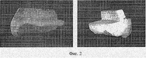
Затем остеотомом отсекается большеберцовая кость с плоскостью остеотомии, перпендикулярной оси большеберцовой кости, на расстоянии 2-3 миллиметра от субхондральной пластинки с оставлением на остеотомированной поверхности трансплантата квадратного выступа ("шипа") для крепления к большеберцовой кости реципиента с размером сторон 6 миллиметров (ширина кистевого остеотома), идущего от середины медиальной поверхности трансплантата до середины малоберцовой вырезки на этом же трансплантате (фиг. 2). В свою очередь во время операции на остеотомированной поверхности большеберцовой кости реципиента формируется паз шириной 6 мм во фронтальной плоскости со стенками, равными ширине остеотома, осью которого является линия, соединяющая середину внутренне боковой поверхности большеберцовой кости на уровне резекции и середину наружной лодыжки, на том же уровне для сочленения с аллотрансплантатом (по типу шиловидного соединения). Then, the tibia is cut off with an osteotomy plane with an osteotomy plane perpendicular to the axis of the tibia, 2-3 mm from the subchondral plate, leaving a square protrusion ("spike") on the osteotomy surface for attachment to the tibia of the recipient with a side width of 6 mm osteotome), going from the middle of the medial surface of the graft to the middle of the fibular notch on the same graft (Fig. 2). In turn, during an operation on the osteotomized surface of the tibia of the recipient, a groove of 6 mm wide is formed in the frontal plane with walls equal to the width of the osteotome, the axis of which is the line connecting the middle of the inner lateral surface of the tibia at the resection level and the middle of the outer ankle, on the same level for articulation with an allograft (as a styloid connection).
После изготовления трансплантата производится его стерилизация по методу, разработанному и принятому в институте [3]. After manufacturing the transplant, it is sterilized according to the method developed and adopted at the institute [3].
СПИСОК ЛИТЕРАТУРЫ
1. Мовшович И.А. Оперативная ортопедия.//Москва, "Медицина" - 1983. С. 285-288.LIST OF REFERENCES
1. Movshovich I.A. Operative orthopedics. // Moscow, "Medicine" - 1983. P. 285-288.
2. Pfahler M. Krodel A. Tritschler A. Zenta S. Role of internal and external fixation in ankle fusion. Archives of Orthopaedic & Trauma Surgery. 115 (3-4): 146-8, 1996. 2. Pfahler M. Krodel A. Tritschler A. Zenta S. Role of internal and external fixation in ankle fusion. Archives of Orthopedic & Trauma Surgery. 115 (3-4): 146-8, 1996.
3. Савельев В.И., Жирнов В.А., Иванкин Д.Е., Этитейн Ю.Т., Подорожная В. Т. Способы химической стерилизации деминерализованных костных трансплантатов.//Методические рекомендации /Ленинград 1990. - 12 с. 3. Savelyev V.I., Zhirnov V.A., Ivankin D.E., Etitein Yu.T., Podorozhnaya V.T. Methods of chemical sterilization of demineralized bone grafts. Methodical recommendations / Leningrad 1990. - 12 p.
Claims (1)
Priority Applications (1)
| Application Number | Priority Date | Filing Date | Title |
|---|---|---|---|
| RU2001106773/14A RU2217103C2 (en) | 2001-03-13 | 2001-03-13 | Allotransplant for making plastic repair of proximal part of talocrural articulation |
Applications Claiming Priority (1)
| Application Number | Priority Date | Filing Date | Title |
|---|---|---|---|
| RU2001106773/14A RU2217103C2 (en) | 2001-03-13 | 2001-03-13 | Allotransplant for making plastic repair of proximal part of talocrural articulation |
Publications (2)
| Publication Number | Publication Date |
|---|---|
| RU2001106773A RU2001106773A (en) | 2003-01-27 |
| RU2217103C2 true RU2217103C2 (en) | 2003-11-27 |
Family
ID=32026525
Family Applications (1)
| Application Number | Title | Priority Date | Filing Date |
|---|---|---|---|
| RU2001106773/14A RU2217103C2 (en) | 2001-03-13 | 2001-03-13 | Allotransplant for making plastic repair of proximal part of talocrural articulation |
Country Status (1)
| Country | Link |
|---|---|
| RU (1) | RU2217103C2 (en) |
-
2001
- 2001-03-13 RU RU2001106773/14A patent/RU2217103C2/en not_active IP Right Cessation
Non-Patent Citations (2)
| Title |
|---|
| ИМАМАЛИЕВ А.С. Гомопластика суставных концов костей. - М.: Медицина, 1975, с.249-262. * |
| МОВШОВИЧ И.А. Оперативная ортопедия. - М.: Медицина, 1983, с.285-288; 1994, c. 334-335. * |
Similar Documents
| Publication | Publication Date | Title |
|---|---|---|
| US8784498B2 (en) | Method and apparatus for fusing the bones of a joint | |
| US9456902B2 (en) | Orthopaedic implants | |
| Russotti et al. | Tibiotalocalcaneal arthrodesis for arthritis and deformity of the hind part of the foot. | |
| Mannerfelt et al. | Arthrodesis of the wrist in rheumatoid arthritis: a technique without external fixation | |
| US20030225458A1 (en) | Universal femoral component for endoprosthetic knee | |
| US20120245701A1 (en) | Hemi Ankle Implant | |
| Moeckel et al. | The double-stemmed silicone-rubber implant for rheumatoid arthritis of the first metatarsophalangeal joint. Long-term results. | |
| Buck-Gramcko | The role of nonvascularized toe phalanx transplantation | |
| Trancik et al. | Capitellocondylar total elbow arthroplasty: two-to eight-year experience | |
| Swanson et al. | Unicompartmental and bicompartmental arthroplasty of the knee with a finned metal tibial-plateau implant. | |
| RU2217103C2 (en) | Allotransplant for making plastic repair of proximal part of talocrural articulation | |
| Wagner et al. | Total knee arthroplasty with concurrent femoral and tibial osteotomies in osteogenesis imperfecta | |
| RU2407485C2 (en) | Total endoprosthesis of ankle joint and talus | |
| RU2341217C2 (en) | Method of treatment of deep defects of condyles of tibial bone of various etiologies | |
| Konstantakos et al. | Eight-year follow-up of total knee arthroplasty in a patient with an ipsilateral below-knee amputation | |
| RU202487U1 (en) | ENDOPROTHESIS OF THE FIRST PLUSNOPHALANGAL JOINT OF THE FOOT | |
| RU2201721C1 (en) | Surgical method for treating the cases of deforming gonarthrosis | |
| RU2801233C2 (en) | Talal head endoprosthesis and method of its implantation | |
| Sodha et al. | Evolution of total ankle arthroplasty | |
| RU2219861C2 (en) | Method for making arthroplastic repair of talocrural articulation | |
| US20230263637A1 (en) | Joint implant apparatus, system, and method | |
| Gosselin et al. | and Daniel-John Lavaly | |
| Higgins | Medial Femoral Trochlea Osteochondral Flap | |
| Tang et al. | A new building block: costo-osteochondral graft for intra-articular incongruity after distal radius fracture | |
| Gosselin et al. | Adult Reconstruction |
Legal Events
| Date | Code | Title | Description |
|---|---|---|---|
| MM4A | The patent is invalid due to non-payment of fees |
Effective date: 20040314 |