KR20220044862A - Devices, systems and methods for monitoring knee replacements - Google Patents
Devices, systems and methods for monitoring knee replacements Download PDFInfo
- Publication number
- KR20220044862A KR20220044862A KR1020227010199A KR20227010199A KR20220044862A KR 20220044862 A KR20220044862 A KR 20220044862A KR 1020227010199 A KR1020227010199 A KR 1020227010199A KR 20227010199 A KR20227010199 A KR 20227010199A KR 20220044862 A KR20220044862 A KR 20220044862A
- Authority
- KR
- South Korea
- Prior art keywords
- sensor
- sensors
- prosthesis
- knee
- data
- Prior art date
Links
- 238000000034 method Methods 0.000 title claims description 90
- 238000013150 knee replacement Methods 0.000 title abstract description 149
- 238000012544 monitoring process Methods 0.000 title description 24
- 210000003127 knee Anatomy 0.000 description 75
- 230000033001 locomotion Effects 0.000 description 64
- 210000000629 knee joint Anatomy 0.000 description 57
- 210000000988 bone and bone Anatomy 0.000 description 54
- 239000002639 bone cement Substances 0.000 description 54
- 230000000694 effects Effects 0.000 description 44
- 239000007943 implant Substances 0.000 description 40
- 210000004417 patella Anatomy 0.000 description 36
- 210000002303 tibia Anatomy 0.000 description 31
- 210000000689 upper leg Anatomy 0.000 description 30
- 238000003860 storage Methods 0.000 description 29
- 238000001356 surgical procedure Methods 0.000 description 28
- 210000001519 tissue Anatomy 0.000 description 26
- 230000036961 partial effect Effects 0.000 description 23
- 238000003384 imaging method Methods 0.000 description 18
- 239000000463 material Substances 0.000 description 18
- 206010023204 Joint dislocation Diseases 0.000 description 17
- 230000001133 acceleration Effects 0.000 description 17
- 230000008859 change Effects 0.000 description 17
- 230000005611 electricity Effects 0.000 description 17
- 239000003814 drug Substances 0.000 description 16
- 230000006870 function Effects 0.000 description 16
- 230000036541 health Effects 0.000 description 15
- 208000015181 infectious disease Diseases 0.000 description 15
- 210000002414 leg Anatomy 0.000 description 13
- 229910052751 metal Inorganic materials 0.000 description 13
- 239000002184 metal Substances 0.000 description 13
- 230000008569 process Effects 0.000 description 13
- 238000011084 recovery Methods 0.000 description 13
- 230000035939 shock Effects 0.000 description 13
- 208000002193 Pain Diseases 0.000 description 12
- 230000008901 benefit Effects 0.000 description 12
- 238000004590 computer program Methods 0.000 description 12
- 230000004060 metabolic process Effects 0.000 description 11
- 229920003229 poly(methyl methacrylate) Polymers 0.000 description 11
- 239000004926 polymethyl methacrylate Substances 0.000 description 11
- 239000000126 substance Substances 0.000 description 11
- 238000004891 communication Methods 0.000 description 10
- 230000006866 deterioration Effects 0.000 description 10
- 229940079593 drug Drugs 0.000 description 10
- 230000002503 metabolic effect Effects 0.000 description 10
- 238000004458 analytical method Methods 0.000 description 9
- 230000006378 damage Effects 0.000 description 9
- 230000002980 postoperative effect Effects 0.000 description 9
- 239000008280 blood Substances 0.000 description 8
- 210000004369 blood Anatomy 0.000 description 8
- 238000001514 detection method Methods 0.000 description 8
- -1 polyethylene Polymers 0.000 description 8
- 208000024891 symptom Diseases 0.000 description 8
- 206010061218 Inflammation Diseases 0.000 description 7
- 230000005540 biological transmission Effects 0.000 description 7
- 238000002059 diagnostic imaging Methods 0.000 description 7
- 238000005516 engineering process Methods 0.000 description 7
- 239000012530 fluid Substances 0.000 description 7
- 230000004054 inflammatory process Effects 0.000 description 7
- 238000012545 processing Methods 0.000 description 7
- 229910000684 Cobalt-chrome Inorganic materials 0.000 description 6
- RTAQQCXQSZGOHL-UHFFFAOYSA-N Titanium Chemical compound [Ti] RTAQQCXQSZGOHL-UHFFFAOYSA-N 0.000 description 6
- WAIPAZQMEIHHTJ-UHFFFAOYSA-N [Cr].[Co] Chemical compound [Cr].[Co] WAIPAZQMEIHHTJ-UHFFFAOYSA-N 0.000 description 6
- 230000000712 assembly Effects 0.000 description 6
- 238000000429 assembly Methods 0.000 description 6
- 239000000919 ceramic Substances 0.000 description 6
- 239000010952 cobalt-chrome Substances 0.000 description 6
- 239000011521 glass Substances 0.000 description 6
- 238000005259 measurement Methods 0.000 description 6
- 239000010935 stainless steel Substances 0.000 description 6
- 229910001220 stainless steel Inorganic materials 0.000 description 6
- 239000010936 titanium Substances 0.000 description 6
- 229910052719 titanium Inorganic materials 0.000 description 6
- 206010019233 Headaches Diseases 0.000 description 5
- 239000004568 cement Substances 0.000 description 5
- 231100000869 headache Toxicity 0.000 description 5
- 208000014674 injury Diseases 0.000 description 5
- 230000007774 longterm Effects 0.000 description 5
- 238000007726 management method Methods 0.000 description 5
- 238000011282 treatment Methods 0.000 description 5
- 230000000007 visual effect Effects 0.000 description 5
- 208000006386 Bone Resorption Diseases 0.000 description 4
- 208000003076 Osteolysis Diseases 0.000 description 4
- 239000004698 Polyethylene Substances 0.000 description 4
- 208000027418 Wounds and injury Diseases 0.000 description 4
- 230000024279 bone resorption Effects 0.000 description 4
- 238000006073 displacement reaction Methods 0.000 description 4
- 230000003628 erosive effect Effects 0.000 description 4
- 238000011156 evaluation Methods 0.000 description 4
- 230000005021 gait Effects 0.000 description 4
- 230000010354 integration Effects 0.000 description 4
- 208000029791 lytic metastatic bone lesion Diseases 0.000 description 4
- 238000000554 physical therapy Methods 0.000 description 4
- 229920000573 polyethylene Polymers 0.000 description 4
- 230000000750 progressive effect Effects 0.000 description 4
- 229940124597 therapeutic agent Drugs 0.000 description 4
- 206010051728 Bone erosion Diseases 0.000 description 3
- 206010065687 Bone loss Diseases 0.000 description 3
- 208000037408 Device failure Diseases 0.000 description 3
- 239000003242 anti bacterial agent Substances 0.000 description 3
- 229940088710 antibiotic agent Drugs 0.000 description 3
- 239000003990 capacitor Substances 0.000 description 3
- 239000011248 coating agent Substances 0.000 description 3
- 238000000576 coating method Methods 0.000 description 3
- 238000013480 data collection Methods 0.000 description 3
- 238000013461 design Methods 0.000 description 3
- 238000009826 distribution Methods 0.000 description 3
- 210000003041 ligament Anatomy 0.000 description 3
- 238000002483 medication Methods 0.000 description 3
- 210000003205 muscle Anatomy 0.000 description 3
- 230000003287 optical effect Effects 0.000 description 3
- 230000000399 orthopedic effect Effects 0.000 description 3
- 229920002635 polyurethane Polymers 0.000 description 3
- 239000004814 polyurethane Substances 0.000 description 3
- 230000011664 signaling Effects 0.000 description 3
- 238000012546 transfer Methods 0.000 description 3
- XLYOFNOQVPJJNP-UHFFFAOYSA-N water Substances O XLYOFNOQVPJJNP-UHFFFAOYSA-N 0.000 description 3
- 208000006820 Arthralgia Diseases 0.000 description 2
- 208000010392 Bone Fractures Diseases 0.000 description 2
- AOJJSUZBOXZQNB-TZSSRYMLSA-N Doxorubicin Chemical compound O([C@H]1C[C@@](O)(CC=2C(O)=C3C(=O)C=4C=CC=C(C=4C(=O)C3=C(O)C=21)OC)C(=O)CO)[C@H]1C[C@H](N)[C@H](O)[C@H](C)O1 AOJJSUZBOXZQNB-TZSSRYMLSA-N 0.000 description 2
- 206010072970 Meniscus injury Diseases 0.000 description 2
- 208000037099 Prosthesis Failure Diseases 0.000 description 2
- 239000004699 Ultra-high molecular weight polyethylene Substances 0.000 description 2
- 230000009471 action Effects 0.000 description 2
- 230000003466 anti-cipated effect Effects 0.000 description 2
- 229940124599 anti-inflammatory drug Drugs 0.000 description 2
- 206010003246 arthritis Diseases 0.000 description 2
- 210000001188 articular cartilage Anatomy 0.000 description 2
- 210000001185 bone marrow Anatomy 0.000 description 2
- 230000015556 catabolic process Effects 0.000 description 2
- 229910010293 ceramic material Inorganic materials 0.000 description 2
- 230000000052 comparative effect Effects 0.000 description 2
- 230000000295 complement effect Effects 0.000 description 2
- 230000003247 decreasing effect Effects 0.000 description 2
- 230000006735 deficit Effects 0.000 description 2
- 238000006731 degradation reaction Methods 0.000 description 2
- 238000010586 diagram Methods 0.000 description 2
- 238000002513 implantation Methods 0.000 description 2
- 230000006872 improvement Effects 0.000 description 2
- 238000011065 in-situ storage Methods 0.000 description 2
- 238000009434 installation Methods 0.000 description 2
- 230000003993 interaction Effects 0.000 description 2
- 210000001503 joint Anatomy 0.000 description 2
- 210000000281 joint capsule Anatomy 0.000 description 2
- 230000008407 joint function Effects 0.000 description 2
- 239000007788 liquid Substances 0.000 description 2
- 238000012423 maintenance Methods 0.000 description 2
- 201000008482 osteoarthritis Diseases 0.000 description 2
- 230000002085 persistent effect Effects 0.000 description 2
- 239000002243 precursor Substances 0.000 description 2
- 230000035485 pulse pressure Effects 0.000 description 2
- 238000011160 research Methods 0.000 description 2
- 206010039073 rheumatoid arthritis Diseases 0.000 description 2
- 238000004092 self-diagnosis Methods 0.000 description 2
- 210000002435 tendon Anatomy 0.000 description 2
- 229920000785 ultra high molecular weight polyethylene Polymers 0.000 description 2
- HRANPRDGABOKNQ-ORGXEYTDSA-N (1r,3r,3as,3br,7ar,8as,8bs,8cs,10as)-1-acetyl-5-chloro-3-hydroxy-8b,10a-dimethyl-7-oxo-1,2,3,3a,3b,7,7a,8,8a,8b,8c,9,10,10a-tetradecahydrocyclopenta[a]cyclopropa[g]phenanthren-1-yl acetate Chemical compound C1=C(Cl)C2=CC(=O)[C@@H]3C[C@@H]3[C@]2(C)[C@@H]2[C@@H]1[C@@H]1[C@H](O)C[C@@](C(C)=O)(OC(=O)C)[C@@]1(C)CC2 HRANPRDGABOKNQ-ORGXEYTDSA-N 0.000 description 1
- VSNHCAURESNICA-NJFSPNSNSA-N 1-oxidanylurea Chemical compound N[14C](=O)NO VSNHCAURESNICA-NJFSPNSNSA-N 0.000 description 1
- IZXIZTKNFFYFOF-UHFFFAOYSA-N 2-Oxazolidone Chemical class O=C1NCCO1 IZXIZTKNFFYFOF-UHFFFAOYSA-N 0.000 description 1
- 108010001478 Bacitracin Proteins 0.000 description 1
- 241000894006 Bacteria Species 0.000 description 1
- KLWPJMFMVPTNCC-UHFFFAOYSA-N Camptothecin Natural products CCC1(O)C(=O)OCC2=C1C=C3C4Nc5ccccc5C=C4CN3C2=O KLWPJMFMVPTNCC-UHFFFAOYSA-N 0.000 description 1
- 206010007710 Cartilage injury Diseases 0.000 description 1
- 229930186147 Cephalosporin Natural products 0.000 description 1
- 206010051055 Deep vein thrombosis Diseases 0.000 description 1
- 208000012661 Dyskinesia Diseases 0.000 description 1
- 206010017076 Fracture Diseases 0.000 description 1
- 229930182566 Gentamicin Natural products 0.000 description 1
- CEAZRRDELHUEMR-URQXQFDESA-N Gentamicin Chemical compound O1[C@H](C(C)NC)CC[C@@H](N)[C@H]1O[C@H]1[C@H](O)[C@@H](O[C@@H]2[C@@H]([C@@H](NC)[C@@](C)(O)CO2)O)[C@H](N)C[C@@H]1N CEAZRRDELHUEMR-URQXQFDESA-N 0.000 description 1
- 108010015899 Glycopeptides Proteins 0.000 description 1
- 102000002068 Glycopeptides Human genes 0.000 description 1
- 235000015842 Hesperis Nutrition 0.000 description 1
- 206010020772 Hypertension Diseases 0.000 description 1
- 235000012633 Iberis amara Nutrition 0.000 description 1
- 208000004733 Knee Dislocation Diseases 0.000 description 1
- FBOZXECLQNJBKD-ZDUSSCGKSA-N L-methotrexate Chemical compound C=1N=C2N=C(N)N=C(N)C2=NC=1CN(C)C1=CC=C(C(=O)N[C@@H](CCC(O)=O)C(O)=O)C=C1 FBOZXECLQNJBKD-ZDUSSCGKSA-N 0.000 description 1
- 206010061223 Ligament injury Diseases 0.000 description 1
- 206010065433 Ligament rupture Diseases 0.000 description 1
- 108010028921 Lipopeptides Proteins 0.000 description 1
- 208000034819 Mobility Limitation Diseases 0.000 description 1
- 241000699670 Mus sp. Species 0.000 description 1
- 208000008589 Obesity Diseases 0.000 description 1
- 229930012538 Paclitaxel Natural products 0.000 description 1
- 229930182555 Penicillin Natural products 0.000 description 1
- 201000004681 Psoriasis Diseases 0.000 description 1
- 101100460147 Sarcophaga bullata NEMS gene Proteins 0.000 description 1
- 241000256247 Spodoptera exigua Species 0.000 description 1
- 208000013201 Stress fracture Diseases 0.000 description 1
- 239000004098 Tetracycline Substances 0.000 description 1
- 108010059993 Vancomycin Proteins 0.000 description 1
- 206010047249 Venous thrombosis Diseases 0.000 description 1
- 230000005856 abnormality Effects 0.000 description 1
- 230000004913 activation Effects 0.000 description 1
- 230000001154 acute effect Effects 0.000 description 1
- 229940126575 aminoglycoside Drugs 0.000 description 1
- 238000004873 anchoring Methods 0.000 description 1
- 229940045799 anthracyclines and related substance Drugs 0.000 description 1
- 230000003110 anti-inflammatory effect Effects 0.000 description 1
- 238000003491 array Methods 0.000 description 1
- 229960004099 azithromycin Drugs 0.000 description 1
- MQTOSJVFKKJCRP-BICOPXKESA-N azithromycin Chemical compound O([C@@H]1[C@@H](C)C(=O)O[C@@H]([C@@]([C@H](O)[C@@H](C)N(C)C[C@H](C)C[C@@](C)(O)[C@H](O[C@H]2[C@@H]([C@H](C[C@@H](C)O2)N(C)C)O)[C@H]1C)(C)O)CC)[C@H]1C[C@@](C)(OC)[C@@H](O)[C@H](C)O1 MQTOSJVFKKJCRP-BICOPXKESA-N 0.000 description 1
- WZPBZJONDBGPKJ-VEHQQRBSSA-N aztreonam Chemical compound O=C1N(S([O-])(=O)=O)[C@@H](C)[C@@H]1NC(=O)C(=N/OC(C)(C)C(O)=O)\C1=CSC([NH3+])=N1 WZPBZJONDBGPKJ-VEHQQRBSSA-N 0.000 description 1
- 229960003644 aztreonam Drugs 0.000 description 1
- 229960003071 bacitracin Drugs 0.000 description 1
- 229930184125 bacitracin Natural products 0.000 description 1
- CLKOFPXJLQSYAH-ABRJDSQDSA-N bacitracin A Chemical compound C1SC([C@@H](N)[C@@H](C)CC)=N[C@@H]1C(=O)N[C@@H](CC(C)C)C(=O)N[C@H](CCC(O)=O)C(=O)N[C@@H]([C@@H](C)CC)C(=O)N[C@@H]1C(=O)N[C@H](CCCN)C(=O)N[C@@H]([C@@H](C)CC)C(=O)N[C@H](CC=2C=CC=CC=2)C(=O)N[C@@H](CC=2N=CNC=2)C(=O)N[C@H](CC(O)=O)C(=O)N[C@@H](CC(N)=O)C(=O)NCCCC1 CLKOFPXJLQSYAH-ABRJDSQDSA-N 0.000 description 1
- 230000004888 barrier function Effects 0.000 description 1
- 230000006399 behavior Effects 0.000 description 1
- 230000009286 beneficial effect Effects 0.000 description 1
- 239000003782 beta lactam antibiotic agent Substances 0.000 description 1
- 230000017531 blood circulation Effects 0.000 description 1
- 230000008468 bone growth Effects 0.000 description 1
- VSJKWCGYPAHWDS-FQEVSTJZSA-N camptothecin Chemical compound C1=CC=C2C=C(CN3C4=CC5=C(C3=O)COC(=O)[C@]5(O)CC)C4=NC2=C1 VSJKWCGYPAHWDS-FQEVSTJZSA-N 0.000 description 1
- 229940127093 camptothecin Drugs 0.000 description 1
- 229940041011 carbapenems Drugs 0.000 description 1
- 239000000969 carrier Substances 0.000 description 1
- 210000000845 cartilage Anatomy 0.000 description 1
- 230000010267 cellular communication Effects 0.000 description 1
- 229940124587 cephalosporin Drugs 0.000 description 1
- 150000001780 cephalosporins Chemical class 0.000 description 1
- 239000003795 chemical substances by application Substances 0.000 description 1
- 230000001684 chronic effect Effects 0.000 description 1
- DQLATGHUWYMOKM-UHFFFAOYSA-L cisplatin Chemical compound N[Pt](N)(Cl)Cl DQLATGHUWYMOKM-UHFFFAOYSA-L 0.000 description 1
- 229960004316 cisplatin Drugs 0.000 description 1
- 229960002227 clindamycin Drugs 0.000 description 1
- KDLRVYVGXIQJDK-AWPVFWJPSA-N clindamycin Chemical compound CN1C[C@H](CCC)C[C@H]1C(=O)N[C@H]([C@H](C)Cl)[C@@H]1[C@H](O)[C@H](O)[C@@H](O)[C@@H](SC)O1 KDLRVYVGXIQJDK-AWPVFWJPSA-N 0.000 description 1
- 210000004439 collateral ligament Anatomy 0.000 description 1
- 238000011109 contamination Methods 0.000 description 1
- 229920001577 copolymer Polymers 0.000 description 1
- 238000007405 data analysis Methods 0.000 description 1
- 230000007850 degeneration Effects 0.000 description 1
- 230000003111 delayed effect Effects 0.000 description 1
- 238000011161 development Methods 0.000 description 1
- 230000018109 developmental process Effects 0.000 description 1
- 206010012601 diabetes mellitus Diseases 0.000 description 1
- 238000003745 diagnosis Methods 0.000 description 1
- 201000010099 disease Diseases 0.000 description 1
- 208000037265 diseases, disorders, signs and symptoms Diseases 0.000 description 1
- VSJKWCGYPAHWDS-UHFFFAOYSA-N dl-camptothecin Natural products C1=CC=C2C=C(CN3C4=CC5=C(C3=O)COC(=O)C5(O)CC)C4=NC2=C1 VSJKWCGYPAHWDS-UHFFFAOYSA-N 0.000 description 1
- 229960004679 doxorubicin Drugs 0.000 description 1
- 239000000975 dye Substances 0.000 description 1
- 230000004064 dysfunction Effects 0.000 description 1
- VJJPUSNTGOMMGY-MRVIYFEKSA-N etoposide Chemical compound COC1=C(O)C(OC)=CC([C@@H]2C3=CC=4OCOC=4C=C3[C@@H](O[C@H]3[C@@H]([C@@H](O)[C@@H]4O[C@H](C)OC[C@H]4O3)O)[C@@H]3[C@@H]2C(OC3)=O)=C1 VJJPUSNTGOMMGY-MRVIYFEKSA-N 0.000 description 1
- 229960005420 etoposide Drugs 0.000 description 1
- 230000005713 exacerbation Effects 0.000 description 1
- 201000010934 exostosis Diseases 0.000 description 1
- 239000000835 fiber Substances 0.000 description 1
- 238000007667 floating Methods 0.000 description 1
- 150000005699 fluoropyrimidines Chemical class 0.000 description 1
- 239000004052 folic acid antagonist Substances 0.000 description 1
- 229960002518 gentamicin Drugs 0.000 description 1
- 230000012010 growth Effects 0.000 description 1
- 230000035876 healing Effects 0.000 description 1
- 230000003862 health status Effects 0.000 description 1
- 201000010930 hyperostosis Diseases 0.000 description 1
- 230000005022 impaired gait Effects 0.000 description 1
- 230000000670 limiting effect Effects 0.000 description 1
- 230000003137 locomotive effect Effects 0.000 description 1
- 206010025135 lupus erythematosus Diseases 0.000 description 1
- 239000003120 macrolide antibiotic agent Substances 0.000 description 1
- 238000004519 manufacturing process Methods 0.000 description 1
- 239000003550 marker Substances 0.000 description 1
- 230000007246 mechanism Effects 0.000 description 1
- 230000005055 memory storage Effects 0.000 description 1
- 230000003340 mental effect Effects 0.000 description 1
- 201000005299 metal allergy Diseases 0.000 description 1
- 230000029052 metamorphosis Effects 0.000 description 1
- 229960000485 methotrexate Drugs 0.000 description 1
- 238000013508 migration Methods 0.000 description 1
- 230000005012 migration Effects 0.000 description 1
- 238000005065 mining Methods 0.000 description 1
- 229960001156 mitoxantrone Drugs 0.000 description 1
- KKZJGLLVHKMTCM-UHFFFAOYSA-N mitoxantrone Chemical compound O=C1C2=C(O)C=CC(O)=C2C(=O)C2=C1C(NCCNCCO)=CC=C2NCCNCCO KKZJGLLVHKMTCM-UHFFFAOYSA-N 0.000 description 1
- 239000000203 mixture Substances 0.000 description 1
- 230000037230 mobility Effects 0.000 description 1
- 210000005036 nerve Anatomy 0.000 description 1
- IAIWVQXQOWNYOU-FPYGCLRLSA-N nitrofural Chemical compound NC(=O)N\N=C\C1=CC=C([N+]([O-])=O)O1 IAIWVQXQOWNYOU-FPYGCLRLSA-N 0.000 description 1
- 229960001907 nitrofurazone Drugs 0.000 description 1
- 235000020824 obesity Nutrition 0.000 description 1
- 229960001592 paclitaxel Drugs 0.000 description 1
- 229940124583 pain medication Drugs 0.000 description 1
- 239000002245 particle Substances 0.000 description 1
- 230000007170 pathology Effects 0.000 description 1
- 150000002960 penicillins Chemical class 0.000 description 1
- 230000000737 periodic effect Effects 0.000 description 1
- 230000037081 physical activity Effects 0.000 description 1
- 230000035479 physiological effects, processes and functions Effects 0.000 description 1
- 150000003057 platinum Chemical class 0.000 description 1
- YJGVMLPVUAXIQN-XVVDYKMHSA-N podophyllotoxin Chemical compound COC1=C(OC)C(OC)=CC([C@@H]2C3=CC=4OCOC=4C=C3[C@H](O)[C@@H]3[C@@H]2C(OC3)=O)=C1 YJGVMLPVUAXIQN-XVVDYKMHSA-N 0.000 description 1
- 229920001690 polydopamine Polymers 0.000 description 1
- 229920000642 polymer Polymers 0.000 description 1
- 229920002959 polymer blend Polymers 0.000 description 1
- 229920001184 polypeptide Polymers 0.000 description 1
- 230000002028 premature Effects 0.000 description 1
- 108090000765 processed proteins & peptides Proteins 0.000 description 1
- 102000004196 processed proteins & peptides Human genes 0.000 description 1
- 230000002035 prolonged effect Effects 0.000 description 1
- 230000000644 propagated effect Effects 0.000 description 1
- 230000005180 public health Effects 0.000 description 1
- 230000001698 pyrogenic effect Effects 0.000 description 1
- 150000007660 quinolones Chemical class 0.000 description 1
- ZAHRKKWIAAJSAO-UHFFFAOYSA-N rapamycin Natural products COCC(O)C(=C/C(C)C(=O)CC(OC(=O)C1CCCCN1C(=O)C(=O)C2(O)OC(CC(OC)C(=CC=CC=CC(C)CC(C)C(=O)C)C)CCC2C)C(C)CC3CCC(O)C(C3)OC)C ZAHRKKWIAAJSAO-UHFFFAOYSA-N 0.000 description 1
- 238000002278 reconstructive surgery Methods 0.000 description 1
- 230000002829 reductive effect Effects 0.000 description 1
- 230000003252 repetitive effect Effects 0.000 description 1
- 208000037803 restenosis Diseases 0.000 description 1
- 230000033764 rhythmic process Effects 0.000 description 1
- 231100000241 scar Toxicity 0.000 description 1
- 230000002000 scavenging effect Effects 0.000 description 1
- 230000009291 secondary effect Effects 0.000 description 1
- 230000035807 sensation Effects 0.000 description 1
- 238000000926 separation method Methods 0.000 description 1
- 229960002930 sirolimus Drugs 0.000 description 1
- QFJCIRLUMZQUOT-HPLJOQBZSA-N sirolimus Chemical compound C1C[C@@H](O)[C@H](OC)C[C@@H]1C[C@@H](C)[C@H]1OC(=O)[C@@H]2CCCCN2C(=O)C(=O)[C@](O)(O2)[C@H](C)CC[C@H]2C[C@H](OC)/C(C)=C/C=C/C=C/[C@@H](C)C[C@@H](C)C(=O)[C@H](OC)[C@H](O)/C(C)=C/[C@@H](C)C(=O)C1 QFJCIRLUMZQUOT-HPLJOQBZSA-N 0.000 description 1
- 230000000391 smoking effect Effects 0.000 description 1
- 239000007787 solid Substances 0.000 description 1
- 238000013112 stability test Methods 0.000 description 1
- 229940124530 sulfonamide Drugs 0.000 description 1
- 150000003456 sulfonamides Chemical class 0.000 description 1
- 238000007460 surgical drainage Methods 0.000 description 1
- 229920002994 synthetic fiber Polymers 0.000 description 1
- RCINICONZNJXQF-MZXODVADSA-N taxol Chemical compound O([C@@H]1[C@@]2(C[C@@H](C(C)=C(C2(C)C)[C@H](C([C@]2(C)[C@@H](O)C[C@H]3OC[C@]3([C@H]21)OC(C)=O)=O)OC(=O)C)OC(=O)[C@H](O)[C@@H](NC(=O)C=1C=CC=CC=1)C=1C=CC=CC=1)O)C(=O)C1=CC=CC=C1 RCINICONZNJXQF-MZXODVADSA-N 0.000 description 1
- 229960002180 tetracycline Drugs 0.000 description 1
- 229930101283 tetracycline Natural products 0.000 description 1
- 235000019364 tetracycline Nutrition 0.000 description 1
- 150000003522 tetracyclines Chemical class 0.000 description 1
- 230000009772 tissue formation Effects 0.000 description 1
- 229960000707 tobramycin Drugs 0.000 description 1
- NLVFBUXFDBBNBW-PBSUHMDJSA-N tobramycin Chemical compound N[C@@H]1C[C@H](O)[C@@H](CN)O[C@@H]1O[C@H]1[C@H](O)[C@@H](O[C@@H]2[C@@H]([C@@H](N)[C@H](O)[C@@H](CO)O2)O)[C@H](N)C[C@@H]1N NLVFBUXFDBBNBW-PBSUHMDJSA-N 0.000 description 1
- 238000012549 training Methods 0.000 description 1
- 230000001131 transforming effect Effects 0.000 description 1
- 230000008733 trauma Effects 0.000 description 1
- 230000000472 traumatic effect Effects 0.000 description 1
- 238000011277 treatment modality Methods 0.000 description 1
- 230000005612 types of electricity Effects 0.000 description 1
- 229960003165 vancomycin Drugs 0.000 description 1
- MYPYJXKWCTUITO-LYRMYLQWSA-N vancomycin Chemical compound O([C@@H]1[C@@H](O)[C@H](O)[C@@H](CO)O[C@H]1OC1=C2C=C3C=C1OC1=CC=C(C=C1Cl)[C@@H](O)[C@H](C(N[C@@H](CC(N)=O)C(=O)N[C@H]3C(=O)N[C@H]1C(=O)N[C@H](C(N[C@@H](C3=CC(O)=CC(O)=C3C=3C(O)=CC=C1C=3)C(O)=O)=O)[C@H](O)C1=CC=C(C(=C1)Cl)O2)=O)NC(=O)[C@@H](CC(C)C)NC)[C@H]1C[C@](C)(N)[C@H](O)[C@H](C)O1 MYPYJXKWCTUITO-LYRMYLQWSA-N 0.000 description 1
- MYPYJXKWCTUITO-UHFFFAOYSA-N vancomycin Natural products O1C(C(=C2)Cl)=CC=C2C(O)C(C(NC(C2=CC(O)=CC(O)=C2C=2C(O)=CC=C3C=2)C(O)=O)=O)NC(=O)C3NC(=O)C2NC(=O)C(CC(N)=O)NC(=O)C(NC(=O)C(CC(C)C)NC)C(O)C(C=C3Cl)=CC=C3OC3=CC2=CC1=C3OC1OC(CO)C(O)C(O)C1OC1CC(C)(N)C(O)C(C)O1 MYPYJXKWCTUITO-UHFFFAOYSA-N 0.000 description 1
- 238000012800 visualization Methods 0.000 description 1
- 238000005406 washing Methods 0.000 description 1
- 230000003442 weekly effect Effects 0.000 description 1
- 239000002132 β-lactam antibiotic Substances 0.000 description 1
- 229940124586 β-lactam antibiotics Drugs 0.000 description 1
Images


Classifications
-
- A—HUMAN NECESSITIES
- A61—MEDICAL OR VETERINARY SCIENCE; HYGIENE
- A61F—FILTERS IMPLANTABLE INTO BLOOD VESSELS; PROSTHESES; DEVICES PROVIDING PATENCY TO, OR PREVENTING COLLAPSING OF, TUBULAR STRUCTURES OF THE BODY, e.g. STENTS; ORTHOPAEDIC, NURSING OR CONTRACEPTIVE DEVICES; FOMENTATION; TREATMENT OR PROTECTION OF EYES OR EARS; BANDAGES, DRESSINGS OR ABSORBENT PADS; FIRST-AID KITS
- A61F2/00—Filters implantable into blood vessels; Prostheses, i.e. artificial substitutes or replacements for parts of the body; Appliances for connecting them with the body; Devices providing patency to, or preventing collapsing of, tubular structures of the body, e.g. stents
- A61F2/02—Prostheses implantable into the body
- A61F2/30—Joints
- A61F2/38—Joints for elbows or knees
-
- A—HUMAN NECESSITIES
- A61—MEDICAL OR VETERINARY SCIENCE; HYGIENE
- A61B—DIAGNOSIS; SURGERY; IDENTIFICATION
- A61B5/00—Measuring for diagnostic purposes; Identification of persons
- A61B5/0002—Remote monitoring of patients using telemetry, e.g. transmission of vital signals via a communication network
- A61B5/0031—Implanted circuitry
-
- A—HUMAN NECESSITIES
- A61—MEDICAL OR VETERINARY SCIENCE; HYGIENE
- A61B—DIAGNOSIS; SURGERY; IDENTIFICATION
- A61B5/00—Measuring for diagnostic purposes; Identification of persons
- A61B5/01—Measuring temperature of body parts ; Diagnostic temperature sensing, e.g. for malignant or inflamed tissue
-
- A—HUMAN NECESSITIES
- A61—MEDICAL OR VETERINARY SCIENCE; HYGIENE
- A61B—DIAGNOSIS; SURGERY; IDENTIFICATION
- A61B5/00—Measuring for diagnostic purposes; Identification of persons
- A61B5/103—Detecting, measuring or recording devices for testing the shape, pattern, colour, size or movement of the body or parts thereof, for diagnostic purposes
- A61B5/11—Measuring movement of the entire body or parts thereof, e.g. head or hand tremor, mobility of a limb
- A61B5/112—Gait analysis
-
- A—HUMAN NECESSITIES
- A61—MEDICAL OR VETERINARY SCIENCE; HYGIENE
- A61B—DIAGNOSIS; SURGERY; IDENTIFICATION
- A61B5/00—Measuring for diagnostic purposes; Identification of persons
- A61B5/45—For evaluating or diagnosing the musculoskeletal system or teeth
- A61B5/4528—Joints
-
- A—HUMAN NECESSITIES
- A61—MEDICAL OR VETERINARY SCIENCE; HYGIENE
- A61B—DIAGNOSIS; SURGERY; IDENTIFICATION
- A61B5/00—Measuring for diagnostic purposes; Identification of persons
- A61B5/45—For evaluating or diagnosing the musculoskeletal system or teeth
- A61B5/4538—Evaluating a particular part of the muscoloskeletal system or a particular medical condition
- A61B5/4585—Evaluating the knee
-
- A—HUMAN NECESSITIES
- A61—MEDICAL OR VETERINARY SCIENCE; HYGIENE
- A61B—DIAGNOSIS; SURGERY; IDENTIFICATION
- A61B5/00—Measuring for diagnostic purposes; Identification of persons
- A61B5/48—Other medical applications
- A61B5/4851—Prosthesis assessment or monitoring
-
- A—HUMAN NECESSITIES
- A61—MEDICAL OR VETERINARY SCIENCE; HYGIENE
- A61B—DIAGNOSIS; SURGERY; IDENTIFICATION
- A61B5/00—Measuring for diagnostic purposes; Identification of persons
- A61B5/68—Arrangements of detecting, measuring or recording means, e.g. sensors, in relation to patient
- A61B5/6846—Arrangements of detecting, measuring or recording means, e.g. sensors, in relation to patient specially adapted to be brought in contact with an internal body part, i.e. invasive
- A61B5/6847—Arrangements of detecting, measuring or recording means, e.g. sensors, in relation to patient specially adapted to be brought in contact with an internal body part, i.e. invasive mounted on an invasive device
- A61B5/686—Permanently implanted devices, e.g. pacemakers, other stimulators, biochips
-
- A—HUMAN NECESSITIES
- A61—MEDICAL OR VETERINARY SCIENCE; HYGIENE
- A61F—FILTERS IMPLANTABLE INTO BLOOD VESSELS; PROSTHESES; DEVICES PROVIDING PATENCY TO, OR PREVENTING COLLAPSING OF, TUBULAR STRUCTURES OF THE BODY, e.g. STENTS; ORTHOPAEDIC, NURSING OR CONTRACEPTIVE DEVICES; FOMENTATION; TREATMENT OR PROTECTION OF EYES OR EARS; BANDAGES, DRESSINGS OR ABSORBENT PADS; FIRST-AID KITS
- A61F2/00—Filters implantable into blood vessels; Prostheses, i.e. artificial substitutes or replacements for parts of the body; Appliances for connecting them with the body; Devices providing patency to, or preventing collapsing of, tubular structures of the body, e.g. stents
- A61F2/02—Prostheses implantable into the body
- A61F2/30—Joints
- A61F2/38—Joints for elbows or knees
- A61F2/3859—Femoral components
-
- A—HUMAN NECESSITIES
- A61—MEDICAL OR VETERINARY SCIENCE; HYGIENE
- A61F—FILTERS IMPLANTABLE INTO BLOOD VESSELS; PROSTHESES; DEVICES PROVIDING PATENCY TO, OR PREVENTING COLLAPSING OF, TUBULAR STRUCTURES OF THE BODY, e.g. STENTS; ORTHOPAEDIC, NURSING OR CONTRACEPTIVE DEVICES; FOMENTATION; TREATMENT OR PROTECTION OF EYES OR EARS; BANDAGES, DRESSINGS OR ABSORBENT PADS; FIRST-AID KITS
- A61F2/00—Filters implantable into blood vessels; Prostheses, i.e. artificial substitutes or replacements for parts of the body; Appliances for connecting them with the body; Devices providing patency to, or preventing collapsing of, tubular structures of the body, e.g. stents
- A61F2/02—Prostheses implantable into the body
- A61F2/30—Joints
- A61F2/38—Joints for elbows or knees
- A61F2/3877—Patellae or trochleae
-
- A—HUMAN NECESSITIES
- A61—MEDICAL OR VETERINARY SCIENCE; HYGIENE
- A61F—FILTERS IMPLANTABLE INTO BLOOD VESSELS; PROSTHESES; DEVICES PROVIDING PATENCY TO, OR PREVENTING COLLAPSING OF, TUBULAR STRUCTURES OF THE BODY, e.g. STENTS; ORTHOPAEDIC, NURSING OR CONTRACEPTIVE DEVICES; FOMENTATION; TREATMENT OR PROTECTION OF EYES OR EARS; BANDAGES, DRESSINGS OR ABSORBENT PADS; FIRST-AID KITS
- A61F2/00—Filters implantable into blood vessels; Prostheses, i.e. artificial substitutes or replacements for parts of the body; Appliances for connecting them with the body; Devices providing patency to, or preventing collapsing of, tubular structures of the body, e.g. stents
- A61F2/02—Prostheses implantable into the body
- A61F2/30—Joints
- A61F2/38—Joints for elbows or knees
- A61F2/389—Tibial components
-
- A—HUMAN NECESSITIES
- A61—MEDICAL OR VETERINARY SCIENCE; HYGIENE
- A61F—FILTERS IMPLANTABLE INTO BLOOD VESSELS; PROSTHESES; DEVICES PROVIDING PATENCY TO, OR PREVENTING COLLAPSING OF, TUBULAR STRUCTURES OF THE BODY, e.g. STENTS; ORTHOPAEDIC, NURSING OR CONTRACEPTIVE DEVICES; FOMENTATION; TREATMENT OR PROTECTION OF EYES OR EARS; BANDAGES, DRESSINGS OR ABSORBENT PADS; FIRST-AID KITS
- A61F2/00—Filters implantable into blood vessels; Prostheses, i.e. artificial substitutes or replacements for parts of the body; Appliances for connecting them with the body; Devices providing patency to, or preventing collapsing of, tubular structures of the body, e.g. stents
- A61F2/02—Prostheses implantable into the body
- A61F2/30—Joints
- A61F2/46—Special tools or methods for implanting or extracting artificial joints, accessories, bone grafts or substitutes, or particular adaptations therefor
- A61F2/4657—Measuring instruments used for implanting artificial joints
-
- A—HUMAN NECESSITIES
- A61—MEDICAL OR VETERINARY SCIENCE; HYGIENE
- A61B—DIAGNOSIS; SURGERY; IDENTIFICATION
- A61B2562/00—Details of sensors; Constructional details of sensor housings or probes; Accessories for sensors
- A61B2562/02—Details of sensors specially adapted for in-vivo measurements
- A61B2562/0219—Inertial sensors, e.g. accelerometers, gyroscopes, tilt switches
-
- A—HUMAN NECESSITIES
- A61—MEDICAL OR VETERINARY SCIENCE; HYGIENE
- A61F—FILTERS IMPLANTABLE INTO BLOOD VESSELS; PROSTHESES; DEVICES PROVIDING PATENCY TO, OR PREVENTING COLLAPSING OF, TUBULAR STRUCTURES OF THE BODY, e.g. STENTS; ORTHOPAEDIC, NURSING OR CONTRACEPTIVE DEVICES; FOMENTATION; TREATMENT OR PROTECTION OF EYES OR EARS; BANDAGES, DRESSINGS OR ABSORBENT PADS; FIRST-AID KITS
- A61F2/00—Filters implantable into blood vessels; Prostheses, i.e. artificial substitutes or replacements for parts of the body; Appliances for connecting them with the body; Devices providing patency to, or preventing collapsing of, tubular structures of the body, e.g. stents
- A61F2/02—Prostheses implantable into the body
- A61F2/48—Operating or control means, e.g. from outside the body, control of sphincters
- A61F2/488—Means for detecting or monitoring wear
-
- A—HUMAN NECESSITIES
- A61—MEDICAL OR VETERINARY SCIENCE; HYGIENE
- A61F—FILTERS IMPLANTABLE INTO BLOOD VESSELS; PROSTHESES; DEVICES PROVIDING PATENCY TO, OR PREVENTING COLLAPSING OF, TUBULAR STRUCTURES OF THE BODY, e.g. STENTS; ORTHOPAEDIC, NURSING OR CONTRACEPTIVE DEVICES; FOMENTATION; TREATMENT OR PROTECTION OF EYES OR EARS; BANDAGES, DRESSINGS OR ABSORBENT PADS; FIRST-AID KITS
- A61F2/00—Filters implantable into blood vessels; Prostheses, i.e. artificial substitutes or replacements for parts of the body; Appliances for connecting them with the body; Devices providing patency to, or preventing collapsing of, tubular structures of the body, e.g. stents
- A61F2/02—Prostheses implantable into the body
- A61F2/30—Joints
- A61F2002/30001—Additional features of subject-matter classified in A61F2/28, A61F2/30 and subgroups thereof
- A61F2002/30667—Features concerning an interaction with the environment or a particular use of the prosthesis
- A61F2002/30668—Means for transferring electromagnetic energy to implants
- A61F2002/3067—Means for transferring electromagnetic energy to implants for data transfer
-
- A—HUMAN NECESSITIES
- A61—MEDICAL OR VETERINARY SCIENCE; HYGIENE
- A61F—FILTERS IMPLANTABLE INTO BLOOD VESSELS; PROSTHESES; DEVICES PROVIDING PATENCY TO, OR PREVENTING COLLAPSING OF, TUBULAR STRUCTURES OF THE BODY, e.g. STENTS; ORTHOPAEDIC, NURSING OR CONTRACEPTIVE DEVICES; FOMENTATION; TREATMENT OR PROTECTION OF EYES OR EARS; BANDAGES, DRESSINGS OR ABSORBENT PADS; FIRST-AID KITS
- A61F2/00—Filters implantable into blood vessels; Prostheses, i.e. artificial substitutes or replacements for parts of the body; Appliances for connecting them with the body; Devices providing patency to, or preventing collapsing of, tubular structures of the body, e.g. stents
- A61F2/02—Prostheses implantable into the body
- A61F2/30—Joints
- A61F2/46—Special tools or methods for implanting or extracting artificial joints, accessories, bone grafts or substitutes, or particular adaptations therefor
- A61F2002/4631—Special tools or methods for implanting or extracting artificial joints, accessories, bone grafts or substitutes, or particular adaptations therefor the prosthesis being specially adapted for being cemented
-
- A—HUMAN NECESSITIES
- A61—MEDICAL OR VETERINARY SCIENCE; HYGIENE
- A61F—FILTERS IMPLANTABLE INTO BLOOD VESSELS; PROSTHESES; DEVICES PROVIDING PATENCY TO, OR PREVENTING COLLAPSING OF, TUBULAR STRUCTURES OF THE BODY, e.g. STENTS; ORTHOPAEDIC, NURSING OR CONTRACEPTIVE DEVICES; FOMENTATION; TREATMENT OR PROTECTION OF EYES OR EARS; BANDAGES, DRESSINGS OR ABSORBENT PADS; FIRST-AID KITS
- A61F2/00—Filters implantable into blood vessels; Prostheses, i.e. artificial substitutes or replacements for parts of the body; Appliances for connecting them with the body; Devices providing patency to, or preventing collapsing of, tubular structures of the body, e.g. stents
- A61F2/02—Prostheses implantable into the body
- A61F2/30—Joints
- A61F2/46—Special tools or methods for implanting or extracting artificial joints, accessories, bone grafts or substitutes, or particular adaptations therefor
- A61F2/4657—Measuring instruments used for implanting artificial joints
- A61F2002/4666—Measuring instruments used for implanting artificial joints for measuring force, pressure or mechanical tension
-
- A—HUMAN NECESSITIES
- A61—MEDICAL OR VETERINARY SCIENCE; HYGIENE
- A61F—FILTERS IMPLANTABLE INTO BLOOD VESSELS; PROSTHESES; DEVICES PROVIDING PATENCY TO, OR PREVENTING COLLAPSING OF, TUBULAR STRUCTURES OF THE BODY, e.g. STENTS; ORTHOPAEDIC, NURSING OR CONTRACEPTIVE DEVICES; FOMENTATION; TREATMENT OR PROTECTION OF EYES OR EARS; BANDAGES, DRESSINGS OR ABSORBENT PADS; FIRST-AID KITS
- A61F2/00—Filters implantable into blood vessels; Prostheses, i.e. artificial substitutes or replacements for parts of the body; Appliances for connecting them with the body; Devices providing patency to, or preventing collapsing of, tubular structures of the body, e.g. stents
- A61F2/02—Prostheses implantable into the body
- A61F2/30—Joints
- A61F2/46—Special tools or methods for implanting or extracting artificial joints, accessories, bone grafts or substitutes, or particular adaptations therefor
- A61F2/4657—Measuring instruments used for implanting artificial joints
- A61F2002/4668—Measuring instruments used for implanting artificial joints for measuring angles
-
- A—HUMAN NECESSITIES
- A61—MEDICAL OR VETERINARY SCIENCE; HYGIENE
- A61F—FILTERS IMPLANTABLE INTO BLOOD VESSELS; PROSTHESES; DEVICES PROVIDING PATENCY TO, OR PREVENTING COLLAPSING OF, TUBULAR STRUCTURES OF THE BODY, e.g. STENTS; ORTHOPAEDIC, NURSING OR CONTRACEPTIVE DEVICES; FOMENTATION; TREATMENT OR PROTECTION OF EYES OR EARS; BANDAGES, DRESSINGS OR ABSORBENT PADS; FIRST-AID KITS
- A61F2/00—Filters implantable into blood vessels; Prostheses, i.e. artificial substitutes or replacements for parts of the body; Appliances for connecting them with the body; Devices providing patency to, or preventing collapsing of, tubular structures of the body, e.g. stents
- A61F2/02—Prostheses implantable into the body
- A61F2/30—Joints
- A61F2/46—Special tools or methods for implanting or extracting artificial joints, accessories, bone grafts or substitutes, or particular adaptations therefor
- A61F2/4657—Measuring instruments used for implanting artificial joints
- A61F2002/4674—Measuring instruments used for implanting artificial joints for measuring the pH
Landscapes
- Health & Medical Sciences (AREA)
- Life Sciences & Earth Sciences (AREA)
- Engineering & Computer Science (AREA)
- Orthopedic Medicine & Surgery (AREA)
- Heart & Thoracic Surgery (AREA)
- General Health & Medical Sciences (AREA)
- Animal Behavior & Ethology (AREA)
- Veterinary Medicine (AREA)
- Public Health (AREA)
- Biomedical Technology (AREA)
- Oral & Maxillofacial Surgery (AREA)
- Biophysics (AREA)
- Surgery (AREA)
- Transplantation (AREA)
- Pathology (AREA)
- Medical Informatics (AREA)
- Physics & Mathematics (AREA)
- Molecular Biology (AREA)
- Physical Education & Sports Medicine (AREA)
- Vascular Medicine (AREA)
- Cardiology (AREA)
- Dentistry (AREA)
- Rheumatology (AREA)
- Physiology (AREA)
- Computer Networks & Wireless Communication (AREA)
- Nuclear Medicine, Radiotherapy & Molecular Imaging (AREA)
- Prostheses (AREA)
- Measurement Of The Respiration, Hearing Ability, Form, And Blood Characteristics Of Living Organisms (AREA)
Abstract
복수의 센서 및 대퇴골 구성요소, 슬개골 인공삽입물 및 경골 구성요소 중 적어도 하나를 포함하는 슬관절 치환 인공삽입물이 제공된다.A knee replacement prosthesis is provided comprising a plurality of sensors and at least one of a femoral component, a patellar prosthesis, and a tibial component.
Description
본 출원은 2013년 6월 23일에 출원된 미국가특허출원 제61/838,317호에 대한 35 USC. § 119(e) 하에서의 이익을 청구하며, 상기 출원은 그 전체가 본원에 참조로 통합된다.This application is filed on June 23, 2013, in 35 USC. claims under § 119(e), which application is incorporated herein by reference in its entirety.
본 발명은 일반적으로 슬관절 치환물에 관한 것이며, 더 구체적으로는 전체 및 부분 슬관절 치환물의 성능을 감시하기 위한 장치 및 방법에 관한 것이다.FIELD OF THE INVENTION The present invention relates generally to knee replacements, and more particularly to devices and methods for monitoring the performance of total and partial knee replacements.
관련 기술의 설명Description of related technology
슬관절 치환술은 가장 일반적인 재건 정형외과 수술 절차 중 하나이다. 슬관절 치환술은, 전형적으로 골관절염, 류마티스 관절염 및 다른 형태의 관절염(루푸스, 건선 및 기타), 조기 슬관절 손상(슬관절 인대 파열(전방 십자, 후방 십자, 내측 측부 및/또는 가측 측부 인대) 및 메니스커스 파열) 및 이러한 조건들에 대한 치료를 위한 앞선 재건 수술의 후속상황(sequellae), 관절 연골 손상, 관절 탈구, 관절 내 파손, 및 감염의 결과로서, 환자가 슬관절의 충분한 사용을 상실할 때, 실행될 수 있다. 전형적으로, 수술은, 극심한 또는 지속적인 관절 통증, 움직임 범위의 감소, 보행 장애, 및/또는 정상적인 일상 생활의 활동에 있어서의 기능의 손실 및 장애의 치료를 위해 필요하며, 일반적으로 슬관절의 모두 또는 일부의 관절 연골의 상당한 손실 또는 퇴화의 증가가 있을 때 필요하다.Knee replacement is one of the most common reconstructive orthopedic surgical procedures. Knee replacement surgery is typically performed for osteoarthritis, rheumatoid arthritis and other forms of arthritis (lupus, psoriasis, and others), premature knee joint damage (knee ligament rupture (anterior cruciate, posterior cruciate, medial collateral and/or lateral collateral ligaments) and meniscus rupture) and subsequent reconstructive surgery to treat these conditions, when the patient loses full use of the knee joint as a result of articular cartilage damage, joint dislocation, intra-articular damage, and infection. can Typically, surgery is needed for the treatment of severe or persistent joint pain, reduced range of motion, difficulty walking, and/or loss of function and impairment in activities of normal daily living, usually all or part of the knee joint. It is necessary when there is a significant loss of articular cartilage or an increase in degeneration.
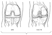
슬관절은 일반적으로 세 개의 "구획", 즉 내측(슬관절의 내측의 관절 면), 가측(슬관절의 외측의 관절 면), 및 슬개대퇴부(슬개골과 납다리뼈 또는 대퇴골과의 사이의 관절)로 나누어진다. 슬관절 치환물은 손상의 정도 및/또는 질환의 정도에 따라 다양한 상이한 형태를 취할 수 있다. 전체 슬관절 치환물(TKR)에서, 무릎 관절의 양 면이 치환되고(즉, 관절의 대퇴 관절 면 및 경골 관절 면이 인공삽입물로 치환되고, 슬개골(무릎덮개뼈(kneecap)) 면은 슬개골에 대한 손상의 정도에 따라 치환되거나 치환되지 않을 수 있다. 부분 또는 단일구획 슬관절 치환물에서, 관절의 하나 또는 두 개의 내측, 가측 또는 슬개대퇴 부분만이 치환된다(내측 구획 치환물이 가장 일반적이다).The knee joint is generally divided into three "compartments": medial (the articular surface on the medial side of the knee joint), lateral (the articular surface on the outside of the knee joint), and the patellar femur (the joint between the patella and the lead femur or femur). lose A knee replacement may take a variety of different forms depending on the extent of the injury and/or disease. In total knee replacement (TKR), both sides of the knee joint are displaced (i.e., the femoral and tibial articular surfaces of the joint are replaced with prostheses, and the patella (kneecap) side is replaced with respect to the patella. Depending on the severity of the injury, it may or may not be replaced In partial or monocompartmental knee replacements, only one or two medial, lateral, or patellar-femoral portions of the joint are replaced (medial compartmental replacements are most common).
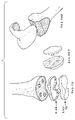
TKR의 다양한 구성요소는 전형적으로 대퇴골 이식물 및 경골 이식물을 포함한다(슬개골 면의 치환이 있거나 없음). 대퇴골 구성요소는 둥근 대퇴 골두(대개 금속이지만, 세라믹일 수 있음)를 포함하고, 경골 구성요소는 내부 중합성(대개 폴리에틸렌이지만, 세라믹 및 금속이 사용될 수 있음) 표면 라이너(liner)에 의해 경골에 부착되는 평평한 금속 외피(경골의 골수강 안으로 연장되는 스템(stem)을 갖거나 갖지 않음)를 포함하며, 슬개골 구성요소(존재하는 경우)는 슬개골의 후방 면에 접합되는 중합성 "버튼(button)"을 포함한다. 현재, TKR의 다양한 구성요소는 예를 들어 폴리에틸렌, 초고 분자량 폴리에틸렌, 세라믹, 수술 등급 스테인리스 강, 코발트 크롬, 티타늄, 및 다양한 세라믹 재료를 포함하는 다양한 상이한 재료로 만들어질 수 있다. 소정 장치에서, 대퇴골 이식물(전형적으로, 스테인리스 강, 티타늄, 또는 코발트 크롬과 같은 금속으로 만들어짐) 및 경골 구성요소(전형적으로 역시 스테인리스 강, 티타늄, 또는 코발트 크롬과 같은 금속으로 만들어짐)의 금속 부분은 대퇴골 및 경골의 골 내에서의 이식물의 통합을 촉진하기 위해 표면 코팅을 갖도록 설계될 수 있다. 인공삽입물은 골 시멘트(PMMA-폴리메틸메타크릴레이트)의 사용에 의해 제자리에 유지될 수 있거나 유지되지 않을 수 있다. 슬관절 치환물의 다양한 구성요소의 대표적인 예가 미국 특허 번호 5,413,604, 5,906,643, 6019,794 및 7,922,771에 기재되어 있다.The various components of TKR typically include femoral and tibial implants (with or without replacement of the patellar face). The femoral component comprises a round femoral ball head (usually metal, but may be ceramic), and the tibial component is secured to the tibia by an internal polymeric (usually polyethylene, but ceramic and metal may be used) surface liner. A polymeric "button" comprising a flat metal sheath attached to it (with or without a stem extending into the intramedullary canal of the tibia), the patellar component (if present) bonded to the posterior face of the patella includes ". Currently, the various components of the TKR can be made of a variety of different materials including, for example, polyethylene, ultrahigh molecular weight polyethylene, ceramics, surgical grade stainless steel, cobalt chromium, titanium, and various ceramic materials. In certain devices, the femoral implant (typically made of a metal such as stainless steel, titanium, or cobalt chromium) and of a tibial component (typically also made of a metal such as stainless steel, titanium, or cobalt chromium) in certain devices. The metal part may be designed to have a surface coating to facilitate integration of the implant within the bones of the femur and tibia. The prosthesis may or may not be held in place by the use of bone cement (PMMA-polymethylmethacrylate). Representative examples of the various components of a knee replacement are described in US Pat. Nos. 5,413,604, 5,906,643, 6019,794, and 7,922,771.
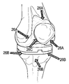
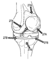
도 1은 본 기술분야에서 알려진 유형의 전체 무릎 관절과 단일구획(내측 구획) 슬관절 치환물을 도시한다. 도 2는, 금속 경골 판(5) 및 경골 스템(2)(일부 경골 판 구성요소는 스템을 갖지 않지만, 이 도면에서는 존재함), 폴리에틸렌 분절 면(7), 다양한 구성요소를 제자리에 유지시키기 위해 사용되는 시멘트(4), 슬관절 "버튼" 인공삽입물(8), 및 대퇴골 슬관절 구성요소(9)를 포함하는 전형적인 인공 관절(10)의 구성요소 및 재료를 도시한다. 도 3은, 나사 및/또는 시멘트에 의해 기초 골에 부착될 수 있는 대퇴골 구성요소, 경골 판 및 슬개골 버튼을 갖는 다른 전형적인 TKR을 도시한다(스템이 달린 경골 판과 대조적임).1 depicts a total knee joint and a single compartment (medial compartment) knee replacement of a type known in the art. Figure 2 shows a metal
불행하게도, 전체 슬관절이 삽입되는 경우, 수술 중에, 수술 후에, 그리고 시간이 흐름에 따라 다양한 골칫거리(complication)가 발생할 수 있다. 예를 들어, 수술 중에, 의사는, 절차 동안 조정이 이루어질 수 있도록, 인공삽입물의 정확한 구조적인 정렬 및/또는 인공삽입물과 주위의 골과의 사이의 임의의 움직임을 확인하기를 원할 수 있다. 수술 후에, 환자는, 슬관절 인공삽입물의 구성요소 중 임의의 것의 약간의 이동, 부분적(아탈구) 또는 전체 탈구가 있는 경우, 염증 및 통증을 경험할 수 있다. 더 장기간에, 무릎 관절의 부적절한 동작을 초래하는 대퇴골 면과 경골 면과의 사이의 점진적인 마모가 있을 수 있다. 경골 면과 대퇴골 면에 사용되는 재료의 유형에 따라, 장기적인 마모는 이식물 주위의 염증 및 골 침식을 초래하는 작은 파편 입자의 발생을 초래할 수 있다. 시간이 지남에 따라(예를 들어, 8 내지 12 년), (골용해로서 알려진 과정으로 인해) 이식물 주위의 조직에서 골 손실이 발생하고, 이것이 인공삽입물의 풀림 및 결국에는 고장을 초래하는 경우, 관련된 일반적인 골칫거리가 발생한다. 상기 급성적 및 만성적 골칫거리의 모두는 슬관절의 성능을 열화시킬 수 있고, 이동 및 보행의 어려움을 초래하며, 환자에게 통증 및 염증을 유발할 수 있다.Unfortunately, when the entire knee is implanted, various complications can arise during, after, and over time. For example, during surgery, the surgeon may wish to confirm the correct structural alignment of the prosthesis and/or any movement between the prosthesis and the surrounding bone so that adjustments can be made during the procedure. After surgery, the patient may experience inflammation and pain if there is slight displacement, partial (subluxation) or total dislocation of any of the components of the knee prosthesis. Over longer periods of time, there may be progressive wear between the femoral and tibial surfaces resulting in improper movement of the knee joint. Depending on the type of material used for the tibial and femoral surfaces, prolonged wear can result in the generation of small debris particles that lead to inflammation and bone erosion around the implant. Over time (eg, 8 to 12 years), bone loss occurs in the tissue around the implant (due to a process known as osteolysis), which leads to loosening and eventual failure of the prosthesis. , a common headache associated with it arises. All of the above acute and chronic annoyances can deteriorate the performance of the knee joint, cause difficulty in movement and ambulation, and cause pain and inflammation in the patient.
언급된 바와 같이, TKR의 대부분의 일반적이고 심각한 골칫거리 중 하나는 마찰에 의해 발생되며 염증 및 골 손실을 유발하는 재료 파편(금속, 세라믹, 및/또는 폴리우레탄 조각)에 의해 유발될 수 있는 이식물 주위의 골의 침식(골용해)이다. 염증 및 골용해의 다른 잠재적인 원인은 이식물 진동 및 움직임, 부적절한 환자 사용/활동, 부적절한 정렬(슬개골의 부적절한 궤적설정(tracking)), 경골-대퇴골 관절 및 슬개골-대퇴골 관절의 준임상적 탈구(아탈구), 기계적인 마모 및 파열, 재료 고장 또는 파손, 골과 시멘트와의 사이의 결합의 풀림, 이식물 재료와 주위의 골과의 사이의 생체적합성의 결핍, 금속 알레르기, 및 골 시멘트와 주위의 골과의 사이의 생체적합성의 결핍이다. 이러한 변화를 조기 검출하고 교정적이고 예방적인 측정을 실시하는 능력은 TKR 환자의 관리에 있어서 크게 유용할 것이다. 조기 검출 및 개입으로부터 이익을 얻을 수 있는 추가적인 골칫거리는 감염, 뼈 골절, 이식물 미세골절, 신경 충격, 심정맥 혈전증, 움직임의 감소, 및 불안정성을 포함한다.As mentioned, one of the most common and serious headaches of TKR is implantation, which is caused by friction and can be caused by material debris (metal, ceramic, and/or polyurethane pieces) that causes inflammation and bone loss. It is erosion (osteolysis) of the surrounding bone. Other potential causes of inflammation and osteolysis include implant vibration and movement, improper patient use/activity, improper alignment (inappropriate tracking of the patella), and subclinical dislocation of the tibial-femoral joint and patellar-femoral joint ( subluxation), mechanical wear and tear, material failure or breakage, loosening of the bond between bone and cement, lack of biocompatibility between implant material and surrounding bone, metal allergy, and bone cement and surrounding It is a lack of biocompatibility between the bones of The ability to detect these changes early and to make corrective and preventative measures would be of great benefit in the management of TKR patients. Additional headaches that could benefit from early detection and intervention include infection, bone fractures, implant microfractures, nerve shock, deep vein thrombosis, decreased movement, and instability.
현재, 슬관절 치환 수술 환자의 수술 후 병원 내 감시는 필요에 따라 병원 직원 및 의료 팀에 의한 개인 방문, 환자의 물리적인 검사, 의료적 감시(중요한 징후 등), 슬관절 움직임 범위(ROM)의 평가, 물리치료(조기 가동(mobilization)및 활동 포함), 및 진단적 영상화 연구 및 혈액 작업을 통해 실시된다. 일단 환자가 병원으로부터 퇴원하면, 인공삽입물 성능 및 환자 만족도는 주기적인 의사의 진료실 방문 동안 확인되며, 진료실에서는 환자 추이를 감시하고 임의의 잠재적인 골칫거리의 발달을 식별하기 위해 철저한 이력, 물리적인 검사 및 보충적인 영상화 및 진단적 연구가 사용된다. 이러한 방문 동안, 의사는 전형적으로 슬관절의 움직임 범위를 평가하고, 소정 움직임 또는 행동 동안 발생하는 임의의 통증의 식별을 시도하며, 활동 수준, 일상적인 기능, 통증 제어 및 재활 추이를 결정하기 위해 환자에게 질문을 한다.Currently, postoperative in-hospital monitoring of patients with knee replacement surgery includes personal visits by hospital staff and medical team as needed, physical examination of the patient, medical monitoring (important indications, etc.), evaluation of knee range of motion (ROM), Physiotherapy (including mobilization and activity), and diagnostic imaging studies and blood work. Once the patient is discharged from the hospital, prosthesis performance and patient satisfaction are checked during periodic physician office visits, where a thorough history, physical examination and Supplementary imaging and diagnostic studies are used. During these visits, the physician typically asks the patient to assess the range of motion of the knee joint, attempt to identify any pain that occurs during a given movement or behavior, and determine activity levels, daily functioning, pain control, and rehabilitation trends. Ask questions.
불행하게도, 환자의 회복 기간의 대부분은 병원 및/또는 진료실 방문 사이에서 일어난다. 그러므로, 수술 일로부터 완전한 회복까지 전체 관절 움직임 범위(ROM은 통증 제어, 항염증 약물, 날의 시간, 최근의 활동, 및/또는 검사 시에 환자가 어떻게 느끼는지에 따라 변할 수 있음), "실생활" 인공삽입물 성능, 환자 활동 수준, 운동 내성, 및 재활 노력(물리치료, 약물 등)의 유효성을 정확하게 측정하고 추적하는 것은 어려울 수 있다. 많은 이러한 정보를 위해, 의사는 수술 후 치료 유효성 및 회복 및 재활 추이에 대한 통찰력을 얻기 위해 환자 자가 기록 또는 제3자 관찰에 의존하는데, 많은 경우에 이는 무엇을 살펴봐야 하는지 확신이 없고, "정상적이고/예상되는" 수술 후 회복이 무엇이어야 하는지에 대한 지식이 없고, 순응적이지 않고, 또는 그들의 증상을 효과적으로 전달할 수 없는 환자에 의해 더 복잡해 진다. 또한, 증상을 나타내고, 의사 방문 사이에서 발생하기 전에 골칫거리를 (병원 내 및 병원 밖에서) 식별 및 추적하는 것 또는 환자가 그 존재를 검출하기 어려운 골칫거리를 (병원 내 및 병원 밖에서) 식별 및 추적하는 것은, 또한 TKR의 관리 및 부분 슬관절 치환물의 관리에 대한 유익한 추가적인 정보를 환자에게 제공할 것이다. 현재, 모든 경우, 의사와 환자 모두 그들이 다른 방식으로 그렇게 하고 싶을 수 있는 "실시간적인", 지속적인, 객관적인, 인공삽입물 성능 측정의 유형에 접근하지 못하고 있다.Unfortunately, most of a patient's recovery period occurs between hospital and/or office visits. Therefore, full range of joint motion from the day of surgery to full recovery (ROM may vary depending on pain control, anti-inflammatory drugs, time of day, recent activity, and/or how the patient feels at the time of examination), "real life" Accurately measuring and tracking prosthetic performance, patient activity level, exercise tolerance, and effectiveness of rehabilitation efforts (physiotherapy, medications, etc.) can be challenging. For much of this information, doctors rely on patient self-reports or third-party observations to gain insight into postoperative treatment effectiveness and trends in recovery and rehabilitation, which, in many cases, is unsure what to look for, and "normal Complicated by patients who do not have the knowledge, are not compliant, or are unable to effectively communicate their symptoms, what the “anticipated/anticipated” post-surgical recovery should be. Also, identifying and tracking (in-hospital and out-of-hospital) annoyances (in-hospital and out-of-hospital) before they present symptoms and occur between doctor visits or identifying and tracking annoyances (in-hospital and out-of-hospital) that are difficult for a patient to detect their presence , will also provide patients with additional useful information on the management of TKR and management of partial knee replacements. Currently, in all cases, both physicians and patients do not have access to the type of "real-time", continuous, objective, prosthetic performance measurement that they may wish to do so in a different way.
본 발명은 이전의 슬관절 치환물의 어려움의 많은 부분을 극복하는 신규한 전체 및 부분 슬관절 치환물, 이러한 신규한 슬관절 치환물을 구성 및 감시하는 방법을 개시하며 또한 다른 관련된 이점을 제공한다.The present invention discloses novel total and partial knee replacements that overcome many of the difficulties of previous knee replacements, methods of constructing and monitoring such novel knee replacements, and also provides other related advantages.
간단히 말하면, 전체 및 부분 슬관절 인공삽입물에는 환자 내부의 인공 무릎 관절의 온전성 및 유효성을 감시하기 위한 다수의 센서가 제공된다. 센서는 인공삽입 슬관절의 외부 표면에, 인공삽입 슬관절의 내부 표면에, 인공삽입 재료(스테인리스 강, 티타늄, 코발트 크롬, 폴리우레탄, 고분자량 폴리우레탄, 세라믹 등) 그 자체 내에, 인공삽입 슬관절을 포함하는 다양한 구성요소 사이에, 인공삽입물을 제자리에 고정시키기 위해 사용되는 나사 및/또는 기기(존재하는 경우)에, 슬관절을 고정시키기 위해 사용되는 골 시멘트(예를 들어, PMMA, 또는 PMMA 및 MMA 코폴리머 혼합물)(존재하는 경우) 내에, 및/또는 인공삽입물 주위의 조직 내에 위치될 수 있다. 소정 실시형태에서, 센서는 피동적이며 따라서 그들 자체의 전원을 필요로 하지 않는 유형이다.Briefly, total and partial knee prostheses are provided with a number of sensors for monitoring the integrity and effectiveness of the knee prosthesis inside the patient. The sensor includes the prosthetic knee joint on the outer surface of the prosthetic knee joint, on the inner surface of the prosthetic knee joint, within the prosthetic material (stainless steel, titanium, cobalt chromium, polyurethane, high molecular weight polyurethane, ceramic, etc.) itself Bone cement (e.g., PMMA, or PMMA and MMA nose) used to immobilize the knee joint, to screws and/or instruments (if any) used to hold the prosthesis in place, between the various components that polymer mixture) (if present), and/or within the tissue surrounding the prosthesis. In certain embodiments, the sensors are of a type that are passive and thus do not require their own power source.
본 발명의 일 양태에서, 전체 또는 부분 슬관절 인공삽입물을 포함하는 이식물을 환자 내에 위치설정 및 배치하기 위한 조립체가 제공되며, 하나 이상의 센서가 인공삽입물 상에, 내에, 또는 주위에 그리고/또는 인공삽입물을 부착시키기 위해 이용되는 골 시멘트 및/또는 골 나사 또는 앵커에 위치된다. 본 발명의 다른 양태에서, 경골 구성요소, 슬개골 인공삽입물, 또는 대퇴골 구성요소 중 적어도 하나 및 하나 이상의 센서를 포함하는 의료 장치가 제공된다. 명확화를 위해, 하나 이상의 센서는 슬관절 치환 인공삽입물, 의료 장치 및/또는 골 나사 또는 앵커 상의 특정 위치에 의도적으로 배치될 수 있으며 그리고/또는 슬관절 치환 인공삽입물, 의료 장치, 골 나사 또는 앵커, 및 골 시멘트에 걸쳐서, 그 상에 및 그 내에 무작위적으로 배치될 수 있다. 그러므로, "배치된다", "나타난다" 또는 "이용된다"라는 용어 또는 구절의 사용은 특정 배치가 필요로 되지 않는 한 특정 배치를 필요로 하는 것으로 간주되어서는 안된다.In one aspect of the present invention, an assembly for positioning and placing an implant comprising a full or partial knee prosthesis in a patient is provided, wherein one or more sensors are provided on, within, or around the prosthesis and/or the prosthesis. Bone cement and/or bone screws or anchors used to attach the insert. In another aspect of the present invention, there is provided a medical device comprising at least one of a tibial component, a patellar prosthesis, or a femoral component and one or more sensors. For clarity, one or more sensors may be intentionally placed at specific locations on the knee replacement prosthesis, medical device and/or bone screw or anchor and/or the knee replacement prosthesis, medical device, bone screw or anchor, and bone It can be randomly placed over, on and within the cement. Therefore, use of the terms or phrases "disposed", "appears" or "utilized" should not be construed as requiring a particular arrangement unless the particular arrangement is required.
다양한 실시형태에서, 센서는 인공삽입 슬관절의 외측 면에, 인공삽입 슬관절의 내측 면에, 인공삽입 슬관절을 구성하기 위해 사용되는 재료 내에, 인공삽입 슬관절을 구성하는 다양한 구성요소, 인공삽입물을 제자리에 고정시키기 위해 사용되는 나사 및/또는 체결 기기(존재하는 경우) 사이에, 인공삽입 슬관절을 고정시키기 위해 사용되는 골 시멘트 상에 또는 내에, 인공삽입 슬관절 주위의 조직(전형적으로는, 골 또는 골수이지만, 또한 근육, 인대, 힘줄, 관절낭 및/또는 활액실), 또는 이들의 임의의 조합) 상에 또는 내에 위치될 수 있다. 본 발명에서 사용하기에 적합한 센서의 대표적인 예는 가속도계(가속도, 기울기, 진동, 충격 및 회전 센서), 압력 센서, 접촉 센서, 위치 센서, 화학적 마이크로센서, 조직 대사 센서, 기계적인 스트레스 센서 및 온도 센서를 포함한다. 특히 바람직한 실시형태에서, 센서는 무선 센서 또는 무선 마이크로프로세서에 연결되는 센서이다.In various embodiments, the sensor places the various components constituting the prosthetic knee, the prosthesis, in place on the lateral surface of the prosthetic knee joint, on the medial surface of the prosthetic knee joint, within the material used to construct the prosthetic knee joint Between the screws and/or fastening devices used to immobilize (if present), on or in the bone cement used to immobilize the prosthetic knee, and the tissue around the prosthetic knee (typically bone or bone marrow, but , also muscles, ligaments, tendons, joint capsules and/or synovial chambers), or any combination thereof). Representative examples of suitable sensors for use in the present invention include accelerometers (acceleration, tilt, vibration, shock and rotation sensors), pressure sensors, contact sensors, position sensors, chemical microsensors, tissue metabolism sensors, mechanical stress sensors, and temperature sensors. includes In a particularly preferred embodiment, the sensor is a wireless sensor or a sensor connected to a wireless microprocessor.
추가적인 실시형태에서, 복수의 상기 센서는 인공삽입 슬관절(골 시멘트, 골 나사, 또는 조직) 상에, 내에, 또는 주위에 위치되며, 바람직한 실시형태에서, 인공삽입 슬관절은 하나 초과의 유형의 센서(예를 들어, 가속도 센서, 기울기 센서, 진동 센서, 충격 센서, 회전 센서, 압력 센서, 접촉 센서, 위치 센서, 화학적 마이크로센서, 조직 대사 센서, 및 기계적인 스트레스 센서 중 하나 이상, 또는 이들의 임의의 조합)를 수용할 수 있다.In further embodiments, a plurality of said sensors are located on, in, or around the prosthetic knee joint (bone cement, bone screw, or tissue), and in a preferred embodiment, the prosthetic knee joint comprises more than one type of sensor ( For example, one or more of an acceleration sensor, a tilt sensor, a vibration sensor, a shock sensor, a rotation sensor, a pressure sensor, a contact sensor, a position sensor, a chemical microsensor, a tissue metabolism sensor, and a mechanical stress sensor, or any thereof combinations) can be accommodated.
다양한 실시형태에 따르면, 센서는 동작, 이동, (인공삽입물 및 주위 조직 양자 모두의) 의료 영상화, 기능, 마모, 성능, 잠재적인 부차적 영향, 환자의 의학적인 상태 및 인공 슬관절의 의학적 상태, 및 환자의 살아 있는 조직과의 인공 슬관절의 상호작용을 감시하기 위해 치환 무릎 관절의 상이한 위치에 배치된다. 환자 활동, 환자 기능, 인공삽입물 활동, 인공삽입물 기능, 인공삽입물 성능, 인공삽입물 및 관절 정렬, 슬개골 궤적설정, 인공삽입물 및 관절 힘, 기계적인 스트레스, 인공삽입물 및 주위 조직 구조(영상화), 인공삽입물의 기계적 및 물리적 온전성, 슬개골 궤적설정 및 잠재적인 부차적 영향의 생생한, 지속적인, 현장 감시가 제공된다. 또한, 물리적인 검사, 의료 영상화 및 진단적인 의료적 연구를 통해 현재 이용할 수 없는 임상적으로 중요한 측정치를 포함하는, 슬관절 치환 인공삽입물 및 그것의 환자 자신의 신체 조직과의 상호작용의 많은 양태에 대한 정보를 이용할 수 있다.According to various embodiments, the sensor may be used for motion, movement, medical imaging (of both the prosthesis and surrounding tissue), function, wear, performance, potential secondary effects, the medical condition of the patient and the medical condition of the knee prosthesis, and the patient It is placed at different locations in the replacement knee joint to monitor the interaction of the artificial knee with the living tissue of the knee joint. Patient Activity, Patient Function, Prosthesis Activity, Prosthesis Function, Prosthesis Performance, Prosthesis and Joint Alignment, Patella Trajectory, Prosthesis and Joint Forces, Mechanical Stress, Prosthesis and Surrounding Tissue Structure (imaging), Prosthesis Vivid, continuous, on-site monitoring of the mechanical and physical integrity of the patient, patellar trajectory and potential collateral effects are provided. Additionally, physical examination, medical imaging, and diagnostic medical research provide insight into many aspects of the knee replacement prosthesis and its interaction with the patient's own body tissues, including clinically important measures currently unavailable through diagnostic medical research. information is available.
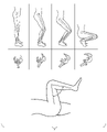
일 실시형태에 따르면, 센서는 슬관절 움직임 범위(ROM)에 대한 평가 데이터를 제공한다. 현재, ROM은 일반적으로 의사가 물리적인 검사 동안 전체 움직임 범위를 통해 무릎 관절을 피동적으로 이동시키고 그 결과(굴곡, 연신, 전방/후방 안정성, 및 내측/가측 안정성의 정도, 예를 들어 도 4 참조)를 기록함으로써 임상적으로 측정될 수 있다. 움직임 센서 및 가속도계는 물리적인 검사 동안 및 방문 사이의 정상적인 일상 활동 동안의 양자 모두의 기간 동안 인공삽입 무릎 관절의 전체 ROM을 정확하게 결정하기 위해 사용될 수 있다. 마찬가지로, 움직임 센서 및 가속도계는 물리적인 검사 동안 및 방문 사이의 정상적인 일상 활동 동안의 양자 모두의 기간 동안 인공삽입 무릎 관절의 임의의 전방/후방 또는 내측/가측 불안정성(전체, 부분 또는 준임상적 탈구 포함)을 정확하게 측정하기 위해 사용될 수 있다. 부가적으로, 움직임 센서 및 가속도계는 물리적인 검사 동안 및 방문 사이의 정상적인 일상 활동 동안의 임의의 슬개골의 부절절한 궤적설정 및/또는 슬개골 불안정성(전체, 부분 또는 준임상적 아탈구 포함)을 정확하게 측정하기 위해 사용될 수 있다.According to one embodiment, the sensor provides evaluation data for range of motion (ROM) of the knee joint. Currently, ROM is commonly performed by doctors passively moving the knee joint through the entire range of motion during a physical examination and as a result (degrees of flexion, extension, anterior/posterior stability, and medial/lateral stability, see e.g. FIG. 4 ) ) can be measured clinically by recording Motion sensors and accelerometers can be used to accurately determine the overall ROM of the prosthetic knee joint during both periods during physical examination and during normal daily activities between visits. Likewise, motion sensors and accelerometers can detect any anterior/posterior or medial/lateral instability (including total, partial or subclinical dislocation) of the prosthetic knee joint during both periods during physical examination and during normal daily activities between visits. ) can be used to accurately measure Additionally, motion sensors and accelerometers accurately measure patellar instability (including total, partial or subclinical subluxation) and/or inappropriate trajectory of any patella during physical examination and during normal daily activities between visits. can be used to
일 실시형태에 따르면, 접촉 센서는 골 침식 및 이식물 주의의 풀림을 측정하기 위해 인공삽입물과 주위 골과의 사이, 나사 및/또는 체결 기기(존재하는 경우)와 주위 골과의 사이, 인공삽입물과 주위 골 시멘트(존재하는 경우)와의 사이, 및/또는 골 시멘트(존재하는 경우)와 주위 골과의 사이에 제공된다. 다른 실시형태에서, 진동 센서는 움직임 및 풀림의 조기 징후로서의, 인공삽입물과 주위 골과의 사이, 나사 및/또는 체결 기기(존재하는 경우)와 주위 골과의 사이, 인공삽입물과 주위 골 시멘트(존재하는 경우)와의 사이, 골 시멘트와 주위 골과의 사이의, 진동을 검출하기 위해 제공된다. 다른 실시형태에서, 변형 게이지는 인공삽입물과 주위 골과의 사이, 나사 및/또는 체결 기기(존재하는 경우)와 주위 골과의 사이, 인공삽입물과 주위 골 시멘트와의 사이, 골 시멘트와 주위 골과의 사이의 변형, 및 또는 인공삽입물의 다양한 부분에 가해지는 변형을 검출하기 위해 제공된다. 변형의 갑작스런 증가는 과도하게 큰 스트레스가 치환 인공삽입물에 가해지고 이것이 신체에 손상을 증가시킬 수 있음을 암시할 수 있다. 예를 들어, 변형의 점진직인 장기간의 감소는 인식물 주위의 골 흡수를 유발하여 인공삽입물의 풀림 또는 인공삽입물 주위의 골의 파손을 초래할 수 있는 반면, 변형의 점진적인 장기간의 증가는 인공삽입물 재료 그 자체의 미세파손을 초래할 수 있다.According to one embodiment, the contact sensor is configured to measure bone erosion and loosening of the implant perimeter, between the prosthesis and surrounding bone, between the screw and/or fastening device (if present) and the surrounding bone, the prosthesis. and between the bone cement (if present) and/or between the bone cement (if present) and the surrounding bone. In other embodiments, the vibration sensor is configured to provide an early indication of movement and loosening between the prosthesis and the surrounding bone, between the screw and/or fastening device (if present) and the surrounding bone, between the prosthesis and the surrounding bone cement ( provided to detect vibrations between the bone cement and the surrounding bone, if present). In other embodiments, the strain gauge is between the prosthesis and the surrounding bone, between the screw and/or fastening device (if present) and the surrounding bone, between the prosthesis and the surrounding bone cement, between the bone cement and the surrounding bone. provided for detecting strains between families, and/or strains applied to various parts of the prosthesis. A sudden increase in strain may suggest that an excessively large stress is applied to the replacement prosthesis, which may increase damage to the body. For example, a gradual, long-term decrease in strain can cause bone resorption around the cognate prosthesis, resulting in loosening of the prosthesis or fracture of the bone around the prosthesis, whereas a gradual, long-term increase in strain can cause bone resorption around the prosthesis material. It may cause micro-damage on its own.
다른 실시형태에 따르면, 진동, 충격, 기울기 및 회전을 검출하는 가속도계가 제공된다. 다른 실시형태에 따르면, 접촉 또는 압력 센서와 같은 표면 마모를 측정하기 위한 센서가, 관절 면 침식을 감시하기 위해 대퇴골 관절 면, 경골 관절 면, 및/또는 슬개골 관절 면 내의 상이한 깊이에 매립될 수 있다. 다른 실시형태에서, 움직임 범위를 나타내며, 시간에 걸친 실제 사용에의 부분적(또는 완전한) 대퇴골-경골 슬관절 탈구 또는 아탈구, 슬개골의 부적절한 궤적설정 및/또는 슬개골-대퇴골 관절의 아탈구, 또는 인공삽입물(및 정착 기기) 그 자체의 상호연결된 구성요소 사이의 이동을 감시하는 위치 센서뿐만 아니라 다른 유형의 센서가 제공된다.According to another embodiment, an accelerometer that detects vibration, shock, tilt and rotation is provided. According to other embodiments, sensors for measuring surface wear, such as contact or pressure sensors, may be embedded at different depths within the femoral articular surface, the tibial articular surface, and/or the patellar articular surface to monitor articular surface erosion. . In another embodiment, a partial (or complete) femoral-tibial knee dislocation or subluxation, improper trajectory of the patella and/or subluxation of the patellar-femoral joint, or prosthesis indicating range of motion over time Other types of sensors are provided, as well as position sensors that monitor movement between (and fusing devices) their interconnected components.
추가적인 실시형태에서, 인공 슬관절(전체 또는 부분)은 특정 위치에서 특정 밀집도로 센서를 수용할 수 있다. 예를 들어, 인공 슬관절은 장치의 1평방 센티미터 당 한 개, 두 개, 세 개, 네 개, 다섯 개, 여섯 개, 일곱 개, 여덟 개, 아홉 개, 또는 열 개보다 큰 센서 밀집도의 센서(예를 들어, 가속도 센서, 기울기 센서, 진동 센서, 충격 센서, 회전 센서, 압력 센서, 접촉 센서, 위치 센서, 화학적 마이크로센서, 조직 대사 센서, 및 기계적인 스트레스 센서, 또는 이들의 임의의 조합)를 가질 수 있다. 다른 실시형태에서, 인공 슬관절(전체 또는 부분)은 장치의 1입방 센티미터 당 한 개, 두 개, 세 개, 네 개, 다섯 개, 여섯 개, 일곱 개, 여덟 개, 아홉 개, 또는 열 개보다 큰 센서 밀집도의 센서(예를 들어, 가속도 센서, 기울기 센서, 진동 센서, 충격 센서, 회전 센서, 압력 센서, 접촉 센서, 위치 센서, 화학적 마이크로센서, 조직 대사 센서, 및 기계적인 스트레스 센서, 또는 이들의 임의의 조합)를 가질 수 있다. 관련된 실시형태에서, 센서(예를 들어, 가속도 센서, 기울기 센서, 진동 센서, 충격 센서, 회전 센서, 압력 센서, 접촉 센서, 위치 센서, 화학적 마이크로센서, 조직 대사 센서, 및 기계적인 스트레스 센서)는, 예를 들어 대퇴골 구성요소(내측, 가측 또는 양자 모두), 경골 판, 경골 스템(존재하는 경우), 경골 라이닝, 인공삽입 경골 라이닝을 포함하는 인공 슬관절 상의, 내의, 또는 주위의 특정 위치에, 연결되는 장치의 부분들(예를 들어, 경골 컵 및 경골 라이닝의 연결 분절들) 내에, 인공삽입물을 제자리에 고정시키기 위해 사용되는 나사 및/또는 체결 기기(존재하는 경우) 내에, 그리고 인공 슬관절 주위에(인공삽입 슬관절을 인공삽입 슬관절 주위의 조직 - 전형적으로는, 골 또는 골수이지만, 또한 근육, 인대, 심줄, 관절낭, 및/또는 활액실 - 상에 또는 내에 고정시키기 위해 사용되는 골 시멘트 상에 또는 내에) 위치될 수 있다.In further embodiments, the artificial knee joint (in whole or in part) may house the sensors at a specific location and at a specific density. For example, an artificial knee joint may have a sensor density greater than one, two, three, four, five, six, seven, eight, nine, or ten sensors per square centimeter of the device ( for example, an acceleration sensor, a tilt sensor, a vibration sensor, a shock sensor, a rotation sensor, a pressure sensor, a contact sensor, a position sensor, a chemical microsensor, a tissue metabolism sensor, and a mechanical stress sensor, or any combination thereof). can have In other embodiments, more than one, two, three, four, five, six, seven, eight, nine, or ten knee prostheses (in whole or in part) per cubic centimeter of the device. sensors of high sensor density (eg, accelerometers, tilt sensors, vibration sensors, shock sensors, rotation sensors, pressure sensors, contact sensors, position sensors, chemical microsensors, tissue metabolism sensors, and mechanical stress sensors, or these any combination of ). In related embodiments, the sensors (eg, acceleration sensors, tilt sensors, vibration sensors, shock sensors, rotation sensors, pressure sensors, contact sensors, position sensors, chemical microsensors, tissue metabolism sensors, and mechanical stress sensors) include: , at a specific location on, within, or around an artificial knee joint, including, for example, a femoral component (medial, lateral or both), a tibial plate, a tibial stem (if present), a tibial lining, a prosthetic tibial lining; in parts of the device to be connected (eg, the connecting segments of the tibial cup and tibial lining), in screws and/or fastening devices (if present) used to hold the prosthesis in place, and around the prosthetic knee (on the bone cement used to immobilize the prosthetic knee on or within the tissue surrounding the prosthetic joint - typically bone or bone marrow, but also muscle, ligament, tendon, joint capsule, and/or synovial chamber - or within).
본 발명의 소정 실시형태에서, 전체 또는 부분 슬관절 인공삽입물에는 특정한 고유의 식별 번호가 제공되며, 추가적인 실시형태에서, 인공삽입 슬관절 상의, 내의 또는 주위의 각각의 센서는 각각 특정한 고유의 식별 번호 또는 그룹 식별 번호(예를 들어, 가속도 센서, 기울기 센서, 진동 센서, 충격 센서, 회전 센서, 압력 센서, 접촉 센서, 위치 센서, 화학적 마이크로센서, 조직 대사 센서, 또는 기계적인 스트레스 센서를 식별하는 식별 번호)를 갖는다. 또한 추가적인 실시형태에서, 특정한 고유의 식별 번호 또는 그룹 식별 번호는 인공삽입 슬관절 상의, 내의 또는 주위의 위치와 구체적으로 연관된다.In certain embodiments of the present invention, the total or partial knee prosthesis is provided with a specific unique identification number, and in further embodiments, each sensor on, within, or around the prosthetic knee joint is each provided with a specific unique identification number or group. Identification number (eg, an identification number that identifies an accelerometer, tilt sensor, vibration sensor, shock sensor, rotation sensor, pressure sensor, contact sensor, position sensor, chemical microsensor, tissue metabolism sensor, or mechanical stress sensor) has In still further embodiments, the particular unique identification number or group identification number is specifically associated with a location on, within, or around the prosthetic knee.
본 발명의 다른 양태에서, 신체 외측의 위치로부터 신체 내측의 위치로 무선 전기 신호를 전송하는 단계, 신체 내측에 위치되는 인공 슬관절 상에, 내에 또는 주위에 위치되는 센서에서 신호를 수신하는 단계, 수신된 신호를 사용하여 센서를 급전하는 단계, 센서에서 데이터를 감지하는 단계, 및 감지된 데이터를 센서로부터 신체의 외측에 위치되는 수신 유닛에 출력하는 단계를 포함하는 방법이 이식된 전체 또는 부분 슬관절 인공삽입물을 감시하기 위해 제공된다.In another aspect of the present invention, there is provided a method comprising the steps of: transmitting a wireless electrical signal from a location on the outside of the body to a location on the inside of the body; receiving the signal from a sensor positioned on, in or around an artificial knee joint positioned inside the body; receiving A method comprising the steps of powering a sensor using the received signal, sensing data from the sensor, and outputting the sensed data from the sensor to a receiving unit located on the outside of the body. provided to monitor the insert.
본 발명의 다른 양태에서, (a) 슬관절 치환물 또는 의료 장치에서 하나 이상의 센서의 위치를 검출하는 단계, 및 (b) 슬관절 치환물 또는 의료 장치의 영상이 생성되도록 상기 하나 이상의 센서의 위치를 시각적으로 표시하는 단계를 포함하는 방법이 본원에 제공된 바와 같은 슬관절 치환물 또는 의료 장치를 영상화하기 위해 제공된다. 다양한 실시형태에서, 검출하는 단계는 시간에 걸쳐 행해질 수 있고, 따라서 시각적인 표시는 시간에 걸친 위치 이동을 나타낼 수 있다. 소정 실시형태에서, 표시되는 영상은 2차원 또는 3차원 영상이다. 바람직한 실시형태에서, 다양한 영상이 시간-순서로(예를 들어, 이동하는 영상 또는 "영화 유사" 영상으로서) 수집되고 표시될 수 있다.In another aspect of the invention, the steps of (a) detecting the position of one or more sensors on the knee replacement or medical device, and (b) visually visualizing the position of the one or more sensors such that an image of the knee replacement or medical device is generated. A method is provided for imaging a knee replacement or medical device as provided herein, comprising the step of indicating In various embodiments, the detecting step may be done over time, such that the visual indication may indicate positional movement over time. In certain embodiments, the displayed image is a two-dimensional or three-dimensional image. In preferred embodiments, various images may be collected and displayed in time-order (eg, as moving images or “movie-like” images).
본원에 제공되는 영상화 기술은 매우 다양한 목적을 위해 이용될 수 있다. 예를 들어, 일 양태에서, 영상화 기술은 슬관절 치환물 또는 의료 장치의 적절한 배치 및 작용을 보장하기 위해 수술 절차 동안 이용될 수 있다. 다른 실시형태에서, 영상화 기술은 슬관절 치환물 또는 의료 장치를 검사하기 위해서 및/또는 시간에 걸친 장치의 동작 및/또는 이동을 비교하기 위해서 수술 후에 이용될 수 있다.The imaging techniques provided herein can be used for a wide variety of purposes. For example, in one aspect, imaging techniques may be used during surgical procedures to ensure proper placement and operation of a knee replacement or medical device. In other embodiments, imaging techniques may be used post surgery to examine a knee replacement or medical device and/or to compare motion and/or movement of the device over time.
부분 또는 전체 슬관절 인공삽입물의 온전성은 무선식으로 문의(interrogating)될 수 있고 그 결과는 정기적으로 기록될 수 있다. 이는 환자의 건강이 환자 및/또는 의사에 의해 요구되는 바에 따라 정기적으로 또는 임의의 시간에 확인될 수 있도록 한다. 또한, 인공삽입물은, "이벤트 기록"의 일부로서 (외부 신호전달/트리거링(triggering) 장치를 통해) 환자에 의해 그렇게 하도록 신호전달을 받을 때 - 즉, 환자가 특정 이벤트(예를 들어, 통증, 손상, 불안정성 등)를 경험하고, 환자가 주관적인/증상적인 데이터를 객관적인/센서 데이터와 비교하는 것을 가능하게 하도록 동시 판독을 얻기 위해 장치에 대해 신호전달/트리거링을 행할 때, 무선식으로 문의를 받을 수 있다. 이벤트 기록 데이터를 센서 데이터와 정합시키는 것은 환자의 특정 증상의 내재하는 원인 또는 특정 유발인자를 더 잘 이해하기 위한 노력의 일부로서 이용될 수 있다. 그러므로, 본 발명의 다양한 실시형태에서, 원하는 시점에 문의하는 단계를 포함하는, 본원에 제공되는 전체 또는 부분 슬관절 치환물 중 하나를 갖는 피술자에 있어서의 이벤트를 검출 및/또는 기록하는 방법이 제공된다. 그러므로, 본 발명의 일 양태에서, 슬관절 치환물 또는 의료 장치 내의 하나 이상의 센서의 활동을 원하는 시점에 문의하는 단계 및 상기 활동을 기록하는 단계를 포함하는, 본원에 제공된 바와 같은 슬관절 치환물 또는 의료 장치를 갖는 피술자에 있어서의 이벤트를 검출 및/또는 기록하는 방법이 제공된다. 다양한 실시형태에서, 상기 방법은 피술자에 의해 그리고/또는 건강 관리 전문가에 의해 달성될 수 있다. 관련된 실시형태에서, 기록하는 단계는 하나 이상의 유선 장치 또는 휴대되거나 착용될 수 있는 무선 장치(예를 들어, 휴대 전화, 시계, 손목 밴드, 및/또는 안경)에 의해 실행될 수 있다. 추가적인 실시형태에서, 착용된 장치(예를 들어, 휴대 전화, 시계, 손목 밴드 및/또는 안경)은 추가적인 데이터 수집 및 분석을 실행할 수 있는 충분한 처리 전력 및 메모리를 가질 수 있다.The integrity of a partial or total knee prosthesis can be interrogated wirelessly and the results recorded regularly. This allows the patient's health to be checked regularly or at any time as required by the patient and/or physician. In addition, the prosthesis may be signaled to do so by the patient (via an external signaling/triggering device) as part of an "event recording" - that is, when the patient is signaled to do so with a specific event (e.g., pain, impairment, instability, etc.), and to receive inquiries wirelessly when can Matching event history data with sensor data can be used as part of an effort to better understand the underlying causes or specific triggers of a patient's specific symptoms. Thus, in various embodiments of the present invention, methods are provided for detecting and/or recording an event in a subject having one of the total or partial knee replacements provided herein comprising interrogating at a desired time point. . Thus, in one aspect of the present invention, a knee replacement or medical device as provided herein comprising interrogating the activity of one or more sensors within the knee replacement or medical device at a desired point in time and recording the activity, in one aspect of the present invention. A method of detecting and/or recording an event in a subject having In various embodiments, the method may be accomplished by the subject and/or by a health care professional. In related embodiments, the recording step may be performed by one or more wired devices or wireless devices that may be carried or worn (eg, cell phones, watches, wristbands, and/or glasses). In further embodiments, worn devices (eg, cell phones, watches, wristbands and/or glasses) may have sufficient processing power and memory to perform additional data collection and analysis.
추가적인 실시형태에서, 센서의 각각은 신호 수신 회로 및 신호 출력 회로를 포함한다. 신호 수신 회로는 전력 및 데이터 수집 요구 성분의 양자 모두를 포함하는 문의 신호를 수신한다. 문의 신호로부터의 전력을 사용하여, 센서는 감지를 실행하는데 필요한 회로의 일부를 급전하고, 감지를 실행하며, 그 후 문의 모듈에 데이터를 출력한다. 문의 모듈은 문의 모듈을 구동시키기 위한 적절한 I/O 회로, 메모리, 마이크로프로세서 형태의 제어기, 및 다른 회로를 포함하는 제어 유닛의 제어 하에 작용한다. 또 다른 실시형태에서, 센서(예를 들어, 가속도 센서, 기울기 센서, 진동 센서, 충격 센서, 회전 센서, 압력 센서, 접촉 센서, 위치 센서, 화학적인 마이크로센서, 조직 대사 센서, 또는 기계적인 스트레스 센서)는, (예를 들어, 슬관절 인공삽입물에의 센서의 영구적인 부착을 제공하는 개구 또는 다른 부속물에 의해) 슬관절 인공삽입물에 용이하게 통합되거나 다른 방식으로 기계적으로 부착될 수 있도록 그리고/또는 슬관절 인공삽입물을 둘러싸는 골 시멘트 또는 조직에 용이하게 통합될 수 있도록 구성된다.In a further embodiment, each of the sensors includes a signal receiving circuit and a signal output circuit. The signal receiving circuit receives an interrogation signal that includes both power and data collection requirements. Using the power from the interrogation signal, the sensor powers a portion of the circuitry necessary to perform sensing, performs sensing, and then outputs data to the interrogation module. The interrogation module operates under the control of a control unit comprising suitable I/O circuitry for driving the interrogation module, memory, a controller in the form of a microprocessor, and other circuitry. In another embodiment, a sensor (eg, an acceleration sensor, a tilt sensor, a vibration sensor, a shock sensor, a rotation sensor, a pressure sensor, a contact sensor, a position sensor, a chemical microsensor, a tissue metabolism sensor, or a mechanical stress sensor) ) to be readily integrated or otherwise mechanically attached to the knee prosthesis (eg, by an aperture or other appendage that provides for permanent attachment of the sensor to the knee prosthesis) and/or the knee prosthesis. It is configured for easy integration into the bone cement or tissue surrounding the insert.
본 발명의 또 다른 양태에서, 신체 외측의 위치로부터 신체 내측의 위치에 무선 전기 신호를 전송하는 단계, 신체 내측에 위치되는 인공삽입 슬관절 상에, 내에 또는 주위에 위치되는 상기 센서 중 하나에서 신호를 수신하는 단계, 수신된 신호를 사용하여 센서를 급전하는 단계, 센서에서 데이터를 감지하는 단계, 및 감지된 데이터를 센서로부터 신체 외측에 위치되는 수신 유닛에 출력하는 단계를 포함하는 방법이 제공된다. 소정 실시형태에서, 수신 유닛은 센서에 의해 제공되는 신호의 분석을 제공할 수 있다.In another aspect of the present invention, there is provided a method comprising: transmitting a wireless electrical signal from a location on the outside of the body to a location on the inside of the body; A method is provided that includes receiving, powering a sensor using the received signal, sensing data at the sensor, and outputting the sensed data from the sensor to a receiving unit located outside the body. In certain embodiments, the receiving unit may provide analysis of the signal provided by the sensor.
센서에 의해 수집된 데이터는 대퇴골 구성요소, 경골 판 및/또는 경골 스템 내에 위치되는 메모리에 저장될 수 있다. 의사에의 방문 동안, 데이터는 무선 센서를 통해 내려받기될 수 있고, 의사는 인공삽입물의 실시간 성능을 나타내는 데이터를 얻을 수 있다.Data collected by the sensors may be stored in memory located within the femoral component, tibial plate, and/or tibial stem. During a visit to the physician, data may be downloaded via the wireless sensor, and the physician may obtain data representative of the real-time performance of the prosthesis.
얻어지는 이점은 인공삽입물의 더 정확한 감시 및 환자의 건강에 기여하는 정확한 현장 데이터의 의료적인 보고의 허용을 포함한다. 하나 이상의 실시형태의 상세사상을 이하의 상세한 설명에서 설명한다. 추가적인 특징, 목적 및 이점은 상세한설명, 도면 및 청구항으로부터 명확해질 것이다. 또한, 본원에서 참조되는 모든 특허 및 특허 출원의 개시내용은 그 전체가 참조로 통합된다.Advantages obtained include more accurate monitoring of the prosthesis and allowing medical reporting of accurate field data that contributes to the patient's health. The details of one or more embodiments are set forth in the detailed description that follows. Additional features, objects and advantages will become apparent from the description, drawings and claims. Also, the disclosures of all patents and patent applications referenced herein are incorporated by reference in their entirety.
도 1은 전체 슬관절 치환물 및 단일구획 슬관절 치환물을 도시한다.
도 2는 전체 슬관절 치환물의 다양한 구성요소를 나타내는 분해도이다.
도 3은 다른 전체 슬관절 치환물의 구성요소를 나타낸다.
도 4는 전체 슬관절 치환물을 갖는 피술자 대한 대표적인 움직임 범위(ROM)를 나타낸다.
도 5는 다양한 접촉 센서를 갖는 TKR을 나타낸다.
도 6은 다양한 변형 게이지를 갖는 TKR을 나타낸다.
도 7은 다양한 가속도계를 갖는 TKR을 나타낸다.
도 8은 다양한 위치 센서를 갖는 TKR을 나타낸다.
도 9는 관절 마모를 검출하기 위해 배치된 센서를 갖는 TKR을 나타낸다.
도 10은 센서 데이터를 처리하기 위해 배치되는 정보 및 통신 기술 시스템 실시형태를 나타낸다.
도 11은 본 발명의 일 실시형태에 따른 센서, 문의 모듈, 및 제어 유닛의 블록도이다.
도 12는, 본 발명의 일 실시형태에 따른, 데이터가 조사되는 피술자 내의 슬관절 치환물에 위치되며 데이터를 출력하는 하나 이상의 센서의 개략도이다.1 shows a total knee replacement and a single compartment knee replacement.
2 is an exploded view showing various components of the entire knee joint replacement.
3 shows the components of another total knee replacement.
4 shows a representative range of motion (ROM) for a subject with total knee replacement.
5 shows a TKR with various contact sensors.
6 shows a TKR with various strain gauges.
7 shows a TKR with various accelerometers.
8 shows a TKR with various position sensors.
9 shows a TKR with a sensor positioned to detect joint wear.
10 depicts an information and communication technology system embodiment that is deployed to process sensor data.
11 is a block diagram of a sensor, an inquiry module, and a control unit according to an embodiment of the present invention.
12 is a schematic diagram of one or more sensors positioned at a knee replacement within a subject for which data is being investigated and outputting data, in accordance with an embodiment of the present invention.
간단히 설명하면, 본 발명은 장치의 온전성 및 유효성을 감시하기 위해 이용될 수 있는 다양한 슬관절 치환물을 제공한다. 그러나, 본 발명을 설명하기에 앞서, 이후에 사용되는 소정 용어의 정의를 먼저 설명하는 것이 본 발명을 이해하는데 도움이 될 수 있다.Briefly stated, the present invention provides a variety of knee replacements that can be used to monitor the integrity and effectiveness of the device. However, before describing the present invention, it may be helpful to understand the present invention by first explaining definitions of certain terms used hereinafter.
본원에서 이용되는 용어로서의 "슬관절 치환물" 또는 "슬관절 인공삽입물"은 다양한 상이한 형태를 취할 수 있고 합성 재료를 갖는 환자의 무릎 관절의 모두의 치환물(전체 슬관절 치환물) 또는 일부의 치환물(부분 슬관절 치환물)을 포함할 수 있다. 전체 슬관절 치환물(TKR)에서, 대퇴골측 및 경골측의 양자 모두가 치환된다. 부분 또는 단일구획 슬관절 치환물에서, 슬관절의 하나 또는 두 개의 부분(표면-경골 또는 대퇴골, 또는 구획들-내측, 가측 또는 슬개골)만이 치환된다.As used herein, "knee replacement" or "knee prosthesis" as used herein may take a variety of different forms and may include all (total knee replacement) or partial (total knee replacement) replacements of a patient's knee joint with synthetic materials. partial knee replacement). In total knee replacement (TKR), both the femoral and tibial side are replaced. In a partial or monocompartmental knee replacement, only one or two portions of the knee (superficial-tibia or femur, or compartments-medial, lateral or patella) are replaced.
TKR의 다양한 구성요소는 전형적으로 대퇴골 이식물, 슬개골 이식물, 및 경골 이식물(스템이 있거나 없는 상태의 경골 판 및 경골 라이너를 포함할 수 있음)을 포함할 수 있다. 현재, 다양한 구성요소는 예를 들어 폴리에틸렌, 초고 분자량 폴리에틸렌, 세라믹, 수술 등급 스테인리스 강, 코발트 크롬, 티타늄, 및 다양한 세라믹 재료를 포함하는 다양한 상이한 재료로 만들어질 수 있다. 소정 장치 내에서, 대퇴골 이식물(전형적으로, 스테인리스 강, 티타늄, 또는 코발트 크롬과 같은 금속으로 만들어짐)은 대퇴골 내로의 이식물의 통합을 촉진하기 위해 골 표면 코팅이 있는 상태로 설계될 수 있으며, 경골판(및 스템)은 또한 경골 안으로의 통합을 촉진하기 위해 표면 코팅을 가질 수 있다. 슬관절 치환물의 다양한 구성요소의 대표적인 예들이 미국특허번호 5,413,604, 5,906,643, 6,019,794 및 7,922,771에 기재되어 있다.The various components of a TKR may typically include a femur implant, a patellar implant, and a tibial implant (which may include a tibial plate and tibial liner with or without a stem). Currently, various components can be made of a variety of different materials including, for example, polyethylene, ultrahigh molecular weight polyethylene, ceramics, surgical grade stainless steel, cobalt chromium, titanium, and various ceramic materials. In certain devices, a femoral implant (typically made of a metal such as stainless steel, titanium, or cobalt chromium) can be designed with a bone surface coating to facilitate integration of the implant into the femur, The tibial plate (and stem) may also have a surface coating to facilitate integration into the tibia. Representative examples of the various components of a knee replacement are described in US Pat. Nos. 5,413,604, 5,906,643, 6,019,794, and 7,922,771.
"골 시멘트"는 인공삽입물 기기와 주위의 골과의 사이에 제공될 수 있고 냉각될 때(또는 활성화될 때) 제자리에 경화되는 재료를 말하며, 그것은 인공삽입물의 구성요소(인공삽입물 대퇴골 면, 경골 판/스템, 슬개골 "버튼") 중 하나 이상을 적절한 골 조직(대퇴골, 경골, 경골 수, 슬개골)에 고정시키기 위해 사용되는 작용제이다. 골 시멘트는 대개 PMMA(폴리메틸메타크릴레이트) 또는 PMMA 및 MMA 코폴리머 혼합물을 포함한다. 골 나사 및/또는 다른 금속(또는 중합성) 고정 장치가 인공삽입물 구성요소를 주위의 골 조직에 정착시키는 것을 돕기 위해 사용될 수도 있다는 것을 유의해야 한다."Bone cement" refers to a material that can be provided between the prosthesis device and the surrounding bone and hardens in place when cooled (or activated), which is a component of the prosthesis (prosthesis femoral face, tibia) An agent used to anchor one or more of a plate/stem, patella “button”) to the appropriate bone tissue (femur, tibia, tibialis fossa, patella). Bone cements usually contain PMMA (polymethylmethacrylate) or a mixture of PMMA and MMA copolymers. It should be noted that bone screws and/or other metal (or polymeric) fixation devices may be used to assist in anchoring the prosthesis component to the surrounding bone tissue.
본 발명은 슬관절 인공삽입물(전체 또는 부분 이식물을 포함할 수 있음), 의료 장치(예를 들어, 슬관절 이식물의 일부 및/또는 장치를 이식하는 처리에서 유용한 구성요소 또는 재료), 및 키트(kit)(예를 들어, 슬관절 인공삽입물, 의료 장치, 및 골 시멘트 및 임의의 연관된 전달 장치와 같은 추가적인 필요한 재료)를 제공하며, 이들 모두는 이하에서 더 상세하게 설명되는 바와 같은 센서를 갖는다. 본원에 제공되는 바와 같은 슬관절 인공삽입물, 의료 장치 및 키트(골 시멘트와 같은 관련된 재료 포함)는 바람직하게는 살균되어 있고, 비-발열성이며, 그리고/또는 인체에의 사용 및/또는 이식에 적합하다. 그러나, 본 발명의 소정 실시형태에서, 슬관절 인공삽입물, 의료 장치 및/또는 키트는 살균되지 않은 환경에서 만들어질 수 있고(또는 나아가 개별 피술자에 맞게 주문제작되고) 이후의 시점에 살균될 수 있다.The present invention relates to knee prostheses (which may include whole or partial implants), medical devices (eg, components or materials useful in the process of implanting parts and/or devices), and kits. ) (eg, knee prosthesis, medical devices, and additional required materials such as bone cement and any associated delivery devices), all of which have sensors as described in more detail below. The knee prostheses, medical devices and kits (including related materials such as bone cement) as provided herein are preferably sterile, non-pyrogenic, and/or suitable for use and/or implantation in the human body. Do. However, in certain embodiments of the present invention, the knee prosthesis, medical device and/or kit may be made in a non-sterile environment (or further customized for the individual subject) and sterilized at a later time point.
"센서"는 신체의 하나 이상의 상이한 양태 및/또는 신체 내에 삽입된 슬관절 인공삽입물, 의료 장치 또는 키트의 온전성, 충격, 유효성 또는 효과를 측정하기 위해 이용될 수 있는 장치를 말한다. 본 발명에서 사용하기에 적합한 센서의 대표적인 예는, 예를 들어 유체 압력 센서, 접촉 센서, 위치 센서, 맥압 센서, 액체(예를 들어, 혈액) 용적 센서, 액체(예를 들어, 혈액) 유동 센서, 화학 센서(예를 들어, 혈액 및/또는 다른 유체를 위한 것), 대사 센서(예를 들어, 혈액 및/또는 다른 유체를 위한 것), 가속도계, 기계적인 스트레스 센서 및 온도 센서를 포함한다. 소정 실시형태에서 센서는 무선 센서일 수 있거나, 다른 실시형태에서 센서는 무선 마이크로프로세서에 연결될 수 있다. 다른 실시형태에서, 센서 중 하나 이상(모두를 포함)은 센서를 구체적으로 식별하는 고유의 센서 식별 번호(Unique Sensor Identification number: "USI")를 가질 수 있다."Sensor" refers to a device that can be used to measure one or more different aspects of the body and/or the integrity, impact, effectiveness or effectiveness of a knee prosthesis, medical device or kit inserted into the body. Representative examples of sensors suitable for use in the present invention include, for example, fluid pressure sensors, contact sensors, position sensors, pulse pressure sensors, liquid (eg blood) volume sensors, liquid (eg blood) flow sensors. , chemical sensors (eg, for blood and/or other fluids), metabolic sensors (eg, for blood and/or other fluids), accelerometers, mechanical stress sensors, and temperature sensors. In certain embodiments the sensor may be a wireless sensor, or in other embodiments the sensor may be coupled to a wireless microprocessor. In other embodiments, one or more (including all) of the sensors may have a Unique Sensor Identification number (“USI”) that specifically identifies the sensor.
본 발명에서 매우 다양한 센서(미세전자기계 시스템(Microelectromechanical System) 또는 "MEMS", 또는 나노전자기계 시스템(Nanoelectromechanical System) 또는 "NEMS", 및 BioMEMS 또는 BioNEMS를 칭하기도 함, 일반적으로 https://en.wikipedia.org/wiki/MEMS 참조)가 이용될 수 있다. 대표적인 특허 및 특허 출원은 미국특허번호 7,383,071 및 8,634,928, 및 미국공보번호 2010/0285082, 및 2013/0215979를 포함한다. 대표적인 공보는, Albert Foch에 의한 "Introduction to BioMEMS", CRC Press, 2013; Marc J. Madou에 의한 "From MEMS to Bio-MEMS and Bio-NEMS: Manufacturing Techniques and Applications", CRC Press 2011; Simona Badilescu에 의한 "Bio-MEMS: Science and Engineering Perspectives", CRC Press 2011; Steven S. Saliterman에 의한 "Fundamentals of BioMEMS and Medical Microdevices", SPIE-The International Society of Optical Engineering, 2006; Wanjun Wang 및 Steven A. Soper에 의해 편집된 "Bio-MEMS: Technologies and Applications", CRC Press, 2012; 및 Volker Kempe에 의한 "Inertial MEMS: Principles and Practice", Cambridge University Press, 2011; Polla, D. L., 등, "Microdevices in Medicine," Ann. Rev. Biomed. Eng. 2000, 02:551-576, Yun, K. S., 등, "A Surface-Tension Driven Micropump for Low-voltage and Low-Power Operations," J. Microelectromechanical Sys., 11:5, 2002년 10월, 454-461; Yeh, R., 등, "Single Mask, Large Force, and Large Displacement Electrostatic Linear Inchworm Motors," J. Microelectromechanical Sys., 11:4, 2002년 8월, 330-336; 및 Loh, N. C., 등, "Sub-10 cm3 Interferometric Accelerometer with Nano-g Resolution," J. Microelectromechanical Sys., 11:3, 2002년 6월, 182-187를 포함하며, 상기 모두는 그 전체가 참조로 통합된다.In the present invention a wide variety of sensors (Microelectromechanical System or "MEMS", or Nanoelectromechanical System or "NEMS", and also referred to as BioMEMS or BioNEMS, generally https://en .wikipedia.org/wiki/MEMS) may be used. Representative patents and patent applications include US Pat. Nos. 7,383,071 and 8,634,928, and US Publication Nos. 2010/0285082, and 2013/0215979. Representative publications include "Introduction to BioMEMS" by Albert Foch, CRC Press, 2013; "From MEMS to Bio-MEMS and Bio-NEMS: Manufacturing Techniques and Applications" by Marc J. Madou, CRC Press 2011; "Bio-MEMS: Science and Engineering Perspectives" by Simona Badilescu, CRC Press 2011; "Fundamentals of BioMEMS and Medical Microdevices" by Steven S. Saliterman, SPIE-The International Society of Optical Engineering, 2006; "Bio-MEMS: Technologies and Applications," CRC Press, 2012, edited by Wanjun Wang and Steven A. Soper; and “Inertial MEMS: Principles and Practice” by Volker Kempe, Cambridge University Press, 2011; Polla, DL, et al., “Microdevices in Medicine,” Ann. Rev. Biomed. Eng. 2000, 02:551-576, Yun, KS, et al., “A Surface-Tension Driven Micropump for Low-voltage and Low-Power Operations,” J. Microelectromechanical Sys., 11:5, October 2002, 454-461 ; Yeh, R., et al., “Single Mask, Large Force, and Large Displacement Electrostatic Linear Inchworm Motors,” J. Microelectromechanical Sys., 11:4, Aug. 2002, 330-336; and Loh, NC, et al., “Sub-10 cm 3 Interferometric Accelerometer with Nano-g Resolution,” J. Microelectromechanical Sys., 11:3, June 2002, 182-187, all of which are incorporated herein by reference in their entirety. incorporated by reference.
본 발명의 다양한 실시형태에서, 본원에 기재된 센서는, 슬관절 인공삽입물, 의료 장치 또는 키트의 내측, 내부, 및/또는 외부 표면(또는 표면들)뿐만 아니라 슬관절 인공삽입물, 의료 장치 또는 키트와 그것을 운반할 수 있는 임의의 장치(예를 들어, 전달 또는 설치 장치)와의 사이를 포함하여 다양한 위치 및 다양한 구성으로 배치될 수 있다. 본원에 제공된 개시내용을 고려하면 용이하게 명확해지는 바와 같이, 센서는 동시에 슬관절 인공삽입물, 의료 장치 또는 키트의 다수의 위치에(즉, 외측 표면의 내측에, 내부에 또는 그 위에) 배치될 수 있다. 소정 실시형태에서, 슬관절 인공삽입물, 의료 장치 또는 키트, 연관된 의료 장치(예를 들어, 전달 기기) 또는 키트는 1평방 센티미터 당 1, 2, 3, 4, 5, 6, 7, 8, 9, 10개 또는 10개 초과보다 큰 센서 밀집도로 센서를 포함한다. 다른 양태에서, 슬관절 인공삽입물, 의료 장치, 또는 키트, 연관된 의료 장치(예를 들어, 전달 기기) 또는 키트는 1입방 센티미터 당 1, 2, 3, 4, 5, 6, 7, 8, 9, 10개 또는 10개 초과보다 큰 센서 밀집도로 센서를 포함한다. 이러한 실시형태 중 어느 것에서, 1평방 센티미터 또는 1입방 센티미터 당 50, 75, 100개 미만 또는 100개의 센서가 있을 수 있다. 다양한 실시형태에서, 센서 중 적어도 하나 이상은 본원에 기재된 바와 같이 카테터, 의료 장치, 또는 키트 내에서 무작위적으로 또는 하나 이상의 특정 위치에 배치될 수 있다.In various embodiments of the invention, the sensors described herein carry the knee prosthesis, medical device or kit and the medial, internal, and/or external surface (or surfaces) of the knee prosthesis, medical device or kit, as well as the same. It can be deployed in a variety of locations and configurations, including between and with any device capable of performing (eg, a delivery or installation device). As will be readily apparent from consideration of the disclosure provided herein, a sensor may be simultaneously disposed in multiple locations (ie, on the inside, on, or on the lateral surface) of a knee prosthesis, medical device or kit. . In certain embodiments, the knee prosthesis, medical device or kit, associated medical device (eg, delivery device) or kit is 1, 2, 3, 4, 5, 6, 7, 8, 9, per square centimeter; Include sensors with a sensor density greater than ten or greater than ten. In another aspect, the knee prosthesis, medical device, or kit, associated medical device (eg, delivery device) or kit is 1, 2, 3, 4, 5, 6, 7, 8, 9, per cubic centimeter; Include sensors with a sensor density greater than ten or greater than ten. In any of these embodiments, there may be less than 50, 75, 100 or 100 sensors per square centimeter or cubic centimeter. In various embodiments, at least one or more of the sensors may be placed randomly or at one or more specific locations within a catheter, medical device, or kit as described herein.
다양한 실시형태에서, 센서는 슬관절 인공삽입물, 의료 장치 또는 키트, 연관된 의료 장치(예를 들어, 전달 기기) 또는 키트에 걸쳐 특정 위치들에 및/또는 무작위적으로 배치될 수 있다. 또한, 센서는 특정 패턴으로 배치될 수 있다(예를 들어, 센서는 X의 패턴으로, 슬관절 인공삽입물, 의료 장치 또는 키트, 연관된 의료 장치(예를 들어, 전달 기기) 또는 키트 주위의 타원형 또는 동심 링으로서 배치될 수 있다).In various embodiments, the sensor may be randomly placed and/or at specific locations throughout the knee prosthesis, medical device or kit, associated medical device (eg, delivery device) or kit. Additionally, the sensors may be disposed in a specific pattern (eg, the sensors are in a pattern of X, oval or concentric around the knee prosthesis, medical device or kit, associated medical device (eg, delivery device) or kit). can be arranged as a ring).
슬관절 인공삽입물, 의료 장치 및 키트의 대표적인 실시형태Representative Embodiments of Knee Prostheses, Medical Devices and Kits
본원에 제공된 본 발명의 다양한 양태를 더 잘 이해하기 위해서, 이하의 섹션, 즉 A. 슬관절 인공삽입물, 의료 장치 및 키트 및 그들의 사용, B. 치료제(들)를 전달하기 위한 슬관절 인공삽입물, 의료 장치 및 키트의 사용, C. 이식물의 열화 또는 마모를 측정하기 위한 센서를 갖는 슬관절 인공삽입물, 의료 장치 또는 키트의 사용, D. 슬관절 인공삽입물, 의료 장치 및 키트에 있어서의 감염을 감시하기 위한 방법, E. 건강관리에 있어서의 센서-포함 슬관절 인공삽입물, 의료 장치 및 키트의 사용, F. 슬관절 인공삽입물, 의료 장치 및 키트로부터의 전력의 생성, G. 슬관절 인공삽입물, 의료 장치 및 키트를 포함하는 조립체의 의료 영상화 및 자가 진단, 예상 분석 및 예상 유지보수, H. 슬관절 인공삽입물, 의료 장치 및 키트를 포함하는 조립체를 감시하는 방법, 및 I. 슬관절 인공삽입물, 의료 장치 및 키트를 포함하는 조립체로부터의 데이터의 수집, 전송, 분석 및 분배가 이하에 제공된다.For a better understanding of the various aspects of the invention provided herein, the following sections are provided: A. Knee Prostheses, Medical Devices and Kits and Their Uses, B. Knee Prostheses for Delivering Therapeutic Agent(s), Medical Devices and use of the kit, C. use of a knee prosthesis, medical device or kit having a sensor to measure deterioration or wear of the implant, D. a method for monitoring infection in the knee prosthesis, medical device and kit; E. Use of sensor-comprising knee prostheses, medical devices and kits in healthcare, F. Generation of electrical power from knee prostheses, medical devices and kits, G. Knee prostheses, including medical devices and kits Medical imaging and self-diagnosis of assemblies, predictive analysis and predictive maintenance, H. methods of monitoring assemblies comprising knee prostheses, medical devices and kits, and I. from assemblies comprising knee prostheses, medical devices and kits The collection, transmission, analysis and distribution of data is provided below.
A. 슬관절 인공삽입물, 의료 장치 및 키트 및 그들의 사용 A. Knee Prostheses, Medical Devices and Kits and Their Uses
슬관절 치환은, 환자가 슬관절의 충분한 사용을 상실하여 신체장애, 이동 및 기능의 상실, 보행 장애, 및/또는 지속적인 관절 통증 및 불편을 갖게 될 때 실행된다. 전체 또는 부분 슬관절 치환으로 이어지는 슬관절 기능 장애의 일반적인 원인은 다양한 유형의 관절염(예를 들어, 류머티즘성 관절염 또는 골관절염, 및 외상(예를 들어, 앞 슬관절 인대 손상 또는 연골/메니스커스 파열))을 포함한다. 대부분의 환자에서, 보행 향상, 정상적인 일상 기능 회복 및 통증 감소에 있어서 수술은 성공적이며, 결과적으로 서양에서는 수술은 매우 일반적인 정형외과적 절차이다.Knee replacement is performed when the patient loses full use of the knee joint, resulting in physical disability, loss of movement and function, impaired gait, and/or persistent joint pain and discomfort. Common causes of knee dysfunction leading to total or partial knee replacement include various types of arthritis (e.g., rheumatoid arthritis or osteoarthritis, and trauma (e.g., anterior knee ligament injury or cartilage/meniscus rupture)). include In most patients, surgery is successful in improving gait, restoring normal daily functions, and reducing pain, and consequently, surgery is a very common orthopedic procedure in the West.
도 5, 도 6, 도 7, 도 8 및 도 9는, 인공삽입물의 실시간 동작, 환자 기능 및 활동의 수준, 및 인공삽입물 성능을 정확하게 그리고 시간에 걸쳐 현장에서 감시하기 위해 인공삽입물 내에 또는 인공삽입물 상에 위치되는 하나 이상의 센서를 갖는 전체 슬관절 치환물 형태의 몇몇 인공삽입물(10)을 도시한다. 다양한 실시형태에 따라 다양한 이러한 센서를 설명한다.5 , 6 , 7 , 8 and 9 illustrate the real-time operation of the prosthesis, level of patient function and activity, and prosthesis performance within or within the prosthesis to accurately and in situ monitoring over time. Shown are
도 5에 도시된 일 실시형태에서, 대퇴 골두 인공삽입물-골 계면 상에 및 내부에 분포되는 접촉 센서(22A), 경골 뼈-금속 판(및 존재하는 경우에는 스템) 계면 상에 및 내부에 분포되는 접촉 센서(22B), 및 슬개골 인공삽입물(슬개골 "버튼")-슬개골 뼈 계면 상에 및 내부에 분포되는 접촉 센서(22C)를 포함하는 하나 이상의 접촉 센서(22)가 이식물에 걸쳐 제공된다. 일부 실시형태에서, 접촉 센서는 인공삽입물 구성요소 그 자체(경골, 대퇴골 및 슬개골 분절 상에 있으며, 다른 실시형태에서 접촉 센서는 인공삽입물을 주위의 골에 고정시키기 위해 사용되는 골 시멘트(존재하는 경우) 상에/내부에 수용되며, 그리고 또 다른 실시형태에서 접촉 센서는 인공삽입물 및 골 시멘트(PMMA)의 양자 모두 상에/내부에 수용된다.5 ,
다양한 실시형태에서, 이러한 센서는 주위의 골(대퇴골, 경골 및/또는 슬개골) 및/또는 주위의 골 시멘트(존재하는 경우)에 대해 그들의 접촉 위치에 기초하여 인공삽입물 구성요소 상에 다양한 상이한 패턴으로 위치될 수 있다. 예를 들어, 센서는, 경골 구성요소와 경골 및/또는 주위의 골 시멘트(존재하는 경우)와의 사이, 대퇴골 구성요소와 대퇴골 및/또는 주위의 골 시멘트(존재하는 경우)와의 사이, 및 슬개골 구성요소와 슬개골 및/또는 주위의 골 시멘트(존재하는 경우)와의 사이의 물리적인 접촉에 대한 정확한 데이터를 수집하기 위해서, X의 패턴으로, 다양한 구성요소 주위의 타원형 또는 동심 링으로서 또는 다양한 다른 패턴으로 배치될 수 있다. 접촉 센서는, 골 시멘트와 인공삽입물(대퇴골부, 경골부 및 슬개골부)의 구성요소와의 사이 및/또는 골 시멘트와 골(대퇴골, 경골, 슬개골) 그 자체와의 사이의 물리적인 접촉에 대한 데이터를 수집하도록 골 시멘트(존재하는 경우) 내에 분포되고/골 시멘트 내에 배치될 수도 있다.In various embodiments, these sensors are arranged in a variety of different patterns on the prosthesis component based on their location of contact with the surrounding bone (femur, tibia, and/or patella) and/or the surrounding bone cement (if present). can be located. For example, the sensor may be located between the tibial component and the tibia and/or surrounding bone cement (if present), between the femoral component and the femur and/or surrounding bone cement (if present), and the patellar component In order to gather accurate data on the physical contact between the element and the patella and/or the surrounding bone cement (if present), in a pattern of X, as oval or concentric rings around various components, or in various other patterns can be placed. The contact sensor may be configured to detect physical contact between the bone cement and components of the prosthesis (femur, tibia, and patella) and/or between the bone cement and the bone (femur, tibia, and patella) itself. It may be distributed/placed within the bone cement (if present) to collect data.
본 발명의 다양한 실시형태에서, 접촉 센서는, 인공삽입 장치 구성요소의 1 평방 센티미터 당 또는 1 입방 센티미터 당 및/또는 골 시멘트의 1 입방 센티미터 당 한 개, 두 개, 세 개, 네 개, 다섯 개, 여섯 개, 일곱 개, 여덟 개, 아홉 개 또는 열 개보다 큰 센서 밀집도로, 슬관절 인공삽입물의 경골 구성요소, 대퇴골 구성요소, 및/또는 슬개골 구성요소 상에 그리고/또는 인공삽입물의 구성요소를 주위의 골에 고정시키는 골 시멘트 내에 배치된다.In various embodiments of the present invention, the contact sensors are one, two, three, four, five per square centimeter or per cubic centimeter of prosthetic device component and/or per cubic centimeter of bone cement. With sensor densities greater than dog, six, seven, eight, nine, or ten, on the tibial component, the femoral component, and/or the patellar component of the knee prosthesis and/or the component of the prosthesis is placed in the bone cement that anchors it to the surrounding bone.
본 발명의 다른 양태에서, (a) 슬관절 치환물 또는 의료 장치에서의 하나 이상의 센서의 위치를 검출하는 단계, 및 (b) 슬관절 치환물 또는 의료 장치의 화상이 생성되도록 상기 하나 이상의 센서의 위치를 시각적으로 표시하는 단계를 포함하는 본원에 제공된 바와 같은 슬관절 치환물 또는 의료 장치를 영상화하기 위한 방법이 제공된다. 다양한 실시형태에서, 검출하는 단계는 시간에 걸쳐 행해질 수 있고, 따라서 시각적인 표시는 시간에 걸친 위치 이동을 나타낼 수 있다. 소정 바람직한 실시형태에서, 표시되는 영상은 3차원 영상이다.In another aspect of the invention, (a) detecting the position of one or more sensors in the knee replacement or medical device, and (b) positioning the one or more sensors such that an image of the knee replacement or medical device is generated. There is provided a method for imaging a knee replacement or medical device as provided herein comprising the step of visually indicating. In various embodiments, the detecting step may be done over time, such that the visual indication may indicate positional movement over time. In certain preferred embodiments, the displayed image is a three-dimensional image.
본원에서 제공되는 영상화 기술은 매우 다양한 목적을 위해 이용될 수 있다. 예를 들어, 일 양태에서, 영상화 기술은 슬관절 치환물 또는 의료 장치의 적절한 배치 및 작용을 보장하기 위해 수술 절차 동안 이용될 수 있다. 다른 실시형태에서, 영상화 기술은 슬관절 치환물 또는 의료 장치를 검사하기 위해 그리고/또는 시간에 걸친 장치의 동작 및/또는 이동을 비교하기 위해 수술 후에 이용될 수 있다.The imaging techniques provided herein can be used for a wide variety of purposes. For example, in one aspect, imaging techniques may be used during surgical procedures to ensure proper placement and operation of a knee replacement or medical device. In other embodiments, imaging techniques may be used post surgery to examine a knee replacement or medical device and/or to compare motion and/or movement of the device over time.
일 실시형태에서, 접촉 센서(22)(22A, 22B, 22C)는 인공삽입물(10)의 풀림 및 그것의 주위 시멘트(존재하는 경우) 및/또는 골에의 연결을 검출할 수 있다. 예를 들어, 경골 구성요소 상에/내에 및/또는 경골 구성요소 주위의 골 시멘트 상에/내에 위치되는 접촉 센서(22B)는 경골 내에서의 경골 구성요소의 풀림을 검출할 수 있고, 이는 수술 동안 정확하게 검출될 수 있고 의사에게 다소간의 수술 중 조정이 필요함을 경고할 수 있다. (수술 후 수준과 비교하여) 시간에 걸친 경골 내에서의 경골 구성요소의 진행중인 풀림은, (예를 들어, 골용해로서 알려진 과정으로 인해) 골 손실이 일어날 때 발생하는 일반적인 골칫거리이며, 이 역시 경골 구성요소 상의/내의 및/또는 주위 골 시멘트 상의/내의 접촉 센서에 의해 검출될 수 있다. 또한, 경골 구성요소의 분절 사이(예를 들어, 경골 판과 경골 라이너와의 사이)에 위치되는 접촉 센서는 구성요소 분절 사이의 비정상적인 이동, 풀림, 또는 마모를 검출할 수 있으며, 이러한 센서는 "짝을 이룰"수 있어(즉, 인접하는 구성요소 사이에서 "쌍을 이루어서") 수술 배치 동안(그리고 후에) 정확한 설치를 가능하게 하기도 한다.In one embodiment, the contact sensors 22 ( 22A, 22B, 22C ) may detect loosening of the
따라서, 도 5의 실시형태에서, 경골과 경골 구성요소와의 사이, 대퇴골과 대퇴골 구성요소와의 사이, 슬개골과 슬개골 구성요소와의 사이, 개별 인공삽입물 구성요소의 상보적인 분절 사이, 및 다중구획 또는 단일구획 인공삽입 무릎 관절의 존재하는 다양한 관절면 사이(내측 및 가측 경골-대퇴골 관절, 슬개골-대퇴골 관절)의 접촉을 감시하기 위해 다양한 접촉 센서가 제공된다. 구체적으로는, 인공삽입 슬관절의 천연 또는 합성 경골 관절 면(내측, 가측, 또는 양자 모두)으로부터의 대퇴골 인공삽입물 관절면의 전체 또는 부분 탈구(아탈구)는 슬관절 치환물의 일반적인 골칫거리이며, 대개 수술 직후에(특히, 주위 근육 및 인대가 수술로부터 여전히 치유 중인 수술 후 회복 기간 동안) 발생한다. 대퇴골 구성요소 관절면 및/또는 경골 구성요소 관절면 상의 접촉 센서는 관절 탈구 또는 아탈구가 발생하는 경우 환자 및 건강관리 제공자에게 경고할 수 있다. 이는 환자 또는 의사에게 용이하게 확인될 수 없는 무릎 관절의 준임상적인 부분적 또는 불완전한 탈구(아탈구)의 검출에 있어서의 특히 유용하며, 이는 조기 가동 및 수술 후 재활 노력 동안의 가장 큰 관심사이다. 추가적으로, 다양한 슬관절 구성요소 상의 접촉 센서는 관절이 이동 및 활동 동안 정확하게 기능하고 정렬(궤적설정)되는지에 대해 결정할 수 있다. 이는 특히 정확한 슬개골 궤적설정을 임상적으로 정확하게 측정하는 것이 어려울 수 있기 때문에 무릎 덮개뼈(knee cap)의 이동에 대해 그러하며, 수술 중 및 수술 후의 양자에 있어서의 슬개골 궤적설정의 정확한 측정이 유리할 것이다.Thus, in the embodiment of FIG. 5 , between the tibia and the tibial component, between the femur and the femur component, between the patella and the patellar component, between the complementary segments of the individual prosthesis components, and multicompartment Alternatively, various contact sensors are provided to monitor contact between the various articular surfaces present (medial and lateral tibial-femoral joints, patellar-femoral joints) of the single-compartment prosthetic knee joint. Specifically, total or partial dislocation (subluxation) of the femoral prosthesis articular facet from the natural or synthetic tibial articular facet (medial, lateral, or both) of the prosthetic knee is a common nuisance of knee replacements, usually immediately after surgery. (especially during the postoperative recovery period when the surrounding muscles and ligaments are still healing from surgery). Contact sensors on the articular surface of the femoral component and/or the articular surface of the tibial component may alert the patient and healthcare provider if a joint dislocation or subluxation is occurring. This is particularly useful in the detection of subclinical partial or incomplete dislocations (subluxations) of the knee joint that cannot be readily identified to the patient or physician, which is of great concern during early mobilization and post-operative rehabilitation efforts. Additionally, contact sensors on the various knee joint components can determine whether the joint is correctly functioning and aligned (trajected) during movement and activity. This is particularly true for the movement of the knee cap because it can be difficult to accurately measure the exact patellar trajectory clinically, and accurate measurement of the patella trajectory both during and after surgery would be advantageous.
도 6에 도시된 다른 실시형태에서, 대퇴 골두 인공삽입물-골 계면 상에 및 내에 분포되는 변형 게이지(26A), 경골 뼈-금속 판(및 존재하는 경우에는 스템) 계면 상에 및 내에 분포되는 변형 게이지(26B), 및 슬개골 인공삽입물(슬개골 "버튼")-슬개골 뼈 계면에 분포되는 변형 게이지(26C)를 포함하는 하나 이상의 변형 게이지(또는 센서)(26)가 이식물 전체에 걸쳐 제공된다. 일부 실시형태에서, 변형 게이지는 인공삽입물 구성요소 그 자체(경골, 대퇴골 및 슬개골 분절) 상에 있으며, 다른 예에서 변형 게이지는 인공삽입물을 주위의 골에 고정하기 위해 사용되는 골 시멘트(존재하는 경우) 상에/내에 수용되며, 또 다른 실시형태에서 변형 게이지는 인공삽입물 구성요소 및 골 시멘트(PMMA)의 양자 모두 상에/내에 수용된다.6 ,
다양한 실시형태에서, 이들 변형 게이지는 주위의 골(대퇴골, 경골 및/또는 슬개골) 및/또는 주위의 골 시멘트(존재하는 경우)에 대한 그 접촉 위치에 기초하여 인공삽입물 구성요소 상에서 다양한 상이한 패턴으로 위치될 수 있다. 예를 들어, 변형 게이지는, 인공삽입물 구성요소, 주위의 골 시멘트(존재하는 경우), 및 주위의 골(대퇴골, 경골, 슬개골) 조직이 경험하는 물리적인 변형에 대한 정확한 데이터를 수집하기 위해, X의 패턴으로, 다양한 구성요소 주위의 타원형 또는 동심 링으로서, 또는 다양한 다른 패턴으로 배치될 수 있다.In various embodiments, these strain gauges are in a variety of different patterns on the prosthesis component based on their location of contact with the surrounding bone (femur, tibia, and/or patella) and/or the surrounding bone cement (if present). can be located. For example, strain gauges can be used to collect accurate data about the physical strain experienced by the prosthesis component, surrounding bone cement (if present), and surrounding bone (femur, tibia, patella) tissue; It can be arranged in a pattern of Xs, as oval or concentric rings around various components, or in various other patterns.
본 발명의 다양한 실시형태에서, 변형 센서는 인공삽입물 구성요소의 1 평방 센티미터당 또는 PMMA 골 시멘트의 1 입방 센티미터 당 한 개, 두 개, 세 개, 네 개, 다섯 개, 여섯 개, 일곱 개, 여덟 개, 아홉 개 또는 열 개보다 큰 센서 밀집도로 경골 구성요소, 대퇴골 구성요소, 슬개골 인공삽입물 상에, 및/또는 골 시멘트 내에 배치된다.In various embodiments of the present invention, the strain sensors are one, two, three, four, five, six, seven, per square centimeter of the prosthesis component or per cubic centimeter of PMMA bone cement; Placed on the tibial component, the femoral component, the patellar prosthesis, and/or in the bone cement with a sensor density greater than eight, nine or ten.
변형 게이지(26)는 접촉 센서(22)와 상이한 데이터 지점을 제공한다. 접촉 센서(22)는 단지 인접하는 구조물 사이에 현재 접촉이 있는지의 여부를 명확히 하고 따라서 두 개의 면 사이에 맞붙는 접촉이 있는지의 여부에 대한 우수한 징표를 제공한다. 그러나, 접촉 센서는 인공삽입물 표면 또는 주위의 골에 존재하는 물리적인 변형의 징표를 제공하지 않으며, 한편 변형 센서(26)는 보정되지 않는 경우 미래의 풀림 및 인공삽입물 고장의 전조일 수 있는 이식물에 걸쳐 가해지는 기계적인 변형력을 나타내는 데이터를 출력한다. 또한, 변형 게이지(26)는 두 개의 면 사이, 예를 들어 경골 측면과 골과의 사이, 대퇴골 측면과 골과의 사이, 슬개골 측면과 골과의 사이, 인공삽입물 구성요소(경골, 대퇴골 및 슬개골)와 골 시멘트와의 사이, 또는 경골, 대퇴골 및 슬개골 구성요소 그 자체들 사이에서 나타나는 변형을 나타내는 유형일 수 있다.Strain gauge 26 provides a different data point than
도 6에 도시된 바와 같이, 변형 게이지(26)는 경골 인공삽입물과 주위의 경골 뼈(및/또는 존재하는 경우에는 골 시멘트)와의 사이에서 발생하는 변형을 검출하기 위해 경골 구성요소 상의 다양한 위치에 위치될 수 있다. 많은 경골 인공삽입물은 정착 및 안정성을 향상시키기 위해 경골의 골수강 안으로 연장되는 스템을 포함한다. 경골 인공삽입물 및/또는 경골 골 시멘트 내의 변형의 감소는, 잠재적으로 경골강의 골 흡수(손실)를 완전히 또는 부분적으로 초래할 수 있는 조건이 존재함을 나타낼 수 있고, 골 흡수는 인공삽입물의 풀림 또는 경골 파손을 초래할 수 있다(반대로는, 증가된 변형은 부분적으로 골 성장에 유리할 것이다). 그러므로, 변형 센서는, 경골 축에 존재하는 변형의 징표를 제공할 수 있고, 오정렬되거나 보정되지 않는 경우 풀림 및 인공삽입물 고장을 초래할 높은 가능성을 갖는 이식물에 걸쳐 가해지는 가장 중요한 기계적인 변형력을 측정할 수 있다. 변형의 증가는 또한 통증의 원인일 수 있는 골 비대(성장)를 암시할 수 있다. 동일한 역학이 대퇴골 및 슬개골 인공삽입물 구성요소(및/또는 골 시멘트)와 대퇴골 및 슬개골과의 사이의 계면에 존재하며, 본 발명의 변형 게이지(26)는 이를 감시하기 위해서도 사용될 수 있다. "실생활" 변형 정보는, 인공삽입물-골 건강에 대한 다양한 활동의 (긍정적 및 부정적) 영향을 결정하기 위해 데이터를 사용할 수 있는 의사 및 환자뿐만 아니라 더 우수한 인공삽입물을 설계하기 위해 그 데이터를 사용할 수 있는 제조자에게도 유익하다.6, strain gauges 26 are located at various locations on the tibial component to detect deformations that occur between the tibial prosthesis and the surrounding tibial bone (and/or bone cement, if present). can be located. Many tibial prostheses include a stem that extends into the intramedullary canal of the tibia to improve fixation and stability. A decrease in strain in the tibial prosthesis and/or tibial bone cement may indicate that there are conditions that can potentially result in full or partial bone resorption (loss) of the tibial cavity, where bone resorption can be caused by loosening of the prosthesis or the tibia. may lead to breakage (on the contrary, increased strain will in part favor bone growth). Therefore, the strain sensor can provide an indication of the strain present in the tibial axis and measure the most important mechanical strain forces applied across the implant with a high probability of causing loosening and prosthesis failure if misaligned or not corrected. can do. Increased deformity may also suggest bone hypertrophy (growth), which may be the cause of pain. The same dynamics exist at the interface between the femur and patella prosthesis components (and/or bone cement) and the femur and patella, and the strain gauge 26 of the present invention may also be used to monitor this. “Real-life” transforming information means that the data can be used to design better prostheses, as well as physicians and patients who can use the data to determine the (positive and negative) effects of various activities on prosthetic-bone health. It is also beneficial for manufacturers who have
도 7에 도시된 다른 실시형태에서, 대퇴 골두 인공삽입물 상에 및 내에 분포되는 가속도계(27A), 경골 판(및 존재하는 경우에는 스템) 및 경골 라이너 상에 및 내에 분포되는 가속도계(26B), 및 슬개골 인공삽입물(슬개골 "버튼") 상에 및 내에 분포되는 가속도계(27C)를 포함하는 하나 이상의 가속도계(27)가 이식물 전체에 걸쳐 제공된다. 일부 실시형태에서, 가속도계는 인공삽입물 구성요소 그 자체(경골, 대퇴골 및 슬개골 분절) 상에/내에 있으며, 다른 실시형태에서 가속도계는 인공삽입물을 주위의 골에 고정시키기 위해 사용되는 골 시멘트(존재하는 경우) 상에/내에 수용되며, 또 다른 실시형태에서 가속도계는 인공삽입물 구성요소 및 골 시멘트(PMMA)의 양자 모두 상에/내에 수용된다.7, an
다양한 실시형태에서, 가속도계는 주위의 골(대퇴골, 경골 및/또는 슬개골), 주위의 골 시멘트(존재하는 경우), 상이한 인공삽입물 구성요소(경골-대퇴골 관절 및 슬개골-대퇴골 관절) 사이의 관절 계면, 및/또는 구성요소의 하위-분절 사이(예를 들어, 경골 판과 경골 라이너와의 사이)에 대한 접촉 위치에 기초하여 인공삽입물 구성요소 내에/상에 다양한 상이한 패턴으로 위치될 수 있다. 예를 들어, 가속도계는, 인공삽입물 구성요소, 주위의 골 시멘트(존재하는 경우), 및 (확정하면) 주위의 골(대퇴골, 경골, 슬개골) 조직이 경험하는 정확한 데이터를 수집하기 위해 X패턴으로, 다양한 구성요소 주위 또는 내의 타원형 또는 동심 링으로서, 또는 다양한 다른 패턴으로 배치될 수 있다.In various embodiments, the accelerometer measures the joint interface between the surrounding bone (femur, tibia, and/or patella), surrounding bone cement (if present), and different prosthetic components (tibial-femoral joint and patella-femoral joint). , and/or may be positioned in a variety of different patterns within/on the prosthesis component based on the location of contact between the sub-segments of the component (eg, between the tibial plate and the tibial liner). For example, an accelerometer can be used in an X-pattern to gather accurate data experienced by the prosthesis component, surrounding bone cement (if present), and (if confirmed) surrounding bone (femur, tibia, patella) tissue. , as oval or concentric rings around or within the various components, or in various other patterns.
본 발명의 다양한 실시형태에서, 가속도계는, 장치 및/또는 골 시멘트의 1평방 센티미터 당 또는 1입방 센티미터 당 한 개, 두 개, 세 개, 네 개, 다섯 개, 여섯 개, 일곱 개, 여덟 개, 아홉 개, 또는 열 개의 센서보다 많은 센서 밀집도로, 경골 구성요소, 대퇴골 구성요소, 슬개골 인공삽입물 상에/내에 및/또는 골 시멘트에 배치된다.In various embodiments of the present invention, the accelerometers are one, two, three, four, five, six, seven, eight per cubic centimeter or per cubic centimeter of the device and/or bone cement. , with a sensor density greater than nine, or ten sensors, placed on/in the tibial component, the femoral component, the patellar prosthesis and/or in the bone cement.
가속도계는 다양한 구성요소의 가속, 진동, 충격, 기울기, 및 회전을 검출할 수 있는 이점을 제공한다. 가속도계는 다양한 조건 하에 그리고 장기간의 시간에 걸쳐 인공삽입물(10)의 성능을 측정하는 능력을 가능하게 한다.Accelerometers offer the advantage of being able to detect acceleration, vibration, shock, tilt, and rotation of various components. Accelerometers enable the ability to measure the performance of the
슬관절 치환 수술 동안, 인공삽입 관절은 수술 봉합 전에 인공삽입물 기능 및 이동성을 평가하기 위해 전체 움직임 범위 및 안정성 시험을 통과할 것이다. 가속도계(27)는 그 때 의사에게 정확하고, 수치적이고, 정량적인 움직임 범위 데이터를 제공할 수 있고, 이 데이터는 이식 수술의 효과를 평가하기 위해 예상된 값과 비교될 수 있고 수술 후에 획득되는 기능 값과의 비교를 위한 기준 값으로서의 역할을 할 수 있다. 진동(주위의 골에서의 인공삽입물의 불충분한 정착의 징표), 기울기(경골-대퇴골 관절 및 슬개골-대퇴골 관절의 부적절한 궤적설정 및/또는 정렬의 징표), 회전(탈구 또는 아탈구의 징표) 및/또는 움직임 범위에 있어서의 임의의 비정상이 이때 다루어질 수 있고 의사가 수술 중에 조정을 행할 수 있게 한다. 슬관절이 치환된 직후에, 다리는 수술 후에 처음에는 피동적으로 그 후에는 능동적으로 움직여질 것이고, 상기 절차로부터의 회복 직후에, 환자는 관절에의 점진적인 중량 지탱을 시작할 것이다. 가속도계(27)는, 다리가 전방으로 선회하고, 지면에 닫고, 서고, 지면으로부터 상승되며, 신체가 전방으로 추진됨에 따른 보행 중을 포함하여 이동 중에 무릎 관절의 이동 및 궤적설정을 측정할 수 있다. 또한, 가속도계는 지면에 닺는 발의 충돌 및 경골을 통해 무릎 관절에 전달되는 힘 및 인공삽입물(10)의 상이한 위치에서 발생할 수 있는 임의의 진동, 충격 및 회전의 영향을 측정할 수 있다. 환자가 수술 후의 그의 움직임 범위를 향상시킴에 따라, 인공삽입 무릎 관절의 상이한 위치에서 경험되는 가속도가 감시될 수 있다. 환자가 수술로부터 치유됨에 따라, 활동 수준은 점진적으로 증가할 것이고, 보행이 향상되고 증가할 것이며, 정지는 더 신속해질(부드러워질) 것이며, 또한 각각의 걸음에 있어서 더 큰 보폭 길이가 달성될 것으로 예상된다. 운동 및 다양한 활동의 영향은 다양한 가속도계(27)에 의해 감시될 수 있고 생활 활동이 수술 회복 및 재활 후에 얼마나 향상(또는 억제)되는지를 결정하기 위해 환자의 주관적인 경험과 비교될 수 있다.During knee replacement surgery, the prosthetic joint will pass full range of motion and stability tests to assess prosthetic function and mobility prior to surgical closure. The
도 8에 도시된 다른 실시형태에서, 대퇴 골두 인공삽입물 상에 및 내에 분포되는 위치 센서(28A), 경골 판(및 또는 존재하는 경우에는 스템) 및 경골 라이너 상에 및 내에 분포되는 위치 센서(26B), 및 슬개골 인공삽입물(슬개골 "버튼")에 분포되는 위치 센서(27C)를 포함하는 하나 이상의 위치 센서(28)가 이식물 전체에 걸쳐 제공된다. 일부 실시형태에서, 위치 센서는 인공삽입물 구성요소 그 자체(경골, 대퇴골 및 슬개골 분절) 상에/내에 있고, 다른 실시형태에서 위치 센서는 인공삽입물을 주위의 골에 고정시키기 위해 사용되는 골 시멘트(존재하는 경우) 상에/내에 수용되며, 또 다른 실시형태에서 위치 센서는 인공삽입물 구성요소 및 골 시멘트(PMMA)의 양자 모두 상에/내에 수용된다.8, a
다양한 실시형태에서, 위치 센서는, 주위의 골(대퇴골, 경골 및/또는 슬개골), 주위의 골 시멘트(존재하는 경우), 상이한 인공삽입물 구성요소(경골-대퇴골 관절 및 슬개골-대퇴골 관절) 사이의 관절 계면, 및/또는 구성요소의 하위 분절 사이(예를 들어, 경골 판과 경골 라이너와의 사이)에 대한 그들의 접촉 위치에 기초하여 인공삽입물 구성요소 내에/상에 다양한 상이한 패턴으로 위치될 수 있다. 예를 들어, 위치 센서는, 인공삽입물 구성요소, 주위의 골 시멘트(존재하는 경우), 및 (확장에 의한) 주위의 골(대퇴골, 경골, 슬개골) 조직이 경험하는 정확한 데이터를 수집하기 위해, X의 패턴으로, 다양한 구성요소 주위 또는 내의 타원형 또는 동심 링으로서, 또는 다양한 다른 패턴으로 배치될 수 있다.In various embodiments, the position sensor is positioned between the surrounding bone (femur, tibia, and/or patella), surrounding bone cement (if present), and between different prosthetic components (tibial-femoral joint and patella-femoral joint). may be positioned in a variety of different patterns within/on the prosthetic component based on the joint interface, and/or their location of contact with respect to the sub-segments of the component (eg, between the tibial plate and the tibial liner). . For example, the position sensor may be used to collect accurate data experienced by the prosthesis component, surrounding bone cement (if present), and surrounding bone (femur, tibia, patella) tissue (by extension); It can be arranged in a pattern of X, as oval or concentric rings around or within various components, or in various other patterns.
본 발명의 다양한 실시형태에서, 위치 센서(28)는, 장치 및/또는 골 시멘트의 1 평방 센티미터 당 또는 1입방 센티미터 당 한 개, 두 개, 세 개, 네 개, 다섯 개, 여섯 개, 일곱 개, 여덟 개, 아홉 개, 또는 열 개의 센서보다 큰 센서 밀집도로 경골 구성요소, 대퇴골 구성요소, 슬개골 인공삽입물 및/또는 골 시멘트에 배치된다.In various embodiments of the present invention, the position sensors 28 are one, two, three, four, five, six, seven per square centimeter or cubic centimeter of the device and/or bone cement. Placement in the tibial component, femoral component, patellar prosthesis and/or bone cement with a sensor density greater than dog, eight, nine, or ten sensors.
본원에 기재된 바와 같은 위치 센서(28)는, 이식물의 3차원 데이터를 제공함으로써 물리적인 시험의 정확도를 향상시키기 위해, 경골-대퇴골 (무릎) 관절 및/또는 슬개골-대퇴골 관절의 전체 및 부분 탈구(아탈구)를 검출하기 위해, 그리고 무릎 관절 및 슬개골의 적절한 궤적설정을 결정하기 위해, 굴곡 및 연신의 측정치를 포함하는 정확한 위치 데이터(수술 중 및 수술 후)를 제공하기 위해 이용될 수 있다.The position sensor 28 as described herein is capable of providing full and partial dislocations ( subluxation) and to provide accurate positional data (intraoperative and postoperative), including measurements of flexion and elongation, to determine proper trajectory of the knee joint and patella.
도 9에 도시된 다른 실시형태에서, 대퇴 골두 관절 면 상에 및 내에 (다양한 깊이에서) 분포되는 접촉 또는 압력 센서(22A), 경골 관절 라이너 상에 및 내에 (다양한 깊이에서) 분포되는 접촉 또는 압력 센서(22B), 및 슬개골 관절 인공삽입물(슬개골 "버튼") 상에 및 내에 (다양한 깊이에서) 분포되는 접촉 또는 압력 센서(27C)를 포함하는 하나 이상의 접촉 또는 압력 센서(22)가 이식물 전체에 걸쳐 제공된다.9 , contact or
이러한 센서는 또한 다양한 관절 면의 점진적인 침식을 검출하기 위해 이용될 수 있다. 센서(22)는 경골, 대퇴골 및 슬개골 관절 면 재료 내의 점진적인 깊이들에 배치될 수 있다. 센서는 또한 센서가 노출될 때(또는 덮는 표면이 마모될 때) 활성화되어 표면 손실의 정도 및 깊이를 나타낼 수 있다.Such sensors can also be used to detect progressive erosion of various articular surfaces.
이러한 센서들은 이식물의 유효 잔여 수명을 추정하기 위해 그리고 상이한 재료 및 이식물의 성능 및 설계를 비교하기 위해 이용될 수 있다.These sensors can be used to estimate the useful remaining life of the implant and to compare the performance and design of different materials and implants.
B. 치료제(들)를 전달하기 위한 슬관절 인공삽입물, 의료 장치 또는 키트의 사용 B. Use of Knee Prosthesis, Medical Device or Kit to Deliver Therapeutic Agent(s)
위에서 논의된 바와 같이, 본 발명은 또한 하나 이상의 센서를 포함하며 치료제(예를 들어, 약물)를 신체 내의 원하는 위치에 투여하기 위해 이용될 수 있는 슬관절 인공삽입물, 의료 장치 및 키트를 제공한다. 예를 들어, 항재협착 약물(예를 들어, 파클리탁셀(paclitaxel), 시롤리무스(sirolimus), 또는 유사체 또는 이들의 유도체)이 슬관절 인공삽입물, 의료 장치 또는 키트에 의해 제공될 수 있다. 바람직한 실시형태에서, 하나 이상의 센서(예를 들어, 압력 센서, 접촉 센서, 및/또는 위치 센서)가 원하는 약물의 적절한 배치뿐만 아니라 원하는 부위에 투여되는 약물의 약을 결정하기 위해 이용될 수 있다.As discussed above, the present invention also provides knee prostheses, medical devices, and kits comprising one or more sensors that can be used to administer a therapeutic agent (eg, a drug) to a desired location in the body. For example, an anti-restenosis drug (eg, paclitaxel, sirolimus, or an analog or derivative thereof) may be provided by the knee prosthesis, medical device or kit. In preferred embodiments, one or more sensors (eg, pressure sensors, contact sensors, and/or position sensors) may be used to determine the appropriate placement of the desired drug as well as the drug to be administered to the desired site.
본 발명의 다른 실시형태에서, 예를 들어 안트라시클린(예를 들어, 겐타마이신, 토브라마이신, 독소루비신 및 미톡산트론), 플루오로피리미딘(예를 들어, 5-FU), 폴산 길항제(예를 들어, 메토트렉세이트); 포도필로톡신(Podophylotoxin)(예를 들어, 에토포시드), 캄프토테신, 히드록시우레아, 및 백금 착물(예를 들어, 시스플라틴)을 포함하는 매우 다양한 추가적인 치료제가 전달될 수 있다(예를 들어, 그 전체가 참조로 통합되는 미국 특허 번호 8,372,420 참조). 다른 치료제는, 베타-락탐 항생제(예를 들어, 페니실린, 세팔로스포린, 카르바세펨 및 카르바페넴), 아미노글리코시드(예를 들어, 술폰아미드, 퀴놀론 및 옥사졸리디논), 당펩티드(예를 들어, 반코마이신), 린코사미드(예를 들어, 클린다마이신), 리포펩티드, 마크롤리드(예를 들어, 아지트로마이신), 모노박탐, 니트로푸란, 폴리펩티드(예를 들어, 바시트라신), 및 테트라시클린을 포함한다.In other embodiments of the invention, for example, anthracyclines (eg, gentamicin, tobramycin, doxorubicin and mitoxantrone), fluoropyrimidines (eg 5-FU), folic acid antagonists ( eg, methotrexate); A wide variety of additional therapeutic agents can be delivered (e.g., including Podophylotoxin (e.g., etoposide), camptothecin, hydroxyurea, and platinum complexes (e.g., cisplatin)) , see U.S. Patent No. 8,372,420, which is incorporated by reference in its entirety). Other therapeutic agents include beta-lactam antibiotics (eg, penicillins, cephalosporins, carbacepems and carbapenems), aminoglycosides (eg, sulfonamides, quinolones and oxazolidinones), glycopeptides (eg, For example, vancomycin), lincosamide (eg clindamycin), lipopeptide, macrolide (eg azithromycin), monobactam, nitrofuran, polypeptide (eg bacitracin), and tetracycline.
C. 이식물의 열화 또는 마모를 측정하기 위한 센서를 갖는 슬관절 인공삽입물, 의료 장치 또는 키트의 사용 C. Use of a Knee Prosthesis, Medical Device, or Kit with a Sensor to Measure Implant Deterioration or Wear
위에서 논의된 바와 같이, 본 발명의 다양한 양태에서, 슬관절 인공삽입물, 의료 장치, 및 키트는 이식물의 열화를 검출하고 감시할 수 있다. 예를 들어, 본 발명의 일 실시형태에서, a) 피술자에게 본원에서 기재된 바와 같은 센서를 갖는 슬관절 치환물, 의료 장치 또는 키트를 제공하는 단계, 및 b) 센서에서 변화를 검출하고 따라서 슬관절 치환물, 의료 장치 또는 키트의 열화를 결정하는 단계를 포함하는 방법이 슬관절 치환물, 의료 장치 또는 키트의 열화를 위헤 제공된다. 다양한 실시형태에서, 센서(들)는 하나 이상의 생리적인 그리고/또는 위치적인 매개변수를 검출할 수 있다. 다른 실시형태에서, 센서(들)은 접촉, 유체 유동, 압력 및/또는 온도를 검출할 수 있다. 또 다른 실시형태에서, 센서는 피술자 내의 위치를 검출할 수 있다.As discussed above, in various aspects of the present invention, the knee prosthesis, medical device, and kit are capable of detecting and monitoring the deterioration of the implant. For example, in one embodiment of the present invention, a) providing a subject with a knee replacement, medical device or kit having a sensor as described herein, and b) detecting a change in the sensor and thus the knee replacement , a method comprising determining deterioration of a medical device or kit is provided for deterioration of a knee replacement, medical device or kit. In various embodiments, the sensor(s) may detect one or more physiological and/or positional parameters. In other embodiments, the sensor(s) may detect contact, fluid flow, pressure and/or temperature. In another embodiment, the sensor may detect a location within the subject.
슬관절 인공삽입물이 열화되거나 손상될 때, 센서는 변화를 검출할 수 있어, 손상 및/또는 열화의 결정이 이루어질 수 있다. 예를 들어, 장치의 폴리머 부분 내에 미리 매립된 센서가 열화 시에 전에는 아무것도 존재하지 않았던 곳에서 유체력 및 압력에 노출될 수 있다. 센서가 침식되는 경우, 센서는 활액 캐비티 내에서 이동할 수 있다(즉, 센서가 앞서 이식된 곳으로부터 멀어지게). 그러므로, 본 발명의 바람직한 실시형태에서, 시간의 기간에 걸쳐 열화가 검출될 수 있다.When the knee prosthesis deteriorates or becomes damaged, the sensor may detect the change, such that a determination of the injury and/or deterioration may be made. For example, sensors pre-embedded within the polymer portion of the device may be exposed to fluid forces and pressures where none previously existed upon degradation. If the sensor is eroded, the sensor may move within the synovial cavity (ie away from where the sensor was previously implanted). Therefore, in a preferred embodiment of the present invention, degradation can be detected over a period of time.
D. 슬관절 인공삽입물, 의료 장치 및 키트에서 감염을 감시하기 위한 방법 D. Methods for Monitoring Infection in Knee Prostheses, Medical Devices and Kits
다른 실시형태에서, 하나 이상의 온도 및/또는 대사 센서를 포함하는 슬관절 인공삽입물, 의료 장치 및 키트가 제공된다. 이러한 슬관절 인공삽입물, 의료 장치 또는 키트는 슬관절 인공삽입물, 의료 장치 또는 키트의 그리고 슬관절 인공삽입물, 의료 장치 또는 키트에 인접하는 국소 조직에서의 온도를 측정하기 위해 이용될 수 있다. 감염이 임박할 수 있다는 통지를 결정하고 그리고/또는 (예를 들어, 환자 및/또는 건강관리 제공자에게) 제공하기 위해, 시간에 걸친 온도의 변화를 감시하기 위한 방법도 제공된다.In other embodiments, provided are knee prostheses, medical devices, and kits comprising one or more temperature and/or metabolic sensors. Such knee prosthesis, medical device or kit may be used to measure temperature in local tissue of and adjacent to the knee prosthesis, medical device or kit. Methods are also provided for monitoring changes in temperature over time to determine and/or provide (eg, to a patient and/or healthcare provider) notification that an infection may be imminent.
본 발명의 소정 실시형태에서, 슬관절 인공삽입물, 의료 장치 또는 키트에 대한 희박하지만 잠재적으로 생활을 위협하는 골칫거리를 감시하기 위해 전체 또는 부분 슬관절 인공삽입물, 의료 장치 또는 키트의 다양한 구성요소 상에 또는 내에 대사 및 물리 센서가 배치될 수도 있다. 일부 환자에서, 슬관절 인공삽입물, 의료 장치 또는 키트 및 주위의 조직은, 전형적으로는 수술 부위를 오염시키는 환자 자신의 피부를 콜로니화(colonizing)하는 박테리아로부터 감염될 수 있다. 온도 센서(온도 증가를 검출), pH 센서(pH 감소를 검출), 및 다른 대사 센서와 같은 센서가 이식물 상의 또는 주위의 감염의 존재를 시사하기 위해 사용될 수 있다. 예를 들어, 감염의 조기 검출을 가능케 하기 위해 슬관절 인공삽입물, 의료 장치 또는 키트의 하나 이상의 구성요소 내에 온도 센서가 포함될 수 있고, 이는 항생제 또는 수술 배액에 의한 선제적 처치를 허용할 수 있고 슬관절 인공삽입물, 의료 장치 또는 키트를 수술적으로 제거할 필요성을 제거할 수 있다.In certain embodiments of the present invention, on or within various components of a full or partial knee prosthesis, medical device or kit to monitor for a rare but potentially life threatening annoyance to the knee prosthesis, medical device or kit. Metabolic and physical sensors may also be deployed. In some patients, the knee prosthesis, medical device or kit, and surrounding tissue may become infected from bacteria colonizing the patient's own skin, typically contaminating the surgical site. Sensors such as a temperature sensor (to detect an increase in temperature), a pH sensor (to detect a decrease in pH), and other metabolic sensors can be used to suggest the presence of an infection on or around the implant. For example, a temperature sensor may be included within one or more components of a knee prosthesis, medical device, or kit to allow for early detection of infection, which may allow for preemptive treatment with antibiotics or surgical drainage and may include a knee prosthesis. It may eliminate the need to surgically remove the insert, medical device or kit.
따라서, 본 발명의 일 실시형태에서, a) 피술자에게 본원에 기재된 바와 같은 슬관절 인공삽입물, 의료 장치 또는 키트를 제공하는 단계로서, 슬관절 인공삽입물, 의료 장치 또는 키트는 적어도 하나의 온도 센서 및/또는 대사 센서를 포함하는, 단계, 및 b)상기 온도 센서 및/또는 대사 센서에서 변화를 검출하고 따라서 감염의 존재를 결정하는 단계를 포함하는 방법이 슬관절 인공삽입물, 의료 장치 또는 키트와 연관된 오염을 결정하기 위해 제공된다. 본 발명의 다양한 실시형태에서, 검출하는 단계는 시간에 걸친 일련의 검출일 수 있고, 센서에서의 변화는 감염의 존재 또는 전개를 평가하기 위해 이용될 수 있다. 추가적인 실시형태에서, 시간(예를 들어, 0.5, 1.0, 1.5, 2.0, 2.5, 3.0, 3.5, 4 시간, 12 시간, 1 일, 또는 2 일)에 걸친 온도 또는 대사 인자의 0.5%, 1.0% 또는 1.5% 상승의 변화는 감염의 존재(또는 전개되는 감염)를 나타낼 수 있다.Accordingly, in one embodiment of the present invention, a) providing a subject with a knee prosthesis, medical device or kit as described herein, wherein the knee prosthesis, medical device or kit comprises at least one temperature sensor and/or determining contamination associated with the knee prosthesis, medical device or kit, comprising: a metabolic sensor; and b) detecting a change in the temperature sensor and/or metabolic sensor and thus determining the presence of an infection. provided to do In various embodiments of the invention, the detecting step may be a series of detections over time, and changes in the sensor may be used to assess the presence or development of an infection. In a further embodiment, 0.5%, 1.0% of a temperature or metabolic factor over time (eg, 0.5, 1.0, 1.5, 2.0, 2.5, 3.0, 3.5, 4 hours, 12 hours, 1 day, or 2 days) Or a change of 1.5% rise may indicate the presence of an infection (or an evolving infection).
본 발명의 다양한 실시형태에서, 감염의 검출에 후속하여 감염을 방지, 억제 또는 처치하기 위해 항생제가 전달될 수 있다. 적절한 항생제의 대표적인 예가 잘 알려져 있으며 섹션 B하에 위에서 설명된다("치료제").In various embodiments of the invention, antibiotics may be delivered to prevent, inhibit or treat infection following detection of the infection. Representative examples of suitable antibiotics are well known and described above under Section B (“therapeutic agents”).
E. 건강관리에서의 센서 포함 슬관절 인공삽입물, 의료 장치 및 키트의 추가적인 사용 E. Additional Uses of Sensored Knee Prostheses, Medical Devices, and Kits in Healthcare
수술 후 추이가 감시될 수 있고(기록이 일별로, 주별로 등으로 비교됨), 편집되어 환자 및 주치 의사의 양자 모두에게 보내지는 정보는 재활이 연속하여 후속될 수 있게 하고 예상된 (전형적인 개체군) 표준과 비교될 수 있게 한다. 소정 실시형태에서, 착용가능한 장치가 선택적으로 또는 무작위적으로 센서에 문의를 하고, 수집된 센서 데이터를 획득하고 그리고/또는 저장한다. 이 데이터는 그 후 다른 시스템 또는 장치(이하에서 더 상세하게 기재되는 바와 같음)에 내려받기될 수 있다.Post-surgical trends can be monitored (records are compared daily, weekly, etc.), compiled and sent to both the patient and the attending physician, allowing rehabilitation to be continuously followed and expected (typical population) to be compared with the standard. In certain embodiments, the wearable device selectively or randomly interrogates sensors, obtains and/or stores collected sensor data. This data can then be downloaded to another system or device (as described in more detail below).
본원에 기재된 센서(예를 들어, 접촉 센서, 위치 센서, 변형 게이지 및/또는 가속도계)에 의해 수집된 데이터를 보수계 및 세계 위치파악 위성(GPS) 능력과 같은 간단한 널리 이용될 수 있는 상업적인 분석 기술과 통합하면, 다양한 "현실 세계" 조건 하에서의 환자 보행의 정도(시간, 거리, 걸음, 속도, 리듬), 환자 활동 수준(활동 빈도, 기간, 강도), 운동 내성(수고로움, 칼로리, 파워, 훈련 효과), 움직임의 범위(이후에 논의됨), 및 인공삽입물 성능(이것으로 제한되는 것은 아님)과 같은 추가적인 임상적으로 중요한 데이터가 수집될 수 있다. 환자의 회복의 더 우수한 관리를 가능하게 함에 있어서 이러한 정보의 가치를 과장하기는 어렵다. 주치 의사(또는 물리치료사, 재활 전문가)가 예정된 방문 동안 환자를 가끔씩 관찰하기만 하고, 정확한 검사 순간에서의 환자의 기능의 정도는, 단지 몇몇만을 언급하면, 통증의 존재 또는 부재, 염증의 존재 또는 부재, 강직, 날의 시각, 약제 사용(통증 약제, 항염증제)의 준수 및 타이밍, 최근의 활동 및 운동 수준, 환자 체력, 정신적인 상태, 언어 장벽, 그들의 의사-환자 관계 무릎의 성질, 또는 나아가 그들의 증상을 정확하게 표현하는 환자의 능력과 같은 다수의 이종 인자에 의해 영향을 받을 수 있다. 지속적인 감시 및 데이터 수집은, 환자 및 의사가 다수의 조건 및 환경 하에서의 환자의 기능에 대한 객관적인 정보를 공급함으로써 추이를 객관적으로 감시하고, 성능이 다양한 간섭(통증 제어, 운동, 물리치료, 항염증 약제, 휴식 등)에 의해 어떻게 영향을 받았는지를 평가하고, 재활 추이 대 이전의 기능 및 미래의 예상 기능을 비교할 수 있게 한다. 의사 및 환자 모두가 환자 재활, 활동, 기능 및 전체적인 성능에 대한 다양한 처리 양상의 영향을 관찰하는 이점을 가질 때 더 우수한 치료 결정 및 더 우수한 환자 순종이 예상될 수 있다.Data collected by the sensors described herein (e.g., contact sensors, position sensors, strain gauges and/or accelerometers) can be combined with simple, widely available commercial analysis techniques such as pedometers and global positioning satellite (GPS) capabilities. Taken together, the extent of patient gait (time, distance, steps, speed, rhythm) under various “real-world” conditions, patient activity level (activity frequency, duration, intensity), and exercise tolerance (effort, calories, power, training effect) ), range of motion (discussed later), and prosthetic performance, such as, but not limited to, additional clinically important data may be collected. It is difficult to overstate the value of this information in enabling better management of the patient's recovery. While the attending physician (or physical therapist, rehabilitation specialist) only occasionally observes the patient during scheduled visits, the patient's degree of functioning at the exact moment of examination is, to name just a few, the presence or absence of pain, the presence of inflammation, or Absence, stiffness, time of day, adherence and timing of medication use (pain medications, anti-inflammatory medications), current level of activity and exercise, patient fitness, mental status, language barriers, their doctor-patient relationship, the nature of the knee, or further their It can be influenced by a number of heterogeneous factors, such as the patient's ability to accurately express symptoms. Continuous monitoring and data collection allows patients and doctors to objectively monitor trends by supplying objective information about the patient's function under multiple conditions and environments, and to perform interventions with varying performance (pain control, exercise, physiotherapy, anti-inflammatory drugs) , rest, etc.) and allow comparison of rehabilitation trends versus previous and future expected functions. Better treatment decisions and better patient compliance can be expected when both physicians and patients have the benefit of observing the impact of various treatment modalities on patient rehabilitation, activity, function and overall performance.
접촉, 변형, 가속도계 및 위치 검출을 위해 사용되는 센서는 일반적으로 이용될 수 있는 것들의 허용 가능한 유형(예를 들어, 변형 게이지, 가속도계 및 많은 다른 감지 기능으로서 작용할 수 있는 MEMs 센서를 포함하는 이러한 센서의 다양한 유형을 기재하는 미국 특허 7,450,332; 7,463,997 및 7,924,267 참조)일 수 있다. 중력장에 대한 물체의 자유 낙하 및 물체의 움직임을 검출하는 미국 특허 7,450,332에 기재된 특정 센서는, 피동적인 및 능동적인 움직임 동안 그리고 다리가 지면과의 충돌 전, 후 및 중의 모두에서 걸음 사이에서 회전하고 있을 때 다리에 작용하는 모든 힘 및 다리의 전체 움직임을 검출하고 저장할 수 있는 점에서 특별한 이점을 가질 것이다.Sensors used for contact, strain, accelerometer, and position detection are generally acceptable types of those that can be used (eg, strain gauges, accelerometers, and many other sensing functions, including MEMs sensors that can act as sensors). US Pat. Nos. 7,450,332; 7,463,997 and 7,924,267, which describe various types of Certain sensors described in U.S. Pat. No. 7,450,332, which detect the free fall of an object and the movement of an object relative to a gravitational field, provide that during passive and active movement, the leg is rotating between steps both before, after, and during impact with the ground. It would have a special advantage in that it could detect and store all the forces acting on the leg and the entire movement of the leg when it
상기 것의 일례로서, 도 4는 본원의 교시에 따라 이식된 센서로부터 획득될 수 있는 상이한 데이터의 유형 및 환자의 물리적인 검사 동안의 센서의 사용을 도시한다. 센서는 슬관절의 움직임의 범위(ROM)에 대한 평가 데이터를 제공한다. 현재, ROM은 일반적으로 물리적인 검사 동안의 전체 움직임의 범위를 통해 무릎 관절을 피동적으로 이동시키고 결과(굴곡, 연신, 외전, 외부 회전, 내부 회전 및 굴곡에서의 회전의 정도)를 기록하는 의사에 의해 임상적으로 측정된다. 움직임 센서 및 가속도계는 수술 중(수술 조정이 필요한 경우에), 수술 후 물리적 검사 동안, 및 방문 사이의 정상적인 일상 활동 동안 인공삽입물 무릎 관절의 전체 ROM을 정확하게 결정하기 위해 사용될 수 있다. 도 11a에 도시된 바와 같이, 슬관절의 건강에 있어서의 하나의 1차 인자는, 환자가 수술로부터 회복됨에 따라 물리적인 요법 동안 다양한 시간에 환자가 달성할 수 있는 각도(X)이다. 각도(X)가 점점더 작아짐에 따라, 의사는 관절 기능이 향상되고 있다고 확신할 수 있다. 시간에 걸쳐 각도(X)를 궤적설정함으로써, 물리 치료사는 환자의 추이를 감시할 수 있고, 상처 조직 형성, 아탈구 또는 다른 병리가 슬관절의 ROM을 제한/영향을 주는지의 여부를 평가할 수 있으며, 필요에 다라 처치를 변경/실행할 수 있다. 본원에 나타낸 바와 같이 설치된 센서에 의해, 물리 치료사 또는 의사는 달성되는 각도를 추측할 필요가 없고, 다리가 판독 컴퓨터에 인접하여 위치되는 경우, 관절이 임상적으로 평가되는 바로 그 순간에 정확한 각도를 알 수 있다. 한편, X가 지속적으로 감소하지 않고 크게 유지되는(또는 증가하는) 경우, 물리 치료사 또는 의사는 환자가 재활 또는 수술로부터의 지연된 회복에 있어서 가지고 있을 수 있는 문제를 경고받을 수 있고 오히려 일찌감치 살펴보고 그리고/또는 행동을 취할 수 있다. 마찬가지로, 도 11b의 실시형태는 사용자가 도시된 바와 같이 정확히 90° 각도(Y)로 다리를 유지하고 있을 때 취해질 수 있는 측정을 나타낸다. 90°로 견고하게 유지되어 있는 다리에 있어서, 변형, 접촉 위치, 가속도 및 다른 데이터를 결정하기 위해 다리 전체에 걸쳐 다양한 센서로부터 데이터가 수집될 수 있다. 본원에서 사용되는 바와 같은 위치 센서는 환자에게 다리가 정확하게 90°로 유지되어 있음을 경고할 수 있고, 따라서 데이터가 환자가 감시됨에 따라 수개월에 걸친 상이한 시간에 수집됨에 따라 데이터의 수집은 정확할 수 있다. 굴곡 및 연신이 배치된 도면에 도시되어 있지만, 데이터는 내측-가측 관절 안정성 및 전방-후방 안정성, 아탈구(존재하는 경우) 및 무릎 관절 및 슬개골의 궤적설정에 대해서도 수집될 수 있다는 것이 통상의 기술자에게 명백해야 한다. 부가적으로, ROM은 또한 환자가 집에 있을 때 일상 활동 동안 발생되는 ROM을 해석함으로써 환자 방문 사이에서 감시될 수 있다.As an example of this, FIG. 4 illustrates the different types of data that can be obtained from an implanted sensor in accordance with the teachings herein and the use of the sensor during a physical examination of a patient. The sensor provides evaluation data on the range of motion (ROM) of the knee joint. Currently, ROM is commonly used by physicians to passively move the knee joint through the entire range of motion during a physical examination and record the results (degree of rotation in flexion, extension, abduction, external rotation, internal rotation, and flexion). measured clinically by Motion sensors and accelerometers can be used to accurately determine the overall ROM of the prosthetic knee joint during surgery (if surgical adjustments are required), during post-operative physical exams, and during normal daily activities between visits. As shown in FIG. 11A , one primary factor in the health of the knee joint is the angle (X) that the patient can achieve at various times during physical therapy as the patient recovers from surgery. As the angle X gets smaller and smaller, the doctor can be sure that the joint function is improving. By tracing the angle (X) over time, the physical therapist can monitor the patient's progress and assess whether scar tissue formation, subluxation, or other pathology limits/affects the ROM of the knee joint; You can change/execute the treatment as needed. With the sensors installed as shown herein, the physical therapist or physician does not have to guess which angle is to be achieved, and when the leg is positioned adjacent to the reading computer, the correct angle can be determined at the very moment the joint is clinically evaluated. Able to know. On the other hand, if X remains large (or increased) rather than continuously decreasing, the physical therapist or physician may be alerted to any problems the patient may have in rehabilitation or delayed recovery from surgery, and rather look ahead to and / or take action. Likewise, the embodiment of FIG. 11B represents a measurement that may be taken when the user is holding the leg at an exact 90° angle Y as shown. For a leg that is held rigidly at 90°, data can be collected from various sensors throughout the leg to determine deformation, contact position, acceleration and other data. A position sensor as used herein can warn the patient that the leg is held at exactly 90°, so the collection of data can be accurate as the data is collected at different times over months as the patient is being monitored. . Although flexion and extension are shown in the displaced figure, it will be appreciated by those skilled in the art that data can also be collected for medial-lateral joint stability and anterior-posterior stability, subluxation (if present) and trajectory of the knee joint and patella. should be clear to Additionally, ROM can also be monitored between patient visits by interpreting ROM that occurs during daily activities when the patient is at home.
위에서 설명된 바와 같이, 본 발명의 다른 양태에서, (a) 슬관절 치환물 또는 의료 장치에서의 하나 이상의 센서의 위치를 검출하는 단계, 및 (b) 슬관절 치환물 또는 의료 장치의 화상이 생성되도록 상기 하나 이상의 센서의 위치를 시각적으로 표시하는 단계를 포함하는, 본원에 제공된 바와 같은 슬관절 치환물 또는 의료 장치를 영상화하기 위한 방법이 제공된다. 다양한 실시형태에서, 검출 단계는 시간에 걸쳐 행해질 수 있고, 따라서 시각적인 표시는 시간에 걸친 위치 이동을 나타낸다. 소정 바람직한 실시형태에서, 표시되는 영상은 3차원 영상이다. 다른 실시형태에서, 영상화 기술은 슬관절 치환물 또는 의료 장치를 검사하기 위해 그리고/또는 시간에 걸친 장치의 동작 및/또는 이동을 비교하기 위해 수술 후에 이용될 수 있다.As described above, in another aspect of the present invention, (a) detecting the position of one or more sensors on the knee replacement or medical device, and (b) generating an image of the knee replacement or medical device. A method is provided for imaging a knee replacement or medical device as provided herein comprising visually indicating the location of one or more sensors. In various embodiments, the detecting step may be done over time, such that the visual indication represents the movement of position over time. In certain preferred embodiments, the displayed image is a three-dimensional image. In other embodiments, imaging techniques may be used post surgery to examine a knee replacement or medical device and/or to compare motion and/or movement of the device over time.
이제 소정 예시적인 실시형태를 더 상세하게 설명한다. 한가지 특별한 이점은 환자의 회복 및 이식된 인공삽입물(10)의 생생한 그리고 현장 감시이다. 본원에 기재된 바와 같은 센서는 정상적인 일상 활동 동안 그리고 나아가 원하는 경우에는 밤 동안 지속적으로 데이터를 수집한다. 즉, 변형은 특정 측정이 규칙적인 간격으로 일어나는 상태에서 장기간의 시간에 걸쳐 정기적으로 측정되고, 수집되며, 저장될 것이다. 예를 들어, 접촉 센서는 10초마다 한번씩, 1분마다 한번씩, 또는 하루에 한번씩 데이터를 획득하고 보고할 수 있다. 다른 센서는 더 자주, 예를 들어 1초에 수 회 데이터를 수집할 것이다. 예를 들어, 가속도 및 위치 데이터는 1초에 수 회 수집되고 저장될 것으로 기대된다. 다른 유형의 데이터는 단지 분 단위로 또는 시간 단위로 수집될 필요가 있을 수 있다. 또 다른 센서는, "이벤트 기록"의 일부로서 (외부 신호전달/트리거링 장치를 통해) 환자에 의해 그렇게 하도록 신호전달을 받을 때만 - 즉 환자가 특별한 이벤트(예를 들어, 통증, 손상, 불안정 등)를 경험하고, 환자의 증상의 내재하는 원인 또는 유발인자를 더 잘 이해하기 위한 노력으로 주관적인/전조 데이터를 객관적인/센서 데이터와 비교하는 것을 가능하게 하도록 그때의 판독을 얻기 위해 장치에 신호전달을 행할 때 - 데이터를 수집할 수 있다. 경골 스템은 금속으로 된 중공형 또는 중실형 바일 수 있는 큰 내부 부분을 포함하기 때문에, 이러한 내부 구조는 하나 이상의 처리 회로, CPU, 메모리 칩 및 다른 전기적인 회로뿐만 아니라 데이터를 송신 및 수신하기 위한 안테나를 수용하기 위한 보다 큰 충분한 공간을 갖는다. 프로세서는 의료 전문가에 의해 설정되는 바와 같은 임의의 원하는 일정으로 다양한 센서로부터 데이터를 수집하도록 프로그래밍될 수 있다. 모든 활동은 수술 후에 지속적으로 감시될 수 있고 데이터는 이식물 내측에 위치되는 메모리에 수집되고 저장된다.Certain exemplary embodiments are now described in more detail. One particular advantage is the live and on-site monitoring of the patient's recovery and the implanted
환자는 일반적으로 정기적인 의료 진단을 받을 것이다. 환자가 의료 진단을 위해 의사의 진료실에 갈 때, 의사는 이식물 내측의 내부 회로로부터 의사의 진료실에 있는 데이터베이스로 데이터를 전달하기 위해 인공삽입물(10), 이 예에서는 슬관절 치환물에 매우 인접하게 판독 장치를 가져간다. 스마트카드 또는 다른 기술을 사용하는 무선 전송의 사용은 본 기술분야에 매우 잘 알려져 있으며 상세하게 설명될 필요가 없다. 이러한 데이터의 무선 전송의 예가 본원에 기재된 공개된 특허 출원 및 특허에 제공된다. 직전 수주 또는 나아가 수개월에 걸친 환자의 이동 및 다리의 사용에 기초하여 수집된 데이터는 이식물에 위치되는 메모리로부터 의사의 컴퓨터 또는 무선 장치에 몇몇 시기에 전달된다. 그러므로, 컴퓨터는 환자의 건강 및 인공삽입물의 동작성의 징표일 수 있는 변태, 시간에 걸친 기대되지 않은 변화, 긍정적 또는 부정적 경향, 및 다른 징후에 대한 데이터를 분석한다. 또한, 의사는 가속도의 크기 및 방향을 포함하는 관절에 대한 모든 영향의 기록을 상세화하는 데이터를 수집할 수 있다. 의사가 환자 넘어짐, 또는 다른 물리적인 활동 또는 운동과 같은 높은 가속도 이벤트를 알아내는 경우, 의사는 환자가 넘어지는 동안 받은 임의의 문제에 대해 환자에게 질문을 할 수 있고 슬관절 이식물에 잠재적으로 손상을 유발할 수 있는 과도하게 격렬한 활동에 대해 환자에게 경고할 수 있다. 예를 들어, 환자가 스키타기 또는 조깅을 하러 가기로 결정한 경우, 의사는 그 이벤트 자체 동안의 가속도 및 변형을 포함하는 인공삽입물(10)에 대한 이러한 활동의 영향을 감시할 수 있다. 의사는 그 후 이벤트 이후 수시간 및 수일 내에 인공삽입물의 상태를 살펴볼 수 있고 그것을 이벤트 이전의 데이터와 비교하여 임의의 특정한 이벤트가 주위 골 조직으로부터의 인공삽입물의 분리 또는 관절 아탈구 같은 장기간 손상을 유발했는지 또는 그 활동에 의해 인공삽입물에 특정 인공 관절에 대한 제조자 성능 사향을 넘는 스트레스/변형/충격 력이 가해졌는지를 결정할 수 있다. 존재할 수 있는 변형 게이지, 접촉 센서, 표면 마모 센서, 또는 다른 센서로부터의 데이터가 수집되고 인공삽입물의 진행중인 그리고 장기간의 성능과 관련하여 비교될 수 있다.Patients will generally receive regular medical examinations. When the patient goes to the doctor's office for a medical diagnosis, the doctor is placed very close to the
일 대안으로, 환자는 주기적으로, 예를 들어 하루에 한번 또는 한 주에 한번 인공삽입물로부터의 데이터를 대조하는 판독 장치를 그들의 집에 가지고 있을 수도 있다. 상기와 같이, 환자는 "이벤트 기록"의 일부로서 (외부 신호전달/트리거링 장치를 통해) 장치 판독을 "트리거링"할 수도 있다. 준수를 향상시키고 환자 성과를 향상시키기 위해서, 환자에게 그들 자신의 재활을 따르게 할 권한을 주는 것 - 그리고 환자가 그들의 건강 및 재활에 대한 다양한 생활양식 선택의 긍정적(및 부정적) 영향을 찾을 수 있게 하는 것을 예상할 수 있다. 또한, 기능 및 재활에 대한 환자의의 추이 대 예상 "표준"을 비교하고 환자에게 환자의 의사가 주의를 기울여야 하는 징후 및 증상을 환기시키기 위해, 환자의 경험은 웹을 통해 다른 환자와 공유될 수 있다. 상이한 이식물의 성능은, 제조자가 더 우수한 인공삽입물 설계하는 것을 돕고 특정 환자 유형을 위한 올바른 인공삽입물의 선택에 있어서 정형외과 의사가 보조하기 위해, 상이한 환자들(상이한 성별, 체중, 활동 수준 등)에서 비교될 수 있다. 발행인, 환자, 제조자 및 의사는 이러한 비교 정보의 수집으로부터 모두 이익을 받을 수 있다. 마지막으로, 집에 축적된 데이터는 수집되고 인터넷을 통해 분석 - 잠재적으로는 일부 경우에 있어서 불필요한 방문의 제거 및 다른 경우에는 즉각적인 의료적 후속조치의 촉진 - 을 위해 의사의 진료실로 전송될 수 있다.Alternatively, the patient may have at their home a reading device that collates data from the prosthesis periodically, for example once a day or once a week. As above, the patient may "trigger" a device read (via an external signaling/triggering device) as part of the "event log". Empowering patients to follow their own rehabilitation, to improve compliance and improve patient outcomes - and to enable patients to discover the positive (and negative) impact of various lifestyle choices on their health and rehabilitation can be expected In addition, patient experiences can be shared with other patients via the web, in order to compare a patient's trends versus expected "standards" for functioning and rehabilitation and to alert patients to signs and symptoms to which the patient's physician needs attention. there is. The performance of different implants is different in different patients (different sex, weight, activity level, etc.), to help manufacturers design better prostheses and to assist orthopedic surgeons in the selection of the right prosthesis for a particular patient type. can be compared. Publishers, patients, manufacturers and physicians can all benefit from the collection of such comparative information. Finally, data accumulated at home can be collected and transmitted via the Internet to a doctor's office for analysis - potentially eliminating unnecessary visits in some cases and facilitating immediate medical follow-up in other cases.
F. 전력의 생성 F. Generation of Power
본 발명의 소정 양태에서, 소형 전기 생성 유닛이 이식물의 외부 또는 대안적으로는 내부 표면을 따라 위치될 수 있다. 특히, 사용자가 걸음을 걸을 때마다, 이식물의 내부 구조물 내측에서 압력의 해제 및 입력의 증가가 있다. 적절한 압전 재료 또는 미소전기 생성기를 사용하여, 취해지는 각각의 걸음에 의해 소량의 전기가 생성될 수 있다. 전기는 이식물 내측에 또한 장착되는 축전기에 저장될 수 있다. 전기는 그 후 인공삽입물 내측의 다양한 위치에 위치되는 센서를 급전하기 위해 사용될 수 있다.In certain aspects of the present invention, a miniature electricity generating unit may be positioned along the exterior or alternatively interior surface of the implant. In particular, each time the user takes a step, there is a release of pressure and an increase in input inside the implant's internal structure. Using a suitable piezoelectric material or microelectric generator, a small amount of electricity can be generated with each step taken. Electricity may be stored in a capacitor that is also mounted inside the implant. Electricity can then be used to power sensors positioned at various locations inside the prosthesis.
작은 기계적인 이동 또는 기계적인 진동으로부터 전력을 모으기 위한 다양한 기술이 기재되어 있다. 예를 들어, Australian Mining Technology Conference, 2007년 10월 2 내지 4일, pp.111-118에 공개된 바와 같은, U.K. Singh 등에 의한 "기계적 진동 에너지의 압전식 전력 수집(Piezoelectric Power Scavenging of Mechanical Vibration Energy)"이라는 제목의 논문을 참고하라. 이 논문은 매우 작은 움직임으로부터 전기를 생성하고 이후의 사용을 위해 전기를 저장할 수 있는 상이한 유형의 전기 수집기의 예를 제공한다. 상기 논문은 또한, 움직임에 대한 필요성은 없지만 고압의 적용의 결과로서 전기를 생성하기 위해 특정 구조로부터 압력이 가해지고 해제되는 실시형태를 기재한다. 본원의 실시형태에서 설명된 바와 같이, 환자가 걸음 동안 그의 체중을 다리에 둘 때 힘이 이식물의 내부 구조물에 가해지고, 이러한 힘은 본원에 기재되는 센서 모두를 동작시키기 위한 보다 더 충분한 전기 전력을 생성할 수 있다. 매우 소량의 반복적인 움직임으로부터 전기를 생성할 수 있는 다른 기구가 2010년 7월 1일에 공개된 미국 특허 출원 공보 번호 2010/0164705에 기재되어 있다. 이 특허 출원은, 타이어의 회전에서 에너지를 모을 수 있고 그 후 모인 에너지를 복수의 상이한 센서를 급전하기 위해 사용할 수 있으며, 그 후 선택된 시간에 선택된 센서가 수집된 데이터를 중앙 수집 장소에 출력할 수 있는 기술을 기재한다. 이러한 유형의 다른 센서가 "자가 급전 타이어 감시 시스템(Self-Powered Tire Monitoring System)"이라는 제목의 발행된 미국 특허 번호 7,603,894에 기재되어 있다.Various techniques have been described for collecting electrical power from small mechanical movements or mechanical vibrations. For example, as published in the Australian Mining Technology Conference, October 2-4, 2007, pp.111-118, U.K. See the paper titled "Piezoelectric Power Scavenging of Mechanical Vibration Energy" by Singh et al. This paper provides examples of different types of electricity collectors that can generate electricity from very small movements and store electricity for later use. The article also describes embodiments in which pressure is applied and released from a specific structure to generate electricity as a result of the application of high pressure but without the need for movement. As described in the embodiments herein, a force is applied to the internal structure of the implant as the patient puts his or her weight on the leg while walking, and this force generates more than sufficient electrical power to operate all of the sensors described herein. can create Another mechanism capable of generating electricity from very small amounts of repetitive motion is described in US Patent Application Publication No. 2010/0164705, published July 1, 2010. This patent application states that energy can be collected from the rotation of a tire and then the collected energy can be used to power a plurality of different sensors, after which the selected sensor at a selected time can output the collected data to a central collection location. Describe the skills you have. Another sensor of this type is described in issued US Pat. No. 7,603,894 entitled “Self-Powered Tire Monitoring System.”
일 바람직한 실시형태에서, 전기 생성 시스템은 움직이지 않으며 오직 걸음 동안 가해지는 압력 및 걸음이 완료되고 다리가 다음 걸음 동안 자유롭게 회전될 때의 그 압력의 해제에 의존한다. 거기에는 움직임이 없기 때문에, 환자는 걸음 동안의 이식물의 위치 또는 길이의 작은 변화로 인한 임의의 감각을 느끼지 않을 것이다. 오히려, 길이는 일정하게 유지되고 전기는 이식물의 지지 구조의 일부를 형성하지 않는 압전 구조에 의해 또는 내부 부유 구조에 의해 생성된다.In one preferred embodiment, the electricity generating system is stationary and only relies on the pressure applied during the step and the release of that pressure when the step is complete and the leg is free to rotate during the next step. Since there is no movement, the patient will not feel any sensation due to small changes in the length or position of the implant during gait. Rather, the length is kept constant and electricity is generated either by the piezoelectric structure that does not form part of the support structure of the implant or by the internal floating structure.
하나 이상의 생성기에 의해 전기가 생성된 후, 전기는 본원에 기재되는 다양한 센서 중 임의의 것에 전송된다. 예를 들어, 전기는 접촉 센서(22), 변형 게이지(26), 가속도계(27), 또는 위치 센서(28)에 전송될 수 있다. 또한 전기는 본원에 기재된 다른 센서에 전송될 수 있다. 전력의 전송은 임의의 허용 가능한 기술에 의해 실행될 수 있다. 예를 들어, 센서가 이식물에 물리적을 연결되는 경우, 전기 와이어가 생성기로부터 특정 센서까지 연장될 수 있다. 대안적으로, 전기는, 무선 스마트카드가 적절한 송신 및 수신 안테나를 사용하여 매우 인접하는 전력원으로부터 전력을 수신하는 것과 동일한 방식으로 무선식으로 전송될 수 있다. 이러한 전기의 송신 및 수신 기술은 또한 앞서 기재된 공보 및 특허 출원 그리고 발행된 미국 특허에 기재되어 있으며, 이들 모두는 참조로 본원에 통합된다.After electricity is generated by the one or more generators, the electricity is transmitted to any of the various sensors described herein. For example, electricity may be transmitted to contact
G. 슬관절 치환물을 포함하는 조립체의 의료 영상화 및 자가 진단; 예상 분석 및 예상 유지보수 G. Medical imaging and self-diagnosis of assemblies including knee replacements; Predictive analysis and predictive maintenance
본 발명은 매우 다양한 조건에 걸친 센서의 사용을 통해 영상화를 할 수 있는 슬관절 치환물을 제공한다. 예를 들어, 본 발명의 다양한 양태에서, 시간에 걸쳐 슬관절 치환물, 의료 장치 또는 키트의, 상의, 및 또는 내의 센서에 있어서의 변화를 검출하는 단계를 포함하고, 슬관절 치환물, 의료 장치 또는 키트는 1 평방 센티미터 당 1, 2, 3, 4, 5, 6, 7, 8, 9, 10 또는 10개보다 큰 센서 밀집도로 센서를 포함하는, 센서를 갖는 슬관절 치환물, 의료 장치 또는 키트(본원에 기재된 바와 같음)를 포함하는 조립체 또는 슬관절 치환물(또는 그 일부(예를 들어, 본원에 기재된 바와 같은 의료 장치 또는 키트))를 영상화하기 위한 방법이 제공된다. 다른 양태에서, 슬관절 치환물, 의료 장치 또는 키트는 1 입방 센티미터 당 1, 2, 3, 4, 5, 6, 7, 8, 9, 10 또는 10보다 큰 센서 밀집도로 센서를 포함한다. 이러한 실시형태 중 어느 것에서, 1 평방 센티미터 또는 1 입방 센티미터 당 50, 75, 100 개 미만 또는 100개의 센서가 있을 수 있다. 다양한 실시형태에서, 센서 중 적어도 하나는 본원에 기재된 바와 같이 슬관절 치환물, 의료 장치, 또는 키트 내에서 하나 이상의 특정 위치에 또는 무작위적으로 배치될 수 있다. 위에서 언급된 바와 같이, 예를 들어, 접촉 센서, 변형 게이지 센서, 압력 센서, 유체 압력 센서, 위치 센서, 맥압 센서, 혈액 용적 센서, 혈액 유동 센서, 혈액 화학 센서, 혈액 대사 센서, 기계적인 스트레스 센서, 및 온도 센서를 포함하는 매우 다양한 센서가 이용될 수 있다.The present invention provides knee replacements capable of imaging through the use of sensors over a wide variety of conditions. For example, in various aspects of the present invention, the method comprising detecting a change in a sensor on, on, and/or within a knee replacement, medical device or kit over time, comprising the steps of: is a knee replacement, medical device or kit having a sensor comprising the sensor at a density greater than 1, 2, 3, 4, 5, 6, 7, 8, 9, 10 or 10 per square centimeter (herein A method is provided for imaging an assembly or a knee replacement (or a portion thereof (eg, a medical device or kit as described herein)) comprising a). In another aspect, the knee replacement, medical device, or kit comprises sensors at a sensor density greater than 1, 2, 3, 4, 5, 6, 7, 8, 9, 10 or 10 per cubic centimeter. In any of these embodiments, there may be less than 50, 75, 100 or 100 sensors per square centimeter or cubic centimeter. In various embodiments, at least one of the sensors may be randomly disposed or at one or more specific locations within a knee replacement, medical device, or kit as described herein. As mentioned above, for example, contact sensor, strain gauge sensor, pressure sensor, fluid pressure sensor, position sensor, pulse pressure sensor, blood volume sensor, blood flow sensor, blood chemistry sensor, blood metabolism sensor, mechanical stress sensor A wide variety of sensors may be used, including , and temperature sensors.
예를 들어, 본원에 기재된 바와 같은 센서를 포함하는 슬관절 치환물, 의료 장치 또는 키트는 위치 이동을 검출할 수 있는 센서를 통해 슬관절 구조를 영상화하기 위해 이용될 수 있다. 사용되는 센서는 또한 다양한 물리적인 변화로 인한 슬관절 치환물의 이동을 검출하기 위해 가속도계 및 움직임 센서를 포함할 수 있다. 시간에 걸친 가속도계 및/또는 움직임 센서의 위치의 변화는 시간에 걸친 슬관절 치환물의 위치의 변화의 측정치로서 사용될 수 있다. 이러한 위치 변화는 슬관절 구조의 대용 표식으로서 사용될 수 있는데, 즉 이러한 위치 변화는 슬관절 치환물에 대한 변화의 크기, 형상 및 위치 및/또는 슬관절 치환물 이동/이주에 대한 정보를 제공하기 위해 슬관절 치환물의 "영상"을 형성할 수 있다. 예를 들어, 슬관절 인공삽입물의 풀림은 활동 및 체중 지탱 동안 인공삽입물이 이식되어 있는 골에 대한 인공삽입물의 원치 않는 이동을 초래할 수 있다. 본 발명에서 센서를 이용함으로써, 상이한 움직임 및 활동 동안 존재하는 원치 않는 이동의 위치 및 이동의 정도의 위치를 결정하는 것이 가능하다. 마찬가지로, 시간에 걸친 관절 공간의 변화(즉, 대퇴골 및 경골 구성요소를 분리하는 공간의 변화)의 감시는 관절 면(대퇴골 측면 및/또는 경골 측면) 침식 및 마모의 지표로서 사용될 수 있다. 마지막으로, 센서의 움직임 범위 전체에 걸친 센서의 이동의 추적은 관절의 역학 "영상"을 제공할 수 있고 임상 의사가 시간에 걸친 관절 기능(및 주위의 조직)에 있어서의 향상 및 감퇴의 양자 모두를 감시할 수 있게 한다.For example, a knee replacement, medical device, or kit comprising a sensor as described herein can be used to image the knee joint structure via a sensor capable of detecting displacement. The sensors used may also include accelerometers and motion sensors to detect movement of the knee replacement due to various physical changes. The change in position of the accelerometer and/or motion sensor over time may be used as a measure of the change in position of the knee replacement over time. This position change can be used as a surrogate marker of knee structure, i.e., such position change of the knee replacement to provide information about the size, shape and position of the change to the knee replacement and/or movement/migration of the knee replacement. "Image" can be formed. For example, loosening of a knee prosthesis can result in unwanted movement of the prosthesis relative to the bone in which the prosthesis is implanted during activity and weight bearing. By using the sensors in the present invention, it is possible to determine the position of the degree of movement and the position of the unwanted movement present during different movements and activities. Likewise, monitoring of changes in joint space over time (ie, changes in the space separating the femoral and tibial components) can be used as an indicator of joint surface (femoral and/or tibial) erosion and wear. Finally, tracking the sensor's movement across its range of motion can provide a "image" of the dynamics of the joint and allow the clinician to see both improvements and declines in joint function (and surrounding tissue) over time. to be able to monitor
H. 슬관절 치환물을 포함하는 조립체를 감시하는 방법 H. Method of Monitoring an Assembly Containing a Knee Replacement
위에서 언급된 바와 같이, 본 발명은 또한 본원에 제공된 슬관절 치환물 조립체 중 하나 이상을 감시하는 방법을 제공한다. 예를 들어, 도 10은 위에서 기재된 도면 중 임의의 것에 도시된 유형으로서의 슬관절 치환물(10)과 함께 이용할 수 있는 감시 시스템을 도시한다. 감시 시스템은 센서(예를 들어, 22, 26, 27 및/또는 28), 문의 모듈(124), 및 제어 유닛(126)을 포함한다. 센서(예를 들어, 22, 26, 27 및/또는 28)는 무선 소스로부터 수신된 전력으로 동작될 수 있는 피동적 무선 유형일 수 있다. 이 유형의 이러한 센서는 본 기술분야에 잘 알려져 있으며 널리 이용될 수 있다. 이 유형의 압력 센서는 MEMS 압력 센서, 예를 들어 STMicroelectronics에 의해 오픈 마켓에서 판매되는 Part No. LPS331AP일 수 있다. MEMS 압력 센서는 매우 낮은 전력에서 동작하며 장기간의 시간 동안 급전되지 않는 상태 및 아이들 상태에서 유지되는데 적합한 것으로 잘 알려져 있다. MEMS 압력 센서는 RF 신호에 의해 무선식으로 전력을 제공받을 수 있고 RF 신호에 의해 무선식으로 수신된 전력에 기초하여 압력 감지를 행하며 그 후 감지된 데이터를 출력한다.As noted above, the present invention also provides a method of monitoring one or more of the knee replacement assemblies provided herein. For example, FIG. 10 illustrates a monitoring system that may be used with a
일 실시형태에서, 본원에 기재된 센서를 급전하기 위해 이용될 수 있는 전기 생성 시스템(위에서 기재된 바와 같음)이 제공된다. 동작 동안, 도 10에 도시된 바와 같이, 문의 모듈(124)이 신호(128)를 출력한다. 신호(128)는 센서가 감지를 행하게 하는 문의 요구뿐만 아니라 센서(예를 들어, 22, 26, 27 및/또는 28)를 위한 전력을 포함하는 일반적으로 RF 대역의 무선 신호이다. 신호(128)에 의해 문의될 때, 센서(예를 들어, 22, 26, 27 및/또는 28)는 급전되고 감지 및 데이터 기록 동안 동작을 유지하기에 충분한 내장 축전기에 전력을 저장한다. 이러한 전력 수신 회로 및 내장 축전기에의 저장은 본 기술분야에 잘 알려져 있으며 따라서 상세하게 도시될 필요가 없다. 적절한 감지가 센서(예를 들어, 22, 26, 27 및/또는 28)에 의해 실행되고 그 후 데이터는 신호(130)에 의해 센서로부터 역으로 문의 모듈(124)로 출력되며, 신호는 문의 모듈의 입력 포트에서 수신된다.In one embodiment, an electricity generation system (as described above) that can be used to power the sensors described herein is provided. During operation, as shown in FIG. 10 , the
일 실시형태에 따르면, 센서를 위한 전력을 제공하기 위해 그리고 감지 동작을 실행하고 신호를 역으로 문의 모듈(124)로 출력하기 위해 초기 신호(128)에 충분한 신호 강도가 제공된다. 다른 실시형태에서, 두 개 이상의 신호(128)가 송신되는데, 각각의 신호는 센서가 감지 동작을 완료할 수 있도록 하기 위해 그리고 그 후 신호 경로(130)를 통해 데이터를 역으로 문의 모듈(124)에 전달하기에 충분한 전력을 제공하기 위해 센서에 추가적인 전력을 제공한다. 예를 들어, 신호의 제1 부분에 감지 요구 성분을 갖는 신호(128)가 지속적으로 송신될 수 있으며, 그리고 센서를 동작시키기 위한 전력을 제공하는 고정된 신호 또는 펄스로서 지속적으로 제공된다. 센서가 데이터를 출력할 준비가 될 때, 센서는 데이터가 오고 있음을 문의 모듈(124)에 경고하는 신호를 송신 하고, 신호(128)는 간섭을 피하도록 꺼질 수 있다. 대안적으로, 문의 신호(128)는 제1 주파수에 있을 수 있고 출력 신호(130)는 제2 주파수에 있을 수 있으며, 이 주파수들은 서로 간섭하지 않도록 충분히 떨어져 있다. 바람직한 실시형태에서, 센서의 동일한 안테나가 신호(128) 및 송신 신호(130)를 수신할 수 있도록 주파수들은 양자 모두가 동일한 주파수이다.According to one embodiment, sufficient signal strength is provided in the
문의 신호(128)는 슬관절 치환물 상의 특정 센서를 선택하기 위한 데이터를 포함할 수 있다. 예를 들어, 신호(128)는 동시에 슬관절 치환물의 모든 센서를 급전할 수 있고 그 후 상이한 선택된 시간에 각각으로부터 데이터에 대한 요구를 송신할 수 있으며, 따라서 설정된 시간, 예를 들어 1 내지 2초 동안 제공된 하나의 문의 신호(128)에 의해, 슬관절 치환물의 센서의 각각이 이 시간 동안 데이터를 수집하고, 그리고 이 시간의 말에, 다음 0.5 내지 2초에 걸친 상이한 시간에 각각의 신호(130)에 의해 데이터를 보고하며, 따라서 하나의 문의 신호(128)에 의해 모든 센서(22)로부터의 데이터가 수집된다.The
문의 모듈(124)은 제어기를 위한 마이크로프로세서, 메모리, 문의 모듈과 상호작용하는 I/O 회로, 및 전원을 갖는 제어 유닛(126)의 제어 하에 동작한다. 제어 유닛은 피술자를 치료하기 위해 의사에 의해 사용되고 표시되도록 컴퓨터 또는 다른 장치에 데이터를 출력할 수 있다.The
도 11은 피술자 내에서의 바람직한 실시형태에 따라 동작을 도시한다. 피술자는 외부 피부(132)를 갖는다. 도 13에 도시된 바와 같이, 문의 모듈(124) 및 제어 유닛(126)은 피술자의 피부(132) 외측에 위치된다. 문의 신호(128)는 무선 RF 신호에 의해 피술자의 피부를 통과하고, 데이터는 무선 RF 신호(130)에 의해 센서(예를 들어, 22, 26, 27 및/또는 28)로부터 역으로 문의 모듈(124)에 수신된다. 무선 신호가 임의의 주파수 범위에 있지만, RF 범위가 바람직하다. 낮은 전력으로 신호가 신체 내의 충분한 깊이까지 전달되도록 하기 위해서 3 내지 1300kHz 사이의 VLF 내지 LF 범위의 주파수가 바람직하지만, 3kHz 미만 및 1300kHz 초과의 주파수 또한 사용될 수 있다. 감지는 많은 양의 데이터의 전달을 필요로 하지 않고 낮은 전력이 바람직하며, 따라서 낮은 주파수 RF 신호가 허용 가능하다. 이는 또한 블루투스, 휴대 전화 등과 같은 다른 무선 신호 생성기로부터의 경쟁 및 이러한 생성기에 의한 우연한 활성화를 회피한다. 11 illustrates operation within a subject according to a preferred embodiment. The subject has
I. 슬관절 치환물을 포함하는 조립체로부터의 데이터의 수집, 전달, 분석 및 분배 I. Collection, Transmission, Analysis and Distribution of Data from Assemblies Containing Knee Replacements
도 12는 센서 데이터(예를 들어, 본원에 제공된 도면 중 임의의 하나의 센서(예를 들어, 22, 26, 27 및/또는 28)로부터의 데이터)를 처리하기 위해 배치되는 정보 및 통신 기술(ICT) 시스템(800)의 일 실시형태를 도시한다. 도 12에서, ICT 시스템(800)은 네트워크(804)를 통해 통신하는 컴퓨팅 장치를 포함하는 것으로 도시되지만, 다른 실시형태에서 컴퓨팅 장치는 서로 직접적으로 또는 다른 중간 장치를 통해 통신할 수 있으며, 일부 경우 컴퓨팅 장치는 전혀 통신하지 않는다. 도 12의 컴퓨팅 장치는 컴퓨팅 서버(802), 제어 유닛(126), 문의 유닛(124), 및 간략화를 위해 도시하지 않는 다른 장치를 포함한다.12 is an information and communication technology arranged to process sensor data (eg, data from any one of the sensors (eg, 22, 26, 27 and/or 28) in the figures provided herein); An embodiment of an ICT) system 800 is shown. In FIG. 12 , ICT system 800 is shown as including computing devices that communicate via a
도 12에서, 하나 이상의 센서(예를 들어, 22, 26, 27 및/또는 28)는 문의 모듈(124)과 통신한다. 도 12의 문의 모듈(124)은 제어 유닛(126)에 의해 관리되지만, 다른 경우 문의 모듈(124)은 자주적으로 동작하며 센서(22)에 그리고 센서(22)로부터 정보를 주고받는다. 문의 모듈(124) 및 제어 유닛(126) 중 하나 또는 양자 모두는 컴퓨팅 서버(802)와 통신할 수 있다.In FIG. 12 , one or more sensors (eg, 22 , 26 , 27 and/or 28 ) communicate with
소정 실시형태에서, 문의 모듈 및/또는 제어 유닛은 피술자에 대한 착용가능 장치일 수 있다. 착용가능 장치(예를 들어, 피술자에 의해 휴대되거나 착용될 수 있는 시계 유사 장치, 안경, 손목 밴드, 또는 다른 장치)는 설정된(또는 무작위적인) 기간의 시간에 걸쳐 센서에 문의를 하고, 데이터를 수집하며, 데이터를 하나 이상의 네트워크(804)에 전송할 수 있다. 또한, 착용가능 장치는 네트워크에 전송될 수도 있는 데이터를 자발적으로 수집할 수 있다. 수집될 수 있는 데이터의 대표적인 예는 위치(예를 들어, GPS), 신체 또는 피부 온도, 및 다른 생리적인 데이터(예를 들어, 맥박)을 포함한다. 또 다른 실시형태에서, 착용가능 장치는 장치의 가능한 또는 실제의 고장(이것으로 제한되는 것은 아님)을 포함하는 임의의 다수의 규정된 조건에 대해 피술자에게 직접적으로 통지할 수 있다.In certain embodiments, the interrogation module and/or control unit may be a wearable device for the subject. A wearable device (eg, a watch-like device, glasses, wristband, or other device that may be carried or worn by the subject) interrogates the sensor over a set (or random) period of time and collects data It may collect and transmit data to one or
문의 모듈(124)과 센서(예를 들어, 22, 26, 27 및/또는 28)와의 사이에서 통신되는 정보는 본원에 기재된 바와 같은 많은 목적에 유용할 수 있다. 일부 경우, 예를 들어 개별 피술자의 건강에 대해 센서 데이터 정보가 수집되고 명확하게 분석된다. 다른 경우, 센서 데이터는 수집되고 다른 데이터와 합쳐지도록 다른 컴퓨팅 장치에 전송된다(예를 들어, 22로부터의 센서 데이터가 수집되고 착용가능 장치(예를 들어, 소정 실시형태에서 GPS 데이터 등을 포함할 수 있는 장치)로부터 수집된 다른 데이터와 합쳐질 수 있다).Information communicated between the
도 12는 컴퓨팅 서버(802a, 802b) 및 하나 이상의 다른 서버(802n)를 더 포함하는 서버의 협력 뱅크로서의 컴퓨팅 서버(802)의 양태를 도시한다. 컴퓨팅 서버(802)는 컴퓨팅 서버의 사용자의 이익에 개별적으로 또는 집합적으로 작용하는 임의의 수의 컴퓨팅 서버를 포함할 수 있다.12 shows an aspect of
일부 실시형태에서, 컴퓨팅 서버(802)는 미국 및 캐나다와 같은 하나 이상의 지리적인 위치에서 생성되는 클라우드 컴퓨팅 장치로서 배치된다. 클라우드 컴퓨팅 장치는 MICROSOF TAZURE 클라우드 컴퓨팅 장치로서 또는 일부 다른 시각적으로 접근 가능한 원격 컴퓨팅 장치로서 생성될 수 있다.In some embodiments,
문의 모듈(124) 및 제어 유닛(126)은 선택적으로 컴퓨팅 서버(802)와 통신하는 것으로서 도시되어 있다. 문의 모듈(124) 또는 제어 유닛(126)을 통해, 센서 데이터는 네트워크(804)를 통해 컴퓨팅 서버(802)에(그리고 추가로 또는 대안적으로는 컴퓨팅 서버(802)로부터) 전송된다.
네트워크(804)는 하나 이상의 국소 지역 네트워크, 광역 네트워크, 개인 지역 네트워크, 및 임의의 다른 유형의 컴퓨팅 네트워크로서 구성되는 셀룰러 통신 네트워크, 종래의 케이블 네트워크, 위성 네트워크, 광섬유 네트워크 등의 일부 또는 모두를 포함한다. 바람직한 실시형태에서, 네트워크(804)는 컴퓨팅 장치의 사용자가 다른 컴퓨팅 장치를 볼 수 있게 하고 다른 컴퓨팅 장치와 상호작용할 수 있게 하기 위해 협력식으로 작용하는 임의의 통신 하드웨어 및 소프트웨어를 포함한다.
컴퓨팅 서버(802)는 중앙 처리 유닛(CPU), 디지털 신호 처리 유닛(DSP)(808), 통신 모듈(810), 입력/출력(I/O) 모듈(812), 및 저장 모듈(814)을 포함한다. 컴퓨팅 서버(802)의 구성요소는 컴퓨팅 서버(802) 내의 및 그것을 통한 정보의 전송 및 제어를 용이하게 하는 하나 이상의 버스(816)에 의해 협력식으로 연결된다. 통신 모듈(810)은 컴퓨터 서버(802)와 컴퓨팅 장치(예를 들어, 컴퓨팅 서버(802a, 802b, 802n), 제어 유닛(126), 문의 유닛(124) 등)과의 사이에서 정보를 보내도록 구성가능하다. I/O 모듈(812)은 키보드, 컴퓨터 마우스, 트랙볼 등과 같은 장치로부터의 입력을 수취하도록 구성가능하다. I/O 모듈(812)은 표시장치, 기록기, LED, 오디오 장치 등과 같은 장치에 추력을 제공하도록 구성가능하다.The
저장 모듈(814)은 하나 이상의 유형의 저장 매체를 포함할 수 있다. 예를 들어, 도 12의 저장 모듈(814)은 랜덤 액세스 메모리(RAM)(818), 리드 온리 메모리(ROM)(810), 디스크 기반 메모리(822), 광학 기반 메모리(8124), 및 다른 유형의 메모리 저장 매체(8126)를 포함한다. 일부 실시형태에서, 저장 모듈(814)의 하나 이상의 메모리 장치는 그 위에 구성된 하나 이상의 데이터베이스 구조를 갖는다. 데이터베이스 구조는 센서(22)로부터 수집된 데이터를 저장하기 위해 사용될 수 있다.The
일부 실시형태에서, 저장 모듈(814)은 비일시적 컴퓨터 판독가능 매체(CRM)를 갖춘 메모리의 하나 이상의 부분을 더 포함할 수 있다. CRM은 CPU(808)에 의해 실행가능한 컴퓨팅 명령을 저장하도록 구성된다. 컴퓨팅 명령은 하나 이상의 파일로서 저장될 수 있으며, 각각의 파일은 하나 이상의 컴퓨터 프로그램을 포함할 수 있다. 컴퓨터 프로그램은 독립 프로그램 또는 더 큰 컴퓨터 프로그램의 일부일 수 있다. 대안적으로 또는 부가적으로, 각각의 파일은 센서로부터의 데이터의 수집, 분석, 처리, 및/또는 분배를 관리하는 어플리케이션을 의한 데이터 또는 다른 연산 지원 재료를 포함할 수 있다. 센서 데이터 어플리케이션은 전형적으로 컴퓨터 판독가능 매체에 저장되는 한 세트의 명령을 실행한다.In some embodiments, the
도면에 도시되고 본원에서 기재되는 컴퓨팅 서버는 단지 예시를 위한 것이며 본 발명의 범위를 제한하려는 것이 아님을 인식할 것이다. 컴퓨팅 서버(802)는, 네트워크(804)에 통합되는 웹 또는 인터넷과 같은 하나 이상의 네트워크를 포함하여 이것을 통해, 도시되지 않은 다른 장치에 연결될 수 있다. 더 일반적으로는, 컴퓨팅 시스템 또는 장치(예를 들어, "클라이언트" 또는 "서버") 또는 그 임의의 일부는, 제한 없이 데스크톱 또는 다른 컴퓨터, 데이터베이스 서버, 네트워크 저장 장치 및 다른 네트워크 장치, PDA, 휴대 전화, 무선 전화, 안경, 손목 밴드, 무선 호출기, 전자 수첩, 인터넷 기구, 텔레비젼 기반 시스템(예를 들어, 셋톱 박스 및/또는 개인/디지털 비디오 리코더를 사용함), 및 적절한 상호통신 능력을 포함하는 다양한 다른 제품을 포함하는, 선택적으로는 프로그래밍되거나 그 밖의 방법으로 구성될 때 기재된 유형의 기능과 상호작용하고 그것을 실행할 수 있는 하드웨어와 소프트웨어의 임의의 조합을 포함할 수 있다. 또한, 도시된 시스템 모듈에 의해 제공되는 기능은 일부 실시형태에서 보다 소수의 모듈에 조합되거나 추가적인 모듈에 분배될 수 있다. 마찬가지로, 일부 실시형태에서, 도시된 모듈 중 일부의 기능은 제공되지 않을 수 있고 그리고/또는 다른 추가적인 기능이 이용될 수 있다.It will be appreciated that the computing servers shown in the drawings and described herein are for illustrative purposes only and are not intended to limit the scope of the present invention.
또한, 다양한 항목이 사용되는 동안 메모리 또는 저장장치에 저장되는 것으로 도시되어 있지만, 이들 항목 또는 그들의 일부는 메모리 관리 및/또는 데이터 온전성을 위해 메모리와 다른 저장 장치와의 사이에서 전달될 수 있다. 적어도 일부 실시형태에서, 도시된 모듈 및/또는 시스템은, CPU/DSP(808) 또는 다른 프로세서에 의해 실행될 때 모듈/시스템을 위한 기재된 동작을 자동적으로 실행하도록 프로세서를 프로그래밍하는 소프트웨어 명령을 포함하는 소프트웨어 모듈/시스템이다. 대안적으로, 다른 실시형태에서, 소프트웨어 모듈 및/또는 시스템의 일부 또는 모두는 다른 장치의 메모리에서 실행될 수 있고 컴퓨터 간의 통신을 통해 도시된 컴퓨팅 시스템/장치와 통신할 수 있다.Also, although various items are shown stored in memory or storage during use, these items, or portions thereof, may be transferred between memory and other storage devices for memory management and/or data integrity purposes. In at least some embodiments, the illustrated modules and/or systems are software comprising software instructions that, when executed by the CPU/
또한, 일부 실시형태에서, 모듈 및/또는 시스템의 일부 또는 모두는 다른 방식으로, 예를 들어 적어도 부분적으로는 하나 이상의 응용 특정 집적 회로(ASIC), 표준 집적 회로, 제어기(예를 들어, 적절한 명령을 실행하는 것에 의함 그리고 마이크로제어기 및/또는 매립 제어기를 포함), 필드 프로그램가능 게이트 어레이(FPGA), 복합 프로그램가능 논리 장치(CPLD) 등(이들로 제한되는 것은 아님)을 포함하는 펌웨어 및/또는 하드웨어 수단에서 실행되거나 제공될 수 있다. 시스템, 모듈, 또는 데이터 구조의 일부 또는 모두는 또한, 적절한 입력 또는 출력 시스템에 의해 또는 적절한 연결을 통해 판독되도록, 일시적 또는 비일시적 컴퓨터 판독가능 저장 매체(814), 예를 들어 하드 디스크(822) 또는 플래시 드라이브 또는 다른 비휘발성 저장 자치(8126), 휘발성 메모리(818) 또는 비휘발성 메모리(810), 네트워크 저장 장치, 또는 휴대용 매체 물품(예를 들어, DVD 디스크, CD 디스크, 광학 디스크, 플래시 메모리 장치 등)에 (예를 들어, 소프트웨어 명령 또는 구조화된 데이터로서) 저장될 수 있다. 시스템, 모듈, 및 데이터 구조는 또한, 일부 실시형태에서 무선 기판 및 유선/케이블 기반 매체를 포함하는 다양한 컴퓨터 판독가능 전송 매체에 의해 생성 데이터 신호로서(예를 들어, 반송파 또는 다른 아날로그 또는 디지털 전파 신호로서) 전송될 수 있다. 데이터 신호는 단일 또는 다중 아날로그 신호의 일부, 다수의 별개의 디지털 패킷 또는 프레임, 별개의 또는 연속하는 디지털 비트의 세트, 또는 일부 다른 형태와 같은 다양한 형태를 취할 수 있다. 이러한 컴퓨터 프로그램 제품은 또한 다른 실시형태에서 다른 형태를 취할 수 있다. 따라서, 본 발명은 다른 컴퓨터 시스템 구성에 의해 실행될 수 있다.Further, in some embodiments, some or all of the modules and/or systems may be otherwise configured, eg, at least in part, in one or more application specific integrated circuits (ASICs), standard integrated circuits, controllers (eg, appropriate instructions firmware and/or including, but not limited to, field programmable gate arrays (FPGAs), complex programmable logic devices (CPLDs), etc. It may be implemented or provided in hardware means. Some or all of the system, module, or data structure may also be a temporary or non-transitory computer-
도 12에서, 예를 들어 센서(예를 들어, 22, 26, 27 및/또는 28)로부터의 센서 데이터가 컴퓨팅 서버(802)에 제공된다. 일반적으로, 센서 데이터는 알려진 피술자로부터 그리고 알려진 센서로부터 회수된 데이터를 나타낸다. 센서 데이터는 추가적인 정보, 예를 들어 USI, UDI, 시간 스탬프, 위치(예를 들어, GPS) 스탬프, 날짜 스탬프, 및 다른 정보를 포함하거나 이 정보와 더 연관될 수 있다. 다양한 센서 사이의 차이는 일부는 데이터를 특정 소스, 수집 장치, 전송 특성 등과 연관시키는 더 많거나 더 적은 데이터 비트를 포함할 수 있다는 것이다.In FIG. 12 , for example, sensor data from a sensor (eg, 22 , 26 , 27 and/or 28 ) is provided to
일부 실시형태에서, 센서 데이터는 특정 피술자와 연관된 사적인 건강 정보와 같은 민감한 정보를 포함할 수 있다. 민감한 정보, 예를 들어 센서(예를 들어, 22, 26, 27 및/또는 28)로부터의 센서 데이터는 연관된 당사자가 넓게 또는 용이하게 유포되지 않기를 원하는 임의의 정보를 포함할 수 있다. 민감한 정보는 단독으로 유지되거나 다른 민감하지 않은 정보와 조합될 수 있다. 예를 들어, 피술자의 의료 정보는 전형적으로 민감한 정보이다. 일부 경우, 피술자의 의료 정보의 저장 및 전송은 미국 건강 보험 통산 및 책임 명령(US Health Insurance Portability and Accountability Act: KNEEPA)과 같은 정부 훈령(예를 들어, 법, 규칙 등)에 의해 보호된다.In some embodiments, sensor data may include sensitive information, such as private health information associated with a particular subject. Sensitive information, such as sensor data from a sensor (eg, 22, 26, 27 and/or 28), may include any information that the associated party does not wish to disseminate widely or easily. Sensitive information can be kept alone or combined with other non-sensitive information. For example, a subject's medical information is typically sensitive information. In some cases, the storage and transmission of a subject's medical information is protected by government directives (eg, laws, rules, etc.) such as the US Health Insurance Portability and Accountability Act (KNEEPA).
본원에서 논의되는 바와 같이, "민감한" 정보라는 언급은 완전히 민감한 정보 및 민감한 정보와 민감하지 않은 정보의 일부 조합인 정보를 포함한다. 민감한 정보는 데이터 파일로 또는 일부 다른 포맷으로 나타낼 수 있다. 본원에서 사용되는 바와 같이, 피술자의 의료 정보를 포함하는 데이터 파일은 "민감한 정보"를 지칭할 수 있다. 고용 정보, 재정 정보, 신원 정보, 및 많은 다른 유형의 정보와 같은 다른 정보 또한 민감한 정보로 고려될 수 있다.As discussed herein, reference to “sensitive” information includes information that is fully sensitive and some combination of sensitive and non-sensitive information. Sensitive information may be presented as a data file or in some other format. As used herein, a data file containing medical information about a subject may refer to “sensitive information”. Other information such as employment information, financial information, identity information, and many other types of information may also be considered sensitive information.
컴퓨팅 시스템은 인코딩 알고리즘(예를 들어, ASCII), 잘 알려진 파일 포맷(예를 들어, PDF)에 의해 또는 일부 다른 포맷에 의해 민감한 정보를 나타낼 수 있다. 컴퓨팅 시스템에서, 민감한 정보는 암호화 알고리즘에 의해 넓게 또는 용이하게 유포되는 것이 방지될 수 있다.A computing system may represent sensitive information by an encoding algorithm (eg, ASCII), a well-known file format (eg, PDF), or by some other format. In computing systems, sensitive information may be prevented from being widely or easily disseminated by cryptographic algorithms.
일반적으로, 민감한 정보는 데이터 비트의 별개의 세트로서 컴퓨팅 시스템에 의해 저장될 수 있다. 데이터 비트의 세트는 "평문"이라 칭할 수 있다. 또한, 컴퓨팅 시스템은 평문을 암호화 알고리즘(즉, 암호)를 사용하여 고도로 판독불가능한 상태를 갖는 데이터 비트의 세트(즉, 암호문)로 변환하기 위한 암호화 처리를 사용할 수 있다. 암호문을 생성하기 위해 사용되는 암호화 키에 대한 지식을 갖는 컴퓨팅 시스템은 정보를 평문 판독가능 상태로 복원할 수 있다. 따라서, 일부 경우, 민감한 데이터(예를 들어, 센서 데이터(806a, 806b)는 컴퓨팅 장치에 전달되기 전에 선택적으로 암호화된다.In general, sensitive information may be stored by a computing system as a separate set of data bits. A set of data bits may be referred to as "plaintext." The computing system may also use cryptographic processing to transform plaintext into a set of data bits (ie, ciphertext) that have a highly unreadable state using an encryption algorithm (ie, cipher). A computing system having knowledge of the encryption key used to generate the ciphertext can restore the information to a plaintext readable state. Thus, in some cases, sensitive data (eg, sensor data 806a, 806b) is optionally encrypted prior to delivery to the computing device.
일 실시형태에서, 도 12의 정보 및 통신 기술(ICT) 시스템(800)의 동작은 컴퓨터 판독가능 매체에 저장되는 하나 이상의 센서 데이터 컴퓨터 프로그램을 포함한다. 컴퓨터 프로그램은 하나 이상의 피술자에 이식된 하나 이상의 슬관절 치환물 센서로부터의 데이터를 선택적으로 관리하고 그리고/또는 수신할 수 있다. 센서 데이터 컴퓨터 프로그램은 컴퓨팅 서버(802)에서 실행될 수 있다. 대안적으로 또는 부가적으로, 센서 데이터 컴퓨터 프로그램은 제어 유닛(126), 문의 유닛(124)에서 실행될 수 있다.In one embodiment, the operation of information and communication technology (ICT) system 800 of FIG. 12 includes one or more sensor data computer programs stored on a computer-readable medium. The computer program may selectively manage and/or receive data from one or more knee replacement sensors implanted in one or more subjects. The sensor data computer program may be executed on the
일 실시형태에서, 슬관절 치환물 센서 데이터의 수집 및 사용을 관리하기 위한 컴퓨터 프로그램이 저장 모듈(814)에서 비일시적 컴퓨터 판독가능 매체에 저장된다. 컴퓨터 프로그램은 그 또는 그녀의 신체에 삽입되는 무선 슬관절 치환물을 갖는 피술자를 식별하도록 구성된다. 무선 슬관절 치환물은 하나 이상의 무선 센서를 포함할 수 있다.In one embodiment, a computer program for managing the collection and use of knee replacement sensor data is stored in the
일부 경우, 컴퓨터 프로그램은 하나의 피술자를 식별하고, 다른 경우 두 개 이상의 피술자가 식별된다. 피술자는 하나 이상의 무선 슬관절 치환물을 각각 가질 수 있고, 각각의 무선 슬관절 치환물은 본원에 기재된 유형의 하나 이상의 무선 센서를 가질 수 있다.In some cases, the computer program identifies one subject, in other cases more than one subject is identified. The subject may each have one or more wireless knee replacements, and each wireless knee replacement may have one or more wireless sensors of the types described herein.
컴퓨터 프로그램은 무선 슬관절 치환물 장치로부터의 센서 데이터의 수집을 관리하도록 배치된다. 센서 데이터는 일반적으로 무선 문의 유닛(124)에 의해 수집된다. 일부 경우, 프로그램은 무선 문의 유닛(124)과 통신한다. 다른 경우, 프로그램은 제어 유닛(126)과 통신하고 결국 무선 문의 유닛(124)을 관리한다. 또 다른 경우, 일부 다른 기구가 센서 데이터의 수집을 관리하기 위해 사용된다.The computer program is arranged to manage the collection of sensor data from the wireless knee replacement device. The sensor data is generally collected by the
일단 센서 데이터가 수집되면, 데이터는 더 처리될 수 있다. 예를 들어, 일부 경우, 센서 데이터는 제거되거나 이 데이터와 분리될 수 있는 민감한 피술자 데이터를 포함한다. 센서 데이터는 (예를 들어, 고유 센서 식별 번호, 장치 번호 등에 의해) 개별적으로 저장되거나 센서 유형, 시간 스탬프, 위치 스탬프, 날짜 스탬프, 피술자 유형, 다른 피술자 특징 또는 일부 다른 수단에 의해 다른 센서와 함께 합쳐질 수 있다.Once the sensor data is collected, the data can be further processed. For example, in some cases sensor data includes sensitive subject data that may be removed or separated from the data. Sensor data may be stored individually (eg, by unique sensor identification number, device number, etc.) or together with other sensors by sensor type, time stamp, location stamp, date stamp, subject type, other subject characteristic, or some other means. can be merged
이하의 의사 코드 기술(pseudo-code description)은 컴퓨팅 서버(802)에 의해 실행되고 도 12를 참조하여 본원에서 일반적으로 설명되는 하나의 예시적인 알고리즘을 일반적으로 나타내기 위해 사용된다.The following pseudo-code description is used to generally represent one example algorithm executed by computing
보안 소켓 계층(SSL) 개방
피술자 식별
미리결정된 제어 유닛과 통신
제어 유닛을 통해 피술자로부터 센서 데이터 요구
센서 데이터 수신
센서 데이터가 암호화되는 경우,
센서 데이터를 해독
해독된 데이터를 선택된 저장 위치에 저장
센서 데이터를 다른 센서 데이터와 합침
해독된 데이터를 선택된 저장 위치에 저장
저장 처리의 기록 유지
저장 후 액션 실행
종료start
Open Secure Sockets Layer (SSL)
Subject identification
Communication with a predetermined control unit
Requesting sensor data from the subject through the control unit
Receive sensor data
If sensor data is encrypted,
decipher sensor data
Save the decrypted data to the selected storage location
Merge sensor data with other sensor data
Save the decrypted data to the selected storage location
Keeping records of storage processing
Save and execute action
end
통상의 기술자는, 장치 및/또는 프로세스 및/또는 시스템을 이식하고, 그 후 이러한 이식된 장치 및/또는 프로세스 및/또는 시스템을 더 포괄적인 장치 및/또는 프로세스 및/또는 시스템에 통합시키는 엔지니어링 및/또는 다른 실행을 사용하는 것이 본 기술분야에서 일반적이라는 것을 인식할 것이다. 즉, 본원에 기재된 장치 및/또는 프로세스 및/또는 시스템의 적어도 일부는 합리적인 양의 실험을 통해 다른 장치 및/또는 프로세스 및/또는 시스템에 통합될 수 있다. 통상의 기술자는, 이러한 다른 장치 및/또는 프로세스 및/또는 시스템의 예는 - 상황 및 용례에 적절한 바에 따라 - (a) 공기 운반수단(예를 들어, 비행기, 로켓, 헬리콥터 등), (b) 지상 운반수단(예를 들어, 자동차, 트럭, 기관차, 탱크, 인원 수송 장갑차 등), (c) 건물(예를 들어, 집, 창고, 진료실, 등), (d) 가전기기(예를 들어, 냉장고, 세척기, 건조기 등), (e) 통신 시스템(예를 들어, 네트워크형 시스템, 전화 시스템, 보이스 오버 IP 시스템(Voice over IP system) 등), (f) 기업체(예를 들어, Comcast Cable, Qwest, Southwestern Bell와 같은 인터넷 서비스 제공자(Internet Service Provider: ISP) 업체 등), 또는 (g) 유선/무선 서비스 업체(예를 들어, Sprint, Cingular, Nextel 등) 등의 장치 및/또는 프로세스 및/또는 시스템의 모두 또는 일부를 포함할 수 있다.소정 경우, 시스템 또는 방법의 사용은 구성요소가 세력권 외측에 위치되는 경우에도 세력권 내에서 발생할 수 있다. 예를 들어, 분배된 컴퓨팅 상황에서, 분배된 컴퓨팅 시스템의 사용은 시스템의 일부가 세력권의 외측에 위치될 수 있는 경우에도 세력권 내에서 발생할 수 있다(예를 들어, 세력권 외측에 위치되는 릴레이, 서버, 프로세서, 신호 탑재 매체, 전송 컴퓨터 등, 수신 컴퓨터 등).Those skilled in the art are skilled in engineering and/or implanting devices and/or processes and/or systems, and then integrating such implanted devices and/or processes and/or systems into more comprehensive devices and/or processes and/or systems. It will be appreciated that it is common in the art to use/or other practices. That is, at least a portion of the devices and/or processes and/or systems described herein may be incorporated into other devices and/or processes and/or systems through a reasonable amount of experimentation. One of ordinary skill in the art will recognize that examples of such other devices and/or processes and/or systems are - as appropriate to the situation and application - (a) air vehicles (eg, airplanes, rockets, helicopters, etc.); (b) Ground vehicles (e.g., automobiles, trucks, locomotives, tanks, armored personnel carriers, etc.); (c) buildings (e.g., homes, warehouses, clinics, etc.); refrigerators, washing machines, dryers, etc.), (e) communication systems (eg, network-type systems, telephone systems, Voice over IP systems, etc.), (f) businesses (eg, Comcast Cable, Internet Service Providers (ISPs, etc.) such as Qwest, Southwestern Bell, or (g) devices and/or processes and/or wired/wireless service providers (eg, Sprint, Cingular, Nextel, etc.); or all or part of a system. In certain instances, use of a system or method may occur within a sphere of influence even if the components are located outside the sphere. For example, in a distributed computing context, use of a distributed computing system may occur within a sphere of influence (eg, relays, servers located outside the sphere) even when portions of the system may be located outside the sphere of influence. , processor, signal-bearing medium, transmitting computer, etc., receiving computer, etc.).
시스템 또는 방법의 판매는 마찬가지로 시스템 또는 방법의 구성요소가 세력권 외측에 위치되고 그리고/또는 사용되는 경우에도 세력권 내에서 발생할 수 있다. 또한, 하나의 세력권 에서 방법을 실행하기 위한 시스템의 적어도 일부의 실행은 다른 세력권의 시스템의 사용을 배제하지 않는다.The sale of a system or method may likewise occur within a sphere of influence even if components of the system or method are located and/or used outside the sphere. Furthermore, implementation of at least a portion of a system for implementing a method in one sphere of influence does not preclude use of a system in another sphere of influence.
결론적으로, 다양한 센서를 이용하는 입공삽입 슬관절 치환물은 슬관절 치환물의 안전하고, 정확하며, 덜 외상적인 위치설정 및 배치, 슬관절 치환물 및 주위 구조의 절차상 및 수술 후 "실시간" 영상화, 슬관절 치환물 골칫거리의 개선, 및 환자의 전체적인 건강 상태와 같은 다양한 중요한 임상적 기능을 제공하기 위해 이용될 수 있다. 현재, 슬관절 치환 환자의 수술 후의 (병원 내 환자 및 외래 환자의 양자 모두의) 평가는 환자 이력, 물리적인 검사 및 요구되는 경우 진단 영상화 연구에 의해 보완되는 의료적 감시를 통한다. 그러나, 환자의 회복 기간의 대부분은 병원 및 진료실 방문 사이에서 일어나고, 일상 기능에 대한 데이터의 대부분은 포착되지 않으며, 또한 일부 진단 영상화 기술의 사용을 통한 환자 추이 감시는 비용이 많이 들고, 침해적이며, 그것 자체의 건강 위험(원자 동위원소 또는 소정 염료의 사용)을 동반한다. 그러므로, 증상의 개선 또는 악화를 정확하게 측정 및 추적하고 "실 생활" 슬관절 치환물 성능을 평가하는 것은, 특히 그것들이 환자 활동 수준, 운동 내성, 및 재활 노력 및 약물의 유효성에 관련되기 때문에, 매우 어려울 수 있다.In conclusion, the safe, accurate, and less traumatic positioning and placement of the knee joint replacement using various sensors, procedural and postoperative "real-time" imaging of the knee replacement and surrounding structures, and the knee replacement It can be used to provide a variety of important clinical functions, such as amelioration of headaches, and overall health status of the patient. Currently, postoperative (both in-hospital and outpatient) evaluation of knee replacement patients is through medical surveillance, complemented by patient history, physical examination and, if required, diagnostic imaging studies. However, most of a patient's recovery period occurs between hospital and office visits, most of the data on daily functioning is not captured, and patient trend monitoring through the use of some diagnostic imaging techniques is expensive, intrusive and , carries its own health risks (use of atomic isotopes or certain dyes). Therefore, accurately measuring and tracking improvement or exacerbation of symptoms and assessing “real life” knee replacement performance will be very difficult, especially as they relate to patient activity level, exercise tolerance, and rehabilitation efforts and effectiveness of medications. can
현재, 의사 및 환자 어느 누구도 그들이 다른 방식으로 원할 수 있는 "실시간적", 연속적, 객관적 슬관절 치환물 성능 측정의 유형에 접근하지 못했다. 슬관절 치환물 기능, 온전성, 구조 및 생리기능을 현장에서 감시할 수 있는 것은 의사에게 진료실 방문 동안 가치 있는 객관적인 정보를 제공할 수 있고, 환자는 의사에게 중요한 보완적인 임상적 정보를 제공하기 위해 다양한 시기(예를 들어, 통증을 경험할 때, 운동 동안, 약물 섭취 후 등)의 집에서의 추가적인 기록(원격 위치로부터 전자적으로 건강관리 제공자에게 송신될 수 있음)을 얻을 수 있다. 환자의 관점에서, 집에서 많은 이러한 동일한 매개변수를 감시할 수 있는 것은 환자가 환자의 간호 및 회복에서 더 주도적인 역할을 할 수 있게 하고 환자에게 의료적인 도움을 구하기 위한 조기 경고 징후 또는 안심을 제공할 수 있게 한다.Currently, neither physicians nor patients have access to the type of "real-time", continuous, objective measurement of knee replacement performance that they may otherwise wish to. The ability to monitor knee replacement function, integrity, structure and physiology in the field in the field can provide the physician with valuable objective information during office visits, and the patient can provide the physician with valuable complementary clinical information in a variety of ways. Additional records at home (which may be transmitted electronically from a remote location to the healthcare provider) of times (eg, when experiencing pain, during exercise, after taking medication, etc.) may be obtained. From the patient's point of view, being able to monitor many of these same parameters at home could enable the patient to take a more proactive role in the patient's care and recovery and provide the patient with an early warning sign or reassurance to seek medical help. make it possible
일 대안에서, 환자는 슬관절 치환물로부터의 데이터를 주기적으로, 예를 들어 하루에 한번 또는 한 주에 한번 대조하는 판독 장치를 그들의 집에 가지고 있을 수 있다. 환자가 그들 자신의 재활을 따르도록 동기부여를 하는 것 및 환자가 그들의 건강 및 재활에 대한 다양한 생활양식 선택의 긍정적(및 부정적) 영향을 찾을 수 있게 하는 것 이외에, 이러한 정보 접근은 준수를 향상시키고 환자 성과를 향상시킬 것으로 기대될 수 있다. 예를 들어, 소정 실시형태에서, 본원에 제공된 장치 및 시스템은 환자 또는 허가된 제3자에게 정상 및/또는 설정된 매개변수로부터의 편차(예를 들어, 10%, 20%, 25%, 50%, 70%, 및/또는 100% 초과)에 대해 알려주거나 다른 방식으로 통지할 수 있다. 또한, 환자의 회복 경험은 기능 및 재활에 대한 환자의 추이 대 예상된 "표준"을 비교하기 위해 그리고 환자에게 그들의 의사가 주의를 기울여야 하는 징후 및 증상을 경고하기 위해 웹을 통해 다른 환자와 공유될 수 있다. 공공 건강 관점에서, 상이한 슬관절 치환물의 성능은, 제조자가 더 우수한 슬관절 치환물을 설계하는 것을 돕고 특정 환자 유형을 위한 올바른 슬관절 치환물의 선택에 있어서 의사를 보조하기 위해 상이한 환자(상이한 성별, 발병도, 활동 수준, 고혈압 및 당뇨병과 같은 합병증, 흡연 상태, 비만 등)에서 비교될 수 있다. 발행인, 환자, 제조자, 및 의사는 모두 이러한 비교 정보의 수집으로부터 이익을 얻을 수 있다. 열등하고 위험한 제품이 식별되고 시장으로부터 제거될 수 있으며 객관적인 장기적인 효과 데이터가 수집되고 분석될 수 있다. 마지막으로, 집에 축적된 데이터는 수집될 수 있고 분석 - 잠재적으로는 일부 경우의 불필요한 방문을 제거 및 다른 경우에서 즉각적인 의료적 후속조치를 촉진 - 을 위해 인터넷을 통해 의사의 진료실에 전송될 수 있다.In one alternative, the patient may have at their home a reading device that periodically collates data from the knee replacement, for example once a day or once a week. In addition to motivating patients to follow their own rehabilitation and enabling patients to discover the positive (and negative) impact of various lifestyle choices on their health and rehabilitation, this access to information improves compliance and It can be expected to improve patient outcomes. For example, in certain embodiments, the devices and systems provided herein are subject to patient or authorized third party deviations from normal and/or established parameters (eg, 10%, 20%, 25%, 50% , 70%, and/or greater than 100%) or otherwise notify. In addition, a patient's recovery experience will be shared with other patients over the web to compare patients' progress versus expected "standards" for functioning and rehabilitation, and to alert patients to signs and symptoms that their physicians should pay attention to. can From a public health point of view, the performance of different knee replacements is important for different patients (of different genders, incidence, activity level, complications such as hypertension and diabetes, smoking status, obesity, etc.). Publishers, patients, manufacturers, and physicians can all benefit from the collection of such comparative information. Inferior and risky products can be identified and removed from the market and objective long-term effectiveness data can be collected and analyzed. Finally, data accumulated at home can be collected and transmitted over the Internet to a doctor's office for analysis - potentially eliminating unnecessary visits in some cases and facilitating immediate medical follow-up in others. .
이하는 본원에 개시된 시스템 및 프로세스의 일부 특정 번호의 실시형태이다. 이 실시형태는 예시일 뿐이다. 본 발명은 예시를 위해 본원에서 설명된 실시형태로 제한되지 않으며 상기 개시내용의 범위 내에 있는 그 모든 형태를 포함한다는 것이 이해될 것이다.Below are some specific numbered embodiments of the systems and processes disclosed herein. This embodiment is merely an example. It is to be understood that the present invention is not limited to the embodiments set forth herein for purposes of illustration, but includes all forms falling within the scope of the above disclosure.
1) 슬관절 치환 인공삽입물이며,1) It is a knee joint replacement prosthesis,
경골 구성요소, 슬개골 인공삽입물, 및 대퇴골 구성요소 중 적어도 하나, 및at least one of a tibial component, a patellar prosthesis, and a femoral component, and
경골 구성요소, 슬개골 인공삽입물, 및 대퇴골 구성요소 중 적어도 하나에 연결되는 복수의 센서를 포함하는, 슬관절 치환 인공삽입물.A knee replacement prosthesis comprising a plurality of sensors coupled to at least one of a tibial component, a patellar prosthesis, and a femoral component.
2) 실시형태 1에 있어서, 복수의 센서는 경골 구성요소 상의 센서를 포함하는, 슬관절 치환 인공삽입물.2) The knee replacement prosthesis according to embodiment 1, wherein the plurality of sensors comprises a sensor on the tibial component.
3) 실시형태 1에 있어서, 복수의 센서는 슬개골 인공삽입물 상의 센서를 포함하는, 슬관절 치환 인공삽입물.3) The knee replacement prosthesis according to embodiment 1, wherein the plurality of sensors comprises a sensor on the patellar prosthesis.
4) 실시형태 1에 있어서, 복수의 센서는 대퇴골 구성요소 상의 센서를 포함하는, 슬관절 치환 인공삽입물.4) The knee replacement prosthesis according to embodiment 1, wherein the plurality of sensors comprises a sensor on the femoral component.
5) 실시형태 1 내지 4 중 어느 하나에 있어서, 상기 센서는 가속도계, 압력 센서, 접촉 센서, 위치 센서, 화학적 마이크로센서, 조직 대사 센서, 기계적인 스트레스 센서 및 온도 센서를 포함하는 군으로부터 선택되는, 슬관절 치환 인공삽입물.5) The method according to any one of embodiments 1 to 4, wherein the sensor is selected from the group comprising an accelerometer, a pressure sensor, a contact sensor, a position sensor, a chemical microsensor, a tissue metabolism sensor, a mechanical stress sensor and a temperature sensor. Knee replacement prosthesis.
6) 실시형태 5에 있어서, 상기 가속도계는 가속도, 기울기, 진동, 충격 및/또는 회전을 검출하는, 슬관절 치환 인공삽입물.6) The knee replacement prosthesis according to
7) 실시형태 1에 있어서, 복수의 센서는 대퇴골 구성요소 상에 위치되는 접촉 센서를 포함하는, 슬관절 치환 인공삽입물.7) The knee replacement prosthesis according to embodiment 1, wherein the plurality of sensors comprises a contact sensor positioned on the femoral component.
8) 실시형태 1에 있어서, 복수의 센서는 슬개골 구성요소 상에 위치되는 복수의 접촉 센서를 포함하는, 슬관절 치환 인공삽입물.8) The knee replacement prosthesis according to embodiment 1, wherein the plurality of sensors comprises a plurality of contact sensors located on the patellar component.
9) 실시형태 1에 있어서, 복수의 센서는 경골 구성요소 상에 위치되는 복수의 접촉 센서를 포함하는, 슬관절 치환 인공삽입물.9) The knee replacement prosthesis according to embodiment 1, wherein the plurality of sensors comprises a plurality of contact sensors positioned on the tibial component.
10) 의료 장치이며, 슬관절 치환 인공삽입물의 대퇴골 구성요소 및 상기 대퇴골 구성요소에 연결되는 복수의 센서를 포함하는, 의료 장치.10) A medical device comprising a femoral component of a knee replacement prosthesis and a plurality of sensors coupled to the femoral component.
11) 의료 장치이며, 슬관절 치환 인공삽입물의 슬개골 인공삽입물 및 상기 슬개골 인공삽입물에 연결되는 복수의 센서를 포함하는, 의료 장치.11) A medical device, comprising a patellar prosthesis of a knee replacement prosthesis and a plurality of sensors coupled to the patellar prosthesis.
12) 의료 장치이며, 슬관절 치환물의 경골 구성요소 및 상기 경골 구성요소에 연결되는 복수의 센서를 포함하는, 의료 장치.12) A medical device comprising a tibial component of a knee replacement and a plurality of sensors coupled to the tibial component.
13) 실시형태 10 내지 12 중 어느 하나에 있어서, 상기 센서는 상기 의료 장치의 표면 내에 및/또는 상에 나타나는, 의료 장치.13) Medical device according to any one of
14) 실시형태 10 내지 13 중 어느 하나에 있어서, 상기 센서는 가속도계, 압력 센서, 접촉 센서, 위치 센서, 화학적 마이크로센서, 조직 대사 센서, 기계적인 스트레스 센서 및 온도 센서를 포함하는 군으로부터 선택되는, 의료 장치.14) according to any one of
15) 실시형태 14에 있어서, 상기 가속도계는 가속도, 기울기, 진동, 충격 및/또는 회전을 검출하는, 의료 장치.15) The medical device according to embodiment 14, wherein the accelerometer detects acceleration, tilt, vibration, shock and/or rotation.
16) 실시형태 1 내지 9 중 어느 하나에 따른 슬관절 치환 인공삽입물 또는 실시형태 10 내지 15 중 어느 하나에 따른 의료 장치이며,16) The knee replacement prosthesis according to any one of embodiments 1 to 9 or the medical device according to any one of
센서에 전기적으로 연결되는 경골 구성요소, 슬개골 인공삽입물 및/또는 대퇴골 구성요소 중 적어도 하나의 상에 및/또는 내측에 위치되는 전자 프로세서를 더 포함하는, 슬관절 치환 인공삽입물 또는 의료 장치.A knee replacement prosthesis or medical device, further comprising an electronic processor positioned on and/or medial to at least one of the tibial component, the patellar prosthesis, and/or the femoral component electrically coupled to the sensor.
17) 실시형태 16에 있어서, 전기적 연결은 무선 연결인, 슬관절 치환 인공삽입물 또는 의료 장치.17) The knee replacement prosthesis or medical device according to embodiment 16, wherein the electrical connection is a wireless connection.
18) 실시형태 17에 있어서,18) according to embodiment 17,
전자 프로세서에 연결되고 경골 구성요소, 슬개골 인공삽입물 및 대퇴골 구성요소 중 적어도 하나의 상에 및/또는 내측에 위치되는 메모리를 더 포함하는, 슬관절 치환 인공삽입물 또는 의료 장치.A knee replacement prosthesis or medical device, further comprising a memory coupled to the electronic processor and positioned on and/or inside at least one of the tibial component, the patellar prosthesis, and the femoral component.
19) 실시형태 1 내지 18 중 어느 하나에 있어서, 상기 센서는 1 평방 센티미터 당 1, 2, 3, 4, 5, 6, 7, 8, 9, 10 또는 20개보다 큰 센서 밀집도로 상기 슬관절 치환물 상에 또는 내에 위치되는 복수의 센서인, 슬관절 치환 인공삽입물 또는 의료 장치.19) The knee replacement according to any one of embodiments 1 to 18, wherein the sensors have a density greater than 1, 2, 3, 4, 5, 6, 7, 8, 9, 10 or 20 sensors per square centimeter. A knee replacement prosthesis or medical device, wherein the plurality of sensors are positioned on or in water.
20) 실시형태 1 내지 19 중 어느 하나에 있어서, 상기 센서는 1 입방 센티미터 당 1, 2, 3, 4, 5, 6, 7, 8, 9, 10 또는 20개보다 큰 센서 밀집도로 상기 슬관절 치환물 상에 또는 내에 위치되는 복수의 센서인, 슬관절 치환물 또는 의료 장치.20) The knee displacement according to any one of embodiments 1 to 19, wherein said sensors have a density greater than 1, 2, 3, 4, 5, 6, 7, 8, 9, 10 or 20 sensors per cubic centimeter. A knee replacement or medical device, wherein the plurality of sensors are positioned on or in the water.
21) 환자의 실시형태 1 내지 20 중 어느 하나에 따른 슬관절 치환 인공삽입물 또는 의료 장치 상의 및/또는 내의 복수의 위치에 위치되는 접촉 센서로부터 접촉 데이터를 획득하는 단계,21) acquiring contact data from contact sensors located at a plurality of locations on and/or within the patient's knee replacement prosthesis or medical device according to any one of embodiments 1 to 20;
슬관절 치환 인공삽입물 또는 의료 장치 상에 또는 내에 위치되는 메모리 장치에 데이터를 저장하는 단계, 및storing the data in a memory device located on or within the knee replacement prosthesis or medical device; and
메모리로부터 슬관절 치환 인공삽입물 또는 의료 장치 외측의 위치에 데이터를 전달하는 단계를 포함하는, 방법.and transferring data from the memory to a location external to the knee replacement prosthesis or medical device.
22) 실시형태 22에 있어서,22) according to
환자의 슬관절 치환 인공삽입물 또는 의료 장치 상의 복수의 위치에 위치되는 변형 센서로부터 변형 데이터를 획득하는 단계, obtaining deformation data from deformation sensors located at a plurality of locations on the patient's knee replacement prosthesis or medical device;
상기 슬관절 치환 인공삽입물 또는 의료 장치에 위치되는 메모리에 변형 데이터를 저장하는 단계, 및storing deformation data in a memory located in the knee replacement prosthesis or medical device; and
상기 메모리로부터 슬관절 치환 인공삽입물 또는 의료 장치 외측에 위치되는 메모리에 변형 데이터를 전달하는 단계를 더 포함하는, 방법.and transferring deformation data from the memory to a memory located external to the knee replacement prosthesis or medical device.
23) 실시형태 22에 있어서,23) according to
환자의 실시형태 1 내지 19 중 어느 하나에 따른 슬관절 치환 인공삽입물 또는 의료 장치에 위치되는 접촉 센서로부터 접촉 데이터를 획득하는 단계,obtaining contact data from a contact sensor located on the medical device or the knee replacement prosthesis according to any one of embodiments 1 to 19 of the patient;
슬관절 치환 인공삽입물 또는 의료 장치에 위치되는 메모리에 접촉 데이터를 저장하는 단계, 및storing the contact data in a memory located on the knee replacement prosthesis or medical device; and
상기 메모리로부터 슬관절 치환 인공삽입물 또는 의료 장치의 외측의 위치에 있는 메모리에 데이터를 전달하는 단계를 포함하는, 방법.transferring data from the memory to a memory at a location external to the knee replacement prosthesis or medical device.
24) 환자의 슬관절에서 제자리에 위치되는 실시형태 1 내지 19 중 어느 하나에 따른 슬관절 치환 인공삽입물 또는 의료 장치 상의 복수의 위치에 위치되는 가속도계로부터 가속도 데이터를 획득하는 단계,24) acquiring acceleration data from an accelerometer positioned at a plurality of locations on the medical device or the knee replacement prosthesis according to any one of embodiments 1 to 19 positioned in situ at the patient's knee joint;
슬관절 치환 인공삽입물 또는 의료 장치에 위치되는 메모리에 가속도 데이터를 저장하는 단계, 및storing the acceleration data in a memory located on the knee replacement prosthesis or medical device; and
슬관절 치환 인공삽입물 또는 의료 장치의 상기 메모리로부터 슬관절 치환 인공삽입물 또는 의료 장치의 외측의 위치에 있는 메모리에 가속도 데이터를 전달하는 단계를 포함하는, 방법.and transferring acceleration data from the memory of the knee replacement prosthesis or medical device to a memory at a location external to the knee replacement prosthesis or medical device.
25) 실시형태 1 내지 19 중 어느 하나에 따른 슬관절 치환 인공삽입물 또는 의료 장치를 포함하는 키트이며, 하나 이상의 센서를 포함하는 골 시멘트 및/또는 골 나사를 더 포함하는, 키트.25) A kit comprising the knee replacement prosthesis or medical device according to any one of embodiments 1 to 19, further comprising bone cement and/or bone screws comprising one or more sensors.
26) 실시형태 25에 있어서, 상기 하나 이상의 센서는 가속도계, 압력 센서, 접촉 센서, 위치 센서, 화학적 마이크로센서, 조직 대사 센서, 기계적인 스트레스 센서 및 온도 센서를 포함하는 군으로부터 선택되는, 키트.26) The kit according to embodiment 25, wherein the one or more sensors are selected from the group comprising an accelerometer, a pressure sensor, a contact sensor, a position sensor, a chemical microsensor, a tissue metabolism sensor, a mechanical stress sensor and a temperature sensor.
27) 실시형태 25 또는 26에 있어서, 상기 센서는 1 평방 센티미터 당 1, 2, 3, 4, 5, 6, 7, 8, 9, 10 또는 20개보다 많은 센서의 밀집도로 상기 인공삽입물 또는 의료 장치 상에 나타나는, 키트.27) The prosthesis or medical device according to embodiment 25 or 26, wherein said sensors have a density of more than 1, 2, 3, 4, 5, 6, 7, 8, 9, 10 or 20 sensors per square centimeter. Appearing on the device, the kit.
28) 실시형태 1 내지 20 또는 25 내지 27 중 어느 하나에 따른 슬관절 치환물, 의료 장치 또는 키트이며, 센서 중 하나 이상은 슬관절 치환물, 의료 장치 또는 키트 내에 무작위적으로 배치되는, 슬관절 치환물, 의료 장치 또는 키트. 다른 실시형태에서, 상기 센서는 슬관절 치환물, 의료 장치 또는 키트 내의 특정 위치에 배치될 수 있다.28) a knee replacement, medical device or kit according to any one of embodiments 1 to 20 or 25 to 27, wherein at least one of the sensors is randomly disposed within the knee replacement, medical device or kit; Medical device or kit. In other embodiments, the sensor may be placed at a specific location within a knee replacement, medical device or kit.
29) 실시형태 1 내지 28 중 어느 하나에 제공된 바와 같은 슬관절 치환물 또는 의료 장치를 갖는 피술자에 있어서의 이벤트를 검출 및/또는 기록하는 방법이며, 슬관절 치환물 또는 의료 장치 내의 하나 이상의 센서의 활동을 원하는 시점에 문의하는 단계 및 상기 활동을 기록하는 단계를 포함하는, 방법.29) A method of detecting and/or recording an event in a subject having a knee replacement or medical device as provided in any one of embodiments 1-28, wherein the activity of one or more sensors within the knee replacement or medical device is described. A method comprising querying at a desired point in time and recording the activity.
30) 실시형태 29에 있어서, 문의하는 단계는 이식된 슬관절 치환물 또는 의료 장치를 갖는 피술자에 의해 실행되는, 방법.30) The method according to embodiment 29, wherein the interrogating step is performed by the subject having an implanted knee replacement or medical device.
31) 실시형태 30에 있어서, 상기 기록은 착용가능 장치에서 실행되는, 방법.31) The method according to embodiment 30, wherein the recording is performed on a wearable device.
32) 실시형태 29 내지 31 중 어느 하나에 있어서, 상기 기록은 건강 관리 제공자에게 제공되는, 방법.32) The method according to any one of embodiments 29 to 31, wherein the record is provided to a health care provider.
33) 실시형태 1 내지 20 또는 25 내지 27 중 어느 하나에 따른 슬관절 치환물, 의료 장치 또는 키트를 영상화하는 방법이며,33) a method of imaging the knee joint replacement, medical device or kit according to any one of embodiments 1 to 20 or 25 to 27,
(a) 실시형태 1 내지 20 또는 25 내지 27 중 어느 하나에 따른 슬관절 치환물, 의료 장치, 또는 키트의 하나 이상의 센서의 위치를 검출하는 단계, 및(a) detecting the position of one or more sensors of the knee replacement, medical device, or kit according to any one of embodiments 1-20 or 25-27, and
(b) 슬관절 치환물 또는 의료 장치의 영상이 생성되도록 상기 하나 이상의 센서의 위치를 시각적으로 표시하는 단계를 포함하는, 방법.(b) visually indicating the location of the one or more sensors such that an image of the knee replacement or medical device is generated.
34) 실시형태 33에 있어서, 검출하는 단계는 시간에 걸쳐 발생하는, 방법.34) The method according to embodiment 33, wherein the detecting occurs over time.
35) 실시형태 34에 있어서, 상기 시각적인 표시는 시간에 걸친 상기 센서의 위치의 변화를 나타내는, 방법.35) The method according to embodiment 34, wherein the visual indication indicates a change in the position of the sensor over time.
36) 실시형태 33 내지 35 중 어느 하나에 있어서, 상기 시각적인 표시는 상기 슬관절 치환물 또는 의료 장치의 3차원 영상인, 방법.36) The method according to any one of embodiments 33 to 35, wherein the visual indication is a three-dimensional image of the knee replacement or medical device.
37) 실시형태 1 내지 20 또는 25 내지 27 중 어느 하나에 따른 슬관절 치환물, 의료 장치 또는 키트를 삽입하는 방법이며,37) A method of inserting a knee joint replacement, medical device or kit according to any one of embodiments 1 to 20 or 25 to 27,
(a) 실시형태 1 내지 20 또는 25 내지 27 중 어느 하나에 따른 의료 장치를 피술자 안으로 삽입하는 단계, 및(a) inserting a medical device according to any one of embodiments 1 to 20 or 25 to 27 into a subject, and
(b) 실시형태 33 내지 36 중 어느 하나의 방법에 따라 상기 의료 장치의 배치를 영상화하는 단계를 포함하는, 방법.(b) imaging the placement of the medical device according to the method of any one of embodiments 33-36.
38) 환자 안으로 이전에 삽입된 실시형태 1 내지 20 또는 25 내지 27 중 어느 하나에 따른 슬관절 치환물, 의료 장치 또는 키트를 검사하는 방법이며, 실시형태 33 내지 36 중 어느 하나의 방법에 따른 슬관절 치환물 또는 의료 장치를 영상화하는 단계를 포함하는, 방법.38) A method of examining a knee joint replacement, medical device or kit according to any one of embodiments 1 to 20 or 25 to 27 previously inserted into a patient, wherein the knee replacement according to the method of any one of embodiments 33 to 36 A method comprising imaging the water or medical device.
39) 피술자 내의 슬관절 치환물, 의료 장치 또는 키트를 감시하는 방법이며,39) a method for monitoring a knee replacement, medical device or kit in a subject,
신체의 외측의 위치로부터 피술자의 신체의 내측의 위치에 무선 전기 신호를 전송하는 단계,transmitting a wireless electrical signal from a location on the outside of the body to a location on the inside of the subject's body;
신체 내측에 위치되는 실시형태 1 내지 20 또는 25 내지 27 중 어느 하나에 따른 슬관절 치환물, 의료 장치 또는 키트에 위치되는 센서에서 신호를 수신하는 단계,Receiving a signal from a sensor positioned on the knee replacement, medical device or kit according to any one of embodiments 1 to 20 or 25 to 27 positioned inside the body;
수신된 신호를 사용하여 센서를 급전하는 단계,powering the sensor using the received signal;
센서에서 데이터를 감지하는 단계, 및detecting data from the sensor; and
감지된 데이터를 센서로부터 신체의 외측에 위치되는 수신 유닛에 출력하는 단계를 포함하는, 방법.and outputting the sensed data from the sensor to a receiving unit located outside the body.
40) 실시형태 39에 있어서, 상기 수신 유닛은 시계, 손목 밴드, 휴대 전화 또는 안경인, 방법.40) The method according to embodiment 39, wherein the receiving unit is a watch, wristband, mobile phone or glasses.
41) 실시형태 39 또는 40에 있어서, 상기 수신 유닛은 피술자의 거주지 또는 사무실 내에 위치되는, 방법.41) The method according to embodiment 39 or 40, wherein the receiving unit is located within a residence or office of the subject.
42) 실시형태 39 내지 41 중 어느 하나의 실시형태에 따른 방법이며, 상기 감지된 데이터는 건강 관리 제공자에게 제공되는, 방법.42) A method according to any one of embodiments 39 to 41, wherein the sensed data is provided to a health care provider.
43) 실시형태 39 내지 42 중 어느 하나에 있어서, 상기 감지된 데이터는 하나 이상의 웹사이트에 게시되는, 방법.43) The method according to any one of embodiments 39 to 42, wherein the sensed data is posted on one or more websites.
44) 저장된 내용이 방법을 실행하기 위한 컴퓨팅 시스템을 구성하는 비일시적 컴퓨터 판독가능 저장 매체이며, 상기 방법은,44) a non-transitory computer-readable storage medium, wherein the stored content constitutes a computing system for executing a method, the method comprising:
피술자를 식별하는 단계로서, 식별된 피술자는 실시형태 1 내지 20 또는 25 내지 27 중 어느 하나에 따른 적어도 하나의 무선 슬관절 치환물, 의료 장치 또는 키트를 갖고, 각각의 무선 슬관절 치환물, 의료 장치 또는 키트는 하나 이상의 무선 센서를 갖는, 피술자를 식별하는 단계,identifying a subject, wherein the identified subject has at least one wireless knee replacement, medical device or kit according to any one of embodiments 1-20 or 25-27, each wireless knee replacement, medical device or The kit comprises the steps of: identifying a subject having one or more wireless sensors;
각각의 하나 이상의 무선 센서 중 적어도 하나로부터 센서 데이터를 수집하기 위해 무선 문의 유닛을 관리하는 단계, 및managing the wireless interrogation unit to collect sensor data from at least one of each of the one or more wireless sensors; and
수집된 센서 데이터를 수신하는 단계를 포함하는, 비일시적 컴퓨터 판독가능 저장 매체.A non-transitory computer-readable storage medium comprising receiving the collected sensor data.
45) 실시형태 44에 있어서, 저장된 내용이 방법을 실행하기 위한 컴퓨팅 시스템을 구성하고, 상기 방법은,45) The method according to embodiment 44, wherein the stored content constitutes a computing system for executing the method, the method comprising:
복수의 피술자를 식별하는 단계로서, 각각의 식별된 피술자는 적어도 하나의 무선 슬관절 치환물, 의료 장치 또는 키트를 갖고, 각각의 무선 슬관절 치환물, 의료 장치 또는 키트는 하나 이상의 무선 센서를 갖는, 복수의 피술자를 식별하는 단계,identifying a plurality of subjects, each identified subject having at least one wireless knee replacement, medical device or kit, each wireless knee replacement, medical device or kit having one or more wireless sensors; identifying the subject of
각각의 하나 이상의 무선 센서 중 적어도 하나로부터 센서 데이터를 수집하기 위해 각각의 식별된 피술자와 연관된 무선 문의 유닛을 관리하는 단계,managing a wireless inquiry unit associated with each identified subject to collect sensor data from at least one of each of the one or more wireless sensors;
수집된 센서 데이터를 수신하는 단계, 및receiving the collected sensor data; and
수집된 센서 데이터를 합치는 단계를 더 포함하는, 비일시적 컴퓨터 판독가능 저장 매체.and combining the collected sensor data.
46) 실시형태 44에 있어서,46) according to embodiment 44,
저장된 내용은 방법을 실행하기 위한 컴퓨팅 시스템을 구성하고, 상기 방법은,The stored content constitutes a computing system for executing the method, the method comprising:
수집된 센서 데이터로부터 민감한 피술자 데이터를 제거하는 단계, 및removing sensitive subject data from the collected sensor data; and
센서의 유형에 따라 합쳐진 데이터를 해석하는 더 단계를 포함하는, 비일시적 컴퓨터 판독가능 저장 매체.and interpreting the aggregated data according to a type of sensor.
47) 실시형태 44에 있어서, 저장된 내용은 방법을 실행하기 위한 컴퓨팅 시스템을 구성하고, 무선 문의 유닛을 관리하는 단계는 무선 문의 유닛과 연관된 제어 유닛을 관리하는 단계를 포함하는, 비일시적 컴퓨터 판독가능 저장 매체.47) The non-transitory computer-readable according to embodiment 44, wherein the stored content constitutes a computing system for executing the method, and wherein managing the wireless interrogation unit comprises managing a control unit associated with the wireless interrogation unit. storage medium.
48) 실시형태 44 내지 47 중 어느 하나에 있어서, 상기 슬관절 치환물, 의료 장치 또는 키트는 실시형태 1 내지 20 또는 25 내지 27 중 어느 하나에 따른 조립체인, 비일시적 컴퓨터 판독가능 저장 매체.48) The non-transitory computer readable storage medium according to any one of embodiments 44 to 47, wherein the knee joint replacement, medical device or kit is an assembly according to any one of embodiments 1 to 20 or 25 to 27.
49) 실시형태 44 내지 48 중 어느 하나에 있어서, 상기 수집된 센서 데이터는 시계, 손목 밴드, 휴대 전화 또는 안경에서 수신되는, 저장 매체.49) The storage medium according to any one of embodiments 44 to 48, wherein the collected sensor data is received at a watch, wristband, mobile phone or glasses.
50) 실시형태 44 내지 49 중 어느 하나에 있어서, 상기 수집된 센서 데이터는 피술자의 거주지 또는 사무실 내에서 수신되는, 저장 매체.50) The storage medium according to any one of embodiments 44 to 49, wherein the collected sensor data is received within a residence or office of the subject.
51) 실시형태 44 내지 50 중 어느 하나에 있어서, 상기 수집된 감지된 데이터는 건강 관리 제공자에게 제공되는, 저장 매체.51) The storage medium according to any one of embodiments 44 to 50, wherein the collected sensed data is provided to a health care provider.
52) 실시형태 44 내지 51 중 어느 하나에 있어서, 상기 감지된 데이터는 하나 이상의 웹사이트에 게시되는, 저장 매체.52) The storage medium according to any one of embodiments 44 to 51, wherein the sensed data is posted to one or more websites.
53) 실시형태 39 내지 43중 어느 하나에 따른 방법 또는 실시형태 44 내지 52 중 어느 하나에 따른 저장 매체이며, 상기 데이터는 분석되는, 방법 또는 저장 매체. 소정 실시형태에서, 데이터는 피술자의 움직임 범위를 평가하기 위해 분석될 수 있다. 다른 실시형태에서, 데이터는 골 침식, 염증, 표면 마모, 및/또는 악화 및/또는 슬관절 인공삽입물, 의료 장치 또는 키트(또는 그 임의의 부분)의 가능한 파손 또는 파손을 평가하거나 검출하기 위해 분석될 수 있다.53) A method according to any one of embodiments 39 to 43 or a storage medium according to any one of embodiments 44 to 52, wherein the data is analyzed. In certain embodiments, data may be analyzed to assess a subject's range of motion. In other embodiments, data may be analyzed to evaluate or detect bone erosion, inflammation, surface wear, and/or deterioration and/or possible breakage or breakage of the knee prosthesis, medical device or kit (or any part thereof). can
54) 실시형태 53에 있어서, 상기 데이터는 시간에 걸친 변화의 가시화를 가능하게 하도록 표시되는, 방법 또는 저장 매체.54) The method or storage medium according to embodiment 53, wherein the data is displayed to enable visualization of changes over time.
55) 실시형태 53 또는 54에 있어서, 상기 데이터는 3차원 영상을 제공하도록 표시되는, 방법 또는 저장 매체.55) The method or storage medium according to embodiments 53 or 54, wherein the data is displayed to provide a three-dimensional image.
56) 슬관절 치환물, 의료 장치 또는 키트의 열화를 결정하는 방법이며, a) 피술자에게 실시형태 1 내지 20 또는 25 내지 27 중 어느 하나에 따른 슬관절 치환물, 의료 장치 또는 키트를 제공하는 단계, 및 b) 센서에 있어서의 변화를 검출하고, 따라서 슬관절 치환물, 의료 장치 또는 키트의 열화를 결정하는 단계를 포함하는, 방법.56) A method for determining deterioration of a knee replacement, medical device or kit, comprising the steps of: a) providing a subject with a knee replacement, medical device or kit according to any one of embodiments 1 to 20 or 25 to 27; and b) detecting a change in the sensor and thus determining a deterioration of the knee replacement, medical device or kit.
57) 실시형태 56에 있어서, 상기 센서는 하나 이상의 생리적 및/또는 위치적 매개변수를 검출할 수 있는, 방법.57) The method according to embodiment 56, wherein the sensor is capable of detecting one or more physiological and/or positional parameters.
58) 실시형태 56 또는 57에 있어서, 상기 센서는 접촉, 유체 유동, 압력 및/또는 온도를 검출하는, 방법.58) The method according to embodiments 56 or 57, wherein the sensor detects contact, fluid flow, pressure and/or temperature.
59) 실시형태 56 내지 58 중 어느 하나에 있어서, 상기 센서는 피술자 내의 위치를 검출하는, 방법.59) The method according to any one of embodiments 56 to 58, wherein the sensor detects a position within the subject.
60) 실시형태 56 내지 59 중 어느 하나에 있어서, 상기 센서는 슬관절 치환물의 열화 시에 신체 내에서 이동하는, 방법.60) The method according to any one of embodiments 56 to 59, wherein the sensor moves within the body upon deterioration of the knee replacement.
61) 실시형태 56 내지 60 중 어느 하나에 있어서, 검출하는 단계는 시간에 걸친 일련의 검출인, 방법.61) The method according to any one of embodiments 56 to 60, wherein the step of detecting is a series of detections over time.
62) 슬관절 치환물, 의료 장치 또는 키트와 연관된 감염을 결정하는 방법이며, a) 피술자에게 실시형태 1 내지 20 또는 25 내지 27 중 어느 하나에 따른 슬관절 치환물, 의료 장치 또는 키트를 제공하는 단계로서, 상기 슬관절 치환물, 의료 장치 또는 키트는 적어도 하나의 온도 센서 및/또는 대사 센서를 포함하는, 슬관절 치환물, 의료 장치 또는 키트를 제공하는 단계, 및 b) 상기 온도 센서 및/또는 대사 센서에 있어서의 변화를 검출하고, 따라서 감염의 존재를 결정하는 단계를 포함하는, 방법.62) A method for determining an infection associated with a knee replacement, medical device or kit, comprising the steps of: a) providing a subject with a knee replacement, medical device or kit according to any one of embodiments 1 to 20 or 25 to 27; , providing a knee replacement, medical device or kit comprising at least one temperature sensor and/or metabolic sensor, and b) to the temperature sensor and/or metabolic sensor detecting a change in the patient and thus determining the presence of an infection.
63) 실시형태 62에 있어서, 검출하는 단계는 시간에 걸친 일련의 검출인, 방법.63) The method according to embodiment 62, wherein the detecting is a series of detections over time.
64) 실시형태 62 또는 63에 있어서, 상기 변화는 한 시간의 기간에 걸친 1%보다 큰 변화인, 방법.64) The method according to embodiment 62 or 63, wherein the change is a change of greater than 1% over a period of one hour.
65) 실시형태 62 내지 64에 있어서, 상기 변화는 4 시간의 과정에 걸쳐 지속적으로 증가하는 온도 및/또는 대사 활동인, 방법.65) The method according to embodiments 62 to 64, wherein the change is a continuously increasing temperature and/or metabolic activity over the course of 4 hours.
상기 다양한 실시형태가 추가적인 실시형태를 제공하기 위해 조합될 수 있다. 본 명세서에서 언급된 미국 특허, 미국 특허 출원 공보, 미국 특허 출원, 외국 특허, 외국 특허 출원 및 비특허 공보 모두는 그 전체가 참조로 본원에 통합된다. 실시형태의 양태는, 필요한 경우, 또 다른 추가적인 실시형태를 제공하기 위해 다양한 특허, 출원 및 공보의 개념을 채용하도록 변형될 수 있다.The various embodiments above may be combined to provide additional embodiments. All US patents, US patent application publications, US patent applications, foreign patents, foreign patent applications, and non-patent publications mentioned herein are incorporated herein by reference in their entirety. Aspects of the embodiments may be modified, if necessary, to employ the concepts of various patents, applications, and publications to provide still further embodiments.
일반적으로, 이하의 실시형태에서, 사용되는 용어는 실시형태를 명세서 및 실시형태에 개시된 특정 실시형태로 제한하는 것으로 해석되어서는 안되며 이러한 실시형태에 부여되는 동등물의 충분한 범위와 함께 모든 가능한 실시형태를 포함하도록 해석되어야 한다. 따라서, 실시형태는 개시내용에 의해 제한되지 않는다.In general, in the following embodiments, the terminology used should not be construed as limiting the embodiments to the specific embodiments disclosed in the specification and embodiments, but rather all possible embodiments, along with a sufficient scope of equivalents to be accorded to these embodiments. should be construed to include Accordingly, the embodiments are not limited by the disclosure.
Claims (1)
Applications Claiming Priority (4)
| Application Number | Priority Date | Filing Date | Title |
|---|---|---|---|
| US201361838317P | 2013-06-23 | 2013-06-23 | |
| US61/838,317 | 2013-06-23 | ||
| KR1020167001921A KR102381113B1 (en) | 2013-06-23 | 2014-06-23 | Devices, systems and methods for monitoring knee replacements |
| PCT/US2014/043736 WO2014209916A1 (en) | 2013-06-23 | 2014-06-23 | Devices, systems and methods for monitoring knee replacements |
Related Parent Applications (1)
| Application Number | Title | Priority Date | Filing Date |
|---|---|---|---|
| KR1020167001921A Division KR102381113B1 (en) | 2013-06-23 | 2014-06-23 | Devices, systems and methods for monitoring knee replacements |
Publications (1)
| Publication Number | Publication Date |
|---|---|
| KR20220044862A true KR20220044862A (en) | 2022-04-11 |
Family
ID=52142601
Family Applications (2)
| Application Number | Title | Priority Date | Filing Date |
|---|---|---|---|
| KR1020167001921A KR102381113B1 (en) | 2013-06-23 | 2014-06-23 | Devices, systems and methods for monitoring knee replacements |
| KR1020227010199A KR20220044862A (en) | 2013-06-23 | 2014-06-23 | Devices, systems and methods for monitoring knee replacements |
Family Applications Before (1)
| Application Number | Title | Priority Date | Filing Date |
|---|---|---|---|
| KR1020167001921A KR102381113B1 (en) | 2013-06-23 | 2014-06-23 | Devices, systems and methods for monitoring knee replacements |
Country Status (16)
| Country | Link |
|---|---|
| US (2) | US20160192878A1 (en) |
| EP (2) | EP3848006A1 (en) |
| JP (4) | JP6613400B2 (en) |
| KR (2) | KR102381113B1 (en) |
| CN (6) | CN113208784A (en) |
| CA (2) | CA3225445A1 (en) |
| DK (1) | DK3013283T3 (en) |
| ES (1) | ES2858563T3 (en) |
| HK (1) | HK1223811A1 (en) |
| HU (1) | HUE054082T2 (en) |
| PL (1) | PL3013283T3 (en) |
| PT (1) | PT3013283T (en) |
| RS (1) | RS61560B1 (en) |
| SG (2) | SG10201710753XA (en) |
| SI (1) | SI3013283T1 (en) |
| WO (1) | WO2014209916A1 (en) |
Families Citing this family (41)
| Publication number | Priority date | Publication date | Assignee | Title |
|---|---|---|---|---|
| US20150238691A1 (en) * | 2014-02-25 | 2015-08-27 | Elwha Llc | Control systems for release of medication responsive to joint activity |
| US10524694B2 (en) | 2014-06-25 | 2020-01-07 | Canaray Medical Inc. | Devices, systems and methods for using and monitoring tubes in body passageways |
| WO2015200704A1 (en) | 2014-06-25 | 2015-12-30 | Hunter William L | Devices, systems and methods for using and monitoring implants |
| US11596347B2 (en) | 2014-06-25 | 2023-03-07 | Canary Medical Switzerland Ag | Devices, systems and methods for using and monitoring orthopedic hardware |
| CA3161026A1 (en) * | 2014-09-17 | 2016-03-24 | Canary Medical Inc. | Devices, systems and methods for using and monitoring medical devices |
| EP3229740B1 (en) * | 2014-12-11 | 2020-02-05 | Think Surgical, Inc. | Surveying tibial trials for knee anthroplasty |
| CN105342730A (en) * | 2015-11-30 | 2016-02-24 | 北京爱康宜诚医疗器材股份有限公司 | Knee joint prosthesis stress detecting system |
| ITUB20156086A1 (en) * | 2015-12-02 | 2017-06-02 | Riva Gian Guido | TELEMETRY SYSTEM FOR ARTOPROSTHETIC DEVICES |
| WO2017143400A1 (en) * | 2016-02-26 | 2017-08-31 | Macquarie University | Implanted sensing system for joint replacements |
| US11042916B1 (en) | 2016-02-29 | 2021-06-22 | Canary Medical Inc. | Computer-based marketplace for information |
| US11191479B2 (en) | 2016-03-23 | 2021-12-07 | Canary Medical Inc. | Implantable reporting processor for an alert implant |
| CA3017932A1 (en) | 2016-03-23 | 2017-09-28 | Canary Medical Inc. | Implantable reporting processor for an alert implant |
| US9839523B1 (en) * | 2016-06-10 | 2017-12-12 | Jared Ruben Hillel FORAN | Antibiotic dispensing spacer apparatus and method for infected total knee arthroplasty |
| FR3052654B1 (en) * | 2016-06-16 | 2018-07-27 | Sysnav | METHOD FOR ESTIMATING RELATIVE ORIENTATION BETWEEN TIBIA AND FEMUR |
| WO2018081795A1 (en) | 2016-10-31 | 2018-05-03 | Zipline Medical, Inc. | Systems and methods for monitoring physical therapy of the knee and other joints |
| US10898106B2 (en) | 2017-01-05 | 2021-01-26 | Biomet Manufacturing, Llc | Implantable knee sensor and methods of use |
| US20180256939A1 (en) * | 2017-03-09 | 2018-09-13 | Christian Malcolm | Variable weight units, computing device kit applications, and method of use |
| EP3416131A1 (en) * | 2017-06-16 | 2018-12-19 | Episurf IP-Management AB | System and method for creating a decision support material indicating damage to an anatomical joint |
| US20190117411A1 (en) * | 2017-10-24 | 2019-04-25 | Hae Sun Paik | Artificial joint with abrasion measuring element and method for measuring abrasion of artificial joint |
| US11284838B2 (en) | 2017-10-24 | 2022-03-29 | George Mason University Research Foundation, Inc. | Non-invasive wearable biomechanical and physiology monitor for injury prevention and rehabilitation |
| US10893948B2 (en) | 2017-11-02 | 2021-01-19 | Howmedica Osteonics Corp. | Rotary arc patella articulating geometry |
| GB2579288B (en) | 2017-11-30 | 2020-12-02 | Bruin Biometrics Llc | Implant evaluation using acoustic emissions |
| DE102018204949A1 (en) * | 2018-03-30 | 2019-10-02 | Bernhard Clasbrummel | Implant and method for the diagnosis and / or treatment of inflammatory tissue conditions |
| GB2574074B (en) | 2018-07-27 | 2020-05-20 | Mclaren Applied Tech Ltd | Time synchronisation |
| CN109171657A (en) * | 2018-09-27 | 2019-01-11 | 无锡研测技术有限公司 | Kneecap health status real-time perception and diagnostic system |
| US10864082B2 (en) * | 2018-10-19 | 2020-12-15 | Roy D. Bloebaum | Osteolysis-resistant cementless joint implant with improved stability and seating function |
| CN109528363A (en) * | 2019-01-31 | 2019-03-29 | 北京爱康宜诚医疗器材有限公司 | Control system and articular prosthesis with it |
| EP3927230A4 (en) | 2019-02-22 | 2022-11-30 | Mokete, Lipalo | An implantable electronic device and endoprosthesis |
| KR102234789B1 (en) * | 2019-05-03 | 2021-04-02 | 주식회사 와이즈터치 | System For Providing Setting Data Of Artificial Jonit Using Sensor |
| WO2020247890A1 (en) * | 2019-06-06 | 2020-12-10 | Canary Medical Inc. | Intelligent joint prosthesis |
| CN110542497A (en) * | 2019-10-15 | 2019-12-06 | 中国科学院电子学研究所 | meniscus-like distributed pressure sensor for knee replacement and method of making same |
| GB2588236B (en) | 2019-10-18 | 2024-03-20 | Mclaren Applied Ltd | Gyroscope bias estimation |
| EP3815649A1 (en) * | 2019-10-28 | 2021-05-05 | Klinikum rechts der Isar der Technischen Universität München | Theragnostic endoprosthetic spacer |
| EP4096519A4 (en) * | 2020-01-28 | 2024-05-01 | Canary Medical Inc | Intelligent knee joint prosthesis |
| CN111568430A (en) * | 2020-04-21 | 2020-08-25 | 广州齐康医疗科技有限公司 | Knee joint detection system |
| US11730603B2 (en) * | 2020-09-03 | 2023-08-22 | Globus Medical, Inc. | Systems and methods for knee arthroplasty |
| CN113116353B (en) * | 2021-04-06 | 2022-03-22 | 骨圣元化机器人(深圳)有限公司 | Pressure measurement system and pressure measurement method for knee joint replacement surgery |
| AU2021443990A1 (en) * | 2021-05-07 | 2023-11-02 | Macquarie University | A surgical implant |
| WO2023278775A1 (en) | 2021-07-01 | 2023-01-05 | Canary Medical Switzerland Ag | Systems and methods for processing and analyzing kinematic data from intelligent kinematic devices |
| CN113951824A (en) * | 2021-10-22 | 2022-01-21 | 北京易迈医疗科技有限公司 | Data acquisition device of joint replacement prosthesis |
| EP4197464A1 (en) | 2021-12-17 | 2023-06-21 | icotec AG | Medical bone implant and method for monitoring the condition of an implant |
Family Cites Families (59)
| Publication number | Priority date | Publication date | Assignee | Title |
|---|---|---|---|---|
| US5413604A (en) | 1992-12-24 | 1995-05-09 | Osteonics Corp. | Prosthetic knee implant for an anterior cruciate ligament deficient total knee replacement |
| GB9305279D0 (en) | 1993-03-15 | 1993-05-05 | Univ London | Total knee replacement prothesis |
| GB9415180D0 (en) | 1994-07-28 | 1994-09-21 | Walker Peter S | Stabilised mobile bearing knee |
| US7603894B2 (en) | 2000-09-08 | 2009-10-20 | Automotive Technologies International, Inc. | Self-powered tire monitoring system |
| DE19924676A1 (en) * | 1999-05-29 | 2000-11-30 | Dietmar Wolter | Hip prosthesis |
| US6245109B1 (en) * | 1999-11-18 | 2001-06-12 | Intellijoint Systems, Ltd. | Artificial joint system and method utilizing same for monitoring wear and displacement of artificial joint members |
| WO2003079940A2 (en) * | 2002-03-19 | 2003-10-02 | The Board Of Trustees Of The University Of Illinois | System and method for prosthetic fitting and balancing in joints |
| DE60328486D1 (en) | 2002-05-24 | 2009-09-03 | Angiotech Int Ag | COMPOSITIONS AND METHODS FOR COATING MEDICAL IMPLANTS |
| US7326252B2 (en) | 2002-12-20 | 2008-02-05 | Smith & Nephew, Inc. | High performance knee prostheses |
| US7383071B1 (en) | 2003-04-25 | 2008-06-03 | United States Of America As Represented By The Secretary Of The Navy | Microsensor system and method for measuring data |
| US8998919B2 (en) * | 2003-06-25 | 2015-04-07 | DePuy Synthes Products, LLC | Assembly tool for modular implants, kit and associated method |
| US7190273B2 (en) * | 2003-07-11 | 2007-03-13 | Depuy Products, Inc. | Joint endoprosthesis with ambient condition sensing |
| ATE490746T1 (en) * | 2003-07-11 | 2010-12-15 | Depuy Products Inc | IN VIVO JOINT IMPLANT CYCLE COUNTER |
| US8346482B2 (en) | 2003-08-22 | 2013-01-01 | Fernandez Dennis S | Integrated biosensor and simulation system for diagnosis and therapy |
| US7324850B2 (en) * | 2004-04-29 | 2008-01-29 | Cardiac Pacemakers, Inc. | Method and apparatus for communication between a handheld programmer and an implantable medical device |
| ZA200700096B (en) * | 2004-06-07 | 2008-05-28 | Synthes Gmbh | Orthopaedic implant with sensors |
| ITTO20040436A1 (en) | 2004-06-28 | 2004-09-28 | St Microelectronics Srl | FREE FALL DETECTION DEVICE FOR THE PROTECTION OF PORTABLE APPLIANCES. |
| US8176922B2 (en) * | 2004-06-29 | 2012-05-15 | Depuy Products, Inc. | System and method for bidirectional communication with an implantable medical device using an implant component as an antenna |
| CN101039683A (en) * | 2004-08-13 | 2007-09-19 | 血管技术国际有限公司 | Compositions and methods using hyaluronic acid and hyaluronidase inhibitors |
| US7097662B2 (en) * | 2004-08-25 | 2006-08-29 | Ut-Battelle, Llc | In-vivo orthopedic implant diagnostic device for sensing load, wear, and infection |
| CA2877645C (en) * | 2004-10-25 | 2017-07-25 | Synvasive Technology, Inc. | Dynamic knee balancer with pressure sensing |
| EP1677178A1 (en) | 2004-12-29 | 2006-07-05 | STMicroelectronics S.r.l. | Pointing device for a computer system with automatic detection of lifting, and relative control method |
| US20060142670A1 (en) * | 2004-12-29 | 2006-06-29 | Disilvestro Mark R | System and method for determining patient follow-up subsequent to an orthopaedic procedure |
| US7277021B2 (en) * | 2005-01-11 | 2007-10-02 | Wisconsin Alumni Research Foundation | Device and method for alerting a runner when a new pair of running shoes is needed |
| US7849751B2 (en) * | 2005-02-15 | 2010-12-14 | Clemson University Research Foundation | Contact sensors and methods for making same |
| AU2006214249B8 (en) * | 2005-02-18 | 2011-11-17 | Komistek, Richard D | Smart joint implant sensors |
| EP2510873B1 (en) * | 2005-03-29 | 2015-06-03 | Martin Roche | Biometric sensor |
| CN101287408A (en) * | 2005-03-29 | 2008-10-15 | 马丁·罗切 | Body parameter detecting sensor and method for detecting body parameters |
| US20100204551A1 (en) * | 2008-10-22 | 2010-08-12 | Martin William Roche | Detection, Prevention and Treatment of Infections in Implantable Devices |
| US20060271199A1 (en) * | 2005-05-20 | 2006-11-30 | Johnson Lanny L | Navigational markers in implants |
| US20060282168A1 (en) * | 2005-06-13 | 2006-12-14 | Sherman Jason T | Orthopaedic prosthesis having a superparamagnetic material and associated method |
| DE102005034167B4 (en) * | 2005-07-21 | 2012-01-26 | Siemens Ag | Device and method for determining a position of an implant in a body |
| US8486070B2 (en) * | 2005-08-23 | 2013-07-16 | Smith & Nephew, Inc. | Telemetric orthopaedic implant |
| DE602005014641D1 (en) | 2005-10-03 | 2009-07-09 | St Microelectronics Srl | Pedometer device and step detection method by algorithm for self-adaptive calculation of acceleration limits |
| US20070088442A1 (en) * | 2005-10-14 | 2007-04-19 | Microchips, Inc. | Passive wear-indicating sensor for implantable prosthetic device |
| CN100411596C (en) * | 2005-12-16 | 2008-08-20 | 清华大学 | Bi-directional digital wireless pressure monitoring system for biology implantation joint |
| US7328131B2 (en) * | 2006-02-01 | 2008-02-05 | Medtronic, Inc. | Implantable pedometer |
| CA2647941C (en) * | 2006-03-14 | 2014-03-18 | Jason K. Otto | Prosthetic device and system and method for implanting prosthetic device |
| US20070233267A1 (en) * | 2006-03-29 | 2007-10-04 | Farid Amirouche | Application of neural networks to prosthesis fitting and balancing in joints |
| US8141437B2 (en) * | 2006-03-29 | 2012-03-27 | Ortho Sensing Technologies, Llc | Force monitoring system |
| GB0607027D0 (en) * | 2006-04-07 | 2006-05-17 | Depuy Int Ltd | Patella tracking |
| GB0618612D0 (en) * | 2006-09-21 | 2006-11-01 | Smith & Nephew | Medical device |
| US7769422B2 (en) * | 2006-09-29 | 2010-08-03 | Depuy Products, Inc. | Apparatus and method for monitoring the position of an orthopaedic prosthesis |
| US20080108912A1 (en) * | 2006-11-07 | 2008-05-08 | General Electric Company | System and method for measurement of clinical parameters of the knee for use during knee replacement surgery |
| US20090005708A1 (en) * | 2007-06-29 | 2009-01-01 | Johanson Norman A | Orthopaedic Implant Load Sensor And Method Of Interpreting The Same |
| WO2009097485A1 (en) * | 2008-02-01 | 2009-08-06 | Smith & Nephew, Inc. | System and method for communicating with an implant |
| US8377073B2 (en) * | 2008-04-21 | 2013-02-19 | Ray Wasielewski | Method of designing orthopedic implants using in vivo data |
| EP2127596A1 (en) * | 2008-05-29 | 2009-12-02 | Nederlandse Organisatie voor toegepast- natuurwetenschappelijk onderzoek TNO | Implantable sensor, sensor assembly comprising such a sensor, implant comprising such an implantable sensor or sensor assembly and method of monitoring a condition of an in-vivo implant or of tissue surrounding said implant |
| US8029566B2 (en) * | 2008-06-02 | 2011-10-04 | Zimmer, Inc. | Implant sensors |
| US8078440B2 (en) * | 2008-09-19 | 2011-12-13 | Smith & Nephew, Inc. | Operatively tuning implants for increased performance |
| US8742912B2 (en) | 2008-12-31 | 2014-06-03 | Stmicroelectronics, Inc. | Self-powered sensor system for monitoring tire pressure |
| US20100191100A1 (en) * | 2009-01-23 | 2010-07-29 | Warsaw Orthopedic, Inc. | Methods and systems for diagnosing, treating, or tracking spinal disorders |
| US9095275B2 (en) * | 2009-06-03 | 2015-08-04 | Andrew C. Clark | Contact sensors and methods for making same |
| US8634928B1 (en) | 2009-06-16 | 2014-01-21 | The Board Of Trustees Of The Leland Stanford Junior University | Wireless power transmission for implantable medical devices |
| US8661893B2 (en) * | 2010-06-29 | 2014-03-04 | Orthosensor Inc. | Prosthetic component having a compliant surface |
| US8707782B2 (en) * | 2009-06-30 | 2014-04-29 | Orthosensor Inc | Prosthetic component for monitoring synovial fluid and method |
| US20130079668A1 (en) * | 2011-09-23 | 2013-03-28 | Orthosensor | Self-contained muscular-skeletal parameter measurement system having a first and second support structure |
| US20130079670A1 (en) * | 2011-09-23 | 2013-03-28 | Orthosensor | Self-contained muscular-skeletal parameter measurement system having un-loaded or lightly loaded cavity |
| US20130215979A1 (en) | 2012-01-04 | 2013-08-22 | The Board Of Trustees Of The Leland Stanford Junior University | Method and Apparatus for Efficient Communication with Implantable Devices |
-
2014
- 2014-06-23 CN CN202110398286.3A patent/CN113208784A/en active Pending
- 2014-06-23 DK DK14817352.9T patent/DK3013283T3/en active
- 2014-06-23 SI SI201431781T patent/SI3013283T1/en unknown
- 2014-06-23 WO PCT/US2014/043736 patent/WO2014209916A1/en active Application Filing
- 2014-06-23 US US14/392,173 patent/US20160192878A1/en not_active Abandoned
- 2014-06-23 PT PT148173529T patent/PT3013283T/en unknown
- 2014-06-23 CN CN202110398244.XA patent/CN113274173A/en active Pending
- 2014-06-23 JP JP2016523838A patent/JP6613400B2/en active Active
- 2014-06-23 CN CN202110410603.9A patent/CN113208568A/en active Pending
- 2014-06-23 CN CN202110398243.5A patent/CN113208783A/en active Pending
- 2014-06-23 SG SG10201710753XA patent/SG10201710753XA/en unknown
- 2014-06-23 KR KR1020167001921A patent/KR102381113B1/en active IP Right Grant
- 2014-06-23 KR KR1020227010199A patent/KR20220044862A/en not_active Application Discontinuation
- 2014-06-23 EP EP20214094.3A patent/EP3848006A1/en active Pending
- 2014-06-23 CA CA3225445A patent/CA3225445A1/en active Pending
- 2014-06-23 RS RS20210292A patent/RS61560B1/en unknown
- 2014-06-23 HU HUE14817352A patent/HUE054082T2/en unknown
- 2014-06-23 CN CN201910803291.0A patent/CN110731838A/en active Pending
- 2014-06-23 CA CA2953097A patent/CA2953097C/en active Active
- 2014-06-23 EP EP14817352.9A patent/EP3013283B8/en active Active
- 2014-06-23 PL PL14817352T patent/PL3013283T3/en unknown
- 2014-06-23 CN CN201480043961.9A patent/CN105451690A/en active Pending
- 2014-06-23 ES ES14817352T patent/ES2858563T3/en active Active
- 2014-06-23 SG SG11201510537SA patent/SG11201510537SA/en unknown
-
2016
- 2016-10-20 HK HK16112103.1A patent/HK1223811A1/en unknown
-
2019
- 2019-07-01 US US16/459,377 patent/US20190388025A1/en active Pending
- 2019-08-30 JP JP2019158837A patent/JP6902581B2/en active Active
-
2021
- 2021-06-21 JP JP2021102380A patent/JP2021176536A/en active Pending
-
2023
- 2023-08-17 JP JP2023132822A patent/JP2023166417A/en active Pending
Also Published As
Similar Documents
| Publication | Publication Date | Title |
|---|---|---|
| KR102381113B1 (en) | Devices, systems and methods for monitoring knee replacements | |
| US20220015699A1 (en) | Devices, systems and methods for monitoring hip replacements |
Legal Events
| Date | Code | Title | Description |
|---|---|---|---|
| A107 | Divisional application of patent | ||
| E902 | Notification of reason for refusal | ||
| E90F | Notification of reason for final refusal | ||
| E601 | Decision to refuse application |