KR20210057765A - How to detect liver disease - Google Patents
How to detect liver disease Download PDFInfo
- Publication number
- KR20210057765A KR20210057765A KR1020217010293A KR20217010293A KR20210057765A KR 20210057765 A KR20210057765 A KR 20210057765A KR 1020217010293 A KR1020217010293 A KR 1020217010293A KR 20217010293 A KR20217010293 A KR 20217010293A KR 20210057765 A KR20210057765 A KR 20210057765A
- Authority
- KR
- South Korea
- Prior art keywords
- hcc
- subject
- cec
- cld
- fibrosis
- Prior art date
Links
Images


Classifications
-
- C—CHEMISTRY; METALLURGY
- C12—BIOCHEMISTRY; BEER; SPIRITS; WINE; VINEGAR; MICROBIOLOGY; ENZYMOLOGY; MUTATION OR GENETIC ENGINEERING
- C12Q—MEASURING OR TESTING PROCESSES INVOLVING ENZYMES, NUCLEIC ACIDS OR MICROORGANISMS; COMPOSITIONS OR TEST PAPERS THEREFOR; PROCESSES OF PREPARING SUCH COMPOSITIONS; CONDITION-RESPONSIVE CONTROL IN MICROBIOLOGICAL OR ENZYMOLOGICAL PROCESSES
- C12Q1/00—Measuring or testing processes involving enzymes, nucleic acids or microorganisms; Compositions therefor; Processes of preparing such compositions
- C12Q1/68—Measuring or testing processes involving enzymes, nucleic acids or microorganisms; Compositions therefor; Processes of preparing such compositions involving nucleic acids
- C12Q1/6876—Nucleic acid products used in the analysis of nucleic acids, e.g. primers or probes
- C12Q1/6883—Nucleic acid products used in the analysis of nucleic acids, e.g. primers or probes for diseases caused by alterations of genetic material
- C12Q1/6886—Nucleic acid products used in the analysis of nucleic acids, e.g. primers or probes for diseases caused by alterations of genetic material for cancer
-
- G—PHYSICS
- G01—MEASURING; TESTING
- G01N—INVESTIGATING OR ANALYSING MATERIALS BY DETERMINING THEIR CHEMICAL OR PHYSICAL PROPERTIES
- G01N1/00—Sampling; Preparing specimens for investigation
- G01N1/28—Preparing specimens for investigation including physical details of (bio-)chemical methods covered elsewhere, e.g. G01N33/50, C12Q
- G01N1/34—Purifying; Cleaning
-
- G—PHYSICS
- G01—MEASURING; TESTING
- G01N—INVESTIGATING OR ANALYSING MATERIALS BY DETERMINING THEIR CHEMICAL OR PHYSICAL PROPERTIES
- G01N33/00—Investigating or analysing materials by specific methods not covered by groups G01N1/00 - G01N31/00
- G01N33/48—Biological material, e.g. blood, urine; Haemocytometers
- G01N33/50—Chemical analysis of biological material, e.g. blood, urine; Testing involving biospecific ligand binding methods; Immunological testing
- G01N33/5005—Chemical analysis of biological material, e.g. blood, urine; Testing involving biospecific ligand binding methods; Immunological testing involving human or animal cells
-
- G—PHYSICS
- G01—MEASURING; TESTING
- G01N—INVESTIGATING OR ANALYSING MATERIALS BY DETERMINING THEIR CHEMICAL OR PHYSICAL PROPERTIES
- G01N33/00—Investigating or analysing materials by specific methods not covered by groups G01N1/00 - G01N31/00
- G01N33/48—Biological material, e.g. blood, urine; Haemocytometers
- G01N33/50—Chemical analysis of biological material, e.g. blood, urine; Testing involving biospecific ligand binding methods; Immunological testing
- G01N33/58—Chemical analysis of biological material, e.g. blood, urine; Testing involving biospecific ligand binding methods; Immunological testing involving labelled substances
- G01N33/582—Chemical analysis of biological material, e.g. blood, urine; Testing involving biospecific ligand binding methods; Immunological testing involving labelled substances with fluorescent label
-
- G—PHYSICS
- G01—MEASURING; TESTING
- G01N—INVESTIGATING OR ANALYSING MATERIALS BY DETERMINING THEIR CHEMICAL OR PHYSICAL PROPERTIES
- G01N33/00—Investigating or analysing materials by specific methods not covered by groups G01N1/00 - G01N31/00
- G01N33/48—Biological material, e.g. blood, urine; Haemocytometers
- G01N33/50—Chemical analysis of biological material, e.g. blood, urine; Testing involving biospecific ligand binding methods; Immunological testing
- G01N33/68—Chemical analysis of biological material, e.g. blood, urine; Testing involving biospecific ligand binding methods; Immunological testing involving proteins, peptides or amino acids
- G01N33/6893—Chemical analysis of biological material, e.g. blood, urine; Testing involving biospecific ligand binding methods; Immunological testing involving proteins, peptides or amino acids related to diseases not provided for elsewhere
-
- C—CHEMISTRY; METALLURGY
- C12—BIOCHEMISTRY; BEER; SPIRITS; WINE; VINEGAR; MICROBIOLOGY; ENZYMOLOGY; MUTATION OR GENETIC ENGINEERING
- C12Q—MEASURING OR TESTING PROCESSES INVOLVING ENZYMES, NUCLEIC ACIDS OR MICROORGANISMS; COMPOSITIONS OR TEST PAPERS THEREFOR; PROCESSES OF PREPARING SUCH COMPOSITIONS; CONDITION-RESPONSIVE CONTROL IN MICROBIOLOGICAL OR ENZYMOLOGICAL PROCESSES
- C12Q2600/00—Oligonucleotides characterized by their use
- C12Q2600/112—Disease subtyping, staging or classification
-
- C—CHEMISTRY; METALLURGY
- C12—BIOCHEMISTRY; BEER; SPIRITS; WINE; VINEGAR; MICROBIOLOGY; ENZYMOLOGY; MUTATION OR GENETIC ENGINEERING
- C12Q—MEASURING OR TESTING PROCESSES INVOLVING ENZYMES, NUCLEIC ACIDS OR MICROORGANISMS; COMPOSITIONS OR TEST PAPERS THEREFOR; PROCESSES OF PREPARING SUCH COMPOSITIONS; CONDITION-RESPONSIVE CONTROL IN MICROBIOLOGICAL OR ENZYMOLOGICAL PROCESSES
- C12Q2600/00—Oligonucleotides characterized by their use
- C12Q2600/158—Expression markers
-
- G—PHYSICS
- G01—MEASURING; TESTING
- G01N—INVESTIGATING OR ANALYSING MATERIALS BY DETERMINING THEIR CHEMICAL OR PHYSICAL PROPERTIES
- G01N2800/00—Detection or diagnosis of diseases
- G01N2800/08—Hepato-biliairy disorders other than hepatitis
- G01N2800/085—Liver diseases, e.g. portal hypertension, fibrosis, cirrhosis, bilirubin
-
- G—PHYSICS
- G01—MEASURING; TESTING
- G01N—INVESTIGATING OR ANALYSING MATERIALS BY DETERMINING THEIR CHEMICAL OR PHYSICAL PROPERTIES
- G01N2800/00—Detection or diagnosis of diseases
- G01N2800/52—Predicting or monitoring the response to treatment, e.g. for selection of therapy based on assay results in personalised medicine; Prognosis
-
- G—PHYSICS
- G01—MEASURING; TESTING
- G01N—INVESTIGATING OR ANALYSING MATERIALS BY DETERMINING THEIR CHEMICAL OR PHYSICAL PROPERTIES
- G01N2800/00—Detection or diagnosis of diseases
- G01N2800/56—Staging of a disease; Further complications associated with the disease
Landscapes
- Health & Medical Sciences (AREA)
- Life Sciences & Earth Sciences (AREA)
- Chemical & Material Sciences (AREA)
- Engineering & Computer Science (AREA)
- Immunology (AREA)
- Molecular Biology (AREA)
- Biomedical Technology (AREA)
- Pathology (AREA)
- Analytical Chemistry (AREA)
- Hematology (AREA)
- Urology & Nephrology (AREA)
- Proteomics, Peptides & Aminoacids (AREA)
- Physics & Mathematics (AREA)
- General Health & Medical Sciences (AREA)
- Biochemistry (AREA)
- Biotechnology (AREA)
- Microbiology (AREA)
- Organic Chemistry (AREA)
- General Physics & Mathematics (AREA)
- Medicinal Chemistry (AREA)
- Food Science & Technology (AREA)
- Cell Biology (AREA)
- Zoology (AREA)
- Wood Science & Technology (AREA)
- Genetics & Genomics (AREA)
- Hospice & Palliative Care (AREA)
- Oncology (AREA)
- Biophysics (AREA)
- General Engineering & Computer Science (AREA)
- Bioinformatics & Cheminformatics (AREA)
- Tropical Medicine & Parasitology (AREA)
- Measuring Or Testing Involving Enzymes Or Micro-Organisms (AREA)
- Other Investigation Or Analysis Of Materials By Electrical Means (AREA)
- Investigating Or Analysing Biological Materials (AREA)
Abstract
본 발명은 순환 상피 세포의 혈액 농도 및 그들의 유전자 발현에 기초하여 간 질환 및 병태를 진단, 결정, 또는 모니터링하는 방법에 관한 것이다.The present invention relates to a method for diagnosing, determining, or monitoring liver diseases and conditions based on blood concentrations of circulating epithelial cells and their gene expression.

Description
연방 지원 연구 또는 개발Federally funded research or development
본 발명은 국립 보건원에서 부여한 과제 번호 DK007191, EB012493, CA172738 및 DK078772 하에 정부 지원으로 만들어졌다. 정부는 본 발명에서 특정 권리를 갖는다.The present invention was made with government support under assignment numbers DK007191, EB012493, CA172738 and DK078772 granted by the National Institutes of Health. The government has certain rights in this invention.
기술분야Technical field
본 발명은 순환 상피 세포 (CEC)를 단리 및 분석하여 대상체에서 간 질환을 검출 및 특징규명하는 방법에 관한 것이다.The present invention relates to a method of isolating and analyzing circulating epithelial cells (CEC) to detect and characterize liver disease in a subject.
액체 생검은 고체 기관으로부터 유래하여 혈류로 들어간 세포 물질을 샘플링하는 것을 지칭한다. 순환 상피 세포 (CEC)는 국소암 (Stott SL, et al. Sci Transl Med 2010;2:25ra23; Lucci A, et al. Lancet Oncol 2012;13:688-95) 및 심지어 전암성 췌장 병변 (Rhim AD, et al. Gastroenterology 2014;146:647-51; Franses JW, et al. Oncologist 2017)의 환경에서 액체 생검에 의해 검출될 수 있으므로, 그들의 존재가 발암에만 국한되지 않음을 시사한다.Liquid biopsy refers to the sampling of cellular material originating from a solid organ and entering the bloodstream. Circulating epithelial cells (CEC) are local cancer (Stott SL, et al . Sci Transl Med 2010;2:25ra23; Lucci A, et al. Lancet Oncol 2012;13:688-95) and even precancerous pancreatic lesions (Rhim AD , et al. Gastroenterology 2014;146:647-51; Franses JW, et al. Oncologist 2017), suggesting that their presence is not limited to carcinogenesis.
CEC를 단리하는 것은 혈류에서 그들의 희소성 및 세포 포획에 사용되는 항원의 가변적 발현으로 인해 기술적 난제이다. 예컨대, EpCAM-의존적 베리덱스 (Veridex) 플랫폼은 2개의 독립적인 연구에서 단지 35% 및 41%의 간세포 암종 (HCC) CEC 검출률을 산출하였다 (Kelley RK, et al. BMC Cancer 2015;15:206; Sun YF, et al. Hepatology 2013;57:1458-68). 이러한 한계를 극복하기 위해, 세포 생존력 및 고품질 RNA 내용물을 보존하면서 CEC를 단리하는 iChip로 불리는 항원-애그노스틱 (agnostic) 세포 분류 디바이스가 개발되었다. iChip 디바이스는 이전에 확립된 간-특이적 마커에 기초하여 RNA 시그니처와 조합되어 HCC에서 CEC의 농축 및 검출을 위한 검정을 생성하였다 (Kalinich M, et al. Proc Natl Acad Sci USA 2017;114:1123-1128).Isolating CECs is a technical challenge due to their scarcity in the bloodstream and the variable expression of antigens used for cell capture. For example, the EpCAM-dependent Veridex platform yielded only 35% and 41% hepatocellular carcinoma (HCC) CEC detection rates in two independent studies (Kelley RK, et al. BMC Cancer 2015;15:206; Sun YF, et al. Hepatology 2013;57:1458-68). To overcome these limitations, an antigen-agnostic cell sorting device called iChip was developed that isolates CECs while preserving cell viability and high quality RNA content. The iChip device was combined with an RNA signature based on previously established liver-specific markers to generate assays for enrichment and detection of CEC in HCC (Kalinich M, et al. Proc Natl Acad Sci USA 2017;114:1123 -1128).
HCC의 비-침습적 진단에 대한 다른 접근법은 높은 검출률을 달성하는데 성공하지 못하였다. 예컨대, 최근 연구에 따르면 무세포 DNA와 단백질 혈액-기반 바이오마커를 조합하여 HCC를 검출하면 HCC 예측 정확도가 44%에 불과한 것으로 나타났으며, 이는 일반적인 반복 돌연변이 및 HCC에 고유한 단백질 마커가 결여되기 때문일 수 있다 (문헌 (Cohen JD, et al. Science 2018) 참조).Other approaches to non-invasive diagnosis of HCC have not been successful in achieving high detection rates. For example, recent studies have shown that HCC detection by combining a cell-free DNA and protein blood-based biomarker results in only 44% of HCC prediction accuracy, which is due to the lack of protein markers unique to common repetitive mutations and HCC. This may be due (see Cohen JD, et al. Science 2018).
비-침습적 방법을 이용하여 특정 간 질환을 진단하는데 있어 또 다른 난제는 CEC가 2개의 상이한 질환에 존재할 수 있으므로 CEC의 정량 분석이 2개의 질환을 구별하는데 필요한 정보를 제공할 수 없다는 것이다.Another challenge in diagnosing certain liver diseases using non-invasive methods is that CEC can exist in two different diseases, so quantitative analysis of CEC cannot provide the necessary information to distinguish between the two diseases.
현재까지, 만성 간 질환 (CLD)을 갖는 대상체에서 HCC와 같은 간 질환을 정확하게 검출하거나 상이한 간 질환 또는 간 질환의 상이한 단계를 구분하는데 이용 가능한 비-침습적 혈액 기반 방법은 없다.To date, there is no non-invasive blood-based method available to accurately detect liver diseases such as HCC in subjects with chronic liver disease (CLD) or to differentiate between different liver diseases or different stages of liver disease.
따라서, 높은 정확도를 갖는 CLD 환자에서 HCC와 같은 간 질환의 존재를 검출하고 간 질환의 단계를 결정하기 위한 비-침습적 방법이 필요하다.Therefore, there is a need for a non-invasive method for detecting the presence of liver disease such as HCC in CLD patients with high accuracy and determining the stage of liver disease.
본 발명은 적어도 부분적으로 간 CEC (hCEC)가 발암에만 국한되는 것이 아니라 만성 간 질환 (CLD)과 같은 비-암 질환 또는 병태를 갖는 대상체에 존재할 수 있다는 발견에 기초한다. 또한, 본 발명은 적어도 부분적으로 CLD를 갖는 대상체에서 hCEC를 정량적으로 또는 정성적으로 분석하여 간세포 암종 (HCC)과 같은 암의 존재를 정확하게 검출하고/하거나 간 섬유증과 같은 간 질환 또는 병태의 상이한 단계 (예컨대, 초기 또는 후기 단계)를 정확히 특징규명할 수 있다는 발견에 기초한다.The present invention is based, at least in part, on the discovery that hepatic CEC (hCEC) is not limited to carcinogenesis but may be present in subjects with non-cancerous diseases or conditions such as chronic liver disease (CLD). In addition, the present invention at least in part by quantitatively or qualitatively analyzing hCEC in a subject with CLD to accurately detect the presence of cancer such as hepatocellular carcinoma (HCC) and / or different stages of liver disease or condition such as liver fibrosis It is based on the discovery that it can accurately characterize (e.g., early or later stages).
한 측면에서, 본 발명은 대상체의 순환 상피 세포 (CEC)에서 간세포 암종 (HCC) 분류자 유전자의 발현 수준을 측정하는 방법에 관한 것으로서, 여기서 HCC 분류자 유전자는 TESC, OSBP2, SLC6A8, SEPT5, F2RL3, E2F1, EZH2, CDC20, CCNA2, CCNB1, PLXNB3, CDC6, MYBL2, APOBEC3B, SPP1, AKR1B10, TOP2A, ASPM, SLC6A9, RECQL4, NUSAP1, PLVAP, FMO1, PDZK1IP1, 및 FBXO32 중 하나 이상을 포함한다.In one aspect, the present invention relates to a method of measuring the expression level of a hepatocellular carcinoma (HCC) classifier gene in circulating epithelial cells (CEC) of a subject, wherein the HCC classifier gene is TESC, OSBP2, SLC6A8, SEPT5, F2RL3 , E2F1, EZH2, CDC20, CCNA2, CCNB1, PLXNB3, CDC6, MYBL2, APOBEC3B, SPP1, AKR1B10, TOP2A, ASPM, SLC6A9, RECQL4, NUSAP1, PLVAP, FMO1, PDZK1IP1, and FBXO32.
일부 실시양태에서, HCC 분류자 유전자는 TESC, OSBP2, SLC6A8, SEPT5, F2RL3, E2F1, EZH2, CDC20, CCNA2, CCNB1, PLXNB3, CDC6, MYBL2, APOBEC3B, SPP1, AKR1B10, TOP2A, ASPM, SLC6A9, RECQL4, NUSAP1, PLVAP, FMO1, PDZK1IP1, 및 FBXO32 중 하나 이상으로 이루어진다.In some embodiments, the HCC classifier gene is TESC, OSBP2, SLC6A8, SEPT5, F2RL3, E2F1, EZH2, CDC20, CCNA2, CCNB1, PLXNB3, CDC6, MYBL2, APOBEC3B, SPP1, AKR1B10, TOP2A, ASPM, SLC6A9, It consists of one or more of NUSAP1, PLVAP, FMO1, PDZK1IP1, and FBXO32.
일부 실시양태에서, HCC 분류자 유전자는 TESC, OSBP2, SLC6A8, SEPT5, F2RL3, E2F1, EZH2, CDC20, CCNA2, CCNB1, PLXNB3, CDC6, MYBL2, APOBEC3B, SPP1, AKR1B10, TOP2A, ASPM, SLC6A9, RECQL4, NUSAP1, PLVAP, FMO1, PDZK1IP1, 및 FBXO32로 이루어진다.In some embodiments, the HCC classifier gene is TESC, OSBP2, SLC6A8, SEPT5, F2RL3, E2F1, EZH2, CDC20, CCNA2, CCNB1, PLXNB3, CDC6, MYBL2, APOBEC3B, SPP1, AKR1B10, TOP2A, ASPM, SLC6A9, It consists of NUSAP1, PLVAP, FMO1, PDZK1IP1, and FBXO32.
일부 실시양태에서, HCC 분류자 유전자는 또한 ACTG2, ADM2, AFP, AGR2, ALDH3A1, ALPK3, AMIGO3, ANKRD65, ANLN, AP1M2, ARHGAP11A, ARHGEF39, ASF1B, ASPHD1, AURKA, AXIN2, BAIAP2L2, BEX2, C15orf48, C1orf106, C1QTNF3, C6orf223, CA12, CA9, CAMK2N2, CAP2, CBX2, CCDC170, CCDC28B, CCDC64, CCNE2, CCNF, CD109, CD34, CDC25A, CDC7, CDCA5, CDCA8, CDH13, CDK1, CDKN2A, CDKN2C, CDT1, CELF6, CENPF, CENPH, CENPL, CENPU, CENPW, CKB, CNNM1, COL15A1, COL4A5, COL7A1, COL9A2, CRIP3, CSPG4, CTNND2, CXorf36, CYP17A1, DLK1, DMKN, DSCC1, DTL, DUOX2, ECT2, EEF1A2, EFNA3, EPHB2, EPPK1, ETV4, FABP4, FAM111B, FAM3B, FAM83D, FANCD2, FANCI, FBXL18, FERMT1, FGF19, FLNC, FLVCR1, FOXD2-AS1, FOXM1, FXYD2, GABRE, GAL3ST1, GCNT3, GINS1, GJC1, GMNN, GNAZ, GOLGA2P7, GPC3, GPR64, GPSM1, HRCT1, IGF2BP2, IGSF1, IGSF3, IQGAP3, ITGA2, ITPKA, KIAA0101, KIF11, KIFC1, KIFC2, KNTC1, KRT23, LAMA3, LEF1, LGR5, LINC00152, LINGO1, LPL, LRRC1, LYPD1, MAD2L1, MAGED4, MAGED4B, MAPK12, MAPK8IP2, MAPT, MCM2, MDGA1, MDK, MFAP2, MISP, MKI67, MMP11, MNS1, MPZ, MSC, MSH5, MTMR11, MUC13, MUC5B, MYH4, NAALADL1, NAV3, NCAPG, NDUFA4L2, NEB, NKD1, NMB, NOTCH3, NOTUM, NPM2, NQO1, NRCAM, NT5DC2, NTS, OBSCN, OLFML2A, OLFML2B, PAQR4, PEG10, PI3, PLCE1, PLCH2, PLK1, PLXDC1, PODXL2, POLE2, PPAP2C, PRC1, PTGES, PTGFR, PTHLH, PTK7, PTP4A3, PTTG1, PYCR1, RACGAP1, RBM24, RHBG, RNF157, ROBO1, RP4-800G7.2, RPS6KL1, RRM2, S100A1, SCGN, 5-Sep, SERPINA12, SEZ6L2, SFN, SGOL2, SLC22A11, SLC51B, SLC6A2, SNCG, SOAT2, SP5, SPARCL1, SPINK1, STIL, STK39, SULT1C2, TCF19, TDGF1, THY1, TK1, TMC5, TMEM132A, TMEM150B, TNFRSF19, TNFRSF25, TONSL, TPX2, TRIM16, TRIM16L, TRIM31, TRIM45, TTC39A, UBD, UBE2C, UBE2T, UGT2B11, USH1C, VSIG10L, WDR62, WDR76, 및 ZWINT로 이루어진 군으로부터 선택되는 1, 2, 3개 또는 그 초과의 추가의 유전자를 포함한다.In some embodiments, the HCC classifier gene is also ACTG2, ADM2, AFP, AGR2, ALDH3A1, ALPK3, AMIGO3, ANKRD65, ANLN, AP1M2, ARHGAP11A, ARHGEF39, ASF1B, ASPHD1, AURKA, AXIN2, BEX1 C106L2, BAIAP15106L2. , C1QTNF3, C6orf223, CA12, CA9, CAMK2N2, CAP2, CBX2, CCDC170, CCDC28B, CCDC64, CCNE2, CCNF, CD109, CD34, CDC25A, CDC7, CDCA5, CDCA8, CDH13, CDK1, CDKN2A, CDKN2 , CENPH, CENPL, CENPU, CENPW, CKB, CNNM1, COL15A1, COL4A5, COL7A1, COL9A2, CRIP3, CSPG4, CTNND2, CXorf36, CYP17A1, DLK1, DMKN, DSCC1, DTL, EFNAEE, EPHB2, DUK1, EPHB , ETV4, FABP4, FAM111B, FAM3B, FAM83D, FANCD2, FANCI, FBXL18, FERMT1, FGF19, FLNC, FLVCR1, FOXD2-AS1, FOXM1, FXYD2, GABRE, GAL3ST1, GCNT3, GINS1, GJC1, GMGANN, GJC3 , GPR64, GPSM1, HRCT1, IGF2BP2, IGSF1, IGSF3, IQGAP3, ITGA2, ITPKA, KIAA0101, KIF11, KIFC1, KIFC2, KNTC1, KRT23, LAMA3, LEF1, LGR5, LINC00152, LPLAGE1, LINGO1, LRPD1, LPLLR , MAGED4B, MAPK12, MAPK8IP2, MAPT, MCM2, MDGA1, MDK, MFAP2, MISP, MKI67, MMP11, MNS1, MPZ, MSC, MS H5, MTMR11, MUC13, MUC5B, MYH4, NAALADL1, NAV3, NCAPG, NDUFA4L2, NEB, NKD1, NMB, NOTCH3, NOTUM, NPM2, NQO1, NRCAM, NT5DC2, NTS, OBSCN, OLFML2A, PIR4, PEG10B, PAQ3, PEG10 PLCE1, PLCH2, PLK1, PLXDC1, PODXL2, POLE2, PPAP2C, PRC1, PTGES, PTGFR, PTHLH, PTK7, PTP4A3, PTTG1, PYCR1, RACGAP1, RBM24, RHBG, RNF157, RORM2, RP6KL7.2 S100A1, SCGN, 5-Sep, SERPINA12, SEZ6L2, SFN, SGOL2, SLC22A11, SLC51B, SLC6A2, SNCG, SOAT2, SP5, SPARCL1, SPINK1, STIL, STK39, SULT1C2, TCF19, TDGF1, TMC5, TMEMA132 1 selected from the group consisting of TMEM150B, TNFRSF19, TNFRSF25, TONSL, TPX2, TRIM16, TRIM16L, TRIM31, TRIM45, TTC39A, UBD, UBE2C, UBE2T, UGT2B11, USH1C, VSIG10L, WDR62, WDR76, and ZWINT, 2 Or more additional genes.
한 측면에서, 본 발명은 만성 간 질환 (CLD)을 갖는 대상체에서 HCC의 존재를 검출하는 방법에 관한 것으로서, 방법은 (a) 대상체의 CEC에서 본원에 기재된 HCC 분류자 유전자의 발현 수준을 측정하는 단계; 및 (b) 대상체의 CEC에서의 HCC 분류자 유전자의 발현 수준을 HCC 분류자 유전자의 참조 발현 수준과 비교하여 HCC의 존재를 결정하는 단계를 포함한다.In one aspect, the present invention relates to a method of detecting the presence of HCC in a subject with chronic liver disease (CLD), the method comprising: (a) measuring the expression level of an HCC classifier gene described herein in a subject's CEC. step; And (b) comparing the expression level of the HCC classifier gene in the CEC of the subject with a reference expression level of the HCC classifier gene to determine the presence of HCC.
일부 실시양태에서, HCC 분류자 유전자의 발현 수준은 HCC 스코어를 계산하는데 사용되고, 계산된 HCC 스코어는 참조 스코어와 비교되고, 여기서 HCC의 존재는 참조 스코어보다 높은 HCC 스코어의 존재에 기초하여 결정된다.In some embodiments, the expression level of the HCC classifier gene is used to calculate an HCC score, and the calculated HCC score is compared to a reference score, wherein the presence of HCC is determined based on the presence of an HCC score higher than the reference score.
일부 실시양태에서, HCC 스코어는 랜덤 포레스트 분석을 이용하여 계산된다.In some embodiments, the HCC score is calculated using random forest analysis.
일부 실시양태에서, HCC 분류자 유전자의 발현 수준은 다변량 로지스틱 회귀 모델링 접근법을 이용하여 HCC 분류자 유전자의 참조 발현 수준과 비교된다.In some embodiments, the expression level of the HCC classifier gene is compared to a reference expression level of the HCC classifier gene using a multivariate logistic regression modeling approach.
일부 실시양태에서, 순환 상피 세포 (CEC)에서 HCC 분류자 유전자의 발현 수준은 (a) 대상체로부터 혈액을 포함하는 샘플을 수득하고; (b) 크기-기반 배제를 통해 샘플로부터 적혈구, 혈소판 및 혈장을 제거하고; (c) 자기영동에 의해 샘플로부터 백혈구 (WBC)를 제거하고; (d) RNA-시퀀싱, qRT-PCT, RNA 제자리 하이브리드화, 단백질 마이크로어레이, 또는 질량 분광측정 및 단백질 프로파일링을 이용하여 CEC에서 유전자 세트의 발현을 측정함으로써 측정된다.In some embodiments, the level of expression of the HCC classifier gene in circulating epithelial cells (CEC) is determined by (a) obtaining a sample comprising blood from a subject; (b) removing red blood cells, platelets and plasma from the sample through size-based exclusion; (c) removing white blood cells (WBC) from the sample by magnetophoresis; (d) Measured by measuring the expression of a set of genes in the CEC using RNA-sequencing, qRT-PCT, RNA in situ hybridization, protein microarray, or mass spectrometry and protein profiling.
일부 실시양태에서, 검출되는 HCC는 초기 단계 HCC 또는 후기 단계 HCC이다.In some embodiments, the HCC detected is an early stage HCC or a late stage HCC.
일부 실시양태에서, CLD를 갖는 대상체에서 HCC의 존재를 검출하는 방법은 또한 (a) 초음파 이미징, 역동적 CT, MRI 이미징, 바늘 생검, 및/또는 생검에 의해 환자에서 HCC의 존재를 확인하거나 확인한 단계; 및 (b) 환자에서 HCC의 존재가 확인되는 경우, HCC 조직의 외과적 제거, HCC 조직의 고주파 절제 (ablation), HCC 조직의 색전술; HCC 조직의 색전술, 화학요법, 및/또는 냉동요법에 의해 HCC에 대해 대상체를 치료하거나 치료한 단계를 포함한다.In some embodiments, the method of detecting the presence of HCC in a subject with CLD also includes (a) confirming or confirming the presence of HCC in the patient by ultrasound imaging, dynamic CT, MRI imaging, needle biopsy, and/or biopsy. ; And (b) if the presence of HCC in the patient is confirmed, surgical removal of HCC tissue, ablation of HCC tissue, embolization of HCC tissue; Treating or treating a subject for HCC by embolization of HCC tissue, chemotherapy, and/or cryotherapy.
한 측면에서, 본 발명은 HCC 발병에 대해 CLD를 갖는 대상체를 모니터링하는 방법에 관한 것으로서, 방법은 (a) 초기 시점에서 본원에 기재된 바와 같이 CLD를 갖는 대상체에서 HCC의 존재를 검출하는 단계, 및 HCC 스코어가 참조 스코어보다 낮은 경우, (b) 하나 이상의 후속 시점에서 검출 단계를 수행하는 단계를 포함한다. 일부 실시양태에서, 검출 단계는 HCC의 존재가 결정될 때까지 하나 이상의 후속 시점에서 수행된다. 일부 실시양태에서, 초기 및 각각의 후속 시점은 약 3개월, 6개월, 또는 1년 간격이다.In one aspect, the present invention relates to a method of monitoring a subject with CLD for the onset of HCC, the method comprising the steps of: (a) detecting the presence of HCC in a subject with CLD as described herein at an initial time point, and If the HCC score is lower than the reference score, (b) performing a detection step at one or more subsequent time points. In some embodiments, the detecting step is performed at one or more subsequent time points until the presence of HCC is determined. In some embodiments, the initial and each subsequent time point is about 3 months, 6 months, or 1 year apart.
한 측면에서, 본 발명은 CLD를 갖는 대상체에서 초기 단계 간 섬유증의 존재와 후기 단계 간 섬유증의 존재를 구별하는 방법에 관한 것으로서, 방법은 (a) 대상체의 혈액 샘플에서 CEC의 농도를 검출하는 단계; (b) 대상체의 혈액 샘플에서의 CEC의 농도를 참조 값과 비교하는 단계; (c) 혈액 샘플에서 참조 값보다 낮은 CEC의 농도를 갖는 대상체를 초기 단계 섬유증으로 진단하는 단계; 및 d) 혈액 샘플에서 참조 값보다 높은 CEC의 농도를 갖는 대상체를 후기 단계 섬유증으로 진단하는 단계를 포함한다.In one aspect, the present invention relates to a method of distinguishing the presence of early stage liver fibrosis and the presence of late stage liver fibrosis in a subject with CLD, the method comprising the steps of: (a) detecting the concentration of CEC in a blood sample of the subject. ; (b) comparing the concentration of CEC in the subject's blood sample with a reference value; (c) diagnosing a subject with a concentration of CEC lower than the reference value in the blood sample as early stage fibrosis; And d) diagnosing a subject with a concentration of CEC higher than the reference value in the blood sample as late stage fibrosis.
일부 실시양태에서, 대상체는 B형 간염을 갖는다. 일부 실시양태에서, CEC의 농도는 면역형광에 의해 측정된다. 일부 실시양태에서, CEC의 농도는 글리피칸-3 (GPC3) 및/또는 시토케라틴 (CK)을 검출함으로써 측정된다.In some embodiments, the subject has hepatitis B. In some embodiments, the concentration of CEC is measured by immunofluorescence. In some embodiments, the concentration of CEC is measured by detecting glypican-3 (GPC3) and/or cytokeratin (CK).
한 측면에서, 본 발명은 진행된 섬유증의 발병에 대해 CLD를 갖는 대상체를 모니터링하는 방법에 관한 것으로서, 방법은 (a) 본원에 기재된, CLD를 갖는 대상체에서 초기 단계 간 섬유증 및 후기 단계 간 섬유증의 존재를 구별하는 방법을 수행하는 단계; 및 대상체의 혈액 샘플에서의 CEC의 농도가 참조 값보다 낮은 경우, (b) 하나 이상의 후속 시점에서 CLD를 갖는 대상체에서 초기 단계 간 섬유증 및 후기 단계 간 섬유증의 존재를 구별하는 방법을 수행하는 단계를 포함한다.In one aspect, the present invention relates to a method of monitoring a subject with CLD for the onset of advanced fibrosis, the method comprising (a) the presence of early stage liver fibrosis and late stage liver fibrosis in a subject with CLD described herein. Performing a method of distinguishing; And if the concentration of CEC in the blood sample of the subject is lower than the reference value, (b) performing a method of discriminating the presence of early stage liver fibrosis and late stage liver fibrosis in a subject having CLD at one or more subsequent time points. Includes.
일부 실시양태에서, CLD를 갖는 대상체에서 초기 단계 간 섬유증 및 후기 단계 간 섬유증의 존재를 구별하는 방법은 대상체가 후기 섬유증으로 진단될 때까지 하나 이상의 후속 시점에서 수행된다. 일부 실시양태에서, 초기 및 각각의 후속 시점은 약 3개월, 6개월, 또는 1년 간격이다.In some embodiments, the method of distinguishing the presence of early stage liver fibrosis and late stage liver fibrosis in a subject with CLD is performed at one or more subsequent time points until the subject is diagnosed with late stage fibrosis. In some embodiments, the initial and each subsequent time point is about 3 months, 6 months, or 1 year apart.
한 측면에서, 본 발명은 섬유증 또는 HCC의 진행을 방지하기 위해 치료 중인 CLD를 갖는 대상체를 모니터링하는 방법에 관한 것으로서, 방법은 (a) 본원에 기재된, CLD를 갖는 대상체에서 초기 단계 간 섬유증 및 후기 단계 간 섬유증의 존재를 구별하는 방법을 수행하고; 대상체의 혈액 샘플에서의 CEC의 농도가 참조 값보다 낮은 경우, 하나 이상의 후속 시점에서 CLD를 갖는 대상체에서 초기 단계 간 섬유증 및 후기 단계 간 섬유증의 존재를 구별하는 방법을 수행하는 단계; 및 (b) 본원에 기재된, CLD를 갖는 대상체에서 HCC의 존재를 검출하는 방법을 수행하고, HCC 스코어의 발현 수준이 참조 스코어보다 낮은 경우, 하나 이상의 후속 시점에서 검출 방법을 수행하는 단계를 포함한다.In one aspect, the present invention relates to a method of monitoring a subject with CLD being treated to prevent the progression of fibrosis or HCC, the method comprising (a) an early stage liver fibrosis and late stage liver fibrosis in a subject with CLD described herein. Performing a method of distinguishing the presence of fibrosis between stages; Performing a method of distinguishing the presence of early stage liver fibrosis and late stage liver fibrosis in a subject with CLD at one or more subsequent time points when the concentration of CEC in the subject's blood sample is lower than the reference value; And (b) performing the method described herein for detecting the presence of HCC in a subject with CLD, and if the expression level of the HCC score is lower than the reference score, performing the detection method at one or more subsequent time points. .
일부 실시양태에서, CLD를 갖는 대상체에서 초기 단계 간 섬유증 및 후기 단계 간 섬유증의 존재를 구별하는 방법은 대상체가 후기 단계 섬유증으로 진단될 때까지 하나 이상의 후속 시점에서 수행되고/되거나 CLD를 갖는 대상체에서 HCC의 존재를 검출하는 방법은 HCC의 존재가 결정될 때까지 하나 이상의 후속 시점에서 수행된다. 일부 실시양태에서, CLD를 갖는 대상체에서 초기 단계 간 섬유증 및 후기 단계 간 섬유증의 존재를 구별하는 방법 또는 CLD를 갖는 대상체에서 HCC의 존재를 검출하는 방법을 수행하기 위한 제1 초기 및 각각의 후속 시점은 약 3개월, 6개월, 또는 1년 간격이며, 제2 초기 및 각각의 후속 시점은 약 3개월, 6개월, 또는 1년 간격이다.In some embodiments, the method of distinguishing the presence of early stage liver fibrosis and late stage liver fibrosis in a subject with CLD is performed at one or more subsequent time points until the subject is diagnosed with late stage fibrosis and/or in a subject with CLD. The method of detecting the presence of HCC is performed at one or more subsequent time points until the presence of HCC is determined. In some embodiments, the first initial and each subsequent time point for performing a method of discriminating the presence of early stage liver fibrosis and late stage liver fibrosis in a subject with CLD or a method of detecting the presence of HCC in a subject with CLD. Is about 3 months, 6 months, or 1 year apart, and the second initial and each subsequent time point is about 3 months, 6 months, or 1 year apart.
일부 실시양태에서, 대상체의 혈액 중의 CEC는 미세유체 디바이스를 사용하여 정제되거나 농축된다. 일부 실시양태에서, 미세유체 디바이스는 iChip 디바이스이다.In some embodiments, the CEC in the subject's blood is purified or concentrated using a microfluidic device. In some embodiments, the microfluidic device is an iChip device.
달리 정의되지 않는 한, 본원에서 사용되는 모든 기술적 및 과학적 용어는 본 발명이 속하는 관련 기술분야에서 통상의 기술자에 의해 일반적으로 이해되는 것과 동일한 의미를 갖는다. 본 발명에서 사용하기 위한 방법 및 재료가 본원에 기재되어 있다; 관련 기술분야에 공지된 다른 적절한 방법 및 재료가 또한 사용될 수 있다. 재료, 방법 및 실시예는 예시일 뿐이며 제한하려는 의도가 아니다. 본원에 언급된 모든 공개문, 특허 출원, 특허, 서열, 데이터베이스 엔트리 및 기타 참조문은 그 전체가 참조로 포함된다. 또한, 미국 특허 출원 US2016/0312298 A1은 그 전체가 참조로 본원에 구체적으로 포함되며, 일부 실시양태에서 본원에 기재된 방법은 그 출원에 기재된 방법과 함께 이용될 수 있다. 상충되는 경우 정의를 포함한 본 명세서가 우선할 것이다.Unless otherwise defined, all technical and scientific terms used herein have the same meaning as commonly understood by one of ordinary skill in the art to which this invention belongs. Methods and materials for use in the present invention are described herein; Other suitable methods and materials known in the art may also be used. The materials, methods, and examples are illustrative only and are not intended to be limiting. All publications, patent applications, patents, sequences, database entries, and other references mentioned herein are incorporated by reference in their entirety. In addition, US patent application US2016/0312298 A1 is specifically incorporated herein by reference in its entirety, and in some embodiments the methods described herein may be used in conjunction with the methods described in that application. In case of conflict, the present specification, including definitions, will control.
본 발명의 다른 특색 및 이점은 하기의 상세한 설명 및 도면 및 청구범위로부터 명백해질 것이다.Other features and advantages of the present invention will become apparent from the following detailed description and drawings and claims.
특허 또는 출원 파일은 컬러로 실행된 적어도 하나의 도면을 함유한다. 컬러 도면(들)을 갖는 이 특허 또는 특허 출원 공개문의 사본은 요청 및 필요한 요금 납부 시 오피스에 의해 제공될 것이다.
도 1은 조혈 세포를 고갈시키는데 사용되는 iChip 항원-애그노스틱 세포 분류 디바이스 (iChip 디바이스)의 도식도이다. 샘플을 iChip 디바이스로 처리하여 샘플을 CEC에 대해 농축시켰으며, 이는 면역형광 또는 RNA-시퀀싱으로 분석될 수 있다.
도 2a는 CLD를 갖는 대상체의 말초 혈액으로부터의 면역형광 표지된 hCEC의 형광 현미경 이미지를 나타낸다. HCC 또는 CLD를 갖는 환자로부터의 혈액 샘플을 iChip 디바이스를 사용하여 처리하여 CEC를 단리하고 DAPI, CD45, 글리피칸-3 (GPC3) 및 넓은-스펙트럼 시토케라틴 (CK-WS)에 대해 염색하였다. 비교를 위해 백혈구 (WBC)가 표시된다.
도 2b는 건강한 공여자 (HD) 또는 CLD, HCC를 갖는 환자, 또는 악성 질환의 증거가 없는 HCC 치료를 받았던 환자 (HCC NED)로부터의 iChip 디바이스-처리된 혈액 샘플에서 면역형광 표지된 hCEC의 검출을 나타내는 그래프이다. P-값은 만-휘트니 (Mann-Whitney) 시험으로 계산되었다.
도 2c는 초기 단계 간 섬유증을 갖는 CLD 환자 및 진행된 섬유증을 갖는 환자에서 hCEC의 검출을 나타내는 그래프이다. P-값은 만-휘트니 시험으로 계산되었다.
도 3a는 대조군 혈액, 1-50개의 HepG2 세포로 스파이킹된 대조군 혈액에서 hCEC의 RNA-seq, 및 HepG2 단일 세포 RNA-seq로부터 수득된 HepG2 유전자 발현 시그너처의 히트맵이다.
도 3b는 CLD 환자, HCC 환자로부터의 hCEC 및 유동-분류된 WBC (B, B 세포; C, 세포독성 T 세포; H, 헬퍼 T 세포; M, 단핵구; N, NK 세포; G, 과립구)의 RNA-seq로부터 수득된 간-특이적 유전자 시그너처의 히트맵이다. 히트맵 단위는 log2 (백만개당 판독 +1)로 표시된다.
도 3c는 본원에 기재된 랜덤 포레스트 알고리즘의 도식도이다.
도 3d는 CLD, 초기 단계 HCC 및 후기 단계 HCC에서 HCC 스코어 (랜덤 포레스트 분류자로부터의 득표 분율)를 나타내는 그래프이다. P-값은 만-휘트니 시험으로 계산되었다.
도 4a는 건강한 공여자 (HD) 또는 CLD를 갖는 환자 (CLD), HCC를 갖는 환자, 또는 이전에 HCC를 가졌으나 HCC 치료 후 악성 질환의 증거를 나타내지 않는 환자 (HCC NED)로부터의 iChip 디바이스-처리된 혈액 샘플에서 글리피칸-3 양성 (GPC3) CEC의 검출을 나타내는 그래프이다. P-값은 만-휘트니 시험으로 계산되었다.
도 4b는 건강한 공여자 (HD) 또는 CLD를 갖는 환자 (CLD), HCC를 갖는 환자 (HCC), 또는 이전에 HCC를 가졌으나 HCC 치료 후 악성 질환의 증거를 나타내지 않는 환자 (HCC NED)로부터의 iChip 디바이스-처리된 혈액 샘플에서 넓은 스펙트럼 시토케라틴을 발현하는 CEC (CK+ 세포)의 검출을 나타내는 그래프이다. P-값은 만-휘트니 시험으로 계산되었다.
도 4c는 섬유증 단계 (초기 단계는 F1 또는 F2로 정의되고 진행된 섬유증은 F3 또는 F4로 정의됨)에 의해 계층화된 HBV CLD 환자 (HCC 없음)에서 hCEC (CK+ 또는 GPC3+인 세포)의 검출을 나타내는 그래프이다. P-값은 만-휘트니 시험으로 계산되었다.
도 4d는 간 질환의 병인에 의해 계층화된 CLD 환자에서 CEC 농도를 나타내는 그래프이다: 비-알콜성 지방간염 (NASH); B형 간염 바이러스 (HBV); C형 간염 바이러스 (HCV); 자가면역 간염 (AIH); 원발성 경화성 담관염 (PSC). P-값은 만-휘트니 시험으로 계산되었다.
도 5a는 CLD 환자, 치료를 받았지만 채혈 시점에 여전히 활성 질환을 가졌던 HCC 환자 (HCC On Tx), 및 치료 경험이 없는 활성 HCC를 갖는 환자 (HCC No Tx)에서 CEC의 HCC 스코어 (랜덤 포레스트 분류자로부터의 득표 분율)를 나타내는 그래프이다. 표시된 p-값은 만-휘트니 시험에 의해 계산되었다.
도 5b는 다변량 로지스틱 회귀 모델링에 의해 생성된 HCC 분류자에 대한 수신자 조작 특성 (ROC) 곡선을 나타내는 그래프이다.
도 5c는 HCC 랜덤 포레스트 분류자에 대한 ROC 곡선을 나타내는 그래프이다.The patent or application file contains at least one drawing executed in color. Copies of this patent or patent application publication with color drawing(s) will be provided by the office upon request and payment of necessary fees.
1 is a schematic diagram of an iChip antigen-agnostic cell sorting device (iChip device) used to deplete hematopoietic cells. Samples were processed with the iChip device to enrich the samples for CEC, which can be analyzed by immunofluorescence or RNA-sequencing.
2A shows fluorescence microscopy images of immunofluorescence-labeled hCECs from peripheral blood of a subject with CLD. Blood samples from patients with HCC or CLD were processed using the iChip device to isolate CEC and stained for DAPI, CD45, Glypican-3 (GPC3) and broad-spectrum cytokeratin (CK-WS). White blood cells (WBC) are indicated for comparison.
Figure 2B shows the detection of immunofluorescence-labeled hCECs in iChip device-treated blood samples from healthy donors (HD) or patients with CLD, HCC, or who have received HCC treatment with no evidence of malignant disease (HCC NED). It is a graph showing. P-values were calculated with the Mann-Whitney test.
2C is a graph showing the detection of hCEC in CLD patients with early stage liver fibrosis and in patients with advanced fibrosis. P-values were calculated by the Mann-Whitney test.
3A is a heat map of the HepG2 gene expression signature obtained from hCEC RNA-seq, and HepG2 single cell RNA-seq in control blood, control blood spiked with 1-50 HepG2 cells.
FIG. 3B shows hCECs from CLD patients, HCC patients and flow-sorted WBCs (B, B cells; C, cytotoxic T cells; H, helper T cells; M, monocytes; N, NK cells; G, granulocytes). It is a heat map of the liver-specific gene signature obtained from RNA-seq. Heatmap units are expressed as log 2 (reads per million +1).
3C is a schematic diagram of the random forest algorithm described herein.
3D is a graph showing HCC scores (the percentage of votes from random forest classifiers) in CLD, early stage HCC and late stage HCC. P-values were calculated by the Mann-Whitney test.
Figure 4A is an iChip device-treatment from a healthy donor (HD) or patient with CLD (CLD), a patient with HCC, or a patient who previously had HCC but shows no evidence of malignant disease after HCC treatment (HCC NED). Is a graph showing the detection of glypican-3 positive (GPC3) CEC in a blood sample. P-values were calculated by the Mann-Whitney test.
Figure 4b shows iChips from healthy donors (HD) or patients with CLD (CLD), patients with HCC (HCC), or patients with HCC before but showing no evidence of malignant disease after HCC treatment (HCC NED). A graph showing the detection of CECs (CK+ cells) expressing broad spectrum cytokeratin in device-treated blood samples. P-values were calculated by the Mann-Whitney test.
Figure 4C is a graph showing the detection of hCEC (cells with CK+ or GPC3+) in HBV CLD patients (no HCC) stratified by fibrosis stage (initial stage is defined as F1 or F2 and advanced fibrosis is defined as F3 or F4) to be. P-values were calculated by the Mann-Whitney test.
4D is a graph showing CEC concentration in CLD patients stratified by the etiology of liver disease: non-alcoholic steatohepatitis (NASH); Hepatitis B virus (HBV); Hepatitis C virus (HCV); Autoimmune hepatitis (AIH); Primary sclerosing cholangitis (PSC). P-values were calculated by the Mann-Whitney test.
5A shows the HCC scores of CEC in CLD patients, HCC patients who received treatment but still had an active disease at the time of blood collection (HCC On Tx), and patients with active HCC without treatment experience (HCC No Tx) (Random Forest Classifier It is a graph showing the vote fraction). The indicated p-value was calculated by the Mann-Whitney test.
5B is a graph showing a receiver operating characteristic (ROC) curve for an HCC classifier generated by multivariate logistic regression modeling.
5C is a graph showing an ROC curve for an HCC random forest classifier.
본 발명은 적어도 부분적으로 hCEC가 발암에만 국한되는 것이 아니라 만성 간 질환 (CLD)과 같은 비-암 질환 또는 병태를 갖는 대상체에 존재할 수 있다는 발견에 기초한다. 또한, 본 발명은 적어도 부분적으로 CLD를 갖는 대상체에서 hCEC를 정량적으로 또는 정성적으로 분석하여 간세포 암종 (HCC)과 같은 암의 존재를 정확하게 검출하고/하거나 간 섬유증과 같은 간 질환 또는 간 병태의 단계 (예컨대, 초기 또는 후기 단계)를 정확히 특징규명할 수 있다는 발견에 기초한다.The present invention is based, at least in part, on the discovery that hCEC is not limited to carcinogenesis but may be present in subjects with non-cancerous diseases or conditions such as chronic liver disease (CLD). In addition, the present invention accurately detects the presence of cancer such as hepatocellular carcinoma (HCC) by quantitatively or qualitatively analyzing hCEC in a subject with at least partially CLD and/or stages of a liver disease or liver condition such as liver fibrosis. It is based on the discovery that it can accurately characterize (e.g., early or later stages).
본원에서 입증되는 바와 같이, 혈류에서 순환하는 병든 간으로부터의 세포 (즉, hCEC)는 HCC 및 CLD 진단에 사용하기 위해 정량적 (예컨대, 면역형광법에 의해) 및 정성적 (예컨대, HCC 분류자 유전자의 유전자 발현 프로파일 또는 발현 수준) 둘 다로 검출된다. 이 액체 생검의 중요한 적용은 HCC, CLD 병인 결정, 간 섬유증 병기분류, HCC 감시 또는 모니터링과 같은 간 질환 또는 병태의 검출 또는 진단을 포함한다. 본 발명은 CLD와 같은 간 병태를 갖는 환자의 진단 및 모니터링 모두에 적용될 수 있다.As demonstrated herein, cells from a diseased liver circulating in the bloodstream (i.e. hCEC) are quantitative (e.g., by immunofluorescence) and qualitative (e.g., HCC classifier genes) for use in HCC and CLD diagnosis. Gene expression profile or expression level). Important applications of this liquid biopsy include the detection or diagnosis of liver diseases or conditions such as HCC, CLD etiology, hepatic fibrosis staging, HCC surveillance or monitoring. The present invention can be applied to both diagnosis and monitoring of patients with liver conditions such as CLD.
본원에서 사용되는 바와 같이, 질환 또는 병태와 관련하여 어구 "정확하게 진단한다" 및 "정확하게 검출한다"는 높은 민감도 (즉, 참 양성 비율 또는 질환 또는 병태가 존재할 때 질환 또는 병태를 검출) 또는 높은 특이도 (즉, 참 음성 비율 또는 질환 또는 병태가 존재하지 않을 때 질환 또는 병태를 검출하지 않음)로 질환 또는 병태의 존재를 예측하는 것을 지칭한다. 일부 실시양태에서, 어구 "정확하게 진단한다" 및 "정확하게 검출한다"는 또한 적어도 약 50%, 적어도 약 55%, 적어도 약 60%, 적어도 약 65%, 적어도 약 70%, 적어도 약 75%, 적어도 약 80%, 적어도 약 85%, 적어도 약 90%, 적어도 약 95%, 적어도 약 96%, 적어도 약 97%, 적어도 약 98%, 적어도 약 99%, 및 적어도 약 99.9%의 참 양성 비율로 질환 또는 병태의 존재를 검출할 수 있음을 의미할 수 있다. 일부 실시양태에서, 어구 "정확하게 진단한다" 및 "정확하게 검출한다"는 적어도 약 50%, 적어도 약 55%, 적어도 약 60%, 적어도 약 65%, 적어도 약 70%, 적어도 약 75%, 적어도 약 80%, 적어도 약 85%, 적어도 약 90%, 적어도 약 95%, 적어도 약 96%, 적어도 약 97%, 적어도 약 98%, 적어도 약 99%, 및 적어도 약 99.9%의 참 음성 비율로 질환 또는 병태의 존재를 검출할 수 있음을 의미할 수 있다.As used herein, the phrases "correctly diagnose" and "correctly detect" with respect to a disease or condition are of high sensitivity (ie, a true positive rate or detection of a disease or condition when the disease or condition is present) or high specificity. Refers to predicting the presence of a disease or condition with a degree (ie, does not detect a disease or condition when a true negative rate or disease or condition is not present). In some embodiments, the phrases “correctly diagnose” and “correctly detect” also include at least about 50%, at least about 55%, at least about 60%, at least about 65%, at least about 70%, at least about 75%, at least Disease with a true positive rate of about 80%, at least about 85%, at least about 90%, at least about 95%, at least about 96%, at least about 97%, at least about 98%, at least about 99%, and at least about 99.9% Or it may mean that the presence of the condition can be detected. In some embodiments, the phrases “correctly diagnose” and “correctly detect” are at least about 50%, at least about 55%, at least about 60%, at least about 65%, at least about 70%, at least about 75%, at least about Disease or disease at a true negative rate of 80%, at least about 85%, at least about 90%, at least about 95%, at least about 96%, at least about 97%, at least about 98%, at least about 99%, and at least about 99.9% It may mean that the presence of the condition can be detected.
본원에서 사용되는 바와 같이, 2개의 질환 또는 병태와 관련하여 어구 "정확하게 구별한다"는, 제2 질환 또는 병태가 또한 존재하거나 부재하는 것과 관계 없이, 높은 민감도 (즉, 제1 질환 또는 병태가 존재할 때 제1 질환 또는 병태를 검출, 즉, 참 양성 비율) 또는 높은 특이도 (즉, 제1 질환 또는 병태가 존재하지 않을 때 제1 질환 또는 병태를 검출하지 않음, 즉, 참 음성 비율)로 제1 질환 또는 제1 병태의 존재를 검출하는 것을 지칭한다. 일부 실시양태에서, 어구 "정확하게 구별한다"는 적어도 약 50%, 적어도 약 55%, 적어도 약 60%, 적어도 약 65%, 적어도 약 70%, 적어도 약 75%, 적어도 약 80%, 적어도 약 85%, 적어도 약 90%, 적어도 약 95%, 적어도 약 96%, 적어도 약 97%, 적어도 약 98%, 적어도 약 99%, 및 적어도 약 99.9%의 참 양성 비율로 질환 또는 병태의 존재를 검출할 수 있음을 의미할 수 있다. 일부 실시양태에서, 어구 "정확하게 구별한다"는 적어도 약 50%, 적어도 약 55%, 적어도 약 60%, 적어도 약 65%, 적어도 약 70%, 적어도 약 75%, 적어도 약 80%, 적어도 약 85%, 적어도 약 90%, 적어도 약 95%, 적어도 약 96%, 적어도 약 97%, 적어도 약 98%, 적어도 약 99%, 및 적어도 약 99.9%의 참 음성 비율로 질환 또는 병태의 존재를 검출할 수 있음을 의미할 수 있다. As used herein, the phrase “correctly distinguishes” in relation to two diseases or conditions means that the second disease or condition is also present or absent, with a high sensitivity (i.e., the presence of the first disease or condition). When the first disease or condition is detected, i.e., a true positive rate) or high specificity (i.e., when the first disease or condition is not present, the first disease or condition is not detected, i.e., a true negative rate). 1 refers to detecting the presence of a disease or a first condition. In some embodiments, the phrase “exactly distinguish” means at least about 50%, at least about 55%, at least about 60%, at least about 65%, at least about 70%, at least about 75%, at least about 80%, at least about 85 %, at least about 90%, at least about 95%, at least about 96%, at least about 97%, at least about 98%, at least about 99%, and at least about 99.9% of a true positive rate to detect the presence of a disease or condition. It can mean that you can. In some embodiments, the phrase “exactly distinguish” means at least about 50%, at least about 55%, at least about 60%, at least about 65%, at least about 70%, at least about 75%, at least about 80%, at least about 85 %, at least about 90%, at least about 95%, at least about 96%, at least about 97%, at least about 98%, at least about 99%, and at least about 99.9% of a true negative rate to detect the presence of a disease or condition. It can mean that you can.
본원에서 사용되는 바와 같이, 질환 또는 병태의 상이한 단계와 관련하여 어구 "정확하게 구별한다"는 높은 민감도 (즉, 질환 또는 병태가 해당 단계에서 존재할 때 질환 또는 병태의 단계를 검출, 즉, 참 양성 비율) 또는 높은 특이도 (즉, 질환 또는 병태가 해당 단계에서 존재하지 않을 때 질환 또는 병태의 단계를 검출하지 않음, 즉, 참 음성 비율)로 질환 (예컨대, 간에서 진행된 섬유증)의 특정 단계의 존재를 검출하여 병태 또는 질환의 특정 단계가 예측될 수 있는 것을 지칭할 수 있다. 일부 실시양태에서, 어구 "정확하게 구별한다"는 적어도 약 50%, 적어도 약 55%, 적어도 약 60%, 적어도 약 65%, 적어도 약 70%, 적어도 약 75%, 적어도 약 80%, 적어도 약 85%, 적어도 약 90%, 적어도 약 95%, 적어도 약 96%, 적어도 약 97%, 적어도 약 98%, 적어도 약 99%, 및 적어도 약 99.9%의 참 양성 비율로 질환 또는 병태의 단계의 존재를 검출할 수 있음을 의미할 수 있다. 일부 실시양태에서, 어구 "정확하게 진단한다"는 적어도 약 50%, 적어도 약 55%, 적어도 약 60%, 적어도 약 65%, 적어도 약 70%, 적어도 약 75%, 적어도 약 80%, 적어도 약 85%, 적어도 약 90%, 적어도 약 95%, 적어도 약 96%, 적어도 약 97%, 적어도 약 98%, 적어도 약 99%, 및 적어도 약 99.9%의 참 음성 비율로 질환 또는 병태의 존재를 검출할 수 있음을 의미할 수 있다. As used herein, the phrase “correctly distinguishes” with respect to different stages of a disease or condition means high sensitivity (ie, detects the stage of a disease or condition when the disease or condition is present at that stage, ie, true positive rate ) Or the presence of a certain stage of the disease (e.g., advanced fibrosis in the liver) with high specificity (i.e., does not detect the stage of the disease or condition when it is not present at that stage, i.e., a true negative rate) Can refer to that a specific stage of a condition or disease can be predicted by detecting. In some embodiments, the phrase “exactly distinguish” means at least about 50%, at least about 55%, at least about 60%, at least about 65%, at least about 70%, at least about 75%, at least about 80%, at least about 85 %, at least about 90%, at least about 95%, at least about 96%, at least about 97%, at least about 98%, at least about 99%, and at least about 99.9% of the presence of a stage of the disease or condition in a true positive rate. It may mean that it is detectable. In some embodiments, the phrase “correctly diagnose” means at least about 50%, at least about 55%, at least about 60%, at least about 65%, at least about 70%, at least about 75%, at least about 80%, at least about 85 %, at least about 90%, at least about 95%, at least about 96%, at least about 97%, at least about 98%, at least about 99%, and at least about 99.9% of a true negative rate to detect the presence of a disease or condition. It can mean that you can.
본원에서 사용되는 바와 같이, 용어 "순환 상피 세포 (CEC)"는 조직 (예컨대, 병든 조직, 종양 조직 또는 비-종양 조직)으로부터 떨어져 나와 혈액에 존재하는, 즉 순환하는 상피 기원의 세포를 지칭할 수 있다. 혈액의 다른 성분으로부터 CEC를 확인 및/또는 단리하는데 사용될 수 있는 세포 마커 (예컨대, 마커 유전자)는 본원에서 하기에 기재된다. 일부 실시양태에서, 간 질환 (예컨대, HCC 및/또는 CLD)을 갖는 대상체로부터의 CEC는, 예컨대 간세포에서 발현되는 유전자 (예컨대, GPC3 및 CK)로 CEC를 면역형광 염색함으로써 결정되는 바와 같이, 주로 간 CEC (hCEC)이다.As used herein, the term “circulating epithelial cells (CEC)” refers to cells of epithelial origin that are present in the blood, ie circulating, separated from tissue (eg, diseased tissue, tumor tissue or non-tumor tissue). I can. Cellular markers (eg, marker genes) that can be used to identify and/or isolate CECs from other components of the blood are described herein below. In some embodiments, the CEC from a subject having a liver disease (e.g., HCC and/or CLD) is primarily, as determined by immunofluorescence staining the CEC with genes (e.g., GPC3 and CK) expressed in hepatocytes, e.g. It is the liver CEC (hCEC).
본원에서 사용되는 바와 같이, 용어 "만성 간 질환 (CLD)"은 간 실질의 점진적 파괴 및 재생을 수반하는 간의 질환 과정을 지칭한다. 일부 실시양태에서, CLD는 섬유증 경변증을 이끌 수 있다. 일부 다른 실시양태에서, CLD는 문맥 고혈압 (예컨대, 복수, 하이퍼플레니즘 (hyperplenism) 및 하부 식도 정맥류 및 직장 정맥류), 간폐 증후군, 간신 증후군, 뇌병증 또는 HCC와 같은 합병증을 초래할 수 있다. CLD는 또한 6개월, 1년, 2년, 3년, 4년, 5년 또는 5년 초과의 기간에 걸쳐 지속되는 간 질환을 지칭할 수 있다. CLD는 B형 간염, C형 간염, 시토메갈로바이러스, 엡스타인 바 바이러스, 황열 바이러스, 알콜성 간 질환, 및/또는 메토트렉세이트, 아미오다론, 니트로푸란토인 또는 아세타미노펜으로 인한 약물 유발된 간 질환으로 인해 유발될 수 있다. 다른 실시양태에서, CLD는 비-알콜성 지방간 질환, 혈색소 침착증, 윌슨 질환, 또는 원발성 담즙성 담관염 또는 원발성 경화성 담관염과 같은자가면역 반응에 의해 유발될 수 있다.As used herein, the term “chronic liver disease (CLD)” refers to a disease process of the liver that involves gradual destruction and regeneration of the liver parenchyma. In some embodiments, CLD can lead to fibrotic cirrhosis. In some other embodiments, CLD can lead to complications such as portal hypertension (e.g., ascites, hyperplenism and lower esophageal varicose veins and rectal varicose veins), hepatic pulmonary syndrome, hepatic syndrome, encephalopathy or HCC. CLD can also refer to liver disease that persists over a period of 6 months, 1 year, 2 years, 3 years, 4 years, 5 years, or more than 5 years. CLD is caused by hepatitis B, hepatitis C, cytomegalovirus, Epstein Barr virus, yellow fever virus, alcoholic liver disease, and/or drug-induced liver disease caused by methotrexate, amiodarone, nitrofurantoin, or acetaminophen. Can be. In other embodiments, CLD can be caused by an autoimmune response such as non-alcoholic fatty liver disease, hemochromatosis, Wilson disease, or primary biliary cholangitis or primary sclerosing cholangitis.
본원에서 사용되는 바와 같이, 용어 "모니터링" 또는 "감시"는 질환 또는 병태의 존재에 대해 대상체 또는 환자 (예컨대, 병태가 발병할 위험이 있는 대상체)를 주기적으로 평가하는 것을 지칭한다. 일부 실시양태에서, 주기적 평가는 약 매일, 약 격일, 약 주 1회, 약 격주 1회, 약 1개월마다, 약 2개월마다, 약 3개월마다, 약 4개월마다, 약 5개월마다, 약 6개월마다, 약 7개월마다, 약 8개월마다, 약 9개월마다, 약 매년, 약 18개월마다, 약 2년마다, 약 3년마다, 약 4년마다, 약 5년마다, 약 6년마다, 약 7년마다, 약 8년마다, 약 9년마다 또는 약 10년마다 발생할 수 있다. 질환 또는 병태의 존재에 대한 대상체 또는 환자의 이러한 반복적 평가는 (1) 질환 또는 병태가 대상체 또는 환자에서 검출되거나; (2) 환자가 더 이상 질환 또는 병태를 발병시킬 위험이 없거나; (3) 모니터링을 받는 대상체 또는 모니터링을 관리하는 사람의 재량에 따라; 또는 (4) 다른 이유로 인해 반복적 평가를 중단해야 할 때까지 계속할 수 있다. 대상체가 질환 또는 병태의 존재에 대해 평가되는 간격은 모니터링 과정에서 조정될 수 있다.As used herein, the term “monitoring” or “surveillance” refers to periodically evaluating a subject or patient (eg, a subject at risk of developing the condition) for the presence of a disease or condition. In some embodiments, the periodic assessment is about daily, about every other day, about once a week, about once every other week, about every month, about every 2 months, about every 3 months, about every 4 months, about every 5 months, about Every 6 months, about every 7 months, about every 8 months, about every 9 months, about every year, about every 18 months, about every 2 years, about every 3 years, about every 4 years, about every 5 years, about 6 years It may occur every, about every 7 years, about every 8 years, about every 9 years, or about every 10 years. Such repeated evaluation of a subject or patient for the presence of a disease or condition may be performed by (1) the disease or condition is detected in the subject or patient; (2) the patient is no longer at risk of developing the disease or condition; (3) at the discretion of the subject being monitored or the person administering the monitoring; Or (4) it may continue until it is necessary to stop iterative evaluation for other reasons. The interval at which a subject is evaluated for the presence of a disease or condition can be adjusted in the course of monitoring.
본원에서 사용되는 바와 같이, 용어 "앙상블 학습 방법"은 훈련된 후 예측을 위해 사용될 수 있는 랜덤 포레스트와 같은 통제된 학습 알고리즘을 지칭한다.As used herein, the term “ensemble learning method” refers to a controlled learning algorithm, such as a random forest, that can be used for prediction after being trained.
본원에서 사용되는 바와 같이, 용어 "간세포 암종 (HCC)"은 CLD를 갖는 대상체에서 만연한 원발성 간암 유형을 지칭한다. HCC는 HBV 감염에 대해 음성 마커를 갖고 간세포 게놈에 통합된 HBV DNA를 갖는 환자를 포함하여 다양한 병인의 기저 경변성 간 질환을 갖는 환자에서 발병할 수 있다. HCC의 역학, 병인 및 발암은 본원에 참조로 포함된 문헌 (Ghouri YA, et al., J Carcinog 2017; 16:1)에 기재되어 있다.As used herein, the term “hepatocellular carcinoma (HCC)” refers to a type of primary liver cancer prevalent in subjects with CLD. HCC can develop in patients with underlying cirrhosis liver disease of a variety of etiologies, including patients with HBV DNA integrated into the hepatocyte genome and having a negative marker for HBV infection. The epidemiology, etiology and carcinogenesis of HCC are described in Ghouri YA, et al., J Carcinog 2017; 16:1, incorporated herein by reference.
본원에서 사용되는 바와 같이, 어구 "초기 단계 HCC"는 밀란 (Milan) 기준 내에 있는 HCC를 지칭할 수 있다. 본원에서 사용되는 바와 같이, 어구 "후기 단계 HCC"는 밀란 기준을 벗어난 HCC를 지칭할 수 있다. 밀란 기준은 HCC를 갖는 대상체가 하기 기준을 충족하도록 요구한다: 5 cm보다 작은 하나의 병변 또는 각각 3 cm보다 작은 최대 3개의 병변인 HCC; 간외 발현 없음; 및 심한 혈관 침습의 증거가 없음. 다시 말해서, "초기 단계 HCC"는 모든 밀란 기준을 충족하고 "후기 단계 HCC"는 모든 밀란 기준을 충족하지 않는다.As used herein, the phrase “early stage HCC” may refer to an HCC that is within Milan criteria. As used herein, the phrase “late stage HCC” may refer to an HCC that deviates from the Milan criteria. The Milan criterion requires subjects with HCC to meet the following criteria: HCC, one lesion less than 5 cm or up to 3 lesions each less than 3 cm; No extrahepatic expression; And no evidence of severe vascular invasion. In other words, "early stage HCC" meets all Milan criteria and "late stage HCC" does not meet all Milan criteria.
본원에서 사용되는 바와 같이, 용어 "초기 단계 간 섬유증" 및 "후기 단계 간 섬유증"은 METAVIR 분류에 의해 정의되는 바와 같이 각각 F1 또는 F2 단계, 및 F3 또는 F4 단계를 지칭한다.As used herein, the terms “early stage liver fibrosis” and “late stage liver fibrosis” refer to stages F1 or F2, respectively, and stage F3 or F4, as defined by the METAVIR classification.
본원에 기재된 방법은 랜덤 포레스트 분류자와 같은 앙상블 학습 방법에 기초한 분류자를 사용하여 환자의 CEC에서 유전자 세트의 발현을 검출하고 분석함으로써 비-암 질환 상태, 예컨대 CLD를 갖는 환자에서 암, 예컨대 HCC의 존재를 정확하게 진단하거나 예측하는데 이용될 수 있다.The method described herein detects and analyzes the expression of a set of genes in the patient's CEC using a classifier based on an ensemble learning method such as a random forest classifier. It can be used to accurately diagnose or predict presence.
일부 실시양태에서, CLD를 갖는 대상체 (예컨대, B형 간염을 갖는 대상체 또는 B형 간염 바이러스로 감염된 대상체)로부터의 hCEC를 (예컨대, 정성적으로) 분석하여 HCC를 갖는 대상체와 갖지 않는 대상체를 정확하게 구별할 수 있다. 다른 실시양태에서, CLD를 갖는 대상체로부터의 hCEC를 정량적으로 측정하여 초기 단계 간 섬유증을 갖는 대상체와 후기 단계 간 섬유증을 갖는 대상체를 정확하게 구별할 수 있다.In some embodiments, hCECs from subjects with CLD (e.g., subjects with hepatitis B or subjects infected with hepatitis B virus) are analyzed (e.g., qualitatively) to accurately determine subjects with and without HCC. Can be distinguished. In other embodiments, hCECs from subjects with CLD can be quantitatively measured to accurately differentiate between subjects with early stage liver fibrosis and subjects with late stage liver fibrosis.
본원에서 입증되는 바와 같이, 암, 예컨대 HCC의 존재 및 비-암 질환 또는 병태, 예컨대 CLD의 존재는 CEC의 증가된 존재와 관련된다. CEC의 증가된 존재는 (예컨대, 치료적 치료를 받았고 질환에 대한 임상적 증거를 갖지 않는 HCC 환자에서) 치료되어 질환의 임상적 증거가 없는 암 (예컨대, HCC)의 이전 존재와 관련된다.As demonstrated herein, the presence of cancer, such as HCC, and the presence of a non-cancer disease or condition, such as CLD, is associated with an increased presence of CEC. The increased presence of CEC is associated with the previous presence of cancer (e.g., HCC) that has been treated (e.g., in HCC patients who have received therapeutic treatment and do not have clinical evidence of disease) and have no clinical evidence of disease.
따라서, 방법은 암, 예컨대 HCC의 존재를 검출하기 위해 다양한 통계적 및 계산적 예측 방법 (예컨대, 랜덤 포레스트 분류자와 같은 앙상블 학습 방법 또는 다변량 로지스틱 회귀와 같은 통계적 방법)을 이용하는 유전자 세트 (예컨대, HCC 분류자 유전자)의 검출 및 분석을 포함할 수 있다.Thus, the method is a set of genes (e.g., HCC classification) using various statistical and computational prediction methods (e.g., ensemble learning methods such as random forest classifiers or statistical methods such as multivariate logistic regression) to detect the presence of cancer, such as HCC. Child gene) detection and analysis.
일부 실시양태에서, 방법은 초기 단계의 암의 존재를 검출할 수 있으며, 이는 그렇지 않으면 초음파 이미징, 역동적 CT, MRI 이미징, 바늘 생검, 또는 생검과 같은 현재 공지된 방법을 이용하여 검출하기 어려울 수 있다.In some embodiments, the method may detect the presence of early stage cancer, which may otherwise be difficult to detect using currently known methods such as ultrasound imaging, dynamic CT, MRI imaging, needle biopsy, or biopsy. .
일부 실시양태에서, 미세유체 (예컨대, "랩-온-어-칩 (lab-on-a-chip)" 또는 iChip 디바이스)가 CEC를 분리, 정제, 농축 또는 제조하기 위해 본 방법에서 사용될 수 있다. 이러한 디바이스는 미세유체 유동 세포측정, 연속 크기-기반 분리, 크로마토그래피 또는 자기영동 분리에 성공적으로 사용되었다. 예컨대, 미국 특허 출원 US2016/0312298 A1 (본원에 참조로 포함됨)에 기재된 iChip 디바이스 및 이러한 디바이스의 다양한 다른 실시양태가 세포 혼합물로부터 hCEC를 분리하거나 hCEC의 농축 집단을 제조하는데 사용될 수 있다. 특히, 이러한 디바이스는 전혈과 같은 복잡한 혼합물로부터 hCEC를 단리하는데 사용될 수 있다.In some embodiments, microfluidics (eg, “lab-on-a-chip” or iChip devices) can be used in the present method to isolate, purify, concentrate or prepare CECs. . These devices have been used successfully for microfluidic flow cytometry, continuous size-based separation, chromatography or magnetophoretic separation. For example, the iChip devices described in U.S. Patent Application US2016/0312298 A1 (incorporated herein by reference) and various other embodiments of such devices can be used to separate hCECs from cell mixtures or to prepare enriched populations of hCECs. In particular, these devices can be used to isolate hCECs from complex mixtures such as whole blood.
일부 실시양태에서, 디바이스는 초기 샘플 혼합물과 비교하여 원하는 세포의 적어도 75%, 예컨대 80%, 90%, 95%, 98% 또는 99%를 보유하는 동시에 원하는 세포 집단을 하나 이상의 원하지 않는 세포 유형에 비해 적어도 100배, 예컨대 1000, 10,000, 100,000, 또는 심지어 1,000,000배 농축시킨다. 한 예에서, 검출 모듈은 분리 또는 농축 디바이스와 유체 연통할 수 있다. 검출 모듈은 본원에 개시된 임의의 검출 방법 또는 관련 기술분야에 공지된 다른 방법을 이용하여 작동할 수 있다. 예컨대, 검출 모듈은 현미경, 세포 계수기, 자석, 바이오캐버티 (biocavity) 레이저 (예컨대, 문헌 (Gourley et al., J. Phys. D: Appl. Phys., 36: R228-R239 (2003)) 참조), 질량 분광측정기, PCR 디바이스, RT-PCR 디바이스, 마이크로어레이, RNA 제자리 하이브리드화를 수행하기 위한 디바이스, 또는 초분광 이미징 시스템 (예컨대, 문헌 (Vo-Dinh et al., IEEE Eng. Med. Biol. Mag., 23:40-49 (2004)) 참조)을 포함한다. 일부 실시양태에서, 컴퓨터 단말기가 검출 모듈에 연결될 수 있다. 예컨대, 검출 모듈은 관심 세포, 단백질 또는 핵산, 예컨대 HCC 분류자 유전자의 전사체 또는 코딩된 단백질에 선택적으로 결합하는 표지를 검출할 수 있다.In some embodiments, the device retains at least 75%, such as 80%, 90%, 95%, 98%, or 99% of the desired cells compared to the initial sample mixture, while simultaneously adding the desired cell population to one or more unwanted cell types. Compared to at least 100 times, such as 1000, 10,000, 100,000, or even 1,000,000 times concentration. In one example, the detection module can be in fluid communication with the separation or concentration device. The detection module can operate using any of the detection methods disclosed herein or other methods known in the art. For example, the detection module is a microscope, cell counter, magnet, biocavity laser (see, e.g., Gourley et al., J. Phys. D: Appl. Phys ., 36: R228-R239 (2003)). ), a mass spectrometer, a PCR device, an RT-PCR device, a microarray, a device for performing RNA in situ hybridization, or a hyperspectral imaging system (e.g., Vo-Dinh et al., IEEE Eng. Med. Biol. Mag ., 23:40-49 (2004)). In some embodiments, a computer terminal may be connected to the detection module. For example, the detection module may detect a label that selectively binds to a cell, protein or nucleic acid of interest, such as a transcript of an HCC classifier gene or an encoded protein.
일부 실시양태에서, 미세유체 시스템은 (i) CEC (예컨대, hCEC)의 분리 또는 농축을 위한 디바이스; (ii) 농축된 CEC의 용해를 위한 디바이스; 및 (iii) 유전자 전사체 (예컨대, HCC 분류자 유전자의 전사체) 또는 코딩된 단백질의 검출을 위한 디바이스를 포함한다.In some embodiments, the microfluidic system comprises: (i) a device for separation or concentration of CEC (eg, hCEC); (ii) a device for dissolution of concentrated CEC; And (iii) a device for detection of a gene transcript (eg, a transcript of an HCC classifier gene) or an encoded protein.
일부 실시양태에서, 본원에 기재된 바와 같은 미세유체 디바이스를 사용하여 제조된 CEC 집단은, 예컨대 상기 및 문헌 (Sambrook, Molecular Cloning: A Laboratory Manual, Third Edition (Cold Spring Harbor Laboratory Press; 3rd edition (Jan. 15, 2001)); and Short Protocols in Molecular Biology, Ausubel et al., eds. (Current Protocols; 52 edition (Nov. 5, 2002)))에 기재되는 바와 같이, 공지된 분자 생물학적 기술을 이용하여 유전자 전사체 또는 단백질의 발현을 분석하는데 사용된다.In some embodiments, CEC populations prepared using microfluidic devices as described herein are described, for example, above and in Sambrook, Molecular Cloning: A Laboratory Manual, Third Edition (Cold Spring Harbor Laboratory Press; 3rd edition (Jan. 15, 2001)); and Short Protocols in Molecular Biology, Ausubel et al., eds. ( Current Protocols ; 52 edition (Nov. 5, 2002))) It is used to analyze the expression of a transcript or protein.
일반적으로, 농축된 CEC (예컨대, CTC) 집단에서 암 진단에 유용한 분류자 유전자의 발현 또는 코딩된 단백질을 검출 및/또는 정량화하기 위한 디바이스가 본원에 기재되어 있으며, 암, 예컨대 상피 기원의 종양의 조기 검출, 예컨대 간암, 췌장암, 폐암, 유방암, 전립선암, 신장암, 난소암 또는 결장암의 조기 검출에 사용될 수 있다.In general, devices for detecting and/or quantifying the expression or encoded protein of a classifier gene useful for cancer diagnosis in an enriched CEC (e.g., CTC) population are described herein and are used in tumors of cancer, such as epithelial origin. It can be used for early detection, such as early detection of liver cancer, pancreatic cancer, lung cancer, breast cancer, prostate cancer, kidney cancer, ovarian cancer or colon cancer.
본원에서 기재되는 바와 같이, 어구 "차등 발현 분석"은 샘플 (예컨대, 세포, 예컨대, CEC, 예컨대, hCEC)에서 개별 유전자 (예컨대, 개별 HCC 분류자 유전자)의 발현 수준 및/또는 다중 유전자 (예컨대, 다중 HCC 분류자 유전자)의 발현 패턴에 대한 계산적 또는 통계적 분석을 수행하는 것을 지칭할 수 있다. 용어 "차등 발현"은 과발현 (참조 값보다 높은 수준으로 유전자 발현) 또는 저발현 (참조 값보다 낮은 수준으로 유전자 발현)을 의미할 수 있다. 일부 실시양태에서, 차등 발현 분석은 샘플의 발현 수준 또는 패턴을 참조 값 (예컨대, 병들지 않은 대응 세포 또는 조직으로부터의 샘플에서 하나 이상의 유전자의 발현 수준 또는 패턴)과 비교할 수 있다. 다른 실시양태에서, 발현 수준 또는 패턴은 하나 이상의 대조군 유전자의 발현 수준에 대해 정규화될 수 있거나, 비-상대적 방식으로 정량화될 수 있다 (예컨대, 부피당 전사체 카피 또는 절대 카피 수). 유전자 발현 수준은 RNA-시퀀싱, qRT-PCT, RNA 제자리 하이브리드화, 단백질 마이크로어레이, 및/또는 질량 분광측정 및 단백질 프로파일링과 같은 임의의 공지된 방법에 의해 측정될 수 있다. 다른 공지된 생화학적 또는 분자 생물학 기술을 이용하여 유전자의 발현을 검출할 수 있다. 일부 실시양태에서, RNA-시퀀싱 및 qRT-PCT는 유전자 발현 수준을 측정하기 위한 바람직한 방법이다.As described herein, the phrase “differential expression assay” refers to the expression level of individual genes (eg, individual HCC classifier genes) and/or multiple genes (eg , Multiple HCC classifier genes) may refer to performing a computational or statistical analysis on the expression pattern. The term “differential expression” can mean overexpression (gene expression at a level higher than a reference value) or underexpression (gene expression at a level lower than a reference value). In some embodiments, differential expression analysis can compare the expression level or pattern of a sample to a reference value (eg, the expression level or pattern of one or more genes in a sample from an unaffected corresponding cell or tissue). In other embodiments, the expression level or pattern can be normalized to the expression level of one or more control genes, or can be quantified in a non-relative manner (eg, transcript copies or absolute copies per volume). Gene expression levels can be determined by any known method such as RNA-sequencing, qRT-PCT, RNA in situ hybridization, protein microarray, and/or mass spectrometry and protein profiling. Other known biochemical or molecular biology techniques can be used to detect the expression of a gene. In some embodiments, RNA-sequencing and qRT-PCT are preferred methods for measuring gene expression levels.
차등 발현 분석은 공지된 통계적 또는 계산적 방법 중 어느 하나, 예컨대 랜덤 포레스트 분류자와 같은 앙상블 학습 방법 또는 다변량 로지스틱 회귀와 같은 통계적 방법에 의해 수행될 수 있다.Differential expression analysis can be performed by any of the known statistical or computational methods, such as ensemble learning methods such as random forest classifiers or statistical methods such as multivariate logistic regression.
한 측면에서, 본 발명은 대상체의 순환 상피 세포 (CEC)에서 간세포 암종 (HCC) 분류자 유전자의 발현 수준을 측정하는 것을 포함하는 방법을 제공한다. 대상체의 CEC에 의한 HCC 분류자 유전자의 과발현은 대상체에서 HCC의 존재를 고도로 예측하는 것으로 결정되었다 (예컨대, 실시예 1-4 참조). 일부 실시양태에서, HCC 분류자 유전자는 TESC, OSBP2, SLC6A8, SEPT5, F2RL3, E2F1, EZH2, CDC20, CCNA2, CCNB1, PLXNB3, CDC6, MYBL2, APOBEC3B, SPP1, AKR1B10, TOP2A, ASPM, SLC6A9, RECQL4, NUSAP1, PLVAP, FMO1, PDZK1IP1, 및 FBXO32 중 1, 2, 3개, 또는 그 초과 (예컨대 전부)를 포함한다. 일부 실시양태에서, HCC 분류자 유전자는 TESC, OSBP2, SLC6A8, SEPT5, F2RL3, E2F1, EZH2, CDC20, CCNA2, CCNB1, PLXNB3, CDC6, MYBL2, APOBEC3B, SPP1, AKR1B10, TOP2A, ASPM, SLC6A9, RECQL4, NUSAP1, PLVAP, FMO1, PDZK1IP1을 모두 포함할 수 있다. 다른 실시양태에서, HCC 분류자 유전자는 또한 HCC에서 과발현되는 하나 이상의 다른 유전자, 예컨대 ACTG2, ADM2, AFP, AGR2, ALDH3A1, ALPK3, AMIGO3, ANKRD65, ANLN, AP1M2, ARHGAP11A, ARHGEF39, ASF1B, ASPHD1, AURKA, AXIN2, BAIAP2L2, BEX2, C15orf48, C1orf106, C1QTNF3, C6orf223, CA12, CA9, CAMK2N2, CAP2, CBX2, CCDC170, CCDC28B, CCDC64, CCNE2, CCNF, CD109, CD34, CDC25A, CDC7, CDCA5, CDCA8, CDH13, CDK1, CDKN2A, CDKN2C, CDT1, CELF6, CENPF, CENPH, CENPL, CENPU, CENPW, CKB, CNNM1, COL15A1, COL4A5, COL7A1, COL9A2, CRIP3, CSPG4, CTNND2, CXorf36, CYP17A1, DLK1, DMKN, DSCC1, DTL, DUOX2, ECT2, EEF1A2, EFNA3, EPHB2, EPPK1, ETV4, FABP4, FAM111B, FAM3B, FAM83D, FANCD2, FANCI, FBXL18, FERMT1, FGF19, FLNC, FLVCR1, FOXD2-AS1, FOXM1, FXYD2, GABRE, GAL3ST1, GCNT3, GINS1, GJC1, GMNN, GNAZ, GOLGA2P7, GPC3, GPR64, GPSM1, HRCT1, IGF2BP2, IGSF1, IGSF3, IQGAP3, ITGA2, ITPKA, KIAA0101, KIF11, KIFC1, KIFC2, KNTC1, KRT23, LAMA3, LEF1, LGR5, LINC00152, LINGO1, LPL, LRRC1, LYPD1, MAD2L1, MAGED4, MAGED4B, MAPK12, MAPK8IP2, MAPT, MCM2, MDGA1, MDK, MFAP2, MISP, MKI67, MMP11, MNS1, MPZ, MSC, MSH5, MTMR11, MUC13, MUC5B, MYH4, NAALADL1, NAV3, NCAPG, NDUFA4L2, NEB, NKD1, NMB, NOTCH3, NOTUM, NPM2, NQO1, NRCAM, NT5DC2, NTS, OBSCN, OLFML2A, OLFML2B, PAQR4, PEG10, PI3, PLCE1, PLCH2, PLK1, PLXDC1, PODXL2, POLE2, PPAP2C, PRC1, PTGES, PTGFR, PTHLH, PTK7, PTP4A3, PTTG1, PYCR1, RACGAP1, RBM24, RHBG, RNF157, ROBO1, RP4-800G7.2, RPS6KL1, RRM2, S100A1, SCGN, 5-Sep, SERPINA12, SEZ6L2, SFN, SGOL2, SLC22A11, SLC51B, SLC6A2, SNCG, SOAT2, SP5, SPARCL1, SPINK1, STIL, STK39, SULT1C2, TCF19, TDGF1, THY1, TK1, TMC5, TMEM132A, TMEM150B, TNFRSF19, TNFRSF25, TONSL, TPX2, TRIM16, TRIM16L, TRIM31, TRIM45, TTC39A, UBD, UBE2C, UBE2T, UGT2B11, USH1C, VSIG10L, WDR62, WDR76, 및 ZWINT 중 하나 이상을 포함할 수 있다.In one aspect, the present invention provides a method comprising measuring the expression level of a hepatocellular carcinoma (HCC) classifier gene in circulating epithelial cells (CEC) of a subject. It has been determined that overexpression of the HCC classifier gene by the subject's CEC highly predicts the presence of HCC in the subject (see, eg, Examples 1-4). In some embodiments, the HCC classifier gene is TESC, OSBP2, SLC6A8, SEPT5, F2RL3, E2F1, EZH2, CDC20, CCNA2, CCNB1, PLXNB3, CDC6, MYBL2, APOBEC3B, SPP1, AKR1B10, TOP2A, ASPM, SLC6A9, 1, 2, 3, or more (such as all) of NUSAP1, PLVAP, FMO1, PDZK1IP1, and FBXO32. In some embodiments, the HCC classifier gene is TESC, OSBP2, SLC6A8, SEPT5, F2RL3, E2F1, EZH2, CDC20, CCNA2, CCNB1, PLXNB3, CDC6, MYBL2, APOBEC3B, SPP1, AKR1B10, TOP2A, ASPM, SLC6A9, NUSAP1, PLVAP, FMO1, PDZK1IP1 can all be included. In other embodiments, the HCC classifier gene is also one or more other genes overexpressed in HCC, such as ACTG2, ADM2, AFP, AGR2, ALDH3A1, ALPK3, AMIGO3, ANKRD65, ANLN, AP1M2, ARHGAP11A, ARHGEF39, ASF1B, ASPHD1, AURKA. , AXIN2, BAIAP2L2, BEX2, C15orf48, C1orf106, C1QTNF3, C6orf223, CA12, CA9, CAMK2N2, CAP2, CBX2, CCDC170, CCDC28B, CCDC64, CCNE2, CCNF, CD109, CD34, CDC25A, CDK1CA , CDKN2A, CDKN2C, CDT1, CELF6, CENPF, CENPH, CENPL, CENPU, CENPW, CKB, CNNM1, COL15A1, COL4A5, COL7A1, COL9A2, CRIP3, CSPG4, CTNND2, CXorx1, DLK1, DTL, DSC, DLK1, DTLK17 , ECT2, EEF1A2, EFNA3, EPHB2, EPPK1, ETV4, FABP4, FAM111B, FAM3B, FAM83D, FANCD2, FANCI, FBXL18, FERMT1, FGF19, FLNC, FLVCR1, FOXD2-AS1, FOX1 GABRECNT3, FOX1 GABRECNT3, FXYDINS , GJC1, GMNN, GNAZ, GOLGA2P7, GPC3, GPR64, GPSM1, HRCT1, IGF2BP2, IGSF1, IGSF3, IQGAP3, ITGA2, ITPKA, KIAA0101, KIF11, KIFC1, KIFC2, KNTC1, KRT1152, LAMA5, LENC1, LAMAING , LPL, LRRC1, LYPD1, MAD2L1, MAGED4, MAGED4B, MAPK12, MAPK8IP2, MAPT, MCM2, MDGA1, MDK, MFAP2, MISP, M KI67, MMP11, MNS1, MPZ, MSC, MSH5, MTMR11, MUC13, MUC5B, MYH4, NAALADL1, NAV3, NCAPG, NDUFA4L2, NEB, NKD1, NMB, NOTCH3, NOTUM, NPM2, NQO1, NRCAM, NT5DC2, CN, OLFML2A, OLFML2B, PAQR4, PEG10, PI3, PLCE1, PLCH2, PLK1, PLXDC1, PODXL2, POLE2, PPAP2C, PRC1, PTGES, PTGFR, PTHLH, PTK7, PTP4A3, PTTG1, PYCR1, RACM24, ROHBOG1, RNF, and RACGAP1 RP4-800G7.2, RPS6KL1, RRM2, S100A1, SCGN, 5-Sep, SERPINA12, SEZ6L2, SFN, SGOL2, SLC22A11, SLC51B, SLC6A2, SNCG, SOAT2, SP5, SPARCL1, SPINK1, STIL, STK39, SULT1C TDGF1, THY1, TK1, TMC5, TMEM132A, TMEM150B, TNFRSF19, TNFRSF25, TONSL, TPX2, TRIM16, TRIM16L, TRIM31, TRIM45, TTC39A, UBD, UBE2C, UBE2T, UGT2B11, USH1C, VSIG10L It may include more than one.
또 다른 측면에서, 본 발명은 만성 간 질환 (CLD)을 갖는 대상체에서 HCC의 존재를 검출하는 방법을 제공한다. 방법은 (a) 대상체의 CEC에서 HCC 분류자 유전자의 발현 수준을 측정하는 단계; 및 (b) 대상체의 CEC에서의 HCC 분류자 유전자의 발현 수준을 HCC 분류자 유전자의 참조 발현 수준과 비교하여 HCC의 존재를 결정하는 단계를 포함할 수 있다.In another aspect, the present invention provides a method of detecting the presence of HCC in a subject with chronic liver disease (CLD). The method comprises the steps of: (a) measuring the expression level of the HCC classifier gene in the CEC of the subject; And (b) comparing the expression level of the HCC classifier gene in the CEC of the subject with the reference expression level of the HCC classifier gene to determine the presence of HCC.
또 다른 측면에서, 본 발명은 HCC 발병에 대해 CLD를 갖는 대상체를 모니터링하는 방법을 제공한다. 방법은 (a) 대상체의 CEC에서 HCC 분류자 유전자의 발현 수준을 측정하고, 대상체의 CEC에서의 HCC 분류자 유전자의 발현 수준을 초기 시점에서 HCC 분류자 유전자의 참조 발현 수준과 비교하는 단계; 및 HCC 분류자 유전자의 발현 수준이 참조 수준보다 낮은 경우, (b) 후속 시점에서, 및 임의적으로 추가의 시점에서, 예컨대 HCC 분류자 유전자의 발현 수준이 참조 수준보다 높을 때까지 단계를 다시 수행하는 단계를 포함한다. 이 평가는 먼저 HCC 스코어 (예컨대, RF 분류자로부터의 득표 분율) 또는 대상체의 CEC에서 HCC 분류자 유전자의 차등 발현 정도를 나타내는 다른 메트릭스를 참조 스코어 또는 다른 참조 메트릭스 값과 비교하여 계산함으로써 형성될 수 있다.In another aspect, the present invention provides a method of monitoring a subject with CLD for the onset of HCC. The method comprises the steps of: (a) measuring the expression level of the HCC classifier gene in the CEC of the subject, and comparing the expression level of the HCC classifier gene in the CEC of the subject with a reference expression level of the HCC classifier gene at an initial time point; And if the expression level of the HCC classifier gene is lower than the reference level, (b) at a subsequent time point, and optionally at a further time point, for example, performing the step again until the expression level of the HCC classifier gene is higher than the reference level. Includes steps. This assessment can be formed by first calculating the HCC score (e.g., the fraction of votes from the RF classifier) or another metric representing the degree of differential expression of the HCC classifier gene in the subject's CEC compared to a reference score or other reference metric value. have.
또 다른 측면에서, 본 발명은 CLD를 갖는 대상체에서 초기 단계 간 섬유증 및 후기 단계 간 섬유증의 존재를 구별하는 방법을 제공한다. 방법은 (a) 대상체의 혈액 샘플에서 CEC의 농도를 검출하는 단계; (b) 대상체의 혈액 샘플에서의 CEC의 농도를 참조 값과 비교하는 단계; (c) 대상체의 혈중 CEC 농도가 참조 값보다 낮은 경우, 대상체를 초기 단계 섬유증으로 진단하는 단계; 및 (d) 대상체의 혈중 CEC 농도가 참조 값보다 높은 경우, 대상체를 후기 단계 섬유증으로 진단하는 단계를 포함할 수 있다.In another aspect, the present invention provides a method of distinguishing the presence of early stage liver fibrosis and late stage liver fibrosis in a subject with CLD. The method comprises the steps of: (a) detecting the concentration of CEC in a blood sample of a subject; (b) comparing the concentration of CEC in the subject's blood sample with a reference value; (c) diagnosing the subject as early stage fibrosis when the CEC concentration in the blood of the subject is lower than the reference value; And (d) diagnosing the subject as late stage fibrosis when the CEC concentration in the blood of the subject is higher than the reference value.
또 다른 측면에서, 본 발명은 진행된 섬유증의 발병에 대해 CLD를 갖는 대상체를 모니터링하는 방법을 제공한다. 방법은 (a) 대상체의 혈액 샘플에서 CEC의 농도를 검출하고 혈액 CEC 농도를 참조 값과 비교하는 단계; 대상체의 혈액 샘플에서의 CEC의 농도가 참조 값보다 낮은 경우, (b) 하나 이상의 후속 시점에서, 예컨대 대상체의 혈액 샘플에서의 CEC의 농도가 참조 값보다 높을 때까지 동일한 검출 및 비교 단계를 수행하는 단계를 포함할 수 있다.In another aspect, the present invention provides a method of monitoring a subject with CLD for the onset of advanced fibrosis. The method comprises the steps of: (a) detecting the concentration of CEC in a blood sample of a subject and comparing the blood CEC concentration with a reference value; If the concentration of CEC in the subject's blood sample is lower than the reference value, (b) performing the same detection and comparison steps at one or more subsequent time points, e.g., until the concentration of CEC in the subject's blood sample is higher than the reference value. It may include steps.
일부 실시양태에서, HCC 분류자 유전자의 발현 수준은 바람직하게는 랜덤 포레스트 분석을 이용하여 HCC 스코어를 계산하는데 사용되며, 방법은 HCC 스코어를 참조 스코어와 비교하는 단계를 포함하며, 여기서 HCC의 존재는 참조 스코어보다 높은 HCC 스코어의 존재에 기초하여 결정된다.In some embodiments, the expression level of the HCC classifier gene is preferably used to calculate the HCC score using random forest analysis, the method comprising comparing the HCC score to a reference score, wherein the presence of HCC is It is determined based on the presence of an HCC score higher than the reference score.
일부 실시양태에서, HCC 분류자 유전자의 발현 수준은 다변량 로지스틱 회귀 모델링 접근법을 이용하여 HCC 분류자 유전자의 참조 발현 수준과 비교된다.In some embodiments, the expression level of the HCC classifier gene is compared to a reference expression level of the HCC classifier gene using a multivariate logistic regression modeling approach.
일부 실시양태에서, 순환 상피 세포 (CEC)에서 HCC 분류자 유전자의 발현 수준은 (a) 대상체로부터 혈액을 포함하는 샘플을 수득하고; (b) 크기-기반 배제를 통해 샘플로부터 적혈구, 혈소판 및 혈장을 제거하고; (c) 자기영동에 의해 샘플로부터 백혈구 (WBC)를 제거하고; (d) RNA-시퀀싱, qRT-PCT, RNA 제자리 하이브리드화, 단백질 마이크로어레이, 또는 질량 분광측정 및 단백질 프로파일링을 이용하여 CEC에서 유전자 세트의 발현을 측정함으로써 측정된다.In some embodiments, the level of expression of the HCC classifier gene in circulating epithelial cells (CEC) is determined by (a) obtaining a sample comprising blood from a subject; (b) removing red blood cells, platelets and plasma from the sample through size-based exclusion; (c) removing white blood cells (WBC) from the sample by magnetophoresis; (d) Measured by measuring the expression of a set of genes in the CEC using RNA-sequencing, qRT-PCT, RNA in situ hybridization, protein microarray, or mass spectrometry and protein profiling.
일부 실시양태에서, 검출되는 HCC는 초기 단계 HCC 또는 후기 단계 HCC이다.In some embodiments, the HCC detected is an early stage HCC or a late stage HCC.
일부 실시양태에서, 방법은 또한 (a) 초음파 이미징, 역동적 CT, MIR 이미징, 바늘 생검, 및/또는 생검에 의해 환자에서 HCC의 존재를 확인하거나 확인한 단계; 및 (b) 환자에서 HCC의 존재가 확인되는 경우, HCC 조직의 외과적 제거, HCC 조직의 고주파 절제, HCC 조직의 색전술; HCC 조직의 색전술, 화학요법, 및/또는 냉동요법에 의해 HCC에 대해 대상체를 치료하거나 치료한 단계를 포함한다.In some embodiments, the method further comprises (a) confirming or confirming the presence of HCC in the patient by ultrasound imaging, dynamic CT, MIR imaging, needle biopsy, and/or biopsy; And (b) if the presence of HCC in the patient is confirmed, surgical removal of HCC tissue, radiofrequency ablation of HCC tissue, embolization of HCC tissue; Treating or treating a subject for HCC by embolization of HCC tissue, chemotherapy, and/or cryotherapy.
일부 실시양태에서, 혈액 CEC 농도를 측정 및 비교하거나 HCC 분류자 유전자를 측정 및 비교하기 위한 초기 및 각각의 후속 시점은 약 3개월, 6개월, 또는 1년 간격이다. 일부 실시양태에서, 대상체는 B형 간염을 갖거나 B형 간염을 갖지 않는다. 일부 실시양태에서, CEC의 농도는 면역형광에 의해 측정된다. 일부 실시양태에서, CEC의 농도는 글리피칸-3 (GPC3) 및/또는 시토케라틴 (CK)을 검출함으로써 측정된다.In some embodiments, the initial and each subsequent time point for measuring and comparing blood CEC concentrations or measuring and comparing HCC classifier genes is about 3 months, 6 months, or 1 year apart. In some embodiments, the subject has hepatitis B or does not have hepatitis B. In some embodiments, the concentration of CEC is measured by immunofluorescence. In some embodiments, the concentration of CEC is measured by detecting glypican-3 (GPC3) and/or cytokeratin (CK).
간 질환의 진단 및 치료Diagnosis and treatment of liver disease
CLD 또는 HCC와 같은 간 질환이 대상체에서 검출되면 CLD 또는 HCC와 같은 질환의 존재를 다른 방법을 이용하여 확인할 수 있다.When a liver disease such as CLD or HCC is detected in a subject, the presence of a disease such as CLD or HCC can be confirmed using other methods.
HCC의 진단 또는 검출Diagnosis or detection of HCC
HCC는 완전한 혈액 카운트 (CBC), 전해질, 간 기능 시험 (LFT), 응고 연구 (예컨대, 국제 정규화 비율 (INR) 및 부분 트롬보플라스틴 시간 (PTT)) 및 알파-태아단백질 (AFP) 결정)를 포함한 기존의 방법을 이용하여 혈액 샘플을 분석함으로써 추가로 확인되거나 진단될 수 있다.HCC, complete blood count (CBC), electrolytes, liver function tests (LFT), coagulation studies (e.g., international normalized ratio (INR) and partial thromboplastin time (PTT)) and alpha-fetoprotein (AFP) determination) It can be further confirmed or diagnosed by analyzing the blood sample using an existing method including.
다양한 이미징 기술을 이용하여 HCC를 진단할 수 있다. 예컨대, 초음파촬영은 자기 공명 이미징 (MRI)의 비용 또는 컴퓨터 단층촬영 (CT)에 필요한 방사선 및 잠재적으로 신독성인 조영제에 대한 노출 없이 비교적 저렴한 스크리닝 방법을 제공한다. 스크리닝 방법으로서의 초음파촬영은 경변 집단에서 민감도가 60%, 특이도가 97%인 것으로 보고되었으며, 비용-효과적인 것으로 입증되었다. 이러한 낮은 민감도로 인해 초음파 검사 결과는 추가의 이미징 연구 및 잠재적인 생검을 통해 확인되어야 한다.HCC can be diagnosed using various imaging techniques. For example, ultrasonography provides a relatively inexpensive screening method without the cost of magnetic resonance imaging (MRI) or exposure to the radiation and potentially nephrotoxic contrast agents required for computed tomography (CT). Ultrasonography as a screening method was reported to have a sensitivity of 60% and a specificity of 97% in the cirrhosis population, and proved to be cost-effective. Due to this low sensitivity, ultrasound results must be confirmed through further imaging studies and potential biopsies.
HCC는 CT 이미징을 이용하여, 바람직하게는 동맥 상에서 조기 향상과 3상 컨트라스트 스캔의 문맥 정맥 상에서 컨트라스트의 빠른 세척을 통해 검출될 수 있다. HCC는 또한 MRI를 사용하여 검출될 수 있다.HCC can be detected using CT imaging, preferably through early enhancement on the artery and rapid flushing of the contrast on the portal vein of a three-phase contrast scan. HCC can also be detected using MRI.
HCC는 생검에 의해, 특히 낮은 수준의 알파-태아단백질과 함께 2 cm보다 큰 HCC를 갖거나 절제 치료 또는 이식이 금기되는 대상체에 대해 검출될 수 있다.HCC can be detected by biopsy, especially for subjects with HCC greater than 2 cm with low levels of alpha-fetoprotein or for whom ablation treatment or transplantation is contraindicated.
상승된 AFP 및 일관된 이미징 특징을 갖는 환자에서, 환자는 생검 없이 HCC에 대해 추정적으로 치료될 수 있다. 바람직하게는, 환자는 또한 단면 이미징으로 간외 질환 (주로 폐 전이)에 대한 평가를 받을 수 있으며; 이는 치료적 국소영역 요법을 방해할 것이다.In patients with elevated AFP and consistent imaging characteristics, patients can be putatively treated for HCC without biopsy. Preferably, the patient can also be evaluated for extrahepatic disease (primarily lung metastases) by cross-sectional imaging; This will interfere with therapeutic local area therapy.
HCC의 치료HCC treatment
HCC는 간 이식을 포함하여 관련 기술분야에 공지된 다수의 방법을 이용하여 치료될 수 있지만-공여자 기관의 제한된 공급은 많은 대상체에 대한 옵션으로의 이식의 이용 가능성을 제한한다. HCC는 또한 적출, 고주파 절제 (RFA)를 이용하여 치료될 수 있다. 소라페닙으로의 (또는 소라페닙이 실패하는 경우, 레고라페닙, 니볼루맙 또는 렌바티닙으로의) 전신 요법을 이용하여 환자를 이식으로 브릿징하거나 HCC의 재발을 지연시킬 수 있다. 적출 또는 이식 후 재발을 경험하는 환자의 경우, 적극적인 외과적 치료가 최상의 가능한 결과와 관련되는 것으로 보인다.HCC can be treated using a number of methods known in the art, including liver transplantation-the limited supply of donor organs limits the availability of transplantation as an option for many subjects. HCC can also be treated using excision, radiofrequency ablation (RFA). Systemic therapy with sorafenib (or with regorafenib, nivolumab or renbatinib, if sorafenib fails) can be used to bridge the patient to a transplant or delay recurrence of HCC. For patients experiencing recurrence after removal or transplantation, aggressive surgical treatment appears to be associated with the best possible outcome.
HCC는 공급 동맥을 종양으로 선택적으로 캐뉼라화하고 독소루비신, 시스플라틴 또는 미토마이신 C를 포함한 높은 국소 용량의 화학요법을 전달하는 트랜스카테터 동맥 화학색전술로 치료될 수 있다. 전신 독성을 방지하기 위해, 공급 동맥을 젤 포움 또는 코일로 막아 유동을 방지한다. HCC can be treated with transcatheter arterial chemoembolization, which selectively cannulates the feeding arteries into tumors and delivers high local doses of chemotherapy including doxorubicin, cisplatin or mitomycin C. To prevent systemic toxicity, the supply arteries are blocked with gel foam or coils to prevent flow.
HCC는 화학요법으로 치료될 수 있지만-HCC는 전신 화학요법에 최소로 반응한다. 예컨대, 가장 큰 효능을 갖는 것으로 보이는 독소루비신-기반 요법은 20-30%의 반응률 및 생존에 대해 최소한의 영향을 갖는다.HCC can be treated with chemotherapy-HCC responds minimally to systemic chemotherapy. For example, doxorubicin-based therapy, which appears to have the greatest efficacy, has a response rate of 20-30% and minimal impact on survival.
소아 C급 경변증 및 이식 금기 환자의 경우, HCC는 통증 제어, 복수, 부종 및 문맥전신선 뇌병증 관리에 중점을 두어 관리될 수 있다.For pediatric Class C cirrhosis and transplant contraindications, HCC can be managed with a focus on pain control, ascites, edema and portal systemic encephalopathy management.
HCC는 수술로 치료될 수 있다. 현재, 효과적인 화학요법의 부재 및 HCC의 방사선요법에 대한 무감응을 고려할 때, 완전한 종양 제거는 장기 치료를 위한 유일한 옵션이다. 부분 간절제술에 의한 종양 적출은 기저 경변증의 정도에 기인하여 제한된 수의 환자 (일반적으로 <15-30%)에서 수행될 수 있다.HCC can be treated surgically. Currently, given the absence of effective chemotherapy and the insensitivity of HCC to radiotherapy, complete tumor removal is the only option for long-term treatment. Tumor resection by partial hepatectomy can be performed in a limited number of patients (typically <15-30%) due to the degree of underlying cirrhosis.
간 경변증의 진단 및 치료Diagnosis and treatment of liver cirrhosis
만성 간 질환은 섬유증 및 정상적인 간 구조가 구조적으로 비정상적인 결절로 전환되는 것을 특징으로 하는 간 경변증을 포함할 수 있다. 간 손상의 경변증으로의 진행은 몇 주 내지 몇 년에 걸쳐 발생할 수 있다. 섬유증에 추가하여, 경변증의 합병증은 문맥 고혈압, 복수, 간신 증후군 및 간성 뇌병증을 포함하지만 이에 제한되지 않는다.Chronic liver disease may include fibrosis and cirrhosis of the liver characterized by the conversion of normal liver structure into structurally abnormal nodules. The progression of liver damage to cirrhosis can occur over weeks to years. In addition to fibrosis, complications of cirrhosis include, but are not limited to, portal hypertension, ascites, hepatic encephalopathy and hepatic encephalopathy.
간 경변증은 C형 간염 알콜성 간 질환, NASH; 및 B형 간염에서 발생할 수 있다. 간 섬유증은 세포외 기질 생산의 정상적으로 균형 잡힌 과정의 변화 및 간의 분해로 인해 발생할 수 있다. 간 경변증에서, 성상 세포는 다양한 주변분비 인자에 의해 콜라겐-형성 세포로 활성화될 수 있다. 이러한 인자는 간 손상 후 간세포, 쿠퍼 세포 및 동양 (sinusoidal) 내피에 의해 방출될 수 있다. 예컨대, 만성 C형 간염을 갖는 환자 및 경변증을 갖는 환자에서 시토카인 전환 성장 인자 베타1 (TGF-beta1)의 증가된 수준이 관찰된다. TGF-beta1은 결국 활성화된 성상 세포를 자극하여 I형 콜라겐을 생성한다.Liver cirrhosis is hepatitis C alcoholic liver disease, NASH; And hepatitis B. Liver fibrosis can occur due to changes in the normally balanced process of extracellular matrix production and the breakdown of the liver. In liver cirrhosis, astrocytes can be activated into collagen-forming cells by various peripheral secretion factors. These factors can be released by hepatocytes, Kupffer cells and sinusoidal endothelium after liver injury. For example, increased levels of cytokine converting growth factor beta1 (TGF-beta1) are observed in patients with chronic hepatitis C and patients with cirrhosis. TGF-beta1 eventually stimulates activated astrocytes to produce type I collagen.
간 경변증의 진단Diagnosis of cirrhosis of the liver
간 경변증의 중증도는 일반적으로, 임상 변수인 뇌병증, 복수의 존재 및/또는 중증도, 혈액에서 빌리루빈 수준 및 알부민 수준, 및 프로트롬빈 시간을 고려하여 경변증의 중증도를 평가하는 스코어링 시스템인 소아-투르코트-푸그 (Child-Turcotte-Pugh: CTP) 시스템을 사용하여 평가된다. The severity of cirrhosis of the liver is generally determined by the pediatric-Turcot-Pug, a scoring system that evaluates the severity of cirrhosis taking into account the clinical variables encephalopathy, the presence and/or severity of ascites, bilirubin levels and albumin levels in the blood, and prothrombin time. It is evaluated using the (Child-Turcotte-Pugh: CTP) system.
간 경변증의 중증도는 또한 투석이 필요한 횟수, 크레아티닌의 혈중 수준, 빌리루빈 수준, 나트륨 및 프로트롬빈 시간의 임상 변수를 고려하여 말기 간 질환에 대한 모델 (Model for End-Stage Liver Disease: MELD) 스코어링 시스템을 사용하여 평가될 수 있다.The severity of liver cirrhosis is also determined using the Model for End-Stage Liver Disease (MELD) scoring system, taking into account clinical variables of the number of times dialysis is required, blood levels of creatinine, bilirubin levels, sodium and prothrombin time. Can be evaluated.
간 경변증의 치료Treatment of cirrhosis of the liver
중증 CLD (예컨대, 보상부전 경변증)를 갖는 대상체는 간 이식을 이용하여 치료될 수 있다. 간 이식은 85-90%의 1년 생존율 및 70% 보다 높은 5년 생존율을 갖는다. 간 이식 후 삶의 질은 대부분의 경우 좋거나 우수하다. 그러나, 공여자 기관의 제한된 공급은 많은 대상체에 대한 옵션으로의 이식의 이용 가능성을 제한한다.Subjects with severe CLD (eg, compensatory cirrhosis) can be treated using liver transplantation. Liver transplantation has a 1-year survival rate of 85-90% and a 5-year survival rate higher than 70%. The quality of life after liver transplantation is good or excellent in most cases. However, the limited supply of donor organs limits the availability of transplants as options for many subjects.
CLD를 갖는 대상체에서 경변증의 발병을 방지하거나 지연시키기 위해 다수의 요법이 이용 가능하다: 자가면역 간염 치료를 위한 프레드니손 및 아자티오프린, B형 및 C형 간염 치료를 위한 인터페론 및 다른 항바이러스제, 혈색소침착증에 대한 정맥절개, 원발성 담즙성 경변증에 대한 우르소데옥시콜산, 및 윌슨 질환에 대한 트리엔틴 및 아연. NASH는 알로스테릭 아세틸-CoA 카르복실라제 (ACC) 억제제 (예컨대, NDI-010976/GS-0976), 오베티콜산, 티아졸리딘디온 (예컨대, 피오글리타존, 로시글리타존, 로베글리타존, 시글리타존, 다르글리타존, 엔글리타존, 네토글리타존, 리보글리타존, 트로글리타존, 발라글리타존), 엘라피브라노르 (GFT505), 오베티콜산 (OCA), 아폽토시스 신호-조절 키나제 1 (ASK1) 억제제 (셀론세르팁), 이중 CCR2/CCR5 억제제 세니크리비록 (CVC, 또한 TBR-652 또는 TAK-652), 및 비타민 E를 사용하는 치료에 대해 평가되고 있는 비알콜성 지방간 질환 (NAFLD)의 진행된 형태이다.A number of therapies are available to prevent or delay the onset of cirrhosis in subjects with CLD: prednisone and azathioprine for the treatment of autoimmune hepatitis, interferon and other antiviral agents for the treatment of hepatitis B and C, hemoglobin. Phlebotomy for deposition, ursodeoxycholic acid for primary biliary cirrhosis, and trientine and zinc for Wilson disease. NASH is an allosteric acetyl-CoA carboxylase (ACC) inhibitor (e.g. NDI-010976/GS-0976), obeticholic acid, thiazolidinedione (e.g. pioglitazone, rosiglitazone, robeglitazone, siglitazone). , Darglitazone, Englitazone, Netoglitazone, Rivoglitazone, Troglitazone, Valaglitazone), elafiranor (GFT505), obeticholic acid (OCA), apoptosis signal-regulating kinase 1 ( Non-alcoholic fatty liver disease (NAFLD) being evaluated for treatment with ASK1) inhibitor (Celonsertip), dual CCR2/CCR5 inhibitor cenicriviroc (CVC, also TBR-652 or TAK-652), and vitamin E It is an advanced form of.
이들 요법은 만성 간 질환이 경변증으로 발전하는 경우 덜 효과적이다. 경변증이 발병하면 치료는 경변증으로 인해 발생하는 합병증의 관리를 목표로 한다. 예컨대, 경변증-관련된 아연 결핍은 이상미각을 개선하고 식욕을 자극하기 위해 황산아연 220 mg을 매일 2회 경구로 치료될 수 있다. 또한, 아연은 근육 경련 치료에 효과적이며 간성 뇌병증에 대한 보조 요법이다. CLD (예컨대, 담즙정체성 간 질환 또는 C형 간염)를 갖는 대상체의 가려움증은 콜레스티라민, 항히스타민 (예컨대, 디펜히드라민, 히드록시진) 및 암모늄 락테이트 12% 스킨 크림 (락-히드린 (Lac-Hydrin))으로 치료될 수 있으며, 우르소데옥시콜산, 독세핀 및 리팜핀을 포함한다. 날트렉손은 효과적일 수 있지만 종종 내약성이 떨어진다. 가바펜틴은 신뢰할 수 없는 요법이다. 중증 가려움증을 갖는 환자는 자외선 요법 또는 혈장분리반출술을 받아야 할 수 있다. CLD를 가는 남성 대상체에서 생식선기능저하증은 국소 테스토스테론 제제로 치료될 수 있다. CLD (특히 만성 담즙정체 또는 원발성 담즙성 경변증)를 갖는 대상체에서 골다공증은 칼슘 및 비타민 D 보충제로 치료될 수 있다. 또한, CLD를 갖는 환자는 A형 간염에 대해 백신접종을 받을 수 있다.These therapies are less effective when chronic liver disease develops into cirrhosis. When cirrhosis occurs, treatment aims to manage complications arising from cirrhosis. For example, cirrhosis-related zinc deficiency can be treated orally with 220 mg zinc sulfate twice daily to improve taste and stimulate appetite. In addition, zinc is effective in treating muscle spasms and is an adjuvant therapy for hepatic encephalopathy. Itching in subjects with CLD (e.g., cholestatic liver disease or hepatitis C) is cholestyramine, antihistamine (e.g., diphenhydramine, hydroxyzine) and ammonium lactate 12% skin cream (Lac-hydrin (Lac -Hydrin)) and includes ursodeoxycholic acid, doxepin and rifampin. Although naltrexone can be effective, it is often poorly tolerated. Gabapentin is an unreliable therapy. Patients with severe itching may need UV therapy or plasmapheresis. Hypogonadism in male subjects with CLD can be treated with topical testosterone preparations. Osteoporosis in subjects with CLD (especially chronic cholestasis or primary biliary cirrhosis) can be treated with calcium and vitamin D supplements. In addition, patients with CLD can be vaccinated against hepatitis A.
실시예Example
본 발명은 청구범위에 기재된 본 발명의 범위를 제한하지 않는 하기 실시예에서 추가로 설명된다.The invention is further illustrated in the following examples which do not limit the scope of the invention described in the claims.
방법Way
하기 재료 및 방법이 하기에 제시된 실시예에서 사용되었다.The following materials and methods were used in the examples presented below.
임상 프로토콜Clinical protocol
환자의 의료 데이터는 환자의 허가를 받아 환자 전자 의료 기록으로부터 수집되었으며, 임의의 주어진 채혈 시 2개의 10-mL EDTA 튜브에 환자로부터 최대 20 mL의 혈액을 수득하였으며, 환자당 약 8-15 ml의 혈액을 처리하였다.Patient medical data was collected from patient electronic medical records with patient permission, and at any given blood draw, up to 20 mL of blood was obtained from the patient in two 10-mL EDTA tubes, approximately 8-15 mL per patient. The blood was treated.
iChip 디바이스를 사용하여 전혈로부터 CEC의 미세유체 정제Microfluidic purification of CEC from whole blood using iChip device
항-인간 CD45 항체 (클론 2D1, R&D 시스템스 (R&D Systems), BAM1430) 및 항-인간 CD66b 항체 (Abd 세로텍 (Abd Serotec), 80H3)에 대한 비오티닐화된 1차 항체를 각각 100 fg/WBC 및 37.5 fg/WBC로 전혈 (5-10 mL 총 부피)에 스파이킹하고,실온에서 20분 동안 흔들면서 인큐베이션하였다. 이어서, 디나비즈 마이원 스트렙타비딘 T1 (Dynabeads MyOne Strepavidin T1) (라이프 테크놀로지즈 (Life Technologies), 65602) 자기 비드를 첨가하고, 추가의 20분 동안 실온에서 흔들면서 인큐베이션하였다. 이어서, 총 혈액 부피 (5-10 mL)를 이전에 기재된 바와 같이 iChip 디바이스 상에서 실행하였다.8 Biotinylated primary antibodies against anti-human CD45 antibody (clone 2D1, R&D Systems, BAM1430) and anti-human CD66b antibody (Abd Serotec, 80H3) were respectively 100 fg/WBC and Spiking into whole blood (5-10 mL total volume) at 37.5 fg/WBC, and incubated with shaking for 20 minutes at room temperature. Then Dynabeads MyOne Strepavidin T1 (Life Technologies, 65602) magnetic beads were added and incubated with shaking at room temperature for an additional 20 minutes. The total blood volume (5-10 mL) was then run on the iChip device as previously described. 8
CEC의 면역형광 염색Immunofluorescence staining of CEC
iChip 디바이스-처리된 혈액 샘플 분취물의 세포를 2% 파라포름알데히드로 10분 동안 고정시킨 다음, 산돈 EZ 메가펀넬 (Shandon EZ Megafunnel) (써모피셔 (ThermoFisher) A78710001)을 사용하여 5분 동안 2000 rpm에서 시토스핀을 통해 유리 슬라이드에 적용하였다. 슬라이드를 PBS로 세척하고, 실온 (RT)에서 1시간 동안 PBS 중의 5% 당나귀 혈청 + 0.3% 트리톤-X로 차단하였다. 이어서, 넓은 스펙트럼 시토케라틴 (WS CK, 애브캄 (Abcam) ab9377), 글리피칸-3 (애브캄 ab81263) 및 CD45 (벡톤 디켄슨 (Becton Dickenson) 555480)에 대한 1차 항체 (각각 PBS, 0.1% BSA, 0.3% 트리톤-X에 1:50 희석)를 첨가하고, RT에서 1시간 동안 인큐베이션하였다. 각각의 1차 항체에 대해 지시된 2차 항체 (각각 PBS, 0.1% BSA, 0.3% 트리톤-X에 1:200 희석)를 형광 표지화에 사용하고, 빛으로부터 보호된 RT에서 1시간 동안 인큐베이션하였다: 1) 시토케라틴 - 당나귀 항-토끼 알렉사 (Alexa)-647 (잭슨 이뮤노리서치 (Jackson ImmunoResearch) 711-605-152); 2) 글리피칸-3 - 당나귀 항-양 Cy3 (잭슨 이뮤노리서치 713-165-003); 3) CD45 - 당나귀 항-마우스 알렉사-488 (잭슨 이뮤노리서치 715-545-150). 세포핵을 DAPI (PBS 중 5 μg/mL, 라이프 테크놀로지즈)로 대조염색하였다. 프로롱 골그 안티페이드 시약 (ProLong Gold Antifade Reagent, 라이프 테크놀로지즈)을 사용하여 슬라이드를 장착하였다. 염색된 세포를 이미지 획득을 위한 적절한 필터 큐브 및 자동화된 이미지 분석을 위한 바이오뷰 (BioView) 플랫폼을 사용하여 형광 현미경 (TiE 또는 이클립스 (Eclipse) 90i, 니콘 (Nikon))으로 이미지화하였다. 검출된 모든 후보 CEC를 무손상 형태, DAPI 핵 대조염색을 사용한 CEC 마커의 국소화 (WS CK 알렉사-647 및/또는 GPC3 Cy3), 및 백혈구 마커의 부재 (CD45 알렉사-488)에 기초하여 검토 및 스코어링하였다.The cells of the iChip device-treated blood sample aliquot were fixed with 2% paraformaldehyde for 10 minutes and then at 2000 rpm for 5 minutes using a Shandon EZ Megafunnel (ThermoFisher A78710001). It was applied to a glass slide via a cytospin. Slides were washed with PBS and blocked with 5% donkey serum + 0.3% Triton-X in PBS for 1 hour at room temperature (RT). Subsequently, primary antibodies against broad spectrum cytokeratin (WS CK, Abcam ab9377), Glypican-3 (Abcam ab81263) and CD45 (Becton Dickenson 555480) (respectively PBS, 0.1% BSA, diluted 1:50 in 0.3% Triton-X) was added and incubated for 1 hour at RT. Secondary antibodies directed against each primary antibody (1:200 dilution in PBS, 0.1% BSA, 0.3% Triton-X, respectively) were used for fluorescent labeling and incubated for 1 hour at RT protected from light: 1) Cytokeratin-donkey anti-rabbit Alexa-647 (Jackson ImmunoResearch 711-605-152); 2) Glypican-3-donkey anti-sheep Cy3 (Jackson Immunoresearch 713-165-003); 3) CD45-Donkey Anti-Mouse Alexa-488 (Jackson Immuno Research 715-545-150). Cell nuclei were counterstained with DAPI (5 μg/mL in PBS, Life Technologies). The slides were mounted using ProLong Gold Antifade Reagent (Life Technologies). Stained cells were imaged with a fluorescence microscope (TiE or Eclipse 90i, Nikon) using an appropriate filter cube for image acquisition and the BioView platform for automated image analysis. Review and score all detected candidate CECs based on intact form, localization of CEC markers using DAPI nuclear counterstaining (WS CK Alexa-647 and/or GPC3 Cy3), and absence of leukocyte markers (CD45 Alexa-488). I did.
HepG2 세포 스파이크-인 (Spike-in)HepG2 cell spike-in
HepG2 세포를 아메리칸 타입 컬쳐 컬렉션 (American Type Culture Collection)-권장된 배양 조건에 따라 배양하였다. 개별 세포를 에펜도르프 트랜스퍼만 (Eppendorf TransferMan) NK2 미세조작기를 사용하여 미세피펫팅하고, iChip 디바이스를 통해 처리하기 전에 건강한 공여자로부터의 혈액 4 mL에 도입하였다.HepG2 cells were cultured according to American Type Culture Collection-recommended culture conditions. Individual cells were micropipetted using the Eppendorf TransferMan NK2 micromanipulator and introduced into 4 mL of blood from healthy donors prior to processing through the iChip device.
CEC의 RNA-시퀀싱CEC RNA-sequencing
iChip 디바이스-처리된 혈액 샘플 분취물을 펠릿화하고 -80℃에서 RNA레이터 (RNAlater) (써모-피셔 사이언티픽 (Thermo-Fisher Scientific))에서 급속 냉동하였다. RNA를 추출하고 (RN이지 마이크로 (RNEasy Micro), 퀴아젠 (Qiagen)), RNA-seq를 위해 하기와 같이 처리하였다. 제조업체의 프로토콜에 따라 시퀀싱을 위한 SMARTer 초저 입력 RNA 키트 (SMARTer Ultra Low Input RNA Kit) (v3 또는 v4) (클론텍 라보라토리즈 (Clontech Laboratories))를 사용하여 각각의 샘플로부터의 RNA로부터 증폭된 cDNA를 생성하였다. 간단히, ERCC RNA 스파이크-인 혼합물 (라이프 테크놀로지즈)의 1:50,000 희석액 1 μl를 각각의 샘플에 첨가하였다. RNA 분자의 제1-가닥 합성을 폴리-dT-기반 3'-SMART CDS 프라이머 II A에 이어 역전사효소에 의한 확장 및 템플릿 스위칭을 이용하여 수행하였다. 제2 가닥 합성 및 증폭 PCR을 18주기 동안 실행하고, 증폭된 cDNA를 1x 아젠코트 AM퓨어 XP 비드 클린업 (Agencourt AMPure XP bead cleanup) (베크만 코울터 (Beckman Coulter))으로 정제하였다. 넥스테라(Nextera)® XT DNA 라이브러리 제조 키트 (일루미나 (Illumina))를 제조업체의 프로토콜에 따라 샘플 바코딩 및 단편화에 사용하였다. 1 ng의 증폭된 cDNA를 효소 태그부착에 이어 개별 라이브러리의 12주기의 증폭 및 고유한 이중-인덱스 바코딩에 사용하였다. PCR 생성물을 1.8x 아젠코트 AM퓨어 XP 비드 클린업으로 정제하였다. 용출된 cDNA 라이브러리는 넥스테라 XT 프로토콜에서 비드-기반 라이브러리 정규화 단계를 거치지 않았다. 라이브러리 검증 및 정량화를 카파 시브르(KAPA SYBR)® 신속 범용 qPCR 키트 (FAST Universal qPCR Kit) (카파 바이오시스템스 (Kapa Biosystems))를 사용하여 정량적 PCR로 수행하였다. 개별 라이브러리를 동일한 농도로 풀링하였으며, 풀 농도를 카파 시브르® 신속 범용 qPCR 키트를 사용하여 결정하였다. 이후, 라이브러리의 풀을 2 x 100 염기쌍 키트 및 이중 유동 셀을 사용하여 빠른 실행 모드 (Rapid Run Mode)의 HiSeq 2500에서 3번의 복제로 시퀀싱하였다. 3번의 시퀀싱 실행으로부터의 페어드-엔드 (paired-end) 판독을 조합하고, 디폴트 설정을 갖는 STAR v2.4.0h 정렬기를 사용하여 http://genome.ucsc.edu로부터의 hg38 게놈에 정렬하였다. 매핑되지 않았거나 다수 위치에 매핑된 판독은 폐기되었다. 중복 판독은 피카드-툴스-1.8.4 (picard-tools-1.8.4)의 마크듀플리케이츠 (MarkDuplicates) 도구를 사용하여 표시되고 제거되었다. 고유하게 정렬된 판독은 공개적으로 이용 가능한 Homo_sapiens.GRCh38.79.gtf 주석 테이블에 대해 교차-엄격 모드 (intersection-strict mode)로 htseq-카운트 (htseq-count)를 사용하여 계산되었다. 이어서, 분석을 위해 데이터를 R 통계 프로그래밍 언어로 불러왔다. 모든 RNA-seq 원시 데이터는 NCBI GEO: 수탁 GSE117623에 제출되었다.Aliquots of iChip device-treated blood samples were pelleted and flash frozen in an RNAlater (Thermo-Fisher Scientific) at -80°C. RNA was extracted (RNEasy Micro, Qiagen) and processed as follows for RNA-seq. CDNA amplified from RNA from each sample using SMARTer Ultra Low Input RNA Kit (v3 or v4) (Clontech Laboratories) for sequencing according to the manufacturer's protocol Was created. Briefly, 1 μl of a 1:50,000 dilution of the ERCC RNA Spike-In mixture (Life Technologies) was added to each sample. The first-stranded synthesis of RNA molecules was performed using poly-dT-based 3'-SMART CDS primer II A followed by expansion by reverse transcriptase and template switching. Second strand synthesis and amplification PCR was performed for 18 cycles, and the amplified cDNA was purified by 1x Agencourt AMPure XP bead cleanup (Beckman Coulter). Nextera® XT DNA Library Preparation Kit (Illumina) was used for sample barcoding and fragmentation according to the manufacturer's protocol. 1 ng of amplified cDNA was used for enzymatic tagging followed by 12 cycles of amplification and unique double-index barcoding of individual libraries. The PCR product was purified by 1.8x Agencoat AM Pure XP Bead Cleanup. The eluted cDNA library did not go through the bead-based library normalization step in the Nextera XT protocol. Library validation and quantification was performed by quantitative PCR using KAPA SYBR® Fast Universal qPCR Kit (Kapa Biosystems). Individual libraries were pooled at the same concentration, and the pool concentration was determined using the Kappa Sibr® Rapid Universal qPCR kit. Then, the pool of the library was sequenced in 3 replicates on a HiSeq 2500 in Rapid Run Mode using a 2 x 100 base pair kit and a dual flow cell. Paired-end reads from 3 sequencing runs were combined and aligned to the hg38 genome from http://genome.ucsc.edu using a STAR v2.4.0h aligner with default settings. Reads that were not mapped or mapped to multiple locations were discarded. Duplicate readings were marked and removed using the MarkDuplicates tool of picard-tools-1.8.4. Uniquely ordered reads were calculated using htseq-count in cross-strict mode against the publicly available Homo_sapiens.GRCh38.79.gtf annotation table. The data was then imported into the R statistical programming language for analysis. All RNA-seq raw data were submitted to NCBI GEO: Accession GSE117623.
백혈구의 유동 분류Classification of leukocyte flow
HCC 환자의 서브세트에 대해, iChip 디바이스-처리된 혈액 샘플을 2개의 동일한 분취물로 나누었다: 하나의 분취물은 펠릿화하고 상기와 같이 급속 냉동하고; 제2 분취물은 오염 백혈구의 아유형 (단핵구, 과립구, NK 세포, 세포독성 T 세포, 헬퍼 T 세포 및 B 세포)을 단리하기 위해 유동 분류되었다. 세포를 시토픽스 (Cytofix) (BD 바이오사이언시즈 (BD Biosciences) 554655)로 고정하였다. 하기 항체를 사용하였다: CD45 (베크만 코울터 IM0782U), CD56 (베크만 코울터 IM2073U), CD16 (바이오레전드 (Biolegend) 360712), CD14 (바이오레전드 301808), CD3 (바이오레전드 317330), CD19 (바이오레전드 302216), CD4 (바이오레전드 300556), CD8 (바이오레전드 301016), CD66b (바이오레전드 305112). 상술된 바와 같이, 유동 분류된 세포를 펠릿화하고, RNA레이터에서 급속 냉동하고, RNA-seq를 실시하였다.For a subset of HCC patients, iChip device-treated blood samples were divided into two equal aliquots: one aliquot was pelleted and flash frozen as above; A second aliquot was flow sorted to isolate subtypes of contaminating leukocytes (monocytes, granulocytes, NK cells, cytotoxic T cells, helper T cells and B cells). Cells were fixed with Cytofix (BD Biosciences 554655). The following antibodies were used: CD45 (Beckman Coulter IM0782U), CD56 (Bekman Coulter IM2073U), CD16 (Biolegend 360712), CD14 (Biolegend 301808), CD3 (BioLegend 317330), CD19 (BioLegend 302216), CD4 (Bio Legends 300556), CD8 (Bio Legends 301016), CD66b (Bio Legends 305112). As described above, the flow-sorted cells were pelleted, quick-frozen in an RNAlator, and RNA-seq was performed.
실시예 1: 랜덤 포레스트 분류자를 사용한 CLD 환자 분류의 개요Example 1: Overview of CLD Patient Classification Using Random Forest Classifier
RNA-seq 원시 데이터는 64개의 CLD 및 52개의 HCC 샘플에 대한 59,074개의 전사체에 대한 판독 카운트로 이루어졌다. 이들 중 250k 초과의 총 판독을 갖는 샘플만 유지되어, 44개의 CLD 및 39개의 HCC 샘플을 남겨두었다. 데이터 세트의 피처 목록을 HCC 상태 예측과 관련된 더 높는 가능성을 갖는 것으로 좁히기 위해, RNA-seq 발현 데이터를 정상 간 및 HCC 조직 둘 다에 대한 발현 카운트를 함유하는 더 캔서 게놈 아틀라스 (The Cancer Genome Atlas: TCGA) 간암 프로젝트 (LIHC)에서 얻었다. R의 다중 가설 시험에 대한 벤자미니-호크버그 (Benjamini-Hochberg) 보정과 함께 DESeq2 패키지 (버전 1.16.1)를 사용하여 HCC 대 정상 간 조직에서 과발현되는 전사체를 확인하기 위해 이 데이터 세트에 대해 차등 발현 분석을 수행하였다. 유동 분류를 통해 수득된 벌크 백혈구 (WBC) 서브세트에 대한 RNA-seq 데이터와 조합된 이 분석을 이용하여, 조정된 p-값 < 0.05, log2 배 변화 (fold change) > 2, 합산된 WBC 서브세트에서 WBC < 50 rpm 및 건강한 간 조직에서의 평균 발현 > 0.5 rpm을 갖는 전사체 목록을 구축하였다. 이 목록을 원시 데이터 세트의 59,074개 피처를 HCC를 예측할 가능성이 더 높은 248개의 전사체 세트로 좁히는데 사용하였다. 248개 전사체의 세트는 ACTG2, ADM2, AFP, AGR2, AKR1B10, ALDH3A1, ALPK3, AMIGO3, ANKRD65, ANLN, AP1M2, APOBEC3B, ARHGAP11A, ARHGEF39, ASF1B, ASPHD1, ASPM, AURKA, AXIN2, BAIAP2L2, BEX2, C15orf48, C1orf106, C1QTNF3, C6orf223, CA12, CA9, CAMK2N2, CAP2, CBX2, CCDC170, CCDC28B, CCDC64, CCNA2, CCNB1, CCNE2, CCNF, CD109, CD34, CDC20, CDC25A, CDC6, CDC7, CDCA5, CDCA8, CDH13, CDK1, CDKN2A, CDKN2C, CDT1, CELF6, CENPF, CENPH, CENPL, CENPU, CENPW, CKB, CNNM1, COL15A1, COL4A5, COL7A1, COL9A2, CRIP3, CSPG4, CTNND2, CXorf36, CYP17A1, DLK1, DMKN, DSCC1, DTL, DUOX2, E2F1, ECT2, EEF1A2, EFNA3, EPHB2, EPPK1, ETV4, EZH2, F2RL3, FABP4, FAM111B, FAM3B, FAM83D, FANCD2, FANCI, FBXL18, FBXO32, FERMT1, FGF19, FLNC, FLVCR1, FMO1, FOXD2-AS1, FOXM1, FXYD2, GABRE, GAL3ST1, GCNT3, GINS1, GJC1, GMNN, GNAZ, GOLGA2P7, GPC3, GPR64, GPSM1, HRCT1, IGF2BP2, IGSF1, IGSF3, IQGAP3, ITGA2, ITPKA, KIAA0101, KIF11, KIFC1, KIFC2, KNTC1, KRT23, LAMA3, LEF1, LGR5, LINC00152, LINGO1, LPL, LRRC1, LYPD1, MAD2L1, MAGED4, MAGED4B, MAPK12, MAPK8IP2, MAPT, MCM2, MDGA1, MDK, MFAP2, MISP, MKI67, MMP11, MNS1, MPZ, MSC, MSH5, MTMR11, MUC13, MUC5B, MYBL2, MYH4, NAALADL1, NAV3, NCAPG, NDUFA4L2, NEB, NKD1, NMB, NOTCH3, NOTUM, NPM2, NQO1, NRCAM, NT5DC2, NTS, NUSAP1, OBSCN, OLFML2A, OLFML2B, OSBP2, PAQR4, PDZK1IP1, PEG10, PI3, PLCE1, PLCH2, PLK1, PLVAP, PLXDC1, PLXNB3, PODXL2, POLE2, PPAP2C, PRC1, PTGES, PTGFR, PTHLH, PTK7, PTP4A3, PTTG1, PYCR1, RACGAP1, RBM24, RECQL4, RHBG, RNF157, ROBO1, RP4-800G7.2, RPS6KL1, RRM2, S100A1, SCGN, 5-Sep, SERPINA12, SEZ6L2, SFN, SGOL2, SLC22A11, SLC51B, SLC6A2, SLC6A8, SLC6A9, SNCG, SOAT2, SP5, SPARCL1, SPINK1, SPP1, STIL, STK39, SULT1C2, TCF19, TDGF1, TESC, THY1, TK1, TMC5, TMEM132A, TMEM150B, TNFRSF19, TNFRSF25, TONSL, TOP2A, TPX2, TRIM16, TRIM16L, TRIM31, TRIM45, TTC39A, UBD, UBE2C, UBE2T, UGT2B11, USH1C, VSIG10L, WDR62, WDR76, ZWINT이었다.RNA-seq raw data consisted of read counts for 59,074 transcripts for 64 CLD and 52 HCC samples. Of these, only samples with total readings greater than 250k were retained, leaving 44 CLD and 39 HCC samples. In order to narrow the list of features in the data set to those with higher likelihood associated with predicting HCC status, RNA-seq expression data was converted to The Cancer Genome Atlas, which contains expression counts for both normal liver and HCC tissue. TCGA) obtained from the Liver Cancer Project (LIHC). On this dataset to identify transcripts overexpressed in HCC versus normal liver tissue using the DESeq2 package (version 1.16.1) with Benjamini-Hochberg correction for multiple hypothesis tests of R. Differential expression analysis was performed. Using this assay combined with RNA-seq data for a subset of bulk leukocytes (WBC) obtained through flow sorting, adjusted p-value <0.05, log 2 fold change> 2, summed WBC sub A list of transcripts with WBC <50 rpm and mean expression in healthy liver tissue> 0.5 rpm in the set was constructed. This list was used to narrow down the 59,074 features of the raw data set to a set of 248 transcripts that were more likely to predict HCC. The set of 248 transcripts is ACTG2, ADM2, AFP, AGR2, AKR1B10, ALDH3A1, ALPK3, AMIGO3, ANKRD65, ANLN, AP1M2, APOBEC3B, ARHGAP11A, ARHGEF39, ASF1B, ASPHD1, ASPM2, BAILor15, ASFL15 , C1orf106, C1QTNF3, C6orf223, CA12, CA9, CAMK2N2, CAP2, CBX2, CCDC170, CCDC28B, CCDC64, CCNA2, CCNB1, CCNE2, CCNF, CD109, CD34, CDC20, CDC25A, CDC6, CDC7, CDCA5, CDK8 CDH13, CDCA5 , CDKN2A, CDKN2C, CDT1, CELF6, CENPF, CENPH, CENPL, CENPU, CENPW, CKB, CNNM1, COL15A1, COL4A5, COL7A1, COL9A2, CRIP3, CSPG4, CTNND2, CXorx1, DLK1, DTLK17, DLK1, DTLK , E2F1, ECT2, EEF1A2, EFNA3, EPHB2, EPPK1, ETV4, EZH2, F2RL3, FABP4, FAM111B, FAM3B, FAM83D, FANCD2, FANCI, FBXL18, FBXO32, FEROXMT1, FV1GF1, FNCMOX , FXYD2, GABRE, GAL3ST1, GCNT3, GINS1, GJC1, GMNN, GNAZ, GOLGA2P7, GPC3, GPR64, GPSM1, HRCT1, IGF2BP2, IGSF1, IGSF3, IQGAP3, ITGA2, ITPKA, KIFCRT, KIFC1, KIFC1, KIA0101 , LAMA3, LEF1, LGR5, LINC00152, LINGO1, LPL, LRRC1, LYPD1, MAD2L1, MAGED4, MAGED4B, MAPK12, MAPK8IP 2, MAPT, MCM2, MDGA1, MDK, MFAP2, MISP, MKI67, MMP11, MNS1, MPZ, MSC, MSH5, MTMR11, MUC13, MUC5B, MYBL2, MYH4, NAALADL1, NAV3, NCAPG, NDUFA4L2, NEB, NKD1, NOTCH3, NOTUM, NPM2, NQO1, NRCAM, NT5DC2, NTS, NUSAP1, OBSCN, OLFML2A, OLFML2B, OSBP2, PAQR4, PDZK1IP1, PEG10, PI3, PLCE1, PLCH2, PLK1, PLVAP, PLXDC1, PLXLENB2, PPDAP2, PLXLE PRC1, PTGES, PTGFR, PTHLH, PTK7, PTP4A3, PTTG1, PYCR1, RACGAP1, RBM24, RECQL4, RHBG, RNF157, ROBO1, RP4-800G7.2, RPS6KL1, RRM2, S100A1, SCGN12, 5-Sep, SERPINA SFN, SGOL2, SLC22A11, SLC51B, SLC6A2, SLC6A8, SLC6A9, SNCG, SOAT2, SP5, SPARCL1, SPINK1, SPP1, STIL, STK39, SULT1C2, TCF19, TDGF1, TERSSC, THY1, TK1, TMC150BMEM, TMC132 TNFRSF25, TONSL, TOP2A, TPX2, TRIM16, TRIM16L, TRIM31, TRIM45, TTC39A, UBD, UBE2C, UBE2T, UGT2B11, USH1C, VSIG10L, WDR62, WDR76, ZWINT.
모든 분석에 사용된 최종 데이터 세트는 상술된 바와 같이 확인된 248개 전사체 및 83개 샘플에 대한 log2(1+RPM)로 이루어졌다. 분류 알고리즘의 성능을 평가하기 위해 10배 교차-검증의 10회 반복을 구현하였으며, 하기에 단계별로 기재된다:The final data set used for all analyzes consisted of log 2 (1+RPM) for 248 transcripts and 83 samples identified as described above. To evaluate the performance of the classification algorithm, 10 iterations of 10-fold cross-validation were implemented and are described step by step below:
1. 피처 선택. R 통계 (stats) 패키지 (버전 3.4.2)를 사용하여 TCGA 차등 발현 분석에 의해 확인된 248개의 전사체 각각에 대한 훈련 세트에 대해 대안적 가설 HA: μCLD < μHCC로 일측 t-시험을 수행하였다. p-값이 0.05 미만인 것만 유지되었다.1. Feature Selection . R statistics (stats) package (version 3.4.2) to TCGA alternative hypothesis H A for the training set for the transfer member 248, each identified by differential expression analysis by using: a CLD μ <μ HCC side t- test Was performed. Only p-values of less than 0.05 were maintained.
2. 랜덤 포레스트 분류자. 피처 선택 단계에서 유지된 모든 전사체가 R의 랜덤포레스트 패키지 (버전 4.6-12)를 사용하여 구축된 랜덤 포레스트를 훈련하는데 사용되었다. 파라미터 mtry는 sqrt(p)의 디폴트 값으로 남겨졌으며 (여기서, p는 데이터 세트 내의 피처의 수임), ntree=500 트리가 구축되었다. 샘플링은 질환 상태에 따라 계층화되었다. 비교기 분류자로서, 피처 선택 단계에서 p-값으로 10개의 가장 중요한 유전자를 사용하여 다변량 로지스틱 회귀 모델을 생성하였다.2. Random Forest Classifier . All transcripts maintained in the feature selection stage were used to train a random forest constructed using R's Random Forest package (version 4.6-12). The parameter m try was left as the default value of sqrt( p ) (where p is the number of features in the data set), and n tree =500 trees were built. Sampling was stratified according to disease state. As a comparator classifier, a multivariate logistic regression model was created using the ten most important genes as p-values in the feature selection step.
3. 예측. 시험 세트의 각각의 샘플에 대한 암 분류에 투표한 랜덤 포레스트의 트리 비율은 랜덤 포레스트 출력으로부터 수득되었으며, pROC 패키지 (버전 1.10.0)로 ROC 곡선을 구축하는데 사용되었다.3. Prediction . The tree ratio of random forests that voted for cancer classification for each sample in the test set was obtained from the random forest output and used to construct the ROC curve with the pROC package (version 1.10.0).
실시예 2: 면역형광에 의한 CEC의 검출Example 2: Detection of CEC by immunofluorescence
CEC는 면역형광 (IF)에 의해 처음 검출되었다. 혈액 샘플을 10명의 건강한 혈액 공여자, 통상적인 임상 감시는 받지만 HCC의 증거를 갖지 않는 39명의 CLD 환자, HCC를 갖는 54명의 환자, 치료적 치료를 받았고 질환의 임상적 증거를 갖지 않는 (NED) 10명의 HCC 환자로부터 수득하였다 (표 1-4 참조). iChip 디바이스로 적혈구, 혈소판 및 혈장의 크기-기반 배제를 수행한 다음 표지된 백혈구 (WBC)의 자기영동을 수행하였다 (문헌 (Ozkumur E, et al. Sci Transl Med 2013;5:179ra47)에 기재된 바와 같음) (도 1 참조). 이어서, CEC는 HCC뿐만 아니라 CLD 간 조직에서 발현되는 종양태아 단백질인 글리피칸-3 (문헌 (Wang HL, et al. Arch Pathol Lab Med 2008;132:1723-8)에 기재된 바와 같음), 또는 상피 마커인 시토케라틴에 대한 IF 염색에 의해 열거되었다 (도 2a 참조). 전혈 10 mL당 5개 세포의 임계값을 사용하여, CEC는 CLD 환자 (79%), HCC 환자 (81%) 및 NED 환자 (90%)에 대해 유사한 비율로 확인되었지만 건강한 공여자에서는 20%에서만 확인되었다 (도 2b 및 도 4a-b 참조; p <0.01, 각각의 군 대 건강한 공여자). 면역형광 정량화와 조합된 iChip 디바이스를 사용한 정제는 HCC 및 CLD 환자에서 유사한 농도로 CEC 검출에 대한 높은 민감도를 나타내었다. CLD 환자 중, 진행된 섬유증을 갖는 환자 (METAVIR F3 또는 F4)는 진행된 섬유증이 없는 환자 (0.7개 세포/mL, p <0.01, 도 2c 참조)에 비해 더 높은 농도의 CEC (중앙값 5.1개 세포/mL)를 가졌다. CLD 연구 집단은 HCC 위험이 충분히 높아 감시를 받을 환자로만 이루어졌기 때문에, 진행된 섬유증이 없는 아군의 각각의 환자에 대한 CLD의 병인은 B형 간염 감염이었다. 섬유증 단계와 관련된 CEC 농도의 차이는 CLD 병인에 기인하는 것으로 보이지 않았으며, 이는 분석이 B형 간염-유발된 CLD를 갖는 환자로만 제한되었을 때 추세가 지속되었기 때문이다 (진행된 섬유증을 갖는 경우 중앙값 5.0개 세포/mL, 진행된 섬유증이 없는 경우 0.7개 세포/mL, p=0.06, 도 4c 참조). 달리, CLD 병인에 의한 CEC 농도의 차이가 없었다 (도 4d 참조).CEC was first detected by immunofluorescence (IF). Blood samples were taken from 10 healthy blood donors, 39 CLD patients with routine clinical surveillance but without evidence of HCC, 54 patients with HCC, receiving therapeutic treatment and without clinical evidence of disease (NED) 10 Obtained from HCC patients (see Table 1-4). Size-based exclusion of red blood cells, platelets and plasma was performed with the iChip device, followed by magnetophoresis of labeled white blood cells (WBC) (as described in Ozkumur E, et al. Sci Transl Med 2013; 5:179ra47). Same) (see Fig. 1). Subsequently, CEC is expressed in HCC as well as CLD liver tissue, an oncoblastic protein, Glypican-3 (as described in Wang HL, et al. Arch Pathol Lab Med 2008;132:1723-8), or epithelial It was enumerated by IF staining for the marker cytokeratin (see Figure 2A). Using a threshold of 5 cells per 10 mL of whole blood, CEC was identified at similar rates for CLD patients (79%), HCC patients (81%) and NED patients (90%), but only 20% in healthy donors. (See Fig. 2B and Fig. 4A-B; p <0.01, each group vs. healthy donor). Purification using the iChip device in combination with immunofluorescence quantification showed high sensitivity to CEC detection at similar concentrations in HCC and CLD patients. Among CLD patients, patients with advanced fibrosis (METAVIR F3 or F4) had a higher concentration of CEC (median 5.1 cells/mL) compared to patients without advanced fibrosis (0.7 cells/mL, p <0.01, see Figure 2c). ). Because the CLD study population consisted of only patients who would be monitored at a sufficiently high risk of HCC, the etiology of CLD for each patient in the subgroup without advanced fibrosis was hepatitis B infection. The difference in CEC concentrations associated with the fibrosis stage did not appear to be due to CLD etiology, as the trend persisted when the analysis was limited to patients with hepatitis B-induced CLD (median 5.0 for advanced fibrosis. Dog cells/mL, 0.7 cells/mL in the absence of advanced fibrosis, p=0.06, see FIG. 4C). Otherwise, there was no difference in CEC concentration due to CLD etiology (see Fig. 4d).
실시예 3: RNA-시퀀싱에 의한 CEC의 검출Example 3: Detection of CEC by RNA-sequencing
RNA-시퀀싱 (RNA-seq)은 CEC를 검출하기 위해 수행되었다. 이 접근법의 민감도를 결정하기 위해, 0, 1, 3, 5, 10 또는 50개의 HepG2 HCC 세포를 4 mL의 건강한 공여자 혈액에 스파이킹하고, RNA-seq를 위해 iChip 디바이스를 통해 처리하였다. HepG2 특이적 유전자 발현은 전혈에서 단일 세포로부터 검출 가능하였다 (도 3a 참조). CEC는 64명의 CLD 및 52명의 HCC 환자로부터의 임상 혈액 샘플에서 확인되었다. 먼저, 유전자형 조직 발현 (GTEx) 발현 데이터에 기초하여 17개의 간-특이적 유전자 시그너처가 생성되었다. 간-특이적 유전자는 두 환자군 모두로부터의 샘플에서 확인되었지만 iChip 디바이스-처리된 혈액으로부터 유동-분류된 WBC 아유형에는 없었다 (도 3b 참조). 따라서, 간-특이적 시그니처는 오염 WBC에서 이들 유전자의 이상 발현보다는 희귀 CEC를 확인하였다.RNA-sequencing (RNA-seq) was performed to detect CEC. To determine the sensitivity of this approach, 0, 1, 3, 5, 10 or 50 HepG2 HCC cells were spiked into 4 mL of healthy donor blood and processed through the iChip device for RNA-seq. HepG2 specific gene expression was detectable from single cells in whole blood (see Fig. 3A). CEC was identified in clinical blood samples from 64 CLD and 52 HCC patients. First, 17 liver-specific gene signatures were generated based on genotypic tissue expression (GTEx) expression data. Liver-specific genes were identified in samples from both patient groups but not in flow-sorted WBC subtypes from iChip device-treated blood (see FIG. 3B). Thus, the liver-specific signature confirmed rare CECs rather than abnormal expression of these genes in contaminating WBCs.
실시예 4: HCC를 검출하기 위한 분류자의 생성Example 4: Generation of classifiers to detect HCC
CEC가 기저 질환 상태에 따라 표현형적으로 상이할 수 있음을 나타내기 위해, CLD 대 HCC 설정에서 CEC 간의 정량적 차이보다는 정성적 차이를 확인하기 위해 유전자 발현 프로파일링을 수행하였다 (도 3c 참조). 더 캔서 게놈 아틀라스 (TCGA) 데이터베이스를 사용하여, 간 조직에 비해 HCC에서 과발현되고 WBC에서 발현된 유전자를 제외한 248개의 유전자가 확인되었다. 랜덤 포레스트 (RF) 기계 학습 접근법을 이용하여 CLD와 HCC CEC를 구별하기 위해 이들 유전자에 기초하여 분류자를 생성하였다. 보다 구체적으로, 랜덤 포레스트에서 각각의 결정 트리는 샘플을 CLD 또는 HCC로 분류하는 "투표"를 캐스팅하였다. 최종 분류자는 표 5에 열거된 25개의 유전자를 사용하였다. 특히, 분류자에서 가장 유용한 유전자 중 3개 (TESC, SLC6A8, SPP1)는 암 전이에 연루되었으며, 또 다른 유전자 (E2F1)는 확립된 세포 증식 마커이다 (문헌 (Kang J, et al. Tumour Biol 2016;37:13843-13853; Loo JM, et al. Cell 2015;160:393-406; 및 Sangaletti S, et al. Cancer Res 2014;74:4706-19) 참조).In order to show that CEC may be phenotypically different depending on the underlying disease state, gene expression profiling was performed to confirm qualitative rather than quantitative differences between CECs in CLD versus HCC settings (see FIG. 3C). Using The Cancer Genome Atlas (TCGA) database, 248 genes were identified, excluding genes overexpressed in HCC and expressed in WBC compared to liver tissue. A random forest (RF) machine learning approach was used to generate classifiers based on these genes to differentiate between CLD and HCC CEC. More specifically, each decision tree in a random forest cast a “vote” that classifies the sample as CLD or HCC. The final classifier used the 25 genes listed in Table 5. In particular, three of the most useful genes in the classifier ( TESC, SLC6A8, SPP1 ) have been implicated in cancer metastasis, and another gene ( E2F1 ) is an established cell proliferation marker (Kang J, et al. Tumor Biol 2016). ;37:13843-13853; Loo JM, et al. Cell 2015;160:393-406; and Sangaletti S, et al. Cancer Res 2014;74:4706-19).
교차-검증된 분류자는 95%의 특이도 (즉, 참 음성 비율)에서 85%의 민감도 (즉, 참 양성 비율) 및 초기 및 후기 단계 HCC 모두의 확인 (밀란 기준에 의함)과 함께 CLD와 HCC 샘플 간의 탁월한 분리를 제공하였다 (도 3d 및 도 5a-c 참조). 이 실시예에서 달성된 정확도 (민감도 및 특이도) 수준은, HCC 예측에 대한 정확도가 단지 44%에 불과한 (일반적인 반복 돌연변이 및 HCC에 고유한 특정 단백질 마커의 결여에 기인할 수 있음), 무세포 DNA와 단백질 혈액-기반 바이오마커를 조합한 최근 연구 (Cohen JD, et al. Science 2018)에 비해 더 높다.Cross-validated classifiers were CLD and HCC with a sensitivity of 85% (i.e., true positive rate) and identification of both early and late stage HCC (by Milan criteria) at 95% specificity (i.e., true negative rate). Provided excellent separation between samples (see FIGS. 3D and 5A-C). The level of accuracy (sensitivity and specificity) achieved in this example is cell-free, with only 44% accuracy for HCC prediction (which may be due to common repetitive mutations and the lack of specific protein markers specific to HCC). It is higher compared to a recent study (Cohen JD, et al. Science 2018) combining DNA and protein blood-based biomarkers.
<표 1><Table 1>
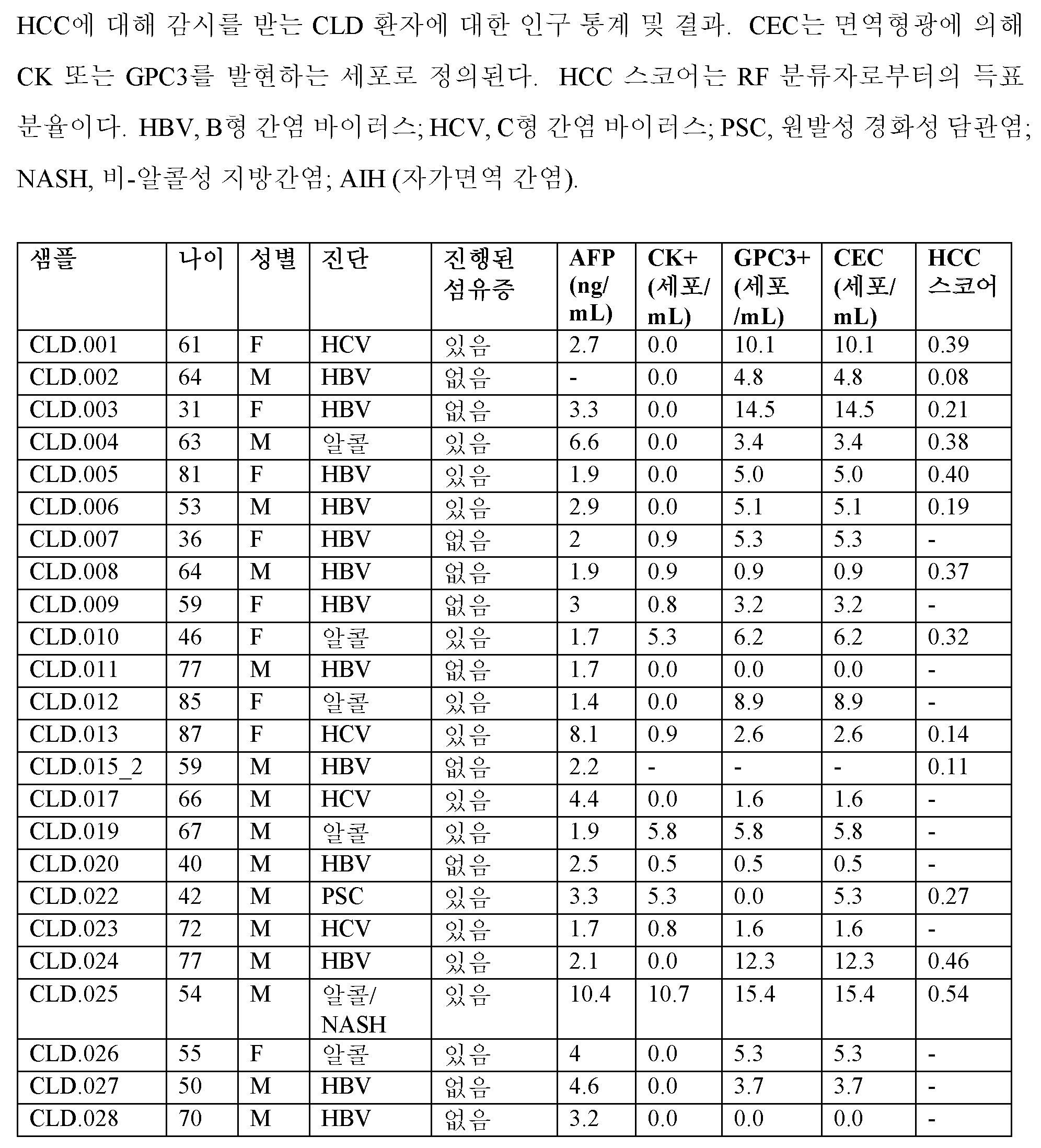


<표 2><Table 2>

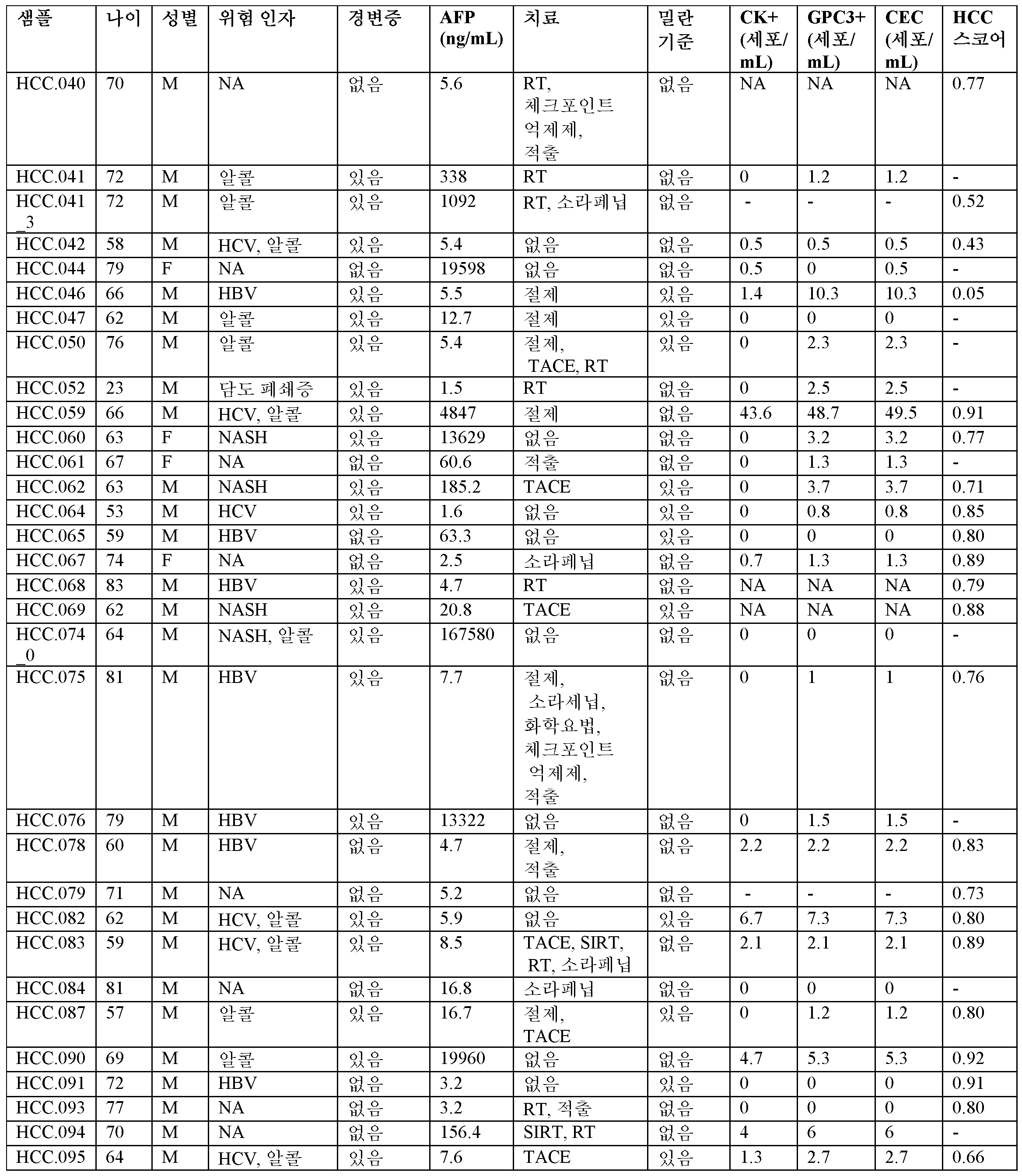

<표 3><Table 3>

<표 4><Table 4>

<표 5><Table 5>

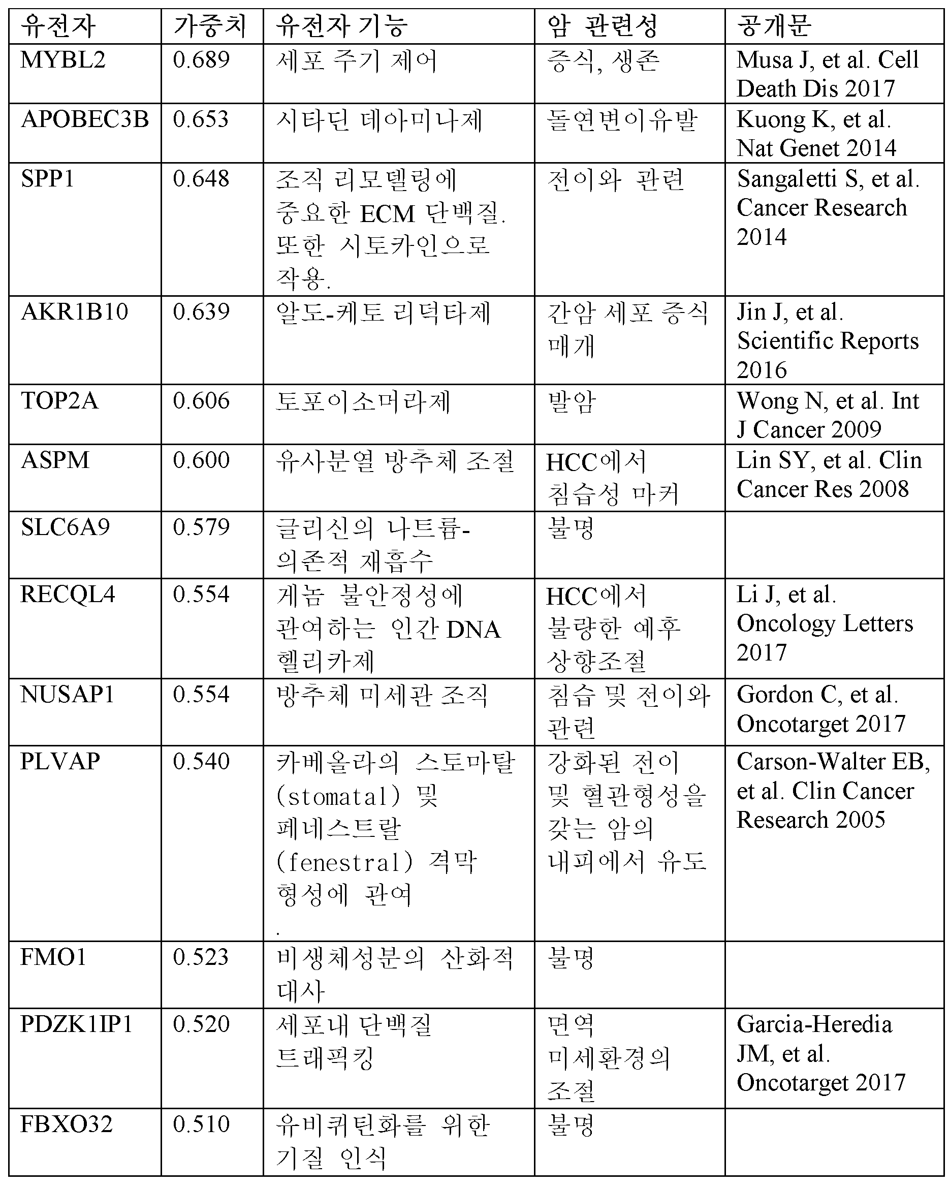
다른 실시양태Other embodiments
본 발명이 상세한 설명과 함께 설명되었지만, 전술된 설명은 첨부된 청구범위에 의해 정의되는 본 발명의 범위를 예시하기 위한 것이지 제한하지 않는 것으로 이해되어야 한다. 다른 측면, 이점 및 변형은 하기의 청구범위 내에 있다.While the present invention has been described in conjunction with the detailed description, it is to be understood that the foregoing description is intended to illustrate and not limit the scope of the invention as defined by the appended claims. Other aspects, advantages and variations are within the scope of the following claims.
Claims (27)
(b) 대상체의 CEC에서의 HCC 분류자 유전자의 발현 수준을 HCC 분류자 유전자의 참조 발현 수준과 비교하여 HCC의 존재를 결정하는 단계
를 포함하는, 만성 간 질환 (CLD)을 갖는 대상체에서 HCC의 존재를 검출하는 방법.(a) measuring the expression level of the HCC classifier gene of any one of claims 1 to 4 in the CEC of the subject; And
(b) determining the presence of HCC by comparing the expression level of the HCC classifier gene in the CEC of the subject with the reference expression level of the HCC classifier gene.
A method of detecting the presence of HCC in a subject having chronic liver disease (CLD) comprising a.
(a) 대상체로부터 혈액을 포함하는 샘플을 수득하고;
(b) 크기-기반 배제를 통해 샘플로부터 적혈구, 혈소판 및 혈장을 제거하고;
(c) 자기영동에 의해 샘플로부터 백혈구 (WBC)를 제거하고;
(d) RNA-시퀀싱, qRT-PCT, RNA 제자리 하이브리드화, 단백질 마이크로어레이, 또는 질량 분광측정 및 단백질 프로파일링을 이용하여 CEC에서 유전자 세트의 발현을 측정함으로써
측정되는 것인 방법.The method according to any one of claims 1 to 8, wherein the expression level of the HCC classifier gene in circulating epithelial cells (CEC) is
(a) obtaining a sample comprising blood from the subject;
(b) removing red blood cells, platelets and plasma from the sample through size-based exclusion;
(c) removing white blood cells (WBC) from the sample by magnetophoresis;
(d) by measuring the expression of a set of genes in CEC using RNA-sequencing, qRT-PCT, RNA in situ hybridization, protein microarray, or mass spectrometry and protein profiling.
The method that is being measured.
(a) 초음파 이미징, 역동적 CT, MRI 이미징, 바늘 생검, 및/또는 생검에 의해 환자에서 HCC의 존재를 확인하거나 확인한 단계; 및
(b) 환자에서 HCC의 존재가 확인되는 경우, HCC 조직의 외과적 제거, HCC 조직의 고주파 절제, HCC 조직의 색전술; HCC 조직의 색전술, 화학요법, 및/또는 냉동요법에 의해 HCC에 대해 대상체를 치료하거나 치료한 단계
를 추가로 포함하는 방법.The method according to any one of claims 5 to 11,
(a) confirming or confirming the presence of HCC in the patient by ultrasound imaging, dynamic CT, MRI imaging, needle biopsy, and/or biopsy; And
(b) if the presence of HCC in the patient is confirmed, surgical removal of HCC tissue, radiofrequency ablation of HCC tissue, embolization of HCC tissue; Treating or treating a subject for HCC by embolization, chemotherapy, and/or cryotherapy of HCC tissue
How to further include.
(b) 하나 이상의 후속 시점에서 제6항 또는 제7항의 방법을 수행하는 단계
를 포함하는, HCC의 발병에 대해 CLD를 갖는 대상체를 모니터링하는 방법.(a) performing the method of claim 6 or 7 at an initial time point, and if the HCC score is lower than the reference score,
(b) performing the method of paragraph 6 or 7 at one or more subsequent points in time.
A method of monitoring a subject having CLD for the onset of HCC, comprising a.
(b) 대상체의 혈액 샘플에서의 CEC의 농도를 참조 값과 비교하는 단계;
(c) 대상체가 혈액 샘플에서 참조 값보다 낮은 CEC의 농도를 갖는 경우, 대상체를 초기 단계 섬유증으로 진단하는 단계; 및
d) 대상체가 혈액 샘플에서 참조 값보다 높은 CEC의 농도를 갖는 경우, 대상체를 후기 단계 섬유증으로 진단하는 단계
를 포함하는, CLD를 갖는 대상체에서 초기 단계 간 섬유증과 후기 단계 간 섬유증의 존재를 구별하는 방법.(a) detecting the concentration of CEC in the blood sample of the subject;
(b) comparing the concentration of CEC in the subject's blood sample with a reference value;
(c) if the subject has a concentration of CEC lower than the reference value in the blood sample, diagnosing the subject with early stage fibrosis; And
d) if the subject has a concentration of CEC higher than the reference value in the blood sample, diagnosing the subject as late stage fibrosis
A method of distinguishing the presence of early stage liver fibrosis and late stage liver fibrosis in a subject with CLD comprising a.
(b) 하나 이상의 후속 시점에서 제16항 내지 제19항 중 어느 한 항의 방법을 수행하는 단계
를 포함하는, 진행된 섬유증의 발병에 대해 CLD를 갖는 대상체를 모니터링하는 방법.(a) performing the method of any one of claims 16-19; And when the concentration of CEC in the subject's blood sample is lower than the reference value,
(b) performing the method of any one of claims 16-19 at one or more subsequent time points.
A method of monitoring a subject with CLD for the onset of advanced fibrosis comprising a.
(b) 초기 시점에서 제6항 또는 제7항의 방법을 수행하고, HCC 스코어의 발현 수준이 참조 스코어보다 낮은 경우, 하나 이상의 후속 시점에서 제6항 또는 제7항의 방법을 수행하는 단계
를 포함하는, 섬유증 또는 HCC의 진행을 방지하기 위해 치료 중인 CLD를 갖는 대상체를 모니터링하는 방법.(a) performing the method of any one of claims 16-19; When the concentration of CEC in the blood sample of the subject is lower than the reference value, performing the method of any one of claims 16 to 19 at one or more subsequent time points; And
(b) performing the method of claim 6 or claim 7 at an initial time point, and if the expression level of the HCC score is lower than the reference score, performing the method of claim 6 or claim 7 at one or more subsequent time points.
A method of monitoring a subject with CLD being treated to prevent the progression of fibrosis or HCC comprising a.
Applications Claiming Priority (3)
| Application Number | Priority Date | Filing Date | Title |
|---|---|---|---|
| US201862729787P | 2018-09-11 | 2018-09-11 | |
| US62/729,787 | 2018-09-11 | ||
| PCT/US2019/050532 WO2020055954A2 (en) | 2018-09-11 | 2019-09-11 | Methods for detecting liver diseases |
Publications (1)
| Publication Number | Publication Date |
|---|---|
| KR20210057765A true KR20210057765A (en) | 2021-05-21 |
Family
ID=69778376
Family Applications (1)
| Application Number | Title | Priority Date | Filing Date |
|---|---|---|---|
| KR1020217010293A KR20210057765A (en) | 2018-09-11 | 2019-09-11 | How to detect liver disease |
Country Status (5)
| Country | Link |
|---|---|
| US (1) | US20220033910A1 (en) |
| JP (2) | JP2022500029A (en) |
| KR (1) | KR20210057765A (en) |
| CN (1) | CN112996928A (en) |
| WO (1) | WO2020055954A2 (en) |
Families Citing this family (8)
| Publication number | Priority date | Publication date | Assignee | Title |
|---|---|---|---|---|
| CN111876483B (en) * | 2020-05-29 | 2022-12-20 | 中山大学肿瘤防治中心(中山大学附属肿瘤医院、中山大学肿瘤研究所) | Use of LYPD1 in diagnosis, treatment, prognosis and prediction of recurrence of hepatocellular carcinoma |
| CN111537719B (en) * | 2020-06-04 | 2022-10-04 | 复旦大学附属中山医院 | Application of serum Sparcl1 protein in non-alcoholic steatohepatitis diagnosis |
| CN113186281B (en) * | 2021-04-02 | 2023-02-28 | 广州医科大学附属第五医院 | INAVA as a marker for hepatocellular carcinoma |
| EP4338159A1 (en) * | 2021-05-11 | 2024-03-20 | Genomic Expression Inc. | Identification and design of cancer therapies based on rna sequencing |
| WO2023008673A1 (en) * | 2021-07-30 | 2023-02-02 | 숙명여자대학교산학협력단 | Machine learning-based complex marker for determining nonalcoholic steatohepatitis, and use thereof |
| CN114150053A (en) * | 2021-11-17 | 2022-03-08 | 重庆美普蓝科技有限公司 | Combination factor and application thereof |
| CN117954097A (en) * | 2023-03-16 | 2024-04-30 | 中国人民解放军空军军医大学 | Lung adenocarcinoma prognosis evaluation system and equipment |
| CN117165678A (en) * | 2023-04-10 | 2023-12-05 | 上海交通大学医学院附属仁济医院 | Liver cancer liver transplantation postoperative recurrence marker and application thereof |
Family Cites Families (7)
| Publication number | Priority date | Publication date | Assignee | Title |
|---|---|---|---|---|
| WO2003086445A1 (en) * | 2002-04-08 | 2003-10-23 | Ciphergen Biosystems, Inc. | Serum biomarkers in hepatocellular carcinoma |
| AU2012275500A1 (en) * | 2011-06-27 | 2014-01-16 | Dana-Farber Cancer Institute, Inc. | Signatures and determinants associated with prostate cancer progression and methods of use thereof |
| WO2013150002A1 (en) * | 2012-04-03 | 2013-10-10 | INSERM (Institut National de la Santé et de la Recherche Médicale) | Methods and kits for determining if a subject is predisposed to fast progression of liver fibrosis |
| CA2928779A1 (en) * | 2013-10-21 | 2015-04-30 | The General Hospital Corporation | Methods relating to circulating tumor cell clusters and the treatment of cancer |
| EP3865144A1 (en) * | 2013-12-20 | 2021-08-18 | The General Hospital Corporation | Methods and assays relating to circulating tumor cells |
| FR3028318A1 (en) * | 2014-11-12 | 2016-05-13 | Phuong Lan Tran | METHOD AND DEVICE FOR SELECTIVE, SPECIFIC AND SIMULTANEOUS SORTING OF TARGET RARE CELLS IN A BIOLOGICAL SAMPLE |
| JP7144934B2 (en) * | 2015-03-25 | 2022-09-30 | ザ ジェネラル ホスピタル コーポレイション | Digital analysis of circulating tumor cells in blood samples |
-
2019
- 2019-09-11 JP JP2021513265A patent/JP2022500029A/en active Pending
- 2019-09-11 KR KR1020217010293A patent/KR20210057765A/en unknown
- 2019-09-11 CN CN201980073917.5A patent/CN112996928A/en active Pending
- 2019-09-11 WO PCT/US2019/050532 patent/WO2020055954A2/en active Application Filing
- 2019-09-11 US US17/274,955 patent/US20220033910A1/en active Pending
-
2024
- 2024-02-01 JP JP2024013945A patent/JP2024059642A/en active Pending
Also Published As
| Publication number | Publication date |
|---|---|
| WO2020055954A2 (en) | 2020-03-19 |
| JP2024059642A (en) | 2024-05-01 |
| JP2022500029A (en) | 2022-01-04 |
| WO2020055954A3 (en) | 2020-07-23 |
| CN112996928A (en) | 2021-06-18 |
| US20220033910A1 (en) | 2022-02-03 |
Similar Documents
| Publication | Publication Date | Title |
|---|---|---|
| KR20210057765A (en) | How to detect liver disease | |
| Kalinich et al. | An RNA-based signature enables high specificity detection of circulating tumor cells in hepatocellular carcinoma | |
| Qi et al. | Circulating tumor cells undergoing EMT provide a metric for diagnosis and prognosis of patients with hepatocellular carcinoma | |
| von Felden et al. | Liquid biopsy in the clinical management of hepatocellular carcinoma | |
| Chew et al. | Chemokine-driven lymphocyte infiltration: an early intratumoural event determining long-term survival in resectable hepatocellular carcinoma | |
| Li et al. | Intratumoral neutrophils: a poor prognostic factor for hepatocellular carcinoma following resection | |
| Chan et al. | Expression of stemness markers (CD 133 and Ep CAM) in prognostication of hepatocellular carcinoma | |
| Mas et al. | Genes involved in viral carcinogenesis and tumor initiation in hepatitis C virus-induced hepatocellular carcinoma | |
| Lang et al. | Expression profiling of circulating tumor cells in metastatic breast cancer | |
| Bozec et al. | Significance of circulating tumor cell detection using the CellSearch system in patients with locally advanced head and neck squamous cell carcinoma | |
| Bhan et al. | Detection and analysis of circulating epithelial cells in liquid biopsies from patients with liver disease | |
| Liu et al. | A rapid nested polymerase chain reaction method to detect circulating cancer cells in breast cancer patients using multiple marker genes | |
| Zheng et al. | Plasma minichromosome maintenance complex component 6 is a novel biomarker for hepatocellular carcinoma patients | |
| Wang et al. | Single nucleotide variant profiles of viable single circulating tumour cells reveal CTC behaviours in breast cancer | |
| EP3376227B1 (en) | Methods for diagnosing and treating cancer | |
| Hesketh et al. | Hepatocellular carcinoma: can circulating tumor cells and radiogenomics deliver personalized care? | |
| Wen et al. | Prognostic model of colorectal cancer constructed by eight immune-related genes | |
| Moldogazieva et al. | Genomic landscape of liquid biopsy for hepatocellular carcinoma personalized medicine | |
| Soave et al. | Do circulating tumor cells have a role in deciding on adjuvant chemotherapy after radical cystectomy? | |
| Ding et al. | Prognostic value of MAC30 expression in human pure squamous cell carcinomas of the lung | |
| Yao et al. | Detecting AFP mRNA in peripheral blood of the patients with hepatocellular carcinoma, liver cirrhosis and hepatitis | |
| Lee et al. | Circulating Tumor Cell–Based Messenger RNA Scoring System for Prognostication of Hepatocellular Carcinoma: Translating Tissue‐Based Messenger RNA Profiling Into a Noninvasive Setting | |
| Guo et al. | Detecting carcinoma cells in peripheral blood of patients with hepatocellular carcinoma by immunomagnetic beads and rt-PCR | |
| Teng et al. | Circulating tumor cells: A step toward precision medicine in hepatocellular carcinoma | |
| Halawa et al. | The role of liquid biopsy in the diagnosis and prognosis of WHO grade 4 astrocytoma |