KR20200078365A - Method and apparatus for managing acute ischemic events - Google Patents
Method and apparatus for managing acute ischemic events Download PDFInfo
- Publication number
- KR20200078365A KR20200078365A KR1020190169427A KR20190169427A KR20200078365A KR 20200078365 A KR20200078365 A KR 20200078365A KR 1020190169427 A KR1020190169427 A KR 1020190169427A KR 20190169427 A KR20190169427 A KR 20190169427A KR 20200078365 A KR20200078365 A KR 20200078365A
- Authority
- KR
- South Korea
- Prior art keywords
- thrombus
- reperfusion
- rich
- levels
- fibrin
- Prior art date
Links
- 238000000034 method Methods 0.000 title claims abstract description 111
- 230000000302 ischemic effect Effects 0.000 title claims abstract description 21
- 230000001154 acute effect Effects 0.000 title claims abstract description 13
- 208000007536 Thrombosis Diseases 0.000 claims abstract description 520
- 230000010410 reperfusion Effects 0.000 claims abstract description 81
- 230000010412 perfusion Effects 0.000 claims abstract description 43
- 238000011337 individualized treatment Methods 0.000 claims abstract description 39
- 210000003743 erythrocyte Anatomy 0.000 claims description 119
- 108010073385 Fibrin Proteins 0.000 claims description 111
- 102000009123 Fibrin Human genes 0.000 claims description 111
- BWGVNKXGVNDBDI-UHFFFAOYSA-N Fibrin monomer Chemical compound CNC(=O)CNC(=O)CN BWGVNKXGVNDBDI-UHFFFAOYSA-N 0.000 claims description 111
- 229950003499 fibrin Drugs 0.000 claims description 111
- 239000000203 mixture Substances 0.000 claims description 95
- 238000004458 analytical method Methods 0.000 claims description 63
- 210000004204 blood vessel Anatomy 0.000 claims description 61
- 238000011084 recovery Methods 0.000 claims description 51
- 210000000265 leukocyte Anatomy 0.000 claims description 49
- 239000012634 fragment Substances 0.000 claims description 40
- 210000001772 blood platelet Anatomy 0.000 claims description 39
- 238000002591 computed tomography Methods 0.000 claims description 36
- 238000011282 treatment Methods 0.000 claims description 35
- 238000001228 spectrum Methods 0.000 claims description 33
- 210000005166 vasculature Anatomy 0.000 claims description 27
- 238000002595 magnetic resonance imaging Methods 0.000 claims description 25
- 239000000126 substance Substances 0.000 claims description 18
- 230000000704 physical effect Effects 0.000 claims description 16
- 230000002792 vascular Effects 0.000 claims description 16
- 238000001727 in vivo Methods 0.000 claims description 12
- 238000004891 communication Methods 0.000 claims description 11
- 238000004497 NIR spectroscopy Methods 0.000 claims description 10
- 238000002583 angiography Methods 0.000 claims description 8
- 210000002966 serum Anatomy 0.000 claims description 8
- 238000001069 Raman spectroscopy Methods 0.000 claims description 5
- 238000009534 blood test Methods 0.000 claims description 4
- 230000004044 response Effects 0.000 claims description 4
- 238000012360 testing method Methods 0.000 claims description 4
- 230000015572 biosynthetic process Effects 0.000 claims description 3
- 238000002592 echocardiography Methods 0.000 claims description 3
- 238000002604 ultrasonography Methods 0.000 claims description 3
- 238000012544 monitoring process Methods 0.000 claims description 2
- 239000003795 chemical substances by application Substances 0.000 claims 1
- 230000009089 cytolysis Effects 0.000 claims 1
- 238000003860 storage Methods 0.000 description 37
- 208000006011 Stroke Diseases 0.000 description 35
- 208000032382 Ischaemic stroke Diseases 0.000 description 20
- 210000004556 brain Anatomy 0.000 description 20
- 239000000463 material Substances 0.000 description 20
- 238000012545 processing Methods 0.000 description 18
- 208000005189 Embolism Diseases 0.000 description 15
- 210000004004 carotid artery internal Anatomy 0.000 description 15
- 238000010586 diagram Methods 0.000 description 15
- 210000001367 artery Anatomy 0.000 description 14
- 238000000338 in vitro Methods 0.000 description 11
- 230000006870 function Effects 0.000 description 10
- 238000003384 imaging method Methods 0.000 description 10
- 210000003657 middle cerebral artery Anatomy 0.000 description 10
- 238000013459 approach Methods 0.000 description 9
- 230000000875 corresponding effect Effects 0.000 description 9
- 238000004820 blood count Methods 0.000 description 8
- 230000036571 hydration Effects 0.000 description 8
- 238000006703 hydration reaction Methods 0.000 description 8
- 230000003287 optical effect Effects 0.000 description 8
- 230000000747 cardiac effect Effects 0.000 description 7
- 238000005516 engineering process Methods 0.000 description 7
- 208000024891 symptom Diseases 0.000 description 7
- 238000013151 thrombectomy Methods 0.000 description 7
- 230000002537 thrombolytic effect Effects 0.000 description 7
- 238000009826 distribution Methods 0.000 description 6
- 238000005259 measurement Methods 0.000 description 6
- 230000004048 modification Effects 0.000 description 6
- 238000012986 modification Methods 0.000 description 6
- 210000002376 aorta thoracic Anatomy 0.000 description 5
- 230000017531 blood circulation Effects 0.000 description 5
- 238000007917 intracranial administration Methods 0.000 description 5
- 208000010378 Pulmonary Embolism Diseases 0.000 description 4
- 206010053648 Vascular occlusion Diseases 0.000 description 4
- 230000009471 action Effects 0.000 description 4
- 230000000740 bleeding effect Effects 0.000 description 4
- 230000002490 cerebral effect Effects 0.000 description 4
- 238000013480 data collection Methods 0.000 description 4
- 230000000694 effects Effects 0.000 description 4
- 230000036541 health Effects 0.000 description 4
- 239000004065 semiconductor Substances 0.000 description 4
- 230000001732 thrombotic effect Effects 0.000 description 4
- 230000009466 transformation Effects 0.000 description 4
- 102000003978 Tissue Plasminogen Activator Human genes 0.000 description 3
- 108090000373 Tissue Plasminogen Activator Proteins 0.000 description 3
- 239000003146 anticoagulant agent Substances 0.000 description 3
- 210000004369 blood Anatomy 0.000 description 3
- 239000008280 blood Substances 0.000 description 3
- 230000009426 cardioembolic effect Effects 0.000 description 3
- 210000001715 carotid artery Anatomy 0.000 description 3
- 210000001168 carotid artery common Anatomy 0.000 description 3
- 230000015556 catabolic process Effects 0.000 description 3
- 210000004027 cell Anatomy 0.000 description 3
- 210000001627 cerebral artery Anatomy 0.000 description 3
- 150000001875 compounds Chemical class 0.000 description 3
- 230000006835 compression Effects 0.000 description 3
- 238000007906 compression Methods 0.000 description 3
- 238000013500 data storage Methods 0.000 description 3
- 238000009792 diffusion process Methods 0.000 description 3
- 208000037265 diseases, disorders, signs and symptoms Diseases 0.000 description 3
- 230000007717 exclusion Effects 0.000 description 3
- 239000012530 fluid Substances 0.000 description 3
- 238000001990 intravenous administration Methods 0.000 description 3
- 230000000670 limiting effect Effects 0.000 description 3
- 239000002245 particle Substances 0.000 description 3
- 210000001147 pulmonary artery Anatomy 0.000 description 3
- 230000000250 revascularization Effects 0.000 description 3
- 210000001519 tissue Anatomy 0.000 description 3
- 229960000187 tissue plasminogen activator Drugs 0.000 description 3
- 238000000844 transformation Methods 0.000 description 3
- 208000021331 vascular occlusion disease Diseases 0.000 description 3
- 229910052688 Gadolinium Inorganic materials 0.000 description 2
- 206010020751 Hypersensitivity Diseases 0.000 description 2
- 206010028980 Neoplasm Diseases 0.000 description 2
- PXHVJJICTQNCMI-UHFFFAOYSA-N Nickel Chemical compound [Ni] PXHVJJICTQNCMI-UHFFFAOYSA-N 0.000 description 2
- 230000007815 allergy Effects 0.000 description 2
- 210000003484 anatomy Anatomy 0.000 description 2
- 229940127219 anticoagulant drug Drugs 0.000 description 2
- 210000000709 aorta Anatomy 0.000 description 2
- 238000003556 assay Methods 0.000 description 2
- 230000036772 blood pressure Effects 0.000 description 2
- 230000001413 cellular effect Effects 0.000 description 2
- 206010008118 cerebral infarction Diseases 0.000 description 2
- 230000008859 change Effects 0.000 description 2
- 230000004087 circulation Effects 0.000 description 2
- 230000015271 coagulation Effects 0.000 description 2
- 238000005345 coagulation Methods 0.000 description 2
- ZPUCINDJVBIVPJ-LJISPDSOSA-N cocaine Chemical compound O([C@H]1C[C@@H]2CC[C@@H](N2C)[C@H]1C(=O)OC)C(=O)C1=CC=CC=C1 ZPUCINDJVBIVPJ-LJISPDSOSA-N 0.000 description 2
- 238000010276 construction Methods 0.000 description 2
- DDRJAANPRJIHGJ-UHFFFAOYSA-N creatinine Chemical compound CN1CC(=O)NC1=N DDRJAANPRJIHGJ-UHFFFAOYSA-N 0.000 description 2
- 238000002059 diagnostic imaging Methods 0.000 description 2
- 230000004069 differentiation Effects 0.000 description 2
- 238000002597 diffusion-weighted imaging Methods 0.000 description 2
- 208000035475 disorder Diseases 0.000 description 2
- 230000010102 embolization Effects 0.000 description 2
- 238000011156 evaluation Methods 0.000 description 2
- 210000001105 femoral artery Anatomy 0.000 description 2
- 239000000835 fiber Substances 0.000 description 2
- 238000002075 inversion recovery Methods 0.000 description 2
- 238000011835 investigation Methods 0.000 description 2
- 210000004072 lung Anatomy 0.000 description 2
- 238000007726 management method Methods 0.000 description 2
- 239000003550 marker Substances 0.000 description 2
- 238000002095 near-infrared Raman spectroscopy Methods 0.000 description 2
- 238000001543 one-way ANOVA Methods 0.000 description 2
- 239000013307 optical fiber Substances 0.000 description 2
- 230000008520 organization Effects 0.000 description 2
- 230000008506 pathogenesis Effects 0.000 description 2
- BASFCYQUMIYNBI-UHFFFAOYSA-N platinum Chemical compound [Pt] BASFCYQUMIYNBI-UHFFFAOYSA-N 0.000 description 2
- 229920000642 polymer Polymers 0.000 description 2
- 238000004445 quantitative analysis Methods 0.000 description 2
- 239000007787 solid Substances 0.000 description 2
- 230000003595 spectral effect Effects 0.000 description 2
- 238000010186 staining Methods 0.000 description 2
- 238000003325 tomography Methods 0.000 description 2
- 210000002385 vertebral artery Anatomy 0.000 description 2
- 238000012800 visualization Methods 0.000 description 2
- 108010047303 von Willebrand Factor Proteins 0.000 description 2
- 102100036537 von Willebrand factor Human genes 0.000 description 2
- 229960001134 von willebrand factor Drugs 0.000 description 2
- XLYOFNOQVPJJNP-UHFFFAOYSA-N water Substances O XLYOFNOQVPJJNP-UHFFFAOYSA-N 0.000 description 2
- PGOHTUIFYSHAQG-LJSDBVFPSA-N (2S)-6-amino-2-[[(2S)-5-amino-2-[[(2S)-2-[[(2S)-2-[[(2S)-2-[[(2S)-4-amino-2-[[(2S)-2-[[(2S)-2-[[(2S)-2-[[(2S)-2-[[(2S)-5-amino-2-[[(2S)-5-amino-2-[[(2S)-2-[[(2S)-2-[[(2S)-2-[[(2S,3R)-2-[[(2S)-5-amino-2-[[(2S)-2-[[(2S)-2-[[(2S,3R)-2-[[(2S)-2-[[(2S)-2-[[(2S)-2-[[(2S)-2-[[(2S)-5-amino-2-[[(2S)-1-[(2S,3R)-2-[[(2S)-2-[[(2S)-2-[[(2R)-2-[[(2S)-2-[[(2S)-2-[[2-[[(2S)-2-[[(2S)-2-[[(2S)-2-[[(2S)-1-[(2S)-2-[[(2S)-2-[[(2S)-2-[[(2S)-2-amino-4-methylsulfanylbutanoyl]amino]-3-(1H-indol-3-yl)propanoyl]amino]-5-carbamimidamidopentanoyl]amino]propanoyl]pyrrolidine-2-carbonyl]amino]-3-methylbutanoyl]amino]-4-methylpentanoyl]amino]-4-methylpentanoyl]amino]acetyl]amino]-3-hydroxypropanoyl]amino]-4-methylpentanoyl]amino]-3-sulfanylpropanoyl]amino]-4-methylsulfanylbutanoyl]amino]-5-carbamimidamidopentanoyl]amino]-3-hydroxybutanoyl]pyrrolidine-2-carbonyl]amino]-5-oxopentanoyl]amino]-3-hydroxypropanoyl]amino]-3-hydroxypropanoyl]amino]-3-(1H-imidazol-5-yl)propanoyl]amino]-4-methylpentanoyl]amino]-3-hydroxybutanoyl]amino]-3-(1H-indol-3-yl)propanoyl]amino]-5-carbamimidamidopentanoyl]amino]-5-oxopentanoyl]amino]-3-hydroxybutanoyl]amino]-3-hydroxypropanoyl]amino]-3-carboxypropanoyl]amino]-3-hydroxypropanoyl]amino]-5-oxopentanoyl]amino]-5-oxopentanoyl]amino]-3-phenylpropanoyl]amino]-5-carbamimidamidopentanoyl]amino]-3-methylbutanoyl]amino]-4-methylpentanoyl]amino]-4-oxobutanoyl]amino]-5-carbamimidamidopentanoyl]amino]-3-(1H-indol-3-yl)propanoyl]amino]-4-carboxybutanoyl]amino]-5-oxopentanoyl]amino]hexanoic acid Chemical compound CSCC[C@H](N)C(=O)N[C@@H](Cc1c[nH]c2ccccc12)C(=O)N[C@@H](CCCNC(N)=N)C(=O)N[C@@H](C)C(=O)N1CCC[C@H]1C(=O)N[C@@H](C(C)C)C(=O)N[C@@H](CC(C)C)C(=O)N[C@@H](CC(C)C)C(=O)NCC(=O)N[C@@H](CO)C(=O)N[C@@H](CC(C)C)C(=O)N[C@@H](CS)C(=O)N[C@@H](CCSC)C(=O)N[C@@H](CCCNC(N)=N)C(=O)N[C@@H]([C@@H](C)O)C(=O)N1CCC[C@H]1C(=O)N[C@@H](CCC(N)=O)C(=O)N[C@@H](CO)C(=O)N[C@@H](CO)C(=O)N[C@@H](Cc1cnc[nH]1)C(=O)N[C@@H](CC(C)C)C(=O)N[C@@H]([C@@H](C)O)C(=O)N[C@@H](Cc1c[nH]c2ccccc12)C(=O)N[C@@H](CCCNC(N)=N)C(=O)N[C@@H](CCC(N)=O)C(=O)N[C@@H]([C@@H](C)O)C(=O)N[C@@H](CO)C(=O)N[C@@H](CC(O)=O)C(=O)N[C@@H](CO)C(=O)N[C@@H](CCC(N)=O)C(=O)N[C@@H](CCC(N)=O)C(=O)N[C@@H](Cc1ccccc1)C(=O)N[C@@H](CCCNC(N)=N)C(=O)N[C@@H](C(C)C)C(=O)N[C@@H](CC(C)C)C(=O)N[C@@H](CC(N)=O)C(=O)N[C@@H](CCCNC(N)=N)C(=O)N[C@@H](Cc1c[nH]c2ccccc12)C(=O)N[C@@H](CCC(O)=O)C(=O)N[C@@H](CCC(N)=O)C(=O)N[C@@H](CCCCN)C(O)=O PGOHTUIFYSHAQG-LJSDBVFPSA-N 0.000 description 1
- 201000001320 Atherosclerosis Diseases 0.000 description 1
- 241000283690 Bos taurus Species 0.000 description 1
- 201000006474 Brain Ischemia Diseases 0.000 description 1
- 206010008120 Cerebral ischaemia Diseases 0.000 description 1
- 235000008733 Citrus aurantifolia Nutrition 0.000 description 1
- 102000008186 Collagen Human genes 0.000 description 1
- 108010035532 Collagen Proteins 0.000 description 1
- 206010010071 Coma Diseases 0.000 description 1
- 206010010904 Convulsion Diseases 0.000 description 1
- 208000002251 Dissecting Aneurysm Diseases 0.000 description 1
- 206010014498 Embolic stroke Diseases 0.000 description 1
- 206010014666 Endocarditis bacterial Diseases 0.000 description 1
- 238000012276 Endovascular treatment Methods 0.000 description 1
- WQZGKKKJIJFFOK-GASJEMHNSA-N Glucose Natural products OC[C@H]1OC(O)[C@H](O)[C@@H](O)[C@@H]1O WQZGKKKJIJFFOK-GASJEMHNSA-N 0.000 description 1
- 102000003886 Glycoproteins Human genes 0.000 description 1
- 108090000288 Glycoproteins Proteins 0.000 description 1
- 208000016988 Hemorrhagic Stroke Diseases 0.000 description 1
- 101000987003 Homo sapiens Tumor protein 63 Proteins 0.000 description 1
- 206010020772 Hypertension Diseases 0.000 description 1
- 206010021639 Incontinence Diseases 0.000 description 1
- -1 MP35N Inorganic materials 0.000 description 1
- 238000012307 MRI technique Methods 0.000 description 1
- 206010027476 Metastases Diseases 0.000 description 1
- 241001465754 Metazoa Species 0.000 description 1
- 208000012902 Nervous system disease Diseases 0.000 description 1
- 208000031481 Pathologic Constriction Diseases 0.000 description 1
- 208000001647 Renal Insufficiency Diseases 0.000 description 1
- 229940122388 Thrombin inhibitor Drugs 0.000 description 1
- 208000001435 Thromboembolism Diseases 0.000 description 1
- 108010000499 Thromboplastin Proteins 0.000 description 1
- 102000002262 Thromboplastin Human genes 0.000 description 1
- 241001547816 Thrombus Species 0.000 description 1
- 235000011941 Tilia x europaea Nutrition 0.000 description 1
- RLFHKJKDXNVQAY-UHFFFAOYSA-N Trichione Natural products OC(=O)CC(=O)OCCC=CC1=C(O)C(=O)c2cccc(O)c2C1=O RLFHKJKDXNVQAY-UHFFFAOYSA-N 0.000 description 1
- 208000032594 Vascular Remodeling Diseases 0.000 description 1
- 239000000853 adhesive Substances 0.000 description 1
- 230000001070 adhesive effect Effects 0.000 description 1
- 229910045601 alloy Inorganic materials 0.000 description 1
- 239000000956 alloy Substances 0.000 description 1
- 230000033115 angiogenesis Effects 0.000 description 1
- 238000010171 animal model Methods 0.000 description 1
- 206010002895 aortic dissection Diseases 0.000 description 1
- 238000000149 argon plasma sintering Methods 0.000 description 1
- 230000001746 atrial effect Effects 0.000 description 1
- 230000002238 attenuated effect Effects 0.000 description 1
- 230000003190 augmentative effect Effects 0.000 description 1
- 208000009361 bacterial endocarditis Diseases 0.000 description 1
- 230000006399 behavior Effects 0.000 description 1
- 230000008901 benefit Effects 0.000 description 1
- 230000002146 bilateral effect Effects 0.000 description 1
- 210000002302 brachial artery Anatomy 0.000 description 1
- 230000006931 brain damage Effects 0.000 description 1
- 231100000874 brain damage Toxicity 0.000 description 1
- 208000029028 brain injury Diseases 0.000 description 1
- 210000005013 brain tissue Anatomy 0.000 description 1
- 201000011510 cancer Diseases 0.000 description 1
- 239000003990 capacitor Substances 0.000 description 1
- 210000000269 carotid artery external Anatomy 0.000 description 1
- 208000026106 cerebrovascular disease Diseases 0.000 description 1
- 229960003920 cocaine Drugs 0.000 description 1
- 229920001436 collagen Polymers 0.000 description 1
- 238000012790 confirmation Methods 0.000 description 1
- 239000000470 constituent Substances 0.000 description 1
- 230000008602 contraction Effects 0.000 description 1
- 239000002872 contrast media Substances 0.000 description 1
- 210000004351 coronary vessel Anatomy 0.000 description 1
- 230000002596 correlated effect Effects 0.000 description 1
- 230000008878 coupling Effects 0.000 description 1
- 238000010168 coupling process Methods 0.000 description 1
- 238000005859 coupling reaction Methods 0.000 description 1
- 229940109239 creatinine Drugs 0.000 description 1
- 238000005520 cutting process Methods 0.000 description 1
- 238000006731 degradation reaction Methods 0.000 description 1
- 230000018044 dehydration Effects 0.000 description 1
- 238000006297 dehydration reaction Methods 0.000 description 1
- 238000013461 design Methods 0.000 description 1
- 230000001066 destructive effect Effects 0.000 description 1
- JXSJBGJIGXNWCI-UHFFFAOYSA-N diethyl 2-[(dimethoxyphosphorothioyl)thio]succinate Chemical compound CCOC(=O)CC(SP(=S)(OC)OC)C(=O)OCC JXSJBGJIGXNWCI-UHFFFAOYSA-N 0.000 description 1
- 201000010099 disease Diseases 0.000 description 1
- 238000006073 displacement reaction Methods 0.000 description 1
- 230000009977 dual effect Effects 0.000 description 1
- 206010014599 encephalitis Diseases 0.000 description 1
- 238000013467 fragmentation Methods 0.000 description 1
- 238000006062 fragmentation reaction Methods 0.000 description 1
- 239000002783 friction material Substances 0.000 description 1
- 230000024924 glomerular filtration Effects 0.000 description 1
- 239000008103 glucose Substances 0.000 description 1
- PCHJSUWPFVWCPO-UHFFFAOYSA-N gold Chemical compound [Au] PCHJSUWPFVWCPO-UHFFFAOYSA-N 0.000 description 1
- 229910052737 gold Inorganic materials 0.000 description 1
- 239000010931 gold Substances 0.000 description 1
- 230000000004 hemodynamic effect Effects 0.000 description 1
- 230000023597 hemostasis Effects 0.000 description 1
- 230000003118 histopathologic effect Effects 0.000 description 1
- 238000007489 histopathology method Methods 0.000 description 1
- 102000051826 human TP63 Human genes 0.000 description 1
- 238000005462 in vivo assay Methods 0.000 description 1
- 208000015181 infectious disease Diseases 0.000 description 1
- 201000007119 infective endocarditis Diseases 0.000 description 1
- 238000001802 infusion Methods 0.000 description 1
- 238000003780 insertion Methods 0.000 description 1
- 230000037431 insertion Effects 0.000 description 1
- 208000020658 intracerebral hemorrhage Diseases 0.000 description 1
- 238000002608 intravascular ultrasound Methods 0.000 description 1
- 201000006370 kidney failure Diseases 0.000 description 1
- 230000031700 light absorption Effects 0.000 description 1
- 239000004571 lime Substances 0.000 description 1
- 150000002632 lipids Chemical class 0.000 description 1
- 239000006166 lysate Substances 0.000 description 1
- 239000000696 magnetic material Substances 0.000 description 1
- 230000005415 magnetization Effects 0.000 description 1
- 230000007246 mechanism Effects 0.000 description 1
- 206010027191 meningioma Diseases 0.000 description 1
- 210000001259 mesencephalon Anatomy 0.000 description 1
- 229910052751 metal Inorganic materials 0.000 description 1
- 239000002184 metal Substances 0.000 description 1
- 230000000926 neurological effect Effects 0.000 description 1
- 238000010984 neurological examination Methods 0.000 description 1
- 229910052759 nickel Inorganic materials 0.000 description 1
- HLXZNVUGXRDIFK-UHFFFAOYSA-N nickel titanium Chemical compound [Ti].[Ti].[Ti].[Ti].[Ti].[Ti].[Ti].[Ti].[Ti].[Ti].[Ti].[Ni].[Ni].[Ni].[Ni].[Ni].[Ni].[Ni].[Ni].[Ni].[Ni].[Ni].[Ni].[Ni].[Ni] HLXZNVUGXRDIFK-UHFFFAOYSA-N 0.000 description 1
- 229910001000 nickel titanium Inorganic materials 0.000 description 1
- 230000036961 partial effect Effects 0.000 description 1
- 230000035699 permeability Effects 0.000 description 1
- 239000004033 plastic Substances 0.000 description 1
- 229920003023 plastic Polymers 0.000 description 1
- 230000010118 platelet activation Effects 0.000 description 1
- 229910052697 platinum Inorganic materials 0.000 description 1
- 239000011148 porous material Substances 0.000 description 1
- 238000002600 positron emission tomography Methods 0.000 description 1
- 238000002360 preparation method Methods 0.000 description 1
- 230000008569 process Effects 0.000 description 1
- 210000002321 radial artery Anatomy 0.000 description 1
- 230000002829 reductive effect Effects 0.000 description 1
- 238000009877 rendering Methods 0.000 description 1
- 210000005245 right atrium Anatomy 0.000 description 1
- 210000005241 right ventricle Anatomy 0.000 description 1
- 238000005070 sampling Methods 0.000 description 1
- 238000002603 single-photon emission computed tomography Methods 0.000 description 1
- 210000004872 soft tissue Anatomy 0.000 description 1
- 239000002904 solvent Substances 0.000 description 1
- 238000004611 spectroscopical analysis Methods 0.000 description 1
- 239000010935 stainless steel Substances 0.000 description 1
- 229910001220 stainless steel Inorganic materials 0.000 description 1
- 230000003068 static effect Effects 0.000 description 1
- 230000036262 stenosis Effects 0.000 description 1
- 208000037804 stenosis Diseases 0.000 description 1
- 210000003270 subclavian artery Anatomy 0.000 description 1
- 238000006467 substitution reaction Methods 0.000 description 1
- 238000001356 surgical procedure Methods 0.000 description 1
- 230000009885 systemic effect Effects 0.000 description 1
- 230000001225 therapeutic effect Effects 0.000 description 1
- 238000002560 therapeutic procedure Methods 0.000 description 1
- 239000003868 thrombin inhibitor Substances 0.000 description 1
- 230000001131 transforming effect Effects 0.000 description 1
- 230000001052 transient effect Effects 0.000 description 1
- 230000007704 transition Effects 0.000 description 1
- 230000002861 ventricular Effects 0.000 description 1
- 230000000007 visual effect Effects 0.000 description 1
- 229960005080 warfarin Drugs 0.000 description 1
- PJVWKTKQMONHTI-UHFFFAOYSA-N warfarin Chemical compound OC=1C2=CC=CC=C2OC(=O)C=1C(CC(=O)C)C1=CC=CC=C1 PJVWKTKQMONHTI-UHFFFAOYSA-N 0.000 description 1
Images


Classifications
-
- A—HUMAN NECESSITIES
- A61—MEDICAL OR VETERINARY SCIENCE; HYGIENE
- A61B—DIAGNOSIS; SURGERY; IDENTIFICATION
- A61B5/00—Measuring for diagnostic purposes; Identification of persons
- A61B5/05—Detecting, measuring or recording for diagnosis by means of electric currents or magnetic fields; Measuring using microwaves or radio waves
- A61B5/055—Detecting, measuring or recording for diagnosis by means of electric currents or magnetic fields; Measuring using microwaves or radio waves involving electronic [EMR] or nuclear [NMR] magnetic resonance, e.g. magnetic resonance imaging
-
- G—PHYSICS
- G16—INFORMATION AND COMMUNICATION TECHNOLOGY [ICT] SPECIALLY ADAPTED FOR SPECIFIC APPLICATION FIELDS
- G16H—HEALTHCARE INFORMATICS, i.e. INFORMATION AND COMMUNICATION TECHNOLOGY [ICT] SPECIALLY ADAPTED FOR THE HANDLING OR PROCESSING OF MEDICAL OR HEALTHCARE DATA
- G16H50/00—ICT specially adapted for medical diagnosis, medical simulation or medical data mining; ICT specially adapted for detecting, monitoring or modelling epidemics or pandemics
- G16H50/20—ICT specially adapted for medical diagnosis, medical simulation or medical data mining; ICT specially adapted for detecting, monitoring or modelling epidemics or pandemics for computer-aided diagnosis, e.g. based on medical expert systems
-
- A—HUMAN NECESSITIES
- A61—MEDICAL OR VETERINARY SCIENCE; HYGIENE
- A61B—DIAGNOSIS; SURGERY; IDENTIFICATION
- A61B17/00—Surgical instruments, devices or methods, e.g. tourniquets
- A61B17/22—Implements for squeezing-off ulcers or the like on the inside of inner organs of the body; Implements for scraping-out cavities of body organs, e.g. bones; Calculus removers; Calculus smashing apparatus; Apparatus for removing obstructions in blood vessels, not otherwise provided for
- A61B17/221—Gripping devices in the form of loops or baskets for gripping calculi or similar types of obstructions
-
- A—HUMAN NECESSITIES
- A61—MEDICAL OR VETERINARY SCIENCE; HYGIENE
- A61B—DIAGNOSIS; SURGERY; IDENTIFICATION
- A61B34/00—Computer-aided surgery; Manipulators or robots specially adapted for use in surgery
- A61B34/10—Computer-aided planning, simulation or modelling of surgical operations
-
- A—HUMAN NECESSITIES
- A61—MEDICAL OR VETERINARY SCIENCE; HYGIENE
- A61B—DIAGNOSIS; SURGERY; IDENTIFICATION
- A61B5/00—Measuring for diagnostic purposes; Identification of persons
- A61B5/0059—Measuring for diagnostic purposes; Identification of persons using light, e.g. diagnosis by transillumination, diascopy, fluorescence
- A61B5/0075—Measuring for diagnostic purposes; Identification of persons using light, e.g. diagnosis by transillumination, diascopy, fluorescence by spectroscopy, i.e. measuring spectra, e.g. Raman spectroscopy, infrared absorption spectroscopy
-
- A—HUMAN NECESSITIES
- A61—MEDICAL OR VETERINARY SCIENCE; HYGIENE
- A61B—DIAGNOSIS; SURGERY; IDENTIFICATION
- A61B5/00—Measuring for diagnostic purposes; Identification of persons
- A61B5/0059—Measuring for diagnostic purposes; Identification of persons using light, e.g. diagnosis by transillumination, diascopy, fluorescence
- A61B5/0082—Measuring for diagnostic purposes; Identification of persons using light, e.g. diagnosis by transillumination, diascopy, fluorescence adapted for particular medical purposes
- A61B5/0084—Measuring for diagnostic purposes; Identification of persons using light, e.g. diagnosis by transillumination, diascopy, fluorescence adapted for particular medical purposes for introduction into the body, e.g. by catheters
- A61B5/0086—Measuring for diagnostic purposes; Identification of persons using light, e.g. diagnosis by transillumination, diascopy, fluorescence adapted for particular medical purposes for introduction into the body, e.g. by catheters using infrared radiation
-
- A—HUMAN NECESSITIES
- A61—MEDICAL OR VETERINARY SCIENCE; HYGIENE
- A61B—DIAGNOSIS; SURGERY; IDENTIFICATION
- A61B5/00—Measuring for diagnostic purposes; Identification of persons
- A61B5/02—Detecting, measuring or recording pulse, heart rate, blood pressure or blood flow; Combined pulse/heart-rate/blood pressure determination; Evaluating a cardiovascular condition not otherwise provided for, e.g. using combinations of techniques provided for in this group with electrocardiography or electroauscultation; Heart catheters for measuring blood pressure
- A61B5/02007—Evaluating blood vessel condition, e.g. elasticity, compliance
-
- A—HUMAN NECESSITIES
- A61—MEDICAL OR VETERINARY SCIENCE; HYGIENE
- A61B—DIAGNOSIS; SURGERY; IDENTIFICATION
- A61B5/00—Measuring for diagnostic purposes; Identification of persons
- A61B5/48—Other medical applications
-
- A—HUMAN NECESSITIES
- A61—MEDICAL OR VETERINARY SCIENCE; HYGIENE
- A61B—DIAGNOSIS; SURGERY; IDENTIFICATION
- A61B5/00—Measuring for diagnostic purposes; Identification of persons
- A61B5/48—Other medical applications
- A61B5/4869—Determining body composition
-
- A—HUMAN NECESSITIES
- A61—MEDICAL OR VETERINARY SCIENCE; HYGIENE
- A61B—DIAGNOSIS; SURGERY; IDENTIFICATION
- A61B5/00—Measuring for diagnostic purposes; Identification of persons
- A61B5/72—Signal processing specially adapted for physiological signals or for diagnostic purposes
- A61B5/7235—Details of waveform analysis
- A61B5/7264—Classification of physiological signals or data, e.g. using neural networks, statistical classifiers, expert systems or fuzzy systems
-
- A—HUMAN NECESSITIES
- A61—MEDICAL OR VETERINARY SCIENCE; HYGIENE
- A61B—DIAGNOSIS; SURGERY; IDENTIFICATION
- A61B6/00—Apparatus or devices for radiation diagnosis; Apparatus or devices for radiation diagnosis combined with radiation therapy equipment
- A61B6/50—Apparatus or devices for radiation diagnosis; Apparatus or devices for radiation diagnosis combined with radiation therapy equipment specially adapted for specific body parts; specially adapted for specific clinical applications
- A61B6/504—Apparatus or devices for radiation diagnosis; Apparatus or devices for radiation diagnosis combined with radiation therapy equipment specially adapted for specific body parts; specially adapted for specific clinical applications for diagnosis of blood vessels, e.g. by angiography
-
- A—HUMAN NECESSITIES
- A61—MEDICAL OR VETERINARY SCIENCE; HYGIENE
- A61F—FILTERS IMPLANTABLE INTO BLOOD VESSELS; PROSTHESES; DEVICES PROVIDING PATENCY TO, OR PREVENTING COLLAPSING OF, TUBULAR STRUCTURES OF THE BODY, e.g. STENTS; ORTHOPAEDIC, NURSING OR CONTRACEPTIVE DEVICES; FOMENTATION; TREATMENT OR PROTECTION OF EYES OR EARS; BANDAGES, DRESSINGS OR ABSORBENT PADS; FIRST-AID KITS
- A61F2/00—Filters implantable into blood vessels; Prostheses, i.e. artificial substitutes or replacements for parts of the body; Appliances for connecting them with the body; Devices providing patency to, or preventing collapsing of, tubular structures of the body, e.g. stents
- A61F2/95—Instruments specially adapted for placement or removal of stents or stent-grafts
-
- G—PHYSICS
- G06—COMPUTING; CALCULATING OR COUNTING
- G06F—ELECTRIC DIGITAL DATA PROCESSING
- G06F18/00—Pattern recognition
- G06F18/20—Analysing
- G06F18/24—Classification techniques
-
- G—PHYSICS
- G16—INFORMATION AND COMMUNICATION TECHNOLOGY [ICT] SPECIALLY ADAPTED FOR SPECIFIC APPLICATION FIELDS
- G16H—HEALTHCARE INFORMATICS, i.e. INFORMATION AND COMMUNICATION TECHNOLOGY [ICT] SPECIALLY ADAPTED FOR THE HANDLING OR PROCESSING OF MEDICAL OR HEALTHCARE DATA
- G16H30/00—ICT specially adapted for the handling or processing of medical images
- G16H30/40—ICT specially adapted for the handling or processing of medical images for processing medical images, e.g. editing
-
- G—PHYSICS
- G16—INFORMATION AND COMMUNICATION TECHNOLOGY [ICT] SPECIALLY ADAPTED FOR SPECIFIC APPLICATION FIELDS
- G16H—HEALTHCARE INFORMATICS, i.e. INFORMATION AND COMMUNICATION TECHNOLOGY [ICT] SPECIALLY ADAPTED FOR THE HANDLING OR PROCESSING OF MEDICAL OR HEALTHCARE DATA
- G16H40/00—ICT specially adapted for the management or administration of healthcare resources or facilities; ICT specially adapted for the management or operation of medical equipment or devices
- G16H40/20—ICT specially adapted for the management or administration of healthcare resources or facilities; ICT specially adapted for the management or operation of medical equipment or devices for the management or administration of healthcare resources or facilities, e.g. managing hospital staff or surgery rooms
-
- G—PHYSICS
- G16—INFORMATION AND COMMUNICATION TECHNOLOGY [ICT] SPECIALLY ADAPTED FOR SPECIFIC APPLICATION FIELDS
- G16H—HEALTHCARE INFORMATICS, i.e. INFORMATION AND COMMUNICATION TECHNOLOGY [ICT] SPECIALLY ADAPTED FOR THE HANDLING OR PROCESSING OF MEDICAL OR HEALTHCARE DATA
- G16H40/00—ICT specially adapted for the management or administration of healthcare resources or facilities; ICT specially adapted for the management or operation of medical equipment or devices
- G16H40/40—ICT specially adapted for the management or administration of healthcare resources or facilities; ICT specially adapted for the management or operation of medical equipment or devices for the management of medical equipment or devices, e.g. scheduling maintenance or upgrades
-
- G—PHYSICS
- G16—INFORMATION AND COMMUNICATION TECHNOLOGY [ICT] SPECIALLY ADAPTED FOR SPECIFIC APPLICATION FIELDS
- G16H—HEALTHCARE INFORMATICS, i.e. INFORMATION AND COMMUNICATION TECHNOLOGY [ICT] SPECIALLY ADAPTED FOR THE HANDLING OR PROCESSING OF MEDICAL OR HEALTHCARE DATA
- G16H40/00—ICT specially adapted for the management or administration of healthcare resources or facilities; ICT specially adapted for the management or operation of medical equipment or devices
- G16H40/60—ICT specially adapted for the management or administration of healthcare resources or facilities; ICT specially adapted for the management or operation of medical equipment or devices for the operation of medical equipment or devices
-
- G—PHYSICS
- G16—INFORMATION AND COMMUNICATION TECHNOLOGY [ICT] SPECIALLY ADAPTED FOR SPECIFIC APPLICATION FIELDS
- G16H—HEALTHCARE INFORMATICS, i.e. INFORMATION AND COMMUNICATION TECHNOLOGY [ICT] SPECIALLY ADAPTED FOR THE HANDLING OR PROCESSING OF MEDICAL OR HEALTHCARE DATA
- G16H70/00—ICT specially adapted for the handling or processing of medical references
- G16H70/20—ICT specially adapted for the handling or processing of medical references relating to practices or guidelines
-
- G—PHYSICS
- G16—INFORMATION AND COMMUNICATION TECHNOLOGY [ICT] SPECIALLY ADAPTED FOR SPECIFIC APPLICATION FIELDS
- G16H—HEALTHCARE INFORMATICS, i.e. INFORMATION AND COMMUNICATION TECHNOLOGY [ICT] SPECIALLY ADAPTED FOR THE HANDLING OR PROCESSING OF MEDICAL OR HEALTHCARE DATA
- G16H70/00—ICT specially adapted for the handling or processing of medical references
- G16H70/60—ICT specially adapted for the handling or processing of medical references relating to pathologies
-
- A—HUMAN NECESSITIES
- A61—MEDICAL OR VETERINARY SCIENCE; HYGIENE
- A61B—DIAGNOSIS; SURGERY; IDENTIFICATION
- A61B17/00—Surgical instruments, devices or methods, e.g. tourniquets
- A61B17/22—Implements for squeezing-off ulcers or the like on the inside of inner organs of the body; Implements for scraping-out cavities of body organs, e.g. bones; Calculus removers; Calculus smashing apparatus; Apparatus for removing obstructions in blood vessels, not otherwise provided for
- A61B2017/22038—Implements for squeezing-off ulcers or the like on the inside of inner organs of the body; Implements for scraping-out cavities of body organs, e.g. bones; Calculus removers; Calculus smashing apparatus; Apparatus for removing obstructions in blood vessels, not otherwise provided for with a guide wire
-
- A—HUMAN NECESSITIES
- A61—MEDICAL OR VETERINARY SCIENCE; HYGIENE
- A61B—DIAGNOSIS; SURGERY; IDENTIFICATION
- A61B17/00—Surgical instruments, devices or methods, e.g. tourniquets
- A61B17/22—Implements for squeezing-off ulcers or the like on the inside of inner organs of the body; Implements for scraping-out cavities of body organs, e.g. bones; Calculus removers; Calculus smashing apparatus; Apparatus for removing obstructions in blood vessels, not otherwise provided for
- A61B2017/22079—Implements for squeezing-off ulcers or the like on the inside of inner organs of the body; Implements for scraping-out cavities of body organs, e.g. bones; Calculus removers; Calculus smashing apparatus; Apparatus for removing obstructions in blood vessels, not otherwise provided for with suction of debris
-
- A—HUMAN NECESSITIES
- A61—MEDICAL OR VETERINARY SCIENCE; HYGIENE
- A61B—DIAGNOSIS; SURGERY; IDENTIFICATION
- A61B17/00—Surgical instruments, devices or methods, e.g. tourniquets
- A61B17/22—Implements for squeezing-off ulcers or the like on the inside of inner organs of the body; Implements for scraping-out cavities of body organs, e.g. bones; Calculus removers; Calculus smashing apparatus; Apparatus for removing obstructions in blood vessels, not otherwise provided for
- A61B2017/22082—Implements for squeezing-off ulcers or the like on the inside of inner organs of the body; Implements for scraping-out cavities of body organs, e.g. bones; Calculus removers; Calculus smashing apparatus; Apparatus for removing obstructions in blood vessels, not otherwise provided for after introduction of a substance
-
- A—HUMAN NECESSITIES
- A61—MEDICAL OR VETERINARY SCIENCE; HYGIENE
- A61B—DIAGNOSIS; SURGERY; IDENTIFICATION
- A61B17/00—Surgical instruments, devices or methods, e.g. tourniquets
- A61B17/22—Implements for squeezing-off ulcers or the like on the inside of inner organs of the body; Implements for scraping-out cavities of body organs, e.g. bones; Calculus removers; Calculus smashing apparatus; Apparatus for removing obstructions in blood vessels, not otherwise provided for
- A61B17/221—Gripping devices in the form of loops or baskets for gripping calculi or similar types of obstructions
- A61B2017/2212—Gripping devices in the form of loops or baskets for gripping calculi or similar types of obstructions having a closed distal end, e.g. a loop
-
- A—HUMAN NECESSITIES
- A61—MEDICAL OR VETERINARY SCIENCE; HYGIENE
- A61B—DIAGNOSIS; SURGERY; IDENTIFICATION
- A61B34/00—Computer-aided surgery; Manipulators or robots specially adapted for use in surgery
- A61B34/10—Computer-aided planning, simulation or modelling of surgical operations
- A61B2034/101—Computer-aided simulation of surgical operations
- A61B2034/105—Modelling of the patient, e.g. for ligaments or bones
-
- A—HUMAN NECESSITIES
- A61—MEDICAL OR VETERINARY SCIENCE; HYGIENE
- A61B—DIAGNOSIS; SURGERY; IDENTIFICATION
- A61B90/00—Instruments, implements or accessories specially adapted for surgery or diagnosis and not covered by any of the groups A61B1/00 - A61B50/00, e.g. for luxation treatment or for protecting wound edges
- A61B90/39—Markers, e.g. radio-opaque or breast lesions markers
- A61B2090/3966—Radiopaque markers visible in an X-ray image
-
- A—HUMAN NECESSITIES
- A61—MEDICAL OR VETERINARY SCIENCE; HYGIENE
- A61B—DIAGNOSIS; SURGERY; IDENTIFICATION
- A61B2217/00—General characteristics of surgical instruments
- A61B2217/002—Auxiliary appliance
- A61B2217/005—Auxiliary appliance with suction drainage system
-
- A—HUMAN NECESSITIES
- A61—MEDICAL OR VETERINARY SCIENCE; HYGIENE
- A61B—DIAGNOSIS; SURGERY; IDENTIFICATION
- A61B5/00—Measuring for diagnostic purposes; Identification of persons
- A61B5/40—Detecting, measuring or recording for evaluating the nervous system
- A61B5/4058—Detecting, measuring or recording for evaluating the nervous system for evaluating the central nervous system
- A61B5/4064—Evaluating the brain
-
- A—HUMAN NECESSITIES
- A61—MEDICAL OR VETERINARY SCIENCE; HYGIENE
- A61B—DIAGNOSIS; SURGERY; IDENTIFICATION
- A61B6/00—Apparatus or devices for radiation diagnosis; Apparatus or devices for radiation diagnosis combined with radiation therapy equipment
- A61B6/02—Arrangements for diagnosis sequentially in different planes; Stereoscopic radiation diagnosis
- A61B6/03—Computed tomography [CT]
- A61B6/032—Transmission computed tomography [CT]
-
- A—HUMAN NECESSITIES
- A61—MEDICAL OR VETERINARY SCIENCE; HYGIENE
- A61B—DIAGNOSIS; SURGERY; IDENTIFICATION
- A61B6/00—Apparatus or devices for radiation diagnosis; Apparatus or devices for radiation diagnosis combined with radiation therapy equipment
- A61B6/48—Diagnostic techniques
- A61B6/481—Diagnostic techniques involving the use of contrast agents
-
- A—HUMAN NECESSITIES
- A61—MEDICAL OR VETERINARY SCIENCE; HYGIENE
- A61B—DIAGNOSIS; SURGERY; IDENTIFICATION
- A61B8/00—Diagnosis using ultrasonic, sonic or infrasonic waves
- A61B8/06—Measuring blood flow
-
- A—HUMAN NECESSITIES
- A61—MEDICAL OR VETERINARY SCIENCE; HYGIENE
- A61B—DIAGNOSIS; SURGERY; IDENTIFICATION
- A61B8/00—Diagnosis using ultrasonic, sonic or infrasonic waves
- A61B8/08—Detecting organic movements or changes, e.g. tumours, cysts, swellings
- A61B8/0891—Detecting organic movements or changes, e.g. tumours, cysts, swellings for diagnosis of blood vessels
-
- A—HUMAN NECESSITIES
- A61—MEDICAL OR VETERINARY SCIENCE; HYGIENE
- A61B—DIAGNOSIS; SURGERY; IDENTIFICATION
- A61B8/00—Diagnosis using ultrasonic, sonic or infrasonic waves
- A61B8/12—Diagnosis using ultrasonic, sonic or infrasonic waves in body cavities or body tracts, e.g. by using catheters
Landscapes
- Health & Medical Sciences (AREA)
- Engineering & Computer Science (AREA)
- Life Sciences & Earth Sciences (AREA)
- Medical Informatics (AREA)
- Biomedical Technology (AREA)
- Public Health (AREA)
- General Health & Medical Sciences (AREA)
- Surgery (AREA)
- Physics & Mathematics (AREA)
- Animal Behavior & Ethology (AREA)
- Veterinary Medicine (AREA)
- Heart & Thoracic Surgery (AREA)
- Molecular Biology (AREA)
- Pathology (AREA)
- Biophysics (AREA)
- Nuclear Medicine, Radiotherapy & Molecular Imaging (AREA)
- Primary Health Care (AREA)
- Epidemiology (AREA)
- General Business, Economics & Management (AREA)
- Business, Economics & Management (AREA)
- Vascular Medicine (AREA)
- Radiology & Medical Imaging (AREA)
- Data Mining & Analysis (AREA)
- High Energy & Nuclear Physics (AREA)
- Artificial Intelligence (AREA)
- Spectroscopy & Molecular Physics (AREA)
- Databases & Information Systems (AREA)
- Orthopedic Medicine & Surgery (AREA)
- Oral & Maxillofacial Surgery (AREA)
- Theoretical Computer Science (AREA)
- Cardiology (AREA)
- Evolutionary Computation (AREA)
- Physiology (AREA)
- Computer Vision & Pattern Recognition (AREA)
- Dentistry (AREA)
- Optics & Photonics (AREA)
- Robotics (AREA)
- Bioethics (AREA)
- Bioinformatics & Computational Biology (AREA)
- Signal Processing (AREA)
Abstract
Description
본 개시내용은 환자를 치료하고 뇌 손상을 최소화하기 위해 초급성 기간 동안의 뇌졸중과 같은 허혈성 사건의 급성 관리에 관한 것이다.The present disclosure relates to acute management of ischemic events, such as strokes during the acute phase, to treat patients and minimize brain damage.
세계보건기구는 연간 15,000,000건의 뇌졸중이 발생한다고 추정한다. 혈전은 또한 색전(embolus)의 형태로 방출되지 않고 국소적으로 발달하여 혈관을 폐색할 수 있다 - 이러한 메커니즘은 관상동맥 폐색물(coronary blockage)의 형성에 흔하다. 급성 폐색물은 혈전, 잘못 배치된 장치, 이동된 장치, 지방 또는 공기의 큰 색전 등을 포함할 수 있다. 혈전색전증(thromboembolism)은 혈전의 일부 또는 전부가 혈관 벽으로부터 분리될 때 발생한다. 이러한 혈전은 이어서 혈류의 방향으로 운반된다. 혈전은 다양한 형태 및 경도를 포함할 수 있다. 더 연질인 혈전 물질의 긴 스트랜드(strand)는 이분지(bifurcation) 또는 삼분지(trifurcation)에 박혀 있는 경향이 있을 수 있어서, 다수의 혈관이 상당한 길이에 걸쳐 동시에 폐색되는 결과를 야기한다. 더 오래된 혈전 물질은 또한 더 연성이고 더 새로운 혈전보다 덜 압축가능할 수 있고, 혈압의 작용 하에서, 그것은 그것이 박혀 있는 순응성 혈관을 팽창시킬 수 있다. 혈전은 또한, 해부학적 구조의 임의의 하나의 주어진 영역 내에서도, 길이가 크게 변할 수 있다. 예를 들어, 허혈성 뇌졸중 환자의 중간 뇌동맥을 폐색하는 혈전은 길이가 단지 수 밀리미터 내지 수 센티미터의 범위일 수 있다.The World Health Organization estimates that 15,000,000 strokes occur annually. Thrombus can also develop and occlude blood vessels locally, not released in the form of embolus-this mechanism is common in the formation of coronary blockage. Acute occlusion can include blood clots, misaligned devices, displaced devices, large embolization of fat or air, and the like. Thromboembolism occurs when some or all of the blood clots separate from the blood vessel wall. This thrombus is then transported in the direction of blood flow. Blood clots can include a variety of forms and hardness. Longer strands of softer thrombus material may tend to be embedded in bifurcation or trifurcation, resulting in multiple vessels occluding simultaneously over significant lengths. Older thrombus material may also be softer and less compressible than newer thrombus, and under the action of blood pressure, it can dilate the compliant blood vessels it is embedded in. Thrombus can also vary greatly in length, within any one given area of the anatomical structure. For example, a thrombus occluding the middle cerebral artery of an ischemic stroke patient may range in length from just a few millimeters to several centimeters.
연간 발생하는 15,000,000건의 뇌졸중 중에서, 환자들의 1/3은 사망하고 다른 1/3은 불구가 된다. 이들 뇌졸중의 85%는 뇌에 공급하는 동맥의 폐색물을 야기하는 혈전으로부터 기인하는 급성 허혈성 뇌졸중(acute ischemic stroke, AIS)이며, 뇌졸중의 다른 15%는 뇌에서의 천공된 혈관에 의해 야기되고 출혈성 뇌졸중으로 지칭된다. AIS 환자들에서 사망률과 관련된 주요 인자들 중 2개는 폐색 위치 및 치료까지의 시간이다. 증상 발생으로부터의 치료 시간에 관해서는, 상기 폐색의 결과로서 유동이 없어진 혈관을 생성하는 합병증을 피하기 위해 현실적으로 가능한 수준에서 빨리 폐색을 치료하는 것이 중요하다. 정맥내(IV) 용해제는 증상 발생 후 유럽에서 최대 4.5시간 및 미국에서 최대 3시간 동안 나타나는 환자들에게 사용된다. 가이드라인은 ECASS 3(European Cooperative Acute Stroke Study 3) 시험 포함/배제 기준을 충족하는 환자들에게 IV 용해제를 3 내지 4.5시간 윈도우 내에서 투여하는 것을 추천한다. 위치에 관해서는, 대학 의료 센터에서 나타나는 선택되지 않은 급성 뇌졸중 환자들 중 46%에 존재하는 큰-혈관 폐색은 더 높은 뇌졸중 중증도와 관련된다. 부가적으로, 모든 환자가 혈전 용해 요법으로 치료될 수 있는 것은 아니므로, 기계적 혈전제거는 t-PA(조직 플라스미노겐 활성화제)의 사용이 금지된 환자들에게 또는 t-PA 치료가 효과적이지 않은 경우에 유용한 대안이다. 이러한 더 근위의 혈관은 대량의 뇌 조직에 공급되며, 따라서 임상의는 큰-혈관 폐색의 표시기로서 나타나는 미국 국립 보건원 뇌졸중 척도(National Institute of Health Stroke Scale, NIHSS) 점수를 사용한다.Of the 15,000,000 strokes that occur annually, one third of patients die and the other one is disabled. 85% of these strokes are acute ischemic strokes (AIS) resulting from clots that cause occlusion of arteries supplying the brain, and the other 15% of strokes are caused by perforated blood vessels in the brain and are bleeding Referred to as stroke. Two of the major factors related to mortality in AIS patients are occlusion location and time to treatment. Regarding the treatment time from the onset of symptoms, it is important to treat the obstruction as quickly as practically possible to avoid complications that result in blood vessels with no flow as a result of the obstruction. Intravenous (IV) solubilizers are used in patients up to 4.5 hours in Europe and up to 3 hours in the United States after symptoms develop. The guidelines recommend administering IV lysates within a 3 to 4.5 hour window to patients meeting the inclusion/exclusion criteria of the European Cooperative Acute Stroke Study 3 (ECASS 3) trial. As for location, the large-vascular occlusion in 46% of unselected acute stroke patients seen in university medical centers is associated with higher stroke severity. Additionally, not all patients can be treated with thrombolysis, so mechanical thrombolysis is not effective for patients who are prohibited from using t-PA (tissue plasminogen activator) or t-PA treatment. This is a useful alternative if not. These proximal blood vessels are supplied to large amounts of brain tissue, so clinicians use the National Institute of Health Stroke Scale (NIHSS) score, which appears as an indicator of large-vessel occlusion.
이로 인해, 혈전이 뇌 혈관계(cerebral vasculature)에 박혀 있으면, 허혈성 뇌졸중이 발생할 수 있음이 이해된다. 뇌졸중 사례의 85%가 급성 허혈성 뇌졸중(AIS)인 것으로 추정된다. 미국에서만, 대략 700,000 AIS 사례가 매년 발생하며, 이 수는 노령 인구에 따라 증가할 것으로 예상된다. 허혈성 뇌졸중에서의 이들 큰 동맥의 폐색은 중대한 장애 및 사망률과 연관된다. 두개내 동맥 폐색의 혈관재형성(revascularization)은 뇌졸중 치료에서의 치료 목표이다.For this reason, it is understood that ischemic stroke may occur if a blood clot is embedded in the cerebral vasculature. It is estimated that 85% of stroke cases are acute ischemic stroke (AIS). In the United States alone, approximately 700,000 AIS cases occur annually, and this number is expected to increase with older populations. Obstruction of these large arteries in ischemic stroke is associated with significant disability and mortality. Revascularization of intracranial artery occlusion is a therapeutic goal in the treatment of stroke.
혈관내 기계적 혈관재형성(혈전제거)은 급성 뇌졸중에서 두개내 큰 혈관 재소통을 위해 점점 더 많이 사용되는 방법이다. "스텐트 제거기" 또는 "스텐트-회수기"로 지칭되는 스텐트-유사 기술에 기초한 그러한 장치는 현재 급성 허혈성 뇌졸중에서의 재소통을 위한 1세대 혈전제거 장치를 대체하고 있다. 높은 수준의 성능을 제공할 수 있는 혈전 제거 장치를 설계하는 것과 관련된 중요한 문제가 있다. 또한 장치를 전달하는 것을 어렵게 만드는 다수의 접근 문제들이 있다. 예를 들어, 혈전이 박혀 있을 수 있는 영역 내의 혈관구조는 종종 부서지기 쉽고 연약하다. 특히, 신경혈관은 신체의 다른 부분에서의 유사한 크기의 혈관보다 더 부서지기 쉽고, 종종 주변 연조직 베드(soft tissue bed)에 드문드문 연결되어 있다. 이러한 혈관에 인가되는 과도한 인장력은 천공 및 출혈을 초래할 수 있다. 뇌에 대한 혈전의 색전형성(embolization)에 의해 야기되는 뇌혈관 뇌졸중 이외에, 폐색전증은 또한 비교적 높은 사망률을 지닌 심각한 의학적 질환이다. 폐색전증은 일반적으로 폐의 폐동맥들 중 하나에서의 폐색물을 특징으로 하며, 종종 폐로 이동하는 하나 이상의 혈전에 의해 야기된다. 급성 허혈성 뇌졸중을 갖는 환자에서 폐색전증의 위험이 증가되는 것으로 알려져 있다. 폐혈관은 뇌 혈관계의 혈관보다 더 크지만, 또한 사실상 연약하고, 더 원위에 있는 혈관이 특히 그러하다.Intravascular mechanical vascular remodeling (thrombus removal) is an increasingly popular method for large intracranial vascular recommunication in acute stroke. Such a device based on a stent-like technique referred to as a “stent remover” or “stent-recovery” is currently replacing the first generation thrombolysis device for re-communication in acute ischemic stroke. There is an important problem associated with designing a thrombus removal device that can provide a high level of performance. There are also a number of access problems that make it difficult to deliver the device. For example, the vasculature in areas where blood clots can be embedded is often brittle and fragile. In particular, neurovascular is more brittle than similarly sized blood vessels in other parts of the body and is often sparsely connected to the surrounding soft tissue bed. Excessive tensile force applied to these blood vessels can result in perforation and bleeding. In addition to cerebrovascular stroke caused by embolization of thrombi to the brain, pulmonary embolism is also a serious medical disease with a relatively high mortality rate. Pulmonary embolism is usually characterized by obstructions in one of the pulmonary arteries of the lung and is often caused by one or more thrombus moving to the lungs. It is known to increase the risk of pulmonary embolism in patients with acute ischemic stroke. The pulmonary vessels are larger than the blood vessels of the cerebral vasculature, but they are also actually softer, especially the more distal ones.
또한 높은 수준의 성능을 제공할 수 있는 혈전 제거 장치를 설계하는 것과 관련된 중요한 문제가 있다. 먼저, 혈관구조는 장치를 전달하는 것을 어렵게 만드는 다수의 접근 문제들을 제공할 수 있다. 접근이 대동맥궁(aortic arch)(예컨대, 관상동맥 또는 대뇌 폐색물)을 내비게이팅하는 것을 수반하는 경우에, 일부 환자에서 궁의 구성은 가이드 카테터를 위치시키는 것을 어렵게 만든다. 완두동맥의 소공이 대동맥궁의 상부보다는 상행 대동맥에 위치되는 이러한 어려운 궁 구성들은, 2형 또는 3형 대동맥궁으로 분류될 수 있으며, 이때 3형 궁이 가장 어려움을 나타낸다. 경동맥 및 척추동맥이 비정형 소공 위치를 갖거나 심각하게 각이 지는(angulated), 다른 해부학적 구성들은, 카테터 접근에 대한 중대한 문제를 제기할 수 있으며, 예를 들어, 소형 궁(bovine arch) 구성에 의해, 우측 총경동맥이 대동맥궁으로부터 직접이 아닌 완두동맥으로부터 시작한다.There are also significant issues associated with designing thrombus removal devices that can provide high levels of performance. First, vasculature can provide a number of access problems that make it difficult to deliver the device. In cases where the approach involves navigating an aortic arch (eg coronary artery or cerebral obstruction), the configuration of the arch in some patients makes it difficult to locate the guide catheter. These difficult arch configurations in which the small bore of the pea artery is located in the ascending aorta rather than the upper part of the aortic arch, can be classified as a
현재의 장치 및 접근법을 사용한 열악한 혈관재형성 결과의 이유는 다면적이다. 혈전의 유형, 혈전의 성질, 혈전의 길이, 혈관 구조 및 환자 동반이환과 같은 문제들이 중요한 역할을 할 수 있다. 혈관 비틀림의 존재는 이러한 어려운 폐색들을 다루는 어려움을 더욱 악화시킬 수 있다. 비틀림은, 가능하게는 장치에 의해 혈전에 인가되는 힘의 선 및 혈관 이동 및 변형의 가능성으로 인해, 혈전을 제거하는 것을 더 어렵게 만들 수 있다.The reasons for poor revascularization results using current devices and approaches are multifaceted. Problems such as the type of thrombus, the nature of the thrombus, the length of the thrombus, the vascular structure and patient morbidity can play an important role. The presence of vascular twist can exacerbate the difficulty of dealing with these difficult occlusions. Torsion can make it more difficult to remove the thrombus, possibly due to the possibility of line and blood vessel movement and deformation of the force applied to the thrombus by the device.
비틀림 문제는 뇌에 접근하는 동맥에서 훨씬 더 심각하다. 예를 들어, 내경동맥의 원위 단부는 일반적으로 장치가 단지 몇 센티미터의 혈관에 걸쳐 빠르게 연속적으로 180° 굽힘부, 90° 굽힘부 및 360° 굽힘부를 갖는 혈관 세그먼트를 내비게이팅하는 것을 어렵게 한다. 폐색전증의 경우, 접근은 정맥계를 통해 그리고 이어서 심장의 우측 심방 및 심실을 통해 이루어진다. 우측 심실 유출로 및 폐동맥은 비가요성 또는 높은 프로파일 장치에 의해 쉽게 손상될 수 있는 섬세한 혈관이다. 이러한 이유로, 혈전 회수 장치는 가능한 한 낮은 프로파일 및 가요성인 가이드 카테터와 양립가능한 것이 바람직하다.The torsion problem is even more severe in the arteries approaching the brain. For example, the distal end of the internal carotid artery typically makes it difficult for the device to navigate a vessel segment with 180° bends, 90° bends and 360° bends in rapid succession across only a few centimeters of blood vessels. In the case of pulmonary embolism, access is made through the venous system and then through the right atrium and ventricle of the heart. The right ventricular outflow tract and pulmonary artery are delicate blood vessels that can be easily damaged by inflexible or high profile devices. For this reason, it is desirable that the thrombus recovery device is compatible with a guide catheter that is as low profile and flexible as possible.
다양한 유형의 혈전과 관련하여, 혈전은 형태 및 경도 둘 모두에 대해 다양할 수 있다. 변형은 혈전의 근원, 혈전 위치, 세포 함량, 비-세포 함량, 적혈구, 혈소판, 및 백혈구를 포함하지만 이에 한정되지 않는 다수의 인자들로 인한 것이다. 비-세포 함량은 피브린, 폰빌레브란트 인자(vWF)(즉, 지혈에 관련된 혈액 당단백질)뿐만 아니라 콜라겐과 같은 인자들을 포함할 수 있다. 다른 인자들은 혈전 내의 혈청의 수준들, 석회 침착물, 지질, 혈전 형상, 혈전 크기, 성분의 분포의 이질성뿐만 아니라, 발병기전을 포함할 수 있다.With regard to various types of thrombus, thrombus can vary in both form and hardness. The modification is due to a number of factors including, but not limited to, the origin of thrombi, thrombus location, cell content, non-cell content, red blood cells, platelets, and white blood cells. The non-cell content can include factors such as fibrin, von Willebrand factor (vWF) (ie, blood glycoprotein associated with hemostasis) as well as collagen. Other factors may include the level of serum in the thrombus, lime deposits, lipids, thrombus shape, thrombus size, heterogeneity of the distribution of components, as well as the pathogenesis.
이러한 다양한 인자들의 결과로서, 특정 혈전은 더 연질인 혈전 물질의 긴 스트랜드를 가질 수 있으며, 이는 이분지 또는 삼분지에 박혀 있는 경향이 있을 수 있어서, 다수의 혈관이 상당한 길이에 걸쳐 동시에 폐색되는 결과를 야기한다. 더 성숙하고 조직화된 혈전 물질은 더 연성이고 더 새로운 혈전보다 덜 압축가능하기 쉽고, 혈압의 작용 하에서, 그것은 그것이 박혀 있는 순응성 혈관을 팽창시킬 수 있다. 현재, 혈관내 치료 이전에 혈전 조성에 대해서는 거의 알려져 있지 않으며, 따라서 유사한 치료 접근법이 모든 혈전에 대해 실시된다. 몇몇 연구들이 급성 허혈성 뇌졸중 혈전 조성을 조직학적으로 분석하였으며 이들 혈전의 이질성을 인식하였다. 문헌[Liebeskind DS, Sanossian N, Yong WH, Starkman S, Tsang MP, Moya AL, et al. CT and MRI early vessel signs reflect clot composition in acute stroke. Stroke. 2011 May;42(5):1237-43. PubMed PMID: 21393591. Pubmed Central PMCID: PMC3094751. eng.]; [Marder VJ, Chute DJ, Starkman S, Abolian AM, Kidwell C, Liebeskind D, et al. Analysis of thrombi retrieved from cerebral arteries of patients with acute ischemic stroke. Stroke. 2006 Aug;37(8):2086-93. PubMed PMID: 16794209. eng.]; [Niesten JM, van der Schaaf IC, van Dam L, Vink A, Vos JA, Schonewille WJ, et al. Histopathologic composition of cerebral thrombi of acute stroke patients is correlated with stroke subtype and thrombus attenuation. PLoS One. 2014;9(2):e88882. PubMed PMID: 24523944. Pubmed Central PMCID: PMC3921255. eng.] [Boeckh-Behrens T, Schubert M, Fㆆrschler A, Prothmann S, Kreiser K, Zimmer C, et al. The Impact of Histological Clot Composition in Embolic Stroke. Clin Neuroradiol. 2016 Jun;26(2):189-97. PubMed PMID: 25261075. eng.]; 및 [Boeckh-Behrens T, Kleine JF, Zimmer C, Neff F, Scheipl F, Pelisek J, et al. Thrombus Histology Suggests Cardioembolic Cause in Cryptogenic Stroke. Stroke. 2016 Jul;47(7):1864-71. PubMed PMID: 27197854. Epub 2016/05/21. eng.]을 참조하라. 그러나, 이들 연구는 개별 통과들에서 제거된 혈전 조각 조성을 조사하지 않았다.As a result of these various factors, certain thrombi may have a longer strand of softer thrombus material, which may tend to be embedded in a bifurcation or trichion, resulting in multiple vessels occluding simultaneously over significant lengths. Cause The more mature and organized thrombus material is softer and less compressible than newer thrombus, and under the action of blood pressure, it can dilate the compliant blood vessel it is embedded in. Currently, little is known about thrombus composition prior to endovascular treatment, so a similar treatment approach is performed for all thrombus. Several studies histologically analyzed the composition of acute ischemic stroke thrombus and recognized the heterogeneity of these thrombus. Liebeskind DS, Sanossian N, Yong WH, Starkman S, Tsang MP, Moya AL, et al. CT and MRI early vessel signs reflect clot composition in acute stroke. Stroke. 2011 May;42(5):1237-43. PubMed PMID: 21393591. Pubmed Central PMCID: PMC3094751. eng.]; [Marder VJ, Chute DJ, Starkman S, Abolian AM, Kidwell C, Liebeskind D, et al. Analysis of thrombi retrieved from cerebral arteries of patients with acute ischemic stroke. Stroke. 2006 Aug;37(8):2086-93. PubMed PMID: 16794209. eng.]; [Niesten JM, van der Schaaf IC, van Dam L, Vink A, Vos JA, Schonewille WJ, et al. Histopathologic composition of cerebral thrombi of acute stroke patients is correlated with stroke subtype and thrombus attenuation. PLoS One. 2014;9(2):e88882. PubMed PMID: 24523944. Pubmed Central PMCID: PMC3921255. eng.] [Boeckh-Behrens T, Schubert M, Fㆆrschler A, Prothmann S, Kreiser K, Zimmer C, et al. The Impact of Histological Clot Composition in Embolic Stroke. Clin Neuroradiol. 2016 Jun;26(2):189-97. PubMed PMID: 25261075. eng.]; And [Boeckh-Behrens T, Kleine JF, Zimmer C, Neff F, Scheipl F, Pelisek J, et al. Thrombus Histology Suggests Cardioembolic Cause in Cryptogenic Stroke. Stroke. 2016 Jul;47(7):1864-71. PubMed PMID: 27197854. Epub 2016/05/21. eng.]. However, these studies did not investigate the composition of thrombus fragments removed in individual passes.
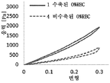
혈전 조성과 관련하여, 문헌[Gunning et al (2017)]은 어떻게 피브린 우세 혈전(fibrin dominant clot)이 적혈구(RBC) 우세 혈전보다 큰 마찰 계수를 갖는지를 설명하였다. 문헌[Gunning GM, McArdle K, Mirza M, Duffy S, Gilvarry M, Brouwer PA. Clot friction variation with fibrin content; implications for resistance to thrombectomy. J Neurointerv Surg. 2017 Jan 02. PubMed PMID: 28044009. Epub 2017/01/04. eng.]을 참조하라. 이는 이들 혈전 유형의 회수에 대한 더 큰 저항을 암시할 수 있다. 피브린-풍부 혈전의 다른 연구에서, 피브린 우세 혈전의 순차적인 압축은, 다수의 통과가 이용될 때 혈전을 제거하는 어려움을 악화시키는 것을 야기할 수 있는 마찰 계수의 증가를 초래하는 것으로서 관찰되었다. (Anderson and Yoo).Regarding thrombus composition, Gunning et al (2017) described how fibrin dominant clots have a greater coefficient of friction than red blood cell (RBC) dominant thrombus. Gunning GM, McArdle K, Mirza M, Duffy S, Gilvarry M, Brouwer PA. Clot friction variation with fibrin content; implications for resistance to thrombectomy. J Neurointerv Surg. 2017 Jan 02.PubMed PMID: 28044009.Epub 2017/01/04. eng.]. This may suggest greater resistance to recovery of these types of thrombus. In another study of fibrin-rich thrombus, sequential compression of the fibrin-dominant thrombus has been observed to result in an increase in the coefficient of friction that can lead to aggravating the difficulty of removing the thrombus when multiple passes are used. (Anderson and Yoo) .
또한, 본 개시내용 전반에 걸쳐 이해되는 바와 같이, 혈전이, 혈전이 더 연성이고 덜 강성일 수 있는 비교적 더 높은 적혈구(RBC) 조성을 갖기보다는, 높은 피브린 및 혈소판 함량(예컨대, 40% 초과의 피브린 및 혈소판 조성)을 갖는 혈전은, 더 높은 마찰 계수를 가질 수 있으며(예컨대, 더 강성이고/이거나 혈관 벽에 더 강하게 연결될 수 있음), 이는 피브린-풍부 혈전을 제거하기 매우 어렵게 만들고, 장치에 의해 혈전을 혈관구조로부터 제거(remove)하거나 심지어 제거(dislodge)하기 위해 다수의 통과를 필요로 할 수 있다. 또한, 혈전의 특성들은 (예컨대, 장치가 혈전의 첫 번째 통과를 수행한 후에) 혈전과 상호작용하는 회수 장치의 작용에 의해 상당히 변화될 수 있다. 특히, 혈전의 압축은 혈전의 탈수를 야기하고, 혈전 강성 및 마찰 계수 둘 모두의 극적인 증가를 초래할 수 있다.In addition, as understood throughout this disclosure, thrombosis, rather than having a relatively higher red blood cell (RBC) composition, which can be softer and less stiff, has higher fibrin and platelet content (e.g., more than 40% fibrin and Thrombus with platelet composition) may have a higher coefficient of friction (e.g., may be more rigid and/or more strongly connected to the vascular wall), making it very difficult to remove the fibrin-rich thrombus, and thrombus by the device It may require multiple passes to remove or even dislodge the vasculature from the vasculature. In addition, the characteristics of the thrombus can be significantly changed by the action of a recovery device that interacts with the thrombus (eg, after the device has performed the first pass of the thrombus). In particular, compression of the thrombus causes dehydration of the thrombus and can result in dramatic increases in both thrombus stiffness and coefficient of friction.
기계적 혈전제거 시술 동안 제거된 특정 혈전을 검사함으로써 시험관 내에서 이들 특성들 중 많은 것을 평가하는 것이 가능하지만, 시험관 내 시험은 몇 가지 분명한 제한을 갖는다. 맨 먼저, 이들은 시간이 많이 걸리고 종종 샘플에 파괴적이다. 샘플은 시험관 내 분석을 받을 수 있게 하기 위해 광범위한 처리를 겪어야 할 수 있다. 급성 허혈성 뇌졸중과 같은 일부 병인의 경우에 상당히, 치료까지의 시간은 환자 결과를 위한 중요한 인자이며, 시험관 내 시험은 샘플링 및 분석에 수반되는 시간 때문에 환자를 위한 최상의 치료를 결정하는 실용적인 방식이 아닐 수 있다.Although it is possible to evaluate many of these properties in vitro by examining specific thrombi removed during a mechanical thrombectomy procedure, in vitro testing has some obvious limitations. First of all, they are time consuming and often destructive to the sample. Samples may have to undergo extensive processing to allow for in vitro analysis. For some etiologies, such as acute ischemic stroke, time to treatment is an important factor for patient outcomes, and in vitro testing may not be a practical way to determine the best treatment for a patient because of the time involved in sampling and analysis. have.
본 개시내용의 해결책은 급성 허혈성 사건의 관리를 개선하기 위해 본 기술 분야의 이들 및 다른 문제들을 해결한다.The solution of the present disclosure addresses these and other problems in the art to improve management of acute ischemic events.
상기 필요성을 해결할 수 있는 본 개시내용의 다양한 예시적인 장치, 시스템, 및 방법이 본 명세서에 개시된다. 일부 실시예에서, 하나 이상의 급성 허혈성 사건을 관리하기 위한 방법이 개시되며, 본 방법은 혈전의 기준을 결정하는 단계; 기준에 기초하여 혈전을 분류하고 분류를 생성하는 단계; 분류에 기초하여 혈전에 대한 개별화된 치료 프로토콜을 결정하는 단계로서, 개별화된 치료 프로토콜은 흡인하는 것, 제1 재관류 장치를 사용하여 관류를 복구하는 것, 및 제2 재관류 장치를 사용하여 관류를 복구하는 것 중 적어도 하나를 사용하는 것으로부터 선택된 하나 이상의 기술을 포함하는, 개별화된 치료 프로토콜을 결정하는 단계; 및 개별화된 치료 프로토콜에 기초하여 혈전을 치료하는 단계를 포함할 수 있다.Various exemplary apparatus, systems, and methods of the present disclosure that can address the above need are disclosed herein. In some embodiments, a method for managing one or more acute ischemic events is disclosed, the method comprising determining a criterion of a thrombus; Classifying thrombus based on criteria and creating a classification; Determining an individualized treatment protocol for the thrombus based on the classification, wherein the individualized treatment protocol is aspiration, repairing perfusion using a first reperfusion device, and repairing perfusion using a second reperfusion device Determining an individualized treatment protocol comprising one or more techniques selected from using at least one of the following; And treating blood clots based on the individualized treatment protocol.
일부 실시예에서, 혈전을 분류하는 단계는 생체 내에서 수행된다.In some embodiments, the step of classifying thrombi is performed in vivo.
일부 실시예에서, 제1 재관류 장치는 스텐트 회수기이고, 제2 재관류 장치는 핀치 회수기이다.In some embodiments, the first reperfusion device is a stent recovery device and the second reperfusion device is a pinch recovery device.
일부 실시예에서, 혈전을 분류하는 것은 임상 검사, 혈액 검사, 컴퓨터 단층촬영(CT) 스캔, 자기 공명 영상(MRI) 스캔, 경동맥 초음파, 뇌혈관 조영상, 및 심초음파 중 하나 이상을 포함한다.In some embodiments, classifying thrombus comprises one or more of a clinical test, blood test, computed tomography (CT) scan, magnetic resonance imaging (MRI) scan, carotid ultrasound, cerebrovascular angiography, and echocardiography.
일부 실시예에서, 혈전의 기준을 결정하는 것은 CT 스캔 및 MRI 스캔 중 적어도 하나를 수행하는 것; 및 적혈구 함량, 백혈구 함량의 수준들, 피브린의 수준들, 혈소판의 수준들, 수화의 수준, 혈전 크기, 혈전 형상, 및/또는 혈관구조 내의 혈전 위치를 포함하는, 혈전의 물리적 및/또는 화학적 조성을 결정하기 위해 CT 스캔 및 MRI 스캔 중 적어도 하나로부터 정보를 분석하는 것을 포함할 수 있다.In some embodiments, determining criteria for thrombus is performing at least one of a CT scan and an MRI scan; And the physical and/or chemical composition of the thrombus, including erythrocyte content, levels of leukocyte content, levels of fibrin, levels of platelets, level of hydration, thrombus size, thrombus shape, and/or thrombus location within the vasculature. It may include analyzing information from at least one of a CT scan and an MRI scan to determine.
일부 실시예에서, 제1 재관류 장치는 적혈구가 풍부한 혈전 또는 혈전의 부분들을 제거하도록 구성된 스텐트 회수기이고, 제2 재관류 장치는 피브린/혈소판-풍부 혈전 또는 혈전의 부분들을 제거하도록 구성된 핀치 회수기이다. 소정 실시예들에서, 분류가 혈전이 적혈구 풍부임을 입증하는 경우, 개별화된 치료 프로토콜은, 혈관에 대한 재관류를 복구하기 위해 제1 재관류 장치를 혈전을 지나, 혈전을 통해, 또는 혈전 주위에서 통과시키고, 이어서 제1 재관류 장치의 루멘 내의 혈전과 맞물리면서 제1 관류 장치를 후퇴시키는 것을 포함할 수 있다.In some embodiments, the first reperfusion device is a stent recoverer configured to remove red blood cell-rich thrombus or portions of a thrombus, and the second reperfusion device is a fibrin/platelet-rich thrombus or pinch recovery machine configured to remove portions of a thrombus. In certain embodiments, if the classification demonstrates that the thrombus is rich in red blood cells, the individualized treatment protocol passes the first reperfusion device through the thrombus, through the thrombus, or around the thrombus to restore reperfusion to the blood vessels. And then retracting the first perfusion device while engaging a thrombus within the lumen of the first reperfusion device.
일부 실시예에서, 제1 재관류 장치는 적혈구가 풍부한 혈전 또는 혈전의 부분들을 제거하도록 구성된 스텐트 회수기이고, 제2 재관류 장치는 피브린-풍부 및/또는 혈소판 풍부 혈전 또는 혈전의 부분들을 제거하도록 구성된 핀치 회수기이다. 소정 실시예들에서, 분류가 혈전이 피브린-풍부 및/또는 혈소판 풍부임을 입증하는 경우, 개별화된 치료 프로토콜은, 혈관에 대한 재관류를 복구하기 위해 혈전을 지나, 혈전을 통해, 또는 혈전 주위에서 제2 재관류 장치를 통과시키고, 이어서 혈전을 핀칭하면서 제2 관류 장치를 후퇴시키는 것을 포함할 수 있다.In some embodiments, the first reperfusion device is a stent recoverer configured to remove red blood cell-rich thrombus or portions of a thrombus, and the second reperfusion device is a fibrin-rich and/or platelet rich thrombus or pinch recovery machine configured to remove portions of a thrombus to be. In certain embodiments, if the classification demonstrates that the thrombus is fibrin-rich and/or platelet rich, then the individualized treatment protocol may pass through the thrombus, through the thrombus, or around the thrombus to restore reperfusion to the blood vessels. 2 passing through the reperfusion device and then retracting the second perfusion device while pinching the thrombus.
일부 실시예에서, 혈전이 30% 이상의 적혈구 수로 구성되는 경우 혈전은 적혈구 풍부로서 분류된다. 일부 실시예에서, 혈전이 30% 이상의 백혈구 수로 구성되는 경우 혈전은 백혈구 풍부로서 분류된다. 일부 실시예에서, 혈전이 30% 이상의 피브린으로 구성되는 경우 혈전은 피브린 풍부로서 분류되며, 혈전이 30% 이상의 혈소판으로 구성되는 경우 혈전은 혈소판 풍부로서 분류된다.In some embodiments, a thrombus is classified as red blood cell abundance when the thrombus consists of 30% or more red blood cell counts. In some embodiments, a thrombus is classified as leukocyte abundance when the thrombus consists of 30% or more white blood cell count. In some embodiments, a thrombus is classified as fibrin rich when the thrombus is composed of 30% or more fibrin, and a thrombus is classified as platelet rich if the thrombus is composed of 30% or more platelet.
일부 실시예에서, 혈전의 기준을 결정하는 단계는, 적혈구 함량, 백혈구 함량, 피브린의 수준들, 혈소판의 수준들, 수화의 수준들, 혈전 크기, 혈전 형상, 및/또는 혈관구조 내의 혈전 위치 중 적어도 하나의 수준들을 실시간으로(예컨대, 즉각적으로 또는 분석을 착수하는 수 분 내에) 결정하기 위해, 혈전과 작동식으로 연통하는 컴퓨팅 장치에 의해, 혈전에 관한 정보를 해석하는 단계를 포함한다. 일부 실시예에서, 본 방법은 또한, 컴퓨팅 장치의 그래픽 사용자 인터페이스를 통해 개별화된 치료 프로토콜을 수신하는 단계, 컴퓨팅 장치에 의해 혈전을 갖는 혈관의 관류를 모니터링하는 단계, 및 혈관 내에서 관류가 복구되는 것에 응답하여 컴퓨팅 장치에 의해 경보하는 단계를 포함할 수 있다.In some embodiments, determining the criteria for a thrombus includes: red blood cell content, white blood cell content, levels of fibrin, levels of platelets, levels of hydration, thrombus size, thrombus shape, and/or thrombus location within the vasculature Interpreting information about the thrombus by a computing device operatively in communication with the thrombus to determine at least one level in real time (eg, immediately or within minutes of undertaking the analysis). In some embodiments, the method also includes receiving a personalized treatment protocol through a graphical user interface of the computing device, monitoring perfusion of blood vessels with blood clots by the computing device, and perfusion within the blood vessels being restored. And alerting by the computing device in response to the response.
일부 실시예에서, 컴퓨팅 장치는, 혈전에 관한 정보를 해석하고 개별화된 치료 프로토콜을 결정하기 위한 상관 데이터를 포함하는 데이터베이스에 링크된다. 데이터베이스는 컴퓨팅 장치로부터 원격일 수 있다.In some embodiments, the computing device is linked to a database containing correlation data for interpreting information about thrombus and determining individualized treatment protocols. The database can be remote from the computing device.
일부 실시예에서, 혈전의 기준을 결정하는 단계는 카테터를 혈관구조 내의 혈전의 부위로 전달하는 단계; 및 혈관구조 내의 혈전의 부위의 제1 위치에서 카테터에 결합된 근적외선 분광법(NIR)을 위한 사용 기구에 의해 혈전의 제1 판독을 취하고, 제1 판독으로부터 스펙트럼을 생성하여, 스펙트럼이 혈전의 화학적 조성 및/또는 물리적 특성들 중 적어도 하나에 관련되게 하는 단계를 포함한다.In some embodiments, determining the criteria for a thrombus includes delivering a catheter to a site of a thrombus within the vasculature; And a first reading of the thrombus by a device used for near-infrared spectroscopy (NIR) coupled to the catheter at the first location of the site of the thrombus within the vasculature, generating a spectrum from the first reading, so that the spectrum is the chemical composition of the thrombus. And/or relating to at least one of the physical properties.
일부 실시예에서, 혈전의 기준을 결정하는 단계는 적혈구 함량, 백혈구 함량의 수준들, 피브린의 수준들, 혈소판의 수준들, 수화의 수준, 혈전 크기, 혈전 형상, 및/또는 혈관구조 내의 혈전 위치 중 적어도 하나를 실시간으로 결정하기 위해 스펙트럼에 포함된 정보를 해석하는 단계를 추가로 포함한다.In some embodiments, determining the criteria for a thrombus includes red blood cell content, white blood cell content levels, fibrin levels, platelet levels, level of hydration, thrombus size, thrombus shape, and/or thrombus location within the vasculature The method further includes interpreting information included in the spectrum to determine at least one of them in real time.
일부 실시예에서, 본 방법은 또한 제1 위치의 원위 또는 근위에 있는 제2 위치에서 NIR을 위한 기구를 사용함으로써 혈전의 제2 판독을 취하는 단계, 제2 판독으로부터 스펙트럼을 생성하여, 제2 판독의 스펙트럼이 혈전의 화학적 조성 및/또는 물리적 특성들 중 적어도 하나에 관련되게 하는 단계, 및 적혈구 함량의 수준들, 백혈구 함량의 수준들, 피브린의 수준들, 혈소판의 수준들, 수화의 수준, 혈전 크기, 혈전 형상, 및/또는 혈관구조 내의 혈전 위치 중 적어도 하나를 즉각적으로 결정하기 위해 제1 판독 및 제2 판독의 스펙트럼에 포함된 정보를 해석하는 단계를 포함할 수 있다.In some embodiments, the method also takes a second reading of the thrombus by using an instrument for NIR at a distal or proximal second location of the first location, generating a spectrum from the second reading to produce a second reading Allowing the spectrum of to be related to at least one of the chemical composition and/or physical properties of the thrombus, and the levels of red blood cell content, levels of leukocyte content, levels of fibrin, levels of platelets, level of hydration, thrombus And interpreting information contained in the spectra of the first reading and the second reading to immediately determine at least one of size, thrombus shape, and/or thrombus location within the vasculature.
일부 실시예에서, 혈전의 기준을 결정하는 단계는 혈전의 근위 단부 및 그의 원위 단부에서 혈전의 측정들을 취하는 단계, 및 혈전의 근위 단부 및 그의 원위 단부에서 혈전의 측정들로부터 스펙트럼들을 생성하여, 각각의 스펙트럼이 혈전의 화학적 조성 및/또는 물리적 특성들 중 적어도 하나에 관련되게 하는 단계를 포함한다.In some embodiments, determining the criteria for a thrombus comprises taking measurements of a thrombus at the proximal end and a distal end of the thrombus, and generating spectra from measurements of the thrombus at the proximal end and the distal end of the thrombus, respectively And causing the spectrum of to be related to at least one of the chemical composition and/or physical properties of the thrombus.
일부 실시예에서, 혈전의 기준을 결정하는 단계는 혈전의 근위 단부와 그의 원위 단부 사이에서 혈전의 하나 이상의 측정을 취하는 단계, 및 혈전의 근위 단부와 그의 원위 단부 사이에서 그의 길이를 따른 혈전의 측정들로부터 스펙트럼들을 생성하여, 각각의 스펙트럼이 혈전의 화학적 조성 및/또는 물리적 특성들 중 적어도 하나에 관련되게 하는 단계를 포함한다.In some embodiments, determining the criteria for a thrombus comprises taking one or more measurements of a thrombus between the proximal end of the thrombus and its distal end, and measuring a thrombus along its length between the proximal end of the thrombus and its distal end. Generating spectra from the fields, such that each spectrum is related to at least one of the chemical composition and/or physical properties of the thrombus.
일부 실시예에서, 혈전의 기준을 결정하는 단계는 카테터를 혈관구조 내의 혈전의 부위로 전달하는 단계, 혈관구조 내의 혈전의 부위에서 카테터에 결합된 라만 분광법(및/또는 스펙트럼의 가시 영역)을 위한 사용 기구에 의해 혈전의 제1 판독을 취하는 단계, 및 제1 판독으로부터 스펙트럼을 생성하여, 스펙트럼이 혈전의 화학적 조성 및/또는 물리적 특성들 중 적어도 하나에 관련되게 하는 단계를 포함한다. 일부 실시예에서, 혈전의 기준을 결정하는 단계는 또한, 적혈구 함량, 백혈구 함량의 수준들, 피브린의 수준들, 혈소판의 수준들, 수화의 수준, 혈전 크기, 혈전 형상, 및/또는 혈관구조 내의 혈전 위치를 실시간으로 결정하기 위해 스펙트럼에 포함된 정보를 해석하는 단계를 포함한다.In some embodiments, determining the criteria for a thrombus comprises delivering the catheter to a site of a thrombus within the vasculature, for Raman spectroscopy (and/or the visible region of the spectrum) bound to the catheter at the site of the thrombus within the vasculature. Taking a first reading of a thrombus by an instrument of use, and generating a spectrum from the first reading, such that the spectrum is related to at least one of the chemical composition and/or physical properties of the thrombus. In some embodiments, determining the criteria for thrombus may also include red blood cell content, levels of leukocyte content, levels of fibrin, levels of platelets, levels of hydration, thrombus size, thrombus shape, and/or within the vasculature. And interpreting information contained in the spectrum to determine the thrombus location in real time.
일부 실시예에서, 본 방법은 혈전의 원위 또는 근위에 있는 제2 위치 및 제1 위치에서 라만 분광법을 위한 기구를 사용함으로써 혈전의 제2 판독을 취하는 단계, 제2 판독으로부터 스펙트럼을 생성하여, 제2 판독의 스펙트럼이 혈전의 화학적 조성 및/또는 물리적 특성들에 관련되게 하는 단계, 및 적혈구 함량, 백혈구 함량의 수준들, 피브린의 수준들, 혈소판의 수준들, 수화의 수준, 혈전 크기, 혈전 형상, 및/또는 혈관구조 내의 혈전 위치 중 적어도 하나를 실시간으로 결정하기 위해 제1 판독 및 제2 판독의 스펙트럼에 포함된 정보를 해석하는 단계를 포함한다.In some embodiments, the method comprises taking a second reading of a thrombus by using an instrument for Raman spectroscopy at a second position and a first position distal or proximal of a thrombus, generating a spectrum from the second reading, 2 making the spectrum of the reading related to the chemical composition and/or physical properties of the thrombus, and the level of red blood cells, levels of white blood cells, levels of fibrin, levels of platelets, levels of hydration, thrombus size, thrombus shape And/or interpreting information contained in the spectra of the first reading and the second reading to determine in real time at least one of the thrombus locations within the vascular structure.
일부 실시예에서, 혈전의 치료 단계는 혈전의 일부분을 회수하는 단계를 포함한다. 본 방법은 또한 회수된 혈전 및/또는 혈전의 하나 이상의 조각을 분석하는 단계, 및 회수된 혈전을 분석하는 것 또는 혈전에 접근하고 혈전을 교차하는 것을 분석하는 것에 기초하여 혈전 치료 단계를 선택하는 단계를 포함할 수 있다. 그러나, 이러한 예는 그렇게 제한되지 않으며, 이러한 실시예의 혈전 분석은 각각의 재관류 장치와 연통되거나 다른 식으로 그와 연결된 임의의 물질(예컨대, 혈청, 또는 혈전과 연관된 임의의 다른 혈소판)을 분석하는 것을 포함하는 것으로 고려된다.In some embodiments, treating a blood clot includes recovering a portion of the blood clot. The method also includes analyzing a recovered thrombus and/or one or more pieces of thrombus, and selecting a thrombus treatment step based on analyzing the recovered thrombus or accessing and crossing the thrombus. It may include. However, this example is not so limited, and the thrombus analysis of this embodiment involves analyzing any substance (eg, serum, or any other platelet associated with a thrombus) that is in communication with or associated with each reperfusion device. It is considered to include.
일부 실시예에서, 혈전의 기준을 결정하는 단계는 백혈구 수준들, 적혈구 수준들, 혈청 수준들, 피브린 수준들, 혈소판의 수준들, 수화의 수준, 혈전 크기, 혈전 위치, 혈전 강도, 혈전 탄성, 혈전 형성의 속도 또는 혈전 용해의 속도 중 하나 이상으로부터 선택된 하나 이상의 정량적 표시를 결정하는 단계를 포함한다. 본 방법은 제1 및 제2 혈전 특성 정량적 표시들을 상관 데이터와 비교하는 단계, 및 흡인, 제1 재관류 장치, 및/또는 제2 재관류 장치로부터 선택되는 하나 이상의 기술을 사용하는 선택 및/또는 순서를 결정하는 단계를 포함할 수 있다.In some embodiments, determining the criteria for thrombus includes leukocyte levels, erythrocyte levels, serum levels, fibrin levels, platelet levels, level of hydration, thrombus size, thrombus location, thrombus strength, thrombus elasticity, Determining one or more quantitative indications selected from one or more of the rate of thrombus formation or the rate of thrombolysis. The method comprises comparing the first and second thrombus characteristics quantitative indications to the correlation data and selecting and/or ordering using one or more techniques selected from aspiration, first reperfusion device, and/or second reperfusion device. And determining.
일부 실시예에서, 허혈성 사건을 치료하기 위한 시스템이 개시된다. 본 시스템은 허혈성 사건을 갖는 대상의 혈전의 생체 내 분석 정보를 제공하기 위한 수단, 분석 정보에 기초하여 대상에 대한 개별화된 치료 프로토콜의 표시를 제공하기 위한 수단 - 개별화된 치료는 흡인을 사용하는 것, 제1 재관류 장치를 사용하여 관류를 복구하는 것, 및/또는 제2 재관류 장치를 사용하여 관류를 복구하는 것으로부터 선택된 하나 이상의 기술을 포함함 -; 및 개인화된 치료 프로토콜에 기초하여 혈전을 치료하기 위한 수단을 포함할 수 있다.In some embodiments, a system for treating an ischemic event is disclosed. The system comprises means for providing in vivo assay information of a blood clot of a subject with ischemic events, means for providing an indication of an individualized treatment protocol for the subject based on the assay information-individualized treatment using aspiration , Including one or more techniques selected from recovering perfusion using a first reperfusion device, and/or recovering perfusion using a second reperfusion device; And means for treating blood clots based on personalized treatment protocols.
일부 실시예에서, 시스템의 제1 재관류 장치는 적혈구가 풍부한 혈전 또는 혈전의 부분들을 제거하도록 구성된 스텐트 회수기이고, 제2 재관류 장치는 피브린 및/또는 혈소판-풍부 혈전 또는 혈전의 부분들을 제거하도록 구성된 핀치 회수기이다. 일부 실시예에서, 표시가 혈전이 적혈구 풍부임을 입증하는 경우, 개별화된 치료 프로토콜은, 혈관에 대한 재관류를 복구하기 위해 제1 재관류 장치를 혈전을 지나, 혈전을 통해, 또는 혈전 주위에서 통과시키고, 이어서 제1 재관류 장치의 루멘 내의 혈전과 맞물리면서 제1 관류 장치를 후퇴시키기 위한 수단을 포함한다.In some embodiments, the first reperfusion device of the system is a stent recoverer configured to remove portions of a red blood cell-rich thrombus or thrombus, and the second reperfusion device is a fibrin and/or platelet-rich thrombus or pinch configured to remove portions of a thrombus It is a recovery machine. In some embodiments, if the indication demonstrates that the thrombus is rich in red blood cells, the individualized treatment protocol passes the first reperfusion device through the thrombus, through the thrombus, or around the thrombus to restore reperfusion to the blood vessel, And means for retracting the first perfusion device while engaging a thrombus within the lumen of the first reperfusion device.
일부 실시예에서, 제1 재관류 장치는 적혈구가 풍부한 혈전 또는 혈전의 부분들을 제거하도록 구성된 스텐트 회수기이고, 제2 재관류 장치는 피브린-풍부 및/또는 혈소판 풍부 혈전 또는 혈전의 부분들을 제거하도록 구성된 핀치 회수기이다. 일부 실시예에서, 분류가 혈전이 피브린-풍부 및/또는 혈소판 풍부임을 입증하는 경우, 개별화된 치료 프로토콜은, 혈관에 대한 재관류를 복구하기 위해 혈전을 지나, 혈전을 통해, 또는 혈전 주위에서 제2 재관류 장치를 통과시키고, 이어서 혈전을 핀칭하면서 제2 관류 장치를 후퇴시키기 위한 수단을 포함한다.In some embodiments, the first reperfusion device is a stent recoverer configured to remove red blood cell-rich thrombus or portions of a thrombus, and the second reperfusion device is a fibrin-rich and/or platelet rich thrombus or pinch recovery machine configured to remove portions of a thrombus to be. In some embodiments, if the classification demonstrates that the thrombus is fibrin-rich and/or platelet rich, the individualized treatment protocol passes through the thrombus, through the thrombus, or around the thrombus to restore reperfusion to the blood vessel. And means for passing the reperfusion device and then retracting the second perfusion device while pinching the thrombus.
일부 실시예에서, 허혈성 사건을 치료하기 위한 시스템이 개시된다. 본 시스템은 혈전을 갖는 폐색된 혈관에 대한 관류를 복구하기 위한 제1 재관류 장치를 포함할 수 있다. 본 시스템은 혈전을 갖는 폐색된 혈관에 대한 관류를 복구하기 위한 제2 재관류 장치를 포함할 수 있다. 본 시스템은 제1 및 제2 재관류 장치들 중 적어도 하나를 폐색된 혈관 내의 혈전에 전달하기 위한 전달 시스템을 포함할 수 있다. 본 시스템은 폐색된 혈관의 혈전을 분석하고 개별화된 치료 프로토콜을 결정하기 위한 혈전 분석 시스템을 포함할 수 있다. 전달 시스템은 가이드 카테터, 가이드와이어, 마이크로카테터 등 중 적어도 하나를 포함할 수 있으며, 이들 각각은 혈관구조를 통해 혈전의 부위로 전달되도록 작동가능하다.In some embodiments, a system for treating an ischemic event is disclosed. The system can include a first reperfusion device for restoring perfusion to occluded blood vessels having a thrombus. The system may include a second reperfusion device to restore perfusion to occluded blood vessels with thrombus. The system can include a delivery system for delivering at least one of the first and second reperfusion devices to a thrombus in an occluded blood vessel. The system can include a thrombus analysis system to analyze clots in occluded blood vessels and determine individualized treatment protocols. The delivery system may include at least one of a guide catheter, guide wire, microcatheter, and the like, each of which is operable to be delivered to the site of the thrombus through the vascular structure.
상기 및 관련 목적의 달성을 위해, 하기의 설명 및 첨부 도면과 관련하여 소정의 예시적인 태양이 본 명세서에 기술된다. 그러나, 이들 태양은 청구된 주제의 원리가 채용될 수 있는 다양한 방식들 중 몇몇만을 나타내며, 청구된 주제는 모든 그러한 태양 및 그들의 등가물을 포함하고자 한다. 다른 이점 및 신규한 특징이 도면과 함께 고려될 때 하기의 상세한 설명으로부터 명백해질 수 있다.To achieve the above and related objects, certain exemplary aspects are described herein in connection with the following description and accompanying drawings. However, these aspects represent only a few of the various ways in which the principles of the claimed subject matter can be employed, and the claimed subject matter is intended to cover all such aspects and their equivalents. Other advantages and novel features may become apparent from the following detailed description when considered in conjunction with the drawings.
본 개시내용은, 첨부 도면을 참조하여, 단지 예로서 주어진, 본 개시내용의 일부 실시예의 하기 설명으로부터 더 명확히 이해될 것이다.
도 1은 전달을 위해 동맥계를 사용하여 뇌 혈관 내에 위치된 예시적인 혈전 회수 장치를 이용한 대퇴부 접근을 통해 카테터 삽입된 환자를 도시한다.
도 2는 뇌로 이어지는 대동맥궁 위의 뇌 동맥들의 소정 해부학적 구조를 도시한다.
도 3은 본 개시내용의 예시적인 스텐트 회수기 장치의 등각도를 도시한다.
도 4는 본 개시내용의 일 실시예에 따른 핀칭 회수기 장치의 사시도이다.
도 5는 본 개시내용의 일례의 개략적인 개요도이다.
도 6은 본 개시내용의 혈전 분석 접근법의 일례의 개략적인 개요도이다.
도 7은 비-조영 CT를 사용한 혈전 분석의 예의 개략적인 개요도이다.
도 8은 뇌 MRI 및/또는 진보된 MR을 사용한 혈전 분석의 예의 개략적인 개요도이다.
도 9는 조영 증강 CT를 사용한 혈전 분석의 예의 개략적인 개요도이다.
도 10은 카테터 혈관조영술을 사용한 혈전 분석의 예의 개략적인 개요도이다.
도 11은 카테터 혈관조영술을 사용한 혈전 분석의 예의 개략적인 개요도이다.
도 12는 하나 이상의 예시적인 실시예에 따른, 본 개시내용의 태양들을 구현할 수 있는 동작 환경을 예시하는 시스템 다이어그램이다.
도 13은 본 명세서에 기술된 하나 이상의 실시예에 따른, 본 개시내용의 태양들을 구현할 수 있는 일반적인 컴퓨팅 시스템을 보여주는 컴퓨터 아키텍처 다이어그램이다.
도 14는 환자로부터 최근에 제거된 시험관 내 혈전 또는 혈전의 조각들을 분석하는 모바일 장치의 개략적인 개요도를 도시한다.
도 15는 본 개시내용에서 분석된 혈전의 조직병리학적 조성의 개요도를 도시한다.
도 16a는 0% 적혈구 수를 갖는 것으로 식별된 수축된 상태 및 비수축된 상태에 있는 혈전의 응력-변형 곡선을 보여주는 그래프를 도시한다.
도 16b는 40% 적혈구 수를 갖는 것으로 식별된 수축된 상태 및 비수축된 상태에 있는 혈전에 대한 응력-변형 곡선을 보여주는 그래프를 도시한다.
도 16c는 5% 적혈구 수를 갖는 것으로 식별된 수축된 상태 및 비수축된 상태에 있는 혈전에 대한 응력-변형 곡선을 보여주는 그래프를 도시한다.
도 17은 하나 이상의 급성 허혈성 사건을 관리하기 위한 본 개시내용의 예시적인 방법을 도시하는 흐름도를 도시한다.
도 18은, 조직병리학적 분석을 위해 혈관내 치료에 의해 혈전 조각들이 회수된, 제1 연구에서의 환자들의 임상 특성들을 보여주는 표이다.
도 19a 내지 도 19d는 제1 연구로부터의 피브린 및 RBC의 분포를 도시한다.
도 20은 제1 연구에서의 모든 사례에 대해 각각의 통과로부터 조합된 혈전 조각들의 RBC, 피브린 및 WBC 조성을 도시한다.
도 21은 총 1회, 2회, 3회 또는 4회 내지 6회 통과로 이루어진 사례들에 대한 조합된 혈전 조각들의 제1 연구에서의 평균 조성을 도시한다. 막대들은 평균 조성을 나타낸다. 개별 표준 편차를 사용하여 간격을 계산하였다.
도 22a는 제1 연구에서의 모든 사례로부터의 통과 1, 2, 3, 4, 5, 6에서 회수된 106개의 혈전 조각들의 평균 RBC, 피브린 및 WBC 조성을 도시한다.
도 22b는 2회 초과의 통과로 이루어진 제1 연구의 그러한 사례들로부터의 통과 1, 2, 3, 4, 5, 6에서 회수된 혈전 조각들(n=53 혈전 조각들)의 평균 RBC, 피브린 및 WBC 조성을 도시한다.
도 23은 제1 연구의 모든 사례에 대해 각각의 통과에서 회수된 혈전 조각들의 RBC, 피브린 및 WBC 백분율을 예시하는 막대 그래프를 도시한다.
도 24는 전체 혈전의 RBC, 피브린 및 WBC 조성을 도시하는데, 여기서 각각의 통과로부터의 조각들을 조합하였고, 심장색전성(cardioembolic), 죽종색전성(atheroembolic) 또는 원인불명의(cryptogenic) 병인(aetiology)에 따라 그룹화하였다. 사분위수 범위 상자, 수염 및 중앙선이 그래프 상에 도시된다. 각각의 병인에 대한 평균 RBC, 피브린 및 WBC 조성은 기호 +로 표시된다.
도 25는 심장색전성, 죽종색전성 및 원인불명의 병인을 가진 환자들로부터 통과들 1 내지 6에서 회수된 조각들의 RBC, 피브린 및 WBC 백분율의 내역(breakdown)을 도시한다.The present disclosure will be more clearly understood from the following description of some embodiments of the present disclosure, given by way of example only, with reference to the accompanying drawings.
FIG. 1 depicts a catheter-inserted patient through a femoral approach using an exemplary thrombus recovery device located within a brain vessel using an arterial system for delivery.
Fig. 2 shows a certain anatomical structure of brain arteries above the aortic arch leading to the brain.
3 shows an isometric view of an exemplary stent recovery device of the present disclosure.
4 is a perspective view of a pinching recovery device according to an embodiment of the present disclosure.
5 is a schematic schematic diagram of an example of the present disclosure.
6 is a schematic schematic diagram of an example of a thrombus analysis approach of the present disclosure.
7 is a schematic schematic diagram of an example of a thrombus analysis using non-contrast CT.
8 is a schematic schematic diagram of an example of a thrombus analysis using brain MRI and/or advanced MR.
9 is a schematic schematic diagram of an example of a thrombus analysis using contrast enhanced CT.
10 is a schematic schematic illustration of an example of a thrombus analysis using catheter angiography.
11 is a schematic schematic illustration of an example of a thrombus analysis using catheter angiography.
12 is a system diagram illustrating an operating environment capable of implementing aspects of the present disclosure, in accordance with one or more exemplary embodiments.
13 is a computer architecture diagram showing a general computing system capable of implementing aspects of the disclosure, in accordance with one or more embodiments described herein.
14 shows a schematic schematic diagram of a mobile device for analyzing in vitro thrombus or fragments of thrombus recently removed from a patient.
15 shows a schematic diagram of the histopathological composition of thrombus analyzed in the present disclosure.
16A shows a graph showing the stress-strain curve of thrombus in the contracted and non-contracted states identified as having 0% red blood cell count.
16B shows a graph showing stress-strain curves for thrombus in the contracted and unconstricted states identified as having 40% red blood cell count.
16C shows a graph showing stress-strain curves for thrombus in the contracted and non-contracted states identified as having 5% red blood cell count.
17 depicts a flow diagram depicting an exemplary method of the present disclosure for managing one or more acute ischemic events.
FIG. 18 is a table showing the clinical characteristics of patients in the first study, in which thrombus fragments were recovered by intravascular treatment for histopathological analysis.
19A-19D show the distribution of fibrin and RBC from the first study.
FIG. 20 depicts the RBC, fibrin and WBC composition of thrombus fragments combined from each pass for all cases in the first study.
FIG. 21 depicts the average composition in the first study of combined thrombus fragments for cases consisting of 1, 2, 3 or 4 to 6 passes in total. Bars represent average composition. Intervals were calculated using individual standard deviations.
22A depicts the average RBC, fibrin and WBC composition of 106 thrombus fragments recovered from
22B is the average RBC, fibrin of thrombus fragments recovered from those 1, 2, 3, 4, 5, 6 (n=53 thrombus fragments) from those cases of the first study with more than 2 passes. And WBC composition.
FIG. 23 depicts a bar graph illustrating the percentage of RBC, fibrin and WBC of thrombus fragments recovered at each pass for all cases in the first study.
FIG. 24 shows the composition of RBC, fibrin and WBC of whole thrombi, where fragments from each passage were combined, cardioembolic, atherobolic or cryptogenic aetiology Grouped according to. The quartile range box, whiskers and center line are shown on the graph. The average RBC, fibrin and WBC composition for each etiology is indicated by the symbol +.
FIG. 25 shows breakdown of RBC, fibrin and WBC percentages of fragments recovered in passages 1-6 from patients with cardiac embolism, atheromatous embolism, and unknown etiology.
본 개시내용의 일부 태양은 생체 내에서 그리고/또는 시험관 내에서 급성 허혈성 사건들을 분석 및/또는 분류할 뿐만 아니라, 특정 급성 허혈성 사건에 대한 치료 프로토콜을 개별화하기 위한 방법 및 시스템에 관한 것이다. 본 개시내용의 예시적인 실시예가 본 명세서에서 상세히 설명되지만, 다른 실시예가 고려되는 것이 이해되어야 한다. 따라서, 본 개시내용은 그의 범주에 있어서 하기의 설명에 기재되거나 도면에 예시되는 구성의 상세 사항 및 구성요소의 배열로 제한되는 것으로 의도되지 않는다. 본 개시내용은 다른 실시예가 가능하며, 다양한 방식으로 실시되거나 수행될 수 있다.Some aspects of the present disclosure relate to methods and systems for analyzing and/or classifying acute ischemic events in vivo and/or in vitro, as well as individualizing treatment protocols for specific acute ischemic events. While exemplary embodiments of the present disclosure are described in detail herein, it should be understood that other embodiments are contemplated. Accordingly, the present disclosure is not intended to be limited in its scope to the arrangement of components and details of constructions set forth in the description below or illustrated in the drawings. The present disclosure is capable of other embodiments, and can be practiced or carried out in various ways.
본 명세서에서 논의되는 바와 같이, "대상"은 임의의 적용가능한 인간, 동물, 또는 다른 유기체 - 살아있거나 죽은 -, 또는 다른 생물학적 또는 분자 구조 또는 화학적 환경일 수 있고, 대상의 특정 구성요소들, 예를 들어, 본 명세서에서 "관심 부위(area of interest)" 또는 "관심 영역(region of interest)"으로 지칭되는 대상의 특정 위치에 있을 수 있는, 대상의 특정 조직들 또는 유체들(예컨대, 살아있는 대상의 신체의 특정 영역 내의 인간 조직)에 관련될 수 있다.As discussed herein, a “subject” can be any applicable human, animal, or other organism—alive or dead—or other biological or molecular structure or chemical environment, and certain components of the subject, eg For example, certain tissues or fluids (eg, living subjects) of a subject, which may be in a particular location of the subject, referred to herein as “area of interest” or “region of interest”. (Human tissue within a certain area of the body).
본 명세서에서 논의되는 바와 같이, "치료 프로토콜"은 폐색된 혈관에 대한 관류를 복구하는 것과 같이, 환자에서 관찰되는 허혈성 사건을 해결하기 위한 임의의 계획일 수 있다. 예를 들어, 치료 프로토콜은 흡인, 스텐트 회수기 장치, 핀치 회수기 장치, 혈전용해제 주입, 또는 폐색된 혈관에 대한 관류를 복구하기 위한 임의의 다른 기계적, 유체, 또는 다른 수단을 포함할 수 있다. 치료 프로토콜은 하기에서 더 구체적으로 논의되는 바와 같이, 특정 환자에 대한 다수의 인자에 기초하여 개별화될 수 있다.As discussed herein, a “treatment protocol” can be any plan to address ischemic events observed in a patient, such as restoring perfusion to occluded blood vessels. For example, the treatment protocol may include aspiration, stent recovery device, pinch recovery device, thrombolytic infusion, or any other mechanical, fluid, or other means to restore perfusion to occluded blood vessels. Treatment protocols can be individualized based on a number of factors for a particular patient, as discussed in more detail below.
"ASPECT" 점수는 10점 정량적 국소해부학(topographic) CT 스캔 점수를 포함하는 ASPECTS(Alberta Stroke Program Early computed tomography (CT) score)를 의미한다.“ASPECT” score means Alberta Stroke Program Early computed tomography (CT) score (ASPECTS), including a 10-point quantitative topographic CT scan score.
본 명세서에서 논의되는 바와 같이, "mRS"는 뇌졸중 또는 신경학적 장애의 다른 원인을 겪은 사람들의 일상 활동에 있어서의 장애 또는 의존성의 정도를 측정하기 위해 일반적으로 사용되는 척도인 mRS(modified Rankin Scale)를 의미한다. mRS 척도는 0 내지 6에 이르고, 이는 증상 없이 완벽한 건강으로부터 사망으로까지 이른다. 0의 mRS 점수는 증상이 관찰되지 않는 것으로 이해된다. 1의 mRS 점수는, 유의한 장애가 관찰되지 않고 환자는 일부 증상에도 불구하고 모든 일상 활동을 수행할 수 있는 것으로 이해된다. 2의 mRS 점수는 경미한 장애로 이해되며, 환자는 도움 없이 자신의 업무를 돌볼 수 있지만, 모든 이전 활동을 수행할 수는 없다. 3의 mRS 점수는, 환자가 약간의 도움을 필요로 할 수 있지만 도움을 받지 않고 걸을 수 있는 중등도 장애로 이해된다. 4의 mRS 점수는 중등도 중증(moderate severe) 장애로 이해되며, 환자는 도움 없이 자신의 신체적 요구를 처리하거나 도움을 받지 않고 걸을 수 없다. 5의 mRS 점수는 중증 장애인 것으로 이해되며, 환자는 지속적인 간호 및 보살핌, 침상 의존상태(bedridden), 실금을 요구한다. 6의 mRS 점수는 환자가 사망한 것으로 이해된다.As discussed herein, “mRS” is a Modified Rankin Scale (mRS), a measure commonly used to measure the degree of disability or dependence in the daily activities of people who have suffered a stroke or other cause of a neurological disorder. Means The mRS scale ranges from 0 to 6, which ranges from complete health to death without symptoms. It is understood that a mRS score of 0 indicates no symptoms. The mRS score of 1 is understood to be that no significant disability is observed and the patient is able to perform all daily activities despite some symptoms. A mRS score of 2 is understood as a minor disability, and patients can take care of their work without assistance, but cannot perform all previous activities. The mRS score of 3 is understood as a moderate disorder in which the patient may need some help but can walk without assistance. The mRS score of 4 is understood as a moderate severe disorder, and the patient is unable to handle his or her physical needs without assistance or to walk without assistance. The mRS score of 5 is understood to be severely disabled, and patients require continued care and care, bedridden, and incontinence. An mRS score of 6 is understood as the patient's death.
본 명세서에서 논의되는 바와 같이, 용어 "컴퓨터 단층촬영" 또는 CT는 상이한 각도들로부터 취해진 많은 X-선 측정들의 컴퓨터-처리된 조합들을 이용하여 스캐닝된 물체의 특정 영역들의 단면(단층촬영) 이미지들(가상 "슬라이스들")을 생성하여, 사용자가 절단 없이 물체 내부를 볼 수 있게 하는 하나 이상의 스캔을 의미한다. 본 개시내용의 그러한 CT 스캔은 X-선 CT 뿐만 아니라 많은 다른 유형의 CT, 예컨대 양전자 방출 단층촬영(PET) 및 단일 광자 방출 컴퓨터 단층촬영(SPECT)을 지칭할 수 있다.As discussed herein, the term “computed tomography” or CT is a cross-sectional (tomography) image of specific areas of an object scanned using computer-processed combinations of many X-ray measurements taken from different angles. (Virtual "slices") means one or more scans that allow the user to see inside the object without cutting. Such CT scans of the present disclosure may refer to X-ray CT as well as many other types of CT, such as positron emission tomography (PET) and single photon emission computed tomography (SPECT).
본 명세서에 논의된 바와 같이, mTICI(modified treatment in cerebral ischemia) 점수는 혈관내 혈관재형성 후 유동 복구의 양을 분류한다. 구체적으로, mTICI 점수는 2013년 합의 그룹(consensus group)에 의해 최초의 TICI(Thrombolysis in Cerebral Infarction) 척도로부터 개발되었다. 권고사항은 뇌졸중에 대한 혈관내 치료법의 사용 증가를 더 잘 반영하기 위해 명칭 변경을 포함하였고, 표적 혈관 영역의 절반 미만(mTICI 2a) 또는 절반 초과(mTICI 2b)로의 TICI 2 구성성분의 단순화를 포함하였다. 분류: 등급 0: 관류 없음; 등급 1: 초기 폐색을 지나는 순방향(antegrade) 재관류, 그러나 원위 재관류가 거의 없거나 느린, 제한된 원위 분기 충전(distal branch filling); 등급 2; 등급 2a: 폐색된 표적 동맥 이전 허혈성 영역의 절반 미만의 순방향 재관류(예를 들어, 중뇌동맥(MCA)의 하나의 주요 분할 및 그의 영역에서의); 등급 2b: 이전에 폐색된 표적 동맥 허혈성 영역의 절반 초과의 순방향 재관류(예를 들어, MCA의 2개의 주요 분할 및 그 영역들에서의); 등급 3: 모든 원위 분기에서 시각화된 폐색이 없는 상태의, 이전에 폐색된 표적 동맥 허혈성 영역의 완전한 순방향 재관류. 대부분의 경우에 현재의 기술을 사용하여 우수한 비율의 mTICI 2b 또는 3 재관류가 달성될 수 있지만, 많은 경우(예컨대, 대략 20% 이상의 경우)에 혈전제거 장치의 여러 번의 통과 및 조작에도 불구하고 만족스러운 재관류가 달성되지 않는 어려운 경우가 남아 있다. 본 개시내용의 해결책은 현재의 접근법 하에서 대략 20% 이상 배제되는 것을 포함하는 모든 유형의 경우에 대한 시스템 및 방법을 제공한다.As discussed herein, a modified treatment in cerebral ischemia (mTICI) score classifies the amount of flow recovery after vascular revascularization. Specifically, the mTICI score was developed from the first Thrombolysis in Cerebral Infarction (TICI) scale by the 2013 consensus group. The recommendations included a name change to better reflect the increased use of endovascular therapies for stroke, including simplification of
이제, 본 개시내용의 태양들의 상세한 설명이 첨부 도면을 참조하여 제공될 것이다. 도면은 본 명세서의 일부를 형성하고, 예시로서, 특정 실시예 또는 예를 예시로서 도시한다. 도면을 참조함에 있어서, 여러 도면 전체에 걸쳐 동일한 도면 부호가 동일한 요소를 나타낸다. 일례로서, 도 1은 카테터(2)를 이용한 대퇴 동맥을 통한 혈전 회수 장치(200)를 갖는 환자의 카테터 삽입의 개략도를 도시한다. 예시적인 장치(200)는 증상 발생으로부터 소정 기간(예컨대, 8시간, 12시간, 24시간 등) 내에 허혈성 뇌졸중을 경험하는 환자들에서 혈전을 제거함으로써 신경혈관 내의 혈류를 복구할 수 있다.Now, a detailed description of aspects of the present disclosure will be provided with reference to the accompanying drawings. The drawings form part of the present specification and show, by way of illustration, specific embodiments or examples as examples. In referring to the drawings, the same reference numbers throughout the drawings indicate the same elements. As an example, FIG. 1 shows a schematic diagram of a catheter insertion of a patient with a
도 2는 소정의 예시적인 뇌 혈관의 개략도를 도시한다. 혈관(100)은 대동맥이다. 혈관(101)은 완두동맥이다. 혈관(102)은 쇄골하동맥이다. 혈관(103)은 총경동맥이다. 혈관(104)은 내경동맥이다. 혈관(105)은 외경동맥이다. 혈관(106)은 중뇌동맥이다. 혈관(107)은 앞-뇌동맥이다. 도 1의 카테터(2)는 그의 원위 단부가 총경동맥 내에 있는 상태로 도시되어 있다. 본 발명의 보다 상세한 도면에서, 접근 부위의 상세 사항은 도시되지 않을 것이지만, 일반적으로 접근 및 전달은 도 1 및/또는 도 2에 따른다. 또한, 장치(200)는 팔의 혈관구조(요골 또는 상완 동맥)를 통해, 또는 직접 경동맥에 접근함으로써 전달될 수 있다. 장치(200)는 내경동맥, 중뇌동맥의 M1 및 M2 부분들, 척추동맥, 및 뇌바닥동맥과 같은 혈관 내의 전방 및 후방 신경혈관구조에 사용하기 위해 설계될 수 있다. 장치(200)는 다른 신경혈관 혈전-회수 시스템의 방식과 유사한 방식으로 형광투시 안내 하에서 혈관내적으로(endovascularly) 전달될 수 있다.2 shows a schematic diagram of certain exemplary brain vessels. The
일단 혈관 폐색 부위를 가로지르면, 장치(200)의 스텐트-유사 요소는 전개되어서, 혈전을 포획하고 그것이 회수되게 하여 혈류를 복구하게 된다. 장치(200)는 관절형 페탈(articulating petal)들, 및 큰-혈관 폐색의 다음인 AIS를 갖는 환자들에서의 혈류를 복구하기 위해 다양한 혈전 유형을 효과적으로 포획, 보유, 및 제거하기 위한 원위 포획 구역을 갖는, 이중-층 스텐트 회수기일 수 있다. 장치(200)는 2개의 길이, 5x21 mm 및 5x33 mm로 입수가능할 수 있다.Once across the vascular occlusion site, the stent-like element of
도 3은 본 개시내용의 예시적인 스텐트 회수기 장치(200)의 일 실시예를 도시한다. 장치(200)는 미국 특허 제8,777,976호; 제8,852,205호; 제9,402,707호; 제9,445,829호; 및 제9,642,639호에서 보다 명확하게 기술된 특징들을 포함하는 것으로 이해될 수 있으며, 이들 출원 각각은 그 전체가 본 명세서에 그대로 기재되어 있는 것처럼 참고로 포함된다. 장치(200)는 세장형 샤프트(206)를 가질 수 있다. 샤프트(206)는 동맥의 내부로 연장되는 원위 단부 및 동맥의 외부로 연장되는 근위 단부를 가질 수 있다. 샤프트(206)는 또한 장치(200)가 전개된 후에 혈전을 통한 혈류의 복구를 용이하게 하기 위해 외측 확장가능 부재(202) 및 내측 확장가능 부재(203)를 갖는 그의 원위 단부에서 구성된 혈전 맞물림 부분을 가질 수 있다. 부재들(202, 203)은 전달을 위한 접힌 구성, 및 혈전 회수, 관류의 복구, 및 일반적으로 단편화 보호를 위한 확장된 구성을 갖도록 구성될 수 있다.3 shows an embodiment of an exemplary
샤프트(206)는 테이퍼 형성된 와이어 샤프트일 수 있으며, 스테인리스강, MP35N, 니티놀 또는 적합하게 높은 모듈러스 및 인장 강도의 다른 재료로 제조될 수 있다. 샤프트(206)는, 그의 원위 단부에 인접하고 외측 부재 및 내측 관형 부재의 근위에 있는 코일(204)을 갖는다. 코일은 저마찰 재료로 코팅될 수 있거나 외측 표면 상에 위치된 중합체 재킷을 가질 수 있다. 슬리브(205)는 코일(204)에 인접한 샤프트(206) 상에 위치될 수 있다. 슬리브(205)는 중합체일 수 있고, 샤프트(206)의 테이퍼 형성된 섹션 위에 위치될 수 있다.The shaft 206 can be a tapered wire shaft, and can be made of stainless steel, MP35N, nitinol or other materials of suitably high modulus and tensile strength. The shaft 206 has a
외측 부재(202)는 마이크로카테터로부터의 해제 시 내측 관형 부재(203)의 직경보다 큰 직경으로 자가-확장되도록 구성된다. 외측 부재(202)의 확장은 혈관에 대한 관류를 복구하는 목적을 위해 확장 동안에 혈전의 압축 및/또는 변위를 야기한다. 방사선 불투과성 코일(208)(이는 백금 또는 금 또는 이들의 합금일 수 있음)이 부재(203)의 원위 단부 위에 위치되고 외측 부재(202)의 원위 칼라(209)에 맞닿아 있으며, 여기서 그것은 접착제 조인트에 의해 칼라(209)에 연결된다. 외측 부재(202)의 입구 개구들은 혈전에 이용가능한 1차 이동 자유도를 제공할 수 있으며, 따라서 외측 부재(202)의 확장은 혈전을 수용 공간(211) 내로 가압하고 외측 부재(202)는 혈전을 수용하기 위한 다수의 입구 입(inlet mouth)을 가질 수 있다. 선택적으로 확장된 원위 스트럿(210)이 내측 부재(203)와 함께 포함될 수 있고, 혈전 또는 혈전 조각들의 배출을 방지하기 위해 추가의 3차원 필터로서 기능할 수 있다.The
도 4를 참조하면, 피브린 풍부 혈전을 제거하도록 구성될 수 있는 본 개시내용의 실시예에 따른 예시적인 핀칭 회수기 장치(300)가 도시되어 있다. 장치(300)는 미국 특허 제16/021,505호에서 보다 명확하게 기술된 특징들을 포함하는 것으로 이해될 수 있으며, 이는 그 전체가 본 명세서에 그대로 기재되어 있는 것처럼 참고로 포함된다. 장치(300)는 근위 핀치 섹션(321), 원위 섹션(322), 원위 마커 코일(324) 및 방사선 불투과성 마커(325)를 포함할 수 있다. 근위 핀치 섹션(321)은 하기의 특징들을 갖는 나선형 형상으로 열 고정될 수 있다: 나선형 피치 - 14 mm(10 내지 25 mm의 범위 내에서); 나선 외경 - 5 mm(4.0 내지 10 mm의 범위 내에서); 그리고, 나선형은 전형적으로 360° 곡선을 형성할 수 있거나, 또는 180 내지 720° 범위일 수 있다. 원위 배럴 섹션(322)의 길이방향 중심 축은 섹션들 사이의 균일한 (낮은 변형률의) 연결을 달성하는 것을 돕도록 나선형의 중심선으로부터 오프셋될 수 있다.Referring to FIG. 4, an exemplary
장치(300)는 구속된 전달 구성, 확장된 혈전 맞물림 전개된 구성, 및 적어도 부분적으로 구속된 혈전 핀칭 구성을 갖는 확장가능 구조물을 가질 수 있으며, 이에 의해 확장가능 구조물의 적어도 일부분은 확장 전개된 구성에서 혈전과 맞물리도록 그리고 전개된 구성으로부터 혈전 핀칭 구성으로의 이동 시 혈전을 핀칭하도록 구성된다. 장치(300)의 원위 단부는 배럴 섹션의 근위 면에 수직이도록 배향된 나선형 섹션을 가질 수 있다. 이러한 배향에서, 나선형 섹션을 배럴 섹션에 연결하는 스트러트들 둘 모두는 길이가 동일하고, 열 형성 맨드릴 상의 절단 패턴 배향에 상관없이 동등한 레벨의 변형률을 갖는다. 다른 실시예에서, 나선형 섹션은 배럴 섹션에 대해 일정 각도로 배향될 수 있다. 장치(300)는 방사선 불투과성 마커들(325)의 엇갈림 뿐만 아니라 마커들의 길이방향 엇갈림을 포함할 수 있다. 일부 경우에서 장치(300)의 핀치 섹션의 근위 단부는 기계적 로킹 시스템을 사용하여 샤프트(장치(200)와 유사함)에 부착된다.The
도 5를 참조하면, 방법(500)의 일 실시예의 그래픽 개요가 도시된다. 알 수 있는 바와 같이, 방법(500)의 하나의 단계는 혈전 분석(510)을 포함할 수 있다. 혈전 분석(510)은 환자의 임상 병력, 뇌졸중 중증도, 예컨대 미국 국립 보건원 뇌졸중 중증도(National Institute of Health Stroke Severity, NIHSS) 임상 검사 및/또는 신경 검사를 고려하는, 다양한 단계들 중 일부 또는 전부를 제한 없이 포함할 수 있다. 혈전 분석의 단계(510)는 또한 ASPECTS(Alberta stroke program early CT score), 및 소프트웨어를 사용한 ASPECTS의 자동 평가(e-ASPECTS)와 같은 뇌졸중 중증도를 분석하기 위한 정량적 방법을 포함하는, 혈액 검사, 비-조영 컴퓨터 단층촬영(CT) 스캔을 포함할 수 있다.5, a graphical overview of one embodiment of
환자의 임상 병력은 다음과 같은 인자들을 포함할 수 있다: 환자가 18세와 85세 사이인지 여부; 0 또는 1의 mRS 점수; 내경동맥(ICA)의 폐색(T 또는 L 폐색을 포함함)의 혈관조영 확인, M1 또는 M2 MCA, VA, 또는 0 내지 1의 mTICI 유동을 갖는 BA; MRI 기준: 시각적으로 평가된 확산 제한의 부피 ≤ 50 mL; 기준선 CT 또는 CTA-소스 이미지들 상의 6 내지 10의 ASPECTS 점수, 또는 상당히 낮춰진 CBV의 부피 ≤ 50 mL를 포함하는 CT 기준; 6개월 미만일 가능성이 있는 기대 수명; 임신했거나 모유 수유중인 여성; 조영제에 대한 심각한 알레르기 병력; 치료 시 알려진 니켈 알레르기; 치료 시 알려진 코카인의 현재 사용; 환자가 지난 3개월 내에 뇌졸중을 앓았음; 환자가 NIHSS 점수 <8 또는 >25를 나타내거나, 임상적으로 유의한 연속된 혼수상태에 있는 것으로 의사에 의해 평가됨; 국제표준화비율(International Normalized Ratio, INR) > 3.0인 와파린 항응고제 또는 임의의 신규 항응고제의 사용; 혈소판 수 < 50,000/μL; 글루코스 < 50 mg/dL; 임의의 공지된 출혈성 또는 응고 결핍; 혈청 크레아티닌 > 3.0 또는 사구체 여과율(Glomerular Filtration Rate, GFR) < 30인 불안정성 신부전; 마지막 48시간 이내에 직접 트롬빈 억제제를 받은 환자; 적합할 정상(normal to be eligible)의 1.5배 미만인 부분 트롬보플라스틴 시간(partial thromboplastin time, PTT); 나타날 시 중증 고혈압을 갖는 환자(SBP > 220 mmHg 및/또는 DBP > 120 mm Hg); 뇌혈관염; 개선중인 신경학적 상태; 양측 뇌졸중 또는 다수의 영역에서의 뇌졸중을 암시하는 임상 증상; 뇌졸중으로 인한 진행 중인 발작; 활성 전신 감염의 증거; 전이가 있는 암; 나타날 시 최근 출혈의 CT 또는 MRI 증거; 종괴 효과(mass effect) 또는 두개내 종양(작은 수막종 제외)을 보여주는 기준선 CT 또는 MRI; 대동맥 박리의 의심, 추정되는 패혈성 색전, 또는 세균성 심내막염의 의심; 치료를 필요로 하거나 폐색 부위로의 접근을 방지하는 근위 혈관 내의, 협착증 또는 임의의 폐색; 추가의 또는 두개내 뇌동맥에서의 절개의 증거; 및/또는 다수의 혈관 영역에서의 폐색(예를 들어, 양측 전방 순환, 또는 전방/후방 순환).The patient's clinical history may include the following factors: whether the patient is between 18 and 85 years old; An mRS score of 0 or 1; Angiographic confirmation of occlusion (including T or L occlusion) of the internal carotid artery (ICA), M1 or M2 MCA, VA, or BA with mTICI flow from 0 to 1; MRI criteria: visually evaluated volume of diffusion limit ≤ 50 mL; Baseline CT or ASPECTS score of 6 to 10 on CTA-source images, or CT baseline including a significantly lower volume of CBV <50 mL; Life expectancy of less than 6 months; Pregnant or breastfeeding women; A history of severe allergies to contrast agents; Known nickel allergies in treatment; Current use of known cocaine in treatment; The patient has had a stroke in the past 3 months; The patient has an NIHSS score of <8 or >25, or is evaluated by a physician as having a clinically significant continuous coma; The use of a warfarin anticoagulant or any new anticoagulant with an International Normalized Ratio (INR)> 3.0; Platelet count <50,000/μL; Glucose <50 mg/dL; Any known bleeding or lack of coagulation; Unstable renal failure with serum creatinine> 3.0 or Glomerular Filtration Rate (GFR) <30; Patients who received thrombin inhibitors directly within the last 48 hours; Partial thromboplastin time (PTT) less than 1.5 times normal to be eligible; Patients with severe hypertension when appearing (SBP> 220 mmHg and/or DBP> 120 mm Hg); Encephalitis; Improving neurological condition; Clinical symptoms suggesting a stroke in either side or multiple areas; Ongoing seizures due to stroke; Evidence of active systemic infection; Cancer with metastases; CT or MRI evidence of recent bleeding when present; Baseline CT or MRI showing mass effect or intracranial tumor (except for small meningioma); Suspected aortic dissection, suspected septic embolism, or bacterial endocarditis; Stenosis or any obstruction in the proximal vessel that requires treatment or prevents access to the occlusion site; Evidence of incision in additional or intracranial cerebral arteries; And/or occlusion in multiple vascular regions (eg, bilateral anterior circulation, or anterior/ posterior circulation).
단계(510)는 또한 이중 에너지 CT 스캐닝을 포함할 수 있으며, 이에 의해 하나의 정상 X-선 및 또한 제2의 덜 강력한 X-선이 동시에 사용되어 이미지들을 만든다. 2개의 X-선은 상이한 튜브 전위들을 사용하여 상이한 스펙트럼들을 생성할 것이다. 단계(510)는 또한 미국 특허 출원 제16/001,427호에 기술된 바와 같이 CT 스캐닝을 사용하는 접근법을 포함할 수 있으며, 이는 그 전체가 본 명세서에 그대로 기재되어 있는 것처럼 참고로 포함된다.Step 510 may also include dual energy CT scanning, whereby one normal X-ray and also a second less powerful X-ray are used simultaneously to create images. The two X-rays will produce different spectra using different tube potentials. Step 510 may also include an approach using CT scanning as described in U.S. Patent Application No. 16/001,427, which is incorporated by reference as if it is entirely described herein.
단계(510)는 또한 혈전을 평가하기 위해 환자의 뇌의 MRI 및/또는 진보된 MR 이미지를 포함할 수 있다. 진보된 MR 이미지들은 선택된 영역 내의 물 분자 이동의 자유도, 미세혈관 완전성 및 혈역학적 특성들, 및 혈전의 화학적 구성을 평가하는 정교한 자기 공명 영상 기법들을 포함할 수 있다. 진보된 MR은 관류 영상, 확산-가중 영상, 및 MR 분광법 뿐만 아니라 자기 공명 혈관조영술, 및/또는 자기 공명 정맥조영술을 포함할 수 있다.Step 510 may also include MRI and/or advanced MR imaging of the patient's brain to evaluate thrombus. Advanced MR images may include sophisticated magnetic resonance imaging techniques to evaluate the degree of freedom of water molecule movement within a selected region, microvascular integrity and hemodynamic properties, and chemical composition of thrombus. Advanced MR may include perfusion imaging, diffusion-weighted imaging, and MR spectroscopy, as well as magnetic resonance angiography, and/or magnetic resonance angiography.
단계(510)는 또한 진보된 MR 시퀀스들, 예컨대, T1-가중 영상, T2-가중 영상, 확산 가중 영상(diffusion weighted imaging, DWI), 양성자 밀도 영상, 유체 감쇠 반전 회복(fluid attenuated inversion recovery, FLAIR), 짧은 T1 반전 복구(Short T1 inversion recovery, STIR), 관류 영상, 겉보기 확산 계수 맵(apparent diffusion coefficient maps, ADC), 경사 에코 영상(gradient echo imaging, GRE), 포스트-가돌리늄 향상 스캔(Post-Gadolinium enhancement scan), 및/또는 T2 이완 시간을 포함할 수 있다.Step 510 also includes advanced MR sequences, such as T1-weighted image, T2-weighted image, diffusion weighted imaging (DWI), proton density image, fluid attenuated inversion recovery, FLAIR ), short T1 inversion recovery (STIR), perfusion imaging, apparent diffusion coefficient maps (ADC), gradient echo imaging (GRE), post-gadolinium enhancement scan (Post- Gadolinium enhancement scan), and/or T2 relaxation time.
단계(510)는 또한 경동맥 초음파, 뇌혈관 조영상, 심초음파, 혈관내 초음파(IVUS), 및/또는 광학 간섭 단층촬영(OCT)을 포함할 수 있다.Step 510 may also include carotid artery ultrasound, cerebrovascular imaging, echocardiography, intravascular ultrasound (IVUS), and/or optical interference tomography (OCT).
단계(510)는 또한 하나 이상의 혈액 검사 뿐만 아니라 환자의 비-조영 및/또는 조영 CT 스캔을 포함할 수 있는데, 이는 뇌의 구조를 보고 혈전 또는 혈전들을 평가하기 위한 뇌 영역을 포함하며, 이는 특히 환자에 대해 준비가 필요하지 않기 때문이다.Step 510 may also include a non-contrast and/or contrast CT scan of the patient as well as one or more blood tests, which include brain regions for viewing the structure of the brain and evaluating thrombus or blood clots, in particular This is because no preparation is required for the patient.
단계(510)는 또한 근적외선 분광법(NIR) 및/또는 라만 분광법과 같은 분광 기법들을 포함하여, 혈전 및 대응하는 혈관구조 상의 하나 이상의 위치에서 판독들을 취하여 각각의 폐색의 화학적 조성 및 물리적 특성들에 관련된 스펙트럼을 생성할 수 있다. 이러한 점에서, 스펙트럼 대역들에 포함된 정보는 시험되는 재료의 성질의 거의 즉각적인 분석을 제공하도록 해석될 수 있다. 소정 실시예에서, NIR 및/또는 라만 분광법과 연관된 기구는 단계들(520, 530, 및/또는 540)과 관련하여 폐색 부위로 전달되는 전달 시스템과 연관된 마이크로카테터 내에 포함될 수 있다.Step 510 also takes spectral techniques, such as near infrared spectroscopy (NIR) and/or Raman spectroscopy, to take readings at one or more locations on the thrombus and corresponding vasculature to relate to the chemical composition and physical properties of each occlusion. Spectrum can be generated. In this regard, the information contained in the spectral bands can be interpreted to provide an almost instantaneous analysis of the properties of the material being tested. In certain embodiments, instruments associated with NIR and/or Raman spectroscopy may be included in a microcatheter associated with a delivery system delivered to the occlusion site in relation to
소정 실시예에서, 단계(510)에서, 폐색은 NIR 또는 라만 분광광도계에 연결된 광섬유 다발 코어를 갖는 카테터를 사용하여 생체 내에서 스캐닝될 수 있다. 카테터의 원위 단부는, 광섬유를 통해 분광광도계로부터 나오는 광이 혈관의 벽을 향해 90° 반사될, 45°로 설정된 미러를 가질 수 있다. 산란되고 반사되는 광은 동일한 미러 및 광섬유 다발을 통해 분광광도계로 다시 투과될 수 있다. 투과된 광의 스펙트럼이 생성될 수 있고, 이 정보는 광이 반사된 재료의 조성을 예측하는 데 사용될 수 있다. 예를 들어, 혈전 및 혈관의 벌크 조성에 대응하는 화학적 정보는 전자기 스펙트럼의 근적외선 부분의 광 흡수로부터 해독될 수 있고, 혈전 내의 RBC, 물, 피브린, 혈소판 등의 상대적 조성 및 혈관 내에서의 아래에 놓인 죽종의 존재를 측정하는 데 사용될 수 있다. 이 실시예에서 검출될 수 있는 물리적 정보는 광의 산란 및 확산에 기인하는 혈전의 조밀도(compactness) 및 조직과 관련될 수 있다.In certain embodiments, at
단계(510)는 또한 각각의 통과 후에 스텐트 회수기로 둘러싸일(embedded) 수 있는 내용물을 분석하는 것을 포함할 수 있다. 예를 들어, 제1 통과 후에, 소정 실시예에서, 혈전 또는 그 조각들은, 그와 연관된 기준을 결정하기 위해 시험관 내에서 분석될 수 있다. 예를 들어, 적혈구 수, 백혈구 수, 혈청 수준, 피브린 수준 등이 혈전을 분류하기 위해 확립될 수 있다. 이어서, 혈전의 샘플이 추가 시술들에 사용되는 적절한 장치의 선택을 돕기 위해 시각적 또는 촉각적 분석을 거칠 수 있다.Step 510 may also include analyzing the contents that may be embedded with the stent recovery device after each pass. For example, after a first pass, in certain embodiments, a thrombus or fragments thereof can be analyzed in vitro to determine criteria associated with it. For example, red blood cell count, white blood cell count, serum level, fibrin level, etc. can be established to classify thrombus. The sample of the thrombus can then be subjected to visual or tactile analysis to aid in the selection of an appropriate device used for further procedures.
분석 단계(510)의 결과로서, 유리하게는, 출혈성 뇌졸중의 존재의 배제를 포함하여, 다음과 같은 정성적 및 정량적 용어 둘 모두에서의 혈전의 분류를 허용하는 혈전 조성의 표시가 제공될 수 있다. 이러한 정보는 세포 구성성분, 세포외 구성성분, 형태, 구성성분들의 조직 및 분포, 투과성, 접착성, 수분 함량, 분해 저항성, 피브린 가교결합 밀도, 섬유 직경, 모듈러스, 변형률, 변형(예컨대, 탄성, 플라스틱, 점탄성), 압축성, 및/또는 파단 거동을 포함할 수 있다. 이러한 표시들의 예시적인 표가 본 명세서에서 제한 없이 제공되고, 다른 정성적 및/또는 정량적 표시들이 본 명세서에 개시된 실시예들에서 사용하기 위해 고려된다:As a result of the

단계(510)는 전술한 혈전 분석 실시예들 중 하나 또는 이들의 조합을 포함할 수 있다. 예를 들어, 단계(510)는 CT 스캔 및 MRI 스캔의 기준을 분석하는 것, 각각에서 분석된 데이터를 비교하여 적혈구 함량, 백혈구 함량의 수준들, 피브린의 수준들, 혈소판의 수준들, 수화의 수준들, 혈전 크기, 혈전 형상, 혈관구조 내의 혈전 위치 등을 결정하는 것을 포함할 수 있다. 단계(510) 이후의 분류에 따라, 치료 프로토콜은 단계(520, 530, 및/또는 540)가 수행되는지 여부를 지시하는 환자에 대해 개별화될 수 있으며, 이는 유리하게는 상황들에서, 특히 급성 허혈성 뇌졸중 치료의 맥락에서, 효과적이지 않은 시술들을 시도함에 있어서 이용가능한 기간으로부터 시간의 불필요한 사용을 회피한다. 예를 들어, 적혈구 수 및/또는 피브린 수준들이 혈전이 피브린-풍부 및/또는 혈소판 풍부인 것으로 분류되게 하는 경우, 단계(540)는 피브린-풍부 및/또는 혈소판 풍부 혈전을 제거하고 회수하도록 작동가능한 장치에 대해 수행될 수 있다. 단계(510)의 결과는 또한 어떠한 조치도 취하지 않거나, 또는 단계들(520, 530, 및/또는 540) 중 하나 이상을 구현하는 결정일 수 있다.Step 510 may include one or a combination of thrombus analysis embodiments described above. For example,
일부 실시예에서, 방법(500)의 단계(520)는 본 명세서에 참고로 포함된 WO2015/0189354A호에 기술된 장치 및 대응하는 시스템을 포함하는, 본 개시내용의 실시예에 따른 카테터 및/또는 주사기 및/또는 하나 이상의 전기기계식 펌프를 통한 혈전의 흡인을 포함할 수 있다. 단계(520)는 또한 혈전 분석(510)의 단계로부터의 정보(예컨대, 분류)에 기초하여 개시될 수 있다.In some embodiments, step 520 of
일부 실시예에서, 단계(530)는 본 개시내용의 실시예에 따라 급성 뇌졸중에서의 두개내 큰 혈관 재소통을 위해 본 개시내용의 스텐트 회수기를 환자의 혈관구조 내로 도입하는 단계를 포함할 수 있다. 예시적인 스텐트 회수기는 장치(200)를 포함할 수 있다. 단계(530)는 또한 혈전 분석(510)의 단계로부터의 정보에 기초하여 개시될 수 있다.In some embodiments,
단계(540)는 혈관구조 내로 핀치 회수기를 도입하는 것을 포함할 수 있으며, 이는 혈전 분석(510)에 의해 또한 알려질 수 있고, 장치(300)에 관하여 전술된 바와 같은 장치 및 대응하는 시스템을 포함한다. 단계(540)와 연관된 핀치 회수기는 그것의 확장가능 요소가 완전히 확장된 구성으로부터 적어도 부분적으로 접힘에 따라 혈관 몸체의 적어도 일부분을 핀칭하도록 혈관구조를 통해 혈전 부위로 전달되도록 작동가능할 수 있다. 핀처 장치의 확장가능 요소는 혈전의 적어도 일부분과 접촉하면서, 세장형 부재의 위치를 고정되게 유지하고 혈전의 적어도 일부분을 핀칭하기 위해 장치의 핀칭 하위구조를 달성하고 혈전 회수 장치 및 핀칭된 폐색 혈전을 환자로부터 후퇴시키도록 구성될 수 있다. 단계(540)는 또한 혈전 분석(510)의 단계로부터의 정보에 기초하여 개시될 수 있다.Step 540 may include introducing a pinch recovery device into the vasculature, which may also be known by
방법(500)의 단계들 각각은, 방법(500)의 단계들 중 임의의 단계로부터의 정보가 치료중인 혈전에 대한 정보에 의존하여 실행되게 하도록, 피드백 루프에서 혈전 분석(510)과 동시에 수행될 수 있다. 혈전 분석(510)은 도 6 내지 도 11에서 보다 구체적으로 논의되는 예시적인 시스템 및 방법에 의해 수행될 수 있다. 그러나, 이들 도면에 도시된 실시예들은 결코 제한적이지 않으며, 다른 혈전 분석 접근법들이 본 개시내용에 포함되는 것으로서 고려된다.Each of the steps of
도 6을 참조하면, 방법(500)의 일 실시예의 그래픽 개요가 도시된다. 알 수 있는 바와 같이, 방법(600)의 하나의 단계는 혈관 내에 존재하는 혈전의 분류(610)를 포함할 수 있다. 분류(610)는 방법(500)의 혈전 분석에 관하여 이전에 기술된 실시예들 중 하나 이상에 기초하여 기준을 분석함으로써 수행될 수 있다. 분류(610)는 하나 이상의 사전결정된 분류 중에서 선택될 수 있다. 예를 들어, 혈전의 분류들은 피브린, 적혈구, 백혈구, 혈청, 마찰 등의 수준들 또는 농도를 포함할 수 있다.Referring to FIG. 6, a graphical overview of one embodiment of
방법(600)의 단계(620)는 분류(610)에 채용된 방법에 따라, 생체 내에서 및/또는 시험관 내에서, 혈전의 하나 이상의 측정을 취하는 것을 포함할 수 있다. 일단 측정(620)이 취해지면, 평가 단계(630)는 조직병리학 연관(650) 및/또는 개별화된 치료, 혈관에 대한 관류를 복구하기 위한 장치 선택, 및 대응하는 결과(640)의 단계를 위해 수행될 수 있다.Step 620 of
도 7을 참조하면, 특정 혈전 분석 실시예의 방법(600)의 일 실시예의 그래픽 개요가 도시된다. 볼 수 있는 바와 같이, 방법(600)의 하나의 단계는 혈관 내의 혈전의 비-조영 CT를 수행하는 단계를 포함할 수 있다. 비-조영 CT는 정성적으로 고밀도 중뇌동맥 징후(HMCAS)에 대해 그리고 정량적으로 하운스필드 단위(HU)에서를 포함하여, 대측성 혈관과 비교된다. 방법(700)의 이러한 분석에 기초하여, 혈전 내에 존재하는 것이 적혈구(RBC) 풍부 구조 또는 피브린-풍부 구조인지가 결정될 수 있는데, 이는 혈전은 RBC 풍부 또는 피브린 풍부일 수 있기 때문이다. 방법(700)의 생체 내 분석 후, 혈전이 RBC 풍부인 것으로 결정되는 경우, 본 개시내용의 스텐트 회수기를 사용한 혈전제거의 결과는 유리할 수 있을 것이다. 반대로, 방법(700)의 생체 내 분석 후, 혈전이 피브린 풍부인 것으로 결정되는 경우, 본 개시내용의 스텐트 회수기를 사용한 혈전제거의 결과는 불리할 수 있을 것이고, 대신에 핀치 회수기가 바람직할 수 있다.Referring to FIG. 7, a graphical overview of one embodiment of a
도 8을 참조하면, 특정 혈전 분석 실시예의 방법(800)의 일 실시예의 그래픽 개요가 도시된다. 볼 수 있는 바와 같이, 방법(800)의 하나의 단계는 혈관 내의 혈전의 뇌 MRI 및/또는 진보된 MR을 수행하는 단계를 포함할 수 있다. 뇌 MRI 및/또는 진보된 MR은, 블루밍 아티팩트(BA), T2 가중 경사 에코(GRE), MRA, 및 혈관 자화 징후에 대해 정성적으로를 포함하여, 대측성 혈관과 비교된다. 방법(800)의 이러한 분석에 기초하여, 혈전 내에 존재하는 것이 적혈구(RBC) 풍부 구조 또는 피브린-풍부 구조인지가 결정될 수 있다. 방법(800)의 생체 내 분석 후, 혈전이 RBC 풍부인 것으로 결정되는 경우, 본 개시내용의 스텐트 회수기를 사용한 혈전제거의 결과는 유리할 수 있을 것이다. 반대로, 방법(800)의 생체 내 분석 후, 혈전이 피브린 풍부인 것으로 결정되는 경우, 본 개시내용의 스텐트 회수기를 사용한 혈전제거의 결과는 불리할 수 있을 것이고, 대신에 핀치 회수기가 바람직할 수 있다.Referring to FIG. 8, a graphical overview of one embodiment of a
도 9를 참조하면, 특정 혈전 분석 실시예의 방법(800)의 일 실시예의 그래픽 개요가 도시된다. 알 수 있는 바와 같이, 방법(800)의 하나의 단계는 뇌의 조영 증강 CT 스캔을 수행하는 것을 포함할 수 있으며, 이에 의해 스캔에 따라, 본 개시내용의 방법론들의 하나 이상의 단계를 개시하기 위해 큰 혈관 폐색(LVO)이 존재하는지 아닌지가 결정될 수 있다.Referring to FIG. 9, a graphical overview of one embodiment of a
도 10을 참조하면, 특정 혈전 분석 실시예의 방법(1000)의 일 실시예의 그래픽 개요가 도시된다. 볼 수 있는 바와 같이, 방법(900)의 하나의 단계는 폐색된 혈관의 내부, 또는 루멘을 시각화하기 위한 카테터 혈관조영술을 포함할 수 있다. 이러한 시각화에 기초하여, 방법(1000)은 혈전이 촉각적인지 또는 다른 물리적 특성들을 나타내는지를 결정할 수 있다. 그 결과, 각각의 혈관의 비틀림 및 혈전의 가시화된 특성들의 결과를 포함하여, 혈전의 회수에 대한 어려움이 있을 것인지 여부가 결정될 수 있다. 예를 들어, 혈전의 촉감이 피브린 풍부 조성에 관련된다고 결정되는 경우, 이는 관류를 복구하기 위한 정확한 치료 프로토콜이 하나 이상의 회수기 장치(예컨대, 스텐트 회수기 및/또는 핀처 회수기)의 다수의 통과일 수 있다는 것을 지시할 수 있다. 하나의 회수기가 가시화된 혈전의 부분들을 회수할 수 있는 반면, 다른 회수기는 나머지를 회수할 수 있다는 것이 또한 고려된다.Referring to FIG. 10, a graphical overview of one embodiment of a
도 11을 참조하면, 특정 혈전 분석 실시예의 방법(1000)의 일 실시예의 그래픽 개요가 도시된다. 볼 수 있는 바와 같이, 방법(1100)의 하나의 단계는 폐색된 혈관의 내부, 또는 루멘을 시각화하기 위한 카테터 혈관조영술을 포함할 수 있다. 이러한 시각화에 기초하여, 방법(1100)은 혈전을 시각화할 수 있을 뿐만 아니라 혈전이 촉각적인지 또는 다른 물리적 특성들을 나타내는지를 결정할 수 있다. 그 결과, 예를 들어, 색상 또는 견고성에 기초하여 혈전의 특징들이 결정될 수 있고, 이는 혈전의 회수에 대한 어려움의 정도를 나타내는 단서일 수 있다. 예를 들어, 일부 실시예에서, 혈전의 견고성 또는 시각화된 희끄무레한 색은 혈전이, RBC 풍부가 아닌, 피브린 풍부인 것으로서 분류되어야 함을 나타낼 수 있다. 이러한 분석에 따라, 적절한 치료 프로토콜은 혈관에 대한 관류를 복구하도록 선택될 수 있다.Referring to FIG. 11, a graphical overview of one embodiment of a
도 12는 혈전 분석 단계 동안 뇌 MRI가 수행되는 일 실시예의 예를 도시한다. 조작자 콘솔(1210), MRI 실시간 제어 시퀀서(1252), 및 MRI 서브시스템(1254)에 결합된 데이터 획득 및 디스플레이 컴퓨터(1250)를 포함하는 MRI 시스템(1200). MRI 서브시스템(1254)은 XYZ 자기 경사 코일들 및 관련 증폭기들(168), 정적 Z-축 자석(1269), 디지털 RF 송신기(1262), 디지털 RF 수신기(1260), 송신/수신 스위치(1264), 및 RF 코일(들)(1266)을 포함할 수 있다. MRI 서브시스템(1254)은 예를 들어 본 개시내용의 다양한 실시예에 따른 자기 공명 영상 시퀀스들을 구현하기 위해, 이미징될 대상(P)에서의 자기 공명 현상을 자극하는 자기 및 무선 주파수 필드를 생성하도록 제어 시퀀서(1252)에 의해 실시간으로 제어될 수 있다. 대상(P)의 관심 부위(A)(본 명세서에서 "관심 영역"으로도 지칭될 수 있음)의 조영 증강 이미지가 디스플레이(1258) 상에 보여질 수 있다. 디스플레이(1258)는 모니터, 프린터, 또는 데이터 저장소 또는 심지어 모바일 장치를 포함하는 다양한 출력 인터페이스를 통해 구현될 수 있다.12 shows an example of an embodiment in which brain MRI is performed during the thrombus analysis step.
관심 부위(A)는 대상(P)의 혈전과 연관된 뇌의 영역에 대응한다. 도 12의 예시적인 실시예에 도시된 관심 부위는 뇌 영역에 대응하지만, 본 명세서에 제시된 본 개시내용의 다양한 태양을 구현하는 목적을 위한 관심 부위는 뇌 영역으로 제한되지 않는다는 것을 이해하여야 한다. 다양한 실시예에서 관심 부위가 다양한 생리학적 특성과 연관된 대상(P)의 다양한 영역을 포함할 수 있다는 것을 인식하고 이해해야 한다.The region of interest A corresponds to an area of the brain associated with a blood clot of the subject P. It should be understood that although the region of interest depicted in the exemplary embodiment of FIG. 12 corresponds to a brain region, the region of interest for purposes of implementing various aspects of the present disclosure presented herein is not limited to the brain region. It should be recognized and understood that in various embodiments, a region of interest may include various regions of the subject P associated with various physiological characteristics.
다양한 유형의 시판되는 의료 영상 시스템들 및 구성요소들을 포함하는 임의의 수 및 유형의 컴퓨터 기반 의료 영상 시스템들 또는 구성요소들이 본 개시내용의 소정 태양을 실시하는 데 사용될 수 있다는 것을 이해해야 한다. 예시적인 실시예들과 관련하여 본 명세서에 기술된 바와 같은 시스템들은 MRI 구현들 또는 도 12에 도시된 특정 시스템으로 특별히 제한되는 것으로 의도되지 않는다. 하나 이상의 실시예에 따라 본 명세서에 기술된 바와 같은 하나 이상의 데이터 획득 또는 데이터 수집 단계는 관심 부위에 대응하는 이미징 데이터와 같은 데이터를 획득, 수집, 수신, 또는 달리 얻는 것을 포함할 수 있다. 예로서, 데이터 획득 또는 수집은 데이터 획득 장치를 통해 데이터를 획득하는 것, 온-사이트 또는 오프-사이트 데이터 획득 장치로부터, 또는 다른 데이터 수집, 저장, 또는 처리 장치로부터 데이터를 수신하는 것을 포함할 수 있다. 유사하게, 본 개시내용의 하나 이상의 실시예에 따른 시스템의 데이터 획득 또는 데이터 수집 장치는 데이터를 획득, 수집, 또는 달리 얻도록, 또는 시스템 내의 데이터 획득 장치, 온-사이트 또는 오프-사이트에 위치된 독립적인 데이터 획득 장치, 또는 다른 데이터 수집, 저장, 또는 처리 장치로부터 데이터를 수신하도록 구성될 수 있다.It should be understood that any number and type of computer-based medical imaging systems or components, including various types of commercially available medical imaging systems and components, can be used to practice certain aspects of the present disclosure. Systems as described herein in connection with example embodiments are not intended to be particularly limited to MRI implementations or to the particular system shown in FIG. 12. In accordance with one or more embodiments, one or more data acquisition or data collection steps as described herein may include acquiring, collecting, receiving, or otherwise acquiring data, such as imaging data corresponding to a region of interest. By way of example, data acquisition or collection may include obtaining data through a data acquisition device, receiving data from an on-site or off-site data acquisition device, or from another data collection, storage, or processing device. have. Similarly, a data acquisition or data collection device of a system according to one or more embodiments of the present disclosure may acquire, collect, or otherwise obtain data, or located at a data acquisition device, on-site or off-site in a system It may be configured to receive data from an independent data acquisition device, or other data collection, storage, or processing device.
도 13은 혈전 분석 및/또는 혈전 분류 및/또는 환자를 위한 치료 프로토콜을 개별화하는 것을 포함하는, 본 명세서에 기술된 하나 이상의 실시예에 따른, 본 개시내용의 태양들을 구현할 수 있는 일반적인 컴퓨팅 시스템을 보여주는 컴퓨터 아키텍처 다이어그램이다. 컴퓨터(1300)는 본 개시내용의 실시예들과 연관된 하나 이상의 기능을 수행하도록 구성될 수 있다. 예를 들어, 컴퓨터(1300)는 혈전 분석 및/또는 분류 및/또는 각각의 환자를 위한 개별화 치료 프로토콜에 관련된 본 명세서에 개시된 실시예들과 연관된 이미징을 렌더링하기 위해 동작들을 수행하도록 구성될 수 있다. 컴퓨터(1300)는 단일 컴퓨팅 장치 또는 다수의 연결된 컴퓨팅 장치로 형성된 컴퓨팅 시스템 내에서 구현될 수 있다는 것을 이해해야 한다. 컴퓨터(1300)는 처리 및/또는 저장 자원들이 다수의 장치 사이에 분산될 수 있는 다양한 분산 컴퓨팅 태스크를 수행하도록 구성될 수 있다. 도 13에 도시된 시스템의 데이터 획득 및 디스플레이 컴퓨터(1350) 및/또는 조작자 콘솔(1310)은 컴퓨터(1300)의 하나 이상의 시스템 및 구성요소를 포함할 수 있다.13 illustrates a general computing system capable of implementing aspects of the present disclosure, in accordance with one or more embodiments described herein, including thrombotic analysis and/or thrombotic classification and/or individualizing treatment protocols for a patient. It is a computer architecture diagram showing.
도시된 바와 같이, 컴퓨터(1300)는 처리 유닛(1302)("CPU"), 시스템 메모리(1304), 및 메모리(1304)를 CPU(1302)에 결합하는 시스템 버스(1306)를 포함한다. 컴퓨터(1300)는 프로그램 모듈들(1314)을 저장하기 위한 대용량 저장 장치(1312)를 추가로 포함한다. 프로그램 모듈들(1314)은 생체 내에서 및/또는 시험관 내에서 하나 이상의 혈전을 분석 및/또는 분류할 뿐만 아니라, 본 명세서에서 논의된 환자의 치료를 개별화하도록 작동가능할 수 있다. 예를 들어, 컴퓨터(1300)로 하여금 본 개시내용의 도면들 중 임의의 것에 기술된 바와 같이 환자의 혈전으로 야기하기 위해. 프로그램 모듈들(1314)은, 예를 들어 관심 부위의 자기 공명 영상에 대응하는 이미지 데이터를 획득하고/하거나 처리하기 위해, 본 명세서에 기술된 바와 같은 데이터 획득 및/또는 처리 기능을 수행하기 위한 이미징 애플리케이션(1318)을 포함할 수 있다. 컴퓨터(1300)는 본 개시내용의 다양한 실시예에 따른 자기 공명 영상의 구현으로부터의 획득된 데이터와 같은 이미징-관련 데이터(1322)를 포함할 수 있는 데이터를 저장하기 위한 데이터 저장소(1320)를 포함할 수 있다.As shown,
대용량 저장 장치(1312)는 버스(1306)에 연결된 대용량 저장 제어기(도시되지 않음)를 통해 CPU(1302)에 연결된다. 대용량 저장 장치(1312) 및 그의 연관된 컴퓨터 저장 매체는 컴퓨터(1300)를 위한 비휘발성 저장소를 제공한다. 본 명세서에 포함된 컴퓨터 저장 매체의 설명이 하드 디스크 또는 CD-ROM 드라이브와 같은 대용량 저장 장치를 지칭하지만, 컴퓨터 저장 매체는 컴퓨터(1300)에 의해 액세스될 수 있는 임의의 이용가능한 컴퓨터 저장 매체일 수 있다는 것을 당업자는 이해해야 한다.The mass storage device 1312 is connected to the
제한이 아닌 예로서, 컴퓨터 저장 매체들(본 명세서에서 "컴퓨터 판독가능 저장 매체" 또는 "컴퓨터 판독가능 저장 매체들"로도 지칭됨)는 컴퓨터 저장 명령어, 데이터 구조, 프로그램 모듈, 또는 다른 데이터와 같은 정보의 저장을 위한 임의의 방법 또는 기술로 구현된 휘발성 및 비휘발성의, 탈착가능 및 탈착불가능 매체들을 포함할 수 있다. 예를 들어, 컴퓨터 저장 매체들은 RAM, ROM, EPROM, EEPROM, 플래시 메모리 또는 기타 솔리드 스테이트 메모리 기술, CD-ROM, DVD(digital versatile disk), HD-DVD, BLU-RAY, 또는 기타 광학 저장소, 자기 카세트, 자기 테이프, 자기 디스크 저장소 또는 기타 자기 저장 장치, 또는 원하는 정보를 저장하는 데 사용될 수 있고 컴퓨터(1300)에 의해 액세스될 수 있는 임의의 기타 매체를 포함하지만, 이들로 제한되지 않는다. 본 명세서에 기술된 바와 같은 "컴퓨터 저장 매체들", "컴퓨터 판독가능 저장 매체" 또는 "컴퓨터 판독가능 저장 매체들"은 일시적 신호들을 포함하지 않는다.By way of non-limiting example, computer storage media (also referred to herein as “computer readable storage media” or “computer readable storage media”) may include computer storage instructions, data structures, program modules, or other data. Volatile and nonvolatile, removable and non-removable media implemented in any method or technology for storage of information. For example, computer storage media include RAM, ROM, EPROM, EEPROM, flash memory or other solid state memory technology, CD-ROM, digital versatile disk (DVD), HD-DVD, BLU-RAY, or other optical storage, magnetic Cassettes, magnetic tapes, magnetic disk storage or other magnetic storage devices, or any other media that can be used to store desired information and accessible by
다양한 실시예에 따르면, 컴퓨터(1300)는 버스(1306)에 연결된 네트워크 인터페이스 유닛(1310)을 통해 네트워크(1316)를 통해 다른 로컬 또는 원격 컴퓨터들에 대한 연결들을 사용하여 네트워크화된 환경에서 작동할 수 있다. 네트워크 인터페이스 유닛(1310)은 컴퓨팅 장치 입력들 및 출력들을, 하나 이상의 적합한 네트워크들 및/또는 연결들, 예컨대 LAN(local area network), WAN(wide area network), 인터넷, 셀룰러 네트워크, 무선 주파수(RF) 네트워크, 블루투스-지원 네트워크, Wi-Fi 지원 네트워크, 위성 기반 네트워크, 또는 외부 장치들 및/또는 시스템들과의 통신을 위한 기타 유선 및/또는 무선 네트워크들에 연결하는 것을 용이하게 할 수 있다.According to various embodiments,
컴퓨터(1300)는 또한 다수의 입력 장치 중 임의의 것으로부터의 입력을 수신 및 처리하기 위한 입력/출력 제어기(1308)를 포함할 수 있다. 입력 장치들은 키보드, 마우스, 스타일러스, 터치스크린, 마이크로폰, 오디오 캡처 장치, 및 이미지/비디오 캡처 장치 중 하나 이상을 포함할 수 있다. 최종 사용자는 입력 장치들을 이용하여, 컴퓨터(1300)에 의해 수행되는 다양한 기능을 관리하기 위한 사용자 인터페이스, 예를 들어 그래픽 사용자 인터페이스와 상호작용할 수 있다. 버스(1306)는 처리 유닛(1302)이 대용량 저장 장치(1312) 또는 다른 컴퓨터 저장 매체로의/로부터의 코드 및/또는 데이터를 판독할 수 있게 할 수 있다.
컴퓨터 저장 매체들은 반도체, 자기 재료, 광학체 등을 포함하지만 이로 제한되지 않는 임의의 적합한 기술을 사용하여 구현되는 저장 요소 형태의 장치를 나타낼 수 있다. 컴퓨터 저장 매체들은, RAM, ROM, 플래시, 또는 다른 유형의 기술로서 특성화되든지 간에, 메모리 구성요소들을 나타낼 수 있다. 컴퓨터 저장 매체들은 또한, 하드 드라이브로서 구현되든 그렇지 않든, 2차 저장소를 나타낼 수 있다. 하드 드라이브 구현들은 솔리드 스테이트로서 특성화될 수 있거나, 자기-인코딩된 정보를 저장하는 회전 매체를 포함할 수 있다. 이미징 애플리케이션(1318)을 포함하는 프로그램 모듈들(1314)은, 처리 유닛(1302) 내에 로딩되고 실행될 때 컴퓨터(1300)로 하여금 본 개시내용의 도면들에 도시된 하나 이상의 실시예와 연관된 기능들을 제공하게 하는 명령어들을 포함할 수 있다. 프로그램 모듈들(1314)은 또한, 컴퓨터(1300)가 본 설명 전반에 걸쳐 논의된 구성요소, 흐름, 및 데이터 구조를 사용하여 전체 시스템 또는 동작 환경 내에 참여할 수 있는, 다양한 툴 또는 기법을 제공할 수 있다.Computer storage media may represent a device in the form of a storage element implemented using any suitable technique, including but not limited to semiconductors, magnetic materials, optics, and the like. Computer storage media, whether characterized as RAM, ROM, flash, or other types of technology, can represent memory components. Computer storage media may also represent secondary storage, whether implemented as a hard drive or not. Hard drive implementations can be characterized as a solid state, or can include a rotating medium that stores self-encoded information. Program modules 1314 that include
일반적으로, 프로그램 모듈들(1314)은, 처리 유닛(1302) 내에 로딩되고 실행될 때, 처리 유닛(1302) 및 전체 컴퓨터(1300)를 범용 컴퓨팅 시스템으로부터 특수 목적 컴퓨팅 시스템으로 변환할 수 있다. 처리 유닛(1302)은 임의의 수의 상태들을 개별적으로 또는 집합적으로 가정할 수 있는 임의의 수의 트랜지스터들 또는 다른 개별 회로 요소들로 구성될 수 있다. 보다 구체적으로, 처리 유닛(1302)은, 프로그램 모듈들(1314) 내에 포함된 실행가능 명령어들에 응답하여, 유한 상태 기계로서 작동할 수 있다. 이러한 컴퓨터 실행가능 명령어들은, 처리 유닛(1302)이 상태들 사이에서 어떻게 전이하는지를 특정함으로써 처리 유닛(1302)을 변환하여, 이에 의해 처리 유닛(1302)을 구성하는 트랜지스터들 또는 다른 개별 하드웨어 요소들을 변환할 수 있다.In general, program modules 1314, when loaded and executed within
프로그램 모듈들(1314)을 인코딩하는 것은 또한 컴퓨터 저장 매체의 물리적 구조들을 변환할 수 있다. 물리적 구조의 특정 변환은 본 설명의 상이한 구현예들에서, 다양한 인자에 의존할 수 있다. 이러한 인자들의 예들은, 컴퓨터 저장 매체들이 1차 또는 2차 저장소 등으로서 특성화되든지 간에, 컴퓨터 저장 매체들을 구현하는 데 사용되는 기술을 포함할 수 있지만 이에 한정되지 않는다. 예를 들어, 컴퓨터 저장 매체들이 반도체 기반 메모리로서 구현되는 경우, 프로그램 모듈들(1314)은 소프트웨어가 그 내부에 인코딩될 때 반도체 메모리의 물리적 상태를 변환할 수 있다. 예를 들어, 프로그램 모듈들(1314)은 트랜지스터들, 커패시터들, 또는 반도체 메모리를 구성하는 다른 개별 회로 요소들의 상태를 변환할 수 있다.Encoding program modules 1314 may also transform the physical structures of the computer storage medium. The specific transformation of the physical structure may depend on various factors, in different implementations of the present description. Examples of these factors may include, but are not limited to, techniques used to implement computer storage media, whether or not computer storage media are characterized as primary or secondary storage, and the like. For example, when computer storage media are implemented as semiconductor-based memory, program modules 1314 can convert the physical state of the semiconductor memory when software is encoded therein. For example, the program modules 1314 can convert states of transistors, capacitors, or other individual circuit elements that make up a semiconductor memory.
다른 예로서, 컴퓨터 저장 매체들은 자기 또는 광학 기술을 사용하여 구현될 수 있다. 그러한 구현들에서, 프로그램 모듈들(1314)은 소프트웨어가 그 내부에 인코딩될 때 자기 또는 광학 매체들의 물리적 상태를 변환할 수 있다. 이들 변환들은 주어진 자기 매체들 내에서의 특정 위치들의 자기 특성들을 변경하는 것을 포함할 수 있다. 이들 변환은 또한 주어진 광학 매체들 내에서의 특정 위치들의 물리적 특징들 또는 특성들을 변경하여, 이들 위치의 광학 특성들을 변화시키는 것을 포함할 수 있다. 본 설명의 범주로부터 벗어남이 없이 물리적 매체들의 다른 변환들이 가능하며, 전술한 예들은 단지 이러한 논의를 용이하게 하기 위해 제공된다.As another example, computer storage media may be implemented using magnetic or optical technology. In such implementations, the program modules 1314 can convert the physical state of magnetic or optical media when software is encoded therein. These transformations may include altering the magnetic properties of specific locations within given magnetic media. These transformations may also include changing the physical characteristics or properties of specific locations within given optical media, thereby changing the optical characteristics of these locations. Other transformations of physical media are possible without departing from the scope of this description, and the examples described above are provided merely to facilitate this discussion.
도 14은 환자로부터 최근에 제거된 시험관 내 혈전(1420) 또는 혈전(1420)의 조각들을 분석하는 모바일 장치(1410)의 예시적인 실시예의 개략적인 개요도를 도시한다. 모바일 장치(1410)는 모바일 폰, 미디어 플레이어, 휴대용 게이밍 장치, 태블릿 컴퓨터 등과 같은, 이미지를 캡처하도록 구성된 임의의 전자 장치일 수 있다. 본 개시내용은 임의의 단일 유형의 모바일 장치로 제한되지 않는다는 것에 유의한다. 장치(1410)는 시스템(1200)과 같은 외부 컴퓨팅 장치 및/또는 시스템에 의해 무선으로 연결되고 제어될 수 있으며, 이에 의해, 그러한 외부 시스템은 본 개시내용의 전술된 실시예들 중 임의의 것에 따른, 혈전에 대한 분석 및/또는 분류 및/또는 치료 프로토콜의 개별화에 관련된 명령어들을 실행하도록 작동가능할 수 있다. 대안적으로, 장치(1410)는 환자 및 그들 각자의 혈전에 대한 분석 및/또는 분류 및/또는 치료 프로토콜의 개별화를 국부적으로 수행할 수 있다.14 shows a schematic schematic diagram of an exemplary embodiment of a
장치(1410)는 장치(1410)에 상주하는 애플리케이션을 통해 외부 컴퓨팅 장치와 작동가능하게 통신할 수 있다. 장치(1410)는 혈전(1420) 또는 혈전(1420)의 조각들을 분석 및/또는 분류하기 위해 이들의 이미지들 또는 비디오를 캡처하도록 구성된, 온보드 카메라와 같은 광학 시스템을 포함할 수 있다.The
장치(1410)의 예시적인 아키텍처는 컴퓨터 명령어들이 처리되는 중앙 처리 장치; 통신 인터페이스로서 작용하고 디스플레이 상에 비디오, 그래픽, 이미지, 및 텍스트를 렌더링하기 위한 기능들을 제공하는 디스플레이 인터페이스, 및 키보드에 통신 인터페이스를 제공하는 키보드 인터페이스; 및 장치(1410) 및/또는 그에 결합된 임의의 외부 컴퓨팅 장치들에 통신 인터페이스를 제공하는 포인팅 장치 인터페이스를 포함할 수 있다. 아키텍처의 예시적인 실시예들은 안테나에 통신 인터페이스를 제공하는 안테나 인터페이스를 포함할 수 있다. 예시적인 실시예들은 외부 장치 또는 네트워크에 통신 인터페이스를 제공할 수 있는 네트워크 연결 인터페이스를 포함할 수 있다.An exemplary architecture of
소정 실시예들에서, 통신 인터페이스로서 작용하고, 온보드 카메라로부터 디지털 이미지들을 캡처하기 위한 기능들, 및 피브린, 적혈구, 백혈구, 혈청, 및 다른 물리적 특성들의 수준들을 포함하는 혈전의 소정 태양들을 시각화하는 능력을 제공하는 카메라 인터페이스가 제공될 수 있다. 예시적인 실시예에 따르면, 랜덤 액세스 메모리(RAM)가 제공될 수 있으며, 여기서 컴퓨터 명령어들 및 데이터는 CPU에 의한 처리를 위해 휘발성 메모리 장치에 저장될 수 있다. 아키텍처는, 기본 입력 및 출력(I/O), 스타트업, 또는 키보드로부터의 키스트로크의 수신과 같은 기본 시스템 기능들을 위한 불변 저레벨 시스템 코드 또는 데이터가 비휘발성 메모리 장치에 저장되는, 판독 전용 메모리(ROM)를 포함할 수 있다. 예시적인 실시예에 따르면, 아키텍처는 저장 매체 또는 다른 적합한 유형의 메모리(예를 들어, 예컨대 RAM, ROM, PROM (programmable read-only memory), EPROM (erasable programable read-only memory), EEPROM(electrically erasable programmable read-only memory), 자기 디스크, 광 디스크, 플로피 디스크, 하드 디스크, 탈착가능 카트리지, 플래시 드라이브)를 포함할 수 있으며, 여기서 파일들은 운영 체제, 애플리케이션 프로그램들(예를 들어, 필요에 따라, 웹 브라우저 애플리케이션, 위젯 또는 가젯 엔진 및 또는 기타 애플리케이션들을 포함함)을 포함하고 데이터 파일들이 저장된다. 예시적인 실시예에 따르면, 아키텍처는 적절한 교류(AC) 또는 직류(DC)를 전력 구성요소들에 제공하는 전원을 포함할 수 있다. 예시적인 실시예에 따르면, 아키텍처는 장치가 전화 네트워크를 통해 사운드를 송신 및 수신할 수 있게 하는 전화 서브시스템을 포함할 수 있다. 구성 장치들 및 CPU는 버스를 통해 서로 통신할 수 있다.In certain embodiments, the ability to act as a communication interface and capture digital images from an onboard camera, and the ability to visualize certain aspects of a thrombus including levels of fibrin, red blood cells, white blood cells, serum, and other physical properties. A camera interface may be provided. According to an exemplary embodiment, random access memory (RAM) may be provided, where computer instructions and data may be stored in a volatile memory device for processing by the CPU. The architecture includes read-only memory (in which low-level system code or data for basic system functions, such as basic input and output (I/O), startup, or reception of keystrokes from a keyboard) is stored in a nonvolatile memory device. ROM). According to an exemplary embodiment, the architecture is a storage medium or other suitable type of memory (e.g., RAM, ROM, programmable read-only memory (PROM), erasable programmable read-only memory (EPROM), electrically erasable EEPROM) programmable read-only memory), magnetic disks, optical disks, floppy disks, hard disks, removable cartridges, flash drives), where the files are the operating system, application programs (eg, as needed, Web browser application, including a widget or gadget engine and or other applications) and data files are stored. According to an exemplary embodiment, the architecture may include a power source providing suitable alternating current (AC) or direct current (DC) to the power components. According to an exemplary embodiment, the architecture may include a telephone subsystem that enables a device to transmit and receive sound over a telephone network. The constituent devices and CPU can communicate with each other via a bus.
도 15는 본 개시내용에서 분석된 100개의 혈전의 조직병리학적 조성의 개요를 도시한다. 각각의 혈전에 대한 혈전 분석이 최고로부터 최저로의 적혈구(RBC) 수 및 최저로부터 최고로의 피브린 수로 분류되었다. 도 15는 그러한 식별된 물리적 특성들에 따라 상이한 치료 프로토콜을 필요로 하는, 시험관 내에서 시험된 100개의 혈전에 걸쳐 단지 3개의 혈전 물리적 특성의 상당한 변이를 보여준다.15 shows an overview of the histopathological composition of 100 thrombus analyzed in the present disclosure. The thrombus analysis for each thrombus was divided into the highest to lowest red blood cell (RBC) number and the lowest to highest fibrin number. 15 shows a significant variation of only 3 thrombus physical properties across 100 thrombus tested in vitro, requiring different treatment protocols depending on those identified physical properties.
도 16a 내지 도 16c는 응고 전의 혈액 혼합물에서 적혈구 수에 의해 분류된 3개의 별개의 혈전에 대한 변형률을 비교하는 그래프들을 도시한다. 검사된 혈전들 각각에 대해, 혈액 혼합물 내의 적혈구의 대부분이 혈전 내로 혼입되었다. 구체적으로, 도 16a는 0% 적혈구 수를 갖는 것으로 식별된 수축된 및 비수축된 상태들에 있는 혈전에 대한 변형률을 보여주는 그래프를 도시하고, 도 16b는 40% 적혈구 수를 갖는 것으로 식별된 수축된 및 비수축된 상태들에 있는 혈전에 대한 변형률을 보여주는 그래프를 도시하며, 도 16c는 5% 적혈구 수를 갖는 것으로 식별된 수축된 및 비수축된 상태들에 있는 혈전에 대한 변형률을 보여주는 그래프를 도시한다. 알 수 있는 바와 같이, 분석된 혈전 내에서 적혈구 수가 변화함에 따라, 혈전의 혈소판 활성화 수축은 그의 대응하는 강성을 변화시키고, 이어서 상기 혈전에 의해 폐색된 혈관에 대한 관류를 복구하는 데 필요한 치료를 변화시킨다. 본 개시내용의 해결책은 도시된 적혈구 조성들 및 다양한 수축 상태에 걸친 환자들을 치료하고, 분류하고, 분석하고, 폐색된 혈관을 갖는 개인을 위한 치료 프로토콜을 결정하도록 구성된다.16A-16C show graphs comparing strains for three distinct thrombi sorted by erythrocyte count in the blood mixture before coagulation. For each of the blood clots tested, most of the red blood cells in the blood mixture were incorporated into the blood clot. Specifically, FIG. 16A shows a graph showing the strain for thrombus in constricted and non-contracted states identified as having 0% erythrocyte count, and FIG. 16B is constricted identified as having 40% erythrocyte count And a graph showing strain for thrombus in non-constricted states, and FIG. 16C shows a graph showing strain for thrombus in constricted and non-constricted states identified as having 5% erythrocyte count. do. As can be seen, as the number of red blood cells in the analyzed thrombus changes, the platelet activation contraction of the thrombus changes its corresponding stiffness, and then the treatment required to restore perfusion to the vessels occluded by the thrombus. Order. The solutions of the present disclosure are configured to treat, classify, analyze, and determine treatment protocols for individuals with occluded blood vessels, with patients with various red blood cell compositions and various contractile conditions shown.
도 17은 하나 이상의 급성 허혈성 사건을 관리하기 위한 본 개시내용의 예시적인 방법(1700)을 도시하는 흐름도의 일 실시예를 도시한다. 방법(1700)은 (1710) 혈전의 기준을 결정하는 단계; (1720) 기준에 기초하여 혈전을 분류하고 분류를 생성하는 단계; (1730) 분류에 기초하여 혈전에 대한 개별화된 치료 프로토콜을 결정하는 단계로서, 개별화된 치료 프로토콜은 흡인을 사용하는 것, 제1 재관류 장치를 사용하여 관류를 복구하는 것으로부터 선택된 하나 이상의 기술을 포함하는, 개별화된 치료 프로토콜을 결정하는 단계; 및/또는 제2 재관류 장치를 사용하여 관류를 복구하는 단계; 및 (1730) 개별화된 치료 프로토콜에 기초하여 혈전을 치료하는 단계를 포함할 수 있다.17 depicts one embodiment of a flow diagram depicting an
또한, 본 명세서 및 첨부된 청구범위에 사용되는 바와 같이, 단수 형태("a", "an" 및 "the")는 그 내용이 명확하게 달리 지시하지 않는 한 복수의 지시 대상을 포함하는 것에 유의하여야 한다. 범위는 본 명세서에서 "약" 또는 "대략" 하나의 특정 값으로부터 그리고/또는 "약" 또는 "대략" 다른 하나의 특정 값까지로 표현될 수 있다. 그러한 범위가 표현될 때, 다른 예시적인 실시예는 하나의 특정 값으로부터 그리고/또는 다른 하나의 특정 값까지를 포함한다.Also, as used in this specification and the appended claims, it should be noted that the singular form ("a", "an" and "the") includes a plurality of indications unless the content clearly dictates otherwise. shall. Ranges can be expressed herein as from “about” or “about” one particular value and/or to “about” or “about” another particular value. When such a range is expressed, other exemplary embodiments include from one specific value and/or to another specific value.
"포함하는(comprising)" 또는 "함유하는(containing)" 또는 "구비하는(including)"이란 적어도 지정된 화합물, 요소, 입자, 또는 방법 단계가 조성물 또는 물품 또는 방법 내에 존재함을 의미하지만, 다른 화합물, 재료, 입자, 방법 단계가 지정된 것과 동일한 기능을 갖는 경우에도, 그러한 다른 화합물, 재료, 입자, 방법 단계의 존재를 배재하지 않는다.“Comprising” or “containing” or “including” means that at least the specified compound, element, particle, or method step is present in the composition or article or method, but other compounds , Even if the material, particles, and method steps have the same function as specified, do not exclude the presence of such other compounds, materials, particles, and method steps.
예시적인 실시예를 기술함에 있어서, 명확함을 위해 용어가 참조될 것이다. 각각의 용어는 당업자에 의해 이해되는 바와 같은 그의 가장 넓은 의미로 고려되며, 유사한 목적을 달성하기 위해 유사한 방식으로 작동되는 모든 기술적 등가물을 포함하는 것으로 의도된다. 또한, 방법의 하나 이상의 단계의 언급은 명확하게 식별되는 그러한 단계들 사이의 추가의 방법 단계 또는 개재하는 방법 단계의 존재를 배제하지 않는 것이 이해되어야 한다. 방법의 단계들은 본 개시내용의 범주로부터 벗어남이 없이 본 명세서에 기술된 순서와 상이한 순서로 수행될 수 있다. 유사하게, 장치 또는 시스템 내의 하나 이상의 구성요소의 언급은 명확하게 식별되는 그러한 구성요소들 사이의 추가의 구성요소 또는 개재하는 구성요소의 존재를 배제하지 않는 것이 또한 이해되어야 한다.In describing exemplary embodiments, terms will be referred to for clarity. Each term is considered in its broadest sense as understood by those skilled in the art and is intended to include all technical equivalents that operate in a similar manner to achieve a similar purpose. It should also be understood that references to one or more steps of the method do not exclude the presence of additional method steps or intervening method steps between those steps that are clearly identified. The steps of the method can be performed in a different order than the order described herein without departing from the scope of the present disclosure. Similarly, it should also be understood that references to one or more components within a device or system do not exclude the presence of additional or intervening components between those components that are clearly identified.
다양한 특허, 특허 출원 및 공보를 포함할 수 있는 일부 참고 문헌은 참조 목록에 인용되고 본 명세서에 제공된 개시내용에서 논의된다. 그러한 참고 문헌의 인용 및/또는 논의는 단지 본 개시내용의 설명을 명확히 하기 위해 제공되며, 임의의 그러한 참조가 본 명세서에 기술된 본 개시내용의 임의의 태양들에 대한 "종래 기술"이라는 인정은 아니다. 표기법의 관점에서, "[n]"은 목록에서의 n번째 참조에 대응한다. 본 명세서에 인용되고 논의된 모든 참고 문헌은 전체적으로 본 명세서에 참고로 포함되며, 마치 각각의 참고 문헌이 개별적으로 참고로 포함되는 것과 동일한 정도로 참고로 포함된다.Some references, which may include various patents, patent applications and publications, are cited in the reference list and discussed in the disclosure provided herein. Citation and/or discussion of such references is provided merely to clarify the description of the present disclosure, and the acknowledgment that any such reference is “prior art” to any aspect of the disclosure described herein. no. In terms of notation, "[n]" corresponds to the nth reference in the list. All references cited and discussed herein are incorporated herein by reference in their entirety, and are incorporated by reference to the same extent that each reference is individually incorporated by reference.
본 명세서에 포함된 설명은 해결책을 예시하는 예이고, 범주를 제한하도록 의도되지 않는다. 본 명세서에 기술된 바와 같이, 해결책은 하나 이상의 혈전을 분석하고 분석에 기초하여 치료를 개별화하는 데 사용될 수 있는 시스템, 장치, 및/또는 방법의 많은 변형 및 수정을 고려한다. 변형은 본 명세서에 기술된 요소들 및 구성요소들의 대안적인 기하학적 형상들, 각각의 구성요소 또는 요소를 위한 많은 재료(예컨대, 방사선 불투과성 재료, 형상 기억 금속 등) 중 임의의 것을 이용하는 것, 추가의 구성요소들을 이용하는 것, 본 명세서에 기술된 기능들을 수행하기 위해 추가의 구성요소들을 이용하는 것, 또는 본 명세서에 기술되지 않은 기능들을 수행하기 위해 추가의 구성요소들을 이용하는 것을 포함할 수 있지만, 이에 제한되지 않는다. 이들 수정은 본 발명이 관련되는 기술 분야의 당업자에게 명백할 것이고, 하기 청구범위의 범주 내에 있는 것으로 의도된다.The description contained herein is an example illustrating a solution and is not intended to limit the scope. As described herein, the solution contemplates many variations and modifications of systems, devices, and/or methods that can be used to analyze one or more thrombus and personalize treatment based on the analysis. Modifications include using any of the elements and alternative geometric shapes of the elements described herein, many of the materials for each component or element (eg, radiopaque material, shape memory metal, etc.), additional May include using components of, using additional components to perform functions described herein, or using additional components to perform functions not described herein. It is not limited. These modifications will be apparent to those skilled in the art to which the present invention pertains and are intended to be within the scope of the following claims.
다양한 요소의 특정 구성, 재료의 선택 및 크기와 형상은 개시된 기술의 원리에 따라 구성되는 시스템 또는 방법을 요구하는 특정 설계 사양 또는 제약에 따라 달라질 수 있다. 그러한 변화는 개시된 기술의 범주 내에 포함되는 것으로 의도된다. 따라서, 현재 개시된 실시예는 모든 점에서 제한적이지 않고 예시적인 것으로 고려된다. 따라서, 전술한 바로부터, 본 개시내용의 특정 형태가 예시되고 기술되었지만, 다양한 수정이 본 개시내용의 사상 및 범주로부터 벗어남이 없이 이루어질 수 있고, 그의 등가물의 의미 및 범위 내에 있는 모든 변화가 본 개시내용에 포함되는 것으로 의도되는 것이 명백할 것이다.The specific construction, selection of materials and size and shape of the various elements may vary depending on the specific design specifications or constraints that require a system or method constructed in accordance with the principles of the disclosed technology. It is intended that such changes be included within the scope of the disclosed technology. Accordingly, the presently disclosed embodiments are considered illustrative and not restrictive in all respects. Accordingly, from the foregoing, although certain forms of the present disclosure have been illustrated and described, various modifications can be made without departing from the spirit and scope of the present disclosure, and all changes within the meaning and scope of equivalents thereof are disclosed herein. It will be apparent that it is intended to be included in the content.
실시예Example
개시된 해결책의 다양한 태양은 일부 예시적인 구현들 및 대응하는 결과들의 하기의 설명으로부터 더욱 더 완전히 이해될 수 있다. 일부 실험 데이터는 예시의 목적을 위해 본 명세서에 제시되며, 개시된 기술의 범주를 어떠한 방식으로든 제한하거나 어떠한 대안적인 또는 추가적인 실시예들도 배제하는 것으로 해석되어서는 안 된다.Various aspects of the disclosed solutions can be more fully understood from the following description of some example implementations and corresponding results. Some experimental data are presented herein for purposes of illustration and should not be construed as limiting the scope of the disclosed techniques in any way or excluding any alternative or additional embodiments.
초기 문제로서, 본 개시내용의 해결책에 관련된 제1 연구는 혈전제거 시술 동안 장치의 각각의 통과로 회수된 혈전 조각들의 조성을 조사하였다. 제1 연구는, 혈전 조성과 통과 번호 사이의 연관성을 명확히 하고 어떻게 이것이 장치 선택을 알려줄 수 있고 시술 기술을 개량할 수 있는지를 명확히 하고자 하였고, 그리고 어려운 혈전을 성공적으로 회수하는 기술들을 개발하는 데 도움을 주고자 하였다. 지금까지 많은 연구들이 AIS 환자들로부터 회수된 혈전들의 조직병리학을 조사하였으며, 이때 별개의 통과들에서 회수된 혈전들이 전체로서 분석되었고, 소수의 - 만약 있다 하더라도 - 연구들만이 개별 통과들에서 회수된 혈전들의 조성을 조사하였다. 제1 연구의 목적은 다수의 통과에 걸쳐 혈전 조성을 평가하고 이를 시술 인자 및 환자 인자(예를 들어, 혈관조영 혈관재형성 결과 및/또는 뇌졸중 병인)와 상관시키는 것이었다. 앞서 논의된 바와 같이, RBC-풍부 혈전 대(versus) 피브린-풍부 및/또는 혈소판 풍부 혈전으로 폐색된 혈관들을 가진 AIS 환자들은 상이한 재소통 결과들을 갖는데, 이는 RBC-풍부 혈전은 일반적으로 감소된 탄성 및 강성과 함께 증가된 점도 및 변형성을 갖기 때문이다. 이 연구 이전에, 동물 모델에서, RBC-풍부 혈전으로 폐색된 혈관은, 피브린-풍부 혈전으로 폐색된 혈관에서 달성된 상당히 더 낮은 백분율(37.5%)의 재소통과 비교하여, 거의 100% 재소통을 달성할 수 있는 것으로 이해되었다.As an initial problem, the first study related to the solution of the present disclosure investigated the composition of the thrombus fragments recovered with each pass of the device during the thrombectomy procedure. The first study attempted to clarify the association between thrombus composition and pass number, and how this could inform device selection and improve surgical techniques, and help develop techniques to successfully recover difficult thrombus. I wanted to give So far, many studies have examined the histopathology of blood clots recovered from AIS patients, with blood clots recovered from separate passages analyzed as a whole, and only a few-if any-studies recovered from individual passages The composition of thrombus was examined. The objective of the first study was to evaluate the thrombus composition over multiple passes and correlate it with surgical factors and patient factors (eg, angiographic angiogenesis results and/or stroke etiology). As discussed previously, AIS patients with blood vessels occluded with RBC-rich thrombus versus fibrin-rich and/or platelet-rich thrombi have different recommunication results, which means that RBC-rich thrombus is generally reduced elasticity And increased viscosity and deformability along with stiffness. Prior to this study, in an animal model, blood vessels occluded with RBC-rich thrombus were re-communicated almost 100%, compared to a significantly lower percentage (37.5%) of recommunication achieved in blood vessels occluded with fibrin-rich thrombus. It was understood that can be achieved.
이러한 이해로, 제1 연구의 기계적 혈전제거 시술은 폐색 혈전을 회수하고 후속적으로 개선된 환자 결과의 가능성을 증가시키기 위해 다수의 시도 또는 통과를 수반하였다. 기계적 혈전제거는 지역 지침에 따라 경험이 있는 신경방사선학자들의 팀에 의해 수행되었다. 대부분의 경우에, 대퇴 동맥을 통해 삽입된 가이드 카테터를 사용하여 폐색에 도달하였다. 각각의 통과 후에, 장치, 중간 카테터 - 회수된 경우 -, 및 흡인 주사기를 혈전 조각의 존재에 대해 검사하였다. 시술 동안, 각각의 통과에서 회수된 조각들을 개별 샘플 용기들 내에서 분리하였다. 마티우스 스칼렛 블루(Martius Scarlett Blue) 염색제를 사용하여 조직병리학적으로 혈전들을 조사하였으며, 이는 적혈구, 피브린 및 백혈구의 분화를 가능하게 한다. 각각의 구성성분에 의해 점유된 상대 면적의 정량적 분석을 각각의 혈전에 대해 수행하였다.With this understanding, the mechanical thrombectomy procedure of the first study involved a number of trials or passes to recover occlusive thrombus and subsequently increase the likelihood of improved patient outcomes. Mechanical thrombus removal was performed by a team of experienced neuroradiologists according to local guidelines. In most cases, occlusion was reached using a guide catheter inserted through the femoral artery. After each pass, the device, intermediate catheter-if recovered-and aspiration syringes were examined for the presence of thrombus fragments. During the procedure, pieces recovered from each passage were separated in separate sample containers. Histologically, thrombus was examined using a Matius Scarlett Blue stain, which enables differentiation of red blood cells, fibrin and white blood cells. Quantitative analysis of the relative area occupied by each component was performed for each thrombus.
인간 AIS 혈전들을 분석한 종래의 연구들은 회수된 혈전들의 "통과-당(per-pass)" 조성에 대해 보고하지 않았다. 예를 들어, 문헌[Marder et al (2006)]은 25개의 혈전에 대한 조직학적 분석을 수행하였고, 회수된 혈전들의 75%가 피브린 풍부였고, RBC들로 특유하게 구성된 RBC 우세 혈전들은 드물다는 것을 보여주었다. 문헌[Marder, supra , p. 2086-93]. 제1 연구의 결과들은 피브린 풍부 혈전이 RBC-풍부 혈전과 비교하여 더 많은 수의 재소통 시도를 필요로 했음을 보여주었다(각각 2.8회 통과 대 4.5회 통과). 이러한 현재의 연구에서, 3회 이상의 통과와 비교될 때 처음 2회 통과에서 제거된 혈전 조각들을 비교할 때 전체 혈전 조성에서 상당한 변화가 있었다. 예를 들어, 도 22a를 참조한다. 전체적으로, 통과-당 혈전 조성의 분석을 통해 달성된 분화는 조합된 "사례-당(per-case)" 혈전 조성과 비교하여 혈전제거 시술의 진행에 대한 우수한 통찰을 제공하였다. 또한, "통과-당" 혈전 조성은 혈관조영 결과 및 뇌졸중 병인을 포함하는 시술 및 임상 데이터를 갖는 환자들에 관련된 60건의 사례로부터의 데이터를 포함한 상기 제1 연구에서 비교되었다.Conventional studies analyzing human AIS thrombus have not reported the "per-pass" composition of recovered thrombus. For example, Marder et al (2006) performed a histological analysis of 25 thrombi, indicating that 75% of the recovered thrombi were fibrin-rich, and RBC predominant thrombi uniquely composed of RBCs are rare. Showed. Marder, supra , p. 2086-93]. The results of the first study showed that the fibrin-rich thrombus required a greater number of recommunication attempts compared to the RBC-rich thrombus (2.8 passes versus 4.5 passes each). In this current study, there was a significant change in overall thrombus composition when comparing the thrombus fragments removed in the first two passes when compared to three or more passes. See, for example, FIG. 22A. Overall, the differentiation achieved through analysis of the passage-sugar thrombus composition provided excellent insight into the progress of the thrombectomy procedure compared to the combined "per-case" thrombus composition. In addition, the "pass-sugar" thrombus composition was compared in the first study above, including data from 60 cases involving patients with surgical and clinical data, including angiographic results and stroke etiology.
TOAST(Trial of Org 10172 in Acute Stroke Treatment) 분류 시스템은, 다음을 포함한 각각의 환자에 대해 이용가능한 진단 및 임상 정보에 기초하여 AIS의 의심된 병인을 정의하기 위해 적용되었다: (1) 죽종색전성 (2) 심장색전성, (3) 다른 결정된 병인, (4) 결정되지 않은 또는 원인불명의 병인.The Trial of Org 10172 in Acute Stroke Treatment (TOAST) classification system was applied to define suspected etiology of AIS based on diagnostic and clinical information available for each patient, including: (1) Atheromatous embolism. (2) cardiac embolism, (3) another determined etiology, (4) an undecided or unknown cause.
조직병리학 조사 결과들과 관련하여, 총 138회 통과 중 106회로부터의 혈전 물질을 조직학적으로 처리하고, H&E 및 MSB로 염색하였다. 염색은 도 19a 내지 도 19d에 도시된 바와 같이 피브린 및 RBC의 분포에서의 높은 통과-간 변동, 및 통과-내 변동을 나타내었다. 보다 구체적으로, 도 19a 내지 도 19d는 제1 연구에서 혈전제거에 의해 회수된 혈전의 MSB 염색의 결과들을 보여준다. 황색으로 염색된 적혈구는 도시된 혈전의 더 밝고 더 흰색인 부분(즉, 더 밝은 회색)으로 도시되는 반면, 적색으로 염색된 피브린은 더 어두운 회색으로 도시되고, 백혈구의 핵은 자주색으로 염색되었고 도 19d의 확대도에서 흑색으로 도시되었다. 도 19a는 일반적으로 RBC 풍부인 혈전을 도시하고, 도 19b는 RBC와 피브린의 혼합물을 갖는 이종 혈전을 도시하고, 도 19c는 큰 피브린 및 RBC 도메인들의 영역들을 갖는 혈전을 도시하고, 도 19d는 피브린-풍부 혈전을 도시한다. 도 19d의 상부 우측 코너에서의 캡처된 이미지는 백혈구의 전술된 염색된 자주색 핵을 예시한다.In relation to the histopathological findings, thrombus material from 106 out of 138 passes was histologically treated and stained with H&E and MSB. Staining showed high inter-pass fluctuations, and intra-pass fluctuations in the distribution of fibrin and RBC, as shown in FIGS. 19A-D. More specifically, FIGS. 19A to 19D show the results of MSB staining of thrombi recovered by thrombi removal in the first study. Red blood cells stained with yellow are shown as brighter and whiter portions (ie, lighter gray) of the shown clot, while fibrin stained with red is shown with darker gray, and the nuclei of white blood cells have been dyed purple. It is shown in black in an enlarged view of 19d. FIG. 19A shows a thrombus that is generally RBC rich, FIG. 19B shows a heterologous thrombus with a mixture of RBC and fibrin, FIG. 19C shows a thrombus with regions of large fibrin and RBC domains, and FIG. 19D is fibrin -Shows a rich thrombus. The captured image in the upper right corner of Figure 19D illustrates the aforementioned dyed purple nucleus of white blood cells.
통과 당 제거된 조합된 혈전 존재량(clot burden)이 도 20에 도시되어 있다. 혈전제거에서 제거된 혈전의 다른 연구들과 유사하게, RBC 및 피브린 조성에서의 광범위한 분포가 제1 연구에서 관찰되었다. WBC 조성은 모든 사례에 걸쳐 일관되게 미미하였다(marginal). 모든 통과로부터 조합된 혈전의 평균 조성은 46.4% ± 23.1% RBC, 48.9% ± 22.0% 피브린 및 4.7% ± 3.1% WBC였다. 총 1회, 2회, 3회 또는 4회 내지 6회 통과로 구성된 사례들에서, 모든 통과로부터 조합된 조각들의 평균 RBC 함량은 도 21에 도시된 바와 같이 각각 51.8% ± 20.6%, 46.9% ± 20.3%, 49.1% ± 13.5% 및 40.8% ± 22.6%였다. 도 21은 구체적으로 총 1회, 2회, 3회 또는 4회 내지 6회 통과로 이루어진 사례들에 대한 조합된 혈전 조각들의 제1 연구에서의 평균 조성을 도시한다. 막대들은 평균 조성을 나타낸다. 개별 표준 편차를 사용하여 간격을 계산하였다. 이러한 조합된 혈전 분석에서, 혈전을 회수하기 위해 요구되는 통과들의 수에 상관없이, 4개의 그룹 사이에서 RBC, 피브린 및 WBC 조성들에 유의한 차이가 없었다(각각 p = 0.490, 0.487 및 0.658).The combined clot burden removed per passage is shown in FIG. 20. Similar to other studies of thrombi removed in thrombectomy, a wide distribution in RBC and fibrin composition was observed in the first study. The WBC composition was consistently marginal across all cases. The average composition of thrombus combined from all passages was 46.4% ± 23.1% RBC, 48.9% ± 22.0% fibrin and 4.7% ± 3.1% WBC. In cases consisting of a total of 1, 2, 3 or 4 to 6 passes, the average RBC content of the combined pieces from all passes was 51.8% ± 20.6%, 46.9% ± respectively, as shown in FIG. 21. 20.3%, 49.1% ± 13.5% and 40.8% ± 22.6%. FIG. 21 specifically shows the average composition in the first study of combined thrombus fragments for cases consisting of a total of 1, 2, 3 or 4 to 6 passes. Bars represent average composition. Intervals were calculated using individual standard deviations. In this combined thrombus analysis, there was no significant difference in RBC, fibrin and WBC compositions between the 4 groups, regardless of the number of passes required to recover the thrombus (p=0.490, 0.487 and 0.658, respectively).
도 22a는 제1 연구에서의 모든 사례로부터의 통과 1, 2, 3, 4, 5, 6에서 회수된 106개의 혈전 조각들의 평균 RBC, 피브린 및 WBC 조성을 도시하며, 통과들 1 및 2에서 회수된 물질의 평균 RBC 조성(각각 51.1% ± 22.1% 및 52.7 ± 22.2%)이 통과 3 및 조합된 통과들 4 내지 6에서의 RBC 조성(각각 31.2% ± 13.2%, 34.1% ± 25.5%, 22.5% ± 31.6%, 37.7% ± 21.9%)보다 상당히 더 높았음을(p≤0.001) 보여준다. 피브린 조성에 대해 역 패턴이 관찰되었으며, 이때 통과들 1 및 2에서 회수된 혈전 조각들은 통과들 3, 및 조합된 통과들 4 내지 6에서 회수된 혈전 조각들(각각 63.0% ± 11.9%, 61.6% ± 25.8%, 72.9% ± 30.7% 및 58.5% ± 19.8%)과 비교될 때 상당히 더 낮은(p≤0.001) 피브린 조성(각각 44.2% ± 20.9% 및 43.0% ± 21.0%)과 연관된다. 통과 2에서 회수된 혈전 조각들과 비교될 때 통과 1에서 회수된 혈전 조각들의 RBC 및 피브린 조성은 유의하게 상이하지 않았다(RBC 비교; p=0.501, 피브린 비교; p=0.963). 도 22b는 2회 초과의 통과로 이루어진 제1 연구의 그러한 사례들로부터의 통과 1, 2, 3, 4, 5, 6에서 회수된 혈전 조각들(n=53 혈전 조각들)의 평균 RBC, 피브린 및 WBC 조성을 도시한다. 두 그래프 모두에 대해, 평균 RBC, 피브린 및 WBC 값들이 각각의 통과에 대해 표시된다. 개별 표준 편차를 사용하여 도시된 간격을 계산하였다.22A depicts the average RBC, fibrin and WBC composition of 106 thrombus fragments recovered from
1회 및 2회 통과로 이루어진 사례들만이 도 22b에 예시된 바와 같이 분석으로부터 배제될 때, 유사한 조사 결과들이 관찰되었다. 이는, 다수의 통과로 이루어진 복잡한 사례들에서, 통과 2 후에 회수된 혈전 조각들이 처음 2회 통과와 비교하여 상당히 더 높은 피브린 조성을 가지며, 일반적으로 WBC에 관하여서는 경향이 관찰되지 않았음을 확인시켜 준다.Similar investigation results were observed when only cases consisting of one and two passes were excluded from the analysis as illustrated in FIG. 22B. This confirms that in complex cases of multiple passages, the thrombus fragments recovered after
도 23은 제1 연구의 60건의 사례들에 걸쳐 각각의 통과에서 회수된 혈전 조각들의 RBC, 피브린 및 WBC 조성을 나타낸다. 138회 통과 중 32회(23.2%)가 적어도 일부 혈전 물질을 회수하는 데 실패하였다. 혈전 조각들을 제거하기 위해 단지 1회 또는 2회 통과를 필요로 한 사례들 중에서, 이들 사례 중 12.2%만이, 혈전 물질이 제거되지 않은 시도를 수반하였다(5/41 통과). 3회 내지 6회 통과를 수반하는 더 복잡한 사례들에서, 이들 사례 중 84.2%는, 혈전 조각들이 제거되지 않은 적어도 하나의 통과를 수반하였다(16/19 통과).FIG. 23 shows the RBC, fibrin and WBC composition of thrombus fragments recovered at each passage over 60 cases of the first study. Of the 138 passes, 32 (23.2%) failed to recover at least some thrombus material. Of the cases requiring only one or two passes to remove the thrombus fragments, only 12.2% of these cases involved an attempt not to remove the thrombus material (
혈전 회수 장치(200)와 같은 혈전 회수 장치가 90%의 통과들(124회 통과)에 사용되었으며, 이때 카테터 기술에 의한 직접 흡인이 나머지 10%의 통과들(14회 통과)에 사용되었고, 이는 도 18에 도시된 바와 같다. 10건의 사례들에서 1차 치료(first line treatment)로서 직접 흡인을 사용하였다. 3회 단일-통과 직접 흡인 사례들이 있었고, 이들 사례 각각에서 혈전 물질이 성공적으로 회수되었다. 직접 흡인 통과들의 5회에 이어서, 기술은, 통과들의 나머지(36%)에 대해 혈전 회수 장치로 전환되었다. 또한, 직접 흡인은 2회 통과(14%)에서 혈전 물질을 회수하는 데 실패하였다. 기술이 혈전 회수 장치로부터 직접 흡인으로 변경된 사례는 단 하나뿐이었고; 이러한 3-통과 사례에서, 1차 혈전 회수 장치 통과 후, 2회 후속 통과에 대해 직접 흡인이 적용되었다. 124회의 혈전 회수 장치 통과 중에서, 30회의 통과가 혈전 물질을 회수하는 데 실패하였다(24%). 요약하면, 직접 흡인 통과들의 50%(14회 통과 중 7회)가 혈전을 회수하는 데 실패했거나 혈전 회수 장치로 전환되었고; 여기서 혈전 회수 장치 통과들의 25%(124회 통과 중 31회)가 혈전을 회수하는 데 실패했거나 직접 흡인으로 전환되었다.A thrombus recovery device, such as
혈전 회수 장치를 사용하여 회수된 혈전의 평균 RBC, 피브린 및 WBC 조성은 각각 44.1% ± 22.7%, 51.1% ± 21.7% 및 4.8% ± 3.2%였다. 직접 흡입에 의해 회수된 혈전의 평균 RBC, 피브린 및 WBC 조성은 각각 64.4% ± 18.1%, 31.6% ± 16.6% 및 4.0% ± 2.2%였다. 직접 흡인 대 혈전 회수 장치에 의해 회수된 조각들의 RBC 및 피브린 조성의 차이는 통계적으로 유의한 반면(각각 p=0.003 및 p=0.002), WBC 조성은 상이하지 않았다(p=0.265). 두 기술에 의해 복구된 혈전의 조성 분석에서의 가장 두드러진 차이는, 직접 흡인의 경우, 56.4% 초과의 피브린을 갖는 혈전 조각들이 회수되지 않은 반면에(6.4 내지 56.4% 피브린(41.2% 내지 91.8% RBC), 혈전 회수 장치에 의해 회수된 혈전 조각들의 조성의 범위는 연구에서 보여진 전체 범위의 혈전 조성, 9.2 내지 92.3% 피브린(0.1 내지 89.9% RBC)을 포함한다는 것이다. 이들 결과는, 장치(200)와 같은 혈전 회수 장치가 넓은 범위의 피브린 조성을 갖는 혈전을 회수할 수 있는 반면, 직접 흡인은 높은 피브린 조성을 갖는 혈전 조각들을 회수하는 것에서 잘 수행되지 않을 수 있음을 시사한다.The average RBC, fibrin and WBC composition of thrombus recovered using a thrombus recovery device were 44.1% ± 22.7%, 51.1% ± 21.7% and 4.8% ± 3.2%, respectively. The average RBC, fibrin and WBC composition of thrombus recovered by direct inhalation were 64.4% ± 18.1%, 31.6% ± 16.6% and 4.0% ± 2.2%, respectively. The difference between the RBC and fibrin composition of the pieces recovered by the direct aspiration versus thrombus recovery device was statistically significant (p=0.003 and p=0.002, respectively), while the WBC composition was not different (p=0.265). The most striking difference in the compositional analysis of thrombi recovered by the two techniques is that for direct aspiration, clot fragments with more than 56.4% fibrin were not recovered (6.4 to 56.4% fibrin (41.2% to 91.8% RBC) ), the range of composition of thrombus fragments recovered by the thrombus recovery device includes the full range of thrombus composition shown in the study, 9.2 to 92.3% fibrin (0.1 to 89.9% RBC). While a thrombus recovery device such as can recover thrombus with a wide range of fibrin composition, direct aspiration may not perform well in recovering thrombus fragments with high fibrin composition.
18건의 사례들(30%)은 ICA 단독의 폐색들, 및 MCA까지 이어지는 ICA의 폐색들을 수반하였다(도 18 참조). 평균적으로, ICA 관여(MCA, ACA 및 뇌바닥동맥)가 없는 혈전을 회수하는 데 2.4 ± 1.8 통과가 요구되는 것과 비교하여, 2.3 ± 1.3 통과가 이들 혈전을 회수하는 데 요구되었다. 이러한 차이는 통계적으로 상이하지 않았다(p=0.760). 그러나, 동일한 그룹들 사이에서 회수된 혈전의 RBC 및 피브린 조성에서 유의한 차이가 발견되었고; ICA 단독, 및 총체적으로 ICA 및 MCA의 폐색들은, ICA를 수반하지 않은 폐색으로부터 회수된 혈전 조각들과 비교하여, 상당히 더 큰(p<0.001) RBC 조성(ICA를 수반하는 폐색에서의 61.3% ± 19.0% 대 비-ICA 혈관들에서의 40.2% ± 21.8%), 상당히 더 낮은(p<0.001) 피브린 조성(ICA에서의 34.3% ± 17.3% 대 비-ICA 혈관들에서의 54.9% ± 21.0%)과 연관되었다.Eighteen cases (30%) involved occlusion of ICA alone, and occlusion of ICA leading up to MCA (see FIG. 18). On average, 2.3±1.3 passages were required to recover these thrombuses, compared to 2.4±1.8 passages required to recover thrombus without ICA involvement (MCA, ACA, and cerebral artery). This difference was not statistically different (p=0.760). However, significant differences were found in the RBC and fibrin composition of thrombi recovered between the same groups; ICA alone, and overall occlusion of ICA and MCA, significantly greater (p<0.001) RBC composition (61.3% in occlusion with ICA) compared to thrombus fragments recovered from occlusion without ICA 19.0% vs. 40.2% ± 21.8% in non-ICA blood vessels), significantly lower (p<0.001) fibrin composition (34.3% ± 17.3% in ICA vs. 54.9% ± 21.0% in non-ICA blood vessels) Was associated with.
또한, rt-PA를 38명의 환자들(63.3%)에게 투여하였다. 이들 사례 중, 혈전 조각들을 회수하는 데 요구되는 통과들의 평균 수는 rt-PA가 투여되지 않은 사례들에서의 2.7 ± 1.7 통과와 비교하여 2.1 ± 1.3 통과였다. 이러한 차이는 통계적으로 상이하지 않았다(p=0.127). rt-PA가 투여된 사례들로부터 회수된 혈전 조각들의 평균 RBC, 피브린 및 WBC 조성은, rt-PA가 투여되지 않은 사례들에서의 40.8% ± 21.4%, 54.1% ± 20.1% 5.1% ± 3.5%와 비교하여, 51.9% ± 18.2%, 43.2% ± 17.0%, 4.9% ± 2.6%였다. 2개의 그룹 사이에서의 RBC 및 피브린 조성의 차이는 통계적으로 상이하였지만(각각 p=0.048 및 p=0.039), WBC 조성에서는 통계적 차이가 없었다(p=0.839).In addition, rt-PA was administered to 38 patients (63.3%). Of these cases, the average number of passes required to recover thrombus fragments was 2.1 ± 1.3 passes compared to 2.7 ± 1.7 passes in cases without rt-PA administered. This difference was not statistically different (p=0.127). The average RBC, fibrin and WBC composition of thrombus fragments recovered from cases with rt-PA administered was 40.8% ± 21.4%, 54.1% ± 20.1% 5.1% ± 3.5% in cases without rt-PA. Compared with 51.9% ± 18.2%, 43.2% ± 17.0%, 4.9% ± 2.6%. The difference in RBC and fibrin composition between the two groups was statistically different (p=0.048 and p=0.039, respectively), but there was no statistical difference in WBC composition (p=0.839).
원인불명의, 죽종색전성 및 심장색전성의 TOAST 분류들을 갖는 환자들로부터 회수된 혈전들의 분석이 도 24 및 도 25에 도시되어 있다. 특히, 도 24에 도시된, 시술에서 복구된 전체 혈전 부하(thrombus load)의 조성은 그룹들 사이에서 현저한 차이를 보여주지 않는다. 도 24는 전체 혈전의 RBC, 피브린 및 WBC 조성을 도시하는데, 여기서 각각의 통과로부터의 조각들을 조합하였고, 심장색전성, 죽종색전성 또는 원인불명의 병인에 따라 그룹화하였다. 사분위수 범위 상자, 수염 및 중앙선이 그래프 상에 도시된다. 각각의 병인에 대한 평균 RBC, 피브린 및 WBC 조성은 기호 +로 표시된다. 심장색전성, 죽종색전성 및 원인불명의 뇌졸중 환자들로부터 회수된 전체 혈전 부하의 평균 RBC 및 피브린 조성은 각각 48.7% ± 23.1%, 54.9% ± 18.8%, 및 44.2% ± 17.4% RBC, 각각 46.0% ± 21.9%, 40.7% ± 16.4% 및 50.8% ± 16.5% 피브린이었다. 일원분산분석(one-way ANOVA)을 사용하면, 제거된 전체 혈전 부하의 RBC 및 피브린 조성은 3개의 상이한 병인 그룹들 사이에서 유의하게 상이하지 않았다(병인들 사이의 RBC 비교; p=0.286, 및 병인들 사이의 피브린 비교; p=0.269).Analysis of thrombus recovered from patients with unexplained, atheromatous and cardiac embolism TOAST classifications is shown in FIGS. 24 and 25. In particular, the composition of the total thrombus load recovered in the procedure, shown in FIG. 24, does not show a significant difference between the groups. Figure 24 shows the RBC, fibrin and WBC composition of the whole thrombus, where fragments from each passage were combined and grouped according to the pathogenesis of cardiac embolism, atrial embolism, or unknown cause. The quartile range box, whiskers and center line are shown on the graph. The average RBC, fibrin and WBC composition for each etiology is indicated by the symbol +. The average RBC and fibrin composition of total thrombus load recovered from patients with cardiac embolism, atheromatous embolism, and unexplained stroke were 48.7% ± 23.1%, 54.9% ± 18.8%, and 44.2% ± 17.4% RBC, respectively 46.0. % ± 21.9%, 40.7% ± 16.4% and 50.8% ± 16.5% fibrin. Using one-way ANOVA, the RBC and fibrin composition of the total thrombus load removed were not significantly different between the three different etiological groups (RBC comparison between etiologies; p=0.286, and Fibrin comparison between etiologies; p=0.269).
각각의 통과에서의 혈전 조성의 조사가 도 25에 도시되어 있다. 도 25는 심장색전성, 죽종색전성 및 원인불명의 병인을 가진 환자들로부터 통과들 1 내지 6에서 회수된 조각들의 RBC, 피브린 및 WBC 백분율의 내역을 구체적으로 도시한다. 각각의 그룹에 대한 통과 당 평균 RBC, 피브린 및 WBC 조성들이 그래프 상에 예시되어 있다. 개별 표준 편차를 사용하여 도시된 간격을 계산하였다. 통과들 1 및 2에서의 피브린 대 RBC의 비는 심장색전성 및 원인불명의 병인에서 유사하였지만 - 각각 51.2% ± 23.8% 및 44.9% ± 18.5%(p=0.282) -, 죽종색전성 병인으로부터의 통과들 1 및 2에서 회수된 혈전은 더 높은 RBC 조성(65.1%)을 갖는다. 이러한 차이는 원인불명의 뇌졸중의 사례에서 통계적으로 유의하지만(p=0.003), 동일한 방향으로의 경향을 보이는 동안 그것은 심장색전성 병인과 비교될 때 통계적으로 상이하지 않다(p=0.077).The investigation of the thrombus composition at each passage is shown in FIG. 25. 25 specifically depicts a breakdown of RBC, fibrin and WBC percentages of fragments recovered from passages 1-6 from patients with cardiac embolism, atheromatous embolism, and unknown etiology. Average RBC, fibrin and WBC compositions per pass for each group are illustrated on the graph. The intervals shown were calculated using the individual standard deviations. The ratio of fibrin to RBC in
도 24 및 도 25에서, 일원분산분석을 사용하여 결정된 바와 같이 3개의 병인 그룹들 사이에서 통과들 3 내지 6에서 회수된 조각들의 RBC, 피브린 및 WBC 조성에 유의한 차이가 없었다(p=0.479). 모든 병인 그룹들 중에서 처음 2회 통과 및 추후 통과들 사이에서 피브린 조성의 뚜렷한 유의한 증가가 있었다. 죽종색전성 혈전은 추후 통과들과 비교하여 통과들 1 및 2에서 RBC 우세 조각들의 상당한 우선적 제거와 연관되었다. 2회 초과의 통과들을 갖는 사례들의 30%에만 초점을 맞추는 추가의 분석은, 전술한 조합된 분석과 동일한 경향을 보여준다(이 분석은 이 사본에는 제공되지 않음).In FIGS. 24 and 25, there was no significant difference in RBC, fibrin and WBC composition of fragments recovered in
최종 혈관조영 재관류율들은 90% mTICI≥2b, 73.3% mTICI≥2c 및 53.3% mTICI 3 점수였다(도 18 참조). 통과들 1 내지 3에서의 혈전 물질의 제거는 통과들 4 내지 6과 비교하여 더 높은 백분율의 최종 mTICI 2c-3 점수들과 연관되었고; 총 1회, 2회, 3회, 또는 4회 내지 6회 통과들로 구성된 사례들에서 최종 mTICI≥2c의 비율은 각각 78.3%, 77.8%, 85.7% 및 50%인 반면, 최종 mTICI≥2b의 비율은 87%, 88.9%, 100% 및 91.7%였다. 재관류 결과들의 전체 분포가 도 18에 도시되어 있다.Final angiographic reperfusion rates were 90% mTICI≥2b, 73.3% mTICI≥2c and 53.3
RBC 및 피브린 조성의 평균 분율이 2b, 2c 및 3의 최종 mTICI를 갖는 사례들에 대해 계산되었다. 0, 1 및 2a의 mTICI를 갖는 사례들은 이 분석에서 제외되었다. 최종 mTICI 점수를 갖는 RBC와 피브린 조성 사이에 유의한 연관성이 없었지만(각각 p=0.173 및 p=0.182), mTICI 점수들이 증가함에 따라 RBC 함량이 증가하는 방향으로 경향이 있었다. 2b 또는 그 초과의 최종 mTICI를 갖는 사례들에 대한 각각의 평균 RBC 및 피브린 조성은 하기와 같다; mTICI 2b: 37% ± 15% 및 58% ± 15%, mTICI 2c: 42% ± 22% 및 52% ± 19%, mTICI 3: 54% ± 19% 및 42% ± 18%. 유사하게, 각각의 통과 후 mTICI 점수와, 그 통과에서 회수된 조각들의 RBC 및 피브린 조성 사이에는 유의한 상관관계가 없었다(각각 p=0.193, 0.229).Average fractions of RBC and fibrin composition were calculated for cases with final mTICI of 2b, 2c and 3. Cases with mTICI of 0, 1 and 2a were excluded from this analysis. There was no significant association between the RBC with the final mTICI score and the fibrin composition (p=0.173 and p=0.182, respectively), but as the mTICI scores increased, the RBC content tended to increase. Each average RBC and fibrin composition for cases with a final mTICI of 2b or more is as follows; mTICI 2b: 37% ± 15% and 58% ± 15%, mTICI 2c: 42% ± 22% and 52% ± 19%, mTICI 3: 54% ± 19% and 42% ± 18%. Similarly, there was no significant correlation between the mTICI score after each pass and the RBC and fibrin composition of the pieces recovered from that pass (p=0.193, 0.229, respectively).
상기에 기술된 것은 하나 이상의 예의 예들을 포함한다. 물론, 전술한 예들을 설명하기 위해 구성요소들 또는 방법들의 모든 가능한 조합을 설명하는 것은 가능하지 않지만, 당업자는 다양한 실시예의 많은 추가적인 조합 및 치환이 가능하다는 것을 인식할 수 있다. 따라서, 기술된 실시예들은 첨부된 청구범위의 사상 및 범주 내에 속하는 모든 그러한 변경, 수정 및 변형을 포함하는 것으로 의도된다.What has been described above includes examples of one or more examples. Of course, it is not possible to describe all possible combinations of components or methods to illustrate the foregoing examples, but those skilled in the art will recognize that many additional combinations and substitutions of various embodiments are possible. Accordingly, the described embodiments are intended to cover all such changes, modifications and variations that fall within the spirit and scope of the appended claims.
Claims (27)
혈전의 기준을 결정하는 단계;
상기 기준에 기초하여 상기 혈전을 분류하고 분류를 생성하는 단계;
상기 분류에 기초하여 상기 혈전에 대한 개별화된 치료 프로토콜을 결정하는 단계로서, 상기 개별화된 치료 프로토콜은 흡인하는 것, 제1 재관류 장치를 사용하여 관류를 복구하는 것, 및 제2 재관류 장치를 사용하여 관류를 복구하는 것 중 적어도 하나를 사용하는 것으로부터 선택된 하나 이상의 기술을 포함하는, 상기 개별화된 치료 프로토콜을 결정하는 단계; 및
상기 개별화된 치료 프로토콜에 기초하여 상기 혈전을 치료하는 단계를 포함하는, 방법.As a method for managing one or more acute ischemic events,
Determining criteria for thrombus;
Classifying the thrombus based on the criteria and generating a classification;
Determining an individualized treatment protocol for the thrombus based on the classification, wherein the individualized treatment protocol is aspiration, restoring perfusion using a first reperfusion device, and using a second reperfusion device Determining the individualized treatment protocol comprising one or more techniques selected from using at least one of repairing perfusion; And
And treating the thrombus based on the individualized treatment protocol.
CT 스캔 및 MRI 스캔 중 적어도 하나를 수행하는 단계; 및
적혈구 함량, 백혈구 함량의 수준들, 피브린의 수준들, 혈소판의 수준들, 혈전 크기, 혈전 형상, 및/또는 혈관구조 내의 혈전 위치를 포함하는, 상기 혈전의 물리적 및/또는 화학적 조성을 결정하기 위해 상기 CT 스캔 및 상기 MRI 스캔 중 적어도 하나로부터 정보를 분석하는 단계를 포함하는, 방법.The method of claim 1, wherein determining the criteria for the thrombus,
Performing at least one of CT scan and MRI scan; And
To determine the physical and/or chemical composition of the thrombus, including erythrocyte content, levels of leukocyte content, levels of fibrin, levels of platelets, thrombus size, thrombus shape, and/or location of thrombus in the vasculature. And analyzing information from at least one of a CT scan and the MRI scan.
상기 분류가 상기 혈전이 적혈구 풍부임을 입증하는 경우, 상기 개별화된 치료 프로토콜은,
혈관에 대한 재관류를 복구하기 위해 상기 제1 재관류 장치를 상기 혈전을 지나, 상기 혈전을 통해, 또는 상기 혈전 주위에서 통과시키고, 이어서 상기 제1 재관류 장치의 루멘 내의 상기 혈전과 맞물리면서 상기 제1 관류 장치를 후퇴시키는 것을 포함하는, 방법.The thrombus or thrombus portion of claim 1, wherein the first reperfusion device is a stent recoverer configured to remove portions of a thrombus or thrombus rich in red blood cells, and the second reperfusion apparatus is a fibrin-rich and/or platelet-rich. A pinch recoverer configured to be removed;
If the classification demonstrates that the thrombus is rich in red blood cells, the individualized treatment protocol is
The first perfusion device passes the first reperfusion device through the thrombus, through the thrombus, or around the thrombus to restore reperfusion to a blood vessel, and then engages the thrombus within the lumen of the first reperfusion device The method comprising retreating.
상기 분류가 상기 혈전이 피브린-풍부 및/또는 혈소판-풍부임을 입증하는 경우, 상기 개별화된 치료 프로토콜은,
혈관에 대한 재관류를 복구하기 위해 상기 제2 재관류 장치를 상기 혈전을 지나, 상기 혈전을 통해, 또는 상기 혈전 주위에서 통과시키고, 이어서 상기 혈전을 핀칭하면서 상기 제2 관류 장치를 후퇴시키는 것을 포함하는, 방법.The thrombus or thrombus portion of claim 1, wherein the first reperfusion device is a stent recoverer configured to remove portions of a thrombus or thrombus rich in red blood cells, and the second reperfusion apparatus is a fibrin-rich and/or platelet-rich. A pinch recoverer configured to be removed;
If the classification demonstrates that the thrombus is fibrin-rich and/or platelet-rich, the individualized treatment protocol is
Passing the second reperfusion device through the thrombus, through the thrombus, or around the thrombus to restore reperfusion to a blood vessel, and then retracting the second perfusion device while pinching the thrombus, Way.
상기 방법은,
상기 컴퓨팅 장치의 그래픽 사용자 인터페이스를 통해, 상기 개별화된 치료 프로토콜을 수신하는 단계;
상기 컴퓨팅 장치에 의해, 상기 혈전을 갖는 혈관의 관류를 모니터링하는 단계; 및
관류가 상기 혈관 내에서 복구되는 것에 응답하여, 상기 컴퓨팅 장치에 의해 경보하는 단계를 추가로 포함하는, 방법.The method of claim 1, wherein determining the criteria for the thrombus comprises: determining the thrombus location in the erythrocyte content, levels of leukocyte content, levels of fibrin, levels of platelets, thrombus size, thrombus shape, and/or vascular structure. Further comprising interpreting information about the thrombus by a computing device operatively in communication with the thrombus to determine in real time;
The above method,
Receiving, via the graphical user interface of the computing device, the personalized treatment protocol;
Monitoring, by the computing device, perfusion of blood vessels having the thrombus; And
And in response to recovery of perfusion within the blood vessel, alerting by the computing device.
상기 데이터베이스는 상기 컴퓨팅 장치로부터 원격에 있는, 방법.9. The method of claim 8, wherein the computing device is linked to a database containing correlation data for interpreting information about the thrombus and determining the individualized treatment protocol;
Wherein the database is remote from the computing device.
카테터를 혈관구조 내의 상기 혈전의 부위로 전달하는 단계;
상기 혈관구조 내의 상기 혈전의 상기 부위의 제1 위치에서 상기 카테터에 결합된 근적외선 분광법(NIR)을 위한 사용 기구에 의해 상기 혈전의 제1 판독을 취하는 단계; 및
상기 제1 판독으로부터 스펙트럼을 생성하여, 상기 스펙트럼이 상기 혈전의 화학적 조성 및 물리적 특성들 중 적어도 하나에 관련되게 하는 단계를 포함하는, 방법.The method of claim 1, wherein determining the criteria for the thrombus,
Delivering a catheter to the site of the thrombus in the vasculature;
Taking a first reading of the thrombus by a device used for near infrared spectroscopy (NIR) coupled to the catheter at a first location of the site of the thrombus within the vasculature; And
Generating a spectrum from the first reading, causing the spectrum to relate to at least one of the chemical composition and physical properties of the thrombus.
상기 혈전의 원위 또는 근위에 있는 제2 위치 및 상기 제1 위치에서 NIR을 위한 상기 기구를 사용함으로써 상기 혈전의 제2 판독을 취하는 단계;
상기 제2 판독으로부터 스펙트럼을 생성하여, 상기 제2 판독의 상기 스펙트럼이 상기 혈전의 화학적 조성 및 물리적 특성들 중 적어도 하나에 관련되게 하는 단계; 및
적혈구 함량의 수준들, 백혈구 함량의 수준들, 피브린의 수준들, 혈소판의 수준들, 혈전 크기, 혈전 형상, 및 상기 혈관구조 내의 혈전 위치 중 적어도 하나를 실시간으로 결정하기 위해 상기 제1 판독 및 상기 제2 판독의 상기 스펙트럼에 포함된 정보를 해석하는 단계를 추가로 포함하는, 방법.The method of claim 10,
Taking a second reading of the thrombus by using the instrument for NIR at a second position at the distal or proximal end of the thrombus;
Generating a spectrum from the second reading, such that the spectrum of the second reading is related to at least one of the chemical composition and physical properties of the thrombus; And
The first reading and the real time to determine at least one of levels of red blood cell content, levels of white blood cell content, levels of fibrin, levels of platelets, thrombus size, thrombus shape, and thrombus location in the vasculature Interpreting the information contained in the spectrum of the second readout.
카테터를 혈관구조 내의 상기 혈전의 부위로 전달하는 단계;
상기 혈관구조 내의 상기 혈전의 상기 부위에서 상기 카테터에 결합된 라만 분광법을 위한 사용 기구에 의해 상기 혈전의 제1 판독을 취하는 단계; 및
상기 제1 판독으로부터 스펙트럼을 생성하여, 상기 스펙트럼이 상기 혈전의 화학적 조성 및 물리적 특성들 중 적어도 하나에 관련되게 하는 단계를 포함하는, 방법.The method of claim 1, wherein determining the criteria for the thrombus,
Delivering a catheter to the site of the thrombus in the vasculature;
Taking a first reading of the thrombus by a device used for Raman spectroscopy coupled to the catheter at the site of the thrombus within the vasculature; And
Generating a spectrum from the first reading, causing the spectrum to relate to at least one of the chemical composition and physical properties of the thrombus.
상기 혈전의 원위 또는 근위에 있는 제2 위치 및 상기 제1 위치에서 라만 분광법을 위한 상기 기구를 사용함으로써 상기 혈전의 제2 판독을 취하는 단계;
상기 제2 판독으로부터 스펙트럼을 생성하여, 상기 제2 판독의 상기 스펙트럼이 상기 혈전의 화학적 조성 및/또는 물리적 특성들에 관련되게 하는 단계; 및
적혈구 함량, 백혈구 함량의 수준들, 피브린의 수준들, 혈소판의 수준들, 혈전 크기, 혈전 형상, 및/또는 상기 혈관구조 내의 혈전 위치 중 적어도 하나를 실시간으로 결정하기 위해 상기 제1 판독 및 상기 제2 판독의 상기 스펙트럼에 포함된 정보를 해석하는 단계를 추가로 포함하는, 방법.The method of claim 13,
Taking a second reading of the thrombus by using the instrument for Raman spectroscopy at a second location distal or proximal to the thrombus and at the first location;
Generating a spectrum from the second reading, such that the spectrum of the second reading is related to the chemical composition and/or physical properties of the thrombus; And
The first reading and the agent to determine in real time at least one of red blood cell content, levels of white blood cell content, levels of fibrin, levels of platelets, thrombus size, thrombus shape, and/or thrombus location within the vascular structure. 2 further comprising interpreting the information contained in the spectrum of the reading.
상기 회수된 혈전 및/또는 상기 혈전의 하나 이상의 조각을 분석하는 단계, 및
상기 회수된 혈전을 분석하는 것 또는 상기 혈전에 접근하고 상기 혈전을 교차하는 것을 분석하는 것에 기초하여 혈전 치료 단계를 선택하는 단계를 추가로 포함하는, 방법.The method of claim 1, wherein treating the thrombus comprises recovering a portion of the thrombus, the method comprising:
Analyzing the recovered thrombus and/or one or more fragments of the thrombus, and
And selecting a thrombus treatment step based on analyzing the recovered thrombi or accessing and crossing the thrombus.
백혈구 수준들, 적혈구 수준들, 혈청 수준들, 피브린 수준들, 혈소판 수준들, 혈전 크기, 혈전 위치, 혈전 강도, 혈전 탄성, 혈전 형성의 속도 또는 혈전 용해의 속도 중 하나 이상으로부터 선택된 하나 이상의 정량적 표시를 결정하는 단계를 포함하는, 방법.The method of claim 1, wherein determining the criteria for the thrombus,
One or more quantitative indications selected from one or more of leukocyte levels, erythrocyte levels, serum levels, fibrin levels, platelet levels, thrombus size, thrombus location, thrombus strength, thrombus elasticity, rate of thrombus formation, or rate of thrombus lysis. The method comprising the step of determining.
상기 제1 및 제2 혈전 특성 정량적 표시들을 상관 데이터와 비교하고, 흡인, 제1 재관류 장치, 및/또는 제2 재관류 장치로부터 선택된 하나 이상의 기술을 사용하는 선택 및 순서 중 적어도 하나를 결정하는 단계를 추가로 포함하는, 방법.The method of claim 17,
Comparing the first and second thrombus characteristics quantitative indications to correlation data, and determining at least one of a selection and order using one or more techniques selected from aspiration, first reperfusion device, and/or second reperfusion device Method further included.
혈전을 갖는 폐색된 혈관에 대한 관류를 복구하기 위한 제1 재관류 장치;
상기 혈전을 갖는 상기 폐색된 혈관에 대한 관류를 복구하기 위한 제2 재관류 장치;
상기 제1 및 제2 재관류 장치들 중 적어도 하나를 상기 폐색된 혈관 내의 상기 혈전에 전달하기 위한 전달 시스템; 및
상기 폐색된 혈관의 상기 혈전을 분석하고 상기 제1 및 상기 제2 재관류 장치들 중 적어도 하나를 사용하여 개별화된 치료 프로토콜을 결정하는 혈전 분석 시스템을 포함하는, 시스템.A system for treating ischemic events,
A first reperfusion device for restoring perfusion to occluded blood vessels having a thrombus;
A second reperfusion device for restoring perfusion to the occluded blood vessel with the thrombus;
A delivery system for delivering at least one of the first and second reperfusion devices to the thrombus in the occluded blood vessel; And
And a thrombus analysis system that analyzes the thrombus of the occluded blood vessel and determines an individualized treatment protocol using at least one of the first and second reperfusion devices.
상기 혈전의 기준을 결정하는 것;
상기 기준에 기초하여 상기 혈전을 분류하고 분류를 생성하는 것;
상기 분류에 기초하여 상기 혈전에 대한 상기 개별화된 치료 프로토콜을 결정하는 것으로서, 상기 개별화된 치료 프로토콜은 흡인하는 것, 상기 제1 재관류 장치를 사용하여 관류를 복구하는 것, 및 상기 제2 재관류 장치를 사용하여 관류를 복구하는 것 중 적어도 하나를 사용하는 것으로부터 선택된 하나 이상의 기술을 포함하는, 상기 개별화된 치료 프로토콜을 결정하는 것
중 적어도 하나를 구현하도록 구성되는, 시스템The method of claim 19, wherein the thrombus analysis system,
Determining the criteria for the thrombus;
Classifying the thrombus based on the criteria and creating a classification;
Determining the individualized treatment protocol for the thrombus based on the classification, wherein the individualized treatment protocol is aspiration, restoring perfusion using the first reperfusion device, and recovering the second reperfusion device. Determining the individualized treatment protocol comprising one or more techniques selected from using at least one of repairing perfusion using the
System configured to implement at least one of the
상기 제2 재관류 장치는 피브린-풍부 및/또는 혈소판-풍부인 혈전 또는 혈전의 부분들을 제거하도록 구성된 핀치 회수기이고;
상기 분류가 상기 혈전이 적혈구 풍부임을 입증하는 경우, 상기 개별화된 치료 프로토콜은, 상기 혈관에 대한 재관류를 복구하기 위해 상기 제1 재관류 장치를 상기 혈전을 지나, 상기 혈전을 통해, 또는 상기 혈전 주위에서 통과시키고, 이어서 상기 제1 재관류 장치의 루멘 내의 상기 혈전과 맞물리면서 상기 제1 관류 장치를 후퇴시키는 것을 포함하는, 시스템.21. The apparatus of claim 20, wherein the first reperfusion device is a red blood cell-rich thrombus or a stent recoverer configured to remove portions of the thrombus;
The second reperfusion device is a fibrin-rich and/or platelet-rich thrombus or a pinch recoverer configured to remove portions of the thrombus;
If the classification demonstrates that the thrombus is rich in red blood cells, the individualized treatment protocol may pass the first reperfusion device past the thrombus, through the thrombus, or around the thrombus to restore reperfusion to the blood vessel. Passing through, and then retracting the first perfusion device while engaging the thrombus in the lumen of the first reperfusion device.
허혈성 사건을 갖는 대상의 혈전의 생체 내 분석 정보를 제공하기 위한 수단;
상기 분석 정보에 기초하여 상기 대상에 대한 개별화된 치료 프로토콜의 표시를 제공하기 위한 수단으로서, 상기 개별화된 치료는 흡인을 사용하는 것, 제1 재관류 장치를 사용하여 관류를 복구하는 것, 및/또는 제2 재관류 장치를 사용하여 관류를 복구하는 것으로부터 선택된 하나 이상의 기술을 포함하는, 상기 개별화된 치료 프로토콜의 표시를 제공하기 위한 수단; 및
상기 개별화된 치료 프로토콜에 기초하여 상기 혈전을 치료하기 위한 수단을 포함하는, 시스템.A system for treating ischemic events,
Means for providing in vivo analysis information of a thrombus of a subject with ischemic event;
A means for providing an indication of an individualized treatment protocol for the subject based on the analysis information, wherein the individualized treatment uses aspiration, restores perfusion using a first reperfusion device, and/or Means for providing an indication of the individualized treatment protocol, including one or more techniques selected from restoring perfusion using a second reperfusion device; And
And means for treating the thrombus based on the individualized treatment protocol.
상기 표시가 상기 혈전이 적혈구 풍부임을 입증하는 경우, 상기 개별화된 치료 프로토콜은,
혈관에 대한 재관류를 복구하기 위해 상기 제1 재관류 장치를 상기 혈전을 지나, 상기 혈전을 통해, 또는 상기 혈전 주위에서 통과시키고, 이어서 상기 제1 재관류 장치의 루멘 내의 상기 혈전과 맞물리면서 상기 제1 관류 장치를 후퇴시키기 위한 수단을 포함하는, 시스템.26. The portion of a thrombus or thrombus according to claim 25, wherein the first reperfusion device is a stent recoverer configured to remove portions of a red blood cell-rich thrombus or thrombus, and the second reperfusion device is a fibrin-rich and/or platelet-rich. A pinch recoverer configured to be removed;
If the indication demonstrates that the thrombus is rich in red blood cells, the individualized treatment protocol is
The first perfusion device passes the first reperfusion device through the thrombus, through the thrombus, or around the thrombus to restore reperfusion to a blood vessel, and then engages the thrombus within the lumen of the first reperfusion device And means for retracting.
상기 분류가 상기 혈전이 피브린-풍부 및/또는 혈소판-풍부임을 입증하는 경우, 상기 개별화된 치료 프로토콜은,
혈관에 대한 재관류를 복구하기 위해 상기 제2 재관류 장치를 상기 혈전을 지나, 상기 혈전을 통해, 또는 상기 혈전 주위에서 통과시키고, 이어서 상기 혈전을 핀칭하면서 상기 제2 관류 장치를 후퇴시키기 위한 수단을 포함하는, 시스템.26. The portion of a thrombus or thrombus according to claim 25, wherein the first reperfusion device is a stent recoverer configured to remove portions of a red blood cell-rich thrombus or thrombus, and the second reperfusion device is a fibrin-rich and/or platelet-rich. A pinch recoverer configured to be removed;
If the classification demonstrates that the thrombus is fibrin-rich and/or platelet-rich, the individualized treatment protocol is
Means for passing the second reperfusion device through the thrombus, through the thrombus, or around the thrombus to restore reperfusion to a blood vessel, and then retracting the second perfusion device while pinching the thrombus. System.
Applications Claiming Priority (2)
| Application Number | Priority Date | Filing Date | Title |
|---|---|---|---|
| US201862782217P | 2018-12-19 | 2018-12-19 | |
| US62/782,217 | 2018-12-19 |
Publications (1)
| Publication Number | Publication Date |
|---|---|
| KR20200078365A true KR20200078365A (en) | 2020-07-01 |
Family
ID=68886979
Family Applications (1)
| Application Number | Title | Priority Date | Filing Date |
|---|---|---|---|
| KR1020190169427A KR20200078365A (en) | 2018-12-19 | 2019-12-18 | Method and apparatus for managing acute ischemic events |
Country Status (4)
| Country | Link |
|---|---|
| EP (1) | EP3669802A1 (en) |
| JP (1) | JP2020099688A (en) |
| KR (1) | KR20200078365A (en) |
| CN (1) | CN111341436A (en) |
Families Citing this family (17)
| Publication number | Priority date | Publication date | Assignee | Title |
|---|---|---|---|---|
| US9463036B2 (en) | 2010-10-22 | 2016-10-11 | Neuravi Limited | Clot engagement and removal system |
| CN109157304B (en) | 2013-03-14 | 2021-12-31 | 尼尔拉维有限公司 | A clot retrieval device for removing a clogged clot from a blood vessel |
| US11253278B2 (en) | 2014-11-26 | 2022-02-22 | Neuravi Limited | Clot retrieval system for removing occlusive clot from a blood vessel |
| US10617435B2 (en) | 2014-11-26 | 2020-04-14 | Neuravi Limited | Clot retrieval device for removing clot from a blood vessel |
| US10363054B2 (en) | 2014-11-26 | 2019-07-30 | Neuravi Limited | Clot retrieval device for removing occlusive clot from a blood vessel |
| US11406416B2 (en) | 2018-10-02 | 2022-08-09 | Neuravi Limited | Joint assembly for vasculature obstruction capture device |
| US11712231B2 (en) | 2019-10-29 | 2023-08-01 | Neuravi Limited | Proximal locking assembly design for dual stent mechanical thrombectomy device |
| US11871946B2 (en) | 2020-04-17 | 2024-01-16 | Neuravi Limited | Clot retrieval device for removing clot from a blood vessel |
| US11730501B2 (en) | 2020-04-17 | 2023-08-22 | Neuravi Limited | Floating clot retrieval device for removing clots from a blood vessel |
| US11717308B2 (en) | 2020-04-17 | 2023-08-08 | Neuravi Limited | Clot retrieval device for removing heterogeneous clots from a blood vessel |
| US11737771B2 (en) | 2020-06-18 | 2023-08-29 | Neuravi Limited | Dual channel thrombectomy device |
| US11937836B2 (en) | 2020-06-22 | 2024-03-26 | Neuravi Limited | Clot retrieval system with expandable clot engaging framework |
| US11864781B2 (en) | 2020-09-23 | 2024-01-09 | Neuravi Limited | Rotating frame thrombectomy device |
| US11937837B2 (en) | 2020-12-29 | 2024-03-26 | Neuravi Limited | Fibrin rich / soft clot mechanical thrombectomy device |
| US11974764B2 (en) | 2021-06-04 | 2024-05-07 | Neuravi Limited | Self-orienting rotating stentriever pinching cells |
| US20230014731A1 (en) * | 2021-07-14 | 2023-01-19 | Neuravi Limited | Clot retrieval device for removing clot from a blood vessel |
| CN114452459B (en) * | 2022-03-01 | 2022-10-18 | 上海璞慧医疗器械有限公司 | Monitoring and early warning system for thrombus aspiration catheter |
Family Cites Families (8)
| Publication number | Priority date | Publication date | Assignee | Title |
|---|---|---|---|---|
| US9402707B2 (en) | 2008-07-22 | 2016-08-02 | Neuravi Limited | Clot capture systems and associated methods |
| EP2341845B1 (en) | 2008-07-22 | 2016-01-06 | Neuravi Limited | Clot capture systems |
| EP2683309B1 (en) | 2011-03-09 | 2021-04-21 | Neuravi Limited | A clot retrieval device for removing occlusive clot from a blood vessel |
| CN109157304B (en) | 2013-03-14 | 2021-12-31 | 尼尔拉维有限公司 | A clot retrieval device for removing a clogged clot from a blood vessel |
| EP3016713B1 (en) * | 2013-07-03 | 2023-03-01 | Bracco Suisse SA | Devices for the ultrasound treatment of ischemic stroke |
| JP6595513B2 (en) | 2014-06-13 | 2019-10-23 | ニューラヴィ・リミテッド | Device for removal of acute occlusions from blood vessels |
| CN108697423A (en) * | 2016-02-16 | 2018-10-23 | 伊瑟拉医疗公司 | The part flow arrangement of suction unit and anchoring |
| AU2017254847A1 (en) * | 2016-11-28 | 2018-06-14 | Neuravi Limited | Devices and methods for removal of acute blockages from blood vessels |
-
2019
- 2019-12-18 KR KR1020190169427A patent/KR20200078365A/en unknown
- 2019-12-18 JP JP2019227935A patent/JP2020099688A/en active Pending
- 2019-12-18 EP EP19217675.8A patent/EP3669802A1/en not_active Withdrawn
- 2019-12-19 CN CN201911317945.5A patent/CN111341436A/en active Pending
Also Published As
| Publication number | Publication date |
|---|---|
| JP2020099688A (en) | 2020-07-02 |
| CN111341436A (en) | 2020-06-26 |
| EP3669802A1 (en) | 2020-06-24 |
Similar Documents
| Publication | Publication Date | Title |
|---|---|---|
| KR20200078365A (en) | Method and apparatus for managing acute ischemic events | |
| WO2020039082A1 (en) | Apparatus for managing acute ischemic events | |
| JP7413358B2 (en) | Device for managing acute ischemic events | |
| Chueh et al. | Mechanical characterization of thromboemboli in acute ischemic stroke and laboratory embolus analogs | |
| US20220280174A1 (en) | Systems and methods to restore perfusion to a vessel | |
| CN109715060B (en) | Medical device for making treatment advice based on sensed lesion characteristics | |
| CN115003229A (en) | Detection and visualization of intraluminal treatment abnormalities based on intraluminal imaging | |
| Hamel | Femoral artery closure after cardiac catheterization | |
| Kaneko et al. | A new aspiration device equipped with a hydro-separator for acute ischemic stroke due to challenging soft and stiff clots | |
| US20220054153A1 (en) | Device and method for treatment of deep vein thrombosis and pulmonary embolism | |
| JP2012513812A (en) | System, method and apparatus for cardiac intervention with MR stroke detection and treatment | |
| Lin et al. | Endovascular treatment outcome and CT angiography findings in acute basilar artery occlusion with and without underlying intracranial atherosclerotic stenosis | |
| EP3673840A1 (en) | Apparatus for managing acute ischemic events | |
| Liu et al. | In vitro models for the experimental evaluation of mechanical thrombectomy devices in acute ischemic stroke | |
| EP3673841A1 (en) | Apparatus for managing acute ischemic events | |
| EP3763305B1 (en) | System for clot retriever cleaning for reinsertion | |
| US20230062684A1 (en) | Intravascular thrombectomy device and process for treating acute ischemic stroke | |
| Babar et al. | The role of 320 slice CT angiography in predicting vascular trauma | |
| Onorato et al. | Echocardiographic detection and transcranial Doppler quantification of right-to-left shunting | |
| US20240065713A1 (en) | Systems and methods of removing challenging clots and restoring perfusion to a vessel | |
| EP4115822A1 (en) | Systems to restore perfusion to a vessel | |
| MOHAMED et al. | A Novel Angiographic Index Can Independently Predict 1-Year Cardiovascular Outcomes after Anterior ST-Elevation Myocardial Infarction | |
| Aguero et al. | Chronic Pulmonary Artery Embolization Models in Large Animals | |
| Ziga-Martinez et al. | Arterial thromboembolism: Thoracic outlet syndrome complications | |
| Balodis | The Impact of Early Imaging and Reperfusion Therapy Tactics on Radiological and Clinical Outcome in Acute Ischemic Stroke. Summary of the Doctoral Thesis |