KR20190015408A - Anti-PD-1 antibody for use in methods of treating tumors - Google Patents
Anti-PD-1 antibody for use in methods of treating tumors Download PDFInfo
- Publication number
- KR20190015408A KR20190015408A KR1020187038120A KR20187038120A KR20190015408A KR 20190015408 A KR20190015408 A KR 20190015408A KR 1020187038120 A KR1020187038120 A KR 1020187038120A KR 20187038120 A KR20187038120 A KR 20187038120A KR 20190015408 A KR20190015408 A KR 20190015408A
- Authority
- KR
- South Korea
- Prior art keywords
- antibody
- tumor
- expression
- stk11
- months
- Prior art date
Links
Images


Classifications
-
- C—CHEMISTRY; METALLURGY
- C07—ORGANIC CHEMISTRY
- C07K—PEPTIDES
- C07K16/00—Immunoglobulins [IGs], e.g. monoclonal or polyclonal antibodies
- C07K16/18—Immunoglobulins [IGs], e.g. monoclonal or polyclonal antibodies against material from animals or humans
- C07K16/28—Immunoglobulins [IGs], e.g. monoclonal or polyclonal antibodies against material from animals or humans against receptors, cell surface antigens or cell surface determinants
- C07K16/2803—Immunoglobulins [IGs], e.g. monoclonal or polyclonal antibodies against material from animals or humans against receptors, cell surface antigens or cell surface determinants against the immunoglobulin superfamily
- C07K16/2818—Immunoglobulins [IGs], e.g. monoclonal or polyclonal antibodies against material from animals or humans against receptors, cell surface antigens or cell surface determinants against the immunoglobulin superfamily against CD28 or CD152
-
- A—HUMAN NECESSITIES
- A61—MEDICAL OR VETERINARY SCIENCE; HYGIENE
- A61P—SPECIFIC THERAPEUTIC ACTIVITY OF CHEMICAL COMPOUNDS OR MEDICINAL PREPARATIONS
- A61P11/00—Drugs for disorders of the respiratory system
-
- A—HUMAN NECESSITIES
- A61—MEDICAL OR VETERINARY SCIENCE; HYGIENE
- A61P—SPECIFIC THERAPEUTIC ACTIVITY OF CHEMICAL COMPOUNDS OR MEDICINAL PREPARATIONS
- A61P35/00—Antineoplastic agents
-
- C—CHEMISTRY; METALLURGY
- C07—ORGANIC CHEMISTRY
- C07K—PEPTIDES
- C07K16/00—Immunoglobulins [IGs], e.g. monoclonal or polyclonal antibodies
- C07K16/18—Immunoglobulins [IGs], e.g. monoclonal or polyclonal antibodies against material from animals or humans
- C07K16/28—Immunoglobulins [IGs], e.g. monoclonal or polyclonal antibodies against material from animals or humans against receptors, cell surface antigens or cell surface determinants
- C07K16/30—Immunoglobulins [IGs], e.g. monoclonal or polyclonal antibodies against material from animals or humans against receptors, cell surface antigens or cell surface determinants from tumour cells
-
- G—PHYSICS
- G01—MEASURING; TESTING
- G01N—INVESTIGATING OR ANALYSING MATERIALS BY DETERMINING THEIR CHEMICAL OR PHYSICAL PROPERTIES
- G01N33/00—Investigating or analysing materials by specific methods not covered by groups G01N1/00 - G01N31/00
- G01N33/48—Biological material, e.g. blood, urine; Haemocytometers
- G01N33/50—Chemical analysis of biological material, e.g. blood, urine; Testing involving biospecific ligand binding methods; Immunological testing
- G01N33/53—Immunoassay; Biospecific binding assay; Materials therefor
- G01N33/574—Immunoassay; Biospecific binding assay; Materials therefor for cancer
- G01N33/57407—Specifically defined cancers
- G01N33/57423—Specifically defined cancers of lung
-
- G—PHYSICS
- G01—MEASURING; TESTING
- G01N—INVESTIGATING OR ANALYSING MATERIALS BY DETERMINING THEIR CHEMICAL OR PHYSICAL PROPERTIES
- G01N33/00—Investigating or analysing materials by specific methods not covered by groups G01N1/00 - G01N31/00
- G01N33/48—Biological material, e.g. blood, urine; Haemocytometers
- G01N33/50—Chemical analysis of biological material, e.g. blood, urine; Testing involving biospecific ligand binding methods; Immunological testing
- G01N33/53—Immunoassay; Biospecific binding assay; Materials therefor
- G01N33/574—Immunoassay; Biospecific binding assay; Materials therefor for cancer
- G01N33/57484—Immunoassay; Biospecific binding assay; Materials therefor for cancer involving compounds serving as markers for tumor, cancer, neoplasia, e.g. cellular determinants, receptors, heat shock/stress proteins, A-protein, oligosaccharides, metabolites
-
- A—HUMAN NECESSITIES
- A61—MEDICAL OR VETERINARY SCIENCE; HYGIENE
- A61K—PREPARATIONS FOR MEDICAL, DENTAL OR TOILETRY PURPOSES
- A61K39/00—Medicinal preparations containing antigens or antibodies
- A61K2039/505—Medicinal preparations containing antigens or antibodies comprising antibodies
-
- A—HUMAN NECESSITIES
- A61—MEDICAL OR VETERINARY SCIENCE; HYGIENE
- A61K—PREPARATIONS FOR MEDICAL, DENTAL OR TOILETRY PURPOSES
- A61K39/00—Medicinal preparations containing antigens or antibodies
- A61K2039/545—Medicinal preparations containing antigens or antibodies characterised by the dose, timing or administration schedule
-
- C—CHEMISTRY; METALLURGY
- C07—ORGANIC CHEMISTRY
- C07K—PEPTIDES
- C07K2317/00—Immunoglobulins specific features
- C07K2317/30—Immunoglobulins specific features characterized by aspects of specificity or valency
- C07K2317/33—Crossreactivity, e.g. for species or epitope, or lack of said crossreactivity
-
- G—PHYSICS
- G01—MEASURING; TESTING
- G01N—INVESTIGATING OR ANALYSING MATERIALS BY DETERMINING THEIR CHEMICAL OR PHYSICAL PROPERTIES
- G01N2333/00—Assays involving biological materials from specific organisms or of a specific nature
- G01N2333/435—Assays involving biological materials from specific organisms or of a specific nature from animals; from humans
- G01N2333/705—Assays involving receptors, cell surface antigens or cell surface determinants
- G01N2333/70578—NGF-receptor/TNF-receptor superfamily, e.g. CD27, CD30 CD40 or CD95
Landscapes
- Health & Medical Sciences (AREA)
- Life Sciences & Earth Sciences (AREA)
- Immunology (AREA)
- Chemical & Material Sciences (AREA)
- Engineering & Computer Science (AREA)
- Molecular Biology (AREA)
- Organic Chemistry (AREA)
- Medicinal Chemistry (AREA)
- General Health & Medical Sciences (AREA)
- Urology & Nephrology (AREA)
- Hematology (AREA)
- Biomedical Technology (AREA)
- Biochemistry (AREA)
- Cell Biology (AREA)
- Physics & Mathematics (AREA)
- General Physics & Mathematics (AREA)
- Hospice & Palliative Care (AREA)
- Oncology (AREA)
- Proteomics, Peptides & Aminoacids (AREA)
- Genetics & Genomics (AREA)
- Biotechnology (AREA)
- Biophysics (AREA)
- Pathology (AREA)
- Microbiology (AREA)
- Analytical Chemistry (AREA)
- Food Science & Technology (AREA)
- Nuclear Medicine, Radiotherapy & Molecular Imaging (AREA)
- General Chemical & Material Sciences (AREA)
- Pharmacology & Pharmacy (AREA)
- Chemical Kinetics & Catalysis (AREA)
- Animal Behavior & Ethology (AREA)
- Public Health (AREA)
- Veterinary Medicine (AREA)
- Pulmonology (AREA)
- Bioinformatics & Cheminformatics (AREA)
- Medicines Containing Antibodies Or Antigens For Use As Internal Diagnostic Agents (AREA)
- Peptides Or Proteins (AREA)
Abstract
본 개시내용은 종양을 앓는 대상체에게 프로그램화된 사멸-1 (PD-1) 수용체에 특이적으로 결합하고 PD-1 활성을 억제하는 항체 또는 그의 항원-결합 부분을 투여하는 것을 포함하는, 상기 대상체를 치료하는 방법을 제공한다. 일부 실시양태에서, 종양은 비소세포 폐암 (NSCLC)으로부터 유래된다. 일부 실시양태에서, 종양은 프로그램화된 사멸 리간드 1 (PD-L1), 세린/트레오닌 키나제 11 (STK11), 또는 PD-L1 및 STK11 둘 다를 발현한다.The present disclosure relates to a method of treating a subject suffering from a tumor, comprising administering to the subject suffering from the tumor an antibody or antigen-binding portion thereof that specifically binds to a programmed death-1 (PD-1) receptor and inhibits PD- Lt; / RTI > In some embodiments, the tumor is derived from non-small cell lung cancer (NSCLC). In some embodiments, the tumor expresses both the programmed killing ligand 1 (PD-L1), serine / threonine kinase 11 (STK11), or both PD-L1 and STK11.
Description
본 발명은 대상체에게 항-프래그램화된 사멸-1 (PD-1) 항체를 투여하는 것을 포함하는, PD-L1 및/또는 야생형 STK11을 발현하는 종양을 치료하는 방법에 관한 것이다.The present invention relates to a method of treating a tumor expressing PD-L1 and / or wild-type STK11, comprising administering to the subject an anti-platelet-killed-1 (PD-1) antibody.
인간 암은 수많은 유전적 및 후성적 변경을 보유하여, 면역계에 의해 잠재적으로 인식가능한 신생항원을 생성시킨다 (Sjoblom et al., (2006) Science 314:268-74). T 및 B 림프구로 구성된 적응 면역계는 다양한 종양 항원에 반응하는 광범위한 능력 및 정교한 특이성과 함께, 강력한 항암 잠재력을 갖는다. 추가로, 면역계는 상당한 가소성 및 기억 성분을 나타낸다. 모든 이들 적응 면역계 속성의 성공적인 활용은 면역요법을 모든 암 치료 양식 중에서 특별한 것으로 만들 것이다.Human cancer has numerous genetic and post-sexual changes, producing potentially recognizable neonates by the immune system (Sjoblom et al. (2006) Science 314: 268-74). The adaptive immune system, consisting of T and B lymphocytes, has potent anticancer potential, with its broad ability to respond to a variety of tumor antigens and elaborate specificity. In addition, the immune system exhibits considerable plasticity and memory components. Successful use of all these adaptive immune system attributes will make immunotherapy special among all cancer treatment modalities.
PD-1은 활성화된 T 및 B 세포에 의해 발현되는 주요 면역 체크포인트 수용체이고 면역억제를 매개한다. PD-1은 CD28, CTLA-4, ICOS, PD-1 및 BTLA를 포함하는 CD28 패밀리의 수용체의 구성원이다. 항원-제시 세포 뿐만 아니라 많은 인간 암에서 발현되는 PD-1에 대한 2종의 세포 표면 당단백질 리간드인 프로그램화된 사멸 리간드-1 (PD-L1) 및 프로그램화된 사멸 리간드-2 (PD-L2)가 확인되었으며, 이는 PD-1에 결합 시 T 세포 활성화 및 시토카인 분비를 하향-조절하는 것으로 밝혀졌다.PD-1 is the major immune checkpoint receptor expressed by activated T and B cells and mediates immunosuppression. PD-I is a member of the CD28 family of receptors, including CD28, CTLA-4, ICOS, PD-1 and BTLA. (PD-L1) and the programmed killing ligand-2 (PD-L2), which are two cell surface glycoprotein ligands for PD-1 expressed in many human cancers as well as antigen- ), Confirming down-regulation of T cell activation and cytokine release upon binding to PD-1.
니볼루맙 (이전에 5C4, BMS-936558, MDX-1106 또는 ONO-4538로 명명됨)은 PD-1 리간드 (PD-L1 및 PD-L2)와의 상호작용을 선택적으로 방지함으로써 항종양 T-세포 기능의 하향-조절을 차단하는 완전 인간 IgG4 (S228P) PD-1 면역 체크포인트 억제제 항체이다 (미국 특허 번호 8,008,449; 문헌 [Wang et al., 2014 Cancer Immunol Res. 2(9):846-56]).Nibuloharup (previously named 5C4, BMS-936558, MDX-1106 or ONO-4538) selectively inhibits the interaction with PD-1 ligands (PD-L1 and PD-L2) (S228P) PD-1 immune checkpoint inhibitor antibody that blocks the down-regulation of human immunoglobulin (IFN-?) Down-regulation of IFN-γ (US Pat. No. 8,008,449; Wang et al., 2014 Cancer Immunol Res. 2 (9): 846-56) .
NSCLC은 미국 및 전세계에서 암 사망의 주요 원인이다 (NCCN 가이드라인즈(NCCN GUIDELINES)® 버전 3.2014 - 비소세포 폐암, www.nccn.org/professionals/physician_gls/pdf/nscl.pdf (마지막 접근 2014년 5월 14일)에서 이용가능함). NSCLC는 화학요법에 대해 비교적 비감수성이지만, 양호한 수행 상태 (PS)를 갖는 IV기 질환을 갖는 환자는 백금 작용제 (예를 들어, 시스플라틴, 카르보플라틴), 탁산 작용제 (예를 들어, 파클리탁셀, 알부민-결합된 파클리탁셀, 도세탁셀), 비노렐빈, 빈블라스틴, 에토포시드, 페메트렉세드 및 겜시타빈을 포함한 화학요법 약물, 및 이들 약물의 다양한 조합을 사용한 치료로부터 이익을 얻는다.NSCLC is a major cause of cancer deaths in the United States and worldwide (NCCN GUIDELINES® Version 3.2014 - Non-small cell lung cancer, www.nccn.org/professionals/physician_gls/pdf/nscl.pdf 14 < / RTI > days). NSCLC is relatively insensitive to chemotherapy, but patients with IV disease having a good performance status (PS) may be treated with platinum agonists (e.g., cisplatin, carboplatin), taxane agonists (e.g., paclitaxel, albumin - conjugated paclitaxel, docetaxel), chemotherapeutic drugs including vinorelbine, vinblastine, etoposide, femetrexed and gemcitabine, and various combinations of these drugs.
본 개시내용은 (i) 프로그램화된 사멸 리간드 1 (PD-L1)의 발현 패턴을 결정하고, (ii) 종양을 앓는 대상체에게, 종양이 PD-L1 발현의 미만성 패턴을 나타내는 경우에, 프로그램화된 사멸-1 (PD-1) 수용체에 특이적으로 결합하고 PD-1 활성을 억제하는 항체 또는 그의 항원-결합 부분 ("항-PD-1 항체")을 투여하는 것을 포함하는, 상기 대상체를 치료하는 방법을 제공한다. 특정 측면에서, 본 개시내용은 (i) PD-L1의 발현 패턴을 결정하고, (ii) 종양을 앓는 대상체에게, 종양이 PD-L1 발현의 불균질 패턴을 나타내는 경우에, 항-PD-1 항체를 투여하는 것을 포함하는, 상기 대상체를 치료하는 방법을 제공한다. 다른 측면에서, 본 개시내용은 (i) PD-L1의 발현 패턴을 결정하고, (ii) 종양을 앓는 대상체에게, 종양이 PD-L1 발현의 종양-기질 계면 패턴을 나타내는 경우에, 항-PD-1 항체를 투여하는 것을 포함하는, 상기 대상체를 치료하는 방법을 제공한다. 다른 측면에서, 본 개시내용은 (i) PD-L1의 발현 패턴을 결정하고, (ii) 항-PD-1 항체 치료에 적합한 종양을 앓는 대상체에게, 종양이 PD-L1 발현의 미만성 패턴을 나타내는 경우에, 항-PD-1 항체를 투여하는 것을 포함하는, 상기 대상체를 확인하는 방법을 제공한다. 또 다른 측면에서, 본 개시내용은 (i) PD-L1의 발현 패턴을 결정하고, (ii) 항-PD-1 항체 치료에 적합한 종양을 앓는 대상체에게, 종양이 PD-L1 발현의 불균질 패턴을 나타내는 경우에, 항-PD-1 항체를 투여하는 것을 포함하는, 상기 대상체를 확인하는 방법을 제공한다. 특정 실시양태에서, 본원에 개시된 방법은 환자가 투여 전에 STK11을 발현하는 종양을 갖는 것으로 확인하는 것을 추가로 포함한다.The present disclosure relates to a method of determining the expression pattern of (i) a programmed killing ligand 1 (PD-L1), and (ii) administering to a subject suffering from the tumor, Comprising administering to said subject an antibody or antigen-binding portion thereof (" anti-PD-1 antibody ") that specifically binds to a killed attenuated-1 (PD-1) receptor and inhibits PD- Provides a method of treatment. In particular aspects, the disclosure provides a method of treating a subject suffering from (i) PD-L1 expression pattern, and (ii) administering to the subject suffering from the tumor an anti-PD-1 The method comprising administering an antibody to a subject in need thereof. In another aspect, the disclosure provides a method of treating a subject suffering from (i) PD-L1 expression pattern and (ii) administering to the subject suffering from the tumor an anti-PD -1 < / RTI > antibody of the invention. In another aspect, the disclosure provides a method of treating a subject suffering from (i) PD-L1 expression pattern, and (ii) administering to a subject suffering from a tumor suitable for anti-PD-1 antibody therapy, In some embodiments, there is provided a method of identifying such a subject, comprising administering an anti-PD-1 antibody. In another aspect, the disclosure provides a method of treating a subject suffering from (i) PD-L1 expression pattern, and (ii) administering to a subject suffering from a tumor suitable for anti-PD-1 antibody therapy a tumor having a heterogeneous pattern of PD- The method comprising administering an anti-PD-1 antibody, wherein the anti-PD-1 antibody exhibits an anti-PD-1 antibody. In certain embodiments, the methods disclosed herein further comprise determining that the patient has a tumor expressing STK11 prior to administration.
다른 측면에서, 본 개시내용은 (i) STK11-양성 종양을 갖는 대상체를 확인하고, (ii) 그 대상체에게 항-PD-1 항체를 투여하는 것을 포함하는, 종양을 앓는 대상체를 치료하는 방법에 관한 것이다. 특정 측면에서, 본 개시내용은 항-PD-1 항체를 투여하는 것을 포함하며, 여기서 환자는 투여 전에 STK11-양성 종양을 갖는 것으로 확인되는 것인, 종양을 앓는 대상체를 치료하는 방법에 관한 것이다. 일부 측면에서, 본 개시내용은 (i) 종양에 의한 STK11 발현을 측정하고, (ii) 항-PD-1 항체 치료에 적합한 종양을 앓는 대상체에게, 종양이 STK11-양성인 경우에, 항-PD-1 항체를 투여하는 것을 포함하는, 상기 대상체를 확인하는 방법에 관한 것이다. 일부 실시양태에서, STK11은 야생형 STK11이다.In another aspect, the disclosure provides a method of treating a subject suffering from a tumor, comprising (i) identifying a subject having a STK11-positive tumor, and (ii) administering an anti-PD-1 antibody to the subject . In particular aspects, the disclosure relates to a method of treating a subject suffering from a tumor, comprising administering an anti-PD-1 antibody, wherein the subject is found to have an STK11-positive tumor prior to administration. In some aspects, the disclosure provides a method of treating a subject suffering from (i) tumor-associated STK11 expression and (ii) treating a tumor suitable for anti-PD-1 antibody therapy, wherein the anti- RTI ID = 0.0 > 1 < / RTI > antibody. In some embodiments, STK11 is a wild-type STK11.
일부 실시양태에서, 종양은 폐암으로부터 유래된다. 일부 실시양태에서, 종양은 소세포 폐암 (SCLC) 또는 비소세포 폐암 (NSCLC)으로부터 유래된다. 한 실시양태에서, 종양은 NSCLC로부터 유래된다.In some embodiments, the tumor is derived from lung cancer. In some embodiments, the tumor is derived from small cell lung cancer (SCLC) or non-small cell lung cancer (NSCLC). In one embodiment, the tumor is derived from NSCLC.
일부 실시양태에서, PD-L1 발현의 미만성 패턴은 약 60 내지 약 500, 약 80 내지 약 480, 약 100 내지 약 460, 약 120 내지 약 440, 약 140 내지 약 420, 약 160 내지 약 400, 약 180 내지 약 380, 약 200 내지 약 360, 약 200 내지 약 340, 약 200 내지 약 320, 또는 약 200 내지 약 300의 PD-L1 H-점수를 특징으로 한다. 일부 실시양태에서, PD-L1 발현의 미만성 패턴은 적어도 약 200의 PD-L1 H-점수를 특징으로 한다.In some embodiments, the diffuse pattern of PD-Ll expression is from about 60 to about 500, from about 80 to about 480, from about 100 to about 460, from about 120 to about 440, from about 140 to about 420, from about 160 to about 400, A PD-L1 H-score of from about 180 to about 380, from about 200 to about 360, from about 200 to about 340, from about 200 to about 320, or from about 200 to about 300. In some embodiments, the diffuse pattern of PD-L1 expression is characterized by a PD-L1 H-score of at least about 200.
일부 실시양태에서, PD-L1 발현의 불균질 패턴은 약 1 내지 약 50, 약 5 내지 약 45, 약 10 내지 약 40, 또는 약 15 내지 약 35의 PD-L1 H-점수를 특징으로 하고, 여기서 PD-L1 발현은 종양의 1개 이상의 특유의 부분에 제한된다. 일부 실시양태에서, PD-L1 발현의 불균질 패턴은 적어도 약 15의 PD-L1 H-점수를 특징으로 한다.In some embodiments, the heterogeneous pattern of PD-L1 expression is characterized by a PD-L1 H-score of about 1 to about 50, about 5 to about 45, about 10 to about 40, or about 15 to about 35, Wherein PD-L1 expression is restricted to one or more distinctive parts of the tumor. In some embodiments, the heterogeneous pattern of PD-L1 expression is characterized by a PD-L1 H-score of at least about 15.
일부 실시양태에서, 항-PD-1 항체는 인간 PD-1에의 결합에 대해 니볼루맙과 교차-경쟁한다. 일부 실시양태에서, 항-PD-1 항체는 니볼루맙과 동일한 에피토프에 결합한다. 일부 실시양태에서, 항-PD-1 항체는 키메라, 인간화 또는 인간 모노클로날 항체 또는 그의 부분이다. 일부 실시양태에서, 항-PD-1 항체는 니볼루맙이다.In some embodiments, the anti-PD-1 antibody cross-competes with nobiludip for binding to human PD-I. In some embodiments, the anti-PD-1 antibody binds to the same epitope as nobiludine. In some embodiments, the anti-PD-1 antibody is a chimeric, humanized, or human monoclonal antibody or portion thereof. In some embodiments, the anti-PD-1 antibody is nobiludip.
일부 실시양태에서, 항-PD-1 항체는 약 1, 2 또는 3주마다 1회 적어도 약 0.1 mg/kg 내지 적어도 약 10.0 mg/kg 체중 범위의 용량으로 투여된다. 한 실시양태에서, 항-PD-1 항체는 약 2주마다 1회 적어도 약 3 mg/kg 체중의 용량으로 투여된다. 일부 실시양태에서, 항-PD-1 항체 또는 그의 항원-결합 부분은 1, 2, 3 또는 4주마다 약 1회 균일 용량으로 투여된다. 한 실시양태에서, 항-PD-1 항체 또는 그의 항원-결합 부분은 균일 용량 또는 약 240 mg으로 투여된다.In some embodiments, the anti-PD-1 antibody is administered at a dose of at least about 0.1 mg / kg to at least about 10.0 mg / kg body weight once every about 1, 2 or 3 weeks. In one embodiment, the anti-PD-1 antibody is administered at a dose of at least about 3 mg / kg body weight once every about 2 weeks. In some embodiments, the anti-PD-1 antibody or antigen-binding portion thereof is administered at a uniform dose of about once every 1, 2, 3 or 4 weeks. In one embodiment, the anti-PD-1 antibody or antigen-binding portion thereof is administered at a uniform dose or about 240 mg.
다른 측면에서, 본 개시내용은 (a) 약 4 mg 내지 약 500 mg 범위의 투여량의 항-PD-1 항체; 및 (b) 본원에 기재된 임의의 방법에서 항-PD-1 항체를 사용하는 것에 대한 지침서를 포함하는, 종양을 앓는 대상체를 치료하기 위한 키트를 제공한다. 인간 환자를 치료하기 위한 특정 실시양태에서, 키트는 본원에 개시된 항-인간 PD-1 항체, 예를 들어 니볼루맙 또는 펨브롤리주맙을 포함한다. 일부 실시양태에서, 키트는 항-PD-L1 항체 및/또는 항-STK11 항체를 추가로 포함한다.In another aspect, the disclosure provides a pharmaceutical composition comprising: (a) an anti-PD-1 antibody at a dose ranging from about 4 mg to about 500 mg; And (b) instructions for using an anti-PD-1 antibody in any of the methods described herein. In another aspect, the invention provides a kit for treating a subject suffering from a tumor. In certain embodiments for treating a human patient, the kit comprises the anti-human PD-1 antibodies disclosed herein, for example, nobilurip or fembrolizumab. In some embodiments, the kit further comprises an anti-PD-L1 antibody and / or an anti-STK11 antibody.
실시양태Embodiment
E1. (i) 프로그램화된 사멸 리간드 1 (PD-L1)의 발현 패턴을 결정하고, (ii) 종양을 앓는 대상체에게, 종양이 PD-L1 발현의 미만성 패턴을 나타내는 경우에, 프로그램화된 사멸-1 (PD-1) 수용체에 특이적으로 결합하고 PD-1 활성을 억제하는 항체 또는 그의 항원-결합 부분 ("항-PD-1 항체")을 투여하는 것을 포함하는, 상기 대상체를 치료하는 방법.E1. (i) determining the expression pattern of the programmed killing ligand 1 (PD-L1), and (ii) administering to the subject suffering from the tumor a programmed death- Comprising administering an antibody or an antigen-binding portion thereof (" anti-PD-1 antibody ") that specifically binds to (PD-1) receptor and inhibits PD-1 activity.
E2. (i) PD-L1의 발현 패턴을 결정하고, (ii) 종양을 앓는 대상체에게, 종양이 PD-L1 발현의 불균질 패턴을 나타내는 경우에, 항-PD-1 항체를 투여하는 것을 포함하는, 상기 대상체를 치료하는 방법.E2. (i) determining an expression pattern of PD-L1, and (ii) administering to the subject suffering from the tumor an anti-PD-1 antibody when the tumor exhibits a heterogeneous pattern of PD-L1 expression. Thereby treating the subject.
E3. (i) PD-L1의 발현 패턴을 결정하고, (ii) 종양을 앓는 대상체에게, 종양이 PD-L1 발현의 종양-기질 계면 패턴을 나타내는 경우에, 항-PD-1 항체를 투여하는 것을 포함하는, 상기 대상체를 치료하는 방법.E3. (i) determining the expression pattern of PD-L1, and (ii) administering an anti-PD-1 antibody to a subject suffering from the tumor when the tumor exhibits a tumor-substrate interface pattern of PD-L1 expression Gt; a < / RTI >
E4. (i) PD-L1의 발현 패턴을 결정하고, (ii) 항-PD-1 항체 치료에 적합한 종양을 앓는 대상체에게, 종양이 PD-L1 발현의 미만성 패턴을 나타내는 경우에, 항-PD-1 항체를 투여하는 것을 포함하는, 상기 대상체를 확인하는 방법.E4. (i) determining the expression pattern of PD-L1 and (ii) administering an anti-PD-1 antibody to a subject suffering from a tumor suitable for anti-PD- Lt; RTI ID = 0.0 > 1, < / RTI > administering an antibody.
E5. (i) PD-L1의 발현 패턴을 결정하고, (ii) 항-PD-1 항체 치료에 적합한 종양을 앓는 대상체에게, 종양이 PD-L1 발현의 불균질 패턴을 나타내는 경우에, 항-PD-1 항체를 투여하는 것을 포함하는, 상기 대상체를 확인하는 방법.E5. (i) determining the expression pattern of PD-L1 and (ii) administering to the subject suffering from a tumor suitable for anti-PD-1 antibody therapy an anti-PD- Lt; RTI ID = 0.0 > 1 < / RTI > antibody.
E6. 실시양태 E1 내지 E5 중 어느 하나에 있어서, 환자가 투여 전에 STK11을 발현하는 종양을 갖는 것으로 확인하는 것을 추가로 포함하는 방법.E6. The method according to any one of embodiments E1 to E5, further comprising confirming that the patient has a tumor expressing STK11 prior to administration.
E7. (i) STK11-양성 종양을 갖는 대상체를 확인하고, (ii) 그 대상체에게 항-PD-1 항체를 투여하는 것을 포함하는, 종양을 앓는 대상체를 치료하는 방법.E7. identifying a subject having (i) a STK11-positive tumor, and (ii) administering an anti-PD-1 antibody to the subject.
E8. 항-PD-1 항체를 투여하는 것을 포함하며, 여기서 환자는 투여 전에 STK11-양성 종양을 갖는 것으로 확인되는 것인, 종양을 앓는 대상체를 치료하는 방법.E8. Comprising administering an anti-PD-1 antibody, wherein the patient is found to have an STK11-positive tumor prior to administration.
E9. (i) 종양에 의한 STK11 발현을 측정하고, (ii) 항-PD-1 항체 치료에 적합한 종양을 앓는 대상체에게, 종양이 STK11-양성인 경우에, 항-PD-1 항체를 투여하는 것을 포함하는, 상기 대상체를 확인하는 방법.E9. (i) measuring expression of STK11 by the tumor, and (ii) administering an anti-PD-1 antibody to a subject suffering from a tumor suitable for anti-PD-1 antibody therapy, wherein the tumor is STK11-positive , A method for identifying the object.
E10. 실시양태 E6 내지 E9 중 어느 하나에 있어서, STK11이 야생형 STK11인 방법.E10. The method according to any one of embodiments E6 to E9, wherein STK11 is wild type STK11.
E11. 실시양태 E6 내지 E10 중 어느 하나에 있어서, 환자가 투여 전에 PD-L1을 발현하는 종양을 갖는 것으로 확인하는 것을 추가로 포함하는 방법.E11. The method according to any one of embodiments E6 to E10, further comprising confirming that the patient has a tumor expressing PD-L1 prior to administration.
E12. 실시양태 E1 내지 E11 중 어느 하나에 있어서, 종양이 폐암으로부터 유래된 것인 방법.E12. The method according to any one of embodiments E1 to E11, wherein the tumor is derived from lung cancer.
E13. 실시양태 E12에 있어서, 종양이 소세포 폐암 (SCLC) 또는 비소세포 폐암 (NSCLC)으로부터 유래된 것인 방법.E13. The method of embodiment E12, wherein the tumor is derived from small cell lung cancer (SCLC) or non-small cell lung cancer (NSCLC).
E14. 실시양태 E13에 있어서, 종양이 NSCLC로부터 유래된 것인 방법.E14. In embodiment E13, the tumor is derived from NSCLC.
E15. 실시양태 E1 및 E12 내지 E14 중 어느 하나에 있어서, PD-L1 발현의 미만성 패턴이 약 60 내지 약 500, 약 80 내지 약 480, 약 100 내지 약 460, 약 120 내지 약 440, 약 140 내지 약 420, 약 160 내지 약 400, 약 180 내지 약 380, 약 200 내지 약 360, 약 200 내지 약 340, 약 200 내지 약 320, 또는 약 200 내지 약 300의 PD-L1 H-점수를 특징으로 하는 것인 방법.E15. The method of any one of embodiments E1 and E12 to E14 wherein the diffuse pattern of PD-L1 expression is from about 60 to about 500, from about 80 to about 480, from about 100 to about 460, from about 120 to about 440, from about 140 to about 420 , A PD-L1 H-score of about 160 to about 400, about 180 to about 380, about 200 to about 360, about 200 to about 340, about 200 to about 320, or about 200 to about 300 Way.
E16. 실시양태 E1 및 E12 내지 E15 중 어느 하나에 있어서, PD-L1 발현의 미만성 패턴이 적어도 약 60, 적어도 약 70, 적어도 약 80, 적어도 약 90, 적어도 약 100, 적어도 약 110, 적어도 약 120, 적어도 약 130, 적어도 약 140, 적어도 약 150, 적어도 약 160, 적어도 약 170, 적어도 약 180, 적어도 약 190, 적어도 약 200, 적어도 약 225, 적어도 약 250, 적어도 약 275, 또는 적어도 약 300의 PD-L1 H-점수를 특징으로 하는 것인 방법.E16. At least about 70, at least about 80, at least about 90, at least about 100, at least about 110, at least about 120, at least about 60, at least about 90, at least about 100, at least about 110, at least about 120, at least At least about 140, at least about 140, at least about 150, at least about 160, at least about 170, at least about 180, at least about 190, at least about 200, at least about 225, at least about 250, at least about 275, RTI ID = 0.0 > L1 H-score. ≪ / RTI >
E17. 실시양태 15에 있어서, PD-L1 발현의 미만성 패턴이 적어도 약 200의 PD-L1 H-점수를 특징으로 하는 것인 방법.E17. The method of
E18. 실시양태 E1 및 E12 내지 E14 중 어느 하나에 있어서, PD-L1 발현의 불균질 패턴이 약 1 내지 약 50, 약 5 내지 약 45, 약 10 내지 약 40, 또는 약 15 내지 약 35의 PD-L1 H-점수를 특징으로 하고, 여기서 PD-L1 발현이 종양의 1개 이상의 특유의 부분에 제한되는 것인 방법.E18. The method of any one of embodiments E1 and E12 through E14 wherein the heterogeneous pattern of PD-L1 expression is from about 1 to about 50, from about 5 to about 45, from about 10 to about 40, or from about 15 to about 35 PD-L1 H-score, wherein the PD-L1 expression is confined to one or more distinctive portions of the tumor.
E19. 실시양태 E18에 있어서, PD-L1 발현의 불균질 패턴이 적어도 약 5, 적어도 약 10, 적어도 약 15, 적어도 약 20, 적어도 약 25, 적어도 약 30, 적어도 약 35, 또는 적어도 약 40의 PD-L1 H-점수를 특징으로 하는 것인 방법.E19. In embodiment E18, the heterogeneous pattern of PD-L1 expression is at least about 5, at least about 10, at least about 15, at least about 20, at least about 25, at least about 30, at least about 35, or at least about 40 PD- RTI ID = 0.0 > L1 H-score. ≪ / RTI >
E20. 실시양태 E19에 있어서, PD-L1 발현의 불균질 패턴이 적어도 약 15의 PD-L1 H-점수를 특징으로 하는 것인 방법.E20. The method of embodiment E19 wherein the heterogeneous pattern of PD-L1 expression is characterized by a PD-L1 H-score of at least about 15.
E21. 실시양태 E1 내지 E5 및 E12 내지 E20 중 어느 하나에 있어서, PD-L1의 발현 패턴이 면역조직화학 (IHC) 검정을 사용하여 결정되는 것인 방법.E21. The method according to any one of embodiments E1 to E5 and E12 to E20 wherein the expression pattern of PD-L1 is determined using immunohistochemistry (IHC) assays.
E22. 실시양태 E21에 있어서, IHC 검정이 자동화 IHC 검정인 방법.E22. The method of embodiment E21 wherein the IHC assay is an automated IHC assay.
E23. 실시양태 E21 또는 E22에 있어서, IHC 검정이 PD-L1에 특이적으로 결합하는 항-PD-L1 모노클로날 항체를 사용하여 수행되고, 여기서 항-PD-L1 모노클로날 항체가 28-8, 28-1, 28-12, 29-8, 5H1 및 그의 임의의 조합으로 이루어진 군으로부터 선택된 것인 방법.E23. In embodiment E21 or E22, the IHC assay is performed using an anti-PD-L1 monoclonal antibody that specifically binds PD-L1, wherein the anti-PD-L1 monoclonal antibody is 28-8, 28-1, 28-12, 29-8, 5H1, and any combination thereof.
E24. 실시양태 E6 내지 E14 중 어느 하나에 있어서, STK11의 발현이 STK11 mRNA의 존재, STK11 단백질의 존재, 또는 둘 다를 검출함으로써 결정되는 것인 방법.E24. The method according to any one of embodiments E6 to E14, wherein the expression of STK11 is determined by detecting the presence of STK11 mRNA, the presence of STK11 protein, or both.
E25. 실시양태 E24에 있어서, STK11 mRNA의 존재가 리버스 트랜스크립타제 PCR을 사용하여 결정되는 것인 방법.E25. In embodiment E24, the presence of STK11 mRNA is determined using reverse transcriptase PCR.
E26. 실시양태 E24에 있어서, STK11 단백질의 존재가 IHC 검정을 사용하여 결정되는 것인 방법.E26. In embodiment E24, the presence of the STK11 protein is determined using an IHC assay.
E27. 실시양태 E26에 있어서, IHC 검정이 자동화 IHC 검정인 방법.E27. The method of embodiment E26, wherein the IHC assay is an automated IHC assay.
E28. 실시양태 E26 또는 E27에 있어서, IHC 검정이 STK11에 특이적으로 결합하는 항-STK11 모노클로날 항체를 사용하여 수행되는 것인 방법.E28. In embodiment E26 or E27, the IHC assay is performed using an anti-STK11 monoclonal antibody that specifically binds to STK11.
E29. 실시양태 E1 내지 E6 및 E11 내지 E23 중 어느 하나에 있어서, 종양이 적어도 약 1%, 적어도 약 2%, 적어도 약 3%, 적어도 약 4%, 적어도 약 5%, 적어도 약 10%, 적어도 약 15%, 적어도 약 20%, 적어도 약 25%, 적어도 약 30%, 적어도 약 35%, 적어도 약 40%, 적어도 약 45%, 적어도 약 50%, 적어도 약 55%, 적어도 약 60%, 적어도 약 65%, 적어도 약 70%, 적어도 약 75%, 적어도 약 80%, 적어도 약 85%, 적어도 약 90%, 적어도 약 95%, 또는 약 100%의 PD-L1 발현 종양 세포를 갖는 것을 특징으로 하는 것인 방법.E29. In any of embodiments E1 to E6 and E11 to E23, the tumor has at least about 1%, at least about 2%, at least about 3%, at least about 4%, at least about 5%, at least about 10% , At least about 20%, at least about 25%, at least about 30%, at least about 35%, at least about 40%, at least about 45%, at least about 50% At least about 70%, at least about 75%, at least about 80%, at least about 85%, at least about 90%, at least about 95%, or about 100% of PD-L1 expressing tumor cells / RTI >
E30. 실시양태 E7 내지 E14, E24 내지 E28 및 E30 중 어느 하나에 있어서, STK11-양성 종양이 적어도 약 1%, 적어도 약 2%, 적어도 약 3%, 적어도 약 4%, 적어도 약 5%, 적어도 약 10%, 적어도 약 15%, 적어도 약 20%, 적어도 약 25%, 적어도 약 30%, 적어도 약 35%, 적어도 약 40%, 적어도 약 45%, 적어도 약 50%, 적어도 약 55%, 적어도 약 60%, 적어도 약 65%, 적어도 약 70%, 적어도 약 75%, 적어도 약 80%, 적어도 약 85%, 적어도 약 90%, 적어도 약 95%, 또는 약 100%의 STK11 발현 종양 세포를 갖는 것을 특징으로 하는 것인 방법.E30. At least about 1%, at least about 2%, at least about 3%, at least about 4%, at least about 5%, at least about 10%, at least about 10%, at least about 10% , At least about 15%, at least about 20%, at least about 25%, at least about 30%, at least about 35%, at least about 40%, at least about 45% , At least about 65%, at least about 70%, at least about 75%, at least about 80%, at least about 85%, at least about 90%, at least about 95%, or about 100% of STK11 expressing tumor cells ≪ / RTI >
E31. 실시양태 E1 내지 E30 중 어느 하나에 있어서, 종양이 높은 염증을 나타내는 것인 방법.E31. The method according to any one of embodiments E1 to E30, wherein the tumor exhibits high inflammation.
E32. 실시양태 E31에 있어서, 염증이 STK11의 발현에 따라 측정되는 것인 방법.E32. In embodiment E31, the inflammation is measured as a function of expression of STK11.
E33. 실시양태 E1 내지 E32 중 어느 하나에 있어서, 항-PD-1 항체가 인간 PD-1에의 결합에 대해 니볼루맙과 교차-경쟁하는 것인 방법.E33. The method of any one of embodiments E1 to E32, wherein the anti-PD-1 antibody cross-competes with nobiludip for binding to human PD-I.
E34. 실시양태 E1 내지 E33 중 어느 하나에 있어서, 항-PD-1 항체가 니볼루맙과 동일한 에피토프에 결합하는 것인 방법.E34. The method according to any one of embodiments E1 to E33, wherein the anti-PD-1 antibody binds to the same epitope as nobiludum.
E35. 실시양태 E1 내지 E34 중 어느 하나에 있어서, 항-PD-1 항체가 키메라, 인간화 또는 인간 모노클로날 항체 또는 그의 부분인 방법.E35. A method according to any one of embodiments E1 to E34, wherein the anti-PD-1 antibody is a chimeric, humanized or human monoclonal antibody or a portion thereof.
E36. 실시양태 E1 내지 E35 중 어느 하나에 있어서, 항-PD-1 항체가 인간 IgG1 또는 IgG4 이소형의 중쇄 불변 영역을 포함하는 것인 방법.E36. The method of any one of embodiments E1 to E35, wherein the anti-PD-1 antibody comprises a heavy chain constant region of a human IgG1 or IgG4 isoform.
E37. 실시양태 E1 내지 E36 중 어느 하나에 있어서, 항-PD-1 항체가 니볼루맙인 방법.E37. The method according to any one of embodiments E1 to E36, wherein the anti-PD-1 antibody is nobiludum.
E38. 실시양태 E1 내지 E37 중 어느 하나에 있어서, 항-PD-1 항체가 펨브롤리주맙인 방법.E38. The method according to any one of embodiments E1 to E37, wherein the anti-PD-1 antibody is pembrolizumab.
E39. 실시양태 E1 내지 E38 중 어느 하나에 있어서, 항-PD-1 항체가 약 1, 2 또는 3주마다 1회 적어도 약 0.1 mg/kg 내지 적어도 약 10.0 mg/kg 체중 범위의 용량으로 투여되는 것인 방법.E39. The method of any one of embodiments E1 to E38 wherein the anti-PD-1 antibody is administered at a dosage ranging from at least about 0.1 mg / kg to at least about 10.0 mg / kg body weight once every about 1, 2 or 3 weeks Way.
E40. 실시양태 E39에 있어서, 항-PD-1 항체가 약 2주마다 1회 적어도 약 3 mg/kg 체중의 용량으로 투여되는 것인 방법.E40. In Embodiment E39, the anti-PD-1 antibody is administered at a dose of at least about 3 mg / kg body weight about once every two weeks.
E41. 실시양태 E1 내지 E38 중 어느 하나에 있어서, 항-PD-1 항체 또는 그의 항원-결합 부분이 균일 용량으로 투여되는 것인 방법.E41. The method according to any one of embodiments E1 to E38, wherein the anti-PD-1 antibody or antigen-binding portion thereof is administered at a uniform dose.
E42. 실시양태 E1 내지 E38 및 E41 중 어느 하나에 있어서, 항-PD-1 항체 또는 그의 항원-결합 부분이 적어도 약 200, 적어도 약 220, 적어도 약 240, 적어도 약 260, 적어도 약 280, 적어도 약 300, 적어도 약 320, 적어도 약 340, 적어도 약 360, 적어도 약 380, 적어도 약 400, 적어도 약 420, 적어도 약 440, 적어도 약 460, 적어도 약 480, 적어도 약 500 또는 적어도 약 550 mg의 균일 용량으로 투여되는 것인 방법.E42. The antibody of any of embodiments E1 to E38 and E41 wherein the anti-PD-1 antibody or antigen-binding portion thereof is at least about 200, at least about 220, at least about 240, at least about 260, at least about 280, At least about 320, at least about 340, at least about 360, at least about 380, at least about 400, at least about 420, at least about 440, at least about 460, at least about 480, at least about 500, or at least about 550 mg How it is.
E43. 실시양태 E1 내지 E38, E41 및 E42 중 어느 하나에 있어서, 항-PD-1 항체 또는 그의 항원-결합 부분이 균일 용량 또는 약 240 mg으로 투여되는 것인 방법.E43. The method according to any one of embodiments E1 to E38, E41 and E42, wherein the anti-PD-1 antibody or antigen-binding portion thereof is administered at a uniform dose or about 240 mg.
E44. 실시양태 E1 내지 E38, E41 및 E42 중 어느 하나에 있어서, 항-PD-1 항체 또는 그의 항원-결합 부분이 1, 2, 3 또는 4주마다 약 1회 균일 용량으로 투여되는 것인 방법.E44. The method according to any one of embodiments E1 to E38, E41 and E42 wherein the anti-PD-1 antibody or antigen-binding portion thereof is administered at about once a uniform dose every 1, 2, 3 or 4 weeks.
E45. 실시양태 E1 내지 E44 중 어느 하나에 있어서, 항-PD-1 항체가 임상 이익이 관찰되는 한 또는 다루기 힘든 독성 또는 질환 진행이 발생할 때까지 투여되는 것인 방법.E45. The method according to any one of embodiments E1 to E44, wherein the anti-PD-1 antibody is administered until clinical benefit is observed or until an unmanageable toxicity or disease progression occurs.
E46. 실시양태 E1 내지 E45 중 어느 하나에 있어서, 항-PD-1 항체가 정맥내 투여를 위해 제제화되는 것인 방법.E46. The method according to any one of embodiments E1 to E45, wherein the anti-PD-1 antibody is formulated for intravenous administration.
E47. 실시양태 E1 내지 E46 중 어느 하나에 있어서, 항-PD-1 항체가 치료량 미만의 용량으로 투여되는 것인 방법.E47. The method according to any one of embodiments E1 to E46, wherein the anti-PD-1 antibody is administered in a sub-therapeutic dose.
E48. 실시양태 E1 내지 E47 중 어느 하나에 있어서, 투여가 종양을 치료하는 것인 방법.E48. The method according to any one of embodiments E1 to E47, wherein the administration is to treat the tumor.
E49. 실시양태 E1 내지 E48 중 어느 하나에 있어서, 투여가 종양의 크기를 감소시키는 것인 방법.E49. The method of any one of embodiments E1 to E48, wherein the administration decreases the size of the tumor.
E50. 실시양태 E49에 있어서, 종양의 크기가 투여 전 종양 크기와 비교하여 적어도 약 10%, 약 20%, 약 30%, 약 40%, 또는 약 50%만큼 감소되는 것인 방법.E50. In embodiment E49, the size of the tumor is reduced by at least about 10%, about 20%, about 30%, about 40%, or about 50%, as compared to the tumor size before administration.
E51. 실시양태 E1 내지 E50 중 어느 하나에 있어서, 대상체가 초기 투여 후 적어도 약 1개월, 적어도 약 2개월, 적어도 약 3개월, 적어도 약 4개월, 적어도 약 5개월, 적어도 약 6개월, 적어도 약 7개월, 적어도 약 8개월, 적어도 약 9개월, 적어도 약 10개월, 적어도 약 11개월, 적어도 약 1년, 적어도 약 18개월, 적어도 약 2년, 적어도 약 3년, 적어도 약 4년, 또는 적어도 약 5년의 무진행 생존을 나타내는 것인 방법.E51. At least about 1 month, at least about 2 months, at least about 3 months, at least about 4 months, at least about 5 months, at least about 6 months, at least about 7 months after initial administration At least about 8 months, at least about 9 months, at least about 10 months, at least about 11 months, at least about 1 year, at least about 18 months, at least about 2 years, at least about 3 years, at least about 4 years, How to indicate progression-free survival of the year.
E52. 실시양태 E1 내지 E51 중 어느 하나에 있어서, 대상체가 투여 후 안정 질환을 나타내는 것인 방법.E52. The method according to any one of embodiments E1 to E51, wherein the subject exhibits stable disease after administration.
E53. 실시양태 E1 내지 E51 중 어느 하나에 있어서, 대상체가 투여 후 부분 반응을 나타내는 것인 방법.E53. The method according to any one of embodiments E1 to E51, wherein the subject exhibits a partial response after administration.
E54. 실시양태 E1 내지 E51 중 어느 하나에 있어서, 대상체가 투여 후 완전 반응을 나타내는 것인 방법.E54. The method according to any one of embodiments E1 to E51, wherein the subject exhibits a complete response after administration.
E55. (a) 약 4 mg 내지 약 500 mg 범위의 투여량의 항-PD-1 항체; 및E55. (a) an anti-PD-1 antibody at a dose ranging from about 4 mg to about 500 mg; And
(b) 실시양태 E1 내지 E54 중 어느 것의 방법에서 항-PD-1 항체를 사용하는 것에 대한 지침서(b) instructions for using anti-PD-1 antibodies in any of the methods of embodiments E1 to E54
를 포함하는, 종양을 앓는 대상체를 치료하기 위한 키트.≪ / RTI > or a pharmaceutically acceptable salt thereof, for the treatment of a subject suffering from a tumor.
E56. 실시양태 E55에 있어서, 항-PD-L1 항체를 추가로 포함하는 키트.E56. The kit of embodiment E55, further comprising an anti-PD-L1 antibody.
E57. 실시양태 E55 또는 E56에 있어서, 항-STK11 항체를 추가로 포함하는 키트.E57. The kit of embodiments E55 or E56 further comprising an anti-STK11 antibody.
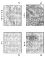
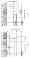
도 1A-1D는 NSCLC 상업적 종양에서 PD-L1 발현의 특유의 패턴을 보여주는 4종의 면역조직화학 (IHC) 영상을 제공한다. PD-L1 발현의 패턴은 미만성 (도 1A), 불균질 (도 1B), 종양-기질 계면 (도 1C) 및 음성 (도 1D)으로 지정된다.
도 2A 및 2B는 도 1A-1D에 제시된 각각의 PD-L1 패턴, 즉, 미만성 (D), 불균질 (H), 음성 (N) 및 종양-기질 계면 (T)에서의 PD-L1 H-점수의 분포 (도 2A), 및 2종의 NSCLC 하위유형, 즉 선암종 및 편평 세포 암종에서의 PD-L1 H-점수의 분포 (도 2B)를 보여준다.
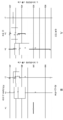
도 3A-3C는 니볼루맙 단독요법으로 치료된 환자로부터의 시험 생검에 상응하는 미만성 (도 3A), 종양-기질 계면 (도 3B) 및 음성 (도 3C) PD-L1 발현 패턴에 상응하는 IHC 영상을 보여준다.
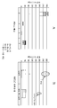
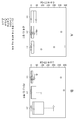
도 4는 니볼루맙 단독요법을 받는, 종양 등급에 의해 카테고리화된 환자의 PD-L1 H-점수를 보여준다. 대다수의 완전 반응자 (CR) 및 부분 반응자 (PR)에서의 우세 PD-L1 패턴은 미만성 패턴이다. TS-IF = 종양-기질 계면, SD = 안정 질환, PD = 진행성 질환, BOR = 최상의 전체 반응.
도 5A 및 5B는 PD-L1 종양 우세 패턴에 따른 전체 CI 점수 (도 5A) 및 PD-L1 CI 점수 (도 5B)를 보여준다. TS-IF = 종양-기질 계면.
도 6은 PD-L1, CD68 및 CD3에 대해 염색된 멀티플렉스화 IHC 영상을 제시한다.
도 7A 및 7B는 RNA 서열분석을 사용하여 측정된 바와 같은 PD-L1 발현 패턴에 따른 NSCLC 종양 내 PD-L1 발현 (도 7A), 및 엑솜 서열분석을 사용하여 측정된 바와 같은 PD-L1 발현 패턴에 따른 NSCLC 종양 내 돌연변이 부하 (도 7B)를 보여준다.
도 8A 및 8B는 CI 점수에 의해 측정된 바와 같은 NSCLC 종양 내 미스센스 돌연변이의 수 대 전체 염증 사이의 관계 (도 8A), 및 PDLP1pos CI 점수에 의해 측정된 바와 같은 NSCLC 종양 내 미스센스 돌연변이의 수 대 PD-L1+ 염증 사이의 관계 (도 8B)를 보여준다.
도 9A 및 9B는 도 9A에서 관찰된 PD-L1 발현 패턴에 대한 상이한 바이오마커 (TP53, STK11, KEAP1, KRAS, EGFR 및 MET)에서의 돌연변이의 빈도를 보여준다. D = 미만성, H = 불균질, I = 종양-기질 계면, N = 음성. 도 9B는 STK11 돌연변이의 존재 ("y") 또는 부재 ("n")에 대한 RNA 서열분석 (RNAseq)에 의해 측정된 PD-L1 발현을 보여준다.
도 10A-10C는 STK11 돌연변이의 존재 ("y") 또는 부재 ("n")와 PD-L1+ CI 점수 사이의 관계를 보여준다 (도 10A). 도 10A에 제시된 정보에 상응하는 수치 데이터는 도 10B에 제시된다. 도 10C는 STK11 돌연변이의 존재 ("STK11-MUT") 또는 부재 ("STK11-WT")에 따른 NSCLC 종양에서의 전체 염증 점수에 상응하는 수치 데이터를 보여준다.
도 11은 스물-네 (24)개의 NSCLC 종양 샘플의 이뮤노프린트 분석을 보여주며, 여기서 FOLR2, VSIG4, CD163, CLEC4D, CSF1R, CD86, MS4A1, CD79B, CD19, KIR2DS4, KIR2DL4, CD3E, CCR4, CCR8 및 CD8A의 수준을 분석하여 염증 패턴에 따라 샘플을 분류하였다 (sigClass). 샘플을 낮은 ("sigClass low"), 중간 ("sigClass med") 또는 높은 ("sigClass hi") 염증으로 분류하였다. 샘플을 또한 STK11 돌연변이의 존재 ("STK11 mut") 또는 부재 ("STK11 wt")에 따라 분류하였다. 추가로, 샘플을 PD-L1 발현 패턴에 따라 음성 ("PDL1_패턴 2 음성"), 미만성 ("PDL1_패턴 2 미만성"), 불균질 ("PDL1_패턴 2 불균질") 또는 종양-기질 계면 ("PDL1_패턴 2 TS")으로 분류하였다.Figures 1A-1D provide four immunohistochemical (IHC) images showing a unique pattern of PD-L1 expression in NSCLC commercial tumors. The pattern of PD-L1 expression is designated as diffuse (Figure 1A), heterogeneous (Figure IB), tumor-substrate interface (Figure 1C), and negative (Figure 1D).
FIGS. 2A and 2B show the PD-L1 H-1 pattern at each of the PD-L1 patterns shown in FIGS. 1A-1D, i.e., diffuse (D), heterogeneous (H), negative (N) and tumor- (FIG. 2A), and distribution of PD-L1 H-scores in two NSCLC subtypes, adenocarcinoma and squamous cell carcinoma (FIG. 2B).
Figures 3A-3C show IHC images corresponding to diffuse (Figure 3A), tumor-substrate interface (Figure 3B) and negative (Figure 3C) PD-L1 expression patterns corresponding to test biopsies from patients treated with nobilvit monotherapy Lt; / RTI >
Figure 4 shows the PD-L1 H-score of patients categorized by tumor grade, receiving nobiluric monotherapy. The predominant PD-L1 pattern in the majority of the complete responders (CR) and the partial responders (PR) is a diffuse pattern. TS-IF = tumor-matrix interface, SD = stable disease, PD = advanced disease, BOR = best overall response.
Figures 5A and 5B show the overall CI scores (Figure 5A) and PD-L1 CI scores (Figure 5B) according to the PD-L1 tumor predominant pattern. TS-IF = tumor-matrix interface.
Figure 6 presents a multiplexed IHC image stained for PD-L1, CD68 and CD3.
Figures 7A and 7B show PD-L1 expression (Figure 7A) in NSCLC tumors according to the PD-L1 expression pattern as measured using RNA sequencing (Figure 7A), and PD-L1 expression pattern as determined using exon sequence analysis Lt; RTI ID = 0.0 > (Figure 7B). ≪ / RTI >
Figures 8A and 8B show the relationship between the number of mismatch mutations in NSCLC tumors as measured by CI scores versus total inflammation (Figure 8A), and the number of mismatch mutations in NSCLC tumors as measured by PDLPlpos CI score PD-L1 + inflammation (Fig. 8B).
Figures 9A and 9B show the frequency of mutations in the different biomarkers (TP53, STK11, KEAPl, KRAS, EGFR and MET) for the PD-L1 expression pattern observed in Figure 9A. D = diffuse, H = heterogeneity, I = tumor-matrix interface, N = negative. Figure 9B shows PD-L1 expression as measured by RNA sequencing (RNAseq) for the presence ("y") or absence ("n") of the STK11 mutation.
Figures 10A-10C show the relationship between the presence ("y") or absence ("n") of the STK11 mutation and the PD-L1 + CI score (Figure 10A). Numerical data corresponding to the information shown in Fig. 10A is shown in Fig. 10B. Figure 10C shows numerical data corresponding to the total inflammation score in NSCLC tumors according to the presence of the STK11 mutation (" STK11-MUT ") or absence (" STK11-WT ").
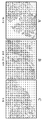
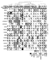
Figure 11 shows an immunoprinting analysis of a sample of twenty-four (24) NSCLC tumors, wherein FOLR2, VSIG4, CD163, CLEC4D, CSF1R, CD86, MS4A1, CD79B, CD19, KIR2DS4, KIR2DL4, CD3E, CCR4, CCR8 And CD8A were analyzed to classify the samples according to the inflammation pattern (sigClass). Samples were classified as low ("sigClass low"), medium ("sigClass med") or high ("sigClass hi") inflammation. Samples were also sorted according to the presence of the STK11 mutation (" STK11 mut ") or absence (" STK11 wt "). Further, the sample may be labeled with negative (" PDL1_pattern2 negative "), diffuse (" PDL1_pattern2 diffuse "), heterogeneous (" PDL1_pattern2 heterogeneous ") or tumor- Interface (" PDL1_pattern2 TS ").
본 발명은 (i) 프로그램화된 사멸 리간드 1 (PD-L1)의 발현 패턴을 결정하고, (ii) 종양을 앓는 대상체에게, 종양이 PD-L1 발현의 미만성 패턴을 나타내는 경우에, 프로그램화된 사멸-1 (PD-1) 수용체에 특이적으로 결합하고 PD-1 활성을 억제하는 항체 또는 그의 항원-결합 부분 ("항-PD-1 항체")을 투여하는 것을 포함하는, 상기 대상체를 치료하는 방법에 관한 것이다. 다른 측면에서, 본 발명은 (i) STK11-양성 종양을 갖는 대상체를 확인하고, (ii) 그 대상체에게 항-PD-1 항체를 투여하는 것을 포함하는, 종양을 앓는 대상체를 치료하는 방법에 관한 것이다. 특정 실시양태에서, 종양은 NSCLC로부터 유래된다.The present invention relates to a method of determining the expression pattern of (i) a programmed killing ligand 1 (PD-L1), and (ii) administering to a subject suffering from the tumor, Comprising administering to said subject an antibody or antigen-binding portion thereof (" anti-PD-1 antibody ") that specifically binds to the death-1 (PD-1) receptor and inhibits PD- . In another aspect, the present invention relates to a method of treating a subject suffering from a tumor, comprising (i) identifying a subject having an STK11-positive tumor and (ii) administering an anti-PD-1 antibody to the subject will be. In certain embodiments, the tumor is derived from NSCLC.
용어Terms
본 개시내용이 보다 용이하게 이해될 수 있도록, 특정 용어가 먼저 정의된다. 본 출원에 사용된 바와 같이, 본원에 달리 명백하게 제공된 경우를 제외하고는, 각각의 하기 용어는 하기 제시된 의미를 가질 것이다. 추가의 정의가 본 출원 전반에 걸쳐 제시된다.In order that the disclosure may be more readily understood, certain terms are first defined. As used in this application, each of the following terms will have the meanings set forth below, except as otherwise expressly provided herein. Additional definitions are set forth throughout this application.
"투여하는"은 관련 기술분야의 통상의 기술자에게 공지된 다양한 방법 및 전달 시스템 중 어느 것을 사용하여 치료제를 포함하는 조성물을 대상체에게 물리적으로 도입하는 것을 지칭한다. 항-PD-1 항체에 대한 투여 경로는 정맥내, 근육내, 피하, 복강내, 척수 또는 다른 비경구 투여 경로, 예를 들어 주사 또는 주입에 의한 투여를 포함한다. 본원에 사용된 어구 "비경구 투여"는 통상적으로 주사에 의한, 경장 및 국소 투여 이외의 다른 투여 방식을 의미하고, 비제한적으로, 정맥내, 근육내, 동맥내, 척수강내, 림프내, 병변내, 피막내, 안와내, 심장내, 피내, 복강내, 경기관, 피하, 각피하, 관절내, 피막하, 지주막하, 척수내, 경막외 및 흉골내 주사 및 주입, 뿐만 아니라 생체내 전기천공을 포함한다. 일부 실시양태에서, 조합은 비-비경구 경로를 통해 투여되고, 일부 실시양태에서는 경구로 투여된다. 다른 비-비경구 경로는 국소, 표피 또는 점막 투여 경로, 예를 들어 비강내, 질, 직장, 설하 또는 국소 투여 경로를 포함한다. 투여는 또한, 예를 들어 1회, 복수회, 및/또는 1회 이상의 연장된 기간에 걸쳐 수행될 수 있다.&Quot; Administering " refers to physically introducing a composition comprising a therapeutic agent into a subject using any of a variety of methods and delivery systems known to those of ordinary skill in the art. Administration routes for anti-PD-1 antibodies include intravenous, intramuscular, subcutaneous, intraperitoneal, spinal or other parenteral administration routes, for example by injection or infusion. The phrase " parenteral administration ", as used herein, refers to other modes of administration other than injection, topical and enteral administration, and includes, but is not limited to, intravenous, intramuscular, intraarterial, intrathecal, Injection and infusion, as well as in vivo electrophysiology, as well as intravenous, intraperitoneal, intraperitoneal, intraperitoneal, intraperitoneal, intramuscular, subcutaneous, subcutaneous, intraarticular, subcapsular, subarachnoid, Including perforation. In some embodiments, the combination is administered via a non-parenteral route, and in some embodiments, administered orally. Other non-parenteral routes include topical, epidermal, or mucosal route of administration, such as intranasal, vaginal, rectal, sublingual or topical routes of administration. Administration may also be carried out, for example, over one, several, and / or one or more extended periods of time.
본원에 사용된 "유해 사건" (AE)은 의학적 치료의 사용과 연관된, 임의의 바람직하지 않고 일반적으로 의도되지 않거나 원하지 않는 징후 (비정상적 실험실 발견 포함), 증상, 또는 질환이다. 예를 들어, 유해 사건은 치료에 대한 반응으로 일어나는 면역계의 활성화 또는 면역계 세포 (예를 들어, T 세포)의 확장과 연관될 수 있다. 의학적 치료는 1건 이상의 연관된 AE를 가질 수 있고 각각의 AE는 동일하거나 상이한 수준의 중증도를 가질 수 있다. "유해 사건을 변경할" 수 있는 방법에 대한 언급은 상이한 치료 요법의 사용과 연관된 1건 이상의 AE의 발생률 및/또는 중증도를 감소시키는 치료 요법을 의미한다.As used herein, " adverse events " (AE) are any undesirable and generally unintended or unwanted indications (including abnormal laboratory findings), symptoms, or diseases associated with the use of medical therapy. For example, adverse events may be associated with the activation of the immune system or the expansion of immune system cells (e. G., T cells) that occur in response to therapy. A medical treatment may have more than one associated AE and each AE may have the same or different levels of severity. Reference to how to " change a noxious event " means a therapeutic regimen that reduces the incidence and / or severity of one or more AEs associated with the use of different therapies.
"항체" (Ab)는 비제한적으로, 항원에 특이적으로 결합하고 디술피드 결합에 의해 상호연결된 적어도 2개의 중쇄 (H) 및 2개의 경쇄 (L)를 포함하는 당단백질 이뮤노글로불린, 또는 그의 항원-결합 부분을 포함할 것이다. 각각의 H 쇄는 중쇄 가변 영역 (본원에서 VH로 약칭됨) 및 중쇄 불변 영역을 포함한다. 중쇄 불변 영역은 3개의 불변 도메인, CH1, CH2 및 CH3을 포함한다. 각각의 경쇄는 경쇄 가변 영역 (본원에서 VL로 약칭됨) 및 경쇄 불변 영역을 포함한다. 경쇄 불변 영역은 1개의 불변 도메인, CL을 포함한다. VH 및 VL 영역은 프레임워크 영역 (FR)으로 명명되는 보다 보존된 영역이 산재되어 있는, 상보성 결정 영역 (CDR)으로 명명되는 초가변성 영역으로 추가로 세분될 수 있다. 각각의 VH 및 VL은 아미노-말단에서 카르복시-말단으로 하기 순서로 배열된 3개의 CDR 및 4개의 FR을 포함한다: FR1, CDR1, FR2, CDR2, FR3, CDR3, FR4. 중쇄 및 경쇄의 가변 영역은 항원과 상호작용하는 결합 도메인을 함유한다. 항체의 불변 영역은 면역계의 다양한 세포 (예를 들어, 이펙터 세포) 및 전형적 보체계의 제1 성분 (C1q)을 포함한 숙주 조직 또는 인자에 대한 이뮤노글로불린의 결합을 매개할 수 있다.&Quot; Antibodies " (Ab) include, but are not limited to, glycoprotein immunoglobulins comprising at least two heavy chains (H) and two light chains (L) linked specifically by an antigen and interconnected by a disulfide bond, Antigen-binding portion thereof. Each H chain comprises a heavy chain variable region (abbreviated herein as V H ) and a heavy chain constant region. The heavy chain constant region includes three constant domains, C H1 , C H2, and C H3 . Each light chain comprises a light chain variable region (abbreviated herein as V L ) and a light chain constant region. The light chain constant region comprises one constant domain, C L. The V H and V L regions may be further subdivided into hypervariable regions, termed complementarity determining regions (CDRs), in which more conserved regions termed framework regions (FR) are scattered. Each V H and V L comprises three CDRs and four FRs arranged in the following order from the amino-terminus to the carboxy-terminus: FR1, CDR1, FR2, CDR2, FR3, CDR3, FR4. The variable regions of the heavy and light chains contain a binding domain that interacts with the antigen. The constant region of the antibody may mediate the binding of immunoglobulins to the host tissue or factor, including various cells of the immune system (e. G., Effector cells) and the first component of a typical complement system (Clq).
이뮤노글로불린은 IgA, 분비형 IgA, IgG 및 IgM을 포함하나 이에 제한되지는 않는, 임의의 통상적으로 공지된 이소형으로부터 유래될 수 있다. IgG 하위부류는 또한 관련 기술분야의 통상의 기술자에게 널리 공지되어 있고, 인간 IgG1, IgG2, IgG3 및 IgG4를 포함하나 이에 제한되지는 않는다. "이소형"은 중쇄 불변 영역 유전자에 의해 코딩되는 항체 부류 또는 하위부류 (예를 들어, IgM 또는 IgG1)를 지칭한다. 용어 "항체"는, 예로서, 자연 발생 및 비-자연 발생 항체 둘 다; 모노클로날 및 폴리클로날 항체; 키메라 및 인간화 항체; 인간 또는 비-인간 항체; 완전 합성 항체; 및 단일 쇄 항체를 포함한다. 비-인간 항체는 인간에서 그의 면역원성이 감소하도록 재조합 방법에 의해 인간화될 수 있다. 명백하게 언급되지 않는 경우, 및 문맥상 달리 나타내지 않는 한, 용어 "항체"는 또한 상기 언급된 이뮤노글로불린 중 어느 것의 항원-결합 단편 또는 항원-결합 부분을 포함하고, 1가 및 2가 단편 또는 부분, 및 단일 쇄 항체를 포함한다.Immunoglobulin can be derived from any conventionally known isoform, including, but not limited to, IgA, secreted IgA, IgG and IgM. IgG subclasses are also well known to those of ordinary skill in the art and include, but are not limited to, human IgG1, IgG2, IgG3, and IgG4. &Quot; Isotype " refers to an antibody class or subclass (e.g., IgM or IgG1) that is encoded by a heavy chain constant region gene. The term " antibody " includes, by way of example, both naturally occurring and non-naturally occurring antibodies; Monoclonal and polyclonal antibodies; Chimeric and humanized antibodies; Human or non-human antibodies; Fully synthetic antibodies; And single chain antibodies. Non-human antibodies can be humanized by recombinant methods to decrease their immunogenicity in humans. Unless explicitly stated, and unless the context indicates otherwise, the term " antibody " also encompasses antigen-binding or antigen-binding portions of any of the above-mentioned immunoglobulins and includes monovalent and divalent fragments or portions , And single chain antibodies.
"단리된 항체"는 상이한 항원 특이성을 갖는 다른 항체가 실질적으로 없는 항체를 지칭한다 (예를 들어, PD-1에 특이적으로 결합하는 단리된 항체는 PD-1 이외의 다른 항원에 특이적으로 결합하는 항체가 실질적으로 없음). 그러나, PD-1에 특이적으로 결합하는 단리된 항체는 다른 항원, 예컨대 상이한 종으로부터의 PD-1 분자와 교차-반응성을 가질 수 있다. 더욱이, 단리된 항체는 다른 세포 물질 및/또는 화학물질이 실질적으로 없을 수 있다.&Quot; Isolated antibody " refers to an antibody that is substantially free of other antibodies having different antigen specificity (e.g., an isolated antibody that specifically binds to PD-1 specifically binds to an antigen other than PD-1 Substantially free of binding antibody). However, isolated antibodies that specifically bind to PD-1 may have cross-reactivity with other antigens, such as PD-1 molecules from different species. Moreover, the isolated antibody may be substantially free of other cellular material and / or chemicals.
용어 "모노클로날 항체" (mAb)는 단일 분자 조성의 항체 분자, 즉, 1차 서열이 본질적으로 동일하고 특정한 에피토프에 대해 단일 결합 특이성 및 친화도를 나타내는 항체 분자의 비-자연 발생 제제를 지칭한다. 모노클로날 항체는 단리된 항체의 예이다. 모노클로날 항체는 하이브리도마, 재조합, 트랜스제닉, 또는 관련 기술분야의 통상의 기술자에게 공지된 다른 기술에 의해 생산될 수 있다.The term " monoclonal antibody " (mAb) refers to an antibody molecule of a single molecular composition, i.e., a non-naturally occurring agent of an antibody molecule that exhibits a single binding specificity and affinity for a particular epitope, do. Monoclonal antibodies are examples of isolated antibodies. Monoclonal antibodies can be produced by hybridoma, recombinant, transgenic, or other techniques known to those skilled in the art.
"인간 항체" (HuMAb)는 FR 및 CDR 둘 다가 인간 배선 이뮤노글로불린 서열로부터 유래된 것인 가변 영역을 갖는 항체를 지칭한다. 추가로, 항체가 불변 영역을 함유하는 경우에, 불변 영역도 또한 인간 배선 이뮤노글로불린 서열로부터 유래된다. 본 발명의 인간 항체는 인간 배선 이뮤노글로불린 서열에 의해 코딩되지 않는 아미노산 잔기 (예를 들어, 시험관내 무작위 또는 부위-특이적 돌연변이유발에 의해 또는 생체내 체세포 돌연변이에 의해 도입된 돌연변이)를 포함할 수 있다. 그러나, 본원에 사용된 용어 "인간 항체"는 또 다른 포유동물 종, 예컨대 마우스의 배선으로부터 유래된 CDR 서열이 인간 프레임워크 서열 상에 그라프팅된 것인 항체를 포함하는 것으로 의도되지는 않는다. 용어 "인간 항체" 및 "완전 인간 항체"는 동의어로 사용된다.A " human antibody " (HuMAb) refers to an antibody having a variable region in which both the FR and CDRs are derived from a human wiring immunoglobulin sequence. In addition, where the antibody contains a constant region, the constant region is also derived from the human wiring immunoglobulin sequence. Human antibodies of the invention include amino acid residues that are not encoded by a human line immunoglobulin sequence (e. G., Mutations introduced by in vitro random or site-specific mutagenesis or by in vivo somatic mutagenesis) . However, the term " human antibody " as used herein is not intended to encompass antibodies that are CDR sequences from another mammalian species, such as a mouse line, that have been grafted onto human framework sequences. The terms " human antibody " and " fully human antibody " are used synonymously.
"인간화 항체"는 비-인간 항체의 CDR 밖의 아미노산 중 일부, 대부분 또는 모두가 인간 이뮤노글로불린으로부터 유래된 상응하는 아미노산으로 대체된 것인 항체를 지칭한다. 인간화 형태의 항체의 한 실시양태에서, CDR 밖의 아미노산 중 일부, 대부분 또는 모두는 인간 이뮤노글로불린으로부터의 아미노산으로 대체된 반면에 1개 이상의 CDR 내의 일부, 대부분 또는 모든 아미노산은 변화되지 않는다. 특정한 항원에 결합하는 항체의 능력을 제거하지 않는 한, 아미노산의 적은 부가, 결실, 삽입, 치환 또는 변형은 허용가능하다. "인간화 항체"는 원래 항체의 경우와 유사한 항원 특이성을 유지한다.&Quot; Humanized antibody " refers to an antibody in which some or all, or all, of the amino acids outside the CDRs of a non-human antibody are replaced by the corresponding amino acids derived from human immunoglobulins. In one embodiment of an antibody of the humanized form, some, most or all of the amino acids outside the CDRs are replaced by amino acids from human immunoglobulins, while some, most or all of the amino acids in one or more CDRs are not changed. Small additions, deletions, insertions, substitutions or modifications of amino acids are permissible unless the ability of the antibody to bind to a particular antigen is eliminated. &Quot; Humanized antibody " retains antigen specificity similar to that of the original antibody.
"키메라 항체"는 가변 영역은 한 종으로부터 유래되고 불변 영역은 또 다른 종으로부터 유래된 것인 항체, 예컨대 가변 영역은 마우스 항체로부터 유래되고 불변 영역은 인간 항체로부터 유래된 것인 항체를 지칭한다.&Quot; Chimeric antibody " refers to an antibody in which the variable region is derived from one species and the constant region is derived from another species, for example, an antibody in which the variable region is derived from a mouse antibody and the constant region is derived from a human antibody.
"항-항원 항체"는 항원에 특이적으로 결합하는 항체를 지칭한다. 예를 들어, 항-PD-1 항체는 PD-1에 특이적으로 결합한다.&Quot; Anti-antigen antibody " refers to an antibody that specifically binds to an antigen. For example, anti-PD-1 antibodies specifically bind to PD-1.
항체의 "항원-결합 부분" (또한 "항원-결합 단편"으로 불림)은 전체 항체에 의해 결합되는 항원에 특이적으로 결합하는 능력을 유지하는 항체의 1개 이상의 단편을 지칭한다.An "antigen-binding portion" (also referred to as an "antigen-binding fragment") of an antibody refers to one or more fragments of an antibody that retain the ability to specifically bind to the antigen bound by the whole antibody.
"암"은 신체 내 비정상 세포의 비제어된 성장을 특징으로 하는 다양한 질환의 광범위한 군을 지칭한다. 비조절된 세포 분열 및 성장은 분열 및 성장으로 인해 이웃 조직을 침습하는 악성 종양의 형성을 일으키고 또한 림프계 또는 혈류를 통해 신체의 원위 부분으로 전이할 수 있다. 일부 실시양태에서, 암은 본원에 개시된 임의의 암이다. 실시양태에서, 암은 폐암이다. 특정 실시양태에서, 폐암은 비소세포 폐암 (NSCLC)이다. 실시양태에서, NSCLC는 편평 조직학 (편평 NSCLC)을 갖는다. 다른 실시양태에서, NSCLC는 비-편평 조직학 (비-편평 NSCLC)을 갖는다. "암"은 종양을 포함할 수 있다. "종양"은 신생물성 세포 성장 및 증식 (악성이든지 또는 양성이든지 간에), 및 모든 전암성 및 암성 세포 및 조직을 지칭한다.&Quot; Cancer " refers to a broad group of diverse diseases characterized by uncontrolled growth of abnormal cells in the body. Unregulated cell division and growth can result in the formation of malignant tumors that invade neighboring tissues due to cleavage and growth, and can also migrate to the distal part of the body through the lymphatic system or bloodstream. In some embodiments, the cancer is any of the cancers disclosed herein. In an embodiment, the cancer is lung cancer. In certain embodiments, the lung cancer is non-small cell lung cancer (NSCLC). In an embodiment, the NSCLC has a flat histology (flat NSCLC). In another embodiment, the NSCLC has non-squamous histology (non-flat NSCLC). &Quot; Cancer " can include tumors. &Quot; Tumor " refers to neoplastic cellular growth and proliferation (whether malignant or benign), and to all pre-cancerous and cancerous cells and tissues.
"세린/트레오닌 키나제 11" 또는 "STK11" (또한 "분극-관련 단백질 LKB1", "신암종 항원 NY-REN-19", "간 키나제 B1", "EC 2.7.11.1" 및 "HLKB1"로 공지됨)은 세포 극성을 조절하고 종양 억제자로서 기능하는 세린/트레오닌 키나제 패밀리의 구성원을 지칭한다. STK11은 AMP-활성화 단백질 키나제 (AMPK) 패밀리 구성원의 활성을 제어하여, 이에 의해 다양한 과정, 예컨대 세포 대사, 세포 극성, 아폽토시스 및 DNA 손상 반응에서 소정의 역할을 한다. STK11은 편재적으로 발현되며, 고환 및 태아 간에서 가장 강하게 발현된다. STK11은 NSCLC, 특히 KRAS 돌연변이를 보유하는 종양에서 통상적으로 불활성화된다. 본원에 기재된 바와 같이, 돌연변이된 STK11, 예를 들어 STK11의 야생형 발현 상실은 SCLC로부터 유래된 종양에서의 감소된 또는 이상 PD-L1 발현과 상관관계가 있다. 일부 실시양태에서, 돌연변이된 STK11, 예를 들어 STK11의 야생형 발현 상실은 SCLC로부터 유래된 종양에서 발생하며, 여기서 종양은 야생형 KRAS를 발현하거나 또는 발현하지 않는다 (예를 들어, 종양은 KRAS 돌연변이를 갖고 있거나 또는 갖지 않는다). 일부 실시양태에서, STK11 돌연변이체는, 예를 들어 이전에 문헌 [Koyama et al., Cancer Res. 76(5):999-1008 (2016), 및/또는 Skoulidis et al., Cancer Discov. 5(8):860-77 (2015)] (이들 둘 다 그 전문이 본원에 참조로 포함됨)에 기재된 STK11 돌연변이체이다.Known as " serine /
용어 "면역요법"은 면역 반응을 유도하거나, 증진시키거나, 억제하거나 또는 달리 변형시키는 것을 포함하는 방법에 의해, 질환을 앓거나, 질환에 걸릴 위험이 있거나 또는 질환의 재발을 앓고 있는 대상체를 치료하는 것을 지칭한다. 대상체의 "치료" 또는 "요법"은 질환과 연관된 증상, 합병증, 상태 또는 생화학적 징후의 발병, 진행, 발달, 중증도 또는 재발을 역전시키거나, 완화시키거나, 호전시키거나, 억제하거나, 저속화하거나 또는 방지하는 것을 목적으로 대상체에 대해 수행되는 임의의 유형의 시술 또는 과정, 또는 그에 대한 활성제의 투여를 지칭한다.The term " immunotherapy " refers to the treatment of a subject suffering from or at risk of developing a disease, or suffering from a recurrence of a disease, by a method comprising inducing, enhancing, inhibiting or otherwise modifying an immune response . The term " treatment " or " therapy " of an object refers to reversing, alleviating, ameliorating, inhibiting, slowing, or slowing the onset, progression, development, severity or recurrence of a symptom, complication, condition, or biochemical indication Refers to any type of procedure or procedure performed on a subject for the purpose of preventing, preventing, or preventing the same.
본원에 사용된 "PD-L1 양성"은 "적어도 약 1%의 PD-L1 발현"과 상호교환가능하게 사용될 수 있다. 한 실시양태에서, PD-L1 발현은 관련 기술분야에 공지된 임의의 방법에 의해 사용될 수 있다. 또 다른 실시양태에서, PD-L1 발현은 자동화 IHC에 의해 측정된다. PD-L1 양성 종양은 따라서 자동화 IHC에 의해 측정된 바와 같이 적어도 약 1%, 적어도 약 2%, 적어도 약 5%, 적어도 약 10%, 적어도 약 20%, 적어도 약 25%, 적어도 약 30%, 적어도 약 40%, 적어도 약 50%, 적어도 약 60%, 적어도 약 70%, 적어도 약 75%, 적어도 약 80%, 적어도 약 85%, 적어도 약 90%, 적어도 약 95%, 또는 약 100%의 PD-L1 발현 종양 세포를 가질 수 있다. 특정 실시양태에서, "PD-L1 양성"은 세포의 표면 상에 PD-L1을 발현하는 적어도 100개의 세포가 존재한다는 것을 의미한다.As used herein, " PD-L1 positive " may be used interchangeably with " at least about 1% PD-L1 expression ". In one embodiment, PD-L1 expression can be used by any method known in the art. In another embodiment, PD-L1 expression is measured by automated IHC. The PD-L1 positive tumors are thus at least about 1%, at least about 2%, at least about 5%, at least about 10%, at least about 20%, at least about 25%, at least about 30% At least about 50%, at least about 60%, at least about 70%, at least about 75%, at least about 80%, at least about 85%, at least about 90%, at least about 95% PD-L1 expressing tumor cells. In certain embodiments, " PD-L1 positive " means that there are at least 100 cells expressing PD-L1 on the surface of the cell.
"프로그램화된 사멸-1" (PD-1)은 CD28 패밀리에 속하는 면역억제 수용체를 지칭한다. PD-1은 생체내 이전에 활성화된 T 세포 상에서 우세하게 발현되고, 2종의 리간드, PD-L1 및 PD-L2에 결합한다. 본원에 사용된 용어 "PD-1"은 인간 PD-1 (hPD-1), hPD-1의 변이체, 이소형 및 종 상동체, 및 hPD-1과 적어도 1개의 공통 에피토프를 갖는 유사체를 포함한다. 완전한 hPD-1 서열은 진뱅크 수탁 번호 U64863 하에 찾아볼 수 있다.&Quot; Programmed death-1 " (PD-1) refers to immunosuppressive receptors belonging to the CD28 family. PD-1 is predominantly expressed on previously activated T cells in vivo and binds to two ligands, PD-L1 and PD-L2. As used herein, the term " PD-1 " includes human PD-1 (hPD-1), variants of hPD-1, isoforms and species homologues, and analogs having at least one common epitope with hPD- . The complete hPD-1 sequence can be found under Genbank accession number U64863.
"프로그램화된 사멸 리간드-1 (PD-L1)"은 PD-1에 대한 결합 시 T 세포 활성화 및 시토카인 분비를 하향조절하는, PD-1에 대한 2종의 세포 표면 당단백질 리간드 중 하나이다 (다른 것은 PD-L2임). 본원에 사용된 용어 "PD-L1"은 인간 PD-L1 (hPD-L1), hPD-L1의 변이체, 이소형 및 종 상동체, 및 hPD-L1과 적어도 1개의 공통 에피토프를 갖는 유사체를 포함한다. 완전한 hPD-L1 서열은 진뱅크 수탁번호 Q9NZQ7 하에 찾아볼 수 있다.&Quot; Programmed apoptotic ligand-1 (PD-L1) " is one of two cell surface glycoprotein ligands for PD-1 that downregulates T cell activation and cytokine release upon binding to PD-1 Is PD-L2). As used herein, the term "PD-L1" includes human PD-L1 (hPD-L1), variants of hPD-L1, isoforms and species homologues, and analogs having hPD-L1 and at least one common epitope . The complete hPD-L1 sequence can be found under GeneBank accession number Q9NZQ7.
"대상체"는 임의의 인간 또는 비-인간 동물을 포함한다. 용어 "비-인간 동물"은 척추동물, 예컨대 비-인간 영장류, 양, 개, 및 설치류, 예컨대 마우스, 래트, 및 기니 피그를 포함하나 이에 제한되지는 않는다. 일부 실시양태에서, 대상체는 인간이다. 용어 "대상체" 및 "환자"는 본원에서 상호교환가능하게 사용된다.&Quot; Subject " includes any human or non-human animal. The term "non-human animal" includes, but is not limited to, vertebrate animals such as non-human primates, sheep, dogs, and rodents such as mice, rats, and guinea pigs. In some embodiments, the subject is a human. The terms " subject " and " patient " are used interchangeably herein.
약물 또는 치료제의 "치료 유효량" 또는 "치료 유효 투여량"은, 단독으로 사용되거나 또는 또 다른 치료제와 조합되어 사용되는 경우에, 질환의 발병에 대해 대상체를 보호하거나, 또는 질환 증상의 중증도의 감소, 질환 무증상 기간의 빈도 및 지속기간의 증가, 또는 앓고 있는 질환으로 인한 손상 또는 장애의 방지에 의해 입증되는 질환 퇴행을 촉진하는 약물의 임의의 양이다. 질환 퇴행을 촉진하는 치료제의 능력은 숙련된 진료의에게 공지된 다양한 방법을 사용하여, 예컨대 임상 시험 동안 인간 대상체에서, 인간에서의 효능을 예측하는 동물 모델 시스템에서, 또는 시험관내 검정에서 작용제의 활성을 검정함으로써 평가될 수 있다.A " therapeutically effective amount " or " therapeutically effective dose " of a drug or therapeutic agent, when used alone or in combination with another therapeutic agent, is intended to protect a subject against the onset of the disease, , An increase in the frequency and duration of the disease asymptomatic period, or prevention of damage or disorder due to the illness being afflicted. The ability of therapeutic agents to promote disease degeneration can be determined using a variety of methods known to those skilled in the art, for example, in human subjects during clinical trials, in animal model systems that predict efficacy in humans, . ≪ / RTI >
본원에 사용된 "치료량 미만의 용량"은 과다증식성 질환 (예를 들어, 암)의 치료를 위해 단독으로 투여되는 경우 치료 화합물 (예를 들어, 항체)의 통상적 또는 전형적 용량보다 더 낮은 치료 화합물의 용량을 의미한다.As used herein, "less than the therapeutic dose" refers to a dose of a therapeutic compound that is lower than the typical or typical dose of a therapeutic compound (eg, antibody) when administered alone for the treatment of a hyperproliferative disease (eg, cancer) Capacity.
예로서, "항암제"는 대상체에서 암 퇴행을 촉진하거나 또는 추가의 종양 성장을 방지한다. 특정 실시양태에서, 치료 유효량의 약물은 암을 제거하는 지점까지 암 퇴행을 촉진한다. "암 퇴행을 촉진하는"은 치료 유효량의 약물을 단독으로 또는 항신생물제와 조합하여 투여하여 종양 성장 또는 크기의 감소, 종양의 괴사, 적어도 1종의 질환 증상의 중증도의 감소, 질환 무증상 기간의 빈도 및 지속기간의 증가, 또는 앓고 있는 질환으로 인한 손상 또는 장애의 방지를 유발하는 것을 의미한다. 추가로, 치료와 관련한 용어 "유효한" 및 "유효성"은 약리학적 유효성 및 생리학적 안전성 둘 다를 포함한다. 약리학적 유효성은 환자에서 암 퇴행을 촉진하는 약물의 능력을 지칭한다. 생리학적 안전성은 약물의 투여로부터 유발되는 세포, 기관 및/또는 유기체 수준에서의 독성의 수준 또는 다른 유해 생리학적 효과 (유해 효과)를 지칭한다.By way of example, " anti-cancer agent " promotes cancer degeneration or prevents further tumor growth in a subject. In certain embodiments, a therapeutically effective amount of the drug promotes cancer regression to the point of eliminating the cancer. &Quot; Promoting cancer degeneration " refers to the administration of a therapeutically effective amount of a drug, either alone or in combination with an anti-neoplastic agent, to reduce tumor growth or size, tumor necrosis, decrease in severity of at least one disease symptom, To increase the frequency and duration, or to prevent damage or disorder due to the illness being afflicted. In addition, the terms " effective " and " efficacy " in relation to therapy include both pharmacological efficacy and physiological safety. Pharmacological efficacy refers to the ability of a drug to promote cancer degeneration in a patient. Physiological safety refers to the level of toxicity at the cellular, organ, and / or organism level or other harmful physiological effects (harmful effects) resulting from the administration of the drug.
종양의 치료에 대한 예로서, 치료 유효량의 항암제는 비치료 대상체에 비해 적어도 약 10%, 적어도 약 20%, 적어도 약 40%, 적어도 약 60%, 적어도 약 70%, 적어도 약 80%, 적어도 약 90%, 적어도 약 95%, 또는 적어도 약 100%만큼 세포 성장 또는 종양 성장을 억제할 수 있다. 본 발명의 다른 실시양태에서, 종양 퇴행은 적어도 약 20일, 적어도 약 30일, 적어도 약 40일, 적어도 약 50일, 또는 적어도 약 60일의 기간 동안 관찰되고 계속될 수 있다. 이들 치료 유효성의 최고의 측정에도 불구하고, 면역요법 약물의 평가는 또한 "면역-관련 반응 패턴"을 감안해야 한다.As an example for the treatment of tumors, a therapeutically effective amount of an anti-cancer agent is at least about 10%, at least about 20%, at least about 40%, at least about 60%, at least about 70%, at least about 80% 90%, at least about 95%, or at least about 100% of the cell growth or tumor growth. In another embodiment of the invention, tumor degeneration can be observed and continued for a period of at least about 20 days, at least about 30 days, at least about 40 days, at least about 50 days, or at least about 60 days. Despite the best measurement of these therapeutic efficacy, the evaluation of the immunotherapeutic drug should also take into account the " immune-related response pattern ".
"면역-관련 반응 패턴"은 암-특이적 면역 반응을 유도함으로써 또는 천연 면역 과정을 변형시킴으로써 항종양 효과를 생성하는 면역요법제로 치료된 암 환자에서 종종 관찰되는 임상 반응 패턴을 지칭한다. 이 반응 패턴은, 전통적인 화학요법제의 평가에서는 질환 진행으로 분류되고 약물 실패와 동의어인 종양 부담의 초기 증가 또는 새로운 병변의 출현에 이어지는 유익한 치료 효과를 특징으로 한다. 따라서, 면역요법제의 적절한 평가는 표적 질환에 대한 이들 작용제의 효과의 장기간 모니터링을 요구할 수 있다. 약물의 치료 유효량은 "예방 유효량"을 포함하며, 이는 암이 발생할 위험이 있는 대상체 (예를 들어, 전-악성 상태를 갖는 대상체) 또는 암의 재발을 앓을 위험이 있는 대상체에게 단독으로 또는 항신생물제와 조합되어 투여되는 경우에, 암의 발생 또는 재발을 억제하는 약물의 임의의 양이다. 특정 실시양태에서, 예방 유효량은 암의 발생 또는 재발을 전적으로 방지한다. 암의 발생 또는 재발을 "억제하는"은 암의 발생 또는 재발 가능성을 경감시키거나, 또는 암의 발생 또는 재발을 전적으로 방지하는 것을 의미한다.&Quot; Immune-related response pattern " refers to the clinical response pattern often observed in cancer patients treated with an immunotherapeutic agent that produces an anti-tumor effect by inducing a cancer-specific immune response or by modifying the natural immune process. This response pattern is characterized by a beneficial therapeutic effect following an initial increase in tumor burden or the appearance of a new lesion that is categorized as disease progression in the assessment of traditional chemotherapy and is synonymous with drug failure. Thus, proper evaluation of immunotherapies may require long-term monitoring of the effects of these agents on the target disease. The therapeutically effective amount of the drug includes a " prophylactically effective amount ", which is sufficient to treat a subject at risk of developing cancer (e.g., a subject having a pre-malignant condition) or a subject at risk of developing cancer recurrence, When administered in combination with an agent, is any amount of a drug that inhibits the occurrence or recurrence of cancer. In certain embodiments, the prophylactically effective amount prevents the occurrence or recurrence of cancer altogether. &Quot; Suppressing " the occurrence or recurrence of cancer means reducing the likelihood of cancer development or recurrence, or preventing the occurrence or recurrence of cancer altogether.
대안적 사용 (예를 들어, "또는")은 대안 중 하나, 둘 다, 또는 그의 임의의 조합을 의미하는 것으로 이해되어야 한다. 본원에 사용된 단수 형태는 임의의 언급되거나 열거된 성분 중 "하나 이상"을 지칭하는 것으로 이해되어야 한다.Alternative uses (e.g., "or") should be understood to mean one, both, or any combination thereof. The singular forms as used herein should be understood to refer to " one or more " of any of the recited or enumerated ingredients.
용어 "약" 또는 "본질적으로 포함하는"은 관련 기술분야의 통상의 기술자에 의해 결정된 바와 같은 특정한 값 또는 조성에 대한 허용 오차 범위 내의 값 또는 조성을 지칭하며, 이는 부분적으로 값 또는 조성이 측정 또는 결정되는 방법, 즉 측정 시스템의 한계에 좌우될 것이다. 예를 들어, "약" 또는 "본질적으로 포함하는"은 관련 기술분야에서의 실시에 따라 1 또는 1 초과의 표준 편차 내에 있음을 의미할 수 있다. 대안적으로, "약" 또는 "본질적으로 포함하는"은 최대 10% 또는 20%의 범위 (즉, ±10% 또는 ±20%)를 의미할 수 있다. 예를 들어, 약 3 mg은 2.7 mg 내지 3.3 mg (10% 경우) 또는 2.4 mg 내지 3.6 mg (20% 경우)의 임의의 수를 포함할 수 있다. 추가로, 특히 생물학적 시스템 또는 과정과 관련하여, 그 용어는 값의 최대 한 자릿수 배수 또는 최대 5-배를 의미할 수 있다. 특정한 값 또는 조성이 본 출원 및 청구범위에서 제공되는 경우에, 달리 언급되지 않는 한, "약" 또는 "본질적으로 포함하는"의 의미는 그러한 특정한 값 또는 조성에 대한 허용 오차 범위 내에 있는 것으로 가정되어야 한다.Refers to a value or composition within a tolerance range for a particular value or composition as determined by one of ordinary skill in the art, , That is, the limits of the measurement system. For example, " about " or " essentially comprising " may mean within a standard deviation of 1 or more, depending on the implementation in the art. Alternatively, " about " or " essentially comprising " may mean a range of up to 10% or 20% (i.e., +/- 10% or +/- 20%). For example, about 3 mg may include any number of 2.7 mg to 3.3 mg (for 10%) or 2.4 mg to 3.6 mg (for 20%). In addition, particularly with respect to biological systems or processes, the term may mean a maximum of one order of magnitude or a maximum of five-fold of the value. Where a particular value or composition is provided in this application and the claims, the meaning of "about" or "essentially comprising", unless otherwise stated, is assumed to be within the tolerance range for such a particular value or composition do.
본원에 사용된 용어 "약 매주 1회", "약 2주마다 1회", 또는 임의의 다른 유사한 투여 간격 용어는 대략적인 횟수를 의미한다. "약 매주 1회"는 7일 ± 1일마다, 즉, 6일마다 내지 8일마다를 포함할 수 있다. "약 2주마다 1회"는 14일 ± 3일마다, 즉, 11일마다 내지 17일마다를 포함할 수 있다. 유사한 근사법이, 예를 들어 약 3주마다 1회, 약 4주마다 1회, 약 5주마다 1회, 약 6주마다 1회, 및 약 12주마다 1회에 대해 적용된다. 일부 실시양태에서, 약 6주마다 1회 또는 약 12주마다 1회의 투여 간격은, 제1 용량이 제1주 중 임의의 날에 투여될 수 있고, 이어서 다음 용량이 각각 제6주 또는 제12주 중 임의의 날에 투여될 수 있다는 것을 의미한다. 다른 실시양태에서, 약 6주마다 1회 또는 약 12주마다 1회의 투여 간격은, 제1 용량이 제1주의 특정한 날 (예를 들어, 월요일)에 투여되고, 이어서 다음 용량이 각각 제6주 또는 제12주의 동일한 날 (즉, 월요일)에 투여된다는 것을 의미한다.As used herein, the terms " about once per week ", " about once every two weeks, " or any other similar dosage interval term refers to an approximate number of times. &Quot; Approximately once a week " may include every 7 days +/- 1 day, i.e. every 6 days to every 8 days. &Quot; about every 2 weeks " may include every 14 days +/- 3 days, i.e. every 11 days to every 17 days. Similar approximations apply, for example, about once every three weeks, once every four weeks, once every about five weeks, once every six weeks, and once every about twelve weeks. In some embodiments, once every about 6 weeks or once every 12 weeks, the first dose can be administered on any day of the first week, followed by the next dose to the sixth week or twelfth Means that the compound can be administered on any day of the week. In other embodiments, once every about 6 weeks or once every 12 weeks, the first dose is administered at a particular day of the first week (e.g., Monday), followed by the next dose at the sixth week Or on the same day of the 12th week (i.e., Monday).
본원에 지칭된 용어 "중량-기준 용량"은 환자에게 투여되는 용량이 환자의 중량을 기준으로 하여 계산된다는 것을 의미한다. 예를 들어, 60 kg 체중을 갖는 환자가 3 mg/kg의 항-PD-1 항체를 요구하는 경우에, 투여를 위해 항-PD-1 항체의 적절한 양 (즉, 180 mg)을 계산하고 사용할 수 있다.The term " weight-based dose " referred to herein means that the dose administered to a patient is calculated based on the weight of the patient. For example, if a patient with a 60 kg body weight requires 3 mg / kg of anti-PD-1 antibody, the appropriate amount of anti-PD-1 antibody (i.e., 180 mg) .
본 발명의 방법과 관련하여 용어 "고정 용량"의 사용은 단일 조성물 중에 2종 이상의 상이한 항체 (예를 들어, 항-PD-1 항체 및 제2 항체)가 서로 특정한 (고정) 비로 조성물 중에 존재한다는 것을 의미한다. 일부 실시양태에서, 고정 용량은 항체의 중량 (예를 들어, mg)을 기준으로 한다. 특정 실시양태에서, 고정 용량은 항체의 농도 (예를 들어, mg/ml)를 기준으로 한다. 일부 실시양태에서, 제1 항체 (예를 들어, 항-PD-1 항체) (mg) 대 제2 항체 (mg)의 비는 적어도 약 1:1, 약 1:2, 약 1:3, 약 1:4, 약 1:5, 약 1:6, 약 1:7, 약 1:8, 약 1:9, 약 1:10, 약 1:15, 약 1:20, 약 1:30, 약 1:40, 약 1:50, 약 1:60, 약 1:70, 약 1:80, 약 1:90, 약 1:100, 약 1:120, 약 1:140, 약 1:160, 약 1:180, 약 1:200, 약 200:1, 약 180:1, 약 160:1, 약 140:1, 약 120:1, 약 100:1, 약 90:1, 약 80:1, 약 70:1, 약 60:1, 약 50:1, 약 40:1, 약 30:1, 약 20:1, 약 15:1, 약 10:1, 약 9:1, 약 8:1, 약 7:1, 약 6:1, 약 5:1, 약 4:1, 약 3:1, 또는 약 2:1이다. 예를 들어, 항-PD-1 항체 및 제2 항체의 3:1 비는 바이알이 약 240 mg의 항-PD-1 항체 및 80 mg의 제2 항체 또는 약 3 mg/ml의 항-PD-1 항체 및 1 mg/ml의 제2 항체를 함유할 수 있다는 것을 의미할 수 있다.The use of the term " fixed dose " in connection with the method of the present invention means that two or more different antibodies (e.g. anti-PD-1 antibody and second antibody) in a single composition are present in the composition in a specific (fixed) . In some embodiments, the fixed dose is based on the weight of the antibody (e.g., mg). In certain embodiments, the fixed dose is based on the concentration of the antibody (e.g., mg / ml). In some embodiments, the ratio of (mg) to (mg) of the first antibody (e.g., anti-PD-1 antibody) is at least about 1: 1, about 1: 2, about 1: 1: 4, about 1: 5, about 1: 6, about 1: 7, about 1: 8, about 1: 9, about 1:10, about 1:15, about 1:20, 1:40, about 1:20, about 1:60, about 1:70, about 1:80, about 1:90, about 1: 100, about 1: About 1: 200, about 200: 1, about 180: 1, about 160: 1, about 140: 1, about 120: 1, about 100: 1, about 90: 1, about 80: About 20: 1, about 15: 1, about 10: 1, about 9: 1, about 8: 1, about 70: 1, about 60: 1, about 50: 1, about 40: 1, about 30: 7: 1, about 6: 1, about 5: 1, about 4: 1, about 3: 1, or about 2: 1. For example, a 3: 1 ratio of anti-PD-1 antibody and second antibody can be determined by comparing the vial with about 240 mg of anti-PD-1 antibody and 80 mg of second antibody or about 3 mg / ml of anti- 1 < / RTI > antibody and 1 mg / ml of the second antibody.
본 발명의 방법 및 투여량과 관련하여 용어 "균일 용량"의 사용은 환자의 중량 또는 체표면적 (BSA)과 무관하게 환자에게 투여되는 용량을 의미한다. 따라서 균일 용량은 mg/kg 용량으로 제공되는 것이 아니라, 오히려 작용제 (예를 들어, 항-PD-1 항체)의 절대량으로서 제공된다. 예를 들어, 60 kg 사람 및 100 kg 사람은 동일한 용량의 항체 (예를 들어, 240 mg의 항-PD-1 항체)를 받을 것이다.The use of the term " homogeneous dose " in connection with the methods and dosages of the present invention refers to the dose administered to a patient regardless of the patient's weight or body surface area (BSA). Thus, the homogeneous dose is not provided in mg / kg dose, but rather is provided as an absolute amount of an agonist (e. G., Anti-PD-1 antibody). For example, 60 kg human and 100 kg human will receive the same dose of antibody (e. G., 240 mg anti-PD-1 antibody).
본원에 기재된 임의의 농도 범위, 백분율 범위, 비 범위 또는 정수 범위는 달리 나타내지 않는 한, 언급된 범위 내의 임의의 정수 값, 및 적절한 경우에, 그의 분율 (예컨대 정수의 1/10 및 1/100)을 포함하는 것으로 이해되어야 한다.Any concentration range, percentage range, non-range, or integer range described herein, unless otherwise indicated, includes any integer value within the stated range and, where appropriate, its fraction (e.g., 1/10 and 1/100 of an integer) Should be understood to include.
본 발명의 다양한 측면이 하기 서브섹션에 추가로 상세하게 기재되어 있다.Various aspects of the invention are described in further detail in the following subsections.
본 발명의 방법The method of the present invention
본 개시내용은 (i) 프로그램화된 사멸 리간드 1 (PD-L1)의 발현 패턴을 결정하고, (ii) 종양을 앓는 대상체에게, 종양이 PD-L1 발현의 미만성 패턴을 나타내는 경우에, 프로그램화된 사멸-1 (PD-1) 수용체에 특이적으로 결합하고 PD-1 활성을 억제하는 항체 또는 그의 항원-결합 부분 ("항-PD-1 항체")을 투여하는 것을 포함하는, 상기 대상체를 치료하는 방법을 제공한다. 특정 측면에서, 본 개시내용은 (i) PD-L1의 발현 패턴을 결정하고, (ii) 종양을 앓는 대상체에게, 종양이 PD-L1 발현의 불균질 패턴을 나타내는 경우에, 항-PD-1 항체를 투여하는 것을 포함하는, 상기 대상체를 치료하는 방법을 제공한다. 다른 측면에서, 본 개시내용은 (i) PD-L1의 발현 패턴을 결정하고, (ii) 종양을 앓는 대상체에게, 종양이 PD-L1 발현의 종양-기질 계면 패턴을 나타내는 경우에, 항-PD-1 항체를 투여하는 것을 포함하는, 상기 대상체를 치료하는 방법을 제공한다. 다른 측면에서, 본 개시내용은 (i) PD-L1의 발현 패턴을 결정하고, (ii) 항-PD-1 항체 치료에 적합한 종양을 앓는 대상체에게, 종양이 PD-L1 발현의 미만성 패턴을 나타내는 경우에, 항-PD-1 항체를 투여하는 것을 포함하는, 상기 대상체를 확인하는 방법을 제공한다. 또 다른 측면에서, 본 개시내용은 (i) PD-L1의 발현 패턴을 결정하고, (ii) 항-PD-1 항체 치료에 적합한 종양을 앓는 대상체에게, 종양이 PD-L1 발현의 불균질 패턴을 나타내는 경우에, 항-PD-1 항체를 투여하는 것을 포함하는, 상기 대상체를 확인하는 방법을 제공한다. 특정 실시양태에서, 본원에 개시된 방법은 환자가 투여 전에 STK11을 발현하는 종양을 갖는 것으로 확인하는 것을 추가로 포함한다The present disclosure relates to a method of determining the expression pattern of (i) a programmed killing ligand 1 (PD-L1), and (ii) administering to a subject suffering from the tumor, Comprising administering to said subject an antibody or antigen-binding portion thereof (" anti-PD-1 antibody ") that specifically binds to a killed attenuated-1 (PD-1) receptor and inhibits PD- Provides a method of treatment. In particular aspects, the disclosure provides a method of treating a subject suffering from (i) PD-L1 expression pattern, and (ii) administering to the subject suffering from the tumor an anti-PD-1 The method comprising administering an antibody to a subject in need thereof. In another aspect, the disclosure provides a method of treating a subject suffering from (i) PD-L1 expression pattern and (ii) administering to the subject suffering from the tumor an anti-PD -1 < / RTI > antibody of the invention. In another aspect, the disclosure provides a method of treating a subject suffering from (i) PD-L1 expression pattern, and (ii) administering to a subject suffering from a tumor suitable for anti-PD-1 antibody therapy, In some embodiments, there is provided a method of identifying such a subject, comprising administering an anti-PD-1 antibody. In another aspect, the disclosure provides a method of treating a subject suffering from (i) PD-L1 expression pattern, and (ii) administering to a subject suffering from a tumor suitable for anti-PD-1 antibody therapy a tumor having a heterogeneous pattern of PD- The method comprising administering an anti-PD-1 antibody, wherein the anti-PD-1 antibody exhibits an anti-PD-1 antibody. In certain embodiments, the methods disclosed herein further comprise confirming that the patient has a tumor expressing STK11 prior to administration
다른 측면에서, 본 개시내용은 (i) STK11-양성 종양 (예를 들어, STK11 야생형)을 갖는 대상체를 확인하고, (ii) 그 대상체에게 항-PD-1 항체를 투여하는 것을 포함하는, 종양을 앓는 대상체를 치료하는 방법에 관한 것이다. 특정 측면에서, 본 개시내용은 항-PD-1 항체를 투여하는 것을 포함하며, 여기서 환자는 투여 전에 STK11-양성 종양을 갖는 것으로 확인되는 것인, 종양을 앓는 대상체를 치료하는 방법에 관한 것이다. 일부 측면에서, 본 개시내용은 (i) 종양에 의한 STK11 발현을 측정하고, (ii) 항-PD-1 항체 치료에 적합한 종양을 앓는 대상체에게, 종양이 STK11-양성인 경우에, 항-PD-1 항체를 투여하는 것을 포함하는, 상기 대상체를 확인하는 방법에 관한 것이다. 일부 실시양태에서, STK11은 야생형 STK11이다.In another aspect, the disclosure provides a method for identifying a subject having an STK11-positive tumor (e. G., A STK11 wild-type), comprising: (i) To a method of treating a subject suffering from < RTI ID = 0.0 > In particular aspects, the disclosure relates to a method of treating a subject suffering from a tumor, comprising administering an anti-PD-1 antibody, wherein the subject is found to have an STK11-positive tumor prior to administration. In some aspects, the disclosure provides a method of treating a subject suffering from (i) tumor-associated STK11 expression and (ii) treating a tumor suitable for anti-PD-1 antibody therapy, wherein the anti- RTI ID = 0.0 > 1 < / RTI > antibody. In some embodiments, STK11 is a wild-type STK11.
다른 측면에서, 본 개시내용은 (i) STK11-음성 종양을 갖는 대상체를 확인하고; (ii) 그 대상체에게 항-PD-1 항체를 투여하지 않거나 또는 항-PD-1 항체 요법을 종결시키거나 증대시키는 것을 포함하는, 종양을 앓는 대상체를 치료하는 방법에 관한 것이다. 본 개시내용의 다른 측면은 (i) 종양에 의한 STK11 발현을 측정하고, (ii) 항-PD-1 항체 치료에 적합하지 않은 종양을 앓는 대상체에게, 종양이 STK11-음성인 경우 또는 종양이 불활성 STK11 돌연변이체를 발현하는 경우에, 항-PD-1 항체를 투여하지 않거나 또는 항-PD-1 항체 요법을 종결시키거나 증대시키는 것을 포함하는, 상기 대상체를 확인하는 방법에 관한 것이다.In another aspect, the disclosure provides a method of identifying a subject having (i) a STK11-negative tumor; (ii) administering the anti-PD-1 antibody to the subject, or terminating or increasing anti-PD-1 antibody therapy. Another aspect of the present disclosure relates to a method of treating a subject suffering from (i) STK11 expression by a tumor and (ii) a subject suffering from a tumor that is not suitable for anti-PD-1 antibody therapy, wherein the tumor is STK11- negative or the tumor is inactive PD-1 antibody in the absence of an anti-PD-1 antibody or in the case of expressing an STK11 mutant, or to terminate or enhance anti-PD-1 antibody therapy.
특정 실시양태에서, 종양은 NSCLC로부터 유래된다. 일부 실시양태에서, 대상체는 인간 환자이다. 특정 실시양태에서, 대상체는 화학요법-나이브 환자 (예를 들어, 이전에 어떠한 화학요법도 받지 않은 환자)이다. 다른 실시양태에서, 본 발명의 조합 요법에 대한 대상체는 또 다른 암 요법 (예를 들어, 화학요법)을 받았으나, 이러한 또 다른 암 요법에 대해 내성 또는 불응성이다. 특정의 구체적 실시양태에서, 본 발명의 요법에 대한 대상체는 EGFR, KRAS 및/또는 STK11 유전자의 돌연변이된 형태를 발현하는 종양 세포를 갖는다. 일부 실시양태에서, 본 발명의 요법에 대한 대상체는 야생형 STK11 및 돌연변이된 STK11 둘 다를 발현하는 종양 세포를 갖는다. 다른 실시양태에서, 본 발명의 요법에 대한 대상체는 단지 STK11의 야생형 형태만을 발현하는 종양 세포를 갖는다. 일부 실시양태에서, 종양은 TP53, KEAP1, KRAS, EGFR, MET 및 그의 1종 이상의 돌연변이 변이체로부터 선택된 1종 이상의 유전자를 발현한다. 일부 실시양태에서, 종양은 STK11, 및 TP53, KEAP1, KRAS, EGFR, MET 및 그의 1종 이상의 돌연변이 변이체로부터 선택된 1종 이상의 유전자를 발현한다.In certain embodiments, the tumor is derived from NSCLC. In some embodiments, the subject is a human patient. In certain embodiments, the subject is a chemotherapy-naive patient (e. G., A patient who has not received any prior chemotherapy). In another embodiment, the subject for the combination therapy of the present invention is another cancer therapy (e.g., chemotherapy), but is resistant or refractory to such another cancer therapy. In certain specific embodiments, the subject for therapy of the present invention has tumor cells that express a mutated form of the EGFR, KRAS, and / or STK11 gene. In some embodiments, the subject for therapy of the present invention has tumor cells expressing both wild-type STK11 and mutated STK11. In another embodiment, the subject for therapy of the present invention has a tumor cell that expresses only the wild-type form of STK11. In some embodiments, the tumor expresses one or more genes selected from TP53, KEAP1, KRAS, EGFR, MET, and one or more mutant variants thereof. In some embodiments, the tumor expresses STK11 and one or more genes selected from TP53, KEAP1, KRAS, EGFR, MET and one or more mutant variants thereof.
특정 실시양태에서, 대상체는 PD-L1 양성인 종양 세포 (PD-L1+)를 갖는다. 특정 실시양태에서, 대상체는 PD-L1 음성인 암 세포 (PD-L1-)를 갖는다. 일부 실시양태에서, 대상체는 비흡연자이다. 특정 실시양태에서, 대상체는 이전 흡연자이다. 한 실시양태에서, 대상체는 현재 흡연자이다. 특정 실시양태에서, 대상체는 편평 암 세포를 갖는다. 특정 실시양태에서, 대상체는 비-편평 암 세포를 갖는다.In certain embodiments, the subject has a PD-L1 positive tumor cell (PD-L1 +). In certain embodiments, the subject has cancer cells (PD-L1-) that are PD-L1 negative. In some embodiments, the subject is a non-smoker. In certain embodiments, the subject is a previous smoker. In one embodiment, the subject is a current smoker. In certain embodiments, the subject has flat cancer cells. In certain embodiments, the subject has non-squamous cancer cells.
일부 실시양태에서, PD-L1 발현의 미만성 패턴은 약 60 내지 약 500, 약 70 내지 약 490, 약 80 내지 약 480, 약 90 내지 약 470, 약 100 내지 약 460, 약 110 내지 약 450, 약 120 내지 약 440, 약 130 내지 약 430, 약 140 내지 약 420, 약 150 내지 약 410, 약 160 내지 약 400, 약 170 내지 약 390, 약 180 내지 약 380, 약 190 내지 약 370, 약 200 내지 약 360, 약 20 내지 약 350, 약 200 내지 약 340, 약 200 내지 약 330, 약 200 내지 약 320, 약 200 내지 약 310, 또는 약 200 내지 약 300의 PD-L1 H-점수를 특징으로 한다. 특정 실시양태에서, PD-L1 발현의 미만성 패턴은 적어도 약 60, 적어도 약 70, 적어도 약 80, 적어도 약 90, 적어도 약 100, 적어도 약 110, 적어도 약 120, 적어도 약 130, 적어도 약 140, 적어도 약 150, 적어도 약 160, 적어도 약 170, 적어도 약 180, 적어도 약 190, 적어도 약 200, 적어도 약 210, 적어도 약 220, 적어도 약 225, 적어도 약 230, 적어도 약 240, 적어도 약 250, 적어도 약 260, 적어도 약 270, 적어도 약 275, 적어도 약 280, 적어도 약 290, 또는 적어도 약 300의 PD-L1 H-점수를 특징으로 한다. 특정 실시양태에서, PD-L1 발현의 미만성 패턴은 적어도 약 200의 PD-L1 H-점수를 특징으로 한다. 다른 실시양태에서, PD-L1 발현의 미만성 패턴은 적어도 약 240의 PD-L1 H-점수를 특징으로 한다. 특정 실시양태에서, PD-L1 발현의 미만성 패턴은 적어도 약 260의 PD-L1 H-점수를 특징으로 한다.In some embodiments, the diffuse pattern of PD-Ll expression is from about 60 to about 500, from about 70 to about 490, from about 80 to about 480, from about 90 to about 470, from about 100 to about 460, from about 110 to about 450, From about 120 to about 440, from about 130 to about 430, from about 140 to about 420, from about 150 to about 410, from about 160 to about 400, from about 170 to about 390, from about 180 to about 380, from about 190 to about 370, A H-score of about 360, about 20 to about 350, about 200 to about 340, about 200 to about 330, about 200 to about 320, about 200 to about 310, or about 200 to about 300 . In certain embodiments, the diffuse pattern of PD-L1 expression is at least about 60, at least about 70, at least about 80, at least about 90, at least about 100, at least about 110, at least about 120, at least about 130, At least about 160, at least about 160, at least about 170, at least about 180, at least about 190, at least about 200, at least about 210, at least about 220, at least about 225, at least about 230, at least about 240, at least about 250, , At least about 270, at least about 275, at least about 280, at least about 290, or at least about 300 PD-L1 H-score. In certain embodiments, the diffuse pattern of PD-L1 expression is characterized by a PD-L1 H-score of at least about 200. In another embodiment, the diffuse pattern of PD-L1 expression is characterized by a PD-L1 H-score of at least about 240. In certain embodiments, the diffuse pattern of PD-L1 expression is characterized by a PD-L1 H-score of at least about 260.
일부 실시양태에서, PD-L1 발현의 불균질 패턴은 약 1 내지 약 50, 약 5 내지 약 45, 약 10 내지 약 40, 또는 약 15 내지 약 35의 PD-L1 H-점수를 특징으로 하고, 여기서 PD-L1 발현은 종양의 1개 이상의 특유의 부분에 제한된다. 일부 실시양태에서, PD-L1 발현의 불균질 패턴은 적어도 약 5, 적어도 약 10, 적어도 약 15, 적어도 약 20, 적어도 약 25, 적어도 약 30, 적어도 약 35, 또는 적어도 약 40의 PD-L1 H-점수를 특징으로 한다. 한 실시양태에서, PD-L1 발현의 불균질 패턴은 적어도 약 15의 PD-L1 H-점수를 특징으로 한다. 또 다른 실시양태에서, PD-L1 발현의 불균질 패턴은 적어도 약 20의 PD-L1 H-점수를 특징으로 한다. 일부 실시양태에서, PD-L1 발현의 불균질 패턴은 PD-L1을 발현하는 적어도 10, 적어도 20, 적어도 30, 적어도 40, 적어도 50, 적어도 60, 적어도 70, 적어도 80, 적어도 90, 또는 100, 적어도 120, 또는 적어도 150개를 포함하는 종양의 부분을 특징으로 한다. 특정 실시양태에서, 종양의 부분 내 세포의 적어도 약 30%, 적어도 약 40%, 적어도 약 50%, 적어도 약 60%, 적어도 약 70%, 적어도 약 80%, 적어도 약 90%, 적어도 약 95%, 또는 약 100%는 PD-L1을 발현한다.In some embodiments, the heterogeneous pattern of PD-L1 expression is characterized by a PD-L1 H-score of about 1 to about 50, about 5 to about 45, about 10 to about 40, or about 15 to about 35, Wherein PD-L1 expression is restricted to one or more distinctive parts of the tumor. In some embodiments, the heterogeneous pattern of PD-L1 expression is at least about 5, at least about 10, at least about 15, at least about 20, at least about 25, at least about 30, at least about 35, or at least about 40 PD- H-score. In one embodiment, the heterogeneous pattern of PD-L1 expression is characterized by a PD-L1 H-score of at least about 15. In another embodiment, the heterogeneous pattern of PD-L1 expression is characterized by a PD-L1 H-score of at least about 20. In some embodiments, the heterogeneous pattern of PD-Ll expression is at least 10, at least 20, at least 30, at least 40, at least 50, at least 60, at least 70, at least 80, at least 90, At least 120, or at least 150. In certain embodiments, at least about 30%, at least about 40%, at least about 50%, at least about 60%, at least about 70%, at least about 80%, at least about 90%, at least about 95% , Or about 100% express PD-L1.
일부 실시양태에서, 종양-기질 계면 PD-L1 발현은 기질의 인접한 (예를 들어, 약 1개 세포 직경, 약 2개 세포 직경, 약 3개 세포 직경, 약 4개 세포 직경, 약 5개 세포 직경, 약 6개 세포 직경, 약 7개 세포 직경, 약 8개 세포 직경, 약 9개 세포 직경, 또는 약 10개 세포 직경 내의) 종양 세포에 의한 PD-L1 발현을 특징으로 한다. 특정 실시양태에서, 종양-기질 계면 PD-L1 발현은 종양의 표면 상의 PD-L1 발현을 특징으로 한다.In some embodiments, the expression of tumor-matrix interface PD-Ll is determined by measuring the concentration of the adjacent (e.g., about 1 cell diameter, about 2 cell diameters, about 3 cell diameters, about 4 cell diameters, about 5 cells Diameter, about 6 cell diameters, about 7 cell diameters, about 8 cell diameters, about 9 cell diameters, or about 10 cell diameters). In certain embodiments, the tumor-substrate interface PD-L1 expression is characterized by expression of PD-L1 on the surface of the tumor.
특정 실시양태에서, 본 발명 (예를 들어, 항-PD-1 항체의 투여)의 요법은 대상체의 생존 기간을 효과적으로 증가시킨다. 일부 실시양태에서, 본 발명의 항-PD-1 항체 요법은 대상체의 무진행 생존을 증가시킨다. 특정 실시양태에서, 본 발명의 항-PD-1 항체 요법은 표준 관리 요법과 비교하여 대상체의 무진행 생존을 증가시킨다. 항-PD-1 항체 요법의 투여 후에, 종양을 갖는 대상체는 투여 후 적어도 약 10개월, 적어도 약 11개월, 적어도 약 12개월, 적어도 약 13개월, 적어도 약 14개월 적어도 약 15개월, 적어도 약 16개월, 적어도 약 17개월, 적어도 약 18개월, 적어도 약 19개월, 적어도 약 20개월, 적어도 약 21개월, 적어도 약 22개월, 적어도 약 23개월, 적어도 약 2년, 적어도 약 3년, 적어도 약 4년, 또는 적어도 약 5년의 전체 생존을 나타낼 수 있다.In certain embodiments, the therapy of the invention (e.g., administration of an anti-PD-1 antibody) effectively increases the survival time of the subject. In some embodiments, the anti-PD-1 antibody therapy of the invention increases progression-free survival of the subject. In certain embodiments, the anti-PD-1 antibody therapy of the invention increases progression-free survival of a subject as compared to standard care regimens. At least about 12 months, at least about 13 months, at least about 14 months, at least about 15 months, at least about 16 months after administration, At least about 17 months, at least about 18 months, at least about 19 months, at least about 20 months, at least about 21 months, at least about 22 months, at least about 23 months, at least about 2 years, at least about 3 years, Year, or at least about 5 years.
다른 실시양태에서, 대상체의 생존 기간 또는 전체 생존은 단지 표준 관리 요법 (예를 들어, 도세탁셀)만으로 또는 요법의 상이한 투여 스케줄로 치료된 또 다른 대상체와 비교하였을 때 적어도 약 1개월, 적어도 약 2개월, 적어도 약 3개월, 적어도 약 4개월, 적어도 약 6개월, 또는 적어도 약 1년만큼 증가된다. 예를 들어, 본원에 개시된 항-PD-1 항체로 치료된 대상체의 생존 기간 또는 전체 생존은 단지 표준 관리 요법 (예를 들어, 도세탁셀)만으로 또는 조합 요법의 상이한 투여 스케줄로 치료된 또 다른 대상체와 비교하였을 때 적어도 약 5%, 적어도 약 10%, 적어도 약 15%, 적어도 약 20%, 적어도 약 25%, 적어도 약 30%, 적어도 약 40%, 적어도 약 50% 또는 적어도 약 75%만큼 증가된다.In another embodiment, the survival time or overall survival of the subject is at least about 1 month, at least about 2 months, or at least about 2 months when compared to another subject treated with only a standard management regimen (e.g., docetaxel) , At least about 3 months, at least about 4 months, at least about 6 months, or at least about 1 year. For example, the survival or overall survival of a subject treated with an anti-PD-1 antibody disclosed herein may be compared to other subjects treated with only a standard management regimen (e.g., docetaxel) or with a different administration schedule of the combination regimen By at least about 5%, at least about 10%, at least about 15%, at least about 20%, at least about 25%, at least about 30%, at least about 40%, at least about 50%, or at least about 75% .
특정 실시양태에서, 본 발명의 요법은 대상체의 무진행 생존 기간을 효과적으로 증가시킨다. 일부 실시양태에서, 대상체는 적어도 약 1개월, 적어도 약 2개월, 적어도 약 3개월, 적어도 약 4개월, 적어도 약 5개월, 적어도 약 6개월, 적어도 약 7개월, 적어도 약 8개월, 적어도 약 9개월, 적어도 약 10개월, 적어도 약 11개월, 적어도 약 1년, 적어도 약 18개월, 적어도 약 2년, 적어도 약 3년, 적어도 약 4년, 또는 적어도 약 5년의 무진행 생존을 나타낸다.In certain embodiments, the therapy of the present invention effectively increases the progression-free survival time of the subject. At least about 4 months, at least about 5 months, at least about 6 months, at least about 7 months, at least about 8 months, at least about 9 months, at least about 6 months, at least about 7 months, at least about 8 months, at least about 9 months, At least about 10 months, at least about 10 months, at least about 11 months, at least about 1 year, at least about 18 months, at least about 2 years, at least about 3 years, at least about 4 years, or at least about 5 years.
대상체에서 종양의 PD-L1 또는 STK11 상태는 본원에 개시된 임의의 조성물을 투여하거나 또는 본원에 개시된 임의의 방법을 이용하기 전에 측정된다. 한 실시양태에서, 종양의 PD-L1 또는 STK11 발현 수준은 적어도 약 1%, 적어도 약 2%, 적어도 약 3%, 적어도 약 4%, 적어도 약 5%, 적어도 약 6%, 적어도 약 7%, 적어도 약 8%, 적어도 약 9%, 적어도 약 10%, 적어도 약 11%, 적어도 약 12%, 적어도 약 13%, 적어도 약 14%, 적어도 약 15%, 적어도 약 20%, 적어도 약 25%, 적어도 약 30%, 적어도 약 40%, 적어도 약 50%, 적어도 약 60%, 적어도 약 70%, 적어도 약 75%, 적어도 약 80%, 적어도 약 85%, 적어도 약 90%, 적어도 약 95%, 또는 약 100%이다. 또 다른 실시양태에서, 종양의 PD-L1 또는 STK11 상태는 적어도 약 1%이다. 다른 실시양태에서, 대상체의 PD-L1 또는 STK11 상태는 적어도 약 5%이다. 특정 실시양태에서, 종양의 PD-L1 또는 STK11 상태는 적어도 약 10%이다. 한 실시양태에서, 종양의 PD-L1 또는 STK11 상태는 적어도 약 25%이다. 특정한 실시양태에서, 종양의 PD-L1 상태는 적어도 약 50%이다.The PD-L1 or STK11 status of the tumor in a subject is measured prior to administering any composition disclosed herein or using any of the methods described herein. In one embodiment, the PD-L1 or STK11 expression level of the tumor is at least about 1%, at least about 2%, at least about 3%, at least about 4%, at least about 5%, at least about 6% At least about 10%, at least about 12%, at least about 13%, at least about 14%, at least about 15%, at least about 20%, at least about 25% At least about 50%, at least about 70%, at least about 75%, at least about 80%, at least about 85%, at least about 90%, at least about 95% Or about 100%. In another embodiment, the PD-L1 or STK11 status of the tumor is at least about 1%. In another embodiment, the PD-L1 or STK11 status of the subject is at least about 5%. In certain embodiments, the PD-L1 or STK11 status of the tumor is at least about 10%. In one embodiment, the PD-L1 or STK11 status of the tumor is at least about 25%. In certain embodiments, the PD-L1 status of the tumor is at least about 50%.
특정 실시양태에서, 종양은 높은 수준의 염증을 나타낼 수 있다. 증가된 염증은 미만성 PD-L1 발현 패턴의 지표일 수 있다. 따라서, 높은 종양 염증은 항-PD-1 항체 요법에 대한 반응성의 지표일 수 있다. 특정 실시양태에서, 염증은 STK11, PD-L1, TP53, KEAP1, KRAS, EGFR 및/또는 MET의 발현에 따라 측정될 수 있다.In certain embodiments, the tumor may exhibit a high level of inflammation. Increased inflammation may be an indicator of the diffuse PD-L1 expression pattern. Thus, high tumor inflammation may be an indicator of responsiveness to anti-PD-1 antibody therapy. In certain embodiments, the inflammation can be measured according to the expression of
일부 실시양태에서, ≥ 1% PD-L1 발현을 갖는 종양을 갖는 대상체의 중앙 무진행 생존은 < 1% PD-L1 발현을 갖는 종양을 갖는 대상체의 중앙 무진행 생존보다 적어도 약 1주, 적어도 약 2주, 적어도 약 3주, 적어도 약 4주, 적어도 약 1개월, 적어도 약 2개월, 적어도 약 3개월, 적어도 약 4개월, 적어도 약 5개월, 적어도 약 6개월, 적어도 약 7개월, 적어도 약 8개월, 적어도 약 9개월, 적어도 약 10개월, 적어도 약 11개월, 또는 적어도 약 1년 더 길다. 일부 실시양태에서, ≥ 1% PD-L1 발현을 갖는 종양을 갖는 대상체의 무진행 생존은 적어도 약 1개월, 적어도 약 2개월, 적어도 약 3개월, 적어도 약 4개월, 적어도 약 5개월, 적어도 약 6개월, 적어도 약 7개월, 적어도 약 8개월, 적어도 약 9개월, 적어도 약 10개월, 적어도 약 11개월, 적어도 약 1년, 적어도 약 18개월, 적어도 약 2년, 적어도 약 3년, 적어도 약 4년, 또는 적어도 약 5년이다.In some embodiments, the central progression-free survival of a subject with a tumor having a > 1% PD-L1 expression is at least about 1 week, at least about 1 week At least about 4 months, at least about 3 weeks, at least about 4 weeks, at least about 1 month, at least about 2 months, at least about 3 months, at least about 4 months, at least about 5 months, at least about 6 months, 8 months, at least about 9 months, at least about 10 months, at least about 11 months, or at least about 1 year. In some embodiments, the progression-free survival of a subject with a tumor having a? 1% PD-L1 expression is at least about 1 month, at least about 2 months, at least about 3 months, at least about 4 months, at least about 5 months, At least about 7 months, at least about 8 months, at least about 9 months, at least about 10 months, at least about 11 months, at least about 1 year, at least about 18 months, at least about 2 years, at least about 3 years, Four years, or at least about five years.
일부 실시양태에서, 항-PD-1 항체의 투여는 종양을 치료한다. 특정 실시양태에서, 투여는 종양의 크기를 감소시킨다. 한 실시양태에서, 종양의 크기는 투여 전 종양 크기와 비교하여 적어도 약 10%, 약 20%, 약 30%, 약 40%, 또는 약 50%만큼 감소된다. 다른 실시양태에서, 대상체는 초기 투여 후 적어도 약 1개월, 적어도 약 2개월, 적어도 약 3개월, 적어도 약 4개월, 적어도 약 5개월, 적어도 약 6개월, 적어도 약 7개월, 적어도 약 8개월, 적어도 약 9개월, 적어도 약 10개월, 적어도 약 11개월, 적어도 약 1년, 적어도 약 18개월, 적어도 약 2년, 적어도 약 3년, 적어도 약 4년, 또는 적어도 약 5년의 무진행 생존을 나타낸다. 일부 실시양태에서, 대상체는 투여 후에 안정 질환을 나타낸다. 일부 실시양태에서, 대상체는 투여 후에 부분 반응을 나타낸다. 일부 실시양태에서, 대상체는 투여 후에 완전 반응을 나타낸다. 일부 실시양태에서, 대상체는 표준 관리 치료로 치료된 대상체와 비교하여 투여 후에 개선된 객관적 반응률 (ORR)을 나타낸다.In some embodiments, administration of the anti-PD-1 antibody treats the tumor. In certain embodiments, the administration reduces the size of the tumor. In one embodiment, the size of the tumor is reduced by at least about 10%, about 20%, about 30%, about 40%, or about 50%, as compared to the tumor size before administration. In another embodiment, the subject is at least about 1 month, at least about 2 months, at least about 3 months, at least about 4 months, at least about 5 months, at least about 6 months, at least about 7 months, at least about 8 months, At least about 9 months, at least about 10 months, at least about 11 months, at least about 1 year, at least about 18 months, at least about 2 years, at least about 3 years, at least about 4 years, or at least about 5 years . In some embodiments, the subject exhibits stable disease after administration. In some embodiments, the subject exhibits a partial response after administration. In some embodiments, the subject exhibits a complete response after administration. In some embodiments, the subject exhibits an improved objective response rate (ORR) after administration as compared to a subject treated with standard management therapy.
PD-L1 발현 및/또는 STK11 발현을 평가하기 위해, 한 실시양태에서, 시험 조직 샘플은 요법을 필요로 하는 환자로부터 수득될 수 있다. 또 다른 실시양태에서, PD-L1 및/또는 STK11 발현의 평가는 시험 조직 샘플을 수득하지 않고 달성될 수 있다. 일부 실시양태에서, 적합한 환자를 선택하는 것은 (i) 임의로 시험 조직의 암을 갖는 환자로부터 수득된 시험 조직 샘플 (시험 조직 샘플은 종양 세포 및/또는 종양-침윤 염증 세포를 포함함)을 제공하는 것; 및 (ii) 시험 조직 샘플에서 세포 표면 상에 PD-L1을 발현하는 세포의 비율이 미리 결정된 역치 수준보다 더 높다는 평가에 기초하여 시험 조직 샘플에서 세포의 표면 상에 PD-L1을 발현하는 세포의 비율을 평가하는 것을 포함한다.To assess PD-L1 expression and / or STK11 expression, in one embodiment, test tissue samples can be obtained from patients in need of therapy. In another embodiment, evaluation of PD-L1 and / or STK11 expression can be accomplished without obtaining a test tissue sample. In some embodiments, selecting a suitable patient may be accomplished by (i) optionally providing a test tissue sample obtained from a patient having cancer of the test tissue, wherein the test tissue sample comprises tumor cells and / or tumor-infiltrating inflammatory cells that; And (ii) a cell that expresses PD-L1 on the surface of the cell in a test tissue sample based on an assessment that the percentage of cells expressing PD-L1 on the cell surface in the test tissue sample is higher than a predetermined threshold level And evaluating the ratio.
그러나, 시험 조직 샘플에서 PD-L1 및/또는 STK11 발현을 측정하는 것을 포함하는 임의의 방법에서, 환자로부터 수득된 시험 조직 샘플을 제공하는 것을 포함하는 단계는 임의적인 단계인 것으로 이해되어야 한다. 또한, 특정 실시양태에서 시험 조직 샘플에서 PD-L1 및/또는 STK11을 발현하는 (예를 들어, 세포 표면 상의 PD-L1 발현) 세포를 확인하거나 또는 그의 수 또는 비율을 결정하기 위한 "측정" 또는 "평가" 단계는 PD-L1 및/또는 STK11 발현에 대한 변형적 검정 방법에 의해, 예를 들어 리버스 트랜스크립타제-폴리머라제 연쇄 반응 (RT-PCR) 검정 또는 IHC 검정을 수행함으로써 수행되는 것으로 이해되어야 한다. 특정의 다른 실시양태에서, 어떠한 변형적 단계도 수반되지 않고, PD-L1 및/또는 STK11 발현은, 예를 들어 실험실로부터의 시험 결과 보고를 검토함으로써 평가된다. 특정 실시양태에서, PD-L1 및/또는 STK11 발현의 평가까지의 및 이를 포함한 방법 단계는 중간 결과를 제공하며, 이는 항-PD-1 항체 또는 항-PD-L1 항체 요법에 대해 적합한 후보를 선택하는데 사용하기 위해 의사 또는 다른 건강관리 제공자에게 제공될 수 있다. 특정 실시양태에서, 중간 결과를 제공하는 단계는 의료 진료의 또는 의료 진료의의 지시 하에 활동하는 사람에 의해 수행된다. 다른 실시양태에서, 이들 단계는 독립적인 실험실에 의해 수행되거나 또는 독립적인 사람, 예컨대 실험실 기술자에 의해 수행된다.However, in any method, including measuring PD-L1 and / or STK11 expression in a test tissue sample, it is to be understood that the step comprising providing a test tissue sample obtained from the patient is an optional step. Also, in certain embodiments, a " measure " to identify or determine the number or percentage of cells expressing PD-L1 and / or STK11 (e.g., PD-L1 on the cell surface) It is understood that the " evaluation " step is performed by a variant assay for PD-L1 and / or STK11 expression, for example, by performing a reverse transcriptase-polymerase chain reaction (RT-PCR) assay or IHC assay . In certain other embodiments, no alterative steps are involved, and expression of PD-L1 and / or STK11 is assessed, for example, by reviewing reports of test results from a laboratory. In certain embodiments, the steps up to and including the evaluation of PD-L1 and / or STK11 expression provide intermediate results that select the appropriate candidate for anti-PD-1 antibody or anti-PD-L1 antibody therapy To a physician or other health care provider for use in the treatment of the disease. In certain embodiments, the step of providing an intermediate result is performed by a person who is active under the direction of the medical care or medical care. In another embodiment, these steps are performed by independent laboratories or by an independent person, such as a laboratory technician.
임의의 본 방법의 특정 실시양태에서, PD-L1 및/또는 STK11을 발현하는 세포의 비율은 PD-L1 및/또는 STK11 RNA의 존재를 결정하기 위한 검정을 수행함으로써 평가된다. 추가 실시양태에서, PD-L1 및/또는 STK11 RNA의 존재는 RT-PCR, 계내 혼성화 또는 RNase 보호에 의해 결정된다. 다른 실시양태에서, PD-L1 및/또는 STK11을 발현하는 세포의 비율은 PD-L1 및/또는 STK11 폴리펩티드의 존재를 결정하는 검정을 수행함으로써 평가된다. 추가 실시양태에서, PD-L1 및/또는 STK11 폴리펩티드의 존재는 면역조직화학 (IHC), 효소-연결된 면역흡착 검정 (ELISA), 생체내 영상화, 또는 유동 세포측정법에 의해 결정된다. 일부 실시양태에서, PD-L1 및/또는 STK11 발현은 IHC에 의해 검정된다. 모든 이들 방법의 다른 실시양태에서, PD-L1 및/또는 STK11의 세포 표면 발현은, 예를 들어 IHC 또는 생체내 영상화를 사용하여 검정된다.In certain embodiments of any of the methods, the percentage of cells expressing PD-L1 and / or STK11 is assessed by performing a test to determine the presence of PD-L1 and / or STK11 RNA. In a further embodiment, the presence of PD-L1 and / or STK11 RNA is determined by RT-PCR, in situ hybridization or RNase protection. In another embodiment, the proportion of cells expressing PD-L1 and / or STK11 is assessed by performing a test to determine the presence of PD-L1 and / or STK11 polypeptide. In a further embodiment, the presence of PD-L1 and / or STK11 polypeptide is determined by immunohistochemistry (IHC), enzyme-linked immunosorbent assay (ELISA), in vivo imaging, or flow cytometry. In some embodiments, PD-L1 and / or STK11 expression is assayed by IHC. In another embodiment of all these methods, the cell surface expression of PD-L1 and / or STK11 is assayed, for example using IHC or in vivo imaging.
영상화 기술은 암 연구 및 치료에 있어서 중요한 도구를 제공하였다. 양전자 방출 단층촬영 (PET), 단일-광자 방출 컴퓨터 단층촬영 (SPECT), 형광 반사 영상화 (FRI), 형광-매개 단층촬영 (FMT), 생물발광 영상화 (BLI), 레이저-스캐닝 공초점 현미경검사 (LSCM) 및 다광자 현미경검사 (MPM)를 포함한, 분자 영상화 시스템에 있어서의 최근의 발전은, 이들 기술이 암 연구에 훨씬 더 많이 사용될 가능성이 있음을 예고할 것이다. 이들 분자 영상화 시스템 중 일부는, 임상의가 종양이 신체 내 어디에 위치하는지를 보도록 할 뿐만 아니라 종양 거동 및/또는 치료 약물에 대한 반응성에 영향을 미치는 특정 분자, 세포 및 생물학적 과정의 발현 및 활성을 가시화하도록 한다 (Condeelis and Weissleder, "In vivo imaging in cancer," Cold Spring Harb. Perspect. Biol. 2(12):a003848 (2010)). PET의 감도 및 분해능과 커플링된 항체 특이성은 이뮤노PET 영상화가 조직 샘플 내 항원의 발현을 모니터링 및 검정하는데 특히 매력적이게 한다 (McCabe and Wu, "Positive progress in immunoPET-not just a coincidence," Cancer Biother. Radiopharm. 25(3):253-61 (2010); Olafsen et al., "ImmunoPET imaging of B-cell lymphoma using 124I-anti-CD20 scFv dimers (diabodies)," Protein Eng. Des. Sel. 23(4):243-9 (2010)). 임의의 본 방법의 특정 실시양태에서, PD-L1 및/또는 STK11 발현은 이뮤노PET 영상화에 의해 검정된다. 임의의 본 방법의 특정 실시양태에서, 시험 조직 샘플에서 PD-L1 및/또는 STK11을 발현하는 세포의 비율은 시험 조직 샘플에서 세포 표면 상의 PD-L1 및/또는 STK11 폴리펩티드의 존재를 결정하는 검정을 수행함으로써 평가된다. 특정 실시양태에서, 시험 조직 샘플은 FFPE 조직 샘플이다. 다른 실시양태에서, PD-L1 및/또는 STK11 폴리펩티드의 존재는 IHC 검정에 의해 결정된다. 추가 실시양태에서, IHC 검정은 자동화 과정을 사용하여 수행된다. 일부 실시양태에서, IHC 검정은 PD-L1 폴리펩티드에 결합하는 항-PD-L1 모노클로날 항체를 사용하여 수행된다. 다른 실시양태에서, IHC 검정은 STK11 폴리펩티드에 결합하는 항-STK11 모노클로날 항체를 사용하여 수행된다.Imaging technology has provided an important tool in cancer research and treatment. (PET), single-photon emission computed tomography (SPECT), fluorescent reflection imaging (FRI), fluorescence-mediated tomography (FMT), bioluminescence imaging (BLI), laser-scanning confocal microscopy Recent advances in molecular imaging systems, including LSCM and multiphoton microscopy (MPM), will foresee that these techniques are likely to be used much more in cancer research. Some of these molecular imaging systems allow clinicians to visualize the expression and activity of specific molecular, cellular, and biological processes that affect tumor activity and / or responsiveness to therapeutic agents, as well as to see where the tumor is located in the body (Condeelis and Weissler, " In vivo imaging in cancer, " Cold Spring Harb. Perspect. Biol. 2 (12): a003848 (2010)). The sensitivity and resolution of PET and coupled antibody specificity make the immuno PET imaging particularly attractive for monitoring and testing the expression of antigens in tissue samples (McCabe and Wu, " Positive progress in immuno PET-just a coincidence, " Cancer ImmunoPET imaging of B-cell lymphoma using 124I-anti-CD20 scFv dimers (diabodies), " Protein Eng. Des. Sel., 23 (4): 243-9 (2010)). In certain embodiments of any of the methods, PD-L1 and / or STK11 expression is assayed by immuno-PET imaging. In certain embodiments of any of the methods, the percentage of cells expressing PD-L1 and / or STK11 in the test tissue sample is determined by determining the presence of PD-L1 and / or STK11 polypeptide on the cell surface in a test tissue sample . In certain embodiments, the test tissue sample is a FFPE tissue sample. In another embodiment, the presence of PD-L1 and / or STK11 polypeptide is determined by IHC assays. In a further embodiment, the IHC assay is performed using an automated procedure. In some embodiments, IHC assays are performed using anti-PD-L1 monoclonal antibodies that bind to PD-L1 polypeptides. In another embodiment, IHC assays are performed using anti-STK11 monoclonal antibodies that bind to STK11 polypeptides.
본 방법의 한 실시양태에서, 자동화 IHC 방법은 FFPE 조직 시편에서 세포 표면 상의 PD-L1 및/또는 STK11의 발현을 검정하는데 사용된다. 본 개시내용은 시험 샘플 및 음성 대조군 샘플을, 인간 PD-L1 및/또는 STK11에 특이적으로 결합하는 모노클로날 항체 또는 그의 부분과 인간 PD-L1 및/또는 STK11 사이의 복합체의 형성을 가능하게 하는 조건 하에서 상기 항체와 접촉시키는 것을 포함하는, 시험 조직 샘플 내 인간 PD-L1 및/또는 STK11 항원의 존재를 검출하거나, 또는 인간 PD-L1 및/또는 STK11 항원의 수준 또는 샘플 내 상기 항원을 발현하는 세포의 비율을 정량화하는 방법을 제공한다. 특정 실시양태에서, 시험 및 대조군 조직 샘플은 FFPE 샘플이다. 이어서 복합체의 형성이 검출되며, 여기서 시험 샘플과 음성 대조군 샘플 사이의 복합체 형성에서의 차이는 샘플 내 인간 PD-L1 및/또는 STK11 항원의 존재를 나타낸다. 다양한 방법이 PD-L1 및/또는 STK11 발현을 정량화하는데 사용된다.In one embodiment of the method, the automated IHC method is used to test the expression of PD-L1 and / or STK11 on the cell surface in FFPE tissue specimens. The present disclosure allows for the formation of a complex between a human monoclonal antibody or portion thereof that specifically binds human PD-L1 and / or STK11 and a human PD-L1 and / or STK11 Detecting the presence of human PD-L1 and / or STK11 antigen in a test tissue sample, or contacting said antibody with said antibody under conditions such that said antigen is expressed in the level or sample of human PD-L1 and / or STK11 antigen Lt; RTI ID = 0.0 > cell < / RTI > In certain embodiments, the test and control tissue samples are FFPE samples. The formation of the complex is then detected, wherein the difference in complex formation between the test sample and the negative control sample indicates the presence of the human PD-L1 and / or STK11 antigen in the sample. A variety of methods are used to quantify PD-L1 and / or STK11 expression.
특정한 실시양태에서, 자동화 IHC 방법은 다음을 포함한다: (a) 자동염색기에서 탑재된 조직 절편을 탈파라핀화 및 재수화하고; (b) 10분 동안 110℃로 가열된, 탈클로킹 챔버 및 pH 6 완충제를 사용하여 항원을 회수하고; (c) 시약을 자동염색기 상에 세팅하고; (d) 조직 시편에서 내인성 퍼옥시다제를 중화시키는 단계; 슬라이드 상의 비-특이적 단백질-결합 부위를 차단하는 단계; 슬라이드를 1차 항체와 함께 인큐베이션하는 단계; 1차후 차단제와 함께 인큐베이션하는 단계; 노보링크 중합체와 함께 인큐베이션하는 단계; 발색원 기질을 첨가하고 현상하는 단계; 및 헤마톡실린으로 대조염색하는 단계를 포함하는 자동염색기를 실행하는 것.In certain embodiments, automated IHC methods include: (a) deparaffinization and rehydration of tissue sections mounted in an automated stainer; (b) recovering the antigen using a de-cloaking chamber and
종양 조직 샘플에서 PD-L1 및/또는 STK11 발현을 평가하기 위해, 병리학자는 현미경 하에서 각각의 필드 내 막 PD-L1+ 및/또는 STK11+ 종양 세포의 수를 조사하고, 양성인 세포의 백분율을 속으로 추정한 다음, 이를 평균내어 최종 백분율에 이르게 한다. 상이한 염색 강도는 0/음성, 1+/약함, 2+/중간, 및 3+/강함으로 정의된다. 전형적으로, 백분율 값은 먼저, 0 및 3+ 버킷에 할당된 다음, 중간 1+ 및 2+ 강도가 고려된다. 고도로 불균질한 조직에 대해서는, 시편이 여러 구역으로 분할되고, 각각의 구역이 개별적으로 점수화된 다음, 백분율 값의 단일 세트로 합해진다. 상이한 염색 강도에 대한 음성 및 양성 세포의 백분율이 각각의 영역으로부터 결정되고, 각각의 구역에 대한 중앙 값이 제공된다. 최종 백분율 값이 다음 각각의 염색 강도 카테고리에 대해 조직에 제공된다: 음성, 1+, 2+ 및 3+. 모든 염색 강도의 합은 100%여야 한다. 한 실시양태에서, PD-L1 및/또는 STK11 양성이어야 하는 세포의 역치 수는 적어도 약 100개, 적어도 약 125개, 적어도 약 150개, 적어도 약 175개, 또는 적어도 약 200개 세포이다. 특정 실시양태에서, PD-L1 및/또는 STK11 양성이어야 하는 세포의 역치 수는 적어도 약 100개 세포이다.To assess PD-L1 and / or STK11 expression in tumor tissue samples, the pathologist examined the number of membrane PD-L1 + and / or STK11 + tumor cells in each field under a microscope and estimated the percentage of positive cells Then, averages them to reach the final percentage. The different staining intensities are defined as 0 / negative, 1 + / weak, 2 + / medium, and 3 + / strong. Typically, percentage values are first assigned to 0 and 3+ buckets, then intermediate 1+ and 2+ intensities are considered. For highly heterogeneous tissue, the specimen is divided into several zones, each zone is individually scored, and then summed into a single set of percentage values. The percentage of negative and positive cells for different staining intensities is determined from each region and a median value for each region is provided. The final percentage values are provided to the tissue for each of the following staining intensity categories: negative, 1+, 2+ and 3+. The sum of all dyeing strengths should be 100%. In one embodiment, the threshold number of cells that should be PD-L1 and / or STK11-positive is at least about 100, at least about 125, at least about 150, at least about 175, or at least about 200 cells. In certain embodiments, the threshold number of cells that should be PD-L1 and / or STK11-positive is at least about 100 cells.
염색은 또한 종양-침윤 염증 세포, 예컨대 대식세포 및 림프구에서 평가된다. 대부분의 경우에, 염색이 많은 비율의 대식세포에서 관찰되기 때문에 대식세포가 내부 양성 대조군으로서의 역할을 한다. 3+ 강도로 염색되도록 요구되지는 않지만, 임의의 기술적 실패를 배제하기 위해서는 대식세포의 염색의 부재가 고려되어야 한다. 대식세포 및 림프구는 형질 막 염색에 대해 평가되고, 모든 샘플에 관해 각각의 세포 카테고리에 대해 양성 또는 음성인 것으로서만 기록된다. 염색은 또한, 외부/내부 종양 면역 세포 지정에 따라 특징화된다. "내부"는 면역 세포가 종양 세포들 사이에 물리적으로 삽입되어 있지 않으면서, 종양 조직 내에 및/또는 종양 영역의 경계 상에 있는 것을 의미한다. "외부"는 면역 세포가 결합 조직과 회합된 주변부 또는 임의의 회합된 인접 조직에서 발견되면서, 종양과 어떠한 물리적 회합도 없는 것을 의미한다.Staining is also evaluated in tumor-infiltrating inflammatory cells, such as macrophages and lymphocytes. In most cases, macrophages play a role as internal positive controls because staining is observed in large proportion of macrophages. Although not required to be stained with 3+ intensity, the absence of staining of macrophages should be considered to rule out any technical failure. Macrophages and lymphocytes are evaluated for plasma membrane staining and recorded as being positive or negative for each cell category with respect to all samples. Staining is also characterized by external / internal tumor immunocyte designation. &Quot; Internal " means that the immune cells are not physically inserted between the tumor cells, and are within the tumor tissue and / or on the boundary of the tumor region. &Quot; External " means that the immune cell is found in the peripheral region associated with connective tissue or any associated adjacent tissue, and that there is no physical association with the tumor.
이들 점수화 방법의 특정 실시양태에서, 샘플은 독립적으로 작업하는 2명의 병리학자에 의해 점수화되고, 점수는 후속적으로 통합된다. 특정의 다른 실시양태에서, 양성 및 음성 세포의 확인은 적절한 소프트웨어를 사용하여 점수화된다.In certain embodiments of these scoring methods, the samples are scored by two pathologists working independently, and scores are subsequently integrated. In certain other embodiments, identification of positive and negative cells is scored using appropriate software.
히스토스코어 (또한 H-점수로 기재됨)는 IHC 데이터의 보다 정량적인 척도로서 사용된다. 히스토스코어는 하기와 같이 계산된다:The hist score (also described as H-score) is used as a more quantitative measure of IHC data. The hist score is calculated as follows:
히스토스코어 = [(% 종양 x 1 (낮은 강도)) + (% 종양 x 2 (중간 강도))Histot Score = [(% tumor x 1 (low intensity)) + (% tumor x 2 (medium intensity))
+ (% 종양 x 3 (높은 강도)] + (% Tumor x 3 (high intensity)]
히스토스코어를 결정하기 위해, 병리학자는 시편 내에서 각각의 강도 카테고리에 있는 염색된 세포의 백분율을 추정한다. 대부분의 바이오마커의 발현이 불균질하기 때문에, 히스토스코어는 전체 발현의 보다 참된 표현이다. 최종 히스토스코어 범위는 0 (발현 부재) 내지 300 (최대 발현)이다.To determine the histo score, the pathologist estimates the percentage of stained cells in each intensity category in the specimen. Since most biomarker expressions are heterogeneous, histos scores are a more true representation of total expression. The final hist score range is 0 (expression member) to 300 (maximum expression).
시험 조직 샘플에서 PD-L1 및/또는 STK11 발현을 정량화하는 대안적 수단 IHC는 염증 밀도에 종양-침윤 염증 세포에 의한 퍼센트 PD-L1 및/또는 STK11 발현을 곱한 것으로서 정의되는 점수인 조정된 염증 점수 (AIS)를 결정하는 것이다 (Taube et al., "Colocalization of inflammatory response with B7-h1 expression in human melanocytic lesions supports an adaptive resistance mechanism of immune escape," Sci. Transl. Med. 4(127):127ra37 (2012)).Alternative Means of Quantifying PD-L1 and / or STK11 Expression in Test Tissue Samples IHC is a modified inflammation score that is a score defined as the percentage of PD-L1 and / or STK11 multiplication by the tumor-infiltrating inflammatory cells in the inflammatory density (AIS) (Taube et al., &Quot; Colocalization of inflammatory response with B7-h1 expression in human melanocytic lesions supports an adaptive resistance mechanism of immune escape, " 2012).
본 방법은 임의의 병기의 종양을 치료할 수 있다. 특정 실시양태에서, 종양은 임의의 병기의 NSCLC로부터 유래된다. NSCLC에는 다음 적어도 7개의 병기가 사용된다: 잠재기 (숨은), 0기 (상피내 암종), I기, II기, IIIA기, IIIB기, 및 IV기. 잠재기에서, 암은 영상화 또는 기관지경검사에 의해 관찰될 수 없다. 0기에서, 암 세포는 기도의 내층에서 발견된다.The method can treat tumors of any stage. In certain embodiments, the tumor is derived from NSCLC of any stage. In NSCLC, the following at least seven stages are used: latent (hidden), 0 (intraparenchymal carcinoma), I, II, IIIA, IIIB, and IV. In latency, the cancer can not be observed by imaging or bronchoscopy. In
한 실시양태에서, 본 방법은 I기 비-편평 NSCLC를 치료한다. I기 NSCLC는 IA기 및 IB기로 나뉘어진다. IA기에서, 종양은 단지 폐 내에만 존재하고, 3 센티미터 이하이다. IB기에서, 암은 림프절로 퍼지지 않았고, 하기 중 1개 이상이 사실이다: 1) 종양이 3 센티미터 초과 5 센티미터 이하임; 2) 암이 주기관지로 퍼졌고, 기관이 기관지와 연결되는 곳 아래 적어도 2 센티미터에 존재함; 3) 암이 폐를 덮는 막의 최내각 층으로 퍼졌음; 또는 4) 폐의 일부가 허탈상태가 되었거나 또는 기관이 기관지와 연결되는 영역에 폐장염 (폐의 염증)이 발생하였음.In one embodiment, the method treats period I non-squamous NSCLC. Phase I NSCLC is divided into IA and IB groups. In the IA stage, tumors are only present in the lungs and are less than 3 centimeters. In IB, the cancer has not spread to the lymph nodes, and at least one of the following is true: 1) the tumor is more than 3 centimeters but less than 5 centimeters; 2) Cancer has spread to the periodic tract and is located at least 2 centimeters below where the organ connects with the bronchus; 3) the cancer spread to the innermost layer of the membrane covering the lung; Or 4) a part of the lungs has become collapsed or a lung enteritis (inflammation of the lungs) has occurred in the area where the organ is connected to the bronchial tree.
또 다른 실시양태에서, 본 발명의 방법은 II기 비-편평 NSCLC를 치료한다. II기 NSCLC는 IIA기 및 IIB기로 나뉘어진다. IIA기에서, 암은 림프절로 퍼졌거나 또는 퍼지지 않았다. 암이 림프절로 퍼진 경우에, 암은 단지 종양과 동일한 흉부 측 상의 림프절로만 퍼질 수 있으며, 암을 갖는 림프절은 폐 내 또는 기관지 근처에 있고 하기 중 1개 이상이 사실이다: 1) 종양은 5 센티미터 이하임; 2) 종양은 주기관지로 퍼졌고, 기관이 기관지와 연결되는 곳 아래 적어도 2 센티미터에 존재함; 3) 암이 폐를 덮는 막의 최내각 층으로 퍼졌음; 또는 4) 폐의 일부가 허탈상태가 되었거나 또는 기관이 기관지와 연결되는 영역에 폐장염 (폐의 염증)이 발생하였음. 종양은 또한 암이 림프절로 퍼지지 않았고 하기 중 1개 이상이 사실인 경우에 IIA기로 간주된다: 1) 종양이 5 센티미터 초과 7 센티미터 이하임; 2) 암이 주기관지로 퍼졌고, 기관이 기관지와 연결되는 곳 아래 적어도 2 센티미터에 존재함; 3) 암이 폐를 덮는 막의 최내각 층으로 퍼졌음; 또는 4) 폐의 일부가 허탈상태가 되었거나 또는 기관이 기관지와 연결되는 영역에 폐장염 (폐의 염증)이 발생하였음. IIB기에서, 암은 림프절로 퍼졌거나 또는 퍼지지 않았다. 암이 림프절로 퍼진 경우에, 암은 단지 종양과 동일한 흉부 측 상의 림프절로만 퍼질 수 있으며, 암을 갖는 림프절은 폐 내 또는 기관지 근처에 있고 하기 중 1개 이상이 사실이다: 1) 종양이 5 센티미터 초과 7 센티미터 이하임; 2) 암이 주기관지로 퍼졌고, 기관이 기관지와 연결되는 곳 아래 적어도 2 센티미터에 존재함; 3) 암이 폐를 덮는 막의 최내각 층으로 퍼졌음; 또는 4) 폐의 일부가 허탈상태가 되었거나 또는 기관이 기관지와 연결되는 영역에 폐장염 (폐의 염증)이 발생하였음. 종양은 또한 암이 림프절로 퍼지지 않았고 하기 중 1개 이상이 사실인 경우에 IIB기로 간주된다: 1) 종양이 7 센티미터 초과임; 2) 암이 주기관지 (기관이 기관지와 연결되는 곳 아래 적어도 2 센티미터에 존재함), 흉벽, 횡경막, 또는 횡경막을 제어하는 신경으로 퍼졌음; 3) 암이 심장 주위의 막 또는 흉벽의 내층으로 퍼졌음; 4) 전체 폐가 허탈상태가 되었거나 또는 폐장염 (폐의 염증)이 발생하였음; 또는 5) 동일한 폐엽에 1개 이상의 별개의 종양이 존재함.In another embodiment, the method of the invention treats stage II non-squamous NSCLC. Phase II NSCLC is divided into IIA and IIB. In stage IIA, cancer spread to the lymph nodes or did not spread. When cancer spreads to the lymph nodes, the cancer can spread only to lymph nodes on the same chest side as the tumor, and lymph nodes with cancer are located in the lung or near the bronchus, and at least one of the following is true: 1) ; 2) The tumor spreads into the periodic tract and is located at least 2 centimeters below where the organ connects with the bronchus; 3) the cancer spread to the innermost layer of the membrane covering the lung; Or 4) a part of the lungs has become collapsed or a lung enteritis (inflammation of the lungs) has occurred in the area where the organ is connected to the bronchial tree. Tumors are also considered IIA if the cancer has not spread to the lymph nodes and at least one of the following is true: 1) the tumor is more than 5 centimeters but less than 7 centimeters; 2) Cancer has spread to the periodic tract and is located at least 2 centimeters below where the organ connects with the bronchus; 3) the cancer spread to the innermost layer of the membrane covering the lung; Or 4) a part of the lungs has become collapsed or a lung enteritis (inflammation of the lungs) has occurred in the area where the organ is connected to the bronchial tree. At stage IIB, cancer spread to the lymph nodes or did not spread. When cancer spreads to the lymph nodes, the cancer can spread only to lymph nodes on the same chest side as the tumor, and the lymph nodes with cancer are located in the lung or near the bronchus, and at least one of the following is true: 1) Less than 7 centimeters; 2) Cancer has spread to the periodic tract and is located at least 2 centimeters below where the organ connects with the bronchus; 3) the cancer spread to the innermost layer of the membrane covering the lung; Or 4) a part of the lungs has become collapsed or a lung enteritis (inflammation of the lungs) has occurred in the area where the organ is connected to the bronchial tree. Tumors are also considered IIB if the cancer has not spread to the lymph nodes and at least one of the following is true: 1) the tumor is greater than 7 centimeters; 2) the cancer spread to the nerves controlling the chest wall (chest wall, diaphragm, or diaphragm), at least 2 centimeters below where the organ connects with the bronchus; 3) the cancer spreads to the inner layer of the membrane or chest wall around the heart; 4) the entire lung became dyspnea or lung enteritis (lung inflammation) occurred; Or 5) one or more distinct tumors are present in the same lobar.
다른 실시양태에서, 본 발명의 임의의 방법은 III기 비-편평 NSCLC를 치료한다. IIIA기는 3개의 섹션으로 나뉘어진다. 이들 3개의 섹션은 1) 종양의 크기; 2) 종양이 발견되는 곳 및 3) 어느 림프절 (존재하는 경우)이 암을 갖는지에 기초한다. IIIA기 NSCLC의 제1 유형에서, 암은 종양과 동일한 흉부 측 상의 림프절로 퍼졌고, 암을 갖는 림프절은 흉골 근처 또는 기관지가 폐로 들어가는 곳에 존재한다. 추가적으로: 1) 종양이 임의의 크기일 수 있음; 2) 폐의 일부 (기관이 기관지와 연결되는 곳) 또는 전체 폐가 허탈상태가 되거나 또는 폐장염 (폐의 염증)이 발생할 수 있음; 3) 동일한 폐엽에 1개 이상의 별개의 종양이 존재할 수 있음; 및 4) 암이 하기 중 어느 것으로 퍼질 수 있음: a) 주기관지, 그러나 기관이 기관지와 연결되는 영역은 아님, b) 흉벽, c) 횡경막 및 그것을 제어하는 신경, d) 폐 주위의 막 또는 흉벽의 내층, e) 심장 주위의 막. IIIA기 NSCLC의 제2 유형에서, 암은 종양과 동일한 흉부 측 상의 림프절로 퍼졌고, 암을 갖는 림프절은 폐 내 또는 기관지 근처에 존재한다. 추가적으로: 1) 종양이 임의의 크기일 수 있음; 2) 전체 폐가 허탈상태가 되거나 또는 폐장염 (폐의 염증)이 발생할 수 있음; 3) 암을 갖는 임의의 폐엽에 1개 이상의 별개의 종양이 존재할 수 있음; 및 4) 암이 하기 중 어느 것으로 퍼질 수 있음: a) 주기관지, 그러나 기관이 기관지와 연결되는 영역은 아님, b) 흉벽, c) 횡경막 및 그것을 제어하는 신경, d) 폐 주위의 막 또는 흉벽의 내층, e) 심장 또는 그것 주위의 막, f) 심장으로 이어지는 또는 그로부터의 주요 혈관, g) 기관, h) 식도, i) 후두 (보이스 박스)를 제어하는 신경, j) 흉골 (흉부 골) 또는 백본, 또는 k) 용골 (기관이 기관지와 연결되는 곳). IIIA기 NSCLC의 제3 유형에서, 암은 림프절로 퍼지지 않았고, 종양은 임의의 크기일 수 있으며, 암은 하기 중 어느 하나로 퍼졌다: a) 심장, b) 심장으로 이어지는 또는 그로부터의 주요 혈관, c) 기관, d) 식도, e) 후두 (보이스 박스)를 제어하는 신경, f) 흉골 (흉부 골) 또는 백본, 또는 g) 용골 (기관이 기관지와 연결되는 곳). IIIB기는 1) 종양의 크기; 2) 종양이 발견되는 곳 및 3) 어느 림프절이 암을 갖는지에 기초하여 2개의 섹션으로 나뉘어진다. IIIB기 NSCLC의 제1 유형에서, 암은 종양과 대향하는 흉부 측 상의 림프절로 퍼졌다. 추가적으로, 1) 종양이 임의의 크기일 수 있음; 2) 폐의 일부 (기관이 기관지와 연결되는 곳) 또는 전체 폐가 허탈상태가 되거나 또는 폐장염 (폐의 염증)이 발생할 수 있음; 3) 암을 갖는 임의의 폐엽에 1개 이상의 별개의 종양이 존재할 수 있음; 및 4) 암이 하기 중 어느 것으로 퍼질 수 있음: a) 주기관지, b) 흉벽, c) 횡경막 및 그것을 제어하는 신경, d) 폐 주위의 막 또는 흉벽의 내층, e) 심장 또는 그것 주위의 막, f) 심장으로 이어지는 또는 그로부터의 주요 혈관, g) 기관, h) 식도, i) 후두 (보이스 박스)를 제어하는 신경, j) 흉골 (흉부 골) 또는 백본, 또는 k) 용골 (기관이 기관지와 연결되는 곳). IIIB기 NSCLC의 제2 유형에서, 암은 종양과 동일한 흉부 측 상의 림프절로 퍼졌다. 암을 갖는 림프절은 흉골 (흉부 골) 근처 또는 기관지가 폐로 들어가는 곳에 존재한다. 추가적으로, 1) 종양이 임의의 크기일 수 있음; 2) 동일한 폐의 상이한 엽에 별개의 종양이 존재할 수 있음; 및 3) 암이 하기 중 어느 것으로 퍼질 수 있음: a) 심장, b) 심장으로 이어지는 또는 그로부터의 주요 혈관, c) 기관, d) 식도, e) 후두 (보이스 박스)를 제어하는 신경, f) 흉골 (흉부 골) 또는 백본, 또는 g) 용골 (기관이 기관지와 연결되는 곳).In another embodiment, any of the methods of the present invention treats a Group III non-squamous NSCLC. The IIIA group is divided into three sections. These three sections are: 1) the size of the tumor; 2) where the tumor is found, and 3) which lymph node (if present) has cancer. In the first type of IIIA NSCLC, cancer spreads to lymph nodes on the same chest side as the tumor, and lymph nodes with cancer are located near the sternum or into the lungs. In addition: 1) the tumor may be of any size; 2) a portion of the lung (where the organ is connected to the bronchial tract) or the entire lung may be in a collapsed state or a lung enteritis (inflammation of the lung) may occur; 3) one or more distinct tumors may be present in the same lobar; B) chest wall, c) diaphragm and nerve controlling it, d) membrane or chest wall around the lungs, and d) Inner layer, e) membrane around the heart. In the second type of IIIA NSCLC, cancer spreads to lymph nodes on the same chest side as the tumor, and lymph nodes with cancer are located in the lung or near the bronchi. In addition: 1) the tumor may be of any size; 2) the entire lung may become dormant or lung enteritis (lung inflammation) may occur; 3) One or more distinct tumors may be present in any lobes with cancer; B) chest wall, c) diaphragm and nerve controlling it, d) membrane or chest wall around the lungs, and d) (I) the nerve that controls the larynx (voice box), (j) the sternum (thoracic bone), or (iii) the central nervous system, Backbone, or k) keel (where the organ is connected to the bronchial tree). In a third type of IIIA group NSCLC, the cancer has not spread to the lymph nodes and the tumor may be of any size and the cancer has spread to any of the following: a) the heart, b) the major blood vessel leading to or from the heart, c) An organ, d) an esophagus, e) a nerve that controls the larynx (voice box), f) a sternum or backbone, or g) a keel (where the organ connects with the bronchial tree). Group IIIB was 1) the size of the tumor; 2) where the tumor is found, and 3) which lymph node has cancer. In the first type of IIIB group NSCLC, cancer spread to the lymph nodes on the thoracic side opposite the tumor. Additionally, 1) the tumor may be of any size; 2) a portion of the lung (where the organ is connected to the bronchial tract) or the entire lung may be in a collapsed state or a lung enteritis (inflammation of the lung) may occur; 3) One or more distinct tumors may be present in any lobes with cancer; B) chest wall, c) diaphragm and nerve controlling it, d) an inner layer of membranes or chest wall around the lungs, e) a membrane of the heart or its surroundings, f) a main vessel leading to or from the heart, g) an organ, h) an esophagus, i) a nerve controlling the larynx (voice box), j) a sternum or backbone, or k) Where it connects). In the second type of group IIIB NSCLC, cancer spread to lymph nodes on the same chest side as the tumor. Cancer-bearing lymph nodes are located near the sternum (thoracic bone) or where the bronchus enters the lungs. Additionally, 1) the tumor may be of any size; 2) separate tumors may be present in different lobes of the same lung; C) an organ, d) an esophagus, e) a nerve that controls the larynx (voice box), f) a nerve that controls the larynx, Sternum (thoracic bone) or backbone, or g) keel (where the organ is connected to the bronchial tree).
일부 실시양태에서, 본 발명의 방법은 IV기 비-편평 NSCLC를 치료한다. IV기 NSCLC에서, 종양은 임의의 크기일 수 있고, 암은 림프절로 퍼질 수 있다. 하기 중 1개 이상은 IV기 NSCLC에서 사실이다: 1) 둘 다의 폐에 1개 이상의 종양이 존재함; 2) 암이 폐 또는 심장 주위의 유체에서 발견됨; 및 3) 암이 신체의 다른 부분, 예컨대 뇌, 간, 부신, 신장 또는 골로 퍼졌음.In some embodiments, the methods of the invention treat IV non-squamous NSCLC. In IV group NSCLC, the tumor may be of any size, and the cancer may spread to the lymph nodes. More than one of the following is true in IV NSCLC: 1) there is more than one tumor in the lungs of both; 2) cancer is found in the fluid around the lung or heart; And 3) the cancer has spread to other parts of the body, such as the brain, liver, adrenal gland, kidney or bone.
특정 실시양태에서 본 방법, 항-PD-1 항체의 니볼루맙이다. 다른 실시양태에서, 그것은 펨브롤리주맙이다. 전형적으로, 항-PD-1 항체는 정맥내 투여를 위해 제제화된다. 특정 실시양태에서, 항-PD-1 항체는 60분의 기간에 걸쳐 정맥내 주입에 의해 투여된다. 특정 실시양태에서, 항-PD-1 항체는 제약상 허용되는 제제로서 투여된다. 특정 실시양태에서, 항-PD-1 항체 또는 그의 항원-결합 부분은 치료량 미만의 용량으로 투여된다.In certain embodiments, the method is nobiludine of an anti-PD-1 antibody. In another embodiment, it is fembrolizumab. Typically, the anti-PD-1 antibody is formulated for intravenous administration. In certain embodiments, the anti-PD-1 antibody is administered by intravenous infusion over a period of 60 minutes. In certain embodiments, the anti-PD-1 antibody is administered as a pharmaceutically acceptable formulation. In certain embodiments, the anti-PD-1 antibody or antigen-binding portion thereof is administered in a sub-therapeutic dose.
본 발명에 유용한 항-PD-1 항체 또는 항-PD-L1 항체Anti-PD-1 antibodies or anti-PD-L1 antibodies useful in the present invention
고친화도로 PD-1에 특이적으로 결합하는 인간 모노클로날 항체는 미국 특허 번호 8,008,449에 개시되었다. 다른 항-PD-1 모노클로날 항체는, 예를 들어 미국 특허 번호 6,808,710, 7,488,802, 8,168,757 및 8,354,509 및 PCT 공개 번호 WO 2012/145493에 기재되었다. 미국 특허 번호 8,008,449에 개시된 각각의 항-PD-1 인간 모노클로날 항체는 하기 특징 중 1개 이상을 나타내는 것으로 입증되었다: (a) 비아코어 바이오센서 시스템을 사용하여 표면 플라즈몬 공명에 의해 결정된 바와 같이, 1 x 10⁻⁷ M 이하의 KD로 인간 PD-1에 결합함; (b) 인간 CD28, CTLA-4 또는 ICOS에 실질적으로 결합하지 않음; (c) 혼합 림프구 반응 (MLR) 검정에서 T-세포 증식을 증가시킴; (d) MLR 검정에서 인터페론-γ 생산을 증가시킴; (e) MLR 검정에서 IL-2 분비를 증가시킴; (f) 인간 PD-1 및 시노몰구스 원숭이 PD-1에 결합함; (g) PD-L1 및/또는 PD-L2의 PD-1에의 결합을 억제함; (h) 항원-특이적 기억 반응을 자극함; (i) 항체 반응을 자극함; 및 (j) 생체내 종양 세포 성장을 억제함. 본 발명에서 사용가능한 항-PD-1 항체는 인간 PD-1에 특이적으로 결합하고 상기 특징 중 적어도 1개, 일부 실시양태에서는 적어도 5개를 나타내는 모노클로날 항체를 포함한다. 일부 실시양태에서, 항-PD-1 항체는 니볼루맙이다. 한 실시양태에서, 항-PD-1 항체는 펨브롤리주맙이다.A human monoclonal antibody specifically binding to PD-1 at high affinity is disclosed in U.S. Patent No. 8,008,449. Other anti-PD-1 monoclonal antibodies have been described, for example, in U.S. Patent Nos. 6,808,710, 7,488,802, 8,168,757 and 8,354,509 and PCT Publication No. WO 2012/145493. Each anti-PD-1 human monoclonal antibody disclosed in U.S. Patent No. 8,008,449 has been shown to exhibit one or more of the following characteristics: (a) as determined by surface plasmon resonance using biacore biosensor systems , Binding to human PD-1 with a K D of 1 x 10 < -7 > M or less; (b) does not substantially bind to human CD28, CTLA-4 or ICOS; (c) increase T-cell proliferation in mixed lymphocyte reaction (MLR) assays; (d) increase interferon-gamma production in an MLR assay; (e) increased IL-2 secretion in an MLR assay; (f) binding to human PD-1 and cynomolgus monkey PD-1; (g) inhibiting binding of PD-L1 and / or PD-L2 to PD-1; (h) stimulating antigen-specific memory responses; (i) stimulates the antibody response; And (j) inhibiting tumor cell growth in vivo. Anti-PD-1 antibodies that can be used in the present invention include monoclonal antibodies that specifically bind to human PD-1 and exhibit at least one, and in some embodiments at least five of the above characteristics. In some embodiments, the anti-PD-1 antibody is nobiludip. In one embodiment, the anti-PD-1 antibody is fembrolizumab.
한 실시양태에서, 항-PD-1 항체는 니볼루맙이다. 니볼루맙 (또한 "옵디보(OPDIVO)®"로 공지됨; 이전에 5C4, BMS-936558, MDX-1106, 또는 ONO-4538로 명명됨)은 PD-1 리간드 (PD-L1 및 PD-L2)와의 상호작용을 선택적으로 방지함으로써 항종양 T-세포 기능의 하향-조절을 차단하는 완전 인간 IgG4 (S228P) PD-1 면역 체크포인트 억제제 항체이다 (미국 특허 번호 8,008,449; 문헌 [Wang et al., In vitro characterization of the anti-PD-1 antibody nivolumab, BMS-936558, and in vivo toxicology in non-human primates, Cancer Imm Res, 2(9):846-56 (2014)]). 또 다른 실시양태에서, 항-PD-1 항체 또는 그의 단편은 니볼루맙과 교차-경쟁한다. 다른 실시양태에서, 항-PD-1 항체 또는 그의 단편은 니볼루맙과 동일한 에피토프에 결합한다. 특정 실시양태에서, 항-PD-1 항체는 니볼루맙과 동일한 CDR을 갖는다.In one embodiment, the anti-PD-1 antibody is nobiludip. (PD-L1 and PD-L2), known as " OPDIVO ", previously named 5C4, BMS-936558, MDX-1106, or ONO- (S228P) PD-1 immune checkpoint inhibitor antibody that blocks the down-regulation of anti-tumor T-cell function by selectively preventing the interaction of the anti-T-cell function with anti-human IgG4 antibody (US Pat. No. 8,008,449; Wang et al., In human characterization of the anti-PD-1 antibody nivolumab, BMS-936558, and in vivo toxicology in non-human primates, Cancer Imm Res, 2 (9): 846-56 (2014). In another embodiment, the anti-PD-1 antibody or fragment thereof cross-competes with nobiludip. In another embodiment, the anti-PD-1 antibody or fragment thereof binds to the same epitope as nobiludine. In certain embodiments, the anti-PD-1 antibody has the same CDR as nobiludine.
또 다른 실시양태에서, 항-PD-1 항체 (또는 그의 항원-결합 부분)는 펨브롤리주맙과 교차-경쟁한다. 일부 실시양태에서, 항-PD-1 항체는 펨브롤리주맙과 동일한 에피토프에 결합한다. 특정 실시양태에서, 항-PD-1 항체는 펨브롤리주맙과 동일한 CDR을 갖는다. 또 다른 실시양태에서, 항-PD-1 항체는 펨브롤리주맙이다. 펨브롤리주맙 (또한 "키트루다(KEYTRUDA)®", 람브롤리주맙, 및 MK-3475로 공지됨)은 인간 세포 표면 수용체 PD-1 (프로그램화된 사멸-1 또는 프로그램화된 세포 사멸-1)에 대해 지시된 인간화 모노클로날 IgG4 항체이다. 펨브롤리주맙은, 예를 들어 미국 특허 번호 8,354,509 및 8,900,587에 기재되어 있으며; 또한 www.cancer.gov/drugdictionary?cdrid=695789 (마지막 접근: 2014년 12월 14일)를 참조한다. 펨브롤리주맙은 재발성 또는 불응성 흑색종의 치료를 위한 것으로 FDA에 의해 승인되었다.In another embodiment, the anti-PD-1 antibody (or antigen-binding portion thereof) cross-competes with pembrolizumab. In some embodiments, the anti-PD-1 antibody binds to the same epitope as pembrolizumab. In certain embodiments, the anti-PD-1 antibody has the same CDR as pembrolizumab. In another embodiment, the anti-PD-1 antibody is fembrolizumab. (Also known as " KEYTRUDA ", Rambrolizumab, and MK-3475) is a human cell surface receptor PD-I (programmed death-1 or programmed cell death- Lt; RTI ID = 0.0 > monoclonal < / RTI > IgG4 antibody. Pembrolyzumab is described, for example, in U.S. Patent Nos. 8,354,509 and 8,900,587; Also see www.cancer.gov/drugdictionary?cdrid=695789 (Last accessed: December 14, 2014). Pembrolizumab was approved by the FDA for the treatment of recurrent or refractory melanoma.
다른 실시양태에서, 항-PD-1 항체 (또는 그의 항원-결합 부분)는 MEDI0680과 교차-경쟁한다. 또 다른 실시양태에서, 항-PD-1 항체 또는 그의 단편은 MEDI0680과 동일한 에피토프에 결합한다. 특정 실시양태에서, 항-PD-1 항체는 MEDI0680과 동일한 CDR을 갖는다. 다른 실시양태에서, 항-PD-1 항체는 모노클로날 항체인 MEDI0680 (이전에 AMP-514)이다. MEDI0680은, 예를 들어 미국 특허 번호 8,609,089B2에 기재되어 있다.In another embodiment, the anti-PD-1 antibody (or antigen-binding portion thereof) cross-competes with MEDI0680. In another embodiment, the anti-PD-1 antibody or fragment thereof binds to the same epitope as MEDI0680. In certain embodiments, the anti-PD-1 antibody has the same CDR as MEDI0680. In another embodiment, the anti-PD-1 antibody is the monoclonal antibody MEDI0680 (formerly AMP-514). MEDI0680 is described, for example, in U.S. Patent No. 8,609,089B2.
특정 실시양태에서, 면역 체크포인트 억제제는 B7-DC Fc 융합 단백질인 AMP-224이다. AMP-224는 미국 공개 번호 2013/0017199 또는 www.cancer.gov/publications/dictionaries/cancer-drug?cdrid=700595 (마지막 접근 2015년 7월 8일)에 논의되어 있다.In certain embodiments, the immune checkpoint inhibitor is AMP-224, a B7-DC Fc fusion protein. AMP-224 is discussed in U.S. Publication No. 2013/0017199 or www.cancer.gov/publications/dictionaries/cancer-drug?cdrid=700595 (last accessed July 8, 2015).
다른 실시양태에서, 항-PD-1 항체 (또는 그의 항원-결합 부분)는 BGB-A317과 교차-경쟁한다. 일부 실시양태에서, 항-PD-1 항체는 BGB-A317과 동일한 에피토프에 결합한다. 특정 실시양태에서, 항-PD-1 항체는 BGB-A317과 동일한 CDR을 갖는다. 특정 실시양태에서, 항-PD-1 항체는 인간화 모노클로날 항체인 BGB-A317이다. BGB-A317은 미국 공개 번호 2015/0079109에 기재되어 있다.In another embodiment, the anti-PD-1 antibody (or antigen-binding portion thereof) cross-competes with BGB-A317. In some embodiments, the anti-PD-1 antibody binds to the same epitope as BGB-A317. In certain embodiments, the anti-PD-1 antibody has the same CDR as BGB-A317. In certain embodiments, the anti-PD-1 antibody is BGB-A317, a humanized monoclonal antibody. BGB-A317 is described in U.S. Publication No. 2015/0079109.
다른 실시양태에서, 항-PD-1 항체 (또는 그의 항원-결합 부분)는 INCSHR1210 (SHR-1210)과 교차-경쟁한다. 일부 실시양태에서, 항-PD-1 항체는 INCSHR1210 (SHR-1210)과 동일한 에피토프에 결합한다. 특정 실시양태에서, 항-PD-1 항체는 INCSHR1210 (SHR-1210)과 동일한 CDR을 갖는다. 특정 실시양태에서, 항-PD-1 항체는 인간 모노클로날 항체인 INCSHR1210 (SHR-1210)이다. INCSHR1210 (SHR-1210)은 WO2015/085847에 기재되어 있다.In another embodiment, the anti-PD-1 antibody (or antigen-binding portion thereof) cross-competes with INCSHR1210 (SHR-1210). In some embodiments, the anti-PD-1 antibody binds to the same epitope as INCSHR1210 (SHR-1210). In certain embodiments, the anti-PD-1 antibody has the same CDR as INCSHR1210 (SHR-1210). In certain embodiments, the anti-PD-1 antibody is INCSHR1210 (SHR-1210), which is a human monoclonal antibody. INCSHR 1210 (SHR-1210) is described in WO2015 / 085847.
다른 실시양태에서, 항-PD-1 항체 (또는 그의 항원-결합 부분)는 REGN-2810과 교차-경쟁한다. 일부 실시양태에서, 항-PD-1 항체는 REGN-2810과 동일한 에피토프에 결합한다. 특정 실시양태에서, 항-PD-1 항체는 REGN-2810과 동일한 CDR을 갖는다. 특정 실시양태에서, 항-PD-1 항체는 인간 모노클로날 항체인 REGN-2810이다. REGN-2810은 WO2015/112800에 기재되어 있다.In another embodiment, the anti-PD-1 antibody (or antigen-binding portion thereof) cross-competes with REGN-2810. In some embodiments, the anti-PD-1 antibody binds to the same epitope as REGN-2810. In certain embodiments, the anti-PD-1 antibody has the same CDR as REGN-2810. In certain embodiments, the anti-PD-1 antibody is REGN-2810, a human monoclonal antibody. REGN-2810 is described in WO2015 / 112800.
다른 실시양태에서, 항-PD-1 항체 (또는 그의 항원-결합 부분)는 PDR001과 교차-경쟁한다. 일부 실시양태에서, 항-PD-1 항체는 PDR001과 동일한 에피토프에 결합한다. 특정 실시양태에서, 항-PD-1 항체는 PDR001과 동일한 CDR을 갖는다. 특정 실시양태에서, 항-PD-1 항체는 인간화 모노클로날 항체인 PDR001이다. PDR001은 WO2015/112900에 기재되어 있다.In another embodiment, the anti-PD-1 antibody (or antigen-binding portion thereof) cross-competes with PDR001. In some embodiments, the anti-PD-1 antibody binds to the same epitope as PDR001. In certain embodiments, the anti-PD-1 antibody has the same CDR as PDR001. In certain embodiments, the anti-PD-1 antibody is PDR001, a humanized monoclonal antibody. PDR001 is described in WO2015 / 112900.
다른 실시양태에서, 항-PD-1 항체 (또는 그의 항원-결합 부분)는 TSR-042 (ANB011)와 교차-경쟁한다. 일부 실시양태에서, 항-PD-1 항체는 TSR-042 (ANB011)와 동일한 에피토프에 결합한다. 특정 실시양태에서, 항-PD-1 항체는 TSR-042 (ANB011)와 동일한 CDR을 갖는다. 특정 실시양태에서, 항-PD-1 항체는 인간화 모노클로날 항체인 TSR-042 (ANB011)이다. TSR-042 (ANB011)는 WO2014/179664에 기재되어 있다.In another embodiment, the anti-PD-1 antibody (or antigen-binding portion thereof) cross-competes with TSR-042 (ANB011). In some embodiments, the anti-PD-1 antibody binds to the same epitope as TSR-042 (ANB011). In certain embodiments, the anti-PD-1 antibody has the same CDR as TSR-042 (ANB011). In certain embodiments, the anti-PD-1 antibody is TSR-042 (ANB011), a humanized monoclonal antibody. TSR-042 (ANB011) is described in WO2014 / 179664.
다른 실시양태에서, 항-PD-1 항체 (또는 그의 항원-결합 부분)는 STI-1110과 교차-경쟁한다. 일부 실시양태에서, 항-PD-1 항체는 STI-1110과 동일한 에피토프에 결합한다. 특정 실시양태에서, 항-PD-1 항체는 STI-1110과 동일한 CDR을 갖는다. 특정 실시양태에서, 항-PD-1 항체는 인간 모노클로날 항체인 STI-1110이다. STI-1110은 WO2014/194302에 기재되어 있다.In another embodiment, the anti-PD-1 antibody (or antigen-binding portion thereof) cross-competes with STI-1110. In some embodiments, the anti-PD-1 antibody binds to the same epitope as STI-1110. In certain embodiments, the anti-PD-1 antibody has the same CDR as STI-1110. In certain embodiments, the anti-PD-1 antibody is a human monoclonal antibody, STI-1110. STI-1110 is described in WO2014 / 194302.
개시된 방법에서 사용가능한 항-PD-1 항체는 또한, 인간 PD-1에 특이적으로 결합하고, 인간 PD-1에의 결합에 대해 니볼루맙과 교차-경쟁하는 단리된 항체를 포함한다 (예를 들어, 미국 특허 번호 8,008,449 및 8,779,105; WO 2013/173223 참조). 항체가 항원에의 결합에 대해 교차-경쟁하는 능력은 이들 항체가 항원의 동일한 에피토프 영역에 결합하고 그러한 특정한 에피토프 영역에 대한 다른 교차-경쟁 항체의 결합을 입체적으로 방해한다는 것을 나타낸다. 이들 교차-경쟁 항체는 그가 PD-1의 동일한 에피토프 영역에 결합하는데 있어서 니볼루맙의 경우와 매우 유사한 기능적 특성을 갖는 것으로 예상된다. 교차-경쟁 항체는 표준 PD-1 결합 검정, 예컨대 비아코어 분석, ELISA 검정 또는 유동 세포측정법에서 니볼루맙과 교차-경쟁하는 그의 능력에 기초하여 용이하게 확인될 수 있다 (예를 들어, WO 2013/173223 참조).Anti-PD-1 antibodies that can be used in the disclosed methods also include isolated antibodies that specifically bind human PD-1 and cross-compete with nobiludip for binding to human PD-1 (see, e. , U.S. Patent Nos. 8,008,449 and 8,779,105; WO 2013/173223). The ability of an antibody to cross-compete for binding to an antigen indicates that these antibodies bind to the same epitope region of the antigen and sterically hinder the binding of other cross-competing antibodies to that particular epitope region. These cross-competing antibodies are expected to have very similar functional properties to that of nobilurip in binding to the same epitope region of PD-I. Cross-competing antibodies can be readily identified based on their ability to cross-compete with nobilvir in standard PD-I binding assays, such as biacore analysis, ELISA assays or flow cytometry (see, e.g., WO 2013 / 173223).
특정 실시양태에서, 인간 PD-1에의 결합에 대해 니볼루맙과 교차-경쟁하거나, 또는 그와 동일한 인간 PD-1 항체의 에피토프 영역에 결합하는 항체는 모노클로날 항체이다. 인간 대상체에 대한 투여의 경우, 이들 교차-경쟁 항체는 키메라 항체이거나, 또는 인간화 항체이거나, 또는 인간 항체이다. 이러한 키메라, 인간화 또는 인간 모노클로날 항체는 관련 기술분야에 널리 공지된 방법에 의해 제조 및 단리될 수 있다.In certain embodiments, the antibody that cross-competes with nobilvir for binding to human PD-1, or binds to the epitope region of the same human PD-1 antibody, is a monoclonal antibody. For administration to a human subject, these cross-competing antibodies are chimeric antibodies, humanized antibodies, or human antibodies. Such chimeric, humanized or human monoclonal antibodies can be prepared and isolated by methods well known in the relevant art.
개시된 발명의 방법에서 사용가능한 항-PD-1 항체는 또한 상기 항체의 항원-결합 부분을 포함한다. 항체의 항원-결합 기능이 전장 항체의 단편에 의해 수행될 수 있다는 것은 충분히 입증되었다. 용어 항체의 "항원-결합 부분" 내에 포괄된 결합 단편의 예는 (i) VL, VH, CL 및 CH1 도메인으로 이루어진 1가 단편인 Fab 단편; (ii) 힌지 영역에서 디술피드 가교에 의해 연결된 2개의 Fab 단편을 포함하는 2가 단편인 F(ab')2 단편; (iii) VH 및 CH1 도메인으로 이루어진 Fd 단편; 및 (iv) 항체의 단일 아암의 VL 및 VH 도메인으로 이루어진 Fv 단편, 또는 그의 임의의 조합을 포함한다.Anti-PD-1 antibodies that can be used in the methods of the disclosed invention also include the antigen-binding portion of the antibody. It has been well established that the antigen-binding function of antibodies can be carried out by fragments of full-length antibodies. Examples of binding fragments encompassed within the "antigen-binding portion" of the antibody include (i) a Fab fragment that is a monovalent fragment consisting of the V L , V H , C L, and C H1 domains; (ii) a F (ab ') 2 fragment that is a divalent fragment comprising two Fab fragments linked by disulfide bridging in the hinge region; (iii) an Fd fragment consisting of the V H and C H1 domains; And (iv) an Fv fragment consisting of the V L and V H domains of a single arm of the antibody, or any combination thereof.
개시된 방법 또는 조성물에서 사용하기에 적합한 항-PD-1 항체는, PD-1에 높은 특이성 및 친화도로 결합하고, PD-L1 및/또는 PD-L2의 결합을 차단하고, PD-1 신호전달 경로의 면역억제 효과를 억제하는 항체이다. 본원에 개시된 임의의 조성물 또는 방법에서, 항-PD-1 "항체"는 PD-1 수용체에 결합하며, 리간드 결합을 억제하고 면역계를 상향-조절하는데 있어서 전체 항체의 경우와 유사한 기능적 특성을 나타내는 항원-결합 부분 또는 단편을 포함한다. 특정 실시양태에서, 항-PD-1 항체 또는 그의 항원-결합 부분은 인간 PD-1에의 결합에 대해 니볼루맙과 교차-경쟁한다. 다른 실시양태에서, 항-PD-1 항체 또는 그의 항원-결합 부분은 키메라, 인간화 또는 인간 모노클로날 항체 또는 그의 부분이다. 특정 실시양태에서, 항체는 인간화 항체이다. 다른 실시양태에서, 항체는 인간 항체이다. IgG1, IgG2, IgG3 또는 IgG4 이소형의 항체가 사용될 수 있다.The anti-PD-1 antibodies suitable for use in the disclosed methods or compositions can bind PD-1 with high specificity and affinity, block binding of PD-L1 and / or PD-L2, Lt; RTI ID = 0.0 > immunosuppressive < / RTI > In any of the compositions or methods disclosed herein, an anti-PD-1 " antibody " binds to the PD-1 receptor and inhibits ligand binding and upregulates the immune system, - binding moieties or fragments. In certain embodiments, the anti-PD-1 antibody or antigen-binding portion thereof cross-competes with nobiludip for binding to human PD-I. In another embodiment, the anti-PD-1 antibody or antigen-binding portion thereof is a chimeric, humanized or human monoclonal antibody or portion thereof. In certain embodiments, the antibody is a humanized antibody. In another embodiment, the antibody is a human antibody. An antibody of the IgG1, IgG2, IgG3 or IgG4 isotype may be used.
특정 실시양태에서, 항-PD-1 항체 또는 그의 항원-결합 부분은 인간 IgG1 또는 IgG4 이소형의 중쇄 불변 영역을 포함한다. 특정의 다른 실시양태에서, 항-PD-1 항체 또는 그의 항원-결합 부분의 IgG4 중쇄 불변 영역의 서열은 힌지 영역 내의 세린 잔기를, IgG1 이소형 항체 내의 상응하는 위치에서 통상적으로 발견되는 프롤린 잔기로 대체한 S228P 돌연변이를 함유한다. 니볼루맙에 존재하는 이 돌연변이는 내인성 IgG4 항체와의 Fab 아암 교환을 방지하는 반면에, 야생형 IgG4 항체와 연관된 Fc 수용체를 활성화시키는데 있어서 낮은 친화도를 유지한다 (Wang et al., 2014 Cancer Immunol Res. 2(9):846-56). 또 다른 실시양태에서, 항체는 인간 카파 또는 람다 불변 영역인 경쇄 불변 영역을 포함한다. 다른 실시양태에서, 항-PD-1 항체 또는 그의 항원-결합 부분은 모노클로날 항체 또는 그의 항원-결합 부분이다.In certain embodiments, the anti-PD-1 antibody or antigen-binding portion thereof comprises a heavy chain constant region of a human IgG1 or IgG4 isoform. In certain other embodiments, the sequence of the IgG4 heavy chain constant region of the anti-PD-1 antibody, or antigen-binding portion thereof, is modified such that the serine residue in the hinge region is replaced with the proline residue normally found at the corresponding position in the IgG1 isoform antibody Contains the replaced S228P mutation. This mutation present in nobilvirum prevents Fab arm exchange with endogenous IgG4 antibodies, while maintaining low affinity in activating Fc receptors associated with wild-type IgG4 antibodies (Wang et al., 2014 Cancer Immunol Res. 2 (9): 846-56). In another embodiment, the antibody comprises a light chain constant region that is a human kappa or lambda constant region. In another embodiment, the anti-PD-1 antibody or antigen-binding portion thereof is a monoclonal antibody or antigen-binding portion thereof.
항-PD-1 항체의 투여를 포함하는 본원에 기재된 임의의 치료 방법의 특정 실시양태에서, 항-PD-1 항체는 니볼루맙이다. 다른 실시양태에서, 항-PD-1 항체는 펨브롤리주맙이다. 다른 실시양태에서, 항-PD-1 항체는 미국 특허 번호 8,008,449에 기재된 인간 항체 17D8, 2D3, 4H1, 4A11, 7D3 및 5F4로부터 선택된다. 또 다른 실시양태에서, 항-PD-1 항체는 MEDI0680 (이전에 AMP-514), AMP-224, 또는 BGB-A317이다.In certain embodiments of any of the methods of treatment described herein, including administration of an anti-PD-1 antibody, the anti-PD-1 antibody is nobiludine. In another embodiment, the anti-PD-1 antibody is fembrolizumab. In another embodiment, the anti-PD-1 antibody is selected from the human antibodies 17D8, 2D3, 4H1, 4A11, 7D3 and 5F4 described in U.S. Patent No. 8,008,449. In another embodiment, the anti-PD-1 antibody is MEDI0680 (formerly AMP-514), AMP-224, or BGB-A317.
특정 실시양태에서, 방법에 사용된 항-PD-1 항체는 또 다른 항-PD-1 또는 항-PD-L1 길항제로 대체될 수 있다. 예를 들어, 항-PD-L1 항체가 PD-1과 PD-L1 사이의 상호작용을 방지함으로써, PD-1의 신호전달 경로에 유사한 효과를 발휘하기 때문에, 항-PD-L1 항체는 본원에 개시된 방법에서 항-PD-1 항체의 사용을 대체할 수 있다. 따라서, 한 실시양태에서, 본 발명은 종양을 앓는 대상체에게 치료 유효량의 항-PD-L1 항체를 투여하는 것을 포함하는, 상기 대상체를 치료하는 방법에 관한 것이다. 특정 실시양태에서, 방법에 유용한 항-PD-L1 항체는 BMS-936559 (이전에 12A4 또는 MDX-1105) (예를 들어 미국 특허 번호 7,943,743; WO 2013/173223 참조)이다. 다른 실시양태에서, 항-PD-L1 항체는 MPDL3280A (또한 RG7446 또는 아테졸리주맙으로 공지됨) (예를 들어, 문헌 [Herbst et al. (2013) J Clin Oncol 31(suppl):3000. Abstract]; 미국 특허 번호 8,217,149 참조), MEDI4736 (또한 두르발루맙으로 불림; 문헌 [Khleif (2013) In: Proceedings from the European Cancer Congress 2013; September 27-October 1, 2013; Amsterdam, The Netherlands. Abstract 802], 미국 특허 번호 8,779,108 또는 2014년 5월 6일 출원된 US 2014/0356353 참조), 또는 MSB0010718C (또한 아벨루맙으로 불림; US 2014/0341917 참조)이다. 다른 실시양태에서, 항-PD-L1 항체는 CX-072 (또한 시톰엑스로 불림; WO2016/149201 참조)이다. 다른 실시양태에서, 항-PD-L1 모노클로날 항체는 28-8, 28-1, 28-12, 29-8, 5H1 및 그의 임의의 조합으로 이루어진 군으로부터 선택된다. 특정 실시양태에서, 인간 PD-L1에의 결합에 대해 상기-참조 PD-L1 항체와 교차-경쟁하거나, 또는 그와 동일한 인간 PD-L1의 에피토프 영역에 결합하는 항체는 모노클로날 항체이다. 인간 대상체에 대한 투여의 경우, 이들 교차-경쟁 항체는 키메라 항체, 인간화 항체 또는 인간 항체일 수 있다. 이러한 키메라, 인간화 또는 인간 모노클로날 항체는 관련 기술분야에 널리 공지된 방법에 의해 제조 및 단리될 수 있다.In certain embodiments, the anti-PD-1 antibody used in the method can be replaced with another anti-PD-1 or anti-PD-L1 antagonist. For example, anti-PD-L1 antibodies can be used in the present invention because anti-PD-L1 antibodies exert a similar effect on the signal transduction pathway of PD-1 by preventing the interaction between PD-1 and PD- Can replace the use of anti-PD-1 antibodies in the disclosed methods. Thus, in one embodiment, the invention relates to a method of treating such a subject, comprising administering to the subject suffering from the tumor a therapeutically effective amount of an anti-PD-L1 antibody. In certain embodiments, anti-PD-L1 antibodies useful in the methods are BMS-936559 (formerly 12A4 or MDX-1105) (see, for example, U.S. Patent No. 7,943,743; WO 2013/173223). In another embodiment, the anti-PD-L1 antibody is selected from the group consisting of MPDL3280A (also known as RG7446 or azelizumab) (e.g., Herbst et al. (2013) J Clin Oncol 31 (suppl) ; US Patent No. 8,217,149); MEDI4736 (also called dorvalume; Khleif (2013) In: Proceedings of the European Cancer Congress 2013; September 27-October 1, 2013; Amsterdam, U.S. Patent No. 8,779,108 or US 2014/0356353 filed on May 6, 2014), or MSB0010718C (also referred to as abeluxate; see US 2014/0341917). In another embodiment, the anti-PD-L1 antibody is CX-072 (also referred to as sirtex X; see WO2016 / 149201). In another embodiment, the anti-PD-L1 monoclonal antibody is selected from the group consisting of 28-8, 28-1, 28-12, 29-8, 5H1, and any combination thereof. In certain embodiments, the antibody that cross-competes with the above-referenced PD-L1 antibody for binding to human PD-L1, or binds to the epitope region of the same human PD-L1, is a monoclonal antibody. For administration to a human subject, these cross-competing antibodies may be chimeric, humanized, or human antibodies. Such chimeric, humanized or human monoclonal antibodies can be prepared and isolated by methods well known in the relevant art.
폐암에 대한 표준 관리 요법Standard Care Therapy for Lung Cancer
상이한 유형의 암에 대한 표준 관리 요법은 관련 기술분야의 통상의 기술자에게 널리 공지되어 있다. 예를 들어, 미국 내 21개 주요 암 센터의 연합인 국립 종합 암 네트워크 (NCCN)는 광범위한 암에 대한 표준 관리 치료에 관한 상세한 최신 정보를 제공하는 NCCN 종양학 임상 관리기준 가이드라인 (NCCN 가이드라인즈®)을 공개한다 (NCCN 가이드라인즈® (2014) 참조, www.nccn.org/professionals/physician_gls/f_guidelines.asp (마지막 접근 2014년 5월 14일)에서 이용가능함).Standard management regimens for different types of cancer are well known to those of ordinary skill in the relevant arts. For example, the National Comprehensive Cancer Network (NCCN), a coalition of 21 major cancer centers in the United States, has published the NCCN Oncology Clinical Management Standards Guidelines (NCCN Guidelines®), which provide detailed, up-to- ) (See NCCN Guidelines® (2014), available at www.nccn.org/professionals/physician_gls/f_guidelines.asp (Last Access May 14, 2014)).
NSCLC는 미국 및 전세계에서 암 사망의 주요 원인이며, 이는 유방암, 결장암 및 전립선암을 합한 것을 초과한다. 미국에서는, 228,190건의 새로운 폐 및 기관지 사례가 미국에서 진단될 것으로 추정되고, 이 질환 때문에 일부 159,480명이 사망할 것이다 (Siegel et al. (2014) CA Cancer J Clin 64(1):9-29). 대다수의 환자 (대략 78%)는 진행성/재발성 또는 전이성 질환으로 진단된다. 폐암에서 부신으로의 전이는 흔히 일어나며, 환자의 약 33%가 이러한 전이를 갖는다. NSCLC 요법은 점진적으로 OS를 개선시켜 왔지만, 그 이익은 정체기에 도달하였다 (후기 환자에 대한 중앙 OS는 단지 1년임). 1L 요법 후 진행은 거의 모든 이들 대상체에서 일어났고, 5-년 생존율은 불응성 세팅에서 단지 3.6%이다. 2005년에서 2009년까지, 미국에서 폐암에 대한 전체 5-년 상대 생존율은 15.9%였다 (NCCN 가이드라인즈®, 버전 3.2014 - 비소세포 폐암, www.nccn.org/professionals/physician_gls/pdf/nscl.pdf (마지막 접근 2014년 5월 14일)에서 이용가능함).NSCLC is the leading cause of cancer deaths in the United States and around the world, exceeding breast, colon, and prostate cancer combined. In the United States, 228,190 new lung and bronchial cases are estimated to be diagnosed in the United States, and some 159,480 people will die from this disease (Siegel et al. (2014) CA Cancer J Clin 64 (1): 9-29). The majority of patients (approximately 78%) are diagnosed with progressive / recurrent or metastatic disease. Metastasis from lung cancer to the adrenal gland is common, with about 33% of patients having this metastasis. NSCLC therapy has gradually improved the OS, but its benefits have reached stagnation (the central OS for late patients is only one year). Progression after 1L therapy occurred in almost all these subjects, and the 5-year survival rate was only 3.6% in the refractory setting. From 2005 to 2009, the overall five-year relative survival rate for lung cancer in the United States was 15.9% (NCCN Guidelines®, version 3.2014 - non-small cell lung cancer, www.nccn.org/professionals/physician_gls/pdf/nscl. Available in pdf (Last Access May 14, 2014)).
수술, 방사선 요법 (RT) 및 화학요법은 NSCLC 환자를 치료하기 위해 통상적으로 사용되는 3가지 양식이다. 한 부류로서, NSCLC는 소세포 암종과 비교하여 화학요법 및 RT에 대해 비교적 비감수성이다. 일반적으로, I기 또는 II기 질환을 갖는 환자의 경우, 외과적 절제가 최상의 치유 기회를 제공하며, 수술-전 및 수술-후 둘 다에 화학요법이 점점 사용되고 있다. RT는 또한, 절제가능한 NSCLC를 갖는 환자에 대한 보조 요법, 1차 국부 치료로서, 또는 치유불가능한 NSCLC를 갖는 환자에 대한 완화적 요법으로서 사용될 수 있다.Surgery, radiotherapy (RT), and chemotherapy are three forms commonly used to treat patients with NSCLC. As a class, NSCLC is relatively insensitive to chemotherapy and RT compared to small cell carcinoma. Generally, in patients with stage I or II disease, surgical resection offers the best chance of healing, and chemotherapy is increasingly being used both pre-operatively and post-operatively. RT can also be used as adjuvant therapy for patients with resectable NSCLC, primary local therapy, or as palliative therapy for patients with non-curable NSCLC.
양호한 수행 상태 (PS)를 갖는 IV기 질환을 갖는 환자는 화학요법으로부터 이익을 얻는다. 백금 작용제 (예를 들어, 시스플라틴, 카르보플라틴), 탁산 작용제 (예를 들어, 파클리탁셀, 알부민-결합된 파클리탁셀, 도세탁셀), 비노렐빈, 빈블라스틴, 에토포시드, 페메트렉세드 및 겜시타빈을 포함한 많은 약물이 IV기 NSCLC에 유용하다. 많은 이들 약물을 사용하는 조합은 30% 내지 40%의 1-년 생존율을 가져오고, 이는 단일 작용제에 비해 우수하다. 특이적 표적화 요법이 또한 진행성 폐암의 치료를 위해 개발되었다. 예를 들어, 베바시주맙 (아바스틴(AVASTIN)®)은 혈관 내피 성장 인자 A (VEGF-A)를 차단하는 모노클로날 항체이다. 에를로티닙 (타르세바(TARCEVA)®)은 표피 성장 인자 수용체 (EGFR)의 소분자 TKI이다. 크리조티닙 (잘코리(XALKORI)®)은 ALK 및 MET를 표적화하는 소분자 TKI이고, 돌연변이된 ALK 융합 유전자를 보유하는 환자에서 NSCLC를 치료하기 위해 사용된다. 세툭시맙 (에르비툭스(ERBITUX)®)은 EGFR을 표적화하는 모노클로날 항체이다.Patients with stage IV disease with a good performance status (PS) benefit from chemotherapy. (For example, paclitaxel, albumin-conjugated paclitaxel, docetaxel), vinorelbine, vinblastine, etoposide, femetrexed and gemcitabine Many drugs, including NSCLC, are useful for IV NSCLC. Combinations using many of these drugs lead to a 1-year survival rate of 30% to 40%, which is superior to single agents. Specific targeting therapies have also been developed for the treatment of advanced lung cancer. For example, bevacizumab (AVASTIN) is a monoclonal antibody that blocks vascular endothelial growth factor A (VEGF-A). Errotinib (TARCEVA®) is a small molecule TKI of the epidermal growth factor receptor (EGFR). Crizotinib (XALKORI®) is a small molecule TKI that targets ALK and MET and is used to treat NSCLC in patients with mutated ALK fusion genes. Cetuximab (ERBITUX) is a monoclonal antibody that targets EGFR.
편평 세포 NSCLC (모든 NSCLC 중 최대 25%에 해당함)를 갖는 환자 중에는 1차 (1L) 요법 후에 치료 옵션이 거의 없기 때문에 특정한 미충족 필요가 존재한다. 단일-작용제 화학요법은 백금-기반 이중 화학요법 (Pt-이중요법)을 사용한 진행 후 표준 관리이며, 이는 대략 7개월의 중앙 OS를 유발한다. 도세탁셀이 이러한 차수의 요법에서 여전히 벤치마크 치료이며, 에를로티닙이 또한 더 적은 빈도로 사용될 수 있다. 페메트렉세드가 또한, 진행성 NSCLC를 갖는 환자의 2차 (2L) 치료에서 도세탁셀과 비교하여 유의하게 더 적은 부작용을 수반하면서도 임상적으로 등가의 효능 결과를 가져오는 것으로 밝혀졌다 (Hanna et al. (2004) J Clin Oncol 22:1589-97). 3차 (3L) 세팅 이후 폐암에 사용하도록 현재 승인된 요법은 존재하지 않는다. 페메트렉세드 및 베바시주맙은 편평 NSCLC에 승인되어 있지 않고, 분자적으로 표적화된 요법은 제한된 적용을 갖는다. 진행성 폐암에서의 미충족 필요는 최근에 온코티레온 및 머크 카게아아의 스티무박스(STIMUVAX)®가 3상 시험에서 OS를 개선시키는데 실패하고, 아르쿨레 및 다이이치 산쿄의 c-Met 키나제 억제제인 티반티닙이 생존 종점을 충족시킬 수 없고, 로슈의 아바스틴(AVASTIN)®과 조합되는 일라이 릴리의 알림타(ALIMTA)®가 후기 연구에서 OS를 개선시키는데 실패하고, 암젠 및 다케다 파마슈티칼스가 후기 시험에서 소분자 VEGF-R 길항제인 모테사닙을 사용하여 임상 종점을 충족시키는데 실패함으로써 악화되어 왔다.Among patients with squamous cell NSCLC (corresponding to up to 25% of all NSCLC), there is a specific unmet need, since there is little treatment option after primary (1L) therapy. Single-agent chemotherapy is standard care after treatment with platinum-based dual chemotherapy (Pt-dual therapy), which results in a central OS of approximately 7 months. Docetaxel is still a benchmark treatment in this order of therapy, and erlotinib may also be used less frequently. Pemetrexed has also been found to result in clinically equivalent efficacy results with significantly fewer side effects compared to docetaxel in secondary (2L) treatment of patients with progressive NSCLC (Hanna et al. 2004) J Clin Oncol 22: 1589-97). There is currently no approved therapy for lung cancer after the third (3L) setting. Pemetrexed and bevacizumab are not approved for flat NSCLC, and molecularly targeted therapies have limited application. The unmet need for advanced lung cancer has recently been hampered by the fact that STIMUVAX® from Oncoteilon and Merck KGaA has failed to improve the OS in phase III trials and has been unable to improve OS with the c-Met kinase inhibitor ARKURLE and Daiichi Sankyo Tinib can not meet its survival endpoint and ALIATA® in combination with Roche's AVASTIN® fails to improve the OS in later studies and Amgen and Takeda Pharmaceuticals in the late trial Has been exacerbated by failure to meet clinical endpoints using the VEGF-R antagonist, mothesanib.
폐암의 면역요법Immunotherapy for lung cancer
다중 차수의 표적화 요법을 진행한 환자에 대한 유효 작용제, 뿐만 아니라 현행 표준 치료 이후 생존을 보다 장기간 연장시키는 요법에 대한 분명한 필요가 존재한다. 면역요법, 특히 CTLA-4, PD-1, 및 PD-L1 억제 경로를 포함한 면역 체크포인트의 차단을 수반하는 보다 새로운 접근법이 최근에 유망성을 나타내고 있다 (Creelan et al. (2014) Cancer Control 21(1):80-89). 그러나, 면역요법에 보다 많이 반응할 수 있는 환자를 확인할, 특히 항-PD-1 또는 항-PD-L1 항체 요법에 반응할 가능성이 보다 많은 환자를 확인할 필요가 남아있다.There is a clear need for effective agents for patients who have undergone multiple order targeting therapies as well as therapies that prolong survival beyond current standard therapy. A newer approach with immunotherapy, especially blocking of immune checkpoints including CTLA-4, PD-1, and PD-L1 inhibitory pathways, has recently been shown to be promising (Creelan et al. (2014)
제약 조성물 및 투여량Pharmaceutical compositions and dosages
본 발명의 치료제는 조성물, 예를 들어 1종 이상의 항체 및 제약상 허용되는 담체를 포함하는 제약 조성물 중에 구성될 수 있다. 본원에 사용된 "제약상 허용되는 담체"는 생리학상 상용성인 임의의 및 모든 용매, 분산 매질, 코팅, 항박테리아제 및 항진균제, 등장화제 및 흡수 지연제 등을 포함한다. 한 실시양태에서, 항체를 함유하는 조성물에 대한 담체는 정맥내, 근육내, 피하, 비경구, 척수 또는 표피 투여 (예를 들어, 주사 또는 주입에 의함)에 적합하다. 본 발명의 제약 조성물은 1종 이상의 제약상 허용되는 염, 항산화제, 수성 및 비수성 담체, 및/또는 아주반트, 예컨대 보존제, 습윤제, 유화제 및 분산제를 포함할 수 있다.The therapeutic agents of the present invention may be comprised in a pharmaceutical composition comprising a composition, for example, one or more antibodies and a pharmaceutically acceptable carrier. As used herein, " pharmaceutically acceptable carrier " includes any and all solvents, dispersion media, coatings, antibacterial and antifungal agents, isotonic and absorption delaying agents, and the like that are compatible with physiologically compatible. In one embodiment, the carrier for a composition containing an antibody is suitable for intravenous, intramuscular, subcutaneous, parenteral, spinal or epidermal administration (e.g., by injection or infusion). The pharmaceutical compositions of the present invention may include one or more pharmaceutically acceptable salts, antioxidants, aqueous and nonaqueous carriers, and / or adjuvants such as preservatives, wetting agents, emulsifying agents and dispersing agents.
본 개시내용은 목적하는 반응, 예를 들어 최대의 치료 반응 및/또는 최소의 유해 효과를 제공할 수 있는 투여 요법을 제공한다. 항-PD-1 항체의 투여의 경우, 투여량은 약 0.01 내지 약 10 mg/kg, 약 1 내지 약 9 mg/kg, 약 2 내지 약 8 mg/kg, 약 3 내지 약 7 mg/kg, 약 3 내지 약 6 mg/kg, 0.01 내지 약 5 mg/kg, 또는 약 1 내지 약 3 mg/kg 대상체 체중 범위일 수 있다. 예를 들어, 투여량은 약 0.1, 약 0.3, 약 1, 약 2, 약 3, 약 4, 약 5, 약 6, 약 7, 약 8, 약 9, 또는 약 10 mg/kg 체중일 수 있다. 투여 스케줄은 전형적으로 항체의 전형적인 약동학적 특성에 기초하여 지속적인 수용체 점유율 (RO)을 유발하는 노출을 달성하도록 설계된다. 예시적인 치료 요법은 약 1주에 1회, 약 2주마다 1회, 약 3주마다 1회, 약 4주마다 1회, 약 매달 1회, 약 3-6개월 또는 더 긴 기간마다 1회 투여를 수반한다. 특정 실시양태에서, 항-PD-1 항체, 예컨대 니볼루맙은 대상체에게 약 2주마다 1회 투여된다. 항-PD-1 항체는 적어도 2회 용량으로 투여될 수 있으며, 2회 용량 사이에 2주마다의 투여 간격으로 각각의 용량은 약 0.01 mg/kg 내지 약 5 mg/kg, 예를 들어 3 mg/kg의 양이다. 일부 실시양태에서, 항-PD-1 항체는 적어도 3, 4, 5, 6 또는 7회 용량 (즉, 다중 용량)으로 투여되며, 2회의 인접해서 주어지는 용량 사이에 2주마다의 투여 간격으로 각각의 용량은 약 0.01 mg/kg 내지 약 10 mg/kg, 예를 들어 1 mg/kg, 3 mg/kg, 또는 6 mg/kg의 양이다. 투여량 및 스케줄은 치료 과정 동안 변경될 수 있다. 한 실시양태에서, 본 발명의 항-PD-1 항체에 대한 투여 요법은 정맥내 투여를 통한 약 0.1 내지 약 5 mg/kg 체중, 약 1 내지 약 5 mg/kg 체중, 또는 약 1 내지 약 3 mg/kg 체중을 포함하며, 여기서 항체는 완전 반응까지 또는 진행성 질환이 확인될 때까지 최대 약 6-주 또는 약 12-주 주기로 약 14-21일마다 주어진다. 일부 실시양태에서, 본원에 개시된 항체 치료, 또는 임의의 조합 치료는 적어도 약 1개월, 적어도 약 3개월, 적어도 약 6개월, 적어도 약 9개월, 적어도 약 1년, 적어도 약 18개월, 적어도 약 24개월, 적어도 약 3년, 적어도 약 5년, 또는 적어도 약 10년 동안 계속된다.The disclosure provides dosing regimens that can provide the desired response, e. G., The greatest therapeutic response and / or minimal adverse effects. For administration of an anti-PD-1 antibody, the dosage is about 0.01 to about 10 mg / kg, about 1 to about 9 mg / kg, about 2 to about 8 mg / kg, about 3 to about 7 mg / From about 3 to about 6 mg / kg, from 0.01 to about 5 mg / kg, or from about 1 to about 3 mg / kg body weight. For example, the dosage can be about 0.1, about 0.3, about 1, about 2, about 3, about 4, about 5, about 6, about 7, about 8, about 9, or about 10 mg / kg body weight . The dosing schedule is typically designed to achieve an exposure that results in sustained receptor occupancy (RO) based on the typical pharmacokinetic characteristics of the antibody. Exemplary therapies include, but are not limited to, once per week, once every two weeks, once every three weeks, once every four weeks, once per month, once per about 3-6 months, ≪ / RTI > In certain embodiments, an anti-PD-1 antibody, such as nobilurp, is administered to a subject about once every two weeks. The anti-PD-1 antibody may be administered at least twice in dose, with each dose ranging from about 0.01 mg / kg to about 5 mg / kg, for example, 3 mg / kg < / RTI > In some embodiments, the anti-PD-1 antibody is administered at a dosage of at least 3, 4, 5, 6, or 7 times (i.e., multiple doses) and is administered at dosing intervals every two weeks between two adjacently dosed doses Is in the amount of about 0.01 mg / kg to about 10 mg / kg, for example, 1 mg / kg, 3 mg / kg, or 6 mg / kg. Dose and schedule may be changed during the course of treatment. In one embodiment, the dosage regimen for an anti-PD-1 antibody of the invention is about 0.1 to about 5 mg / kg body weight, about 1 to about 5 mg / kg body weight, or about 1 to about 3 mg / mg / kg < / RTI > body weight, wherein the antibody is given about every 14-21 days until complete response or until a progressive disease is identified up to about 6 weeks or about 12 weeks. In some embodiments, the antibody therapy, or any combination therapy disclosed herein is administered for at least about 1 month, at least about 3 months, at least about 6 months, at least about 9 months, at least about 1 year, at least about 18 months, Month, at least about 3 years, at least about 5 years, or at least about 10 years.
다른 요법 (예를 들어, 다른 면역요법)과 조합되어 사용되는 경우에, 항-PD-1 항체의 투여량은 단독요법 용량과 비교하여 낮아질 수 있다. 전형적인 3 mg/kg 미만, 그러나 0.001 mg/kg 이상인 니볼루맙의 투여량이 치료량 미만의 투여량이다. 본원의 방법에 사용되는 항-PD-1 항체의 치료량 미만의 용량은 0.001 mg/kg 초과 및 3mg/kg 미만이다. 일부 실시양태에서, 치료량 미만의 용량은 약 0.001 mg/kg-약 1 mg/kg, 약 0.01 mg/kg 내지 약 1 mg/kg, 약 0.1 mg/kg 내지 약 1 mg/kg, 또는 약 0.001 mg/kg 내지 약 0.1 mg/kg 체중이다. 일부 실시양태에서, 치료량 미만의 용량은 적어도 약 0.001 mg/kg, 적어도 약 0.005 mg/kg, 적어도 약 0.01 mg/kg, 적어도 약 0.05 mg/kg, 적어도 약 0.1 mg/kg, 적어도 약 0.5 mg/kg, 또는 적어도 약 1.0 mg/kg 체중이다. 0.3 mg/kg 내지 10 mg/kg의 니볼루맙 투여를 받은 15명의 대상체로부터의 수용체-점유율 데이터는, 이 용량 범위에서 PD-1 점유율이 용량-비의존성인 것으로 보인다는 것을 나타낸다. 모든 용량에 걸쳐, 평균 점유율은 85% (범위, 70% 내지 97%)이고, 이때 평균 플래토 점유율은 72% (범위, 59% 내지 81%)였다 (Brahmer et al., (2010) J Clin Oncol 28:3167-75). 따라서, 0.3 mg/kg 투여는 최대 생물학적 활성에 이르기에 충분한 노출을 가능하게 할 수 있다.When used in combination with other therapies (e. G., Other immunotherapies), the dose of anti-PD-1 antibody may be lowered compared to the monotherapy dose. Typical doses of less than 3 mg / kg, but less than 0.001 mg / kg, are less than therapeutic doses. Doses below therapeutic doses of anti-PD-1 antibodies used in the methods herein are greater than 0.001 mg / kg and less than 3 mg / kg. In some embodiments, the sub-therapeutic dose is about 0.001 mg / kg to about 1 mg / kg, about 0.01 mg / kg to about 1 mg / kg, about 0.1 mg / kg to about 1 mg / / kg to about 0.1 mg / kg body weight. In some embodiments, the dosage less than the therapeutic dose is at least about 0.001 mg / kg, at least about 0.005 mg / kg, at least about 0.01 mg / kg, at least about 0.05 mg / kg, at least about 0.1 mg / kg, or at least about 1.0 mg / kg body weight. The receptor-occupancy data from 15 subjects receiving 0.3 mg / kg to 10 mg / kg of nobilvir treatment indicate that PD-1 occupancy appears to be dose-independent in this dose range. Over all doses, the average occupancy was 85% (range, 70% to 97%), with an average plate occupancy of 72% (range, 59% to 81%) (Brahmer et al. Oncol 28: 3167-75). Thus, administration of 0.3 mg / kg may allow sufficient exposure to reach maximum biological activity.
본 발명의 일부 실시양태에서, 항-PD-1 항체는 3 mg/kg의 용량으로 투여된다. 본 발명의 다른 실시양태에서, 항-PD-1 항체는 1 mg/kg의 용량으로 투여된다.In some embodiments of the invention, the anti-PD-1 antibody is administered at a dose of 3 mg / kg. In another embodiment of the invention, the anti-PD-1 antibody is administered at a dose of 1 mg / kg.
특정 실시양태에서, 항-PD-1 항체 (또는 항-PD-L1 항체)의 용량은 제약 조성물에서 고정 용량이다. 다른 실시양태에서, 본 발명의 방법은 균일 용량 (환자 체중과 무관하게 환자에게 주어지는 용량)으로 사용될 수 있다. 실시양태에서, 항-PD-1 항체 또는 그의 항원 결합 부분의 균일 용량은 적어도 약 100 mg, 120 mg, 140 mg, 160 mg, 180 mg, 200 mg, 220 mg, 240 mg, 260 mg, 280 mg, 300 mg, 400 mg, 420 mg, 440 mg, 460 mg, 480 mg, 500 mg, 520 mg, 540 mg, 560 mg, 또는 600 mg이다. 예를 들어, 니볼루맙의 균일 용량은 약 240 mg일 수 있다. 예를 들어, 펨브롤리주맙의 균일 용량은 약 200 mg일 수 있다. 실시양태에서, 항-PD-1 항체 또는 그의 항원-결합 부분은 약 240 mg의 용량으로 투여된다. 실시양태에서, 항-PD-1 항체 또는 그의 항원-결합 부분은 약 360 mg의 용량으로 투여된다. 실시양태에서, 항-PD-1 항체 또는 그의 항원-결합 부분은 약 480 mg의 용량으로 투여된다. 실시양태에서, 항-PD-1 항체 또는 그의 항원 결합 부분의 균일 용량은 약 매주, 2주마다, 3주마다, 4주마다, 5주마다, 또는 6주마다 1회 투여된다. 한 실시양태에서, 360 mg의 항-PD-1 항체 또는 항원 결합 단편은 3주마다 1회 투여된다. 또 다른 실시양태에서, 480 mg의 항-PD-1 항체 또는 항원 결합 단편은 4주마다 1회 투여된다.In certain embodiments, the dose of anti-PD-1 antibody (or anti-PD-L1 antibody) is a fixed dose in the pharmaceutical composition. In another embodiment, the method of the present invention can be used as a homogeneous dose (a dose given to the patient regardless of the patient's weight). In an embodiment, the uniform dose of anti-PD-1 antibody or antigen binding portion thereof is at least about 100 mg, 120 mg, 140 mg, 160 mg, 180 mg, 200 mg, 220 mg, 240 mg, , 300 mg, 400 mg, 420 mg, 440 mg, 460 mg, 480 mg, 500 mg, 520 mg, 540 mg, 560 mg, or 600 mg. For example, the uniform dose of nobiludine can be about 240 mg. For example, the homogeneous dose of pembrolizumab may be about 200 mg. In an embodiment, the anti-PD-1 antibody or antigen-binding portion thereof is administered at a dose of about 240 mg. In an embodiment, the anti-PD-1 antibody or antigen-binding portion thereof is administered at a dose of about 360 mg. In an embodiment, the anti-PD-1 antibody or antigen-binding portion thereof is administered at a dose of about 480 mg. In an embodiment, a uniform dose of the anti-PD-1 antibody or antigen-binding portion thereof is administered about once every week, every two weeks, every three weeks, every four weeks, every five weeks, or every six weeks. In one embodiment, 360 mg of anti-PD-1 antibody or antigen-binding fragment is administered once every three weeks. In another embodiment, 480 mg of anti-PD-1 antibody or antigen-binding fragment is administered once every four weeks.
투여량 및 빈도는 대상체에서의 항체의 반감기에 따라 달라진다. 일반적으로, 인간 항체가 가장 긴 반감기를 제시하고, 이어서 인간화 항체, 키메라 항체, 및 비-인간 항체이다. 투여량 및 투여 빈도는 치료가 예방적인지 또는 치료적인지의 여부에 따라 달라질 수 있다. 예방적 적용에서는, 비교적 낮은 투여량이 전형적으로 장기간에 걸쳐 비교적 덜 빈번한 간격으로 투여된다. 일부 환자는 그의 여생 동안 계속 치료를 받는다. 치료적 적용에서는, 질환의 진행이 감소 또는 종결될 때까지, 또는 환자가 질환의 증상의 부분적인 또는 완전한 호전을 보일 때까지, 비교적 짧은 간격으로의 비교적 높은 투여량이 때때로 요구된다. 그 후, 환자는 예방 요법이 투여될 수 있다.The dose and frequency depend on the half-life of the antibody in the subject. Generally, human antibodies exhibit the longest half-life, followed by humanized antibodies, chimeric antibodies, and non-human antibodies. The dosage and frequency of administration may vary depending on whether the treatment is prophylactic or therapeutic. In prophylactic applications, relatively low doses are typically administered at relatively less frequent intervals over a prolonged period of time. Some patients continue to receive treatment for the rest of their lives. In therapeutic applications, relatively high doses at relatively short intervals are sometimes required until the progression of the disease is reduced or terminated, or until the patient exhibits a partial or complete improvement in the symptoms of the disease. The patient can then be given prophylactic therapy.
본 발명의 제약 조성물 중 활성 성분의 실제 투여량 수준은 환자에게 과도하게 독성이지 않으면서, 특정한 환자, 조성물 및 투여 방식에 대해 목적하는 치료 반응을 달성하는데 효과적인 활성 성분의 양을 수득하도록 달라질 수 있다. 선택된 투여량 수준은 사용되는 본 발명의 특정한 조성물의 활성, 투여 경로, 투여 시간, 사용되는 특정한 화합물의 배출 속도, 치료 지속기간, 사용되는 특정한 조성물과 조합되어 사용되는 다른 약물, 화합물 및/또는 물질, 치료되는 환자의 연령, 성별, 체중, 상태, 전반적 건강 및 과거 병력, 및 의학 기술분야에 널리 공지되어 있는 기타 인자를 포함한 다양한 약동학적 인자에 좌우될 것이다. 본 발명의 조성물은 관련 기술분야에 널리 공지된 다양한 방법 중 1종 이상을 사용하여 1종 이상의 투여 경로를 통해 투여될 수 있다. 통상의 기술자에 의해 인지될 바와 같이, 투여 경로 및/또는 방식은 목적하는 결과에 따라 달라질 것이다.The actual dosage level of the active ingredient in the pharmaceutical compositions of the present invention may be varied to obtain the amount of active ingredient effective to achieve the desired therapeutic response to the particular patient, composition and mode of administration, without unduly toxic to the patient . The selected dosage level will depend upon a variety of factors including the activity of the particular composition of the present invention employed, the route of administration, the time of administration, the rate of excretion of the particular compound employed, the duration of the treatment, , Age, sex, weight, condition, general health and past medical history of the patient being treated, and other factors well known in the medical arts. The compositions of the present invention may be administered via one or more routes of administration using one or more of a variety of methods well known in the art. As will be appreciated by those of ordinary skill in the art, the route and / or mode of administration will vary depending upon the desired result.
키트Kit
또한 항-PD-1 항체 또는 항-PD-L1 항체 포함하는 키트가 본 발명의 범주 내에 있다. 키트는 전형적으로, 키트의 내용물의 의도된 사용을 나타내는 라벨 및 사용에 대한 지침서를 포함한다. 용어 라벨은 키트 상에 또는 키트와 함께 공급되거나, 또는 달리 키트에 동반되는 임의의 문서 또는 기록물을 포함한다. 따라서, 본 개시내용은 (a) 약 4 mg 내지 약 500 mg 범위의 투여량의 항-PD-1 항체; 및 (b) 본원에 기재된 임의의 방법에서 항-PD-1 항체를 사용하는 것에 대한 지침서를 포함하는, 종양을 앓는 대상체를 치료하기 위한 키트를 제공한다. 인간 환자를 치료하기 위한 특정 실시양태에서, 키트는 본원에 개시된 항-인간 PD-1 항체, 예를 들어 니볼루맙 또는 펨브롤리주맙을 포함한다. 일부 실시양태에서, 키트는 항-PD-L1 항체 및/또는 항-STK11 항체를 추가로 포함한다. 다른 실시양태에서, 키트는 종양 샘플에서 PD-L1 및/또는 STK11의 발현을 검출하는 것에 대한 지침서를 추가로 포함한다.Also included within the scope of the present invention are kits comprising anti-PD-1 antibodies or anti-PD-L1 antibodies. The kit typically includes a label indicating the intended use of the kit's contents and instructions for its use. The term label includes any document or document that is supplied on, or otherwise accompanied by, the kit. Accordingly, the disclosure provides a pharmaceutical composition comprising: (a) an anti-PD-1 antibody at a dose ranging from about 4 mg to about 500 mg; And (b) instructions for using an anti-PD-1 antibody in any of the methods described herein. In another aspect, the invention provides a kit for treating a subject suffering from a tumor. In certain embodiments for treating a human patient, the kit comprises the anti-human PD-1 antibodies disclosed herein, for example, nobilurip or fembrolizumab. In some embodiments, the kit further comprises an anti-PD-L1 antibody and / or an anti-STK11 antibody. In another embodiment, the kit further comprises instructions for detecting the expression of PD-L1 and / or STK11 in a tumor sample.
본 발명은 추가의 제한으로 해석되어서는 안되는 하기 실시예에 의해 추가로 예시된다. 본 출원 전반에 걸쳐 인용된 모든 참고문헌의 내용은 명백히 본원에 참조로 포함된다.The invention is further illustrated by the following examples which should not be construed as further limitations. The contents of all references cited throughout this application are expressly incorporated herein by reference.
실시예 1Example 1
니볼루맙 반응에 대한 바이오마커로서의 STK11 돌연변이STK11 mutation as a biomarker for nobilviram response
PD-L1은 NSCLC 종양, 예를 들어 상업적 NSCLC 종양에서 상이한 발현 패턴에 따라 발현된다 (도 1A-1D). 이들 패턴은 미만성, 불균질, 종양-기질 계면 및 음성으로 지정되었다. PD-L1 발현 패턴은 기계론적 가설과 연관될 수 있다. 예를 들어, 미만성 패턴을 갖는 종양에서, PD-L1의 발현은 돌연변이에 의해 구동되는 대신에 종양원성 신호전달 경로에서의 9p24 증폭에 의해 구동된다. 종양-기질 계면 패턴을 갖는 종양에서, 상피에서 중간엽으로의 전이 (EMT)에 대신에 적응 내성이 존재한다.PD-L1 is expressed according to different expression patterns in NSCLC tumors, e.g., commercial NSCLC tumors (Figures 1A-1D). These patterns were specified as diffuse, heterogeneous, tumor-substrate interface and negative. The PD-L1 expression pattern can be associated with a mechanistic hypothesis. For example, in tumors with a diffuse pattern, the expression of PD-L1 is driven by 9p24 amplification in the tumorigenic signal transduction pathway, instead of being driven by mutations. In tumors with tumor-matrix interface patterns, adaptive resistance exists instead of epithelial to mesenchymal transition (EMT).
NSCLC 상업적 종양 내 PD-L1 발현의 패턴은 도 2A-2B에 제시된 바와 같이 PD-L1 H-점수와 상관관계가 있다. 각각의 패턴에 따른 PD-L1 발현의 수준에 있어서 상당한 차이가 존재한다. 예를 들어, 매우 높은 H-점수가 미만성 패턴 샘플에서 관찰되었으며, 이는 PD-L1 억제에 대한 이들 종양의 잠재적 의존성을 시사하였다.The pattern of PD-L1 expression in NSCLC commercial tumors correlates with the PD-L1 H-score as shown in Figures 2A-2B. There is a significant difference in the level of PD-L1 expression according to each pattern. For example, very high H-scores were observed in diffuse pattern samples suggesting the potential dependence of these tumors on PD-L1 inhibition.
NSCLC 상업적 종양에서 관찰된 PD-L1 발현 패턴은 또한 생검에서도 관찰되었다. 도 3A-3C는 니볼루맙 단독요법으로 치료된 환자에 상응하는 시험 생검에서의 PD-L1 발현 패턴을 보여주며, 이는 NSCLC 상업적 종양에서 관찰된 것과 동일한 패턴에 상응한다.The pattern of PD-L1 expression observed in NSCLC commercial tumors was also observed in biopsies. Figures 3A-3C show PD-L1 expression patterns on a test biopsy corresponding to patients treated with nibolumab monotherapy, corresponding to the same pattern as observed in NSCLC commercial tumors.
가음성 PD-L1에 대한 잠재력은 패턴 카테고리, 분석전 변수 및 생검의 크기에 의해 영향을 받는다. 예를 들어, 종양-기질 계면 패턴은 불균질하고, 특히 가음성 결과에 영향받기 쉽다. 따라서, NSCLC 종양의 분류를 용이하게 할 수 있는 바이오마커에 대한 필요가 존재하며, 이는 차례로 특정 치료제에 대한 종양 반응을 예측하는데 사용될 수 있다.The potential for negative PD-L1 is influenced by the pattern category, pre-analysis variables, and size of the biopsy. For example, the tumor-substrate interface pattern is heterogeneous and is particularly susceptible to negative results. Thus, there is a need for biomarkers that can facilitate the classification of NSCLC tumors, which in turn can be used to predict tumor response to a particular therapeutic agent.
PD-L1 발현의 패턴과 니볼루맙 효능 사이의 상관관계가 관찰되었다 (도 4). 등급 3 종양에서의 대부분의 완전 반응자는 PD-L1 발현의 미만성 패턴 및 높은 PD-L1 H-점수를 가졌다. 따라서, 미만성 PD-L1 발현 패턴에 특이적인 바이오마커의 확인은 그 바이오마커의 존재/부재에 기초하여 니볼루맙에 대한 치료에 적합한 환자를 확인하는데 사용될 수 있다.A correlation was observed between the pattern of PD-L1 expression and the potency of nobilulam (FIG. 4). Most complete responders in
면역 침윤물은 미만성 발현 패턴을 갖는 NSCLC 종양에 특이적인 바이오마커로서 잠재적으로 사용될 수 있으며 (도 5A-5B), 이는 도 5에 제시된 데이터를 생성하는데 사용된 상업적 NSCLC 종양에서, 미만성 또는 불균질 PD-L1 패턴을 갖는 것들이 보다 풍부한 면역 침윤물 (보다 높은 PD-L1 H-점수)과 연관되었기 때문이다.Immune infiltration can potentially be used as a biomarker specific for NSCLC tumors with diffuse expression patterns (FIGS. 5A-5B), which can be used in commercial NSCLC tumors to generate data as shown in FIG. 5, Because those with the -L1 pattern were associated with a more abundant immune infiltrate (higher PD-L1 H-score).
멀티플렉스 IHC 실험은 종양 세포와 면역 세포 하위세트 사이의 규정된 특별한 관계를 제시하였다. PD-L1 표지는 종양 내 미만성 PD-L1 발현을 나타냈다. CD68 검출은 종양-기질 계면에 있는 대식세포의 층이 "장벽" 활성화된 T-세포의 형성에 기여한다는 것을 나타냈고, 반면에 CD3 검출은 T-세포가 중간 정도로 풍부하지만 주로 기질에 국한된다는 것을 나타냈다 (도 6).Multiplex IHC experiments have established specific relationships between tumor cells and immune cell subsets. The PD-L1 label showed diffuse PD-L1 expression in the tumor. CD68 detection indicated that a layer of macrophage at the tumor-substrate interface contributed to the formation of " barrier " activated T-cells whereas CD3 detection showed that T-cells were moderately abundant, (Fig. 6).
PD-L1 발현 패턴은 게놈 데이터와 상관관계가 있다 (도 7A-7B). 도 7A는 PD-L1 발현의 수준이 RNA 서열분석 데이터와 상관관계가 있지만, RNA 서열분석 데이터 단독은 IHC를 통해 관찰된 PD-L1 발현 패턴에 대한 지리적 콘텍스트를 제공하지 않는다는 것을 보여준다. 엑솜 서열분석 데이터를 제시하는 도 7B는 PD-L1 미만성 발현 패턴이 보다 높은 돌연변이 부하와 상관관계가 있다는 것을 보여준다.The PD-L1 expression pattern correlates with genomic data (Figs. 7A-7B). Figure 7A shows that although the level of PD-L1 expression correlates with RNA sequencing data, the RNA sequencing data alone does not provide a geographic context for the PD-L1 expression pattern observed through IHC. Figure 7B, which presents exon sequencing data, shows that PD-L1 diffuse expression patterns correlate with higher mutation loads.
보다 높은 돌연변이 부하는 또한 염증발생 종양과 연관되었다 (도 8A-8B). 도 8A는 만성 염증 침윤물의 강도 점수인 "CI 점수"를 사용하여 측정된 전체 염증을 보여준다. 도 8B는 PD-L1+ 면역 침윤물의 상대 비율의 강도 점수인 "PD-L1+ CI 점수"를 사용하여 측정된 PD-L1+ 염증을 보여준다. NSCLC 종양 내 미스센스 돌연변이의 수와 전체 염증 사이의 관계가 존재한다.Higher mutation loads were also associated with inflammatory tumors (Figs. 8A-8B). Figure 8A shows the total inflammation measured using the " CI score " which is the intensity score of the chronic inflammatory infiltrate. Fig. 8B shows PD-L1 + inflammation measured using the " PD-L1 + CI score " which is the intensity score of the relative ratio of PD-L1 + immune infiltrate. There is a relationship between the number of mismatch mutations in the NSCLC tumor and total inflammation.
관찰된 PD-L1 발현 패턴에 대한 상이한 바이오마커 (TP53, STK11, KEAP1, KRAS, EGFR 및 MET)에서의 돌연변이의 빈도를 평가하였으며 (도 9A-9B), 결과는 음성 PD-L1 종양 세포 발현 및 보다 낮은 PD-L1 mRNA 발현이 SKT11 돌연변이의 존재와 연관된다는 것을 나타낸다. 돌연변이 STK11의 존재는 "N" (PD-L1 음성) 발현 패턴의 존재 (도 10A) 및 염증 (도 10B-10C)과 상관관계가 있었다. 돌연변이 STK11의 존재는 니볼루맙 요법에 대한 대부분의 반응자에서 관찰된 패턴인 "D" (미만성) 패턴의 존재와 상관관계가 없었다. 따라서, STK11의 돌연변이 형태의 존재는 니볼루맙을 사용한 NSCLC 종양의 치료에 대한 음성 바이오마커로서 사용될 수 있다 (즉, 그의 존재는 니볼루맙에 대한 반응의 결여 또는 불량한 반응을 예측해줄 것임). 반대로, STK11의 야생형 형태의 존재 (또는 돌연변이 형태의 부재)는 니볼루맙을 사용한 치료에 대한 양성 선택 바이오마커로서 사용될 수 있다.The frequency of mutations in different biomarkers (TP53, STK11, KEAP1, KRAS, EGFR and MET) for the observed PD-L1 expression pattern was assessed (Figures 9A-9B) Lt; RTI ID = 0.0 > PD-L1 < / RTI > mRNA expression is associated with the presence of the SKT11 mutation. The presence of mutant STK11 correlated with the presence of the " N " (PD-L1 negative) expression pattern (Fig. 10A) and inflammation (Fig. 10B-10C). The presence of the mutant STK11 was not correlated with the presence of the " D " (diffuse) pattern, which was observed in most responders to nibolumetab. Thus, the presence of a mutant form of STK11 can be used as a negative biomarker for the treatment of NSCLC tumors with nobiludine (i.e., its presence would predict a lack of response or poor response to nobiluric). Conversely, the presence of a wild-type form of STK11 (or a member of a mutant form) can be used as a positive selection biomarker for treatment with nobiludipine.
돌연변이로 인한 STK11 상실은 mTOR 신호전달을 증가시키는 것으로 예측된다. KRAS 및 STK11 돌연변이를 갖는 폐 선암종 (마우스 모델 및 인간 종양 둘 다)은 PD-L1의 감소된 발현 및 감소된 T-세포 침윤물을 보여준다. SKT11에서의 돌연변이에 의해 매개된 면역 억제의 제안된 메카니즘은 증가된 락테이트 생산을 갖는 당분해 대사로의 전환, 및 항염증 전사 프로그램으로 이어지는 KEP1의 빈번한 공-돌연변이를 포함할 것이다.Loss of STK11 due to mutation is predicted to increase mTOR signaling. Lung adenocarcinomas with both KRAS and STK11 mutations (both mouse models and human tumors) show reduced expression of PD-L1 and reduced T-cell infiltration. The proposed mechanism of mutation-mediated immunosuppression in SKT11 will involve frequent co-mutations of KEP1 leading to conversion to glycosylated metabolism with increased lactate production, and anti-inflammatory transcriptional programs.
FOLR2, VSIG4, CD163, CLEC4D, CSF1R, CD86, MS4A1, CD79B, CD19, KIR2DS4, CD3E, CCR4, CCR8 및 CD8A의 수준이 분석된 24개의 NSCLC 종양 샘플에서의 이뮤노프린트 분석을 사용하여 염증 패턴에 따라 샘플을 분류하였다 (sigClass). 도 11 참조. 샘플을 낮은 ("sigClass low"), 중간 ("sigClass med") 및 높은 ("sigClass hi") 염증으로 분류하였다. 샘플을 또한 STK11 돌연변이의 존재 ("STK11 mut") 또는 부재 ("STK11 wt")에 따라 분류하였다. 추가로, 샘플을 PD-L1 발현 패턴에 따라 음성 ("PDL1_패턴 2 음성"), 미만성 ("PDL1_패턴 2 미만성"), 불균질 ("PDL1_패턴 2 불균질") 및 종양-기질 계면 ("PDL1_패턴 2 TS")으로 분류하였다. 미만성 PD-L1 발현 패턴을 갖는 종양은 고도로 염증발생인 것으로 나타났고, STK11의 야생형 형태가 존재하였다. PD-L1 음성 종양은 다음 2개의 군으로 클러스터링된다: 중간 염증 및 낮은 염증. STK11 돌연변이 상태에 의한 PD-L1 음성 종양의 염증의 수준에 있어서의 명확한 구별은 존재하지 않았다. 돌연변이 STK11을 갖는 모든 종양은 또한 PD-L1에 대해 음성이었다.The immunoprint analysis in 24 NSCLC tumor samples analyzed for levels of FOLR2, VSIG4, CD163, CLEC4D, CSF1R, CD86, MS4A1, CD79B, CD19, KIR2DS4, CD3E, CCR4, CCR8 and CD8A, Samples were sorted (sigClass). See FIG. Samples were classified as low ("sigClass low"), medium ("sigClass med") and high ("sigClass hi") inflammation. Samples were also sorted according to the presence of the STK11 mutation (" STK11 mut ") or absence (" STK11 wt "). In addition, the samples were analyzed for the presence of negative ("PDL1_pattern2 negative"), diffuse ("PDL1_pattern2 diffuse"), heterogeneous ("PDL1_pattern2 heterogeneous") and tumor- Interface (" PDL1_pattern2 TS "). Tumors with diffuse PD-L1 expression patterns were highly inflammatory and there was a wild-type form of STK11. PD-L1 voice tumors are clustered into two groups: median inflammation and low inflammation. There was no clear distinction in the level of inflammation of PD-L1 negative tumors due to STK11 mutation status. All tumors with mutant STK11 were also negative for PD-L1.
이 데이터는 PD-L1 발현 패턴이 고유한 표현형 및 유전적 배경과 연관된다는 것을 나타낸다. 미만성 PD-L1 발현은 염증발생 TME 및 보다 높은 돌연변이 부하와 상관관계가 있다. 추가로, STK11 돌연변이의 존재는 PD-L1 음성 종양의 하위세트를 확인시켜 준다. 이들 발견은 PD-L1 음성 종양의 하위세트를 확인하기 위한 바이오마커로서의 STK11의 적합성, 및 면역요법에 대한 다양한 반응 가능성을 갖는 NSCLC 하위세트를 정의하는 특색을 확인하기 위해 조직병리학적 및 게놈 데이터를 통합하는 가능성을 확립한다.This data indicates that the PD-L1 expression pattern is associated with a unique phenotype and genetic background. Diffuse PD-L1 expression correlates with inflammatory TME and higher mutation load. In addition, the presence of the STK11 mutation identifies a subset of PD-L1 negative tumors. These findings include histopathological and genomic data to determine the suitability of STK11 as a biomarker to identify a subset of PD-L1 voice tumors and the characteristics defining an NSCLC subset with varying responsiveness to immunotherapy Establish the possibility of integration.
Claims (15)
(b) 제1항 내지 제14항 어느 한 항에 따라 항-PD-1 항체를 사용하는 것에 대한 지침서
를 포함하는 키트.(a) an antibody that specifically binds to a programmed death-1 (PD-1) receptor and inhibits PD-1 activity, or an antigen-binding portion thereof (" Anti-PD-1 antibody "); And
(b) instructions for using anti-PD-1 antibodies according to any one of claims 1 to 14
≪ / RTI >
Priority Applications (1)
| Application Number | Priority Date | Filing Date | Title |
|---|---|---|---|
| KR1020237026427A KR20230118713A (en) | 2016-06-03 | 2017-06-02 | Anti-pd-1 antibody for use in a method of treating a tumor |
Applications Claiming Priority (3)
| Application Number | Priority Date | Filing Date | Title |
|---|---|---|---|
| US201662345658P | 2016-06-03 | 2016-06-03 | |
| US62/345,658 | 2016-06-03 | ||
| PCT/US2017/035798 WO2017210624A1 (en) | 2016-06-03 | 2017-06-02 | Anti-pd-1 antibody for use in a method of treating a tumor |
Related Child Applications (1)
| Application Number | Title | Priority Date | Filing Date |
|---|---|---|---|
| KR1020237026427A Division KR20230118713A (en) | 2016-06-03 | 2017-06-02 | Anti-pd-1 antibody for use in a method of treating a tumor |
Publications (1)
| Publication Number | Publication Date |
|---|---|
| KR20190015408A true KR20190015408A (en) | 2019-02-13 |
Family
ID=59067919
Family Applications (2)
| Application Number | Title | Priority Date | Filing Date |
|---|---|---|---|
| KR1020187038120A KR20190015408A (en) | 2016-06-03 | 2017-06-02 | Anti-PD-1 antibody for use in methods of treating tumors |
| KR1020237026427A KR20230118713A (en) | 2016-06-03 | 2017-06-02 | Anti-pd-1 antibody for use in a method of treating a tumor |
Family Applications After (1)
| Application Number | Title | Priority Date | Filing Date |
|---|---|---|---|
| KR1020237026427A KR20230118713A (en) | 2016-06-03 | 2017-06-02 | Anti-pd-1 antibody for use in a method of treating a tumor |
Country Status (6)
| Country | Link |
|---|---|
| US (2) | US20200325226A1 (en) |
| EP (1) | EP3464369A1 (en) |
| JP (2) | JP2019517511A (en) |
| KR (2) | KR20190015408A (en) |
| CN (1) | CN109476753A (en) |
| WO (1) | WO2017210624A1 (en) |
Families Citing this family (10)
| Publication number | Priority date | Publication date | Assignee | Title |
|---|---|---|---|---|
| WO2017011580A2 (en) | 2015-07-13 | 2017-01-19 | Cytomx Therapeutics, Inc. | Anti-pd-1 antibodies, activatable anti-pd-1 antibodies, and methods of use thereof |
| EP3334763A4 (en) | 2015-08-11 | 2019-04-03 | Wuxi Biologics (Cayman) Inc. | Novel anti-pd-1 antibodies |
| US11072657B2 (en) | 2015-11-18 | 2021-07-27 | Bristol-Myers Squibb Company | Treatment of lung cancer using a combination of an anti-PD-1 antibody and an anti-CTLA-4 antibody |
| WO2018223040A1 (en) * | 2017-06-01 | 2018-12-06 | Bristol-Myers Squibb Company | Methods of treating a tumor using an anti-pd-1 antibody |
| US20200405806A1 (en) * | 2018-02-08 | 2020-12-31 | Bristol-Myers Squibb Company | Combination of a tetanus toxoid, anti-ox40 antibody and/or anti-pd-1 antibody to treat tumors |
| BR112020015915A8 (en) * | 2018-02-13 | 2023-01-31 | Merck Sharp & Dohme | USES OF AN ANTI-PD-1 ANTIBODY AND AN ANTI-CTLA4 ANTIBODY OR ANTIGEN-BINDING FRAGMENTS THEREOF, AS WELL AS A KIT FOR TREATMENT OF A PATIENT WITH CANCER |
| AU2019220495A1 (en) | 2018-02-13 | 2020-08-13 | Merck Sharp & Dohme Llc | Methods for treating cancer with anti-PD-1 antibodies |
| WO2019207030A1 (en) * | 2018-04-26 | 2019-10-31 | INSERM (Institut National de la Santé et de la Recherche Médicale) | Methods for predicting a response with an immune checkpoint inhibitor in a patient suffering from a lung cancer |
| CN113677402A (en) * | 2019-03-28 | 2021-11-19 | 百时美施贵宝公司 | Method for treating tumors |
| WO2021034890A1 (en) * | 2019-08-19 | 2021-02-25 | Pandion Therapeutics, Inc. | Targeted immunotolerance with a pd-1 agonist |
Family Cites Families (23)
| Publication number | Priority date | Publication date | Assignee | Title |
|---|---|---|---|---|
| PL354286A1 (en) | 1999-08-23 | 2003-12-29 | Dana-Farber Cancer Institutedana-Farber Cancer Institute | Pd-1, a receptor for b7-4, and uses therefor |
| ES2367430T3 (en) | 2002-12-23 | 2011-11-03 | Wyeth Llc | ANTIBODIES AGAINST PD-1 AND ITS USES. |
| RU2406760C3 (en) | 2005-05-09 | 2017-11-28 | Оно Фармасьютикал Ко., Лтд. | HUMAN MONOCLONAL ANTIBODIES TO PROGRAMMABLE DEATH 1 PROTECTION (PD-1) AND METHODS OF CANCER TREATMENT USING ANTI-PD-1-ANTI-BODY, INDEPENDENTLY OR IN COMBINATION WITH OTHER IMMUNETURAH AND I And I And I And I, In The Combine, I And I Do Not Allocate To Them, Combined With Other Overarching |
| CN101248089A (en) | 2005-07-01 | 2008-08-20 | 米德列斯公司 | Human monoclonal antibodies to programmed death ligand 1(PD-L1) |
| SI2170959T1 (en) | 2007-06-18 | 2014-04-30 | Merck Sharp & Dohme B.V. | Antibodies to human programmed death receptor pd-1 |
| EP2262837A4 (en) | 2008-03-12 | 2011-04-06 | Merck Sharp & Dohme | Pd-1 binding proteins |
| SI2350129T1 (en) | 2008-08-25 | 2015-11-30 | Amplimmune, Inc. | Compositions of pd-1 antagonists and methods of use |
| CN104479018B (en) | 2008-12-09 | 2018-09-21 | 霍夫曼-拉罗奇有限公司 | Anti- PD-L1 antibody and they be used to enhance the purposes of T cell function |
| JP2013512251A (en) | 2009-11-24 | 2013-04-11 | アンプリミューン、インコーポレーテッド | Simultaneous inhibition of PD-L1 / PD-L2 |
| KR101740171B1 (en) | 2009-11-24 | 2017-05-25 | 메디뮨 리미티드 | Targeted binding agents against b7-h1 |
| RU2625034C2 (en) | 2011-04-20 | 2017-07-11 | МЕДИММЬЮН, ЭлЭлСи | Antibodies and other molecules binding b7-h1 and pd-1 |
| EA036814B9 (en) | 2011-11-28 | 2021-12-27 | Мерк Патент Гмбх | Anti-pd-l1 antibody (embodiments), composition comprising this antibody and use thereof |
| KR102193343B1 (en) * | 2012-05-15 | 2020-12-22 | 브리스톨-마이어스 스큅 컴퍼니 | Cancer immunotherapy by disrupting pd-1/pd-l1 signaling |
| US20160084839A1 (en) | 2013-04-02 | 2016-03-24 | Marisa Dolled-Filhart | Immunohistochemical assay for detecting expression of programmed death ligand 1 (pd-l1) in tumor tissue |
| US9815897B2 (en) | 2013-05-02 | 2017-11-14 | Anaptysbio, Inc. | Antibodies directed against programmed death-1 (PD-1) |
| WO2014194302A2 (en) * | 2013-05-31 | 2014-12-04 | Sorrento Therapeutics, Inc. | Antigen binding proteins that bind pd-1 |
| CA3080200A1 (en) | 2013-09-13 | 2015-03-19 | Beigene Switzerland Gmbh | Anti-pd1 antibodies and their use as therapeutics and diagnostics |
| CA2932966C (en) | 2013-12-12 | 2022-03-22 | Shanghai Hengrui Pharmaceutical Co., Ltd. | Pd-1 antibody, antigen-binding fragment thereof, and medical use thereof |
| TWI681969B (en) | 2014-01-23 | 2020-01-11 | 美商再生元醫藥公司 | Human antibodies to pd-1 |
| JOP20200094A1 (en) | 2014-01-24 | 2017-06-16 | Dana Farber Cancer Inst Inc | Antibody molecules to pd-1 and uses thereof |
| US9885721B2 (en) * | 2014-05-29 | 2018-02-06 | Spring Bioscience Corporation | PD-L1 antibodies and uses thereof |
| AU2015350242A1 (en) * | 2014-11-17 | 2017-06-29 | Genentech, Inc. | Combination therapy comprising OX40 binding agonists and PD-1 axis binding antagonists |
| EP3268392A2 (en) | 2015-03-13 | 2018-01-17 | CytomX Therapeutics, Inc. | Anti-pdl1 antibodies, activatable anti-pdl1 antibodies, and methods of use thereof |
-
2017
- 2017-06-02 CN CN201780048321.0A patent/CN109476753A/en active Pending
- 2017-06-02 KR KR1020187038120A patent/KR20190015408A/en active Application Filing
- 2017-06-02 KR KR1020237026427A patent/KR20230118713A/en not_active Application Discontinuation
- 2017-06-02 JP JP2018563090A patent/JP2019517511A/en active Pending
- 2017-06-02 EP EP17730636.2A patent/EP3464369A1/en active Pending
- 2017-06-02 US US16/306,380 patent/US20200325226A1/en not_active Abandoned
- 2017-06-02 WO PCT/US2017/035798 patent/WO2017210624A1/en unknown
-
2022
- 2022-03-21 US US17/699,949 patent/US20220315657A1/en active Pending
- 2022-09-14 JP JP2022146466A patent/JP2022188071A/en active Pending
Also Published As
| Publication number | Publication date |
|---|---|
| JP2022188071A (en) | 2022-12-20 |
| WO2017210624A1 (en) | 2017-12-07 |
| US20220315657A1 (en) | 2022-10-06 |
| US20200325226A1 (en) | 2020-10-15 |
| CN109476753A (en) | 2019-03-15 |
| KR20230118713A (en) | 2023-08-11 |
| JP2019517511A (en) | 2019-06-24 |
| EP3464369A1 (en) | 2019-04-10 |
Similar Documents
| Publication | Publication Date | Title |
|---|---|---|
| US20220315657A1 (en) | Anti-pd-1 antibody for use in a method of treating a tumor | |
| US20240092911A1 (en) | Treatment of lag-3 positive tumors | |
| US20220017619A1 (en) | Treatment of lung cancer using a combination of an anti-pd-1 antibody and an anti-ctla-4 antibody | |
| US20180155429A1 (en) | Treatment of pd-l1 positive lung cancer using an anti-pd-1 antibody | |
| KR20190015407A (en) | Anti-PD-1 antibody for use in the treatment of recurrent small cell lung cancer | |
| KR20190015377A (en) | PD-1 blockade using nobilvir in refractory Hodgkin's lymphoma | |
| US20240124589A1 (en) | Methods of treating urothelial carcinoma using an anti-pd-1 antibody | |
| US20220154296A1 (en) | Use of anti-pd-1 antibody in preparation of medicament for treating solid tumors | |
| CN117321418A (en) | Cancer biomarkers and methods of use thereof | |
| US20230082898A1 (en) | Use of anti-pd-1 antibody in treatment of tumors | |
| TW202408573A (en) | Treatment of lung cancer using a combination of an anti-pd-1 antibody and an anti-ctla-4 antibody |
Legal Events
| Date | Code | Title | Description |
|---|---|---|---|
| A201 | Request for examination | ||
| E902 | Notification of reason for refusal | ||
| AMND | Amendment | ||
| E601 | Decision to refuse application | ||
| AMND | Amendment | ||
| E902 | Notification of reason for refusal | ||
| AMND | Amendment | ||
| X601 | Decision of rejection after re-examination | ||
| A107 | Divisional application of patent |