KR20180029105A - Nucleic acid analysis - Google Patents
Nucleic acid analysis Download PDFInfo
- Publication number
- KR20180029105A KR20180029105A KR1020187006847A KR20187006847A KR20180029105A KR 20180029105 A KR20180029105 A KR 20180029105A KR 1020187006847 A KR1020187006847 A KR 1020187006847A KR 20187006847 A KR20187006847 A KR 20187006847A KR 20180029105 A KR20180029105 A KR 20180029105A
- Authority
- KR
- South Korea
- Prior art keywords
- rna
- microvesicles
- nucleic acid
- urine
- extraction
- Prior art date
Links
Images


Classifications
-
- C—CHEMISTRY; METALLURGY
- C07—ORGANIC CHEMISTRY
- C07D—HETEROCYCLIC COMPOUNDS
- C07D249/00—Heterocyclic compounds containing five-membered rings having three nitrogen atoms as the only ring hetero atoms
- C07D249/02—Heterocyclic compounds containing five-membered rings having three nitrogen atoms as the only ring hetero atoms not condensed with other rings
- C07D249/08—1,2,4-Triazoles; Hydrogenated 1,2,4-triazoles
-
- C—CHEMISTRY; METALLURGY
- C12—BIOCHEMISTRY; BEER; SPIRITS; WINE; VINEGAR; MICROBIOLOGY; ENZYMOLOGY; MUTATION OR GENETIC ENGINEERING
- C12Q—MEASURING OR TESTING PROCESSES INVOLVING ENZYMES, NUCLEIC ACIDS OR MICROORGANISMS; COMPOSITIONS OR TEST PAPERS THEREFOR; PROCESSES OF PREPARING SUCH COMPOSITIONS; CONDITION-RESPONSIVE CONTROL IN MICROBIOLOGICAL OR ENZYMOLOGICAL PROCESSES
- C12Q1/00—Measuring or testing processes involving enzymes, nucleic acids or microorganisms; Compositions therefor; Processes of preparing such compositions
- C12Q1/68—Measuring or testing processes involving enzymes, nucleic acids or microorganisms; Compositions therefor; Processes of preparing such compositions involving nucleic acids
- C12Q1/6876—Nucleic acid products used in the analysis of nucleic acids, e.g. primers or probes
- C12Q1/6883—Nucleic acid products used in the analysis of nucleic acids, e.g. primers or probes for diseases caused by alterations of genetic material
-
- C—CHEMISTRY; METALLURGY
- C12—BIOCHEMISTRY; BEER; SPIRITS; WINE; VINEGAR; MICROBIOLOGY; ENZYMOLOGY; MUTATION OR GENETIC ENGINEERING
- C12Q—MEASURING OR TESTING PROCESSES INVOLVING ENZYMES, NUCLEIC ACIDS OR MICROORGANISMS; COMPOSITIONS OR TEST PAPERS THEREFOR; PROCESSES OF PREPARING SUCH COMPOSITIONS; CONDITION-RESPONSIVE CONTROL IN MICROBIOLOGICAL OR ENZYMOLOGICAL PROCESSES
- C12Q1/00—Measuring or testing processes involving enzymes, nucleic acids or microorganisms; Compositions therefor; Processes of preparing such compositions
- C12Q1/68—Measuring or testing processes involving enzymes, nucleic acids or microorganisms; Compositions therefor; Processes of preparing such compositions involving nucleic acids
- C12Q1/6806—Preparing nucleic acids for analysis, e.g. for polymerase chain reaction [PCR] assay
-
- A—HUMAN NECESSITIES
- A61—MEDICAL OR VETERINARY SCIENCE; HYGIENE
- A61P—SPECIFIC THERAPEUTIC ACTIVITY OF CHEMICAL COMPOUNDS OR MEDICINAL PREPARATIONS
- A61P13/00—Drugs for disorders of the urinary system
- A61P13/12—Drugs for disorders of the urinary system of the kidneys
-
- A—HUMAN NECESSITIES
- A61—MEDICAL OR VETERINARY SCIENCE; HYGIENE
- A61P—SPECIFIC THERAPEUTIC ACTIVITY OF CHEMICAL COMPOUNDS OR MEDICINAL PREPARATIONS
- A61P7/00—Drugs for disorders of the blood or the extracellular fluid
- A61P7/12—Antidiuretics, e.g. drugs for diabetes insipidus
-
- C—CHEMISTRY; METALLURGY
- C12—BIOCHEMISTRY; BEER; SPIRITS; WINE; VINEGAR; MICROBIOLOGY; ENZYMOLOGY; MUTATION OR GENETIC ENGINEERING
- C12N—MICROORGANISMS OR ENZYMES; COMPOSITIONS THEREOF; PROPAGATING, PRESERVING, OR MAINTAINING MICROORGANISMS; MUTATION OR GENETIC ENGINEERING; CULTURE MEDIA
- C12N15/00—Mutation or genetic engineering; DNA or RNA concerning genetic engineering, vectors, e.g. plasmids, or their isolation, preparation or purification; Use of hosts therefor
- C12N15/09—Recombinant DNA-technology
- C12N15/10—Processes for the isolation, preparation or purification of DNA or RNA
- C12N15/1003—Extracting or separating nucleic acids from biological samples, e.g. pure separation or isolation methods; Conditions, buffers or apparatuses therefor
- C12N15/1017—Extracting or separating nucleic acids from biological samples, e.g. pure separation or isolation methods; Conditions, buffers or apparatuses therefor by filtration, e.g. using filters, frits, membranes
-
- C—CHEMISTRY; METALLURGY
- C12—BIOCHEMISTRY; BEER; SPIRITS; WINE; VINEGAR; MICROBIOLOGY; ENZYMOLOGY; MUTATION OR GENETIC ENGINEERING
- C12Q—MEASURING OR TESTING PROCESSES INVOLVING ENZYMES, NUCLEIC ACIDS OR MICROORGANISMS; COMPOSITIONS OR TEST PAPERS THEREFOR; PROCESSES OF PREPARING SUCH COMPOSITIONS; CONDITION-RESPONSIVE CONTROL IN MICROBIOLOGICAL OR ENZYMOLOGICAL PROCESSES
- C12Q1/00—Measuring or testing processes involving enzymes, nucleic acids or microorganisms; Compositions therefor; Processes of preparing such compositions
- C12Q1/34—Measuring or testing processes involving enzymes, nucleic acids or microorganisms; Compositions therefor; Processes of preparing such compositions involving hydrolase
-
- C—CHEMISTRY; METALLURGY
- C12—BIOCHEMISTRY; BEER; SPIRITS; WINE; VINEGAR; MICROBIOLOGY; ENZYMOLOGY; MUTATION OR GENETIC ENGINEERING
- C12Q—MEASURING OR TESTING PROCESSES INVOLVING ENZYMES, NUCLEIC ACIDS OR MICROORGANISMS; COMPOSITIONS OR TEST PAPERS THEREFOR; PROCESSES OF PREPARING SUCH COMPOSITIONS; CONDITION-RESPONSIVE CONTROL IN MICROBIOLOGICAL OR ENZYMOLOGICAL PROCESSES
- C12Q2600/00—Oligonucleotides characterized by their use
- C12Q2600/158—Expression markers
-
- C—CHEMISTRY; METALLURGY
- C12—BIOCHEMISTRY; BEER; SPIRITS; WINE; VINEGAR; MICROBIOLOGY; ENZYMOLOGY; MUTATION OR GENETIC ENGINEERING
- C12Q—MEASURING OR TESTING PROCESSES INVOLVING ENZYMES, NUCLEIC ACIDS OR MICROORGANISMS; COMPOSITIONS OR TEST PAPERS THEREFOR; PROCESSES OF PREPARING SUCH COMPOSITIONS; CONDITION-RESPONSIVE CONTROL IN MICROBIOLOGICAL OR ENZYMOLOGICAL PROCESSES
- C12Q2600/00—Oligonucleotides characterized by their use
- C12Q2600/178—Oligonucleotides characterized by their use miRNA, siRNA or ncRNA
Landscapes
- Chemical & Material Sciences (AREA)
- Health & Medical Sciences (AREA)
- Life Sciences & Earth Sciences (AREA)
- Engineering & Computer Science (AREA)
- Organic Chemistry (AREA)
- Genetics & Genomics (AREA)
- Zoology (AREA)
- Wood Science & Technology (AREA)
- Bioinformatics & Cheminformatics (AREA)
- Analytical Chemistry (AREA)
- Proteomics, Peptides & Aminoacids (AREA)
- Biotechnology (AREA)
- General Engineering & Computer Science (AREA)
- Biomedical Technology (AREA)
- General Health & Medical Sciences (AREA)
- Biochemistry (AREA)
- Molecular Biology (AREA)
- Microbiology (AREA)
- Physics & Mathematics (AREA)
- Biophysics (AREA)
- Immunology (AREA)
- Plant Pathology (AREA)
- Crystallography & Structural Chemistry (AREA)
- Chemical Kinetics & Catalysis (AREA)
- Pathology (AREA)
- Pharmacology & Pharmacy (AREA)
- Medicinal Chemistry (AREA)
- Public Health (AREA)
- Animal Behavior & Ethology (AREA)
- Diabetes (AREA)
- Nuclear Medicine, Radiotherapy & Molecular Imaging (AREA)
- Veterinary Medicine (AREA)
- General Chemical & Material Sciences (AREA)
- Urology & Nephrology (AREA)
- Hematology (AREA)
- Measuring Or Testing Involving Enzymes Or Micro-Organisms (AREA)
- Micro-Organisms Or Cultivation Processes Thereof (AREA)
- Investigating Or Analysing Biological Materials (AREA)
- Peptides Or Proteins (AREA)
- Saccharide Compounds (AREA)
Abstract
생물학적 샘플로부터 고품질 핵산을 추출하는 방법이 개시된다. 본원에 기술된 방법에 의해 수득된 추출물은 높은 수율 및 높은 완전성을 특징으로 하여, 추출된 핵산을 고품질 핵산 추출물이 바람직한 다양한 용도, 예를 들어, 의학적 상태에 대한 진단, 예후 또는 치료법 평가에 유용하게 한다.A method for extracting a high quality nucleic acid from a biological sample is disclosed. The extracts obtained by the methods described herein are characterized by high yields and high completeness so that the extracted nucleic acids can be used in a variety of applications in which high quality nucleic acid extracts are desirable, for example in the diagnosis, prognosis or treatment evaluation of a medical condition do.
Description
관련 출원에 대한 상호 참조Cross-reference to related applications
본 출원은 미국 가출원 번호 61,226,025호 및 61,226,106호 (모두 2009년 7월 16일 출원)를 우선권으로 청구하고, 이들 각각은 전문이 본원에 참조로 포함된다.This application claims priority to U.S. Provisional Application Nos. 61,226,025 and 61,226,106, all filed July 16, 2009, each of which is incorporated herein by reference in its entirety.
기술분야Technical field
본 발명은 인간 또는 기타 동물 대상체에서의 일반적인 핵산 분석 분야, 특히 생물학적 샘플로부터의, 특히 미세소포(microvesicle)로부터의 고품질 핵산의 획득 및 분석에 관한 것이다.The present invention relates to the field of general nucleic acid analysis in human or other animal subjects, in particular to the acquisition and analysis of high quality nucleic acids from biological samples, especially from microvesicles.
배경기술Background
세포에서 쉐딩(shedding)된 소형 미세소포가 "엑소솜(exosome)"으로 공지되어 있다 (문헌 [Thery et al., 2002]). 엑소솜은 직경이 약 30-100 nm인 것으로 보고되어 있고, 정상적인 상태 및 병리학적 상태 양쪽 모두 하에 다수의 상이한 세포 유형으로부터 쉐딩된다 (문헌 [Thery et al., 2002]). 고전적으로, 엑소솜은 후기 엔도솜(endosome) 막의 내향성 함입 및 핀칭 오프(pinching off)로부터 형성된다. 이는 어버이 세포의 세포질의 샘플을 각각 함유하는 소형 지질 이중층 소포들 (직경 ~40-100 nm)이 적재된 다소포체 (MVB)의 형성을 초래한다 (문헌 [Stoorvogel et al., 2002]). MVB와 세포막의 융합은 세포로부터의 이러한 엑소솜들의 방출, 및 혈액, 소변 또는 기타 체액 내로의 이들의 전달을 초래한다. Small microvesicles shedding in cells are known as “exosomes” (Thery et al., 2002). Exosomes are reported to be about 30-100 nm in diameter and are sheathed from a number of different cell types under both normal and pathological conditions (Thery et al., 2002). Classically, exosomes are formed from introverted intrusion and pinching off of late endosome membranes. This results in the formation of multifocal vesicles (MVB) loaded with small lipid bilayer vesicles (~40-100 nm in diameter) each containing a cytoplasmic sample of parental cells (Stoorvogel et al., 2002). The fusion of the MVB with the cell membrane results in the release of these exosomes from the cells, and their delivery into blood, urine or other body fluids.
세포-유래 소포의 또 다른 카테고리는 "쉐딩 미세소포"로 공지되어 있다 (문헌 [Cocucci et al., 2009]). 세포의 형질막의 직접적인 출아(budding off)로부터 형성된 이러한 미세소포들은 엑소솜들보다 크기가 더욱 이질적이고, 엑소솜과 마찬가지로 어버이 세포의 세포질의 샘플을 또한 함유한다. 엑소솜 및 쉐딩 미세소포는 초원심분리 및 초여과 단리 기술을 사용하여 공동으로 단리되고, 따라서 본원에서 미세소포로 총괄적으로 지칭될 것이다.Another category of cell-derived vesicles is known as “shedding microvesicles” (Cocucci et al., 2009). These microvesicles, formed from direct budding off of the plasma membrane of a cell, are more heterogeneous in size than exosomes and, like exosomes, also contain a sample of the cytoplasm of the parent cell. Exosomes and shedding microvesicles are co-isolated using ultracentrifugation and ultrafiltration isolation techniques and will therefore be collectively referred to herein as microvesicles.
최근의 연구에서, 미세소포 내의 핵산이 바이오마커(biomarker)로서의 역할이 있는 것으로 밝혀졌다. 예를 들어, 스코그(Skog) 등은 GBM 환자 혈청 내의 미세소포로부터 추출된 핵산의 의학적 진단, 예후 및 치료법 평가를 위한 용도를 특히 기술하였다 (문헌 [Skog et al., 2008]). 미세소포로부터 추출된 핵산의 사용은 생검에 대한 요구를 잠재적으로 우회하는 것으로 간주되어, 미세소포 생물학의 막대한 진단 잠재력을 강조한다 (문헌 [Skog et al., 2008]).In recent studies, it has been found that nucleic acids in microvesicles play a role as biomarkers. For example, Skog et al. specifically described the use of nucleic acids extracted from microvesicles in the serum of GBM patients for medical diagnosis, prognosis and evaluation of therapy (Skog et al., 2008). The use of nucleic acids extracted from microvesicles is considered to potentially bypass the need for biopsy, highlighting the enormous diagnostic potential of microvesicle biology (Skog et al., 2008).
핵산 바이오마커의 연구 및 개발, 뿐만 아니라 상업적 용도에서, 일관적이고 신뢰할 수 있는 방식으로 생물학적 샘플로부터 고품질 핵산을 추출하는 것이 바람직하다. 본 발명은 미세소포 및 기타 생물학적 샘플로부터의 고품질 핵산 추출물의 조성물, 이같은 추출물을 제조하는 방법, 및 이러한 고품질 핵산을 다양한 용도에서 사용하는 방법을 제공한다.In the research and development of nucleic acid biomarkers, as well as in commercial use, it is desirable to extract high quality nucleic acids from biological samples in a consistent and reliable manner. The present invention provides compositions of high quality nucleic acid extracts from microvesicles and other biological samples, methods of preparing such extracts, and methods of using such high quality nucleic acids in a variety of applications.
발명의 개요Summary of the invention
한 측면에서, 본 발명은 18S rRNA 및 28S rRNA가 추출물 내에서 검출가능한, 진핵생물의 생물학적 샘플로부터 단리된 하나 이상의 미세소포로부터의 신규 핵산 추출물이다. 바람직하게는, 신규 추출물 내에서 검출가능한 18S rRNA 대 28S rRNA의 정량적 비율은 약 1:1 내지 약 1:2의 범위 내이고, 바람직하게는 약 1:2이다. 신규 추출물이 수득될 수 있는 생물학적 샘플은, 특히, 임의의 체액, 바람직하게는 소변, 혈청 또는 혈장을 포함하고, 바람직하게는 포유동물, 특히 인간으로부터의 것이다. 소변과 같이 단백질 농도가 10 ㎎/㎖ 미만인 체액 샘플에 대해, 신규 핵산 추출물은 RNA 완전성 숫자 (RNA Integrity Number) (모든 경우에, 애질런트 바이오애널라이저(Agilent BioAnalyzer) 또는 이의 등가물 상에서 수득됨)가 5 이상인 핵산 추출물을 추가로 포함할 수 있고/있거나, 50 pg/㎖ 이상의 20 ㎖의 생물학적 샘플로부터의 핵산 수율을 추가로 포함할 수 있다. 유사하게, 혈청 또는 혈장과 같이 단백질 농도가 10 ㎎/㎖ 초과인 체액 샘플에 대해, 신규 핵산 추출물은 3 이상의 RNA 완전성 숫자를 추가로 포함할 수 있고/있거나, 50 pg/㎖ 이상의 1 ㎖의 생물학적 샘플로부터의 핵산 수율을 추가로 포함할 수 있다.In one aspect, the invention is a novel nucleic acid extract from one or more microvesicles isolated from eukaryotic biological samples, wherein 18S rRNA and 28S rRNA are detectable in the extract. Preferably, the quantitative ratio of 18S rRNA to 28S rRNA detectable in the new extract is in the range of about 1:1 to about 1:2, preferably about 1:2. Biological samples from which new extracts can be obtained include, in particular, any bodily fluids, preferably urine, serum or plasma, and are preferably from mammals, in particular humans. For bodily fluid samples with a protein concentration of less than 10 mg/ml, such as urine, the new nucleic acid extract has an RNA Integrity Number (in all cases, obtained on an Agilent BioAnalyzer or its equivalent) of 5 or higher. It may further comprise nucleic acid extracts and/or may further comprise yields of nucleic acids from 20 ml biological samples of at least 50 pg/ml. Similarly, for bodily fluid samples with protein concentrations greater than 10 mg/ml, such as serum or plasma, the new nucleic acid extract may additionally contain an RNA integrity number of 3 or more, and/or 1 ml of biological Nucleic acid yields from the sample may further be included.
또 다른 측면에서, 본 발명은 18S rRNA 및 28S rRNA가 프로파일 내에서 검출가능한, 진핵생물의 생물학적 샘플로부터 단리된 하나 이상의 미세소포로부터의 핵산의 신규 프로파일이다. 바람직하게는, 신규 프로파일 내에서 검출가능한 18S rRNA 대 28S rRNA의 정량적 비율은 약 1:1 내지 약 1:2의 범위 내이고, 바람직하게는 약 1:2이다. 신규 프로파일이 수득될 수 있는 생물학적 샘플은, 특히, 임의의 체액, 바람직하게는 소변, 혈청 또는 혈장을 포함하고, 바람직하게는 포유동물, 특히 인간으로부터의 것이다. 소변과 같이 단백질 농도가 10 ㎎/㎖ 미만인 체액 샘플에 대해, 신규 프로파일은 5 이상의 RNA 완전성 숫자를 추가로 포함할 수 있고/있거나, 50 pg/㎖ 이상의 20 ㎖의 생물학적 샘플로부터의 핵산 수율을 추가로 포함할 수 있다. 유사하게, 혈청 또는 혈장과 같이 단백질 농도가 10 ㎎/㎖ 초과인 체액 샘플에 대해, 신규 프로파일은 3 이상의 RNA 완전성 숫자를 추가로 포함할 수 있고/있거나, 50 pg/㎖ 이상의 1 ㎖의 생물학적 샘플로부터의 핵산 수율을 추가로 포함할 수 있다.In another aspect, the invention is a novel profile of nucleic acids from one or more microvesicles isolated from eukaryotic biological samples in which 18S rRNA and 28S rRNA are detectable within the profile. Preferably, the quantitative ratio of 18S rRNA to 28S rRNA detectable within the new profile is in the range of about 1:1 to about 1:2, preferably about 1:2. Biological samples from which a new profile can be obtained include, in particular, any bodily fluid, preferably urine, serum or plasma, and are preferably from mammals, in particular humans. For bodily fluid samples with protein concentrations less than 10 mg/ml, such as urine, the new profile may additionally contain an RNA integrity number of 5 or more and/or add nucleic acid yields from 20 ml biological samples of 50 pg/ml or more. Can be included as. Similarly, for bodily fluid samples with a protein concentration greater than 10 mg/ml, such as serum or plasma, the new profile may additionally contain an RNA integrity number of 3 or more, and/or 1 ml of a biological sample of 50 pg/ml or more. The yield of nucleic acids from may further be included.
또 다른 측면에서, 본 발명은 (a) 미세소포로부터 RNA를 추출하는 단계; 및 (b) 추출물 내의 18S 및 28S rRNA의 양을 결정함으로써 RNA의 품질을 측정하는 단계를 포함하는, 진핵생물의 생물학적 샘플로부터 단리된 미세소포로부터의 핵산 추출물의 품질을 평가하는 방법이다. 바람직하게는, 신규 방법에서 결정된 18S rRNA 대 28S rRNA의 정량적 비율은 약 1:1 내지 약 1:2의 범위 내이고, 바람직하게는 약 1:2이다. 신규 방법이 수행될 수 있는 생물학적 샘플은, 특히, 임의의 체액, 바람직하게는 소변, 혈청 또는 혈장을 포함하고, 바람직하게는 포유동물, 특히 인간으로부터의 것이다. 소변과 같이 단백질 농도가 10 ㎎/㎖ 미만인 체액 샘플에 대해, 신규 방법은 RNA 완전성 숫자가 5 이상인 핵산의 추출을 추가로 초래할 수 있고/있거나, 50 pg/㎖ 이상의 20 ㎖의 생물학적 샘플로부터의 핵산 수율을 추가로 초래할 수 있다. 유사하게, 혈청 또는 혈장과 같이 단백질 농도가 10 ㎎/㎖ 초과인 체액 샘플에 대해, 신규 방법은 RNA 완전성 숫자가 3 이상인 핵산의 추출을 추가로 초래할 수 있고/있거나, 50 pg/㎖ 이상의 1 ㎖의 생물학적 샘플로부터의 핵산 수율을 추가로 초래할 수 있다.In another aspect, the present invention comprises the steps of: (a) extracting RNA from microvesicles; And (b) measuring the quality of RNA by determining the amount of 18S and 28S rRNA in the extract. Preferably, the quantitative ratio of 18S rRNA to 28S rRNA determined in the novel method is in the range of about 1:1 to about 1:2, preferably about 1:2. Biological samples in which the novel method can be carried out include, in particular, any bodily fluid, preferably urine, serum or plasma, and are preferably from mammals, in particular humans. For bodily fluid samples with protein concentrations less than 10 mg/ml, such as urine, the new method may further result in the extraction of nucleic acids with an RNA integrity number greater than 5 and/or nucleic acids from 20 ml biological samples greater than 50 pg/ml. It can lead to additional yields. Similarly, for bodily fluid samples with protein concentrations greater than 10 mg/ml, such as serum or plasma, the new method may further result in the extraction of nucleic acids with RNA integrity numbers greater than 3 and/or 1 ml greater than 50 pg/ml. May further result in yield of nucleic acids from biological samples of.
추가적인 측면에서, 본 발명은 (a) 생물학적 샘플을 수득하는 단계; (b) 생물학적 샘플 상에서 추출 강화 공정을 수행하는 단계; 및 (c) 생물학적 샘플로부터 핵산을 추출하는 단계를 포함하는, 생물학적 샘플로부터 핵산을 수득하는 방법이다. 추출 강화 공정은 (a) 생물학적 샘플에 하나 이상의 강화제를 첨가하는 것; 또는 (b) 핵산 추출 전에 하나 이상의 강화 단계를 수행하는 것; 또는 (c) 강화제 및 강화 단계의 조합으로 구성된다. 강화제는 (i) RNase 억제제; (ii) 프로테아제(protease); (iii) 환원제; (iv) 합성 RNA와 같은 디코이(decoy) 기질; (v) 가용성 수용체; (vi) 소형 간섭 RNA; (vii) 항-RNA 항체, 샤페론(chaperone) 단백질, 또는 RNase 억제 단백질과 같은 RNA 결합 분자; (ix) 삼투압 농도가 높은 용액 또는 세제와 같은 RNase 변성 물질을 포함할 수 있다. 추출 강화 단계는 (x) 세정; (xi) 샘플로부터 RNase를 크기-분리하는 것; (xii) 온도 감소, 동결/해동 사이클 실행에 의해서와 같이 물리적 변화를 통해 RNase 변성을 달성하는 것을 포함할 수 있다. 신규 방법은 임의의 체액, 바람직하게는 소변, 혈청 또는 혈장을 특히 포함하고, 바람직하게는 포유동물, 특히 인간으로부터의 것인 생물학적 샘플 상에서 수행될 수 있다. 한 실시양태에서, 유도체가 생물학적 샘플로부터 수득되고, 핵산 추출 전에 추출 강화 공정에 적용된다. 바람직하게는, 유도체는 생물학적 샘플로부터의 미세소포 분획이다. 한 실시양태에서는 미세소포 분획이 여과 농축 기술에 의해 수득되지만, 기타 공지된 단리 기술이 또한 활용될 수 있다. 본 발명의 방법의 추가적인 측면에서, 추출 강화 공정의 수행 전에 유도체가 리보뉴클레아제 (ribonuclease), 데옥시리보뉴클레아제 (deoxyribonuclease), 또는 이들의 조합으로 처리될 수 있다. 일부 측면에서, 추출 강화 공정은 핵산을 추출하기 전에 생물학적 샘플 또는 유도체에 RNase 억제제를 첨가하는 것을 포함한다; 바람직하게는, RNase 억제제의 농도는 1 ㎕ 이상의 샘플에 대해 0.027 AU (1X) 초과이거나, 다르게는 1 ㎕ 이상의 샘플에 대해 0.135 AU (5X) 이상이거나, 다르게는 1 ㎕ 이상의 샘플에 대해 0.27 AU (10X) 이상이거나, 다르게는 1 ㎕ 이상의 샘플에 대해 0.675 AU (25X) 이상이거나, 다르게는 1 ㎕ 이상의 샘플에 대해 1.35 AU (50X) 이상이고, 이때 1X 프로테아제 농도는 0.027 AU 이상의 프로테아제가 1 ㎕ 이상의 체액으로부터 단리된 미세소포를 처리하는데 사용된 효소 조건을 지칭하고, 5X 프로테아제 농도는 0.135 AU 이상의 프로테아제가 1 ㎕ 이상의 체액으로부터 단리된 미세소포를 처리하는데 사용된 효소 조건을 지칭하고, 10X 프로테아제 농도는 0.27 AU 이상의 프로테아제가 1 ㎕ 이상의 체액으로부터 단리된 미세소포를 처리하는데 사용된 효소 조건을 지칭하고; 25X 프로테아제 농도는 0.675 AU 이상의 프로테아제가 1 ㎕ 이상의 체액으로부터 단리된 미세소포를 처리하는데 사용된 효소 조건을 지칭하며, 50X 프로테아제 농도는 1.35 AU 이상의 프로테아제가 1 ㎕ 이상의 체액으로부터 단리된 미세소포를 처리하는데 사용된 효소 조건을 지칭한다. 바람직하게는, RNase 억제제는 프로테아제이다.In a further aspect, the invention provides a method comprising the steps of: (a) obtaining a biological sample; (b) performing an extraction enrichment process on the biological sample; And (c) extracting the nucleic acid from the biological sample. The extraction strengthening process comprises (a) adding one or more strengthening agents to the biological sample; Or (b) performing one or more enrichment steps prior to nucleic acid extraction; Or (c) a combination of a strengthening agent and a strengthening step. Enhancers include (i) RNase inhibitors; (ii) protease; (iii) reducing agents; (iv) decoy substrates such as synthetic RNA; (v) soluble receptors; (vi) small interfering RNA; (vii) RNA binding molecules such as anti-RNA antibodies, chaperone proteins, or RNase inhibitory proteins; (ix) It may contain an RNase-modified substance such as a solution or detergent having a high osmotic pressure. The step of enhancing the extraction includes (x) washing; (xi) size-separating the RNase from the sample; (xii) It may involve achieving RNase denaturation through physical changes, such as by reducing temperature and executing freeze/thaw cycles. The novel method can be carried out on a biological sample comprising in particular any bodily fluid, preferably urine, serum or plasma, preferably from a mammal, in particular a human. In one embodiment, the derivative is obtained from a biological sample and subjected to an extraction enrichment process prior to nucleic acid extraction. Preferably, the derivative is a microvesicle fraction from a biological sample. In one embodiment the microvesicle fraction is obtained by filtration concentration techniques, but other known isolation techniques may also be utilized. In a further aspect of the method of the present invention, the derivative may be treated with ribonuclease, deoxyribonuclease, or a combination thereof prior to performing the extraction enrichment process. In some aspects, the extraction enrichment process comprises adding an RNase inhibitor to the biological sample or derivative prior to extracting the nucleic acid; Preferably, the concentration of the RNase inhibitor is greater than 0.027 AU (1X) for 1 μl or more samples, alternatively 0.135 AU (5X) or more for 1 μl or more samples, alternatively 0.27 AU ( 10X) or more, alternatively, 0.675 AU (25X) or more for 1 µl or more samples, or alternatively 1.35 AU (50X) or more for 1 µl or more samples, wherein the 1X protease concentration is 0.027 AU or
추가적인 측면에서, 본 발명은 (a) 핵산 추출 강화제; (b) DNase, RNase, 또는 둘 다; 및 (c) 용해 완충제를 하나 이상의 용기 내에 포함하는, 미세소포로부터 핵산을 수득하기 위한 신규 키트이다. 신규 키트는 키트를 사용하기 위한 설명서를 추가로 포함할 수 있다. 본 발명의 신규 키트에서, 핵산 추출 강화제는 (a) RNase 억제제; (b) 프로테아제; (c) 환원제; (d) 디코이 기질; (e) 가용성 수용체; (f) 소형 간섭 RNA; (g) RNA 결합 분자; (h) RNase 변성 물질; 또는 (i) 상기 작용제들 중 임의의 것의 임의의 조합을 혼합물로서 또는 개별적으로 포함할 수 있다.In a further aspect, the present invention provides a method comprising: (a) a nucleic acid extraction enhancer; (b) DNase, RNase, or both; And (c) a lysis buffer in one or more containers. The new kit may further include instructions for using the kit. In the novel kit of the present invention, the nucleic acid extraction enhancing agent is (a) an RNase inhibitor; (b) protease; (c) a reducing agent; (d) decoy substrate; (e) soluble receptors; (f) small interfering RNA; (g) RNA binding molecules; (h) RNase denaturing substances; Or (i) any combination of any of the above agents as a mixture or individually.
또 다른 측면에서, 본 발명은 (a) 미세소포의 샘플을 수득하는 단계; (b) 샘플을 DNase로 처리하여 샘플 내의 미세소포의 외부 또는 표면에 위치하는 임의의 DNA를 모두 또는 실질적으로 모두 제거하는 단계; (c) 샘플로부터 RNA를 추출하는 단계; 및 (d) 추출된 RNA를 분석하는 단계를 포함하는, 미세소포로부터의 RNA를 분석하는 신규 방법이다. 신규 방법은 임의의 체액, 바람직하게는 소변, 혈청 또는 혈장을 특히 포함하고, 바람직하게는 포유동물, 특히 인간으로부터의 것인 생물학적 샘플 상에서 수행될 수 있다. In another aspect, the present invention provides a method comprising the steps of: (a) obtaining a sample of microvesicles; (b) treating the sample with DNase to remove all or substantially all of any DNA located outside or on the surface of microvesicles in the sample; (c) extracting RNA from the sample; And (d) analyzing the extracted RNA. It is a novel method for analyzing RNA from microvesicles. The novel method can be carried out on a biological sample comprising in particular any bodily fluid, preferably urine, serum or plasma, preferably from a mammal, in particular a human.
추가적인 측면에서, 본 발명은 (a) 대상체로부터의 소변 샘플로부터 미세소포 분획을 단리하는 단계; (b) 미세소포 분획 내의 바이오마커의 존재 또는 부재를 검출하는 단계를 포함하고, 이때 바이오마커가 (i) 핵산의 종, (ii) 핵산의 발현 수준, (iii) 핵산 변이체, 및 (iv) 상기의 것들 중 임의의 것의 임의의 조합으로 구성된 군으로부터 선택되며, 바이오마커가 질환 또는 기타 의학적 상태의 존재 또는 부재, 또는 치료 선택권의 가능성과 관련되는, 대상체를 진단, 모니터링 또는 치료하는 신규 방법이다. 일부 측면에서, 바이오마커는 mRNA 전사물이다; 예를 들어, mRNA 전사물은 NPHS2 (포도신(podocin)), LGALS1 (갈렉틴(Galectin)-1), HSPG2 (헤파린 술페이트 프로테오글리칸); CUBN (큐빌린(cubilin)), LRP2 (메갈린(megalin)), AQP1 (아쿠아포린(aquaporin) 1), CA4 (탄산 탈수효소 4), CLCN5 (클로라이드 채널 단백질 5), BDKRB1 (브라디키닌(bradykinin) B1 수용체), CALCR (칼시토닌(calcitonin) 수용체), SCNN1D (아밀로라이드-민감성 나트륨 채널 서브유닛 델타), SLC12A3 (티아지드-민감성 나트륨-클로라이드 공동수송체), AQP2 (아쿠아포린 2), ATP6V1B1 (V-ATPase B1 서브유닛), SLC12A1 (리보앰프화(RiboAmped) mRNA의 RT-PCR을 통한 신장-특이적 Na-K-Cl 심포터(symporter))로 구성된 군으로부터 선택될 수 있다; 더욱 바람직하게는, mRNA 전사물은 AQP2 (아쿠아포린 2) 또는 ATP6V1B1 (V-ATPase B1 서브유닛)이다. 신규 방법의 추가적인 측면에서, 바이오마커, 및 질환 또는 기타 의학적 상태는 (a) NPHS2 (포도신) 및 사구체 질환, 예컨대 스테로이드-저항성 신염 증후군; (b) CUBN (큐빌린), 및 이머스룬트-그래스베크(Imerslund-Graesbeck) 증후군과 같은 단백뇨; 및 (c) AQP2 (아쿠아포린 2), 및 요붕증으로 구성된 군으로부터 선택된다.In a further aspect, the present invention provides a method comprising: (a) isolating a microvesicle fraction from a urine sample from a subject; (b) detecting the presence or absence of a biomarker in the microvesicle fraction, wherein the biomarker is (i) the species of the nucleic acid, (ii) the expression level of the nucleic acid, (iii) the nucleic acid variant, and (iv) It is a novel method of diagnosing, monitoring or treating a subject selected from the group consisting of any combination of any of the above, wherein the biomarker is associated with the presence or absence of a disease or other medical condition, or the possibility of a treatment option. . In some aspects, the biomarker is an mRNA transcript; For example, mRNA transcripts include NPHS2 (podocin), LGALS1 (Galectin-1), HSPG2 (heparin sulfate proteoglycan); CUBN (cubilin), LRP2 (megalin), AQP1 (aquaporin 1), CA4 (carbonic anhydrase 4), CLCN5 (chloride channel protein 5), BDKRB1 (bradykinin ( bradykinin) B1 receptor), CALCR (calcitonin receptor), SCNN1D (amiloride-sensitive sodium channel subunit delta), SLC12A3 (thiazide-sensitive sodium-chloride cotransporter), AQP2 (aquaporin 2), ATP6V1B1 (V-ATPase B1 subunit), SLC12A1 (a kidney-specific Na-K-Cl symporter via RT-PCR of RiboAmped mRNA) can be selected from the group consisting of; More preferably, the mRNA transcript is AQP2 (aquaporin 2) or ATP6V1B1 (V-ATPase B1 subunit). In a further aspect of the novel method, the biomarkers, and diseases or other medical conditions, include (a) NPHS2 (grapesin) and glomerular diseases such as steroid-resistant nephritis syndrome; (b) proteinuria such as CUBN (Cubilin), and Imerslund-Graesbeck syndrome; And (c) AQP2 (aquaporin 2), and diabetes insipidus.
또 다른 측면에서, 본 발명은 서열 1-29로 구성된 군으로부터 선택된 제2 뉴클레오티드 서열에 대해 90% 이상 동일한 제1 뉴클레오티드 서열을 포함하는 단리된 폴리뉴클레오티드 분자; 서열 1-29로부터 선택된 뉴클레오티드 서열의 절편을 포함하는 단리된 폴리뉴클레오티드; 또는 서열 1-29 중 임의의 서열 내의 임의의 13-뉴클레오티드 서열과 동일한 뉴클레오티드 13개 이상의 서열을 포함하는 단리된 폴리뉴클레오티드이다. 특히, 상기 폴리뉴클레오티드 분자들은 데옥시리보뉴클레오티드 또는 리보뉴클레오티드일 수 있다. 다른 측면에서, 본 발명은 상기 단리된 핵산 분자들 중 임의의 것을 포함하는 벡터이다. 추가적인 측면에서, 본 발명은 상기 벡터들 중 임의의 것 또는 상기 단리된 핵산 분자들 중 임의의 것을 포함하는 숙주 세포이다.In another aspect, the invention provides an isolated polynucleotide molecule comprising a first nucleotide sequence that is at least 90% identical to a second nucleotide sequence selected from the group consisting of SEQ ID NOs: 1-29; An isolated polynucleotide comprising a fragment of a nucleotide sequence selected from SEQ ID NO: 1-29; Or an isolated polynucleotide comprising a sequence of 13 or more nucleotides identical to any 13-nucleotide sequence within any of SEQ ID NOs: 1-29. In particular, the polynucleotide molecules may be deoxyribonucleotides or ribonucleotides. In another aspect, the invention is a vector comprising any of the above isolated nucleic acid molecules. In a further aspect, the invention is a host cell comprising any of the above vectors or any of the above isolated nucleic acid molecules.
추가적인 측면에서, 본 발명은 (a) 생물학적 샘플을 제공하는 단계; (b) 생물학적 샘플로부터 핵산의 추출물을 수득하는 단계; (c) 추출물 내의 서열 1-29로부터 선택된 뉴클레오티드 서열을 갖는 절편을 포함하는 폴리뉴클레오티드 분자의 양을 측정하는 단계; 및 (d) 폴리뉴클레오티드 분자의 양을 표준과 비교하여 핵산 추출물의 품질을 평가하는 단계를 포함하는, 생물학적 샘플로부터의 핵산 추출물의 품질을 평가하는 신규 방법이다. 신규 방법은 바람직하게는 포유동물 예컨대 인간으로부터의 것인 임의의 생물학적 샘플, 예를 들어, 체액, 특히 소변, 혈청 또는 혈장 상에서 수행될 수 있다. 이러한 신규 방법은 상기 신규 핵산 추출물 또는 신규 추출 방법 중 임의의 것과 함께 사용될 수 있다. 특히, 핵산 추출물의 품질을 평가하는데 사용되는 표준은 5개를 초과하는 생물학적 샘플로부터의 핵산 추출물 내의 서열 1-29로부터 선택된 뉴클레오티드 서열을 갖는 절편을 포함하는 폴리뉴클레오티드 분자의 양을 측정하는 것에 의해 유래될 수 있다.In a further aspect, the invention provides a method comprising the steps of: (a) providing a biological sample; (b) obtaining an extract of the nucleic acid from the biological sample; (c) measuring the amount of a polynucleotide molecule comprising a fragment having a nucleotide sequence selected from SEQ ID NO: 1-29 in the extract; And (d) evaluating the quality of the nucleic acid extract by comparing the amount of the polynucleotide molecule to a standard. The new method can be carried out on any biological sample, for example body fluid, in particular urine, serum or plasma, which is preferably from a mammal such as a human. This new method can be used with any of the above new nucleic acid extracts or new extraction methods. In particular, the standard used to evaluate the quality of nucleic acid extracts is derived by measuring the amount of polynucleotide molecules comprising fragments having a nucleotide sequence selected from SEQ ID NO: 1-29 in nucleic acid extracts from more than five biological samples. Can be.
본 발명은 미세소포 및 기타 생물학적 샘플로부터의 고품질 핵산 추출물의 조성물, 이같은 추출물을 제조하는 방법, 및 이러한 고품질 핵산을 다양한 용도에서 사용하는 방법을 제공한다.The present invention provides compositions of high quality nucleic acid extracts from microvesicles and other biological samples, methods of preparing such extracts, and methods of using such high quality nucleic acids in a variety of applications.
도면의 간단한 설명
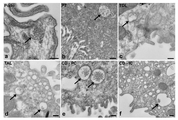
도 1의 프레임 a 내지 f는 소변 다소포체의 전자 현미경 사진이다. 네프론 및 집합관의 다양한 영역에서 다소포체 (MVB)가 확인될 수 있다 (화살표 참조). Podo - 발세포, PT - 근위 세관, TDL - 가는 하행각, TAL - 굵은 상행각, CD-PC - 집합관 주세포, CD-IC - 집합관 개재 세포. 눈금 막대 = a, c, d, e, f에 대해서는 200 nm; b에 대해서는 500 nm.
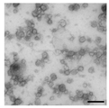
도 2는 단리된 소변 미세소포의 전자 현미경 사진이다. 분별 초원심분리를 통해 단리되고, 포스포텅스텐산을 염료로 사용하여 TEM을 통해 영상화된 인간 소변 미세소포. 눈금 막대 = 200 nm.
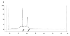
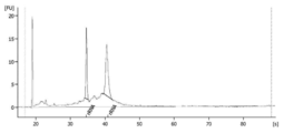
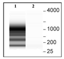
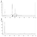
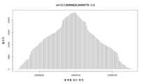
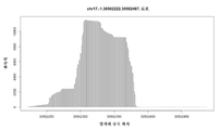
도 3은 100 kDa MWCO 필터의 방법으로 생성된 RNA 프로파일을 도해하는 도표이다. 애질런트 바이오애널라이저가 도표를 생성시키는데 사용되었다.
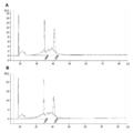
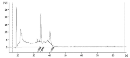
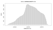
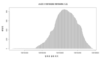
도 4는 초원심분리의 방법으로 생성된 RNA 프로파일을 도해하는 도표이다. 애질런트 바이오애널라이저가 도표를 생성시키는데 사용되었다.
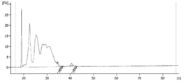
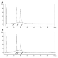
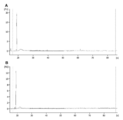
도 5는 각각의 경우에 초원심분리가 이어지는, x300g 스핀, x17,000g 스핀 및 0.8 ㎛ 여과의 3단계 예비-프로세싱 방법 (A) 또는 0.8 ㎛ 여과 단독의 1단계 예비-프로세싱 방법 (B)으로 생성된 RNA 프로파일을 도해하는 한 쌍의 도표이다. 애질런트 바이오애널라이저가 도표를 생성시키는데 사용되었다.
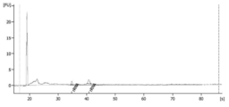
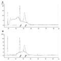
도 6은 각각의 경우에 여과 농축이 이어지는, x300g 스핀, x17,000g 스핀 및 0.8 ㎛ 여과의 3단계 예비-프로세싱 방법 (A) 또는 0.8 ㎛ 여과 단독의 1단계 예비-프로세싱 방법 (B)으로 생성된 RNA 프로파일을 도해하는 한 쌍의 도표이다. 애질런트 바이오애널라이저가 도표를 생성시키는데 사용되었다.
도 7은 추출 강화 공정이 있는, 소변으로부터 핵산을 추출하는 신규 방법을 도해하는 순서도이다.
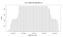
도 8은 5X 대 10X 농축 프로테아제를 사용하는 방법으로 생성된 RNA 프로파일을 도해하는 한 쌍의 도표이다. 20 ㎖ 소변 샘플로부터 여과 농축기를 통해 미세소포가 단리되었다. 도표 A는 5X 프로테아제로 수득된 프로파일을 나타낸다. 도표 B는 10X 프로테아제로 수득된 프로파일을 나타낸다. 1X 프로테아제 농도는 0.027 AU 이상의 프로테아제가 1 ㎕ 이상의 체액으로부터 단리된 미세소포를 처리하는데 사용된 효소 조건을 지칭한다. 5X 프로테아제 농도는 0.135 AU 이상의 프로테아제가 1 ㎕ 이상의 체액으로부터 단리된 미세소포를 처리하는데 사용된 효소 조건을 지칭한다. 1 mAU는 분 당 1 ㎛ol 타이로신에 상응하는 폴린-양성 아미노산 및 펩티드를 방출하는 프로테아제 활성이다.
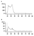
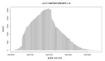
도 9는 25X 대 50X 농축 프로테아제를 사용하는 방법으로 생성된 RNA 프로파일을 도해하는 한 쌍의 도표이다. 40 ㎖ 소변 샘플로부터 여과 농축기를 통해 미세소포가 단리되었다. 도표 A는 25X 프로테아제로 수득된 프로파일을 나타낸다. 도표 B는 50X 프로테아제로 수득된 프로파일을 나타낸다. 1X 프로테아제는 0.027 AU를 지칭한다. 1 mAU는 분 당 1 ㎛ol 타이로신에 상응하는 폴린-양성 아미노산 및 펩티드를 방출하는 프로테아제 활성이다.
도 10은 흑색종 혈청인 샘플 1의 RNA 프로파일을 도해하는 도표이다. RNase 억제제인 수퍼라제-인(Superase-In) (앰비온 인코포레이티드(Ambion, Inc))을 사용하는 방법으로 1 ㎖ 혈청으로부터 RNA가 추출되었다. RNase 억제제의 최종 농도는 미세소포 현탁액 완충제 1 ㎕ 당 1.6 유닛이다.
도 11은 흑색종 혈청인 샘플 2의 RNA 프로파일을 도해하는 도표이다. RNase 억제제인 수퍼라제-인 (앰비온 인코포레이티드)을 사용하는 방법으로 1 ㎖ 혈청으로부터 RNA가 추출되었다. RNase 억제제의 최종 농도는 미세소포 현탁액 완충제 1 ㎕ 당 1.6 유닛이다.
도 12는 흑색종 혈청인 샘플 3의 RNA 프로파일을 도해하는 도표이다. RNase 억제제인 수퍼라제-인 (앰비온 인코포레이티드)을 사용하는 방법으로 1 ㎖ 혈청으로부터 RNA가 추출되었다. RNase 억제제의 최종 농도는 미세소포 현탁액 완충제 1 ㎕ 당 1.6 유닛이다.
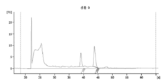
도 13은 흑색종 혈청인 샘플 4의 RNA 프로파일을 도해하는 도표이다. RNase 억제제인 수퍼라제-인 (앰비온 인코포레이티드)을 사용하는 방법으로 1 ㎖ 혈청으로부터 RNA가 추출되었다. RNase 억제제의 최종 농도는 미세소포 현탁액 완충제 1 ㎕ 당 1.6 유닛이다.
도 14는 흑색종 혈청인 샘플 5의 RNA 프로파일을 도해하는 도표이다. RNase 억제제인 수퍼라제-인 (앰비온 인코포레이티드)을 사용하는 방법으로 1 ㎖ 혈청으로부터 RNA가 추출되었다. RNase 억제제의 최종 농도는 미세소포 현탁액 완충제 1 ㎕ 당 3.2 유닛이다.
도 15는 흑색종 혈청인 샘플 6의 RNA 프로파일을 도해하는 도표이다. RNase 억제제인 수퍼라제-인 (앰비온 인코포레이티드)을 사용하는 방법으로 1 ㎖ 혈청으로부터 RNA가 추출되었다. RNase 억제제의 최종 농도는 미세소포 현탁액 완충제 1 ㎕ 당 3.2 유닛이다.
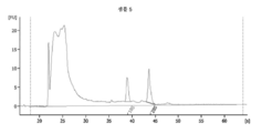
도 16은 정상 혈청인 샘플 7의 RNA 프로파일을 도해하는 도표이다. RNase 억제제인 수퍼라제-인 (앰비온 인코포레이티드)을 사용하는 방법으로 1 ㎖ 혈청으로부터 RNA가 추출되었다. RNase 억제제의 최종 농도는 미세소포 현탁액 완충제 1 ㎕ 당 1.6 유닛이다.
도 17은 정상 혈청인 샘플 8의 RNA 프로파일을 도해하는 도표이다. RNase 억제제인 수퍼라제-인 (앰비온 인코포레이티드)을 사용하는 방법으로 1 ㎖ 혈청으로부터 RNA가 추출되었다. RNase 억제제의 최종 농도는 미세소포 현탁액 완충제 1 ㎕ 당 1.6 유닛이다.
도 18은 정상 혈청인 샘플 9의 RNA 프로파일을 도해하는 도표이다. RNase 억제제인 수퍼라제-인 (앰비온 인코포레이티드)을 사용하는 방법으로 1 ㎖ 혈청으로부터 RNA가 추출되었다. RNase 억제제의 최종 농도는 미세소포 현탁액 완충제 1 ㎕ 당 1.6 유닛이다.
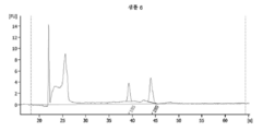
도 19는 정상 혈청인 샘플 10의 RNA 프로파일을 도해하는 도표이다. RNase 억제제인 수퍼라제-인 (앰비온 인코포레이티드)을 사용하는 방법으로 1 ㎖ 혈청으로부터 RNA가 추출되었다. RNase 억제제의 최종 농도는 미세소포 현탁액 완충제 1 ㎕ 당 1.6 유닛이다.
도 20은 정상 혈청인 샘플 11의 RNA 프로파일을 도해하는 도표이다. RNase 억제제인 수퍼라제-인 (앰비온 인코포레이티드)을 사용하는 방법으로 1 ㎖ 혈청으로부터 RNA가 추출되었다. RNase 억제제의 최종 농도는 미세소포 현탁액 완충제 1 ㎕ 당 3.2 유닛이다.
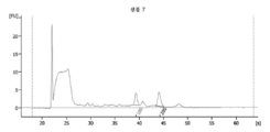
도 21은 정상 혈청인 샘플 12의 RNA 프로파일을 도해하는 도표이다. RNase 억제제인 수퍼라제-인 (앰비온 인코포레이티드)을 사용하는 방법으로 1 ㎖ 혈청으로부터 RNA가 추출되었다. RNase 억제제의 최종 농도는 미세소포 현탁액 완충제 1 ㎕ 당 3.2 유닛이다.
도 22는 추출 강화 공정을 사용하여 생물학적 샘플로부터 핵산을 추출하는 신규 방법을 도해하는 순서도이다.
도 23은 DNase 처리가 있는 방법 또는 DNase 처리가 없는 방법으로 생성된 RNA 프로파일을 도해하는 한 쌍의 도표이다. 미세소포 내부에 위치하지 않은 DNA가 용해 및 핵산 추출 전에 소변 샘플로부터 단리된 미세소포 펠릿의 DNase 소화에 의해 제거되었다. a - 알엔이지 마이크로 키트(RNeasy Micro Kit)를 사용한 DNase 소화가 없는 프로파일. b - 알엔이지 마이크로 키트를 사용한 DNase 소화가 있는 프로파일. c - 미르바나 키트(MirVana Kit)를 사용한 DNase 소화가 없는 프로파일. d - 미르바나 키트를 사용한 DNase 소화가 있는 프로파일. 화살표로 지시된 소형 RNA 피크 높이 및 면적에서의 변화를 주지한다. d는 페놀/클로로포름 기반 추출 후에 샘플 내로 약간의 DNA가 넘겨지는 것을 시사한다.
도 24는 DNase 처리가 있는 방법 또는 DNase 처리가 없는 방법으로 생성된 RNA 프로파일을 도해하는 한 쌍의 도표이다. 미세소포 내부에 위치하지 않은 DNA가 용해 및 핵산 추출 전에 소변 샘플로부터 단리된 미세소포 펠릿의 DNase 소화에 의해 제거되었다. a - DNase 소화가 없는 프로파일. b - DNase 소화가 있는 프로파일. c - 혈청-유래 미세소포와 공동-단리될 수 있는 "세포자멸사체"-유사 래더(ladder)를 나타내는 슈도(pseudo) 젤.
도 25는 RNase 처리가 있는 방법 또는 RNase 처리가 없는 방법으로 생성된 RNA 프로파일을 도해하는 한 쌍의 도표이다. 미세소포 내부에 위치하지 않은 RNA가 용해 및 핵산 추출 전에 소변 샘플로부터 단리된 미세소포 펠릿의 RNase 소화에 의해 제거되었다. A - RNase 소화가 없는 프로파일. B - RNase 소화가 있는 프로파일.
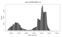
도 26은 소변 미세소포 및 래트 신장 조직으로부터 생성된 RNA 프로파일을 도해하는 한 쌍의 도표이다. A - 래트 신장 조직으로부터의 프로파일. B - 소변 미세소포로부터의 프로파일.
도 27은 소형 RNA 추출을 강화할 수 있는 방법을 사용하여 소변 미세소포 및 래트 신장 조직으로부터 생성된 RNA 프로파일을 도해하는 한 쌍의 도표이다. A - 래트 신장 조직으로부터의 프로파일. B - 소변 미세소포로부터의 프로파일.
도 28은 DNase 처리와 함께 또는 DNase 처리 없이, 미세소포를 제외한 전체 소변으로부터 생성된 RNA 프로파일을 도해하는 한 쌍의 도표이다 (본 발명의 단리 기술에 의해 포획되지 않음). A - DNase 처리 없이 전체 소변으로부터 단리된 핵산. B - DNase 처리와 함께 전체 소변으로부터 단리된 핵산.
도 29는 소변 미세소포로부터 생성된 RNA 프로파일을 도해하는 한 쌍의 도표이다. A - DNase 처리 없이 소변 미세소포로부터 단리된 핵산. B - DNase 처리와 함께 미세소포로부터 단리된 핵산.
도 30은 300g 스핀 동안 형성된 펠릿으로부터 추출된 핵산으로부터 생성된 RNA 프로파일을 도해하는 한 쌍의 도표이다. A - DNase 처리 없이 300g 스핀 펠릿으로부터 단리된 핵산. B - DNase 처리와 함께 300g 스핀 펠릿으로부터 단리된 핵산.
도 31은 17,000g 스핀 동안 형성된 펠릿으로부터 추출된 핵산으로부터 생성된 RNA 프로파일을 도해하는 한 쌍의 도표이다. A - DNase 처리가 없는 17,000g 스핀 펠릿으로부터의 핵산 프로파일. B - DNase 처리가 있는 17,000g 펠릿으로부터의 핵산 프로파일.
도 32는 미세소포내 RNase 소화와 함께 또는 미세소포내 RNase 소화 없이, 미세소포 용해 전에 외부에서 RNase 및 DNase 소화가 진행된 미세소포로부터 생성된 RNA 프로파일을 도해하는 한 쌍의 도표이다. A - 미세소포내 RNase 소화가 없는 핵산 프로파일. B - 미세소포내 RNase 소화가 있는 핵산 프로파일.
도 33은 미세소포내 DNase 소화와 함께 또는 미세소포내 DNase 소화 없이, 미세소포 용해 및 미세소포내 RNase 소화 전에 외부에서 RNase 및 DNase 소화가 진행된 미세소포로부터 생성된 RNA 프로파일을 도해하는 한 쌍의 도표이다. A - 미세소포내 DNase 소화가 없는 핵산 프로파일. B - 미세소포내 DNase 소화가 있는 핵산 프로파일. 도표 B에서, 20s 직후의 피크가 도표 A에서의 필적하는 피크와 비교하여 감소된다. 이러한 감소는 소량의 DNase 소화성 물질이 엑소솜 내에 존재한다는 것을 시사한다.
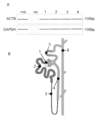
도 34 A)는 RT-PCR에 의한 소변 미세소포 내의 베타-액틴 및 GAPDH에 대한 리보앰프(RiboAmp) 증폭 mRNA 전사물이 양성으로 확인되는, 바이오애널라이저에 의해 생성된 '슈도 젤' 프로파일이다; B)는 6개의 기능적으로 별개인 영역을 강조하는, 네프론 및 집합관의 삽화이다. 1. 사구체; 2. 근위 세관; 3. 가는 하행각; 4. 수질 굵은 상행각; 5. 원위 곱슬 세관; 6. 집합관.
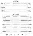
도 35는 소변 미세소포로부터 리보앰프화 mRNA의 RT-PCR에 의해 검출된 네프론 및 집합관의 영역 1 및 2로부터의 특이적 유전자를 코딩하는 mRNA 전사물들이 확인되는, 바이오애널라이저에 의해 생성된 슈도 젤 프로파일을 나타내고, 구체적으로는 하기와 같다: 1. 사구체: NPHS2 - 포도신, LGALS1 - 갈렉틴-1, HSPG2 - 헤파린 술페이트 프로테오글리칸 2. 근위 세관: CUBN - 큐빌린, LRP2 - 메갈린, AQP1 - 아쿠아포린 1, CA4 - 탄산 탈수효소 4, CLCN5 - 클로라이드 채널 단백질 5.
도 36은 소변 미세소포로부터 리보앰프화 mRNA의 RT-PCR에 의해 검출된 네프론 및 집합관의 영역 3-6으로부터의 특이적 유전자를 코딩하는 mRNA 전사물들이 확인되는, 바이오애널라이저에 의해 생성된 슈도 젤 프로파일을 나타내고, 구체적으로는 하기와 같다: 3. 가는 하행각: BDKRB1 - 브라디키닌 B1 수용체. 4. 수질 굵은 상행각: CALCR - 칼시토닌 수용체, SCNN1D - 아밀로라이드-민감성 나트륨 채널 서브유닛 델타. 5. 원위 곱슬 세관: SLC12A3 - 티아지드-민감성 나트륨-클로라이드 공동수송체. 6. 집합관: AQP2 - 아쿠아포린 2, ATP6V1B1 - vATPase B1 서브유닛, SLC12A1 - 신장-특이적 Na-K-Cl 심포터.
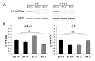
도 37 A)는 V-ATPase B1 KO (B1 -/-) 및 야생형 (B1 +/+) 마우스에서의 RT-PCR에 의한 V-ATPase B1 서브유닛 및 AQP2 mRNA의 발현을 도해하는 한 쌍의 바이오애널라이저 슈도 젤이다; B)는 실시간 PCR 분석에 의한 V-ATPase B1 KO (B1 -/-) 및 야생형 (B1 +/+) 마우스로부터의 소변 미세소포 및 신장 세포 내의 V-ATPase B2 서브유닛의 발현을 도해하는 한 쌍의 차트이다. "NS" - 통계적으로 유의하지 않음.
도 38은 소변 미세소포로부터 생성된 RNA 프로파일을 도해하는 3개의 도표이다. 미세소포 막이 핵산 추출을 위해 파괴되기 전에, 소변 미세소포가 어떠한 추출 강화제로도 세정 또는 처리되지 않았다. 3개의 샘플이 이러한 추출 군에서 사용되었다. 프로파일이 A, B 및 C에서 각각 제시된다.
도 39는 소변 미세소포로부터 생성된 RNA 프로파일을 도해하는 3개의 도표이다. 미세소포 막이 핵산 추출을 위해 파괴되기 전에, 소변 미세소포가 세정되지 않았지만, RNase 억제제인 RNase-In (프로메가(Promega))으로 처리되었다. 3개의 샘플이 이러한 추출 군에서 사용되었다. 프로파일이 A, B 및 C에서 각각 제시된다.
도 40은 소변 미세소포로부터 생성된 RNA 프로파일을 도해하는 3개의 도표이다. 미세소포 막이 핵산 추출을 위해 파괴되기 전에, 소변 미세소포가 세정되었지만 어떠한 RNase 억제제로도 처리되지 않았다. 3개의 샘플이 이러한 추출 군에서 사용되었다. 프로파일이 A, B 및 C에서 각각 제시된다.
도 41은 소변 미세소포로부터 생성된 RNA 프로파일을 도해하는 3개의 도표이다. 미세소포 막이 핵산 추출을 위해 파괴되기 전에, 소변 미세소포가 세정되었고 RNase 억제제로 처리되었다. 3개의 샘플이 이러한 추출 군에서 사용되었다. 프로파일이 A, B 및 C에서 각각 제시된다.
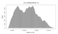
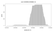
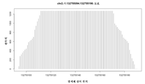
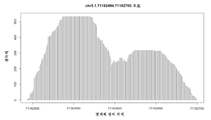
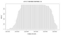
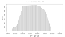
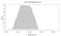
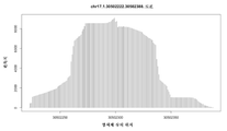
도 42는 소변 미세소포로부터 단리된 RNA의 딥 시퀀싱(deep sequencing) 분석에서 500을 초과하는 전사물 히트(hit) ("스파이크(spike)")가 있는 염색체 영역의 목록이다. 숫자는 각각의 염색체 영역의 시작점 및 종점을 지시한다. 예를 들어, "chr1.-1.91625366.91625741"은 뉴클레오티드 번호 91625366과 91625741 사이의 인간 염색체 1 상의 영역을 지칭한다. 상응하는 서열 번호가 또한 지시된다.
도 43은 지시된 바와 같은 10개의 염색체 영역 내의 서열을 증폭시키기 위해 PCR 반응에 사용된 프라이머의 목록이다. 예를 들어, "chr1.-1.91625366.91625741"은 뉴클레오티드 번호 91625366과 91625741 사이의 인간 염색체 1 상의 영역을 지칭한다. 이러한 영역을 증폭시키는데 사용된 프라이머 쌍은 "tccagctcacgttccctatt 1L 및 ccaggtggggagtttgact 1R"이었다. 프라이머는 왼쪽에서 오른쪽으로 5' → 3'이다.
도 44는 10개의 스파이크-풍부 염색체 영역의 PCR 증폭 결과를 도해하는 한 쌍의 바이오애널라이저 슈도 젤이다. 각각의 프레임의 상부의 레인의 번호매김은 도 43에 제시된 염색체 영역의 번호매김에 상응한다. A에서는, 소변 미세소포로부터의 핵산 추출물이 PCR용 주형으로 사용되었다. B에서는, 신장 조직으로부터의 핵산 추출물이 PCR용 주형으로 사용되었다.
도 45-73은 29개의 염색체 영역에서의 스파이크를 도해하는 도표이다. 각각의 도표의 상부에 영역이 지시된다. 예를 들어, 도 46의 도표는 뉴클레오티드 번호 91625366과 91625741 사이의 인간 염색체 1 상의 영역인 "chr1.-1.91625366.91625741"의 영역을 지칭한다. Brief description of the drawing
Frames a to f of FIG. 1 are electron micrographs of a urine multifocal body. Multifocal bodies (MVBs) can be identified in various areas of the nephron and collecting ducts (see arrows). Podo-podocyte, PT-proximal tubule, TDL-fine descending angle, TAL-thick ascending angle, CD-PC-collecting duct main cell, CD-IC-collecting duct intervening cell. Scale bar = 200 nm for a, c, d, e, f; 500 nm for b.
2 is an electron micrograph of isolated urine microvesicles. Human urine microvesicles isolated through fractional ultracentrifugation and imaged through TEM using phosphotungstic acid as a dye. Scale bar = 200 nm.
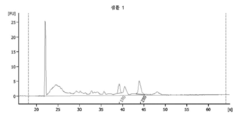
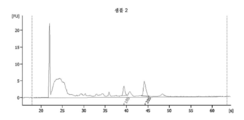
3 is a diagram illustrating the RNA profile generated by the method of a 100 kDa MWCO filter. An Agilent bioanalyzer was used to generate the plot.
4 is a diagram illustrating the RNA profile generated by the method of ultracentrifugation. An Agilent bioanalyzer was used to generate the plot.
5 is a three-step pre-processing method (A) of x300 g spin, x17,000 g spin and 0.8 μm filtration followed by ultracentrifugation in each case, or a one-step pre-processing method (B) of 0.8 μm filtration alone. A pair of diagrams illustrating the resulting RNA profile. An Agilent bioanalyzer was used to generate the plot.
Figure 6 is produced by a three-step pre-processing method (A) of x300 g spin, x17,000 g spin and 0.8 μm filtration followed by filtration concentration in each case or a one-step pre-processing method (B) of 0.8 μm filtration alone. This is a pair of diagrams illustrating the resulting RNA profile. An Agilent bioanalyzer was used to generate the plot.
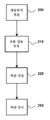
7 is a flow chart illustrating a novel method of extracting nucleic acids from urine with an extraction enhancement process.
Figure 8 is a pair of diagrams illustrating the RNA profile generated by the method using 5X vs. 10X enriched protease. Microvesicles were isolated from a 20 ml urine sample through a filter concentrator. Table A shows the profile obtained with 5X protease. Table B shows the profile obtained with 10X protease. 1X protease concentration refers to the enzymatic conditions in which a protease of 0.027 AU or more was used to treat microvesicles isolated from 1 μl or more body fluids. 5X protease concentration refers to the enzymatic conditions in which 0.135 AU or more protease was used to treat microvesicles isolated from 1 μl or more body fluids. 1 mAU is the protease activity that releases foline-positive amino acids and peptides corresponding to 1 μmol tyrosine per minute.
9 is a pair of diagrams illustrating the RNA profile generated by the method using 25X vs. 50X enriched protease. Microvesicles were isolated from a 40 ml urine sample through a filter concentrator. Table A shows the profile obtained with 25X protease. Table B shows the profile obtained with 50X protease. 1X protease refers to 0.027 AU. 1 mAU is the protease activity that releases foline-positive amino acids and peptides corresponding to 1 μmol tyrosine per minute.
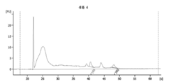
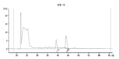
10 is a diagram illustrating the RNA profile of
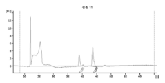
11 is a diagram illustrating the RNA profile of
12 is a diagram illustrating the RNA profile of
13 is a diagram illustrating the RNA profile of
14 is a diagram illustrating the RNA profile of
15 is a diagram illustrating the RNA profile of
16 is a diagram illustrating the RNA profile of
17 is a diagram illustrating the RNA profile of
18 is a diagram illustrating the RNA profile of
19 is a diagram illustrating the RNA profile of
20 is a diagram illustrating the RNA profile of
21 is a diagram illustrating the RNA profile of
22 is a flow chart illustrating a novel method of extracting nucleic acids from biological samples using an extraction enrichment process.
FIG. 23 is a pair of diagrams illustrating RNA profiles generated with or without DNase treatment. DNA not located inside the microvesicles was removed by DNase digestion of microvesicle pellets isolated from urine samples prior to lysis and nucleic acid extraction. a-Profile without DNase digestion using RNeasy Micro Kit. b-Profile with DNase digestion using R&G micro kit. c-Profile without DNase digestion using MirVana Kit. d-Profile with DNase digestion using the Mirvana kit. Note the change in small RNA peak height and area indicated by arrows. d suggests that some DNA was handed over into the sample after phenol/chloroform based extraction.
Fig. 24 is a pair of diagrams illustrating RNA profiles generated by methods with or without DNase treatment. DNA not located inside the microvesicles was removed by DNase digestion of microvesicle pellets isolated from urine samples prior to lysis and nucleic acid extraction. a-Profile without DNase digestion. b-Profile with DNase digestion. c-Pseudo gel showing a "apoptotic"-like ladder that can be co-isolated with serum-derived microvesicles.
Figure 25 is a pair of diagrams illustrating RNA profiles generated with or without RNase treatment. RNA not located inside the microvesicles was removed by RNase digestion of microvesicle pellets isolated from urine samples prior to lysis and nucleic acid extraction. A-Profile without RNase digestion. B-Profile with RNase digestion.
26 is a pair of diagrams illustrating RNA profiles generated from urine microvesicles and rat kidney tissue. A-Profile from rat kidney tissue. B-Profile from urine microvesicles.
Figure 27 is a pair of diagrams illustrating RNA profiles generated from urine microvesicles and rat kidney tissue using a method that can enhance small RNA extraction. A-Profile from rat kidney tissue. B-Profile from urine microvesicles.
28 is a pair of diagrams illustrating the RNA profile generated from whole urine except microvesicles, with or without DNase treatment (not captured by the isolation technique of the present invention). A-Nucleic acid isolated from whole urine without DNase treatment. B-Nucleic acid isolated from whole urine with DNase treatment.
29 is a pair of diagrams illustrating the RNA profile generated from urine microvesicles. A-Nucleic acid isolated from urine microvesicles without DNase treatment. B-Nucleic acid isolated from microvesicles with DNase treatment.
30 is a pair of diagrams illustrating RNA profiles generated from nucleic acids extracted from pellets formed during 300 g spin. A-Nucleic acid isolated from 300 g spin pellets without DNase treatment. B-Nucleic acid isolated from 300 g spin pellet with DNase treatment.
31 is a pair of diagrams illustrating RNA profiles generated from nucleic acids extracted from pellets formed during 17,000 g spins. A-Nucleic acid profile from 17,000 g spin pellets without DNase treatment. B-Nucleic acid profile from 17,000 g pellet with DNase treatment.
FIG. 32 is a pair of diagrams illustrating the RNA profile generated from microvesicles subjected to external RNase and DNase digestion before microvesicle lysis, with or without intra-microvesicle RNase digestion. A-Nucleic acid profile without RNase digestion in microvesicles. B-Nucleic acid profile with RNase digestion in microvesicles.
Figure 33 is a pair of diagrams illustrating the RNA profile generated from microvesicles subjected to external RNase and DNase digestion prior to microvesicle lysis and RNase digestion in microvesicles with or without DNase digestion in microvesicles. to be. A-Nucleic acid profile without DNase digestion in microvesicles. B-Nucleic acid profile with DNase digestion in microvesicles. In Figure B, the peak immediately after 20 s decreases compared to the comparable peak in Figure A. This reduction suggests that a small amount of DNase digestible material is present in the exosomes.
Fig. 34A) is a'pseudo gel' profile generated by a bioanalyzer in which a riboamp amplified mRNA transcript for beta-actin and GAPDH in urine microvesicles by RT-PCR is confirmed as positive; B) is an illustration of the nephron and the assembly, highlighting the six functionally distinct areas. 1. glomeruli; 2. proximal customs; 3. going down angle; 4. Water quality thick ascending angle; 5. Distal curly customs; 6. Gathering hall.
Fig. 35 is a pseudogel produced by a bioanalyzer in which mRNA transcripts encoding specific genes from
Figure 36 is a pseudogel produced by a bioanalyzer in which mRNA transcripts encoding specific genes from regions 3-6 of the collecting duct and nephrons detected by RT-PCR of riboampized mRNA from urine microvesicles are identified. The profile is shown, specifically as follows: 3. Thin descending angle: BDKRB1-bradykinin B1 receptor. 4. Medullary coarse ascending angle: CALCR-calcitonin receptor, SCNN1D-amiloride-sensitive sodium channel subunit delta. 5. Distal curly tubules: SLC12A3-thiazide-sensitive sodium-chloride cotransporter. 6. Collecting tube: AQP2-
Figure 37A) is a pair of bios illustrating the expression of V-ATPase B1 subunit and AQP2 mRNA by RT-PCR in V-ATPase B1 KO (B1 -/-) and wild-type (B1 +/+) mice. It is an analyzer pseudo gel; B) is a pair illustrating the expression of the V-ATPase B2 subunit in urine microvesicles and kidney cells from V-ATPase B1 KO (B1 -/-) and wild-type (B1 +/+) mice by real-time PCR analysis. It is a chart of. "NS"-not statistically significant.
38 are three diagrams illustrating the RNA profile generated from urine microvesicles. Before the microvesicle membrane was destroyed for nucleic acid extraction, the urine microvesicles were not washed or treated with any extraction enhancer. Three samples were used in this extraction group. Profiles are presented in A, B and C, respectively.
39 is three diagrams illustrating the RNA profile generated from urine microvesicles. Before the microvesicle membrane was destroyed for nucleic acid extraction, the urine microvesicles were not washed, but treated with the RNase inhibitor RNase-In (Promega). Three samples were used in this extraction group. Profiles are presented in A, B and C, respectively.
40 are three diagrams illustrating the RNA profile generated from urine microvesicles. Before the microvesicle membrane was destroyed for nucleic acid extraction, the urine microvesicles were washed but not treated with any RNase inhibitors. Three samples were used in this extraction group. Profiles are presented in A, B and C, respectively.
41 are three diagrams illustrating the RNA profile generated from urine microvesicles. Before the microvesicle membrane was destroyed for nucleic acid extraction, urine microvesicles were washed and treated with an RNase inhibitor. Three samples were used in this extraction group. Profiles are presented in A, B and C, respectively.
FIG. 42 is a list of chromosomal regions with more than 500 transcript hits (“spikes”) in deep sequencing analysis of RNA isolated from urine microvesicles. The numbers indicate the starting and ending points of each chromosomal region. For example, “chr1.-1.91625366.91625741” refers to the region on
43 is a list of primers used in the PCR reaction to amplify sequences within 10 chromosomal regions as indicated. For example, “chr1.-1.91625366.91625741” refers to the region on
44 is a pair of bioanalyzer pseudo gels illustrating the PCR amplification results of 10 spike-rich chromosomal regions. The numbering of the lanes at the top of each frame corresponds to the numbering of the chromosomal regions shown in Figure 43. In A, the nucleic acid extract from urine microvesicles was used as a template for PCR. In B, a nucleic acid extract from kidney tissue was used as a template for PCR.
45-73 are diagrams illustrating spikes in 29 chromosomal regions. The area is indicated at the top of each diagram. For example, the diagram in FIG. 46 refers to the region of “chr1.-1.91625366.91625741”, the region on
상세한 설명details
미세소포는 세포의 외부로 진핵생물 세포에 의해 쉐딩되거나 또는 형질막으로부터 출아된다. 직경이 약 10 nm 내지 약 5000 nm 범위이면서 이러한 막 소포는 크기가 이질적이다. 세포내 다소포체의 세포외배출에 의해 방출되는 소형 미세소포 (직경이 약 10 내지 1000 nm, 더욱 종종 약 10 내지 200 nm)가 당업계에서 "엑소솜"으로 지칭된다. 본원에 기술된 조성물, 방법 및 용도는 모든 크기, 바람직하게는 10 내지 800 nm, 더욱 바람직하게는 10 내지 200 nm의 미세소포에 동등하게 적용가능하다.Microvesicles are either sheathed by eukaryotic cells to the outside of the cell or sprouted from the plasma membrane. These membrane vesicles are heterogeneous in size, ranging from about 10 nm to about 5000 nm in diameter. Small microvesicles (about 10 to 1000 nm in diameter, more often about 10 to 200 nm) released by extracellular excretion of intracellular multivesicles are referred to in the art as "exosomes". The compositions, methods and uses described herein are equally applicable to microvesicles of all sizes, preferably 10 to 800 nm, more preferably 10 to 200 nm.
일부 문헌에서, 용어 "엑소솜"은 mRNA 분해 및 소형 핵소체 RNA (snoRNA), 소형 핵 RNA (snRNA) 및 리보솜 RNA (rRNA)의 프로세싱에 수반되는 엑소리보뉴클레아제(exoribonuclease)를 함유하는 단백질 복합체를 또한 지칭한다 (문헌 [Liu et al., 2006]; [van Dijk et al., 2007]). 이같은 단백질 복합체는 막이 없으며, 본원에서 사용되는 이러한 용어와 같은 "미세소포" 또는 "엑소솜"이 아니다.In some literature, the term “exosome” refers to a protein complex containing exoribonuclease involved in mRNA degradation and processing of small nucleolar RNA (snoRNA), small nuclear RNA (snRNA) and ribosomal RNA (rRNA). Also referred to (Liu et al., 2006]; [van Dijk et al., 2007]). Such protein complexes are membraneless and are not “microvesicles” or “exosomes” as used herein as such terms.
본 발명은 유해 인자가 생물학적 샘플로부터의 핵산의 효과적인 추출을 방지할 수 있고, 뜻밖의 신규 작용제 및 단계가 이러한 유해 인자를 완화 또는 제거하는데 사용되어, 추출된 핵산의 품질을 극적으로 개선할 수 있다는 발견을 부분적으로 기초로 한다. 따라서, 본 발명의 한 측면은 생물학적 샘플로부터 고품질 핵산을 추출하는 신규 방법이다. 본원에 기술된 신규 방법에 의해 수득된 고품질 추출물은 높은 수율 및 높은 완전성을 특징으로 하여, 추출된 핵산을 고품질 핵산 추출물이 바람직한 다양한 용도에 유용하게 한다.The present invention is that harmful factors can prevent the effective extraction of nucleic acids from biological samples, and unexpected new agents and steps can be used to mitigate or eliminate these harmful factors, thereby dramatically improving the quality of the extracted nucleic acids. It is based in part on findings. Thus, one aspect of the invention is a novel method of extracting high quality nucleic acids from biological samples. The high-quality extracts obtained by the novel methods described herein are characterized by high yields and high integrity, making the extracted nucleic acids useful in a variety of applications where high-quality nucleic acid extracts are desirable.
대략적으로 기술하면, 신규 방법은, 예를 들어, 생물학적 샘플을 수득하는 단계, 생물학적 샘플로부터의 핵산의 효과적인 추출을 방지하는 유해 인자를 완화시키거나 제거하는 단계, 및 생물학적 샘플로부터 핵산을 추출하는 단계 (임의적으로 핵산 분석이 이어짐)를 포함한다. Broadly stated, the novel methods include, for example, obtaining a biological sample, mitigating or eliminating harmful factors that prevent effective extraction of nucleic acids from the biological sample, and extracting nucleic acids from the biological sample. (Optionally followed by nucleic acid analysis).
적용가능한 생물학적 샘플에는, 예를 들어, 세포, 일군의 세포, 세포 단편, 미세소포가 예를 들어 포함되는 세포 산물, 세포 배양물, 대상체로부터의 신체 조직, 또는 체액이 포함된다. 체액은 혈액, 혈장, 혈청, 소변, 가래, 척수액, 흉수, 유두 흡인물, 림프액, 기도액, 소화관액, 비뇨생식관액, 눈물, 타액, 모유, 림프계 체액, 정액, 뇌척수액, 기관계내 체액, 복수, 낭성 종양 체액, 양수액 및 이들의 조합을 예를 들어 포함하지만 이에 한정되지 않는, 대상체의 신체 내의 임의의 장소, 바람직하게는 말초 위치로부터 단리된 유체일 수 있다. Applicable biological samples include, for example, cells, a group of cells, cell fragments, cell products including, for example, microvesicles, cell cultures, body tissues from a subject, or body fluids. Body fluids include blood, plasma, serum, urine, sputum, spinal fluid, pleural fluid, nipple aspirate, lymph fluid, airway fluid, digestive tract fluid, genitourinary fluid, tears, saliva, breast milk, lymphatic fluid, semen, cerebrospinal fluid, organ fluid, ascites. , Cystic tumor bodily fluid, amniotic fluid, and combinations thereof, including, but not limited to, a fluid isolated from any place within the subject's body, preferably a peripheral location.
생물학적 샘플은 종종 대상체로부터 유래될 수 있다. 용어 "대상체"는 미세소포가 있는 것으로 나타났거나 미세소포가 있을 것으로 예상되는 모든 동물을 포함하도록 의도된다. 특정 실시양태에서, 대상체는 포유동물, 인간 또는 비-인간 영장류, 개, 고양이, 말, 소, 기타 가축, 또는 설치류 (예를 들어, 마우스, 래트, 기니 피그 등)이다. 용어 "대상체" 및 "개체"는 본원에서 상호교환가능하게 사용된다.Biological samples can often be derived from a subject. The term “subject” is intended to include all animals that have been shown to or are expected to have microvesicles. In certain embodiments, the subject is a mammal, human or non-human primate, dog, cat, horse, cow, other domestic animal, or rodent (eg, mouse, rat, guinea pig, etc.). The terms “subject” and “subject” are used interchangeably herein.
유해 효과를 완화 또는 제거하는 단계를 수행하기 전, 후 또는 이와 동시에 생물학적 샘플을 임의적으로 프로세싱하여 생물학적 샘플 유도체를 수득할 수 있다. 생물학적 샘플 유도체는 세포, 세포 잔해물, 막 소포, 또는 미세소포일 수 있다. Biological sample derivatives can be obtained by optionally processing the biological sample before, after or concurrently with the step of mitigating or eliminating adverse effects. Biological sample derivatives can be cells, cell debris, membrane vesicles, or microvesicles.
생물학적 샘플이 종종 예비-프로세싱된 후 생물학적 샘플 유도체 예컨대 미세소포가 수득된다. 일부 예에서, 예비-프로세싱 단계가 바람직하다. 예를 들어, 소변 샘플이 예비-프로세싱되어 소변 미세소포를 수득할 수 있다. 예비-프로세싱은 당업계에 공지된 기술 예컨대 저속 원심분리 및 예비-여과에 의해 달성될 수 있다. 예를 들어, 소변 샘플에 300g의 제1 원심분리 단계를 적용하여, 샘플 내의 대형 입자를 제거할 수 있다. 소변 샘플에 17,000g의 제2 원심분리 단계를 적용하여 샘플 내의 더 작은 입자를 제거할 수 있다. 제2 원심분리 단계 후, 소변 샘플에 예비-여과 단계, 예를 들어, 0.8 ㎛ 예비-여과 단계를 추가로 적용할 수 있다. 다르게는, 1회 이상의 원심분리 단계를 먼저 적용하지 않으면서 소변 샘플을 예비-여과 단계에 의해 예비-프로세싱할 수 있다.Biological samples are often pre-processed to obtain biological sample derivatives such as microvesicles. In some instances, a pre-processing step is preferred. For example, a urine sample can be pre-processed to obtain urine microvesicles. Pre-processing can be accomplished by techniques known in the art such as low speed centrifugation and pre-filtration. For example, the urine sample may be subjected to a first centrifugation step of 300 g to remove large particles in the sample. A second centrifugation step of 17,000 g can be applied to the urine sample to remove smaller particles in the sample. After the second centrifugation step, the urine sample may be further subjected to a pre-filtration step, for example a 0.8 μm pre-filtration step. Alternatively, the urine sample can be pre-processed by a pre-filtration step without first applying one or more centrifugation steps.
막 소포, 예를 들어, 미세소포가 생물학적 샘플로부터 단리될 수 있다. 일부 경우에, 생물학적 샘플의 예비-프로세싱 없이 이같은 단리가 일부 경우에 수행될 수 있다. 또 다른 경우에, 생물학적 샘플이 예비-프로세싱된 후에 이같은 단리가 수행될 수 있다. 생물학적 샘플로부터의 고품질 핵산 추출에 단리 단계가 유리할 수 있다. 예를 들어, 단리는 하기와 같은 장점들을 초래할 수 있다: 1) 질환- 또는 종양-특이적 미세소포를 체액 샘플 내의 다른 미세소포로부터 단리함으로써 수득될 수 있는, 질환- 또는 종양-특이적 핵산을 선택적으로 분석할 기회; 2) 체액 샘플로부터 핵산을 직접적으로 추출함으로써 수득되는 수율/완전성과 비교하여 핵산 종의 유의하게 더 높은 수율 + 더 높은 완전성; 3) 확장성, 예를 들어, 낮은 수준으로 발현된 핵산을 검출하는 확장성 (더 많은 양의 혈청으로부터 더 많은 미세소포를 펠릿화함으로써 감도가 증가될 수 있다); 4) 단백질 및 지질, 죽은 세포로부터의 잔해물, 및 기타 잠재적인 오염물 및 PCR 억제제가 핵산 추출 단계 전에 미세소포 펠릿으로부터 제외된다는 점에서 더 순수한 핵산; 및 5) 미세소포 펠릿의 부피가 출발 혈청보다 훨씬 작아서, 소량의 칼럼 필터를 사용하여 이러한 미세소포 펠릿으로부터 핵산을 추출하는 것을 가능하게 하기 때문에, 핵산 추출 방법에서의 더 많은 선택권. Membrane vesicles, such as microvesicles, can be isolated from biological samples. In some cases, such isolation may be performed in some cases without pre-processing of the biological sample. In another case, such isolation can be performed after the biological sample has been pre-processed. Isolation steps may be advantageous for extraction of high quality nucleic acids from biological samples. For example, isolation can lead to the following advantages: 1) disease- or tumor-specific nucleic acids, which can be obtained by isolating disease- or tumor-specific microvesicles from other microvesicles in a bodily fluid sample. The opportunity to analyze selectively; 2) significantly higher yield + higher integrity of nucleic acid species compared to yield/integrity obtained by directly extracting nucleic acids from bodily fluid samples; 3) scalability, eg, scalability to detect nucleic acids expressed at low levels (sensitivity can be increased by pelleting more microvesicles from higher amounts of serum); 4) purer nucleic acids in that proteins and lipids, debris from dead cells, and other potential contaminants and PCR inhibitors are excluded from the microvesicle pellet prior to the nucleic acid extraction step; And 5) more options in the nucleic acid extraction method, since the volume of the microvesicle pellets is much smaller than the starting serum, making it possible to extract nucleic acids from these microvesicle pellets using a small amount of column filter.
생물학적 샘플로부터 미세소포를 단리하는 방법들이 당업계에 공지되어 있다. 예를 들어, 분별 원심분리 방법이 라포소(Raposo) 등의 논문 (문헌 [Raposo et al., 1996]), 스코그(Skog) 등의 논문 (문헌 [Skog et al., 2008]) 및 닐슨(Nilsson) 등의 논문 (문헌 [Nilsson et al., 2009])에 기술되어 있다. 음이온 교환 및/또는 젤 투과 크로마토그래피 방법이 미국 특허 번호 6,899,863 및 6,812,023에 기술되어 있다. 수크로스 밀도 구배 또는 세포소기관 전기영동 방법이 미국 특허 번호 7,198,923에 기술되어 있다. 자기 활성화 세포 분류 (MACS) 방법이 테일러(Taylor) 및 저슬-테일러(Gercel-Taylor)의 논문 (문헌 [Taylor and Gercel-Taylor, 2008])에 기술되어 있다. 나노막 초여과 농축 방법이 쉐루반키(Cheruvanky) 등의 논문 (문헌 [Cheruvanky et al., 2007])에 기술되어 있다. 추가로, 독특한 미세유체 플랫폼을 사용하여 종양-유래 미세소포를 효율적으로, 그리고 선택적으로 분리하는 새롭게 개발된 마이크로칩 기술에 의해 대상체의 체액으로부터 미세소포가 확인 및 단리될 수 있다 (문헌 [Chen et al.]). 상기 언급한 문헌들 각각은 이러한 방법들의 교시에 대해 본원에 참고로 포함된다.Methods for isolating microvesicles from biological samples are known in the art. For example, the fractional centrifugation method is described by Raposo et al. (Raposo et al., 1996), Skog et al. (Skog et al., 2008) and Nielsen (Reference [Skog et al., 2008]) and Nielsen (Raposo et al., 1996). Nilsson) et al. (Nilsson et al., 2009). Anion exchange and/or gel permeation chromatography methods are described in US Pat. Nos. 6,899,863 and 6,812,023. Sucrose density gradient or organelle electrophoresis methods are described in US Pat. No. 7,198,923. The self-activating cell sorting (MACS) method is described in the article by Taylor and Gercel-Taylor (Taylor and Gercel-Taylor, 2008). A nanomembrane ultrafiltration concentration method is described in Cheruvanky et al. (Cheruvanky et al., 2007). Additionally, microvesicles can be identified and isolated from the body fluids of a subject by a newly developed microchip technology that efficiently and selectively separates tumor-derived microvesicles using a unique microfluidic platform (Chen et al. al.]). Each of the aforementioned documents is incorporated herein by reference for the teaching of these methods.
본원에 기술된 방법들의 한 실시양태에서, 체액으로부터 단리된 미세소포가 특정 세포 유형, 예를 들어, 폐, 췌장, 위, 장, 방광, 신장, 난소, 정소, 피부, 결장직장, 유방, 전립선, 뇌, 식도, 간, 태반, 태아 세포에서 유래되는 것들에 대해 강화된다. 미세소포는 종종 자신의 도너(donor) 세포로부터의 표면 분자 예컨대 항원을 보유하기 때문에, 표면 분자가 특이적 도너 세포 유형으로부터의 미세소포를 확인, 단리 및/또는 강화하는데 사용될 수 있다 (문헌 [Al-Nedawi et al., 2008]; [Taylor and Gercel-Taylor, 2008]). 이러한 방식으로, 별개의 세포 집단으로부터 유래된 미세소포를 이의 핵산 함량에 대해 분석할 수 있다. 예를 들어, 종양 (악성 및 비-악성) 미세소포는 종양-관련 표면 항원을 보유하고, 이러한 특이적인 종양-관련 표면 항원을 통해 검출, 단리 및/또는 강화될 수 있다. 한 예에서, 표면 항원은 상피-세포-부착-분자 (EpCAM)이고, 이는 폐, 결장직장, 유방, 전립선, 두경부 및 간 기원의 암종으로부터의 미세소포에 대해 특이적이지만, 혈액학적 세포 기원의 암종에 대해서는 그렇지 않다 (문헌 [Balzar et al., 1999]; [Went et al., 2004]). 또 다른 예에서, 표면 항원은 CD24이고, 이는 소변 미세소포에 대해 특이적인 당단백질이다 (문헌 [Keller et al., 2007]). 또 다른 예에서, 표면 항원은 CD70, 암배아 항원 (CEA), EGFR, EGFRvIII 및 기타 변이체, Fas 리간드, TRAIL, 트랜스페린 수용체, p38.5, p97 및 HSP72와 같은 분자들의 군으로부터 선택된다. 추가적으로, 종양 특이적 미세소포는 CD80 및 CD86과 같은 표면 마커의 결여를 특징으로 할 수 있다.In one embodiment of the methods described herein, microvesicles isolated from bodily fluids are of a specific cell type, e.g., lung, pancreas, stomach, intestine, bladder, kidney, ovary, testis, skin, colorectal, breast, prostate. , Brain, esophagus, liver, placenta, and those derived from fetal cells are fortified. Since microvesicles often carry surface molecules such as antigens from their donor cells, surface molecules can be used to identify, isolate and/or enrich microvesicles from specific donor cell types (see [Al. -Nedawi et al., 2008]; [Taylor and Gercel-Taylor, 2008]). In this way, microvesicles derived from distinct cell populations can be analyzed for their nucleic acid content. For example, tumor (malignant and non-malignant) microvesicles possess tumor-associated surface antigens and can be detected, isolated and/or enriched through these specific tumor-associated surface antigens. In one example, the surface antigen is an epithelial-cell-attachment-molecule (EpCAM), which is specific for microvesicles from carcinomas of lung, colorectal, breast, prostate, head and neck and liver origin, but of hematological cell origin. This is not the case for carcinoma (Balzar et al., 1999]; [Went et al., 2004]). In another example, the surface antigen is CD24, which is a glycoprotein specific for urine microvesicles (Keller et al., 2007). In another example, the surface antigen is selected from the group of molecules such as CD70, carcinoembryonic antigen (CEA), EGFR, EGFRvIII and other variants, Fas ligand, TRAIL, transferrin receptor, p38.5, p97 and HSP72. Additionally, tumor specific microvesicles can be characterized by a lack of surface markers such as CD80 and CD86.
특정 세포 유형으로부터의 미세소포의 단리가, 예를 들어, 원하는 표면 항원에 대해 특이적인 항체, 압타머(aptamer), 압타머 유사체 또는 분자적으로 각인된 중합체를 사용함으로써 달성될 수 있다. 한 실시양태에서, 표면 항원은 암 유형에 대해 특이적이다. 또 다른 실시양태에서, 표면 항원은 반드시 암성이지는 않은 세포 유형에 대해 특이적이다. 세포 표면 항원을 기초로 하는 미세소포 분리 방법의 한 예가 미국 특허 번호 7,198,923에서 제공된다. 미국 특허 번호 5,840,867 및 5,582,981, WO/2003/050290 및 존슨(Johnson) 등의 간행물 (문헌 [Johnson et al., 2008])에 예를 들어 기술된 바와 같이, 압타머 및 이의 유사체는 표면 분자에 특이적으로 결합하고, 세포 유형-특이적 미세소포를 회수하기 위한 분리 도구로서 사용될 수 있다. 분자적으로 각인된 중합체 또한 미국 특허 번호 6,525,154, 7,332,553 및 7,384,589 및 보시(Bossi) 등의 간행물 (문헌 [Bossi et al., 2007])에 예를 들어 기술된 바와 같이 표면 분자를 특이적으로 인식하고, 세포 유형-특이적 미세소포를 회수 및 단리하기 위한 도구이다. 상기 참고문헌들 각각은 이러한 방법들의 교시에 대해 본원에 포함된다.Isolation of microvesicles from specific cell types can be achieved, for example, by using antibodies, aptamers, aptamer analogs or molecularly imprinted polymers specific for the desired surface antigen. In one embodiment, the surface antigen is specific for a cancer type. In another embodiment, the surface antigen is specific for cell types that are not necessarily cancerous. An example of a method for separating microvesicles based on cell surface antigens is provided in US Pat. No. 7,198,923. As described for example in U.S. Patent Nos. 5,840,867 and 5,582,981, WO/2003/050290 and publications of Johnson et al. (Johnson et al., 2008), aptamers and analogs thereof are specific for surface molecules. And can be used as a separation tool to recover cell type-specific microvesicles. Molecularly imprinted polymers also specifically recognize surface molecules as described, for example, in U.S. Patent Nos. 6,525,154, 7,332,553 and 7,384,589 and in the publications of Bossi et al. (Bossi et al., 2007). , A tool for recovering and isolating cell type-specific microvesicles. Each of the above references is incorporated herein for the teaching of these methods.
의도되는 생물학적 유도체가 미세소포와 같은 막 소포인 경우에, 미세소포의 내부에 있지 않은 핵산을 제거하는 단계가 종종 수행된다. 핵산을 제거하는 방법은 당업계에 주지되어 있다. 예를 들어, 샘플로부터 이같은 핵산을 제거하기 위해, 효소 소화 단계가 수행될 수 있다. 이같은 효소는 리보핵산의 효소 소화를 촉매하는 리보뉴클레아제 유형, 또는 데옥시리보핵산의 효소 소화를 촉매하는 데옥시리보뉴클레아제 유형일 수 있다. When the intended biological derivative is a membrane vesicle such as microvesicles, a step of removing nucleic acids not inside the microvesicles is often performed. Methods of removing nucleic acids are well known in the art. For example, to remove such nucleic acids from the sample, an enzymatic digestion step can be performed. Such enzymes may be of the ribonuclease type, which catalyzes the enzymatic digestion of ribonucleic acid, or the deoxyribonuclease type, which catalyzes the enzymatic digestion of deoxyribonucleic acid.
본 발명의 한 측면에서, 신규 핵산 추출 방법은 생물학적 샘플로부터의 고품질 핵산 추출을 방지하는 유해 인자를 제거 또는 완화하는 단계를 포함한다. 이같은 유해 인자는 상이한 생물학적 샘플들이 다양한 종의 이같은 유해 인자를 함유할 수 있다는 점에서 이질적이다. 일부 생물학적 샘플에서, 과량의 미세소포외 DNA와 같은 인자가 이같은 샘플로부터의 핵산 추출 품질에 영향을 미칠 수 있고, 미세소포 내로부터 추출된 DNA를 오염시킬 수 있다. 다른 샘플에서, 과량의 내인성 RNase와 같은 인자가 이같은 샘플로부터의 핵산 추출 품질에 영향을 미칠 수 있다. 다수의 작용제 및 방법이 이러한 유해 인자를 제거하는데 사용될 수 있다. 이러한 방법 및 작용제들이 총괄적으로 "추출 강화 공정"으로 지칭된다.In one aspect of the invention, a method of extracting a novel nucleic acid comprises removing or mitigating harmful factors that prevent extraction of high quality nucleic acids from a biological sample. Such harmful factors are heterogeneous in that different biological samples may contain various species of such harmful factors. In some biological samples, factors such as excess non-microvesicle DNA can affect the quality of nucleic acid extraction from such samples and can contaminate DNA extracted from within the microvesicles. In other samples, factors such as excess endogenous RNase can affect the quality of nucleic acid extraction from such samples. A number of agents and methods can be used to eliminate these harmful factors. These methods and agents are collectively referred to as the “extraction enrichment process”.
일부 경우에, 추출 강화 공정은 생물학적 샘플 또는 유도체에 핵산 추출 강화제를 첨가하는 것을 수반할 수 있다. 내인성 RNase와 같은 유해 인자를 제거하기 위해, 본원에서 정의된 바와 같은 이같은 추출 강화제는 시판되는 RNase 억제제 예컨대 수퍼라제-인 (앰비온 인코포레이티드), RNaseIN (프로메가 코포레이션Promega Corp.), 또는 유사한 방식으로 기능하는 기타 작용제; 프로테아제; 환원제; 합성 RNA와 같은 디코이 기질; RNase에 결합할 수 있는 가용성 수용체; 소형 간섭 RNA (siRNA); 항-RNA 항체 또는 샤페론 단백질과 같은 RNA 결합 분자; 삼투압 농도가 높은 용액, 세제와 같은 RNase 변성 물질, 또는 이들의 조합을 포함할 수 있지만, 이에 한정되지는 않는다. 이러한 강화제들은, 예를 들어, RNase 활성을 억제하는 것 (예를 들어, RNase 억제제), 단백질의 편재성 분해 (예를 들어, 프로테아제), 또는 RNA에 결합하고 이를 보호하는 샤페론 단백질 (예를 들어, RNA-결합 단백질)을 통해서와 같은, 그러나 이에 한정되지 않는 다양한 방식으로 자신의 기능을 발휘할 수 있다. 모든 경우에, 이같은 추출 강화제는 제거 또는 완화되지 않으면 생물학적 샘플로부터의 고품질 핵산 추출을 방지 또는 방해할 생물학적 샘플 내의 유해 인자의 일부 또는 전체를 제거 또는 완화한다.In some cases, the extraction enrichment process may involve adding a nucleic acid extraction enhancer to the biological sample or derivative. To eliminate harmful factors such as endogenous RNases, such extraction enhancers as defined herein are commercially available RNase inhibitors such as Superase-In (Ambion Inc.), RNaseIN (Promega Corp.), or Other agents that function in a similar manner; Protease; reducing agent; Decoy substrates such as synthetic RNA; A soluble receptor capable of binding to RNase; Small interfering RNA (siRNA); RNA binding molecules such as anti-RNA antibodies or chaperone proteins; A solution having a high osmotic pressure, an RNase-modifying substance such as a detergent, or a combination thereof may be included, but is not limited thereto. Such potentiators are, for example, those that inhibit RNase activity (e.g., RNase inhibitors), ubiquitous degradation of proteins (e.g., proteases), or chaperone proteins that bind to and protect RNA (e.g., RNA-binding proteins) can exert their functions in a variety of ways, such as but not limited to. In all cases, such extraction enhancers remove or mitigate some or all of the harmful factors in the biological sample that, if not removed or mitigated, will prevent or interfere with the extraction of high quality nucleic acids from the biological sample.
다른 예에서, 추출 강화 공정은 1회 이상의 공정 단계들의 수행을 수반할 수 있다. 이같은 공정들은 샘플의 핵산-함유 성분 예컨대 미세소포의 집중적인 또는 실질적으로 완전한 세정; 생물학적 샘플로부터의 RNase의 크기 분리; 특정 pH 조건, 온도 조건 (예를 들어, 감소되는 온도 또는 더 낮은 온도의 유지), 동결/해동 사이클 및 이들의 조합을 생성시키는 것을 포함하지만 이에 한정되지 않는 다양한 기술에 의한 생물학적 샘플 내의 단백질의 변성을 포함한다.In another example, the extraction enrichment process may involve performing one or more process steps. Such processes include intensive or substantially complete cleaning of the nucleic acid-containing components of the sample such as microvesicles; Size separation of RNases from biological samples; Denaturation of proteins in biological samples by various techniques, including but not limited to producing specific pH conditions, temperature conditions (e.g., maintaining a reduced or lower temperature), freeze/thaw cycles, and combinations thereof. Includes.
본원에 기술된 것과 같은 추출 강화 공정의 사용의 한가지 놀라운 현시는 미세소포로부터의 핵산 추출물에서 유의한 양의 리보솜 RNA (rRNA)의 존재를 검출하는 능력이다. 미세소포 핵산 추출물 내의 18S 및 28S rRNA의 검출을 실연한 종래의 연구가 공지되지 있지 않다. 반면에, 종래의 연구에서는 rRNA가 미세소포로부터의 핵산 추출물 내에 존재하지 않거나 거의 존재하지 않는 것으로 제안되었다 (문헌 [Skog et al., 2008]; [Taylor and Gercel-Taylor, 2008]; [Valadi et al., 2007]). One surprising manifestation of the use of an extraction enhancement process as described herein is the ability to detect the presence of significant amounts of ribosomal RNA (rRNA) in nucleic acid extracts from microvesicles. No prior studies demonstrating the detection of 18S and 28S rRNA in microvesicle nucleic acid extracts are known. On the other hand, conventional studies have suggested that rRNA is absent or hardly present in nucleic acid extracts from microvesicles (Skog et al., 2008]; [Taylor and Gercel-Taylor, 2008]; [Valadi et al. al., 2007]).
본 발명의 또 다른 측면에서, 추출 강화 공정의 수행은 추출된 RNA의 품질을 RNA 완전성 숫자 (RIN)의 관점에서 개선시킬 것이다. 애질런트 테크놀러지즈(Agilent Technologies)에서 디자인된 RNA 완전성 숫자 (RIN) (http://www.chem.agilent.com/en-us/products/instruments/lab-on-a-chip/pages/gp14975.aspx, 2010년 7월 15일 액세스)는 전체 RNA 샘플의 완전성을 추정하도록 디자인된 소프트웨어 툴(tool)의 산물이다. 이러한 소프트웨어는 진핵생물 전체 RNA 샘플에 완전성 숫자를 자동으로 할당한다. 이러한 툴을 사용하여, 샘플 완전성이 18S 및 28S 리보솜 밴드의 비율에 의해서가 아니라, RNA 샘플의 전체 전기영동 트레이스(trace)에 의해서 결정된다. 이는 분해 생성물의 존재 또는 부재를 포함한다. 할당된 RIN은 샘플 농도, 설비, 및 분석가에 독립적이고, RNA 완전성에 대한 표준으로서의 구실을 할 수 있다.In another aspect of the invention, performing the extraction enhancement process will improve the quality of the extracted RNA in terms of RNA integrity number (RIN). RNA Integrity Number (RIN) designed by Agilent Technologies (http://www.chem.agilent.com/en-us/products/instruments/lab-on-a-chip/pages/gp14975.aspx , Accessed July 15, 2010) is the product of a software tool designed to estimate the integrity of a total RNA sample. Such software automatically assigns completeness numbers to eukaryotic total RNA samples. Using this tool, sample integrity is determined not by the ratio of 18S and 28S ribosomal bands, but by the total electrophoretic trace of the RNA sample. This includes the presence or absence of decomposition products. The assigned RIN is independent of sample concentration, equipment, and analyst, and can serve as a standard for RNA integrity.
본 발명의 또 다른 측면에서, 추출 강화 공정의 수행은 추출된 핵산의 양 또는 수율을 개선할 것이다. 예를 들어, 본원에 기술된 바와 같은 추출 강화 공정을 사용하여, 소변과 같은 저-단백질 생물학적 샘플 20 ㎖로부터 50 pg/㎖ 이상의 핵산 수율을 수득할 수 있다. 다르게는, 혈청 또는 혈장과 같은 고-단백질 생물학적 샘플 1 ㎖로부터 50 pg/㎖ 이상의 핵산 수율을 수득할 수 있다.In another aspect of the invention, performing an extraction enrichment process will improve the amount or yield of extracted nucleic acids. For example, an extraction enrichment process as described herein can be used to obtain a nucleic acid yield of at least 50 pg/ml from 20 ml of a low-protein biological sample such as urine. Alternatively, a nucleic acid yield of 50 pg/ml or more can be obtained from 1 ml of a high-protein biological sample such as serum or plasma.
본원에 기술된 방법에 의해 수득된 신규 고품질 핵산 추출물은 바람직하게는 약 1:1 내지 약 1:2, 더욱 바람직하게는 약 1:2의 비율의 18S 및 28S rRNA의 검출; 저-단백질 생물학적 샘플에 대한 5 이상의 RNA 완전성 숫자, 또는 고-단백질 생물학적 샘플에 대한 3 이상의 RNA 완전성 숫자; 및 20 ㎖의 저-단백질 생물학적 샘플 또는 1 ㎖의 고-단백질 생물학적 샘플로부터의 50 pg/㎖ 이상의 핵산 수율의 조합을 나타낼 수 있다. The novel high quality nucleic acid extracts obtained by the methods described herein preferably contain detection of 18S and 28S rRNAs in a ratio of about 1:1 to about 1:2, more preferably about 1:2; An RNA integrity number of 5 or greater for a low-protein biological sample, or an RNA integrity number of 3 or greater for a high-protein biological sample; And a nucleic acid yield of at least 50 pg/ml from 20 ml of low-protein biological sample or 1 ml of high-protein biological sample.
RNA 분해는 추출된 RNA의 후속 평가, 예컨대 유전자 발현 및 mRNA 분석, 뿐만 아니라 비-코딩 RNA 예컨대 소형 RNA 및 마이크로 RNA의 분석에 중대하게 영향을 미칠 수 있기 때문에 고품질 RNA 추출물이 고도로 바람직하다. 본원에 기술된 신규 방법은 미세소포와 같은 생물학적 샘플로부터 고품질 핵산을 추출할 수 있게 하여, 엑소솜 내에서의 유전자 발현 및 돌연변이 수준의 정확한 분석이 수행될 수 있게 한다. 한 실시양태에서, 예를 들어, 증가된 농도의 프로테아제 (5X, 10X)가 추출 강화제로서 사용되는 경우, 소변 미세소포로부터 단리된 RNA의 양 및 완전성이 유의하게 증가된다.High quality RNA extracts are highly desirable because RNA digestion can significantly affect the subsequent evaluation of extracted RNA, such as gene expression and mRNA analysis, as well as analysis of non-coding RNA such as small RNA and micro RNA. The novel methods described herein allow the extraction of high quality nucleic acids from biological samples such as microvesicles, allowing accurate analysis of gene expression and mutation levels in exosomes to be performed. In one embodiment, for example, when an increased concentration of protease (5X, 10X) is used as an extraction enhancer, the amount and integrity of RNA isolated from urine microvesicles is significantly increased.
본 발명의 또 다른 측면은 소변과 같은 생물학적 샘플로부터 고품질 소형 RNA를 추출하는 방법을 제공한다. 핵산 추출 공정 동안 소형 RNA, 예컨대 miRNA는 특히 분해 및 손실되기 쉽다. 본원에 개시된 신규 방법에서, 고농도의 프로테아제가 사용되어 소형 RNA의 고품질 추출을 방지하는 유해 인자를 제거하거나 완화한다. 한 실시양태에서, 핵산, 특히 소형 RNA를 추출하는 방법이 25X 및 50X 프로테아제를 추출 강화제로서 사용하고, 유의하게 증가된 양의 소형 RNA를 수득할 수 있다. 본원에서 사용된 5X, 10X, 25X 및 50X와 같은 표현은 퀴아앰프 민일루트 바이러스 스핀 키트(QIAamp MinElute Virus Spin Kit)와 같은 시판되는 핵산 추출 키트에서 현재 사용되거나 권장되는 프로테아제의 활성 수준의 5배, 10배 등을 의미한다.Another aspect of the present invention provides a method of extracting high quality small RNA from a biological sample such as urine. Small RNAs such as miRNAs are particularly prone to degradation and loss during the nucleic acid extraction process. In the novel methods disclosed herein, high concentrations of proteases are used to eliminate or mitigate harmful factors that prevent high-quality extraction of small RNAs. In one embodiment, a method of extracting nucleic acids, particularly small RNAs, uses 25X and 50X proteases as extraction enhancers and can yield significantly increased amounts of small RNAs. Expressions such as 5X, 10X, 25X and 50X as used herein are 5 times the activity level of the protease currently used or recommended in a commercially available nucleic acid extraction kit such as QIAamp MinElute Virus Spin Kit, It means 10 times, etc.
추출에 영향을 미치는 유해 인자가 제거 또는 완화된 경우, 당업계에 주지된 다수의 절차를 사용하여 생물학적 샘플로부터 핵산 분자를 단리할 수 있다. 당업자는 특정 단리 절차를 특정 생물학적 샘플에 적합한 것으로 선택할 것이다. 추출 방법의 예가 본원의 실시예 섹션에서 제공된다. 일부 경우에, 일부 기술로, 미세소포로부터의 추출 없이 핵산을 분석하는 것이 또한 가능할 수 있다.When harmful factors affecting extraction are eliminated or mitigated, nucleic acid molecules can be isolated from biological samples using a number of procedures well known in the art. One of skill in the art will select a particular isolation procedure to be suitable for a particular biological sample. Examples of extraction methods are provided in the Examples section herein. In some cases, with some techniques, it may also be possible to analyze nucleic acids without extraction from microvesicles.
한 실시양태에서, DNA 및/또는 RNA가 포함되는 추출된 핵산이 증폭 단계 없이 직접적으로 분석된다. 직접적인 분석은 나노스트링(NanoString) 기술을 포함하지만 이에 한정되지 않는 여러 방법으로 수행될 수 있다. 나노스트링 기술은 색깔로 코딩되는 형광 리포터를 각각의 표적 분자에 부착시킴으로써 생물학적 샘플 내의 개별적인 표적 분자의 확인 및 정량을 가능하게 한다. 이러한 접근법은 바코드를 스캐닝함으로써 품목을 측정하는 개념과 유사하다. 리포터가 수백 개, 심지어 수천 개의 상이한 코드로 제조될 수 있어, 고도로 다중화된 분석을 허용한다. 이러한 기술은 가이스(Geiss) 등의 간행물 (문헌 [Geiss et al., 2008])에 기술되어 있고, 이러한 교시내용에 대해 본원에 참조로 포함된다.In one embodiment, the extracted nucleic acid comprising DNA and/or RNA is analyzed directly without an amplification step. Direct analysis can be performed in a number of ways, including, but not limited to, NanoString technology. Nanostring technology enables identification and quantification of individual target molecules in biological samples by attaching a color-coded fluorescent reporter to each target molecule. This approach is similar to the concept of measuring items by scanning barcodes. Reporters can be made with hundreds or even thousands of different codes, allowing highly multiplexed analysis. Such techniques are described in the publications of Geiss et al. (Geiss et al., 2008), which are incorporated herein by reference to these teachings.
또 다른 실시양태에서, 미세소포의 핵산을 분석하기 전에 이를 증폭시키는 것이 유익하거나 또는 다른 방식으로 바람직할 수 있다. 핵산 증폭 방법은 당업계에서 통상적으로 사용되고 일반적으로 공지되어 있으며, 이의 많은 예가 본원에서 기술된다. 원한다면, 정량적이도록 증폭이 수행될 수 있다. 정량적 증폭은 다양한 핵산의 상대적인 양을 정량적으로 결정하도록 하여, 하기에 기술되는 바와 같은 프로파일이 생성될 것이다.In another embodiment, it may be beneficial or otherwise desirable to amplify the nucleic acid of the microvesicles prior to analysis. Nucleic acid amplification methods are commonly used and generally known in the art, many examples of which are described herein. If desired, amplification can be performed to be quantitative. Quantitative amplification allows the relative amounts of various nucleic acids to be quantitatively determined, resulting in a profile as described below.
한 실시양태에서, 추출된 핵산은 RNA이다. 그후, 바람직하게는 RNA가 추가적인 증폭 전에 상보적인 DNA (cDNA)로 역전사된다. 이같은 역전사는 단독으로 또는 증폭 단계와 조합되어 수행될 수 있다. 역전사와 증폭 단계를 조합하는 방법의 한 예는 역전사 중합효소 연쇄 반응 (RT-PCR)이며, 이는 정량적이도록 추가로 변형될 수 있고, 예를 들어, 미국 특허 번호 5,639,606 (이러한 교시내용에 대해 본원에 참고로 포함됨)에 기술된 바와 같은 정량적 RT-PCR이다.In one embodiment, the extracted nucleic acid is RNA. Thereafter, preferably the RNA is reverse transcribed into complementary DNA (cDNA) prior to further amplification. Such reverse transcription can be performed alone or in combination with an amplification step. One example of a method of combining reverse transcription and amplification steps is reverse transcription polymerase chain reaction (RT-PCR), which can be further modified to be quantitative, e.g., U.S. Pat. It is a quantitative RT-PCR as described in (incorporated by reference).
핵산 증폭 방법에는, 비제한적으로, 중합효소 연쇄 반응 (PCR) (미국 특허 번호 5,219,727) 및 이의 변형 예컨대 원위치(in situ) 중합효소 연쇄 반응 (미국 특허 번호 5,538,871), 정량적 중합효소 연쇄 반응 (미국 특허 번호 5,219,727), 네스티드(nested) 중합효소 연쇄 반응 (미국 특허 번호 5,556,773), 자립형 서열 복제 및 이의 변형 (문헌 [Guatelli et al., 1990]), 전사 증폭 시스템 및 이의 변형 (문헌 [Kwoh et al., 1989]), 큐비 리플리카제(Qb Replicase) 및 이의 변형 (문헌 [Miele et al., 1983]), 콜드(cold)-PCR (문헌 [Li et al., 2008]), 또는 임의의 기타 핵산 증폭 방법이 포함되고, 증폭된 분자를 당업자에게 주지된 기술을 사용하여 검출하는 것이 이에 이어진다. 핵산 분자가 매우 낮은 개수로 존재하는 경우 핵산 분자의 검출을 위해 디자인된 이러한 검출 계획이 특히 유용하다. 상기 참고문헌들은 이러한 방법들의 교시에 대해 본원에 포함된다.Nucleic acid amplification methods include, but are not limited to, polymerase chain reaction (PCR) (US Pat. No. 5,219,727) and modifications thereof such as in situ polymerase chain reaction (US Pat. No. 5,538,871), quantitative polymerase chain reaction (US Pat. No. 5,219,727), nested polymerase chain reaction (US Pat. No. 5,556,773), self-supporting sequence replication and modifications thereof (Guatelli et al., 1990), transcription amplification systems and modifications thereof (Kwoh et al. ., 1989], Qb Replicase and modifications thereof (Miele et al., 1983), cold-PCR (Li et al., 2008]), or any Other methods of nucleic acid amplification are included, followed by detection of the amplified molecule using techniques well known to those of skill in the art. Such detection schemes designed for detection of nucleic acid molecules are particularly useful when nucleic acid molecules are present in very low numbers. The above references are incorporated herein for the teaching of these methods.
미세소포 내에 존재하는 핵산의 분석은 정량적 및/또는 정성적이다. 정량적 분석을 위해, 미세소포 내의 관심 대상인 특정 핵산의 양 (발현 수준)이, 상대적이든지 또는 절대적이든지, 당업계에 공지된 방법 (하기 기술됨)으로 측정된다. 정성적 분석을 위해, 미세소포 내의 관심 대상인 특정 핵산의 종이, 야생형이든지 또는 변이체이든지, 당업계에 공지된 방법으로 확인된다.Analysis of nucleic acids present in microvesicles is quantitative and/or qualitative. For quantitative analysis, the amount (expression level) of a particular nucleic acid of interest in the microvesicles, whether relative or absolute, is determined by methods known in the art (described below). For qualitative analysis, the species, wild type, or variant of a particular nucleic acid of interest in microvesicles is identified by methods known in the art.
본원에 개시된 발명은 18S 및 28S rRNA가 추출물 내에서 검출가능한 미세소포로부터의 핵산 추출물을 신규 조성물로서 또한 포함한다. 이같은 핵산 추출물은 본 발명에 개시된 신규 핵산 추출 방법을 사용하여 달성될 수 있다. 생물학적 샘플 내의 미세소포로부터의 고품질 핵산 추출물은 다수의 경우에 바람직하다. 일부 경우에, 조직 샘플이 쉽게 입수가능하지 않다. 예를 들어, 뇌 종양 샘플은 뇌 수술 없이 일반적으로 수득될 수 없다. 대신, 뇌 종양 환자 혈청으로부터의 미세소포 샘플은 쉽게 입수가능하다. 뇌 종양 세포 내의 핵산을 분석하기 위해, 뇌 종양 세포에 의해 분비되는 혈청 미세소포 내의 핵산을 분석하는 것이 더 용이하다. 따라서, 조직 세포에 의해 분비되는 미세소포 내의 핵산이 조직 세포로부터의 핵산을 대체하는데 사용되는 경우에, 조직 세포로부터 직접적으로 수득되는 것과 유사하게, 검출가능한 품질의 대조물, 예컨대 18S 및 28S rRNA를 함유하는 고품질 핵산을 수득하는 것이 바람직하다. 다른 경우에, 고품질 소형 RNA가 바람직하다. 본원에 개시된 핵산 추출물은 이같은 고품질 소형 RNA를 18S 및 28S rRNA와 함께 함유한다. 이같은 고품질 소형 RNA는 다양한 목적, 예를 들어, 특정 miRNA의 발현 수준을 위한 핵산의 정확한 평가에 중요하다.The invention disclosed herein also includes as a novel composition a nucleic acid extract from microvesicles in which 18S and 28S rRNA are detectable in the extract. Such nucleic acid extracts can be achieved using the novel nucleic acid extraction methods disclosed in the present invention. High quality nucleic acid extracts from microvesicles in biological samples are desirable in many cases. In some cases, tissue samples are not readily available. For example, brain tumor samples cannot generally be obtained without brain surgery. Instead, microvesicle samples from serum patients with brain tumors are readily available. To analyze nucleic acids in brain tumor cells, it is easier to analyze nucleic acids in serum microvesicles secreted by brain tumor cells. Thus, when nucleic acids in microvesicles secreted by tissue cells are used to replace nucleic acids from tissue cells, controls of detectable quality, such as 18S and 28S rRNAs, similar to those obtained directly from tissue cells, are used. It is desirable to obtain high quality nucleic acids containing. In other cases, high quality small RNA is desirable. The nucleic acid extracts disclosed herein contain such high quality small RNAs along with 18S and 28S rRNAs. Such high-quality small RNAs are important for accurate evaluation of nucleic acids for a variety of purposes, eg, the level of expression of a particular miRNA.
본원에 개시된 발명은 생물학적 샘플 내의 미세소포로부터의 핵산의 신규 고품질 프로파일을 추가로 포함한다. 이같은 프로파일은 18S 및 28S rRNA를 함유하는 핵산 추출물을 분석함으로써 생성된다. 이같은 프로파일은 본원에 개시된 신규 방법으로 수득될 수 있다. 고품질 핵산 프로파일은 다수의 용도, 예컨대 의학적 상태 또는 치료법 선택을 위한 바이오마커로서의 용도에 고도로 바람직하다. 이는 이같은 프로파일이 샘플들 간에 일관된다는 점에서 바람직하다. 이같은 일관성은 고품질 핵산 추출물 없이는 거의 달성될 수 없다. 본 발명의 한 실시양태에서, 기원 세포에 의해 분비되는 미세소포 내의 핵산을 분석함으로써 핵산의 프로파일이 수득될 수 있다. 이같은 미세소포는 쉽게 입수가능한 생물학적 샘플, 예를 들어, 소변, 혈청 또는 혈장으로부터 단리될 수 있다. 이같은 핵산 프로파일은 소형 RNA, 메신저 RNA, 마이크로RNA, 비-코딩 RNA 또는 이들의 조합을 포함할 수 있다. 본 발명의 추가적인 실시양태에서, 더욱 정확하게 특정 결과를 달성하도록 이같은 핵산 프로파일이 다른 바이오마커와 조합될 수 있다. The invention disclosed herein further includes novel high quality profiles of nucleic acids from microvesicles in biological samples. This profile is generated by analyzing nucleic acid extracts containing 18S and 28S rRNA. Such profiles can be obtained with the novel methods disclosed herein. High quality nucleic acid profiles are highly desirable for a number of uses, such as as biomarkers for medical conditions or therapy selection. This is desirable in that this profile is consistent between samples. This consistency can hardly be achieved without high quality nucleic acid extracts. In one embodiment of the invention, a profile of the nucleic acid can be obtained by analyzing the nucleic acid in microvesicles secreted by the cell of origin. Such microvesicles can be isolated from readily available biological samples such as urine, serum or plasma. Such nucleic acid profiles can include small RNA, messenger RNA, microRNA, non-coding RNA, or combinations thereof. In a further embodiment of the invention, such nucleic acid profiles can be combined with other biomarkers to more accurately achieve specific results.
핵산 프로파일은 예를 들어 유전적 이상의 선집일 수 있고, 이는 미세소포 내의 핵산 양, 뿐만 아니라 핵산 변이체를 지칭하도록 본원에서 사용된다. 구체적으로, 유전적 이상에는, 비제한적으로, 유전자 (예를 들어, 종양유전자) 또는 유전자 패널의 과다발현, 유전자 (예를 들어, 종양 억제제 유전자 예컨대 p53 또는 RB) 또는 유전자 패널의 과소발현, 유전자 또는 유전자 패널의 스플라이스(splice) 변이체의 별법적인 생산, 유전자 카피 수 변이체 (CNV) (예를 들어 DNA 이중 미소체) (문헌 [Hahn, 1993]), 핵산 변형 (예를 들어, 메틸화, 아세틸화 및 인산화), 단일 뉴클레오티드 다형성 (SNP), 염색체 재배열 (예를 들어, 역전, 결실 및 중복), 및 유전자 또는 유전자 패널의 돌연변이 (삽입, 결실, 중복, 미스센스, 논센스, 동의성 변화 또는 임의의 기타 뉴클레오티드 변화) (돌연변이는 다수의 경우에 궁극적으로 유전자 산물의 활성 및 기능에 영향을 미치고, 별법적인 전사 스플라이스 변이체 및/또는 유전자 발현 수준의 변화에 이른다)가 포함된다.Nucleic acid profiles can be, for example, an selection of genetic abnormalities, which are used herein to refer to the amount of nucleic acid in the microvesicles, as well as nucleic acid variants. Specifically, genetic abnormalities include, but are not limited to, overexpression of a gene (e.g., an oncogene) or a panel of genes, an overexpression of a gene (e.g., a tumor suppressor gene such as p53 or RB) or a panel of genes, a gene Or alternative production of splice variants of the genetic panel, gene copy number variants (CNV) (eg DNA double microsomes) (Hahn, 1993), nucleic acid modifications (eg methylation, acetyl And phosphorylation), single nucleotide polymorphisms (SNPs), chromosomal rearrangements (e.g., inversions, deletions and duplications), and mutations of genes or panels of genes (insertion, deletion, duplication, missense, nonsense, change in agreement Or any other nucleotide change) (mutations in many cases ultimately affect the activity and function of the gene product, leading to alternative transcriptional splice variants and/or changes in the level of gene expression).
이같은 유전적 이상의 결정은 당업자에게 공지된 다양한 기술에 의해 수행될 수 있다. 예를 들어, 핵산의 발현 수준, 별법적인 스플라이싱 변이체, 염색체 재배열 및 유전자 카피 수를 마이크로어레이(microarray) 분석 (미국 특허 번호 6,913,879, 7,364,848, 7,378,245, 6,893,837 및 6,004,755) 및 정량적 PCR에 의해 결정할 수 있다. 특히, 카피 수 변화를 일루미나(Illumina)의 인피늄 II(Infinium II) 전체 게놈 유전자형결정 분석법 또는 애질런트(Agilent)의 휴먼 게놈 CGH 마이크로어레이(Human Genome CGH Microarray)로 검출할 수 있다 (문헌 [Steemers et al., 2006]). 핵산 변형을 미국 특허 번호 7,186,512 및 특허 공보 WO/2003/023065에 예를 들어 기술된 방법에 의해 분석할 수 있다. 특히, 메틸화 프로파일을 일루미나의 DNA 메틸레이션 OMA003 캔서 패널(DNA Methylation OMA003 Cancer Panel)에 의해 결정할 수 있다. SNP 및 돌연변이는 대립유전자-특이적 프로브와의 혼성화, 효소에 의한 돌연변이 검출, 미스매치(mismatched) 헤테로듀플렉스(heteroduplex)의 화학적 절단 (문헌 [Cotton et al., 1988]), 미스매치 염기의 리보뉴클레아제 절단 (문헌 [Myers et al., 1985]), 질량 분광법 (미국 특허 번호 6,994,960, 7,074,563, 및 7,198,893), 핵산 서열분석, 단일 가닥 형상 다형성 (SSCP) (문헌 [Orita et al., 1989]), 변성 구배 젤 전기영동 (DGGE) (문헌 [Fischer and Lerman, 1979a]; [Fischer and Lerman, 1979b]), 온도 구배 젤 전기영동 (TGGE) (문헌 [Fischer and Lerman, 1979a]; [Fischer and Lerman, 1979b]), 제한 단편 길이 다형성 (RFLP) (문헌 [Kan and Dozy, 1978a]; [Kan and Dozy, 1978b]), 올리고뉴클레오티드 결찰 분석법 (OLA), 대립유전자-특이적 PCR (ASPCR) (미국 특허 번호 5,639,611), 결찰 연쇄 반응 (LCR) 및 이의 변형 (문헌 [Abravaya et al., 1995]; [Landegren et al., 1988]; [Nakazawa et al., 1994]), 유동-세포측정식 헤테로듀플렉스 분석 (WO/2006/113590) 및 이들의 조합/변형에 의해 검출될 수 있다. 특히, 유전자 발현 수준은 유전자 발현의 연속 분석 (SAGE) 기술 (문헌 [Velculescu et al., 1995])에 의해 결정될 수 있다. 일반적으로, 유전적 이상을 분석하는 방법들이 본원에서 인용된 것에 한정되지 않는 다수의 간행물에 보고되어 있고, 당업자에게 입수가능하다. 적합한 분석 방법은 특정한 분석 목적, 환자의 상태/병력, 및 검출, 모니터링 또는 치료될 특정 암(들), 질환 또는 기타 의학적 상태에 좌우될 것이다. 상기 참고문헌들은 이러한 방법들에 대한 이들의 교시내용에 대해 본원에 포함된다.Determination of such genetic abnormalities can be carried out by various techniques known to those skilled in the art. For example, the expression level of nucleic acids, alternative splicing variants, chromosomal rearrangements and gene copy numbers are determined by microarray analysis (U.S. Patent Nos. 6,913,879, 7,364,848, 7,378,245, 6,893,837 and 6,004,755) and quantitative PCR. I can. In particular, the change in copy number can be detected by Illumina's Infinium II whole genome genotyping assay or Agilent's Human Genome CGH Microarray (Steemers et al., 2006]). Nucleic acid modifications can be analyzed by methods described, for example, in US Pat. No. 7,186,512 and patent publication WO/2003/023065. In particular, the methylation profile can be determined by Illumina's DNA Methylation OMA003 Cancer Panel. SNPs and mutations are hybridization with allele-specific probes, enzymatic detection of mutations, chemical cleavage of mismatched heteroduplexes (Cotton et al., 1988), ribometric mismatched bases. Nuclease cleavage (Myers et al., 1985), mass spectrometry (US Pat. Nos. 6,994,960, 7,074,563, and 7,198,893), nucleic acid sequencing, single stranded shape polymorphism (SSCP) (Orita et al., 1989) ]), modified gradient gel electrophoresis (DGGE) (Fischer and Lerman, 1979a); [Fischer and Lerman, 1979b]), temperature gradient gel electrophoresis (TGGE) (Fischer and Lerman, 1979a]; [Fischer and Lerman, 1979b]), restriction fragment length polymorphism (RFLP) (Kan and Dozy, 1978a]; [Kan and Dozy, 1978b]), oligonucleotide ligation assay (OLA), allele-specific PCR (ASPCR) (U.S. Patent No. 5,639,611), ligation chain reaction (LCR) and modifications thereof (Abravaya et al., 1995; [Landegren et al., 1988]; [Nakazawa et al., 1994]), flow-cytometry It can be detected by formula heteroduplex analysis (WO/2006/113590) and combinations/variants thereof. In particular, the level of gene expression can be determined by serial analysis of gene expression (SAGE) technique (Velculescu et al., 1995). In general, methods for analyzing genetic abnormalities have been reported in a number of publications, not limited to those cited herein, and are available to those skilled in the art. Appropriate assay methods will depend on the particular assay purpose, the condition/history of the patient, and the particular cancer(s), disease or other medical condition being detected, monitored or treated. The above references are incorporated herein for their teaching of these methods.
본 발명이 본원에 기술된 특정 방법론, 프로토콜 및 시약에 한정되지 않고, 이들이 변할 수 있음을 이해하여야 한다. 본원에서 사용된 용어는 단지 특정 실시양태를 설명하기 위한 목적이고, 본 발명의 범주를 제한하도록 의도되지 않으며, 본 발명의 범주는 청구항에 의해서만 정의된다.It is to be understood that the invention is not limited to the specific methodologies, protocols and reagents described herein, and they may vary. The terms used herein are for the purpose of describing specific embodiments only, and are not intended to limit the scope of the invention, and the scope of the invention is defined only by the claims.
본원에 개시된 내용의 실시예들이 하기에 기재된다. 본원에 개시된 내용의 다른 특색, 목표 및 장점이 상세한 설명, 도면, 실시예 및 청구항으로부터 명백할 것이다. 본원에 기술된 것들과 실질적으로 유사하거나 등가인 방법, 기구 및 물질이 본원에 개시된 내용의 실행 또는 테스트에서 사용될 수 있다. 이제 예시적인 방법, 기구, 용도 및 물질이 기술된다. Examples of the subject matter disclosed herein are described below. Other features, objects, and advantages of the subject matter disclosed herein will be apparent from the detailed description, drawings, examples and claims. Methods, devices, and materials substantially similar or equivalent to those described herein can be used in the practice or testing of the subject matter disclosed herein. Exemplary methods, apparatus, uses and materials are now described.
소변 내의 미세소체Microsomal in urine
실시예 1: 신세포가 다소포체를 함유한다Example 1: Renal cells contain multifoam
신세포에서 미세소포가 쉐딩되는지 여부를 조사하기 위해, 투과 전자 현미경 (TEM)을 사용하여 신세포가 미세소포를 제공할 수 있는 다소포체를 함유하는지 여부를 결정하였다. 래트 신장을 0.1 M 소듐 카코딜레이트 완충제, pH 7.4 (일렉트론 마이크로스코피 사이언시즈(Electron Microscopy Sciences), 펜실베이니아주) 내의 2.0% 글루타르알데히드의 정맥내 관류로 고정하고, 신장 절편을 4℃에서 하룻밤 동안 추가로 고정시켰다. 샘플 절편을 0.1 M 소듐 카코딜레이트 완충제에서 헹구고, 1시간 동안 실온에서 카코딜레이트 완충제 내의 1.0% 사산화오스뮴 내에 후-고정시키고, 완충제에서 다시 헹군 후, 증류수 (dH2O)에서 헹구고, 1시간 동안 실온에서 2.0% 아세트산우라닐의 수용액에서 일괄적으로 염색하였다. 샘플을 증류수에서 헹구고, 100%까지의 일련의 에탄올 구배를 통해 탈수시켰다. 이폰(Epon):에탄올의 1:1 용액에서 하룻밤 동안 함침시킴으로써 샘플에 이폰 수지 (테드 펠라(Ted Pella), 캘리포니아주)를 침윤시켰다. 다음날, 샘플을 신선한 이폰 내에 수시간 동안 놓고, 60℃에서 하룻밤 동안 이폰에 매립시켰다. 박편을 레이체르트 울트라컷 E(Reichert Ultracut E) 초미세절단기 상에서 절단하고, 폼바(formvar)-코팅 격자 상에서 수집하고, 아세트산우라닐 및 시트르산납으로 염색하였다. 샘플을 80 kV의 JEOL JEM 1011 투과 전자 현미경에서 조사하였다. 영상을 AMT (어드밴스드 마이크로스코피 테크닉스(Advanced Microscopy Techniques), 매사추세츠주) 디지털 영상화 시스템을 사용하여 수집하였다. 도 1에 제시된 바와 같이, MVB의 투과 전자 현미경 (TEM) 영상이 래트 신조직 세포에서 나타난다. 다소포체 (MVB)를 발세포, 근위 세관, 가는 하행각, 굵은 상행각, 집합관 주세포, 및 집합관 개재 세포가 포함되는, 네프론 및 집합관의 다양한 영역에서 확인할 수 있다. 이는 네프론, 뿐만 아니라 집합관의 개재 세포 및 주세포 둘 다의 다양한 영역으로부터 엑소솜이 실제로 방출될 수 있음을 실연한다.To investigate whether microvesicles are shedding in renal cells, transmission electron microscopy (TEM) was used to determine whether renal cells contain multivesicles capable of providing microvesicles. Rat kidneys were fixed by intravenous perfusion of 2.0% glutaraldehyde in 0.1 M sodium cacodylate buffer, pH 7.4 (Electron Microscopy Sciences, PA), and kidney sections were fixed at 4° C. overnight. It was further fixed. Sample sections are rinsed in 0.1 M sodium cacodylate buffer, post-fixed in 1.0% osmium tetraoxide in cacodylate buffer at room temperature for 1 hour, rinsed again in buffer, then rinsed in distilled water (dH 2 O), 1 It was dyed in batches in an aqueous solution of 2.0% uranyl acetate at room temperature for an hour. Samples were rinsed in distilled water and dehydrated through a series of ethanol gradients up to 100%. Samples were impregnated with Ipon resin (Ted Pella, CA) by soaking overnight in a 1:1 solution of Epon:ethanol. The next day, the samples were placed in fresh Ipon for several hours and embedded in Ipon overnight at 60°C. The flakes were cut on a Reichert Ultracut E microcutter, collected on a formvar-coated grid, and stained with uranyl acetate and lead citrate. Samples were examined in a JEOL JEM 1011 transmission electron microscope at 80 kV. Images were collected using an AMT (Advanced Microscopy Techniques, Massachusetts) digital imaging system. As shown in Fig. 1, transmission electron microscopy (TEM) images of MVB appear in rat renal tissue cells. Multifocal bodies (MVB) can be identified in various areas of nephrons and collecting ducts, including podocytes, proximal tubules, fine descending angles, thick ascending angles, collecting duct main cells, and collecting duct intervening cells. This demonstrates that exosomes can actually be released from the nephron, as well as from various regions of both the intervening and main cells of the collecting duct.
실시예 2: 미세소포가 소변 내에 존재한다Example 2: Microvesicles are present in urine
미세소포 자체를 조사하기 위해, 인간 소변 미세소포를 TEM에 의해 조사하였다. 인간 소변을 매사추세츠 제네럴 호스피탈(Massachusetts General Hospital)의 인가된 IRB 지침 하에 수득하였다. 그 후, 소변을 3단계로 구성된 방법에 의해 예비-프로세싱하였다: 4℃에서 10분 동안 300g에서 소변을 원심분리함, 4℃에서 20분 동안 17,000g에서 상등액을 원심분리함, 및 상등액을 0.8 ㎛ 필터 (셀룰로스 니트레이트 막 필터 유닛, 날진(Nalgene), 뉴욕주)를 통해 여과함. 다르게는, 어떠한 예비-원심분리 단계도 없이 0.8 ㎛ 필터를 통해 직접적으로 1단계에 의해 소변을 예비-프로세싱하였다. 양쪽 경우에, 여액에 4℃에서 70분 동안 118,000g에서의 초원심분리를 적용하였고, 상등액을 제거하고, 미세소포-함유 펠릿을 PBS에서 세정하고, 4℃에서 70분 동안 118,000g에서 다시 펠릿화시켰다.In order to investigate the microvesicles themselves, human urine microvesicles were investigated by TEM. Human urine was obtained under the approved IRB guidelines of Massachusetts General Hospital. The urine was then pre-processed by a method consisting of three steps: centrifuging the urine at 300 g for 10 minutes at 4° C., centrifuging the supernatant at 17,000 g for 20 minutes at 4° C., and 0.8 Filtered through a μm filter (Cellulose Nitrate Membrane Filter Unit, Nalgene, NY). Alternatively, urine was pre-processed by one step directly through a 0.8 μm filter without any pre-centrifugation step. In both cases, ultracentrifugation was applied to the filtrate at 118,000 g for 70 minutes at 4° C., the supernatant was removed, the microvesicle-containing pellet was washed in PBS, and pelleted again at 118,000 g for 70 minutes at 4° C. I made it angry.
초원심분리 대신, 예비-프로세싱된 샘플로부터 미세소포를 단리하는데 여과 농축이 또한 사용되었다. 여액 농축기 (100 kDa MWCO) (밀리포어(Millipore), 매사추세츠주)를 제조사의 설명서에 따라 제조하였다. 예비-프로세싱된 여액을 여과 농축기에 첨가하고, 4,000g에서 4분 동안 실온에서 원심분리하였다. 15 ㎖의 PBS 세정 단계가 포함되었다.Instead of ultracentrifugation, filter concentration was also used to isolate microvesicles from pre-processed samples. A filtrate concentrator (100 kDa MWCO) (Millipore, Massachusetts) was prepared according to the manufacturer's instructions. The pre-processed filtrate was added to the filter concentrator and centrifuged at 4,000 g for 4 minutes at room temperature. A 15 ml PBS wash step was included.
미세소포 펠릿을 dH2O 내의 4% 파라포름알데히드로 1:1 고정시켰다. 10 ㎕ 액적을 폼바-코팅 200 메시(mesh) 금 격자 상에 파이펫팅하고, 1분 후에 배수시켰다. 샘플을 몇 방울의 dH2O로 2회 헹궜다. 수성 2.0% 포스포텅스텐산 (PTA)을 10초 동안 적용하고 (10 ㎕), 배수시키고, dH2O로 1회 헹궜다. 샘플을 80 kV의 JEOL JEM 1011 투과 전자 현미경에서 조사하였다. 영상을 AMT (어드밴스드 마이크로스코피 테크닉스, 매사추세츠주) 디지털 영상화 시스템을 사용하여 수집하였다. 도 2에 제시된 바와 같이, 펠릿에 미세소포가 실제로 풍부하였다. TEM 영상에서 미세소포들은 때때로 함께 응집되거나 또는 단수로 존재한다.Microvesicle pellets were fixed 1:1 with 4
생물학적 샘플로부터의 핵산 추출을 위한 개선된 방법Improved method for nucleic acid extraction from biological samples
실시예 3: 미세소포 단리의 목적을 위해 초원심분리가 여과 농축기로 교체가능하다Example 3: For the purpose of microvesicle isolation, ultracentrifugation is interchangeable with a filtration concentrator
여과 농축기가 초원심분리 방법과 유사한 RNA 추출을 위한 실행가능한 미세소포를 산출할 수 있다는 것이 본 실시예에서 나타난다. 실시예 2에 상술된 바와 같이 75 ㎖ 소변을 4℃에서 10분 동안 300g에서의 원심분리 및 4℃에서 20분 동안 17,000g에서의 원심분리에 의해 예비-프로세싱한 후, 0.8 ㎛ 필터를 통해 여과시켰다. 그 후, 미세소포를 100 kDa MWCO 여과 농축기 (밀리포어, 매사추세츠주) 및 초원심분리를 통해 단리하였고, 이때 둘 다 각각 RNase 소화가 존재하였고, DNase 소화는 존재 또는 부재하여, 미세소포외 핵산 오염을 제거하였다. 도 3 및 4에 나타난 바와 같이, 초원심분리 (도 4)는 410 ± 28 pg/㎕, 여과 농축기 (도 3)는 381 ± 47 pg/㎕ (평균 ± SD)로, 초원심분리 방법 및 여과 농축 방법에서 75 ㎖ 소변 샘플로부터 유사한 RNA 농도가 산출되었다. 2개의 수율 간에 통계적으로 유의한 차이가 없었다. 이러한 데이터는 여과 농축기의 사용이 RNA 분석을 위해 소변 미세소포를 단리하기 위한 신뢰할 수 있는 방법이라는 것을 실연한다.It is shown in this example that a filter concentrator can yield viable microvesicles for RNA extraction similar to the ultracentrifugation method. 75 ml urine was pre-processed by centrifugation at 300 g for 10 minutes at 4° C. and 17,000 g for 20 minutes at 4° C. as described in Example 2, and then filtered through a 0.8 μm filter. Made it. Thereafter, microvesicles were isolated through a 100 kDa MWCO filter concentrator (Millipore, Massachusetts) and ultracentrifugation, where both RNase digestion was present, and DNase digestion was present or absent, resulting in contamination of nucleic acids other than microvesicles. Was removed. As shown in Figures 3 and 4, ultracentrifugation (Figure 4) is 410 ± 28 pg/µl, filter concentrator (Figure 3) is 381 ± 47 pg/µl (mean ± SD), ultracentrifugation method and filtration Similar RNA concentrations were calculated from 75 ml urine samples in the concentration method. There was no statistically significant difference between the two yields. These data demonstrate that the use of a filter concentrator is a reliable method for isolating urine microvesicles for RNA analysis.
실시예 4: 0.8 ㎛ 예비-여과 단계만으로의 샘플 예비-프로세싱이 미세소포 단리 목적에 충분하다Example 4: Sample pre-processing with only a 0.8 μm pre-filtration step is sufficient for microvesicle isolation purposes
추가로, 0.8 ㎛ 예비-여과 단계만으로의 소변 예비-프로세싱이 저속 원심분리 단계를 포함하는 방법만큼 효과적이었기 때문에 300g 및 17,000g에서의 저속 원심분리 단계가 제거될 수 있음을 발견하였다. 도 5 및 6에 나타난 바와 같이, 0.8 ㎛ 예비-여과와 커플링된 저속 원심분리의 방법 (A)을 사용한 핵산 프로파일이 0.8 ㎛ 예비-여과만의 방법 (B)을 사용하는 프로파일과 동일하였다. Additionally, it was found that the low speed centrifugation steps at 300 g and 17,000 g can be eliminated because the urine pre-processing with only the 0.8 μm pre-filtration step was as effective as the method comprising the low speed centrifugation step. 5 and 6, the nucleic acid profile using the method (A) of low-speed centrifugation coupled with 0.8 μm pre-filtration was the same as the profile using the 0.8 μm pre-filtration only method (B).
실시예 5: 유해 인자의 제거 또는 완화를 포함하는 방법으로의 소변 미세소포로부터의 핵산 추출Example 5: Extraction of nucleic acids from urine microvesicles in a method involving removal or mitigation of harmful factors
미세소포로부터의 핵산 추출을 위한 개선된 방법을 사용하였다. 이러한 방법에서, 미세소포 막을 파괴하기 전에 고품질 핵산 추출물을 위해 유해 인자를 제거하였다. 도 7에 제시된 바와 같이, 소변 샘플 100을 0.8 ㎛ 필터 막 110을 통해 여과함으로써 예비-프로세싱하였다. 그 후, 여액 내의 미세소포를 초원심분리 또는 여과 농축에 의해 단리하였고 (120), 이때 상세사항은 실시예 2에 기술된 것과 유사하였다. 그 후, 단리된 미세소포에 RNase 및/또는 DNase 소화를 적용하여, 미세소포 내부에 함유되지 않은 핵산을 제거하였다 (130). 구체적으로, 미세소포를 PBS 내의 1 ㎕/㎖ RNase A (DNase 및 프로테아제 없음) (퍼멘타스(Fermentas), 메릴랜드주)에 재현탁시키고, 1시간 동안 37℃에서 인큐베이션하였다. 샘플을 PBS에서 70분 동안 118,000g에서 다시 펠릿화시켰다. DNase I 소화를 위해, (제조사의 설명서에 따라) RDD 완충제에 용해된 DNase I (RNase 없음) (퀴아젠(Qiagen), 캘리포니아주) 및 PBS 500 ㎕에 펠릿을 재현탁시키고, 실온에서 10분 동안 인큐베이션하였다. 샘플을 PBS에서 70분 동안 118,000g에서 다시 펠릿화시켰다. 여과 농축기를 통해 단리된 미세소포의 RNase A 및 DNase I 소화를 위해, 동일한 농도의 RNase 및 DNase가 사용되었고, 인큐베이션이 여과 농축기에서 수행되었다. 추출 강화 단계 140, 예를 들어, 15 ㎖의 PBS로의 3회 재현탁/세정 단계 단독 또는 프로테아제 처리와 커플링된 재현탁/세정 단계를 수행하였다. 미세소포를 단리하고, 뉴클레아제로 소화시키고, 프로테아제로 처리한 후, 핵산을 추출하였다 (150). 제조사의 설명서에 따라 알엔이지 마이크로 키트 (퀴아젠, 캘리포니아주)를 사용하여 RNA 추출을 수행하였다. 간략하게, 350 ㎕ RLT 완충제 (RLT 1 ㎖ 당 10 ㎕ 베타-메르캅토에탄올이 있음)를 사용하여 엑소솜을 용해시키고, 뉴클레아제가 없는 물 16 ㎕를 용출에 사용하였다. 게놈 DNA (gDNA)를 제거하도록 디자인된 알엔이지 플러스 마이크로(RNeasy Plus Micro) 키트 (퀴아젠, 캘리포니아주)를 제조사의 설명서에 따라 수행하였고, 뉴클레아제가 없는 물 16 ㎕에 용출시켰다. 알엔이지 마이크로 키트 또는 알엔이지 플러스 마이크로 키트를 사용하는 소형 RNA 단리를 위해, 제조사의 설명서에 따라 miRNA 단리 방법을 따랐다. 단리된 RNA를 애질런트 바이오애널라이저 (애질런트(Agilent), 캘리포니아주)를 사용하여 RNA 피코(Pico) 6000 칩 (애질런트, 캘리포니아주) 상에서 분석하였고 (160), 이에 의해 샘플의 전기영동 프로파일 및 상응하는 '슈도 젤'이 생성되었다.An improved method for nucleic acid extraction from microvesicles was used. In this method, harmful factors were removed for high-quality nucleic acid extract before breaking the microvesicle membrane. As shown in Figure 7,
도 8 및 9에 제시된 바와 같이, 미세소포의 막을 파괴하기 전에 더 많은 프로테아제가 미세소포를 처리하는데 사용됨에 따라 소변 미세소포로부터의 RNA 추출물의 품질 (수율 및 완전성 면에서의 품질)이 증가되었다. 도 8에서, RNA 추출이 퀴아젠의 퀴아앰프 민일루트 바이러스 스핀 키트를 사용하여 수행된 것을 제외하고는 상기의 개선된 방법을 사용하여 20 ㎖ 소변 내의 미세소포로부터의 핵산이 추출되었다. 소변 샘플이 여과 농축으로 농축되었고, 200 ㎕ PBS 내에 용출되었다. 여기에서, 1X 프로테아제는 0.027 AU로; 5X 프로테아제는 0.135 AU로; 10X 프로테아제는 0.27 AU로; 25X 프로테아제는 0.675 AU로; 50X는 1.35 AU로 정의된다. 프로테아제의 농도가 5X (A)에서 10X (B)로 증가되었을 때 더 큰 완전성의 18S 및 28S rRNA 피크가 관찰되었다. 유사하게, 도 9에 제시된 바와 같이, 더 높은 프로테아제 농도인 25X (A) 및 50X (B)에서, 증가된 소형/miRNA 수준에 더하여, 더 높은 완전성의 18S 및 28S rRNA가 관찰되었다. 이러한 데이터는 프로테아제의 첨가가 18S 및 28S rRNA, 뿐만 아니라 소형 RNA 및 microRNA의 수율 및 완전성을 증가시킬 수 있음을 시사한다. 프로테아제의 효과는 RNase 및 기타 억제 인자를 소화해 버리는 이의 능력에 기인할 수 있는 것으로 추측된다.As shown in Figures 8 and 9, the quality (quality in terms of yield and integrity) of the RNA extract from urine microvesicles increased as more proteases were used to process the microvesicles before breaking the membrane of the microvesicles. In Fig. 8, nucleic acids from microvesicles in 20 ml urine were extracted using the above improved method, except that RNA extraction was performed using Qiagen's Qiaamp Minillute virus spin kit. Urine samples were concentrated by filtration concentration and eluted in 200 μl PBS. Here, the 1X protease is 0.027 AU; 5X protease at 0.135 AU; 10X protease at 0.27 AU; 25X protease at 0.675 AU; 50X is defined as 1.35 AU. The 18S and 28S rRNA peaks of greater integrity were observed when the concentration of protease was increased from 5X (A) to 10X (B). Similarly, as shown in Figure 9, at higher protease concentrations 25X (A) and 50X (B), in addition to increased small/miRNA levels, 18S and 28S rRNAs of higher integrity were observed. These data suggest that the addition of proteases can increase the yield and integrity of 18S and 28S rRNAs, as well as small RNAs and microRNAs. It is speculated that the effect of the protease may be due to its ability to digest RNase and other inhibitory factors.
미세소포를 여러 번 세정하는 단계의 추가 또한 소변 미세소포로부터 추출된 핵산의 품질을 극적으로 개선시켰다. 세정 단계는 소변 미세소포로부터의 고품질 핵산 추출을 방지하는 유해 인자를 효과적으로 제거할 수 있다. 각각 20 ㎖의 소변 샘플이 약간의 변형을 제외한 상기의 방법 후에 4가지 군의 핵산 추출 테스트에 사용되었다. 군 1에서는, 단리된 미세소포가 어떠한 개입 단계도 없이 핵산 추출에 직접적으로 사용되었다. 군 2에서는, 미세소포가 핵산 추출 전에 RNase 억제제로 처리되었다. 군 3에서는, 미세소포가 핵산 추출 전에 어떠한 RNase 억제제로도 처리되지 않으면서 세정되었다. 군 4에서는, 미세소포가 세정되었고, RNase 억제제로 처리되었다. 도 38에 제시된 바와 같이, 군 1 테스트에서, 3.63 ± 2.3의 RIN 및 101.3 ± 27.6 pg/㎕의 RNA 농도로, 추출된 핵산의 품질이 매우 불량하였다. 군 2 테스트에서 (도 39), 1.83 ± 2.2의 RIN 및 101.6 ± 88 pg/㎕의 RNA 농도로, 추출된 핵산의 품질이 군 1 테스트에서의 것과 대략적으로 유사하였다. 군 3 테스트에서 (도 40), 9.2 ± 0.0의 RIN 및 347.7 ± 97.7 pg/㎕의 RNA 농도로, 추출된 핵산의 품질이 극적으로 개선되었다. 군 4 테스트에서 (도 41), 7.43 ± 0.2의 RIN 및 346.3 ± 32.7 pg/㎕의 RNA 농도로, 추출된 핵산의 품질이 군 3에서의 것과 유사하였다. 이러한 데이터는 세정 단계 없이는, 미세소포가 핵산 추출 전에 세정된 추출물보다 비교적 변화량이 더 높으면서, 미세소포로부터의 추출 품질이 일관적이지 않았음을 나타냈다.The addition of the step of washing microvesicles several times also dramatically improved the quality of nucleic acids extracted from urine microvesicles. The washing step can effectively remove harmful factors that prevent extraction of high-quality nucleic acids from urine microvesicles. Each 20 ml urine sample was used for the four groups of nucleic acid extraction tests after the above method except for minor modifications. In
실시예 6: 혈청 미세소포로부터의 핵산 추출을 위한 RNase 억제제의 사용Example 6: Use of RNase Inhibitors for Nucleic Acid Extraction from Serum Microvesicles
상기의 개선된 방법으로, 고품질 핵산 추출물이 혈청 미세소포로부터 또한 수득될 수 있다. 이때, 흑색종 및 정상 환자 혈청 양쪽으로부터 혈청을 수득하였고, RNase 억제제 칵테일 수퍼라제-인(SUPERase-In)™ (앰비온 인코포레이티드)을 사용하여 재현탁에 의해 미세소포 펠릿을 처리하였다. 1가지 테스트 뱃치에서, 1 ㎖ 흑색종 혈청 샘플의 4개의 복제물로부터 미세소포를 단리하였고, 미세소포 펠릿을 1 ㎕ 당 1.6 유닛의 최종 농도의 수퍼라제-인으로 처리하였다. 미세소포 단리 방법은 초원심분리였고, 미세소포 펠릿을 20분 동안 실온에서 DNase로 처리하였다. 도 10-13에 제시된 바와 같이, 4개의 흑색종 혈청 샘플로부터의 RNA 추출 품질이 낮고 일관적이지 않았으며, 이때 RNA 수율은 각각 543 pg/㎕, 607 pg/㎕, 1084 pg/㎕, 1090 pg/㎕이었고, 28s/18s 비율에 의해 평가된 RNA 완전성은 각각 1.7, 1.8, 1.3, 및 0.6이었다. 또다른 테스트 뱃치에서, 1 ㎖ 흑색종 혈청 샘플의 2개의 복제물로부터 미세소포를 단리하였고, 1 ㎕ 당 3.2 유닛의 최종 농도의 수퍼라제-인으로 처리하였다. 도 14 및 15에 제시된 바와 같이, 3.2 유닛의 수퍼라제-인/㎕로 처리된 2개의 흑색종 혈청 샘플로부터의 RNA 추출 품질이 1.6 유닛의 수퍼라제-인/㎕로 처리된 것들로부터의 RNA 추출 품질보다 일반적으로 더 양호하였다. 2개의 흑색종 혈청 샘플에 대한 RNA 수율은 각각 3433 pg/㎕ 및 781 pg/㎕이었고, 28S/18S 비율은 각각 1.4 및 1.5였다. With the above improved method, high quality nucleic acid extracts can also be obtained from serum microvesicles. At this time, serum was obtained from both melanoma and normal patient serum, and microvesicle pellets were treated by resuspension using RNase inhibitor cocktail Superase-In™ (Ambion Inc.). In one test batch, microvesicles were isolated from 4 replicates of a 1 ml melanoma serum sample, and microvesicle pellets were treated with a final concentration of 1.6 units per μl of superase-in. Microvesicle isolation method was ultracentrifugation, and microvesicle pellets were treated with DNase at room temperature for 20 minutes. As shown in Figure 10-13, the quality of RNA extraction from four melanoma serum samples was low and inconsistent, with RNA yields of 543 pg/μl, 607 pg/μl, 1084 pg/μl, and 1090 pg, respectively. /ΜL, and RNA integrity assessed by the 28s/18s ratio were 1.7, 1.8, 1.3, and 0.6, respectively. In another test batch, microvesicles were isolated from two replicates of a 1 ml melanoma serum sample and treated with a final concentration of superase-in of 3.2 units per 1 μl. As shown in Figures 14 and 15, the quality of RNA extraction from two melanoma serum samples treated with 3.2 units of superase-in/µl is RNA extraction from those treated with 1.6 units of superase-in/µl. It was generally better than the quality. RNA yields for the two melanoma serum samples were 3433 pg/µl and 781 pg/µl, respectively, and 28S/18S ratios were 1.4 and 1.5, respectively.
추가로, 1 ㎖ 정상 혈청 샘플의 4개의 복제물을 1.6 유닛의 수퍼라제-인/㎕에서, 1 ㎖ 정상 혈청 샘플의 2개의 복제물을 3.2 수퍼라제-인/㎕에서 테스트하였다. 도 16-19에 제시된 바와 같이, 1.6 유닛의 수퍼라제-인/㎕에서의 RNA 추출 품질이 낮았고, 이때 RNA 수율은 각각 995 pg/㎕, 1257 pg/㎕, 1027 pg/㎕, 및 1206 pg/㎕이었고, 28S/18S 비율은 각각 1.3, 1.6, 1.6, 1.8이었다. 대조적으로, 도 20 및 21에 제시된 바와 같이, 3.2 유닛의 수퍼라제-인/㎕에서의 RNA 추출 품질이 증가되었고, 이때 RNA 수율은 각각 579 pg/㎕ 및 952 pg/㎕, 28S/18S 비율은 각각 1.6 및 2.3이었다.Additionally, 4 copies of a 1 ml normal serum sample were tested at 1.6 units of superase-in/µl and two copies of a 1 ml normal serum sample were tested at 3.2 superase-in/µl. As shown in Figures 16-19, the RNA extraction quality at 1.6 units of superase-in/µl was low, with RNA yields of 995 pg/µl, 1257 pg/µl, 1027 pg/µl, and 1206 pg/µl, respectively. Μl, and 28S/18S ratios were 1.3, 1.6, 1.6, and 1.8, respectively. In contrast, as shown in Figs. 20 and 21, the RNA extraction quality in 3.2 units of superase-in/µl was increased, where the RNA yield was 579 pg/µl and 952 pg/µl, respectively, and the 28S/18S ratio was It was 1.6 and 2.3, respectively.
실시예 7: 생물학적 샘플로부터의 핵산 추출을 위한 추출 강화제의 사용Example 7: Use of Extraction Enhancers for Extraction of Nucleic Acids from Biological Samples
실시예 3 및 4에서, 테스트 결과는 추출 강화제로의 처리가 미세소포로부터의 RNA 추출 품질을 증가시킬 수 있음을 시사한다. 이같은 추출 강화제가 다른 생물학적 샘플에 대해 유사한 효과가 있을 것으로 예측된다. 도 22에 제시된 바와 같이, 본 발명에서의 신규 핵산 추출 방법은 생물학적 샘플에 추출 강화 공정을 수행하는 단계를 필요로 할 것이다. 이같은 방법이 하기의 핵산 추출 실험 발상에서 실험될 수 있다. 의사가 환자에 대한 종양 바이오마커의 테스트를 처방한다. 그 후 5 ㎖ 혈액을 환자로부터 채취한다. 때때로 혈액 샘플 200을 예비-프로세싱하여 혈청을 얻는다. 그 후, 강화 공정 210을 수행하고, 예를 들어, 적합한 양의 추출 강화제를 혈청에 첨가하고, 혼합물을 37℃에서 30분 동안 인큐베이션한다. 그 후, 처리된 혈청으로부터의 핵산을 실시예 5에 상술된 바와 같이 정규 추출 방법을 사용하여 추출하고 (220), 애질런트 바이오애널라이저를 사용하여 분석한다 (230). 이같은 추출은 생물학적 샘플로부터 고품질 핵산을 생산할 것으로 예상된다.In Examples 3 and 4, the test results suggest that treatment with an extraction enhancer can increase the quality of RNA extraction from microvesicles. It is expected that such extraction enhancers will have similar effects on other biological samples. As shown in Fig. 22, the novel nucleic acid extraction method in the present invention will require the step of performing an extraction enhancement process on a biological sample. Such a method can be tested in the following nucleic acid extraction experimental idea. The doctor prescribes a test for the tumor biomarker on the patient. After that, 5 ml of blood is taken from the patient. Sometimes
바이오마커로서의 소변 미세소포로부터의 핵산Nucleic acid from urine microvesicles as biomarkers
실시예 8: 소변 미세소포가 자유로운 미세소포외 비-세포 DNA로 오염된다Example 8: Urine microvesicles are contaminated with non-cellular DNA other than free microvesicles
소변 샘플을 2개의 25 ㎖ 복제물 샘플로 분리하고, 상기 상술된 바와 같이 분별 원심분리에 의해 2개의 하위 샘플로부터 미세소포를 단리하였다. 1개의 하위 샘플에서, 상기 상술된 바와 같이, 미세소포를 DNase로 처리하고, 처리된 미세소포로부터 핵산을 추출하였다. 또 다른 하위 샘플에서는, 미세소포를 DNase로 처리하지 않았고, 처리되지 않은 미세소포로부터 핵산을 추출하였다. 도 23에 제시된 바와 같이, 자유로운 미세소포외 비-세포 DNA가 단리된 소변 미세소포를 오염시켰다. 알엔이지 마이크로 키트가 핵산 추출에 사용된 경우 (도 23 a 및 b), (a)에서의 피크가 (b)에서의 피크보다 일반적으로 더 높았기 때문에 결과는 처리되지 않은 샘플 (a)에 처리된 샘플 (b)보다 더 많은 핵산이 나타났음을 제시하였다.Urine samples were separated into two 25 ml replicate samples and microvesicles were isolated from the two subsamples by fractional centrifugation as detailed above. In one subsample, microvesicles were treated with DNase as described above, and nucleic acids were extracted from the treated microvesicles. In another subsample, microvesicles were not treated with DNase, and nucleic acids were extracted from untreated microvesicles. As shown in FIG. 23, free microvesicles and non-cellular DNA contaminated the isolated urine microvesicles. When the RNG micro kit was used for nucleic acid extraction (Fig.23 a and b), the result was treated in the untreated sample (a) because the peak in (a) was generally higher than the peak in (b). It was suggested that more nucleic acids were found than in the sample (b).
또 다른 테스트에서, 혈청 샘플이 소변 샘플 대신 사용된 것을 제외하고는 유사한 테스트를 수행하였다. 도 24에 제시된 바와 같이, 자유로운 미세소포외 비-세포 DNA가 단리된 혈청 미세소포를 또한 오염시켰다. (a)에서의 피크가 (b)에서의 피크보다 일반적으로 더 높았기 때문에, DNase로 처리되지 않은 샘플 (a)에서 DNase로 처리된 샘플 (b)보다 더 많은 핵산이 나타났다. 유사하게, 미르바나 키트가 핵산 추출에 사용되었을 때, 도 23 (c) 및 (d)에 제시된 바와 같이, (c)에서의 피크가 (d)에서의 피크보다 일반적으로 더 높았기 때문에 결과는 처리되지 않은 샘플 (c)에 처리된 샘플 (d)보다 더 많은 핵산이 나타났음을 또한 제시하였다. 도 24 c에서의 슈도 젤에서 제시된 바와 같이 DNase에 감수성인 "세포자멸사체"-유사 래더가 나타났기 때문에, 처리되지 않은 샘플로부터의 추가적인 핵산은 아마도 DNase에 감수성인 "세포자멸사체"로부터의 것이었을 것이다. 소변 및 혈청 샘플 둘 다에서, 자유로운 미세소포외 비-세포 DNA의 양은 대상체들 간에 달랐지만, 이러한 DNA의 크기는 약 25 내지 1500개의 염기쌍 범위 내였다.In another test, a similar test was performed except that a serum sample was used instead of a urine sample. As shown in Figure 24, free microvesicle non-cellular DNA also contaminated the isolated serum microvesicles. Since the peak in (a) was generally higher than the peak in (b), more nucleic acids appeared in the sample (a) not treated with DNase than the sample (b) treated with DNase. Similarly, when the Mirvana kit was used for nucleic acid extraction, as shown in Figures 23 (c) and (d), the result was because the peak in (c) was generally higher than the peak in (d). It was also shown that more nucleic acids appeared in the untreated sample (c) than in the treated sample (d). Since the DNase-sensitive "apoptotic"-like ladder appeared as shown in the pseudogel in Figure 24C, the additional nucleic acid from the untreated sample was probably from the DNase-sensitive "apoptotic". will be. In both urine and serum samples, the amount of free extracellular non-cellular DNA was different between subjects, but the size of this DNA was in the range of about 25 to 1500 base pairs.
실시예 9: 소변 미세소포가 자유로운 미세소포외 비-세포 RNA로 거의 오염되지 않는다 Example 9: Preparation of the urine micro vesicles outer free micro vesicles non-multi are not substantially contaminated with cellular RNA
소변 샘플을 2개의 25 ㎖ 복제물 샘플로 분리하고, 상기 상술된 바와 같이 분별 원심분리에 의해 2개의 하위 샘플로부터 미세소포를 단리하였다. 1개의 하위 샘플에서, 상기 상술된 바와 같이, 미세소포를 RNase로 처리하고, 처리된 미세소포로부터 핵산을 추출하였다. 또 다른 하위 샘플에서는, 미세소포를 RNase로 처리하지 않았고, 처리되지 않은 미세소포로부터 핵산을 추출하였다. 도 25에 제시된 바와 같이, 자유로운 미세소포외 비-세포 RNA가 단리된 소변 미세소포를 거의 오염시키지 않았다. RNase로 처리되지 않은 샘플 (A)의 곡선이 RNase로 처리된 샘플 (B)의 곡선과 거의 중첩되었고, 이는 단리된 미세소포와 회합된 자유로운 미세소포외 비-세포 RNA가 없음을 시사한다. 이는 소변 내의 리보뉴클레아제의 존재로 인한 것일 수 있다. Urine samples were separated into two 25 ml replicate samples and microvesicles were isolated from the two subsamples by fractional centrifugation as detailed above. In one subsample, as described above, microvesicles were treated with RNase, and nucleic acids were extracted from the treated microvesicles. In another subsample, microvesicles were not treated with RNase, and nucleic acids were extracted from untreated microvesicles. As shown in FIG. 25, free microvesicles, non-cellular RNAs, did little contamination of the isolated urine microvesicles. The curve of sample (A) not treated with RNase almost overlapped with the curve of sample (B) treated with RNase, suggesting that there was no free microvesicle non-cellular RNA associated with the isolated microvesicles. This may be due to the presence of ribonuclease in urine.
실시예 10: 핵산 프로파일이 애질런트 바이오애널라이저에 의해 측정된 소변 미세소포 및 신세포에서 유사하다.Example 10: Nucleic acid profiles are similar in urine microvesicles and renal cells measured by Agilent Bioanalyzer.
소변 미세소포 및 신 (신장) 조직 양쪽으로부터 핵산을 추출하였고, 이들의 프로파일을 비교하였다. 소변 미세소포로부터의 추출 방법은 실시예 5에 상술된 바와 같았다. 래트 신장 샘플을 알엔이지(RNeasy) 미니(Mini) 키트 및 알엔지이 플러스(Plus) 키트를 통해 프로세싱하였다. 래트 신장 샘플 내의 소형 RNA의 양을 결정하기 위해, 이를 제조사의 설명서에 따라 miRNA 단리 방법을 사용하여 양쪽 키트에 의해 또한 프로세싱하였다.Nucleic acids were extracted from both urine microvesicles and renal (kidney) tissues and their profiles were compared. The extraction method from urine microvesicles was as detailed in Example 5. Rat kidney samples were processed through the RNeasy Mini kit and the RNG Plus kit. To determine the amount of small RNA in the rat kidney sample, it was also processed by both kits using the miRNA isolation method according to the manufacturer's instructions.
도 26에 제시된 바와 같이, 이들의 프로파일 (A - 신장, B - 미세소포)이 18S 및 28S rRNA 피크의 존재 및 완전성을 포함하여 매우 유사하였다. 이전에 보고된 혈청-유래 또는 세포 배양 배지-유래 미세소포에서는 이같은 18S 및 28S rRNA 피크가 나타나지 않았다. As shown in Figure 26, their profiles (A-elongation, B-microvesicles) were very similar, including the presence and completeness of the 18S and 28S rRNA peaks. These 18S and 28S rRNA peaks did not appear in the previously reported serum-derived or cell culture medium-derived microvesicles.
rRNA 피크에서의 유사성에 더하여, 소변 미세소포는 신세포로부터 수득된 것와 유사한 소형 RNA 프로파일을 또한 함유하였다. 도 27에 제시된 바와 같이, 소변 미세소포 (B) 및 신 조직 (A) 둘 다 소형 RNA (약 25-200개의 염기쌍)를 함유하였고, 유사한 패턴을 공유하였다. In addition to the similarity in the rRNA peak, urine microvesicles also contained a small RNA profile similar to that obtained from renal cells. As shown in Figure 27, both urine microvesicles (B) and renal tissue (A) contained small RNAs (about 25-200 base pairs) and shared similar patterns.
이러한 데이터는 본 발명에 개시된 신규 핵산 추출 방법을 사용하여, 소변 미세소포 내의 프로파일이 미세소포가 유래되는 신세포 내의 프로파일을 시험하는데 사용될 수 있음을 시사한다.These data suggest that, using the novel nucleic acid extraction method disclosed in the present invention, the profile in urine microvesicles can be used to test the profile in renal cells from which microvesicles are derived.
실시예 11: 소변 미세소포 내의 RNA 프로파일이 전체 소변으로부터의 것과 상이하다Example 11: RNA profile in urine microvesicles is different from that from whole urine
소변 미세소포 내의 RNA 프로파일이 전체 소변으로부터의 것과 상이하다는 것을 발견하였다. 75 ㎖ 복제물 소변 샘플이 테스트에 사용되었다. 4℃에서 10분 동안 300g, 4℃에서 20분 동안 17,000g에서 상등액의 원심분리, 및 0.8 ㎛ 필터 (셀룰로스 니트레이트 막 필터 유닛, 날진, 뉴욕주)를 통한 상등액의 여과에 의해 소변을 1차로 예비-프로세싱한 후, 실시예 5에서 상술된 바와 같은 단계들에 의해, 소변 미세소포로부터 RNA를 단리하였다. 전체 소변으로부터의 RNA를 제조사의 설명서에 따라 ZR 소변 RNA 단리 키트 (자이모 리서치(Zymo Research), 캘리포니아주)를 사용하여 단리하였다. 자이모로 프로세싱된 샘플로부터 DNA를 제거하기 위해, 용출된 RNA를 350 ㎕ RLT 완충제에 재현탁시키고, 회합된 DNA를 DNase를 사용하여 제거하는 알엔이지 플러스 마이크로 키트를 통해 프로세싱하고, 16 ㎕의 뉴클레아제가 없는 물에 용출시켰다.It was found that the RNA profile in urine microvesicles differs from that from whole urine. A 75 ml replicate urine sample was used for testing. Urine first by centrifugation of the supernatant at 300 g for 10 min at 4° C., 17,000 g for 20 min at 4° C., and filtration of the supernatant through a 0.8 μm filter (Cellulose Nitrate Membrane Filter Unit, Naljin, NY) After pre-processing, RNA was isolated from urine microvesicles by the steps as detailed in Example 5. RNA from total urine was isolated using the ZR urine RNA isolation kit (Zymo Research, CA) according to the manufacturer's instructions. To remove DNA from Zymo-processed samples, the eluted RNA was resuspended in 350 μl RLT buffer, and the associated DNA was processed through R&G Plus Micro Kit to remove using DNase, and 16 μl of nuclea. It was eluted in water without me.
도 28에 제시된 바와 같이, DNase가 사용되지 않았을 때 ZR 소변 RNA 단리 키트를 사용하여 다량의 핵산을 단리할 수 있었다 (A). 그러나, 프로파일의 폭이 넓게 나타났고, 18S 및 28S rRNA 피크가 없었다. 추가로, DNase가 사용되었을 때 프로파일이 유의하게 변화되었고 (B), 이는 대부분의 추출물이 성질 면에서 DNA였음을 시사한다. 대조적으로, 도 29에 제시된 바와 같이, 동일한 소변 샘플로부터의 미세소포로부터의 RNA 프로파일은 전체 소변 추출물로부터의 프로파일과 일반적으로 매우 상이하였다. 미세소포 프로파일에는, 18S 및 28S 피크가 있었다. 또한, 미세소포로부터의 RNA가 전체 소변으로부터의 것보다 더욱 풍부하였다. DNase 처리가 없을 때의 프로파일 (A)을 DNase 처리가 있을 때의 프로파일 (B)과 비교했을 때, 미세소포로부터의 추출물의 DNase 소화는 rRNA 피크에 유의하게 영향을 미치지 않았다. 300g 펠릿 (도 30) 및 17,000g 펠릿 (도 31)으로부터의 RNA 프로파일은 전체 소변 추출물로부터의 것과 유사하였다. 이러한 프로파일 둘 다에서, DNase 처리가 없을 때의 프로파일 (A)을 DNase 처리가 있을 때의 프로파일 (B)과 비교했을 때, DNase 처리 후 피크가 실질적으로 감소되었다. 이러한 데이터는 추출물 내에서 DNA가 우세한 종이었음을 시사하였고, 18S 및 28S rRNA 피크가 검출가능하지 않았다. 따라서, 실시예 10에서의 데이터와 함께, 소변 미세소포로부터의 RNA 프로파일은 전체 소변으로부터의 프로파일보다는 신세포 프로파일과 더 유사하였다. 추가로, 미세소포로부터의 RNA 추출물의 완전성이 전체 소변으로부터의 것보다 10배 이상 더 양호하였다.As shown in Fig. 28, when DNase was not used, a large amount of nucleic acid could be isolated using a ZR urine RNA isolation kit (A). However, the breadth of the profile appeared and there were no 18S and 28S rRNA peaks. Additionally, the profile was significantly changed when DNase was used (B), suggesting that most of the extracts were DNA in terms of properties. In contrast, as shown in Figure 29, the RNA profile from microvesicles from the same urine sample was generally very different from the profile from the whole urine extract. In the microvesicle profile, there were 18S and 28S peaks. In addition, RNA from microvesicles was more abundant than that from whole urine. When comparing the profile (A) without DNase treatment with the profile (B) with DNase treatment, DNase digestion of extracts from microvesicles did not significantly affect the rRNA peak. RNA profiles from 300 g pellets (FIG. 30) and 17,000 g pellets (FIG. 31) were similar to those from whole urine extracts. In both of these profiles, when comparing the profile (A) without DNase treatment with the profile (B) with DNase treatment, the peaks after DNase treatment were substantially reduced. These data suggested that DNA was the dominant species in the extract, and 18S and 28S rRNA peaks were not detectable. Thus, with the data in Example 10, the RNA profile from urine microvesicles was more similar to the renal cell profile than the profile from whole urine. Additionally, the integrity of the RNA extract from microvesicles was at least 10 times better than that from whole urine.
실시예 12: 소변 미세소포가 RNA 및 DNA 둘 다를 함유한다Example 12: Urine microvesicles contain both RNA and DNA
펠릿을 먼저 RNase 및 DNase로 처리하여 자유로운 미세소포외 비-세포 오염물을 제거한 후, 칼럼 기반 핵산 단리 동안 미세소포내 핵산을 RNase 및/또는 DNase로 소화시킴으로써, 소변 미세소포가 RNA, DNA, 또는 둘 다를 함유하였는지 여부를 결정하였다. RNase 소화 (B)는 RNase 소화가 없는 것 (A)과 비교하여 핵산 프로파일을 거의 완전히 폐지시켰다 (도 32). 이러한 데이터는 RNA가 미세소포 내의 가장 풍부한 핵산을 나타낸다는 것을 시사한다. 도 33에 제시된 바와 같이, RNase로 처리된 샘플을 칼럼 상에서 DNase로 소화시킨 후 (B), 추가적인 DNase 소화 전의 피크 (A)와 비교하여 내부에서의 추가적인 DNase 소화 후에 20s 직후의 피크가 감소된다. 이러한 감소는 소량의 DNase-소화성 물질, 가능하게는 DNA가 미세소포 내에 존재하였음을 실연하였다.The pellet is first treated with RNase and DNase to remove non-cellular contaminants other than free microvesicles, and then the nucleic acids in the microvesicles are digested with RNase and/or DNase during column-based nucleic acid isolation. It was determined whether it contained all. RNase digestion (B) almost completely abolished the nucleic acid profile compared to that without RNase digestion (A) (Fig. 32). These data suggest that RNA represents the most abundant nucleic acid in microvesicles. As shown in FIG. 33, after digestion of the RNase-treated sample with DNase on a column (B), the peak immediately after 20 s after additional DNase digestion inside was reduced compared to the peak (A) before further DNase digestion. This reduction demonstrated that a small amount of DNase-digestible material, possibly DNA, was present in the microvesicles.
실시예 13: 소변 미세소포가 네프론 및 집합관의 다양한 영역으로부터의 특이적 유전자를 코딩하는 mRNA 전사물을 함유한다Example 13: Urine microvesicles contain mRNA transcripts encoding specific genes from the nephron and various regions of the collecting duct
실시예 10에서 제시된 바와 같이, 애질런트 바이오애널라이저에 의해 측정된 소변 미세소포 및 신세포에서 핵산 프로파일이 유사하다. 여기에서, 미세소포가 네프론 및 집합관의 다양한 영역으로부터의 특이적 유전자를 코딩하는 mRNA 전사물을 함유한다는 것이 추가로 제시된다. 소변 미세소포를 4명의 인간 대상체 (23세 내지 32세)으로부터의 200 ㎖ 소변으로부터 단리하였고, 실시예 5에 상술된 바와 같이 RNase 및 DNase로 소화시킨 후 엑소솜을 용해시키고 RNA를 추출하였다. 추출된 RNA에 리보앰프 (몰레큘라 디바이시즈(Molecular Devices), 캘리포니아주)를 사용하여 2회 라운드의 mRNA 증폭을 적용하였다. 시험관내 전사 단계의 제1 라운드를 위한 리보증폭(riboamplification)을 위해 샘플을 42℃에서 4시간 동안 인큐베이션하였고, 제2 시험관내 전사 단계를 위해 샘플을 42℃에서 6시간 동안 인큐베이션하였다. 증폭된 RNA를 5분 동안 65℃에서 변성시키고, 퀴아젠의 옴니스크립트(Omniscript) 프로토콜 (퀴아젠, 캘리포니아주)에 기술된 바와 같이 제1 가닥 cDNA 합성에 적용하였다. GAPDH 및 베타-액틴 유전자 둘 다가 모든 샘플에서 확인되었다 (도 34A). 네프론 및 집합관의 다양한 영역 (도 34B)에 특징적인 15개의 전사물을 시험하였다. 여기에는 사구체로부터의 포도신, 근위 세관으로부터의 큐빌린 및 집합관으로부터의 아쿠아포린 2를 비롯하여 다양한 신장병에 연루되는 단백질 및 수용체가 포함되었다.As shown in Example 10, the nucleic acid profiles in urine microvesicles and renal cells measured by Agilent Bioanalyzer are similar. Here, it is further shown that microvesicles contain mRNA transcripts encoding specific genes from the nephron and various regions of the collecting duct. Urine microvesicles were isolated from 200 ml urine from 4 human subjects (23 to 32 years old), digested with RNase and DNase as detailed in Example 5, then exosomes were lysed and RNA was extracted. Two rounds of mRNA amplification were applied to the extracted RNA using a riboamp (Molecular Devices, CA). Samples were incubated at 42° C. for 4 hours for riboamplification for the first round of the in vitro transcription step, and samples were incubated at 42° C. for 6 hours for the second in vitro transcription step. The amplified RNA was denatured at 65° C. for 5 minutes and subjected to first strand cDNA synthesis as described in Qiagen's Omniscript protocol (Qiagen, CA). Both GAPDH and beta-actin genes were identified in all samples (Figure 34A). Fifteen transcripts characteristic of the nephron and various regions of the collecting duct (Figure 34B) were tested. These included proteins and receptors implicated in various kidney diseases, including grapevine from the glomerulus, cubilin from the proximal tubule and
인간 샘플에 대해, 사용된 PCR 프라이머는 하기와 같았다: For human samples, the PCR primers used were as follows:

"UTR"은 UTR에서 디자인된 프라이머를 지칭하고, "EX"는 엑손에 걸쳐 디자인된 프라이머를 지칭한다. PCR 프로토콜은 30 사이클 동안 94℃에서 5분, 94℃에서 40초, 55℃에서 30초, 65℃에서 1분; 및 68℃에서 4분이었다. 마우스 샘플에 대해, 사용된 프라이머는 하기와 같았다: “UTR” refers to primers designed in UTR, and “EX” refers to primers designed across exons. The PCR protocol was for 30 cycles at 94°C for 5 minutes, 94°C for 40 seconds, 55°C for 30 seconds, 65°C for 1 minute; And 4 minutes at 68°C. For mouse samples, the primers used were as follows:

PCR 프로토콜은 30 사이클 동안 94℃에서 5분, 94℃에서 40초, 55℃에서 30초, 65℃에서 1분; 및 68℃에서 4분이었다.The PCR protocol was for 30 cycles at 94°C for 5 minutes, 94°C for 40 seconds, 55°C for 30 seconds, 65°C for 1 minute; And 4 minutes at 68°C.
도 34, 패널 A에 제시된 바와 같이, 리보앰프로 증폭된, 베타-액틴 및 GAPDH에 대한 mRNA 전사물이 4명의 인간 대상체로부터의 소변 미세소포에서 바이오애널라이저에 의해 생성된 슈도 젤에서 쉽게 검출가능하였다. 설명의 목적을 위해, 네프론 및 집합관의 6개의 영역이 도 34, 패널 B에서 제시된다. 소변 엑소솜으로부터의 리보앰프화 mRNA의 RT-PCR 분석의 결과로서, 6개의 영역 내의 하기의 전사물들이 또한 쉽게 검출가능하였다: 영역 1 사구체: NPHS2 - 포도신, LGALS1 - 갈렉틴-1, 및 HSPG2 - 헤파린 술페이트 프로테오글리칸 (도 35); 영역 2 근위 세관: CUBN - 큐빌린, LRP2 - 메갈린, AQP1 - 아쿠아포린 1, CA4 - 탄산 탈수효소 4, 및 CLCN5 - 클로라이드 채널 단백질 5 (도 35); 영역 3 가는 하행각: BDKRB1 - 브라디키닌 B1 수용체 (도 36); 영역 4 수질 굵은 상행각: CALCR - 칼시토닌 수용체, 및 SCNN1D - 아밀로라이드-민감성 나트륨 채널 서브유닛 델타 (도 36); 영역 5 원위 곱슬 세관: SLC12A3 - 티아지드-민감성 나트륨-클로라이드 공동수송체 (도 36); 영역 6 집합관: AQP2 - 아쿠아포린 2, ATP6V1B1 - vATPase B1 서브유닛, 및 SLC12A1 - 신장-특이적 Na-K-Cl 심포터 (도 36). As shown in Figure 34, Panel A, mRNA transcripts for beta-actin and GAPDH amplified with riboamps were readily detectable in pseudogels produced by bioanalyzers in urine microvesicles from 4 human subjects. . For illustrative purposes, six regions of the nephron and collecting duct are presented in Figure 34, Panel B. As a result of RT-PCR analysis of riboamphetized mRNA from urine exosomes, the following transcripts in 6 regions were also readily detectable:
따라서, 시험된 모든 신장 영역으로부터의 mRNA 전사물이 확인될 수 있었고, 이는 mRNA를 함유하는 미세소포가 네프론 및 집합관의 모든 영역으로부터 방출되고 미세소포가 신장병에 대한 핵산 바이오마커의 신규 비-침습성 공급원일 수 있음을 시사한다.Thus, mRNA transcripts from all kidney regions tested could be identified, which means that microvesicles containing mRNA are released from all regions of the nephron and collecting ducts and microvesicles are a novel non-invasive source of nucleic acid biomarkers for kidney disease. Suggests that it may be.
실시예 14: 소변 미세소포 내부의 일부 mRNA 전사물이 신세포에 대해 특이적이다Example 14: Some mRNA transcripts inside urine microvesicles are specific for renal cells
미세소포 내의 핵산이 질환에서 신장 유전자를 비-침습적으로 시험하는데 사용되는 경우, 미세소포 내의 전사물이 신세포에 대해 특이적이어야 한다. 여기에서, mRNA 전사물이 신세포에 대해 특이적이라는 것이 제시된다. V-ATPase B1 서브유닛이 없는 녹아웃(knockout) 마우스가 사용되었다. V-ATPase B1 서브유닛의 부재는 마우스에서 신장 산증에 이른다 (문헌 [Finberg KE, Wagner CA, Bailey MA, et al., The B1-subunit of the H(+) ATPase is required for maximal urinary acidification. Proc Natl Acad Sci USA 102:13616-13621, 2005]).When nucleic acids in microvesicles are used to non-invasively test kidney genes in a disease, the transcripts in microvesicles must be specific for renal cells. Here, it is shown that the mRNA transcript is specific for renal cells. Knockout mice without the V-ATPase B1 subunit were used. The absence of the V-ATPase B1 subunit leads to renal acidosis in mice (Finberg KE, Wagner CA, Bailey MA, et al., The B1-subunit of the H(+) ATPase is required for maximal urinary acidification. Proc Natl Acad Sci USA 102:13616-13621, 2005]).
모든 동물 실험은 매사추세츠 제네럴 호스피탈의 인가된 동물 윤리 지침에 따라 수행되었다. V-ATPase B1 서브유닛 녹아웃 동물이 문헌 [Finberg KE, Wagner CA, Bailey MA, et al. The B1-subunit of the H(+) ATPase is required for maximal urinary acidification. Proc Natl Acad Sci USA 102:13616-13621, 2005]에 기술되어 있다. 소변 수집을 위해, 2개의 군 (군 당 n = 동물 4마리)으로 72시간의 기간에 걸쳐 동물을 대사 우리에 넣어 두었고 (우리 당 1 마리의 동물을 넣어 둠으로써 충분한 RNA가 또한 수득될 수 있다), 인간 소변에 대해 상기 기술된 바와 같이 미세소포 단리 및 분석을 위해 소변을 수집하였다. 신장 추출을 위해, 펜토바르비탈 소듐 (넴부탈(Nembutal)) (애보트 래버러토리즈(Abbott Laboratories), 일리노이주) (체중 1 ㎏ 당 65 mg i.p.)을 사용하여 동물을 마취시키고, 신장을 즉각적으로 제거하고 액체 질소 내에 동결시켰다. 액체 질소 배스(bath) 내의 막자 및 막자 사발을 사용하여, 동결된 신장을 분쇄하고, RNA레이터(RNAlater) (퀴아젠, 캘리포니아주) 내에 재현탁시키고, 1 ㎖ 분취량으로 -80℃에서 보관하였다. RNA 추출을 위해, 분취량을 얼음 상에서 해동시키고, 50 ㎕을 350 ㎕ RLT 완충제 (RLT 1 ㎖ 당 10 ㎕ 베타-메르캅토에탄올이 있음)에 용해시켰다. 마우스 신장 샘플을 DNA 소화 단계가 포함된 알엔이지 미니 키트 (퀴아젠, 캘리포니아주)를 통해 프로세싱하였다.All animal experiments were performed in accordance with Massachusetts General Hospital's approved animal ethics guidelines. V-ATPase B1 subunit knockout animals are described in Finberg KE, Wagner CA, Bailey MA, et al. The B1-subunit of the H(+) ATPase is required for maximal urinary acidification. Proc Natl Acad Sci USA 102:13616-13621, 2005. For urine collection, animals were placed in metabolic cages over a period of 72 hours in 2 groups (n = 4 animals per cage) and sufficient RNA can also be obtained by placing 1 animal per cage. ), urine was collected for microvesicle isolation and analysis as described above for human urine. For kidney extraction, animals were anesthetized using pentobarbital sodium (Nembutal) (Abbott Laboratories, IL) (65 mg ip per kg body weight), and the kidneys were immediately Removed and frozen in liquid nitrogen. Using a mortar and pestle bowl in a liquid nitrogen bath, frozen kidneys were crushed, resuspended in RNAlater (Qiagen, CA), and stored at -80°C in 1 ml aliquots. . For RNA extraction, aliquots were thawed on ice and 50 μl was dissolved in 350 μl RLT buffer (with 10 μl beta-mercaptoethanol per ml RLT). Mouse kidney samples were processed through an R&G mini kit (Qiagen, CA) that included a DNA digestion step.
실시간 PCR 분석을 위해, 마우스 소변 미세소포로부터 추출된 RNA를 5분 동안 65℃에서 변성시키고, 퀴아젠의 센시스크립트(Sensiscript) 프로토콜 (퀴아젠, 메릴랜드주)에 기술된 바와 같이 제1 가닥 cDNA 합성에 적용하였다. 센시스크립트 역전사를 위해, 올리고-dT 프라이머들이 1 μM의 최종 농도로 사용되었다 (어플라이드 바이오시스템즈(Applied Biosystems), 캘리포니아주). 생성된 cDNA를 14회의 예비증폭 사이클을 사용하여 제조사의 지침에 따라 택맨 프리앰프 마스터 믹스 키트(TaqMan PreAmp Master Mix Kit) (어플라이드 바이오시스템즈, 캘리포니아주)에서 사용하였다. 그 후 예비증폭 생성물을 1X TE 완충제 (프로메가, 위스콘신주)로 1:20 희석하였다. 그 후 생성된 cDNA를 택맨 프리앰프 지침 (어플라이드 바이오시스템즈, 캘리포니아주)에 따라 실시간 PCR용 주형으로 사용하였다. 마우스 신장 RNA 농도를 스마트스펙(SmartSpec) 3000 (바이오-래드(Bio-Rad), 캘리포니아주)에서 측정하고, 모든 샘플을 90 ng/㎕로 희석하였다. 마우스 신장 RNA를 5분 동안 65℃에서 변성시키고, 퀴아젠의 옴니스크립트 프로토콜 (퀴아젠, 메릴랜드주)에 기술된 바와 같이 제1 가닥 cDNA 합성에 적용하였다. 옴니스크립트 역전사에서, 올리고-dT 프라이머들이 1 μM의 최종 농도로 사용되었고 (어플라이드 바이오시스템즈, 캘리포니아주), 그 후 웰 당 1 ㎕의 생성된 cDNA가 이어지는 실시간 PCR 반응에서 사용되었다. ABI 7300 리얼 타임 PCR 시스템(Real Time PCR System) (어플라이드 바이오시스템즈, 캘리포니아주) 상에서 택맨(TaqMan) 진 익스프레션 마스터 믹스(Gene Expression Master Mix) 및 익스프레션 어세이(Expression Assays) (마우스 GAPD 파트 넘버 4352339E 및 마우스 Atp6v1b2 분석법 아이디 Mm00431996_mH)를 사용하여 실시간 PCR 반응을 수행하였다. For real-time PCR analysis, RNA extracted from mouse urine microvesicles was denatured at 65° C. for 5 minutes, and first-stranded cDNA synthesis as described in Qiagen's Sensiscript protocol (Qiagen, MD). Applied to. For Sensiscript reverse transcription, oligo-dT primers were used at a final concentration of 1 μM (Applied Biosystems, CA). The generated cDNA was used in a TaqMan PreAmp Master Mix Kit (Applied Biosystems, CA) according to the manufacturer's instructions using 14 pre-amplification cycles. The preamplification product was then diluted 1:20 with 1X TE buffer (Promega, Wisconsin). Then, the generated cDNA was used as a template for real-time PCR according to the Taekman preamplifier guidelines (Applied Biosystems, CA). Mouse kidney RNA concentration was measured in SmartSpec 3000 (Bio-Rad, CA), and all samples were diluted to 90 ng/µl. Mouse kidney RNA was denatured at 65° C. for 5 minutes and subjected to first strand cDNA synthesis as described in Qiagen's OmniScript Protocol (Qiagen, MD). In Omniscript reverse transcription, oligo-dT primers were used at a final concentration of 1 μM (Applied Biosystems, CA), then 1 μl of the resulting cDNA per well was used in the subsequent real-time PCR reaction. TaqMan Gene Expression Master Mix and Expression Assays on ABI 7300 Real Time PCR System (Applied Biosystems, CA) (mouse GAPD part number 4352339E and A real-time PCR reaction was performed using mouse Atp6v1b2 assay ID Mm00431996_mH).
녹아웃 마우스로부터 신장 조직, 뿐만 아니라 소변 미세소포로부터 RNA를 추출하였다. RT-PCR을 사용하여 V-ATPase B1 서브유닛 및 아쿠아포린 2 (AQP2) mRNA의 발현을 시험하였다. 도 37, 패널 A에 제시된 바와 같이, V-ATPase B1 서브유닛 전사물이 이중 돌연변이체 마우스 (B1-/-)로부터의 신장 및 소변 미세소포 샘플 둘 다에서 검출되지 않았고, 이는 V-ATPase B1 서브유닛 유전자가 이러한 마우스에서 녹아웃되었다는 사실과 일치한다. 대조적으로, V-ATPase B1 서브유닛 전사물이 야생형 마우스 (B1/+/+)로부터의 신장 및 미세소포 샘플 내에 존재하였다. AQP2 mRNA는 B1 녹아웃 또는 야생형 마우스 둘 다로부터의 신장 및 미세소포 샘플 둘 다에서 쉽게 검출되었고, 이는 V-ATPase B1 서브유닛 결실이 AQP2의 발현에 영향을 미치지 않기 때문에 예상된다. 추가로, 도 37, 패널 B에 제시된 바와 같이, B1 녹아웃으로부터의 미세소포에서의 V-ATPase B2 서브유닛의 발현 수준이 야생형 마우스로부터의 미세소포에서의 수준과 통계적으로 상이하지 않았다. 이는 야생형 마우스로부터의 신장 세포에서의 수준과 비교했을 때 녹아웃 마우스로부터의 신장 세포에서의 V-ATPase B2 서브유닛의 발현 수준에 대해서 또한 참이었다. 그러므로, 신장 세포 내에 존재하는 전사물을 신장 세포에 의해 분비되는 소변 미세소포에서 검출할 수 있고, 신장 세포 내에 부재하는 전사물을 신장 세포에 의해 분비되는 소변 미세소포에서 검출할 수 없다. 따라서, 소변 미세소포 내의 전사물이 신세포에 대해 특이적이고, 신세포 전사물에 대한 비-침습성 바이오마커이다.RNA was extracted from kidney tissue as well as urine microvesicles from knockout mice. RT-PCR was used to test the expression of V-ATPase B1 subunit and Aquaporin 2 (AQP2) mRNA. As shown in Figure 37, Panel A, the V-ATPase B1 subunit transcript was not detected in both kidney and urine microvesicle samples from double mutant mice (B1-/-), which is the V-ATPase B1 subunit. It is consistent with the fact that the unit gene was knocked out in these mice. In contrast, the V-ATPase B1 subunit transcript was present in kidney and microvesicle samples from wild type mice (B1/+/+). AQP2 mRNA was easily detected in both kidney and microvesicle samples from both B1 knockout or wild-type mice, which is expected because deletion of the V-ATPase B1 subunit does not affect the expression of AQP2. Additionally, as shown in Figure 37, Panel B, the expression level of the V-ATPase B2 subunit in microvesicles from B1 knockout was not statistically different from that in microvesicles from wild-type mice. This was also true for the expression level of the V-ATPase B2 subunit in kidney cells from knockout mice compared to the levels in kidney cells from wild type mice. Therefore, transcripts present in kidney cells can be detected in urine microvesicles secreted by kidney cells, and transcripts present in kidney cells cannot be detected in urine microvesicles secreted by kidney cells. Thus, transcripts in urine microvesicles are specific for renal cells and are non-invasive biomarkers for renal cell transcripts.
실시예 15: 소변 미세소포가 비-코딩 RNA 전사물을 함유한다Example 15: Urine microvesicles contain non-coding RNA transcripts
실시예 5에 상술된 상기 방법에 따라 소변 미세소포를 단리하고 핵산을 추출하였다. 소변 미세소포 RNA의 딥-시퀀싱을 수행하였고, 과도한 전사를 나타낸 특정 염색체 상의 무작위 구역이 있었음을 발견하였다. 전사물 개수 대 염색체 상의 위치를 도표화했을 때, 이러한 전사물들은 "스파이크(Spike)"로 나타났다. 이러한 전사물들은 GAPDH 또는 액틴과 같은 주지된 내인성 마커들보다 더욱 풍부하게 발현되었고, 일반적으로 염색체의 비-코딩 영역이었다. 이러한 스파이크 서열들의 비교적 높은 발현 수준은 이러한 서열들이 염색체 활성화 및 세포 조절에서 또한 중요한 역할을 할 수 있음을 시사한다.Urine microvesicles were isolated and nucleic acids were extracted according to the method described in Example 5. Deep-sequencing of urine microvesicle RNA was performed and found that there were random regions on certain chromosomes that showed excessive transcription. When plotting the number of transcripts versus their location on the chromosome, these transcripts appeared as "Spikes." These transcripts were expressed more abundantly than known endogenous markers such as GAPDH or actin, and were generally non-coding regions of the chromosome. The relatively high level of expression of these spike sequences suggests that these sequences may also play an important role in chromosomal activation and cell regulation.
500을 초과하는 스파이크가 있는 29개의 영역이 확인되었다. 29개의 영역이 도 42에서 제시되고, 서열 1-29에 상응한다. 이러한 29개의 영역에서의 도표화된 스파이크가 도 45-73에서 제시된다. 가장 고도로 발현된 스파이크 전사물들의 서열의 PCR 분석은 이들이 인간 소변 미세소포 및 인간 신장 세포 둘 다 내에 실재로 존재하였음을 실연하였고, 이는 이러한 서열들이 딥 시퀀싱의 인공물이 아니었음을 시사한다. 10개의 이같은 스파이크 풍부 영역을 증폭하는데 사용된 프라이머가 도 43에서 제시된다. 하기의 프로그램에 따라 PCR이 수행되었다: 95℃에서 8분 동안의 초기 변성; 95℃에서 40초 동안의 변성, 55℃에서 30초 동안의 어닐링(annealing), 및 65℃에서 1분 동안의 신장인 3단계 30 사이클; 68℃에서 5분 동안의 최종 신장; 반응물의 바이오애널라이저 분석 전에 4℃에서 유지시킴. 도 44에 제시된 바와 같이, 이러한 10개의 영역 각각의 증폭이 인간 소변 미세소포 (A) 및 인간 신장 세포 (B) 둘 다에서의 주형을 사용하여 양성 결과를 제공하였고, 이는 이러한 스파이크 전사물이 미세소포 및 인간 신장 세포 내에 실재로 존재하였음을 시사한다. 29 areas with spikes in excess of 500 were identified. 29 regions are shown in Figure 42 and correspond to SEQ ID NOs: 1-29. The plotted spikes in these 29 areas are presented in Figures 45-73. PCR analysis of the sequences of the most highly expressed Spike transcripts demonstrated that they were actually present in both human urine microvesicles and human kidney cells, suggesting that these sequences were not artifacts of deep sequencing. The primers used to amplify 10 such spike-rich regions are shown in Figure 43. PCR was performed according to the following program: initial denaturation at 95° C. for 8 minutes; 3
이러한 풍부한 스파이크 전사물들이 생물학적 샘플로부터의 핵산 추출물의 품질을 평가하는데 사용될 수 있다. 예를 들어, 임의의 스파이크 전사물의 양이 GAPDH 또는 ACTIN 폴리뉴클레오티드 분자와 같은 통상적인 마커 대신 소변 미세소포로부터의 핵산의 품질을 평가하는데 사용될 수 있다. 소변 미세소포 내의 GAPDH 또는 ACTIN RNA의 양은 너무 낮아서, 추가적인 증폭 단계, 예를 들어, 리보 AMP(RiboAMP)가 이의 양을 측정하기 위해 요구되었다. 대조적으로, 스파이크 전사물들 중 임의의 것의 양은 매우 높아서, 추가적인 증폭 단계가 필요하지 않았다. 그러므로, 이러한 스파이크 전사물의 사용은 핵산 추출 품질의 평가를 더욱 효율적이고 더욱 단순하게 할 수 있다. 따라서, 본원에 기술된 본 발명의 또 다른 측면은 생물학적 샘플, 예를 들어, 인간 소변 샘플로부터의 핵산 추출물의 품질을 평가하는 신규 방법이다. 이러한 방법은 생물학적 샘플로부터 핵산을 추출하고, 임의의 스파이크 전사물의 양을 측정하고, 이러한 양을 특정 생물학적 샘플에 대해 확립된 표준과 비교함으로써 달성될 수 있다. 확립된 이같은 표준은, 예를 들어, 숙달된 생물공학 전문가에 의해 수행된 10개의 정상인 소변 샘플로부터 추출된 이같은 스파이크 전사물의 평균량일 수 있다.These rich spike transcripts can be used to assess the quality of nucleic acid extracts from biological samples. For example, the amount of any spike transcript can be used to assess the quality of nucleic acids from urine microvesicles instead of conventional markers such as GAPDH or ACTIN polynucleotide molecules. The amount of GAPDH or ACTIN RNA in urine microvesicles was so low that an additional amplification step, e.g., RiboAMP, was required to determine its amount. In contrast, the amount of any of the spike transcripts was very high, so no additional amplification step was required. Therefore, the use of such spike transcripts can make the evaluation of nucleic acid extraction quality more efficient and simpler. Thus, another aspect of the invention described herein is a novel method of assessing the quality of nucleic acid extracts from biological samples, eg, human urine samples. This method can be accomplished by extracting nucleic acids from a biological sample, measuring the amount of any spike transcript, and comparing this amount to a standard established for a particular biological sample. Such an established standard can be, for example, the average amount of such spike transcripts extracted from 10 normal human urine samples performed by an experienced biotechnology expert.
본 발명이 특정 실시양태들을 참조로 개시되었지만, 기술된 실시양태들에 대한 다양한 변형, 변경 및 변화가 첨부된 청구항에서 정의된 바와 같은 본 발명의 범위 및 범주를 벗어나지 않으면서 가능하다. 따라서, 기술된 실시양태들에 본 발명이 한정되지 않고, 본 발명의 전체 범주가 하기의 청구항의 언어 및 이의 등가물에 의해 정의되는 것으로 의도된다.While the present invention has been disclosed with reference to certain embodiments, various modifications, changes and changes to the described embodiments are possible without departing from the scope and scope of the invention as defined in the appended claims. Accordingly, the invention is not limited to the described embodiments, and it is intended that the entire scope of the invention be defined by the language of the following claims and their equivalents.
참고문헌:references:


SEQUENCE LISTING <110> THE GENERAL HOSPITAL CORPORATION <120> A METHOD OF ANALYSING NUCLEIC ACIDS FROM MICROVESICLES <130> 26504-008 <150> US 61/226,106 <151> 2009-07-16 <150> US 61/226,025 <151> 2009-07-16 <160> 29 <170> PatentIn version 3.5 <210> 1 <211> 376 <212> DNA <213> HOMO SAPIENS <400> 1 tcatttccgg ggacagtgcc aggtggggag tttgactggg gctgtacacc tgtcaaacgg 60 taacgcaggt gtcctaaggc gagctcaggg aggacagaaa cctcccgtgg agcagaaggg 120 caaaagctcg cttgatcttg attttcagta cgaatacaga ccgtgaaagc ggggcctcac 180 gatccttctg accttttggg ttttaagcag gaggtgtcag aaaagttacc acagggataa 240 ctggcttgtg gcggccaagc gttcatagcg acgtcgcttt ttgatccttc gatgtcggct 300 cttcctatca ttgtgaagca gaattcacca agcgttggat tgttcaccca ctaataggga 360 acgtgagctg gaggtt 376 <210> 2 <211> 378 <212> DNA <213> HOMO SAPIENS <400> 2 gaacctccag ctcacgttcc ctattagtgg gtgaacaatc caacgcttgg tgaattctgc 60 ttcacaatga taggaagagc cgacatcgaa ggatcaaaaa gcgacgtcgc tatgaacgct 120 tggccgccac aagccagtta tccctgtggt aacttttctg acacctcctg cttaaaaccc 180 aaaaggtcag aaggatcgtg aggccccgct ttcacggtct gtattcgtac tgaaaatcaa 240 gatcaagcga gcttttgccc ttctgctcca cgggaggttt ctgtcctccc tgagctcgcc 300 ttaggacacc tgcgttaccg tttgacaggt gtacagcccc agtcaaactc cccacctggc 360 actgtccccg gaaatgat 378 <210> 3 <211> 226 <212> DNA <213> HOMO SAPIENS <400> 3 gatccgccac cgggttgaat cctccgggcg gactgcgcgg acctgaccca tttacctctt 60 aacggtttca cgccctcttg tactctctct tcaaagttct tttcaacttt cccttacggt 120 acttgttgac tatcggtctc gtgccggtat ttagccttaa atggagttta ccacccgctt 180 tgggctgcat tcccaagcaa cccgactcct ggaagacccg ggccaa 226 <210> 4 <211> 223 <212> DNA <213> HOMO SAPIENS <400> 4 ttggcccggg tcttccagga gtcgggttgc ttgggaatgc agcccaaagc gggtggtaaa 60 ctccatttaa ggctaaatac cggcacgaga ccgatagtca acaagtaccg taagggaaag 120 ttgaaaagaa ctttgaagag agagtacaag agggcgtgaa accgttaaga ggtaaatggg 180 tcaggtccgc gcagtccgcc cggaggattc aacccggtgg cgg 223 <210> 5 <211> 107 <212> DNA <213> HOMO SAPIENS <400> 5 gtggggaacc tggcgctaaa ccattcgtag acgacttgct tctgggtcgg ggtttcgtac 60 gtagcagagc agctccctcg ctgcgatcta ttgaaagtca gatctcc 107 <210> 6 <211> 160 <212> DNA <213> HOMO SAPIENS <400> 6 cgcagacccg ctcgggggtc gcgtaactag ttagcatgcc agagtctcgt tcgttatcgg 60 aattaaccag acaaatcgct ccaccaacta agaacggcca tgcaccacca cccacggaat 120 cgagaaagag ctatcaatct gtcaatcctg tccgtaaatc 160 <210> 7 <211> 105 <212> DNA <213> HOMO SAPIENS <400> 7 agatctgact ttcaatagat cgcagcgagg gagctgctct gctacgtacg aaaccccgac 60 ccagaagcaa gtcgtctacg aatggtttag cgccaggttc cccac 105 <210> 8 <211> 159 <212> DNA <213> HOMO SAPIENS <400> 8 gatttacgga caggattgac agattgatag ctctttctcg attccgtggg tggtggtgca 60 tggccgttct tagttggtgg agcgatttgt ctggttaatt ccgataacga acgagactct 120 ggcatgctaa ctagttacgc gacccccgag cgggtctgc 159 <210> 9 <211> 271 <212> DNA <213> HOMO SAPIENS <400> 9 aacactcagc taagagcatc gagggggcgc caagaggcaa ggggcgggga cgggcagtgg 60 ctcgcctcat ggcggactgc ccgcccgctc ccaagatcca actaggagct ttttaactgc 120 agcaacttta atatacgcta ttggagctgg aattaccgcg gctgctggca ctaaacttgc 180 cctccaatgg atcttcgtta aaggatttaa agtggactca ttccaattac aggacctcga 240 aagagtcctg tattgttatg tttcatcact a 271 <210> 10 <211> 143 <212> DNA <213> HOMO SAPIENS <400> 10 gtccctctta atcatggcct cagttccata aaccaacaaa atagaaccgc ggtcctattc 60 cattattcct agctgcggta tccaggcggc ttggacctgc tttgaacact ctaatttttt 120 caaagtaaac gctttgggcc gcg 143 <210> 11 <211> 212 <212> DNA <213> HOMO SAPIENS <400> 11 ccacgtaggg taggcacacg ctgagccagt cagtgtagcg cgcgtgcagt cccggacatc 60 taagggcatc acagatctgt tattgctcaa tttcgggtgg ctgaaggcca cttgtccctc 120 taagaagttg ggggacgccg accgctcggg ggtcacgtaa ctagttagca tgccagagtc 180 tcgttggtta ttggaattaa ccagacaaat ca 212 <210> 12 <211> 588 <212> DNA <213> HOMO SAPIENS <400> 12 tagtgatgaa acataacaat acaggactct ttcgaggtcc tgtaattgga atgagtccac 60 tttaaatcct ttaacgaaga tccattggag ggcaagttta gtgccagcag ccgcggtaat 120 tccagctcca atagcgtata ttaaagttgc tgcagttaaa aagctcctag ttggatcttg 180 ggagcgggcg ggcagtccgc catgaggcga gccactgccc gtccccgccc cttgcctctt 240 ggcgccccct cgatgctctt agctgagtgt tccgcggccc aaagcgttta ctttgaaaaa 300 attagagtgt tcaaagcagg tccaagccgc ctggataccg cagctaggaa taatggaata 360 ggaccgcggt tctattttgt tggtttatgg aactgaggcc atgattaaga gggacagctg 420 ggggcatttg tattgcaccg ctagaggtga aattcttgga ccggcgcagt ttggaccaga 480 gtgaaagcat ttgccaagaa tgttttcatt aatcaagaat gaaagtcgga ggttcgaaga 540 cgatcagata ccgtggtagt tctgaccata aacgatgccg actggcaa 588 <210> 13 <211> 137 <212> DNA <213> HOMO SAPIENS <400> 13 gatttgtctg gttaattcca ataaccaacg agactctggc atgctaacta gttacgtgac 60 ccccgagcgg tcggcgtccc ccaacttctt agagggacaa gtggccttca gccacccgaa 120 attgagcaat aacagat 137 <210> 14 <211> 266 <212> DNA <213> HOMO SAPIENS <400> 14 agaatccccg ccccgcggtg gggcgcggga aatgtggcgt acggaagacc cactccccgg 60 cgccgctcgt gtggggccca agtccttctg atcgaggccc gcgacctcag atcagacgtg 120 gcgacccgct gaatttaagc atatttgtca gcggaggaaa agaaactaac caggattccc 180 tcagtaatgg cgagtgaaca gggaagagcc cagcgccgaa tccccgcccc gcggtggggc 240 gcgggaaatg tggcgtacgg aaaaat 266 <210> 15 <211> 167 <212> DNA <213> HOMO SAPIENS <400> 15 atttttccgt acgccacatt tcccgcgccc caccgcgggg cggggattcg gcgctgggct 60 cttccctgtt cactcgccat tactgaggga atcctggtta gtttcttttc ctccgctgac 120 aaatatgctt aaattcagcg ggtcgccacg tctgatctga ggtcgcg 167 <210> 16 <211> 171 <212> DNA <213> HOMO SAPIENS <400> 16 tgactgctgg cgatggccgg gtatgggcct gatgctccag tgccatccat tttcagggct 60 agttgattcg gcaggtgagt tgttacacac tccttagtgg attctgactt ccatggccac 120 cgtcctgctg tctatatcaa ccaacacctt ttctggggtc tgatgagcga g 171 <210> 17 <211> 164 <212> DNA <213> HOMO SAPIENS <400> 17 ctcgctcatc agaccccaga aaaggtgttg gttgatatag acagcaggac ggtggccatg 60 gaagtcagaa tccactaagg agtgtgtaac aactcacctg ccgaatcaac tagccctgaa 120 aatggatggc actggagcat caggcccata cccggccatc gcca 164 <210> 18 <211> 121 <212> DNA <213> HOMO SAPIENS <400> 18 cttggaataa tggaatagga ccacagttct attttgttgg ttttcggaac tgaggccatg 60 attaagaggg acggccgggg gcattcatat tgcaccgcta gaggtgaaat tcttggactg 120 g 121 <210> 19 <211> 97 <212> DNA <213> HOMO SAPIENS <400> 19 gtgcagatct tggtggtagt agcaaatttt caaatgagaa ctttgaaggc cgaagtggag 60 aagggttcca tgtgaacagc agttgaacat gggtccc 97 <210> 20 <211> 245 <212> DNA <213> HOMO SAPIENS <400> 20 atcgccagtt ggcatcgttt atggtcggaa ttacaacggt atctgatcgt cttcgaacct 60 ccgactttcg ttcttgatta atgaaaacat tcttggcaaa tgctttcgct ctggtctaaa 120 ctgccagtcc aagaatttca cctctagcgg tgcaatatga atgcccccgg ccgtccctct 180 taatcatggc ctcagttccg aaaaccaaca aaatagaact gtggtcctat tccattattc 240 caagc 245 <210> 21 <211> 95 <212> DNA <213> HOMO SAPIENS <400> 21 gggacccatg ttcaactgct gttcacatgg aacccttctc cacttcggcc ttcaaagttc 60 tcatttgaaa atttgctact accaccaaga tctgc 95 <210> 22 <211> 216 <212> DNA <213> HOMO SAPIENS <400> 22 tttgcgtgcg gccgggcgct tggcgccaga agcgagagcc cctcagggct cgcccccccg 60 cctcaccggg tcagtgaaaa aacgatcaga gtagtggtat ttcaccggcg gcccgcaggg 120 ccggcagacc ccgccctgga cccctcgcgg ggacaccggg gggcgccggg ggcctcccac 180 ttattctaca cctctcacgt ctcttcaccg tgccag 216 <210> 23 <211> 229 <212> DNA <213> HOMO SAPIENS <400> 23 gcttgactct agtctggcac ggtgaagaga cgtgagaggt gtagaataag tgggaggccc 60 ccggcgcccc ccggtgtccc cgcgaggggt ccagggcggg gtctgccggc cctgcgggcc 120 gccggtgaaa taccactact ctgatcgttt tttcactgac ccggtgaggc gggggggcga 180 gccctgaggg gctctcgctt ctggcgccaa gcgcccggcc gcacgcaaa 229 <210> 24 <211> 592 <212> DNA <213> HOMO SAPIENS <400> 24 tctacctacc tacctggttg atcctgccag tagcatatgc ttgtctcaaa gattaagcca 60 tgcatgtcta agtacgcacg gccggtacag tgaaactgcg aatggctcat taaatcagtt 120 atggttcctt tggtcgctcg ctcctctcct acttggataa ctgtggtaat tctagagcta 180 atacatgccg acgggcgctg acccccttcg cgggggggat gcgtgcattt atcagatcaa 240 aaccaacccg gtcagcccct ctccggcccc ggccgggggg cgggcgccgg cggctttggt 300 gactctagat aacctcgggc tgatcgcacg ccccccgtgg cggcgacgac ccattcgaac 360 gtctgcccta tcaactttcg atggtagtcg ccgtgcctac catggtgacc acgggtgacg 420 gggaatcagg gttcgattcc ggagagggag cctgagaaac ggctaccaca tccaaggaag 480 gcagcaggcg cgcaaattac ccactcccga cccggggagg tagtgacgaa aaataacaat 540 acaggactct ttcgaggccc tgtaattgga atgagtccac tttaaatcta at 592 <210> 25 <211> 670 <212> DNA <213> HOMO SAPIENS <400> 25 atttaaagtg gactcattcc aattacaggg cctcgaaaga gtcctgtatt gttatttttc 60 gtcactacct ccccgggtcg ggagtgggta atttgcgcgc ctgctgcctt ccttggatgt 120 ggtagccgtt tctcaggctc cctctccgga atcgaaccct gattccccgt cacccgtggt 180 caccatggta ggcacggcga ctaccatcga aagttgatag ggcagacgtt cgaatgggtc 240 gtcgccgcca cggggggcgt gcgatcagcc cgaggttatc tagagtcacc aaagccgccg 300 gcgcccgccc cccggccggg gccggagagg ggctgaccgg gttggttttg atctgataaa 360 tgcacgcatc ccccccgcga agggggtcag cgcccgtcgg catgtattag ctctagaatt 420 accacagtta tccaagtagg agaggagcga gcgaccaaag gaaccataac tgatttaatg 480 agccattcgc agtttcactg taccggccgt gcgtacttag acatgcatgg cttaatcttt 540 gagacaagca tatgctactg gcaggatcaa ccaggtaggt aggtagagcg cggcgaggcc 600 ccgacgcggc cggacggccg gccggggggc ctcgcgagga cgggcccggc gccccgcaag 660 cgaggaggac 670 <210> 26 <211> 210 <212> DNA <213> HOMO SAPIENS <400> 26 gtgttgagat ttacaccctc tcccccggat tttcaagggc cagcgagagc tcaccggatg 60 ccgccagaac cgcgacgctt tccaaggcac gggcccctct ctcggggcga acccattcca 120 gggcgccctg cccttcacaa agaaaagaga actctccccg gggctcccgc cggcttctcc 180 gggatcggtc acgttaccgc actggaagaa 210 <210> 27 <211> 207 <212> DNA <213> HOMO SAPIENS <400> 27 ttcttccagt gcggtaacgt gaccgatccc ggagaagccg gcgggagccc cggggagagt 60 tctcttttct ttgtgaaggg cagggcgccc tggaatgggt tcgccccgag agaggggccc 120 gtgccttgga aagcgtcgcg gttctggcgg catccggtga gctctcgctg gcccttgaaa 180 atccggggga gagggtgtaa atctcaa 207 <210> 28 <211> 94 <212> DNA <213> HOMO SAPIENS <400> 28 ccgaaggtag tgagttatct caattgattg ttcacagtca gttacagatc gaactccttg 60 ttctactctt tccccccttc tcactactgc actt 94 <210> 29 <211> 99 <212> DNA <213> HOMO SAPIENS <400> 29 gtcaagtgca gtagtgagaa ggggggaaag agtagaacaa ggagttcgat ctgtaactga 60 ctgtgaacaa tcaattgaga taactcacta ccttcggac 99 SEQUENCE LISTING <110> THE GENERAL HOSPITAL CORPORATION <120> A METHOD OF ANALYSING NUCLEIC ACIDS FROM MICROVESICLES <130> 26504-008 <150> US 61/226,106 <151> 2009-07-16 <150> US 61/226,025 <151> 2009-07-16 <160> 29 <170> PatentIn version 3.5 <210> 1 <211> 376 <212> DNA <213> HOMO SAPIENS <400> 1 tcatttccgg ggacagtgcc aggtggggag tttgactggg gctgtacacc tgtcaaacgg 60 taacgcaggt gtcctaaggc gagctcaggg aggacagaaa cctcccgtgg agcagaaggg 120 caaaagctcg cttgatcttg attttcagta cgaatacaga ccgtgaaagc ggggcctcac 180 gatccttctg accttttggg ttttaagcag gaggtgtcag aaaagttacc acagggataa 240 ctggcttgtg gcggccaagc gttcatagcg acgtcgcttt ttgatccttc gatgtcggct 300 cttcctatca ttgtgaagca gaattcacca agcgttggat tgttcaccca ctaataggga 360 acgtgagctg gaggtt 376 <210> 2 <211> 378 <212> DNA <213> HOMO SAPIENS <400> 2 gaacctccag ctcacgttcc ctattagtgg gtgaacaatc caacgcttgg tgaattctgc 60 ttcacaatga taggaagagc cgacatcgaa ggatcaaaaa gcgacgtcgc tatgaacgct 120 tggccgccac aagccagtta tccctgtggt aacttttctg acacctcctg cttaaaaccc 180 aaaaggtcag aaggatcgtg aggccccgct ttcacggtct gtattcgtac tgaaaatcaa 240 gatcaagcga gcttttgccc ttctgctcca cgggaggttt ctgtcctccc tgagctcgcc 300 ttaggacacc tgcgttaccg tttgacaggt gtacagcccc agtcaaactc cccacctggc 360 actgtccccg gaaatgat 378 <210> 3 <211> 226 <212> DNA <213> HOMO SAPIENS <400> 3 gatccgccac cgggttgaat cctccgggcg gactgcgcgg acctgaccca tttacctctt 60 aacggtttca cgccctcttg tactctctct tcaaagttct tttcaacttt cccttacggt 120 acttgttgac tatcggtctc gtgccggtat ttagccttaa atggagttta ccacccgctt 180 tgggctgcat tcccaagcaa cccgactcct ggaagacccg ggccaa 226 <210> 4 <211> 223 <212> DNA <213> HOMO SAPIENS <400> 4 ttggcccggg tcttccagga gtcgggttgc ttgggaatgc agcccaaagc gggtggtaaa 60 ctccatttaa ggctaaatac cggcacgaga ccgatagtca acaagtaccg taagggaaag 120 ttgaaaagaa ctttgaagag agagtacaag agggcgtgaa accgttaaga ggtaaatggg 180 tcaggtccgc gcagtccgcc cggaggattc aacccggtgg cgg 223 <210> 5 <211> 107 <212> DNA <213> HOMO SAPIENS <400> 5 gtggggaacc tggcgctaaa ccattcgtag acgacttgct tctgggtcgg ggtttcgtac 60 gtagcagagc agctccctcg ctgcgatcta ttgaaagtca gatctcc 107 <210> 6 <211> 160 <212> DNA <213> HOMO SAPIENS <400> 6 cgcagacccg ctcgggggtc gcgtaactag ttagcatgcc agagtctcgt tcgttatcgg 60 aattaaccag acaaatcgct ccaccaacta agaacggcca tgcaccacca cccacggaat 120 cgagaaagag ctatcaatct gtcaatcctg tccgtaaatc 160 <210> 7 <211> 105 <212> DNA <213> HOMO SAPIENS <400> 7 agatctgact ttcaatagat cgcagcgagg gagctgctct gctacgtacg aaaccccgac 60 ccagaagcaa gtcgtctacg aatggtttag cgccaggttc cccac 105 <210> 8 <211> 159 <212> DNA <213> HOMO SAPIENS <400> 8 gatttacgga caggattgac agattgatag ctctttctcg attccgtggg tggtggtgca 60 tggccgttct tagttggtgg agcgatttgt ctggttaatt ccgataacga acgagactct 120 ggcatgctaa ctagttacgc gacccccgag cgggtctgc 159 <210> 9 <211> 271 <212> DNA <213> HOMO SAPIENS <400> 9 aacactcagc taagagcatc gagggggcgc caagaggcaa ggggcgggga cgggcagtgg 60 ctcgcctcat ggcggactgc ccgcccgctc ccaagatcca actaggagct ttttaactgc 120 agcaacttta atatacgcta ttggagctgg aattaccgcg gctgctggca ctaaacttgc 180 cctccaatgg atcttcgtta aaggatttaa agtggactca ttccaattac aggacctcga 240 aagagtcctg tattgttatg tttcatcact a 271 <210> 10 <211> 143 <212> DNA <213> HOMO SAPIENS <400> 10 gtccctctta atcatggcct cagttccata aaccaacaaa atagaaccgc ggtcctattc 60 cattattcct agctgcggta tccaggcggc ttggacctgc tttgaacact ctaatttttt 120 caaagtaaac gctttgggcc gcg 143 <210> 11 <211> 212 <212> DNA <213> HOMO SAPIENS <400> 11 ccacgtaggg taggcacacg ctgagccagt cagtgtagcg cgcgtgcagt cccggacatc 60 taagggcatc acagatctgt tattgctcaa tttcgggtgg ctgaaggcca cttgtccctc 120 taagaagttg ggggacgccg accgctcggg ggtcacgtaa ctagttagca tgccagagtc 180 tcgttggtta ttggaattaa ccagacaaat ca 212 <210> 12 <211> 588 <212> DNA <213> HOMO SAPIENS <400> 12 tagtgatgaa acataacaat acaggactct ttcgaggtcc tgtaattgga atgagtccac 60 tttaaatcct ttaacgaaga tccattggag ggcaagttta gtgccagcag ccgcggtaat 120 tccagctcca atagcgtata ttaaagttgc tgcagttaaa aagctcctag ttggatcttg 180 ggagcgggcg ggcagtccgc catgaggcga gccactgccc gtccccgccc cttgcctctt 240 ggcgccccct cgatgctctt agctgagtgt tccgcggccc aaagcgttta ctttgaaaaa 300 attagagtgt tcaaagcagg tccaagccgc ctggataccg cagctaggaa taatggaata 360 ggaccgcggt tctattttgt tggtttatgg aactgaggcc atgattaaga gggacagctg 420 ggggcatttg tattgcaccg ctagaggtga aattcttgga ccggcgcagt ttggaccaga 480 gtgaaagcat ttgccaagaa tgttttcatt aatcaagaat gaaagtcgga ggttcgaaga 540 cgatcagata ccgtggtagt tctgaccata aacgatgccg actggcaa 588 <210> 13 <211> 137 <212> DNA <213> HOMO SAPIENS <400> 13 gatttgtctg gttaattcca ataaccaacg agactctggc atgctaacta gttacgtgac 60 ccccgagcgg tcggcgtccc ccaacttctt agagggacaa gtggccttca gccacccgaa 120 attgagcaat aacagat 137 <210> 14 <211> 266 <212> DNA <213> HOMO SAPIENS <400> 14 agaatccccg ccccgcggtg gggcgcggga aatgtggcgt acggaagacc cactccccgg 60 cgccgctcgt gtggggccca agtccttctg atcgaggccc gcgacctcag atcagacgtg 120 gcgacccgct gaatttaagc atatttgtca gcggaggaaa agaaactaac caggattccc 180 tcagtaatgg cgagtgaaca gggaagagcc cagcgccgaa tccccgcccc gcggtggggc 240 gcgggaaatg tggcgtacgg aaaaat 266 <210> 15 <211> 167 <212> DNA <213> HOMO SAPIENS <400> 15 atttttccgt acgccacatt tcccgcgccc caccgcgggg cggggattcg gcgctgggct 60 cttccctgtt cactcgccat tactgaggga atcctggtta gtttcttttc ctccgctgac 120 aaatatgctt aaattcagcg ggtcgccacg tctgatctga ggtcgcg 167 <210> 16 <211> 171 <212> DNA <213> HOMO SAPIENS <400> 16 tgactgctgg cgatggccgg gtatgggcct gatgctccag tgccatccat tttcagggct 60 agttgattcg gcaggtgagt tgttacacac tccttagtgg attctgactt ccatggccac 120 cgtcctgctg tctatatcaa ccaacacctt ttctggggtc tgatgagcga g 171 <210> 17 <211> 164 <212> DNA <213> HOMO SAPIENS <400> 17 ctcgctcatc agaccccaga aaaggtgttg gttgatatag acagcaggac ggtggccatg 60 gaagtcagaa tccactaagg agtgtgtaac aactcacctg ccgaatcaac tagccctgaa 120 aatggatggc actggagcat caggcccata cccggccatc gcca 164 <210> 18 <211> 121 <212> DNA <213> HOMO SAPIENS <400> 18 cttggaataa tggaatagga ccacagttct attttgttgg ttttcggaac tgaggccatg 60 attaagaggg acggccgggg gcattcatat tgcaccgcta gaggtgaaat tcttggactg 120 g 121 <210> 19 <211> 97 <212> DNA <213> HOMO SAPIENS <400> 19 gtgcagatct tggtggtagt agcaaatttt caaatgagaa ctttgaaggc cgaagtggag 60 aagggttcca tgtgaacagc agttgaacat gggtccc 97 <210> 20 <211> 245 <212> DNA <213> HOMO SAPIENS <400> 20 atcgccagtt ggcatcgttt atggtcggaa ttacaacggt atctgatcgt cttcgaacct 60 ccgactttcg ttcttgatta atgaaaacat tcttggcaaa tgctttcgct ctggtctaaa 120 ctgccagtcc aagaatttca cctctagcgg tgcaatatga atgcccccgg ccgtccctct 180 taatcatggc ctcagttccg aaaaccaaca aaatagaact gtggtcctat tccattattc 240 caagc 245 <210> 21 <211> 95 <212> DNA <213> HOMO SAPIENS <400> 21 gggacccatg ttcaactgct gttcacatgg aacccttctc cacttcggcc ttcaaagttc 60 tcatttgaaa atttgctact accaccaaga tctgc 95 <210> 22 <211> 216 <212> DNA <213> HOMO SAPIENS <400> 22 tttgcgtgcg gccgggcgct tggcgccaga agcgagagcc cctcagggct cgcccccccg 60 cctcaccggg tcagtgaaaa aacgatcaga gtagtggtat ttcaccggcg gcccgcaggg 120 ccggcagacc ccgccctgga cccctcgcgg ggacaccggg gggcgccggg ggcctcccac 180 ttattctaca cctctcacgt ctcttcaccg tgccag 216 <210> 23 <211> 229 <212> DNA <213> HOMO SAPIENS <400> 23 gcttgactct agtctggcac ggtgaagaga cgtgagaggt gtagaataag tgggaggccc 60 ccggcgcccc ccggtgtccc cgcgaggggt ccagggcggg gtctgccggc cctgcgggcc 120 gccggtgaaa taccactact ctgatcgttt tttcactgac ccggtgaggc gggggggcga 180 gccctgaggg gctctcgctt ctggcgccaa gcgcccggcc gcacgcaaa 229 <210> 24 <211> 592 <212> DNA <213> HOMO SAPIENS <400> 24 tctacctacc tacctggttg atcctgccag tagcatatgc ttgtctcaaa gattaagcca 60 tgcatgtcta agtacgcacg gccggtacag tgaaactgcg aatggctcat taaatcagtt 120 atggttcctt tggtcgctcg ctcctctcct acttggataa ctgtggtaat tctagagcta 180 atacatgccg acgggcgctg acccccttcg cgggggggat gcgtgcattt atcagatcaa 240 aaccaacccg gtcagcccct ctccggcccc ggccgggggg cgggcgccgg cggctttggt 300 gactctagat aacctcgggc tgatcgcacg ccccccgtgg cggcgacgac ccattcgaac 360 gtctgcccta tcaactttcg atggtagtcg ccgtgcctac catggtgacc acgggtgacg 420 gggaatcagg gttcgattcc ggagagggag cctgagaaac ggctaccaca tccaaggaag 480 gcagcaggcg cgcaaattac ccactcccga cccggggagg tagtgacgaa aaataacaat 540 acaggactct ttcgaggccc tgtaattgga atgagtccac tttaaatcta at 592 <210> 25 <211> 670 <212> DNA <213> HOMO SAPIENS <400> 25 atttaaagtg gactcattcc aattacaggg cctcgaaaga gtcctgtatt gttatttttc 60 gtcactacct ccccgggtcg ggagtgggta atttgcgcgc ctgctgcctt ccttggatgt 120 ggtagccgtt tctcaggctc cctctccgga atcgaaccct gattccccgt cacccgtggt 180 caccatggta ggcacggcga ctaccatcga aagttgatag ggcagacgtt cgaatgggtc 240 gtcgccgcca cggggggcgt gcgatcagcc cgaggttatc tagagtcacc aaagccgccg 300 gcgcccgccc cccggccggg gccggagagg ggctgaccgg gttggttttg atctgataaa 360 tgcacgcatc ccccccgcga agggggtcag cgcccgtcgg catgtattag ctctagaatt 420 accacagtta tccaagtagg agaggagcga gcgaccaaag gaaccataac tgatttaatg 480 agccattcgc agtttcactg taccggccgt gcgtacttag acatgcatgg cttaatcttt 540 gagacaagca tatgctactg gcaggatcaa ccaggtaggt aggtagagcg cggcgaggcc 600 ccgacgcggc cggacggccg gccggggggc ctcgcgagga cgggcccggc gccccgcaag 660 cgaggaggac 670 <210> 26 <211> 210 <212> DNA <213> HOMO SAPIENS <400> 26 gtgttgagat ttacaccctc tcccccggat tttcaagggc cagcgagagc tcaccggatg 60 ccgccagaac cgcgacgctt tccaaggcac gggcccctct ctcggggcga acccattcca 120 gggcgccctg cccttcacaa agaaaagaga actctccccg gggctcccgc cggcttctcc 180 gggatcggtc acgttaccgc actggaagaa 210 <210> 27 <211> 207 <212> DNA <213> HOMO SAPIENS <400> 27 ttcttccagt gcggtaacgt gaccgatccc ggagaagccg gcgggagccc cggggagagt 60 tctcttttct ttgtgaaggg cagggcgccc tggaatgggt tcgccccgag agaggggccc 120 gtgccttgga aagcgtcgcg gttctggcgg catccggtga gctctcgctg gcccttgaaa 180 atccggggga gagggtgtaa atctcaa 207 <210> 28 <211> 94 <212> DNA <213> HOMO SAPIENS <400> 28 ccgaaggtag tgagttatct caattgattg ttcacagtca gttacagatc gaactccttg 60 ttctactctt tccccccttc tcactactgc actt 94 <210> 29 <211> 99 <212> DNA <213> HOMO SAPIENS <400> 29 gtcaagtgca gtagtgagaa ggggggaaag agtagaacaa ggagttcgat ctgtaactga 60 ctgtgaacaa tcaattgaga taactcacta ccttcggac 99
Claims (8)
(b) 미세소포 분획물 상에서 추출 강화 공정을 수행하는 단계이며, 여기서 상기 추출 강화 공정이 미세소포 분획물을 세정하여 RNase 및 오염물을 제거하는 것으로 구성되는 것인 단계;
(c) 소변 샘플로부터의 미세소포 분획물로부터 RNA를 추출하는 단계;
(d) RNA 추출물의 품질 평가를 위해서 추출물 내 18S 및 28S rRNA의 양을 결정함으로써 RNA의 품질을 측정하는 단계이며, 여기서 RNA 추출물 내 18S 및 28S rRNA의 검출은 RNA 추출물이 높은 수율 또는 높은 완전성(integrity)임을 나타내는 것인 단계; 및
(e) 높은 수율 또는 높은 완전성으로 평가된, 상기 단계 (d)의 RNA 추출물로부터의 하나 이상의 RNA를 분석하는 단계
를 포함하는, 소변 샘플로부터 RNA를 수득하고 분석하는 방법.(a) obtaining a urine sample, pre-processing the urine sample by filtration through a filter having a pore size of 0.8 m or less to remove cells and cellular debris, and exposing the fraction to ultrafiltration or filtration concentration, ≪ / RTI > to obtain a fine vesicle fraction;
(b) performing an extraction and enrichment process on the microvesicle fractions, wherein said extraction and enrichment process comprises washing the microvesicle fractions to remove RNase and contaminants;
(c) extracting RNA from the microvesicle fraction from the urine sample;
(d) measuring the quality of RNA by determining the amount of 18S and 28S rRNA in the extract to assess the quality of the RNA extract, wherein the detection of 18S and 28S rRNA in the RNA extract indicates that the RNA extract has high yield or high integrity integrity; And
(e) analyzing one or more RNAs from the RNA extract of step (d), evaluated as high yield or high completeness
Wherein the RNA is obtained from a urine sample and analyzed.
Applications Claiming Priority (5)
| Application Number | Priority Date | Filing Date | Title |
|---|---|---|---|
| US22602509P | 2009-07-16 | 2009-07-16 | |
| US22610609P | 2009-07-16 | 2009-07-16 | |
| US61/226,106 | 2009-07-16 | ||
| US61/226,025 | 2009-07-16 | ||
| PCT/US2010/042365 WO2011009104A1 (en) | 2009-07-16 | 2010-07-16 | Nucleic acid analysis |
Related Parent Applications (1)
| Application Number | Title | Priority Date | Filing Date |
|---|---|---|---|
| KR1020127003940A Division KR20120037992A (en) | 2009-07-16 | 2010-07-16 | Nucleic acid analysis |
Publications (1)
| Publication Number | Publication Date |
|---|---|
| KR20180029105A true KR20180029105A (en) | 2018-03-19 |
Family
ID=43449850
Family Applications (2)
| Application Number | Title | Priority Date | Filing Date |
|---|---|---|---|
| KR1020187006847A KR20180029105A (en) | 2009-07-16 | 2010-07-16 | Nucleic acid analysis |
| KR1020127003940A KR20120037992A (en) | 2009-07-16 | 2010-07-16 | Nucleic acid analysis |
Family Applications After (1)
| Application Number | Title | Priority Date | Filing Date |
|---|---|---|---|
| KR1020127003940A KR20120037992A (en) | 2009-07-16 | 2010-07-16 | Nucleic acid analysis |
Country Status (14)
| Country | Link |
|---|---|
| US (3) | US20110195426A1 (en) |
| EP (1) | EP2454588B1 (en) |
| JP (3) | JP5909444B2 (en) |
| KR (2) | KR20180029105A (en) |
| CN (2) | CN102498397B (en) |
| AU (1) | AU2010273928A1 (en) |
| BR (1) | BR112012001077A2 (en) |
| CA (1) | CA2768371A1 (en) |
| DK (1) | DK2454588T3 (en) |
| ES (1) | ES2547715T3 (en) |
| HK (2) | HK1173213A1 (en) |
| IN (1) | IN2012DN00866A (en) |
| SG (2) | SG177677A1 (en) |
| WO (1) | WO2011009104A1 (en) |
Families Citing this family (46)
| Publication number | Priority date | Publication date | Assignee | Title |
|---|---|---|---|---|
| CA2644855C (en) * | 2006-03-09 | 2019-11-19 | Aethlon Medical, Inc. | Extracorporeal removal of microvesicular particles |
| US11266736B2 (en) | 2008-04-17 | 2022-03-08 | Vin De Bona Trading Company Pte Ltd | Method of painting micro vesicles |
| WO2011031892A1 (en) | 2009-09-09 | 2011-03-17 | The General Hospital Corporation | Use of microvesicles in analyzing kras mutations |
| WO2011031877A1 (en) | 2009-09-09 | 2011-03-17 | The General Hospital Corporation | Use of microvesicles in analyzing nucleic acid profiles |
| WO2011156763A1 (en) * | 2010-06-11 | 2011-12-15 | Hitachi Chemical Co., Ltd. | Methods for characterizing kidney function |
| WO2012031008A2 (en) | 2010-08-31 | 2012-03-08 | The General Hospital Corporation | Cancer-related biological materials in microvesicles |
| JP6219721B2 (en) * | 2010-11-10 | 2017-10-25 | エキソソーム ダイアグノスティックス インコーポレイテッド | Methods for isolation of nucleic acid-containing particles and extraction of nucleic acids from the particles |
| US20120196285A1 (en) * | 2011-01-31 | 2012-08-02 | Esoterix Genetic Laboratories, Llc | Methods for Enriching Microparticles or Nucleic Acids Using Binding Molecules |
| US20140212871A1 (en) | 2011-05-11 | 2014-07-31 | Exosome Diagnostics, Inc. | Nucleic Acid Extraction from Heterogeneous Biological Materials |
| JP5823031B2 (en) | 2011-06-10 | 2015-11-25 | 日立化成株式会社 | Vesicle capture device and method for using the same |
| WO2013028788A1 (en) | 2011-08-22 | 2013-02-28 | Exosome Diagnostics, Inc. | Urine biomarkers |
| EP2776587A4 (en) * | 2011-11-10 | 2015-07-15 | Exosome Diagnostics Inc | Cerebrospinal fluid assay |
| CA2883220C (en) | 2012-08-30 | 2022-09-20 | Exosome Diagnostics, Inc. | Controls for nucleic acid assays |
| CA2887058C (en) | 2012-10-03 | 2022-02-22 | Exosome Diagnostics, Inc. | Use of microvesicles in diagnosis, prognosis, and treatment of medical diseases and conditions |
| US20150275301A1 (en) * | 2012-10-05 | 2015-10-01 | Hitachi Chemical Co., Ltd. | URINE EXOSOME mRNAS AND METHODS OF USING SAME TO DETECT DIABETIC NEPHROPATHY |
| DK2906717T3 (en) | 2012-10-11 | 2019-04-01 | Vin De Bona Trading Company Pte Ltd | PROCEDURE FOR PAINTING MICROVESICS |
| HUE049003T2 (en) * | 2013-03-13 | 2020-09-28 | Univ Miami | Method for isolation and purification of microvesicles from cell culture supernatants and biological fluids |
| JP6297676B2 (en) | 2013-05-06 | 2018-03-20 | 日立化成株式会社 | Devices and methods for capturing target molecules |
| EP3495509B1 (en) | 2013-08-06 | 2021-12-15 | Exosome Diagnostics, Inc. | Urine biomarker cohorts, gene expression signatures, and methods of use thereof |
| CA2929826C (en) | 2013-11-06 | 2022-08-16 | The United States Of America, As Represented By The Secretary, Department Of Health And Human Services | Method for selecting and treating lymphoma types |
| WO2015082372A1 (en) * | 2013-12-02 | 2015-06-11 | Albert-Ludwigs-Universität Freiburg | Process for the diagnosis of cancer by using exosomes |
| WO2015153732A2 (en) | 2014-04-01 | 2015-10-08 | Cornell University | Use of double-stranded dna in exosomes: a novel biomarker in cancer detection |
| CN106232815A (en) * | 2014-04-22 | 2016-12-14 | 特罗瓦基因公司 | Nucleic acid in urinary concentration |
| CN104031915A (en) * | 2014-06-17 | 2014-09-10 | 复旦大学附属肿瘤医院 | Gene segments for prognosis of colorectal cancers and application of gene segments |
| US10266895B2 (en) | 2014-11-05 | 2019-04-23 | Hitachi Chemical Company Ltd. | Exosomes and microvesicles in intestinal luminal fluids and stool and use of same for the assessment of inflammatory bowel disease |
| WO2016077537A1 (en) | 2014-11-12 | 2016-05-19 | Hitachi Chemical Co., Ltd. | Method and device for diagnosing organ injury |
| WO2016172598A1 (en) * | 2015-04-22 | 2016-10-27 | The Broad Institute Inc. | Exosomes and uses thereof |
| GB2539497A (en) | 2015-06-19 | 2016-12-21 | Airbus Operations Ltd | A Rotational Joint For An Aircraft Folding Wing |
| US11028443B2 (en) | 2015-08-31 | 2021-06-08 | Showa Denko Materials Co., Ltd. | Molecular methods for assessing urothelial disease |
| WO2018057820A1 (en) | 2016-09-21 | 2018-03-29 | Predicine, Inc. | Systems and methods for combined detection of genetic alterations |
| WO2017181161A1 (en) | 2016-04-15 | 2017-10-19 | Predicine, Inc. | Systems and methods for detecting genetic alterations |
| CA3022528A1 (en) * | 2016-05-13 | 2017-11-16 | Exosome Diagnostics, Inc. | Automated and manual methods for isolation of extracellular vesicles and co-isolation of cell-free dna from biofluids |
| CN110945145A (en) | 2017-05-17 | 2020-03-31 | 外来体诊断公司 | Microvesicle nucleic acids and/or proteins and their use as markers for renal transplant rejection |
| CN111133106A (en) | 2017-07-12 | 2020-05-08 | 外来体诊断公司 | Method for isolating and enriching extracellular vesicles from biological fluid sources and methods of use thereof |
| JP7069587B2 (en) * | 2017-07-31 | 2022-05-18 | 東ソー株式会社 | How to clean analytical equipment |
| WO2019116234A1 (en) * | 2017-12-12 | 2019-06-20 | Nestec S.A. | Methods for determining microbiome ribosomal rna composition changes |
| JP7444386B2 (en) * | 2018-06-07 | 2024-03-06 | 株式会社Lsiメディエンス | Method for isolating and analyzing microvesicles from human urine |
| US20210403881A1 (en) | 2018-11-20 | 2021-12-30 | Exosome Diagnostics, Inc. | Compositions and methods for internal controls of microvesicle isolations |
| CN109856259A (en) * | 2018-12-29 | 2019-06-07 | 南通大学附属医院 | A kind of serum cancer of pancreas excretion body differential protein and its again verification process |
| CN111593093B (en) * | 2020-03-09 | 2022-03-04 | 中山大学 | Method for obtaining substrate sequence efficiently combined in ADAR protein cell, substrate sequence and application |
| AU2021281287A1 (en) | 2020-05-29 | 2023-01-19 | Exosome Diagnostics, Inc. | Use of microvesicle signature for the diagnosis and treatment of kidney transplant rejection |
| CA3190604A1 (en) | 2020-08-15 | 2022-02-24 | Regeneron Pharmaceuticals, Inc. | Treatment of obesity in subjects having variant nucleic acid molecules encoding calcitonin receptor (calcr) |
| EP4370712A1 (en) | 2021-07-16 | 2024-05-22 | Exosome Diagnostics, Inc. | Methods of detecting sjögren's syndrome using salivary exosomes |
| WO2023158869A1 (en) | 2022-02-18 | 2023-08-24 | Exosome Diagnostics, Inc. | Use of microvesicle signatures in the identification and treatment of renal disorders |
| CN114480656B (en) * | 2022-03-07 | 2023-12-26 | 温州医科大学附属眼视光医院 | Application of SLC12A3 and/or SLC12A9 as grape membrane melanoma treatment and prognosis detection index |
| WO2024054572A1 (en) | 2022-09-07 | 2024-03-14 | Exosome Diagnostics, Inc. | Methods of detecting sjögren's syndrome using salivary exosomes |
Family Cites Families (44)
| Publication number | Priority date | Publication date | Assignee | Title |
|---|---|---|---|---|
| US5639611A (en) | 1988-12-12 | 1997-06-17 | City Of Hope | Allele specific polymerase chain reaction |
| US5219727A (en) | 1989-08-21 | 1993-06-15 | Hoffmann-Laroche Inc. | Quantitation of nucleic acids using the polymerase chain reaction |
| US5840867A (en) | 1991-02-21 | 1998-11-24 | Gilead Sciences, Inc. | Aptamer analogs specific for biomolecules |
| CA2218875C (en) | 1991-07-23 | 2000-11-07 | The Research Foundation Of State University Of New York | Improvements in the in situ pcr |
| US5582981A (en) | 1991-08-14 | 1996-12-10 | Gilead Sciences, Inc. | Method for identifying an oligonucleotide aptamer specific for a target |
| US5605798A (en) | 1993-01-07 | 1997-02-25 | Sequenom, Inc. | DNA diagnostic based on mass spectrometry |
| US5639606A (en) | 1993-04-06 | 1997-06-17 | The University Of Rochester | Method for quantitative measurement of gene expression using multiplex competitive reverse transcriptase-polymerase chain reaction |
| US5556773A (en) | 1993-08-06 | 1996-09-17 | Yourno; Joseph | Method and apparatus for nested polymerase chain reaction (PCR) with single closed reaction tubes |
| US6329179B1 (en) * | 1996-03-26 | 2001-12-11 | Oncomedx, Inc. | Method enabling use of extracellular RNA extracted from plasma or serum to detect, monitor or evaluate cancer |
| EP0954612A2 (en) | 1996-11-06 | 1999-11-10 | Sequenom, Inc. | Dna diagnostics based on mass spectrometry |
| EP0985148A4 (en) | 1997-05-28 | 2004-03-10 | Inst Medical W & E Hall | Nucleic acid diagnostics based on mass spectrometry or mass separation and base specific cleavage |
| US6004755A (en) | 1998-04-07 | 1999-12-21 | Incyte Pharmaceuticals, Inc. | Quantitative microarray hybridizaton assays |
| US6204375B1 (en) * | 1998-07-31 | 2001-03-20 | Ambion, Inc. | Methods and reagents for preserving RNA in cell and tissue samples |
| FR2788780B1 (en) | 1999-01-27 | 2001-03-30 | Ap Cells Inc | PROCESS FOR THE PREPARATION OF MEMBRANE VESICLES |
| GB9927320D0 (en) | 1999-11-18 | 2000-01-12 | Chiron Spa | Exosome separation |
| US6870021B2 (en) | 2000-01-28 | 2005-03-22 | Mip Technologies Ab | Functional monomers for molecular recognition and catalysis |
| US20040241176A1 (en) * | 2000-04-27 | 2004-12-02 | Ap Cells. Inc. | Method of producing membrane vesicles |
| US6812023B1 (en) | 2000-04-27 | 2004-11-02 | Anosys, Inc. | Methods of producing membrane vesicles |
| US6913879B1 (en) | 2000-07-10 | 2005-07-05 | Telechem International Inc. | Microarray method of genotyping multiple samples at multiple LOCI |
| US6525154B1 (en) | 2000-07-20 | 2003-02-25 | The Regents Of The University Of California | Molecular imprinting for the recognition of peptides in aqueous solution |
| JP4162494B2 (en) * | 2001-05-11 | 2008-10-08 | リージェンツ オブ ザ ユニバーシティ オブ ミネソタ | Introns associated with myotonic dystrophy type 2 and methods of use |
| AU2002310296A1 (en) * | 2001-06-05 | 2002-12-16 | Gene Logic, Inc. | Rna purification methods |
| US6696271B2 (en) | 2001-08-23 | 2004-02-24 | The Regents Of The University Of California | Frozen tissue microarray technology for analysis of RNA, DNA, and proteins |
| WO2003023065A1 (en) | 2001-09-06 | 2003-03-20 | Syngenta Participations Ag | Dna methylation patterns |
| US20030162190A1 (en) | 2001-11-15 | 2003-08-28 | Gorenstein David G. | Phosphoromonothioate and phosphorodithioate oligonucleotide aptamer chip for functional proteomics |
| EP1534865A4 (en) | 2002-06-26 | 2005-12-21 | Cold Spring Harbor Lab | Methods and compositions for determining methylation profiles |
| AU2003267065A1 (en) | 2002-09-02 | 2004-03-19 | Pamgene B.V. | Novel integrated microarray analysis |
| US7141371B2 (en) | 2002-09-06 | 2006-11-28 | State Of Oregon Acting By And Through The State Board Of Higher Education On Behalf Of The University Of Oregon | Methods for detecting and localizing DNA mutations by microarray |
| EP1498144A1 (en) * | 2003-07-15 | 2005-01-19 | Universite Pierre Et Marie Curie Paris Vi | Extracellular vesicles from non-pathogenic amoebae useful as vehicle for transferring a molecule of interest to an eukaryotic cell |
| US7384589B2 (en) | 2003-08-01 | 2008-06-10 | Lawrence Livermore National Security, Llc | Nanoscale molecularly imprinted polymers and method thereof |
| WO2005121369A2 (en) * | 2004-06-02 | 2005-12-22 | Sourcepharm, Inc. | Rna-containing microvesicles and methods therefor |
| US8021847B2 (en) * | 2004-06-02 | 2011-09-20 | Proxy Life Science Holdings, Inc. | Microvesicle-based compositions and methods |
| CA2572065A1 (en) * | 2004-07-01 | 2006-01-19 | University Of Pittsburgh Of The Commonwealth System Of Higher Education | Immunosuppressive exosomes |
| US20060223072A1 (en) * | 2005-03-31 | 2006-10-05 | Boyes Barry E | Methods of using a DNase I-like enzyme |
| EP1869221A2 (en) | 2005-04-15 | 2007-12-26 | Cedars-Sinai Medical Center | Flow-cytometric heteroduplex analysis for detection of genetic alterations |
| CA2607454A1 (en) * | 2005-05-03 | 2006-11-09 | Althea Technologies, Inc. | Compositions and methods for the analysis of degraded nucleic acids |
| JP2009527572A (en) * | 2006-02-24 | 2009-07-30 | ノバルティス アーゲー | Microparticles containing biodegradable polymers and cationic polysaccharides for use in immunogenic compositions |
| JP2008035779A (en) * | 2006-08-07 | 2008-02-21 | Mitsubishi Rayon Co Ltd | Method for measuring degradation degree of nucleic acid and nucleic acid array |
| US20080287669A1 (en) * | 2007-05-16 | 2008-11-20 | Braman Jeffrey C | Methods and compositions for identifying compounds useful in nucleic acid purification |
| US8216784B2 (en) * | 2007-07-25 | 2012-07-10 | University Of Louisville Research Foundation, Inc. | Cancer-derived microvesicle-associated microrna as a diagnostic marker |
| EP2176665B1 (en) * | 2007-08-16 | 2016-03-02 | The Royal Institution for the Advancement of Learning / McGill University | Tumor cell-derived microvesicles |
| ES2561979T3 (en) * | 2007-09-05 | 2016-03-01 | Laurentian University | Method of using tumor RNA integrity to measure the response to chemotherapy in cancer patients |
| CN101842484B (en) * | 2007-09-14 | 2015-07-29 | 俄亥俄州立大学研究基金会 | MIRNA in human peripheral blood microvesicles expresses and its purposes |
| ES2703363T3 (en) * | 2008-02-01 | 2019-03-08 | Massachusetts Gen Hospital | Use of microvesicles in the diagnosis and prognosis of brain tumors |
-
2010
- 2010-07-16 ES ES10800649.5T patent/ES2547715T3/en active Active
- 2010-07-16 CN CN201080041496.7A patent/CN102498397B/en active Active
- 2010-07-16 WO PCT/US2010/042365 patent/WO2011009104A1/en active Application Filing
- 2010-07-16 AU AU2010273928A patent/AU2010273928A1/en not_active Abandoned
- 2010-07-16 JP JP2012520829A patent/JP5909444B2/en active Active
- 2010-07-16 KR KR1020187006847A patent/KR20180029105A/en not_active Application Discontinuation
- 2010-07-16 SG SG2012003141A patent/SG177677A1/en unknown
- 2010-07-16 DK DK10800649.5T patent/DK2454588T3/en active
- 2010-07-16 EP EP10800649.5A patent/EP2454588B1/en active Active
- 2010-07-16 KR KR1020127003940A patent/KR20120037992A/en active Application Filing
- 2010-07-16 IN IN866DEN2012 patent/IN2012DN00866A/en unknown
- 2010-07-16 CN CN201610128465.4A patent/CN105779436A/en active Pending
- 2010-07-16 CA CA2768371A patent/CA2768371A1/en not_active Abandoned
- 2010-07-16 US US12/838,413 patent/US20110195426A1/en not_active Abandoned
- 2010-07-16 BR BR112012001077-8A patent/BR112012001077A2/en not_active IP Right Cessation
- 2010-07-16 SG SG2014014617A patent/SG2014014617A/en unknown
-
2012
- 2012-11-19 HK HK12111737.1A patent/HK1173213A1/en unknown
- 2012-11-20 HK HK12111815.6A patent/HK1171083A1/en unknown
-
2015
- 2015-12-17 JP JP2015246006A patent/JP2016116512A/en active Pending
-
2016
- 2016-07-11 US US15/206,747 patent/US20160348095A1/en not_active Abandoned
- 2016-12-09 US US15/374,766 patent/US20170114389A1/en not_active Abandoned
-
2017
- 2017-04-25 JP JP2017086218A patent/JP2017131244A/en active Pending
Also Published As
| Publication number | Publication date |
|---|---|
| AU2010273928A2 (en) | 2012-02-09 |
| HK1173213A1 (en) | 2013-05-10 |
| US20110195426A1 (en) | 2011-08-11 |
| CN102498397B (en) | 2016-04-06 |
| JP2016116512A (en) | 2016-06-30 |
| DK2454588T3 (en) | 2015-09-28 |
| JP5909444B2 (en) | 2016-04-26 |
| CA2768371A1 (en) | 2011-01-20 |
| US20170114389A1 (en) | 2017-04-27 |
| EP2454588B1 (en) | 2015-07-15 |
| KR20120037992A (en) | 2012-04-20 |
| JP2012533308A (en) | 2012-12-27 |
| IN2012DN00866A (en) | 2015-07-10 |
| SG177677A1 (en) | 2012-02-28 |
| WO2011009104A1 (en) | 2011-01-20 |
| HK1171083A1 (en) | 2013-03-15 |
| EP2454588A4 (en) | 2012-12-12 |
| EP2454588A1 (en) | 2012-05-23 |
| CN102498397A (en) | 2012-06-13 |
| SG2014014617A (en) | 2014-05-29 |
| US20160348095A1 (en) | 2016-12-01 |
| AU2010273928A1 (en) | 2012-02-09 |
| JP2017131244A (en) | 2017-08-03 |
| CN105779436A (en) | 2016-07-20 |
| ES2547715T3 (en) | 2015-10-08 |
| BR112012001077A2 (en) | 2020-08-11 |
Similar Documents
| Publication | Publication Date | Title |
|---|---|---|
| KR20180029105A (en) | Nucleic acid analysis | |
| EP3455355B1 (en) | Automated and manual methods for isolation of extracellular vesicles and co-isolation of cell-free dna from biofluids | |
| EP3529374B1 (en) | Sequencing and analysis of exosome associated nucleic acids | |
| JP6759182B2 (en) | A method for isolating microvesicles and a method for extracting nucleic acid from a biological sample | |
| KR101810799B1 (en) | Use of microvesicles in diagnosis, prognosis and treatment of medical diseases and conditions | |
| EP3495509B1 (en) | Urine biomarker cohorts, gene expression signatures, and methods of use thereof | |
| US20200149036A1 (en) | Methods to distinguish rna and dna in a combined preparation | |
| AU2017219022A1 (en) | Nucleic acid analysis | |
| RUSSO et al. | Patent 2768371 Summary |
Legal Events
| Date | Code | Title | Description |
|---|---|---|---|
| A107 | Divisional application of patent | ||
| A201 | Request for examination | ||
| E902 | Notification of reason for refusal | ||
| E601 | Decision to refuse application |